Genetics of Endocrine and Neuroendocrine Neoplasias (PDQ®)–Health Professional Version

Genetics of Endocrine and Neuroendocrine Neoplasias (PDQ®)–Health Professional Version

Executive Summary

This executive summary reviews the topics covered in this PDQ summary on the genetics of endocrine and neuroendocrine neoplasias, with hyperlinks to detailed sections below that describe the evidence on each topic.

  • Inheritance and Risk

    Several hereditary syndromes involve the endocrine or neuroendocrine glands. Multiple endocrine neoplasia type 1 (MEN1), multiple endocrine neoplasia type 2 (MEN2), multiple endocrine neoplasia type 4 (MEN4), familial pheochromocytoma (PHEO) and paraganglioma (PGL) syndrome (FPPL), Carney-Stratakis syndrome (CSS), and familial nonmedullary thyroid cancer (FNMTC) are discussed in this summary. Autosomal dominant pathogenic variants cause most of these syndromes. PHEOs and PGLs may also be found in individuals with von Hippel-Lindau disease. For more information, see von Hippel-Lindau Disease.

  • Associated Genes and Syndromes

    MEN1, which is primarily associated with the development of parathyroid tumors and primary hyperparathyroidism, duodenopancreatic neuroendocrine tumors (NETs), and pituitary tumors, is caused by germline pathogenic variants in the MEN1 gene. The primary endocrine features of MEN2, which is subdivided into MEN2A and MEN2B, include medullary thyroid cancer (MTC); its precursor, C-cell hyperplasia; PHEO; and parathyroid adenomas and/or hyperplasia. MEN2 is caused by germline pathogenic variants in the RET gene. MEN4 is a rare syndrome with clinical features that overlap with the other MEN syndromes; the most common features are primary hyperparathyroidism and pituitary adenomas. MEN4 is caused by germline pathogenic variants in the CDKN1B gene. Both FPPL and CSS are caused by germline pathogenic variants in the SDH genes. PHEOs and PGLs commonly occur sporadically as well, although up to 33% of apparently sporadic PHEOs in individuals with no known family history and up to 40% of apparently sporadic PGLs have a recognizable germline pathogenic variant in one of the known PGL/PHEO susceptibility genes. Multifocal, locally aggressive gastrointestinal stromal tumors (GISTs) are also found in individuals with CSS. FNMTC is a polygenic disease with no single locus responsible for the majority of cases or easily identifiable phenotype and is likely modified by multiple low-penetrance alleles and environmental factors.

  • Clinical Management

    Regular surveillance is a mainstay in individuals found to have or be at risk of carrying a pathogenic variant in MEN1, RET, CDKN1B, or one of the SDH genes. Surveillance recommendations include regular screening for both endocrine and nonendocrine manifestations of disease.

    Surgical management of pituitary and parathyroid tumors in MEN1 is based on disease presentation and management of symptoms of the organ. Surgical management of duodenopancreatic NETs of MEN1 is more specific to preventing disease progression.

    The decision to operate on PHEOs in MEN2 is based on hormonal hypersecretion and symptomatology. Treatment of MTC consists of surgical removal of the entire thyroid gland, including the posterior capsule, and central lymph node dissection. In addition, risk-reducing thyroidectomy has been shown to reduce the subsequent incidence of persistent or recurrent disease in MEN2 patients who had thyroidectomy earlier in life. The timing of risk-reducing thyroidectomy is guided by the risks associated with specific RET variants, although basal calcitonin levels may be used to determine the optimal timing of the procedure. MEN2-related parathyroid disease may also be treated surgically or with medical therapy in high-risk surgical patients.

    Parathyroid and pituitary tumors associated with MEN4 are also managed surgically, in accordance with treatment for other familial syndromes such as MEN1.

    FPPL-associated PHEOs and PGLs are also treated surgically. Preoperative management aimed at preventing catecholamine-induced complications of the surgery is common.

    The mainstay of treatment for CSS-associated GISTs and PGLs is complete surgical resection of the tumor. The timing of the operation correlates with the presentation of the tumor.

    Thyroid cancers associated with FNMTC are also managed surgically, commonly with a total thyroidectomy. Patients who undergo a total thyroidectomy must receive lifelong thyroid hormone replacement therapy.

Introduction

There are several hereditary syndromes that involve endocrine or neuroendocrine glands, such as multiple endocrine neoplasia type 1 (MEN1), multiple endocrine neoplasia type 2 (MEN2), multiple endocrine neoplasia type 4 (MEN4), pheochromocytoma (PHEO), paraganglioma (PGL), Li-Fraumeni syndrome, familial adenomatous polyposis, and von Hippel-Lindau disease. This summary currently focuses on MEN1, MEN2, MEN4, familial PHEO and PGL syndrome, Carney-Stratakis (CSS) syndrome, and familial nonmedullary thyroid cancer (FNMTC). Li-Fraumeni syndrome, familial adenomatous polyposis, Cowden syndrome, and von Hippel-Lindau disease are discussed in the following PDQ summaries: Genetics of Breast and Gynecologic Cancers, Genetics of Colorectal Cancer, and von Hippel-Lindau Disease.

The term multiple endocrine neoplasia is used to describe a group of heritable tumors in endocrine tissues, which can be benign or malignant. Multiple endocrine neoplasias are typically classified into two main categories: MEN1 (also known as Wermer syndrome) and MEN2. Historically, MEN2 was further divided into three subtypes based on the presence or absence of certain endocrine tumors in an individual or family: MEN2A, familial medullary thyroid cancer (FMTC), and MEN2B (which is sometimes referred to as MEN3). FMTC is now considered a subtype of MEN2A.[1] MEN4 was described as a novel syndrome in humans in 2011. Major characteristics of MEN4 include primary hyperparathyroidism and pituitary adenomas. MEN syndrome–associated tumors usually manifest as the overproduction of hormones, tumor growth, or both. For more information, see the MEN1, MEN2, and MEN4 sections.

PGLs and PHEOs are rare tumors arising from chromaffin cells, which have the ability to synthesize, store, and secrete catecholamines and neuropeptides.[2] Either tumor may occur sporadically, as a manifestation of a hereditary syndrome, or as the sole tumor in familial PGL and PHEO syndrome. For more information, see the Familial PHEO and PGL Syndrome section.

Affected individuals with Carney-Stratakis syndrome (CSS) have multifocal, locally aggressive gastrointestinal stromal tumors and multiple neck, intrathoracic, and intra-abdominal PGLs at relatively young ages.[35] CSS is distinct from similarly named syndromes, Carney Complex and Carney Triad. For more information, see the CSS section.

Familial nonmedullary thyroid cancer (FNMTC) is thought to account for 5% to 10% of all differentiated thyroid cancer cases.[68] With the exception of a few rare genetic syndromes that include nonmedullary thyroid cancer as a minor component, most FNMTC is nonsyndromic, and the underlying genetic predisposition is unclear. For more information, see the FNMTC section.

References
  1. Wells SA, Asa SL, Dralle H, et al.: Revised American Thyroid Association guidelines for the management of medullary thyroid carcinoma. Thyroid 25 (6): 567-610, 2015. [PUBMED Abstract]
  2. Inherited tumour syndromes. In: Lloyd RV, Osamura RY, Klöppel G, et al.: WHO Classification of Tumours of Endocrine Organs. 4th ed. International Agency for Research on Cancer, 2017, pp. 262–66.
  3. Carney JA, Stratakis CA: Familial paraganglioma and gastric stromal sarcoma: a new syndrome distinct from the Carney triad. Am J Med Genet 108 (2): 132-9, 2002. [PUBMED Abstract]
  4. McWhinney SR, Pasini B, Stratakis CA, et al.: Familial gastrointestinal stromal tumors and germ-line mutations. N Engl J Med 357 (10): 1054-6, 2007. [PUBMED Abstract]
  5. Pasini B, McWhinney SR, Bei T, et al.: Clinical and molecular genetics of patients with the Carney-Stratakis syndrome and germline mutations of the genes coding for the succinate dehydrogenase subunits SDHB, SDHC, and SDHD. Eur J Hum Genet 16 (1): 79-88, 2008. [PUBMED Abstract]
  6. Stoffer SS, Van Dyke DL, Bach JV, et al.: Familial papillary carcinoma of the thyroid. Am J Med Genet 25 (4): 775-82, 1986. [PUBMED Abstract]
  7. Mazeh H, Sippel RS: Familial nonmedullary thyroid carcinoma. Thyroid 23 (9): 1049-56, 2013. [PUBMED Abstract]
  8. Lupoli G, Vitale G, Caraglia M, et al.: Familial papillary thyroid microcarcinoma: a new clinical entity. Lancet 353 (9153): 637-9, 1999. [PUBMED Abstract]

Multiple Endocrine Neoplasia Type 1

Clinical Description

Multiple endocrine neoplasia type 1 (MEN1) is an autosomal dominant syndrome, with an estimated prevalence of about 1 in 30,000 individuals.[1] The major endocrine features of MEN1 include the following:

A clinical diagnosis of MEN1 may be made when an individual has two of the three major endocrine tumors listed above, especially if he/she was diagnosed with these tumors at a young age. Alternatively, familial MEN1 may be defined as having at least one MEN1 case in the family plus at least one first-degree relative (FDR) with one of these three tumors, or two FDRs with a germline pathogenic variant.[25]

Initial clinical presentation of symptoms typically occurs between the ages of 20 and 30 years. However, in many cases, an MEN1 diagnosis may not be confirmed for many years after initial symptoms occur. The age-related penetrance of MEN1 is 45% to 73% by age 30 years, 82% by age 50 years, and 96% by age 70 years.[2,57] To date, there are no well-established genotype-phenotype correlations to guide clinical management of patients with MEN1.

Parathyroid Tumors and PHPT

The most common features and often the first presenting signs of MEN1 are parathyroid tumors, which result in PHPT. These tumors occur in 80% to 100% of patients by age 50 years.[8,9] MEN1-associated parathyroid tumors are typically multiglandular and hyperplastic. This differs from sporadic parathyroid tumors, which often present with a solitary adenoma.[10] The mean age of PHPT onset is 20 to 25 years in individuals with MEN1. In contrast, PHPT onset occurs in the general population at age 50 to 59 years. When MEN1 presents in childhood, the most common presenting feature is multi-gland hyperparathyroidism.[11] Parathyroid carcinoma in MEN1 is rare but has been described.[1214]

Individuals with MEN1-associated PHPT will have elevated parathyroid hormone (PTH) and calcium levels in the blood. The clinical manifestations of PHPT are mainly the result of hypercalcemia. Mild hypercalcemia may go undetected and have few or no symptoms. More severe hypercalcemia can result in the following:

  • Constipation.
  • Nausea and vomiting.
  • Dehydration.
  • Decreased appetite and abdominal pain.
  • Anorexia.
  • Diuresis.
  • Kidney stones.
  • Increased bone resorption with resultant increased risk of bone fracture.
  • Lethargy.
  • Depression.
  • Confusion.
  • Hypertension.
  • Shortened QT interval.

Since MEN1-associated hypercalcemia is directly related to the presence of parathyroid tumors, surgical removal of these tumors may normalize calcium and PTH levels. This can help relieve an individual’s symptoms. However, there have been high recurrence rates of parathyroid tumors after surgery in some series.[1517] For more information, see the Interventions section.

Duodenopancreatic NETs

Duodenopancreatic NETs are the second most common endocrine manifestation in MEN1, occurring in 30% to 80% of patients by age 40 years.[2,8] A study has shown that the incidence may be as great as twofold higher in young patients (aged 20–40 y) with pathogenic variants in exon 2 of MEN1. These individuals are also more likely to have more aggressive disease and distant metastases.[18] Furthermore, duodenopancreatic NETs are associated with early mortality even after surgical resection.[19]

Duodenopancreatic NETs seen in MEN1 include the following:

  • Gastrinomas.
  • Nonfunctioning NETs.
  • Insulinomas.
  • Vasoactive intestinal peptide tumors (VIPomas).
  • Glucagonomas.
  • Somatostatinomas.
Table 1. MEN1-Associated Duodenopancreatic Neuroendocrine Tumors
Tumor type Estimated Penetrance Symptoms
MEN1 = multiple endocrine neoplasia type 1.
Gastrinoma ≤70% [8,20] Peptic ulcer disease and esophagitis
Diarrhea
Abdominal pain
Weight loss
Nonfunctioning 20%–55% [8,21] Local compressive symptoms: abdominal pain, jaundice, anorexia, weight loss
Insulinoma 10% [8] Whipple’s triad: symptomatic hypoglycemia reversed by glucose administration with associated elevation of insulin, C-peptide, and proinsulin levels
Vasoactive intestinal peptide 1% [8,22] Watery diarrhea
Hypokalemia
Achlorhydria
Glucagonoma 1% [8,22] Diabetes mellitus
Diarrhea
Depression
Necrolytic migratory erythema
Thromboembolic disease
Somatostatinoma <1% [22] Diabetes mellitus
Diarrhea/steatorrhea
Gallbladder disease
Hypochlorhydria
Weight loss

Gastrinomas represent 50% of the gastrointestinal NETs in MEN1 and are the major cause of morbidity and mortality in MEN1 patients.[2,15] Gastrinomas are usually multicentric, with small (<0.5 cm) foci throughout the duodenum.[23] Most result in peptic ulcer disease (Zollinger-Ellison syndrome), and half are malignant at the time of diagnosis.[5,15,23,24]

Originally, nonfunctioning duodenopancreatic NETs were thought to be uncommon in individuals with MEN1. However, recognition of these tumors has increased with advanced genetic testing and improved imaging techniques. For example, a prospective study showed that MEN1 pathogenic variant carriers had a nonfunctioning duodenopancreatic NET frequency of 55% by age 39 years when they underwent endoscopic ultrasonography of the pancreas.[21,25] These tumors can be metastatic. One study of 108 MEN1 pathogenic variant carriers with nonfunctioning duodenopancreatic NETs showed a positive correlation between tumor size, rate of metastasis, and death. Individuals with tumors larger than 2 cm had significantly higher rates of metastasis than those with tumors smaller than 2 cm.[26] For more information, see the Molecular Genetics of MEN1 section.

Pituitary Tumors

Approximately 15% to 50% of MEN1 patients will develop a pituitary tumor.[2,8] Two-thirds are microadenomas (<1.0 cm in diameter), and the majority are prolactin-secreting.[27] Other pituitary tumors can include somatotropinomas and corticotropinomas, or they may be nonfunctioning.

Table 2. MEN1-Associated Pituitary Tumors
Tumor type Estimated Penetrance Symptoms
MEN1 = multiple endocrine neoplasia type 1.
Prolactinoma 20% [8] Galactorrhea
Amenorrhea/infertility
Hypogonadism
Somatotropinoma 10% [8] Coarse facial features
Soft tissue overgrowth: enlargement of hands/feet
Hyperhidrosis
Corticotropinoma <5% [8] Weight gain
Hypertension
Flushing
Easy bruising/bleeding
Hyperglycemia

Other MEN1-Associated Tumors

Other manifestations of MEN1 include carcinoids of the foregut (5%–10% of MEN1 patients). These are typically bronchial or thymic and are sometimes gastric. Skin lesions are also common and can include facial angiofibromas (up to 80% of MEN1 patients) and collagenomas (~75% of MEN1 patients).[28] Lipomas (~30% of MEN1 patients) and adrenal cortical lesions (up to 50% of MEN1 patients),[29] including cortical adenomas, diffuse or nodular hyperplasia, or rarely, carcinoma are also common.[3032] The following manifestations have also been reported:[3335]

  • Thyroid adenoma.
  • Pheochromocytoma.
  • Spinal ependymoma.
  • Meningioma.
  • Leiomyoma (e.g., esophageal, lung, and uterine).

Making the Diagnosis of MEN1

MEN1 is often difficult to diagnose in the absence of a significant family history or a positive genetic test for a pathogenic variant in the MEN1 gene. One study of 560 individuals with MEN1 showed a significant delay between the time of the first presenting symptom and the diagnosis of MEN1.[36] This time lapse is likely because some presenting symptoms of MEN1-associated tumors, such as amenorrhea, peptic ulcers, hypoglycemia, and nephrolithiasis, are not specific to MEN1.

Furthermore, identification of an MEN1-associated tumor is not sufficient to make the clinical diagnosis of MEN1 and may not trigger a referral to an endocrinologist. The median time between the first presenting symptom and diagnosis of MEN1 ranges from 7.6 years to 12 years.[6,31] Genetic testing alleviates some of this delay. Several studies have shown statistically significant differences in the age at MEN1 diagnosis between probands and their family members. In one study, clinically symptomatic probands were diagnosed with MEN1 at a mean age of 47.5 years (standard deviation [SD] +/- 13.5 y), while family members were diagnosed at a mean age of 38.5 years (SD +/- 15.4 y; P < .001).[36] In another study of 154 individuals with MEN1, probands were diagnosed at a mean age of 39.5 years (range: 18–74 y), compared with a mean age of 27 years (range: 14–56 y; P < .05) in family members diagnosed by predictive genetic testing.[37] Nonetheless, the lag time between the diagnosis of MEN1 in an index case and the diagnosis of MEN1 in family members can be significant, leading to increased morbidity and mortality.[38] This was demonstrated in a Dutch MEN1 Study Group analysis, which showed that 10% to 38% of non-index cases already had an MEN1-related manifestation at diagnosis; 4% of these individuals died of an MEN1-related cause that developed during or before the lag time. In family members, the majority of the morbidity related to lag time was due to metastatic duodenopancreatic NETs, pituitary macroadenomas, and multiple MEN1 manifestations.[38] Early intervention is particularly critical as it relates to mortality from duodenopancreatic NETs. A study showed that for every year older at time of surgery, the odds of metastasis increased by 6%.[19] These findings underscore the importance of increased awareness of the signs and symptoms of MEN1-related tumors and the constellation of findings necessary to suspect the diagnosis. It also highlights the importance of genetic counseling and testing and communication among family members once a diagnosis of MEN1 is made.[39,40] Figure 1 illustrates some of the challenges in identifying MEN1 in a family.
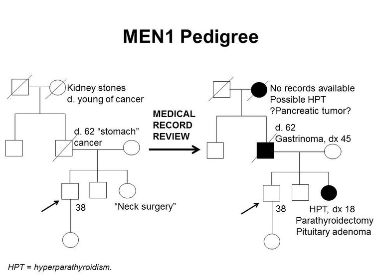
EnlargePedigree showing some of the features of a family with a deleterious MEN1 mutation across four generations, including transmission occurring through paternal lineage. The unaffected male proband is shown as having an affected sister (self-report of neck surgery confirmed upon review of medical records to be hyperparathyroidism diagnosed at age 18 y, parathyroidectomy, and pituitary adenoma), father (self-report of stomach cancer confirmed upon review of medical records to be gastrinoma diagnosed at age 45 y), and paternal grandmother (suspected hyperparathyroidism and/or pancreatic tumor).
Figure 1. MEN1 pedigree. MEN1 can be very difficult to identify in a pedigree. The pedigree on the left was constructed based on self-report, and the pedigree on the right depicts the same family following a review of available medical records. This pedigree shows some of the features of a family with an MEN1 pathogenic variant across four generations, including affected family members with hyperparathyroidism, a pituitary adenoma, gastrinoma, and a suspected pancreatic tumor. The tumors in MEN1 typically occur at an earlier age than their sporadic counterparts. MEN1 families may exhibit some or all of these features. As an autosomal dominant syndrome, transmission can occur through maternal or paternal lineages, as depicted in the figure.

Since many of the tumors in MEN1 are underdiagnosed or misdiagnosed, identifying an MEN1 gene pathogenic variant in the proband early in the disease process can allow for early detection and treatment of tumors and earlier identification of at-risk family members. Many studies have been performed to determine the prevalence of MEN1 gene pathogenic variants among patients with apparently sporadic MEN1-related tumors.[8] For example, approximately one-third of patients with Zollinger-Ellison syndrome will carry an MEN1 pathogenic variant.[41,42] In individuals with apparently isolated PHPT or pituitary adenomas, the pathogenic variant prevalence is lower, on the order of 2% to 5%,[27,43,44] but the prevalence is higher in individuals diagnosed with these tumors before age 30 years. Some authors suggest referral for genetics consultation and/or genetic testing for pathogenic variants in MEN1 if one of the following conditions is present:[8,4547]

  • Gastrinoma at any age in the individual or an FDR.
  • Multifocal duodenopancreatic NETs at any age.
  • PHPT before age 30 or 40 years.
  • Multiglandular parathyroid adenomas/hyperplasia or recurrent PHPT.
  • Presence of one of the three main MEN1 tumors plus one of the less common tumors/findings.
  • Presence of two or more features (e.g., adrenal adenomas and carcinoid tumor).
  • Combination of at least two of the following in one individual: parathyroid adenoma; thymic, bronchial, or foregut carcinoid tumor; duodenopancreatic NET; pituitary tumor; adrenal tumor.
  • Parathyroid adenoma and a family history of hyperparathyroidism, pituitary adenoma, duodenopancreatic NET, or foregut carcinoid tumor.
  • Multiple primary duodenopancreatic NETs in the same person.

Molecular Genetics of MEN1

The MEN1 gene is located on chromosome 11q13 and encodes the protein menin.[3,48,49] Over 1,300 pathogenic variants have been identified in the MEN1 gene to date, and these are scattered across the entire coding region.[50,51] Most (~65%) of these are nonsense or frameshift variants. The remainder are missense variants (20%) (which lead to the expression of an altered protein), splice-site variants (9%), or partial- or whole-gene deletions (1%–4%).[52] In MEN1, phenotypic variation is common within a family and between unrelated individuals. Data suggested that anticipation may also occur in families with MEN1 pathogenic variants.[8,5355] One large study demonstrated that pituitary, adrenal, and thymic NETs had the highest rates of heritability.[56]

Genetic Testing and Differential Diagnosis for MEN1

Genetic testing for MEN1 pathogenic variants is recommended for individuals meeting clinical diagnostic criteria and may be considered in individuals with less common MEN1-associated tumors. For more information, see the Making the diagnosis of MEN1 section. For individuals meeting diagnostic criteria, the pathogenic variant detection rate is approximately 75% to 90%.[53,57] Still, germline pathogenic variant yield ranged from 16% to 38% for apparently sporadic cases of parathyroid (15.8%), pancreatic islet (25.0%), or pituitary (37.5%) tumors. Genetic testing may be warranted in these individuals because a diagnosis of MEN1 would prompt screening for other MEN1-related tumors.[58] Laboratories that offer MEN1 testing primarily use DNA sequencing. Several laboratories offer additional analyses for MEN1 partial- or whole-gene deletion and/or duplication. However, these types of variants are rare. Deletion/duplication testing is often reserved for individuals who have very high clinical suspicions for MEN1 but a detectable pathogenic variant was not found by direct DNA sequencing. Evolving studies continue to reveal causative pathogenic variants in MEN1.[52]

A multigene panel that includes MEN1 and other genes associated with an increased risk of endocrine tumors may also be used. Such genetic testing can be used to distinguish between MEN1 and other forms of hereditary hyperparathyroidism, such as familial isolated hyperparathyroidism (FIHP), hyperparathyroidism–jaw tumor syndrome (HPT-JT), and familial hypocalciuric hypercalcemia (FHH). [Note: The hyperparathyroidism in FHH is not primary hyperparathyroidism, which is seen in MEN1, HPT-JT and FIHP.] HPT-JT, which is caused by germline pathogenic variants in the CDC73 gene, is associated with PHPT, ossifying lesions of the maxilla and mandible, and renal lesions, usually bilateral renal cysts, hamartomas, and in some cases, Wilms tumor.[59,60] Unlike MEN1, HPT-JT is associated with an increased risk of parathyroid carcinoma.[61] FIHP, as its name suggests, is characterized by isolated PHPT with no additional endocrine features; in some families, FIHP is the initial diagnosis of what later develops into MEN1, HPT-JT, or FHH.[6264] Approximately 20% of families with a clinical diagnosis of FIHP carry germline MEN1 pathogenic variants.[63,65,66] Pathogenic variants in the calcium-sensing receptor (CASR) gene cause FHH, which can closely mimic the hyperparathyroidism seen in MEN1.

Genetic diagnosis will help guide management for patients with early-onset hyperparathyroidism. This is especially crucial, since many of the above conditions have different management guidelines that correspond with their features. For example, distinguishing between MEN1 and FHH can be critical for a patient’s disease management. Removing the parathyroid glands in FHH does not correct the hyperparathyroidism that is seen in patients with MEN1. This could result in an unnecessary surgery that would not relieve the patient’s symptoms. In addition, HPT-JT is unique because it increases parathyroid carcinoma risk. Hence, individuals with this syndrome have different management guidelines than individuals with other forms of hereditary hyperparathyroidism.[67,68] For more information on MEN1 clinical features and other forms of hyperparathyroidism, see Table 3.

Table 3. Major Clinical Features of MEN1, FIHP, HPT-JT, and FHH
Condition Gene(s) Major Clinical Features
FHH = familial hypocalciuric hypercalcemia; FIHP = familial isolated hyperparathyroidism; HPT-JT = hyperparathyroidism–jaw tumor syndrome; MEN1 = multiple endocrine neoplasia type 1 (gene is italicized); NETs = neuroendocrine tumors; PHPT = primary hyperparathyroidism.
MEN1 MEN1 PHPT, pituitary adenomas, duodenopancreatic NETs [8,10,69]
FIHP MEN1, CDC73 PHPT [6266]
HPT-JT CDC73 PHPT; osteomas of maxilla and mandible; renal cysts or hamartomas; and rarely, Wilms tumor and parathyroid carcinoma [5961]
FHH CASR (type 1), GNA11 (type 2), AP2S1 (type 3) Hyperparathyroidism (not primary) [67,7072]

Surveillance

Screening and surveillance for MEN1 may include a combination of biochemical tests and imaging techniques.

Traditionally, magnetic resonance imaging (MRI) was used for surveillance and staging. However, ongoing studies have evaluated the role of MRI in functional imaging, including gallium Ga 68-DOTATATE (68Ga-DOTATATE) positron emission tomography (PET)–computed tomography (CT) scanning. A multicenter retrospective study examined 108 MEN1 patients undergoing PET-CT for screening, staging, restaging, or targeted radiotherapy selection. This study demonstrated that PET-CT has the potential to increase diagnostic sensitivity when searching for MEN1-associated NETs.[73] In 51% of cases, PET-CT provided superior lesion detection when compared with conventional imaging techniques. However, the retrospective nature of the study makes it impossible to discern how much selection bias may have impacted the study’s findings. Consequently, PET-CT’s exact role in detecting metastasis remains unclear. PET-CT’s potentially improved diagnostic sensitivity must also be weighed against its increased levels of radiation exposure, which are higher than that of other imaging modalities. Radiation exposure is particularly relevant for MEN1 patients, who are prone to developing aggressive malignancies. The issue is even more nuanced in young patients who require lifelong screening to detect aggressive MEN1-associated malignancies early.

A study analyzed thoracic screening techniques in 50 patients with MEN1. It found that when patients with MEN1 underwent functional imaging with fluorine F 18-fludeoxyglucose (18F-FDG) PET-CT screening, they had a similar number of lung nodules as individuals in the general population. However, when lesions in MEN1 patients were FDG-avid, they were more likely to progress during the follow-up period. Therefore, further observation and follow up of FDG-avid lesions may be warranted in patients with MEN1.[74] While lung-specific imaging is not routinely performed in MEN1 patients, the lungs are visualized during MRI surveillance of thymic lesions. When pulmonary lesions are identified, it is important to recognize that lung NETs may grow slowly and may have a good prognosis. Referral to a multidisciplinary team may be beneficial for selective resection of lung NETs.[75]

Recommendations for MEN1 surveillance are summarized in Table 4.[4,8]

Table 4. Practice Guidelines for Surveillance of MEN1a
Biochemical Test or Procedure Condition Screened For Age Screening Initiated (y) Frequency
CT = computed tomography; MEN1 = multiple endocrine neoplasia type 1; MRI = magnetic resonance imaging; NETs = neuroendocrine tumors; PHPT = primary hyperparathyroidism; PTH = parathyroid hormone.
aAdapted from Brandi et al.[4] and Thakker et al.[8]
bThe recommendations for abdominal imaging differ between two published guidelines for the diagnosis and management of MEN1.[4,8] There is weak evidence at this time to support annual imaging before age 10 years. Imaging before age 10 years does identify disease in a high proportion of patients, but it is not clear whether this impacts prognosis.[21,76]
cThe age to initiate screening and the screening frequency for pituitary tumors may be debatable because the clinical significance of small, nonfunctional tumors is unclear;[77] further study may be warranted.
dAdapted from Niederle et al.[5]
eAdapted from Shirali et al.[78] The 2012 guidelines recommend chest MRI every 1-2 years.[8]
Serum prolactin and/or insulin-like growth factor 1 Pituitary tumors 5 Every 1 y
Fasting total serum calcium and/or ionized calcium and PTH Parathyroid tumors and PHPT 8 Every 1 y
Fasting serum gastrin Duodenopancreatic gastrinoma 20 Every 1 y
Chromogranin A, pancreatic polypeptide, glucagon, and vasointestinal polypeptided Duodenopancreatic NETs 10–16 Up to every 3 years (consider every 3 years if asymptomatic; consider shorter screening intervals depending on the clinical scenario)
Fasting glucose and insulin Insulinoma 5 Every 1 y
Brain MRIc Pituitary tumors 5 Every 3–5 y based on biochemical results
Chest MRIe Thymic and bronchial NETs <20 About every 3 years. Consider more frequent screening for men, smokers, or individuals with a positive family history. Baseline chest MRI is done prior to parathyroidectomy
Abdominal CT or MRIb [4] Duodenopancreatic NETs 20 Every 3–5 y based on biochemical results
Abdominal CT, MRI, or endoscopic ultrasonographyb [8] Duodenopancreatic NETs <10 Every 1 y

Interventions

Surgical management of MEN1 is complex and controversial, given the multifocal and multiglandular nature of the disease. Patients with MEN1 have a high risk of tumor recurrence, even after surgery. Additionally, these patients may have an increased risk of developing venous thromboembolisms.[79] Clinicians should be aware of this risk, particularly in the perioperative period. It is critical to establish an MEN1 diagnosis before making surgical decisions, in order to prevent unnecessary and/or inappropriate surgeries. Furthermore, it is recommended that individuals with MEN1 use a surgeon who has experience treating this disease.

Treatment for parathyroid tumors

Once evidence of parathyroid disease is established biochemically, surgical removal of the hyperfunctional parathyroid tissue is recommended to achieve eucalcemia and euparathyroidism. However, the timing and the amount of parathyroid and thymus gland tissue that is removed during surgery remains controversial.[40] For patients with primary hyperparathyroidism who are at risk for MEN1, preoperative detection of a pathogenic variant helps guide the extent of their initial operations, can increase the likelihood of successful initial surgeries, and lower the likelihood of recurrent disease.[68] Furthermore, knowledge of an MEN1 pathogenic variant can help guide surgical decision making and avoid the use of a single-gland surgical approach. Studies have shown that concomitant bilateral cervical thymectomy decreases the rate of parathyroid tumor recurrence and suggests that the thymus can be removed during the patient’s initial operation.[80]

Some groups reserve surgical intervention for symptomatic patients, with continued annual biochemical screening for those without objective signs of disease. Subtotal parathyroidectomy (removal of 3–3.5 glands) is commonly suggested as the initial surgical treatment when a provider decides to proceed with surgery.[68] If 3.5 or more glands are removed during surgery, the rate of persistent disease is only 5% to 6%. Removing fewer than 3.5 glands decreases the durability of eucalcemia. Studies suggest that preoperative imaging (to determine which glands are hyperfunctional) is not reliable enough to justify unilateral exploration, with 86% of patients having enlarged contralateral parathyroid glands.[81] Fifty percent of the patients who had imaging to direct resection had the largest parathyroid gland identified intraoperatively on the contralateral side of greatest uptake.[81] Insufficient resection fails to accomplish the desired eucalcemia.[1517,68]

Total parathyroidectomy with autotransplantation of parathyroid tissue to a distant site, such as the forearm, is a less commonly recommended option. Likelihood of cancer recurrence is lowered with total parathyroidectomy. However, this procedure also renders the patient aparathyroid for a period of time while the autotransplanted tissue becomes functional. This can cause a permanent PTH deficiency (no detectable PTH in the body).[80,82] Benefits of this approach include the following: 1) it is easier remove/debulk recurrent disease from the forearm than it is to remove/debulk recurrent disease from the neck, and 2) differential lateralization with arm blood draws. Hypocalcemia management involves taking numerous oral medications, including calcitrol and calcium replacement, and these daily requirements can be a major burden for patients. Recovery was less likely for patients who were aparathyroid for 6 months after total parathyroidectomy.[83] In select high-risk patients, like in those who had reoperative parathyroidectomy or cervical surgery, cryopreservation can be beneficial, but it is recommended that providers use this method sparingly.

Treatment for duodenopancreatic NETs

The timing and extent of surgery for duodenopancreatic NETs are controversial and depend on many factors, including severity of symptoms, extent of disease, functional component, location and necessity of simple enucleation, subtotal or total pancreatectomy, and pancreaticoduodenectomy (Whipple procedure). Surgical enucleation has been associated with higher recurrence compared with distal pancreatectomy, and a decreased rate of endocrine insufficiency compared with a Whipple procedure.[84] Tumor size has been suggested to advocate for surgical resection on the basis of the increased propensity for risk of metastases or recurrence with increased tumor diameter.[5,85,86] Unfortunately, there is no specific tumor marker or combination of tumor markers that are predictive of disease-specific mortality.[87] Long-acting somatostatin analogs may have a role in early-stage MEN1 duodenopancreatic NETs.[88] Initial study results of pharmacological therapy suggest that the treatment is safe and that long-term suppression of tumor and hormonal activity can be seen in up to 10% of patients and stability of hormone hyperfunction in 80% of patients.[88] The primary goal of surgery is to improve long-term survival by reducing symptoms associated with hormone excess and lowering the risk of distant metastasis.[24] Surgery is commonly performed for most functional tumors and for nonfunctioning NETs when the tumor exceeds 2 to 3 cm because the likelihood of distant metastases is high.[86,8991] Structural imaging modalities alone are suboptimal for predicting the malignant potential of duodenopancreatic NETs. However, a study found that screening MEN1 patients with 18F-FDG PET-CT identified those NETs with an increased malignant potential; the FDG avidity correlated with a Ki-67 index.[92] Tumor size does seem to influence patient survival, with patients with smaller tumors having increased survival after resection.[93] While more-extensive surgical approaches (e.g., pancreatoduodenectomy) have been associated with higher cure rates and improved overall survival,[9496] they also have higher rates of postoperative complications and long-term morbidity.[97] Therefore, the risks and benefits should be carefully considered, and surgical decisions should be made on a case-by-case basis. With regard to open or laparoscopic approaches, in selected patients, pancreatic laparoscopic surgery appears to be safe and associated with a shorter length of stay and fewer complications.[98]

Individuals with MEN1 who are diagnosed with NETs often have multiple tumors of various types throughout the pancreas and duodenum, some of which can be identified using magnetic resonance imaging or computed tomography (CT). Combining functional tracer accumulation with anatomic imaging improves tumor localization. 68Ga-DOTATATE PET-CT demonstrates excellent sensitivity in mapping duodenopancreatic NET disease. This modality may guide the initial workup and appears to be superior to standard somatostatin octreotide, especially for lesions smaller than 10 mm.[99,100] Many tumors are too small to be detected using standard imaging techniques, and intra-arterial secretin stimulation testing and/or intraoperative ultrasonography may also be useful.[101,102] Preoperative assessment using a combination of various biochemical and imaging modalities, intraoperative assessment of tumor burden, and resolution of hormonal hyper-secretion are critical and, in some series, have been associated with higher cure rates and longer disease-free intervals.[101104]

In the current era of effective treatment for hyperfunctional hormone excess states, most MEN1-related deaths are due to the malignant nature of duodenopancreatic NETs. A less common but important risk of death is from malignant thymic carcinoid tumors. Indicators of a poor MEN1 prognosis include elevated fasting serum gastrin, the presence of functional hormonal syndromes, liver or distant metastases, aggressive duodenopancreatic NET growth, large duodenopancreatic NET size, or the need for multiple parathyroidectomies. The most common cause of non-MEN1–related death in this patient cohort is from cardiovascular disease.[105]

Other duodenopancreatic NETs

Glucagonomas, VIPomas, and somatostatinomas are rare but often have higher rates of malignancy than other duodenopancreatic NETs.[22] These are often treated with aggressive surgery.[106]

Insulinomas

Medical management of insulinoma using diet and medication is often unsuccessful; the mainstay of treatment for this tumor is surgical resection.[8] Insulinomas in MEN1 patients can be located throughout the pancreas, with a preponderance found in the distal gland,[107109] and have a higher rate of metastasis than sporadic insulinoma.[106] Surgery can range from enucleation of single or multiple large tumors to partial pancreatic resection, or both,[108] to subtotal or total pancreatectomy.[107,108] More-extensive surgical approaches are associated with a lower rate of recurrence [94,95,108,110] but a higher rate of postoperative morbidity. Because insulinoma often occurs in conjunction with nonfunctioning pancreatic tumors, the selective intra-arterial calcium-injection test (SAS test) may be necessary to determine the source of insulin excess.[111] Intraoperative monitoring of insulin/glucose can help determine whether insulin-secreting tumors have been successfully excised.[102,112]

Gastrinomas

Most MEN1-associated gastrinomas originate in the duodenum. These tumors are typically multifocal and cause hyper-secretion of gastrin, with resultant peptic ulcer disease (Zollinger-Ellison syndrome).[113] The multifocal nature makes complete surgical resection difficult. It is critical to manage symptoms before considering any type of surgical intervention.[114] Historically, some groups have recommended close observation of individuals with smaller tumors (<2.0 cm on imaging) who have relief of symptoms using medications (e.g., proton pump inhibitors or histamine-2 agonists);[115] however, this approach may not be optimal for all patients.

Several published series have shown a positive correlation between primary tumor size and rate of distant metastasis. One retrospective study showed that 61% of patients with tumors larger than 3 cm had liver metastases.[24] In another series, 40% of patients with tumors larger than 3 cm had liver metastases.[116] In contrast, both of these series showed significantly lower rates of liver metastases in individuals with tumors smaller than 3 cm (32% and 4.8%, respectively). On the basis of these and other data, many groups recommend surgery in individuals with nonmetastatic gastrinoma who have tumors larger than 2 cm.[8,96]

The type of surgery for gastrinoma depends on many factors. A Whipple procedure is typically discouraged as an initial surgery, given the high postoperative morbidity and long-term complications, such as diabetes mellitus and malabsorption. Less extensive operations have been described with varying results. At a minimum, duodenectomy with intraoperative palpation and/or ultrasonography to locate and excise duodenal tumors and peri-pancreatic lymph node dissection are performed.[101,117] Because most patients with gastrinoma will have concomitant NETs throughout the pancreas, some of which may be nonfunctional, some groups recommend resection of the distal pancreas and enucleation of tumors in the pancreatic head in addition to duodenal tumor excision.[101,117,118]

Nonfunctioning NETs

Approximately 50% of individuals with MEN1 will develop nonfunctioning NETs.[21,26] These are often identified incidentally during assessment and exploration for functioning tumors. As with gastrinomas, the metastatic rate is correlated with larger tumor size.[26,86] Tumors smaller than 1.5 cm are not likely to have lymph node metastases,[119] although the presence of metastatic disease has been associated with earlier age at death than in those without duodenopancreatic NETs.[9,26]

Pituitary tumors

Medical therapy to suppress hypersecretion is often the first line of therapy for MEN1-associated pituitary tumors. In one series of 136 patients, medical therapy was successful in approximately one-half of patients with secreting tumors (49 of 116, 42%), and successful suppression was correlated with smaller tumor size.[120] Surgery is often necessary for patients who are resistant to this treatment. Radiation therapy is reserved for patients for whom complete surgical resection was not rendered.[8,121]

References
  1. Agarwal SK, Ozawa A, Mateo CM, et al.: The MEN1 gene and pituitary tumours. Horm Res 71 (Suppl 2): 131-8, 2009. [PUBMED Abstract]
  2. Trump D, Farren B, Wooding C, et al.: Clinical studies of multiple endocrine neoplasia type 1 (MEN1) QJM 89 (9): 653-69, 1996. [PUBMED Abstract]
  3. Chandrasekharappa SC, Guru SC, Manickam P, et al.: Positional cloning of the gene for multiple endocrine neoplasia-type 1. Science 276 (5311): 404-7, 1997. [PUBMED Abstract]
  4. Brandi ML, Gagel RF, Angeli A, et al.: Guidelines for diagnosis and therapy of MEN type 1 and type 2. J Clin Endocrinol Metab 86 (12): 5658-71, 2001. [PUBMED Abstract]
  5. Niederle B, Selberherr A, Bartsch DK, et al.: Multiple Endocrine Neoplasia Type 1 and the Pancreas: Diagnosis and Treatment of Functioning and Non-Functioning Pancreatic and Duodenal Neuroendocrine Neoplasia within the MEN1 Syndrome – An International Consensus Statement. Neuroendocrinology 111 (7): 609-630, 2021. [PUBMED Abstract]
  6. Carty SE, Helm AK, Amico JA, et al.: The variable penetrance and spectrum of manifestations of multiple endocrine neoplasia type 1. Surgery 124 (6): 1106-13; discussion 1113-4, 1998. [PUBMED Abstract]
  7. Goudet P, Dalac A, Le Bras M, et al.: MEN1 disease occurring before 21 years old: a 160-patient cohort study from the Groupe d’étude des Tumeurs Endocrines. J Clin Endocrinol Metab 100 (4): 1568-77, 2015. [PUBMED Abstract]
  8. Thakker RV, Newey PJ, Walls GV, et al.: Clinical practice guidelines for multiple endocrine neoplasia type 1 (MEN1). J Clin Endocrinol Metab 97 (9): 2990-3011, 2012. [PUBMED Abstract]
  9. Goudet P, Murat A, Binquet C, et al.: Risk factors and causes of death in MEN1 disease. A GTE (Groupe d’Etude des Tumeurs Endocrines) cohort study among 758 patients. World J Surg 34 (2): 249-55, 2010. [PUBMED Abstract]
  10. Chandrasekharappa SC, Teh BT: Clinical and molecular aspects of multiple endocrine neoplasia type 1. Front Horm Res 28: 50-80, 2001. [PUBMED Abstract]
  11. Shariq OA, Lines KE, English KA, et al.: Multiple endocrine neoplasia type 1 in children and adolescents: Clinical features and treatment outcomes. Surgery 171 (1): 77-87, 2022. [PUBMED Abstract]
  12. del Pozo C, García-Pascual L, Balsells M, et al.: Parathyroid carcinoma in multiple endocrine neoplasia type 1. Case report and review of the literature. Hormones (Athens) 10 (4): 326-31, 2011 Oct-Dec. [PUBMED Abstract]
  13. Christakis I, Busaidy NL, Cote GJ, et al.: Parathyroid carcinoma and atypical parathyroid neoplasms in MEN1 patients; A clinico-pathologic challenge. The MD Anderson case series and review of the literature. Int J Surg 31: 10-6, 2016. [PUBMED Abstract]
  14. Singh Ospina N, Sebo TJ, Thompson GB, et al.: Prevalence of parathyroid carcinoma in 348 patients with multiple endocrine neoplasia type 1 – case report and review of the literature. Clin Endocrinol (Oxf) 84 (2): 244-249, 2016. [PUBMED Abstract]
  15. Norton JA, Venzon DJ, Berna MJ, et al.: Prospective study of surgery for primary hyperparathyroidism (HPT) in multiple endocrine neoplasia-type 1 and Zollinger-Ellison syndrome: long-term outcome of a more virulent form of HPT. Ann Surg 247 (3): 501-10, 2008. [PUBMED Abstract]
  16. Hellman P, Skogseid B, Oberg K, et al.: Primary and reoperative parathyroid operations in hyperparathyroidism of multiple endocrine neoplasia type 1. Surgery 124 (6): 993-9, 1998. [PUBMED Abstract]
  17. Schreinemakers JM, Pieterman CR, Scholten A, et al.: The optimal surgical treatment for primary hyperparathyroidism in MEN1 patients: a systematic review. World J Surg 35 (9): 1993-2005, 2011. [PUBMED Abstract]
  18. Christakis I, Qiu W, Hyde SM, et al.: Genotype-phenotype pancreatic neuroendocrine tumor relationship in multiple endocrine neoplasia type 1 patients: A 23-year experience at a single institution. Surgery 163 (1): 212-217, 2018. [PUBMED Abstract]
  19. Donegan D, Singh Ospina N, Rodriguez-Gutierrez R, et al.: Long-term outcomes in patients with multiple endocrine neoplasia type 1 and pancreaticoduodenal neuroendocrine tumours. Clin Endocrinol (Oxf) 86 (2): 199-206, 2017. [PUBMED Abstract]
  20. Norton JA, Krampitz G, Jensen RT: Multiple Endocrine Neoplasia: Genetics and Clinical Management. Surg Oncol Clin N Am 24 (4): 795-832, 2015. [PUBMED Abstract]
  21. Thomas-Marques L, Murat A, Delemer B, et al.: Prospective endoscopic ultrasonographic evaluation of the frequency of nonfunctioning pancreaticoduodenal endocrine tumors in patients with multiple endocrine neoplasia type 1. Am J Gastroenterol 101 (2): 266-73, 2006. [PUBMED Abstract]
  22. Lévy-Bohbot N, Merle C, Goudet P, et al.: Prevalence, characteristics and prognosis of MEN 1-associated glucagonomas, VIPomas, and somatostatinomas: study from the GTE (Groupe des Tumeurs Endocrines) registry. Gastroenterol Clin Biol 28 (11): 1075-81, 2004. [PUBMED Abstract]
  23. Pipeleers-Marichal M, Somers G, Willems G, et al.: Gastrinomas in the duodenums of patients with multiple endocrine neoplasia type 1 and the Zollinger-Ellison syndrome. N Engl J Med 322 (11): 723-7, 1990. [PUBMED Abstract]
  24. Weber HC, Venzon DJ, Lin JT, et al.: Determinants of metastatic rate and survival in patients with Zollinger-Ellison syndrome: a prospective long-term study. Gastroenterology 108 (6): 1637-49, 1995. [PUBMED Abstract]
  25. Tonelli F, Giudici F, Fratini G, et al.: Pancreatic endocrine tumors in multiple endocrine neoplasia type 1 syndrome: review of literature. Endocr Pract 17 (Suppl 3): 33-40, 2011 Jul-Aug. [PUBMED Abstract]
  26. Triponez F, Dosseh D, Goudet P, et al.: Epidemiology data on 108 MEN 1 patients from the GTE with isolated nonfunctioning tumors of the pancreas. Ann Surg 243 (2): 265-72, 2006. [PUBMED Abstract]
  27. Corbetta S, Pizzocaro A, Peracchi M, et al.: Multiple endocrine neoplasia type 1 in patients with recognized pituitary tumours of different types. Clin Endocrinol (Oxf) 47 (5): 507-12, 1997. [PUBMED Abstract]
  28. Darling TN, Skarulis MC, Steinberg SM, et al.: Multiple facial angiofibromas and collagenomas in patients with multiple endocrine neoplasia type 1. Arch Dermatol 133 (7): 853-7, 1997. [PUBMED Abstract]
  29. Ventura M, Melo M, Carrilho F: Outcome and long-term follow-up of adrenal lesions in multiple endocrine neoplasia type 1. Arch Endocrinol Metab 63 (5): 516-523, 2019. [PUBMED Abstract]
  30. Machens A, Schaaf L, Karges W, et al.: Age-related penetrance of endocrine tumours in multiple endocrine neoplasia type 1 (MEN1): a multicentre study of 258 gene carriers. Clin Endocrinol (Oxf) 67 (4): 613-22, 2007. [PUBMED Abstract]
  31. Pieterman CR, Schreinemakers JM, Koppeschaar HP, et al.: Multiple endocrine neoplasia type 1 (MEN1): its manifestations and effect of genetic screening on clinical outcome. Clin Endocrinol (Oxf) 70 (4): 575-81, 2009. [PUBMED Abstract]
  32. Waldmann J, Bartsch DK, Kann PH, et al.: Adrenal involvement in multiple endocrine neoplasia type 1: results of 7 years prospective screening. Langenbecks Arch Surg 392 (4): 437-43, 2007. [PUBMED Abstract]
  33. Gibril F, Schumann M, Pace A, et al.: Multiple endocrine neoplasia type 1 and Zollinger-Ellison syndrome: a prospective study of 107 cases and comparison with 1009 cases from the literature. Medicine (Baltimore) 83 (1): 43-83, 2004. [PUBMED Abstract]
  34. McKeeby JL, Li X, Zhuang Z, et al.: Multiple leiomyomas of the esophagus, lung, and uterus in multiple endocrine neoplasia type 1. Am J Pathol 159 (3): 1121-7, 2001. [PUBMED Abstract]
  35. Vortmeyer AO, Lubensky IA, Skarulis M, et al.: Multiple endocrine neoplasia type 1: atypical presentation, clinical course, and genetic analysis of multiple tumors. Mod Pathol 12 (9): 919-24, 1999. [PUBMED Abstract]
  36. Yamazaki M, Suzuki S, Kosugi S, et al.: Delay in the diagnosis of multiple endocrine neoplasia type 1: typical symptoms are frequently overlooked. Endocr J 59 (9): 797-807, 2012. [PUBMED Abstract]
  37. Lourenço DM, Toledo RA, Coutinho FL, et al.: The impact of clinical and genetic screenings on the management of the multiple endocrine neoplasia type 1. Clinics (Sao Paulo) 62 (4): 465-76, 2007. [PUBMED Abstract]
  38. van Leeuwaarde RS, van Nesselrooij BP, Hermus AR, et al.: Impact of Delay in Diagnosis in Outcomes in MEN1: Results From the Dutch MEN1 Study Group. J Clin Endocrinol Metab 101 (3): 1159-65, 2016. [PUBMED Abstract]
  39. Strømsvik N, Nordin K, Berglund G, et al.: Living with multiple endocrine neoplasia type 1: decent care-insufficient medical and genetic information: a qualitative study of MEN 1 patients in a Swedish hospital. J Genet Couns 16 (1): 105-17, 2007. [PUBMED Abstract]
  40. Marini F, Giusti F, Tonelli F, et al.: Management impact: effects on quality of life and prognosis in MEN1. Endocr Relat Cancer 24 (10): T227-T242, 2017. [PUBMED Abstract]
  41. Roy PK, Venzon DJ, Shojamanesh H, et al.: Zollinger-Ellison syndrome. Clinical presentation in 261 patients. Medicine (Baltimore) 79 (6): 379-411, 2000. [PUBMED Abstract]
  42. Bardram L, Stage JG: Frequency of endocrine disorders in patients with the Zollinger-Ellison syndrome. Scand J Gastroenterol 20 (2): 233-8, 1985. [PUBMED Abstract]
  43. Uchino S, Noguchi S, Sato M, et al.: Screening of the Men1 gene and discovery of germ-line and somatic mutations in apparently sporadic parathyroid tumors. Cancer Res 60 (19): 5553-7, 2000. [PUBMED Abstract]
  44. Scheithauer BW, Laws ER, Kovacs K, et al.: Pituitary adenomas of the multiple endocrine neoplasia type I syndrome. Semin Diagn Pathol 4 (3): 205-11, 1987. [PUBMED Abstract]
  45. Newey PJ, Thakker RV: Role of multiple endocrine neoplasia type 1 mutational analysis in clinical practice. Endocr Pract 17 (Suppl 3): 8-17, 2011 Jul-Aug. [PUBMED Abstract]
  46. Hampel H, Bennett RL, Buchanan A, et al.: A practice guideline from the American College of Medical Genetics and Genomics and the National Society of Genetic Counselors: referral indications for cancer predisposition assessment. Genet Med 17 (1): 70-87, 2015. [PUBMED Abstract]
  47. Bashford MT, Kohlman W, Everett J, et al.: Addendum: A practice guideline from the American College of Medical Genetics and Genomics and the National Society of Genetic Counselors: referral indications for cancer predisposition assessment. Genet Med 21 (12): 2844, 2019. [PUBMED Abstract]
  48. Larsson C, Skogseid B, Oberg K, et al.: Multiple endocrine neoplasia type 1 gene maps to chromosome 11 and is lost in insulinoma. Nature 332 (6159): 85-7, 1988. [PUBMED Abstract]
  49. Bassett JH, Forbes SA, Pannett AA, et al.: Characterization of mutations in patients with multiple endocrine neoplasia type 1. Am J Hum Genet 62 (2): 232-44, 1998. [PUBMED Abstract]
  50. Lemos MC, Thakker RV: Multiple endocrine neoplasia type 1 (MEN1): analysis of 1336 mutations reported in the first decade following identification of the gene. Hum Mutat 29 (1): 22-32, 2008. [PUBMED Abstract]
  51. Concolino P, Costella A, Capoluongo E: Multiple endocrine neoplasia type 1 (MEN1): An update of 208 new germline variants reported in the last nine years. Cancer Genet 209 (1-2): 36-41, 2016 Jan-Feb. [PUBMED Abstract]
  52. Brandi ML, Agarwal SK, Perrier ND, et al.: Multiple Endocrine Neoplasia Type 1: Latest Insights. Endocr Rev 42 (2): 133-170, 2021. [PUBMED Abstract]
  53. Giraud S, Zhang CX, Serova-Sinilnikova O, et al.: Germ-line mutation analysis in patients with multiple endocrine neoplasia type 1 and related disorders. Am J Hum Genet 63 (2): 455-67, 1998. [PUBMED Abstract]
  54. Wautot V, Vercherat C, Lespinasse J, et al.: Germline mutation profile of MEN1 in multiple endocrine neoplasia type 1: search for correlation between phenotype and the functional domains of the MEN1 protein. Hum Mutat 20 (1): 35-47, 2002. [PUBMED Abstract]
  55. van den Broek MFM, van Nesselrooij BPM, Pieterman CRC, et al.: Clues For Genetic Anticipation In Multiple Endocrine Neoplasia Type 1. J Clin Endocrinol Metab 105 (7): , 2020. [PUBMED Abstract]
  56. Thevenon J, Bourredjem A, Faivre L, et al.: Unraveling the intrafamilial correlations and heritability of tumor types in MEN1: a Groupe d’étude des Tumeurs Endocrines study. Eur J Endocrinol 173 (6): 819-26, 2015. [PUBMED Abstract]
  57. Agarwal SK, Kester MB, Debelenko LV, et al.: Germline mutations of the MEN1 gene in familial multiple endocrine neoplasia type 1 and related states. Hum Mol Genet 6 (7): 1169-75, 1997. [PUBMED Abstract]
  58. Klein RD, Salih S, Bessoni J, et al.: Clinical testing for multiple endocrine neoplasia type 1 in a DNA diagnostic laboratory. Genet Med 7 (2): 131-8, 2005. [PUBMED Abstract]
  59. Teh BT, Farnebo F, Kristoffersson U, et al.: Autosomal dominant primary hyperparathyroidism and jaw tumor syndrome associated with renal hamartomas and cystic kidney disease: linkage to 1q21-q32 and loss of the wild type allele in renal hamartomas. J Clin Endocrinol Metab 81 (12): 4204-11, 1996. [PUBMED Abstract]
  60. Carpten JD, Robbins CM, Villablanca A, et al.: HRPT2, encoding parafibromin, is mutated in hyperparathyroidism-jaw tumor syndrome. Nat Genet 32 (4): 676-80, 2002. [PUBMED Abstract]
  61. Marx SJ: Multiple endocrine neoplasia type 1. In: Vogelstein B, Kinzler KW, eds.: The Genetic Basis of Human Cancer. McGraw-Hill, 1998, pp 489-506.
  62. Warner J, Epstein M, Sweet A, et al.: Genetic testing in familial isolated hyperparathyroidism: unexpected results and their implications. J Med Genet 41 (3): 155-60, 2004. [PUBMED Abstract]
  63. Mizusawa N, Uchino S, Iwata T, et al.: Genetic analyses in patients with familial isolated hyperparathyroidism and hyperparathyroidism-jaw tumour syndrome. Clin Endocrinol (Oxf) 65 (1): 9-16, 2006. [PUBMED Abstract]
  64. Cetani F, Pardi E, Borsari S, et al.: Molecular pathogenesis of primary hyperparathyroidism. J Endocrinol Invest 34 (7 Suppl): 35-9, 2011. [PUBMED Abstract]
  65. Miedlich S, Lohmann T, Schneyer U, et al.: Familial isolated primary hyperparathyroidism–a multiple endocrine neoplasia type 1 variant? Eur J Endocrinol 145 (2): 155-60, 2001. [PUBMED Abstract]
  66. Cetani F, Pardi E, Ambrogini E, et al.: Genetic analyses in familial isolated hyperparathyroidism: implication for clinical assessment and surgical management. Clin Endocrinol (Oxf) 64 (2): 146-52, 2006. [PUBMED Abstract]
  67. Raue F, Frank-Raue K: Primary hyperparathyroidism–what the nephrologist should know–an update. Nephrol Dial Transplant 22 (3): 696-9, 2007. [PUBMED Abstract]
  68. Romero Arenas MA, Morris LF, Rich TA, et al.: Preoperative multiple endocrine neoplasia type 1 diagnosis improves the surgical outcomes of pediatric patients with primary hyperparathyroidism. J Pediatr Surg 49 (4): 546-50, 2014. [PUBMED Abstract]
  69. Thakker RV: Multiple endocrine neoplasia type 1 (MEN1) and type 4 (MEN4). Mol Cell Endocrinol 386 (1-2): 2-15, 2014. [PUBMED Abstract]
  70. Christensen SE, Nissen PH, Vestergaard P, et al.: Familial hypocalciuric hypercalcaemia: a review. Curr Opin Endocrinol Diabetes Obes 18 (6): 359-70, 2011. [PUBMED Abstract]
  71. Nesbit MA, Hannan FM, Howles SA, et al.: Mutations affecting G-protein subunit α11 in hypercalcemia and hypocalcemia. N Engl J Med 368 (26): 2476-2486, 2013. [PUBMED Abstract]
  72. Nesbit MA, Hannan FM, Howles SA, et al.: Mutations in AP2S1 cause familial hypocalciuric hypercalcemia type 3. Nat Genet 45 (1): 93-7, 2013. [PUBMED Abstract]
  73. Mennetrey C, Le Bras M, Bando-Delaunay A, et al.: Value of Somatostatin Receptor PET/CT in Patients With MEN1 at Various Stages of Their Disease. J Clin Endocrinol Metab 107 (5): e2056-e2064, 2022. [PUBMED Abstract]
  74. So A, Pointon O, Hodgson R, et al.: An assessment of 18 F-FDG PET/CT for thoracic screening and risk stratification of pulmonary nodules in multiple endocrine neoplasia type 1. Clin Endocrinol (Oxf) 88 (5): 683-691, 2018. [PUBMED Abstract]
  75. van den Broek MFM, de Laat JM, van Leeuwaarde RS, et al.: The Management of Neuroendocrine Tumors of the Lung in MEN1: Results From the Dutch MEN1 Study Group. J Clin Endocrinol Metab 106 (2): e1014-e1027, 2021. [PUBMED Abstract]
  76. Langer P, Kann PH, Fendrich V, et al.: Prospective evaluation of imaging procedures for the detection of pancreaticoduodenal endocrine tumors in patients with multiple endocrine neoplasia type 1. World J Surg 28 (12): 1317-22, 2004. [PUBMED Abstract]
  77. de Laat JM, Dekkers OM, Pieterman CR, et al.: Long-Term Natural Course of Pituitary Tumors in Patients With MEN1: Results From the DutchMEN1 Study Group (DMSG). J Clin Endocrinol Metab 100 (9): 3288-96, 2015. [PUBMED Abstract]
  78. Shirali AS, Pieterman CRC, Lewis MA, et al.: It’s not a mystery, it’s in the history: Multidisciplinary management of multiple endocrine neoplasia type 1. CA Cancer J Clin 71 (5): 369-380, 2021. [PUBMED Abstract]
  79. Lee ME, Ortega-Sustache YM, Agarwal SK, et al.: Patients With MEN1 Are at an Increased Risk for Venous Thromboembolism. J Clin Endocrinol Metab 106 (2): e460-e468, 2021. [PUBMED Abstract]
  80. Pieterman CR, van Hulsteijn LT, den Heijer M, et al.: Primary hyperparathyroidism in MEN1 patients: a cohort study with longterm follow-up on preferred surgical procedure and the relation with genotype. Ann Surg 255 (6): 1171-8, 2012. [PUBMED Abstract]
  81. Nilubol N, Weinstein LS, Simonds WF, et al.: Limited Parathyroidectomy in Multiple Endocrine Neoplasia Type 1-Associated Primary Hyperparathyroidism: A Setup for Failure. Ann Surg Oncol 23 (2): 416-23, 2016. [PUBMED Abstract]
  82. Lairmore TC, Govednik CM, Quinn CE, et al.: A randomized, prospective trial of operative treatments for hyperparathyroidism in patients with multiple endocrine neoplasia type 1. Surgery 156 (6): 1326-34; discussion 1334-5, 2014. [PUBMED Abstract]
  83. Landry JP, Pieterman CRC, Clemente-Gutierrez U, et al.: Evaluation of risk factors, long-term outcomes, and immediate and delayed autotransplantation to minimize postsurgical hypoparathyroidism in multiple endocrine neoplasia type 1 (MEN1): A retrospective cohort study. Surgery 171 (5): 1240-1246, 2022. [PUBMED Abstract]
  84. Ratnayake CBB, Loveday BP, Windsor JA, et al.: Patient characteristics and clinical outcomes following initial surgical intervention for MEN1 associated pancreatic neuroendocrine tumours: A systematic review and exploratory meta-analysis of the literature. Pancreatology 19 (3): 462-471, 2019. [PUBMED Abstract]
  85. Kishi Y, Shimada K, Nara S, et al.: Basing treatment strategy for non-functional pancreatic neuroendocrine tumors on tumor size. Ann Surg Oncol 21 (9): 2882-8, 2014. [PUBMED Abstract]
  86. Nell S, Verkooijen HM, Pieterman CRC, et al.: Management of MEN1 Related Nonfunctioning Pancreatic NETs: A Shifting Paradigm: Results From the DutchMEN1 Study Group. Ann Surg 267 (6): 1155-1160, 2018. [PUBMED Abstract]
  87. Qiu W, Christakis I, Silva A, et al.: Utility of chromogranin A, pancreatic polypeptide, glucagon and gastrin in the diagnosis and follow-up of pancreatic neuroendocrine tumours in multiple endocrine neoplasia type 1 patients. Clin Endocrinol (Oxf) 85 (3): 400-7, 2016. [PUBMED Abstract]
  88. Ramundo V, Del Prete M, Marotta V, et al.: Impact of long-acting octreotide in patients with early-stage MEN1-related duodeno-pancreatic neuroendocrine tumours. Clin Endocrinol (Oxf) 80 (6): 850-5, 2014. [PUBMED Abstract]
  89. Triponez F, Goudet P, Dosseh D, et al.: Is surgery beneficial for MEN1 patients with small (< or = 2 cm), nonfunctioning pancreaticoduodenal endocrine tumor? An analysis of 65 patients from the GTE. World J Surg 30 (5): 654-62; discussion 663-4, 2006. [PUBMED Abstract]
  90. Bettini R, Partelli S, Boninsegna L, et al.: Tumor size correlates with malignancy in nonfunctioning pancreatic endocrine tumor. Surgery 150 (1): 75-82, 2011. [PUBMED Abstract]
  91. Triponez F, Sadowski SM, Pattou F, et al.: Long-term Follow-up of MEN1 Patients Who Do Not Have Initial Surgery for Small ≤2 cm Nonfunctioning Pancreatic Neuroendocrine Tumors, an AFCE and GTE Study: Association Francophone de Chirurgie Endocrinienne & Groupe d’Etude des Tumeurs Endocrines. Ann Surg 268 (1): 158-164, 2018. [PUBMED Abstract]
  92. Kornaczewski Jackson ER, Pointon OP, Bohmer R, et al.: Utility of FDG-PET Imaging for Risk Stratification of Pancreatic Neuroendocrine Tumors in MEN1. J Clin Endocrinol Metab 102 (6): 1926-1933, 2017. [PUBMED Abstract]
  93. Brunner SM, Weber F, Werner JM, et al.: Neuroendocrine tumors of the pancreas: a retrospective single-center analysis using the ENETS TNM-classification and immunohistochemical markers for risk stratification. BMC Surg 15: 49, 2015. [PUBMED Abstract]
  94. Bartsch DK, Langer P, Wild A, et al.: Pancreaticoduodenal endocrine tumors in multiple endocrine neoplasia type 1: surgery or surveillance? Surgery 128 (6): 958-66, 2000. [PUBMED Abstract]
  95. Bartsch DK, Fendrich V, Langer P, et al.: Outcome of duodenopancreatic resections in patients with multiple endocrine neoplasia type 1. Ann Surg 242 (6): 757-64, discussion 764-6, 2005. [PUBMED Abstract]
  96. Norton JA, Jensen RT: Role of surgery in Zollinger-Ellison syndrome. J Am Coll Surg 205 (4 Suppl): S34-7, 2007. [PUBMED Abstract]
  97. Lopez CL, Waldmann J, Fendrich V, et al.: Long-term results of surgery for pancreatic neuroendocrine neoplasms in patients with MEN1. Langenbecks Arch Surg 396 (8): 1187-96, 2011. [PUBMED Abstract]
  98. Drymousis P, Raptis DA, Spalding D, et al.: Laparoscopic versus open pancreas resection for pancreatic neuroendocrine tumours: a systematic review and meta-analysis. HPB (Oxford) 16 (5): 397-406, 2014. [PUBMED Abstract]
  99. Morgat C, Vélayoudom-Céphise FL, Schwartz P, et al.: Evaluation of (68)Ga-DOTA-TOC PET/CT for the detection of duodenopancreatic neuroendocrine tumors in patients with MEN1. Eur J Nucl Med Mol Imaging 43 (7): 1258-66, 2016. [PUBMED Abstract]
  100. Lastoria S, Marciello F, Faggiano A, et al.: Role of (68)Ga-DOTATATE PET/CT in patients with multiple endocrine neoplasia type 1 (MEN1). Endocrine 52 (3): 488-94, 2016. [PUBMED Abstract]
  101. Imamura M, Komoto I, Ota S, et al.: Biochemically curative surgery for gastrinoma in multiple endocrine neoplasia type 1 patients. World J Gastroenterol 17 (10): 1343-53, 2011. [PUBMED Abstract]
  102. Tonelli F, Fratini G, Nesi G, et al.: Pancreatectomy in multiple endocrine neoplasia type 1-related gastrinomas and pancreatic endocrine neoplasias. Ann Surg 244 (1): 61-70, 2006. [PUBMED Abstract]
  103. Lewis MA, Thompson GB, Young WF: Preoperative assessment of the pancreas in multiple endocrine neoplasia type 1. World J Surg 36 (6): 1375-81, 2012. [PUBMED Abstract]
  104. van Asselt SJ, Brouwers AH, van Dullemen HM, et al.: EUS is superior for detection of pancreatic lesions compared with standard imaging in patients with multiple endocrine neoplasia type 1. Gastrointest Endosc 81 (1): 159-167.e2, 2015. [PUBMED Abstract]
  105. Ito T, Igarashi H, Uehara H, et al.: Causes of death and prognostic factors in multiple endocrine neoplasia type 1: a prospective study: comparison of 106 MEN1/Zollinger-Ellison syndrome patients with 1613 literature MEN1 patients with or without pancreatic endocrine tumors. Medicine (Baltimore) 92 (3): 135-81, 2013. [PUBMED Abstract]
  106. Akerström G, Stålberg P: Surgical management of MEN-1 and -2: state of the art. Surg Clin North Am 89 (5): 1047-68, 2009. [PUBMED Abstract]
  107. O’Riordain DS, O’Brien T, van Heerden JA, et al.: Surgical management of insulinoma associated with multiple endocrine neoplasia type I. World J Surg 18 (4): 488-93; discussion 493-4, 1994 Jul-Aug. [PUBMED Abstract]
  108. Crippa S, Zerbi A, Boninsegna L, et al.: Surgical management of insulinomas: short- and long-term outcomes after enucleations and pancreatic resections. Arch Surg 147 (3): 261-6, 2012. [PUBMED Abstract]
  109. Sakurai A, Yamazaki M, Suzuki S, et al.: Clinical features of insulinoma in patients with multiple endocrine neoplasia type 1: analysis of the database of the MEN Consortium of Japan. Endocr J 59 (10): 859-66, 2012. [PUBMED Abstract]
  110. Vezzosi D, Cardot-Bauters C, Bouscaren N, et al.: Long-term results of the surgical management of insulinoma patients with MEN1: a Groupe d’étude des Tumeurs Endocrines (GTE) retrospective study. Eur J Endocrinol 172 (3): 309-19, 2015. [PUBMED Abstract]
  111. Grant CS: Insulinoma. Best Pract Res Clin Gastroenterol 19 (5): 783-98, 2005. [PUBMED Abstract]
  112. Giudici F, Nesi G, Brandi ML, et al.: Surgical management of insulinomas in multiple endocrine neoplasia type 1. Pancreas 41 (4): 547-53, 2012. [PUBMED Abstract]
  113. Plöckinger U: Diagnosis and Treatment of Gastrinomas in Multiple Endocrine Neoplasia Type 1 (MEN-1). Cancers (Basel) 4 (1): 39-54, 2012. [PUBMED Abstract]
  114. Falconi M, Eriksson B, Kaltsas G, et al.: ENETS Consensus Guidelines Update for the Management of Patients with Functional Pancreatic Neuroendocrine Tumors and Non-Functional Pancreatic Neuroendocrine Tumors. Neuroendocrinology 103 (2): 153-71, 2016. [PUBMED Abstract]
  115. Mignon M, Cadiot G: Diagnostic and therapeutic criteria in patients with Zollinger-Ellison syndrome and multiple endocrine neoplasia type 1. J Intern Med 243 (6): 489-94, 1998. [PUBMED Abstract]
  116. Cadiot G, Vuagnat A, Doukhan I, et al.: Prognostic factors in patients with Zollinger-Ellison syndrome and multiple endocrine neoplasia type 1. Groupe d’Etude des Néoplasies Endocriniennes Multiples (GENEM and groupe de Recherche et d’Etude du Syndrome de Zollinger-Ellison (GRESZE). Gastroenterology 116 (2): 286-93, 1999. [PUBMED Abstract]
  117. Dickson PV, Rich TA, Xing Y, et al.: Achieving eugastrinemia in MEN1 patients: both duodenal inspection and formal lymph node dissection are important. Surgery 150 (6): 1143-52, 2011. [PUBMED Abstract]
  118. Akerström G, Stålberg P, Hellman P: Surgical management of pancreatico-duodenal tumors in multiple endocrine neoplasia syndrome type 1. Clinics (Sao Paulo) 67 (Suppl 1): 173-8, 2012. [PUBMED Abstract]
  119. Zhang IY, Zhao J, Fernandez-Del Castillo C, et al.: Operative Versus Nonoperative Management of Nonfunctioning Pancreatic Neuroendocrine Tumors. J Gastrointest Surg 20 (2): 277-83, 2016. [PUBMED Abstract]
  120. Vergès B, Boureille F, Goudet P, et al.: Pituitary disease in MEN type 1 (MEN1): data from the France-Belgium MEN1 multicenter study. J Clin Endocrinol Metab 87 (2): 457-65, 2002. [PUBMED Abstract]
  121. Pieterman CR, Vriens MR, Dreijerink KM, et al.: Care for patients with multiple endocrine neoplasia type 1: the current evidence base. Fam Cancer 10 (1): 157-71, 2011. [PUBMED Abstract]

Multiple Endocrine Neoplasia Type 2

Multiple endocrine neoplasia type 2 (MEN2) is caused by pathogenic variants in the RET gene. The endocrine disorders observed in MEN2 include medullary thyroid cancer and its precursor, C-cell hyperplasia (referred to as C-cell neoplasia or C-cell carcinoma in situ in more recent publications);[1] pheochromocytoma; and parathyroid adenomas and/or hyperplasia. For more information about MEN2, see Multiple Endocrine Neoplasia Type 2.

References
  1. Wells SA, Asa SL, Dralle H, et al.: Revised American Thyroid Association guidelines for the management of medullary thyroid carcinoma. Thyroid 25 (6): 567-610, 2015. [PUBMED Abstract]

Multiple Endocrine Neoplasia Type 4

Introduction

Multiple endocrine neoplasia type 4 (MEN4) is a novel, rare syndrome with clinical features that overlap with the other MEN syndromes. The most common phenotype of the 19 established cases of MEN4 that have been described to date is primary hyperparathyroidism (PHPT), followed by pituitary adenomas. MEN4 is caused by germline pathogenic variants in the tumor suppressor gene CDKN1B (12p13.1).[1] This syndrome was discovered initially in rats (MENX) [2] and later in humans (MEN4). The syndrome has the phenotype of being multiple endocrine neoplasia type 1 (MEN1)-like. The incidence of CDKN1B variants in patients with an MEN1-related phenotype is difficult to estimate, but it is likely to be in the range of 1.5% to 3.7%.[35] Pathogenic variants leading to the MEN4 phenotype are transmitted in an autosomal dominant fashion.

Clinical Diagnosis

PHPT due to parathyroid neoplasia affects approximately 80% of the reported cases of MEN4. PHPT occurs at a later age in MEN4 than in MEN1 (mean age ~56 y vs. ~25 y, respectively), with a female predominance.[6] There have been no reports of PHPT recurrence after surgical resection, which might indicate that PHPT in MEN4 represents an overall milder disease spectrum than in MEN1. Pituitary involvement in MEN4 is the second most common manifestation of the disease, affecting approximately 37% of the reported cases. Pituitary adenomas in MEN4 vary and include nonfunctional, somatotropinoma, prolactinoma, or corticotropinoma types. The age at diagnosis for these lesions also varies widely, from 30 years to 79 years. The youngest patient reported to have MEN4 presented at age 30 years with acromegaly.[2] Pancreatic neuroendocrine tumors (NETs) have been rare, with only a few cases reported. These include duodenopancreatic or gastrointestinal NETs that could be nonfunctioning or hormonally active and may secrete several substances, including gastrin, insulin, adrenocorticotropic hormone, or vasoactive intestinal polypeptide. Although adrenal neoplasia is a frequent finding in MEN1, only one case of nonfunctional bilateral adrenal nodules has been reported in MEN4.[5] Skin manifestations that are commonly reported in MEN1, such as lipomas, angiofibromas, and collagenomas, have not been reported in MEN4. There is no known genotype-phenotype correlation.

Genetics, Inheritance, and Genetic Testing for MEN4

The CDKN1B variant codes for p27Kip1 (commonly referred to as p27 or KIP1), a putative tumor suppressor gene that regulates cell cycle progression. Alterations in this gene lead to a decrease in expression of p27 protein, triggering uncontrolled cell cycle progression. Although the loss of one allele of p27 is a frequent event in many human cancers, the remaining allele is rarely mutated or lost by loss of heterozygosity in human cancers.[7] Somatic variants or germline pathogenic variants in CDKN1B have also been identified in patients with sporadic PHPT, small intestinal NETs, lymphoma, and breast cancer. These findings demonstrate a novel role for CDKN1B as a tumor susceptibility gene in other neoplasms.[810]

To date, only 19 cases having CDKN1B germline variants have been reported in the medical literature.[8] Thirteen pathogenic germline variants that have been frameshift, nonsense, or missense variants have been described.[11,12]

Index cases or individuals with MEN1-like features and negative results of MEN1 genetic testing are offered genetic counseling and testing for MEN4. Confirmation of an MEN4 diagnosis is only made with genetic testing for CDKN1B variants. In clinical practice, patients with asymptomatic or symptomatic PHPT who are also young (typically <30 y) and have multigland disease, parathyroid carcinoma, or atypical adenoma, or those with a family history or evidence of syndromic disease and negative for MEN1 or RET, are candidates for genetic testing for CDKN1B using accredited laboratories.[8] For those with proven disease, screening is also offered to a first-degree relative with or without MEN1 features. The identification of a germline CDKN1B variant should prompt periodic clinical biochemical screening for MEN4.

Surveillance

Surveillance of CDKN1B pathogenic variant carriers should be performed, though guidelines have not been established yet.[8,13] Currently, surveillance is mainly clinical and focuses on finding an excess of growth hormone. It is recommended that annual biochemical testing for insulin-like growth factor-1 and annual blood work be done to assess for PHPT.[13] For known CDKN1B carriers, surveillance begins at adolescence. The role of imaging has not been established.

Interventions

Similar to the treatment used in other familial syndromes, surgical treatment is recommended for parathyroid and pituitary disease. For more information, see the MEN1 section.

Outcomes

A study of 293 MEN1 pathogenic variant–positive cases and 30 MEN1 pathogenic variant–negative cases, all with the MEN1 phenotype, showed that the pathogenic variant–negative cohort developed disease manifestations later in life, with improved life expectancy.[14] One of the limitations in applying this finding to MEN4 is that only 1 of these 30 MEN1-negative patients was CDKN1B positive.

References
  1. Marinoni I, Pellegata NS: p27kip1: a new multiple endocrine neoplasia gene? Neuroendocrinology 93 (1): 19-28, 2011. [PUBMED Abstract]
  2. Pellegata NS, Quintanilla-Martinez L, Siggelkow H, et al.: Germ-line mutations in p27Kip1 cause a multiple endocrine neoplasia syndrome in rats and humans. Proc Natl Acad Sci U S A 103 (42): 15558-63, 2006. [PUBMED Abstract]
  3. Georgitsi M, Raitila A, Karhu A, et al.: Germline CDKN1B/p27Kip1 mutation in multiple endocrine neoplasia. J Clin Endocrinol Metab 92 (8): 3321-5, 2007. [PUBMED Abstract]
  4. Agarwal SK, Mateo CM, Marx SJ: Rare germline mutations in cyclin-dependent kinase inhibitor genes in multiple endocrine neoplasia type 1 and related states. J Clin Endocrinol Metab 94 (5): 1826-34, 2009. [PUBMED Abstract]
  5. Molatore S, Marinoni I, Lee M, et al.: A novel germline CDKN1B mutation causing multiple endocrine tumors: clinical, genetic and functional characterization. Hum Mutat 31 (11): E1825-35, 2010. [PUBMED Abstract]
  6. Lee M, Pellegata NS: Multiple endocrine neoplasia type 4. Front Horm Res 41: 63-78, 2013. [PUBMED Abstract]
  7. Philipp-Staheli J, Payne SR, Kemp CJ: p27(Kip1): regulation and function of a haploinsufficient tumor suppressor and its misregulation in cancer. Exp Cell Res 264 (1): 148-68, 2001. [PUBMED Abstract]
  8. Alrezk R, Hannah-Shmouni F, Stratakis CA: MEN4 and CDKN1B mutations: the latest of the MEN syndromes. Endocr Relat Cancer 24 (10): T195-T208, 2017. [PUBMED Abstract]
  9. Malanga D, De Gisi S, Riccardi M, et al.: Functional characterization of a rare germline mutation in the gene encoding the cyclin-dependent kinase inhibitor p27Kip1 (CDKN1B) in a Spanish patient with multiple endocrine neoplasia-like phenotype. Eur J Endocrinol 166 (3): 551-60, 2012. [PUBMED Abstract]
  10. Occhi G, Regazzo D, Trivellin G, et al.: A novel mutation in the upstream open reading frame of the CDKN1B gene causes a MEN4 phenotype. PLoS Genet 9 (3): e1003350, 2013. [PUBMED Abstract]
  11. Georgitsi M: MEN-4 and other multiple endocrine neoplasias due to cyclin-dependent kinase inhibitors (p27(Kip1) and p18(INK4C)) mutations. Best Pract Res Clin Endocrinol Metab 24 (3): 425-37, 2010. [PUBMED Abstract]
  12. Lee M, Pellegata NS: Multiple endocrine neoplasia syndromes associated with mutation of p27. J Endocrinol Invest 36 (9): 781-7, 2013. [PUBMED Abstract]
  13. Wasserman JD, Tomlinson GE, Druker H, et al.: Multiple Endocrine Neoplasia and Hyperparathyroid-Jaw Tumor Syndromes: Clinical Features, Genetics, and Surveillance Recommendations in Childhood. Clin Cancer Res 23 (13): e123-e132, 2017. [PUBMED Abstract]
  14. de Laat JM, van der Luijt RB, Pieterman CR, et al.: MEN1 redefined, a clinical comparison of mutation-positive and mutation-negative patients. BMC Med 14 (1): 182, 2016. [PUBMED Abstract]

Familial Pheochromocytoma and Paraganglioma Syndrome

Introduction

Paragangliomas (PGLs) and pheochromocytomas (PHEOs) are rare tumors arising from chromaffin cells, which have the ability to synthesize, store, and secrete catecholamines and neuropeptides. Individuals may present with secondary hypertension. In 2004, the World Health Organization characterized adrenal gland tumors as PHEOs.[1] The term paraganglioma is reserved for non-adrenal (or extra-adrenal) neoplasms and may arise in various sites from the paraganglia along the parasympathetic nerves or the sympathetic trunk. PGLs may be found in the head and neck region, abdomen, or pelvis. Only those arising from sympathetic neural chains have secretory capacity. PGLs found in the skull base or head and neck region typically arise in the glomus cells, near the carotid body, along the vagal nerve or jugular fosse, and are usually from parasympathetic paraganglia and therefore rarely secrete catecholamines.[2,3] The most recognizable tumors are found at the carotid body. PGLs below the neck are most commonly located in the upper mediastinum or the urinary bladder.[3] The reported incidence of these tumors in the general population is variable because they may be asymptomatic but ranges from 1 in 30,000 to 1 in 100,000 individuals.[3] One autopsy study found a much greater incidence of 1 in 2,000 individuals, suggesting a high frequency of occult tumors.[4] PGLs have an equal sex distribution. They can occur at any age but have the highest incidence between the ages of 40 and 50 years.[5,6]

Clinical Description

PHEOs and PGLs may occur sporadically, as manifestations of a hereditary syndrome, or as the sole tumor in one of several hereditary PHEO/PGL syndromes. Some individuals with a predisposition to PHEOs/PGLs do not have a known family history of these tumors. For example, a study of 108 patients with PHEOs/PGLs found that 33% of patients with a germline pathogenic variant did not have family histories of a hereditary PHEO/PGL syndrome. Similarly, 36% of the patients with SDHB germline pathogenic variants did not have family histories of PHEOs/PGLs or personal histories of PHEOs/PGLs at presentation.[7]

Most sporadic PHEOs occur unilaterally. Bilateral PHEOs are more likely to occur in a hereditary condition. A single-center study of patients with PHEOs found that up to 7% of adults and 37.5% of children had bilateral PHEOs. Synchronous tumors were seen in 80% of patients with bilateral PHEOs. When metachronous PHEOs were identified, the median time to develop a second PHEO was 4.5 years (range 1–38 y). Hereditary cancer syndromes were identified in 80% of individuals with bilateral PHEOs. These syndromes included multiple endocrine neoplasia type 2A (MEN2A) (found in 42.6% of patients), von Hippel-Lindau Disease (VHL) (found in 19.1% of patients), MEN2B (found in 9.6% of patients), and neurofibromatosis type 1 (found in 8.5% of patients).[8] In another retrospective series that spanned nearly 50 years, 15 of 49 patients (30%) who presented with a unilateral PHEO and had unilateral total adrenalectomy developed a PHEO in the contralateral adrenal gland. This occurred at a median period of 8.2 years after a patient’s initial diagnosis (range, 1–20 y).[9] Of the 15 patients who developed PHEOs in the contralateral adrenal gland, 8 had MEN2A, 2 had MEN2B, 2 had VHL, and 1 had a familial PHEO. The risk of developing a contralateral PHEO increased over time. Twenty-five percent of patients developed PHEOs after a median period of 6 years, and 43% of patients developed PHEOs after a median period of 32 years.

PGLs and PHEOs are typically slow-growing tumors, and some may be present for many years before coming to clinical attention. A minority of these tumors are malignant and present with an aggressive clinical course. PGL and PHEO malignancy is defined by the presence of metastases at sites distant from the primary tumor in nonchromaffin tissue. Common sites of metastases include the bone, liver, and lungs.[1,10,11]

There are a lack of reliable molecular, immunohistochemical, and genetic predictors that distinguish between benign and malignant tumors.[12] However, in some studies, multivariate analysis indicates that certain factors lead to a higher malignancy rate (up to 70% in one study).[13] These factors include the following: an SDHB pathogenic variant,[14,15] young patient age,[13,15] extra-adrenal tumors,[15] and large tumors.[16] Some experts view local invasion into surrounding tissue as an additional marker of malignancy.[11,17,18] Others have disagreed with this classification because locally invasive tumors tend to follow a more indolent course than tumors with distant metastatic involvement.[1923] Consequently, it is difficult to estimate the rate of malignancy in patients with PGLs. Studies have reported malignancy rates that range from 5% to 20%. Certain gene-specific malignancy estimates may be higher or lower than these percentages.[24]

Clinical Diagnosis of PHEO and PGL

A PGL may cause a variety of symptoms depending on the location of the tumor and whether the tumor has secretory capacity. PGLs of the head and neck are rarely associated with elevated catecholamines. Secretory PGLs and PHEOs may cause hypertension, headache, tachycardia, sweating, and flushing. Typically, nonsecretory tumors are painless, coming to attention only when growth of the lesion into surrounding structures causes a mass effect. Patients with a head or neck PGL may present with an enlarging lateral neck mass, hoarseness, Horner syndrome, pulsatile tinnitus, dizziness, facial droop, or blurred vision.[25]

Patients with clinically apparent catecholamine excess generally undergo biochemical testing to evaluate the secretory capacity of the tumor(s).[26] This evaluation is best performed by measuring urine and/or plasma fractionated metanephrines (normetanephrine and metanephrine), which yields a higher sensitivity and specificity than directly measuring catecholamines (norepinephrine, dopamine, and epinephrine).[27] For patients whose plasma metanephrines levels are measured, blood is collected after an intravenous catheter has been inserted and the patient has been in a supine position for 15 to 20 minutes.[28] Additionally, the patient should not have food or caffeinated beverages, smoke cigarettes, or engage in strenuous physical activity in the 8 to 12 hours before the blood draw.[28]

Imaging of PGLs is the mainstay of diagnosis; the initial evaluation includes computed tomography (CT) of the neck and chest. Magnetic resonance imaging (MRI) also has utility for the head and neck.[27] PGLs typically appear homogeneous with intense enhancement after administration of intravenous contrast. MRI may also be used to distinguish the tumor from adjacent vascular and skeletal structures. On T2-weighted images, a tumor that is larger than 2 cm is likely to display a classic “salt and pepper” appearance, a reflection of scattered areas of signal void mingled with areas of high signal intensity from increased vascularity.[29] Imaging of PHEOs usually consists of a dedicated CT of the adrenal glands.[30,31]

Nuclear imaging, particularly somatostatin receptor scintigraphy (SRS) in combination with anatomic imaging, may be useful for localization and determination of the extent of disease (multifocality vs. distant metastatic deposits).[32] Benign tumors are reported to be more sensitive to SRS than iodine I 123-metaiodobenzylguanidine (123I-MIBG) imaging. Sensitivity is highest for the head and neck region compared with abdomen PGLs or PHEOs (91% vs. 40% and 42%, respectively).[33] SRS has been reported to be superior to MIBG in detecting metastatic tumors (95% vs. 23%, respectively).[33] 123I-MIBG, however, is highly sensitive for PHEO [33] and positron emission tomography–computed tomography (PET-CT) is very specific for PGLs. Functional imaging for PGLs and/or PHEOs with fluorine F 18-dihydroxyphenylalanine (18F-DOPA), 18F-fluorodopamine, or PET-CT may be particularly helpful in localizing head and neck tumors. Data suggest that the selection of PET tracer used for tumor localization should be centered on the patient’s genetic status, on the basis of the metabolic activity of the various tumors.[14] It has been suggested that patients with SDHx and VHL pathogenic variants are more likely to have higher 18F-fludeoxyglucose activity, which is related to gene activation in response to hypoxia.[14,34] Some SDHB tumors only weakly concentrate 18F-DOPA, and patients with SDHx pathogenic variants may have false-negative results with such scans. Tumors with VHL pathogenic variants may be missed with MIBG scans.[14]

While further study is needed to determine the optimal imaging strategy for each PHEO/PGL case, gallium Ga 68-DOTATATE (68Ga-DOTATATE) PET-CT is a more recently developed, highly sensitive imaging modality that can identify PHEOs/PGLs.[11,35,36] Two small case series found that 68Ga-DOTATATE PET-CT improved PHEO/PGL identification when compared with conventional imaging done with MRI or CT.[37,38] A meta-analysis found that 68Ga-DOTATATE PET-CT exhibited superior performance for PHEO/PGL detection over other functional imaging modalities in patients with PHEOs/PGLs.[36] The pooled detection rate for 68Ga-DOTATATE PET-CT was 93% when compared with 80% for fluorine F 18-fluorohydroxyphenylalanine (18F-FDOPA) PET-CT, 74% for fluorine F 18-fludeoxyglucose (18F-FDG) PET-CT, and 38% for 123I-MIBG scans. However, a recent study of 14 patients examined the performance of 68Ga-DOTATATE PET-CT, 18F-FDG PET-CT, 18F-FDOPA PET-CT, and MRI in visualizing sporadic primary PHEOs. This study found that 18F-FDOPA PET-CT identified 100% of sporadic PHEOs. Further study is needed to determine if a PHEO’s genetic status and secretory capacity can drive decision making regarding preferred PHEO imaging modalities.

Genetics, Inheritance, and Genetic Testing for Familial PHEO and PGL Syndrome

A significant proportion of individuals presenting with apparently sporadic PHEO or PGL are carriers of germline pathogenic variants. Up to 33% of patients with apparently sporadic PHEO, and up to 40% of patients with apparently sporadic PGLs, actually have a recognizable germline pathogenic variant in one of the classical PGL/PHEO susceptibility genes.[21,3944] One study found that in individuals with a single tumor and a negative family history, the likelihood of an inherited pathogenic variant was 11.6%,[21] whereas other groups detected pathogenic variants in 41% of such patients.[43,45] A large population study reported that up to 85% of patients younger than 21 years with a PGL/PHEO had a pathogenic variant. Specifically, 50% of these patients had an SDHB pathogenic variant.[13,46] For more information, see Childhood Pheochromocytoma and Paraganglioma Treatment. Even among carriers of SDHB pathogenic variants, there can be reduced penetrance and delayed onset of disease, which may further obscure the hereditary nature of the disease.[47] Genetic testing is recommended for all patients with PHEOs or PGLs, even in those who have a single PHEO/PGL but do not have personal or family histories of these tumors. Genetic testing is recommended in this scenario because of the high frequency of pathogenic variants associated with PHEO/PGL predisposition syndromes.[27,48]

PGLs and PHEOs can be seen as part of several well-described tumor susceptibility syndromes including von Hippel-Lindau disease (VHL), MEN2, neurofibromatosis type 1, Carney-Stratakis syndrome, and familial paraganglioma (FPGL) syndrome. FPGL is most commonly caused by pathogenic variants in one of the following four genes: SDHA, SDHB, SDHC, and SDHD (collectively referred to as SDHx). The SDHx proteins form part of the succinate dehydrogenase (SDH) complex, which is located on the inner mitochondrial membrane and plays a critical role in cellular energy metabolism.[49] Pathogenic variants in SDHB are most common, followed by SDHD and rarely SDHC and SDHA. Pathogenic variants in the SDHAF2 (also called SDH5), TMEM127, and MAX genes have been described in FPGL/PHEO,[5053] but these variants are less common. The mechanism of tumor formation has remained elusive. One study suggests that SDHx-associated tumors display a hypermethylator phenotype that is associated with downregulation of important genes involved in the differentiation of neuroendocrine tissues.[54]

The inheritance pattern of FPGL depends on the gene involved. While most families show traditional autosomal dominant inheritance, those with pathogenic variants in SDHAF2 and SDHD show almost exclusive paternal transmission of the phenotype. FPGL/PHEO syndromes are among the rare inherited diseases in which genomic imprinting contributes to the risk of disease. For example, the SDHD pathogenic variant is normally not activated when inherited from the mother, and the risk of FPGL/PHEO syndromes is not increased. There are reports of disease in individuals with maternally inherited SDHD pathogenic variants,[5558] although the apparent risk is still unknown but appears to be quite low. However, when a pathogenic variant is inherited from the father, the risk for FPGL/PHEO is greatly increased.[59,60] In other words, while the pathogenic variant can be passed down from mother or father, tumors are almost exclusively seen in individuals with paternally inherited pathogenic variants.[59,60] Potential mechanism(s) of tumorigenesis in individuals with maternally inherited pathogenic variants have been described.[58,61] In cases of FPGL not caused by SDHD or SDHAF2 pathogenic variants, first-degree relatives (FDRs) of an affected individual have a 50% chance of carrying the pathogenic variant and are at increased risk of developing PGLs. Because the family history can appear negative in families with lower penetrance pathogenic variants, it is important to offer genetic testing to all unaffected FDRs once the pathogenic variant in the family has been identified.

Genetic testing for hereditary PHEO and PGL syndromes has until recently been largely based on published algorithms,[27] whereby testing is performed stepwise on the basis of factors such as tumor type and location, age at diagnosis, family history, and presence of malignancy.[21,62,63] This approach allowed for cost-effective, targeted testing on the basis of clinical features. Within the last several years, however, next-generation sequencing (NGS) technology has led to a dramatic decrease in the cost and increase in the efficiency of genetic testing, and interrogation of pathogenic variants in 10 to 30 genes for the same cost of testing two or three genes is now possible.[64] These tests are particularly effective for individuals and families who have an atypical presentation or overlapping clinical features where the “best fit” candidate gene is not obvious.[65] Screening through a multigene panel moderately increases the detection rate. In a small series of 87 patients with PHEO, 25.3% of individuals (22 of 87) were found to have germline pathogenic variants on a screening panel that included ten PGL/PHEO-associated genes; 11.7% had germline pathogenic variants in VHL, 6.8% in RET, 2.3% in SDHD, 2.3% in MAX, 1.1% in SDHB, and 1.1% in TMEM127.[66] Apparently sporadic tumors were present in 74.7% of patients (65 of 87).

Genotype-Phenotype Correlations

In FPGL/PHEO, the type and location of tumors, age at onset, and lifetime penetrance vary depending on which genetic variant an individual has. While these correlations can help guide genetic testing and screening decisions, caution must be used since there is a high degree of variability in these conditions. For more information, see Table 5.

Table 5. Genotype-Phenotype Correlations in FPGL/PHEOa
Gene Risk of PGL/PHEO Primary Location Risk of Metastatic or Recurrent Disease Other Associated Features
CNS = central nervous system; FPGL = familial paraganglioma; GIST = gastrointestinal stromal tumor; HNPGL = head and neck paraganglioma; MTC = medullary thyroid cancer; NETs = neuroendocrine tumors; PGL = paraganglioma; PHEO = pheochromocytoma; PHPT = primary hyperparathyroidism; RCC = renal cell carcinoma.
aAdapted from Fishbein et al.[11]
NF1 Up to 13% PHEO (rare case reports of PGL) ~12% Neurofibromas, Lisch nodules, café au lait spots, optic gliomas, skeletal dysplasia
VHL 20% PHEO (bilateral) (rare case reports of PGL) <5% RCC (clear cell type), pancreatic NETs, CNS hemangioblastomas (including the retina)
RET 50% PHEO (bilateral) (rare case reports of PGL) <5% MTC, PHPT
SDHA 10% PGL, PHEO 12% RCC (clear cell type), GIST
SDHB 25% PGL, HNPGL, PHEO 25%–50%  
SDHC Low HNPGL (unifocal), thoracic PGL <5%  
SDHD 45% HNPGL (multifocal), PGL, PHEO <5%–8%  
SDHAF2 Low HNPGL (multifocal) Low  
TMEM127 Low PHEO, PGL less common <5% RCC
MAX Unknown PHEO Unclear  
FH Very Low PGL May be high RCC (papillary type), cutaneous leiomyomas, uterine fibroids

SDHD pathogenic variants are mainly associated with an increased risk of parasympathetic PGLs. These are more commonly multifocal and located in the head and neck, with a low rate of malignancy.[13,24,67,68] Multiple series showed a risk of 71% for a head and neck tumor in SDHD carriers.[23,69] The lifetime risk for any PGL in any location in SDHD carriers was estimated to be as high as 77% by age 50 years in one series [69] and 90% by age 70 years in a second series.[68] A review of more than 1,700 cases reported in the literature provided similar estimates, suggesting a lifetime penetrance of 86%.[70] In another study of 160 probands and nonprobands with SDHD pathogenic variants, the risk to age 60 years for all PGL/PHEO was 79%, but the risk for nonprobands only to age 60 years was 50%. In this same study, there was a statistically significant higher penetrance for symptomatic tumors associated with SDHD pathogenic variants compared with SDHB.[67]

The SDHD pathogenic variant p.Pro81Leu (P81L) is common, especially among individuals of European ancestry, and has been previously described as having a distinct phenotype with a low risk for PHEO and sympathetic PGL, and almost exclusive presentation of head and neck PGL.[68] In a 2018 study, the risk to age 60 years for PHEO and sympathetic PGL was lower than 5% among probands and nonprobands with P81L and higher than 25% among those with other SDHD pathogenic variants (P = .01).[67]

Pathogenic variants in the SDHB gene are associated with sympathetic PGLs, although PHEO and parasympathetic PGLs also have been described. SDHB PGLs are more commonly located in the abdomen and mediastinum than in the head and neck. While older studies reported a high age-related penetrance,[70] newer data suggest that the penetrance ranges from 9% to 35% by age 50 years.[47,7174] There is some evidence that the penetrance in SDHB carriers may be lower in females than in males.[67,75]

Symptoms of hormonal hypersecretion in those with pediatric-onset PGL/PHEO were common in one series of pediatric carriers of SDHB germline pathogenic variants; hypertension was seen in 76%, followed by headache in 68%, sweating/diaphoresis in 51%, palpitations in 40%, nausea and/or vomiting in 31%, and flushing in 25%.[13] The rate of malignancy is higher with SDHB than with the other SDH genes, with up to one-third of patients having malignant tumors in most series.[68,69] However, in one study among nonprobands only, the rate of malignant disease to age 60 was only 4.2 %.[67] One single-center study of pediatric SDHB carriers found that 70% of childhood-onset PGL/PHEO patients developed metastatic disease, with a median interval between diagnoses of primary tumors of 4 years (range, 0–26 y).[13] Pathogenic variants in SDHB have also been associated with several other tumors and malignancies, including gastrointestinal stromal tumors (GISTs), pituitary tumors, renal cell carcinoma (RCC), and papillary thyroid cancer.[6769]

SDHC pathogenic variants are rare, accounting for an estimated 0.5% of all PGLs.[70] In one series of 153 patients with multiple PGLs or a single PGL diagnosed before age 40 years, 3 (2%) had an SDHC pathogenic variant.[40] Another series of 121 index cases from a head and neck PGL registry showed a pathogenic variant rate of 4% (5 of 121), [76] and another series identified 26 SDHC variants among 391 probands with PGL/PHEO (6.6%).[67] SDHC pathogenic variants most commonly cause head and neck PGLs but have been seen in a small number of patients with abdominal PGLs.[21,67,77] Given small populations sizes, lifetime risk estimates for PGL/PHEO associated with pathogenic SDHC variants are limited, but one study of 43 probands and nonprobands found a lifetime risk for all PGL/PHEO to age 60 years of approximately 75%; among nonprobands only, the risk was 25% (95% confidence interval, 0%–57%).[67] Pathogenic variants in SDHB, SDHC, and SDHD can also cause Carney-Stratakis syndrome, which is characterized by the classic dyad of PGLs and GISTs but can also include pituitary and thyroid tumors.[67,78]

Pathogenic variants in SDHA, SDHAF2, MAX, and TMEM127 have also been described; collectively, they account for up to 6% of cases without pathogenic variants in the classical PGL/PHEO genes, with about one-half of these in SDHA.[65]

Although biallelic pathogenic variants in SDHA have long been known to cause the autosomal recessive condition inherited juvenile encephalopathy/Leigh syndrome,[79] it was not until recently that monoallelic pathogenic variants were linked to an increased risk of developing PGL. One series showed a 7.6% incidence of SDHA pathogenic variants in a cohort of 393 patients with PGL in the Netherlands.[80] Tumors most commonly develop in the head and neck, followed by the adrenal glands and abdomen (extra-adrenal).[81,82] In the same series from the Netherlands,[80] the estimated penetrance for non-index pathogenic variant carriers was 10% by age 70 years.

Initially, pathogenic variants in SDHAF2 were described only in head and neck PGLs.[53]

The MAX gene was first described as a PHEO susceptibility gene in 2011 through exome sequencing of three unrelated cases.[50] Three different germline pathogenic variants were identified, and a follow-up series of 59 cases by the same group identified an additional five variants. The MAX protein plays a key role in the development and progression of neural crest cell tumors.[83]

The TMEM127 gene is located on chromosome 2q11.2. It encodes a transmembrane protein that is a negative regulator of mTOR, which regulates multiple cellular processes. A review of 23 patients with TMEM127 pathogenic variants showed that 96% (22 of 23) had a PHEO, and 9% (2 of 23) had a PGL.[70] TMEM127-associated tumors were diagnosed at an average age of 45 years, which is older than the average age of diagnosis in other hereditary PGL/PHEO syndromes. However, individuals with TMEM127 pathogenic variants have a high chance to develop multiple tumors, either synchronously or metachronously. One large study suggested that TMEM127-associated tumors metastasized infrequently; however, the follow-up period was short. RCC occurred frequently in this population. Hence, RCC risk should be considered when creating surveillance plans for patients with TMEM127 pathogenic variants.[84]

Another study looked at TMEM127 and other genes. Here, an additional 58 patients from the European-American-Asian Pheochromocytoma-Paraganglioma Registry Study Group more than doubled the number of previously reported carriers of rare PGL/PHEO predisposition genes SDHA (n = 29), SDHAF2 (n = 1), MAX (n = 8), and TMEM127 (n = 20).[65] The study identified malignant disease in 12% of SDHA pathogenic variant carriers and 10% of TMEM127 carriers, which is significantly higher than previous estimates. Extra-adrenal tumors were common in the cohort (48%), particularly in SDHA carriers (79%) who had an overrepresentation of head and neck tumors (44%). However, no GIST tumors were identified in SDHA carriers in this cohort, compared with frequent reports in previously identified cohorts. SDHA-related tumors occurred in patients as young as 8 years. Tumors associated with MAX pathogenic variants were almost all in the adrenal glands, and frequently bilateral. Overall, penetrance of developing a PGL/PHEO by age 40 years was estimated to be 73% for MAX pathogenic variant carriers, 41% for TMEM127 carriers, and 39% for SDHA carriers. Penetrance was also calculated for pathogenic variant–positive relatives and was significantly lower for these individuals (13%) compared with index patients for SDHA carriers, but not significantly different for MAX or TMEM127 probands and nonprobands. It is important to remember that these relatively small studies are prone to selection and ascertainment biases, as mentioned above. For example, only 22% of family members from this cohort had cascade screening, which affects penetrance calculations. Additionally, the high rates of metastatic disease could represent ascertainment bias of a tertiary care center, and the lack of GIST tumors could be because this was a PGL/PHEO-specific registry, and therefore might not capture the full spectrum of related tumors.[85]

Surveillance

Patients with an identified germline pathogenic variant in one of the SDH genes are at a significantly increased risk of developing PGLs, PHEOs, renal tumors, and GISTs. PHEOs and PGLs typically have a slow growth pattern, but unchecked growth can lead to mass effect and, ultimately, neurologic compromise. Further, although most of these tumors are benign, some may undergo malignant transformation. As such, periodic screening for interval development of a tumor is of critical importance because early detection and removal can minimize risk to the patient.[15] Although limited studies have been performed to delineate the ideal protocol, total-body MRI has been proposed as a reasonable method for screening because of its high sensitivity and minimal radiation exposure.[27,86] In one study, 37 carriers of SDHx pathogenic variants underwent annual biochemical testing and annual or biennial whole-body MRI beginning at age 10 years.[87] This screening protocol identified six tumors in five patients. The sensitivity of MRI was 87.5%, and the specificity was 94.7%. The sensitivity of biochemical testing was significantly lower at 37.5%, with a specificity similar to MRI at 94.9%.[87] A retrospective study of 157 patients evaluated a rapid contrast-enhanced angio-MRI protocol for the detection of head and neck paragangliomas in carriers of SDH pathogenic variants.[88] This protocol had a high sensitivity and specificity of 88.7% and 93.7%, respectively. Another group, analyzing their experience with a cohort of 157 patients, proposed an algorithm of sequential queries into tumor characteristics to identify those at greatest risk for malignant tumors, and therefore more rigorous surveillance. The presence of any one of the following features including extra-adrenal location, positive family history, positive genetic testing, or tumor size greater than 4 cm was associated with a 100% sensitivity and a 42.5% specificity for the identification of malignancy. The authors further suggested that long-term surveillance may be de-escalated for those patients with small adrenal tumors (4 cm or smaller), as none developed a malignancy in their cohort after a mean follow-up of 7.3 years.[15] Prospective validation of these findings at another institution would be valuable.

Although the optimal imaging protocol in SDH pathogenic variant carriers remains unclear, annual biochemical testing and clinical surveillance may be considered. A combined approach of imaging and biochemical testing may be beneficial, since biochemical testing can miss up to 29% of SDH-related tumors, and imaging has varying sensitivity rates based on the radioisotope that is used.[89] Biochemical testing can be performed by measuring plasma-free metanephrines/catecholamines or 24-hour urinary excretion of fractionated catecholamines (including methoxytyramine, a dopamine metabolite, if available). Clinical surveillance may include physical examination and blood pressure measurement. Clinical surveillance and biochemical testing may begin between ages 5 years and 10 years, or 10 years earlier than the earliest age at diagnosis in the family.[90,91] It is unknown whether continued surveillance beyond age 50 to 60 years is beneficial when a patient is asymptomatic. This is an active area of investigation.[92] One highly specialized center has recommended beginning surveillance at age 6 years for SDHB carriers.[13]

Level of evidence: 4

Interventions

Preoperative management

Medical management is the bridge to surgical resection of PGLs/PHEOs. Preoperative medical therapy is not essential for patients without evidence of catecholamine hypersecretion, although some advocate its use regardless of the results of hormonal testing.[28] The aim of pharmacologic therapy is to control hypertension for at least 10 to 14 days before surgery.[93] Management is aimed at preventing catecholamine-induced complications, even in patients who may not present with preoperative hypertension, to avoid intraoperative hypertensive crisis, cardiac arrhythmias, pulmonary edema, and cardiac ischemia. Failure to adequately block the catecholamine excess can dramatically increase the risk of perioperative mortality from hypertensive crisis and lethal arrhythmias and cause hypotensive crisis after tumor removal.[94,95]

In the absence of a randomized controlled trial comparing the various regimens, there is no universally recommended approach. The alpha-adrenoreceptor blocker phenoxybenzamine (Dibenzyline) is most frequently used to control blood pressure and expand the blood volume.[28] Other alpha-blocking drugs have also been used with success, including prazosin, terazosin, or doxazosin; these drugs are more specific alpha-1 adrenergic competitive antagonists and have a shorter half-life than phenoxybenzamine.[96,97] The noncompetitive binding of phenoxybenzamine to the alpha receptors, coupled with its longer half-life, may result in a sustained effect of the drug, with some patients experiencing postoperative hypotension.[28,98] One study found that patients treated with sustained-release doxazosin had more stable perioperative hemodynamic changes and a shorter time interval to preoperative blood pressure control than did patients who received phenoxybenzamine.[98]

A high-volume tertiary care center proposed deviating from the general recommendation for perioperative alpha-receptor blockade. In a closed-case series that compared patients with and without blockade, there was no significant difference in maximal intraoperative systolic arterial pressure or hypertensive episodes.[99] Further study is warranted.

Once the alpha blockade is initiated, expansion of the blood volume is often necessary, as these patients are typically volume contracted.[100,101] In addition to the vasodilatory effects from alpha blockade, volume expansion may be achieved by consuming a high-sodium diet and high fluid intake or a preoperative saline infusion. A clinical manifestation of adequate blockade is the symptom of nasal stuffiness or lightheadedness.

Calcium channel blockers such as nicardipine or nifedipine also have been employed to control the hypertension preoperatively.[102] A calcium channel blocker may be used in conjunction with alpha and beta blockades for refractory hypertension. A calcium channel blocker can also be used alone as a second-line agent for patients with intolerable side effects from the alpha blockade.[28]

Consideration of preoperative imaging is warranted if a pathogenic variant has been identified in a PHEO/PGL syndrome gene. This may alter an individual’s surgical plan and approach.[46] This imaging usually includes a dedicated CT scan of the adrenal glands to visualize PHEOs prior to surgery. This scan can help determine if a cortical-sparing adrenalectomy approach is feasible.[30,31] For more information, see the Clinical Diagnosis of PGL and PHEO section.

Surgery

Surgical resection is the treatment of choice for PGL and PHEO. Both open resection and laparoscopic approaches are safe, but if feasible, laparoscopic removal is preferred.[90,103] Open resection is commonly recommended for large tumors (>6 cm–7 cm) because of the increased risk of technical difficulty within the confined space of laparoscopy. Means of exposure and approach are based on the anatomic location of the tumor. Direct access to the adrenal and para-aortic region can be achieved with the posterior approach. It is direct, safe, and efficient.[104] Adequate exposure of the complete tumor is important for complete removal. Robotic assistance can be utilized in select cases because it offers a three-dimensional, magnified view of the anatomy.[105] The efficacy and safety of posterior retroperitoneoscopic adrenalectomy is established, but ongoing studies are examining the relevance of this approach in familial syndromes (for more information, see NCT02618694).

PGLs are commonly located in the para-aortic retroperitoneal sympathetic chain above the aortic bifurcation, below the takeoff of the inferior mesenteric artery (organ of Zuckerkandl), or near the dome of the bladder.[106,107] Malignant PGLs have a dense fibrous capsule that may adhere to surrounding vascularity, which can make complete resection difficult or unfeasible.[11,107] Regional lymph nodes may be involved with malignant tumors, and if suspected preoperatively or noted intraoperatively, a regional lymphadenectomy may be performed.

Genetic testing is best performed before the initial surgery to inform the risk of recurrent or contralateral disease and to guide the extent of resection (e.g., whether to preserve the cortex) because synchronous or metachronous bilateral disease is quite common in hereditary PHEO. Preoperative knowledge of a germline pathogenic variant significantly affects variables associated with a cortical-sparing adrenalectomy. Preserving the cortex is important in patients with a known pathogenic variant because they are at risk of developing a contralateral tumor. Cortical sparing reduces the possibility of future adrenal insufficiency with contralateral adrenalectomy. This consideration must be weighed against the high risk of malignancy in SDHB carriers. Cortical-sparing surgery is an attractive option because it minimizes the risk of adrenal insufficiency and the need for lifelong steroid supplementation. In large series of patients, cortical-sparing surgery has a 3% to 7% recurrence rate after cortical preservation versus a 2% to 3% recurrence rate after total resection (recurrence in the adrenal bed).[9,108] The frequency of steroid dependence in both studies was lower in patients who underwent cortical-sparing techniques than in patients who did not (57% compared with 86%). One of 39 patients (3%) developed adrenal insufficiency after a cortical-sparing procedure; 5 of 25 patients (20%) developed adrenal insufficiency after total adrenalectomy.[9] These study authors recommend cortical-sparing surgery as a viable option for patients with hereditary PHEO, including patients who initially present with seemingly unilateral disease.

Level of evidence: 5

References
  1. Inherited tumour syndromes. In: Lloyd RV, Osamura RY, Klöppel G, et al.: WHO Classification of Tumours of Endocrine Organs. 4th ed. International Agency for Research on Cancer, 2017, pp. 262–66.
  2. Offergeld C, Brase C, Yaremchuk S, et al.: Head and neck paragangliomas: clinical and molecular genetic classification. Clinics (Sao Paulo) 67 (Suppl 1): 19-28, 2012. [PUBMED Abstract]
  3. Raygada M, Pasini B, Stratakis CA: Hereditary paragangliomas. Adv Otorhinolaryngol 70: 99-106, 2011. [PUBMED Abstract]
  4. McNeil AR, Blok BH, Koelmeyer TD, et al.: Phaeochromocytomas discovered during coronial autopsies in Sydney, Melbourne and Auckland. Aust N Z J Med 30 (6): 648-52, 2000. [PUBMED Abstract]
  5. O’Riordain DS, Young WF, Grant CS, et al.: Clinical spectrum and outcome of functional extraadrenal paraganglioma. World J Surg 20 (7): 916-21; discussion 922, 1996. [PUBMED Abstract]
  6. Erickson D, Kudva YC, Ebersold MJ, et al.: Benign paragangliomas: clinical presentation and treatment outcomes in 236 patients. J Clin Endocrinol Metab 86 (11): 5210-6, 2001. [PUBMED Abstract]
  7. Nockel P, El Lakis M, Gaitanidis A, et al.: Preoperative genetic testing in pheochromocytomas and paragangliomas influences the surgical approach and the extent of adrenal surgery. Surgery 163 (1): 191-196, 2018. [PUBMED Abstract]
  8. Kittah NE, Gruber LM, Bancos I, et al.: Bilateral pheochromocytoma: Clinical characteristics, treatment and longitudinal follow-up. Clin Endocrinol (Oxf) 93 (3): 288-295, 2020. [PUBMED Abstract]
  9. Grubbs EG, Rich TA, Ng C, et al.: Long-term outcomes of surgical treatment for hereditary pheochromocytoma. J Am Coll Surg 216 (2): 280-9, 2013. [PUBMED Abstract]
  10. Eisenhofer G, Lenders JW, Timmers H, et al.: Measurements of plasma methoxytyramine, normetanephrine, and metanephrine as discriminators of different hereditary forms of pheochromocytoma. Clin Chem 57 (3): 411-20, 2011. [PUBMED Abstract]
  11. Fishbein L, Del Rivero J, Else T, et al.: The North American Neuroendocrine Tumor Society Consensus Guidelines for Surveillance and Management of Metastatic and/or Unresectable Pheochromocytoma and Paraganglioma. Pancreas 50 (4): 469-493, 2021. [PUBMED Abstract]
  12. Jovanovic R, Kostadinova-Kunovska S, Bogoeva B, et al.: Histological features, Ki-67 and Bcl-2 immunohistochemical expression and their correlation with the aggressiveness of pheochromocytomas. Prilozi 33 (2): 23-40, 2012. [PUBMED Abstract]
  13. Jochmanova I, Abcede AMT, Guerrero RJS, et al.: Clinical characteristics and outcomes of SDHB-related pheochromocytoma and paraganglioma in children and adolescents. J Cancer Res Clin Oncol 146 (4): 1051-1063, 2020. [PUBMED Abstract]
  14. Taïeb D, Neumann H, Rubello D, et al.: Modern nuclear imaging for paragangliomas: beyond SPECT. J Nucl Med 53 (2): 264-74, 2012. [PUBMED Abstract]
  15. Dhir M, Li W, Hogg ME, et al.: Clinical Predictors of Malignancy in Patients with Pheochromocytoma and Paraganglioma. Ann Surg Oncol 24 (12): 3624-3630, 2017. [PUBMED Abstract]
  16. Eisenhofer G, Lenders JW, Siegert G, et al.: Plasma methoxytyramine: a novel biomarker of metastatic pheochromocytoma and paraganglioma in relation to established risk factors of tumour size, location and SDHB mutation status. Eur J Cancer 48 (11): 1739-49, 2012. [PUBMED Abstract]
  17. Eisenhofer G, Bornstein SR, Brouwers FM, et al.: Malignant pheochromocytoma: current status and initiatives for future progress. Endocr Relat Cancer 11 (3): 423-36, 2004. [PUBMED Abstract]
  18. Zarnegar R, Kebebew E, Duh QY, et al.: Malignant pheochromocytoma. Surg Oncol Clin N Am 15 (3): 555-71, 2006. [PUBMED Abstract]
  19. Medeiros LJ, Wolf BC, Balogh K, et al.: Adrenal pheochromocytoma: a clinicopathologic review of 60 cases. Hum Pathol 16 (6): 580-9, 1985. [PUBMED Abstract]
  20. Walz MK, Alesina PF, Wenger FA, et al.: Laparoscopic and retroperitoneoscopic treatment of pheochromocytomas and retroperitoneal paragangliomas: results of 161 tumors in 126 patients. World J Surg 30 (5): 899-908, 2006. [PUBMED Abstract]
  21. Mannelli M, Castellano M, Schiavi F, et al.: Clinically guided genetic screening in a large cohort of italian patients with pheochromocytomas and/or functional or nonfunctional paragangliomas. J Clin Endocrinol Metab 94 (5): 1541-7, 2009. [PUBMED Abstract]
  22. Lee JH, Barich F, Karnell LH, et al.: National Cancer Data Base report on malignant paragangliomas of the head and neck. Cancer 94 (3): 730-7, 2002. [PUBMED Abstract]
  23. Niemeijer ND, Rijken JA, Eijkelenkamp K, et al.: The phenotype of SDHB germline mutation carriers: a nationwide study. Eur J Endocrinol 177 (2): 115-125, 2017. [PUBMED Abstract]
  24. Lee H, Jeong S, Yu Y, et al.: Risk of metastatic pheochromocytoma and paraganglioma in SDHx mutation carriers: a systematic review and updated meta-analysis. J Med Genet 57 (4): 217-225, 2020. [PUBMED Abstract]
  25. DeLellis RA, Lloyd RV, Heitz PU, et al., eds.: Pathology and Genetics of Tumours of Endocrine Organs. IARC Press, 2004. World Health Organization classification of tumours, vol. 8.
  26. Grossman A, Pacak K, Sawka A, et al.: Biochemical diagnosis and localization of pheochromocytoma: can we reach a consensus? Ann N Y Acad Sci 1073: 332-47, 2006. [PUBMED Abstract]
  27. Lenders JW, Duh QY, Eisenhofer G, et al.: Pheochromocytoma and paraganglioma: an endocrine society clinical practice guideline. J Clin Endocrinol Metab 99 (6): 1915-42, 2014. [PUBMED Abstract]
  28. Chen H, Sippel RS, O’Dorisio MS, et al.: The North American Neuroendocrine Tumor Society consensus guideline for the diagnosis and management of neuroendocrine tumors: pheochromocytoma, paraganglioma, and medullary thyroid cancer. Pancreas 39 (6): 775-83, 2010. [PUBMED Abstract]
  29. Olsen WL, Dillon WP, Kelly WM, et al.: MR imaging of paragangliomas. AJR Am J Roentgenol 148 (1): 201-4, 1987. [PUBMED Abstract]
  30. Pacak K, Eisenhofer G, Ahlman H, et al.: Pheochromocytoma: recommendations for clinical practice from the First International Symposium. October 2005. Nat Clin Pract Endocrinol Metab 3 (2): 92-102, 2007. [PUBMED Abstract]
  31. Guerin C, Romanet P, Taieb D, et al.: Looking beyond the thyroid: advances in the understanding of pheochromocytoma and hyperparathyroidism phenotypes in MEN2 and of non-MEN2 familial forms. Endocr Relat Cancer 25 (2): T15-T28, 2018. [PUBMED Abstract]
  32. Gimenez-Roqueplo AP, Dahia PL, Robledo M: An update on the genetics of paraganglioma, pheochromocytoma, and associated hereditary syndromes. Horm Metab Res 44 (5): 328-33, 2012. [PUBMED Abstract]
  33. Michałowska I, Ćwikła JB, Pęczkowska M, et al.: Usefulness of Somatostatin Receptor Scintigraphy (Tc-[HYNIC, Tyr3]-Octreotide) and 123I-Metaiodobenzylguanidine Scintigraphy in Patients with SDHx Gene-Related Pheochromocytomas and Paragangliomas Detected by Computed Tomography. Neuroendocrinology 101 (4): 321-30, 2015. [PUBMED Abstract]
  34. Span PN, Rao JU, Oude Ophuis SB, et al.: Overexpression of the natural antisense hypoxia-inducible factor-1alpha transcript is associated with malignant pheochromocytoma/paraganglioma. Endocr Relat Cancer 18 (3): 323-31, 2011. [PUBMED Abstract]
  35. Janssen I, Blanchet EM, Adams K, et al.: Superiority of [68Ga]-DOTATATE PET/CT to Other Functional Imaging Modalities in the Localization of SDHB-Associated Metastatic Pheochromocytoma and Paraganglioma. Clin Cancer Res 21 (17): 3888-95, 2015. [PUBMED Abstract]
  36. Han S, Suh CH, Woo S, et al.: Performance of 68Ga-DOTA-Conjugated Somatostatin Receptor-Targeting Peptide PET in Detection of Pheochromocytoma and Paraganglioma: A Systematic Review and Metaanalysis. J Nucl Med 60 (3): 369-376, 2019. [PUBMED Abstract]
  37. Kong G, Schenberg T, Yates CJ, et al.: The Role of 68Ga-DOTA-Octreotate PET/CT in Follow-Up of SDH-Associated Pheochromocytoma and Paraganglioma. J Clin Endocrinol Metab 104 (11): 5091-5099, 2019. [PUBMED Abstract]
  38. Jha A, Ling A, Millo C, et al.: Superiority of 68Ga-DOTATATE over 18F-FDG and anatomic imaging in the detection of succinate dehydrogenase mutation (SDHx )-related pheochromocytoma and paraganglioma in the pediatric population. Eur J Nucl Med Mol Imaging 45 (5): 787-797, 2018. [PUBMED Abstract]
  39. Jafri M, Whitworth J, Rattenberry E, et al.: Evaluation of SDHB, SDHD and VHL gene susceptibility testing in the assessment of individuals with non-syndromic phaeochromocytoma, paraganglioma and head and neck paraganglioma. Clin Endocrinol (Oxf) 78 (6): 898-906, 2013. [PUBMED Abstract]
  40. Pęczkowska M, Kowalska A, Sygut J, et al.: Testing new susceptibility genes in the cohort of apparently sporadic phaeochromocytoma/paraganglioma patients with clinical characteristics of hereditary syndromes. Clin Endocrinol (Oxf) 79 (6): 817-23, 2013. [PUBMED Abstract]
  41. Karasek D, Frysak Z, Pacak K: Genetic testing for pheochromocytoma. Curr Hypertens Rep 12 (6): 456-64, 2010. [PUBMED Abstract]
  42. Neumann HP, Bausch B, McWhinney SR, et al.: Germ-line mutations in nonsyndromic pheochromocytoma. N Engl J Med 346 (19): 1459-66, 2002. [PUBMED Abstract]
  43. Fishbein L, Merrill S, Fraker DL, et al.: Inherited mutations in pheochromocytoma and paraganglioma: why all patients should be offered genetic testing. Ann Surg Oncol 20 (5): 1444-50, 2013. [PUBMED Abstract]
  44. Gómez AM, Soares DC, Costa AAB, et al.: Pheochromocytoma and paraganglioma: implications of germline mutation investigation for treatment, screening, and surveillance. Arch Endocrinol Metab 63 (4): 369-375, 2019. [PUBMED Abstract]
  45. Bacca A, Sellari Franceschini S, Carrara D, et al.: Sporadic or familial head neck paragangliomas enrolled in a single center: clinical presentation and genotype/phenotype correlations. Head Neck 35 (1): 23-7, 2013. [PUBMED Abstract]
  46. Babic B, Patel D, Aufforth R, et al.: Pediatric patients with pheochromocytoma and paraganglioma should have routine preoperative genetic testing for common susceptibility genes in addition to imaging to detect extra-adrenal and metastatic tumors. Surgery 161 (1): 220-227, 2017. [PUBMED Abstract]
  47. Rijken JA, Niemeijer ND, Corssmit EP, et al.: Low penetrance of paraganglioma and pheochromocytoma in an extended kindred with a germline SDHB exon 3 deletion. Clin Genet 89 (1): 128-32, 2016. [PUBMED Abstract]
  48. Yip L, Duh QY, Wachtel H, et al.: American Association of Endocrine Surgeons Guidelines for Adrenalectomy: Executive Summary. JAMA Surg 157 (10): 870-877, 2022. [PUBMED Abstract]
  49. Hussain I, Husain Q, Baredes S, et al.: Molecular genetics of paragangliomas of the skull base and head and neck region: implications for medical and surgical management. J Neurosurg 120 (2): 321-30, 2014. [PUBMED Abstract]
  50. Comino-Méndez I, Gracia-Aznárez FJ, Schiavi F, et al.: Exome sequencing identifies MAX mutations as a cause of hereditary pheochromocytoma. Nat Genet 43 (7): 663-7, 2011. [PUBMED Abstract]
  51. Neumann HP, Sullivan M, Winter A, et al.: Germline mutations of the TMEM127 gene in patients with paraganglioma of head and neck and extraadrenal abdominal sites. J Clin Endocrinol Metab 96 (8): E1279-82, 2011. [PUBMED Abstract]
  52. Burnichon N, Lepoutre-Lussey C, Laffaire J, et al.: A novel TMEM127 mutation in a patient with familial bilateral pheochromocytoma. Eur J Endocrinol 164 (1): 141-5, 2011. [PUBMED Abstract]
  53. Bayley JP, Kunst HP, Cascon A, et al.: SDHAF2 mutations in familial and sporadic paraganglioma and phaeochromocytoma. Lancet Oncol 11 (4): 366-72, 2010. [PUBMED Abstract]
  54. Letouzé E, Martinelli C, Loriot C, et al.: SDH mutations establish a hypermethylator phenotype in paraganglioma. Cancer Cell 23 (6): 739-52, 2013. [PUBMED Abstract]
  55. Bayley JP, Oldenburg RA, Nuk J, et al.: Paraganglioma and pheochromocytoma upon maternal transmission of SDHD mutations. BMC Med Genet 15: 111, 2014. [PUBMED Abstract]
  56. Pigny P, Vincent A, Cardot Bauters C, et al.: Paraganglioma after maternal transmission of a succinate dehydrogenase gene mutation. J Clin Endocrinol Metab 93 (5): 1609-15, 2008. [PUBMED Abstract]
  57. Burnichon N, Mazzella JM, Drui D, et al.: Risk assessment of maternally inherited SDHD paraganglioma and phaeochromocytoma. J Med Genet 54 (2): 125-133, 2017. [PUBMED Abstract]
  58. Yeap PM, Tobias ES, Mavraki E, et al.: Molecular analysis of pheochromocytoma after maternal transmission of SDHD mutation elucidates mechanism of parent-of-origin effect. J Clin Endocrinol Metab 96 (12): E2009-13, 2011. [PUBMED Abstract]
  59. van der Mey AG, Maaswinkel-Mooy PD, Cornelisse CJ, et al.: Genomic imprinting in hereditary glomus tumours: evidence for new genetic theory. Lancet 2 (8675): 1291-4, 1989. [PUBMED Abstract]
  60. Baysal BE: Mitochondrial complex II and genomic imprinting in inheritance of paraganglioma tumors. Biochim Biophys Acta 1827 (5): 573-7, 2013. [PUBMED Abstract]
  61. Hensen EF, Jordanova ES, van Minderhout IJ, et al.: Somatic loss of maternal chromosome 11 causes parent-of-origin-dependent inheritance in SDHD-linked paraganglioma and phaeochromocytoma families. Oncogene 23 (23): 4076-83, 2004. [PUBMED Abstract]
  62. Amar L, Bertherat J, Baudin E, et al.: Genetic testing in pheochromocytoma or functional paraganglioma. J Clin Oncol 23 (34): 8812-8, 2005. [PUBMED Abstract]
  63. Neumann HP, Erlic Z, Boedeker CC, et al.: Clinical predictors for germline mutations in head and neck paraganglioma patients: cost reduction strategy in genetic diagnostic process as fall-out. Cancer Res 69 (8): 3650-6, 2009. [PUBMED Abstract]
  64. Toledo RA, Burnichon N, Cascon A, et al.: Consensus Statement on next-generation-sequencing-based diagnostic testing of hereditary phaeochromocytomas and paragangliomas. Nat Rev Endocrinol 13 (4): 233-247, 2017. [PUBMED Abstract]
  65. Bausch B, Schiavi F, Ni Y, et al.: Clinical Characterization of the Pheochromocytoma and Paraganglioma Susceptibility Genes SDHA, TMEM127, MAX, and SDHAF2 for Gene-Informed Prevention. JAMA Oncol 3 (9): 1204-1212, 2017. [PUBMED Abstract]
  66. Sbardella E, Cranston T, Isidori AM, et al.: Routine genetic screening with a multi-gene panel in patients with pheochromocytomas. Endocrine 59 (1): 175-182, 2018. [PUBMED Abstract]
  67. Andrews KA, Ascher DB, Pires DEV, et al.: Tumour risks and genotype-phenotype correlations associated with germline variants in succinate dehydrogenase subunit genes SDHB, SDHC and SDHD. J Med Genet 55 (6): 384-394, 2018. [PUBMED Abstract]
  68. Ricketts CJ, Forman JR, Rattenberry E, et al.: Tumor risks and genotype-phenotype-proteotype analysis in 358 patients with germline mutations in SDHB and SDHD. Hum Mutat 31 (1): 41-51, 2010. [PUBMED Abstract]
  69. Neumann HP, Pawlu C, Peczkowska M, et al.: Distinct clinical features of paraganglioma syndromes associated with SDHB and SDHD gene mutations. JAMA 292 (8): 943-51, 2004. [PUBMED Abstract]
  70. Welander J, Söderkvist P, Gimm O: Genetics and clinical characteristics of hereditary pheochromocytomas and paragangliomas. Endocr Relat Cancer 18 (6): R253-76, 2011. [PUBMED Abstract]
  71. Schiavi F, Milne RL, Anda E, et al.: Are we overestimating the penetrance of mutations in SDHB? Hum Mutat 31 (6): 761-2, 2010. [PUBMED Abstract]
  72. Solis DC, Burnichon N, Timmers HJ, et al.: Penetrance and clinical consequences of a gross SDHB deletion in a large family. Clin Genet 75 (4): 354-63, 2009. [PUBMED Abstract]
  73. Hes FJ, Weiss MM, Woortman SA, et al.: Low penetrance of a SDHB mutation in a large Dutch paraganglioma family. BMC Med Genet 11: 92, 2010. [PUBMED Abstract]
  74. Rijken JA, Niemeijer ND, Jonker MA, et al.: The penetrance of paraganglioma and pheochromocytoma in SDHB germline mutation carriers. Clin Genet 93 (1): 60-66, 2018. [PUBMED Abstract]
  75. Jochmanova I, Wolf KI, King KS, et al.: SDHB-related pheochromocytoma and paraganglioma penetrance and genotype-phenotype correlations. J Cancer Res Clin Oncol 143 (8): 1421-1435, 2017. [PUBMED Abstract]
  76. Schiavi F, Boedeker CC, Bausch B, et al.: Predictors and prevalence of paraganglioma syndrome associated with mutations of the SDHC gene. JAMA 294 (16): 2057-63, 2005. [PUBMED Abstract]
  77. Peczkowska M, Cascon A, Prejbisz A, et al.: Extra-adrenal and adrenal pheochromocytomas associated with a germline SDHC mutation. Nat Clin Pract Endocrinol Metab 4 (2): 111-5, 2008. [PUBMED Abstract]
  78. Pasini B, McWhinney SR, Bei T, et al.: Clinical and molecular genetics of patients with the Carney-Stratakis syndrome and germline mutations of the genes coding for the succinate dehydrogenase subunits SDHB, SDHC, and SDHD. Eur J Hum Genet 16 (1): 79-88, 2008. [PUBMED Abstract]
  79. Horváth R, Abicht A, Holinski-Feder E, et al.: Leigh syndrome caused by mutations in the flavoprotein (Fp) subunit of succinate dehydrogenase (SDHA). J Neurol Neurosurg Psychiatry 77 (1): 74-6, 2006. [PUBMED Abstract]
  80. van der Tuin K, Mensenkamp AR, Tops CMJ, et al.: Clinical Aspects of SDHA-Related Pheochromocytoma and Paraganglioma: A Nationwide Study. J Clin Endocrinol Metab 103 (2): 438-445, 2018. [PUBMED Abstract]
  81. Burnichon N, Brière JJ, Libé R, et al.: SDHA is a tumor suppressor gene causing paraganglioma. Hum Mol Genet 19 (15): 3011-20, 2010. [PUBMED Abstract]
  82. Korpershoek E, Favier J, Gaal J, et al.: SDHA immunohistochemistry detects germline SDHA gene mutations in apparently sporadic paragangliomas and pheochromocytomas. J Clin Endocrinol Metab 96 (9): E1472-6, 2011. [PUBMED Abstract]
  83. Grandori C, Cowley SM, James LP, et al.: The Myc/Max/Mad network and the transcriptional control of cell behavior. Annu Rev Cell Dev Biol 16: 653-99, 2000. [PUBMED Abstract]
  84. Armaiz-Pena G, Flores SK, Cheng ZM, et al.: Genotype-Phenotype Features of Germline Variants of the TMEM127 Pheochromocytoma Susceptibility Gene: A 10-Year Update. J Clin Endocrinol Metab 106 (1): e350-e364, 2021. [PUBMED Abstract]
  85. Fishbein L, Nathanson KL: Pheochromocytoma and Paraganglioma Susceptibility Genes: Estimating the Associated Risk of Disease. JAMA Oncol 3 (9): 1212-1213, 2017. [PUBMED Abstract]
  86. Schiffman JD: No child left behind in SDHB testing for paragangliomas and pheochromocytomas. J Clin Oncol 29 (31): 4070-2, 2011. [PUBMED Abstract]
  87. Jasperson KW, Kohlmann W, Gammon A, et al.: Role of rapid sequence whole-body MRI screening in SDH-associated hereditary paraganglioma families. Fam Cancer 13 (2): 257-65, 2014. [PUBMED Abstract]
  88. Gravel G, Niccoli P, Rohmer V, et al.: The value of a rapid contrast-enhanced angio-MRI protocol in the detection of head and neck paragangliomas in SDHx mutations carriers: a retrospective study on behalf of the PGL.EVA investigators. Eur Radiol 26 (6): 1696-704, 2016. [PUBMED Abstract]
  89. Greenberg SE, Jacobs MF, Wachtel H, et al.: Tumor detection rates in screening of individuals with SDHx-related hereditary paraganglioma-pheochromocytoma syndrome. Genet Med 22 (12): 2101-2107, 2020. [PUBMED Abstract]
  90. Timmers HJ, Gimenez-Roqueplo AP, Mannelli M, et al.: Clinical aspects of SDHx-related pheochromocytoma and paraganglioma. Endocr Relat Cancer 16 (2): 391-400, 2009. [PUBMED Abstract]
  91. Lefebvre M, Foulkes WD: Pheochromocytoma and paraganglioma syndromes: genetics and management update. Curr Oncol 21 (1): e8-e17, 2014. [PUBMED Abstract]
  92. Greenberg SE, Holman R, Kohlmann W, et al.: Paraganglioma and other tumour detection rates in individuals with SDHx pathogenic variants by age of diagnosis and after the age of 50. Clin Endocrinol (Oxf) 95 (3): 447-452, 2021. [PUBMED Abstract]
  93. Pacak K: Preoperative management of the pheochromocytoma patient. J Clin Endocrinol Metab 92 (11): 4069-79, 2007. [PUBMED Abstract]
  94. GRAHAM JB: Pheochromocytoma and hypertension; an analysis of 207 cases. Int Abstr Surg 92 (2): 105-21, 1951. [PUBMED Abstract]
  95. Goldstein RE, O’Neill JA, Holcomb GW, et al.: Clinical experience over 48 years with pheochromocytoma. Ann Surg 229 (6): 755-64; discussion 764-6, 1999. [PUBMED Abstract]
  96. Miura Y, Yoshinaga K: Doxazosin: a newly developed, selective alpha 1-inhibitor in the management of patients with pheochromocytoma. Am Heart J 116 (6 Pt 2): 1785-9, 1988. [PUBMED Abstract]
  97. Prys-Roberts C, Farndon JR: Efficacy and safety of doxazosin for perioperative management of patients with pheochromocytoma. World J Surg 26 (8): 1037-42, 2002. [PUBMED Abstract]
  98. Zhu Y, He HC, Su TW, et al.: Selective α1-adrenoceptor antagonist (controlled release tablets) in preoperative management of pheochromocytoma. Endocrine 38 (2): 254-9, 2010. [PUBMED Abstract]
  99. Groeben H, Nottebaum BJ, Alesina PF, et al.: Perioperative α-receptor blockade in phaeochromocytoma surgery: an observational case series. Br J Anaesth 118 (2): 182-189, 2017. [PUBMED Abstract]
  100. Ross EJ, Prichard BN, Kaufman L, et al.: Preoperative and operative management of patients with phaeochromocytoma. Br Med J 1 (5534): 191-8, 1967. [PUBMED Abstract]
  101. Hack HA: The perioperative management of children with phaeochromocytoma. Paediatr Anaesth 10 (5): 463-76, 2000. [PUBMED Abstract]
  102. Proye C, Thevenin D, Cecat P, et al.: Exclusive use of calcium channel blockers in preoperative and intraoperative control of pheochromocytomas: hemodynamics and free catecholamine assays in ten consecutive patients. Surgery 106 (6): 1149-54, 1989. [PUBMED Abstract]
  103. Vargas HI, Kavoussi LR, Bartlett DL, et al.: Laparoscopic adrenalectomy: a new standard of care. Urology 49 (5): 673-8, 1997. [PUBMED Abstract]
  104. Perrier ND, Kennamer DL, Bao R, et al.: Posterior retroperitoneoscopic adrenalectomy: preferred technique for removal of benign tumors and isolated metastases. Ann Surg 248 (4): 666-74, 2008. [PUBMED Abstract]
  105. Dickson PV, Jimenez C, Chisholm GB, et al.: Posterior retroperitoneoscopic adrenalectomy: a contemporary American experience. J Am Coll Surg 212 (4): 659-65; discussion 665-7, 2011. [PUBMED Abstract]
  106. Ober WB: Emil Zuckerkandl and his delightful little organ. Pathol Annu 18 Pt 1: 103-19, 1983. [PUBMED Abstract]
  107. Mundschenk J, Lehnert H: Malignant pheochromocytoma. Exp Clin Endocrinol Diabetes 106 (5): 373-6, 1998. [PUBMED Abstract]
  108. Castinetti F, Qi XP, Walz MK, et al.: Outcomes of adrenal-sparing surgery or total adrenalectomy in phaeochromocytoma associated with multiple endocrine neoplasia type 2: an international retrospective population-based study. Lancet Oncol 15 (6): 648-55, 2014. [PUBMED Abstract]

Carney-Stratakis Syndrome

Clinical Description

Carney-Stratakis syndrome (CSS; also known as Carney-Stratakis dyad) was first described in 2002. CSS is distinct from similarly named syndromes, Carney Complex and Carney Triad (for more information, see Table 6). CSS is characterized by an autosomal dominant germline pathogenic variant in the succinate dehydrogenase (SDH) subunit B, C, or D (SDHx) genes that demonstrates incomplete penetrance. Affected individuals develop multifocal, locally aggressive gastrointestinal stromal tumors (GISTs) and multiple neck, intrathoracic, and intra-abdominal paragangliomas (PGLs) at relatively early ages.[13] CSS-associated GISTs and PGLs display phenotypes that differ from their sporadically occurring, more-common counterparts; as a result, it is important to understand the unique features of imaging, treatment, and surveillance in patients with CSS.

Table 6. Comparison of Carney-Stratakis Syndrome, Carney Triad, and Carney Complex
Syndrome Inheritance Pattern Mean Age at Onset (y) Affected Sex Associated Lesions Pathogenic Variants Tumor Behavior
AD = autosomal dominant; GIST = gastrointestinal stromal tumor; F = female; M = male.
Carney-Stratakis syndrome [1,3,4] AD 23 M, F Paraganglioma, stomach epithelioid GIST Germline SDHx pathogenic variants common; no KIT or PDGFRA pathogenic variants GIST metastasis but protracted course; paraganglioma aggressive
Carney triad [46] None <30 >95% F Lung chondroma, paraganglioma, stomach epithelioid GIST No KIT or PDGFRA pathogenic variants; rarely, SDHx pathogenic variants (9.5% in one series) [7] GIST metastasis but protracted course
Carney complex [8,9] AD 20 M, F Lentigines, myxomas, schwannoma, thyroid follicular adenomas or carcinoma, primary pigmented nodular adrenocortical disease, pituitary adenomas Germline PRKAR1A pathogenic variants N/A

Genetics, Inheritance, and Genetic Testing for CSS

The tumorigenesis of CSS-associated GISTs appears to involve succinate dehydrogenase deficiency rather than gain-of-function variants in the KIT gene or the PDGFRA gene, as seen in the vast majority of GISTs.[10] SDH deficiency is also a characteristic finding of pediatric-type GISTs; CSS-associated GISTs display clinical findings similar to these tumors, including young age at onset (median age, 19 y), specificity to the stomach, multifocality, and resistance to imatinib.[3,1113] Furthermore, tumor size and mitotic rate do not accurately predict metastatic potential or survival, as SDH-deficient GISTs frequently metastasize to regional lymph nodes, the peritoneal cavity, and the liver; however, long-term survival is common.[6,14] For more information about genetic testing for Carney-Stratakis syndrome, see the section on Genetics, Inheritance, and Genetic Testing in the Familial Pheochromocytoma and Paraganglioma Syndrome section.

Surveillance

Although the natural history of CSS is poorly understood, experts recommend that ongoing surveillance include the following: close patient follow-up with annual history that focuses on symptoms of anemia and catecholamine excess, physical exam, biochemical analysis with plasma metanephrine level and chromogranin A to detect recurrent PGLs, and cross-sectional imaging. Although many PGLs do not secrete catecholamines, chromogranin A has been found to be elevated in PGLs and may be a useful marker for tumor recurrence. The appropriate screening imaging modality is unknown at this time, but fluorine F 18-fludeoxyglucose positron emission tomography–computed tomography (18F-FDG PET-CT) is highly sensitive at identifying extra-adrenal PGLs and GISTs. Because of the risks of ionizing radiation exposure from CT, some suggest using MRI for annual surveillance.[15,16]

Level of evidence: 4

Interventions

Because multiple primary GISTs and PGLs are common with CSS, preoperative imaging is paramount to accurately identify the extent of disease before surgical planning. Most patients will present having already undergone imaging with CT or magnetic resonance imaging (MRI). Both methods have excellent sensitivity for identifying PGLs, but additional functional imaging is recommended because of the diffuse nature of these tumors. 18F-FDG PET-CT is superior to iodine I 123-metaiodobenzylguanidine at identifying SDHx-associated PGLs and, because of the high metabolic activity of GISTs, has excellent sensitivity in identifying them.[15,17] Thus, in patients with SDHx pathogenic variants, including those with CSS, 18F-FDG PET-CT is the preferred functional imaging modality to optimally detect and stage all GISTs and PGLs.[16] Some evidence suggests that fluorine F 18-fluorohydroxyphenylalanine (18F-FDOPA) PET-CT is superior at identifying the primary PGL, while 18F-FDG PET-CT is superior at identifying metastases.

There are no prospective treatment studies involving patients with CSS; therefore, recommendations are based on limited clinical experience, single case series, and extrapolations from genetically-similar tumors with similar clinical behavior. The mainstay of treatment for CSS-associated GISTs and PGLs is complete surgical resection of the tumor. The timing of the operation correlates with the presentation of the tumor. Surgical resection can be accomplished with laparoscopic or open techniques. For PGLs, vascular reconstruction is uncommon. Although PGLs are commonly present in the paraaortic region, the need for major vascular reconstruction is uncommon. GIST tumors can be resected with wedge resection and primary closure and re-anastomosis. Ensuring negative margins is important, as patients for whom a complete resection is accomplished experience the longest survival.[18] In the rare setting of synchronous disease, combined resection is appropriate if tolerable by the patient. More commonly, tumors develop metachronously, with GISTs arising first; individual resection occurs at the time of diagnosis of each tumor.

A thorough preoperative endoscopy and complete surgical exploration of the stomach are essential, as multiple separate GISTs are frequently encountered. The high frequency of multifocality and the likelihood of tumor recurrence do not justify a prophylactic total gastrectomy because of its substantial associated morbidity. Furthermore, a total gastrectomy is generally only performed when the current disease burden precludes a lesser resection. To this end, gastric wedge resection with gross negative margins is the surgical goal.[19] Sampling of any suspicious nodes at the time of resection is commonly performed. Evidence suggests that locally advanced CSS-associated GISTs demonstrate a rather indolent course;[20] thus, the concern for nodal involvement based on preoperative imaging or abdominal exploration need not deter resection of the primary tumor. While a role for neoadjuvant imatinib in locally advanced adult-type GISTs has been widely described to improve resectability or reduce the burden of resection, it is unlikely to have any effect in locally advanced SDH-deficient GISTs.[21] Evidence suggests that for these tumors, the second-line targeted agents, including sorafenib, sunitinib, dasatinib, and nilotinib, may be beneficial in the adjuvant setting.[22,23] No data support using these agents in the neoadjuvant setting at this time.

Regarding treatment of CSS-associated PGLs, patients are commonly initiated on alpha-blockade preoperatively to minimize perioperative cardiac morbidity and mortality. PGLs typically occur in the para-aortic chain from the urinary bladder and the aortic bifurcation to the superior mediastinum and head and neck. As in the treatment of GISTs, the operative goal is resection of all known disease. Preoperative imaging and intra-operative exploration are essential to achieving this goal. Multiple tumors are common; when disease is present in the bilateral adrenal glands, the surgeon faces the possibility of rendering a patient steroid dependent with a lifelong risk of a fatal Addisonian crisis. In this setting, a surgeon proficient in performing a cortical-sparing adrenalectomy may be consulted.

Level of evidence: 5

References
  1. Carney JA, Stratakis CA: Familial paraganglioma and gastric stromal sarcoma: a new syndrome distinct from the Carney triad. Am J Med Genet 108 (2): 132-9, 2002. [PUBMED Abstract]
  2. McWhinney SR, Pasini B, Stratakis CA, et al.: Familial gastrointestinal stromal tumors and germ-line mutations. N Engl J Med 357 (10): 1054-6, 2007. [PUBMED Abstract]
  3. Pasini B, McWhinney SR, Bei T, et al.: Clinical and molecular genetics of patients with the Carney-Stratakis syndrome and germline mutations of the genes coding for the succinate dehydrogenase subunits SDHB, SDHC, and SDHD. Eur J Hum Genet 16 (1): 79-88, 2008. [PUBMED Abstract]
  4. Gaal J, Stratakis CA, Carney JA, et al.: SDHB immunohistochemistry: a useful tool in the diagnosis of Carney-Stratakis and Carney triad gastrointestinal stromal tumors. Mod Pathol 24 (1): 147-51, 2011. [PUBMED Abstract]
  5. Agaimy A, Pelz AF, Corless CL, et al.: Epithelioid gastric stromal tumours of the antrum in young females with the Carney triad: a report of three new cases with mutational analysis and comparative genomic hybridization. Oncol Rep 18 (1): 9-15, 2007. [PUBMED Abstract]
  6. Zhang L, Smyrk TC, Young WF, et al.: Gastric stromal tumors in Carney triad are different clinically, pathologically, and behaviorally from sporadic gastric gastrointestinal stromal tumors: findings in 104 cases. Am J Surg Pathol 34 (1): 53-64, 2010. [PUBMED Abstract]
  7. Boikos SA, Xekouki P, Fumagalli E, et al.: Carney triad can be (rarely) associated with germline succinate dehydrogenase defects. Eur J Hum Genet 24 (4): 569-73, 2016. [PUBMED Abstract]
  8. Boikos SA, Stratakis CA: Carney complex: pathology and molecular genetics. Neuroendocrinology 83 (3-4): 189-99, 2006. [PUBMED Abstract]
  9. Correa R, Salpea P, Stratakis CA: Carney complex: an update. Eur J Endocrinol 173 (4): M85-97, 2015. [PUBMED Abstract]
  10. Hensen EF, Bayley JP: Recent advances in the genetics of SDH-related paraganglioma and pheochromocytoma. Fam Cancer 10 (2): 355-63, 2011. [PUBMED Abstract]
  11. Agaram NP, Laquaglia MP, Ustun B, et al.: Molecular characterization of pediatric gastrointestinal stromal tumors. Clin Cancer Res 14 (10): 3204-15, 2008. [PUBMED Abstract]
  12. Miettinen M, Wang ZF, Sarlomo-Rikala M, et al.: Succinate dehydrogenase-deficient GISTs: a clinicopathologic, immunohistochemical, and molecular genetic study of 66 gastric GISTs with predilection to young age. Am J Surg Pathol 35 (11): 1712-21, 2011. [PUBMED Abstract]
  13. Sawhney SA, Chapman AD, Carney JA, et al.: Incomplete Carney triad–a review of two cases. QJM 102 (9): 649-53, 2009. [PUBMED Abstract]
  14. Rege TA, Wagner AJ, Corless CL, et al.: “Pediatric-type” gastrointestinal stromal tumors in adults: distinctive histology predicts genotype and clinical behavior. Am J Surg Pathol 35 (4): 495-504, 2011. [PUBMED Abstract]
  15. Ayala-Ramirez M, Callender GG, Kupferman ME, et al.: Paraganglioma syndrome type 1 in a patient with Carney-Stratakis syndrome. Nat Rev Endocrinol 6 (2): 110-5, 2010. [PUBMED Abstract]
  16. Timmers HJ, Kozupa A, Chen CC, et al.: Superiority of fluorodeoxyglucose positron emission tomography to other functional imaging techniques in the evaluation of metastatic SDHB-associated pheochromocytoma and paraganglioma. J Clin Oncol 25 (16): 2262-9, 2007. [PUBMED Abstract]
  17. Timmers HJ, Chen CC, Carrasquillo JA, et al.: Comparison of 18F-fluoro-L-DOPA, 18F-fluoro-deoxyglucose, and 18F-fluorodopamine PET and 123I-MIBG scintigraphy in the localization of pheochromocytoma and paraganglioma. J Clin Endocrinol Metab 94 (12): 4757-67, 2009. [PUBMED Abstract]
  18. Abadin SS, Ayala-Ramirez M, Jimenez C, et al.: Impact of surgical resection for subdiaphragmatic paragangliomas. World J Surg 38 (3): 733-41, 2014. [PUBMED Abstract]
  19. Demetri GD, Benjamin RS, Blanke CD, et al.: NCCN Task Force report: management of patients with gastrointestinal stromal tumor (GIST)–update of the NCCN clinical practice guidelines. J Natl Compr Canc Netw 5 (Suppl 2): S1-29; quiz S30, 2007. [PUBMED Abstract]
  20. Maki RG, Blay JY, Demetri GD, et al.: Key Issues in the Clinical Management of Gastrointestinal Stromal Tumors: An Expert Discussion. Oncologist 20 (7): 823-30, 2015. [PUBMED Abstract]
  21. Ganjoo KN, Villalobos VM, Kamaya A, et al.: A multicenter phase II study of pazopanib in patients with advanced gastrointestinal stromal tumors (GIST) following failure of at least imatinib and sunitinib. Ann Oncol 25 (1): 236-40, 2014. [PUBMED Abstract]
  22. Gill AJ, Chou A, Vilain R, et al.: Immunohistochemistry for SDHB divides gastrointestinal stromal tumors (GISTs) into 2 distinct types. Am J Surg Pathol 34 (5): 636-44, 2010. [PUBMED Abstract]
  23. Janeway KA, Albritton KH, Van Den Abbeele AD, et al.: Sunitinib treatment in pediatric patients with advanced GIST following failure of imatinib. Pediatr Blood Cancer 52 (7): 767-71, 2009. [PUBMED Abstract]

Familial Nonmedullary Thyroid Cancer

Clinical Description

Papillary and follicular cancers, along with their various histologic subtypes, arise from the follicular cells of the thyroid and are collectively referred to as differentiated thyroid cancer or nonmedullary thyroid cancer (NMTC). Papillary thyroid cancer (PTC) is the most common form of thyroid cancer, comprising over 85% of all cases, and is rapidly increasing in incidence worldwide.[1]

Radiation exposure, particularly during childhood, has been extensively studied as a causative factor in the development of thyroid cancer; however, it accounts for only a small minority of cases.[2,3] One of the strongest risk factors for the development of thyroid cancer is a family history of the disease, in which cases are termed familial nonmedullary thyroid cancer (FNMTC). The exact incidence of FNMTC is difficult to determine because the criteria used to qualify as a heritable condition varies among studies. Criteria that have yet to be universally defined include the number of affected relatives and their relationship (i.e., first-degree relatives, second-degree relatives, etc.), pattern of inheritance, and the presence of coexisting thyroid conditions.

Further confounding the distinction between inherited and sporadic disease is the high prevalence of incidental microcarcinomas, which may be found in 10% to 15% of surgeries or autopsies.[4] Because there are, as of yet, no known gene variants responsible for FNMTC in the majority of families, this high background prevalence of disease poses a challenge in assessing the risk of a thyroid malignancy in other family members. This uncertainty may be especially problematic in equivocal cases of inherited disease; for example, two second-degree relatives with thyroid cancer.

FNMTC may be part of a larger syndrome associated with tumors involving other organs or may represent a stand-alone condition. Table 7 outlines the various hereditary syndromes associated with NMTC.

Genetics, Inheritance, and Genetic Testing for Familial NMTC

The genetics of familial medullary thyroid cancer (FMTC) in the context of multiple endocrine neoplasia type 2 are well established. Genetic factors also clearly contribute to NMTC, as it has one of the highest heritabilities of any cancer site, with a relative risk of fivefold to tenfold for relatives of patients, especially (female) siblings.[58] FNMTC, which includes follicular subtypes, primarily papillary, is thought to account for 5% to 10% of all NMTC cases.[5,9,10] Notably, when there are only two individuals affected in a family, there is a 40% to 60% chance that the disease is actually sporadic, whereas when three or more family members are affected there is a 96% chance the disease has an inherited component.[11] With the exception of a few rare genetic syndromes with NMTC as a minor component, the majority of FNMTC is nonsyndromic and the underlying genetic predisposition is unclear. Still, the term familial cancer is somewhat misleading as FNMTC pedigrees demonstrate a definitive mendelian pattern of inheritance which is autosomal dominant with incomplete penetrance and variable expressivity.[5,1215] However, unlike FMTC, FNMTC is a polygenic disease with no single locus responsible for the majority of cases or easily identifiable phenotype and it is likely modified by multiple low-penetrance alleles and environmental factors.[16]

Ruling out syndromic FNMTC

As there is no clinical genetic testing for nonsyndromic FNMTC, identification of at-risk families must rely on astute clinicians obtaining a thorough clinical examination and detailed personal and family history of any patient presenting with thyroid cancer or disease. Aspects of a history that suggest FNMTC include multiple generations affected, early-onset bilateral/multifocal thyroid tumors (especially in males) with a more aggressive clinical course, and association with benign thyroid pathologies.[17] Detailed work-up is critical in FNMTC as it is ultimately a diagnosis of exclusion in the sense that other familial cancer predisposition syndromes associated with NMTC must first be ruled out, such as Cowden syndrome or familial adenomatous polyposis. These differential diagnoses for FNMTC are outlined in Table 7. Notably, the association of NMTC with McCune-Albright, Peutz-Jeghers, ataxia-telangiectasia, and multiple endocrine neoplasia type 1 syndromes is less established.

Table 7. Hereditary Syndromes Associated With Nonmedullary Thyroid Cancera
Syndrome Gene Inheritance Incidence of Thyroid Cancer (%) Type of Thyroid Cancer Extrathyroidal Clinical Features
FAP = familial adenomatous polyposis; FTC = follicular thyroid cancer; MNG = multinodular goiter; PTC = papillary thyroid cancer.
aAdapted from Nose,[18] Sturgeon et al.,[19] and Vriens et al.[17]
FAP/Gardner syndrome APC Autosomal dominant 2 PTC (cribriform morular variant) Gastrointestinal adenomatous polyps; Gardner syndrome also includes desmoid tumors, supernumerary teeth, fibrous dysplasia of skull, osteomas, epidermoid cysts, hypertrophy of retinal epithelium
Cowden syndrome (PTEN hamartoma syndrome) PTEN (rarely SDHx, KLLN, AKT1, PIK3CA) Autosomal dominant 10–35 FTC, PTC Malignant tumors and hamartomas of breast, endometrium, thyroid, kidney, gastrointestinal tract, brain, skin
Carney complex PRKAR1α Autosomal dominant 11–15 FTC, PTC Myxomas of soft tissues, skin and mucosal pigmentation (blue nevi), schwannomas, tumors of adrenal, pituitary and testicle
Werner syndrome WRN Autosomal recessive 18 FTC, anaplastic PTC Premature aging (adult progeria), scleroderma-like skin changes, cataracts, subcutaneous calcifications, muscular atrophy, diabetes
DICER1 syndrome DICER1 Autosomal dominant Unknown PTC (and MNG) Familial pleuropulmonary blastoma; cystic nephroma; ovarian Sertoli-Leydig cell tumors
McCune-Albright syndrome GNAS Mosaic somatic variants Unknown FTC Polyostotic fibrous dysplasia, café-au-lait spots, endocrine hyperfunction of pituitary, adrenal, gonadal tissues
Peutz-Jeghers syndrome STK11 (LKB1) Autosomal dominant Unknown Primarily PTC Hamartomas of small intestine, mucocutaneous hyperpigmentation, Sertoli cell testicular tumors
Ataxia-telangiectasia ATM Autosomal recessive Unknown Primarily PTC Cerebellar ataxia and nystagmus, oculocutaneous telangiectasia, immunodeficiency, lymphoreticular cancers
Multiple endocrine neoplasia type 1 (MEN1) MEN1 Autosomal dominant Unknown Primarily PTC Tumors of parathyroid glands, endocrine gastroenteropancreatic tract, anterior pituitary gland

Identifying genes and inherited variants associated with nonsyndromic FNMTC

Various methods have been employed to uncover the landscape of genetic variation associated with FNMTC, mainly genome-wide linkage analysis using microsatellite markers evenly distributed across the genome and informative large pedigrees with multiple affected family members. More than 15 genetic loci have been linked to FNMTC, which are summarized in Table 8. The loci that are italicized represent those where the susceptibility gene has been identified; the causal genes at the other loci remain unknown. The first four loci were identified by microsatellite linkage analysis. The remaining loci have been identified by increasingly dense single nucleotide variant (SNV) arrays as well as microRNA arrays and, most recently, next-generation sequencing. Most of these studies have been done on groups of families with pedigrees consistent with FNMTC; however, two of the loci were identified through large, population level SNV array analysis. Notably, several studies have excluded the genes that are most commonly somatically altered in association with sporadic NMTC as having a role in FNMTC, namely BRAF, RET, RET/PTC, MET, MEK1, MEK2, RAS, and NTRK.[20] For more information on linkage analysis and next-generation sequencing, see Cancer Genetics Overview.

Table 8. Nonsyndromic Familial Nonmedullary Thyroid Cancer Susceptibility Loci
Locus Location Tumor Type Sample Sizea Study Type Original Cohort Country of Origin Year References
FTC = follicular thyroid cancer; MNG = multinodular goiter; miRNA = microRNA; NMTC = nonmedullary thyroid cancer; PRN = papillary renal neoplasia; PTC = papillary thyroid cancer; SNV = single nucleotide variant; WES = whole-exome sequencing.
aCombined across studies.
MNG1 14q31 MNG with PTC 1 kindred Microsatellite linkage Canada 1997 [21]
18 MNG
2 PTC
TCO 19p13.2 PTC with oxyphilia 1 kindred Microsatellite linkage France 1998 [2225]
20 families
6 MNG
3 PTC
49 NMTC
fPTC/PRN 1q21 PTC with PRN 1 kindred Microsatellite linkage United States 2000 [26]
5 PTC
2 PRN
NMTC1 2q21 PTC (follicular variant) 1 kindred, 80 pedigrees Microsatellite linkage Tasmania 2001 [24,25,27]
19 families
49 NMTC
FTEN 8p23.1-p22 PTC (classic) 1 kindred 10K SNV array Portugal 2008 [28]
11 benign
5 NMTC
Unknown 8q24 PTC with melanoma 26 families 50K SNV array United States 2009 [29]
FOXE1 9q22.33 PTC/FTC 60 families 300K SNV array Iceland/Spain/United States 2009 [30]
197 PTC/FTC
NKX2-1/TITF-1 14q13.3 PTC and MNG 60 families 300K SNV array Iceland/United States/Spain 2009 [30]
197 PTC/FTC
Unknown 6q22 PTC/FTC (classic) 38 families 50K SNV array United States/Italy 2009 [31]
49 PTC
miR-886-3p 5q31.2 PTC 21 PTC 3K miRNA array United States 2011 [32]
7 FNMTC
10 normal thyroid tissue
miR-20a 13q31.3 PTC 21 PTC 3K miRNA array United States 2011 [32]
7 FNMTC
10 normal thyroid tissue
Telomere-telomerase complex (TERT, TRF1, TFR2, RAP1, TIN2, TPP1, POT1) 5p15.3 (TERT), etc. PTC 47 PTC     2008 [33]
SRGAP1 12q14 PTC 38 families 250K SNV array United States/Poland 2013 [34]
HAPB2 10q25-26 PTC, follicular adenoma 1 kindred WES United States 2015 [35]
7 PTC
RTFC (c14orf93) 14q11.2 PTC 15 families WES China 2017 [36]
Susceptibility loci identified through linkage analyses

MNG1, TCO, fPTC/PRN and NMTC1 are proposed FNMTC susceptibility loci identified in families with multiple affected individuals and are summarized in Table 8. Conflicting evidence exists regarding the linkage to the loci described above. MNG1 has shown strong evidence of linkage in only one Canadian kindred with multiple multinodular goiters (MNGs) and linkage analyses in 124 additional families failed to find an association between MNG1 and FNMTC. Therefore, the locus may be important for MNG alone but not for FNMTC.[21,22,26,3739] TCO accounts for a minority of FNMTC cases, but specifically those associated with tumor cell oxyphilia, which is a rare morphology that does not apply to the majority of FNMTC cases ascertained.[2225] fPTC/PRN is also a rare subtype of FNMTC in which PTC is associated with papillary renal neoplasia, but other than the original family reported, no additional families sharing this phenotype have been identified.[22,26,39] NMTC1 seems to predispose to the follicular variant of PTC, another rare subtype. Classic PTC and oxyphilic tumors are also associated with this locus, though to a lesser extent.[24,24,27] In 2001, a comprehensive mutation and linkage analysis of 22 international FNMTC families revealed that only one family had significant linkage to any known susceptibility locus (TCO in this case), including the ones described above.[22] This cumulative evidence suggests that these FNMTC loci account for disease in a small subset of families, which is consistent with the concept that FNMTC exhibits genetic and locus heterogeneity.

Susceptibility loci identified through genome-wide SNV arrays

Five FNMTC loci have been identified through increasingly dense SNV arrays, also listed in Table 8. The first FNMTC study done by SNV array along with microsatellite analysis was in 2008 in a Portuguese family.[28] This family had five members with PTC (4 classic and 1 follicular variant) and 11 members with benign thyroid diseases. The susceptibility locus was identified at 8p23.1-p22 and designated FTEN (familial thyroid epithelial neoplasia). The 8q24 locus was first identified from a linkage analysis study using SNV arrays of 26 FNMTC families (with PTC). One family had three generations of PTC and melanoma (and MNG); but melanoma was not reported in the other 25 families. Sequencing of genes in the 8q24 region did not reveal any candidate pathogenic variants, but gene expression analysis indicated AK023948 (PTSCC1), a noncoding RNA gene that is downregulated in PTC, could be involved.[29]

In 2009, a population-level study was done in Iceland on 197 cases of PTC or FTC and compared with genotypes of 37,196 Icelandic controls.[30] Two loci had high statistical significance, 9q22.33 and 14q13.3, which are near the genes FOXE1 and NKX2-1, respectively. Two SNVs in particular were associated with increased risk of PTC and FTC: rs944289 (near NKX2-1) and rs965513 (near FOXE1). These results were replicated in two additional large cohorts from the United States (726 individuals tested) and Spain (1,433 individuals tested), as well as other cohorts that also found additional SNVs of interest, particularly in FOXE1.[30,40,41] FOXE1 remains a gene of interest in FNMTC because it produces a thyroid transcription factor with a key role in thyroid gland formation, differentiation, and function.[42] The NKX2.1/TITF-1 gene also encodes a thyroid transcription factor. A germline variant, A339V, has been reported in two FNMTC families affected with MNG or PTC/MNG;[43] however, this association could not be replicated in subsequent studies of other families.[44] Lastly, a large United States and Italian cohort (110 individuals, 49 affected, from 28 FNMTC families) was studied using a 50K SNV array. The majority of these families had classic PTC. The pooled analysis showed linkage to previously identified 1q21 locus (PRN) and a new locus at 6q22.[31]

MicroRNA (miRNA) susceptibility loci

miRNAs are small noncoding RNAs that regulate gene expression. Whole-genome miRNA microarrays were used to evaluate 21 sporadic and seven familial NMTC cases, as well as ten normal thyroid tissue samples.[32] Two miRNAs, miR-20a (13q31.3) and miR-886-3p (5q31.2), were differentially expressed between sporadic and familial NMTC, as confirmed by quantitative reverse transcription-polymerase chain reaction (RT-PCR). Both were also downregulated in NMTC compared with normal thyroid tissues by fourfold. Cell-line transfection studies using miR-886-3p confirmed that it plays a critical role in cell proliferation and migration and it regulates genes involved in DNA replication and focal adhesion pathways.[32] Furthermore, a polymorphism in pre-miR-146a (rs2910164) has been shown to affect miRNA expression and was identified in a significant proportion of the tumors of 608 PTC patients, suggesting it could contribute to genetic predisposition to PTC and play a role in the tumorigenesis through somatic changes.[45] The role of gene regulatory mechanisms and their effect on gene expression and FNMTC tumorigenesis warrants further exploration.

Telomere-telomerase complex

Telomeres are noncoding chromosomal ends consisting of tandem repeats that are important in maintaining chromosomal stability. Telomere length is maintained by a telomerase complex that includes telomerase reverse transcriptase (TERT), along with six other proteins: TRF1, TFR2, RAP1, TIN2, TPP1, and POT1.[46] Shortened telomere length is associated with chromosomal instability that can play a role in cancer development. The telomere-telomerase complex has become a focus of investigation as another possible genetic mechanism for predisposition to FNMTC. In 2008, a cohort of patients with FNMTC was studied using qualitative PCR and fluorescence in situ hybridization to evaluate relative telomere length.[33] They found that telomere length was significantly shorter in familial PTC patients compared with unaffected family members and sporadic PTC. The same group also found that the telomeres in FNMTC cancers were relatively fragile and had a high rate of fragment formation.[47] A second study of telomere length in FNMTC also showed shorter telomere lengths in 13 affected patients compared with 31 unaffected family members.[48] However, the same study showed that relative telomere length was not associated with altered copy number or expression of telomere complex genes hTERT, TRF1, TFR2, RAP1, TIN2, TPP1, or POT1. Other studies have failed to show any significant differences in telomere length between FNMTC and sporadic PTC cases.[49] The role and mechanism of the telomere-telomerase complex in predisposition to FNMTC remains to be elucidated.

Other recently identified FNMTC susceptibility genes and variants

SRGAP1 is a gene that was identified in 2013 through genome-wide linkage analysis of 38 FNMTC families with PTC from the United States and Poland.[34] Four germline missense variants were identified but two variants, Q149H and A275T, were most notable because they segregated in two separate families but not in 800 sporadic cases. SRGAP1 regulates the small G-protein CDC42 in neurons and affects cell mobility.[50] Functional assays demonstrated that Q149H and R617C variants in SRGAP1 could lead to loss-of-function changes that impair ability to inactivate CDC42, which could lead to tumorigenesis.[34] Further studies are needed to validate the association of this gene in other FNMTC cohorts.

HAPB2 was identified in 2015 through whole-exome sequencing (WES) in seven affected members of an FNMTC kindred with PTC and follicular adenoma, using unaffected spouses as controls.[35] One specific germline variant, G534E, was found in the heterozygous state in all affected cases. The group also detected this variant through next-generation sequencing in 4.7% of NMTC cases from the Cancer Genome Atlas. This variant was associated with increased HAPB2 protein expression in the thyroid neoplasms of affected family members. This was not seen in normal thyroid tissue or in individuals with sporadic PTC. Functional G534E studies showed that this variant increased colony formation and cellular migration, suggesting a loss of tumor suppression function. Notably, the authors used a criterion of general population frequency of 1% or less to filter variants identified in this kindred using the 1000 Genomes Project (phase III) and HapMap3. However, subsequent correspondences commented on higher reported frequencies of G534E variants from public databases including that of the Exome Aggregation Consortium (ExAC) (2.22% in the total population, 3.29% in non-Finnish European individuals) and the National Heart, Lung, and Blood Institute (NHLBI) Grand Opportunity Exome Sequencing Project database (5.5% in the total population, 3.88% in American individuals of European descent).[5153] Many additional studies were conducted to ascertain the frequency of HAPB2 G534E in FNMTC with variable results. While the G534E variant was not identified in 12 Chinese FNMTC families with PTC (nor in 217 patients with sporadic PTC) [53] or in 11 Middle Eastern FNMTC family members,[54] it was shown to segregate in several independent FNMTC kindreds with PTC from a United States study.[55] Several other studies showed that the G534E variant was found in controls and sporadic cases just as often, or more often, than it was found in familial cases of PTC (if the variant was identified in familial PTC cohorts at all). In some cases, the G534E variant did not segregate with disease in familial PTC cohorts.[54,5658] Therefore, it seems that the HAPB2 G534E variant frequency differs among ancestries and populations; this variant was present in low-to-moderate levels in individuals of European ancestry and in low levels, or completely absent, in individuals of Asian and Middle Eastern ancestries. Larger validation studies are required to determine its role and association with FNMTC.

Lastly, RTFC (c14orf93) was identified through WES of FNMTC families in China.[36] Three genes were identified as candidate genes for FNMTC (RTFC, PYGL, and BMP4) but the RTFC gene was the only one shown to have oncogenic function in promoting thyroid cancer cell survival under starving conditions and promoting cell migration and colony-forming capacity. Specifically, the V205M (c.613G>C) variant in RTFC was important because it was identified in FNMTC patients but absent in unaffected controls. Two additional oncogenic RTFC variants were identified (R115Q and G209D) in patients with sporadic NMTC. Collectively, the ExAC frequencies of these variants in East Asian populations are higher than the ExAC frequencies in the total population, which may indicate that this gene is most relevant in East Asian families with FNMTC. Larger validation studies of this gene and these variants need to be conducted.

In summary, although multiple susceptibility loci have been identified in FNMTC families, no single locus accounts for the majority of nonsyndromic FNMTC and no gene identified shows strong enough associations to warrant clinical genetic testing. Newer sequencing techniques, including WES, will allow for new genes to be discovered and evaluated. Identifying susceptibility genes will allow for screening and early diagnosis, which in turn would lead to improved outcomes for patients and families.

Surveillance

Differentiated thyroid cancer, whether inherited or sporadic, may be associated with a high rate of recurrence, depending on the clinicopathologic features of the disease. Disease recurrence may occur as late as 40 years after initial diagnosis.[59] Surveillance for recurrent disease therefore plays an important role in the long-term management of patients with these tumors. The optimal follow-up strategy is dependent upon both the initial tumor characteristics and the patient’s response to therapy.[60] Fortunately, for most patients, the disease is associated with a low risk of recurrence, and surveillance is accordingly less intensive. In these cases, postoperative evaluation is centered on sonographic examination of the neck and measurement of serum thyroglobulin.[60]

Thyroglobulin, a protein produced by both benign and malignant thyroid follicular cells, is used as a tumor marker for patients with differentiated thyroid cancer. Thyroglobulin measurement is most sensitive after a total thyroidectomy, so detection of thyroglobulin—particularly an increasing trend in the serum concentration—is often an early indicator of recurrent or progressive disease.[61] However, it is important to recognize several caveats about the use of this tumor marker. It is imperative to assess serum thyroid-stimulating hormone (TSH) and thyroglobulin antibody levels concomitantly at each measurement. Thyroglobulin rises with increasing TSH values; therefore, an elevating thyroglobulin level could indicate progressive disease or simply a rising TSH level. Furthermore, the presence of thyroglobulin antibodies can interfere with the accurate measurement of thyroglobulin, with most cases resulting in a spurious lowering of the tumor marker.[62] In such cases, the antibody titer may be used as a surrogate marker of disease status.[60] The final caveat about the use of thyroglobulin as a tumor marker is that the test must be performed in the same laboratory at each measurement to accurately assess the trend in levels; each assay can render a different value of thyroglobulin on the same serum sample.[60] Measurement of serum thyroglobulin to assess for recurrent or persistent disease may be performed 3 to 6 months after therapy is completed and monitored periodically thereafter, depending on the concern for persistent or recurrent disease.[60] Stimulated thyroglobulin testing (after withdrawal of thyroid hormone reaches a minimum TSH level of 30 mIU/L or after recombinant TSH injection) may be useful in select patients, particularly in patients with follicular thyroid cancer or in whom there is high clinical suspicion of recurrent or residual disease.

Whether a patient has received radioactive iodine or only surgery, careful ultrasonography of all compartments in the anterior neck is an important tool to determine if there is recurrent or residual disease because most disease is localized in this region. The initial ultrasonography is typically performed 6 to 12 months after surgery.[60] Ultrasonography may be performed sooner if there is concern about residual disease, but it is important for the sonographer to recognize the potential for false-positive findings due to postoperative swelling. The timing and need for subsequent sonographic evaluation of the neck is dependent upon the patient’s risk for recurrence and the serum thyroglobulin status.[60]

Ultrasonography combined with a serum thyroglobulin test has a very high sensitivity for identifying nodal disease, far superior to the radioiodine diagnostic whole-body scans that were historically the mainstay of surveillance.[61]

Interventions

Once a thyroid nodule is detected, further work-up includes complete ultrasonography of the thyroid, as well as a comprehensive neck ultrasonography to evaluate the central and lateral neck lymph nodes. Comprehensive preoperative neck ultrasonography not only provides the opportunity for fine-needle aspiration (FNA) biopsy of any suspicious nodes before surgery but also allows the surgeon to plan the appropriate surgery and counsel the patient regarding a surgical procedure and its associated risks.[63]

FNA is indicated for cytologic evaluation of suspicious nodules based on size of the nodule, imaging characteristics, and associated patient risk factors.[64,65] Most current guidelines recommend consideration of FNA biopsy of all nodules measuring 10 mm or larger. Those nodules with less suspicious sonographic features may be considered for FNA at a larger size threshold. Nodules smaller than 10 mm in greatest dimension may still warrant cytologic evaluation if radiographic imaging demonstrates features concerning for malignancy, such as suspicious lymphadenopathy or extrathyroidal extension. Consideration may also be given to perform FNA of sonographically suspicious nodules, regardless of size, for patients with a strong family history of thyroid cancer. Ultrasonographic features suspicious for malignancy include hypoechogenicity, solid composition, taller-than-wide shape, infiltrative borders, and microcalcifications.

Although positron emission tomography scanning is not recommended for thyroid nodule assessment, concentrated uptake of contrast in the thyroid gland may be detected when the scan is obtained for other reasons. Incidental increase in fluorine F 18-fludeoxyglucose avidity, and an increase in nodule size (more than 50% volume) during surveillance may also be indications for FNA biopsy of nodules.[60]

Cytologic evaluation and indeterminate thyroid nodules

The Bethesda Thyroid Cytology Classification standardizes the cytologic interpretation of thyroid biopsies. Pathologic results are classified into one of the following six categories:[66]

  • Nondiagnostic or unsatisfactory.
  • Benign.
  • Atypia of undetermined significance (AUS) or follicular lesion of undetermined significance (FLUS).
  • Follicular neoplasm or suspicious for follicular neoplasm.
  • Suspicious for malignancy.
  • Malignant.

Patients with biopsy-proven malignant nodules (or nodules suspicious for malignancy) may be considered for surgical resection as discussed below. Nodules classified as AUS/FLUS fall into the indeterminate category because the extent of architectural or cytologic atypia excludes a benign diagnosis, but the degree of atypia is insufficient for a definitive malignant classification.[66] These lesions may be considered for repeat FNA, surveillance ultrasonography, molecular testing, or surgical removal on the basis of clinical concern for malignancy, comorbid conditions, and/or family history of thyroid cancer.[60]

Surgical treatment of thyroid cancer

Patients with a diagnosis of FNMTC may have increased aggressiveness of disease in comparison with sporadic cases.[67] If there are three affected members of a kindred, the tumor is more likely to have aggressive features at a younger age; this suggests a more aggressive surgical management may be warranted.[68] Most experts support total thyroidectomy because of the risk of increased frequency of multicentric disease, lymph node metastases, local invasion, and recurrence of aggressive disease.[67,69] Most surgeons would agree that patients with FNMTC and radiographically, clinically, or intraoperatively suspicious or biopsy-proven metastatic lymph nodes warrant total thyroidectomy and therapeutic compartment-based removal of the lymph node basin(s). Controversy exists, however, as to the appropriate treatment of nonenlarged lymph nodes of the central neck at the time of initial thyroidectomy. Specifically, some groups advocate routine prophylactic central node dissection (PCND) for all patients with known FNMTC to decrease the risk of local recurrence, although there are no specific, prospective, randomized data to support a survival benefit.[70,71] While two retrospective studies (not specific to hereditary thyroid cancer) have reported a reduction in disease recurrence rates associated with PCND,[72,73] two meta-analyses have shown that PCND does not reduce recurrence rates in a clinically significant manner.[74,75] The current recommendations published by the American Thyroid Association (ATA) state that prophylactic or bilateral Level VI lymph node dissection is recommended in patients with T3/T4 papillary cancer (whether familial or not), clinically involved lateral neck nodes or if the information will be used to plan further therapy such as radioactive iodine ablation. The ATA also states that this recommendation should be interpreted in light of available surgical expertise, acknowledging that PCND may lead to increased perioperative morbidity.[60] Currently, selective, rather than routine PCND seems the most reasonable option to guide the decision process.[76]

After total thyroidectomy, patients will need lifelong thyroid hormone replacement therapy.[60] The levothyroxine replacement therapy dose is approximately 1.6 µg/kg/day and is then titrated to reach an appropriate level of TSH suppression.[77,78] The degree of TSH suppression is also individualized on the basis of the patient’s disease status, risk of recurrence, an individual’s risk of cardiovascular and bone complications with aggressive TSH suppression,[60] and clinicopathological tumor features. Patients typically undergo a surveillance regimen for recurrence consisting of laboratory evaluation and ultrasonography. In papillary and follicular cancer, thyroxine and TSH demonstrate the level of thyroid suppression, and thyroglobulin and thyroglobulin antibody levels are important markers for possible disease recurrence or metastases.

References
  1. Davies L, Welch HG: Current thyroid cancer trends in the United States. JAMA Otolaryngol Head Neck Surg 140 (4): 317-22, 2014. [PUBMED Abstract]
  2. Bhatti P, Veiga LH, Ronckers CM, et al.: Risk of second primary thyroid cancer after radiotherapy for a childhood cancer in a large cohort study: an update from the childhood cancer survivor study. Radiat Res 174 (6): 741-52, 2010. [PUBMED Abstract]
  3. Mazonakis M, Tzedakis A, Damilakis J, et al.: Thyroid dose from common head and neck CT examinations in children: is there an excess risk for thyroid cancer induction? Eur Radiol 17 (5): 1352-7, 2007. [PUBMED Abstract]
  4. Pazaitou-Panayiotou K, Capezzone M, Pacini F: Clinical features and therapeutic implication of papillary thyroid microcarcinoma. Thyroid 17 (11): 1085-92, 2007. [PUBMED Abstract]
  5. Stoffer SS, Van Dyke DL, Bach JV, et al.: Familial papillary carcinoma of the thyroid. Am J Med Genet 25 (4): 775-82, 1986. [PUBMED Abstract]
  6. Goldgar DE, Easton DF, Cannon-Albright LA, et al.: Systematic population-based assessment of cancer risk in first-degree relatives of cancer probands. J Natl Cancer Inst 86 (21): 1600-8, 1994. [PUBMED Abstract]
  7. Frich L, Glattre E, Akslen LA: Familial occurrence of nonmedullary thyroid cancer: a population-based study of 5673 first-degree relatives of thyroid cancer patients from Norway. Cancer Epidemiol Biomarkers Prev 10 (2): 113-7, 2001. [PUBMED Abstract]
  8. Hemminki K, Eng C, Chen B: Familial risks for nonmedullary thyroid cancer. J Clin Endocrinol Metab 90 (10): 5747-53, 2005. [PUBMED Abstract]
  9. Loh KC: Familial nonmedullary thyroid carcinoma: a meta-review of case series. Thyroid 7 (1): 107-13, 1997. [PUBMED Abstract]
  10. Lupoli G, Vitale G, Caraglia M, et al.: Familial papillary thyroid microcarcinoma: a new clinical entity. Lancet 353 (9153): 637-9, 1999. [PUBMED Abstract]
  11. Charkes ND: On the prevalence of familial nonmedullary thyroid cancer in multiply affected kindreds. Thyroid 16 (2): 181-6, 2006. [PUBMED Abstract]
  12. Lote K, Andersen K, Nordal E, et al.: Familial occurrence of papillary thyroid carcinoma. Cancer 46 (5): 1291-7, 1980. [PUBMED Abstract]
  13. Houlston RS, Stratton MR: Genetics of non-medullary thyroid cancer. QJM 88 (10): 685-93, 1995. [PUBMED Abstract]
  14. Burgess JR, Duffield A, Wilkinson SJ, et al.: Two families with an autosomal dominant inheritance pattern for papillary carcinoma of the thyroid. J Clin Endocrinol Metab 82 (2): 345-8, 1997. [PUBMED Abstract]
  15. Malchoff CD, Malchoff DM: Familial nonmedullary thyroid carcinoma. Cancer Control 13 (2): 106-10, 2006. [PUBMED Abstract]
  16. Khan A, Smellie J, Nutting C, et al.: Familial nonmedullary thyroid cancer: a review of the genetics. Thyroid 20 (7): 795-801, 2010. [PUBMED Abstract]
  17. Vriens MR, Suh I, Moses W, et al.: Clinical features and genetic predisposition to hereditary nonmedullary thyroid cancer. Thyroid 19 (12): 1343-9, 2009. [PUBMED Abstract]
  18. Nosé V: Familial thyroid cancer: a review. Mod Pathol 24 (Suppl 2): S19-33, 2011. [PUBMED Abstract]
  19. Sturgeon C, Clark OH: Familial nonmedullary thyroid cancer. Thyroid 15 (6): 588-93, 2005. [PUBMED Abstract]
  20. Hou P, Xing M: Absence of germline mutations in genes within the MAP kinase pathway in familial non-medullary thyroid cancer. Cell Cycle 5 (17): 2036-9, 2006. [PUBMED Abstract]
  21. Bignell GR, Canzian F, Shayeghi M, et al.: Familial nontoxic multinodular thyroid goiter locus maps to chromosome 14q but does not account for familial nonmedullary thyroid cancer. Am J Hum Genet 61 (5): 1123-30, 1997. [PUBMED Abstract]
  22. Bevan S, Pal T, Greenberg CR, et al.: A comprehensive analysis of MNG1, TCO1, fPTC, PTEN, TSHR, and TRKA in familial nonmedullary thyroid cancer: confirmation of linkage to TCO1. J Clin Endocrinol Metab 86 (8): 3701-4, 2001. [PUBMED Abstract]
  23. Canzian F, Amati P, Harach HR, et al.: A gene predisposing to familial thyroid tumors with cell oxyphilia maps to chromosome 19p13.2. Am J Hum Genet 63 (6): 1743-8, 1998. [PUBMED Abstract]
  24. McKay JD, Thompson D, Lesueur F, et al.: Evidence for interaction between the TCO and NMTC1 loci in familial non-medullary thyroid cancer. J Med Genet 41 (6): 407-12, 2004. [PUBMED Abstract]
  25. Prazeres HJ, Rodrigues F, Soares P, et al.: Loss of heterozygosity at 19p13.2 and 2q21 in tumours from familial clusters of non-medullary thyroid carcinoma. Fam Cancer 7 (2): 141-9, 2008. [PUBMED Abstract]
  26. Malchoff CD, Sarfarazi M, Tendler B, et al.: Papillary thyroid carcinoma associated with papillary renal neoplasia: genetic linkage analysis of a distinct heritable tumor syndrome. J Clin Endocrinol Metab 85 (5): 1758-64, 2000. [PUBMED Abstract]
  27. McKay JD, Lesueur F, Jonard L, et al.: Localization of a susceptibility gene for familial nonmedullary thyroid carcinoma to chromosome 2q21. Am J Hum Genet 69 (2): 440-6, 2001. [PUBMED Abstract]
  28. Cavaco BM, Batista PF, Sobrinho LG, et al.: Mapping a new familial thyroid epithelial neoplasia susceptibility locus to chromosome 8p23.1-p22 by high-density single-nucleotide polymorphism genome-wide linkage analysis. J Clin Endocrinol Metab 93 (11): 4426-30, 2008. [PUBMED Abstract]
  29. He H, Nagy R, Liyanarachchi S, et al.: A susceptibility locus for papillary thyroid carcinoma on chromosome 8q24. Cancer Res 69 (2): 625-31, 2009. [PUBMED Abstract]
  30. Gudmundsson J, Sulem P, Gudbjartsson DF, et al.: Common variants on 9q22.33 and 14q13.3 predispose to thyroid cancer in European populations. Nat Genet 41 (4): 460-4, 2009. [PUBMED Abstract]
  31. Suh I, Filetti S, Vriens MR, et al.: Distinct loci on chromosome 1q21 and 6q22 predispose to familial nonmedullary thyroid cancer: a SNP array-based linkage analysis of 38 families. Surgery 146 (6): 1073-80, 2009. [PUBMED Abstract]
  32. Xiong Y, Zhang L, Holloway AK, et al.: MiR-886-3p regulates cell proliferation and migration, and is dysregulated in familial non-medullary thyroid cancer. PLoS One 6 (10): e24717, 2011. [PUBMED Abstract]
  33. Capezzone M, Cantara S, Marchisotta S, et al.: Short telomeres, telomerase reverse transcriptase gene amplification, and increased telomerase activity in the blood of familial papillary thyroid cancer patients. J Clin Endocrinol Metab 93 (10): 3950-7, 2008. [PUBMED Abstract]
  34. He H, Bronisz A, Liyanarachchi S, et al.: SRGAP1 is a candidate gene for papillary thyroid carcinoma susceptibility. J Clin Endocrinol Metab 98 (5): E973-80, 2013. [PUBMED Abstract]
  35. Gara SK, Jia L, Merino MJ, et al.: Germline HABP2 Mutation Causing Familial Nonmedullary Thyroid Cancer. N Engl J Med 373 (5): 448-55, 2015. [PUBMED Abstract]
  36. Liu C, Yu Y, Yin G, et al.: C14orf93 (RTFC) is identified as a novel susceptibility gene for familial nonmedullary thyroid cancer. Biochem Biophys Res Commun 482 (4): 590-596, 2017. [PUBMED Abstract]
  37. McKay JD, Williamson J, Lesueur F, et al.: At least three genes account for familial papillary thyroid carcinoma: TCO and MNG1 excluded as susceptibility loci from a large Tasmanian family. Eur J Endocrinol 141 (2): 122-5, 1999. [PUBMED Abstract]
  38. Lesueur F, Stark M, Tocco T, et al.: Genetic heterogeneity in familial nonmedullary thyroid carcinoma: exclusion of linkage to RET, MNG1, and TCO in 56 families. NMTC Consortium. J Clin Endocrinol Metab 84 (6): 2157-62, 1999. [PUBMED Abstract]
  39. Cavaco BM, Batista PF, Martins C, et al.: Familial non-medullary thyroid carcinoma (FNMTC): analysis of fPTC/PRN, NMTC1, MNG1 and TCO susceptibility loci and identification of somatic BRAF and RAS mutations. Endocr Relat Cancer 15 (1): 207-15, 2008. [PUBMED Abstract]
  40. Bullock M, Duncan EL, O’Neill C, et al.: Association of FOXE1 polyalanine repeat region with papillary thyroid cancer. J Clin Endocrinol Metab 97 (9): E1814-9, 2012. [PUBMED Abstract]
  41. Landa I, Ruiz-Llorente S, Montero-Conde C, et al.: The variant rs1867277 in FOXE1 gene confers thyroid cancer susceptibility through the recruitment of USF1/USF2 transcription factors. PLoS Genet 5 (9): e1000637, 2009. [PUBMED Abstract]
  42. Pereira JS, da Silva JG, Tomaz RA, et al.: Identification of a novel germline FOXE1 variant in patients with familial non-medullary thyroid carcinoma (FNMTC). Endocrine 49 (1): 204-14, 2015. [PUBMED Abstract]
  43. Ngan ES, Lang BH, Liu T, et al.: A germline mutation (A339V) in thyroid transcription factor-1 (TITF-1/NKX2.1) in patients with multinodular goiter and papillary thyroid carcinoma. J Natl Cancer Inst 101 (3): 162-75, 2009. [PUBMED Abstract]
  44. Cantara S, Capuano S, Formichi C, et al.: Lack of germline A339V mutation in thyroid transcription factor-1 (TITF-1/NKX2.1) gene in familial papillary thyroid cancer. Thyroid Res 3 (1): 4, 2010. [PUBMED Abstract]
  45. Jazdzewski K, Murray EL, Franssila K, et al.: Common SNP in pre-miR-146a decreases mature miR expression and predisposes to papillary thyroid carcinoma. Proc Natl Acad Sci U S A 105 (20): 7269-74, 2008. [PUBMED Abstract]
  46. Fu D, Collins K: Purification of human telomerase complexes identifies factors involved in telomerase biogenesis and telomere length regulation. Mol Cell 28 (5): 773-85, 2007. [PUBMED Abstract]
  47. Cantara S, Pisu M, Frau DV, et al.: Telomere abnormalities and chromosome fragility in patients affected by familial papillary thyroid cancer. J Clin Endocrinol Metab 97 (7): E1327-31, 2012. [PUBMED Abstract]
  48. He M, Bian B, Gesuwan K, et al.: Telomere length is shorter in affected members of families with familial nonmedullary thyroid cancer. Thyroid 23 (3): 301-7, 2013. [PUBMED Abstract]
  49. Jendrzejewski J, Tomsic J, Lozanski G, et al.: Telomere length and telomerase reverse transcriptase gene copy number in patients with papillary thyroid carcinoma. J Clin Endocrinol Metab 96 (11): E1876-80, 2011. [PUBMED Abstract]
  50. Wong K, Ren XR, Huang YZ, et al.: Signal transduction in neuronal migration: roles of GTPase activating proteins and the small GTPase Cdc42 in the Slit-Robo pathway. Cell 107 (2): 209-21, 2001. [PUBMED Abstract]
  51. Tomsic J, He H, de la Chapelle A: HABP2 Mutation and Nonmedullary Thyroid Cancer. N Engl J Med 373 (21): 2086, 2015. [PUBMED Abstract]
  52. Sponziello M, Durante C, Filetti S: HABP2 Mutation and Nonmedullary Thyroid Cancer. N Engl J Med 373 (21): 2085-6, 2015. [PUBMED Abstract]
  53. Zhou EY, Lin Z, Yang Y: HABP2 Mutation and Nonmedullary Thyroid Cancer. N Engl J Med 373 (21): 2084-5, 2015. [PUBMED Abstract]
  54. Alzahrani AS, Murugan AK, Qasem E, et al.: HABP2 Gene Mutations Do Not Cause Familial or Sporadic Non-Medullary Thyroid Cancer in a Highly Inbred Middle Eastern Population. Thyroid 26 (5): 667-71, 2016. [PUBMED Abstract]
  55. Zhang T, Xing M: HABP2 G534E Mutation in Familial Nonmedullary Thyroid Cancer. J Natl Cancer Inst 108 (6): djv415, 2016. [PUBMED Abstract]
  56. Tomsic J, Fultz R, Liyanarachchi S, et al.: HABP2 G534E Variant in Papillary Thyroid Carcinoma. PLoS One 11 (1): e0146315, 2016. [PUBMED Abstract]
  57. Sahasrabudhe R, Stultz J, Williamson J, et al.: The HABP2 G534E variant is an unlikely cause of familial non-medullary thyroid cancer. J Clin Endocrinol Metab 10 (3): 1098-1103, 2016. [PUBMED Abstract]
  58. Weeks AL, Wilson SG, Ward L, et al.: HABP2 germline variants are uncommon in familial nonmedullary thyroid cancer. BMC Med Genet 17 (1): 60, 2016. [PUBMED Abstract]
  59. Mazzaferri EL, Jhiang SM: Long-term impact of initial surgical and medical therapy on papillary and follicular thyroid cancer. Am J Med 97 (5): 418-28, 1994. [PUBMED Abstract]
  60. Haugen BR, Alexander EK, Bible KC, et al.: 2015 American Thyroid Association Management Guidelines for Adult Patients with Thyroid Nodules and Differentiated Thyroid Cancer: The American Thyroid Association Guidelines Task Force on Thyroid Nodules and Differentiated Thyroid Cancer. Thyroid 26 (1): 1-133, 2016. [PUBMED Abstract]
  61. Pacini F, Molinaro E, Castagna MG, et al.: Recombinant human thyrotropin-stimulated serum thyroglobulin combined with neck ultrasonography has the highest sensitivity in monitoring differentiated thyroid carcinoma. J Clin Endocrinol Metab 88 (8): 3668-73, 2003. [PUBMED Abstract]
  62. Spencer CA, Takeuchi M, Kazarosyan M, et al.: Serum thyroglobulin autoantibodies: prevalence, influence on serum thyroglobulin measurement, and prognostic significance in patients with differentiated thyroid carcinoma. J Clin Endocrinol Metab 83 (4): 1121-7, 1998. [PUBMED Abstract]
  63. Marshall CL, Lee JE, Xing Y, et al.: Routine pre-operative ultrasonography for papillary thyroid cancer: effects on cervical recurrence. Surgery 146 (6): 1063-72, 2009. [PUBMED Abstract]
  64. Pellegriti G, Frasca F, Regalbuto C, et al.: Worldwide increasing incidence of thyroid cancer: update on epidemiology and risk factors. J Cancer Epidemiol 2013: 965212, 2013. [PUBMED Abstract]
  65. Kwak JY: Indications for fine needle aspiration in thyroid nodules. Endocrinol Metab (Seoul) 28 (2): 81-5, 2013. [PUBMED Abstract]
  66. Cibas ES, Ali SZ: The Bethesda System for Reporting Thyroid Cytopathology. Thyroid 19 (11): 1159-65, 2009. [PUBMED Abstract]
  67. Mazeh H, Benavidez J, Poehls JL, et al.: In patients with thyroid cancer of follicular cell origin, a family history of nonmedullary thyroid cancer in one first-degree relative is associated with more aggressive disease. Thyroid 22 (1): 3-8, 2012. [PUBMED Abstract]
  68. El Lakis M, Giannakou A, Nockel PJ, et al.: Do patients with familial nonmedullary thyroid cancer present with more aggressive disease? Implications for initial surgical treatment. Surgery 165 (1): 50-57, 2019. [PUBMED Abstract]
  69. Mazeh H, Sippel RS: Familial nonmedullary thyroid carcinoma. Thyroid 23 (9): 1049-56, 2013. [PUBMED Abstract]
  70. Mazzaferri EL, Doherty GM, Steward DL: The pros and cons of prophylactic central compartment lymph node dissection for papillary thyroid carcinoma. Thyroid 19 (7): 683-9, 2009. [PUBMED Abstract]
  71. McLeod DS, Sawka AM, Cooper DS: Controversies in primary treatment of low-risk papillary thyroid cancer. Lancet 381 (9871): 1046-57, 2013. [PUBMED Abstract]
  72. Moo TA, McGill J, Allendorf J, et al.: Impact of prophylactic central neck lymph node dissection on early recurrence in papillary thyroid carcinoma. World J Surg 34 (6): 1187-91, 2010. [PUBMED Abstract]
  73. Perrino M, Vannucchi G, Vicentini L, et al.: Outcome predictors and impact of central node dissection and radiometabolic treatments in papillary thyroid cancers < or =2 cm. Endocr Relat Cancer 16 (1): 201-10, 2009. [PUBMED Abstract]
  74. Shan CX, Zhang W, Jiang DZ, et al.: Routine central neck dissection in differentiated thyroid carcinoma: a systematic review and meta-analysis. Laryngoscope 122 (4): 797-804, 2012. [PUBMED Abstract]
  75. Zetoune T, Keutgen X, Buitrago D, et al.: Prophylactic central neck dissection and local recurrence in papillary thyroid cancer: a meta-analysis. Ann Surg Oncol 17 (12): 3287-93, 2010. [PUBMED Abstract]
  76. Moreno MA, Edeiken-Monroe BS, Siegel ER, et al.: In papillary thyroid cancer, preoperative central neck ultrasound detects only macroscopic surgical disease, but negative findings predict excellent long-term regional control and survival. Thyroid 22 (4): 347-55, 2012. [PUBMED Abstract]
  77. Jin J, Allemang MT, McHenry CR: Levothyroxine replacement dosage determination after thyroidectomy. Am J Surg 205 (3): 360-3; discussion 363-4, 2013. [PUBMED Abstract]
  78. Mistry D, Atkin S, Atkinson H, et al.: Predicting thyroxine requirements following total thyroidectomy. Clin Endocrinol (Oxf) 74 (3): 384-7, 2011. [PUBMED Abstract]

Latest Updates to This Summary (12/13/2024)

The PDQ cancer information summaries are reviewed regularly and updated as new information becomes available. This section describes the latest changes made to this summary as of the date above.

Multiple Endocrine Neoplasia Type 2

This section was extensively revised.

This summary is written and maintained by the PDQ Cancer Genetics Editorial Board, which is editorially independent of NCI. The summary reflects an independent review of the literature and does not represent a policy statement of NCI or NIH. More information about summary policies and the role of the PDQ Editorial Boards in maintaining the PDQ summaries can be found on the About This PDQ Summary and PDQ® Cancer Information for Health Professionals pages.

About This PDQ Summary

Purpose of This Summary

This PDQ cancer information summary for health professionals provides comprehensive, peer-reviewed, evidence-based information about the genetics of endocrine and neuroendocrine neoplasias. It is intended as a resource to inform and assist clinicians in the care of their patients. It does not provide formal guidelines or recommendations for making health care decisions.

Reviewers and Updates

This summary is reviewed regularly and updated as necessary by the PDQ Cancer Genetics Editorial Board, which is editorially independent of the National Cancer Institute (NCI). The summary reflects an independent review of the literature and does not represent a policy statement of NCI or the National Institutes of Health (NIH).

Board members review recently published articles each month to determine whether an article should:

  • be discussed at a meeting,
  • be cited with text, or
  • replace or update an existing article that is already cited.

Changes to the summaries are made through a consensus process in which Board members evaluate the strength of the evidence in the published articles and determine how the article should be included in the summary.

The lead reviewers for Genetics of Endocrine and Neuroendocrine Neoplasias are:

  • Erica Blouch, MS, CGC (Massachusetts General Hospital Cancer Center)
  • Kathleen A. Calzone, PhD, RN, AGN-BC, FAAN (National Cancer Institute)
  • Suzanne C. O’Neill, PhD (Georgetown University)
  • Nancy D. Perrier, MD, FACS (University of Texas, M.D. Anderson Cancer Center)
  • John M. Quillin, PhD, MPH, MS (Virginia Commonwealth University)
  • Charite Ricker, MS, CGC (University of Southern California)
  • Catharine Wang, PhD, MSc (Boston University School of Public Health)

Any comments or questions about the summary content should be submitted to Cancer.gov through the NCI website’s Email Us. Do not contact the individual Board Members with questions or comments about the summaries. Board members will not respond to individual inquiries.

Levels of Evidence

Some of the reference citations in this summary are accompanied by a level-of-evidence designation. These designations are intended to help readers assess the strength of the evidence supporting the use of specific interventions or approaches. The PDQ Cancer Genetics Editorial Board uses a formal evidence ranking system in developing its level-of-evidence designations.

Permission to Use This Summary

PDQ is a registered trademark. Although the content of PDQ documents can be used freely as text, it cannot be identified as an NCI PDQ cancer information summary unless it is presented in its entirety and is regularly updated. However, an author would be permitted to write a sentence such as “NCI’s PDQ cancer information summary about breast cancer prevention states the risks succinctly: [include excerpt from the summary].”

The preferred citation for this PDQ summary is:

PDQ® Cancer Genetics Editorial Board. PDQ Genetics of Endocrine and Neuroendocrine Neoplasias. Bethesda, MD: National Cancer Institute. Updated <MM/DD/YYYY>. Available at: /types/thyroid/hp/medullary-thyroid-genetics-pdq. Accessed <MM/DD/YYYY>. [PMID: 26389271]

Images in this summary are used with permission of the author(s), artist, and/or publisher for use within the PDQ summaries only. Permission to use images outside the context of PDQ information must be obtained from the owner(s) and cannot be granted by the National Cancer Institute. Information about using the illustrations in this summary, along with many other cancer-related images, is available in Visuals Online, a collection of over 2,000 scientific images.

Disclaimer

The information in these summaries should not be used as a basis for insurance reimbursement determinations. More information on insurance coverage is available on Cancer.gov on the Managing Cancer Care page.

Contact Us

More information about contacting us or receiving help with the Cancer.gov website can be found on our Contact Us for Help page. Questions can also be submitted to Cancer.gov through the website’s Email Us.

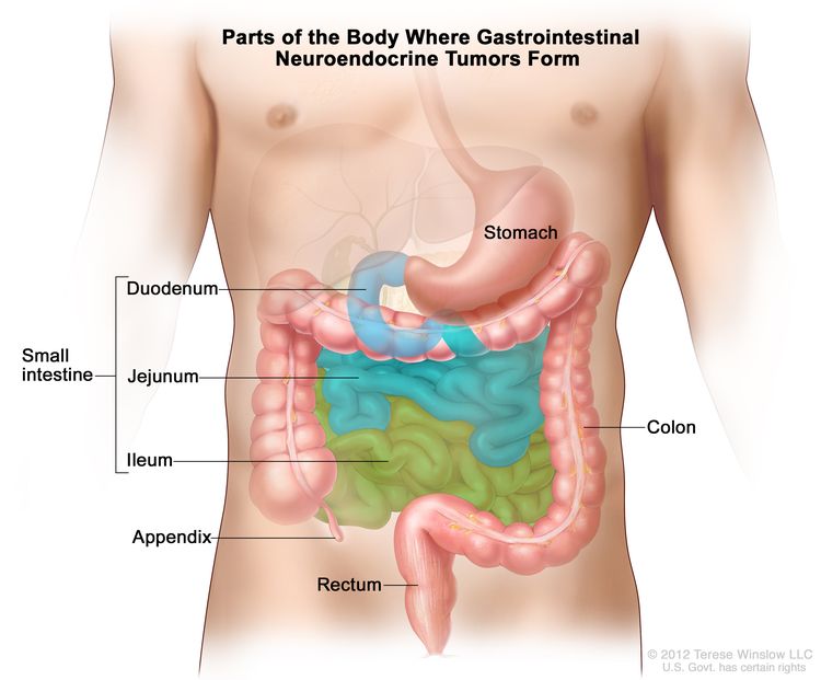
Pancreatic Neuroendocrine Tumors (Islet Cell Tumors) Treatment (PDQ®)–Health Professional Version

Pancreatic Neuroendocrine Tumors (Islet Cell Tumors) Treatment (PDQ®)–Health Professional Version

General Information About Pancreatic Neuroendocrine Tumors (Islet Cell Tumors)

Incidence and Mortality

Pancreatic neuroendocrine tumors (NETs) are uncommon cancers with about 1,000 new cases per year in the United States.[1] They account for less than 2% of pancreatic malignancies and, overall, have a better prognosis than the more common pancreatic exocrine tumors.[1,2]

Pathogenesis

Tumors of the endocrine pancreas are a collection of tumor cell types collectively referred to as pancreatic NETs. These tumors originate in islet cells. Although they may be similar or identical in histological appearance to carcinoid tumors of the gastrointestinal tract, differences in their underlying biology and likely differences in response to therapeutic agents suggest that they should be treated and investigated as a distinct entity.[3]

Most pancreatic NETs are sporadic, but some occur as part of the autosomal dominant multiple endocrine neoplasia type 1 (MEN1) inherited syndrome. This syndrome consists of tumors of the anterior pituitary, parathyroid, and endocrine pancreas glands and is a result of inactivation of the MEN1 tumor suppressor gene located on chromosome 11q13.[4] When part of the MEN1 syndrome, there may be multiple pancreatic tumors.

Islet tumors may either be functional (produce one or more active hormones) or nonfunctional.[4] The functional tumors, which usually present with symptoms of hormone hypersecretion, include:

  • Gastrinoma.
  • Insulinoma.
  • Glucagonoma.
  • Somatostatinoma.
  • VIPoma (Verner-Morrison syndrome).

Prognostic Factors

Most islet cell cancers are functional, but about 15% are nonfunctional, with presentations similar to the far more common exocrine adenocarcinomas of the pancreas.[57] Because of the presence of several cell types in the pancreatic islets (alpha, beta, delta, A, B, C, D, E, F), the term islet cell tumors refers to at least five distinct cancers that, when functional, produce unique metabolic and clinical characteristics. The clinical manifestations in functional tumors may result from the distinctive metabolic effects of the polypeptide(s) secreted by the cancer cells rather than from tumor bulk or metastatic disease. Functional tumors may even be too small to be detected by conventional imaging techniques.

Nonfunctional tumors tend to present at later clinical stages with symptoms attributable to mass effect or metastases.[4] Although nonfunctional tumors do not produce specific clinical syndromes, they may secrete inactive amine and peptide products such as:

  • Neurotensin.
  • Alpha-subunit of human chorionic gonadotropin (alpha-hCG).
  • Neuron-specific enolase.
  • Pancreatic polypeptide.
  • Chromogranin A.

Diagnostics

The potential long delays between initial symptoms and diagnosis and the varied effects of the polypeptides secreted often necessitate involvement of multiple surgical and medical subspecialties. Surgery is the only curative modality and is often used, even in patients with metastatic disease, to alleviate the symptoms of hormonal hypersecretion.[4] Effective palliation may be achieved because of the slow-growing nature of most of these tumors and the potential use of antihormonal pharmacological therapy (e.g., cimetidine in the ulcer-producing Zollinger-Ellison syndrome). In patients with indolent, slow-growing, metastatic islet cell tumors, the best therapy may be careful observation, and no treatment until palliation is required. In patients with MEN1 in which 85% have pancreatic islet cell tumors, 90% have hyperparathyroidism, and 65% have pituitary tumors, and they are less likely to be cured by pancreatic resection than are patients with sporadic islet cell tumors. With the exception of pain relief from bone metastases, radiation therapy has a limited role in this disease.

Tumor localization and staging studies include imaging with computed tomography (CT) with or without magnetic resonance imaging (MRI), and endoscopic ultrasonography. Somatostatin-receptor scintigraphy and single-photon emission CT may be useful adjuncts. However, somatostatin-receptor scintigraphy is less useful in localizing insulinomas versus other pancreatic NETs, since insulinomas often have a low density of somatostatin receptors.[4] If the noninvasive tests do not reveal a tumor, but clinical suspicion remains high, more invasive and technically demanding tests, such as selective arteriography or selective arterial stimulation (with a secretagogue specific for the suspected tumor type), may be useful.[7]

Some of the tumor types have unique characteristics that require specific approaches in their diagnosis and initial evaluation.

Gastrinoma

Diagnosis is dependent on elevated serum gastrin and elevated gastric acid levels. Provocative testing with calcium and secretin shows considerable overlap, and the value of these tests is limited. Zollinger-Ellison syndrome is a condition of unrelenting peptic ulcer disease, diarrhea, and gastric hyperacidity, associated with a gastrin-producing tumor. It accounts for less than 1% of all peptic ulcer disease. About 15% to 35% of gastrinomas are associated with MEN1 syndrome and up to 50% are malignant. Up to 33% of patients with gastrinomas have liver metastases.[4] For more information, see the Diarrhea section in Gastrointestinal Complications.

Diagnostic tests:

  1. BAO:MAO ≥ 0.6 (basal acid output:maximal acid output).
  2. Overnight AO ≥ 100 mmol.
  3. BAO ≥ 10 mmol/hr.
  4. Serum gastrin 10x normal or >500 pg/mL (the accuracy of gastrin assays may vary widely).
  5. Secretin test: 1 unit/kg intravenous rapid injection: Positive = 100% increase in gastrin within 10 minutes; 2 units/kg: Positive = 100% increase over baseline.
  6. Elevated hCG levels.

Insulinoma

Insulinomas are far more likely to be benign than malignant. Only 10% of insulinomas are multiple, and only 10% are malignant. About 5% to 8% are associated with MEN1 syndrome.[4] The clinical manifestations are those of hypoglycemia, which results from inappropriate secretion of insulin by the tumor. Fasting hypoglycemia (<40 mg/dL) associated with an elevated insulin level (in the absence of exogenous administration of insulin) is pathognomonic. Measurement of plasma proinsulin may be helpful for diagnosing insulin-secreting carcinomas. These tumors are usually slow-growing tumors and, when localized to the pancreas or regional lymph nodes, can be cured with pancreatic resection.

The approach to management depends on carefully performed preoperative localization studies and the findings at exploratory laparotomy. In a retrospective case series of 30 patients with 32 pancreatic insulinomas, the combination of preoperative, dual-phase, thin-section multidetector CT and endoscopic sonography correctly identified and localized all of the tumors.[8] These tests, with or without MRI, have replaced older, more invasive, and technically challenging tests, such as percutaneous transhepatic portal venous sampling and arterial stimulation with venous sampling, except for unusual circumstances in which the imaging tests are unsuccessful.[4,9]

Glucagonoma

Glucagonoma is the third most common endocrine-secreting islet cell tumor. About 75% of glucagonomas are malignant.[4] Necrolytic migratory erythema, hyperglycemia, and venous thrombosis comprise a virtually diagnostic triad. A serum glucagon level greater than 1,000 pg/mL confirms the diagnosis. These tumors tend to be large and easily visible on CT scan. Somatostatin-receptor scintigraphy scanning may be a useful adjunct in detecting metastases.

Miscellaneous islet cell tumors

These tumors are rare but have defined clinical syndromes associated with specific production of polypeptide hormone production by islet cell tumors. Because of their rarity and similar approaches to management, they are grouped in the section on treatment. Miscellaneous tumors include:

  • VIPoma.

    VIPoma is characterized by watery diarrhea, hypokalemia, and achlorhydria. A serum vasoactive intestinal peptide (VIP) greater than 200 pg/mL is diagnostic.[4] These tumors can generally be easily localized by CT scan. Somatostatin-receptor scintigraphy scanning may be a useful adjunct in detecting metastases.

  • Somatostatinoma.

    These tumors are particularly rare. They often present with diarrhea, steatorrhea, diabetes, and/or gallstones. Decreased pancreatic secretion of enzymes and bicarbonate accounts for the diarrhea and steatorrhea. Somatostatin-mediated inhibition of cholecystokinin leads to gallstone formation. Somatostatin also inhibits insulin, producing hyperglycemia. The diagnosis is made by a fasting serum somatostatin level greater than 100 pg/mL. CT scan, MRI, and endoscopic ultrasound can usually help localize and stage the tumor. Most of these tumors are malignant and have metastases at diagnosis.

References
  1. Ries LAG, Young JL, Keel GE, et al., eds.: SEER Survival Monograph: Cancer Survival Among Adults: U. S. SEER Program, 1988-2001, Patient and Tumor Characteristics. National Cancer Institute, 2007. NIH Pub. No. 07-6215.
  2. Neuroendocrine tumors of the pancreas. In: Amin MB, Edge SB, Greene FL, et al., eds.: AJCC Cancer Staging Manual. 8th ed. Springer; 2017, pp. 407–19.
  3. Kulke MH, Siu LL, Tepper JE, et al.: Future directions in the treatment of neuroendocrine tumors: consensus report of the National Cancer Institute Neuroendocrine Tumor clinical trials planning meeting. J Clin Oncol 29 (7): 934-43, 2011. [PUBMED Abstract]
  4. Davies K, Conlon KC: Neuroendocrine tumors of the pancreas. Curr Gastroenterol Rep 11 (2): 119-27, 2009. [PUBMED Abstract]
  5. Hochwald SN, Zee S, Conlon KC, et al.: Prognostic factors in pancreatic endocrine neoplasms: an analysis of 136 cases with a proposal for low-grade and intermediate-grade groups. J Clin Oncol 20 (11): 2633-42, 2002. [PUBMED Abstract]
  6. O’Grady HL, Conlon KC: Pancreatic neuroendocrine tumours. Eur J Surg Oncol 34 (3): 324-32, 2008. [PUBMED Abstract]
  7. King CM, Reznek RH, Dacie JE, et al.: Imaging islet cell tumours. Clin Radiol 49 (5): 295-303, 1994. [PUBMED Abstract]
  8. Gouya H, Vignaux O, Augui J, et al.: CT, endoscopic sonography, and a combined protocol for preoperative evaluation of pancreatic insulinomas. AJR Am J Roentgenol 181 (4): 987-92, 2003. [PUBMED Abstract]
  9. Nikfarjam M, Warshaw AL, Axelrod L, et al.: Improved contemporary surgical management of insulinomas: a 25-year experience at the Massachusetts General Hospital. Ann Surg 247 (1): 165-72, 2008. [PUBMED Abstract]

Cellular Classification of Pancreatic Neuroendocrine Tumors (Islet Cell Tumors)

Table 1. Endocrine Tumors of the Pancreas
Islet Cells Secreted Active Agent Tumor and Syndrome
5-HT = serotonin; ACTH = adrenocorticotropin hormone; MSH = melanocyte-stimulating hormone; VIP = vasoactive intestinal peptide; WDHA = watery diarrhea, hypokalemia, and achlorhydria.
Alpha Glucagon Glucagonoma (diabetes, dermatitis)
Beta Insulin Insulinoma (hypoglycemia)
Delta Somatostatin Somatostatinoma (mild diabetes); diarrhea/steatorrhea; gallstones
D Gastrin Gastrinoma (peptic ulcer disease)
A→D VIP and/or other undefined mediators WDHA
  5-HT Carcinoid
  ACTH Cushing disease
  MSH Hyperpigmentation
Interacinar Cells Secreted Active Agent Tumor and Syndrome
F Pancreatic polypeptide Multiple hormonal syndromes
EC 5-HT Carcinoid

Stage Information for Pancreatic Neuroendocrine Tumors (Islet Cell Tumors)

American Joint Committee on Cancer (AJCC) Stage Groupings and TNM Definitions

The AJCC has designated staging by TNM (tumor, node, metastasis) classification to define pancreatic neuroendocrine tumors (islet cell tumors).[1]

Table 2. Definitions for TNM Stage Ia
Stage TNM Definition
T = primary tumor; N = regional lymph node; M = distant metastasis.
aReprinted with permission from AJCC: Neuroendocrine Tumors of the Pancreas. In: Amin MB, Edge SB, Greene FL, et al., eds.: AJCC Cancer Staging Manual. 8th ed. New York, NY: Springer, 2017, pp. 407–19.
bThe explanation for superscript b is at the end of Table 5.
I T1, N0, M0 T1 = Tumor limited to the pancreas,b <2 cm.
N0 = No regional lymph node involvement.
M0 = No distant metastasis.
Table 3. Definitions for TNM Stage IIa
Stage TNM Definition
T = primary tumor; N = regional lymph node; M = distant metastasis.
aReprinted with permission from AJCC: Neuroendocrine Tumors of the Pancreas. In: Amin MB, Edge SB, Greene FL, et al., eds.: AJCC Cancer Staging Manual. 8th ed. New York, NY: Springer, 2017, pp. 407–19.
bThe explanation for superscript b is at the end of Table 5.
II T2, N0, M0 T2 = Tumor limited to the pancreas,b 2–4 cm.
N0 = No regional lymph node involvement.
M0 = No distant metastasis.
T3, N0, M0 T3 = Tumor limited to the pancreas,b >4 cm; or tumor invading the duodenum or common bile duct.
N0 = No regional lymph node involvement.
M0 = No distant metastasis.
Table 4. Definitions for TNM Stage IIIa
Stage TNM Definition
T = primary tumor; N = regional lymph node; M = distant metastasis.
aReprinted with permission from AJCC: Neuroendocrine Tumors of the Pancreas. In: Amin MB, Edge SB, Greene FL, et al., eds.: AJCC Cancer Staging Manual. 8th ed. New York, NY: Springer, 2017, pp. 407–19.
bThe explanation for superscript b is at the end of Table 5.
III T4, N0, M0 T4 = Tumor invading adjacent organs (stomach, spleen, colon, adrenal gland) or the wall of large vessels (celiac axis or the superior mesenteric artery).
N0 = No regional lymph node involvement.
M0 = No distant metastasis.
Any T, N1, M0 TX = Tumor cannot be assessed.
T1 = Tumor limited to the pancreas,b <2 cm.
T2 = Tumor limited to the pancreas,b 2–4 cm.
T3 = Tumor limited to the pancreas,b >4 cm; or tumor invading the duodenum or common bile duct.
T4 = Tumor invading adjacent organs (stomach, spleen, colon, adrenal gland) or the wall of large vessels (celiac axis or the superior mesenteric artery).
N1 = Regional lymph node involvement.
M0 = No distant metastasis.
Table 5. Definitions for TNM Stage IVa
Stage TNM Definition
T = primary tumor; N = regional lymph node; M = distant metastasis.
aReprinted with permission from AJCC: Neuroendocrine Tumors of the Pancreas. In: Amin MB, Edge SB, Greene FL, et al., eds.: AJCC Cancer Staging Manual. 8th ed. New York, NY: Springer, 2017, pp. 407–19.
bLimited to the pancreas means there is no invasion of adjacent organs (stomach, spleen, colon, adrenal gland) or the wall of large vessels (celiac axis or the superior mesenteric artery). Extension of tumor into peripancreatic adipose tissue is NOT a basis for staging. Note: Multiple tumors should be designated as such (the largest tumor should be used to assign T category): if the number of tumors is known, use T(#); e.g., pT3(4) N0 M0; if the number of tumors is unavailable or too numerous, use the m suffix, T(m); e.g., pT3(m) N0 M0.
IV Any T, Any N, M1 TX = Tumor cannot be assessed.
T1 = Tumor limited to the pancreas,b <2 cm.
T2 = Tumor limited to the pancreas,b 2–4 cm.
T3 = Tumor limited to the pancreas,b >4 cm; or tumor invading the duodenum or common bile duct.
T4 = Tumor invading adjacent organs (stomach, spleen, colon, adrenal gland) or the wall of large vessels (celiac axis or the superior mesenteric artery).
NX = Regional lymph nodes cannot be assessed.
N0 = No regional lymph node involvement.
N1 = Regional lymph node involvement.
M1 = Distant metastases.
–M1a = Metastasis confined to liver.
–M1b = Metastases in at least one extrahepatic site (e.g., lung, ovary, nonregional lymph node, peritoneum, bone).
–M1c = Both hepatic and extrahepatic metastases.
References
  1. Neuroendocrine tumors of the pancreas. In: Amin MB, Edge SB, Greene FL, et al., eds.: AJCC Cancer Staging Manual. 8th ed. Springer; 2017, pp. 407–19.

Treatment Option Overview for Pancreatic Neuroendocrine Tumors (Islet Cell Tumors)

Localized Disease

If technically and medically feasible, primary management of endocrine tumors of the pancreas involves surgical resection with curative intent. Given the rare nature of these tumors, surgical approaches are based on case series and expert opinion rather than randomized controlled trials.[1] The surgical options listed below are based on retrospective series from single reporting centers.[24][Level of evidence C2]

Adjuvant therapy has no proven benefit and is, therefore, investigational. There have been no well-controlled trials of adjuvant therapy after complete tumor resection.[5]

Surgical Cytoreduction for Metastases

Surgery plays a role even in the setting of metastatic disease. The symptoms of metastatic functional pancreatic neuroendocrine tumors (NETs) may be ameliorated by the reduction of overall tumor burden through surgical debulking.

The liver is a common site of metastasis from pancreatic NETs. Because of the slow growth rate of many NETs, liver metastases are often resected when technically feasible. Resection of all grossly visible liver metastases can be associated with long-term survival and, in the case of symptomatic hormonally functional tumors, symptom relief.[6] Most symptoms from functional tumors respond to this form of surgical debulking. It is not known if the favorable survival rates are attributable to patient selection factors (e.g., underlying patient condition, extent of metastases, slow doubling time, and so forth).

Alternative approaches to the management of liver metastases have been reported, including Gelfoam embolization or transarterial chemoembolization,[7] radioembolization with radioactive microspheres,[810] radiofrequency ablation, cryoablation, and percutaneous alcohol ablation. These alternative approaches have been reviewed.[11]

Results from surgical resection series appear to be more favorable than with these techniques, and surgery is considered to be the standard approach to resectable liver metastases. However, there are no high-quality studies comparing the various approaches. A systematic review of evidence comparing liver resection versus other treatments for patients with resectable liver neuroendocrine metastases found no randomized trials, or even quasi-experimental, cohort, or case-control studies in which the patient population given the alternative therapies was similar enough to the surgery group to draw reliable conclusions.[12] The evidence for resection of all grossly visible liver metastases is derived solely from case series.[Level of evidence C2]

In most cases, liver metastases are not completely resectable. Cytoreductive surgery, with or without radiofrequency ablation or cryoablation, has been used to palliate symptoms. A systematic review found no randomized or quasi-randomized trials comparing cytoreductive surgery to other palliative approaches such as chemotherapy or tumor product inhibitors.[13] The evidence for surgical cytoreduction of unresectable liver metastases is restricted to case series [Level of evidence C2], and interpretation of outcomes may be strongly affected by patient selection factors.

Systemic Therapy for Advanced and Metastatic Disease

Somatostatin analogues may be effective in reducing the symptoms of functional tumors.[14]

Chemotherapy using drugs such as the following, either alone or in combination, has been shown to have antitumor effects, but evidence is weak or conflicting regarding the impact of chemotherapy on overall survival:[1517]

  • Streptozocin.
  • Doxorubicin.
  • Fluorouracil.
  • Chlorozotocin.
  • Dacarbazine.
  • Temozolomide.

A variety of systemic agents have shown biological or palliative activity, including:[5,18]

  • Tyrosine kinase inhibitors (e.g., sunitinib).
  • Temozolomide.
  • Vascular endothelial growth factor pathway inhibitors.
  • Mammalian target of rapamycin inhibitors (e.g., everolimus).

Nearly all of the evidence of activity is derived from case series.[Level of evidence C3] However, there are ongoing placebo-controlled randomized trials of everolimus [19] and sunitinib [20] that have been reported in abstract form showing an increase in progression-free survival in each case.[Level of evidence B1]

Favorable responses have been reported in patients with advanced progressive pancreatic NETs after treatment with several radiolabeled somatostatin analogues in which the analogues octreotide, octreotate, lanreotide, or edotreotide are stably attached to the radionuclides indium In 111, yttrium Y 90, or lutetium Lu 177.[2123] The relative efficacy of these various compounds is unknown. Study designs have been limited to case series with tumor response, biochemical response, or symptom control as the measure of efficacy.[Level of evidence C3]

As noted in each of the clinical situations, there is a paucity or absence of high-level evidence and a need for randomized controlled trials.[5]

Fluorouracil dosing

The DPYD gene encodes an enzyme that catabolizes pyrimidines and fluoropyrimidines, like capecitabine and fluorouracil. An estimated 1% to 2% of the population has germline pathogenic variants in DPYD, which lead to reduced DPD protein function and an accumulation of pyrimidines and fluoropyrimidines in the body.[24,25] Patients with the DPYD*2A variant who receive fluoropyrimidines may experience severe, life-threatening toxicities that are sometimes fatal. Many other DPYD variants have been identified, with a range of clinical effects.[2426] Fluoropyrimidine avoidance or a dose reduction of 50% may be recommended based on the patient’s DPYD genotype and number of functioning DPYD alleles.[2729] DPYD genetic testing costs less than $200, but insurance coverage varies due to a lack of national guidelines.[30] In addition, testing may delay therapy by 2 weeks, which would not be advisable in urgent situations. This controversial issue requires further evaluation.[31]

References
  1. Davies K, Conlon KC: Neuroendocrine tumors of the pancreas. Curr Gastroenterol Rep 11 (2): 119-27, 2009. [PUBMED Abstract]
  2. Phan GQ, Yeo CJ, Hruban RH, et al.: Surgical experience with pancreatic and peripancreatic neuroendocrine tumors: Review of 125 patients. J Gastrointest Surg 2 (5): 473-82, 1998 Sep-Oct. [PUBMED Abstract]
  3. Kazanjian KK, Reber HA, Hines OJ: Resection of pancreatic neuroendocrine tumors: results of 70 cases. Arch Surg 141 (8): 765-9; discussion 769-70, 2006. [PUBMED Abstract]
  4. Hochwald SN, Zee S, Conlon KC, et al.: Prognostic factors in pancreatic endocrine neoplasms: an analysis of 136 cases with a proposal for low-grade and intermediate-grade groups. J Clin Oncol 20 (11): 2633-42, 2002. [PUBMED Abstract]
  5. Kulke MH, Siu LL, Tepper JE, et al.: Future directions in the treatment of neuroendocrine tumors: consensus report of the National Cancer Institute Neuroendocrine Tumor clinical trials planning meeting. J Clin Oncol 29 (7): 934-43, 2011. [PUBMED Abstract]
  6. Sarmiento JM, Que FG: Hepatic surgery for metastases from neuroendocrine tumors. Surg Oncol Clin N Am 12 (1): 231-42, 2003. [PUBMED Abstract]
  7. Gupta S, Johnson MM, Murthy R, et al.: Hepatic arterial embolization and chemoembolization for the treatment of patients with metastatic neuroendocrine tumors: variables affecting response rates and survival. Cancer 104 (8): 1590-602, 2005. [PUBMED Abstract]
  8. Nguyen C, Faraggi M, Giraudet AL, et al.: Long-term efficacy of radionuclide therapy in patients with disseminated neuroendocrine tumors uncontrolled by conventional therapy. J Nucl Med 45 (10): 1660-8, 2004. [PUBMED Abstract]
  9. Kennedy AS, Dezarn WA, McNeillie P, et al.: Radioembolization for unresectable neuroendocrine hepatic metastases using resin 90Y-microspheres: early results in 148 patients. Am J Clin Oncol 31 (3): 271-9, 2008. [PUBMED Abstract]
  10. King J, Quinn R, Glenn DM, et al.: Radioembolization with selective internal radiation microspheres for neuroendocrine liver metastases. Cancer 113 (5): 921-9, 2008. [PUBMED Abstract]
  11. Siperstein AE, Berber E: Cryoablation, percutaneous alcohol injection, and radiofrequency ablation for treatment of neuroendocrine liver metastases. World J Surg 25 (6): 693-6, 2001. [PUBMED Abstract]
  12. Gurusamy KS, Ramamoorthy R, Sharma D, et al.: Liver resection versus other treatments for neuroendocrine tumours in patients with resectable liver metastases. Cochrane Database Syst Rev (2): CD007060, 2009. [PUBMED Abstract]
  13. Gurusamy KS, Pamecha V, Sharma D, et al.: Palliative cytoreductive surgery versus other palliative treatments in patients with unresectable liver metastases from gastro-entero-pancreatic neuroendocrine tumours. Cochrane Database Syst Rev (1): CD007118, 2009. [PUBMED Abstract]
  14. di Bartolomeo M, Bajetta E, Buzzoni R, et al.: Clinical efficacy of octreotide in the treatment of metastatic neuroendocrine tumors. A study by the Italian Trials in Medical Oncology Group. Cancer 77 (2): 402-8, 1996. [PUBMED Abstract]
  15. Moertel CG, Lefkopoulo M, Lipsitz S, et al.: Streptozocin-doxorubicin, streptozocin-fluorouracil or chlorozotocin in the treatment of advanced islet-cell carcinoma. N Engl J Med 326 (8): 519-23, 1992. [PUBMED Abstract]
  16. Kouvaraki MA, Ajani JA, Hoff P, et al.: Fluorouracil, doxorubicin, and streptozocin in the treatment of patients with locally advanced and metastatic pancreatic endocrine carcinomas. J Clin Oncol 22 (23): 4762-71, 2004. [PUBMED Abstract]
  17. Kulke MH, Hornick JL, Frauenhoffer C, et al.: O6-methylguanine DNA methyltransferase deficiency and response to temozolomide-based therapy in patients with neuroendocrine tumors. Clin Cancer Res 15 (1): 338-45, 2009. [PUBMED Abstract]
  18. Yao JC, Lombard-Bohas C, Baudin E, et al.: Daily oral everolimus activity in patients with metastatic pancreatic neuroendocrine tumors after failure of cytotoxic chemotherapy: a phase II trial. J Clin Oncol 28 (1): 69-76, 2010. [PUBMED Abstract]
  19. Yao JC, Shah MH, Ito T, et al.: A randomized, double-blind, placebo-controlled multicenter phase III trial of everolimus in patients with advanced pancreatic neuroendocrine tumors (PNET) (RADIANT-3). [Abstract] Ann Oncol 21 (Suppl 8): A-LBA9, viii4-5, 2010.
  20. Raymond E, Niccoli-Sire P, Bang Y: Updated results of the phase III trial of sunitinib (SU) versus placebo (PBO) for treatment of advanced pancreatic neuroendocrine tumors (NET). [Abstract] American Society of Clinical Oncology 2010 Gastrointestinal Cancers Symposium, 22–24 January 2010, Orlando, Florida. A-127, 2010.
  21. Teunissen JJ, Kwekkeboom DJ, de Jong M, et al.: Endocrine tumours of the gastrointestinal tract. Peptide receptor radionuclide therapy. Best Pract Res Clin Gastroenterol 19 (4): 595-616, 2005. [PUBMED Abstract]
  22. Kwekkeboom DJ, de Herder WW, Kam BL, et al.: Treatment with the radiolabeled somatostatin analog [177 Lu-DOTA 0,Tyr3]octreotate: toxicity, efficacy, and survival. J Clin Oncol 26 (13): 2124-30, 2008. [PUBMED Abstract]
  23. Bushnell DL, O’Dorisio TM, O’Dorisio MS, et al.: 90Y-edotreotide for metastatic carcinoid refractory to octreotide. J Clin Oncol 28 (10): 1652-9, 2010. [PUBMED Abstract]
  24. Sharma BB, Rai K, Blunt H, et al.: Pathogenic DPYD Variants and Treatment-Related Mortality in Patients Receiving Fluoropyrimidine Chemotherapy: A Systematic Review and Meta-Analysis. Oncologist 26 (12): 1008-1016, 2021. [PUBMED Abstract]
  25. Lam SW, Guchelaar HJ, Boven E: The role of pharmacogenetics in capecitabine efficacy and toxicity. Cancer Treat Rev 50: 9-22, 2016. [PUBMED Abstract]
  26. Shakeel F, Fang F, Kwon JW, et al.: Patients carrying DPYD variant alleles have increased risk of severe toxicity and related treatment modifications during fluoropyrimidine chemotherapy. Pharmacogenomics 22 (3): 145-155, 2021. [PUBMED Abstract]
  27. Amstutz U, Henricks LM, Offer SM, et al.: Clinical Pharmacogenetics Implementation Consortium (CPIC) Guideline for Dihydropyrimidine Dehydrogenase Genotype and Fluoropyrimidine Dosing: 2017 Update. Clin Pharmacol Ther 103 (2): 210-216, 2018. [PUBMED Abstract]
  28. Henricks LM, Lunenburg CATC, de Man FM, et al.: DPYD genotype-guided dose individualisation of fluoropyrimidine therapy in patients with cancer: a prospective safety analysis. Lancet Oncol 19 (11): 1459-1467, 2018. [PUBMED Abstract]
  29. Lau-Min KS, Varughese LA, Nelson MN, et al.: Preemptive pharmacogenetic testing to guide chemotherapy dosing in patients with gastrointestinal malignancies: a qualitative study of barriers to implementation. BMC Cancer 22 (1): 47, 2022. [PUBMED Abstract]
  30. Brooks GA, Tapp S, Daly AT, et al.: Cost-effectiveness of DPYD Genotyping Prior to Fluoropyrimidine-based Adjuvant Chemotherapy for Colon Cancer. Clin Colorectal Cancer 21 (3): e189-e195, 2022. [PUBMED Abstract]
  31. Baker SD, Bates SE, Brooks GA, et al.: DPYD Testing: Time to Put Patient Safety First. J Clin Oncol 41 (15): 2701-2705, 2023. [PUBMED Abstract]

Treatment of Gastrinoma

The approach to treatment often depends on the results of preoperative localization studies and findings at exploratory laparotomy. At exploration, 85% of these tumors are found in the gastrinoma triangle with 40% on the surface of the pancreas and 40% outside of the pancreas. Only 15% are found within the substance of the pancreas. Percutaneous transhepatic venous sampling may occasionally provide accurate localization of single sporadic gastrinomas. Resection (enucleation of individual tumors, if technically feasible), and even excision of liver metastases, is associated with long-term cure or disease control.[1]

Treatment options:

  1. Single lesion in the head of the pancreas:[25]
    • Enucleation.
    • Parietal cell vagotomy and cimetidine.
    • Total gastrectomy (rarely used with the introduction of current therapies).
  2. Single or multiple lesions in the duodenum:[25]
    • Pancreatoduodenectomy.
  3. Single lesion in the body/tail of the pancreas:[25]
    • Resection of body/tail.
  4. Multiple lesions in the pancreas:[25]
    • Resection of body/tail.
    • If residual disease is present, parietal cell vagotomy and cimetidine.
    • Total gastrectomy (rarely used with the introduction of current therapies).
  5. No tumor found:
    • Parietal cell vagotomy and cimetidine.
    • Total gastrectomy (rarely used with the introduction of current therapies).
  6. Liver metastases:[613]
    • Liver resection where possible.
    • Radiofrequency ablation or cryosurgical ablation.
    • Chemoembolization of liver.
  7. Metastatic disease or disease refractory to surgery and cimetidine:[1423]
    • Chemotherapy
    • Somatostatin analogue therapy.

Patients with hepatic-dominant disease and substantial symptoms caused by tumor bulk or hormone-release syndromes may benefit from procedures that reduce hepatic arterial blood flow to metastases (hepatic arterial occlusion with embolization or with chemoembolization). Such treatment may also be combined with systemic chemotherapy in selected patients. Treatment with proton pump inhibitors or H2 blocking agents may aid in control of peptic symptoms.

In the era of proton pump inhibitors and H2 blocking agents, the potentially lethal hyperacidity and hypersecretory states induced by excessive gastrin production can usually be controlled. In a series of 212 patients with Zollinger-Ellison syndrome (ZES), no patients died of causes related to acid hypersecretion. Only 2.3% of those patients had undergone total gastrectomy, and the study cohort had a long median follow-up period of 13.8 years. Although 32% of the patients died during the course of the study, only 50% of the 67 deaths were attributable to ZES-related causes. Those causes were mainly liver metastases with progressive anorexia and cachexia (67%) or secondary endocrine tumors consequent to multiple endocrine neoplasia type 1 syndrome. The development of bone or liver metastases (especially diffuse liver disease) or of ectopic Cushing syndrome during the study period predicted for decreased survival times.[24]

Current Clinical Trials

Use our advanced clinical trial search to find NCI-supported cancer clinical trials that are now enrolling patients. The search can be narrowed by location of the trial, type of treatment, name of the drug, and other criteria. General information about clinical trials is also available.

References
  1. Norton JA, Fraker DL, Alexander HR, et al.: Surgery increases survival in patients with gastrinoma. Ann Surg 244 (3): 410-9, 2006. [PUBMED Abstract]
  2. Davies K, Conlon KC: Neuroendocrine tumors of the pancreas. Curr Gastroenterol Rep 11 (2): 119-27, 2009. [PUBMED Abstract]
  3. Phan GQ, Yeo CJ, Hruban RH, et al.: Surgical experience with pancreatic and peripancreatic neuroendocrine tumors: Review of 125 patients. J Gastrointest Surg 2 (5): 473-82, 1998 Sep-Oct. [PUBMED Abstract]
  4. Kazanjian KK, Reber HA, Hines OJ: Resection of pancreatic neuroendocrine tumors: results of 70 cases. Arch Surg 141 (8): 765-9; discussion 769-70, 2006. [PUBMED Abstract]
  5. Hochwald SN, Zee S, Conlon KC, et al.: Prognostic factors in pancreatic endocrine neoplasms: an analysis of 136 cases with a proposal for low-grade and intermediate-grade groups. J Clin Oncol 20 (11): 2633-42, 2002. [PUBMED Abstract]
  6. Sarmiento JM, Que FG: Hepatic surgery for metastases from neuroendocrine tumors. Surg Oncol Clin N Am 12 (1): 231-42, 2003. [PUBMED Abstract]
  7. Gupta S, Johnson MM, Murthy R, et al.: Hepatic arterial embolization and chemoembolization for the treatment of patients with metastatic neuroendocrine tumors: variables affecting response rates and survival. Cancer 104 (8): 1590-602, 2005. [PUBMED Abstract]
  8. Nguyen C, Faraggi M, Giraudet AL, et al.: Long-term efficacy of radionuclide therapy in patients with disseminated neuroendocrine tumors uncontrolled by conventional therapy. J Nucl Med 45 (10): 1660-8, 2004. [PUBMED Abstract]
  9. Kennedy AS, Dezarn WA, McNeillie P, et al.: Radioembolization for unresectable neuroendocrine hepatic metastases using resin 90Y-microspheres: early results in 148 patients. Am J Clin Oncol 31 (3): 271-9, 2008. [PUBMED Abstract]
  10. King J, Quinn R, Glenn DM, et al.: Radioembolization with selective internal radiation microspheres for neuroendocrine liver metastases. Cancer 113 (5): 921-9, 2008. [PUBMED Abstract]
  11. Siperstein AE, Berber E: Cryoablation, percutaneous alcohol injection, and radiofrequency ablation for treatment of neuroendocrine liver metastases. World J Surg 25 (6): 693-6, 2001. [PUBMED Abstract]
  12. Gurusamy KS, Ramamoorthy R, Sharma D, et al.: Liver resection versus other treatments for neuroendocrine tumours in patients with resectable liver metastases. Cochrane Database Syst Rev (2): CD007060, 2009. [PUBMED Abstract]
  13. Gurusamy KS, Pamecha V, Sharma D, et al.: Palliative cytoreductive surgery versus other palliative treatments in patients with unresectable liver metastases from gastro-entero-pancreatic neuroendocrine tumours. Cochrane Database Syst Rev (1): CD007118, 2009. [PUBMED Abstract]
  14. di Bartolomeo M, Bajetta E, Buzzoni R, et al.: Clinical efficacy of octreotide in the treatment of metastatic neuroendocrine tumors. A study by the Italian Trials in Medical Oncology Group. Cancer 77 (2): 402-8, 1996. [PUBMED Abstract]
  15. Moertel CG, Lefkopoulo M, Lipsitz S, et al.: Streptozocin-doxorubicin, streptozocin-fluorouracil or chlorozotocin in the treatment of advanced islet-cell carcinoma. N Engl J Med 326 (8): 519-23, 1992. [PUBMED Abstract]
  16. Kouvaraki MA, Ajani JA, Hoff P, et al.: Fluorouracil, doxorubicin, and streptozocin in the treatment of patients with locally advanced and metastatic pancreatic endocrine carcinomas. J Clin Oncol 22 (23): 4762-71, 2004. [PUBMED Abstract]
  17. Kulke MH, Hornick JL, Frauenhoffer C, et al.: O6-methylguanine DNA methyltransferase deficiency and response to temozolomide-based therapy in patients with neuroendocrine tumors. Clin Cancer Res 15 (1): 338-45, 2009. [PUBMED Abstract]
  18. Yao JC, Lombard-Bohas C, Baudin E, et al.: Daily oral everolimus activity in patients with metastatic pancreatic neuroendocrine tumors after failure of cytotoxic chemotherapy: a phase II trial. J Clin Oncol 28 (1): 69-76, 2010. [PUBMED Abstract]
  19. Yao JC, Shah MH, Ito T, et al.: A randomized, double-blind, placebo-controlled multicenter phase III trial of everolimus in patients with advanced pancreatic neuroendocrine tumors (PNET) (RADIANT-3). [Abstract] Ann Oncol 21 (Suppl 8): A-LBA9, viii4-5, 2010.
  20. Raymond E, Niccoli-Sire P, Bang Y: Updated results of the phase III trial of sunitinib (SU) versus placebo (PBO) for treatment of advanced pancreatic neuroendocrine tumors (NET). [Abstract] American Society of Clinical Oncology 2010 Gastrointestinal Cancers Symposium, 22–24 January 2010, Orlando, Florida. A-127, 2010.
  21. Teunissen JJ, Kwekkeboom DJ, de Jong M, et al.: Endocrine tumours of the gastrointestinal tract. Peptide receptor radionuclide therapy. Best Pract Res Clin Gastroenterol 19 (4): 595-616, 2005. [PUBMED Abstract]
  22. Kwekkeboom DJ, de Herder WW, Kam BL, et al.: Treatment with the radiolabeled somatostatin analog [177 Lu-DOTA 0,Tyr3]octreotate: toxicity, efficacy, and survival. J Clin Oncol 26 (13): 2124-30, 2008. [PUBMED Abstract]
  23. Bushnell DL, O’Dorisio TM, O’Dorisio MS, et al.: 90Y-edotreotide for metastatic carcinoid refractory to octreotide. J Clin Oncol 28 (10): 1652-9, 2010. [PUBMED Abstract]
  24. Kvols LK, Buck M, Moertel CG, et al.: Treatment of metastatic islet cell carcinoma with a somatostatin analogue (SMS 201-995). Ann Intern Med 107 (2): 162-8, 1987. [PUBMED Abstract]

Treatment of Insulinoma

Curative surgical excision, by open laparotomy or laparoscopy, is the treatment of choice when possible. The open surgical approach is used if the tumor is suspected to be malignant because en bloc lymphadenectomy is performed for malignant tumors without distant metastases. Intraoperative ultrasonography aids the localization of tumor extent and the relationship to other anatomical structures.[1]

Treatment options:

  1. Single, small lesion in the head or tail of the pancreas:[14]
    • Enucleation, if feasible.
  2. Large lesion in the head of the pancreas that is not amenable to enucleation:[14]
    • Pancreaticoduodenectomy.
  3. Single, large lesion in the body/tail:[14]
    • Distal pancreatectomy.
  4. Multiple lesions (may occur in 10% of patients, often in association with multiple endocrine neoplasia syndrome type 1):[14]
    • Distal pancreatectomy with enucleation of tumors in the head of the pancreas.
  5. Metastatic lesions in lymph nodes or distant sites:[512]
    • Resect when possible.
    • Consider radiofrequency or cryosurgical ablation, if not resectable.
  6. Unresectable:[1322]
    • Combination chemotherapy.
    • Pharmacological palliation: diazoxide 300 to 500 mg/day.
    • Somatostatin analogue therapy.

Patients with hepatic-dominant disease and substantial symptoms caused by tumor bulk or hormone-release syndromes may benefit from procedures that reduce hepatic arterial blood flow to metastases (hepatic arterial occlusion with embolization or with chemoembolization).[6,812] Such treatment may also be combined with systemic chemotherapy in selected patients.

Current Clinical Trials

Use our advanced clinical trial search to find NCI-supported cancer clinical trials that are now enrolling patients. The search can be narrowed by location of the trial, type of treatment, name of the drug, and other criteria. General information about clinical trials is also available.

References
  1. Davies K, Conlon KC: Neuroendocrine tumors of the pancreas. Curr Gastroenterol Rep 11 (2): 119-27, 2009. [PUBMED Abstract]
  2. Phan GQ, Yeo CJ, Hruban RH, et al.: Surgical experience with pancreatic and peripancreatic neuroendocrine tumors: Review of 125 patients. J Gastrointest Surg 2 (5): 473-82, 1998 Sep-Oct. [PUBMED Abstract]
  3. Kazanjian KK, Reber HA, Hines OJ: Resection of pancreatic neuroendocrine tumors: results of 70 cases. Arch Surg 141 (8): 765-9; discussion 769-70, 2006. [PUBMED Abstract]
  4. Hochwald SN, Zee S, Conlon KC, et al.: Prognostic factors in pancreatic endocrine neoplasms: an analysis of 136 cases with a proposal for low-grade and intermediate-grade groups. J Clin Oncol 20 (11): 2633-42, 2002. [PUBMED Abstract]
  5. Sarmiento JM, Que FG: Hepatic surgery for metastases from neuroendocrine tumors. Surg Oncol Clin N Am 12 (1): 231-42, 2003. [PUBMED Abstract]
  6. Gupta S, Johnson MM, Murthy R, et al.: Hepatic arterial embolization and chemoembolization for the treatment of patients with metastatic neuroendocrine tumors: variables affecting response rates and survival. Cancer 104 (8): 1590-602, 2005. [PUBMED Abstract]
  7. Nguyen C, Faraggi M, Giraudet AL, et al.: Long-term efficacy of radionuclide therapy in patients with disseminated neuroendocrine tumors uncontrolled by conventional therapy. J Nucl Med 45 (10): 1660-8, 2004. [PUBMED Abstract]
  8. Kennedy AS, Dezarn WA, McNeillie P, et al.: Radioembolization for unresectable neuroendocrine hepatic metastases using resin 90Y-microspheres: early results in 148 patients. Am J Clin Oncol 31 (3): 271-9, 2008. [PUBMED Abstract]
  9. King J, Quinn R, Glenn DM, et al.: Radioembolization with selective internal radiation microspheres for neuroendocrine liver metastases. Cancer 113 (5): 921-9, 2008. [PUBMED Abstract]
  10. Siperstein AE, Berber E: Cryoablation, percutaneous alcohol injection, and radiofrequency ablation for treatment of neuroendocrine liver metastases. World J Surg 25 (6): 693-6, 2001. [PUBMED Abstract]
  11. Gurusamy KS, Ramamoorthy R, Sharma D, et al.: Liver resection versus other treatments for neuroendocrine tumours in patients with resectable liver metastases. Cochrane Database Syst Rev (2): CD007060, 2009. [PUBMED Abstract]
  12. Gurusamy KS, Pamecha V, Sharma D, et al.: Palliative cytoreductive surgery versus other palliative treatments in patients with unresectable liver metastases from gastro-entero-pancreatic neuroendocrine tumours. Cochrane Database Syst Rev (1): CD007118, 2009. [PUBMED Abstract]
  13. di Bartolomeo M, Bajetta E, Buzzoni R, et al.: Clinical efficacy of octreotide in the treatment of metastatic neuroendocrine tumors. A study by the Italian Trials in Medical Oncology Group. Cancer 77 (2): 402-8, 1996. [PUBMED Abstract]
  14. Moertel CG, Lefkopoulo M, Lipsitz S, et al.: Streptozocin-doxorubicin, streptozocin-fluorouracil or chlorozotocin in the treatment of advanced islet-cell carcinoma. N Engl J Med 326 (8): 519-23, 1992. [PUBMED Abstract]
  15. Kouvaraki MA, Ajani JA, Hoff P, et al.: Fluorouracil, doxorubicin, and streptozocin in the treatment of patients with locally advanced and metastatic pancreatic endocrine carcinomas. J Clin Oncol 22 (23): 4762-71, 2004. [PUBMED Abstract]
  16. Kulke MH, Hornick JL, Frauenhoffer C, et al.: O6-methylguanine DNA methyltransferase deficiency and response to temozolomide-based therapy in patients with neuroendocrine tumors. Clin Cancer Res 15 (1): 338-45, 2009. [PUBMED Abstract]
  17. Yao JC, Lombard-Bohas C, Baudin E, et al.: Daily oral everolimus activity in patients with metastatic pancreatic neuroendocrine tumors after failure of cytotoxic chemotherapy: a phase II trial. J Clin Oncol 28 (1): 69-76, 2010. [PUBMED Abstract]
  18. Yao JC, Shah MH, Ito T, et al.: A randomized, double-blind, placebo-controlled multicenter phase III trial of everolimus in patients with advanced pancreatic neuroendocrine tumors (PNET) (RADIANT-3). [Abstract] Ann Oncol 21 (Suppl 8): A-LBA9, viii4-5, 2010.
  19. Raymond E, Niccoli-Sire P, Bang Y: Updated results of the phase III trial of sunitinib (SU) versus placebo (PBO) for treatment of advanced pancreatic neuroendocrine tumors (NET). [Abstract] American Society of Clinical Oncology 2010 Gastrointestinal Cancers Symposium, 22–24 January 2010, Orlando, Florida. A-127, 2010.
  20. Teunissen JJ, Kwekkeboom DJ, de Jong M, et al.: Endocrine tumours of the gastrointestinal tract. Peptide receptor radionuclide therapy. Best Pract Res Clin Gastroenterol 19 (4): 595-616, 2005. [PUBMED Abstract]
  21. Kwekkeboom DJ, de Herder WW, Kam BL, et al.: Treatment with the radiolabeled somatostatin analog [177 Lu-DOTA 0,Tyr3]octreotate: toxicity, efficacy, and survival. J Clin Oncol 26 (13): 2124-30, 2008. [PUBMED Abstract]
  22. Bushnell DL, O’Dorisio TM, O’Dorisio MS, et al.: 90Y-edotreotide for metastatic carcinoid refractory to octreotide. J Clin Oncol 28 (10): 1652-9, 2010. [PUBMED Abstract]

Treatment of Glucagonoma

As with the other pancreatic neuroendocrine tumors, the mainstay of therapy is surgical resection, and extended survival is possible even when the disease is metastatic. Resection of metastases is also a consideration when feasible.[1]

Treatment options:

  1. Single, small lesion in the head or tail of the pancreas:[14]
    • Enucleation, if feasible.
  2. Large lesion in the head of the pancreas that is not amenable to enucleation:[14]
    • Pancreaticoduodenectomy.
  3. Single, large lesion in the body/tail:[14]
    • Distal pancreatectomy.
  4. Multiple lesions:[14]
    • Enucleation, if feasible.
    • Resect body and tail otherwise.
  5. Metastatic disease in lymph nodes or distant sites:[512]
    • Resect when possible.
    • Consider radiofrequency or cryosurgical ablation, if not resectable.
  6. Unresectable disease:[1322]
    • Combination chemotherapy.
    • Somatostatin analogue therapy. Necrotizing erythema of glucagonoma may be relieved in 24 hours with somatostatin analogue, with nearly complete disappearance within 1 week.

Patients with hepatic-dominant disease and substantial symptoms caused by tumor bulk or hormone-release syndromes may benefit from procedures that reduce hepatic arterial blood flow to metastases (hepatic arterial occlusion with embolization or with chemoembolization).[6,812] Such treatment may also be combined with systemic chemotherapy in selected patients.

Current Clinical Trials

Use our advanced clinical trial search to find NCI-supported cancer clinical trials that are now enrolling patients. The search can be narrowed by location of the trial, type of treatment, name of the drug, and other criteria. General information about clinical trials is also available.

References
  1. Davies K, Conlon KC: Neuroendocrine tumors of the pancreas. Curr Gastroenterol Rep 11 (2): 119-27, 2009. [PUBMED Abstract]
  2. Phan GQ, Yeo CJ, Hruban RH, et al.: Surgical experience with pancreatic and peripancreatic neuroendocrine tumors: Review of 125 patients. J Gastrointest Surg 2 (5): 473-82, 1998 Sep-Oct. [PUBMED Abstract]
  3. Kazanjian KK, Reber HA, Hines OJ: Resection of pancreatic neuroendocrine tumors: results of 70 cases. Arch Surg 141 (8): 765-9; discussion 769-70, 2006. [PUBMED Abstract]
  4. Hochwald SN, Zee S, Conlon KC, et al.: Prognostic factors in pancreatic endocrine neoplasms: an analysis of 136 cases with a proposal for low-grade and intermediate-grade groups. J Clin Oncol 20 (11): 2633-42, 2002. [PUBMED Abstract]
  5. Sarmiento JM, Que FG: Hepatic surgery for metastases from neuroendocrine tumors. Surg Oncol Clin N Am 12 (1): 231-42, 2003. [PUBMED Abstract]
  6. Gupta S, Johnson MM, Murthy R, et al.: Hepatic arterial embolization and chemoembolization for the treatment of patients with metastatic neuroendocrine tumors: variables affecting response rates and survival. Cancer 104 (8): 1590-602, 2005. [PUBMED Abstract]
  7. Nguyen C, Faraggi M, Giraudet AL, et al.: Long-term efficacy of radionuclide therapy in patients with disseminated neuroendocrine tumors uncontrolled by conventional therapy. J Nucl Med 45 (10): 1660-8, 2004. [PUBMED Abstract]
  8. Kennedy AS, Dezarn WA, McNeillie P, et al.: Radioembolization for unresectable neuroendocrine hepatic metastases using resin 90Y-microspheres: early results in 148 patients. Am J Clin Oncol 31 (3): 271-9, 2008. [PUBMED Abstract]
  9. King J, Quinn R, Glenn DM, et al.: Radioembolization with selective internal radiation microspheres for neuroendocrine liver metastases. Cancer 113 (5): 921-9, 2008. [PUBMED Abstract]
  10. Siperstein AE, Berber E: Cryoablation, percutaneous alcohol injection, and radiofrequency ablation for treatment of neuroendocrine liver metastases. World J Surg 25 (6): 693-6, 2001. [PUBMED Abstract]
  11. Gurusamy KS, Ramamoorthy R, Sharma D, et al.: Liver resection versus other treatments for neuroendocrine tumours in patients with resectable liver metastases. Cochrane Database Syst Rev (2): CD007060, 2009. [PUBMED Abstract]
  12. Gurusamy KS, Pamecha V, Sharma D, et al.: Palliative cytoreductive surgery versus other palliative treatments in patients with unresectable liver metastases from gastro-entero-pancreatic neuroendocrine tumours. Cochrane Database Syst Rev (1): CD007118, 2009. [PUBMED Abstract]
  13. di Bartolomeo M, Bajetta E, Buzzoni R, et al.: Clinical efficacy of octreotide in the treatment of metastatic neuroendocrine tumors. A study by the Italian Trials in Medical Oncology Group. Cancer 77 (2): 402-8, 1996. [PUBMED Abstract]
  14. Moertel CG, Lefkopoulo M, Lipsitz S, et al.: Streptozocin-doxorubicin, streptozocin-fluorouracil or chlorozotocin in the treatment of advanced islet-cell carcinoma. N Engl J Med 326 (8): 519-23, 1992. [PUBMED Abstract]
  15. Kouvaraki MA, Ajani JA, Hoff P, et al.: Fluorouracil, doxorubicin, and streptozocin in the treatment of patients with locally advanced and metastatic pancreatic endocrine carcinomas. J Clin Oncol 22 (23): 4762-71, 2004. [PUBMED Abstract]
  16. Kulke MH, Hornick JL, Frauenhoffer C, et al.: O6-methylguanine DNA methyltransferase deficiency and response to temozolomide-based therapy in patients with neuroendocrine tumors. Clin Cancer Res 15 (1): 338-45, 2009. [PUBMED Abstract]
  17. Yao JC, Lombard-Bohas C, Baudin E, et al.: Daily oral everolimus activity in patients with metastatic pancreatic neuroendocrine tumors after failure of cytotoxic chemotherapy: a phase II trial. J Clin Oncol 28 (1): 69-76, 2010. [PUBMED Abstract]
  18. Yao JC, Shah MH, Ito T, et al.: A randomized, double-blind, placebo-controlled multicenter phase III trial of everolimus in patients with advanced pancreatic neuroendocrine tumors (PNET) (RADIANT-3). [Abstract] Ann Oncol 21 (Suppl 8): A-LBA9, viii4-5, 2010.
  19. Raymond E, Niccoli-Sire P, Bang Y: Updated results of the phase III trial of sunitinib (SU) versus placebo (PBO) for treatment of advanced pancreatic neuroendocrine tumors (NET). [Abstract] American Society of Clinical Oncology 2010 Gastrointestinal Cancers Symposium, 22–24 January 2010, Orlando, Florida. A-127, 2010.
  20. Teunissen JJ, Kwekkeboom DJ, de Jong M, et al.: Endocrine tumours of the gastrointestinal tract. Peptide receptor radionuclide therapy. Best Pract Res Clin Gastroenterol 19 (4): 595-616, 2005. [PUBMED Abstract]
  21. Kwekkeboom DJ, de Herder WW, Kam BL, et al.: Treatment with the radiolabeled somatostatin analog [177 Lu-DOTA 0,Tyr3]octreotate: toxicity, efficacy, and survival. J Clin Oncol 26 (13): 2124-30, 2008. [PUBMED Abstract]
  22. Bushnell DL, O’Dorisio TM, O’Dorisio MS, et al.: 90Y-edotreotide for metastatic carcinoid refractory to octreotide. J Clin Oncol 28 (10): 1652-9, 2010. [PUBMED Abstract]

Treatment of Miscellaneous Islet Cell Tumors

VIPoma

Immediate fluid resuscitation is often necessary to correct the electrolyte and fluid problems that occur as a result of the watery diarrhea, hypokalemia, and achlorhydria that patients experience. Somatostatin analogues are also used to ameliorate the large fluid and electrolyte losses. Once patients are stabilized, excision of the primary tumor and regional nodes is the first line of therapy for clinically localized disease. In the case of locally advanced or metastatic disease, where curative resection is not possible, debulking and removal of gross disease, including metastases, should be considered to alleviate the characteristic manifestations of vasoactive intestinal peptide overproduction.[1] For more information, see the Treatment Option Overview for Pancreatic Neuroendocrine Tumors (Islet Cell Tumors) section.

Somatostatinoma

Complete excision is the therapy of choice, if technically possible. However, metastases often preclude curative resection, and palliative debulking can be considered to relieve symptoms.[1] For more information, see the Treatment Option Overview for Pancreatic Neuroendocrine Tumors (Islet Cell Tumors) section.

Other Pancreatic Neuroendocrine Tumors

For these very rare tumors, complete surgical excision is the only curative option when technically possible. Debulking or somatostatin analogues are used for palliation of symptoms if the tumor is functional. For more information, see the Treatment Option Overview for Pancreatic Neuroendocrine Tumors (Islet Cell Tumors) section.

Current Clinical Trials

Use our advanced clinical trial search to find NCI-supported cancer clinical trials that are now enrolling patients. The search can be narrowed by location of the trial, type of treatment, name of the drug, and other criteria. General information about clinical trials is also available.

References
  1. Davies K, Conlon KC: Neuroendocrine tumors of the pancreas. Curr Gastroenterol Rep 11 (2): 119-27, 2009. [PUBMED Abstract]

Treatment of Recurrent and Progressive Pancreatic Neuroendocrine Tumors

There is no established therapy for pancreatic neuroendocrine tumors that recur or progress after previous therapy.[1] Deciding on further treatment depends on many factors, including:

  • The specific cancer.
  • Previous treatment.
  • Site of recurrence.
  • Individual patient considerations.

Attempts at re-resection of local tumors that have recurred or re-resection of metastatic lesions may offer palliation, when technically feasible. Intra-arterial chemotherapy is a consideration for patients with liver metastases. Patients with hepatic-dominant disease and substantial symptoms caused by tumor bulk or hormone-release syndromes may benefit from continuous-infusion intra-arterial chemotherapy or procedures that reduce hepatic arterial blood flow to metastases (hepatic arterial occlusion with embolization or with chemoembolization).[27] Such treatment may also be combined with systemic chemotherapy. A variety of systemic agents have shown biological or palliative activity,[1,8] including:

  • Somatostatin analogues.
  • Radiolabeled somatostatin analogues.[911]
  • Tyrosine kinase inhibitors (e.g., sunitinib).
  • Temozolomide.
  • Vascular endothelial growth factor pathway inhibitors.
  • Mammalian target of rapamycin inhibitors (e.g., everolimus).

Current Clinical Trials

Use our advanced clinical trial search to find NCI-supported cancer clinical trials that are now enrolling patients. The search can be narrowed by location of the trial, type of treatment, name of the drug, and other criteria. General information about clinical trials is also available.

References
  1. Kulke MH, Siu LL, Tepper JE, et al.: Future directions in the treatment of neuroendocrine tumors: consensus report of the National Cancer Institute Neuroendocrine Tumor clinical trials planning meeting. J Clin Oncol 29 (7): 934-43, 2011. [PUBMED Abstract]
  2. Gupta S, Johnson MM, Murthy R, et al.: Hepatic arterial embolization and chemoembolization for the treatment of patients with metastatic neuroendocrine tumors: variables affecting response rates and survival. Cancer 104 (8): 1590-602, 2005. [PUBMED Abstract]
  3. Kennedy AS, Dezarn WA, McNeillie P, et al.: Radioembolization for unresectable neuroendocrine hepatic metastases using resin 90Y-microspheres: early results in 148 patients. Am J Clin Oncol 31 (3): 271-9, 2008. [PUBMED Abstract]
  4. King J, Quinn R, Glenn DM, et al.: Radioembolization with selective internal radiation microspheres for neuroendocrine liver metastases. Cancer 113 (5): 921-9, 2008. [PUBMED Abstract]
  5. Siperstein AE, Berber E: Cryoablation, percutaneous alcohol injection, and radiofrequency ablation for treatment of neuroendocrine liver metastases. World J Surg 25 (6): 693-6, 2001. [PUBMED Abstract]
  6. Gurusamy KS, Ramamoorthy R, Sharma D, et al.: Liver resection versus other treatments for neuroendocrine tumours in patients with resectable liver metastases. Cochrane Database Syst Rev (2): CD007060, 2009. [PUBMED Abstract]
  7. Gurusamy KS, Pamecha V, Sharma D, et al.: Palliative cytoreductive surgery versus other palliative treatments in patients with unresectable liver metastases from gastro-entero-pancreatic neuroendocrine tumours. Cochrane Database Syst Rev (1): CD007118, 2009. [PUBMED Abstract]
  8. Yao JC, Lombard-Bohas C, Baudin E, et al.: Daily oral everolimus activity in patients with metastatic pancreatic neuroendocrine tumors after failure of cytotoxic chemotherapy: a phase II trial. J Clin Oncol 28 (1): 69-76, 2010. [PUBMED Abstract]
  9. Teunissen JJ, Kwekkeboom DJ, de Jong M, et al.: Endocrine tumours of the gastrointestinal tract. Peptide receptor radionuclide therapy. Best Pract Res Clin Gastroenterol 19 (4): 595-616, 2005. [PUBMED Abstract]
  10. Kwekkeboom DJ, de Herder WW, Kam BL, et al.: Treatment with the radiolabeled somatostatin analog [177 Lu-DOTA 0,Tyr3]octreotate: toxicity, efficacy, and survival. J Clin Oncol 26 (13): 2124-30, 2008. [PUBMED Abstract]
  11. Bushnell DL, O’Dorisio TM, O’Dorisio MS, et al.: 90Y-edotreotide for metastatic carcinoid refractory to octreotide. J Clin Oncol 28 (10): 1652-9, 2010. [PUBMED Abstract]

Latest Updates to This Summary (05/09/2025)

The PDQ cancer information summaries are reviewed regularly and updated as new information becomes available. This section describes the latest changes made to this summary as of the date above.

Editorial changes were made to this summary.

This summary is written and maintained by the PDQ Adult Treatment Editorial Board, which is editorially independent of NCI. The summary reflects an independent review of the literature and does not represent a policy statement of NCI or NIH. More information about summary policies and the role of the PDQ Editorial Boards in maintaining the PDQ summaries can be found on the About This PDQ Summary and PDQ® Cancer Information for Health Professionals pages.

About This PDQ Summary

Purpose of This Summary

This PDQ cancer information summary for health professionals provides comprehensive, peer-reviewed, evidence-based information about the treatment of pancreatic neuroendocrine tumors (islet cell tumors). It is intended as a resource to inform and assist clinicians in the care of their patients. It does not provide formal guidelines or recommendations for making health care decisions.

Reviewers and Updates

This summary is reviewed regularly and updated as necessary by the PDQ Adult Treatment Editorial Board, which is editorially independent of the National Cancer Institute (NCI). The summary reflects an independent review of the literature and does not represent a policy statement of NCI or the National Institutes of Health (NIH).

Board members review recently published articles each month to determine whether an article should:

  • be discussed at a meeting,
  • be cited with text, or
  • replace or update an existing article that is already cited.

Changes to the summaries are made through a consensus process in which Board members evaluate the strength of the evidence in the published articles and determine how the article should be included in the summary.

The lead reviewer for Pancreatic Neuroendocrine Tumors (Islet Cell Tumors) Treatment is:

  • Jaydira del Rivero, MD (National Cancer Institute)

Any comments or questions about the summary content should be submitted to Cancer.gov through the NCI website’s Email Us. Do not contact the individual Board Members with questions or comments about the summaries. Board members will not respond to individual inquiries.

Levels of Evidence

Some of the reference citations in this summary are accompanied by a level-of-evidence designation. These designations are intended to help readers assess the strength of the evidence supporting the use of specific interventions or approaches. The PDQ Adult Treatment Editorial Board uses a formal evidence ranking system in developing its level-of-evidence designations.

Permission to Use This Summary

PDQ is a registered trademark. Although the content of PDQ documents can be used freely as text, it cannot be identified as an NCI PDQ cancer information summary unless it is presented in its entirety and is regularly updated. However, an author would be permitted to write a sentence such as “NCI’s PDQ cancer information summary about breast cancer prevention states the risks succinctly: [include excerpt from the summary].”

The preferred citation for this PDQ summary is:

PDQ® Adult Treatment Editorial Board. PDQ Pancreatic Neuroendocrine Tumors (Islet Cell Tumors) Treatment. Bethesda, MD: National Cancer Institute. Updated <MM/DD/YYYY>. Available at: /types/pancreatic/hp/pnet-treatment-pdq. Accessed <MM/DD/YYYY>. [PMID: 26389309]

Images in this summary are used with permission of the author(s), artist, and/or publisher for use within the PDQ summaries only. Permission to use images outside the context of PDQ information must be obtained from the owner(s) and cannot be granted by the National Cancer Institute. Information about using the illustrations in this summary, along with many other cancer-related images, is available in Visuals Online, a collection of over 2,000 scientific images.

Disclaimer

Based on the strength of the available evidence, treatment options may be described as either “standard” or “under clinical evaluation.” These classifications should not be used as a basis for insurance reimbursement determinations. More information on insurance coverage is available on Cancer.gov on the Managing Cancer Care page.

Contact Us

More information about contacting us or receiving help with the Cancer.gov website can be found on our Contact Us for Help page. Questions can also be submitted to Cancer.gov through the website’s Email Us.
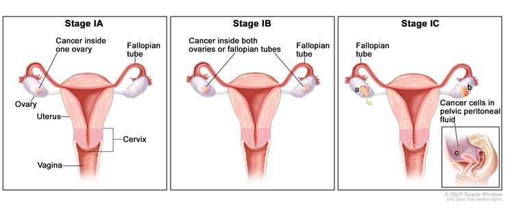
Gastrointestinal Neuroendocrine Tumors Treatment (PDQ®)–Patient Version

Gastrointestinal Neuroendocrine Tumors Treatment (PDQ®)–Patient Version

General Information About Gastrointestinal Neuroendocrine Tumors

Key Points

  • A gastrointestinal neuroendocrine tumor is cancer that forms in the lining of the gastrointestinal tract.
  • Health history can affect the risk of GI neuroendocrine tumors.
  • Some GI neuroendocrine tumors have no signs or symptoms in the early stages.
  • Carcinoid syndrome may occur if the tumor spreads to the liver or other parts of the body.
  • Imaging studies and tests that examine the blood and urine are used to diagnose GI neuroendocrine tumors.
  • Certain factors affect prognosis (chance of recovery) and treatment options.

A gastrointestinal neuroendocrine tumor is cancer that forms in the lining of the gastrointestinal tract.

The gastrointestinal (GI) tract is part of the body’s digestive system, a series of hollow, muscular organs joined in a long, twisting tube from the mouth to the anus. The digestive tract processes nutrients in foods that are eaten and helps pass waste material out of the body:

  • Food moves from the throat to the stomach through a tube called the esophagus.
  • After food enters the stomach, it is broken down by stomach muscles that mix the food and liquid with digestive juices.
  • After leaving the stomach, partly digested food passes into the small intestine and then into the large intestine.
  • The end of the large intestine, called the rectum, stores the waste from the digested food until it is pushed out of the anus during a bowel movement.
EnlargeDrawing of the gastrointestinal tract showing the stomach, small intestine (including the duodenum, jejunum, and ileum), appendix, colon, and rectum.
Gastrointestinal neuroendocrine tumors form in the lining of the gastrointestinal tract, most often in the small intestine, appendix, or rectum.

Gastrointestinal (GI) neuroendocrine tumors (also called gastrointestinal carcinoid tumors) form from a certain type of neuroendocrine cell (a type of cell that is like a nerve cell and a hormone-making cell). These cells are scattered throughout the chest and abdomen but most are found in the GI tract. Neuroendocrine cells make hormones that help control digestive juices and the muscles used in moving food through the stomach and intestines. A GI neuroendocrine tumor may also make hormones and release them into the body.

GI neuroendocrine tumors are rare and most grow very slowly. Most of them occur in the small intestine, rectum, and appendix. Sometimes more than one tumor will form.

See the following for information about other types of neuroendocrine tumors:

Health history can affect the risk of GI neuroendocrine tumors.

Anything that increases a person’s chance of getting a disease is called a risk factor. Not every person with one or more of these risk factors will develop GI neuroendocrine tumors, and they can develop in people who don’t have any known risk factors. Talk to your doctor if you think you may be at risk.

Risk factors for GI neuroendocrine tumors include:

Some GI neuroendocrine tumors have no signs or symptoms in the early stages.

Signs and symptoms may be caused by the growth of the tumor and/or the hormones the tumor makes. Some tumors, especially tumors of the stomach or appendix, may not cause signs or symptoms. Neuroendocrine tumors are often found during tests or treatments for other conditions.

Neuroendocrine tumors in the small intestine (duodenum, jejunum, and ileum), colon, and rectum sometimes cause signs or symptoms as they grow or because of the hormones they make. Other conditions may cause the same signs or symptoms. Check with your doctor if you have:

  • Duodenum

    Signs and symptoms of GI neuroendocrine tumors in the duodenum (first part of the small intestine, that connects to the stomach) may include:

  • Jejunum and ileum

    Signs and symptoms of GI neuroendocrine tumors in the jejunum (middle part of the small intestine) and ileum (last part of the small intestine, that connects to the colon) may include:

  • Colon

    Signs and symptoms of GI neuroendocrine tumors in the colon may include:

    • Abdominal pain.
    • Weight loss for no known reason.
  • Rectum

    Signs and symptoms of GI neuroendocrine tumors in the rectum may include:

Carcinoid syndrome may occur if the tumor spreads to the liver or other parts of the body.

The hormones made by GI neuroendocrine tumors are usually destroyed by liver enzymes in the blood. If the tumor has spread to the liver and the liver enzymes cannot destroy the extra hormones made by the tumor, high amounts of these hormones may remain in the body and cause carcinoid syndrome. This can also happen if tumor cells enter the blood. Signs and symptoms of carcinoid syndrome include:

  • Redness or a feeling of warmth in the face and neck.
  • Abdominal pain.
  • Feeling bloated.
  • Diarrhea.
  • Wheezing or other trouble breathing.
  • Fast heartbeat.

These signs and symptoms may be caused by GI neuroendocrine tumors or by other conditions. Talk to your doctor if you have any of these signs or symptoms.

Imaging studies and tests that examine the blood and urine are used to diagnose GI neuroendocrine tumors.

In addition to asking about your personal and family health history and doing a physical exam, your doctor may perform the following tests and procedures:

  • Blood chemistry studies: A procedure in which a blood sample is checked to measure the amounts of certain substances, such as hormones, released into the blood by organs and tissues in the body. An unusual (higher or lower than normal) amount of a substance can be a sign of disease. The blood sample is checked to see if it contains a hormone produced by neuroendocrine tumors. This test is used to help diagnose carcinoid syndrome.
  • Tumor marker test: A procedure in which a sample of blood, urine, or tissue is checked to measure the amounts of certain substances, such as chromogranin A, made by organs, tissues, or tumor cells in the body. Chromogranin A is a tumor marker. It has been linked to neuroendocrine tumors when found in increased levels in the body.
  • Twenty-four-hour urine test: A test in which urine is collected for 24 hours to measure the amounts of certain substances, such as 5-HIAA or serotonin (hormone). An unusual (higher or lower than normal) amount of a substance can be a sign of disease in the organ or tissue that makes it. This test is used to help diagnose carcinoid syndrome.
  • MIBG scan: A procedure used to find neuroendocrine tumors. A very small amount of radioactive material called MIBG (metaiodobenzylguanidine) is injected into a vein and travels through the bloodstream. Neuroendocrine tumors take up the radioactive material and are detected by a device that measures radiation.
  • CT scan (CAT scan): A procedure that makes a series of detailed pictures of areas inside the body, taken from different angles. The pictures are made by a computer linked to an x-ray machine. A dye may be injected into a vein or swallowed to help the organs or tissues show up more clearly. This procedure is also called computed tomography, computerized tomography, or computerized axial tomography.
  • MRI (magnetic resonance imaging): A procedure that uses a magnet, radio waves, and a computer to make a series of detailed pictures of areas inside the body. This procedure is also called nuclear magnetic resonance imaging.
  • PET scan (positron emission tomography scan): A procedure to find malignant tumor cells in the body. A small amount of radioactive glucose (sugar) is injected into a vein. The PET scanner rotates around the body and makes a picture of where glucose is being used in the body. Malignant tumor cells show up brighter in the picture because they are more active and take up more glucose than normal cells.
  • Endoscopic ultrasound (EUS): A procedure in which an endoscope is inserted into the body, usually through the mouth or rectum. An endoscope is a thin, tube-like instrument with a light and a lens for viewing. A probe at the end of the endoscope is used to bounce high-energy sound waves (ultrasound) off internal tissues or organs, such as the stomach, small intestine, colon, or rectum, and make echoes. The echoes form a picture of body tissues called a sonogram. This procedure is also called endosonography.
  • Upper endoscopy: A procedure to look at organs and tissues inside the body to check for abnormal areas. An endoscope is inserted through the mouth and passed through the esophagus into the stomach. Sometimes the endoscope also is passed from the stomach into the small intestine. An endoscope is a thin, tube-like instrument with a light and a lens for viewing. It may also have a tool to remove tissue or lymph node samples, which are checked under a microscope for signs of disease.
  • Colonoscopy: A procedure to look inside the rectum and colon for polyps, abnormal areas, or cancer. A colonoscope is inserted through the rectum into the colon. A colonoscope is a thin, tube-like instrument with a light and a lens for viewing. It may also have a tool to remove polyps or tissue samples, which are checked under a microscope for signs of cancer.
  • Capsule endoscopy: A procedure used to see all of the small intestine. The patient swallows a capsule that contains a tiny camera. As the capsule moves through the gastrointestinal tract, the camera takes pictures and sends them to a receiver worn on the outside of the body.
  • Biopsy: The removal of cells or tissues so they can be viewed under a microscope to check for signs of cancer. Tissue samples may be taken during endoscopy and colonoscopy.

Certain factors affect prognosis (chance of recovery) and treatment options.

The prognosis and treatment options depend on:

  • Where the tumor is in the gastrointestinal tract.
  • The size of the tumor.
  • Whether the cancer has spread from the stomach and intestines to other parts of the body, such as the liver or lymph nodes.
  • Whether the patient has carcinoid syndrome or has carcinoid heart syndrome.
  • Whether the cancer can be completely removed by surgery.
  • Whether the cancer is newly diagnosed or has recurred.

Stages of Gastrointestinal Neuroendocrine Tumors

Key Points

  • After a gastrointestinal neuroendocrine tumor has been diagnosed, tests are done to find out if cancer cells have spread within the stomach and intestines or to other parts of the body.
  • There are three ways that cancer spreads in the body.
  • Cancer may spread from where it began to other parts of the body.
  • The plan for cancer treatment depends on where the neuroendocrine tumor is found and whether it can be removed by surgery.

After a gastrointestinal neuroendocrine tumor has been diagnosed, tests are done to find out if cancer cells have spread within the stomach and intestines or to other parts of the body.

Staging is the process used to find out how far the cancer has spread. The information gathered from the staging process determines the stage of the disease. The results of tests and procedures used to diagnose gastrointestinal (GI) neuroendocrine tumors may also be used for staging. See the General Information section for a description of these tests and procedures. A bone scan may be done to check if there are rapidly dividing cells, such as cancer cells, in the bone. A very small amount of radioactive material is injected into a vein and travels through the bloodstream. The radioactive material collects in the bones with cancer and is detected by a scanner.

There are three ways that cancer spreads in the body.

Cancer can spread through tissue, the lymph system, and the blood:

  • Tissue. The cancer spreads from where it began by growing into nearby areas.
  • Lymph system. The cancer spreads from where it began by getting into the lymph system. The cancer travels through the lymph vessels to other parts of the body.
  • Blood. The cancer spreads from where it began by getting into the blood. The cancer travels through the blood vessels to other parts of the body.

Cancer may spread from where it began to other parts of the body.

When cancer spreads to another part of the body, it is called metastasis. Cancer cells break away from where they began (the primary tumor) and travel through the lymph system or blood.

  • Lymph system. The cancer gets into the lymph system, travels through the lymph vessels, and forms a tumor (metastatic tumor) in another part of the body.
  • Blood. The cancer gets into the blood, travels through the blood vessels, and forms a tumor (metastatic tumor) in another part of the body.

The metastatic tumor is the same type of tumor as the primary tumor. For example, if a GI neuroendocrine tumor spreads to the liver, the tumor cells in the liver are actually GI neuroendocrine tumor cells. The disease is metastatic GI neuroendocrine tumor, not liver cancer.

Many cancer deaths are caused when cancer moves from the original tumor and spreads to other tissues and organs. This is called metastatic cancer. This animation shows how cancer cells travel from the place in the body where they first formed to other parts of the body.

The plan for cancer treatment depends on where the neuroendocrine tumor is found and whether it can be removed by surgery.

For many cancers it is important to know the stage of the cancer in order to plan treatment. However, the treatment of GI neuroendocrine tumors is not based on the stage of the cancer. Treatment depends mainly on whether the tumor can be removed by surgery and if the tumor has spread.

Treatment is based on whether the tumor:

  • Can be completely removed by surgery.
  • Has spread to other parts of the body.
  • Has come back after treatment. The tumor may come back in the stomach or intestines or in other parts of the body.
  • Has not gotten better with treatment.

Treatment Option Overview

Key Points

  • There are different types of treatment for patients with gastrointestinal neuroendocrine tumors.
  • The following types of treatment are used:
    • Surgery
    • Radiation therapy
    • Chemotherapy
    • Hormone therapy
  • Treatment for carcinoid syndrome may also be needed.
  • New types of treatment are being tested in clinical trials.
    • Targeted therapy
  • Treatment for gastrointestinal neuroendocrine tumors may cause side effects.
  • Patients may want to think about taking part in a clinical trial.
  • Patients can enter clinical trials before, during, or after starting their cancer treatment.
  • Follow-up care may be needed.

There are different types of treatment for patients with gastrointestinal neuroendocrine tumors.

Different types of treatment are available for patients with gastrointestinal neuroendocrine (GI) tumors. Some treatments are standard (the currently used treatment), and some are being tested in clinical trials. A treatment clinical trial is a research study meant to help improve current treatments or obtain information on new treatments for patients with cancer. When clinical trials show that a new treatment is better than the standard treatment, the new treatment may become the standard treatment. Patients may want to think about taking part in a clinical trial. Some clinical trials are open only to patients who have not started treatment.

The following types of treatment are used:

Surgery

Treatment of GI neuroendocrine tumors usually includes surgery. One of the following surgical procedures may be used:

  • Endoscopic resection: Surgery to remove a small tumor that is on the inside lining of the GI tract. An endoscope is inserted through the mouth and passed through the esophagus to the stomach and sometimes, the duodenum. An endoscope is a thin, tube-like instrument with a light, a lens for viewing, and a tool for removing tumor tissue.
  • Local excision: Surgery to remove the tumor and a small amount of normal tissue around it.
  • Resection: Surgery to remove part or all of the organ that contains cancer. Nearby lymph nodes may also be removed.
  • Cryosurgery: A treatment that uses an instrument to freeze and destroy the tumor. This type of treatment is also called cryotherapy. The doctor may use ultrasound to guide the instrument.
  • Radiofrequency ablation: The use of a special probe with tiny electrodes that release high-energy radio waves (similar to microwaves) that kill cancer cells. The probe may be inserted through the skin or through an incision (cut) in the abdomen.
  • Liver transplant: Surgery to remove the whole liver and replace it with a healthy donated liver.
  • Hepatic artery embolization: A procedure to embolize (block) the hepatic artery, which is the main blood vessel that brings blood into the liver. Blocking the flow of blood to the liver helps kill cancer cells growing there.

Radiation therapy

Radiation therapy is a cancer treatment that uses high-energy x-rays or other types of radiation to kill cancer cells or keep them from growing. There are two types of radiation therapy:

Radiopharmaceutical therapy is a type of internal radiation therapy. Radiation is given to the tumor using a drug that has a radioactive substance, such as iodine I 131, attached to it. The radioactive substance kills the tumor cells.

External and internal radiation therapy are used to treat GI neuroendocrine tumors that have spread to other parts of the body.

Chemotherapy

Chemotherapy is a cancer treatment that uses drugs to stop the growth of cancer cells, either by killing the cells or by stopping the cells from dividing. When chemotherapy is taken by mouth or injected into a vein or muscle, the drugs enter the bloodstream and can reach cancer cells throughout the body (systemic chemotherapy). When chemotherapy is placed directly into the cerebrospinal fluid, an organ, or a body cavity such as the abdomen, the drugs mainly affect cancer cells in those areas (regional chemotherapy).

Chemoembolization of the hepatic artery is a type of regional chemotherapy that may be used to treat a GI neuroendocrine tumor that has spread to the liver. The anticancer drug is injected into the hepatic artery through a catheter (thin tube). The drug is mixed with a substance that embolizes (blocks) the artery and cuts off blood flow to the tumor. Most of the anticancer drug is trapped near the tumor and only a small amount of the drug reaches other parts of the body. The blockage may be temporary or permanent, depending on the substance used to block the artery. The tumor is prevented from getting the oxygen and nutrients it needs to grow. The liver continues to receive blood from the hepatic portal vein, which carries blood from the stomach and intestine.

The way the chemotherapy is given depends on the type and stage of the cancer being treated.

Hormone therapy

Hormone therapy with a somatostatin analog is a treatment that stops extra hormones from being made. GI neuroendocrine tumors are treated with octreotide or lanreotide which are injected under the skin or into the muscle. Octreotide and lanreotide may also have a small effect on stopping tumor growth.

Treatment for carcinoid syndrome may also be needed.

Treatment of carcinoid syndrome may include:

  • Hormone therapy with a somatostatin analog stops extra hormones from being made. Carcinoid syndrome is treated with octreotide or lanreotide to lessen flushing and diarrhea. Octreotide and lanreotide may also help slow tumor growth.
  • Interferon therapy stimulates the body’s immune system to work better and lessens flushing and diarrhea. Interferon may also help slow tumor growth.
  • Taking medicine for diarrhea.
  • Taking medicine for skin rashes.
  • Taking medicine to breathe easier.
  • Taking medicine before having anesthesia for a medical procedure.

Other ways to help treat carcinoid syndrome include avoiding things that cause flushing or difficulty breathing such as alcohol, nuts, certain cheeses and foods with capsaicin, such as chili peppers. Avoiding stressful situations and certain types of physical activity can also help treat carcinoid syndrome.

For some patients with carcinoid heart syndrome, a heart valve replacement may be done.

New types of treatment are being tested in clinical trials.

This summary section describes treatments that are being studied in clinical trials. It may not mention every new treatment being studied. Information about clinical trials is available from the NCI website.

Targeted therapy

Targeted therapy is a type of treatment that uses drugs or other substances to identify and attack specific cancer cells. Several types of targeted therapy are being studied in the treatment of GI neuroendocrine tumors.

Treatment for gastrointestinal neuroendocrine tumors may cause side effects.

For information about side effects caused by treatment for cancer, visit our Side Effects page.

Patients may want to think about taking part in a clinical trial.

For some patients, taking part in a clinical trial may be the best treatment choice. Clinical trials are part of the cancer research process. Clinical trials are done to find out if new cancer treatments are safe and effective or better than the standard treatment.

Many of today’s standard treatments for cancer are based on earlier clinical trials. Patients who take part in a clinical trial may receive the standard treatment or be among the first to receive a new treatment.

Patients who take part in clinical trials also help improve the way cancer will be treated in the future. Even when clinical trials do not lead to effective new treatments, they often answer important questions and help move research forward.

Patients can enter clinical trials before, during, or after starting their cancer treatment.

Some clinical trials only include patients who have not yet received treatment. Other trials test treatments for patients whose cancer has not gotten better. There are also clinical trials that test new ways to stop cancer from recurring (coming back) or reduce the side effects of cancer treatment.

Clinical trials are taking place in many parts of the country. Information about clinical trials supported by NCI can be found on NCI’s clinical trials search webpage. Clinical trials supported by other organizations can be found on the ClinicalTrials.gov website.

Follow-up care may be needed.

As you go through treatment, you will have follow-up tests or check-ups. Some tests that were done to diagnose or stage the cancer may be repeated to see how well the treatment is working. Decisions about whether to continue, change, or stop treatment may be based on the results of these tests.

Some of the tests will continue to be done from time to time after treatment has ended. The results of these tests can show if your condition has changed or if the cancer has recurred (come back).

Treatment of Neuroendocrine Tumors in the Stomach

For information about the treatments listed below, see the Treatment Option Overview section.

Treatment of gastrointestinal (GI) neuroendocrine tumors in the stomach may include:

  • Endoscopic surgery (resection) for small tumors.
  • Surgery (resection) to remove part or all of the stomach. Nearby lymph nodes for larger tumors, tumors that grow deep into the stomach wall, or tumors that are growing and spreading quickly may also be removed.

For patients with GI neuroendocrine tumors in the stomach and MEN1 syndrome, treatment may also include:

Use our clinical trial search to find NCI-supported cancer clinical trials that are accepting patients. You can search for trials based on the type of cancer, the age of the patient, and where the trials are being done. General information about clinical trials is also available.

Treatment of Neuroendocrine Tumors in the Small Intestine

For information about the treatments listed below, see the Treatment Option Overview section.

It is not clear what the best treatment is for gastrointestinal (GI) neuroendocrine tumors in the duodenum (first part of the small intestine, that connects to the stomach). Treatment may include:

Treatment of GI neuroendocrine tumors in the jejunum (middle part of the small intestine) and ileum (last part of the small intestine, that connects to the colon) may include:

  • Surgery (resection) to remove the tumor and the membrane that connects the intestines to the back of the abdominal wall. Nearby lymph nodes are also removed.
  • A second surgery to remove the membrane that connects the intestines to the back of the abdominal wall, if any tumor remains or the tumor continues to grow.
  • Hormone therapy.

Use our clinical trial search to find NCI-supported cancer clinical trials that are accepting patients. You can search for trials based on the type of cancer, the age of the patient, and where the trials are being done. General information about clinical trials is also available.

Treatment of Neuroendocrine Tumors in the Appendix

For information about the treatments listed below, see the Treatment Option Overview section.

Treatment of gastrointestinal (GI) neuroendocrine tumors in the appendix may include:

  • Surgery (resection) to remove the appendix.
  • Surgery (resection) to remove the right side of the colon including the appendix. Nearby lymph nodes are also removed.

Use our clinical trial search to find NCI-supported cancer clinical trials that are accepting patients. You can search for trials based on the type of cancer, the age of the patient, and where the trials are being done. General information about clinical trials is also available.

Treatment of Neuroendocrine Tumors in the Colon

For information about the treatments listed below, see the Treatment Option Overview section.

Treatment of gastrointestinal (GI) neuroendocrine tumors in the colon may include:

Use our clinical trial search to find NCI-supported cancer clinical trials that are accepting patients. You can search for trials based on the type of cancer, the age of the patient, and where the trials are being done. General information about clinical trials is also available.

Treatment of Neuroendocrine Tumors in the Rectum

For information about the treatments listed below, see the Treatment Option Overview section.

Treatment of gastrointestinal (GI) neuroendocrine tumors in the rectum may include:

  • Endoscopic surgery (resection) for tumors that are smaller than 1 centimeter.
  • Surgery (resection) for tumors that are larger than 2 centimeters or that have spread to the muscle layer of the rectal wall. This may be either:
    • surgery to remove part of the rectum; or
    • surgery to remove the anus, the rectum, and part of the colon through an incision made in the abdomen.

It is not clear what the best treatment is for tumors that are 1 to 2 centimeters. Treatment may include:

  • Endoscopic surgery (resection).
  • Surgery (resection) to remove part of the rectum.
  • Surgery (resection) to remove the anus, the rectum, and part of the colon through an incision made in the abdomen.

Use our clinical trial search to find NCI-supported cancer clinical trials that are accepting patients. You can search for trials based on the type of cancer, the age of the patient, and where the trials are being done. General information about clinical trials is also available.

Treatment of Metastatic Gastrointestinal Neuroendocrine Tumors

For information about the treatments listed below, see the Treatment Option Overview section.

Distant metastases

Treatment of distant metastases of gastrointestinal (GI) neuroendocrine tumors is usually palliative therapy to relieve symptoms and improve quality of life. Treatment may include:

Liver metastases

Treatment of cancer that has spread to the liver may include:

Use our clinical trial search to find NCI-supported cancer clinical trials that are accepting patients. You can search for trials based on the type of cancer, the age of the patient, and where the trials are being done. General information about clinical trials is also available.

Treatment of Recurrent Gastrointestinal Neuroendocrine Tumors

For information about the treatments listed below, see the Treatment Option Overview section.

Treatment of recurrent gastrointestinal (GI) neuroendocrine tumors may include:

Use our clinical trial search to find NCI-supported cancer clinical trials that are accepting patients. You can search for trials based on the type of cancer, the age of the patient, and where the trials are being done. General information about clinical trials is also available.

To Learn More About Gastrointestinal Neuroendocrine Tumors

About This PDQ Summary

About PDQ

Physician Data Query (PDQ) is the National Cancer Institute’s (NCI’s) comprehensive cancer information database. The PDQ database contains summaries of the latest published information on cancer prevention, detection, genetics, treatment, supportive care, and complementary and alternative medicine. Most summaries come in two versions. The health professional versions have detailed information written in technical language. The patient versions are written in easy-to-understand, nontechnical language. Both versions have cancer information that is accurate and up to date and most versions are also available in Spanish.

PDQ is a service of the NCI. The NCI is part of the National Institutes of Health (NIH). NIH is the federal government’s center of biomedical research. The PDQ summaries are based on an independent review of the medical literature. They are not policy statements of the NCI or the NIH.

Purpose of This Summary

This PDQ cancer information summary has current information about the treatment of adult gastrointestinal neuroendocrine tumors. It is meant to inform and help patients, families, and caregivers. It does not give formal guidelines or recommendations for making decisions about health care.

Reviewers and Updates

Editorial Boards write the PDQ cancer information summaries and keep them up to date. These Boards are made up of experts in cancer treatment and other specialties related to cancer. The summaries are reviewed regularly and changes are made when there is new information. The date on each summary (“Updated”) is the date of the most recent change.

The information in this patient summary was taken from the health professional version, which is reviewed regularly and updated as needed, by the PDQ Adult Treatment Editorial Board.

Clinical Trial Information

A clinical trial is a study to answer a scientific question, such as whether one treatment is better than another. Trials are based on past studies and what has been learned in the laboratory. Each trial answers certain scientific questions in order to find new and better ways to help cancer patients. During treatment clinical trials, information is collected about the effects of a new treatment and how well it works. If a clinical trial shows that a new treatment is better than one currently being used, the new treatment may become “standard.” Patients may want to think about taking part in a clinical trial. Some clinical trials are open only to patients who have not started treatment.

Clinical trials can be found online at NCI’s website. For more information, call the Cancer Information Service (CIS), NCI’s contact center, at 1-800-4-CANCER (1-800-422-6237).

Permission to Use This Summary

PDQ is a registered trademark. The content of PDQ documents can be used freely as text. It cannot be identified as an NCI PDQ cancer information summary unless the whole summary is shown and it is updated regularly. However, a user would be allowed to write a sentence such as “NCI’s PDQ cancer information summary about breast cancer prevention states the risks in the following way: [include excerpt from the summary].”

The best way to cite this PDQ summary is:

PDQ® Adult Treatment Editorial Board. PDQ Gastrointestinal Neuroendocrine Tumors Treatment. Bethesda, MD: National Cancer Institute. Updated <MM/DD/YYYY>. Available at: /types/gi-neuroendocrine-tumors/patient/gi-neuroendocrine-treatment-pdq. Accessed <MM/DD/YYYY>. [PMID: 26389212]

Images in this summary are used with permission of the author(s), artist, and/or publisher for use in the PDQ summaries only. If you want to use an image from a PDQ summary and you are not using the whole summary, you must get permission from the owner. It cannot be given by the National Cancer Institute. Information about using the images in this summary, along with many other images related to cancer can be found in Visuals Online. Visuals Online is a collection of more than 3,000 scientific images.

Disclaimer

The information in these summaries should not be used to make decisions about insurance reimbursement. More information on insurance coverage is available on Cancer.gov on the Managing Cancer Care page.

Contact Us

More information about contacting us or receiving help with the Cancer.gov website can be found on our Contact Us for Help page. Questions can also be submitted to Cancer.gov through the website’s E-mail Us.

Gastrointestinal Neuroendocrine Tumors Treatment (PDQ®)–Health Professional Version

Gastrointestinal Neuroendocrine Tumors Treatment (PDQ®)–Health Professional Version

General Information About Gastrointestinal Neuroendocrine (Carcinoid) Tumors

Epidemiology

The age-adjusted incidence of neuroendocrine (carcinoid) tumors worldwide is approximately 2 per 100,000 people.[1,2] The average age at diagnosis is 61.4 years.[3] Neuroendocrine tumors (also called NETs) represent about 0.5% of all newly diagnosed malignancies.[2,3]

Anatomy

Neuroendocrine tumors are rare, slow-growing tumors that originate in cells of the diffuse neuroendocrine system. They occur most frequently in tissues derived from the embryonic gut. Foregut tumors, which account for up to 25% of cases, arise in the lung, thymus, stomach, or proximal duodenum. Midgut tumors, which account for up to 50% of cases, arise in the small intestine, appendix, or proximal colon. Hindgut tumors, which account for approximately 15% of cases, arise in the distal colon or rectum.[4] Other sites of origin include the gallbladder, kidney, liver, pancreas, ovary, and testis.[35]

Gastrointestinal neuroendocrine tumors, especially tumors of the small intestine, are often associated with other cancers. Synchronous or metachronous cancers occur in approximately 29% of patients with small intestinal neuroendocrine tumors.[3] However, it is possible that the association may be due in part to the serendipitous discovery of slow-growing neuroendocrine tumors, which are found while staging or investigating symptoms from other tumors.

Histology

The term carcinoid may be used for well-differentiated neuroendocrine tumors or carcinomas of the gastrointestinal tract only. The term should not be used to describe pancreatic neuroendocrine tumors or islet cell tumors.[6] Data regarding carcinoids and other neuroendocrine tumors, such as poorly differentiated neuroendocrine carcinomas, may be combined in some epidemiological and clinical studies, rendering separate consideration difficult. Occurring nonrandomly throughout the gastrointestinal tract are more than 14 cell types, which produce different hormones.[7] Although the cellular origin of neuroendocrine tumors of the gastrointestinal tract is uncertain, consistent expression of cytokeratins in neuroendocrine tumors and the expression of the caudal-related homeodomain protein 2 (Cd2 protein), an intestinal transcription factor in endocrine tumors of the small intestine, suggests an origin from an epithelial precursor cell.[8] For more information, see Pancreatic Neuroendocrine Tumors (Islet Cell Tumors) Treatment and the Cellular and Pathological Classification of Gastrointestinal Neuroendocrine Tumors section.

Most neuroendocrine tumors of the small and large intestines occur sporadically, while others may occur within the background of an inherited neoplasia syndrome such as multiple endocrine neoplasia type 1 (MEN1) or neurofibromatosis type 1 (NF1) (e.g., gastrin-producing G-cell tumors and somatostatin-producing D-cell tumors of the duodenum, respectively).[9] Tumor multifocality is the rule within the background of neuroendocrine cell hyperplasia, but multifocality is found in approximately one-third of patients with small enterochromaffin cell tumors in the absence of proliferative or genetic factors. Clonality studies suggest that most of these neoplasms are separate primary lesions.[10,11] Gastric neuroendocrine tumors may be associated with chronic atrophic gastritis.[7]

Histopathology

Individual carcinoid tumors have specific histological and immunohistochemical features based on their anatomical location and endocrine cell type. However, all carcinoids share common pathological features that characterize them as well-differentiated neuroendocrine tumors.[5] In the gastric or intestinal wall, carcinoids may occur as firm white, yellow, or gray nodules and may be intramural masses or may protrude into the lumen as polypoid nodules. The overlying gastric or intestinal mucosa may be intact or have focal ulceration.

Neuroendocrine cells have uniform nuclei and abundant granular or faintly staining (clear) cytoplasm. They are present as solid or small trabecular clusters or are dispersed among other cells, which may make them difficult to recognize in sections stained with hematoxylin and eosin; immunostaining enables their exact identification.[12] At the ultrastructural level, neuroendocrine cells contain cytoplasmic membrane-bound dense-cored secretory granules (diameter >80 NM) and may also contain small clear vesicles (diameter 40–80 NM) that correspond to the synaptic vesicles of neurons.

Molecular genetics

Occasionally, gastrointestinal carcinoids occur in association with inherited syndromes, such as MEN1 and NF1.[1315]

MEN1 is caused by alterations of the MEN1 gene located at chromosomal region 11q13. Most carcinoids associated with MEN1 appear to be of foregut origin.[13] NF1 is an autosomal dominant genetic disorder caused by alteration of the NF1 gene at chromosome 17q11.[16] Carcinoids in patients with NF1 appear to arise primarily in the periampullary region.[5,17,18] For more information, see Genetics of Endocrine and Neuroendocrine Neoplasias.

In sporadic gastrointestinal carcinoids, numerous chromosomal imbalances have been found by comparative genome hybridization analysis. Gains involving chromosomes 5, 14, 17 (especially 17q), and 19 and losses involving chromosomes 11 (especially 11q) and 18 appear to be the most common.[19,20]

The most common pathogenic variant in gastrointestinal carcinoids is CTNNB1. In one study, CTNNB1 exon 3 variants were found in 27 of 72 cases (37.5%).[21]

However, no consistent genetic markers for gastrointestinal carcinoid prognosis have yet been identified.[9] For more information, see the Cellular and Pathological Classification of Gastrointestinal Neuroendocrine Tumors section.

Carcinoid syndrome

Carcinoid syndrome, which occurs in fewer than 20% of patients with neuroendocrine tumors, is caused by the release of metabolically undergraded vasoactive amines into the systemic circulation. It is associated with flushing, abdominal pain and diarrhea, bronchoconstriction, and carcinoid heart disease.[22,23] Because vasoactive amines are efficiently metabolized by the liver, carcinoid syndrome rarely occurs in the absence of hepatic metastases. Exceptions include circumstances in which venous blood draining from a tumor enters directly into the systemic circulation (e.g., primary pulmonary or ovarian carcinoids, pelvic or retroperitoneal involvement by metastatic or locally invasive small bowel carcinoids, or extensive bone metastases).

Carcinoid heart disease develops in more than one-third of patients with carcinoid syndrome. Pathologically, the cardiac valves become thickened because of fibrosis, and the tricuspid and pulmonic valves are affected to a greater extent than the mitral and aortic valves. Symptoms include:[22]

  • Tricuspid and pulmonic regurgitation.
  • Pulmonary stenosis.
  • Mitral and aortic insufficiency.
  • Cardiac dysrhythmias.

Severe carcinoid heart disease is associated with reduced survival. For more information, see the Prognostic Factors section.

Site-Specific Clinical Features

The clinical features of gastrointestinal neuroendocrine tumors vary according to anatomical location and cell type.[5,12,24] Most neuroendocrine tumors in the gastrointestinal tract are located within 3 feet (~90 cm) of the ileocecal valve, with 50% found in the appendix.[25] They are often detected fortuitously during surgery for another gastrointestinal disorder or during emergency surgery for appendicitis, gastrointestinal bleeding, or perforation.[26]

Gastric neuroendocrine tumors

Most gastric neuroendocrine tumors are enterochromaffin-like (ECL)-cell neuroendocrine tumors; rarely, other types may occur in the stomach. For more information, see Table 1.

Type I ECL-cell gastric neuroendocrine tumors, the most common type, typically do not have clinical symptoms. They are often discovered during endoscopy for reflux, anemia, or other reasons. They are typically multifocal. Occurring most commonly in women (female-to-male ratio, 2.5:1) at a mean age of 63 years, achlorhydria may be present, and hypergastrinemia or evidence of antral G-cell hyperplasia is usually found.[5,24,27] These tumors are gastrin-driven and arise in a background of chronic atrophic gastritis of the corpus, usually because of autoimmune pernicious anemia but sometimes caused by Helicobacter pylori infection.[9]

Type II ECL-cell neuroendocrine tumors, the least common type of gastric neuroendocrine tumor, occur at a mean age of 50 years with no sex predilection. The hypergastrinemia associated with MEN1-Zollinger-Ellison syndrome (ZES) is thought to promote the ECL-cell hyperplasia that leads to type II tumors.[27,28]

Type I and type II ECL-cell gastric neuroendocrine tumors have been reported to metastasize in fewer than 10% of cases.[27,29] Type III gastric ECL-cell neuroendocrine tumors, the second most common type of gastric neuroendocrine tumor, occur mostly in men (male-to-female ratio, 2.8:1) at a mean age of 55 years.[27] There are no neuroendocrine manifestations, and patients typically present with signs and symptoms related to an aggressive tumor.[5,30]

Duodenal neuroendocrine tumors

Duodenal neuroendocrine tumors comprise only 2% to 3% of gastrointestinal neuroendocrine tumors. They are discovered incidentally or because of symptoms from hormonal or peptide production. These tumors may also arise in the periampullary region, obstruct the ampulla of Vater, and produce jaundice.[3,5,31] The age at presentation varies widely (range, 19–90 years; mean age, 53 years).[15,32]

The most common duodenal neuroendocrine tumors are gastrin-producing G-cell tumors (~two-thirds), followed by somatostatin-producing D-cell tumors (~one-fifth), which rarely produce systemic manifestations of somatostatin excess.[5,31,33]

Gastrin production from G-cell neuroendocrine tumors (also called gastrinomas if serum gastrin levels are elevated) results in ZES in approximately one-third of the cases of duodenal G-cell tumors.[24] Although duodenal G-cell neuroendocrine tumors may occur sporadically, 90% of patients with MEN1 develop them.[5] The clinical manifestations of serum gastrin elevation include:

  • Nausea.
  • Vomiting.
  • Abdominal pain.
  • Hemorrhage from multiple and recurrent peptic ulcers.
  • Gastroesophageal reflux caused by excess acid production.
  • Diarrhea from hypergastrinemia.

The most common symptom is abdominal pain; both abdominal pain and diarrhea are present in approximately 50% of patients. In contrast to sporadic gastrinomas, which are usually solitary lesions, gastrinomas in patients with MEN1-ZES are usually multiple and smaller than 5 mm.[5]

Somatostatin-producing D-cell tumors occur exclusively in and around the ampulla of Vater, and as many as 50% of patients with D-cell neuroendocrine tumors have NF1.[34] Most patients with this type of tumor and NF1 are Black women, and their tumors are exclusively located in the periampullary region.[15,32] As a result of their location, these tumors may cause local obstructive symptoms and signs such as jaundice, pancreatitis, or hemorrhage. Although D-cell tumors produce somatostatin, systemic manifestations of excess somatostatin such as steatorrhea, diarrhea, diabetes mellitus, hypochlorhydria and achlorhydria, anemia, and cholelithiasis are rare.[31]

Jejunal and ileal neuroendocrine tumors

Most jejunal and ileal neuroendocrine tumors are argentaffin-positive, substance P–containing, and serotonin-producing EC-cell tumors that generate carcinoid syndrome when hepatic or retroperitoneal nodal metastases are present. L-cell, glucagon-like polypeptide-producing, and pancreatic polypeptide- and polypeptide YY-producing tumors occur less frequently.[24] Ileal neuroendocrine tumors develop preferentially in the terminal ileum.[12] Jejunal and ileal neuroendocrine tumors occur equally in men and women at a mean age of 65.4 years.[3] Similar to all neuroendocrine tumors, jejunal and ileal neuroendocrine tumors vary in their biological behavior and ability to metastasize. Typically, EC-cell neuroendocrine tumors of the small intestine metastasize to lymph nodes and the liver.[5] Patients with these lesions may be asymptomatic. The primary tumor may cause small intestinal obstruction, ischemia, or bleeding, and some patients may complain of a long history of intermittent crampy abdominal pain, weight loss, fatigue, abdominal distention, diarrhea, or nausea and vomiting.[5,23,35]

At the time of diagnosis, ileal neuroendocrine tumors (i.e., carcinoids plus poorly differentiated neuroendocrine carcinomas) are commonly larger than 2 cm and have metastasized to regional lymph nodes; in as many as 40% of cases, the tumors are multifocal.[12] Immunocytochemically, the cells contain serotonin, substance P, kallikrein, and catecholamine. Approximately 20% of patients with ileal neuroendocrine tumors have regional lymph node and liver metastases. Most gastrointestinal neuroendocrine tumors secrete their bioactive peptides and amines into the portal circulation, and the effects of these biochemical mediators are diminished or negated by hepatic detoxification; accordingly, carcinoid syndrome (e.g., flush, diarrhea, and endocardial fibrosis) occurs only in patients with liver metastases because hepatic detoxification of serotonin is bypassed.

Appendiceal neuroendocrine tumors

Most appendiceal neuroendocrine tumors are serotonin-producing EC-cell tumors similar to neuroendocrine tumors that occur in the jejunum and ileum. Less commonly, appendiceal neuroendocrine tumors are L-cell tumors similar to those in the colon.[16] The biological behavior of both cell types is strikingly different in the appendix compared with tumors of the ileum and nonappendiceal colon. Most appendiceal neuroendocrine tumors have a benign clinical course and do not metastasize, perhaps because growth in the appendix produces obstruction, appendicitis, and subsequent surgical removal.[5,36] Although appendiceal neuroendocrine tumors occur at all ages, patients with these tumors tend to be much younger than patients diagnosed with other appendiceal neoplasms or carcinoids at other sites. Appendiceal neuroendocrine tumors are reportedly more common in women.[3,5] However, age and sex patterns may be spurious, reflecting the younger age range of patients who typically undergo appendectomy for inflammatory appendicitis, and the larger number of incidental appendectomies performed in women during pelvic operations. For more information, see Pediatric Gastrointestinal Neuroendocrine Tumors Treatment.

Colorectal neuroendocrine tumors

Most colorectal neuroendocrine tumors occur in the rectum; fewer arise in the cecum.[5] In the cecum, argentaffin-like EC-cell neuroendocrine tumors are most common, become increasingly less common in the more distal colon, and are uncommon in the rectum.[31] Rectal neuroendocrine tumors account for approximately one-fourth of gastrointestinal neuroendocrine tumors and fewer than 1% of all rectal cancers.[3,31] Most rectal neuroendocrine tumors have L-cell differentiation. The mean age of patients at diagnosis for colonic neuroendocrine tumors is 66 years and for rectal neuroendocrine tumors, 56.2 years. Colorectal neuroendocrine tumors have no sex predilection, and rectal neuroendocrine tumors are more common in the Black population.[3,37] Abdominal pain and weight loss are typical symptoms of colonic neuroendocrine tumors, but more than 50% of patients with rectal neuroendocrine tumors are asymptomatic, and the tumors are discovered at routine rectal examination or screening endoscopy.[24] Symptoms of rectal neuroendocrine tumors include bleeding, pain, and constipation. Metastatic disease from colonic neuroendocrine tumors may produce carcinoid syndrome, whereas metastatic disease from rectal neuroendocrine tumors is not associated with carcinoid syndrome.[5,38]

Diagnostics: Biochemical Markers, Imaging, and Approach

Biochemical markers

Biochemical investigations in the diagnosis of gastrointestinal neuroendocrine tumors include the use of 24-hour urinary 5-hydroxyindoleacetic acid (5-HIAA) collection, which has a specificity of approximately 88%, although the sensitivity is reported to be as low as 35%.[3941] A time-consuming test, 5-HIAA requires dietary avoidance of serotonin-rich foods, such as bananas, tomatoes, and eggplant.[42] Measurement of plasma chromogranin A (CgA), first described in a study of adrenal gland secretions in 1967 as one of the soluble protein fractions (also including CgB and CgC) of chromaffin granules, is also useful.[43] Although plasma levels of CgA are very sensitive markers of gastrointestinal neuroendocrine tumors, they are nonspecific because they are also elevated in other types of neuroendocrine tumors, such as pancreatic and small cell lung carcinomas.[4446] Plasma CgA appears to be a better biochemical marker of neuroendocrine tumors than urinary 5-HIAA.[47] Numerous investigations have revealed an association between plasma CgA levels and disease severity.[26] However, false-positive plasma levels of CgA may occur in patients on proton pump inhibitors, reported to occur even with short-term, low-dose treatment.[48,49] Many other biochemical markers are associated with neuroendocrine tumors—including substance P, neurotensin, bradykinin, human chorionic gonadotropin, neuropeptide L, and pancreatic polypeptide—but none match the specificity or predictive value of 5-HIAA or CgA.[44]

Imaging

Imaging modalities for gastrointestinal neuroendocrine tumors include the use of somatostatin scintigraphy with indium In 111 (111In)-octreotide; bone scintigraphy with technetium Tc 99m-methylene diphosphonate (99mTc-MDP); iodine I 123-metaiodobenzylguanidine (123I-MIBG) scintigraphy; computed tomography (CT); capsule endoscopy; enteroscopy; and angiography.[26]

Somatostatin receptor scintigraphy

There are five different somatostatin receptor (SSTR) subtypes. More than 70% of neuroendocrine tumors of both the gastrointestinal tract and pancreas express multiple subtypes, with a predominance of receptor subtype 2 [sst(2)] and receptor subtype 5 [sst(5)].[50,51] The synthetic radiolabeled SSTR analogue 111In-DTP-d-Phe10-{octreotide} affords an important method, somatostatin receptor scintigraphy (SRS), to localize carcinoid tumors, especially sst(2)-positive and sst(5)-positive tumors; imaging is accomplished in one session, and small primary tumors and metastases are diagnosed more readily than with conventional imaging or imaging techniques requiring multiple sessions.[26,52,53] Overall sensitivity of the octreotide scan is reported to be as high as 90%; however, failed detection may result from various technical issues, small tumor size, or inadequate expression of SSTRs.[26,54]

Bone scintigraphy

Bone scintigraphy with 99mTc-MDP is the primary imaging modality for identifying bone involvement in neuroendocrine tumors, with detection rates reported to be 90% or higher.[26] 123I-MIBG is concentrated by neuroendocrine tumors in as many as 70% of cases, using the same mechanism as norepinephrine, and is used successfully to visualize neuroendocrine tumors. However, 123I-MIBG appears to be about half as sensitive as 111In-octreotide scintigraphy in detecting tumors.[26,55]

CT/MRI

CT and magnetic resonance imaging (MRI) are important modalities used in the initial localization of primary neuroendocrine tumors and/or metastases. The median detection rate and sensitivity of CT and/or MRI have been estimated at 80%. Detection rates by CT alone range between 76% and 100%, while MRI detection rates are between 67% and 100%.[26] CT and MRI may be used for initial localization of the tumor only because both imaging techniques may miss lesions otherwise detected by 111In-octreotide scintigraphy. One study has shown that lesions in 50% of patients were missed, especially in lymph nodes and extrahepatic locations.[26,56]

PET

A promising approach for positron emission tomography (PET) as an imaging modality to visualize gastrointestinal neuroendocrine tumors appears to be the use of the radioactive-labeled serotonin precursor carbon C 11-5-hydroxytryptophan (11C-5-HTP). With 11C-5-HTP, tumor detection rates have been reported to be as high as 100%, and some investigators have concluded that 11C-5-HTP PET should be used as a universal method for detecting neuroendocrine tumors.[5759] In one study of neuroendocrine tumors, including 18 patients with gastrointestinal carcinoids, 11C-5-HTP PET detected tumor lesions in 95% of patients. In 58% of cases, 11C-5-HTP PET detected more lesions than SRS and CT, compared with the 7% that 11C-5-HTP PET did not detect.[59] Other imaging approaches have been investigated using technetium-labeled isotopes, combining CT/MRI with fluorine F 18-fluorodopa PET, combining iodine I 131-MIBG with 111In-octreotide, and coupling the isotopes gallium Ga 68 and copper Cu 64 to octreotide.[26]

EUS

Endoscopic ultrasonography (EUS) may be a sensitive method for the detection of gastric and duodenal neuroendocrine tumors and may be superior to conventional ultrasonography, particularly in the detection of small tumors (2–3 mm) that are localized in the bowel lumen.[60,61] In one study, EUS was reported to have an accuracy of 90% for the localization and staging of colorectal neuroendocrine tumors.[62]

Capsule endoscopy

Capsule endoscopy may prove useful in the detection of small bowel carcinoids.[63]

Enteroscopy

Double-balloon enteroscopy is a time-consuming procedure that is being studied in the diagnosis of small bowel tumors, including neuroendocrine tumors.[64,65] It is usually performed under general anesthesia, although it can be done under conscious sedation.

Angiography

MRI angiography has replaced angiography to a large extent. However, selective and supraselective angiography may be useful to:

  • Demonstrate the degree of tumor vascularity.
  • Identify the sources of vascular supply.
  • Delineate the relationship of the tumor to adjacent major vascular structures.
  • Provide information regarding vascular invasion.

Angiography may be useful as an adjunct to surgery, particularly in the case of large invasive lesions in proximity to the portal vein and superior mesenteric artery. Overall, this imaging technique provides a more precise topographic delineation of the tumor or tumor-related vessels and facilitates resection.[26]

General diagnostic approaches

As might be expected, diagnostic approaches to gastrointestinal neuroendocrine tumors vary according to anatomical location. In 2004, a consensus statement regarding the diagnosis and treatment of gastrointestinal neuroendocrine tumors was published on behalf of the European Neuroendocrine Tumor Society,[66] which details site-specific approaches to diagnosis.

Prognostic Factors

Factors that determine the clinical course and outcome of patients with gastrointestinal neuroendocrine tumors are complex and multifaceted and include:[67]

  • The site of origin.
  • The size of the primary tumor.
  • The anatomical extent of disease.

Elevated expression of the proliferation antigen Ki-67 and the tumor suppressor protein p53 have been associated with poorer prognosis; however, some investigators suggest that the Ki-67 index may be helpful in establishing prognosis of gastric lesions only and maintain that no consistent genetic markers of prognosis have yet been discovered.[9] Adverse clinical prognostic indicators include:

  • Carcinoid syndrome.
  • Carcinoid heart disease.
  • High concentrations of the tumor markers urinary 5-HIAA and plasma chromogranin A.

Follow-Up and Survivorship

In general, patients with neuroendocrine tumors of the appendix and rectum experience longer survival than patients with tumors of the stomach, small intestine, and colon. Neuroendocrine tumors in the small intestine, even small ones, are more likely to metastasize than those in the appendix, colon, and rectum.[67] Appendiceal and rectal neuroendocrine tumors are usually small at initial detection and have rarely metastasized. The presence of metastases has been associated with a reduction in 5-year survival ranging from 39% to 60% in several case series and reviews.[3,6871] Some patients with metastatic neuroendocrine tumors have an indolent clinical course with survival of several years, whereas others experience an aggressively malignant course with short survival. Although metastases are associated with a shorter survival in large patient samples, the presence of metastases alone does not sufficiently predict the clinical course of the individual patient.

Approximately 35% of neuroendocrine tumors of the small intestine are associated with carcinoid syndrome. The relatively common neuroendocrine tumors of the appendix and rectum rarely produce this syndrome, and neuroendocrine tumors from other sites have intermediate risks.[71,72] Investigations using echocardiographic criteria for carcinoid heart disease found prevalences ranging from 35% to 77% among patients with carcinoid syndrome.[7377] The tricuspid valve is affected more frequently and severely than the pulmonic valve, and the presence and severity of carcinoid heart disease, particularly tricuspid valve dysfunction, is associated with shortened survival.[74,7678] One study involving 64 patients with midgut carcinoid syndrome found 5-year survival rates of 30% for those with severe carcinoid heart disease versus 75% for those with no cardiac disease.[76]

In another study, statistically significantly reduced survival was observed for patients with midgut neuroendocrine tumors who had urinary 5-HIAA concentrations greater than 300 μmol/24 hours compared with patients who had lower concentrations of urinary 5-HIAA.[79] Correspondingly, a study of patients with midgut carcinoid syndrome showed that urinary 5-HIAA levels greater than 500 μmol/24 hours were associated with shorter survival.[76] The degree of elevation of urinary 5-HIAA is also associated with the severity of carcinoid symptoms, with the highest levels being observed in patients with carcinoid heart failure.[76,80] In one study, vascular endothelial growth factor (VEGF) expression by low-grade tumors and surrounding stromal cells was associated with progression-free survival (PFS); median durations of PFS in patients with strong and weak VEGF expression were 29 months and 81 months, respectively.[81]

References
  1. Crocetti E, Paci E: Malignant carcinoids in the USA, SEER 1992-1999. An epidemiological study with 6830 cases. Eur J Cancer Prev 12 (3): 191-4, 2003. [PUBMED Abstract]
  2. Taal BG, Visser O: Epidemiology of neuroendocrine tumours. Neuroendocrinology 80 (Suppl 1): 3-7, 2004. [PUBMED Abstract]
  3. Modlin IM, Lye KD, Kidd M: A 5-decade analysis of 13,715 carcinoid tumors. Cancer 97 (4): 934-59, 2003. [PUBMED Abstract]
  4. Scarsbrook AF, Ganeshan A, Statham J, et al.: Anatomic and functional imaging of metastatic carcinoid tumors. Radiographics 27 (2): 455-77, 2007 Mar-Apr. [PUBMED Abstract]
  5. Levy AD, Sobin LH: From the archives of the AFIP: Gastrointestinal carcinoids: imaging features with clinicopathologic comparison. Radiographics 27 (1): 237-57, 2007 Jan-Feb. [PUBMED Abstract]
  6. Arnold R: Endocrine tumours of the gastrointestinal tract. Introduction: definition, historical aspects, classification, staging, prognosis and therapeutic options. Best Pract Res Clin Gastroenterol 19 (4): 491-505, 2005. [PUBMED Abstract]
  7. Klöppel G, Anlauf M: Epidemiology, tumour biology and histopathological classification of neuroendocrine tumours of the gastrointestinal tract. Best Pract Res Clin Gastroenterol 19 (4): 507-17, 2005. [PUBMED Abstract]
  8. Barbareschi M, Roldo C, Zamboni G, et al.: CDX-2 homeobox gene product expression in neuroendocrine tumors: its role as a marker of intestinal neuroendocrine tumors. Am J Surg Pathol 28 (9): 1169-76, 2004. [PUBMED Abstract]
  9. Williams GT: Endocrine tumours of the gastrointestinal tract-selected topics. Histopathology 50 (1): 30-41, 2007. [PUBMED Abstract]
  10. Yantiss RK, Odze RD, Farraye FA, et al.: Solitary versus multiple carcinoid tumors of the ileum: a clinical and pathologic review of 68 cases. Am J Surg Pathol 27 (6): 811-7, 2003. [PUBMED Abstract]
  11. Katona TM, Jones TD, Wang M, et al.: Molecular evidence for independent origin of multifocal neuroendocrine tumors of the enteropancreatic axis. Cancer Res 66 (9): 4936-42, 2006. [PUBMED Abstract]
  12. Klöppel G: Tumour biology and histopathology of neuroendocrine tumours. Best Pract Res Clin Endocrinol Metab 21 (1): 15-31, 2007. [PUBMED Abstract]
  13. Duh QY, Hybarger CP, Geist R, et al.: Carcinoids associated with multiple endocrine neoplasia syndromes. Am J Surg 154 (1): 142-8, 1987. [PUBMED Abstract]
  14. Levy AD, Patel N, Dow N, et al.: From the archives of the AFIP: abdominal neoplasms in patients with neurofibromatosis type 1: radiologic-pathologic correlation. Radiographics 25 (2): 455-80, 2005 Mar-Apr. [PUBMED Abstract]
  15. Levy AD, Taylor LD, Abbott RM, et al.: Duodenal carcinoids: imaging features with clinical-pathologic comparison. Radiology 237 (3): 967-72, 2005. [PUBMED Abstract]
  16. Zikusoka MN, Kidd M, Eick G, et al.: The molecular genetics of gastroenteropancreatic neuroendocrine tumors. Cancer 104 (11): 2292-309, 2005. [PUBMED Abstract]
  17. Karatzas G, Kouraklis G, Karayiannakis A, et al.: Ampullary carcinoid and jejunal stromal tumour associated with von Recklinghausen’s disease presenting as gastrointestinal bleeding and jaundice. Eur J Surg Oncol 26 (4): 428-9, 2000. [PUBMED Abstract]
  18. Mayoral W, Salcedo J, Al-Kawas F: Ampullary carcinoid tumor presenting as acute pancreatitis in a patient with von Recklinghausen’s disease: case report and review of the literature. Endoscopy 35 (10): 854-7, 2003. [PUBMED Abstract]
  19. Tönnies H, Toliat MR, Ramel C, et al.: Analysis of sporadic neuroendocrine tumours of the enteropancreatic system by comparative genomic hybridisation. Gut 48 (4): 536-41, 2001. [PUBMED Abstract]
  20. Terris B, Meddeb M, Marchio A, et al.: Comparative genomic hybridization analysis of sporadic neuroendocrine tumors of the digestive system. Genes Chromosomes Cancer 22 (1): 50-6, 1998. [PUBMED Abstract]
  21. Fujimori M, Ikeda S, Shimizu Y, et al.: Accumulation of beta-catenin protein and mutations in exon 3 of beta-catenin gene in gastrointestinal carcinoid tumor. Cancer Res 61 (18): 6656-9, 2001. [PUBMED Abstract]
  22. Dierdorf SF: Carcinoid tumor and carcinoid syndrome. Curr Opin Anaesthesiol 16 (3): 343-7, 2003. [PUBMED Abstract]
  23. Sweeney JF, Rosemurgy AS: Carcinoid Tumors of the Gut. Cancer Control 4 (1): 18-24, 1997. [PUBMED Abstract]
  24. Capella C, Solcia E, Sobin L, et al.: Endocrine tumours of the small intestine. In: Hamilton SR, Aaltonen LA: Pathology and Genetics of Tumours of the Digestive System. International Agency for Research on Cancer, 2000, pp 67-90.
  25. Northrop JA, Lee JH: Large bowel carcinoid tumors. Curr Opin Gastroenterol 23 (1): 74-8, 2007. [PUBMED Abstract]
  26. Modlin IM, Latich I, Zikusoka M, et al.: Gastrointestinal carcinoids: the evolution of diagnostic strategies. J Clin Gastroenterol 40 (7): 572-82, 2006. [PUBMED Abstract]
  27. Rindi G, Bordi C, Rappel S, et al.: Gastric carcinoids and neuroendocrine carcinomas: pathogenesis, pathology, and behavior. World J Surg 20 (2): 168-72, 1996. [PUBMED Abstract]
  28. Solcia E, Capella C, Fiocca R, et al.: Gastric argyrophil carcinoidosis in patients with Zollinger-Ellison syndrome due to type 1 multiple endocrine neoplasia. A newly recognized association. Am J Surg Pathol 14 (6): 503-13, 1990. [PUBMED Abstract]
  29. Rindi G, Luinetti O, Cornaggia M, et al.: Three subtypes of gastric argyrophil carcinoid and the gastric neuroendocrine carcinoma: a clinicopathologic study. Gastroenterology 104 (4): 994-1006, 1993. [PUBMED Abstract]
  30. Debelenko LV, Emmert-Buck MR, Zhuang Z, et al.: The multiple endocrine neoplasia type I gene locus is involved in the pathogenesis of type II gastric carcinoids. Gastroenterology 113 (3): 773-81, 1997. [PUBMED Abstract]
  31. Riddell R, Petras R, Williams G, et al.: Tumors of the Intestines. Armed Forces Institute of Pathology, 2003.
  32. Burke AP, Sobin LH, Shekitka KM, et al.: Somatostatin-producing duodenal carcinoids in patients with von Recklinghausen’s neurofibromatosis. A predilection for black patients. Cancer 65 (7): 1591-5, 1990. [PUBMED Abstract]
  33. Klöppel G, Perren A, Heitz PU: The gastroenteropancreatic neuroendocrine cell system and its tumors: the WHO classification. Ann N Y Acad Sci 1014: 13-27, 2004. [PUBMED Abstract]
  34. Dayal Y, Tallberg KA, Nunnemacher G, et al.: Duodenal carcinoids in patients with and without neurofibromatosis. A comparative study. Am J Surg Pathol 10 (5): 348-57, 1986. [PUBMED Abstract]
  35. Akerström G, Hellman P, Hessman O, et al.: Management of midgut carcinoids. J Surg Oncol 89 (3): 161-9, 2005. [PUBMED Abstract]
  36. Gore RM, Berlin JW, Mehta UK, et al.: GI carcinoid tumours: appearance of the primary and detecting metastases. Best Pract Res Clin Endocrinol Metab 19 (2): 245-63, 2005. [PUBMED Abstract]
  37. Modlin IM, Sandor A: An analysis of 8305 cases of carcinoid tumors. Cancer 79 (4): 813-29, 1997. [PUBMED Abstract]
  38. Jetmore AB, Ray JE, Gathright JB, et al.: Rectal carcinoids: the most frequent carcinoid tumor. Dis Colon Rectum 35 (8): 717-25, 1992. [PUBMED Abstract]
  39. Tormey WP, FitzGerald RJ: The clinical and laboratory correlates of an increased urinary 5-hydroxyindoleacetic acid. Postgrad Med J 71 (839): 542-5, 1995. [PUBMED Abstract]
  40. Bajetta E, Ferrari L, Martinetti A, et al.: Chromogranin A, neuron specific enolase, carcinoembryonic antigen, and hydroxyindole acetic acid evaluation in patients with neuroendocrine tumors. Cancer 86 (5): 858-65, 1999. [PUBMED Abstract]
  41. Seregni E, Ferrari L, Bajetta E, et al.: Clinical significance of blood chromogranin A measurement in neuroendocrine tumours. Ann Oncol 12 (Suppl 2): S69-72, 2001. [PUBMED Abstract]
  42. Feldman JM, Lee EM: Serotonin content of foods: effect on urinary excretion of 5-hydroxyindoleacetic acid. Am J Clin Nutr 42 (4): 639-43, 1985. [PUBMED Abstract]
  43. Blaschko H, Comline RS, Schneider FH, et al.: Secretion of a chromaffin granule protein, chromogranin, from the adrenal gland after splanchnic stimulation. Nature 215 (5096): 58-9, 1967. [PUBMED Abstract]
  44. Eriksson B, Oberg K: Peptide hormones as tumor markers in neuroendocrine gastrointestinal tumors. Acta Oncol 30 (4): 477-83, 1991. [PUBMED Abstract]
  45. Stridsberg M, Oberg K, Li Q, et al.: Measurements of chromogranin A, chromogranin B (secretogranin I), chromogranin C (secretogranin II) and pancreastatin in plasma and urine from patients with carcinoid tumours and endocrine pancreatic tumours. J Endocrinol 144 (1): 49-59, 1995. [PUBMED Abstract]
  46. Drivsholm L, Paloheimo LI, Osterlind K: Chromogranin A, a significant prognostic factor in small cell lung cancer. Br J Cancer 81 (4): 667-71, 1999. [PUBMED Abstract]
  47. Eriksson B, Arnberg H, Lindgren PG, et al.: Neuroendocrine pancreatic tumours: clinical presentation, biochemical and histopathological findings in 84 patients. J Intern Med 228 (2): 103-13, 1990. [PUBMED Abstract]
  48. Sanduleanu S, De Bruïne A, Stridsberg M, et al.: Serum chromogranin A as a screening test for gastric enterochromaffin-like cell hyperplasia during acid-suppressive therapy. Eur J Clin Invest 31 (9): 802-11, 2001. [PUBMED Abstract]
  49. Giusti M, Sidoti M, Augeri C, et al.: Effect of short-term treatment with low dosages of the proton-pump inhibitor omeprazole on serum chromogranin A levels in man. Eur J Endocrinol 150 (3): 299-303, 2004. [PUBMED Abstract]
  50. Reubi JC, Kvols L, Krenning E, et al.: Distribution of somatostatin receptors in normal and tumor tissue. Metabolism 39 (9 Suppl 2): 78-81, 1990. [PUBMED Abstract]
  51. de Herder WW, Hofland LJ, van der Lely AJ, et al.: Somatostatin receptors in gastroentero-pancreatic neuroendocrine tumours. Endocr Relat Cancer 10 (4): 451-8, 2003. [PUBMED Abstract]
  52. Modlin IM, Tang LH: Approaches to the diagnosis of gut neuroendocrine tumors: the last word (today). Gastroenterology 112 (2): 583-90, 1997. [PUBMED Abstract]
  53. Mufarrij P, Varkarakis IM, Studeman KD, et al.: Primary renal carcinoid tumor with liver metastases detected with somatostatin receptor imaging. Urology 65 (5): 1002, 2005. [PUBMED Abstract]
  54. Krenning EP, Kooij PP, Bakker WH, et al.: Radiotherapy with a radiolabeled somatostatin analogue, [111In-DTPA-D-Phe1]-octreotide. A case history. Ann N Y Acad Sci 733: 496-506, 1994. [PUBMED Abstract]
  55. Hoefnagel CA, den Hartog Jager FC, Taal BG, et al.: The role of I-131-MIBG in the diagnosis and therapy of carcinoids. Eur J Nucl Med 13 (4): 187-91, 1987. [PUBMED Abstract]
  56. Shi W, Johnston CF, Buchanan KD, et al.: Localization of neuroendocrine tumours with [111In] DTPA-octreotide scintigraphy (Octreoscan): a comparative study with CT and MR imaging. QJM 91 (4): 295-301, 1998. [PUBMED Abstract]
  57. Orlefors H, Sundin A, Ahlström H, et al.: Positron emission tomography with 5-hydroxytryprophan in neuroendocrine tumors. J Clin Oncol 16 (7): 2534-41, 1998. [PUBMED Abstract]
  58. Sundin A, Eriksson B, Bergström M, et al.: PET in the diagnosis of neuroendocrine tumors. Ann N Y Acad Sci 1014: 246-57, 2004. [PUBMED Abstract]
  59. Orlefors H, Sundin A, Garske U, et al.: Whole-body (11)C-5-hydroxytryptophan positron emission tomography as a universal imaging technique for neuroendocrine tumors: comparison with somatostatin receptor scintigraphy and computed tomography. J Clin Endocrinol Metab 90 (6): 3392-400, 2005. [PUBMED Abstract]
  60. Rösch T, Lightdale CJ, Botet JF, et al.: Localization of pancreatic endocrine tumors by endoscopic ultrasonography. N Engl J Med 326 (26): 1721-6, 1992. [PUBMED Abstract]
  61. Zimmer T, Ziegler K, Liehr RM, et al.: Endosonography of neuroendocrine tumors of the stomach, duodenum, and pancreas. Ann N Y Acad Sci 733: 425-36, 1994. [PUBMED Abstract]
  62. Yoshida M, Tsukamoto Y, Niwa Y, et al.: Endoscopic assessment of invasion of colorectal tumors with a new high-frequency ultrasound probe. Gastrointest Endosc 41 (6): 587-92, 1995. [PUBMED Abstract]
  63. Coates SW, DeMarco DC: Metastatic carcinoid tumor discovered by capsule endoscopy and not detected by esophagogastroduodenoscopy. Dig Dis Sci 49 (4): 639-41, 2004. [PUBMED Abstract]
  64. Bellutti M, Fry LC, Schmitt J, et al.: Detection of neuroendocrine tumors of the small bowel by double balloon enteroscopy. Dig Dis Sci 54 (5): 1050-8, 2009. [PUBMED Abstract]
  65. Almeida N, Figueiredo P, Lopes S, et al.: Double-balloon enteroscopy and small bowel tumors: a South-European single-center experience. Dig Dis Sci 54 (7): 1520-4, 2009. [PUBMED Abstract]
  66. Plöckinger U, Rindi G, Arnold R, et al.: Guidelines for the diagnosis and treatment of neuroendocrine gastrointestinal tumours. A consensus statement on behalf of the European Neuroendocrine Tumour Society (ENETS). Neuroendocrinology 80 (6): 394-424, 2004. [PUBMED Abstract]
  67. Rorstad O: Prognostic indicators for carcinoid neuroendocrine tumors of the gastrointestinal tract. J Surg Oncol 89 (3): 151-60, 2005. [PUBMED Abstract]
  68. Soga J: Carcinoids of the rectum: an evaluation of 1271 reported cases. Surg Today 27 (2): 112-9, 1997. [PUBMED Abstract]
  69. Koura AN, Giacco GG, Curley SA, et al.: Carcinoid tumors of the rectum: effect of size, histopathology, and surgical treatment on metastasis free survival. Cancer 79 (7): 1294-8, 1997. [PUBMED Abstract]
  70. Shebani KO, Souba WW, Finkelstein DM, et al.: Prognosis and survival in patients with gastrointestinal tract carcinoid tumors. Ann Surg 229 (6): 815-21; discussion 822-3, 1999. [PUBMED Abstract]
  71. Soga J: Carcinoids and their variant endocrinomas. An analysis of 11842 reported cases. J Exp Clin Cancer Res 22 (4): 517-30, 2003. [PUBMED Abstract]
  72. Soga J, Yakuwa Y, Osaka M: Carcinoid syndrome: a statistical evaluation of 748 reported cases. J Exp Clin Cancer Res 18 (2): 133-41, 1999. [PUBMED Abstract]
  73. Lundin L, Norheim I, Landelius J, et al.: Carcinoid heart disease: relationship of circulating vasoactive substances to ultrasound-detectable cardiac abnormalities. Circulation 77 (2): 264-9, 1988. [PUBMED Abstract]
  74. Pellikka PA, Tajik AJ, Khandheria BK, et al.: Carcinoid heart disease. Clinical and echocardiographic spectrum in 74 patients. Circulation 87 (4): 1188-96, 1993. [PUBMED Abstract]
  75. Denney WD, Kemp WE, Anthony LB, et al.: Echocardiographic and biochemical evaluation of the development and progression of carcinoid heart disease. J Am Coll Cardiol 32 (4): 1017-22, 1998. [PUBMED Abstract]
  76. Westberg G, Wängberg B, Ahlman H, et al.: Prediction of prognosis by echocardiography in patients with midgut carcinoid syndrome. Br J Surg 88 (6): 865-72, 2001. [PUBMED Abstract]
  77. Møller JE, Connolly HM, Rubin J, et al.: Factors associated with progression of carcinoid heart disease. N Engl J Med 348 (11): 1005-15, 2003. [PUBMED Abstract]
  78. Zuetenhorst JM, Bonfrer JM, Korse CM, et al.: Carcinoid heart disease: the role of urinary 5-hydroxyindoleacetic acid excretion and plasma levels of atrial natriuretic peptide, transforming growth factor-beta and fibroblast growth factor. Cancer 97 (7): 1609-15, 2003. [PUBMED Abstract]
  79. Janson ET, Holmberg L, Stridsberg M, et al.: Carcinoid tumors: analysis of prognostic factors and survival in 301 patients from a referral center. Ann Oncol 8 (7): 685-90, 1997. [PUBMED Abstract]
  80. Norheim I, Oberg K, Theodorsson-Norheim E, et al.: Malignant carcinoid tumors. An analysis of 103 patients with regard to tumor localization, hormone production, and survival. Ann Surg 206 (2): 115-25, 1987. [PUBMED Abstract]
  81. Zhang J, Jia Z, Li Q, et al.: Elevated expression of vascular endothelial growth factor correlates with increased angiogenesis and decreased progression-free survival among patients with low-grade neuroendocrine tumors. Cancer 109 (8): 1478-86, 2007. [PUBMED Abstract]

Cellular and Pathological Classification of Gastrointestinal Neuroendocrine Tumors

A variety of neuroendocrine cells normally populate the gastrointestinal mucosa and submucosa. The type, location, and secretory products of gastrointestinal neuroendocrine cells are well defined and are summarized in Table 1. As previously noted, individual neuroendocrine (carcinoid) tumors have specific histological and immunohistochemical features based on their anatomical location and neuroendocrine cell type. However, all carcinoids share common pathological features that characterize them as well-differentiated neuroendocrine tumors.[1]

Table 1. Gastrointestinal Neuroendocrine Cellsa
CCK = cholecystokinin; D = somatostatin-producing; EC = enterochromaffin; ECL = enterochromaffin-like; G = Gastrin cell; GIP = gastric inhibitory polypeptide; L = enteroendocrine; M = motilin; N = neurotensin; PP = pancreatic polypeptide; S = secretin.
aAdapted from [13]
Cell Type Location Secretory Product
G cell Gastric antrum and duodenum Gastrin
ECL cell Gastric fundus and body Histamine
D cell Stomach, duodenum, jejunum, colon, and rectum Somatostatin
EC cell Stomach, duodenum, jejunum, ileum, colon, and rectum Serotonin, motilin, and substance P
CCK cell Duodenum and jejunum Cholecystokinin
GIP cell Duodenum and jejunum Gastric inhibitory polypeptide
M cell Duodenum and jejunum Motilin
S cell Duodenum and jejunum Secretin
PP cell Duodenum Pancreatic polypeptide
L cell Jejunum, ileum, colon, and rectum Polypeptide YY
N cell Jejunum and ileum Neurotensin

Updated in 2000, the World Health Organization (WHO) classification is clinically and prognostically useful for patients with newly diagnosed neuroendocrine tumors of the gastrointestinal tract because it accounts for specific biological behavior according to location and tumor differentiation.[4,5]

This classification distinguishes between the following:

  • Well-differentiated, mostly benign tumors with an excellent prognosis.
  • Well-differentiated carcinomas with a low malignant potential and a favorable prognosis.
  • Poorly differentiated carcinomas (small cell and fewer large cell), which are highly malignant and carry a poor prognosis.

In this classification, the term carcinoid (or typical carcinoid) is used only for well-differentiated neuroendocrine tumors of the gastrointestinal tract, excluding the pancreas, and the term malignant carcinoid (or atypical carcinoid) is used for the corresponding well-differentiated neuroendocrine tumors at the same gastrointestinal tract locations.[6,7] Despite some uncertainty surrounding the role of cell proliferation indices in the prognosis of neuroendocrine tumors, it is clear that poorly differentiated carcinomas are highly aggressive and require a special therapeutic approach.[79] In a second step, the WHO classification subdivides gastrointestinal neuroendocrine tumors on the basis of localization and biology to achieve a prognostically relevant classification of the tumors.[57,9] In this subclassification, gastrointestinal anatomical locations include:

  • Stomach (four different types).
  • Duodenum (and proximal jejunum) (five different types).
  • Ileum (including the distal jejunum).
  • Appendix.
  • Colon-rectum.

For more information about a clinicopathological correlation of cell types and anatomical location, see the Site-Specific Clinical Features section.

In addition, in the WHO classification scheme, gastrointestinal neuroendocrine tumors have been grouped with pancreatic neuroendocrine tumors (islet cell tumors) and labeled as gastroenteropancreatic neuroendocrine tumors (GEP-NETS). However, because of differences in chromosomal alteration patterns and molecular genetics between gastrointestinal neuroendocrine tumors and pancreatic neuroendocrine tumors, some investigators have suggested that this gastroenteropancreatic neuroendocrine tumors grouping requires reassessment.[7,9,10]

Because there were no proven molecular and genetic alterations with clinical and prognostic relevance, only traditional morphological and histopathological criteria were used in the classification. In addition to the level of differentiation, these criteria include:

  • Size of the tumor.
  • Presence or absence of angioinvasion.
  • Proliferative activity (as measured by a Ki-67 index).[5,6]

Traditional cytological and histopathological assessment of growth patterns and cellular features of well-differentiated neuroendocrine tumors seldom help predict their functional behavior and degree of malignancy. In general, typical neuroendocrine tumors in the stomach, appendix, or rectum have an excellent prognosis.[6] In contrast, poorly differentiated neuroendocrine tumors that are composed of cells displaying severe nuclear atypia, a high mitotic index, and few secretory granules are invariably high-grade malignancies.[7]

Diagnostic markers that help to identify gastrointestinal neuroendocrine tumors include:

  • Cytosolic and cell-membrane markers such as neuron-specific enolase, protein gene product 9.5, histidine carboxylase, vesicular monoamine transporter 2 (VMAT2), and neural-cell adhesion molecule CD56 (high sensitivity and low specificity).
  • Small vesicle-associated markers such as synaptophysin and synaptic vesicle protein 2 (high sensitivity and high specificity).
  • Large secretory granule-associated markers such as chromogranins A, B, and C and CD57 (low sensitivity and high specificity).
  • Somatostatin receptors.
  • Specific peptide hormone markers such as serotonin, somatostatin, and gastrin.[7,8]

Hormones that are highly specific for certain gastrointestinal neuroendocrine tumors are serotonin and substance P for ileal and appendiceal NETS, and VMAT2 for ECLomas.[7]

References
  1. Levy AD, Sobin LH: From the archives of the AFIP: Gastrointestinal carcinoids: imaging features with clinicopathologic comparison. Radiographics 27 (1): 237-57, 2007 Jan-Feb. [PUBMED Abstract]
  2. Hemminki K, Li X: Incidence trends and risk factors of carcinoid tumors: a nationwide epidemiologic study from Sweden. Cancer 92 (8): 2204-10, 2001. [PUBMED Abstract]
  3. Burke AP, Thomas RM, Elsayed AM, et al.: Carcinoids of the jejunum and ileum: an immunohistochemical and clinicopathologic study of 167 cases. Cancer 79 (6): 1086-93, 1997. [PUBMED Abstract]
  4. Capella C, Heitz PU, Höfler H, et al.: Revised classification of neuroendocrine tumours of the lung, pancreas and gut. Virchows Arch 425 (6): 547-60, 1995. [PUBMED Abstract]
  5. Solcia E, Kloppel G, Sobin LH, et al.: Histological Typing of Endocrine Tumours. 2nd ed. Springer, 2000 .
  6. Arnold R: Endocrine tumours of the gastrointestinal tract. Introduction: definition, historical aspects, classification, staging, prognosis and therapeutic options. Best Pract Res Clin Gastroenterol 19 (4): 491-505, 2005. [PUBMED Abstract]
  7. Klöppel G: Tumour biology and histopathology of neuroendocrine tumours. Best Pract Res Clin Endocrinol Metab 21 (1): 15-31, 2007. [PUBMED Abstract]
  8. Williams GT: Endocrine tumours of the gastrointestinal tract-selected topics. Histopathology 50 (1): 30-41, 2007. [PUBMED Abstract]
  9. Klöppel G, Perren A, Heitz PU: The gastroenteropancreatic neuroendocrine cell system and its tumors: the WHO classification. Ann N Y Acad Sci 1014: 13-27, 2004. [PUBMED Abstract]
  10. Zikusoka MN, Kidd M, Eick G, et al.: The molecular genetics of gastroenteropancreatic neuroendocrine tumors. Cancer 104 (11): 2292-309, 2005. [PUBMED Abstract]

Stage Information for Gastrointestinal Neuroendocrine Tumors

American Joint Committee on Cancer (AJCC) Stage Groupings and TNM Definitions

The AJCC has designated staging by TNM (tumor, node, metastasis) classification to define neuroendocrine tumors.[16]

Gastric neuroendocrine tumors

Table 2. Definitions of TNM Stage I Neuroendocrine Tumors of the Stomacha
Stage TNM Description
T = primary tumor; N = regional lymph nodes; M = distant metastasis.
aReprinted with permission from AJCC: Neuroendocrine Tumors: Stomach. In: Amin MB, Edge SB, Greene FL, et al., eds.: AJCC Cancer Staging Manual. 8th ed. New York, NY: Springer, 2017, pp. 351–9.
bThe explanation for superscript b is at the end of Table 5.
I T1, N0, M0 T1 = Invades the lamina propria or submucosa and ≤1 cm in size.b
N0 = No regional lymph node metastasis.
M0 = No distant metastasis.
Table 3. Definitions of TNM Stage II Neuroendocrine Tumors of the Stomacha
Stage TNM Description
T = primary tumor; N = regional lymph nodes; M = distant metastasis.
aReprinted with permission from AJCC: Neuroendocrine Tumors: Stomach. In: Amin MB, Edge SB, Greene FL, et al., eds.: AJCC Cancer Staging Manual. 8th ed. New York, NY: Springer, 2017, pp. 351–9.
bThe explanation for superscript b is at the end of Table 5.
II T2 or T3, N0, M0 T2 = Invades the muscularis propria or >1 cm in size.b
T3 = Invades through the muscularis propria into subserosal tissue without penetration of overlying serosa.b
N0 = No regional lymph node metastasis.
M0 = No distant metastasis.
Table 4. Definitions of TNM Stage III Neuroendocrine Tumors of the Stomacha
Stage TNM Description
T = primary tumor; N = regional lymph nodes; M = distant metastasis.
aReprinted with permission from AJCC: Neuroendocrine Tumors: Stomach. In: Amin MB, Edge SB, Greene FL, et al., eds.: AJCC Cancer Staging Manual. 8th ed. New York, NY: Springer, 2017, pp. 351–9.
bThe explanation for superscript b is at the end of Table 5.
III T1, T2, T3, or T4; N1; M0 T1, T2, T3, or T4 = See Stage IV Neuroendocrine Tumors of the Stomach below in Table 5.
N1 = Regional lymph node metastasis.
M0 = No distant metastasis.
T4, N0, M0 T4 = Invades visceral peritoneum (serosal) or other organs or adjacent structures.b
N0 = No regional lymph node metastasis.
M0 = No distant metastasis.
Table 5. Definitions of TNM Stage IV Neuroendocrine Tumors of the Stomacha
Stage TNM Description
T = primary tumor; N = regional lymph nodes; M = distant metastasis.
aReprinted with permission from AJCC: Neuroendocrine Tumors: Stomach. In: Amin MB, Edge SB, Greene FL, et al., eds.: AJCC Cancer Staging Manual. 8th ed. New York, NY: Springer, 2017, pp. 351–9.
bFor any T, add (m) for multiple tumors [TX(#), where X = 1−4 and # = number of primary tumors identified]; for multiple tumors with different Ts, use the highest.
IV TX, T0, T1, T2, T3, or T4; NX, N0, N1; M1 TX = Primary tumor cannot be assessed.
T0 = No evidence of primary tumor.
T1 = Invades the lamina propria or submucosa and ≤1 cm in size.b
T2 = Invades the muscularis propria or >1 cm in size.b
T3 = Invades through the muscularis propria into subserosal tissue without penetration of overlying serosa.b
T4 = Invades visceral peritoneum (serosal) or other organs or adjacent structures.b
NX = Regional lymph nodes cannot be assessed.
N0 = No regional lymph node metastasis.
N1 = Regional lymph node metastasis.
M1 = Distant metastasis.

Duodenal neuroendocrine tumors

Table 6. Definitions of TNM Stage I Neuroendocrine Tumors of the Duodenum and Ampulla of Vatera
Stage TNM Description
T = primary tumor; N = regional lymph nodes; M = distant metastasis.
aReprinted with permission from AJCC: Neuroendocrine Tumors of the Duodenum and Ampulla of Vater. In: Amin MB, Edge SB, Greene FL, et al., eds.: AJCC Cancer Staging Manual. 8th ed. New York, NY: Springer, 2017, pp. 361–73.
I T1, N0, M0 T1 = Tumor invades the mucosa or submucosa only and is ≤1 cm (duodenal tumors); tumor ≤1 cm and confined within the sphincter of Oddi (ampullary tumors).
N0 = No regional lymph node involvement.
M0 = No distant metastasis.
Table 7. Definitions of TNM Stage II Neuroendocrine Tumors of the Duodenum and Ampulla of Vatera
Stage TNM Description
T = primary tumor; N = regional lymph nodes; M = distant metastasis.
aReprinted with permission from AJCC: Neuroendocrine Tumors of the Duodenum and Ampulla of Vater. In: Amin MB, Edge SB, Greene FL, et al., eds.: AJCC Cancer Staging Manual. 8th ed. New York, NY: Springer, 2017, pp. 361–73.
II T2 or T3; N0, M0 T2 = Tumor invades the muscularis propria or is >1 cm (duodenal); tumor invades through sphincter into duodenal submucosa or muscularis propria, or is >1 cm (ampullary).
T3 = Tumor invades the pancreas or peripancreatic adipose tissue.
N0 = No regional lymph node involvement.
M0 = No distant metastasis.
Table 8. Definitions of TNM Stage III Neuroendocrine Tumors of the Duodenum and Ampulla of Vatera
Stage TNM Description
T = primary tumor; N = regional lymph nodes; M = distant metastasis.
aReprinted with permission from AJCC: Neuroendocrine Tumors of the Duodenum and Ampulla of Vater. In: Amin MB, Edge SB, Greene FL, et al., eds.: AJCC Cancer Staging Manual. 8th ed. New York, NY: Springer, 2017, pp. 361–73.
III T4, N0, M0 T4 = Tumor invades the visceral peritoneum (serosa) or other organs.
N0 = No regional lymph node involvement.
M0 = No distant metastasis.
Any T, N1, M0 Any T = See Stage IV Neuroendocrine Tumors of the Duodenum and Ampulla of Vater below in Table 9.
N1 = Regional lymph node involvement.
M0 = No distant metastasis.
Table 9. Definitions of TNM Stage IV Neuroendocrine Tumors of the Duodenum and Ampulla of Vatera
Stage TNM Description
T = primary tumor; N = regional lymph nodes; M = distant metastasis.
aReprinted with permission from AJCC: Neuroendocrine Tumors of the Duodenum and Ampulla of Vater. In: Amin MB, Edge SB, Greene FL, et al., eds.: AJCC Cancer Staging Manual. 8th ed. New York, NY: Springer, 2017, pp. 361–73.
IV Any T, Any N, M1 TX = Primary tumor cannot be assessed.
T1 = Tumor invades the mucosa or submucosa only and is ≤1 cm (duodenal tumors); tumor ≤1 cm and confined within the sphincter of Oddi (ampullary tumors).
T2 = Tumor invades the muscularis propria or is >1 cm (duodenal); tumor invades through sphincter into duodenal submucosa or muscularis propria, or is >1 cm (ampullary).
T3 = Tumor invades the pancreas or peripancreatic adipose tissue.
T4 = Tumor invades the visceral peritoneum (serosa) or other organs.
NX = Regional lymph nodes cannot be assessed.
N0 = No regional lymph node involvement.
N1 = Regional lymph node involvement.
M1 = Distant metastases.

Jejunal and ileal neuroendocrine tumors

Table 10. Definitions of TNM Stage I Neuroendocrine Tumors of the Jejunum and Ileuma
Stage Tb NM Description
T = primary tumor; N = regional lymph nodes; M = distant metastasis.
aReprinted with permission from AJCC: Neuroendocrine Tumors of the Jejunum and Ileum. In: Amin MB, Edge SB, Greene FL, et al., eds.: AJCC Cancer Staging Manual. 8th ed. New York, NY: Springer, 2017, pp. 375–87.
bThe explanation for superscript b is at the end of Table 13.
I T1, N0, M0 T1 = Invades lamina propria or submucosa and ≤1 cm in size.
N0 = No regional lymph node metastasis has occurred.
M0 = No distant metastasis.
Table 11. Definitions of TNM Stage II Neuroendocrine Tumors of the Jejunum and Ileuma
Stage TbNM Description
T = primary tumor; N = regional lymph nodes; M = distant metastasis.
aReprinted with permission from AJCC: Neuroendocrine Tumors of the Jejunum and Ileum. In: Amin MB, Edge SB, Greene FL, et al., eds.: AJCC Cancer Staging Manual. 8th ed. New York, NY: Springer, 2017, pp. 375–87.
bThe explanation for superscript b is at the end of Table 13.
II T2 or T3; N0, M0 T2 = Invades muscularis propria or >1 cm in size.
T3 = Invades through the muscularis propria into subserosal tissue without penetration of overlying serosa.
N0 = No regional lymph node metastasis has occurred.
M0 = No distant metastasis.
Table 12. Definitions of TNM Stage III Neuroendocrine Tumors of the Jejunum and Ileuma
Stage TbNM Description
T = primary tumor; N = regional lymph nodes; M = distant metastasis.
aReprinted with permission from AJCC: Neuroendocrine Tumors of the Jejunum and Ileum. In: Amin MB, Edge SB, Greene FL, et al., eds.: AJCC Cancer Staging Manual. 8th ed. New York, NY: Springer, 2017, pp. 375–87.
bThe explanation for superscript b is at the end of Table 13.
III T1, T2, T3, or T4; N1, N2; M0 T1, T2, T3, or T4 = See Stage IV Neuroendocrine Tumors of the Jejunum and Ileum below in Table 13.
N1 = Regional lymph node metastasis <12 nodes.
N2 = Large mesenteric masses (>2 cm) and/or extensive nodal deposits (≥12), especially those that encase the superior mesenteric vessels.
M0 = No distant metastasis.
T4, N0, M0 T4 = Invades visceral peritoneum (serosal) or other organs or adjacent structures.
N0 = No regional lymph node metastasis has occurred.
M0 = No distant metastasis.
Table 13. Definitions of TNM Stage IV Neuroendocrine Tumors of the Jejunum and Ileuma
Stage TbNM Description
T = primary tumor; N = regional lymph nodes; M = distant metastasis.
aReprinted with permission from AJCC: Neuroendocrine Tumors of the Jejunum and Ileum. In: Amin MB, Edge SB, Greene FL, et al., eds.: AJCC Cancer Staging Manual. 8th ed. New York, NY: Springer, 2017, pp. 375–87.
bFor any T, add (m) for multiple tumors [TX(#) or TX(m), where X = 1−4, and # = number of primary tumors identifiedc]; for multiple tumors with different T, use the highest.
cExample: If there are two primary tumors, only one of which invades through the muscularis propria into subserosal tissue without penetration of overlying serosa (jejunal or ileal), we define the primary tumor as either T3(2) or T3(m).
IV TX, T0, T1, T2, T3, or T4; NX, N0, N1, N2; M1 TX = Primary tumor cannot be assessed.
T0 = No evidence of primary tumor.
T1 = Invades lamina propria or submucosa and ≤1 cm in size.
T2 = Invades muscularis propria or >1 cm in size.
T3 = Invades through the muscularis propria into subserosal tissue without penetration of overlying serosa.
T4 = Invades visceral peritoneum (serosal) or other organs or adjacent structures.
NX = Regional lymph nodes cannot be assessed.
N0 = No regional lymph node metastasis has occurred.
N1 = Regional lymph node metastasis <12 nodes.
N2 = Large mesenteric masses (>2 cm) and/or extensive nodal deposits (≥12), especially those that encase the superior mesenteric vessels.
M1 = Distant metastasis.

Appendiceal neuroendocrine tumors

Table 14. Definitions of TNM Stage I Neuroendocrine Tumors of the Appendixa
Stage TNM Description
T = primary tumor; N = regional lymph nodes; M = distant metastasis.
aReprinted with permission from AJCC: Neuroendocrine Tumors of the Appendix. In: Amin MB, Edge SB, Greene FL, et al., eds.: AJCC Cancer Staging Manual. 8th ed. New York, NY: Springer, 2017, pp. 389–94.
I T1, N0 M0 T1 = Tumor ≤2 cm in greatest dimension.
N0 = No regional lymph node metastasis.
M0 = No distant metastasis.
Table 15. Definitions of TNM Stage II Neuroendocrine Tumors of the Appendixa
Stage TNM Description
T = primary tumor; N = regional lymph nodes; M = distant metastasis.
aReprinted with permission from AJCC: Neuroendocrine Tumors of the Appendix. In: Amin MB, Edge SB, Greene FL, et al., eds.: AJCC Cancer Staging Manual. 8th ed. New York, NY: Springer, 2017, pp. 389–94.
II T2 or T3; N0, M0 T2 = Tumor >2 cm but ≤4 cm.
T3 = Tumor >4 cm or with subserosal invasion or involvement of the mesoappendix.
N0 = No regional lymph node metastasis.
M0 = No distant metastasis.
Table 16. Definitions of TNM Stage III Neuroendocrine Tumors of the Appendixa
Stage TNM Description
T = primary tumor; N = regional lymph nodes; M = distant metastasis.
aReprinted with permission from AJCC: Neuroendocrine Tumors of the Appendix. In: Amin MB, Edge SB, Greene FL, et al., eds.: AJCC Cancer Staging Manual. 8th ed. New York, NY: Springer, 2017, pp. 389–94.
III T1, T2, T3, or T4; N1; M0 T1, T2, T3, or T4 = See Stage IV Neuroendocrine Tumors of the Appendix below in Table 17.
N1 = Regional lymph node metastasis.
M0 = No distant metastasis.
T4, N0, M0 T4 = Tumor perforates the peritoneum or directly invades other adjacent organs or structures (excluding direct mural extension to adjacent subserosa of adjacent bowel), e.g., abdominal wall and skeletal muscle.
N0 = No regional lymph node metastasis.
M0 = No distant metastasis.
Table 17. Definitions of TNM Stage IV Neuroendocrine Tumors of the Appendixa
Stage TNM Description
T = primary tumor; N = regional lymph nodes; M = distant metastasis.
aReprinted with permission from AJCC: Neuroendocrine Tumors of the Appendix. In: Amin MB, Edge SB, Greene FL, et al., eds.: AJCC Cancer Staging Manual. 8th ed. New York, NY: Springer, 2017, pp. 389–94.
IV TX, T0, T1, T2, T3, or T4; NX, N0, N1; M1 TX = Primary tumor cannot be assessed.
T0 = No evidence of primary tumor.
T1 = Tumor ≤2 cm in greatest dimension.
T2 = Tumor >2 cm but ≤4 cm.
T3 = Tumor >4 cm or with subserosal invasion or involvement of the mesoappendix.
T4 = Tumor perforates the peritoneum or directly invades other adjacent organs or structures (excluding direct mural extension to adjacent subserosa of adjacent bowel), e.g., abdominal wall and skeletal muscle.
NX = Regional lymph nodes cannot be assessed.
N0 = No regional lymph node metastasis.
N1 = Regional lymph node metastasis.
M1 = Distant metastasis.

Colonic and rectal neuroendocrine tumors

Table 18. Definitions of TNM Stage I Neuroendocrine Tumors of the Colon and Rectuma
Stage TbNM Description
T = primary tumor; N = regional lymph nodes; M = distant metastasis.
aReprinted with permission from AJCC: Neuroendocrine Tumors of the Colon and Rectum. In: Amin MB, Edge SB, Greene FL, et al., eds.: AJCC Cancer Staging Manual. 8th ed. New York, NY: Springer, 2017, pp. 395–406.
bThe explanation for superscript b is at the end of Table 21.
I T1, N0, M0 T1 = Tumor invades the lamina propria or submucosa and is ≤2 cm.
N0 = No regional lymph node metastasis has occurred.
M0 = No distant metastasis.
Table 19. Definitions of TNM Stages IIA and IIB Neuroendocrine Tumors of the Colon and Rectuma
Stage TbNM Description
aReprinted with permission from AJCC: Neuroendocrine Tumors of the Colon and Rectum. In: Amin MB, Edge SB, Greene FL, et al., eds.: AJCC Cancer Staging Manual. 8th ed. New York, NY: Springer, 2017, pp. 395–406.
T = primary tumor; N = regional lymph nodes; M = distant metastasis.
bThe explanation for superscript b is at the end of Table 21.
IIA T2, N0, M0 T2 = Tumor invades the muscularis propria or is >2 cm with invasion of the lamina propria or submucosa.
N0 = No regional lymph node metastasis has occurred.
M0 = No distant metastasis.
IIB T3, N0, M0 T3 = Tumor invades through the muscularis propria into subserosal tissue without penetration of overlying serosa.
N0 = No regional lymph node metastasis has occurred.
M0 = No distant metastasis.
Table 20. Definitions of TNM Stage IIIA and IIIB Neuroendocrine Tumors of the Colon and Rectuma
Stage TbNM Description
T = primary tumor; N = regional lymph nodes; M = distant metastasis.
aReprinted with permission from AJCC: Neuroendocrine Tumors of the Colon and Rectum. In: Amin MB, Edge SB, Greene FL, et al., eds.: AJCC Cancer Staging Manual. 8th ed. New York, NY: Springer, 2017, pp. 395–406.
bThe explanation for superscript b is at the end of Table 21.
IIIA T4, N0, M0 T4 = Tumor invades the visceral peritoneum (serosa) or other organs or adjacent structures.
N0 = No regional lymph node metastasis has occurred.
M0 = No distant metastasis.
IIIB T1, T2, T3, or T4; N1; M0 T1 = Tumor invades the lamina propria or submucosa and is ≤2 cm.
T2 = Tumor invades the muscularis propria or is >2 cm with invasion of the lamina propria or submucosa.
T3 = Tumor invades through the muscularis propria into subserosal tissue without penetration of overlying serosa.
T4 = Tumor invades the visceral peritoneum (serosa) or other organs or adjacent structures.
N1 = Regional lymph node metastasis.
M0 = No distant metastasis.
Table 21. Definitions of TNM Stage IV Neuroendocrine Tumors of the Colon and Rectuma
Stage TbNM Description
T = primary tumor; N = regional lymph nodes; M = distant metastasis.
aReprinted with permission from AJCC: Neuroendocrine Tumors of the Colon and Rectum. In: Amin MB, Edge SB, Greene FL, et al., eds.: AJCC Cancer Staging Manual. 8th ed. New York, NY: Springer, 2017, pp. 395–406.
bFor any T, add (m) for multiple tumors [TX(#) or TX(m), where X = 1−4 and # = number of primary tumors identifiedc]; for multiple tumors with different T, use the highest.
cExample: If there are two primary tumors, only one of which invades through the muscularis propria into the subserosal tissue without penetration of the overlying serosa, we define the primary tumor as either T3(2) or T3(m).
IV TX, T0, T1, T2, T3, or T4; Any N; M1 TX = Primary tumor cannot be assessed.
T0 = No evidence of primary tumor.
T1 = Tumor invades the lamina propria or submucosa and is ≤2 cm.
T2 = Tumor invades the muscularis propria or is >2 cm with invasion of the lamina propria or submucosa.
T3 = Tumor invades through the muscularis propria into subserosal tissue without penetration of overlying serosa.
T4 = Tumor invades the visceral peritoneum (serosa) or other organs or adjacent structures.
NX = Regional lymph nodes cannot be assessed.
N0 = No regional lymph node metastasis has occurred.
N1 = Regional lymph node metastasis.
M1 = Distant metastasis.
References
  1. Neuroendocrine tumors of the stomach. In: Amin MB, Edge SB, Greene FL, et al., eds.: AJCC Cancer Staging Manual. 8th ed. Springer; 2017, pp. 351–9.
  2. Neuroendocrine tumors of the duodenum and ampulla of vater. In: Amin MB, Edge SB, Greene FL, et al., eds.: AJCC Cancer Staging Manual. 8th ed. Springer; 2017, pp. 361–73.
  3. Neuroendocrine tumors of the jejunum and ileum. In: Amin MB, Edge SB, Greene FL, et al., eds.: AJCC Cancer Staging Manual. 8th ed. Springer; 2017, pp. 375–87.
  4. Neuroendocrine tumors of the appendix. In: Amin MB, Edge SB, Greene FL, et al., eds.: AJCC Cancer Staging Manual. 8th ed. Springer; 2017, pp. 389–94.
  5. Neuroendocrine tumors of the colon and rectum. In: Amin MB, Edge SB, Greene FL, et al., eds.: AJCC Cancer Staging Manual. 8th ed. Springer; 2017, pp. 395–406.
  6. Neuroendocrine tumors of the pancreas. In: Amin MB, Edge SB, Greene FL, et al., eds.: AJCC Cancer Staging Manual. 8th ed. Springer; 2017, pp. 407–19.

Treatment Option Overview for Gastrointestinal Neuroendocrine Tumors

Treatment options for patients with gastrointestinal neuroendocrine (carcinoid) tumors include:

  • Surgery.
  • Somatostatin analogues.
  • Interferons.
  • Treatment of hepatic metastases.
  • Radionuclides.
  • Management of neuroendocrine tumor–related fibrosis.
  • Symptomatic therapy.
  • Molecular-targeted therapies (under clinical evaluation).
  • Therapies for symptomatic relief.
  • Antifibrotic therapies (under clinical evaluation).[1]

Surgery

The only potentially curative therapy for gastrointestinal neuroendocrine tumors, which may be possible in as many as 20% of patients, is resection of the primary tumor and local lymph nodes.[24] Endoscopic surgery may be suitable for some tumors depending on the location, number, size, and degree of malignancy.[4] Resection of nonhepatic tumor primaries is associated with increased median survival ranging from 69 to 139 months.[5,6] However, the extent of resection depends on the site of origin of a given tumor, the involvement of surrounding structures, and the extent of metastases.[1]

Somatostatin Analogues

The development of long-acting and depot formulations of somatostatin analogues has been important in the amelioration of symptoms of carcinoid syndrome. The result has been a substantial improvement in quality of life with relatively mild adverse effects.[1,7] The inhibitory effects of somatostatin on neurotransmission, motor and cognitive functions, smooth muscle contractility, glandular and exocrine secretions, intestinal motility, and absorption of nutrients and ions are mediated by cyclic adenosine monophosphate inhibition.[8,9] Experimentally, somatostatin has been shown to have a cytostatic effect on tumor cells. This effect involves hyperphosphorylation of the retinoblastoma gene product and G1 cell cycle arrest, in addition to somatostatin receptor (SSTR) subtype 3 [sst(3)]-mediated (and to a lesser extent, SSTR subtype [sst(2)]-mediated) apoptosis.[1012] Somatostatin also appears to have some antiangiogenic properties.[1] However, only a small number of patients treated with somatostatin analogue therapy experience partial tumor regression.[1,4]

Available somatostatin analogues display high affinity for sst(2) and SSTR subtype 5, low affinity for SSTR subtype 1 and SSTR subtype 4, and medium affinity for sst(3). For more information, see the Somatostatin receptor scintigraphy section. Octreotide, a short-acting somatostatin analogue and the first biotherapeutic agent used in the management of neuroendocrine tumors, exhibits beneficial effects that are limited to symptom relief, with about 70% of patients experiencing resolution of diarrhea or flushing. [1,4]

In the treatment of neuroendocrine tumors, lanreotide, a long-acting somatostatin analogue administered every 10 to 14 days, has an efficacy similar to that of octreotide and an agreeable formulation for patient use.[13] The effects of lanreotide on symptom relief are comparable to those of octreotide, with 75% to 80% of patients reporting decreased diarrhea and flushing; however, there appears to be little improvement in tumor responses over shorter-acting octreotide.[1] Depot formulations include long-acting repeatable (LAR) octreotide and a slow-release depot preparation of lanreotide. One study comparing subcutaneous short-acting octreotide with monthly LAR octreotide reported an increased median survival from the time of metastatic disease diagnosis (143 months vs. 229 months in favor of the LAR form), representing a 66% lower risk of death among patients treated with the LAR formulation.[14] A randomized controlled study in patients with metastatic midgut neuroendocrine tumors showed improved time to tumor progression with monthly LAR octreotide compared with placebo. For more information, see the Treatment of Jejunal and Ileal Neuroendocrine Tumors section.

The typical duration of treatment with somatostatin analogues is approximately 12 months because of the development of tachyphylaxis (reported less frequently with long-acting formulations) and/or disease progression.[1517] In the management of carcinoid crises, intravenous somatostatin analogues are effective; crises are usually precipitated by anesthesia, surgical interventions, or radiologic interventions.[18] Adverse effects of somatostatin analogue administration include:[19,20]

  • Nausea.
  • Cramping.
  • Loose stools.
  • Steatorrhea.
  • Cardiac conduction abnormalities and arrhythmias.
  • Endocrine disturbances (e.g., hypothyroidism, hypoglycemia, or, more commonly, hyperglycemia).
  • Gastric atony (rarely).

Biliary sludge and cholelithiasis occur in as many as 50% of the patients, but few patients (1%–3%) develop acute symptoms requiring cholecystectomy.[21]

Interferons

The most researched interferon in the treatment of neuroendocrine tumors is interferon alfa (IFN alfa). Comparable to somatostatin analogues, the most pronounced effects of IFN alfa are inhibition of disease progression and symptom relief, with approximately 75% of patients reporting the resolution of diarrhea or flushing.[1] IFN alfa, like other IFNs studied in the treatment of neuroendocrine tumors (e.g., IFN gamma and human leukocyte interferon), has substantial adverse effects, including alopecia, anorexia, fatigue, weight loss, fever, a flu-like syndrome, and myelosuppression. However, IFN alfa may show greater antitumor activity than somatostatin analogues.[13] Both single-agent and multiagent chemotherapeutics appear to have little role in the management of these essentially chemoresistant tumors; no protocol has shown objective tumor response rates greater than 15%.[1]

Treatment of Hepatic Metastases

The management of hepatic metastases may include surgical resection; hepatic artery embolization; cryoablation and radiofrequency ablation (RFA); and orthotopic liver transplant.[1] In one large review of 120 patients with neuroendocrine tumors, a biochemical response rate of 96% and a 5-year survival rate of 61% were reported for patients whose hepatic metastases were resected surgically.[22] The 5-year survival rate without surgical therapy was approximately 30%.[4] For hepatic artery embolization, the most frequently used single agent is gelatin powder. In more than 60 patients with neuroendocrine tumors, the use of gelatin powder resulted in 34% and 42% of patients achieving biochemical and tumor-diminution responses, respectively.[2325] Trials using transcatheter arterial occlusion with chemoembolization have also been performed, with the most thoroughly researched combination involving hepatic artery ligation with gelatin foam and doxorubicin (4 trials and 66 patients), resulting in biochemical responses in 71% of patients and tumor regression in approximately 50% of patients.[1] However, the duration of response can be short lived after embolization, and embolization may be associated with adverse effects that range from transient symptoms (e.g., pain, nausea, fever, and fatigue), which occur in 30% to 70% of patients, to liver enzyme abnormalities, which occur in as many as 100% of patients (i.e., transaminitis and postembolization syndrome), to florid and potentially lethal carcinoid crisis with massive release of vasoactive substances.[4]

In one prospective trial, 80 RFA sessions were performed in 63 patients with neuroendocrine hepatic metastases (including 36 carcinoids), and 92% of the patients reported at least partial symptom relief. In the same 63 patients, 70% had significant or complete relief at 1 week postoperatively, with a perioperative morbidity of 5%; duration of symptom control was 11 ± 2.3 months, and median survival time was 3.9 years after the first RFA.[26] There are few trials of cryoablation of hepatic metastases, and the results of liver transplant for metastatic disease are disappointing, reflecting the typically advanced disease states of transplant recipients.[1]

Information about ongoing clinical trials is available from the NCI website.

Radionuclides

The four radionuclide conjugates most commonly used in the treatment of neuroendocrine tumors are iodine I 131-metaiodobenzylguanidine (131I-MIBG), indium In 111 (111In), yttrium Y 90, and lutetium Lu 177 (177Lu), with the latter three bound to a variety of somatostatin analogues. However, the median tumor response rate for the patients treated with 131I-MIBG is less than 5%, although the modality appears somewhat more effective in achieving biochemical stability (~50%) or tumor stability (~70%).[1] Although 111In-labeled somatostatin analogues are the most commonly studied radiopeptides to date, largely reflecting their availability, and with therapeutic benefits similar to 131I-MIBG, the most promising advance in radiopeptide therapeutics has been the development of 177Lu-octreotate, which emits both beta and gamma radiation.[1] In the largest patient series treated to date with lutetium-labeled somatostatin analogues (n = 131; 65 with gastrointestinal neuroendocrine tumors), remission rates were correlated positively with high pretherapy octreotide scintigraphy uptake and limited hepatic tumor load.[27] In patients with extensive liver involvement, median time to progression was shorter (26 months) compared with patients who had either stable disease or tumor regression (>36 months).

Management of Neuroendocrine Tumor–Related Fibrosis

Bowel obstruction secondary to peritoneal fibrosis is the most common presenting symptom of small intestinal neuroendocrine tumors. Heart failure secondary to right-sided valvular fibrosis represents a serious extraintestinal manifestation of neuroendocrine tumor–related fibrosis. It occurs in 20% to 70% of patients with metastatic disease and it accounts for as much as 50% of neuroendocrine tumor mortality.[28,29] There is no effective pharmacological therapy for either clinical problem. In the instance of bowel obstruction, surgical lysis of the adhesions often is technically demanding because of the cocoon-like effects of extensive fibrosis stimulated by the various tumor-derived growth factors.[30] Valvular replacement usually is required to manage carcinoid heart disease.[1]

Symptomatic Therapy

In addition to the use of long-acting depot formulations of somatostatin analogues to ameliorate neuroendocrine tumor symptoms, supportive care of patients includes:

  • Advising them to avoid factors that induce flushing or bronchospastic episodes, including:
    • Ingestion of alcohol, certain cheeses, capsaicin-containing foods, and nuts.
    • Stressful situations.
    • Some kinds of physical activity.
  • Diarrhea may be treated with conventional antidiarrheal agents such as loperamide or diphenoxylate; more pronounced diarrhea may be treated with the 5-HT receptor subtype 2 antagonist cyproheptadine, which is effective in as many as 50% of patients and may also help alleviate anorexia or cachexia in patients with a malignant carcinoid syndrome.[1]
  • Histamine 1 receptor blockade with fexofenadine, loratadine, terfenadine, or diphenhydramine may help treat skin rashes, particularly in histamine-secreting gastric neuroendocrine tumors.
  • Bronchospasm can be managed with theophylline or beta-2 adrenergic receptor agonists such as albuterol.[1]

Carcinoid crisis is manifested by profound flushing, extreme blood pressure fluctuations, bronchoconstriction, dysrhythmias, and confusion or stupor lasting hours or days and may be provoked by induction of anesthesia or an invasive radiologic procedure.[18,31] This potentially fatal condition can occur after manipulation of tumor masses (including bedside palpation), administration of chemotherapy, or hepatic arterial embolization.[32] In contrast with the treatment of other causes of acute hypotension, the use of calcium and catecholamines should be avoided in carcinoid crisis because these agents provoke the release of bioactive tumor mediators that may perpetuate or worsen the situation. Plasma infusion and octreotide are used for hemodynamic support. For the most part, the use of somatostatin analogues has replaced other pharmacological maneuvers in the treatment of crises, and their use has been associated with increased survival rates. Prophylactic use of subcutaneous octreotide or the administration of a depot somatostatin analogue in a timely fashion before any procedures are undertaken is mandatory to prevent the development of a crisis.[1]

Molecular-Targeted Therapies

Various therapies targeting vascular endothelial growth factor (VEGF), platelet-derived growth factor receptor, and mammalian target of rapamycin (mTOR) are in development.[1,33] Therapeutic agents under investigation include the VEGF monoclonal antibody, bevacizumab and VEGF tyrosine kinase inhibitors, sunitinib, vatalanib, and sorafenib.

General Therapeutic Approaches

As might be expected, therapeutic approaches to gastrointestinal neuroendocrine tumors vary according to anatomical location. In 2004, a consensus statement regarding the diagnosis and treatment of gastrointestinal neuroendocrine tumors was published on behalf of the European Neuroendocrine Tumor Society,[4] which details site-specific approaches to treatment.

References
  1. Modlin IM, Latich I, Kidd M, et al.: Therapeutic options for gastrointestinal carcinoids. Clin Gastroenterol Hepatol 4 (5): 526-47, 2006. [PUBMED Abstract]
  2. Rothmund M, Kisker O: Surgical treatment of carcinoid tumors of the small bowel, appendix, colon and rectum. Digestion 55 (Suppl 3): 86-91, 1994. [PUBMED Abstract]
  3. Loftus JP, van Heerden JA: Surgical management of gastrointestinal carcinoid tumors. Adv Surg 28: 317-36, 1995. [PUBMED Abstract]
  4. Plöckinger U, Rindi G, Arnold R, et al.: Guidelines for the diagnosis and treatment of neuroendocrine gastrointestinal tumours. A consensus statement on behalf of the European Neuroendocrine Tumour Society (ENETS). Neuroendocrinology 80 (6): 394-424, 2004. [PUBMED Abstract]
  5. McEntee GP, Nagorney DM, Kvols LK, et al.: Cytoreductive hepatic surgery for neuroendocrine tumors. Surgery 108 (6): 1091-6, 1990. [PUBMED Abstract]
  6. Søreide O, Berstad T, Bakka A, et al.: Surgical treatment as a principle in patients with advanced abdominal carcinoid tumors. Surgery 111 (1): 48-54, 1992. [PUBMED Abstract]
  7. Welin SV, Janson ET, Sundin A, et al.: High-dose treatment with a long-acting somatostatin analogue in patients with advanced midgut carcinoid tumours. Eur J Endocrinol 151 (1): 107-12, 2004. [PUBMED Abstract]
  8. Bruns C, Weckbecker G, Raulf F, et al.: Molecular pharmacology of somatostatin-receptor subtypes. Ann N Y Acad Sci 733: 138-46, 1994. [PUBMED Abstract]
  9. Lambert P, Minghini A, Pincus W, et al.: Treatment and prognosis of primary malignant small bowel tumors. Am Surg 62 (9): 709-15, 1996. [PUBMED Abstract]
  10. Schally AV: Oncological applications of somatostatin analogues. Cancer Res 48 (24 Pt 1): 6977-85, 1988. [PUBMED Abstract]
  11. Patel YC, Greenwood MT, Panetta R, et al.: The somatostatin receptor family. Life Sci 57 (13): 1249-65, 1995. [PUBMED Abstract]
  12. Reisine T, Bell GI: Molecular biology of somatostatin receptors. Endocr Rev 16 (4): 427-42, 1995. [PUBMED Abstract]
  13. Oberg K, Kvols L, Caplin M, et al.: Consensus report on the use of somatostatin analogs for the management of neuroendocrine tumors of the gastroenteropancreatic system. Ann Oncol 15 (6): 966-73, 2004. [PUBMED Abstract]
  14. Anthony LB, Kang Y, Shyr Y, et al.: Malignant carcinoid syndrome: survival in the octreotide LAR era. [Abstract] J Clin Oncol 23 (Suppl 16): A-4084, 328s, 2005.
  15. Corleto VD, Angeletti S, Schillaci O, et al.: Long-term octreotide treatment of metastatic carcinoid tumor. Ann Oncol 11 (4): 491-3, 2000. [PUBMED Abstract]
  16. Aparicio T, Ducreux M, Baudin E, et al.: Antitumour activity of somatostatin analogues in progressive metastatic neuroendocrine tumours. Eur J Cancer 37 (8): 1014-9, 2001. [PUBMED Abstract]
  17. Kölby L, Persson G, Franzén S, et al.: Randomized clinical trial of the effect of interferon alpha on survival in patients with disseminated midgut carcinoid tumours. Br J Surg 90 (6): 687-93, 2003. [PUBMED Abstract]
  18. Ahlman H, Nilsson O, Wängberg B, et al.: Neuroendocrine insights from the laboratory to the clinic. Am J Surg 172 (1): 61-7, 1996. [PUBMED Abstract]
  19. Oberg K: Future aspects of somatostatin-receptor-mediated therapy. Neuroendocrinology 80 (Suppl 1): 57-61, 2004. [PUBMED Abstract]
  20. Lamberts SW, van der Lely AJ, Hofland LJ: New somatostatin analogs: will they fulfil old promises? Eur J Endocrinol 146 (5): 701-5, 2002. [PUBMED Abstract]
  21. Sahin M, Kartal A, Belviranli M, et al.: Effect of octreotide (Sandostatin 201-995) on bile flow and bile components. Dig Dis Sci 44 (1): 181-5, 1999. [PUBMED Abstract]
  22. Sarmiento JM, Heywood G, Rubin J, et al.: Surgical treatment of neuroendocrine metastases to the liver: a plea for resection to increase survival. J Am Coll Surg 197 (1): 29-37, 2003. [PUBMED Abstract]
  23. Nobin A, Månsson B, Lunderquist A: Evaluation of temporary liver dearterialization and embolization in patients with metastatic carcinoid tumour. Acta Oncol 28 (3): 419-24, 1989. [PUBMED Abstract]
  24. Wängberg B, Westberg G, Tylén U, et al.: Survival of patients with disseminated midgut carcinoid tumors after aggressive tumor reduction. World J Surg 20 (7): 892-9; discussion 899, 1996. [PUBMED Abstract]
  25. Eriksson BK, Larsson EG, Skogseid BM, et al.: Liver embolizations of patients with malignant neuroendocrine gastrointestinal tumors. Cancer 83 (11): 2293-301, 1998. [PUBMED Abstract]
  26. Mazzaglia PJ, Berber E, Milas M, et al.: Laparoscopic radiofrequency ablation of neuroendocrine liver metastases: a 10-year experience evaluating predictors of survival. Surgery 142 (1): 10-9, 2007. [PUBMED Abstract]
  27. Kwekkeboom DJ, Teunissen JJ, Bakker WH, et al.: Radiolabeled somatostatin analog [177Lu-DOTA0,Tyr3]octreotate in patients with endocrine gastroenteropancreatic tumors. J Clin Oncol 23 (12): 2754-62, 2005. [PUBMED Abstract]
  28. Modlin IM, Shapiro MD, Kidd M: Carcinoid tumors and fibrosis: an association with no explanation. Am J Gastroenterol 99 (12): 2466-78, 2004. [PUBMED Abstract]
  29. Zuetenhorst JM, Bonfrer JM, Korse CM, et al.: Carcinoid heart disease: the role of urinary 5-hydroxyindoleacetic acid excretion and plasma levels of atrial natriuretic peptide, transforming growth factor-beta and fibroblast growth factor. Cancer 97 (7): 1609-15, 2003. [PUBMED Abstract]
  30. Akerström G, Hellman P, Hessman O, et al.: Management of midgut carcinoids. J Surg Oncol 89 (3): 161-9, 2005. [PUBMED Abstract]
  31. Kinney MA, Warner ME, Nagorney DM, et al.: Perianaesthetic risks and outcomes of abdominal surgery for metastatic carcinoid tumours. Br J Anaesth 87 (3): 447-52, 2001. [PUBMED Abstract]
  32. Kharrat HA, Taubin H: Carcinoid crisis induced by external manipulation of liver metastasis. J Clin Gastroenterol 36 (1): 87-8, 2003. [PUBMED Abstract]
  33. Yao JC: Neuroendocrine tumors. Molecular targeted therapy for carcinoid and islet-cell carcinoma. Best Pract Res Clin Endocrinol Metab 21 (1): 163-72, 2007. [PUBMED Abstract]

Treatment of Gastric Neuroendocrine Tumors

Type I gastric neuroendocrine (carcinoid) tumors smaller than 1 cm are indolent with minimal risk for invasion and can be removed with endoscopic mucosal resection.[13] Local surgical excision may be performed for rare larger or invasive tumors, but exceptional cases with large multifocal lesions may require gastric resection. Follow-up with yearly endoscopic surveillance and repeated gastroscopy with multiple gastric biopsies is required, and treatment with somatostatin analogues may prevent recurrence.[4]

For type II neuroendocrine tumors, surgery is focused on removing the source of hypergastrinemia, typically by excision of duodenal gastrinomas in patients with multiple endocrine neoplasia type I via duodenotomy with resection of lymph node metastases.[57] Because of their generally benign course similar to type I tumors, type II tumors can usually be managed with endoscopic resection (particularly for tumors <1 cm), followed by close endoscopic surveillance.[1,3] Liberal surgical excision or gastric resection with regional lymphadenectomy is performed for larger and multifocal tumors or for those with deep wall invasion or angioinvasion.[3] In patients with multiple tumors, somatostatin analogue treatment may be used to reduce tumor growth, particularly if hypergastrinemia has not been reversed by surgery.[4]

Sporadic type III gastric neuroendocrine tumors, which behave more aggressively than type I and type II tumors, are treated with gastric resection and regional lymphadenectomy.[3] Tumors larger than 2 cm or those with atypical histology or gastric wall invasion are most appropriately dealt with by gastrectomy or radical gastrectomy.[1,8,9] Most of these tumors are metastatic at the time of presentation.[8] The 5-year survival rate may approach 50%, but, in patients with distant metastases, it is only 10%.[10,11]

Subtyping gastric neuroendocrine tumors is helpful in the prediction of malignant potential and long-term survival and as a guide to management.[12] Based on a combined population from 24 Swedish hospitals, one study of 65 patients with gastric neuroendocrine tumors (51 type I, 1 type II, 4 type III, and 9 poorly differentiated [designated as type IV in the study]), management varied according to tumor type. Among all of the patients, 3 received no specific treatment, 40 underwent endoscopic or surgical excision (in 10 cases combined with antrectomy), 7 underwent total gastrectomy, and 1 underwent proximal gastric resection. Radical tumor removal could not be performed in 2 of 4 patients with type III tumors and in 7 of 9 patients with poorly differentiated tumors. For more information, see the Cellular and Pathological Classification of Gastrointestinal Neuroendocrine Tumors section. Five- and 10-year crude survival rates were 96.1% and 73.9%, respectively, for type I tumors (not different from the general population) but only 33.3% and 22.2% for poorly differentiated gastric neuroendocrine tumors.[12][Level of evidence C2]

Current Clinical Trials

Use our advanced clinical trial search to find NCI-supported cancer clinical trials that are now enrolling patients. The search can be narrowed by location of the trial, type of treatment, name of the drug, and other criteria. General information about clinical trials is also available.

References
  1. Kulke MH: Neuroendocrine tumours: clinical presentation and management of localized disease. Cancer Treat Rev 29 (5): 363-70, 2003. [PUBMED Abstract]
  2. Ichikawa J, Tanabe S, Koizumi W, et al.: Endoscopic mucosal resection in the management of gastric carcinoid tumors. Endoscopy 35 (3): 203-6, 2003. [PUBMED Abstract]
  3. Akerström G, Hellman P: Surgery on neuroendocrine tumours. Best Pract Res Clin Endocrinol Metab 21 (1): 87-109, 2007. [PUBMED Abstract]
  4. Delle Fave G, Capurso G, Milione M, et al.: Endocrine tumours of the stomach. Best Pract Res Clin Gastroenterol 19 (5): 659-73, 2005. [PUBMED Abstract]
  5. Bordi C, Falchetti A, Azzoni C, et al.: Aggressive forms of gastric neuroendocrine tumors in multiple endocrine neoplasia type I. Am J Surg Pathol 21 (9): 1075-82, 1997. [PUBMED Abstract]
  6. Richards ML, Gauger P, Thompson NW, et al.: Regression of type II gastric carcinoids in multiple endocrine neoplasia type 1 patients with Zollinger-Ellison syndrome after surgical excision of all gastrinomas. World J Surg 28 (7): 652-8, 2004. [PUBMED Abstract]
  7. Norton JA, Melcher ML, Gibril F, et al.: Gastric carcinoid tumors in multiple endocrine neoplasia-1 patients with Zollinger-Ellison syndrome can be symptomatic, demonstrate aggressive growth, and require surgical treatment. Surgery 136 (6): 1267-74, 2004. [PUBMED Abstract]
  8. Rindi G, Bordi C, Rappel S, et al.: Gastric carcinoids and neuroendocrine carcinomas: pathogenesis, pathology, and behavior. World J Surg 20 (2): 168-72, 1996. [PUBMED Abstract]
  9. Rindi G, Azzoni C, La Rosa S, et al.: ECL cell tumor and poorly differentiated endocrine carcinoma of the stomach: prognostic evaluation by pathological analysis. Gastroenterology 116 (3): 532-42, 1999. [PUBMED Abstract]
  10. Modlin IM, Kidd M, Latich I, et al.: Current status of gastrointestinal carcinoids. Gastroenterology 128 (6): 1717-51, 2005. [PUBMED Abstract]
  11. Akerström G, Hellman P, Hessman O: Gastrointestinal carcinoids. In: Lennard TWJ, ed.: Endocrine Surgery. 4th ed. WB Saunders Ltd, 2009, pp 147-76.
  12. Borch K, Ahrén B, Ahlman H, et al.: Gastric carcinoids: biologic behavior and prognosis after differentiated treatment in relation to type. Ann Surg 242 (1): 64-73, 2005. [PUBMED Abstract]

Treatment of Duodenal Neuroendocrine Tumors

Duodenal neuroendocrine (carcinoid) tumors are rare, and there is no consensus on the optimal extent of surgical treatment.[1] In a retrospective review of 24 patients with a pathological diagnosis of duodenal neuroendocrine tumor, most tumors (89%) measured smaller than 2 cm in diameter, and most (85%) were limited to the mucosa or submucosa. Lymph node metastases were identified in surgical specimens in 7 (54%) of 13 patients in whom lymph nodes were examined, including 2 patients with tumors smaller than 1 cm, which were limited to the submucosa. At a mean follow-up of 46 months, the disease-specific survival rate was 100%, and only 2 patients had recurrences in regional lymph nodes. No patient was reported to have distant metastases or the carcinoid syndrome.[1][Level of evidence C1] The authors concluded that although duodenal neuroendocrine tumors are indolent, the presence of regional lymph node metastases cannot be predicted reliably on the basis of tumor size or depth of invasion, and their impact on survival is unclear.

In general, endoscopic excision of primary duodenal neuroendocrine tumors appears to be most appropriate for tumors smaller than 1 cm.[1] Duodenal neuroendocrine tumors smaller than 2 cm may be excised locally; for tumors between 1 cm and 2 cm, complete resection is ensured by operative full-thickness excision.[1,2] Follow-up endoscopy is indicated. Tumors larger than 1 cm may be difficult to remove completely endoscopically and should be evaluated with endoscopic ultrasonography before endoscopic resection is attempted because of their potential to invade beyond the submucosa.[3]

Appropriate management of tumors larger than 2 cm can be problematic.[2] However, in general, these tumors can be treated with operative full-thickness excision and regional lymphadenectomy. Lymphadenectomy is performed even in the face of negative preoperative imaging because of the high rate of lymph node metastasis for these tumors.[1]

In addition, some authors recommend that for tumors larger than 2 cm, a regional lymphadenectomy includes the lymph nodes in the following locations:

  • Posterior to the duodenum and pancreatic head and anterior to the inferior vena cava.
  • Posterolateral to the bile duct and portal vein.
  • Anterior to the common hepatic artery.[1,4]

Regardless of the size of the primary tumor, abnormal lymph nodes detected on pretreatment imaging studies or at the time of surgery should be resected. Because little is known about the natural history of unresected, grossly evident lymph node metastases, nonoperative management might otherwise be supported. Node-positive patients should undergo continued radiographic surveillance regardless of the size of the primary tumor.[1]

Ampullary and periampullary duodenal neuroendocrine tumors deserve special consideration because they differ clinically, histologically, and immunohistochemically from neuroendocrine tumors that occur elsewhere in the duodenum.[5] Although their rarity precludes the establishment of any definitive natural history, these tumors appear to behave unpredictably and might be viewed as a distinct category of carcinoid tumor when treatment options are being considered.[2] Compared with tumors in other duodenal sites, even small (<1 cm) ampullary and periampullary neuroendocrine tumors exhibit distinctly different aggressive behavior, and they may metastasize early.[5,6]

Current Clinical Trials

Use our advanced clinical trial search to find NCI-supported cancer clinical trials that are now enrolling patients. The search can be narrowed by location of the trial, type of treatment, name of the drug, and other criteria. General information about clinical trials is also available.

References
  1. Mullen JT, Wang H, Yao JC, et al.: Carcinoid tumors of the duodenum. Surgery 138 (6): 971-7; discussion 977-8, 2005. [PUBMED Abstract]
  2. Zyromski NJ, Kendrick ML, Nagorney DM, et al.: Duodenal carcinoid tumors: how aggressive should we be? J Gastrointest Surg 5 (6): 588-93, 2001 Nov-Dec. [PUBMED Abstract]
  3. Yoshikane H, Tsukamoto Y, Niwa Y, et al.: Carcinoid tumors of the gastrointestinal tract: evaluation with endoscopic ultrasonography. Gastrointest Endosc 39 (3): 375-83, 1993 May-Jun. [PUBMED Abstract]
  4. Modlin IM, Latich I, Kidd M, et al.: Therapeutic options for gastrointestinal carcinoids. Clin Gastroenterol Hepatol 4 (5): 526-47, 2006. [PUBMED Abstract]
  5. Makhlouf HR, Burke AP, Sobin LH: Carcinoid tumors of the ampulla of Vater: a comparison with duodenal carcinoid tumors. Cancer 85 (6): 1241-9, 1999. [PUBMED Abstract]
  6. Hatzitheoklitos E, Büchler MW, Friess H, et al.: Carcinoid of the ampulla of Vater. Clinical characteristics and morphologic features. Cancer 73 (6): 1580-8, 1994. [PUBMED Abstract]

Treatment of Jejunal and Ileal Neuroendocrine Tumors

At the time of diagnosis, 58% to 64% of patients with neuroendocrine (carcinoid) tumors of the small intestine have metastatic disease in the regional lymph nodes or the liver.[1] Early surgical treatment should include removal of the mesentery by wedge resection and resection of lymph node metastases surrounding the mesenteric artery and vein to preserve intestinal vascular supply and to limit the intestinal resection.[2] With grossly radical tumor resections, patients may remain symptom free for extended periods of time; however, because of the tenacity of neuroendocrine tumors, patients should undergo lifelong surveillance.

Surgical treatment for advanced neuroendocrine tumors involves prophylactic removal of mesenteric metastases early on because later the disease may become impossible to manage surgically.[3] Repeat surgery may be necessary if mesenteric metastases are left during primary surgery or have progressed after primary surgery.[2] These operations are difficult because of fibrosis between regions of the intestine, and surgery may result in fistulation, intestinal devascularization, or creation of a short bowel.[3] The 5-year survival rate is approximately 50% for those with inoperable liver metastases and approximately 40% for those with inoperable liver and mesenteric metastases.[4,5]

The effect of octreotide (long-acting repeatable, 30 mg intramuscularly every 28 days) on time to tumor progression in patients with metastatic midgut neuroendocrine tumors has been tested in a randomized placebo-controlled clinical trial.[6] Although the planned study accrual was 162 patients, because of slow accrual, it was stopped after 85 evaluable patients were enrolled. At an interim analysis, the median time to tumor progression was 14.3 months in the octreotide group versus 6 months in the placebo group (hazard ratio, 0.34; 95% confidence interval, 0.20–0.59; P < .0001). Quality of life was similar in both treatment groups. There was no difference in overall survival, but about three-quarters of the control group received octreotide at disease progression.[6][Level of evidence B1]

Current Clinical Trials

Use our advanced clinical trial search to find NCI-supported cancer clinical trials that are now enrolling patients. The search can be narrowed by location of the trial, type of treatment, name of the drug, and other criteria. General information about clinical trials is also available.

References
  1. Modlin IM, Lye KD, Kidd M: A 5-decade analysis of 13,715 carcinoid tumors. Cancer 97 (4): 934-59, 2003. [PUBMED Abstract]
  2. Akerström G, Hellman P: Surgery on neuroendocrine tumours. Best Pract Res Clin Endocrinol Metab 21 (1): 87-109, 2007. [PUBMED Abstract]
  3. Makridis C, Rastad J, Oberg K, et al.: Progression of metastases and symptom improvement from laparotomy in midgut carcinoid tumors. World J Surg 20 (7): 900-6; discussion 907, 1996. [PUBMED Abstract]
  4. Makridis C, Ekbom A, Bring J, et al.: Survival and daily physical activity in patients treated for advanced midgut carcinoid tumors. Surgery 122 (6): 1075-82, 1997. [PUBMED Abstract]
  5. Hellman P, Lundström T, Ohrvall U, et al.: Effect of surgery on the outcome of midgut carcinoid disease with lymph node and liver metastases. World J Surg 26 (8): 991-7, 2002. [PUBMED Abstract]
  6. Rinke A, Müller HH, Schade-Brittinger C, et al.: Placebo-controlled, double-blind, prospective, randomized study on the effect of octreotide LAR in the control of tumor growth in patients with metastatic neuroendocrine midgut tumors: a report from the PROMID Study Group. J Clin Oncol 27 (28): 4656-63, 2009. [PUBMED Abstract]

Treatment of Appendiceal Neuroendocrine Tumors

Approximately 90% of appendiceal neuroendocrine (carcinoid) tumors measure smaller than 1 cm and are not located in the appendiceal base. These tumors can be consistently cured by appendectomy.[1]

Appendiceal neuroendocrine tumors larger than 2 cm require right-sided hemicolectomy and ileocecal lymphadenectomy because of the significant risk of metastasis.[1] For tumors measuring 1 to 2 cm, treatment is controversial, but hemicolectomy may be appropriate if there is invasion in the mesoappendix, if there is residual tumor in the resection margins, or in the presence of lymph node metastases. For same-size lesions confined to the appendiceal wall, appendectomy alone may carry a low risk for metastases. Acceptable indications for hemicolectomy may include operative specimens that show high proliferative activity (high Ki-67 index), high mitotic index, or signs of angioinvasion, but evidence is limited and histological parameters for risk evaluation in appendiceal neuroendocrine tumors measuring 1 cm to 2 cm requires definition.[13] Follow-up should be considered in patients for whom elevated serum chromogranin A may indicate the need for extended operation. Although survival is excellent with locoregional tumor, 10-year survival is approximately 30% with distant metastases.[1]

Goblet cell carcinoid or adenocarcinoid is a rare variant of appendiceal neuroendocrine tumor with mixed endocrine and exocrine features.[1] Often presenting with a diffusely inflamed appendix and occurring in patients at a later age (~50 years), these tumors are aggressive, often with peritoneal and ovarian metastases, and occasionally appearing as mucinous adenocarcinoma.[24] They do not express somatostatin receptors and cannot be visualized by indium In 111-octreotide scintigraphy. Goblet cell carcinoids are treated with right-sided hemicolectomy and lymphadenectomy in combination with chemotherapy. For disseminated tumors, aggressive surgical reduction including peritonectomy and oophorectomy may be required.[1] Goblet cell carcinoids have a 10-year survival rate of approximately 60%.[2]

Current Clinical Trials

Use our advanced clinical trial search to find NCI-supported cancer clinical trials that are now enrolling patients. The search can be narrowed by location of the trial, type of treatment, name of the drug, and other criteria. General information about clinical trials is also available.

References
  1. Akerström G, Hellman P: Surgery on neuroendocrine tumours. Best Pract Res Clin Endocrinol Metab 21 (1): 87-109, 2007. [PUBMED Abstract]
  2. Goede AC, Caplin ME, Winslet MC: Carcinoid tumour of the appendix. Br J Surg 90 (11): 1317-22, 2003. [PUBMED Abstract]
  3. Stinner B, Rothmund M: Neuroendocrine tumours (carcinoids) of the appendix. Best Pract Res Clin Gastroenterol 19 (5): 729-38, 2005. [PUBMED Abstract]
  4. Akerström G, Hellman P, Hessman O: Gastrointestinal carcinoids. In: Lennard TWJ, ed.: Endocrine Surgery. 4th ed. WB Saunders Ltd, 2009, pp 147-76.

Treatment of Colonic Neuroendocrine Tumors

Colonic neuroendocrine (carcinoid) tumors are often exophytic and large (>5 cm), but they rarely bleed. Only occasional right-sided lesions are positive with indium In 111-octreotide scintigraphy. Many of these tumors are aggressive with a high proliferation rate, and they often present with more liver metastases than regional lymph node metastases.[1] These tumors of the colon are treated similarly to adenocarcinoma of the colon.[2] Attempts to achieve radical resection by hemicolectomy or subtotal colectomy with lymphadenectomy should be made, but frequently only debulking is possible. The overall 5-year survival rate is approximately 40% and is slightly worse than the survival rate for patients with colon adenocarcinoma.[1]

Current Clinical Trials

Use our advanced clinical trial search to find NCI-supported cancer clinical trials that are now enrolling patients. The search can be narrowed by location of the trial, type of treatment, name of the drug, and other criteria. General information about clinical trials is also available.

References
  1. Akerström G, Hellman P: Surgery on neuroendocrine tumours. Best Pract Res Clin Endocrinol Metab 21 (1): 87-109, 2007. [PUBMED Abstract]
  2. Plöckinger U, Rindi G, Arnold R, et al.: Guidelines for the diagnosis and treatment of neuroendocrine gastrointestinal tumours. A consensus statement on behalf of the European Neuroendocrine Tumour Society (ENETS). Neuroendocrinology 80 (6): 394-424, 2004. [PUBMED Abstract]

Treatment of Rectal Neuroendocrine Tumors

In general, rectal neuroendocrine (carcinoid) tumors often present as very small, isolated lesions.[1] The TNM (tumor, node, metastasis) system is used for rectal neuroendocrine tumors, but size appears to be one of the best estimates of recurrence. Rectal neuroendocrine tumors should be evaluated by endoscopic ultrasonography (EUS) or rectal magnetic resonance imaging (MRI). Tumors smaller than 1 cm can be safely removed by endoscopic excision.[25] Excised specimens should be examined histologically to exclude muscularis invasion.[2,68]. A report about the patients with rectal carcinoid tumors in the Surveillance, Epidemiology, and End Results (SEER) Program database demonstrated that the 5-year survival rate for patients with stage I carcinoid tumors was 97%.[9]

For patients with tumors that are larger than 2 cm or that have invasion of the muscularis as seen by EUS or MRI, surgical resection with abdominoperineal resection (APR) or low anterior resection (LAR) is recommended because of the high rate of nodal metastases and risk of distant metastatic disease. In the report from the SEER database, patients with stage II or III rectal carcinoid tumors had 5-year survival rates of 84% and 20%, respectively.[9] In a report from the National Cancer Database, among 3,287 patients with rectal carcinoid tumors, the 5-year survival rates for patients with stage II or III disease were 87.3% and 35.5%, respectively.[10]

There is considerable debate about whether local excision or rectal resection (i.e., APR or LAR) is needed for tumors that measure 1 cm to 2 cm. Although it may be possible to recognize tumors with particular atypia and high mitotic index before embarking on the more radical surgery, the presence of muscularis invasion or regional metastases generally supports rectal resection. In a multicenter series of 100 patients who underwent anterior resection for rectal carcinoid tumors, the rate of nodal metastases for patients with tumors between 1 cm and 2 cm was 31%.[11] In this series, tumor size larger than 1 cm and lymphovascular invasion were the two strongest predictors of lymph node metastases. In patients with distant metastases, prognosis is generally poor, with an overall 5-year survival rate of approximately 30%.[12]

Current Clinical Trials

Use our advanced clinical trial search to find NCI-supported cancer clinical trials that are now enrolling patients. The search can be narrowed by location of the trial, type of treatment, name of the drug, and other criteria. General information about clinical trials is also available.

References
  1. Soga J: Carcinoids of the rectum: an evaluation of 1271 reported cases. Surg Today 27 (2): 112-9, 1997. [PUBMED Abstract]
  2. Koura AN, Giacco GG, Curley SA, et al.: Carcinoid tumors of the rectum: effect of size, histopathology, and surgical treatment on metastasis free survival. Cancer 79 (7): 1294-8, 1997. [PUBMED Abstract]
  3. Kwaan MR, Goldberg JE, Bleday R: Rectal carcinoid tumors: review of results after endoscopic and surgical therapy. Arch Surg 143 (5): 471-5, 2008. [PUBMED Abstract]
  4. Mani S, Modlin IM, Ballantyne G, et al.: Carcinoids of the rectum. J Am Coll Surg 179 (2): 231-48, 1994. [PUBMED Abstract]
  5. Caplin M, Sundin A, Nillson O, et al.: ENETS Consensus Guidelines for the management of patients with digestive neuroendocrine neoplasms: colorectal neuroendocrine neoplasms. Neuroendocrinology 95 (2): 88-97, 2012. [PUBMED Abstract]
  6. Suzuki H, Ikeda K: Endoscopic mucosal resection and full thickness resection with complete defect closure for early gastrointestinal malignancies. Endoscopy 33 (5): 437-9, 2001. [PUBMED Abstract]
  7. Vogelsang H, Siewert JR: Endocrine tumours of the hindgut. Best Pract Res Clin Gastroenterol 19 (5): 739-51, 2005. [PUBMED Abstract]
  8. Akerström G, Hellman P, Hessman O: Gastrointestinal carcinoids. In: Lennard TWJ, ed.: Endocrine Surgery. 4th ed. WB Saunders Ltd, 2009, pp 147-76.
  9. Landry CS, Brock G, Scoggins CR, et al.: A proposed staging system for rectal carcinoid tumors based on an analysis of 4701 patients. Surgery 144 (3): 460-6, 2008. [PUBMED Abstract]
  10. Chagpar R, Chiang YJ, Xing Y, et al.: Neuroendocrine tumors of the colon and rectum: prognostic relevance and comparative performance of current staging systems. Ann Surg Oncol 20 (4): 1170-8, 2013. [PUBMED Abstract]
  11. Shields CJ, Tiret E, Winter DC, et al.: Carcinoid tumors of the rectum: a multi-institutional international collaboration. Ann Surg 252 (5): 750-5, 2010. [PUBMED Abstract]
  12. Akerström G, Hellman P: Surgery on neuroendocrine tumours. Best Pract Res Clin Endocrinol Metab 21 (1): 87-109, 2007. [PUBMED Abstract]

Treatment of Metastatic Gastrointestinal Neuroendocrine Tumors

Although the definitive role of surgery in patients with metastatic disease has not been established, conservative resections of the intestine, mesenteric tumors, and fibrotic areas may improve symptoms and quality of life substantially in patients with metastatic hepatic, mesenteric, and peritoneal neuroendocrine (carcinoid) tumors. If the condition of the patient is such that surgery is not a greater risk than the disease, the primary tumor should be resected to prevent an emergency presentation with obstruction, perforation, or bleeding.[1] Despite common acceptance that resection of at least 90% of the tumor burden is required to achieve palliation, approximately 60% of patients with surgery alone will experience symptom recurrence; the 5-year survival rate is between 35% and 80%, depending on the experience of the surgical center.[2,3] Because treatment with somatostatin analogues can achieve similar rates of symptom relief with fewer adverse effects, in each patient the benefits of surgical treatment of gastrointestinal neuroendocrine tumors should be weighed carefully against the potential risks of an open exploration. Tumor debulking, however, may potentiate pharmacological therapy by decreasing the secretion of bioactive substances.[4]

Management of hepatic metastases may include surgical resection; hepatic artery embolization; cryoablation and radiofrequency ablation; and orthotopic liver transplant. For more information, see the Treatment of Hepatic Metastases section. Cytoreductive surgery for hepatic metastases from gastrointestinal neuroendocrine tumors can be performed safely with minimal morbidity and mortality resulting in regression of symptoms and prolonged survival in most patients.[5] In one large review that included 120 patients with neuroendocrine tumors, a biochemical response rate of 96% and a 5-year survival rate of 61% were reported for patients whose hepatic metastases were resected surgically.[6][Level of evidence C2]

In the case of liver metastases, localization and resection of the primary tumor may be considered, even among patients in whom the primary neoplasm is asymptomatic. In a retrospective study involving 84 patients, 60 of whom had their primary neoplasm resected, the resected group had a greater median progression-free survival (PFS) of 56 months, compared with 25 months of PFS for the primary nonresected group (P < .001). Median survival time for the resected group was longer at 159 months when compared with 47 months for the nonresected group (P < .001).[7][Level of evidence C2]

Although the response of neuroendocrine tumors to external-beam radiation therapy is very limited, palliative radiation therapy has some efficacy for bone and brain metastases and in the management of spinal cord metastases.[4]

Treatment with single-agent chemotherapy or multiple-agent chemotherapy appears to be of little benefit in the management of gastrointestinal neuroendocrine tumors because no regimen has shown objective tumor response rates greater than 15%.[4]

Treatment with radionuclides such as iodine I 131-metaiodobenzylguanidine and lutetium Lu 177-octreotate may be of benefit. For more information, see the Radionuclides section.

Somatostatin analogues and interferon alfa are the primary agents used in the treatment of carcinoid syndrome. Management of the symptoms of carcinoid syndrome may also include dietary modification and the use of various antidiarrheal agents, antihistaminics for skin rashes, and theophylline or beta-2 adrenergic receptor agonists for bronchospasm. For more information, see the sections on Somatostatin Analogues, Interferons, and Symptomatic Therapy.

Information about ongoing clinical trials is available from the NCI website.

Current Clinical Trials

Use our advanced clinical trial search to find NCI-supported cancer clinical trials that are now enrolling patients. The search can be narrowed by location of the trial, type of treatment, name of the drug, and other criteria. General information about clinical trials is also available.

References
  1. Läuffer JM, Zhang T, Modlin IM: Review article: current status of gastrointestinal carcinoids. Aliment Pharmacol Ther 13 (3): 271-87, 1999. [PUBMED Abstract]
  2. McEntee GP, Nagorney DM, Kvols LK, et al.: Cytoreductive hepatic surgery for neuroendocrine tumors. Surgery 108 (6): 1091-6, 1990. [PUBMED Abstract]
  3. Plöckinger U, Rindi G, Arnold R, et al.: Guidelines for the diagnosis and treatment of neuroendocrine gastrointestinal tumours. A consensus statement on behalf of the European Neuroendocrine Tumour Society (ENETS). Neuroendocrinology 80 (6): 394-424, 2004. [PUBMED Abstract]
  4. Modlin IM, Latich I, Kidd M, et al.: Therapeutic options for gastrointestinal carcinoids. Clin Gastroenterol Hepatol 4 (5): 526-47, 2006. [PUBMED Abstract]
  5. Hodul P, Malafa M, Choi J, et al.: The role of cytoreductive hepatic surgery as an adjunct to the management of metastatic neuroendocrine carcinomas. Cancer Control 13 (1): 61-71, 2006. [PUBMED Abstract]
  6. Sarmiento JM, Heywood G, Rubin J, et al.: Surgical treatment of neuroendocrine metastases to the liver: a plea for resection to increase survival. J Am Coll Surg 197 (1): 29-37, 2003. [PUBMED Abstract]
  7. Givi B, Pommier SJ, Thompson AK, et al.: Operative resection of primary carcinoid neoplasms in patients with liver metastases yields significantly better survival. Surgery 140 (6): 891-7; discussion 897-8, 2006. [PUBMED Abstract]

Treatment of Recurrent Gastrointestinal Neuroendocrine Tumors

The prognosis for any patient with progressive or recurrent disease is poor. Decisions about further treatment depend on many factors, including previous treatment, site of recurrence, and individual patient considerations. Attempts at re-resecting slow-growing tumors (e.g., repeat or multiple liver resections) are worthy of consideration after extensive evaluation, as further reduction of tumor volume may provide long-term palliation. Recurrence at any single site may also be potentially resectable. Clinical trials are appropriate and should be considered when possible.

Information about ongoing clinical trials is available from the NCI website.

Current Clinical Trials

Use our advanced clinical trial search to find NCI-supported cancer clinical trials that are now enrolling patients. The search can be narrowed by location of the trial, type of treatment, name of the drug, and other criteria. General information about clinical trials is also available.

Latest Updates to This Summary (05/09/2025)

The PDQ cancer information summaries are reviewed regularly and updated as new information becomes available. This section describes the latest changes made to this summary as of the date above.

Editorial changes were made to this summary.

This summary is written and maintained by the PDQ Adult Treatment Editorial Board, which is editorially independent of NCI. The summary reflects an independent review of the literature and does not represent a policy statement of NCI or NIH. More information about summary policies and the role of the PDQ Editorial Boards in maintaining the PDQ summaries can be found on the About This PDQ Summary and PDQ® Cancer Information for Health Professionals pages.

About This PDQ Summary

Purpose of This Summary

This PDQ cancer information summary for health professionals provides comprehensive, peer-reviewed, evidence-based information about the treatment of adult gastrointestinal neuroendocrine tumors. It is intended as a resource to inform and assist clinicians in the care of their patients. It does not provide formal guidelines or recommendations for making health care decisions.

Reviewers and Updates

This summary is reviewed regularly and updated as necessary by the PDQ Adult Treatment Editorial Board, which is editorially independent of the National Cancer Institute (NCI). The summary reflects an independent review of the literature and does not represent a policy statement of NCI or the National Institutes of Health (NIH).

Board members review recently published articles each month to determine whether an article should:

  • be discussed at a meeting,
  • be cited with text, or
  • replace or update an existing article that is already cited.

Changes to the summaries are made through a consensus process in which Board members evaluate the strength of the evidence in the published articles and determine how the article should be included in the summary.

The lead reviewers for Gastrointestinal Neuroendocrine Tumors Treatment are:

  • Amit Chowdhry, MD, PhD (University of Rochester Medical Center)
  • Leon Pappas, MD, PhD (Massachusetts General Hospital)

Any comments or questions about the summary content should be submitted to Cancer.gov through the NCI website’s Email Us. Do not contact the individual Board Members with questions or comments about the summaries. Board members will not respond to individual inquiries.

Levels of Evidence

Some of the reference citations in this summary are accompanied by a level-of-evidence designation. These designations are intended to help readers assess the strength of the evidence supporting the use of specific interventions or approaches. The PDQ Adult Treatment Editorial Board uses a formal evidence ranking system in developing its level-of-evidence designations.

Permission to Use This Summary

PDQ is a registered trademark. Although the content of PDQ documents can be used freely as text, it cannot be identified as an NCI PDQ cancer information summary unless it is presented in its entirety and is regularly updated. However, an author would be permitted to write a sentence such as “NCI’s PDQ cancer information summary about breast cancer prevention states the risks succinctly: [include excerpt from the summary].”

The preferred citation for this PDQ summary is:

PDQ® Adult Treatment Editorial Board. PDQ Gastrointestinal Neuroendocrine Tumors Treatment. Bethesda, MD: National Cancer Institute. Updated <MM/DD/YYYY>. Available at: /types/gi-neuroendocrine-tumors/hp/gi-neuroendocrine-treatment-pdq. Accessed <MM/DD/YYYY>. [PMID: 26389233]

Images in this summary are used with permission of the author(s), artist, and/or publisher for use within the PDQ summaries only. Permission to use images outside the context of PDQ information must be obtained from the owner(s) and cannot be granted by the National Cancer Institute. Information about using the illustrations in this summary, along with many other cancer-related images, is available in Visuals Online, a collection of over 2,000 scientific images.

Disclaimer

Based on the strength of the available evidence, treatment options may be described as either “standard” or “under clinical evaluation.” These classifications should not be used as a basis for insurance reimbursement determinations. More information on insurance coverage is available on Cancer.gov on the Managing Cancer Care page.

Contact Us

More information about contacting us or receiving help with the Cancer.gov website can be found on our Contact Us for Help page. Questions can also be submitted to Cancer.gov through the website’s Email Us.
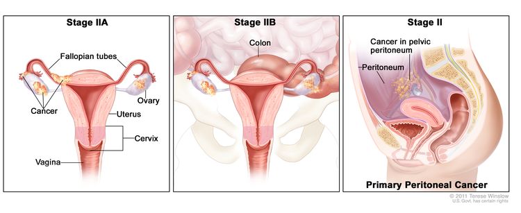
Ovarian, Fallopian Tube, and Primary Peritoneal Cancers Prevention (PDQ®)–Health Professional Version

Ovarian, Fallopian Tube, and Primary Peritoneal Cancers Prevention (PDQ®)–Health Professional Version

Who Is at Risk?

Ovarian cancer is a rare disease, with carcinomas comprising approximately 90% of tumors and germ cell and stromal tumors accounting for the remainder. Ovarian carcinoma is a disease that predominantly affects postmenopausal women. Ovarian carcinomas consist of several histopathological types, with high-grade serous being both the most common and most lethal. The category of ovarian borderline tumor or tumor of low-malignant potential, which historically had been considered in the context of ovarian cancer, is now generally considered a nonmalignant entity, although it has a postulated relationship with the development of some histological subtypes of low-grade ovarian carcinomas.[1]

Risk factors for ovarian cancer include a family history of breast and/or ovarian cancer and inheritance of deleterious mutations in BRCA1, BRCA2, and selected other high-penetrance genes.[26] For more information, see Genetics of Breast and Gynecologic Cancers. Other risk factors for ovarian cancer include obesity, tall height, endometriosis, and the use of postmenopausal hormone therapy.[79]

Associations of some risk factors with ovarian cancer vary by histopathological subtype. The association of endometriosis with ovarian cancer is stronger for nonserous subtypes, especially clear cell carcinoma and endometrioid subtypes.[10] Further, among carriers of deleterious mutations in BRCA1 or BRCA2, increasing evidence suggests that many tumors previously classified as ovarian high-grade serous carcinoma may develop from malignant cells arising in the tubal epithelium (serous tubal intraepithelial carcinoma [STIC]), although these tumors continue to be referred to as ovarian cancers in most writings. It is hypothesized that high-grade serous carcinomas among individuals who are not carriers of mutations in BRCA1 or BRCA2 may also develop in the fallopian tube, but few STICs have been identified among these women in the absence of concurrent high-stage disease. Further, data suggest that the distinction of high-grade serous carcinomas from other histological types of high-grade carcinomas, particularly endometrioid carcinomas, is not reliable. Reported rates of mucinous carcinoma diagnoses have declined dramatically, but expert pathology reviews suggest that this reflects increased recognition of metastases from occult gastrointestinal primary tumors to the ovary, rather than a true decline in rates of ovarian primary tumors.[11]

References
  1. Kurman RJ, Carcangiu ML, Young RH, eds.: WHO Classification of Tumours of Female Reproductive Organs. 4th ed. International Agency for Research on Cancer, 2014.
  2. Bolton KL, Ganda C, Berchuck A, et al.: Role of common genetic variants in ovarian cancer susceptibility and outcome: progress to date from the Ovarian Cancer Association Consortium (OCAC). J Intern Med 271 (4): 366-78, 2012. [PUBMED Abstract]
  3. Weissman SM, Weiss SM, Newlin AC: Genetic testing by cancer site: ovary. Cancer J 18 (4): 320-7, 2012 Jul-Aug. [PUBMED Abstract]
  4. Hunn J, Rodriguez GC: Ovarian cancer: etiology, risk factors, and epidemiology. Clin Obstet Gynecol 55 (1): 3-23, 2012. [PUBMED Abstract]
  5. Pal T, Akbari MR, Sun P, et al.: Frequency of mutations in mismatch repair genes in a population-based study of women with ovarian cancer. Br J Cancer 107 (10): 1783-90, 2012. [PUBMED Abstract]
  6. Gayther SA, Pharoah PD: The inherited genetics of ovarian and endometrial cancer. Curr Opin Genet Dev 20 (3): 231-8, 2010. [PUBMED Abstract]
  7. Lacey JV, Brinton LA, Leitzmann MF, et al.: Menopausal hormone therapy and ovarian cancer risk in the National Institutes of Health-AARP Diet and Health Study Cohort. J Natl Cancer Inst 98 (19): 1397-405, 2006. [PUBMED Abstract]
  8. Trabert B, Wentzensen N, Yang HP, et al.: Ovarian cancer and menopausal hormone therapy in the NIH-AARP diet and health study. Br J Cancer 107 (7): 1181-7, 2012. [PUBMED Abstract]
  9. Lahmann PH, Cust AE, Friedenreich CM, et al.: Anthropometric measures and epithelial ovarian cancer risk in the European Prospective Investigation into Cancer and Nutrition. Int J Cancer 126 (10): 2404-15, 2010. [PUBMED Abstract]
  10. Poole EM, Lin WT, Kvaskoff M, et al.: Endometriosis and risk of ovarian and endometrial cancers in a large prospective cohort of U.S. nurses. Cancer Causes Control 28 (5): 437-445, 2017. [PUBMED Abstract]
  11. Seidman JD, Kurman RJ, Ronnett BM: Primary and metastatic mucinous adenocarcinomas in the ovaries: incidence in routine practice with a new approach to improve intraoperative diagnosis. Am J Surg Pathol 27 (7): 985-93, 2003. [PUBMED Abstract]

Overview

Note: The Overview section summarizes the published evidence on this topic. The rest of the summary describes the evidence in more detail.

Other PDQ summaries on Ovarian, Fallopian Tube, and Primary Peritoneal Cancers Screening and Ovarian Epithelial, Fallopian Tube, and Primary Peritoneal Cancer Treatment are also available.

Factors With Adequate Evidence of an Increased Risk of Ovarian, Fallopian Tube, and Primary Peritoneal Cancers

Family history and inherited susceptibility to ovarian, fallopian tube, and primary peritoneal cancers

Based on solid evidence, women with a family history of ovarian cancer, especially in a first-degree relative, and those with an inherited predisposition to ovarian cancer, such as a BRCA1 or BRCA2 mutation, have an increased risk of developing ovarian cancer. For more information, see Genetics of Breast and Gynecologic Cancers.

Endometriosis

Based on fair evidence, self-reported and laparoscopically confirmed endometriosis is associated with an increased risk of ovarian cancer.[1,2] The association is stronger with nonserous histological subtypes, specifically endometrioid and clear cell carcinomas.[2,3]

Magnitude of Effect: Modest with observed relative risks (RRs) of 1.8 to 2.4.

  • Study Design: Cohort and case-control studies.
  • Internal Validity: Good.
  • Consistency: Fair.
  • External Validity: Good.

Hormone replacement therapy

Based on fair evidence, current or recent hormone therapy is associated with a small increased risk of ovarian cancer. Risks attenuate after hormone therapy is discontinued. Risks did not differ by oral preparation type (estrogen only vs. combined estrogen/progestin).[4,5] Cutaneous hormone therapy may have a lower risk than oral hormone therapy.[6]

Magnitude of Effect: Modest with observed RRs of 1.20 to 1.8.

  • Study Design: One randomized clinical trial, cohort and case-control studies.
  • Internal Validity: Good.
  • Consistency: Fair.
  • External Validity: Good.

Obesity and height

Based on fair evidence, increases in height and body mass index (BMI) are associated with a modest increased risk of ovarian cancer.

Magnitude of Effect: Based on an overview analysis of 25,157 women with ovarian cancer and 81,211 women without ovarian cancer from 47 epidemiological studies, the RR of ovarian cancer per 5 cm increase in height is 1.07 (95% confidence interval [CI], 1.05–1.09). The RR of ovarian cancer per 5 kg/m2 increase in BMI is 1.10 (95% CI, 1.07–1.13) among never-users of hormone therapy and 0.95 (95% CI, 0.92–0.99) among ever-users of hormone therapy.[7]

  • Study Design: Cohort and case-control studies.
  • Internal Validity: Good.
  • Consistency: Good.
  • External Validity: Good.

Factors With Adequate Evidence of a Decreased Risk of Ovarian, Fallopian Tube, and Primary Peritoneal Cancers

Oral contraceptives: Benefits

Based on solid evidence, oral contraceptive use is associated with a decreased risk of developing ovarian cancer.

Magnitude of Effect: The degree of risk reduction varies by duration of oral contraceptive use and time since last use. A prospective, contemporary, nationwide cohort study of women aged 15 to 49 years in Denmark found that any use of hormonal contraception was associated with an absolute reduction in the rate of ovarian cancer of 3.2 cases per 100,000 person-years. The reduction in risk persists for more than 30 years after use is discontinued, but the degree of reduction attenuates over time.[8]

  • Study Design: Multiple case-control and cohort studies; meta-analyses.
  • Internal Validity: Good.
  • Consistency: Good.
  • External Validity: Good.

Oral contraceptives: Harms

Based on solid evidence, combined current use of estrogen-progestin oral contraceptive use is associated with an increased risk of venous thromboembolism, particularly among smokers, for whom use is contraindicated. Oral contraceptives are not associated with a long-term increased risk of breast cancer but may be associated with a short-term increased risk while a woman is taking oral contraceptives. The risk of breast cancer declines with time since last use.

Magnitude of Effect: The risks may vary by preparation. Overall, the absolute risk of venous thromboembolism is about three events per 10,000 women per year while taking oral contraceptives. The risk is modified by smoking. Breast cancer risk among long-term (>10 years) current users is estimated at one extra case per year per 100,000 women. The risk dissipates with time since last use.

  • Study Design: Observational studies.
  • Internal Validity: Good.
  • Consistency: Good.
  • External Validity: Good.

Tubal ligation: Benefits

Based on solid evidence, tubal ligation is associated with a decreased risk of ovarian cancer.

Magnitude of Effect: Adjusting for other forms of contraception, tubal ligation provides a relative reduction in the odds of developing ovarian cancer of about 30%.

  • Study Design: Multiple case-control studies and cohort studies.
  • Internal Validity: Good.
  • Consistency: Good.
  • External Validity: Good.

Tubal ligation: Harms

Based on fair evidence, harms include surgical risks, including the following:[9]

  • Major morbidity including blood transfusion, reoperation, or hospital readmission (rate of 1.0 per 100 procedures).
  • Minor morbidity including postoperative fever, urinary tract infections, or wound infections (rate of 6.0 per 100 procedures).

Multiparity

Based on good evidence, multiparity is associated with a decreased risk of ovarian cancer.

Magnitude of Effect: Based on good evidence from multiple observational epidemiological studies, parous women have an approximately 30% lower ovarian cancer risk than nulliparous women.[7,1012]

  • Study Design: Observational epidemiological studies.
  • Internal Validity: Good.
  • Consistency: Good.
  • External Validity: Good.

Salpingectomy

Based on limited data, salpingectomy is associated with a decrease in risk of ovarian cancer.

Magnitude of Effect: Approximately 50% decrease for bilateral salpingectomy, less protection for unilateral salpingectomy.

  • Study Design: Observational epidemiological studies from several different countries.
  • Internal Validity: Good.
  • Consistency: Good.
  • External Validity: Good.

Breastfeeding

Based on solid evidence, breastfeeding is associated with a decreased risk of ovarian cancer.

Magnitude of Effect: 2% decrease with every month of breastfeeding.[13]

  • Study Design: Multiple case-control and cohort studies; meta-analysis.
  • Internal Validity: Good.
  • Consistency: Good.
  • External Validity: Good.

Risk-reducing bilateral salpingo-oophorectomy: Benefits

Based on solid evidence, risk-reducing bilateral salpingo-oophorectomy is associated with a decreased risk of ovarian cancer. Peritoneal carcinomatosis has been reported rarely following surgery. Risk-reducing surgery is generally reserved for women at high risk of developing ovarian cancer, such as women who have an inherited susceptibility to ovarian cancer.

Magnitude of Effect: 90% reduction in risk of ovarian cancer observed among women with a BRCA1 or BRCA2 mutation.

  • Study Design: Multiple case-control studies.
  • Internal Validity: Good.
  • Consistency: Good.
  • External Validity: Good.

Risk-reducing bilateral salpingo-oophorectomy: Harms

Based on solid evidence, prophylactic oophorectomy among women who are still menstruating at the time of surgery is associated with infertility, vasomotor symptoms, decreased sexual interest, vaginal dryness, urinary frequency, decreased bone-mineral density, and increased cardiovascular disease.

Magnitude of Effect: Reported prevalence of vasomotor symptoms varies from 41% to 61.4% among women who underwent oophorectomy before natural menopause. Women with bilateral oophorectomy who did not take hormone therapy were twice as likely to have moderate or severe hot flashes, compared with women who underwent natural menopause. The RR of cardiovascular disease among women with bilateral oophorectomy and early menopause was 4.55 (95% CI, 2.56–9.01).

  • Study Design: Cohort and case-control studies.
  • Internal Validity: Good.
  • Consistency: Good.
  • External Validity: Good.

Areas of Uncertainty

Ovarian hyperstimulation for infertility treatment

Evidence is poor to determine the association between ovarian hyperstimulation and the risk of ovarian cancer. Risk of ovarian cancer may be increased among women who remain nulligravid after being treated with ovarian stimulating medications.

Magnitude of Effect: Uncertain—risk of invasive ovarian cancer may be increased among women who remain nulligravid after treatment; risk of borderline ovarian tumors may be increased among women treated with infertility drugs.

  • Study Design: Cohort and case-control studies; systematic review.
  • Internal Validity: Fair.
  • Consistency: Poor.
  • External Validity: Fair.
References
  1. Poole EM, Lin WT, Kvaskoff M, et al.: Endometriosis and risk of ovarian and endometrial cancers in a large prospective cohort of U.S. nurses. Cancer Causes Control 28 (5): 437-445, 2017. [PUBMED Abstract]
  2. Pearce CL, Templeman C, Rossing MA, et al.: Association between endometriosis and risk of histological subtypes of ovarian cancer: a pooled analysis of case-control studies. Lancet Oncol 13 (4): 385-94, 2012. [PUBMED Abstract]
  3. Mogensen JB, Kjær SK, Mellemkjær L, et al.: Endometriosis and risks for ovarian, endometrial and breast cancers: A nationwide cohort study. Gynecol Oncol 143 (1): 87-92, 2016. [PUBMED Abstract]
  4. Mørch LS, Løkkegaard E, Andreasen AH, et al.: Hormone therapy and ovarian cancer. JAMA 302 (3): 298-305, 2009. [PUBMED Abstract]
  5. Beral V, Gaitskell K, Hermon C, et al.: Menopausal hormone use and ovarian cancer risk: individual participant meta-analysis of 52 epidemiological studies. Lancet 385 (9980): 1835-42, 2015. [PUBMED Abstract]
  6. Simin J, Tamimi RM, Callens S, et al.: Menopausal hormone therapy treatment options and ovarian cancer risk: A Swedish prospective population-based matched-cohort study. Int J Cancer 147 (1): 33-44, 2020. [PUBMED Abstract]
  7. Braem MG, Onland-Moret NC, van den Brandt PA, et al.: Reproductive and hormonal factors in association with ovarian cancer in the Netherlands cohort study. Am J Epidemiol 172 (10): 1181-9, 2010. [PUBMED Abstract]
  8. Iversen L, Fielding S, Lidegaard Ø, et al.: Association between contemporary hormonal contraception and ovarian cancer in women of reproductive age in Denmark: prospective, nationwide cohort study. BMJ 362: k3609, 2018. [PUBMED Abstract]
  9. Lawrie TA, Kulier R, Nardin JM: Techniques for the interruption of tubal patency for female sterilisation. Cochrane Database Syst Rev (9): CD003034, 2015. [PUBMED Abstract]
  10. Fortner RT, Ose J, Merritt MA, et al.: Reproductive and hormone-related risk factors for epithelial ovarian cancer by histologic pathways, invasiveness and histologic subtypes: Results from the EPIC cohort. Int J Cancer 137 (5): 1196-208, 2015. [PUBMED Abstract]
  11. Yang HP, Trabert B, Murphy MA, et al.: Ovarian cancer risk factors by histologic subtypes in the NIH-AARP Diet and Health Study. Int J Cancer 131 (4): 938-48, 2012. [PUBMED Abstract]
  12. Lee AW, Rosenzweig S, Wiensch A, et al.: Expanding Our Understanding of Ovarian Cancer Risk: The Role of Incomplete Pregnancies. J Natl Cancer Inst 113 (3): 301-308, 2021. [PUBMED Abstract]
  13. Feng LP, Chen HL, Shen MY: Breastfeeding and the risk of ovarian cancer: a meta-analysis. J Midwifery Womens Health 59 (4): 428-37, 2014 Jul-Aug. [PUBMED Abstract]

Incidence and Mortality

In 2025 in the United States, ovarian cancer will cause an estimated 20,890 new cases and 12,730 deaths.[1] Based on statistical models for analysis, rates for new ovarian cancer cases fell, on average, by 2.7% each year from 2012 to 2021. Death rates fell, on average, by 2.4% each year from 2013 to 2022.[2] In 2021, the overall incidence rate for ovarian cancer among women aged 65 years and older was 33.5 cases per 100,000 women-years.[3] Given that the Surveillance, Epidemiology, and End Results (SEER) Program does not adjust for oophorectomy or salpingectomy, racial differences in the prevalence of women who have undergone these procedures could bias racial rate comparisons. A statistically significant decrease in delayed-adjusted incidence rates of 1.7% per year among White women from 2000 to 2015 and 0.7% per year among Black women from 2000 to 2021 was observed. A statistically significant decrease in mortality rates of 2.5% per year among White women from 2005 to 2022 and 1.9% per year among Black women from 2003 to 2022 was observed. The population lifetime risk of ovarian cancer is 1.12%; the population lifetime risk of dying from ovarian cancer is 0.71%.[3]

References
  1. American Cancer Society: Cancer Facts and Figures 2025. American Cancer Society, 2025. Available online. Last accessed January 16, 2025.
  2. National Cancer Institute: SEER Stat Fact Sheets: Ovarian Cancer. Bethesda, Md: National Institutes of Health. Available online. Last accessed February 10, 2025.
  3. Surveillance Research Program, National Cancer Institute: SEER*Explorer: An interactive website for SEER cancer statistics. Bethesda, MD: National Cancer Institute. Available online. Last accessed December 30, 2024.

Histology and Pathogenesis of Ovarian, Fallopian Tube, and Primary Peritoneal Cancers

Ovarian carcinoma is a biologically and clinically heterogeneous class of tumors that includes several major subtypes: serous, mucinous, endometrioid, and clear cell. Classification of ovarian carcinomas into type I and type II tumors has been proposed. In this system, type I tumors include the following:[1]

  1. Endometriosis-related subtypes, such as endometrioid, clear cell, and seromucinous.
  2. Low-grade serous.
  3. Mucinous and malignant Brenner tumors.

Among type I tumors, endometrioid and clear cell carcinomas are most common and most important clinically. In general, type I ovarian carcinomas present at a lower stage than type II tumors and portend a better prognosis.

Type II tumors are comprised mainly of high-grade serous carcinomas, the most common and lethal of all ovarian carcinoma subtypes. These cancers usually present with symptomatic bulky stage III or IV disease and ascites. Many, but possibly not all, high-grade serous carcinomas appear to arise from malignant in situ lesions in the epithelium of the fallopian tube fimbria. These lesions spread to the ovaries secondarily but continue to be referred to as ovarian carcinomas. Evidence for a tubal origin is based mainly on examination of risk-reducing salpingo-oophorectomy specimens, performed among BRCA1/BRCA2 mutation carriers, in which incidental low-volume disease enables recognition of serous tubal intraepithelial carcinoma (STIC). However, not all women with high-grade serous carcinomas have identifiable STIC, and few studies of the fallopian tubes of women who are not carriers of BRCA1/BRCA2 mutations have been performed, suggesting that pathogenesis of these tumors is not fully known. Serous carcinomas can be further divided on the basis of molecular characteristics.[2]

The heterogeneity in the etiology and pathogenesis of different ovarian cancer subtypes and variability in the classification of tumors over time and between studies pose challenges for interpretation of etiological data. Ovarian cancer is rare, thus sample size and power of studies to detect moderate associations by cancer subtype is limited. However, clearer subtyping of cancers may help improve our understanding of the etiology of ovarian malignancies in future studies.

References
  1. Kurman RJ, Shih IeM: The Dualistic Model of Ovarian Carcinogenesis: Revisited, Revised, and Expanded. Am J Pathol 186 (4): 733-47, 2016. [PUBMED Abstract]
  2. Cancer Genome Atlas Research Network: Integrated genomic analyses of ovarian carcinoma. Nature 474 (7353): 609-15, 2011. [PUBMED Abstract]

Factors With Adequate Evidence of an Increased Risk of Ovarian, Fallopian Tube, and Primary Peritoneal Cancers

Family History and Inherited Susceptibility to Ovarian, Fallopian Tube, and Primary Peritoneal Cancers

Some women are at an increased risk of these cancers because of an inherited mutation, with the magnitude of that risk dependent on the affected gene and specific mutation. Underlying ovarian cancer risk can be assessed through accurate pedigrees and/or genetic markers of risk. Because of uncertainties about cancer risks associated with certain specific gene mutations, genetic information may be difficult to interpret outside of families with a high incidence of ovarian cancer.

This summary does not address multiple genetic syndromes or women who are at high risk because of inherited genetic factors. For specific information related to ovarian cancer risk associated with multiple genetic syndromes and ovarian cancer in BRCA1/BRCA2 mutation carriers, see Genetics of Breast and Gynecologic Cancers and Genetics of Colorectal Cancer.

Endometriosis

Endometriosis has been associated with a modestly increased risk of ovarian cancer. The association is stronger with nonserous histological subtypes, specifically endometrioid and clear cell carcinomas. In one analysis, data were pooled from 13 ovarian cancer case-control studies, including 13,226 controls and 7,911 women with invasive ovarian cancer who were part of the Ovarian Cancer Association Consortium. Logistic regression analyses were undertaken to assess the association between self-reported endometriosis and risk of ovarian cancer. Self-reported endometriosis was associated with a significantly increased risk of clear cell (odds ratio [OR], 3.05; 95% confidence interval [CI], 2.43–3.84; P < .0001), low-grade serous (OR, 2.11; 95% CI, 1.39–3.20; P < .0001), and endometrioid invasive ovarian cancers (OR, 2.04; 95% CI, 1.67–2.48; P < .0001). No association was noted between endometriosis and risk of mucinous (OR, 1.02; 95% CI, 0.69–1.50; P = .93) or high-grade serous invasive ovarian cancer (OR, 1.13; 95% CI, 0.97–1.32; P = .13), or borderline tumors of either subtype (OR, 1.20; 95% CI, 0.95–1.52; P = .12 for serous and OR, 1.12; 95% CI, 0.84–1.48; P = .45 for mucinous).[1] Considering that the clear cell and endometrioid ovarian cancers represented approximately 15% of all ovarian cancers, the lifetime risk of these histological subtypes is approximately 0.2%, which, on the basis of these data, would increase to approximately 0.4% to 0.6% in the presence of self-reported endometriosis.

A cohort study from the Danish National Patient Register identified 45,790 women with a clinical diagnosis of endometriosis between 1977 and 2012. Data were linked to the Danish Cancer Register, which identified 186 women with a diagnosis of ovarian cancer. Endometriosis was associated with modestly increased risks of ovarian cancer overall (standardized incidence ratio [SIR], 1.34; 95% CI, 1.16–1.55). This was primarily caused by increases in endometrioid (SIR, 1.64; 95% CI, 1.09–2.37) and clear cell subtypes (SIR, 3.64; 95% CI, 2.36–5.38). No increased risk of serous or mucinous histological subtypes was reported.[2]

Using data from the Nurses’ Health Study II, 228 ovarian cancers were identified from among 102,025 eligible women. Cox proportional hazards regression models were used to assess associations between endometriosis and cancer risk, evaluating the impacts of self-reported versus laparoscopically confirmed endometriosis, delayed diagnosis, and postendometriosis diagnosis changes in risk-factor exposures. Self-reported endometriosis was associated with ovarian cancer (relative risk [RR], 1.81; 95% CI, 1.26–2.58), which was stronger for laparoscopically confirmed endometriosis diagnoses (RR, 2.14; 95% CI, 1.45–3.15). Diagnosis delays or postendometriosis diagnosis changes in risk factors had little impact on risk. Although this study had limited power to detect differences in risk on the basis of histological subtype, nonserous cases were shown to have an increased risk (RR, 2.44; 95% CI, 1.48–4.01), and serous cases were not (RR, 1.69; 95% CI, 0.92–3.11).[3] A large case-control study in African American women found similar associations with a history of endometriosis and ovarian cancer (OR, 1.78; 95% CI, 1.09–2.90), suggesting that findings from populations of predominantly White women are also observed in Black women.[4]

Hormone Replacement Therapy/Hormone Therapy

A meta-analysis of 52 studies (17 prospective and 35 retrospective) including 21,488 ovarian cancers found increased risks with current or recent hormone replacement use in prospective studies (RR, 1.37; 95% CI, 1.29–1.46), with similar results for retrospective designs. Significant relationships were found for serous and endometrioid subtypes.[5] Recent use was strongly related to risk even among women who had used hormone replacement therapy for less than 5 years (RR, 1.41; 95% CI, 1.32–1.50). Risk declined among women who had discontinued use, with greater effects for longer periods of cessation. Risks did not differ by preparation types (estrogen only vs. combined estrogen/progestin). Risks also did not differ by age at use.[6,7] Cutaneous hormone therapy may have a lower risk than oral hormone therapy.[8]

Tibolone, a synthetic steroid with estrogenic, progestogenic, and androgenic properties, has been associated with an increased incidence rate ratio of 3.56 (95% CI, 3.08–4.69) for endometrial cancer for current users compared with never-users. Tibolone is approved for use to manage menopausal symptoms or to prevent osteoporosis in many countries. However, it is not approved for use in Canada or the United States. Other combined therapy with estrogen and progestin may also increase the risk of breast cancer, so the risks and benefits must be considered.[9]

Obesity and Height

Ovarian cancer risk increases with increasing height and weight (body mass index [BMI]).[10] The Collaborative Group on Epidemiological Studies of Ovarian Cancer compiled individual data, both published and unpublished, from 47 epidemiological studies including 12,157 women with ovarian cancer and 81,311 controls. RR increased significantly with increasing height (1.07 per 5 cm of height) and with increasing BMI (1.10 per 5 kg/m2). These findings were unaffected by other factors known to be associated with ovarian cancer risk, with the exception that ever-users of hormone therapy had no increased risk with increasing BMI. Given that height, weight, and BMI are thought to be strongly correlated, separating out the individual effects can be difficult.[11,12] Ovarian cancer mortality has also been shown to be increased in women with obesity.[13,14]

References
  1. Pearce CL, Templeman C, Rossing MA, et al.: Association between endometriosis and risk of histological subtypes of ovarian cancer: a pooled analysis of case-control studies. Lancet Oncol 13 (4): 385-94, 2012. [PUBMED Abstract]
  2. Mogensen JB, Kjær SK, Mellemkjær L, et al.: Endometriosis and risks for ovarian, endometrial and breast cancers: A nationwide cohort study. Gynecol Oncol 143 (1): 87-92, 2016. [PUBMED Abstract]
  3. Poole EM, Lin WT, Kvaskoff M, et al.: Endometriosis and risk of ovarian and endometrial cancers in a large prospective cohort of U.S. nurses. Cancer Causes Control 28 (5): 437-445, 2017. [PUBMED Abstract]
  4. Park HK, Schildkraut JM, Alberg AJ, et al.: Benign gynecologic conditions are associated with ovarian cancer risk in African-American women: a case-control study. Cancer Causes Control 29 (11): 1081-1091, 2018. [PUBMED Abstract]
  5. Beral V, Gaitskell K, Hermon C, et al.: Menopausal hormone use and ovarian cancer risk: individual participant meta-analysis of 52 epidemiological studies. Lancet 385 (9980): 1835-42, 2015. [PUBMED Abstract]
  6. Lacey JV, Brinton LA, Leitzmann MF, et al.: Menopausal hormone therapy and ovarian cancer risk in the National Institutes of Health-AARP Diet and Health Study Cohort. J Natl Cancer Inst 98 (19): 1397-405, 2006. [PUBMED Abstract]
  7. Trabert B, Wentzensen N, Yang HP, et al.: Ovarian cancer and menopausal hormone therapy in the NIH-AARP diet and health study. Br J Cancer 107 (7): 1181-7, 2012. [PUBMED Abstract]
  8. Simin J, Tamimi RM, Callens S, et al.: Menopausal hormone therapy treatment options and ovarian cancer risk: A Swedish prospective population-based matched-cohort study. Int J Cancer 147 (1): 33-44, 2020. [PUBMED Abstract]
  9. Løkkegaard ECL, Mørch LS: Tibolone and risk of gynecological hormone sensitive cancer. Int J Cancer 142 (12): 2435-2440, 2018. [PUBMED Abstract]
  10. Collaborative Group on Epidemiological Studies of Ovarian Cancer: Ovarian cancer and body size: individual participant meta-analysis including 25,157 women with ovarian cancer from 47 epidemiological studies. PLoS Med 9 (4): e1001200, 2012. [PUBMED Abstract]
  11. Dixon-Suen SC, Nagle CM, Thrift AP, et al.: Adult height is associated with increased risk of ovarian cancer: a Mendelian randomisation study. Br J Cancer 118 (8): 1123-1129, 2018. [PUBMED Abstract]
  12. Qian F, Rookus MA, Leslie G, et al.: Mendelian randomisation study of height and body mass index as modifiers of ovarian cancer risk in 22,588 BRCA1 and BRCA2 mutation carriers. Br J Cancer 121 (2): 180-192, 2019. [PUBMED Abstract]
  13. Calle EE, Rodriguez C, Walker-Thurmond K, et al.: Overweight, obesity, and mortality from cancer in a prospectively studied cohort of U.S. adults. N Engl J Med 348 (17): 1625-38, 2003. [PUBMED Abstract]
  14. Aune D, Navarro Rosenblatt DA, Chan DS, et al.: Anthropometric factors and ovarian cancer risk: a systematic review and nonlinear dose-response meta-analysis of prospective studies. Int J Cancer 136 (8): 1888-98, 2015. [PUBMED Abstract]

Factors With Adequate Evidence of a Decreased Risk of Ovarian, Fallopian Tube, and Primary Peritoneal Cancers

Factors associated with a decreased risk of ovarian cancer include multiparity, use of oral contraceptives, breastfeeding, and risk-reducing surgical procedures like tubal ligation, salpingectomy, and salpingo-oophorectomy.[15] Each of these contributes to a decrease in ovulatory years. Pooled data from 25 case-control studies conducted by the Ovarian Cancer Association Consortium was used to quantify the association between lifetime ovulatory years and epithelial ovarian cancer risk. Lifetime ovulatory years was calculated by subtracting the years of anovulation from an individual’s menstrual span (i.e., age at last menstrual period subtracted from age at menarche).[6] The odds ratio (OR) for ovarian cancer per lifetime ovulatory year with anovulation caused by pregnancy, oral contraceptive use, and breastfeeding was 1.041 (95% confidence interval [CI], 1.036–1.045).

Multiparity

Compared with nulliparous women, the risk of ovarian cancer was reduced by 30% to 60% among parous women, with additive protection for each additional birth.[1] The risk of developing ovarian cancer was lower for parous women (relative risk [RR], 0.69; 95% CI, 0.64–0.74) than for women who never had children, with increased risk reduction with increasing number of children for one child (RR, 0.82; 95% CI, 0.43–0.91) to four or more children (RR, 0.58; 95% CI, 0.53–0.64).[7]

Oral Contraceptives

A collaborative analysis was performed of individual data from 23,257 women with ovarian cancer and 87,303 women without ovarian cancer from 45 studies in 21 countries.[8] Oral contraceptive use was associated with a dose-response effect by duration of use, without observed changes in risk reduction by decade of use from the 1960s to 1980s, over which time the amount of estrogen in oral contraceptives was approximately halved. No risk reduction was observed for women who used oral contraceptives for less than 1 year. The risk reduction associated with use from 1 to 4 years, 5 to 9 years, 10 to 14 years, and 15 years or more was 0.78 (99% CI, 0.73–0.893), 0.64 (99% CI, 0.59–0.69), 0.56 (99% CI, 0.50–0.62), and 0.42 (99% CI, 0.36–0.49), respectively. The observed risk reduction persisted after cessation of oral contraceptive therapy but attenuated over time since last use. The proportional reduction in risk per 5 years of use was 29% (95% CI, 23%–34%) for women who had discontinued use within the last 10 years. The reduction in risk was 15% (95% CI, 9%–21%) for women who discontinued use 20 to 29 years ago.[9]

A meta-analysis, in which the primary analysis was restricted to 24 case-control and cohort studies published since 2000 to reflect more recent types of oral contraceptive preparations, also observed a dose-response by duration of use.[2] The authors estimated that 185 women needed to be treated for 5 years to prevent one case of ovarian cancer. Based on an estimated lifetime risk of 1.38% and prevalence of ever-use of oral contraceptives of 83%, the authors estimated a lifetime relative reduction of ovarian cancer attributable to oral contraceptives of 0.54%. A prospective cohort study from Denmark, which represented nearly 1.9 million women, also examined contemporary oral contraceptive formulations and found that current oral contraceptive formulation users had an RR reduction of 0.58 (95% CI, 0.49–0.68), and former oral contraceptive formulation users had a RR reduction of 0.77 (95% CI, 0.66–0.91). The benefits were strengthened by longer duration of use and weakened by more time since last use. No benefit was found for progesterone-only birth control products. This study was limited by only including incident ovarian cancers that occurred in women younger than 50 years.[10]

For specific information related to ovarian cancer risk among BRCA1/BRCA2 mutation carriers, see Genetics of Breast and Gynecologic Cancers.

Depot-Medroxyprogesterone Acetate

Limited information is available on the use of injectable progestational contraceptives (depot-medroxyprogesterone acetate [DMPA]) and the risk of ovarian cancer. Studies are confounded by the use of other contraceptive methods, particularly oral contraceptives. A hospital-based study conducted in Mexico and Thailand, with 224 cases and 1,781 controls (the World Health Organization Collaborative Study of Neoplasia and Steroid Contraceptives), did not observe an association between DMPA and ovarian cancer (RR, 1.07; 95% CI, 0.6–1.8).[11] However, only 22 of the participants had ever used DMPA, and nine of them had used it for 6 months or less.

A subsequent multicenter study conducted in 12 hospitals in Thailand, including 330 cases and 982 matched controls, observed a statistically significant decreased risk of ovarian cancer associated with DMPA use, controlling for oral contraceptive use and other associated factors (OR, 0.52; 95% CI, 0.33–0.88). A dose-response association was observed, but the sample size was limited in longer-term use categories.[12]

Tubal Ligation

A meta-analysis of 16 case-control studies, three retrospective studies, and two prospective cohort studies observed a decreased risk of ovarian cancer associated with tubal ligation (RR, 0.66; 95% CI, 0.60–0.73).[4] The reduced risk was observed up to 14 years after tubal ligation. A population-based case-control study of 902 cases and 1,802 controls published subsequent to the meta-analysis observed an adjusted OR of 0.62 (95% CI, 0.51–0.75) associated with a history of a tubal ligation.[13] The association was adjusted for oral contraceptive use, which was also associated with a lower risk of ovarian cancer (OR, 0.62; 95% CI, 0.47–0.85) and other risk factors.[13] Salpingectomy has also been discussed as a preferred means of sterilization.[14,15] For more information, see the Salpingectomy section.

Another pooling project with primary data from 13 population-based case-control studies examined the association between tubal ligation and ovarian cancer risk. It included 7,942 individuals with epithelial ovarian cancers and 13,904 controls.[16] Overall, tubal ligation was associated with a 29% reduction in risk (OR, 0.71; 95% CI, 0.66–0.77). The observed risk reduction varied by subtype of invasive cancers and was 52% (OR, 0.48; 95% CI, 0.40–49) for endometrioid cancer; 48% (OR, 0.52; 95% CI, 0.40–0.67) for clear cell cancer; 32% (OR, 0.68; 95% CI, 0.52–89) for mucinous cancer; and 19% (OR, 0.81; 95% CI, 0.74–0,89) for serous cancer.

A pooled analysis from 21 prospective cohort studies examined 14 hormonal, reproductive, and lifestyle factors by histological subtype among 5,584 women with invasive ovarian cancer within a total sample of 1.3 million participants. Overall, tubal ligation was associated with an 18% reduction in risk (OR, 0.82; 95% CI, 0.73–0.93). The observed risk reduction varied by subtype of invasive cancer and was 40% (OR, 0.60; 95% CI, 0.41–88) for endometrioid cancer; 65% (OR, 0.35; 95% CI, 0.18–0.69) for clear cell cancer; and 9% (OR, 0.91; 95% CI, 0.79–1.06) for serous cancer. There was a nonsignificant increase in risk of 1% (OR, 1.01; 95% CI, 0.60–1.71) for mucinous cancer.[7]

Breastfeeding

A meta-analysis [3] that included five prospective studies and 30 case-control studies examined the association between breastfeeding and the risk of ovarian cancer. Any breastfeeding was associated with a decreased risk of ovarian cancer (RR, 0.76; 95% CI, 0.69–0.83). The risk of ovarian cancer decreased 8% for every 5-month increase in duration of breastfeeding (95% CI, 0.90–0.95). Another meta-analysis that included five prospective studies and 35 case-control studies found that any breastfeeding was associated with a decreased risk of ovarian cancer (RR, 0.70; 95% CI, 0.64–0.76). These results are consistent with a previous meta-analysis and further support the prior finding of a suggested association between increased duration of breastfeeding and greater levels of protection.[17] Another meta-analysis of 19 studies, including four cohort and 15 case-control studies, found an overall decreased risk of ovarian cancer with an OR of 0.66 (95% CI, 0.57–0.76) and an association with duration (2% decrease per month). The benefit of breastfeeding was greatest for the first 8 to 10 months.[18]

Risk-Reducing Salpingo-Oophorectomy

Risk-reducing surgery is an option considered by women who are at high risk of ovarian cancer, such as those with an inherited susceptibility to cancer. For more information on this as a risk-reducing intervention, see the Surgical history section in Genetics of Breast and Gynecologic Cancers. Among women in the general population, opportunistic salpingectomy, oophorectomy, or salpingo-oophorectomy have been considered as possible interventions at the time of surgery for other benign indications.

Harms

Risks associated with benign oophorectomy (with or without salpingectomy or hysterectomy) have been analyzed in six published studies. Studies of three cohorts found that oophorectomy performed before menopause (age 45 or 50 years) was associated with increased overall mortality, likely related to cardiovascular disease. This finding was noted particularly among individuals not using hormone replacement. In the Women’s Health Initiative, bilateral salpingo-oophorectomy was not associated with increased mortality. In the National Health and Nutrition Examination Survey (NHANES III), oophorectomy overall was not related to mortality, but mortality was increased among women younger than 40 years with obesity who did not use hormone replacement. The California Teachers Study did not find a mortality risk with oophorectomy, but only 3% of women did not use hormone replacement. Overall, data suggest that oophorectomy among younger women likely increases overall mortality and that this risk may be attenuated with hormone replacement.[1924] Risk-reducing salpingo-oophorectomy has been associated with worsened menopausal symptoms, decreased sexual activity, and decreased sexual functioning.[25]

Salpingectomy

Data relating salpingectomy to risk of ovarian/tubal cancer are limited but consistent. A meta-analysis of three studies found an OR of 0.51 (95% CI, 0.35–0.71) for risk of these cancers among women who had undergone salpingectomy, compared with women who had intact fallopian tubes.[5] These studies included a Swedish record linkage study conducted from 1973 to 2009 with a mean follow-up of 23 years, which found the following hazard ratios (HRs) for risk of ovarian cancer, compared with women who had not undergone surgery:[26]

  • For hysterectomy, the HR was 0.79 (95% CI, 0.70–0.88).
  • For hysterectomy with bilateral salpingo-oophorectomy, the HR was 0.06 (95% CI, 0.03–0.12).
  • For salpingectomy, the HR was 0.65 (95% CI, 0.52–0.81).
  • For sterilization procedures, the HR was 0.72 (95% CI, 0.64–0.81).

Another population-based cohort study of all individuals in British Columbia, Canada, between 2008 and 2017, examined observed versus expected rates of ovarian cancer among individuals who had undergone opportunistic salpingectomy. The study included 25,889 individuals who underwent opportunistic salpingectomy, compared with 32,080 individuals who underwent hysterectomy or tubal ligation alone. There were no serous ovarian cancers in the opportunistic-salpingectomy group, while the age-adjusted expected rate was 5.27 (95% CI, 1.78–19.29) serous cancers based on the age-adjusted incidence rate in the control group.[27] The risk reduction for ovarian cancer of unspecified histology associated with bilateral salpingectomy was approximately twice that of unilateral salpingectomy (HR, 0.35; 95% CI, 0.17–0.73 vs. HR, 0.71; 95% CI, 0.56–0.91, respectively).[26] This report included limited covariate data, but results were similar to other smaller studies included in the meta-analysis.[5]

A nested case-control study from Denmark compared 16,822 epithelial ovarian cancer cases, each matched to 40 controls.[28] This analysis observed an overall risk reduction for epithelial ovarian cancer incidence after both unilateral salpingectomy (OR, 0.73; 95% CI, 0.60–0.87) and bilateral salpingectomy (OR, 0.46; 95% CI, 0.31–0.67). Outcomes by histological type were also evaluated, and a similar risk reduction was observed for serous histology of any grade after bilateral salpingectomy (OR, 0.44; 95% CI, 0.27–0.73). The risk reduction for ovarian cancer incidence increased with time since salpingectomy (OR, 0.72; 95% CI, 0.55–0.95) for individuals 10 to 19 years postsalpingectomy and (OR, 0.55; 95% CI, 0.42–0.73) for individuals 20 years or more postsalpingectomy. Age-stratified analysis suggested that salpingectomy protected against ovarian cancer when salpingectomy was performed in individuals younger than 50 years (OR, 0.61; 95% CI, 0.51–0.73), but not when performed in individuals aged 50 years or older (OR, 1.21; 95% CI, 0.78–1.88). However, the small number of ovarian cancer cases among individuals who had a salpingectomy at age 50 years or older (n = 21) may have limited this finding. Other study limitations included small numbers of ovarian cancer cases of other histological subtypes. The study also did not provide information about tumor grade and hereditary disposition (i.e., BRCA mutation).

Data based on circulating surrogate markers of ovarian reserve suggest that salpingectomy does not have an adverse effect on ovarian function.[29,30]

References
  1. Permuth-Wey J, Sellers TA: Epidemiology of ovarian cancer. Methods Mol Biol 472: 413-37, 2009. [PUBMED Abstract]
  2. Havrilesky LJ, Moorman PG, Lowery WJ, et al.: Oral contraceptive pills as primary prevention for ovarian cancer: a systematic review and meta-analysis. Obstet Gynecol 122 (1): 139-47, 2013. [PUBMED Abstract]
  3. Luan NN, Wu QJ, Gong TT, et al.: Breastfeeding and ovarian cancer risk: a meta-analysis of epidemiologic studies. Am J Clin Nutr 98 (4): 1020-31, 2013. [PUBMED Abstract]
  4. Cibula D, Widschwendter M, Májek O, et al.: Tubal ligation and the risk of ovarian cancer: review and meta-analysis. Hum Reprod Update 17 (1): 55-67, 2011 Jan-Feb. [PUBMED Abstract]
  5. Yoon SH, Kim SN, Shim SH, et al.: Bilateral salpingectomy can reduce the risk of ovarian cancer in the general population: A meta-analysis. Eur J Cancer 55: 38-46, 2016. [PUBMED Abstract]
  6. Fu Z, Brooks MM, Irvin S, et al.: Lifetime ovulatory years and risk of epithelial ovarian cancer: a multinational pooled analysis. J Natl Cancer Inst 115 (5): 539-551, 2023. [PUBMED Abstract]
  7. Wentzensen N, Poole EM, Trabert B, et al.: Ovarian Cancer Risk Factors by Histologic Subtype: An Analysis From the Ovarian Cancer Cohort Consortium. J Clin Oncol 34 (24): 2888-98, 2016. [PUBMED Abstract]
  8. Collaborative Group on Epidemiological Studies of Ovarian Cancer, Beral V, Doll R, et al.: Ovarian cancer and oral contraceptives: collaborative reanalysis of data from 45 epidemiological studies including 23,257 women with ovarian cancer and 87,303 controls. Lancet 371 (9609): 303-14, 2008. [PUBMED Abstract]
  9. Schrijver LH, Antoniou AC, Olsson H, et al.: Oral contraceptive use and ovarian cancer risk for BRCA1/2 mutation carriers: an international cohort study. Am J Obstet Gynecol 225 (1): 51.e1-51.e17, 2021. [PUBMED Abstract]
  10. Iversen L, Fielding S, Lidegaard Ø, et al.: Association between contemporary hormonal contraception and ovarian cancer in women of reproductive age in Denmark: prospective, nationwide cohort study. BMJ 362: k3609, 2018. [PUBMED Abstract]
  11. Depot-medroxyprogesterone acetate (DMPA) and risk of epithelial ovarian cancer. The WHO Collaborative Study of Neoplasia and Steroid Contraceptives. Int J Cancer 49 (2): 191-5, 1991. [PUBMED Abstract]
  12. Wilailak S, Vipupinyo C, Suraseranivong V, et al.: Depot medroxyprogesterone acetate and epithelial ovarian cancer: a multicentre case-control study. BJOG 119 (6): 672-7, 2012. [PUBMED Abstract]
  13. Ness RB, Dodge RC, Edwards RP, et al.: Contraception methods, beyond oral contraceptives and tubal ligation, and risk of ovarian cancer. Ann Epidemiol 21 (3): 188-96, 2011. [PUBMED Abstract]
  14. Hanley GE, McAlpine JN, Kwon JS, et al.: Opportunistic salpingectomy for ovarian cancer prevention. Gynecol Oncol Res Pract 2: 5, 2015. [PUBMED Abstract]
  15. Daly MB, Dresher CW, Yates MS, et al.: Salpingectomy as a means to reduce ovarian cancer risk. Cancer Prev Res (Phila) 8 (5): 342-8, 2015. [PUBMED Abstract]
  16. Sieh W, Salvador S, McGuire V, et al.: Tubal ligation and risk of ovarian cancer subtypes: a pooled analysis of case-control studies. Int J Epidemiol 42 (2): 579-89, 2013. [PUBMED Abstract]
  17. Li DP, Du C, Zhang ZM, et al.: Breastfeeding and ovarian cancer risk: a systematic review and meta-analysis of 40 epidemiological studies. Asian Pac J Cancer Prev 15 (12): 4829-37, 2014. [PUBMED Abstract]
  18. Feng LP, Chen HL, Shen MY: Breastfeeding and the risk of ovarian cancer: a meta-analysis. J Midwifery Womens Health 59 (4): 428-37, 2014 Jul-Aug. [PUBMED Abstract]
  19. Duan L, Xu X, Koebnick C, et al.: Bilateral oophorectomy is not associated with increased mortality: the California Teachers Study. Fertil Steril 97 (1): 111-7, 2012. [PUBMED Abstract]
  20. Rocca WA, Grossardt BR, de Andrade M, et al.: Survival patterns after oophorectomy in premenopausal women: a population-based cohort study. Lancet Oncol 7 (10): 821-8, 2006. [PUBMED Abstract]
  21. McCarthy AM, Menke A, Ouyang P, et al.: Bilateral oophorectomy, body mass index, and mortality in U.S. women aged 40 years and older. Cancer Prev Res (Phila) 5 (6): 847-54, 2012. [PUBMED Abstract]
  22. Rivera CM, Grossardt BR, Rhodes DJ, et al.: Increased cardiovascular mortality after early bilateral oophorectomy. Menopause 16 (1): 15-23, 2009 Jan-Feb. [PUBMED Abstract]
  23. Parker WH, Feskanich D, Broder MS, et al.: Long-term mortality associated with oophorectomy compared with ovarian conservation in the nurses’ health study. Obstet Gynecol 121 (4): 709-16, 2013. [PUBMED Abstract]
  24. Jacoby VL, Grady D, Wactawski-Wende J, et al.: Oophorectomy vs ovarian conservation with hysterectomy: cardiovascular disease, hip fracture, and cancer in the Women’s Health Initiative Observational Study. Arch Intern Med 171 (8): 760-8, 2011. [PUBMED Abstract]
  25. Mai PL, Huang HQ, Wenzel LB, et al.: Prospective follow-up of quality of life for participants undergoing risk-reducing salpingo-oophorectomy or ovarian cancer screening in GOG-0199: An NRG Oncology/GOG study. Gynecol Oncol 156 (1): 131-139, 2020. [PUBMED Abstract]
  26. Falconer H, Yin L, Grönberg H, et al.: Ovarian cancer risk after salpingectomy: a nationwide population-based study. J Natl Cancer Inst 107 (2): , 2015. [PUBMED Abstract]
  27. Hanley GE, Pearce CL, Talhouk A, et al.: Outcomes From Opportunistic Salpingectomy for Ovarian Cancer Prevention. JAMA Netw Open 5 (2): e2147343, 2022. [PUBMED Abstract]
  28. Duus AH, Zheng G, Baandrup L, et al.: Risk of ovarian cancer after salpingectomy and tubal ligation: Prospects on histology and time since the procedure. Gynecol Oncol 177: 125-131, 2023. [PUBMED Abstract]
  29. Findley AD, Siedhoff MT, Hobbs KA, et al.: Short-term effects of salpingectomy during laparoscopic hysterectomy on ovarian reserve: a pilot randomized controlled trial. Fertil Steril 100 (6): 1704-8, 2013. [PUBMED Abstract]
  30. Venturella R, Lico D, Borelli M, et al.: 3 to 5 Years Later: Long-term Effects of Prophylactic Bilateral Salpingectomy on Ovarian Function. J Minim Invasive Gynecol 24 (1): 145-150, 2017. [PUBMED Abstract]

Factors With Inadequate Evidence of an Association Risk of Ovarian, Fallopian Tube, and Primary Peritoneal Cancers

Dietary Factors

No consistent association has been observed between a variety of dietary factors and the risk of ovarian cancer.

A systematic review and meta-analysis that included 23 case-control studies and three cohort studies found no evidence of an association between alcohol use and epithelial ovarian cancer.[1]

A case-control study of the Healthy Eating Index (HEI), based on current U.S. Department of Agriculture dietary guidelines, found no association between the highest HEI score and ovarian cancer risk for any specific food group.[2] A systematic review of the role of diet in ovarian cancer included only prospective studies, with at least 200 reported cases in the publications.[3] Twenty-four publications from ten cohort studies were reviewed, and no dietary factors were consistently associated with the risk of ovarian cancer.

Aspirin and Nonsteroidal Anti-Inflammatory Drugs

A systematic review and meta-analysis of 21 observational studies found a decreased risk of invasive ovarian cancer associated with aspirin use (relative risk [RR], 0.88; 95% confidence interval [CI], 0.79–0.98), but no statistically significant association with the use of nonsteroidal anti-inflammatory drugs (NSAIDs).[4] A study published after that review examined NSAID use and ovarian cancer risk in the National Institutes of Health–AARP Diet and Health Study. No association was observed between the development of ovarian cancer and regular aspirin use (RR, 1.06; 95% CI, 0.87–1.29) or NSAID use (RR, 0.93; 95% CI, 0.74–1.15).[5] A population-based case-control study [6] of 902 incident cases and 1,802 population controls observed a decreased risk of ovarian cancer associated with continual use (0.71; 95% CI, 0.53–0.97) or low-dose daily use (0.72; 95% CI, 0.53–0.97). In that study, selective cyclooxygenase-2 NSAIDs, but not nonselective NSAIDs, were associated with a decreased risk of ovarian cancer (odds ratio [OR], 0.60; 95% CI, 0.39–0.94). A cohort analysis of about 200,000 women in the Nurses’ Health Studies, which used detailed data about the intensity and duration of aspirin use over time, showed a reduced hazard ratio (HR) for ovarian cancer of 0.77 (95% CI, 0.61–0.96) for low-dose aspirin use (≤100 mg/d) but no reduction for standard-dose aspirin use (HR, 1.17; 95% CI, 0.92–1.49).[7]

Perineal Talc Exposure

Results from case-control and cohort studies are inconsistent, so the data are inadequate to support an association between perineal talc exposure and an increased risk of ovarian cancer.

A meta-analysis of 16 studies observed an increased risk with the use of talc (RR, 1.33; 95% CI, 1.16–1.45); however, a dose-response relationship was not found.[8] A pooled analysis from the Ovarian Cancer Association Consortium, composed of multiple case-control studies, included 8,525 cases and 9,859 controls. The analysis found a modest increased risk of epithelial ovarian cancer associated with genital powder use (OR, 1.24; 95% CI, 1.15–1.33), but the trend across increasing lifetime number of applications was not statistically significant (P trend = .17).[9] A meta-analysis of ten case-control studies and a highly selected subset analysis of one prospective cohort study found an association (OR, 1.47; 95% CI, 1.31–1.65) among women who used perineal talc at least twice a week.[10] The subset analysis of the prospective study was inconsistent with the main findings of the original report.[11] However, because of the structure of this analysis, the results should be interpreted with care.[10] A population-based case-control study of African American women in the United States found an association between genital powder use and risk of epithelial ovarian cancer (OR, 1.44; 95% CI, 1.11–1.86).[12] In this study of 584 cases and 745 controls, a dose-response relationship for any genital powder use was reported. Specifically, among any genital powder use, daily powder use was associated with increased adjusted OR of developing ovarian cancer (OR, 1.71; 95% CI, 1.26–2.33) compared with less than daily use (OR, 1.12; 95% CI, 0.80–1.58).

A cohort study among nurses did not observe a risk of ovarian cancer associated with perineal talc use (RR, 1.09; 95% CI, 0.86–1.37), and there was no evidence of increasing risk with increasing frequency of use.[13] Another prospective study, the Women’s Health Initiative, examined the association between perineal powder use and the development of ovarian cancer among 61,576 women without a history of cancer at enrollment and who provided exposure information. Among this group, 429 cases of ovarian cancer occurred. Powder use on genitals, sanitary napkins, and diaphragms was examined individually and as a combined exposure. Women were followed for a mean of 12.4 years. An association of ovarian cancer with ever-use was not found when analyzed either by individual method of exposure or by overall combined exposure. The observed risk (HR) for combined exposure to perineal powder was 1.06 (95% CI, 0.87–1.28), and there was no increased risk observed for increasing duration of use.[14] The cohort study cited above,[11] which included 250,000 women enrolled in four long-term studies of women’s health, was consistent with other cited cohort studies, as the risk of ovarian cancer in perineal talc exposure never-users was similar to that in ever-users, with an HR of 1.08 (95 % CI, 0.99–1.17).[11]

References
  1. Rota M, Pasquali E, Scotti L, et al.: Alcohol drinking and epithelial ovarian cancer risk. a systematic review and meta-analysis. Gynecol Oncol 125 (3): 758-63, 2012. [PUBMED Abstract]
  2. Chandran U, Bandera EV, Williams-King MG, et al.: Healthy eating index and ovarian cancer risk. Cancer Causes Control 22 (4): 563-71, 2011. [PUBMED Abstract]
  3. Crane TE, Khulpateea BR, Alberts DS, et al.: Dietary intake and ovarian cancer risk: a systematic review. Cancer Epidemiol Biomarkers Prev 23 (2): 255-73, 2014. [PUBMED Abstract]
  4. Baandrup L, Faber MT, Christensen J, et al.: Nonsteroidal anti-inflammatory drugs and risk of ovarian cancer: systematic review and meta-analysis of observational studies. Acta Obstet Gynecol Scand 92 (3): 245-55, 2013. [PUBMED Abstract]
  5. Murphy MA, Trabert B, Yang HP, et al.: Non-steroidal anti-inflammatory drug use and ovarian cancer risk: findings from the NIH-AARP Diet and Health Study and systematic review. Cancer Causes Control 23 (11): 1839-52, 2012. [PUBMED Abstract]
  6. Lo-Ciganic WH, Zgibor JC, Bunker CH, et al.: Aspirin, nonaspirin nonsteroidal anti-inflammatory drugs, or acetaminophen and risk of ovarian cancer. Epidemiology 23 (2): 311-9, 2012. [PUBMED Abstract]
  7. Barnard ME, Poole EM, Curhan GC, et al.: Association of Analgesic Use With Risk of Ovarian Cancer in the Nurses’ Health Studies. JAMA Oncol 4 (12): 1675-1682, 2018. [PUBMED Abstract]
  8. Huncharek M, Geschwind JF, Kupelnick B: Perineal application of cosmetic talc and risk of invasive epithelial ovarian cancer: a meta-analysis of 11,933 subjects from sixteen observational studies. Anticancer Res 23 (2C): 1955-60, 2003 Mar-Apr. [PUBMED Abstract]
  9. Terry KL, Karageorgi S, Shvetsov YB, et al.: Genital powder use and risk of ovarian cancer: a pooled analysis of 8,525 cases and 9,859 controls. Cancer Prev Res (Phila) 6 (8): 811-21, 2013. [PUBMED Abstract]
  10. Woolen SA, Lazar AA, Smith-Bindman R: Association Between the Frequent Use of Perineal Talcum Powder Products and Ovarian Cancer: a Systematic Review and Meta-analysis. J Gen Intern Med 37 (10): 2526-2532, 2022. [PUBMED Abstract]
  11. O’Brien KM, Tworoger SS, Harris HR, et al.: Association of Powder Use in the Genital Area With Risk of Ovarian Cancer. JAMA 323 (1): 49-59, 2020. [PUBMED Abstract]
  12. Schildkraut JM, Abbott SE, Alberg AJ, et al.: Association between Body Powder Use and Ovarian Cancer: The African American Cancer Epidemiology Study (AACES). Cancer Epidemiol Biomarkers Prev 25 (10): 1411-1417, 2016. [PUBMED Abstract]
  13. Gertig DM, Hunter DJ, Cramer DW, et al.: Prospective study of talc use and ovarian cancer. J Natl Cancer Inst 92 (3): 249-52, 2000. [PUBMED Abstract]
  14. Houghton SC, Reeves KW, Hankinson SE, et al.: Perineal powder use and risk of ovarian cancer. J Natl Cancer Inst 106 (9): , 2014. [PUBMED Abstract]

Areas of Uncertainty

Ovarian Hyperstimulation Due to Infertility Treatment

Controversy persists concerning the association between ovarian hyperstimulation and ovarian cancer. Results of a systematic review and meta-analysis of nine cohort studies comprised 109,969 women who were exposed to ovarian hyperstimulation for infertility treatment (i.e., in vitro fertilization [IVF]), with 76 incident ovarian cancer cases observed, provided inconclusive evidence for an association.[1] An increased risk of ovarian cancer was observed when the comparison group was the general population (relative risk [RR], 1.50; 95% confidence interval [CI], 1.17–1.92), but no statistically significant increased risk was observed when the reference group was unexposed infertile women (RR, 1.26; 95% CI, 0.62–2.55). A major limitation was that only one of the cohort studies included in the meta-analysis had a follow-up period longer than 10 years for those exposed to IVF.

A Cochrane systematic review that included 11 case-control studies and 14 cohort studies, for a total of 186,972 women, was also indeterminate for an association. Summary statistics were not calculated because of methodological and clinical heterogeneity. Among seven cohort studies that compared treated women with untreated subfertile women, no excess risk was noted in association with hyperstimulation medications. Two cohorts noted an increased risk of twofold to fivefold when treated women were compared with the general population. An increased risk of borderline ovarian tumors was noted in three case-control studies and two cohort studies. Overall, the authors concluded there was no convincing evidence that an increased risk of invasive ovarian tumors was associated with fertility drug treatments.[2]

After the Cochrane review, a follow-up study of an infertility cohort [3] was published. A retrospective cohort of 9,825 women enrolled between 1965 and 1988 was followed through 2010. Ovarian cancer occurred in 85 women. Overall, there was no association between ovarian cancer and clomiphene citrate (RR, 1.34; 95% CI, 0.86–2.07) or gonadotropins (RR, 1.00; 95% CI, 0.48–2.08). Among the subgroup of women who remained nulligravid after treatment, an increased risk of ovarian cancer was associated with clomiphene citrate (RR, 3.63; 95% CI, 1.36–9.72). No increased risk was observed among women who successfully conceived after being treated, compared with women who were not treated.

References
  1. Siristatidis C, Sergentanis TN, Kanavidis P, et al.: Controlled ovarian hyperstimulation for IVF: impact on ovarian, endometrial and cervical cancer–a systematic review and meta-analysis. Hum Reprod Update 19 (2): 105-23, 2013 Mar-Apr. [PUBMED Abstract]
  2. Rizzuto I, Behrens RF, Smith LA: Risk of ovarian cancer in women treated with ovarian stimulating drugs for infertility. Cochrane Database Syst Rev 8: CD008215, 2013. [PUBMED Abstract]
  3. Trabert B, Lamb EJ, Scoccia B, et al.: Ovulation-inducing drugs and ovarian cancer risk: results from an extended follow-up of a large United States infertility cohort. Fertil Steril 100 (6): 1660-6, 2013. [PUBMED Abstract]

Latest Updates to This Summary (04/09/2025)

The PDQ cancer information summaries are reviewed regularly and updated as new information becomes available. This section describes the latest changes made to this summary as of the date above.

Incidence and Mortality

Updated statistics with estimated new cases and deaths for 2025 (cited American Cancer Society as reference 1).

This summary is written and maintained by the PDQ Screening and Prevention Editorial Board, which is editorially independent of NCI. The summary reflects an independent review of the literature and does not represent a policy statement of NCI or NIH. More information about summary policies and the role of the PDQ Editorial Boards in maintaining the PDQ summaries can be found on the About This PDQ Summary and PDQ® Cancer Information for Health Professionals pages.

About This PDQ Summary

Purpose of This Summary

This PDQ cancer information summary for health professionals provides comprehensive, peer-reviewed, evidence-based information about ovarian, fallopian tube, and primary peritoneal cancers prevention. It is intended as a resource to inform and assist clinicians in the care of their patients. It does not provide formal guidelines or recommendations for making health care decisions.

Reviewers and Updates

This summary is reviewed regularly and updated as necessary by the PDQ Screening and Prevention Editorial Board, which is editorially independent of the National Cancer Institute (NCI). The summary reflects an independent review of the literature and does not represent a policy statement of NCI or the National Institutes of Health (NIH).

Board members review recently published articles each month to determine whether an article should:

  • be discussed at a meeting,
  • be cited with text, or
  • replace or update an existing article that is already cited.

Changes to the summaries are made through a consensus process in which Board members evaluate the strength of the evidence in the published articles and determine how the article should be included in the summary.

Any comments or questions about the summary content should be submitted to Cancer.gov through the NCI website’s Email Us. Do not contact the individual Board Members with questions or comments about the summaries. Board members will not respond to individual inquiries.

Levels of Evidence

Some of the reference citations in this summary are accompanied by a level-of-evidence designation. These designations are intended to help readers assess the strength of the evidence supporting the use of specific interventions or approaches. The PDQ Screening and Prevention Editorial Board uses a formal evidence ranking system in developing its level-of-evidence designations.

Permission to Use This Summary

PDQ is a registered trademark. Although the content of PDQ documents can be used freely as text, it cannot be identified as an NCI PDQ cancer information summary unless it is presented in its entirety and is regularly updated. However, an author would be permitted to write a sentence such as “NCI’s PDQ cancer information summary about breast cancer prevention states the risks succinctly: [include excerpt from the summary].”

The preferred citation for this PDQ summary is:

PDQ® Screening and Prevention Editorial Board. PDQ Ovarian, Fallopian Tube, and Primary Peritoneal Cancers Prevention. Bethesda, MD: National Cancer Institute. Updated <MM/DD/YYYY>. Available at: /types/ovarian/hp/ovarian-prevention-pdq. Accessed <MM/DD/YYYY>. [PMID: 26389359]

Images in this summary are used with permission of the author(s), artist, and/or publisher for use within the PDQ summaries only. Permission to use images outside the context of PDQ information must be obtained from the owner(s) and cannot be granted by the National Cancer Institute. Information about using the illustrations in this summary, along with many other cancer-related images, is available in Visuals Online, a collection of over 2,000 scientific images.

Disclaimer

The information in these summaries should not be used as a basis for insurance reimbursement determinations. More information on insurance coverage is available on Cancer.gov on the Managing Cancer Care page.

Contact Us

More information about contacting us or receiving help with the Cancer.gov website can be found on our Contact Us for Help page. Questions can also be submitted to Cancer.gov through the website’s Email Us.
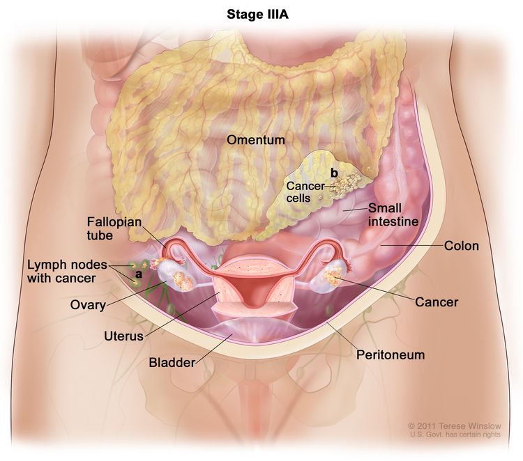
Ovarian Borderline Tumors Treatment (PDQ®)–Health Professional Version

Ovarian Borderline Tumors Treatment (PDQ®)–Health Professional Version

General Information About Ovarian Borderline Tumors

Incidence and Mortality

Ovarian borderline tumors (i.e., tumors of low malignant potential) account for 15% of all epithelial ovarian cancers. Nearly 75% of borderline tumors are stage I at the time of diagnosis.[1] Recognizing these tumors is important because their prognosis and treatment is different from the frankly malignant invasive carcinomas. Ovarian borderline tumors can also become low-grade serous tumors. For more information, see the Treatment of Low-Grade Serous Carcinoma section in Ovarian Epithelial, Fallopian Tube, and Primary Peritoneal Cancer Treatment.

A review of 22 series (which included 953 patients), with a mean follow-up of 7 years, revealed a survival rate of 92% for patients with advanced-stage ovarian borderline tumors, if patients with so-called invasive implants were excluded. The causes of death in these patients were determined to be benign complications of disease (e.g., small bowel obstruction), complications of therapy, and only rarely (0.7% of patients), malignant transformation.[2] In one series, the 5-, 10-, 15-, and 20-year survival rates of patients with borderline tumors (all stages), as demonstrated by clinical life table analysis, were 97%, 95%, 92%, and 89%, respectively.[3] In this series, mortality rates were stage dependent: 0.7% of patients with stage I tumors, 4.2% of patients with stage II tumors, and 26.8% of patients with stage III tumors died of disease.[3] The Fédération Internationale de Gynécologie et d’Obstétrique reported an extremely good prognosis for ovarian borderline tumors, with a 10-year survival rate of approximately 95%.[1] These survival rates are clearly in contrast with the 30% survival rate for patients with invasive tumors (all stages).

Another large retrospective study showed that early stage, serous histology, and younger age were associated with more favorable prognoses in patients with ovarian borderline tumors.[4]

Endometrioid Tumors

Endometrioid borderline tumors are less common and should not be regarded as malignant because they seldom, if ever, metastasize. However, malignant transformation can occur and may be associated with a similar tumor outside of the ovary. Such tumors are the result of either a second primary or rupture of the primary endometrial tumor.[5]

References
  1. Berek JS, Renz M, Kehoe S, et al.: Cancer of the ovary, fallopian tube, and peritoneum: 2021 update. Int J Gynaecol Obstet 155 (Suppl 1): 61-85, 2021. [PUBMED Abstract]
  2. Kurman RJ, Trimble CL: The behavior of serous tumors of low malignant potential: are they ever malignant? Int J Gynecol Pathol 12 (2): 120-7, 1993. [PUBMED Abstract]
  3. Leake JF, Currie JL, Rosenshein NB, et al.: Long-term follow-up of serous ovarian tumors of low malignant potential. Gynecol Oncol 47 (2): 150-8, 1992. [PUBMED Abstract]
  4. Kaern J, Tropé CG, Abeler VM: A retrospective study of 370 borderline tumors of the ovary treated at the Norwegian Radium Hospital from 1970 to 1982. A review of clinicopathologic features and treatment modalities. Cancer 71 (5): 1810-20, 1993. [PUBMED Abstract]
  5. Norris HJ: Proliferative endometrioid tumors and endometrioid tumors of low malignant potential of the ovary. Int J Gynecol Pathol 12 (2): 134-40, 1993. [PUBMED Abstract]

Stage Information for Ovarian Borderline Tumors

Fédération Internationale de Gynécologie et d’Obstétrique (FIGO) Staging

FIGO and the American Joint Committee on Cancer have designated staging to define ovarian borderline tumors; the FIGO system is most commonly used.[1,2]

Table 1. Definitions of FIGO Stage Ia
Stage Definition Illustration
FIGO = Fédération Internationale de Gynécologie et d’Obstétrique.
aAdapted from FIGO Committee for Gynecologic Oncology.[1]
I Tumor confined to ovaries or fallopian tube(s).
EnlargeThree-panel drawing of stage IA, stage IB, and stage IC; each panel shows the ovaries, fallopian tubes, uterus, cervix, and vagina. The first panel (stage IA) shows cancer inside one ovary. The second panel (stage IB) shows cancer inside both ovaries. The third panel (stage IC) shows cancer inside both ovaries and (a) the tumor in the ovary shown on the left has ruptured (broken open), (b) there is cancer on the surface of the ovary shown on the right, and (c) there are cancer cells in the pelvic peritoneal fluid (inset).
IA Tumor limited to one ovary (capsule intact) or fallopian tube; no tumor on ovarian or fallopian tube surface; no malignant cells in the ascites or peritoneal washings.  
IB Tumor limited to both ovaries (capsules intact) or fallopian tubes; no tumor on ovarian or fallopian tube surface; no malignant cells in the ascites or peritoneal washings.  
IC Tumor limited to one or both ovaries or fallopian tubes, with any of the following:  
IC1: Surgical spill.  
IC2: Capsule ruptured before surgery or tumor on ovarian or fallopian tube surface.  
IC3: Malignant cells in the ascites or peritoneal washings.  
Table 2. Definitions of FIGO Stage IIa
Stage Definition Illustration
FIGO = Fédération Internationale de Gynécologie et d’Obstétrique.
aAdapted from FIGO Committee for Gynecologic Oncology.[1]
II Tumor involves one or both ovaries or fallopian tubes with pelvic extension (below the pelvic brim) or primary peritoneal cancer.
EnlargeThree-panel drawing of stage IIA, stage IIB, and stage II primary peritoneal cancer; the first panel (stage IIA) shows cancer inside both ovaries that has spread to the fallopian tube and uterus . Also shown are the cervix and vagina. The second panel (stage IIB) shows cancer inside both ovaries that has spread to the colon. The third panel (primary peritoneal cancer) shows cancer in the pelvic peritoneum.
IIA Extension and/or implants on uterus and/or fallopian tubes and/or ovaries.  
IIB Extension to other pelvic intraperitoneal tissues.  
Table 3. Definitions of FIGO Stage IIIa
Stage Definition Illustration
FIGO = Fédération Internationale de Gynécologie et d’Obstétrique.
aAdapted from FIGO Committee for Gynecologic Oncology.[1]
III Tumor involves one or both ovaries or fallopian tubes, or primary peritoneal cancer, with cytologically or histologically confirmed spread to the peritoneum outside the pelvis and/or metastasis to the retroperitoneal lymph nodes.  
IIIA1 Positive retroperitoneal lymph nodes only (cytologically or histologically proven):
EnlargeDrawing of stage IIIA shows cancer inside both ovaries that has spread to (a) lymph nodes behind the peritoneum and (b) the omentum. The small intestine, colon, fallopian tubes, uterus, and bladder are also shown.
IIIA1(I): Metastasis ≤10 mm in greatest dimension.
IIIA1(ii): Metastasis >10 mm in greatest dimension.
IIIA2 Microscopic extrapelvic (above the pelvic brim) peritoneal involvement with or without positive retroperitoneal lymph nodes.
IIIB Macroscopic peritoneal metastasis beyond the pelvis ≤2 cm in greatest dimension, with or without metastasis to the retroperitoneal lymph nodes.
EnlargeDrawing of stage IIIB shows cancer inside both ovaries that has spread to the omentum. The cancer in the omentum is 2 centimeters or smaller. An inset shows 2 centimeters is about the size of a peanut. Also shown are the small intestine, colon, fallopian tubes, uterus, bladder, and lymph nodes behind the peritoneum.
IIIC Macroscopic peritoneal metastasis beyond the pelvis >2 cm in greatest dimension, with or without metastasis to the retroperitoneal lymph nodes (includes extension of tumor to capsule of liver and spleen without parenchymal involvement of either organ).
EnlargeDrawing of stage IIIC shows cancer inside both ovaries that has spread to the omentum. The cancer in the omentum is larger than 2 centimeters. An inset shows 2 centimeters is about the size of a peanut. Also shown are the small intestine, colon, fallopian tubes, uterus, bladder, and lymph nodes behind the peritoneum.
Table 4. Definitions of FIGO Stage IVa
Stage Definition Illustration
FIGO = Fédération Internationale de Gynécologie et d’Obstétrique.
aAdapted from FIGO Committee for Gynecologic Oncology.[1]
IV Distant metastasis excluding peritoneal metastases.
EnlargeDrawing of stage IV shows other parts of the body where ovarian cancer may spread, including the lung, liver, and lymph nodes in the groin. An inset on the top shows extra fluid around the lung. An inset on the bottom shows cancer cells spreading through the blood and lymph system to another part of the body where metastatic cancer has formed.
IVA Pleural effusion with positive cytology.  
IVB Parenchymal metastases and metastases to extra-abdominal organs (including inguinal lymph nodes and lymph nodes outside of the abdominal cavity).  
References
  1. Berek JS, Renz M, Kehoe S, et al.: Cancer of the ovary, fallopian tube, and peritoneum: 2021 update. Int J Gynaecol Obstet 155 (Suppl 1): 61-85, 2021. [PUBMED Abstract]
  2. Ovary, fallopian tube, and primary peritoneal carcinoma. In: Amin MB, Edge SB, Greene FL, et al., eds.: AJCC Cancer Staging Manual. 8th ed. Springer; 2017, pp 681-90.

Treatment of Early-Stage Ovarian Borderline Tumors

Treatment Options for Early-Stage Ovarian Borderline Tumors

Treatment options for early-stage ovarian borderline tumors include:

Surgery

In early-stage disease (stage I or II), no additional treatment is indicated for patients with a completely resected borderline tumor.[1]

Importance of Staging for Treatment

The value of complete staging has not been demonstrated for patients with early-stage disease, but the opposite ovary should be carefully evaluated for evidence of bilateral disease. The impact of surgical staging on therapeutic management has not been defined. However, in a study of 29 patients with presumed localized disease, 7 patients were upstaged after complete surgical staging.[2]

In two other studies, 16% and 18% of patients with presumed localized borderline tumors were upstaged as a result of a staging laparotomy.[3,4] In one of these studies, the yield for serous tumors was 30.8%, compared with 0% for mucinous tumors.[3]

In another study, patients with localized intraperitoneal disease and negative lymph nodes had a low incidence of recurrence (5%), whereas patients with localized intraperitoneal disease and positive lymph nodes had a statistically significant higher incidence of recurrence (50%).[5]

Fertility Preservation

When a patient wishes to retain childbearing potential, a unilateral salpingo-oophorectomy is adequate therapy.[6,7] In the presence of bilateral ovarian cystic neoplasms, or for patients with a single ovary, a partial oophorectomy can be used to preserve fertility.[8] Some physicians stress the importance of limiting ovarian cystectomy to patients with stage IA disease whose cystectomy specimen margins are tumor free.[9]

In a large series, the relapse rate was higher for patients who underwent more conservative surgery (cystectomy > unilateral oophorectomy > total abdominal hysterectomy and bilateral salpingo-oophorectomy [TAHBSO]). However, differences were not statistically significant, and the survival rates were nearly 100% for all groups.[5,10] When childbearing is not a consideration, a TAHBSO is appropriate therapy. Once a patient’s family is complete, most, but not all,[9] physicians favor removal of remaining ovarian tissue as there is risk of recurrence of a borderline tumor or, rarely, a carcinoma.[3,6]

Current Clinical Trials

Use our advanced clinical trial search to find NCI-supported cancer clinical trials that are now enrolling patients. The search can be narrowed by location of the trial, type of treatment, name of the drug, and other criteria. General information about clinical trials is also available.

References
  1. Tropé C, Kaern J, Vergote IB, et al.: Are borderline tumors of the ovary overtreated both surgically and systemically? A review of four prospective randomized trials including 253 patients with borderline tumors. Gynecol Oncol 51 (2): 236-43, 1993. [PUBMED Abstract]
  2. Yazigi R, Sandstad J, Munoz AK: Primary staging in ovarian tumors of low malignant potential. Gynecol Oncol 31 (3): 402-8, 1988. [PUBMED Abstract]
  3. Snider DD, Stuart GC, Nation JG, et al.: Evaluation of surgical staging in stage I low malignant potential ovarian tumors. Gynecol Oncol 40 (2): 129-32, 1991. [PUBMED Abstract]
  4. Leake JF, Rader JS, Woodruff JD, et al.: Retroperitoneal lymphatic involvement with epithelial ovarian tumors of low malignant potential. Gynecol Oncol 42 (2): 124-30, 1991. [PUBMED Abstract]
  5. Leake JF, Currie JL, Rosenshein NB, et al.: Long-term follow-up of serous ovarian tumors of low malignant potential. Gynecol Oncol 47 (2): 150-8, 1992. [PUBMED Abstract]
  6. Kaern J, Tropé CG, Abeler VM: A retrospective study of 370 borderline tumors of the ovary treated at the Norwegian Radium Hospital from 1970 to 1982. A review of clinicopathologic features and treatment modalities. Cancer 71 (5): 1810-20, 1993. [PUBMED Abstract]
  7. Lim-Tan SK, Cajigas HE, Scully RE: Ovarian cystectomy for serous borderline tumors: a follow-up study of 35 cases. Obstet Gynecol 72 (5): 775-81, 1988. [PUBMED Abstract]
  8. Rice LW, Berkowitz RS, Mark SD, et al.: Epithelial ovarian tumors of borderline malignancy. Gynecol Oncol 39 (2): 195-8, 1990. [PUBMED Abstract]
  9. Piura B, Dgani R, Blickstein I, et al.: Epithelial ovarian tumors of borderline malignancy: a study of 50 cases. Int J Gynecol Cancer 2 (4): 189-197, 1992. [PUBMED Abstract]
  10. Casey AC, Bell DA, Lage JM, et al.: Epithelial ovarian tumors of borderline malignancy: long-term follow-up. Gynecol Oncol 50 (3): 316-22, 1993. [PUBMED Abstract]

Treatment of Advanced-Stage Ovarian Borderline Tumors

Treatment Options for Advanced-Stage Ovarian Borderline Tumors

Treatment options for advanced-stage ovarian borderline tumors include:

Surgery

Patients with advanced disease should undergo a total hysterectomy, bilateral salpingo-oophorectomy, omentectomy, node sampling, and aggressive cytoreductive surgery. Patients with stage III or IV disease with no gross residual tumor had a survival rate of 100% in some series, regardless of the follow-up duration.[1,2] Patients with gross residual disease had a 7-year survival rate of only 69% in a large series, [3] and survival appeared to be inversely proportional to the length of follow-up.[3]

Chemotherapy and/or radiation therapy are not indicated for patients with more advanced-stage disease and microscopic or gross residual disease. Scant evidence exists that postoperative chemotherapy or radiation therapy alters the course of this disease in any beneficial way.[1,36] In a retrospective study of 364 patients without residual tumor, adjuvant therapy had no effect on disease-free or corrected survival when stratified for disease stage.[7] Patients without residual tumor who received no adjuvant treatment had a survival rate equal to or greater than the treated groups. No controlled studies have compared postoperative treatment with no postoperative treatment.

In a review of 150 patients with borderline ovarian tumors, the survival of patients with residual tumors smaller than 2 cm was significantly better than survival for those with residual tumors measuring 2 cm to 5 cm or tumors measuring larger than 5 cm (P < .05).[8] It is not clear whether invasive implants imply a worse prognosis. Some investigators have correlated invasive implants with a poor prognosis,[9] while others have not.[2,10] Some studies have suggested DNA ploidy of the tumors can identify patients who will develop aggressive disease.[11,12] One study could not correlate DNA ploidy of the primary serous tumor with patient survival, but found that aneuploid invasive implants were associated with a poor prognosis.[13] No evidence indicates that treating patients with aneuploid tumors would have an impact on survival. No significant associations were found between TP53 and HER2/neu overexpression and tumor recurrence or patient survival.[14]

Current Clinical Trials

Use our advanced clinical trial search to find NCI-supported cancer clinical trials that are now enrolling patients. The search can be narrowed by location of the trial, type of treatment, name of the drug, and other criteria. General information about clinical trials is also available.

References
  1. Barnhill D, Heller P, Brzozowski P, et al.: Epithelial ovarian carcinoma of low malignant potential. Obstet Gynecol 65 (1): 53-9, 1985. [PUBMED Abstract]
  2. Bostwick DG, Tazelaar HD, Ballon SC, et al.: Ovarian epithelial tumors of borderline malignancy. A clinical and pathologic study of 109 cases. Cancer 58 (9): 2052-65, 1986. [PUBMED Abstract]
  3. Leake JF, Currie JL, Rosenshein NB, et al.: Long-term follow-up of serous ovarian tumors of low malignant potential. Gynecol Oncol 47 (2): 150-8, 1992. [PUBMED Abstract]
  4. Casey AC, Bell DA, Lage JM, et al.: Epithelial ovarian tumors of borderline malignancy: long-term follow-up. Gynecol Oncol 50 (3): 316-22, 1993. [PUBMED Abstract]
  5. Tumors of the ovary: neoplasms derived from coelomic epithelium. In: Morrow CP, Curtin JP: Synopsis of Gynecologic Oncology. 5th ed. Churchill Livingstone, 1998, pp 233-281.
  6. Sutton GP, Bundy BN, Omura GA, et al.: Stage III ovarian tumors of low malignant potential treated with cisplatin combination therapy (a Gynecologic Oncology Group study). Gynecol Oncol 41 (3): 230-3, 1991. [PUBMED Abstract]
  7. Kaern J, Tropé CG, Abeler VM: A retrospective study of 370 borderline tumors of the ovary treated at the Norwegian Radium Hospital from 1970 to 1982. A review of clinicopathologic features and treatment modalities. Cancer 71 (5): 1810-20, 1993. [PUBMED Abstract]
  8. Tamakoshi K, Kikkawa F, Nakashima N, et al.: Clinical behavior of borderline ovarian tumors: a study of 150 cases. J Surg Oncol 64 (2): 147-52, 1997. [PUBMED Abstract]
  9. Bell DA, Scully RE: Serous borderline tumors of the peritoneum. Am J Surg Pathol 14 (3): 230-9, 1990. [PUBMED Abstract]
  10. Michael H, Roth LM: Invasive and noninvasive implants in ovarian serous tumors of low malignant potential. Cancer 57 (6): 1240-7, 1986. [PUBMED Abstract]
  11. Friedlander ML, Hedley DW, Swanson C, et al.: Prediction of long-term survival by flow cytometric analysis of cellular DNA content in patients with advanced ovarian cancer. J Clin Oncol 6 (2): 282-90, 1988. [PUBMED Abstract]
  12. Kaern J, Trope C, Kjorstad KE, et al.: Cellular DNA content as a new prognostic tool in patients with borderline tumors of the ovary. Gynecol Oncol 38 (3): 452-7, 1990. [PUBMED Abstract]
  13. de Nictolis M, Montironi R, Tommasoni S, et al.: Serous borderline tumors of the ovary. A clinicopathologic, immunohistochemical, and quantitative study of 44 cases. Cancer 70 (1): 152-60, 1992. [PUBMED Abstract]
  14. Eltabbakh GH, Belinson JL, Kennedy AW, et al.: p53 and HER-2/neu overexpression in ovarian borderline tumors. Gynecol Oncol 65 (2): 218-24, 1997. [PUBMED Abstract]

Latest Updates to This Summary (02/12/2025)

The PDQ cancer information summaries are reviewed regularly and updated as new information becomes available. This section describes the latest changes made to this summary as of the date above.

General Information About Ovarian Borderline Tumors

Added text to state that ovarian borderline tumors can also become low-grade serous tumors.

Revised text to state that the Fédération Internationale de Gynécologie et d’Obstétrique reported an extremely good prognosis for ovarian borderline tumors, with a 10-year survival rate of approximately 95%.

This summary is written and maintained by the PDQ Adult Treatment Editorial Board, which is editorially independent of NCI. The summary reflects an independent review of the literature and does not represent a policy statement of NCI or NIH. More information about summary policies and the role of the PDQ Editorial Boards in maintaining the PDQ summaries can be found on the About This PDQ Summary and PDQ® Cancer Information for Health Professionals pages.

About This PDQ Summary

Purpose of This Summary

This PDQ cancer information summary for health professionals provides comprehensive, peer-reviewed, evidence-based information about the treatment of ovarian borderline tumors. It is intended as a resource to inform and assist clinicians in the care of their patients. It does not provide formal guidelines or recommendations for making health care decisions.

Reviewers and Updates

This summary is reviewed regularly and updated as necessary by the PDQ Adult Treatment Editorial Board, which is editorially independent of the National Cancer Institute (NCI). The summary reflects an independent review of the literature and does not represent a policy statement of NCI or the National Institutes of Health (NIH).

Board members review recently published articles each month to determine whether an article should:

  • be discussed at a meeting,
  • be cited with text, or
  • replace or update an existing article that is already cited.

Changes to the summaries are made through a consensus process in which Board members evaluate the strength of the evidence in the published articles and determine how the article should be included in the summary.

The lead reviewers for Ovarian Borderline Tumors Treatment are:

  • Olga T. Filippova, MD (Lenox Hill Hospital)
  • Marina Stasenko, MD (New York University Medical Center)

Any comments or questions about the summary content should be submitted to Cancer.gov through the NCI website’s Email Us. Do not contact the individual Board Members with questions or comments about the summaries. Board members will not respond to individual inquiries.

Levels of Evidence

Some of the reference citations in this summary are accompanied by a level-of-evidence designation. These designations are intended to help readers assess the strength of the evidence supporting the use of specific interventions or approaches. The PDQ Adult Treatment Editorial Board uses a formal evidence ranking system in developing its level-of-evidence designations.

Permission to Use This Summary

PDQ is a registered trademark. Although the content of PDQ documents can be used freely as text, it cannot be identified as an NCI PDQ cancer information summary unless it is presented in its entirety and is regularly updated. However, an author would be permitted to write a sentence such as “NCI’s PDQ cancer information summary about breast cancer prevention states the risks succinctly: [include excerpt from the summary].”

The preferred citation for this PDQ summary is:

PDQ® Adult Treatment Editorial Board. PDQ Ovarian Borderline Tumors Treatment. Bethesda, MD: National Cancer Institute. Updated <MM/DD/YYYY>. Available at: /types/ovarian/hp/ovarian-borderline-tumors-treatment-pdq. Accessed <MM/DD/YYYY>. [PMID: 26389466]

Images in this summary are used with permission of the author(s), artist, and/or publisher for use within the PDQ summaries only. Permission to use images outside the context of PDQ information must be obtained from the owner(s) and cannot be granted by the National Cancer Institute. Information about using the illustrations in this summary, along with many other cancer-related images, is available in Visuals Online, a collection of over 2,000 scientific images.

Disclaimer

Based on the strength of the available evidence, treatment options may be described as either “standard” or “under clinical evaluation.” These classifications should not be used as a basis for insurance reimbursement determinations. More information on insurance coverage is available on Cancer.gov on the Managing Cancer Care page.

Contact Us

More information about contacting us or receiving help with the Cancer.gov website can be found on our Contact Us for Help page. Questions can also be submitted to Cancer.gov through the website’s Email Us.
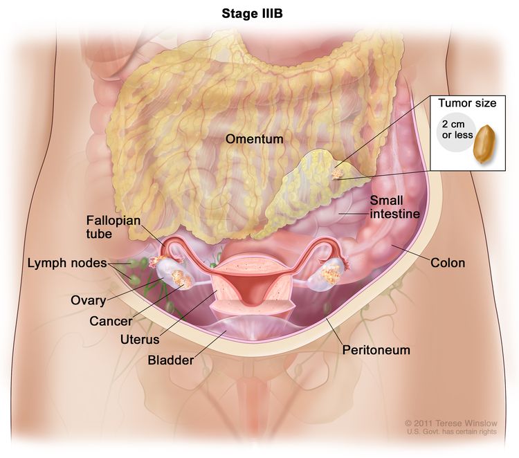
Ovarian Germ Cell Tumors Treatment (PDQ®)–Health Professional Version

Ovarian Germ Cell Tumors Treatment (PDQ®)–Health Professional Version

General Information About Ovarian Germ Cell Tumors

Incidence and Mortality

Germ cell tumors of the ovary are uncommon but aggressive tumors, seen most often in young women and adolescent girls. These tumors are frequently unilateral and are generally curable if found and treated early. The use of combination chemotherapy after initial surgery has dramatically improved the prognosis for many women with these tumors.[13]

Dysgerminomas

One series found a 10-year survival rate of 88.6% following conservative surgery for patients with dysgerminoma confined to the ovary; smaller than 10 cm in size; with an intact, smooth capsule unattached to other organs; and without ascites.[4] A number of patients had one or more successful pregnancies following unilateral salpingo-oophorectomy.[4] Even patients with incompletely resected dysgerminoma can be rendered disease free following chemotherapy with bleomycin, etoposide, and cisplatin (BEP) or a combination of cisplatin, vinblastine, and bleomycin.[5]

Other Germ Cell Tumors

A report of 35 cases of germ cell tumors, half of which were advanced stage or recurrent or progressive disease, demonstrated a 97% sustained remission at 10 months to 54 months after the start of a combination of BEP.[1] Two Gynecologic Oncology Group trials reported that 89 of 93 patients with stage I, II, or III disease who had completely resected tumors were disease free after three cycles of BEP.[1,3]

Endodermal sinus tumors of the ovary are particularly aggressive. A review of the literature in 1979 prior to the widespread use of combination chemotherapy found only 27% of 96 patients with stage I endodermal sinus tumor alive at 2 years after diagnosis. More than 50% of the patients died within a year of diagnosis.

Patients with mature teratomas usually experience long-term survival, but survival for patients with immature teratomas after surgery only is related to the grade of the tumor, especially its neural elements. In a series of 58 patients with immature teratoma treated before the modern chemotherapeutic era, recurrence was reported in 18% of the patients with grade 1 disease, 37% of the patients with grade 2 disease, and 70% of the patients with grade 3 disease.[6] Similar findings have been reported by others.[7]

Some studies have found that size and histology were the major factors determining prognosis for patients with malignant mixed germ cell tumors of the ovary.[6,8] Prognosis was poor for patients with large tumors when more than one-third of the tumor was composed of endodermal sinus elements, choriocarcinoma, or grade 3 immature teratoma. When the tumor was smaller than 10 cm in diameter, the prognosis was good, regardless of the composition of the tumor.[8,9]

References
  1. Gershenson DM: Update on malignant ovarian germ cell tumors. Cancer 71 (4 Suppl): 1581-90, 1993. [PUBMED Abstract]
  2. Segelov E, Campbell J, Ng M, et al.: Cisplatin-based chemotherapy for ovarian germ cell malignancies: the Australian experience. J Clin Oncol 12 (2): 378-84, 1994. [PUBMED Abstract]
  3. Williams S, Blessing JA, Liao SY, et al.: Adjuvant therapy of ovarian germ cell tumors with cisplatin, etoposide, and bleomycin: a trial of the Gynecologic Oncology Group. J Clin Oncol 12 (4): 701-6, 1994. [PUBMED Abstract]
  4. Thomas GM, Dembo AJ, Hacker NF, et al.: Current therapy for dysgerminoma of the ovary. Obstet Gynecol 70 (2): 268-75, 1987. [PUBMED Abstract]
  5. Williams SD, Blessing JA, Hatch KD, et al.: Chemotherapy of advanced dysgerminoma: trials of the Gynecologic Oncology Group. J Clin Oncol 9 (11): 1950-5, 1991. [PUBMED Abstract]
  6. Norris HJ, Zirkin HJ, Benson WL: Immature (malignant) teratoma of the ovary: a clinical and pathologic study of 58 cases. Cancer 37 (5): 2359-72, 1976. [PUBMED Abstract]
  7. Gallion H, van Nagell JR, Powell DF, et al.: Therapy of endodermal sinus tumor of the ovary. Am J Obstet Gynecol 135 (4): 447-51, 1979. [PUBMED Abstract]
  8. Kurman RJ, Norris HJ: Malignant germ cell tumors of the ovary. Hum Pathol 8 (5): 551-64, 1977. [PUBMED Abstract]
  9. Murugaesu N, Schmid P, Dancey G, et al.: Malignant ovarian germ cell tumors: identification of novel prognostic markers and long-term outcome after multimodality treatment. J Clin Oncol 24 (30): 4862-6, 2006. [PUBMED Abstract]

Cellular Classification of Ovarian Germ Cell Tumors

The following histological subtypes have been described.[1,2]

  • Dysgerminoma.
  • Other germ cell tumors:
    • Endodermal sinus tumor (rare subtypes are hepatoid and intestinal).[1]
    • Embryonal carcinoma.
    • Polyembryoma.
    • Choriocarcinoma.
    • Teratoma:
      • Immature.
      • Mature:
        • Solid.
        • Cystic:
          • Dermoid cyst (mature cystic teratoma).
          • Dermoid cyst with malignant transformation.
      • Monodermal and highly specialized:
        • Struma ovarii.
        • Carcinoid.
        • Struma ovarii and carcinoid.
        • Others (e.g., malignant neuroectodermal and ependymoma).
    • Mixed forms.
References
  1. Gershenson DM: Update on malignant ovarian germ cell tumors. Cancer 71 (4 Suppl): 1581-90, 1993. [PUBMED Abstract]
  2. Serov SF, Scully RE, Robin IH: International Histologic Classification of Tumours: No. 9. Histological Typing of Ovarian Tumours. World Health Organization, 1973.

Stage Information for Ovarian Germ Cell Tumors

In the absence of obvious metastatic disease, accurate staging of germ cell tumors of the ovary requires laparotomy with careful examination of the following:

  • Entire diaphragm.
  • Both paracolic gutters.
  • Pelvic nodes on the side of the ovarian tumor.
  • The para-aortic lymph nodes.
  • The omentum.

The contralateral ovary should be carefully examined and biopsied if necessary. Ascitic fluid should be examined cytologically. If ascites is not present, it is important to obtain peritoneal washings before the tumor is manipulated. In patients with dysgerminoma, lymphangiography or computed tomography is indicated if the pelvic and para-aortic lymph nodes were not carefully examined at the time of surgery.

Although not required for formal staging, it is desirable to obtain serum levels of alpha fetoprotein and human chorionic gonadotropin as soon as the diagnosis is established because persistence of these markers in the serum after surgery indicates unresected tumor.

The Fédération Internationale de Gynécologie et d’Obstétrique (FIGO) Staging

The FIGO and the American Joint Committee on Cancer (AJCC) have designated staging to define ovarian germ cell tumors; the FIGO system is most commonly used.[1,2]

Table 1. Definitions of FIGO Stage Ia
Stage Definition Illustration
FIGO = Fédération Internationale de Gynécologie et d’Obstétrique.
aAdapted from FIGO Committee for Gynecologic Oncology.[1]
I Tumor confined to ovaries or fallopian tube(s).
EnlargeThree-panel drawing of stage IA, stage IB, and stage IC; each panel shows the ovaries, fallopian tubes, uterus, cervix, and vagina. The first panel (stage IA) shows cancer inside one ovary. The second panel (stage IB) shows cancer inside both ovaries. The third panel (stage IC) shows cancer inside both ovaries and (a) the tumor in the ovary shown on the left has ruptured (broken open), (b) there is cancer on the surface of the ovary shown on the right, and (c) there are cancer cells in the pelvic peritoneal fluid (inset).
IA Tumor limited to one ovary (capsule intact) or fallopian tube; no tumor on ovarian or fallopian tube surface; no malignant cells in the ascites or peritoneal washings.  
IB Tumor limited to both ovaries (capsules intact) or fallopian tubes; no tumor on ovarian or fallopian tube surface; no malignant cells in the ascites or peritoneal washings.  
IC Tumor limited to one or both ovaries or fallopian tubes, with any of the following:  
IC1: Surgical spill.  
IC2: Capsule ruptured before surgery or tumor on ovarian or fallopian tube surface.  
IC3: Malignant cells in the ascites or peritoneal washings.  
Table 2. Definitions of FIGO Stage IIa
Stage Definition Illustration
FIGO = Fédération Internationale de Gynécologie et d’Obstétrique.
aAdapted from FIGO Committee for Gynecologic Oncology.[1]
II Tumor involves one or both ovaries or fallopian tubes with pelvic extension (below the pelvic brim) or primary peritoneal cancer.
EnlargeThree-panel drawing of stage IIA, stage IIB, and stage II primary peritoneal cancer; the first panel (stage IIA) shows cancer inside both ovaries that has spread to the fallopian tube and uterus . Also shown are the cervix and vagina. The second panel (stage IIB) shows cancer inside both ovaries that has spread to the colon. The third panel (primary peritoneal cancer) shows cancer in the pelvic peritoneum.
IIA Extension and/or implants on uterus and/or fallopian tubes and/or ovaries.  
IIB Extension to other pelvic intraperitoneal tissues.  
Table 3. Definitions of FIGO Stage IIIa
Stage Definition Illustration
FIGO = Fédération Internationale de Gynécologie et d’Obstétrique.
aAdapted from FIGO Committee for Gynecologic Oncology.[1]
III Tumor involves one or both ovaries or fallopian tubes, or primary peritoneal cancer, with cytologically or histologically confirmed spread to the peritoneum outside the pelvis and/or metastasis to the retroperitoneal lymph nodes.  
IIIA1 Positive retroperitoneal lymph nodes only (cytologically or histologically proven):
EnlargeDrawing of stage IIIA shows cancer inside both ovaries that has spread to (a) lymph nodes behind the peritoneum and (b) the omentum. The small intestine, colon, fallopian tubes, uterus, and bladder are also shown.
IIIA1(I): Metastasis ≤10 mm in greatest dimension.
IIIA1(ii): Metastasis >10 mm in greatest dimension.
IIIA2 Microscopic extrapelvic (above the pelvic brim) peritoneal involvement with or without positive retroperitoneal lymph nodes.
IIIB Macroscopic peritoneal metastasis beyond the pelvis ≤2 cm in greatest dimension, with or without metastasis to the retroperitoneal lymph nodes.
EnlargeDrawing of stage IIIB shows cancer inside both ovaries that has spread to the omentum. The cancer in the omentum is 2 centimeters or smaller. An inset shows 2 centimeters is about the size of a peanut. Also shown are the small intestine, colon, fallopian tubes, uterus, bladder, and lymph nodes behind the peritoneum.
IIIC Macroscopic peritoneal metastasis beyond the pelvis >2 cm in greatest dimension, with or without metastasis to the retroperitoneal lymph nodes (includes extension of tumor to capsule of liver and spleen without parenchymal involvement of either organ).
EnlargeDrawing of stage IIIC shows cancer inside both ovaries that has spread to the omentum. The cancer in the omentum is larger than 2 centimeters. An inset shows 2 centimeters is about the size of a peanut. Also shown are the small intestine, colon, fallopian tubes, uterus, bladder, and lymph nodes behind the peritoneum.
Table 4. Definitions of FIGO Stage IVa
Stage Definition Illustration
FIGO = Fédération Internationale de Gynécologie et d’Obstétrique.
aAdapted from FIGO Committee for Gynecologic Oncology.[1]
IV Distant metastasis excluding peritoneal metastases.
EnlargeDrawing of stage IV shows other parts of the body where ovarian cancer may spread, including the lung, liver, and lymph nodes in the groin. An inset on the top shows extra fluid around the lung. An inset on the bottom shows cancer cells spreading through the blood and lymph system to another part of the body where metastatic cancer has formed.
IVA Pleural effusion with positive cytology.  
IVB Parenchymal metastases and metastases to extra-abdominal organs (including inguinal lymph nodes and lymph nodes outside of the abdominal cavity).  
References
  1. Berek JS, Renz M, Kehoe S, et al.: Cancer of the ovary, fallopian tube, and peritoneum: 2021 update. Int J Gynaecol Obstet 155 (Suppl 1): 61-85, 2021. [PUBMED Abstract]
  2. Ovary, fallopian tube, and primary peritoneal carcinoma. In: Amin MB, Edge SB, Greene FL, et al., eds.: AJCC Cancer Staging Manual. 8th ed. Springer; 2017, pp 681-90.

Treatment Option Overview for Ovarian Germ Cell Tumors

Treatment options for patients with ovarian germ cell tumors include:

  • Surgery.
  • Chemotherapy.
  • Radiation therapy.
  • High-dose chemotherapy with bone marrow transplant (under clinical evaluation).

Patients may be treated with unilateral salpingo-oophorectomy or total abdominal hysterectomy and bilateral salpingo-oophorectomy.

All patients except those with stage I, grade I immature teratoma and stage IA dysgerminoma require postoperative chemotherapy. With platinum-based combination chemotherapy, the prognosis for patients with endodermal sinus tumors, immature teratomas, embryonal carcinomas, choriocarcinomas, and mixed tumors containing one or more of these elements has improved dramatically.[1] As new and more effective drugs are developed, many of these patients will be candidates for newer clinical trials.

References
  1. Gershenson DM, Morris M, Cangir A, et al.: Treatment of malignant germ cell tumors of the ovary with bleomycin, etoposide, and cisplatin. J Clin Oncol 8 (4): 715-20, 1990. [PUBMED Abstract]

Treatment of Stage I Ovarian Germ Cell Tumors

Dysgerminomas

Treatment options for stage I dysgerminomas include:

  1. Unilateral salpingo-oophorectomy with or without lymphangiography or computed tomography (CT).
  2. Unilateral salpingo-oophorectomy followed by observation.
  3. Unilateral salpingo-oophorectomy with adjuvant radiation therapy or chemotherapy.

For patients with stage I dysgerminoma, unilateral salpingo-oophorectomy conserving the uterus and opposite ovary is accepted treatment of the younger patient who wants to preserve fertility or a pregnancy. Postoperative lymphangiography or CT is indicated before treatment decisions are made for patients who have not had careful surgical and pathological examination of pelvic and para-aortic lymph nodes during surgery.

Patients who have been completely staged and have stage IA tumors may be observed carefully after surgery without adjuvant treatment. About 15% to 25% of these patients will relapse, but they can be treated successfully at the time of recurrence with a high likelihood of cure.

Incompletely staged patients or those with higher stage tumors should probably receive adjuvant treatment. Options include radiation therapy or chemotherapy. A disadvantage of the former is loss of fertility resulting from ovarian failure. Experience with adjuvant chemotherapy is limited, but considering the effectiveness of chemotherapy in tumors other than dysgerminoma and in advanced stage dysgerminoma, adjuvant chemotherapy is likely to be very effective and to allow recovery of reproductive potential in patients with an intact ovary, fallopian tube, and uterus.[1]

Other Germ Cell Tumors

Treatment options for patients with other stage I germ cell tumors include:

  1. Unilateral salpingo-oophorectomy with adjuvant chemotherapy.
  2. Unilateral salpingo-oophorectomy followed by observation.

For patients with stage I germ cell tumors, unilateral salpingo-oophorectomy should be performed when fertility is to be preserved. For all patients with tumors other than pure dysgerminoma and low-grade (grade I) immature teratoma, chemotherapy is usually given postoperatively, although a series demonstrated excellent survival for patients with all types of stage I tumors managed by surveillance, reserving chemotherapy for cases in which postsurgery recurrence is documented.[2][Level of evidence C1]

There is considerable experience with a combination of vincristine, dactinomycin, and cyclophosphamide (VAC) given in an adjuvant setting. However, combinations containing cisplatin, etoposide, and bleomycin (BEP) are now preferred because of a lower relapse rate and shorter treatment time.[3] While a prospective comparison of VAC versus BEP has not been performed, in well-staged patients with completely resected tumors, relapse is rare following platinum-based chemotherapy.[3] However, the disease will recur in about 25% of well-staged patients treated with 6 months of VAC.[4]

Evidence suggests that second-look laparotomy is not beneficial in patients with initially completely resected tumors who receive cisplatin-based adjuvant treatment.[5,6]

Current Clinical Trials

Use our advanced clinical trial search to find NCI-supported cancer clinical trials that are now enrolling patients. The search can be narrowed by location of the trial, type of treatment, name of the drug, and other criteria. General information about clinical trials is also available.

References
  1. Thomas GM, Dembo AJ, Hacker NF, et al.: Current therapy for dysgerminoma of the ovary. Obstet Gynecol 70 (2): 268-75, 1987. [PUBMED Abstract]
  2. Dark GG, Bower M, Newlands ES, et al.: Surveillance policy for stage I ovarian germ cell tumors. J Clin Oncol 15 (2): 620-4, 1997. [PUBMED Abstract]
  3. Williams S, Blessing JA, Liao SY, et al.: Adjuvant therapy of ovarian germ cell tumors with cisplatin, etoposide, and bleomycin: a trial of the Gynecologic Oncology Group. J Clin Oncol 12 (4): 701-6, 1994. [PUBMED Abstract]
  4. Slayton RE, Park RC, Silverberg SG, et al.: Vincristine, dactinomycin, and cyclophosphamide in the treatment of malignant germ cell tumors of the ovary. A Gynecologic Oncology Group Study (a final report). Cancer 56 (2): 243-8, 1985. [PUBMED Abstract]
  5. Williams SD, Blessing JA, DiSaia PJ, et al.: Second-look laparotomy in ovarian germ cell tumors: the gynecologic oncology group experience. Gynecol Oncol 52 (3): 287-91, 1994. [PUBMED Abstract]
  6. Gershenson DM: The obsolescence of second-look laparotomy in the management of malignant ovarian germ cell tumors. Gynecol Oncol 52 (3): 283-5, 1994. [PUBMED Abstract]

Treatment of Stage II Ovarian Germ Cell Tumors

Dysgerminomas

Treatment options for patients with stage II dysgerminomas include:

  1. Total abdominal hysterectomy and bilateral salpingo-oophorectomy with adjuvant radiation therapy or chemotherapy.
  2. Unilateral salpingo-oophorectomy with adjuvant chemotherapy.
  3. Clinical trials.

For patients with stage II dysgerminoma, total abdominal hysterectomy and bilateral salpingo-oophorectomy are usually performed. For the younger patient who wants to preserve fertility, a unilateral salpingo-oophorectomy may be considered standard therapy, depending on the age of the patient, and adjuvant chemotherapy should be given.

These patients should receive adjuvant treatment. Options include radiation therapy or chemotherapy. A disadvantage of the former is loss of fertility resulting from ovarian failure. Experience with adjuvant chemotherapy is limited, but considering the effectiveness of chemotherapy in tumors other than dysgerminoma and its effectiveness in advanced-stage dysgerminoma, adjuvant chemotherapy is likely to be effective and to allow recovery of reproductive potential in patients with an intact ovary, fallopian tube, and uterus. Thus, adjuvant chemotherapy with the combination of bleomycin, etoposide, and cisplatin (BEP) has replaced radiation therapy except in the rare patient in whom chemotherapy is not considered appropriate.

Other Germ Cell Tumors

Treatment options for patients with other stage II germ cell tumors include:

  1. Unilateral salpingo-oophorectomy with adjuvant chemotherapy.
  2. Second-look laparotomy.
  3. Clinical trials.

For patients with stage II germ cell tumors other than pure dysgerminoma, unilateral salpingo-oophorectomy should be performed when fertility is to be preserved. Although there is considerable experience with the combination of vincristine, dactinomycin, and cyclophosphamide (VAC), especially when given in an adjuvant setting, BEP is more effective.[13] Patients who do not respond to a cisplatin-based combination may still attain a durable remission with VAC as salvage therapy.[4] Recurrence after three courses of BEP as adjuvant therapy is rare.[4] All patients who do not respond to standard therapy are candidates for clinical trials. When there is residual disease or elevated levels of alpha-fetoprotein or human chorionic gonadotropin after maximal surgical debulking, three or four courses of BEP combination chemotherapy are indicated.[5]

Evidence suggests that second-look laparotomy is not beneficial in patients with initially completely resected tumors who receive cisplatin-based adjuvant treatment.[6] Second-look surgery may be of benefit for a minority of patients whose tumor was not completely resected at the initial surgical procedure and who had teratomatous elements in their primary tumor.[6,7] Surgical resection of residual masses detected by clinical examination, by radiographic procedures, or at re-exploration should be undertaken since reversion to germ cell tumor has been described.

Current Clinical Trials

Use our advanced clinical trial search to find NCI-supported cancer clinical trials that are now enrolling patients. The search can be narrowed by location of the trial, type of treatment, name of the drug, and other criteria. General information about clinical trials is also available.

References
  1. Williams S, Blessing JA, Liao SY, et al.: Adjuvant therapy of ovarian germ cell tumors with cisplatin, etoposide, and bleomycin: a trial of the Gynecologic Oncology Group. J Clin Oncol 12 (4): 701-6, 1994. [PUBMED Abstract]
  2. Pinkerton CR, Pritchard J, Spitz L: High complete response rate in children with advanced germ cell tumors using cisplatin-containing combination chemotherapy. J Clin Oncol 4 (2): 194-9, 1986. [PUBMED Abstract]
  3. Gershenson DM, Morris M, Cangir A, et al.: Treatment of malignant germ cell tumors of the ovary with bleomycin, etoposide, and cisplatin. J Clin Oncol 8 (4): 715-20, 1990. [PUBMED Abstract]
  4. Williams SD, Blessing JA, Moore DH, et al.: Cisplatin, vinblastine, and bleomycin in advanced and recurrent ovarian germ-cell tumors. A trial of the Gynecologic Oncology Group. Ann Intern Med 111 (1): 22-7, 1989. [PUBMED Abstract]
  5. Williams SD, Birch R, Einhorn LH, et al.: Treatment of disseminated germ-cell tumors with cisplatin, bleomycin, and either vinblastine or etoposide. N Engl J Med 316 (23): 1435-40, 1987. [PUBMED Abstract]
  6. Williams SD, Blessing JA, DiSaia PJ, et al.: Second-look laparotomy in ovarian germ cell tumors: the gynecologic oncology group experience. Gynecol Oncol 52 (3): 287-91, 1994. [PUBMED Abstract]
  7. Gershenson DM: The obsolescence of second-look laparotomy in the management of malignant ovarian germ cell tumors. Gynecol Oncol 52 (3): 283-5, 1994. [PUBMED Abstract]

Treatment of Stage III Ovarian Germ Cell Tumors

Dysgerminomas

Treatment options for patients with stage III dysgerminomas include:

  1. Total abdominal hysterectomy and bilateral salpingo-oophorectomy.
  2. Unilateral salpingo-oophorectomy with adjuvant chemotherapy.
  3. Clinical trials.

For patients with stage III dysgerminoma, total abdominal hysterectomy and bilateral salpingo-oophorectomy are recommended with removal of as much gross tumor as can be done safely without resection of portions of the urinary tract or large segments of the small or large bowel. Patients who want to preserve fertility may be treated with unilateral salpingo-oophorectomy if chemotherapy is to be used.[15]

Combination chemotherapy with bleomycin, etoposide, and cisplatin (BEP) can cure most of these patients. In a report of results from two Gynecologic Oncology Group (GOG) trials, 19 of 20 patients with incompletely resected tumors who were treated with BEP or cisplatin, vinblastine, and bleomycin (PVB) were disease free at a median follow-up of 26 months.[1] When there is bulky residual disease, it is common to give three to four courses of a cisplatin-containing combination such as PVB or BEP.[68] A randomized study in testicular cancer has shown that bleomycin is an essential component of the BEP regime when only three courses are given.[9] Because chemotherapy with BEP appears to be less sterilizing than wide-field radiation, combination chemotherapy is the preferred treatment in the patient who wants to preserve fertility.[1]

Other Germ Cell Tumors

Treatment options for patients with other stage III germ cell tumors include:

  1. Total abdominal hysterectomy and bilateral salpingo-oophorectomy with adjuvant chemotherapy, with or without neoadjuvant chemotherapy.
  2. Unilateral salpingo-oophorectomy with adjuvant chemotherapy, with or without neoadjuvant chemotherapy.
  3. Second-look laparotomy.
  4. Clinical trials.

For patients with stage III germ cell tumors other than pure dysgerminoma, total abdominal hysterectomy and bilateral salpingo-oophorectomy is recommended with removal of as much tumor in the abdomen and pelvis as can be done safely without resection of portions of the urinary tract or large segments of the small or large bowel. Patients who wish to preserve fertility can be treated with unilateral salpingo-oophorectomy.[1,3,4] For patients with extensive intra-abdominal disease whose clinical condition precludes debulking surgery, chemotherapy can be considered prior to surgery. Following maximal surgical debulking, three to four courses of cisplatin-containing combination chemotherapy are indicated.[2,6,10]

Evidence suggests that second-look laparotomy is not beneficial in patients with initially completely resected tumors who receive cisplatin-based adjuvant treatment.[11] Patients who do not respond to a cisplatin and etoposide-based combination may still attain a durable remission with a combination of vincristine, dactinomycin, and cyclophosphamide or a combination of cisplatin, vinblastine, and ifosfamide as salvage therapy.[6] Second-look surgery may be of benefit for a minority of patients whose tumor was not completely resected at the initial surgical procedure and who had teratomatous elements in their primary tumor.[11] Surgical resection of residual masses detected by clinical examination, by radiographic procedures, or at re-exploration should be undertaken since reversion to germ cell tumor or progressive teratoma has been described.

Current Clinical Trials

Use our advanced clinical trial search to find NCI-supported cancer clinical trials that are now enrolling patients. The search can be narrowed by location of the trial, type of treatment, name of the drug, and other criteria. General information about clinical trials is also available.

References
  1. Williams SD, Blessing JA, Hatch KD, et al.: Chemotherapy of advanced dysgerminoma: trials of the Gynecologic Oncology Group. J Clin Oncol 9 (11): 1950-5, 1991. [PUBMED Abstract]
  2. Gershenson DM, Morris M, Cangir A, et al.: Treatment of malignant germ cell tumors of the ovary with bleomycin, etoposide, and cisplatin. J Clin Oncol 8 (4): 715-20, 1990. [PUBMED Abstract]
  3. Wu PC, Huang RL, Lang JH, et al.: Treatment of malignant ovarian germ cell tumors with preservation of fertility: a report of 28 cases. Gynecol Oncol 40 (1): 2-6, 1991. [PUBMED Abstract]
  4. Schwartz PE, Chambers SK, Chambers JT, et al.: Ovarian germ cell malignancies: the Yale University experience. Gynecol Oncol 45 (1): 26-31, 1992. [PUBMED Abstract]
  5. Low JJ, Perrin LC, Crandon AJ, et al.: Conservative surgery to preserve ovarian function in patients with malignant ovarian germ cell tumors. A review of 74 cases. Cancer 89 (2): 391-8, 2000. [PUBMED Abstract]
  6. Williams SD, Blessing JA, Moore DH, et al.: Cisplatin, vinblastine, and bleomycin in advanced and recurrent ovarian germ-cell tumors. A trial of the Gynecologic Oncology Group. Ann Intern Med 111 (1): 22-7, 1989. [PUBMED Abstract]
  7. Williams SD, Birch R, Einhorn LH, et al.: Treatment of disseminated germ-cell tumors with cisplatin, bleomycin, and either vinblastine or etoposide. N Engl J Med 316 (23): 1435-40, 1987. [PUBMED Abstract]
  8. Taylor MH, Depetrillo AD, Turner AR: Vinblastine, bleomycin, and cisplatin in malignant germ cell tumors of the ovary. Cancer 56 (6): 1341-9, 1985. [PUBMED Abstract]
  9. Williams S, Blessing JA, Liao SY, et al.: Adjuvant therapy of ovarian germ cell tumors with cisplatin, etoposide, and bleomycin: a trial of the Gynecologic Oncology Group. J Clin Oncol 12 (4): 701-6, 1994. [PUBMED Abstract]
  10. Williams SD, Blessing JA, DiSaia PJ, et al.: Second-look laparotomy in ovarian germ cell tumors: the gynecologic oncology group experience. Gynecol Oncol 52 (3): 287-91, 1994. [PUBMED Abstract]
  11. Gershenson DM: The obsolescence of second-look laparotomy in the management of malignant ovarian germ cell tumors. Gynecol Oncol 52 (3): 283-5, 1994. [PUBMED Abstract]

Treatment of Stage IV Ovarian Germ Cell Tumors

Dysgerminomas

Treatment options for patients with stage IV dysgerminomas include:

  1. Total abdominal hysterectomy and bilateral salpingo-oophorectomy with adjuvant chemotherapy.
  2. Unilateral salpingo-oophorectomy with adjuvant chemotherapy.
  3. Clinical trials.

For patients with stage IV dysgerminoma, total abdominal hysterectomy and bilateral salpingo-oophorectomy is recommended with removal of as much gross tumor in the abdomen and pelvis as can be done safely without resection of portions of the urinary tract or large segments of small or large bowel, although unilateral salpingo-oophorectomy should be considered in patients who wish to preserve fertility.[1,2] Chemotherapy with bleomycin, etoposide, and cisplatin (BEP) can cure the majority of such patients. Stage IV dysgerminoma is not treated with radiation therapy, but rather with chemotherapy, preferably with three to four courses of cisplatin-containing combination chemotherapy such as BEP.[1] A second-look operation following treatment is rarely beneficial.

Other Germ Cell Tumors

Treatment options for patients with other stage IV germ cell tumors include:

  1. Total abdominal hysterectomy and bilateral salpingo-oophorectomy with adjuvant chemotherapy with or without neoadjuvant chemotherapy.
  2. Unilateral salpingo-oophorectomy with adjuvant chemotherapy with or without neoadjuvant chemotherapy.
  3. Clinical trials.

For patients with stage IV germ cell tumors other than pure dysgerminoma, total abdominal hysterectomy and bilateral salpingo-oophorectomy is recommended with removal of as much tumor from the abdomen and pelvis as can be done safely without resection of the kidney or large segments of the small or large bowel. Patients who wish to preserve fertility can be treated with unilateral salpingo-oophorectomy. Following maximal surgical debulking, three to four courses of cisplatin-containing combination chemotherapy are indicated.[3,4] For patients with extensive intra-abdominal disease whose clinical condition precludes debulking surgery, chemotherapy can be considered prior to surgery. Patients who do not respond to a cisplatin and etoposide-based combination may still attain a durable remission with vincristine, dactinomycin, and cyclophosphamide or cisplatin, vinblastine, and ifosfamide as salvage therapy.[4]

Second-look surgery may be of benefit for a minority of patients whose tumor was not completely resected at the initial surgical procedure and who had teratomatous elements in their primary tumor.[5,6] Surgical resection of residual masses detected by clinical examination, by radiographic procedures, or at re-exploration should be undertaken since reversion to germ cell tumor or progressive teratoma has been described.

Current Clinical Trials

Use our advanced clinical trial search to find NCI-supported cancer clinical trials that are now enrolling patients. The search can be narrowed by location of the trial, type of treatment, name of the drug, and other criteria. General information about clinical trials is also available.

References
  1. Williams SD, Blessing JA, Hatch KD, et al.: Chemotherapy of advanced dysgerminoma: trials of the Gynecologic Oncology Group. J Clin Oncol 9 (11): 1950-5, 1991. [PUBMED Abstract]
  2. Low JJ, Perrin LC, Crandon AJ, et al.: Conservative surgery to preserve ovarian function in patients with malignant ovarian germ cell tumors. A review of 74 cases. Cancer 89 (2): 391-8, 2000. [PUBMED Abstract]
  3. Gershenson DM, Morris M, Cangir A, et al.: Treatment of malignant germ cell tumors of the ovary with bleomycin, etoposide, and cisplatin. J Clin Oncol 8 (4): 715-20, 1990. [PUBMED Abstract]
  4. Williams SD, Blessing JA, Moore DH, et al.: Cisplatin, vinblastine, and bleomycin in advanced and recurrent ovarian germ-cell tumors. A trial of the Gynecologic Oncology Group. Ann Intern Med 111 (1): 22-7, 1989. [PUBMED Abstract]
  5. Williams SD, Blessing JA, DiSaia PJ, et al.: Second-look laparotomy in ovarian germ cell tumors: the gynecologic oncology group experience. Gynecol Oncol 52 (3): 287-91, 1994. [PUBMED Abstract]
  6. Gershenson DM: The obsolescence of second-look laparotomy in the management of malignant ovarian germ cell tumors. Gynecol Oncol 52 (3): 283-5, 1994. [PUBMED Abstract]

Treatment of Recurrent Ovarian Germ Cell Tumors

Dysgerminomas

Treatment options for patients with recurrent dysgerminomas include:

  • Cisplatin-based chemotherapy has been used effectively for patients with recurrent dysgerminoma with and without adjuvant radiation therapy.[1]
  • Patients with recurrent pure dysgerminoma of the ovary are candidates for clinical trials. Some consideration should be given to the use of high-dose regimens with rescue.

Other Germ Cell Tumors

Treatment options for patients with other recurrent germ cell tumors include:

  • Patients with recurrent germ cell tumors of the ovary other than pure dysgerminoma should be treated with chemotherapy, the type of which is determined by previous treatment.[2] Radiation therapy is not effective in this setting. Cisplatin-based combination chemotherapy is effective.[1,3,4] Patients who do not respond to a cisplatin-based combination may still attain a durable remission with vincristine, dactinomycin, and cyclophosphamide or ifosfamide and cisplatin as salvage therapy.[1] Newer potential treatments include an ifosfamide combination [5] or high-dose chemotherapy and autologous marrow rescue.[68] Although the role of secondary cytoreductive surgery for patients with recurrent or progressive ovarian germ cell tumors remains controversial, it may have some benefit for a select group of patients, particularly those with immature teratoma.[9] After maximal effort for surgical cytoreduction, chemotherapy should be considered.
  • Patients with recurrent germ cell tumors of the ovary are candidates for clinical trials. Some consideration should be given to the use of high-dose regimens with rescue.

Current Clinical Trials

Use our advanced clinical trial search to find NCI-supported cancer clinical trials that are now enrolling patients. The search can be narrowed by location of the trial, type of treatment, name of the drug, and other criteria. General information about clinical trials is also available.

References
  1. Williams SD, Blessing JA, Moore DH, et al.: Cisplatin, vinblastine, and bleomycin in advanced and recurrent ovarian germ-cell tumors. A trial of the Gynecologic Oncology Group. Ann Intern Med 111 (1): 22-7, 1989. [PUBMED Abstract]
  2. Williams SD, Blessing JA, Hatch KD, et al.: Chemotherapy of advanced dysgerminoma: trials of the Gynecologic Oncology Group. J Clin Oncol 9 (11): 1950-5, 1991. [PUBMED Abstract]
  3. Williams SD, Birch R, Einhorn LH, et al.: Treatment of disseminated germ-cell tumors with cisplatin, bleomycin, and either vinblastine or etoposide. N Engl J Med 316 (23): 1435-40, 1987. [PUBMED Abstract]
  4. Taylor MH, Depetrillo AD, Turner AR: Vinblastine, bleomycin, and cisplatin in malignant germ cell tumors of the ovary. Cancer 56 (6): 1341-9, 1985. [PUBMED Abstract]
  5. Munshi NC, Loehrer PJ, Roth BJ, et al.: Vinblastine, ifosfamide and cisplatin (VeIP) as second line chemotherapy in metastatic germ cell tumors (GCT). [Abstract] Proceedings of the American Society of Clinical Oncology 9: A-520, 134, 1990.
  6. Broun ER, Nichols CR, Kneebone P, et al.: Long-term outcome of patients with relapsed and refractory germ cell tumors treated with high-dose chemotherapy and autologous bone marrow rescue. Ann Intern Med 117 (2): 124-8, 1992. [PUBMED Abstract]
  7. Motzer RJ, Bosl GJ: High-dose chemotherapy for resistant germ cell tumors: recent advances and future directions. J Natl Cancer Inst 84 (22): 1703-9, 1992. [PUBMED Abstract]
  8. Mandanas RA, Saez RA, Epstein RB, et al.: Long-term results of autologous marrow transplantation for relapsed or refractory male or female germ cell tumors. Bone Marrow Transplant 21 (6): 569-76, 1998. [PUBMED Abstract]
  9. Munkarah A, Gershenson DM, Levenback C, et al.: Salvage surgery for chemorefractory ovarian germ cell tumors. Gynecol Oncol 55 (2): 217-23, 1994. [PUBMED Abstract]

Latest Updates to This Summary (09/18/2024)

The PDQ cancer information summaries are reviewed regularly and updated as new information becomes available. This section describes the latest changes made to this summary as of the date above.

Editorial changes were made to this summary.

This summary is written and maintained by the PDQ Adult Treatment Editorial Board, which is editorially independent of NCI. The summary reflects an independent review of the literature and does not represent a policy statement of NCI or NIH. More information about summary policies and the role of the PDQ Editorial Boards in maintaining the PDQ summaries can be found on the About This PDQ Summary and PDQ® Cancer Information for Health Professionals pages.

About This PDQ Summary

Purpose of This Summary

This PDQ cancer information summary for health professionals provides comprehensive, peer-reviewed, evidence-based information about the treatment of ovarian germ cell tumors. It is intended as a resource to inform and assist clinicians in the care of their patients. It does not provide formal guidelines or recommendations for making health care decisions.

Reviewers and Updates

This summary is reviewed regularly and updated as necessary by the PDQ Adult Treatment Editorial Board, which is editorially independent of the National Cancer Institute (NCI). The summary reflects an independent review of the literature and does not represent a policy statement of NCI or the National Institutes of Health (NIH).

Board members review recently published articles each month to determine whether an article should:

  • be discussed at a meeting,
  • be cited with text, or
  • replace or update an existing article that is already cited.

Changes to the summaries are made through a consensus process in which Board members evaluate the strength of the evidence in the published articles and determine how the article should be included in the summary.

The lead reviewers for Ovarian Germ Cell Tumors Treatment are:

  • Olga T. Filippova, MD (Lenox Hill Hospital)
  • Marina Stasenko, MD (New York University Medical Center)

Any comments or questions about the summary content should be submitted to Cancer.gov through the NCI website’s Email Us. Do not contact the individual Board Members with questions or comments about the summaries. Board members will not respond to individual inquiries.

Levels of Evidence

Some of the reference citations in this summary are accompanied by a level-of-evidence designation. These designations are intended to help readers assess the strength of the evidence supporting the use of specific interventions or approaches. The PDQ Adult Treatment Editorial Board uses a formal evidence ranking system in developing its level-of-evidence designations.

Permission to Use This Summary

PDQ is a registered trademark. Although the content of PDQ documents can be used freely as text, it cannot be identified as an NCI PDQ cancer information summary unless it is presented in its entirety and is regularly updated. However, an author would be permitted to write a sentence such as “NCI’s PDQ cancer information summary about breast cancer prevention states the risks succinctly: [include excerpt from the summary].”

The preferred citation for this PDQ summary is:

PDQ® Adult Treatment Editorial Board. PDQ Ovarian Germ Cell Tumors Treatment. Bethesda, MD: National Cancer Institute. Updated <MM/DD/YYYY>. Available at: /types/ovarian/hp/ovarian-germ-cell-treatment-pdq. Accessed <MM/DD/YYYY>. [PMID: 26389449]

Images in this summary are used with permission of the author(s), artist, and/or publisher for use within the PDQ summaries only. Permission to use images outside the context of PDQ information must be obtained from the owner(s) and cannot be granted by the National Cancer Institute. Information about using the illustrations in this summary, along with many other cancer-related images, is available in Visuals Online, a collection of over 2,000 scientific images.

Disclaimer

Based on the strength of the available evidence, treatment options may be described as either “standard” or “under clinical evaluation.” These classifications should not be used as a basis for insurance reimbursement determinations. More information on insurance coverage is available on Cancer.gov on the Managing Cancer Care page.

Contact Us

More information about contacting us or receiving help with the Cancer.gov website can be found on our Contact Us for Help page. Questions can also be submitted to Cancer.gov through the website’s Email Us.
Ovarian Epithelial, Fallopian Tube, and Primary Peritoneal Cancer Treatment (PDQ®)–Health Professional Version

Ovarian Epithelial, Fallopian Tube, and Primary Peritoneal Cancer Treatment (PDQ®)–Health Professional Version

General Information About Ovarian Epithelial, Fallopian Tube, and Primary Peritoneal Cancer

This PDQ summary addresses the staging and treatment of ovarian epithelial cancer, fallopian tube cancer (FTC), and primary peritoneal cancer (PPC).

Regardless of the site of origin, the hallmark of these cancers is their early peritoneal spread of metastases. The inclusion of FTC and PPC within the ovarian epithelial cancer designation is generally accepted because of much evidence that points to a common Müllerian epithelium derivation and similar management of these three neoplasms. The hypothesis that many high-grade serous ovarian cancers (the most common histological subtype) may arise from precursor lesions that originate in the fimbriae of the fallopian tubes has been supported by findings from risk-reducing surgeries in healthy women with BRCA1 or BRCA2 variants.[1] In addition, histologically similar cancers diagnosed as primary peritoneal carcinomas share molecular findings, such as loss or inactivation of the tumor-suppressor p53 and BRCA1 or BRCA2 proteins.[2] Therefore, high-grade serous adenocarcinomas arising from the fallopian tube and elsewhere in the peritoneal cavity, together with most ovarian epithelial cancers, represent extrauterine adenocarcinomas of Müllerian epithelial origin and are staged and treated similarly to ovarian cancer. Since 2000, FTC and PPC have usually been included in ovarian cancer clinical trials.[3]

Clear cell and endometrioid ovarian cancers that are linked to endometriosis have different gene-expression signatures, as do mucinous subtypes.[2]

Stromal and germ cell tumors are relatively uncommon and comprise fewer than 10% of cases. For more information, see Ovarian Germ Cell Tumors Treatment and Ovarian Borderline Tumors Treatment.

Incidence and Mortality

Epithelial carcinoma of the ovary is one of the most common gynecologic malignancies, with almost 50% of all cases occurring in women older than 65 years. It is the sixth most frequent cause of cancer death in women.[4,5]

Estimated new cases and deaths from ovarian cancer in the United States in 2025:[5]

  • New cases: 20,890.
  • Deaths: 12,730.

Anatomy

The fimbriated ends of the fallopian tubes are in close apposition to the ovaries and in the peritoneal space, as opposed to the corpus uteri (body of the uterus) that is located under a layer of peritoneum.

EnlargeAnatomy of the female reproductive system; drawing shows the uterus, myometrium (muscular outer layer of the uterus), endometrium (inner lining of the uterus), ovaries, fallopian tubes, cervix, and vagina.
Normal female reproductive system anatomy.

Risk Factors

Increasing age is the most important risk factor for most cancers. Other risk factors for ovarian (epithelial) cancer include:

  • Family history of ovarian cancer.[68]
    • A first-degree relative (e.g., mother, daughter, or sister) with the disease.
  • Inherited risk.[9]
    • BRCA1 or BRCA2 variants.[6,10]
  • Other hereditary conditions such as hereditary nonpolyposis colorectal cancer (HNPCC; also called Lynch syndrome).[6,9]
  • Endometriosis.[1113]
  • Hormone therapy.[14,15]
    • Postmenopausal hormone replacement therapy.
  • Obesity.[1618]
    • High body mass index.
  • Tall height.[1618]

Family history and genetic alterations

The most important risk factor for ovarian cancer is a history of ovarian cancer in a first-degree relative (mother, daughter, or sister). Approximately 20% of ovarian cancers are familial, and although most of these are linked to pathogenic variants in either the BRCA1 or BRCA2 gene, several other genes have been implicated.[19,20] The risk is highest in women who have two or more first-degree relatives with ovarian cancer.[21] The risk is somewhat less for women who have one first-degree relative and one second-degree relative (grandmother or aunt) with ovarian cancer.

In most families affected with breast and ovarian cancer syndrome or site-specific ovarian cancer, genetic linkage to the BRCA1 locus on chromosome 17q21 has been identified.[2224] BRCA2, also responsible for some instances of inherited ovarian and breast cancer, has been mapped by genetic linkage to chromosome 13q12.[25]

The lifetime risk of developing ovarian cancer in patients with germline pathogenic variants in BRCA1 is substantially increased over that of the general population.[26,27] Two retrospective studies of patients with germline pathogenic variants in BRCA1 suggest that the women in these studies have improved survival compared with those without BRCA1 variants.[28,29][Level of evidence C1] Most women with BRCA1 variants probably have family members with a history of ovarian and/or breast cancer. Therefore, the women in these studies may have been more vigilant and inclined to participate in cancer screening programs that may have led to earlier detection.

For women at increased risk, prophylactic oophorectomy may be considered after age 35 years if childbearing is complete. A family-based study included 551 women with BRCA1 or BRCA2 variants. Of the 259 women who had undergone bilateral prophylactic oophorectomy, 2 (0.8%) developed subsequent papillary serous peritoneal carcinoma, and 6 (2.8%) had stage I ovarian cancer at the time of surgery. Of the 292 matched controls, 20% who did not have prophylactic surgery developed ovarian cancer. Prophylactic surgery was associated with a reduction in the risk of ovarian cancer that exceeded 90% (relative risk, 0.04; 95% confidence interval, 0.01–0.16), with an average follow-up of 9 years.[30] However, family-based studies may be associated with biases resulting from case selection and other factors that influence the estimate of benefit.[31] After a prophylactic oophorectomy, a small percentage of women may develop a PPC that is similar in appearance to ovarian cancer.[32] This risk of developing PPC is likely related to the presence of serous tubal intraepithelial carcinoma (STIC) at the time of prophylactic oophorectomy. In a large study that pooled patients with BRCA variants from several sources, women with a STIC lesion were nearly 34 times more likely to develop PPC than women without such a lesion. This finding highlights the need for accurate and thorough pathological review of the prophylactic oophorectomy specimen to help with individual patient counseling.[33]

For more information, see Ovarian, Fallopian Tube, and Primary Peritoneal Cancers Prevention and BRCA1 and BRCA2: Cancer Risks and Management.

Clinical Presentation

Ovarian epithelial cancer, FTC, or PPC may not cause early signs or symptoms. When signs or symptoms do appear, cancer is often advanced. Signs and symptoms include:

  • Pain, swelling, or a feeling of pressure in the abdomen or pelvis.
  • Urinary urgency or frequency.
  • Difficulty eating or feeling full.
  • A lump in the pelvic area.
  • Gastrointestinal problems such as gas, bloating, or constipation.

These symptoms often go unrecognized, leading to delays in diagnosis. Efforts have been made to enhance physician and patient awareness of the occurrence of these nonspecific symptoms.[3438]

Screening procedures such as gynecologic assessment, vaginal ultrasonography, and cancer antigen 125 (CA-125) assay have had low predictive value in detecting ovarian cancer in women without special risk factors.[39,40] As a result of these confounding factors, annual mortality in ovarian cancer is approximately 65% of the incidence rate.

Most patients with ovarian cancer have widespread disease at presentation. Early peritoneal spread of the most common subtype of high-grade serous cancers may relate to serous cancers starting in the fimbriae of the fallopian tubes or in the peritoneum, readily explaining why such cancers are detected at an advanced stage. Conversely, high-grade serous cancers are underrepresented among stage I cancers of the ovary. Other types of ovarian cancers are, in fact, overrepresented in cancers detected in stages I and II. This type of ovarian cancer usually spreads via local shedding into the peritoneal cavity followed by implantation on the peritoneum and via local invasion of bowel and bladder. The incidence of positive nodes at primary surgery has been reported to be as high as 24% in patients with stage I disease, 50% in patients with stage II disease, 74% in patients with stage III disease, and 73% in patients with stage IV disease. The pelvic nodes were involved as often as the para-aortic nodes.[41] Tumor cells may also block diaphragmatic lymphatics. The resulting impairment of lymphatic drainage of the peritoneum is thought to play a role in development of ascites in ovarian cancer. Transdiaphragmatic spread to the pleura is common.

Diagnostic and Staging Evaluation

The following tests and procedures may be used in the diagnosis and staging of ovarian epithelial cancer, FTC, or PPC:

  • Physical examination and history.
  • Pelvic examination.
  • CA-125 assay.
  • Ultrasonography (pelvic or transvaginal).
  • Computed tomography (CT) scan.
  • Positron emission tomography (PET) scan.
  • Magnetic resonance imaging (MRI).
  • Chest x-ray.
  • Biopsy.

CA-125 levels can be elevated in other malignancies and benign gynecologic problems such as endometriosis. CA-125 levels and histology are used to diagnose epithelial ovarian cancer.[42,43]

Prognostic Factors

Prognosis for patients with ovarian cancer is influenced by multiple factors. Multivariate analyses suggest that the most important favorable prognostic factors include:[4448]

  • Younger age.
  • Good performance status.
  • Cell type other than mucinous or clear cell.
  • Well-differentiated tumor.
  • Early-stage disease.
  • Absence of ascites.
  • Lower disease volume before surgical debulking.
  • Smaller residual tumor after primary cytoreductive surgery.
  • BRCA1 or BRCA2 variant.

For patients with stage I disease, the most important prognostic factor associated with relapse is grade, followed by dense adherence and large-volume ascites.[49] Stage I tumors have a high proportion of low-grade serous cancers. These cancers have a derivation distinctly different from that of high-grade serous cancers, which usually present in stages III and IV. Many high-grade serous cancers originate in the fallopian tube and other areas of extrauterine Müllerian epithelial origin.

If the tumor is grade 3, densely adherent, or stage IC, the chance of relapse and death from ovarian cancer is as much as 30%.[4952]

The use of DNA flow cytometric analysis of tumors from patients with stage I and stage IIA disease may identify those at high risk.[53] Patients with clear cell histology appear to have a worse prognosis.[54] Patients with a significant component of transitional cell carcinoma appear to have a better prognosis.[55]

Case-control studies suggest that women with BRCA1 and BRCA2 variants have improved responses to chemotherapy when compared with patients with sporadic epithelial ovarian cancer. This may be the result of a deficient homologous DNA repair mechanism in these tumors, which leads to increased sensitivity to chemotherapy agents.[56,57]

Follow-Up After Treatment

Because of the low specificity and sensitivity of the CA-125 assay, serial CA-125 monitoring of patients undergoing treatment for recurrence may be useful. However, whether that confers a net benefit has not yet been determined. There is little guidance about patient follow-up after initial induction therapy. Neither early detection by imaging nor by CA-125 elevation has been shown to alter outcomes.[58] For more information, see the Treatment of Recurrent or Persistent Ovarian Epithelial, Fallopian Tube, and Primary Peritoneal Cancer section.

References
  1. Levanon K, Crum C, Drapkin R: New insights into the pathogenesis of serous ovarian cancer and its clinical impact. J Clin Oncol 26 (32): 5284-93, 2008. [PUBMED Abstract]
  2. Birrer MJ: The origin of ovarian cancer—is it getting clearer? N Engl J Med 363 (16): 1574-5, 2010. [PUBMED Abstract]
  3. Dubeau L, Drapkin R: Coming into focus: the nonovarian origins of ovarian cancer. Ann Oncol 24 (Suppl 8): viii28-viii35, 2013. [PUBMED Abstract]
  4. National Cancer Institute: SEER Stat Fact Sheets: Ovarian Cancer. Bethesda, Md: National Institutes of Health. Available online. Last accessed February 10, 2025.
  5. American Cancer Society: Cancer Facts and Figures 2025. American Cancer Society, 2025. Available online. Last accessed January 16, 2025.
  6. Bolton KL, Ganda C, Berchuck A, et al.: Role of common genetic variants in ovarian cancer susceptibility and outcome: progress to date from the Ovarian Cancer Association Consortium (OCAC). J Intern Med 271 (4): 366-78, 2012. [PUBMED Abstract]
  7. Weissman SM, Weiss SM, Newlin AC: Genetic testing by cancer site: ovary. Cancer J 18 (4): 320-7, 2012 Jul-Aug. [PUBMED Abstract]
  8. Hunn J, Rodriguez GC: Ovarian cancer: etiology, risk factors, and epidemiology. Clin Obstet Gynecol 55 (1): 3-23, 2012. [PUBMED Abstract]
  9. Pal T, Akbari MR, Sun P, et al.: Frequency of mutations in mismatch repair genes in a population-based study of women with ovarian cancer. Br J Cancer 107 (10): 1783-90, 2012. [PUBMED Abstract]
  10. Gayther SA, Pharoah PD: The inherited genetics of ovarian and endometrial cancer. Curr Opin Genet Dev 20 (3): 231-8, 2010. [PUBMED Abstract]
  11. Poole EM, Lin WT, Kvaskoff M, et al.: Endometriosis and risk of ovarian and endometrial cancers in a large prospective cohort of U.S. nurses. Cancer Causes Control 28 (5): 437-445, 2017. [PUBMED Abstract]
  12. Pearce CL, Templeman C, Rossing MA, et al.: Association between endometriosis and risk of histological subtypes of ovarian cancer: a pooled analysis of case-control studies. Lancet Oncol 13 (4): 385-94, 2012. [PUBMED Abstract]
  13. Mogensen JB, Kjær SK, Mellemkjær L, et al.: Endometriosis and risks for ovarian, endometrial and breast cancers: A nationwide cohort study. Gynecol Oncol 143 (1): 87-92, 2016. [PUBMED Abstract]
  14. Lacey JV, Brinton LA, Leitzmann MF, et al.: Menopausal hormone therapy and ovarian cancer risk in the National Institutes of Health-AARP Diet and Health Study Cohort. J Natl Cancer Inst 98 (19): 1397-405, 2006. [PUBMED Abstract]
  15. Trabert B, Wentzensen N, Yang HP, et al.: Ovarian cancer and menopausal hormone therapy in the NIH-AARP diet and health study. Br J Cancer 107 (7): 1181-7, 2012. [PUBMED Abstract]
  16. Engeland A, Tretli S, Bjørge T: Height, body mass index, and ovarian cancer: a follow-up of 1.1 million Norwegian women. J Natl Cancer Inst 95 (16): 1244-8, 2003. [PUBMED Abstract]
  17. Lahmann PH, Cust AE, Friedenreich CM, et al.: Anthropometric measures and epithelial ovarian cancer risk in the European Prospective Investigation into Cancer and Nutrition. Int J Cancer 126 (10): 2404-15, 2010. [PUBMED Abstract]
  18. Collaborative Group on Epidemiological Studies of Ovarian Cancer: Ovarian cancer and body size: individual participant meta-analysis including 25,157 women with ovarian cancer from 47 epidemiological studies. PLoS Med 9 (4): e1001200, 2012. [PUBMED Abstract]
  19. Lynch HT, Watson P, Lynch JF, et al.: Hereditary ovarian cancer. Heterogeneity in age at onset. Cancer 71 (2 Suppl): 573-81, 1993. [PUBMED Abstract]
  20. Pennington KP, Swisher EM: Hereditary ovarian cancer: beyond the usual suspects. Gynecol Oncol 124 (2): 347-53, 2012. [PUBMED Abstract]
  21. Piver MS, Goldberg JM, Tsukada Y, et al.: Characteristics of familial ovarian cancer: a report of the first 1,000 families in the Gilda Radner Familial Ovarian Cancer Registry. Eur J Gynaecol Oncol 17 (3): 169-76, 1996. [PUBMED Abstract]
  22. Miki Y, Swensen J, Shattuck-Eidens D, et al.: A strong candidate for the breast and ovarian cancer susceptibility gene BRCA1. Science 266 (5182): 66-71, 1994. [PUBMED Abstract]
  23. Easton DF, Bishop DT, Ford D, et al.: Genetic linkage analysis in familial breast and ovarian cancer: results from 214 families. The Breast Cancer Linkage Consortium. Am J Hum Genet 52 (4): 678-701, 1993. [PUBMED Abstract]
  24. Steichen-Gersdorf E, Gallion HH, Ford D, et al.: Familial site-specific ovarian cancer is linked to BRCA1 on 17q12-21. Am J Hum Genet 55 (5): 870-5, 1994. [PUBMED Abstract]
  25. Wooster R, Neuhausen SL, Mangion J, et al.: Localization of a breast cancer susceptibility gene, BRCA2, to chromosome 13q12-13. Science 265 (5181): 2088-90, 1994. [PUBMED Abstract]
  26. Easton DF, Ford D, Bishop DT: Breast and ovarian cancer incidence in BRCA1-mutation carriers. Breast Cancer Linkage Consortium. Am J Hum Genet 56 (1): 265-71, 1995. [PUBMED Abstract]
  27. Struewing JP, Hartge P, Wacholder S, et al.: The risk of cancer associated with specific mutations of BRCA1 and BRCA2 among Ashkenazi Jews. N Engl J Med 336 (20): 1401-8, 1997. [PUBMED Abstract]
  28. Rubin SC, Benjamin I, Behbakht K, et al.: Clinical and pathological features of ovarian cancer in women with germ-line mutations of BRCA1. N Engl J Med 335 (19): 1413-6, 1996. [PUBMED Abstract]
  29. Aida H, Takakuwa K, Nagata H, et al.: Clinical features of ovarian cancer in Japanese women with germ-line mutations of BRCA1. Clin Cancer Res 4 (1): 235-40, 1998. [PUBMED Abstract]
  30. Rebbeck TR, Lynch HT, Neuhausen SL, et al.: Prophylactic oophorectomy in carriers of BRCA1 or BRCA2 mutations. N Engl J Med 346 (21): 1616-22, 2002. [PUBMED Abstract]
  31. Klaren HM, van’t Veer LJ, van Leeuwen FE, et al.: Potential for bias in studies on efficacy of prophylactic surgery for BRCA1 and BRCA2 mutation. J Natl Cancer Inst 95 (13): 941-7, 2003. [PUBMED Abstract]
  32. Piver MS, Jishi MF, Tsukada Y, et al.: Primary peritoneal carcinoma after prophylactic oophorectomy in women with a family history of ovarian cancer. A report of the Gilda Radner Familial Ovarian Cancer Registry. Cancer 71 (9): 2751-5, 1993. [PUBMED Abstract]
  33. Steenbeek MP, van Bommel MHD, Bulten J, et al.: Risk of Peritoneal Carcinomatosis After Risk-Reducing Salpingo-Oophorectomy: A Systematic Review and Individual Patient Data Meta-Analysis. J Clin Oncol 40 (17): 1879-1891, 2022. [PUBMED Abstract]
  34. Goff BA, Mandel L, Muntz HG, et al.: Ovarian carcinoma diagnosis. Cancer 89 (10): 2068-75, 2000. [PUBMED Abstract]
  35. Friedman GD, Skilling JS, Udaltsova NV, et al.: Early symptoms of ovarian cancer: a case-control study without recall bias. Fam Pract 22 (5): 548-53, 2005. [PUBMED Abstract]
  36. Smith LH, Morris CR, Yasmeen S, et al.: Ovarian cancer: can we make the clinical diagnosis earlier? Cancer 104 (7): 1398-407, 2005. [PUBMED Abstract]
  37. Goff BA, Mandel LS, Melancon CH, et al.: Frequency of symptoms of ovarian cancer in women presenting to primary care clinics. JAMA 291 (22): 2705-12, 2004. [PUBMED Abstract]
  38. Goff BA, Mandel LS, Drescher CW, et al.: Development of an ovarian cancer symptom index: possibilities for earlier detection. Cancer 109 (2): 221-7, 2007. [PUBMED Abstract]
  39. Partridge E, Kreimer AR, Greenlee RT, et al.: Results from four rounds of ovarian cancer screening in a randomized trial. Obstet Gynecol 113 (4): 775-82, 2009. [PUBMED Abstract]
  40. van Nagell JR, Miller RW, DeSimone CP, et al.: Long-term survival of women with epithelial ovarian cancer detected by ultrasonographic screening. Obstet Gynecol 118 (6): 1212-21, 2011. [PUBMED Abstract]
  41. Burghardt E, Girardi F, Lahousen M, et al.: Patterns of pelvic and paraaortic lymph node involvement in ovarian cancer. Gynecol Oncol 40 (2): 103-6, 1991. [PUBMED Abstract]
  42. Berek JS, Knapp RC, Malkasian GD, et al.: CA 125 serum levels correlated with second-look operations among ovarian cancer patients. Obstet Gynecol 67 (5): 685-9, 1986. [PUBMED Abstract]
  43. Atack DB, Nisker JA, Allen HH, et al.: CA 125 surveillance and second-look laparotomy in ovarian carcinoma. Am J Obstet Gynecol 154 (2): 287-9, 1986. [PUBMED Abstract]
  44. Omura GA, Brady MF, Homesley HD, et al.: Long-term follow-up and prognostic factor analysis in advanced ovarian carcinoma: the Gynecologic Oncology Group experience. J Clin Oncol 9 (7): 1138-50, 1991. [PUBMED Abstract]
  45. van Houwelingen JC, ten Bokkel Huinink WW, van der Burg ME, et al.: Predictability of the survival of patients with advanced ovarian cancer. J Clin Oncol 7 (6): 769-73, 1989. [PUBMED Abstract]
  46. Neijt JP, ten Bokkel Huinink WW, van der Burg ME, et al.: Long-term survival in ovarian cancer. Mature data from The Netherlands Joint Study Group for Ovarian Cancer. Eur J Cancer 27 (11): 1367-72, 1991. [PUBMED Abstract]
  47. Hoskins WJ, Bundy BN, Thigpen JT, et al.: The influence of cytoreductive surgery on recurrence-free interval and survival in small-volume stage III epithelial ovarian cancer: a Gynecologic Oncology Group study. Gynecol Oncol 47 (2): 159-66, 1992. [PUBMED Abstract]
  48. Thigpen T, Brady MF, Omura GA, et al.: Age as a prognostic factor in ovarian carcinoma. The Gynecologic Oncology Group experience. Cancer 71 (2 Suppl): 606-14, 1993. [PUBMED Abstract]
  49. Dembo AJ, Davy M, Stenwig AE, et al.: Prognostic factors in patients with stage I epithelial ovarian cancer. Obstet Gynecol 75 (2): 263-73, 1990. [PUBMED Abstract]
  50. Ahmed FY, Wiltshaw E, A’Hern RP, et al.: Natural history and prognosis of untreated stage I epithelial ovarian carcinoma. J Clin Oncol 14 (11): 2968-75, 1996. [PUBMED Abstract]
  51. Monga M, Carmichael JA, Shelley WE, et al.: Surgery without adjuvant chemotherapy for early epithelial ovarian carcinoma after comprehensive surgical staging. Gynecol Oncol 43 (3): 195-7, 1991. [PUBMED Abstract]
  52. Kolomainen DF, A’Hern R, Coxon FY, et al.: Can patients with relapsed, previously untreated, stage I epithelial ovarian cancer be successfully treated with salvage therapy? J Clin Oncol 21 (16): 3113-8, 2003. [PUBMED Abstract]
  53. Schueler JA, Cornelisse CJ, Hermans J, et al.: Prognostic factors in well-differentiated early-stage epithelial ovarian cancer. Cancer 71 (3): 787-95, 1993. [PUBMED Abstract]
  54. Young RC, Walton LA, Ellenberg SS, et al.: Adjuvant therapy in stage I and stage II epithelial ovarian cancer. Results of two prospective randomized trials. N Engl J Med 322 (15): 1021-7, 1990. [PUBMED Abstract]
  55. Gershenson DM, Silva EG, Mitchell MF, et al.: Transitional cell carcinoma of the ovary: a matched control study of advanced-stage patients treated with cisplatin-based chemotherapy. Am J Obstet Gynecol 168 (4): 1178-85; discussion 1185-7, 1993. [PUBMED Abstract]
  56. Vencken PM, Kriege M, Hoogwerf D, et al.: Chemosensitivity and outcome of BRCA1- and BRCA2-associated ovarian cancer patients after first-line chemotherapy compared with sporadic ovarian cancer patients. Ann Oncol 22 (6): 1346-52, 2011. [PUBMED Abstract]
  57. Safra T, Borgato L, Nicoletto MO, et al.: BRCA mutation status and determinant of outcome in women with recurrent epithelial ovarian cancer treated with pegylated liposomal doxorubicin. Mol Cancer Ther 10 (10): 2000-7, 2011. [PUBMED Abstract]
  58. Rustin GJ, van der Burg ME, Griffin CL, et al.: Early versus delayed treatment of relapsed ovarian cancer (MRC OV05/EORTC 55955): a randomised trial. Lancet 376 (9747): 1155-63, 2010. [PUBMED Abstract]

Cellular Classification of Ovarian Epithelial, Fallopian Tube, and Primary Peritoneal Cancer

Table 1 describes the histological classification of ovarian epithelial cancer, fallopian tube cancer (FTC), and primary peritoneal cancer (PPC).

Table 1. Ovarian Epithelial Cancer, FTC, and PPC Histological Classification
Histological Classification Histological Subtypes
FTC = fallopian tube cancer; PPC = primary peritoneal cancer.
Serous cystomas Serous benign cystadenomas.
Serous cystadenomas with proliferating activity of the epithelial cells and nuclear abnormalities but with no infiltrative destructive growth.
Serous cystadenocarcinomas.
Mucinous cystomas Mucinous benign cystadenomas.
Mucinous cystadenomas with proliferating activity of the epithelial cells and nuclear abnormalities but with no infiltrative destructive growth (low malignant potential or borderline malignancy).
Mucinous cystadenocarcinomas.
Endometrioid tumors (similar to adenocarcinomas in the endometrium) Endometrioid benign cysts.
Endometrioid tumors with proliferating activity of the epithelial cells and nuclear abnormalities but with no infiltrative destructive growth (low malignant potential or borderline malignancy).
Endometrioid adenocarcinomas.
Clear cell (mesonephroid) tumors Benign clear cell tumors.
Clear cell tumors with proliferating activity of the epithelial cells and nuclear abnormalities but with no infiltrative destructive growth (low malignant potential or borderline malignancy).
Clear cell cystadenocarcinomas.
Unclassified tumors that cannot be allotted to one of the above groups  
No histology (cytology-only diagnosis)  
Other malignant tumors (malignant tumors other than those of the common epithelial types are not to be included with the categories listed above)  

Stage Information for Ovarian Epithelial, Fallopian Tube, and Primary Peritoneal Cancer

In the absence of extra-abdominal metastatic disease, definitive staging of ovarian cancer requires surgery. The role of surgery in patients with stage IV ovarian cancer and extra-abdominal disease is yet to be established. If disease appears to be limited to the ovaries or pelvis, it is essential at laparotomy to obtain peritoneal washings and to examine and biopsy or obtain cytological brushings of the following sites:

  • Diaphragm.
  • Both paracolic gutters.
  • Pelvic peritoneum.
  • Para-aortic and pelvic nodes.
  • Infracolic omentum.[1]

The Fédération Internationale de Gynécologie et d’Obstétrique (FIGO) Staging

The FIGO and the American Joint Committee on Cancer (AJCC) have designated staging to define ovarian epithelial cancer. The FIGO-approved staging system for ovarian epithelial cancer, fallopian tube cancer (FTC), and primary peritoneal cancer (PPC) is the one most commonly used.[2,3]

Table 2. Definitions of FIGO Stage Ia
Stage Definition Illustration
FIGO = Fédération Internationale de Gynécologie et d’Obstétrique.
aAdapted from FIGO Committee for Gynecologic Oncology.[2]
I Tumor confined to ovaries or fallopian tube(s).
EnlargeThree-panel drawing of stage IA, stage IB, and stage IC; each panel shows the ovaries, fallopian tubes, uterus, cervix, and vagina. The first panel (stage IA) shows cancer inside one ovary. The second panel (stage IB) shows cancer inside both ovaries. The third panel (stage IC) shows cancer inside both ovaries and (a) the tumor in the ovary shown on the left has ruptured (broken open), (b) there is cancer on the surface of the ovary shown on the right, and (c) there are cancer cells in the pelvic peritoneal fluid (inset).
IA Tumor limited to one ovary (capsule intact) or fallopian tube; no tumor on ovarian or fallopian tube surface; no malignant cells in the ascites or peritoneal washings.  
IB Tumor limited to both ovaries (capsules intact) or fallopian tubes; no tumor on ovarian or fallopian tube surface; no malignant cells in the ascites or peritoneal washings.
IC Tumor limited to one or both ovaries or fallopian tubes, with any of the following:
IC1: Surgical spill.
IC2: Capsule ruptured before surgery or tumor on ovarian or fallopian tube surface.
IC3: Malignant cells in the ascites or peritoneal washings.
Table 3. Definitions of FIGO Stage IIa
Stage Definition Illustration
FIGO = Fédération Internationale de Gynécologie et d’Obstétrique.
aAdapted from FIGO Committee for Gynecologic Oncology.[2]
II Tumor involves one or both ovaries or fallopian tubes with pelvic extension (below pelvic brim) or primary peritoneal cancer.
EnlargeThree-panel drawing of stage IIA, stage IIB, and stage II primary peritoneal cancer; the first panel (stage IIA) shows cancer inside both ovaries that has spread to the fallopian tube and uterus . Also shown are the cervix and vagina. The second panel (stage IIB) shows cancer inside both ovaries that has spread to the colon. The third panel (primary peritoneal cancer) shows cancer in the pelvic peritoneum.
IIA Extension and/or implants on the uterus and/or fallopian tubes and/or ovaries.  
IIB Extension to other pelvic intraperitoneal tissues.
Table 4. Definitions of FIGO Stage IIIa
Stage Definition Illustration
FIGO = Fédération Internationale de Gynécologie et d’Obstétrique.
aAdapted from FIGO Committee for Gynecologic Oncology.[2]
III Tumor involves one or both ovaries, or fallopian tubes, or primary peritoneal cancer, with cytologically or histologically confirmed spread to the peritoneum outside the pelvis and/or metastasis to the retroperitoneal lymph nodes.  
IIIA1 Positive retroperitoneal lymph nodes only (cytologically or histologically proven):
EnlargeDrawing of stage IIIA shows cancer inside both ovaries that has spread to (a) lymph nodes behind the peritoneum and (b) the omentum. The small intestine, colon, fallopian tubes, uterus, and bladder are also shown.
IIIA1(I): Metastasis ≤10 mm in greatest dimension.
IIIA1(ii): Metastasis >10 mm in greatest dimension.
IIIA2 Microscopic extrapelvic (above the pelvic brim) peritoneal involvement with or without positive retroperitoneal lymph nodes.
IIIB Macroscopic peritoneal metastases beyond the pelvis ≤2 cm in greatest dimension, with or without metastasis to the retroperitoneal lymph nodes.
EnlargeDrawing of stage IIIB shows cancer inside both ovaries that has spread to the omentum. The cancer in the omentum is 2 centimeters or smaller. An inset shows 2 centimeters is about the size of a peanut. Also shown are the small intestine, colon, fallopian tubes, uterus, bladder, and lymph nodes behind the peritoneum.
IIIC Macroscopic peritoneal metastasis beyond the pelvis >2 cm in greatest dimension, with or without metastasis to the retroperitoneal nodes (includes extension of tumor to capsule of liver and spleen without parenchymal involvement of either organ).
EnlargeDrawing of stage IIIC shows cancer inside both ovaries that has spread to the omentum. The cancer in the omentum is larger than 2 centimeters. An inset shows 2 centimeters is about the size of a peanut. Also shown are the small intestine, colon, fallopian tubes, uterus, bladder, and lymph nodes behind the peritoneum.
Table 5. Definitions of FIGO Stage IVa
Stage Definition Illustration
FIGO = Fédération Internationale de Gynécologie et d’Obstétrique.
aAdapted from FIGO Committee for Gynecologic Oncology.[2]
IV Distant metastasis excluding peritoneal metastases.
EnlargeDrawing of stage IV shows other parts of the body where ovarian cancer may spread, including the lung, liver, and lymph nodes in the groin. An inset on the top shows extra fluid around the lung. An inset on the bottom shows cancer cells spreading through the blood and lymph system to another part of the body where metastatic cancer has formed.
IVA Pleural effusion with positive cytology.  
IVB Parenchymal metastases and metastases to extra-abdominal organs (including inguinal lymph nodes and lymph nodes outside of the abdominal cavity).
References
  1. Hoskins WJ: Surgical staging and cytoreductive surgery of epithelial ovarian cancer. Cancer 71 (4 Suppl): 1534-40, 1993. [PUBMED Abstract]
  2. Berek JS, Renz M, Kehoe S, et al.: Cancer of the ovary, fallopian tube, and peritoneum: 2021 update. Int J Gynaecol Obstet 155 (Suppl 1): 61-85, 2021. [PUBMED Abstract]
  3. Ovary, fallopian tube, and primary peritoneal carcinoma. In: Amin MB, Edge SB, Greene FL, et al., eds.: AJCC Cancer Staging Manual. 8th ed. Springer; 2017, pp 681-90.

Treatment Option Overview for Ovarian Epithelial, Fallopian Tube, and Primary Peritoneal Cancer

Treatment options for patients with all stages of ovarian epithelial cancer, fallopian tube cancer (FTC), and primary peritoneal cancer (PPC) have consisted of surgery followed by platinum-based chemotherapy.

Early stage refers to stages I and II. However, because of high recurrence rates for stage II patients in early-stage disease trials, patients with stage II cancers have been included with patients who have more advanced-stage cancer in Gynecologic Oncology Group clinical trials since 2009. Going forward, stage I will remain a separate category for treatment considerations, but high-grade serous stage II cancers are likely to be included with more advanced stages.

Numerous clinical trials are in progress to refine existing therapies and test the value of different approaches to postoperative drug and radiation therapy. Patients with any stage of ovarian cancer are appropriate candidates for clinical trials.[1,2] Information about ongoing clinical trials is available from the NCI website.

The treatment options for ovarian epithelial cancer, FTC, and PPC are presented in Table 6.

Table 6. Treatment Options for Low-Grade Serous Carcinoma, Ovarian Epithelial Cancer, FTC, and PPC
Stage Treatment Options
FTC = fallopian tube cancer; HIPEC = hyperthermic intraperitoneal chemotherapy; OS = overall survival; PARP = poly (ADP-ribose) polymerase; PPC = primary peritoneal cancer.
Low-grade serous carcinoma Surgery with or without chemotherapy
Secondary cytoreductive surgery
Targeted therapies
Hormone therapy alone or combined with chemotherapy (under clinical evaluation)
Early-stage ovarian epithelial cancer, FTC, and PPC Surgery with or without chemotherapy
Advanced-stage ovarian epithelial cancer, FTC, and PPC Surgery followed by platinum-based chemotherapy
Surgery before or after platinum-based chemotherapy and/or additional consolidation therapy
Surgery before or after platinum-based chemotherapy and the addition of bevacizumab to induction therapy and/or consolidation therapy
Surgery after platinum-based chemotherapy and the addition of HIPEC
Surgery before or after platinum-based chemotherapy and the addition of PARP inhibitors to induction therapy and/or consolidation therapy
Chemotherapy for patients who cannot have surgery (although the impact on OS has not been proven)
Recurrent ovarian epithelial cancer, FTC, and PPC Platinum-containing chemotherapy regimens
Bevacizumab, other targeted drugs, and PARP inhibitors with or without chemotherapy
Chemotherapy
Chemotherapy and/or bevacizumab
Immune checkpoint inhibitors
Antibody-drug conjugates

Capecitabine and Fluorouracil Dosing

The DPYD gene encodes an enzyme that catabolizes pyrimidines and fluoropyrimidines, like capecitabine and fluorouracil. An estimated 1% to 2% of the population has germline pathogenic variants in DPYD, which lead to reduced DPD protein function and an accumulation of pyrimidines and fluoropyrimidines in the body.[3,4] Patients with the DPYD*2A variant who receive fluoropyrimidines may experience severe, life-threatening toxicities that are sometimes fatal. Many other DPYD variants have been identified, with a range of clinical effects.[35] Fluoropyrimidine avoidance or a dose reduction of 50% may be recommended based on the patient’s DPYD genotype and number of functioning DPYD alleles.[68] DPYD genetic testing costs less than $200, but insurance coverage varies due to a lack of national guidelines.[9] In addition, testing may delay therapy by 2 weeks, which would not be advisable in urgent situations. This controversial issue requires further evaluation.[10]

References
  1. Ozols RF, Young RC: Ovarian cancer. Curr Probl Cancer 11 (2): 57-122, 1987 Mar-Apr. [PUBMED Abstract]
  2. Cannistra SA: Cancer of the ovary. N Engl J Med 329 (21): 1550-9, 1993. [PUBMED Abstract]
  3. Sharma BB, Rai K, Blunt H, et al.: Pathogenic DPYD Variants and Treatment-Related Mortality in Patients Receiving Fluoropyrimidine Chemotherapy: A Systematic Review and Meta-Analysis. Oncologist 26 (12): 1008-1016, 2021. [PUBMED Abstract]
  4. Lam SW, Guchelaar HJ, Boven E: The role of pharmacogenetics in capecitabine efficacy and toxicity. Cancer Treat Rev 50: 9-22, 2016. [PUBMED Abstract]
  5. Shakeel F, Fang F, Kwon JW, et al.: Patients carrying DPYD variant alleles have increased risk of severe toxicity and related treatment modifications during fluoropyrimidine chemotherapy. Pharmacogenomics 22 (3): 145-155, 2021. [PUBMED Abstract]
  6. Amstutz U, Henricks LM, Offer SM, et al.: Clinical Pharmacogenetics Implementation Consortium (CPIC) Guideline for Dihydropyrimidine Dehydrogenase Genotype and Fluoropyrimidine Dosing: 2017 Update. Clin Pharmacol Ther 103 (2): 210-216, 2018. [PUBMED Abstract]
  7. Henricks LM, Lunenburg CATC, de Man FM, et al.: DPYD genotype-guided dose individualisation of fluoropyrimidine therapy in patients with cancer: a prospective safety analysis. Lancet Oncol 19 (11): 1459-1467, 2018. [PUBMED Abstract]
  8. Lau-Min KS, Varughese LA, Nelson MN, et al.: Preemptive pharmacogenetic testing to guide chemotherapy dosing in patients with gastrointestinal malignancies: a qualitative study of barriers to implementation. BMC Cancer 22 (1): 47, 2022. [PUBMED Abstract]
  9. Brooks GA, Tapp S, Daly AT, et al.: Cost-effectiveness of DPYD Genotyping Prior to Fluoropyrimidine-based Adjuvant Chemotherapy for Colon Cancer. Clin Colorectal Cancer 21 (3): e189-e195, 2022. [PUBMED Abstract]
  10. Baker SD, Bates SE, Brooks GA, et al.: DPYD Testing: Time to Put Patient Safety First. J Clin Oncol 41 (15): 2701-2705, 2023. [PUBMED Abstract]

Treatment of Low-Grade Serous Carcinoma

Treatment Options for Low-Grade Serous Carcinoma

Low-grade serous carcinoma (LGSC), also known as invasive micropapillary serous carcinoma, arises either from serous borderline tumors or de novo. These tumors are uncommon, making up 5% of ovarian carcinomas, and occur in younger women. These tumors are associated with a better clinical prognosis than high-grade serous carcinomas. Molecular characterization of LGSC detected a lower frequency of TP53 variants, greater expression of estrogen and progesterone receptors, and a higher prevalence of BRAF and NRAS variants.[1] Unlike patients with high-grade histologies, patients with LGSC often do not have a markedly elevated CA-125 level at the time of diagnosis.[2] LGSC is seen in patients with variants in homologous recombination repair genes. The frequency of variants in homologous recombination repair genes is much lower in patients with LGSC (around 11%) than in patients with high-grade histologies (27%).[3]

Treatment approaches for patients with recurrent disease can include cytoreductive surgery, cytotoxic chemotherapy, hormonal therapy, or targeted agents.

Treatment options for LGSC include:

  1. Surgery with or without chemotherapy.
  2. Secondary cytoreductive surgery.
  3. Targeted therapies.
  4. Hormone therapy alone or combined with chemotherapy (under clinical evaluation [NCT04095364]).

Surgery with or without chemotherapy

While complete cytoreductive surgery is a major component of the treatment approach to LGSC, these tumors tend to be chemoresistant.[4] Despite this, given the lack of evidence to support alternative therapies, many patients receive a platinum and taxane chemotherapy doublet similar to that used in more invasive ovarian cancer. Patients requiring neoadjuvant chemotherapy before debulking surgery have a worse outcome than those undergoing primary debulking surgery.[5]

Secondary cytoreductive surgery

Evidence (secondary cytoreductive surgery):

  1. A single-institution retrospective review evaluated secondary cytoreductive surgery performed between 1995 and 2002 in 41 patients with recurrent LGSC. The study concluded that secondary cytoreduction is beneficial.[6][Level of evidence C3] The median time from primary tumor debulking until secondary cytoreduction was 33.2 months.
    • Thirty-two patients (78%) had gross residual disease at completion of secondary cytoreduction.
    • Patients with no gross residual disease after secondary cytoreduction had a median progression-free survival (PFS) of 60.3 months, compared with 10.7 months for those with gross residual disease (P = .008).
    • After secondary cytoreduction surgery, the overall survival (OS) was 93.6 months for those with no gross residual disease and 45.8 months for those with gross residual disease (P = .04).

Targeted therapies

As LGSC is relatively chemoresistant, attention has focused on targeted therapies.

Evidence (targeted therapies):

  1. Anastrozole: Estrogen receptor (ER) expression is noted in most LGSCs and is a potential target. The PARAGON phase II basket trial evaluated the activity of anastrozole in patients with ER-positive tumors, including 36 patients with recurrent LGSC.[7][Level of evidence C3]
    • The trial demonstrated clinical benefit in 61% of patients at 6 months and 34% of patients at 12 months. Notably, the study included patients with LGSC and other ER-positive tumors.
    • The median duration of clinical benefit was 9.5 months.
  2. Trametinib: Trametinib is a selective, reversible, allosteric inhibitor of MEK1/MEK2. A recent international, randomized, multicenter, phase II/III trial evaluated trametinib in patients with LGSC. An unlimited number of prior therapies, including chemotherapy or hormone therapy, was allowed. Patients must have received at least one prior platinum-based regimen, but not all five standard-of-care drugs. Patients received either oral trametinib (2 mg once daily) or the standard of care with the physician’s choice of chemotherapy (paclitaxel, pegylated liposomal doxorubicin, topotecan, oral letrozole, or oral tamoxifen).[8][Level of evidence B1]
    • The median PFS was 13.0 months (95% confidence interval [CI], 9.9–15.0) in the trametinib group and 7.5 months (95% CI, 5.6–9.9) in the standard-of-care group (hazard ratio [HR], 0.48; 95% CI, 0.36–0.64; one-sided P < .0001).
    • A post hoc analysis was performed on a subgroup of 87 patients who were preplanned to receive letrozole if randomly assigned to the standard-of-care group. The PFS was 15.0 months (95% CI, 7.7–23.1) in the trametinib group and 10.6 months (95% CI, 6.5–12.8) in the letrozole group (HR, 0.58; 95% CI, 0.36–0.95; one-sided P = .0085).
    • The overall response rate was 26% (34 of 130 patients) in the trametinib group. An additional 59% of patients (77 of 130) had stable disease for at least 8 weeks.
    • The median OS was 37.6 months (95% CI, 32.0–not evaluable) in the trametinib group and 29.2 months (95% CI, 23.5–51.6) in the standard-of-care group (HRdeath, 0.76; 95% CI, 0.51–1.12; one-sided P = .056).
    • There was no evidence that BRAF, KRAS, or NRAS variant status predicted PFS.
  3. Binimetinib: MILO/ENGOT-ov11 (NCT01849874) was a phase III trial of binimetinib, a small molecule inhibitor of MEK1/MEK2. The trial included 303 patients with recurrent LGSC who had received one to three prior lines of chemotherapy. Patients were randomly assigned in a 2:1 ratio to receive either binimetinib (45 mg orally twice daily) or physician’s choice of chemotherapy (pegylated liposomal doxorubicin, paclitaxel, or topotecan).[9]
    • The median PFS was 9.1 months (95% CI, 7.3–11.3) for patients who received binimetinib and 10.6 months (95% CI, 9.2–14.5) for patients who received chemotherapy. The study closed early according to the prespecified futility boundary.
    • The overall response rate was 16% for patients who received binimetinib (including 32 patients with complete/partial responses), and 13% for patients who received chemotherapy (including 13 patients with complete/partial responses).
    • The median duration of response was 8.1 months (range, 0.03 to ≥12.0) for patients who received binimetinib and 6.7 months (range, 0.03 to ≥9.7) for patients who received chemotherapy.
    • The OS was 25.3 months for patients who received binimetinib and 20.8 months for patients who received chemotherapy.
    • OS was similar and viewed as no better or worse than chemotherapy.
    • A post hoc analysis was conducted on tumor molecular testing samples from 215 patients. KRAS variants were seen in 32% to 34% of patients. The analysis showed that KRAS variants were associated with a response to treatment with binimetinib (odds ratio, 3.4; 95% CI, 1.53–7.66; unadjusted P = .003) and with prolonged PFS in patients treated with binimetinib (median PFS: KRAS variant, 17.7 months [95% CI, 12–not reached]; KRAS wild-type, 10.8 months [95% CI, 5.5–16.7]; P = .006).
  4. Bevacizumab: Bevacizumab is an anti-VEGFA monoclonal antibody. A single institution enrolled 13 patients with LGSC and 4 patients with borderline tumors. Two patients received bevacizumab as a single agent while the other patients received bevacizumab in combination with chemotherapy (i.e., paclitaxel, topotecan, oral cyclophosphamide, gemcitabine, or gemcitabine and carboplatin).[10][Level of evidence C3]
    • After a median duration of 23 weeks (range, 6–79.4), there were no patients with a complete response and six patients with partial responses (five of whom had received concurrent paclitaxel, and one who received concurrent gemcitabine). The overall response rate was 40% among all evaluable patients, and 55% among the subgroup of LGSC patients.
  5. Ribociclib and letrozole: GOG-3026 (NCT03673124) was a phase II trial evaluating the combination of ribociclib and letrozole in patients with recurrent LGSC. Patients received ribociclib, a CDK4/6 inhibitor, at 600 mg daily on a 3-weeks-on/1-week-off schedule, plus 2.5 mg of oral letrozole once daily for a 28-day cycle.[11]
    • The overall response rate was 23% (90% CI, 13.4%–35.1%), and the clinical benefit rate was 97% (90% CI, 67.2%–88.2%).
    • The median duration of response was 19.1 months. The median PFS was 19.1 months, and the median OS was not reached.
  6. Selumetinib: Selumetinib is a selective small molecule inhibitor of MEK1/MEK2. A phase II study included 52 patients with recurrent LGSC who received 50 mg of selumetinib twice daily until progression.[12][Level of evidence C3]
    • There were 8 patients (15%) with complete responses, 7 patients (13%) with partial responses, and 34 patients (65%) with stable disease.
    • The median time to response was 4.8 months, and the median duration of response was 10.5 months.
    • The median PFS was 11 months, and 63% of patients had a PFS of more than 6 months.
    • Responses were irrespective of BRAF or KRAS variant status.
  7. Imatinib: LGSC has a high expression of PDGFRB and BCR::ABL1. A phase II study evaluated imatinib mesylate in patients with platinum-resistant recurrent LGSC. The trial enrolled 13 patients who had received at least four prior lines of platinum- and/or taxane-containing chemotherapy and had been screened for a targeted biomarker (i.e., c-kit, PDGFRB, or BCR::ABL1). Patients received 600 mg of imatinib mesylate daily for 6 weeks.[13]
    • There was no evidence of efficacy in patients who received imatinib mesylate despite high expression of PDGFRB.

Current Clinical Trials

Use our advanced clinical trial search to find NCI-supported cancer clinical trials that are now enrolling patients. The search can be narrowed by location of the trial, type of treatment, name of the drug, and other criteria. General information about clinical trials is also available.

References
  1. Gershenson DM: Low-grade serous carcinoma of the ovary or peritoneum. Ann Oncol 27 (Suppl 1): i45-i49, 2016. [PUBMED Abstract]
  2. Fader AN, Java J, Krivak TC, et al.: The prognostic significance of pre- and post-treatment CA-125 in grade 1 serous ovarian carcinoma: a gynecologic Oncology Group study. Gynecol Oncol 132 (3): 560-5, 2014. [PUBMED Abstract]
  3. Norquist BM, Brady MF, Harrell MI, et al.: Mutations in Homologous Recombination Genes and Outcomes in Ovarian Carcinoma Patients in GOG 218: An NRG Oncology/Gynecologic Oncology Group Study. Clin Cancer Res 24 (4): 777-783, 2018. [PUBMED Abstract]
  4. Gershenson DM, Sun CC, Lu KH, et al.: Clinical behavior of stage II-IV low-grade serous carcinoma of the ovary. Obstet Gynecol 108 (2): 361-8, 2006. [PUBMED Abstract]
  5. Scott SA, Llaurado Fernandez M, Kim H, et al.: Low-grade serous carcinoma (LGSC): A Canadian multicenter review of practice patterns and patient outcomes. Gynecol Oncol 157 (1): 36-45, 2020. [PUBMED Abstract]
  6. Crane EK, Sun CC, Ramirez PT, et al.: The role of secondary cytoreduction in low-grade serous ovarian cancer or peritoneal cancer. Gynecol Oncol 136 (1): 25-9, 2015. [PUBMED Abstract]
  7. Tang M, O’Connell RL, Amant F, et al.: PARAGON: A Phase II study of anastrozole in patients with estrogen receptor-positive recurrent/metastatic low-grade ovarian cancers and serous borderline ovarian tumors. Gynecol Oncol 154 (3): 531-538, 2019. [PUBMED Abstract]
  8. Gershenson DM, Miller A, Brady WE, et al.: Trametinib versus standard of care in patients with recurrent low-grade serous ovarian cancer (GOG 281/LOGS): an international, randomised, open-label, multicentre, phase 2/3 trial. Lancet 399 (10324): 541-553, 2022. [PUBMED Abstract]
  9. Monk BJ, Grisham RN, Banerjee S, et al.: MILO/ENGOT-ov11: Binimetinib Versus Physician’s Choice Chemotherapy in Recurrent or Persistent Low-Grade Serous Carcinomas of the Ovary, Fallopian Tube, or Primary Peritoneum. J Clin Oncol 38 (32): 3753-3762, 2020. [PUBMED Abstract]
  10. Grisham RN, Iyer G, Sala E, et al.: Bevacizumab shows activity in patients with low-grade serous ovarian and primary peritoneal cancer. Int J Gynecol Cancer 24 (6): 1010-4, 2014. [PUBMED Abstract]
  11. Slomovitz B, Deng W, Killion J, et al.: GOG 3026 A phase II trial of letrozole + ribociclib in women with recurrent low-grade serous carcinoma of the ovary, fallopian tube or peritoneum: A GOG foundation study. [Abstract] Gynecol Oncol 176 (Suppl 1): A-001, S2, 2023.
  12. Farley J, Brady WE, Vathipadiekal V, et al.: Selumetinib in women with recurrent low-grade serous carcinoma of the ovary or peritoneum: an open-label, single-arm, phase 2 study. Lancet Oncol 14 (2): 134-40, 2013. [PUBMED Abstract]
  13. Noguera IR, Sun CC, Broaddus RR, et al.: Phase II trial of imatinib mesylate in patients with recurrent platinum- and taxane-resistant low-grade serous carcinoma of the ovary, peritoneum, or fallopian tube. Gynecol Oncol 125 (3): 640-5, 2012. [PUBMED Abstract]

Treatment of Early-Stage Ovarian Epithelial, Fallopian Tube, and Primary Peritoneal Cancer

Early stage refers to stage I and stage II. However, because of high recurrence rates for stage II patients in early-stage disease trials, patients with stage II cancers have been included with patients who have more advanced-stage cancer in Gynecologic Oncology Group (GOG) clinical trials since 2009. Going forward, stage I will remain a separate category for treatment considerations, but high-grade serous stage II cancers are likely to be included with more advanced stages.

Treatment Options for Early-Stage Ovarian Epithelial Cancer, FTC, and PPC

Treatment options for early-stage ovarian epithelial cancer, fallopian tube cancer (FTC), and primary peritoneal cancer (PPC) include:

Surgery with or without chemotherapy

If the tumor is well differentiated or moderately well differentiated, surgery alone may be adequate treatment for patients with stage IA or IB disease. Surgery includes hysterectomy, bilateral salpingo-oophorectomy, and omentectomy. The undersurface of the diaphragm is visualized and biopsied. Biopsies of the pelvic and abdominal peritoneum and the pelvic and para-aortic lymph nodes are also performed. Peritoneal washings are routinely obtained.[1,2] In patients who desire childbearing and have grade 1 tumors, unilateral salpingo-oophorectomy may be associated with a low risk of recurrence.[3]

In the United States, except for the most favorable subset of patients (those with stage IA well-differentiated disease), evidence based on double-blinded, randomized, controlled trials with total mortality end points supports adjuvant treatment with cisplatin, carboplatin, and paclitaxel.

Evidence (surgery with or without chemotherapy):

  1. In two large European trials, the European Organisation for Research and Treatment of Cancer-Adjuvant ChemoTherapy in Ovarian Neoplasm trial (EORTC-ACTION) and International Collaborative Ovarian Neoplasm trial (MRC-ICON1 [NCT00002477]), patients with stage IA (grade 2) and stage IB (grade 3), all stage IC and stage II ovarian epithelial, and all stage I and stage IIA clear cell carcinoma were randomly assigned to receive adjuvant chemotherapy or observation.[46]
    1. The EORTC-ACTION trial required at least four cycles of carboplatin or cisplatin-based chemotherapy as treatment. Although surgical staging criteria were monitored, inadequate staging was not an exclusion criterion.[4]
      • Recurrence-free survival (RFS) was improved for patients in the adjuvant chemotherapy arm (hazard ratio [HR], 0.63; P = .02), but overall survival (OS) was not affected (HR, 0.69; 95% confidence interval [CI], 0.44–1.08; P = .10).
      • OS was improved by chemotherapy in the subset of patients with inadequate surgical staging.
    2. The MRC-ICON1 trial randomly assigned patients to receive six cycles of single-agent carboplatin or cisplatin or platinum-based chemotherapy (usually cyclophosphamide, doxorubicin, and cisplatin) versus observation and had entry criteria similar to the EORTC-ACTION trial; however, the MRC-ICON1 trial did not monitor whether adequate surgical staging was performed.[5] When the results of the trials were combined, the difference in OS achieved statistical significance.
      • Both RFS and OS were significantly improved; the 5-year survival rates were 79% for patients who received adjuvant chemotherapy versus 70% for those who did not receive adjuvant chemotherapy.
    3. An analysis of pooled data from both studies demonstrated the following results:[6][Level of evidence A1]
      • Patients who received chemotherapy showed significant improvement in RFS (HR, 0.64; 95% CI, 0.50–0.82; P = .001) and OS (HR, 0.67; 95% CI, 0.50–0.90; P = .008). The 5-year OS rate was 82% for patients who received chemotherapy and 74% for patients who underwent observation (difference, 8%; 95% CI, 2%–12%).[6][Level of evidence A1]
      • An accompanying editorial emphasized that the focus of subsequent trials must be to identify patients who do not require additional therapy among the early ovarian cancer subset.[7] Optimal staging is one way to better identify these patients.
  2. The GOG-0157 trial evaluated whether six cycles of chemotherapy were superior to three cycles for patients with early-stage, high-risk epithelial ovarian cancer after primary surgery. Eligible patients were those with stage IA grade 3 or clear cell histology, stage IB grade 3 or clear cell histology, all stage IC, and all stage II. Patients were randomly assigned to receive either three or six cycles of the combination of paclitaxel (175 mg/m2 administered over 3 hours) and carboplatin dosed (area under the curve, 7.5) over 30 minutes and given every 21 days. The primary end point was RFS, and the study was powered to detect a 50% decrease in the recurrence rate at 5 years. A total of 427 patients were eligible.[8]
    • No significant difference in cumulative incidence of recurrence was found when three cycles (25.4%) were compared with six cycles (20.1%) (HR, 0.76; 95% CI, 0.5–1.13) or OS for three cycles (81%) versus six cycles (83%) (HR, 1.02; P = .94).[8][Level of evidence B1]
    • As expected, the use of six cycles was associated with increased grade 3 or 4 neurological toxic effects and increased grade 4 hematologic toxic effects.
    • Although surgical staging was required for study entry, an audit revealed that 29% of the patients had either incomplete documentation of their surgery or insufficient surgical effort.
    • In a post hoc analysis of the patients who underwent complete surgical staging, three additional cycles of chemotherapy decreased the risk of recurrence by only 3%. The cumulative incidence of recurrence within 5 years was 18% for women with stage I disease and 33% for women with stage II disease.

    Given the increased risk of recurrence in patients with stage II disease and in those classified as having high-grade serous cancer, the GOG after 2007 opted to include patients with stage II disease in advanced ovarian cancer trials (for more information, see the Treatment of Advanced-Stage Ovarian Epithelial, Fallopian Tube, and Primary Peritoneal Cancer section). Although the routine use of six cycles of chemotherapy is promulgated by guidelines, on subset analyses it is a source of controversy. Platinum-based chemotherapy including paclitaxel for three or six cycles has been evaluated by the GOG in additional trials that included prolonged maintenance paclitaxel, before phasing out early-stage clinical trials.

  3. Patients with stage II ovarian cancer were enrolled in a Japanese Gynecology Oncology Group study (JGOG-3016 [NCT00226915]) that tested a weekly dosing schedule versus the conventional every-3-week dosing schedule in first-line ovarian cancer.[911]

The following treatments have been largely displaced by the adoption of carboplatin plus paclitaxel for early stages of high-grade ovarian cancers:

  • Intraperitoneal phosphorus P 32 or radiation therapy.[1,12,13]
  • Platinum-based systemic chemotherapy alone or in combination with alkylating agents.[1,12,1416]

Current Clinical Trials

Use our advanced clinical trial search to find NCI-supported cancer clinical trials that are now enrolling patients. The search can be narrowed by location of the trial, type of treatment, name of the drug, and other criteria. General information about clinical trials is also available.

References
  1. Young RC, Decker DG, Wharton JT, et al.: Staging laparotomy in early ovarian cancer. JAMA 250 (22): 3072-6, 1983. [PUBMED Abstract]
  2. Fader AN, Java J, Ueda S, et al.: Survival in women with grade 1 serous ovarian carcinoma. Obstet Gynecol 122 (2 Pt 1): 225-32, 2013. [PUBMED Abstract]
  3. Zanetta G, Chiari S, Rota S, et al.: Conservative surgery for stage I ovarian carcinoma in women of childbearing age. Br J Obstet Gynaecol 104 (9): 1030-5, 1997. [PUBMED Abstract]
  4. Trimbos JB, Vergote I, Bolis G, et al.: Impact of adjuvant chemotherapy and surgical staging in early-stage ovarian carcinoma: European Organisation for Research and Treatment of Cancer-Adjuvant ChemoTherapy in Ovarian Neoplasm trial. J Natl Cancer Inst 95 (2): 113-25, 2003. [PUBMED Abstract]
  5. Colombo N, Guthrie D, Chiari S, et al.: International Collaborative Ovarian Neoplasm trial 1: a randomized trial of adjuvant chemotherapy in women with early-stage ovarian cancer. J Natl Cancer Inst 95 (2): 125-32, 2003. [PUBMED Abstract]
  6. Trimbos JB, Parmar M, Vergote I, et al.: International Collaborative Ovarian Neoplasm trial 1 and Adjuvant ChemoTherapy In Ovarian Neoplasm trial: two parallel randomized phase III trials of adjuvant chemotherapy in patients with early-stage ovarian carcinoma. J Natl Cancer Inst 95 (2): 105-12, 2003. [PUBMED Abstract]
  7. Young RC: Early-stage ovarian cancer: to treat or not to treat. J Natl Cancer Inst 95 (2): 94-5, 2003. [PUBMED Abstract]
  8. Bell J, Brady MF, Young RC, et al.: Randomized phase III trial of three versus six cycles of adjuvant carboplatin and paclitaxel in early stage epithelial ovarian carcinoma: a Gynecologic Oncology Group study. Gynecol Oncol 102 (3): 432-9, 2006. [PUBMED Abstract]
  9. Katsumata N, Yasuda M, Takahashi F, et al.: Dose-dense paclitaxel once a week in combination with carboplatin every 3 weeks for advanced ovarian cancer: a phase 3, open-label, randomised controlled trial. Lancet 374 (9698): 1331-8, 2009. [PUBMED Abstract]
  10. Katsumata N, Yasuda M, Isonishi S, et al.: Long-term results of dose-dense paclitaxel and carboplatin versus conventional paclitaxel and carboplatin for treatment of advanced epithelial ovarian, fallopian tube, or primary peritoneal cancer (JGOG 3016): a randomised, controlled, open-label trial. Lancet Oncol 14 (10): 1020-6, 2013. [PUBMED Abstract]
  11. Scambia G, Salutari V, Amadio G: Controversy in treatment of advanced ovarian cancer. Lancet Oncol 14 (10): 920-1, 2013. [PUBMED Abstract]
  12. Vergote IB, Vergote-De Vos LN, Abeler VM, et al.: Randomized trial comparing cisplatin with radioactive phosphorus or whole-abdomen irradiation as adjuvant treatment of ovarian cancer. Cancer 69 (3): 741-9, 1992. [PUBMED Abstract]
  13. Piver MS, Lele SB, Bakshi S, et al.: Five and ten year estimated survival and disease-free rates after intraperitoneal chromic phosphate; stage I ovarian adenocarcinoma. Am J Clin Oncol 11 (5): 515-9, 1988. [PUBMED Abstract]
  14. Bolis G, Colombo N, Pecorelli S, et al.: Adjuvant treatment for early epithelial ovarian cancer: results of two randomised clinical trials comparing cisplatin to no further treatment or chromic phosphate (32P). G.I.C.O.G.: Gruppo Interregionale Collaborativo in Ginecologia Oncologica. Ann Oncol 6 (9): 887-93, 1995. [PUBMED Abstract]
  15. Piver MS, Malfetano J, Baker TR, et al.: Five-year survival for stage IC or stage I grade 3 epithelial ovarian cancer treated with cisplatin-based chemotherapy. Gynecol Oncol 46 (3): 357-60, 1992. [PUBMED Abstract]
  16. McGuire WP: Early ovarian cancer: treat now, later or never? Ann Oncol 6 (9): 865-6, 1995. [PUBMED Abstract]

Treatment of Advanced-Stage Ovarian Epithelial, Fallopian Tube, and Primary Peritoneal Cancer

Treatment options for patients with all stages of ovarian epithelial cancer, fallopian tube cancer (FTC), and primary peritoneal cancer (PPC) have consisted of surgery followed by platinum-based chemotherapy. Because of high recurrence rates for stage II patients in early-stage disease trials, patients with stage II cancers have been included with patients who have more advanced-stage cancer in Gynecologic Oncology Group (GOG) clinical trials since 2009. Going forward, stage I will remain a separate category for treatment considerations, but high-grade serous stage II cancers are likely to be included with more advanced stages.

The most common approach to advanced ovarian cancer is surgery followed by adjuvant platinum-based chemotherapy. Published trials, most with primary end points of progression-free survival (PFS), are listed in Table 7. A PFS end point was endorsed by the Gynecologic Cancer InterGroup (GCIC), but subsequently it was questioned in a systematic review and meta-analysis conducted by the GCIC.[1] After a MEDLINE search of randomized clinical trials of newly-diagnosed patients with ovarian epithelial cancer, FTC, or PPC, all studies with a minimum sample of 60 patients published from 2001 through 2016 were used to extract PFS and overall survival (OS) at an individual level. The PFS was mostly based on measurement of CA-125 levels confirmed by radiological examination or by GCIC criteria. Of 17 trials that were individually assessed, five tested the addition of maintenance therapy, seven tested additional induction drugs, and five tested intensification therapy. No poly (ADP-ribose) polymerase (PARP) inhibitor trials were included in this meta-analysis. The analysis concluded that PFS is not an adequate surrogate for OS, but it was limited by the narrow range of treatment effects observed and by poststudy treatments.

Treatment Options for Advanced-Stage Ovarian Epithelial Cancer, FTC, and PPC

Treatment options for advanced-stage ovarian epithelial cancer, FTC, and PPC include:

Platinum-based chemotherapy is the initial treatment for all patients diagnosed with advanced disease who undergo surgical resection and are staged with cancer that has spread to the pelvic peritoneum (stage II) and beyond (stages III and IV). The role of surgery for patients with stage IV disease is unclear, but in most instances, the bulk of the disease is intra-abdominal, and surgical procedures similar to those used in the management of patients with stage II and III disease are applied.

Surgery has historically been done by open laparotomy performed by gynecologic oncology surgeons, and has included hysterectomy, bilateral salpingo-oophorectomy, omentectomy, and debulking of peritoneal implants (often including resection of the bowel or adjacent organs as needed) to reduce tumor to microscopic, if it can safely be performed.

The volume of disease left at the completion of the primary surgical procedure in GOG studies has been related to patient survival.[25] A literature review showed that patients with optimal cytoreduction had a median survival of 39 months compared with survival of only 17 months in patients with suboptimal residual disease.[2][Level of evidence C1]

However, in an analysis of 2,655 of the 4,312 patients enrolled in the largest GOG study (GOG-0182 [NCT00011986]), only cytoreduction to nonvisible disease that is R0 (i.e., complete surgical resection) had an independent effect on survival. For more information, see the Surgery followed by platinum-based chemotherapy section.[6] The GOG had conducted separate trials to establish a role for intraperitoneal (IP) therapy for women whose disease has been optimally cytoreduced (defined as ≤1 cm residuum) and for those who had suboptimal cytoreductions (>1 cm residuum). For more information, see the Surgery before or after platinum-based chemotherapy and/or additional consolidation therapy section.

Suboptimally debulked stage III and stage IV patients have inferior 5-year survival rates, but the gap has narrowed in trials that included taxanes and other drugs added to platinums.[7] By contrast, optimally debulked stage III patients treated with a combination of intravenous (IV) taxane and IP platinum plus taxane achieved a median survival of 66 months in a GOG trial.[8][Level of evidence A1]

Surgery followed by platinum-based chemotherapy

Platinum agents, such as cisplatin or its less-toxic second-generation analog, carboplatin, given either alone or in combination with other drugs, are the foundation of chemotherapy regimens used. Trials by various cooperative groups (conducted from 1999 to 2010) addressed issues of optimal dose intensity [911] for both cisplatin and carboplatin,[12] schedule,[13] and the equivalent results obtained with either of these platinum drugs, usually in combination with cyclophosphamide.[14]

With the introduction of the taxane paclitaxel, two trials confirmed the superiority of cisplatin combined with paclitaxel when compared with the previous standard treatment of cisplatin plus cyclophosphamide.[15,16] However, two trials that compared single-agent paclitaxel with either cisplatin or carboplatin (ICON3 and GOG-132) failed to confirm such superiority in all outcome parameters (i.e., response, time-to-progression, and survival) (see Table 7 for a list of these studies).

Based on the evidence, the initial standard treatment for patients with ovarian cancer is the combination of cisplatin or carboplatin with paclitaxel (defined as induction chemotherapy).

Evidence (combination of cisplatin or carboplatin with paclitaxel):

  1. GOG-132 was widely regarded as showing that sequential treatment with cisplatin and paclitaxel was equivalent to the combination of cisplatin-plus-paclitaxel; however, many patients crossed over before disease progression. Moreover, the cisplatin-only arm was more toxic than the combination of cisplatin (75 mg/m2) and paclitaxel because it used a 100 mg/m2 cisplatin dose per cycle.[17]
  2. The Medical Research Council study (MRC-ICON3) compared carboplatin monotherapy with the combination of carboplatin and paclitaxel. While MRC-ICON3 had fewer early crossovers than GOG-132, it yielded similar outcomes for carboplatin monotherapy, including OS (albeit with less toxicity) compared with the combination treatment.[18]

Since the adoption of the standard combination of platinum plus taxane nearly worldwide, clinical trials have demonstrated the following:

  1. Noninferiority of carboplatin plus paclitaxel versus cisplatin plus paclitaxel.[15,16,19]
  2. Noninferiority of carboplatin plus paclitaxel versus carboplatin plus docetaxel.[20]
  3. No advantage but increased toxic effects of adding epirubicin to the carboplatin plus paclitaxel doublet.[21]
  4. Noninferiority of carboplatin plus paclitaxel versus sequential carboplatin-containing doublets with either gemcitabine or topotecan; or, triplets with the addition of gemcitabine or pegylated liposomal doxorubicin to the reference doublet as shown below:[22,23]
    1. From 2001 to 2004, 4,312 women with stage III or stage IV ovarian epithelial cancer, FTC, or PPC participating in the GOG-0182 trial were randomly assigned to four different experimental arms or to a reference treatment consisting of carboplatin (area under the curve [AUC], 6) and paclitaxel (175 mg/m2) every 3 weeks for eight cycles.[22] Stratification factors were residual-disease status and the intention to perform interval debulking surgery.
      • None of the experimental regimens was inferior.
      • Lethal events attributable to treatment occurred in less than 1% of patients without clustering to any one regimen.
      • With a median follow-up of 3.7 years, the adjusted relative risk of death ranged from 0.952 to 1.114, with the control arm achieving a PFS of 16.0 months and a median OS of 44.1 months.

      Moreover, for the stage III patients who made up 84% to 87% of patients, PFS differences were only noted if surgery achieved R0 resections:[22]

      • PFS in patients with residuum larger than 1 cm was 13 months, and OS was 33 months.
      • With residuum 1 cm or smaller, PFS was 16 months, and OS was 40 months.
      • With R0 resection (e.g., no residuum or microscopic residuum only), PFS was 29 months, and OS was 68 months.

In gynecologic cancer, as opposed to breast cancer, weekly paclitaxel was not explored in phase III trials before 2004. The positive results from the Japanese Gynecologic Oncology Group (JGOG) 3016 study subsequently led to early adoption of divided-dose paclitaxel as the standard treatment, but with only partial confirmation of its superior results.

Evidence (dose-dense [weekly] treatment schedule):

  1. A JGOG trial (JGOG-3016 [NCT00226915]) accrued 637 patients and randomly assigned them to six to nine cycles of weekly (dose-dense) paclitaxel (80 mg/m2) or to the standard every-21-day schedule of paclitaxel at 180 mg/m2. Both regimens were given with carboplatin (AUC, 6) in every-3-week cycles. The primary study end point was PFS with a goal of detecting a PFS increase from 16 months to 21 months in patients receiving the weekly paclitaxel-based regimen.[24,25] Although more toxic, the weekly paclitaxel regimen did not adversely affect quality of life when compared with the intermittent schedule.[26][Level of evidence B1]

    Other than ethnicity, this trial population may have differed from GOG and other studies in that patients were younger (average age, 57 years). Twenty percent of patients had stage II disease and 33% of patients had histologies other than high-grade serous or endometrioid cancer. Also, 11% of patients were entered while receiving neoadjuvant treatment, which was an all-inclusive way of assessing treatments other than chemotherapy in first-line settings. The JGOG-3016 study results demonstrated the following results:

    • At the 1.5-year follow-up after cessation of treatment, patients who received the weekly regimen had a median PFS of 28.0 months (95% confidence interval [CI], 22.3–35.4), and patients who received the intermittent regimen had a median PFS of 17.2 months (range, 15.7–21.1; hazard ratio [HR], 0.71), favoring the weekly regimen (P = .0015).
    • A 2013 update revealed an increase in median survival for patients who received the weekly regimen (median OS, 8.3 years vs. 5.1 years; P = .040); the intermittent regimen results are also noteworthy relative to other clinical trials of weekly dosing schedules.
  2. In a phase III trial (MITO-7 [NCT00660842]), the outcomes of 406 patients assigned to weekly paclitaxel (60 mg/m2) administered with weekly carboplatin (AUC, 2) were compared with those of 404 patients receiving the conventional every-3-week regimen of paclitaxel and carboplatin.[27][Level of evidence A1]
    • The results failed to confirm the superiority of this weekly schedule (18.3 months PFS for the weekly arm vs. 17.3 months PFS for the standard arm [HR, 0.96; 95% CI, 0.80–1.16]).
    • The treatments did not differ in toxic effects. A decrease in quality of life (assessed by the Functional Assessment of Cancer Therapy Ovarian Trial Outcome Index questionnaire) was not seen in the weekly arm compared with the every-3-week arm.
  3. GOG-0262 (NCT01167712) is a phase III study that compared weekly paclitaxel (80 mg/m2) to every-3-week dosing (175 mg/m2), both with the conventional every-3-week carboplatin (AUC, 6) regimen.[28][Level of evidence B1] An option to give bevacizumab every 3 weeks beginning with cycle two and continuing until cycle six and followed by bevacizumab alone for 1 year, as in GOG-0218, was included for both arms. This option was applied in about 84% of all patients.
    • Overall, the weekly paclitaxel regimen failed to prolong PFS compared with the every-3-week regimen (14.7 months vs. 14.0 months), with an HR for progression or death of 0.89 (95% CI, 0.74–1.06).
    • However, among patients not receiving bevacizumab, the weekly paclitaxel arm had significantly prolonged PFS (14.2 months vs. 10.3 months), with an HR of 0.62 (95% CI, 0.40–0.95; P = .03)
    • The weekly paclitaxel regimen had a higher rate of grade 3 or 4 anemia (36% vs. 16%) and grade 2 to 4 sensory neuropathy (26% vs. 18%).
  4. The phase III ICON8 (NCT01654146) trial compared weekly paclitaxel with every-3-week dosing, with another arm that compared weekly paclitaxel with weekly carboplatin (AUC, 2 ˣ 6 cycles).[29]
    • This large study did not demonstrate any significant differences between the arms.
    • A separate quality-of-life study found no difference in global quality of life among the three groups at a 9-month cross-sectional analysis, although the weekly paclitaxel schedules scored significantly lower in longitudinal analyses.[30]

While weekly paclitaxel dosing remains an option for the appropriate patient, several large trials have not been able to replicate the superiority of this treatment, and this regimen is now used less often.[31]

Table 7. Selected Phase III Studies of Intravenous Adjuvant Therapy for Advanced Ovarian Cancer After Initial Surgery
Trial Treatment Regimens No. of Patients Progression-Free Survival (mo) Overall Survival (mo)
AUC = area under the curve; EORTC = European Organisation for Research and Treatment of Cancer; Est = estimated; GOG = Gynecologic Oncology Group; ICON = International Collaboration on Ovarian Neoplasms; JGOG = Japanese Gynecologic Oncology Group; MITO = Multicentre Italian Trials in Ovarian cancer; MRC = Medical Research Council; No. = number; NR = not reported.
aControl arms are bolded.
bStatistically inferior result (P < .001–< .05).
cOptimally debulked only.
dEvery 3 weeks for six cycles unless specified.
eJGOG-3016 included stage II patients.
fEstimated from the curve.
GOG-111 (1990–1992)a [32] Paclitaxel (135 mg/m2, 24 h) and cisplatin (75 mg/m2) 184 18 38
Cyclophosphamide (750 mg/m2) and cisplatin (75 mg/m2) 202 13b 24b
EORTC-55931 Paclitaxel (175 mg/m2, 3 h) and cisplatin (75 mg/m2) 162 15.5 35.6
Cyclophosphamide (750 mg/m2) and cisplatin (75 mg/m2) 161 11.5b 25.8b
GOG-132 (1992–1994) Paclitaxel (135 mg/m2, 24 h) and cisplatin (75 mg/m2) 201 14.2 26.6
Cisplatin (100 mg/m2) 200 16.4 30.2
Paclitaxel (200 mg/m2, 24 h) 213 11.2b 26
MRC-ICON3 [18] Paclitaxel (175 mg/m2, 3 h) and carboplatin (AUC, 6) 478 17.3 36.1
Carboplatin (AUC, 6) 943 16.1 35.4
Paclitaxel (175 mg/m2, 3 h) and carboplatin (AUC, 6) 232 17 40
Cyclophosphamide (500 mg/m2) and doxorubicin (50 mg/m2) and cisplatin (50 mg/m2) 421 17 40
GOG-158 (1995–1998)c Paclitaxel (135 mg/m2, 24 h) and cisplatin (75 mg/m2)d 425 14.5 48
Paclitaxel (175 mg/m2, 3 h) and carboplatin (AUC, 6) 415 15.5 52
JGOG-3016 (2002–2004)e Paclitaxel (180 mg/m2) and carboplatin (AUC, 6)d 319 17.5 62.2
Paclitaxel (80 mg/m2) and carboplatin (AUC, 6) 312 28.5 100.5
MITO-7 [27,33] Paclitaxel (175 mg/m2) and carboplatin (AUC, 6)d 404 17.3 NR
Paclitaxel (60 mg/m2) and carboplatin (AUC, 6) 406 18.3 NR
GOG-0262 [28] Paclitaxel (80 mg/m2) and carboplatin (AUC, 6) plus optional bevacizumab cycles 2–6, and every 3 wk until progression 346 14.7 Est 42
Paclitaxel (175 mg/m2) and carboplatin (AUC, 6) (× 6 cycles) plus optional bevacizumab cycles 2–6, and every 3 wk until progression 346 14.0 Est 42
GOG-218 Paclitaxel (175 mg/m2) and carboplatin (AUC, 6) (× 6 cycles) and placebo cycles 2–22 625 10.3 39.3
Paclitaxel (175 mg/m2) and carboplatin (AUC, 6) (× 6 cycles) and bevacizumab cycles 2–6, and placebo cycles 7–22 625 11.2 38.7
Paclitaxel (175 mg/m2) and carboplatin (AUC, 6) (× 6 cycles) and bevacizumab cycles 2–22 623 14.1 39.7
ICON7 [34] Paclitaxel (175 mg/m2) and carboplatin (AUC, 5 or 6) and bevacizumab (7.5 mg/kg) (× 6 cycles) and bevacizumab alone cycles 7–18 764 19.0 45.5
Paclitaxel (175 mg/m2) and carboplatin (AUC, 5 or 6) (× 6 cycles) 764 17.3 44.6
ICON8 [29,31] Paclitaxel (175 mg/m2) and carboplatin (AUC, 5 or 6) (× 6 cycles) 522 17.5 47.4f
Paclitaxel (80 mg/m2 weekly) and carboplatin (AUC, 5 or 4) (× 6 cycles) 523 20.1 54.8f
Paclitaxel (80 mg/m2 weekly) and carboplatin (AUC, 2 weekly) (× 6 cycles) 521 20.1 53.4f

Surgery before or after platinum-based chemotherapy and/or additional consolidation therapy

The pharmacological basis for the delivery of anticancer drugs by the IP route was established in the late 1970s and early 1980s. When several drugs were studied, mostly in the setting of measurable residual disease at reassessment after patients had received their initial chemotherapy, cisplatin alone and in combination received the most attention. Favorable outcomes from IP cisplatin were most often seen when tumors had shown responsiveness to platinum therapy and with small-volume tumors (usually defined as tumors <1 cm).[35]

In the 1990s, randomized trials were conducted to evaluate whether the IP route would prove superior to the IV route. IP cisplatin was the common denominator of these randomized trials.

Evidence (surgery followed by IP chemotherapy):

  1. The use of IP cisplatin as part of the initial approach in patients with stage III optimally debulked ovarian cancer is supported principally by the results of three randomized clinical trials (SWOG-8501, GOG-0114, and GOG-0172 [NCT00003322]).[8,36,37] These studies tested the role of IP drugs (IP cisplatin in all three studies and IP paclitaxel in the last study) against the standard IV regimen.
    • In the three studies, superior PFS and OS favoring the IP arm were documented.

    Specifically, the most recent study, GOG-0172, demonstrated the following results:[8][Level of evidence A1]

    • A median survival of 66 months for patients on the IP arm versus 50 months for patients who received IV administration of cisplatin and paclitaxel (P = .03).
    • Toxic effects were greater in the IP arm because of the cisplatin dose per cycle (100 mg/m2); sensory neuropathy resulted from the additional IP chemotherapy and from the systemic administration of paclitaxel.
    • The rate of completion of six cycles of treatment was also less frequent in the IP arm (42% vs. 83%) because of the toxic effects and catheter-related problems.

    An updated combined analysis of GOG-0114 and GOG-0172 included 876 patients with a median follow-up of 10.7 years and reported the following results:[38]

    • Median survival with IP therapy was 61.8 months (95% CI, 55.5–69.5) compared with 51.4 months (95% CI, 46.0–58.2) for IV therapy.
    • IP therapy was associated with a 23% decreased risk of death (adjusted hazard ratio [AHR], 0.77; 95% CI, 0.65–0.90; P = .002).
    • IP therapy improved the survival of patients with gross residual (≤1 cm) disease (AHR, 0.75; 95% CI, 0.62–0.92; P = .006).
    • Risk of death decreased by 12% for each cycle of IP chemotherapy completed (AHR, 0.88; 95% CI, 0.83–0.94; P < .001).
    • Factors associated with poorer survival included clear and mucinous versus serous histology (AHR, 2.79; 95% CI, 1.83–4.24; P < .001), gross residual versus no visible disease (AHR, 1.89; 95% CI, 1.48–2.43; P < .001), and fewer versus more cycles of IP chemotherapy (AHR, 0.88; 95% CI, 0.83–0.94; P < .001).
    • Younger patients were more likely to complete the IP regimen, with a 5% decrease in probability of completion with each year of age (odds ratio, 0.95; 95% CI, 0.93–0.96; P < .001).
  2. A Cochrane-sponsored meta-analysis of all randomized IP-versus-IV trials showed an HR of 0.79 for disease-free survival and 0.79 for OS, favoring the IP arms.[39]
  3. In another meta-analysis of seven randomized trials assessing IP versus systemic chemotherapy conducted by Cancer Care of Ontario, the relative ratio (RR) of disease progression at 5 years based on the three trials that reported this end point was 0.91 (95% CI, 0.85–0.98), and the RR of death at 5 years based on six trials was 0.88 (95% CI, 0.81–0.95) for the IP route.[40]
  4. In the subsequent IP trial (GOG-252), modifications of the IP regimen used in GOG-0172 were made to improve its tolerability (e.g., to reduce by ≥25% the total 3-hour amount of cisplatin given; and, to shift from the less practical 24-hour IV administration of paclitaxel to a 3-hour IV administration).[41]

    In this study, 1,560 patients were randomly assigned to receive six cycles of IV paclitaxel (80 mg/m2 once per week with IV carboplatin [AUC, 6] every 3 weeks) versus IV paclitaxel (80 mg/m2 once per week with IP carboplatin [AUC, 6] [the IP carboplatin arm]) versus once-every-3-weeks IV paclitaxel (135 mg/m2 over 3 hours on day 1, IP cisplatin 75 mg/m2 on day 2, and IP paclitaxel 60 mg/m2 on day 8 [the IP cisplatin arm]). The last regimen was the modified IP superior arm of GOG-0172. All participants received bevacizumab (15 mg/kg IV every 3 weeks in cycles 2−22) and bevacizumab (15 mg/kg every 3 weeks) was added to all three arms.

    • The median PFS duration was 24.9 months in the IV carboplatin arm, 27.4 months in the IP carboplatin arm, and 26.2 months in the IP cisplatin arm.
    • For the subgroup of 1,380 patients with stage II/III and residual disease of 1 cm or less, the median PFS was 26.9 months in the IV carboplatin arm, 28.7 months in the IP carboplatin arm, and 27.8 months in the IP cisplatin arm.
    • The median PFS for patients with stage II/III disease and no residual tumor was 35.9, 38.8, and 35.5 months, respectively.
    • The median OS for all enrolled patients was 75.5, 78.9, and 72.9 months, respectively; the median OS for patients with stage II/III disease with no gross residual tumor was 98.8 months, 104.8 months, and not reached, respectively.
    • This study concluded that, compared with the IV carboplatin reference arm, PFS was not significantly increased with either IP regimen when combined with bevacizumab.[41][Level of evidence B1]

Surgery before or after platinum-based chemotherapy and the addition of bevacizumab to induction and/or consolidation therapy

Two phase III studies compared the outcome of standard primary cytoreductive surgery with that of neoadjuvant chemotherapy followed by interval cytoreductive surgery; both studies (described below) demonstrated that PFS and OS were noninferior with the use of primary cytoreductive surgery.[42,43]

Evidence (chemotherapy followed by surgery):

  1. Between 1998 and 2006, a study led by the European Organisation for the Research and Treatment of Cancer (EORTC) Gynecological Cancer Group, together with the National Cancer Institute of Canada Clinical Trials Group (EORTC-55971 [NCT00003636]), included 670 women with stages IIIC and IV ovarian epithelial cancer, FTC, and PPC.[42][Level of evidence A1] The women were randomly assigned to undergo primary debulking surgery followed by at least six courses of platinum-based chemotherapy or to receive three courses of neoadjuvant platinum-based chemotherapy followed by interval debulking surgery, and at least three more courses of platinum-based chemotherapy.

    Methods included efforts to ensure accuracy of diagnosis (e.g., rule out peritoneal carcinomatosis of gastrointestinal origin) and stratification by largest preoperative tumor size (excluding ovaries) (<5 cm, >5 cm–10 cm, >10 cm–20 cm, or >20 cm). Other stratification factors included institution, method of biopsy (i.e., image-guided, laparoscopy, laparotomy, or fine-needle aspiration), and tumor stage (i.e., stage IIIC or IV). The primary end point of the study was OS, with primary debulking surgery considered the standard.[42][Level of evidence A1]

    • Median OS for primary debulking surgery was 29 months, compared with 30 months for patients assigned to neoadjuvant chemotherapy.
    • The HRdeath in the group assigned to neoadjuvant chemotherapy followed by interval debulking, as compared with the group assigned to primary debulking surgery followed by chemotherapy, was 0.98 (90% CI, 0.84–1.13; P = .01 for noninferiority).[42][Level of evidence A1]
    • Perioperative and postoperative morbidity and mortality were higher in the primary debulking surgery group (7.4% severe hemorrhage and 2.5% deaths, compared with 4.1% severe hemorrhage and 0.7% deaths in the neoadjuvant group).
    • The strongest independent predictor of prolonged survival was the absence of residual tumor after surgery.
    • The subset of patients achieving optimal cytoreduction (≤1 cm residuum), whether after primary debulking surgery or after neoadjuvant chemotherapy followed by interval debulking surgery, had the best median OS.
  2. Between 2004 and 2010, a group of 87 hospitals in the United Kingdom and New Zealand enrolled 550 women with stage III or IV ovarian epithelial cancer and randomly assigned them to undergo primary cytoreductive surgery followed by six cycles of chemotherapy or primary (neoadjuvant) chemotherapy for three cycles, followed by surgery and three additional cycles of chemotherapy. In contrast to the EORTC study, the chemotherapy consisted of conventional carboplatin (AUC, 5 or AUC, 6) and paclitaxel (175 mg/m2, in 76% of patients), or carboplatin alone (23% of patients), or nonpaclitaxel chemotherapy (1% of patients).[43][Level of evidence A1]

    A minimization method was used to randomly assign patients in a 1:1 ratio.[44] Participants were stratified by randomizing center, largest radiological tumor, and prespecified chemotherapy regimen. The primary end point was to establish noninferiority, with the upper bound of a one-sided 90% CI for the HRdeath at less than 1.18.

    • As of 2014, 451 deaths had occurred, and the HRdeath favored neoadjuvant chemotherapy, with the upper bound of the one-sided 90% CI of 0.98 (95% CI, 0.72‒1.05).
    • The most common grade 3 or 4 postoperative adverse event was hemorrhage in both groups, with 8 women (3%) having this problem with primary cytoreductive surgery versus 14 (6%) in the neoadjuvant chemotherapy group. Grade 3 and 4 toxic events from chemotherapy occurred in 110 (49%) of 225 women randomly assigned to primary cytoreductive surgery and in 102 (40%) of the 253 women receiving neoadjuvant chemotherapy, with one fatal event of neutropenic sepsis occurring in the primary chemotherapy group.

These studies and additional observational and partially published phase III studies have led to the publication of a Clinical Practice Guideline on behalf of the Society of Gynecologic Oncology and the American Society of Clinical Oncology.[45]

Two phase III trials (GOG-0218 [NCT00262847] and ICON7 [NCT00483782]) have evaluated the role of bevacizumab in first-line therapy for ovarian epithelial cancer, FTC, and PPC after surgical cytoreduction.[46,47] Both trials showed a modest improvement in PFS when bevacizumab was added to initial chemotherapy and continued every 3 weeks for 16 and 12 additional cycles, as a maintenance phase.

Evidence (surgery followed by chemotherapy and bevacizumab):

  1. GOG-0218 was a double-blinded, randomized, controlled trial that included 1,873 women with stage III or IV disease, all of whom received chemotherapy—carboplatin (AUC, 6) and paclitaxel (175 mg/m2 for six cycles). Forty percent of the women had suboptimally resected stage III disease, and 26% had stage IV disease. The primary end point was PFS.[46][Level of evidence B1] Participants were randomly assigned to receive one of the following regimens:
    • Chemotherapy plus placebo (cycles 2–22) (the control group).
    • Chemotherapy plus bevacizumab (15 mg/kg cycles 2–6), followed by placebo (cycles 7–22) (the bevacizumab-initiation group).
    • Chemotherapy plus bevacizumab (15 mg/kg cycles 2–22) (the bevacizumab-throughout group).

    The trial demonstrated the following results:

    • There was no difference in PFS between the control group and the bevacizumab-initiation group.
    • There was a statistically significant increase in PFS in the bevacizumab-throughout group when compared with the control group (14.1 months vs. 10.3 months), with an HR disease progression or death of 0.717 in the bevacizumab-throughout group (95% CI, 0.625–0.824; P < .001).
    • The median OS was 39.3 months for the control group, 38.7 months for the bevacizumab-initiation group, and 39.7 months for the bevacizumab-throughout group.
    • Quality of life was not different between the three groups. Hypertension of grade 2 or higher was more common with bevacizumab than with placebo.
    • There were more treatment-related deaths in the bevacizumab-throughout arm (10 of 607, 2.3%) than in the control arm (6 of 601, 1.0%).
  2. ICON7 randomly assigned 1,528 women after initial surgery to chemotherapy—carboplatin (AUC, 5 or 6) plus paclitaxel (175 mg/m2 for six cycles)—or to chemotherapy plus bevacizumab (7.5 mg/kg for six cycles), followed by bevacizumab alone for an additional 12 cycles. Nine percent of patients had early-stage, high-grade tumors; 70% had stage IIIC or IV disease; and 26% had more than 1 cm of residual tumor before initiating chemotherapy. PFS was the main outcome measure.[47][Level of evidence B1]
    1. The median PFS was 17.3 months in the control group and 19 months in the bevacizumab group. HR disease progression or death in the bevacizumab group was 0.81 (95% CI, 0.70–0.94; P = .004).
    2. Grade 3 or higher adverse events were more common in the bevacizumab group, with an increase in bleeding, hypertension (grade 2 or higher), thromboembolic events (grade 3 or higher), and gastrointestinal perforations.
    3. Quality of life was not different between the two groups.
    4. In 2015, the ICON7 authors reported an updated survival analysis.[34]
      • There was no significant difference with 44.6 months (95% CI, 43.2–45.9) in patients on standard chemotherapy versus 45.5 months (44.2–46.7) in patients receiving bevacizumab with the chemotherapy induction, and then completing 1 year of bevacizumab maintenance (log-rank P = .85).

Supported by these two studies, the U.S. Food and Drug Administration (FDA) approved bevacizumab in the first-line setting, both during induction and as consolidation therapy. Bevacizumab had first gained approval in the platinum-resistant setting (AURELIA trial [NCT00976911]).

Surgery after platinum-based chemotherapy and the addition of HIPEC

Hyperthermic intraperitoneal chemotherapy (HIPEC) is another pharmacological-based modality to enhance the antitumor effects via direct drug delivery to peritoneal surfaces. It was initially tested against mucinous tumors of gastrointestinal origin.[48] Increasingly, HIPEC is being applied to ovarian cancers. There is considerable variation in patient selection, drugs administered, and time at target temperatures (most often 30 minutes at 42°C). The role of HIPEC remains experimental in the treatment of patients with high-grade serous ovarian cancers.

Experience with HIPEC spans more than two decades after initial publications that have since been summarized.[49] Evidence for its use in ovarian cancer includes a randomized study.

Evidence (surgery after platinum-based chemotherapy and the addition of HIPEC):

  1. The final results of a phase III, open-label, Dutch study (NCT00426257) have been published. The study was performed in eight hospitals and included 245 patients with newly diagnosed ovarian cancer who were at least stable after receiving three cycles of carboplatin (AUC, 5–6) and paclitaxel (175 mg/m2), both of which were given by IV every 3 weeks.[50] Randomization took place at the time of surgery, and patients were assigned to undergo either cytoreductive surgery without HIPEC (n = 123) or with HIPEC (n = 122). HIPEC consisted of perfusion of the abdominal cavity with cisplatin (100 mg/m2) in heated saline at 40°C (104°F) that was maintained for 60 minutes. Sodium thiosulfate was given at the start of the perfusion as an IV bolus (9 g/m2 in 200 mL) followed by continuous infusion IV (12 g/m2 in 1L) for 6 hours.[51] All patients subsequently received three additional cycles of IV chemotherapy. Only two patients received first-line PARP inhibitors for maintenance. The final survival analysis was reported with a median follow-up of 10 years.[52]
    • The median OS was 33.3 months for patients in the surgery-alone group and 44.9 months for patients in the HIPEC group (HR, 0.70; 95% CI, 0.53–0.92).[51][Level of evidence A1]
    • The median recurrence-free survival was 10.7 months for patients in the surgery-alone group and 14.3 months for patients in the HIPEC group (HR, 0.63; 95% CI, 0.48–0.83).
    • An exploratory analysis suggested patients without germline or somatic BRCA pathogenic variants had a better disease response to HIPEC. The effect was most pronounced for patients with BRCA wild-type tumors who were found to be homologous recombination deficient (HR, 0.41; 95% CI, 0.20–0.85).

In institutions that have experience performing HIPEC, adverse events were comparable between women who did and did not receive HIPEC during interval debulking surgery. Patients in the HIPEC group had higher incidences of ileus (3% vs. 8%), fever (8% vs. 12%), and thromboembolic events (2% vs. 6%), but the rates of electrolyte changes (5% vs. 6%) and neuropathy (27% vs. 31%) were similar between the HIPEC and the surgery-only groups. The use of sodium thiosulfate was mandatory as part of HIPEC protocol in a published phase I trial.[53] HIPEC should be considered an option during interval debulking surgery for patients who have access to a surgical team who has experience performing HIPEC and in whom optimal resection of disease is achieved at the time of interval debulking surgery.

Surgery before or after platinum-based chemotherapy and the addition of poly (ADP-ribose) polymerase (PARP) inhibitors to induction and/or consolidation therapy

PARP is a family of enzymes involved in base-excision repair of DNA single-strand breaks. In patients with homologous recombination deficiency, including patients with germline BRCA1 or BRCA2 pathogenic variants or with nongermline homologous recombination deficiency–positive tumors, the inhibition of PARP results in the production of double-strand breaks of DNA. Human DNA repair mechanisms largely rely on one intact copy of the gene. Cells with a double-strand break are usually targeted for cell death. This susceptibility of BRCA-deficient or BRCA-altered cells to PARP inhibition,[54,55] has spurred the clinical development of this class of agents. Initially, these agents were tested in women who had been pretreated with chemotherapy. For more information, see the Bevacizumab, other targeted drugs, and poly (ADP-ribose) polymerase (PARP) inhibitors with or without chemotherapy section.

Evidence (surgery before or after chemotherapy and PARP inhibitors):

  1. A double-blind phase III trial (SOLO-1) (NCT01844986) compared maintenance olaparib (300 mg tablets bid) with a placebo. Enrolled patients had newly diagnosed, high-grade serous or endometrioid advanced ovarian cancer with variants of BRCA1, BRCA2, or both, who had a complete or partial clinical response after platinum-based chemotherapy.[56] The study of 391 randomly assigned patients ran from 2013 to 2015. Of those patients, 260 were assigned to receive olaparib, and 131 patients were assigned to receive a placebo. All but three patients had germline pathogenic variants in BRCA1 (n = 191) or BRCA2 (n = 66). The analysis of the primary end point was stopped after 2 years if there was no evidence of disease or was continued until investigator-assessed disease progression. Patients with partial responses at 2 years were permitted to receive the intervention in a blinded manner. Crossover was not specified, but after discontinuation, patients could receive treatments at the discretion of the investigator. The primary end point was PFS, which was defined as from the time of randomization to objective disease progression on imaging (q 12 weeks up to 3 years), or death from any cause.
    1. After a median follow-up of 41 months, the risk of disease progression or death was 70% lower with olaparib than with a placebo (Kaplan-Meier estimates of PFS at 3 years, 60% vs. 27%; HR, 0.3; 95% CI, 0.23–0.41; P < .001).
    2. Grades 3 and 4 adverse events were present in 39% of the patients who received olaparib versus 18% who received a placebo. The most common events with olaparib were fatigue, vomiting, and anemia. Drug discontinuation occurred in 12% of the patients who received olaparib versus 2% who received a placebo.
    3. No significant changes in quality of life occurred in either group.[56][Level of evidence B1]
    4. The results of an updated analysis indicated that the risk of disease progression or death was reduced as follows:[57]
      • By 69% with olaparib (HR, 0.31; 95% CI, 0.21–0.46) compared with 63% with placebo (HR, 0.37; 95% CI, 0.24–0.58) in patients undergoing up-front or interval surgery;
      • By 56% with olaparib (HR, 0.44; 95% CI, 0.25–0.77) compared with 67% with placebo (HR, 0.33; 95% CI, 0.23–0.46) in patients with residual or no residual disease after surgery;
      • By 66% with olaparib (HR, 0.34; 95% CI, 0.24–0.47) compared with 69% with placebo (HR, 0.31; 95% CI, 0.18–0.52) in women with clinical complete response or partial response at baseline; and
      • By 59% with olaparib (HR, 0.41; 95% CI, 0.30–0.56) compared with 80% with placebo (HR, 0.20; 95% CI, 0.10–0.37) in patients with BRCA1 or BRCA2 variants.
  2. A double-blind phase III trial (PRIMA [NCT02655016]) compared maintenance niraparib (300 mg tablets once daily and later amended to 200 mg in women <77 kg and/or baseline platelet count <150,000/µL) versus placebo in patients with high-grade serous ovarian cancer before the last cycle of platinum-based chemotherapy.[58] Homologous recombination deficiency as determined by myChoice (Myriad) was present in 50.9% of patients. In a 2:1 randomization (niraparib, n = 487; placebo, n = 246), patients were assigned for comparison of primary end points of PFS for homologous recombination deficiency (50.9%) and for the overall population.
    1. At a median follow-up of 13.8 months, the risk of progression in the homologous recombination deficiency population had an HR of 0.43 (95% CI, 0.31−0.59; P < .001) corresponding to a median PFS of 21.9 months versus 10.4 months favoring the drug compared with the placebo. In the overall population selected for this trial, the HR was 0.62, which corresponded to a median PFS of 13.8 months versus 8.2 months (95% CI, 0.50−0.76; P < .001).
    2. Grade 3 or higher adverse events, none fatal, consisted of anemia in 31% of patients, thrombocytopenia in 28.7% of patients, and neutropenia in 12.8% of patients; 58 of 307 discontinuations of niraparib were caused by adverse events versus 5 of 175 discontinuations of the placebo.
  3. VELIA/GOG-3005 (NCT02470585), a phase III placebo-controlled study, assessed the efficacy of oral veliparib added to first-line induction chemotherapy with carboplatin/paclitaxel and continued as maintenance chemotherapy.[59] The study randomly assigned 1,140 patients in a 1:1:1 ratio to receive chemotherapy plus placebo followed by placebo maintenance, chemotherapy plus veliparib followed by placebo maintenance, and chemotherapy plus veliparib induction and maintenance (labeled as ‘veliparib throughout’). Doses of veliparib were 150 mg twice daily during induction, and patients who completed six cycles without progression received single-agent veliparib (or matching placebo) at 300 mg twice daily for 2 weeks (labeled as the transition period), and if no dose-limiting side effects were noted, escalated to 400 mg twice daily for an additional 30 cycles of 3 weeks of oral drug. The study accrued patients from 2015 to 2017, and the data was analyzed at a median follow-up of 28 months. As in the PRIMA study above, efficacy analyses were performed in three sequential inclusive populations: 1) the BRCA variant cohort, 2) the homologous recombination deficiency cohort (that included the preceding cohort), and 3) the intention-to-treat population.
    1. At a median follow-up of 28 months, the BRCA variant cohort experienced a PFS of 34.7 months with veliparib throughout versus 22.0 months in the chemotherapy-only arm (induction veliparib added without veliparib maintenance was not compared). This corresponded to an HR of 0.44 (95% CI, 0.28−0.68; P < .001).
    2. For the homologous recombination deficiency population, PFS occurred at a median of 31.9 months for the veliparib throughout arm versus 20.5 months for the chemotherapy-alone arm, with an HR of 0.57 (95% CI, 0.43−0.76; P < .001).
    3. In the overall population selected, the median PFS was 23.5 months for the veliparib throughout arm versus 17.3 months for the chemotherapy-alone arm, corresponding to an HR of 0.68 (95% CI, 0.56−0.83; P < .001).
    4. Veliparib contributed to a higher rate of anemia and thrombocytopenia when combined with chemotherapy, and contributed overall to nausea and fatigue. Adverse events unrelated to progression during the maintenance phase led to drug discontinuation in 82 patients. Forty of 377 patients going onto maintenance therapy in the veliparib throughout cohort withdrew consent for the trial drug, 22 patients in the chemotherapy-plus-placebo cohort of 371 patients withdrew consent for the trial drug, and 24 patients in the veliparib only as maintenance (not further analyzed in the comparisons) cohort of 383 patients withdrew consent for the trial drug.
  4. PAOLA1 (NCT02477644), a placebo-controlled trial, compared first-line chemotherapy with carboplatin/paclitaxel followed by bevacizumab maintenance for 2 years, to the inclusion of olaparib versus placebo in the maintenance phase.[60] This study included 537 patients who were assessed for BRCA variants (including somatic variants) and a nonhomologous recombination deficiency cohort.
    1. PFS in the 29% of patients with BRCA variants was 37.2 months for bevacizumab-plus-olaparib group versus 21.7 months for the bevacizumab-alone maintenance group, which corresponded to an HR of 0.31.
    2. For the non-BRCA population, the HR was 0.71, which corresponded to a median PFS of 18.9 months versus 16.0 months.
    3. For the overall population, the HR was 0.59, which corresponded to a PFS of 22.1 months for the bevacizumab-plus-olaparib group versus 16.6 months for the bevacizumab-alone maintenance group.

Other consolidation and/or maintenance therapy trials

Phase III trials of consolidation and/or maintenance therapy have been carried out with cytotoxic drugs, small molecules,[61] vaccines,[62] and radioimmunoconjugates [63] with negative results. Extending the duration of paclitaxel has resulted in modest lengthening of PFS in randomized trials,[64,65] but this process was not adopted as a standard treatment after a subsequent trial.

Evidence (other consolidation and/or maintenance therapy):

  1. The JAVELIN OVARIAN 100 study (NCT02718417) was the first published randomized trial of a checkpoint inhibitor (avelumab, an anti–programmed death-ligand 1 [PD-L1] antibody) in patients with previously untreated epithelial ovarian, fallopian tube, or peritoneal cancer.[66] Between May 2016 and January 2018, 998 patients were randomly assigned to receive either chemotherapy plus avelumab induction followed by avelumab maintenance (n = 331); chemotherapy followed by avelumab maintenance (n = 332); or chemotherapy followed by observation (n = 335).
    • The study was terminated at interim analysis for futility of reaching the primary end point of improved PFS.
    • More patients in the avelumab arms discontinued study treatment and experienced grade 3 to 5 serious adverse events than patients in the chemotherapy-alone arm.
  2. IMagyn050 (NCT03038100) was a multicenter, placebo-controlled, double-blind, randomized phase III trial of platinum-based chemotherapy and bevacizumab with or without atezolizumab, an anti–PD-L1 antibody. The study reported negative results in the first-line arms for patients who received atezolizumab.[67]

Treatment options under clinical evaluation

Trials are ongoing with antiangiogenic drugs (other than bevacizumab) and with PARP inhibitors. PARP is a family of enzymes involved in base-excision repair of DNA single-strand breaks. In patients with homologous recombination deficiency, including patients with germline BRCA1 or BRCA2 pathogenic variants or with nongermline homologous recombination deficiency–positive tumors, inhibition of PARP results in production of double-strand breaks of DNA. Human DNA repair mechanisms largely rely on one intact copy of the gene; cells with a double-strand break are usually targeted for cell death. This susceptibility of BRCA-deficient or BRCA-altered cells to PARP inhibition [54,55] has spurred the clinical development of this class of agents. Sensitivity to platinum compounds is a feature of homologous recombination deficiency, and a population of platinum-sensitive patients is expected to be homologous recombination deficiency enriched and most likely to benefit from PARP inhibition.

Information about ongoing clinical trials is available from the NCI website.

Current Clinical Trials

Use our advanced clinical trial search to find NCI-supported cancer clinical trials that are now enrolling patients. The search can be narrowed by location of the trial, type of treatment, name of the drug, and other criteria. General information about clinical trials is also available.

References
  1. Paoletti X, Lewsley LA, Daniele G, et al.: Assessment of Progression-Free Survival as a Surrogate End Point of Overall Survival in First-Line Treatment of Ovarian Cancer: A Systematic Review and Meta-analysis. JAMA Netw Open 3 (1): e1918939, 2020. [PUBMED Abstract]
  2. Hoskins WJ: Surgical staging and cytoreductive surgery of epithelial ovarian cancer. Cancer 71 (4 Suppl): 1534-40, 1993. [PUBMED Abstract]
  3. Hoskins WJ, Bundy BN, Thigpen JT, et al.: The influence of cytoreductive surgery on recurrence-free interval and survival in small-volume stage III epithelial ovarian cancer: a Gynecologic Oncology Group study. Gynecol Oncol 47 (2): 159-66, 1992. [PUBMED Abstract]
  4. Hoskins WJ, McGuire WP, Brady MF, et al.: The effect of diameter of largest residual disease on survival after primary cytoreductive surgery in patients with suboptimal residual epithelial ovarian carcinoma. Am J Obstet Gynecol 170 (4): 974-9; discussion 979-80, 1994. [PUBMED Abstract]
  5. Bristow RE, Tomacruz RS, Armstrong DK, et al.: Survival effect of maximal cytoreductive surgery for advanced ovarian carcinoma during the platinum era: a meta-analysis. J Clin Oncol 20 (5): 1248-59, 2002. [PUBMED Abstract]
  6. Horowitz NS, Miller A, Rungruang B, et al.: Does aggressive surgery improve outcomes? Interaction between preoperative disease burden and complex surgery in patients with advanced-stage ovarian cancer: an analysis of GOG 182. J Clin Oncol 33 (8): 937-43, 2015. [PUBMED Abstract]
  7. Omura GA, Brady MF, Homesley HD, et al.: Long-term follow-up and prognostic factor analysis in advanced ovarian carcinoma: the Gynecologic Oncology Group experience. J Clin Oncol 9 (7): 1138-50, 1991. [PUBMED Abstract]
  8. Armstrong DK, Bundy B, Wenzel L, et al.: Intraperitoneal cisplatin and paclitaxel in ovarian cancer. N Engl J Med 354 (1): 34-43, 2006. [PUBMED Abstract]
  9. Markman M, Reichman B, Hakes T, et al.: Impact on survival of surgically defined favorable responses to salvage intraperitoneal chemotherapy in small-volume residual ovarian cancer. J Clin Oncol 10 (9): 1479-84, 1992. [PUBMED Abstract]
  10. Markman M: Intraperitoneal chemotherapy. Semin Oncol 18 (3): 248-54, 1991. [PUBMED Abstract]
  11. Levin L, Simon R, Hryniuk W: Importance of multiagent chemotherapy regimens in ovarian carcinoma: dose intensity analysis. J Natl Cancer Inst 85 (21): 1732-42, 1993. [PUBMED Abstract]
  12. McGuire WP, Hoskins WJ, Brady MF, et al.: Assessment of dose-intensive therapy in suboptimally debulked ovarian cancer: a Gynecologic Oncology Group study. J Clin Oncol 13 (7): 1589-99, 1995. [PUBMED Abstract]
  13. Bolis G, Favalli G, Danese S, et al.: Weekly cisplatin given for 2 months versus cisplatin plus cyclophosphamide given for 5 months after cytoreductive surgery for advanced ovarian cancer. J Clin Oncol 15 (5): 1938-44, 1997. [PUBMED Abstract]
  14. Alberts DS, Green S, Hannigan EV, et al.: Improved therapeutic index of carboplatin plus cyclophosphamide versus cisplatin plus cyclophosphamide: final report by the Southwest Oncology Group of a phase III randomized trial in stages III and IV ovarian cancer. J Clin Oncol 10 (5): 706-17, 1992. [PUBMED Abstract]
  15. du Bois A, Lück HJ, Meier W, et al.: A randomized clinical trial of cisplatin/paclitaxel versus carboplatin/paclitaxel as first-line treatment of ovarian cancer. J Natl Cancer Inst 95 (17): 1320-9, 2003. [PUBMED Abstract]
  16. Neijt JP, Engelholm SA, Tuxen MK, et al.: Exploratory phase III study of paclitaxel and cisplatin versus paclitaxel and carboplatin in advanced ovarian cancer. J Clin Oncol 18 (17): 3084-92, 2000. [PUBMED Abstract]
  17. Muggia FM, Braly PS, Brady MF, et al.: Phase III randomized study of cisplatin versus paclitaxel versus cisplatin and paclitaxel in patients with suboptimal stage III or IV ovarian cancer: a gynecologic oncology group study. J Clin Oncol 18 (1): 106-15, 2000. [PUBMED Abstract]
  18. The International Collaborative Ovarian Neoplasm Group: Paclitaxel plus carboplatin versus standard chemotherapy with either single-agent carboplatin or cyclophosphamide, doxorubicin, and cisplatin in women with ovarian cancer: the ICON3 randomised trial. Lancet 360 (9332): 505-15, 2002. [PUBMED Abstract]
  19. Ozols RF, Bundy BN, Greer BE, et al.: Phase III trial of carboplatin and paclitaxel compared with cisplatin and paclitaxel in patients with optimally resected stage III ovarian cancer: a Gynecologic Oncology Group study. J Clin Oncol 21 (17): 3194-200, 2003. [PUBMED Abstract]
  20. Vasey PA, Jayson GC, Gordon A, et al.: Phase III randomized trial of docetaxel-carboplatin versus paclitaxel-carboplatin as first-line chemotherapy for ovarian carcinoma. J Natl Cancer Inst 96 (22): 1682-91, 2004. [PUBMED Abstract]
  21. Kristensen GB, Vergote I, Stuart G, et al.: First-line treatment of ovarian cancer FIGO stages IIb-IV with paclitaxel/epirubicin/carboplatin versus paclitaxel/carboplatin. Int J Gynecol Cancer 13 (Suppl 2): 172-7, 2003 Nov-Dec. [PUBMED Abstract]
  22. Bookman MA, Brady MF, McGuire WP, et al.: Evaluation of new platinum-based treatment regimens in advanced-stage ovarian cancer: a Phase III Trial of the Gynecologic Cancer Intergroup. J Clin Oncol 27 (9): 1419-25, 2009. [PUBMED Abstract]
  23. Hoskins PJ: Triple cytotoxic therapy for advanced ovarian cancer: a failed application, not a failed strategy. J Clin Oncol 27 (9): 1355-8, 2009. [PUBMED Abstract]
  24. Katsumata N, Yasuda M, Takahashi F, et al.: Dose-dense paclitaxel once a week in combination with carboplatin every 3 weeks for advanced ovarian cancer: a phase 3, open-label, randomised controlled trial. Lancet 374 (9698): 1331-8, 2009. [PUBMED Abstract]
  25. Katsumata N, Yasuda M, Isonishi S, et al.: Long-term results of dose-dense paclitaxel and carboplatin versus conventional paclitaxel and carboplatin for treatment of advanced epithelial ovarian, fallopian tube, or primary peritoneal cancer (JGOG 3016): a randomised, controlled, open-label trial. Lancet Oncol 14 (10): 1020-6, 2013. [PUBMED Abstract]
  26. Harano K, Terauchi F, Katsumata N, et al.: Quality-of-life outcomes from a randomized phase III trial of dose-dense weekly paclitaxel and carboplatin compared with conventional paclitaxel and carboplatin as a first-line treatment for stage II-IV ovarian cancer: Japanese Gynecologic Oncology Group Trial (JGOG3016). Ann Oncol 25 (1): 251-7, 2014. [PUBMED Abstract]
  27. Pignata S, Scambia G, Katsaros D, et al.: Carboplatin plus paclitaxel once a week versus every 3 weeks in patients with advanced ovarian cancer (MITO-7): a randomised, multicentre, open-label, phase 3 trial. Lancet Oncol 15 (4): 396-405, 2014. [PUBMED Abstract]
  28. Chan JK, Brady MF, Penson RT, et al.: Weekly vs. Every-3-Week Paclitaxel and Carboplatin for Ovarian Cancer. N Engl J Med 374 (8): 738-48, 2016. [PUBMED Abstract]
  29. Clamp AR, James EC, McNeish IA, et al.: Weekly dose-dense chemotherapy in first-line epithelial ovarian, fallopian tube, or primary peritoneal carcinoma treatment (ICON8): primary progression free survival analysis results from a GCIG phase 3 randomised controlled trial. Lancet 394 (10214): 2084-2095, 2019. [PUBMED Abstract]
  30. Blagden SP, Cook AD, Poole C, et al.: Weekly platinum-based chemotherapy versus 3-weekly platinum-based chemotherapy for newly diagnosed ovarian cancer (ICON8): quality-of-life results of a phase 3, randomised, controlled trial. Lancet Oncol 21 (7): 969-977, 2020. [PUBMED Abstract]
  31. Clamp AR, James EC, McNeish IA, et al.: Weekly dose-dense chemotherapy in first-line epithelial ovarian, fallopian tube, or primary peritoneal cancer treatment (ICON8): overall survival results from an open-label, randomised, controlled, phase 3 trial. Lancet Oncol 23 (7): 919-930, 2022. [PUBMED Abstract]
  32. McGuire WP, Hoskins WJ, Brady MF, et al.: Cyclophosphamide and cisplatin compared with paclitaxel and cisplatin in patients with stage III and stage IV ovarian cancer. N Engl J Med 334 (1): 1-6, 1996. [PUBMED Abstract]
  33. Mahner S, Burges A: Quality of life as a primary endpoint in ovarian cancer trials. Lancet Oncol 15 (4): 363-4, 2014. [PUBMED Abstract]
  34. Oza AM, Cook AD, Pfisterer J, et al.: Standard chemotherapy with or without bevacizumab for women with newly diagnosed ovarian cancer (ICON7): overall survival results of a phase 3 randomised trial. Lancet Oncol 16 (8): 928-36, 2015. [PUBMED Abstract]
  35. Howell SB, Zimm S, Markman M, et al.: Long-term survival of advanced refractory ovarian carcinoma patients with small-volume disease treated with intraperitoneal chemotherapy. J Clin Oncol 5 (10): 1607-12, 1987. [PUBMED Abstract]
  36. Alberts DS, Liu PY, Hannigan EV, et al.: Intraperitoneal cisplatin plus intravenous cyclophosphamide versus intravenous cisplatin plus intravenous cyclophosphamide for stage III ovarian cancer. N Engl J Med 335 (26): 1950-5, 1996. [PUBMED Abstract]
  37. Markman M, Bundy BN, Alberts DS, et al.: Phase III trial of standard-dose intravenous cisplatin plus paclitaxel versus moderately high-dose carboplatin followed by intravenous paclitaxel and intraperitoneal cisplatin in small-volume stage III ovarian carcinoma: an intergroup study of the Gynecologic Oncology Group, Southwestern Oncology Group, and Eastern Cooperative Oncology Group. J Clin Oncol 19 (4): 1001-7, 2001. [PUBMED Abstract]
  38. Tewari D, Java JJ, Salani R, et al.: Long-term survival advantage and prognostic factors associated with intraperitoneal chemotherapy treatment in advanced ovarian cancer: a gynecologic oncology group study. J Clin Oncol 33 (13): 1460-6, 2015. [PUBMED Abstract]
  39. Jaaback K, Johnson N: Intraperitoneal chemotherapy for the initial management of primary epithelial ovarian cancer. Cochrane Database Syst Rev (1): CD005340, 2006. [PUBMED Abstract]
  40. Elit L, Oliver TK, Covens A, et al.: Intraperitoneal chemotherapy in the first-line treatment of women with stage III epithelial ovarian cancer: a systematic review with metaanalyses. Cancer 109 (4): 692-702, 2007. [PUBMED Abstract]
  41. Walker JL, Brady MF, Wenzel L, et al.: Randomized Trial of Intravenous Versus Intraperitoneal Chemotherapy Plus Bevacizumab in Advanced Ovarian Carcinoma: An NRG Oncology/Gynecologic Oncology Group Study. J Clin Oncol 37 (16): 1380-1390, 2019. [PUBMED Abstract]
  42. Vergote I, Tropé CG, Amant F, et al.: Neoadjuvant chemotherapy or primary surgery in stage IIIC or IV ovarian cancer. N Engl J Med 363 (10): 943-53, 2010. [PUBMED Abstract]
  43. Kehoe S, Hook J, Nankivell M, et al.: Primary chemotherapy versus primary surgery for newly diagnosed advanced ovarian cancer (CHORUS): an open-label, randomised, controlled, non-inferiority trial. Lancet 386 (9990): 249-57, 2015. [PUBMED Abstract]
  44. Fagotti A, Ferrandina G, Vizzielli G, et al.: Phase III randomised clinical trial comparing primary surgery versus neoadjuvant chemotherapy in advanced epithelial ovarian cancer with high tumour load (SCORPION trial): Final analysis of peri-operative outcome. Eur J Cancer 59: 22-33, 2016. [PUBMED Abstract]
  45. Wright AA, Bohlke K, Armstrong DK, et al.: Neoadjuvant chemotherapy for newly diagnosed, advanced ovarian cancer: Society of Gynecologic Oncology and American Society of Clinical Oncology Clinical Practice Guideline. Gynecol Oncol 143 (1): 3-15, 2016. [PUBMED Abstract]
  46. Burger RA, Brady MF, Bookman MA, et al.: Incorporation of bevacizumab in the primary treatment of ovarian cancer. N Engl J Med 365 (26): 2473-83, 2011. [PUBMED Abstract]
  47. Perren TJ, Swart AM, Pfisterer J, et al.: A phase 3 trial of bevacizumab in ovarian cancer. N Engl J Med 365 (26): 2484-96, 2011. [PUBMED Abstract]
  48. Sugarbaker PH: Laboratory and clinical basis for hyperthermia as a component of intracavitary chemotherapy. Int J Hyperthermia 23 (5): 431-42, 2007. [PUBMED Abstract]
  49. Koopman M, Antonini NF, Douma J, et al.: Sequential versus combination chemotherapy with capecitabine, irinotecan, and oxaliplatin in advanced colorectal cancer (CAIRO): a phase III randomised controlled trial. Lancet 370 (9582): 135-42, 2007. [PUBMED Abstract]
  50. van Driel WJ, Koole SN, Sikorska K, et al.: Hyperthermic Intraperitoneal Chemotherapy in Ovarian Cancer. N Engl J Med 378 (3): 230-240, 2018. [PUBMED Abstract]
  51. Howell SB, Kirmani S, Lucas WE, et al.: A phase II trial of intraperitoneal cisplatin and etoposide for primary treatment of ovarian epithelial cancer. J Clin Oncol 8 (1): 137-45, 1990. [PUBMED Abstract]
  52. Aronson SL, Lopez-Yurda M, Koole SN, et al.: Cytoreductive surgery with or without hyperthermic intraperitoneal chemotherapy in patients with advanced ovarian cancer (OVHIPEC-1): final survival analysis of a randomised, controlled, phase 3 trial. Lancet Oncol 24 (10): 1109-1118, 2023. [PUBMED Abstract]
  53. Zivanovic O, Abramian A, Kullmann M, et al.: HIPEC ROC I: a phase I study of cisplatin administered as hyperthermic intraoperative intraperitoneal chemoperfusion followed by postoperative intravenous platinum-based chemotherapy in patients with platinum-sensitive recurrent epithelial ovarian cancer. Int J Cancer 136 (3): 699-708, 2015. [PUBMED Abstract]
  54. Bryant HE, Schultz N, Thomas HD, et al.: Specific killing of BRCA2-deficient tumours with inhibitors of poly(ADP-ribose) polymerase. Nature 434 (7035): 913-7, 2005. [PUBMED Abstract]
  55. Farmer H, McCabe N, Lord CJ, et al.: Targeting the DNA repair defect in BRCA mutant cells as a therapeutic strategy. Nature 434 (7035): 917-21, 2005. [PUBMED Abstract]
  56. Moore K, Colombo N, Scambia G, et al.: Maintenance Olaparib in Patients with Newly Diagnosed Advanced Ovarian Cancer. N Engl J Med 379 (26): 2495-2505, 2018. [PUBMED Abstract]
  57. DiSilvestro P, Colombo N, Scambia G, et al.: Efficacy of Maintenance Olaparib for Patients With Newly Diagnosed Advanced Ovarian Cancer With a BRCA Mutation: Subgroup Analysis Findings From the SOLO1 Trial. J Clin Oncol 38 (30): 3528-3537, 2020. [PUBMED Abstract]
  58. González-Martín A, Pothuri B, Vergote I, et al.: Niraparib in Patients with Newly Diagnosed Advanced Ovarian Cancer. N Engl J Med 381 (25): 2391-2402, 2019. [PUBMED Abstract]
  59. Coleman RL, Fleming GF, Brady MF, et al.: Veliparib with First-Line Chemotherapy and as Maintenance Therapy in Ovarian Cancer. N Engl J Med 381 (25): 2403-2415, 2019. [PUBMED Abstract]
  60. Ray-Coquard I, Pautier P, Pignata S, et al.: Olaparib plus Bevacizumab as First-Line Maintenance in Ovarian Cancer. N Engl J Med 381 (25): 2416-2428, 2019. [PUBMED Abstract]
  61. Vergote IB, Jimeno A, Joly F, et al.: Randomized phase III study of erlotinib versus observation in patients with no evidence of disease progression after first-line platin-based chemotherapy for ovarian carcinoma: a European Organisation for Research and Treatment of Cancer-Gynaecological Cancer Group, and Gynecologic Cancer Intergroup study. J Clin Oncol 32 (4): 320-6, 2014. [PUBMED Abstract]
  62. Berek JS, Taylor PT, Gordon A, et al.: Randomized, placebo-controlled study of oregovomab for consolidation of clinical remission in patients with advanced ovarian cancer. J Clin Oncol 22 (17): 3507-16, 2004. [PUBMED Abstract]
  63. Verheijen RH, Massuger LF, Benigno BB, et al.: Phase III trial of intraperitoneal therapy with yttrium-90-labeled HMFG1 murine monoclonal antibody in patients with epithelial ovarian cancer after a surgically defined complete remission. J Clin Oncol 24 (4): 571-8, 2006. [PUBMED Abstract]
  64. Markman M, Liu PY, Wilczynski S, et al.: Phase III randomized trial of 12 versus 3 months of maintenance paclitaxel in patients with advanced ovarian cancer after complete response to platinum and paclitaxel-based chemotherapy: a Southwest Oncology Group and Gynecologic Oncology Group trial. J Clin Oncol 21 (13): 2460-5, 2003. [PUBMED Abstract]
  65. Pecorelli S, Favalli G, Gadducci A, et al.: Phase III trial of observation versus six courses of paclitaxel in patients with advanced epithelial ovarian cancer in complete response after six courses of paclitaxel/platinum-based chemotherapy: final results of the After-6 protocol 1. J Clin Oncol 27 (28): 4642-8, 2009. [PUBMED Abstract]
  66. Monk BJ, Colombo N, Oza AM, et al.: Chemotherapy with or without avelumab followed by avelumab maintenance versus chemotherapy alone in patients with previously untreated epithelial ovarian cancer (JAVELIN Ovarian 100): an open-label, randomised, phase 3 trial. Lancet Oncol 22 (9): 1275-1289, 2021. [PUBMED Abstract]
  67. Moore KN, Bookman M, Sehouli J, et al.: Atezolizumab, Bevacizumab, and Chemotherapy for Newly Diagnosed Stage III or IV Ovarian Cancer: Placebo-Controlled Randomized Phase III Trial (IMagyn050/GOG 3015/ENGOT-OV39). J Clin Oncol 39 (17): 1842-1855, 2021. [PUBMED Abstract]

Treatment of Recurrent or Persistent Ovarian Epithelial, Fallopian Tube, and Primary Peritoneal Cancer

Approximately 80% of patients diagnosed with ovarian epithelial cancer, fallopian tube cancer (FTC), and primary peritoneal cancer (PPC) relapse after first-line platinum-based and taxane-based chemotherapy. These patients may benefit from subsequent therapies. Early detection of persistent disease by second-look laparotomies after completion of first-line treatment is no longer practiced. When the outcomes in institutions practicing such procedures (50% of institutions) were informally compared with the outcomes in institutions not using such procedures, support for second-look laparotomies decreased. This was confirmed in the Gynecologic Oncology Group (GOG) GOG-0158 trial.[1]

However, the practice of close follow-up of patients completing treatment by measuring cancer antigen 125 (CA-125) levels at intervals of 1 to 3 months was nearly universally adopted. In patients who are in clinical complete remission, increases in CA-125 from their initial treatment represent the most common method to detect disease that will eventually relapse clinically.

Treatment based on abnormal increases in CA-125 levels in the absence of symptoms or imaging evidence of disease has been addressed in a clinical trial.

Evidence (early vs. delayed initiation of treatment):

  1. A trial by the Medical Research Council (MRC) (MRC-OV05) and the European Organisation for Research and Treatment of Cancer examined the consequences of early treatment for an elevated CA-125 level versus treatment delayed until clinical symptoms appeared.[2] Patients in clinical complete remission after platinum-based chemotherapy were registered and followed with CA-125 levels and clinical visits only. Upon detection of a twofold elevation over the normal range, patients were randomly assigned to disclosure of the result and early treatment for recurrence versus continued blinding and treatment upon development of signs and symptoms indicative of clinical relapse. The number of randomly assigned patients was to exceed 500 to yield a superior survival outcome at 2 years with early institution of therapy; this required 1,400 registrations, which were accrued between 1996 and 2005.
    • Among 1,442 patients, 29% continued to show no evidence of relapse; however, in 19% of patients, the CA-125 level was noninformative at clinical relapse, or a doubling occurred concurrently with clinical relapse.
    • Patients had stage III and stage IV disease in 67% of the cases; however, these stages represented 80% of the patients with a twofold or higher increase in CA-125 level who subsequently were randomly assigned.
    • The median survival of all patients registered was 70.8 months.
    • Median survival for patients randomly assigned to early treatment (n = 265) was 25.7 months compared with 27.1 months for patients in the delayed-treatment group (n = 264) (hazard ratio [HR], 0.98; 95% confidence interval [CI], 0.8–1.2).
    • The median delay in instituting second-line chemotherapy was 4.8 months, and the median delay in instituting third-line chemotherapy was 4.6 months. Second-line chemotherapy treatments were comparable among the two groups (mostly platinum- and taxane-based), whereas third-line treatments were less often applied to the delayed-treatment group.
    • The study concluded that there was no benefit in the detection of early presence of disease by CA-125. This finding is consistent with the failure of second-look surgeries to provide improved outcomes after early detection of persistent disease.

A quality-of-life assessment accompanying this study found a detrimental effect in the early treatment when it was compared with waiting for the development of signs and symptoms.[3]

The impact of these findings on CA-125 surveillance patterns over a decade in five U.S. Cancer Centers was disappointingly low.[4,5] Monitoring CA-125 levels in follow-up was used to separate platinum-sensitive from platinum-resistant recurrences and plays a role in identifying appropriate candidates for secondary cytoreduction, although this strategy awaits confirmation with a randomized trial.

Treatment Options for Patients with Recurrent or Persistent Ovarian Epithelial Cancer, FTC, and PPC

Drug treatment options for patients with recurrent disease are subdivided as follows:

  1. Platinum-sensitive recurrence: For patients whose disease recurs more than 6 months after cessation of the induction therapy, re-treatment with a platinum or platinum-containing combination, such as carboplatin, should be considered (see Table 8).
  2. Platinum-refractory or platinum-resistant recurrence: For patients who progress before cessation of induction therapy (platinum refractory) or within 6 months after cessation of induction therapy (platinum resistant), platinum therapy is generally not useful as part of the treatment plan. Clinical trials should be considered.

Other agents that have shown activity in phase II trials are listed in Table 10 and may also be used alone or in combination with other drugs, but such treatments are best done in prospective trials.

Cytoreduction may be used;[4] this intervention has been studied in the setting of randomized clinical trials (e.g., GOG-0213 [NCT00565851], DESKTOP III [NCT01166737], and SOC 1 [NCT01611766]). The SOC 1 trial was published with immature survival data.[6] Eligibility criteria differed between each of the trials. Only 67% of patients achieved complete gross resections in the GOG-0213 trial,[7] compared with 77% of patients in the SOC 1 trial and 75% of patients in the DESKTOP III trial.[8] The Dutch SOCcer trial was closed prematurely in 2015 because only 27 of 230 planned patients (12%) were accrued in 5 years.[9,10] In order for the GOG trial to study the role of surgery, the target enrollment was 485 patients which took almost 10 years to achieve.

The role of radiation therapy in patients with recurrent ovarian cancer has not been defined.

Platinum-sensitive recurrence

Platinum-containing chemotherapy regimens

Table 8 shows the chemotherapy regimens used in first relapse for the treatment of platinum-sensitive recurrent ovarian cancer.

Table 8. Chemotherapy Regimens Used in Platinum-Sensitive First Relapse
Eligibility (mo since end of initial therapy) Regimen No. of Patients Comparator Comments on Outcome (mo)
HR = hazard ratio; No. = number; OS = overall survival; PFS = progression-free survival; PLD = pegylated liposomal doxorubicin.
aTrabectedin has been approved for use in treating recurrent ovarian cancer in Europe and Canada.
bOS data were not mature at the time the manuscript was published.[11]
cP < .0001.
dP = .012.
eHR, 0.51; P = .0001.
Most Commonly Used
Platinum sensitive (>6) Cisplatin or carboplatin + paclitaxel 802 Single-agent nontaxane + platinum agents PFS 11 vs. 9; OS 24 vs. 19 [5]
Platinum sensitive (>6) Carboplatin + gemcitabine 356 Carboplatin PFS 8.6 vs. 5.8; OS 18 vs. 17 [12]
Platinum sensitive (>6) Carboplatin + PLD 976 Carboplatin + paclitaxel PFS 11.3 vs. 9.4; OS 30.7 vs. 33.0 [13]
Other Regimens
Platinum sensitive (>6) Carboplatin + epirubicin 190 Carboplatin Powered for response differences; OS 17 vs. 15 [14]
Platinum sensitive (≥12) Cisplatin + doxorubicin + cyclophosphamide 97 Paclitaxel PFS 15.7 vs. 9; OS 34.7 vs. 25.8 [15]
Platinum sensitive + resistant PLD + trabectedina 672 PLD PFS 7.3 vs. 5.8; OS 20.5 vs. 19.4b
Platinum sensitive Paclitaxel + carboplatin 674 Paclitaxel + carboplatin + bevacizumab PFS 10.4 vs. 13.8c; OS 37.4 vs. 42.2 [7]
Platinum sensitive Carboplatin + PLD + bevacizumab 682 Carboplatin + gemcitabine + bevacizumab PFS 12.4 vs. 11.3d, OS 31.9 vs. 27.8 [16]
Platinum sensitive Carboplatin + paclitaxel or gemcitabine or PLD 406 Same doublets + bevacizumab PFS 8.8 vs. 11.8e, deaths 68 vs. 79, OS 27.1 vs. 26.7 [17]

Based on improved survival with etoposide or fluorouracil, carboplatin was approved in 1987 for the treatment of patients with ovarian cancer whose disease recurred after treatment with cisplatin.[18] In a randomized phase II trial of paclitaxel, a currently used second-line drug, the cisplatin-containing combination of cisplatin plus doxorubicin plus cyclophosphamide, yielded a superior survival outcome.[15] This study and subsequent studies (see Table 8) have reinforced the use of carboplatin as the treatment core for patients with platinum-sensitive recurrences. Cisplatin is occasionally used, particularly in combination with other drugs, because of its lesser myelosuppression, but this advantage over carboplatin is counterbalanced by greater patient intolerance.

Oxaliplatin, initially introduced with the hope that it would overcome platinum resistance, has activity mostly in platinum-sensitive patients [19] but has not been compared with carboplatin alone or in combinations.

With all platinum agents, outcome is generally better the longer the initial interval without recurrence from the initial platinum-containing regimens.[20] Therefore, on occasion, patients with platinum-sensitive recurrences relapsing within 1 year have been included in trials of nonplatinum drugs. In one such trial, comparing the pegylated liposomal doxorubicin to topotecan, the subset of patients who were platinum sensitive had better outcomes with either drug (and in particular with pegylated liposomal doxorubicin) relative to the platinum-resistant cohort.[21]

Several randomized trials have addressed whether the use of a platinum in combination with other chemotherapy agents is superior to single agents (see Table 8).

Evidence (platinum in combination with other chemotherapy agents):

  1. In an analysis of data examining jointly the results of trials performed by the MRC/Arbeitsgemeinschaft Gynaekologische Onkologie (MRC/AGO) and the International Collaborative Ovarian Neoplasm (ICON) investigators (ICON4), the following results were observed:[5,14][Level of evidence A1]
    • A platinum-plus-paclitaxel combination yielded superior response rates, progression-free survival (PFS), and overall survival (OS), compared with carboplatin as a single agent or other platinum-containing combinations as controls.
    • Platinum plus paclitaxel was compared with several control regimens, although 71% used carboplatin as a single agent in the control, and 80% used carboplatin plus paclitaxel. Prolonged PFS (HR, 0.76; 95% CI, 0.66–0.89; P = .004) and OS (HR, 0.82; 95% CI, 0.69–0.97; P = .023) were improved in the platinum-plus-paclitaxel arm.[14]; [5][Level of evidence A1]
    • The AGO had previously compared the combination of epirubicin plus carboplatin with carboplatin alone and had not found significant differences in outcome.
    • A meta-analysis of five trials (three of which are in Table 8), with four reviewing independent patient data, supports the use of platinum agents in combination with other active agents rather than carboplatin alone for patients with platinum-sensitive recurrent ovarian cancer.[22]
  2. Another trial by European and Canadian groups compared gemcitabine plus carboplatin with carboplatin.
    • The PFS of 8.6 months with the combination was significantly superior to 5.8 months for the carboplatin alone (HR, 0.72; 95% CI, 0.58–0.90; P = .003).[12][Level of evidence B1]
    • The study was not powered to detect significant differences in OS, and the median survival for both arms was 18 months (HR, 0.96; CI, 0.75–1.23; P = .73).
  3. In a phase III trial, carboplatin plus pegylated liposomal doxorubicin was compared with carboplatin plus paclitaxel in patients with platinum-sensitive recurrence (>6 months). The primary end point was PFS.
    • The median PFS for the carboplatin-plus-pegylated-liposomal-doxorubicin arm was 11.3 months versus 9.4 months for the carboplatin-plus-paclitaxel arm (HR, 0.823; 95% CI, 0.72–0.94; P = .005).[23][Level of evidence B1]
    • Long-term follow-up revealed no difference in OS rates between the two arms (30.7 months for carboplatin plus pegylated liposomal doxorubicin vs. 33.0 months for carboplatin plus paclitaxel).[13]
    • The carboplatin-plus-paclitaxel arm was associated with increased severe neutropenia, alopecia, neuropathy, and allergic reaction. The carboplatin-plus-pegylated-liposomal-doxorubicin arm was associated with increased severe thrombocytopenia, nausea, and hand-foot syndrome.

    Given its toxicity profile and noninferiority to the standard regimen, carboplatin plus pegylated liposomal doxorubicin is an important option for patients with platinum-sensitive recurrence.

Carboplatin plus paclitaxel has been considered the standard regimen for platinum-sensitive recurrence in the absence of residual neurological toxic effects. The GOG-0213 trial is comparing this regimen with the experimental arm that adds bevacizumab to carboplatin plus paclitaxel.

Bevacizumab, other targeted drugs, and poly (ADP-ribose) polymerase (PARP) inhibitors with or without chemotherapy

Evidence (bevacizumab with gemcitabine/carboplatin):

  1. The Ovarian Cancer Study Comparing Efficacy and Safety of Chemotherapy and Anti-Angiogenic Therapy in Platinum-Sensitive Recurrent Diseases (OCEANS [NCT00434642]) assessed the role of bevacizumab in the treatment of platinum-sensitive recurrence (see Table 8 for other trials in this setting). In this double-blind, placebo-controlled, phase III trial of chemotherapy (gemcitabine plus carboplatin) with or without bevacizumab for recurrent ovarian epithelial cancer, FTC, or PPC, 242 patients were randomly assigned to each arm. In contrast to the first-line studies, treatment was allowed to continue beyond six cycles to ten cycles in responding patients, but there was no maintenance therapy.[24]
    • A subsequent analysis will appear when additional survival data become mature; however, at the time of publication, differences in median OS were not apparent, and crossover from a placebo to bevacizumab had occurred in 31% of the patients.
    • Median PFS for patients receiving bevacizumab was 12.4 months versus 8.4 months for those receiving a placebo.
    • The HR for the effect of bevacizumab on disease progression in patients assigned to the bevacizumab arm compared with placebo was 0.484 (95% CI, 0.388–0.605; P < .0001).
    • Objective responses to chemotherapy were increased when combined with bevacizumab (78.5% vs. 57.4%; P < .0001).
    • Bevacizumab-associated toxicities such as hypertension and proteinuria were more prominent than in the first-line trials, but feared safety issues such as gastrointestinal perforations did not occur during the study.
    • Treatment discontinuation because of adverse events was more common for patients who received bevacizumab than placebo (55 vs. 12), but fewer patients discontinued treatment because of disease progression (104 vs. 160).

Evidence (bevacizumab added to carboplatin or carboplatin doublets):

  1. NRG Oncology Group, or National Clinical Trials Network group, a combined research effort of the National Surgical Adjuvant Breast and Bowel Project, the Radiation Therapy Oncology Group, and the GOG (GOG-0213 [NCT00565851]) assessed both the role of surgical debulking and the addition of bevacizumab induction and maintenance in women with platinum-sensitive recurrences of ovarian cancer.[7][Level of evidence A1] The nonsurgical portion of GOG-0213 had 81% power for a true HR of 0.75; it enrolled 674 women from 2007 to 2011, and the published analysis took place after a median follow-up exceeding 4 years.
    • OS was not significantly different: 37.3 months (95% CI, 32.6–39.7) versus 42.2 months (95% CI, 37.7–46.2).
    • The secondary end point of median PFS was significantly in favor of the addition of bevacizumab: 10.4 months (95% CI, 9.7–11) for chemotherapy alone versus 13.8 months (95% CI, 13.0–14.7).
    • Bevacizumab (15 mg/kg q 3 weeks) with chemotherapy and its use in maintenance led to an excess of grade 3 and 4 adverse events (8% for chemotherapy alone vs. 30%), any bleeding (12% vs. 42%), and any hypertension (3% vs. 41%).
  2. In an open-label, randomized, phase III trial (MITO16b/MANGO-OV2/ENGOT-ov17 [NCT01802749]), investigators enrolled 406 patients at first recurrence or progression at least 6 months after receiving first-line platinum-based treatment, including bevacizumab during induction or maintenance.[17] The trial compared carboplatin doublets with or without bevacizumab as standard treatment for patients with platinum-sensitive recurrence. Dosing schedules varied with each doublet and all bevacizumab was given at a dose rate of 5 mg/kg/week. Twenty-one patients received carboplatin/paclitaxel, and 22 patients received carboplatin/paclitaxel plus bevacizumab; 99 patients received cisplatin/gemcitabine, and 98 patients received cisplatin/gemcitabine plus bevacizumab; and 83 patients received pegylated liposomal doxorubicin, and 83 patients received pegylated liposomal doxorubicin plus bevacizumab. The primary end point was investigator-assessed PFS, seeking to reduce the HR for recurrence to 0.67 (90% power and two-tailed test at alpha = .05). Secondary end points were OS (from time of randomization to death from any cause) and objective response rates according to Response Evaluation Criteria in Solid Tumors (RECIST).
    • Median PFS was 8.8 months (95% CI, 8.4–9.3) for patients in the standard chemotherapy arm versus 11.8 months (HR, 0.51; 95% CI, 10.8–12.9) for patients in the chemotherapy-plus-bevacizumab arm.[17][Level of evidence B1]
    • OS was not different between the two groups: median OS was 27.1 months for patients in the standard arm versus 26.7 months for patients in the chemotherapy-plus-bevacizumab arm.
    • Objective response rate analysis determined that 71 of 143 patients in the standard arm and 90 of 130 patients in the chemotherapy-plus-bevacizumab arm had either complete or partial responses.
    • The safety population included 200 patients in the standard arm and 201 patients in the chemotherapy-plus-bevacizumab arm. Numerous serious adverse events were recorded in both arms: 61 events for 41 patients in the standard arm and 76 events for 52 patients in the chemotherapy-plus-bevacizumab arm.
    • Hypertension was a major adverse event. In the standard arm, 93% of patients experienced hypertension: 166 patients experienced grade 1 or 2 hypertension and 20 patients experienced grade 3 hypertension. In the chemotherapy-plus-bevacizumab arm, 98% of patients experienced hypertension: 139 patients experienced grade 1 or 2 hypertension and 58 patients experienced grade 3 hypertension.
    • Proteinuria and epistaxis were also prominent grade 1 or 2 adverse events for patients in the chemotherapy-plus-bevacizumab arm.

    The study demonstrated that the addition of bevacizumab improved PFS but did not result in any OS benefit and was associated with increased toxicity. This study preceded incorporation of PARP inhibitors in phase III trials for patients with platinum-sensitive recurrence. In a subset analysis of patients with BRCA variants in this study, only 23 patients in the standard arm and 30 patients in the chemotherapy-plus-bevacizumab arm had documented deleterious variants. These details underscore the evolution of targeted treatment that has occurred during and after this study.

Evidence (bevacizumab plus pegylated liposomal doxorubicin/carboplatin vs. bevacizumab plus gemcitabine/carboplatin):

For the results of clinical trials that used gemcitabine/carboplatin, see Table 8.[16,24] Adverse events differed among the two comparators but serious adverse events were 10% with pegylated liposomal doxorubicin/carboplatin and 9% with gemcitabine/carboplatin. Specifically, hypertensive crisis, presumably related to bevacizumab, occurred in five patients after receiving pegylated liposomal doxorubicin/carboplatin and in three patients after receiving gemcitabine/carboplatin.

Evidence (PARP inhibitors with or without antiangiogenic agents):

PARP is a family of enzymes involved in base-excision repair of DNA single-strand breaks. In patients with homologous recombination deficiency, including patients with germline BRCA1 or BRCA2 pathogenic variants or with nongermline homologous recombination deficiency–positive tumors, inhibition of PARP results in production of double-strand breaks of DNA. Human DNA repair mechanisms largely rely on one intact copy of the gene; cells with a double-strand break are usually targeted for cell death. This susceptibility of BRCA-deficient or BRCA-altered cells to PARP inhibition [25,26] has spurred the clinical development of this class of agents. Sensitivity to platinum compounds is a feature of homologous recombination deficiency, and a population of platinum-sensitive patients is expected to be homologous recombination deficiency-enriched and most likely to benefit from PARP inhibition. Clinical studies with olaparib have been ongoing since 2005 when a phase I study enrolled women with ovarian cancer with known BRCA variants. Because of objective responses in this initial trial, olaparib, and subsequently rucaparib and niraparib, have been studied after several lines of treatment for recurrence; these studies lead to an initial approval for olaparib, and then rucaparib and niraparib, as listed in Table 9.

Table 9. Indications for PARP Inhibitors in Ovarian Cancer
Drug PARP−Trapping Potency FDA-Approved Indications Dose Key Trials Toxicities Other Features
AML = acute myeloid leukemia; CR = complete response; bid = twice a day; FDA = U.S. Food and Drug Administration; FTC = fallopian tube cancer; HRD = homologous recombination deficiency; MDS = myelodysplastic syndrome; PARP = poly (ADP-ribose) polymerase; PO = by mouth; PPC = primary peritoneal cancer; PR = partial response.
Olaparib Intermediate Maintenance of recurrent ovarian epithelial cancer, FTC, or PPC in patients with CR or PR to platinum-based chemotherapy, regardless of BRCA status 300 mg tablet bid (replacing the 400 mg capsules) Study 19, Study 42, SOLO2 (NCT01874353), SOLO1 (NCT01844986) (updated October 2018), others ongoing Nausea, fatigue, myelosuppression (especially anemia), abdominal pain; rare cases of MDS/AML Companion diagnostic evaluating for deleterious germline BRCA variants when olaparib is used in treatment (not maintenance)
Rucaparib Low-intermediate Maintenance of recurrent ovarian epithelial cancer, FTC, or PPC in patients with CR or PR to platinum-based chemotherapy, regardless of BRCA status 600 mg PO bid Study 10, ARIEL2 (NCT01891344), ARIEL3 (NCT01968213), ARIEL4 (NCT02855944) (ongoing), many others ongoing Nausea, fatigue, elevated liver enzymes, myelosuppression (especially anemia), abdominal pain; rare cases of MDS/AML Companion diagnostic evaluating for deleterious BRCA variants when rucaparib is used in treatment (not maintenance); HRD assays used, but are not yet approved for use
Niraparib Intermediate-high Maintenance of recurrent ovarian epithelial cancer, FTC, or PPC in patients with CR or PR to platinum-based chemotherapy, regardless of BRCA status 300 mg PO once daily NOVA (NCT01847274), QUADRA (NCT02354586), TOPACIO (NCT02657889) (ongoing), PRIMA (NCT02655016) (ongoing), others ongoing Nausea, fatigue, constipation, hypertension, myelosuppression (especially ↓ platelets); rare cases of MDS/AML HRD assays were used in the key clinical trials, but are not yet approved for use
Veliparib Low None yet   Ongoing trials combining veliparib with chemotherapy    
Talazoparib High None yet   Ongoing trials, some with immunotherapy    
  1. In a randomized, double blind, placebo-controlled phase II trial of olaparib maintenance therapy, eligible patients had platinum-sensitive, high-grade serous ovarian cancer. Patients were randomly assigned to receive olaparib (400 mg bid) or placebo. Having germline BRCA1 or BRCA2 variants was not required for eligibility; however, 23% of patients in the experimental group and 22% of patients in the placebo group had known BRCA1 or BRCA2 variants. The primary end point was PFS.[27][Level of evidence B1]
    • Median PFS was 8.4 months for patients in the olaparib arm versus 4.8 months for patients in the placebo arm (HR, 0.35; 95% CI, 0.25–0.49; P < .001).
    • OS was not different between the two groups, as noted in an updated report.[28]
    • The more common adverse events in the olaparib group were nausea, fatigue, vomiting, and anemia.
  2. Olaparib tablets (as opposed to the previous capsule formulation) underwent evaluation in SOLO2 (NCT01874353), a double-blind, randomized, placebo-controlled phase III trial in patients with high-grade serous or endometrioid cancer, PPC, or FTC. Patients had platinum-sensitive relapses and were preselected for BRCA1/BRCA2 variants.[29][Level of evidence B1] Stratification for response (complete vs. partial) to previous platinum and platinum-free intervals (>6–12 vs. >12 months) and 2:1 random allocation to olaparib in two 150-mg twice-daily or matching placebo tablets took place. Of 295 eligible patients enrolled, 196 were assigned to olaparib, and 99 were assigned to a placebo. The primary end point was PFS.
    • PFS was 19.1 months (95% CI, 16.3–25.7) for the olaparib group and 5.5 months (range, 5.2–5.8) for the placebo group (HR, 0.30; 95% CI, 0.22–0.41; P < .0001).
    • Serious adverse events occurred in 18% of patients who received olaparib and 8% of patients who received placebo. The most common adverse events were anemia, abdominal pain, and intestinal obstruction.
    • In this trial, a comprehensive assessment of health-related quality-of-life measurements was carried out in patients who received olaparib compared with patients who received placebo.[30] The Trial Outcome Index score was used in a prespecified analysis of changes, showing that several measurements were met. In addition, time without significant symptoms of toxicity and quality-adjusted PFS were longer in patients who were treated with olaparib. These assessments supplement other measurements such as time to first treatment and time to subsequent therapy or death that have been sought to supplement PFS as a primary end point for drug approval.
  3. The SOLO3 trial [NCT00628251], a phase III study that employed 2:1 randomization, compared olaparib tablets with physicians’ choice of nonplatinum chemotherapy (pegylated liposomal doxorubicin, weekly paclitaxel, gemcitabine, or topotecan) for the treatment of recurrent platinum-sensitive ovarian cancer and germline BRCA1 or BRCA2 variants.[31] A previous randomized phase II study of dose levels of olaparib capsules at either 200 or 400 mg twice daily versus pegylated liposomal doxorubicin had failed to show an advantage over chemotherapy.[32] In SOLO3, 178 patients were allocated to receive olaparib and 88 patients received physicians’ choice chemotherapy. The primary end point was overall response rate.
    • Of 151 patients assessed, 109 had objective responses to olaparib (14 complete responses), whereas of 72 patients assessed, 37 had objective responses to chemotherapy (2 complete responses).
    • At final analysis, there was no significant difference in OS. The median OS was 34.9 months for patients who received olaparib and 32.9 months for patients who received chemotherapy (HR, 1.07; P = .71).[33]
    • A subgroup analysis of patients treated with at least three lines of chemotherapy showed a survival detriment. Among these patients, the median OS was 29.9 months in the olaparib group and 39.4 months in the chemotherapy group (HR, 1.33; 95% CI, 0.84–2.18).
    • Adverse events were consistent with established safety profiles of olaparib and the chemotherapy comparators, but in this pretreated population, four patients assigned to receive olaparib and three patients assigned to receive chemotherapy developed acute myeloid leukemia (AML)/myelodysplasia; new primary malignancies also occurred in three patients assigned to receive olaparib.[31][Level of evidence B1]
    • In 2022, AstraZeneca withdrew the indication for olaparib monotherapy for the treatment of patients with deleterious germline BRCA-altered ovarian cancer who have received at least three previous lines of chemotherapy.[34]
  4. Rucaparib underwent phase II evaluation in ARIEL2 (NCT01891344), an open-label study enrolling 206 patients, 204 of whom were actually receiving the drug (192 were actually in classifiable subgroups) and had high-grade platinum-sensitive recurrences between October 2013 and November 2014.[35][Level of evidence C2] The following three predefined homologous recombination deficiency subgroups on the basis of tumor mutational analysis were studied:
    • BRCA variant (deleterious genetic or somatic) (n = 40).
    • BRCA wild type and high loss of heterozygosity (LOH) quantified by next-generation sequencing analysis (LOH high) (n = 82).
    • BRCA wild type and low LOH (LOH low) (n = 70).

    The drug was given orally at 600 mg twice daily, and patients were treated until disease progression or other reasons for discontinuation. Median duration of treatment for the 204 patients was 5.7 months.

    • Median PFS after the start of rucaparib treatment for patients with deleterious BRCA variants was 12.8 months (95% CI, 9.0–14.7); for those with LOH, high was 5.7 months (range, 5.3–7.6 months), and low was 5.2 months (range, 3.6–5.5 months).
    • The study also showed that variant and methylation status of BRCA and other homologous recombination repair–related genes, such as RAD51C, can be associated with high genomic LOH in BRCA wild-type tumors, conferring higher rates of response to rucaparib than are seen in patients with low genomic LOH.
  5. Rucaparib was later assessed as maintenance therapy after response to platinum therapy in a randomized double-blind, placebo-controlled phase III trial (ARIEL3 [NCT01968213]).[36] To be eligible, patients had high-grade carcinomas that were previously treated with at least two platinum-containing regimens and had achieved complete or partial responses to the last platinum-containing regimen. In a 2:1 treatment allocation, 375 patients received rucaparib, and 189 patients received placebo.
    1. PFS, as determined by the investigator, was the primary end point using a step-down procedure for the following three determined, nested treatment cohorts:
      • Patients known to have deleterious germline or somatic BRCA variants: PFS of 16.6 months in the rucaparib group (95% CI, 13.4–22.9) versus 5.4 months in the placebo group (95% CI, 3.4–6.7; HR, 0.23; 95% CI, 0.16–0.34; P < .0001).
      • Patients with homologous recombination deficiencies: PFS of 13.6 months in the rucaparib group (95% CI, 10.9–16.2) versus 5.4 months in the placebo group (95% CI, 5.1–5.6; HR, 0.32; 95% CI, 0.24–0.42; P < .00011).
      • The intention-to-treat population: PFS of 10.8 months in the rucaparib group (95% CI, 8.3–11.4) versus 5.4 months in the placebo group (95% CI, 5.3–5.5; HR, 0.24–0.42; P < .0001.
    2. Treatment-emergent adverse events of grade 3 or higher in the rucaparib group versus the placebo group consisted primarily of anemia (19% vs. 1%) and increased alanine aminotransferase or aspartate aminotransferase (10% vs. 0%).
    3. In an updated outcomes, intention-to-treat, and safety analysis for all cohorts, at a median follow-up of 28 months, rucaparib had significant persistent advantages in PFS compared with placebo (14.3 months vs. 8.8 months [HR, 0.43; 95% CI, 0,35−0.53]).[37][Level of evidence B1]
    4. Time to first subsequent therapy, time to progression on subsequent therapy, and time to start a second subsequent treatment were also significantly in favor of rucaparib.
    5. Three occurrences of AML/myelodysplasia treatment-related events had been previously reported, and treatment-emergent serious adverse events were recorded in 22% of patients who received rucaparib versus 11% of patients who received placebo.
    6. Anemia was the most common toxicity attributed to rucaparib (in 22% of patients).
    7. In an analysis from this trial of quality-adjusted PFS and quality-adjusted time without symptoms or toxicity, both determinations confirmed that rucaparib was beneficial compared with placebo in all predefined cohorts.[38]
  6. The ARIEL4 trial (NCT02855944) evaluated rucaparib versus chemotherapy in patients with high-grade ovarian epithelial cancer and BRCA variants who had been treated with at least two previous lines of chemotherapy.[39]
    • In the intent-to-treat population, the median OS was 19.4 months in the rucaparib group and 25.4 months in the chemotherapy group (HR, 1.31; 95% CI, 1.00–1.73; P = .0507).
    • In 2022, Clovis Oncology withdrew the indication for rucaparib monotherapy for the treatment of patients with BRCA-altered cancer who had been treated with at least two previous lines of chemotherapy.
  7. QUADRA (NCT02354586) was a multicenter, open-label, single-arm phase II trial that studied niraparib as late-line treatment for patients with ovarian cancer.[40] It enrolled 463 women who had received a median of four (range, 3−5) previous regimens (151 patients were platinum resistant and 161 patients were platinum refractory).
    • In the primary efficacy measurable population, 13 of 47 patients responded according to RECIST.[40][Level of evidence C2]
    • Homologous recombination deficiency was a predictor of response.
    • Because the makers of olaparib and rucaparib found decreased survival in patients who had received previous chemotherapy when a PARP inhibitor was used as monotherapy, in 2022, GSK withdrew the indication for niraparib as monotherapy for fourth-line treatment in patients with ovarian epithelial tumors associated with homologous recombination deficiency.[41]
  8. Niraparib was evaluated further in a double-blind, placebo-controlled phase III trial of 533 patients with platinum-sensitive, predominantly high-grade serous ovarian cancer, who were randomly assigned in a 2:1 ratio to maintenance with oral niraparib or placebo and followed for the primary end point of PFS.[42] Patients were categorized according to the presence or absence of germline BRCA or non-BRCA homologous recombination deficiency–positive ovarian cancer or non-BRCA homologous recombination deficiency–negative ovarian cancer, based on BRCA Analysis testing (Myriad Genetics) from tumor and blood samples.
    1. Patients who received niraparib had significantly longer median PFS duration compared with a placebo.[42][Level of evidence B1] Comparisons across categories ranged from HR, 0.27 for germline BRCA cancer (21.0 months vs. 5.5 months), HR, 0.38 for non-BRCA cancer, homologous recombination deficiency-positive cancer (12.9 months vs. 3.8 months), and HR, 0.45 for non-BRCA, homologous recombination deficiency–negative cancer (9.3 months vs. 3.9 months).
    2. A total of 16.1% of patients who received niraparib and 19.3% of patients who received placebo died during the study.
    3. One-third to nearly one-half of the patients had received at least three previous lines of therapy that included:
      • Grade 3 or 4 adverse events that were managed with dose modifications while patients received niraparib included thrombocytopenia (in 33.8% of patients), anemia (in 25.3%), and neutropenia (in 19.6%).
      • Other excess severe toxicities in patients who received niraparib occurred at starting doses of 300 mg once daily and included fatigue (in 30 patients vs. 1 patient on the placebo), hypertension (in 30 patients vs. 4 on the placebo), nausea (in 11 patients vs. 2 on the placebo), and vomiting (in 7 patients vs. 1 on the placebo).
      • A subsequent analysis of the ENGOT-OV16/NOVA trial (NCT01847274) updates PFS after maintenance niraparib or placebo according to the best response (partial response [PR] or complete response [CR]) from the last platinum-based chemotherapy in patients with germline BRCA variants and in non-germline BRCA variant cohorts.[43]
        • The HR was significant for niraparib versus placebo, and the effect was seen in both cohorts.
        • No meaningful differences in patient-reported outcomes were observed.
    4. A phase III, randomized, double-blind, placebo-controlled study of niraparib maintenance in patients with homologous recombination deficiency–positive advanced ovarian cancer following response to front-line platinum-based chemotherapy (NCT01847274) is closed to patient accrual and results are pending.
    5. Other PARP inhibitor trials have been exploring their role in platinum-resistant disease and their role in combination with other agents.
  9. Olaparib was also evaluated as a single agent in a multicenter phase II trial for patients with documented germline BRCA1 or BRCA2 variants.[44][Level of evidence C3] This trial was open to patients with platinum-resistant ovarian cancer, breast cancer treated with three or more previous regimens, pancreatic cancer with previously administered gemcitabine, or prostate cancer previously treated with hormonal therapy and one systemic therapy. Olaparib was given at 400 mg twice a day. The primary end point was response rate. A total of 298 patients were included.
    • The overall response rate was 26.2%; the response rate was 31.1% in patients with ovarian cancer.[44][Level of evidence C3]

    The data from this trial were used by the U.S. Food and Drug Administration (FDA) to approve olaparib for patients with ovarian cancer who have known BRCA1 or BRCA2 variants and have failed three previous regimens.

  10. Several other trials have combined olaparib with either cytotoxic chemotherapy or other biological therapy.[45,46]
    • Extension in PFS, but not in OS, has been noted.

Platinum-refractory or platinum-resistant recurrence

Chemotherapy

Clinical recurrences that take place within 6 months of completion of a platinum-containing regimen are considered platinum-refractory or platinum-resistant recurrences. Anthracyclines (particularly when formulated as pegylated liposomal doxorubicin), taxanes, topotecan, and gemcitabine are used as single agents for these recurrences on the basis of activity and their favorable therapeutic indices relative to agents listed in Table 10. The long list underscores the marginal benefit, if any, of these agents. Clinical trials should be considered for patients with platinum-resistant disease.

Drugs used to treat platinum-refractory or platinum-resistant recurrences include:

  • Paclitaxel.

    Treatment with paclitaxel historically provided the first agent with consistent activity in patients with platinum-refractory or platinum-resistant recurrences.[4751] Patients generally received paclitaxel in front-line induction regimens. Re-treatment with paclitaxel, particularly in weekly schedules, had activity comparable with that of other drugs. Residual neuropathy upon recurrence may shift the choice of treatment towards other agents.

  • Topotecan.

    Randomized studies have indicated that the use of topotecan achieved results that were comparable with those achieved with paclitaxel.[52]

    Evidence (topotecan):

    1. Topotecan was compared with pegylated liposomal doxorubicin in a randomized trial of 474 patients and demonstrated similar response rates, PFS, and OS at the time of the initial report. Responses occurred primarily in the platinum-resistant subsets.[53]
    2. In phase II studies, topotecan administered intravenously (IV) on days 1 to 5 of a 21-day cycle yielded objective response rates ranging from 13% to 16.3% and other outcomes that were equivalent or superior to paclitaxel.[5456]
      • Objective responses were reported in patients with platinum-refractory disease.
      • Substantial myelosuppression followed administration. Other toxic effects included nausea, vomiting, alopecia, and asthenia. Some schedules and oral formulations to reduce toxicity are under evaluation.
    3. In a phase III study, 235 patients who did not respond to initial treatment with a platinum-based regimen, but who had not previously received paclitaxel or topotecan, were randomly assigned to receive either topotecan as a 30-minute infusion daily for 5 days every 21 days or paclitaxel as a 3-hour infusion every 21 days.[52][Level of evidence B1]
      • The overall objective response rate was 20.5% for patients who were randomly assigned to treatment with topotecan and 13.2% for patients who were randomly assigned to treatment with paclitaxel (P = .138).
      • Both groups experienced myelosuppression and gastrointestinal (GI) toxic effects. Nausea and vomiting, fatigue, and infection were observed more commonly after treatment with topotecan, whereas alopecia, arthralgia, myalgia, and neuropathy were observed more commonly after treatment with paclitaxel.[52]
    4. The combination of weekly topotecan and biweekly bevacizumab was evaluated in a phase II study.
      • Results showed an objective response rate of 25% (all partial responses) in a platinum-resistant patient population.[57]
      • The most common grade 3 and grade 4 toxicities were hypertension, neutropenia, and GI toxicity, although no bowel perforations occurred.
  • Pegylated liposomal doxorubicin.

    Evidence (pegylated liposomal doxorubicin):

    1. In a phase II study encapsulated doxorubicin was given IV once every 21 to 28 days.[58]
      • Results demonstrated one complete response and eight partial responses in 35 patients with platinum-refractory or paclitaxel-refractory disease (response rate, 25.7%).
      • In general, liposomal doxorubicin has few acute side effects other than hypersensitivity. The most frequent toxic effects (stomatitis and hand-foot syndrome) were usually observed after the first cycle and were more pronounced after dose rates exceeded 10 mg/m2 per week. Neutropenia and nausea were minimal, and alopecia rarely occurred.
    2. Pegylated liposomal doxorubicin and topotecan have been compared in a randomized trial of 474 patients with recurrent ovarian cancer.[53][Level of evidence A1]
      • Response rates (19.7% vs. 17.0%; P = .390), PFS (16.1 weeks vs. 17.0 weeks; P = .095), and OS (60 weeks vs. 56.7 weeks; P = .341) did not differ significantly between the pegylated liposomal doxorubicin and topotecan arms.[53][Level of evidence A1]
      • Survival was longer for the patients with platinum-sensitive disease who received pegylated liposomal doxorubicin.[21]
  • Docetaxel.

    This drug has shown activity in paclitaxel-pretreated patients and is a reasonable alternative to weekly paclitaxel in the recurrent setting.[59]

  • Gemcitabine.

    Gemcitabine is an antimetabolite that was developed and approved in combination with platinum-based chemotherapy drugs and has shown activity as a single agent. Gemcitabine combined with cell cycle−targeted drugs and other drug combinations used in indications such as pancreatic and lung cancers are being explored.[6063]

    Evidence (gemcitabine):

    1. Several phase II trials of gemcitabine as a single-agent–administered IV on days 1, 8, and 15 of a 28-day cycle have been reported.[6062]
      • The response rate ranged from 13% to 19% in evaluable patients.
      • Responses have been observed in patients whose disease was platinum refractory and/or paclitaxel refractory as well as in patients with bulky disease.
      • Leukopenia, anemia, and thrombocytopenia were the most common toxic effects. Many patients reported transient flu-like symptoms and a rash after drug administration. Other toxic effects, including nausea, were usually mild.
    2. A randomized trial of gemcitabine versus pegylated liposomal doxorubicin showed noninferiority and no advantage in therapeutic index of one drug over the other.[63]
  • Pemetrexed.

    Pemetrexed combined with gemcitabine has had unconvincing results compared with either agent alone.[64,65] More studies are forthcoming that target cell cycle derangements common in certain genomic subtypes of ovarian cancer. Specifically, gemcitabine is presumed to be more active when there is loss of G1/S checkpoint from TP53 variants, CCNE1 amplification, RB1 loss, or CDKN2A mRNA downregulation.

    Evidence (pemetrexed):

    1. A randomized, double-blinded phase II European trial with 102 patients evaluated pemetrexed at two doses: standard-dose (500 mg/m2) versus high-dose (900 mg/m2) IV every 3 weeks.[66]
      • The response rate was 9.3% for the standard dose and 10.4% for the high dose.
      • The toxicity profile favored the standard dose, with fatigue, nausea, and vomiting as the most common severe toxicities.
    2. A phase II study by the GOG utilized pemetrexed (900 mg/m2) IV every 3 weeks in 51 patients with platinum-resistant recurrent disease.[67]
      • The response rate was 21% in a heavily pretreated population in which 39% of the patients had received five or more regimens previously.
      • Myelosuppression and fatigue were the most common severe toxicities.
Chemotherapy and/or bevacizumab
  • Chemotherapy with or without bevacizumab.

    The FDA has approved the use of bevacizumab in combination with pegylated liposomal doxorubicin, paclitaxel, or topotecan as a result of the OCEANS and AURELIA trials.

    OCEANS (NCT00434642) assessed the role of bevacizumab in the treatment of platinum-sensitive recurrences. For more information, see the Bevacizumab, other targeted drugs, and poly (ADP-ribose) polymerase (PARP) inhibitors with or without chemotherapy section.

    Evidence (bevacizumab with chemotherapy):

    1. The Avastin Use in Platinum-Resistant Epithelial Ovarian Cancer (AURELIA [NCT00976911]) trial was an open-label, randomized trial designed to evaluate the effect of adding bevacizumab to standard chemotherapy in patients with platinum-resistant recurrent ovarian cancer.[68] Eligible patients had platinum-resistant disease (progression within 6 months of finishing a regimen) and no more than two previous regimens. Patients with platinum-refractory disease (those with progression during receipt of a platinum-containing regimen) and those with clinical or radiological signs of bowel involvement were ineligible. Patients were prescribed one of the following three chemotherapy regimens, on the basis of physician preference:
      1. Pegylated liposomal doxorubicin 40 mg/m2 by IV on day 1 every 4 weeks.
      2. Paclitaxel 80 mg/m2 by IV on days 1, 8, 15, and 22 every 4 weeks.
      3. Topotecan 4 mg/m2 by IV on days 1, 8, and 15 every 4 weeks; or 1.25 mg/m2 by IV on days 1 through 5 every 3 weeks.

      Patients were then randomly assigned to receive either chemotherapy alone or chemotherapy with bevacizumab (10 mg/kg every 2 weeks, or 15 mg/kg every 3 weeks if on the 3-week-dosing schedule). Crossover to a bevacizumab-containing regimen was allowed at progression for those patients in the chemotherapy-only arm. PFS was the primary outcome, with response rate, OS, safety, and quality of life used as secondary end points. The enrollment included 361 patients with a median follow-up of 13.9 months in the chemotherapy-only arm and 13.0 months in the chemotherapy-plus-bevacizumab arm.

      • Patients in the bevacizumab arm exhibited longer PFS (HR, 0.48; 95% CI, 0.38 to 0.60); median PFS was 3.4 months in the chemotherapy-alone arm versus 6.7 months in the chemotherapy-plus-bevacizumab arm.
      • The objective response rate was 12.6% in the chemotherapy-alone arm versus 30.9% in the chemotherapy-plus-bevacizumab arm.
      • There was no statistically significant difference in OS between the regimens (13.3 months chemotherapy alone vs. 16.6 months chemotherapy plus bevacizumab).
      • Patients in the chemotherapy-plus-bevacizumab arm had an increased incidence of hypertension and proteinuria, when compared with patients in the chemotherapy-only arm.
      • GI perforation occurred in 2% of those receiving chemotherapy plus bevacizumab, which reflects the study’s stringent exclusion criteria.
      • The primary end point for the quality-of-life portion of the study was a 15% or greater absolute improvement in the abdominal and GI symptom portion of the assessment modules at week 8 to week 9 of the protocol for patients in the chemotherapy-plus-bevacizumab arm.[69][Level of evidence A3] The study used patient-reported outcomes from the European Organisation for Research and Treatment of Cancer Ovarian Cancer Module 28 and the Functional Assessment of Cancer Therapy-Ovarian Cancer symptom index at baseline and every 8 to 9 weeks until disease progression.

      Although there were some limitations in study design,[70] more patients on the chemotherapy-plus-bevacizumab arm had 15% or greater improvement in their GI scores when compared with baseline. For the chemotherapy-plus-bevacizumab arm, 34 of 115 patients (29.6%) showed improvement versus 15 of 118 (12.7%) patients who showed improvement on the chemotherapy-alone arm (difference, 16.9%; 95% CI, 6.1%–27.6%; P = .002).

      These studies confirm the effect of improving PFS when bevacizumab is added to chemotherapy for ovarian cancer. In the OCEANS trial, the HR for progression was even more prominent than in the first-line trials, and a significant effect was seen when the bevacizumab-chemotherapy combination was extended beyond six cycles until progression.

      In summary, the improvement achieved by bevacizumab in relative risk and PFS rates in platinum-sensitive and platinum-resistant recurrences has been consistently more than the improvement achieved with chemotherapy alone; however, bevacizumab-related toxic effects must be considered.

  • Bevacizumab alone.

    Three phase II studies have shown activity for this antibody to vascular endothelial growth factor.

    1. The first study (GOG-0170D) included 62 patients who had received only one or two previous treatments. These last patients had received one additional platinum-based regimen because of an initial interval of 12 months or longer after first-line regimens and also had to have a performance status of 0 or 1.[71] Patients received a dose of 15 mg/kg every 21 days.
      • There were two complete responses and 11 partial responses, a median PFS of 4.7 months, and an OS of 17 months. This activity was noted in both platinum-sensitive and platinum-resistant subsets.
    2. The second study included only patients with platinum-resistant disease using an identical dose schedule.
      • The study was stopped because 5 of 44 patients experienced bowel perforations, one of them fatal; seven partial responses had been observed.[72] This increased risk of bowel perforations was associated with three or more previous treatments.[7375][Level of evidence C2]
    3. The third study (CCC-PHII-45) included 70 patients who received 50 mg of oral cyclophosphamide daily, in addition to bevacizumab (10 mg/kg q 2 weeks).
      • Partial responses were observed in 17 patients, and 4 patients had intestinal perforations.[76]
Immune checkpoint inhibitors
  • Avelumab.

    Avelumab, an antibody targeting programmed death-ligand 1 (PD-L1), was studied alone or in combination with pegylated liposomal doxorubicin chemotherapy followed by chemotherapy alone in patients with platinum-resistant or refractory ovarian cancer.[77]

    Evidence (avelumab):

    1. The JAVELIN Ovarian 200 trial (NCT02580058) accrued 556 patients between January 2016 and March 2016. Patients were randomly assigned to receive either avelumab plus pegylated liposomal doxorubicin, pegylated liposomal doxorubicin alone, or avelumab alone.[77] This study began before the FDA approved the combination of bevacizumab and pegylated liposomal doxorubicin from the AURELIA trial.[68]
      • The PFS and OS results failed to show superiority for avelumab over pegylated liposomal doxorubicin. The median PFS was 3.7 months (95% CI, 3.3–5.1) for patients who received the combination therapy, 3.5 months (2.1–4.0) for patients who received pegylated liposomal doxorubicin alone, and 1.9 months (1.8–1.9) for patients who received avelumab alone. The median OS was 15.7 months (95% CI, 12.7–18.7) for patients who received the combination therapy, 13.1 months (11.8–15.5) for patients who received pegylated liposomal doxorubicin alone, and 11.8 months (8.9–14.1) for patients who received avelumab alone.
  • Durvalumab.

    Early phase studies have evaluated the use of other immune checkpoint inhibitors (e.g., durvalumab) with pegylated liposomal doxorubicin in patients with platinum-resistant recurrent disease.[78]

    Evidence (durvalumab):

    1. In a phase I/II trial (NCT02431559), published in abstract form, 40 patients scheduled to receive pegylated liposomal doxorubicin also received durvalumab.[78]
      • At 6 months, the PFS rate was 47.7% (when assessed per protocol).
      • The overall response rate was 22.5%, with four patients achieving a complete response and five patients achieving a partial response. The median PFS was 5.5 months (0.3–28.8+) and the median OS was 17.6 months (1.7–23.5+).
Antibody-drug conjugates
  • Mirvetuximab soravtansine.

    Evidence (mirvetuximab soravtansine):

    1. The international, phase III, randomized controlled MIRASOL trial (NCT04209855) enrolled 453 women with platinum-resistant, high-grade, serous ovarian cancer. Patients had received one to three prior lines of chemotherapy. Women were randomly assigned to receive either mirvetuximab soravtansine (6 mg/kg every 3 weeks) or the investigator’s choice chemotherapy (paclitaxel, liposomal doxorubicin, or topotecan). All patients were required to have high tumor expression of folate receptor alpha (defined as ≥75% of cells ≥2+ by immunohistochemistry). The primary end point was PFS.[79]
      • Patients who received mirvetuximab soravtansine had a longer median PFS (5.62 months; 95% CI, 4.34–5.95) than those who received the investigator’s choice of chemotherapy (3.98 months; 95% CI, 2.86–4.47).[79][Level of evidence B1]
      • Ocular examinations were mandatory, and 56% of patients who received mirvetuximab soravtansine developed an ocular adverse event.

Other drugs used to treat platinum-refractory or platinum-resistant recurrence (efficacy not well defined)

The drugs shown in Table 10 are not fully confirmed to have activity in patients with platinum-resistant disease. These drugs have a less desirable therapeutic index, and have a level of evidence lower than C3.

Table 10. Other Drugs That Have Been Used in Patients With Recurrent Ovarian Cancer (Efficacy Not Well Defined After Failure of Platinum-Containing Regimens)
Drugs Drug Class Major Toxicities Comments
Etoposide Topoisomerase II inhibitor Myelosuppression; alopecia Oral administration; rare leukemia lessens acceptability and dampens interest
Cyclophosphamide and several other bis chloroethyl amines Alkylating agents Myelosuppression; alopecia (only the oxazaphosphorines) Leukemia and cystitis; uncertain activity after platinum agents
Hexamethylmelamine (Altretamine) Unknown but probably alkylating prodrugs Emesis and neurological toxic effects Oral administration; uncertain activity after platinum agents
Irinotecan Topoisomerase I inhibitor Diarrhea and other gastrointestinal symptoms Cross-resistant to topotecan
Oxaliplatin Platinum Neuropathy, emesis, myelosuppression Cross-resistant to usual platinum agents, but less so
Vinorelbine Mitotic inhibitor Myelosuppression Erratic activity
Fluorouracil and capecitabine Fluoropyrimidine antimetabolites Gastrointestinal symptoms and myelosuppression Capecitabine is oral; may be useful in mucinous tumors
Tamoxifen Antiestrogen Thromboembolism Oral administration; minimal activity, perhaps more in subsets

Current Clinical Trials

Use our advanced clinical trial search to find NCI-supported cancer clinical trials that are now enrolling patients. The search can be narrowed by location of the trial, type of treatment, name of the drug, and other criteria. General information about clinical trials is also available.

References
  1. Ozols RF, Bundy BN, Greer BE, et al.: Phase III trial of carboplatin and paclitaxel compared with cisplatin and paclitaxel in patients with optimally resected stage III ovarian cancer: a Gynecologic Oncology Group study. J Clin Oncol 21 (17): 3194-200, 2003. [PUBMED Abstract]
  2. Rustin GJ, van der Burg ME, Griffin CL, et al.: Early versus delayed treatment of relapsed ovarian cancer (MRC OV05/EORTC 55955): a randomised trial. Lancet 376 (9747): 1155-63, 2010. [PUBMED Abstract]
  3. Stark DP, Cook A, Brown JM, et al.: Quality of life with cediranib in relapsed ovarian cancer: The ICON6 phase 3 randomized clinical trial. Cancer 123 (14): 2752-2761, 2017. [PUBMED Abstract]
  4. Hoskins WJ, Rubin SC, Dulaney E, et al.: Influence of secondary cytoreduction at the time of second-look laparotomy on the survival of patients with epithelial ovarian carcinoma. Gynecol Oncol 34 (3): 365-71, 1989. [PUBMED Abstract]
  5. Parmar MK, Ledermann JA, Colombo N, et al.: Paclitaxel plus platinum-based chemotherapy versus conventional platinum-based chemotherapy in women with relapsed ovarian cancer: the ICON4/AGO-OVAR-2.2 trial. Lancet 361 (9375): 2099-106, 2003. [PUBMED Abstract]
  6. Shi T, Zhu J, Feng Y, et al.: Secondary cytoreduction followed by chemotherapy versus chemotherapy alone in platinum-sensitive relapsed ovarian cancer (SOC-1): a multicentre, open-label, randomised, phase 3 trial. Lancet Oncol 22 (4): 439-449, 2021. [PUBMED Abstract]
  7. Coleman RL, Brady MF, Herzog TJ, et al.: Bevacizumab and paclitaxel-carboplatin chemotherapy and secondary cytoreduction in recurrent, platinum-sensitive ovarian cancer (NRG Oncology/Gynecologic Oncology Group study GOG-0213): a multicentre, open-label, randomised, phase 3 trial. Lancet Oncol 18 (6): 779-791, 2017. [PUBMED Abstract]
  8. Du Bois A, Sehouli J, Vergote I, et al.: Randomized phase III study to evaluate the impact of secondary cytoreductive surgery in recurrent ovarian cancer: final analysis of AGO DESKTOP III/ENGOT-ov20. [Abstract] J Clin Oncol 2020; 38(15)(suppl): 6000. doi:10.1200/JCO.2020.38.15_suppl.6000. Available online. Last accessed February 10, 2025.
  9. van de Laar R, Zusterzeel PL, Van Gorp T, et al.: Cytoreductive surgery followed by chemotherapy versus chemotherapy alone for recurrent platinum-sensitive epithelial ovarian cancer (SOCceR trial): a multicenter randomised controlled study. BMC Cancer 14: 22, 2014. [PUBMED Abstract]
  10. van de Laar R, Kruitwagen RF, Zusterzeel PL, et al.: Correspondence: Premature Stop of the SOCceR Trial, a Multicenter Randomized Controlled Trial on Secondary Cytoreductive Surgery: Netherlands Trial Register Number: NTR3337. Int J Gynecol Cancer 27 (1): 2, 2017. [PUBMED Abstract]
  11. Monk BJ, Herzog TJ, Kaye SB, et al.: Trabectedin plus pegylated liposomal Doxorubicin in recurrent ovarian cancer. J Clin Oncol 28 (19): 3107-14, 2010. [PUBMED Abstract]
  12. Pfisterer J, Plante M, Vergote I, et al.: Gemcitabine plus carboplatin compared with carboplatin in patients with platinum-sensitive recurrent ovarian cancer: an intergroup trial of the AGO-OVAR, the NCIC CTG, and the EORTC GCG. J Clin Oncol 24 (29): 4699-707, 2006. [PUBMED Abstract]
  13. Wagner U, Marth C, Largillier R, et al.: Final overall survival results of phase III GCIG CALYPSO trial of pegylated liposomal doxorubicin and carboplatin vs paclitaxel and carboplatin in platinum-sensitive ovarian cancer patients. Br J Cancer 107 (4): 588-91, 2012. [PUBMED Abstract]
  14. Bolis G, Scarfone G, Giardina G, et al.: Carboplatin alone vs carboplatin plus epidoxorubicin as second-line therapy for cisplatin- or carboplatin-sensitive ovarian cancer. Gynecol Oncol 81 (1): 3-9, 2001. [PUBMED Abstract]
  15. Cantù MG, Buda A, Parma G, et al.: Randomized controlled trial of single-agent paclitaxel versus cyclophosphamide, doxorubicin, and cisplatin in patients with recurrent ovarian cancer who responded to first-line platinum-based regimens. J Clin Oncol 20 (5): 1232-7, 2002. [PUBMED Abstract]
  16. Pfisterer J, Shannon CM, Baumann K, et al.: Bevacizumab and platinum-based combinations for recurrent ovarian cancer: a randomised, open-label, phase 3 trial. Lancet Oncol 21 (5): 699-709, 2020. [PUBMED Abstract]
  17. Pignata S, Lorusso D, Joly F, et al.: Carboplatin-based doublet plus bevacizumab beyond progression versus carboplatin-based doublet alone in patients with platinum-sensitive ovarian cancer: a randomised, phase 3 trial. Lancet Oncol 22 (2): 267-276, 2021. [PUBMED Abstract]
  18. Muggia FM: Overview of carboplatin: replacing, complementing, and extending the therapeutic horizons of cisplatin. Semin Oncol 16 (2 Suppl 5): 7-13, 1989. [PUBMED Abstract]
  19. Piccart MJ, Green JA, Lacave AJ, et al.: Oxaliplatin or paclitaxel in patients with platinum-pretreated advanced ovarian cancer: A randomized phase II study of the European Organization for Research and Treatment of Cancer Gynecology Group. J Clin Oncol 18 (6): 1193-202, 2000. [PUBMED Abstract]
  20. Markman M, Markman J, Webster K, et al.: Duration of response to second-line, platinum-based chemotherapy for ovarian cancer: implications for patient management and clinical trial design. J Clin Oncol 22 (15): 3120-5, 2004. [PUBMED Abstract]
  21. Gordon AN, Tonda M, Sun S, et al.: Long-term survival advantage for women treated with pegylated liposomal doxorubicin compared with topotecan in a phase 3 randomized study of recurrent and refractory epithelial ovarian cancer. Gynecol Oncol 95 (1): 1-8, 2004. [PUBMED Abstract]
  22. Raja FA, Counsell N, Colombo N, et al.: Platinum versus platinum-combination chemotherapy in platinum-sensitive recurrent ovarian cancer: a meta-analysis using individual patient data. Ann Oncol 24 (12): 3028-34, 2013. [PUBMED Abstract]
  23. Pujade-Lauraine E, Wagner U, Aavall-Lundqvist E, et al.: Pegylated liposomal Doxorubicin and Carboplatin compared with Paclitaxel and Carboplatin for patients with platinum-sensitive ovarian cancer in late relapse. J Clin Oncol 28 (20): 3323-9, 2010. [PUBMED Abstract]
  24. Aghajanian C, Blank SV, Goff BA, et al.: OCEANS: a randomized, double-blind, placebo-controlled phase III trial of chemotherapy with or without bevacizumab in patients with platinum-sensitive recurrent epithelial ovarian, primary peritoneal, or fallopian tube cancer. J Clin Oncol 30 (17): 2039-45, 2012. [PUBMED Abstract]
  25. Bryant HE, Schultz N, Thomas HD, et al.: Specific killing of BRCA2-deficient tumours with inhibitors of poly(ADP-ribose) polymerase. Nature 434 (7035): 913-7, 2005. [PUBMED Abstract]
  26. Farmer H, McCabe N, Lord CJ, et al.: Targeting the DNA repair defect in BRCA mutant cells as a therapeutic strategy. Nature 434 (7035): 917-21, 2005. [PUBMED Abstract]
  27. Ledermann J, Harter P, Gourley C, et al.: Olaparib maintenance therapy in platinum-sensitive relapsed ovarian cancer. N Engl J Med 366 (15): 1382-92, 2012. [PUBMED Abstract]
  28. Ledermann J, Harter P, Gourley C, et al.: Olaparib maintenance therapy in patients with platinum-sensitive relapsed serous ovarian cancer: a preplanned retrospective analysis of outcomes by BRCA status in a randomised phase 2 trial. Lancet Oncol 15 (8): 852-61, 2014. [PUBMED Abstract]
  29. Pujade-Lauraine E, Ledermann JA, Selle F, et al.: Olaparib tablets as maintenance therapy in patients with platinum-sensitive, relapsed ovarian cancer and a BRCA1/2 mutation (SOLO2/ENGOT-Ov21): a double-blind, randomised, placebo-controlled, phase 3 trial. Lancet Oncol 18 (9): 1274-1284, 2017. [PUBMED Abstract]
  30. Friedlander M, Gebski V, Gibbs E, et al.: Health-related quality of life and patient-centred outcomes with olaparib maintenance after chemotherapy in patients with platinum-sensitive, relapsed ovarian cancer and a BRCA1/2 mutation (SOLO2/ENGOT Ov-21): a placebo-controlled, phase 3 randomised trial. Lancet Oncol 19 (8): 1126-1134, 2018. [PUBMED Abstract]
  31. Penson RT, Valencia RV, Cibula D, et al.: Olaparib Versus Nonplatinum Chemotherapy in Patients With Platinum-Sensitive Relapsed Ovarian Cancer and a Germline BRCA1/2 Mutation (SOLO3): A Randomized Phase III Trial. J Clin Oncol 38 (11): 1164-1174, 2020. [PUBMED Abstract]
  32. Kaye SB, Lubinski J, Matulonis U, et al.: Phase II, open-label, randomized, multicenter study comparing the efficacy and safety of olaparib, a poly (ADP-ribose) polymerase inhibitor, and pegylated liposomal doxorubicin in patients with BRCA1 or BRCA2 mutations and recurrent ovarian cancer. J Clin Oncol 30 (4): 372-9, 2012. [PUBMED Abstract]
  33. Penson R, Valencia RV, Colombo N, et al.: Final overall survival results from SOLO3: Phase III trial assessing olaparib monotherapy versus non-platinum chemotherapy in heavily pretreated patients with germline BRCA1- and/or BRCA2-mutated platinum-sensitive relapsed ovarian cancer. [Abstract] Gynecol Oncol 166 (Suppl 1) A-026, S19-20, 2022.
  34. LYNPARZA (olaparib): Important Prescribing Information. AstraZeneca, 2022. Available online. Last accessed February 10, 2025.
  35. Swisher EM, Lin KK, Oza AM, et al.: Rucaparib in relapsed, platinum-sensitive high-grade ovarian carcinoma (ARIEL2 Part 1): an international, multicentre, open-label, phase 2 trial. Lancet Oncol 18 (1): 75-87, 2017. [PUBMED Abstract]
  36. Coleman RL, Oza AM, Lorusso D, et al.: Rucaparib maintenance treatment for recurrent ovarian carcinoma after response to platinum therapy (ARIEL3): a randomised, double-blind, placebo-controlled, phase 3 trial. Lancet 390 (10106): 1949-1961, 2017. [PUBMED Abstract]
  37. Ledermann JA, Oza AM, Lorusso D, et al.: Rucaparib for patients with platinum-sensitive, recurrent ovarian carcinoma (ARIEL3): post-progression outcomes and updated safety results from a randomised, placebo-controlled, phase 3 trial. Lancet Oncol 21 (5): 710-722, 2020. [PUBMED Abstract]
  38. Oza AM, Lorusso D, Aghajanian C, et al.: Patient-Centered Outcomes in ARIEL3, a Phase III, Randomized, Placebo-Controlled Trial of Rucaparib Maintenance Treatment in Patients With Recurrent Ovarian Carcinoma. J Clin Oncol 38 (30): 3494-3505, 2020. [PUBMED Abstract]
  39. Oza AM, Lisyanskaya AS, Fedenko AA, et al.: Overall survival results from ARIEL4: A phase III study assessing rucaparib vs chemotherapy in patients with advanced, relapsed ovarian carcinoma and a deleterious BRCA1/2 mutation. [Abstract] Ann Oncol 33 (Suppl 7) A-5180, S780, 2022.
  40. Moore KN, Secord AA, Geller MA, et al.: Niraparib monotherapy for late-line treatment of ovarian cancer (QUADRA): a multicentre, open-label, single-arm, phase 2 trial. Lancet Oncol 20 (5): 636-648, 2019. [PUBMED Abstract]
  41. ZEJULA (niraparib): Important Prescribing Information. GSK, 2022. Available online. Last accessed February 10, 2025.
  42. Mirza MR, Monk BJ, Herrstedt J, et al.: Niraparib Maintenance Therapy in Platinum-Sensitive, Recurrent Ovarian Cancer. N Engl J Med 375 (22): 2154-2164, 2016. [PUBMED Abstract]
  43. Del Campo JM, Matulonis UA, Malander S, et al.: Niraparib Maintenance Therapy in Patients With Recurrent Ovarian Cancer After a Partial Response to the Last Platinum-Based Chemotherapy in the ENGOT-OV16/NOVA Trial. J Clin Oncol 37 (32): 2968-2973, 2019. [PUBMED Abstract]
  44. Kaufman B, Shapira-Frommer R, Schmutzler RK, et al.: Olaparib monotherapy in patients with advanced cancer and a germline BRCA1/2 mutation. J Clin Oncol 33 (3): 244-50, 2015. [PUBMED Abstract]
  45. Liu JF, Barry WT, Birrer M, et al.: Combination cediranib and olaparib versus olaparib alone for women with recurrent platinum-sensitive ovarian cancer: a randomised phase 2 study. Lancet Oncol 15 (11): 1207-14, 2014. [PUBMED Abstract]
  46. Oza AM, Cibula D, Benzaquen AO, et al.: Olaparib combined with chemotherapy for recurrent platinum-sensitive ovarian cancer: a randomised phase 2 trial. Lancet Oncol 16 (1): 87-97, 2015. [PUBMED Abstract]
  47. Kohn EC, Sarosy G, Bicher A, et al.: Dose-intense taxol: high response rate in patients with platinum-resistant recurrent ovarian cancer. J Natl Cancer Inst 86 (1): 18-24, 1994. [PUBMED Abstract]
  48. McGuire WP, Rowinsky EK, Rosenshein NB, et al.: Taxol: a unique antineoplastic agent with significant activity in advanced ovarian epithelial neoplasms. Ann Intern Med 111 (4): 273-9, 1989. [PUBMED Abstract]
  49. Einzig AI, Wiernik PH, Sasloff J, et al.: Phase II study and long-term follow-up of patients treated with taxol for advanced ovarian adenocarcinoma. J Clin Oncol 10 (11): 1748-53, 1992. [PUBMED Abstract]
  50. Thigpen JT, Blessing JA, Ball H, et al.: Phase II trial of paclitaxel in patients with progressive ovarian carcinoma after platinum-based chemotherapy: a Gynecologic Oncology Group study. J Clin Oncol 12 (9): 1748-53, 1994. [PUBMED Abstract]
  51. Trimble EL, Adams JD, Vena D, et al.: Paclitaxel for platinum-refractory ovarian cancer: results from the first 1,000 patients registered to National Cancer Institute Treatment Referral Center 9103. J Clin Oncol 11 (12): 2405-10, 1993. [PUBMED Abstract]
  52. ten Bokkel Huinink W, Gore M, Carmichael J, et al.: Topotecan versus paclitaxel for the treatment of recurrent epithelial ovarian cancer. J Clin Oncol 15 (6): 2183-93, 1997. [PUBMED Abstract]
  53. Gordon AN, Fleagle JT, Guthrie D, et al.: Recurrent epithelial ovarian carcinoma: a randomized phase III study of pegylated liposomal doxorubicin versus topotecan. J Clin Oncol 19 (14): 3312-22, 2001. [PUBMED Abstract]
  54. Kudelka AP, Tresukosol D, Edwards CL, et al.: Phase II study of intravenous topotecan as a 5-day infusion for refractory epithelial ovarian carcinoma. J Clin Oncol 14 (5): 1552-7, 1996. [PUBMED Abstract]
  55. Creemers GJ, Bolis G, Gore M, et al.: Topotecan, an active drug in the second-line treatment of epithelial ovarian cancer: results of a large European phase II study. J Clin Oncol 14 (12): 3056-61, 1996. [PUBMED Abstract]
  56. Bookman MA, Malmström H, Bolis G, et al.: Topotecan for the treatment of advanced epithelial ovarian cancer: an open-label phase II study in patients treated after prior chemotherapy that contained cisplatin or carboplatin and paclitaxel. J Clin Oncol 16 (10): 3345-52, 1998. [PUBMED Abstract]
  57. McGonigle KF, Muntz HG, Vuky J, et al.: Combined weekly topotecan and biweekly bevacizumab in women with platinum-resistant ovarian, peritoneal, or fallopian tube cancer: results of a phase 2 study. Cancer 117 (16): 3731-40, 2011. [PUBMED Abstract]
  58. Muggia FM, Hainsworth JD, Jeffers S, et al.: Phase II study of liposomal doxorubicin in refractory ovarian cancer: antitumor activity and toxicity modification by liposomal encapsulation. J Clin Oncol 15 (3): 987-93, 1997. [PUBMED Abstract]
  59. Berkenblit A, Seiden MV, Matulonis UA, et al.: A phase II trial of weekly docetaxel in patients with platinum-resistant epithelial ovarian, primary peritoneal serous cancer, or fallopian tube cancer. Gynecol Oncol 95 (3): 624-31, 2004. [PUBMED Abstract]
  60. Friedlander M, Millward MJ, Bell D, et al.: A phase II study of gemcitabine in platinum pre-treated patients with advanced epithelial ovarian cancer. Ann Oncol 9 (12): 1343-5, 1998. [PUBMED Abstract]
  61. Lund B, Hansen OP, Theilade K, et al.: Phase II study of gemcitabine (2′,2′-difluorodeoxycytidine) in previously treated ovarian cancer patients. J Natl Cancer Inst 86 (20): 1530-3, 1994. [PUBMED Abstract]
  62. Shapiro JD, Millward MJ, Rischin D, et al.: Activity of gemcitabine in patients with advanced ovarian cancer: responses seen following platinum and paclitaxel. Gynecol Oncol 63 (1): 89-93, 1996. [PUBMED Abstract]
  63. Mutch DG, Orlando M, Goss T, et al.: Randomized phase III trial of gemcitabine compared with pegylated liposomal doxorubicin in patients with platinum-resistant ovarian cancer. J Clin Oncol 25 (19): 2811-8, 2007. [PUBMED Abstract]
  64. Yuan Y, Cohen DJ, Love E, et al.: Phase I dose-escalating study of biweekly fixed-dose rate gemcitabine plus pemetrexed in patients with advanced solid tumors. Cancer Chemother Pharmacol 68 (2): 371-8, 2011. [PUBMED Abstract]
  65. Hensley ML, Larkin J, Fury M, et al.: A phase I trial of pemetrexed plus gemcitabine given biweekly with B-vitamin support in solid tumor malignancies or advanced epithelial ovarian cancer. Clin Cancer Res 14 (19): 6310-6, 2008. [PUBMED Abstract]
  66. Vergote I, Calvert H, Kania M, et al.: A randomised, double-blind, phase II study of two doses of pemetrexed in the treatment of platinum-resistant, epithelial ovarian or primary peritoneal cancer. Eur J Cancer 45 (8): 1415-23, 2009. [PUBMED Abstract]
  67. Miller DS, Blessing JA, Krasner CN, et al.: Phase II evaluation of pemetrexed in the treatment of recurrent or persistent platinum-resistant ovarian or primary peritoneal carcinoma: a study of the Gynecologic Oncology Group. J Clin Oncol 27 (16): 2686-91, 2009. [PUBMED Abstract]
  68. Pujade-Lauraine E, Hilpert F, Weber B, et al.: Bevacizumab combined with chemotherapy for platinum-resistant recurrent ovarian cancer: The AURELIA open-label randomized phase III trial. J Clin Oncol 32 (13): 1302-8, 2014. [PUBMED Abstract]
  69. Stockler MR, Hilpert F, Friedlander M, et al.: Patient-reported outcome results from the open-label phase III AURELIA trial evaluating bevacizumab-containing therapy for platinum-resistant ovarian cancer. J Clin Oncol 32 (13): 1309-16, 2014. [PUBMED Abstract]
  70. Liu JF, Cannistra SA: Emerging role for bevacizumab in combination with chemotherapy for patients with platinum-resistant ovarian cancer. J Clin Oncol 32 (13): 1287-9, 2014. [PUBMED Abstract]
  71. Burger RA, Sill MW, Monk BJ, et al.: Phase II trial of bevacizumab in persistent or recurrent epithelial ovarian cancer or primary peritoneal cancer: a Gynecologic Oncology Group Study. J Clin Oncol 25 (33): 5165-71, 2007. [PUBMED Abstract]
  72. Cannistra SA, Matulonis UA, Penson RT, et al.: Phase II study of bevacizumab in patients with platinum-resistant ovarian cancer or peritoneal serous cancer. J Clin Oncol 25 (33): 5180-6, 2007. [PUBMED Abstract]
  73. Vasey PA, McMahon L, Paul J, et al.: A phase II trial of capecitabine (Xeloda) in recurrent ovarian cancer. Br J Cancer 89 (10): 1843-8, 2003. [PUBMED Abstract]
  74. Monk BJ, Han E, Josephs-Cowan CA, et al.: Salvage bevacizumab (rhuMAB VEGF)-based therapy after multiple prior cytotoxic regimens in advanced refractory epithelial ovarian cancer. Gynecol Oncol 102 (2): 140-4, 2006. [PUBMED Abstract]
  75. Kaye SB: Bevacizumab for the treatment of epithelial ovarian cancer: will this be its finest hour? J Clin Oncol 25 (33): 5150-2, 2007. [PUBMED Abstract]
  76. Garcia AA, Hirte H, Fleming G, et al.: Phase II clinical trial of bevacizumab and low-dose metronomic oral cyclophosphamide in recurrent ovarian cancer: a trial of the California, Chicago, and Princess Margaret Hospital phase II consortia. J Clin Oncol 26 (1): 76-82, 2008. [PUBMED Abstract]
  77. Pujade-Lauraine E, Fujiwara K, Ledermann JA, et al.: Avelumab alone or in combination with chemotherapy versus chemotherapy alone in platinum-resistant or platinum-refractory ovarian cancer (JAVELIN Ovarian 200): an open-label, three-arm, randomised, phase 3 study. Lancet Oncol 22 (7): 1034-1046, 2021. [PUBMED Abstract]
  78. O’Cearbhaill RE, Wolfer A, Disilvestro P: A phase I/II study of chemo-immunotherapy with durvalumab (durva) and pegylated liposomal doxorubicin (PLD) in platinum-resistant recurrent ovarian cancer (PROC). [Abstract] Ann Oncol 29 (suppl 8): A-945P, viii337, 2018. Also available online. Last accessed February 10, 2025.
  79. Moore KN, Angelergues A, Konecny GE, et al.: Mirvetuximab Soravtansine in FRα-Positive, Platinum-Resistant Ovarian Cancer. N Engl J Med 389 (23): 2162-2174, 2023. [PUBMED Abstract]

Latest Updates to This Summary (05/14/2025)

The PDQ cancer information summaries are reviewed regularly and updated as new information becomes available. This section describes the latest changes made to this summary as of the date above.

Editorial changes were made to this summary.

This summary is written and maintained by the PDQ Adult Treatment Editorial Board, which is editorially independent of NCI. The summary reflects an independent review of the literature and does not represent a policy statement of NCI or NIH. More information about summary policies and the role of the PDQ Editorial Boards in maintaining the PDQ summaries can be found on the About This PDQ Summary and PDQ® Cancer Information for Health Professionals pages.

About This PDQ Summary

Purpose of This Summary

This PDQ cancer information summary for health professionals provides comprehensive, peer-reviewed, evidence-based information about the treatment of ovarian epithelial, fallopian tube, and primary peritoneal cancer. It is intended as a resource to inform and assist clinicians in the care of their patients. It does not provide formal guidelines or recommendations for making health care decisions.

Reviewers and Updates

This summary is reviewed regularly and updated as necessary by the PDQ Adult Treatment Editorial Board, which is editorially independent of the National Cancer Institute (NCI). The summary reflects an independent review of the literature and does not represent a policy statement of NCI or the National Institutes of Health (NIH).

Board members review recently published articles each month to determine whether an article should:

  • be discussed at a meeting,
  • be cited with text, or
  • replace or update an existing article that is already cited.

Changes to the summaries are made through a consensus process in which Board members evaluate the strength of the evidence in the published articles and determine how the article should be included in the summary.

The lead reviewers for Ovarian Epithelial, Fallopian Tube, and Primary Peritoneal Cancer Treatment are:

  • Olga T. Filippova, MD (Lenox Hill Hospital)
  • Marina Stasenko, MD (New York University Medical Center)

Any comments or questions about the summary content should be submitted to Cancer.gov through the NCI website’s Email Us. Do not contact the individual Board Members with questions or comments about the summaries. Board members will not respond to individual inquiries.

Levels of Evidence

Some of the reference citations in this summary are accompanied by a level-of-evidence designation. These designations are intended to help readers assess the strength of the evidence supporting the use of specific interventions or approaches. The PDQ Adult Treatment Editorial Board uses a formal evidence ranking system in developing its level-of-evidence designations.

Permission to Use This Summary

PDQ is a registered trademark. Although the content of PDQ documents can be used freely as text, it cannot be identified as an NCI PDQ cancer information summary unless it is presented in its entirety and is regularly updated. However, an author would be permitted to write a sentence such as “NCI’s PDQ cancer information summary about breast cancer prevention states the risks succinctly: [include excerpt from the summary].”

The preferred citation for this PDQ summary is:

PDQ® Adult Treatment Editorial Board. PDQ Ovarian Epithelial, Fallopian Tube, and Primary Peritoneal Cancer Treatment. Bethesda, MD: National Cancer Institute. Updated <MM/DD/YYYY>. Available at: /types/ovarian/hp/ovarian-epithelial-treatment-pdq. Accessed <MM/DD/YYYY>. [PMID: 26389443]

Images in this summary are used with permission of the author(s), artist, and/or publisher for use within the PDQ summaries only. Permission to use images outside the context of PDQ information must be obtained from the owner(s) and cannot be granted by the National Cancer Institute. Information about using the illustrations in this summary, along with many other cancer-related images, is available in Visuals Online, a collection of over 2,000 scientific images.

Disclaimer

Based on the strength of the available evidence, treatment options may be described as either “standard” or “under clinical evaluation.” These classifications should not be used as a basis for insurance reimbursement determinations. More information on insurance coverage is available on Cancer.gov on the Managing Cancer Care page.

Contact Us

More information about contacting us or receiving help with the Cancer.gov website can be found on our Contact Us for Help page. Questions can also be submitted to Cancer.gov through the website’s Email Us.
Extragonadal Germ Cell Tumors Treatment (PDQ®)–Patient Version

Extragonadal Germ Cell Tumors Treatment (PDQ®)–Patient Version

General Information About Extragonadal Germ Cell Tumors

Key Points

  • Extragonadal germ cell tumors form from developing sperm or egg cells that travel from the gonads to other parts of the body.
  • Age and sex can affect the risk of extragonadal germ cell tumors.
  • Signs and symptoms of extragonadal germ cell tumors include breathing problems and chest pain.
  • Imaging and blood tests are used to diagnose extragonadal germ cell tumors.
  • Some people decide to get a second opinion.
  • Certain factors affect prognosis (chance of recovery) and treatment options.

Extragonadal germ cell tumors form from developing sperm or egg cells that travel from the gonads to other parts of the body.

“Extragonadal” means outside of the gonads (sex organs). When cells that are meant to form sperm in the testicles or eggs in the ovaries travel to other parts of the body, they may grow into extragonadal germ cell tumors. These tumors may begin to grow anywhere in the body but usually begin in organs such as the pineal gland in the brain, in the mediastinum (area between the lungs), or in the retroperitoneum (the back wall of the abdomen).

EnlargeExtragonadal germ cell tumor; drawing shows parts of the body where extragonadal germ cell tumors may form, including the pineal gland in the brain, the mediastinum (the area between the lungs), and the retroperitoneum (the area behind the abdominal organs). Also shown are the heart and peritoneum.
Extragonadal germ cell tumors form in parts of the body other than the gonads (testicles or ovaries). This includes the pineal gland in the brain, the mediastinum (area between the lungs), and retroperitoneum (the back wall of the abdomen).

Extragonadal germ cell tumors can be benign (noncancer) or malignant (cancer). Benign extragonadal germ cell tumors are called benign teratomas. These are more common than malignant extragonadal germ cell tumors and often are very large.

Malignant extragonadal germ cell tumors are divided into two types, nonseminoma and seminoma. Nonseminomas tend to grow and spread more quickly than seminomas. They usually are large and cause signs and symptoms. If untreated, malignant extragonadal germ cell tumors may spread to the lungs, lymph nodes, bones, liver, or other parts of the body.

Age and sex can affect the risk of extragonadal germ cell tumors.

Anything that increases a person’s chance of getting a disease is called a risk factor. Not every person with one or more of these risk factors will develop an extragonadal germ cell tumor, and it will develop in some people who don’t have any known risk factors. Talk with your doctor if you think you may be at risk. Risk factors for malignant extragonadal germ cell tumors include:

Signs and symptoms of extragonadal germ cell tumors include breathing problems and chest pain.

Malignant extragonadal germ cell tumors may cause signs and symptoms as they grow into nearby areas. Other conditions may cause the same signs and symptoms. Check with your doctor if you have:

  • chest pain
  • breathing problems
  • cough
  • fever
  • headache
  • change in bowel habits
  • feeling very tired
  • trouble walking
  • trouble in seeing or moving the eyes

Imaging and blood tests are used to diagnose extragonadal germ cell tumors.

In addition to asking about your personal and family health history and doing a physical exam, your doctor may perform the following tests and procedures:

  • Chest x-ray is a type of energy beam that can go through the body and onto film, making a picture of areas inside the body.
  • Serum tumor marker test is a procedure in which a sample of blood is examined to measure the amounts of certain substances released into the blood by organs, tissues, or tumor cells in the body. Certain substances are linked to specific types of cancer when found in increased levels in the blood. These are called tumor markers. The following three tumor markers are used to detect extragonadal germ cell tumor:

    Blood levels of the tumor markers help determine if the tumor is a seminoma or nonseminoma.

  • CT scan (CAT scan) uses a computer linked to an x-ray machine to make a series of detailed pictures of areas inside the body. The pictures are taken from different angles and are used to create 3-D views of tissues and organs. A dye may be injected into a vein or swallowed to help the organs or tissues show up more clearly. This procedure is also called computed tomography, computerized tomography, or computerized axial tomography.

    Sometimes a CT scan and a PET scan are done at the same time. A PET scan uses a small amount of radioactive sugar (also called glucose) that is injected into a vein. Then a scanner rotates around the body to make detailed, computerized pictures of areas inside the body where the glucose is taken up. Because cancer cells often take up more glucose than normal cells, the pictures can be used to find cancer cells in the body. When a PET scan and CT scan are done at the same time, it is called a PET-CT.

  • A biopsy is the removal of cells or tissues so they can be viewed under a microscope by a pathologist to check for signs of cancer. The type of biopsy used depends on where the extragonadal germ cell tumor is found.
    • Excisional biopsy is the removal of an entire lump of tissue.
    • Incisional biopsy is the removal of part of a lump or sample of tissue.
    • Core biopsy is the removal of tissue using a wide needle.
    • Fine-needle aspiration (FNA) biopsy is the removal of tissue or fluid using a thin needle.

Some people decide to get a second opinion.

You may want to get a second opinion to confirm your diagnosis and treatment plan. If you seek a second opinion, you will need to get medical test results and reports from the first doctor to share with the second doctor. The second doctor will review the pathology report, slides, and scans. They may agree with the first doctor, suggest changes or another treatment approach, or provide more information about your cancer.

Learn more about choosing a doctor and getting a second opinion at Finding Cancer Care. You can contact NCI’s Cancer Information Service via chat, email, or phone (both in English and Spanish) for help finding a doctor, hospital, or getting a second opinion. For questions you might want to ask at your appointments, visit Questions to Ask Your Doctor About Cancer.

Certain factors affect prognosis (chance of recovery) and treatment options.

The prognosis and treatment options depend on:

  • whether the tumor is nonseminoma or seminoma
  • the size of the tumor and where it is in the body
  • the blood levels of AFP, beta-hCG, and LDH
  • whether the tumor has spread to other parts of the body
  • the way the tumor responds to initial treatment
  • whether the tumor has just been diagnosed or has recurred (come back)

Stages of Extragonadal Germ Cell Tumors

Key Points

  • After an extragonadal germ cell tumor has been diagnosed, tests are done to find out if cancer cells have spread to other parts of the body.
  • There are three ways that cancer spreads in the body.
  • Cancer may spread from where it began to other parts of the body.
  • The following prognostic groups are used for extragonadal germ cell tumors:
    • Good prognosis
    • Intermediate prognosis
    • Poor prognosis

After an extragonadal germ cell tumor has been diagnosed, tests are done to find out if cancer cells have spread to other parts of the body.

The extent or spread of cancer is usually described as stages. For extragonadal germ cell tumors, prognostic groups are used instead of stages. The tumors are grouped according to how well the cancer is expected to respond to treatment. It is important to know the prognostic group in order to plan treatment.

There are three ways that cancer spreads in the body.

Cancer can spread through tissue, the lymph system, and the blood:

  • Tissue. The cancer spreads from where it began by growing into nearby areas.
  • Lymph system. The cancer spreads from where it began by getting into the lymph system. The cancer travels through the lymph vessels to other parts of the body.
  • Blood. The cancer spreads from where it began by getting into the blood. The cancer travels through the blood vessels to other parts of the body.

Cancer may spread from where it began to other parts of the body.

When cancer spreads to another part of the body, it is called metastasis. Cancer cells break away from where they began (the primary tumor) and travel through the lymph system or blood.

  • Lymph system. The cancer gets into the lymph system, travels through the lymph vessels, and forms a tumor (metastatic tumor) in another part of the body.
  • Blood. The cancer gets into the blood, travels through the blood vessels, and forms a tumor (metastatic tumor) in another part of the body.

The metastatic tumor is the same type of tumor as the primary tumor. For example, if an extragonadal germ cell tumor spreads to the lung, the tumor cells in the lung are actually cancerous germ cells. The disease is metastatic extragonadal germ cell tumor, not lung cancer.

Many cancer deaths are caused when cancer moves from the original tumor and spreads to other tissues and organs. This is called metastatic cancer. This animation shows how cancer cells travel from the place in the body where they first formed to other parts of the body.

The following prognostic groups are used for extragonadal germ cell tumors:

Good prognosis

A nonseminoma extragonadal germ cell tumor is in the good prognosis group if:

  • the tumor is in the back of the abdomen; and
  • the tumor has not spread to organs other than the lungs; and
  • the levels of tumor markers AFP and beta-hCG are normal and LDH is slightly above normal.

A seminoma extragonadal germ cell tumor is in the good prognosis group if:

  • the tumor has not spread to organs other than the lungs; and
  • the level of AFP is normal; beta-hCG and LDH may be at any level.

Intermediate prognosis

A nonseminoma extragonadal germ cell tumor is in the intermediate prognosis group if:

  • the tumor is in the back of the abdomen; and
  • the tumor has not spread to organs other than the lungs; and
  • the level of any one of the tumor markers (AFP, beta-hCG, or LDH) is more than slightly above or below normal.

A seminoma extragonadal germ cell tumor is in the intermediate prognosis group if:

  • the tumor has spread to organs other than the lungs; and
  • the level of AFP is normal; beta-hCG and LDH may be at any level.

Poor prognosis

A nonseminoma extragonadal germ cell tumor is in the poor prognosis group if:

  • the tumor is in the chest; or
  • the tumor has spread to organs other than the lungs; or
  • the level of any one of the tumor markers (AFP, beta-hCG, or LDH) is high.

A seminoma extragonadal germ cell tumor does not have a poor prognosis group.

Treatment Option Overview

Key Points

  • There are different types of treatment for patients with extragonadal germ cell tumors.
  • The following types of treatment are used:
    • Radiation therapy
    • Chemotherapy
    • Surgery
  • New types of treatment are being tested in clinical trials.
    • High-dose chemotherapy with stem cell transplant
  • Treatment for extragonadal germ cell tumors may cause side effects.
  • Follow-up care may be needed.

There are different types of treatment for patients with extragonadal germ cell tumors.

Different types of treatments are available for extragonadal germ cell tumors. You and your cancer care team will work together to decide your treatment plan, which may include more than one type of treatment. Many factors will be considered, such as the tumor’s prognostic group, your overall health, and your preferences. Your plan will include information about your cancer, the goals of treatment, your treatment options and the possible side effects, and the expected length of treatment. 

Talking with your cancer care team before treatment begins about what to expect will be helpful. You’ll want to learn what you need to do before treatment begins, how you’ll feel while going through it, and what kind of help you will need. To learn more, visit Questions to Ask Your Doctor about Treatment.

The following types of treatment are used:

Radiation therapy

Radiation therapy uses high-energy x-rays or other types of radiation to kill cancer cells or keep them from growing. External radiation therapy uses a machine outside the body to send radiation toward the area of the body with cancer. External radiation therapy is used to treat seminoma.

Chemotherapy

Chemotherapy (also called chemo) uses drugs to stop the growth of cancer cells, either by killing the cells or by stopping them from dividing.

Chemotherapy for extragonadal germ cell tumors is usually systemic, meaning it is injected into a vein or given by mouth. When given this way, the drugs enter the bloodstream to reach cancer cells throughout the body.

Chemotherapy drugs used to treat extragonadal germ cell tumors may include:

Combinations of these drugs may be used. Other chemotherapy drugs not listed here may also be used.

Learn more about how chemotherapy works, how it is given, common side effects, and more at Chemotherapy to Treat Cancer and Chemotherapy and You: Support for People With Cancer.

Surgery

If you have benign tumors or tumor remaining after chemotherapy or radiation therapy, surgery may be needed.

New types of treatment are being tested in clinical trials.

For some people, joining a clinical trial may be an option. There are different types of clinical trials for people with cancer. For example, a treatment trial tests new treatments or new ways of using current treatments. Supportive care and palliative care trials look at ways to improve quality of life, especially for those who have side effects from cancer and its treatment.

You can use the clinical trial search to find NCI-supported cancer clinical trials accepting participants. The search allows you to filter trials based on the type of cancer, your age, and where the trials are being done. Clinical trials supported by other organizations can be found on the ClinicalTrials.gov website.

Learn more about clinical trials, including how to find and join one, at Clinical Trials Information for Patients and Caregivers.

This summary section describes treatments that are being studied in clinical trials. It may not mention every new treatment being studied. Information about clinical trials is available from the NCI website.

High-dose chemotherapy with stem cell transplant

High doses of chemotherapy are given to kill cancer cells. Healthy cells, including blood-forming cells, are also destroyed by the cancer treatment. Stem cell transplant is a treatment to replace the blood-forming cells. Stem cells (immature blood cells) are removed from the blood or bone marrow of the patient or a donor and are frozen and stored. After the patient completes chemotherapy, the stored stem cells are thawed and given back to the patient through an infusion. These reinfused stem cells grow into (and restore) the body’s blood cells.

Treatment for extragonadal germ cell tumors may cause side effects.

For information about side effects caused by treatment for cancer, visit our Side Effects page.

Follow-up care may be needed.

As you go through treatment, you will have follow-up tests or check-ups. Some tests that were done to diagnose or stage the cancer may be repeated to see how well the treatment is working. Decisions about whether to continue, change, or stop treatment may be based on the results of these tests.

Some of the tests will continue to be done from time to time after treatment has ended. The results of these tests can show if your condition has changed or if the cancer has recurred (come back).

After initial treatment for extragonadal germ cell tumors, your blood levels of AFP and other tumor markers will continue to be checked to find out how well the treatment is working.

Treatment of Benign Teratoma

Treatment of benign teratomas is surgery.

Learn more about this treatment in the Treatment Option Overview.

Use our clinical trial search to find NCI-supported cancer clinical trials that are accepting patients. You can search for trials based on the type of cancer, the age of the patient, and where the trials are being done. General information about clinical trials is also available.

Treatment of Seminoma

Treatment of seminoma extragonadal germ cell tumors may include:

  • Radiation therapy for small tumors in one area, followed by watchful waiting if there is tumor remaining after treatment.
  • Chemotherapy for larger tumors or tumors that have spread. If a tumor smaller than 3 centimeters remains after chemotherapy, watchful waiting follows. If a larger tumor remains after treatment, surgery or watchful waiting follow.

Learn more about these treatments in the Treatment Option Overview.

Use our clinical trial search to find NCI-supported cancer clinical trials that are accepting patients. You can search for trials based on the type of cancer, the age of the patient, and where the trials are being done. General information about clinical trials is also available.

Treatment of Nonseminoma

Treatment of nonseminoma extragonadal germ cell tumors may include:

Learn more about these treatments in the Treatment Option Overview.

Use our clinical trial search to find NCI-supported cancer clinical trials that are accepting patients. You can search for trials based on the type of cancer, the age of the patient, and where the trials are being done. General information about clinical trials is also available.

Treatment of Recurrent or Refractory Extragonadal Germ Cell Tumors

Treatment of extragonadal germ cell tumors that are recurrent (come back after being treated) or refractory (do not get better during treatment) may include:

Learn more about these treatments in the Treatment Option Overview.

Use our clinical trial search to find NCI-supported cancer clinical trials that are accepting patients. You can search for trials based on the type of cancer, the age of the patient, and where the trials are being done. General information about clinical trials is also available.

To Learn More About Extragonadal Germ Cell Tumors

About This PDQ Summary

About PDQ

Physician Data Query (PDQ) is the National Cancer Institute’s (NCI’s) comprehensive cancer information database. The PDQ database contains summaries of the latest published information on cancer prevention, detection, genetics, treatment, supportive care, and complementary and alternative medicine. Most summaries come in two versions. The health professional versions have detailed information written in technical language. The patient versions are written in easy-to-understand, nontechnical language. Both versions have cancer information that is accurate and up to date and most versions are also available in Spanish.

PDQ is a service of the NCI. The NCI is part of the National Institutes of Health (NIH). NIH is the federal government’s center of biomedical research. The PDQ summaries are based on an independent review of the medical literature. They are not policy statements of the NCI or the NIH.

Purpose of This Summary

This PDQ cancer information summary has current information about the treatment of extragonadal germ cell tumors. It is meant to inform and help patients, families, and caregivers. It does not give formal guidelines or recommendations for making decisions about health care.

Reviewers and Updates

Editorial Boards write the PDQ cancer information summaries and keep them up to date. These Boards are made up of experts in cancer treatment and other specialties related to cancer. The summaries are reviewed regularly and changes are made when there is new information. The date on each summary (“Updated”) is the date of the most recent change.

The information in this patient summary was taken from the health professional version, which is reviewed regularly and updated as needed, by the PDQ Adult Treatment Editorial Board.

Clinical Trial Information

A clinical trial is a study to answer a scientific question, such as whether one treatment is better than another. Trials are based on past studies and what has been learned in the laboratory. Each trial answers certain scientific questions in order to find new and better ways to help cancer patients. During treatment clinical trials, information is collected about the effects of a new treatment and how well it works. If a clinical trial shows that a new treatment is better than one currently being used, the new treatment may become “standard.” Patients may want to think about taking part in a clinical trial. Some clinical trials are open only to patients who have not started treatment.

Clinical trials can be found online at NCI’s website. For more information, call the Cancer Information Service (CIS), NCI’s contact center, at 1-800-4-CANCER (1-800-422-6237).

Permission to Use This Summary

PDQ is a registered trademark. The content of PDQ documents can be used freely as text. It cannot be identified as an NCI PDQ cancer information summary unless the whole summary is shown and it is updated regularly. However, a user would be allowed to write a sentence such as “NCI’s PDQ cancer information summary about breast cancer prevention states the risks in the following way: [include excerpt from the summary].”

The best way to cite this PDQ summary is:

PDQ® Adult Treatment Editorial Board. PDQ Extragonadal Germ Cell Tumors Treatment. Bethesda, MD: National Cancer Institute. Updated <MM/DD/YYYY>. Available at: /types/extragonadal-germ-cell/patient/extragonadal-treatment-pdq. Accessed <MM/DD/YYYY>. [PMID: 26389213]

Images in this summary are used with permission of the author(s), artist, and/or publisher for use in the PDQ summaries only. If you want to use an image from a PDQ summary and you are not using the whole summary, you must get permission from the owner. It cannot be given by the National Cancer Institute. Information about using the images in this summary, along with many other images related to cancer can be found in Visuals Online. Visuals Online is a collection of more than 3,000 scientific images.

Disclaimer

The information in these summaries should not be used to make decisions about insurance reimbursement. More information on insurance coverage is available on Cancer.gov on the Managing Cancer Care page.

Contact Us

More information about contacting us or receiving help with the Cancer.gov website can be found on our Contact Us for Help page. Questions can also be submitted to Cancer.gov through the website’s E-mail Us.

Extragonadal Germ Cell Tumors Treatment (PDQ®)–Health Professional Version

Extragonadal Germ Cell Tumors Treatment (PDQ®)–Health Professional Version

General Information About Extragonadal Germ Cell Tumors

Incidence and Mortality

Extragonadal germ cell tumors are rare and account for only a small percentage of all germ cell tumors. However, the true incidence of these tumors may conceivably be higher than originally thought because of failure to diagnose them properly.

Cellular Classification of Extragonadal Germ Cell Tumors

Extragonadal germ cell tumors can be benign (teratoma) or malignant. The latter group can be divided into seminoma and nonseminoma germ cell tumors, which include:

  • Embryonal carcinomas.
  • Malignant teratomas.
  • Endodermal sinus tumors.
  • Choriocarcinomas.
  • Mixed germ cell tumors.

Extragonadal germ cell tumors occur much more often in males than in females [1] and are usually seen in young adults. These aggressive neoplasms can arise virtually anywhere, but the site of origin is typically in the midline (mediastinum, retroperitoneum, or pineal gland). Gonadal origin should be excluded by careful testicular examination and ultrasound. The diagnosis can be difficult and should be considered in any patient with a poorly defined epithelial malignancy, particularly young individuals with midline masses.[2,3]

An international germ cell tumor prognostic classification has been developed based on a retrospective analysis of 5,202 patients with metastatic nonseminomatous germ cell tumors and 660 patients with metastatic seminomatous germ cell tumors.[4] All patients received treatment with cisplatin-containing or carboplatin-containing therapy as their first chemotherapy course. The prognostic classification, shown below, was agreed on in early 1997 by all major clinical trial groups worldwide and should be used for the reporting of clinical trial results of patients with extragonadal germ cell tumors.

Good Prognosis

Nonseminoma

  • Testis/retroperitoneal primary

    and

  • No nonpulmonary visceral metastases

    and

  • Good markers–all of:
    • Alpha-fetoprotein (AFP) less than 1,000 ng/mL

      and

    • Beta-human chorionic gonadotropin (beta-hCG) less than 5,000 IU/L (1,000 ng/mL)

      and

    • Lactate dehydrogenase (LDH) less than 1.5 × upper limit of normal
  • A total of 56% of nonseminomas are good prognosis. The 5-year progression-free survival (PFS) rate is 89%; the 5-year survival rate is 92%.

Seminoma

  • Any primary site

    and

  • No nonpulmonary visceral metastases

    and

  • Normal AFP, any beta-hCG, any LDH
  • A total of 90% of seminomas are good prognosis. The 5-year PFS rate is 82%; the 5-year survival rate is 86%.

Intermediate Prognosis

Nonseminoma

  • Testis/retroperitoneal primary

    and

  • No nonpulmonary visceral metastases

    and

  • Intermediate markers–any of:
    • AFP 1,000 ng/mL or greater and 10,000 ng/mL or less

      or

    • Beta-hCG 5,000 IU/L or greater and 50,000 IU/L or less

      or

    • LDH 1.5 × upper limit of normal or greater and 10 × upper limit of normal or less
  • A total of 28% of nonseminomas are intermediate prognosis. The 5-year PFS rate is 75%; the 5-year survival rate is 80%.

Seminoma

  • Any primary site

    and

  • Nonpulmonary visceral metastases

    and

  • Normal AFP, any beta-hCG, any LDH
  • A total of 10% of seminomas are intermediate prognosis. The 5-year PFS rate is 67%; the 5-year survival rate is 72%.

Poor Prognosis

Nonseminoma

  • Mediastinal primary

    or

  • Nonpulmonary visceral metastases

    or

  • Poor markers–any of:
    • AFP greater than 10,000 ng/mL

      or

    • Beta-hCG greater than 50,000 IU/L (1,000 ng/mL)

      or

    • LDH greater than 10 × upper limit of normal
  • A total of 16% of nonseminomas are poor prognosis. The 5-year PFS rate is 41%; the 5-year survival rate is 48%.

Seminoma

No patients are classified as poor prognosis.

References
  1. Mayordomo JI, Paz-Ares L, Rivera F, et al.: Ovarian and extragonadal malignant germ-cell tumors in females: a single-institution experience with 43 patients. Ann Oncol 5 (3): 225-31, 1994. [PUBMED Abstract]
  2. Greco FA, Vaughn WK, Hainsworth JD: Advanced poorly differentiated carcinoma of unknown primary site: recognition of a treatable syndrome. Ann Intern Med 104 (4): 547-53, 1986. [PUBMED Abstract]
  3. Hainsworth JD, Greco FA: Extragonadal germ cell tumors and unrecognized germ cell tumors. Semin Oncol 19 (2): 119-27, 1992. [PUBMED Abstract]
  4. International Germ Cell Consensus Classification: a prognostic factor-based staging system for metastatic germ cell cancers. International Germ Cell Cancer Collaborative Group. J Clin Oncol 15 (2): 594-603, 1997. [PUBMED Abstract]

Treatment of Benign Teratoma

Benign teratomas are treated with surgical excision only. These tumors are frequently very large, and the surgical procedure can be formidable.

Current Clinical Trials

Use our advanced clinical trial search to find NCI-supported cancer clinical trials that are now enrolling patients. The search can be narrowed by location of the trial, type of treatment, name of the drug, and other criteria. General information about clinical trials is also available.

Treatment of Seminoma

The diagnosis of seminoma requires that the serum alpha-fetoprotein be normal, with no other germ cells present. Management decisions in patients presenting with these tumors can be difficult.

As in testicular seminoma, these tumors are very radiosensitive. About 60% to 80% of patients will remain disease free after treatment with radiation therapy.[1] Craniospinal radiation therapy for intracranial germinomas (the intracranial counterpart of seminoma) is associated with relapse-free and overall survival rates of 90% to 95% at 5 years, respectively, as evidenced in the GER-GPO-MAKEI-86/89 trial, for example.[2][Level of evidence C1]

Initial chemotherapy with regimens used in nonseminoma testicular cancer is also efficacious. Practically speaking, patients with localized relatively small tumors are usually treated initially with radiation therapy, while those with very bulky tumors or nonlocalized tumors are treated with etoposide-based and cisplatin-based chemotherapy regimens.

As in testicular seminoma, many patients will be left with a residual mass posttreatment. If the residual mass is smaller than 3.0 cm, most experts agree that observation is appropriate. In those with larger residual masses, some experts favor surgical excision while others favor observation.[3,4]

Current Clinical Trials

Use our advanced clinical trial search to find NCI-supported cancer clinical trials that are now enrolling patients. The search can be narrowed by location of the trial, type of treatment, name of the drug, and other criteria. General information about clinical trials is also available.

References
  1. Clamon GH: Management of primary mediastinal seminoma. Chest 83 (2): 263-7, 1983. [PUBMED Abstract]
  2. Bamberg M, Kortmann RD, Calaminus G, et al.: Radiation therapy for intracranial germinoma: results of the German cooperative prospective trials MAKEI 83/86/89. J Clin Oncol 17 (8): 2585-92, 1999. [PUBMED Abstract]
  3. Motzer R, Bosl G, Heelan R, et al.: Residual mass: an indication for further therapy in patients with advanced seminoma following systemic chemotherapy. J Clin Oncol 5 (7): 1064-70, 1987. [PUBMED Abstract]
  4. Schultz SM, Einhorn LH, Conces DJ, et al.: Management of postchemotherapy residual mass in patients with advanced seminoma: Indiana University experience. J Clin Oncol 7 (10): 1497-503, 1989. [PUBMED Abstract]

Treatment of Nonseminoma

Patients with nonseminomas should receive chemotherapy at diagnosis. These patients tend to have a large tumor volume at diagnosis and are usually symptomatic. Initial debulking surgery is rarely useful. Many high-risk patients qualify for clinical trials. Standard therapy is generally four courses of BEP (bleomycin, etoposide, and cisplatin).[1,2]

A randomized study comparing four courses of BEP with four courses of VIP (etoposide, ifosfamide, and cisplatin) showed similar overall survival (OS) and time-to-treatment failure for the two regimens in patients with advanced disseminated germ cell tumors who had not received previous chemotherapy.[3,4][Level of evidence A1] Of the 304 patients in this study, 66 had extragonadal primary tumors. In this subset of patients, responses to the two regimens were similar. Hematologic toxic effects in OS were substantially worse with the VIP regimen than with the BEP regimen.

Patients with a residual mass after chemotherapy may achieve long-term disease-free survival after postchemotherapy surgery with resection of all residual disease.[5][Level of evidence C2] Patients with nonseminomatous extragonadal germ cell tumors who relapse after front-line chemotherapy generally have poor prognoses with poor responses to salvage chemotherapy regimens, including autologous bone marrow transplant, that have had success for recurrent testicular cancer.[68] Such patients are candidates for studies of new approaches.

Mediastinal Nonseminoma

Mediastinal nonseminomas have certain unique aspects. The tumors are more frequent in individuals with Klinefelter syndrome and are associated with a risk of subsequent development of hematologic neoplasia that is not treatment related.[9,10] Approximately 50% of patients with mediastinal nonseminomas will survive with appropriate management.[11] High risk is partially related to tumor bulk, chemotherapy resistance, and a predisposition to develop hematologic neoplasia and other nongerm cell malignancies. In an uncontrolled study, some patients with a postchemotherapy residual mediastinal mass achieved long-term disease-free survival after complete resection, even when serum tumor markers were elevated.[5][Level of evidence C2] Patient selection factors may play a role in these favorable outcomes.

Retroperitoneal Nonseminoma

The prognosis of retroperitoneal nonseminoma is reasonably good and, similar to the situation with nodal metastasis from a testicular primary tumor, is related to tumor volume.

Current Clinical Trials

Use our advanced clinical trial search to find NCI-supported cancer clinical trials that are now enrolling patients. The search can be narrowed by location of the trial, type of treatment, name of the drug, and other criteria. General information about clinical trials is also available.

References
  1. Williams SD, Birch R, Einhorn LH, et al.: Treatment of disseminated germ-cell tumors with cisplatin, bleomycin, and either vinblastine or etoposide. N Engl J Med 316 (23): 1435-40, 1987. [PUBMED Abstract]
  2. Bosl GJ, Gluckman R, Geller NL, et al.: VAB-6: an effective chemotherapy regimen for patients with germ-cell tumors. J Clin Oncol 4 (10): 1493-9, 1986. [PUBMED Abstract]
  3. Nichols CR, Catalano PJ, Crawford ED, et al.: Randomized comparison of cisplatin and etoposide and either bleomycin or ifosfamide in treatment of advanced disseminated germ cell tumors: an Eastern Cooperative Oncology Group, Southwest Oncology Group, and Cancer and Leukemia Group B Study. J Clin Oncol 16 (4): 1287-93, 1998. [PUBMED Abstract]
  4. Hinton S, Catalano PJ, Einhorn LH, et al.: Cisplatin, etoposide and either bleomycin or ifosfamide in the treatment of disseminated germ cell tumors: final analysis of an intergroup trial. Cancer 97 (8): 1869-75, 2003. [PUBMED Abstract]
  5. Schneider BP, Kesler KA, Brooks JA, et al.: Outcome of patients with residual germ cell or non-germ cell malignancy after resection of primary mediastinal nonseminomatous germ cell cancer. J Clin Oncol 22 (7): 1195-200, 2004. [PUBMED Abstract]
  6. Saxman SB, Nichols CR, Einhorn LH: Salvage chemotherapy in patients with extragonadal nonseminomatous germ cell tumors: the Indiana University experience. J Clin Oncol 12 (7): 1390-3, 1994. [PUBMED Abstract]
  7. Beyer J, Kramar A, Mandanas R, et al.: High-dose chemotherapy as salvage treatment in germ cell tumors: a multivariate analysis of prognostic variables. J Clin Oncol 14 (10): 2638-45, 1996. [PUBMED Abstract]
  8. Loehrer PJ, Gonin R, Nichols CR, et al.: Vinblastine plus ifosfamide plus cisplatin as initial salvage therapy in recurrent germ cell tumor. J Clin Oncol 16 (7): 2500-4, 1998. [PUBMED Abstract]
  9. Nichols CR, Heerema NA, Palmer C, et al.: Klinefelter’s syndrome associated with mediastinal germ cell neoplasms. J Clin Oncol 5 (8): 1290-4, 1987. [PUBMED Abstract]
  10. Nichols CR, Roth BJ, Heerema N, et al.: Hematologic neoplasia associated with primary mediastinal germ-cell tumors. N Engl J Med 322 (20): 1425-9, 1990. [PUBMED Abstract]
  11. Nichols CR, Saxman S, Williams SD, et al.: Primary mediastinal nonseminomatous germ cell tumors. A modern single institution experience. Cancer 65 (7): 1641-6, 1990. [PUBMED Abstract]

Treatment of Recurrent or Refractory Extragonadal Germ Cell Tumors

A randomized controlled trial compared conventional doses of salvage chemotherapy to high-dose chemotherapy with autologous marrow rescue in 263 patients with recurrent or refractory germ cell tumors. Of the 263 patients, 43 of whom had extragonadal primary tumors, more toxic effects and treatment-related deaths were seen in the high-dose arm, without any improvement in response rate or overall survival.[1][Level of evidence A1]

Current Clinical Trials

Use our advanced clinical trial search to find NCI-supported cancer clinical trials that are now enrolling patients. The search can be narrowed by location of the trial, type of treatment, name of the drug, and other criteria. General information about clinical trials is also available.

References
  1. Pico JL, Rosti G, Kramar A, et al.: A randomised trial of high-dose chemotherapy in the salvage treatment of patients failing first-line platinum chemotherapy for advanced germ cell tumours. Ann Oncol 16 (7): 1152-9, 2005. [PUBMED Abstract]

Latest Updates to This Summary (12/09/2024)

The PDQ cancer information summaries are reviewed regularly and updated as new information becomes available. This section describes the latest changes made to this summary as of the date above.

Editorial changes were made to this summary.

This summary is written and maintained by the PDQ Adult Treatment Editorial Board, which is editorially independent of NCI. The summary reflects an independent review of the literature and does not represent a policy statement of NCI or NIH. More information about summary policies and the role of the PDQ Editorial Boards in maintaining the PDQ summaries can be found on the About This PDQ Summary and PDQ® Cancer Information for Health Professionals pages.

About This PDQ Summary

Purpose of This Summary

This PDQ cancer information summary for health professionals provides comprehensive, peer-reviewed, evidence-based information about the treatment of extragonadal germ cell tumors. It is intended as a resource to inform and assist clinicians in the care of their patients. It does not provide formal guidelines or recommendations for making health care decisions.

Reviewers and Updates

This summary is reviewed regularly and updated as necessary by the PDQ Adult Treatment Editorial Board, which is editorially independent of the National Cancer Institute (NCI). The summary reflects an independent review of the literature and does not represent a policy statement of NCI or the National Institutes of Health (NIH).

Board members review recently published articles each month to determine whether an article should:

  • be discussed at a meeting,
  • be cited with text, or
  • replace or update an existing article that is already cited.

Changes to the summaries are made through a consensus process in which Board members evaluate the strength of the evidence in the published articles and determine how the article should be included in the summary.

The lead reviewer for Extragonadal Germ Cell Tumors Treatment is:

  • Timothy Gilligan, MD (Cleveland Clinic Taussig Cancer Institute)

Any comments or questions about the summary content should be submitted to Cancer.gov through the NCI website’s Email Us. Do not contact the individual Board Members with questions or comments about the summaries. Board members will not respond to individual inquiries.

Levels of Evidence

Some of the reference citations in this summary are accompanied by a level-of-evidence designation. These designations are intended to help readers assess the strength of the evidence supporting the use of specific interventions or approaches. The PDQ Adult Treatment Editorial Board uses a formal evidence ranking system in developing its level-of-evidence designations.

Permission to Use This Summary

PDQ is a registered trademark. Although the content of PDQ documents can be used freely as text, it cannot be identified as an NCI PDQ cancer information summary unless it is presented in its entirety and is regularly updated. However, an author would be permitted to write a sentence such as “NCI’s PDQ cancer information summary about breast cancer prevention states the risks succinctly: [include excerpt from the summary].”

The preferred citation for this PDQ summary is:

PDQ® Adult Treatment Editorial Board. PDQ Extragonadal Germ Cell Tumors Treatment. Bethesda, MD: National Cancer Institute. Updated <MM/DD/YYYY>. Available at: /types/extragonadal-germ-cell/hp/extragonadal-treatment-pdq. Accessed <MM/DD/YYYY>. [PMID: 26389346]

Images in this summary are used with permission of the author(s), artist, and/or publisher for use within the PDQ summaries only. Permission to use images outside the context of PDQ information must be obtained from the owner(s) and cannot be granted by the National Cancer Institute. Information about using the illustrations in this summary, along with many other cancer-related images, is available in Visuals Online, a collection of over 2,000 scientific images.

Disclaimer

Based on the strength of the available evidence, treatment options may be described as either “standard” or “under clinical evaluation.” These classifications should not be used as a basis for insurance reimbursement determinations. More information on insurance coverage is available on Cancer.gov on the Managing Cancer Care page.

Contact Us

More information about contacting us or receiving help with the Cancer.gov website can be found on our Contact Us for Help page. Questions can also be submitted to Cancer.gov through the website’s Email Us.