Biomarker Testing for Cancer Treatment

Biomarker Testing for Cancer Treatment

Doctor and patient looking at DNA genomic sequence on iPad

Biomarker testing (also called tumor testing, tumor profiling, or tumor genetic testing) finds changes in your cancer that could help you and your doctor choose your cancer treatment.

Credit: Darryl Leja, NHGRI

What is biomarker testing for cancer treatment?

Biomarker testing is a way to look for genes, proteins, and other substances (called biomarkers or tumor markers) that can provide information about cancer. Each person’s cancer has a unique pattern of biomarkers. Some biomarkers affect how certain cancer treatments work. Biomarker testing may help you and your doctor choose a cancer treatment for you.

There are also other kinds of biomarkers that can help doctors diagnose and monitor cancer during and after treatment. To learn more, visit the Tumor Markers fact sheet.

Biomarker testing is for people who have cancer. People with solid tumors and people with blood cancer can get biomarker testing.

Biomarker testing for cancer treatment may also be called:

  • tumor testing 
  • tumor genetic testing 
  • genomic testing or genomic profiling
  • molecular testing or molecular profiling 
  • somatic testing 
  • tumor subtyping 

A biomarker test may be called a companion diagnostic test if it is paired with a specific treatment.

Biomarker testing is different from genetic testing that is used to find out if someone has inherited mutations that make them more likely to get cancer. Inherited mutations are those you are born with. They are passed on to you by your parents.

How are biomarker tests used to select cancer treatment?

Biomarker tests can help you and your doctor select a cancer treatment for you. Some cancer treatments, including targeted therapies and immunotherapies, may only work for people whose cancers have certain biomarkers.

For example, people with cancer that has certain genetic changes in the EGFR gene can get treatments that target those changes, called EGFR inhibitors. In this case, biomarker testing can find out whether someone’s cancer has an EGFR gene change that can be treated with an EGFR inhibitor.

Biomarker testing could also help you find a study of a new cancer treatment (a clinical trial) that you may be able to join. Some studies enroll people based on the biomarkers in their cancer, instead of where in the body the cancer started growing. These are sometimes called basket trials. 

For some other clinical trials, biomarker testing is part of the study.  For example, studies like NCI-MATCH and NCI-COG Pediatric MATCH are using biomarker tests to match people to treatments based on the genetic changes in their cancers.

To find out if there are open trials for which you may be eligible, use the search tool at Find Clinical Trial. Or, contact the Cancer Information Service for help.

Is biomarker testing part of precision medicine?

Yes, biomarker testing is an important part of precision medicine, also called personalized medicine. Precision medicine is an approach to medical care in which disease prevention, diagnosis, and treatment are tailored to the genes, proteins, and other substances in your body.

For cancer treatment, precision medicine means using biomarker and other tests to select treatments that are most likely to help you, while at the same time sparing you from getting treatments that are not likely to help.

The idea of precision medicine isn’t new, but recent advances in science and technology have helped speed up the pace of this area of research. Scientists now understand that cancer cells can have many different changes in genes, proteins, and other substances that make the cells grow and spread. They have also learned that even two people with the same type of cancer may not have the same changes in their cancer. Some of these changes affect how certain cancer treatments work. 

Even though researchers are making progress every day, the precision medicine approach to cancer treatment is not yet part of routine care for most patients. But it’s important to note that even the “standard” approach to cancer treatment (selecting treatments based on the type of cancer you have, its size, and whether it has spread) is effective and is personalized to each patient. 

Should I get biomarker testing to select cancer treatment?

Talk with your health care provider to discuss whether biomarker testing for cancer treatment should be part of your care. Doctors usually suggest genomic biomarker testing (also called genomic profiling) for people with cancer that has spread or come back after treatment (what’s called advanced cancer). 

Biomarker testing is also done routinely to select treatment for people who are diagnosed with certain types of cancer—including non-small cell lung cancer, breast cancer, and colorectal cancer. 

It’s also a good idea to check with your health insurance provider to see if they will cover biomarker testing for your cancer. Biomarker testing is not available at every hospital. Check with your health care provider to see if biomarker testing is offered at the hospital or place where you get your cancer care.

How is biomarker testing done?

If you and your health care providers decide to make biomarker testing part of your care, they will take a sample of your cancer cells. If you have a solid tumor, they may take a sample during surgery. If you aren’t having surgery, you may need to have a biopsy of your tumor. 

If you have blood cancer or are getting a biomarker test known as a liquid biopsy, you will need to have a blood draw. You might get a liquid biopsy test if you can’t safely get a tumor biopsy, for example, because your tumor is hard to reach with a needle. 

Your samples will be sent to a special lab where they will be tested for certain biomarkers. The lab will create a report that lists the biomarkers in your cancer cells and if there are any treatments that might work for you. Your health care team will discuss the results with you to decide on a treatment. 

For some biomarker tests that analyze genes, you will also need to give a sample of your healthy cells. This is usually done by collecting your blood, saliva, or a small piece of your skin. These tests compare your cancer cells with your healthy cells to find genetic changes (called somatic mutations) that arose during your lifetime. Somatic mutations cause most cancers and can’t be passed on to family members.

Are there different types of biomarker tests?

Yes, there are many types of biomarker tests that can help select cancer treatment. Most biomarker tests used to select cancer treatment look for genetic markers. But some look for proteins or other kinds of markers.

Some tests check for a single biomarker. Others check for many biomarkers at the same time and may be called multigene tests or panel tests. One example is the Oncotype DX test, which looks at the activity of 21 different genes to predict whether chemotherapy is likely to work for someone with breast cancer. 

Some tests are for people with a certain type of cancer, like melanoma. Other tests look for biomarkers that are found in many cancer types, and such tests can be used by people with different kinds of cancer. 
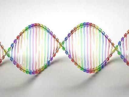
Some tests, called whole-exome sequencing, look at all the genes in your cancer. Others, called whole-genome sequencing, look at all the DNA (both genes and outside of genes) in your cancer. 

Still other biomarker tests look at the number of genetic changes in your cancer (what’s known as tumor mutational burden). This information can help figure out if a type of immunotherapy known as immune checkpoint inhibitors may work for you.

Biomarker tests known as liquid biopsies look in blood or other fluids for biomarkers from cancer cells. There are two liquid biopsy tests approved by the Food and Drug Administration (FDA), called Guardant360 CDx and FoundationOne Liquid CDx

What do the results of a biomarker test mean?

The results of a biomarker test could show that your cancer has a certain biomarker that is targeted by a known therapy. That means that the therapy may work to treat your cancer. The matching therapy may be available as an FDA-approved treatment, an off-label treatment, or through participation in a clinical trial. 

The results could also show that your cancer has a biomarker that may prevent a certain therapy from working. This information could spare you from getting a treatment that won’t help you.

In many cases, biomarker testing may find changes in your cancer that won’t help your doctor make treatment decisions. For example, genetic changes that are thought to be harmless (benign) or whose effects are not known (variant of unknown significance) are not used to make treatment decisions. 

Based on your test results, your health care provider may recommend a treatment that is not FDA approved for your cancer type, but is approved for the treatment of a different type of cancer that has the same biomarker as your cancer. This means the treatment would be used off label, but it may work for you because your cancer has the biomarker that the treatment targets.

Some biomarker tests can find genetic changes that you may have been born with (inherited) that increase your risk of cancer or other diseases. These genetic changes are also called germline mutations. If such a change is found, you may need to get another genetic test to confirm whether you truly have an inherited mutation that increases cancer risk

Finding out that you have an inherited mutation that increases cancer risk may affect you and your family. For that reason, your health care provider may recommend that you speak with a genetic healthcare provider (such as a genetic counselor, clinical geneticist, or a certified genetic nurse) to help you understand what the test results mean for you and your family.

Will biomarker testing for cancer treatment help me?

Biomarker tests don’t help everyone who gets them. There are several different reasons why they may not help you. Biomarker testing may not help you if:

  • you are unable to safely get a biopsy needed for testing.
  • there is not enough tumor tissue in your biopsy sample to have biomarker testing done.
  • the test doesn’t find any biomarkers in your cancer that match with available therapies.
  • the test identifies a matching therapy that would be used off label, and your insurance doesn’t cover the cost.
  • the test identifies a matching therapy that is being tested in a clinical trial, and you are not able to participate in the trial.

Even if your test finds a biomarker that matches an available treatment, the therapy may not work for you. Sometimes other features of your cancer or your body affect how well a treatment works, such as how the medicine is broken down in your body. 

Another reason the treatment might not work is that not all of your cancer cells have the same biomarkers. That means that a biomarker test may find a treatment that will kill some, but not all, of your cancer cells. Cancer cells that are not killed by the treatment could keep growing, preventing the treatment from working or causing the cancer to quickly come back.

One other reason biomarker tests might not help is because the biomarkers in your cancer can change over time. But a test only captures a “snapshot” of the changes at one point in time. So, the results of a biomarker test done in the past may not reflect the biomarkers in your cancer now. Your health care provider may want to test your cancer again, for example, if it comes back after treatment. 

How much does biomarker testing for cancer treatment cost?

The cost of biomarker testing varies widely depending on the type of test you get, the type of cancer you have, and your insurance plan. 

For people with advanced cancer, some biomarker tests are covered by Medicare and Medicaid. Private insurance providers often cover the cost of a biomarker test if there is enough proof that the test is required to guide treatment decisions. Tests without enough proof to support their value may be considered experimental and are likely not covered by insurance.

Many clinical trials involve biomarker testing. If you join one of these clinical trials, the cost of biomarker testing might be covered. The study coordinator can give you more information about related costs.
Radiation Therapy to Treat Cancer

Radiation Therapy to Treat Cancer

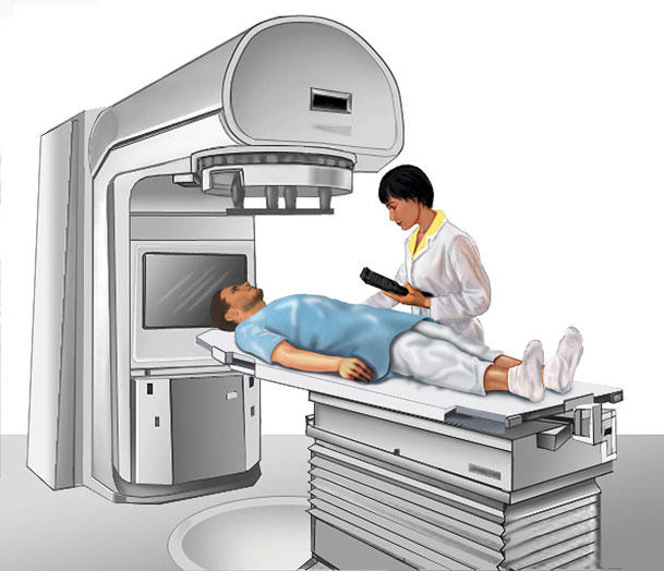
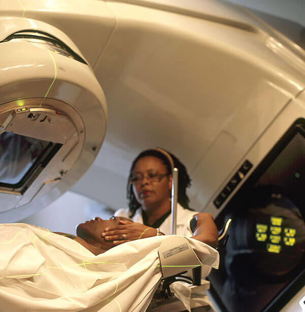
Technician positions a person lying on a treatment table for radiation therapy.

Radiation therapy kills cancer cells or slows their growth by damaging their DNA.

Credit: National Cancer Institute

Radiation therapy (also called radiotherapy) is a cancer treatment that uses high doses of radiation to kill cancer cells and shrink tumors. At low doses, radiation is used in x-rays to see inside your body, as with x-rays of your teeth or broken bones.

How radiation therapy works against cancer

At high doses, radiation therapy kills cancer cells or slows their growth by damaging their DNA. Cancer cells whose DNA is damaged beyond repair stop dividing or die. When the damaged cells die, they are broken down and removed by the body.

Radiation therapy does not kill cancer cells right away. It takes days or weeks of treatment before DNA is damaged enough for cancer cells to die. Then, cancer cells keep dying for weeks or months after radiation therapy ends.

Types of radiation therapy

There are two main types of radiation therapy, external beam and internal.

The type of radiation therapy that you may have depends on many factors, including:

  • the type of cancer
  • the size of the tumor
  • the tumor’s location in the body
  • how close the tumor is to normal tissues that are sensitive to radiation
  • your general health and medical history
  • whether you will have other types of cancer treatment
  • other factors, such as your age and other medical conditions

External beam radiation therapy

External beam radiation therapy comes from a machine that aims radiation at your cancer. The machine is large and may be noisy. It does not touch you, but can move around you, sending radiation to a part of your body from many directions.

External beam radiation therapy is a local treatment, which means it treats a specific part of your body. For example, if you have cancer in your lung, you will have radiation only to your chest, not to your whole body.

Learn more about external beam radiation therapy.

Internal radiation therapy

Internal radiation therapy is a treatment in which a source of radiation is put inside your body. The radiation source can be solid or liquid.

Internal radiation therapy with a solid source is called brachytherapy. In this type of treatment, seeds, ribbons, or capsules that contain a radiation source are placed in your body, in or near the tumor. Like external beam radiation therapy, brachytherapy is a local treatment and treats only a specific part of your body.

With brachytherapy, the radiation source in your body will give off radiation for a while.

Learn more about brachytherapy.

Internal radiation therapy with a liquid source is called systemic therapy. Systemic means that the treatment travels in the blood to tissues throughout your body, seeking out and killing cancer cells. You receive systemic radiation therapy by swallowing, through a vein via an IV line, or through an injection.

With systemic radiation, your body fluids, such as urine, sweat, and saliva, will give off radiation for a while.

Why people with cancer receive radiation therapy

Radiation therapy is used to treat cancer and ease cancer symptoms.

When used to treat cancer, radiation therapy can cure cancer, prevent it from returning, or stop or slow its growth.

When treatments are used to ease symptoms, they are known as palliative treatments. External beam radiation may shrink tumors to treat pain and other problems caused by the tumor, such as trouble breathing or loss of bowel and bladder control.

Pain from cancer that has spread to the bone can be treated with systemic radiation therapy drugs called radiopharmaceuticals.

Types of cancer that are treated with radiation therapy

External beam radiation therapy is used to treat many types of cancer.

Brachytherapy is most often used to treat cancers of the head and neck, breast, cervix, prostate, and eye.

A systemic radiation therapy called radioactive iodine, or I-131, is most often used to treat certain types of thyroid cancer.

Another type of systemic radiation therapy, called targeted radionuclide therapy, is used to treat some patients who have advanced prostate cancer or gastroenteropancreatic neuroendocrine tumor (GEP-NET). This type of treatment may also be referred to as molecular radiotherapy.

How radiation is used with other cancer treatments

For some people, radiation may be the only treatment you need. But, most often, you will have radiation therapy with other cancer treatments, such as surgerychemotherapy, and immunotherapy. Radiation therapy may be given before, during, or after these other treatments to improve the chances that treatment will work. The timing of when radiation therapy is given depends on the type of cancer being treated and whether the goal of radiation therapy is to treat the cancer or ease symptoms.

When radiation is combined with surgery, it can be given:

  • Before surgery, to shrink the size of the cancer so it can be removed by surgery and be less likely to return.
  • During surgery, so that it goes straight to the cancer without passing through the skin. Radiation therapy used this way is called intraoperative radiation. With this technique, doctors can more easily protect nearby normal tissues from radiation.
  • After surgery to kill any cancer cells that remain.

Lifetime dose limits

There is a limit to the amount of radiation an area of your body can safely receive over the course of your lifetime. Depending on how much radiation an area has already been treated with, you may not be able to have radiation therapy to that area a second time. But, if one area of the body has already received the safe lifetime dose of radiation, another area might still be treated if the distance between the two areas is large enough.

Radiation therapy can cause side effects

Radiation not only kills or slows the growth of cancer cells, it can also affect nearby healthy cells. Damage to healthy cells can cause side effects.

Learn more about the side effects of radiation therapy.

How much radiation therapy costs

Radiation therapy can be expensive. It uses complex machines and involves the services of many health care providers. The exact cost of your radiation therapy depends on the cost of health care where you live, what type of radiation therapy you get, and how many treatments you need.

Talk with your health insurance company about what services it will pay for. Most insurance plans pay for radiation therapy. To learn more, talk with the business office at the clinic or hospital where you go for treatment. If you need financial assistance, there are organizations that may be able to help. To find such organizations, go to the National Cancer Institute database, Organizations that Offer Support Services and search for “financial assistance.” Or call toll-free 1-800-4-CANCER (1-800-422-6237) to ask for information on organizations that may help.

Special diet needs while on radiation therapy

Radiation can cause side effects that make it hard to eat, such as nausea, mouth sores, and throat problems called esophagitis. Since your body uses a lot of energy to heal during radiation therapy, it is important that you eat enough calories and protein to maintain your weight during treatment.

If you are having trouble eating and maintaining your weight, talk to your doctor or nurse. You might also find it helpful to speak with a dietitian. For more information about coping with eating problems see the booklet Eating Hints or read more about side effects.

Working during radiation therapy

Some people are able to work full-time during radiation therapy. Others can work only part-time or not at all. How much you are able to work depends on how you feel. Ask your doctor or nurse what you may expect from the treatment you will have.

You are likely to feel well enough to work when you first start your radiation treatments. As time goes on, do not be surprised if you are more tired, have less energy, or feel weak. Once you have finished treatment, it may take just a few weeks for you to feel better—or it could take months.

You may get to a point during your radiation therapy when you feel too sick to work. Talk with your employer to find out if you can go on medical leave. Check that your health insurance will pay for treatment while you are on medical leave.

Childhood Salivary Gland Tumors (PDQ®)–Patient Version

Childhood Salivary Gland Tumors (PDQ®)–Patient Version

What are salivary gland tumors?

Salivary gland tumors are abnormal growths that can form in the salivary glands. They can be benign (not cancer) or malignant (cancerous). Although benign tumors do not spread to other parts of the body, they may require treatment to stop them from continuing to grow and press on nearby tissue. Cancerous tumors can spread to other areas of the body and will be treated to kill the cancer cells. Salivary gland tumors rarely occur in children.

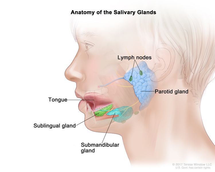
The salivary glands make saliva and release it into the mouth. Saliva has enzymes that help digest food and antibodies that help protect against mouth and throat infections. There are three pairs of major salivary glands:

  • Parotid glands are the largest glands and are found in front of and just below each ear. Most salivary gland tumors begin in this gland.
  • Sublingual glands are found under the tongue in the floor of the mouth.
  • Submandibular glands are found below the jawbone.
EnlargeAnatomy of the salivary glands; drawing shows a cross section of the head and the three main pairs of salivary glands. The parotid glands are in front of and just below each ear; the sublingual glands are under the tongue in the floor of the mouth; and the submandibular glands are below each side of the jawbone. The tongue and lymph nodes are also shown.
Anatomy of the salivary glands. The three main pairs of salivary glands are the parotid glands, the sublingual glands, and the submandibular glands.

Causes and risk factors for childhood salivary gland tumors

Salivary gland tumors in children are caused by certain changes to the way salivary gland cells function, especially how they grow and divide into new cells. Often, the exact cause of these cell changes is unknown. Learn more about how cancer develops at What Is Cancer?

A risk factor is anything that increases the chance of getting a disease. Past treatment for cancer with chemotherapy or radiation therapy is a risk factor for childhood salivary gland tumors. Not every child with this risk factor will develop a salivary gland tumor. And it will develop in some children who don’t have a known risk factor. Talk with your child’s doctor if you think your child may be at risk.

Symptoms of childhood salivary gland tumors

Children may not have symptoms of a salivary gland tumor until the tumor has grown bigger. It’s important to check with your child’s doctor if your child has:

  • a lump near the ear, cheek, jaw, lip, or inside the mouth that may be painless
  • numbness or weakness in the face
  • pain in the face that does not go away

These symptoms may be caused by problems other than a salivary gland tumor. The only way to know is to see your child’s doctor.

Tests to diagnose childhood salivary gland tumors

If your child has symptoms that suggest a salivary gland tumor, the doctor will need to find out if these are due to a salivary gland tumor or another problem. The doctor will ask when the symptoms started and how often your child has been having them. They will also ask about your child’s personal and family medical history and do a physical exam. Depending on these results, they may recommend other tests. If your child is diagnosed with a salivary gland tumor, the results of these tests will help you and your child’s doctor plan treatment.

The tests used to diagnose salivary gland tumors in children may include:

Magnetic resonance imaging (MRI)

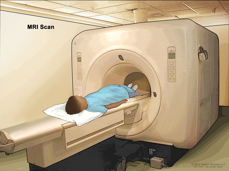
MRI uses a magnet, radio waves, and a computer to make a series of detailed pictures of areas of the body, such as the head and neck. This procedure is also called nuclear magnetic resonance imaging (NMRI).

EnlargeMagnetic resonance imaging (MRI) scan; drawing shows a child lying on a table that slides into the MRI machine, which takes a series of detailed pictures of areas inside the body.
Magnetic resonance imaging (MRI) scan. The child lies on a table that slides into the MRI machine, which takes a series of detailed pictures of areas inside the body. The positioning of the child on the table depends on the part of the body being imaged.

CT scan (CAT scan)

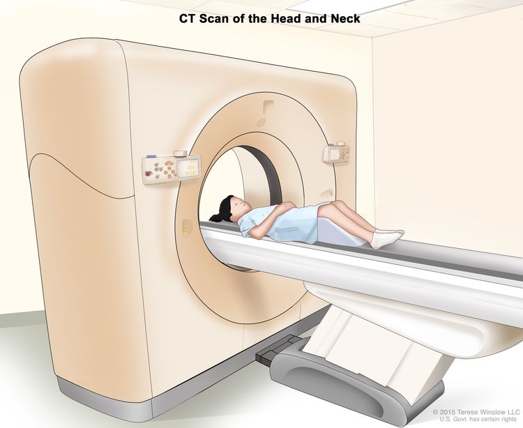
A CT scan uses a computer linked to an x-ray machine to make a series of detailed pictures of areas inside the body. The pictures are taken from different angles and are used to create 3-D views of tissues and organs. A dye may be injected into a vein or swallowed to help the organs or tissues show up more clearly. This procedure is also called computed tomography, computerized tomography, or computerized axial tomography. Learn more about Computed Tomography (CT) Scans and Cancer.

EnlargeComputed tomography (CT) scan of the head and neck; drawing shows a child lying on a table that slides through the CT scanner, which takes a series of detailed x-ray pictures of the inside of the head and neck.
Computed tomography (CT) scan of the head and neck. The child lies on a table that slides through the CT scanner, which takes a series of detailed x-ray pictures of the inside of the head and neck.

Ultrasound exam

An ultrasound exam uses high-energy sound waves (ultrasound) which bounce off internal tissue or organs, such as the pelvis, and make echoes. The echoes form a picture of body tissues called a sonogram.

Biopsy

A biopsy is a procedure in which a sample of tissue is removed from the tumor so that a pathologist can view it under a microscope to check for signs of cancer. Fine-needle aspiration biopsy is used to check for salivary gland cancer. During a fine-needle aspiration biopsy, tissue or fluid is removed using a thin needle.

Getting a second opinion

You may want to get a second opinion to confirm your child’s diagnosis and treatment plan. If you seek a second opinion, you will need to get medical test results and reports from the first doctor to share with the second doctor. The second doctor will review the pathology report, slides, and scans. This doctor may agree with the first doctor, suggest changes to the treatment plan, or provide more information about your child’s tumor.

To learn more about choosing a doctor and getting a second opinion, see Finding Cancer Care. You can contact NCI’s Cancer Information Service via chat, email, or phone (both in English and Spanish) for help finding a doctor or hospital that can provide a second opinion. For questions you might want to ask at your child’s appointments, see Questions to Ask Your Doctor about Cancer.

Who treats children with salivary gland tumors?

A pediatric oncologist, a doctor who specializes in treating children with cancer, oversees treatment of salivary gland tumors. The pediatric oncologist works with other health care providers who are experts in treating children with cancer and who specialize in certain areas of medicine. Other specialists may include:

Treatment of childhood salivary gland tumors

There are different types of treatment for children and adolescents with salivary gland tumors. You and your child’s care team will work together to decide treatment. Many factors will be considered, such as your child’s overall health and whether the tumor is newly diagnosed or has come back.

Your child’s treatment plan will include information about the tumor, the goals of treatment, treatment options, and the possible side effects. It will be helpful to talk with your child’s care team before treatment begins about what to expect. For help every step of the way, see our booklet, Children with Cancer: A Guide for Parents.

Treatment of salivary gland tumors may include:

  • Surgery to remove salivary gland tumors or salivary gland cancer is the most common treatment. If the parotid gland is removed during surgery, special care must be taken to avoid damage to the facial nerve. Learn more about Surgery to Treat Cancer.
  • Radiation therapy may be given after surgery for salivary gland cancer. Radiation therapy uses high-energy x-rays or other types of radiation to kill cancer cells or keep them from growing. Salivary gland cancer may be treated with external beam radiation therapy or internal radiation therapy if the cancer is likely to spread.
    • External beam radiation therapy uses a machine outside the body to send radiation toward the area of the body with cancer.
    • Internal radiation therapy, also called brachytherapy, uses a radioactive substance sealed in needles, seeds, wires, or catheters that are placed directly into or near the cancer.

    Learn more about Radiation Therapy to Treat Cancer and Radiation Therapy Side Effects.

  • Targeted therapy (entrectinib or larotrectinib) may be used to treat recurrent childhood salivary gland cancer. Recurrent salivary gland cancer is cancer that has come back after it has been treated. Targeted therapy uses drugs or other substances to block the action of specific enzymes, proteins, or other molecules involved in the growth and spread of cancer cells. The doctor may suggest biomarker tests to help predict your child’s response to certain targeted therapy drugs. Learn more about Biomarker Testing for Cancer Treatment and Targeted Therapy to Treat Cancer.

Clinical trials

For some children, joining a clinical trial may be an option. There are different types of clinical trials for childhood cancer. For example, a treatment trial tests new treatments or new ways of using current treatments. Supportive care and palliative care trials look at ways to improve quality of life, especially for those who have side effects from cancer and its treatment.

You can use the clinical trial search to find NCI-supported cancer clinical trials accepting participants. The search allows you to filter trials based on the type of cancer, your child’s age, and where the trials are being done. Clinical trials supported by other organizations can be found on the ClinicalTrials.gov website.

Learn more about clinical trials, including how to find and join one, at Clinical Trials Information for Patients and Caregivers.

Prognosis and prognostic factors for childhood salivary gland tumors

If your child has been diagnosed with salivary gland cancer, you likely have questions about how serious the cancer is and your child’s chances of survival. The likely outcome or course of a disease is called prognosis. The prognosis can be affected by whether the cancer has spread to lymph nodes or other parts of the body at the time of diagnosis and whether the cancer can be completely removed by surgery. The prognosis for salivary gland cancer in children is usually good.

No two people are alike, and responses to treatment can vary greatly. Your child’s cancer care team is in the best position to talk with you about your child’s prognosis.

Side effects and late effects of treatment

Cancer treatments can cause side effects. Which side effects your child might have depends on the type of treatment they receive, the dose, and how their body reacts. Talk with your child’s treatment team about which side effects to look for and ways to manage them.

To learn more about side effects that begin during treatment for cancer, visit Side Effects.

Problems from cancer treatment that begin 6 months or later after treatment and continue for months or years are called late effects. Late effects of cancer treatment may include:

  • physical problems, such as:
    • dry mouth
    • trouble seeing
    • numbness or weakness in the face
    • changes in the way the bones of the head and face grow
    • other changes in the child’s appearance
  • changes in mood, feelings, thinking, learning, or memory
  • second cancers (new types of cancer)

Some late effects may be treated or controlled. It is important to talk with your child’s doctors about the possible late effects caused by some treatments. Learn more about Late Effects of Treatment for Childhood Cancer.

Follow-up care

As your child goes through treatment, they will have follow-up tests or check-ups. Some of the tests that were done to diagnose the tumor may be repeated to see how well the treatment is working. Decisions about whether to continue, change, or stop treatment may be based on the results of these tests.

Some of the tests will continue to be done from time to time after treatment has ended. The results of these tests can show if your child’s condition has changed or if the tumor has recurred (come back).

Coping with your child's cancer

When a child has a tumor, every member of the family needs support. Taking care of yourself during this difficult time is important. Reach out to your child’s treatment team and to people in your family and community for support. To learn more, see Support for Families: Childhood Cancer and the booklet Children with Cancer: A Guide for Parents.

Related resources

About This PDQ Summary

About PDQ

Physician Data Query (PDQ) is the National Cancer Institute’s (NCI’s) comprehensive cancer information database. The PDQ database contains summaries of the latest published information on cancer prevention, detection, genetics, treatment, supportive care, and complementary and alternative medicine. Most summaries come in two versions. The health professional versions have detailed information written in technical language. The patient versions are written in easy-to-understand, nontechnical language. Both versions have cancer information that is accurate and up to date and most versions are also available in Spanish.

PDQ is a service of the NCI. The NCI is part of the National Institutes of Health (NIH). NIH is the federal government’s center of biomedical research. The PDQ summaries are based on an independent review of the medical literature. They are not policy statements of the NCI or the NIH.

Purpose of This Summary

This PDQ cancer information summary has current information about the treatment of childhood salivary gland tumors. It is meant to inform and help patients, families, and caregivers. It does not give formal guidelines or recommendations for making decisions about health care.

Reviewers and Updates

Editorial Boards write the PDQ cancer information summaries and keep them up to date. These Boards are made up of experts in cancer treatment and other specialties related to cancer. The summaries are reviewed regularly and changes are made when there is new information. The date on each summary (“Updated”) is the date of the most recent change.

The information in this patient summary was taken from the health professional version, which is reviewed regularly and updated as needed, by the PDQ Pediatric Treatment Editorial Board.

Clinical Trial Information

A clinical trial is a study to answer a scientific question, such as whether one treatment is better than another. Trials are based on past studies and what has been learned in the laboratory. Each trial answers certain scientific questions in order to find new and better ways to help cancer patients. During treatment clinical trials, information is collected about the effects of a new treatment and how well it works. If a clinical trial shows that a new treatment is better than one currently being used, the new treatment may become “standard.” Patients may want to think about taking part in a clinical trial. Some clinical trials are open only to patients who have not started treatment.

Clinical trials can be found online at NCI’s website. For more information, call the Cancer Information Service (CIS), NCI’s contact center, at 1-800-4-CANCER (1-800-422-6237).

Permission to Use This Summary

PDQ is a registered trademark. The content of PDQ documents can be used freely as text. It cannot be identified as an NCI PDQ cancer information summary unless the whole summary is shown and it is updated regularly. However, a user would be allowed to write a sentence such as “NCI’s PDQ cancer information summary about breast cancer prevention states the risks in the following way: [include excerpt from the summary].”

The best way to cite this PDQ summary is:

PDQ® Pediatric Treatment Editorial Board. PDQ Childhood Salivary Gland Tumors. Bethesda, MD: National Cancer Institute. Updated <MM/DD/YYYY>. Available at: /types/head-and-neck/patient/child/salivary-gland-treatment-pdq. Accessed <MM/DD/YYYY>.

Images in this summary are used with permission of the author(s), artist, and/or publisher for use in the PDQ summaries only. If you want to use an image from a PDQ summary and you are not using the whole summary, you must get permission from the owner. It cannot be given by the National Cancer Institute. Information about using the images in this summary, along with many other images related to cancer can be found in Visuals Online. Visuals Online is a collection of more than 3,000 scientific images.

Disclaimer

The information in these summaries should not be used to make decisions about insurance reimbursement. More information on insurance coverage is available on Cancer.gov on the Managing Cancer Care page.

Contact Us

More information about contacting us or receiving help with the Cancer.gov website can be found on our Contact Us for Help page. Questions can also be submitted to Cancer.gov through the website’s E-mail Us.

Childhood Oral Cavity Cancer (PDQ®)–Patient Version

Childhood Oral Cavity Cancer (PDQ®)–Patient Version

What is childhood oral cavity cancer?

Childhood oral cavity cancer is a rare type of cancer that forms in the mouth. There are several types of oral cavity cancers, including lymphoma, sarcoma, squamous cell carcinoma, and mucoepidermoid carcinoma. Most of the tumors (more than 90%) that children get in their mouth are not cancer. All types of tumors in the mouth can affect eating or speaking and need treatment.

The oral cavity includes:

  • the front two-thirds of the tongue
  • the gums (gingiva)
  • the lining of the inside of the cheeks (buccal mucosa)
  • the bottom (floor) of the mouth under the tongue
  • the roof of the mouth (hard palate)
  • the small area behind the wisdom teeth (retromolar trigone)
EnlargeAnatomy of the oral cavity; drawing shows the lip, hard palate, soft palate, retromolar trigone, front two-thirds of the tongue, gingiva, buccal mucosa, and floor of mouth. Also shown are the teeth, uvula, and tonsil.
Anatomy of the oral cavity. The oral cavity includes the lips, hard palate (the bony front portion of the roof of the mouth), soft palate (the muscular back portion of the roof of the mouth), retromolar trigone (the area behind the wisdom teeth), front two-thirds of the tongue, gingiva (gums), buccal mucosa (the inner lining of the lips and cheeks), and floor of the mouth under the tongue.

Causes and risk factors for childhood oral cavity cancer

Oral cavity cancer is caused by certain changes to the way the cells in the oral cavity function, especially how they grow and divide into new cells. Often, the exact cause of these cell changes is unknown. Learn more about how cancer develops at What Is Cancer?

A risk factor is anything that increases the chance of getting a disease. Not every child with one or more of these risk factors will develop oral cavity cancer. And it will develop in some children who don’t have a known risk factor.

Risk factors for oral cavity cancer in children and adolescents include:

Getting the HPV vaccine can protect against HPV infection and lower the risk of this and many other types of cancer. Learn more about HPV and Cancer.

Talk with your child’s doctor if you think your child may be at risk.

Symptoms of childhood oral cavity cancer

The symptoms of oral cavity cancer may be similar to an infection. It’s important to check with your child’s doctor if your child has:

  • a sore in the mouth that does not heal
  • a lump or thickening in the mouth
  • a white or red patch on the gums, tongue, or lining of the mouth
  • bleeding or pain in the mouth

These symptoms may be caused by problems other than oral cavity cancer. The only way to know is to see your child’s doctor.

Tests to diagnose childhood oral cavity cancer

If your child has symptoms that suggest an oral cavity cancer, the doctor will need to find out if these are due to cancer or another problem. The doctor will ask when the symptoms started and how often your child has been having them. They will also ask about your child’s personal and family health history and do a physical exam. Depending on these results, they may recommend other tests. If your child is diagnosed with oral cavity cancer, the results of these tests will help you and your child’s doctor plan treatment.

The tests used to diagnose oral cavity cancer may include:

Oral exam

In an oral exam, a medical doctor or dentist checks the mouth for abnormal areas. This exam may be done while under anesthesia. The doctor or dentist will feel the entire inside of the mouth with a gloved finger and examine the oral cavity with a small long-handled mirror and lights or a fiberoptic device. This will include checking the insides of the cheeks and lips; the gums; the roof and floor of the mouth; and the top, bottom, and sides of the tongue. The neck will be felt for swollen lymph nodes. A dental exam may also be done.

X-ray

An x-ray is a type of radiation that can go through the body and make pictures of areas inside the body.

Magnetic resonance imaging (MRI)

MRI uses a magnet, radio waves, and a computer to make a series of detailed pictures of areas of the body, such as the head and neck. This procedure is also called nuclear magnetic resonance imaging (NMRI).

EnlargeMagnetic resonance imaging (MRI) scan; drawing shows a child lying on a table that slides into the MRI machine, which takes a series of detailed pictures of areas inside the body.
Magnetic resonance imaging (MRI) scan. The child lies on a table that slides into the MRI machine, which takes a series of detailed pictures of areas inside the body. The positioning of the child on the table depends on the part of the body being imaged.

CT scan (CAT scan)

A CT scan uses a computer linked to an x-ray machine to make a series of detailed pictures of areas inside the body. The pictures are taken from different angles and are used to create 3-D views of tissues and organs. A dye may be injected into a vein or swallowed to help the tissues or organs show up more clearly. This procedure is also called computed tomography, computerized tomography, or computerized axial tomography. To learn more, see Computed Tomography (CT) Scans and Cancer.

EnlargeComputed tomography (CT) scan of the head and neck; drawing shows a child lying on a table that slides through the CT scanner, which takes a series of detailed x-ray pictures of the inside of the head and neck.
Computed tomography (CT) scan of the head and neck. The child lies on a table that slides through the CT scanner, which takes a series of detailed x-ray pictures of the inside of the head and neck.

PET scan (positron emission tomography scan)

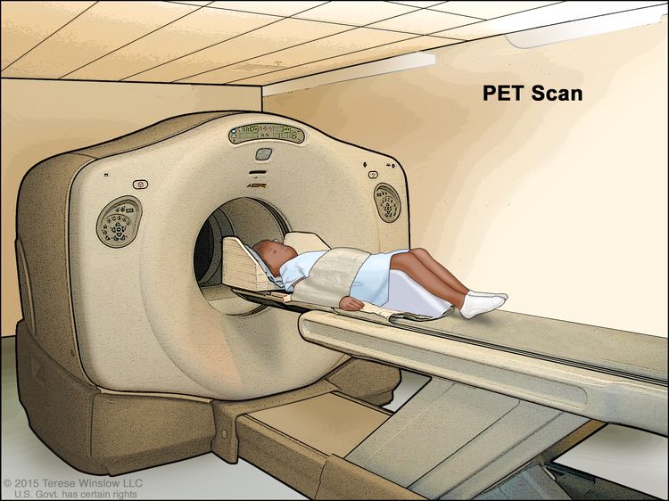
A PET scan uses a small amount of radioactive sugar (also called radioactive glucose) that is injected into the vein. Then a scanner is used to make detailed, computerized pictures of areas inside the body where the glucose is taken up. Because cancer cells often take up more glucose than normal cells, the pictures can be used to find cancer cells in the body.

EnlargePositron emission tomography (PET) scan; drawing shows a child lying on table that slides through the PET scanner.
Positron emission tomography (PET) scan. The child lies on a table that slides through the PET scanner. The head rest and white strap help the child lie still. A small amount of radioactive glucose (sugar) is injected into the child’s vein, and a scanner makes a picture of where the glucose is being used in the body. Cancer cells show up brighter in the picture because they take up more glucose than normal cells do.

Biopsy

A biopsy is a procedure in which a sample of tissue is removed from the tumor so that a pathologist can view it under a microscope to check for signs of cancer.

  • Fine-needle aspiration biopsy is the removal of cells, tissue or fluid using a thin needle.
  • Incisional biopsy is the surgical removal of part of a lump or a sample of tissue that doesn’t look normal.

Getting a second opinion

You may want to get a second opinion to confirm your child’s cancer diagnosis and treatment plan. If you seek a second opinion, you will need to get medical test results and reports from the first doctor to share with the second doctor. The second doctor will review the pathology report, slides, and scans. This doctor may agree with the first doctor, suggest changes to the treatment plan, or provide more information about your child’s cancer.

To learn more about choosing a doctor and getting a second opinion, see Finding Cancer Care. You can contact NCI’s Cancer Information Service via chat, email, or phone (both in English and Spanish) for help finding a doctor or hospital that can provide a second opinion. For questions you might want to ask at your child’s appointments, see Questions to Ask Your Doctor about Cancer.

Who treats children with oral cavity cancer?

A pediatric oncologist, a doctor who specializes in treating children with cancer, oversees treatment of oral cavity cancer. The pediatric oncologist works with other health care providers who are experts in treating children with cancer and who specialize in certain areas of medicine. Other specialists may include:

Treatment of childhood oral cavity cancer

There are different types of treatment for children with oral cavity cancer. You and your child’s care team will work together to decide treatment. Many factors will be considered, such as where the cancer is located and your child’s age and overall health.

Your child’s treatment plan will include information about the cancer, the goals of treatment, treatment options, and the possible side effects. It will be helpful to talk with your child’s care team before treatment begins about what to expect. For help every step of the way, see our booklet, Children with Cancer: A Guide for Parents.

For oral cavity cancer in children, treatment might include:

  • Surgery is the most common treatment for oral cavity cancer in children. For children with an oral cavity tumor that is not cancer, surgery is likely the only treatment that will be needed. Learn more about Surgery to Treat Cancer.
  • Chemotherapy (also called chemo) uses drugs to stop the growth of cancer cells, either by killing the cells or by stopping them from dividing. Chemotherapy may be given with other types of treatments.

    Chemotherapy can be given in different ways. For oral cavity cancer, chemotherapy is injected into a vein, enabling it to reach cancer cells throughout the body. Learn more about Chemotherapy to Treat Cancer.

  • Radiation therapy uses high-energy x-rays or other types of radiation to kill cancer cells or keep them from growing. The type of radiation therapy most often used for oral cavity cancer is external beam radiation therapy. This treatment uses a machine outside the body to send radiation toward the area of the body with cancer. Learn more about External Beam Radiation Therapy for Cancer and Radiation Therapy Side Effects.

Clinical trials

For some children, joining a clinical trial may be an option. There are different types of clinical trials for childhood cancer. For example, a treatment trial tests new treatments or new ways of using current treatments. Supportive care and palliative care trials look at ways to improve quality of life, especially for those who have side effects from cancer and its treatment.

You can use the clinical trial search to find NCI-supported cancer clinical trials accepting participants. The search allows you to filter trials based on the type of cancer, your child’s age, and where the trials are being done. Clinical trials supported by other organizations can be found on the ClinicalTrials.gov website.

Learn more about clinical trials, including how to find and join one, at Clinical Trials Information for Patients and Caregivers.

Follow-up care

As your child goes through treatment, they will have follow-up tests or check-ups. Some of the tests that were done to diagnose the cancer may be repeated to see how well the treatment is working. Decisions about whether to continue, change, or stop treatment may be based on the results of these tests.

Some of the tests will continue to be done from time to time after treatment has ended. The results of these tests can show if your child’s condition changes or if the cancer has come back.

Coping with your child's cancer

When your child has cancer, every member of the family needs support. Taking care of yourself during this difficult time is important. Reach out to your child’s treatment team and to people in your family and community for support. To learn more, see Support for Families: Childhood Cancer and the booklet Children with Cancer: A Guide for Parents.

Related resources

About This PDQ Summary

About PDQ

Physician Data Query (PDQ) is the National Cancer Institute’s (NCI’s) comprehensive cancer information database. The PDQ database contains summaries of the latest published information on cancer prevention, detection, genetics, treatment, supportive care, and complementary and alternative medicine. Most summaries come in two versions. The health professional versions have detailed information written in technical language. The patient versions are written in easy-to-understand, nontechnical language. Both versions have cancer information that is accurate and up to date and most versions are also available in Spanish.

PDQ is a service of the NCI. The NCI is part of the National Institutes of Health (NIH). NIH is the federal government’s center of biomedical research. The PDQ summaries are based on an independent review of the medical literature. They are not policy statements of the NCI or the NIH.

Purpose of This Summary

This PDQ cancer information summary has current information about the treatment of childhood oral cavity cancer. It is meant to inform and help patients, families, and caregivers. It does not give formal guidelines or recommendations for making decisions about health care.

Reviewers and Updates

Editorial Boards write the PDQ cancer information summaries and keep them up to date. These Boards are made up of experts in cancer treatment and other specialties related to cancer. The summaries are reviewed regularly and changes are made when there is new information. The date on each summary (“Updated”) is the date of the most recent change.

The information in this patient summary was taken from the health professional version, which is reviewed regularly and updated as needed, by the PDQ Pediatric Treatment Editorial Board.

Clinical Trial Information

A clinical trial is a study to answer a scientific question, such as whether one treatment is better than another. Trials are based on past studies and what has been learned in the laboratory. Each trial answers certain scientific questions in order to find new and better ways to help cancer patients. During treatment clinical trials, information is collected about the effects of a new treatment and how well it works. If a clinical trial shows that a new treatment is better than one currently being used, the new treatment may become “standard.” Patients may want to think about taking part in a clinical trial. Some clinical trials are open only to patients who have not started treatment.

Clinical trials can be found online at NCI’s website. For more information, call the Cancer Information Service (CIS), NCI’s contact center, at 1-800-4-CANCER (1-800-422-6237).

Permission to Use This Summary

PDQ is a registered trademark. The content of PDQ documents can be used freely as text. It cannot be identified as an NCI PDQ cancer information summary unless the whole summary is shown and it is updated regularly. However, a user would be allowed to write a sentence such as “NCI’s PDQ cancer information summary about breast cancer prevention states the risks in the following way: [include excerpt from the summary].”

The best way to cite this PDQ summary is:

PDQ® Pediatric Treatment Editorial Board. PDQ Childhood Oral Cavity Cancer. Bethesda, MD: National Cancer Institute. Updated <MM/DD/YYYY>. Available at: /types/head-and-neck/patient/child/oral-cavity-treatment-pdq. Accessed <MM/DD/YYYY>.

Images in this summary are used with permission of the author(s), artist, and/or publisher for use in the PDQ summaries only. If you want to use an image from a PDQ summary and you are not using the whole summary, you must get permission from the owner. It cannot be given by the National Cancer Institute. Information about using the images in this summary, along with many other images related to cancer can be found in Visuals Online. Visuals Online is a collection of more than 3,000 scientific images.

Disclaimer

The information in these summaries should not be used to make decisions about insurance reimbursement. More information on insurance coverage is available on Cancer.gov on the Managing Cancer Care page.

Contact Us

More information about contacting us or receiving help with the Cancer.gov website can be found on our Contact Us for Help page. Questions can also be submitted to Cancer.gov through the website’s E-mail Us.

Childhood Thyroid Cancer (PDQ®)–Patient Version

Childhood Thyroid Cancer (PDQ®)–Patient Version

What is childhood thyroid cancer?

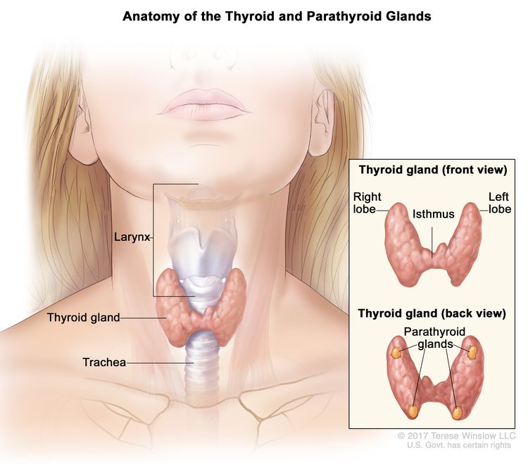
Thyroid cancer in children is a rare cancer that forms in the thyroid, a butterfly-shaped gland at the base of the throat near the windpipe (also called the trachea). The thyroid gland has a right lobe and a left lobe that is connected by a thin piece of tissue called the isthmus. Although thyroid cancer can affect children of all ages, it’s more common in adolescents and girls. It also occurs more often in White people than in Black people.

Children can also have thyroid nodules. A thyroid nodule is an abnormal growth of thyroid cells in the thyroid. The nodules may be solid or fluid-filled. Thyroid nodules usually don’t cause symptoms or need treatment unless they become large enough that it is hard to swallow or breathe. Only one in five thyroid nodules become cancer.

EnlargeAnatomy of the thyroid and parathyroid glands; drawing shows the thyroid gland at the base of the throat near the trachea. An inset shows the front and back views. The front view shows that the thyroid is shaped like a butterfly, with the right lobe and left lobe connected by a thin piece of tissue called the isthmus. The back view shows the four pea-sized parathyroid glands. The larynx is also shown.
Anatomy of the thyroid and parathyroid glands. The thyroid gland lies at the base of the throat near the trachea. It is shaped like a butterfly, with the right lobe and left lobe connected by a thin piece of tissue called the isthmus. The parathyroid glands are four pea-sized organs found in the neck near the thyroid. The thyroid and parathyroid glands make hormones.

The thyroid uses iodine, a mineral found in some foods and in iodized salt, to help make several hormones. Thyroid hormones:

  • control heart rate, body temperature, and how quickly food is changed into energy (metabolism)
  • control the amount of calcium in the blood

Types of childhood thyroid cancer

There are four types of thyroid cancers:

Papillary and follicular thyroid cancer are sometimes called differentiated thyroid cancer. Medullary and anaplastic thyroid cancer are sometimes called poorly differentiated or undifferentiated thyroid cancer.

Adenomas are a type of thyroid nodule that can grow very large and sometimes make hormones. Adenomas are not cancer but may rarely become cancer and spread to the lungs or lymph nodes in the neck.

Causes and risk factors for childhood thyroid cancer

Thyroid cancer in children is caused by certain changes to the way thyroid cells function, especially how they grow and divide into new cells. Often, the exact cause of these cell changes is unknown. Learn more about how cancer develops at What Is Cancer?

A risk factor is anything that increases the chance of getting a disease. Not every child with one or more of these risk factors will develop thyroid cancer. And it will develop in some children who don’t have a known risk factor.

Risk factors for childhood thyroid cancer include:

Talk with your child’s doctor if you think your child may be at risk for thyroid cancer.

Genetic counseling for children with thyroid cancer

It is not always clear from the family medical history whether a condition is inherited. Genetic counseling can assess the likelihood that your child’s cancer is inherited and whether genetic testing is needed. Genetic testing may help explain why a child develops a rare cancer or a cancer that is usually seen in adults. Genetic counselors and other specially trained health professionals can discuss your child’s diagnosis and your family’s medical history to help you understand:

  • the options for testing for changes in the RET, DICER1, or APC gene
  • the risk of other cancers for your child
  • the risk of thyroid cancer or other cancers for your child’s siblings
  • the risks and benefits of learning genetic information

Genetic counselors can also help you cope with your child’s genetic testing results, including how to discuss the results with family members. They can advise you about whether other members of your family should receive genetic testing.

If your child has been found to have a change in the RET gene, other family members may also be tested to find out if they have an increased risk of medullary thyroid cancer. Family members, including young children, who have the changed gene may have a thyroidectomy (surgery to remove the thyroid). This can decrease the chance of developing medullary thyroid cancer.

Learn more about Genetic Testing for Inherited Cancer Risk.

Symptoms of childhood thyroid cancer

Often thyroid cancer is found when a physical exam or an imaging test is done for another health problem. Sometimes the cancer can cause symptoms. It’s important to check with your child’s doctor if your child has:

  • a lump in the neck
  • trouble breathing
  • trouble swallowing
  • hoarseness or a change in the voice

Children with changes to the RET gene may have symptoms that can lead to an early diagnosis of multiple endocrine neoplasia type 2A or 2B syndrome. It’s important to check with your child’s doctor if your child has:

  • bumps on the lips, tongue, or eyelids that do not hurt
  • trouble making tears
  • constipation
  • Marfan syndrome (being tall and thin, with long arms, legs, fingers, and toes)

These symptoms may be caused by problems other than thyroid cancer. The only way to know is to see your child’s doctor.

Tests to diagnose childhood thyroid cancer

If your child has symptoms that suggest thyroid cancer, the doctor will need to find out if these are due to cancer or to another problem. The doctor will ask when the symptoms started and how often your child has been having them. They will also ask about your child’s personal and family medical history and do a physical exam. Depending on these results, they may recommend other tests. If your child is diagnosed with thyroid cancer, the results of these tests will help you and your child’s doctor plan treatment.

The tests used to diagnose thyroid cancer may include:

Thyroid function test

A thyroid function test checks the blood for abnormal levels of thyroid-stimulating hormone (TSH). TSH is made by the pituitary gland in the brain. It stimulates the release of thyroid hormone and controls how fast follicular thyroid cells grow. The blood may also be checked for high levels of calcitonin (a hormone made by the thyroid that decreases the amount of calcium in the blood).

Thyroglobulin test

A thyroglobulin test checks the blood for the amount of thyroglobulin, a protein made by the thyroid gland. Thyroglobulin levels are low or absent with normal thyroid function but may be higher with thyroid cancer or other conditions.

RET, DICER1, or APC genetic testing

A RET, DICER1, or APC genetic test checks a sample of blood, saliva, or tissue for certain changes in the RET, DICER1, or APC genes.

Ultrasound exam

An ultrasound exam uses high-energy sound waves (ultrasound) that bounce off internal tissues or organs in the neck and make echoes. The echoes form a picture of body tissues called a sonogram. This procedure can show the size of the mass and whether it is solid or a fluid-filled cyst. Ultrasound may be used to guide a fine-needle aspiration biopsy. A complete ultrasound exam of the neck is done before surgery.

Thyroid scan

A thyroid scan uses a small amount of a radioactive substance that is swallowed or injected. The radioactive material collects in thyroid gland cells. A special camera linked to a computer detects the radiation given off and makes pictures that show how the thyroid looks and functions and whether the cancer has spread beyond the thyroid gland. If the amount of TSH in the child’s blood is low, a scan to make images of the thyroid may be done before surgery.

CT scan (CAT scan)

A CT scan uses a computer linked to an x-ray machine to make a series of detailed pictures of areas inside the body, such as the neck, chest, abdomen, and brain. The pictures are taken from different angles and are used to create 3-D views of tissues and organs. A dye may be injected into a vein or swallowed to help the organs or tissues show up more clearly. This procedure is also called computed tomography, computerized tomography, or computerized axial tomography. Learn more about Computed Tomography (CT) Scans and Cancer.

EnlargeComputed tomography (CT) scan of the head and neck; drawing shows a child lying on a table that slides through the CT scanner, which takes a series of detailed x-ray pictures of the inside of the head and neck.
Computed tomography (CT) scan of the head and neck. The child lies on a table that slides through the CT scanner, which takes a series of detailed x-ray pictures of the inside of the head and neck.

Magnetic resonance imaging (MRI) with gadolinium

MRI uses a magnet, radio waves, and a computer to make a series of detailed pictures of areas inside the body, such as the neck and chest. A substance called gadolinium is injected into a vein. The gadolinium collects around the cancer cells so they show up brighter in the picture. This procedure is also called nuclear magnetic resonance imaging (NMRI).

Chest x-ray

An x-ray is a type of radiation that can go through the body and make pictures. A chest x-ray makes pictures of the organs and bones inside the chest.

Biopsy

A biopsy is a procedure in which a sample of tissue is removed from the tumor so that a pathologist can view it under a microscope to check for signs of cancer.

  • Fine-needle aspiration biopsy is the removal of thyroid tissue using a thin needle. The needle is inserted through the skin into the thyroid. Several tissue samples are removed from different parts of the thyroid. A pathologist views the tissue samples under a microscope to look for cancer cells. If it is not clear whether cancer is present, a surgical biopsy may be done.
  • Surgical biopsy is the removal of the thyroid nodule or one lobe of the thyroid during surgery so the cells and tissues can be viewed under a microscope by a pathologist to check for signs of cancer.

Whole-body thyroid scan

A whole-body thyroid scan uses a small amount of radioactive substance that is swallowed or injected. The radioactive material collects in any thyroid tissue or cancer cells remaining after surgery. Radioactive iodine is used because only thyroid cells take up iodine. A special camera detects the radiation given off by the thyroid tissue or cancer cells. This procedure is also called radioactive iodine scan or RAI scan.

Getting a second opinion

You may want to get a second opinion to confirm your child’s cancer diagnosis and treatment plan. If you seek a second opinion, you will need to get medical test results and reports from the first doctor to share with the second doctor. The second doctor will review the genetic test report, pathology report, slides, and scans. This doctor may agree with the first doctor, suggest changes to the treatment plan, or provide more information about your child’s cancer.

To learn more about choosing a doctor and getting a second opinion, see Finding Cancer Care. You can contact NCI’s Cancer Information Service via chat, email, or phone (both in English and Spanish) for help finding a doctor or hospital that can provide a second opinion. For questions you might want to ask at your child’s appointments, see Questions to Ask Your Doctor about Cancer.

Types of treatment for childhood thyroid cancer

Who treats children with thyroid cancer?

A pediatric oncologist, a doctor who specializes in treating children with cancer, oversees treatment of thyroid cancer. The pediatric oncologist works with other health care providers who are experts in treating children with cancer and who specialize in certain areas of medicine. Other specialists may include:

There are different types of treatment for children and adolescents with thyroid cancer. You and your child’s cancer care team will work together to decide treatment. Many factors will be considered, such as your child’s overall health and whether the tumor is newly diagnosed or has come back.

Your child’s treatment plan will include information about the cancer, the goals of treatment, treatment options, and the possible side effects. It will be helpful to talk with your child’s cancer care team before treatment begins about what to expect. For help every step of the way, see our booklet, Children with Cancer: A Guide for Parents.

Treatment for thyroid cancer might include:

Surgery

Surgery is the most common treatment for thyroid cancer. One of the following procedures may be used:

  • Total thyroidectomy is the removal of the whole thyroid. Lymph nodes near the cancer may also be removed and checked under a microscope for signs of cancer.
  • Near-total thyroidectomy is the removal of all but a very small part of the thyroid. Lymph nodes near the cancer may also be removed and checked under a microscope for signs of cancer.

In children, a total thyroidectomy is usually done.

Learn more about Surgery to Treat Cancer.

Radioactive iodine therapy

Follicular and papillary thyroid cancers are sometimes treated with radioactive iodine (RAI) therapy. RAI therapy may be given to children after surgery to kill any thyroid cancer cells that were not removed or to children whose tumor cannot be removed by surgery. RAI is taken by mouth and collects in any remaining thyroid tissue, including thyroid cancer cells that have spread to other places in the body. Because only thyroid tissue takes up iodine, the RAI destroys thyroid tissue and thyroid cancer cells without harming other tissue. Before a full treatment dose of RAI is given, a small test dose is given to see if the tumor takes up the iodine.

Targeted therapy

Targeted therapy uses drugs or other substances to block the action of specific enzymes, proteins, and other molecules involved in the growth and spread of cancer cells. Targeted therapies used to treat thyroid cancer include:

Learn more about Targeted Therapy to Treat Cancer.

Clinical trials

Joining a clinical trial may be an option. There are different types of clinical trials for childhood cancer. For example, a treatment trial tests new treatments or new ways of using existing treatments. Supportive care and palliative care trials look at ways to improve quality of life, especially for those who have side effects from cancer and its treatment.

You can use the clinical trial search to find NCI-supported cancer clinical trials that are accepting patients. The search allows you to filter trials based on the type of cancer, your child’s age, and where the trials are being done.

Learn more at Clinical Trials Information for Patients and Caregivers.

Treatment of newly diagnosed childhood papillary and follicular thyroid cancer

Treatment of papillary and follicular thyroid carcinoma in children may include:

  • Surgery to remove all or most of the thyroid gland and sometimes lymph nodes near the thyroid gland. Radioactive iodine therapy may also be given if any thyroid cancer cells remain after surgery. Hormone replacement therapy (HRT) is given to make up for the lost thyroid hormone.
  • Radioactive iodine therapy alone may be given to children whose tumor cannot be removed by surgery. HRT is given to make up for the lost thyroid hormone.

Treatment of newly diagnosed childhood medullary thyroid cancer

Treatment of medullary thyroid cancer in children may include:

  • surgery to remove the cancer
  • targeted therapy (selpercatinib or vandetanib) for cancer that has continued to grow during treatment or has spread to other parts of the body

Treatment of progressive or recurrent childhood thyroid cancer

Treatment of progressive or recurrent papillary and follicular thyroid carcinoma in children may include:

  • radioactive iodine therapy
  • targeted therapy (entrectinib, larotrectinib, or selpercatinib)

If the cancer comes back after treatment, your child’s doctor will talk with you about what to expect and possible next steps. There might be treatment options that may shrink the cancer or control its growth. If there are no treatments, your child can receive care to control symptoms from cancer so they can be as comfortable as possible.

Prognosis and prognostic factors for childhood thyroid cancer

If your child has been diagnosed with thyroid cancer, you likely have questions about how serious the cancer is and your child’s chances of survival. The likely outcome or course of a disease is called prognosis.

The prognosis depends on:

  • your child’s age at the time of diagnosis
  • the type of thyroid cancer
  • the size of the cancer
  • whether the tumor has spread to the lymph nodes or other parts of the body at the time of diagnosis
  • whether the cancer was completely removed by surgery

The prognosis for most children with papillary thyroid cancer and follicular thyroid cancer is very good.

No two people are alike, and responses to treatment vary greatly. Your child’s cancer care team is in the best position to talk with you about your child’s prognosis.

Side effects and late effects of treatment

Cancer treatments can cause side effects. Which side effects your child might have depends on the type of treatment they receive, the dose, and how their body reacts. Talk with your child’s treatment team about which side effects to look for and ways to manage them.

To learn more about side effects that begin during treatment for cancer, visit Side Effects.

Problems from cancer treatment that begin 6 months or later after treatment and continue for months or years are called late effects. Late effects of cancer treatment for childhood thyroid cancer may include:

  • changes in the salivary glands
  • increased risk of infection
  • trouble breathing
  • changes in mood, feelings, thinking, learning, or memory
  • second cancers (new types of cancer)

Some late effects may be treated or controlled. It is important to talk with your child’s doctors about the effects cancer treatment can have on your child. Learn more about Late Effects of Treatment for Childhood Cancer.

Follow-up care

It is common for thyroid cancer to recur (come back), especially in children younger than 10 years and those with cancer in the lymph nodes. Ultrasound, whole-body scan, and thyroglobulin tests may be done from time to time to check if the cancer has recurred. Lifelong follow-up of thyroid hormone levels in the blood is needed to make sure the right amount of hormone replacement therapy (HRT) is being given. Talk with your child’s doctor to find out how often these tests need to be done.

Coping with your child's cancer

When your child has cancer, every member of the family needs support. Taking care of yourself during this difficult time is important. Reach out to your child’s treatment team and to people in your family and community for support. To learn more, see Support for Families: Childhood Cancer and the booklet Children with Cancer: A Guide for Parents.

Related resources

About This PDQ Summary

About PDQ

Physician Data Query (PDQ) is the National Cancer Institute’s (NCI’s) comprehensive cancer information database. The PDQ database contains summaries of the latest published information on cancer prevention, detection, genetics, treatment, supportive care, and complementary and alternative medicine. Most summaries come in two versions. The health professional versions have detailed information written in technical language. The patient versions are written in easy-to-understand, nontechnical language. Both versions have cancer information that is accurate and up to date and most versions are also available in Spanish.

PDQ is a service of the NCI. The NCI is part of the National Institutes of Health (NIH). NIH is the federal government’s center of biomedical research. The PDQ summaries are based on an independent review of the medical literature. They are not policy statements of the NCI or the NIH.

Purpose of This Summary

This PDQ cancer information summary has current information about the treatment of childhood thyroid cancer. It is meant to inform and help patients, families, and caregivers. It does not give formal guidelines or recommendations for making decisions about health care.

Reviewers and Updates

Editorial Boards write the PDQ cancer information summaries and keep them up to date. These Boards are made up of experts in cancer treatment and other specialties related to cancer. The summaries are reviewed regularly and changes are made when there is new information. The date on each summary (“Updated”) is the date of the most recent change.

The information in this patient summary was taken from the health professional version, which is reviewed regularly and updated as needed, by the PDQ Pediatric Treatment Editorial Board.

Clinical Trial Information

A clinical trial is a study to answer a scientific question, such as whether one treatment is better than another. Trials are based on past studies and what has been learned in the laboratory. Each trial answers certain scientific questions in order to find new and better ways to help cancer patients. During treatment clinical trials, information is collected about the effects of a new treatment and how well it works. If a clinical trial shows that a new treatment is better than one currently being used, the new treatment may become “standard.” Patients may want to think about taking part in a clinical trial. Some clinical trials are open only to patients who have not started treatment.

Clinical trials can be found online at NCI’s website. For more information, call the Cancer Information Service (CIS), NCI’s contact center, at 1-800-4-CANCER (1-800-422-6237).

Permission to Use This Summary

PDQ is a registered trademark. The content of PDQ documents can be used freely as text. It cannot be identified as an NCI PDQ cancer information summary unless the whole summary is shown and it is updated regularly. However, a user would be allowed to write a sentence such as “NCI’s PDQ cancer information summary about breast cancer prevention states the risks in the following way: [include excerpt from the summary].”

The best way to cite this PDQ summary is:

PDQ® Pediatric Treatment Editorial Board. PDQ Childhood Thyroid Cancer. Bethesda, MD: National Cancer Institute. Updated <MM/DD/YYYY>. Available at: /types/thyroid/patient/child-thyroid-treatment-pdq. Accessed <MM/DD/YYYY>.

Images in this summary are used with permission of the author(s), artist, and/or publisher for use in the PDQ summaries only. If you want to use an image from a PDQ summary and you are not using the whole summary, you must get permission from the owner. It cannot be given by the National Cancer Institute. Information about using the images in this summary, along with many other images related to cancer can be found in Visuals Online. Visuals Online is a collection of more than 3,000 scientific images.

Disclaimer

The information in these summaries should not be used to make decisions about insurance reimbursement. More information on insurance coverage is available on Cancer.gov on the Managing Cancer Care page.

Contact Us

More information about contacting us or receiving help with the Cancer.gov website can be found on our Contact Us for Help page. Questions can also be submitted to Cancer.gov through the website’s E-mail Us.

Thyroid Cancer—Patient Version

Thyroid Cancer—Patient Version

Overview

There are four main types of thyroid cancer. These are papillary, follicular, medullary, and anaplastic. Papillary is the most common type. The four types differ in how aggressive they are. Thyroid cancer that is found at an early stage can often be treated successfully. Explore the links on this page to learn more about thyroid cancer treatment, screening, statistics, research, and clinical trials.

Causes & Prevention

NCI does not have PDQ evidence-based information about prevention of thyroid cancer.

Screening

PDQ Screening Information for Patients

Coping with Cancer

The information in this section is meant to help you cope with the many issues and concerns that occur when you have cancer.

Emotions and Cancer Adjusting to Cancer Support for Caregivers Survivorship Advanced Cancer Managing Cancer Care

Surgery to Treat Cancer

Surgery to Treat Cancer

Group of surgeons in caps and masks.

Surgery is used to treat many types of cancer. It works best for solid tumors that are contained in one area.

Credit: National Cancer Institute

Surgery, when used to treat cancer, is a procedure in which a surgeon removes cancer from your body. Surgeons are medical doctors with special training in surgery.

How surgery is performed

Surgeons often use small, thin knives, called scalpels, and other sharp tools to cut your body during surgery. Surgery often requires cuts through skin, muscles, and sometimes bone. After surgery, these cuts can be painful and take some time to heal.

Anesthesia keeps you from feeling pain during surgery. Anesthesia refers to drugs or other substances that cause you to lose feeling or awareness. There are three types of anesthesia.

  • Local anesthesia causes loss of feeling in one small area of the body.
  • Regional anesthesia causes loss of feeling in a part of the body, such as an arm or leg.
  • General anesthesia causes loss of feeling and a complete loss of awareness that seems like a very deep sleep.

There are other ways of performing surgery that do not involve cuts with scalpels. Some of these include:

  • Cryosurgery
    Cryosurgery is a type of treatment in which extreme cold produced by liquid nitrogen or argon gas is used to destroy abnormal tissue. Cryosurgery may be used to treat early-stage skin cancer, retinoblastoma, and precancerous growths on the skin and cervix. Cryosurgery is also called cryotherapy.

    For more information, see Cryosurgery to Treat Cancer.

  • Lasers
    This is a type of treatment in which powerful beams of light are used to cut through tissue. Lasers can focus very accurately on tiny areas, so they can be used for precise surgeries. Lasers can also be used to shrink or destroy tumors or growths that might turn into cancer.

    Lasers are most often used to treat tumors on the surface of the body or on the inside lining of internal organs. Examples include basal cell carcinoma, cervical changes that might turn into cancer, and cervical, vaginal, esophageal, and non-small cell lung cancer.

    For more information, see Lasers to Treat Cancer.

  • Hyperthermia
    Hyperthermia is a type of treatment in which small areas of body tissue are exposed to high temperatures. The high heat can damage and kill cancer cells or make them more sensitive to radiation and certain chemotherapy drugs. Radiofrequency ablation is one type of hyperthermia that uses high-energy radio waves to generate heat. Hyperthermia is not widely available and is being studied in clinical trials.

    For more information, see Hyperthermia to Treat Cancer.

  • Photodynamic Therapy
    Photodynamic therapy is a type of treatment that uses drugs which react to a certain type of light. When the tumor is exposed to this light, these drugs become active and kill nearby cancer cells. Photodynamic therapy is used most often to treat or relieve symptoms caused by skin cancer, mycosis fungoides, and non-small cell lung cancer.

    For more information, see Photodynamic Therapy for Cancer.

Types of surgery

There are many types of surgery. The types differ based on the purpose of the surgery, the part of the body that requires surgery, the amount of tissue to be removed, and, in some cases, what the patient prefers.

Surgery may be open or minimally invasive.

  • In open surgery, the surgeon makes one large cut to remove the tumor, some healthy tissue, and maybe some nearby lymph nodes.
  • In minimally invasive surgery, the surgeon makes a few small cuts instead of one large one. They insert a long, thin tube with a tiny camera into one of the small cuts. This tube is called a laparoscope. The camera projects images from the inside of the body onto a monitor, which allows the surgeon to see what they are doing. They use special surgery tools that are inserted through the other small cuts to remove the tumor and some healthy tissue.

Because minimally invasive surgery requires smaller cuts, it takes less time to recover from than open surgery.  

To learn about the type of surgery that may be used to treat your type of cancer, see the cancer treatment summaries for adult and childhood cancers.

Types of cancer treated with surgery

Many types of cancer are treated with surgery. Surgery works best for solid tumors that are contained in one area. It is a local treatment, meaning that it treats only the part of your body with the cancer. It is not used for leukemia (a type of blood cancer) or for cancers that have spread.

Sometimes surgery will be the only treatment you need. But most often, you will also have other cancer treatments.

How surgery works against cancer

Depending on your type of cancer and how advanced it is, surgery can be used to:

  • Remove the entire tumor
    Surgery removes cancer that is contained in one area.
  • Debulk a tumor
    Surgery removes some, but not all, of a cancer tumor. Debulking is used when removing an entire tumor might damage an organ or the body. Removing part of a tumor can help other treatments work better.
  • Ease cancer symptoms
    Surgery is used to remove tumors that are causing pain or pressure.

Risks of surgery

Surgeons are highly trained and will do everything they can to prevent problems during surgery. Even so, sometimes problems do occur. Common problems are:

  • Pain
    After surgery, most people will have pain in the part of the body that was operated on. How much pain you feel will depend on the extent of the surgery, the part of your body where you had surgery, and how you feel pain.

    Your doctor or nurse can help you manage pain after surgery. Talk with your doctor or nurse before surgery about ways to control pain. After surgery, tell them if your pain is not controlled.

    For more information, see Pain and Cancer Treatment.

  • Infection
    Infection is another problem that can happen after surgery. To help prevent infection, follow your nurse’s instructions about caring for the area where you had surgery. If you do develop an infection, your doctor can prescribe a medicine (called an antibiotic) to treat it.

    Other risks of surgery include bleeding, damage to nearby tissues, and reactions to the anesthesia. Talk to your doctor about possible risks for the type of surgery you will have.

How much surgery costs

The cost of surgery depends on many factors, including:

  • the type of surgery you have
  • how many specialists are involved in your surgery
  • if you need local, regional, or general anesthesia
  • where you have surgery—at an outpatient clinic, a doctor’s office, or the hospital
  • if you need to stay in the hospital, and for how long
  • the part of the country where you live

Talk with your health insurance company about what services it will pay for. Most insurance plans pay for surgery to treat cancer. To learn more, talk with the business office of the clinic or hospital where you go for treatment. If you need financial assistance, there are organizations that may be able to help. To find such organizations, go to the NCI database, Organizations that Offer Support Services and search for “financial assistance.” Or call toll-free 1-800-4-CANCER (1-800-422-6237) to ask for information on organizations that may help.

Where you have surgery

Where you have surgery depends on:

  • the type of surgery
  • how extensive it is
  • where the surgeon practices
  • the type of facility your insurance will cover

You can have outpatient surgery in a doctor’s office, surgery center, or hospital. Outpatient means that you do not spend the night. Or, you may have surgery in the hospital and stay the night. How many nights you stay will depend on the type of surgery you have and how quickly you recover.

What to expect before, during, and after surgery

Before surgery

Before surgery, a nurse may call you to tell you how to prepare. They may tell you about tests and exams you need to have before the surgery. Common tests that you may need, if you have not had them lately, are:

You may not be able to eat or drink for a certain period of time before the surgery. It is important to follow the instructions about eating and drinking. If you don’t, your surgery may need to be rescheduled.

You may also be asked to have supplies on hand for taking care of your wounds after surgery. Supplies might include antiseptic ointment and bandages.

During surgery

Once you are under anesthesia, the surgeon removes the cancer, usually along with some healthy tissue around it. Removing this healthy tissue helps improve the chances that all the cancer has been removed.

Sometimes, the surgeon might also remove lymph nodes or other tissues near the tumor. These tissues will be checked under a microscope to see if the cancer has spread. Knowing if the nearby tissue contains cancer will help your doctors suggest the best treatment plan for you after surgery.

After surgery

Once you are ready to go home after surgery, the nurse will tell you how to take care of yourself. They will explain:

  • how to control pain
  • activities you should and should not do
  • how to take care of your wound
  • how to spot signs of infection and steps to take if you do
  • when you can return to work

You will have at least one more visit with the surgeon a week or two after you go home. For more complex surgeries, you may need to see the surgeon more often. You may have stitches removed, and the surgeon will check to make sure you are healing as you should.

Special diet needs before and after surgery

Surgery increases your need for good nutrition. If you are weak or underweight, you may need to eat a high-protein, high-calorie diet before surgery.

Some types of surgery may change how your body uses food. Surgery can also affect eating if you have surgery of the mouth, stomach, intestines, or throat. If you have trouble eating after surgery, you may be given nutrients through a feeding tube or IV (through a needle directly into a vein).

Talk with a dietitian for help with eating problems caused by surgery. For more information about coping with eating problems see Nutrition During Cancer Treatment.

Working after surgery

You will need to take time off from work to have and recover from surgery. You may need only 1 day or many weeks. How long you need to recover depends on many factors, such as:

  • The type of anesthesia you have. If you have local or regional anesthesia, you will probably return to work more quickly than if you have general anesthesia.
  • The type of surgery you have, and how extensive it is
  • The type of work you do. If you have an active job, you may need to take off more time than if you sit at a desk. If your job allows, you may want to see if you can work at home, or start back part time, to help you ease back into a full day.

Ask your doctor how long you will need to recover from your surgery. If you expect a longer recovery time, talk with your employer to find out if you can take medical leave. Check to make sure your health insurance will cover costs if you are on medical leave and not working for a time.

Chemotherapy to Treat Cancer

Chemotherapy to Treat Cancer

Seated female patient receives chemotherapy infusion from female nurse.

Chemotherapy works against cancer by killing fast-growing cancer cells.

Credit: National Cancer Institute

Chemotherapy (also called chemo) is a type of cancer treatment that uses drugs to kill cancer cells.

How chemotherapy works against cancer

Chemotherapy works by killing or stopping the growth of cancer and other fast-growing cells. Chemotherapy is used for two reasons:

  • Treat cancer: Chemotherapy can be used to cure cancer, lessen the chance it will return, or stop or slow its growth.
  • Ease cancer symptoms: Chemotherapy can be used to shrink tumors that are causing pain and other problems.

Which types of cancer does chemotherapy treat

Chemotherapy is used to treat many types of cancer. For some people, chemotherapy may be the only treatment you receive. But most often, you will have chemotherapy with other cancer treatments. The types of treatment that you need depend on the type of cancer you have, if it has spread and where, and if you have other health problems. To learn more about treatment for your cancer, see the PDQ® cancer treatment summaries for adult and childhood cancers.

How chemotherapy is used with other cancer treatments

When used with other treatments, chemotherapy can

  • make a tumor smaller before surgery or radiation therapy (called neoadjuvant chemotherapy)
  • destroy cancer cells that may remain after surgery or radiation therapy (called adjuvant chemotherapy)
  • help other treatments work better
  • kill cancer cells that have returned or spread to other parts of your body

Chemotherapy can cause side effects

Chemotherapy not only kills fast-growing cancer cells, but also kills or slows the growth of healthy cells that grow and divide quickly. Examples are cells that line your mouth and intestines and those that cause your hair to grow. Damage to healthy cells may cause side effects, such as mouth sores, nausea, and hair loss. Side effects often get better or go away after you have finished chemotherapy.

The most common side effect is fatigue, which is feeling exhausted and worn out. You can prepare for fatigue by

  • asking someone to drive you to and from chemotherapy
  • planning time to rest on the day of and day after chemotherapy
  • asking for help with meals and childcare on the day of and at least one day after chemotherapy

There are many ways you can help manage chemotherapy side effects. For more information, see the section on side effects.

How much chemotherapy costs

The cost of chemotherapy depends on

  • the types and doses of chemotherapy used
  • how long and how often chemotherapy is given
  • whether you get chemotherapy at home, in a clinic or office, or during a hospital stay
  • the part of the country where you live

Talk with your health insurance company about what services it will pay for. Most insurance plans pay for chemotherapy. To learn more, talk with the business office where you go for treatment.

If you need financial assistance, there are organizations that may be able to help. To find such organizations, go to the National Cancer Institute database Organizations that Offer Support Services and search for “financial assistance.” Or call toll-free 1-800-4-CANCER (1-800-422-6237) to ask for information on organizations that may help.

What to expect when receiving chemotherapy

How chemotherapy is given

Chemotherapy may be given in many ways. Some common ways include

  • oral: comes in pills, capsules, or liquids that you swallow
  • intravenous (IV): goes directly into a vein
  • injection: given by a shot in a muscle in your arm, thigh, or hip, or right under the skin in the fatty part of your arm, leg, or belly
  • intrathecal: injected into the space between the layers of tissue that cover the brain and spinal cord
  • intraperitoneal (IP): goes directly into the peritoneal cavity, which is the area in your body that contains organs such as your intestines, stomach, and liver
  • intra-arterial (IA): injected directly into the artery that leads to the cancer
  • topical: comes in a cream that you rub onto your skin

Of all the methods mentioned above, chemotherapy is most often given with an IV, through a thin needle that is placed in a vein on your hand or lower arm. Your nurse will put the needle in at the start of each treatment and remove it when treatment is over. IV chemotherapy may also be given through catheters or ports, sometimes with the help of a pump.

  • Catheter: A catheter is a thin, soft tube. A doctor or nurse places one end of the catheter in a large vein, often in your chest area. The other end of the catheter stays outside your body. Most catheters stay in place until you have finished your chemotherapy treatments. Catheters can also be used to give you other drugs and to draw blood. Be sure to watch for signs of infection around your catheter. See the section about infection for more information.
  • Port: A port is a small, round disc that is placed under your skin during minor surgery. A surgeon puts it in place before you begin your course of treatment, and it remains there until you have finished. A catheter connects the port to a large vein, most often in your chest. Your nurse can insert a needle into your port to give you chemotherapy or draw blood. This needle can be left in place for chemotherapy treatments that are given for longer than one day. Be sure to watch for signs of infection around your port. See the section about infection for more information.
  • Pump: Pumps are often attached to catheters or ports. They control how much and how fast chemotherapy goes into a catheter or port, allowing you to receive your chemotherapy outside of the hospital. Pumps can be internal or external. External pumps remain outside your body. Internal pumps are placed under your skin during surgery.

How your doctor decides which chemotherapy drugs to give you

There are many different chemotherapy drugs. Which ones are included in your treatment plan depends mostly on

  • the type of cancer you have and how advanced it is
  • whether you have had chemotherapy before
  • whether you have other health problems, such as diabetes or heart disease

Where you go for chemotherapy

You may receive chemotherapy during a hospital stay, at home, or as an outpatient at a doctor’s office, clinic, or hospital. Outpatient means you do not stay overnight. No matter where you go for chemotherapy, your doctor and nurse will watch for side effects and help you manage them. For more information on side effects and how to manage them, see the section on side effects.

How often you receive chemotherapy

Treatment schedules for chemotherapy vary widely. How often and how long you get chemotherapy depends on

  • your type of cancer and how advanced it is
  • whether chemotherapy is used to
    • cure your cancer
    • control cancer’s growth
    • ease symptoms
  • the type of chemotherapy you are getting
  • how your body responds to the chemotherapy

You may receive chemotherapy in cycles. A cycle is a period of chemotherapy treatment followed by a period of rest. For instance, you might receive chemotherapy every day for 1 week followed by 3 weeks with no chemotherapy. These 4 weeks make up one cycle. The rest period gives your body a chance to recover and build new healthy cells.

Missing a chemotherapy treatment

It is best not to skip a chemotherapy treatment. But, sometimes your doctor may change your chemotherapy schedule if you are having certain side effects. If this happens, your doctor or nurse will explain what to do and when to start treatment again.

How chemotherapy may affect you

Chemotherapy affects people in different ways. How you feel depends on

  • the type of chemotherapy you are getting
  • the dose of chemotherapy you are getting
  • your type of cancer
  • how advanced your cancer is
  • how healthy you are before treatment

Since everyone is different and people respond to chemotherapy in different ways, your doctor and nurses cannot know for sure how you will feel during chemotherapy.

How will I know if chemotherapy is working?

You will see your doctor often. During these visits, they will ask you how you feel, do a physical exam, and order medical tests and scans. Tests might include blood tests. Scans might include MRI, CT, or PET scans.

You cannot tell if chemotherapy is working based on its side effects. Some people think that severe side effects mean that chemotherapy is working well, or that no side effects mean that chemotherapy is not working. The truth is that side effects have nothing to do with how well chemotherapy is fighting your cancer.

Special diet needs while on chemotherapy

Chemotherapy can damage the healthy cells that line your mouth and intestines and cause eating problems. Tell your doctor or nurse if you have trouble eating while you are receiving chemotherapy. You might also find it helpful to speak with a dietitian. For more information about coping with eating problems see the booklet Eating Hints or the section on side effects.  

Working during chemotherapy

Many people can work during chemotherapy, as long as they match their work schedule to how they feel. Whether or not you can work may depend on what kind of job you have. If your job allows, you may want to see if you can work part-time or from home on days you do not feel well.

Many employers are required by law to change your work schedule to meet your needs during cancer treatment. Talk with your employer about ways to adjust your work during chemotherapy. You can learn more about these laws by talking with a social worker.

Radiation Therapy Side Effects

Radiation Therapy Side Effects

Radiation not only kills or slows the growth of cancer cells, it can also affect nearby healthy cells. Damage to healthy cells can cause side effects.

Many people who get radiation therapy have fatigue. Fatigue is feeling exhausted and worn out. It can happen all at once or come on slowly. People feel fatigue in different ways and you may feel more or less fatigue than someone else who is getting the same amount of radiation therapy to the same part of the body.

See Fatigue and Cancer Treatment to learn more.

Other radiation therapy side effects you may have depend on the part of the body that is treated. To see which side effects you might expect, find the part of your body being treated in the following chart. Many of the side effects in the list link to more information in the Side Effects section.

Discuss this chart with your doctor or nurse. Ask them about the side effects that you might expect.

Treatment areas and possible side effects

Part of the body being treated Possible side effects
Brain
Breast
Chest
Head and Neck
Pelvis
Rectum
Stomach and Abdomen

Healthy cells that are damaged during radiation treatment usually recover within a few months after treatment is over. But sometimes people may have side effects that do not improve. Other side effects may show up months or years after radiation therapy is over. These are called late effects. Whether you might have late effects, and what they might be, depends on the part of your body that was treated, other cancer treatments you’ve had, genetics, and other factors, such as smoking.

Ask your doctor or nurse which late effects you should watch for. See the section on Late Effects to learn more.

External Beam Radiation Therapy for Cancer

External Beam Radiation Therapy for Cancer

External beam radiation therapy comes from a machine that aims radiation at your cancer.

Credit: National Cancer Institute

External beam radiation therapy comes from a machine that aims radiation at your cancer. It is a local treatment, which means it treats a specific part of your body. For example, if you have cancer in your lung, you will have radiation only to your chest, not to your whole body.

External beam radiation therapy is used to treat many types of cancer.

Types of beams used in radiation therapy

Radiation beams used in external radiation therapy come from three types of particles:

  • photons
  • protons
  • electrons

Photons

Most radiation therapy machines use photon beams. Photons are also used in x-rays, but x-rays use lower doses. Photon beams can reach tumors deep in the body. As they travel through the body, photon beams scatter little bits of radiation along their path. These beams do not stop once they reach the tumor but go into normal tissue past it.

Protons

Protons are particles with a positive charge. Like photon beams, proton beams can also reach tumors deep in the body. However, proton beams do not scatter radiation on their path through the body and they stop once they reach the tumor. Doctors think that proton beams might reduce the amount of normal tissue that is exposed to radiation. Clinical trials are underway to compare radiation therapy using proton beams with that using photons beams. Some cancer centers are using proton beams in radiation therapy, but the high cost and size of the machines are limiting their use.

Electrons

Electrons are particles with a negative charge. Electron beams cannot travel very far through body tissues. Therefore, their use is limited to tumors on the skin or near the surface of the body.

Types of external beam radiation therapy

There are many types of external beam radiation therapy, all of which share the goal of delivering the highest prescribed dose of radiation to the tumor while sparing the normal tissue around it. Each type relies on a computer to analyze images of the tumor in order to calculate the most precise dose and treatment path possible.

Types of external beam radiation therapy include:

3-D conformal radiation therapy

What it is

3-D conformal radiation therapy is a common type of external beam radiation therapy. It uses images from CT, MRI, and PET scans to precisely plan the treatment area, a process called simulation. A computer program is used to analyze the images and to design radiation beams that conform to the shape of the tumor.

How it works

3-D conformal radiation conforms to the shape of the tumor by delivering beams from many directions. The precise shaping makes it possible to use higher doses of radiation to the tumor while sparing normal tissue.

Treatment schedule

Most people have treatment once a day, Monday through Friday. The number of treatments vary from person to person based on details about your cancer, such as the type and stage of the cancer and the size and location of the tumor.

Intensity-modulated radiation therapy (IMRT)

What it is

IMRT is a type of 3-D conformal radiation therapy.

How it works

Like 3-D conformal radiation, radiation beams are aimed at the tumor from several directions.

IMRT uses many more smaller beams than 3-D conformal and the strength of the beams in some areas can be changed to give higher doses to certain parts of the tumor.

Treatment schedule

Most people have treatment once a day, Monday through Friday. The number of treatments varies from person to person based on details about your cancer, such as the type and stage of the cancer and the size and location of the tumor.

Image-guided radiation therapy (IGRT)

What it is

IGRT is a type of IMRT. However, it uses imaging scans not only for treatment planning before radiation therapy sessions but also during radiation therapy sessions.

How it works

During treatment, you will have repeated scans, such as CT, MRI, or PET scans. These scans are processed by computers to detect changes in the tumor’s size and location. The repeated imaging allows for your position or the radiation dose to be adjusted during treatment if needed. These adjustments can improve the accuracy of treatment and help spare normal tissue.

Treatment schedule

Most people have treatment once a day, Monday through Friday. The number of treatments varies from person to person based on details about your cancer, such as the type and stage of the cancer and the size and location of the tumor.

Tomotherapy®

What it is

Tomotherapy® is a type of IMRT that uses a machine that is a combination of a CT scanner and an external-beam radiation machine.

How it works

Tomotherapy® machines take images of the tumor right before treatment sessions to allow for very precise tumor targeting and sparing of normal tissues. It rotates around you during treatment, delivering radiation in a spiral pattern, slice by slice. Tomotherapy® might be better at sparing normal tissue than 3-D conformal radiation therapy, but it has not been tested in clinical trials to be sure.

Treatment schedule

Most people have treatment once a day, Monday through Friday. The number of treatments varies from person to person based on details about your cancer, such as the type and stage of the cancer and the size and location of the tumor.

Stereotactic radiosurgery

What it is

Stereotactic radiosurgery is the use of focused, high-energy beams to treat small tumors with well-defined edges in the brain and central nervous system. It may be an option if surgery is too risky due to your age or other health problems or if the tumor cannot safely be reached with surgery. GammaKnife is a type of stereotactic radiosurgery.

How it works

You will be placed in a head frame or some other device to make sure you do not move during treatment. In stereotactic radiosurgery, many small beams of radiation are aimed at the tumor from different directions. Each beam has very little effect on the tissue it passes through, but a precisely targeted dose of radiation is delivered to the site where all the beams come together.

Treatment schedule

Treatment schedules can vary, but treatment is usually given in one dose. In some cases, you may receive up to five doses, given once per day.

Stereotactic body radiation therapy

What it is

Stereotactic body radiation therapy is similar to stereotactic radiosurgery, but it is used for small, isolated tumors outside the brain and spinal cord, often in the liver or lung. It may be an option when you cannot have surgery due to age, health problems, or the location of the tumor.

How it works

As in stereotactic radiosurgery, stereotactic body radiation therapy uses special equipment to hold you still during treatment. It delivers a highly precise beam to a limited area.

Treatment schedule

Tumors outside of the brain are more likely to move with the normal motion of the body, such as with breathing or digesting. Therefore, the radiation beams cannot be targeted as precisely as they are in stereotactic radiosurgery. For this reason, stereotactic body radiation is usually given in more than one dose. You may have up to five doses, given once per day.

What to expect when having external beam radiation therapy

How often you will have external beam radiation therapy

Most people have external beam radiation therapy once a day, five days a week, Monday through Friday. Radiation is given in a series of treatments to allow healthy cells to recover and to make radiation more effective. How many weeks you have treatment depends on the type of cancer you have, the goal of your treatment, the radiation dose, and the radiation schedule.

The span of time from your first radiation treatment to the last is called a course of treatment.

Researchers are looking at different ways to adjust the radiation dose or schedule in order to reach the total dose of radiation more quickly or to limit damage to healthy cells. Different ways of delivering the total radiation dose include:

  • Accelerated fractionation, which is treatment given in larger daily or weekly doses to reduce the number of weeks of treatment.
  • Hyperfractionation, which is a smaller dose than the usual daily dose of radiation given more than once a day.
  • Hypofractionation, which is larger doses given once a day or less often to reduce the number of treatments.

Researchers hope these different schedules for delivering radiation may be more effective and cause fewer side effects than the usual way of doing it or be as effective but more convenient.

Where you go for external beam radiation therapy

Most of the time, you will get external beam radiation therapy as an outpatient. This means that you will have treatment at a clinic or radiation therapy center and will not stay the night in the hospital.

What happens before your first external beam radiation therapy treatment

You will have a 1- to 2-hour meeting with your doctor or nurse before you begin radiation therapy. At this time, you will have a physical exam, talk about your medical history, and maybe have imaging tests. Your doctor or nurse will discuss external beam radiation therapy, its benefits and side effects, and ways you can care for yourself during and after treatment. You can then choose whether to have external beam radiation therapy.

If you decide to have external beam radiation therapy, you will be scheduled for a treatment planning session called a simulation. At this time:

  • A radiation oncologist (a doctor who specializes in using radiation to treat cancer) and radiation therapist will figure out your treatment area. You may also hear the treatment area referred to as the treatment port or treatment field. These terms refer to the places in your body that will get radiation. You will be asked to lie very still while x-rays or scans are taken.
  • The radiation therapist will tattoo or draw small dots of colored ink on your skin to mark the treatment area. These dots will be needed throughout your course of radiation therapy. The radiation therapist will use them to make sure you are in exactly the same position for every treatment. The dots are about the size of a freckle. If the dots are tattooed, they will remain on your skin for the rest of your life. Ink markings will fade over time. Be careful not to remove them and tell the radiation therapist if they fade or lose color.
  • A body mold may be made of the part of the body that is being treated. This is a plastic or plaster form that keeps you from moving during treatment. It also helps make sure that you are in exactly the same position for each treatment

A mask fitted to your face helps make sure that you are in exactly the same position for each treatment.

Credit: National Cancer Institute

  • If you are getting radiation to the head and neck area you may be fitted for a mask. The mask has many air holes. It attaches to the table where you will lie for your treatments. The mask helps keep your head from moving so that you are in exactly the same position for each treatment.

What to wear for your treatments

Wear clothes that are comfortable and made of soft fabric, such as fleece or cotton. Choose clothes that are easy to take off, since you may need to expose the treatment area or change into a hospital gown. Do not wear clothes that are tight, such as close-fitting collars or waistbands, near your treatment area. Also, do not wear jewelry, adhesive bandages, or powder in the treatment area.

What happens during a treatment session

  • You may be asked to change into a hospital gown or robe.
  • You will go to the treatment room where you will receive radiation. The temperature in this room will be very cool.
  • Depending on where your cancer is, you will either lie down on a treatment table or sit in a special chair. The radiation therapist will use the dots on your skin and body mold or face mask, if you have one, to help place you in the right position.
  • You may see colored lights pointed at your skin marks. These lights are harmless and help the therapist position you for treatment.
  • You will need to stay very still so the radiation goes to the exact same place each time. You will get radiation for 1 to 5 minutes. During this time, you can breathe normally.

The radiation therapist will leave the room just before your treatment begins. He or she will go to a nearby room to control the radiation machine. The therapist watches you on a TV screen or through a window and talks with you through a speaker in the treatment room. Make sure to tell the therapist if you feel sick or are uncomfortable. He or she can stop the radiation machine at any time. You will hear the radiation machine and see it moving around, but you won’t be able to feel, hear, see, or smell the radiation.

Most visits last from 30 minutes to an hour, with most of that time spent placing you in the correct position.

How to relax for treatment sessions

Keep yourself busy while you wait.

  • Read a book or magazine.
  • Work on crossword puzzles or needlework.
  • Use headphones to listen to music or recorded books.
  • Meditate, breathe deeply, pray, use imagery, or find other ways to relax.

See Learning to Relax for exercises and other ideas on how to relax.

External beam radiation therapy will not make you radioactive

People often wonder if they will be radioactive when they are having treatment with radiation. External beam radiation therapy will not make you radioactive. You may safely be around other people, even pregnant women, babies, and young children.


Audio icon

What To Know About External Beam Radiation Therapy
(Type: MP3 | Time: 1:58 | Size: 1.9MB)