Pancreatic cancer is a type of cancer that forms in the tissues of the pancreas.
Smoking and health history can affect the risk of pancreatic cancer.
Signs and symptoms of pancreatic cancer include jaundice, pain, and weight loss.
Pancreatic cancer is difficult to diagnose early.
Tests that examine the pancreas are used to diagnose and stage pancreatic cancer.
Some people decide to get a second opinion.
Certain factors affect prognosis (chance of recovery) and treatment options.
Pancreatic cancer is a type of cancer that forms in the tissues of the pancreas.
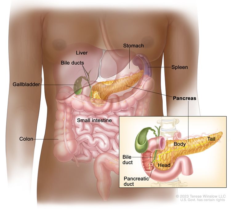
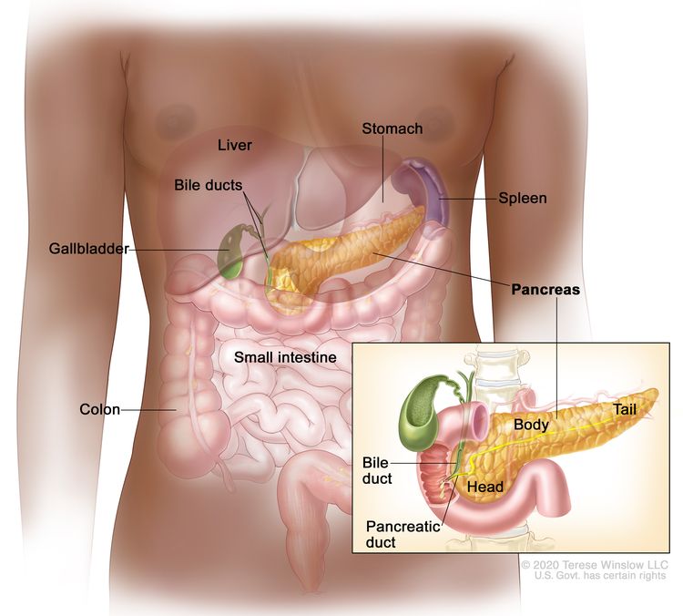
The pancreas is a gland about 6 inches long that is shaped like a thin pear lying on its side. The wider end of the pancreas is called the head, the middle section is called the body, and the narrow end is called the tail. The pancreas lies between the stomach and the spine.
EnlargeAnatomy of the pancreas. The pancreas has three areas: the head, body, and tail. It is found in the abdomen near the stomach, intestines, and other organs.
The pancreas has two main jobs in the body:
to make juices that help break down food into substances the body can use
to make hormones, such as insulin and glucagon, that help control blood sugar levels and help the body use and store the energy it gets from food
Smoking and health history can affect the risk of pancreatic cancer.
Pancreatic cancer is caused by certain changes to the way pancreatic cells function, especially how they grow and divide into new cells. A risk factor is anything that increases the chance of getting a disease. Some risk factors for pancreatic cancer, like smoking, can be changed. However, risk factors also include things people cannot change, like their genetics and their family history. Learning about risk factors for pancreatic cancer can help you make changes that might lower your risk of getting it.
There are many risk factors for pancreatic cancer, but many do not directly cause cancer. Instead, they increase the chance of DNA damage in cells that may lead to pancreatic cancer. Learn more about how cancer develops at What Is Cancer?
Having one or more of these risk factors does not mean that you will get pancreatic cancer. Many people with risk factors never develop pancreatic cancer, while others with no known risk factors do.
These symptoms may be caused by many conditions other than pancreatic cancer. It’s important to check with your doctor if you have any of these symptoms to find out the cause and begin treatment, if needed.
Pancreatic cancer is difficult to diagnose early.
Pancreatic cancer is difficult to detect and diagnose for the following reasons:
There aren’t any noticeable signs or symptoms in the early stages of pancreatic cancer.
The signs and symptoms of pancreatic cancer, when present, are like the signs and symptoms of many other illnesses.
Tests that examine the pancreas are used to diagnose and stage pancreatic cancer.
Pancreatic cancer is usually diagnosed with tests and procedures that make pictures of the pancreas and the area around it. The process used to find out if cancer cells have spread within and around the pancreas is called staging. Tests and procedures to detect, diagnose, and stage pancreatic cancer are usually done at the same time. To plan treatment, it is important to know the stage of the disease and whether the pancreatic cancer can be removed by surgery.
Blood chemistry study is a laboratory test in which a blood sample is checked to measure the amounts of certain substances, such as bilirubin, released into the blood by organs and tissues in the body. An unusual (higher or lower than normal) amount of a substance can be a sign of disease.
Tumor marker test checks a sample of blood, urine, or tissue to measure the amounts of certain substances, such as CA 19-9 and carcinoembryonic antigen (CEA), made by organs, tissues, or tumor cells in the body. Certain substances are linked to specific types of cancer when found in increased levels in the body. These are called tumor markers.
MRI (magnetic resonance imaging) uses a magnet, radio waves, and a computer to make a series of detailed pictures of areas inside the body. This procedure is also called nuclear magnetic resonance imaging (NMRI).
CT scan (CAT scan) uses a computer linked to an x-ray machine to make a series of detailed pictures of areas inside the body. The pictures are taken from different angles and are used to create 3-D views of tissues and organs. A dye may be injected into a vein or swallowed to help the organs or tissues show up more clearly. This procedure is also called computed tomography, computerized tomography, or computerized axial tomography. A spiral or helical CTscan makes a series of very detailed pictures of areas inside the body using an x-ray machine that scans the body in a spiral path.
PET scan (positron emission tomography scan) uses a small amount of radioactive sugar (also called glucose) that is injected into a vein. Then a scanner rotates around the body to make detailed, computerized pictures of areas inside the body where the glucose is taken up. Because cancer cells often take up more glucose than normal cells, the pictures can be used to find cancer cells in the body. A PET scan and CT scan may be done at the same time. This is called a PET-CT.
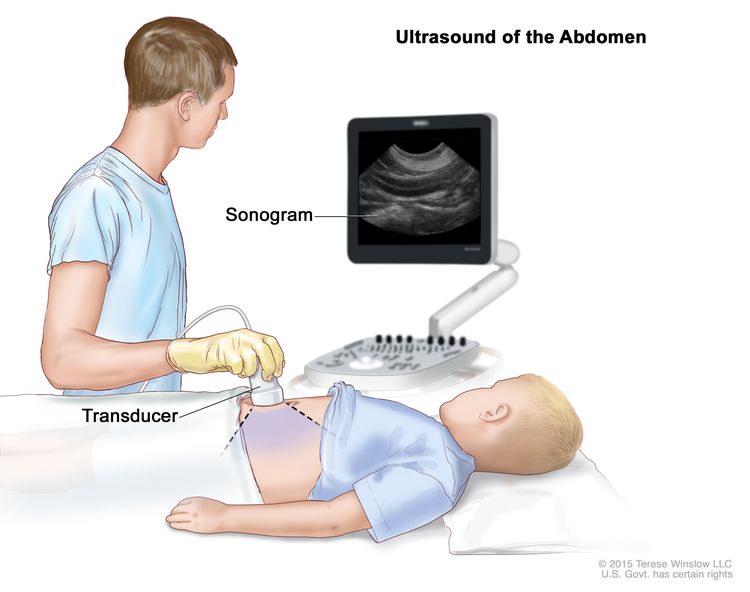
Abdominal ultrasound makes pictures of the inside of the abdomen. The ultrasound transducer is pressed against the skin of the abdomen and directs high-energy sound waves (ultrasound) into the abdomen. The sound waves bounce off the internal tissues and organs and make echoes. The transducer receives the echoes and sends them to a computer, which uses the echoes to make pictures called sonograms.
Endoscopic ultrasound (EUS) is a procedure that uses an endoscope inserted into the body, usually through the mouth or rectum. An endoscope is a thin, tube-like instrument with a light and a lens for viewing. A probe at the end of the endoscope is used to bounce high-energy sound waves (ultrasound) off internal tissues or organs and make echoes. The echoes form a picture of body tissues called a sonogram. This procedure is also called endosonography.
Endoscopic retrograde cholangiopancreatography (ERCP) is a procedure to x-ray the ducts (tubes) that carry bile from the liver to the gallbladder and from the gallbladder to the small intestine. Sometimes pancreatic cancer causes these ducts to narrow and block or slow the flow of bile, causing jaundice. An endoscope (a thin, lighted tube) is passed through the mouth, esophagus, and stomach into the first part of the small intestine. A catheter (a smaller tube) is then inserted through the endoscope into the pancreatic ducts. A dye is injected through the catheter into the ducts and an x-ray is taken. If the ducts are blocked by a tumor, a fine tube may be inserted into the duct to unblock it. This tube (or stent) may be left in place to keep the duct open. Tissue samples may also be taken.
Percutaneous transhepatic cholangiography (PTC) is a procedure to x-ray the liver and bile ducts. A thin needle is inserted through the skin below the ribs and into the liver. Dye is injected into the liver or bile ducts and an x-ray is taken. If a blockage is found, a thin, flexible tube called a stent is sometimes left in the liver to drain bile into the small intestine or a collection bag outside the body. This test is done only if ERCP cannot be done.
Laparoscopy is a surgical procedure to look at the organs inside the abdomen to check for signs of disease. Small incisions (cuts) are made in the wall of the abdomen and a laparoscope (a thin, lighted tube) is inserted into one of the incisions. The laparoscope may have an ultrasound probe at the end in order to bounce high-energy sound waves off internal organs, such as the pancreas. This is called laparoscopic ultrasound. Other instruments may be inserted through the same or other incisions to perform procedures such as taking tissue samples from the pancreas or a sample of fluid from the abdomen to check for cancer.
Biopsy is the removal of cells or tissues so they can be viewed under a microscope by a pathologist to check for signs of cancer. There are several ways to do a biopsy for pancreatic cancer. A fine needle or a core needle may be inserted into the pancreas during an x-ray or ultrasound to remove cells. Tissue may also be removed during a laparoscopy or surgery to remove the tumor.
Some people decide to get a second opinion.
You may want to get a second opinion to confirm your pancreatic cancer diagnosis and treatment plan. If you seek a second opinion, you will need to get medical test results and reports from the first doctor to share with the second doctor. The second doctor will review the pathology report, slides, and scans. They may agree with the first doctor, suggest changes or another treatment approach, or provide more information about your cancer.
To learn more about choosing a doctor and getting a second opinion, visit Finding Cancer Care. You can contact NCI’s Cancer Information Service via chat, email, or phone (both in English and Spanish) for help finding a doctor, hospital, or getting a second opinion. For questions you might want to ask at your appointments, visit Questions to Ask Your Doctor about Cancer.
Certain factors affect prognosis (chance of recovery) and treatment options.
The prognosis and treatment options depend on:
whether the tumor can be removed by surgery
the stage of the cancer (the size of the tumor and whether the cancer has spread outside the pancreas to nearby tissues or lymph nodes or to other places in the body)
the patient’s general health
whether the cancer has just been diagnosed or has recurred (come back)
Pancreatic cancer can be controlled only if it is found before it has spread, when it can be completely removed by surgery. If the cancer has spread, palliative treatment can improve the patient’s quality of life by controlling the symptoms and complications of this disease.
Stages of Pancreatic Cancer
Key Points
Cancer stage describes the extent of cancer in the body.
The following stages are used for pancreatic cancer:
Stage 0 (carcinoma in situ)
Stage I (also called stage 1) pancreatic cancer
Stage II (also called stage 2) pancreatic cancer
Stage III (also called stage 3) pancreatic cancer
Stage IV (also called stage 4) pancreatic cancer
The following groups are used to plan treatment:
Resectable pancreatic cancer
Borderline resectable pancreatic cancer
Locally advanced pancreatic cancer
Metastatic pancreatic cancer
Recurrent pancreatic cancer
Cancer stage describes the extent of cancer in the body.
Cancer stage describes the extent of cancer in the body, such as the size of the tumor, whether it has spread, and how far it has spread from where it first formed. It is important to know the stage of the pancreatic cancer to plan the best treatment.
There are several staging systems for cancer that describe the extent of the cancer. Pancreatic cancer staging usually uses the TNM staging system. Your cancer may be described by this staging system in your pathology report. Based on the TNM results, a stage (I, II, III, or IV, also written as 1, 2, 3, or 4) is assigned to your cancer. When talking to you about your cancer, your doctor may describe it as one of these stages.
The information in this section is about staging for exocrine pancreatic cancer, the most common type of pancreatic cancer.
The following stages are used for pancreatic cancer:
Stage 0 (carcinoma in situ)
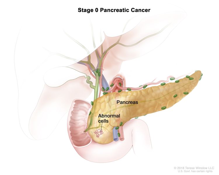
EnlargeStage 0 pancreatic cancer. Abnormal cells are found in the lining of the pancreas. These abnormal cells may become cancer and spread into nearby normal tissue.
In stage 0, abnormal cells are found in the lining of the pancreas. These abnormal cells may become cancer and spread into nearby normal tissue. Stage 0 is also called carcinoma in situ.
Stage I (also called stage 1) pancreatic cancer
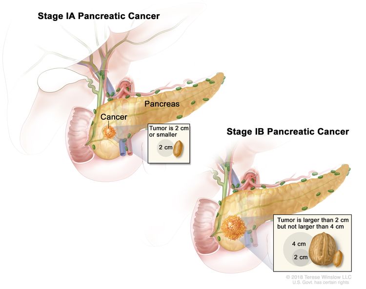
EnlargeStage I pancreatic cancer. Cancer is found in the pancreas only. In stage IA, the tumor is 2 centimeters or smaller. In stage IB, the tumor is larger than 2 centimeters but not larger than 4 centimeters.
In stage I, cancer has formed and is found in the pancreas only. Stage I is divided into stages IA and IB, depending on the size of the tumor.
Stage IB: The tumor is larger than 2 centimeters but not larger than 4 centimeters.
Stage II (also called stage 2) pancreatic cancer
Stage II is divided into stages IIA and IIB, depending on the size of the tumor and where the cancer has spread.
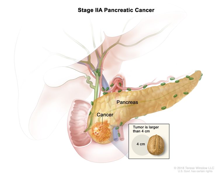
Stage IIA: The tumor is larger than 4 centimeters. EnlargeStage IIA pancreatic cancer. The tumor is larger than 4 centimeters.
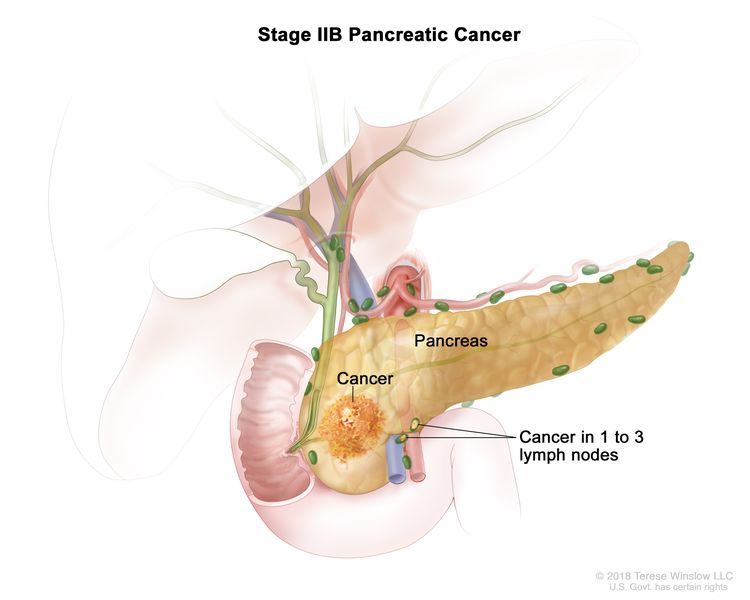
Stage IIB: The tumor is any size, and cancer has spread to 1 to 3 nearby lymph nodes. EnlargeStage IIB pancreatic cancer. The tumor is any size and cancer has spread to 1 to 3 nearby lymph nodes.
Stage III (also called stage 3) pancreatic cancer
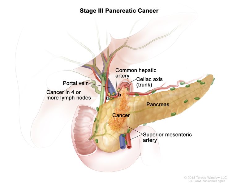
EnlargeStage III pancreatic cancer. The tumor is any size and cancer has spread to (a) 4 or more nearby lymph nodes; or (b) the major blood vessels near the pancreas. These include the portal vein, common hepatic artery, celiac axis (trunk), and superior mesenteric artery.
In stage III, the tumor is any size, and cancer has spread to:
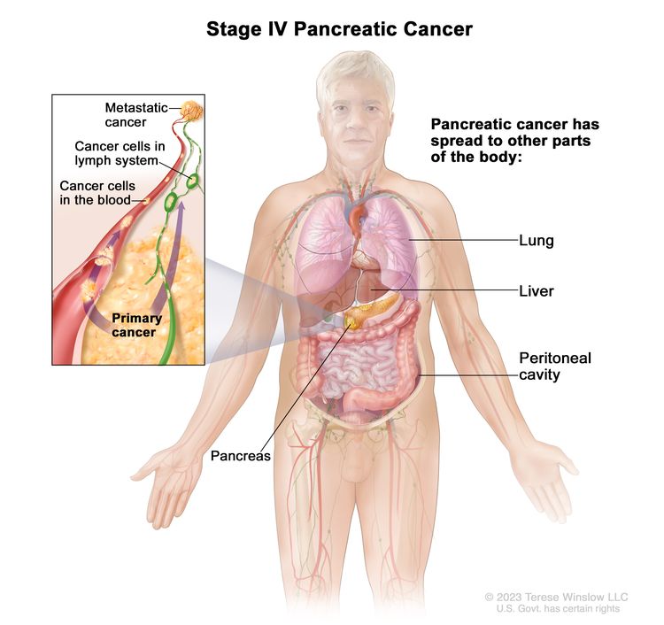
EnlargeStage IV pancreatic cancer. The tumor is any size and cancer has spread to other parts of the body, such as the lung, liver, or peritoneal cavity (the body cavity that contains most of the organs in the abdomen).
In stage IV, the tumor is any size, and cancer has spread to other parts of the body, such as the liver, lung, or peritoneal cavity (the body cavity that contains most of the organs in the abdomen).
Stage IV pancreatic cancer is also called metastatic pancreatic cancer. Metastatic cancer happens when cancer cells travel through the lymphatic system or blood and form tumors in other parts of the body. The metastatic tumor is the same type of cancer as the primary tumor. For example, if pancreatic cancer spreads to the liver, the cancer cells in the liver are actually pancreatic cancer cells. The disease is called metastatic pancreatic cancer, not liver cancer. Learn more in Metastatic Cancer: When Cancer Spreads.
The following groups are used to plan treatment:
Resectable pancreatic cancer
Resectable pancreatic cancer can be removed by surgery because it has not grown into major blood vessels near the tumor.
Borderline resectable pancreatic cancer
Borderline resectable pancreatic cancer has grown into a major blood vessel or nearby tissue or organs. It may be possible to remove the tumor, but there is a high risk that all of the cancer cells will not be removed with surgery.
Locally advanced pancreatic cancer
Locally advanced pancreatic cancer has grown into or close to nearby lymph nodes or blood vessels, so surgery cannot completely remove the cancer.
Metastatic pancreatic cancer
Metastatic pancreatic cancer has spread to other organs, so surgery cannot completely remove the cancer.
Recurrent pancreatic cancer
Recurrent pancreatic cancer is cancer that has recurred (come back) after it has been treated. The cancer may come back in the pancreas or in other parts of the body. Tests will be done to help determine where the cancer has returned in your body. The type of treatment for recurrent pancreatic cancer will depend on where it has come back.
There are different types of treatment for patients with pancreatic cancer.
The following types of treatment are used:
Surgery
Radiation therapy
Chemotherapy
Chemoradiation therapy
Targeted therapy
There are treatments for pain caused by pancreatic cancer.
Patients with pancreatic cancer have special nutritional needs.
New types of treatment are being tested in clinical trials.
Treatment for pancreatic cancer may cause side effects.
Follow-up care may be needed.
There are different types of treatment for patients with pancreatic cancer.
Different types of treatments are available for people with pancreatic cancer. You and your cancer care team will work together to decide your treatment plan, which may include more than one type of treatment. Many factors will be considered, such as the stage of the cancer, your overall health, and your preferences. Your plan will include information about your cancer, the goals of treatment, your treatment options and the possible side effects, and the expected length of treatment.
Talking with your cancer care team before treatment begins about what to expect will be helpful. You’ll want to learn what you need to do before treatment begins, how you’ll feel while going through it, and what kind of help you will need. To learn more, visit Questions to Ask Your Doctor about Treatment.
The following types of treatment are used:
Surgery
One of the following types of surgery may be used to take out the tumor:
Total pancreatectomy is surgery to remove the whole pancreas, part of the stomach, part of the small intestine, the common bile duct, the gallbladder, the spleen, and nearby lymph nodes.
Distal pancreatectomy is surgery to remove the body and the tail of the pancreas. The spleen may also be removed if cancer has spread to the spleen.
Biliary bypass: If cancer is blocking the bile duct and bile is building up in the gallbladder, a biliary bypass may be done. During this operation, the doctor will cut the gallbladder or bile duct in the area before the blockage and sew it to the small intestine to create a new pathway around the blocked area.
Endoscopicstent placement: If the tumor is blocking the bile duct, surgery may be done to put in a stent (a thin tube) to drain bile that has built up in the area. The doctor may place the stent through a catheter that drains the bile into a bag on the outside of the body, or the stent may go around the blocked area and drain the bile into the small intestine.
Gastric bypass: If the tumor is blocking the flow of food from the stomach, the stomach may be sewn directly to the small intestine so the patient can continue to eat normally.
Radiation therapy
Radiation therapy uses high-energy x-rays or other types of radiation to kill cancer cells or keep them from growing. Pancreatic cancer is sometimes treated with external beam radiation therapy. This type of radiation therapy uses a machine outside the body to send radiation toward the area of the body with cancer.
Chemotherapy (also called chemo) uses drugs to stop the growth of cancer cells, either by killing the cells or by stopping them from dividing.
Chemotherapy for pancreatic cancer is usually systemic, meaning it is injected into a vein or given by mouth. When given this way, the drugs enter the bloodstream to reach cancer cells throughout the body.
Chemotherapy drugs used to treat pancreatic cancer may include:
Chemoradiation therapy combines chemotherapy and radiation therapy to increase the effects of both.
Targeted therapy
Targeted therapy uses drugs or other substances to identify and attack specific cancer cells. Erlotinib is a targeted therapy drug used to treat pancreatic cancer.
There are treatments for pain caused by pancreatic cancer.
Pain can occur when the tumor presses on nerves or other organs near the pancreas. When pain medicine is not enough, there are treatments that act on nerves in the abdomen to relieve the pain. The doctor may inject medicine into the area around affected nerves or may cut the nerves to block the feeling of pain. Radiation therapy with or without chemotherapy can also help relieve pain by shrinking the tumor. Learn more about Cancer Pain.
Patients with pancreatic cancer have special nutritional needs.
Surgery to remove the pancreas may affect its ability to make pancreatic enzymes that help to digest food. As a result, patients may have problems digesting food and absorbing nutrients into the body. To prevent malnutrition, the doctor may prescribe medicines that replace these enzymes.
New types of treatment are being tested in clinical trials.
For some people, joining a clinical trial may be an option. There are different types of clinical trials for people with cancer. For example, a treatment trial tests new treatments or new ways of using current treatments. Supportive care and palliative care trials look at ways to improve quality of life, especially for those who have side effects from cancer and its treatment.
You can use the clinical trial search to find NCI-supported cancer clinical trials accepting participants. The search allows you to filter trials based on the type of cancer, your age, and where the trials are being done. Clinical trials supported by other organizations can be found on the ClinicalTrials.gov website.
As you go through treatment, you will have follow-up tests or check-ups. Some tests that were done to diagnose or stage the cancer may be repeated to see how well the treatment is working. Decisions about whether to continue, change, or stop treatment may be based on the results of these tests.
Some of the tests will continue to be done from time to time after treatment has ended. The results of these tests can show if your condition has changed or if the cancer has recurred (come back).
Treatment of Resectable or Borderline Resectable Pancreatic Cancer
Treatment of resectable or borderline resectable pancreatic cancer may include:
Use our clinical trial search to find NCI-supported cancer clinical trials that are accepting patients. You can search for trials based on the type of cancer, the age of the patient, and where the trials are being done. General information about clinical trials is also available.
Treatment of Locally Advanced Pancreatic Cancer
Treatment of pancreatic cancer that is locally advanced may include:
palliative surgery or stent placement to bypass blocked areas in ducts or the small intestine, and possibly chemotherapy and chemoradiation to shrink the tumor to allow for surgery
a clinical trial of new anticancer therapies together with chemotherapy or chemoradiation
Use our clinical trial search to find NCI-supported cancer clinical trials that are accepting patients. You can search for trials based on the type of cancer, the age of the patient, and where the trials are being done. General information about clinical trials is also available.
Treatment of Metastatic or Recurrent Pancreatic Cancer
Use our clinical trial search to find NCI-supported cancer clinical trials that are accepting patients. You can search for trials based on the type of cancer, the age of the patient, and where the trials are being done. General information about clinical trials is also available.
Use our clinical trial search to find NCI-supported cancer clinical trials that are accepting patients. You can search for trials based on the type of cancer, the age of the patient, and where the trials are being done. General information about clinical trials is also available.
Physician Data Query (PDQ) is the National Cancer Institute’s (NCI’s) comprehensive cancer information database. The PDQ database contains summaries of the latest published information on cancer prevention, detection, genetics, treatment, supportive care, and complementary and alternative medicine. Most summaries come in two versions. The health professional versions have detailed information written in technical language. The patient versions are written in easy-to-understand, nontechnical language. Both versions have cancer information that is accurate and up to date and most versions are also available in Spanish.
PDQ is a service of the NCI. The NCI is part of the National Institutes of Health (NIH). NIH is the federal government’s center of biomedical research. The PDQ summaries are based on an independent review of the medical literature. They are not policy statements of the NCI or the NIH.
Purpose of This Summary
This PDQ cancer information summary has current information about the treatment of adult pancreatic cancer. It is meant to inform and help patients, families, and caregivers. It does not give formal guidelines or recommendations for making decisions about health care.
Reviewers and Updates
Editorial Boards write the PDQ cancer information summaries and keep them up to date. These Boards are made up of experts in cancer treatment and other specialties related to cancer. The summaries are reviewed regularly and changes are made when there is new information. The date on each summary (“Updated”) is the date of the most recent change.
The information in this patient summary was taken from the health professional version, which is reviewed regularly and updated as needed, by the PDQ Adult Treatment Editorial Board.
Clinical Trial Information
A clinical trial is a study to answer a scientific question, such as whether one treatment is better than another. Trials are based on past studies and what has been learned in the laboratory. Each trial answers certain scientific questions in order to find new and better ways to help cancer patients. During treatment clinical trials, information is collected about the effects of a new treatment and how well it works. If a clinical trial shows that a new treatment is better than one currently being used, the new treatment may become “standard.” Patients may want to think about taking part in a clinical trial. Some clinical trials are open only to patients who have not started treatment.
Clinical trials can be found online at NCI’s website. For more information, call the Cancer Information Service (CIS), NCI’s contact center, at 1-800-4-CANCER (1-800-422-6237).
Permission to Use This Summary
PDQ is a registered trademark. The content of PDQ documents can be used freely as text. It cannot be identified as an NCI PDQ cancer information summary unless the whole summary is shown and it is updated regularly. However, a user would be allowed to write a sentence such as “NCI’s PDQ cancer information summary about breast cancer prevention states the risks in the following way: [include excerpt from the summary].”
The best way to cite this PDQ summary is:
PDQ® Adult Treatment Editorial Board. PDQ Pancreatic Cancer Treatment. Bethesda, MD: National Cancer Institute. Updated <MM/DD/YYYY>. Available at: /types/pancreatic/patient/pancreatic-treatment-pdq. Accessed <MM/DD/YYYY>. [PMID: 26389396]
Images in this summary are used with permission of the author(s), artist, and/or publisher for use in the PDQ summaries only. If you want to use an image from a PDQ summary and you are not using the whole summary, you must get permission from the owner. It cannot be given by the National Cancer Institute. Information about using the images in this summary, along with many other images related to cancer can be found in Visuals Online. Visuals Online is a collection of more than 3,000 scientific images.
Disclaimer
The information in these summaries should not be used to make decisions about insurance reimbursement. More information on insurance coverage is available on Cancer.gov on the Managing Cancer Care page.
Contact Us
More information about contacting us or receiving help with the Cancer.gov website can be found on our Contact Us for Help page. Questions can also be submitted to Cancer.gov through the website’s E-mail Us.
Multiple endocrine neoplasia (MEN) syndromes are inherited disorders that affect the endocrine system.
There are several types of MEN syndromes and each type may cause different conditions or cancers.
MEN1 syndrome usually causes tumors in the parathyroid gland, pituitary gland, or islet cells of the pancreas.
MEN2A syndrome may cause medullary thyroid cancer, pheochromocytoma, or parathyroid gland disease.
MEN2B syndrome causes several conditions.
Children with MEN2A syndrome, MEN2B syndrome, or FMTC may need genetic testing.
Tests used to diagnose and stage cancers related to MEN syndromes depend on the signs and symptoms and the patient’s family history.
If cancer, such as medullary thyroid cancer, has formed, tests are done to find out if cancer cells have spread to nearby areas or to other parts of the body.
There are three ways that cancer spreads in the body.
Cancer may spread from where it began to other parts of the body.
Multiple endocrine neoplasia (MEN) syndromes are inherited disorders that affect the endocrine system.
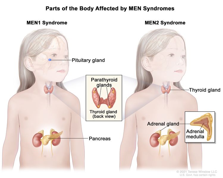
EnlargeMultiple endocrine neoplasia (MEN) syndromes are rare, inherited disorders that affect the endocrine glands or organs. There are several types of MEN syndromes, and each type may cause different conditions or cancers. The two main types of MEN syndromes are MEN1 and MEN2. MEN1 syndrome usually causes tumors in the pituitary gland, parathyroid gland, or pancreas. MEN2 syndrome usually causes tumors in the thyroid gland, parathyroid gland, or adrenal gland. The tumors may be benign (not cancer) or malignant (cancer).
MEN1 syndrome usually causes tumors in the parathyroid gland, pituitary gland, or islet cells of the pancreas.
A diagnosis of MEN1 syndrome is made when tumors are found in two of the following glands or organs: parathyroid gland, pituitary gland, or islet cells in the pancreas. These tumors may make extra hormones and cause certain signs or symptoms of disease. The signs and symptoms depend on the type of hormone made by the tumor. MEN1 syndrome is also called Wermer syndrome.
Children with primary hyperparathyroidism, tumors associated with MEN1 syndrome, or a family history of hypercalcemia or MEN1 syndrome may have genetic testing to check for a mutation (change) in the MEN1gene. Parents should receive genetic counseling (a discussion with a trained professional about the risk of genetic diseases) before genetic testing is done. Genetic counseling also includes a discussion of the risk of MEN1 syndrome for the child and other family members.
Children who are diagnosed with MEN1 syndrome are checked for signs of cancer starting at age 5 years and continuing for the rest of their life. Talk to your child’s doctor about the tests and procedures needed to check for signs of cancer and how often they should be done.
MEN2A syndrome may cause medullary thyroid cancer, pheochromocytoma, or parathyroid gland disease.
MEN2A syndrome is also called Sipple syndrome. A diagnosis of MEN2A syndrome may be made when the patient or the patient’s parents, brothers, sisters, or children have two or more of the following conditions:
Medullary thyroid cancer (a cancer that forms in parafollicular C cells in the thyroid). Signs and symptoms of medullary thyroid cancer may include the following:
Parathyroid gland disease (a benign tumor of the parathyroid gland or increase in the size of the parathyroid gland). Signs and symptoms of parathyroid gland disease may include the following:
Hypercalcemia.
Pain in the abdomen, side, or back that doesn’t go away.
Pain in the bones.
A broken bone.
A lump in the neck.
Trouble speaking.
Trouble swallowing.
Familial medullary cancer of the thyroid is like MEN2A syndrome without pheochromocytoma or parathyroid tumors. A diagnosis of familial medullary cancer of the thyroid may be made when two or more family members have medullary thyroid cancer and no family members have parathyroid or adrenal gland problems. Signs and symptoms of medullary thyroid cancer may include the following:
A lump in the throat or neck.
Trouble breathing.
Trouble swallowing.
Hoarseness.
The prognosis is usually good.
A mutation in the RET gene is usually linked to medullary thyroid cancer in MEN2 syndrome. If a diagnosis of MEN2 syndrome is suspected for the child or a family member is diagnosed with MEN2 syndrome, the parents should receive genetic counseling before genetic testing is done for the child. Genetic counseling also includes a discussion of the risk of MEN2 syndrome for the child and other family members.
MEN2B syndrome causes several conditions.
Patients with MEN2B syndrome may have a slender body build with long, thin arms and legs. The lips may appear large and bumpy because of benign tumors in the mucous membranes. MEN2B syndrome may cause the following conditions:
Medullary thyroid cancer (a fast-growing, aggressive cancer).
Nerve cell tumors in the mucous membranes or other places.
The prognosis for MEN2B syndrome is not as good as the prognosis for MEN1 syndrome and MEN2A syndrome because medullary thyroid cancer is a more aggressive cancer.
Children with MEN2A syndrome, MEN2B syndrome, or FMTC may need genetic testing.
A mutation in the RET gene is usually linked to medullary thyroid cancer in MEN2 syndrome. If a diagnosis of MEN2 syndrome is suspected for the child or a family member is diagnosed with MEN2 syndrome, the parents should receive genetic counseling before genetic testing is done for the child. Genetic counseling also includes a discussion of the risk of MEN2 syndrome for the child and other family members.
Tests used to diagnose and stage cancers related to MEN syndromes depend on the signs and symptoms and the patient’s family history.
Tests are done to diagnose and stage cancer. After cancer is diagnosed, more tests are done to find out if cancer cells have spread to nearby areas or to other parts of the body. This process is called staging.
The following tests and procedures may be used:
Physical exam and health history: An exam of the body to check general signs of health, including checking for signs of disease, such as lumps or anything else that seems unusual. A history of the patient’s health habits and past illnesses and treatments will also be taken.
Blood chemistry studies: A procedure in which a blood sample is checked to measure the amounts of certain substances released into the blood by organs and tissues in the body. An unusual (higher or lower than normal) amount of a substance can be a sign of disease.
Blood hormone studies: A procedure in which a blood sample is checked to measure the amounts of certain hormones released into the blood by organs and tissues in the body. An unusual (higher or lower than normal) amount of a substance can be a sign of disease in the organ or tissue that makes it. The blood may also be checked for high levels of the hormone calcitonin or parathyroid hormone (PTH).
Genetic testing: A laboratory test in which cells or tissue are analyzed to look for changes in genes or chromosomes. These changes may be a sign that a person has or is at risk of having a specific disease or condition. A sample of blood is checked for the MEN1 gene to diagnose MEN1 syndrome and for the RET gene to diagnose MEN2 syndrome.
Twenty-four-hour urine test: A procedure used to diagnose neuroendocrine tumors, such as pheochromocytoma. Urine is collected for 24 hours to measure the amounts of catecholamines in the urine. Substances caused by the breakdown of these catecholamines are also measured. An unusual (higher or lower than normal) amount of a substance can be a sign of disease in the organ or tissue that makes it. Higher than normal amounts may be a sign of pheochromocytoma.
Ultrasound: A procedure in which high-energy sound waves (ultrasound) are bounced off internal tissues or organs and make echoes. The echoes form a picture of body tissues called a sonogram. The picture can be printed to be looked at later.
MRI: A procedure that uses a magnet, radio waves, and a computer to make a series of detailed pictures of areas inside the body. This procedure is also called nuclear magnetic resonance imaging (NMRI).
CT scan: A procedure that makes a series of detailed pictures of areas inside the body, taken from different angles. The pictures are made by a computer linked to an x-ray machine. A dye may be injected into a vein or swallowed to help the organs or tissues show up more clearly. This procedure is also called computed tomography, computerized tomography, or computerized axial tomography.
PET scan: A procedure to find malignant tumor cells in the body. A small amount of radioactive glucose (sugar) is injected into a vein. The PET scanner rotates around the body and makes a picture of where glucose is being used in the body. Malignant tumor cells show up brighter in the picture because they are more active and take up more glucose than normal cells do.
Thyroid scan: A small amount of a radioactive substance is swallowed or injected. The radioactive material collects in thyroid gland cells. A special camera linked to a computer detects the radiation given off and makes pictures that show how the thyroid looks and functions and whether the cancer has spread beyond the thyroid gland. If the amount of thyroid-stimulating hormone in the child’s blood is low, a scan to make images of the thyroid may be done before surgery.
Sestamibi scan: A type of radionuclide scan used to find an overactive parathyroid gland. A very small amount of a radioactive substance called technetium 99 is injected into a vein and travels through the bloodstream to the parathyroid gland. The radioactive substance will collect in the overactive gland and show up brightly on a special camera that detects radioactivity.
Venous sampling for an overactive parathyroid gland: A procedure in which a sample of blood is taken from veins near the parathyroid glands. The sample is checked to measure the amount of parathyroid hormone released into the blood by each gland. Venous sampling may be done if blood tests show there is an overactive parathyroid gland but imaging tests don’t show which one it is.
Somatostatin receptor scintigraphy: A type of radionuclide scan that may be used to find tumors. A very small amount of radioactive octreotide (a hormone that attaches to tumors) is injected into a vein and travels through the blood. The radioactive octreotide attaches to the tumor and a special camera that detects radioactivity is used to show whether there are islet cell tumors in the pancreas. This procedure is also called octreotide scan and SRS.
MIBG scan: A procedure used to find neuroendocrine tumors, such as pheochromocytoma. A very small amount of a substance called radioactive MIBG is injected into a vein and travels through the bloodstream. Neuroendocrine tumor cells take up the radioactive MIBG and are detected by a scanner. Scans may be taken over 1-3 days. An iodine solution may be given before or during the test to keep the thyroid gland from absorbing too much of the MIBG.
Pentagastrin stimulation test: A test in which blood samples are checked to measure the amount of calcitonin in the blood. Calcium gluconate and pentagastrin are injected into the blood and then several blood samples are taken over the next 5 minutes. If the level of calcitonin in the blood increases, it may be a sign of medullary thyroid cancer.
Biopsy: The removal of cells or tissues so they can be viewed under a microscope by a pathologist to check for signs of cancer.
If cancer, such as medullary thyroid cancer, has formed, tests are done to find out if cancer cells have spread to nearby areas or to other parts of the body.
The process used to find out if cancer has spread to nearby areas or to other parts of the body is called staging. There is no standard system for staging childhood cancers linked to MEN syndromes. The results of the tests and procedures done to diagnose cancer are used to help make decisions about treatment.
Sometimes childhood MEN syndromes recur (come back) after treatment.
There are three ways that cancer spreads in the body.
Tissue. The cancer spreads from where it began by growing into nearby areas.
Lymph system. The cancer spreads from where it began by getting into the lymph system. The cancer travels through the lymph vessels to other parts of the body.
Blood. The cancer spreads from where it began by getting into the blood. The cancer travels through the blood vessels to other parts of the body.
Cancer may spread from where it began to other parts of the body.
When cancer spreads to another part of the body, it is called metastasis. Cancer cells break away from where they began (the primary tumor) and travel through the lymph system or blood.
Lymph system. The cancer gets into the lymph system, travels through the lymph vessels, and forms a tumor (metastatic tumor) in another part of the body.
Blood. The cancer gets into the blood, travels through the blood vessels, and forms a tumor (metastatic tumor) in another part of the body.
The metastatic tumor is the same type of cancer as the primary tumor. For example, if medullary thyroid cancer spreads to the lung, the cancer cells in the lung are actually medullary thyroid cells. The disease is metastatic medullary thyroid cancer, not lung cancer.
Many cancer deaths are caused when cancer moves from the original tumor and spreads to other tissues and organs. This is called metastatic cancer. This animation shows how cancer cells travel from the place in the body where they first formed to other parts of the body.
Treatment Option Overview
Key Points
There are different types of treatment for children with MEN syndromes.
Children with MEN syndromes should have their treatment planned by a team of doctors who are experts in treating childhood cancer.
The treatment will depend on the type of MEN syndrome the child has.
New types of treatment are being tested in clinical trials.
Treatment for childhood MEN syndrome may cause side effects.
Patients may want to think about taking part in a clinical trial.
Patients can enter clinical trials before, during, or after starting their cancer treatment.
Follow-up tests may be needed.
There are different types of treatment for children with MEN syndromes.
Some treatments are standard (the currently used treatment), and some are being tested in clinical trials. A treatment clinical trial is a research study meant to help improve current treatments or obtain information on new treatments for patients with cancer. When clinical trials show that a new treatment is better than the standard treatment, the new treatment may become the standard treatment.
Because cancer in children is rare, taking part in a clinical trial should be considered. Some clinical trials are open only to patients who have not started treatment.
Children with MEN syndromes should have their treatment planned by a team of doctors who are experts in treating childhood cancer.
Treatment will be overseen by a pediatric oncologist, a doctor who specializes in treating children with cancer. The pediatric oncologist works with other pediatric health professionals who are experts in treating children with cancer and who specialize in certain areas of medicine. This may include the following specialists and others:
Patients may want to think about taking part in a clinical trial.
For some patients, taking part in a clinical trial may be the best treatment choice. Clinical trials are part of the cancer research process. Clinical trials are done to find out if new cancer treatments are safe and effective or better than the standard treatment.
Many of today’s standard treatments for cancer are based on earlier clinical trials. Patients who take part in a clinical trial may receive the standard treatment or be among the first to receive a new treatment.
Patients who take part in clinical trials also help improve the way cancer will be treated in the future. Even when clinical trials do not lead to effective new treatments, they often answer important questions and help move research forward.
Patients can enter clinical trials before, during, or after starting their cancer treatment.
Some clinical trials only include patients who have not yet received treatment. Other trials test treatments for patients whose cancer has not gotten better. There are also clinical trials that test new ways to stop cancer from recurring (coming back) or reduce the side effects of cancer treatment.
Clinical trials are taking place in many parts of the country. Information about clinical trials supported by NCI can be found on NCI’s clinical trials search webpage. Clinical trials supported by other organizations can be found on the ClinicalTrials.gov website.
Follow-up tests may be needed.
As your child goes through treatment, they will have follow-up tests or check-ups. Some tests that were done to diagnose or stage the cancer may be repeated to see how well the treatment is working. Decisions about whether to continue, change, or stop treatment may be based on the results of these tests.
Some of the tests will continue to be done from time to time after treatment has ended. The results of these tests can show if your child’s condition has changed or if the cancer has recurred (come back).
Use our clinical trial search to find NCI-supported cancer clinical trials that are accepting patients. You can search for trials based on the type of cancer, the age of the patient, and where the trials are being done. General information about clinical trials is also available.
Children with MEN2A syndrome who have medullary thyroid cancer may be treated with targeted therapy. Targeted therapy is a type of treatment that uses drugs or other substances to identify and attack specific cancer cells. Targeted therapies usually cause less harm to normal cells than chemotherapy or radiation therapy do.
A clinical trial of selpercatinib in tumors with RET gene changes.
Use our clinical trial search to find NCI-supported cancer clinical trials that are accepting patients. You can search for trials based on the type of cancer, the age of the patient, and where the trials are being done. General information about clinical trials is also available.
Children with MEN2B syndrome who have medullary thyroid cancer may be treated with targeted therapy. Targeted therapy is a type of treatment that uses drugs or other substances to identify and attack specific cancer cells. Targeted therapies usually cause less harm to normal cells than chemotherapy or radiation therapy do.
Use our clinical trial search to find NCI-supported cancer clinical trials that are accepting patients. You can search for trials based on the type of cancer, the age of the patient, and where the trials are being done. General information about clinical trials is also available.
A clinical trial that checks a sample of the patient’s tumor for certain gene changes. The type of targeted therapy that will be given to the patient depends on the type of gene change.
A clinical trial of selpercatinib in tumors with RET gene changes.
Physician Data Query (PDQ) is the National Cancer Institute’s (NCI’s) comprehensive cancer information database. The PDQ database contains summaries of the latest published information on cancer prevention, detection, genetics, treatment, supportive care, and complementary and alternative medicine. Most summaries come in two versions. The health professional versions have detailed information written in technical language. The patient versions are written in easy-to-understand, nontechnical language. Both versions have cancer information that is accurate and up to date and most versions are also available in Spanish.
PDQ is a service of the NCI. The NCI is part of the National Institutes of Health (NIH). NIH is the federal government’s center of biomedical research. The PDQ summaries are based on an independent review of the medical literature. They are not policy statements of the NCI or the NIH.
Purpose of This Summary
This PDQ cancer information summary has current information about the treatment of childhood multiple endocrine neoplasia (MEN) syndromes. It is meant to inform and help patients, families, and caregivers. It does not give formal guidelines or recommendations for making decisions about health care.
Reviewers and Updates
Editorial Boards write the PDQ cancer information summaries and keep them up to date. These Boards are made up of experts in cancer treatment and other specialties related to cancer. The summaries are reviewed regularly and changes are made when there is new information. The date on each summary (“Updated”) is the date of the most recent change.
The information in this patient summary was taken from the health professional version, which is reviewed regularly and updated as needed, by the PDQ Pediatric Treatment Editorial Board.
Clinical Trial Information
A clinical trial is a study to answer a scientific question, such as whether one treatment is better than another. Trials are based on past studies and what has been learned in the laboratory. Each trial answers certain scientific questions in order to find new and better ways to help cancer patients. During treatment clinical trials, information is collected about the effects of a new treatment and how well it works. If a clinical trial shows that a new treatment is better than one currently being used, the new treatment may become “standard.” Patients may want to think about taking part in a clinical trial. Some clinical trials are open only to patients who have not started treatment.
Clinical trials can be found online at NCI’s website. For more information, call the Cancer Information Service (CIS), NCI’s contact center, at 1-800-4-CANCER (1-800-422-6237).
Permission to Use This Summary
PDQ is a registered trademark. The content of PDQ documents can be used freely as text. It cannot be identified as an NCI PDQ cancer information summary unless the whole summary is shown and it is updated regularly. However, a user would be allowed to write a sentence such as “NCI’s PDQ cancer information summary about breast cancer prevention states the risks in the following way: [include excerpt from the summary].”
Images in this summary are used with permission of the author(s), artist, and/or publisher for use in the PDQ summaries only. If you want to use an image from a PDQ summary and you are not using the whole summary, you must get permission from the owner. It cannot be given by the National Cancer Institute. Information about using the images in this summary, along with many other images related to cancer can be found in Visuals Online. Visuals Online is a collection of more than 3,000 scientific images.
Disclaimer
The information in these summaries should not be used to make decisions about insurance reimbursement. More information on insurance coverage is available on Cancer.gov on the Managing Cancer Care page.
Contact Us
More information about contacting us or receiving help with the Cancer.gov website can be found on our Contact Us for Help page. Questions can also be submitted to Cancer.gov through the website’s E-mail Us.
Pancreatic cancer can develop from two kinds of cells in the pancreas: exocrine cells and neuroendocrine cells, such as islet cells. The exocrine type is more common and is usually found at an advanced stage. Pancreatic neuroendocrine tumors (islet cell tumors) are less common but have a better prognosis. Explore the links on this page to learn more about pancreatic cancer treatment, statistics, research, and clinical trials.
Pancreatic cancer is a disease in which malignant (cancer) cells form in the tissues of the pancreas.
There are four types of pancreatic cancer in children.
Signs and symptoms of pancreatic cancer include feeling tired and weight loss.
Tests that examine the pancreas are used to help diagnose pancreatic cancer.
Pancreatic cancer is a disease in which malignant (cancer) cells form in the tissues of the pancreas.
The pancreas is a gland about 6 inches long that is shaped like a thin pear lying on its side. The wider end of the pancreas is called the head, the middle section is called the body, and the narrow end is called the tail. The pancreas lies between the stomach and the spine.
EnlargeAnatomy of the pancreas. The pancreas has three parts: the head, the body, and the tail. It is found in the abdomen near the stomach, intestines, and other organs.
Endocrine pancreas cells make hormones, such as insulin and glucagon, that help control blood sugar levels. Both of these hormones help the body use and store the energy it gets from food. Endocrine pancreas cells cluster together in many small groups (islets) throughout the pancreas. Endocrine pancreas cells are also called islet cells or islets of Langerhans.
There are four types of pancreatic cancer in children.
The four types of pancreatic cancer in children include the following:
Solid pseudopapillary tumor of the pancreas. This is the most common type of pancreatic tumor in children. It most commonly affects females that are older adolescents and young adults. These slow-growing tumors have both cyst-like and solid parts. Solid pseudopapillary tumor of the pancreas is unlikely to spread to other parts of the body and the prognosis is very good. Occasionally, the tumor may spread to the liver, lung, or lymph nodes.
Pancreatoblastoma. It usually occurs in children aged 10 years or younger. Children with Beckwith-Wiedemann syndrome and familial adenomatous polyposis (FAP) syndrome have an increased risk of developing pancreatoblastoma. These slow-growing tumors often make the tumor markeralpha-fetoprotein. These tumors may also make adrenocorticotropic hormone (ACTH) and antidiuretic hormone (ADH). Pancreatoblastoma may spread to the liver, lung, or lymph nodes. The prognosis for children with pancreatoblastoma is good.
Pancreaticcarcinoma. Pancreatic carcinoma is very rare in children. The two types of pancreatic carcinoma are acinar cell carcinoma and ductal adenocarcinoma.
Signs and symptoms of pancreatic cancer include feeling tired and weight loss.
General signs and symptoms of pancreatic cancer may include the following:
In children, some pancreatic tumors do not secrete hormones and there are no signs and symptoms of disease. This makes it hard to diagnose pancreatic cancer early.
Pancreatic tumors that do secrete hormones may cause signs and symptoms. The signs and symptoms depend on the type of hormone being made.
If the tumor secretes insulin, signs and symptoms that may occur include the following:
Low blood sugar. This can cause blurred vision, headache, and feeling lightheaded, tired, weak, shaky, nervous, irritable, sweaty, confused, or hungry.
Check with your child’s doctor if you see any of these problems in your child. Other conditions that are not pancreatic cancer may cause these same signs and symptoms.
Tests that examine the pancreas are used to help diagnose pancreatic cancer.
The following tests and procedures may be used:
Physical exam and health history: An exam of the body to check general signs of health, including checking for signs of disease, such as lumps or anything else that seems unusual. A history of the patient’s health habits and past illnesses and treatments will also be taken.
Chest x-ray: An x-ray of the organs and bones inside the chest. An x-ray is a type of energy beam that can go through the body and onto film, making a picture of areas inside the body.
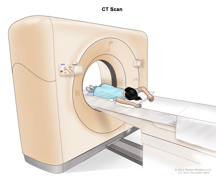
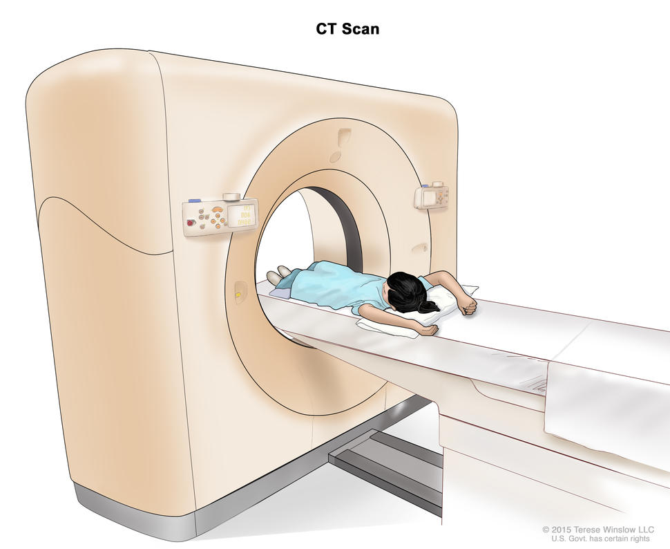
CT scan (CAT scan): A procedure that makes a series of detailed pictures of areas inside the body, such as the chest and abdomen, taken from different angles. The pictures are made by a computer linked to an x-ray machine. A dye may be injected into a vein or swallowed to help the organs or tissues show up more clearly. This procedure is also called computed tomography, computerized tomography, or computerized axial tomography. EnlargeComputed tomography (CT) scan. The child lies on a table that slides through the CT scanner, which takes a series of detailed x-ray pictures of areas inside the body.
MRI (magnetic resonance imaging): A procedure that uses a magnet, radio waves, and a computer to make a series of detailed pictures of areas inside the body, such as the chest and abdomen. This procedure is also called nuclear magnetic resonance imaging (NMRI).
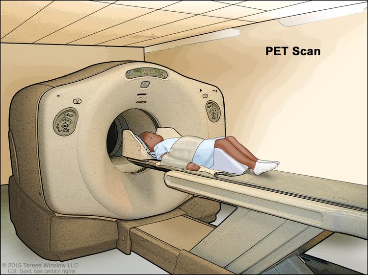
PET scan (positron emission tomography scan): A procedure to find malignant tumor cells in the body. A small amount of radioactive glucose (sugar) is injected into a vein. The PET scanner rotates around the body and makes a picture of where glucose is being used in the body. Malignant tumor cells show up brighter in the picture because they are more active and take up more glucose than normal cells do. EnlargePositron emission tomography (PET) scan. The child lies on a table that slides through the PET scanner. The head rest and white strap help the child lie still. A small amount of radioactive glucose (sugar) is injected into the child’s vein, and a scanner makes a picture of where the glucose is being used in the body. Cancer cells show up brighter in the picture because they take up more glucose than normal cells do.
Endoscopic ultrasound (EUS): A procedure in which an endoscope is inserted into the body, usually through the mouth or rectum. An endoscope is a thin, tube-like instrument with a light and a lens for viewing. A probe at the end of the endoscope is used to bounce high-energy sound waves (ultrasound) off internal tissues or organs and make echoes. The echoes form a picture of body tissues called a sonogram. This procedure is also called endosonography.
Somatostatin receptor scintigraphy: A type of radionuclide scan used to find pancreatic tumors. A very small amount of radioactiveoctreotide (a hormone that attaches to carcinoid tumors) is injected into a vein and travels through the bloodstream. The radioactive octreotide attaches to the tumor and a special camera that detects radioactivity is used to show where the tumors are in the body. This procedure is used to diagnose islet cell tumors.
Biopsy: The removal of cells or tissues so they can be viewed under a microscope by a pathologist to check for signs of cancer. Procedures used to obtain a tissue sample include the following:
Laparoscopy: A surgical procedure to look at the organs inside the abdomen to check for signs of disease. Small incisions (cuts) are made in the wall of the abdomen and a laparoscope (a thin, lighted tube) is inserted into one of the incisions. Other instruments may be inserted through the same or other incisions to perform procedures such as taking tissue samples.
Laparotomy: A surgical procedure in which an incision (cut) is made in the wall of the abdomen to check the inside of the abdomen for signs of disease. The size of the incision depends on the reason the laparotomy is being done. Sometimes tissue samples are taken.
Stages of Childhood Pancreatic Cancer
Key Points
If cancer has formed in the pancreas, tests are done to find out if cancer cells have spread to nearby areas or to other parts of the body.
There are three ways that cancer spreads in the body.
Cancer may spread from where it began to other parts of the body.
If cancer has formed in the pancreas, tests are done to find out if cancer cells have spread to nearby areas or to other parts of the body.
The process used to find out if cancer has spread from the pancreas to nearby areas or to other parts of the body is called staging. There is no standard system for staging childhood pancreatic cancer. The results of the tests and procedures done to diagnose cancer are used to help make decisions about treatment.
Sometimes childhood pancreatic cancer recurs (comes back) after treatment. It may come back in the pancreas or other parts of the body.
There are three ways that cancer spreads in the body.
Tissue. The cancer spreads from where it began by growing into nearby areas.
Lymph system. The cancer spreads from where it began by getting into the lymph system. The cancer travels through the lymph vessels to other parts of the body.
Blood. The cancer spreads from where it began by getting into the blood. The cancer travels through the blood vessels to other parts of the body.
Cancer may spread from where it began to other parts of the body.
When cancer spreads to another part of the body, it is called metastasis. Cancer cells break away from where they began (the primary tumor) and travel through the lymph system or blood.
Lymph system. The cancer gets into the lymph system, travels through the lymph vessels, and forms a tumor (metastatic tumor) in another part of the body.
Blood. The cancer gets into the blood, travels through the blood vessels, and forms a tumor (metastatic tumor) in another part of the body.
The metastatictumor is the same type of cancer as the primary tumor. For example, if pancreatic cancer spreads to the lung, the cancer cells in the lung are actually pancreas cancer cells. The disease is metastatic pancreatic cancer, not lung cancer.
Many cancer deaths are caused when cancer moves from the original tumor and spreads to other tissues and organs. This is called metastatic cancer. This animation shows how cancer cells travel from the place in the body where they first formed to other parts of the body.
Treatment Option Overview
Key Points
There are different types of treatment for children with pancreatic cancer.
Children with pancreatic cancer should have their treatment planned by a team of doctors who are experts in treating childhood cancer.
Three types of standard treatment are used:
Surgery
Chemotherapy
Targeted therapy
New types of treatment are being tested in clinical trials.
Treatment for childhood pancreatic cancer may cause side effects.
Patients may want to think about taking part in a clinical trial.
Patients can enter clinical trials before, during, or after starting their cancer treatment.
Follow-up tests may be needed.
There are different types of treatment for children with pancreatic cancer.
Some treatments are standard (the currently used treatment), and some are being tested in clinical trials. A treatment clinical trial is a research study meant to help improve current treatments or obtain information on new treatments for patients with cancer. When clinical trials show that a new treatment is better than the standard treatment, the new treatment may become the standard treatment.
Because cancer in children is rare, taking part in a clinical trial should be considered. Some clinical trials are open only to patients who have not started treatment.
Children with pancreatic cancer should have their treatment planned by a team of doctors who are experts in treating childhood cancer.
Treatment will be overseen by a pediatric oncologist, a doctor who specializes in treating children with cancer. The pediatric oncologist works with other pediatric health professionals who are experts in treating children with cancer and who specialize in certain areas of medicine. This may include the following specialists and others:
Chemotherapy is a cancer treatment that uses drugs to stop the growth of cancer cells, either by killing the cells or by stopping them from dividing. When chemotherapy is taken by mouth or injected into a vein or muscle, the drugs enter the bloodstream and can reach cancer cells throughout the body (systemic chemotherapy).
Targeted therapy
Targeted therapy is a type of treatment that uses drugs or other substances to identify and attack specific cancer cells. Targeted therapies usually cause less harm to normal cells than chemotherapy or radiation therapy do.
Side effects from cancer treatment that begin after treatment and continue for months or years are called late effects. Late effects of cancer treatment may include:
Physical problems.
Changes in mood, feelings, thinking, learning, or memory.
Some late effects may be treated or controlled. It is important to talk with your child’s doctors about the possible late effects caused by some treatments. See the PDQ summary on Late Effects of Treatment for Childhood Cancer for more information.
Patients may want to think about taking part in a clinical trial.
For some patients, taking part in a clinical trial may be the best treatment choice. Clinical trials are part of the cancer research process. Clinical trials are done to find out if new cancer treatments are safe and effective or better than the standard treatment.
Many of today’s standard treatments for cancer are based on earlier clinical trials. Patients who take part in a clinical trial may receive the standard treatment or be among the first to receive a new treatment.
Patients who take part in clinical trials also help improve the way cancer will be treated in the future. Even when clinical trials do not lead to effective new treatments, they often answer important questions and help move research forward.
Patients can enter clinical trials before, during, or after starting their cancer treatment.
Some clinical trials only include patients who have not yet received treatment. Other trials test treatments for patients whose cancer has not gotten better. There are also clinical trials that test new ways to stop cancer from recurring (coming back) or reduce the side effects of cancer treatment.
Clinical trials are taking place in many parts of the country. Information about clinical trials supported by NCI can be found on NCI’s clinical trials search webpage. Clinical trials supported by other organizations can be found on the ClinicalTrials.gov website.
Follow-up tests may be needed.
As your child goes through treatment, they will have follow-up tests or check-ups. Some tests that were done to diagnose or stage the cancer may be repeated to see how well the treatment is working. Decisions about whether to continue, change, or stop treatment may be based on the results of these tests.
Some of the tests will continue to be done from time to time after treatment has ended. The results of these tests can show if your child’s condition has changed or if the cancer has recurred (come back).
Treatment of newly diagnosed solid pseudopapillary tumor of the pancreas in children may include the following:
Surgery to remove the tumor. A Whipple procedure may be done for tumors in the head of the pancreas.
Chemotherapy for tumors that cannot be removed by surgery or have spread to other parts of the body.
Treatment of newly diagnosed pancreatoblastoma in children may include the following:
Surgery to remove the tumor. A Whipple procedure may be done for tumors in the head of the pancreas.
Chemotherapy may be given to shrink the tumor before surgery. More chemotherapy may be given after surgery for large tumors, tumors that could not initially be removed by surgery, and tumors that have spread to other parts of the body.
Chemotherapy may be given if the tumor does not respond to treatment or comes back.
Use our clinical trial search to find NCI-supported cancer clinical trials that are accepting patients. You can search for trials based on the type of cancer, the age of the patient, and where the trials are being done. General information about clinical trials is also available.
Treatment of Recurrent Childhood Pancreatic Cancer
A clinical trial that checks a sample of the patient’s tumor for certain gene changes. The type of targeted therapy that will be given to the patient depends on the type of gene change.
Use our clinical trial search to find NCI-supported cancer clinical trials that are accepting patients. You can search for trials based on the type of cancer, the age of the patient, and where the trials are being done. General information about clinical trials is also available.
Physician Data Query (PDQ) is the National Cancer Institute’s (NCI’s) comprehensive cancer information database. The PDQ database contains summaries of the latest published information on cancer prevention, detection, genetics, treatment, supportive care, and complementary and alternative medicine. Most summaries come in two versions. The health professional versions have detailed information written in technical language. The patient versions are written in easy-to-understand, nontechnical language. Both versions have cancer information that is accurate and up to date and most versions are also available in Spanish.
PDQ is a service of the NCI. The NCI is part of the National Institutes of Health (NIH). NIH is the federal government’s center of biomedical research. The PDQ summaries are based on an independent review of the medical literature. They are not policy statements of the NCI or the NIH.
Purpose of This Summary
This PDQ cancer information summary has current information about the treatment of childhood pancreatic cancer. It is meant to inform and help patients, families, and caregivers. It does not give formal guidelines or recommendations for making decisions about health care.
Reviewers and Updates
Editorial Boards write the PDQ cancer information summaries and keep them up to date. These Boards are made up of experts in cancer treatment and other specialties related to cancer. The summaries are reviewed regularly and changes are made when there is new information. The date on each summary (“Updated”) is the date of the most recent change.
The information in this patient summary was taken from the health professional version, which is reviewed regularly and updated as needed, by the PDQ Pediatric Treatment Editorial Board.
Clinical Trial Information
A clinical trial is a study to answer a scientific question, such as whether one treatment is better than another. Trials are based on past studies and what has been learned in the laboratory. Each trial answers certain scientific questions in order to find new and better ways to help cancer patients. During treatment clinical trials, information is collected about the effects of a new treatment and how well it works. If a clinical trial shows that a new treatment is better than one currently being used, the new treatment may become “standard.” Patients may want to think about taking part in a clinical trial. Some clinical trials are open only to patients who have not started treatment.
Clinical trials can be found online at NCI’s website. For more information, call the Cancer Information Service (CIS), NCI’s contact center, at 1-800-4-CANCER (1-800-422-6237).
Permission to Use This Summary
PDQ is a registered trademark. The content of PDQ documents can be used freely as text. It cannot be identified as an NCI PDQ cancer information summary unless the whole summary is shown and it is updated regularly. However, a user would be allowed to write a sentence such as “NCI’s PDQ cancer information summary about breast cancer prevention states the risks in the following way: [include excerpt from the summary].”
The best way to cite this PDQ summary is:
PDQ® Pediatric Treatment Editorial Board. PDQ Childhood Pancreatic Cancer Treatment. Bethesda, MD: National Cancer Institute. Updated <MM/DD/YYYY>. Available at: /types/pancreatic/patient/child-pancreatic-treatment-pdq. Accessed <MM/DD/YYYY>.
Images in this summary are used with permission of the author(s), artist, and/or publisher for use in the PDQ summaries only. If you want to use an image from a PDQ summary and you are not using the whole summary, you must get permission from the owner. It cannot be given by the National Cancer Institute. Information about using the images in this summary, along with many other images related to cancer can be found in Visuals Online. Visuals Online is a collection of more than 3,000 scientific images.
Disclaimer
The information in these summaries should not be used to make decisions about insurance reimbursement. More information on insurance coverage is available on Cancer.gov on the Managing Cancer Care page.
Contact Us
More information about contacting us or receiving help with the Cancer.gov website can be found on our Contact Us for Help page. Questions can also be submitted to Cancer.gov through the website’s E-mail Us.
Paraganglioma forms in nerve tissue near the carotid artery, along nerve pathways in the head and neck, and in other parts of the body.
Pheochromocytoma and paraganglioma may be benign (not cancer) or malignant (cancer).
Inheriting certain gene mutations (changes) increases the risk of pheochromocytoma and paraganglioma.
Signs and symptoms of pheochromocytoma and paraganglioma occur if too much adrenaline or noradrenaline is released into the blood.
Tests and procedures used to diagnose and stage pheochromocytoma and paraganglioma depend on the patient’s signs and symptoms and family history.
Certain factors affect prognosis (chance of recovery) and treatment options.
Pheochromocytoma forms in the adrenal gland.
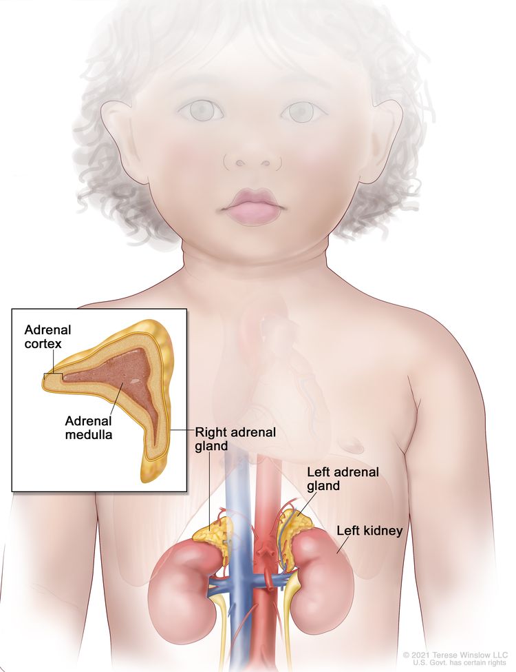
Pheochromocytoma forms in the adrenal gland. There are two adrenal glands, one on top of each kidney in the back of the upper abdomen. Each adrenal gland has two parts. The outer layer of the adrenal gland is the adrenal cortex. The center of the adrenal gland is the adrenal medulla. Pheochromocytoma is a tumor of the adrenal medulla.
EnlargeAnatomy of the adrenal gland. There are two adrenal glands, one on top of each kidney. The outer part of each gland is the adrenal cortex and the inner part is the adrenal medulla.
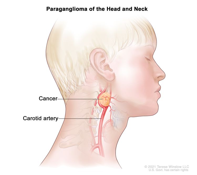
Paraganglioma forms in nerve tissue near the carotid artery, along nerve pathways in the head and neck, and in other parts of the body.
Some paragangliomas make extra catecholamines called adrenaline and noradrenaline. The release of extra adrenaline and noradrenaline into the blood may cause symptoms.
EnlargeA paraganglioma is a rare tumor that often forms in nerve tissue near the carotid artery. It may also form along nerve pathways in the head and neck and in other parts of the body.
Pheochromocytoma and paraganglioma may be benign (not cancer) or malignant (cancer).
About half of all children with pheochromocytoma or paraganglioma have malignant pheochromocytoma or paraganglioma. This means that the tumor cells have spread to other parts of the body. Benign and malignant pheochromocytoma and paraganglioma require treatment because they can cause severe or life-threatening heart problems and affect many body functions.
Inheriting certain gene mutations (changes) increases the risk of pheochromocytoma and paraganglioma.
Anything that increases your chance of getting a disease is called a risk factor. Having a risk factor does not mean that you will get cancer; not having risk factors doesn’t mean that you will not get cancer. Talk with your child’s doctor if you think your child may be at risk.
Having any of the following inheritedsyndromes or gene changes increases risk of pheochromocytoma and paraganglioma:
More than half of the children and adolescentsdiagnosed with pheochromocytoma or paraganglioma have an inherited syndrome or gene change that increases the risk of cancer. Genetic counseling (a discussion with a trained professional about inherited diseases) and testing is an important part of the treatment plan.
Signs and symptoms of pheochromocytoma and paraganglioma occur if too much adrenaline or noradrenaline is released into the blood.
Some tumors do not make extra adrenaline or noradrenaline and do not cause symptoms. These tumors may be found when a lump forms in the neck or when a test is done for another reason. Signs and symptoms of pheochromocytoma and paraganglioma occur when too much adrenaline or noradrenaline is released into the blood. These and other signs and symptoms may be caused by pheochromocytoma, paraganglioma, or other conditions.
Check with your child’s doctor if your child has any of the following:
These signs and symptoms may come and go, but high blood pressure is more likely to occur for long periods of time in young patients. These signs and symptoms may also occur with physical activity, injury, anesthesia, surgery to remove the tumor, eating foods such as chocolate and cheese, or while passing urine (if the tumor is in the bladder).
Tests and procedures used to diagnose and stage pheochromocytoma and paraganglioma depend on the patient’s signs and symptoms and family history.
Tests are done to diagnose and stage cancer. After cancer is diagnosed, more tests are done to find out if cancer cells have spread to nearby areas or to other parts of the body. This process is called staging.
The following tests and procedures may be used:
Physical exam and health history: An exam of the body to check general signs of health, including checking for signs of disease, such as lumps or anything else that seems unusual. A history of the patient’s health habits and past illnesses and treatments will also be taken.
Plasma-free metanephrines test: A blood test that measures the amount of metanephrines in the blood. Metanephrines are substances that are made when the body breaks down adrenaline or noradrenaline. Pheochromocytomas and paragangliomas can make large amounts of adrenaline and noradrenaline and cause high levels of metanephrines in both the blood and urine.
Blood catecholamine studies: A procedure in which a blood sample is checked to measure the amount of certain catecholamines (adrenaline or noradrenaline) released into the blood. Substances caused by the breakdown of these catecholamines are also measured. An unusual (unusual higher or lower than normal) amount of a substance can be a sign of disease in the organ or tissue that makes it. Higher than normal amounts may be a sign of pheochromocytoma or paraganglioma.
Twenty-four-hour urine test: A test in which urine is collected for 24 hours to measure the amounts of catecholamines (adrenaline or noradrenaline) or metanephrines in the urine. Substances caused by the breakdown of these catecholamines are also measured. An unusual (higher than normal) amount of a substance can be a sign of disease in the organ or tissue that makes it. Higher than normal amounts may be a sign of pheochromocytoma or paraganglioma.
PET scan: A procedure to find malignant tumor cells in the body. A small amount of radioactive glucose (sugar) is injected into a vein. The PET scanner rotates around the body and makes a picture of where glucose is being used in the body. Malignant tumor cells show up brighter in the picture because they are more active and take up more glucose than normal cells do. EnlargePositron emission tomography (PET) scan. The child lies on a table that slides through the PET scanner. The head rest and white strap help the child lie still. A small amount of radioactive glucose (sugar) is injected into the child’s vein, and a scanner makes a picture of where the glucose is being used in the body. Cancer cells show up brighter in the picture because they take up more glucose than normal cells do.
CT scan (CAT scan): A procedure that makes a series of detailed pictures of areas inside the body, taken from different angles. The pictures are made by a computer linked to an x-ray machine. A dye may be injected into a vein or swallowed to help the organs or tissues show up more clearly. This procedure is also called computed tomography, computerized tomography, or computerized axial tomography. EnlargeComputed tomography (CT) scan. The child lies on a table that slides through the CT scanner, which takes a series of detailed x-ray pictures of areas inside the body.
MRI (magnetic resonance imaging): A procedure that uses a magnet, radio waves, and a computer to make a series of detailed pictures of areas inside the body. This procedure is also called nuclear magnetic resonance imaging (NMRI). EnlargeMagnetic resonance imaging (MRI) scan. The child lies on a table that slides into the MRI machine, which takes a series of detailed pictures of areas inside the body. The positioning of the child on the table depends on the part of the body being imaged.
MIBG scan: A procedure used to find neuroendocrine tumors, such as pheochromocytoma and paraganglioma. A very small amount of a substance called radioactive MIBG is injected into a vein and travels through the bloodstream. Neuroendocrine tumor cells take up the radioactive MIBG and are detected by a scanner. Scans may be taken over 1-3 days. An iodine solution may be given before or during the test to keep the thyroid gland from absorbing too much of the MIBG.
Somatostatin receptor scintigraphy: A type of radionuclide scan that may be used to find tumors. A very small amount of radioactive octreotide (a hormone that attaches to tumors) is injected into a vein and travels through the blood. The radioactive octreotide attaches to the tumor and a special camera that detects radioactivity is used to show where the tumors are in the body. This procedure is also called octreotide scan and SRS.
Genetic testing: A laboratory test in which cells or tissue are analyzed to look for changes in genes or chromosomes. These changes may be a sign that a person has or is at risk of having a specific disease or condition. The following are genes that might be tested for in children with pheochromocytoma or paraganglioma: VHL, NF1, RET, SDHD, SDHB, SDHA, MAX, and TMEM127 genes.
Certain factors affect prognosis (chance of recovery) and treatment options.
Prognosis and treatment options depend on the following:
Whether the cancer has just been diagnosed or has recurred (come back).
Stages of Childhood Pheochromocytoma and Paraganglioma
Key Points
After pheochromocytoma or paraganglioma has been diagnosed, tests are done to find out if cancer cells have spread to nearby areas or to other parts of the body.
There are three ways that cancer spreads in the body.
Cancer may spread from where it began to other parts of the body.
After pheochromocytoma or paraganglioma has been diagnosed, tests are done to find out if cancer cells have spread to nearby areas or to other parts of the body.
Sometimes childhood pheochromocytoma or paraganglioma recurs (comes back) after treatment. It may come back in the place it first formed or in other parts of the body.
There are three ways that cancer spreads in the body.
Tissue. The cancer spreads from where it began by growing into nearby areas.
Lymph system. The cancer spreads from where it began by getting into the lymph system. The cancer travels through the lymph vessels to other parts of the body.
Blood. The cancer spreads from where it began by getting into the blood. The cancer travels through the blood vessels to other parts of the body.
Cancer may spread from where it began to other parts of the body.
When cancer spreads to another part of the body, it is called metastasis. Cancer cells break away from where they began (the primary tumor) and travel through the lymph system or blood.
Lymph system. The cancer gets into the lymph system, travels through the lymph vessels, and forms a tumor (metastatic tumor) in another part of the body.
Blood. The cancer gets into the blood, travels through the blood vessels, and forms a tumor (metastatic tumor) in another part of the body.
The metastatictumor is the same type of cancer as the primary tumor. For example, if pheochromocytoma spreads to the bone, the cancer cells in the bone are actually pheochromocytoma cells. The disease is metastatic pheochromocytoma, not bone cancer.
Many cancer deaths are caused when cancer moves from the original tumor and spreads to other tissues and organs. This is called metastatic cancer. This animation shows how cancer cells travel from the place in the body where they first formed to other parts of the body.
Treatment Option Overview
Key Points
There are different types of treatment for children with pheochromocytoma or paraganglioma.
Children with pheochromocytoma or paraganglioma should have their treatment planned by a team of doctors who are experts in treating childhood cancer.
Four types of standard treatment are used:
Surgery
Chemotherapy
High-dose 131I-MIBG therapy
Targeted therapy
New types of treatment are being tested in clinical trials.
Treatment of pheochromocytoma and paraganglioma may cause side effects.
Patients may want to think about taking part in a clinical trial.
Patients can enter clinical trials before, during, or after starting their cancer treatment.
Follow-up tests may be needed.
There are different types of treatment for children with pheochromocytoma or paraganglioma.
Some treatments are standard (the currently used treatment), and some are being tested in clinical trials. A treatment clinical trial is a research study meant to help improve current treatments or obtain information on new treatments for patients with cancer. When clinical trials show that a new treatment is better than the standard treatment, the new treatment may become the standard treatment.
Because cancer in children is rare, taking part in a clinical trial should be considered. Some clinical trials are open only to patients who have not started treatment.
Children with pheochromocytoma or paraganglioma should have their treatment planned by a team of doctors who are experts in treating childhood cancer.
Treatment will be overseen by a pediatric oncologist, a doctor who specializes in treating children with cancer. The pediatric oncologist works with other pediatric health professionals who are experts in treating children with cancer and who specialize in certain areas of medicine. This may include the following specialists and others:
Chemotherapy is a cancer treatment that uses drugs to stop the growth of cancer cells, either by killing the cells or by stopping them from dividing. When chemotherapy is taken by mouth or injected into a vein or muscle, the drugs enter the bloodstream and can reach cancer cells throughout the body (systemic chemotherapy).
High-dose 131I-MIBG therapy
131I-MIBGtherapy is a treatment with high-doseradioactive iodine. The radioactive iodine is given through an intravenous (IV) line and enters the bloodstream which carries radiation directly to tumor cells. Radioactive iodine collects in pheochromocytoma and paraganglioma cells and kills them with the radiation that is given off.
Targeted therapy
Targeted therapy is a type of treatment that uses drugs or other substances to identify and attack specific cancer cells. Targeted therapies usually cause less harm to normal cells than chemotherapy or radiation therapy do.
Patients may want to think about taking part in a clinical trial.
For some patients, taking part in a clinical trial may be the best treatment choice. Clinical trials are part of the cancer research process. Clinical trials are done to find out if new cancer treatments are safe and effective or better than the standard treatment.
Many of today’s standard treatments for cancer are based on earlier clinical trials. Patients who take part in a clinical trial may receive the standard treatment or be among the first to receive a new treatment.
Patients who take part in clinical trials also help improve the way cancer will be treated in the future. Even when clinical trials do not lead to effective new treatments, they often answer important questions and help move research forward.
Patients can enter clinical trials before, during, or after starting their cancer treatment.
Some clinical trials only include patients who have not yet received treatment. Other trials test treatments for patients whose cancer has not gotten better. There are also clinical trials that test new ways to stop cancer from recurring (coming back) or reduce the side effects of cancer treatment.
Clinical trials are taking place in many parts of the country. Information about clinical trials supported by NCI can be found on NCI’s clinical trials search webpage. Clinical trials supported by other organizations can be found on the ClinicalTrials.gov website.
Follow-up tests may be needed.
As your child goes through treatment, they will have follow-up tests or check-ups. Some tests that were done to diagnose or stage the cancer may be repeated to see how well the treatment is working. Decisions about whether to continue, change, or stop treatment may be based on the results of these tests.
Some of the tests will continue to be done from time to time after treatment has ended. The results of these tests can show if your child’s condition has changed or if the cancer has recurred (come back).
For patients with pheochromocytoma or paraganglioma that causes symptoms, catecholamine levels in the blood and urine will be checked on a regular basis. Catecholamine levels that are higher than normal can be a sign that the cancer has come back. Talk to your child’s doctor about which tests should be done and how often.
Patients with pheochromocytoma or paraganglioma need lifelong follow-up.
Treatment of Childhood Pheochromocytoma and Paraganglioma
Use our clinical trial search to find NCI-supported cancer clinical trials that are accepting patients. You can search for trials based on the type of cancer, the age of the patient, and where the trials are being done. General information about clinical trials is also available.
Treatment of Recurrent Childhood Pheochromocytoma and Paraganglioma
A clinical trial that checks a sample of the patient’s tumor for certain gene changes. The type of targeted therapy that will be given to the patient depends on the type of gene change.
Use our clinical trial search to find NCI-supported cancer clinical trials that are accepting patients. You can search for trials based on the type of cancer, the age of the patient, and where the trials are being done. General information about clinical trials is also available.
To Learn More About Childhood Pheochromocytoma and Paraganglioma
Physician Data Query (PDQ) is the National Cancer Institute’s (NCI’s) comprehensive cancer information database. The PDQ database contains summaries of the latest published information on cancer prevention, detection, genetics, treatment, supportive care, and complementary and alternative medicine. Most summaries come in two versions. The health professional versions have detailed information written in technical language. The patient versions are written in easy-to-understand, nontechnical language. Both versions have cancer information that is accurate and up to date and most versions are also available in Spanish.
PDQ is a service of the NCI. The NCI is part of the National Institutes of Health (NIH). NIH is the federal government’s center of biomedical research. The PDQ summaries are based on an independent review of the medical literature. They are not policy statements of the NCI or the NIH.
Purpose of This Summary
This PDQ cancer information summary has current information about the treatment of childhood pheochromocytoma and paraganglioma. It is meant to inform and help patients, families, and caregivers. It does not give formal guidelines or recommendations for making decisions about health care.
Reviewers and Updates
Editorial Boards write the PDQ cancer information summaries and keep them up to date. These Boards are made up of experts in cancer treatment and other specialties related to cancer. The summaries are reviewed regularly and changes are made when there is new information. The date on each summary (“Updated”) is the date of the most recent change.
The information in this patient summary was taken from the health professional version, which is reviewed regularly and updated as needed, by the PDQ Pediatric Treatment Editorial Board.
Clinical Trial Information
A clinical trial is a study to answer a scientific question, such as whether one treatment is better than another. Trials are based on past studies and what has been learned in the laboratory. Each trial answers certain scientific questions in order to find new and better ways to help cancer patients. During treatment clinical trials, information is collected about the effects of a new treatment and how well it works. If a clinical trial shows that a new treatment is better than one currently being used, the new treatment may become “standard.” Patients may want to think about taking part in a clinical trial. Some clinical trials are open only to patients who have not started treatment.
Clinical trials can be found online at NCI’s website. For more information, call the Cancer Information Service (CIS), NCI’s contact center, at 1-800-4-CANCER (1-800-422-6237).
Permission to Use This Summary
PDQ is a registered trademark. The content of PDQ documents can be used freely as text. It cannot be identified as an NCI PDQ cancer information summary unless the whole summary is shown and it is updated regularly. However, a user would be allowed to write a sentence such as “NCI’s PDQ cancer information summary about breast cancer prevention states the risks in the following way: [include excerpt from the summary].”
Images in this summary are used with permission of the author(s), artist, and/or publisher for use in the PDQ summaries only. If you want to use an image from a PDQ summary and you are not using the whole summary, you must get permission from the owner. It cannot be given by the National Cancer Institute. Information about using the images in this summary, along with many other images related to cancer can be found in Visuals Online. Visuals Online is a collection of more than 3,000 scientific images.
Disclaimer
The information in these summaries should not be used to make decisions about insurance reimbursement. More information on insurance coverage is available on Cancer.gov on the Managing Cancer Care page.
Contact Us
More information about contacting us or receiving help with the Cancer.gov website can be found on our Contact Us for Help page. Questions can also be submitted to Cancer.gov through the website’s E-mail Us.
Anatomy of the digestive tract. The digestive tract is made up of organs that food and liquids travel through when they are swallowed, digested, absorbed, and leave the body as feces. These organs include the mouth, pharynx (throat), esophagus, stomach, small intestine, large intestine, rectum, and anus.
Childhood stomach cancer is a very rare cancer that starts in the cells lining the stomach. The stomach is an organ on the left side of the upper abdomen that digests food. The stomach is part of the digestive tract, a series of hollow, muscular organs joined in a long, twisting tube from the mouth to the anus. The digestive tract processes nutrients in foods that are eaten and helps pass waste material out of the body:
Food moves from the throat to the stomach through a tube called the esophagus.
After food enters the stomach, it is broken down by stomach muscles that mix the food and liquid with digestive juices.
After leaving the stomach, partly digested food passes into the small intestine and then into the large intestine.
The end of the large intestine, called the rectum, stores the waste from the digested food until it is pushed out of the anus during a bowel movement.
Causes and risk factors for childhood stomach cancer
Childhood stomach cancer is caused by certain changes to the way stomach cells function, especially how they grow and divide into new cells. Often, the exact cause of the cell changes is unknown. Learn more about how cancer develops at What Is Cancer?
A risk factor is anything that increases the chance of getting a disease. Not every child with a risk factor will develop stomach cancer, and it will develop in some children who don’t have a known risk factor.
Risk factors for childhood stomach cancer include:
H. pylori infection. Chronic infection of the mucosal layer of the stomach with H. pylori is a risk factor for stomach cancer. This bacterium spreads from person to person through direct contact with saliva, vomit, or stool. Although many people with chronic H. pylori infections do not have symptoms, some develop stomach ulcers or inflammation of the stomach called atrophic gastritis. In some people, atrophic gastritis leads to increasingly severe changes in the stomach lining and eventually to stomach cancer or gastric MALT lymphoma. Treatment of H. pylori infections reduces the risk of these types of stomach cancer.
Talk with your child’s doctor if you think your child might be at risk of stomach cancer.
Genetic counseling for children with stomach cancer
It may not be clear from the family medical history whether a child with stomach cancer has an inherited condition that increased their risk. Genetic testing may help explain why a child develops a rare cancer or a cancer that is usually seen in adults, such as stomach cancer. Genetic counselors and other specially trained health professionals can discuss your child’s diagnosis and your family’s medical history to help you understand:
the risk of stomach cancer and other cancers for your child’s siblings
the risks and benefits of learning genetic information
Genetic counselors can also help you cope with your child’s genetic testing results, including how to discuss the results with family members.
Symptoms of childhood stomach cancer
Many children do not have symptoms of stomach cancer until the cancer has spread. It’s important to check with your child’s doctor if your child has:
stomach pain
loss of appetite
weight loss for no known reason
nausea
vomiting
constipation or diarrhea
weakness
anemia (symptoms of which may include tiredness, dizziness, fast or irregular heartbeat, shortness of breath, and pale skin)
These symptoms may be caused by conditions other than childhood stomach cancer. The only way to know is to see your child’s doctor. The doctor will ask you when the symptoms started and how often your child has been having them as a first step in making a diagnosis.
Diagnosis of childhood stomach cancer
If your child has symptoms that suggest stomach cancer, the doctor will need to find out if they are due to cancer or another condition. The doctor will ask about your child’s personal and family medical history and do a physical exam. Depending on these results, they may recommend tests to find out if your child has stomach cancer.
The following tests and procedures are used to diagnose stomach cancer. The results will also help you and your child’s doctor plan treatment.
CT scan (CAT scan)
Computed tomography (CT) scan. The child lies on a table that slides through the CT scanner, which takes a series of detailed x-ray pictures of areas inside the body.
A CT scan uses a computer linked to an x-ray machine to make a series of detailed pictures of areas inside the body from different angles. A dye may be injected into a vein or swallowed to help the organs or tissues show up more clearly. This procedure is also called computed tomography, computerized tomography, or computerized axial tomography.
Upper endoscopy is a procedure to look inside the esophagus, stomach, and duodenum (first part of the small intestine) to check for abnormal areas. An endoscope is passed through the mouth and down the throat into the esophagus. An endoscope is a thin, tube-like instrument with a light and a lens for viewing. It may also have a tool to remove tissue or lymph node samples (biopsy), which are checked under a microscope for signs of cancer.
Talk with your child’s doctor about what to expect during and after your child’s biopsy.
Learn about the type of information that can be found in a pathologist’s report about the cells or tissue removed during a biopsy at Pathology Reports.
Barium swallow
Barium swallow is a series of x-rays of the esophagus and stomach. The patient drinks a liquid that contains barium (a silver-white metallic compound). The liquid coats the esophagus and stomach, and x-rays are taken. This procedure is also called an upper GI series.
Getting a second opinion
You may want to get a second opinion to confirm your child’s stomach cancer diagnosis and treatment plan. If you seek a second opinion, you will need to get medical test results and reports from the first doctor to share with the second doctor. The second doctor will review the pathology report, slides, and scans. This doctor may agree with the first doctor, suggest changes to the treatment plan, or provide more information about your child’s cancer.
To learn more about choosing a doctor and getting a second opinion, see Finding Cancer Care. You can contact NCI’s Cancer Information Service via chat, email, or phone (both in English and Spanish) for help finding a doctor or hospital that can provide a second opinion. For questions you might want to ask at your child’s appointments, see Questions to Ask Your Doctor about Cancer.
Prognostic factors for childhood stomach cancer
If your child has been diagnosed with stomach cancer, you likely have questions about how serious the cancer is and your child’s chances of survival. The likely outcome or course of a disease is called prognosis. The prognosis can be affected by whether the cancer has spread to other parts of the body at the time of diagnosis and how well the cancer responds to treatment. Your child’s cancer care team is in the best position to talk with you about your child’s prognosis.
Stages of childhood stomach cancer
Cancer stage describes the extent of cancer in the body, such as the size of the tumor, whether it has spread, and how far it has spread from where it first formed. There is no staging system used for childhood stomach cancer, but the tests and procedures done to diagnose the cancer are also used to help plan treatment.
Types of treatment for childhood stomach cancer
There are different types of treatment for children and adolescents with stomach cancer. You and your child’s cancer care team will work together to decide treatment. Many factors will be considered, such as your child’s overall health and whether the cancer is newly diagnosed or has come back.
A pediatric oncologist, a doctor who specializes in treating children with cancer, oversees treatment for childhood stomach cancer. The pediatric oncologist works with other pediatric health care providers who are experts in treating children with cancer and who specialize in certain areas of medicine. Other specialists may include:
Your child’s treatment plan will include information about the cancer, the goals of treatment, treatment options, and the possible side effects. It will be helpful to talk with your child’s cancer care team before treatment begins about what to expect. For help every step of the way, see our downloadable booklet, Children with Cancer: A Guide for Parents.
The types of treatment your child might have include:
Surgery
Surgery to remove the tumor is the main treatment for stomach cancer.
Radiation therapy
Radiation therapy uses high-energy x-rays or other types of radiation to kill cancer cells or keep them from growing. Stomach cancer is sometimes treated with external beam radiation therapy. This type of radiation therapy uses a machine outside the body to send radiation toward the area of the body with cancer. Radiation therapy may be given alone or with other treatments, such as chemotherapy.
Chemotherapy (also called chemo) uses drugs to stop the growth of cancer cells, either by killing the cells or by stopping them from dividing. Chemotherapy may be given alone or with other types of treatment, such as radiation therapy.
For stomach cancer, the chemotherapy is injected into a vein. When given this way, the drugs enter the bloodstream to reach cancer cells throughout the body. Chemotherapy drugs used alone or in combination to treat stomach cancer in children are:
Other chemotherapy drugs not listed here may also be used.
Learn more about how chemotherapy works, how it is given, common side effects, and more at Chemotherapy to Treat Cancer.
Clinical trials
For some children, joining a clinical trial may be an option. There are different types of clinical trials for childhood cancer. For example, a treatment trial tests new treatments or new ways of using current treatments. Supportive care and palliative care trials look at ways to improve quality of life, especially for those who have side effects from cancer and its treatment.
You can use the clinical trial search to find NCI-supported cancer clinical trials accepting participants. The search allows you to filter trials based on the type of cancer, your child’s age, and where the trials are being done. Clinical trials supported by other organizations can be found on the ClinicalTrials.gov website.
Treatment of newly diagnosed stomach cancer in children may include:
surgery to remove the cancer and some healthy tissue around it
surgery to remove as much of the cancer as possible, followed by radiation therapy and chemotherapy
If your child’s cancer comes back after treatment, their doctor will talk with you about what to expect and next steps. There might be treatment options that may shrink the cancer or control its growth. If there are no treatments, your child can receive care to control symptoms from cancer so they can be as comfortable as possible.
Side effects of treatment
Cancer treatments can cause side effects. Which side effects your child might have depends on the type of treatment they receive, the dose, and how their body reacts. Talk with your child’s treatment team about which side effects to look for and ways to manage them.
To learn more about side effects that begin during treatment for cancer, visit Side Effects.
Side effects from cancer treatment that begin after treatment and continue for months or years are called late effects. Late effects of cancer treatment may include:
physical problems
changes in mood, feelings, thinking, learning, or memory
second cancers (new types of cancer) or other conditions
Some late effects may be treated or controlled. It is important to talk with your child’s doctors about the possible late effects caused by some treatments.
As your child goes through treatment, they will have follow-up tests or check-ups. Some of the tests that were done to diagnose the cancer may be repeated to see how well the treatment is working. Decisions about whether to continue, change, or stop treatment may be based on the results of these tests.
Some of the tests will continue to be done from time to time after treatment has ended. The results of these tests can show if your child’s condition has changed or if the cancer has recurred (come back).
Coping with cancer
When your child has cancer, every member of the family needs support. Honest and calm conversations build trust as you talk with your child and their siblings. Taking care of yourself during this difficult time is also important. Reach out to your child’s treatment team and to people in your family and community for support. To learn more, see Support for Families: Childhood Cancer and the booklet Children with Cancer: A Guide for Parents.
Adrenocortical cancer (also called cancer of the adrenal cortex) is rare. Certain inherited disorders increase the risk of adrenocortical cancer. Explore the links on this page to learn more about adrenocortical cancer treatment, research, and clinical trials.
Adrenocortical carcinoma is a rare disease in which malignant (cancer) cells form in the outer layer of the adrenal gland.
Having a certain mutation (change) in the TP53 gene increases the risk of adrenocortical carcinoma.
Signs and symptoms of adrenocortical carcinoma include a lump or pain in the abdomen.
Tests that examine the adrenal glands are used to diagnose and stage adrenocortical carcinoma.
Certain factors affect prognosis (chance of recovery).
Adrenocortical carcinoma is a rare disease in which malignant (cancer) cells form in the outer layer of the adrenal gland.
There are two adrenal glands. The adrenal glands are small and shaped like a triangle. One adrenal gland sits on top of each kidney. Each adrenal gland has two parts. The outer layer of the adrenal gland is the adrenal cortex. The center of the adrenal gland is the adrenal medulla. Adrenocortical carcinoma is also called adrenocortical cancer or cancer of the adrenal cortex.
EnlargeAnatomy of the adrenal gland. There are two adrenal glands, one on top of each kidney. The outer part of each gland is the adrenal cortex and the inner part is the adrenal medulla.
Most childhood adrenocorticaltumors occur during the first 5 years of life, but they may also occur during adolescence.
Having a certain mutation (change) in the TP53 gene increases the risk of adrenocortical carcinoma.
Anything that increases your chance of getting a disease is called a risk factor. Having a risk factor does not mean that you will get cancer; not having risk factors doesn’t mean that you will not get cancer. Talk with your child’s doctor if you think your child may be at risk.
The risk of adrenocortical carcinoma is increased by having a mutation (change) in the TP53gene or any of the following syndromes:
Also, cancer of the adrenal cortex may be functioning (makes more hormones than normal) or nonfunctioning (does not make extra hormones). Most tumors of the adrenal cortex in children are functioning tumors. The extra hormones made by functioning tumors may cause certain signs or symptoms of disease and these depend on the type of hormone made by the tumor. For example, extra androgen hormone may also cause male children to develop an enlarged penis and female children to develop enlarged genitalia. Extra estrogen hormone may cause the growth of breasttissue in male children. Extra cortisol hormone may cause (hypercortisolism).
See the PDQ summary onAdrenocortical Carcinoma Treatment (Adult) for more information on the signs and symptoms of adrenocortical carcinoma.
Tests that examine the adrenal glands are used to diagnose and stage adrenocortical carcinoma.
Tests are done to diagnose and stage cancer. After cancer is diagnosed, more tests are done to find out if cancer cells have spread to nearby areas or to other parts of the body. This process is called staging. It is important to know whether cancer has spread in order to plan the best treatment.
The following tests and procedures may be used:
Physical exam and health history: An exam of the body to check general signs of health, including checking for signs of disease, such as lumps or anything else that seems unusual. A history of the patient’s health habits and past illnesses and treatments will also be taken.
X-ray: An x-ray of the chest, abdomen, or bones inside the body. An x-ray is a type of energy beam that can go through the body and onto film, making a picture of areas inside the body.
CT scan: A procedure that makes a series of detailed pictures of areas inside the body, such as the chest or abdomen, taken from different angles. The pictures are made by a computer linked to an x-ray machine. A dye may be injected into a vein or swallowed to help the organs or tissues show up more clearly. This procedure is also called computed tomography, computerized tomography, or computerized axial tomography. EnlargeComputed tomography (CT) scan. The child lies on a table that slides through the CT scanner, which takes a series of detailed x-ray pictures of areas inside the body.
MRI (magnetic resonance imaging): A procedure that uses a magnet, radio waves, and a computer to make a series of detailed pictures of areas of the body, such as the chest and abdomen. This procedure is also called nuclear magnetic resonance imaging (NMRI). EnlargeMagnetic resonance imaging (MRI) scan. The child lies on a table that slides into the MRI machine, which takes a series of detailed pictures of areas inside the body. The positioning of the child on the table depends on the part of the body being imaged.
PET scan: A procedure to find malignant tumor cells in the body. A small amount of radioactive glucose (sugar) is injected into a vein. The PET scanner rotates around the body and makes a picture of where glucose is being used in the body. Malignant tumor cells show up brighter in the picture because they are more active and take up more glucose than normal cells do. EnlargePositron emission tomography (PET) scan. The child lies on a table that slides through the PET scanner. The head rest and white strap help the child lie still. A small amount of radioactive glucose (sugar) is injected into the child’s vein, and a scanner makes a picture of where the glucose is being used in the body. Cancer cells show up brighter in the picture because they take up more glucose than normal cells do.
Ultrasound exam: A procedure in which high-energy sound waves (ultrasound) are bounced off internal tissues or organs, such as the abdomen, and make echoes. The echoes form a picture of body tissues called a sonogram. The picture can be printed to be looked at later. EnlargeAbdominal ultrasound. An ultrasound transducer connected to a computer is pressed against the skin of the abdomen. The transducer bounces sound waves off internal organs and tissues to make echoes that form a sonogram (computer picture).
Biopsy: The removal of cells or tissues during surgery so they can be viewed under a microscope by a pathologist to check for signs of cancer.
Blood chemistry studies: A procedure in which a blood sample is checked to measure the amounts of certain substances released into the blood by organs and tissues in the body. An unusual (higher or lower than normal) amount of a substance can be a sign of disease.
Blood hormone studies: A procedure in which a blood sample is checked to measure the amounts of certain hormones released into the blood by organs and tissues in the body. An unusual (higher or lower than normal) amount of a substance can be a sign of disease in the organ or tissue that makes it. The blood may be checked for testosterone or estrogen. A higher than normal amount of these hormones may be a sign of adrenocortical carcinoma.
Twenty-four-hour urine test: A test in which urine is collected for 24 hours to measure the amounts of cortisol or 17-ketosteroids. A higher than normal amount of these substances in the urine may be a sign of disease in the adrenal cortex.
Low-dosedexamethasone suppression test: A test in which one or more small doses of dexamethasone are given. The level of cortisol is checked from a sample of blood or from urine that is collected for three days. This test is done to check if the adrenal gland is making too much cortisol.
High-dose dexamethasone suppression test: A test in which one or more high doses of dexamethasone are given. The level of cortisol is checked from a sample of blood or from urine that is collected for three days. This test is done to check if the adrenal gland is making too much cortisol or if the pituitary gland is telling the adrenal glands to make too much cortisol.
Adrenal angiography: A procedure to look at the arteries and the flow of blood near the adrenal gland. A contrast dye is injected into the adrenal arteries. As the dye moves through the blood vessel, a series of x-rays are taken to see if any arteries are blocked.
Adrenal venography: A procedure to look at the adrenal veins and the flow of blood near the adrenal glands. A contrast dye is injected into an adrenal vein. As the contrast dye moves through the vein, a series of x-rays are taken to see if any veins are blocked. A catheter (very thin tube) may be inserted into the vein to take a blood sample, which is checked for abnormal hormone levels.
Certain factors affect prognosis (chance of recovery).
The prognosis is good for patients who have small tumors that have been completely removed by surgery. For other patients, the prognosis depends on the following:
The size of the tumor.
How quickly the cancer is growing.
Whether there are changes in certain genes.
Whether the tumor has spread to other parts of the body, including the lymph nodes, liver, lung, kidney, or bone.
The child’s age.
Whether the covering around the tumor broke open during surgery to remove the tumor.
Whether the tumor was completely removed during surgery.
Whether the child has developed masculine traits.
Stages of Adrenocortical Carcinoma
Key Points
After adrenocortical carcinoma has been diagnosed, tests are done to find out if cancer cells have spread to nearby areas or to other parts of the body.
There are three ways that cancer spreads in the body.
Cancer may spread from where it began to other parts of the body.
After adrenocortical carcinoma has been diagnosed, tests are done to find out if cancer cells have spread to nearby areas or to other parts of the body.
The process used to find out if cancer has spread to tissues near the adrenal glands or to other parts of the body is called staging. The information gathered from the staging process is used to plan treatment. The results of the tests and procedures used to diagnose cancer are often also used to stage the disease.
Tissue. The cancer spreads from where it began by growing into nearby areas.
Lymph system. The cancer spreads from where it began by getting into the lymph system. The cancer travels through the lymph vessels to other parts of the body.
Blood. The cancer spreads from where it began by getting into the blood. The cancer travels through the blood vessels to other parts of the body.
Cancer may spread from where it began to other parts of the body.
When cancer spreads to another part of the body, it is called metastasis. Cancer cells break away from where they began (the primary tumor) and travel through the lymph system or blood.
Lymph system. The cancer gets into the lymph system, travels through the lymph vessels, and forms a tumor (metastatic tumor) in another part of the body.
Blood. The cancer gets into the blood, travels through the blood vessels, and forms a tumor (metastatic tumor) in another part of the body.
The metastatictumor is the same type of cancer as the primary tumor. For example, if adrenocortical carcinoma spreads to the liver, the cancer cells in the liver are actually adrenocortical carcinoma cells. The disease is metastatic adrenocortical carcinoma, not liver cancer.
Many cancer deaths are caused when cancer moves from the original tumor and spreads to other tissues and organs. This is called metastatic cancer. This animation shows how cancer cells travel from the place in the body where they first formed to other parts of the body.
Treatment Option Overview
Key Points
There are different types of treatment for children with adrenocortical carcinoma.
Children with adrenocortical carcinoma should have their treatment planned by a team of doctors who are experts in treating childhood cancer.
Two types of standard treatment are used:
Surgery
Chemotherapy
New types of treatment are being tested in clinical trials.
Immunotherapy
Treatment for childhood adrenocortical carcinoma may cause side effects.
Patients may want to think about taking part in a clinical trial.
Patients can enter clinical trials before, during, or after starting their cancer treatment.
Follow-up tests may be needed.
There are different types of treatment for children with adrenocortical carcinoma.
Some treatments are standard (the currently used treatment), and some are being tested in clinical trials. A treatment clinical trial is a research study meant to help improve current treatments or obtain information on new treatments for patients with cancer. When clinical trials show that a new treatment is better than the standard treatment, the new treatment may become the standard treatment.
Because cancer in children is rare, taking part in a clinical trial should be considered. Some clinical trials are open only to patients who have not started treatment.
Children with adrenocortical carcinoma should have their treatment planned by a team of doctors who are experts in treating childhood cancer.
Treatment will be overseen by a pediatric oncologist, a doctor who specializes in treating children with cancer. The pediatric oncologist works with other pediatric health professionals who are experts in treating children with cancer and who specialize in certain areas of medicine. This may include the following specialists and others:
Chemotherapy is a cancer treatment that uses drugs to stop the growth of cancer cells, either by killing the cells or by stopping them from dividing. When chemotherapy is taken by mouth or injected into a vein or muscle, the drugs enter the bloodstream and can reach cancer cells throughout the body (systemic chemotherapy).
New types of treatment are being tested in clinical trials.
This summary section describes treatments that are being studied in clinical trials. It may not mention every new treatment being studied. Information about clinical trials is available from the NCI website.
Immunotherapy
Immunotherapy is a treatment that uses the patient’s immune system to fight cancer. Substances made by the body or made in a laboratory are used to boost, direct, or restore the body’s natural defenses against cancer. This cancer treatment is a type of biologic therapy.
Immune checkpoint inhibitor therapy is a type of immunotherapy that blocks certain proteins. PD-1 is a protein on the surface of T cells that helps keep the body’s immune responses in check. PD-L1 is a protein found on some types of cancer cells. When PD-1 attaches to PD-L1, it stops the T cell from killing the cancer cell. PD-1 and PD-L1 inhibitors keep PD-1 and PD-L1 proteins from attaching to each other. This allows the T cells to kill cancer cells. Pembrolizumab is a PD-1 inhibitor that is being studied in the treatment of childhood adrenocortical carcinoma that is advanced or has come back after treatment.
EnlargeImmune checkpoint inhibitor. Checkpoint proteins, such as PD-L1 on tumor cells and PD-1 on T cells, help keep immune responses in check. The binding of PD-L1 to PD-1 keeps T cells from killing tumor cells in the body (left panel). Blocking the binding of PD-L1 to PD-1 with an immune checkpoint inhibitor (anti-PD-L1 or anti-PD-1) allows the T cells to kill tumor cells (right panel).
Immunotherapy uses the body’s immune system to fight cancer. This animation explains one type of immunotherapy that uses immune checkpoint inhibitors to treat cancer.
Treatment for childhood adrenocortical carcinoma may cause side effects.
Side effects from cancer treatment that begin after treatment and continue for months or years are called late effects. Late effects of cancer treatment may include the following:
Some late effects may be treated or controlled. It is important to talk with your child’s doctors about the possible late effects caused by some treatments. See the PDQ summary on Late Effects of Treatment for Childhood Cancer. for more information.
Patients may want to think about taking part in a clinical trial.
For some patients, taking part in a clinical trial may be the best treatment choice. Clinical trials are part of the cancer research process. Clinical trials are done to find out if new cancer treatments are safe and effective or better than the standard treatment.
Many of today’s standard treatments for cancer are based on earlier clinical trials. Patients who take part in a clinical trial may receive the standard treatment or be among the first to receive a new treatment.
Patients who take part in clinical trials also help improve the way cancer will be treated in the future. Even when clinical trials do not lead to effective new treatments, they often answer important questions and help move research forward.
Patients can enter clinical trials before, during, or after starting their cancer treatment.
Some clinical trials only include patients who have not yet received treatment. Other trials test treatments for patients whose cancer has not gotten better. There are also clinical trials that test new ways to stop cancer from recurring (coming back) or reduce the side effects of cancer treatment.
Clinical trials are taking place in many parts of the country. Information about clinical trials supported by NCI can be found on NCI’s clinical trials search webpage. Clinical trials supported by other organizations can be found on the ClinicalTrials.gov website.
Follow-up tests may be needed.
As your child goes through treatment, they will have follow-up tests or check-ups. Some tests that were done to diagnose or stage the cancer may be repeated to see how well the treatment is working. Decisions about whether to continue, change, or stop treatment may be based on the results of these tests.
Some of the tests will continue to be done from time to time after treatment has ended. The results of these tests can show if your child’s condition has changed or if the cancer has recurred (come back).
Use our clinical trial search to find NCI-supported cancer clinical trials that are accepting patients. You can search for trials based on the type of cancer, the age of the patient, and where the trials are being done. General information about clinical trials is also available.
Treatment of Recurrent Childhood Adrenocortical Carcinoma
Use our clinical trial search to find NCI-supported cancer clinical trials that are accepting patients. You can search for trials based on the type of cancer, the age of the patient, and where the trials are being done. General information about clinical trials is also available.
To Learn More About Childhood Adrenocortical Carcinoma
Physician Data Query (PDQ) is the National Cancer Institute’s (NCI’s) comprehensive cancer information database. The PDQ database contains summaries of the latest published information on cancer prevention, detection, genetics, treatment, supportive care, and complementary and alternative medicine. Most summaries come in two versions. The health professional versions have detailed information written in technical language. The patient versions are written in easy-to-understand, nontechnical language. Both versions have cancer information that is accurate and up to date and most versions are also available in Spanish.
PDQ is a service of the NCI. The NCI is part of the National Institutes of Health (NIH). NIH is the federal government’s center of biomedical research. The PDQ summaries are based on an independent review of the medical literature. They are not policy statements of the NCI or the NIH.
Purpose of This Summary
This PDQ cancer information summary has current information about the treatment of childhood adrenocortical carcinoma. It is meant to inform and help patients, families, and caregivers. It does not give formal guidelines or recommendations for making decisions about health care.
Reviewers and Updates
Editorial Boards write the PDQ cancer information summaries and keep them up to date. These Boards are made up of experts in cancer treatment and other specialties related to cancer. The summaries are reviewed regularly and changes are made when there is new information. The date on each summary (“Updated”) is the date of the most recent change.
The information in this patient summary was taken from the health professional version, which is reviewed regularly and updated as needed, by the PDQ Pediatric Treatment Editorial Board.
Clinical Trial Information
A clinical trial is a study to answer a scientific question, such as whether one treatment is better than another. Trials are based on past studies and what has been learned in the laboratory. Each trial answers certain scientific questions in order to find new and better ways to help cancer patients. During treatment clinical trials, information is collected about the effects of a new treatment and how well it works. If a clinical trial shows that a new treatment is better than one currently being used, the new treatment may become “standard.” Patients may want to think about taking part in a clinical trial. Some clinical trials are open only to patients who have not started treatment.
Clinical trials can be found online at NCI’s website. For more information, call the Cancer Information Service (CIS), NCI’s contact center, at 1-800-4-CANCER (1-800-422-6237).
Permission to Use This Summary
PDQ is a registered trademark. The content of PDQ documents can be used freely as text. It cannot be identified as an NCI PDQ cancer information summary unless the whole summary is shown and it is updated regularly. However, a user would be allowed to write a sentence such as “NCI’s PDQ cancer information summary about breast cancer prevention states the risks in the following way: [include excerpt from the summary].”
The best way to cite this PDQ summary is:
PDQ® Pediatric Treatment Editorial Board. PDQ Childhood Adrenocortical Carcinoma Treatment. Bethesda, MD: National Cancer Institute. Updated <MM/DD/YYYY>. Available at: /types/adrenocortical/patient/child-adrenocortical-treatment-pdq. Accessed <MM/DD/YYYY>.
Images in this summary are used with permission of the author(s), artist, and/or publisher for use in the PDQ summaries only. If you want to use an image from a PDQ summary and you are not using the whole summary, you must get permission from the owner. It cannot be given by the National Cancer Institute. Information about using the images in this summary, along with many other images related to cancer can be found in Visuals Online. Visuals Online is a collection of more than 3,000 scientific images.
Disclaimer
The information in these summaries should not be used to make decisions about insurance reimbursement. More information on insurance coverage is available on Cancer.gov on the Managing Cancer Care page.
Contact Us
More information about contacting us or receiving help with the Cancer.gov website can be found on our Contact Us for Help page. Questions can also be submitted to Cancer.gov through the website’s E-mail Us.
Heart tumors (also called cardiac tumors) are rare growths that can form in any part of the heart. They can be benign (not cancerous) or malignant (cancerous). Most heart tumors in children are benign. Although benign tumors do not spread to other parts of the body, they may require treatment to stop them from causing problems with the heart’s rhythm and blood flow. Cancerous heart tumors are very rare in children. They can spread to other parts of the body and require more intensive treatment than benign tumors.
Types of benign heart tumors that may occur in a fetus, newborn, or child include:
Rhabdomyoma is a tumor that forms in muscle made up of long fibers.
Teratoma is a type of germ cell tumor. In the heart, these tumors form most often in the pericardium (the sac that covers the heart). Some teratomas are cancerous.
Fibroma is a tumor that forms in fiber-like tissue that holds bones, muscles, and other organs in place.
Histiocytoid cardiomyopathy tumor is a tumor that forms in the heart cells that control heart rhythm.
Hemangioma is a tumor that forms in the cells that line blood vessels.
Neurofibroma is a tumor that forms in the cells and tissues that cover nerves.
The most common benign heart tumors that may occur in a fetus or newborn include rhabdomyoma, myxoma, teratoma, and fibroma. Most rhabdomyomas go away on their own.
EnlargeHeart tumors are rare tumors that form in the tissues of the heart, including muscle tissue, connective tissue, and tissues that line the blood vessels, control heart rhythm, and cover the nerves of the heart. They may also form in the pericardium (the sac around the heart). Most heart tumors are benign (not cancer), but some may be malignant (cancer).
Types of cancerous heart tumors that may occur in a fetus, newborn, or child include:
Malignant teratoma is a type of germ cell tumor. In the heart, these tumors form most often in the pericardium (the sac that covers the heart). Most teratomas are not cancer.
Primary cardiac lymphoma begins in the heart and/or pericardium (the sac that covers the heart).
Rhabdomyosarcoma forms in muscle made up of long fibers.
Angiosarcoma forms in cells that line blood vessels or lymph vessels.
Undifferentiated pleomorphic sarcoma usually forms in the soft tissue, but it may also form in bone.
Leiomyosarcoma forms in smooth muscle cells.
Chondrosarcoma usually forms in bone cartilage, but very rarely can begin in the heart.
Synovial sarcoma usually forms around joints, but may very rarely form in the heart or sac around the heart.
Infantile fibrosarcoma forms in fiber-like tissue that holds bones, muscles, and other organs in place.
Causes and risk factors for childhood heart tumors
Heart tumors in children are caused by certain changes to the way heart cells function, especially how they grow and divide into new cells. Often, the exact cause of the cell changes is unknown. Learn more about how cancer develops at What Is Cancer?
A risk factor is anything that increases the chance of getting a disease. A fetus or newborn can have a higher risk of benign heart tumors if they have a genetic condition called tuberous sclerosis. Another condition, called Carney complex, might also increase a child’s risk of a type of benign heart tumor called myxoma. Not all children with these risk factors will get a benign heart tumor, and benign heart tumors may develop in some children who don’t have a known risk factor. Talk with your child’s doctor if you think your child may be at risk.
There are no known risk factors for cancerous heart tumors in children.
Symptoms of childhood heart tumors
Most children with a heart tumor will have some signs and symptoms that alert parents of a problem. It’s important to check with your child’s doctor if your child has:
a change in the heart’s normal rhythm
trouble breathing, especially when lying down
pain or tightness in the middle of the chest that feels better when sitting up
numbness or weakness of the face, arm, or leg (especially on one side of the body)
confusion or trouble speaking or understanding
trouble seeing with one or both eyes
trouble walking or feeling dizzy
loss of balance or coordination
severe headache for no known reason
These symptoms may be caused by problems other than a heart tumor. The only way to know is to see your child’s doctor.
Tests to diagnose childhood heart tumors
If your child has symptoms that suggest a heart tumor, the doctor will need to find out the cause. The doctor will ask when the symptoms started and how often your child has been having them. They will also ask about your child’s personal and family medical history and do a physical exam. Depending on these results, they may recommend other tests. If your child is diagnosed with a heart tumor, the results of these tests will help you and your child’s doctor plan treatment.
The tests and procedures used to diagnose heart tumors may include:
Echocardiogram
An echocardiogram uses high-energy sound waves (ultrasound) that bounce off the heart and nearby tissues or organs and make echoes. A moving picture is made of the heart and heart valves as blood is pumped through the heart.
CT scan (CAT scan)
A CT scan uses a computer linked to an x-ray machine to make a series of detailed pictures of areas inside the body. The pictures are taken from different angles and are used to create 3-D views of tissues and organs. A dye may be injected into a vein or swallowed to help the organs or tissues show up more clearly. This procedure is also called computed tomography, computerized tomography, or computerized axial tomography. Learn more about Computed Tomography (CT) Scans and Cancer.
EnlargeComputed tomography (CT) scan. The child lies on a table that slides through the CT scanner, which takes a series of detailed x-ray pictures of areas inside the body.
Magnetic resonance imaging (MRI)
MRI uses a magnet, radio waves, and a computer to make a series of detailed pictures of areas of the body, such as the head and neck. This procedure is also called nuclear magnetic resonance imaging (NMRI).
EnlargeMagnetic resonance imaging (MRI) scan. The child lies on a table that slides into the MRI machine, which takes a series of detailed pictures of areas inside the body. The positioning of the child on the table depends on the part of the body being imaged.
Chest x-ray
An x-ray is a type of radiation that can go through the body and make pictures. A chest x-ray is one that makes pictures of the organs and bones inside the chest.
Electrocardiogram (EKG)
An EKG is a recording of the heart’s electrical activity to check its rate and rhythm. Small pads (electrodes) are placed on the patient’s chest, arms, and legs, and are connected by wires to the EKG machine. Heart activity is then recorded as a line graph on paper. Electrical activity that is faster or slower than normal may be a sign of heart disease or damage.
Cardiac catheterization
Cardiac catheterization is a procedure to look inside the blood vessels and heart for abnormal areas or cancer. A long, thin, catheter is inserted into an artery or vein in the groin, neck, or arm and threaded through the blood vessels to the heart. A sample of tissue may be removed using a special tool. A pathologist views the tissue under a microscope to look for cancer cells.
Getting a second opinion
You may want to get a second opinion to confirm your child’s diagnosis and treatment plan. If you seek a second opinion, you will need to get medical test results and reports from the first doctor to share with the second doctor. The second doctor will review the pathology report, slides, and scans. They may agree with the first doctor, suggest changes to the treatment plan, or provide more information about your child’s tumor.
To learn more about choosing a doctor and getting a second opinion, see Finding Cancer Care. You can contact NCI’s Cancer Information Service via chat, email, or phone (both in English and Spanish) for help finding a doctor or hospital that can provide a second opinion. For questions you might want to ask at your child’s appointments, see Questions to Ask Your Doctor About Cancer.
Types of treatment for childhood heart tumors
Who treats children with heart tumors?
A pediatric oncologist, a doctor who specializes in treating children with cancer, oversees treatment of heart tumors. The pediatric oncologist works with other health care providers who are experts in treating children with cancer and who specialize in certain areas of medicine. Other specialists may include:
There are different types of treatment for children and adolescents with heart tumors. You and your child’s care team will work together to decide treatment. Many factors will be considered, such as your child’s overall health and whether the tumor is newly diagnosed or has come back.
Your child’s treatment plan will include information about the tumor, the goals of treatment, treatment options, and the possible side effects. It will be helpful to talk with your child’s care team before treatment begins about what to expect. For help every step of the way, see our booklet, Children with Cancer: A Guide for Parents.
Watchful waiting
Watchful waiting is closely monitoring a child’s condition without giving any treatment until signs or symptoms appear or change. This treatment may be used for rhabdomyoma (a benign tumor).
Surgery
When possible, the tumor is removed by surgery. Types of surgery that may be done include:
Surgery to remove the tumor and some healthy tissue around it.
Heart transplant. If the child is waiting for a donated heart, they may receive other treatment as needed.
Sometimes chemotherapy may be given before surgery to make a cancerous tumor smaller and decrease the amount of tissue that needs to be removed during surgery. This is called neoadjuvant (preoperative) therapy.
Chemotherapy
Chemotherapy (also called chemo) uses drugs to stop the growth of cancer cells. Chemotherapy either kills the cancer cells or stops them from dividing. For children with sarcoma, chemotherapy may be given alone or with other types of treatment to reduce the size of the tumor before surgery.
Radiation therapy uses high-energy x-rays or other types of radiation to kill cancer cells or keep them from growing. Cancerous heart tumors are sometimes treated with external beam radiation therapy. External beam radiation therapy uses a machine outside the body to send radiation toward the area of the body with cancer. Radiation therapy may be used when the cancer cannot be removed by surgery.
Targeted therapy uses drugs or other substances to block the action of specific enzymes, proteins, or other molecules involved in the growth of tumor cells.
Targeted therapies used to treat benign heart tumors include:
For some children, joining a clinical trial may be an option. There are different types of clinical trials for childhood cancer. For example, a treatment trial tests new treatments or new ways of using current treatments. Supportive care and palliative care trials look at ways to improve quality of life, especially for those who have side effects from cancer and its treatment.
You can use the clinical trial search to find NCI-supported cancer clinical trials accepting participants. The search allows you to filter trials based on the type of cancer, your child’s age, and where the trials are being done. Clinical trials supported by other organizations can be found on the ClinicalTrials.gov website.
chemotherapy followed by surgery (which may include removing some or all of the tumor or a heart transplant) for children with sarcomas
surgery to completely remove the tumor for children with other types of heart tumors
radiation therapy for children with cancerous tumors that cannot be removed by surgery
If a cancerous tumor comes back after treatment, your child’s doctor will talk with you about what to expect and possible next steps. There may be treatment options that could shrink the tumor or control its growth. If there are no treatments, your child can receive care to control symptoms from cancer so they can be as comfortable as possible.
Side effects and late effects of treatment
Treatments for heart tumors can cause side effects. Which side effects your child might have depends on the type of treatment they receive, the dose, and how their body reacts. Talk with your child’s treatment team about which side effects to look for and ways to manage them.
Problems from cancer treatment that begin 6 months or later after treatment and continue for months or years are called late effects. Late effects of cancer treatment may include:
physical problems
changes in mood, feelings, thinking, learning, or memory
second cancers (new types of cancer) or other conditions
Some late effects may be treated or controlled. It is important to talk with your child’s doctors about the possible late effects caused by some treatments. Learn more about Late Effects of Treatment for Childhood Cancer.
Follow-up care
As your child goes through treatment, they will have follow-up tests or check-ups. Some of the tests that were done to diagnose the cancer may be repeated to see how well the treatment is working. Decisions about whether to continue, change, or stop treatment may be based on the results of these tests.
Some of the tests will continue to be done from time to time after treatment has ended. The results of these tests can show if your child’s condition has changed or if the cancer has recurred (come back).
Coping with your child's cancer
When your child has a heart tumor, every member of the family needs support. Taking care of yourself during this difficult time is important. Reach out to your child’s treatment team and to people in your family and community for support. To learn more, see Support for Families: Childhood Cancer and the booklet Children with Cancer: A Guide for Parents.
Related resources
For more childhood cancer information and other general cancer resources, see:
Physician Data Query (PDQ) is the National Cancer Institute’s (NCI’s) comprehensive cancer information database. The PDQ database contains summaries of the latest published information on cancer prevention, detection, genetics, treatment, supportive care, and complementary and alternative medicine. Most summaries come in two versions. The health professional versions have detailed information written in technical language. The patient versions are written in easy-to-understand, nontechnical language. Both versions have cancer information that is accurate and up to date and most versions are also available in Spanish.
PDQ is a service of the NCI. The NCI is part of the National Institutes of Health (NIH). NIH is the federal government’s center of biomedical research. The PDQ summaries are based on an independent review of the medical literature. They are not policy statements of the NCI or the NIH.
Purpose of This Summary
This PDQ cancer information summary has current information about the treatment of childhood cardiac (heart) tumors. It is meant to inform and help patients, families, and caregivers. It does not give formal guidelines or recommendations for making decisions about health care.
Reviewers and Updates
Editorial Boards write the PDQ cancer information summaries and keep them up to date. These Boards are made up of experts in cancer treatment and other specialties related to cancer. The summaries are reviewed regularly and changes are made when there is new information. The date on each summary (“Updated”) is the date of the most recent change.
The information in this patient summary was taken from the health professional version, which is reviewed regularly and updated as needed, by the PDQ Pediatric Treatment Editorial Board.
Clinical Trial Information
A clinical trial is a study to answer a scientific question, such as whether one treatment is better than another. Trials are based on past studies and what has been learned in the laboratory. Each trial answers certain scientific questions in order to find new and better ways to help cancer patients. During treatment clinical trials, information is collected about the effects of a new treatment and how well it works. If a clinical trial shows that a new treatment is better than one currently being used, the new treatment may become “standard.” Patients may want to think about taking part in a clinical trial. Some clinical trials are open only to patients who have not started treatment.
Clinical trials can be found online at NCI’s website. For more information, call the Cancer Information Service (CIS), NCI’s contact center, at 1-800-4-CANCER (1-800-422-6237).
Permission to Use This Summary
PDQ is a registered trademark. The content of PDQ documents can be used freely as text. It cannot be identified as an NCI PDQ cancer information summary unless the whole summary is shown and it is updated regularly. However, a user would be allowed to write a sentence such as “NCI’s PDQ cancer information summary about breast cancer prevention states the risks in the following way: [include excerpt from the summary].”
The best way to cite this PDQ summary is:
PDQ® Pediatric Treatment Editorial Board. PDQ Childhood Heart Tumors. Bethesda, MD: National Cancer Institute. Updated <MM/DD/YYYY>. Available at: /types/cardiac/patient-child-cardiac-treatment-pdq. Accessed <MM/DD/YYYY>.
Images in this summary are used with permission of the author(s), artist, and/or publisher for use in the PDQ summaries only. If you want to use an image from a PDQ summary and you are not using the whole summary, you must get permission from the owner. It cannot be given by the National Cancer Institute. Information about using the images in this summary, along with many other images related to cancer can be found in Visuals Online. Visuals Online is a collection of more than 3,000 scientific images.
Disclaimer
The information in these summaries should not be used to make decisions about insurance reimbursement. More information on insurance coverage is available on Cancer.gov on the Managing Cancer Care page.
Contact Us
More information about contacting us or receiving help with the Cancer.gov website can be found on our Contact Us for Help page. Questions can also be submitted to Cancer.gov through the website’s E-mail Us.
Thymomas and thymic carcinomas are rare tumors that form in cells on the thymus. Thymomas grow slowly and rarely spread beyond the thymus. Thymic carcinoma grows faster, often spreads to other parts of the body, and is harder to treat. Explore the links on this page to learn more about thymoma and thymic carcinoma treatment and clinical trials.