Testicular Cancer—Patient Version

Testicular Cancer—Patient Version

Overview

Testicular cancer most often begins in germ cells (cells that make sperm). It is rare and is most frequently diagnosed in men ages 20 to 34. Most testicular cancers can be cured, even if diagnosed at an advanced stage. Explore the links on this page to learn more about testicular cancer screening, treatment, statistics, and clinical trials.

Causes & Prevention

NCI does not have PDQ evidence-based information about prevention of testicular cancer.

Screening

PDQ Screening Information for Patients

Coping with Cancer

The information in this section is meant to help you cope with the many issues and concerns that occur when you have cancer.

Emotions and Cancer Adjusting to Cancer Support for Caregivers Survivorship Advanced Cancer Managing Cancer Care

Childhood Extracranial Germ Cell Tumors Treatment (PDQ®)–Patient Version

Childhood Extracranial Germ Cell Tumors Treatment (PDQ®)–Patient Version

General Information About Childhood Extracranial Germ Cell Tumors

Key Points

  • Childhood extracranial germ cell tumors start in germ cells in parts of the body other than the brain.
  • Childhood extracranial germ cell tumors may be benign or malignant.
  • Childhood extracranial germ cell tumors are grouped as gonadal or extragonadal extracranial tumors.
    • Gonadal germ cell tumors
    • Extragonadal extracranial germ cell tumors
  • There are three types of extracranial germ cell tumors.
    • Teratomas
    • Malignant germ cell tumors
    • Mixed germ cell tumors
  • The cause of most childhood extracranial germ cell tumors is unknown.
  • Having certain inherited disorders can increase the risk of extracranial germ cell tumors.
  • Signs of childhood extracranial germ cell tumors depend on where the tumor formed in the body.
  • Imaging studies and blood tests are used to diagnose childhood extracranial germ cell tumors.
  • Certain factors affect prognosis (chance of recovery) and treatment options.

Childhood extracranial germ cell tumors start in germ cells in parts of the body other than the brain.

A germ cell is a type of cell that forms as a fetus develops. These cells later become sperm in the testicles or eggs in the ovaries.

This summary is about germ cell tumors that form in parts of the body that are extracranial (outside the brain). Extracranial germ cell tumors usually form in the following areas of the body:

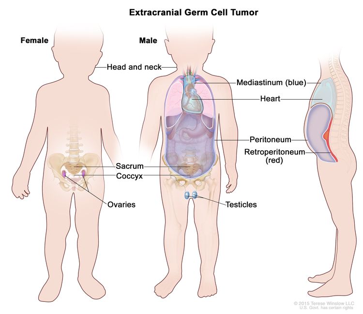
EnlargeExtracranial germ cell tumor; drawing shows parts of the body where extracranial germ cell tumors may form, including the head and neck, mediastinum (the area between the lungs, shown in blue), retroperitoneum (the area behind the abdominal organs, shown in red), sacrum, coccyx, testicles (in males), and ovaries (in females). Also shown are the heart and peritoneum.
Extracranial germ cell tumors form in parts of the body other than the brain. This includes the testicles, ovaries, sacrum (lower part of the spine), coccyx (tailbone), mediastinum (area between the lungs), retroperitoneum (the back wall of the abdomen), and the head and neck.

Extracranial germ cell tumors are most common in adolescents, with rates in this age group lower for females than males.

For information on intracranial (inside the brain) germ cell tumors, see Childhood Central Nervous System Germ Cell Tumors Treatment.

Childhood extracranial germ cell tumors may be benign or malignant.

Extracranial germ cell tumors may be benign (noncancer) or malignant (cancer).

Childhood extracranial germ cell tumors are grouped as gonadal or extragonadal extracranial tumors.

Malignant extracranial germ cell tumors are tumors that form outside the brain. They are gonadal or extragonadal.

Gonadal germ cell tumors

Gonadal germ cell tumors form in the gonads (testicles and ovaries).

Extragonadal extracranial germ cell tumors

Extragonadal extracranial germ cell tumors form in areas of the body other than the brain or gonads (testicles and ovaries).

Most extragonadal extracranial germ cell tumors form along the midline of the body. This includes the following:

  • Sacrum (the large, triangle-shaped bone in the lower spine that forms part of the pelvis).
  • Coccyx (tailbone).
  • Mediastinum (the area between the lungs).
  • Back of the abdomen.
  • Neck.

In children younger than 11 years, extragonadal extracranial germ cell tumors usually occur at birth or in early childhood. Most of these tumors are benign teratomas in the sacrum or coccyx.

In older children, adolescents, and young adults (11 years and older), extragonadal extracranial germ cell tumors are often in the mediastinum.

There are three types of extracranial germ cell tumors.

Extracranial germ cell tumors are also grouped into teratomas, malignant germ cell tumors, and mixed germ cell tumors:

Teratomas

There are two main types of teratomas:

  • Mature teratomas. These tumors are the most common type of extracranial germ cell tumor. Mature teratomas are benign tumors and not likely to become cancer. They usually occur in the sacrum or coccyx in newborns or in the testicles or ovaries at the start of puberty. The cells of mature teratomas look almost like normal cells under a microscope. Some mature teratomas release enzymes or hormones that cause signs and symptoms of disease.
  • Immature teratomas. These tumors usually occur in areas other than the gonads in young children or in the ovaries at the start of puberty. They have cells that look very different from normal cells under a microscope. Immature teratomas may be cancer and spread to other parts of the body. They often have several different types of tissue in them, such as hair, muscle, and bone. Some immature teratomas release enzymes or hormones that cause signs and symptoms of disease.

Malignant germ cell tumors

Malignant germ cell tumors are cancer. There are two main types of malignant germ cell tumors:

  • Seminomatous germ cell tumors. There are three types of seminomatous germ cell tumors:
    • Seminomas form in the testicle.
    • Dysgerminomas form in the ovary.
    • Germinomas form in areas of the body that are not the ovary or testicle, such as the mediastinum.
  • Nonseminomatous germ cell tumors. There are four types of nonseminomatous germ cell tumors:

Mixed germ cell tumors

Mixed germ cell tumors are made up of at least two types of malignant germ cell tumor. They can form in the ovary, testicle, or other areas of the body.

The cause of most childhood extracranial germ cell tumors is unknown.

Having certain inherited disorders can increase the risk of extracranial germ cell tumors.

A risk factor is anything that increases the chance of getting a disease. Not every child with one or more of these risk factors will develop an extracranial germ cell tumor. And it will develop in some children who don’t have a known risk factor.

Possible risk factors for extracranial germ cell tumors include:

Talk with your child’s doctor if you think your child may be at risk.

Signs of childhood extracranial germ cell tumors depend on where the tumor formed in the body.

Different tumors may cause the following signs and symptoms. Other conditions may cause these same signs and symptoms. It’s important to check with your child’s doctor if your child has:

  • A lump in the neck, abdomen, or lower back.
  • A painless lump in the testicle.
  • Pain in the abdomen or back.
  • Respiratory distress, chest pain, or cough.
  • Fever.
  • Constipation.
  • Trouble urinating.
  • Early puberty.
  • In females, no menstrual periods or unusual vaginal bleeding.

Imaging studies and blood tests are used to diagnose childhood extracranial germ cell tumors.

If your child has symptoms that suggest they have an extracranial germ cell tumor, their doctor will need to find out if these are due to cancer or another problem. The doctor will ask when the symptoms started and how often your child has been having them. They will also ask about your child’s personal and family medical history and do a physical exam. Depending on these results, they may recommend other tests. If your child is diagnosed with an extracranial germ cell tumor, the results of these tests will help you and your child’s doctor plan treatment.

The tests used to diagnose extracranial germ cell tumors may include:

  • Serum tumor marker test: A procedure in which a sample of blood is checked to measure the amounts of certain substances released into the blood by organs, tissues, or tumor cells in the body. Certain substances are linked to specific types of cancer when found in increased levels in the blood. These are called tumor markers.

    Some malignant germ cell tumors release tumor markers. The following tumor markers may be used to detect extracranial germ cell tumors:

    • Alpha-fetoprotein (AFP).
    • Beta-human chorionic gonadotropin (beta-hCG).

    For testicular germ cell tumors, blood levels of the tumor markers help show if the tumor is a seminoma or nonseminoma.

  • Blood chemistry studies: A procedure in which a blood sample is checked to measure the amounts of certain substances released into the blood by organs and tissues in the body, including lactate dehydrogenase. An unusual (higher or lower than normal) amount of a substance can be a sign of disease.
  • Chest x-ray: An x-ray of the organs and bones inside the chest. An x-ray is a type of energy beam that can go through the body and onto film, making a picture of areas inside the body.
  • CT scan (CAT scan): A procedure that makes a series of detailed pictures of areas inside the body, such as the chest or lymph nodes, taken from different angles. The pictures are made by a computer linked to an x-ray machine. A dye may be injected into a vein or swallowed to help the organs or tissues show up more clearly. This procedure is also called computed tomography, computerized tomography, or computerized axial tomography.
    EnlargeComputed tomography (CT) scan; drawing shows a child lying on a table that slides through the CT scanner, which takes a series of detailed x-ray pictures of areas inside the body.
    Computed tomography (CT) scan. The child lies on a table that slides through the CT scanner, which takes a series of detailed x-ray pictures of areas inside the body.
  • MRI (magnetic resonance imaging): A procedure that uses a magnet, radio waves, and a computer to make a series of detailed pictures of areas inside the body. This procedure is also called nuclear magnetic resonance imaging (NMRI).
    EnlargeMagnetic resonance imaging (MRI) scan; drawing shows a child lying on a table that slides into the MRI machine, which takes a series of detailed pictures of areas inside the body.
    Magnetic resonance imaging (MRI) scan. The child lies on a table that slides into the MRI machine, which takes a series of detailed pictures of areas inside the body. The positioning of the child on the table depends on the part of the body being imaged.
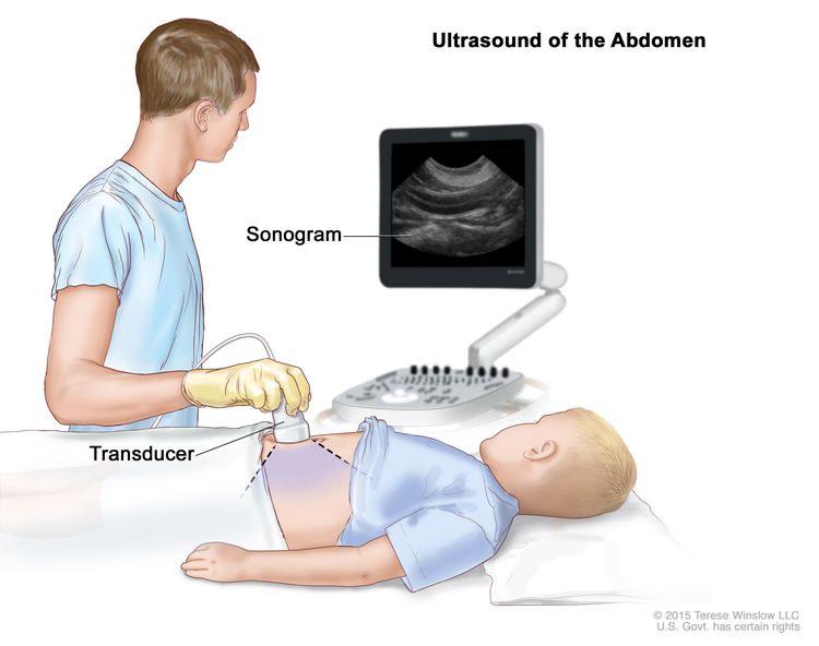
  • Ultrasound exam: A procedure in which high-energy sound waves (ultrasound) are bounced off internal tissues or organs and make echoes. The echoes form a picture of body tissues called a sonogram. The picture can be printed to be looked at later.
    EnlargeAbdominal ultrasound; drawing shows a child lying on an exam table during an abdominal ultrasound procedure. A technician is shown pressing a transducer (a device that makes sound waves that bounce off tissues inside the body) against the skin of the abdomen. A computer screen shows a sonogram (picture).
    Abdominal ultrasound. An ultrasound transducer connected to a computer is pressed against the skin of the abdomen. The transducer bounces sound waves off internal organs and tissues to make echoes that form a sonogram (computer picture).
  • Biopsy: The removal of cells or tissues so they can be viewed under a microscope by a pathologist to check for signs of cancer. Sometimes an incisional biopsy or needle biopsy is done before surgery to remove a sample of tissue. Sometimes the tumor is removed during surgery and then a sample of tissue is removed from the tumor.

    The following tests may be done on the sample of tissue that is removed:

    • Cytogenetic analysis: A laboratory test in which the chromosomes of cells in a sample of tissue are counted and checked for any changes, such as broken, missing, rearranged, or extra chromosomes. Changes in certain chromosomes may be a sign of cancer. Cytogenetic analysis is used to help diagnose cancer, plan treatment, or find out how well treatment is working.
    • Immunohistochemistry: A laboratory test that uses antibodies to check for certain antigens (markers) in a sample of a patient’s tissue. The antibodies are usually linked to an enzyme or a fluorescent dye. After the antibodies bind to a specific antigen in the tissue sample, the enzyme or dye is activated, and the antigen can then be seen under a microscope. This type of test is used to help diagnose cancer and to help tell one type of cancer from another type of cancer.

Certain factors affect prognosis (chance of recovery) and treatment options.

The prognosis and treatment options depend on the following:

  • The child’s age and general health.
  • The stage of the cancer (whether it has spread to nearby areas, lymph nodes, or to other places in the body).
  • Where the tumor first began to grow.
  • The type of germ cell tumor.
  • How well the tumor responds to treatment.
  • Whether the child has gonadal dysgenesis.
  • Whether the tumor can be completely removed by surgery.
  • Whether the cancer has just been diagnosed or has recurred (come back).

The prognosis for childhood extracranial germ cell tumors, especially ovarian germ cell tumors, is good.

Stages of Childhood Extracranial Germ Cell Tumors

Key Points

  • After a childhood extracranial germ cell tumor has been diagnosed, tests are done to find out if cancer cells have spread from where the tumor started to nearby areas or to other parts of the body.
  • There are three ways that cancer spreads in the body.
  • Cancer may spread from where it began to other parts of the body.
  • Stages are used to describe the different types of extracranial germ cell tumors.
    • Testicular germ cell tumors in children younger than 11 years
    • Testicular germ cell tumors in adolescents and young adults 11 years and older
    • Ovarian germ cell tumors
    • Extragonadal extracranial germ cell tumors
    • Childhood extracranial germ cell tumors usually do not come back after treatment.

After a childhood extracranial germ cell tumor has been diagnosed, tests are done to find out if cancer cells have spread from where the tumor started to nearby areas or to other parts of the body.

The process used to find out if cancer has spread from where the tumor started to other parts of the body is called staging. The information gathered from the staging process determines the stage of the disease. It is important to know the stage in order to plan treatment. In some cases, staging may follow surgery to remove the tumor.

The following procedures may be used:

  • MRI (magnetic resonance imaging): A procedure that uses a magnet, radio waves, and a computer to make a series of detailed pictures of areas inside the body, such as the brain or lymph nodes. This procedure is also called nuclear magnetic resonance imaging (NMRI).
  • CT scan (CAT scan): A procedure that makes a series of detailed pictures of areas inside the body, such as the chest or lymph nodes, taken from different angles. The pictures are made by a computer linked to an x-ray machine. A dye may be injected into a vein or swallowed to help the organs or tissues show up more clearly. This procedure is also called computed tomography, computerized tomography, or computerized axial tomography.
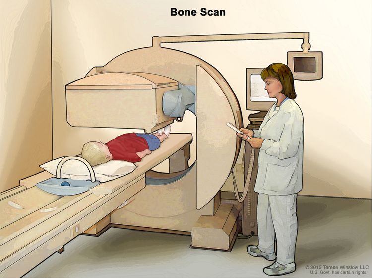
  • Bone scan: A procedure to check if there are rapidly dividing cells, such as cancer cells, in the bone. A very small amount of radioactive material is injected into a vein and travels through the bloodstream. The radioactive material collects in the bones with cancer and is detected by a scanner.
  • Thoracentesis: The removal of fluid from the space between the lining of the chest and the lung, using a needle. A pathologist views the fluid under a microscope to look for cancer cells.
  • Paracentesis: The removal of fluid from the space between the lining of the abdomen and the organs in the abdomen, using a needle. A pathologist views the fluid under a microscope to look for cancer cells.

The results from tests and procedures used to detect and diagnose childhood extracranial germ cell tumors may also be used in staging.

There are three ways that cancer spreads in the body.

Cancer can spread through tissue, the lymph system, and the blood:

  • Tissue. The cancer spreads from where it began by growing into nearby areas.
  • Lymph system. The cancer spreads from where it began by getting into the lymph system. The cancer travels through the lymph vessels to other parts of the body.
  • Blood. The cancer spreads from where it began by getting into the blood. The cancer travels through the blood vessels to other parts of the body.

Cancer may spread from where it began to other parts of the body.

When cancer spreads to another part of the body, it is called metastasis. Cancer cells break away from where they began (the primary tumor) and travel through the lymph system or blood.

  • Lymph system. The cancer gets into the lymph system, travels through the lymph vessels, and forms a tumor (metastatic tumor) in another part of the body.
  • Blood. The cancer gets into the blood, travels through the blood vessels, and forms a tumor (metastatic tumor) in another part of the body.

The metastatic tumor is the same type of cancer as the primary tumor. For example, if an extracranial germ cell tumor spreads to the liver, the cancer cells in the liver are actually cancerous germ cells. The disease is metastatic extracranial germ cell tumor, not liver cancer.

Many cancer deaths are caused when cancer moves from the original tumor and spreads to other tissues and organs. This is called metastatic cancer. This animation shows how cancer cells travel from the place in the body where they first formed to other parts of the body.

Stages are used to describe the different types of extracranial germ cell tumors.

Testicular germ cell tumors in children younger than 11 years

The following stages are from the Children’s Oncology Group.

  • Stage I

    In stage I, the cancer is found in the testicle only. The testicle and spermatic cord are completely removed by surgery and all of the following are true:

  • Stage II

    In stage II, the testicle and spermatic cord are removed by surgery and one of the following is true:

    • the capsule (outer covering of the tumor) ruptured (broke open) or a biopsy was done before surgery; or
    • cancer that can only be seen with a microscope remains in the scrotum or in the spermatic cord near the scrotum, and after surgery, tumor marker levels do not return to normal or do not decrease.

    Cancer has not spread to the lymph nodes.

  • Stage III

    In stage III, one of the following is true:

    • the cancer has spread to one or more lymph nodes at the back of the abdomen; or
    • lymph nodes are at least 2 centimeters wide or are larger than 1 centimeter but smaller than 2 centimeters in their shortest diameter and either have not changed or are growing when a CT scan or MRI is repeated within 4 to 6 weeks.
  • Stage IV

    In stage IV, the cancer has spread to other parts of the body, such as the liver, lung, bone, and brain.

Testicular germ cell tumors in adolescents and young adults 11 years and older

In males older than 15 years, there are only stage I tumors and metastatic tumors. For more information about staging used for testicular germ cell tumors in adolescents and young adults 11 years and older, see Testicular Cancer Treatment.

Ovarian germ cell tumors

Two staging systems are used for ovarian germ cell tumors: Children’s Oncology Group and the International Federation of Gynecology and Obstetrics (FIGO).

The following stages are from the Children’s Oncology Group.

  • Stage I

    In stage I, the tumor in the ovary is completely removed by surgery and all of the following are true:

    • the capsule (outer covering of the tumor) did not rupture (break open) and a biopsy was not done before the tumor was removed; and
    • there is no sign that the cancer has spread through the capsule; and
    • no cancer cells are found in fluid taken from the abdomen; and
    • no cancer is seen in tissue that lines the abdomen or found in tissue samples taken during a biopsy; and
    • lymph nodes are smaller than 1 centimeter in their shortest diameter on a CT scan or MRI or no cancer is found in lymph node tissue samples taken during a biopsy.
  • Stage II

    In stage II, the tumor in the ovary is completely removed by surgery and a biopsy is done before surgery and one of the following is true:

    • cancer has spread through part or all of the capsule (outer covering of the tumor); or
    • the tumor is larger than 10 centimeters and is removed by laparoscopic surgery; or
    • the tumor is removed by being broken up into small pieces and it is not known if cancer has spread through the capsule.

    Cancer cells are not found in fluid taken from the abdomen. Cancer is not seen in lymph nodes or tissue that lines the abdomen and cancer is not found in tissue samples taken during a biopsy.

  • Stage III

    In stage III, there is tumor in the ovary and one of the following is true:

    • lymph nodes are at least 2 centimeters wide or are larger than 1 centimeter but smaller than 2 centimeters in their shortest diameter and either have not changed or are growing when a CT scan or MRI is repeated 4 to 6 weeks after surgery; or
    • the tumor is not completely removed by surgery or a biopsy was done before surgery; or
    • cancer cells (including immature teratoma) are found in fluid taken from the abdomen; or
    • cancer (including immature teratoma) is found in lymph nodes; or
    • cancer (including immature teratoma) is found in tissue that lines the abdomen.
  • Stage III-X

    In stage III-X, the tumor can be described as stage I or stage II, except:

  • Stage IV

    In stage IV, one of the following is true:

    • the cancer has spread to the liver or outside the abdomen to other areas of the body, such as the bone, lung, or brain.
    • cancer cells are found in the fluid in the lung.

The following stages are from the International Federation of Gynecology and Obstetrics (FIGO).

  • Stage I

    In stage I, cancer is found in one or both of the ovaries and has not spread. Stage I is divided into stage IA, stage IB, and stage IC.

    • Stage IA: Cancer is found in one ovary.
    • Stage IB: Cancer is found in both ovaries.
    • Stage IC: Cancer is found in one or both ovaries and one of the following is true:
  • Stage II

    In stage II, cancer is found in one or both ovaries and has spread into other areas of the pelvis, or primary peritoneal cancer is found. Stage II is divided into stage IIA and stage IIB.

  • Stage III

    In stage III, cancer is found in one or both ovaries or primary peritoneal cancer is found. Cancer has spread outside the pelvis to other parts of the abdomen and/or to lymph nodes at the back of the abdomen. Stage III is divided into stage IIIA, stage IIIB, and stage IIIC.

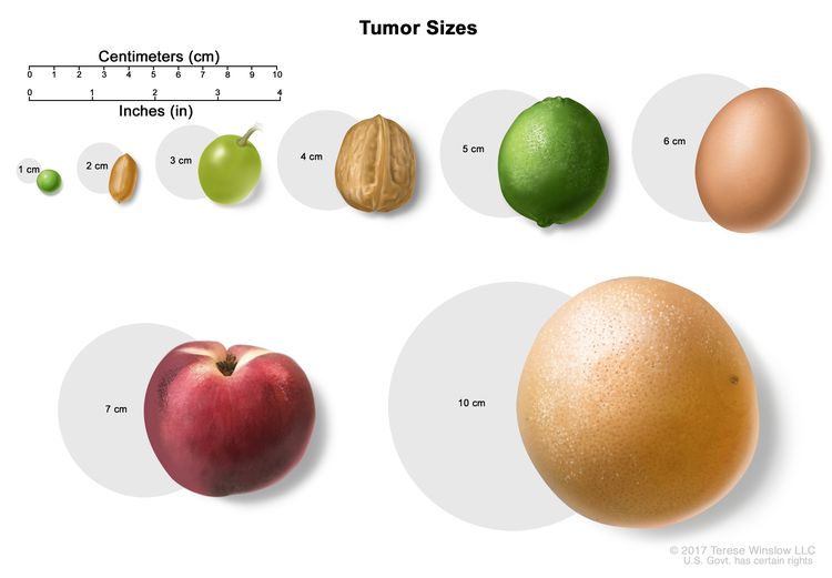
    EnlargeDrawing shows different sizes of a tumor in centimeters (cm) compared to the size of a pea (1 cm), a peanut (2 cm), a grape (3 cm), a walnut (4 cm), a lime (5 cm), an egg (6 cm), a peach (7 cm), and a grapefruit (10 cm). Also shown is a 10-cm ruler and a 4-inch ruler.
    Tumor sizes are often measured in centimeters (cm) or inches. Common food items that can be used to show tumor size in cm include: a pea (1 cm), a peanut (2 cm), a grape (3 cm), a walnut (4 cm), a lime (5 cm or 2 inches), an egg (6 cm), a peach (7 cm), and a grapefruit (10 cm or 4 inches).
    • In stage IIIA, one of the following is true:
      • cancer has spread to lymph nodes at the back of the abdomen only; or
      • cancer cells that can be seen only with a microscope have spread to the surface of the peritoneum outside the pelvis. Cancer may have spread to nearby lymph nodes at the back of the abdomen.
    • Stage IIIB: Cancer has spread to the peritoneum outside the pelvis and the cancer in the peritoneum is 2 centimeters or smaller. Cancer may have spread to lymph nodes at the back of the abdomen.
    • Stage IIIC: Cancer has spread to the peritoneum outside the pelvis and the cancer in the peritoneum is larger than 2 centimeters. Cancer may have spread to lymph nodes at the back of the abdomen or to the surface of the liver or spleen.
  • Stage IV

    Stage IV is divided into stage IVA and IVB.

Extragonadal extracranial germ cell tumors

The following stages are from the Children’s Oncology Group.

  • Stage I

    In stage I, the tumor is completely removed by surgery and all of the following are true:

  • Stage II

    In stage II, cancer is not completely removed by surgery and one of the following is true:

    • cancer that can only be seen with a microscope remains after surgery; or
    • the capsule (outer covering of the tumor) ruptured (broke open) or a biopsy was done.

    Cancer cells are not found in fluid taken from the abdomen. There is no sign of cancer in lymph nodes in the abdomen, pelvis, or chest on a CT scan or MRI.

  • Stage III

    In stage III, one of the following is true:

    • cancer is not completely removed by surgery and cancer that can be seen with the eye remains after surgery or only a biopsy was done; or
    • lymph nodes are at least 2 centimeters wide or are larger than 1 centimeter but smaller than 2 centimeters in their shortest diameter and either have not changed or are growing when a CT scan or MRI is repeated within 4 to 6 weeks.
  • Stage IV

    In stage IV, the cancer has spread to other parts of the body, such as the liver, lung, bone, or brain.

Childhood extracranial germ cell tumors usually do not come back after treatment.

Recurrent childhood extracranial germ cell tumor is cancer that has come back after it has been treated. The cancer may come back in the same place or in other parts of the body.

Most germ cell tumors do not recur (come back) after treatment. If they do recur, they usually come back within 3 years of surgery. About half of the teratomas that recur in the sacrum or coccyx are cancer, so follow-up is important.

Treatment Option Overview

Key Points

  • There are different types of treatment for children with extracranial germ cell tumors.
  • Children with extracranial germ cell tumors should have their treatment planned by a team of health care providers who are experts in treating cancer in children.
  • The following types of treatment are used:
    • Surgery
    • Observation
    • Chemotherapy
  • New types of treatment are being tested in clinical trials.
    • High-dose chemotherapy with stem cell transplant
    • Radiation therapy
  • Treatment for childhood extracranial germ cell tumors may cause side effects.
  • Patients may want to think about taking part in a clinical trial.
  • Patients can enter clinical trials before, during, or after starting their cancer treatment.
  • Follow-up tests may be needed.

There are different types of treatment for children with extracranial germ cell tumors.

Different types of treatments are available for children with extracranial germ cell tumors. Some treatments are standard (the currently used treatment), and some are being tested in clinical trials. A treatment clinical trial is a research study meant to help improve current treatments or obtain information on new treatments for patients with cancer. When clinical trials show that a new treatment is better than the standard treatment, the new treatment may become the standard treatment.

Because cancer in children is rare, taking part in a clinical trial should be considered. Some clinical trials are open only to patients who have not started treatment.

Children with extracranial germ cell tumors should have their treatment planned by a team of health care providers who are experts in treating cancer in children.

Treatment will be overseen by a pediatric oncologist, a doctor who specializes in treating children with cancer. The pediatric oncologist works with other health care providers who are experts in treating children with extracranial germ cell tumors and who specialize in certain areas of medicine. These may include the following specialists:

The following types of treatment are used:

Surgery

Surgery to completely remove the tumor is done whenever possible. If the tumor is very large, chemotherapy may be given first, to make the tumor smaller and decrease the amount of tissue that needs to be removed during surgery. A goal of surgery is to keep reproductive function. The following types of surgery may be used:

After the doctor removes all the cancer that can be seen at the time of the surgery, some patients may be given chemotherapy after surgery to kill any cancer cells that are left. Treatment given after surgery, to lower the risk that the cancer will come back, is called adjuvant therapy.

Observation

Observation is closely monitoring a patient’s condition without giving any treatment until signs or symptoms appear or change. For children with extracranial germ cell tumors, this includes physical exams, imaging tests, and tumor marker tests.

Chemotherapy

Chemotherapy is a cancer treatment that uses drugs to stop the growth of cancer cells, either by killing the cells or by stopping them from dividing. When chemotherapy is taken by mouth or injected into a vein or muscle, the drugs enter the bloodstream and can reach cancer cells throughout the body (systemic chemotherapy). Systemic chemotherapy is used to treat extracranial germ cell tumors.

New types of treatment are being tested in clinical trials.

This summary section describes treatments that are being studied in clinical trials. It may not mention every new treatment being studied. Learn more at Clinical Trials Information for Patients and Caregivers.

High-dose chemotherapy with stem cell transplant

High doses of chemotherapy are given to kill cancer cells. Healthy cells, including blood-forming cells, are also destroyed by the cancer treatment. Stem cell transplant is a treatment to replace the blood-forming cells. Stem cells (immature blood cells) are removed from the blood or bone marrow of the patient or a donor and are frozen and stored. After the patient completes chemotherapy, the stored stem cells are thawed and given back to the patient through an infusion. These reinfused stem cells grow into (and restore) the body’s blood cells.

Radiation therapy

Radiation therapy is a cancer treatment that uses high-energy x-rays or other types of radiation to kill cancer cells or keep them from growing. External radiation therapy uses a machine outside the body to send radiation toward the area of the body with cancer. External radiation therapy is being studied for the treatment of childhood extracranial germ cell tumors that have come back.

Treatment for childhood extracranial germ cell tumors may cause side effects.

To learn more about side effects that begin during treatment for cancer, visit Side Effects.

Side effects from cancer treatment that begin after treatment and continue for months or years are called late effects. Late effects of cancer treatment may include the following:

  • Physical problems, such as infertility, trouble hearing and kidney problems.
  • Changes in mood, feelings, thinking, learning, or memory.
  • Second cancers (new types of cancer), such as leukemia or malignant melanoma.

Some late effects may be treated or controlled. It is important to talk with your child’s doctors about the effects cancer treatment can have on your child. For more information, see Late Effects of Treatment for Childhood Cancer.

Patients may want to think about taking part in a clinical trial.

For some patients, taking part in a clinical trial may be the best treatment choice. Clinical trials are part of the cancer research process. Clinical trials are done to find out if new cancer treatments are safe and effective or better than the standard treatment.

Many of today’s standard treatments for cancer are based on earlier clinical trials. Patients who take part in a clinical trial may receive the standard treatment or be among the first to receive a new treatment.

Patients who take part in clinical trials also help improve the way cancer will be treated in the future. Even when clinical trials do not lead to effective new treatments, they often answer important questions and help move research forward.

Patients can enter clinical trials before, during, or after starting their cancer treatment.

Some clinical trials only include patients who have not yet received treatment. Other trials test treatments for patients whose cancer has not gotten better. There are also clinical trials that test new ways to stop cancer from recurring (coming back) or reduce the side effects of cancer treatment.

Clinical trials are taking place in many parts of the country. Information about clinical trials supported by NCI can be found on NCI’s clinical trials search webpage. Clinical trials supported by other organizations can be found on the ClinicalTrials.gov website.

Follow-up tests may be needed.

As your child goes through treatment, they will have follow-up tests or check-ups. Some tests that were done to diagnose or stage the cancer may be repeated to see how well the treatment is working. Decisions about whether to continue, change, or stop treatment may be based on the results of these tests.

Some of the tests will continue to be done from time to time after treatment has ended. The results of these tests can show if your child’s condition has changed or if the cancer has recurred (come back).

For childhood extracranial germ cell tumors, follow-up may include regular physical exams, tumor marker tests, and imaging tests such as CT scan, MRI or chest x-ray.

Treatment of Childhood Mature and Immature Teratomas

For information about the treatments listed below, see the Treatment Option Overview section.

Treatment of newly diagnosed mature teratomas includes the following:

Treatment of newly diagnosed immature teratomas includes the following:

  • Surgery to remove the tumor followed by observation for stage I tumors.
  • Surgery to remove the tumor for stage I–IV tumors. In young children, surgery is followed by observation; the use of chemotherapy after surgery is controversial. In adolescents and young adults, chemotherapy is given after surgery.

Sometimes a mature or immature teratoma also has malignant cells. A teratoma with malignant cells may need to be treated differently.

Use our clinical trial search to find NCI-supported cancer clinical trials that are accepting patients. You can search for trials based on the type of cancer, the age of the patient, and where the trials are being done. General information about clinical trials is also available.

Treatment of Malignant Testicular Germ Cell Tumors

For information about the treatments listed below, see the Treatment Option Overview section.

Treatment of newly diagnosed malignant testicular germ cell tumors may include the following:

For boys younger than 11 years:

For boys 11 years and older:

Malignant testicular germ cell tumors in boys 11 years and older are treated differently than they are in young boys. For more information, see Testicular Cancer Treatment.

  • Surgery to remove the tumor. Sometimes lymph nodes in the abdomen are also removed.
  • A clinical trial of a new regimen of surgery followed by observation for stage I tumors or chemotherapy for stage II–IV tumors.

Use our clinical trial search to find NCI-supported cancer clinical trials that are accepting patients. You can search for trials based on the type of cancer, the age of the patient, and where the trials are being done. General information about clinical trials is also available.

Treatment of Malignant Ovarian Germ Cell Tumors

Dysgerminomas

For information about the treatments listed below, see the Treatment Option Overview section.

Treatment of newly diagnosed stage I dysgerminomas of the ovary may include the following:

Treatment of newly diagnosed stages II–IV dysgerminomas of the ovary may include the following:

  • Surgery (unilateral salpingo-oophorectomy) followed by chemotherapy.
  • Chemotherapy to shrink the tumor, followed by surgery (unilateral salpingo-oophorectomy).

Nongerminomas

For information about the treatments listed below, see the Treatment Option Overview section.

Treatment of newly diagnosed nongerminomas of the ovary, such as yolk sac tumors, mixed germ cell tumors, choriocarcinoma, and embryonal carcinomas, in young girls may include the following:

Treatment of newly diagnosed nongerminomas of the ovary in adolescents and young women may include the following:

  • Surgery and chemotherapy for stage I–IV tumors.
  • A clinical trial of a new regimen of surgery followed by observation for stage I or chemotherapy for stage II–IV.

Treatment of newly diagnosed nongerminomas of the ovary that cannot be removed by primary surgery without risk to nearby tissue may include the following:

  • Biopsy followed by chemotherapy and surgery.

Use our clinical trial search to find NCI-supported cancer clinical trials that are accepting patients. You can search for trials based on the type of cancer, the age of the patient, and where the trials are being done. General information about clinical trials is also available.

Treatment of Malignant Extragonadal Extracranial Germ Cell Tumors

For information about the treatments listed below, see the Treatment Option Overview section.

Treatment of newly diagnosed childhood malignant extragonadal extracranial germ cell tumors in young children may include the following:

In addition to stage of the disease, treatment of malignant extragonadal extracranial germ cell tumors also depends on where the tumor formed in the body:

  • For tumors in the sacrum or coccyx, chemotherapy to shrink the tumor followed by surgery to remove the tumor and coccyx.
  • For tumors in the mediastinum, chemotherapy before or after surgery to remove the tumor in the mediastinum.
  • For tumors in the abdomen, biopsy followed by chemotherapy to shrink the tumor and surgery to remove the tumor in the abdomen.
  • For tumors in the head and neck, surgery to remove the tumor in the head or neck, which may be followed by chemotherapy if the tumor is cancer.

Treatment of newly diagnosed childhood malignant extragonadal extracranial germ cell tumors in adolescents and young adults may include the following:

  • Surgery.
  • Chemotherapy.
  • Chemotherapy followed by surgery to remove the tumor.
  • A clinical trial of a new regimen of surgery followed by observation for stage I tumors or chemotherapy for stage II–IV tumors.

Use our clinical trial search to find NCI-supported cancer clinical trials that are accepting patients. You can search for trials based on the type of cancer, the age of the patient, and where the trials are being done. General information about clinical trials is also available.

Treatment of Recurrent Childhood Malignant Extracranial Germ Cell Tumors

For information about the treatments listed below, see the Treatment Option Overview section.

Treatment of recurrent childhood extracranial germ cell tumors may include the following:

Use our clinical trial search to find NCI-supported cancer clinical trials that are accepting patients. You can search for trials based on the type of cancer, the age of the patient, and where the trials are being done. General information about clinical trials is also available.

To Learn More About Childhood Cancer

About This PDQ Summary

About PDQ

Physician Data Query (PDQ) is the National Cancer Institute’s (NCI’s) comprehensive cancer information database. The PDQ database contains summaries of the latest published information on cancer prevention, detection, genetics, treatment, supportive care, and complementary and alternative medicine. Most summaries come in two versions. The health professional versions have detailed information written in technical language. The patient versions are written in easy-to-understand, nontechnical language. Both versions have cancer information that is accurate and up to date and most versions are also available in Spanish.

PDQ is a service of the NCI. The NCI is part of the National Institutes of Health (NIH). NIH is the federal government’s center of biomedical research. The PDQ summaries are based on an independent review of the medical literature. They are not policy statements of the NCI or the NIH.

Purpose of This Summary

This PDQ cancer information summary has current information about the treatment of childhood extracranial germ cell tumors. It is meant to inform and help patients, families, and caregivers. It does not give formal guidelines or recommendations for making decisions about health care.

Reviewers and Updates

Editorial Boards write the PDQ cancer information summaries and keep them up to date. These Boards are made up of experts in cancer treatment and other specialties related to cancer. The summaries are reviewed regularly and changes are made when there is new information. The date on each summary (“Updated”) is the date of the most recent change.

The information in this patient summary was taken from the health professional version, which is reviewed regularly and updated as needed, by the PDQ Pediatric Treatment Editorial Board.

Clinical Trial Information

A clinical trial is a study to answer a scientific question, such as whether one treatment is better than another. Trials are based on past studies and what has been learned in the laboratory. Each trial answers certain scientific questions in order to find new and better ways to help cancer patients. During treatment clinical trials, information is collected about the effects of a new treatment and how well it works. If a clinical trial shows that a new treatment is better than one currently being used, the new treatment may become “standard.” Patients may want to think about taking part in a clinical trial. Some clinical trials are open only to patients who have not started treatment.

Clinical trials can be found online at NCI’s website. For more information, call the Cancer Information Service (CIS), NCI’s contact center, at 1-800-4-CANCER (1-800-422-6237).

Permission to Use This Summary

PDQ is a registered trademark. The content of PDQ documents can be used freely as text. It cannot be identified as an NCI PDQ cancer information summary unless the whole summary is shown and it is updated regularly. However, a user would be allowed to write a sentence such as “NCI’s PDQ cancer information summary about breast cancer prevention states the risks in the following way: [include excerpt from the summary].”

The best way to cite this PDQ summary is:

PDQ® Pediatric Treatment Editorial Board. PDQ Childhood Extracranial Germ Cell Tumors Treatment. Bethesda, MD: National Cancer Institute. Updated <MM/DD/YYYY>. Available at: /types/extracranial-germ-cell/patient/germ-cell-treatment-pdq. Accessed <MM/DD/YYYY>. [PMID: 26389180]

Images in this summary are used with permission of the author(s), artist, and/or publisher for use in the PDQ summaries only. If you want to use an image from a PDQ summary and you are not using the whole summary, you must get permission from the owner. It cannot be given by the National Cancer Institute. Information about using the images in this summary, along with many other images related to cancer can be found in Visuals Online. Visuals Online is a collection of more than 3,000 scientific images.

Disclaimer

The information in these summaries should not be used to make decisions about insurance reimbursement. More information on insurance coverage is available on Cancer.gov on the Managing Cancer Care page.

Contact Us

More information about contacting us or receiving help with the Cancer.gov website can be found on our Contact Us for Help page. Questions can also be submitted to Cancer.gov through the website’s E-mail Us.


Childhood Testicular Cancer (PDQ®)–Patient Version

Childhood Testicular Cancer (PDQ®)–Patient Version

What is childhood testicular cancer?

Testicular tumors in children can form in the tissue of one or both testicles. These tumors can be benign (not cancer) or malignant (cancerous). Benign tumors do not spread to other parts of the body. However, they may grow and press on nearby tissues, causing discomfort or other problems. Malignant tumors may spread to other parts of the body. Benign tumors are more common in young boys and infants while malignant tumors are more common in boys who have gone through puberty.

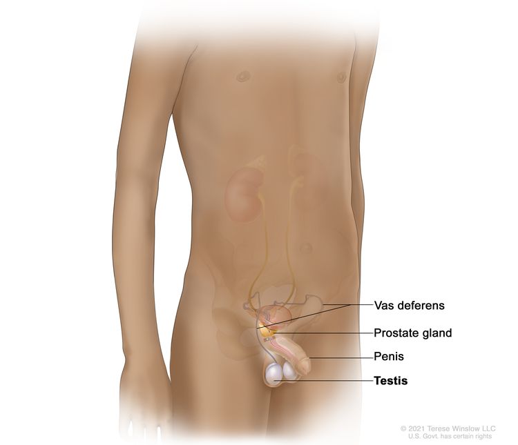
The testicles are two egg-shaped glands in the male reproductive system. They are located inside the scrotum, a sac of loose skin that lies directly below the penis. The testicles are held within the scrotum by the spermatic cord, which contains the vas deferens, vessels, and nerves of the testicles. The testicles produce male hormones, such as testosterone, and sperm.

EnlargeAnatomy of the male reproductive system; drawing shows the vas deferens (a long tube that carries sperm out of the testes), prostate gland, penis, and testes.
Anatomy of the male reproductive system. The male reproductive system is made up of organs and glands involved in making offspring (children). These include the vas deferens (a long tube that carries sperm out of the testes), prostate gland, penis, and testes.

Types of testicular tumors

There are two types of testicular tumors.

  • Non-germ cell tumors begin in the cells that produce hormones and in the tissue that surrounds and supports the testicles. These tumors may be benign or cancerous. Types of non-germ cell tumors include:
    • Juvenile granulosa cell tumors, which most often occur in young infants.
    • Sertoli cell tumors, which often occur in older infants.
    • Leydig cell tumors, which are more common in adolescents.
  • Germ cell tumors begin in the sperm-producing cells of the testicles. Most childhood testicular tumors are germ cell tumors. Learn more about testicular germ cell tumors and their treatment at Childhood Extracranial Germ Cell Tumors Treatment.

Causes and risk factors for childhood testicular cancer

Childhood testicular cancer is caused by certain changes to the way testicular cells function, especially how they grow and divide into new cells. Often, the exact cause of these cell changes is unknown. Learn more about how cancer develops at What Is Cancer?

A risk factor is anything that increases the chance of getting a disease. Not every child with one or more of these risk factors will develop testicular cancer. And it will develop in some children who don’t have a known risk factor.

The risk of testicular cancer may be increased if your child has one of the following syndromes:

  • DICER1 syndrome is a disorder that may cause a goiter, polyps in the colon, and tumors of the ovary, cervix, testicle, kidney, brain, eye, and lining of the lung.
  • Peutz-Jeghers syndrome is a disorder that causes polyps to form in the intestines and dark spots to form on the mouth and fingers.
  • Carney syndrome or complex is a disorder that causes dark spots on the skin and tumors to form in the heart, endocrine glands, skin, and nerves.

Talk with your child’s doctor if you think your child may be at risk.

Genetic counseling for children with testicular cancer

It may not be clear from the family medical history whether a child with testicular cancer has an inherited condition that increased their risk. Genetic counseling before genetic testing can help assess your child’s risk of having a gene change that caused your child’s tumor and whether genetic testing is needed. Genetic testing may help explain why a child develops a rare cancer or a cancer that is usually seen in adults, such as testicular cancer. Genetic counselors and other specially trained health professionals can discuss your child’s diagnosis and your family’s medical history to help you understand:

  • the options for DICER1 gene testing
  • the risk of other cancers for your child
  • the risk of testicular cancer and other cancers for your child’s siblings
  • the risks and benefits of learning genetic information

Genetic counselors can also help you cope with your child’s genetic testing results, including how to discuss the results with family members. They can advise you about whether other members of your family should receive genetic testing.

Learn more about Genetic Testing for Inherited Cancer Risk.

Symptoms of childhood testicular cancer

Children may not have symptoms of testicular cancer until the tumor has grown bigger. It’s important to check with your child’s doctor if your child has:

  • a painless lump in the testicles
  • early signs of puberty
  • enlarged breasts

These symptoms may be caused by problems other than childhood testicular cancer. The only way to know is to see your child’s doctor.

Tests to diagnose childhood testicular cancer

If your child has symptoms that suggest testicular cancer, the doctor will need to find out if these are due to cancer or another problem. The doctor will ask when the symptoms started and how often your child has been having them. They will also ask about your child’s personal and family medical history and do a physical exam. Depending on these results, they may recommend other tests. If your child is diagnosed with testicular cancer, the results of these tests will help you and your child’s doctor plan treatment.

The tests and procedures used to diagnose testicular cancer may include:

Ultrasound exam

An ultrasound exam uses high-energy sound waves (ultrasound) that bounce off internal tissues or organs and make echoes. The echoes form a picture of body tissues called a sonogram.

CT scan (CAT scan)

A CT scan uses a computer linked to an x-ray machine to make a series of detailed pictures of areas inside the body, such as the chest, abdomen, or pelvis. The pictures are taken from different angles and are used to create 3-D views of tissues and organs. A dye may be injected into a vein or swallowed to help the organs or tissues show up more clearly. This procedure is also called computed tomography, computerized tomography, or computerized axial tomography. Learn more about Computed Tomography (CT) Scans and Cancer.

EnlargeComputed tomography (CT) scan; drawing shows a child lying on a table that slides through the CT scanner, which takes a series of detailed x-ray pictures of areas inside the body.
Computed tomography (CT) scan. The child lies on a table that slides through the CT scanner, which takes a series of detailed x-ray pictures of areas inside the body.

Magnetic resonance imaging (MRI)

MRI uses a magnet, radio waves, and a computer to make a series of detailed pictures of areas of the body, such as the chest, abdomen, or pelvis. This procedure is also called nuclear magnetic resonance imaging (NMRI).

EnlargeMagnetic resonance imaging (MRI) scan; drawing shows a child lying on a table that slides into the MRI machine, which takes a series of detailed pictures of areas inside the body.
Magnetic resonance imaging (MRI) scan. The child lies on a table that slides into the MRI machine, which takes a series of detailed pictures of areas inside the body. The positioning of the child on the table depends on the part of the body being imaged.

Chest x-ray

An x-ray is a type of radiation that can go through the body and make pictures. A chest x-ray makes pictures of the organs and bones inside the chest.

Biopsy

A biopsy is a procedure in which a sample of tissue is removed from the tumor so that a pathologist can view it under a microscope to check for signs of cancer.

Serum tumor marker test

A serum tumor marker test examines a sample of blood to measure the amounts of certain substances released into the blood by organs, tissues, or tumor cells in the body. Certain substances are linked to specific types of cancer when found in increased levels in the blood. These are called tumor markers. If an increased level of the tumor marker alpha-fetoprotein is found in the blood, it means the patient has a testicular germ cell tumor, not a testicular non-germ cell tumor.

DICER1 genetic testing

DICER1 genetic testing examines a sample of blood or saliva for changes in the DICER1 gene.

Getting a second opinion

You may want to get a second opinion to confirm your child’s testicular cancer diagnosis and treatment plan. If you seek a second opinion, you will need to get medical test results and reports from the first doctor to share with the second doctor. The second doctor will review the genetic test results, pathology report, slides, and scans. This doctor may agree with the first doctor, suggest changes to the treatment plan, or provide more information about your child’s cancer.

To learn more about choosing a doctor and getting a second opinion, see Finding Cancer Care. You can contact NCI’s Cancer Information Service via chat, email, or phone (both in English and Spanish) for help finding a doctor or hospital that can provide a second opinion. For questions you might want to ask at your child’s appointments, see Questions to Ask Your Doctor about Cancer.

Who treats children with testicular cancer?

A pediatric oncologist, a doctor who specializes in treating children with cancer, oversees treatment for testicular cancer. The pediatric oncologist works with other health care providers who are experts in treating children with cancer and who specialize in certain areas of medicine. Other specialists may include:

Treatment of childhood testicular cancer

There are different types of treatment for children and adolescents with testicular cancer. You and your child’s cancer care team will work together to decide treatment. Many factors will be considered, such as your child’s overall health and whether the cancer is newly diagnosed or has come back.

Your child’s treatment plan will include information about the cancer, the goals of treatment, treatment options, and the possible side effects. It will be helpful to talk with your child’s cancer care team before treatment begins about what to expect. For help every step of the way, see our booklet, Children with Cancer: A Guide for Parents.

Treatment for non-germ cell testicular cancer in children is usually surgery to remove the tumor. In some cases, the entire testicle with cancer may need to be removed.

If the cancer comes back after treatment, your child’s doctor will talk with you about what to expect and possible next steps. There may be treatment options to shrink the cancer or control its growth. If there are no treatment options, your child can receive care to control symptoms from cancer so they can be as comfortable as possible.

Clinical trials

For some children, joining a clinical trial may be an option. There are different types of clinical trials for childhood cancer. For example, a treatment trial tests new treatments or new ways of using current treatments. Supportive care and palliative care trials look at ways to improve quality of life, especially for those who have side effects from cancer and its treatment.

You can use the clinical trial search to find NCI-supported cancer clinical trials accepting participants. The search allows you to filter trials based on the type of cancer, your child’s age, and where the trials are being done. Clinical trials supported by other organizations can be found on the ClinicalTrials.gov website.

Learn more about clinical trials, including how to find and join one, at Clinical Trials Information for Patients and Caregivers.

Prognosis for childhood testicular cancer

If your child has been diagnosed with testicular cancer, you likely have questions about how serious the cancer is and your child’s chances of survival. The likely outcome or course of a disease is called prognosis. In children, the prognosis for testicular cancer is usually excellent after surgery to remove the tumor.

Side effects and late effects of treatment

Cancer treatments can cause side effects. Which side effects your child might have depends on the type of treatment they receive, the dose, and how their body reacts. Talk with your child’s treatment team about which side effects to look for and ways to manage them.

To learn more about side effects that begin during treatment for cancer, visit Side Effects.

Problems from cancer treatment that begin 6 months or later after treatment and continue for months or years are called late effects. Physical problems, such as problems with fertility, are a late effect of treatment.

Some late effects may be treated or controlled. It is important to talk with your child’s doctors about the possible late effects caused by some treatments. Learn more about Late Effects of Treatment for Childhood Cancer.

Follow-up care

As your child goes through treatment, they will have follow-up tests or check-ups. Some of the tests that were done to diagnose the cancer may be repeated to see how well the treatment is working. Decisions about whether to continue, change, or stop treatment may be based on the results of these tests.

Some of the tests will continue to be done from time to time after treatment has ended. The results of these tests can show if your child’s condition has changed or if the cancer has recurred (come back).

Coping with your child's cancer

When a child has cancer, every member of the family needs support. Taking care of yourself during this difficult time is important. Reach out to your child’s treatment team and to people in your family and community for support. To learn more, see Support for Families: Childhood Cancer and the booklet Children with Cancer: A Guide for Parents.

Related resources

About This PDQ Summary

About PDQ

Physician Data Query (PDQ) is the National Cancer Institute’s (NCI’s) comprehensive cancer information database. The PDQ database contains summaries of the latest published information on cancer prevention, detection, genetics, treatment, supportive care, and complementary and alternative medicine. Most summaries come in two versions. The health professional versions have detailed information written in technical language. The patient versions are written in easy-to-understand, nontechnical language. Both versions have cancer information that is accurate and up to date and most versions are also available in Spanish.

PDQ is a service of the NCI. The NCI is part of the National Institutes of Health (NIH). NIH is the federal government’s center of biomedical research. The PDQ summaries are based on an independent review of the medical literature. They are not policy statements of the NCI or the NIH.

Purpose of This Summary

This PDQ cancer information summary has current information about the treatment of childhood testicular cancer. It is meant to inform and help patients, families, and caregivers. It does not give formal guidelines or recommendations for making decisions about health care.

Reviewers and Updates

Editorial Boards write the PDQ cancer information summaries and keep them up to date. These Boards are made up of experts in cancer treatment and other specialties related to cancer. The summaries are reviewed regularly and changes are made when there is new information. The date on each summary (“Updated”) is the date of the most recent change.

The information in this patient summary was taken from the health professional version, which is reviewed regularly and updated as needed, by the PDQ Pediatric Treatment Editorial Board.

Clinical Trial Information

A clinical trial is a study to answer a scientific question, such as whether one treatment is better than another. Trials are based on past studies and what has been learned in the laboratory. Each trial answers certain scientific questions in order to find new and better ways to help cancer patients. During treatment clinical trials, information is collected about the effects of a new treatment and how well it works. If a clinical trial shows that a new treatment is better than one currently being used, the new treatment may become “standard.” Patients may want to think about taking part in a clinical trial. Some clinical trials are open only to patients who have not started treatment.

Clinical trials can be found online at NCI’s website. For more information, call the Cancer Information Service (CIS), NCI’s contact center, at 1-800-4-CANCER (1-800-422-6237).

Permission to Use This Summary

PDQ is a registered trademark. The content of PDQ documents can be used freely as text. It cannot be identified as an NCI PDQ cancer information summary unless the whole summary is shown and it is updated regularly. However, a user would be allowed to write a sentence such as “NCI’s PDQ cancer information summary about breast cancer prevention states the risks in the following way: [include excerpt from the summary].”

The best way to cite this PDQ summary is:

PDQ® Pediatric Treatment Editorial Board. PDQ Childhood Testicular Cancer. Bethesda, MD: National Cancer Institute. Updated <MM/DD/YYYY>. Available at: /types/testicular/patient/child-testicular-treatment-pdq. Accessed <MM/DD/YYYY>.

Images in this summary are used with permission of the author(s), artist, and/or publisher for use in the PDQ summaries only. If you want to use an image from a PDQ summary and you are not using the whole summary, you must get permission from the owner. It cannot be given by the National Cancer Institute. Information about using the images in this summary, along with many other images related to cancer can be found in Visuals Online. Visuals Online is a collection of more than 3,000 scientific images.

Disclaimer

The information in these summaries should not be used to make decisions about insurance reimbursement. More information on insurance coverage is available on Cancer.gov on the Managing Cancer Care page.

Contact Us

More information about contacting us or receiving help with the Cancer.gov website can be found on our Contact Us for Help page. Questions can also be submitted to Cancer.gov through the website’s E-mail Us.
Childhood Bladder Cancer

Childhood Bladder Cancer

Childhood bladder cancer is a very rare type of cancer that forms in the tissues of the bladder.

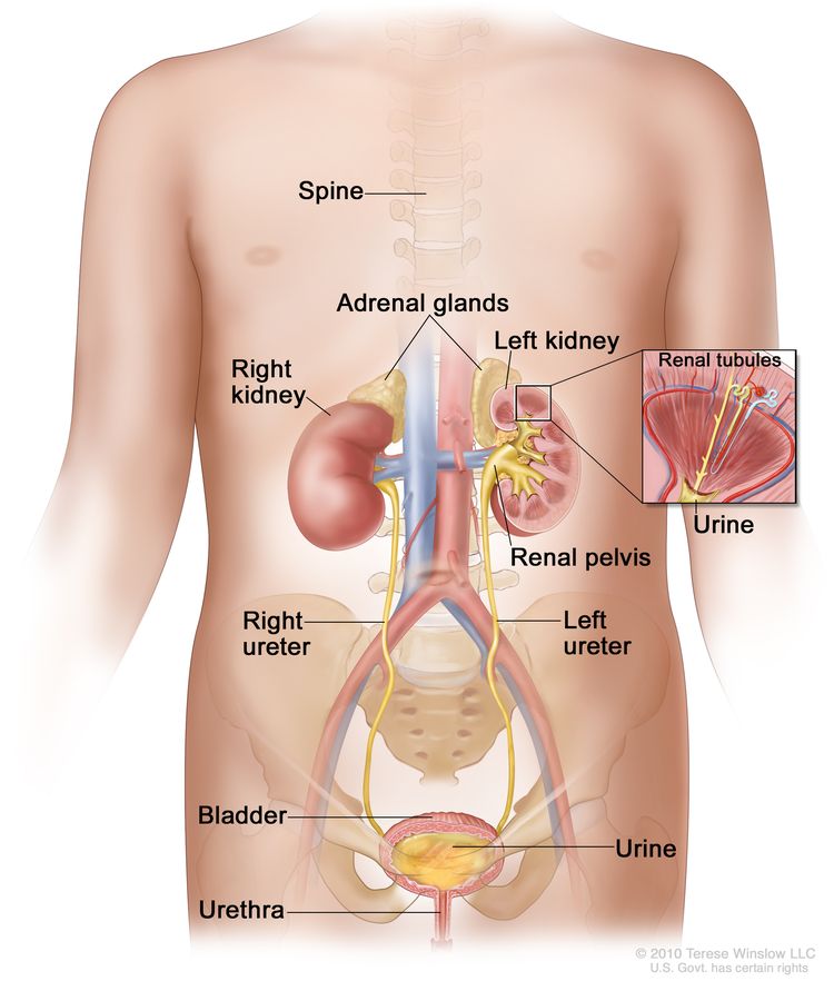
The bladder is a hollow, balloon-shaped organ in the lower part of the abdomen that stores urine. The bladder has a muscular wall that allows it to get larger to store urine made by the kidneys, and to shrink to squeeze urine out of the body. There are two kidneys, one on each side of the backbone, above the waist. The bladder and kidneys work together to remove toxins and wastes from the body through urine:

  • Tiny tubules in the kidneys filter and clean the blood.
  • These tubules take out waste products and make urine.
  • The urine passes from each kidney through a long tube called a ureter into the bladder.
  • The bladder holds the urine until it passes through the urethra and leaves the body.
EnlargeAnatomy of the urinary system; drawing showing the right and left kidneys, the ureters, the bladder filled with urine, and the urethra. The inside of the left kidney shows the renal pelvis. An inset shows the renal tubules and urine. The spine and adrenal glands are also shown.
Anatomy of the urinary system showing the kidneys, ureters, bladder, and urethra. The inside of the left kidney shows the renal pelvis. An inset shows the renal tubules and urine. Also shown is the spine and adrenal glands. Urine is made in the renal tubules and collects in the renal pelvis of each kidney. The urine flows from the kidneys through the ureters to the bladder. The urine is stored in the bladder until it leaves the body through the urethra.

Urothelial carcinoma (also called transitional cell carcinoma) is cancer that begins in the urothelial cells, which line the urethra, bladder, ureters, renal pelvis, and some other organs. The cells of the urothelium are known as transitional cells because they are able to stretch when the bladder is full of urine and shrink when it is emptied. Urothelial carcinoma is the most common form of bladder cancer in children.

Squamous cell and other more aggressive types of bladder cancer are less common in children.

Bladder cancer can occur at any age and most often affects males.

Risk factors for childhood bladder cancer

Bladder cancer is caused by certain changes to the way bladder cells function, especially how they grow and divide into new cells. Learn more about how cancer develops at What Is Cancer?

The risk of bladder cancer is increased in children who have been treated for cancer with certain anticancer drugs, called alkylating agents, which include cyclophosphamide, ifosfamide, busulfan, and temozolomide. Survivors of heritable retinoblastoma also have an increased risk of developing bladder cancer. Not every child with these risk factors will develop bladder cancer. And it will develop in some children who don’t have a known risk factor. Talk with your child’s doctor if you’re concerned about your child’s risk.

Symptoms of childhood bladder cancer

The symptoms of bladder cancer can vary from person to person. The most common symptom of bladder cancer is blood in the urine, called hematuria. It’s often slightly rusty to bright red in color. You may see blood in your child’s urine at one point, then not see it again for a while. Sometimes there are very small amounts of blood in the urine that can only be found by having a test done.

Other common symptoms of bladder cancer can include:

  • frequent urination or feeling the need to urinate without being able to do so
  • pain during urination
  • abdominal or lower back pain

It’s important to check with your child’s doctor if your child has any of these symptoms. Keep in mind that urinary tract infections, kidney or bladder stones, or other problems related to the kidney could also be the cause, not cancer. Your child’s doctor will ask you when the symptoms started and how often your child has been having them. They’ll most likely ask for a urine sample as a first step in diagnosing what is causing these symptoms.

Tests to diagnose childhood bladder cancer

If your child has symptoms or lab test results that suggest bladder cancer, their doctor will need to find out if these are due to cancer or another problem. They may:

  • ask about your child’s personal and family medical history to learn more about your child’s symptoms and possible risk factors for bladder cancer
  • do a physical exam to check for signs of cancer
  • ask for a sample of urine so it can be checked in the lab for blood, abnormal cells, or infection

Depending on your child’s symptoms and medical history and the results of their physical exam and urine lab tests, the doctor may recommend more tests to find out if your child has bladder cancer, and if so, its extent (stage). The results of tests and procedures done to diagnose bladder cancer are used to help make decisions about treatment.

The following tests and procedures may be used:

CT scan (CAT scan)

A CT scan uses a computer linked to an x-ray machine to make a series of detailed pictures of areas inside the body, such as the pelvis. The pictures are taken from different angles and are used to create 3-D views of tissues and organs. This procedure is also called computed tomography, computerized tomography, or computerized axial tomography. Learn more about Computed Tomography (CT) Scans and Cancer.

EnlargeComputed tomography (CT) scan; drawing shows a child lying on a table that slides through the CT scanner, which takes a series of detailed x-ray pictures of areas inside the body.
Computed tomography (CT) scan. The child lies on a table that slides through the CT scanner, which takes a series of detailed x-ray pictures of areas inside the body.

Ultrasound exam

An ultrasound exam uses high-energy sound waves (ultrasound) that bounce off internal tissues or organs, such as the pelvis, and make echoes. The echoes form a picture of body tissues called a sonogram.

EnlargeAbdominal ultrasound; drawing shows a child lying on an exam table during an abdominal ultrasound procedure. A technician is shown pressing a transducer (a device that makes sound waves that bounce off tissues inside the body) against the skin of the abdomen. A computer screen shows a sonogram (picture).
Abdominal ultrasound. An ultrasound transducer connected to a computer is pressed against the skin of the abdomen. The transducer bounces sound waves off internal organs and tissues to make echoes that form a sonogram (computer picture).

Cystoscopy

Cystoscopy is a procedure in which the doctor looks inside the bladder and urethra (the tube that carries urine out of your body) to check for abnormal areas. A cystoscope is slowly inserted through the urethra into the bladder to allow the doctor to see inside. A cystoscope is a thin, tube-like instrument with a light and a lens for viewing. It may also have a tool to remove very small tumors or tissue samples for biopsy. If a cystoscopy is not done at diagnosis, tissue samples are removed and checked for cancer during surgery to remove all or part of the bladder.

Getting a second opinion

You may want to get a second opinion to confirm your child’s bladder cancer diagnosis and treatment plan. If you seek a second opinion, you will need to get medical test results and reports from the first doctor to share with the second doctor. The second doctor will review the pathology report, slides, and scans. They may agree with the first doctor, suggest changes to the treatment plan, or provide more information about your child’s tumor.

To learn more about choosing a doctor and getting a second opinion, see Finding Cancer Care. You can contact NCI’s Cancer Information Service via chat, email, or phone (both in English and Spanish) for help finding a doctor or hospital that can provide a second opinion. For questions you might want to ask at your child’s appointments, see Questions to Ask Your Doctor about Cancer.

Prognosis of childhood bladder cancer

If your child has been diagnosed with bladder cancer, you likely have questions about how serious the cancer is and your child’s chances of survival. The likely outcome or course of a disease is called prognosis.

The prognosis can be affected by whether the cancer can be removed by surgery. In children, bladder cancer is usually low grade (not likely to spread) and the prognosis is usually excellent after surgery to remove the tumor.

Types of treatment for childhood bladder cancer

There are different types of treatment for children and adolescents with bladder cancer. You and your child’s cancer care team will work together to decide treatment. Many factors will be considered, such as your child’s overall health and whether the cancer is newly diagnosed or has come back.

A pediatric oncologist, a doctor who specializes in treating children with cancer, will oversee treatment for childhood bladder cancer. The pediatric oncologist works with other health care providers who are experts in treating children with cancer and who specialize in certain areas of medicine. Other specialists may include:

Your child’s treatment plan will include information about the cancer, the goals of treatment, treatment options, and the possible side effects. It will be helpful to talk with your child’s cancer care team before treatment begins about what to expect. For help every step of the way, see our downloadable booklet, Children with Cancer: A Guide for Parents.

Types of treatment your child might have include:

Surgery

Surgery to remove the cancer and part or all of the bladder is the standard treatment for bladder cancer in children. The type of surgery depends on where the cancer is located and whether it is aggressive.

Talk with your child’s doctor about how surgery for bladder cancer can affect urinating, sexual function, and fertility. Learn more about Fertility Issues in Girls and Women with Cancer and Fertility Issues in Boys and Men with Cancer.

Transurethral resection (TUR)

During TUR, the doctor removes tissue from the bladder using a resectoscope inserted into the bladder through the urethra. A resectoscope is a thin, tube-like instrument with a light, a lens for viewing, and a tool to remove tissue and burn away any remaining tumor cells. Tissue samples from the area where the tumor was removed are checked under a microscope for signs of cancer.

Cystectomy

Cystectomy, surgery to remove part or all of the bladder, is rarely used to treat bladder cancer in children. However, it may be needed in children with squamous cell carcinoma or more aggressive carcinomas.

Clinical trials

For some children, joining a clinical trial may be an option. There are different types of clinical trials for childhood cancer. For example, a treatment trial tests new treatments or new ways of using current treatments. Supportive care and palliative care trials look at ways to improve quality of life, especially for those who have side effects from cancer and its treatment.

You can use the clinical trial search to find NCI-supported cancer clinical trials accepting participants. The search allows you to filter trials based on the type of cancer, your child’s age, and where the trials are being done. Clinical trials supported by other organizations can be found on the ClinicalTrials.gov website.

Learn more about clinical trials, including how to find and join one, at Clinical Trials Information for Patients and Caregivers.

Treatment of childhood bladder cancer

Treatment of newly diagnosed bladder cancer in children is usually surgery:

  • transurethral resection (TUR)
  • surgery to remove the bladder (rare)

Learn more about these treatments in the Types of treatment section.

Sometimes bladder cancer can recur (come back) after treatment. If your child is diagnosed with recurrent bladder cancer, your child’s doctor will work with you to plan treatment.

Side effects and late effects of cancer treatment

Learn more about Side Effects of Cancer Treatment.

Problems from cancer treatment that begin 6 months or later after treatment and continue for months or years are called late effects. Late effects of cancer treatment may include:

  • physical problems
  • changes in mood, feelings, thinking, learning, or memory
  • second cancers (new types of cancer) or other conditions

Some late effects may be treated or controlled. It is important to talk with your child’s doctors about the possible late effects caused by some treatments. Learn more about Late Effects of Treatment for Childhood Cancer.

Follow-up care

Some of the tests that were done to diagnose the cancer may be repeated to see how well the treatment worked.

If the bladder cancer recurs (comes back) after treatment, it will usually be within 3 years from diagnosis. Your child will receive tests from time to time after surgery to find out if their condition has changed or if the cancer has recurred. These tests are sometimes called follow-up tests or check-ups.

Coping and support

When a child has cancer, every member of the family needs support. Taking care of yourself during this difficult time is also important. Reach out to your child’s treatment team and to people in your family and community for support. To learn more, see Support for Families: Childhood Cancer.

Soft Tissue Sarcoma—Patient Version

Soft Tissue Sarcoma—Patient Version

Overview

Soft tissue sarcoma is a broad term for cancers that start in soft tissues (muscle, tendons, fat, lymph and blood vessels, and nerves). These cancers can develop anywhere in the body but are found mostly in the arms, legs, chest, and abdomen. Explore the links on this page to learn more about different types of soft tissue sarcoma and how they are treated. We also have information about research and clinical trials.

Causes & Prevention

NCI does not have PDQ evidence-based information about prevention of soft tissue sarcoma.

Screening

NCI does not have PDQ evidence-based information about screening for soft tissue sarcoma.

Coping with Cancer

The information in this section is meant to help you cope with the many issues and concerns that occur when you have cancer.

Emotions and Cancer Adjusting to Cancer Support for Caregivers Survivorship Advanced Cancer Managing Cancer Care

Childhood Gastrointestinal Stromal Tumors Treatment (PDQ®)–Patient Version

Childhood Gastrointestinal Stromal Tumors Treatment (PDQ®)–Patient Version

General Information About Childhood Gastrointestinal Stromal Tumors (GIST)

Key Points

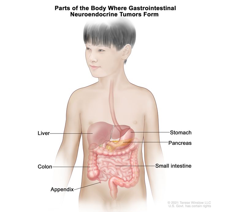
  • Gastrointestinal stromal tumors are a disease in which malignant (cancer) cells form in the tissues of the wall of the stomach or intestines.
  • GIST may develop as part of a syndrome.
  • Signs and symptoms of GIST include anemia and a lump in the abdomen.
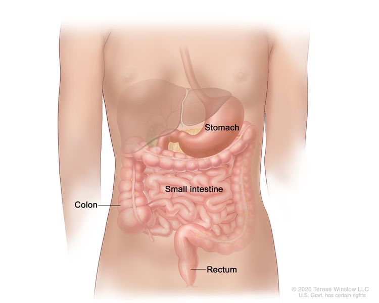
  • Tests that examine the digestive tract are used to help diagnose GIST.

Gastrointestinal stromal tumors are a disease in which malignant (cancer) cells form in the tissues of the wall of the stomach or intestines.

Gastrointestinal stromal tumors (GIST) usually begin in cells in the tissues of the wall of the stomach or intestines. These cells, called interstitial cells of Cajal, help food move along the digestive tract.

Childhood GIST usually occurs in the stomach. It is more common in girls, and typically appears in the teen years.

GIST in children is not the same as GIST in adults. Patients should be seen at centers that specialize in the treatment of GIST.

EnlargeDrawing of the gastrointestinal tract showing the stomach, small intestine, colon, and rectum.
Gastrointestinal stromal tumors (GISTs) are most common in the stomach and small intestine but may be found anywhere in or near the gastrointestinal tract.

GIST may develop as part of a syndrome.

GIST may occur as part of the following syndromes:

Signs and symptoms of GIST include anemia and a lump in the abdomen.

These and other signs and symptoms may be caused by GIST or by other conditions.

Check with your child’s doctor if your child has any of the following:

  • Anemia (tiredness, dizziness, fast or irregular heartbeat, shortness of breath, or pale skin).
  • A lump in the abdomen.
  • A blockage of the intestine (cramping pain in the abdomen, nausea, vomiting, diarrhea, constipation, and swelling of the abdomen).

Tests that examine the digestive tract are used to help diagnose GIST.

The following tests and procedures may be used:

  • Physical exam and health history: An exam of the body to check general signs of health, including checking for signs of disease, such as lumps or anything else that seems unusual. A history of the patient’s health habits and past illnesses and treatments will also be taken.
  • Gene testing: A laboratory test in which cells or tissue are analyzed to look for changes in the KIT, PDGFA, and SDH genes. It is important to know whether there are changes in these genes in order to diagnose GIST and plan treatment.
  • MRI (magnetic resonance imaging): A procedure that uses a magnet, radio waves, and a computer to make a series of detailed pictures of areas inside the body. This procedure is also called nuclear magnetic resonance imaging (NMRI).
  • CT scan (CAT scan): A procedure that makes a series of detailed pictures of areas inside the body, taken from different angles. The pictures are made by a computer linked to an x-ray machine. A dye may be injected into a vein or swallowed to help the organs or tissues show up more clearly. This procedure is also called computed tomography, computerized tomography, or computerized axial tomography.
  • PET scan (positron emission tomography scan): A procedure to find malignant tumor cells in the body. A small amount of radioactive glucose (sugar) is injected into a vein. The PET scanner rotates around the body and makes a picture of where glucose is being used in the body. Malignant tumor cells show up brighter in the picture because they are more active and take up more glucose than normal cells do.
    EnlargePositron emission tomography (PET) scan; drawing shows a child lying on table that slides through the PET scanner.
    Positron emission tomography (PET) scan. The child lies on a table that slides through the PET scanner. The head rest and white strap help the child lie still. A small amount of radioactive glucose (sugar) is injected into the child’s vein, and a scanner makes a picture of where the glucose is being used in the body. Cancer cells show up brighter in the picture because they take up more glucose than normal cells do.
  • X-ray: An x-ray is a type of energy beam that can go through the body and onto film, making a picture of areas inside the body, such as the abdomen or the area where the tumor formed.
  • Biopsy: The removal of cells or tissues so they can be viewed under a microscope by a pathologist to check for signs of cancer.
    • Fine-needle aspiration: The removal of tissue using a thin needle.
    • Endoscopy: A procedure to look at organs and tissues inside the body to check for abnormal areas. An endoscope is inserted through an incision (cut) in the skin or opening in the body, such as the mouth or anus. An endoscope is a thin, tube-like instrument with a light and a lens for viewing. It may also have a tool to remove tissue or lymph node samples, which are checked under a microscope for signs of disease.

    The following laboratory test may be done to study the tissue samples:

    • Immunohistochemistry: A laboratory test that uses antibodies to check for certain antigens (markers) in a sample of a patient’s tissue. The antibodies are usually linked to an enzyme or a fluorescent dye. After the antibodies bind to a specific antigen in the tissue sample, the enzyme or dye is activated, and the antigen can then be seen under a microscope. This type of test looks for the enzyme SDH in a sample of the patient’s tissue. When SDH is not present, it is called SDH-deficient GIST. It is important to know whether the cancer is SDH-deficient in order to plan treatment.

Stages of GIST

Key Points

  • After GIST has been diagnosed, tests are done to find out if cancer cells have spread to nearby areas or to other parts of the body.
  • There are three ways that cancer spreads in the body.
  • Cancer may spread from where it began to other parts of the body.

After GIST has been diagnosed, tests are done to find out if cancer cells have spread to nearby areas or to other parts of the body.

The process used to find out if GIST has spread to nearby areas or other parts of the body is called staging. GIST may spread to the lymph nodes, liver, and peritoneum. There is no standard staging system for childhood GIST. The results of tests and procedures done to diagnose GIST are used to help make decisions about treatment.

Sometimes childhood GIST recurs (comes back) after treatment.

There are three ways that cancer spreads in the body.

Cancer can spread through tissue, the lymph system, and the blood:

  • Tissue. The cancer spreads from where it began by growing into nearby areas.
  • Lymph system. The cancer spreads from where it began by getting into the lymph system. The cancer travels through the lymph vessels to other parts of the body.
  • Blood. The cancer spreads from where it began by getting into the blood. The cancer travels through the blood vessels to other parts of the body.

Cancer may spread from where it began to other parts of the body.

When cancer spreads to another part of the body, it is called metastasis. Cancer cells break away from where they began (the primary tumor) and travel through the lymph system or blood.

  • Lymph system. The cancer gets into the lymph system, travels through the lymph vessels, and forms a tumor (metastatic tumor) in another part of the body.
  • Blood. The cancer gets into the blood, travels through the blood vessels, and forms a tumor (metastatic tumor) in another part of the body.

The metastatic tumor is the same type of cancer as the primary tumor. For example, if GIST spreads to the liver, the cancer cells in the liver are actually GIST cells. The disease is metastatic GIST, not liver cancer.

Many cancer deaths are caused when cancer moves from the original tumor and spreads to other tissues and organs. This is called metastatic cancer. This animation shows how cancer cells travel from the place in the body where they first formed to other parts of the body.

Treatment Option Overview

Key Points

  • There are different types of treatment for children with GIST.
  • Children with GIST should have their treatment planned by a team of doctors who are experts in treating childhood cancer.
  • Two types of standard treatment are used:
    • Surgery
    • Targeted therapy
  • New types of treatment are being tested in clinical trials.
    • Chemotherapy
  • Treatment for childhood GIST may cause side effects.
  • Patients may want to think about taking part in a clinical trial.
  • Patients can enter clinical trials before, during, or after starting their cancer treatment.
  • Follow-up tests may be needed.

There are different types of treatment for children with GIST.

Some treatments are standard (the currently used treatment), and some are being tested in clinical trials. A treatment clinical trial is a research study meant to help improve current treatments or obtain information on new treatments for patients with cancer. When clinical trials show that a new treatment is better than the standard treatment, the new treatment may become the standard treatment.

Because cancer in children is rare, taking part in a clinical trial should be considered. Some clinical trials are open only to patients who have not started treatment.

Children with GIST should have their treatment planned by a team of doctors who are experts in treating childhood cancer.

Treatment will be overseen by a pediatric oncologist, a doctor who specializes in treating children with cancer. The pediatric oncologist works with other pediatric health professionals who are experts in treating children with cancer and who specialize in certain areas of medicine. This may include the following specialists and others:

Two types of standard treatment are used:

Surgery

Surgery to remove the cancer is a treatment for SDH-deficient GIST.

Targeted therapy

Targeted therapy is a type of treatment that uses drugs or other substances to attack cancer cells. Targeted therapies usually cause less harm to normal cells than chemotherapy or radiation therapy do.

New types of treatment are being tested in clinical trials.

This summary section describes treatments that are being studied in clinical trials. It may not mention every new treatment being studied. Information about clinical trials is available from the NCI website.

Chemotherapy

Chemotherapy is a cancer treatment that uses drugs to stop the growth of cancer cells, either by killing the cells or by stopping them from dividing. When chemotherapy is taken by mouth or injected into a vein or muscle, the drugs enter the bloodstream and can reach cancer cells throughout the body (systemic chemotherapy).

Treatment for childhood GIST may cause side effects.

To learn more about side effects that begin during treatment for cancer, visit Side Effects.

Side effects from cancer treatment that begin after treatment and continue for months or years are called late effects. Late effects of cancer treatment may include:

  • Physical problems.
  • Changes in mood, feelings, thinking, learning, or memory.
  • Second cancers (new types of cancer) or other conditions.

Some late effects may be treated or controlled. It is important to talk with your child’s doctors about the possible late effects caused by some treatments. See the PDQ summary on Late Effects of Treatment for Childhood Cancer for more information.

Patients may want to think about taking part in a clinical trial.

For some patients, taking part in a clinical trial may be the best treatment choice. Clinical trials are part of the cancer research process. Clinical trials are done to find out if new cancer treatments are safe and effective or better than the standard treatment.

Many of today’s standard treatments for cancer are based on earlier clinical trials. Patients who take part in a clinical trial may receive the standard treatment or be among the first to receive a new treatment.

Patients who take part in clinical trials also help improve the way cancer will be treated in the future. Even when clinical trials do not lead to effective new treatments, they often answer important questions and help move research forward.

Patients can enter clinical trials before, during, or after starting their cancer treatment.

Some clinical trials only include patients who have not yet received treatment. Other trials test treatments for patients whose cancer has not gotten better. There are also clinical trials that test new ways to stop cancer from recurring (coming back) or reduce the side effects of cancer treatment.

Clinical trials are taking place in many parts of the country. Information about clinical trials supported by NCI can be found on NCI’s clinical trials search webpage. Clinical trials supported by other organizations can be found on the ClinicalTrials.gov website.

Follow-up tests may be needed.

As your child goes through treatment, they will have follow-up tests or check-ups. Some tests that were done to diagnose or stage the cancer may be repeated to see how well the treatment is working. Decisions about whether to continue, change, or stop treatment may be based on the results of these tests.

Some of the tests will continue to be done from time to time after treatment has ended. The results of these tests can show if your child’s condition has changed or if the cancer has recurred (come back).

Treatment of GIST

For information about the treatments listed below, see the Treatment Option Overview section.

Treatment for children who have newly diagnosed tumors with changes in the KIT or PDGFR gene includes the following:

Treatment for children whose newly diagnosed tumors are SDH-deficient may include the following:

  • Surgery to remove the tumor. More surgery may be needed if intestinal obstruction or bleeding occurs.
  • Targeted therapy with a tyrosine kinase inhibitor (sunitinib).

Use our clinical trial search to find NCI-supported cancer clinical trials that are accepting patients. You can search for trials based on the type of cancer, the age of the patient, and where the trials are being done. General information about clinical trials is also available.

Treatment of Recurrent GIST

For information about the treatments listed below, see the Treatment Option Overview section.

Treatment of recurrent GIST in children may include the following:

To Learn More About GIST

About This PDQ Summary

About PDQ

Physician Data Query (PDQ) is the National Cancer Institute’s (NCI’s) comprehensive cancer information database. The PDQ database contains summaries of the latest published information on cancer prevention, detection, genetics, treatment, supportive care, and complementary and alternative medicine. Most summaries come in two versions. The health professional versions have detailed information written in technical language. The patient versions are written in easy-to-understand, nontechnical language. Both versions have cancer information that is accurate and up to date and most versions are also available in Spanish.

PDQ is a service of the NCI. The NCI is part of the National Institutes of Health (NIH). NIH is the federal government’s center of biomedical research. The PDQ summaries are based on an independent review of the medical literature. They are not policy statements of the NCI or the NIH.

Purpose of This Summary

This PDQ cancer information summary has current information about the treatment of childhood gastrointestinal stromal tumors. It is meant to inform and help patients, families, and caregivers. It does not give formal guidelines or recommendations for making decisions about health care.

Reviewers and Updates

Editorial Boards write the PDQ cancer information summaries and keep them up to date. These Boards are made up of experts in cancer treatment and other specialties related to cancer. The summaries are reviewed regularly and changes are made when there is new information. The date on each summary (“Updated”) is the date of the most recent change.

The information in this patient summary was taken from the health professional version, which is reviewed regularly and updated as needed, by the PDQ Pediatric Treatment Editorial Board.

Clinical Trial Information

A clinical trial is a study to answer a scientific question, such as whether one treatment is better than another. Trials are based on past studies and what has been learned in the laboratory. Each trial answers certain scientific questions in order to find new and better ways to help cancer patients. During treatment clinical trials, information is collected about the effects of a new treatment and how well it works. If a clinical trial shows that a new treatment is better than one currently being used, the new treatment may become “standard.” Patients may want to think about taking part in a clinical trial. Some clinical trials are open only to patients who have not started treatment.

Clinical trials can be found online at NCI’s website. For more information, call the Cancer Information Service (CIS), NCI’s contact center, at 1-800-4-CANCER (1-800-422-6237).

Permission to Use This Summary

PDQ is a registered trademark. The content of PDQ documents can be used freely as text. It cannot be identified as an NCI PDQ cancer information summary unless the whole summary is shown and it is updated regularly. However, a user would be allowed to write a sentence such as “NCI’s PDQ cancer information summary about breast cancer prevention states the risks in the following way: [include excerpt from the summary].”

The best way to cite this PDQ summary is:

PDQ® Pediatric Treatment Editorial Board. PDQ Childhood Gastrointestinal Stromal Tumors Treatment. Bethesda, MD: National Cancer Institute. Updated <MM/DD/YYYY>. Available at: /types/soft-tissue-sarcoma/patient/child-gist-treatment-pdq. Accessed <MM/DD/YYYY>.

Images in this summary are used with permission of the author(s), artist, and/or publisher for use in the PDQ summaries only. If you want to use an image from a PDQ summary and you are not using the whole summary, you must get permission from the owner. It cannot be given by the National Cancer Institute. Information about using the images in this summary, along with many other images related to cancer can be found in Visuals Online. Visuals Online is a collection of more than 3,000 scientific images.

Disclaimer

The information in these summaries should not be used to make decisions about insurance reimbursement. More information on insurance coverage is available on Cancer.gov on the Managing Cancer Care page.

Contact Us

More information about contacting us or receiving help with the Cancer.gov website can be found on our Contact Us for Help page. Questions can also be submitted to Cancer.gov through the website’s E-mail Us.
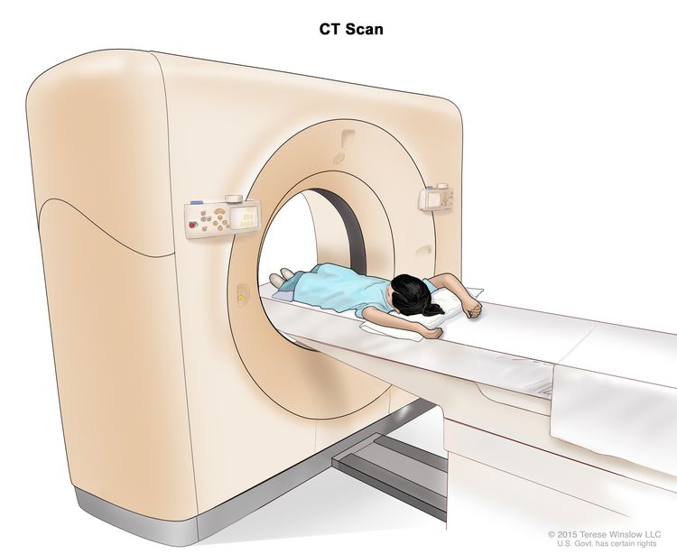
Gastrointestinal Neuroendocrine Tumors—Patient Version

Gastrointestinal Neuroendocrine Tumors—Patient Version

Overview

Gastrointestinal (GI) neuroendocrine tumors are slow-growing tumors that form in the GI tract, mainly in the rectum, small intestine, or appendix. Explore the links on this page to learn more about GI neuroendocrine tumor treatment and clinical trials.


Causes & Prevention

NCI does not have PDQ evidence-based information about prevention of gastrointestinal neuroendocrine tumors.

Screening

NCI does not have PDQ evidence-based information about screening for gastrointestinal neuroendocrine tumors.

Coping with Cancer

The information in this section is meant to help you cope with the many issues and concerns that occur when you have cancer.

Emotions and Cancer Adjusting to Cancer Support for Caregivers Survivorship Advanced Cancer Managing Cancer Care

Childhood Gastrointestinal Neuroendocrine Tumors Treatment (PDQ®)–Patient Version

Childhood Gastrointestinal Neuroendocrine Tumors Treatment (PDQ®)–Patient Version

General Information About Childhood Gastrointestinal Neuroendocrine Tumors

Key Points

  • A gastrointestinal neuroendocrine tumor forms in neuroendocrine cells in the lining of the digestive tract, appendix, and other organs in the abdomen.
  • Signs and symptoms of gastrointestinal neuroendocrine tumors depend on where the tumor forms.
  • Tests that examine the digestive tract, liver, and pancreas are used to diagnose gastrointestinal neuroendocrine tumors.
  • Certain factors affect prognosis (chance of recovery).

A gastrointestinal neuroendocrine tumor forms in neuroendocrine cells in the lining of the digestive tract, appendix, and other organs in the abdomen.

Gastrointestinal neuroendocrine tumors (also called gastrointestinal carcinoid tumors) form from a certain type of neuroendocrine cell (a type of cell that is like a nerve cell and a hormone-making cell). These cells are scattered throughout the chest and abdomen, but most are found in certain organs in the abdomen. Neuroendocrine cells in the digestive tract make hormones that help control digestive juices and the muscles used in moving food through the stomach and intestines.

Most gastrointestinal neuroendocrine tumors in children form in the appendix (a pouch that sticks out from the first part of the large intestine near the end of the small intestine). The tumor is often found during surgery to remove the appendix. They also form in the digestive tract (lining of the stomach or intestines), pancreas, and liver.

These tumors are usually small, slow-growing, and benign (not cancer). Some tumors may be malignant (cancer) and spread to other places in the body.

EnlargeDrawing of the gastrointestinal tract showing the liver, stomach, pancreas, small intestine, colon, and appendix.
Gastrointestinal neuroendocrine tumors form in the lining of the gastrointestinal tract and other organs in the abdomen. Most gastrointestinal neuroendocrine tumors in children form in the appendix, but they can also form in the stomach, intestines, pancreas, and liver.

Signs and symptoms of gastrointestinal neuroendocrine tumors depend on where the tumor forms.

Check with your child’s doctor if you are concerned about signs and symptoms that may be caused by gastrointestinal neuroendocrine tumors or by other conditions.

Neuroendocrine tumors in the appendix may cause the following signs and symptoms:

Gastrointestinal neuroendocrine tumors that are not in the appendix may release hormones and other substances. Carcinoid syndrome occurs when a neuroendocrine tumor in the digestive tract releases the hormone serotonin and other substances. It may cause any of the following signs and symptoms. Check with your child’s doctor if your child has any of the following:

  • Redness and a warm feeling in the face, neck, and upper chest.
  • A fast heartbeat.
  • Trouble breathing.
  • Sudden drop in blood pressure (restlessness, confusion, weakness, dizziness, and pale, cool, and clammy skin).
  • Diarrhea.

Tests that examine the digestive tract, liver, and pancreas are used to diagnose gastrointestinal neuroendocrine tumors.

The following tests and procedures may be used:

  • Physical exam and health history: An exam of the body to check general signs of health, including checking for signs of disease, such as lumps or anything else that seems unusual. A history of the patient’s health habits and past illnesses and treatments will also be taken.
  • Blood chemistry studies: A procedure in which a blood sample is checked to measure the amounts of certain substances released into the blood by organs and tissues in the body. An unusual (higher or lower than normal) amount of a substance can be a sign of disease.
  • MRI (magnetic resonance imaging): A procedure that uses a magnet and radio waves to make a series of detailed pictures of areas inside the body. The pictures are made by a computer. This procedure is also called nuclear magnetic resonance imaging (NMRI).
    EnlargeMagnetic resonance imaging (MRI) scan; drawing shows a child lying on a table that slides into the MRI machine, which takes a series of detailed pictures of areas inside the body.
    Magnetic resonance imaging (MRI) scan. The child lies on a table that slides into the MRI machine, which takes a series of detailed pictures of areas inside the body. The positioning of the child on the table depends on the part of the body being imaged.
  • PET scan (positron emission tomography scan): A procedure to find malignant tumor cells in the body. A small amount of radioactive glucose (sugar) is injected into a vein. The PET scanner rotates around the body and makes a picture of where glucose is being used in the body. Malignant tumor cells show up brighter in the picture because they are more active and take up more glucose than normal cells do.
    EnlargePositron emission tomography (PET) scan; drawing shows a child lying on table that slides through the PET scanner.
    Positron emission tomography (PET) scan. The child lies on a table that slides through the PET scanner. The head rest and white strap help the child lie still. A small amount of radioactive glucose (sugar) is injected into the child’s vein, and a scanner makes a picture of where the glucose is being used in the body. Cancer cells show up brighter in the picture because they take up more glucose than normal cells do.
  • CT scan (CAT scan): A procedure that makes a series of detailed pictures of areas inside the body, taken from different angles. The pictures are made by a computer linked to an x-ray machine. This procedure is also called computed tomography, computerized tomography, or computerized axial tomography.
    EnlargeComputed tomography (CT) scan; drawing shows a child lying on a table that slides through the CT scanner, which takes a series of detailed x-ray pictures of areas inside the body.
    Computed tomography (CT) scan. The child lies on a table that slides through the CT scanner, which takes a series of detailed x-ray pictures of areas inside the body.
  • Ultrasound exam: A procedure in which high-energy sound waves (ultrasound) are bounced off internal tissues or organs and make echoes. The echoes form a picture of body tissues called a sonogram. The picture can be printed to be looked at later.
    EnlargeAbdominal ultrasound; drawing shows a child lying on an exam table during an abdominal ultrasound procedure. A technician is shown pressing a transducer (a device that makes sound waves that bounce off tissues inside the body) against the skin of the abdomen. A computer screen shows a sonogram (picture).
    Abdominal ultrasound. An ultrasound transducer connected to a computer is pressed against the skin of the abdomen. The transducer bounces sound waves off internal organs and tissues to make echoes that form a sonogram (computer picture).
  • Twenty-four-hour urine test: A test in which urine is collected for 24 hours to measure the amounts of certain substances, such as hormones. An unusual (higher or lower than normal) amount of a substance can be a sign of disease in the organ or tissue that makes it. The urine sample is checked to see if it contains 5-HIAA (a breakdown product of the hormone serotonin which may be made by neuroendocrine tumors). This test is used to help diagnose carcinoid syndrome.
  • Somatostatin receptor scintigraphy: A type of radionuclide scan that may be used to find tumors. A very small amount of radioactive octreotide (a hormone that attaches to tumors) is injected into a vein and travels through the blood. The radioactive octreotide attaches to the tumor and a special camera that detects radioactivity is used to show where the tumors are in the body. This procedure is also called octreotide scan and SRS.

Certain factors affect prognosis (chance of recovery).

Prognosis depends on the following:

  • Where the tumor first formed in the body.
  • The size of the tumor.
  • Whether the tumor has spread to other parts of the body.
  • Whether the tumor is newly diagnosed or has recurred (come back).

The prognosis for neuroendocrine tumors in the appendix in children is usually excellent after surgery to remove the tumor. Gastrointestinal neuroendocrine tumors that are not in the appendix are usually larger or have spread to other parts of the body at the time of diagnosis and do not respond well to chemotherapy. Larger tumors are more likely to recur (come back).

Stages of Gastrointestinal Neuroendocrine Tumors

Key Points

  • After a gastrointestinal neuroendocrine tumor has been diagnosed, tests are done to find out if cancer cells have spread to nearby areas or to other parts of the body.
  • There are three ways that cancer spreads in the body.
  • Cancer may spread from where it began to other parts of the body.

After a gastrointestinal neuroendocrine tumor has been diagnosed, tests are done to find out if cancer cells have spread to nearby areas or to other parts of the body.

The process used to find out if cancer has spread to nearby areas or other parts of the body is called staging. There is no standard staging system for childhood gastrointestinal neuroendocrine tumors.

Neuroendocrine tumors of the appendix are not known to spread, but other gastrointestinal neuroendocrine tumors may spread. The results of tests and procedures done to diagnose gastrointestinal neuroendocrine tumors are used to help make decisions about treatment.

Sometimes childhood gastrointestinal neuroendocrine tumors recur (come back) after treatment.

There are three ways that cancer spreads in the body.

Cancer can spread through tissue, the lymph system, and the blood:

  • Tissue. The cancer spreads from where it began by growing into nearby areas.
  • Lymph system. The cancer spreads from where it began by getting into the lymph system. The cancer travels through the lymph vessels to other parts of the body.
  • Blood. The cancer spreads from where it began by getting into the blood. The cancer travels through the blood vessels to other parts of the body.

Cancer may spread from where it began to other parts of the body.

When cancer spreads to another part of the body, it is called metastasis. Cancer cells break away from where they began (the primary tumor) and travel through the lymph system or blood.

  • Lymph system. The cancer gets into the lymph system, travels through the lymph vessels, and forms a tumor (metastatic tumor) in another part of the body.
  • Blood. The cancer gets into the blood, travels through the blood vessels, and forms a tumor (metastatic tumor) in another part of the body.

The metastatic tumor is the same type of cancer as the primary tumor. For example, if neuroendocrine tumor cells in the small intestine spread to the liver, the cancer cells in the liver are actually neuroendocrine tumor cells. The cells in the liver are metastatic neuroendocrine tumor cells, not liver cancer.

Many cancer deaths are caused when cancer moves from the original tumor and spreads to other tissues and organs. This is called metastatic cancer. This animation shows how cancer cells travel from the place in the body where they first formed to other parts of the body.

Treatment Option Overview

Key Points

  • There are different types of treatment for children with gastrointestinal neuroendocrine tumors.
  • Children with gastrointestinal neuroendocrine tumors not in the appendix should have their treatment planned by a team of doctors who are experts in treating childhood cancer.
  • Five types of standard treatment are used:
    • Surgery
    • Embolization
    • Hormone therapy
    • Peptide receptor radionuclide therapy
    • Targeted therapy
  • New types of treatment are being tested in clinical trials.
  • Treatment of gastrointestinal neuroendocrine tumors may cause side effects.
  • Patients may want to think about taking part in a clinical trial.
  • Patients can enter clinical trials before, during, or after starting their cancer treatment.
  • Follow-up tests may be needed.

There are different types of treatment for children with gastrointestinal neuroendocrine tumors.

Some treatments are standard (the currently used treatment), and some are being tested in clinical trials. A treatment clinical trial is a research study meant to help improve current treatments or obtain information on new treatments for patients with cancer. When clinical trials show that a new treatment is better than the standard treatment, the new treatment may become the standard treatment.

Because cancer in children is rare, taking part in a clinical trial should be considered. Some clinical trials are open only to patients who have not started treatment.

Children with gastrointestinal neuroendocrine tumors not in the appendix should have their treatment planned by a team of doctors who are experts in treating childhood cancer.

Treatment will be overseen by a pediatric oncologist, a doctor who specializes in treating children with cancer. The pediatric oncologist works with other pediatric health professionals who are experts in treating children with cancer and who specialize in certain areas of medicine. This may include the following specialists and others:

Five types of standard treatment are used:

Surgery

Surgery to remove the tumor is the only treatment needed for neuroendocrine tumors in the appendix.

Embolization

Embolization is a treatment in which contrast dye and particles are injected into the hepatic artery through a catheter (thin tube). The particles block the artery, cutting off blood flow to the tumor. Sometimes a small amount of a radioactive substance is attached to the particles. Most of the radiation is trapped near the tumor to kill the cancer cells. This is called radioembolization.

Hormone therapy

Hormone therapy with a somatostatin analogue (octreotide or lanreotide) may be used to treat gastrointestinal neuroendocrine tumors that have spread or cannot be removed by surgery. This treatment stops extra hormones from being made by the neuroendocrine tumor. Octreotide or lanreotide are somatostatin analogues which are injected under the skin or into the muscle.

Peptide receptor radionuclide therapy

Sometimes a small amount of a radioactive substance is attached to the somatostatin analogue drugs octreotide or lanreotide to kill the cancer cells.

Targeted therapy

Targeted therapy is a type of treatment that uses drugs or other substances to identify and attack specific cancer cells. Targeted therapies usually cause less harm to normal cells than chemotherapy or radiation therapy do.

  • Tyrosine kinase inhibitors: These targeted therapy drugs block signals needed for tumors to grow. Sunitinib is used to treat gastrointestinal neuroendocrine tumors that are not in the appendix.
  • mTOR inhibitors: A type of targeted therapy that stops the protein that helps cells divide and survive. Everolimus is used to treat gastrointestinal neuroendocrine tumors that are not in the appendix.

New types of treatment are being tested in clinical trials.

Information about clinical trials is available from the NCI website.

Treatment of gastrointestinal neuroendocrine tumors may cause side effects.

To learn more about side effects that begin during treatment for cancer, visit Side Effects.

Side effects from cancer treatment that begin after treatment and continue for months or years are called late effects. Late effects of cancer treatment may include:

  • Physical problems.
  • Changes in mood, feelings, thinking, learning, or memory.
  • Second cancers (new types of cancer) or other conditions.

Some late effects may be treated or controlled. It is important to talk with your child’s doctors about the possible late effects caused by some treatments. See the PDQ summary on Late Effects of Treatment for Childhood Cancer for more information.

Patients may want to think about taking part in a clinical trial.

For some patients, taking part in a clinical trial may be the best treatment choice. Clinical trials are part of the cancer research process. Clinical trials are done to find out if new cancer treatments are safe and effective or better than the standard treatment.

Many of today’s standard treatments for cancer are based on earlier clinical trials. Patients who take part in a clinical trial may receive the standard treatment or be among the first to receive a new treatment.

Patients who take part in clinical trials also help improve the way cancer will be treated in the future. Even when clinical trials do not lead to effective new treatments, they often answer important questions and help move research forward.

Patients can enter clinical trials before, during, or after starting their cancer treatment.

Some clinical trials only include patients who have not yet received treatment. Other trials test treatments for patients whose cancer has not gotten better. There are also clinical trials that test new ways to stop cancer from recurring (coming back) or reduce the side effects of cancer treatment.

Clinical trials are taking place in many parts of the country. Information about clinical trials supported by NCI can be found on NCI’s clinical trials search webpage. Clinical trials supported by other organizations can be found on the ClinicalTrials.gov website.

Follow-up tests may be needed.

As your child goes through treatment, they will have follow-up tests or check-ups. Some tests that were done to diagnose or stage the cancer may be repeated to see how well the treatment is working. Decisions about whether to continue, change, or stop treatment may be based on the results of these tests.

Some of the tests will continue to be done from time to time after treatment has ended. The results of these tests can show if your child’s condition has changed or if the cancer has recurred (come back).

Treatment of Gastrointestinal Neuroendocrine Tumors

For information about the treatments listed below, see the Treatment Option Overview section.

Treatment of newly diagnosed neuroendocrine tumors in the appendix in children may include the following:

Treatment of newly diagnosed neuroendocrine tumors in the large intestine, pancreas, or stomach is usually surgery.

Treatment of newly diagnosed tumors that cannot be removed by surgery, multiple tumors, or tumors that have spread may include the following:

Sometimes childhood gastrointestinal neuroendocrine tumors can recur (come back) after treatment. Your child’s doctor will work with you to plan treatment if your child is diagnosed with a recurrent gastrointestinal neuroendocrine tumor.

Use our clinical trial search to find NCI-supported cancer clinical trials that are accepting patients. You can search for trials based on the type of cancer, the age of the patient, and where the trials are being done. General information about clinical trials is also available.

To Learn More About Gastrointestinal Neuroendocrine Tumors

About This PDQ Summary

About PDQ

Physician Data Query (PDQ) is the National Cancer Institute’s (NCI’s) comprehensive cancer information database. The PDQ database contains summaries of the latest published information on cancer prevention, detection, genetics, treatment, supportive care, and complementary and alternative medicine. Most summaries come in two versions. The health professional versions have detailed information written in technical language. The patient versions are written in easy-to-understand, nontechnical language. Both versions have cancer information that is accurate and up to date and most versions are also available in Spanish.

PDQ is a service of the NCI. The NCI is part of the National Institutes of Health (NIH). NIH is the federal government’s center of biomedical research. The PDQ summaries are based on an independent review of the medical literature. They are not policy statements of the NCI or the NIH.

Purpose of This Summary

This PDQ cancer information summary has current information about the treatment of childhood gastrointestinal neuroendocrine tumors. It is meant to inform and help patients, families, and caregivers. It does not give formal guidelines or recommendations for making decisions about health care.

Reviewers and Updates

Editorial Boards write the PDQ cancer information summaries and keep them up to date. These Boards are made up of experts in cancer treatment and other specialties related to cancer. The summaries are reviewed regularly and changes are made when there is new information. The date on each summary (“Updated”) is the date of the most recent change.

The information in this patient summary was taken from the health professional version, which is reviewed regularly and updated as needed, by the PDQ Pediatric Treatment Editorial Board.

Clinical Trial Information

A clinical trial is a study to answer a scientific question, such as whether one treatment is better than another. Trials are based on past studies and what has been learned in the laboratory. Each trial answers certain scientific questions in order to find new and better ways to help cancer patients. During treatment clinical trials, information is collected about the effects of a new treatment and how well it works. If a clinical trial shows that a new treatment is better than one currently being used, the new treatment may become “standard.” Patients may want to think about taking part in a clinical trial. Some clinical trials are open only to patients who have not started treatment.

Clinical trials can be found online at NCI’s website. For more information, call the Cancer Information Service (CIS), NCI’s contact center, at 1-800-4-CANCER (1-800-422-6237).

Permission to Use This Summary

PDQ is a registered trademark. The content of PDQ documents can be used freely as text. It cannot be identified as an NCI PDQ cancer information summary unless the whole summary is shown and it is updated regularly. However, a user would be allowed to write a sentence such as “NCI’s PDQ cancer information summary about breast cancer prevention states the risks in the following way: [include excerpt from the summary].”

The best way to cite this PDQ summary is:

PDQ® Pediatric Treatment Editorial Board. PDQ Childhood Gastrointestinal Neuroendocrine Tumors Treatment. Bethesda, MD: National Cancer Institute. Updated <MM/DD/YYYY>. Available at: /types/gi-neuroendocrine-tumors/patient/child-gi-neuroendocrine-treatment-pdq. Accessed <MM/DD/YYYY>.

Images in this summary are used with permission of the author(s), artist, and/or publisher for use in the PDQ summaries only. If you want to use an image from a PDQ summary and you are not using the whole summary, you must get permission from the owner. It cannot be given by the National Cancer Institute. Information about using the images in this summary, along with many other images related to cancer can be found in Visuals Online. Visuals Online is a collection of more than 3,000 scientific images.

Disclaimer

The information in these summaries should not be used to make decisions about insurance reimbursement. More information on insurance coverage is available on Cancer.gov on the Managing Cancer Care page.

Contact Us

More information about contacting us or receiving help with the Cancer.gov website can be found on our Contact Us for Help page. Questions can also be submitted to Cancer.gov through the website’s E-mail Us.
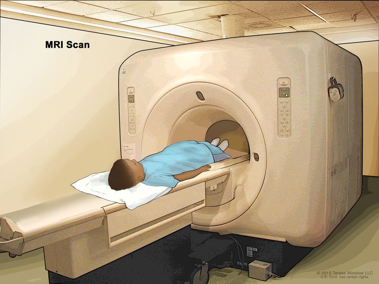
Colorectal Cancer—Patient Version

Colorectal Cancer—Patient Version

Overview

Colorectal cancer often begins as a growth called a polyp inside the colon or rectum. Finding and removing polyps can prevent colorectal cancer. Explore the links on this page to learn more about colorectal cancer prevention, screening, treatment, statistics, research, clinical trials, and more.

Causes & Prevention

Coping with Cancer

The information in this section is meant to help you cope with the many issues and concerns that occur when you have cancer.

Emotions and Cancer Adjusting to Cancer Support for Caregivers Survivorship Advanced Cancer Managing Cancer Care

Childhood Colorectal Cancer Treatment (PDQ®)–Patient Version

Childhood Colorectal Cancer Treatment (PDQ®)–Patient Version

General Information About Childhood Colorectal Cancer

Key Points

  • Colorectal cancer is a disease in which malignant (cancer) cells form in the tissues of the colon or the rectum.
  • Having certain inherited syndromes increases the risk of colorectal cancer.
  • Signs and symptoms of colorectal cancer include pain in the abdomen, constipation, and diarrhea.
  • Tests that examine the colon and rectum are used to diagnose colorectal cancer.
  • Certain factors affect prognosis (chance of recovery).

Colorectal cancer is a disease in which malignant (cancer) cells form in the tissues of the colon or the rectum.

The colon is part of the body’s digestive system. The digestive system removes and processes nutrients (vitamins, minerals, carbohydrates, fats, proteins, and water) from foods and helps pass waste material out of the body. The digestive system is made up of the mouth, throat, esophagus, stomach, and the small and large intestines. The colon (large bowel) is the main part of the large intestine and is about 5 feet long in an adult. Together, the rectum and anal canal make up the last part of the large intestine and are 6 to 8 inches long. The anal canal ends at the anus (the opening of the large intestine to the outside of the body).

Having certain inherited syndromes increases the risk of colorectal cancer.

Anything that increases your chance of getting a disease is called a risk factor. Having a risk factor does not mean that you will get cancer; not having risk factors doesn’t mean that you will not get cancer. Talk with your child’s doctor if you think your child may be at risk.

Childhood colorectal cancer may be part of an inherited syndrome. Some colorectal cancers in young people are linked to a gene mutation that causes polyps (growths in the mucous membrane that lines the colon) to form that may turn into cancer later. The risk of colorectal cancer is increased by having inherited conditions, such as the following:

Polyps that form in the colon of children who do not have an inherited syndrome are not linked to an increased risk of cancer.

Signs and symptoms of colorectal cancer include pain in the abdomen, constipation, and diarrhea.

Signs and symptoms of childhood colorectal cancer usually depend on where the tumor forms. These and other signs and symptoms may be caused by colorectal cancer or by other conditions.

Check with your child’s doctor if your child has any of the following:

  • Tumors of the rectum or lower colon may cause pain in the abdomen, constipation, or diarrhea.
  • Tumors in the part of the colon on the left side of the body may cause:
    • A lump in the abdomen.
    • Weight loss for no known reason.
    • Nausea and vomiting.
    • Loss of appetite.
    • Blood in the stool.
    • Anemia (tiredness, dizziness, fast or irregular heartbeat, shortness of breath, pale skin).
  • Tumors in the part of the colon on the right side of the body may cause:
    • Pain in the abdomen.
    • Blood in the stool.
    • Constipation or diarrhea.
    • Nausea and vomiting.
    • Weight loss for no known reason.

Tests that examine the colon and rectum are used to diagnose colorectal cancer.

The following tests and procedures may be used:

  • Physical exam and health history: An exam of the body to check general signs of health, including checking for signs of disease, such as lumps or anything else that seems unusual. A history of the patient’s health habits and past illnesses and treatments will also be taken.
  • Colonoscopy: A procedure to look inside the rectum and colon for polyps, abnormal areas, or cancer. A colonoscope is inserted through the rectum into the colon. A colonoscope is a thin, tube-like instrument with a light and a lens for viewing. It also has a tool to remove polyps or tissue samples, which are checked under a microscope for signs of cancer.
  • Barium enema: A series of x-rays of the lower gastrointestinal tract. A liquid that contains barium (a silver-white metallic compound) is put into the rectum. The barium coats the lower gastrointestinal tract and x-rays are taken. This procedure is also called a lower GI series.
  • Fecal occult blood test: A test to check stool (solid waste) for blood that can only be seen with a microscope. Small samples of stool are placed on special cards and returned to the doctor or laboratory for testing.
  • Complete blood count (CBC): A procedure in which a sample of blood is drawn and checked for the following:
  • Carcinoembryonic antigen (CEA) assay: A test that measures the level of CEA in the blood. CEA is released into the bloodstream from both cancer cells and normal cells. When found in higher than normal amounts, it can be a sign of colorectal cancer or other conditions.

Certain factors affect prognosis (chance of recovery).

Prognosis depends on the following:

Stages of Childhood Colorectal Cancer

Key Points

  • After colorectal cancer has been diagnosed, tests are done to find out if cancer cells have spread within the colon or rectum or to other parts of the body.
  • There are three ways that cancer spreads in the body.
  • Cancer may spread from where it began to other parts of the body.
  • Sometimes childhood colorectal cancer recurs (comes back) after treatment.

After colorectal cancer has been diagnosed, tests are done to find out if cancer cells have spread within the colon or rectum or to other parts of the body.

To plan treatment, it is important to know whether cancer cells have spread within the colon or rectum or to other parts of the body. The process used to find out if cancer has spread is called staging. It is important to know the stage in order to plan treatment. The results of the tests used to diagnose cancer are often also used to stage the disease. (See the General Information section.)

In children, the cancer has often spread to the lymph nodes, outside the colon or rectum, or to other organs in the abdomen at diagnosis. The following tests and procedures may be used to find out if cancer has spread:

  • Chest x-ray: An x-ray of the organs and bones inside the chest. An x-ray is a type of energy beam that can go through the body and onto film, making a picture of areas inside the body.
  • PET scan (positron emission tomography scan): A procedure to find malignant tumor cells in the body. A small amount of radioactive glucose (sugar) is injected into a vein. The PET scanner rotates around the body and makes a picture of where glucose is being used in the body. Malignant tumor cells show up brighter in the picture because they are more active and take up more glucose than normal cells do.
  • MRI (magnetic resonance imaging): A procedure that uses a magnet, radio waves, and a computer to make a series of detailed pictures of areas of the body, such as the chest, abdomen, and pelvis. This procedure is also called nuclear magnetic resonance imaging (NMRI).
  • CT scan (CAT scan): A procedure that makes a series of detailed pictures of areas inside the body, such as the chest, taken from different angles. The pictures are made by a computer linked to an x-ray machine. A dye may be injected into a vein or swallowed to help the organs or tissues show up more clearly. This procedure is also called computed tomography, computerized tomography, or computerized axial tomography.
  • Bone scan: A procedure to check if there are rapidly dividing cells, such as cancer cells, in the bone. A very small amount of radioactive material is injected into a vein and travels through the bloodstream. The radioactive material collects in the bones with cancer and is detected by a scanner.
    EnlargeBone scan; drawing shows a child lying on a table that slides under the scanner, a technician operating the scanner, and a computer monitor that will show images made during the scan.
    Bone scan. A small amount of radioactive material is injected into the child’s vein and travels through the blood. The radioactive material collects in the bones. As the child lies on a table that slides under the scanner, the radioactive material is detected and images are made on a computer screen.

There are three ways that cancer spreads in the body.

Cancer can spread through tissue, the lymph system, and the blood:

  • Tissue. The cancer spreads from where it began by growing into nearby areas.
  • Lymph system. The cancer spreads from where it began by getting into the lymph system. The cancer travels through the lymph vessels to other parts of the body.
  • Blood. The cancer spreads from where it began by getting into the blood. The cancer travels through the blood vessels to other parts of the body.

Cancer may spread from where it began to other parts of the body.

When cancer spreads to another part of the body, it is called metastasis. Cancer cells break away from where they began (the primary tumor) and travel through the lymph system or blood.

  • Lymph system. The cancer gets into the lymph system, travels through the lymph vessels, and forms a tumor (metastatic tumor) in another part of the body.
  • Blood. The cancer gets into the blood, travels through the blood vessels, and forms a tumor (metastatic tumor) in another part of the body.

The metastatic tumor is the same type of cancer as the primary tumor. For example, if colorectal cancer spreads to the bone, the cancer cells in the bone are actually colorectal cancer cells. The disease is metastatic colorectal cancer, not bone cancer.

Many cancer deaths are caused when cancer moves from the original tumor and spreads to other tissues and organs. This is called metastatic cancer. This animation shows how cancer cells travel from the place in the body where they first formed to other parts of the body.

Sometimes childhood colorectal cancer recurs (comes back) after treatment.

Childhood colorectal cancer may come back in the colon, rectum, or in other parts of the body.

Treatment Option Overview

Key Points

  • There are different types of treatment for children with colorectal cancer.
  • Children with colorectal cancer should have their treatment planned by a team of doctors who are experts in treating childhood cancer.
  • Four types of standard treatment are used:
    • Surgery
    • Radiation therapy
    • Chemotherapy
    • Immunotherapy
  • New types of treatment are being tested in clinical trials.
  • Treatment for childhood colorectal cancer may cause side effects.
  • Patients may want to think about taking part in a clinical trial.
  • Patients can enter clinical trials before, during, or after starting their cancer treatment.
  • Follow-up tests may be needed.

There are different types of treatment for children with colorectal cancer.

Some treatments are standard (the currently used treatment), and some are being tested in clinical trials. A treatment clinical trial is a research study meant to help improve current treatments or obtain information on new treatments for patients with cancer. When clinical trials show that a new treatment is better than the standard treatment, the new treatment may become the standard treatment.

Because cancer in children is rare, taking part in a clinical trial should be considered. Some clinical trials are open only to patients who have not started treatment.

Children with colorectal cancer should have their treatment planned by a team of doctors who are experts in treating childhood cancer.

Treatment will be overseen by a pediatric oncologist, a doctor who specializes in treating children with cancer. The pediatric oncologist works with other pediatric health professionals who are experts in treating children with cancer and who specialize in certain areas of medicine. This may include the following specialists and others:

Four types of standard treatment are used:

Surgery

Surgery to remove the cancer is done if the cancer has not spread to other parts of the body at diagnosis.

Radiation therapy

Radiation therapy is a cancer treatment that uses high-energy x-rays or other types of radiation to kill cancer cells or keep them from growing. External radiation therapy uses a machine outside the body to send radiation toward the area of the body with cancer.

Chemotherapy

Chemotherapy is a cancer treatment that uses drugs to stop the growth of cancer cells, either by killing the cells or by stopping them from dividing. When chemotherapy is taken by mouth or injected into a vein or muscle, the drugs enter the bloodstream and can reach cancer cells throughout the body (systemic chemotherapy). Chemotherapy using more than one drug is called combination chemotherapy.

Immunotherapy

Immunotherapy is a treatment that uses the patient’s immune system to fight cancer. Substances made by the body or made in a laboratory are used to boost, direct, or restore the body’s natural defenses against cancer. This cancer treatment is a type of biologic therapy.

Immune checkpoint inhibitor therapy is a type of immunotherapy. Some types of immune cells, such as T cells, and some cancer cells have certain proteins, called checkpoint proteins, on their surface that keep immune responses in check. When cancer cells have large amounts of these proteins, they will not be attacked and killed by T cells. Immune checkpoint inhibitors block these proteins and the ability of T cells to kill cancer cells is increased.

Immunotherapy uses the body’s immune system to fight cancer. This animation explains one type of immunotherapy that uses immune checkpoint inhibitors to treat cancer.

There are two types of immune checkpoint inhibitor therapy:

  • PD-1 and PD-L1 inhibitor therapy: PD-1 is a protein on the surface of T cells that helps keep the body’s immune responses in check. PD-L1 is a protein found on some types of cancer cells. When PD-1 attaches to PD-L1, it stops the T cell from killing the cancer cell. PD-1 and PD-L1 inhibitors keep PD-1 and PD-L1 proteins from attaching to each other. This allows the T cells to kill cancer cells. Nivolumab is a type of PD-1 inhibitor that has been used to treat children 12 years and older with progressive colorectal cancer.
    EnlargeImmune checkpoint inhibitor; the panel on the left shows the binding of proteins PD-L1 (on the tumor cell) to PD-1 (on the T cell), which keeps T cells from killing tumor cells in the body. Also shown are a tumor cell antigen and T cell receptor. The panel on the right shows immune checkpoint inhibitors (anti-PD-L1 and anti-PD-1) blocking the binding of PD-L1 to PD-1, which allows the T cells to kill tumor cells.
    Immune checkpoint inhibitor. Checkpoint proteins, such as PD-L1 on tumor cells and PD-1 on T cells, help keep immune responses in check. The binding of PD-L1 to PD-1 keeps T cells from killing tumor cells in the body (left panel). Blocking the binding of PD-L1 to PD-1 with an immune checkpoint inhibitor (anti-PD-L1 or anti-PD-1) allows the T cells to kill tumor cells (right panel).
  • CTLA-4 inhibitor. CTLA-4 is a protein on the surface of T cells that helps keep the body’s immune responses in check. When CTLA-4 attaches to another protein called B7 on a cancer cell, it stops the T cell from killing the cancer cell. CTLA-4 inhibitors attach to CTLA-4 and allow the T cells to kill cancer cells. Ipilimumab is used to treat children 12 years and older with progressive colorectal cancer.
    EnlargeImmune checkpoint inhibitor; the panel on the left shows the binding of the T-cell receptor (TCR) to antigen and MHC proteins on the antigen-presenting cell (APC) and the binding of CD28 on the T cell to B7-1/B7-2 on the APC. It also shows the binding of B7-1/B7-2 to CTLA-4 on the T cell, which keeps the T cells in the inactive state. The panel on the right shows immune checkpoint inhibitor (anti-CTLA antibody) blocking the binding of B7-1/B7-2 to CTLA-4, which allows the T cells to be active and to kill tumor cells.
    Immune checkpoint inhibitor. Checkpoint proteins, such as B7-1/B7-2 on antigen-presenting cells (APC) and CTLA-4 on T cells, help keep the body’s immune responses in check. When the T-cell receptor (TCR) binds to antigen and major histocompatibility complex (MHC) proteins on the APC and CD28 binds to B7-1/B7-2 on the APC, the T cell can be activated. However, the binding of B7-1/B7-2 to CTLA-4 keeps the T cells in the inactive state so they are not able to kill tumor cells in the body (left panel). Blocking the binding of B7-1/B7-2 to CTLA-4 with an immune checkpoint inhibitor (anti-CTLA-4 antibody) allows the T cells to be active and to kill tumor cells (right panel).

New types of treatment are being tested in clinical trials.

Information about clinical trials is available from the NCI website.

Treatment for childhood colorectal cancer may cause side effects.

To learn more about side effects that begin during treatment for cancer, visit Side Effects.

Side effects from cancer treatment that begin after treatment and continue for months or years are called late effects. Late effects of cancer treatment may include the following:

  • Physical problems.
  • Changes in mood, feelings, thinking, learning, or memory.
  • Second cancers (new types of cancer) or other conditions.

Some late effects may be treated or controlled. It is important to talk with your child’s doctors about the possible late effects caused by some treatments. See the PDQ summary on Late Effects of Treatment for Childhood Cancer for more information.

Patients may want to think about taking part in a clinical trial.

For some patients, taking part in a clinical trial may be the best treatment choice. Clinical trials are part of the cancer research process. Clinical trials are done to find out if new cancer treatments are safe and effective or better than the standard treatment.

Many of today’s standard treatments for cancer are based on earlier clinical trials. Patients who take part in a clinical trial may receive the standard treatment or be among the first to receive a new treatment.

Patients who take part in clinical trials also help improve the way cancer will be treated in the future. Even when clinical trials do not lead to effective new treatments, they often answer important questions and help move research forward.

Patients can enter clinical trials before, during, or after starting their cancer treatment.

Some clinical trials only include patients who have not yet received treatment. Other trials test treatments for patients whose cancer has not gotten better. There are also clinical trials that test new ways to stop cancer from recurring (coming back) or reduce the side effects of cancer treatment.

Clinical trials are taking place in many parts of the country. Information about clinical trials supported by NCI can be found on NCI’s clinical trials search webpage. Clinical trials supported by other organizations can be found on the ClinicalTrials.gov website.

Follow-up tests may be needed.

As your child goes through treatment, they will have follow-up tests or check-ups. Some tests that were done to diagnose or stage the cancer may be repeated to see how well the treatment is working. Decisions about whether to continue, change, or stop treatment may be based on the results of these tests.

Some of the tests will continue to be done from time to time after treatment has ended. The results of these tests can show if your child’s condition has changed or if the cancer has recurred (come back).

Treatment of Childhood Colorectal Cancer

For information about the treatments listed below, see the Treatment Option Overview section.

Treatment of newly diagnosed colorectal cancer in children may include the following:

Children with certain familial colorectal cancer syndromes may be treated with the following:

  • Surgery to remove the colon before cancer forms.
  • Medicine to decrease the number of polyps in the colon.

Use our clinical trial search to find NCI-supported cancer clinical trials that are accepting patients. You can search for trials based on the type of cancer, the age of the patient, and where the trials are being done. General information about clinical trials is also available.

Treatment of Recurrent or Progressive Childhood Colorectal Cancer

For information about the treatments listed below, see the Treatment Option Overview section.

Treatment of recurrent or progressive colorectal cancer in children may include the following:

Use our clinical trial search to find NCI-supported cancer clinical trials that are accepting patients. You can search for trials based on the type of cancer, the age of the patient, and where the trials are being done. General information about clinical trials is also available.

To Learn More About Childhood Colorectal Cancer

About This PDQ Summary

About PDQ

Physician Data Query (PDQ) is the National Cancer Institute’s (NCI’s) comprehensive cancer information database. The PDQ database contains summaries of the latest published information on cancer prevention, detection, genetics, treatment, supportive care, and complementary and alternative medicine. Most summaries come in two versions. The health professional versions have detailed information written in technical language. The patient versions are written in easy-to-understand, nontechnical language. Both versions have cancer information that is accurate and up to date and most versions are also available in Spanish.

PDQ is a service of the NCI. The NCI is part of the National Institutes of Health (NIH). NIH is the federal government’s center of biomedical research. The PDQ summaries are based on an independent review of the medical literature. They are not policy statements of the NCI or the NIH.

Purpose of This Summary

This PDQ cancer information summary has current information about the treatment of childhood colorectal cancer. It is meant to inform and help patients, families, and caregivers. It does not give formal guidelines or recommendations for making decisions about health care.

Reviewers and Updates

Editorial Boards write the PDQ cancer information summaries and keep them up to date. These Boards are made up of experts in cancer treatment and other specialties related to cancer. The summaries are reviewed regularly and changes are made when there is new information. The date on each summary (“Updated”) is the date of the most recent change.

The information in this patient summary was taken from the health professional version, which is reviewed regularly and updated as needed, by the PDQ Pediatric Treatment Editorial Board.

Clinical Trial Information

A clinical trial is a study to answer a scientific question, such as whether one treatment is better than another. Trials are based on past studies and what has been learned in the laboratory. Each trial answers certain scientific questions in order to find new and better ways to help cancer patients. During treatment clinical trials, information is collected about the effects of a new treatment and how well it works. If a clinical trial shows that a new treatment is better than one currently being used, the new treatment may become “standard.” Patients may want to think about taking part in a clinical trial. Some clinical trials are open only to patients who have not started treatment.

Clinical trials can be found online at NCI’s website. For more information, call the Cancer Information Service (CIS), NCI’s contact center, at 1-800-4-CANCER (1-800-422-6237).

Permission to Use This Summary

PDQ is a registered trademark. The content of PDQ documents can be used freely as text. It cannot be identified as an NCI PDQ cancer information summary unless the whole summary is shown and it is updated regularly. However, a user would be allowed to write a sentence such as “NCI’s PDQ cancer information summary about breast cancer prevention states the risks in the following way: [include excerpt from the summary].”

The best way to cite this PDQ summary is:

PDQ® Pediatric Treatment Editorial Board. PDQ Childhood Colorectal Cancer Treatment. Bethesda, MD: National Cancer Institute. Updated <MM/DD/YYYY>. Available at: /types/colorectal/patient/child-colorectal-treatment-pdq. Accessed <MM/DD/YYYY>.

Images in this summary are used with permission of the author(s), artist, and/or publisher for use in the PDQ summaries only. If you want to use an image from a PDQ summary and you are not using the whole summary, you must get permission from the owner. It cannot be given by the National Cancer Institute. Information about using the images in this summary, along with many other images related to cancer can be found in Visuals Online. Visuals Online is a collection of more than 3,000 scientific images.

Disclaimer

The information in these summaries should not be used to make decisions about insurance reimbursement. More information on insurance coverage is available on Cancer.gov on the Managing Cancer Care page.

Contact Us

More information about contacting us or receiving help with the Cancer.gov website can be found on our Contact Us for Help page. Questions can also be submitted to Cancer.gov through the website’s E-mail Us.