Intraocular melanoma is a disease in which malignant (cancer) cells form in the tissues of the eye.
Having a fair complexion and certain conditions can affect the risk of melanoma.
Signs and symptoms of intraocular melanoma include trouble seeing or a dark spot on the iris.
Tests that examine the eye are used to diagnose intraocular melanoma.
Certain factors affect prognosis (chance of recovery).
Intraocular melanoma is a disease in which malignant (cancer) cells form in the tissues of the eye.
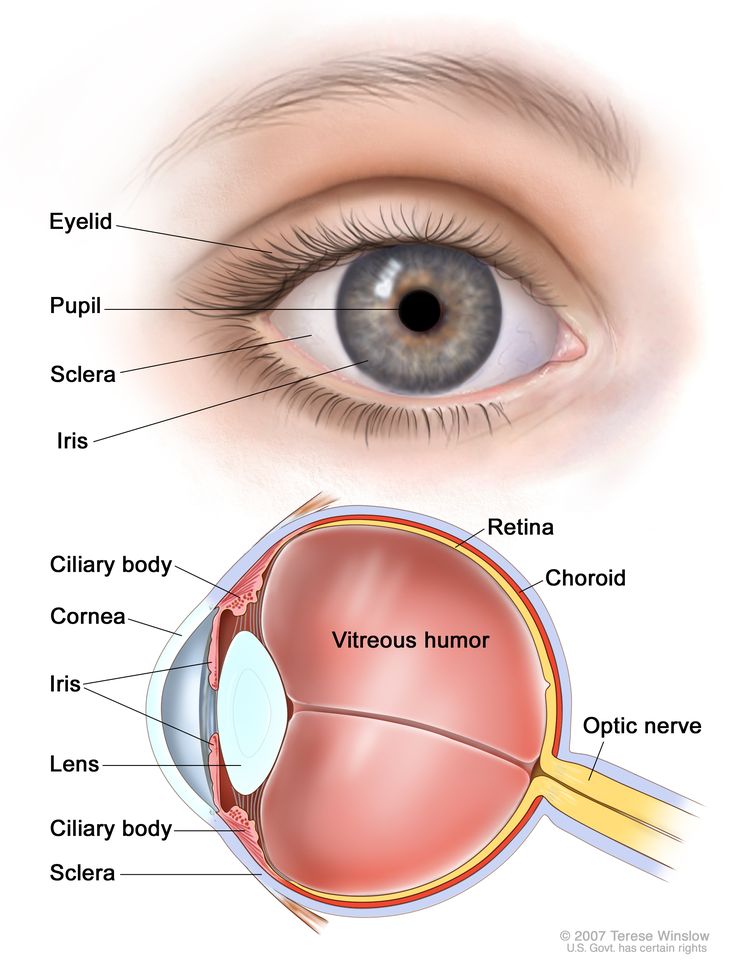
Intraocular melanoma begins in the middle of three layers of the wall of the eye. The outer layer includes the white sclera (the “white of the eye”) and the clear cornea at the front of the eye. The inner layer has a lining of nervetissue, called the retina, which senses light and sends images along the optic nerve to the brain. The middle layer, where intraocular melanoma forms, is called the uvea or uveal tract, and has three main parts: the iris, the ciliary body, and the choroid.
EnlargeAnatomy of the eye, showing the outside and inside of the eye including the sclera, cornea, iris, ciliary body, choroid, retina, vitreous humor, and optic nerve. The vitreous humor is a liquid that fills the center of the eye.
Having a fair complexion and certain conditions can affect the risk of melanoma.
Anything that increases your risk of getting a disease is called a risk factor. Having a risk factor does not mean that you will get cancer; not having risk factors doesn’t mean that you will not get cancer. Talk with your child’s doctor if you think your child may be at risk.
Risk factors for childhood intraocular melanoma include the following:
Signs and symptoms of intraocular melanoma include trouble seeing or a dark spot on the iris.
These and other signs and symptoms may be caused by intraocular melanoma or by other conditions.
Check with your child’s doctor if your child has any of the following:
Trouble seeing.
Dark spot on the iris (colored part of the eye).
A bulging eye.
Tests that examine the eye are used to diagnose intraocular melanoma.
The following tests and procedures may be used:
Physical exam and health history: An exam of the body to check general signs of health, including checking for signs of disease, such as lumps or anything else that seems unusual. A history of the patient’s health habits and past illnesses and treatments will also be taken.
Ultrasound: A procedure in which high-energy sound waves (ultrasound) are bounced off internal tissues or organs and make echoes. The echoes form a picture of body tissues called a sonogram. The picture can be printed to be looked at later.
Fluorescein angiography: A test used to take pictures of the retina in the eye. A yellow dye is injected into a vein and travels throughout the body including the blood vessels in the eye. The yellow dye causes the vessels in the eye to fluoresce when a picture is taken.
Certain factors affect prognosis (chance of recovery).
Whether the tumor has spread within the eye or to other places in the body.
Whether there are certain changes in the genes linked to intraocular melanoma.
Stages of Childhood Intraocular Melanoma
Key Points
After intraocular melanoma has been diagnosed, tests are done to find out if cancer cells have spread within the eye or to other parts of the body.
There are three ways cancer spreads in the body.
Cancer may spread from where it began to other parts of the body.
Sometimes intraocular melanoma comes back after treatment.
After intraocular melanoma has been diagnosed, tests are done to find out if cancer cells have spread within the eye or to other parts of the body.
The process used to find out if cancer has spread to areas near the eye or to other parts of the body is called staging. There is no standard staging system for childhood intraocular melanoma.
The tests and procedures used to diagnose cancer and other tests and procedures may be used to find out if cancer has spread and plan treatment:
Liver function tests: A procedure in which a blood sample is checked to measure the amounts of certain substances released into the blood by the liver. A higher than normal amount of a substance can be a sign that cancer has spread to the liver.
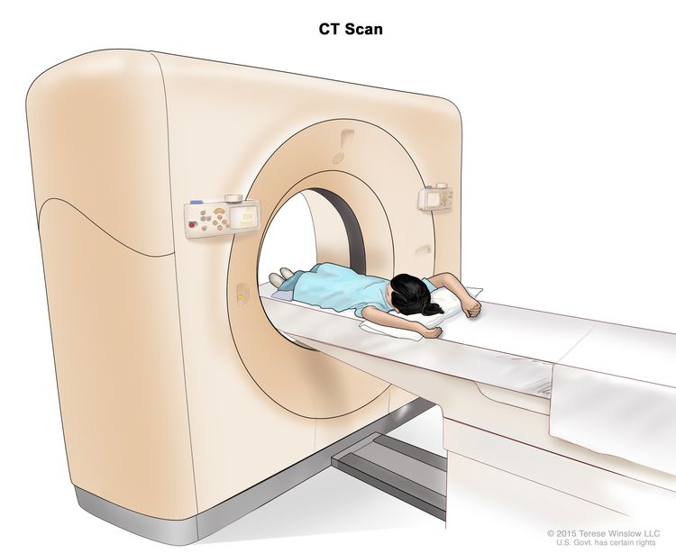
CT scan (CAT scan): A procedure that makes a series of detailed pictures of areas inside the body, such as the chest or liver, taken from different angles. The pictures are made by a computer linked to an x-ray machine. A dye may be injected into a vein or swallowed to help the organs or tissues show up more clearly. This procedure is also called computed tomography, computerized tomography, or computerized axial tomography. EnlargeComputed tomography (CT) scan. The child lies on a table that slides through the CT scanner, which takes a series of detailed x-ray pictures of areas inside the body.
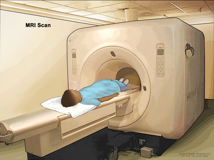
MRI: A procedure that uses a magnet, radio waves, and a computer to make a series of detailed pictures of areas inside the body, such as the liver. This procedure is also called nuclear magnetic resonance imaging (NMRI). EnlargeMagnetic resonance imaging (MRI) scan. The child lies on a table that slides into the MRI machine, which takes a series of detailed pictures of areas inside the body. The positioning of the child on the table depends on the part of the body being imaged.
Chest x-ray: An x-ray of the chest. An x-ray is a type of energy beam that can go through the body and onto film, making a picture of areas inside the body.
Tissue. The cancer spreads from where it began by growing into nearby areas.
Lymph system. The cancer spreads from where it began by getting into the lymph system. The cancer travels through the lymph vessels to other parts of the body.
Blood. The cancer spreads from where it began by getting into the blood. The cancer travels through the blood vessels to other parts of the body.
Cancer may spread from where it began to other parts of the body.
When cancer spreads to another part of the body, it is called metastasis. Cancer cells break away from where they began (the primary tumor) and travel through the lymph system or blood.
Lymph system. The cancer gets into the lymph system, travels through the lymph vessels, and forms a tumor (metastatic tumor) in another part of the body.
Blood. The cancer gets into the blood, travels through the blood vessels, and forms a tumor (metastatic tumor) in another part of the body.
The metastatictumor is the same type of cancer as the primary tumor. For example, if intraocular melanoma spreads to the liver, the cancer cells in the liver are actually intraocular melanoma cells. The disease is metastatic intraocular melanoma, not liver cancer.
Many cancer deaths are caused when cancer moves from the original tumor and spreads to other tissues and organs. This is called metastatic cancer. This animation shows how cancer cells travel from the place in the body where they first formed to other parts of the body.
Sometimes intraocular melanoma comes back after treatment.
The cancer may come back in the eye or in other parts of the body, such as in the lung or liver.
Treatment Option Overview
Key Points
There are different types of treatment for children with intraocular melanoma.
Children with intraocular melanoma should have their treatment planned by a team of doctors who are experts in treating childhood cancer.
Three types of standard treatment are used:
Surgery
Radiation therapy
Laser surgery
New types of treatment are being tested in clinical trials.
Targeted therapy
Treatment for childhood intraocular melanoma may cause side effects.
Patients may want to think about taking part in a clinical trial.
Patients can enter clinical trials before, during, or after starting their cancer treatment.
Follow-up tests may be needed.
There are different types of treatment for children with intraocular melanoma.
Some treatments are standard (the currently used treatment), and some are being tested in clinical trials. A treatment clinical trial is a research study meant to help improve current treatments or obtain information on new treatments for patients with cancer. When clinical trials show that a new treatment is better than the standard treatment, the new treatment may become the standard treatment.
Because cancer in children is rare, taking part in a clinical trial should be considered. Some clinical trials are open only to patients who have not started treatment.
Children with intraocular melanoma should have their treatment planned by a team of doctors who are experts in treating childhood cancer.
Treatment will be overseen by a pediatric oncologist, a doctor who specializes in treating children with cancer. The pediatric oncologist works with other pediatric health professionals who are experts in treating children with cancer and who specialize in certain areas of medicine. This may include the following specialists and others:
During surgery, all or part of the eye with cancer is removed. Whether all or part of the eye is removed during surgery depends on the size of the cancer and where it is in the eye.
Radiation therapy
Radiation therapy is a cancer treatment that uses high-energy x-rays or other types of radiation to kill cancer cells or keep them from growing. There are two types of radiation therapy:
External radiation therapy uses a machine outside the body to send radiation toward the area of the body with cancer.
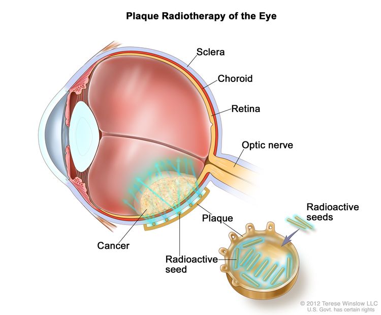
Internal radiation therapy uses a radioactive substance sealed in needles, seeds, wires, or catheters that are placed directly into or near the cancer. For intraocular melanoma, localizedplaque radiation therapy is used. Radioactive seeds are attached to one side of a disk, called a plaque, and placed directly on the outside wall of the eye near the tumor. The side of the plaque with the seeds on it faces the eyeball, aiming radiation at the tumor. The plaque helps protect other nearby tissue from the radiation. EnlargePlaque radiotherapy is a type of radiation therapy used to treat eye cancer. Radioactive seeds are attached to one side of a thin piece of metal (usually gold) called a plaque. The plaque is sewn onto the outside wall of the eye near the tumor. The seeds give off radiation which kills the cancer. The plaque is removed at the end of treatment, which usually lasts for several days.
Laser surgery
Laser surgery uses a laser beam (a narrow beam of intense light) to turn the cancer cells into a gas that evaporates (dissolves into the air).
New types of treatment are being tested in clinical trials.
This summary section describes treatments that are being studied in clinical trials. It may not mention every new treatment being studied. Information about clinical trials is available from the NCI website.
Targeted therapy
Targeted therapy is a type of treatment that uses drugs or other substances to identify and attack specific cancer cells. Targeted therapies usually cause less harm to normal cells than chemotherapy or radiation therapy do.
Targeted therapy is being studied for the treatment of childhood intraocular melanoma that has recurred (come back).
Treatment for childhood intraocular melanoma may cause side effects.
Patients may want to think about taking part in a clinical trial.
For some patients, taking part in a clinical trial may be the best treatment choice. Clinical trials are part of the cancer research process. Clinical trials are done to find out if new cancer treatments are safe and effective or better than the standard treatment.
Many of today’s standard treatments for cancer are based on earlier clinical trials. Patients who take part in a clinical trial may receive the standard treatment or be among the first to receive a new treatment.
Patients who take part in clinical trials also help improve the way cancer will be treated in the future. Even when clinical trials do not lead to effective new treatments, they often answer important questions and help move research forward.
Patients can enter clinical trials before, during, or after starting their cancer treatment.
Some clinical trials only include patients who have not yet received treatment. Other trials test treatments for patients whose cancer has not gotten better. There are also clinical trials that test new ways to stop cancer from recurring (coming back) or reduce the side effects of cancer treatment.
Clinical trials are taking place in many parts of the country. Information about clinical trials supported by NCI can be found on NCI’s clinical trials search webpage. Clinical trials supported by other organizations can be found on the ClinicalTrials.gov website.
Follow-up tests may be needed.
As your child goes through treatment, they will have follow-up tests or check-ups. Some tests that were done to diagnose or stage the cancer may be repeated to see how well the treatment is working. Decisions about whether to continue, change, or stop treatment may be based on the results of these tests.
Some of the tests will continue to be done from time to time after treatment has ended. The results of these tests can show if your child’s condition has changed or if the cancer has recurred (come back).
Use our clinical trial search to find NCI-supported cancer clinical trials that are accepting patients. You can search for trials based on the type of cancer, the age of the patient, and where the trials are being done. General information about clinical trials is also available.
Treatment of Recurrent Childhood Intraocular Melanoma
A clinical trial that checks a sample of the patient’s tumor for certain gene changes. The type of targeted therapy that will be given to the patient depends on the type of gene change.
Use our clinical trial search to find NCI-supported cancer clinical trials that are accepting patients. You can search for trials based on the type of cancer, the age of the patient, and where the trials are being done. General information about clinical trials is also available.
To Learn More About Childhood Intraocular Melanoma
Physician Data Query (PDQ) is the National Cancer Institute’s (NCI’s) comprehensive cancer information database. The PDQ database contains summaries of the latest published information on cancer prevention, detection, genetics, treatment, supportive care, and complementary and alternative medicine. Most summaries come in two versions. The health professional versions have detailed information written in technical language. The patient versions are written in easy-to-understand, nontechnical language. Both versions have cancer information that is accurate and up to date and most versions are also available in Spanish.
PDQ is a service of the NCI. The NCI is part of the National Institutes of Health (NIH). NIH is the federal government’s center of biomedical research. The PDQ summaries are based on an independent review of the medical literature. They are not policy statements of the NCI or the NIH.
Purpose of This Summary
This PDQ cancer information summary has current information about the treatment of childhood intraocular (uveal) melanoma. It is meant to inform and help patients, families, and caregivers. It does not give formal guidelines or recommendations for making decisions about health care.
Reviewers and Updates
Editorial Boards write the PDQ cancer information summaries and keep them up to date. These Boards are made up of experts in cancer treatment and other specialties related to cancer. The summaries are reviewed regularly and changes are made when there is new information. The date on each summary (“Updated”) is the date of the most recent change.
The information in this patient summary was taken from the health professional version, which is reviewed regularly and updated as needed, by the PDQ Pediatric Treatment Editorial Board.
Clinical Trial Information
A clinical trial is a study to answer a scientific question, such as whether one treatment is better than another. Trials are based on past studies and what has been learned in the laboratory. Each trial answers certain scientific questions in order to find new and better ways to help cancer patients. During treatment clinical trials, information is collected about the effects of a new treatment and how well it works. If a clinical trial shows that a new treatment is better than one currently being used, the new treatment may become “standard.” Patients may want to think about taking part in a clinical trial. Some clinical trials are open only to patients who have not started treatment.
Clinical trials can be found online at NCI’s website. For more information, call the Cancer Information Service (CIS), NCI’s contact center, at 1-800-4-CANCER (1-800-422-6237).
Permission to Use This Summary
PDQ is a registered trademark. The content of PDQ documents can be used freely as text. It cannot be identified as an NCI PDQ cancer information summary unless the whole summary is shown and it is updated regularly. However, a user would be allowed to write a sentence such as “NCI’s PDQ cancer information summary about breast cancer prevention states the risks in the following way: [include excerpt from the summary].”
The best way to cite this PDQ summary is:
PDQ® Pediatric Treatment Editorial Board. PDQ Childhood Intraocular (Uveal) Melanoma Treatment. Bethesda, MD: National Cancer Institute. Updated <MM/DD/YYYY>. Available at: /types/eye/patient/child-intraocular-melanoma-treatment-pdq. Accessed <MM/DD/YYYY>.
Images in this summary are used with permission of the author(s), artist, and/or publisher for use in the PDQ summaries only. If you want to use an image from a PDQ summary and you are not using the whole summary, you must get permission from the owner. It cannot be given by the National Cancer Institute. Information about using the images in this summary, along with many other images related to cancer can be found in Visuals Online. Visuals Online is a collection of more than 3,000 scientific images.
Disclaimer
The information in these summaries should not be used to make decisions about insurance reimbursement. More information on insurance coverage is available on Cancer.gov on the Managing Cancer Care page.
Contact Us
More information about contacting us or receiving help with the Cancer.gov website can be found on our Contact Us for Help page. Questions can also be submitted to Cancer.gov through the website’s E-mail Us.
Skin cancer is a disease in which malignant (cancer) cells form in the tissues of the skin.
Different types of cancer start in the skin.
Exposure to sunlight affects the risk of basal cell carcinoma and squamous cell carcinoma of the skin.
Signs of basal cell carcinoma and squamous cell carcinoma of the skin include a raised lump and a sore that does not heal.
Tests that examine the skin are used to diagnose basal cell carcinoma and squamous cell carcinoma of the skin.
Skin cancer is a disease in which malignant (cancer) cells form in the tissues of the skin.
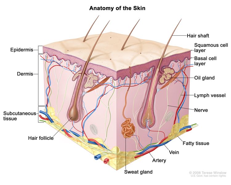
The skin is the body’s largest organ. It protects against heat, sunlight, injury, and infection. Skin also helps control body temperature and stores water, fat, and vitamin D. The skin has several layers, but the two main layers are the epidermis (upper or outer layer) and the dermis (lower or inner layer). Skin cancer begins in the epidermis, which is made up of three kinds of cells:
Squamous cells: Thin, flat cells that form the top layer of the epidermis.
Basal cells: Round cells under the squamous cells.
Melanocytes: Cells that make melanin and are found in the lower part of the epidermis. Melanin is the pigment that gives skin its natural color. When skin is exposed to the sun, melanocytes make more pigment and cause the skin to darken.
EnlargeAnatomy of the skin showing the epidermis (including the squamous cell and basal cell layers), dermis, subcutaneous tissue, and other parts of the skin.
Skin cancer can occur anywhere on the body, but it usually occurs in skin that is exposed to sunlight, such as the face, neck, and hands.
Different types of cancer start in the skin.
There are two main forms of skin cancer:
Nonmelanoma: Skin cancer that forms in basal cells is called basal cell carcinoma. Skin cancer that forms in squamous cells is called squamous cell carcinoma of the skin. Basal cell carcinoma and squamous cell carcinoma of the skin are two types of nonmelanoma skin cancer.
Melanoma: Skin cancer that forms in the melanocytes (cells that color the skin) is called melanoma. In children and adolescents, melanoma is more common than both basal cell carcinoma and squamous cell carcinoma of the skin. See the PDQ summary on Childhood Melanoma for more information.
Exposure to sunlight affects the risk of basal cell carcinoma and squamous cell carcinoma of the skin.
Anything that increases your risk of getting a disease is called a risk factor. Having a risk factor does not mean that you will get cancer; not having risk factors doesn’t mean that you will not get cancer. Talk with your child’s doctor if you think your child may be at risk.
Risk factors for childhood basal cell carcinoma and squamous cell carcinoma of the skin include the following:
Being exposed to natural sunlight or artificial sunlight (such as from tanning beds).
Having a fair complexion, which includes the following:
Fair skin that freckles and burns easily, does not tan, or tans poorly.
Signs of basal cell carcinoma and squamous cell carcinoma of the skin include a raised lump and a sore that does not heal.
These and other signs and symptoms may be caused by basal cell carcinoma and squamous cell carcinoma of the skin or by other conditions.
Check with your child’s doctor if your child has any of the following:
A sore that does not heal.
Areas of the skin that are:
Small, raised, smooth, shiny, and waxy.
Small, raised, and red or reddish-brown.
Flat, rough, red or brown, and scaly.
Scaly, bleeding, or crusty.
Similar to a scar and firm.
Tests that examine the skin are used to diagnose basal cell carcinoma and squamous cell carcinoma of the skin.
The following tests and procedures may be used:
Physical exam and health history: An exam of the body to check general signs of health, including checking for signs of disease, such as lumps or anything else that seems unusual. A history of the patient’s health habits and past illnesses and treatments will also be taken.
Skin exam: A doctor or nurse checks the skin for bumps or spots that look abnormal in color, size, shape, or texture.
Biopsy: All or part of a growth that doesn’t look normal is cut from the skin and viewed under a microscope by a pathologist to check for signs of cancer. There are three main types of skin biopsies:
Shave biopsy: A sterile razor blade is used to “shave off” the growth that does not look normal.
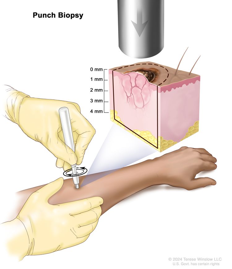
Punch biopsy: A special instrument called a punch or a trephine is used to remove a circle of tissue from the growth that does not look normal. EnlargePunch biopsy. A sharp, hollow, circular instrument is used to remove a small, round piece of tissue from a lesion on the skin. The instrument is turned clockwise and counterclockwise to cut about 4 millimeters (mm) down to the layer of fatty tissue below the skin and remove the sample of tissue. Skin thickness is different on different parts of the body.
Excision: The tumor and a small amount of healthy tissue around the tumor is removed.
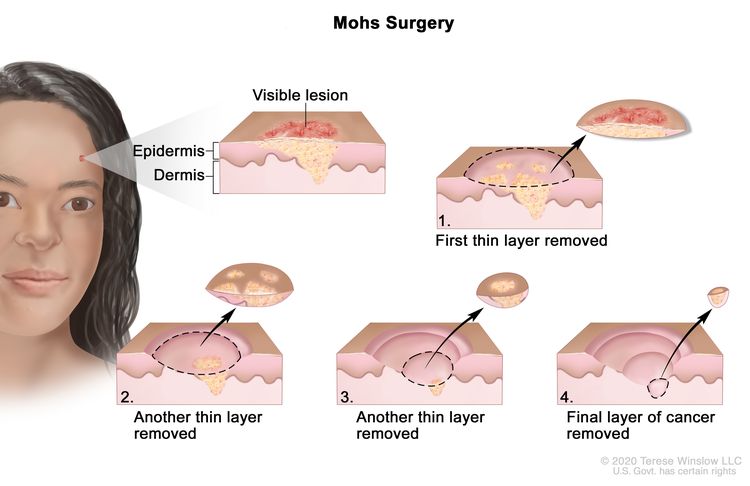
Mohs micrographic surgery: The tumor is cut from the skin in thin layers. During surgery, the edges of the tumor and each layer of tumor removed are viewed through a microscope to check for cancercells. Layers continue to be removed until no more cancer cells are seen. This type of surgery removes as little normal tissue as possible and is often used to remove skin cancer on the face. EnlargeMohs surgery. A surgical procedure to remove a visible lesion on the skin in several steps. First, a thin layer of cancerous tissue is removed. Then, a second thin layer of tissue is removed and viewed under a microscope to check for cancer cells. More layers of tissue are removed one at a time until the tissue viewed under a microscope shows no remaining cancer. This type of surgery is used to remove as little normal tissue as possible.
To Learn More About Childhood Basal Cell Carcinoma and Squamous Cell Carcinoma of the Skin
Physician Data Query (PDQ) is the National Cancer Institute’s (NCI’s) comprehensive cancer information database. The PDQ database contains summaries of the latest published information on cancer prevention, detection, genetics, treatment, supportive care, and complementary and alternative medicine. Most summaries come in two versions. The health professional versions have detailed information written in technical language. The patient versions are written in easy-to-understand, nontechnical language. Both versions have cancer information that is accurate and up to date and most versions are also available in Spanish.
PDQ is a service of the NCI. The NCI is part of the National Institutes of Health (NIH). NIH is the federal government’s center of biomedical research. The PDQ summaries are based on an independent review of the medical literature. They are not policy statements of the NCI or the NIH.
Purpose of This Summary
This PDQ cancer information summary has current information about the treatment of childhood basal cell carcinoma and squamous cell carcinoma of the skin. It is meant to inform and help patients, families, and caregivers. It does not give formal guidelines or recommendations for making decisions about health care.
Reviewers and Updates
Editorial Boards write the PDQ cancer information summaries and keep them up to date. These Boards are made up of experts in cancer treatment and other specialties related to cancer. The summaries are reviewed regularly and changes are made when there is new information. The date on each summary (“Updated”) is the date of the most recent change.
The information in this patient summary was taken from the health professional version, which is reviewed regularly and updated as needed, by the PDQ Pediatric Treatment Editorial Board.
Clinical Trial Information
A clinical trial is a study to answer a scientific question, such as whether one treatment is better than another. Trials are based on past studies and what has been learned in the laboratory. Each trial answers certain scientific questions in order to find new and better ways to help cancer patients. During treatment clinical trials, information is collected about the effects of a new treatment and how well it works. If a clinical trial shows that a new treatment is better than one currently being used, the new treatment may become “standard.” Patients may want to think about taking part in a clinical trial. Some clinical trials are open only to patients who have not started treatment.
Clinical trials can be found online at NCI’s website. For more information, call the Cancer Information Service (CIS), NCI’s contact center, at 1-800-4-CANCER (1-800-422-6237).
Permission to Use This Summary
PDQ is a registered trademark. The content of PDQ documents can be used freely as text. It cannot be identified as an NCI PDQ cancer information summary unless the whole summary is shown and it is updated regularly. However, a user would be allowed to write a sentence such as “NCI’s PDQ cancer information summary about breast cancer prevention states the risks in the following way: [include excerpt from the summary].”
The best way to cite this PDQ summary is:
PDQ® Pediatric Treatment Editorial Board. PDQ Childhood Basal Cell Carcinoma and Squamous Cell Carcinoma of the Skin Treatment. Bethesda, MD: National Cancer Institute. Updated <MM/DD/YYYY>. Available at: /types/skin/patient/child-skin-treatment-pdq. Accessed <MM/DD/YYYY>.
Images in this summary are used with permission of the author(s), artist, and/or publisher for use in the PDQ summaries only. If you want to use an image from a PDQ summary and you are not using the whole summary, you must get permission from the owner. It cannot be given by the National Cancer Institute. Information about using the images in this summary, along with many other images related to cancer can be found in Visuals Online. Visuals Online is a collection of more than 3,000 scientific images.
Disclaimer
The information in these summaries should not be used to make decisions about insurance reimbursement. More information on insurance coverage is available on Cancer.gov on the Managing Cancer Care page.
Contact Us
More information about contacting us or receiving help with the Cancer.gov website can be found on our Contact Us for Help page. Questions can also be submitted to Cancer.gov through the website’s E-mail Us.
Cancer prevention is action taken to lower the chance of getting cancer. By preventing cancer, the number of new cases of cancer in a group or population is lowered. Hopefully, this will lower the number of deaths caused by cancer.
To prevent new cancers from starting, scientists look at risk factors and protective factors. Anything that increases your chance of developing cancer is called a cancer risk factor; anything that decreases your chance of developing cancer is called a cancer protective factor.
Some risk factors for cancer can be avoided, but many cannot. For example, both smoking and inheriting certain genes are risk factors for some types of cancer, but only smoking can be avoided. Regular exercise and a healthy diet may be protective factors for some types of cancer. Avoiding risk factors and increasing protective factors may lower your risk, but it does not mean that you will not get cancer.
Different ways to prevent cancer are being studied.
General Information About Skin Cancer
Key Points
Skin cancer is a disease in which malignant (cancer) cells form in the tissues of the skin.
There are several types of skin cancer.
Skin cancer is the most common cancer in the United States.
Skin cancer is a disease in which malignant (cancer) cells form in the tissues of the skin.
The skin is the body’s largest organ. It protects against heat, sunlight, injury, and infection. Skin also helps control body temperature and stores water, fat, and vitamin D. The skin has several layers, but the two main layers are the epidermis (upper or outer layer) and the dermis (lower or inner layer).
The epidermis is made up of three kinds of cells:
Squamous cells are the thin, flat cells that make up most of the epidermis.
Basal cells are the round cells under the squamous cells.
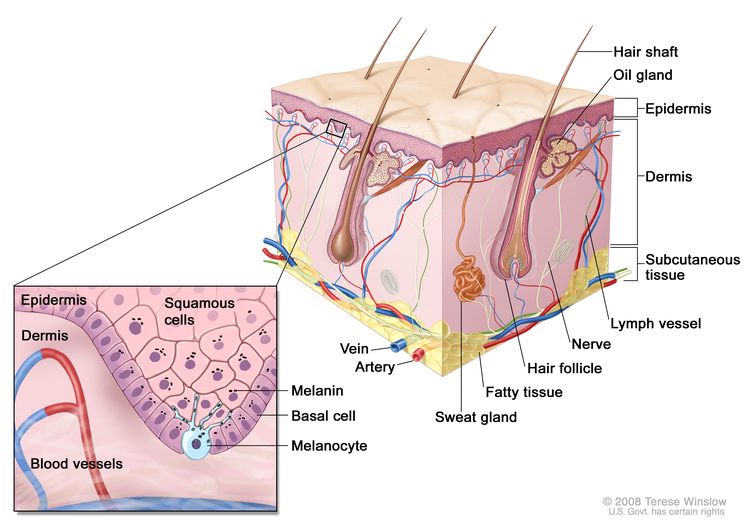
Melanocytes are found throughout the lower part of the epidermis. They make melanin, the pigment that gives skin its natural color. When skin is exposed to the sun, melanocytes make more pigment, causing the skin to tan, or darken.
EnlargeAnatomy of the skin, showing the epidermis, dermis, and subcutaneous tissue. Melanocytes are in the layer of basal cells at the deepest part of the epidermis.
Other PDQ summaries containing information related to skin cancer include:
The most common types of skin cancer are squamous cell carcinoma, which forms in the squamous cells, and basal cell carcinoma, which forms in the basal cells. Melanoma, which forms in the melanocytes, is a less common type of skin cancer that grows and spreads quickly.
Skin cancer can occur anywhere on the body, but it is most common in areas exposed to sunlight, such as the face, neck, hands, and arms.
Skin cancer is the most common cancer in the United States.
Nonmelanoma skin cancer (squamous cell carcinoma and basal cell carcinoma) is the most common type of skin cancer in the United States. New cases of nonmelanoma skin cancer appear to be increasing every year. Nonmelanoma skin cancer can usually be cured.
On the other hand, melanoma is more likely to spread to nearby tissues and other parts of the body and can be harder to cure. Finding and treating melanoma skin cancer early may help prevent death from melanoma.
Skin Cancer Prevention
Key Points
Avoiding risk factors and increasing protective factors may help prevent cancer.
Being exposed to ultraviolet radiation is a risk factor for skin cancer.
Treatment of sun-damaged skin to prevent skin cancer:
Topical fluorouracil
It is not known if the following lower the risk of nonmelanoma skin cancer:
Sunscreen use and avoiding sun exposure
Chemopreventive agents
It is not known if the following lower the risk of melanoma:
Sunscreen
Counseling and protecting the skin from the sun
Cancer prevention clinical trials are used to study ways to prevent cancer.
New ways to prevent skin cancer are being studied in clinical trials.
Avoiding risk factors and increasing protective factors may help prevent cancer.
Avoiding cancerrisk factors may help prevent certain cancers. Risk factors include smoking, having overweight, and not getting enough exercise. Increasing protective factors such as quitting smoking and exercising may also help prevent some cancers. Talk to your doctor or other health care professional about how you might lower your risk of cancer.
Being exposed to ultraviolet radiation is a risk factor for skin cancer.
Some studies suggest that being exposed to ultraviolet (UV) radiation and the sensitivity of a person’s skin to UV radiation are risk factors for skin cancer. UV radiation is the name for the invisible rays that are part of the energy that comes from the sun. Sunlamps and tanning beds also give off UV radiation.
having a family history of unusual moles (atypical nevus syndrome)
having a family or personal history of melanoma
being White
Although having a fair complexion is a risk factor for nonmelanoma and melanoma skin cancer, people of all skin colors can get skin cancer.
Treatment of sun-damaged skin to prevent skin cancer:
Topical fluorouracil
A study showed that topicalfluorouracil applied on sun-damaged skin daily for up to 4 weeks prevented new actinic keratoses from developing. The areas treated with topical fluorouracil had a lower risk of developing into squamous cell carcinoma that would require surgery. The lower risk of developing into squamous cell carcinoma was seen for 1 year after treatment. Topical fluorouracil did not, however, change the risk of developing basal cell carcinoma.
It is not known if the following lower the risk of nonmelanoma skin cancer:
Sunscreen use and avoiding sun exposure
It is not known if nonmelanoma skin cancer risk is decreased by staying out of the sun, using sunscreens, or wearing protective clothing when outdoors. This is because not enough studies have been done to prove this.
Sunscreen has been shown to prevent sunburns and actinic keratoses that may become squamous cell carcinoma, and to decrease the signs and symptoms of existing actinic keratoses.
The harms of using sunscreen are likely to be small and include allergic reactions to skin creams and lower levels of vitamin D made in the skin because of less sun exposure. It is also possible that when a person uses sunscreen to avoid sunburn, they may spend too much time in the sun and be exposed to harmful UV radiation.
Although protecting the skin and eyes from the sun has not been proven to lower the chance of getting skin cancer, skin experts suggest:
using sunscreen that protects against UV radiation
limiting time in the sun, especially when the sun is at its strongest
wearing long sleeve shirts, long pants, sun hats, and sunglasses when outdoors
Chemopreventive agents
Chemoprevention is the use of drugs, vitamins, or other agents to try to reduce the risk of cancer. The following chemopreventive agents have been studied to find whether they lower the risk of nonmelanoma skin cancer:
Beta carotene
Studies of beta carotene (taken as a supplement in pills) have not shown that it prevents nonmelanoma skin cancer from forming or coming back.
Isotretinoin and related retinoids
Retinoids are vitamin A or vitamin A-like compounds that are applied to the skin or taken by mouth. Isotretinoin is a type of retinoid being studied in the prevention and treatment of certain cancers.
High doses of isotretinoin taken by mouth have been shown to prevent new skin cancers in people with xeroderma pigmentosum. Isotretinoin cream has not been shown to prevent nonmelanoma skin cancers from coming back in people previously treated for nonmelanoma skin cancers. These treatments can cause serious side effects.
Selenium
Studies have shown that selenium (taken in brewer’s yeast tablets) does not lower the risk of basal cell carcinoma, and may increase the risk of squamous cell carcinoma.
Celecoxib
A study of celecoxib, a nonsteroidal anti-inflammatory drug (NSAID), in people with actinic keratosis and a history of nonmelanoma skin cancer found those who took celecoxib had slightly lower rates of recurrent nonmelanoma skin cancers. Celecoxib may cause serious heart and blood vessel side effects.
Alpha-difluoromethylornithine (DFMO)
A study of alpha-difluoromethylornithine (DFMO) in people with a history of nonmelanoma skin cancer showed that those who took DFMO had lower rates of basal cell carcinomas than those who took a placebo, but no difference in squamous cell carcinoma rates. However, those who took DFMO had greater hearing loss than the placebo group, leading to a higher rate of people discontinuing this drug.
Nicotinamide (vitamin B3)
Studies have shown that nicotinamide (vitamin B3) could help prevent new actinic keratoses lesions from forming in people who had four or fewer actinic lesions before taking nicotinamide. However, one study also showed an increased incidence of nonmelanoma skin cancers in people months after they were treated with nicotinamide. More studies are needed to find out if nicotinamide prevents nonmelanoma skin cancer from forming or coming back.
It is not known if the following lower the risk of melanoma:
Sunscreen
It has not been proven that using sunscreen to prevent sunburn can protect against melanoma caused by UV radiation. Other risk factors such as having skin that burns easily, having many benign (noncancerous) moles, or having atypical nevi may also play a role in whether melanoma forms.
Counseling and protecting the skin from the sun
Studies show that people who receive counseling or information about avoiding sun exposure improve their sun protective habits. These studies show mixed effects on reducing sunburns and do not show whether skin cancers are reduced.
Harms of avoiding sun exposure may include mood disorders, sleep disturbances, higher blood pressure, and impaired vitamin Dmetabolism.
Cancer prevention clinical trials are used to study ways to prevent cancer.
Cancer prevention clinical trials are used to study ways to lower the risk of developing certain types of cancer. Some cancer prevention trials include healthy people who may or may not have an increased risk of cancer. Other prevention trials include people who have had cancer and are trying to prevent recurrence or a second cancer.
The purpose of some cancer prevention clinical trials is to find out whether actions people take can prevent cancer. These may include eating fruits and vegetables, exercising, quitting smoking, or taking certain medicines, vitamins, minerals, or food supplements.
New ways to prevent skin cancer are being studied in clinical trials.
Physician Data Query (PDQ) is the National Cancer Institute’s (NCI’s) comprehensive cancer information database. The PDQ database contains summaries of the latest published information on cancer prevention, detection, genetics, treatment, supportive care, and complementary and alternative medicine. Most summaries come in two versions. The health professional versions have detailed information written in technical language. The patient versions are written in easy-to-understand, nontechnical language. Both versions have cancer information that is accurate and up to date and most versions are also available in Spanish.
PDQ is a service of the NCI. The NCI is part of the National Institutes of Health (NIH). NIH is the federal government’s center of biomedical research. The PDQ summaries are based on an independent review of the medical literature. They are not policy statements of the NCI or the NIH.
Purpose of This Summary
This PDQ cancer information summary has current information about skin cancer prevention. It is meant to inform and help patients, families, and caregivers. It does not give formal guidelines or recommendations for making decisions about health care.
Reviewers and Updates
Editorial Boards write the PDQ cancer information summaries and keep them up to date. These Boards are made up of experts in cancer treatment and other specialties related to cancer. The summaries are reviewed regularly and changes are made when there is new information. The date on each summary (“Updated”) is the date of the most recent change.
The information in this patient summary was taken from the health professional version, which is reviewed regularly and updated as needed, by the PDQ Screening and Prevention Editorial Board.
Clinical Trial Information
A clinical trial is a study to answer a scientific question, such as whether one treatment is better than another. Trials are based on past studies and what has been learned in the laboratory. Each trial answers certain scientific questions in order to find new and better ways to help cancer patients. During treatment clinical trials, information is collected about the effects of a new treatment and how well it works. If a clinical trial shows that a new treatment is better than one currently being used, the new treatment may become “standard.” Patients may want to think about taking part in a clinical trial. Some clinical trials are open only to patients who have not started treatment.
Clinical trials can be found online at NCI’s website. For more information, call the Cancer Information Service (CIS), NCI’s contact center, at 1-800-4-CANCER (1-800-422-6237).
Permission to Use This Summary
PDQ is a registered trademark. The content of PDQ documents can be used freely as text. It cannot be identified as an NCI PDQ cancer information summary unless the whole summary is shown and it is updated regularly. However, a user would be allowed to write a sentence such as “NCI’s PDQ cancer information summary about breast cancer prevention states the risks in the following way: [include excerpt from the summary].”
The best way to cite this PDQ summary is:
PDQ® Screening and Prevention Editorial Board. PDQ Skin Cancer Prevention. Bethesda, MD: National Cancer Institute. Updated <MM/DD/YYYY>. Available at: /types/skin/patient/skin-prevention-pdq. Accessed <MM/DD/YYYY>. [PMID: 26389434]
Images in this summary are used with permission of the author(s), artist, and/or publisher for use in the PDQ summaries only. If you want to use an image from a PDQ summary and you are not using the whole summary, you must get permission from the owner. It cannot be given by the National Cancer Institute. Information about using the images in this summary, along with many other images related to cancer can be found in Visuals Online. Visuals Online is a collection of more than 3,000 scientific images.
Disclaimer
The information in these summaries should not be used to make decisions about insurance reimbursement. More information on insurance coverage is available on Cancer.gov on the Managing Cancer Care page.
Contact Us
More information about contacting us or receiving help with the Cancer.gov website can be found on our Contact Us for Help page. Questions can also be submitted to Cancer.gov through the website’s E-mail Us.
Melanoma is a disease in which malignant (cancer) cells form in melanocytes (cells that color the skin).
There are different types of cancer that start in the skin.
Unusual moles, exposure to sunlight, and health history can affect the risk of melanoma.
Signs of melanoma include a change in the way a mole or pigmented area looks.
Tests that examine the skin are used to diagnose melanoma.
Melanoma is a disease in which malignant (cancer) cells form in melanocytes (cells that color the skin).
The skin is the body’s largest organ. It protects against heat, sunlight, injury, and infection. Skin also helps control body temperature and stores water, fat, and vitamin D. The skin has several layers, but the two main layers are the epidermis (upper or outer layer) and the dermis (lower or inner layer). Skin cancer begins in the epidermis, which is made up of three kinds of cells:
Squamous cells: Thin, flat cells that form the top layer of the epidermis.
Basal cells: Round cells under the squamous cells.
Melanocytes: Cells that make melanin and are found in the lower part of the epidermis. Melanin is the pigment that gives skin its natural color. When skin is exposed to the sun or artificial light, melanocytes make more pigment and cause the skin to darken.
EnlargeAnatomy of the skin, showing the epidermis, dermis, and subcutaneous tissue. Melanocytes are in the layer of basal cells at the deepest part of the epidermis.
There are different types of cancer that start in the skin.
Melanoma is a rare form of skin cancer. Even though melanoma is rare, it is the most common skin cancer in children. It occurs more often in adolescents aged 15 to 19 years. Melanoma is more likely to invade nearby tissues and spread to other parts of the body than other types of skin cancer. When melanoma starts in the skin, it is called cutaneous melanoma. Melanoma may also occur in mucous membranes (thin, moist layers of tissue that cover surfaces such as the lips) and the eye (intraocular melanoma). This PDQ summary is about cutaneous (skin) melanoma. (See the PDQ summary on Childhood Intraocular (Uveal) Melanoma Treatment for more information about intraocular melanoma).
Unusual moles, exposure to sunlight, and health history can affect the risk of melanoma.
Anything that increases your risk of getting a disease is called a risk factor. Having a risk factor does not mean that you will get cancer; not having risk factors doesn’t mean that you will not get cancer. Talk with your child’s doctor if you think your child may be at risk.
Risk factors for childhood melanoma include the following:
Signs of melanoma include a change in the way a mole or pigmented area looks.
These and other signs and symptoms may be caused by melanoma or by other conditions.
Check with your child’s doctor if your child has any of the following:
A mole that:
changes in size, shape, or color.
has irregular edges or borders.
is more than one color.
is asymmetrical (if the mole is divided in half, the 2 halves are different in size or shape).
itches.
oozes, bleeds, or is ulcerated (a condition in which the top layer of skin breaks down and the tissue below shows through).
A change in pigmented (colored) skin.
Satellite moles (new moles that grow near an existing mole).
Tests that examine the skin are used to diagnose melanoma.
If a mole or pigmented area of the skin changes or looks abnormal, the following tests and procedures can help find and diagnose melanoma:
Physical exam and health history: An exam of the body to check general signs of health, including checking for signs of disease, such as lumps or anything else that seems unusual. A history of the patient’s health habits and past illnesses and treatments will also be taken.
Skin exam: A doctor or nurse checks the skin for moles, birthmarks, or other pigmented areas that look abnormal in color, size, shape, or texture.
Biopsy: A procedure to remove the abnormal tissue and a small amount of normal tissue around it. A pathologist looks at the tissue under a microscope to check for cancer cells. It can be hard to tell the difference between a colored mole and an early melanoma lesion. Patients may want to have the sample of tissue checked by a second pathologist. If the abnormal mole or lesion is cancer, the sample of tissue may also be tested for certain gene changes.
There are four main types of skin biopsies:
Shave biopsy: A sterile razor blade is used to “shave-off” the abnormal-looking growth.
Punch biopsy: A special instrument called a punch or a trephine is used to remove a circle of tissue from the abnormal-looking growth. EnlargePunch biopsy. A sharp, hollow, circular instrument is used to remove a small, round piece of tissue from a lesion on the skin. The instrument is turned clockwise and counterclockwise to cut about 4 millimeters (mm) down to the layer of fatty tissue below the skin and remove the sample of tissue. Skin thickness is different on different parts of the body.
After melanoma has been diagnosed, tests are done to find out if cancer cells have spread to other parts of the body.
There are three ways cancer spreads in the body.
Cancer may spread from where it began to other parts of the body.
Sometimes childhood melanoma recurs (comes back) after treatment.
After melanoma has been diagnosed, tests are done to find out if cancer cells have spread to other parts of the body.
The process used to find out if cancer has spread within the skin or to other parts of the body is called staging. There is no standard staging system for childhood melanoma. To plan treatment, it is important to know whether melanoma has spread to lymph nodes or to other parts of the body.
The following procedures may be used to find out if cancer has spread:
Chest x-ray: An x-ray of the organs and bones inside the chest. An x-ray is a type of energy beam that can go through the body and onto film, making a picture of areas inside the body.
CT scan (CAT scan): A procedure that makes a series of detailed pictures of areas inside the body, taken from different angles. The pictures are made by a computer linked to an x-ray machine. A dye may be injected into a vein or swallowed to help the organs or tissues show up more clearly. This procedure is also called computed tomography, computerized tomography, or computerized axial tomography.
MRI: A procedure that uses a magnet, radio waves, and a computer to make a series of detailed pictures of areas inside the body. This procedure is also called nuclear magnetic resonance imaging (NMRI). EnlargeMagnetic resonance imaging (MRI) scan. The child lies on a table that slides into the MRI machine, which takes a series of detailed pictures of areas inside the body. The positioning of the child on the table depends on the part of the body being imaged.
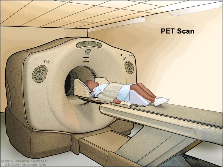
PET scan: A procedure to find malignanttumorcells in the body. A small amount of radioactive glucose (sugar) is injected into a vein. The PET scanner rotates around the body and makes a picture of where glucose is being used in the body. Malignant tumor cells show up brighter in the picture because they are more active and take up more glucose than normal cells do. EnlargePositron emission tomography (PET) scan. The child lies on a table that slides through the PET scanner. The head rest and white strap help the child lie still. A small amount of radioactive glucose (sugar) is injected into the child’s vein, and a scanner makes a picture of where the glucose is being used in the body. Cancer cells show up brighter in the picture because they take up more glucose than normal cells do.
Ultrasound: A procedure in which high-energy sound waves (ultrasound) are bounced off internal tissues or organs and make echoes. The echoes form a picture of body tissues called a sonogram. The picture can be printed to be looked at later.
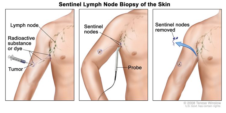
Sentinel lymph node biopsy: The removal of the sentinel lymph node during surgery. The sentinel lymph node is the first lymph node in a group of lymph nodes to receive lymphatic drainage from the primary tumor. It is the first lymph node the cancer is likely to spread to from the primary tumor. A radioactive substance and/or blue dye is injected near the tumor. The substance or dye flows through the lymphducts to the lymph nodes. The first lymph node to receive the substance or dye is removed. A pathologist views the tissue under a microscope to look for cancer cells. If cancer cells are not found, it may not be necessary to remove more lymph nodes. Sometimes, a sentinel lymph node is found in more than one group of nodes. EnlargeSentinel lymph node biopsy of the skin. A radioactive substance and/or blue dye is injected near the tumor (first panel). The injected material is detected visually and/or with a probe that detects radioactivity (middle panel). The sentinel nodes (the first lymph nodes to take up the material) are removed and checked for cancer cells (last panel).
Lymph node dissection: A surgical procedure in which lymph nodes are removed and a sample of tissue is checked under a microscope for signs of cancer. For a regional lymph node dissection, some of the lymph nodes in the tumor area are removed. For a radical lymph node dissection, most or all of the lymph nodes in the tumor area are removed. This procedure is also called a lymphadenectomy.
Tissue. The cancer spreads from where it began by growing into nearby areas.
Lymph system. The cancer spreads from where it began by getting into the lymph system. The cancer travels through the lymph vessels to other parts of the body.
Blood. The cancer spreads from where it began by getting into the blood. The cancer travels through the blood vessels to other parts of the body.
Cancer may spread from where it began to other parts of the body.
When cancer spreads to another part of the body, it is called metastasis. Cancer cells break away from where they began (the primary tumor) and travel through the lymph system or blood.
Lymph system. The cancer gets into the lymph system, travels through the lymph vessels, and forms a tumor (metastatic tumor) in another part of the body.
Blood. The cancer gets into the blood, travels through the blood vessels, and forms a tumor (metastatic tumor) in another part of the body.
The metastatic tumor is the same type of cancer as the primary tumor. For example, if melanoma spreads to the lung, the cancer cells in the lung are actually melanoma cells. The disease is metastatic melanoma, not lung cancer.
Many cancer deaths are caused when cancer moves from the original tumor and spreads to other tissues and organs. This is called metastatic cancer. This animation shows how cancer cells travel from the place in the body where they first formed to other parts of the body.
Sometimes childhood melanoma recurs (comes back) after treatment.
The cancer may come back in the skin, lymph nodes, or in other parts of the body.
Treatment Option Overview
Key Points
There are different types of treatment for children with melanoma.
Children with melanoma should have their treatment planned by a team of doctors who are experts in treating childhood cancer.
Three types of standard treatment are used:
Surgery
Immunotherapy
Targeted therapy
New types of treatment are being tested in clinical trials.
Treatment for childhood melanoma may cause side effects.
Patients may want to think about taking part in a clinical trial.
Patients can enter clinical trials before, during, or after starting their cancer treatment.
Follow-up tests may be needed.
There are different types of treatment for children with melanoma.
Some treatments are standard (the currently used treatment), and some are being tested in clinical trials. A treatment clinical trial is a research study meant to help improve current treatments or obtain information on new treatments for patients with cancer. When clinical trials show that a new treatment is better than the standard treatment, the new treatment may become the standard treatment.
Because cancer in children is rare, taking part in a clinical trial should be considered. Some clinical trials are open only to patients who have not started treatment.
Children with melanoma should have their treatment planned by a team of doctors who are experts in treating childhood cancer.
Treatment will be overseen by a pediatric oncologist, a doctor who specializes in treating children with cancer. The pediatric oncologist works with other pediatric health professionals who are experts in treating children with cancer and who specialize in certain areas of medicine. This may include the following specialists and others:
Surgery to remove the tumor is used to treat childhood melanoma. A wide local excision is used to remove the melanoma and some of the normal tissue around it. Skin grafting (taking skin from another part of the body to replace the skin that is removed) may be done to cover the wound caused by surgery. Nearby lymph nodes with cancer may also be removed.
Immunotherapy
Immunotherapy is a treatment that uses the patient’s immune system to fight cancer. Substances made by the body or made in a laboratory are used to boost, direct, or restore the body’s natural defenses against cancer. This type of cancer treatment is also called biotherapy or biologic therapy.
Immunotherapy uses the body’s immune system to fight cancer. This animation explains one type of immunotherapy that uses immune checkpoint inhibitors to treat cancer.
There are different types of immune checkpoint inhibitor therapy:
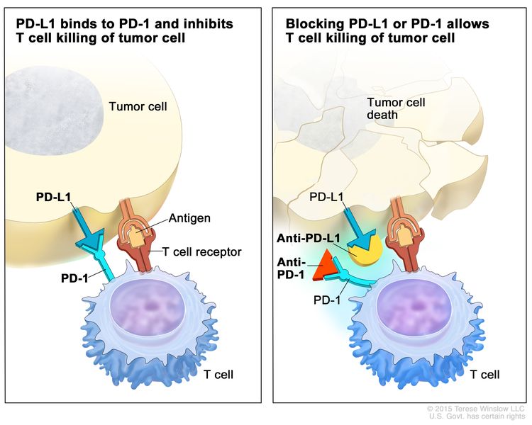
PD-1 inhibitor: PD-1 is a protein on the surface of T cells that helps keep the body’s immune responses in check. When PD-1 attaches to another protein called PDL-1 on a tumor cell, it stops the T cell from killing the tumor cell. PD-1 inhibitors attach to PDL-1 and allow the T cells to kill tumor cells. Pembrolizumab and nivolumab are used to treat melanoma that has spread to lymph nodes and are being studied to treat recurrent melanoma. EnlargeImmune checkpoint inhibitor. Checkpoint proteins, such as PD-L1 on tumor cells and PD-1 on T cells, help keep immune responses in check. The binding of PD-L1 to PD-1 keeps T cells from killing tumor cells in the body (left panel). Blocking the binding of PD-L1 to PD-1 with an immune checkpoint inhibitor (anti-PD-L1 or anti-PD-1) allows the T cells to kill tumor cells (right panel).
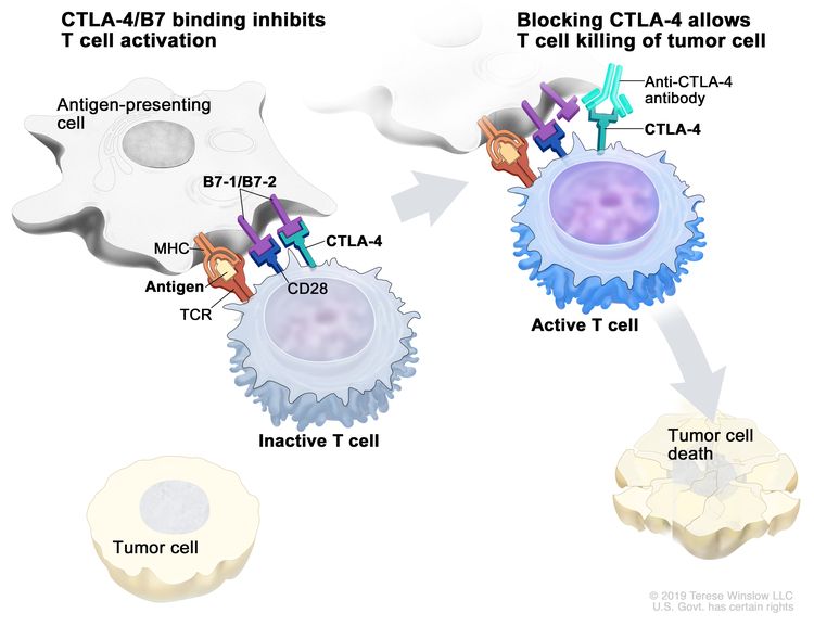
CTLA-4 inhibitor: CTLA-4 is a protein on the surface of T cells that helps keep the body’s immune responses in check. When CTLA-4 attaches to another protein called B7 on a cancer cell, it stops the T cell from killing the cancer cell. CTLA-4 inhibitors attach to CTLA-4 and allow the T cells to kill cancer cells. Ipilimumab is used to treat melanoma that has spread to lymph nodes or to other parts of the body and is being studied to treat recurrent melanoma. EnlargeImmune checkpoint inhibitor. Checkpoint proteins, such as B7-1/B7-2 on antigen-presenting cells (APC) and CTLA-4 on T cells, help keep the body’s immune responses in check. When the T-cell receptor (TCR) binds to antigen and major histocompatibility complex (MHC) proteins on the APC and CD28 binds to B7-1/B7-2 on the APC, the T cell can be activated. However, the binding of B7-1/B7-2 to CTLA-4 keeps the T cells in the inactive state so they are not able to kill tumor cells in the body (left panel). Blocking the binding of B7-1/B7-2 to CTLA-4 with an immune checkpoint inhibitor (anti-CTLA-4 antibody) allows the T cells to be active and to kill tumor cells (right panel).
Targeted therapy
Targeted therapy is a type of treatment that uses drugs or other substances to attack cancer cells. Targeted therapies usually cause less harm to normal cells than chemotherapy or radiation therapy do. The following type of targeted therapy is being used or studied in the treatment of melanoma:
Signal transduction inhibitor therapy: Signal transduction inhibitors block signals that are passed from one molecule to another inside a cell. Blocking these signals may kill cancer cells. They are used to treat some patients with advanced melanoma or tumors that cannot be removed by surgery. Signal transduction inhibitors include the following:
Patients may want to think about taking part in a clinical trial.
For some patients, taking part in a clinical trial may be the best treatment choice. Clinical trials are part of the cancer research process. Clinical trials are done to find out if new cancer treatments are safe and effective or better than the standard treatment.
Many of today’s standard treatments for cancer are based on earlier clinical trials. Patients who take part in a clinical trial may receive the standard treatment or be among the first to receive a new treatment.
Patients who take part in clinical trials also help improve the way cancer will be treated in the future. Even when clinical trials do not lead to effective new treatments, they often answer important questions and help move research forward.
Patients can enter clinical trials before, during, or after starting their cancer treatment.
Some clinical trials only include patients who have not yet received treatment. Other trials test treatments for patients whose cancer has not gotten better. There are also clinical trials that test new ways to stop cancer from recurring (coming back) or reduce the side effects of cancer treatment.
Clinical trials are taking place in many parts of the country. Information about clinical trials supported by NCI can be found on NCI’s clinical trials search webpage. Clinical trials supported by other organizations can be found on the ClinicalTrials.gov website.
Follow-up tests may be needed.
As your child goes through treatment, they will have follow-up tests or check-ups. Some tests that were done to diagnose or stage the cancer may be repeated to see how well the treatment is working. Decisions about whether to continue, change, or stop treatment may be based on the results of these tests.
Some of the tests will continue to be done from time to time after treatment has ended. The results of these tests can show if your child’s condition has changed or if the cancer has recurred (come back).
Use our clinical trial search to find NCI-supported cancer clinical trials that are accepting patients. You can search for trials based on the type of cancer, the age of the patient, and where the trials are being done. General information about clinical trials is also available.
Treatment of recurrentmelanoma in children may include the following:
A clinical trial that checks a sample of the patient’s tumor for certain gene changes. The type of targeted therapy that will be given to the patient depends on the type of gene change.
Use our clinical trial search to find NCI-supported cancer clinical trials that are accepting patients. You can search for trials based on the type of cancer, the age of the patient, and where the trials are being done. General information about clinical trials is also available.
Physician Data Query (PDQ) is the National Cancer Institute’s (NCI’s) comprehensive cancer information database. The PDQ database contains summaries of the latest published information on cancer prevention, detection, genetics, treatment, supportive care, and complementary and alternative medicine. Most summaries come in two versions. The health professional versions have detailed information written in technical language. The patient versions are written in easy-to-understand, nontechnical language. Both versions have cancer information that is accurate and up to date and most versions are also available in Spanish.
PDQ is a service of the NCI. The NCI is part of the National Institutes of Health (NIH). NIH is the federal government’s center of biomedical research. The PDQ summaries are based on an independent review of the medical literature. They are not policy statements of the NCI or the NIH.
Purpose of This Summary
This PDQ cancer information summary has current information about the treatment of childhood melanoma. It is meant to inform and help patients, families, and caregivers. It does not give formal guidelines or recommendations for making decisions about health care.
Reviewers and Updates
Editorial Boards write the PDQ cancer information summaries and keep them up to date. These Boards are made up of experts in cancer treatment and other specialties related to cancer. The summaries are reviewed regularly and changes are made when there is new information. The date on each summary (“Updated”) is the date of the most recent change.
The information in this patient summary was taken from the health professional version, which is reviewed regularly and updated as needed, by the PDQ Pediatric Treatment Editorial Board.
Clinical Trial Information
A clinical trial is a study to answer a scientific question, such as whether one treatment is better than another. Trials are based on past studies and what has been learned in the laboratory. Each trial answers certain scientific questions in order to find new and better ways to help cancer patients. During treatment clinical trials, information is collected about the effects of a new treatment and how well it works. If a clinical trial shows that a new treatment is better than one currently being used, the new treatment may become “standard.” Patients may want to think about taking part in a clinical trial. Some clinical trials are open only to patients who have not started treatment.
Clinical trials can be found online at NCI’s website. For more information, call the Cancer Information Service (CIS), NCI’s contact center, at 1-800-4-CANCER (1-800-422-6237).
Permission to Use This Summary
PDQ is a registered trademark. The content of PDQ documents can be used freely as text. It cannot be identified as an NCI PDQ cancer information summary unless the whole summary is shown and it is updated regularly. However, a user would be allowed to write a sentence such as “NCI’s PDQ cancer information summary about breast cancer prevention states the risks in the following way: [include excerpt from the summary].”
The best way to cite this PDQ summary is:
PDQ® Pediatric Treatment Editorial Board. PDQ Childhood Melanoma Treatment. Bethesda, MD: National Cancer Institute. Updated <MM/DD/YYYY>. Available at: /types/skin/patient/child-melanoma-treatment-pdq. Accessed <MM/DD/YYYY>.
Images in this summary are used with permission of the author(s), artist, and/or publisher for use in the PDQ summaries only. If you want to use an image from a PDQ summary and you are not using the whole summary, you must get permission from the owner. It cannot be given by the National Cancer Institute. Information about using the images in this summary, along with many other images related to cancer can be found in Visuals Online. Visuals Online is a collection of more than 3,000 scientific images.
Disclaimer
The information in these summaries should not be used to make decisions about insurance reimbursement. More information on insurance coverage is available on Cancer.gov on the Managing Cancer Care page.
Contact Us
More information about contacting us or receiving help with the Cancer.gov website can be found on our Contact Us for Help page. Questions can also be submitted to Cancer.gov through the website’s E-mail Us.
Malignant mesothelioma is a cancer of the thin tissue (mesothelium) that lines the lung, chest wall, and abdomen. The major risk factor for mesothelioma is asbestos exposure. Explore the links on this page to learn more about malignant mesothelioma treatment and clinical trials.
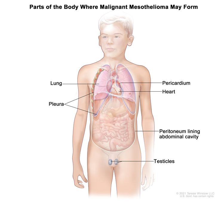
Peritoneum: A thin layer of tissue that lines the abdomen and covers most of the organs in the abdomen.
Pericardium: A thin layer of tissue that surrounds the heart.
The tumors often spread over the surface of organs without spreading into the organ. They may spread to nearby lymph nodes or in other parts of the body. Malignant mesothelioma may also form in the testicles, but this is rare.
EnlargeMalignant mesothelioma forms in the tissue that lines the chest or abdomen, including the pleura (the tissue that lines the chest cavity and covers the lungs), the pericardium (the tissue that surrounds the heart), and the peritoneum (the tissue that lines the abdomen and covers most of the organs in the abdomen). Malignant mesothelioma may also form in the testicles, but this is rare.
Treatment with radiation therapy increases the risk of childhood mesothelioma.
Anything that increases your chance of getting a disease is called a risk factor. Having a risk factor does not mean that you will get cancer; not having risk factors doesn’t mean that you will not get cancer. Talk with your child’s doctor if you think your child may be at risk.
In adults, mesothelioma is strongly linked to being exposed to asbestos, which has been used in the building and textile industries. In children, there is little information about the risk of developing mesothelioma after being exposed to asbestos.
Signs and symptoms of mesothelioma include trouble breathing and pain in the chest or abdomen.
In children, these and other signs and symptoms may be caused by mesothelioma or by other conditions.
Check with your child’s doctor if your child has any of the following:
Trouble breathing.
Cough for no known reason.
Pain under the rib cage or pain in the chest and abdomen.
Weight loss for no known reason.
Feeling very tired.
Tests that examine the chest, abdomen, and heart are used to diagnose mesothelioma.
The following tests and procedures may be used:
Physical exam and health history: An exam of the body to check general signs of health, including checking for signs of disease, such as lumps or anything else that seems unusual. A history of the patient’s health habits and past illnesses and treatments will also be taken.
Chest x-ray: An x-ray of the organs and bones inside the chest. An x-ray is a type of energy beam that can go through the body and onto film, making a picture of areas inside the body.
CT scan (CAT scan): A procedure that makes a series of detailed pictures of areas inside the body, taken from different angles. The pictures are made by a computer linked to an x-ray machine. A dye may be injected into a vein or swallowed to help the organs or tissues show up more clearly. This procedure is also called computed tomography, computerized tomography, or computerized axial tomography.
PET scan (positron emission tomography scan): A procedure to find malignant tumor cells in the body. A small amount of radioactive glucose (sugar) is injected into a vein. The PET scanner rotates around the body and makes a picture of where glucose is being used in the body. Malignant tumor cells show up brighter in the picture because they are more active and take up more glucose than normal cells do. EnlargePositron emission tomography (PET) scan. The child lies on a table that slides through the PET scanner. The head rest and white strap help the child lie still. A small amount of radioactive glucose (sugar) is injected into the child’s vein, and a scanner makes a picture of where the glucose is being used in the body. Cancer cells show up brighter in the picture because they take up more glucose than normal cells do.
MRI (magnetic resonance imaging): A procedure that uses a magnet, radio waves, and a computer to make a series of detailed pictures of areas inside the body. This procedure is also called nuclear magnetic resonance imaging (NMRI).
Pulmonary function test (PFT): A test to see how well the lungs are working. It measures how much air the lungs can hold and how quickly air moves into and out of the lungs. It also measures how much oxygen is used and how much carbon dioxide is given off during breathing. This is also called a lung function test.
Thoracoscopy: A surgical procedure to look at the organs inside the chest to check for abnormal areas. An incision (cut) is made between two ribs and a thoracoscope is inserted into the chest. A thoracoscope is a thin, tube-like instrument with a light and a lens for viewing. It may also have a tool to remove tissue or lymph node samples, which are checked under a microscope for signs of cancer. In some cases, this procedure is used to remove part of the esophagus or lung.
Bronchoscopy: A procedure to look inside the trachea and large airways in the lung for abnormal areas. A bronchoscope is inserted through the nose or mouth into the trachea and lungs. A bronchoscope is a thin, tube-like instrument with a light and a lens for viewing. It may also have a tool to remove tissue samples, which are checked under a microscope for signs of cancer.
Laparoscopy: A surgical procedure to look at the organs inside the abdomen to check for abnormal areas. Small incisions (cuts) are made in the wall of the abdomen and a laparoscope (thin, lighted tube) is inserted into one of the incisions. Other instruments may be inserted through the same or other incisions to perform procedures such as removing organs or taking tissue samples to be checked under a microscope for signs of cancer.
Cytologic exam: An exam of cells under a microscope (by a pathologist) to check for anything abnormal. For mesothelioma, fluid is taken from around the lungs or from the abdomen. A pathologist checks the cells in the fluid.
Certain factors affect prognosis (chance of recovery).
The process used to find out if cancer has spread from where it first began is called staging. In childhood mesothelioma, cancer may spread to nearby or distant lymph nodes. There is no standard staging system for childhood mesothelioma. The results of tests and procedures done to diagnose mesothelioma are used to help make decisions about treatment.
Sometimes childhood mesothelioma recurs (comes back) after it has been treated.
Treatment Option Overview
Key Points
There are different types of treatment for children with mesothelioma.
Children with mesothelioma should have their treatment planned by a team of doctors who are experts in treating childhood cancer.
Three types of treatment are used:
Surgery
Chemotherapy
Radiation therapy
New types of treatment are being tested in clinical trials.
Targeted therapy
Immunotherapy
Treatment for childhood mesothelioma may cause side effects.
Patients may want to think about taking part in a clinical trial.
Patients can enter clinical trials before, during, or after starting their cancer treatment.
Follow-up tests may be needed.
There are different types of treatment for children with mesothelioma.
Some treatments are standard (the currently used treatment), and some are being tested in clinical trials. A treatment clinical trial is a research study meant to help improve current treatments or obtain information on new treatments for patients with cancer. When clinical trials show that a new treatment is better than the standard treatment, the new treatment may become the standard treatment.
Because cancer in children is rare, taking part in a clinical trial should be considered. Some clinical trials are open only to patients who have not started treatment.
Children with mesothelioma should have their treatment planned by a team of doctors who are experts in treating childhood cancer.
Treatment will be overseen by a pediatric oncologist, a doctor who specializes in treating children with cancer. The pediatric oncologist works with other pediatric health professionals who are experts in treating children with cancer and who specialize in certain areas of medicine. This may include the following specialists and others:
Chemotherapy is a cancer treatment that uses drugs to stop the growth of cancer cells, either by killing the cells or by stopping them from dividing. When chemotherapy is taken by mouth or injected into a vein or muscle, the drugs enter the bloodstream and can reach cancer cells throughout the body (systemic chemotherapy).
Radiation therapy
Radiation therapy is a cancer treatment that uses high-energy x-rays or other types of radiation to kill cancer cells or keep them from growing. External radiation therapy uses a machine outside the body to send radiation toward the area of the body with cancer.
New types of treatment are being tested in clinical trials.
This summary section describes treatments that are being studied in clinical trials. It may not mention every new treatment being studied. Information about clinical trials is available from the NCI website.
Targeted therapy
Targeted therapy is a type of treatment that uses drugs or other substances to identify and attack specific cancer cells. Targeted therapies usually cause less harm to normal cells than chemotherapy or radiation therapy do.
Targeted therapy is being studied for the treatment of childhood mesothelioma that has recurred (come back).
Immunotherapy
Immunotherapy is a treatment that uses the patient’s immune system to fight cancer. Substances made by the body or made in a laboratory are used to boost, direct, or restore the body’s natural defenses against cancer. This cancer treatment is a type of biologic therapy.
Treatment for childhood mesothelioma may cause side effects.
Side effects from cancer treatment that begin after treatment and continue for months or years are called late effects. Late effects of cancer treatment may include:
Physical problems.
Changes in mood, feelings, thinking, learning, or memory.
Some late effects may be treated or controlled. It is important to talk with your child’s doctors about the possible late effects caused by some treatments.
Patients may want to think about taking part in a clinical trial.
For some patients, taking part in a clinical trial may be the best treatment choice. Clinical trials are part of the cancer research process. Clinical trials are done to find out if new cancer treatments are safe and effective or better than the standard treatment.
Many of today’s standard treatments for cancer are based on earlier clinical trials. Patients who take part in a clinical trial may receive the standard treatment or be among the first to receive a new treatment.
Patients who take part in clinical trials also help improve the way cancer will be treated in the future. Even when clinical trials do not lead to effective new treatments, they often answer important questions and help move research forward.
Patients can enter clinical trials before, during, or after starting their cancer treatment.
Some clinical trials only include patients who have not yet received treatment. Other trials test treatments for patients whose cancer has not gotten better. There are also clinical trials that test new ways to stop cancer from recurring (coming back) or reduce the side effects of cancer treatment.
Clinical trials are taking place in many parts of the country. Information about clinical trials supported by NCI can be found on NCI’s clinical trials search webpage. Clinical trials supported by other organizations can be found on the ClinicalTrials.gov website.
Follow-up tests may be needed.
As your child goes through treatment, they will have follow-up tests or check-ups. Some tests that were done to diagnose or stage the cancer may be repeated to see how well the treatment is working. Decisions about whether to continue, change, or stop treatment may be based on the results of these tests.
Some of the tests will continue to be done from time to time after treatment has ended. The results of these tests can show if your child’s condition has changed or if the cancer has recurred (come back).
Use our clinical trial search to find NCI-supported cancer clinical trials that are accepting patients. You can search for trials based on the type of cancer, the age of the patient, and where the trials are being done. General information about clinical trials is also available.
A clinical trial that checks a sample of the patient’s tumor for certain gene changes. The type of targeted therapy that will be given to the patient depends on the type of gene change.
Use our clinical trial search to find NCI-supported cancer clinical trials that are accepting patients. You can search for trials based on the type of cancer, the age of the patient, and where the trials are being done. General information about clinical trials is also available.
Physician Data Query (PDQ) is the National Cancer Institute’s (NCI’s) comprehensive cancer information database. The PDQ database contains summaries of the latest published information on cancer prevention, detection, genetics, treatment, supportive care, and complementary and alternative medicine. Most summaries come in two versions. The health professional versions have detailed information written in technical language. The patient versions are written in easy-to-understand, nontechnical language. Both versions have cancer information that is accurate and up to date and most versions are also available in Spanish.
PDQ is a service of the NCI. The NCI is part of the National Institutes of Health (NIH). NIH is the federal government’s center of biomedical research. The PDQ summaries are based on an independent review of the medical literature. They are not policy statements of the NCI or the NIH.
Purpose of This Summary
This PDQ cancer information summary has current information about the treatment of childhood mesothelioma. It is meant to inform and help patients, families, and caregivers. It does not give formal guidelines or recommendations for making decisions about health care.
Reviewers and Updates
Editorial Boards write the PDQ cancer information summaries and keep them up to date. These Boards are made up of experts in cancer treatment and other specialties related to cancer. The summaries are reviewed regularly and changes are made when there is new information. The date on each summary (“Updated”) is the date of the most recent change.
The information in this patient summary was taken from the health professional version, which is reviewed regularly and updated as needed, by the PDQ Pediatric Treatment Editorial Board.
Clinical Trial Information
A clinical trial is a study to answer a scientific question, such as whether one treatment is better than another. Trials are based on past studies and what has been learned in the laboratory. Each trial answers certain scientific questions in order to find new and better ways to help cancer patients. During treatment clinical trials, information is collected about the effects of a new treatment and how well it works. If a clinical trial shows that a new treatment is better than one currently being used, the new treatment may become “standard.” Patients may want to think about taking part in a clinical trial. Some clinical trials are open only to patients who have not started treatment.
Clinical trials can be found online at NCI’s website. For more information, call the Cancer Information Service (CIS), NCI’s contact center, at 1-800-4-CANCER (1-800-422-6237).
Permission to Use This Summary
PDQ is a registered trademark. The content of PDQ documents can be used freely as text. It cannot be identified as an NCI PDQ cancer information summary unless the whole summary is shown and it is updated regularly. However, a user would be allowed to write a sentence such as “NCI’s PDQ cancer information summary about breast cancer prevention states the risks in the following way: [include excerpt from the summary].”
The best way to cite this PDQ summary is:
PDQ® Pediatric Treatment Editorial Board. PDQ Childhood Mesothelioma Treatment. Bethesda, MD: National Cancer Institute. Updated <MM/DD/YYYY>. Available at: /types/mesothelioma/patient/child-mesothelioma-treatment-pdq. Accessed <MM/DD/YYYY>.
Images in this summary are used with permission of the author(s), artist, and/or publisher for use in the PDQ summaries only. If you want to use an image from a PDQ summary and you are not using the whole summary, you must get permission from the owner. It cannot be given by the National Cancer Institute. Information about using the images in this summary, along with many other images related to cancer can be found in Visuals Online. Visuals Online is a collection of more than 3,000 scientific images.
Disclaimer
The information in these summaries should not be used to make decisions about insurance reimbursement. More information on insurance coverage is available on Cancer.gov on the Managing Cancer Care page.
Contact Us
More information about contacting us or receiving help with the Cancer.gov website can be found on our Contact Us for Help page. Questions can also be submitted to Cancer.gov through the website’s E-mail Us.
Childhood cervical and vaginal cancers are very rare types of cancer. Cervical cancer forms in the cells of the cervix, and vaginal cancer forms in the cells of the vagina. The cervix is the lower, narrow end of the uterus (the hollow, pear-shaped organ where a fetus grows). The cervix leads from the uterus to the vagina (birth canal). The vagina is the canal leading from the cervix to the outside of the body. At birth, a baby passes out of the body through the vagina. Learn more about how cancer develops at What Is Cancer?
EnlargeAnatomy of the female reproductive system. The organs in the female reproductive system include the uterus, ovaries, fallopian tubes, cervix, and vagina. The uterus has a muscular outer layer called the myometrium and an inner lining called the endometrium.
Childhood cervical cancer and vaginal cancer symptoms
The most common symptom of cervical cancer and vaginal cancer in children is bleeding from the vagina. Other conditions may also cause vaginal bleeding. If your child has vaginal bleeding, it is important that you tell their doctor. The doctor will ask you when it started and how often it occurs as a first step in making a diagnosis.
Tests to diagnose childhood cervical cancer and vaginal cancer
If your child has symptoms that suggest vaginal or cervical cancer, their doctor will need to find out if they are due to cancer or another problem. They may ask about your child’s personal and family medical history and do a physical exam.
Depending on your child’s symptoms and medical history and the results of their physical exam, the doctor may recommend more tests to find out if your child has vaginal or cervical cancer, and if so, its extent (stage). The results of tests and procedures done to diagnose vaginal and cervical cancers are used to help make decisions about treatment.
The following tests and procedures may be used to diagnose and stage childhood cervical or vaginal cancers:
Pap test
The Pap test (also called a Pap smear or cervical cytology) collects cervical cells from the surface of the cervix and vagina using a soft, narrow brush or tiny spatula. The cells are viewed under a microscope to find out if they are abnormal.
Biopsy
Transvaginal needle biopsy is the removal of tissue using a needle that is guided by ultrasound. A pathologist views the tissue under a microscope to check for signs of cancer.
Serum tumor marker test
Serum tumor markers are substances found in the blood that are either made by vaginal or cervical cancer cells or that the body makes in response to vaginal or cervical cancer. For this test, a sample of blood is checked in the lab to measure the amounts of certain substances released into the blood by organs, tissues, or tumor cells in the body.
Ultrasound exam
An ultrasound exam uses high-energy sound waves (ultrasound) that bounce off internal tissues or organs, such as the pelvis, and make echoes. The echoes form a picture of body tissues called a sonogram.
Magnetic resonance imaging (MRI)
MRI uses a magnet and radio waves to make a series of detailed pictures of areas inside the body, such as the pelvis. The pictures are made by a computer. This procedure is also called nuclear magnetic resonance imaging.
CT scan (CAT scan)
A CT scan uses a computer linked to an x-ray machine to make a series of detailed pictures of areas inside the body, such as the pelvis. The pictures are taken from different angles and are used to create 3-D views of tissues and organs. This procedure is also called computed tomography, computerized tomography, or computerized axial tomography. Learn more about Computed Tomography (CT) Scans and Cancer.
EnlargeComputed tomography (CT) scan. The child lies on a table that slides through the CT scanner, which takes a series of detailed x-ray pictures of areas inside the body.
Getting a second opinion
You may want to get a second opinion to confirm your child’s cervical or vaginal cancer diagnosis and treatment plan. If you seek a second opinion, you will need to get medical test results and reports from the first doctor to share with the second doctor. The second doctor will review the pathology report, slides, and scans. They may agree with the first doctor, suggest changes to the treatment plan, or provide more information about your child’s cancer.
To learn more about choosing a doctor and getting a second opinion, see Finding Cancer Care. You can contact NCI’s Cancer Information Service via chat, email, or phone (both in English and Spanish) for help finding a doctor or hospital that can provide a second opinion. For questions you might want to ask at your child’s appointments, see Questions to Ask Your Doctor about Cancer.
Cervical cancer and vaginal cancer stages
Cancer stage describes the extent of cancer in the body, such as the size of the tumor, whether it has spread, and how far it has spread from where it first formed. It is important to know the stage of the cervical or vaginal cancer to plan the best treatment.
There are several staging systems for cancer that describe the extent of the cancer. The International Federation of Gynecology and Obstetrics (FIGO) staging system is used for cervical and vaginal cancers. You may see your child’s cancer described by this staging system in the pathology report. Based on the FIGO results, a stage is assigned to the cancer, ranging from stage I, stage II, stage III, or stage IV (may also be written as stage 1, stage 2, stage 3, or stage 4). When talking with you, your child’s doctor may describe it as one of these stages.
Recurrent cancer is cancer that has come back after it has been treated. Cervical or vaginal cancer may come back in the cervix or vagina or as metastatic tumors in other parts of the body. Tests will be done to help determine where the cancer has returned in the body, if it has spread, and how far. The type of treatment that your child will have for recurrent cancer will depend on how far it has spread.
Types of treatment for childhood cervical and vaginal cancer
There are different types of treatment for children and adolescents with cervical cancer or vaginal cancer. You and your child’s cancer care team will work together to decide treatment. Many factors will be considered, such as your child’s overall health and whether the cancer is newly diagnosed or has come back.
A pediatric oncologist, a doctor who specializes in treating children with cancer, will oversee treatment for childhood cervical and vaginal cancers. The pediatric oncologist works with other health care providers who are experts in treating children with cancer and who specialize in certain areas of medicine. Other specialists may include:
Your child’s treatment plan will include information about the cancer, the goals of treatment, treatment options, and the possible side effects. It will be helpful to talk with your child’s cancer care team before treatment begins about what to expect. For help every step of the way, see our downloadable booklet, Children with Cancer: A Guide for Parents.
A cervical cancer diagnosis can raise concerns about whether treatment will affect your child’s fertility. Talk with your child’s cancer care team before treatment begins about what to expect. For more information and support, see Fertility Issues in Girls and Women with Cancer.
Types of treatment your child might have include:
Surgery
Surgery is used to remove as much cancer as possible from the cervix or vagina. If cancer cells remain after surgery or cancer has spread to the lymph nodes, more treatment may be needed.
Radiation therapy uses high-energy x-rays or other types of radiation to kill cancer cells or keep them from growing. Cervical cancer and vaginal cancer are sometimes treated with external beam radiation therapy. This type of radiation therapy uses a machine outside the body to send radiation toward the area of the body with cancer. Radiation therapy may be given alone or with other types of treatment, such as chemotherapy.
Chemotherapy (also called chemo) uses drugs to stop the growth of cancer cells. Chemotherapy either kills the cells or stops them from dividing. Chemotherapy for vaginal cancer or cervical cancer is injected into a vein. When given this way, the drugs enter the bloodstream to reach cancer cells throughout the body.
It is not known if chemotherapy is an effective treatment for childhood cervical cancer or vaginal cancer, although drugs commonly used to treat these cancers in adults, such as carboplatin and paclitaxel, may be used.
For some children, joining a clinical trial may be an option. There are different types of clinical trials for childhood cancer. For example, a treatment trial tests new treatments or new ways of using current treatments. Supportive care and palliative care trials look at ways to improve quality of life, especially for those who have side effects from cancer and its treatment.
You can use the clinical trial search to find NCI-supported cancer clinical trials accepting participants. The search allows you to filter trials based on the type of cancer, your child’s age, and where the trials are being done. Clinical trials supported by other organizations can be found on the ClinicalTrials.gov website.
Treatment of newly diagnosed childhood cervical cancer and vaginal cancer
Treatment of newly diagnosed cervical cancer and vaginal cancer in children may include:
Surgery will be done to remove as much of the cancer as possible, followed by radiation therapy, if cancer cells remain after surgery or cancer has spread to the lymph nodes.
Chemotherapy may also be used, but it is not yet known how well this treatment works.
Sometimes childhood cervical cancer and vaginal cancer can recur (come back) after treatment. If your child is diagnosed with a recurrent cervical cancer or vaginal cancer, your child’s doctor will work with you to plan treatment.
Side effects and late effects of treatment
Cancer treatment can cause side effects. Which side effects your child might have depends on the type of treatment they receive, the dose, and how their body reacts. Talk with your child’s treatment team about which side effects to look for and ways to manage them.
Problems from cancer treatment that begin 6 months or later after treatment and continue for months or years are called late effects. Physical problems, such as problems with fertility, may be a late effect of treatment.
Some late effects may be treated or controlled. It is important to talk with your child’s doctors about the possible late effects caused by some treatments. Learn more about Late Effects of Treatment for Childhood Cancer.
Follow-up care
Some of the tests that were done to diagnose the cancer may be repeated to see how well the treatment is working. Decisions about whether to continue, change, or stop treatment may be based on the results of these tests.
Some of the tests will continue to be done from time to time after treatment has ended. The results of these tests can show if your child’s condition has changed or if the cancer has recurred (come back). These tests are sometimes called follow-up tests or check-ups.
Coping with your child's cancer
When a child has cancer, every member of the family needs support. Taking care of yourself during this difficult time is also important. Reach out to your child’s treatment team and to people in your family and community for support. To learn more, see Support for Families: Childhood Cancer and the booklet Children with Cancer: A Guide for Parents.
A cancer stage describes the extent of cancer in the body, especially whether the cancer has spread from where it first formed to other parts of the body. It is important to know the stage of cervical cancer in order to plan the best treatment.
In stage I, cervical cancer has formed and is found in the cervix only. It is divided into stages IA and IB, based on the size of the tumor and the deepest point of tumor invasion.
Stage IA is subdivided based on the deepest point of tumor invasion.
EnlargeStage IA1 and IA2 cervical cancer. A very small amount of cancer that can only be seen under a microscope is found in the tissues of the cervix. In stage IA1, the cancer is not more than 3 millimeters deep. In stage IA2, the cancer is more than 3 but not more than 5 millimeters deep.
Stage IA1: A very small amount of cancer that can only be seen with a microscope is found in the tissues of the cervix. The deepest point of tumor invasion is 3 millimeters or less.
Stage IA2: A very small amount of cancer that can only be seen with a microscope is found in the tissues of the cervix. The deepest point of tumor invasion is more than 3 millimeters but not more than 5 millimeters. EnlargeMillimeters (mm). A sharp pencil point is about 1 mm, a new crayon point is about 2 mm, and a new pencil eraser is about 5 mm.
Stage IB is subdivided based on the size of the tumor and the deepest point of tumor invasion.
Stage IB1: The tumor is 2 centimeters or smaller and the deepest point of tumor invasion is more than 5 millimeters. EnlargeStage IB1 cervical cancer. The cancer is 2 centimeters or smaller and is more than 5 millimeters deep.
Stage IB2: The tumor is larger than 2 centimeters but not larger than 4 centimeters. EnlargeStage IB2 and IB3 cervical cancer. In stage IB2, the cancer is larger than 2 centimeters but not larger than 4 centimeters. In stage IB3, the cancer is larger than 4 centimeters.
Stage IB3: The tumor is larger than 4 centimeters. EnlargeTumor sizes are often measured in centimeters (cm) or inches. Common food items that can be used to show tumor size in cm include: a pea (1 cm), a peanut (2 cm), a grape (3 cm), a walnut (4 cm), a lime (5 cm or 2 inches), an egg (6 cm), a peach (7 cm), and a grapefruit (10 cm or 4 inches).
In stage II, cervical cancer has spread to the upper two-thirds of the vagina or to the tissue around the uterus.
Stage II is subdivided based on how far the cancer has spread.
EnlargeStage II cervical cancer. In stages IIA1 and IIA2, cancer has spread from the cervix to the upper two-thirds of the vagina but has not spread to the tissue around the uterus. In stage IIA1, the cancer is 4 centimeters or smaller. In stage IIA2, the cancer is larger than 4 centimeters. In stage IIB, cancer has spread from the cervix to the tissue around the uterus.
Stage IIA: Cancer has spread from the cervix to the upper two-thirds of the vagina but has not spread to the tissue around the uterus. Stage IIA is further divided based on the size of the tumor:
Stage IIA1: The tumor is 4 centimeters or smaller.
Stage IIA2: The tumor is larger than 4 centimeters.
Stage IIB: Cancer has spread from the cervix to the tissue around the uterus.
In stage III, cervical cancer has spread to the lower third of the vagina and/or to the pelvic wall, and/or has caused kidney problems, and/or involves lymph nodes.
Stage III is subdivided based on how far the cancer has spread.
Stage IIIA: Cancer has spread to the lower third of the vagina but has not spread to the pelvic wall. EnlargeStage IIIA cervical cancer. Cancer has spread to the lower third of the vagina but has not spread to the pelvic wall.
Stage IIIB: Cancer has spread to the pelvic wall; and/or the tumor has become large enough to block one or both ureters or has caused one or both kidneys to get bigger or stop working. EnlargeStage IIIB cervical cancer. Cancer has spread to the pelvic wall and/or the tumor has become large enough to block one or both ureters or has caused one or both kidneys to get bigger or stop working.
Stage IIIC: Stage IIIC is divided into stages IIIC1 and IIIC2, based on the spread of cancer to the lymph nodes. EnlargeStage IIIC cervical cancer. In stage IIIC1, cancer has spread to lymph nodes in the pelvis. In stage IIIC2, cancer has spread to lymph nodes in the abdomen near the aorta.
In stage IV, cervical cancer has spread beyond the pelvis, or has spread to the lining of the bladder or rectum, or has spread to other parts of the body.
Stage IV is subdivided into stages IVA and IVB, based on where the cancer has spread.
Stage IVA: Cancer has spread to nearby pelvic organs, such as the bladder or rectum. EnlargeStage IVA cervical cancer. Cancer has spread to nearby pelvic organs, such as the bladder or rectum.
Stage IVB: Cancer has spread to other parts of the body, such as the liver, lungs, bones, or distant lymph nodes. EnlargeStage IVB cervical cancer. Cancer has spread to other parts of the body, such as the lymph nodes, lung, liver, or bone.
Stage IV is also called metastatic cancer. Metastatic cancer happens when cancer cells travel through the lymphatic system or blood and form tumors in other parts of the body. The metastatic tumor is the same type of cancer as the primary tumor. For example, if cervical cancer spreads to the lung, the cancer cells in the lung are actually cervical cancer cells. The disease is called metastatic cervical cancer, not lung cancer. Learn more in Metastatic Cancer: When Cancer Spreads.
Recurrent cervical cancer is cancer that has recurred (come back) after it has been treated. The cancer may come back in the cervix or as metastatic tumors in other parts of the body. Tests will be done to help determine where the cancer has returned in your body, if it has spread, and how far. The type of treatment that you have for recurrent cervical cancer will depend on how far it has spread.
When cancer comes back, you have the experience to face it that you didn’t have before.
Credit: iStock
When cancer comes back after treatment, doctors call it a recurrence or recurrent cancer. Finding out that cancer has come back can cause feelings of shock, anger, sadness, and fear. But you have something now that you didn’t have before—experience.
You’re in Control
Maybe in the back of your mind, you feared that your cancer might return. Now you might be thinking, “How can this be happening to me again? Haven’t I been through enough?” But you’ve lived through cancer once. You know a lot about what to expect and how to prepare. You know the health care team and people at the hospital.
Remember that treatments may have improved since your first cancer. New drugs or methods may help with your treatment or in managing side effects. In some cases, improved treatments have helped turn cancer into a chronic disease that people can manage and live with for many years.
Cancer that returns can affect all parts of your life. You may feel weak and no longer in control. But you don’t have to feel that way. You can take part in your care and in making decisions. You can also talk with your health care team and loved ones as you decide about your care. This may help you feel a sense of control and well-being.
Why Cancer Comes Back
Recurrent cancer starts with cancer cells that the first treatment didn’t fully remove or destroy. This doesn’t mean that the treatment you received was wrong. It just means that a small number of cancer cells survived the treatment and were too small to show up in follow-up tests. Over time, these cells grew into tumors or cancer that your doctor can now detect.
Sometimes, a new type of cancer will occur in people who have a history of cancer. When this happens, the new cancer is known as a second primary cancer. Second primary cancer is different from recurrent cancer.
Types of Recurrent Cancer
Doctors describe recurrent cancer by where it develops and how far it has spread. The different types of recurrence are:
Local recurrence means that the cancer is in the same place as the original cancer or very close to it.
Regional recurrence means that the tumor has grown into lymph nodes or tissues near the original cancer.
Distant recurrence means the cancer has spread to organs or tissues far from the original cancer. When cancer spreads to a distant place in the body, it is called metastasis or metastatic cancer. When cancer spreads, it is still the same type of cancer. For example, if you had colon cancer, it may come back in your liver. But, the cancer is still called colon cancer.
Staging Recurrent Cancer
To figure out the type of recurrence you have, you will have many of the same tests you had when your cancer was first diagnosed, such as lab tests and imaging procedures. These tests help determine where the cancer has returned in your body, if it has spread, and how far. Your doctor may refer to this new assessment of your cancer as “restaging.”
After these tests, the doctor may assign a new stage to the cancer. An “r” will be added to the beginning of the new stage to reflect the restaging. The original stage at diagnosis does not change.
See our information on Diagnosis to learn more about the tests that may be used to assess recurrent cancer.
Treatment for Recurrent Cancer
The steps you go through for treatment most likely will be the same as when you first had cancer, even if your treatment changes. Many people have a treatment team of health providers who work together to help them. This team may include doctors, nurses, social workers, dietitians, or other specialists.
There are many treatment choices for recurrent cancer. You may have the same or a different treatment than you did for the first cancer. This will depend partly on the type of cancer and the treatment you had before. It will also depend on where the cancer has recurred, whether it has spread, and how your health is now.
Chemotherapy, surgery, radiation, biological therapies, or a combination of treatments may be options for you. Your doctor may also suggest a clinical trial that you may benefit from. It’s important to ask your doctor questions about all your treatment choices. You will want to know all the risks and benefits of treatment. And if you choose not to go through treatment again, palliative care can provide comfort care for you.
To learn about the treatments that may be used to treat your recurrent cancer, find your type of cancer among the PDQ® cancer treatment summaries for adult and childhood cancers.
Childhood ovarian tumors form in the tissues of the ovaries. Most ovarian tumors in children are not cancer. Ovarian cancer in children is very rare but occurs most often in females aged 15 to 19 years.
The ovaries are a pair of organs in the female reproductive system. They are in the pelvis, one on each side of the uterus (the hollow, pear-shaped organ where a fetus grows). Each ovary is about the size and shape of an almond in an adult woman. The ovaries make eggs and female hormones (chemicals that control the way certain cells or organs work).
EnlargeAnatomy of the female reproductive system. The organs in the female reproductive system include the uterus, ovaries, fallopian tubes, cervix, and vagina. The uterus has a muscular outer layer called the myometrium and an inner lining called the endometrium.
Types of childhood ovarian tumors
Several types of ovarian tumors are seen in children:
Epithelial tumors may be benign or cancer. These tumors begin in the tissue covering the ovary. Epithelial tumors are the second most common ovarian tumor in girls. Ovarian epithelial cancer is usually found at an early stage in children and is easier to treat in children than in adults.
Stromal tumors may be benign or cancer. These tumors begin in stromal cells, which make up tissues that surround and support the ovaries. Juvenile granulosa cell tumors and Sertoli-Leydig cell tumors are two types of stromal tumors.
Small cell carcinoma of the ovary begins in the ovary and may spread to the abdomen, pelvis, or other parts of the body. This type of ovarian cancer is fast growing and has a poor prognosis.
This page is about ovarian non-germ cell tumors (epithelial tumors, stromal tumors, and small cell carcinoma of the ovary).
Causes and risk factors for childhood ovarian cancer
Ovarian tumors in children are caused by certain changes to the way ovarian cells function, especially how they grow and divide into new cells. Often, the exact cause of these cell changes is unknown. Learn more about how cancer develops at What Is Cancer?
A risk factor is anything that increases the chance of getting a disease. Not every child with a risk factor will develop an ovarian tumor. And it will develop in some children who don’t have a known risk factor.
The following disorders increase a child’s risk of juvenile granulosa cell tumors:
Ollier disease is a rare disorder that causes abnormal growth of cartilage at the end of long bones.
Maffucci syndrome is a very rare disorder that causes abnormal growth of cartilage at the end of long bones and benign blood vessel tumors in the skin.
The following disorders increase a child’s risk of Sertoli-Leydig cell tumors:
Peutz-Jeghers syndrome is a genetic disorder that causes polyps to form in the intestines and dark spots to form on the mouth and fingers.
DICER1 syndrome is a rare genetic disorder that may cause a goiter, polyps in the colon, and tumors of the ovary, cervix, testicle, kidney, brain, eye, and lining of the lung.
Talk with your child’s doctor if you think your child may be at risk.
Genetic counseling for children with Sertoli-Leydig cell tumor
It may not be clear from the family medical history whether your child’s ovarian tumor is part of an inherited condition related to a change in the DICER1 gene. Genetic counseling can assess the likelihood that your child’s cancer is inherited and whether genetic testing is needed. Genetic counselors and other specially trained health professionals can discuss your child’s diagnosis and your family’s medical history to help you understand:
the options for DICER1 gene testing
the risk of other cancers for your child
the risk of a Sertoli-Leydig cell tumor or other cancers for your child’s siblings
the risks and benefits of learning genetic information
Genetic counselors can also help you cope with your child’s genetic testing results, including how to discuss the results with family members. They can advise you about whether other members of your family should receive genetic testing.
These symptoms may be caused by problems other than a tumor. The only way to know is to see your child’s doctor.
Tests to diagnose childhood ovarian cancer
If your child has symptoms that suggest they have an ovarian tumor, the doctor will need to find out if these are due to cancer or another problem. The doctor will ask when the symptoms started and how often your child has been having them. They will also ask about your child’s personal and family medical history and do a physical exam. Depending on these results, they may recommend other tests. If your child is diagnosed with an ovarian tumor, the results of these tests will help you and your child’s doctor plan treatment.
The tests and procedures used to diagnose ovarian tumors may include:
CT scan
CT scan (CAT scan) uses a computer linked to an x-ray machine to make a series of detailed pictures of areas inside the body. The pictures are taken from different angles and are used to create 3-D views of tissues and organs. A dye may be injected into a vein or swallowed to help the organs or tissues show up more clearly. This procedure is also called computed tomography, computerized tomography, or computerized axial tomography. Learn more about Computed Tomography (CT) Scans and Cancer.
EnlargeComputed tomography (CT) scan. The child lies on a table that slides through the CT scanner, which takes a series of detailed x-ray pictures of areas inside the body.
Magnetic resonance imaging (MRI)
MRI uses a magnet, radio waves, and a computer to make a series of detailed pictures of areas in the body, such as the pelvis or abdomen. This procedure is also called nuclear magnetic resonance imaging (NMRI).
Ultrasound
Ultrasound uses high-energy sound waves (ultrasound) that bounce off internal tissues or organs and make echoes. The echoes form a picture of body tissues called a sonogram.
Biopsy
Biopsy is the removal of a sample of cells or tissue from the tumor so that a pathologist can view it under a microscope to check for cancer.
Serum tumor marker test
A serum tumor marker test examines a sample of blood to measure the amounts of certain substances released into the blood by organs, tissues, or tumor cells in the body. Certain substances are linked to specific types of cancer when found in increased levels in the blood. These are called tumor markers. The tumor markers alpha-fetoprotein, beta-human chorionic gonadotropin (beta-hCG), CEA, CA-125, and others are used to diagnose ovarian cancer.
DICER1 genetic testing
DICER1 genetic testing examines a sample of blood or saliva for changes in the DICER1 gene. This test may be done for children with a Sertoli-Leydig cell tumor.
Molecular testing
A molecular test checks for certain genes, proteins, or other molecules in a sample of tissue, blood, or bone marrow. Molecular tests also check for certain changes in a gene or chromosome that may cause or affect the chance of developing an ovarian tumor. A molecular test may be used to help plan treatment, find out how well treatment is working, or make a prognosis.
The Molecular Characterization Initiative offers free molecular testing to children, adolescents, and young adults with certain types of newly diagnosed cancer. The program is offered through NCI’s Childhood Cancer Data Initiative. To learn more, visit About the Molecular Characterization Initiative.
Getting a second opinion
You may want to get a second opinion to confirm your child’s diagnosis and treatment plan. If you seek a second opinion, you will need to get medical test results and reports from the first doctor to share with the second doctor. The second doctor will review the genetic test report, pathology report, slides, and scans. This doctor may agree with the first doctor, suggest changes to the treatment plan, or provide more information about your child’s tumor.
To learn more about choosing a doctor and getting a second opinion, visit Finding Cancer Care. You can contact NCI’s Cancer Information Service via chat, email, or phone (both in English and Spanish) for help finding a doctor or hospital that can provide a second opinion. For questions you might want to ask at your child’s appointments, visit Questions to Ask Your Doctor About Cancer.
Stages of childhood ovarian cancer
Cancer stage describes the extent of cancer in the body, such as the size of the tumor, whether it has spread, and how far it has spread from where it first formed. It is important to know the stage of ovarian cancer to plan the best treatment.
There are several staging systems for cancer. The International Federation of Gynecology and Obstetrics (FIGO) staging system is used for ovarian cancer. Based on the FIGO results, a stage is assigned to your child’s ovarian cancer, such as stage I, stage II, stage III, or stage IV (may also be written as stage 1, stage 2, stage 3, or stage 4). When talking with you about your child’s cancer, your child’s doctor may describe it as one of these stages.
Stage I (also called stage 1) ovarian cancer
EnlargeIn stage IA, cancer is found inside a single ovary or fallopian tube. In stage IB, cancer is found inside both ovaries or fallopian tubes. In stage IC, cancer is found inside one or both ovaries or fallopian tubes and one of the following is true: (a) either the tumor or the capsule (outer covering) of the ovary has ruptured (broken open), or (b) cancer is also found on the surface of the ovary or fallopian tube, or (c) cancer cells are found in the pelvic peritoneal fluid.
In stage I, ovarian cancer has formed and is found in one or both ovaries or fallopian tubes. Stage I is divided into stages IA, IB, and IC.
Stage IA: Cancer is found inside a single ovary or fallopian tube.
Stage IB: Cancer is found inside both ovaries or fallopian tubes.
Stage IC: Cancer is found inside one or both ovaries or fallopian tubes. Stage IC is further divided based on whether the cancer is found outside the ovary.
Stage IC1: The tumor ruptured (broke open) during surgery.
Stage IC2: The capsule (outer covering) of the ovary ruptured before surgery, or there is cancer on the surface of the ovary or fallopian tube.
Stage IC3: Cancer cells are found in the fluid of the peritoneal cavity (the body cavity that contains most of the organs in the abdomen) or in washings of the peritoneum (tissue lining the peritoneal cavity).
Stage II (also called stage 2) ovarian cancer
EnlargeIn stage IIA, cancer is found in one or both ovaries or fallopian tubes and has spread to the uterus and/or the fallopian tubes and/or the ovaries. In stage IIB, cancer is found in one or both ovaries or fallopian tubes and has spread to organs in the peritoneal cavity, such as the colon. In primary peritoneal cancer, cancer is found in the pelvic peritoneum and has not spread there from another part of the body.
In stage II, cancer is found in one or both ovaries or fallopian tubes and has spread into other areas of the pelvis, or primary peritoneal cancer is found within the pelvis.
Stage II is subdivided based on whether the cancer has spread to other tissue.
Stage IIA: The cancer has spread from where it first formed to the uterus and/or the fallopian tubes and/or the ovaries.
Stage IIB: The cancer has spread from the ovary or fallopian tube to organs in the peritoneal cavity (the body cavity that contains most of the organs in the abdomen).
Stage III (also called stage 3) ovarian cancer
EnlargeTumor sizes are often measured in centimeters (cm) or inches. Common food items that can be used to show tumor size in cm include: a pea (1 cm), a peanut (2 cm), a grape (3 cm), a walnut (4 cm), a lime (5 cm or 2 inches), an egg (6 cm), a peach (7 cm), and a grapefruit (10 cm or 4 inches).
In stage III, cancer is found in one or both ovaries or fallopian tubes, or is primary peritoneal cancer, and has spread outside the pelvis to other parts of the abdomen and/or to nearby lymph nodes.
Stage III is subdivided based on how far the cancer has spread.
Stage IIIA1: The cancer has spread to lymph nodes behind the peritoneum only.
Stage IIIA2: The cancer cells that can be seen only with a microscope have spread to the surface of the peritoneum outside the pelvis, such as the omentum (a fold of the peritoneum that surrounds the stomach and other organs in the abdomen). Cancer may have spread to nearby lymph nodes. EnlargeIn stage IIIA, cancer is found in one or both ovaries or fallopian tubes and (a) cancer has spread to lymph nodes behind the peritoneum only, or (b) cancer cells that can be seen only with a microscope have spread to the surface of the peritoneum outside the pelvis, such as the omentum. Cancer may have also spread to nearby lymph nodes.
Stage IIIB: The cancer has spread to the peritoneum outside the pelvis, such as the omentum, and the cancer in the peritoneum is 2 centimeters or smaller. Cancer may have spread to lymph nodes behind the peritoneum. EnlargeIn stage IIIB, cancer is found in one or both ovaries or fallopian tubes and has spread to the peritoneum outside the pelvis, such as the omentum. The cancer in the omentum is 2 centimeters or smaller. Cancer may have also spread to lymph nodes behind the peritoneum.
Stage IIIC: The cancer has spread to the peritoneum outside the pelvis, such as the omentum, and the cancer in the peritoneum is larger than 2 centimeters. Cancer may have spread to lymph nodes behind the peritoneum or to the surface of the liver or spleen. EnlargeIn stage IIIC, cancer is found in one or both ovaries or fallopian tubes and has spread to the peritoneum outside the pelvis, such as the omentum. The cancer in the omentum is larger than 2 centimeters. Cancer may have also spread to lymph nodes behind the peritoneum or to the surface of the liver or spleen (not shown).
Stage IV (also called stage 4) ovarian cancer
EnlargeIn stage IV, cancer has spread beyond the abdomen to other parts of the body. In stage IVA, cancer cells are found in extra fluid that builds up around the lungs. In stage IVB, cancer has spread to organs and tissues outside the abdomen, including the lung, liver, and lymph nodes in the groin.
In stage IV, cancer has spread beyond the abdomen to other parts of the body. Stage IV is divided into stage IVA and stage IVB.
Stage IVA: Cancer cells are found in extra fluid that builds up around the lungs.
Stage IVB: Cancer has spread to organs and tissues outside the abdomen, including lymph nodes in the groin.
Stage IV ovarian cancer is also called metastatic ovarian cancer. Metastatic cancer happens when cancer cells travel through the lymphatic system or blood and form tumors in other parts of the body. The metastatic tumor is the same type of cancer as the primary tumor. For example, if ovarian cancer spreads to the lung, the cancer cells in the lung are actually ovarian cancer cells. The disease is called metastatic ovarian cancer, not lung cancer. Learn more about Metastatic Cancer: When Cancer Spreads.
Recurrent ovarian cancer
Recurrent ovarian cancer is cancer that has come back after it has been treated. The cancer may come back in the ovary or as metastatic tumors in other parts of the body. Tests will be done to help determine where the cancer has returned in the body, if it has spread, and how far. The type of treatment that your child will have for recurrent ovarian cancer will depend on how far it has spread.
A pediatric oncologist, a doctor who specializes in treating children with cancer, oversees treatment for childhood ovarian cancer. The pediatric oncologist works with other health care providers who are experts in treating children with cancer and who specialize in certain areas of medicine. Other specialists may include:
There are different types of treatment for children and adolescents with ovarian tumors. You and your child’s cancer care team will work together to decide treatment. Many factors will be considered, such as your child’s overall health, whether the tumor is benign or cancerous, and whether the tumor is newly diagnosed or has come back.
Your child’s treatment plan will include information about the cancer, the goals of treatment, treatment options, and the possible side effects. It will be helpful to talk with your child’s cancer care team before treatment begins about what to expect. For help every step of the way, see our booklet, Children with Cancer: A Guide for Parents.
The types of treatment your child might have include:
Surgery
Surgery is used to remove a tumor in the ovary. Sometimes the ovary and/or fallopian tube are also removed. During surgery to remove the tumor, fluid in the abdomen will be checked for signs of cancer.
Chemotherapy
Chemotherapy (also called chemo) uses drugs to stop the growth of cancer cells. Chemotherapy either kills the cancer cells or stops them from dividing.
Chemotherapy may be given in different ways. For ovarian cancer, chemotherapy is injected into a vein. When given this way, the drugs enter the bloodstream and can affect cancer cells throughout the body. Chemotherapy drugs used alone or in combination to treat ovarian cancer in children include:
Other chemotherapy drugs not listed here may also be used.
Learn more about how chemotherapy works, how it is given, common side effects, and more at Chemotherapy to Treat Cancer.
Stem cell transplant
High doses of chemotherapy are given to kill cancer cells. This cancer treatment destroys healthy cells, including blood-forming cells. Stem cell transplant (also called stem cell rescue) is a treatment to replace the blood-forming cells. Stem cells (immature blood cells) are removed from the blood or bone marrow of the patient and are frozen and stored. After the patient completes chemotherapy, the stored stem cells are thawed and given back to the patient through an infusion. These reinfused stem cells grow into (and restore) the body’s blood cells.
Targeted therapy
Targeted therapy uses drugs or other substances to block the action of specific enzymes, proteins, or other molecules involved in the growth and spread of cancer cells.
For some children, joining a clinical trial may be an option. There are different types of clinical trials for childhood cancer. For example, a treatment trial tests new treatments or new ways of using current treatments. Supportive care and palliative care trials look at ways to improve quality of life, especially for those who have side effects from cancer and its treatment.
You can use the clinical trial search to find NCI-supported cancer clinical trials accepting participants. The search allows you to filter trials based on the type of cancer, your child’s age, and where the trials are being done. Clinical trials supported by other organizations can be found on the ClinicalTrials.gov website.
chemotherapy for cancer that has recurred (come back)
Treatment of childhood small cell carcinoma of the ovary
Treatment of newly diagnosed small cell carcinoma of the ovary in children may include:
surgery followed by chemotherapy and high-dose chemotherapy with stem cell rescue
targeted therapy (tazemetostat)
Treatment of recurrent ovarian cancer
If the cancer comes back after treatment, your child’s doctor will talk with you about what to expect and possible next steps. There might be treatment options that may shrink the cancer or control its growth. If there are no treatment options, your child can receive care to control symptoms from cancer so they can be as comfortable as possible.
Side effects and late effects of treatment
Cancer treatments can cause side effects. Which side effects your child might have depends on the type of treatment they receive, the dose, and how their body reacts. Talk with your child’s treatment team about which side effects to look for and ways to manage them.
Problems from cancer treatment that begin 6 months or later after treatment and continue for months or years are called late effects. Physical problems, such as problems with fertility, are a late effect of treatment.
Some late effects may be treated or controlled. It is important to talk with your child’s doctors about the possible late effects caused by some treatments. Learn more about Late Effects of Treatment for Childhood Cancer.
Follow-up care
As your child goes through treatment, they will have follow-up tests or check-ups. Some of the tests that were done to diagnose the cancer may be repeated to see how well the treatment is working. Decisions about whether to continue, change, or stop treatment may be based on the results of these tests.
Some of the tests will continue to be done from time to time after treatment has ended. The results of these tests can show if your child’s condition has changed or if the cancer has recurred (come back).
Coping with your child's cancer
When a child has a tumor, every member of the family needs support. Taking care of yourself during this difficult time is important. Reach out to your child’s treatment team and to people in your family and community for support. To learn more, visit Support for Families: Childhood Cancer and the booklet Children with Cancer: A Guide for Parents.
Related resources
For more childhood cancer information and other general cancer resources, visit:
Physician Data Query (PDQ) is the National Cancer Institute’s (NCI’s) comprehensive cancer information database. The PDQ database contains summaries of the latest published information on cancer prevention, detection, genetics, treatment, supportive care, and complementary and alternative medicine. Most summaries come in two versions. The health professional versions have detailed information written in technical language. The patient versions are written in easy-to-understand, nontechnical language. Both versions have cancer information that is accurate and up to date and most versions are also available in Spanish.
PDQ is a service of the NCI. The NCI is part of the National Institutes of Health (NIH). NIH is the federal government’s center of biomedical research. The PDQ summaries are based on an independent review of the medical literature. They are not policy statements of the NCI or the NIH.
Purpose of This Summary
This PDQ cancer information summary has current information about the treatment of childhood ovarian cancer. It is meant to inform and help patients, families, and caregivers. It does not give formal guidelines or recommendations for making decisions about health care.
Reviewers and Updates
Editorial Boards write the PDQ cancer information summaries and keep them up to date. These Boards are made up of experts in cancer treatment and other specialties related to cancer. The summaries are reviewed regularly and changes are made when there is new information. The date on each summary (“Updated”) is the date of the most recent change.
The information in this patient summary was taken from the health professional version, which is reviewed regularly and updated as needed, by the PDQ Pediatric Treatment Editorial Board.
Clinical Trial Information
A clinical trial is a study to answer a scientific question, such as whether one treatment is better than another. Trials are based on past studies and what has been learned in the laboratory. Each trial answers certain scientific questions in order to find new and better ways to help cancer patients. During treatment clinical trials, information is collected about the effects of a new treatment and how well it works. If a clinical trial shows that a new treatment is better than one currently being used, the new treatment may become “standard.” Patients may want to think about taking part in a clinical trial. Some clinical trials are open only to patients who have not started treatment.
Clinical trials can be found online at NCI’s website. For more information, call the Cancer Information Service (CIS), NCI’s contact center, at 1-800-4-CANCER (1-800-422-6237).
Permission to Use This Summary
PDQ is a registered trademark. The content of PDQ documents can be used freely as text. It cannot be identified as an NCI PDQ cancer information summary unless the whole summary is shown and it is updated regularly. However, a user would be allowed to write a sentence such as “NCI’s PDQ cancer information summary about breast cancer prevention states the risks in the following way: [include excerpt from the summary].”
The best way to cite this PDQ summary is:
PDQ® Pediatric Treatment Editorial Board. PDQ Childhood Ovarian Cancer. Bethesda, MD: National Cancer Institute. Updated <MM/DD/YYYY>. Available at: /types/ovarian/patient/child-ovarian-treatment-pdq. Accessed <MM/DD/YYYY>.
Images in this summary are used with permission of the author(s), artist, and/or publisher for use in the PDQ summaries only. If you want to use an image from a PDQ summary and you are not using the whole summary, you must get permission from the owner. It cannot be given by the National Cancer Institute. Information about using the images in this summary, along with many other images related to cancer can be found in Visuals Online. Visuals Online is a collection of more than 3,000 scientific images.
Disclaimer
The information in these summaries should not be used to make decisions about insurance reimbursement. More information on insurance coverage is available on Cancer.gov on the Managing Cancer Care page.
Contact Us
More information about contacting us or receiving help with the Cancer.gov website can be found on our Contact Us for Help page. Questions can also be submitted to Cancer.gov through the website’s E-mail Us.
Leaving ChemoPrescribe
You are now leaving ChemoPrescribe website.
To return to the website, click on the back arrow on your browser.