Immune System Modulators

Immune System Modulators
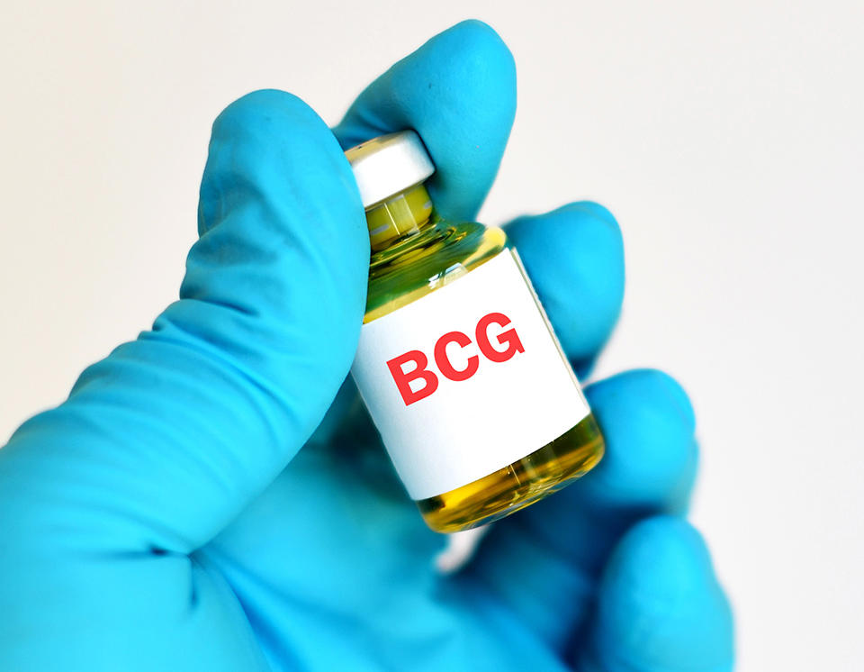
gloved hand holding a small bottle labeled BCG

When inserted directly into the bladder, BCG can cause an immune response against bladder cancer cells.

Credit: iStock

Immune system modulators are a type of immunotherapy that enhance the body’s immune response against cancer.

What are the types of immune system modulators? 

Types of immune system modulators include cytokines, BCG, and immunomodulatory drugs.

Cytokines are proteins made by white blood cells. They play important roles in your body’s normal immune responses and in the immune system’s ability to respond to cancer.

Cytokines that are sometimes used to treat cancer:

  • Interferons (INFs). Researchers have found that one type of interferon, called INF-alfa, can enhance your immune response to cancer cells by causing certain white blood cells, such as natural killer cells and dendritic cells, to become active. INF-alfa may also slow the growth of cancer cells or promote their death.
  • Interleukins (ILs). There are more than a dozen interleukins, including IL-2, which is also called T-cell growth factor. IL-2 boosts the number of white blood cells in the body, including killer T cells and natural killer cells. Increasing these cells can cause an immune response against cancer. IL-2 also helps B cells (another type of white blood cell) produce certain substances that can target cancer cells.

Cytokines that are sometimes used to reduce side effects from cancer treatment are called hematopoietic growth factors. They promote the growth of blood cells that are damaged by chemotherapy:

  • Erythropoietin, which increases the production of red blood cells.
  • IL-11, which increases the production of platelets.
  • Granulocyte-macrophage colony-stimulating factor (GM-CSF) and granulocyte colony-stimulating factor (G-CSF), which both increase the number of white blood cells. Boosting white blood cells reduces the risk of infections. G-CSF and GM-CSF can also enhance the immune system response against cancer by increasing the number of cancer-fighting T cells.

BCG is a weakened form of the bacteria that causes tuberculosis. It does not cause disease in humans. BCG is used to treat bladder cancer. When inserted directly into the bladder with a catheter, BCG causes an immune response against cancer cells. It is also being studied in other types of cancer. BCG stands for Bacillus Calmette-Guérin.

Immunomodulatory drugs (also called biological response modifiers) stimulate the immune system. They include

Thalidomide, lenaliodomide, and pomalidomide cause cells to release IL-2. They also stop tumors from forming new blood vessels. Tumors need to form new blood vessels to grow beyond a certain size. These three drugs may also be called angiogenesis inhibitors.

Imiquimod is a cream that you rub on the skin. It causes cells to release cytokines.
Nonspecific Immune Stimulation

Learn about nonspecific immune stimulation, one type of immunotherapy used to treat cancer.

Which types of cancer are treated with immune system modulators?

Most immune-modulating agents are used to treat advanced cancer. Some are used to help manage side effects.

What are the side effects of immune system modulators?

Immune-modulating agents can cause side effects, which affect people in different ways. The side effects you may have and how they make you feel will depend on how healthy you are before treatment, your type of cancer, how advanced it is, the type of immune-modulating agent you are getting, and the dose.

Doctors and nurses cannot know for sure when or if side effects will occur or how serious they will be. So, it is important to know which signs to look for and what to do if you start to have problems.

Immune-modulating agents can cause flu-like symptoms, which include

Learn more about flu-like symptoms caused by cancer treatment.

Cytokines can also cause many serious side effects

  • trouble breathing
  • low or high blood pressure
  • severe allergic reactions
  • lowered blood counts, which can raise the risk of infections and cause bleeding problems
  • blood clots
  • problems with mood, behavior, thinking, and memory
  • skin problems, such as rash, burning at injection site, and ulcers
  • organ damage

BCG can also cause urinary side effects.

Thalidomide, lenalidomide, and pomalidomide can cause

  • blood clots
  • nerve problems that lead to pain, numbness, tingling, swelling, or muscle weakness in different parts of the body
  • birth defects, if used during pregnancy

Imiquimod can cause skin reactions.
T-cell Transfer Therapy

T-cell Transfer Therapy

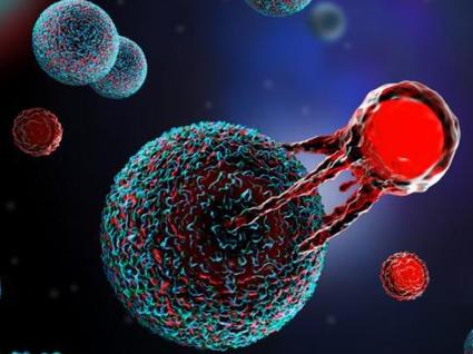
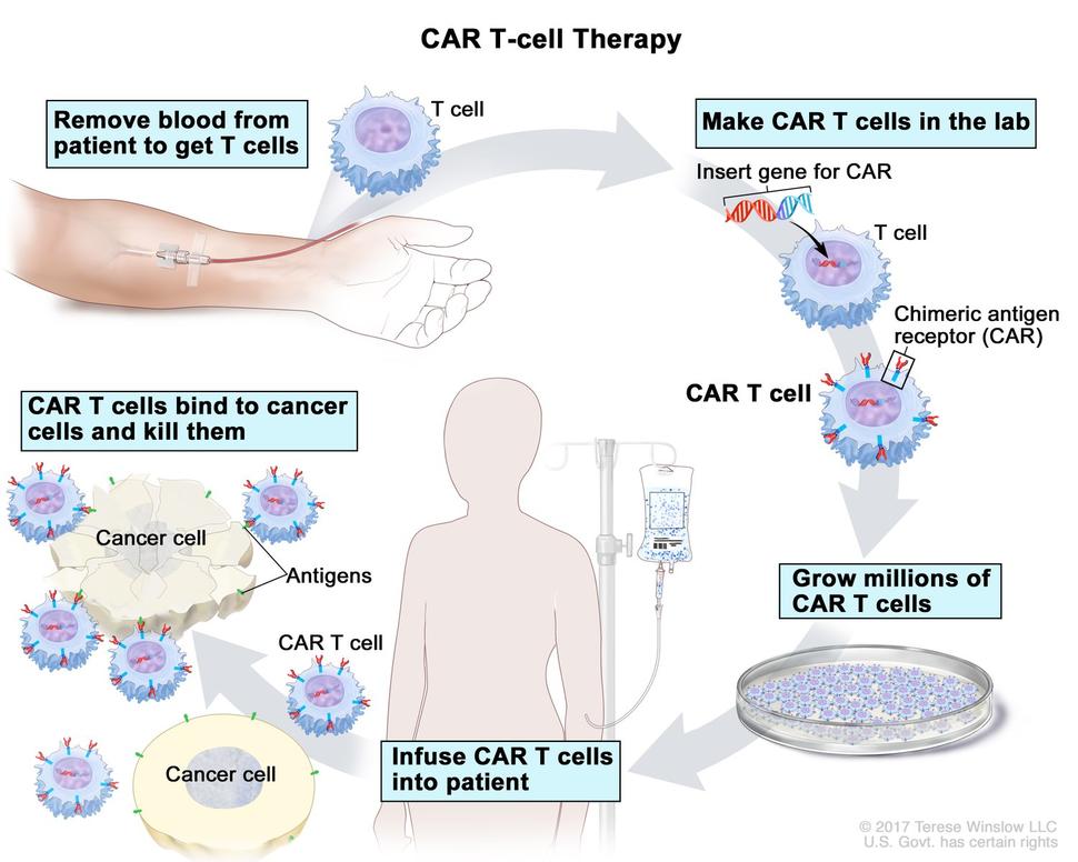
CAR T-cell therapy is a type of treatment in which a patient’s T cells (a type of immune cell) are changed in the laboratory so they will bind to cancer cells and kill them.

Credit: © Terese Winslow

How does T-cell transfer therapy work against cancer?

T-cell transfer therapy is a type of immunotherapy that makes your own immune cells better able to attack cancer. There are two main types of T-cell transfer therapy: tumor-infiltrating lymphocytes (or TIL) therapy and CAR T-cell therapy. Both involve collecting your own immune cells, growing large numbers of these cells in the lab, and then giving the cells back to you through a needle in your vein. T-cell transfer therapy is also called adoptive cell therapy, adoptive immunotherapy, and immune cell therapy.

The process of growing your T cells in the lab can take 2 to 8 weeks. During this time, you may have treatment with chemotherapy and, maybe, radiation therapy to get rid of other immune cells. Reducing your immune cells helps the transferred T cells to be more effective. After these treatments, the T cells that were grown in the lab will be given back to you via a needle in your vein.

  • TIL therapy uses T cells called tumor-infiltrating lymphocytes that are found in your tumor. Doctors test these lymphocytes in the lab to find out which ones best recognize your tumor cells. Then, these selected lymphocytes are treated with substances that make them grow to large numbers quickly.

    The idea behind this approach is that the lymphocytes that are in or near the tumor have already shown the ability to recognize your tumor cells. But there may not be enough of them to kill the tumor or to overcome the signals that the tumor is releasing to suppress the immune system. Giving you large numbers of the lymphocytes that react best with the tumor can help to overcome these barriers.

  • CAR T-cell therapy is similar to TIL therapy, but your T cells are changed in the lab so that they make a type of protein known as CAR before they are grown and given back to you. CAR stands for chimeric antigen receptor. CARs are designed to allow the T cells to attach to specific proteins on the surface of the cancer cells, improving their ability to attack the cancer cells.

What cancers are treated with T-cell transfer therapy?

A TIL therapy called lifileucel (Amtagvi) has been approved by the Food and Drug Administration (FDA) to treat melanoma. And it has produced promising findings in other cancers, such as cervical squamous cell carcinoma and cholangiocarcinoma. However, this treatment is still experimental for those cancers.

Six CAR T-cell therapies have been approved by the FDA for blood cancers.

CAR T-cell therapy has also been studied for the treatment of solid tumors, including breast and brain cancers, but use in such cancers is still experimental.

What are the side effects of T-cell transfer therapy?

T-cell transfer therapy can cause side effects, which people experience in different ways. The side effects you may have and how serious they are will depend on how healthy you are before treatment, your type of cancer, how advanced it is, the type of T-cell transfer therapy you are receiving, and the dose.

Doctors and nurses cannot know for sure when or if side effects will occur or how they will affect you. So, it is important to know which signs to look for and what to do if you start to have problems.

CAR T-cell therapy can cause a serious side effect known as cytokine release syndrome. This syndrome is caused when the transferred T cells, or other immune cells responding to the new T cells, release a large amount of cytokines into the blood.

Cytokines are immune substances that have many different functions in the body. A sudden increase in their levels can cause:

  • fever
  • nausea
  • headache
  • rash
  • rapid heartbeat
  • low blood pressure
  • trouble breathing

Most people have a mild form of cytokine release syndrome. But in some people, it may be severe or life-threatening.

Also, although CAR T cells are designed to recognize proteins that are found only on cancer cells, they can also sometimes recognize normal cells. Depending on which normal cells are recognized, this can cause a range of side effects, including organ damage.

TIL therapy can cause capillary leak syndrome. This syndrome causes fluid and proteins to leak out of tiny blood vessels and flow into surrounding tissues, resulting in dangerously low blood pressure. Capillary leak syndrome may lead to multiple organ failure and shock.

Access the 508-compliant version of this video

Immune Checkpoint Inhibitors

Immune Checkpoint Inhibitors

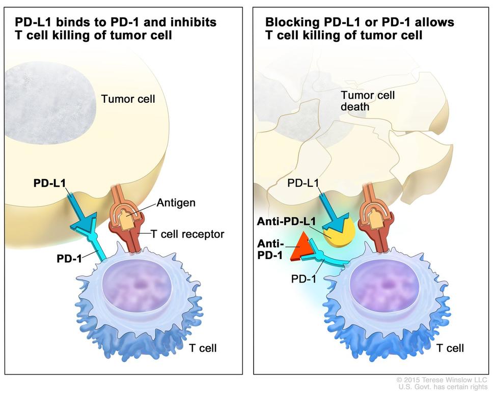
Checkpoint proteins, such as PD-L1 on tumor cells and PD-1 on T cells, help keep immune responses in check. The binding of PD-L1 to PD-1 keeps T cells from killing tumor cells in the body (left panel). Blocking the binding of PD-L1 to PD-1 with an immune checkpoint inhibitor (anti-PD-L1 or anti-PD-1) allows the T cells to kill tumor cells (right panel).

Credit: © Terese Winslow

How do immune checkpoint inhibitors work against cancer?

Immune checkpoints are a normal part of the immune system. Their role is to prevent an immune response from being so strong that it destroys healthy cells in the body.

Immune checkpoints engage when proteins on the surface of immune cells called T cells recognize and bind to partner proteins on other cells, such as some tumor cells. These proteins are called immune checkpoint proteins. When the checkpoint and partner proteins bind together, they send an “off” signal to the T cells. This can prevent the immune system from destroying the cancer.

Immunotherapy drugs called immune checkpoint inhibitors work by blocking checkpoint proteins from binding with their partner proteins. This prevents the “off” signal from being sent, allowing the T cells to kill cancer cells.

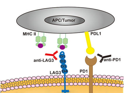
One such drug acts against a checkpoint protein called CTLA-4. Other immune checkpoint inhibitors act against a checkpoint protein called PD-1 or its partner protein PD-L1. Some tumors turn down the T cell response by producing lots of PD-L1.

Which cancers are treated with immune checkpoint inhibitors?

What side effects are caused by immune checkpoint inhibitors?

Immune checkpoint inhibitors can cause side effects that affect people in different ways. The side effects you may have and how they make you feel will depend on how healthy you are before treatment, your type of cancer, how advanced it is, the type of immune checkpoint inhibitor you are receiving, and the dose.

Doctors and nurses cannot know for sure when or if side effects will occur or how serious they will be. So, it is important to know which signs to look for and what to do if they occur.

Common side effects of immune checkpoint inhibitors include:

Rarer side effects of immune checkpoint inhibitors can include widespread inflammation. Depending on the organ of your body that is affected, inflammation can lead to:

  • changes in skin color, rash, and feeling itchy, caused by skin inflammation
  • cough and chest pains, caused by inflammation in the lungs
  • belly pain and diarrhea, caused by inflammation in the colon
  • diabetes, caused by inflammation in the pancreas
  • hepatitis (inflammation of the liver)
  • hypophysitis (inflammation of the pituitary gland)
  • myocarditis (inflammation of the heart muscle)
  • nephritis (inflammation of the kidney) and impaired kidney function
  • overactive or underactive thyroid
  • nervous system problems such as muscle weakness, numbness, and trouble breathing

Learn more about organ inflammation and immunotherapy.

Immune Checkpoint Inhibitors

Learn about immune checkpoint inhibitors, one type of immunotherapy used to treat cancer.

Immunotherapy Side Effects

Immunotherapy Side Effects

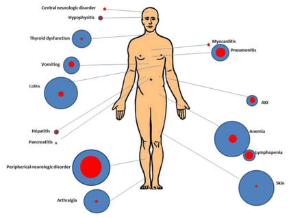
Immunotherapy can cause side effects. Many side effects happen when the immune system that is revved-up to act against the cancer also acts against healthy cells and tissues in the body.

Different people have different side effects. The ones you have and how they make you feel will depend on

  • how healthy you are before treatment
  • your type of cancer
  • how advanced your cancer is
  • the type and dose of immunotherapy you are getting

You might be on immunotherapy for a long time. And side effects can occur at any point during and after treatment. Doctors and nurses cannot know for certain when or if side effects will occur or how serious they will be. So, it is important to talk with your doctors and nurses about what signs to look for and what to do if you start to have problems.

Some side effects are common with all types of immunotherapy. For instance, you might have skin reactions at the needle site, which include:

  • pain
  • swelling
  • soreness
  • redness
  • itchiness
  • rash

Learn more about skin changes caused by cancer treatment.

You may have flu-like symptoms, which include:

Other side effects might include:

Some types of immunotherapy may cause severe or fatal allergic and inflammation-related reactions. But, these reactions are rare.

Certain side effects might happen depending on the type of immunotherapy you receive. Visit the page for the type of immunotherapy that you are receiving for more details about serious side effects. Types of immunotherapy include:

Treatment of Bladder Cancer by Stage

Treatment of Bladder Cancer by Stage

Bladder cancer stage and grade are important factors in deciding the best treatment for bladder cancer. Other factors, such as your preferences and overall health, are also important.

Palliative therapy aims to relieve symptoms and improve the quality of life for people with life-threatening diseases like cancer. It is helpful at all stages of cancer. Many cancer treatments can also be used as palliative therapy to enhance comfort. Learn more about Palliative Care in Cancer.

For some people, taking part in a clinical trial of new cancer drugs or treatment combinations may be an option. To learn about clinical trials, including how to find and join one, visit Clinical Trials Information for Patients and Caregivers.

Treatment of stages 0 and I bladder cancer

Stage 0 and stage I bladder cancer, also known as non-muscle-invasive bladder cancer (NMIBC), haven’t spread to the muscle layer of the bladder. Treatment of NMIBC depends on several factors:

  • the risk level of the cancer (low, intermediate, or high) for recurrence or becoming muscle-invasive
  • the stage and grade of the cancer
  • the size and number of tumors

The first treatment is usually a surgical procedure called transurethral resection (TUR) with fulguration to remove the tumor. Sometimes, surgery might be repeated if the first doesn’t remove enough of the tumor or does not include a sample from the muscle layer. If a repeat surgery finds that the cancer has invaded the muscle layer, it will be treated as muscle-invasive bladder cancer (stage II bladder cancer or higher).

Because stage 0 and stage I bladder cancer often come back after surgery, most people receive intravesical chemotherapy with mitomycin or gemcitabine or intravesical BCG at the time of their first surgery. To help lower the risk of bladder cancer recurrence, your doctor may recommend you continue having intravesical BCG for up to 3 years, depending on the characteristics of the cancer. This is called maintenance therapy.

To learn more about the treatments below, visit Bladder Cancer Treatment.

Treatment of low-risk bladder cancer

Low-risk bladder cancer might include small, single, low-grade tumors.

Treatment typically includes TUR with fulguration and intravesical therapy given around the time of surgery, followed by surveillance. Surveillance means closely watching your condition but not giving any treatment unless the cancer returns.

Treatment of intermediate-risk bladder cancer

Intermediate-risk bladder cancer might include multiple or large, slow-growing stage 0a tumors, slow-growing stage 0a tumors that recur within 1 year, a single fast-growing stage 0a tumor, or a slow-growing stage I tumor.

Treatment typically includes TUR with fulguration, followed by intravesical therapy (BCG or chemotherapy with mitomycin or gemcitabine). Depending on the characteristics of the cancer, your doctor may recommend that you continue having intravesical BCG for up to 1 year to lower the risk of recurrence. Surveillance with regular cystoscopies (bladder inspections with a camera) and possibly additional imaging tests are used to monitor for signs of cancer recurrence or progression.

Treatment of high-risk bladder cancer

High-risk bladder cancer might include multiple or large high-grade stage 0a tumors, the presence of carcinoma in situ (stage 0is), and fast-growing stage I tumors.

Treatment typically includes TUR with fulguration, followed by intravesical BCG therapy. Sometimes, intravesical BCG is continued for up to 3 years to lower the risk of recurrence. If you have multiple tumors or carcinoma in situ, a type of high-grade stage 0 cancer, another option is surgery to remove part or all of your bladder (cystoscopy).

Treatment of very high-risk bladder cancer

Very high-risk bladder cancer might include cancer that does not respond to intravesical BCG, that recurs in the urethra, or that involves cancer cells detected in the blood or lymph system near the tumor. Very high-risk bladder cancer has a greater chance of progressing to muscle-invasive disease (stage II or higher).

If you have many tumors or carcinoma in situ (a type of high-grade stage 0 cancer), partial or complete cystectomy (surgical removal of the bladder) may be an option. If you have carcinoma in situ, cannot or choose not to have a cystectomy, and BCG did not work, other options include intravesical chemotherapy, the immunotherapy drug pembrolizumab given through a vein, or intravesical immunotherapy with nadofaragene firadenovec-vncg or nogapendekin alfa inbakicept-pmln.

Treatment of stages II and III bladder cancer

The two main treatments for stage II and stage III bladder cancer are radical cystectomy or a combination of radiation therapy and chemotherapy.

Radical cystectomy is surgery to remove the bladder and surrounding tissues and organs. Surgery to make a new way for urine to leave the body (called urinary diversion) will be done. Additional treatments may be given before or after surgery:

  • Chemotherapy may be given before surgery to people who are well enough to tolerate it. Giving combination chemotherapy that includes cisplatin before surgery has been shown to help people live longer than surgery alone.
  • The immunotherapy drug nivolumab may be given if the cancer has a high risk of coming back after surgery or did not respond to chemotherapy.

If you are unable or choose not to have surgery, treatment may include a combination of radiation therapy and chemotherapy, such as cisplatin and fluorouracil, given at the same time. Giving chemotherapy at the same time as radiation therapy helps the radiation therapy work better.

Partial cystectomy (removal of part of the bladder) is a less common treatment for stages II and III bladder cancer.

To learn more about these treatments, visit Bladder Cancer Treatment.

Treatment of stage IV bladder cancer

Treatment for stage IV bladder cancer depends on whether the cancer is locally advanced or metastatic. Treatment for metastatic bladder cancer is aimed at relieving symptoms and improving quality of life while also slowing the growth and spread of the cancer.

Treatment of stage IVA (locally advanced) bladder cancer may include:

Treatment of stage IVB (metastatic) bladder cancer may include:

  • the immunotherapy drug pembrolizumab in combination with the targeted therapy drug enfortumab vedotin
  • the immunotherapy drug nivolumab in combination with the chemotherapy drugs cisplatin and gemcitabine
  • the chemotherapy drugs cisplatin and gemcitabine followed by the immunotherapy drug avelumab
  • systemic chemotherapy, such as one of the following regimens:
    • gemcitabine with either cisplatin or carboplatin
    • dose-dense methotrexate, vinblastine, doxorubicin, and cisplatin (MVAC)
    • cisplatin, methotrexate, and vinblastine (CMV)
  • an immunotherapy drug alone, such as avelumab, nivolumab, or pembrolizumab
  • radiation therapy as palliative therapy
  • urinary diversion, as palliative therapy or to prevent a blockage of urine that could damage the kidneys
  • surgery to remove part or all of the bladder (cystectomy) as palliative therapy

To learn more about these treatments, visit Bladder Cancer Treatment.

Treatment of recurrent bladder cancer

Treatment of bladder cancer that has recurred (come back) depends on previous treatment and where the cancer has come back. Treatment may include:

To learn more about these treatments, visit Bladder Cancer Treatment.

Bladder Cancer Treatment

Bladder Cancer Treatment

Different types of treatment are available for bladder cancer. You and your cancer care team will work together to decide your treatment plan, which may include more than one type of treatment. Many factors will be considered, such as the stage and grade of the cancer, your overall health, and your preferences. Your plan will include information about your cancer, the goals of treatment, your treatment options and the possible side effects, and the expected length of treatment.

It will be helpful to talk with your cancer care team before treatment begins about what to expect. Some things you’ll want to learn about include what you need to do before treatment begins, how you’ll feel while going through it, and what kind of help you will need. To learn more, visit Questions to Ask Your Doctor About Your Treatment. For treatment by stage, visit Treatment of Bladder Cancer by Stage.

Surgery

Surgery is the main treatment for bladder cancer. The type of surgery depends on where the cancer is located. Other treatments may be given in addition to surgery:

  • Treatment given before surgery is called preoperative therapy or neoadjuvant therapy. Chemotherapy may be given before surgery to shrink the tumor and reduce the amount of tissue that needs to be removed during surgery.
  • Treatment given after surgery, to lower the risk that the cancer will come back, is called adjuvant therapy. After the doctor removes all the cancer that can be seen, some patients may be given chemotherapy, radiation therapy, immunotherapy, and/or targeted therapy to kill any cancer cells that are left.

Learn more about Surgery to Treat Cancer.

The types of surgery done to treat bladder cancer are:

Transurethral resection (TUR) with fulguration

During TUR with fulguration, the doctor inserts a cystoscope (a thin lighted tube) into the bladder through the urethra. A tool with a small wire loop on the end is then used to remove the cancer or to burn the tumor away with high-energy electricity. This is known as fulguration.

Partial cystectomy

Partial cystectomy is surgery to remove part of the bladder. This may be done for patients who have a low-grade tumor that has invaded the wall of the bladder but is limited to one area of the bladder. Because only a part of the bladder is removed, patients are able to urinate normally after recovering from this surgery. This is also called segmental cystectomy.

Radical cystectomy with urinary diversion

Radical cystectomy is surgery to remove the bladder and any lymph nodes and nearby organs that contain cancer. This surgery may be done when the bladder cancer invades the muscle layers or when non-muscle-invasive bladder cancer involves a large part of the bladder:

Sometimes, when the cancer has spread outside the bladder and can’t be completely removed, surgery to remove only the bladder may be done to reduce urinary symptoms caused by the cancer.

When the bladder must be removed, the surgeon performs a procedure called urinary diversion to create another way for the body to store and pass urine. It may involve redirecting urine into the colon, using catheters to drain the bladder, or making an opening in the abdomen that connects to a bag outside the body for collecting urine. To learn more, visit the National Institute of Diabetes and Digestive and Kidney Diseases page on Urinary Diversion.

Radiation therapy

Radiation therapy uses high-energy x-rays or other types of radiation to kill cancer cells or keep them from growing. Bladder cancer is sometimes treated with external beam radiation therapy. This type of radiation therapy uses a machine outside the body to send radiation toward the area of the body with cancer. Radiation therapy may be given alone or with other types of treatment, such as chemotherapy.

Learn more about External Beam Radiation Therapy for Cancer and Radiation Therapy Side Effects.

Chemotherapy

Chemotherapy (also called chemo) uses drugs to stop the growth of cancer cells, either by killing the cells or by stopping them from dividing. Chemotherapy may be given alone or with other types of treatment. The way the chemotherapy is given depends on the type and stage of the cancer being treated.

Systemic chemotherapy

Systemic chemotherapy for bladder cancer is when chemotherapy drugs are injected into a vein. When given this way, the drugs enter the bloodstream to reach cancer cells throughout the body. Systemic chemotherapy drugs that may be used to treat bladder cancer are:

Combinations of these drugs may be used. Other chemotherapy drugs not listed here may also be used.

Intravesical chemotherapy

For bladder cancer, chemotherapy may be intravesical, meaning it is put into the bladder through a tube inserted into the urethra. Intravesical treatments flush the bladder with drugs that kill cancer cells that remain after surgery. This lowers the chance of the cancer coming back.

Mitomycin and gemcitabine are two chemotherapy drugs given as intravesical chemotherapy to treat bladder cancer. These drugs can also be given systemically.

To learn more about how chemotherapy works, how it is given, common side effects, and more, visit Chemotherapy to Treat Cancer and Chemotherapy and You: Support for People With Cancer.

Immunotherapy

Immunotherapy helps a person’s immune system fight cancer. Your doctor may suggest biomarker tests to help predict your response to certain immunotherapy drugs. Learn more about Biomarker Testing for Cancer Treatment.

Systemic immunotherapy

Systemic immunotherapy drugs used to treat urothelial cancer (a type of bladder cancer) include:

These drugs work in more than one way to kill cancer cells. They are also considered targeted therapy because they target specific changes or substances in cancer cells (visit the section on Targeted therapy).

Intravesical immunotherapy

BCG (bacillus Calmette-Guérin), nadofaragene firadenovec-vncg, and nogapendekin alfa inbakicept-pmln are intravesical immunotherapy drugs used to treat bladder cancer. They are given in a solution that is placed directly into the bladder using a catheter (thin tube). Intravesical treatments flush the bladder with drugs that kill cancer cells that remain after surgery. This lowers the chance of the cancer coming back.

Immunotherapy uses the body’s immune system to fight cancer. This animation explains one type of immunotherapy called nonspecific immune stimulation that is used to treat cancer.

Learn more about Immunotherapy to Treat Cancer and Immunotherapy Side Effects.

Targeted therapy

Targeted therapy uses drugs or other substances to block the action of specific enzymes, proteins, or other molecules involved in the growth and spread of cancer cells. Your doctor may suggest biomarker tests to help predict your response to certain targeted therapy drugs. Learn more about Biomarker Testing for Cancer.

Targeted therapies used to treat bladder cancer include:

Learn more about Targeted Therapy to Treat Cancer.

Clinical trials

For some people, joining a clinical trial may be an option. There are different types of clinical trials for people with cancer. For example, a treatment trial tests new treatments or new ways of using current treatments. Supportive care and palliative care trials look at ways to improve quality of life, especially for those who have side effects from cancer and its treatment.

You can use the clinical trial search to find NCI-supported cancer clinical trials accepting participants. The search allows you to filter trials based on the type of cancer, your age, and where the trials are being done. Clinical trials supported by other organizations can be found on the ClinicalTrials.gov website.

Learn more about clinical trials, including how to find and join one, at Clinical Trials Information for Patients and Caregivers.

Follow-up care

Some of the tests that were done to diagnose or stage the cancer may be repeated. Some tests will be repeated to see how well the treatment is working. Decisions about whether to continue, change, or stop treatment may be based on the results of these tests. These tests are sometimes called follow-up tests or check-ups.

Coping with Bladder Cancer

Coping with Bladder Cancer

When you first learn that you have bladder cancer, you may wonder how you’re going to cope with the upcoming changes in your life. One step you can take is to be informed of the physical and emotional changes that may occur and what resources are available to help you. Talking with your health care team about your concerns can give you a greater sense of control. Family and friends can also be important sources of support.

For resources on the common physical side effects of treatment for bladder cancer, see Bladder Cancer Treatment. For help with emotional side effects, see Emotions and Cancer.

For help after your cancer treatment has ended, see Cancer Survivorship and our booklet Facing Forward: Life After Cancer Treatment.

Adjusting after urostomy

For those who have a stoma and urostomy bag, getting used to them takes time. Some people need to learn how to use a catheter to empty their bladder, which is another big change. And if you have incontinence or problems controlling your bladder, it can be frustrating to deal with.

Know that you aren’t alone in how you feel. Adjusting to these changes can be hard. But, over time, many people are able to do a lot of what they did before surgery. Support from family members and your health care team can help you get used to life after urostomy surgery. Ask your nurse for resources and support groups that may be helpful. Learn more about Patient’s Concerns About Surgery.

Self-image and sexual problems

Body changes may affect your self-image and sex life after treatment. Some treatments for bladder cancer, including chemotherapy, radiation therapy, surgery, or certain medicines, can cause short-term or long-term problems with sex. For example, having a cystectomy may affect the nerves and make it harder for men to have an erection. Women may have pain during sex or problems with lubrication and orgasm.

It’s important to discuss any issues and concerns you have about sex with your health care team before treatment. Knowing your thoughts ahead of time may help them plan your treatment.

Learn more about the sexual problems some cancer treatments can cause and ways to cope in Sexual Health Issues in Women with Cancer and Sexual Health Issues in Men with Cancer. For help communicating your feelings, see Self-Image and Sexuality.

The stress of follow-up care

It’s common for bladder cancer to come back, even after successful treatment. As a result, it is important for people who have been treated for bladder cancer to visit their doctor regularly to get certain follow-up exams or tests. This can be hard for a number of reasons, such as:

  • Planning and scheduling these appointments can be stressful and time-consuming.
  • Waiting for test results can cause anxiety and an ongoing fear of recurrence.
  • The added costs of things such as copays, medicines, and parking and transportation fees only add to the stress.

For information about how to prepare for follow-up appointments, see Follow-Up Medical Care.

For tips on how to deal with the fear of cancer coming back, see the section Coping with Fear of Recurrence on our New Normal page.

Cost of cancer treatment

Cancer is one of the most costly diseases to treat in the United States. Even if you have health insurance, you may face major financial challenges and need help dealing with the costs of bladder cancer treatment. Having cancer may also make it hard for you to work and pay your bills. The problems a person has related to the cost of treatment is known as financial toxicity. For ways to cope, see  Managing Costs and Medical Information. To learn about financial toxicity and find out if you are at risk, see Financial Toxicity (Financial Distress) and Cancer Treatment.

Childhood Chordoma (PDQ®)–Patient Version

Childhood Chordoma (PDQ®)–Patient Version

What is childhood chordoma?

Childhood chordoma is a very rare type of bone cancer that begins in cells left over from early development that helped form disks of the spine. These cells usually go away before birth, but sometimes they stay in the body and may become cancer in rare cases. Chordoma can form in a bone at the base of the skull called the clivus, the spine, and the tailbone. In children and adolescents, the most common location is the clivus, making them hard to remove completely with surgery. Only about 20 out of the 300 cases of chordoma diagnosed in the United States each year occur in people younger than 20 years.

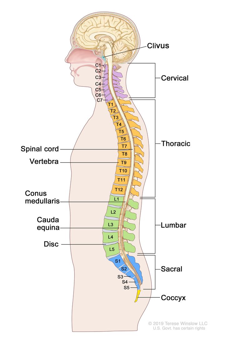
EnlargeAnatomy of the spine; drawing shows a side view of the spine, including the cervical spine (C1-C7), thoracic spine (T1-T12), lumbar spine (L1-L5), sacral spine (S1-S5), and the coccyx (tailbone). Also shown are the spinal cord, vertebra (back bone), conus medullaris (the end of the spinal cord), cauda equina (the bundle of spinal nerves that extend beyond the conus medullaris), and a lumbar disc. The clivus (a bone at the base of the skull near the spinal cord) is also shown.
Anatomy of the spine. The spine is made up of bones, muscles, tendons, nerves, and other tissues that reach from the base of the skull near the spinal cord (clivus) to the coccyx (tailbone). The vertebrae (back bones) of the spine include the cervical spine (C1-C7), thoracic spine (T1-T12), lumbar spine (L1-L5), sacral spine (S1-S5), and the tailbone. Each vertebra is separated by a disc. The vertebrae surround and protect the spinal cord. The spinal cord is divided into segments, each containing a pair of spinal nerves that send messages between the brain and the rest of the body. Many spinal nerves extend beyond the conus medullaris (the end of the spinal cord) to form a bundle of nerves called the cauda equina.

Causes and risk factors for childhood chordoma

Chordoma in children is caused by certain changes in the way cells function, especially how they grow and divide into new cells. Often, the exact cause of these cell changes is unknown. Learn more about how cancer develops at What Is Cancer?

A risk factor is anything that increases the chance of getting a disease. Children who have an inherited condition called tuberous sclerosis may be at an increased risk of chordoma. Not every child with this risk factor will develop a chordoma. And it will develop in some children who don’t have a known risk factor. Talk with your child’s doctor if you think your child may be at risk.

Symptoms of childhood chordoma

Children may not have symptoms of a chordoma until the tumor has grown bigger. It’s important to check with your child’s doctor if your child has:

  • a headache
  • double vision
  • a blocked or stuffy nose
  • trouble speaking
  • trouble swallowing
  • neck or back pain
  • pain down the back of the legs
  • numbness, tingling, or weakness of the arms and legs
  • a change in bowel or bladder habits

These symptoms may be caused by problems other than childhood chordoma. The only way to know is to see your child’s doctor.

Tests to diagnose childhood chordoma

If your child has symptoms that suggest a chordoma, the doctor will need to find out if these are due to a chordoma or another problem. The doctor will ask when the symptoms started and how often your child has been having them. They will also ask about your child’s personal and family medical history and do a physical exam. Depending on these results, they may recommend other tests. If your child is diagnosed with a chordoma, the results of these tests will help you and your child’s doctor plan treatment.

The tests used to diagnose childhood chordoma may include:

Magnetic resonance imaging (MRI)

MRI uses a magnet, radio waves, and a computer to make a series of detailed pictures of areas of the body, such as the whole spine. This procedure is called nuclear magnetic resonance imaging (NMRI).

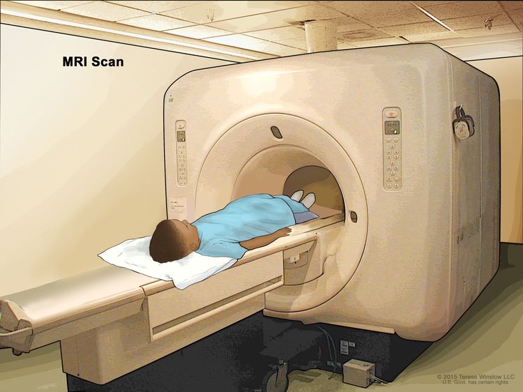
EnlargeMagnetic resonance imaging (MRI) scan; drawing shows a child lying on a table that slides into the MRI machine, which takes a series of detailed pictures of areas inside the body.
Magnetic resonance imaging (MRI) scan. The child lies on a table that slides into the MRI machine, which takes a series of detailed pictures of areas inside the body. The positioning of the child on the table depends on the part of the body being imaged.

CT scan (CAT scan)

A CT scan uses a computer linked to an x-ray machine to make a series of detailed pictures of areas inside the body, such as the brain and spine. The pictures are taken from different angles and are used to create 3-D views of tissue and organs. A dye may be injected into a vein or swallowed to help the organs or tissues show up more clearly. This procedure is also called computed tomography, computerized tomography, or computerized axial tomography. Learn more about Computed Tomography (CT) Scans and Cancer.

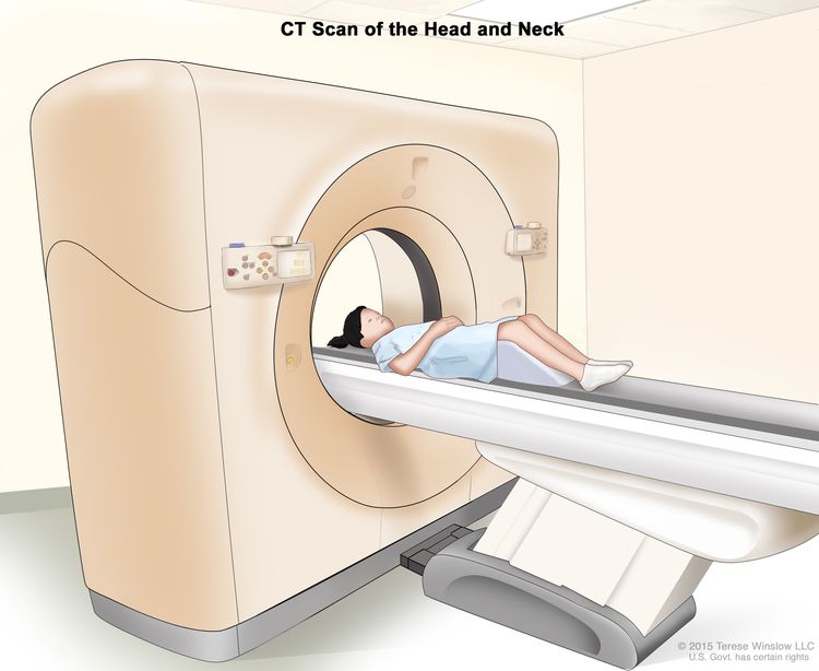
EnlargeComputed tomography (CT) scan of the head and neck; drawing shows a child lying on a table that slides through the CT scanner, which takes a series of detailed x-ray pictures of the inside of the head and neck.
Computed tomography (CT) scan of the head and neck. The child lies on a table that slides through the CT scanner, which takes a series of detailed x-ray pictures of the inside of the head and neck.

Biopsy

A biopsy is a procedure in which a sample of tissue is removed from the tumor so that a pathologist can view it under a microscope to check for signs of cancer. The sample of tissue may also be checked for a high level of a protein called brachyury.

The following laboratory tests may be done on the tissue that was removed during the biopsy:

  • Immunohistochemistry uses antibodies to check for certain antigens (markers) in a sample of a patient’s tissue. The antibodies are usually linked to an enzyme or a fluorescent dye. After the antibodies bind to a specific antigen in the tissue sample, the enzyme or dye is activated, and the antigen can then be seen under a microscope. This type of test is used to help diagnose cancer and help tell one type of cancer from another type.
  • Molecular test checks for certain genes, proteins, or other molecules in a sample of tissue, blood, or other body fluid. A molecular test may be done with other procedures, such as biopsies, to help diagnose some types of cancer. Molecular tests check for certain gene or chromosome changes that occur in some cancers.

    The Molecular Characterization Initiative offers free molecular testing to children, adolescents, and young adults with certain types of newly diagnosed cancer. The program is offered through NCI’s Childhood Cancer Data Initiative. To learn more, visit About the Molecular Characterization Initiative.

Getting a second opinion

You may want to get a second opinion to confirm your child’s diagnosis and treatment plan. If you seek a second opinion, you will need to get medical test results and reports from the first doctor to share with the second doctor. The second doctor will review the pathology report, slides, and scans. This doctor may agree with the first doctor, suggest changes to the treatment plan, or provide more information about your child’s cancer.

To learn more about choosing a doctor and getting a second opinion, see Finding Cancer Care. You can contact NCI’s Cancer Information Service via chat, email, or phone (both in English and Spanish) for help finding a doctor or hospital that can provide a second opinion. For questions you might want to ask at your child’s appointments, see Questions to Ask Your Doctor about Cancer.

Who treats children with chordoma?

A pediatric oncologist, a doctor who specializes in treating children with cancer, oversees treatment of a chordoma. The pediatric oncologist works with other health professionals who are experts in treating children with cancer and who specialize in other areas of medicine. Other specialists may include:

Treatment of childhood chordoma

There are different types of treatment for children and adolescents with chordoma. You and your child’s care team will work together to decide treatment. Many factors will be considered, such as your child’s overall health and whether the tumor is newly diagnosed or has come back.

Your child’s treatment plan will include information about the cancer, the goals of treatment, treatment options, and the possible side effects. It will be helpful to talk with your child’s care team before treatment begins about what to expect. For help every step of the way, see our booklet, Children with Cancer: A Guide for Parents.

Surgery

Surgery may be used to remove as much of the tumor as possible. If the chordoma formed in or near the brain, or by important nerves or blood vessels, it cannot be completely removed by surgery without causing harm to your child.

Radiation therapy

Radiation therapy uses high-energy x-rays or other types of radiation to kill cancer cells or keep them from growing. Childhood chordoma may be treated with external beam radiation therapy. External beam radiation therapy uses a machine outside the body to send radiation toward the area of the body with cancer. Proton beam radiation therapy is a type of high-energy, external radiation therapy that aims streams of protons (tiny, invisible, positively charged particles) at the cancer cells to kill them. This type of radiation therapy may be used for tumors near the base of the skull.

Learn more about External Beam Radiation Therapy for Cancer and Radiation Therapy Side Effects.

If the cancer comes back after treatment, your child’s doctor will talk with you about what to expect and possible next steps. There might be treatment options that may shrink the cancer or control its growth. If there are no treatments, your child can receive care to control symptoms from cancer so they can be as comfortable as possible.

Clinical trials

For some children, joining a clinical trial may be an option. There are different types of clinical trials for childhood cancer. For example, a treatment trial tests new treatments or new ways of using current treatments. Supportive care and palliative care trials look at ways to improve quality of life, especially for those who have side effects from cancer and its treatment.

You can use the clinical trial search to find NCI-supported cancer clinical trials accepting participants. The search allows you to filter trials based on the type of cancer, your child’s age, and where the trials are being done. Clinical trials supported by other organizations can be found on the ClinicalTrials.gov website.

Learn more about clinical trials, including how to find and join one, at Clinical Trials Information for Patients and Caregivers.

Prognostic factors for childhood chordoma

If your child has been diagnosed with a chordoma, you likely have questions about how serious the cancer is and your child’s chances of survival. The likely outcome or course of a disease is called prognosis.

Your child’s prognosis depends on:

  • your child’s age
  • where the tumor has formed in the tissue along the spine
  • how the tumor responds to treatment
  • whether there were changes in bowel or bladder habits at diagnosis
  • whether the tumor has certain gene changes
  • whether the tumor is newly diagnosed or has recurred (come back)

No two people are alike, and responses to treatment can vary greatly. Your child’s cancer care team is in the best position to talk with you about your child’s prognosis.

Follow-up care

As your child goes through treatment, they will have follow-up tests or check-ups. Some of the tests that were done to diagnose the cancer may be repeated to see how well the treatment is working. Decisions about whether to continue, change, or stop treatment may be based on the results of these tests.

Some of the tests will continue to be done from time to time after treatment has ended. The results of these tests can show if your child’s condition has changed or if the cancer has recurred (come back).

To learn more about follow-up tests, visit Tests to diagnose childhood chordoma.

Coping with your child's cancer

When your child has cancer, every member of the family needs support. Taking care of yourself during this difficult time is important. Reach out to your child’s treatment team and to people in your family and community for support. To learn more, see Support for Families: Childhood Cancer and the booklet Children with Cancer: A Guide for Parents.

Related resources

About This PDQ Summary

About PDQ

Physician Data Query (PDQ) is the National Cancer Institute’s (NCI’s) comprehensive cancer information database. The PDQ database contains summaries of the latest published information on cancer prevention, detection, genetics, treatment, supportive care, and complementary and alternative medicine. Most summaries come in two versions. The health professional versions have detailed information written in technical language. The patient versions are written in easy-to-understand, nontechnical language. Both versions have cancer information that is accurate and up to date and most versions are also available in Spanish.

PDQ is a service of the NCI. The NCI is part of the National Institutes of Health (NIH). NIH is the federal government’s center of biomedical research. The PDQ summaries are based on an independent review of the medical literature. They are not policy statements of the NCI or the NIH.

Purpose of This Summary

This PDQ cancer information summary has current information about the treatment of childhood chordoma. It is meant to inform and help patients, families, and caregivers. It does not give formal guidelines or recommendations for making decisions about health care.

Reviewers and Updates

Editorial Boards write the PDQ cancer information summaries and keep them up to date. These Boards are made up of experts in cancer treatment and other specialties related to cancer. The summaries are reviewed regularly and changes are made when there is new information. The date on each summary (“Updated”) is the date of the most recent change.

The information in this patient summary was taken from the health professional version, which is reviewed regularly and updated as needed, by the PDQ Pediatric Treatment Editorial Board.

Clinical Trial Information

A clinical trial is a study to answer a scientific question, such as whether one treatment is better than another. Trials are based on past studies and what has been learned in the laboratory. Each trial answers certain scientific questions in order to find new and better ways to help cancer patients. During treatment clinical trials, information is collected about the effects of a new treatment and how well it works. If a clinical trial shows that a new treatment is better than one currently being used, the new treatment may become “standard.” Patients may want to think about taking part in a clinical trial. Some clinical trials are open only to patients who have not started treatment.

Clinical trials can be found online at NCI’s website. For more information, call the Cancer Information Service (CIS), NCI’s contact center, at 1-800-4-CANCER (1-800-422-6237).

Permission to Use This Summary

PDQ is a registered trademark. The content of PDQ documents can be used freely as text. It cannot be identified as an NCI PDQ cancer information summary unless the whole summary is shown and it is updated regularly. However, a user would be allowed to write a sentence such as “NCI’s PDQ cancer information summary about breast cancer prevention states the risks in the following way: [include excerpt from the summary].”

The best way to cite this PDQ summary is:

PDQ® Pediatric Treatment Editorial Board. PDQ Childhood Chordoma. Bethesda, MD: National Cancer Institute. Updated <MM/DD/YYYY>. Available at: /types/bone/patient/child-chordoma-treatment-pdq. Accessed <MM/DD/YYYY>.

Images in this summary are used with permission of the author(s), artist, and/or publisher for use in the PDQ summaries only. If you want to use an image from a PDQ summary and you are not using the whole summary, you must get permission from the owner. It cannot be given by the National Cancer Institute. Information about using the images in this summary, along with many other images related to cancer can be found in Visuals Online. Visuals Online is a collection of more than 3,000 scientific images.

Disclaimer

The information in these summaries should not be used to make decisions about insurance reimbursement. More information on insurance coverage is available on Cancer.gov on the Managing Cancer Care page.

Contact Us

More information about contacting us or receiving help with the Cancer.gov website can be found on our Contact Us for Help page. Questions can also be submitted to Cancer.gov through the website’s E-mail Us.

Skin Cancer Screening (PDQ®)–Patient Version

Skin Cancer Screening (PDQ®)–Patient Version

What Is Screening?

Screening is looking for cancer before a person has any symptoms. This can help find cancer at an early stage. When abnormal tissue or cancer is found early, it may be easier to treat. By the time symptoms appear, cancer may have begun to spread.

Scientists are trying to better understand which people are more likely to get certain types of cancer. They also study the things we do and the things around us to see if they cause cancer. This information helps doctors recommend who should be screened for cancer, which screening tests should be used, and how often the tests should be done.

It is important to remember that your doctor does not necessarily think you have cancer if he or she suggests a screening test. Screening tests are given when you have no cancer symptoms.

If a screening test result is abnormal, you may need to have more tests done to find out if you have cancer. These are called diagnostic tests.

General Information About Skin Cancer

Key Points

  • Skin cancer is a disease in which malignant (cancer) cells form in the tissues of the skin.
  • Skin cancer is the most common cancer in the United States.
  • Different factors increase or decrease the risk of skin cancer.

Skin cancer is a disease in which malignant (cancer) cells form in the tissues of the skin.

The skin is the body’s largest organ. It protects against heat, sunlight, injury, and infection. Skin also helps control body temperature and stores water, fat, and vitamin D. The skin has several layers, but the two main layers are the epidermis (top or outer layer) and the dermis (lower or inner layer). Skin cancer begins in the epidermis, which is made up of three kinds of cells:

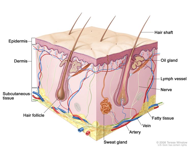
EnlargeSkin anatomy; drawing shows layers of the epidermis, dermis, and subcutaneous tissue including hair shafts and follicles, oil glands, lymph vessels, nerves, fatty tissue, veins, arteries, and a sweat gland.
Anatomy of the skin, showing the epidermis, dermis, and subcutaneous tissue.

Skin cancer is the most common cancer in the United States.

There are two main types of skin cancer:

Basal cell carcinoma and squamous cell carcinoma of the skin, also called nonmelanoma skin cancer or keratinocyte carcinoma, are the most common forms of skin cancer. Most basal cell and squamous cell skin cancers can be cured.

Melanoma is more likely to spread to nearby tissues and other parts of the body and can be harder to cure. Melanoma is easier to cure if the tumor is found before it spreads to the dermis (inner layer of skin). Melanoma is less likely to cause death when it is found and treated early.

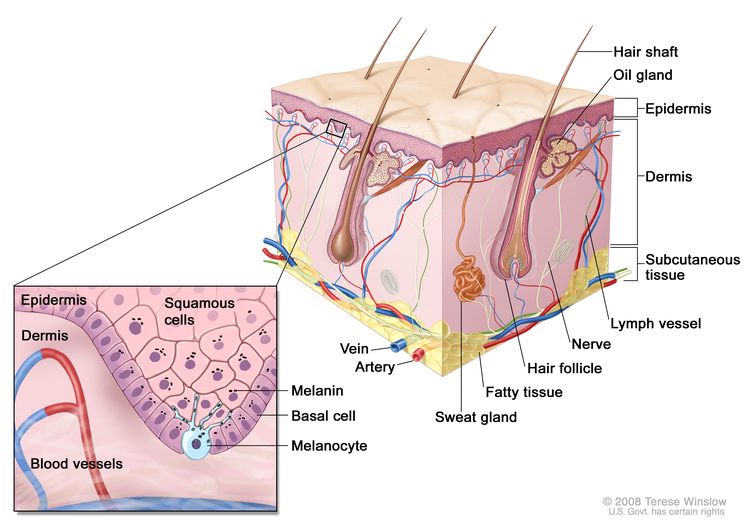
EnlargeAnatomy of the skin with melanocytes; drawing shows normal skin anatomy, including the epidermis, dermis, hair follicles, sweat glands, hair shafts, veins, arteries, fatty tissue, nerves, lymph vessels, oil glands, and subcutaneous tissue. The pullout shows a close-up of the squamous cell and basal cell layers of the epidermis above the dermis with blood vessels. Melanin is shown in the cells. A melanocyte is shown in the layer of basal cells at the deepest part of the epidermis.
Anatomy of the skin, showing the epidermis, dermis, and subcutaneous tissue. Melanocytes are in the layer of basal cells at the deepest part of the epidermis.

In the United States, about 3 million people are diagnosed with nonmelanoma skin cancer each year. Rates of nonmelanoma skin cancer have been increasing, possibly because greater public awareness has led to higher rates of screening exams, self-exams, and detection of these skin cancers.

Since the early 2000s, the rate of melanoma cases in adults younger than 50 years has held steady in women, but decreased by about 1% per year in men. In adults aged 50 years and older, the rate of melanoma cases has held steady in men, but increased by about 3% per year in women in recent years. From 2013 to 2022, the number of deaths from melanoma decreased by about 3% per year in men and 4% per year in women.

The rate of melanoma cases in children and adolescents increased until 2001. However, between 2001 and 2022, the yearly rates of melanoma in these age groups decreased slightly.

Other PDQ summaries containing information related to skin cancer include:

Different factors increase or decrease the risk of skin cancer.

Anything that increases your chance of getting a disease is called a risk factor. Anything that decreases your chance of getting a disease is called a protective factor.

For information about risk and protective factors for skin cancer, visit Skin Cancer Prevention.

Skin Cancer Screening

Key Points

  • Tests are used to screen for different types of cancer when a person does not have symptoms.
  • Screening for skin cancer may include examination by both the patient and the health care provider.
  • Screening tests for skin cancer are being studied in clinical trials.

Tests are used to screen for different types of cancer when a person does not have symptoms.

Scientists study screening tests to find those with the fewest harms and most benefits. Cancer screening trials also are meant to show whether early detection (finding cancer before it causes symptoms) helps a person live longer or decreases a person’s chance of dying from the disease. For some types of cancer, the chance of recovery is better if the disease is found and treated at an early stage. There is not enough evidence to know if screening the population for skin cancer lowers the rates of death from the disease.

Screening for skin cancer may include examination by both the patient and the health care provider.

A visual self-exam by the patient and a clinical examination by the health care provider may be used to screen for skin cancer.

During a skin exam a doctor or nurse checks the skin for moles, birthmarks, or other pigmented areas that look abnormal in color, size, shape, or texture. Skin exams to screen for skin cancer have not been shown to decrease the number of deaths from the disease.

Regular skin checks by a doctor are important for people who have already had skin cancer. If you are checking your skin and find a worrisome change, you should report it to your doctor.

If an area on the skin looks abnormal, a biopsy is usually done. The doctor will remove as much of the suspicious tissue as possible with a local excision. A pathologist then looks at the tissue under a microscope to check for cancer cells. Because it is sometimes difficult to tell if a skin growth is benign (not cancer) or malignant (cancer), you may want to have the biopsy sample checked by a second pathologist.

Most melanomas in the skin can be seen by the naked eye. Usually, melanoma grows for a long time under the top layer of skin (the epidermis) but does not grow into the deeper layer (the dermis). This allows time for skin cancer to be found early. Melanoma is easier to cure if it is found before it spreads.

Mobile apps that evaluate skin lesions to detect skin cancer and malignant melanoma have been developed. However, these apps require further study in large-scale testing programs to find out if they are accurate and useful for skin cancer screening.

Screening tests for skin cancer are being studied in clinical trials.

Information about clinical trials supported by NCI can be found on NCI’s clinical trials search webpage. Clinical trials supported by other organizations can be found on the ClinicalTrials.gov website.

Risks of Skin Cancer Screening

Key Points

  • Screening tests have risks.
  • The risks of skin cancer screening tests include:
    • Finding skin cancer does not always improve health or help you live longer.
    • False-negative test results can occur.
    • False-positive test results can occur.
    • A biopsy may cause scarring.

Screening tests have risks.

Decisions about screening tests can be difficult. Not all screening tests are helpful and most have risks. Before having any screening test, you may want to discuss the test with your doctor. It is important to know the risks of the test and whether it has been proven to reduce the risk of dying from cancer.

The risks of skin cancer screening tests include:

Finding skin cancer does not always improve health or help you live longer.

Screening may not improve your health or help you live longer if you have advanced skin cancer.

Some cancers never cause symptoms or become life-threatening, but if found by a screening test, the cancer may be treated. Treatments for cancer may have serious side effects.

False-negative test results can occur.

Screening test results may appear to be normal even though cancer is present. A person who receives a false-negative test result (one that shows there is no cancer when there really is) may delay getting medical care even if there are symptoms.

False-positive test results can occur.

Screening test results may appear to be abnormal even though no cancer is present. A false-positive test result (one that shows there is cancer when there really isn’t) can cause anxiety and is usually followed by more tests (such as a biopsy), which also have risks.

A biopsy may cause scarring.

When a skin biopsy is done, the doctor will try to leave the smallest scar possible, but there is a risk of scarring and infection.

Talk to your doctor about your risk for skin cancer and your need for screening tests.

About This PDQ Summary

About PDQ

Physician Data Query (PDQ) is the National Cancer Institute’s (NCI’s) comprehensive cancer information database. The PDQ database contains summaries of the latest published information on cancer prevention, detection, genetics, treatment, supportive care, and complementary and alternative medicine. Most summaries come in two versions. The health professional versions have detailed information written in technical language. The patient versions are written in easy-to-understand, nontechnical language. Both versions have cancer information that is accurate and up to date and most versions are also available in Spanish.

PDQ is a service of the NCI. The NCI is part of the National Institutes of Health (NIH). NIH is the federal government’s center of biomedical research. The PDQ summaries are based on an independent review of the medical literature. They are not policy statements of the NCI or the NIH.

Purpose of This Summary

This PDQ cancer information summary has current information about skin cancer screening. It is meant to inform and help patients, families, and caregivers. It does not give formal guidelines or recommendations for making decisions about health care.

Reviewers and Updates

Editorial Boards write the PDQ cancer information summaries and keep them up to date. These Boards are made up of experts in cancer treatment and other specialties related to cancer. The summaries are reviewed regularly and changes are made when there is new information. The date on each summary (“Updated”) is the date of the most recent change.

The information in this patient summary was taken from the health professional version, which is reviewed regularly and updated as needed, by the PDQ Screening and Prevention Editorial Board.

Clinical Trial Information

A clinical trial is a study to answer a scientific question, such as whether one treatment is better than another. Trials are based on past studies and what has been learned in the laboratory. Each trial answers certain scientific questions in order to find new and better ways to help cancer patients. During treatment clinical trials, information is collected about the effects of a new treatment and how well it works. If a clinical trial shows that a new treatment is better than one currently being used, the new treatment may become “standard.” Patients may want to think about taking part in a clinical trial. Some clinical trials are open only to patients who have not started treatment.

Clinical trials can be found online at NCI’s website. For more information, call the Cancer Information Service (CIS), NCI’s contact center, at 1-800-4-CANCER (1-800-422-6237).

Permission to Use This Summary

PDQ is a registered trademark. The content of PDQ documents can be used freely as text. It cannot be identified as an NCI PDQ cancer information summary unless the whole summary is shown and it is updated regularly. However, a user would be allowed to write a sentence such as “NCI’s PDQ cancer information summary about breast cancer prevention states the risks in the following way: [include excerpt from the summary].”

The best way to cite this PDQ summary is:

PDQ® Screening and Prevention Editorial Board. PDQ Skin Cancer Screening. Bethesda, MD: National Cancer Institute. Updated <MM/DD/YYYY>. Available at: /types/skin/patient/skin-screening-pdq. Accessed <MM/DD/YYYY>. [PMID: 26389182]

Images in this summary are used with permission of the author(s), artist, and/or publisher for use in the PDQ summaries only. If you want to use an image from a PDQ summary and you are not using the whole summary, you must get permission from the owner. It cannot be given by the National Cancer Institute. Information about using the images in this summary, along with many other images related to cancer can be found in Visuals Online. Visuals Online is a collection of more than 3,000 scientific images.

Disclaimer

The information in these summaries should not be used to make decisions about insurance reimbursement. More information on insurance coverage is available on Cancer.gov on the Managing Cancer Care page.

Contact Us

More information about contacting us or receiving help with the Cancer.gov website can be found on our Contact Us for Help page. Questions can also be submitted to Cancer.gov through the website’s E-mail Us.

Skin Cancer Prevention (PDQ®)–Patient Version

Skin Cancer Prevention (PDQ®)–Patient Version

What Is Prevention?

Cancer prevention is action taken to lower the chance of getting cancer. By preventing cancer, the number of new cases of cancer in a group or population is lowered. Hopefully, this will lower the number of deaths caused by cancer.

To prevent new cancers from starting, scientists look at risk factors and protective factors. Anything that increases your chance of developing cancer is called a cancer risk factor; anything that decreases your chance of developing cancer is called a cancer protective factor.

Some risk factors for cancer can be avoided, but many cannot. For example, both smoking and inheriting certain genes are risk factors for some types of cancer, but only smoking can be avoided. Regular exercise and a healthy diet may be protective factors for some types of cancer. Avoiding risk factors and increasing protective factors may lower your risk, but it does not mean that you will not get cancer.

Different ways to prevent cancer are being studied.

General Information About Skin Cancer

Key Points

  • Skin cancer is a disease in which malignant (cancer) cells form in the tissues of the skin.
  • There are several types of skin cancer.
  • Skin cancer is the most common cancer in the United States.

Skin cancer is a disease in which malignant (cancer) cells form in the tissues of the skin.

The skin is the body’s largest organ. It protects against heat, sunlight, injury, and infection. Skin also helps control body temperature and stores water, fat, and vitamin D. The skin has several layers, but the two main layers are the epidermis (upper or outer layer) and the dermis (lower or inner layer).

The epidermis is made up of three kinds of cells:

  • Squamous cells are the thin, flat cells that make up most of the epidermis.
  • Basal cells are the round cells under the squamous cells.
  • Melanocytes are found throughout the lower part of the epidermis. They make melanin, the pigment that gives skin its natural color. When skin is exposed to the sun, melanocytes make more pigment, causing the skin to tan, or darken.

The dermis contains blood and lymph vessels, hair follicles, and glands.

EnlargeAnatomy of the skin with melanocytes; drawing shows normal skin anatomy, including the epidermis, dermis, hair follicles, sweat glands, hair shafts, veins, arteries, fatty tissue, nerves, lymph vessels, oil glands, and subcutaneous tissue. The pullout shows a close-up of the squamous cell and basal cell layers of the epidermis above the dermis with blood vessels. Melanin is shown in the cells. A melanocyte is shown in the layer of basal cells at the deepest part of the epidermis.
Anatomy of the skin, showing the epidermis, dermis, and subcutaneous tissue. Melanocytes are in the layer of basal cells at the deepest part of the epidermis.

Other PDQ summaries containing information related to skin cancer include:

There are several types of skin cancer.

There are two main types of skin cancer:

The most common types of skin cancer are squamous cell carcinoma, which forms in the squamous cells, and basal cell carcinoma, which forms in the basal cells. Melanoma, which forms in the melanocytes, is a less common type of skin cancer that grows and spreads quickly.

Skin cancer can occur anywhere on the body, but it is most common in areas exposed to sunlight, such as the face, neck, hands, and arms.

Skin cancer is the most common cancer in the United States.

Nonmelanoma skin cancer (squamous cell carcinoma and basal cell carcinoma) is the most common type of skin cancer in the United States. New cases of nonmelanoma skin cancer appear to be increasing every year. Nonmelanoma skin cancer can usually be cured.

On the other hand, melanoma is more likely to spread to nearby tissues and other parts of the body and can be harder to cure. Finding and treating melanoma skin cancer early may help prevent death from melanoma.

Skin Cancer Prevention

Key Points

  • Avoiding risk factors and increasing protective factors may help prevent cancer.
  • Being exposed to ultraviolet radiation is a risk factor for skin cancer.
  • Treatment of sun-damaged skin to prevent skin cancer:
    • Topical fluorouracil
  • It is not known if the following lower the risk of nonmelanoma skin cancer:
    • Sunscreen use and avoiding sun exposure
    • Chemopreventive agents
  • It is not known if the following lower the risk of melanoma:
    • Sunscreen
    • Counseling and protecting the skin from the sun
  • Cancer prevention clinical trials are used to study ways to prevent cancer.
  • New ways to prevent skin cancer are being studied in clinical trials.

Avoiding risk factors and increasing protective factors may help prevent cancer.

Avoiding cancer risk factors may help prevent certain cancers. Risk factors include smoking, having overweight, and not getting enough exercise. Increasing protective factors such as quitting smoking and exercising may also help prevent some cancers. Talk to your doctor or other health care professional about how you might lower your risk of cancer.

Being exposed to ultraviolet radiation is a risk factor for skin cancer.

Some studies suggest that being exposed to ultraviolet (UV) radiation and the sensitivity of a person’s skin to UV radiation are risk factors for skin cancer. UV radiation is the name for the invisible rays that are part of the energy that comes from the sun. Sunlamps and tanning beds also give off UV radiation.

Risk factors for nonmelanoma and melanoma cancers are not the same.

  • Risk factors for nonmelanoma skin cancer:
    • being exposed to natural sunlight or artificial sunlight (such as from tanning beds) over long periods of time
    • having a fair complexion, which includes:
      • fair skin that freckles and burns easily, does not tan, or tans poorly
      • blue or green or other light-colored eyes
      • red or blond hair
    • having actinic keratosis
    • past treatment with radiation
    • having a weakened immune system, including people treated with immunosuppressive therapy after organ transplant
    • being exposed to arsenic
  • Risk factors for melanoma skin cancer:
    • having a fair complexion, which includes:
      • fair skin that freckles and burns easily, does not tan, or tans poorly
      • blue or green or other light-colored eyes
      • red or blond hair
    • being exposed to natural sunlight or artificial sunlight (such as from tanning beds) over long periods of time
    • having a history of many blistering sunburns, especially as a child or teenager
    • having many moles (also called nevi)
    • having a family history of unusual moles (atypical nevus syndrome)
    • having a family or personal history of melanoma
    • being White

Although having a fair complexion is a risk factor for nonmelanoma and melanoma skin cancer, people of all skin colors can get skin cancer.

Treatment of sun-damaged skin to prevent skin cancer:

Topical fluorouracil

A study showed that topical fluorouracil applied on sun-damaged skin daily for up to 4 weeks prevented new actinic keratoses from developing. The areas treated with topical fluorouracil had a lower risk of developing into squamous cell carcinoma that would require surgery. The lower risk of developing into squamous cell carcinoma was seen for 1 year after treatment. Topical fluorouracil did not, however, change the risk of developing basal cell carcinoma.

It is not known if the following lower the risk of nonmelanoma skin cancer:

Sunscreen use and avoiding sun exposure

It is not known if nonmelanoma skin cancer risk is decreased by staying out of the sun, using sunscreens, or wearing protective clothing when outdoors. This is because not enough studies have been done to prove this.

Sunscreen has been shown to prevent sunburns and actinic keratoses that may become squamous cell carcinoma, and to decrease the signs and symptoms of existing actinic keratoses.

The harms of using sunscreen are likely to be small and include allergic reactions to skin creams and lower levels of vitamin D made in the skin because of less sun exposure. It is also possible that when a person uses sunscreen to avoid sunburn, they may spend too much time in the sun and be exposed to harmful UV radiation.

Although protecting the skin and eyes from the sun has not been proven to lower the chance of getting skin cancer, skin experts suggest:

  • using sunscreen that protects against UV radiation
  • limiting time in the sun, especially when the sun is at its strongest
  • wearing long sleeve shirts, long pants, sun hats, and sunglasses when outdoors

Chemopreventive agents

Chemoprevention is the use of drugs, vitamins, or other agents to try to reduce the risk of cancer. The following chemopreventive agents have been studied to find whether they lower the risk of nonmelanoma skin cancer:

Beta carotene

Studies of beta carotene (taken as a supplement in pills) have not shown that it prevents nonmelanoma skin cancer from forming or coming back.

Isotretinoin and related retinoids

Retinoids are vitamin A or vitamin A-like compounds that are applied to the skin or taken by mouth. Isotretinoin is a type of retinoid being studied in the prevention and treatment of certain cancers.

High doses of isotretinoin taken by mouth have been shown to prevent new skin cancers in people with xeroderma pigmentosum. Isotretinoin cream has not been shown to prevent nonmelanoma skin cancers from coming back in people previously treated for nonmelanoma skin cancers. These treatments can cause serious side effects.

Selenium

Studies have shown that selenium (taken in brewer’s yeast tablets) does not lower the risk of basal cell carcinoma, and may increase the risk of squamous cell carcinoma.

Celecoxib

A study of celecoxib, a nonsteroidal anti-inflammatory drug (NSAID), in people with actinic keratosis and a history of nonmelanoma skin cancer found those who took celecoxib had slightly lower rates of recurrent nonmelanoma skin cancers. Celecoxib may cause serious heart and blood vessel side effects.

Alpha-difluoromethylornithine (DFMO)

A study of alpha-difluoromethylornithine (DFMO) in people with a history of nonmelanoma skin cancer showed that those who took DFMO had lower rates of basal cell carcinomas than those who took a placebo, but no difference in squamous cell carcinoma rates. However, those who took DFMO had greater hearing loss than the placebo group, leading to a higher rate of people discontinuing this drug.

Nicotinamide (vitamin B3)

Studies have shown that nicotinamide (vitamin B3) could help prevent new actinic keratoses lesions from forming in people who had four or fewer actinic lesions before taking nicotinamide. However, one study also showed an increased incidence of nonmelanoma skin cancers in people months after they were treated with nicotinamide. More studies are needed to find out if nicotinamide prevents nonmelanoma skin cancer from forming or coming back.

It is not known if the following lower the risk of melanoma:

Sunscreen

It has not been proven that using sunscreen to prevent sunburn can protect against melanoma caused by UV radiation. Other risk factors such as having skin that burns easily, having many benign (noncancerous) moles, or having atypical nevi may also play a role in whether melanoma forms.

Counseling and protecting the skin from the sun

Studies show that people who receive counseling or information about avoiding sun exposure improve their sun protective habits. These studies show mixed effects on reducing sunburns and do not show whether skin cancers are reduced.

Harms of avoiding sun exposure may include mood disorders, sleep disturbances, higher blood pressure, and impaired vitamin D metabolism.

Cancer prevention clinical trials are used to study ways to prevent cancer.

Cancer prevention clinical trials are used to study ways to lower the risk of developing certain types of cancer. Some cancer prevention trials include healthy people who may or may not have an increased risk of cancer. Other prevention trials include people who have had cancer and are trying to prevent recurrence or a second cancer.

The purpose of some cancer prevention clinical trials is to find out whether actions people take can prevent cancer. These may include eating fruits and vegetables, exercising, quitting smoking, or taking certain medicines, vitamins, minerals, or food supplements.

New ways to prevent skin cancer are being studied in clinical trials.

Information about clinical trials supported by NCI can be found on NCI’s clinical trials search webpage. Clinical trials supported by other organizations can be found on the ClinicalTrials.gov website.

About This PDQ Summary

About PDQ

Physician Data Query (PDQ) is the National Cancer Institute’s (NCI’s) comprehensive cancer information database. The PDQ database contains summaries of the latest published information on cancer prevention, detection, genetics, treatment, supportive care, and complementary and alternative medicine. Most summaries come in two versions. The health professional versions have detailed information written in technical language. The patient versions are written in easy-to-understand, nontechnical language. Both versions have cancer information that is accurate and up to date and most versions are also available in Spanish.

PDQ is a service of the NCI. The NCI is part of the National Institutes of Health (NIH). NIH is the federal government’s center of biomedical research. The PDQ summaries are based on an independent review of the medical literature. They are not policy statements of the NCI or the NIH.

Purpose of This Summary

This PDQ cancer information summary has current information about skin cancer prevention. It is meant to inform and help patients, families, and caregivers. It does not give formal guidelines or recommendations for making decisions about health care.

Reviewers and Updates

Editorial Boards write the PDQ cancer information summaries and keep them up to date. These Boards are made up of experts in cancer treatment and other specialties related to cancer. The summaries are reviewed regularly and changes are made when there is new information. The date on each summary (“Updated”) is the date of the most recent change.

The information in this patient summary was taken from the health professional version, which is reviewed regularly and updated as needed, by the PDQ Screening and Prevention Editorial Board.

Clinical Trial Information

A clinical trial is a study to answer a scientific question, such as whether one treatment is better than another. Trials are based on past studies and what has been learned in the laboratory. Each trial answers certain scientific questions in order to find new and better ways to help cancer patients. During treatment clinical trials, information is collected about the effects of a new treatment and how well it works. If a clinical trial shows that a new treatment is better than one currently being used, the new treatment may become “standard.” Patients may want to think about taking part in a clinical trial. Some clinical trials are open only to patients who have not started treatment.

Clinical trials can be found online at NCI’s website. For more information, call the Cancer Information Service (CIS), NCI’s contact center, at 1-800-4-CANCER (1-800-422-6237).

Permission to Use This Summary

PDQ is a registered trademark. The content of PDQ documents can be used freely as text. It cannot be identified as an NCI PDQ cancer information summary unless the whole summary is shown and it is updated regularly. However, a user would be allowed to write a sentence such as “NCI’s PDQ cancer information summary about breast cancer prevention states the risks in the following way: [include excerpt from the summary].”

The best way to cite this PDQ summary is:

PDQ® Screening and Prevention Editorial Board. PDQ Skin Cancer Prevention. Bethesda, MD: National Cancer Institute. Updated <MM/DD/YYYY>. Available at: /types/skin/patient/skin-prevention-pdq. Accessed <MM/DD/YYYY>. [PMID: 26389434]

Images in this summary are used with permission of the author(s), artist, and/or publisher for use in the PDQ summaries only. If you want to use an image from a PDQ summary and you are not using the whole summary, you must get permission from the owner. It cannot be given by the National Cancer Institute. Information about using the images in this summary, along with many other images related to cancer can be found in Visuals Online. Visuals Online is a collection of more than 3,000 scientific images.

Disclaimer

The information in these summaries should not be used to make decisions about insurance reimbursement. More information on insurance coverage is available on Cancer.gov on the Managing Cancer Care page.

Contact Us

More information about contacting us or receiving help with the Cancer.gov website can be found on our Contact Us for Help page. Questions can also be submitted to Cancer.gov through the website’s E-mail Us.