Transitional cell cancer of the renal pelvis and ureter is a type of cancer that forms in the transitional cells of the renal pelvis and ureter.
A personal history of bladder cancer and smoking can increase the risk of transitional cell cancer of the renal pelvis and ureter.
Signs and symptoms of transitional cell cancer of the renal pelvis and ureter include blood in the urine and back pain.
Tests that examine the abdomen and kidneys are used to diagnose transitional cell cancer of the renal pelvis and ureter.
After transitional cell cancer of the renal pelvis and ureter has been diagnosed, tests are done to find out if cancer cells have spread within the renal pelvis and ureter or to other parts of the body.
Some people decide to get a second opinion.
Certain factors affect prognosis (chance of recovery) and treatment options.
Transitional cell cancer of the renal pelvis and ureter is a type of cancer that forms in the transitional cells of the renal pelvis and ureter.
Transitional cells are a type of cell that lines the inside of the renal pelvis, ureters, and other organs. These cells are able to stretch when the renal pelvis or bladder is full of urine and shrink when it is emptied. Transitional cell cancer can form in the renal pelvis, the ureters, or both.
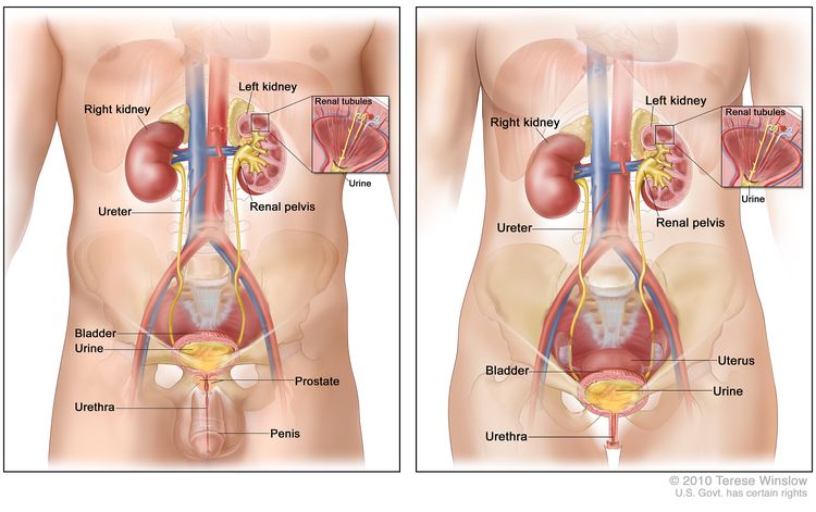
The renal pelvis is the area at the center of the kidney. It collects urine before it flows into the ureters, which are long tubes that connect the kidney to the bladder. There are two kidneys, one on each side of the backbone, above the waist. The kidneys of an adult are about 5 inches long and 3 inches wide and are shaped like a kidney bean.
The kidneys play an important role in the urinary system:
The kidneys filter and clean the blood by taking out waste products and extra water as urine.
The urine collects in the middle of each kidney in the renal pelvis.
Urine passes from the renal pelvis through the ureter into the bladder.
The bladder holds the urine until it passes through the urethra and leaves the body.
EnlargeAnatomy of the male urinary system (left panel) and female urinary system (right panel) showing the kidneys, ureters, bladder, and urethra. The inside of the left kidney shows the renal pelvis. An inset shows the renal tubules and urine. Also shown are the prostate and penis (left panel) and the uterus (right panel). Urine is made in the renal tubules and collects in the renal pelvis of each kidney. The urine flows from the kidneys through the ureters to the bladder. The urine is stored in the bladder until it leaves the body through the urethra.
A personal history of bladder cancer and smoking can increase the risk of transitional cell cancer of the renal pelvis and ureter.
Transitional cell cancer of the renal pelvis and ureter is caused by certain changes to the way transitional cells in the renal pelvis and ureters function, especially how they grow and divide into new cells. There are many risk factors for transitional cell cancer of the renal pelvis and ureter, but many do not directly cause cancer. Instead, they increase the chance of DNA damage in cells that may lead to cancer. To learn more about how cancer develops, see What Is Cancer?
A risk factor is anything that increases the chance of getting a disease. Some risk factors for transitional cell cancer of the renal pelvis and ureter, like smoking, can be changed. However, risk factors may also include things you cannot change, like your personal medical history. Learning about risk factors for transitional cell cancer of the renal pelvis and ureter can help you make changes that might lower your risk of getting it.
Risk factors for transitional cell cancer of the renal pelvis and ureter include:
taking a lot of certain pain medicines, such as phenacetin
being exposed to certain dyes and chemicals used in making leather goods, textiles, plastics, and rubber
Having one or more of these risk factors does not mean that you will get transitional cell cancer of the renal pelvis and ureter. Many people with risk factors never develop this type of cancer, while others with no known risk factors do. Talk with your doctor if you think you might be at risk.
Signs and symptoms of transitional cell cancer of the renal pelvis and ureter include blood in the urine and back pain.
In the early stages, there may be no signs and symptoms of transitional cell cancer of the renal pelvis and ureter. Symptoms may appear as the tumor grows and may include:
blood in the urine
a pain in the back that doesn’t go away
extreme tiredness
weight loss with no known reason
painful or frequent urination
These symptoms may be caused by transitional cell cancer of the renal pelvis and ureter or by other conditions. It’s important to check with your doctor if you have any of these symptoms to find out the cause and begin treatment if needed.
Tests that examine the abdomen and kidneys are used to diagnose transitional cell cancer of the renal pelvis and ureter.
Urinalysis is a test to check the color of urine and its contents, such as sugar, protein, blood, and bacteria.
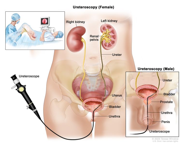
Ureteroscopy is a procedure to look inside the ureter and renal pelvis to check for abnormal areas. A ureteroscope is a thin, tube-like instrument with a light and a lens for viewing. The ureteroscope is inserted through the urethra into the bladder, ureter, and renal pelvis. A tool may be inserted through the ureteroscope to take tissue samples to be checked under a microscope for signs of disease. EnlargeA ureteroscopy is a procedure that uses a ureteroscope (a thin, tube-like instrument with a light and a lens for viewing) to look inside the ureter and renal pelvis to check for abnormal areas. The ureteroscope is inserted through the urethra into the bladder, ureter, and renal pelvis.
Urine cytology is a laboratory test in which a sample of urine is checked under a microscope for abnormal cells. Cancer in the kidney, bladder, or ureter may shed cancer cells into the urine.
CT scan (CAT scan) uses a computer linked to an x-ray machine to make a series of detailed pictures of areas inside the body from different angles. A dye may be injected into a vein or swallowed to help the organs or tissues show up more clearly. This procedure is also called computed tomography, computerized tomography, or computerized axial tomography.
Ultrasound uses high-energy sound waves (ultrasound) that bounce off internal tissues or organs and make echoes. The echoes form a picture of body tissues called a sonogram. An ultrasound of the abdomen may be done to help diagnose cancer of the renal pelvis and ureter.
MRI (magnetic resonance imaging) uses a magnet, radio waves, and a computer to make a series of detailed pictures of areas inside the body, such as the pelvis. This procedure is also called nuclear magnetic resonance imaging (NMRI).
Biopsy is the removal of cells or tissues so they can be viewed under a microscope by a pathologist to check for signs of cancer. This may be done during a ureteroscopy or surgery.
After transitional cell cancer of the renal pelvis and ureter has been diagnosed, tests are done to find out if cancer cells have spread within the renal pelvis and ureter or to other parts of the body.
The process used to find out if the cancer has spread within the renal pelvis and ureter or to other parts of the body is called staging. The information gathered from the staging process determines the stage of the disease. The results of the diagnostic and staging tests will also help you and your doctor plan treatment.
The following tests and procedures may also be used in the staging process:
Chest x-ray is an x-ray of the organs and bones inside the chest. An x-ray is a type of energy beam that can go through the body and onto film, making a picture of areas inside the body.
PET scan (positron emission tomography scan) uses a small amount of radioactive sugar (also called glucose) that is injected into a vein. Then a scanner is used to make detailed, computerized pictures of areas inside the body where the glucose is taken up. Because cancer cells often take up more glucose than normal cells, the pictures can be used to find cancer cells in the body.
Bone scan is used to check if there are rapidly dividing cells, such as cancer cells, in the bone. A very small amount of radioactive material is injected into a vein and travels through the bloodstream. The radioactive material collects in the bones with cancer and is detected by a scanner.
Some people decide to get a second opinion.
You may want to get a second opinion to confirm your cancer diagnosis and treatment plan. If you seek a second opinion, you will need to get medical test results and reports from the first doctor to share with the second doctor. The second doctor will review the pathology report, slides, and scans. They may agree with the first doctor, suggest changes or another treatment approach, or provide more information about your cancer.
To learn more about choosing a doctor and getting a second opinion, see Finding Cancer Care. You can contact NCI’s Cancer Information Service via chat, email, or phone (both in English and Spanish) for help finding a doctor, hospital, or getting a second opinion. For questions you might want to ask at your appointments, see Questions to Ask Your Doctor about Cancer.
Certain factors affect prognosis (chance of recovery) and treatment options.
The prognosis depends on the stage and grade of the tumor.
The treatment options depend on:
the stage and grade of the tumor
where the tumor is
whether the patient’s other kidney is healthy
whether the cancer has recurred (come back)
Most transitional cell cancer of the renal pelvis and ureter can be cured if found early.
Stages of Transitional Cell Cancer of the Renal Pelvis and Ureter
Key Points
The following stages are used for transitional cell cancer of the renal pelvis and/or ureter:
Stage 0 (Noninvasive Papillary Carcinoma and Carcinoma in Situ)
Stage I (also called stage 1)
Stage II (also called stage 2)
Stage III (also called stage 3)
Stage IV (also called stage 4)
Transitional cell cancer of the renal pelvis and ureter is also described as localized, regional, metastatic, or recurrent:
Localized
Regional
Metastatic
Recurrent
Cancer stage describes the extent of cancer in the body, such as the size of the tumor, whether it has spread, and how far it has spread from where it first formed. It is important to know the stage of transitional cell cancer of the renal pelvis and ureter to plan the best treatment.
There are several staging systems for cancer that describe the extent of the cancer. Transitional cell cancer of the renal pelvis and ureter staging usually uses the TNM staging system. You may see your cancer described by this staging system in your pathology report. Based on the TNM results, a stage (I, II, III, or IV, also written as 1, 2, 3, or 4) is assigned to your cancer. When talking to you about your cancer, your doctor may describe it as one of these stages.
The following stages are used for transitional cell cancer of the renal pelvis and/or ureter:
Stage 0 (Noninvasive Papillary Carcinoma and Carcinoma in Situ)
In stage 0, abnormal cells are found in tissue lining the inside of the renal pelvis or ureter. These abnormal cells may become cancer and spread into nearby normal tissue. Stage 0 is divided into stages 0a and 0is, depending on the type of tumor:
Stage 0a is also called noninvasive papillary carcinoma, which may look like long, thin growths that grow out from the tissue lining the inside of the renal pelvis or ureter.
Stage 0is is also called carcinoma in situ, which is a flat tumor on the tissue lining the inside of the renal pelvis or ureter.
Stage I (also called stage 1)
In stage I, cancer has formed and has spread from the tissue lining the inside of the renal pelvis or ureter to the connective tissue layer.
Stage II (also called stage 2)
In stage III, cancer has spread to the muscle layer of the renal pelvis or ureter.
Stage III (also called stage 3)
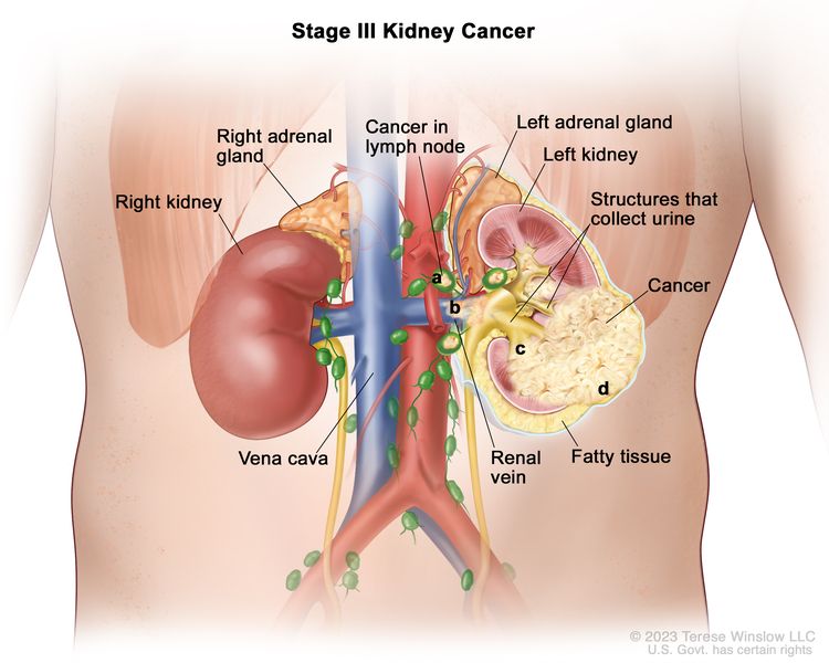
In stage III, cancer has spread:
from the muscle layer of the renal pelvis to fat around the renal pelvis or to tissue in the kidney; or
from the muscle layer of the ureter to fat around the ureter.
Stage IV (also called stage 4)
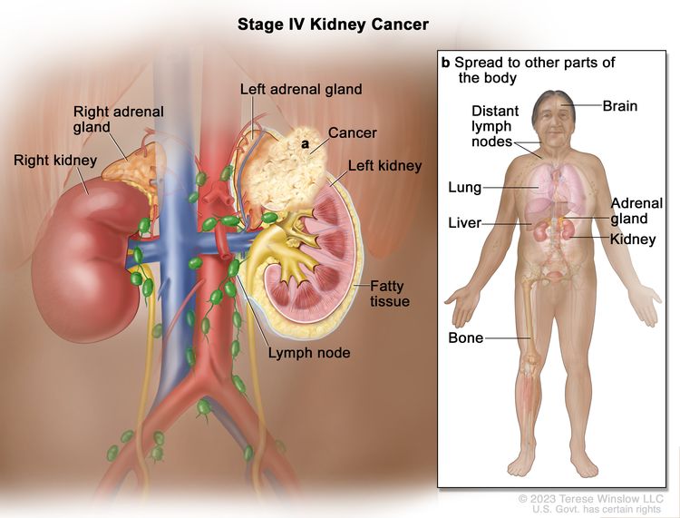
In stage IV, cancer has spread to at least one of the following:
other parts of the body, such as the lung, liver, or bone.
Stage IV transitional cell cancer of the renal pelvis and ureter is also called metastatic cancer. Metastatic cancer happens when cancer cells travel through the lymphatic system or blood and form tumors in other parts of the body. The metastatic tumor is the same type of cancer as the primary tumor. For example, if transitional cell cancer renal pelvis and ureter spreads to the liver, the cancer cells in the liver are actually transitional cell cancer of the renal pelvis and ureter. The disease is called metastatic transitional cell cancer of the renal pelvis and ureter, not liver cancer. Learn more in Metastatic Cancer: When Cancer Spreads.
Transitional cell cancer of the renal pelvis and ureter is also described as localized, regional, metastatic, or recurrent:
Recurrent cancer is cancer that has recurred (come back) after it has been treated. If transitional cell cancer of the renal pelvis and ureter comes back, it may come back in the renal pelvis, ureter, or in other parts of the body, such as the lung, liver, or bone. Tests will be done to help determine where the cancer has returned. The type of treatment for recurrent transitional cell cancer of the renal pelvis and ureter will depend on where it has come back.
Treatment Option Overview for Transitional Cell Cancer of the Renal Pelvis and Ureter
Key Points
There are different types of treatment for patients with transitional cell cancer of the renal pelvis and ureter.
The following type of treatment is used:
Surgery
New types of treatment are being tested in clinical trials.
Fulguration
Segmental resection of the renal pelvis
Treatment for transitional cell cancer of the renal pelvis and ureter may cause side effects.
Follow-up care may be needed.
There are different types of treatment for patients with transitional cell cancer of the renal pelvis and ureter.
Different types of treatments are available for people with transitional cell cancer of the renal pelvis and ureter. You and your cancer care team will work together to decide your treatment plan, which may include more than one type of treatment. Many factors will be considered, such as the stage of the cancer, your overall health, and your preferences. Your plan will include information about your cancer, the goals of treatment, your treatment options and the possible side effects, and the expected length of treatment.
Talking with your cancer care team before treatment begins about what to expect will be helpful. You’ll want to learn what you need to do before treatment begins, how you’ll feel while going through it, and what kind of help you will need. To learn more, see Questions to Ask Your Doctor about Treatment.
The following type of treatment is used:
Surgery
One of the following surgical procedures may be used to treat transitional cell cancer of the renal pelvis and ureter:
Nephroureterectomy is surgery to remove the entire kidney, the ureter, and the bladder cuff (tissue that connects the ureter to the bladder). Most people with transitional cell cancer of the renal pelvis and ureter undergo nephroureterectomy.
Segmental resection of the ureter is a surgical procedure to remove the part of the ureter that contains cancer and some of the healthy tissue around it. The ends of the ureter are then reattached. This treatment is used when the cancer is superficial and in the lower third of the ureter only, near the bladder.
New types of treatment are being tested in clinical trials.
For some people, joining a clinical trial may be an option. There are different types of clinical trials for people with cancer. For example, a treatment trial tests new treatments or new ways of using current treatments. Supportive care and palliative care trials look at ways to improve quality of life, especially for those who have side effects from cancer and its treatment.
You can use the clinical trial search to find NCI-supported cancer clinical trials accepting participants. The search allows you to filter trials based on the type of cancer, your age, and where the trials are being done. Clinical trials supported by other organizations can be found on the ClinicalTrials.gov website.
This summary section describes treatments that are being studied in clinical trials. It may not mention every new treatment being studied.
Fulguration
Fulguration is a surgical procedure that destroys tissue using an electric current. A tool with a small wire loop on the end is used to remove the cancer or to burn away the tumor with electricity.
Segmental resection of the renal pelvis
This is a surgical procedure to remove localized cancer from the renal pelvis without removing the entire kidney. Segmental resection may be done to save kidney function when the other kidney is damaged or has already been removed.
Laser surgery
A laser beam (narrow beam of intense light) is used as a knife to remove the cancer. A laser beam can also be used to kill the cancer cells. This procedure may also be called laser fulguration.
Regional treatment for transitional cell cancer of renal pelvis and ureter is a method of placing the chemotherapy or immunotherapy drug directly into the renal pelvis or the ureter to mainly affect cancer cells in those areas.
Treatment for transitional cell cancer of the renal pelvis and ureter may cause side effects.
As you go through treatment, you will have follow-up tests or check-ups. Some tests that were done to diagnose or stage the cancer may be repeated to see how well the treatment is working. Decisions about whether to continue, change, or stop treatment may be based on the results of these tests.
Some of the tests will continue to be done from time to time after treatment has ended. The results of these tests can show if your condition has changed or if the cancer has recurred (come back).
Treatment of Localized Transitional Cell Cancer of the Renal Pelvis and Ureter
Treatment of localized transitional cell cancer of the renal pelvis and ureter may include:
Use our clinical trial search to find NCI-supported cancer clinical trials that are accepting patients. You can search for trials based on the type of cancer, the age of the patient, and where the trials are being done. General information about clinical trials is also available.
Treatment of Regional Transitional Cell Cancer of the Renal Pelvis and Ureter
Treatment of regional transitional cell cancer of the renal pelvis and ureter is usually done in a clinical trial.
Use our clinical trial search to find NCI-supported cancer clinical trials that are accepting patients. You can search for trials based on the type of cancer, the age of the patient, and where the trials are being done. General information about clinical trials is also available.
Treatment of Metastatic or Recurrent Transitional Cell Cancer of the Renal Pelvis and Ureter
Treatment of metastatic or recurrent transitional cell cancer of the renal pelvis and ureter is usually done in a clinical trial, which may include chemotherapy.
Use our clinical trial search to find NCI-supported cancer clinical trials that are accepting patients. You can search for trials based on the type of cancer, the age of the patient, and where the trials are being done. General information about clinical trials is also available.
To Learn More About Transitional Cell Cancer of the Renal Pelvis and Ureter
For more information from the National Cancer Institute about transitional cell cancer of the renal pelvis and ureter, see:
Physician Data Query (PDQ) is the National Cancer Institute’s (NCI’s) comprehensive cancer information database. The PDQ database contains summaries of the latest published information on cancer prevention, detection, genetics, treatment, supportive care, and complementary and alternative medicine. Most summaries come in two versions. The health professional versions have detailed information written in technical language. The patient versions are written in easy-to-understand, nontechnical language. Both versions have cancer information that is accurate and up to date and most versions are also available in Spanish.
PDQ is a service of the NCI. The NCI is part of the National Institutes of Health (NIH). NIH is the federal government’s center of biomedical research. The PDQ summaries are based on an independent review of the medical literature. They are not policy statements of the NCI or the NIH.
Purpose of This Summary
This PDQ cancer information summary has current information about the treatment of transitional cell cancer of the renal pelvis and ureter. It is meant to inform and help patients, families, and caregivers. It does not give formal guidelines or recommendations for making decisions about health care.
Reviewers and Updates
Editorial Boards write the PDQ cancer information summaries and keep them up to date. These Boards are made up of experts in cancer treatment and other specialties related to cancer. The summaries are reviewed regularly and changes are made when there is new information. The date on each summary (“Updated”) is the date of the most recent change.
The information in this patient summary was taken from the health professional version, which is reviewed regularly and updated as needed, by the PDQ Adult Treatment Editorial Board.
Clinical Trial Information
A clinical trial is a study to answer a scientific question, such as whether one treatment is better than another. Trials are based on past studies and what has been learned in the laboratory. Each trial answers certain scientific questions in order to find new and better ways to help cancer patients. During treatment clinical trials, information is collected about the effects of a new treatment and how well it works. If a clinical trial shows that a new treatment is better than one currently being used, the new treatment may become “standard.” Patients may want to think about taking part in a clinical trial. Some clinical trials are open only to patients who have not started treatment.
Clinical trials can be found online at NCI’s website. For more information, call the Cancer Information Service (CIS), NCI’s contact center, at 1-800-4-CANCER (1-800-422-6237).
Permission to Use This Summary
PDQ is a registered trademark. The content of PDQ documents can be used freely as text. It cannot be identified as an NCI PDQ cancer information summary unless the whole summary is shown and it is updated regularly. However, a user would be allowed to write a sentence such as “NCI’s PDQ cancer information summary about breast cancer prevention states the risks in the following way: [include excerpt from the summary].”
The best way to cite this PDQ summary is:
PDQ® Adult Treatment Editorial Board. PDQ Transitional Cell Cancer of the Renal Pelvis and Ureter Treatment. Bethesda, MD: National Cancer Institute. Updated <MM/DD/YYYY>. Available at: /types/kidney/patient/transitional-cell-treatment-pdq. Accessed <MM/DD/YYYY>. [PMID: 26389285]
Images in this summary are used with permission of the author(s), artist, and/or publisher for use in the PDQ summaries only. If you want to use an image from a PDQ summary and you are not using the whole summary, you must get permission from the owner. It cannot be given by the National Cancer Institute. Information about using the images in this summary, along with many other images related to cancer can be found in Visuals Online. Visuals Online is a collection of more than 3,000 scientific images.
Disclaimer
The information in these summaries should not be used to make decisions about insurance reimbursement. More information on insurance coverage is available on Cancer.gov on the Managing Cancer Care page.
Contact Us
More information about contacting us or receiving help with the Cancer.gov website can be found on our Contact Us for Help page. Questions can also be submitted to Cancer.gov through the website’s E-mail Us.
Estimated new cases and deaths from renal cell (kidney and renal pelvis) cancer in the United States in 2025:[1]
New cases: 80,980.
Deaths: 14,510.
Follow-Up and Survivorship
Renal cell cancer, also called renal adenocarcinoma or hypernephroma, can often be cured if it is diagnosed and treated when still localized to the kidney and the immediately surrounding tissue. The probability of cure is directly related to the stage or degree of tumor dissemination. Even when regional lymphatics or blood vessels are involved with the tumor, a significant number of patients can achieve prolonged survival and probable cure.[2] When distant metastases are present, disease-free survival is poor; however, some patients survive after surgical resection of all known tumor. Because most patients are diagnosed when the tumor is still relatively localized and amenable to surgical removal, approximately 75% of all patients with renal cell cancer survive for 5 years.[3] Occasionally, patients with locally advanced or metastatic disease may exhibit indolent courses lasting several years. Late tumor recurrence many years after initial treatment also occasionally occurs.
Renal cell cancer is one of the few tumors in which well-documented cases of spontaneous tumor regression in the absence of therapy exist, but this regression occurs rarely and may not lead to long-term survival.
Treatment Modalities
Surgical resection is the mainstay of treatment of this disease. Even in patients with disseminated tumor, locoregional forms of therapy may play an important role in palliating symptoms of the primary tumor or of ectopic hormone production. Systemic therapy has demonstrated only limited effectiveness.
References
American Cancer Society: Cancer Facts and Figures 2025. American Cancer Society, 2025. Available online. Last accessed January 16, 2025.
Sene AP, Hunt L, McMahon RF, et al.: Renal carcinoma in patients undergoing nephrectomy: analysis of survival and prognostic factors. Br J Urol 70 (2): 125-34, 1992. [PUBMED Abstract]
National Cancer Institute: SEER Stat Fact Sheets: Kidney and Renal Pelvis Cancer. Bethesda, Md: National Cancer Institute. Available online. Last accessed February 17, 2025.
Cellular Classification of Renal Cell Cancer
Approximately 85% of renal cell cancers are adenocarcinomas, mostly of proximal tubular origin. Most of the remainder are transitional cell carcinomas of the renal pelvis. For more information, see Transitional Cell Cancer of the Renal Pelvis and Ureter Treatment. Adenocarcinomas may be separated into clear cell and granular cell carcinomas; however, the two cell types may occur together in some tumors. Some investigators have found that granular cell tumors have a worse prognosis, but this finding is not universal. Distinguishing between well-differentiated renal adenocarcinomas and renal adenomas can be difficult. The diagnosis is usually made arbitrarily based on the size of the mass, but size alone should not influence the treatment approach because metastases can occur with lesions as small as 0.5 cm.
Stage Information for Renal Cell Cancer
The staging system for renal cell cancer is based on the degree of tumor spread beyond the kidney.[1–3] Involvement of blood vessels may not be a poor prognostic sign if the tumor is otherwise confined to the substance of the kidney. Abnormal liver function test results may be caused by a paraneoplastic syndrome that is reversible with tumor removal, and these types of results do not necessarily represent metastatic disease. Except when computed tomography (CT) examination is equivocal or when iodinated contrast material is contraindicated, CT scanning is as good as or better than magnetic resonance imaging for detecting renal masses.[4]
AJCC Stage Groupings and TNM Definitions
The American Joint Committee on Cancer (AJCC) has designated staging by TNM (tumor, node, metastasis) classification to define renal cell cancer.[5]
Table 1. Definitions of TNM Stage Ia
Stage
TNM
Definition
Illustration
T = primary tumor; N = regional lymph node; M = distant metastasis.
aReprinted with permission from AJCC: Kidney. In: Amin MB, Edge SB, Greene FL, et al., eds.: AJCC Cancer Staging Manual. 8th ed. New York, NY: Springer, 2017, pp. 739–48.
I
T1, N0, M0
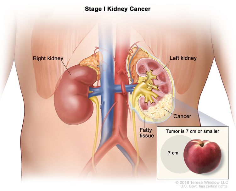
T1 = Tumor ≤7 cm in greatest dimension, limited to the kidney.
–T1a = Tumor ≤4 cm in greatest dimension, limited to the kidney.
–T1b = Tumor >4 cm but ≤7 cm in greatest dimension, limited to the kidney.
N0 = No regional lymph node metastasis.
M0 = No distant metastasis.
Table 2. Definitions of TNM Stage IIa
Stage
TNM
Definition
Illustration
T = primary tumor; N = regional lymph node; M = distant metastasis.
aReprinted with permission from AJCC: Kidney. In: Amin MB, Edge SB, Greene FL, et al., eds.: AJCC Cancer Staging Manual. 8th ed. New York, NY: Springer, 2017, pp. 739–48.
II
T2, N0, M0
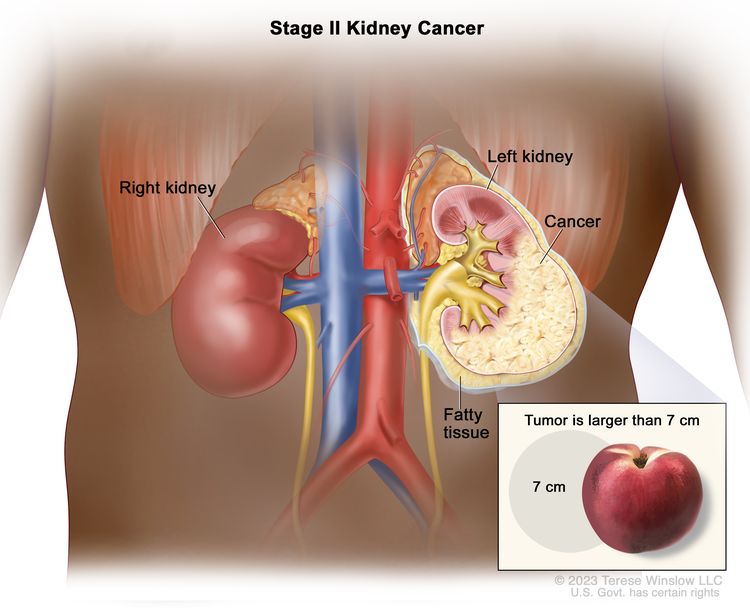
T2 = Tumor >7 cm in greatest dimension, limited to the kidney.
–T2a = Tumor >7 cm but ≤10 cm in greatest dimension, limited to the kidney.
–T2b = Tumor >10 cm, limited to the kidney.
N0 = No regional lymph node metastasis.
M0 = No distant metastasis.
Table 3. Definitions of TNM Stage IIIa
Stage
TNM
Definition
Illustration
T = primary tumor; N = regional lymph node; M = distant metastasis.
aReprinted with permission from AJCC: Kidney. In: Amin MB, Edge SB, Greene FL, et al., eds.: AJCC Cancer Staging Manual. 8th ed. New York, NY: Springer, 2017, pp. 739–48.
III
T1, N1, M0
T1 = Tumor ≤7 cm in greatest dimension, limited to the kidney.
–T1a = Tumor ≤4 cm in greatest dimension, limited to the kidney.
–T1b = Tumor >4 cm but ≤7 cm in greatest dimension, limited to the kidney.
N1 = Metastasis in regional lymph node(s).
M0 = No distant metastasis.
T2, N1, M0
T2 = Tumor >7 cm in greatest dimension, limited to the kidney.
–T2a = Tumor >7 cm but ≤10 cm in greatest dimension, limited to the kidney.
–T2b = Tumor >10 cm, limited to the kidney.
N1 = Metastasis in regional lymph node(s).
M0 = No distant metastasis.
T3, N0, M0
T3 = Tumor extends into major veins or perinephric tissues but not into the ipsilateral adrenal gland and not beyond Gerota’s fascia.
–T3a = Tumor extends into the renal vein or its segmental branches, or invades the pelvicalyceal system, or invades perirenal and/or renal sinus fat but not beyond Gerota’s fascia.
–T3b = Tumor extends into the vena cava below the diaphragm.
–T3c = Tumor extends into the vena cava above the diaphragm or invades the wall of the vena cava.
N0 = No regional lymph node metastasis.
M0 = No distant metastasis.
T3, N1, M0
T3 = Tumor extends into major veins or perinephric tissues but not into the ipsilateral adrenal gland and not beyond Gerota’s fascia.
–T3a = Tumor extends into the renal vein or its segmental branches, or invades the pelvicalyceal system, or invades perirenal and/or renal sinus fat but not beyond Gerota’s fascia.
–T3b = Tumor extends into the vena cava below the diaphragm.
–T3c = Tumor extends into the vena cava above the diaphragm or invades the wall of the vena cava.
N1 = Metastasis in regional lymph node(s).
M0 = No distant metastasis.
Table 4. Definitions of TNM Stage IVa
Stage
TNM
Definition
Illustration
T = primary tumor; N = regional lymph node; M = distant metastasis.
aReprinted with permission from AJCC: Kidney. In: Amin MB, Edge SB, Greene FL, et al., eds.: AJCC Cancer Staging Manual. 8th ed. New York, NY: Springer, 2017, pp. 739–48.
IV
T4, Any N, M0
T4 = Tumor invades beyond Gerota’s fascia (including contiguous extension into the ipsilateral adrenal gland).
T1 = Tumor ≤7 cm in greatest dimension, limited to the kidney.
–T1a = Tumor ≤4 cm in greatest dimension, limited to the kidney.
–T1b = Tumor >4 cm but ≤7 cm in greatest dimension, limited to the kidney.
T2 = Tumor >7 cm in greatest dimension, limited to the kidney.
–T2a = Tumor >7 cm but ≤10 cm in greatest dimension, limited to the kidney.
–T2b = Tumor >10 cm, limited to the kidney.
T3 = Tumor extends into major veins or perinephric tissues but not into the ipsilateral adrenal gland and not beyond Gerota’s fascia.
–T3a = Tumor extends into the renal vein or its segmental branches, or invades the pelvicalyceal system, or invades perirenal and/or renal sinus fat but not beyond Gerota’s fascia.
–T3b = Tumor extends into the vena cava below the diaphragm.
–T3c = Tumor extends into the vena cava above the diaphragm or invades the wall of the vena cava.
T4 = Tumor invades beyond Gerota’s fascia (including contiguous extension into the ipsilateral adrenal gland).
NX = Regional lymph nodes cannot be assessed.
N0 = No regional lymph node metastasis.
N1 = Metastasis in regional lymph node(s).
M1 = Distant metastasis.
References
Bassil B, Dosoretz DE, Prout GR: Validation of the tumor, nodes and metastasis classification of renal cell carcinoma. J Urol 134 (3): 450-4, 1985. [PUBMED Abstract]
Golimbu M, Joshi P, Sperber A, et al.: Renal cell carcinoma: survival and prognostic factors. Urology 27 (4): 291-301, 1986. [PUBMED Abstract]
Robson CJ, Churchill BM, Anderson W: The results of radical nephrectomy for renal cell carcinoma. J Urol 101 (3): 297-301, 1969. [PUBMED Abstract]
Current treatment cures more than 50% of patients with stage I renal cell cancer, but patients with stage IV disease have poor outcomes. All patients with newly diagnosed renal cell cancer are candidates for clinical trials.
Treatment of Stage I Renal Cell Cancer
Treatment Options for Stage I Renal Cell Cancer
Treatment options for stage I renal cell cancer include:
Surgical resection is the accepted, often curative therapy for patients with stage I renal cell cancer. In appropriately selected patients, partial nephrectomy has oncologic outcomes that are comparable with those of radical nephrectomy. Partial nephrectomy also has the benefits of preserving greater renal function and, according to some studies, reduced mortality.[4–7] When the entire kidney is removed, resection may be simple or radical. Radical resection includes removal of the kidney, adrenal gland, perirenal fat, and Gerota’s fascia, with or without a regional lymph node dissection. Radical nephrectomy is preferred if the tumor extends into the inferior vena cava.
In patients with bilateral stage I neoplasms (concurrent or subsequent), bilateral partial nephrectomy or unilateral partial nephrectomy with contralateral radical nephrectomy, when technically feasible, may be a preferred alternative to bilateral nephrectomy with dialysis or transplant.[1] Increasing evidence suggests that a partial nephrectomy is curative in selected cases. A pathologist should examine the gross specimen as well as the frozen section from the parenchymal margin of excision.[8]
In patients who are not candidates for resection, there are several curative alternatives, including cryoablation, thermal ablation, and SABR (or SBRT).[9–13]
In patients who are not candidates for the treatments above, EBRT or arterial embolization can provide palliation.
Current Clinical Trials
Use our advanced clinical trial search to find NCI-supported cancer clinical trials that are now enrolling patients. The search can be narrowed by location of the trial, type of treatment, name of the drug, and other criteria. General information about clinical trials is also available.
References
Novick AC, Streem S, Montie JE, et al.: Conservative surgery for renal cell carcinoma: a single-center experience with 100 patients. J Urol 141 (4): 835-9, 1989. [PUBMED Abstract]
deKernion JB, Berry D: The diagnosis and treatment of renal cell carcinoma. Cancer 45 (7 Suppl): 1947-56, 1980. [PUBMED Abstract]
Swanson DA, Wallace S, Johnson DE: The role of embolization and nephrectomy in the treatment of metastatic renal carcinoma. Urol Clin North Am 7 (3): 719-30, 1980. [PUBMED Abstract]
Kim SP, Thompson RH, Boorjian SA, et al.: Comparative effectiveness for survival and renal function of partial and radical nephrectomy for localized renal tumors: a systematic review and meta-analysis. J Urol 188 (1): 51-7, 2012. [PUBMED Abstract]
Larcher A, Capitanio U, Terrone C, et al.: Elective Nephron Sparing Surgery Decreases Other Cause Mortality Relative to Radical Nephrectomy Only in Specific Subgroups of Patients with Renal Cell Carcinoma. J Urol 196 (4): 1008-13, 2016. [PUBMED Abstract]
Wang Z, Wang G, Xia Q, et al.: Partial nephrectomy vs. radical nephrectomy for renal tumors: A meta-analysis of renal function and cardiovascular outcomes. Urol Oncol 34 (12): 533.e11-533.e19, 2016. [PUBMED Abstract]
Patel HD, Pierorazio PM, Johnson MH, et al.: Renal Functional Outcomes after Surgery, Ablation, and Active Surveillance of Localized Renal Tumors: A Systematic Review and Meta-Analysis. Clin J Am Soc Nephrol 12 (7): 1057-1069, 2017. [PUBMED Abstract]
Thrasher JB, Robertson JE, Paulson DF: Expanding indications for conservative renal surgery in renal cell carcinoma. Urology 43 (2): 160-8, 1994. [PUBMED Abstract]
El Dib R, Touma NJ, Kapoor A: Cryoablation vs radiofrequency ablation for the treatment of renal cell carcinoma: a meta-analysis of case series studies. BJU Int 110 (4): 510-6, 2012. [PUBMED Abstract]
Gkentzis A, Oades G: Thermal ablative therapies for treatment of localised renal cell carcinoma: a systematic review of the literature. Scott Med J 61 (4): 185-191, 2016. [PUBMED Abstract]
Zargar H, Atwell TD, Cadeddu JA, et al.: Cryoablation for Small Renal Masses: Selection Criteria, Complications, and Functional and Oncologic Results. Eur Urol 69 (1): 116-28, 2016. [PUBMED Abstract]
Salagierski M, Wojciechowska A, Zając K, et al.: The Role of Ablation and Minimally Invasive Techniques in the Management of Small Renal Masses. Eur Urol Oncol 1 (5): 395-402, 2018. [PUBMED Abstract]
Siva S, Ali M, Correa RJM, et al.: 5-year outcomes after stereotactic ablative body radiotherapy for primary renal cell carcinoma: an individual patient data meta-analysis from IROCK (the International Radiosurgery Consortium of the Kidney). Lancet Oncol 23 (12): 1508-1516, 2022. [PUBMED Abstract]
Treatment of Stage II Renal Cell Cancer
Treatment Options for Stage II Renal Cell Cancer
Treatment options for stage II renal cell cancer include:
Surgical resection is the accepted, often curative, therapy for patients with stage II renal cell cancer. In appropriately selected patients, partial nephrectomy has oncologic outcomes that are comparable with those of radical nephrectomy. Partial nephrectomy also has the benefits of preserving greater renal function and, according to some studies, reduced mortality.[3–6] When the entire kidney is removed, resection may be simple or radical. Radical resection includes removal of the kidney, adrenal gland, perirenal fat, and Gerota’s fascia, with or without a regional lymph node dissection. Radical nephrectomy is preferred if the tumor extends into the inferior vena cava. Lymphadenectomy is commonly used, but its effectiveness has not been definitively proven.
Postoperative systemic therapy with the anti-programmed death-1 (PD-1) antibody pembrolizumab has been shown to prolong disease-free survival (DFS), but not overall survival (OS), in patients with high-risk pT2 tumors.
In patients who are not candidates for surgery, arterial embolization can provide palliation.
Adjuvant pembrolizumab
Pembrolizumab is an immune checkpoint inhibitor and a monoclonal antibody targeting the PD-1 protein.
Evidence (adjuvant pembrolizumab):
A double-blind, randomized, placebo-controlled trial (KEYNOTE-564 [NCT03142334]) studied the impact of 1 year of pembrolizumab after nephrectomy. The study enrolled 994 patients with clear cell renal cell carcinoma at high risk of recurrence after nephrectomy. Eligibility included patients with stage II disease with nuclear grade 4 or sarcomatoid differentiation, stage III disease or higher, regional lymph node metastases, or stage M1 status with no evidence of disease after resection. Patients were randomly assigned to receive up to 12 months of pembrolizumab (200 mg) or placebo administered intravenously every 21 days. The primary end point was DFS.[2]
At a median follow-up of 24 months, pembrolizumab was associated with a higher 2-year DFS rate than placebo (77.3% for pembrolizumab vs. 68.1% for placebo; hazard ratio [HR], 0.68; 95% confidence interval [CI], 0.53–0.87).[2][Level of evidence B1] A subsequent report with a median follow-up of 30 months showed a 75.2% DFS rate for patients who received pembrolizumab, compared with 65.5% for patients who received placebo (HR, 0.63; 95% CI, 0.50–0.80).[7]
Grade 3 or higher adverse events were reported for 32% of patients who received pembrolizumab, and 20% of the patients had serious adverse events. The most common high-grade adverse events were diarrhea and transaminitis, each occurring in 2% of patients who received pembrolizumab. The most common serious adverse events were adrenal insufficiency, colitis, and diabetic ketoacidosis, each occurring in 1% of patients who received pembrolizumab.
OS (the secondary end point) will be assessed when the study has longer follow-up.
Questions have been raised about confounding issues that may have resulted in the significant improvement in DFS in the KEYNOTE-564 trial.[8] An uneven number of patients dropped out in the early follow-up period, which led to a lack of information about recurrence in the control group for the censored patients. Only 36% of the 166 patients in the control group received immunotherapy at relapse, despite conclusive evidence for an OS advantage when immunotherapy is given to patients with metastatic disease. Two other trials using atezolizumab or nivolumab plus ipilimumab failed to show a significant difference in DFS or OS.[9,10] These issues suggest that the U.S. Food and Drug Administration approval of pembrolizumab in the adjuvant setting, and the guidelines released by the National Comprehensive Cancer Network and the European Society for Medical Oncology may have been premature. Further follow-up for more mature DFS and especially OS results is needed.[8,11]
Current Clinical Trials
Use our advanced clinical trial search to find NCI-supported cancer clinical trials that are now enrolling patients. The search can be narrowed by location of the trial, type of treatment, name of the drug, and other criteria. General information about clinical trials is also available.
References
deKernion JB, Berry D: The diagnosis and treatment of renal cell carcinoma. Cancer 45 (7 Suppl): 1947-56, 1980. [PUBMED Abstract]
Choueiri TK, Tomczak P, Park SH, et al.: Adjuvant Pembrolizumab after Nephrectomy in Renal-Cell Carcinoma. N Engl J Med 385 (8): 683-694, 2021. [PUBMED Abstract]
Kim SP, Thompson RH, Boorjian SA, et al.: Comparative effectiveness for survival and renal function of partial and radical nephrectomy for localized renal tumors: a systematic review and meta-analysis. J Urol 188 (1): 51-7, 2012. [PUBMED Abstract]
Larcher A, Capitanio U, Terrone C, et al.: Elective Nephron Sparing Surgery Decreases Other Cause Mortality Relative to Radical Nephrectomy Only in Specific Subgroups of Patients with Renal Cell Carcinoma. J Urol 196 (4): 1008-13, 2016. [PUBMED Abstract]
Wang Z, Wang G, Xia Q, et al.: Partial nephrectomy vs. radical nephrectomy for renal tumors: A meta-analysis of renal function and cardiovascular outcomes. Urol Oncol 34 (12): 533.e11-533.e19, 2016. [PUBMED Abstract]
Patel HD, Pierorazio PM, Johnson MH, et al.: Renal Functional Outcomes after Surgery, Ablation, and Active Surveillance of Localized Renal Tumors: A Systematic Review and Meta-Analysis. Clin J Am Soc Nephrol 12 (7): 1057-1069, 2017. [PUBMED Abstract]
Powles T, Tomczak P, Park SH, et al.: Pembrolizumab versus placebo as post-nephrectomy adjuvant therapy for clear cell renal cell carcinoma (KEYNOTE-564): 30-month follow-up analysis of a multicentre, randomised, double-blind, placebo-controlled, phase 3 trial. Lancet Oncol 23 (9): 1133-1144, 2022. [PUBMED Abstract]
Tannock IF, Goldstein DA, Ofer J, et al.: Evaluating Trials of Adjuvant Therapy: Is There Benefit for People With Resected Renal Cancer? J Clin Oncol 41 (15): 2713-2717, 2023. [PUBMED Abstract]
Pal SK, Uzzo R, Karam JA, et al.: Adjuvant atezolizumab versus placebo for patients with renal cell carcinoma at increased risk of recurrence following resection (IMmotion010): a multicentre, randomised, double-blind, phase 3 trial. Lancet 400 (10358): 1103-1116, 2022. [PUBMED Abstract]
Motzer RJ, Russo P, Grünwald V, et al.: Adjuvant nivolumab plus ipilimumab versus placebo for localised renal cell carcinoma after nephrectomy (CheckMate 914): a double-blind, randomised, phase 3 trial. Lancet 401 (10379): 821-832, 2023. [PUBMED Abstract]
Merino M, Kasamon Y, Theoret M, et al.: Irreconcilable Differences: The Divorce Between Response Rates, Progression-Free Survival, and Overall Survival. J Clin Oncol 41 (15): 2706-2712, 2023. [PUBMED Abstract]
Treatment of Stage III Renal Cell Cancer
Treatment Options for Stage III Renal Cell Cancer
Treatment options for stage III renal cell cancer include:
Surgical resection is the standard treatment for patients with clinical stage III renal cell cancer. Several different studies have investigated whether adjuvant (postoperative) systemic therapy improves outcomes. None of these trials have demonstrated any impact on overall survival (OS). However, two agents are associated with longer relapse-free survival.
Adjuvant pembrolizumab
Pembrolizumab is an immune checkpoint inhibitor and a monoclonal antibody targeting the programmed death-1 (PD-1) protein.
Evidence (adjuvant pembrolizumab):
A double-blind, randomized, placebo-controlled trial (KEYNOTE-564 [NCT03142334]) studied the impact of 1 year of pembrolizumab after nephrectomy. The study enrolled 994 patients with clear cell renal cell cancer at high risk of recurrence after nephrectomy. Eligibility included patients with stage II disease with nuclear grade 4 or sarcomatoid differentiation, stage III disease or higher, regional lymph node metastases, or stage M1 status with no evidence of disease after resection. Patients were randomly assigned to receive up to 12 months of pembrolizumab (200 mg) or placebo administered intravenously every 21 days. The primary end point was disease-free survival (DFS).[4]
At a median follow-up of 24 months, pembrolizumab was associated with a higher 2-year DFS rate than placebo (77.3% for pembrolizumab vs. 68.1% for placebo; hazard ratio [HR], 0.68; 95% confidence interval [CI], 0.53–0.87).[4][Level of evidence B1] A subsequent report with a median follow-up of 30 months showed a 75.2% DFS rate for patients who received pembrolizumab, compared with 65.5% for patients who received placebo (HR, 0.63; 95% CI, 0.50–0.80).[6]
Grade 3 or higher adverse events were reported for 32% of patients who received pembrolizumab, and 20% of the patients had serious adverse events. The most common high-grade adverse events were diarrhea and transaminitis, each occurring in 2% of patients who received pembrolizumab. The most common serious adverse events were adrenal insufficiency, colitis, and diabetic ketoacidosis, each occurring in 1% of patients who received pembrolizumab.
OS (the secondary end point) will be assessed when the study has longer follow-up.
Questions have been raised about confounding issues that may have resulted in the significant improvement in DFS in the KEYNOTE-564 trial.[7] An uneven number of patients dropped out in the early follow-up period, which led to a lack of information about recurrence in the control group for the censored patients. Only 36% of the 166 patients in the control group received immunotherapy at relapse, despite conclusive evidence for an OS advantage when immunotherapy is given to patients with metastatic disease. Two other trials using atezolizumab or nivolumab plus ipilimumab failed to show a significant difference in DFS or OS.[8,9] These issues suggest that the U.S. Food and Drug Administration approval of pembrolizumab in the adjuvant setting, and the guidelines released by the National Comprehensive Cancer Network and the European Society for Medical Oncology may have been premature. Further follow-up for more mature DFS and especially OS results is needed.[7,10]
Adjuvant sunitinib
Sunitinib is an oral tyrosine kinase inhibitor targeting the vascular endothelial growth factor pathway.
Evidence (sunitinib):
A double-blind, placebo-controlled, randomized trial (NCT00375674) studied the impact of sunitinib after nephrectomy in patients with locoregional, high-risk, clear cell renal cell cancer (stage T3 or higher and/or regional lymph node metastases). The study randomly assigned 615 patients to receive either 1 year of sunitinib (50 mg) or placebo once daily for the first 4 weeks of a repeating 6-week cycle (i.e., a 4-week-on, 2-week-off schedule).[5]
The median DFS was 6.8 years for patients who received sunitinib and 5.6 years for patients who received placebo (HR, 0.76; 95% CI, 0.59–0.98; P = .03). No difference in OS was reported (HR, 1.01; 95% CI, 0.72–1.44; P = .94).
Dose reductions were reported in 34% of patients who received sunitinib. Dose interruptions were seen in 46% of patients, and 28% of patients assigned to sunitinib discontinued the medication.
Of patients who received sunitinib, 48% experienced grade 3 adverse events and 12% experienced grade 4 adverse events. The most common high-grade adverse events were palmar-plantar erythrodysesthesia, hypertension, fatigue, neutropenia, thrombocytopenia, and mucosal inflammation.
Treatment options for patients with T3a, N0, M0 disease
Radical resection is the accepted, often curative therapy for patients with this stage of renal cell cancer. The operation includes removal of the kidney, adrenal gland, perirenal fat, and Gerota’s fascia, with or without a regional lymph node dissection.[11] Lymphadenectomy is commonly used, but its effectiveness has not been definitively proven. EBRT has been given before or after nephrectomy, without conclusive evidence that it improves survival compared with the results of surgery alone. However, it may benefit selected patients with more extensive tumors.
In patients with bilateral stage T3a neoplasms (concurrent or subsequent), bilateral partial nephrectomy or unilateral partial nephrectomy with contralateral radical nephrectomy, when technically feasible, may be a preferred alternative to bilateral nephrectomy with dialysis or transplant.[12]
In patients who are not candidates for surgery, arterial embolization can provide palliation.
Treatment options for patients with T3b, N0, M0 disease
Radical resection is the accepted, often curative therapy for patients with this stage of renal cell cancer. The operation includes removal of the kidney, adrenal gland, perirenal fat, and Gerota’s fascia, with or without a regional lymph node dissection.[11] Lymphadenectomy is commonly used, but its effectiveness has not been definitively proven. Surgery is extended to remove the entire renal vein and caval thrombus and a portion of the vena cava as necessary.[1] EBRT has been given before or after nephrectomy, without conclusive evidence that it improves survival compared with the results of surgery alone. However, it may benefit selected patients with more extensive tumors.
In patients with stage T3b neoplasms who manifest concurrent or subsequent renal cell cancer in the contralateral kidney, a partial nephrectomy, when technically feasible, may be a preferred alternative to bilateral nephrectomy with dialysis or transplant.[12–14]
In patients who are not candidates for surgery, arterial embolization can provide palliation.
Treatment options for patients with T1, N1, M0; T2, N1, M0; or T3, N1, M0 disease
This stage of renal cell cancer is curable with surgery in a small minority of patients. A radical nephrectomy and lymph node dissection is necessary. The value of preoperative and postoperative EBRT has not been demonstrated, but EBRT may be used for palliation in patients who are not candidates for surgery. Arterial embolization of the tumor with Gelfoam or other materials may be used preoperatively to reduce blood loss at nephrectomy or for palliation in patients with inoperable disease.
Current Clinical Trials
Use our advanced clinical trial search to find NCI-supported cancer clinical trials that are now enrolling patients. The search can be narrowed by location of the trial, type of treatment, name of the drug, and other criteria. General information about clinical trials is also available.
References
Hatcher PA, Anderson EE, Paulson DF, et al.: Surgical management and prognosis of renal cell carcinoma invading the vena cava. J Urol 145 (1): 20-3; discussion 23-4, 1991. [PUBMED Abstract]
deKernion JB, Berry D: The diagnosis and treatment of renal cell carcinoma. Cancer 45 (7 Suppl): 1947-56, 1980. [PUBMED Abstract]
Swanson DA, Wallace S, Johnson DE: The role of embolization and nephrectomy in the treatment of metastatic renal carcinoma. Urol Clin North Am 7 (3): 719-30, 1980. [PUBMED Abstract]
Choueiri TK, Tomczak P, Park SH, et al.: Adjuvant Pembrolizumab after Nephrectomy in Renal-Cell Carcinoma. N Engl J Med 385 (8): 683-694, 2021. [PUBMED Abstract]
Ravaud A, Motzer RJ, Pandha HS, et al.: Adjuvant Sunitinib in High-Risk Renal-Cell Carcinoma after Nephrectomy. N Engl J Med 375 (23): 2246-2254, 2016. [PUBMED Abstract]
Powles T, Tomczak P, Park SH, et al.: Pembrolizumab versus placebo as post-nephrectomy adjuvant therapy for clear cell renal cell carcinoma (KEYNOTE-564): 30-month follow-up analysis of a multicentre, randomised, double-blind, placebo-controlled, phase 3 trial. Lancet Oncol 23 (9): 1133-1144, 2022. [PUBMED Abstract]
Tannock IF, Goldstein DA, Ofer J, et al.: Evaluating Trials of Adjuvant Therapy: Is There Benefit for People With Resected Renal Cancer? J Clin Oncol 41 (15): 2713-2717, 2023. [PUBMED Abstract]
Pal SK, Uzzo R, Karam JA, et al.: Adjuvant atezolizumab versus placebo for patients with renal cell carcinoma at increased risk of recurrence following resection (IMmotion010): a multicentre, randomised, double-blind, phase 3 trial. Lancet 400 (10358): 1103-1116, 2022. [PUBMED Abstract]
Motzer RJ, Russo P, Grünwald V, et al.: Adjuvant nivolumab plus ipilimumab versus placebo for localised renal cell carcinoma after nephrectomy (CheckMate 914): a double-blind, randomised, phase 3 trial. Lancet 401 (10379): 821-832, 2023. [PUBMED Abstract]
Merino M, Kasamon Y, Theoret M, et al.: Irreconcilable Differences: The Divorce Between Response Rates, Progression-Free Survival, and Overall Survival. J Clin Oncol 41 (15): 2706-2712, 2023. [PUBMED Abstract]
Phillips E, Messing EM: Role of lymphadenectomy in the treatment of renal cell carcinoma. Urology 41 (1): 9-15, 1993. [PUBMED Abstract]
Novick AC, Streem S, Montie JE, et al.: Conservative surgery for renal cell carcinoma: a single-center experience with 100 patients. J Urol 141 (4): 835-9, 1989. [PUBMED Abstract]
deKernion JB: Management of renal adenocarcinoma. In: deKernion JB, Paulson DF, eds.: Genitourinary Cancer Management. Lea and Febiger, 1987, pp 187-217.
Angermeier KW, Novick AC, Streem SB, et al.: Nephron-sparing surgery for renal cell carcinoma with venous involvement. J Urol 144 (6): 1352-5, 1990. [PUBMED Abstract]
Treatment of Stage IV and Recurrent Renal Cell Cancer
Treatment Options for First-Line Therapy for Stage IV Renal Cell Cancer
Treatment options for first-line therapy for stage IV renal cell cancer include:
Everolimus (for patients previously treated with sunitinib and/or sorafenib).[37]
Any of the agents listed for first- or second-line therapy.
The prognosis is poor for any patient with renal cell cancer that is progressing, recurring, or relapsing after treatment, regardless of cancer cell type or stage of disease. Almost all patients with stage IV renal cell cancer have incurable disease. The use and selection of further treatment depends on many factors, including previous treatment and site of recurrence, as well as individual patient considerations. Carefully selected patients may benefit from surgical resection of localized metastatic disease, particularly if they have had a prolonged disease-free interval since their primary therapy.
Immunotherapy
Immune checkpoint inhibitors
Immune checkpoint inhibitors are drugs that block certain proteins that inhibit the immune system’s response to cancer. These proteins down-regulate T-lymphocyte activity and can prevent these cells from killing cancer cells. By reducing the activity of these inhibitory proteins, immune checkpoint inhibitors increase the immune response to cancer. Immune checkpoint proteins that are targeted by this class of drugs include programmed death-1 (PD-1), programmed cell death-ligand-1 (PD-L1), and cytotoxic T-lymphocyte-associated antigen 4 (CTLA-4).
Ipilimumab plus nivolumab
In a randomized controlled trial, the combination of ipilimumab and nivolumab prolonged overall survival (OS) when compared with sunitinib as first-line systemic therapy for patients with advanced-stage renal cell carcinoma.[1] Both drugs are immune checkpoint inhibitors. Ipilimumab is an antibody that targets CTLA-4. Nivolumab is an antibody that targets PD-1.
Evidence (ipilimumab plus nivolumab):
A randomized controlled trial compared the combination of ipilimumab and nivolumab with sunitinib. Nivolumab (3 mg/kg) and ipilimumab (1 mg/kg) were given every 3 weeks for four doses, followed by maintenance nivolumab (3 mg/kg) every 2 weeks. Sunitinib was given at a dose of 50 mg once daily for the first 4 weeks of a repeating 6-week cycle (i.e., each cycle consisted of 4 weeks taking the drug, followed by a 2-week break). Treatment continued until disease progression unless adverse events or withdrawal of consent led to discontinuation. The coprimary end points were OS, progression-free survival (PFS), and objective response rate in patients with intermediate- or poor-risk disease. Of note, because there were three primary end points, the overall alpha level of 0.05 was divided among the three end points. This meant that the P-value cutoffs for significance were 0.001 for response rate, 0.009 for PFS, and 0.04 for OS. The trial enrolled 1,096 patients, 847 of whom had intermediate- or poor-risk disease.
With a median follow-up of 25.2 months for intermediate- and poor-risk patients, the 18-month OS rate was 75% in the ipilimumab-plus-nivolumab arm, compared with 60% in the sunitinib arm. Among patients with intermediate- and poor-risk disease, the hazard ratio (HR)death was 0.63 (99.8% confidence interval [CI], 0.44–0.89; P < .001).
There was no statistically significant difference in PFS. Median PFS among patients with intermediate- and poor-risk disease was 11.6 months with ipilimumab plus nivolumab, compared with 8.4 months with sunitinib (HR, 0.82; 99.1% CI, 0.64–1.05).
The objective response rate was higher with ipilimumab plus nivolumab than with sunitinib (42% vs. 27%, P < .001). In the ipilimumab-plus-nivolumab arm, 40 patients (9%) had complete responses, compared with 5 patients (1%) in the sunitinib arm.
Nivolumab
Nivolumab is the only treatment that has shown prolonged OS in patients who have previously received antiangiogenic therapy. Nivolumab is a fully human antibody that blocks ligand activation of the PD-1 protein. By blocking the interaction between PD-1 and PD-1 ligands 1 and 2, nivolumab blocks a pathway that inhibits the cellular immune response and restores cellular immunity.
Evidence (nivolumab):
A phase II trial showed promising results and no dose response with nivolumab, which was given every 3 weeks at a dose of 0.3 mg/kg, 2 mg/kg, or 10 mg/kg.[22]
The median survival was 25.5 months with a dose of 2 mg/kg given every 3 weeks and 24.7 months with a dose of 10 mg/kg given every 3 weeks.
A randomized controlled trial compared nivolumab at a dose of 3 mg/kg every 2 weeks with everolimus at a dose of 10 mg daily.[38] The trial randomly assigned 821 patients with metastatic renal cell cancer and a clear cell component who had previously received one or two antiangiogenic regimens.
The objective response rate was 25% with nivolumab, compared with 5% with everolimus (P < .001).
The median duration of treatment was 5.5 months with nivolumab, compared with 3.7 months with everolimus, and there was no significant difference in PFS (median PFS, 4.6 months with nivolumab vs. 4.4 months with everolimus).
However, OS was significantly longer with nivolumab (median OS, 25.0 months vs. 19.6 months; HR, 0.73; 98.5% CI, 0.57–0.93).[38]
It is not clear whether the dose of 3 mg/kg every 2 weeks used in the phase III trial offers any advantage over the dose of 2 mg/kg every 3 weeks used in the phase II trial. However, the latter dose offers substantial cost savings.
Cytokine therapy
Interferon alfa and IL-2
Cytokine therapy with interferon alfa or IL-2 has been shown to induce objective responses. Interferon alfa appears to have a modest impact on survival in selected patients. Interferon alfa has approximately a 15% objective response rate in appropriately selected individuals.[16] In general, these patients have nonbulky pulmonary or soft tissue metastases with excellent performance status ratings of 0 or 1, according to the Eastern Cooperative Oncology Group (ECOG) rating scale, and the patients show no weight loss. The interferon alfa doses used in studies reporting good response rates have been in an intermediate range (6–20 million units administered 3 times weekly). A Cochrane analysis of six randomized trials, with a total of 963 patients, indicated an HR for survival of 0.78 (0.67–0.90) or a weighted average improvement in survival of 2.6 months.[16][Level of evidence A1]
High-dose IL-2 produces an overall response rate similar to that of interferon alfa, but approximately 5% of the patients have shown durable complete remissions.[20,39–43] No randomized controlled trial of IL-2 has shown a longer survival result. High-dose IL-2 is used because it is the only systemic therapy that has been associated with inducing durable complete remissions, albeit in a small fraction (about 5%) of patients who are eligible for this treatment. The optimum dose of IL-2 is unknown. High-dose therapy appears to be associated with higher response rates but with more toxic effects. Low-dose inpatient regimens show activity against renal cell carcinoma with fewer toxic effects, especially hypotension, but have not been shown to be superior to placebo or any alternative regimen in terms of survival or quality of life (QOL).[21] Outpatient subcutaneous administration has also demonstrated responses with acceptable toxic effects but, again, with unclear survival or QOL benefit.[44] Combinations of IL-2 and interferon alfa have been studied, but outcomes have not been better than with high-dose or low-dose IL-2 alone.[17,45]
Combined Immune Checkpoint Inhibitors and Antiangiogenic Targeted Therapies
After immune checkpoint inhibitors and antiangiogenic targeted therapies were found to improve outcomes, clinical trials of the combination of these two approaches showed longer OS when compared with monotherapy.
Pembrolizumab plus axitinib
Evidence (pembrolizumab plus axitinib):
An open-label, phase III, randomized controlled trial (NCT02853331) compared sunitinib with the combination of pembrolizumab and axitinib. The study enrolled 861 patients who had received no previous systemic therapy for metastatic disease.[2]
With median follow-up of 12.8 months, the 1-year OS rate was 90% in the pembrolizumab-plus-axitinib arm, compared with 78% in the sunitinib arm (HR, 0.53; 95% CI, 0.38–0.74; P < .0001).[2][Level of evidence A1]
Median PFS was also prolonged for patients who received the combination therapy (15.1 months vs. 11.1 months; HR, 0.69; 95% CI, 0.57–0.84).
The objective response rate was 59.3% for patients who received combination therapy, compared with 35.7% for patients who received sunitinib (P < .001).
Grade 3 or higher adverse event rates were similar: 75.8% of the patients in the pembrolizumab-plus-axitinib arm, compared with 70.6% of the patients in the sunitinib arm.
Pembrolizumab plus lenvatinib
Evidence (pembrolizumab plus lenvatinib):
An open-label, phase III, randomized controlled trial (NCT02811861) compared sunitinib with either lenvatinib plus pembrolizumab or lenvatinib plus everolimus. The study enrolled 1,609 patients who had received no previous systemic therapy for metastatic disease.[3]
With a median follow-up of 26.6 months, the 2-year OS rate was 79.2% in the pembrolizumab-plus-lenvatinib arm, compared with 70.4% in the sunitinib arm (HR, 0.66; 95% CI, 0.49–0.88; P = .005).[3][Level of evidence A1] There was no difference in OS between the sunitinib and lenvatinib-plus-everolimus arms.
The median PFS was also longer with pembrolizumab plus lenvatinib than with sunitinib (23.9 months vs. 9.2 months; HR, 0.39; 95% CI, 0.32–0.49; P < .001). The PFS was also longer in the lenvatinib-plus-everolimus arm (14.7 months), compared with the sunitinib arm (HR, 0.65; 95% CI, 0.53–0.80; P < .001).
The objective response rate was 71.0% with pembrolizumab-plus-lenvatinib therapy, compared with 36.1% with sunitinib (P < .001). Complete responses were reported in 16.1% of patients in the pembrolizumab-plus-lenvatinib arm, 4.2% of patients in the sunitinib arm, and 9.8% of patients in the lenvatinib-plus-everolimus arm.
Grade 3 or higher adverse event rates were more common in the lenvatinib arms: 82.4% of the patients in the pembrolizumab-plus-lenvatinib arm and 83.1% of patients in the lenvatinib-plus-everolimus arm, compared with 71.8% of patients in the sunitinib arm. Treatment was discontinued in 37.2% of patients in the pembrolizumab-plus-lenvatinib arm, compared with 14.4% of patients in the sunitinib arm. Hypertension (27.6%), diarrhea (9.7%), and weight loss (8.0%) were the most common high-grade toxicities in the pembrolizumab-plus-lenvatinib arm.
Health-related quality of life (HRQOL) was assessed using multiple measures. The results showed that patients who received pembrolizumab and lenvatinib had similar or less deterioration of their HRQOL and disease-related symptom scores over time when compared with patients who received sunitinib.[46][Level of evidence B2]
Nivolumab plus cabozantinib
Evidence (nivolumab plus cabozantinib):
An open-label, phase III, randomized controlled trial (NCT03141177) compared sunitinib with nivolumab plus cabozantinib. The study enrolled 651 patients who had received no previous systemic therapy for metastatic disease.[47]
With a median follow-up of 32.9 months, the OS was 37.7 months in the nivolumab-plus-cabozantinib arm, compared with 34.3 months in the sunitinib arm (HR, 0.70; 95% CI, 0.55–0.90).[47][Level of evidence A1]
The median PFS was also longer in the nivolumab-plus-cabozantinib arm than in the sunitinib arm (16.6 months vs. 8.3 months; HR, 0.51; 95% CI, 0.41–0.64; P < .001).
The objective response rate was 55.7% for patients who received combination therapy, compared with 27.1% for patients who received sunitinib (P < .001).
Grade 3 or higher adverse event rates were reported in 65% of patients who received nivolumab plus cabozantinib, compared with 54% of those who received sunitinib. High-grade treatment-related serious adverse events were reported in 22% of patients in the combination therapy arm and 10% of patients in the sunitinib arm. At least one of the trial drugs was discontinued in 19.7% of those assigned to combination therapy, compared with 16.9% of those assigned to sunitinib. The most common high-grade toxicities with combination therapy were hypertension (12.5%), hyponatremia (9.4%), diarrhea (6.9%), elevated lipase (6.2%), and hypophosphatemia (5.9%).
Avelumab plus axitinib
Evidence (avelumab plus axitinib):
An open-label, phase III, randomized trial (NCT02684006) compared the combination of avelumab and axitinib with sunitinib monotherapy. The study included 560 patients with previously untreated stage IV PD-L1–positive renal cell carcinoma (the entire study population was 886 patients, including those who were PD-L1 negative).[11] This trial specified two primary end points: PFS and OS among patients with PD-L1-positive tumors. PFS among the entire study population was a secondary end point.
With a median follow-up of less than 1 year, there was no significant difference in OS between the two arms.
Among patients with PD-L1–positive tumors, the PFS was 13.8 months in the combination therapy arm, compared with 7.2 months in the sunitinib arm (HR, 0.61; 95% CI, 0.47–0.79).[11][Level of evidence B1]
For the entire study population, the median PFS was 13.8 months in the combination arm, compared with 8.4 months in the sunitinib arm (HR, 0.69; 95% CI, 0.56–0.84).
Local Therapy
In patients without metastatic disease, resection of the primary tumor, when feasible, is standard practice. In patients with unresectable and/or metastatic cancers, tumor embolization, EBRT, and nephrectomy can aid in the palliation of symptoms caused by the primary tumor or related ectopic hormone or cytokine production.
Cytoreductive nephrectomy
In the era before targeted antiangiogenic therapies and immune checkpoint inhibitors, two randomized studies demonstrated an OS benefit in selected patients who underwent initial cytoreductive nephrectomy before the administration of interferon alfa.[5,6] However, there is evidence that undergoing cytoreductive nephrectomy before antiangiogenic therapy does not provide a survival benefit to patients with intermediate- and poor-risk disease. Cytoreductive nephrectomy for good-risk patients has not been studied in a randomized controlled trial in the era of targeted therapies and immunotherapy. The limited data available from retrospective nonrandomized studies suggest a benefit in good-risk patients in the current era of targeted therapies.
The CARMENA trial (NCT00930033) evaluated the effectiveness of cytoreductive nephrectomy before targeted therapy. The study reported no benefit for patients who underwent cytoreductive nephrectomy before receiving treatment with sunitinib, an oral antiangiogenic tyrosine kinase inhibitor.[7] This study enrolled only patients with intermediate- (57%) and poor-risk (43%) disease, whereas a previous retrospective study found that cytoreductive nephrectomy only benefited good- and intermediate-risk patients in the sunitinib era. Similarly, the positive trials in the interferon era were restricted to patients who were asymptomatic or minimally symptomatic, with a performance status rating of 0 or 1, according to the ECOG rating scale; these patients were also considered candidates for postoperative immunotherapy.
A multicenter analysis of 351 patients with metastatic renal cell carcinoma was conducted to assess the impact of cytoreductive nephrectomy. The study evaluated patients who received systemic therapy and compared outcomes of those who underwent cytoreductive nephrectomy with those who did not. The median OS was 38.1 months for patients who underwent nephrectomy compared with 16.4 months for those treated with systemic therapy alone (P = .03). However, the survival benefit was limited to patients with an ECOG performance status rating of 0 to 1 and good- or intermediate-risk disease.[8] Interpretation of the study is limited by selection bias because patients were not randomly assigned to the nephrectomy group. Whether there is a benefit from cytoreductive nephrectomy for patients who are not subsequently treated with systemic therapy has not been tested.
Randomized controlled trials of cytoreductive nephrectomy:
A randomized, controlled, noninferiority trial of 450 patients compared the outcomes of patients who received sunitinib alone with those who received cytoreductive nephrectomy followed by sunitinib. The trial was designed to enroll 576 individuals; therefore it was underpowered.[7] In this study, 43% of the patients had poor-risk disease and 57% had intermediate-risk disease.
With a median follow-up of 50.9 months, and after 326 deaths, the HRdeath was 0.89 (95% CI, 0.71–1.10) in favor of sunitinib alone. The median OS was 18.4 months in the sunitinib-alone arm and 13.9 months in the nephrectomy-followed-by-sunitinib arm, but the difference was not statistically significant.[7][Level of evidence A1]
Randomized controlled trials of interferon with or without preceding cytoreductive nephrectomy:
Two randomized studies demonstrated an OS benefit in selected patients who underwent initial cytoreductive nephrectomy before the administration of interferon alfa.[5,6]
In the larger study, 246 patients were randomly assigned to either undergo a nephrectomy followed by interferon alfa or receive interferon alfa alone.[5]
The median OS was 11.1 months when the primary tumor was removed first (95% CI, 9.2–16.5), compared with 8.1 months in the control arm (95% CI, 5.4–9.5; P = .05).
In the smaller study, 85 patients with identical eligibility criteria were randomly assigned to treatment as in the larger study.[6]
Patients who underwent nephrectomy before receiving interferon alfa had a median OS of 17 months compared with an OS of 7 months in patients who received interferon alfa alone (HR, 0.54; 95% CI, 0.31–0.94; P = .03).[6][Level of evidence A1]
Resection of oligometastatic disease
Selected patients with solitary or a limited number of distant metastases can achieve prolonged survival with nephrectomy and surgical resection of the metastases.[48–53] Even patients with brain metastases had similar results.[54] The likelihood of achieving therapeutic benefit with this approach appears enhanced in patients with a long disease-free interval between the initial nephrectomy and the development of metastatic disease.
Antiangiogenic and Other Targeted Therapy
A growing understanding of the biology of cancer in general, and renal cell cancer in particular, has led to the development and U.S. Food and Drug Administration (FDA) approval of six new agents that target specific growth pathways. Two of the approved targeted therapies block the mammalian target of rapamycin (mTOR), a serine/threonine protein kinase that regulates cell growth, division, and survival.
Based on research showing that most clear cell renal cell carcinomas carried a variant resulting in constitutive production of cytokines stimulating angiogenesis, several agents that targeted VEGF-mediated pathways were developed. Several of these agents have been shown in randomized, controlled trials to significantly delay progression of clear cell renal cell carcinoma, but none has resulted in a statistically significant increase in OS as conventionally assessed. Many of these trials allowed crossover upon progression and, in some instances, other agents with similar biological activity were available to patients after they withdrew from the clinical trial. These circumstances may have made it more difficult to detect an OS benefit. For the clinician, this makes it challenging to determine the real benefit of these drugs to the patient. The four FDA-approved anti-VEGF agents include three oral TKIs: pazopanib, sorafenib, and sunitinib; and an anti-VEGF monoclonal antibody, bevacizumab. Axitinib is a newer, highly selective, and more potent inhibitor of VEGF receptors 1, 2, and 3. The FDA approved axitinib for the treatment of advanced renal cell carcinoma after the failure of one previously received systemic therapy.[55]
Cabozantinib
Cabozantinib is an oral TKI of the MET, AXL, and VEGF receptors. After a phase I trial showed activity against renal cell carcinoma, a phase III trial assessed the activity of cabozantinib in the second-line setting in a randomized controlled trial.
Evidence (cabozantinib):
The METEOR trial (NCT01865747) randomly assigned 658 patients who had previously been treated with a VEGF TKI to receive either cabozantinib (60 mg qd) or everolimus (10 mg qd).[24,56] Doses were reduced in 60% of the patients receiving cabozantinib, compared with 25% of the patients assigned to everolimus.
The incidence of grade 3 or 4 adverse events was 68% with cabozantinib compared with 58% with everolimus.
The most common high-grade adverse events were hypertension (15%), diarrhea (11%), and fatigue (9%) with cabozantinib, compared with anemia (16%), fatigue (7%), and hyperglycemia (5%) with everolimus.
Dose reductions of cabozantinib were mainly the result of diarrhea, palmar-plantar erythrodysesthesia syndrome, and fatigue.
With a median follow-up of about 19 months, median OS was 21.4 months for patients who received cabozantinib and 16.5 months for patients who received everolimus (HR, 0.66; 95% CI, 0.53–0.83; P = .0003).
These results were confirmed when the prespecified final analysis was performed after 430 deaths had been confirmed. Median survival was 21.4 months with cabozantinib and 17.1 months with everolimus (HR, 0.70; 95% CI, 0.58–0.85).[57]
A subsequent trial compared cabozantinib with sunitinib in the first-line setting. The study randomly assigned 157 patients with intermediate- or poor-risk metastatic renal cell carcinoma to receive either cabozantinib or sunitinib.[9,10]
Adverse events were seen in more than 95% of the patients.
Grade 3 to 4 adverse events were seen in 68% of the patients on the cabozantinib arm and 65% of the patients on the sunitinib arm.
Adverse events included hypertension, diarrhea, fatigue, and thrombocytopenia.
Grade 5 adverse events occurred in 4% of the patients on the cabozantinib arm and 10% of the patients on the sunitinib arm.
With a median follow-up of 34.5 months, there was no significant difference in OS between the two arms, and the OS curves crossed multiple times.
PFS, however, was longer with cabozantinib (8.6 months vs. 5.3 months [HR, 0.48; 95% CI, 0.31–0.74]), demonstrating that PFS is an inadequate surrogate for OS.[9,10][Level of evidence B1]
Sunitinib
Sunitinib and the combination of bevacizumab plus interferon alfa have each been associated with longer PFS than interferon alfa alone in randomized, controlled trials. Sunitinib is an orally available multikinase inhibitor (VEGFR-1, VEGFR-2, PDGFR, c-Kit).
Evidence (sunitinib):
In 750 previously untreated patients, all of whom had clear cell kidney cancer, a phase III trial compared sunitinib with interferon alfa.[12]
Sunitinib as first-line systemic therapy was associated with a median PFS of 11 months, compared with 5 months for interferon alfa.
The HR for progression was 0.42 (95% CI, 0.32–0.54; P < .001).[12][Level of evidence B1]
However, the analysis for OS showed a strong but statistically nonsignificant trend to improved survival (26.4 months vs. 21.8 months; HR, 0.82; 95% CI, 0.669–1.001; P = .051).[13][Level of evidence B1]
Bevacizumab, a monoclonal antibody that binds to and neutralizes circulating VEGF protein, delayed progression of clear-cell renal cell carcinoma when compared with placebo in patients with disease refractory to biological therapy.[32] Similarly, bevacizumab plus interferon alfa as first-line therapy resulted in longer PFS but not OS compared with interferon alfa alone in two similarly designed, randomized, controlled trials.[33,34]
Pazopanib
Pazopanib is an orally available multikinase inhibitor (VEGFR-1, VEGFR-2, VEGFR-3, PDGFR, and c-KIT) and has also been approved for the treatment of patients with advanced renal cell carcinoma.
Evidence (pazopanib):
Pazopanib was evaluated in a randomized, placebo-controlled, international trial (VEG015192 [NCT00334282]) that enrolled 435 patients with clear cell or predominantly clear cell renal cell carcinoma.[28] Nearly 50% of the patients had previously received cytokine therapy, although the remainder of them were treatment naïve.
PFS was significantly prolonged in the pazopanib arm at 9.2 months compared with 4.2 months in the placebo arm.
The HR for progression was 0.46 (95% CI, 0.34–0.62; P < .0001), and the median duration of response was longer than 1 year.
Pazopanib was also compared with sunitinib in a randomized controlled trial (NCT00720941) that enrolled 1,110 patients who had metastatic renal cell carcinoma with a clear cell component in a 1:1 ratio.[14] The primary end point was PFS. The study was powered to assess the noninferiority of pazopanib. Results were reported when there was disease progression in 336 of 557 patients (60%) who received pazopanib and in 323 of 553 patients (58%) who received sunitinib.
The median PFS was 8.4 months for those in the pazopanib arm and 9.5 months for those in the sunitinib arm (HR, 1.05; CI, 0.9–1.22).
There was no difference in OS (HR, 0.91; 95% CI, 0.76–1.08).
Although QOL was compared in the study, differences in the scheduled administration of the medications made this comparison difficult to interpret.
A subsequent double-blind, randomized, controlled, crossover trial compared sunitinib followed by pazopanib with pazopanib followed by sunitinib. The primary end point was patient preference for one drug over the other.[15] Patients were treated for 10 weeks with either sunitinib or pazopanib, followed by a 2-week washout period, followed by 10 more weeks of treatment with the other drug. Preference was assessed at the end of the second 10-week treatment period. This study design created possible bias in favor of pazopanib.
Although the typical regimen for administering sunitinib is a 6-week cycle of 4 weeks on the drug and 2 weeks off the drug, the Patient Preference Study of Pazopanib Versus Sunitinib in Advanced or Metastatic Kidney Cancer (PISCES [NCT01064310]) chose a treatment period of 10 weeks rather than 12 weeks. Because of this treatment-period change, the 10 weeks of sunitinib treatment included 4 weeks on the drug, followed by 2 weeks off the drug, followed by 4 more weeks on the drug. Patients assigned to pazopanib followed by sunitinib had their preference for treatment assessed at the end of the second 4-weeks-on-the-drug period during which they took sunitinib daily for 28 days. At that point, the sunitinib side effects became the most severe. The expected result from an assessment conducted at the end of a 6-week treatment cycle versus the 4-week treatment cycle would be greatly abated side effects.
In addition, the 2-week washout period that occurred between the two 10-week treatment periods was a true break from treatment for patients assigned to take pazopanib first; however, for the patients taking sunitinib, the 2-week washout period was just the completion of their second 6-week treatment cycle. In other words, patients assigned to pazopanib first had a true 2-week break from treatment, and their drug preference was assessed at the peak period of toxic effects from sunitinib; however, the patients assigned to sunitinib first had no true treatment break before starting pazopanib and may have had less opportunity to recover from the side effects of sunitinib.
Despite these limitations, 70% of the patients preferred pazopanib, and 22% of the patients preferred sunitinib (P < .001).
More patients preferred pazopanib regardless of the treatment they received first; however, that difference was greater for the patients who received pazopanib first (80% vs. 11%) compared with the patients who received sunitinib first (62% vs. 32%).
The main side effects cited by the patients that contributed to patient preference were diarrhea, HRQOL, fatigue, loss of appetite, taste changes, nausea and vomiting, hand and foot soreness, stomach pain, and mouth and throat soreness.
The patients who preferred pazopanib cited less fatigue and better overall QOL as the most common reasons for their preference.
The patients who preferred sunitinib cited less diarrhea and better QOL as the most common reasons for their preference.
Physician preference was a secondary end point of the study, and 61% of physicians preferred to continue patient treatment with pazopanib, compared with 22% of physicians who preferred to continue patient treatment with sunitinib.
Sorafenib
Sorafenib is an orally available multikinase inhibitor (CRAF, BRAF, KIT, FLT-3, VEGFR-2, VEGFR-3, and PDGFRB) and has also been approved for the treatment of patients with advanced renal cell carcinoma.[58]
Evidence (sorafenib):
In an international, multicenter, randomized trial, 769 patients were stratified by the Memorial Sloan Kettering Cancer Center prognostic risk category and by country. Patients were randomly assigned to receive either sorafenib (400 mg bid) or a placebo. Approximately 82% of the patients had received IL-2 previously and/or interferon alfa in both arms of the study. The primary end points were PFS and OS.
The median PFS for patients randomly assigned to sorafenib was 167 days compared with 84 days for patients randomly assigned to placebo (P < .001).
The estimated HR for the risk of progression with sorafenib compared with a placebo was 0.44 (95% CI, 0.35–0.55). There was no significant difference in OS.[58][Level of evidence B1]
A phase II study randomly assigned 189 patients to either sorafenib or interferon alfa.[29]
No difference was reported in PFS (5.7 months vs. 5.6 months), but sorafenib was associated with better QOL than interferon alfa.
Axitinib
Axitinib has prolonged PFS when given as second-line systemic therapy.
Evidence (axitinib):
A randomized controlled trial of 723 patients conducted at 175 sites in 22 countries evaluated axitinib versus sorafenib as treatment for renal cell carcinoma. Patients had disease with a clear cell component that had progressed during or after first-line treatment with sunitinib (54%), cytokines (35%), bevacizumab plus interferon alfa (8%), or temsirolimus (3%).[36,55] The primary end point was PFS, and the data were analyzed when disease in 88% of the axitinib patients and 90% of the sorafenib patients had progressed, while 58% and 59%, respectively, had died.
Median PFS was 8.3 months for axitinib and 5.7 months for sorafenib (HRprogression or death, 0.656; 95% CI, 0.552–0.779, one-sided log-rank P < .0001 and a threshold of P < .025 for significance).
Median OS was 20.1 months with axitinib compared with 19.2 months with sorafenib (HR, 0.969; 95% CI, 0.80–1.17, P = .374).
However, the largest benefit was seen in patients who received cytokines as first-line therapy and whose median PFS was 12.2 months with axitinib compared with 8.2 months with sorafenib (P < .0001), while median OS was 29.4 months with axitinib compared with 27.8 months with sorafenib (HR, 0.81; 95% CI, 0.55–1.19; P = .144).
In contrast, in patients who had previously received sunitinib, axitinib was associated with a 2.1-month increase in PFS compared with sorafenib (6.5 months vs. 4.4 months, one-sided P = .002), but median OS was nearly identical: 15.2 months with axitinib compared with 16.5 months with sorafenib (HR, 1.0; 95% CI, 0.782–1.270; P = .49).[36]
Comparing the toxicity of the axitinib and sorafenib regimens is complicated because the axitinib arm included a dose-escalation component, and only those patients who tolerated the lower dose were subsequently given the higher doses. Hypertension, nausea, dysphonia, and hypothyroidism were more common with axitinib, whereas palmar-plantar erythrodysesthesia, alopecia, and rash were more common with sorafenib.[36,55]
Tivozanib
Tivozanib is a TKI that is selective for the VEGF receptor. A phase III randomized controlled trial compared tivozanib with sorafenib for first-line treatment of patients with metastatic renal cell cancer. The study reported that tivozanib was associated with a longer median PFS, but OS favored sorafenib.[26] A subsequent phase III randomized controlled trial compared the two drugs in patients who had at least two prior systemic treatments, including at least one prior VEGF inhibitor. This second trial reported a longer median PFS for patients treated with tivozanib and no difference in OS.[27] In the United States, the FDA has approved tivozanib for patients who have had two or more prior systemic therapies.
Evidence (tivozanib):
In an open-label, phase III, randomized controlled trial, tivozanib was compared with sorafenib as initial targeted therapy. The study included 517 patients with metastatic renal cell cancer with a clear cell component who had received zero to one prior systemic therapy. Prior treatment with VEGF-targeted therapy and mTOR inhibitors was not permitted. The primary end point was PFS.[26]
The median PFS was longer for patients who received tivozanib than for patients who received sorafenib (11.9 months vs. 9.1 months; HR, 0.797; 95% CI, 0.639–0.993; P = .042). A total of 156 patients (61%) who had disease progression while receiving sorafenib crossed over to receive tivozanib.[26][Level of evidence B1]
There was no difference in OS.
The two medications had different toxicity profiles. Adverse events that were more common with tivozanib than with sorafenib included hypertension (44% vs. 34%) and dysphonia (21% vs. 5%). Adverse events that were more common with sorafenib than with tivozanib included hand-foot skin reaction (54% vs. 14%) and diarrhea (33% vs. 23%).
In an open-label randomized controlled trial (NCT02627963), tivozanib was compared with sorafenib. The study included 350 patients with metastatic renal cell cancer who had previously been treated with at least two different systemic therapies, including one VEGF inhibitor.[27]
With a median follow-up of 19.0 months, the median PFS was longer for patients who received tivozanib (5.6 months vs. 3.9 months; HR, 0.73; 95% CI, 0.56–0.94; P = .016).[27][Level of evidence B1]
No significant difference was reported for OS. The median OS was 16.4 months for patients who received tivozanib and 19.2 months for patients who received sorafenib (HR, 0.97; 95% CI, 0.75–1.24).[59]
Grades 3 to 4 treatment-related adverse events were reported in 11% of patients who received tivozanib and 10% of patients who received sorafenib.
Mammalian target of rapamycin (mTOR) inhibitors
Temsirolimus
Temsirolimus is an intravenously administered mTOR inhibitor.
Evidence (temsirolimus):
A phase III randomized controlled trial enrolled intermediate- and poor-risk patients with a variety of subtypes of renal cell carcinoma. The trial was not restricted to clear cell kidney cancer.
Temsirolimus was shown to result in prolonged OS compared with interferon alfa.
The HRdeath was 0.73 (95% CI, 0.58–0.92; P = .008), making temsirolimus the only therapy for renal cell carcinoma to clearly show results in longer OS than did interferon alfa using conventional statistical analysis.[31]
Everolimus
Everolimus is an orally administered mTOR inhibitor.
Evidence (everolimus):
Everolimus was evaluated in a double-blind, randomized, placebo-controlled, phase III trial that enrolled patients with metastatic renal cell carcinoma with a clear cell component that had progressed during or within 6 months of stopping treatment with sunitinib, sorafenib, or both drugs.
Median PFS was 4.0 months with everolimus compared with 1.9 months with placebo.[37]
No difference in OS was reported.
Hypoxia-inducible factor (HIF)-2alpha inhibitors
Belzutifan
Belzutifan is an HIF-2alpha inhibitor used to treat advanced clear cell renal cell carcinoma in patients with disease progression after one to three prior lines of systemic therapy, including immune checkpoint inhibitors and VEGF-targeted treatments.
The open-label phase III LITESPARK-005 trial (NCT04195750) compared belzutifan with everolimus in patients with advanced clear cell renal cell carcinoma who experienced disease progression after one to three lines of prior systemic therapies, including immune checkpoint inhibitors and VEGF-targeted treatments. The co-primary end points of the study were PFS, as assessed by Response Evaluation Criteria in Solid Tumors (RECIST) version 1.1 via blinded independent central review, and OS.[25]
The trial demonstrated a significant improvement in PFS with belzutifan compared with everolimus (HR, 0.75; 95% CI, 0.63–0.90; P = .0016). Objective response rates were also 19.2% higher in the belzutifan arm (95% CI, 14.8%–24.0%). However, OS did not improve significantly (HR, 0.88; 95% CI, 0.73–1.07; P = .20), indicating no clinically meaningful survival difference. The lack of OS benefit raises concerns about the clinical meaningfulness of the PFS improvement.[25][Level of evidence B1]
Grade 3 or higher adverse events of any cause occurred in 61.8% of patients in the belzutifan group (grade 5 severity occurred in 3.5% of patients) and in 62.5% of patients in the everolimus group (grade 5 severity occurred in 5.3% of patients). Adverse events led to treatment discontinuation in 5.9% of patients in the belzutifan group and 14.7% of patients in the everolimus group.
The FDA approved belzutifan for the treatment of advanced renal cell carcinoma in the second-line and beyond based on the PFS benefit observed in the trial. However, the trial did not meet the co-primary end point of OS. Also, the cost of belzutifan is $31,000 per month for uninsured patients, while everolimus, a generic drug, costs approximately $450 per month (in 2024 dollars).
Combined Therapy With Multitargeted TKIs and mTOR Inhibitors
Lenvatinib plus everolimus
Lenvatinib is a multitargeted tyrosine kinase inhibitor with activity against VEGFR-1, VEGFR-2, and VEGFR-3, with inhibitory activity against fibroblast growth factor receptors (FGFR1, FGFR2, FGFR3, and FGFR4), PDGFRA, RET, and KIT.
Evidence (lenvatinib plus everolimus):
The combination of lenvatinib plus everolimus was compared with each medication as a single agent in a randomized, controlled phase II study of 153 subjects with advanced-stage renal cell carcinoma who had received previous antiangiogenic therapy.[23] The primary end point was PFS.
Median PFS was significantly longer in the combination arm (14.6 months) than in the everolimus arm (5.5 months) (HR, 0.40; 95% CI, 0.24–0.68).[23][Level of evidence B1]
Median PFS was 7.4 months for lenvatinib, which was not significantly shorter than in the combination arm (HR, 0.66; 0.39–1.10), but was significantly longer than in the everolimus arm (HR, 0.61; 95% CI, 0.38–0.98; P = .048).
There was no significant difference in OS at the time of data cutoff. A later, post hoc analysis of OS with longer follow-up did show longer survival in the combination arm compared with the everolimus-alone arm (25.5 months [95% CI, 16.4 to not evaluable] vs. 15.4 months [95% CI, 11.8–19.6]; HR, 0.51; 95% CI, 0.30–0.88; P = .024).
Median OS in the lenvatinib-alone arm was 19.1 months and did not differ significantly from the other two arms.
All patients experienced adverse events and almost all of these were judged to be related to treatment. Seventy-one percent of patients had adverse events that were grade 3 or higher. The most common toxicities in the combination arm were diarrhea, hypertension, fatigue, loss of appetite, vomiting, and cough. One patient in the combination arm died of a cerebral hemorrhage that was thought to be related to treatment.
Chemotherapy
Responses to cytotoxic chemotherapy generally have not exceeded 10% for any regimen that has been studied in adequate numbers of patients.
Current Clinical Trials
Use our advanced clinical trial search to find NCI-supported cancer clinical trials that are now enrolling patients. The search can be narrowed by location of the trial, type of treatment, name of the drug, and other criteria. General information about clinical trials is also available.
References
Motzer RJ, Tannir NM, McDermott DF, et al.: Nivolumab plus Ipilimumab versus Sunitinib in Advanced Renal-Cell Carcinoma. N Engl J Med 378 (14): 1277-1290, 2018. [PUBMED Abstract]
Rini BI, Plimack ER, Stus V, et al.: Pembrolizumab plus Axitinib versus Sunitinib for Advanced Renal-Cell Carcinoma. N Engl J Med 380 (12): 1116-1127, 2019. [PUBMED Abstract]
Motzer R, Alekseev B, Rha SY, et al.: Lenvatinib plus Pembrolizumab or Everolimus for Advanced Renal Cell Carcinoma. N Engl J Med 384 (14): 1289-1300, 2021. [PUBMED Abstract]
Choueiri TK, Powles T, Burotto M, et al.: Nivolumab plus Cabozantinib versus Sunitinib for Advanced Renal-Cell Carcinoma. N Engl J Med 384 (9): 829-841, 2021. [PUBMED Abstract]
Flanigan RC, Salmon SE, Blumenstein BA, et al.: Nephrectomy followed by interferon alfa-2b compared with interferon alfa-2b alone for metastatic renal-cell cancer. N Engl J Med 345 (23): 1655-9, 2001. [PUBMED Abstract]
Mickisch GH, Garin A, van Poppel H, et al.: Radical nephrectomy plus interferon-alfa-based immunotherapy compared with interferon alfa alone in metastatic renal-cell carcinoma: a randomised trial. Lancet 358 (9286): 966-70, 2001. [PUBMED Abstract]
Méjean A, Ravaud A, Thezenas S, et al.: Sunitinib Alone or after Nephrectomy in Metastatic Renal-Cell Carcinoma. N Engl J Med 379 (5): 417-427, 2018. [PUBMED Abstract]
Mathieu R, Pignot G, Ingles A, et al.: Nephrectomy improves overall survival in patients with metastatic renal cell carcinoma in cases of favorable MSKCC or ECOG prognostic features. Urol Oncol 33 (8): 339.e9-15, 2015. [PUBMED Abstract]
Choueiri TK, Halabi S, Sanford BL, et al.: Cabozantinib Versus Sunitinib As Initial Targeted Therapy for Patients With Metastatic Renal Cell Carcinoma of Poor or Intermediate Risk: The Alliance A031203 CABOSUN Trial. J Clin Oncol 35 (6): 591-597, 2017. [PUBMED Abstract]
Choueiri TK, Hessel C, Halabi S, et al.: Cabozantinib versus sunitinib as initial therapy for metastatic renal cell carcinoma of intermediate or poor risk (Alliance A031203 CABOSUN randomised trial): Progression-free survival by independent review and overall survival update. Eur J Cancer 94: 115-125, 2018. [PUBMED Abstract]
Motzer RJ, Penkov K, Haanen J, et al.: Avelumab plus Axitinib versus Sunitinib for Advanced Renal-Cell Carcinoma. N Engl J Med 380 (12): 1103-1115, 2019. [PUBMED Abstract]
Motzer RJ, Hutson TE, Tomczak P, et al.: Sunitinib versus interferon alfa in metastatic renal-cell carcinoma. N Engl J Med 356 (2): 115-24, 2007. [PUBMED Abstract]
Motzer RJ, Hutson TE, Tomczak P, et al.: Overall survival and updated results for sunitinib compared with interferon alfa in patients with metastatic renal cell carcinoma. J Clin Oncol 27 (22): 3584-90, 2009. [PUBMED Abstract]
Motzer RJ, Hutson TE, Cella D, et al.: Pazopanib versus sunitinib in metastatic renal-cell carcinoma. N Engl J Med 369 (8): 722-31, 2013. [PUBMED Abstract]
Escudier B, Porta C, Bono P, et al.: Randomized, controlled, double-blind, cross-over trial assessing treatment preference for pazopanib versus sunitinib in patients with metastatic renal cell carcinoma: PISCES Study. J Clin Oncol 32 (14): 1412-8, 2014. [PUBMED Abstract]
Coppin C, Porzsolt F, Awa A, et al.: Immunotherapy for advanced renal cell cancer. Cochrane Database Syst Rev (1): CD001425, 2005. [PUBMED Abstract]
Negrier S, Perol D, Ravaud A, et al.: Medroxyprogesterone, interferon alfa-2a, interleukin 2, or combination of both cytokines in patients with metastatic renal carcinoma of intermediate prognosis: results of a randomized controlled trial. Cancer 110 (11): 2468-77, 2007. [PUBMED Abstract]
Pyrhönen S, Salminen E, Ruutu M, et al.: Prospective randomized trial of interferon alfa-2a plus vinblastine versus vinblastine alone in patients with advanced renal cell cancer. J Clin Oncol 17 (9): 2859-67, 1999. [PUBMED Abstract]
Interferon-alpha and survival in metastatic renal carcinoma: early results of a randomised controlled trial. Medical Research Council Renal Cancer Collaborators. Lancet 353 (9146): 14-7, 1999. [PUBMED Abstract]
McDermott DF, Regan MM, Clark JI, et al.: Randomized phase III trial of high-dose interleukin-2 versus subcutaneous interleukin-2 and interferon in patients with metastatic renal cell carcinoma. J Clin Oncol 23 (1): 133-41, 2005. [PUBMED Abstract]
Yang JC, Topalian SL, Parkinson D, et al.: Randomized comparison of high-dose and low-dose intravenous interleukin-2 for the therapy of metastatic renal cell carcinoma: an interim report. J Clin Oncol 12 (8): 1572-6, 1994. [PUBMED Abstract]
Motzer RJ, Rini BI, McDermott DF, et al.: Nivolumab for Metastatic Renal Cell Carcinoma: Results of a Randomized Phase II Trial. J Clin Oncol 33 (13): 1430-7, 2015. [PUBMED Abstract]
Motzer RJ, Hutson TE, Glen H, et al.: Lenvatinib, everolimus, and the combination in patients with metastatic renal cell carcinoma: a randomised, phase 2, open-label, multicentre trial. Lancet Oncol 16 (15): 1473-1482, 2015. [PUBMED Abstract]
Choueiri TK, Escudier B, Powles T, et al.: Cabozantinib versus Everolimus in Advanced Renal-Cell Carcinoma. N Engl J Med 373 (19): 1814-23, 2015. [PUBMED Abstract]
Choueiri TK, Powles T, Peltola K, et al.: Belzutifan versus Everolimus for Advanced Renal-Cell Carcinoma. N Engl J Med 391 (8): 710-721, 2024. [PUBMED Abstract]
Motzer RJ, Nosov D, Eisen T, et al.: Tivozanib versus sorafenib as initial targeted therapy for patients with metastatic renal cell carcinoma: results from a phase III trial. J Clin Oncol 31 (30): 3791-9, 2013. [PUBMED Abstract]
Rini BI, Pal SK, Escudier BJ, et al.: Tivozanib versus sorafenib in patients with advanced renal cell carcinoma (TIVO-3): a phase 3, multicentre, randomised, controlled, open-label study. Lancet Oncol 21 (1): 95-104, 2020. [PUBMED Abstract]
Sternberg CN, Davis ID, Mardiak J, et al.: Pazopanib in locally advanced or metastatic renal cell carcinoma: results of a randomized phase III trial. J Clin Oncol 28 (6): 1061-8, 2010. [PUBMED Abstract]
Escudier B, Szczylik C, Hutson TE, et al.: Randomized phase II trial of first-line treatment with sorafenib versus interferon Alfa-2a in patients with metastatic renal cell carcinoma. J Clin Oncol 27 (8): 1280-9, 2009. [PUBMED Abstract]
Escudier B, Eisen T, Stadler WM, et al.: Sorafenib in advanced clear-cell renal-cell carcinoma. N Engl J Med 356 (2): 125-34, 2007. [PUBMED Abstract]
Hudes G, Carducci M, Tomczak P, et al.: Temsirolimus, interferon alfa, or both for advanced renal-cell carcinoma. N Engl J Med 356 (22): 2271-81, 2007. [PUBMED Abstract]
Yang JC, Haworth L, Sherry RM, et al.: A randomized trial of bevacizumab, an anti-vascular endothelial growth factor antibody, for metastatic renal cancer. N Engl J Med 349 (5): 427-34, 2003. [PUBMED Abstract]
Rini BI, Halabi S, Rosenberg JE, et al.: Bevacizumab plus interferon alfa compared with interferon alfa monotherapy in patients with metastatic renal cell carcinoma: CALGB 90206. J Clin Oncol 26 (33): 5422-8, 2008. [PUBMED Abstract]
Escudier B, Pluzanska A, Koralewski P, et al.: Bevacizumab plus interferon alfa-2a for treatment of metastatic renal cell carcinoma: a randomised, double-blind phase III trial. Lancet 370 (9605): 2103-11, 2007. [PUBMED Abstract]
Escudier B, Bellmunt J, Négrier S, et al.: Phase III trial of bevacizumab plus interferon alfa-2a in patients with metastatic renal cell carcinoma (AVOREN): final analysis of overall survival. J Clin Oncol 28 (13): 2144-50, 2010. [PUBMED Abstract]
Motzer RJ, Escudier B, Tomczak P, et al.: Axitinib versus sorafenib as second-line treatment for advanced renal cell carcinoma: overall survival analysis and updated results from a randomised phase 3 trial. Lancet Oncol 14 (6): 552-62, 2013. [PUBMED Abstract]
Motzer RJ, Escudier B, Oudard S, et al.: Efficacy of everolimus in advanced renal cell carcinoma: a double-blind, randomised, placebo-controlled phase III trial. Lancet 372 (9637): 449-56, 2008. [PUBMED Abstract]
Motzer RJ, Escudier B, McDermott DF, et al.: Nivolumab versus Everolimus in Advanced Renal-Cell Carcinoma. N Engl J Med 373 (19): 1803-13, 2015. [PUBMED Abstract]
Rosenberg SA, Lotze MT, Muul LM, et al.: A progress report on the treatment of 157 patients with advanced cancer using lymphokine-activated killer cells and interleukin-2 or high-dose interleukin-2 alone. N Engl J Med 316 (15): 889-97, 1987. [PUBMED Abstract]
Fisher RI, Coltman CA, Doroshow JH, et al.: Metastatic renal cancer treated with interleukin-2 and lymphokine-activated killer cells. A phase II clinical trial. Ann Intern Med 108 (4): 518-23, 1988. [PUBMED Abstract]
Weiss GR, Margolin KA, Aronson FR, et al.: A randomized phase II trial of continuous infusion interleukin-2 or bolus injection interleukin-2 plus lymphokine-activated killer cells for advanced renal cell carcinoma. J Clin Oncol 10 (2): 275-81, 1992. [PUBMED Abstract]
Rosenberg SA, Yang JC, Topalian SL, et al.: Treatment of 283 consecutive patients with metastatic melanoma or renal cell cancer using high-dose bolus interleukin 2. JAMA 271 (12): 907-13, 1994 Mar 23-30. [PUBMED Abstract]
Fyfe G, Fisher RI, Rosenberg SA, et al.: Results of treatment of 255 patients with metastatic renal cell carcinoma who received high-dose recombinant interleukin-2 therapy. J Clin Oncol 13 (3): 688-96, 1995. [PUBMED Abstract]
Sleijfer DT, Janssen RA, Buter J, et al.: Phase II study of subcutaneous interleukin-2 in unselected patients with advanced renal cell cancer on an outpatient basis. J Clin Oncol 10 (7): 1119-23, 1992. [PUBMED Abstract]
Atkins MB, Sparano J, Fisher RI, et al.: Randomized phase II trial of high-dose interleukin-2 either alone or in combination with interferon alfa-2b in advanced renal cell carcinoma. J Clin Oncol 11 (4): 661-70, 1993. [PUBMED Abstract]
Motzer R, Porta C, Alekseev B, et al.: Health-related quality-of-life outcomes in patients with advanced renal cell carcinoma treated with lenvatinib plus pembrolizumab or everolimus versus sunitinib (CLEAR): a randomised, phase 3 study. Lancet Oncol 23 (6): 768-780, 2022. [PUBMED Abstract]
Motzer RJ, Powles T, Burotto M, et al.: Nivolumab plus cabozantinib versus sunitinib in first-line treatment for advanced renal cell carcinoma (CheckMate 9ER): long-term follow-up results from an open-label, randomised, phase 3 trial. Lancet Oncol 23 (7): 888-898, 2022. [PUBMED Abstract]
Murthy SC, Kim K, Rice TW, et al.: Can we predict long-term survival after pulmonary metastasectomy for renal cell carcinoma? Ann Thorac Surg 79 (3): 996-1003, 2005. [PUBMED Abstract]
van der Poel HG, Roukema JA, Horenblas S, et al.: Metastasectomy in renal cell carcinoma: A multicenter retrospective analysis. Eur Urol 35 (3): 197-203, 1999. [PUBMED Abstract]
Eggener SE, Yossepowitch O, Kundu S, et al.: Risk score and metastasectomy independently impact prognosis of patients with recurrent renal cell carcinoma. J Urol 180 (3): 873-8; discussion 878, 2008. [PUBMED Abstract]
Kwak C, Park YH, Jeong CW, et al.: Metastasectomy without systemic therapy in metastatic renal cell carcinoma: comparison with conservative treatment. Urol Int 79 (2): 145-51, 2007. [PUBMED Abstract]
Russo P, O’Brien MF: Surgical intervention in patients with metastatic renal cancer: metastasectomy and cytoreductive nephrectomy. Urol Clin North Am 35 (4): 679-86; viii, 2008. [PUBMED Abstract]
Hofmann HS, Neef H, Krohe K, et al.: Prognostic factors and survival after pulmonary resection of metastatic renal cell carcinoma. Eur Urol 48 (1): 77-81; discussion 81-2, 2005. [PUBMED Abstract]
Wroński M, Arbit E, Russo P, et al.: Surgical resection of brain metastases from renal cell carcinoma in 50 patients. Urology 47 (2): 187-93, 1996. [PUBMED Abstract]
Rini BI, Escudier B, Tomczak P, et al.: Comparative effectiveness of axitinib versus sorafenib in advanced renal cell carcinoma (AXIS): a randomised phase 3 trial. Lancet 378 (9807): 1931-9, 2011. [PUBMED Abstract]
Choueiri TK, Escudier B, Powles T, et al.: Cabozantinib versus everolimus in advanced renal cell carcinoma (METEOR): final results from a randomised, open-label, phase 3 trial. Lancet Oncol 17 (7): 917-927, 2016. [PUBMED Abstract]
Motzer RJ, Escudier B, Powles T, et al.: Long-term follow-up of overall survival for cabozantinib versus everolimus in advanced renal cell carcinoma. Br J Cancer 118 (9): 1176-1178, 2018. [PUBMED Abstract]
Nexavar® [label information]. Rockville, Md: Center for Drug Evaluation and Research, FDA, 2007. Available online. Last accessed February 17, 2025.
Pal SK, Escudier BJ, Atkins MB, et al.: Final Overall Survival Results from a Phase 3 Study to Compare Tivozanib to Sorafenib as Third- or Fourth-line Therapy in Subjects with Metastatic Renal Cell Carcinoma. Eur Urol 78 (6): 783-785, 2020. [PUBMED Abstract]
Latest Updates to This Summary (05/13/2025)
The PDQ cancer information summaries are reviewed regularly and updated as new information becomes available. This section describes the latest changes made to this summary as of the date above.
Editorial changes were made to this summary.
This summary is written and maintained by the PDQ Adult Treatment Editorial Board, which is editorially independent of NCI. The summary reflects an independent review of the literature and does not represent a policy statement of NCI or NIH. More information about summary policies and the role of the PDQ Editorial Boards in maintaining the PDQ summaries can be found on the About This PDQ Summary and PDQ® Cancer Information for Health Professionals pages.
About This PDQ Summary
Purpose of This Summary
This PDQ cancer information summary for health professionals provides comprehensive, peer-reviewed, evidence-based information about the treatment of renal cancer. It is intended as a resource to inform and assist clinicians in the care of their patients. It does not provide formal guidelines or recommendations for making health care decisions.
Reviewers and Updates
This summary is reviewed regularly and updated as necessary by the PDQ Adult Treatment Editorial Board, which is editorially independent of the National Cancer Institute (NCI). The summary reflects an independent review of the literature and does not represent a policy statement of NCI or the National Institutes of Health (NIH).
Board members review recently published articles each month to determine whether an article should:
be discussed at a meeting,
be cited with text, or
replace or update an existing article that is already cited.
Changes to the summaries are made through a consensus process in which Board members evaluate the strength of the evidence in the published articles and determine how the article should be included in the summary.
The lead reviewers for Renal Cell Cancer Treatment are:
Juskaran S. Chadha, DO (Moffitt Cancer Center)
Jad Chahoud, MD, MPH (Moffitt Cancer Center)
Timothy Gilligan, MD (Cleveland Clinic Taussig Cancer Institute)
Joseph J. Park, MD (Duke University Medical Center)
Any comments or questions about the summary content should be submitted to Cancer.gov through the NCI website’s Email Us. Do not contact the individual Board Members with questions or comments about the summaries. Board members will not respond to individual inquiries.
Levels of Evidence
Some of the reference citations in this summary are accompanied by a level-of-evidence designation. These designations are intended to help readers assess the strength of the evidence supporting the use of specific interventions or approaches. The PDQ Adult Treatment Editorial Board uses a formal evidence ranking system in developing its level-of-evidence designations.
Permission to Use This Summary
PDQ is a registered trademark. Although the content of PDQ documents can be used freely as text, it cannot be identified as an NCI PDQ cancer information summary unless it is presented in its entirety and is regularly updated. However, an author would be permitted to write a sentence such as “NCI’s PDQ cancer information summary about breast cancer prevention states the risks succinctly: [include excerpt from the summary].”
The preferred citation for this PDQ summary is:
PDQ® Adult Treatment Editorial Board. PDQ Renal Cell Cancer Treatment. Bethesda, MD: National Cancer Institute. Updated <MM/DD/YYYY>. Available at: /types/kidney/hp/kidney-treatment-pdq. Accessed <MM/DD/YYYY>. [PMID: 26389256]
Images in this summary are used with permission of the author(s), artist, and/or publisher for use within the PDQ summaries only. Permission to use images outside the context of PDQ information must be obtained from the owner(s) and cannot be granted by the National Cancer Institute. Information about using the illustrations in this summary, along with many other cancer-related images, is available in Visuals Online, a collection of over 2,000 scientific images.
Disclaimer
Based on the strength of the available evidence, treatment options may be described as either “standard” or “under clinical evaluation.” These classifications should not be used as a basis for insurance reimbursement determinations. More information on insurance coverage is available on Cancer.gov on the Managing Cancer Care page.
Contact Us
More information about contacting us or receiving help with the Cancer.gov website can be found on our Contact Us for Help page. Questions can also be submitted to Cancer.gov through the website’s Email Us.
Renal cell cancer is a type of cancer that forms in tubules of the kidney.
Smoking and misuse of certain pain medicines can affect the risk of renal cell cancer.
Signs of renal cell cancer include blood in the urine and a lump in the abdomen.
Tests that examine the abdomen and kidneys are used to diagnose renal cell cancer.
After renal cell cancer has been diagnosed, tests are done to find out if cancer cells have spread within the kidney or to other parts of the body.
Certain factors affect prognosis (chance of recovery) and treatment options.
Renal cell cancer is a type of cancer that forms in tubules of the kidney.
Renal cell cancer (also called renal cell adenocarcinoma) is a disease in which cancer cells are found in the lining of tubules (very small tubes) in the kidney. It is the most common type of kidney cancer in adults. There are two kidneys, one on each side of the backbone, above the waist. Tiny tubules in the kidneys filter and clean the blood. They take out waste products and make urine. The urine passes from each kidney through a long tube called a ureter into the bladder. The bladder holds the urine until it passes through the urethra and leaves the body.
EnlargeAnatomy of the male urinary system (left panel) and female urinary system (right panel) showing the kidneys, ureters, bladder, and urethra. The inside of the left kidney shows the renal pelvis. An inset shows the renal tubules and urine. Also shown are the prostate and penis (left panel) and the uterus (right panel). Urine is made in the renal tubules and collects in the renal pelvis of each kidney. The urine flows from the kidneys through the ureters to the bladder. The urine is stored in the bladder until it leaves the body through the urethra.
Smoking and misuse of certain pain medicines can affect the risk of renal cell cancer.
Renal cell cancer is caused by certain changes to the way renal cells function, especially how they grow and divide into new cells. There are many risk factors for renal cell cancer, but many do not directly cause cancer. Instead, they increase the chance of DNA damage in cells that may lead to renal cell cancer. Learn more about how cancer develops at What Is Cancer?
A risk factor is anything that increases the chance of getting a disease. Some risk factors for renal cell cancer, like smoking, can be changed. However, risk factors also include things you cannot change, like your genetics, getting older, and your family history. Learning about risk factors for renal cell cancer can help you make changes that might lower your risk of getting it.
Risk factors for renal cell cancer include:
smoking tobacco
misusing certain pain medicines, including over-the-counter pain medicines, for a long time
Tobacco use is a leading cause of cancer and death from cancer. Learn more about Tobacco (includes help with quitting).
Having one or more of these risk factors does not mean you will get renal cell cancer. Many people with risk factors never develop renal cell cancer, while others with no known risk factors do. Talk with your doctor if you think you may be at risk.
Signs of renal cell cancer include blood in the urine and a lump in the abdomen.
These and other signs and symptoms may be caused by renal cell cancer or by other conditions. There may be no signs or symptoms in the early stages. Signs and symptoms may appear as the tumor grows. Check with your doctor if you have:
Ultrasound exam uses high-energy sound waves (ultrasound) that bounce off internal tissues or organs and make echoes. The echoes form a picture of body tissues called a sonogram.
Blood chemistry study uses a blood sample to measure the amounts of certain substances released into the blood by organs and tissues in the body. An unusual (higher or lower than normal) amount of a substance can be a sign of disease.
CT scan (CAT scan) uses a computer linked to an x-ray machine to make a series of detailed pictures of areas inside the body, such as the abdomen and pelvis. The pictures are taken from different angles and are used to create 3-D views of tissues and organs. A dye may be injected into a vein or swallowed to help the organs or tissues show up more clearly. This procedure is also called computed tomography, computerized tomography, or computerized axial tomography.
MRI (magnetic resonance imaging) uses a magnet, radio waves, and a computer to make a series of detailed pictures of areas inside the body. This procedure is also called nuclear magnetic resonance imaging (NMRI).
Biopsy is the removal of cells or tissues so they can be viewed under a microscope by a pathologist to check for signs of cancer. To do a biopsy for renal cell cancer, a thin needle is inserted into the tumor and a sample of tissue is withdrawn. A biopsy may not be needed if the imaging test results provide enough information to make a diagnosis.
After renal cell cancer has been diagnosed, tests are done to find out if cancer cells have spread within the kidney or to other parts of the body.
The process used to find out if cancer has spread within the kidney or to other parts of the body is called staging. The information gathered from the staging process determines the stage of the disease. It is important to know the stage in order to plan treatment.
Some of the tests and procedures used to diagnose liver cancer, such as CT scan and MRI, may be used in the staging process. Other tests include:
Chest x-ray is a type of radiation that can go through the body and make pictures of organs and bones inside the chest.
Bone scan is a procedure to check if there are rapidly dividing cells, such as cancer cells, in the bone. A very small amount of radioactive material is injected into a vein and travels through the bloodstream. The radioactive material collects in the bones with cancer and is detected by a scanner.
Some people decide to get a second opinion.
You may want to get a second opinion to confirm your renal cell cancer diagnosis and treatment plan. If you seek a second opinion, you will need to get medical test results and reports from the first doctor to share with the second doctor. The second doctor will review the pathology report, slides, and scans. They may agree with the first doctor, suggest changes or another treatment approach, or provide more information about your cancer.
To learn more about choosing a doctor and getting a second opinion, see Finding Cancer Care. You can contact NCI’s Cancer Information Service via chat, email, or phone (both in English and Spanish) for help finding a doctor, hospital, or getting a second opinion. For questions you might want to ask at your appointments, see Questions to Ask Your Doctor About Cancer.
Certain factors affect prognosis (chance of recovery) and treatment options.
The prognosis and treatment options depend on:
the stage of the disease
your age and general health
Stages of Renal Cell Cancer
Key Points
The following stages are used for renal cell cancer:
Stage I (also called stage 1) renal cell cancer
Stage II (also called stage 2) renal cell cancer
Stage III (also called stage 3) renal cell cancer
Stage IV (also called stage 4) renal cell cancer
Renal cell cancer can recur (come back) many years after initial treatment.
Cancer stage describes the extent of cancer in the body, such as the size of the tumor, whether it has spread, and how far it has spread from where it first formed.
There are several staging systems for cancer that describe the extent of the cancer. Renal cell cancer staging usually uses the TNM staging system. The cancer may be described by this staging system in your pathology report. Based on the TNM results, a stage (I, II, III, or IV, also written as 1, 2, 3, or 4) is assigned to your cancer. When talking to you about your diagnosis, your doctor may describe the cancer as one of these stages.
The following stages are used for renal cell cancer:
Stage I (also called stage 1) renal cell cancer
EnlargeStage I kidney cancer. The tumor is 7 centimeters or smaller and is found in the kidney only.
In stage I, the tumor is 7 centimeters or smaller and is found in the kidney only.
Stage II (also called stage 2) renal cell cancer
EnlargeStage II kidney cancer. The tumor is larger than 7 centimeters and is found in the kidney only.
In stage II, the tumor is larger than 7 centimeters and is found in the kidney only.
Stage III (also called stage 3) renal cell cancer
EnlargeStage III kidney cancer. The cancer in the kidney is any size and cancer has spread to a) nearby lymph nodes, b) the blood vessels in or near the kidney (renal vein or vena cava), c) the structures in the kidney that collect urine, or d) the layer of fatty tissue around the kidney.
In stage III, one of the following is found:
the cancer in the kidney is any size, and cancer has spread to nearby lymph nodes; or
cancer has spread to blood vessels in or near the kidney (renal vein or vena cava), to the fat around the structures in the kidney that collect urine, or to the layer of fatty tissue around the kidney and may have spread to nearby lymph nodes.
Stage IV (also called stage 4) renal cell cancer
EnlargeStage IV kidney cancer. Cancer has spread a) beyond the layer of fatty tissue around the kidney and may have spread into the adrenal gland above the kidney with cancer or to nearby lymph nodes, or b) to other parts of the body, such as the brain, lung, liver, adrenal gland, bone, or distant lymph nodes.
In stage IV, one of the following is found:
cancer has spread beyond the layer of fatty tissue around the kidney and may have spread into the adrenal gland above the kidney with cancer or to nearby lymph nodes; or
cancer has spread to other parts of the body, such as the bones, liver, lungs, brain, adrenal glands, or distant lymph nodes.
Stage IV renal cell cancer is also called metastatic renal cell cancer. Metastatic cancer happens when cancer cells travel through the lymphatic system or blood and form tumors in other parts of the body. The metastatic tumor is the same type of cancer as the primary tumor. For example, if renal cell cancer spreads to the liver, the cancer cells in the liver are actually renal cell cancer. The disease is called metastatic renal cell cancer, not liver cancer. Learn more in Metastatic Cancer: When Cancer Spreads.
Renal cell cancer can recur (come back) many years after initial treatment.
Recurrent renal cell cancer is cancer that has come back after it has been treated. If renal cell cancer comes back, it may come back in the kidney or in other parts of the body. Tests will be done to help determine where the cancer has returned. The type of treatment for recurrent renal cell cancer will depend on where it has come back.
There are different types of treatment for people with renal cell cancer.
The following types of treatment are used:
Surgery
Radiation therapy
Immunotherapy
Targeted therapy
New types of treatment are being tested in clinical trials.
Treatment for renal cell cancer may cause side effects.
Follow-up care may be needed.
There are different types of treatment for people with renal cell cancer.
Different types of treatments are available for people with renal cell cancer. You and your cancer care team will work together to decide your treatment plan, which may include more than one type of treatment. Many factors will be considered, such as the stage of the cancer, your overall health, and your preferences. Your plan will include information about your cancer, the goals of treatment, your treatment options and the possible side effects, and the expected length of treatment.
Talking with your cancer care team before treatment begins about what to expect will be helpful. You’ll want to learn what you need to do before treatment begins, how you’ll feel while going through it, and what kind of help you will need. To learn more, see Questions to Ask Your Doctor About Treatment.
The following types of treatment are used:
Surgery
Surgery to remove part or all of the kidney is often used to treat renal cell cancer. The following types of surgery may be used:
Partial nephrectomy is surgery to remove the cancer within the kidney and some of the tissue around it. A partial nephrectomy may be done to prevent loss of kidney function when the other kidney is damaged or has already been removed.
Simple nephrectomy is surgery to remove the kidney only.
Radical nephrectomy is surgery to remove the kidney, the adrenal gland, surrounding tissue, and usually nearby lymph nodes.
A person can live with part of one working kidney, but if both kidneys are removed or not working, the person will need dialysis (a procedure to clean the blood using a machine outside of the body) or a kidney transplant (replacement with a healthy donated kidney). A kidney transplant may be done when the disease is in the kidney only and a donated kidney can be found. If the person has to wait for a donated kidney, other treatment is given as needed.
After the doctor removes all the cancer that can be seen at the time of the surgery, some people may receive radiation therapy to kill any cancer cells that are left. Treatment given after surgery to lower the risk that the cancer will come back is called adjuvant therapy.
When surgery to remove the cancer is not possible, other procedures might be done:
Arterial embolization may be used to shrink the tumor. A small incision (cut) is made, and a catheter (thin tube) is inserted into the main blood vessel that flows to the kidney. Small pieces of a special gelatin sponge are injected through the catheter into the blood vessel. The sponges block the blood flow to the kidney and prevent the cancer cells from getting oxygen and other substances they need to grow.
Cryosurgery uses an instrument to freeze and destroy abnormal tissue. This type of treatment is also called cryotherapy. Learn more about Cryosurgery to Treat Cancer.
Thermal ablation (also called hyperthermia) uses heat to destroy abnormal tissue. Learn more about Hyperthermia to Treat Cancer.
Radiation therapy
Radiation therapy uses high-energy x-rays or other types of radiation to kill cancer cells or keep them from growing. External beam radiation therapy uses a machine outside the body to send radiation toward the area of the body with cancer. External beam radiation therapy is used to treat renal cell cancer, and may also be used as palliative therapy to relieve symptoms and improve quality of life. A specific way to give radiation therapy called stereotactic ablative body radiation therapy, or stereotactic radiosurgery, may be used in some cases.
Immunotherapy helps a person’s immune system fight cancer. Your doctor may suggest biomarker tests to help predict your response to certain immunotherapy drugs. Learn more about Biomarker Testing for Cancer Treatment.
Immunotherapy drugs used to treat renal cell cancer include:
Targeted therapy uses drugs or other substances to identify and attack specific cancer cells. Your doctor may suggest biomarker tests to help predict your response to certain targeted therapy drugs. Learn more about Biomarker Testing for Cancer Treatment.
Targeted therapies used to treat renal cell cancer include:
New types of treatment are being tested in clinical trials.
For some people, joining a clinical trial may be an option. There are different types of clinical trials for people with cancer. For example, a treatment trial tests new treatments or new ways of using current treatments. Supportive care and palliative care trials look at ways to improve quality of life, especially for those who have side effects from cancer and its treatment.
You can use the clinical trial search to find NCI-supported cancer clinical trials accepting participants. The search allows you to filter trials based on the type of cancer, your age, and where the trials are being done. Clinical trials supported by other organizations can be found on the ClinicalTrials.gov website.
As you go through treatment, you will have follow-up tests or check-ups. Some tests that were done to diagnose or stage the cancer may be repeated to see how well the treatment is working. Decisions about whether to continue, change, or stop treatment may be based on the results of these tests.
Some of the tests will continue to be done from time to time after treatment has ended. The results of these tests can show if your condition has changed or if the cancer has recurred (come back).
After treatment for renal cell cancer, a blood test to measure amounts of carcinoembryonic antigen (a substance in the blood that may be increased when cancer is present) may be done to see if the cancer has come back.
Use our clinical trial search to find NCI-supported cancer clinical trials that are accepting patients. You can search for trials based on the type of cancer, the age of the patient, and where the trials are being done. General information about clinical trials is also available.
Use our clinical trial search to find NCI-supported cancer clinical trials that are accepting patients. You can search for trials based on the type of cancer, the age of the patient, and where the trials are being done. General information about clinical trials is also available.
Use our clinical trial search to find NCI-supported cancer clinical trials that are accepting patients. You can search for trials based on the type of cancer, the age of the patient, and where the trials are being done. General information about clinical trials is also available.
Treatment of Stage IV and Recurrent Renal Cell Cancer
Use our clinical trial search to find NCI-supported cancer clinical trials that are accepting patients. You can search for trials based on the type of cancer, the age of the patient, and where the trials are being done. General information about clinical trials is also available.
Physician Data Query (PDQ) is the National Cancer Institute’s (NCI’s) comprehensive cancer information database. The PDQ database contains summaries of the latest published information on cancer prevention, detection, genetics, treatment, supportive care, and complementary and alternative medicine. Most summaries come in two versions. The health professional versions have detailed information written in technical language. The patient versions are written in easy-to-understand, nontechnical language. Both versions have cancer information that is accurate and up to date and most versions are also available in Spanish.
PDQ is a service of the NCI. The NCI is part of the National Institutes of Health (NIH). NIH is the federal government’s center of biomedical research. The PDQ summaries are based on an independent review of the medical literature. They are not policy statements of the NCI or the NIH.
Purpose of This Summary
This PDQ cancer information summary has current information about the treatment of renal cancer. It is meant to inform and help patients, families, and caregivers. It does not give formal guidelines or recommendations for making decisions about health care.
Reviewers and Updates
Editorial Boards write the PDQ cancer information summaries and keep them up to date. These Boards are made up of experts in cancer treatment and other specialties related to cancer. The summaries are reviewed regularly and changes are made when there is new information. The date on each summary (“Updated”) is the date of the most recent change.
The information in this patient summary was taken from the health professional version, which is reviewed regularly and updated as needed, by the PDQ Adult Treatment Editorial Board.
Clinical Trial Information
A clinical trial is a study to answer a scientific question, such as whether one treatment is better than another. Trials are based on past studies and what has been learned in the laboratory. Each trial answers certain scientific questions in order to find new and better ways to help cancer patients. During treatment clinical trials, information is collected about the effects of a new treatment and how well it works. If a clinical trial shows that a new treatment is better than one currently being used, the new treatment may become “standard.” Patients may want to think about taking part in a clinical trial. Some clinical trials are open only to patients who have not started treatment.
Clinical trials can be found online at NCI’s website. For more information, call the Cancer Information Service (CIS), NCI’s contact center, at 1-800-4-CANCER (1-800-422-6237).
Permission to Use This Summary
PDQ is a registered trademark. The content of PDQ documents can be used freely as text. It cannot be identified as an NCI PDQ cancer information summary unless the whole summary is shown and it is updated regularly. However, a user would be allowed to write a sentence such as “NCI’s PDQ cancer information summary about breast cancer prevention states the risks in the following way: [include excerpt from the summary].”
The best way to cite this PDQ summary is:
PDQ® Adult Treatment Editorial Board. PDQ Renal Cell Cancer Treatment. Bethesda, MD: National Cancer Institute. Updated <MM/DD/YYYY>. Available at: /types/kidney/patient/kidney-treatment-pdq. Accessed <MM/DD/YYYY>. [PMID: 26389448]
Images in this summary are used with permission of the author(s), artist, and/or publisher for use in the PDQ summaries only. If you want to use an image from a PDQ summary and you are not using the whole summary, you must get permission from the owner. It cannot be given by the National Cancer Institute. Information about using the images in this summary, along with many other images related to cancer can be found in Visuals Online. Visuals Online is a collection of more than 3,000 scientific images.
Disclaimer
The information in these summaries should not be used to make decisions about insurance reimbursement. More information on insurance coverage is available on Cancer.gov on the Managing Cancer Care page.
Contact Us
More information about contacting us or receiving help with the Cancer.gov website can be found on our Contact Us for Help page. Questions can also be submitted to Cancer.gov through the website’s E-mail Us.
We offer evidence-based supportive and palliative care information for health professionals on the assessment and management of cancer-related symptoms and conditions.
Kidney cancer can develop in adults and children. The main types of kidney cancer are renal cell cancer, transitional cell cancer, and Wilms tumor. Certain inherited conditions increase the risk of kidney cancer. Explore the links on this page to learn more about kidney cancer treatment, statistics, research, and clinical trials.
If you’ve been diagnosed with bladder cancer, you may have questions about how serious the cancer is and your chances of survival. The likely outcome or course of a disease is called prognosis.
The prognosis for bladder cancer depends on many factors:
the stage of the cancer, including whether the cancer
has not reached the muscle wall of the bladder (called non-muscle-invasive or superficial bladder cancer) or
has spread through the inner lining of the bladder and into the muscle wall of the bladder or beyond it (called muscle-invasive bladder cancer or invasive bladder cancer)
For non-muscle-invasive bladder cancer, prognosis also depends on whether
there are many tumors or large tumors
the cancer has grown into the connective tissue next to the lining of the bladder
the cancer has come back after treatment
Non-muscle-invasive bladder cancer can often be cured.
For muscle-invasive bladder cancer, prognosis also depends on whether carcinoma in situ is also present.
Survival rates for bladder cancer
Doctors estimate bladder cancer prognosis by using statistics collected over many years from people with bladder cancer. One statistic that is commonly used in making a prognosis is the 5-year relative survival rate. The 5-year relative survival rate tells you what percent of people with the same type and stage of bladder cancer are alive 5 years after their cancer was diagnosed, compared with people in the overall population. For example, the 5-year relative survival rate for localized bladder cancer is 71%. This means that people diagnosed with localized bladder cancer are 71% as likely as someone who does not have bladder cancer to be alive 5 years after diagnosis.
The 5-year relative survival rates for bladder cancer are as follows:
97% for carcinoma in situ of the bladder alone (abnormal cells found in the tissue lining the inside of the bladder)
71% for localized bladder cancer (cancer is in the bladder only)
39% for regional bladder cancer (cancer has spread beyond the bladder to nearby lymph nodes or organs)
8% for metastatic bladder cancer (cancer has spread beyond the bladder to a distant part of the body)
Because survival statistics are based on large groups of people, they cannot be used to predict exactly what will happen to you. The doctor who knows the most about your situation is in the best position to discuss these statistics and talk with you about your prognosis. It is important to note the following when reviewing survival statistics:
No two people are entirely alike, and responses to treatment can vary greatly.
Survival statistics use information collected from large groups of people who may have received different types of treatment.
It takes several years to see the effect of newer and better treatments, so these effects may not be reflected in current survival statistics.
To learn more about survival statistics and to see videos of patients and their doctors exploring their feelings about prognosis, see Understanding Cancer Prognosis.
Screening is looking for cancer before a person has any symptoms. This can help find cancer at an early stage. When abnormaltissue or cancer is found early, it may be easier to treat.
Tests to screen for bladder cancer
There is no standard screening test for bladder cancer in people at average risk. When enough evidence has been collected to show that a screening test is safe, accurate, and useful for people at average risk of bladder cancer, it will become a standard test. However, certain tests may be used to screen for bladder cancer in people who have had bladder cancer in the past or who are at an increased risk of developing it.
Hematuria test
Hematuria is red blood cells in the urine. It may be caused by cancer or by other conditions. A hematuria test is used to check for blood in a sample of urine by viewing it under a microscope or using a special test strip. The test may be repeated over time.
Urine cytology
Urine cytology is a lab test in which a sample of urine is checked under a microscope for abnormal cells.
Urine tumor marker tests
Urinary tumor markers are substances found in the urine that are either made by bladder cancer cells or that the body makes in response to bladder cancer. For this test, a sample of urine is checked in the lab to detect the presence of these substances. Urine tumor marker tests may also be used to help diagnose some types of bladder cancer.
Cystoscopy
Cystoscopy is a procedure to look inside the bladder and urethra to check for abnormal areas. A cystoscope (a thin, lighted tube) is inserted through the urethra into the bladder. Tissue samples may be taken for biopsy.
EnlargeCystoscopy. A cystoscope (a thin, tube-like instrument with a light and a lens for viewing) is inserted through the urethra into the bladder. Fluid is used to fill the bladder. The doctor looks at an image of the inner wall of the bladder on a computer monitor to check for abnormal areas.
Risks of bladder cancer screening
Screening tests have benefits and risks. Talk with your doctor about whether bladder cancer screening is right for you.
Potential risks of harm from bladder cancer screening include false-positive and false-negative test results:
False-positive test results. Screening test results may appear to be abnormal even though no cancer is present. A false-positive test result (one that shows there is cancer when there isn’t) can cause anxiety and is usually followed by more tests (such as cystoscopy or other invasive procedures), which also have risks. False-positive results often occur with hematuria testing; blood in the urine is usually caused by conditions other than cancer.
False-negative test results. Screening test results may appear to be normal even though bladder cancer is present. A person who receives a false-negative test result (one that shows there is no cancer when there is) may delay seeking medical care even if there are symptoms.
Bladder cancer is caused by certain changes in how bladdercells function, especially how they grow and divide into new cells. There are many risk factors for bladder cancer, but many do not directly cause cancer. Instead, they increase the chance of DNA damage in cells that may lead to bladder cancer. Learn more about how cancer develops at What Is Cancer?
A risk factor is anything that increases the chance of getting a disease. Some risk factors for bladder cancer, like using tobacco, can be changed. Risk factors also include things people cannot change, like their age and family history. It’s important to learn about risk factors for bladder cancer because it can help you make choices that might lower your risk of getting it.
Risk factors for bladder cancer
In the United States, bladder cancer occurs more often in men than in women, and more often in White individuals than in Black individuals. Bladder cancer can be diagnosed at any age, but the risk increases as a person gets older.
Using tobacco, especially smoking cigarettes, is a major risk factor for bladder cancer. Tobacco contains harmful chemicals called carcinogens. When you use tobacco, these chemicals get absorbed into the bloodstream, are filtered by the kidneys, and then collect in the urine. This exposes your bladder to high levels of these chemicals, which can damage the DNA in the cells lining your bladder. Learn about different tools to help you quit smoking and how to use them.
drinking water from a well that has high levels of arsenic
drinking water that has been treated with chlorine
having a bladder infection caused by a parasite called Schistosoma haematobium, which is common in Africa and the Middle East but rare in the United States
Having one or more of these risk factors does not necessarily mean you will get bladder cancer. Many people with risk factors never develop bladder cancer, while others with no known risk factors do. Talk with your doctor if you think you might be at risk of bladder cancer. Bladder cancer screening options may be available to you.
When inserted directly into the bladder, BCG can cause an immune response against bladder cancer cells.
Credit: iStock
Immune system modulators are a type of immunotherapy that enhance the body’s immune response against cancer.
What are the types of immune system modulators?
Types of immune system modulators include cytokines, BCG, and immunomodulatory drugs.
Cytokines are proteins made by white blood cells. They play important roles in your body’s normal immune responses and in the immune system’s ability to respond to cancer.
Cytokines that are sometimes used to treat cancer:
Interferons (INFs). Researchers have found that one type of interferon, called INF-alfa, can enhance your immune response to cancer cells by causing certain white blood cells, such as natural killer cells and dendritic cells, to become active. INF-alfa may also slow the growth of cancer cells or promote their death.
Interleukins (ILs). There are more than a dozen interleukins, including IL-2, which is also called T-cell growth factor. IL-2 boosts the number of white blood cells in the body, including killer T cells and natural killer cells. Increasing these cells can cause an immune response against cancer. IL-2 also helps B cells (another type of white blood cell) produce certain substances that can target cancer cells.
Cytokines that are sometimes used to reduce side effects from cancer treatment are called hematopoietic growth factors. They promote the growth of blood cells that are damaged by chemotherapy:
Erythropoietin, which increases the production of red blood cells.
IL-11, which increases the production of platelets.
Granulocyte-macrophage colony-stimulating factor (GM-CSF) and granulocyte colony-stimulating factor (G-CSF), which both increase the number of white blood cells. Boosting white blood cells reduces the risk of infections. G-CSF and GM-CSF can also enhance the immune system response against cancer by increasing the number of cancer-fighting T cells.
BCG is a weakened form of the bacteria that causes tuberculosis. It does not cause disease in humans. BCG is used to treat bladder cancer. When inserted directly into the bladder with a catheter, BCG causes an immune response against cancer cells. It is also being studied in other types of cancer. BCG stands for Bacillus Calmette-Guérin.
Thalidomide, lenaliodomide, and pomalidomide cause cells to release IL-2. They also stop tumors from forming new blood vessels. Tumors need to form new blood vessels to grow beyond a certain size. These three drugs may also be called angiogenesis inhibitors.
Imiquimod is a cream that you rub on the skin. It causes cells to release cytokines.
Nonspecific Immune Stimulation
Learn about nonspecific immune stimulation, one type of immunotherapy used to treat cancer.
Which types of cancer are treated with immune system modulators?
Most immune-modulating agents are used to treat advanced cancer. Some are used to help manage side effects.
What are the side effects of immune system modulators?
Immune-modulating agents can cause side effects, which affect people in different ways. The side effects you may have and how they make you feel will depend on how healthy you are before treatment, your type of cancer, how advanced it is, the type of immune-modulating agent you are getting, and the dose.
Doctors and nurses cannot know for sure when or if side effects will occur or how serious they will be. So, it is important to know which signs to look for and what to do if you start to have problems.
Immune-modulating agents can cause flu-like symptoms, which include
Treatment vaccines can help the immune system learn to recognize and react to antigens and destroy cancer cells that contain them.
Credit: Victor Segura Ibarra and Rita Serda, Ph.D.
How do cancer treatment vaccines work against cancer?
Cancer treatment vaccines are a type of immunotherapy that treats cancer by strengthening the body’s natural defenses against the cancer. Unlike cancer prevention vaccines, cancer treatment vaccines are designed to be used in people who already have cancer—they work against cancer cells, not against something that causes cancer.
The idea behind treatment vaccines is that cancer cells contain substances, called tumor-associated antigens, that are not present in normal cells or, if present, are at lower levels. Treatment vaccines can help the immune system learn to recognize and react to these antigens and destroy cancer cells that contain them.
Cancer treatment vaccines may be made in three main ways.
They can be made from your own tumor cells. This means they are custom-made so that they cause an immune response against features that are unique to your cancer.
They may be made from tumor-associated antigens that are found on cancer cells of many people with a specific type of cancer. Such a vaccine can cause an immune response in any patient whose cancer produces that antigen. This type of vaccine is still experimental.
They may be made from your own dendritic cells, which are a type of immune cell. Dendritic cell vaccines stimulate your immune system to respond to an antigen on tumor cells. One dendritic cell vaccine has been approved, sipuleucel-T, which is used to treat some men with advanced prostate cancer.
A different type of cancer treatment, called oncolytic virus therapy, is sometimes described as a type of cancer treatment vaccine. It uses an oncolytic virus, which is a virus that infects and breaks down cancer cells but does not harm normal cells.
The first FDA-approved oncolytic virus therapy is talimogene laherparepvec (T-VEC, or Imlygic®). It is based on herpes simplex virus type 1. Although this virus can infect both cancer and normal cells, normal cells are able to kill the virus while cancer cells cannot.
T-VEC is injected directly into a tumor. As the virus makes more and more copies of itself, it causes cancer cells to burst and die. The dying cells release new viruses and other substances that can cause an immune response against cancer cells throughout the body.
Which cancers are treated with cancer treatment vaccines?
Sipuleucel-T is used to treat people with prostate cancer:
T-VEC is used to treat some people with melanoma that returns after surgery and cannot be removed with more surgery.
What are the side effects of cancer treatment vaccines?
Cancer treatment vaccines can cause side effects, which affect people in different ways. The side effects you may have and how they make you feel will depend on how healthy you are before treatment, your type of cancer, how advanced it is, the type of treatment vaccine you are getting, and the dose.
Doctors and nurses cannot know for sure when or if side effects will occur or how serious they will be. So, it is important to know which signs to look for and what to do if you start to have problems.
Cancer treatment vaccines can cause flu-like symptoms, which include:
T-VEC can cause tumor lysis syndrome. In this syndrome, the tumor cells die and break apart in the blood. This changes certain chemicals in the blood, which may cause damage to organs like the kidneys, heart, and liver.
Since T-VEC is made from herpesvirus it can sometimes cause a herpesvirus infection that can lead to:
pain, burning, or tingling in a blister around the mouth, genitals, fingers, or ears
eye pain, sensitivity, discharge from the eyes, and blurry vision
weakness in the arms and legs
extreme fatigue and drowsiness
confusion
Leaving ChemoPrescribe
You are now leaving ChemoPrescribe website.
To return to the website, click on the back arrow on your browser.