Surgery to Reduce the Risk of Breast Cancer

Can surgery be used to reduce someone’s risk of breast cancer?

Yes. Risk-reducing (also called preventive or prophylactic) surgery can lower the risk of breast cancer in people who are at very high risk, such as women who carry a harmful mutation in a breast cancer susceptibility gene such as BRCA1, BRCA2, TP53, or PTEN

The main type of surgery to reduce breast cancer risk is the removal of both breasts, called bilateral risk-reducing mastectomy (or bilateral prophylactic mastectomy). The surgery may be a total mastectomy, in which the nipple and areola are removed, or a nipple-sparing mastectomy, which preserves the nipple and areola. Total mastectomy provides slightly more risk reduction, whereas nipple-sparing mastectomy allows for more natural-looking breasts after breast reconstruction surgery (1).  

A second type of risk-reducing surgery is the removal of both ovaries (bilateral prophylactic oophorectomy) or of the ovaries and the fallopian tubes (bilateral prophylactic salpingo-oophorectomy, also called risk-reducing salpingo-oophorectomy). This surgery is often used to reduce the risk of ovarian and fallopian tube cancer in those who are at high risk for the disease because of inherited harmful changes in BRCA1, BRCA2, and several other genes. Some studies have suggested that risk-reducing salpingo-oophorectomy also reduces the risk of breast cancer in women at high risk, but other studies have not shown such a reduction (25).

How effective is risk-reducing mastectomy?

Bilateral mastectomy has been shown to reduce the risk of breast cancer by at least 95% in women who have a harmful (disease-causing) variant in the BRCA1 gene or the BRCA2 gene and up to 90% in women who have a strong family history of breast cancer (69). (It is important to keep in mind that mastectomy is not 100% effective at reducing risk because it is impossible to remove all the breast tissue that may be at risk of becoming cancerous in the future.)

Who should consider having surgery to reduce their risk of breast cancer?

People who are known to have inherited a harmful mutation that greatly increases their risk of developing breast cancer may consider having bilateral risk-reducing mastectomy to reduce this risk. 

People who have a high risk of breast cancer but are not known to have inherited a harmful mutation may want to discuss with their doctors the possible benefits and harms of risk-reducing mastectomy in their situation. Such people include those with pleomorphic lobular carcinoma in situ (PLCIS) plus a strong family history of breast cancer and those who have had radiation therapy to the chest (including the breasts) before age 30 years.

Can a person have risk-reducing surgery if they have already been diagnosed with breast cancer?

Yes. Some people who have been diagnosed with cancer in one breast, particularly those who are known to be at very high risk, may consider having the other breast (called the contralateral breast) removed as well, even if there is no sign of cancer in that breast. Removal of the contralateral breast during breast cancer surgery (known as contralateral prophylactic mastectomy) reduces the risk of breast cancer in that breast (1,6,8,9). 

However, doctors often discourage contralateral prophylactic mastectomy for people with cancer in one breast who do not have a high risk of developing a contralateral breast cancer. That is because their risk of developing another breast cancer, either in the same or the other breast, is very small (10), especially if their treatment included adjuvant chemotherapy or hormone therapy (11,12). Contralateral mastectomy may also increase the risk of complications and could result in delays in treatment for the known cancer. Moreover, the evidence to date does not indicate that contralateral prophylactic mastectomy reduces mortality.  

People with a harmful BRCA variant who have surgery to treat breast cancer may also consider having risk-reducing salpingo-oophorectomy to improve their long-term outcomes. The risk of dying from any cause after breast cancer surgery for patients with a BRCA variant was reduced by more than half if they also received prophylactic salpingo-oophorectomy (13). The risk reduction was greater for those with a harmful variant in BRCA1 than for those with a harmful variant in BRCA2.

What are the potential harms of risk-reducing surgeries?

As with any other major surgery, bilateral prophylactic mastectomy and bilateral prophylactic salpingo-oophorectomy have potential complications or harms, such as bleeding or infection (14). Also, both surgeries are irreversible. 

Breast removal can also affect psychological well-being due to a change in body image and the loss of normal breast functions. Although most people who choose to have this surgery are satisfied with their decision, they can still experience anxiety and concerns about body image (15,16). People who undergo total mastectomies lose nipple sensation, which may hinder sexual arousal.

The sudden drop in estrogen production caused by bilateral prophylactic salpingo-oophorectomy induces early menopause (this is also called surgical menopause). The abrupt onset of menopausal symptoms, including hot flashes, insomnia, anxiety, and depression, can be distressing. 

People who have severe menopausal symptoms after undergoing bilateral prophylactic salpingo-oophorectomy may consider using short-term menopausal hormone therapy after surgery to alleviate these symptoms. Learn more about the health effects of menopausal hormone therapy.

What can someone at very high risk of breast cancer do if they do not want to undergo risk-reducing surgery?

Options for some people who are at very high risk of breast cancer (or of contralateral breast cancer, if they have previously been treated for breast cancer) and wish to avoid risk-reducing surgery include:

  • More frequent breast cancer screening (also called enhanced surveillance). For example, they may have yearly mammograms and yearly magnetic resonance imaging (MRI) screening—with these tests staggered so that the breasts are imaged every 6 months—as well as clinical breast examinations performed regularly by a health care professional (17).
  • Risk-reducing medications (sometimes called chemoprevention). Tamoxifen and raloxifene are approved by the U.S. Food and Drug Administration (FDA) to reduce breast cancer risk in women who, based on their personal and family medical history, have an increased risk of breast cancer. Whether these medications can be used to prevent breast cancer in women with inherited harmful mutations in BRCA1, BRCA2, or other genes associated with breast cancer risk is not yet clear (18). However, tamoxifen may lower the risk of contralateral breast cancer among BRCA1 and BRCA2 variant carriers previously diagnosed with breast cancer (19).

What can people at very high risk of ovarian cancer do to reduce their risk? 

Risk-reducing salpingo-oophorectomy greatly reduces the risk of ovarian cancer and is the most effective approach. However, it has serious side effects, such as inducing early menopause in premenopausal women. Since many ovarian cancers are thought to start in the fallopian tubes, researchers are investigating the use of just salpingectomy to see if this also reduces the risk of ovarian cancer while sparing women from early menopause. However, this is not yet an option in clinical practice.

Another potential option is the use of oral contraceptives. Studies have consistently shown that women who have ever used oral contraceptives have a 30% to 50% lower risk of ovarian cancer than women who have never used oral contraceptives (2022). A reduction in ovarian cancer risk with use of oral contraceptives has been seen not just among women in general but also among women who carry a harmful variant in the BRCA1 or BRCA2 gene (2325).

These hormone-containing medications are thought to reduce the risk of ovarian cancer by preventing ovulation, which reduces exposure to naturally occurring female hormones that fuel the growth of ovarian cancer cells.

Does health insurance cover the cost of risk-reducing surgery?

Many health insurance companies have official policies about whether and under what conditions they will pay for risk-reducing surgeries for breast and ovarian cancer risk reduction. However, the criteria used for considering these procedures as medically necessary may vary among insurance companies. Some insurance companies may require a second opinion or a letter of medical necessity from the health care provider before they will approve coverage of any surgical procedure. A person who is considering risk-reducing surgery should discuss insurance coverage issues with their doctor and insurance company before choosing to have the surgery.

Who should a person talk to when considering surgery to reduce their risk of breast cancer?

If someone has a strong family history of breast cancer, ovarian cancer, or both, they and other members of their family should talk with a genetic counselor to understand whether they are truly at very high risk (that is, are likely to have a harmful variant in BRCA1, BRCA2, or another gene linked to breast cancer). A genetic counselor or other health care provider trained in genetics can review the family’s risks of disease and help family members obtain genetic testing for harmful changes in cancer-predisposing genes, if appropriate.

The decision to have any surgery to reduce the risk of breast cancer is a major one. Someone who is at high risk of breast cancer may wish to get a second opinion on risk-reducing surgery as well as on alternatives to surgery. For more information on getting a second opinion, see NCI’s Finding Health Care Services page.

A person who is considering risk-reducing mastectomy may also want to talk with a breast surgeon and a surgeon who specializes in breast reconstruction. Other health care professionals, including a breast health specialist, medical social worker, or cancer clinical psychologist or psychiatrist, can also help someone consider their options for reducing their risk of breast cancer.
Chemotherapy for Breast Cancer

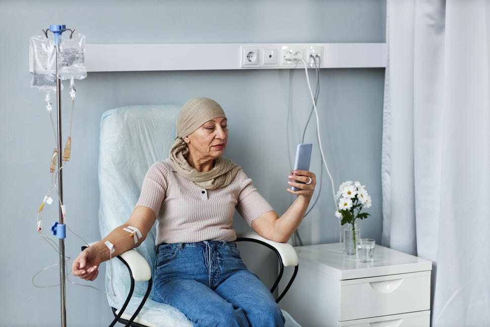
An older woman sitting in a chair and looking at a device while receiving an IV treatment.

Chemotherapy for breast cancer is usually systemic, meaning it is injected into a vein or given by mouth.

Credit: iStock

Chemotherapy (chemo) uses drugs to stop the growth of cancer cells, either by killing the cells or stopping them from dividing. Learn about what to expect when receiving chemotherapy at Chemotherapy to Treat Cancer.

Who gets chemotherapy for breast cancer?

Many people with breast cancer get chemotherapy but not everyone. For example, some people with early-stage breast cancer that does not have a high risk of recurrence may not get chemotherapy at all.

Multigene tests such as Oncotype DX and MammaPrint can help determine if you are likely to benefit from chemotherapy. If these tests show you are unlikely to benefit from chemotherapy, you can skip it without increasing the chance of recurrence. Learn more about multigene tests at How Breast Cancer Is Diagnosed. Learn about other biomarker tests at Tests for Breast Cancer Biomarkers.  

When is chemotherapy for breast cancer given?

Chemotherapy for breast cancer may be given at different times in your treatment. You might get it after surgery or before surgery. It can also be given if your disease cannot be treated with surgery or if the disease comes back (recurs) after initial treatment.

Chemotherapy after surgery for breast cancer

After surgery to remove breast cancer, some people may receive chemotherapy. Chemotherapy is given to kill any remaining cancer cells to lower the risk of recurrence. Chemotherapy after surgery may be called adjuvant chemotherapy.

Your doctor may recommend chemotherapy after surgery if you have: 

Chemotherapy before surgery for breast cancer

Sometimes chemotherapy is given before surgery. It may be given to shrink large tumors to make breast-conserving surgery possible for people who would have otherwise needed a mastectomy. It may also be given to reduce the number of lymph nodes that need to be removed during surgery or to lower the risk of recurrence. Chemotherapy before surgery may be called neoadjuvant chemotherapy.

Your doctor may recommend chemotherapy before surgery if you have:

  • high-grade breast cancer (grade 3)
  • breast cancer that has spread to the lymph nodes
  • a large breast cancer
  • HER2-positive breast cancer
  • triple-negative breast cancer
  • inflammatory breast cancer

Chemotherapy drugs used for breast cancer

Chemotherapy drugs used to treat breast cancer include:

Combinations of chemotherapy drugs may be used. Other chemotherapy drugs not listed here may also be used. 

Chemotherapy may also be combined with other kinds of drugs. For example:

Side effects of chemotherapy

The most common side effect of chemotherapy is fatigue, which is feeling exhausted or extremely tired. Other side effects may include hair loss, mouth sores, and nausea. Learn more about the side effects of cancer treatment and the steps you can take to manage or prevent them.

Mammograms

Drawing of a woman standing with her left breast pressed between two plates of a mammography machine. Behind her, a health professional uses an X-ray machine to take pictures of the breast. An inset shows the X-ray film image with an arrow pointed at abnormal tissue.

Mammography is an imaging test used to screen for and diagnose breast cancer. It can detect abnormal breast tissue, including cancer, sometimes before symptoms appear.

Credit: © Terese Winslow

Mammography is an imaging test that uses low-dose x-rays to create pictures of the breast. It is used both to screen for breast cancer and diagnose breast conditions.

What is a mammogram?

Mammograms are x-ray images of the breasts. Mammograms are used for breast cancer screening because they can find tumors at an earlier stage, before they cause symptoms.

Mammograms can also be used to check for breast cancer after a woman or her doctor finds a lump or other change. This type of mammogram is called a diagnostic mammogram.

The same machines are used for both types of mammograms. However, diagnostic mammography requires images from more angles than screening mammography, so the dose of radiation is higher.

What can a mammogram show?

Mammograms can show a mass (breast lump), deposits of calcium (called calcifications), and other changes. They also show breast density. A radiologist will study the mammogram for unusual changes. When possible, the radiologist will compare your most recent mammogram with past mammograms to check for changes in breast tissue since your last mammogram.

Mass (also called a breast lump): The size, shape, and edges of a lump give the radiologist important information. A lump that is not cancer often looks smooth and round and has clear, defined edges. Lumps that look like this are often benign cysts. However, if the lump has a jagged outline, an irregular shape, or other unusual features, more tests may be needed.

Calcifications are deposits of calcium in the breast that are too small to be felt. There are two types, neither of which is related to calcium in your diet:

  • Macrocalcifications look like small white dots on a mammogram. They are often caused by aging, an old injury, or inflammation and are usually benign.
  • Microcalcifications look like white specks on a mammogram. If found in an area of rapidly dividing cells or grouped together in a certain way, they may be a sign of DCIS or breast cancer.

Breast density describes the relative amounts of dense and fatty tissue in the breasts, as seen on a mammogram. Mammography is more likely to miss cancer in women with dense breasts.

What happens during a mammogram?

During a mammogram, your breast is placed between two plates that are then pressed together. Pressing the breast helps get a better x-ray picture of the inside of your breast. Several images are taken from different angles, with the breast repositioned each time.

For some people, the compression of the breast can be painful. Taking an over-the-counter pain medication before the procedure may lessen the discomfort.

If possible, try not to schedule your mammogram right before or during your menstrual period, when your breasts may be especially tender.

You should avoid using personal care products, such as deodorants, antiperspirants, powders, lotions, creams, or perfumes, around the breasts or under the arms on the day of your mammogram.

How are mammogram results reported?

You will receive the radiologist’s report of your screening mammogram within about 2 weeks, either by mail or in your electronic medical record. The results will also be sent to the health care provider who ordered the mammogram. If you don’t get your results within 2 weeks, you should contact your provider.

The report of your screening mammogram will usually include the BI-RADS category. BI-RADS categories provide a consistent way for radiologists to report a range of possible mammographic findings, with the recommended follow-up.

Breast Imaging Reporting and Database System (BI-RADS)

Category Assessment Follow-up
0 Need additional imaging evaluation Additional imaging needed before a category can be assigned
1 Negative Continue regular screening mammograms
2 Benign (noncancerous) finding Continue regular screening mammograms
3 Probably benign Receive a 6-month follow-up mammogram
4 Suspicious abnormality May require biopsy 
5 Highly suggestive of malignancy (cancer) Requires biopsy
6 Known biopsy-proven malignancy (cancer) Biopsy confirms presence of cancer before treatment begins

The mammogram screening report also includes information on breast density.

Depending on your result, you may be called back for further imaging or a biopsy. The letter with your results will explain what follow-up exams you may need. 

Although it can be scary to be called back for further testing, it is important to keep in mind that most people who are called back are not found to have breast cancer. 

When and how often should I have a screening mammogram?

The U.S. Preventive Services Task Force (USPSTF) currently recommends that women at average risk of breast cancer have screening mammograms every 2 years between ages 40 and 74. Talk with your doctor to find out your risk for breast cancer, based on your personal and family health history.

Some individuals at very high risk of breast cancer may be advised to begin screening for breast cancer at a younger age and to get screened more often than those at average risk. For example, anyone who received treatment for a childhood cancer that included radiation therapy to the breast or chest may be advised to begin screening at age 25, or 8 years after finishing radiation therapy, whichever is later. People who have known harmful mutations in the BRCA1 or BRCA2 genes may also consider having earlier or more frequent screening.

What are 3D mammograms?

Three-dimensional, or 3D, mammography, also called digital breast tomosynthesis (DBT), creates three-dimensional images of the breast. To do this, the DBT machine takes multiple pictures of thin “slices” across the breast that are then assembled into a 3D picture by computer software. DBT also creates 2D images of the breast like those created by standard mammography.

DBT combined with standard mammography is better at finding tumors than standard mammography alone. However, it’s still unknown whether DBT is more effective than standard mammography at reducing deaths from breast cancer. The NCI-sponsored TMIST trial is comparing 2D mammography with DBT to answer this question.

Are mammograms done in people with breast implants?

Yes. Women with breast implants should continue getting mammograms. However, most women who have breast reconstruction with implants after mastectomy usually do not need a mammogram of the reconstructed breast.

Implants can hide some breast tissue, making it harder for a radiologist to see unusual changes on the mammogram. If you have breast implants you should let the mammography facility know. A special technique called implant displacement views may be used.

Where can I get a mammogram?

Mammograms are available in breast clinics, hospital radiology departments, mobile vans, private radiology offices, and doctors’ offices. You may be able to schedule your own screening mammogram directly with the mammography facility without a referral from your doctor.

The Mammography Quality Standards Act (MQSA) is a federal law that requires mammography facilities across the nation to meet uniform quality standards. MQSA regulations also require that mammography facilities give patients an easy-to-read report of their mammogram results.

If you need help finding an FDA-certified mammography facility near you, you can contact NCI’s Cancer Information Service at 1–800–4–CANCER (1–800–422–6237). A searchable list is available from the FDA.

How much does a mammogram cost?

Insurance plans governed by the federal Affordable Care Act must cover screening mammograms as a preventive benefit every 1–2 years for women ages 40 and over without requiring copayments, coinsurance, or deductibles. In addition, many states require that Medicaid and public employee health plans cover screening mammography. Contact your mammography facility or health insurance company to confirm the cost and coverage.

Medicare pays for annual screening mammograms for all female Medicare beneficiaries who are age 40 or older. Information about coverage of mammograms is available from Medicare.

If you need a diagnostic mammogram, check with your health insurance provider about coverage.

How can uninsured or low-income women get a free or low-cost screening mammogram?

Some state and local health programs and employers provide mammograms free or at low cost. For example, CDC’s National Breast and Cervical Cancer Early Detection Program provides screening services, including clinical breast exams and mammograms, to low-income, uninsured women throughout the United States and in several U.S. territories. Contact information for local programs is available from the CDC or by calling 1–800–CDC–INFO (1–800–232–4636).

Information about free or low-cost mammography screening programs is also available from NCI’s Cancer Information Service at 1–800–4–CANCER (1–800–422–6237) and from local hospitals, health departments, women’s centers, or other community groups.

Mastectomy

In a mastectomy, the surgeon removes the whole breast that contains ductal carcinoma in situ (DCIS) or cancer. Your doctor may suggest a mastectomy if:

  • you have inflammatory breast cancer
  • your breast has more than one tumor
  • you received radiation therapy to the chest in the past
  • the cancer or DCIS is large relative to the size of the breast
  • you had a lumpectomy (breast-conserving surgery) that did not remove all the cancer cells
  • you are at a high risk of a second breast cancer because of a genetic mutation or strong family history

If you have a mastectomy, you may not need radiation therapy, so your doctor may also suggest a mastectomy if you are not able to have radiation therapy because you are pregnant or can’t commit to daily treatments.

People who have a mastectomy may need chemotherapy, hormone therapy, or targeted therapy. Learn more about Breast Cancer Treatment.

After a mastectomy, you may choose to stay flat, wear a prosthesis (breast-like form) in your bra, or have breast reconstruction surgery.

Types of mastectomy

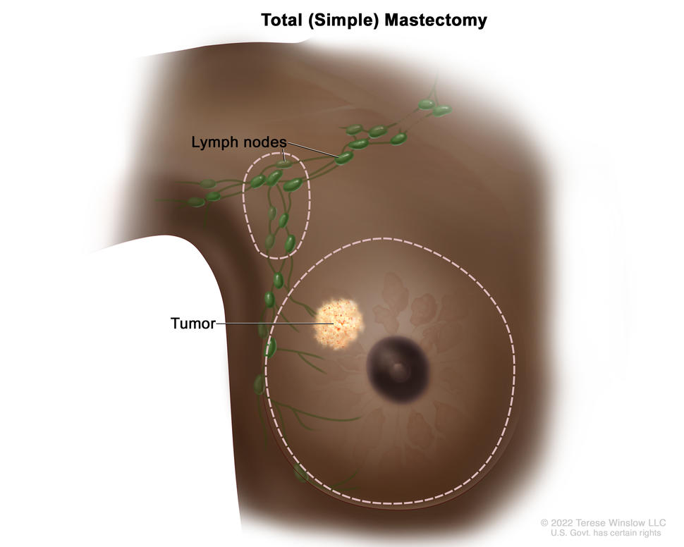
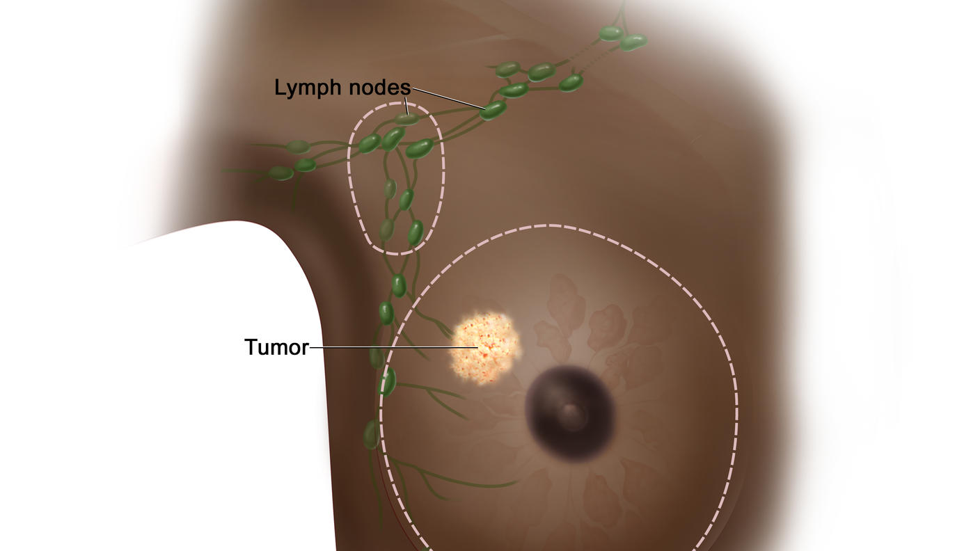
  • Total mastectomy (also called simple mastectomy). In this type of surgery, the surgeon removes your whole breast, including the nipple, areola, breast tissue, and skin. Sometimes, they also remove one or more of the lymph nodes under your arm in a procedure called sentinel lymph node biopsy.
Total (simple) mastectomy; drawing shows removal of the whole breast and some of the lymph nodes under the arm.

Total (simple) mastectomy. The whole breast is removed. Some of the lymph nodes under the arm may also be removed.

Credit: © Terese Winslow

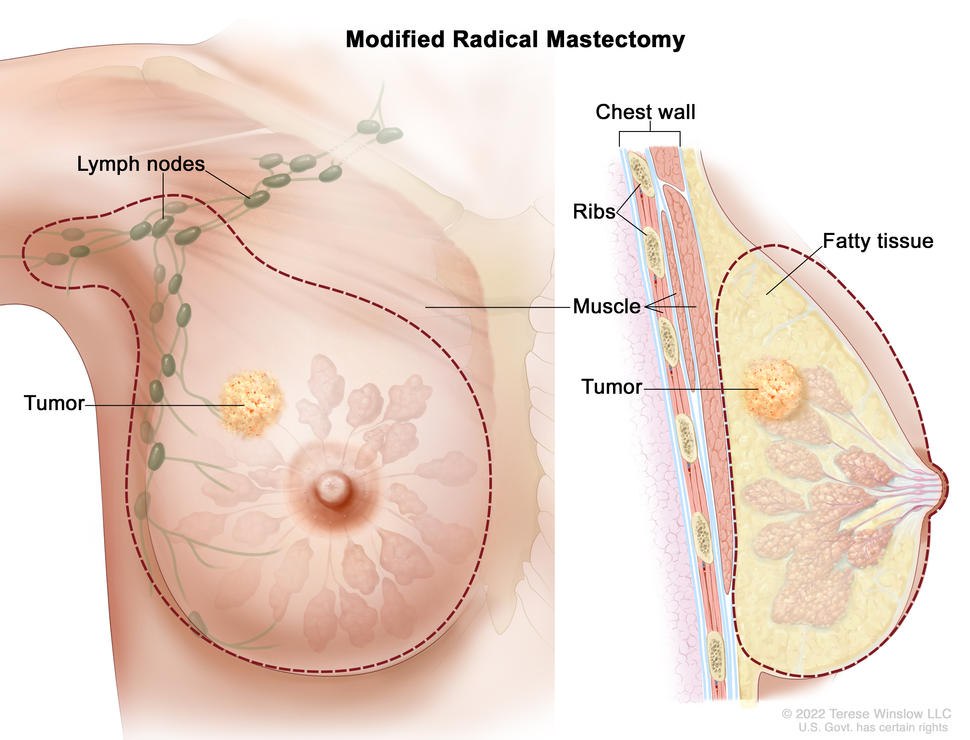
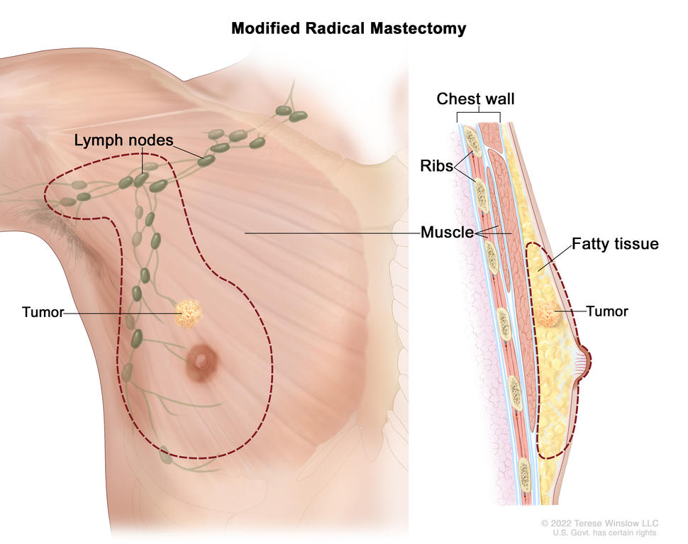
  • Modified radical mastectomy. This is the same as a total mastectomy, but the surgeon also removes lymph nodes under your arm (axillary lymph node dissection). Because cancer cells may spread to the lymph nodes, lymph node removal can help reduce the risk of the cancer coming back. But it is often used only for more advanced stages of breast cancer.
  • Skin-sparing mastectomy. The surgeon removes the whole breast, as in a total mastectomy, but leaves the skin. A skin-sparing mastectomy leaves less scarring and makes for a more natural-looking reconstructed breast. But if the tumor is large or near the skin, this procedure may not be an option. Skin-sparing mastectomies require breast reconstruction right after surgery.
  • Nipple-sparing mastectomy. The surgeon removes the whole breast but leaves the skin, nipple, and areola. Nipple-sparing mastectomy may not be an option if the tumor is large or if cancer cells are found near the skin, nipple, or areola. It also may not be an option for people with larger breasts.
  • Radical mastectomy. This type of mastectomy was common in the past, but it is now rarely performed because it does not improve how long people live. It involves removing the chest wall muscles and all lymph nodes, as well as the whole breast, nipple, areola, and skin. A surgeon may still suggest a radical mastectomy if the cancer cells have spread to the chest wall muscles.
Modified radical mastectomy; the drawing on the left shows the removal of the whole breast, including the  lymph nodes under the arm. The drawing on the right shows a cross-section of the breast, including the fatty tissue and chest wall (ribs  and muscle). A tumor in the breast is also shown.

Modified radical mastectomy. The whole breast and most of the lymph nodes under the arm are removed.

Credit: © Terese Winslow

Single vs. double mastectomy

In a single (unilateral) mastectomy, only the breast with cancer is removed. A double (bilateral) mastectomy involves the removal of both breasts.

Your doctor may suggest a double mastectomy if:

  • cancer is in both breasts
  • you have cancer in one breast but are at high risk of developing cancer in the other breast because of a genetic mutation or strong family history of breast cancer

If you do not have a genetic mutation or strong family history of breast cancer, you do not have an increased risk of developing a new cancer in the other breast. For you, research shows that having a double mastectomy instead of a single mastectomy does not help you live longer.

You may choose to have a double mastectomy, even if your doctor suggests a single mastectomy, if:

  • you want to reduce the risk of developing a new breast cancer in the healthy breast
  • you prefer not to have routine screening to check for cancer in the healthy breast
  • you want both breasts to be the same size
  • you are concerned about how your chest will look after a single mastectomy

Discuss insurance coverage and the risks and benefits of a double mastectomy with your doctor before making a decision.

Mastectomy with breast reconstruction surgery

You can have breast reconstruction at the same time as the mastectomy, or at any time after. Some types of mastectomies (skin- and nipple-sparing) require reconstruction at the same time. Breast reconstruction is done by a plastic surgeon with experience in this type of surgery. The surgeon uses an implant or tissue from another part of your body to create the breast mound after the breast has been removed. If your nipple is removed, the surgeon may also make the form of a nipple and add a tattoo that looks like the areola.

For more information about types of breast reconstruction, recovery times, and possible complications, visit Breast Reconstruction After Mastectomy.

Going flat after a mastectomy

You may choose to not have breast reconstruction after a mastectomy. This is called “going flat.” If you decide against reconstruction, you have the option of wearing a breast prosthesis to create the breast form under your clothes. To make the chest flat and smooth after a mastectomy, the surgeon will perform an aesthetic flat closure.

Recovering after a mastectomy

Recovery after a mastectomy depends on the type of mastectomy you had and whether you had breast reconstruction. Most people begin returning to normal activities 4 weeks after surgery. But recovery could take longer for people who have breast reconstruction.

Mastectomy risks

As with any surgery, a mastectomy has possible risks:

  • infection
  • pain or other discomfort that may or may not get better over time (postmastectomy pain syndrome and phantom breast syndrome)
  • arm and shoulder pain and stiffness
  • lymphedema in the arm, especially when many lymph nodes have been removed

Mastectomy with reconstruction has additional risks. Learn more at Breast Reconstruction After Mastectomy.

Breast Reconstruction After Mastectomy

What is breast reconstruction?

Many women who have a mastectomy—surgery to remove an entire breast to treat or prevent breast cancer—have the option of having the shape of the removed breast rebuilt. Some people choose not to have their breasts reconstructed after a mastectomy, instead preferring a flat closure.

Breasts can be rebuilt using implants (saline or silicone) or autologous tissue (that is, tissue from elsewhere in the body). Sometimes both implants and autologous tissue are used to rebuild the breast.

Surgery to reconstruct the breasts can be done (or started) at the time of the mastectomy, called immediate reconstruction, or it can be done after the mastectomy incisions have healed and treatment has been completed, called delayed reconstruction. Delayed reconstruction can happen months or even years after the mastectomy.

In the final stage of breast reconstruction, a nipple and areola may be re-created on the reconstructed breast, if these were not preserved during the mastectomy.

Sometimes breast reconstruction surgery includes surgery on the other, or contralateral, breast so that the two breasts will match in size and shape.

How do surgeons use implants to reconstruct a woman’s breast?

Implants are inserted underneath the skin or chest muscle following the mastectomy. Implants are usually placed as part of a two-stage procedure.

  • In the first stage, the surgeon places a device called a tissue expander under the skin that is left after the mastectomy or under the chest muscle (1, 2). The expander is slowly filled with saline or air during periodic visits to the doctor after surgery.
  • In the second stage, after the chest tissue has relaxed and healed enough, the expander is removed and replaced with an implant. The chest tissue is usually ready for the implant 2 to 6 months after the tissue expander is placed.

In some cases, the implant can be placed in the breast during the same surgery as the mastectomy—that is, a tissue expander is not used (3).

Surgeons are increasingly using material called acellular dermal matrix as a kind of scaffold or “sling” to support tissue expanders and implants. Acellular dermal matrix is a kind of mesh that is made from donated human or pig skin that has been sterilized and processed to remove all cells to eliminate the risks of rejection and infection.

How do surgeons use tissue from a woman’s own body to reconstruct the breast?

In autologous tissue reconstruction, a piece of tissue containing skin, fat, blood vessels, and sometimes muscle is taken from elsewhere in a woman’s body and used to rebuild the breast. This piece of tissue is called a flap.

Different sites in the body can provide flaps for breast reconstruction. Flaps most often come from the abdomen or back. However, they can also be taken from the thigh or buttocks.

Depending on their source, flaps can be pedicled or free.

  • With a pedicled flap, the tissue and attached blood vessels are moved together through the body to the breast area. Because the blood supply to the tissue used for reconstruction is left intact, blood vessels do not need to be reconnected once the tissue is moved.
  • With free flaps, the tissue is cut free from its blood supply. It must be attached to new blood vessels in the breast area, using a technique called microsurgery, to give the reconstructed breast a blood supply.

Abdominal and back flaps include:

  • DIEP flap: Tissue comes from the abdomen and contains only skin, blood vessels, and fat, without the underlying muscle. This type of flap is a free flap.
  • Latissimus dorsi (LD) flap: Tissue comes from the middle and side of the back. This type of flap is pedicled when used for breast reconstruction. (LD flaps can also be used for other types of reconstruction, such as abdominal or head and neck reconstruction.)
  • SIEA flap (also called SIEP flap): Tissue comes from the abdomen as in a DIEP flap but includes a different set of blood vessels. It also does not involve cutting of the abdominal muscle and is a free flap. This type of flap is not usually an option for breast reconstruction because the necessary blood vessels are often not adequate or do not exist.
  • TRAM flap: Tissue comes from the lower abdomen as in a DIEP flap but includes muscle. TRAM flaps can be either pedicled or free.

Flaps taken from the thigh or buttocks are used for women who have had previous major abdominal surgery or who don’t have enough abdominal tissue to reconstruct a breast. These types of flaps are free flaps.

  • IGAP flap: Tissue comes from the buttocks and contains only skin, blood vessels, and fat.
  • PAP flap: Tissue, without muscle, comes from the upper inner thigh.
  • SGAP flap: Tissue comes from the buttocks as in an IGAP flap but includes a different set of blood vessels and contains only skin, blood vessels, and fat.
  • TUG flap: Tissue, including muscle, comes from the upper inner thigh.

The table below summarizes the different types of flaps used for breast reconstruction:

Name of flap Type of flap Source of tissue Includes muscle?
DIEP Free Abdomen No
Latissumus dorsi Pedicled Middle and side of back Yes
SIEA/SIEP Free Abdomen No
TRAM Pedicled or free Lower abdomen Yes
IGAP Free Buttocks No
PAP Free Upper inner thigh No
SGAP Free Buttocks No
TUG Free Upper inner thigh Yes

In some cases, an implant and autologous tissue are used together. For example, with flaps taken from the thigh or buttocks an implant is often used as well to provide sufficient breast volume. Also, autologous tissue may be used to cover an implant when there isn’t enough skin and muscle left after mastectomy to allow for expansion and use of an implant (1, 2).

How do surgeons reconstruct the nipple and areola?

After the chest heals from reconstruction surgery and the position of the breast mound on the chest wall has had time to stabilize, the nipple and areola can be reconstructed. This can be done in two ways—surgically or with tattoos. 

For surgical nipple reconstruction, the new nipple is created by cutting and moving small pieces of skin from the reconstructed breast to the nipple site and shaping them into a new nipple. A few months later, the surgeon can re-create the areola. This is usually done using tattoo ink.

However, in some cases, skin grafts may be taken from the groin or abdomen and attached to the breast to create an areola at the time of the nipple reconstruction (1).

Alternatively, a new nipple and areola can be created by a tattoo artist who specializes in 3-D nipple tattooing. Such a nipple is flat to the touch but looks realistic.

A mastectomy that preserves a woman’s own nipple and areola, called nipple-sparing mastectomy, may be an option for some women, depending on the size and location of the breast cancer and the shape and size of the breasts (4, 5).

What factors can affect the timing of breast reconstruction?

One factor that can affect the timing of breast reconstruction is whether a woman will need radiation therapy. Radiation therapy can sometimes cause wound healing problems or infections in reconstructed breasts, so some women may prefer to delay reconstruction until after radiation therapy is completed. However, because of improvements in surgical and radiation techniques, immediate reconstruction with an implant is usually still an option for women who will need radiation therapy. Autologous tissue breast reconstruction is usually reserved for after radiation therapy, so that the breast and chest wall tissue damaged by radiation can be replaced with healthy tissue from elsewhere in the body.

Even if a woman is a candidate for immediate reconstruction, she may choose delayed reconstruction. For instance, some women prefer not to consider what type of reconstruction to have until after they have recovered from their mastectomy and subsequent adjuvant treatment. Women who delay reconstruction (or choose not to undergo the procedure at all) can use external breast prostheses, or breast forms, to give the appearance of breasts.

What factors can affect the choice of breast reconstruction method?

Several factors can influence the type of reconstructive surgery a woman chooses. These include the size and shape of the breast that is being rebuilt, the woman’s age and health, her history of past surgeries, her risk factors for complications after surgery (for example, smoking history and obesity), the availability of autologous tissue, and the location of the tumor in the breast (2, 6). Women who have had past abdominal surgery may not be candidates for an abdominally based flap reconstruction.

Each type of reconstruction has factors that a woman should think about before making a decision. Some of the more common considerations are listed below.

Reconstruction with Implants

Surgery and recovery

  • Enough skin and muscle must remain after mastectomy to cover the implant
  • Shorter surgical procedure than for reconstruction with autologous tissue; little blood loss
  • Recovery period may be shorter than with autologous reconstruction
  • Many follow-up visits may be needed to inflate the expander and insert the implant

Possible complications

Other considerations

  • May not be an option for patients who have previously undergone radiation therapy to the chest
  • May not be adequate for women with very large breasts
  • Will not last a lifetime; the longer a woman has implants, the more likely she is to have complications and to need to have her implants removed or replaced
  • Silicone implants may feel more natural than saline implants to the touch
  • The Food and Drug Administration (FDA) recommends that women with silicone implants undergo periodic MRI screenings to detect possible “silent” rupture of the implants

More information about implants can be found on FDA’s Breast Implants page.

Reconstruction with Autologous Tissue

Surgery and recovery

  • Longer surgical procedure than for implants
  • The initial recovery period may be longer than for implants
  • Pedicle flap reconstruction is usually a shorter operation than free flap reconstruction and usually requires a shorter hospitalization
  • Free flap reconstruction is a highly technical operation that requires a surgeon who has experience with microsurgery to reattach blood vessels.

Possible complications

  • Necrosis (death) of the transferred tissue
  • The risk of bleeding and blood clots is higher with autologous reconstruction than with implants
  • Pain and weakness at the site from which the donor tissue was taken
  • Obesity, diabetes, and smoking may increase the rate of complications

Other considerations

  • May provide a more natural breast shape than implants
  • May feel softer and more natural to the touch than implants
  • Leaves a scar at the site from which the donor tissue was taken
  • Can be used to replace tissue that has been damaged by radiation therapy

All women who undergo mastectomy for breast cancer experience varying degrees of numbness and loss of feeling in the breast because nerves that provide sensation to the breast are cut when breast tissue is removed during surgery. However, some breast sensation may be regained as the severed nerves grow and regenerate, and breast surgeons continue to make technical advances that can spare or repair damage to nerves.

Any type of breast reconstruction can fail if healing does not occur properly. In these cases, the implant or flap will have to be removed. If an implant reconstruction fails, a woman can usually have a second reconstruction using an alternative approach.

Will health insurance pay for breast reconstruction?

The Women’s Health and Cancer Rights Act of 1998 (WHCRA) is a federal law that requires group health plans and health insurance companies that offer mastectomy coverage to also pay for reconstructive surgery after mastectomy. This coverage must include all stages of reconstruction and surgery to achieve symmetry between the breasts, breast prostheses, and treatment of complications that result from the mastectomy, including lymphedema. More information about WHCRA is available from the Department of Labor and the Centers for Medicare & Medicaid Services.

Some health plans sponsored by religious organizations and some government health plans may be exempt from WHCRA. Also, WHCRA does not apply to Medicare or Medicaid. However, Medicare may cover breast reconstruction surgery as well as external breast prostheses (including a post-surgical bra) after a medically necessary mastectomy. Medicaid benefits vary by state; a woman should contact her state Medicaid office for information on whether, and to what extent, breast reconstruction is covered.

A woman considering breast reconstruction may want to discuss costs and health insurance coverage with her doctor and insurance company before choosing to have the surgery. Some insurance companies require a second opinion before they will agree to pay for a surgery.

What type of follow-up care and rehabilitation is needed after breast reconstruction?

Any type of reconstruction increases the number of side effects a woman may experience compared with those after a mastectomy alone. A woman’s medical team will watch her closely for complications, some of which can occur months or even years after surgery (1, 2, 8).

Women who have breast reconstruction may benefit from physical therapy to improve or maintain shoulder range of motion or help recover from weakness experienced at the site from which donor tissue was taken for an autologous reconstruction (9, 10). A physical therapist can help a woman use exercises to regain strength, adjust to new physical limitations, and figure out the safest ways to perform everyday activities.

Does breast reconstruction affect the ability to check for breast cancer?

Mammography is not typically done on a breast that is reconstructed after mastectomy; instead, physical exams are done to check for recurrence. However, women who have one breast removed by mastectomy will still have mammograms of the other breast.

What is flat closure?

A flat closure (also called aesthetic flat closure) is surgery done to rebuild the shape of the chest wall after the removal of one or both breasts. It is also a surgical option for people who have had their breast implants removed.

During an aesthetic flat closure, a surgeon removes extra skin, fat, and other tissue in the breast area. The remaining tissue is then tightened and smoothed out so that the chest wall appears flat and contoured. Simply forgoing additional procedures to reconstruct the breast(s) after a mastectomy will not achieve a flat closure (11).
 

What are some new developments in breast reconstruction after mastectomy?

  • Oncoplastic surgery. In general, women who have lumpectomy or partial mastectomy for early-stage breast cancer do not have reconstruction. However, in some cases the surgeon may use plastic surgery techniques to reshape the breast at the time of cancer surgery. This type of breast-conserving surgery, called oncoplastic surgery, may use local tissue rearrangement, reconstruction through breast reduction surgery, or transfer of tissue flaps. Long-term outcomes of this type of surgery are comparable to those for standard breast-conserving surgery (12).
     
  • Autologous fat grafting. A newer type of breast reconstruction technique involves the transfer of fat tissue from one part of the body (usually the thighs, abdomen, or buttocks) to the reconstructed breast. The fat tissue is harvested by liposuction, washed, and liquified so that it can be injected into the area of interest. Fat grafting is mainly used to correct deformities and asymmetries that may appear after breast reconstruction. It is also sometimes used to reconstruct an entire breast. Although concern has been raised about the lack of long-term outcome studies, this technique is considered safe (1, 6, 13).

Living with Breast Cancer & Survivorship

A smiling woman with her arm around an older smiling woman sitting together on a sofa.

Some survivors say they would not have been able to cope without the help and love of their family members.

Credit: iStock

You may have just learned that you have breast cancer. Or you may be in treatment, finishing treatment, or have a friend or family member with cancer. Wherever you are in dealing with cancer, from the time of diagnosis through the rest of your life, you are thought of as a survivor. As a result, what being a breast cancer survivor means to you may change over time. Or you may not feel that the term applies to you, and that is okay, too.

Having cancer changes your life and the lives of those around you. The symptoms and side effects of the disease and its treatment may cause certain physical changes, but they can also affect the way you feel and how you live.

Learn more about the unique challenges cancer survivors face and find resources to help you cope at Cancer Survivorship.

Coping with breast cancer treatment

Cancer treatment can cause side effects or changes to your body and how you feel. You may have different side effects even when you are given the same treatment for the same type of cancer as other people. It’s normal to feel uncertain or fearful about side effects that you may have. Keep in mind your health care team can help prevent and relieve any side effects.

Side effects of cancer treatment

Breast cancer treatments can cause many physical side effects that affect healthy tissues or organs. The type of side effects you may have depends on the type of treatments you have and their doses. Some side effects that many people with breast cancer have include lymphedema and fertility issues.

Learn more about the possible side effects of cancer treatment at Side Effects of Cancer Treatment.

Emotional effects of cancer treatment

Breast cancer and its treatment can be overwhelming and cause many emotional effects. Just as cancer affects your physical health, it can bring up a wide range of emotions you’re not used to dealing with. It can also make existing feelings seem more intense.

Learn more about how to understand and cope with new, and sometimes difficult, thoughts and feelings at Emotions and Cancer.

Changes in body image and sexuality

Breast surgery, deciding whether to have breast reconstruction, and hair loss are some examples of how cancer treatment may affect how you look and feel about yourself. Other treatments for breast cancer also may affect your body, how others see you, and how you see yourself. Coping with these changes to your body can be hard, but there are resources that can help you.

Learn more about coping with changes to your body in How Cancer Affects Your Self-Image and Sexuality.

Follow-up care

Many breast cancer survivors need to visit their doctor regularly to get follow-up exams or tests. Planning and scheduling these appointments can be stressful and time-consuming, but making regular visits with your health care team can help you feel supported.

Getting Financial Support

Even if you have health insurance, you may face financial challenges and need help dealing with the costs of breast cancer treatment. For tips and ways to cope, visit Managing Cancer Costs and Medical Information.

Your doctor may give you a survivorship care plan that details the types of cancer treatment you received and a plan for your follow-up care. At follow-up appointments, your doctor may do imaging and other tests to check for a recurrence, such as:

Learn more about the health care you should be receiving after treatment at Follow-Up Medical Care.

Coping with fear of breast cancer recurrence

You may feel a mix of relief and worry when treatment ends. When you stop having a treatment that was working, it can cause fear that cancer could come back. Tests and scans that are part of normal follow-up care can create anxiety called “scanxiety.” Physical changes in your body also can cause worry. You may fear that any pain or discomfort may be due to cancer’s return. Although these feelings are uncomfortable, they can improve with time and help.

Learn more about how to cope with fear of recurrence at Life After Cancer Treatment.

Late effects of cancer treatment

Late effects are problems caused by cancer treatment that may not show up for months or years after treatment. These problems are specific to certain types of treatments and the amount received. Like side effects that you may have during treatment, late effects can be very different from person to person.

Learn about late effects of treatment and how to cope at Late Effects of Cancer Treatment.

Getting support

No matter where you are in your cancer journey, you or your caregivers may need help coping. Know that you are not alone. Seek support from family and friends, your health care team, and support groups.

Get advice on how to ask for help in Adjusting to Cancer, or learn more about Cancer Support Groups.

Support for caregivers

If you are caring for someone who has cancer, it’s important to know what resources are available to you. Learn more in Support for Caregivers of Cancer Patients.

Choosing Between a Lumpectomy and Mastectomy

Face of young woman with serious expression

To decide which breast cancer surgery to have, compare your choices and think about what matters to you.

Credit: National Cancer Institute

Are you facing a decision about lumpectomy vs. mastectomy?

Do you have ductal carcinoma in situ (DCIS) or breast cancer that can be removed with surgery? If so, you may be able to choose which type of breast surgery you have. Often, your choice is between a lumpectomy or other breast-conserving surgery (surgery that takes out the cancer and leaves most of the breast) and a mastectomy (surgery that removes the whole breast).

Learn more about these surgeries at Breast Cancer Surgery.

Once you are diagnosed, treatment will usually not begin right away. Use the time to:

  • talk with your doctor
  • get a second opinion
  • check with your insurance company
  • learn the facts about your surgery choices
  • think about what is important to you  

Surgery Choices: Cindy, Theresa, Paula

Three women describe the type of surgery that they chose to treat their breast cancer.

Compare lumpectomy and mastectomy

If your surgeon gives you a choice about which surgery to have, it can be hard to decide which one is best for you. This section can help you compare the different surgeries. Learn how the surgeries are alike and how they are different by clicking on each question to learn more.  

Questions about the effectiveness of breast cancer surgery

Which type of breast cancer surgery is best for my cancer? 

Lumpectomy or other breast-conserving surgery Most women with DCIS or breast cancer can choose to have a lumpectomy (breast-conserving surgery), usually followed by radiation therapy.
Mastectomy

Most women with DCIS or breast cancer can choose to have a mastectomy. A mastectomy may be a better choice for you if:

  • you have small breasts and a large area of DCIS or cancer
  • you have DCIS or cancer in more than one part of your breast
  • the DCIS or cancer is under the nipple
  • you are not able or prefer not to receive radiation therapy 
Mastectomy with reconstruction  If you have a mastectomy, you might also want breast reconstruction surgery. You can choose to have reconstruction surgery at the same time as your mastectomy or wait and have it later. Your surgeon can talk with you about how much skin, including the nipple and areola, might be preserved for reconstruction. 

Will the type of breast cancer surgery I have affect how long I live? 

No. Research shows that women who have breast-conserving surgery live as long as women who have a mastectomy. This does not change if you also have reconstruction. 

What are the chances that my breast cancer will return in the same area? 

Lumpectomy or other breast-conserving surgery  There is a chance that your cancer will come back in the same breast if you have a lumpectomy. But if it does, it is not likely to affect how long you live. About 5% to 10% of women (1 out of every 10 to 20) who have a lumpectomy followed by radiation therapy get cancer in the same breast within 12 years. If this happens, you can be effectively treated with a mastectomy.
Mastectomy 

There is a chance that your cancer will return in the same breast that was removed. This is called a local recurrence. It can happen after a mastectomy because surgeons cannot remove every bit of breast tissue. Also, the cancer can come back in the chest muscle or chest wall.

Local recurrence is slightly less likely after mastectomy than after lumpectomy. About 1 out of every 20 women who have a mastectomy will get cancer on the same side of their chest within 12 years.

Mastectomy with reconstruction  Your chances are the same as mastectomy because breast reconstruction surgery does not affect the chances of the cancer returning.

Questions about recovering from breast cancer surgery 

Will I have pain? 

Most people have some pain after surgery. Talk with your doctor or nurse before surgery about ways to control pain after surgery. Also, tell them if your pain control is not working. 

How long before I can return to normal activities?

Lumpectomy or other breast-conserving surgery  Most women are ready to return to most of their usual activities within 2 weeks. 
Mastectomy  It may take 3 to 4 weeks to feel mostly normal after a mastectomy. 
Mastectomy with reconstruction  Your recovery will depend on the type of reconstruction you have. It can take 6 to 8 weeks or longer to fully recover from a mastectomy that is followed by breast reconstruction. 

What other problems might I have from breast cancer surgery? 

Lumpectomy or other breast-conserving surgery  You may feel very tired and have skin changes from radiation therapy. 
Mastectomy  You may feel out of balance if you had large breasts and do not have reconstruction surgery. This may also lead to neck and shoulder pain. 
Mastectomy with reconstruction 
  • You may not like how your breast-like shape looks.  
  • If you have an implant, your breast may harden and can become painful.  
  • If your implant breaks or leaks you may need more surgery.  
  • If you have tissue flap reconstruction, you may lose strength in the part of your body where a muscle was removed. 

If lymph nodes are removed from under your arm, you may develop lymphedema. Learn more about Lymphedema

Questions about treatment after breast surgery 

What other types of treatment might I need after breast cancer surgery? 

If you choose to have a lumpectomy or other breast-conserving surgery, you will usually need radiation therapy. Radiation treatments are often given 5 days a week for up to 6 weeks.

If you have a mastectomy, you may still need radiation therapy.

No matter which surgery you choose, you might need:

Will I need more surgery? 

Lumpectomy or other breast-conserving surgery  After surgery, if there are cancer cells at the edge of the removed breast tissue, then you may need more surgery to make sure that all the cancer is removed.
Mastectomy  If you have problems after your mastectomy, you may need more surgery. 
Mastectomy with reconstruction 

You will most likely need more than one surgery to build a new breast-like shape. The number of surgeries you need will depend on the type of reconstruction you have and if you choose to have a nipple or areola added.

Some women may also decide to have surgery on the opposite breast to help it better match the reconstructed breast.

If you have an implant, you are likely to need surgery many years later to remove or replace it. 

With all three surgeries, you may need more surgery to remove lymph nodes from under your arm to see how far the cancer has spread. 

Questions about body image and quality of life after breast cancer surgery 

What will my breast look like after breast cancer surgery? 

Lumpectomy or other breast-conserving surgery 

Your breast should look a lot like it did before surgery.

But if your tumor is large, your breast may look different or smaller after breast-conserving surgery.

You will have a small scar where the surgeon cut to remove the DCIS or cancer. The length of the scar will depend on how large the incision was. 

Mastectomy

If you have a mastectomy without reconstruction, your breast and nipple will be removed. You will have a flat chest on the side of your body where the breast was removed.

You will have a scar over the place where your breast was removed. The length of the scar will depend on the size of your breast. The scar is usually shorter for smaller breasts and longer for larger breasts. 

Mastectomy with reconstruction 

If you have a skin- or nipple-sparing mastectomy, your skin and nipple will remain after surgery. You will not have the sensation of temperature or touch, but your nipple will still react to these.

Even if you have reconstruction, your breast shape will not look or feel like it did before surgery. And it will not look or feel like your other breast. You will have scars where the surgeon stitched skin together to make the new breast-like shape. If you have tissue flap reconstruction, you will have scars around the new breast, as well as in the area where the surgeon removed the muscle, fat, and skin to make the new breast-like shape. 

With all three surgeries, the scars left from where the surgeon cut the skin and stitched it back together tend to fade over time.

To get a better idea of what to expect from surgery, ask your surgeon if you can see before and after pictures of other people who have had different types of surgery. 

Will my breast have feeling after breast cancer surgery? 

Lumpectomy or other breast-conserving surgery Yes. You should still have feeling in your breast, nipple, and areola. 
Mastectomy

Maybe. After surgery, the skin around where the surgeon cut and maybe the area under your arm will be numb. 

This numb feeling may improve over 1 to 2 years, but it will never feel like it once did. Also, the skin where your breast was may feel tight. 

Mastectomy with reconstruction  No. Most of the area around your breast will not have feeling.

Some surgeons are trying new techniques to preserve or restore breast feeling after a mastectomy. Find clinical trials studying this issue using our NCI-supported clinical trials search or ClinicalTrials.gov

Will the type of breast surgery I choose affect my quality of life? 

Research suggests that women who have breast-conserving surgery have a better long-term quality of life than those who have a mastectomy

Think about what is important to you

After you have talked with a breast surgeon and learned the facts, you may also want to talk with your spouse or partner, family, friends, or other women who have had breast cancer surgery.

Then, think about what is important to you. Thinking about the following questions and talking them over with others might help:

  • If I have a lumpectomy or other breast-conserving surgery, am I willing and able to have radiation therapy 5 days a week for up to 6 weeks?
  • If I have a lumpectomy and the cancer comes back later in the same breast, how would I feel about needing a mastectomy?
  • If I have a mastectomy, do I also want breast reconstruction surgery?
  • If I have breast reconstruction surgery, do I want it at the same time as the mastectomy?
  • What treatment does my insurance cover? What do I have to pay for?
  • How important is it to me how my breast looks after cancer surgery?
  • How important is it to me how my breast feels after cancer surgery?
  • If I have a mastectomy and do not have reconstruction, will my insurance cover prostheses and special bras?
  • Where can I find breast prostheses and special bras?
  • Do I want a second opinion?
  • Is there someone else I should talk with about my surgery choices?
  • What else do I want to learn or do before I decide about breast cancer surgery? 

Radiation for Breast Cancer

Radiation therapy is a cancer treatment that uses high-energy x-rays or other types of radiation to kill cancer cells or keep them from growing. Radiation is given in a series of treatments to allow healthy cells to recover and to make radiation more effective. The number of treatments is based on details about the cancer, such as the size and location of the tumor. Most of the time, radiation is given as an outpatient treatment. This means that you will have treatment at a clinic or radiation therapy center and will not stay overnight in the hospital.

Types of radiation therapy for breast cancer

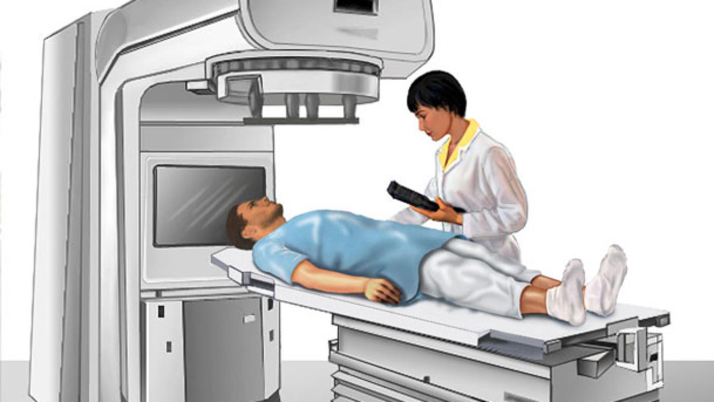
External beam radiation therapy and brachytherapy (internal radiation therapy) are the main types of radiation therapy used for breast cancer. External beam radiation therapy comes from a machine that aims radiation at the cancer. For internal radiation therapy, the radiation source is placed inside the body near the cancer.

External beam radiation therapy for breast cancer

External beam radiation therapy is a local treatment, which means it treats a specific part of your body. It may be given to the whole breast or chest, or to the part of the breast where the cancer is or where it was located before it was removed by surgery. Most of the time, you will get external beam radiation therapy as an outpatient.

Preparing for your appointment

To learn how to prepare for radiation therapy, visit the booklet Radiation Therapy and You: Support for People with Cancer.

Radiation therapy for breast cancer usually begins about a month or two after surgery. That is because radiation therapy can slow wound healing. If you are also receiving chemotherapy, radiation therapy usually starts about a month after you have finished chemotherapy. You can have radiation at the same time as you are receiving some other treatments, such as hormone therapy or targeted therapy.

Whole breast radiation therapy

With this type of radiation therapy, an x-ray machine delivers radiation to either the whole breast, or, if done after mastectomy, to the entire chest wall. Sometimes, radiation will also target the lymph nodes in the armpit or neck area or under the breastbone. Most people have treatment once a day, Monday through Friday, for about 5 to 6 weeks. In some cases, it may be done in 4 weeks or less. Your doctor will recommend the treatment regimen that is right for you.

Partial breast external beam radiation therapy

Two techniques are commonly used to give external beam radiation treatment to the area where the cancer is or where it was located before it was removed by surgery:

  • Three-dimensional conformal external beam radiation (3DCRT) shapes the radiation beams to match the tumor’s shape, delivering a fixed dose of radiation from many directions.
  • Intensity-modulated radiation therapy (IMRT) also aims the radiation beams at the tumor from many directions. But IMRT also varies the intensity of radiation across the treatment area to allow higher doses of radiation to be given in areas needing more treatment and lower doses to be given in areas near sensitive organs or tissues.

Most people have treatment once a day, Monday through Friday, for 2 to 4 weeks.

Researchers are looking at other ways to adjust the radiation dose or schedule to reach the total dose of radiation more quickly or to limit damage to healthy cells.

  • Accelerated fractionation is treatment given in larger daily or weekly doses to reduce the number of weeks of treatment.
  • Hyperfractionation is a smaller dose than the usual daily dose of radiation given more than once a day.
  • Hypofractionation is larger doses given once a day or less often to reduce the number of treatments.

Intraoperative radiation therapy

Intraoperative radiation therapy is a type of external beam radiation therapy in which a single dose of radiation therapy is delivered to the tumor site during surgery.

Brachytherapy for breast cancer

Brachytherapy (also called internal radiation therapy) is sometimes used after breast-conserving surgery. It involves placing radioactive material using seeds, pellets, or needles in the area where the cancer was removed. The radiation only reaches a small area around the surgical site, where the risk of recurrence is highest.

Intracavitary brachytherapy

Intracavitary brachytherapy is the most common type of brachytherapy for breast cancer. In this form of brachytherapy, a balloon, applicator, or similar device is inserted into the space where the cancer was, either during or a few days after breast-conserving surgery. A radioactive pellet is placed into the device during each treatment session and then removed after each treatment.

Treatments usually last about 5 to 10 minutes and are given 2 times a day for 5 days. After about 2 weeks, the device is removed. You may need stitches to close the hole when treatment is complete.

Interstitial brachytherapy 

In interstitial brachytherapy, several small tubes, called catheters, are inserted into the tissue where the breast cancer was and stitched in place. This is most often done 1 to 2 weeks after surgery. A radiation source, such as radioactive pellets or seeds, is temporarily placed into the catheters during each treatment session. Treatments usually last about 20 minutes. Two kinds of sources, high-dose implants and low-dose implants, can be used: 

  • High-dose implants: If you receive high-dose implants, the radiation is delivered once or twice a day for about 5 days. You will be able to leave the hospital after each treatment, and you will not remain radioactive after. It is safe to be around others, including babies and children.
  • Low-dose implants: If you receive low-dose implants, treatment can be delivered over several hours to days. The radiation source stays in place and is slowly delivered to the tissue around the catheters. You will stay in the hospital throughout treatment to avoid exposing others to radiation. The radiation source and catheters are removed when treatment is complete. Then you can go home.

Permanent breast seed implant (PBSI) 

PBSI involves placing permanent radioactive seeds into the breast tissue around the area where the cancer was removed. The seeds emit radiation at a low dose. Although the seeds remain in the body, they lose their radioactivity over time and do not need to be removed. This is a one-time procedure that usually does not require a hospital stay.

Intravenous radiation

Intravenous radiation with strontium-89 (a radionuclide) is given as palliative therapy to relieve bone pain caused by breast cancer that has spread to the bones. Strontium-89 is injected into a vein and travels to the surface of the bones. Radiation is released and kills cancer cells in the bones.

Learn more about Brachytherapy to Treat Cancer.

Side effects of radiation therapy for breast cancer

Radiation kills or slows the growth of cancer cells and can also affect nearby healthy cells. Damage to healthy cells can cause side effects. Your experience may be different from someone else who is getting the same amount of radiation therapy to the same part of the body. Sometimes, people may have side effects that do not improve.

Possible early side effects of radiation therapy for breast cancer include:

  • fatigue (feeling exhausted and worn out)
  • hair loss in the area exposed to radiation
  • skin changes, such as redness and dryness, in the area exposed to radiation
  • breast tenderness or hardening

Problems from cancer treatment that begin 6 months or later after treatment and continue for months or years are called late effects.

Possible late effects of radiation therapy for breast cancer include:

  • skin changes, such as redness and dryness, in the area exposed to radiation
  • breast pain, tenderness, or hardening
  • heart and lung problems
  • bone weakness
  • lymphedema
  • second cancers, rarely

Learn more about Late Effects of Cancer Treatment.

Discuss potential side effects with your doctor or nurse so you know what to expect during and after treatment. Regular follow-up care and supportive care can help manage side effects that do occur. 

Learn more about Living with Breast Cancer and Survivorship.

Lumpectomy (Breast-Conserving Surgery)

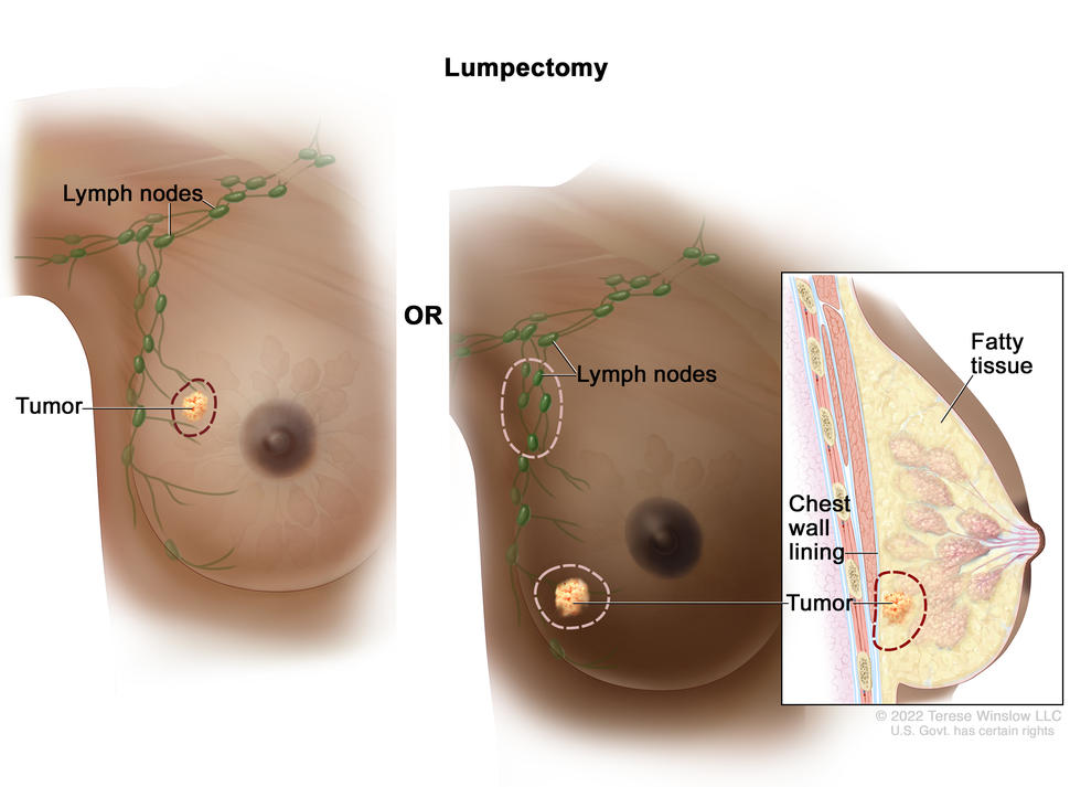
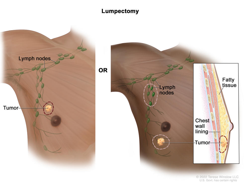
The drawing on the left shows removal of the tumor and some of the normal tissue around it. The drawing on the right shows removal of some of the lymph nodes under the arm and removal of the tumor and part of the chest wall lining near the tumor. Also shown is fatty tissue in the breast.

Lumpectomy. The tumor and some normal tissue around it are removed, but not the breast itself. Some lymph nodes under the arm may also be removed. If the cancer is near the chest wall, part of the chest wall lining may be removed as well.

Credit: © Terese Winslow

Lumpectomy is a type of breast surgery in which the surgeon removes the ductal carcinoma in situ (DCIS) or breast cancer and some healthy tissue around it. If the cancer is near the chest wall, part of the chest wall lining may also be removed.

Lumpectomy is an option for some people with DCIS or invasive breast cancer. It is usually followed by radiation therapy. Research shows that lumpectomy plus radiation therapy is just as effective as mastectomy (surgery to remove all breast tissue) at lowering your chance of dying from breast cancer or of the cancer coming back. Learn more about choosing between a mastectomy and lumpectomy to treat breast cancer.

Your doctor may suggest a lumpectomy plus radiation therapy if:

  • the cancer or DCIS is small relative to the size of the breast
  • the cancer or DCIS is found in only one place in the breast
  • you are not pregnant and are able to have daily radiation therapy for up to 6 weeks

If large amounts of tissue need to be removed during a lumpectomy, the surgery may be called partial mastectomy, segmental mastectomy, or quadrantectomy.

After a lumpectomy, a pathologist will check the healthy tissue that was removed to see if it contains DCIS or cancer. If it does, you may need more surgery or a mastectomy so the surgeon can remove more tissue and make sure no DCIS or cancer cells remain in the breast. 

Treatment before or after lumpectomy

Most people who have a lumpectomy receive radiation therapy after they heal from surgery. The goal of this treatment is to keep cancer from coming back in the same breast. Learn more about Radiation for Breast Cancer.

You might also need chemotherapy, hormone therapy, or targeted therapy, before or after surgery. Learn more about Breast Cancer Treatment.

The type of treatment you receive will depend on many factors, such as how different the tumor cells look from normal breast cells, how likely the tumor cells are to grow and spread, and the results of biomarker tests. Learn more at How Breast Cancer Is Diagnosed and Tests for Breast Cancer Biomarkers.

Recovering from lumpectomy

Most people can go home the day of their lumpectomy and go about their daily activities with few restrictions. But you may need help with some tasks, depending on the extent of your surgery. Within two weeks, you should be able to return to your normal activities.

Reconstruction after lumpectomy

After a lumpectomy, your breast will usually look and feel much like it did before surgery. You will have a scar at the site of surgery. And there may be a dent where the tumor and tissue were removed.

Talk to your surgical team about how your surgery might change how your breast looks. If you think you will not be happy with the look or feel of your breast after surgery, you may think about reconstruction. Reconstruction can take place during or any time after the lumpectomy. 

There are two types of breast reconstruction for people who have a lumpectomy:

  • Oncoplastic surgery combines lumpectomy with plastic surgery that can help make the breasts look more even if large amounts of tissue are removed from one breast. It can also reduce scarring and dents. A plastic surgeon will often do this reconstruction during the same surgery as the lumpectomy.
  • Revision reconstruction usually happens after you have healed from a lumpectomy. A plastic surgeon may be able to improve scarring and hardening of tissue at the surgical site. And they may be able to restore the shape of the breast with fat grafting, tissue flaps, or implants.

Lumpectomy risks

As with any surgery, lumpectomy has risks, including:

  • bleeding and pain at the site of surgery
  • infection
  • fluid buildup near the site of surgery
  • lymphedema, if lymph nodes are removed

Male Breast Cancer

What is male breast cancer?

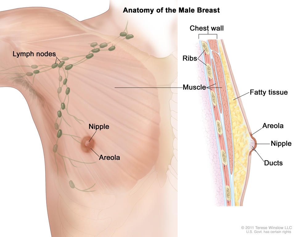
Drawing of male breast anatomy showing the lymph nodes, nipple, areola, chest wall, ribs, muscle, fatty tissue, and ducts.

Anatomy of the male breast. The nipple and areola are shown on the outside of the breast. The lymph nodes, fatty tissue, ducts, and other parts of the inside of the breast are also shown.

Credit: © Terese Winslow

Male breast cancer is breast cancer that develops in men. Breast cancer is much more common in women, but men can get breast cancer because they also have breast tissue where cancer can start. Fewer than 1 in 100 breast cancers in the United States occur in men. Men of any age can get breast cancer, but it usually occurs at a later age in men than in women.

Most male breast cancers start in the milk ducts of the breast and are called ductal cancers. Both female and male breasts have milk ducts, but in male breasts, milk ducts do not develop in the same way as they do in female breasts. Male breasts may also have milk glands called lobules, but far fewer than in female breasts. Cancers that start in the lobules, called lobular cancers, are rare in men.  

Other types of breast cancer are extremely rare in men, but they can happen. These include phyllodes tumor, Paget disease of the breast, and inflammatory breast cancer.

When abnormal cells are found in the ducts and have not spread to other tissues in the breast, it is called ductal carcinoma in situ. Invasive cancers have spread into surrounding breast tissue and can spread to nearby lymph nodes or other organs throughout the body. Most breast cancers in men are invasive. 

What are risk factors for male breast cancer?  

Older age is a risk factor for most cancers, including breast cancer in men. In addition, if you are male, you may have an increased risk of breast cancer if you have:

  • a history of radiation therapy directed at the chest
  • one or more female relatives who have had breast cancer
  • inherited changes in the BRCA1 or BRCA2 genes or in other genes that increase breast cancer risk
  • a condition linked to higher-than-normal estrogen levels in the body:
    • Klinefelter syndrome
    • liver disease (cirrhosis)
    • testicular problems, including inflamed testicles (orchitis), an undescended testicle, or surgery to remove one or both testicles
    • obesity 

Screening for male breast cancer

If you think you may have risk factors for male breast cancer, talk to your doctor. Screening mammograms are not usually recommended for men, even those who are at increased risk of breast cancer. But your doctor may suggest staying alert to any changes in your breast or the skin of your breast and getting regular clinical breast exams

What are the symptoms of breast cancer in men?  

Signs and symptoms of breast cancer in men include:

  • a lump or thickening in or near the breast (most common)
  • a nipple that changes in shape or the direction it is pointing
  • a nipple that is inverted
  • fluid from the nipple that may be clear or bloody
  • an open wound (ulcer) in the skin of the breast
  • scaly, red, or swollen skin on the breast, nipple, or areola
  • dimpling or ridges on the skin that resemble an orange peel
  • swollen lymph nodes under the arm or near the collarbone

Many breast changes in men are signs of less serious or benign conditions, but it is important to check with your doctor if you notice any unusual breast changes. 

How is breast cancer diagnosed in men? 

If you are male and have symptoms of breast cancer, your doctor will need to find out if they are due to cancer or another condition. Your doctor may:

If it is cancer, additional tests may include:  

  • Biomarker tests check the cancer cells for hormone receptors and HER2 protein that may help plan treatment. Male breast cancer is almost always hormone receptor positive. Learn more about Tests for Breast Cancer Biomarkers.  
  • Bone scan checks to see if cancer has spread to your bones.
  • PET scan or CT scan are imaging tests that help determine the extent of spread of the cancer.  
  • Genetic testing looks for inherited mutations in BRCA1, BRCA2, and several other genes that increase the risk of breast cancer. Such mutations, if present, can affect treatment decisions. They also have implications for family members.  

Your doctor will assign a stage to the cancer based on a combination of the extent of spread, hormone receptor and HER2 status, and other tumor features, such as tumor grade.

Learn more about the different stages of breast cancer and tests used to diagnose it at Breast Cancer Stages and How Breast Cancer Is Diagnosed

How is male breast cancer treated?

Male breast cancer is usually treated in the same way as breast cancer in women. However, men are more likely than women to have a mastectomy (surgery to remove the whole breast) rather than a lumpectomy (breast-conserving surgery) to remove the cancer because they have less breast tissue. But some male breast cancers can be treated with a lumpectomy, depending on the size of the cancer, the amount of breast tissue the person has, and other factors.

Learn more about breast cancer treatment at:

The drawing on the left shows removal of the tumor and some of the normal tissue around it. The drawing on the right shows removal of some of the lymph nodes under the arm and removal of the tumor and part of the chest wall lining near the tumor. Also shown is fatty tissue in the breast.

Modified radical mastectomy; the drawing on the left shows the removal of the whole breast, including the lymph nodes under the arm. The drawing on the right shows a cross-section of the breast, including the fatty tissue and chest wall (ribs  and muscle). A tumor is also shown in the breast.

What is the survival rate and prognosis for men with breast cancer?

Doctors estimate the prognosis for men with breast cancer by using statistics collected over many years. One common statistic is the 5-year relative survival rate. The 5-year relative survival rate tells you what percent of people with the same type and stage of male breast cancer are alive 5 years after their cancer was diagnosed, compared with people in the overall population.  

Men with breast cancer have a slightly lower 5-year survival rate than their female peers. This may be because male breast cancer is often diagnosed at a later stage than female breast cancer. Race also affects survival. Black men are usually diagnosed with a later stage of breast cancer than White men and are more likely to die of their disease.

More research is needed to find better treatments for male breast cancer and to better understand differences between male and female breast cancers.

The 5-year relative survival rates for people with male breast cancer are:  

  • 95% for localized male breast cancer (cancer is in the breast only)  
  • 84% for regional male breast cancer (cancer has spread beyond the breast to nearby lymph nodes or organs)  
  • 20% for metastatic male breast cancer (cancer has spread beyond the breast to a distant part of the body)  

To learn more about factors that affect breast cancer prognosis, visit Breast Cancer Prognosis and Survival Rates.