Male Breast Cancer

What is male breast cancer?
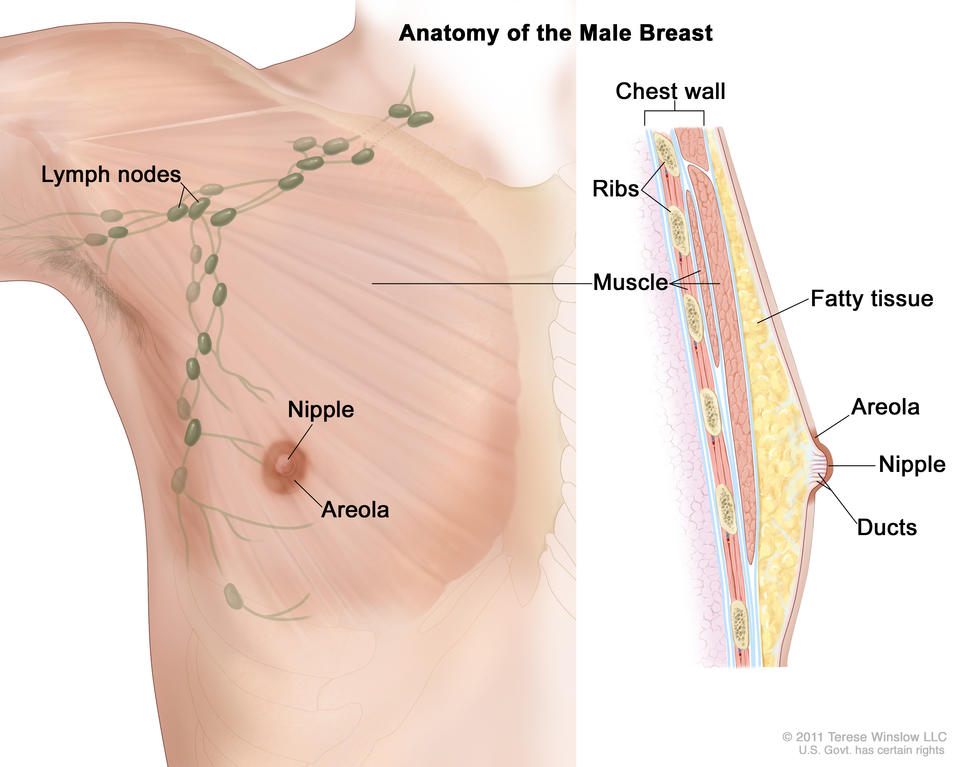
Drawing of male breast anatomy showing the lymph nodes, nipple, areola, chest wall, ribs, muscle, fatty tissue, and ducts.

Anatomy of the male breast. The nipple and areola are shown on the outside of the breast. The lymph nodes, fatty tissue, ducts, and other parts of the inside of the breast are also shown.

Credit: © Terese Winslow

Male breast cancer is breast cancer that develops in men. Breast cancer is much more common in women, but men can get breast cancer because they also have breast tissue where cancer can start. Fewer than 1 in 100 breast cancers in the United States occur in men. Men of any age can get breast cancer, but it usually occurs at a later age in men than in women.

Most male breast cancers start in the milk ducts of the breast and are called ductal cancers. Both female and male breasts have milk ducts, but in male breasts, milk ducts do not develop in the same way as they do in female breasts. Male breasts may also have milk glands called lobules, but far fewer than in female breasts. Cancers that start in the lobules, called lobular cancers, are rare in men.  

Other types of breast cancer are extremely rare in men, but they can happen. These include phyllodes tumor, Paget disease of the breast, and inflammatory breast cancer.

When abnormal cells are found in the ducts and have not spread to other tissues in the breast, it is called ductal carcinoma in situ. Invasive cancers have spread into surrounding breast tissue and can spread to nearby lymph nodes or other organs throughout the body. Most breast cancers in men are invasive. 

What are risk factors for male breast cancer?  

Older age is a risk factor for most cancers, including breast cancer in men. In addition, if you are male, you may have an increased risk of breast cancer if you have:

  • a history of radiation therapy directed at the chest
  • one or more female relatives who have had breast cancer
  • inherited changes in the BRCA1 or BRCA2 genes or in other genes that increase breast cancer risk
  • a condition linked to higher-than-normal estrogen levels in the body:
    • Klinefelter syndrome
    • liver disease (cirrhosis)
    • testicular problems, including inflamed testicles (orchitis), an undescended testicle, or surgery to remove one or both testicles
    • obesity 

Screening for male breast cancer

If you think you may have risk factors for male breast cancer, talk to your doctor. Screening mammograms are not usually recommended for men, even those who are at increased risk of breast cancer. But your doctor may suggest staying alert to any changes in your breast or the skin of your breast and getting regular clinical breast exams

What are the symptoms of breast cancer in men?  

Signs and symptoms of breast cancer in men include:

  • a lump or thickening in or near the breast (most common)
  • a nipple that changes in shape or the direction it is pointing
  • a nipple that is inverted
  • fluid from the nipple that may be clear or bloody
  • an open wound (ulcer) in the skin of the breast
  • scaly, red, or swollen skin on the breast, nipple, or areola
  • dimpling or ridges on the skin that resemble an orange peel
  • swollen lymph nodes under the arm or near the collarbone

Many breast changes in men are signs of less serious or benign conditions, but it is important to check with your doctor if you notice any unusual breast changes. 

How is breast cancer diagnosed in men? 

If you are male and have symptoms of breast cancer, your doctor will need to find out if they are due to cancer or another condition. Your doctor may:

If it is cancer, additional tests may include:  

  • Biomarker tests check the cancer cells for hormone receptors and HER2 protein that may help plan treatment. Male breast cancer is almost always hormone receptor positive. Learn more about Tests for Breast Cancer Biomarkers.  
  • Bone scan checks to see if cancer has spread to your bones.
  • PET scan or CT scan are imaging tests that help determine the extent of spread of the cancer.  
  • Genetic testing looks for inherited mutations in BRCA1, BRCA2, and several other genes that increase the risk of breast cancer. Such mutations, if present, can affect treatment decisions. They also have implications for family members.  

Your doctor will assign a stage to the cancer based on a combination of the extent of spread, hormone receptor and HER2 status, and other tumor features, such as tumor grade.

Learn more about the different stages of breast cancer and tests used to diagnose it at Breast Cancer Stages and How Breast Cancer Is Diagnosed

How is male breast cancer treated?

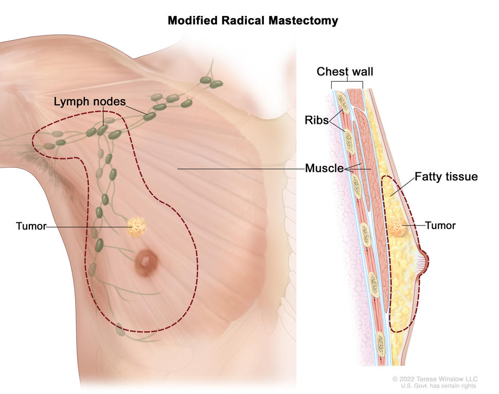
Male breast cancer is usually treated in the same way as breast cancer in women. However, men are more likely than women to have a mastectomy (surgery to remove the whole breast) rather than a lumpectomy (breast-conserving surgery) to remove the cancer because they have less breast tissue. But some male breast cancers can be treated with a lumpectomy, depending on the size of the cancer, the amount of breast tissue the person has, and other factors.

Learn more about breast cancer treatment at:
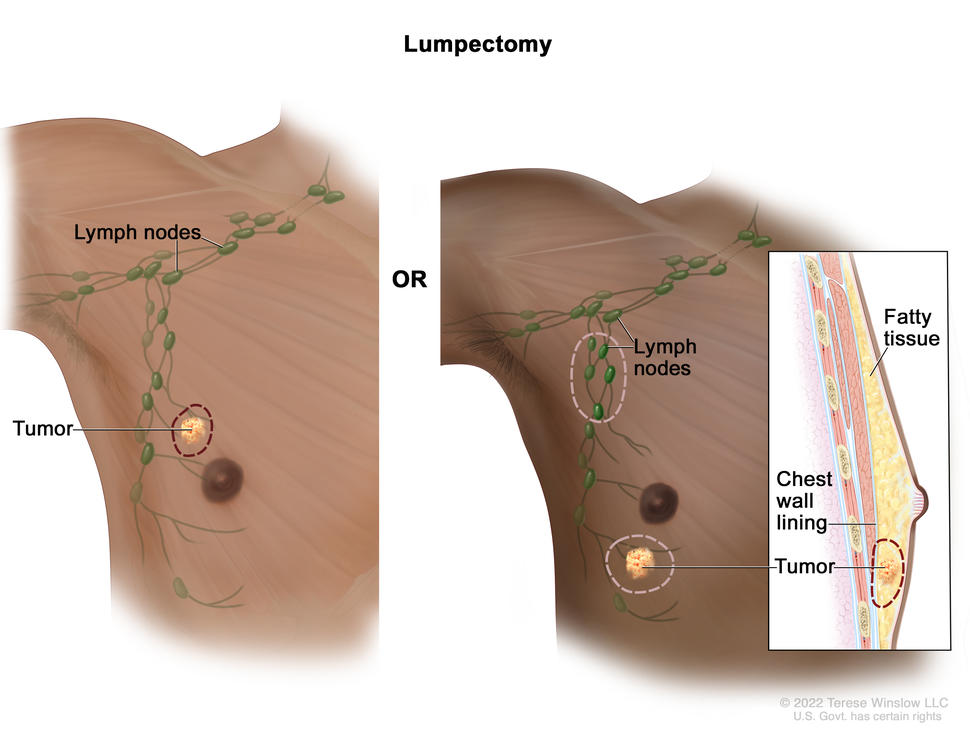
The drawing on the left shows removal of the tumor and some of the normal tissue around it. The drawing on the right shows removal of some of the lymph nodes under the arm and removal of the tumor and part of the chest wall lining near the tumor. Also shown is fatty tissue in the breast.

Modified radical mastectomy; the drawing on the left shows the removal of the whole breast, including the lymph nodes under the arm. The drawing on the right shows a cross-section of the breast, including the fatty tissue and chest wall (ribs  and muscle). A tumor is also shown in the breast.

What is the survival rate and prognosis for men with breast cancer?

Doctors estimate the prognosis for men with breast cancer by using statistics collected over many years. One common statistic is the 5-year relative survival rate. The 5-year relative survival rate tells you what percent of people with the same type and stage of male breast cancer are alive 5 years after their cancer was diagnosed, compared with people in the overall population.  

Men with breast cancer have a slightly lower 5-year survival rate than their female peers. This may be because male breast cancer is often diagnosed at a later stage than female breast cancer. Race also affects survival. Black men are usually diagnosed with a later stage of breast cancer than White men and are more likely to die of their disease.

More research is needed to find better treatments for male breast cancer and to better understand differences between male and female breast cancers.

The 5-year relative survival rates for people with male breast cancer are:  

  • 95% for localized male breast cancer (cancer is in the breast only)  
  • 84% for regional male breast cancer (cancer has spread beyond the breast to nearby lymph nodes or organs)  
  • 20% for metastatic male breast cancer (cancer has spread beyond the breast to a distant part of the body)  

To learn more about factors that affect breast cancer prognosis, visit Breast Cancer Prognosis and Survival Rates.   
Metastatic Breast Cancer

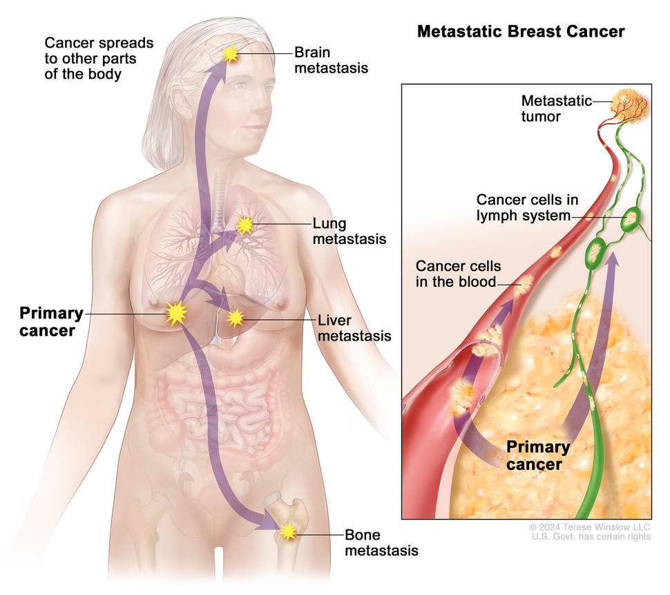
Drawing of a female body shows where primary breast cancer may spread, including the brain, lungs, liver, and bones. An inset shows a close-up view of cancer cells spreading from the breast through the blood and lymph system to another part of the body, to form a metastatic tumor.

In metastatic breast cancer, cancer cells break away from where they first formed in the breast, travel through the blood and lymph system, and form a new tumor in another part of the body. The cancer cells in the new, metastatic tumor are the same type of cells as those found in the primary breast cancer.

Credit: © Terese Winslow

What is metastatic breast cancer?

In metastatic breast cancer, cancer cells have broken away from where they began and traveled through the lymph system or blood to form a tumor in another part of the body.

A metastatic tumor is the same type of cancer as the original tumor. For example, if breast cancer spreads to the bone, the cancer cells in the bone are breast cancer cells. The tumor in the bone is treated as metastatic breast cancer, not bone cancer. Metastatic breast tumors most commonly develop in the bones, lungs, brain, or liver. 

Doctors may use the term “advanced breast cancer” to refer to metastatic breast cancer. This term is not the same as “locally advanced breast cancer,” which is breast cancer that has spread to nearby tissues or lymph nodes but not to other parts of the body.

To learn more about cancer that has spread to other parts of the body, visit Metastatic Cancer: When Cancer Spreads.

What are the symptoms of metastatic breast cancer?

The symptoms of metastatic breast cancer depend on where and the extent to which the cancer has spread.

Symptoms of metastatic breast cancer may include:

  • headache, seizures, or dizziness, if cancer has spread to the brain
  • jaundice or swelling in the belly, if cancer has spread to the liver
  • pain and fractures, if cancer has spread to the bone
  • shortness of breath, if cancer has spread to the lung
  • speech or vision changes, if cancer has spread to the brain
  • unusual fatigue

Sometimes, people with metastatic breast cancer do not have any symptoms, or they may be different from those that are described above. If you have been treated for breast cancer in the past and develop any unusual symptoms, it is important to contact your health care provider right away. Your doctor may recommend one or more tests to see if the cancer has returned. These symptoms may also be signs of other diseases or conditions, such as an infection or injury.

How is metastatic breast cancer diagnosed?

About 20% to 30% of women diagnosed with early-stage breast cancer eventually develop metastatic disease. This is sometimes called metastatic recurrence or distant recurrence. Metastatic breast cancer may develop months, years, or even decades after a person has completed treatment for early or locally advanced breast cancer.

In the United States, 5% to 6% of women with newly diagnosed breast cancer have de novo metastatic breast cancer (called stage IV breast cancer). This means that when breast cancer is detected for the first time, it has already spread to another part of the body.

The following tests may be used to diagnose metastatic breast cancer:

  • biopsy of the tumor to confirm metastatic breast cancer and determine whether it has certain biomarkers such as ER, PR, and HER2, which may be different from those in the original tumor
  • blood tests to see if the liver and other organs in the body are functioning properly
  • CT scan, bone scan, or other imaging tests to see where cancer may have spread in the body

What is the prognosis for people with metastatic breast cancer?

Doctors estimate metastatic breast cancer prognosis, or the likely outcome of disease, by using statistics collected over many years from people with metastatic breast cancer. One statistic that is commonly used in making a prognosis is the 5-year relative survival rate. The 5-year relative survival rate tells you what percent of people with the same type and stage of breast cancer are alive 5 years after their cancer was diagnosed, compared with people in the overall population.

The 5-year relative survival rate for women with metastatic breast cancer in the United States is 31%. Find more statistics for specific stages of breast cancer at Breast Cancer Prognosis & Survival Rates.

Because survival statistics are based on large groups of people, they cannot be used to predict exactly what will happen to you. Your doctor is in the best position to discuss these statistics and talk with you about your prognosis and treatment options.

To learn more about survival statistics and to see videos of patients and their doctors exploring their feelings about prognosis, visit Understanding Cancer Prognosis

How is metastatic breast cancer treated?

There are many treatment options for metastatic breast cancer. Often, the goal of treating metastatic cancer is to control it by stopping or slowing its growth. Some people can live for years with metastatic breast cancer that is well controlled. The goal of other treatments is to improve the quality of life by relieving symptoms. This type of care is called palliative care.

The treatment that you may have depends on the type of breast cancer you were originally diagnosed with, where it has spread, treatments you have had in the past, and your general health. Metastatic breast cancer may stop responding to drugs over time, so you may need to change treatments when this happens.

Treatments for metastatic breast cancer include:

Learn more about these breast cancer treatments.

Clinical trials

Joining a clinical trial may be an option. There are different types of clinical trials for people with metastatic breast cancer. For example, a treatment trial tests new treatments or new ways of using existing treatments. Supportive care and palliative care trials look at ways to improve quality of life, especially for those who have side effects from cancer and its treatment.

You can use the clinical trial search to find NCI-supported cancer clinical trials that are accepting participants. This search allows you to filter trials based on the type of cancer, your age, and where the trials are being done. You can also review a list of all current Breast Cancer Clinical Trials.

Learn more about clinical trials at Cancer Clinical Trial Information for Patients and Caregivers.

Ductal Carcinoma in Situ (DCIS)

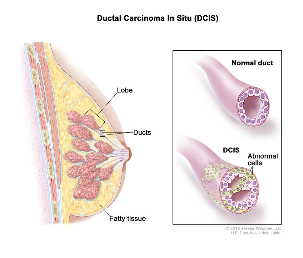
Illustration of Ductal Carcinoma In Situ (DCIS) in the breast. On the left, a cross-section of the breast shows the lobe, ducts, and fatty tissue. On the right, a close-up comparison of a normal duct and a duct affected by DCIS. The normal duct is shown with regular cells, while the DCIS duct shows abnormal cells inside.

Ductal carcinoma in situ (DCIS). Abnormal cells are found in the lining of a breast duct.

Credit: © Terese Winslow

What is DCIS?

Ductal carcinoma in situ (DCIS) is a condition in which abnormal cells form in the lining of the milk ducts but have not broken out of the milk ducts and spread to other breast tissue. DCIS is not cancer, but it may be called precancer because it may spread beyond the milk ducts to other parts of the breast or body, becoming invasive cancer. About 20%–25% of new breast cancer cases in the United States each year are DCIS. DCIS may also be called noninvasive breast cancer, intraductal carcinoma, or stage 0 breast cancer. 

What are symptoms of DCIS?

DCIS usually does not cause symptoms. Most cases of DCIS are found on a mammogram during routine breast cancer screening. DCIS may occasionally cause breast changes such as a lump or nipple discharge. 

How is DCIS diagnosed?

If you have symptoms or screening test results that suggest DCIS, your doctor will need to find out if they are due to DCIS or another condition. Mammograms, which are x-ray images of breast tissue, can reveal signs of DCIS called breast calcifications that appear as white specks on the image. Additional tests may include: 

  • Biopsy. In a biopsy, a surgeon removes cells or tissue so a pathologist can study them under a microscope. This will show whether the abnormal area is DCIS or whether the abnormal cells have spread to nearby tissue and become invasive breast cancer. The biopsy results will also reveal the tumor grade. Learn more about biopsies and biopsy results at How Breast Cancer Is Diagnosed.
  • Biomarker tests. These will check the abnormal cells for hormone receptors and help plan treatment. Although the amount of HER2 protein in breast cancer cells is a biomarker for invasive breast cancer, DCIS cells usually are not tested for HER2. Learn more about biomarker tests at Tests for Breast Cancer Biomarkers.  

How is DCIS treated? 

Although DCIS is not technically cancer because it has not spread beyond where it formed, it is treated like cancer because doctors don’t know which cases of DCIS will become invasive and spread to other parts of the body. Treatment for people with DCIS may include: 

What is the survival rate and prognosis for people with DCIS?

People with DCIS have an excellent prognosis, and most can be cured with treatment.  

Doctors estimate DCIS prognosis by using statistics collected over many years from people with DCIS. One statistic that is commonly used in making a prognosis is the 5-year relative survival rate. The 5-year relative survival rate tells you what percent of people with the same type and stage of breast cancer are alive 5 years after their cancer was diagnosed, compared with people in the overall population.  

More than 98% of people diagnosed with DCIS are alive 5 years after their diagnosis. DCIS is also less likely to recur than invasive forms of breast cancer.

Learn more about Breast Cancer Prognosis and Survival Rates

Breast Cancer Prognosis and Survival Rates

If you or someone close to you has just been diagnosed with breast cancer you may be asking: How serious is this cancer? What is the prognosis? What are survival rates among people with similar diagnoses?

Prognosis and survival rates vary from person to person and depend on many factors. Talk with your doctor to get a better understanding of the expected outcome of your cancer.

The survival statistics on this page refer to women with breast cancer. For information about prognosis and survival rates for men, visit Male Breast Cancer

Understanding your breast cancer prognosis

A breast cancer prognosis is the likely outcome or course of breast cancer. It may be helpful to think of a prognosis as an estimate or prediction. Understanding your prognosis will help you talk with your doctor and make decisions about treatment.  

Many factors affect your breast cancer prognosis, including:  

  • Type of breast cancer, such as ductal carcinoma in situ, invasive ductal carcinoma, inflammatory breast cancer, and triple-negative breast cancer. Learn about types of breast cancer.
  • The size of the tumor and whether it has spread beyond the breast tissue to the lymph nodes or to other places in the body.
  • Tumor grade, which describes how abnormal the cells look under the microscope, as well as how fast they are likely to grow and spread.
  • Biomarker status, in particular whether your tumor has receptors for the hormones estrogen and/or progesterone or has high levels of a protein called HER2. Learn about breast cancer biomarkers.
  • Personal factors, such as your age, medical history, family history, menopausal status, and ethnicity.
  • Treatment response, in particular how your cancer responds to treatment given before surgery.
  • Results from gene expression tests such as OncotypeDX, Mammaprint, or Breast Cancer Index. These tests, which are also called gene expression profiling or multigene tests, help predict the chances that cancer will spread or come back after surgery.  

Your doctor may tell you that you have a certain prognostic stage—stage 1, stage 2, stage 3, or stage 4. The prognostic stage combines information about several of the prognostic factors listed above, including the size and spread of the tumor, tumor grade, whether it has estrogen and/or progesterone receptors and HER2, and its score on a gene expression test. Learn more about Breast Cancer Stages.

As part of a discussion of breast cancer prognosis, your doctor can tell you about the chance that your cancer will return after treatment and how different treatments might affect that chance. To learn more about breast cancer recurrence and how it is diagnosed and treated, visit Recurrent Breast Cancer

Survival statistics for breast cancer

Doctors estimate breast cancer prognosis by using statistics collected over many years from people with breast cancer. One commonly used statistic is the 5-year relative survival rate. This statistic tells you what percentage of people with the same type and stage of breast cancer are alive 5 years after their cancer was diagnosed, compared with people in the overall population.  

The most recent 5-year relative survival rates for female breast cancer from NCI’s Surveillance, Epidemiology, and End Results (SEER) program, representing the period 2013–2019, are as follows:

  • For all types of breast cancer combined, 90.8%
  • For all types of breast cancer, based on how far breast cancer has spread at the time of diagnosis,
    • 99.3% for localized breast cancer (cancer is found only in the breast tissue and has not spread to nearby lymph nodes or other parts of the body)  
    • 86.3% for regional breast cancer (cancer has spread beyond the breast tissue to nearby lymph nodes or organs)
    • 31% for distant breast cancer (cancer has spread, or metastasized, from the original tumor to distant locations in the body; this is sometimes referred to as stage 4 or metastatic breast cancer)
  • For subtypes of breast cancer, based on hormone receptor and HER2 biomarkers,
    • 94.8% for HR+/HER2- breast cancer (localized, 100.0%; regional, 90.2%; distant, 34.0%)
    • 91.0% for HR+/HER2+ breast cancer (localized, 99.1%; regional, 89.8%; distant, 45.6%)
    • 85.6% for HR-/HER2+ breast cancer (localized, 97.2%; regional, 84.0%; distant, 39.5%)
    • 77.6% for HR-/HER2- (triple-negative) breast cancer (localized, 91.8%; regional, 66.2%; distant, 12.8%) 

Talking with your doctor to make sense of cancer statistics 

Because survival statistics are based on large groups of people, they cannot be used to predict exactly what will happen to you. Your doctor can help you understand how these statistics relate to your own situation.  

When reviewing survival statistics, it’s important to keep in mind that:  

  • responses to cancer treatment vary, even among women with the same type of breast cancer who are receiving the same treatment.  
  • it takes several years to see the effect of newer and better treatments, so the positive impact of improved treatments may not be reflected in current statistics.  

Learn more about a cancer prognosis and watch videos of people with cancer talking with their doctor in Understanding Cancer Prognosis.  

Additional breast cancer statistics  

NCI’s SEER program provides additional breast cancer statistics, including statistics on incidence, lifetime risk, prevalence, and deaths. Statistics are available for different racial and ethnic groups and different age groups. 

TNM Staging for Breast Cancer

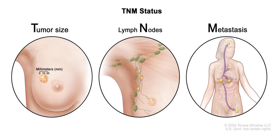
Breast cancer staging uses the TNM (tumor node metastasis) staging system to describe the size of the primary tumor and the spread of cancer to nearby lymph nodes or other parts of the body. The TNM value is combined with a grade and biomarker status (whether the tumor has certain hormone receptors or high levels of HER2) to determine the stage of breast cancer. Results from multigene tests may be used to help stage some breast cancers. Learn more about Breast Cancer Stages.

Three-panel drawing showing the TNM (tumor node metastasis) staging system for breast cancer. The first panel shows a tumor in the breast that measures 20 millimeters with a ruler. The second panel shows cancer that has spread from the breast to three nearby lymph nodes. The third panel shows cancer that has spread from the breast to other parts of the body, including the brain, lung, liver, and bone.

Breast cancer staging uses the TNM (tumor node metastasis) staging system to describe the size of the primary tumor and the spread of cancer to nearby lymph nodes or other parts of the body.

Credit: © Terese Winslow

Tumor (T)

The T category in the TNM staging system describes the size and location of the tumor:

TX: The primary tumor cannot be assessed.

T0: There is no sign of a primary tumor in the breast.

Tis: There is carcinoma in situ in the breast.

  • Tis (DCIS): Abnormal cells have been found in the breast ducts and have not spread past the layer of tissue where they began.
  • Tis (Paget disease): A rare form of early, noninvasive cancer where abnormal cells are found in the skin of the nipple and have not spread beyond where they first formed. No tumor or DCIS is found in the breast tissue under the nipple.

T1: The tumor is 20 millimeters or smaller. There are four subtypes of a T1 tumor depending on the size of the tumor:

  • T1mi: The tumor is 1 millimeter or smaller.
  • T1a: The tumor is larger than 1 millimeter but not larger than 5 millimeters.
  • T1b: The tumor is larger than 5 millimeters but not larger than 10 millimeters.
  • T1c: The tumor is larger than 10 millimeters but not larger than 20 millimeters.

T2: The tumor is larger than 20 millimeters but not larger than 50 millimeters.

T3: The tumor is larger than 50 millimeters.

T4: The tumor is described as one of the following:

  • T4a: The tumor has grown into the chest wall.
  • T4b: The tumor has grown into the skin. An ulcer has formed on the surface of the skin of the breast, small tumor nodules have formed in the same breast as the primary tumor, and/or the skin of the breast is swollen.
  • T4c: The tumor has grown into the chest wall and the skin.
  • T4d: One-third or more of the skin on the breast is red and swollen (called peau d’orange). T4d includes inflammatory breast cancer.

Lymph node (N)

This section describes pathological staging of lymph nodes. Pathological staging is done when the lymph nodes are removed by surgery and studied under a microscope by a pathologist. When the lymph nodes are checked using mammography or ultrasound, it is called clinical staging. Clinical staging of lymph nodes is not described here.

The N category describes the location of the lymph nodes where cancer has spread and the size of the cancer in the lymph nodes:

NX: The lymph nodes cannot be assessed.

N0: There is no sign of cancer in the lymph nodes, or there may be tiny clusters of cancer cells not larger than 0.2 millimeters in the lymph nodes.

N1: Cancer is described as one of the following:

  • N1mi: Cancer has spread to the axillary (armpit area) lymph nodes and is larger than 0.2 millimeters but not larger than 2 millimeters.
  • N1a: Cancer has spread to one to three axillary lymph nodes and the cancer in at least one of the lymph nodes is larger than 2 millimeters.
  • N1b: Cancer has spread to lymph nodes near the breastbone on the same side of the body as the primary tumor. The cancer is larger than 0.2 millimeters and is found by sentinel lymph node biopsy. Cancer is not found in the axillary lymph nodes.
  • N1c: Cancer has spread to one to three axillary lymph nodes, and the cancer in at least one of the lymph nodes is larger than 2 millimeters. Cancer is also found by sentinel lymph node biopsy in the lymph nodes near the breastbone on the same side of the body as the primary tumor.

N2: Cancer is described as one of the following:

  • N2a: Cancer has spread to four to nine axillary lymph nodes, and the cancer in at least one of the lymph nodes is larger than 2 millimeters.
  • N2b: Cancer has spread to lymph nodes near the breastbone, and the cancer is found by imaging tests. Cancer is not found in the axillary lymph nodes by sentinel lymph node biopsy or lymph node dissection.

N3: Cancer is described as one of the following:

  • N3a: Cancer has spread to 10 or more axillary lymph nodes. The cancer in at least one of the lymph nodes is larger than 2 millimeters, or the cancer has spread to lymph nodes below the collarbone.
  • N3b: Cancer is described as one of the following:
    • Cancer has spread to one to nine axillary lymph nodes, and the cancer in at least one of the lymph nodes is larger than 2 millimeters. Cancer has also spread to lymph nodes near the breastbone, and the cancer is found by imaging tests.
    • Cancer has spread to four to nine axillary lymph nodes, and the cancer in at least one of the lymph nodes is larger than 2 millimeters. Cancer has also spread to lymph nodes near the breastbone on the same side of the body as the primary tumor. The cancer in these lymph nodes is larger than 0.2 millimeters and is found by sentinel lymph node biopsy.
  • N3c: Cancer has spread to lymph nodes above the collarbone on the same side of the body as the primary tumor.

Metastasis (M)

The M category describes the spread of cancer to other parts of the body.

M0: There is no sign that cancer has spread to other parts of the body.

M1: Cancer has spread to other parts of the body, most often to the bones, lungs, liver, or brain. If cancer has spread to distant lymph nodes, the cancer in the lymph nodes is larger than 0.2 millimeters. The cancer is called metastatic breast cancer.

Breast Cancer Treatment by Stage

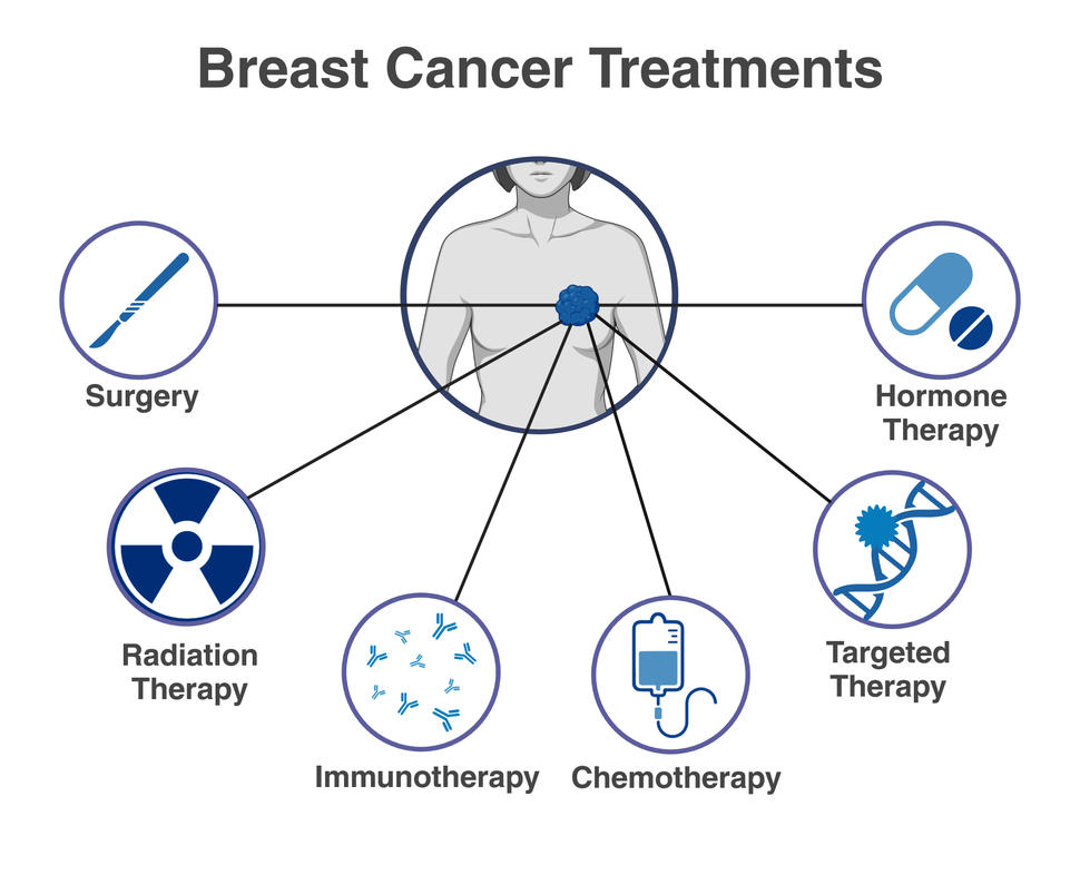
Drawing of a person with a tumor shown in their breast who is surrounded by icons representing different treatments, including chemotherapy, surgery, radiation therapy, immunotherapy, targeted therapy, and hormone therapy.

There are different types of treatment for breast cancer. You and your cancer care team will work together to decide on a treatment plan that’s best for you based on how advanced your cancer is and other factors. This may include one or more types of treatment.

Credit: Created by Nadia Jaber and Linda Wang with Biorender.com

Cancer stage, which is the extent of cancer in the body, is an important factor in determining which breast cancer treatments may work best for you. To learn how breast cancer is staged, visit Breast Cancer Stages.  

Other factors that will be considered when planning your treatment include:

  • the type of breast cancer you have
  • whether you have started menopause
  • whether you are pregnant
  • your age and general health  
  • your preferences  

You and your cancer care team will work together to decide your treatment plan. Most people receive more than one type of treatment.

The information on this page applies to most types of invasive breast cancer. To learn about the treatment of these and other less common types of breast cancer, visit Breast Cancer Treatment

Treatment of stage 0 breast cancer (ductal carcinoma in situ)

Ductal carcinoma in situ (DCIS) is a condition where abnormal cells form in the lining of the milk ducts but have not spread. It’s not cancer, but it may develop into invasive cancer.  

Treatment of ductal carcinoma in situ may include lumpectomy with radiation therapy, mastectomy with or without radiation, and hormone therapy if biomarker testing suggests it may be helpful.

To learn more about DCIS and how it is treated, visit Ductal Carcinoma in Situ.

Treatment of stages I to III breast cancer

Treatment of early-stage breast cancer, which includes stages I, IIA, and some stage IIB breast cancers, usually begins with surgery to remove the cancer. Surgery is often followed by additional therapy. If the tumor is large, chemotherapy or targeted therapy may be given before surgery to make the tumor smaller and easier to remove.

Treatment of locally advanced breast cancer, which includes some stage IIB breast cancers and stages IIIA, IIIB, and IIIC, often begins with chemotherapy, followed by surgery and radiation.  

Palliative Care

Palliative care can be given at any stage of disease. It aims to improve quality of life by managing symptoms and providing support to patients and their families. Palliative care can be provided alongside curative treatments, such as chemotherapy, or as the focus of care. Learn more at Palliative Care in Cancer.

Treatment of stages I to III breast cancer may include:

  • Surgery. Lumpectomy (breast-conserving surgery) is often used, but mastectomy may be recommended if the tumor is large or cancer is found in multiple areas of the breast or chest. The surgeon may also remove the underarm lymph node closest to the tumor in a procedure called sentinel lymph node biopsy. Learn more about Breast Cancer Surgery and Sentinel Lymph Node Biopsy.  
  • Radiation therapy. After surgery, you will likely have radiation therapy to the breast or the chest wall where the breast was removed to reduce the chance of breast cancer returning in the breast or the chest wall. However, you may not need radiation therapy if you have a mastectomy. Learn more about Radiation Therapy for Breast Cancer.  
  • Chemotherapy. You are more likely to receive chemotherapy if the tumor is high grade, in the lymph nodes, HER2-positive, or triple-negative. Whether chemotherapy is given before or after surgery depends on the size and location of the tumor and other factors. Learn more about Chemotherapy for Breast Cancer.
  • Hormone therapy. If the cancer tests positive for estrogen receptor and/or progesterone receptor, or when the hormone receptor status of the cancer is unknown, you may receive hormone therapy as part of your treatment. Hormone therapy for breast cancer may begin before or after surgery. If you are premenopausal, you might also have treatment to stop the ovaries from making hormones. Learn more about Hormone Therapy for Breast Cancer.
  • Immunotherapy. This cancer treatment is only used to treat triple-negative breast cancer. Learn about this type of cancer and how it is treated at Triple-Negative Breast Cancer and Immunotherapy for Breast Cancer.
  • Targeted therapy. If biomarker tests suggest that the cancer is HER2-positive, hormone-receptor positive, or has a BRCA1 or BRCA2 mutation, you may receive targeted therapy. Targeted therapy for breast cancer may be given at different times in your treatment. It might be started before or after surgery. It can also be given even if surgery is not recommended for you. Learn more about Targeted Therapy for Breast Cancer.  
Join a Clinical Trial

Joining a clinical trial may be an option. There are different types of clinical trials for people with breast cancer. You can use the clinical trial search to find NCI-supported cancer clinical trials that are accepting participants. You can also review a list of all current NCI-supported Breast Cancer Clinical Trials

Get live help finding a clinical trial at 1-800-4-CANCER. Learn more about clinical trials at Cancer Clinical Trial Information for Patients and Caregivers.

Treatment of locoregional recurrent breast cancer

Sometimes breast cancer comes back after treatment. Locoregional recurrent breast cancer is cancer that has come back in the breast, the chest wall, or nearby lymph nodes after treatment. Learn more about Breast Cancer Recurrence and how it is treated.

Treatment of stage IV (metastatic) breast cancer

Breast cancer that has spread beyond the breast, chest wall, or nearby lymph nodes is called metastatic breast cancer. Treatment of metastatic breast cancer focuses on slowing the spread of the cancer and controlling the symptoms. Some of the treatments used for stages I to III breast cancer may also be used for metastatic breast cancer. The choice of treatments will partly depend on how the cancer responded to the treatments you have already received and what you want from treatment. Learn more about Metastatic Breast Cancer and how it is treated. 

Stages of Breast Cancer

A cancer stage describes the extent of cancer in the body and whether the cancer has spread from where it first formed to other parts of the body. It is important to know the stage of breast cancer to plan your treatment and understand your prognosis.

In breast cancer, stage is based on the size and location of the primary tumor, the spread of cancer to nearby lymph nodes or other parts of the body, the grade of the tumor, and whether certain biomarkers are present.

Learn about tests to stage breast cancer at How Breast Cancer Is Diagnosed in the section, “Tests to stage breast cancer.”

How breast cancer stage is determined

There are two types of staging for breast cancer. Staging done before surgery is called clinical prognostic staging, and staging done after surgery is called pathological prognostic staging. 

  • Clinical prognostic stage is used first to assign a stage for anyone diagnosed with breast cancer. It is based on health history, physical examination, imaging tests (including mammography or ultrasound examination of the lymph nodes), and biopsy findings.
  • Pathological prognostic stage is used only for people who have surgery as their first treatment for breast cancer. The pathological prognostic stage is based on all clinical information, biomarker status, and laboratory test results from breast tissue and lymph nodes removed during surgery.

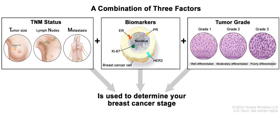
In both types of staging, a number between 0 and IV (4) is assigned. This number is determined by a combination of three factors: TNM (tumor node metastasis) value, grade, and biomarker status (whether the tumor has certain hormone receptors or high levels of HER2). Results from multigene tests may be used to help stage some breast cancers. Generally, the lower the stage number, the less the cancer has spread.

Three-panel image showing TNM status, biomarkers, and tumor grade used to determine breast cancer stage.

In breast cancer, stage is based on the TNM status (the size and location of the primary tumor and the spread of cancer to nearby lymph nodes or other parts of the body), the presence of certain biomarkers, and the grade of the tumor.

Credit: © Terese Winslow

TNM staging

TNM staging uses T, N, and M categories to describe the size of the primary tumor and whether the cancer has spread to nearby lymph nodes or other parts of the body. 

  • Tumor (T): The size and location of the tumor.
  • Lymph node (N): The location of the lymph nodes where cancer has spread and the size of the cancer in the lymph nodes.
  • Metastasis (M): The spread of cancer to other parts of the body.

The numbers or letters after T, N, and M provide more details about the cancer. Higher numbers generally mean the cancer is more advanced.

To learn more, review the full TNM staging descriptions for breast cancer.

The grading system

Tumor grade describes how abnormal the cancer cells and tissue look under a microscope and how quickly the cancer cells are likely to grow and spread. Low-grade cancer cells look more like normal cells and tend to grow and spread more slowly than high-grade cancer cells. To describe how abnormal the cancer cells and tissue are, the pathologist will assess the following three features:

  • how much of the tumor tissue has normal breast ducts
  • the size and shape of the nuclei in the tumor cells
  • how many dividing cells are present, which is a measure of how fast the tumor cells are growing and dividing

For each feature, the pathologist assigns a score of 1 to 3. A score of 1 means the cells and tumor tissue look the most like normal cells and tissue, and a score of 3 means the cells and tissue look the most abnormal. The scores for each feature are added together to get a total score between 3 and 9.

Three grades are possible:

  • total score of 3 to 5: G1 (low grade or well differentiated)
  • total score of 6 to 7: G2 (intermediate grade or moderately differentiated)
  • total score of 8 to 9: G3 (high grade or poorly differentiated)

Biomarker testing

Biomarker testing is used to find out whether breast cancer cells have certain receptors (biomarkers), including:

  • Estrogen receptor (ER). If the breast cancer cells have estrogen receptors, the cancer cells are called ER positive (ER+). If the breast cancer cells do not have estrogen receptors, the cancer cells are called ER negative (ER-).
  • Progesterone receptor (PR). If the breast cancer cells have progesterone receptors, the cancer cells are called PR positive (PR+). If the breast cancer cells do not have progesterone receptors, the cancer cells are called PR negative (PR-).
  • Human epidermal growth factor type 2 receptor (HER2/neu or HER2). If the breast cancer cells have more than the normal amount of HER2 receptors on their surface, the cancer cells are called HER2 positive (HER2+). If the breast cancer cells have a normal amount of HER2 on their surface, the cancer cells are called HER2 negative (HER2-). HER2+ breast cancer is more likely to grow and divide faster than HER2- breast cancer.

Learn more about Tests for Breast Cancer Biomarkers.

Stage 0 breast cancer

Stage 0 is a noninvasive breast cancer, such as ductal carcinoma in situ (DCIS). In stage 0, abnormal cells are found in the breast, but there is no evidence that these cells have spread from where they first formed. Stage 0 breast cancers can be any grade and have any biomarker (HER2, ER, PR) status.

Learn about treatment for DCIS at Ductal Carcinoma in Situ in the section, “How is DCIS treated?”

Stage I (1) breast cancer

In stage I breast cancer, the cancer is small and only in the breast tissue, or it might be found in lymph nodes close to the breast. Stage I can be divided into stage IA and stage IB. The difference is determined by the size of the tumor and whether there are any lymph nodes with cancer.

Learn about treatment of stage I breast cancer.

Stage IA

Stage IA breast cancer generally has a TNM score of T0 or T1, N0 or N1, and M0. It can be any grade and different combinations of HER2, ER, and PR status. To learn more about the different staging factors, visit TNM Staging for Breast Cancer.

TNM Grade HER2 Status ER Status PR Status
T1, N0, M0 G1 Positive Positive Any
T0, N1mi, M0 G1 Positive Negative Any
T1, N1mi, M0 G1 or G2 Negative Positive Any
T1, N1mi, M0 G1 or G2 Negative Negative Positive
T1, N1mi, M0 G2 or G3 Positive Any Any
T1, N1mi, M0 G3 Negative Positive Positive

Abbreviations: T = tumor N = lymph node M = metastasis, HER2 = human epidermal growth factor type 2 receptor, ER = estrogen receptor, PR = progesterone receptor.

Stage IB

Stage IB breast cancer generally has a TNM score of T0 or T1, N0 or N1, and M0. It can be any grade and different combinations of HER2, ER, and PR status. To learn more about the different staging factors, visit TNM Staging for Breast Cancer.

TNM Grade HER2 Status ER Status PR Status
T1, N1mi, M0 G1 or G2 Negative Negative Negative
T0, N1, M0
T1, N1, M0
T2, N0, M0
G1 or G2 Any Positive Positive
T0, N1, M0
T1, N1, M0
T2, N0, M0
G3 Positive Positive Positive
T2, N1, M0
T3, N0, M0
Any Positive Positive Positive
T1, N1mi, M0 G3 Negative Positive Negative
T1, N1mi, M0 G3 Negative Negative Any
T0, N1, M0
T1, N1, M0
T2, N0, M0
G3 Positive Positive Positive

Stage II (2) breast cancer

In stage II breast cancer, there is cancer in the breast or nearby lymph nodes or both. This stage is divided into groups: Stage IIA and Stage IIB. The difference is determined by the size of the tumor and whether the breast cancer has spread to the lymph nodes. 

Learn about treatment of stage II breast cancer.

Stage IIA

Stage IIA breast cancer generally has a TNM score of T0, T1, or T2; N0 or N1; and M0. It can be any grade and different combinations of HER2, ER, and PR status. To learn more about the different staging factors, visit TNM Staging for Breast Cancer.

TNM Grade HER2 Status ER Status PR Status
T0, N1, M0
T1, N1, M0
T2, N0, M0
G1 or G2 Positive Positive Negative
T0, N1, M0
T1, N1, M0
T2, N0, M0
G1 or G2 Positive Negative Any
T0, N1, M0
T1, N1, M0
T2, N0, M0
G1 or G2 Negative Positive Negative
T0, N1, M0
T1, N1, M0
T2, N0, M0
G1 Negative Negative Any
T0, N1, M0
T1, N1, M0
T2, N0, M0
G2 Negative Negative Positive

Stage IIB

Stage IIB breast cancer generally has a TNM score of T0, T1, or T2; N0 or N1; and M0. It can be any grade and different combinations of HER2, ER, and PR status. To learn more about the different staging factors, visit TNM Staging for Breast Cancer.

TNM Grade HER2 Status ER Status PR Status
T0, N1, M0
T1, N1, M0
T2, N0, M0
G2 Negative Negative Negative
T0, N1, M0
T1, N1, M0
T2, N0, M0
G3 Negative Positive Negative
T0, N1, M0
T1, N1, M0
T2, N0, M0
G3 Negative Negative Any
T2, N1, M0
T3, N0, M0
G1 or G2 Positive Negative Negative
T2, N1, M0
T3, N0, M0
G1 or G2 Negative Positive Negative
T2, N1, M0
T3, N0, M0
G1 or G2 Negative Negative Any
T2, N1, M0
T3, N0, M0
G3 Positive Positive Negative
T2, N1, M0
T3, N0, M0
G3 Positive Negative Any
T2, N1, M0
T3, N0, M0
G3 Negative Positive Positive
T0, N2, M0
T1, N2, M0
T2, N2, M0
T3, N1, M0
T3, N2, M0
G3 Positive Positive Positive

Stage III (3) breast cancer

In stage III breast cancer, the cancer is found in the lymph nodes close to the breast, the skin of the breast, or the chest wall.

Learn about treatment of stage III breast cancer.

Stage IIIA

Stage IIIA breast cancer generally has a TNM score of any T, any N, and M0. It can be any grade and different combinations of HER2, ER, and PR status. To learn more about the different staging factors, visit TNM Staging for Breast Cancer.

TNM Grade HER2 Status ER Status PR Status
T2, N1, M0
T3, N0, M0
G3 Negative Positive Negative
T2, N1, M0
T3, N0, M0
G3 Negative Negative Positive
T0, N2, M0
T1, N2, M0
T2, N2, M0
T3, N1, M0
T3, N2, M0
Any Positive Positive Negative
T0, N2, M0
T1, N2, M0
T2, N2, M0
T3, N1, M0
T3, N2, M0
Any Positive Negative Any
T0, N2, M0
T1, N2, M0
T2, N2, M0
T3, N1, M0
T3, N2, M0
G1 or G2 Negative Positive Negative
T0, N2, M0
T1, N2, M0
T2, N2, M0
T3, N1, M0
T3, N2, M0
G2 Negative Negative Positive
T0, N2, M0
T1, N2, M0
T2, N2, M0
T3, N1, M0
T3, N2, M0
G3 Negative Positive Positive
T4, N0, M0
T4, N1, M0
T4, N2, M0
Any T, N3, M0
G1 or G2 Positive Positive Positive

Stage IIIB

Stage IIIB breast cancer generally has a TNM score of any T, any N, and M0. It can be any grade and different combinations of HER2, ER, and PR status. To learn more about the different staging factors, visit TNM Staging for Breast Cancer.

TNM Grade HER2 Status ER Status PR Status
T2, N1, M0
T3, N0, M0
G2 or G3 Negative Negative Negative
T0, N2, M0
T1, N2, M0
T2, N2, M0
T3, N1, M0
T3, N2, M0
G1 or G2 Negative Negative Negative
T0, N2, M0
T1, N2, M0
T2, N2, M0
T3, N1, M0
T3, N2, M0
G3 Negative Positive Negative
T0, N2, M0
T1, N2, M0
T2, N2, M0
T3, N1, M0
T3, N2, M0
G3 Negative Negative Positive
T4, N0, M0
T4, N1, M0
T4, N2, M0
Any T, N3, M0
G1 or G2 Positive Positive Negative
T4, N0, M0
T4, N1, M0
T4, N2, M0
Any T, N3, M0
G1 or G2 Positive Negative Any
T4, N0, M0
T4, N1, M0
T4, N2, M0
Any T, N3, M0
G1 or G2 Negative Positive Any
T4, N0, M0
T4, N1, M0
T4, N2, M0
Any T, N3, M0
G1 or G2 Negative Negative Positive
T4, N0, M0
T4, N1, M0
T4, N2, M0
Any T, N3, M0
G3 Positive Any Any
T4, N0, M0
T4, N1, M0
T4, N2, M0
Any T, N3, M0
G3 Negative Positive Positive

Stage IIIC

Stage IIIC breast cancer generally has a TNM score of any T, any N, and M0. It can be any grade and different combinations of HER2, ER, and PR status. To learn more about the different staging factors, visit TNM Staging for Breast Cancer.

TNM Grade HER2 Status ER Status PR Status
T0, N2, M0
T1, N2, M0
T2, N2, M0
T3, N1, M0
T3, N2, M0
G3 Negative Negative Negative
T4, N0, M0
T4, N1, M0
T4, N2, M0
Any T, N3, M0
G1 or G2 Negative Negative Negative
T4, N0, M0
T4, N1, M0
T4, N2, M0
Any T, N3, M0
G3 Negative Positive Negative
T4, N0, M0
T4, N1, M0
T4, N2, M0
Any T, N3, M0
G3 Negative Negative Any

Stage IV (4) or metastatic breast cancer

Stage IV breast cancer means that the cancer has spread to other parts of the body, such as the bones, liver, or lungs. This stage is also called metastatic breast cancer.

Stage IV breast cancer generally has a TNM score of any T, any N, and M1. It can be any grade and different combinations of HER2, ER, and PR status. To learn more about the different staging factors, visit TNM Staging for Breast Cancer.

TNM Grade HER2 Status ER Status PR Status
Any T, Any N, M1 Any Any Any Any

Learn about treatment for metastatic breast cancer at Metastatic Breast Cancer in the section, “How is metastatic breast cancer treated?”

Recurrent breast cancer

Recurrent breast cancer is cancer that has recurred (come back) after it has been treated. Breast cancer may come back in the breast (local recurrence); nearby lymph nodes, chest, or skin (regional recurrence); or other parts of the body, such as the liver, lung, or bone (distant or metastatic recurrence).

To figure out the type of recurrence you have, you will have many of the same tests you had when your cancer was first diagnosed, such as lab tests and imaging procedures. These tests help determine where in your body the cancer has returned, if it has spread, and how far. Your doctor may refer to this new assessment of your cancer as “restaging.”

After these tests, the doctor may assign a new stage to the cancer. A letter “r” will be added to the beginning of the new stage to reflect the restaging.

Learn about how recurrent breast cancer is treated at Breast Cancer Recurrence in the section, “How is recurrent breast cancer treated?”

Triple-Negative Breast Cancer (TNBC)

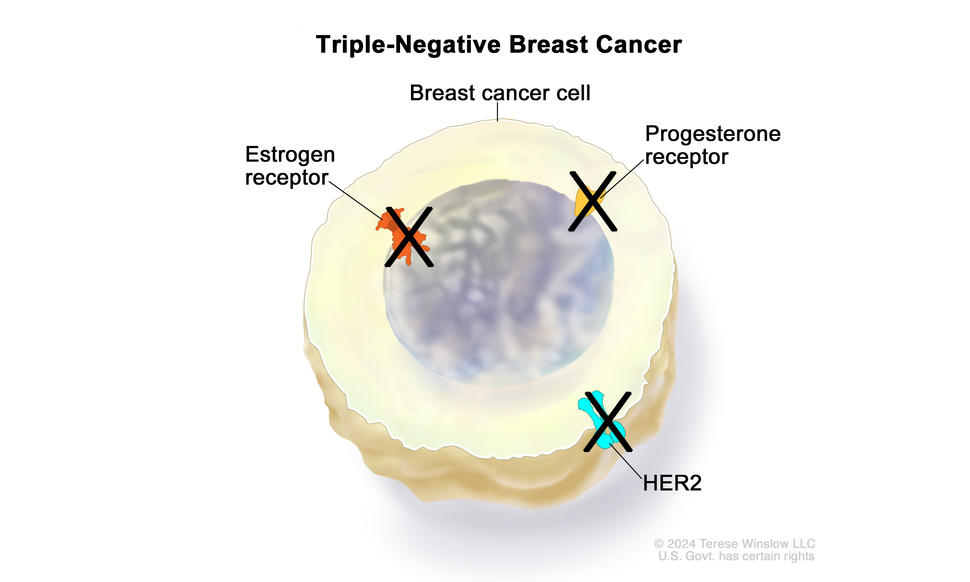
Drawing showing a triple-negative breast cancer cell with the estrogen receptor, progesterone receptor, and HER2 proteins crossed out with a black "X" mark. This means that the breast cancer cell does not have these proteins.

Triple-negative breast cancer is a type of breast cancer in which the cancer cells do not have estrogen receptors, progesterone receptors, or large amounts of HER2/neu protein on their surface.

Credit: © Terese Winslow

What is triple-negative breast cancer?

When breast cancer is triple negative, it lacks hormone receptors for estrogen and progesterone, and it makes little or none of the protein HER2. In other words, it is negative for all three biomarkers

Triple-negative breast cancer (TNBC) makes up about 15% of all breast cancers. It is usually diagnosed at a later stage, grows more quickly, and is more likely to come back than other types of invasive breast cancer. Because TNBC grows quickly, it is rarely diagnosed as ductal carcinoma in situ (stage 0) and is almost always invasive.

What are the risk factors for triple-negative breast cancer?

TNBC is more common in certain age groups and populations. TNBC is: 

  • often diagnosed at a younger age
  • more common and diagnosed at a younger age in Black women than in White women
  • more common in women with certain gene changes, including changes in BRCA1 or BRCA2 genes

The cause of TNBC is unknown, and other risk factors are the same as for other types of breast cancer. For more information, visit Breast Cancer Causes and Risk Factors.

What are the symptoms of triple-negative breast cancer?

Symptoms of TNBC are the same as for most other breast cancers (except inflammatory breast cancer). However, TNBC does not always cause symptoms.

Learn more about Breast Cancer Symptoms.

How is triple-negative breast cancer diagnosed?

If you have symptoms or screening test results that suggest breast cancer, your doctor will need to find out if they are due to cancer or another condition. Your doctor may: 

If it is cancer, additional tests may include: 

  • Biomarker tests. These will reveal if the cancer is triple negative. Triple negative means the results of biomarker tests showed that the cancer cells do not have hormone receptors and make little or no HER2 protein. Learn more about Tests for Breast Cancer Biomarkers.  
  • PET scan or CT scan. These imaging tests will determine the extent of spread of the cancer. 

Your doctor will assign a stage to the cancer based on a combination of the extent of spread, triple-negative status, and other tumor features, such as tumor grade.  

Learn more about tests used to diagnose breast cancer and the different stages of breast cancer at How Breast Cancer Is Diagnosed and Breast Cancer Stages.

How is triple-negative breast cancer treated?

TNBC can be more difficult to treat than other types of breast cancer because the cancer cells lack hormone receptors and do not make much or any of the protein HER2. This means therapies that target those biomarkers don’t work for people with TNBC. However, chemotherapy and other treatments, including immunotherapy, are available. 

For information about how TNBC is treated, visit Triple-Negative Breast Cancer Treatment.

What is the survival rate and prognosis for people with triple-negative breast cancer?

People with TNBC generally have a less favorable prognosis and survival rate than people with other types of breast cancer. That is because TNBC grows more quickly and is more likely to come back than other types of breast cancer. If you are diagnosed with TNBC, your prognosis will depend on many factors, including the stage of the cancer when it is diagnosed and your age and general health.

Survival rates for triple-negative breast cancer

Doctors estimate TNBC prognosis by using statistics collected over many years from people with TNBC. One statistic that is commonly used in making a prognosis is the 5-year relative survival rate. The 5-year relative survival rate tells you what percent of people with the same type and stage of breast cancer are alive 5 years after their cancer was diagnosed, compared with people in the overall population.

The 5-year relative survival rates for TNBC are:

  • 91% for localized TNBC (cancer is in the breast only)
  • 66% for regional TNBC (cancer has spread beyond the breast to nearby lymph nodes or organs)
  • 12% for metastatic TNBC (cancer has spread beyond the breast to a distant part of the body)

To learn more about factors that affect breast cancer prognosis, visit Breast Cancer Prognosis and Survival Rates

Tests for Breast Cancer Biomarkers

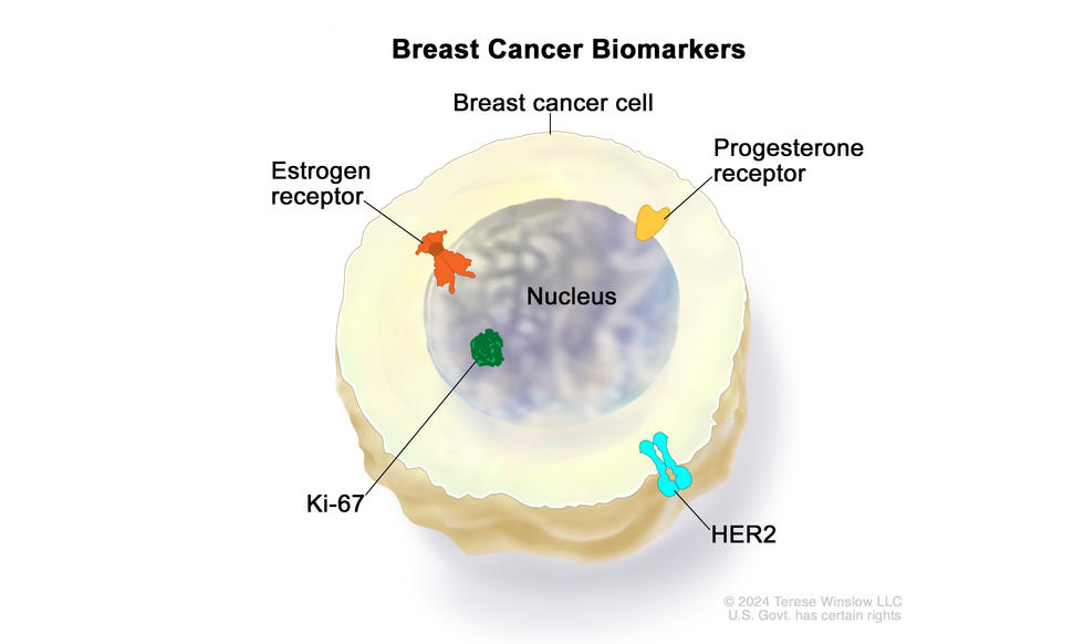
Drawing showing a breast cancer cell with estrogen receptor, progesterone receptor, and Ki-67 proteins inside the nucleus of the cell. Also shown is a HER2 protein on the surface of the cell.

Breast cancer cells are usually tested for certain biomarkers, including  estrogen receptors, progesterone receptors, and HER2, to help determine the stage of the cancer and plan treatment. Breast cancer cells may also be tested for the Ki-67 protein, which is found only in cells that are dividing. The results of this test may help predict how well the cancer will respond to certain treatments.

Credit: © Terese Winslow

Once breast cancer is diagnosed, breast cancer cells are tested for certain biomarkers and tumor features. To check for biomarkers, samples of tissue containing breast cancer cells are removed during a biopsy or surgery. Then the samples are tested in a lab. In most cases, the sample that was used to diagnose the cancer will also be used for biomarker testing. Learn more about tests to diagnose and stage breast cancer at How Breast Cancer Is Diagnosed.

Biomarker tests give information about:

  • how quickly the cancer may grow
  • how likely it is that the cancer will spread through the body
  • how well certain treatments might work

There are many types of biomarker tests for breast cancer. Tests used most often include:

  • estrogen receptor (ER) and progesterone receptor (PR) tests (known together as hormone receptor tests)
  • HER2 test
  • Ki-67 score

Breast cancer hormone receptor status

Healthy cells in many parts of the body, including the breast, and some breast cancer cells, have receptors that attach to the hormones estrogen and progesterone. These receptors tell cells to grow when estrogen and progesterone are present. When your cancer cells have hormone receptors, it means that one or both of these hormones can fuel cancer growth.

Breast cancer cells that contain estrogen receptors are called estrogen receptor positive (ER positive or ER+). Those that contain progesterone receptors are called progesterone receptor positive (PR positive, PgR positive, or PR+). Most ER-positive breast cancers are also PR positive. Breast cancers with one or both of these receptors are sometimes called hormone receptor positive (HR positive or HR+).

HR-positive breast cancer is often treated with hormone therapy. Some hormone therapies slow or stop the growth of HR-positive cancer by blocking the body’s ability to produce hormones. Others work by interfering with effects of hormones on breast cancer cells.

Breast cancer cells that don’t have estrogen receptors are called estrogen receptor negative (ER negative or ER-). Those that don’t have progesterone receptors are called progesterone receptor negative (PR negative or PR-). When breast cancer cells do not have hormone receptors, they are sometimes called hormone receptor negative (HR negative or HR-). 

HR-negative breast cancer probably won’t respond to treatments that block hormones in the body. In these cases, other treatment options, such as chemotherapy, are available.

Hormone receptor status can change over time. So another biopsy to test the cancer for hormone receptors may be needed if your cancer comes back after treatment.

Breast cancer HER2 status

HER2 is a protein that helps control breast cell growth. Some breast cancers have too much HER2, which causes the breast cells to grow and divide more quickly than normal. This is called HER2-positive breast cancer. HER2-positive breast cancer is more likely to be fast-growing than cancers with less HER2 (HER2 negative or HER2 low). HER2 status helps determine whether drugs that target HER2 will help treat the cancer.

Ki-67 score

Ki-67 is a protein that is found only in cells that are dividing. If your tumor has a high number of cancer cells with the Ki-67 protein, it means the cancer cells are dividing quickly. The results of this test may predict how well your cancer will respond to certain treatments, such as chemotherapy.

Biomarker test results and molecular subtypes of breast cancer

Your doctor may use biomarker test results to assign your breast cancer to one of the following groups or subtypes. These groups help show what treatment options may be most effective for you. They also help your doctor estimate the chances that your cancer will come back after treatment.

Subtype of breast cancer Defining features Treatment options
Luminal A
  • grade 1 or 2
  • ER positive and PR positive
  • HER2 negative 
    have low levels of
  • Ki-67

Most breast cancers are luminal A.

  • hormone therapy
  • chemotherapy
Luminal B
  • ER positive and PR negative
  • HER2 positive or negative
  • often have high levels of Ki-67
  • hormone therapy
  • chemotherapy
  • possibly HER2-targeted therapies
HER2-enriched
  • HER2 positive
  • ER positive or negative and PR positive or negative
  • often grow more quickly than luminal A and B
  • HER2-targeted therapies
  • chemotherapy
  • hormone therapy
HER2-low
  • have some HER2 protein but not enough to be classified as HER2 positive
  • ER positive or negative and PR positive or negative
  • certain HER2-targeted therapies
  • chemotherapy
  • hormone therapy
Basal-like (also called triple-negative)
  • ER negative and PR negative
  • HER2 negative
  • grow more quickly and are more likely to recur than other forms of breast cancer

Breast Cancer Surgery

Most people with breast cancer or ductal carcinoma in situ (DCIS) will have surgery to remove the tumor and nearby breast tissue. The two main types of surgery are lumpectomy (breast-conserving surgery) and mastectomy, with or without breast reconstruction.

Breast cancer surgery may also involve checking nearby lymph nodes for cancer. Usually, a sentinel lymph node biopsy is done, often during a lumpectomy or mastectomy but sometimes as a separate surgery. Learn more about sentinel lymph node biopsy at How Breast Cancer Is Diagnosed.