How Breast Cancer Is Diagnosed

If you have symptoms or screening test results that suggest breast cancer, your doctor will need to find out if they are due to cancer or some other cause. Your doctor may:  

Depending on your symptoms and medical history, your doctor may recommend tests to find out if you have breast cancer. The following tests and procedures are used to diagnose breast cancer. If results of these tests show that you have breast cancer, they will also help you and your doctor plan treatment. 

Imaging tests to diagnose breast cancer
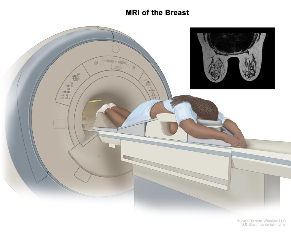
MRI of the breast; drawing shows a person lying face down on a narrow, padded table with their arms above their head. The person’s breasts hang down into an opening in the table. The table slides into the MRI machine, which takes detailed pictures of the inside of the breast. An inset shows an MRI image of the insides of both breasts.

An MRI of the breast is a procedure that uses radio waves, a strong magnet, and a computer to create detailed pictures of the inside of the breast. A contrast dye may be injected into a vein (not shown) to make the breast tissues easier to see on the MRI pictures. An MRI may be used with other breast imaging tests to detect breast cancer or other abnormal changes in the breast. It may also be used to screen for breast cancer in some people who have a high risk of the disease. Note: The inset shows an MRI image of the insides of both breasts. Credit for inset: The Cancer Imaging Archive (TCIA).

Credit: © Terese Winslow

  • A diagnostic mammogram is an x-ray picture of the breast that checks for breast cancer. It is used after a lump or other symptom of the disease has been found. It is also used to follow up on breast changes found during a screening mammogram. A diagnostic mammogram often involves taking more detailed x-ray pictures of the breast from different angles to check the abnormal area more closely.  
  • Breast ultrasound is a procedure that uses high-energy sound waves to make pictures of the inside of the breast.  
  • Breast MRI uses a powerful magnet, radio waves, and a computer to take detailed pictures of areas inside the breast.

Biopsy for breast cancer

You may have a biopsy if imaging tests find a lump or other abnormal area in the breast. In a biopsy, a surgeon removes cells or tissue so a pathologist can study them under a microscope. A biopsy is the only sure way to diagnose breast cancer.  

The following types of biopsies may be used to check for breast cancer:

  • Fine-needle aspiration biopsy uses a thin needle to remove tissue or fluid.  
  • Core-needle biopsy uses a wider needle to remove tissue samples, sometimes called cores. A small metal clip may be placed in the biopsy area to mark the spot for any future procedures.
  • Image-guided biopsy is sometimes used for fine-needle aspiration biopsies and often used for core-needle biopsies. In this type of biopsy, ultrasound, mammography, or MRI are used to guide the needle to the area where the tissue needs to be checked. It is often used when the abnormal area is deep inside the breast or when the doctor cannot feel a lump or mass. When mammography is used to guide the needle, the procedure may be called a stereotactic biopsy.  
  • Surgical biopsy is the use of surgery to remove some or all of a lump or abnormal area. There are two types of surgical biopsies:
    • Incisional biopsy removes part of a lump or a sample of tissue.
    • Excisional biopsy removes an entire lump. A small amount of healthy tissue around the lump may also be removed.  

You will usually have a biopsy as an outpatient, meaning you will likely go home the same day as the procedure. Whether you will need anesthesia and how long it will take you to recover from a breast biopsy depend on whether it involves surgery. Nonsurgical biopsies (fine-needle or core biopsies) usually do not require anesthesia. Surgical biopsies are done with local or general anesthesia. Recovery takes longer after a surgical biopsy. Talk with your doctor about what to expect during and after your biopsy.

The pathologist will study the biopsy sample and provide the results of their analysis in a pathology report. If the pathologist finds that you have cancer, the pathology report will help you and your doctor understand your cancer and treatment options. The report will include information about:

  • where in the breast the cancer started, such as the ducts or lobes
  • the tumor grade
  • whether the cancer has spread to nearby normal tissue (called invasive breast cancer)

Learn more about the kind of information that can be found in Pathology Reports.

If the biopsy shows breast cancer, the cells will be tested for biomarkers. Learn more about Tests for Breast Cancer Biomarkers

Tests to stage breast cancer

If you are diagnosed with breast cancer, your doctor will perform more tests to find out if the cancer has spread and if so, how far. Sometimes the cancer is only in the breast. Or, it may have spread from the breast to the lymph nodes or other parts of the body. The process of learning how far the cancer has spread is called staging. 

For breast cancer, stages are defined according to many factors, including extent of spread, results of biomarker tests, tumor grade, and, in some cases, multigene tests. It is important to know the stage of breast cancer to plan treatment.  

The following tests and procedures may be used find out your breast cancer stage:  

Sentinel lymph node biopsy

Breast cancer may spread to nearby lymph nodes, such as those in the underarm area. A sentinel lymph node biopsy can help doctors tell if cancer cells have spread beyond the breast. A sentinel lymph node is the first lymph node to which cancer cells are most likely to spread from the primary tumor. Sometimes, there can be more than one sentinel lymph node.
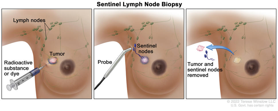
Sentinel lymph node biopsy of the breast; the first panel shows a radioactive substance and/or blue dye being injected near the tumor; the second panel shows that the injected material is followed visually and/or with a probe that detects radioactivity to find the sentinel nodes (the first lymph nodes to which cancer cells are likely to spread from a primary tumor); and the third panel shows the removal of the tumor and the sentinel nodes to check for cancer cells.

Sentinel lymph node biopsy of the breast. A radioactive substance and/or blue dye is injected near the tumor (first panel). The injected material is followed visually and/or with a probe that detects radioactivity to find the sentinel nodes (the first lymph nodes to which cancer cells are likely to spread from a primary tumor) (second panel). The sentinel nodes are removed and checked for cancer cells (third panel). A sentinel lymph node biopsy is usually done at the same time the primary tumor is removed, but it can also be done before or after the tumor is removed.

Credit: © Terese Winslow

To locate the sentinel lymph node, a radioactive substance, a blue dye, or both will be injected near the tumor. The surgeon then uses a device to detect lymph nodes that contain the radioactive substance, or they look for lymph nodes that are stained with the blue dye. Once the surgeon locates the sentinel lymph node, they make a small incision in the skin and remove the node.

A pathologist then checks the sentinel node for cancer cells. If cancer is found, the surgeon may remove other lymph nodes, either during the same biopsy procedure or during a follow-up surgery. A sentinel lymph node biopsy is an outpatient procedure done under general anesthesia.  

Imaging tests to stage breast cancer  

The following imaging tests may be done to find out if breast cancer has spread:

A CT scan (CAT scan) uses a computer linked to an x-ray machine to make a series of detailed pictures of areas inside the body. The pictures are taken from different angles and are used to create 3-D views of tissues and organs. A dye may be injected into a vein or swallowed to help the organs or tissues show up more clearly. This procedure is also called computed tomography, computerized tomography, or computerized axial tomography. Learn more about Computed Tomography (CT) Scans and Cancer.

A bone scan checks if there are rapidly dividing cells, such as cancer cells, in the bone. A very small amount of radioactive material is injected into a vein and travels through the bloodstream. The radioactive material collects in the areas of the bones with cancer and is detected by a scanner.

A PET scan (positron emission tomography scan) uses a small amount of radioactive sugar (also called radioactive glucose) that is injected into a vein. The PET scanner rotates around the body and makes pictures of where sugar is being used by the body. Cancer cells show up brighter in the pictures because they are more active and take up more sugar than normal cells do. When this procedure is done at the same time as a CT scan, it is called a PET-CT scan.

Biomarker tests for breast cancer

All breast cancers are tested for the presence of certain biomarkers, such as hormone receptors (estrogen receptors and progesterone receptors) and HER2. This information is needed to identify the stage of the breast cancer and to plan treatment. Learn more about these tests at Tests for Breast Cancer Biomarkers.

Breast cancer tumor grade

Tumor grade describes how abnormal the cancer cells and tissue look under a microscope and how quickly the cancer cells are likely to grow and spread. The pathologist assigns a score of 1 to 3 to the cells or tissue. A score of 1 means the cells and tumor tissue look the most normal and are less likely to spread. A score of 3 means the cells and tissue look the most abnormal and are more likely to grow fast and spread. Learn more about the breast cancer grading system at Breast Cancer Stages.
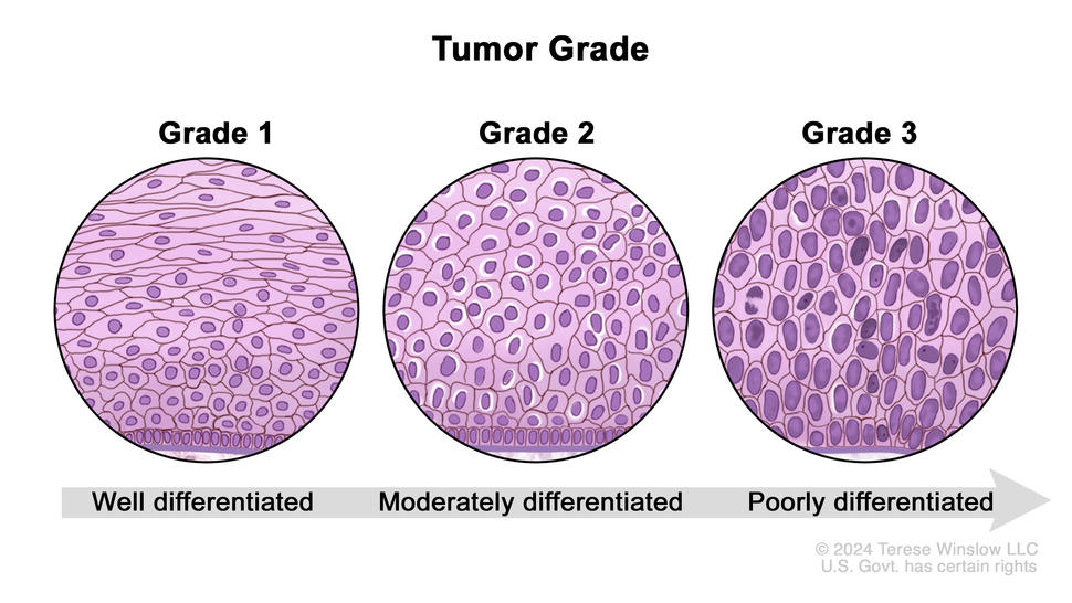
Three-panel drawing showing different grades of breast cancer cells based on how abnormal they look under a microscope. The first panel shows grade 1 or well-differentiated cancer cells. The second panel shows grade 2 or moderately differentiated cancer cells. The third panel shows grade 3 or poorly differentiated cancer cells.

Tumor grade for breast cancer is based on how abnormal the cancer cells look under a microscope and how quickly the cancer cells are likely to grow and spread. Grade 1 (well-differentiated) cancer cells look more like normal cells. Grade 2 (moderately differentiated) cancer cells look more abnormal than grade 1 cancer cells but not as abnormal as grade 3 (poorly differentiated) cancer cells. The higher the grade number, the more the cancer cells look like abnormal cells and the more likely they will grow and spread quickly.

Credit: © Terese Winslow

Multigene tests to predict risk of recurrence

Multigene tests, also called genomic tests, are lab tests that look at the activity of genes in your breast cancer cells. These tests may help predict the chances that cancer will spread to other parts of the body or come back. And they can help you and your doctor make decisions about treatment.

The tests listed below are often done on a sample of the tumor that was removed during the biopsy or surgery. Most people won’t need another procedure for these tests.

There are many types of multigene tests. Examples of tests you might have include:

  • Oncotype DX looks at the activity of 21 genes in people with early-stage breast cancer that is ER positive, HER2 negative, and has not spread to the lymph nodes or has spread to no more than three lymph nodes. The test helps predict whether breast cancer will spread to other parts of the body. If the risk of the cancer spreading is high, you may have chemotherapy to lower that risk.
  • MammaPrint looks at the activity of 70 genes in the breast cancer tissue of people with early-stage breast cancer that has not spread to the lymph nodes or has spread to no more than three lymph nodes. The test helps predict whether breast cancer will spread to other parts of the body or come back after treatment. If the test shows that the risk of the cancer spreading or returning is high, you may have chemotherapy to lower that risk.
  • Breast Cancer Index looks at the activity of 11 genes in the breast cancer tissue of people with early-stage, HR-positive breast cancer that has not spread to the lymph nodes or has spread to no more than three lymph nodes. The test helps predict the risk that your breast cancer will come back within 5 to 10 years after diagnosis. If the test shows that the risk of the cancer coming back is high, you may receive 5 more years of hormone therapy to help lower that risk.  

Other types of multigene tests are available. Ask your doctor if multigene tests might be right for you and how the results might affect your treatment plan. 

Genetic counseling and testing 

Your doctor might suggest genetic testing as part of diagnosing your breast cancer. Genetic testing can be done on a sample of your blood, saliva, or from a swab of inside your cheek. Genetic test results show if you were born with a change in the BRCA1, BRCA2, or another breast cancer risk gene. Knowing if you have a specific genetic change may help your doctor suggest the best treatment for your cancer. These gene changes are sometimes called mutations or pathogenic variants.  

Genetic counseling before genetic testing can help you understand your chances of having a gene change that caused your breast cancer and whether genetic testing is needed. For example, genetic testing might be advised if you:

Genetic counselors can also help you cope with your genetic testing results, including how to discuss the results with family members. They can advise you about whether other members of your family should receive genetic testing to find out if they are at risk of breast cancer or other cancers. Learn more about BRCA gene changes and cancer risk and genetic testing for inherited cancer risk.

Waiting for test results  

Waiting for cancer test results can be stressful. Ask your doctor when the results will be ready and how you will be notified so you know how long it will be before you hear back. If you don’t hear back within the expected time, it’s okay to follow up with your doctor.  

Sometimes test results are released to your online patient portal before you’ve met with your doctor. Although it can be difficult to wait to look at the test results until you are able to review them with your doctor, doing so can help avoid confusion about what the results mean.  

If you need help dealing with the stress of waiting for test results, reach out to your care team. A social worker can offer support and resources to help you. Whatever the results, your doctor can help you understand next steps. 

Getting a second opinion

You may want a second opinion to confirm your breast cancer diagnosis and treatment plan. If you seek a second opinion, you will need to get medical test results and reports from the first doctor to share with the second doctor. The second doctor will review the pathology report, slides, and scans before giving advice. The doctor who gives the second opinion may agree with your first doctor, suggest changes or another approach, or provide more information about your cancer.

To learn more about choosing a doctor and getting a second opinion, visit Finding Cancer Care. You can contact NCI’s Cancer Information Service via chat, email, or phone (both in English and Spanish) for help finding a doctor or hospital or getting a second opinion. For questions you might want to ask at your doctor visits, visit Questions to Ask Your Doctor About Cancer

Dense Breasts: Answers to Commonly Asked Questions

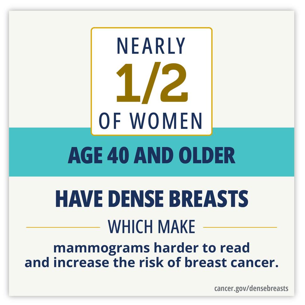
Nearly half of women age 40 and older have dense breasts, which make mammograms harder to read and increase the risk of breast cancer.

What are dense breasts?

Breasts contain glandular tissue, fibrous connective tissue, and fatty breast tissue. Breast density is a term that describes the relative amount of these different types of breast tissue as seen on a mammogram. Dense breast tissue has relatively high amounts of glandular tissue and fibrous connective tissue and relatively low amounts of fatty breast tissue.

Are dense breasts common?

Yes, dense breasts are common. Nearly half of all women who are 40 and older who get mammograms are found to have dense breast tissue. 

What factors influence breast density?

Breast density is often inherited, but other factors can influence it.

  • Factors associated with higher breast density include using menopausal hormone therapy and having a low body mass index.
  • Factors associated with lower breast density include increasing age and having children.

How do I know if I have dense breasts?

Dense breast tissue cannot be felt by a woman, such as during a breast self-exam, or by her doctor during a clinical breast exam. Only a radiologist looking at a mammogram can tell if a woman has dense breasts. Dense breasts are sometimes called mammographically dense breasts.

How is breast density categorized in a mammogram report?

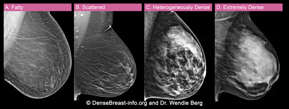
Doctors use the Breast Imaging Reporting and Data System (BI-RADS) to classify breast density. This system, developed by the American College of Radiology, helps doctors interpret and report back mammogram findings. Doctors who review mammograms are called radiologists. BI-RADS classifies breast density into four categories:

If your mammogram report letter says you have dense breasts, it means that you have either heterogeneously dense breast tissue or extremely dense breast tissue.

Dense Breasts Categories

The four breast density categories are shown in this image. Breasts can be almost entirely fatty (A), have scattered areas of dense fibroglandular breast tissue (B), have many areas of glandular and connective tissue (C), or be extremely dense (D). Breasts are classified as “dense” if they fall in the heterogeneously dense (C) or extremely dense (D) categories.

Does having dense breast tissue affect a mammogram?

Dense breasts can make a mammogram more difficult to interpret. That’s because dense breast tissue and some abnormal breast changes, such as calcifications and tumors, both appear as white areas in the mammogram, whereas fatty tissue appears as dark areas.

As a result, mammography is less sensitive in women with dense breasts—that is, it is more likely to miss cancer. Women with dense breasts may be called back for follow-up testing more often than women with fatty breasts.

Are dense breasts a risk factor for breast cancer?

Dense breasts are not considered an abnormal breast condition or a disease. However, dense breasts are a risk factor for breast cancer. That is, women with dense breasts have a higher risk of breast cancer than women with fatty breasts. This risk is separate from the effect of dense breasts on the ability to read a mammogram. Learn more about other risk factors for breast cancer as well as protective factors for breast cancer.

Updated FDA regulations require mammography providers to inform women if they have dense breasts

Should women with dense breasts have additional screening for breast cancer?

There is not yet enough evidence to recommend for or against additional imaging tests such as ultrasound or MRI to screen for breast cancer in women with dense breasts, according to the Recommendation Statement on Breast Cancer Screening by the United States Preventive Services Task Force (USPSTF). Talk with your doctor or nurse to learn more about your options.

Are breast cancer patients with dense breasts more likely to die from breast cancer?

No. Research has found that breast cancer patients who have dense breasts are no more likely to die from breast cancer than breast cancer patients who have fatty breasts, after accounting for other health factors and tumor characteristics.

Questions to consider asking your doctor or nurse

Ask these questions to get information that’s specific to you, based on your personal medical history:

  • Do I have dense breasts, based on my most recent mammogram result?
  • Do you recommend additional screening for me? What are the benefits and drawbacks of these additional tests? 
  • What is my personal risk of breast cancer, given my risk factors and protective factors?

Are there clinical trials for women with dense breasts?

Yes, there are ongoing and completed clinical trials related to dense breasts that are studying better ways to detect breast cancer in women with dense breasts. You can also contact NCI’s Cancer Information Service to get tailored clinical trial searches related to breast density and breast cancer screening.

Questions that researchers are asking about the relationship between breast density and breast cancer

  • What biomarkers or molecular markers can be identified to better characterize dense breast tissue and predict a woman’s risk for breast cancer? 
  • Are certain patterns or areas of dense breast tissue particularly risky? 
  • How does the microenvironment of dense breast tissue affect the development and growth of tumors? 
  • Are changes in breast density over time associated with changes in breast cancer risk?
  • Could women with dense breasts lower their breast density and risk of developing breast cancer by exercising, taking drugs such as low dose tamoxifen, or applying topical agents? 
  • Could imaging tests such as ultrasound, contrast enhanced mammography, MRI (such as diffusion-weighted MRI or fast MRI), or molecular breast imaging help find breast cancer in women with dense breasts? 

NCI’s Division of Cancer Epidemiology and Genetics conducts research on breast density and breast cancer risk factors.

NCI’s Division of Cancer Prevention supports research on cancer screening and risk factors, including breast density, to help researchers learn about the best way to find breast cancer in women who have no symptoms. 

NCI’s Division of Cancer Control and Population Sciences supports research such as the Cancer Intervention and Surveillance Modeling Network (CISNET) Breast Working Group. CISNET is a consortium of NCI-sponsored investigators conducting simulation modeling to improve our understanding of the impact of prevention, screening, and treatment on population trends in incidence and mortality.

Breast Cancer Treatment

Different types of treatment are available for breast cancer. You and your cancer care team will work together to decide your treatment plan, which may include more than one type of treatment.

Your treatment options may include both local and systemic treatments. Local treatments, such as surgery and radiation, are directed at the area with cancer and not the whole body. Systemic treatments, such as chemotherapy, are drugs that can reach cancer cells throughout the body. 

There are different treatment approaches for each type and stage of breast cancer. To learn more about treatment for specific types and stages of breast cancer, review What kind of treatment will I receive? below.

Consulte Tratamiento del cáncer de mama (seno) para obtener información sobre el tratamiento del cáncer de mama en español.

Inflammatory Breast Cancer

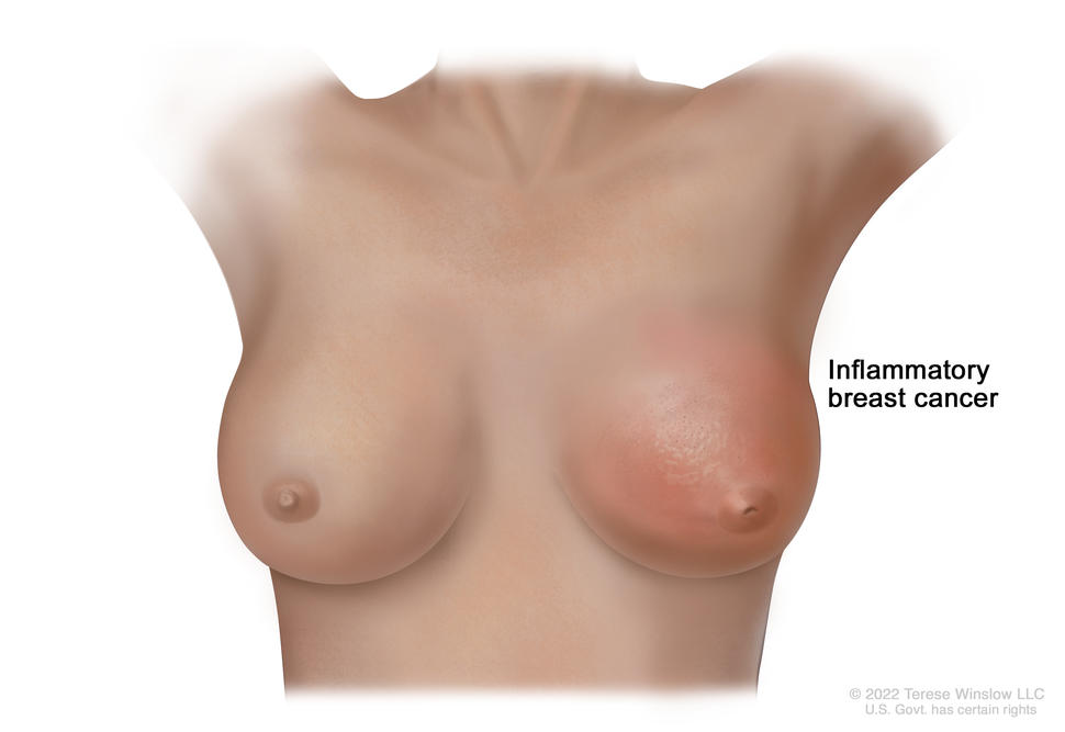
Inflammatory breast cancer of the left breast showing redness, swelling, peau d'orange, and an inverted nipple.

Inflammatory breast cancer is a type of breast cancer in which the cancer cells block the lymph vessels in the skin of the breast. This causes the breast to look red and swollen. The skin may also appear dimpled or pitted, like the skin of an orange (peau d’orange), and the nipple may be inverted (facing inward).

Credit: © Terese Winslow

What is inflammatory breast cancer?

Inflammatory breast cancer is a rare type of fast-growing breast cancer in which cancer cells block lymph vessels in the skin of the breast. In the United States, 1%–5% of all breast cancers are inflammatory breast cancer. Most inflammatory breast cancers are invasive ductal carcinomas, which means they develop from cells that line the milk ducts of the breast and then spread beyond the ducts.

What are risk factors for inflammatory breast cancer?

Inflammatory breast cancer is more common in certain age groups and populations. Inflammatory breast cancer is:

  • often diagnosed at a younger age
  • more common and diagnosed at a younger age in Black women than in White women
  • more common in women with obesity than in women at a healthy weight

The cause of inflammatory breast cancer is unknown, and other risk factors are the same as for other types of breast cancer. For more information, visit Breast Cancer Causes and Risk Factors.

What are the symptoms of inflammatory breast cancer?

This type of breast cancer is known as inflammatory because it causes the breast to become red and swollen, or inflamed. Inflammatory breast cancer usually does not form a lump you can feel. Symptoms of inflammatory breast cancer include:

  • pink, reddish purple, or bruised appearance of the skin of the breast
  • dimpling or ridges on the skin that resemble an orange peel, caused by the buildup of fluid (lymph) in the skin of the breast
  • rapid increase in breast size
  • sensations of heaviness, burning, or tenderness in the breast
  • a nipple that is inverted (facing inward)
  • swollen lymph nodes under the arm or near the collarbone

These symptoms generally come on rapidly. They may also be signs of other diseases or conditions, such as an infection, injury, or another type of breast cancer that is locally advanced. Mastitis is an infection of the breast that has symptoms similar to those of inflammatory breast cancer. Mastitis can occur in anyone, but it is particularly common among people who are breastfeeding. If you have any of these symptoms, contact your doctor for evaluation.

How is inflammatory breast cancer diagnosed?

Because inflammatory breast cancer may develop and progress quickly, often between regular screening intervals, mammograms and other screening methods are less effective at diagnosing it early. Also, inflammatory breast cancer usually cannot be seen on a mammogram.

If you have symptoms of inflammatory breast cancer, your doctor will need to find out if they are due to cancer or another condition. Your doctor may: 

If it is cancer, additional tests may include:

Your doctor will assign a stage to the cancer based on a combination of the extent of spread, hormone receptor and HER2 status, and other tumor features, such as tumor grade. Inflammatory breast cancer is either stage III (stage 3) or stage IV (stage 4) when diagnosed, depending on whether cancer cells have spread only to nearby areas or to distant tissues as well.

Learn more about tests used to diagnose breast cancer and the different stages of breast cancer at How Breast Cancer Is Diagnosed and Breast Cancer Stages.

How is inflammatory breast cancer treated?

Because inflammatory breast cancer grows quickly and is more likely to come back after treatment, you may receive more treatment for a longer period of time than people with other types of breast cancer.

For information about how inflammatory breast cancer is treated, visit Inflammatory Breast Cancer Treatment.

What is the survival rate and prognosis for people with inflammatory breast cancer?

Doctors estimate inflammatory breast cancer prognosis by using statistics collected over many years from people with the disease. One statistic that is commonly used in making a prognosis is the 5-year relative survival rate. The 5-year relative survival rate tells you what percent of people with the same type and stage of breast cancer are alive 5 years after their cancer was diagnosed, compared with people in the overall population.

Inflammatory breast cancer tends to have a less favorable prognosis and survival rate than many other types of breast cancer because it is often diagnosed at an advanced stage and grows and spreads more quickly. Your doctor is in the best position to discuss your prognosis with you. Some people like to have a loved one or friend with them for the conversation.

Five-year relative survival rates among women with inflammatory breast cancer (note that there is no localized category for inflammatory breast cancer):

  • 53% among women with regional inflammatory breast cancer (cancer has spread beyond the breast to nearby lymph nodes or organs)
  • 22% among women with distant inflammatory breast cancer (cancer has spread beyond the breast to a distant part of the body)

To learn more about factors that affect breast cancer prognosis, visit Breast Cancer Prognosis and Survival Rates.

Ongoing research will increase our understanding of how inflammatory breast cancer begins and progresses, which could lead to new treatments and better outcomes for women with this disease. If you have been diagnosed with inflammatory breast cancer, you may want to talk with your doctor about the possibility of participating in a clinical trial. Learn more about clinical trials at Cancer Clinical Trial Information for Patients and Caregivers.

Breast Cancer

Breast cancer is the second most common cancer in women after skin cancer. Mammograms can detect breast cancer early, possibly before it has spread. Explore the links on this page to learn more about breast cancer prevention, screening, treatment, statistics, research, clinical trials, and more.

Breast Cancer Causes and Risk Factors

A senior woman sitting in between an older woman and a younger woman on a sofa.

A family history of breast cancer in a first-degree relative (mother, daughter, or sister) increases your risk of breast cancer.

Credit: iStock

Breast cancer is caused by certain changes in how breast cells function, especially how they grow and divide into new cells. A risk factor is anything that increases the chance of getting a disease. Some risk factors for breast cancer, like drinking alcohol, can be changed. However, risk factors also include things people cannot change, like your genetics, getting older, and your family history.

Factors that can increase the risk of breast cancer

There are many risk factors for breast cancer, but most do not directly cause cancer. Instead, they increase the chance of DNA damage in cells that may lead to breast cancer. Learn more about how cancer develops at What Is Cancer?  

Having one or more of these risk factors does not mean that you will get breast cancer and some people who get breast cancer do not have known risk factors. The most important risk factor for breast cancer, besides female biological sex, is increasing age. 

Personal health history and breast conditions

The following personal factors and breast conditions have been linked to a higher risk of breast cancer:

  • Having had breast cancer in the past.
  • Having had ductal carcinoma in situ (DCIS) in the past.
  • Having been exposed to high levels of radiation in the past, particularly at a young age, such as during treatment for childhood cancer.
  • Having a history of certain kinds of breast changes. Learn more about precancerous breast conditions.
  • Having dense breasts, a common condition that not only increases the risk of breast cancer but also makes it more difficult to detect breast cancer on mammograms. Learn more about dense breasts.
  • Having excess body weight, especially after menopause.
  • Taking combination (estrogen plus progestin) hormone replacement therapy for symptoms of menopause. Learn more about Menopausal Hormone Therapy and Cancer.
  • Having been exposed to DES (a synthetic estrogen) before birth (in utero) or having taken it during pregnancy. Learn more about cancer risks linked to DES exposure.

Reproductive history

If your reproductive history leads to a longer exposure to natural estrogen, it can increase your risk of developing breast cancer. Reproductive factors that increase the length of time a woman’s breast tissue is exposed to estrogen include:

  • having a longer menstrual history (i.e., having early menarche, later menopause)
  • being at an older age at first birth
  • never having carried a pregnancy to term
  • never having breastfed (among women who have given birth)

Genetics and family history

The following genetics and family history factors have been linked to a higher risk of breast cancer:

Genetic counselors and other specially trained health professionals can help people with breast cancer or with a family history of breast cancer understand:

  • the likelihood that they have a genetic risk factor for breast cancer based on their personal history and their family medical history
  • their options for genetic testing for changes in the BRCA1, BRCA2, and other genes that increase the risk of breast cancer
  • the risks and benefits of learning genetic information
  • how to cope with their genetic testing results
  • how to discuss the results with family members

Learn more about genetic counseling and genetic testing to assess your risk of breast cancer and other cancers at Genetic Testing for Inherited Cancer Risk

Behaviors

The following behaviors have been linked to a higher risk of breast cancer:

Race

The incidence of breast cancer varies with race. In the United States, White women have the highest incidence of breast cancer, whereas Black women have the highest death rate. 

Potential risk factors that require more study

Some studies have shown that women who are current or recent users of hormonal birth control pills (also called oral hormonal contraceptives) may have a slight increase in breast cancer risk. Other studies have not shown an increased risk of breast cancer in women using birth control pills. Learn more about Oral Contraceptives and Cancer Risk.

Scientists are also studying whether exposure to certain chemicals in the environment may increase a person’s risk of breast cancer. Studies of this kind can be difficult to conduct and interpret for many reasons, making it hard to know which chemicals, if any, may increase the risk of breast cancer.

Understanding your risk of breast cancer

Everyone with breast tissue has some risk of breast cancer, but certain risk factors may increase your risk. 

On average, women have a 1 in 8 (or 13 in 100) chance of developing breast cancer during their lifetime. If you have certain risk factors—for instance, you got your menstrual period at an early age, you were older when your first child was born, or you have a first-degree relative with breast cancer—you may be at higher risk. To help you understand your own risk, your doctor can use a risk calculator, such as NCI’s Breast Cancer Risk Assessment Tool, which estimates people’s risk of breast cancer based on these and several other clinical, reproductive, and medical history factors.

There are some risk factors that, on their own, confer a very high risk of breast cancer—60 in 100 or more. These include certain harmful genetic changes in genes such as BRCA1, BRCA2, PALB2, and PTEN; a strong family history of breast cancer (with multiple relatives diagnosed); and having had radiation to the chest before age 30. 

Women who may be at increased risk of breast cancer can discuss possible screening or preventive measures with their doctors.

It is important to keep in mind that, even with the best available scientific information, doctors cannot precisely estimate an individual’s risk. All estimates are subject to uncertainty. For example, someone might have a personal factor or exposure that influences their risk but that isn’t included in the risk calculator. Also, scientific evidence can change over time, making earlier calculations less accurate. Estimates of risk should be considered best guesses based on the knowledge we have now.

Types of Breast Cancer

A doctor holding a model of a breast and pointing at an image of a mammogram on a monitor while talking to a patient.

Talk to your doctor to find out what type of breast cancer you have and how it is used to plan the best treatment for you.

Credit: iStock

There are many types of breast cancer. The types differ based on several factors, such as which cells in the breast become cancer, whether the cancer has spread from where it first formed, and whether the cancer has certain features that affect treatment options.  

Invasive or infiltrating ductal carcinoma begins in the cells that line the milk ducts and has spread beyond where it first formed. It is the most common breast cancer diagnosis. Learn more about Breast Cancer.

Invasive lobular carcinoma begins in the cells that line the breast glands that make milk, called lobules, and has spread beyond where it first formed. It grows more slowly and is less common than invasive ductal carcinoma. Lobular carcinoma is more often found in both breasts than are other types of breast cancer.

Inflammatory breast cancer is a rare form of breast cancer in which cancer cells block lymph vessels in the skin of the breast. This type of breast cancer has a high risk of recurrence. It is called inflammatory because the affected breast often looks swollen and red, or inflamed.  

Triple-negative breast cancer is a form of breast cancer in which the cancer cells lack features that are common in breast cancer, including hormone receptors and a protein called human epidermal growth factor receptor 2 (HER2). This form of breast cancer has a higher risk of recurrence than most other forms of breast cancer.

Metastatic breast cancer, also called stage 4 breast cancer, is breast cancer that has spread from the breast to another part of the body. Metastatic spread occurs when breast cancer cells break away from the original tumor and travel through the lymph system or blood to other sites in the body.

Ductal carcinoma in situ (DCIS) forms in the cells that line the milk ducts but has not spread beyond where it first formed. It may also be called noninvasive breast cancer, intraductal carcinoma, or stage 0 breast cancer.  

Paget disease of the breast is a rare type of cancer that involves the skin of the nipple and the areola.

Phyllodes tumor (also called cystosarcoma phyllodes of the breast, or CSP) is a rare type of breast tumor that starts in the connective tissue of the breast. Most phyllodes tumors are not cancer. Learn more at Benign and Precancerous Breast Lumps and Conditions.

Lobular carcinoma in situ (LCIS) forms in the cells that line the breast glands that make milk, called lobules, but has not spread beyond where it first formed. LCIS is not breast cancer but increases the risk of developing breast cancer in the future. Learn more at Benign and Precancerous Breast Lumps and Conditions.

Molecular subtypes of breast cancer

Molecular subtypes of breast cancer are defined by whether they have hormone receptors, HER2 protein, or other biomarkers. Examples of molecular subtypes of breast cancer include triple-negative, luminal A, luminal B, and HER2-positive. Learn about how these subtypes are diagnosed and how they affect treatment at Tests for Breast Cancer Biomarkers and Breast Cancer Treatment by Stage.

Multiple Endocrine Neoplasia (MEN) Syndromes

Multiple endocrine neoplasia (MEN) syndromes are inherited disorders that affect the endocrine system. The endocrine system is made up of glands and cells that make hormones and release them into the blood. MEN syndromes may cause hyperplasia (the growth of too many normal cells) or tumors that may be benign (not cancer) or malignant (cancer).

There are several types of MEN syndromes and each type may cause different conditions or cancers. The two main types of MEN syndromes are MEN1 and MEN2. MEN2 syndrome has two subgroups: MEN2A syndrome and MEN2B syndrome. MEN2A syndrome includes familial medullary thyroid cancer.

Learn more about MEN syndromes in children, including symptoms and how it is diagnosed and treated:

Adrenocortical Carcinoma Symptoms

These and other signs and symptoms may be caused by adrenocortical carcinoma:

  • A lump in the abdomen.
  • Pain the abdomen or back.
  • A feeling of fullness in the abdomen.

A nonfunctioning adrenocortical tumor may not cause signs or symptoms in the early stages. A functioning adrenocortical tumor makes too much of one of the following hormones:

Symptoms caused by too much cortisol

  • Weight gain in the face, neck, and trunk of the body and thin arms and legs.
  • Growth of fine hair on the face, upper back, or arms.
  • A round, red, full face.
  • A lump of fat on the back of the neck.
  • A deepening of the voice and swelling of the sex organs or breasts in both males and females.
  • Muscle weakness.
  • High blood sugar.
  • High blood pressure.

Symptoms caused by too much aldosterone

  • High blood pressure.
  • Muscle weakness or cramps.
  • Frequent urination.
  • Feeling thirsty.

Symptoms caused by too much testosterone (in women)

  • Growth of fine hair on the face, upper back, or arms.
  • Acne.
  • Balding.
  • A deepening of the voice.
  • No menstrual periods.

Men who make too much testosterone do not usually have signs or symptoms.

Symptoms caused by too much estrogen (in women)

  • Irregular menstrual periods in women who have not gone through menopause.
  • Vaginal bleeding in women who have gone through menopause.
  • Weight gain.

Symptoms caused by too much estrogen (in men)

These and other signs and symptoms may be caused by adrenocortical carcinoma or by other conditions. Check with your doctor if you have any of these problems.

Childhood Adrenocortical Carcinoma

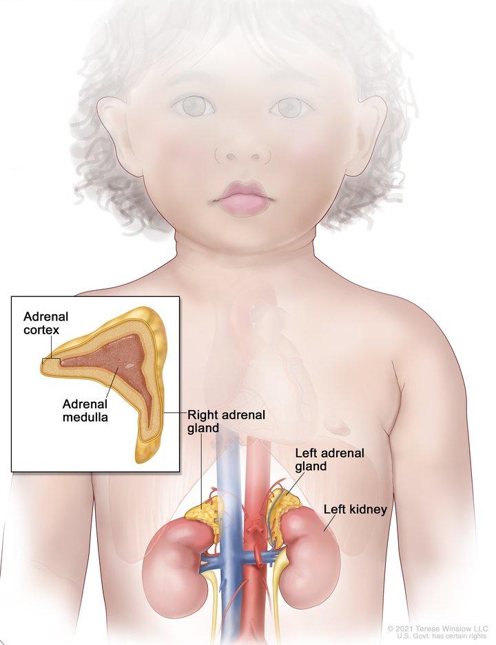
Drawing of the abdomen showing the left and right adrenal glands, the left and right kidneys, and major blood vessels. Also shown is an inset of an adrenal gland showing the adrenal cortex and the adrenal medulla.

Anatomy of the adrenal gland. There are two adrenal glands, one on top of each kidney. The outer part of each gland is the adrenal cortex and the inner part is the adrenal medulla.

Credit: © Terese Winslow

Adrenocortical carcinoma is a rare disease in which malignant (cancer) cells form in the outer layer of the adrenal gland.

There are two adrenal glands. The adrenal glands are small and shaped like a triangle. One adrenal gland sits on top of each kidney. Each adrenal gland has two parts. The outer layer of the adrenal gland is the adrenal cortex. The center of the adrenal gland is the adrenal medulla. Adrenocortical carcinoma is also called adrenocortical cancer or cancer of the adrenal cortex.

The adrenal cortex makes important hormones that:

  • Balance the water and salt in the body.
  • Help keep blood pressure normal.
  • Help control the body’s use of protein, fat, and carbohydrates.
  • Cause the body to have male or female characteristics.

The adrenal medulla makes hormones that help the body react to stress. Cancer that forms in the adrenal medulla is called pheochromocytoma and is not discussed in this summary. See the PDQ summary on Childhood Pheochromocytoma and Paraganglioma Treatment for more information.

Most childhood adrenocortical tumors occur during the first 5 years of life, but they may also occur during adolescence. 

Risk factors for childhood adrenocortical carcinoma

Anything that increases your chance of getting a disease is called a risk factor. Having a risk factor does not mean that you will get cancer; not having risk factors doesn’t mean that you will not get cancer. Talk with your child’s doctor if you think your child may be at risk.

The risk of adrenocortical carcinoma is increased by having a mutation (change) in the TP53 gene or any of the following syndromes:

Symptoms of childhood adrenocortical carcinoma

These and other signs and symptoms may be caused by adrenocortical carcinoma or by other conditions.

Check with your child’s doctor if your child has any of the following:

  • Pain in the abdomen or back.
  • A lump in the abdomen.
  • Feeling of fullness in the abdomen.
  • High blood pressure.
  • Acne.
  • Growing body hair.
  • Deepening of the voice.
  • Growing faster than normal.

Also, cancer of the adrenal cortex may be functioning (makes more hormones than normal) or nonfunctioning (does not make extra hormones). Most tumors of the adrenal cortex in children are functioning tumors. The extra hormones made by functioning tumors may cause certain signs or symptoms of disease and these depend on the type of hormone made by the tumor. For example, extra androgen hormone may also cause male children to develop an enlarged penis and female children to develop enlarged genitalia. Extra estrogen hormone may cause the growth of breast tissue in male children. Extra cortisol hormone may cause (hypercortisolism).

Learn more about Adrenocortical Carcinoma Symptoms.

Tests to diagnose childhood adrenocortical carcinoma

Tests are done to diagnose and stage cancer. After cancer is diagnosed, more tests are done to find out if cancer cells have spread to nearby areas or to other parts of the body. This process is called staging. It is important to know whether cancer has spread in order to plan the best treatment.

The following tests and procedures may be used:

  • Physical exam and health history: An exam of the body to check general signs of health, including checking for signs of disease, such as lumps or anything else that seems unusual. A history of the patient’s health habits and past illnesses and treatments will also be taken.
  • X-ray: An x-ray of the chest, abdomen, or bones inside the body. An x-ray is a type of energy beam that can go through the body and onto film, making a picture of areas inside the body.
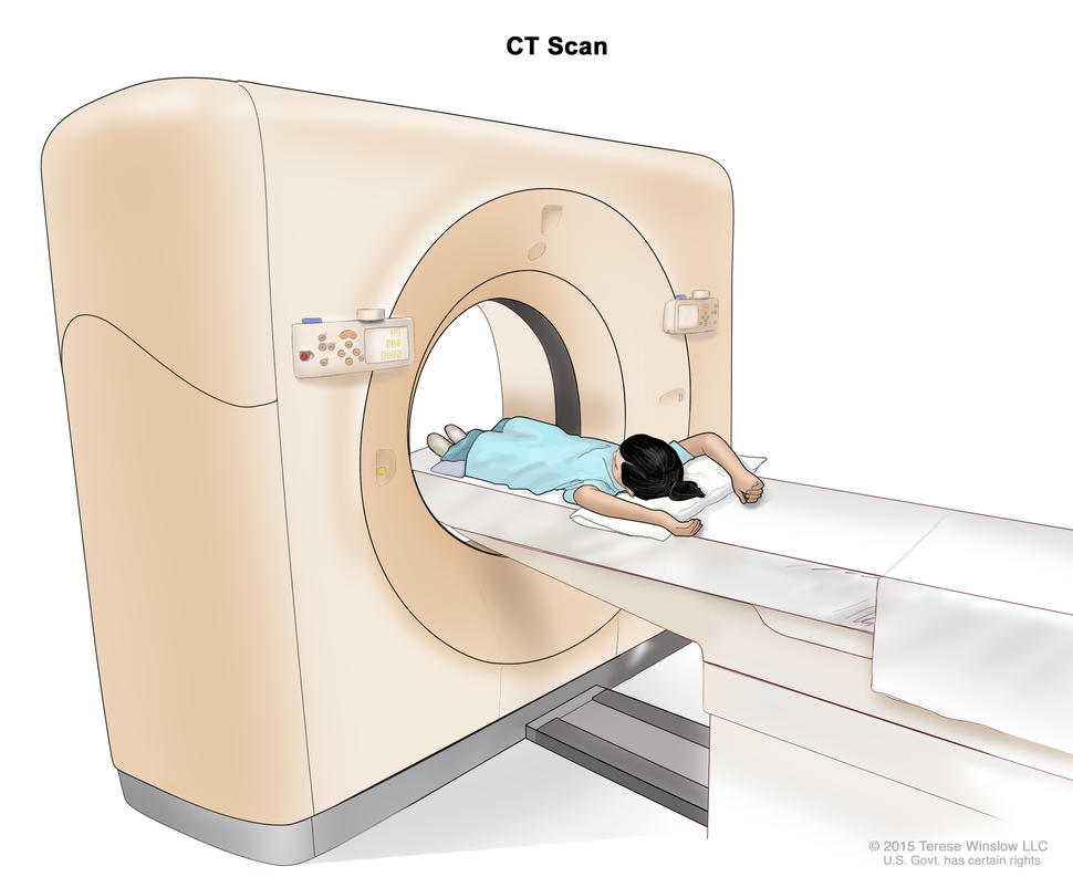
Computed tomography (CT) scan; drawing shows a child lying on a table that slides through the CT scanner, which takes a series of detailed x-ray pictures of areas inside the body.

Computed tomography (CT) scan. The child lies on a table that slides through the CT scanner, which takes a series of detailed x-ray pictures of areas inside the body.

Credit: © Terese Winslow

  • CT scan: A procedure that makes a series of detailed pictures of areas inside the body, such as the chest or abdomen, taken from different angles. The pictures are made by a computer linked to an x-ray machine. A dye may be injected into a vein or swallowed to help the organs or tissues show up more clearly. This procedure is also called computed tomography, computerized tomography, or computerized axial tomography.
  • MRI (magnetic resonance imaging): A procedure that uses a magnet, radio waves, and a computer to make a series of detailed pictures of areas of the body, such as the chest and abdomen. This procedure is also called nuclear magnetic resonance imaging (NMRI).
  • PET scan: A procedure to find malignant tumor cells in the body. A small amount of radioactive glucose (sugar) is injected into a vein. The PET scanner rotates around the body and makes a picture of where glucose is being used in the body. Malignant tumor cells show up brighter in the picture because they are more active and take up more glucose than normal cells do.
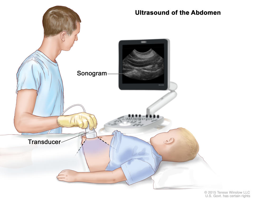
  • Ultrasound exam: A procedure in which high-energy sound waves (ultrasound) are bounced off internal tissues or organs, such as the abdomen, and make echoes. The echoes form a picture of body tissues called a sonogram. The picture can be printed to be looked at later.
  • Biopsy: The removal of cells or tissues during surgery so they can be viewed under a microscope by a pathologist to check for signs of cancer.
  • Blood chemistry studies: A procedure in which a blood sample is checked to measure the amounts of certain substances released into the blood by organs and tissues in the body. An unusual (higher or lower than normal) amount of a substance can be a sign of disease.
  • Blood hormone studies: A procedure in which a blood sample is checked to measure the amounts of certain hormones released into the blood by organs and tissues in the body. An unusual (higher or lower than normal) amount of a substance can be a sign of disease in the organ or tissue that makes it. The blood may be checked for testosterone or estrogen. A higher-than-normal amount of these hormones may be a sign of adrenocortical carcinoma.
  • Twenty-four-hour urine test: A test in which urine is collected for 24 hours to measure the amounts of cortisol or 17-ketosteroids. A higher-than-normal amount of these substances in the urine may be a sign of disease in the adrenal cortex.
Abdominal ultrasound; drawing shows a child lying on an exam table during an abdominal ultrasound procedure. A technician is shown pressing a transducer (a device that makes sound waves that bounce off tissues inside the body) against the skin of the abdomen. A computer screen shows a sonogram (picture).

Abdominal ultrasound. An ultrasound transducer connected to a computer is pressed against the skin of the abdomen. The transducer bounces sound waves off internal organs and tissues to make echoes that form a sonogram (computer picture).

Credit: © Terese Winslow

  • Low-dose dexamethasone suppression test: A test in which one or more small doses of dexamethasone are given. The level of cortisol is checked from a sample of blood or from urine that is collected for three days. This test is done to check if the adrenal gland is making too much cortisol.
  • High-dose dexamethasone suppression test: A test in which one or more high doses of dexamethasone are given. The level of cortisol is checked from a sample of blood or from urine that is collected for three days. This test is done to check if the adrenal gland is making too much cortisol or if the pituitary gland is telling the adrenal glands to make too much cortisol.
  • Adrenal angiography: A procedure to look at the arteries and the flow of blood near the adrenal gland. A contrast dye is injected into the adrenal arteries. As the dye moves through the blood vessel, a series of x-rays are taken to see if any arteries are blocked.
  • Adrenal venography: A procedure to look at the adrenal veins and the flow of blood near the adrenal glands. A contrast dye is injected into an adrenal vein. As the contrast dye moves through the vein, a series of x-rays are taken to see if any veins are blocked. A catheter (very thin tube) may be inserted into the vein to take a blood sample, which is checked for abnormal hormone levels.

Prognostic factors for childhood adrenocortical carcinoma

The prognosis is good for patients who have small tumors that have been completely removed by surgery. For other patients, the prognosis depends on the following:

  • The size of the tumor.
  • How quickly the cancer is growing.
  • Whether there are changes in certain genes.
  • Whether the tumor has spread to other parts of the body, including the lymph nodes, liver, lung, kidney, or bone.
  • The child’s age.
  • Whether the covering around the tumor broke open during surgery to remove the tumor.
  • Whether the tumor was completely removed during surgery.
  • Whether the child has developed masculine traits. 

Stages of childhood adrenocortical carcinoma

The process used to find out if cancer has spread to tissues near the adrenal glands or to other parts of the body is called staging. The information gathered from the staging process is used to plan treatment. The results of the tests and procedures used to diagnose cancer are often also used to stage the disease.

Sometimes childhood adrenocortical carcinoma recurs (comes back) in the adrenal cortex or in other parts of the body after it has been treated.

There are three ways that cancer spreads in the body

Cancer can spread through tissue, the lymph system, and the blood:

  • Tissue. The cancer spreads from where it began by growing into nearby areas.
  • Lymph system. The cancer spreads from where it began by getting into the lymph system. The cancer travels through the lymph vessels to other parts of the body.
  • Blood. The cancer spreads from where it began by getting into the blood. The cancer travels through the blood vessels to other parts of the body.

Cancer may spread from where it began to other parts of the body

When cancer spreads to another part of the body, it is called metastasis. Cancer cells break away from where they began (the primary tumor) and travel through the lymph system or blood.

  • Lymph system. The cancer gets into the lymph system, travels through the lymph vessels, and forms a tumor (metastatic tumor) in another part of the body.
  • Blood. The cancer gets into the blood, travels through the blood vessels, and forms a tumor (metastatic tumor) in another part of the body.

The metastatic tumor is the same type of cancer as the primary tumor. For example, if adrenocortical carcinoma spreads to the liver, the cancer cells in the liver are actually adrenocortical carcinoma cells. The disease is metastatic adrenocortical carcinoma, not liver cancer. 

Metastasis: How Cancer Spreads

During metastasis, cancer cells spread from the place in the body where they first formed to other parts of the body.

Treatment options for children with adrenocortical carcinoma

There are different types of treatment for children with adrenocortical carcinoma.

Some treatments are standard (the currently used treatment), and some are being tested in clinical trials. A treatment clinical trial is a research study meant to help improve current treatments or obtain information on new treatments for patients with cancer. When clinical trials show that a new treatment is better than the standard treatment, the new treatment may become the standard treatment.

Because cancer in children is rare, taking part in a clinical trial should be considered. Some clinical trials are open only to patients who have not started treatment.

Who treats children with adrenocortical carcinoma?

Treatment will be overseen by a pediatric oncologist, a doctor who specializes in treating children with cancer. The pediatric oncologist works with other pediatric health professionals who are experts in treating children with cancer and who specialize in certain areas of medicine. This may include the following specialists and others:

Two types of standard treatment are used:

Surgery

Surgery to remove the tumor is the main treatment for adrenocortical carcinoma.

Chemotherapy

Chemotherapy is a cancer treatment that uses drugs to stop the growth of cancer cells, either by killing the cells or by stopping them from dividing. When chemotherapy is taken by mouth or injected into a vein or muscle, the drugs enter the bloodstream and can reach cancer cells throughout the body (systemic chemotherapy).

New treatments being tested in clinical trials

This summary section describes treatments that are being studied in clinical trials. It may not mention every new treatment being studied. Information about clinical trials is available from the NCI website.

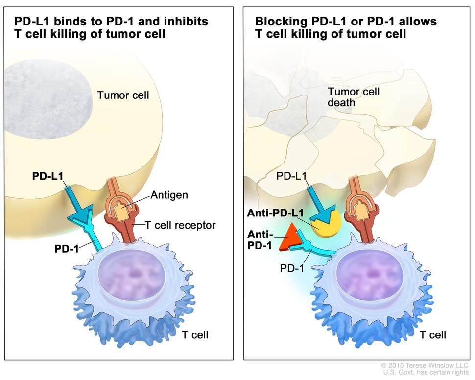
Two-panel illustration showing the interaction between T cells and tumor cells. The left panel is titled "PD-L1 binds to PD-1 and inhibits T cell killing of tumor cell" and shows an antigen and T cell receptor, illustrating the inhibition of T cell activity. The right panel is titled "Blocking PD-L1 or PD-1 allows T cell killing of tumor cell" and shows tumor cell death, indicating successful T cell activity.

Checkpoint proteins, such as PD-L1 on tumor cells and PD-1 on T cells, help keep immune responses in check. The binding of PD-L1 to PD-1 keeps T cells from killing tumor cells in the body (left panel). Blocking the binding of PD-L1 to PD-1 with an immune checkpoint inhibitor (anti-PD-L1 or anti-PD-1) allows the T cells to kill tumor cells (right panel).

Credit: © Terese Winslow

Immunotherapy

Immunotherapy is a treatment that uses the patient’s immune system to fight cancer. Substances made by the body or made in a laboratory are used to boost, direct, or restore the body’s natural defenses against cancer.

  • Immune checkpoint inhibitor therapy is a type of immunotherapy that blocks certain proteins. PD-1 is a protein on the surface of T cells that helps keep the body’s immune responses in check. PD-L1 is a protein found on some types of cancer cells. When PD-1 attaches to PD-L1, it stops the T cell from killing the cancer cell. PD-1 and PD-L1 inhibitors keep PD-1 and PD-L1 proteins from attaching to each other. This allows the T cells to kill cancer cells. Pembrolizumab is a PD-1 inhibitor that is being studied in the treatment of childhood adrenocortical carcinoma that is advanced or has come back after treatment.

Immune Checkpoint Inhibitors

Learn about immune checkpoint inhibitors, one type of immunotherapy used to treat cancer.

Treatment of newly diagnosed childhood adrenocortical carcinoma

Treatment of newly diagnosed adrenocortical carcinoma in children may include the following:

Treatment of recurrent childhood adrenocortical carcinoma

Treatment of recurrent adrenocortical carcinoma in children may include the following:

Clinical trials

For some patients, taking part in a clinical trial may be the best treatment choice. Clinical trials are part of the cancer research process. Clinical trials are done to find out if new cancer treatments are safe and effective or better than the standard treatment.

Many of today’s standard treatments for cancer are based on earlier clinical trials. Patients who take part in a clinical trial may receive the standard treatment or be among the first to receive a new treatment.

Patients who take part in clinical trials also help improve the way cancer will be treated in the future. Even when clinical trials do not lead to effective new treatments, they often answer important questions and help move research forward.

Patients can enter clinical trials before, during, or after starting their cancer treatment

Some clinical trials only include patients who have not yet received treatment. Other trials test treatments for patients whose cancer has not gotten better. There are also clinical trials that test new ways to stop cancer from recurring (coming back) or reduce the side effects of cancer treatment.

Clinical trials are taking place in many parts of the country. Find clinical trials for adrenocortical carcinoma at Treatment Clinical Trials for Adrenal Cortex Cancer. Clinical trials supported by other organizations can be found on the ClinicalTrials.gov website. General information about clinical trials is also available. 

Side effects of treatment

To learn more about side effects that begin during treatment for cancer, visit Side Effects.

Side effects from cancer treatment that begin after treatment and continue for months or years are called late effects. Late effects of cancer treatment may include the following:

  • Physical problems, including problems with the thyroid gland, hearing, fully opening the mouth, dental cavities, and chronic sinusitis or ear infections.
  • Changes in mood, feelings, thinking, learning, or memory.
  • Second cancers (new types of cancer) or other conditions.

Some late effects may be treated or controlled. It is important to talk with your child’s doctors about the possible late effects caused by some treatments. See the PDQ summary on Late Effects of Treatment for Childhood Cancer for more information. 

Follow-up care

As your child goes through treatment, they will have follow-up tests or check-ups. Some tests that were done to diagnose or stage the cancer may be repeated to see how well the treatment is working. Decisions about whether to continue, change, or stop treatment may be based on the results of these tests.

Some of the tests will continue to be done from time to time after treatment has ended. The results of these tests can show if your child’s condition has changed or if the cancer has recurred (come back).