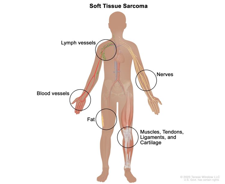
EnlargeSoft tissue sarcoma forms in the soft tissues of the body, including the muscles, tendons, ligaments, cartilage, fat, blood vessels, lymph vessels, nerves, and tissues around joints.
There are many types of soft tissue sarcoma. The cells of each type of sarcoma look different under a microscope, based on the type of soft tissue in which the cancer began.
For more information about soft tissue sarcomas, see the following:
Having certain inherited disorders can increase the risk of soft tissue sarcoma.
Anything that increases a person’s chance of getting a disease is called a risk factor. Not every person with one or more of these risk factors will develop soft tissue sarcoma, and it will develop in people who don’t have any known risk factors. Talk with your doctor if you think you may be at risk. Risk factors for soft tissue sarcoma include the following inheriteddisorders:
A sign of soft tissue sarcoma is a lump or swelling in soft tissue of the body.
A sarcoma may appear as a painless lump under the skin, often on an arm or a leg. Sarcomas that begin in the abdomen may not cause signs or symptoms until they get very big. As the sarcoma grows and presses on nearby organs, nerves, muscles, or blood vessels, signs and symptoms may include:
Pain.
Trouble breathing.
Other conditions may cause the same signs and symptoms. Check with your doctor if you have any of these problems.
Soft tissue sarcoma is diagnosed with a biopsy.
If your doctor thinks you may have a soft tissue sarcoma, a biopsy will be done. The type of biopsy will be based on the size of the tumor and where it is in the body. These types of biopsies may be used:
Core needle biopsy: The removal of tissue using a wide needle. Multiple tissue samples are taken. This procedure may be guided using ultrasound, CT scan, or MRI.
Incisional biopsy: The removal of part of a lump or a sample of tissue. An incisional biopsy may be done when a core needle biopsy is not safe to perform or core needle biopsy findings are not clear.
Careful planning of the biopsy should involve the surgeon, a radiation oncologist, and an interventional radiologist who uses medical imaging to guide diagnosis. Samples will be taken from the primary tumor, lymph nodes, and other suspicious areas. A pathologist views the tissue under a microscope to look for cancer cells and to find out the grade of the tumor. The grade of a tumor depends on how abnormal the cancer cells look under a microscope and how quickly the cells are dividing. High-grade tumors usually grow and spread more quickly than low-grade tumors.
Because soft tissue sarcoma can be hard to diagnose, the tissue samples should be checked by a pathologist who has experience in diagnosing soft tissue sarcoma.
The following tests may be done on the tissue that was removed:
Immunohistochemistry: A laboratory test that uses antibodies to check for certain antigens (markers) in a sample of a patient’s tissue. The antibodies are usually linked to an enzyme or a fluorescent dye. After the antibodies bind to a specific antigen in the tissue sample, the enzyme or dye is activated, and the antigen can then be seen under a microscope. This type of test is used to help diagnose cancer and to help tell one type of cancer from another type of cancer.
Flow cytometry: A laboratory test that measures the number of cells in a sample, the percentage of live cells in a sample, and certain characteristics of the cells, such as size, shape, and the presence of tumor (or other) markers on the cell surface. The cells from a sample of a patient’s blood, bone marrow, or other tissue are stained with a fluorescent dye, placed in a fluid, and then passed one at a time through a beam of light. The test results are based on how the cells that were stained with the fluorescent dye react to the beam of light.
Molecular profiling: A laboratory method that uses a sample of tissue, blood, or other body fluid to check for certain genes, proteins, or other molecules that may be a sign of a disease or condition, such as cancer. It can also be used to check for certain changes in a gene or chromosome that may increase a person’s risk of developing cancer or other diseases. It may be done with other procedures, such as biopsies, to help diagnose some types of cancer. It may also be used to help plan treatment, find out how well treatment is working, make a prognosis, or predict whether cancer will come back or spread to other parts of the body.
Light and electron microscopy: A laboratory test in which cells in a sample of tissue are viewed under regular and high-powered microscopes to look for certain changes in the cells.
Cytogenetic analysis: A laboratory test in which the chromosomes of cells in a sample of tissue are counted and checked for any changes, such as broken, missing, rearranged, or extra chromosomes. Changes in certain chromosomes may be a sign of cancer. Cytogenetic analysis is used to help diagnose cancer, plan treatment, or find out how well treatment is working.
FISH (fluorescence in situ hybridization): A laboratory test used to look at and count genes or chromosomes in cells and tissues. Pieces of DNA that contain fluorescent dyes are made in the laboratory and added to a sample of a patient’s cells or tissues. When these dyed pieces of DNA attach to certain genes or areas of chromosomes in the sample, they light up when viewed under a fluorescent microscope. The FISH test is used to help diagnose cancer and help plan treatment.
Certain factors affect treatment options and prognosis (chance of recovery).
The treatment options and prognosis depend on the following:
Small, low-grade tumors, especially in the trunk, arms, or legs, are frequently treated with surgery alone. High-grade sarcomas are more difficult to treat and more likely to spread.
Stages of Soft Tissue Sarcoma
Key Points
After soft tissue sarcoma has been diagnosed, tests are done to find out if cancer cells have spread within the soft tissue or to other parts of the body.
There are three ways that cancer spreads in the body.
Cancer may spread from where it began to other parts of the body.
The grade of the tumor is also used to describe the cancer and plan treatment.
For soft tissue sarcoma of the trunk, arms, and legs, the following stages are used:
Stage I
Stage II
Stage III
Stage IV
For soft tissue sarcoma of the retroperitoneum, the following stages are used:
Stage I
Stage II
Stage III
Stage IV
There is no standard staging system for soft tissue sarcoma of the head, neck, chest, or abdomen.
Soft tissue sarcoma can recur (come back) after it has been treated.
After soft tissue sarcoma has been diagnosed, tests are done to find out if cancer cells have spread within the soft tissue or to other parts of the body.
The process used to find out if cancer has spread within the soft tissue or to other parts of the body is called staging. Staging of soft tissue sarcoma is also based on the grade and size of the tumor, and whether it has spread to the lymph nodes or other parts of the body. The information gathered from the staging process determines the stage of the disease. It is important to know the stage in order to plan treatment.
In addition to asking about your personal and family health history and doing a physical exam, your doctor may perform the following tests and procedures in the staging process:
Chest x-ray: An x-ray of the organs and bones inside the chest. An x-ray is a type of energy beam that can go through the body and onto film, making a picture of areas inside the body.
Blood chemistry studies: A procedure in which a blood sample is checked to measure the amounts of certain substances released into the blood by organs and tissues in the body. An unusual (higher or lower than normal) amount of a substance can be a sign of disease.
Complete blood count (CBC): A procedure in which a sample of blood is drawn and checked for the following:
The portion of the blood sample made up of red blood cells.
CT scan (CAT scan): A procedure that makes a series of detailed pictures of areas inside of the body, such as the lung, abdomen, and pelvis, taken from different angles. The pictures are made by a computer linked to an x-ray machine. A dye may be injected into a vein or swallowed to help the organs or tissues show up more clearly. This procedure is also called computed tomography, computerized tomography, or computerized axial tomography.
MRI (magnetic resonance imaging): A procedure that uses a magnet, radio waves, and a computer to make a series of detailed pictures of areas inside the body. This procedure is also called nuclear magnetic resonance imaging (NMRI).
PET scan (positron emission tomography scan): A procedure to find malignant tumor cells in the body. A small amount of radioactiveglucose (sugar) is injected into a vein. The PET scanner rotates around the body and makes a picture of where glucose is being used in the body. Malignant tumor cells show up brighter in the picture because they are more active and take up more glucose than normal cells do.
The results of these tests are viewed together with the results of the tumor biopsy to find out the stage of the soft tissue sarcoma before treatment is given. Sometimes chemotherapy or radiation therapy is given as the initial treatment and afterwards the soft tissue sarcoma is staged again.
There are three ways that cancer spreads in the body.
Tissue. The cancer spreads from where it began by growing into nearby areas.
Lymph system. The cancer spreads from where it began by getting into the lymph system. The cancer travels through the lymph vessels to other parts of the body.
Blood. The cancer spreads from where it began by getting into the blood. The cancer travels through the blood vessels to other parts of the body.
Cancer may spread from where it began to other parts of the body.
When cancer spreads to another part of the body, it is called metastasis. Cancer cells break away from where they began (the primary tumor) and travel through the lymph system or blood.
Lymph system. The cancer gets into the lymph system, travels through the lymph vessels, and forms a tumor (metastatic tumor) in another part of the body.
Blood. The cancer gets into the blood, travels through the blood vessels, and forms a tumor (metastatic tumor) in another part of the body.
The metastatic tumor is the same type of cancer as the primary tumor. For example, if soft tissue sarcoma spreads to the lung, the cancer cells in the lung are actually soft tissue sarcoma cells. The disease is metastatic soft tissue sarcoma, not lung cancer.
Many cancer deaths are caused when cancer moves from the original tumor and spreads to other tissues and organs. This is called metastatic cancer. This animation shows how cancer cells travel from the place in the body where they first formed to other parts of the body.
The grade of the tumor is also used to describe the cancer and plan treatment.
The grade of the tumor describes how abnormal the cancer cells look under a microscope and how quickly the tumor is likely to grow and spread. Low grade, mid grade, and high grade are used to describe soft tissue sarcoma:
Low grade: In low-grade soft tissue sarcoma, the cancer cells look more like normal cells under a microscope and grow and spread more slowly than in mid-grade and high-grade soft tissue sarcoma.
Mid grade: In mid-grade soft tissue sarcoma, the cancer cells look more abnormal under a microscope and grow and spread more quickly than in low-grade soft tissue sarcoma.
High grade: In high-grade soft tissue sarcoma, the cancer cells look more abnormal under a microscope and grow and spread more quickly than in low-grade and mid-grade soft tissue sarcoma.
For soft tissue sarcoma of the trunk, arms, and legs, the following stages are used:
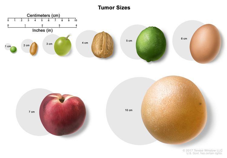
EnlargeTumor sizes are often measured in centimeters (cm) or inches. Common food items that can be used to show tumor size in cm include: a pea (1 cm), a peanut (2 cm), a grape (3 cm), a walnut (4 cm), a lime (5 cm or 2 inches), an egg (6 cm), a peach (7 cm), and a grapefruit (10 cm or 4 inches).
EnlargeTumor sizes are often measured in centimeters (cm) or inches. Common food items that can be used to show tumor size in cm include: a pea (1 cm), a peanut (2 cm), a grape (3 cm), a walnut (4 cm), a lime (5 cm or 2 inches), an egg (6 cm), a peach (7 cm), and a grapefruit (10 cm or 4 inches).
There is no standard staging system for soft tissue sarcoma of the head, neck, chest, or abdomen.
Soft tissue sarcoma can recur (come back) after it has been treated.
The cancer may come back in the same soft tissue or in other parts of the body.
Treatment Option Overview
Key Points
There are different types of treatment for patients with soft tissue sarcoma.
The following types of treatment are used:
Surgery
Radiation therapy
Chemotherapy
Targeted therapy
Immunotherapy
Treatment for soft tissue sarcoma may cause side effects.
Patients may want to think about taking part in a clinical trial.
Patients can enter clinical trials before, during, or after starting their cancer treatment.
Follow-up tests may be needed.
There are different types of treatment for patients with soft tissue sarcoma.
Different types of treatments are available for patients with soft tissue sarcoma. Some treatments are standard (the currently used treatment), and some are being tested in clinical trials. A treatment clinical trial is a research study meant to help improve current treatments or obtain information on new treatments for patients with cancer. When clinical trials show that a new treatment is better than the standard treatment, the new treatment may become the standard treatment. Patients may want to think about taking part in a clinical trial. Some clinical trials are open only to patients who have not started treatment.
The following types of treatment are used:
Surgery
Surgery is the most common treatment for soft tissue sarcoma. It may be the only treatment needed for small, low-grade tumors, especially in the trunk, arms, or legs. The following surgical procedures may be used:
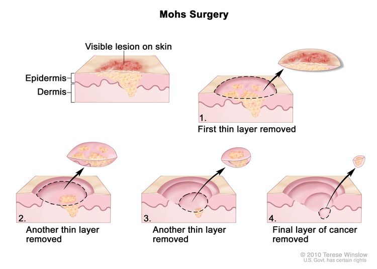
Mohs micrographic surgery: A procedure in which the tumor is cut from the skin in thin layers. During surgery, the edges of the tumor and each layer of tumor removed are viewed through a microscope to check for cancer cells. Layers continue to be removed until no more cancer cells are seen. This type of surgery removes as little normal tissue as possible and is often used where appearance is important, such as on the skin. Enlarge
Wide local excision: Removal of the tumor along with some normal tissue around it. For tumors of the head, neck, abdomen, and trunk, as little normal tissue as possible is removed.
Limb-sparing surgery: Removal of the tumor in an arm or leg without amputation, so the use and appearance of the limb is saved. Radiation therapy or chemotherapy may be given first to shrink the tumor. The tumor is then removed in a wide local excision. Tissue and bone that are removed may be replaced with a graft using tissue and bone taken from another part of the patient’s body, or with an implant such as artificial bone.
Amputation: Surgery to remove part or all of an arm or leg. Amputation is rarely used to treat soft tissue sarcoma.
Lymphadenectomy: A surgical procedure in which lymph nodes are removed and a sample of tissue is checked under a microscope for signs of cancer. This procedure is also called a lymph node dissection.
Radiation therapy or chemotherapy may be given before or after surgery to remove the tumor. When given before surgery, radiation therapy or chemotherapy will make the tumor smaller and reduce the amount of tissue that needs to be removed during surgery. Treatment given before surgery is called neoadjuvant therapy. When given after surgery to remove all of the tumor that can be seen, radiation therapy or chemotherapy will kill any remaining cancer cells. Treatment given after the surgery, to lower the risk that the cancer will come back, is called adjuvant therapy.
Radiation therapy
Radiation therapy is a cancer treatment that uses high-energy x-rays or other types of radiation to kill cancer cells or keep them from growing. There are two types of radiation therapy:
External radiation therapy uses a machine outside the body to send radiation toward the area of the body with cancer.
Intensity-modulated radiation therapy (IMRT) is a type of 3-dimensional (3-D) radiation therapy that uses a computer to make pictures of the size and shape of the tumor. Thin beams of radiation of different intensities (strengths) are aimed at the tumor from many angles. This type of external radiation therapy causes less damage to nearby healthy tissue and is less likely to cause dry mouth, trouble swallowing, and damage to the skin.
The way the radiation therapy is given depends on the type and stage of the cancer being treated. External radiation therapy and internal radiation therapy may be used to treat soft tissue sarcoma.
Chemotherapy
Chemotherapy is a cancer treatment that uses drugs to stop the growth of cancer cells, either by killing the cells or by stopping them from dividing. When chemotherapy is taken by mouth or injected into a vein or muscle, the drugs enter the bloodstream and can reach cancer cells throughout the body (systemic chemotherapy).
Targeted therapy is a type of treatment that uses drugs or other substances to identify and attack specific cancer cells. There are different types of targeted therapy. These include:
Histone methyltransferase inhibitor therapy: This therapy may help keep cancer cells from growing. Tazemetostat is a type of histone methyltransferase inhibitor therapy used to treat soft tissue sarcoma.
Immunotherapy is a treatment that uses the patient’s immune system to fight cancer. Substances made by the body or made in a laboratory are used to boost, direct, or restore the body’s natural defenses against cancer.
Immune checkpoint inhibitor therapy is a type of immunotherapy. Some types of immune system cells, such as T cells, and some cancer cells have certain proteins, called checkpoint proteins, on their surface that keep immune responses in check. These checkpoints help keep immune responses from being too strong and sometimes can keep T cells from killing cancer cells. When these checkpoints are blocked, T cells can kill cancer cells better.
Types of immune checkpoint inhibitor therapy include the following:
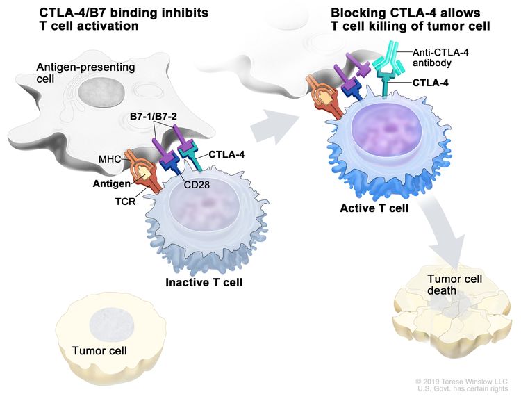
CTLA-4 inhibitor therapy: CTLA-4 is a protein on the surface of T cells that helps keep the body’s immune responses in check. When CTLA-4 attaches to another protein called B7 on a cancer cell, it stops the T cell from killing the cancer cell. CTLA-4 inhibitors attach to CTLA-4 and allow the T cells to kill cancer cells.
Ipilimumab is a type of CTLA-4 inhibitor that is being studied to treat soft tissue sarcoma.
EnlargeImmune checkpoint inhibitor. Checkpoint proteins, such as B7-1/B7-2 on antigen-presenting cells (APC) and CTLA-4 on T cells, help keep the body’s immune responses in check. When the T-cell receptor (TCR) binds to antigen and major histocompatibility complex (MHC) proteins on the APC and CD28 binds to B7-1/B7-2 on the APC, the T cell can be activated. However, the binding of B7-1/B7-2 to CTLA-4 keeps the T cells in the inactive state so they are not able to kill tumor cells in the body (left panel). Blocking the binding of B7-1/B7-2 to CTLA-4 with an immune checkpoint inhibitor (anti-CTLA-4 antibody) allows the T cells to be active and to kill tumor cells (right panel).
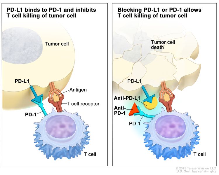
PD-1 and PD-L1 inhibitor therapy: PD-1 is a protein on the surface of T cells that helps keep the body’s immune responses in check. PD-L1 is a protein found on some types of cancer cells. When PD-1 attaches to PD-L1, it stops the T cell from killing the cancer cell. PD-1 and PD-L1 inhibitors keep PD-1 and PD-L1 proteins from attaching to each other. This allows the T cells to kill cancer cells.
EnlargeImmune checkpoint inhibitor. Checkpoint proteins, such as PD-L1 on tumor cells and PD-1 on T cells, help keep immune responses in check. The binding of PD-L1 to PD-1 keeps T cells from killing tumor cells in the body (left panel). Blocking the binding of PD-L1 to PD-1 with an immune checkpoint inhibitor (anti-PD-L1 or anti-PD-1) allows the T cells to kill tumor cells (right panel).
How do monoclonal antibodies work to treat cancer? This video shows how monoclonal antibodies, such as trastuzumab, pembrolizumab, and rituximab, block molecules cancer cells need to grow, flag cancer cells for destruction by the body’s immune system, or deliver harmful substances to cancer cells.
Patients may want to think about taking part in a clinical trial.
For some patients, taking part in a clinical trial may be the best treatment choice. Clinical trials are part of the cancer research process. Clinical trials are done to find out if new cancer treatments are safe and effective or better than the standard treatment.
Many of today’s standard treatments for cancer are based on earlier clinical trials. Patients who take part in a clinical trial may receive the standard treatment or be among the first to receive a new treatment.
Patients who take part in clinical trials also help improve the way cancer will be treated in the future. Even when clinical trials do not lead to effective new treatments, they often answer important questions and help move research forward.
Patients can enter clinical trials before, during, or after starting their cancer treatment.
Some clinical trials only include patients who have not yet received treatment. Other trials test treatments for patients whose cancer has not gotten better. There are also clinical trials that test new ways to stop cancer from recurring (coming back) or reduce the side effects of cancer treatment.
Clinical trials are taking place in many parts of the country. Information about clinical trials supported by NCI can be found on NCI’s clinical trials search webpage. Clinical trials supported by other organizations can be found on the ClinicalTrials.gov website.
Follow-up tests may be needed.
As you go through treatment, you will have follow-up tests or check-ups. Some tests that were done to diagnose or stage the cancer may be repeated to see how well the treatment is working. Decisions about whether to continue, change, or stop treatment may be based on the results of these tests.
Some of the tests will continue to be done from time to time after treatment has ended. The results of these tests can show if your condition has changed or if the cancer has recurred (come back).
Use our clinical trial search to find NCI-supported cancer clinical trials that are accepting patients. You can search for trials based on the type of cancer, the age of the patient, and where the trials are being done. General information about clinical trials is also available.
Treatment of Stage II Soft Tissue Sarcoma and Stage III Soft Tissue Sarcoma That Has Not Spread to Lymph Nodes
Use our clinical trial search to find NCI-supported cancer clinical trials that are accepting patients. You can search for trials based on the type of cancer, the age of the patient, and where the trials are being done. General information about clinical trials is also available.
Treatment of Stage III Soft Tissue Sarcoma That Has Spread to Lymph Nodes (Advanced)
Use our clinical trial search to find NCI-supported cancer clinical trials that are accepting patients. You can search for trials based on the type of cancer, the age of the patient, and where the trials are being done. General information about clinical trials is also available.
Use our clinical trial search to find NCI-supported cancer clinical trials that are accepting patients. You can search for trials based on the type of cancer, the age of the patient, and where the trials are being done. General information about clinical trials is also available.
Use our clinical trial search to find NCI-supported cancer clinical trials that are accepting patients. You can search for trials based on the type of cancer, the age of the patient, and where the trials are being done. General information about clinical trials is also available.
To Learn More About Soft Tissue Sarcoma
For more information from the National Cancer Institute about soft tissue sarcomas, see the following:
Physician Data Query (PDQ) is the National Cancer Institute’s (NCI’s) comprehensive cancer information database. The PDQ database contains summaries of the latest published information on cancer prevention, detection, genetics, treatment, supportive care, and complementary and alternative medicine. Most summaries come in two versions. The health professional versions have detailed information written in technical language. The patient versions are written in easy-to-understand, nontechnical language. Both versions have cancer information that is accurate and up to date and most versions are also available in Spanish.
PDQ is a service of the NCI. The NCI is part of the National Institutes of Health (NIH). NIH is the federal government’s center of biomedical research. The PDQ summaries are based on an independent review of the medical literature. They are not policy statements of the NCI or the NIH.
Purpose of This Summary
This PDQ cancer information summary has current information about the treatment of soft tissue sarcoma. It is meant to inform and help patients, families, and caregivers. It does not give formal guidelines or recommendations for making decisions about health care.
Reviewers and Updates
Editorial Boards write the PDQ cancer information summaries and keep them up to date. These Boards are made up of experts in cancer treatment and other specialties related to cancer. The summaries are reviewed regularly and changes are made when there is new information. The date on each summary (“Updated”) is the date of the most recent change.
The information in this patient summary was taken from the health professional version, which is reviewed regularly and updated as needed, by the PDQ Adult Treatment Editorial Board.
Clinical Trial Information
A clinical trial is a study to answer a scientific question, such as whether one treatment is better than another. Trials are based on past studies and what has been learned in the laboratory. Each trial answers certain scientific questions in order to find new and better ways to help cancer patients. During treatment clinical trials, information is collected about the effects of a new treatment and how well it works. If a clinical trial shows that a new treatment is better than one currently being used, the new treatment may become “standard.” Patients may want to think about taking part in a clinical trial. Some clinical trials are open only to patients who have not started treatment.
Clinical trials can be found online at NCI’s website. For more information, call the Cancer Information Service (CIS), NCI’s contact center, at 1-800-4-CANCER (1-800-422-6237).
Permission to Use This Summary
PDQ is a registered trademark. The content of PDQ documents can be used freely as text. It cannot be identified as an NCI PDQ cancer information summary unless the whole summary is shown and it is updated regularly. However, a user would be allowed to write a sentence such as “NCI’s PDQ cancer information summary about breast cancer prevention states the risks in the following way: [include excerpt from the summary].”
Images in this summary are used with permission of the author(s), artist, and/or publisher for use in the PDQ summaries only. If you want to use an image from a PDQ summary and you are not using the whole summary, you must get permission from the owner. It cannot be given by the National Cancer Institute. Information about using the images in this summary, along with many other images related to cancer can be found in Visuals Online. Visuals Online is a collection of more than 3,000 scientific images.
Disclaimer
The information in these summaries should not be used to make decisions about insurance reimbursement. More information on insurance coverage is available on Cancer.gov on the Managing Cancer Care page.
Contact Us
More information about contacting us or receiving help with the Cancer.gov website can be found on our Contact Us for Help page. Questions can also be submitted to Cancer.gov through the website’s E-mail Us.
Uterine sarcoma accounts for less than 1% of gynecologic malignancies and 2% to 5% of all uterine malignancies.[1] The following tumors arise primarily from three distinct tissues:
Carcinosarcoma arises in the endometrium, in other organs of mullerian origin, and accounts for 40% to 50% of all uterine sarcomas.
Leiomyosarcoma arises from myometrial muscle, with a peak incidence occurring at age 50 years, and accounts for 30% of all uterine sarcomas.
Sarcoma arises in the endometrial stroma, with a peak incidence occurring before menopause for the low-grade tumors and after menopause for the high-grade tumors. It accounts for 15% of all uterine sarcomas.
These three distinct entities are often grouped together as uterine sarcomas; however, each type of tumor is being studied in separate clinical trials.
Carcinosarcoma (the preferred designation by the World Health Organization [WHO]) is also referred to as mixed mesodermal sarcoma or mullerian tumor. Controversy exists about the following issues:
Whether carcinosarcoma is a true sarcoma.
Whether the sarcomatous elements are actually derived from a common epithelial-cell precursor that also gives rise to the usually more abundant adenocarcinomatous elements.
The stromal components of carcinosarcoma are further characterized by homologous elements, such as malignant mesenchymal tissue considered possibly native to the uterus, or heterologous elements, such as striated muscle, cartilage, or bone, which are foreign to the uterus. Carcinosarcoma parallels endometrial cancer in its postmenopausal predominance and in other epidemiological features. Increasingly, the treatment of carcinosarcoma is becoming similar to combined modality approaches for endometrial adenocarcinoma.
Other rare forms of uterine sarcoma also fall under the WHO classification for mesenchymal and mixed tumors of the uterus. These sarcomas include the following:[2,3]
Mixed endometrial stromal and smooth muscle tumors.
Adenosarcomas, in which the epithelial elements appear benign within a malignant mesenchymal background.
Embryonal botryoides or rhabdomyosarcomas, which are found almost exclusively in infants.
PEComa—a perivascular epithelial-cell tumor that may behave in a malignant fashion, which is the latest to be added.
The only documented etiological factor in 10% to 25% of these malignancies is prior pelvic radiation therapy, which is often administered for benign uterine bleeding that began 5 to 25 years earlier. An increased incidence of uterine sarcoma has been associated with tamoxifen in the treatment of breast cancer. Subsequently, increases have also been noted when tamoxifen was given to prevent breast cancer in women at increased risk—a possible result of the estrogenic effect of tamoxifen on the uterus. Because of this increase, patients taking tamoxifen should have follow-up pelvic examinations and should undergo endometrial biopsy if there is any abnormal uterine bleeding.[4–6]
Prognosis
The prognosis for women with uterine sarcoma primarily depends on the extent of disease at the time of diagnosis.[7] For women with carcinosarcoma, significant predictors of metastatic disease at initial surgery include:[7]
Isthmic or cervical location.
Lymphatic vascular space invasion.
Serous and clear cell histology.
Grade 2 or 3 carcinoma.
These factors, in addition to the following ones, correlate with a progression-free interval:[7]
Adnexal spread.
Lymph node metastases.
Tumor size.
Peritoneal cytologic findings.
Depth of myometrial invasion.
Factors that bear no relationship to the presence or absence of metastases at surgical exploration include:
Presence or absence of stromal heterologous elements.
Types of such elements.
Grade of the stromal components.
Mitotic activity of the stromal components.
In one study, women with well-differentiated sarcomatous components or carcinosarcomas had significantly longer progression-free intervals than those with moderately to poorly differentiated sarcomas for the homologous and heterologous types. The recurrence rate was 44% for homologous tumors and 63% for heterologous tumors. The type of heterologous sarcoma had no effect on the progression-free interval.
For women with leiomyosarcomas, some investigators consider tumor size to be the most important prognostic factor. Women with tumors larger than 5.0 cm in maximum diameter have a poor prognosis.[8] However, in a Gynecologic Oncology Group study, the mitotic index was the only factor significantly related to progression-free interval.[7] Leiomyosarcomas matched for other known prognostic factors may be more aggressive than their carcinosarcoma counterparts.[9] The 5-year survival rate for women with stage I disease, which is confined to the corpus, is approximately 50% versus 0% to 20% for the remaining stages.
Surgery alone can be curative if the malignancy is contained within the uterus. The value of pelvic radiation therapy is not established. Current studies consist primarily of phase II chemotherapy trials for patients with advanced disease. Adjuvant chemotherapy following complete resection for patients with stage I or II disease was not found to be effective in a randomized trial.[10] Yet, nonrandomized trials have reported improved survival following adjuvant chemotherapy with or without radiation therapy.[11–13]
References
Forney JP, Buschbaum HJ: Classifying, staging, and treating uterine sarcomas. Contemp Ob Gyn 18(3):47, 50, 55-56, 61-62, 64, 69, 1981.
Gershenson D, McGuire W, Gore Martin, et al.: Gynecologic Cancer: Controversies in Management. 3rd ed. Churchill Livingstone, 2004.
Tavassoéli F, Devilee P, et al.: Pathology and Genetics of Tumours of the Breast and Female Genital Organs. International Agency for Research on Cancer, 2004.
Bergman L, Beelen ML, Gallee MP, et al.: Risk and prognosis of endometrial cancer after tamoxifen for breast cancer. Comprehensive Cancer Centres’ ALERT Group. Assessment of Liver and Endometrial cancer Risk following Tamoxifen. Lancet 356 (9233): 881-7, 2000. [PUBMED Abstract]
Wickerham DL, Fisher B, Wolmark N, et al.: Association of tamoxifen and uterine sarcoma. J Clin Oncol 20 (11): 2758-60, 2002. [PUBMED Abstract]
Major FJ, Blessing JA, Silverberg SG, et al.: Prognostic factors in early-stage uterine sarcoma. A Gynecologic Oncology Group study. Cancer 71 (4 Suppl): 1702-9, 1993. [PUBMED Abstract]
Evans HL, Chawla SP, Simpson C, et al.: Smooth muscle neoplasms of the uterus other than ordinary leiomyoma. A study of 46 cases, with emphasis on diagnostic criteria and prognostic factors. Cancer 62 (10): 2239-47, 1988. [PUBMED Abstract]
Oláh KS, Dunn JA, Gee H: Leiomyosarcomas have a poorer prognosis than mixed mesodermal tumours when adjusting for known prognostic factors: the result of a retrospective study of 423 cases of uterine sarcoma. Br J Obstet Gynaecol 99 (7): 590-4, 1992. [PUBMED Abstract]
Omura GA, Blessing JA, Major F, et al.: A randomized clinical trial of adjuvant adriamycin in uterine sarcomas: a Gynecologic Oncology Group Study. J Clin Oncol 3 (9): 1240-5, 1985. [PUBMED Abstract]
Piver MS, Lele SB, Marchetti DL, et al.: Effect of adjuvant chemotherapy on time to recurrence and survival of stage I uterine sarcomas. J Surg Oncol 38 (4): 233-9, 1988. [PUBMED Abstract]
van Nagell JR, Hanson MB, Donaldson ES, et al.: Adjuvant vincristine, dactinomycin, and cyclophosphamide therapy in stage I uterine sarcomas. A pilot study. Cancer 57 (8): 1451-4, 1986. [PUBMED Abstract]
Peters WA, Rivkin SE, Smith MR, et al.: Cisplatin and adriamycin combination chemotherapy for uterine stromal sarcomas and mixed mesodermal tumors. Gynecol Oncol 34 (3): 323-7, 1989. [PUBMED Abstract]
Cellular Classification of Uterine Sarcoma
The most common histological types of uterine sarcomas include:
The uterine neoplasm classification of the International Society of Gynecologic Pathologists and the World Health Organization uses the term carcinosarcoma for all primary uterine neoplasms containing malignant elements of both epithelial and stromal light microscopic appearances, regardless of whether malignant heterologous elements are present.[1]
References
Silverberg SG, Major FJ, Blessing JA, et al.: Carcinosarcoma (malignant mixed mesodermal tumor) of the uterus. A Gynecologic Oncology Group pathologic study of 203 cases. Int J Gynecol Pathol 9 (1): 1-19, 1990. [PUBMED Abstract]
Stage Information for Uterine Sarcoma
FIGO Staging
The Fédération Internationale de Gynécologie et d’Obstétrique (FIGO) and the American Joint Committee on Cancer have designated staging to define uterine sarcoma; the FIGO system is most commonly used.[1,2]
The FIGO staging system has two divisions, one for leiomyosarcoma and endometrial stromal sarcoma, and one for adenosarcoma. Carcinosarcoma is staged using the designated endometrial carcinoma definitions. For more information, see Endometrial Cancer Treatment.[1]
Table 1. FIGO Definitions of Uterine Sarcoma–Leiomyosarcoma and Endometrial Stromal Sarcomaa
Stage
Definition
FIGO = Fédération Internationale de Gynécologie et d’Obstétrique.
aAdapted from the Fédération Internationale de Gynécologie et d’Obstétrique.[1]
I
Tumor limited to uterus.
–IA
Tumor ≤5 cm.
–IB
Tumor >5 cm.
II
Tumor extends beyond the uterus, within the pelvis.
–IIA
Adnexal involvement.
–IIB
Involvement of other pelvic tissues.
III
Tumor invades abdominal tissues (not just protruding into the abdomen).
–IIIA
One site.
–IIIB
More than one site.
–IIIC
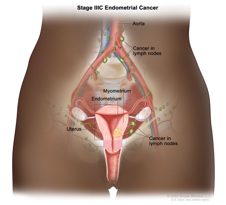
Metastasis to pelvic and/or para-aortic lymph nodes.
IV
–IVA
Tumor invades bladder and/or rectum.
–IVB
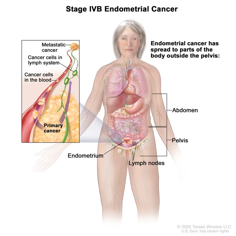
Distant metastasis.
Table 2. FIGO Definitions of Uterine Sarcoma–Adenosarcomaa
Stage
Definition
FIGO = Fédération Internationale de Gynécologie et d’Obstétrique.
aAdapted from the Fédération Internationale de Gynécologie et d’Obstétrique.[1]
I
Tumor limited to uterus.
–IA
Tumor limited to endometrium/endocervix with no myometrial invasion.
–IB
Less than or equal to half myometrial invasion.
–IC
More than half myometrial invasion.
II
Tumor extends to the pelvis.
–IIA
Adnexal involvement.
–IIB
Tumor extends to extrauterine pelvic tissue.
III
Tumor invades abdominal tissues (not just protruding into the abdomen).
–IIIA
One site.
–IIIB
More than one site.
–IIIC
Metastasis to pelvic and/or para-aortic lymph nodes.
IV
–IVA
Tumor invades bladder and/or rectum.
–IVB
Distant metastasis.
References
Mbatani N, Olawaiye AB, Prat J: Uterine sarcomas. Int J Gynaecol Obstet 143 (Suppl 2): 51-58, 2018. [PUBMED Abstract]
Corpus uteri – sarcoma. In: Amin MB, Edge SB, Greene FL, et al., eds.: AJCC Cancer Staging Manual. 8th ed. Springer; 2017, pp 671-80.
Treatment Option Overview for Uterine Sarcoma
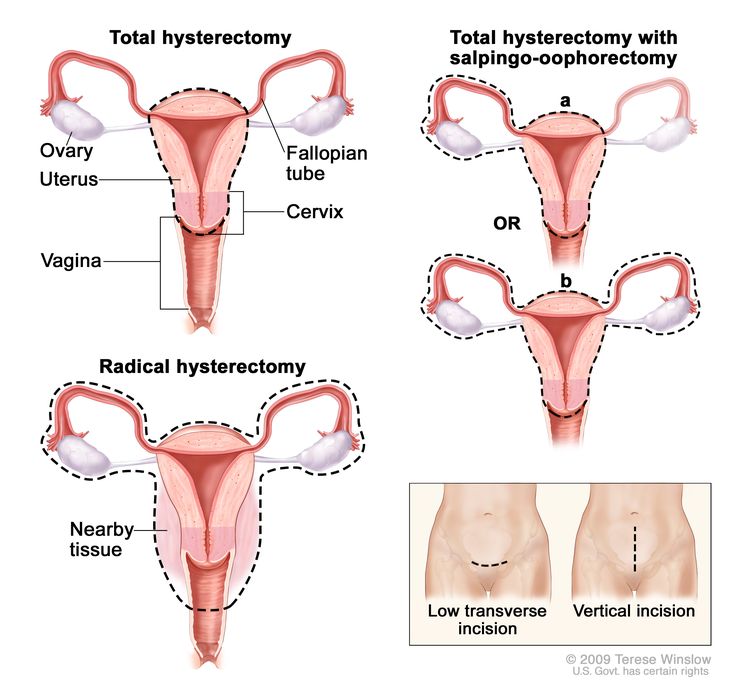
Surgery is often the principal means of diagnosis and is the primary treatment for all patients with uterine sarcoma. If the diagnosis is known, the extent of surgery is planned according to the stage of the tumor. Hysterectomy is usually performed when a uterine malignancy is suspected, except for rare instances when preservation of the uterus in a young patient is deemed safe for the type of cancer (e.g., a totally confined low-grade leiomyosarcoma in a woman who desires to retain childbearing potential). Medically suitable patients with the preoperative diagnosis of uterine sarcoma are considered candidates for abdominal hysterectomy, bilateral salpingo-oophorectomy, and pelvic and periaortic selective lymphadenectomy. Cytologic washings are obtained from the pelvis and abdomen. Thorough examination of the diaphragm, omentum, and upper abdomen is performed.
There is no firm evidence from a prospective study that adjuvant chemotherapy or radiation therapy is of benefit for patients with uterine sarcoma.[1] In one Gynecologic Oncology Group (GOG) study, the use of adjuvant doxorubicin did not alter the survival rate of patients with resected stage I or stage II uterine sarcomas; however, interpretation of these results is difficult because this study included some patients who received radiation and three types of uterine sarcomas that have variable responses to doxorubicin.[1][Level of evidence A1] Because the risk of disease recurrence is high, even with localized presentations, many physicians have considered the use of adjuvant chemotherapy or radiation therapy.[2] Another GOG study (GOG-0150 [NCT00002546]) addressed radiation therapy versus adjuvant chemotherapy.[3]
References
Omura GA, Blessing JA, Major F, et al.: A randomized clinical trial of adjuvant adriamycin in uterine sarcomas: a Gynecologic Oncology Group Study. J Clin Oncol 3 (9): 1240-5, 1985. [PUBMED Abstract]
Kohorn EI, Schwartz PE, Chambers JT, et al.: Adjuvant therapy in mixed mullerian tumors of the uterus. Gynecol Oncol 23 (2): 212-21, 1986. [PUBMED Abstract]
Wolfson AH, Brady MF, Rocereto T, et al.: A gynecologic oncology group randomized phase III trial of whole abdominal irradiation (WAI) vs. cisplatin-ifosfamide and mesna (CIM) as post-surgical therapy in stage I-IV carcinosarcoma (CS) of the uterus. Gynecol Oncol 107 (2): 177-85, 2007. [PUBMED Abstract]
Treatment of Stage I Uterine Sarcoma
Treatment Options for Stage I Uterine Sarcoma
Treatment options for stage I uterine sarcoma include:
Surgery (total abdominal hysterectomy, bilateral salpingo-oophorectomy, and pelvic and periaortic selective lymphadenectomy).
Surgery plus pelvic radiation therapy.
Surgery plus adjuvant chemotherapy.
Surgery plus adjuvant radiation therapy.
A nonrandomized Gynecologic Oncology Group study examined the effect of pelvic radiation therapy on patients with stage I and II carcinosarcomas. Patients who had pelvic radiation therapy had a significant reduction in tumor recurrences within the radiation treatment field but no alteration in survival.[1] A large nonrandomized study demonstrated improved survival and a lower local failure rate in patients with mixed mullerian tumors following postoperative external and intracavitary radiation therapy.[2] One nonrandomized study that predominantly included patients with carcinosarcomas showed that adjuvant chemotherapy with cisplatin and doxorubicin benefitted participants.[3]
Current Clinical Trials
Use our advanced clinical trial search to find NCI-supported cancer clinical trials that are now enrolling patients. The search can be narrowed by location of the trial, type of treatment, name of the drug, and other criteria. General information about clinical trials is also available.
References
Hornback NB, Omura G, Major FJ: Observations on the use of adjuvant radiation therapy in patients with stage I and II uterine sarcoma. Int J Radiat Oncol Biol Phys 12 (12): 2127-30, 1986. [PUBMED Abstract]
Larson B, Silfverswärd C, Nilsson B, et al.: Mixed müllerian tumours of the uterus–prognostic factors: a clinical and histopathologic study of 147 cases. Radiother Oncol 17 (2): 123-32, 1990. [PUBMED Abstract]
Peters WA, Rivkin SE, Smith MR, et al.: Cisplatin and adriamycin combination chemotherapy for uterine stromal sarcomas and mixed mesodermal tumors. Gynecol Oncol 34 (3): 323-7, 1989. [PUBMED Abstract]
Treatment of Stage II Uterine Sarcoma
Treatment Options for Stage II Uterine Sarcoma
Treatment options for stage II uterine sarcoma include:
Surgery (total abdominal hysterectomy, bilateral salpingo-oophorectomy, and pelvic and periaortic selective lymphadenectomy).
Surgery plus pelvic radiation therapy.
Surgery plus adjuvant chemotherapy.
Surgery plus adjuvant radiation therapy.
A nonrandomized Gynecologic Oncology Group study examined the effect of pelvic radiation therapy on patients with stage I and II carcinosarcomas. Patients who had pelvic radiation therapy had a significant reduction in tumor recurrences within the radiation treatment field but no alteration in survival.[1] One nonrandomized study that predominantly included patients with carcinosarcomas showed that adjuvant chemotherapy with cisplatin and doxorubicin benefitted participants.[2]
Current Clinical Trials
Use our advanced clinical trial search to find NCI-supported cancer clinical trials that are now enrolling patients. The search can be narrowed by location of the trial, type of treatment, name of the drug, and other criteria. General information about clinical trials is also available.
References
Hornback NB, Omura G, Major FJ: Observations on the use of adjuvant radiation therapy in patients with stage I and II uterine sarcoma. Int J Radiat Oncol Biol Phys 12 (12): 2127-30, 1986. [PUBMED Abstract]
Peters WA, Rivkin SE, Smith MR, et al.: Cisplatin and adriamycin combination chemotherapy for uterine stromal sarcomas and mixed mesodermal tumors. Gynecol Oncol 34 (3): 323-7, 1989. [PUBMED Abstract]
Treatment of Stage III Uterine Sarcoma
Treatment Options for Stage III Uterine Sarcoma
Treatment options for stage III uterine sarcoma include:
Surgery (total abdominal hysterectomy, bilateral salpingo-oophorectomy, pelvic and periaortic selective lymphadenectomy, and resection of all gross tumor).
Surgery plus pelvic radiation therapy (under clinical evaluation).
Surgery plus adjuvant chemotherapy (under clinical evaluation).
Phase II chemotherapy studies by the Gynecologic Oncology Group have documented some antitumor activity for cisplatin, doxorubicin, and ifosfamide.[1,2] These studies have also documented differences in response leading to separate trials for patients with carcinosarcomas and leiomyosarcomas. In patients previously untreated with chemotherapy, ifosfamide had a 32.2% response rate in patients with carcinosarcomas [3] and a 17.2% partial response rate in patients with leiomyosarcomas.[2]
GOG-108 was a randomized trial that examined the use of ifosfamide with or without cisplatin as first-line therapy for patients with measurable advanced or recurrent carcinosarcomas. Patients in the combination arm had a higher response rate (54% vs. 34%) and longer progression-free survival (PFS) (6 months vs. 4 months). However, patients did not have a significant improvement in survival (9 months vs. 8 months).[4][Level of evidence A1] The follow-up GOG-0161 study [NCT00003128] used 3-day ifosfamide regimens (instead of the more toxic 5-day regimen in the preceding study) given alone or in combination with paclitaxel (with filgrastim starting on day 4).[5] The combination was superior in response rates (45% vs. 29%), PFS (8.4 months vs. 5.8 months), and overall survival (13.5 months vs. 8.4 months). The hazard ratio for death favored the combination (0.69; 95% confidence interval, 0.49–0.97).[5][Level of evidence A1] In this study, 52% of 179 evaluable patients had recurrent disease, 18% had stage III disease, and 30% had stage IV disease. In addition, imbalances were present in the sites of disease and in the use of prior radiation therapy, and 30 patients were excluded for wrong pathology.
A role for chemotherapy as an adjuvant to surgery has not been established.
Current Clinical Trials
Use our advanced clinical trial search to find NCI-supported cancer clinical trials that are now enrolling patients. The search can be narrowed by location of the trial, type of treatment, name of the drug, and other criteria. General information about clinical trials is also available.
References
Thigpen JT, Blessing JA, Beecham J, et al.: Phase II trial of cisplatin as first-line chemotherapy in patients with advanced or recurrent uterine sarcomas: a Gynecologic Oncology Group study. J Clin Oncol 9 (11): 1962-6, 1991. [PUBMED Abstract]
Sutton GP, Blessing JA, Barrett RJ, et al.: Phase II trial of ifosfamide and mesna in leiomyosarcoma of the uterus: a Gynecologic Oncology Group study. Am J Obstet Gynecol 166 (2): 556-9, 1992. [PUBMED Abstract]
Sutton GP, Blessing JA, Rosenshein N, et al.: Phase II trial of ifosfamide and mesna in mixed mesodermal tumors of the uterus (a Gynecologic Oncology Group study). Am J Obstet Gynecol 161 (2): 309-12, 1989. [PUBMED Abstract]
Sutton G, Brunetto VL, Kilgore L, et al.: A phase III trial of ifosfamide with or without cisplatin in carcinosarcoma of the uterus: A Gynecologic Oncology Group Study. Gynecol Oncol 79 (2): 147-53, 2000. [PUBMED Abstract]
Homesley HD, Filiaci V, Markman M, et al.: Phase III trial of ifosfamide with or without paclitaxel in advanced uterine carcinosarcoma: a Gynecologic Oncology Group Study. J Clin Oncol 25 (5): 526-31, 2007. [PUBMED Abstract]
Treatment of Stage IV Uterine Sarcoma
Treatment Options for Stage IV Uterine Sarcoma
There is currently no standard therapy for patients with stage IV disease. These patients should enroll in an ongoing clinical trial.
Phase II chemotherapy studies by the Gynecologic Oncology Group have documented some antitumor activity for cisplatin, doxorubicin, and ifosfamide.[1,2] These studies have also documented differences in response leading to separate trials for patients with carcinosarcomas and leiomyosarcomas. In patients previously untreated with chemotherapy, ifosfamide had a 32.2% response rate in patients with carcinosarcomas,[2] a 33% response rate in patients with endometrial stromal cell sarcomas,[3], and a 17.2% partial response rate in patients with leiomyosarcomas.[4] Doxorubicin in combination with dacarbazine or cyclophosphamide is no more active than doxorubicin alone for advanced disease.[5,6] Cisplatin has activity as first-line therapy and minimal activity as second-line therapy for patients with carcinosarcomas, but cisplatin is inactive as first- or second-line therapy for patients with leiomyosarcomas.[1,7]
GOG-108 was a randomized trial that examined the use of ifosfamide with or without cisplatin as first-line therapy for patients with measurable advanced or recurrent carcinosarcomas. Patients in the combination arm had a higher response rate (54% vs. 34%) and longer progression-free survival (PFS) (6 months vs. 4 months). However, patients did not have a significant improvement in survival (9 months vs. 8 months).[8][Level of evidence A1] The follow-up GOG-0161 study [NCT00003128] used 3-day ifosfamide regimens (instead of the more toxic 5-day regimen in the preceding study) given alone or in combination with paclitaxel (with filgrastim starting on day 4).[9] The combination was superior in response rates (45% vs. 29%), PFS (8.4 months vs. 5.8 months), and overall survival (13.5 months vs. 8.4 months). The hazard ratio for death favored the combination (0.69; 95% confidence interval, 0.49–0.97).[9][Level of evidence A1] In this study, 52% of 179 evaluable patients had recurrent disease, 18% had stage III disease, and 30% had stage IV disease. In addition, imbalances were present in the sites of disease and in the use of prior radiation therapy, and 30 patients were excluded for wrong pathology.
Current Clinical Trials
Use our advanced clinical trial search to find NCI-supported cancer clinical trials that are now enrolling patients. The search can be narrowed by location of the trial, type of treatment, name of the drug, and other criteria. General information about clinical trials is also available.
References
Thigpen JT, Blessing JA, Beecham J, et al.: Phase II trial of cisplatin as first-line chemotherapy in patients with advanced or recurrent uterine sarcomas: a Gynecologic Oncology Group study. J Clin Oncol 9 (11): 1962-6, 1991. [PUBMED Abstract]
Sutton GP, Blessing JA, Rosenshein N, et al.: Phase II trial of ifosfamide and mesna in mixed mesodermal tumors of the uterus (a Gynecologic Oncology Group study). Am J Obstet Gynecol 161 (2): 309-12, 1989. [PUBMED Abstract]
Sutton G, Blessing JA, Park R, et al.: Ifosfamide treatment of recurrent or metastatic endometrial stromal sarcomas previously unexposed to chemotherapy: a study of the Gynecologic Oncology Group. Obstet Gynecol 87 (5 Pt 1): 747-50, 1996. [PUBMED Abstract]
Sutton GP, Blessing JA, Barrett RJ, et al.: Phase II trial of ifosfamide and mesna in leiomyosarcoma of the uterus: a Gynecologic Oncology Group study. Am J Obstet Gynecol 166 (2): 556-9, 1992. [PUBMED Abstract]
Omura GA, Major FJ, Blessing JA, et al.: A randomized study of adriamycin with and without dimethyl triazenoimidazole carboxamide in advanced uterine sarcomas. Cancer 52 (4): 626-32, 1983. [PUBMED Abstract]
Muss HB, Bundy B, DiSaia PJ, et al.: Treatment of recurrent or advanced uterine sarcoma. A randomized trial of doxorubicin versus doxorubicin and cyclophosphamide (a phase III trial of the Gynecologic Oncology Group). Cancer 55 (8): 1648-53, 1985. [PUBMED Abstract]
Thigpen JT, Blessing JA, Wilbanks GD: Cisplatin as second-line chemotherapy in the treatment of advanced or recurrent leiomyosarcoma of the uterus. A phase II trial of the Gynecologic Oncology Group. Am J Clin Oncol 9 (1): 18-20, 1986. [PUBMED Abstract]
Sutton G, Brunetto VL, Kilgore L, et al.: A phase III trial of ifosfamide with or without cisplatin in carcinosarcoma of the uterus: A Gynecologic Oncology Group Study. Gynecol Oncol 79 (2): 147-53, 2000. [PUBMED Abstract]
Homesley HD, Filiaci V, Markman M, et al.: Phase III trial of ifosfamide with or without paclitaxel in advanced uterine carcinosarcoma: a Gynecologic Oncology Group Study. J Clin Oncol 25 (5): 526-31, 2007. [PUBMED Abstract]
Treatment of Recurrent Uterine Sarcoma
Treatment Options for Recurrent Uterine Sarcoma
There is currently no standard therapy for patients with recurrent disease. These patients should enroll in an ongoing clinical trial.
Phase II chemotherapy studies by the Gynecologic Oncology Group have documented some antitumor activity for cisplatin, doxorubicin, and ifosfamide.[1,2] These studies have also documented differences in response leading to separate trials for patients with carcinosarcomas and leiomyosarcomas. In patients previously untreated with chemotherapy, ifosfamide had a 32.2% response rate in patients with carcinosarcomas,[2] a 33% response rate in patients with endometrial stromal cell sarcomas,[3] and a 17.2% partial response rate in patients with leiomyosarcomas.[4] Doxorubicin in combination with dacarbazine or cyclophosphamide is no more active than doxorubicin alone for recurrent disease.[5,6] Cisplatin has activity as first-line therapy and minimal activity as second-line therapy for patients with carcinosarcomas, but cisplatin is inactive as first- or second-line therapy for patients with leiomyosarcomas.[1,7] A regimen of gemcitabine plus docetaxel had a 53% response rate in patients with unresectable leiomyosarcomas and is undergoing further study.[8]
GOG-108 was a randomized trial that examined the use of ifosfamide with or without cisplatin as first-line therapy for patients with measurable advanced or recurrent carcinosarcomas. Patients in the combination arm had a higher response rate (54% vs. 34%) and longer progression-free survival (PFS) (6 months vs. 4 months). However, patients did not have a significant improvement in survival (9 months vs. 8 months).[9][Level of evidence A1] The follow-up GOG-0161 study [NCT00003128] used 3-day ifosfamide regimens (instead of the more toxic 5-day regimen in the preceding study) given alone or in combination with paclitaxel (with filgrastim starting on day 4).[10] The combination was superior in response rates (45% vs. 29%), PFS (8.4 months vs. 5.8 months), and overall survival (13.5 months vs. 8.4 months). The hazard ratio for death favored the combination (0.69; 95% confidence interval, 0.49–0.97).[10][Level of evidence A1] In this study, 52% of 179 evaluable patients had recurrent disease, 18% had stage III disease, and 30% had stage IV disease. In addition, imbalances were present in the sites of disease and in the use of prior radiation therapy, and 30 patients were excluded for wrong pathology.
Radiation therapy may be an effective method of palliative care for patients with carcinosarcoma who have localized recurrence in the pelvis confirmed by computed tomography. Phase I and II clinical trials are appropriate for patients who have disease recurrence with distant metastasis and are unresponsive to first-line phase II trials. High-dose progesterone hormone therapy may be of some benefit to patients with low-grade stromal sarcoma.[11]
Current Clinical Trials
Use our advanced clinical trial search to find NCI-supported cancer clinical trials that are now enrolling patients. The search can be narrowed by location of the trial, type of treatment, name of the drug, and other criteria. General information about clinical trials is also available.
References
Thigpen JT, Blessing JA, Beecham J, et al.: Phase II trial of cisplatin as first-line chemotherapy in patients with advanced or recurrent uterine sarcomas: a Gynecologic Oncology Group study. J Clin Oncol 9 (11): 1962-6, 1991. [PUBMED Abstract]
Sutton GP, Blessing JA, Rosenshein N, et al.: Phase II trial of ifosfamide and mesna in mixed mesodermal tumors of the uterus (a Gynecologic Oncology Group study). Am J Obstet Gynecol 161 (2): 309-12, 1989. [PUBMED Abstract]
Sutton G, Blessing JA, Park R, et al.: Ifosfamide treatment of recurrent or metastatic endometrial stromal sarcomas previously unexposed to chemotherapy: a study of the Gynecologic Oncology Group. Obstet Gynecol 87 (5 Pt 1): 747-50, 1996. [PUBMED Abstract]
Sutton GP, Blessing JA, Barrett RJ, et al.: Phase II trial of ifosfamide and mesna in leiomyosarcoma of the uterus: a Gynecologic Oncology Group study. Am J Obstet Gynecol 166 (2): 556-9, 1992. [PUBMED Abstract]
Omura GA, Major FJ, Blessing JA, et al.: A randomized study of adriamycin with and without dimethyl triazenoimidazole carboxamide in advanced uterine sarcomas. Cancer 52 (4): 626-32, 1983. [PUBMED Abstract]
Muss HB, Bundy B, DiSaia PJ, et al.: Treatment of recurrent or advanced uterine sarcoma. A randomized trial of doxorubicin versus doxorubicin and cyclophosphamide (a phase III trial of the Gynecologic Oncology Group). Cancer 55 (8): 1648-53, 1985. [PUBMED Abstract]
Thigpen JT, Blessing JA, Wilbanks GD: Cisplatin as second-line chemotherapy in the treatment of advanced or recurrent leiomyosarcoma of the uterus. A phase II trial of the Gynecologic Oncology Group. Am J Clin Oncol 9 (1): 18-20, 1986. [PUBMED Abstract]
Hensley ML, Maki R, Venkatraman E, et al.: Gemcitabine and docetaxel in patients with unresectable leiomyosarcoma: results of a phase II trial. J Clin Oncol 20 (12): 2824-31, 2002. [PUBMED Abstract]
Sutton G, Brunetto VL, Kilgore L, et al.: A phase III trial of ifosfamide with or without cisplatin in carcinosarcoma of the uterus: A Gynecologic Oncology Group Study. Gynecol Oncol 79 (2): 147-53, 2000. [PUBMED Abstract]
Homesley HD, Filiaci V, Markman M, et al.: Phase III trial of ifosfamide with or without paclitaxel in advanced uterine carcinosarcoma: a Gynecologic Oncology Group Study. J Clin Oncol 25 (5): 526-31, 2007. [PUBMED Abstract]
Katz L, Merino MJ, Sakamoto H, et al.: Endometrial stromal sarcoma: a clinicopathologic study of 11 cases with determination of estrogen and progestin receptor levels in three tumors. Gynecol Oncol 26 (1): 87-97, 1987. [PUBMED Abstract]
Latest Updates to This Summary (12/17/2024)
The PDQ cancer information summaries are reviewed regularly and updated as new information becomes available. This section describes the latest changes made to this summary as of the date above.
Editorial changes were made to this summary.
This summary is written and maintained by the PDQ Adult Treatment Editorial Board, which is editorially independent of NCI. The summary reflects an independent review of the literature and does not represent a policy statement of NCI or NIH. More information about summary policies and the role of the PDQ Editorial Boards in maintaining the PDQ summaries can be found on the About This PDQ Summary and PDQ® Cancer Information for Health Professionals pages.
About This PDQ Summary
Purpose of This Summary
This PDQ cancer information summary for health professionals provides comprehensive, peer-reviewed, evidence-based information about the treatment of uterine sarcoma. It is intended as a resource to inform and assist clinicians in the care of their patients. It does not provide formal guidelines or recommendations for making health care decisions.
Reviewers and Updates
This summary is reviewed regularly and updated as necessary by the PDQ Adult Treatment Editorial Board, which is editorially independent of the National Cancer Institute (NCI). The summary reflects an independent review of the literature and does not represent a policy statement of NCI or the National Institutes of Health (NIH).
Board members review recently published articles each month to determine whether an article should:
be discussed at a meeting,
be cited with text, or
replace or update an existing article that is already cited.
Changes to the summaries are made through a consensus process in which Board members evaluate the strength of the evidence in the published articles and determine how the article should be included in the summary.
The lead reviewer for Uterine Sarcoma Treatment is:
Marina Stasenko, MD (New York University Medical Center)
Any comments or questions about the summary content should be submitted to Cancer.gov through the NCI website’s Email Us. Do not contact the individual Board Members with questions or comments about the summaries. Board members will not respond to individual inquiries.
Levels of Evidence
Some of the reference citations in this summary are accompanied by a level-of-evidence designation. These designations are intended to help readers assess the strength of the evidence supporting the use of specific interventions or approaches. The PDQ Adult Treatment Editorial Board uses a formal evidence ranking system in developing its level-of-evidence designations.
Permission to Use This Summary
PDQ is a registered trademark. Although the content of PDQ documents can be used freely as text, it cannot be identified as an NCI PDQ cancer information summary unless it is presented in its entirety and is regularly updated. However, an author would be permitted to write a sentence such as “NCI’s PDQ cancer information summary about breast cancer prevention states the risks succinctly: [include excerpt from the summary].”
The preferred citation for this PDQ summary is:
PDQ® Adult Treatment Editorial Board. PDQ Uterine Sarcoma Treatment. Bethesda, MD: National Cancer Institute. Updated <MM/DD/YYYY>. Available at: /types/uterine/hp/uterine-sarcoma-treatment-pdq. Accessed <MM/DD/YYYY>. [PMID: 26389327]
Images in this summary are used with permission of the author(s), artist, and/or publisher for use within the PDQ summaries only. Permission to use images outside the context of PDQ information must be obtained from the owner(s) and cannot be granted by the National Cancer Institute. Information about using the illustrations in this summary, along with many other cancer-related images, is available in Visuals Online, a collection of over 2,000 scientific images.
Disclaimer
Based on the strength of the available evidence, treatment options may be described as either “standard” or “under clinical evaluation.” These classifications should not be used as a basis for insurance reimbursement determinations. More information on insurance coverage is available on Cancer.gov on the Managing Cancer Care page.
Contact Us
More information about contacting us or receiving help with the Cancer.gov website can be found on our Contact Us for Help page. Questions can also be submitted to Cancer.gov through the website’s Email Us.
Cancer of the endometrium is the most common gynecologic malignancy in the United States and accounts for 7% of all cancers in women. Most cases are diagnosed at an early stage and are amenable to treatment with surgery alone.[1] However, patients with pathological features predictive of a high rate of relapse and patients with extrauterine spread at diagnosis have a high rate of relapse despite adjuvant therapy. The most common cause of death in patients with endometrial cancer is cardiovascular disease because of related metabolic risk factors.[2]
Incidence and Mortality
Estimated new cases and deaths from cancer of the uterine corpus, which includes the endometrium, in the United States in 2025:[1]
New cases: 69,120.
Deaths: 13,860.
Anatomy
The endometrium is the inner lining of the uterus and has both functional and basal layers. The functional layer is hormonally sensitive and is shed in a cyclical pattern during menstruation in reproductive-age women. Both estrogen and progesterone are necessary to maintain a normal endometrial lining. However, factors that lead to an excess of estrogen, including obesity and anovulation, lead to an increase in the deposition of the endometrial lining. These changes may lead to endometrial hyperplasia and, in some cases, endometrial cancer. Whatever the cause, a thickened lining will lead to sloughing of the endometrial tissue through the endometrial canal and into the vagina. As a result, heavy menstrual bleeding or bleeding after menopause are often the initial signs of endometrial cancer. This symptom tends to happen early in the disease course, allowing for identification of the disease at an early stage for most women.
Prolonged, unopposed estrogen exposure has been associated with an increased risk of endometrial cancer.[9,32] However, combined estrogen and progesterone therapy prevents this increased risk.[33,34]
Tamoxifen, which is used to treat and prevent breast cancer (NSABP-B-14), is associated with an increased risk of endometrial cancer related to the estrogenic effect of tamoxifen on the endometrium.[15,35] It is important that patients who are receiving tamoxifen and experiencing abnormal uterine bleeding have follow-up examinations and biopsy of the endometrial lining. The U.S. Food and Drug Administration released a black box warning that includes data about the increase in uterine malignancies associated with tamoxifen use. For more information about risk factors for Lynch syndrome–associated endometrial cancer, see the Lynch Syndrome section in Genetics of Breast and Gynecologic Cancers.
Clinical Features
Irregular vaginal bleeding is the most common presenting sign of endometrial cancer. It generally occurs early in the disease process and is the reason why most patients are diagnosed with highly curable stage I endometrial cancer.
Diagnostic Evaluation
The following procedures may be used to detect endometrial cancer:
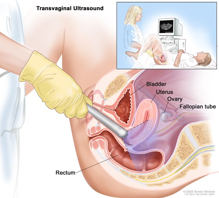
Transvaginal ultrasonography.
Endometrial biopsy.
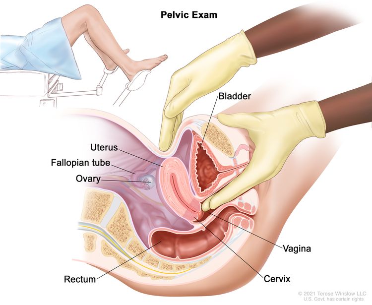
Pelvic examination.
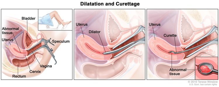
Dilatation and curettage.
Hysteroscopy.
To definitively diagnose endometrial cancer, a procedure that directly samples the endometrial tissue is necessary.
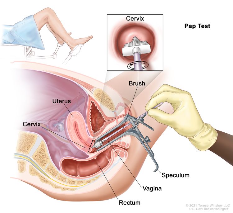
The Pap smear is not a reliable screening procedure for the detection of endometrial cancer, even though a retrospective study found a strong correlation between positive cervical cytology and high-risk endometrial disease (i.e., high-grade tumor and deep myometrial invasion).[36] A prospective study found a statistically significant association between malignant cytology and increased risk of nodal disease.[37]
Prognostic Factors
Prognostic factors for endometrial cancer include:
Tumor stage and grade (including extrauterine nodal spread)
Table 1 highlights the risk of nodal metastasis based on findings at the time of staging surgery:[38]
Table 1. Risk of Nodal Metastasis in Clinical Stage I Endometrial Cancer
Prognostic Group
Patient Characteristics
Risk of Nodal Involvement
A
Grade 1 tumors involving only endometrium
<5%
No evidence of intraperitoneal spread
B
Grade 2–3 tumors
5%–9% pelvic nodes
Invasion of <50% of myometrium
No intraperitoneal spread
4% para-aortic nodes
C
Deep muscle invasion
20%–60% pelvic nodes
High-grade tumors
10%–30% para-aortic nodes
Intraperitoneal spread
A Gynecologic Oncology Group study related surgical-pathological parameters and postoperative treatment to recurrence-free interval and recurrence site. Grade 3 histology and deep myometrial invasion in patients without extrauterine spread were the greatest determinants of recurrence. In this study, the presence of the following factors greatly increased the frequency of recurrence:[39,40]
Positive pelvic nodes.
Adnexal metastasis.
Positive peritoneal cytology.
Capillary space involvement.
Involvement of the isthmus or cervix.
Positive para-aortic nodes (includes all grades and depth of invasion). Of the cases with aortic node metastases, 98% were in patients with positive pelvic nodes, intra-abdominal metastases, or tumor invasion of the outer 33% of the myometrium.
When the only evidence of extrauterine spread is positive peritoneal cytology, the influence on outcome is unclear. The value of therapy directed at this cytological finding is not well founded,[41–46] and some data are contradictory.[47] Although the collection of cytology specimens is still suggested, a positive result does not upstage the cancer. Other extrauterine disease must be present before additional postoperative therapy is considered.
Involvement of the capillary-lymphatic space on histopathological examination correlates with extrauterine and nodal spread of tumor.[48]
Hormone receptor status
When possible, progesterone and estrogen receptor statuses, assessed either by biochemical or immunohistochemical methods, are included in the evaluation of patients with stage I and stage II cancer.[49–51]
One report found progesterone receptor levels to be the single most important prognostic indicator of 3-year survival in clinical stages I and II disease. Patients with progesterone receptor levels of 100 or greater had a 3-year disease-free survival rate of 93%, compared with 36% for those with a level below 100. After adjusting for progesterone receptor levels, only cervical involvement and peritoneal cytology were significant prognostic variables.[52]
Other reports confirm the importance of hormone receptor status as an independent prognostic factor.[53] Additionally, immunohistochemical staining of paraffin-embedded tissue for both estrogen and progesterone receptors has been shown to correlate with Fédération Internationale de Gynécologie et d’Obstétrique (FIGO) grade and survival.[49–51]
Other prognostic factors
Other factors predictive of poor prognosis include:[51,54,55]
A high S-phase fraction.
Aneuploidy.
PTEN loss-of-function variant.
PIK3CA variant.
TP53 variant.
Oncogene expression (e.g., overexpression of the HER2/neu oncogene has been associated with a poor overall prognosis).
A general review of prognostic factors has been published.[56]
References
American Cancer Society: Cancer Facts and Figures 2025. American Cancer Society, 2025. Available online. Last accessed January 16, 2025.
Ward KK, Shah NR, Saenz CC, et al.: Cardiovascular disease is the leading cause of death among endometrial cancer patients. Gynecol Oncol 126 (2): 176-9, 2012. [PUBMED Abstract]
Beral V, Bull D, Reeves G, et al.: Endometrial cancer and hormone-replacement therapy in the Million Women Study. Lancet 365 (9470): 1543-51, 2005 Apr 30-May 6. [PUBMED Abstract]
Anderson GL, Limacher M, Assaf AR, et al.: Effects of conjugated equine estrogen in postmenopausal women with hysterectomy: the Women’s Health Initiative randomized controlled trial. JAMA 291 (14): 1701-12, 2004. [PUBMED Abstract]
Furness S, Roberts H, Marjoribanks J, et al.: Hormone therapy in postmenopausal women and risk of endometrial hyperplasia. Cochrane Database Syst Rev (2): CD000402, 2009. [PUBMED Abstract]
Grady D, Gebretsadik T, Kerlikowske K, et al.: Hormone replacement therapy and endometrial cancer risk: a meta-analysis. Obstet Gynecol 85 (2): 304-13, 1995. [PUBMED Abstract]
Smith DC, Prentice R, Thompson DJ, et al.: Association of exogenous estrogen and endometrial carcinoma. N Engl J Med 293 (23): 1164-7, 1975. [PUBMED Abstract]
Mack TM, Pike MC, Henderson BE, et al.: Estrogens and endometrial cancer in a retirement community. N Engl J Med 294 (23): 1262-7, 1976. [PUBMED Abstract]
Ziel HK, Finkle WD: Increased risk of endometrial carcinoma among users of conjugated estrogens. N Engl J Med 293 (23): 1167-70, 1975. [PUBMED Abstract]
Walker AM, Jick H: Cancer of the corpus uteri: increasing incidence in the United States, 1970–1975. Am J Epidemiol 110 (1): 47-51, 1979. [PUBMED Abstract]
McDonald TW, Annegers JF, O’Fallon WM, et al.: Exogenous estrogen and endometrial carcinoma: case-control and incidence study. Am J Obstet Gynecol 127 (6): 572-80, 1977. [PUBMED Abstract]
Antunes CM, Strolley PD, Rosenshein NB, et al.: Endometrial cancer and estrogen use. Report of a large case-control study. N Engl J Med 300 (1): 9-13, 1979. [PUBMED Abstract]
Shapiro S, Kelly JP, Rosenberg L, et al.: Risk of localized and widespread endometrial cancer in relation to recent and discontinued use of conjugated estrogens. N Engl J Med 313 (16): 969-72, 1985. [PUBMED Abstract]
Fisher B, Costantino JP, Redmond CK, et al.: Endometrial cancer in tamoxifen-treated breast cancer patients: findings from the National Surgical Adjuvant Breast and Bowel Project (NSABP) B-14. J Natl Cancer Inst 86 (7): 527-37, 1994. [PUBMED Abstract]
Cummings SR, Eckert S, Krueger KA, et al.: The effect of raloxifene on risk of breast cancer in postmenopausal women: results from the MORE randomized trial. Multiple Outcomes of Raloxifene Evaluation. JAMA 281 (23): 2189-97, 1999. [PUBMED Abstract]
DeMichele A, Troxel AB, Berlin JA, et al.: Impact of raloxifene or tamoxifen use on endometrial cancer risk: a population-based case-control study. J Clin Oncol 26 (25): 4151-9, 2008. [PUBMED Abstract]
Bergström A, Pisani P, Tenet V, et al.: Overweight as an avoidable cause of cancer in Europe. Int J Cancer 91 (3): 421-30, 2001. [PUBMED Abstract]
Aune D, Navarro Rosenblatt DA, Chan DS, et al.: Anthropometric factors and endometrial cancer risk: a systematic review and dose-response meta-analysis of prospective studies. Ann Oncol 26 (8): 1635-48, 2015. [PUBMED Abstract]
Esposito K, Chiodini P, Capuano A, et al.: Metabolic syndrome and endometrial cancer: a meta-analysis. Endocrine 45 (1): 28-36, 2014. [PUBMED Abstract]
Troisi R, Potischman N, Hoover RN, et al.: Insulin and endometrial cancer. Am J Epidemiol 146 (6): 476-82, 1997. [PUBMED Abstract]
Tsilidis KK, Kasimis JC, Lopez DS, et al.: Type 2 diabetes and cancer: umbrella review of meta-analyses of observational studies. BMJ 350: g7607, 2015. [PUBMED Abstract]
Dossus L, Allen N, Kaaks R, et al.: Reproductive risk factors and endometrial cancer: the European Prospective Investigation into Cancer and Nutrition. Int J Cancer 127 (2): 442-51, 2010. [PUBMED Abstract]
Brown SB, Hankinson SE: Endogenous estrogens and the risk of breast, endometrial, and ovarian cancers. Steroids 99 (Pt A): 8-10, 2015. [PUBMED Abstract]
Barry JA, Azizia MM, Hardiman PJ: Risk of endometrial, ovarian and breast cancer in women with polycystic ovary syndrome: a systematic review and meta-analysis. Hum Reprod Update 20 (5): 748-58, 2014 Sep-Oct. [PUBMED Abstract]
Win AK, Reece JC, Ryan S: Family history and risk of endometrial cancer: a systematic review and meta-analysis. Obstet Gynecol 125 (1): 89-98, 2015. [PUBMED Abstract]
Daniels MS: Genetic testing by cancer site: uterus. Cancer J 18 (4): 338-42, 2012 Jul-Aug. [PUBMED Abstract]
Dunlop MG, Farrington SM, Nicholl I, et al.: Population carrier frequency of hMSH2 and hMLH1 mutations. Br J Cancer 83 (12): 1643-5, 2000. [PUBMED Abstract]
Lynch HT, Lynch J, Conway T, et al.: Familial aggregation of carcinoma of the endometrium. Am J Obstet Gynecol 171 (1): 24-7, 1994. [PUBMED Abstract]
Lu KH, Schorge JO, Rodabaugh KJ, et al.: Prospective determination of prevalence of lynch syndrome in young women with endometrial cancer. J Clin Oncol 25 (33): 5158-64, 2007. [PUBMED Abstract]
Widra EA, Dunton CJ, McHugh M, et al.: Endometrial hyperplasia and the risk of carcinoma. Int J Gynecol Cancer 5 (3): 233-235, 1995. [PUBMED Abstract]
Jick SS: Combined estrogen and progesterone use and endometrial cancer. Epidemiology 4 (4): 384, 1993. [PUBMED Abstract]
Bilezikian JP: Major issues regarding estrogen replacement therapy in postmenopausal women. J Womens Health 3 (4): 273-82, 1994.
van Leeuwen FE, Benraadt J, Coebergh JW, et al.: Risk of endometrial cancer after tamoxifen treatment of breast cancer. Lancet 343 (8895): 448-52, 1994. [PUBMED Abstract]
DuBeshter B, Warshal DP, Angel C, et al.: Endometrial carcinoma: the relevance of cervical cytology. Obstet Gynecol 77 (3): 458-62, 1991. [PUBMED Abstract]
Larson DM, Johnson KK, Reyes CN, et al.: Prognostic significance of malignant cervical cytology in patients with endometrial cancer. Obstet Gynecol 84 (3): 399-403, 1994. [PUBMED Abstract]
Takeshima N, Hirai Y, Tanaka N, et al.: Pelvic lymph node metastasis in endometrial cancer with no myometrial invasion. Obstet Gynecol 88 (2): 280-2, 1996. [PUBMED Abstract]
Morrow CP, Bundy BN, Kurman RJ, et al.: Relationship between surgical-pathological risk factors and outcome in clinical stage I and II carcinoma of the endometrium: a Gynecologic Oncology Group study. Gynecol Oncol 40 (1): 55-65, 1991. [PUBMED Abstract]
Lanciano RM, Corn BW, Schultz DJ, et al.: The justification for a surgical staging system in endometrial carcinoma. Radiother Oncol 28 (3): 189-96, 1993. [PUBMED Abstract]
Ambros RA, Kurman RJ: Combined assessment of vascular and myometrial invasion as a model to predict prognosis in stage I endometrioid adenocarcinoma of the uterine corpus. Cancer 69 (6): 1424-31, 1992. [PUBMED Abstract]
Turner DA, Gershenson DM, Atkinson N, et al.: The prognostic significance of peritoneal cytology for stage I endometrial cancer. Obstet Gynecol 74 (5): 775-80, 1989. [PUBMED Abstract]
Piver MS, Recio FO, Baker TR, et al.: A prospective trial of progesterone therapy for malignant peritoneal cytology in patients with endometrial carcinoma. Gynecol Oncol 47 (3): 373-6, 1992. [PUBMED Abstract]
Kadar N, Homesley HD, Malfetano JH: Positive peritoneal cytology is an adverse factor in endometrial carcinoma only if there is other evidence of extrauterine disease. Gynecol Oncol 46 (2): 145-9, 1992. [PUBMED Abstract]
Lurain JR: The significance of positive peritoneal cytology in endometrial cancer. Gynecol Oncol 46 (2): 143-4, 1992. [PUBMED Abstract]
Lurain JR, Rice BL, Rademaker AW, et al.: Prognostic factors associated with recurrence in clinical stage I adenocarcinoma of the endometrium. Obstet Gynecol 78 (1): 63-9, 1991. [PUBMED Abstract]
Garg G, Gao F, Wright JD, et al.: Positive peritoneal cytology is an independent risk-factor in early stage endometrial cancer. Gynecol Oncol 128 (1): 77-82, 2013. [PUBMED Abstract]
Hanson MB, van Nagell JR, Powell DE, et al.: The prognostic significance of lymph-vascular space invasion in stage I endometrial cancer. Cancer 55 (8): 1753-7, 1985. [PUBMED Abstract]
Carcangiu ML, Chambers JT, Voynick IM, et al.: Immunohistochemical evaluation of estrogen and progesterone receptor content in 183 patients with endometrial carcinoma. Part I: Clinical and histologic correlations. Am J Clin Pathol 94 (3): 247-54, 1990. [PUBMED Abstract]
Chambers JT, Carcangiu ML, Voynick IM, et al.: Immunohistochemical evaluation of estrogen and progesterone receptor content in 183 patients with endometrial carcinoma. Part II: Correlation between biochemical and immunohistochemical methods and survival. Am J Clin Pathol 94 (3): 255-60, 1990. [PUBMED Abstract]
Gurpide E: Endometrial cancer: biochemical and clinical correlates. J Natl Cancer Inst 83 (6): 405-16, 1991. [PUBMED Abstract]
Ingram SS, Rosenman J, Heath R, et al.: The predictive value of progesterone receptor levels in endometrial cancer. Int J Radiat Oncol Biol Phys 17 (1): 21-7, 1989. [PUBMED Abstract]
Creasman WT: Prognostic significance of hormone receptors in endometrial cancer. Cancer 71 (4 Suppl): 1467-70, 1993. [PUBMED Abstract]
Friberg LG, Norén H, Delle U: Prognostic value of DNA ploidy and S-phase fraction in endometrial cancer stage I and II: a prospective 5-year survival study. Gynecol Oncol 53 (1): 64-9, 1994. [PUBMED Abstract]
Hetzel DJ, Wilson TO, Keeney GL, et al.: HER-2/neu expression: a major prognostic factor in endometrial cancer. Gynecol Oncol 47 (2): 179-85, 1992. [PUBMED Abstract]
Binder PS, Mutch DG: Update on prognostic markers for endometrial cancer. Womens Health (Lond Engl) 10 (3): 277-88, 2014. [PUBMED Abstract]
Cellular Classification of Endometrial Cancer
Endometrial cancers are classified as one of the following two types:
Type 1 may arise from complex atypical hyperplasia and is pathogenetically linked to unopposed estrogenic stimulation.
Type 2 develops from atrophic endometrium and is not linked to hormonally driven pathogenesis.
The most common type of endometrial cancer is endometrioid adenocarcinoma.
Frequency of endometrial cancer cell types is as follows:
Endometrioid (75%) comprises malignant glandular epithelial elements; an admixture of squamous metaplasia is not uncommon.
Ciliated adenocarcinoma.
Secretory adenocarcinoma.
Papillary and villoglandular adenocarcinomas are histologically similar to those noted in the ovary and the fallopian tube. The prognosis is worse for these tumors.[1]
Adenocarcinoma with squamous differentiation.
Adenoacanthoma.
Adenosquamous cells contain malignant glandular and squamous epithelial elements.[2]
Mixed, defined as two carcinomatous cell types, with the smaller component making up at least 10% of the total (10%).
Uterine papillary serous (<10%).
Clear cell (4%) is histologically similar to those noted in the ovary and the fallopian tube. The prognosis for clear cell tumors is worse.[1]
Carcinosarcoma (3%), also known as malignant mixed mesodermal tumor, has both carcinomatous and sarcomatous elements. This tumor was historically categorized as a subtype of uterine sarcomas; however, recent evidence points to its origin as an adenocarcinoma that has undergone differentiation into the sarcomatous elements.
Mucinous (1%).
Squamous cell (<1%).
Undifferentiated (<1%).
Molecular Subgroups
PTEN variants are more common in type 1 endometrial cancers; TP53 and HER2/neu overexpression are more common in type 2 endometrial cancers, although some overlap exists.
The Cancer Genome Atlas’s full genetic display of hundreds of endometrial cancers identified four subtypes to further characterize endometrial cancers:[3]
POLE ultramutated. This subtype has clinical significance, and adjuvant therapies are avoided.
Microsatellite instability hypermutated.
Copy number low.
Copy number high.
These categories can be used to stratify patients into low- and high-risk prognostic categories. A modification of The Cancer Genome Atlas methods into more accessible tests was also successful in discriminating cancers into relevant prognostic categories. However, a combination of previously known risk factors with the genetic data was the most effective at determining prognostic categories.[4]
References
Gusberg SB: Virulence factors in endometrial cancer. Cancer 71 (4 Suppl): 1464-6, 1993. [PUBMED Abstract]
Zaino RJ, Kurman R, Herbold D, et al.: The significance of squamous differentiation in endometrial carcinoma. Data from a Gynecologic Oncology Group study. Cancer 68 (10): 2293-302, 1991. [PUBMED Abstract]
Kandoth C, Schultz N, Cherniack AD, et al.: Integrated genomic characterization of endometrial carcinoma. Nature 497 (7447): 67-73, 2013. [PUBMED Abstract]
Talhouk A, McConechy MK, Leung S, et al.: A clinically applicable molecular-based classification for endometrial cancers. Br J Cancer 113 (2): 299-310, 2015. [PUBMED Abstract]
Stage Information for Endometrial Cancer
The pattern of endometrial cancer spread is partially dependent on the degree of cellular differentiation. Well-differentiated tumors tend to limit their spread to the surface of the endometrium; myometrial invasion is less common. Myometrial invasion occurs much more frequently in patients with poorly differentiated tumors and is frequently a harbinger of lymph node involvement and distant metastases.[1,2]
Metastatic spread occurs in a characteristic pattern. Regional spread to the pelvic and para-aortic nodes is common. Distant metastasis most commonly involves the following sites:
Lungs.
Inguinal and supraclavicular nodes.
Liver.
Bones.
Brain.
Vagina.
FIGO Staging
The Fédération Internationale de Gynécologie et d’Obstétrique (FIGO) and the American Joint Committee on Cancer (AJCC) have both designated staging systems for endometrial cancer. The FIGO system is the most commonly used staging system for endometrial cancer.[3–5] The 2023 FIGO staging update has not been widely adopted because it incorporates molecular-based results, and some physicians do not have access to those data for their patients. Additionally, given the significant changes in the 2023 FIGO staging system, especially in the definition of early stage disease, more outcome data are needed so it might be more prudent to use the 2021 system in a clinical setting while discussing prognosis and treatment with patients. Therefore, both the 2023 and the 2021 FIGO staging systems are presented in this section.
FIGO stages I to IV are further subdivided by the histological grade (G) of the tumor, for example, stage IB G2. Carcinosarcomas, which had previously been designated as sarcomas, are now considered poorly differentiated adenocarcinomas; as such, they are included in this system.[5]
2023 FIGO staging for endometrial cancer
Table 2. 2023 FIGO Definitions for Stage I Cancer of the Endometriuma,b,c
Stage
Description
T = primary tumor; N = regional lymph node; M = distant metastasis; p = pathological; AJCC = American Joint Committee on Cancer; ESGO-ESTRO-ESP = European Society of Gynaecological Oncology, European Society for Radiotherapy and Oncology, European Society of Pathology; FIGO = Fédération Internationale de Gynécologie et d’Obstétrique; ITC = isolated tumor cell; LVSI = lymphovascular space involvement; MMRd = mismatch repair deficiency; NSMP = no specific molecular profile; POLEmut = pathogenic POLE mutation; p53abn = TP53 abnormal; SLN = sentinel lymph node; WHO = World Health Organization.
aAdapted from FIGO Committee on Gynecologic Oncology.[3]
bEndometrial cancer is surgically staged and pathologically examined. In all stages, the grade of the lesion, the histological type and LVSI must be recorded. If available and feasible, molecular classification testing (POLEmut, MMRd, NSMP, p53abn) is encouraged in all patients with endometrial cancer for prognostic risk-group stratification and as factors that might influence adjuvant and systemic treatment decisions (see Table 6).
cIn early endometrial cancer, the standard surgery is a total hysterectomy with bilateral salpingo-oophorectomy via a minimally invasive laparoscopic approach. Staging procedures include infracolic omentectomy in specific histological subtypes, such as serous and undifferentiated endometrial carcinoma, as well as carcinosarcoma, due to the high risk of microscopic omental metastases. Lymph node staging should be performed in patients with intermediate-high/high-risk disease. SLN biopsy is an adequate alternative to systematic lymphadenectomy for staging proposes. SLN biopsy can also be considered in patients with low−/low-intermediate-risk disease to rule out occult lymph node metastases and to identify disease truly confined to the uterus. Thus, the ESGO-ESTRO-ESP guidelines allow an approach of SLN in all patients with endometrial carcinoma, which is endorsed by FIGO. In assumed early endometrial cancer, an SLN biopsy in an adequate alternative to systematic lymphadenectomy in high-intermediate and high-risk cases for the purpose of lymph node staging and can also be considered in low–/intermediate-risk disease to rule out occult lymph node metastases. An SLN biopsy should be done in association with thorough (ultrastaging) staging as it will increase the detection of low-volume disease in lymph nodes.
dLow-grade endometrioid carcinomas involving both the endometrium and the ovary are considered to have a good prognosis, and no adjuvant treatment is recommended if all the below criteria are met. Disease limited to low-grade endometrioid carcinomas involving the endometrium and ovaries (Stage IA3) must be distinguished from extensive spread of the endometrial carcinoma to the ovary (Stage IIIA1), by the following criteria: (1) no more than superficial myometrial invasion is present (<50%); (2) absence of extensive/substantial LVSI; (3) absence of additional metastases; and (4) the ovarian tumor is unilateral, limited to the ovary, without capsule invasion/rupture (equivalent to pT1a).
eLVSI as defined in WHO 2021: extensive/substantial, ≥5 vessels involved.
fGrade and histological type are as follows: (1) Serous adenocarcinomas, clear cell adenocarcinomas, mesonephric-like carcinomas, gastrointestinal-type mucinous endometrial carcinoma, undifferentiated carcinomas, and carcinosarcomas are considered high grade by definition. For endometrioid carcinomas, grade is based on the proportion of solid areas: low grade = grade 1 (≤5%) and grade 2 (6%–50%); and high grade = grade 3 (>50%). Nuclear atypia excessive for the grade raises the grade of a grade 1 or 2 tumor by one. The presence of unusual nuclear atypia in an architecturally low-grade tumor should prompt the evaluation of TP53 and consideration of serous carcinoma. Adenocarcinomas with squamous differentiation are graded according to the microscopic features of the glandular component; (2) Nonaggressive histological types are composed of low-grade (grade 1 and 2) endometrioid carcinomas. Aggressive histological types are composed of high-grade endometrioid carcinomas (grade 3), serous, clear cell, undifferentiated, mixed, mesonephric-like, gastrointestinal mucinous type carcinomas, and carcinosarcomas; and (3) It should be noted that high-grade endometrioid carcinomas (grade 3) are a prognostically, clinically, and molecularly heterogenous disease, and the tumor type that benefits most from applying molecular classification for improved prognostication and for treatment decision-making. Without molecular classification, high-grade endometrioid carcinomas cannot appropriately be allocated to a risk group, and thus, molecular profiling is particularly recommended in these patients. For practical purposes and to avoid undertreatment of patients, if the molecular classification is unknown, high-grade endometrioid carcinomas were grouped together with the aggressive histological types in the actual FIGO classification.
gMicrometastases are considered to be metastatic involvement (pN1 [mi]). The prognostic significance of ITCs is unclear. The presence of ITCs should be documented and is regarded as pN0(I+). According to the AJCC 8th edition staging, macrometastases are >2 mm in size, micrometastases are >0.2–2 mm and/or >200 cells, and ITCs are ≤0.2 mm and ≤200 cells. These definitions are based on staging established by FIGO and the 8th edition of the AJCC Cancer Staging Manual.
I
Confined to the uterine corpus and ovary.d
IA
Disease limited to the endometrium OR nonaggressive histological type, i.e., low-grade endometrioid, with invasion of less than half of myometrium with no or focal LVSI OR good prognosis disease.
IA1
Nonaggressive histological type limited to an endometrial polyp OR confined to the endometrium.
IA2
Nonaggressive histological types involving less than half of the myometrium with no or focal LVSI.
IA3
Low-grade endometrioid carcinomas limited to the uterus and ovary.d
IB
Nonaggressive histological types with invasion of half or more of the myometrium, and with no or focal LVSI.e
IC
Aggressive histological typesf limited to a polyp or confined to the endometrium.
Table 3. 2023 FIGO Definitions for Stage II Cancer of the Endometriuma,b,c
Stage
Description
FIGO = Fédération Internationale de Gynécologie et d’Obstétrique; LVSI = lymphovascular space involvement.
aAdapted from FIGO Committee on Gynecologic Oncology.[3]
For the explanations for footnotes b−f, see Table 2.
II
Invasion of cervical stroma without extrauterine extension OR with substantial LVSI OR aggressive histological types with myometrial invasion.
IIA
Invasion of the cervical stroma of nonaggressive histological types.
IIB
Substantial LVSIe of nonaggressive histological types.
IIC
Aggressive histological typesf with any myometrial involvement.
Table 4. 2023 FIGO Definitions for Stage III Cancer of the Endometriuma,b,c
Stage
Description
FIGO = Fédération Internationale de Gynécologie et d’Obstétrique.
aAdapted from FIGO Committee on Gynecologic Oncology.[3]
For the explanations for footnotes b−d and g, see Table 2.
III
Local and/or regional spread of the tumor of any histological subtype.
IIIA
Invasion of uterine serosa, adnexa, or both by direct extension or metastasis.
IIIA1
Spread to ovary or fallopian tube (except when meeting stage IA3 criteria).d
IIIA2
Involvement of uterine subserosa or spread through the uterine serosa.
IIIB
Metastasis or direct spread to the vagina and/or to the parametria or pelvic peritoneum.
IIIB1
Metastasis or direct spread to the vagina and/or the parametria.
IIIB2
Metastasis to the pelvic peritoneum.
IIIC
Metastasis to the pelvic or para-aortic lymph nodes or both.g
IIIC1
Metastasis to the pelvic lymph nodes.
IIIC1i
Micrometastasis.
IIIC1ii
Macrometastasis.
IIIC2
Metastasis to para-aortic lymph nodes up to the renal vessels, with or without metastasis to the pelvic lymph nodes.
IIIC2i
Micrometastasis.
IIIC2ii
Macrometastasis.
Table 5. 2023 FIGO Definitions for Stage IV Cancer of the Endometriuma,b,c
Stage
Description
FIGO = Fédération Internationale de Gynécologie et d’Obstétrique.
aAdapted from FIGO Committee on Gynecologic Oncology.[3]
For the explanations for footnotes b−c, see Table 2.
IV
Spread to the bladder mucosa and/or intestinal mucosa and/or distance metastasis.
IVA
Invasion of the bladder mucosa and/or the intestinal/bowel mucosa.
IVB
Abdominal peritoneal metastasis beyond the pelvis.
IVC
Distant metastasis, including metastasis to any extra- or intra-abdominal lymph nodes above the renal vessels, lungs, liver, brain, or bone.
Table 6. 2023 FIGO Definitions for Endometrial Cancer Stage With Molecular Classificationa,b
Stage Designation
Molecular Findings in Patients With Early Endometrial Cancer (Stages I and II After Surgical Staging)
FIGO = Fédération Internationale de Gynécologie et d’Obstétrique; LVSI = lymphovascular space involvement; MMRd = mismatch repair deficiency; MSI = microsatellite instability; NSMP = no specific molecular profile; POLEmut = pathogenic POLE mutation; p53abn = TP53 abnormal.
aAdapted from FIGO Committee on Gynecologic Oncology.[3]
bWhen feasible, the addition of molecular subtype to the staging criteria allows a better prediction of prognosis in a staging/prognosis scheme. The performance of complete molecular classification (POLEmut, MMRd, NSMP, p53abn) is encouraged in all cases of endometrial cancer for prognostic risk-group stratification and as potential influencing factors of adjuvant or systemic treatment decisions. Molecular subtype assignment can be done on a biopsy, in which case it need not be repeated on the hysterectomy specimen. When performed, these molecular classifications should be recorded in all stages. A pathogenic POLE mutation (POLEmut) is associated with a good prognosis. MMRd or MSI and NSMP are associated with an intermediate prognosis. Abnormal TP53 (p53abn) is associated with a poor prognosis. When the molecular classification is known the staging is modified as follows: (1) FIGO Stages I and II are based on surgical/anatomical and histological findings. In case the molecular classification reveals POLEmut or p53abn status, the FIGO stage is modified in the early stage of the disease. This is depicted in the FIGO stage by the addition of “m” for molecular classification, and a subscript is added to denote POLEmut or p53abn status, as shown in the table. MMRd or NSMP status do not modify early FIGO stages; however, these molecular classifications should be recorded for the purpose of data collection. When molecular classification reveals MMRd or NSMP, it should be recorded as Stage ImMMRd or Stage ImNSMP and Stage IImMMRd or Stage IImNSMP; (2) FIGO Stages III and IV are based on surgical/anatomical findings. The stage category is not modified by molecular classification; however, the molecular classification should be recorded if known. When the molecular classification is known, it should be recorded as Stage IIIm or Stage IVm with the appropriate subscript for the purpose of data collection. For example, when molecular classification reveals p53abn, it should be recorded as Stage IIImp53abn or Stage IVmp53abn.
IAmPOLEmut
POLEmut endometrial carcinoma, confined to the uterine corpus or with cervical extension, regardless of the degree of LVSI or histological type.
IICmp53abn
p53abn endometrial carcinoma confined to the uterine corpus with any myometrial invasion, with or without cervical invasion, and regardless of the degree of LVSI or histological type.
2021 FIGO staging for endometrial cancer
Table 7. 2021 FIGO Definitions for Stage I Cancer of the Endometriuma
Stage
Description
Illustration
FIGO = Fédération Internationale de Gynécologie et d’Obstétrique.
aAdapted from FIGO Committee on Gynecologic Oncology.[4]
Hendrickson M, Ross J, Eifel PJ, et al.: Adenocarcinoma of the endometrium: analysis of 256 cases with carcinoma limited to the uterine corpus. Pathology review and analysis of prognostic variables. Gynecol Oncol 13 (3): 373-92, 1982. [PUBMED Abstract]
Nori D, Hilaris BS, Tome M, et al.: Combined surgery and radiation in endometrial carcinoma: an analysis of prognostic factors. Int J Radiat Oncol Biol Phys 13 (4): 489-97, 1987. [PUBMED Abstract]
Berek JS, Matias-Guiu X, Creutzberg C, et al.: FIGO staging of endometrial cancer: 2023. Int J Gynaecol Obstet 162 (2): 383-394, 2023. [PUBMED Abstract]
Koskas M, Amant F, Mirza MR, et al.: Cancer of the corpus uteri: 2021 update. Int J Gynaecol Obstet 155 (Suppl 1): 45-60, 2021. [PUBMED Abstract]
Corpus uteri – carcinoma and carcinosarcoma. In: Amin MB, Edge SB, Greene FL, et al., eds.: AJCC Cancer Staging Manual. 8th ed. Springer; 2017, pp 661-69.
Treatment Option Overview for Endometrial Cancer
The degree of tumor differentiation has an important effect on the natural history of this disease and on treatment selection.
Patients with endometrial cancer who have localized disease are usually cured. Best results are obtained with one of two standard treatments:
Hysterectomy with bilateral salpingo-oophorectomy.
Hysterectomy with bilateral salpingo-oophorectomy and adjuvant radiation therapy (when deep invasion of the myometrial muscle [more than 50% of the myometrium] or grade 3 tumor with myometrial invasion is present).
Patients with regional and distant metastases are rarely cured, although they are occasionally responsive to standard hormone therapy.
Progestational agents have been evaluated as adjuvant therapy in several randomized trials. A meta-analysis by the Cochrane group confirms no clinical benefit to adjuvant progestogens in clinical stage I disease.[1][Level of evidence A1]
The treatment options for each stage of endometrial cancer are presented in Table 11.
Table 11. Treatment Options for Endometrial Cancer
Martin-Hirsch PP, Bryant A, Keep SL, et al.: Adjuvant progestagens for endometrial cancer. Cochrane Database Syst Rev (6): CD001040, 2011. [PUBMED Abstract]
Treatment of Stage I and Stage II Endometrial Cancer
Treatment Options for Stage I and Stage II Endometrial Cancer
Treatment of stage I and stage II endometrial cancer depends on the grade and histological type.
In the current Fédération Internationale de Gynécologie et d’Obstétrique (FIGO) staging system, stage II describes tumor that invades the cervical stroma; this is equivalent to the prior stage IIB. Almost all randomized trials for early-stage cancer excluded stage IIB patients. As a result, there is a paucity of quality data on which to base clinical decisions for patients with stage II endometrial cancer.
Low-risk histology:
Grades 1 and 2 tumors are considered low-risk unless they have serous or clear cell histologies.
Treatment options for patients with low-risk histological subtypes of stage I endometrial cancer include:
Surgery: Hysterectomy with bilateral salpingo-oophorectomy and possible lymph node dissection.
Most patients do well with surgery alone. However, patients with stage I disease who have high-risk histologies are at a greater risk of recurrence and are eligible for adjuvant therapy.
High-risk histology:
Grade 3 tumors of any histology and any serous tumors, clear cell tumors, or carcinosarcomas are considered high-risk.
Treatment options for patients with stage I or stage II endometrial cancer who have high-risk histologies include:
Surgery: Hysterectomy with bilateral salpingo-oophorectomy, with pelvic and para-aortic lymph node dissection.
Postoperative chemotherapy with or without radiation therapy.
Patients with serous or clear cell histologies have higher rates of recurrence than do patients with other stage I or stage II endometrioid carcinomas. Management guidelines are based on the outcomes reported in institutional case series that used a regimen of adjuvant carboplatin plus paclitaxel, and occasionally, radiation therapy for patients with this histological subtype.[1–9]
Carcinosarcomas have been evaluated in clinical trials both separately and with other sarcomas because of their prior designation in this group. In a nonrandomized Gynecologic Oncology Group (GOG) study of patients with stage I or II carcinosarcomas, patients who underwent pelvic radiation therapy had a significant reduction in recurrences within the radiation treatment field but no improvement in survival.[10] One nonrandomized study that predominantly included patients with carcinosarcomas appeared to show benefit for adjuvant therapy with cisplatin and doxorubicin.[11]
Surgery
If the uterine cervix is involved, patients may consider one or more of the following options:
Standard hysterectomy with bilateral salpingo-oophorectomy, followed by adjuvant radiation therapy.
Radical hysterectomy.
Pelvic and para-aortic lymph node dissection.
Single-institution reviews suggest that radical hysterectomy is more beneficial than standard hysterectomy in cases of cervical involvement of the tumor.[12–14]
Surgery with or without lymph node sampling
Table 12 highlights the risk of nodal metastasis based on findings at the time of staging surgery:[15]
Table 12. Risk of Nodal Metastasis in Clinical Stage I Endometrial Cancer
Prognostic Group
Patient Characteristics
Risk of Nodal Involvement
A
Grade 1 tumors involving only endometrium
<5%
No evidence of intraperitoneal spread
B
Grade 2–3 tumors
5%–9% pelvic nodes
Invasion of <50% of myometrium
No intraperitoneal spread
4% para-aortic nodes
C
Deep muscle invasion
20%–60% pelvic nodes
High-grade tumors
10%–30% para-aortic nodes
Intraperitoneal spread
For patients in Group A, lymph node dissection has limited utility. Conversely, full pelvic and para-aortic lymph node dissection is important for patients in Group C, given the likelihood of positive findings. The difficulty lies in determining how to manage patients in Group B.
There are several accepted surgical approaches for patients with presumed stage I endometrial cancer, with intermediate risk for lymphatic spread.
Both retrospective and prospective data support stratifying patients with presumed stage I endometrial cancer into two groups based on the following characteristics:
Low risk: Well-differentiated or moderately differentiated tumor and/or depth of myometrial invasion is less than 50% and/or tumor is smaller than 2 cm.
High risk: Poorly differentiated tumor and/or depth of myometrial invasion is 50% or more and/or tumor is 2 cm or larger.
Evidence (lymph node dissection):
In two studies, patients with low-risk cancer had a sufficiently low risk of lymph node metastasis such that lymph node sampling could be omitted. For patients meeting high-risk criteria, a full pelvic and para-aortic lymph node dissection was suggested, given patterns of lymphatic spread.[16,17]
An alternative strategy is the use of sentinel lymph node dissection in patients with presumed stage I endometrial cancer.[18] Although this strategy has been widely adopted at various academic centers, a prospective multicenter trial to determine the false-negative rate for this protocol is lacking. For cases in which isolated tumor cells are identified using the sentinel lymph node approach, it is unclear whether treatment is necessary.
In patients with high-risk histology (serous, clear cell, carcinosarcoma, or undifferentiated tumors), hysterectomy and bilateral salpingo-oophorectomy with pelvic and para-aortic lymph node dissection is the standard approach.
Laparotomy has been the standard surgical approach. However, laparoscopy is now favored because of the improvement in patients’ postoperative recovery without significant impact on outcomes.
Evidence (treatment or surgical staging using laparoscopy vs. laparotomy):
For patients with early-stage endometrial cancer, several randomized trials have compared total laparoscopic hysterectomy (TLH) with the standard open procedure, total abdominal hysterectomy (TAH). Feasibility of the laparoscopic approach has been confirmed, but this approach is associated with a longer operative time.[15,19,20] TLH has an improved [15,19] or similar [20] adverse event profile and a shorter hospital stay [15,19,20] than does TAH.
TLH is associated with less pain and a quicker resumption of daily activities,[20,21] although one study found that most of the gains in quality of life favoring laparoscopy at the 6-week postsurgical period were no longer significant at 6 months.[20,21]
A GOG trial (GOG-LAP2) randomly assigned 2,616 patients with clinical stages I to IIA disease in a 2:1 ratio to comprehensive surgical staging via laparoscopy or laparotomy.[22][Level of evidence A1]
Time to recurrence was the primary end point, with noninferiority defined as a difference in recurrence rate of less than 5.3% between the two groups at 3 years.
The recurrence rate at 3 years was 10.24% for patients in the laparotomy arm and 11.39% for patients in the laparoscopy arm, with an estimated difference between groups of 1.14% (90% lower bound, -1.278; 95% upper bound, 3.996).
Although this difference was lower than the prespecified limit, the statistical requirements for noninferiority were not met because of fewer-than-expected recurrences in both groups.
The 5-year overall survival (OS) rate was 89.8% in both groups.
A Cochrane review of the use of laparoscopic staging included four randomized controlled trials that reported OS and progression-free survival (PFS). Ninety percent of the patients were from the GOG-LAP2 trial.[23][Level of evidence A1]
Overall, laparoscopy and laparotomy were associated with similar OS and PFS rates.
Future analyses may determine whether there are subgroups of patients for whom there is a clinically significant decrement when laparoscopic staging is used.[22][Level of evidence B1]
Postoperative vaginal brachytherapy
Adjuvant radiation therapy reduces the incidence of local and regional recurrence. However, improved survival rates have not been confirmed, and radiation therapy increased toxicity.[24–28] Vaginal cuff brachytherapy is associated with less radiation-related morbidity than is external-beam radiation therapy (EBRT) and has been shown to be equivalent to EBRT in the short term for patients with stage I disease.[29] However, long-term follow up of a randomized trial comparing EBRT plus vaginal brachytherapy (VBT) to VBT alone found decreased OS and increased toxicity in the EBRT plus VBT arm.[30]
Evidence (VBT):
Results of two randomized trials that used adjuvant radiation therapy in patients with stage I disease did not show improved survival but did show reduced locoregional recurrence (3%–4% in the radiation therapy group vs. 12%–14% in the control group after median follow-up of 5–6 years; P < .001), with an increase in side effects.[27,31,32][Level of evidence B1]
Results of a study by the Danish Endometrial Cancer Group suggest that the absence of radiation therapy does not improve the survival of patients with stage I intermediate-risk disease (grades 1 and 2 with >50% myometrial invasion or grade 3 with <50% myometrial invasion).[33]
The PORTEC-2 trial (NCT00411138) randomly assigned patients with stage I endometrial cancer who did not undergo lymph node dissection to undergo VBT or EBRT, with prevention of vaginal recurrence as the primary outcome.[29,34][Level of evidence A1]
At 5 years, there was no difference in the rates of vaginal recurrence, locoregional recurrence, PFS, or OS (84.8% [95% confidence interval (CI), 79.3%–90.3%] for VBT vs. 79.6% [95% CI, 71.2%–88.0%] for EBRT; P = .57).
The VBT group had significantly fewer gastrointestinal toxic effects and improved quality of life, making VBT the preferred option for adjuvant treatment of patients with stage I disease.
The Norwegian Radium Hospital trial included 568 patients with clinical stage I endometrial cancer between 1968 and 1974 (before FIGO surgical staging was initiated).[30][Level of evidence A1] After hysterectomy and bilateral salpingo-oophorectomy, patients were randomly assigned to receive either EBRT and VBT or VBT alone.
An updated report presenting over 20 years of follow-up data showed no difference in OS between the treatment groups. Median OS was 20.5 years in the EBRT/VBT group and 20.48 years in the VBT-alone group (P = .186). In all women, there was an increased risk of secondary cancers after EBRT (hazard ratio [HR], 1.42; 95% CI, 1.01–2.0).
A post hoc subset analysis of women younger than 60 years at the time of trial registration showed increased mortality in the EBRT/VBT arm (HR, 1.36; 95% CI, 1.06–1.76). Further, the risk of secondary cancers doubled in this group (HR, 2.02; 95% CI, 1.3–3.15).
Postoperative radiation therapy
If the cervix is not clinically involved, but extension to the cervix is noted on postoperative pathology, radiation therapy is considered.[22][Level of evidence A1]
Radiation therapy alone
Patients who have medical contraindications to surgery may be treated with radiation therapy alone, but cure rates may be lower than those attained with surgery.[35–37]
Current Clinical Trials
Use our advanced clinical trial search to find NCI-supported cancer clinical trials that are now enrolling patients. The search can be narrowed by location of the trial, type of treatment, name of the drug, and other criteria. General information about clinical trials is also available.
References
Kiess AP, Damast S, Makker V, et al.: Five-year outcomes of adjuvant carboplatin/paclitaxel chemotherapy and intravaginal radiation for stage I-II papillary serous endometrial cancer. Gynecol Oncol 127 (2): 321-5, 2012. [PUBMED Abstract]
Boruta DM, Gehrig PA, Fader AN, et al.: Management of women with uterine papillary serous cancer: a Society of Gynecologic Oncology (SGO) review. Gynecol Oncol 115 (1): 142-53, 2009. [PUBMED Abstract]
Huh WK, Powell M, Leath CA, et al.: Uterine papillary serous carcinoma: comparisons of outcomes in surgical Stage I patients with and without adjuvant therapy. Gynecol Oncol 91 (3): 470-5, 2003. [PUBMED Abstract]
Fader AN, Drake RD, O’Malley DM, et al.: Platinum/taxane-based chemotherapy with or without radiation therapy favorably impacts survival outcomes in stage I uterine papillary serous carcinoma. Cancer 115 (10): 2119-27, 2009. [PUBMED Abstract]
Kelly MG, O’malley DM, Hui P, et al.: Improved survival in surgical stage I patients with uterine papillary serous carcinoma (UPSC) treated with adjuvant platinum-based chemotherapy. Gynecol Oncol 98 (3): 353-9, 2005. [PUBMED Abstract]
Havrilesky LJ, Secord AA, Bae-Jump V, et al.: Outcomes in surgical stage I uterine papillary serous carcinoma. Gynecol Oncol 105 (3): 677-82, 2007. [PUBMED Abstract]
Dietrich CS, Modesitt SC, DePriest PD, et al.: The efficacy of adjuvant platinum-based chemotherapy in Stage I uterine papillary serous carcinoma (UPSC). Gynecol Oncol 99 (3): 557-63, 2005. [PUBMED Abstract]
Townamchai K, Berkowitz R, Bhagwat M, et al.: Vaginal brachytherapy for early stage uterine papillary serous and clear cell endometrial cancer. Gynecol Oncol 129 (1): 18-21, 2013. [PUBMED Abstract]
Barney BM, Petersen IA, Mariani A, et al.: The role of vaginal brachytherapy in the treatment of surgical stage I papillary serous or clear cell endometrial cancer. Int J Radiat Oncol Biol Phys 85 (1): 109-15, 2013. [PUBMED Abstract]
Hornback NB, Omura G, Major FJ: Observations on the use of adjuvant radiation therapy in patients with stage I and II uterine sarcoma. Int J Radiat Oncol Biol Phys 12 (12): 2127-30, 1986. [PUBMED Abstract]
Peters WA, Rivkin SE, Smith MR, et al.: Cisplatin and adriamycin combination chemotherapy for uterine stromal sarcomas and mixed mesodermal tumors. Gynecol Oncol 34 (3): 323-7, 1989. [PUBMED Abstract]
Ayhan A, Taskiran C, Celik C, et al.: The long-term survival of women with surgical stage II endometrioid type endometrial cancer. Gynecol Oncol 93 (1): 9-13, 2004. [PUBMED Abstract]
Eltabbakh GH, Moore AD: Survival of women with surgical stage II endometrial cancer. Gynecol Oncol 74 (1): 80-5, 1999. [PUBMED Abstract]
Orezzoli JP, Sioletic S, Olawaiye A, et al.: Stage II endometrioid adenocarcinoma of the endometrium: clinical implications of cervical stromal invasion. Gynecol Oncol 113 (3): 316-23, 2009. [PUBMED Abstract]
Walker JL, Piedmonte MR, Spirtos NM, et al.: Laparoscopy compared with laparotomy for comprehensive surgical staging of uterine cancer: Gynecologic Oncology Group Study LAP2. J Clin Oncol 27 (32): 5331-6, 2009. [PUBMED Abstract]
Mariani A, Dowdy SC, Cliby WA, et al.: Prospective assessment of lymphatic dissemination in endometrial cancer: a paradigm shift in surgical staging. Gynecol Oncol 109 (1): 11-8, 2008. [PUBMED Abstract]
Mariani A, Webb MJ, Keeney GL, et al.: Low-risk corpus cancer: is lymphadenectomy or radiotherapy necessary? Am J Obstet Gynecol 182 (6): 1506-19, 2000. [PUBMED Abstract]
Barlin JN, Khoury-Collado F, Kim CH, et al.: The importance of applying a sentinel lymph node mapping algorithm in endometrial cancer staging: beyond removal of blue nodes. Gynecol Oncol 125 (3): 531-5, 2012. [PUBMED Abstract]
Janda M, Gebski V, Brand A, et al.: Quality of life after total laparoscopic hysterectomy versus total abdominal hysterectomy for stage I endometrial cancer (LACE): a randomised trial. Lancet Oncol 11 (8): 772-80, 2010. [PUBMED Abstract]
Mourits MJ, Bijen CB, Arts HJ, et al.: Safety of laparoscopy versus laparotomy in early-stage endometrial cancer: a randomised trial. Lancet Oncol 11 (8): 763-71, 2010. [PUBMED Abstract]
Kornblith AB, Huang HQ, Walker JL, et al.: Quality of life of patients with endometrial cancer undergoing laparoscopic international federation of gynecology and obstetrics staging compared with laparotomy: a Gynecologic Oncology Group study. J Clin Oncol 27 (32): 5337-42, 2009. [PUBMED Abstract]
Walker JL, Piedmonte MR, Spirtos NM, et al.: Recurrence and survival after random assignment to laparoscopy versus laparotomy for comprehensive surgical staging of uterine cancer: Gynecologic Oncology Group LAP2 Study. J Clin Oncol 30 (7): 695-700, 2012. [PUBMED Abstract]
Galaal K, Bryant A, Fisher AD, et al.: Laparoscopy versus laparotomy for the management of early stage endometrial cancer. Cochrane Database Syst Rev 9: CD006655, 2012. [PUBMED Abstract]
Aalders J, Abeler V, Kolstad P, et al.: Postoperative external irradiation and prognostic parameters in stage I endometrial carcinoma: clinical and histopathologic study of 540 patients. Obstet Gynecol 56 (4): 419-27, 1980. [PUBMED Abstract]
Morrow CP, Bundy BN, Kurman RJ, et al.: Relationship between surgical-pathological risk factors and outcome in clinical stage I and II carcinoma of the endometrium: a Gynecologic Oncology Group study. Gynecol Oncol 40 (1): 55-65, 1991. [PUBMED Abstract]
Marchetti DL, Caglar H, Driscoll DL, et al.: Pelvic radiation in stage I endometrial adenocarcinoma with high-risk attributes. Gynecol Oncol 37 (1): 51-4, 1990. [PUBMED Abstract]
Creutzberg CL, van Putten WL, Koper PC, et al.: Surgery and postoperative radiotherapy versus surgery alone for patients with stage-1 endometrial carcinoma: multicentre randomised trial. PORTEC Study Group. Post Operative Radiation Therapy in Endometrial Carcinoma. Lancet 355 (9213): 1404-11, 2000. [PUBMED Abstract]
Kong A, Johnson N, Kitchener HC, et al.: Adjuvant radiotherapy for stage I endometrial cancer: an updated Cochrane systematic review and meta-analysis. J Natl Cancer Inst 104 (21): 1625-34, 2012. [PUBMED Abstract]
Nout RA, Smit VT, Putter H, et al.: Vaginal brachytherapy versus pelvic external beam radiotherapy for patients with endometrial cancer of high-intermediate risk (PORTEC-2): an open-label, non-inferiority, randomised trial. Lancet 375 (9717): 816-23, 2010. [PUBMED Abstract]
Onsrud M, Cvancarova M, Hellebust TP, et al.: Long-term outcomes after pelvic radiation for early-stage endometrial cancer. J Clin Oncol 31 (31): 3951-6, 2013. [PUBMED Abstract]
Keys HM, Roberts JA, Brunetto VL, et al.: A phase III trial of surgery with or without adjunctive external pelvic radiation therapy in intermediate risk endometrial adenocarcinoma: a Gynecologic Oncology Group study. Gynecol Oncol 92 (3): 744-51, 2004. [PUBMED Abstract]
Scholten AN, van Putten WL, Beerman H, et al.: Postoperative radiotherapy for Stage 1 endometrial carcinoma: long-term outcome of the randomized PORTEC trial with central pathology review. Int J Radiat Oncol Biol Phys 63 (3): 834-8, 2005. [PUBMED Abstract]
Bertelsen K, Ortoft G, Hansen ES: Survival of Danish patients with endometrial cancer in the intermediate-risk group not given postoperative radiotherapy: the Danish Endometrial Cancer Study (DEMCA). Int J Gynecol Cancer 21 (7): 1191-9, 2011. [PUBMED Abstract]
Nout RA, Putter H, Jürgenliemk-Schulz IM, et al.: Five-year quality of life of endometrial cancer patients treated in the randomised Post Operative Radiation Therapy in Endometrial Cancer (PORTEC-2) trial and comparison with norm data. Eur J Cancer 48 (11): 1638-48, 2012. [PUBMED Abstract]
Eltabbakh GH, Piver MS, Hempling RE, et al.: Excellent long-term survival and absence of vaginal recurrences in 332 patients with low-risk stage I endometrial adenocarcinoma treated with hysterectomy and vaginal brachytherapy without formal staging lymph node sampling: report of a prospective trial. Int J Radiat Oncol Biol Phys 38 (2): 373-80, 1997. [PUBMED Abstract]
Stokes S, Bedwinek J, Kao MS, et al.: Treatment of stage I adenocarcinoma of the endometrium by hysterectomy and adjuvant irradiation: a retrospective analysis of 304 patients. Int J Radiat Oncol Biol Phys 12 (3): 339-44, 1986. [PUBMED Abstract]
Grigsby PW, Kuske RR, Perez CA, et al.: Medically inoperable stage I adenocarcinoma of the endometrium treated with radiotherapy alone. Int J Radiat Oncol Biol Phys 13 (4): 483-8, 1987. [PUBMED Abstract]
Treatment of Stage III, Stage IV, and Recurrent Endometrial Cancer
Treatment Options for Stage III, Stage IV, and Recurrent Endometrial Cancer
Treatment options for patients with stage III, stage IV, and recurrent endometrial cancer include:
Treatment of patients with stage IV endometrial cancer is dictated by the site of metastatic disease and symptoms related to disease sites.
Surgery followed by chemotherapy or radiation therapy
In general, patients with stage III or stage IV endometrial cancer are treated with surgery, followed by chemotherapy, radiation therapy, or both. Observational studies support maximal cytoreductive surgery for patients with stage IV disease, although these conclusions need to be interpreted with care because of the small number of cases and likely selection bias.[1,2]
For many years, radiation therapy was the standard adjuvant treatment for patients with endometrial cancer. However, several randomized trials have confirmed improved survival when adjuvant chemotherapy is used instead of radiation therapy.
Doxorubicin was historically the most active anticancer agent used, with useful but temporary responses obtained in as many as 33% of patients with recurrent disease. Paclitaxel, when combined with platinum chemotherapy or when used as a single agent, also has significant anticancer activity.[3]
Evidence (surgery followed by chemotherapy or radiation therapy):
Several randomized trials by the Gynecologic Oncology Group (GOG) have used doxorubicin because of its known antitumor activity.[4]
Adding cisplatin to doxorubicin increased response rates and progression-free survival (PFS) when compared with doxorubicin alone. However, adding cisplatin did not affect overall survival (OS).
A three-drug regimen of doxorubicin, cisplatin, and paclitaxel with granulocyte-colony stimulating factor (G-CSF) was significantly superior to cisplatin and doxorubicin, as shown by the following results:[5,6][Level of evidence B3]
Response rate was 57% with the three-drug regimen, compared with 34% with the cisplatin and doxorubicin regimen.
PFS was 8.3 months with the three-drug regimen, compared with 5.3 months with the cisplatin and doxorubicin regimen.
OS was 15.3 months with the three-drug regimen, compared with 12.3 months with the cisplatin and doxorubicin regimen.
The 3-drug regimen was associated with grade 3 peripheral neuropathy in 12% of patients and grade 2 peripheral neuropathy in 27% of patients.
Given the toxicity and limited efficacy of these regimens, other treatment options have been widely sought. Several observational studies [7,8] and phase II studies [9–12] suggest clinical activity with the combination of platinum chemotherapy and paclitaxel in patients with endometrial cancer and measurable disease either after primary surgery or at recurrence.
The phase III, randomized, open-label, noninferiority GOG-0209 trial (NCT000063999) compared the combination of paclitaxel, doxorubicin, cisplatin (TAP), and G-CSF with carboplatin and paclitaxel (TC) in 1,381 women.[13]
The median OS was 37 months for patients in the TC group and 41 months for patients in the TAP group (hazard ratio [HR], 1.002; 90% confidence interval [CI], 0.9–1.12). The median PFS was 13 months for patients in the TC group and 14 months for patients in the TAP group (HR, 1.032; 90% CI, 0.93–1.15).[13]
These results led to the use of TC as the standard adjuvant treatment for patients with stages III and IV disease.
The use of cisplatin and doxorubicin compared with whole-abdominal radiation therapy was studied in a trial of patients with stage III or IV disease with residual tumors smaller than 2 cm and no parenchymal organ involvement.[14][Level of evidence A1]
Results suggest that cisplatin and doxorubicin improved OS, compared with whole-abdominal radiation therapy (5-year survival rate, 55% for cisplatin and doxorubicin vs. 42% for whole-abdominal radiation; adjusted HR, 0.68; 95% CI, 0.52–0.89; P = .02).
Several trials support combination chemotherapy for patients with stage III, stage IV, and recurrent carcinosarcoma.
The GOG-108 trial of ifosfamide, with or without cisplatin, as first-line therapy in patients with measurable advanced or recurrent carcinosarcomas demonstrated a higher response rate (54% vs. 34%) and longer PFS in the combination arm (6 months vs. 4 months), but there was no significant improvement in survival (9 months vs. 8 months).[15][Level of evidence A1]
The follow-up GOG-0161 study (NCT00003128) used 3-day ifosfamide regimens (instead of the more-toxic 5-day regimen in the preceding study) for the control and ifosfamide combined with paclitaxel (with G-CSF starting on day 4) for the study arm.[16]
The combination regimen produced superior response rates (45% vs. 29%), PFS (8.4 months vs. 5.8 months), and OS (13.5 months vs. 8.4 months). The HR for death also favored the combination regimen (HR, 0.69; 95% CI, 0.49–0.97).[16][Level of evidence A1]
In this study, 52% of 179 evaluable patients had recurrent disease, 18% had stage III disease, and 30% had stage IV disease. In addition, there were imbalances between the treatment arms with respect to the sites of disease and the use of prior radiation therapy, and 30 patients were excluded for wrong pathology.
Chemotherapy and radiation therapy
Patients with inoperable disease caused by tumor that extends to the pelvic wall may be treated with a combination of chemotherapy and radiation therapy. The usual approach for radiation therapy is a combination of intracavitary and external-beam radiation therapy (EBRT).[17,18]
For patients with localized recurrences (pelvic and para-aortic lymph nodes) or distant metastases in selected sites, radiation therapy may be an effective palliative therapy. Pelvic radiation therapy may be curative in patients with pure vaginal recurrence when no previous radiation therapy has been used.
Hormone therapy
Progesterone and estrogen hormone receptors are commonly found in endometrial carcinoma tissues. Response to hormone therapy is correlated with the presence and level of hormone receptors and the degree of tumor differentiation.[19] Patients with tumors that are positive for estrogen and progesterone receptors respond best to progestin therapy.
When distant metastases, especially pulmonary metastases, are present, hormonal therapy is indicated. Patients who are not candidates for either surgery or radiation therapy may be treated with progestational agents, the most common hormonal treatment. Progestational agents produce good antitumor responses in 15% to 30% of patients. These responses are associated with significant improvement in survival.[19]
One study followed 115 patients with advanced endometrial cancer treated with progestins.[20]
Responses occurred in 75% of patients (42 of 56) with progesterone receptor–positive tumors.
Responses occurred in 7% of patients (4 of 59) without detectable progesterone receptors.
A receptor-poor status may predict a poor response to progestins and a better response to cytotoxic chemotherapy.[21]
Other hormonal agents have shown benefit in treating endometrial cancer. Tamoxifen (20 mg bid) yields a 20% response rate in patients who do not respond to standard progesterone therapy.[22]
Aromatase inhibitors have also been evaluated for the treatment of advanced and recurrent endometrial cancer, although they yield lower response rates than progestational agents.[23]
Biological therapy
Several biological agents have been evaluated for the treatment of endometrial cancer.
Mammalian target of rapamycin (mTOR) inhibitors.
Endometrial cancers often show alterations in the AKT-PI3K pathway, making mTOR inhibitors an attractive choice for clinical study in patients with metastatic or recurrent disease. Phase II studies of single-agent everolimus [24] and ridaforolimus [25,26] have predominantly shown disease stabilization. A phase II study of the combination of everolimus and letrozole showed a 32% response rate.[27][Level of evidence C3]
Bevacizumab.
Bevacizumab was used as a single agent in a phase II trial. The overall response rate was 13.5%.[28][Level of evidence C3]
Bevacizumab combined with temsirolimus has been used.[29]
Immunotherapy
With the published results of The Cancer Genome Atlas, and as more is learned about the molecular drivers of endometrial cancer, the use of immunotherapy has been evaluated for the treatment of advanced and recurrent disease.
Evidence (immunotherapy):
Study 309/KEYNOTE-775 (NCT03517449) was a large, international, multicenter, randomized trial that compared the combination of pembrolizumab (200mg intravenously [IV] every 3 weeks for up to 35 cycles) and lenvatinib (20mg orally daily) with physician’s choice of chemotherapy. The study enrolled women with advanced or recurrent endometrial cancer who had disease progression after a platinum-based regimen (measurable disease was required). All histologies except carcinosarcoma and sarcoma were permitted. Patients were stratified by mismatch repair (MMR) status. The two primary end points were PFS and OS.[30]
After a median follow-up of approximately 12 months in both groups, survivals were statistically longer for patients in the pembrolizumab-lenvatinib group. The benefit persisted despite the patients’ MMR statuses. Among all patients, the median PFS was 7.3 months for patients who received pembrolizumab and lenvatinib and 3.8 months for patients who received chemotherapy (HR, 0.56; 95% CI, 0.48–0.66; P < .001). The median OS was 18.7 months for patients who received pembrolizumab and lenvatinib and 11.9 months for patients who received chemotherapy (HR, 0.65; 95% CI, 0.55–0.77; P < .001).[30][Level of evidence A1]
The most frequent serious side effect in the experimental group was hypertension.
The double-blind placebo-controlled KEYNOTE-868 study (NCT03914612) included 810 patients with advanced (stage III, stage IVA, or stage IVB) or recurrent endometrial cancer.[31] Patients were randomly assigned to receive six cycles of paclitaxel and carboplatin with either pembrolizumab or placebo. Afterwards, patients received up to 14 cycles of maintenance therapy with pembrolizumab or placebo. Patients were stratified according to MMR statuses (proficient [pMMR; n =588] or deficient [dMMR; n = 222]) to investigate if the checkpoint inhibitor, pembrolizumab, was efficacious in patients with dMMR tumors.
After a median follow-up of 12.0 months, the 1-year PFS for patients with dMMR tumors was 74% in the pembrolizumab group and 38% in the placebo group (HR, 0.30; 95% CI, 0.19–0.48; P < .001).[31][Level of evidence B1]
After a median follow-up of 7.9 months, the median PFS for patients with pMMR tumors was 13.1 months in the pembrolizumab group and 8.7 months in the placebo group (HR, 0.54; 95% CI, 0.41–0.71; P < .001).[31][Level of evidence B1]
Regardless of MMR status, pembrolizumab showed significant PFS improvement in patients with advanced or recurrent endometrial cancer.
The phase III randomized KEYNOTE-B21 trial (NCT04634877) included 1,095 patients with newly diagnosed high-risk stage I to stage IVA endometrial cancer (including carcinosarcoma) without evidence of disease postsurgery. All patients received either six cycles of chemotherapy with optional EBRT or four cycles of chemotherapy followed by chemoradiation therapy. Vaginal brachytherapy was allowed in both groups. Patients were randomly assigned to receive either adjuvant pembrolizumab or placebo every 3 weeks for six cycles, then every 6 weeks for six cycles. A total of 66% of patients had stage III or stage IVA disease, 26% had dMMR tumors, and 67% were treated with radiation therapy.[32,33]
After a median follow-up of 24 months, the 2-year disease-free survival (DFS) rate was 75% in the pembrolizumab group and 76% in the placebo group (HR, 1.02; 95% CI, 0.79–1.32; P = .57).[33][Level of evidence B1]
In the dMMR cohort, the 2-year DFS rate was 92.4% in the pembrolizumab group and 80.2% in the placebo group (HR, 0.31; 95% CI, 0.14–0.69).
No other predetermined subgroup benefitted from adding pembrolizumab.
This study evaluated giving pembrolizumab to patients with positive lymph nodes after surgery who had no other visible evidence of disease. It also evaluated combining radiation therapy and pembrolizumab. Results were significant because they expanded the population of patients who can receive pembrolizumab.
The RUBY trial (NCT03981796) was a large multicenter trial in women with Fédération Internationale de Gynécologie et d’Obstétrique stage III, IV, or recurrent endometrial cancer of all histologies not amenable to curative therapy. Unlike the Study 309/KEYNOTE-775 trial, some patients without measurable disease qualified for inclusion. Women were randomly assigned to receive TC with or without dostarlimab (500 mg IV) every 3 weeks for six cycles followed by maintenance dostarlimab (1,000 mg IV every 6 weeks) for up to 3 years. Patients were stratified by MMR status. The primary end point was PFS.[34]
After a median follow-up of approximately 2 years, the dostarlimab group from the overall population had a 36% lower risk of progression or death (HR, 0.64; 95% CI, 0.51–0.80; P < .001). The dostarlimab group from the dMMR population had a 72% lower risk of progression or death (HR, 0.28; 95% CI, 0.16–0.50; P < .001). Similar benefit was seen in OS (HR, 0.64; 95% CI, 0.46–0.87, P = .0021 for the overall population; HR, 0.30; 95% CI, 0.13–0.70 for the dMMR population).[34][Level of evidence B1]
These three studies demonstrate the activity and benefit of immunotherapy in the treatment of patients with advanced stage and recurrent endometrial cancer. These results may also facilitate the incorporation of such treatments into the up-front setting. More data are needed to help discern the role of immunotherapy in patients whose treatment plan would historically include radiation therapy.
Clinical trials
All patients with advanced disease should consider clinical trials that evaluate single-agent or combination therapy for this disease.
Studies of treatment failure patterns have found a high rate of distant metastases in the upper abdomen and in extra-abdominal sites.[35] For this reason, patients with stage III disease may be candidates for innovative clinical trials.
Treatment options under clinical evaluation for stage IV endometrial cancer include the following agents:
Paclitaxel and carboplatin with or without metformin in stages III, IV, and recurrent endometrial cancer (GOG-0286B [NCT02065687]).
PI3K/mTOR inhibitor in recurrent or persistent endometrial cancer (15-079 [NCT02549989]).
Everolimus and letrozole or hormonal therapy in recurrent or persistent endometrial cancer (GOG-3007 [NCT02228681]).
Everolimus, letrozole, and metformin in advanced or recurrent endometrial cancer (2012-0543 [NCT01797523]).
Current Clinical Trials
Use our advanced clinical trial search to find NCI-supported cancer clinical trials that are now enrolling patients. The search can be narrowed by location of the trial, type of treatment, name of the drug, and other criteria. General information about clinical trials is also available.
References
Shih KK, Yun E, Gardner GJ, et al.: Surgical cytoreduction in stage IV endometrioid endometrial carcinoma. Gynecol Oncol 122 (3): 608-11, 2011. [PUBMED Abstract]
Barlin JN, Puri I, Bristow RE: Cytoreductive surgery for advanced or recurrent endometrial cancer: a meta-analysis. Gynecol Oncol 118 (1): 14-8, 2010. [PUBMED Abstract]
Ball HG, Blessing JA, Lentz SS, et al.: A phase II trial of paclitaxel in patients with advanced or recurrent adenocarcinoma of the endometrium: a Gynecologic Oncology Group study. Gynecol Oncol 62 (2): 278-81, 1996. [PUBMED Abstract]
Thigpen JT, Brady MF, Homesley HD, et al.: Phase III trial of doxorubicin with or without cisplatin in advanced endometrial carcinoma: a Gynecologic Oncology Group study. J Clin Oncol 22 (19): 3902-8, 2004. [PUBMED Abstract]
Fleming GF, Brunetto VL, Cella D, et al.: Phase III trial of doxorubicin plus cisplatin with or without paclitaxel plus filgrastim in advanced endometrial carcinoma: a Gynecologic Oncology Group Study. J Clin Oncol 22 (11): 2159-66, 2004. [PUBMED Abstract]
Fleming GF, Filiaci VL, Bentley RC, et al.: Phase III randomized trial of doxorubicin + cisplatin versus doxorubicin + 24-h paclitaxel + filgrastim in endometrial carcinoma: a Gynecologic Oncology Group study. Ann Oncol 15 (8): 1173-8, 2004. [PUBMED Abstract]
Arimoto T, Nakagawa S, Yasugi T, et al.: Treatment with paclitaxel plus carboplatin, alone or with irradiation, of advanced or recurrent endometrial carcinoma. Gynecol Oncol 104 (1): 32-5, 2007. [PUBMED Abstract]
Sovak MA, Hensley ML, Dupont J, et al.: Paclitaxel and carboplatin in the adjuvant treatment of patients with high-risk stage III and IV endometrial cancer: a retrospective study. Gynecol Oncol 103 (2): 451-7, 2006. [PUBMED Abstract]
Hoskins PJ, Swenerton KD, Pike JA, et al.: Paclitaxel and carboplatin, alone or with irradiation, in advanced or recurrent endometrial cancer: a phase II study. J Clin Oncol 19 (20): 4048-53, 2001. [PUBMED Abstract]
Pectasides D, Xiros N, Papaxoinis G, et al.: Carboplatin and paclitaxel in advanced or metastatic endometrial cancer. Gynecol Oncol 109 (2): 250-4, 2008. [PUBMED Abstract]
Nomura H, Aoki D, Takahashi F, et al.: Randomized phase II study comparing docetaxel plus cisplatin, docetaxel plus carboplatin, and paclitaxel plus carboplatin in patients with advanced or recurrent endometrial carcinoma: a Japanese Gynecologic Oncology Group study (JGOG2041). Ann Oncol 22 (3): 636-42, 2011. [PUBMED Abstract]
Dimopoulos MA, Papadimitriou CA, Georgoulias V, et al.: Paclitaxel and cisplatin in advanced or recurrent carcinoma of the endometrium: long-term results of a phase II multicenter study. Gynecol Oncol 78 (1): 52-7, 2000. [PUBMED Abstract]
Miller DS, Filiaci VL, Mannel RS, et al.: Carboplatin and Paclitaxel for Advanced Endometrial Cancer: Final Overall Survival and Adverse Event Analysis of a Phase III Trial (NRG Oncology/GOG0209). J Clin Oncol 38 (33): 3841-3850, 2020. [PUBMED Abstract]
Randall ME, Filiaci VL, Muss H, et al.: Randomized phase III trial of whole-abdominal irradiation versus doxorubicin and cisplatin chemotherapy in advanced endometrial carcinoma: a Gynecologic Oncology Group Study. J Clin Oncol 24 (1): 36-44, 2006. [PUBMED Abstract]
Sutton G, Brunetto VL, Kilgore L, et al.: A phase III trial of ifosfamide with or without cisplatin in carcinosarcoma of the uterus: A Gynecologic Oncology Group Study. Gynecol Oncol 79 (2): 147-53, 2000. [PUBMED Abstract]
Homesley HD, Filiaci V, Markman M, et al.: Phase III trial of ifosfamide with or without paclitaxel in advanced uterine carcinosarcoma: a Gynecologic Oncology Group Study. J Clin Oncol 25 (5): 526-31, 2007. [PUBMED Abstract]
Wegner RE, Beriwal S, Heron DE, et al.: Definitive radiation therapy for endometrial cancer in medically inoperable elderly patients. Brachytherapy 9 (3): 260-5, 2010 Jul-Sep. [PUBMED Abstract]
Kupelian PA, Eifel PJ, Tornos C, et al.: Treatment of endometrial carcinoma with radiation therapy alone. Int J Radiat Oncol Biol Phys 27 (4): 817-24, 1993. [PUBMED Abstract]
Kauppila A: Oestrogen and progestin receptors as prognostic indicators in endometrial cancer. A review of the literature. Acta Oncol 28 (4): 561-6, 1989. [PUBMED Abstract]
Kauppila A, Friberg LG: Hormonal and cytotoxic chemotherapy for endometrial carcinoma. Steroid receptors in the selection of appropriate therapy. Acta Obstet Gynecol Scand Suppl 101: 59-64, 1981. [PUBMED Abstract]
Lindemann K, Malander S, Christensen RD, et al.: Examestane in advanced or recurrent endometrial carcinoma: a prospective phase II study by the Nordic Society of Gynecologic Oncology (NSGO). BMC Cancer 14: 68, 2014. [PUBMED Abstract]
Slomovitz BM, Lu KH, Johnston T, et al.: A phase 2 study of the oral mammalian target of rapamycin inhibitor, everolimus, in patients with recurrent endometrial carcinoma. Cancer 116 (23): 5415-9, 2010. [PUBMED Abstract]
Colombo N, McMeekin DS, Schwartz PE, et al.: Ridaforolimus as a single agent in advanced endometrial cancer: results of a single-arm, phase 2 trial. Br J Cancer 108 (5): 1021-6, 2013. [PUBMED Abstract]
Tsoref D, Welch S, Lau S, et al.: Phase II study of oral ridaforolimus in women with recurrent or metastatic endometrial cancer. Gynecol Oncol 135 (2): 184-9, 2014. [PUBMED Abstract]
Slomovitz BM, Jiang Y, Yates MS, et al.: Phase II study of everolimus and letrozole in patients with recurrent endometrial carcinoma. J Clin Oncol 33 (8): 930-6, 2015. [PUBMED Abstract]
Aghajanian C, Sill MW, Darcy KM, et al.: Phase II trial of bevacizumab in recurrent or persistent endometrial cancer: a Gynecologic Oncology Group study. J Clin Oncol 29 (16): 2259-65, 2011. [PUBMED Abstract]
Alvarez EA, Brady WE, Walker JL, et al.: Phase II trial of combination bevacizumab and temsirolimus in the treatment of recurrent or persistent endometrial carcinoma: a Gynecologic Oncology Group study. Gynecol Oncol 129 (1): 22-7, 2013. [PUBMED Abstract]
Makker V, Colombo N, Casado Herráez A, et al.: Lenvatinib Plus Pembrolizumab in Previously Treated Advanced Endometrial Cancer: Updated Efficacy and Safety From the Randomized Phase III Study 309/KEYNOTE-775. J Clin Oncol 41 (16): 2904-2910, 2023. [PUBMED Abstract]
Eskander RN, Sill MW, Beffa L, et al.: Pembrolizumab plus Chemotherapy in Advanced Endometrial Cancer. N Engl J Med 388 (23): 2159-2170, 2023. [PUBMED Abstract]
Van Gorp T, Cibula D, Lv W, et al.: ENGOT-en11/GOG-3053/KEYNOTE-B21: a randomised, double-blind, phase III study of pembrolizumab or placebo plus adjuvant chemotherapy with or without radiotherapy in patients with newly diagnosed, high-risk endometrial cancer. Ann Oncol 35 (11): 968-980, 2024. [PUBMED Abstract]
Slomovitz BM, Cibula D, Lv W, et al.: Pembrolizumab or Placebo Plus Adjuvant Chemotherapy With or Without Radiotherapy for Newly Diagnosed, High-Risk Endometrial Cancer: Results in Mismatch Repair-Deficient Tumors. J Clin Oncol 43 (3): 251-259, 2025. [PUBMED Abstract]
Mirza MR, Chase DM, Slomovitz BM, et al.: Dostarlimab for Primary Advanced or Recurrent Endometrial Cancer. N Engl J Med 388 (23): 2145-2158, 2023. [PUBMED Abstract]
Greven KM, Curran WJ, Whittington R, et al.: Analysis of failure patterns in stage III endometrial carcinoma and therapeutic implications. Int J Radiat Oncol Biol Phys 17 (1): 35-9, 1989. [PUBMED Abstract]
Latest Updates to This Summary (05/14/2025)
The PDQ cancer information summaries are reviewed regularly and updated as new information becomes available. This section describes the latest changes made to this summary as of the date above.
Editorial changes were made to this summary.
This summary is written and maintained by the PDQ Adult Treatment Editorial Board, which is editorially independent of NCI. The summary reflects an independent review of the literature and does not represent a policy statement of NCI or NIH. More information about summary policies and the role of the PDQ Editorial Boards in maintaining the PDQ summaries can be found on the About This PDQ Summary and PDQ® Cancer Information for Health Professionals pages.
About This PDQ Summary
Purpose of This Summary
This PDQ cancer information summary for health professionals provides comprehensive, peer-reviewed, evidence-based information about the treatment of endometrial cancer. It is intended as a resource to inform and assist clinicians in the care of their patients. It does not provide formal guidelines or recommendations for making health care decisions.
Reviewers and Updates
This summary is reviewed regularly and updated as necessary by the PDQ Adult Treatment Editorial Board, which is editorially independent of the National Cancer Institute (NCI). The summary reflects an independent review of the literature and does not represent a policy statement of NCI or the National Institutes of Health (NIH).
Board members review recently published articles each month to determine whether an article should:
be discussed at a meeting,
be cited with text, or
replace or update an existing article that is already cited.
Changes to the summaries are made through a consensus process in which Board members evaluate the strength of the evidence in the published articles and determine how the article should be included in the summary.
The lead reviewers for Endometrial Cancer Treatment are:
Olga T. Filippova, MD (Lenox Hill Hospital)
Marina Stasenko, MD (New York University Medical Center)
Any comments or questions about the summary content should be submitted to Cancer.gov through the NCI website’s Email Us. Do not contact the individual Board Members with questions or comments about the summaries. Board members will not respond to individual inquiries.
Levels of Evidence
Some of the reference citations in this summary are accompanied by a level-of-evidence designation. These designations are intended to help readers assess the strength of the evidence supporting the use of specific interventions or approaches. The PDQ Adult Treatment Editorial Board uses a formal evidence ranking system in developing its level-of-evidence designations.
Permission to Use This Summary
PDQ is a registered trademark. Although the content of PDQ documents can be used freely as text, it cannot be identified as an NCI PDQ cancer information summary unless it is presented in its entirety and is regularly updated. However, an author would be permitted to write a sentence such as “NCI’s PDQ cancer information summary about breast cancer prevention states the risks succinctly: [include excerpt from the summary].”
The preferred citation for this PDQ summary is:
PDQ® Adult Treatment Editorial Board. PDQ Endometrial Cancer Treatment. Bethesda, MD: National Cancer Institute. Updated <MM/DD/YYYY>. Available at: /types/uterine/hp/endometrial-treatment-pdq. Accessed <MM/DD/YYYY>. [PMID: 26389270]
Images in this summary are used with permission of the author(s), artist, and/or publisher for use within the PDQ summaries only. Permission to use images outside the context of PDQ information must be obtained from the owner(s) and cannot be granted by the National Cancer Institute. Information about using the illustrations in this summary, along with many other cancer-related images, is available in Visuals Online, a collection of over 2,000 scientific images.
Disclaimer
Based on the strength of the available evidence, treatment options may be described as either “standard” or “under clinical evaluation.” These classifications should not be used as a basis for insurance reimbursement determinations. More information on insurance coverage is available on Cancer.gov on the Managing Cancer Care page.
Contact Us
More information about contacting us or receiving help with the Cancer.gov website can be found on our Contact Us for Help page. Questions can also be submitted to Cancer.gov through the website’s Email Us.
Screening is looking for cancer before a person has any symptoms. This can help find cancer at an early stage. When abnormaltissue or cancer is found early, it may be easier to treat. By the time symptoms appear, cancer may have begun to spread.
Scientists are trying to better understand which people are more likely to get certain types of cancer. They also study the things we do and the things around us to see if they cause cancer. This information helps doctors recommend who should be screened for cancer, which screening tests should be used, and how often the tests should be done.
It is important to remember that your doctor does not necessarily think you have cancer if he or she suggests a screening test. Screening tests are given when you have no cancer symptoms.
If a screening test result is abnormal, you may need to have more tests done to find out if you have cancer. These are called diagnostic tests.
General Information About Endometrial Cancer
Key Points
Endometrial cancer is a disease in which malignant (cancer) cells form in the tissues of the endometrium.
Endometrial cancer is most common in postmenopausal women.
Different factors increase or decrease the risk of getting endometrial cancer.
Endometrial cancer is a disease in which malignant (cancer) cells form in the tissues of the endometrium.
The endometrium is the innermost lining of the uterus. The uterus is a hollow, muscular organ in a woman’s pelvis. The uterus is where a fetus grows. In most nonpregnant women, the uterus is about 3 inches long.
EnlargeAnatomy of the female reproductive system. The organs in the female reproductive system include the uterus, ovaries, fallopian tubes, cervix, and vagina. The uterus has a muscular outer layer called the myometrium and an inner lining called the endometrium.
Endometrial cancer is most common in postmenopausal women.
Endometrial cancer occurs most often in postmenopausal women, with 60 being the average age at diagnosis.
From 2012 to 2021, the number of new cases of endometrial cancer increased slightly in White women and by 2% to 3% each year in women of all other racial and ethnic groups. From 2013 to 2022, the number of deaths from endometrial cancer increased by just under 2% each year.
Different factors increase or decrease the risk of getting endometrial cancer.
Anything that increases your chance of getting a disease is called a risk factor. Anything that decreases your chance of getting a disease is called a protective factor.
Tests are used to screen for different types of cancer when a person does not have symptoms.
Endometrial cancer is usually found early.
There is no standard or routine screening test for endometrial cancer.
Tests that may detect (find) endometrial cancer are being studied:
Pap test
Transvaginal ultrasound
Endometrial sampling
Screening tests for endometrial cancer are being studied in clinical trials.
Tests are used to screen for different types of cancer when a person does not have symptoms.
Scientists study screening tests to find those with the fewest harms and most benefits. Cancer screening trials also are meant to show whether early detection (finding cancer before it causes symptoms) helps a person live longer or decreases a person’s chance of dying from the disease. For some types of cancer, the chance of recovery is better if the disease is found and treated at an early stage.
Endometrial cancer is usually found early.
Endometrial cancer usually causes symptoms (such as vaginal bleeding) and is found at an early stage, when there is a good chance of recovery.
There is no standard or routine screening test for endometrial cancer.
Screening for endometrial cancer is under study and there are screening clinical trials taking place in many parts of the country. Information about ongoing clinical trials is available from the NCI website.
Tests that may detect (find) endometrial cancer are being studied:
Pap test
A Pap test is a procedure to collect cells from the surface of the cervix and vagina. A piece of cotton, a brush, or a small wooden stick is used to gently scrape cells from the cervix and vagina. The cells are viewed under a microscope to find out if they are abnormal. This procedure is also called a Pap smear.
Pap tests are not used to screen for endometrial cancer; however, Pap test results sometimes show signs of an abnormal endometrium (lining of the uterus). Follow-up tests may detect endometrial cancer.
Transvaginal ultrasound
No studies have shown that screening by transvaginal ultrasound (TVU) lowers the number of deaths caused by endometrial cancer.
Transvaginal ultrasound (TVU) is a procedure used to examine the vagina, uterus, fallopian tubes, and bladder. It is also called endovaginal ultrasound. An ultrasound transducer (probe) is inserted into the vagina and used to bounce high-energy sound waves (ultrasound) off internal tissues or organs and make echoes. The echoes form a picture of body tissues called a sonogram. The doctor can identify tumors by looking at the sonogram.
EnlargeTransvaginal ultrasound. An ultrasound probe connected to a computer is inserted into the vagina and is gently moved to show different organs. The probe bounces sound waves off internal organs and tissues to make echoes that form a sonogram (computer picture).
TVU is commonly used to examine women who have abnormal vaginal bleeding. For women who have or are at risk for hereditary nonpolyposis colon cancer, experts suggest yearly screening with transvaginal ultrasound, beginning as early as age 25.
The use of tamoxifen to treat or prevent breast cancer increases the risk of endometrial cancer. TVU is not useful in screening for endometrial cancer in women who take tamoxifen but do not have any symptoms of endometrial cancer. In women taking tamoxifen, TVU should be used in those who have vaginal bleeding.
Endometrial sampling
It has not been proven that screening by endometrial sampling (biopsy) lowers the number of deaths caused by endometrial cancer.
Endometrial sampling is the removal of tissue from the endometrium by inserting a brush, curette, or thin, flexible tube through the cervix and into the uterus. The tool is used to gently scrape a small amount of tissue from the endometrium and then remove the tissue samples. A pathologist views the tissue under a microscope to look for cancer cells.
Endometrial sampling is commonly used to examine women who have abnormal vaginal bleeding. If you have abnormal vaginal bleeding, check with your doctor.
Screening tests for endometrial cancer are being studied in clinical trials.
The risks of endometrial cancer screening tests include:
Finding endometrial cancer may not improve health or help a woman live longer.
False-negative test results can occur.
False-positive test results can occur.
Side effects may be caused by the test itself.
Screening tests have risks.
Decisions about screening tests can be difficult. Not all screening tests are helpful and most have risks. Before having any screening test, you may want to discuss the test with your doctor. It is important to know the risks of the test and whether it has been proven to reduce the risk of dying from cancer.
The risks of endometrial cancer screening tests include:
Finding endometrial cancer may not improve health or help a woman live longer.
Screening may not improve your health or help you live longer if you have advancedendometrial cancer or if it has already spread to other places in your body.
Some cancers never cause symptoms or become life-threatening, but if found by a screening test, the cancer may be treated. It is not known if treatment of these cancers would help you live longer than if no treatment were given, and treatments for cancer may have serious side effects.
False-negative test results can occur.
Screening test results may appear to be normal even though endometrial cancer is present. A woman who receives a false-negative test result (one that shows there is no cancer when there really is) may delay seeking medical care even if she has symptoms.
False-positive test results can occur.
Screening test results may appear to be abnormal even though no cancer is present. A false-positive test result (one that shows there is cancer when there really isn’t) can cause anxiety and is usually followed by more tests (such as biopsy), which also have risks.
Side effects may be caused by the test itself.
Side effects that may be caused by screening tests for endometrial cancer include:
If you have any questions about your risk for endometrial cancer or the need for screening tests, check with your doctor.
About This PDQ Summary
About PDQ
Physician Data Query (PDQ) is the National Cancer Institute’s (NCI’s) comprehensive cancer information database. The PDQ database contains summaries of the latest published information on cancer prevention, detection, genetics, treatment, supportive care, and complementary and alternative medicine. Most summaries come in two versions. The health professional versions have detailed information written in technical language. The patient versions are written in easy-to-understand, nontechnical language. Both versions have cancer information that is accurate and up to date and most versions are also available in Spanish.
PDQ is a service of the NCI. The NCI is part of the National Institutes of Health (NIH). NIH is the federal government’s center of biomedical research. The PDQ summaries are based on an independent review of the medical literature. They are not policy statements of the NCI or the NIH.
Purpose of This Summary
This PDQ cancer information summary has current information about endometrial cancer screening. It is meant to inform and help patients, families, and caregivers. It does not give formal guidelines or recommendations for making decisions about health care.
Reviewers and Updates
Editorial Boards write the PDQ cancer information summaries and keep them up to date. These Boards are made up of experts in cancer treatment and other specialties related to cancer. The summaries are reviewed regularly and changes are made when there is new information. The date on each summary (“Updated”) is the date of the most recent change.
The information in this patient summary was taken from the health professional version, which is reviewed regularly and updated as needed, by the PDQ Screening and Prevention Editorial Board.
Clinical Trial Information
A clinical trial is a study to answer a scientific question, such as whether one treatment is better than another. Trials are based on past studies and what has been learned in the laboratory. Each trial answers certain scientific questions in order to find new and better ways to help cancer patients. During treatment clinical trials, information is collected about the effects of a new treatment and how well it works. If a clinical trial shows that a new treatment is better than one currently being used, the new treatment may become “standard.” Patients may want to think about taking part in a clinical trial. Some clinical trials are open only to patients who have not started treatment.
Clinical trials can be found online at NCI’s website. For more information, call the Cancer Information Service (CIS), NCI’s contact center, at 1-800-4-CANCER (1-800-422-6237).
Permission to Use This Summary
PDQ is a registered trademark. The content of PDQ documents can be used freely as text. It cannot be identified as an NCI PDQ cancer information summary unless the whole summary is shown and it is updated regularly. However, a user would be allowed to write a sentence such as “NCI’s PDQ cancer information summary about breast cancer prevention states the risks in the following way: [include excerpt from the summary].”
The best way to cite this PDQ summary is:
PDQ® Screening and Prevention Editorial Board. PDQ Endometrial Cancer Screening. Bethesda, MD: National Cancer Institute. Updated <MM/DD/YYYY>. Available at: /types/uterine/patient/endometrial-screening-pdq. Accessed <MM/DD/YYYY>. [PMID: 26389486]
Images in this summary are used with permission of the author(s), artist, and/or publisher for use in the PDQ summaries only. If you want to use an image from a PDQ summary and you are not using the whole summary, you must get permission from the owner. It cannot be given by the National Cancer Institute. Information about using the images in this summary, along with many other images related to cancer can be found in Visuals Online. Visuals Online is a collection of more than 3,000 scientific images.
Disclaimer
The information in these summaries should not be used to make decisions about insurance reimbursement. More information on insurance coverage is available on Cancer.gov on the Managing Cancer Care page.
Contact Us
More information about contacting us or receiving help with the Cancer.gov website can be found on our Contact Us for Help page. Questions can also be submitted to Cancer.gov through the website’s E-mail Us.
Uterine sarcoma is a disease in which malignant (cancer) cells form in the muscles of the uterus or other tissues that support the uterus.
Past treatment with radiation therapy to the pelvis can increase the risk of uterine sarcoma.
Signs of uterine sarcoma include abnormal bleeding.
Tests that examine the uterus are used to diagnose uterine sarcoma.
Certain factors affect prognosis (chance of recovery) and treatment options.
Uterine sarcoma is a disease in which malignant (cancer) cells form in the muscles of the uterus or other tissues that support the uterus.
The uterus is part of the female reproductive system. The uterus is the hollow, pear-shaped organ in the pelvis, where a fetus grows. The cervix is at the lower, narrow end of the uterus, and leads to the vagina.
EnlargeAnatomy of the female reproductive system. The organs in the female reproductive system include the uterus, ovaries, fallopian tubes, cervix, and vagina. The uterus has a muscular outer layer called the myometrium and an inner lining called the endometrium.
Uterine sarcoma is different from endometrial cancer, a disease in which cancer forms in the tissue that lines the uterus. Carcinosarcoma is a subtype of endometrial cancer and is staged using endometrial cancer definitions. For more information, visit Endometrial Cancer Treatment.
Past treatment with radiation therapy to the pelvis can increase the risk of uterine sarcoma.
Anything that increases a person’s chance of getting a disease is called a risk factor. Not every person with one or more of these risk factors will develop an uterine sarcoma, and it will develop in some people who don’t have any known risk factors. Talk with your doctor if you think you may be at risk. Risk factors for uterine sarcoma include:
Signs of uterine sarcoma include abnormal bleeding.
Abnormal bleeding from the vagina and other signs and symptoms may be caused by uterine sarcoma or by other conditions. Check with your doctor if you have:
Pelvic exam: An exam of the vagina, cervix, uterus, fallopian tubes, ovaries, and rectum. A speculum is inserted into the vagina and the doctor or nurse looks at the vagina and cervix for signs of disease. A Pap test of the cervix is usually done. The doctor or nurse also inserts one or two lubricated, gloved fingers of one hand into the vagina and places the other hand over the lower abdomen to feel the size, shape, and position of the uterus and ovaries. The doctor or nurse also inserts a lubricated, gloved finger into the rectum to feel for lumps or abnormal areas. EnlargePelvic exam. A doctor or nurse inserts one or two lubricated, gloved fingers of one hand into the vagina and presses on the lower abdomen with the other hand. This is done to feel the size, shape, and position of the uterus and ovaries. The vagina, cervix, fallopian tubes, and rectum are also checked.
Pap test: A procedure to collect cells from the surface of the cervix and vagina. A piece of cotton, a brush, or a small wooden stick is used to gently scrape cells from the cervix and vagina. The cells are viewed under a microscope to find out if they are abnormal. This procedure is also called a Pap smear. Because uterine sarcoma begins inside the uterus, this cancer may not show up on the Pap test. EnlargePap test. A speculum is inserted into the vagina to widen it. Then, a brush is inserted into the vagina to collect cells from the cervix. The cells are checked under a microscope for signs of disease.
Transvaginal ultrasound exam: A procedure used to examine the vagina, uterus, fallopian tubes, and bladder. An ultrasound transducer (probe) is inserted into the vagina and used to bounce high-energy sound waves (ultrasound) off internal tissues or organs and make echoes. The echoes form a picture of body tissues called a sonogram. The doctor can identify tumors by looking at the sonogram. EnlargeTransvaginal ultrasound. An ultrasound probe connected to a computer is inserted into the vagina and is gently moved to show different organs. The probe bounces sound waves off internal organs and tissues to make echoes that form a sonogram (computer picture).
Dilatation and curettage: A procedure to remove samples of tissue from the inner lining of the uterus. The cervix is dilated and a curette (spoon-shaped instrument) is inserted into the uterus to remove tissue. The tissue samples are checked under a microscope for signs of disease. This procedure is also called a D&C. EnlargeDilatation and curettage (D and C). A speculum is inserted into the vagina to widen it in order to look at the cervix (first panel). A dilator is used to widen the cervix (middle panel). A curette is put through the cervix into the uterus to scrape out abnormal tissue (last panel).
Endometrial biopsy: The removal of tissue from the endometrium (inner lining of the uterus) by inserting a thin, flexible tube through the cervix and into the uterus. The tube is used to gently scrape a small amount of tissue from the endometrium and then remove the tissue samples. A pathologist views the tissue under a microscope to look for cancer cells.
Certain factors affect prognosis (chance of recovery) and treatment options.
Whether the cancer has just been diagnosed or has recurred (come back).
Stages of Uterine Sarcoma
Key Points
After uterine sarcoma has been diagnosed, tests are done to find out if cancer cells have spread within the uterus or to other parts of the body.
Uterine sarcoma may be diagnosed, staged, and treated in the same surgery.
There are three ways that cancer spreads in the body.
Cancer may spread from where it began to other parts of the body.
The following FIGO staging system is used for leiomyosarcomas and endometrial stromal sarcomas:
Stage I
Stage II
Stage III
Stage IV
The following FIGO staging system is used for adenosarcomas:
Stage I
Stage II
Stage III
Stage IV
Uterine sarcoma can recur (come back) after it has been treated.
After uterine sarcoma has been diagnosed, tests are done to find out if cancer cells have spread within the uterus or to other parts of the body.
The process used to find out if cancer has spread within the uterus or to other parts of the body is called staging. The information gathered from the staging process determines the stage of the disease. It is important to know the stage in order to plan treatment. The following procedures may be used in the staging process:
Blood chemistry studies: A procedure in which a blood sample is checked to measure the amounts of certain substances released into the blood by organs and tissues in the body. An unusual (higher or lower than normal) amount of a substance can be a sign of disease.
CA-125assay: A test that measures the level of CA-125 in the blood. CA-125 is a substance released by cells into the bloodstream. An increased CA-125 level is sometimes a sign of cancer or other condition.
Chest x-ray: An x-ray of the organs and bones inside the chest. An x-ray is a type of energy beam that can go through the body and onto film, making a picture of areas inside the body.
Transvaginal ultrasound exam: A procedure used to examine the vagina, uterus, fallopian tubes, and bladder. An ultrasound transducer (probe) is inserted into the vagina and used to bounce high-energy sound waves (ultrasound) off internal tissues or organs and make echoes. The echoes form a picture of body tissues called a sonogram. The doctor can identify tumors by looking at the sonogram. EnlargeTransvaginal ultrasound. An ultrasound probe connected to a computer is inserted into the vagina and is gently moved to show different organs. The probe bounces sound waves off internal organs and tissues to make echoes that form a sonogram (computer picture).
CT scan (CAT scan): A procedure that makes a series of detailed pictures of areas inside the body, such as the abdomen and pelvis, taken from different angles. The pictures are made by a computer linked to an x-ray machine. A dye may be injected into a vein or swallowed to help the organs or tissues to show up more clearly. This procedure is also called computed tomography, computerized tomography, or computerized axial tomography.
Cystoscopy: A procedure to look inside the bladder and urethra to check for abnormal areas. A cystoscope is inserted through the urethra into the bladder. A cystoscope is a thin, tube-like instrument with a light and a lens for viewing. It may also have a tool to remove tissue samples, which are checked under a microscope for signs of cancer. EnlargeCystoscopy. A cystoscope (a thin, tube-like instrument with a light and a lens for viewing) is inserted through the urethra into the bladder. Fluid is used to fill the bladder. The doctor looks at an image of the inner wall of the bladder on a computer monitor to check for abnormal areas.
Uterine sarcoma may be diagnosed, staged, and treated in the same surgery.
Surgery is used to diagnose, stage, and treat uterine sarcoma. During this surgery, the doctor removes as much of the cancer as possible. The following procedures may be used to diagnose, stage, and treat uterine sarcoma:
Laparotomy: A surgical procedure in which an incision (cut) is made in the wall of the abdomen to check the inside of the abdomen for signs of disease. The size of the incision depends on the reason the laparotomy is being done. Sometimes organs are removed or tissue samples are taken and checked under a microscope for signs of disease.
Abdominal and pelvic washings: A procedure in which a saline solution is placed into the abdominal and pelvic body cavities. After a short time, the fluid is removed and viewed under a microscope to check for cancer cells.
Total abdominal hysterectomy: A surgical procedure to remove the uterus and cervix through a large incision (cut) in the abdomen. EnlargeHysterectomy. The uterus is surgically removed with or without other organs or tissues. In a total hysterectomy, the uterus and cervix are removed. In a total hysterectomy with salpingo-oophorectomy, (a) the uterus plus one (unilateral) ovary and fallopian tube are removed; or (b) the uterus plus both (bilateral) ovaries and fallopian tubes are removed. In a radical hysterectomy, the uterus, cervix, both ovaries, both fallopian tubes, and nearby tissue are removed. These procedures are done using a low transverse incision or a vertical incision.
Lymphadenectomy: A surgical procedure in which lymph nodes are removed and checked under a microscope for signs of cancer. For a regional lymphadenectomy, some of the lymph nodes in the tumor area are removed. For a radical lymphadenectomy, most or all of the lymph nodes in the tumor area are removed. This procedure is also called lymph node dissection.
Treatment in addition to surgery may be given, as described in the Treatment Option Overview section of this summary.
There are three ways that cancer spreads in the body.
Cancer may spread from where it began to other parts of the body.
When cancer spreads to another part of the body, it is called metastasis. Cancer cells break away from where they began (the primary tumor) and travel through the lymph system or blood.
Lymph system. The cancer gets into the lymph system, travels through the lymph vessels, and forms a tumor (metastatic tumor) in another part of the body.
Blood. The cancer gets into the blood, travels through the blood vessels, and forms a tumor (metastatic tumor) in another part of the body.
The metastatic tumor is the same type of cancer as the primary tumor. For example, if uterine sarcoma spreads to the lung, the cancer cells in the lung are actually uterine sarcoma cells. The disease is metastatic uterine sarcoma, not lung cancer.
Many cancer deaths are caused when cancer moves from the original tumor and spreads to other tissues and organs. This is called metastatic cancer. This animation shows how cancer cells travel from the place in the body where they first formed to other parts of the body.
Tissue. The cancer spreads from where it began by growing into nearby areas.
Lymph system. The cancer spreads from where it began by getting into the lymph system. The cancer travels through the lymph vessels to other parts of the body.
Blood. The cancer spreads from where it began by getting into the blood. The cancer travels through the blood vessels to other parts of the body.
The following FIGO staging system is used for leiomyosarcomas and endometrial stromal sarcomas:
Stage I
In stage I, the tumor is found in the uterus only. Stage I is divided into stages IA and IB:
In stage IA, the tumor is 5 centimeters or smaller.
In stage IB, the tumor is larger than 5 centimeters.
EnlargeTumor sizes are often measured in centimeters (cm) or inches. Common food items that can be used to show tumor size in cm include: a pea (1 cm), a peanut (2 cm), a grape (3 cm), a walnut (4 cm), a lime (5 cm or 2 inches), an egg (6 cm), a peach (7 cm), and a grapefruit (10 cm or 4 inches).
Stage II
In stage II, the tumor has spread beyond the uterus but has not spread beyond the pelvis. Stage II is divided into stages IIA and IIB:
In stage IVB, the tumor has spread to distant parts of the body.
The following FIGO staging system is used for adenosarcomas:
Stage I
In stage I, the tumor is found in the uterus only. Stage I is divided into stages IA, IB, and IC:
In stage IA, the tumor is found in the endometrium or endocervix (the inner part of the cervix that forms a canal connecting the vagina to the uterus).
In stage IB, the tumor has spread halfway or less into the myometrium (the muscular outer layer of the uterus).
In stage IC, the tumor has spread more than halfway into the myometrium.
Stage II
In stage II, the tumor has spread outside the uterus into the pelvis. Stage II is divided into stages IIA and IIB:
In stage IVB, the tumor has spread to distant parts of the body.
Uterine sarcoma can recur (come back) after it has been treated.
The cancer may come back in the uterus, the pelvis, or in other parts of the body.
Treatment Option Overview
Key Points
There are different types of treatment for patients with uterine sarcoma.
The following types of treatment are used:
Surgery
Radiation therapy
Chemotherapy
Hormone therapy
New types of treatment are being tested in clinical trials.
Treatment for uterine sarcoma may cause side effects.
Patients may want to think about taking part in a clinical trial.
Patients can enter clinical trials before, during, or after starting their cancer treatment.
Follow-up care may be needed.
There are different types of treatment for patients with uterine sarcoma.
Different types of treatments are available for patients with uterine sarcoma. Some treatments are standard (the currently used treatment), and some are being tested in clinical trials. A treatment clinical trial is a research study meant to help improve current treatments or obtain information on new treatments for patients with cancer. When clinical trials show that a new treatment is better than the standard treatment, the new treatment may become the standard treatment. Patients may want to think about taking part in a clinical trial. Some clinical trials are open only to patients who have not started treatment.
The following types of treatment are used:
Surgery
Surgery is the most common treatment for uterine sarcoma, as described in the Stages of Uterine Sarcoma section of this summary.
After the doctor removes all the cancer that can be seen at the time of the surgery, some patients may be given chemotherapy or radiation therapy after surgery to kill any cancer cells that are left. Treatment given after the surgery, to lower the risk that the cancer will come back, is called adjuvant therapy.
Radiation therapy
Radiation therapy is a cancer treatment that uses high energy x-rays or other types of radiation to kill cancer cells or keep them from growing. There are two types of radiation therapy:
External radiation therapy uses a machine outside the body to send radiation toward the area of the body with cancer.
The way the radiation therapy is given depends on the type and stage of the cancer being treated. External and internal radiation therapy are used to treat uterine sarcoma, and may also be used as palliative therapy to relieve symptoms and improve quality of life.
Chemotherapy
Chemotherapy is a cancer treatment that uses drugs to stop the growth of cancer cells, either by killing the cells or by stopping them from dividing. When chemotherapy is taken by mouth or injected into a vein or muscle, the drugs enter the bloodstream and can reach cancer cells throughout the body (systemic chemotherapy).
Hormone therapy
Hormone therapy is a cancer treatment that removes hormones or blocks their action and stops cancer cells from growing. Hormones are substances produced by glands in the body and circulated in the bloodstream. Some hormones can cause certain cancers to grow. If tests show the cancer cells have places where hormones can attach (receptors), drugs, surgery, or radiation therapy is used to reduce the production of hormones or block them from working.
New types of treatment are being tested in clinical trials.
Information about clinical trials is available from the NCI website.
Treatment for uterine sarcoma may cause side effects.
Patients may want to think about taking part in a clinical trial.
For some patients, taking part in a clinical trial may be the best treatment choice. Clinical trials are part of the cancer research process. Clinical trials are done to find out if new cancer treatments are safe and effective or better than the standard treatment.
Many of today’s standard treatments for cancer are based on earlier clinical trials. Patients who take part in a clinical trial may receive the standard treatment or be among the first to receive a new treatment.
Patients who take part in clinical trials also help improve the way cancer will be treated in the future. Even when clinical trials do not lead to effective new treatments, they often answer important questions and help move research forward.
Patients can enter clinical trials before, during, or after starting their cancer treatment.
Some clinical trials only include patients who have not yet received treatment. Other trials test treatments for patients whose cancer has not gotten better. There are also clinical trials that test new ways to stop cancer from recurring (coming back) or reduce the side effects of cancer treatment.
Clinical trials are taking place in many parts of the country. Information about clinical trials supported by NCI can be found on NCI’s clinical trials search webpage. Clinical trials supported by other organizations can be found on the ClinicalTrials.gov website.
Follow-up care may be needed.
As you go through treatment, you will have follow-up tests or check-ups. Some tests that were done to diagnose or stage the cancer may be repeated to see how well the treatment is working. Decisions about whether to continue, change, or stop treatment may be based on the results of these tests.
Some of the tests will continue to be done from time to time after treatment has ended. The results of these tests can show if your condition has changed or if the cancer has recurred (come back).
Use our clinical trial search to find NCI-supported cancer clinical trials that are accepting patients. You can search for trials based on the type of cancer, the age of the patient, and where the trials are being done. General information about clinical trials is also available.
Use our clinical trial search to find NCI-supported cancer clinical trials that are accepting patients. You can search for trials based on the type of cancer, the age of the patient, and where the trials are being done. General information about clinical trials is also available.
A clinical trial of surgery followed by chemotherapy.
Use our clinical trial search to find NCI-supported cancer clinical trials that are accepting patients. You can search for trials based on the type of cancer, the age of the patient, and where the trials are being done. General information about clinical trials is also available.
Use our clinical trial search to find NCI-supported cancer clinical trials that are accepting patients. You can search for trials based on the type of cancer, the age of the patient, and where the trials are being done. General information about clinical trials is also available.
Use our clinical trial search to find NCI-supported cancer clinical trials that are accepting patients. You can search for trials based on the type of cancer, the age of the patient, and where the trials are being done. General information about clinical trials is also available.
Physician Data Query (PDQ) is the National Cancer Institute’s (NCI’s) comprehensive cancer information database. The PDQ database contains summaries of the latest published information on cancer prevention, detection, genetics, treatment, supportive care, and complementary and alternative medicine. Most summaries come in two versions. The health professional versions have detailed information written in technical language. The patient versions are written in easy-to-understand, nontechnical language. Both versions have cancer information that is accurate and up to date and most versions are also available in Spanish.
PDQ is a service of the NCI. The NCI is part of the National Institutes of Health (NIH). NIH is the federal government’s center of biomedical research. The PDQ summaries are based on an independent review of the medical literature. They are not policy statements of the NCI or the NIH.
Purpose of This Summary
This PDQ cancer information summary has current information about the treatment of uterine sarcoma. It is meant to inform and help patients, families, and caregivers. It does not give formal guidelines or recommendations for making decisions about health care.
Reviewers and Updates
Editorial Boards write the PDQ cancer information summaries and keep them up to date. These Boards are made up of experts in cancer treatment and other specialties related to cancer. The summaries are reviewed regularly and changes are made when there is new information. The date on each summary (“Updated”) is the date of the most recent change.
The information in this patient summary was taken from the health professional version, which is reviewed regularly and updated as needed, by the PDQ Adult Treatment Editorial Board.
Clinical Trial Information
A clinical trial is a study to answer a scientific question, such as whether one treatment is better than another. Trials are based on past studies and what has been learned in the laboratory. Each trial answers certain scientific questions in order to find new and better ways to help cancer patients. During treatment clinical trials, information is collected about the effects of a new treatment and how well it works. If a clinical trial shows that a new treatment is better than one currently being used, the new treatment may become “standard.” Patients may want to think about taking part in a clinical trial. Some clinical trials are open only to patients who have not started treatment.
Clinical trials can be found online at NCI’s website. For more information, call the Cancer Information Service (CIS), NCI’s contact center, at 1-800-4-CANCER (1-800-422-6237).
Permission to Use This Summary
PDQ is a registered trademark. The content of PDQ documents can be used freely as text. It cannot be identified as an NCI PDQ cancer information summary unless the whole summary is shown and it is updated regularly. However, a user would be allowed to write a sentence such as “NCI’s PDQ cancer information summary about breast cancer prevention states the risks in the following way: [include excerpt from the summary].”
The best way to cite this PDQ summary is:
PDQ® Adult Treatment Editorial Board. PDQ Uterine Sarcoma Treatment. Bethesda, MD: National Cancer Institute. Updated <MM/DD/YYYY>. Available at: /types/uterine/patient/uterine-sarcoma-treatment-pdq. Accessed <MM/DD/YYYY>. [PMID: 26389379]
Images in this summary are used with permission of the author(s), artist, and/or publisher for use in the PDQ summaries only. If you want to use an image from a PDQ summary and you are not using the whole summary, you must get permission from the owner. It cannot be given by the National Cancer Institute. Information about using the images in this summary, along with many other images related to cancer can be found in Visuals Online. Visuals Online is a collection of more than 3,000 scientific images.
Disclaimer
The information in these summaries should not be used to make decisions about insurance reimbursement. More information on insurance coverage is available on Cancer.gov on the Managing Cancer Care page.
Contact Us
More information about contacting us or receiving help with the Cancer.gov website can be found on our Contact Us for Help page. Questions can also be submitted to Cancer.gov through the website’s E-mail Us.
There is no evidence that screening by ultrasonography (e.g., endovaginal ultrasound or transvaginal ultrasound) reduces mortality from endometrial cancer. Most cases of endometrial cancer (85%) are diagnosed at low stage because of symptoms, and survival rates are high.
Transvaginal Ultrasound: Harms
Based on solid evidence, screening asymptomatic women will result in unnecessary additional biopsies because of false-positive test results. Risks associated with false-positive tests include anxiety and complications from biopsies.
Study Design: Evidence obtained from cohort studies.
Internal Validity: Fair.
Consistency: One study for endometrial biopsy and one study for hysteroscopy.
Magnitude of Effects on Health Outcomes: Small negative magnitude.
External Validity: Fair.
Endometrial Sampling (Biopsy): Benefits
There is inadequate evidence that screening by endometrial sampling (i.e., biopsy) reduces mortality from endometrial cancer. Most cases of endometrial cancer (85%) are diagnosed at low stage because of symptoms, and survival rates are high.
Endometrial Sampling (Biopsy): Harms
Based on solid evidence, endometrial biopsy may result in discomfort, bleeding, infection, and rarely, uterine perforation.
Study Design: Evidence obtained from cohort studies.
Internal Validity: Fair.
Consistency: One study for endometrial biopsy and one study for hysteroscopy.
Magnitude of Effects on Health Outcomes: Small negative magnitude.
External Validity: Fair.
Significance
Epidemiology of Endometrial Cancer
Incidence and mortality
Endometrial cancer is the most common invasive gynecologic cancer in U.S. women, with an estimated 69,120 new cases expected to occur in 2025 and an estimated 13,860 women expected to die of the disease.[1] Endometrial cancer is primarily a disease of postmenopausal women, with a mean age at diagnosis of 60 years.[2] Age-adjusted endometrial cancer incidence in the United States increased from the mid-1960s to 1975 and then declined from 1975 to 1980, with a transient increase in incidence occurring from 1973 to 1978, which was associated with estrogen therapy, also known as hormone therapy.[3] There was no associated increase in mortality. Continuing with more recent trends, incidence rates increased by 0.6% per year in White women and by 2% to 3% per year in women of all other racial and ethnic groups. Between 2013 and 2022, death rates for endometrial cancer increased by 1.5% per year.[1] Most cases of endometrial cancer are diagnosed because of symptoms and typically at an early stage.
Risk Factors
Estrogen therapy unopposed by progesterone therapy is a cause of endometrial cancer in women with an intact uterus. However, women taking combination estrogen-progesterone therapy (hormone therapy) exhibit similar risk to women who do not take postmenopausal hormone therapy.[4–8] Tamoxifen therapy is also a cause of endometrial cancer. Results from the National Surgical Adjuvant Breast and Bowel Project P-1 trial, report a doubling of the risk of endometrial cancer associated with an annual rate of 2.30 per 1,000 for women taking tamoxifen compared with 0.91 per 1,000 for women receiving placebo; the increased risk was seen primarily in postmenopausal women.[9]
In addition to the increased risk of developing endometrial cancer that is observed in women who use unopposed estrogen therapy or tamoxifen, a number of additional risk factors have been identified, and most appear to be related to estrogenic effects. Among these factors are obesity, a high-fat diet, and reproductive factors such as nulliparity, polycystic ovary syndrome, early menarche, and late menopause. Hereditary nonpolyposis colorectal cancer (HNPCC) syndrome is associated with a markedly increased risk of endometrial cancer compared with women in the general population. Among women who are HNPCC carriers, the estimated cumulative incidence of endometrial cancer ranges from 20% to 60% by age 70 years (for more information, see Genetics of Colorectal Cancer).[10–12] This risk appears to differ slightly based on the germline mutation; for MLH1 carriers the lifetime risk at age 70 years is 25%, while MSH2 mutation carriers have a 35% to 40% lifetime risk of endometrial cancer by age 70 years. The mean age of diagnosis for MLH1 or MSH2 carriers is 47 years compared with 60 years for noninherited forms of endometrial cancer.[13] The prognosis and survival are similar between HNPCC-related and noninherited forms of endometrial cancer.[14]
Major differences exist between Black and White women in stages of endometrial cancer at detection and at subsequent survival. Although the incidence of endometrial cancer is lower among Black women, mortality is higher. The National Cancer Institute initiated a Black/White Cancer Survival Study [15] and concluded that higher-grade and more aggressive histologies appear to be related to excess risk of advanced-stage disease in Black women. It is difficult to disentangle the effects that biology and socioeconomic status may have on the lower survival rates of Black women with endometrial cancer. Evidence suggests that lower income is associated with advanced-stage disease, lower probability of undergoing a hysterectomy, and lower survival rates.[16] Others, however, assert that there is no Black/White racial difference in the interval from patient-reported symptoms to initial medical consultation, making it unlikely that patient delay after onset of symptoms could explain much of the excess of advanced-stage disease found in Black women.[17] Further research is necessary to understand why Black women tend to be diagnosed with more aggressive disease and have a higher probability of dying than White women, despite their lower incidence of endometrial cancer.
References
American Cancer Society: Cancer Facts and Figures 2025. American Cancer Society, 2025. Available online. Last accessed January 16, 2025.
American Cancer Society: Detailed Guide: Endometrial Cancer: What are the Risk Factors for Endometrial Cancer? Atlanta, Ga: American Cancer Society, 2005. Available online. Last accessed April 8, 2025.
Jick H, Walker AM, Rothman KJ: The epidemic of endometrial cancer: a commentary. Am J Public Health 70 (3): 264-7, 1980. [PUBMED Abstract]
Pike MC, Peters RK, Cozen W, et al.: Estrogen-progestin replacement therapy and endometrial cancer. J Natl Cancer Inst 89 (15): 1110-6, 1997. [PUBMED Abstract]
Persson I, Weiderpass E, Bergkvist L, et al.: Risks of breast and endometrial cancer after estrogen and estrogen-progestin replacement. Cancer Causes Control 10 (4): 253-60, 1999. [PUBMED Abstract]
Heiss G, Wallace R, Anderson GL, et al.: Health risks and benefits 3 years after stopping randomized treatment with estrogen and progestin. JAMA 299 (9): 1036-45, 2008. [PUBMED Abstract]
Doherty JA, Cushing-Haugen KL, Saltzman BS, et al.: Long-term use of postmenopausal estrogen and progestin hormone therapies and the risk of endometrial cancer. Am J Obstet Gynecol 197 (2): 139.e1-7, 2007. [PUBMED Abstract]
Barakat RR, Bundy BN, Spirtos NM, et al.: Randomized double-blind trial of estrogen replacement therapy versus placebo in stage I or II endometrial cancer: a Gynecologic Oncology Group Study. J Clin Oncol 24 (4): 587-92, 2006. [PUBMED Abstract]
Cuzick J, Forbes JF, Sestak I, et al.: Long-term results of tamoxifen prophylaxis for breast cancer–96-month follow-up of the randomized IBIS-I trial. J Natl Cancer Inst 99 (4): 272-82, 2007. [PUBMED Abstract]
Watson P, Vasen HF, Mecklin JP, et al.: The risk of endometrial cancer in hereditary nonpolyposis colorectal cancer. Am J Med 96 (6): 516-20, 1994. [PUBMED Abstract]
Aarnio M, Mecklin JP, Aaltonen LA, et al.: Life-time risk of different cancers in hereditary non-polyposis colorectal cancer (HNPCC) syndrome. Int J Cancer 64 (6): 430-3, 1995. [PUBMED Abstract]
Aarnio M, Sankila R, Pukkala E, et al.: Cancer risk in mutation carriers of DNA-mismatch-repair genes. Int J Cancer 81 (2): 214-8, 1999. [PUBMED Abstract]
Berends MJ, Wu Y, Sijmons RH, et al.: Toward new strategies to select young endometrial cancer patients for mismatch repair gene mutation analysis. J Clin Oncol 21 (23): 4364-70, 2003. [PUBMED Abstract]
Boks DE, Trujillo AP, Voogd AC, et al.: Survival analysis of endometrial carcinoma associated with hereditary nonpolyposis colorectal cancer. Int J Cancer 102 (2): 198-200, 2002. [PUBMED Abstract]
Barrett RJ, Harlan LC, Wesley MN, et al.: Endometrial cancer: stage at diagnosis and associated factors in black and white patients. Am J Obstet Gynecol 173 (2): 414-22; discussion 422-3, 1995. [PUBMED Abstract]
Madison T, Schottenfeld D, James SA, et al.: Endometrial cancer: socioeconomic status and racial/ethnic differences in stage at diagnosis, treatment, and survival. Am J Public Health 94 (12): 2104-11, 2004. [PUBMED Abstract]
Coates RJ, Click LA, Harlan LC, et al.: Differences between black and white patients with cancer of the uterine corpus in interval from symptom recognition to initial medical consultation (United States). Cancer Causes Control 7 (3): 328-36, 1996. [PUBMED Abstract]
Evidence of Benefit
Measuring endometrial thickness (ET) with transvaginal ultrasound (TVU) and endometrial sampling with cytological examination have been proposed as possible screening modalities for endometrial cancer. The Papanicolaou (Pap) test, used successfully for screening for cervical cancer, is too insensitive to be used as a screening technique for the detection of endometrial cancer,[1] although occasionally the Pap test may fortuitously identify endometrial abnormalities, such as endometrial cancer.
Routine screening of asymptomatic women for endometrial cancer has not been evaluated for its impact on endometrial cancer mortality.[2,3] Although high-risk groups can be identified, the benefit of screening in reducing endometrial cancer mortality in these high-risk groups has not been evaluated. Using the same cutoffs to define an abnormal ET in asymptomatic women [4] as used in symptomatic women [5] would result in large numbers of false-positive test results and larger numbers of unnecessary referrals for cytological evaluations. Published recommendations for screening certain groups of women at high risk of endometrial carcinoma are based on opinion regarding presumptive benefit.[6]
Modalities of Endometrial Cancer Screening
Ultrasonography in women with vaginal bleeding
TVU is used as a diagnostic tool to evaluate symptomatic women with vaginal bleeding. Among women with postmenopausal uterine bleeding and cancer, 96% will have an abnormal ET (>6 mm). The specificity varies by whether women used hormone therapy. Among nonusers, the specificity was 92%.[5] Much less work has been done to evaluate the accuracy of TVU among asymptomatic women. If the same ET cutoff is used among asymptomatic women, the false positives will be extremely high, resulting in a very low positive predictive value.[4] No studies have evaluated the efficacy of screening with TVU in reducing mortality from endometrial cancer.
A group of researchers used dilation and curettage (D&C) as a gold standard, to evaluate TVU measurement of ET as a predictor of endometrial cancer in women reporting postmenopausal bleeding (PMB) (estrogen-progestin therapy [hormone therapy] and nonhormone therapy users). Of the 339 participants, 39 (11.5%) were diagnosed with endometrial cancer (four had an ET of 5–7 mm and 35 had an ET >8 mm) based on histopathology from curettage. No cancers were detected in women with an ET of less than 4 mm. Using a cutoff point of 4 mm, TVU has 100% sensitivity and 60% specificity.[7] In this population, 46% (156) of the women had an ET greater than 4 mm.
Ultrasonography in women without vaginal bleeding
A comparison of TVU and endometrial aspiration was conducted among asymptomatic postmenopausal women potentially eligible for an osteoporosis prevention trial [8] as part of determination of eligibility for randomization. TVU was performed on 1,926 women. Of these, 93 women had ET greater than 6 mm. Among the 93 women with abnormal ET, 42 had endometrial aspiration with one finding of abnormal pathology (defined as adenocarcinoma or atypical hyperplasia). Of the 1,833 women with ET measuring 6 mm or less, 1,750 women had endometrial aspiration and five of these women had an abnormal pathological biopsy. Among this population of asymptomatic postmenopausal women, the estimated sensitivity for TVU with a threshold value of 6 mm was 17% and 33% for a threshold value of 5 mm.
One study assessed the usefulness of TVU among a cohort of postmenopausal, asymptomatic women receiving hormone therapy. Using the Postmenopausal Estrogen and Progestin Interventions Trial participants who had undergone both TVU and endometrial biopsy, sensitivity, specificity, positive predictive value, and negative predictive value were determined for women who received placebo, estrogen alone, and estrogen-progestin therapy. At a threshold value of 5 mm for ET, TVU had 90% sensitivity and 48% specificity. Using this threshold, more than half the women would receive a biopsy while only 4% of them had serious disease.[9]
Another study obtained endometrial biopsy specimens from 801 asymptomatic perimenopausal and postmenopausal women before enrollment in a hormone therapy study. Of the specimens, 75% of the samples contained sufficient tissue for diagnosis. Among these women, one case of endometrial cancer was diagnosed, illustrating the low yield of screening among asymptomatic women and the difficulty with endometrial cavity access.[10]
Although TVU can be used to evaluate asymptomatic and occult endometrial pathology, the technique has not been evaluated as a screening method for reducing mortality in asymptomatic women.
Ultrasonography in women using tamoxifen
Tamoxifen is widely used as part of adjuvant therapy for breast cancer and as chemoprevention for women at increased risk of breast cancer. In addition to the protective effects for breast cancer, the biological and endocrine effects of tamoxifen increase patients’ risk of developing endometrial pathology, including endometrial polyps, endometrial hyperplasia, and endometrial carcinoma.
There is interest in trying to reduce the morbidity from endometrial cancer through early detection, and there has been interest in using endovaginal ultrasound as a method to screen women to detect endometrial cancer.
In a prospective, observational study of 304 women using tamoxifen over 6 years, women underwent annual endovaginal ultrasound screening. Women with abnormal ultrasound findings and women who were symptomatic with bleeding all underwent endometrial biopsy. Thirty-two percent of the ultrasound examinations had associated significant uterine abnormalities identified that required further medical or surgical investigation and treatment. However, most abnormalities (80%) represented benign polyps for which no treatment was needed. Six cases of primary endometrial cancer were detected, and all cases presented with irregular bleeding. The sensitivity of ultrasound was only 63.3%, with a specificity of 60.4%, and had a low positive predictive value for cancer of only 1%.[11]
Other reports have noted similar results. Routine ultrasound surveillance in asymptomatic women using tamoxifen is not useful because of its low specificity and low positive predictive value. Evaluation of the endometrium in women taking tamoxifen should be limited to women symptomatic with vaginal bleeding.
Sonohysterography
Sonohysterography (hydrosonography) is a diagnostic test used to help guide biopsies in asymptomatic women that is able to separate space occupied by endometrial lesions from an abnormal endometrial-myometrial junction. There is no evidence that routine screening sonohysterography will confer clinical benefit.
Endometrial sampling in women with uterine bleeding
In the setting of abnormal uterine bleeding, endometrial sampling has gained favor largely as an alternative to more invasive procedures such as fractional D&C. Several methods of biopsy exist (e.g., Pipelle, Tao Brush, and Uterine Explora Curette) to identify endometrial pathology. Although endometrial sampling has largely replaced D&C as the first choice in the evaluation of women with bleeding, issues of access to the endometrial cavity and sampling error limit the clinical significance of a negative result. In the Arimidex, Tamoxifen, Alone or in Combination trial, 36% of biopsies had insufficient tissue for diagnosis. A meta-analysis of PMB reported that 91% (95% confidence interval [CI], 87%–93%) of women with endometrial cancer reported PMB. However, among women with PMB, only 9% (95% CI, 8%–11%) were diagnosed with endometrial cancer. This report is limited by a lack of histology-specific estimates.[12,13]
No studies have evaluated the use of endometrial sampling as routine screening in reducing endometrial cancer mortality.
Hysteroscopy
Hysteroscopy is used in the office setting to directly visualize the uterine cavity. A group of researchers noted that hysteroscopy is not as useful in detecting endometrial cancer as biopsy or D&C.[14] It has not been evaluated as a screening tool.[15]
References
Burk JR, Lehman HF, Wolf FS: Inadequacy of papanicolaou smears in the detection of endometrial cancer. N Engl J Med 291 (4): 191-2, 1974. [PUBMED Abstract]
Pritchard KI: Screening for endometrial cancer: is it effective? Ann Intern Med 110 (3): 177-9, 1989. [PUBMED Abstract]
Eddy D: ACS report on the cancer-related health checkup. CA Cancer J Clin 30 (4): 193-240, 1980 Jul-Aug. [PUBMED Abstract]
Smith-Bindman R, Weiss E, Feldstein V: How thick is too thick? When endometrial thickness should prompt biopsy in postmenopausal women without vaginal bleeding. Ultrasound Obstet Gynecol 24 (5): 558-65, 2004. [PUBMED Abstract]
Smith-Bindman R, Kerlikowske K, Feldstein VA, et al.: Endovaginal ultrasound to exclude endometrial cancer and other endometrial abnormalities. JAMA 280 (17): 1510-7, 1998. [PUBMED Abstract]
Burke W, Petersen G, Lynch P, et al.: Recommendations for follow-up care of individuals with an inherited predisposition to cancer. I. Hereditary nonpolyposis colon cancer. Cancer Genetics Studies Consortium. JAMA 277 (11): 915-9, 1997. [PUBMED Abstract]
Gull B, Karlsson B, Milsom I, et al.: Can ultrasound replace dilation and curettage? A longitudinal evaluation of postmenopausal bleeding and transvaginal sonographic measurement of the endometrium as predictors of endometrial cancer. Am J Obstet Gynecol 188 (2): 401-8, 2003. [PUBMED Abstract]
Fleischer AC, Wheeler JE, Lindsay I, et al.: An assessment of the value of ultrasonographic screening for endometrial disease in postmenopausal women without symptoms. Am J Obstet Gynecol 184 (2): 70-5, 2001. [PUBMED Abstract]
Langer RD, Pierce JJ, O’Hanlan KA, et al.: Transvaginal ultrasonography compared with endometrial biopsy for the detection of endometrial disease. Postmenopausal Estrogen/Progestin Interventions Trial. N Engl J Med 337 (25): 1792-8, 1997. [PUBMED Abstract]
Archer DF, McIntyre-Seltman K, Wilborn WW, et al.: Endometrial morphology in asymptomatic postmenopausal women. Am J Obstet Gynecol 165 (2): 317-20; discussion 320-2, 1991. [PUBMED Abstract]
Fung MF, Reid A, Faught W, et al.: Prospective longitudinal study of ultrasound screening for endometrial abnormalities in women with breast cancer receiving tamoxifen. Gynecol Oncol 91 (1): 154-9, 2003. [PUBMED Abstract]
Clarke MA, Long BJ, Del Mar Morillo A, et al.: Association of Endometrial Cancer Risk With Postmenopausal Bleeding in Women: A Systematic Review and Meta-analysis. JAMA Intern Med 178 (9): 1210-1222, 2018. [PUBMED Abstract]
Duffy S, Jackson TL, Lansdown M, et al.: The ATAC adjuvant breast cancer trial in postmenopausal women: baseline endometrial subprotocol data. BJOG 110 (12): 1099-106, 2003. [PUBMED Abstract]
Bradley WH, Boente MP, Brooker D, et al.: Hysteroscopy and cytology in endometrial cancer. Obstet Gynecol 104 (5 Pt 1): 1030-3, 2004. [PUBMED Abstract]
Gumus II, Keskin EA, Kiliç E, et al.: Diagnostic value of hysteroscopy and hysterosonography in endometrial abnormalities in asymptomatic postmenopausal women. Arch Gynecol Obstet 278 (3): 241-4, 2008. [PUBMED Abstract]
Special Populations
Hormone Therapy
There is no evidence to suggest that screening women before or during estrogen-progestin therapy, also known as hormone therapy, would decrease endometrial cancer mortality.[1,2] Thus, women on hormone therapy should have a prompt diagnostic work-up for abnormal bleeding. Although women using certain hormone regimens have an increased risk of endometrial cancer, most women who develop cancer will have vaginal bleeding. There is no evidence that screening these women would decrease mortality from endometrial cancer.
Hereditary Nonpolyposis Colorectal Cancer
The lifetime risk of endometrial cancer for women with hereditary nonpolyposis colorectal cancer (HNPCC) and for women who are at high risk for HNPCC is as high as 60%. These cases are often diagnosed in the fifth decade, 10 to 20 years earlier than sporadic cases.[3–7] Based on limited evidence, it appears that 5-year survival among women with HNPCC diagnosed with endometrial cancer is similar to that of nonhereditary cases in the general population.[8] Because the risk of endometrial cancer is so high among these women, international guidelines suggest gynecologic surveillance including annual transvaginal ultrasound with endometrial biopsy for women aged 25 to 35 years.[9,10] The most recent American Cancer Society Cancer Detection Guidelines (updated January 2005) recommend annual screening with endometrial biopsy beginning at age 35 years.[11]
Women Treated With Tamoxifen
The risk of endometrial cancer is increased in women who are treated with tamoxifen and is even greater in the subset of women who have a history of prior estrogen therapy.[12] Beyond a routine gynecologic examination eliciting any history of abnormal bleeding, it has been recommended that screening studies and procedures for detecting endometrial pathology in women taking tamoxifen should be left to the discretion of the individual gynecologist.[13] Commonly, there are endometrial abnormalities in women taking tamoxifen, especially in false-positive endovaginal ultrasound screening tests. More importantly, any abnormal uterine bleeding should be completely evaluated.
Endometrial cancers that occur in tamoxifen-treated women are very similar to those cancers occurring in the general population, with respect to stage, grade, and histology.[14–16] Prognosis is good and not affected by early detection.[17]
There have been no published studies evaluating the effect of endometrial cancer-screening modalities on mortality among women taking tamoxifen for breast cancer treatment or prevention.
References
ACOG committee opinion. Routine cancer screening. Number 185, September 1997 (replaces no. 128, October 1993). Committee on Gynecologic Practice. American College of Obstetricians and Gynecologists. Int J Gynaecol Obstet 59 (2): 157-61, 1997. [PUBMED Abstract]
Korhonen MO, Symons JP, Hyde BM, et al.: Histologic classification and pathologic findings for endometrial biopsy specimens obtained from 2964 perimenopausal and postmenopausal women undergoing screening for continuous hormones as replacement therapy (CHART 2 Study). Am J Obstet Gynecol 176 (2): 377-80, 1997. [PUBMED Abstract]
Watson P, Vasen HF, Mecklin JP, et al.: The risk of endometrial cancer in hereditary nonpolyposis colorectal cancer. Am J Med 96 (6): 516-20, 1994. [PUBMED Abstract]
Aarnio M, Sankila R, Pukkala E, et al.: Cancer risk in mutation carriers of DNA-mismatch-repair genes. Int J Cancer 81 (2): 214-8, 1999. [PUBMED Abstract]
Vasen HF, Wijnen JT, Menko FH, et al.: Cancer risk in families with hereditary nonpolyposis colorectal cancer diagnosed by mutation analysis. Gastroenterology 110 (4): 1020-7, 1996. [PUBMED Abstract]
Dunlop MG, Farrington SM, Carothers AD, et al.: Cancer risk associated with germline DNA mismatch repair gene mutations. Hum Mol Genet 6 (1): 105-10, 1997. [PUBMED Abstract]
Lancaster JM, Powell CB, Kauff ND, et al.: Society of Gynecologic Oncologists Education Committee statement on risk assessment for inherited gynecologic cancer predispositions. Gynecol Oncol 107 (2): 159-62, 2007. [PUBMED Abstract]
Boks DE, Trujillo AP, Voogd AC, et al.: Survival analysis of endometrial carcinoma associated with hereditary nonpolyposis colorectal cancer. Int J Cancer 102 (2): 198-200, 2002. [PUBMED Abstract]
Burke W, Petersen G, Lynch P, et al.: Recommendations for follow-up care of individuals with an inherited predisposition to cancer. I. Hereditary nonpolyposis colon cancer. Cancer Genetics Studies Consortium. JAMA 277 (11): 915-9, 1997. [PUBMED Abstract]
Vasen HF, Mecklin JP, Khan PM, et al.: The International Collaborative Group on Hereditary Non-Polyposis Colorectal Cancer (ICG-HNPCC). Dis Colon Rectum 34 (5): 424-5, 1991. [PUBMED Abstract]
Smith RA, Cokkinides V, Eyre HJ: American Cancer Society Guidelines for the Early Detection of Cancer, 2005. CA Cancer J Clin 55 (1): 31-44; quiz 55-6, 2005 Jan-Feb. [PUBMED Abstract]
ACOG committee opinion. Tamoxifen and endometrial cancer. Number 169, February 1996. Committee on Gynecologic Practice. American College of Obstetricians and Gynecologists. Int J Gynaecol Obstet 53 (2): 197-9, 1996. [PUBMED Abstract]
Fisher B, Costantino JP, Wickerham DL, et al.: Tamoxifen for prevention of breast cancer: report of the National Surgical Adjuvant Breast and Bowel Project P-1 Study. J Natl Cancer Inst 90 (18): 1371-88, 1998. [PUBMED Abstract]
Barakat RR, Wong G, Curtin JP, et al.: Tamoxifen use in breast cancer patients who subsequently develop corpus cancer is not associated with a higher incidence of adverse histologic features. Gynecol Oncol 55 (2): 164-8, 1994. [PUBMED Abstract]
Fornander T, Hellström AC, Moberger B: Descriptive clinicopathologic study of 17 patients with endometrial cancer during or after adjuvant tamoxifen in early breast cancer. J Natl Cancer Inst 85 (22): 1850-5, 1993. [PUBMED Abstract]
Barakat RR, Gilewski TA, Almadrones L, et al.: Effect of adjuvant tamoxifen on the endometrium in women with breast cancer: a prospective study using office endometrial biopsy. J Clin Oncol 18 (20): 3459-63, 2000. [PUBMED Abstract]
Evidence of Harms
Abnormal ultrasound typically requires further investigation including endometrial biopsy (sampling). The evidence is solid that endometrial sampling may result in discomfort, bleeding, infection, and rarely uterine perforation. A study designed to evaluate performance, patient acceptance, and cost-effectiveness of blind biopsy, hysteroscopy with biopsy, and ultrasound in 683 women with vaginal bleeding, reported that minor events, including discomfort and distress, occurred in 16% of women who had hysteroscopy with biopsy, and in 10% of the women who had a blind biopsy.[1] A group of researchers studied 13,600 diagnostic and operative hysteroscopic procedures and found a lower complication rate among diagnostic procedures (0.13%) compared with operative procedures (0.28%).[2] Risks associated with false-positive test results include anxiety and additional diagnostic testing and surgery. Endometrial cancers may be missed on endometrial sampling and ultrasound.
References
Critchley HO, Warner P, Lee AJ, et al.: Evaluation of abnormal uterine bleeding: comparison of three outpatient procedures within cohorts defined by age and menopausal status. Health Technol Assess 8 (34): iii-iv, 1-139, 2004. [PUBMED Abstract]
Jansen FW, Vredevoogd CB, van Ulzen K, et al.: Complications of hysteroscopy: a prospective, multicenter study. Obstet Gynecol 96 (2): 266-70, 2000. [PUBMED Abstract]
Latest Updates to This Summary (04/10/2025)
The PDQ cancer information summaries are reviewed regularly and updated as new information becomes available. This section describes the latest changes made to this summary as of the date above.
Updated statistics with estimated new cases and deaths for 2025 (cited American Cancer Society as reference 1). Also revised text to state that between 2013 and 2022, death rates for endometrial cancer increased by 1.5% per year.
This summary is written and maintained by the PDQ Screening and Prevention Editorial Board, which is editorially independent of NCI. The summary reflects an independent review of the literature and does not represent a policy statement of NCI or NIH. More information about summary policies and the role of the PDQ Editorial Boards in maintaining the PDQ summaries can be found on the About This PDQ Summary and PDQ® Cancer Information for Health Professionals pages.
About This PDQ Summary
Purpose of This Summary
This PDQ cancer information summary for health professionals provides comprehensive, peer-reviewed, evidence-based information about endometrial cancer screening. It is intended as a resource to inform and assist clinicians in the care of their patients. It does not provide formal guidelines or recommendations for making health care decisions.
Reviewers and Updates
This summary is reviewed regularly and updated as necessary by the PDQ Screening and Prevention Editorial Board, which is editorially independent of the National Cancer Institute (NCI). The summary reflects an independent review of the literature and does not represent a policy statement of NCI or the National Institutes of Health (NIH).
Board members review recently published articles each month to determine whether an article should:
be discussed at a meeting,
be cited with text, or
replace or update an existing article that is already cited.
Changes to the summaries are made through a consensus process in which Board members evaluate the strength of the evidence in the published articles and determine how the article should be included in the summary.
Any comments or questions about the summary content should be submitted to Cancer.gov through the NCI website’s Email Us. Do not contact the individual Board Members with questions or comments about the summaries. Board members will not respond to individual inquiries.
Levels of Evidence
Some of the reference citations in this summary are accompanied by a level-of-evidence designation. These designations are intended to help readers assess the strength of the evidence supporting the use of specific interventions or approaches. The PDQ Screening and Prevention Editorial Board uses a formal evidence ranking system in developing its level-of-evidence designations.
Permission to Use This Summary
PDQ is a registered trademark. Although the content of PDQ documents can be used freely as text, it cannot be identified as an NCI PDQ cancer information summary unless it is presented in its entirety and is regularly updated. However, an author would be permitted to write a sentence such as “NCI’s PDQ cancer information summary about breast cancer prevention states the risks succinctly: [include excerpt from the summary].”
The preferred citation for this PDQ summary is:
PDQ® Screening and Prevention Editorial Board. PDQ Endometrial Cancer Screening. Bethesda, MD: National Cancer Institute. Updated <MM/DD/YYYY>. Available at: /types/uterine/hp/endometrial-screening-pdq. Accessed <MM/DD/YYYY>. [PMID: 26389229]
Images in this summary are used with permission of the author(s), artist, and/or publisher for use within the PDQ summaries only. Permission to use images outside the context of PDQ information must be obtained from the owner(s) and cannot be granted by the National Cancer Institute. Information about using the illustrations in this summary, along with many other cancer-related images, is available in Visuals Online, a collection of over 2,000 scientific images.
Disclaimer
The information in these summaries should not be used as a basis for insurance reimbursement determinations. More information on insurance coverage is available on Cancer.gov on the Managing Cancer Care page.
Contact Us
More information about contacting us or receiving help with the Cancer.gov website can be found on our Contact Us for Help page. Questions can also be submitted to Cancer.gov through the website’s Email Us.
Genetics of Prostate Cancer (PDQ®)–Health Professional Version
Executive Summary
This executive summary reviews the topics covered in Genetics of Prostate Cancer and provides hyperlinks to detailed sections that describe available evidence on each topic.
Prostate cancer is highly heritable. Up to 60% of prostate cancer risk is caused by inherited factors. This inherited risk is comprised of risk from common genetic variants and risk from pathogenic variants in moderate-risk and high-risk genes.
Risk factors for prostate cancer include age, a family history of prostate cancer and other cancers, genetics, and ancestry (such as West African ancestry).
Risk assessment for prostate cancer primarily includes intake of an individual’s personal cancer history, family cancer history, and ancestry. These factors are then incorporated into recommendations for prostate cancer screening.
Hereditary prostate cancer genetic testing criteria are based on one or more of the following: an individual’s family history and/or genetic test results, personal/disease characteristics, and tumor sequencing results. Criteria for prostate cancer genetic testing vary based on current guidelines and expert opinion.
Since next-generation sequencing (NGS) has become readily available and patent restrictions have been eliminated, several clinical laboratories offer multigene panel testing at a cost that is comparable to that of single-gene testing.
The bulk of inherited prostate cancer risk is conferred by hundreds of genetic polymorphisms, which are common in the general population. Each of these polymorphisms provides a slight increase in prostate cancer risk. For a subset of individuals, prostate cancer risk is caused by rare, deleterious variants located in specific genes.
This section focuses on the impacts of genetics on prostate cancer screening, surveillance, and treatment. Genetic test results are increasingly driving targeted therapy options and strategies for treatment in oncology.
Introduction
Prostate cancer is highly heritable. Up to 60% of prostate cancer risk is caused by inherited factors.[1,2] The inherited risk is comprised of risk from common genetic variants and risk from pathogenic variants in moderate-risk and high-risk genes. As with breast and colon cancers, familial clustering of prostate cancer has been reported frequently.[3]
Prostate cancer clusters with particular intensity in some families. Highly to moderately penetrantgenetic variants are thought to be associated with prostate cancer risk in these families. Members of these families may benefit from genetic counseling. Additionally, polygenic risk scores derived from combinations of single nucleotide polymorphisms, in addition to other risk factors like family history, race, and age/stage of prostate cancer diagnosis, have also been developed.[4,5] Recommendations and guidelines for genetic counseling referrals are based on an individual’s age at prostate cancer diagnosis, prostate cancer stage at diagnosis, and specific patterns of cancer in the family history.[6,7] However, uptake of genetic testing based on an individual’s family history of prostate cancer and/or a diagnosis of prostate cancer is variably implemented across practice settings and geographical regions.[8–10] For more information about genetic testing criteria for prostate cancer, see Table 2.
References
Houlahan KE, Livingstone J, Fox NS, et al.: A polygenic two-hit hypothesis for prostate cancer. J Natl Cancer Inst 115 (4): 468-472, 2023. [PUBMED Abstract]
Mucci LA, Hjelmborg JB, Harris JR, et al.: Familial Risk and Heritability of Cancer Among Twins in Nordic Countries. JAMA 315 (1): 68-76, 2016. [PUBMED Abstract]
Seibert TM, Garraway IP, Plym A, et al.: Genetic Risk Prediction for Prostate Cancer: Implications for Early Detection and Prevention. Eur Urol 83 (3): 241-248, 2023. [PUBMED Abstract]
Pagadala MS, Lynch J, Karunamuni R, et al.: Polygenic risk of any, metastatic, and fatal prostate cancer in the Million Veteran Program. J Natl Cancer Inst 115 (2): 190-199, 2023. [PUBMED Abstract]
Huynh-Le MP, Karunamuni R, Fan CC, et al.: Prostate cancer risk stratification improvement across multiple ancestries with new polygenic hazard score. Prostate Cancer Prostatic Dis 25 (4): 755-761, 2022. [PUBMED Abstract]
National Comprehensive Cancer Network: NCCN Clinical Practice Guidelines in Oncology: Genetic/Familial High-Risk Assessment: Breast, Ovarian, and Pancreatic. Version 2.2024. Plymouth Meeting, Pa: National Comprehensive Cancer Network, 2023. Available online with free registration. Last accessed September 18, 2024.
National Comprehensive Cancer Network: NCCN Clinical Practice Guidelines in Oncology: Prostate Cancer Early Detection. Version 2.2023. Plymouth Meeting, Pa: National Comprehensive Cancer Network, 2023. Available online with free registration. Last accessed November 30, 2023.
Clark NM, Flanagan MR: ASO Author Reflections: Low Genetic Testing Utilization Among Patients with Breast, Ovarian, Pancreatic, and Prostate Cancers. Ann Surg Oncol 30 (3): 1327-1328, 2023. [PUBMED Abstract]
Giri VN, Morgan TM, Morris DS, et al.: Genetic testing in prostate cancer management: Considerations informing primary care. CA Cancer J Clin 72 (4): 360-371, 2022. [PUBMED Abstract]
Prostate cancer risk correlates with age. Prostate cancer is rarely seen in men younger than 40 years. The incidence rises rapidly with each decade thereafter. For example, the probability of being diagnosed with prostate cancer is 1 in 468 for men aged 49 years or younger, 1 in 26 for men aged 50 through 64 years, 1 in 9 for men aged 65 through 84 years, and 1 in 31 for men aged 85 years and older. Lifetime risk of developing prostate cancer is 1 in 8.[1] Approximately 10% of prostate cancer cases are diagnosed in men younger than 56 years and represent early-onset prostate cancer. Data from the Surveillance, Epidemiology, and End Results (SEER) Program show that early-onset prostate cancer diagnosis rates are increasing, and there is evidence that cases may be more aggressive in this subpopulation.[2]
Ancestry
The risk of developing prostate cancer is dramatically higher in Black American individuals, who predominantly have West African ancestry (191.5 cases/100,000 men) when compared with other racial and ethnic groups in the United States:
White: 114.5 cases/100,000 men.
Asian American or Pacific Islander: 63.1 cases/100,000 men.
American Indian or Alaska Native: 99.1 cases/100,000 men.
Prostate cancer mortality rates in Black individuals (37.2 deaths/100,000 men) are higher than those in other racial and ethnic groups in the United States:
White: 18.1 deaths/100,000 men.
Asian American or Pacific Islander: 8.8 deaths/100,000 men.
American Indian or Alaska Native: 21.2 deaths/100,000 men.
Globally, prostate cancer incidence and mortality rates also vary widely from country to country.[3] The etiology of this variation in prostate cancer risk is likely multifactorial and may be due to biological factors, access to health care, and other social determinants of health.[4,5]
Family History of Prostate Cancer and Other Cancers
Results from several large case-control studies and cohort studies representing various populations suggest that family history is a major risk factor in prostate cancer.[6–10] A family history of a brother or father with prostate cancer increases the risk of prostate cancer, and the risk is inversely related to the age of the affected relative.[7,8,11–13] Risk is increased when a first-degree relative (FDR) was diagnosed with prostate cancer before age 65 years.
A meta-analysis of 33 epidemiological case-control and cohort-based studies has provided detailed information regarding risk ratios related to family history of prostate cancer (for more information, see Table 1).[14]
Table 1. Relative Risk (RR) Related to Family History of Prostate Cancera
Risk Group
RR for Prostate Cancer (95% CI)
CI = confidence interval; FDR = first-degree relative.
A family history of breast cancer is also associated with increased prostate cancer risk. In the Health Professionals Follow-up Study (HPFS), comprising over 40,000 men, those with a family history of breast cancer had a 21% higher risk of developing prostate cancer overall and a 34% increased risk of developing a lethal form of prostate cancer.[10] This is consistent with findings from previous cohorts,[15] though, notably, not all series have detected this association.[16,17] The HPFS and other studies have also shown that men with a family history of both prostate and breast/ovarian cancers were at an even higher risk of prostate cancer compared with men with a family history of either prostate or breast/ovarian cancer alone.[10,16] A proportion of the increased prostate cancer risk associated with family history of breast cancer is likely due to pathogenic variants in the DNA damage repair pathway, most commonly BRCA2.[18–21] For more information, see the BRCA1 and BRCA2 section. The association between prostate and breast cancers in families appears bidirectional. Among women, a family history of prostate cancer is likewise associated with increased risk of breast cancer.[22,23]
An association also exists between prostate cancer risk and colon cancer. Men with germline variants in DNA mismatch repair genes are at increased risk of developing prostate cancer.[24] One study reported an approximately twofold increased risk of prostate cancer among first- and second-degree relatives of probands with colorectal cancer meeting Amsterdam I or Amsterdam II criteria for Lynch syndrome.[25] For more information on Amsterdam criteria, see the Defining Lynch syndrome families section in Genetics of Colorectal Cancer.
Family history has been shown to be a risk factor for men of different races and ethnicities. In a population-based case-control study of prostate cancer among African American, White, and Asian American individuals in the United States (Los Angeles, San Francisco, and Hawaii) and Canada (Vancouver and Toronto),[26] 5% of controls and 13% of all cases reported a father, brother, or son with prostate cancer. These prevalence estimates were somewhat lower among Asian American individuals than among African American or White individuals. A positive family history was associated with a twofold to threefold increase in relative risk (RR) in each of the three ethnic groups. The overall odds ratio (OR) associated with a family history of prostate cancer was 2.5 (95% confidence interval [CI], 1.9–3.3) with adjustment for age and ethnicity.[26]
Evidence shows that a family history of prostate cancer can be associated with inferior clinical outcomes. When patients were referred for prostate biopsy (typically due to elevated prostate-specific antigen [PSA]), men with a family history of the disease were at increased risk for high-grade prostate cancer when compared with patients without a family history.[27] A large population-based study from Utah reported that men with either of the following were at an increased risk for early-onset prostate cancer: 1) three or more FDRs diagnosed with prostate cancer, or 2) two or more FDRs or second-degree relatives with prostate cancer.[28]
Genetics
There are multiple germline pathogenic variants and single nucleotide variants that are associated with prostate cancer risk. For more information about these genetic variants, see the National Human Genome Research Institute’s GWAS catalog. Germline genetic testing may be indicated to assess prostate cancer risk and/or inform therapeutic decision-making in men diagnosed with prostate cancer. Prostate cancer risks vary depending on the specific gene and pathogenic variant involved.[29] Prostate cancer heritability (when considering low, moderate, and high-penetrant genetic factors) can be as high 57% (95% CI, 51%–63%).[30] Genetic variants that contribute to this risk are continuously being identified.[28] Prostate cancer heritability rates may also vary in different racial and ethnic populations.[31] For more information, see the Germline Genetics for Prostate Cancer section.
References
American Cancer Society: Cancer Facts and Figures 2025. American Cancer Society, 2025. Available online. Last accessed January 16, 2025.
Salinas CA, Tsodikov A, Ishak-Howard M, et al.: Prostate cancer in young men: an important clinical entity. Nat Rev Urol 11 (6): 317-23, 2014. [PUBMED Abstract]
Sung H, Ferlay J, Siegel RL, et al.: Global Cancer Statistics 2020: GLOBOCAN Estimates of Incidence and Mortality Worldwide for 36 Cancers in 185 Countries. CA Cancer J Clin 71 (3): 209-249, 2021. [PUBMED Abstract]
Krimphove MJ, Cole AP, Fletcher SA, et al.: Evaluation of the contribution of demographics, access to health care, treatment, and tumor characteristics to racial differences in survival of advanced prostate cancer. Prostate Cancer Prostatic Dis 22 (1): 125-136, 2019. [PUBMED Abstract]
Fletcher SA, Marchese M, Cole AP, et al.: Geographic Distribution of Racial Differences in Prostate Cancer Mortality. JAMA Netw Open 3 (3): e201839, 2020. [PUBMED Abstract]
Carter BS, Beaty TH, Steinberg GD, et al.: Mendelian inheritance of familial prostate cancer. Proc Natl Acad Sci U S A 89 (8): 3367-71, 1992. [PUBMED Abstract]
Grönberg H, Damber L, Damber JE: Familial prostate cancer in Sweden. A nationwide register cohort study. Cancer 77 (1): 138-43, 1996. [PUBMED Abstract]
Cannon L, Bishop DT, Skolnick M, et al.: Genetic epidemiology of prostate cancer in the Utah Mormon genealogy. Cancer Surv 1 (1): 47-69, 1982.
Saarimäki L, Tammela TL, Määttänen L, et al.: Family history in the Finnish Prostate Cancer Screening Trial. Int J Cancer 136 (9): 2172-7, 2015. [PUBMED Abstract]
Barber L, Gerke T, Markt SC, et al.: Family History of Breast or Prostate Cancer and Prostate Cancer Risk. Clin Cancer Res 24 (23): 5910-5917, 2018. [PUBMED Abstract]
Ghadirian P, Howe GR, Hislop TG, et al.: Family history of prostate cancer: a multi-center case-control study in Canada. Int J Cancer 70 (6): 679-81, 1997. [PUBMED Abstract]
Matikaine MP, Pukkala E, Schleutker J, et al.: Relatives of prostate cancer patients have an increased risk of prostate and stomach cancers: a population-based, cancer registry study in Finland. Cancer Causes Control 12 (3): 223-30, 2001. [PUBMED Abstract]
Kiciński M, Vangronsveld J, Nawrot TS: An epidemiological reappraisal of the familial aggregation of prostate cancer: a meta-analysis. PLoS One 6 (10): e27130, 2011. [PUBMED Abstract]
Cerhan JR, Parker AS, Putnam SD, et al.: Family history and prostate cancer risk in a population-based cohort of Iowa men. Cancer Epidemiol Biomarkers Prev 8 (1): 53-60, 1999. [PUBMED Abstract]
Kalish LA, McDougal WS, McKinlay JB: Family history and the risk of prostate cancer. Urology 56 (5): 803-6, 2000. [PUBMED Abstract]
Damber L, Grönberg H, Damber JE: Familial prostate cancer and possible associated malignancies: nation-wide register cohort study in Sweden. Int J Cancer 78 (3): 293-7, 1998. [PUBMED Abstract]
Agalliu I, Karlins E, Kwon EM, et al.: Rare germline mutations in the BRCA2 gene are associated with early-onset prostate cancer. Br J Cancer 97 (6): 826-31, 2007. [PUBMED Abstract]
Edwards SM, Kote-Jarai Z, Meitz J, et al.: Two percent of men with early-onset prostate cancer harbor germline mutations in the BRCA2 gene. Am J Hum Genet 72 (1): 1-12, 2003. [PUBMED Abstract]
Ford D, Easton DF, Bishop DT, et al.: Risks of cancer in BRCA1-mutation carriers. Breast Cancer Linkage Consortium. Lancet 343 (8899): 692-5, 1994. [PUBMED Abstract]
Gayther SA, de Foy KA, Harrington P, et al.: The frequency of germ-line mutations in the breast cancer predisposition genes BRCA1 and BRCA2 in familial prostate cancer. The Cancer Research Campaign/British Prostate Group United Kingdom Familial Prostate Cancer Study Collaborators. Cancer Res 60 (16): 4513-8, 2000. [PUBMED Abstract]
Beebe-Dimmer JL, Yee C, Cote ML, et al.: Familial clustering of breast and prostate cancer and risk of postmenopausal breast cancer in the Women’s Health Initiative Study. Cancer 121 (8): 1265-72, 2015. [PUBMED Abstract]
Sellers TA, Potter JD, Rich SS, et al.: Familial clustering of breast and prostate cancers and risk of postmenopausal breast cancer. J Natl Cancer Inst 86 (24): 1860-5, 1994. [PUBMED Abstract]
Dominguez-Valentin M, Sampson JR, Seppälä TT, et al.: Cancer risks by gene, age, and gender in 6350 carriers of pathogenic mismatch repair variants: findings from the Prospective Lynch Syndrome Database. Genet Med 22 (1): 15-25, 2020. [PUBMED Abstract]
Samadder NJ, Smith KR, Wong J, et al.: Cancer Risk in Families Fulfilling the Amsterdam Criteria for Lynch Syndrome. JAMA Oncol 3 (12): 1697-1701, 2017. [PUBMED Abstract]
Whittemore AS, Wu AH, Kolonel LN, et al.: Family history and prostate cancer risk in black, white, and Asian men in the United States and Canada. Am J Epidemiol 141 (8): 732-40, 1995. [PUBMED Abstract]
Clements MB, Vertosick EA, Guerrios-Rivera L, et al.: Defining the Impact of Family History on Detection of High-grade Prostate Cancer in a Large Multi-institutional Cohort. Eur Urol 82 (2): 163-169, 2022. [PUBMED Abstract]
Beebe-Dimmer JL, Kapron AL, Fraser AM, et al.: Risk of Prostate Cancer Associated With Familial and Hereditary Cancer Syndromes. J Clin Oncol 38 (16): 1807-1813, 2020. [PUBMED Abstract]
Seibert TM, Garraway IP, Plym A, et al.: Genetic Risk Prediction for Prostate Cancer: Implications for Early Detection and Prevention. Eur Urol 83 (3): 241-248, 2023. [PUBMED Abstract]
Mucci LA, Hjelmborg JB, Harris JR, et al.: Familial Risk and Heritability of Cancer Among Twins in Nordic Countries. JAMA 315 (1): 68-76, 2016. [PUBMED Abstract]
Bree KK, Hensley PJ, Pettaway CA: Germline Predisposition to Prostate Cancer in Diverse Populations. Urol Clin North Am 48 (3): 411-423, 2021. [PUBMED Abstract]
Risk Assessment for Prostate Cancer
Risk assessment for prostate cancer primarily involves the intake of a patient’s family cancer history. Family history intake includes the following:
Information about cancers* in male and female blood relatives on maternal and paternal sides of the family.
Ages at cancer diagnoses.
Ages of death from cancer.
The number of relatives with metastatic prostate cancer.
The number of relatives who died of prostate cancer.
Information on relatives who are undergoing cancer screening, if known.
*Cancers include, but are not limited to, the following: prostate, breast, pancreas, colorectal, uterine, ovarian, upper gastrointestinal (GI), and skin cancers.
Ancestry is also an important component of the family history. Ashkenazi Jewish ancestry on either side of the family may prompt greater suspicion for founder pathogenic variants in BRCA1 and BRCA2, which could lead to increased cancer risk in a family. Men of African descent (Black men) also have a higher risk for prostate cancer. Within the United States, Black men (191.5 cases/100,000 men) have approximately a 67% higher incidence rate of prostate cancer than White men (114.5 cases/100,000 men).[1] Black men also have more than twice the rate of prostate cancer–specific death (37.2 deaths/100,000 men) than White men (18.1 deaths/100,000 men).[1] This increased prostate cancer risk may be due to challenges, including the following: 1) access to care, 2) limited awareness of prostate cancer screening programs, 3) limited engagement in prostate cancer screening/genetic testing, and 4) the presence of specific genetic markers that can increase prostate cancer risk.[2–6]
These familial risk factors are then incorporated into recommendations for prostate cancer screening. National guidelines recommend discussing prostate cancer screening with prostate-specific antigen (PSA) and digital rectal exam between the ages of 45 and 75 years for individuals at average risk for prostate cancer.
In contrast, prostate cancer screening is recommended to start at age 40 years for individuals in these high-risk groups:
Men of Black/African descent.
Men with germline pathogenic variants that increase prostate cancer risk.
Men who have family histories with features suggestive of hereditary cancer syndromes like the following:
Hereditary breast and ovarian cancer syndrome: Family members with ovarian cancer, pancreatic cancer, metastatic/high-risk prostate cancer, male breast cancer, and/or breast cancer diagnosed at or before age 50 years.
Lynch syndrome: Family members with colorectal or endometrial cancer diagnosed at or before age 50 years, ovarian cancer, pancreatic cancer, urothelial cancer, and/or upper GI cancer.
Hereditary prostate cancer: Multiple generations with prostate cancer, deaths from prostate cancer, and/or family members with metastatic prostate cancer.[4–6]
The role of additional markers, such as polygenic risk scores, in prostate cancer risk assessment is evolving. Additional screening strategies, like multiparametric magnetic resonance imaging (mpMRI), are also being studied.
References
American Cancer Society: Cancer Facts and Figures 2025. American Cancer Society, 2025. Available online. Last accessed January 16, 2025.
Liadi Y, Campbell T, Dike P, et al.: Prostate cancer metastasis and health disparities: a systematic review. Prostate Cancer Prostatic Dis 27 (2): 183-191, 2024. [PUBMED Abstract]
Nair SS, Chakravarty D, Dovey ZS, et al.: Why do African-American men face higher risks for lethal prostate cancer? Curr Opin Urol 32 (1): 96-101, 2022. [PUBMED Abstract]
National Comprehensive Cancer Network: NCCN Clinical Practice Guidelines in Oncology: Genetic/Familial High-Risk Assessment: Breast, Ovarian, and Pancreatic. Version 2.2024. Plymouth Meeting, Pa: National Comprehensive Cancer Network, 2023. Available online with free registration. Last accessed September 18, 2024.
National Comprehensive Cancer Network: NCCN Clinical Practice Guidelines in Oncology: Prostate Cancer Early Detection. Version 2.2023. Plymouth Meeting, Pa: National Comprehensive Cancer Network, 2023. Available online with free registration. Last accessed November 30, 2023.
National Comprehensive Cancer Network: NCCN Clinical Practice Guidelines in Oncology: Prostate Cancer. Version 4.2023. Plymouth Meeting, Pa: National Comprehensive Cancer Network, 2023. Available online with free registration. Last accessed November 30, 2023.
Indications for Prostate Cancer Germline Genetic Testing
The criteria for consideration of genetic testing for prostate cancer varies depending on the current guidelines and expert opinion consensus, as summarized in Table 2.[1–5] Hereditary prostate cancer genetic testing criteria are based on an individual’s family history, personal/disease characteristics, and tumor sequencing results. The genes recommended for genetic testing vary based on national guidelines and consensus conference recommendations. Precision therapy has emerged as a major driver for germline genetic testing and may be a separate reason to pursue testing beyond the criteria stated in Table 2. The National Comprehensive Cancer Network (NCCN) Prostate Cancer guidelines recommend testing for at least BRCA1, BRCA2, ATM, CHEK2, PALB2, HOXB13,MLH1, MSH2, MSH6, and PMS2 for men meeting specific testing indications.[4] A consensus conference in 2019 addressed the role of genetic testing for inherited prostate cancer.[6] Family history–based indications for genetic testing included testing for BRCA1/BRCA2, HOXB13, DNA mismatch repair (MMR) genes, and ATM. Tumor sequencing that identifies variants that may be germline in origin, like variants in BRCA1/BRCA2, DNA MMR genes, or ATM and other genes, warrants confirmatory germline testing. Somatic findings for which germline testing is considered include the following:
Somatic variants that are associated with germline susceptibility.
Hypermutated tumors, which are indicative of DNA MMR.
High-variant allele frequency (percent of sequence reads that have the identified variant). Variant allele frequency can be altered for reasons not associated with germline variants such as loss of heterozygosity, ploidy (copy number variants), tumor heterogeneity, and tumor sample purity.[7]
It is recommended that germline genetic testing candidates undergo genetic education and counseling before participating in testing. Genetic counseling provides information about genetic testing and possible testing outcomes (including risks, benefits, limitations, and familial, psychological, and health care–based implications that vary depending on results). Genetic education and counseling help individuals make informed decisions about whether they should undergo germline genetic testing. For more information on genetic education and genetic counseling, see Cancer Genetics Risk Assessment and Counseling.
Table 2. Indications for Prostate Cancer Genetic Testing
Philadelphia Prostate Cancer Consensus Conference (Giri et al. 2020)a [6]
Genetic/Familial High-Risk Assessment: Breast, Ovarian, and Pancreatic (Version 2.2024)b [3]
European Advanced Prostate Cancer Consensus Conference (Gillessen et al. 2017 [2] and Gillessen 2020 [8])d
dMMR = mismatch repair deficient; FDR = first-degree relative; HBOC = hereditary breast and ovarian cancer; MMR = mismatch repair; MSI = microsatellite instability; NCCN = National Comprehensive Cancer Network; SDR= second-degree relative; TDR= third-degree relative.
aGiri et al.: Specific genes to test include BRCA1/BRCA2, DNA MMR genes, ATM, and HOXB13 depending on various testing indications.
bNCCN Genetic/Familial High-Risk Assessment: Breast, Ovarian, and Pancreatic guidelines state that prostate cancer risk management is indicated for BRCA1 and BRCA2carriers, but evidence for risk management is insufficient for other genes.
cNCCN Prostate Cancer guidelines specify that germline multigene testing includes at least the following genes: BRCA1, BRCA2, ATM, PALB2, CHEK2, MLH1, MSH2, MSH6, and PMS2. Including additional genes may be appropriate based on clinical context.
dGillessen et al. endorsed the use of large panel testing including homologous recombination and DNA MMR genes.
Family History Criteria
All men with prostate cancer from families meeting established testing or syndromic criteria for HBOC, hereditary prostate cancer, or Lynch syndrome
Men affected with prostate cancer who have a family history of the following: ≥1 FDR, SDR, or TDR (on the same side of the family) with breast cancer at age ≤50 y or with any of the following: triple-negative breast cancer, ovarian cancer, pancreatic cancer, high- or very-high-risk prostate cancer, male breast cancer, or metastatic prostate cancer at any age
Men affected with prostate cancer who have the following: ≥1 FDR, SDR, or TDR (on the same side of the family) with breast cancer at age ≤50 y, colorectal or endometrial cancer at age ≤50 y, male breast cancer at any age, ovarian cancer at any age, exocrine pancreatic cancer at any age, or metastatic, regional, very-high-risk, high-risk prostate cancer at any age
Men with a positive family history of prostate cancer [2]
Men affected with prostate cancer who have >2 close biological relatives with a cancer associated with HBOC, hereditary prostate cancer, or Lynch syndrome
Men affected with prostate cancer who have ≥3 FDRs, SDRs, or TDRs (on the same side of the family) with breast cancer or prostate cancer (any grade) at any age
Men affected with prostate cancer who have ≥1 FDR with prostate cancer at age ≤60 y (exclude relatives with clinically localized Grade Group 1 disease)
Men with a positive family history of other cancer syndromes (HBOC and/or pancreatic cancer and/or Lynch syndrome) [2]
Men with an FDR who was diagnosed with prostate cancer at <60 y
Men with or without prostate cancer with an FDR who meets any of the criteria listed above (except when a man without prostate cancer has relatives who meet the above criteria solely for systemic therapy decision-making; these criteria may also be extended to an affected TDR if he/she is related to the patient through two male relatives)
Men affected with prostate cancer who have ≥2 FDRs, SDRs, or TDRs (on the same side of the family) with breast cancer or prostate cancer at any age (exclude relatives with clinically localized Grade Group 1 disease)
Men with relatives who died of prostate cancer
Men affected with prostate cancer who have ≥3 FDRs or SDRs (on the same side of the family) with the following Lynch syndrome-related cancers, especially if diagnosed at age <50 y: colorectal, endometrial, gastric, ovarian, exocrine pancreas, upper tract urothelial, glioblastoma, biliary tract, and small intestine
Men with a metastatic prostate cancer in an FDR
Consider genetic testing in men with prostate cancer and Ashkenazi Jewish ancestry
Men with prostate cancer and Ashkenazi Jewish ancestry
Men with prostate cancer and Ashkenazi Jewish ancestry
Men with prostate cancer and a known family history of a pathogenic or likely pathogenic variant in one of the following genes: BRCA1, BRCA2, ATM, PALB2, CHEK2, MLH1, MSH2, MSH6, PMS2, or EPCAM
Clinical/Pathological Features
Men with metastatic prostate cancer
Men with metastatic prostate cancer
Men with metastatic prostate cancer
Men with newly diagnosed metastatic prostate cancer (62% of panel voted in favor of genetic counseling/testing in a minority of selected patients) [8]
Men with stage T3a or higher prostate cancer
Men with high- or very-high-risk prostate cancer
Men with high-risk prostate cancer, very-high-risk prostate cancer, high-risk localized prostate cancer, or regional (node-positive) prostate cancer
Men with prostate cancer that has intraductal/ductal histology
Testing may be considered in men who have intermediate-risk prostate cancer with intraductal/cribriform histology at any age
Germline testing may be considered in men who have intermediate-risk prostate cancer with intraductal/cribriform histology at any age
Germline testing may be considered in men with prostate cancer AND a prior personal history of any of the following cancers: exocrine pancreatic, colorectal, gastric, melanoma, upper tract urothelial, glioblastoma, biliary tract, and small intestinal
Men with prostate cancer diagnosed at age <60 y [2]
Tumor Sequencing Characteristics
Men with prostate cancer whose somatic testing reveals the possibility of a germline variant in a cancer risk gene, especially BRCA2, BRCA1, ATM, and DNA MMR genes
Men with a pathogenic variant found on tumor genomic testing that may have clinical implications if it is also identified in the germline
Recommend tumor testing for pathogenic variants in homologous recombination genes in men with metastatic disease; consider tumor testing in men with regional prostate cancer
Recommend MSI-high or dMMR tumor testing in men with metastatic castration-resistant prostate cancer; consider testing in men with regional or castration-sensitive metastatic prostate cancer
References
Giri VN, Knudsen KE, Kelly WK, et al.: Role of Genetic Testing for Inherited Prostate Cancer Risk: Philadelphia Prostate Cancer Consensus Conference 2017. J Clin Oncol 36 (4): 414-424, 2018. [PUBMED Abstract]
Gillessen S, Attard G, Beer TM, et al.: Management of Patients with Advanced Prostate Cancer: The Report of the Advanced Prostate Cancer Consensus Conference APCCC 2017. Eur Urol 73 (2): 178-211, 2018. [PUBMED Abstract]
National Comprehensive Cancer Network: NCCN Clinical Practice Guidelines in Oncology: Genetic/Familial High-Risk Assessment: Breast, Ovarian, and Pancreatic. Version 2.2024. Plymouth Meeting, Pa: National Comprehensive Cancer Network, 2023. Available online with free registration. Last accessed September 18, 2024.
National Comprehensive Cancer Network: NCCN Clinical Practice Guidelines in Oncology: Prostate Cancer. Version 4.2023. Plymouth Meeting, Pa: National Comprehensive Cancer Network, 2023. Available online with free registration. Last accessed November 30, 2023.
National Comprehensive Cancer Network: NCCN Clinical Practice Guidelines in Oncology: Prostate Cancer Early Detection. Version 2.2023. Plymouth Meeting, Pa: National Comprehensive Cancer Network, 2023. Available online with free registration. Last accessed November 30, 2023.
Giri VN, Knudsen KE, Kelly WK, et al.: Implementation of Germline Testing for Prostate Cancer: Philadelphia Prostate Cancer Consensus Conference 2019. J Clin Oncol 38 (24): 2798-2811, 2020. [PUBMED Abstract]
Raymond VM, Gray SW, Roychowdhury S, et al.: Germline Findings in Tumor-Only Sequencing: Points to Consider for Clinicians and Laboratories. J Natl Cancer Inst 108 (4): , 2016. [PUBMED Abstract]
Gillessen S, Attard G, Beer TM, et al.: Management of Patients with Advanced Prostate Cancer: Report of the Advanced Prostate Cancer Consensus Conference 2019. Eur Urol 77 (4): 508-547, 2020. [PUBMED Abstract]
Genetic Testing Approach for Prostate Cancer
Since next-generation sequencing (NGS) has become readily available and patent restrictions have been eliminated, several clinical laboratories offer multigene panel testing at a cost that is comparable to that of single-gene testing. Three types of genetic test results can be reported: 1) pathogenic/likely pathogenic variants, 2) variants of uncertain significance (VUS), or 3) negative results. Patients need pretest genetic counseling or informed consent to understand germline genetic testing results. For example, patients should understand that VUS can be reported, that VUS do not immediately impact care/inform cancer risk, and that VUS may be reclassified as either pathogenic/likely pathogenic or benign/likely benign when more data are acquired. For more information on genetic counseling considerations and research associated with multigene testing, see the Multigene (panel) testing section in Cancer Genetics Risk Assessment and Counseling.
Germline Genetics for Prostate Cancer
Prostate cancer is highly heritable. More than half of an individual’s prostate cancer risk is inherited from one’s parents.[1] Considerable work has been performed to identify and characterize inherited germline variants that contribute to the genetic portion of prostate cancer risk. For most patients, the bulk of inherited risk is conferred by hundreds of genetic polymorphisms, which are common in the general population. Each of these polymorphisms slightly increases prostate cancer risk. For a small subset of patients, prostate cancer risk is generated by rare, deleterious variants located in specific genes. In this section, we will describe the specific genes implicated in inherited prostate cancer risk and the many common polymorphisms (which are typically located in the genomic space between genes) that create a risk profile for most patients.
EnlargeGenetic architecture of cancer risk. This graph depicts the general finding of a low relative risk associated with common, low-penetrance genetic variants, such as single-nucleotide polymorphisms identified in genome-wide association studies, and a higher relative risk associated with rare, high-penetrance genetic variants, such as mutations in the BRCA1/ BRCA2 genes associated with hereditary breast and ovarian cancer and the mismatch repair genes associated with Lynch syndrome.
Clinically Relevant Genes for Prostate Cancer
BRCA1 and BRCA2
Studies of male carriers of BRCA1 and BRCA2 pathogenic variants demonstrate that these individuals have a higher risk of prostate cancer and other cancers.[2,3] Prostate cancer, in particular, has been observed at higher rates in male carriers of BRCA2 pathogenic variants than in the general population.[4] For more information about BRCA1 and BRCA2 pathogenic variants, see BRCA1 and BRCA2: Cancer Risks and Management.
BRCA–associated prostate cancer risk
The risk of prostate cancer in carriers of BRCA pathogenic variants has been studied in various settings.
In an effort to clarify the relationship between BRCA pathogenic variants and prostate cancer risk, findings from a systematic review and meta-analysis are summarized in Table 3 .
Table 3. BRCA Pathogenic Variants in Prostate Cancera
Population
Number of Studies
Fixed-Effect Pooled Prostate Cancer RR (95% CI)
Random-Effect Pooled Prostate Cancer RR (95% CI)
I2
CI = confidence interval; RR = relative risk.
aAdapted from Nyberg et al.
BRCA1
All
20
1.57 (1.30–1.91)
1.69 (1.30–2.20)
30%
Unselected for age, aggressive prostate cancer, or prostate cancer family history
15
1.43 (1.71–1.75)
1.47 (1.13–1.91)
25%
Unselected for age, aggressive prostate cancer, or prostate cancer family history and did not use historical controls
13
1.32 (1.07–1.64)
1.33 (1.05–1.69)
8%
Prostate cancer diagnosed <65 y
4
2.21 (1.47–3.30)
2.19 (1.21–3.98)
57%
Prostate cancer diagnosed >65 y
3
1.18 (0.83–1.70)
1.43 (0.71–2.87)
65%
BRCA2
All
21
5.24 (4.63–5.49)
3.94 (2.79–5.56)
83%
Unselected for age, aggressive prostate cancer, or prostate cancer family history
15
3.87 (3.34–4.47)
3.33 (2.57–4.33)
58%
Prostate cancer diagnosed <65 y
5
6.37 (4.81–8.43)
5.28 (3.10–9.00)
63%
Prostate cancer diagnosed >65 y
3
3.74 (2.82–4.96)
3.74 (2.82–4.96)
0%
Prevalence of BRCA founder pathogenic variants in men with prostate cancer
Ashkenazi Jewish population
Several studies in Israel and in North America have analyzed the frequency of BRCAfounder pathogenic variants among Ashkenazi Jewish (AJ) men with prostate cancer.[5–7] Two specific BRCA1 pathogenic variants (185delAG and 5382insC) and one BRCA2 pathogenic variant (6174delT) are common in individuals of AJ ancestry. Carrier frequencies for these pathogenic variants in the general Jewish population are 0.9% (95% CI, 0.7%–1.1%) for the 185delAG pathogenic variant, 0.3% (95% CI, 0.2%–0.4%) for the 5382insC pathogenic variant, and 1.3% (95% CI, 1.0%–1.5%) for the BRCA2 6174delT pathogenic variant.[8–11] In these studies, the relative risks (RRs) were commonly greater than 1, but only a few were statistically significant. Many of these studies were not sufficiently powered to rule out a lower, but clinically significant, risk of prostate cancer in carriers of Ashkenazi BRCA founder pathogenic variants.
Table 4 summarizes the findings from a systemic review and meta-analysis, which help clarify the relationship between BRCA pathogenic variants and prostate cancer risk in individuals of Ashkenazi Jewish heritage.
Table 4. BRCA Pathogenic Variants in Ashkenazi Jewish Populations with Prostate Cancera
Population
Number of Studies
Fixed-Effect Pooled Prostate Cancer RR (95% CI)
Random-Effect Pooled Prostate Cancer RR (95% CI)
I2
CI = confidence interval; RR = relative risk.
aAdapted from Nyberg et al.
BRCA1
All
3
1.12 (0.55–2.31)
1.12 (0.55–2.31)
0%
BRCA2
All
6
2.08 (1.38–3.12)
2.08 (1.38–3.12)
0%
This systematic review and meta-analysis provide further evidence that prostate cancer occurs more often in Ashkenazi Jewish BRCA founder variant carriers and suggests that prostate cancer risk may be greater in men with BRCA2 6174delT founder pathogenic variants than in men with BRCA1 85delAG or BRCA1 5382insC founder pathogenic variants.
Other populations
The association between prostate cancer and pathogenic variants in BRCA1 and BRCA2 has also been studied in other populations. Table 5 summarizes studies from a systematic review and meta-analysis. This table reports the prevalence of BRCA pathogenic variants in men with prostate cancer from other varied populations.
Table 5. Case-Control Studies in Varied Populations With BRCA1/BRCA2 Pathogenic Variants and Prostate Cancer Riska
Population
Number of Studies
Fixed-Effect Pooled Prostate Cancer RR (95% CI)
Random-Effect Pooled Prostate Cancer RR (95% CI)
I2
CI = confidence interval; RR = relative risk.
aAdapted from Nyberg et al.
BRCA1
Non-Ashkenazi European Ancestry
8
1.30 (1.03–1.64)
1.30 (0.95–1.79)
30%
African Ancestry
1
1.11 (0.09–13.61)
1.11 (0.09–13.61)
–
Asian Ancestry
1
2.27 (0.92–5.59)
2.27 (0.92–5.59)
–
BRCA2
Non-Ashkenazi European Ancestry
7
4.07 (3.45–4.80)
3.69 (2.71–5.04)
66%
African Ancestry
1
10.30 (1.28–82.73)
10.30 (1.28–82.73)
–
Asian Ancestry
1
5.65 (3.49–9.15)
5.65 (3.49–9.15)
–
Prostate cancer aggressiveness in carriers of BRCA pathogenic variants
A systematic review and meta-analysis found that BRCA1 and BRCA2 showed differences in prostate cancer aggressiveness.[3] The pooled, random-effects RRs of aggressive prostate cancer (using any definition of aggressiveness) were the following for BRCA1 and BRCA2:
BRCA1: RR, 1.98 (1.35–2.90; I² = 0%).
BRCA2: RR, 6.08 (3.44–10.8; I² = 82%).
Men harboring pathogenic variants in the United Kingdom and Ireland were prospectively followed for prostate cancer diagnoses (BRCA1 [n = 16/376] and BRCA2 [n = 26/447]; median follow-up, 5.9 y and 5.3 y, respectively).[12] The prostate cancers identified covered the spectrum of Gleason scores from less than 6 to greater than 8; however, they differed by gene:
BRCA1 Gleason score less than 6; standardized incidence ratio (SIR), 3.50 (95% CI, 1.67–7.35) and Gleason score greater than 7; SIR, 1.80 (95% CI, 0.89–3.65).
BRCA2 Gleason score less than 6; SIR, 3.03 (95% CI, 1.24–7.44) and Gleason score greater than 7; SIR, 5.07 (95% CI, 3.20–8.02).
This study was followed by a large, retrospective, international study of men diagnosed with prostate cancer who had pathogenic variants in BRCA1 (n = 3,453) and BRCA2 (n = 3,051).[13] In BRCA1, there were no statistically significant associations between overall prostate cancer risk/prostate cancer with a Gleason score of 8 or higher and pathogenic sequence variant types, pathogenic variant function, or the region of the gene in which a pathogenic variant occurred, such as RING or BRCA1 C-terminal (BRCT) domains. In contrast, two prostate cancer cluster regions were identified in BRCA2: 1) 3’ of BRCA2 c.7914 (hazard ratio [HR],1.78; 95% confidence interval [CI], 1.25–2.52; P = .001), and 2) BRCA2 c.756–c.1000 (HR, 2.83; 95% CI, 1.71–4.68; P = 4.0 x 10⁻⁵).
These studies suggest that prostate cancer in BRCA carriers is associated with aggressive disease features including a high Gleason score, and a high tumor stage and/or grade at diagnosis. This is a finding that warrants consideration when patients undergo cancer risk assessment and genetic counseling.[14] Research is under way to gain insight into the biological basis of aggressive prostate cancer in carriers of BRCA pathogenic variants. One study of 14 BRCA2 germline pathogenic variant carriers reported that BRCA2-associated prostate cancers harbor increased genomic instability and a mutational profile that more closely resembles metastatic prostate cancer than localized disease, with genomic and epigenomic dysregulation of the MED12L/MED12 axis similar to metastatic castration-resistant prostate cancer.[15]
BRCA1/BRCA2 and survival outcomes
Analyses of prostate cancer cases in families with known BRCA1 or BRCA2 pathogenic variants have been examined for survival. A meta-analysis that examined BRCA1/BRCA2 prostate cancer risk, BRCA1/BRCA2 frequency in patients with prostate cancer, and prostate cancer mortality found that BRCA1/BRCA2 carriers who were diagnosed with prostate cancer had decreased cancer-specific survival (HR, 2.53; 95% CI, 1.98–3.22; P < .0001) when compared with noncarriers.[16] Similarly, prostate cancer overall survival (OS) was lower in men with BRCA1/BRCA2 pathogenic variants (HR, 2.08; 95% CI, 1.55–2.79; P < .0001). BRCA2 carriers had decreased cancer-specific survival (HR, 2.63; 95% CI, 2.00–3.47; P < .0001) and OS (HR, 2.21; 95% CI, 1.64–2.99; P < .0001) values when compared with noncarriers. BRCA2 carriers (BRCA2, 71.1%; 95% CI, 31.4%–93.0%) were also more likely to have prostate cancer with a Gleason score of 7 or greater than BRCA1 carriers (BRCA1, 36.3%; 95% CI, 20.0%–56.5%).
HOXB13
Key points
HOXB13 was the first gene found to be associated with hereditary prostate cancer. The HOXB13 G84E variant has been extensively studied because of its association with prostate cancer risk.
Overall risk of prostate cancer with the G84E variant ranges from 3- to 5-fold, with a higher risk of early-onset prostate cancer with the G84E variant of up to 10-fold.
Penetrance for carriers of the G84E variant is an approximate 60% lifetime risk of prostate cancer by age 80 years.
There is no clear association of the G84E variant with aggressive prostate cancer or other cancers.
Preliminary studies suggest additional variants in HOXB13 may be relevant for prostate cancer risk in diverse populations.
Background
Linkage to 17q21-22 was initially reported by the UM-PCGP from 175 pedigrees of families with hereditary prostate cancer.[17] Fine-mapping of this region provided strong evidence of linkage (LOD score, 5.49) and a narrow candidate interval (15.5 Mb) for a putative susceptibility gene among 147 families with four or more affected men and average age at diagnosis of 65 years or younger.[18] The exons of 200 genes in the 17q21-22 region were sequenced in DNA from 94 unrelated patients from hereditary prostate cancer families (from the UM-PCGP and Johns Hopkins University).[19] Probands from four families were discovered to have a recurrent pathogenic variant (G84E) in HOXB13, and 18 men with prostate cancer from these four families carried the pathogenic variant. The pathogenic variant status was determined in 5,083 additional cases and 2,662 controls. Carrier frequencies and ORs for prostate cancer risk were as follows:
Men with a positive family history of prostate cancer, 2.2% versus negative, 0.8% (OR, 2.8; 95% CI, 1.6–5.1; P = 1.2 × 10⁻⁴).
Men younger than 55 years at diagnosis, 2.2% versus older than 55 years, 0.8% (OR, 2.7; 95% CI, 1.6–4.7; P = 1.1 × 10⁻⁴).
Men with a positive family history of prostate cancer and younger than 55 years at diagnosis, 3.1% versus a negative family history of prostate cancer and age at diagnosis older than 55 years, 0.6% (OR, 5.1; 95% CI, 2.4–12.2; P = 2.0 × 10⁻⁶).
Men with a positive family history of prostate cancer and older than 55 years at diagnosis, 1.2%.
The clinical utility of genetic testing for the HOXB13 G84E variant is evolving.[20,21]
Validation and confirmatory studies
A validation study from the International Consortium of Prostate Cancer Genetics confirmed HOXB13 as a susceptibility gene for prostate cancer risk.[22] Within carrier families, the G84E pathogenic variant was more common among men with prostate cancer than among unaffected men (OR, 4.42; 95% CI, 2.56–7.64). The G84E pathogenic variant was also significantly overtransmitted from parents to affected offspring (P = 6.5 × 10⁻⁶).
Additional studies have emerged that better define the carrier frequency and prostate cancer risk associated with the HOXB13 G84E pathogenic variant.[19,23–28] This pathogenic variant appears to be restricted to White men, primarily of European descent.[19,23–25] The highest carrier frequency of 6.25% was reported in Finnish early-onset cases.[26] A pooled analysis of European Americans that included 9,016 cases and 9,678 controls found an overall G84E pathogenic variant frequency of 1.34% among cases and 0.28% among controls.[27]
Risk of prostate cancer by HOXB13 G84E pathogenic variant status has been reported to vary by age of onset, family history, and geographical region. A validation study in an independent cohort of 9,988 cases and 61,994 controls from six studies of men of European ancestry, including 4,537 cases and 54,444 controls from Iceland whose genotypes were largely imputed, reported an OR of 7.06 (95% CI, 4.62–10.78; P = 1.5 × 10⁻¹⁹) for prostate cancer risk by G84E carrier status.[29] A pooled analysis reported a prostate cancer OR of 4.86 (95% CI, 3.18–7.69; P = 3.48 × 10⁻¹⁷) in men with HOXB13 pathogenic variants compared with noncarriers; this increased to an OR of 8.41 (95% CI, 5.27–13.76; P = 2.72 ×10⁻²²) among men diagnosed with prostate cancer at age 55 years or younger. The OR was 7.19 (95% CI, 4.55–11.67; P = 9.3 × 10⁻²¹) among men with a positive family history of prostate cancer and 3.09 (95% CI, 1.83–5.23; P = 6.26 × 10⁻⁶) among men with a negative family history of prostate cancer.[27] A meta-analysis that included 24,213 cases and 73,631 controls of European descent revealed an overall OR for prostate cancer by carrier status of 4.07 (95% CI, 3.05–5.45; P < .00001). Risk of prostate cancer varied by geographical region: United States (OR, 5.10; 95% CI, 3.21–8.10; P < .00001), Canada (OR, 5.80; 95% CI, 1.27–26.51; P = .02), Northern Europe (OR, 3.61; 95% CI, 2.81–4.64; P < .00001), and Western Europe (OR, 8.47; 95% CI, 3.68–19.48; P < .00001).[24] In addition, the association between the G84E pathogenic variant and prostate cancer risk was higher for early-onset cases (OR, 10.11; 95% CI, 5.97–17.12). There was no significant association with aggressive disease in the meta-analysis.
Another meta-analysis that included 11 case-control studies also reported higher risk estimates for prostate cancer in HOXB13 G84E carriers (OR, 4.51; 95% CI, 3.28–6.20; P < .00001) and found a stronger association between HOXB13 G84E and early-onset disease (OR, 9.73; 95% CI, 6.57–14.39; P < .00001).[30] An additional meta-analysis of 25 studies that included 51,390 cases and 93,867 controls revealed an OR for prostate cancer of 3.248 (95% CI, 2.121–3.888). The association was most significant in White individuals (OR, 2.673; 95% CI, 1.920–3.720), especially those of European descent. No association was found for breast or colorectal cancer.[31] One population-based, case-control study from the United States confirmed the association of the G84E pathogenic variant with prostate cancer (OR, 3.30; 95% CI, 1.21–8.96) and reported a suggestive association with aggressive disease.[32] In addition, one study identified no men of AJ ancestry who carried the G84E pathogenic variant.[33] A case-control study from the United Kingdom that included 8,652 cases and 5,252 controls also confirmed the association of HOXB13 G84E with prostate cancer (OR, 2.93; 95% CI, 1.94–4.59; P = 6.27 × 10⁻⁸).[34] The risk was higher among men with a family history of the disease (OR, 4.53; 95% CI, 2.86–7.34; P = 3.1 × 10⁻⁸) and in early-onset prostate cancer (diagnosed at age 55 y or younger) (OR, 3.11; 95% CI, 1.98–5.00; P = 6.1 × 10⁻⁷). No association was found between carrier status and Gleason score, cancer stage, OS, or cancer-specific survival.
However, a 2018 publication of a study combining multiple prostate cancer cases and controls of Nordic origin along with functional analysis reported that simultaneous presence of HOXB13 (G84E) and CIP2A (R229Q) predisposes men to an increased risk of prostate cancer (OR, 21.1; P = .000024).[35] Furthermore, dual carriers had elevated risk for high Gleason score (OR, 2.3; P = .025) and worse prostate cancer–specific survival (hazard ratio [HR], 3.9; P = .048). Clinical validation is needed.
HOXB13 pathogenic variants in diverse populations
A study of Chinese men with and without prostate cancer failed to identify the HOXB13 G84E pathogenic variant; however, there was an excess of a novel variant, G135E, in cases compared with controls.[36] A large study of approximately 20,000 Japanese men with and without prostate cancer identified another novel HOXB13 variant, G132E, which was associated with prostate cancer with an OR of 6.08 (95% CI, 3.39–11.59).[37]
Two studies confirmed the association between the HOXB13 X285K variant and increased prostate cancer risk in African American men after this variant was identified in Martinique.[38] One of these was a single-institution study, which sequenced HOXB13 in a clinical patient population of 1,048 African American men undergoing prostatectomy for prostate cancer.[39] The HOXB13 X285K variant was identified in eight patients. In a case–case analysis, X285K variant carriers were at increased risk of developing clinically significant prostate cancer (1.2% X285K carrier rate in prostate cancers with a Gleason score ≥7 vs. 0% X285K carrier rate in prostate cancers with Gleason score <7; P = .028). Similarly, X285K variant carriers also had an increased chance of developing prostate cancer at an early age (2.4% X285K carrier rate in patients <50 years vs. 0.5% X285K carrier rate in patients ≥50 years; OR, 5.25; 95% CI, 1.00–28.52; P = .03). A second study included 11,688 prostate cancer cases and 10,673 controls from multiple large consortia.[40] The HOXB13 X285K variant was only present in men of West African ancestry and was associated with a 2.4-fold increased chance of developing prostate cancer (95% CI, 1.5–3.9; P = 2 x 10⁻⁴). Individuals with the X285K variant were also more likely to have aggressive and advanced prostate cancer (Gleason score ≥8: OR, 4.7; 95% CI, 2.3–9.5; P = 2 x 10⁻⁵; stage T3/T4: OR, 4.5; 95% CI, 2.0–10.0; P = 2 x 10⁻⁴; metastatic disease: OR, 5.1; 95% CI, 1.9–13.7; P = .001). This information is important to consider when developing genetic tests for HOXB13 pathogenic variants in broader populations.
Penetrance
Penetrance estimates for prostate cancer development in carriers of the HOXB13 G84E pathogenic variant are also being reported. One study from Sweden estimated a 33% lifetime risk of prostate cancer among G84E carriers.[41] Another study from Australia reported an age-specific cumulative risk of prostate cancer of up to 60% by age 80 years.[42] A study in the United Kingdom that included HOXB13 genotype data from nearly 12,000 men with prostate cancer enrolled between 1993 and 2014 reported that the average predicted risk of prostate cancer by age 85 years is 62% (95% CI, 47%–76%) for carriers of the G84E pathogenic variant. The risk of developing prostate cancer in variant carriers increased if the men had affected family members, especially those diagnosed at an early age.[43]
Biology
HOXB13 plays a role in prostate cancer development and interacts with the androgen receptor; however, the mechanism by which it contributes to the pathogenesis of prostate cancer remains unknown. This is the first gene identified to account for a fraction of hereditary prostate cancer, particularly early-onset prostate cancer. The clinical utility and implications for genetic counseling regarding HOXB13 G84E or other pathogenic variants have yet to be defined.
DNA mismatch repair genes (Lynch syndrome)
Five genes are implicated in mismatch repair (MMR), namely MLH1, MSH2, MSH6, PMS2, and EPCAM. Germline pathogenic variants in these five genes have been associated with Lynch syndrome, which manifests by cases of nonpolyposis colorectal cancer and a constellation of other cancers in families, including endometrial, ovarian, duodenal cancers, and transitional cell cancers of the ureter and renal pelvis. For more information about other cancers that are associated with Lynch syndrome, see the Lynch syndrome section in Genetics of Colorectal Cancer. Reports have suggested that prostate cancer may be observed in men harboring an MMR gene pathogenic variant.[44,45] The first quantitative study described nine cases of prostate cancer occurring in a population-based cohort of 106 Norwegian male carriers of MMR gene pathogenic variants or obligate carriers.[46] The expected number of cases among these 106 men was 1.52 (P < .01); the men were younger at the time of diagnosis (60.4 y vs. 66.6 y; P = .006) and had more evidence of Gleason score of 8 to 10 (P < .00001) than the cases from the Norwegian Cancer Registry. Kaplan-Meier analysis revealed that the cumulative risk of prostate cancer diagnosis by age 70 years was 30% in carriers of MMR gene pathogenic variants and 8% in the general population. This finding awaits confirmation in additional populations. A population-based case-control study examined haplotype-tagging SNVs in three MMR genes (MLH1, MSH2, and PMS2). This study provided some evidence supporting the contribution of genetic variation in MLH1 and overall risk of prostate cancer.[47] To assess the contribution of prostate cancer as a feature of Lynch syndrome, one study performed microsatellite instability (MSI) testing on prostate cancer tissue blocks from families enrolled in a prostate cancer family registry who also reported a history of colon cancer. Among 35 tissue blocks from 31 distinct families, two tumors from families with MMR gene pathogenic variants were found to be MSI-high. The authors conclude that MSI is rare in hereditary prostate cancer.[48] Other studies are attempting to characterize rates of prostate cancer in Lynch syndrome families and correlate molecular features with prostate cancer risk.[49]
One study that included two familial cancer registries found an increased cumulative incidence and risk of prostate cancer among 198 independent families with MMR gene pathogenic variants and Lynch syndrome.[50] The cumulative lifetime risk of prostate cancer (to age 80 y) was 30.0% (95% CI, 16.54%–41.30%; P = .07) in carriers of MMR gene pathogenic variants, whereas it was 17.84% in the general population, according to the Surveillance, Epidemiology, and End Results (SEER) Program estimates. There was a trend of increased prostate cancer risk in carriers of pathogenic variants by age 50 years, where the risk was 0.64% (95% CI, 0.24%–1.01%; P = .06), compared with a risk of 0.26% in the general population. Overall, the HR (to age 80 y) for prostate cancer in carriers of MMR gene pathogenic variants in the combined data set was 1.99 (95% CI, 1.31–3.03; P = .0013). Among men aged 20 to 59 years, the HR was 2.48 (95% CI, 1.34–4.59; P = .0038).
A systematic review and meta-analysis that included 23 studies (6 studies with molecular characterization and 18 risk studies, of which 12 studies quantified risk for prostate cancer) reported an association of prostate cancer with Lynch syndrome.[51] In the six molecular studies included in the analysis, 73% (95% CI, 57%–85%) of prostate cancers in carriers of MMR gene pathogenic variants were MMR deficient. The RR of prostate cancer in carriers of MMR gene pathogenic variants was estimated to be 3.67 (95% CI, 2.32–6.67). Of the twelve risk studies, the RR of prostate cancer ranged from 2.11 to 2.28, compared with that seen in the general population depending on carrier status, prior diagnosis of colorectal cancer, or unknown male carrier status from families with a known pathogenic variant.
A study from three sites participating in the Colon Cancer Family Registry examined 32 cases of prostate cancer (mean age at diagnosis, 62 y; standard deviation, 8 y) in men with a documented MMR gene pathogenic variant (23 MSH2 carriers, 5 MLH1 carriers, and 4 MSH6 carriers).[52] Seventy-two percent (n = 23) had a previous diagnosis of colorectal cancer. Immunohistochemistry was used to assess MMR protein loss, which was observed in 22 tumors (69%); the pattern of loss of protein expression was 100% concordant with the germline pathogenic variant. The RR of prostate cancer was highest in carriers of MSH2 pathogenic variants (RR, 5.8; 95% CI, 2.6–20.9); the RRs in carriers of MLH1 and MSH6 pathogenic variants were 1.7 (95% CI, 1.1–6.7) and 1.3 (95% CI, 1.1–5.3), respectively. Gleason scores ranged from 5 to 10; two tumors had a Gleason score of 5; 22 tumors had a Gleason score of 6 or 7; and eight tumors had a Gleason score higher than 8. Sixty-seven percent (12 of 18) of the tumors were found to have perineural invasion, and 47% (9 of 19) had extracapsular invasion. A large observational cohort study, which included more than 6,000 MMR-variant carriers, reported an increased cumulative incidence of prostate cancer by age 70 years for specific MMR genes, as follows: MLH1 (7.0; 95% CI, 4.2–11.9), MSH2 (15.9; 95% CI, 11.2–22.5), and PMS2 (4.6; 95% CI, 0.8–67.5). No significant increase in prostate cancer incidence was reported for MSH6.[53]
Although the risk of prostate cancer appears to be elevated in families with Lynch syndrome, strategies for germline testing for MMR gene pathogenic variants in index prostate cancer patients remain to be determined.
A study of 1,133 primary prostate adenocarcinomas and 43 neuroendocrine prostate cancers (NEPC) conducted screening by MSH2 immunohistochemistry with confirmation by NGS.[54] MSI was assessed by polymerase chain reaction and NGS. Of primary adenocarcinomas and NEPC, 1.2% (14/1,176) had MSH2 loss. Overall, 8% (7/91) of adenocarcinomas with primary Gleason pattern 5 (Gleason score 9–10) had MSH2 loss compared with 0.4% (5/1,042) of tumors with any other Gleason scores (P < .05). Three patients had germline variants in MSH2, of whom two had a primary Gleason score of 5. Pending further confirmation, these findings may support universal MMR screening of prostate cancer with a Gleason score of 9 to 10 to identify men who may be eligible for immunotherapy and germline testing.
EPCAM testing has been included in some multigene panels likely due to EPCAM variants silencing MSH2. Specific large genomic rearrangement variants at the 3’ end of EPCAM (which lies near the MSH2 gene) induce methylation of the MSH2 promoter, resulting in MSH2 protein loss.[55] Pathogenic variants in MSH2 are associated with Lynch syndrome and an increase in prostate cancer risk.[52] For more information on EPCAM and MSH2, see the Gene-specific considerations and associated CRC risk section or the Lynch Syndrome section in Genetics of Colorectal Cancer. Thus far, studies have not found an association between increased prostate cancer risk and EPCAM pathogenic variants.[56]
ATM
Ataxia telangiectasia (AT) is an autosomal recessive disorder characterized by neurological deterioration, telangiectasias, immunodeficiency states, and hypersensitivity to ionizing radiation. It is estimated that 1% of the general population may be heterozygous carriers of ATM pathogenic variants.[57] In the presence of DNA damage, the ATM protein is involved in mediating cell cycle arrest, DNA repair, and apoptosis.[58] Given evidence of other cancer risks in heterozygous ATM carriers, evidence of an association with prostate cancer susceptibility continues to emerge. A prospective case series of 10,317 Danish individuals who had a 36-year follow-up period, during which 2,056 individuals developed cancer, found that the ATM Ser49Cys variant was associated with increased prostate cancer risk (HR, 2.3; 95% CI, 1.1–5.0).[58] A retrospective case series of 692 men with metastatic prostate cancer, who were not selected based on a family history of cancer or the patient’s age at cancer diagnosis, found that 1.6% of participants (11 of 692) had an ATM pathogenic variant.[56] Multiple independent reports have shown that the ATM P1054R variant, which is found in 2% of Europeans, is associated with increased prostate cancer risk.[37,59,60] For example, the Prostate Cancer Association Group to Investigate Cancer Associated Alterations in the Genome (PRACTICAL) consortium found an OR of 1.16 (95% CI, 1.10–1.22) for the ATM P1054 variant’s association with prostate cancer risk.[61] A subsequent PRACTICAL consortium study had 14 groups (five from North America, six from Europe, and two from Australia) and 8,913 participants (5,560 cases and 3,353 controls). Next-generation ATM sequencing data were standardized and ClinVar classifications were used to categorize the variants as Tier 1 (likely pathogenic) or Tier 2 (potentially deleterious). Prostate cancer risk in Tier 1 variants had an OR of 4.4 (95% CI, 2.0–9.5).[62]
CHEK2
CHEK2 has also been investigated for a potential association with prostate cancer risk. For more information on other cancers associated with CHEK2 pathogenic variants, see the CHEK2 section in Genetics of Breast and Gynecologic Cancers and the CHEK2 section in Genetics of Colorectal Cancer. A retrospective case series of 692 men with metastatic prostate cancer unselected for cancer family history or age at diagnosis found 1.9% (10 of 534 [men with data]) were found to have a CHEK2 pathogenic variant.[56] A systematic review and meta-analysis from eight retrospective cohort studies examining the relationship between CHEK2 variants (1100delC, IVS2+1G>A, I157T) and prostate cancer confirmed the association of the 1100delC (OR, 3.29; 95% CI, 1.85–5.85; P = .00) and I157T (OR, 1.80; 95% CI, 1.51–2.14; P = .00) variants with prostate cancer susceptibility.[63] A genome-wide association study (GWAS) focusing on African American cases and controls identified a missense variant, I448S, which is associated with prostate cancer (risk allele frequency, 1.5%; OR, 1.62; 95% CI, 1.39–1.89, P = 7.50 × 10⁻¹⁰).[64] Further studies of CHEK2 in large diverse populations are warranted.
TP53
TP53 has also been investigated for a potential association with prostate cancer risk. For more information about other cancers associated with TP53 pathogenic variants, see the Li-Fraumeni Syndrome section in Genetics of Breast and Gynecologic Cancers. In a case series of 286 individuals from 107 families with a deleterious TP53 variant, 403 cancer diagnoses were reported, of which 211 were the first primary cancer including two prostate cancers diagnosed after age 45 years. Prostate cancer was also reported in 4 of 61 men with a second primary cancer.[65] In a Dutch case series of 180 families meeting either classic Li-Fraumeni syndrome (LFS) or Li-Fraumeni–like (LFL) family history criteria, a deleterious TP53 variant was identified in 24 families with one case of prostate cancer found in each group (LFS or LFL). Prostate cancer risks varied on the basis of the family history criteria with LFS (RR, 0.50; 95% CI, 0.01–3.00) and LFL (RR, 4.90; 95% CI, 0.10–27.00).[66] In a French case series of 415 families with a deleterious TP53 variant, four prostate cancers were reported, with a mean age at diagnosis of 63 years (range, 57–71 y).[67]
Germline TP53 pathogenic variants have also been identified in men with prostate cancer who have undergone tumor testing. A prospective case series of 42 men with either localized, biochemically recurrent, or metastatic prostate cancer unselected for cancer family history or age at diagnosis undergoing tumor-only somatic testing found that 2 of 42 men (5%) were found to have a suspected TP53 germline pathogenic variant.[68]
Further evidence supports an association between prostate cancer and germline TP53 pathogenic variants.[69–71] A retrospective study of 163 men (>18 y) with TP53 pathogenic/likely pathogenic variants from 132 known TP53 families found that 19% (n = 31/163) of participants had diagnoses of prostate cancer.[72] Of these participants, 48% (n = 31) were older than age 50 years. The median age of prostate cancer diagnosis was 56 years (range, 50–64 y). Locally advanced prostate cancer or de novo metastatic disease was found in 19% (n = 4) of men. Additionally, 40% (n = 8/20) of participants had high-grade prostate cancer (Gleason score, >8) at the time of diagnosis. This study also combined the existing cohort with a prostate cancer cohort that had documented germline TP53 pathogenic/likely pathogenic variants. This combined cohort had a prostate cancer relative risk of 9.1 (95% CI, 6.2–14; P < .0001).
NBN
NBN, which is also known as NBS1, has been investigated due to a potential association with prostate cancer risk, with the literature constantly evolving. Studies mostly from Polish populations reported that the NBN 657del5 variant is associated with prostate cancer risk (OR, 2.5; P < .001), mortality (HR, 1.6; P = .001), and familial prostate cancer (OR, 4.6; P < .0001).[73,74] One of these studies (from Poland) reported adverse survival when individuals with the NBN 657del5 variant also carried the NBN E185Q GG genotype (HR,1.9; P = .0004).[73] In the metastatic setting, a retrospective case series of 692 men with metastatic prostate cancer (unselected for cancer family history or age at diagnosis) found that 0.3% (2 of 692 men) had an NBN pathogenic variant.[56] Some clinical genetic testing laboratories do not include NBN on their prostate cancer panels, since NBN‘s association with prostate cancer is based on preliminary evidence. Further data will be required to fully understand the role and generalizability of NBN and its association with prostate cancer.
Multigene testing studies in prostate cancer
Prevalence of pathogenic variants with prostate cancer risk on multigene panel testing
The following section gives information about additional genes that may be on hereditary prostate cancer panel tests.
One retrospective case series of 692 men with metastatic prostate cancer unselected for cancer family history or age at diagnosis assessed the incidence of germline pathogenic variants in 16 DNA repair genes. Pathogenic variants were identified in 11.8% (82 of 692), a rate higher than in men with localized prostate cancer (4.6%, P < .001), suggesting that genetic aberrations are more commonly observed in men with aggressive forms of disease.[56] Two studies were published using data from a clinical testing laboratory database. The first study evaluated 1,328 men with prostate cancer and reported an overall pathogenic variant rate of 15.6%, including 10.9% in DNA repair genes.[75] A second study involved a larger cohort of 3,607 men with prostate cancer, some of whom had been included in the prior publication.[76] The reported pathogenic variant rate was 17.2%. Overall, pathogenic variant rates by gene were consistently reported between the two studies and were as follows: BRCA2, 4.74%; CHEK2, 2.88%; ATM, 2.03%; and BRCA1, 1.25%.[76] The most commonly aberrant gene in this cohort was BRCA2. The first publication reported associations between family history of breast cancer and high Gleason score (≥8).[75] The second publication focused on the percentage of men with pathogenic variants who met National Comprehensive Cancer Network national guidelines for genetic testing and found that 229 individuals (37%) with pathogenic variants in this cohort did not meet guidelines for genetic testing.[76] A systematic evidence review examined the median prevalence of pathogenic germline variants in the DNA damage-response pathway, including ATM, ATR, BRCA1, BRCA2, CHEK2, FANCA, MLH1, MRE11A, NBN, PALB2, and RAD51C. The overall prevalence was 18.6% (range, 17.2%–19%; n = 1,712) for general prostate cancer, 11.6% (range, 11.4%–11.8%; n = 1,261) for metastatic prostate cancer, 8.3% (range, 7.5%–9.1%; n = 738) for metastatic castration-resistant prostate cancer, and 29.3% (range, 7.3%–92.67%; n = 327) for familial prostate cancer.[77]
A case-control study in a Japanese population of 7,636 men with prostate cancer and 12,366 men without prostate cancer evaluated pathogenic variants in eight genes (BRCA1, BRCA2, CHEK2, ATM, NBN, PALB2, HOXB13, and BRIP1) for an association with prostate cancer.[37] The study found strong associations for BRCA2 (OR, 5.65; 95% CI, 3.55–9.32), HOXB13 (OR, 4.73; 95% CI, 2.84–8.19), and ATM (OR, 2.86; 95% CI, 1.63–5.15). The study supports a population-specific assessment of the genetic contribution to prostate cancer risk.
Germline pathogenic variants associated with metastatic prostate cancer
The metastatic prostate cancer setting is also contributing insights into the germline pathogenic variant spectrum of prostate cancer. Clinical sequencing of 150 metastatic tumors from men with castrate-resistant prostate cancer identified alterations in genes involved in DNA repair in 23% of men.[78] Interestingly, 8% of these variants were pathogenic and present in the germline. Another study focused on tumor-normal sequencing of advanced and metastatic cancers identified germline pathogenic variants in 19.6% of men (71 of 362) with prostate cancer.[79] Germline pathogenic variants were found in BRCA1, BRCA2, MSH2, MSH6, PALB2, PMS2, ATM, BRIP1, NBN, as well as other genes. These and other studies are summarized in Table 6. The contribution of germline variants identified from large sequencing efforts to inherited prostate cancer predisposition requires molecular confirmation of genes not classically linked to prostate cancer risk.
Table 6. Summary of Tumor Sequencing Studies With Germline Findings
692 men with metastatic prostate cancer, unselected for family history; analysis focused on 20 genes involved in maintaining DNA integrity and associated with autosomal dominant cancer–predisposing syndromes
82/692 (11.8%) had germline pathogenic variants:
Frequency of germline pathogenic variants in DNA repair genes among men with metastatic prostate cancer significantly exceeded the prevalence of 4.6% among 499 men with localized prostate cancer in the Cancer Genome Atlas (P < .001)
1,566 patients undergoing tumor profiling (341 genes) with matched normal DNA at a single institution; 97 cases of prostate cancer included
10/97 (10.3%) had germline pathogenic variants:
— BRCA2: 6/97 (6.2%)
— BRCA1: 1/97 (1.0%)
— MSH6: 1/97 (1.0%)
— MUTYH: 1/97 (1.0%)
— PMS2: 1/97 (1.0%)
Common Risk Variants and Polygenic Risk Scores for Prostate Cancer
The most prevalent prostate cancer risk variants in the human genome were discovered in genome-wide association studies (GWAS). GWAS evaluate the millions of common single nucleotide polymorphisms (SNPs) in the human population (typically >5% prevalence) and ask if each variant is enriched in individuals with a given disease. With great statistical rigor, GWAS have revealed over 250 prostate cancer risk variants. Each single SNP confers a very modest prostate cancer risk. However, when compounded, these SNPs comprise a substantial portion of inherited prostate cancer risk. Research continues to translate these discoveries into clinical practice, with use in tools like polygenic risk scores (PRS).
GWAS and SNPs
GWAS can identify inherited genetic variants that influence a specific phenotype, such as risk of a particular disease.
For complex diseases, such as prostate cancer, risk of developing the disease is the product of multiple genetic and environmental factors; each individual factor contributes relatively little to overall risk.
To date, GWAS have discovered more than 250 common genetic variants associated with prostate cancer risk.
Individuals can be genotyped for all known prostate cancer risk markers relatively easily; but, to date, studies have not demonstrated that this information substantially refines risk estimates from commonly used variables, such as family history.
The clinical relevance of variants identified from GWAS remains unclear.
Although the statistical evidence for an association between genetic variation at these loci and prostate cancer risk is overwhelming, the clinical relevance of the variants and the mechanism(s) by which they lead to increased risk are unclear and will require further characterization. Additionally, these loci are associated with very modest risk estimates and explain only a fraction of overall inherited risk. However, when combined into a PRS, these confirmed genetic risk variants may prove to be useful for prostate cancer risk stratification and to identify men for targeted screening and early detection. Further work will include genome-wide analysis of rarer alleles catalogued via sequencing efforts. Disease-associated alleles with frequencies of less than 1% in the population may prove to be more highly penetrant and clinically useful. In addition, further work is needed to describe the landscape of genetic risk in non-European populations. Finally, until the individual and collective influences of genetic risk alleles are evaluated prospectively, their clinical utility will remain difficult to fully assess.
Beginning in 2006, multiple genome-wide studies seeking associations with prostate cancer risk converged on the same chromosomal locus, 8q24.[81–94] Since that time, more than ten genetic polymorphisms, all independently associated with disease, reside within five distinct 8q24 risk regions. The population-attributable risk of prostate cancer from the 8q24 risk alleles reported to date is 9.4%.[95]
Since prostate cancer risk loci have been discovered at 8q24, more than 250 variants have been identified at other chromosomal risk loci. These chromosomal risk loci were detected by multistage GWAS, which were comprised of thousands of cases and controls and were validated in independent cohorts.[96] The most convincing associations reported to date for men of European ancestry are annotated in the National Human Genome Research Institute GWAS catalog.
Most prostate cancer GWAS data generated to date have been derived from populations of European descent. This shortcoming is profound, considering that linkage disequilibrium structure, SNV frequencies, and incidence of disease differ across ancestral groups. To provide meaningful genetic data to all patients, well-designed, adequately powered GWAS must be aimed at specific ethnic groups.[97] Most work in this regard has focused on African American, Chinese, and Japanese men. The most convincing associations reported to date for men of non-European ancestry are annotated in the National Human Genome Research Institute GWAS catalog.
The African American population is of particular interest because American men with West African ancestry are at higher risk of prostate cancer than any other group. A handful of studies have sought to determine whether GWAS findings in men of European ancestry are applicable to men of African ancestry.[64,98,99] The majority of risk alleles (approximately 83%) are shared across African American and European American populations. Three independent associations were subsequently replicated. All three variants were within or near long noncoding RNAs (lncRNAs) previously associated with prostate cancer, and two of the variants were unique to men of African ancestry.[100]
Statistically well-powered GWAS have also been launched to examine inherited cancer risk in Japanese and Chinese populations. Investigators discovered that these populations share many risk regions observed in African American men.[101–104] Additionally, risk regions that are unique to these ancestral groups were identified (for more information, see the National Human Genome Research Institute GWAS catalog). Ongoing work in larger cohorts will validate and expand upon these findings.
Polygenic risk scores for prostate cancer
Current GWAS findings account for an estimated 58% of heritable prostate cancer risk. Another 6% of familial prostate cancer risk is attributed to rare genetic variants.[105] Efforts have been made to translate these discoveries into clinically useful metrics for risk stratification and early detection. PRS were devised to measure prostate cancer risk based on the burden of genetic risk variants that an individual inherits. Associations between PRS and disease risk clearly exist. However, it remains unclear whether screening PRS can appreciably influence long-term outcomes.
In a 2023 study, PRS were created for a multi-ethnic cohort of over 150,000 prostate cancer cases and over 750,000 controls.[106] A PRS was based on 451 prostate cancer risk variants validated via GWAS. The study accounted for genetic dose (i.e., homozygosity vs. heterozygosity). When focusing on men in the top quintile of PRS scores and comparing them to men in the middle of the distribution, men of European ancestry had an OR of greater than 2-fold for developing prostate cancer when compared with men who had average PRS scores. In men of African ancestry, those who belonged to the upper 16% of the PRS had a greater than 2-fold increased risk to develop prostate cancer before age 66 years when compared with those who had average PRS scores. Men in the upper quintile of the PRS represented over 50% of prostate cancer cases, including clinically aggressive cases. In contrast, those in the lowest quintile of the PRS represented fewer than 5% of prostate cancer cases. These data suggest that PRS could inform prostate cancer screening.[107,108] Studies that were conducted prior to this 2023 study analyzed multi-ethnic cohorts and began validating models.[109–120] Further research is needed to determine whether a PRS devised using prostate cancer risk SNPs can help identify clinically aggressive disease.[121]
As GWAS elucidate these networks, it is hoped that new therapies and chemopreventive strategies will follow.[122–130]
Germline SNPs associated with prostate cancer aggressiveness
Prostate cancer is biologically and clinically heterogeneous. Many tumors are indolent and are successfully managed with observation alone. Other tumors are quite aggressive and prove deadly. Several variables are used to determine prostate cancer aggressiveness at the time of diagnosis, such as Gleason score and PSA, but these are imperfect. Additional markers are needed because sound treatment decisions depend on accurate prognostic information. Germline genetic variants are attractive markers because they are present, easily detectable, and static throughout life.
Findings regarding inherited risk of aggressive disease are considered preliminary. Further work is needed to validate findings and assess these associations prospectively.
References
Mucci LA, Hjelmborg JB, Harris JR, et al.: Familial Risk and Heritability of Cancer Among Twins in Nordic Countries. JAMA 315 (1): 68-76, 2016. [PUBMED Abstract]
Thompson D, Easton DF; Breast Cancer Linkage Consortium: Cancer Incidence in BRCA1 mutation carriers. J Natl Cancer Inst 94 (18): 1358-65, 2002. [PUBMED Abstract]
Nyberg T, Tischkowitz M, Antoniou AC: BRCA1 and BRCA2 pathogenic variants and prostate cancer risk: systematic review and meta-analysis. Br J Cancer 126 (7): 1067-1081, 2022. [PUBMED Abstract]
Mersch J, Jackson MA, Park M, et al.: Cancers associated with BRCA1 and BRCA2 mutations other than breast and ovarian. Cancer 121 (2): 269-75, 2015. [PUBMED Abstract]
Nastiuk KL, Mansukhani M, Terry MB, et al.: Common mutations in BRCA1 and BRCA2 do not contribute to early prostate cancer in Jewish men. Prostate 40 (3): 172-7, 1999. [PUBMED Abstract]
Vazina A, Baniel J, Yaacobi Y, et al.: The rate of the founder Jewish mutations in BRCA1 and BRCA2 in prostate cancer patients in Israel. Br J Cancer 83 (4): 463-6, 2000. [PUBMED Abstract]
Lehrer S, Fodor F, Stock RG, et al.: Absence of 185delAG mutation of the BRCA1 gene and 6174delT mutation of the BRCA2 gene in Ashkenazi Jewish men with prostate cancer. Br J Cancer 78 (6): 771-3, 1998. [PUBMED Abstract]
Struewing JP, Abeliovich D, Peretz T, et al.: The carrier frequency of the BRCA1 185delAG mutation is approximately 1 percent in Ashkenazi Jewish individuals. Nat Genet 11 (2): 198-200, 1995. [PUBMED Abstract]
Oddoux C, Struewing JP, Clayton CM, et al.: The carrier frequency of the BRCA2 6174delT mutation among Ashkenazi Jewish individuals is approximately 1%. Nat Genet 14 (2): 188-90, 1996. [PUBMED Abstract]
Roa BB, Boyd AA, Volcik K, et al.: Ashkenazi Jewish population frequencies for common mutations in BRCA1 and BRCA2. Nat Genet 14 (2): 185-7, 1996. [PUBMED Abstract]
Struewing JP, Hartge P, Wacholder S, et al.: The risk of cancer associated with specific mutations of BRCA1 and BRCA2 among Ashkenazi Jews. N Engl J Med 336 (20): 1401-8, 1997. [PUBMED Abstract]
Nyberg T, Frost D, Barrowdale D, et al.: Prostate Cancer Risks for Male BRCA1 and BRCA2 Mutation Carriers: A Prospective Cohort Study. Eur Urol 77 (1): 24-35, 2020. [PUBMED Abstract]
Patel VL, Busch EL, Friebel TM, et al.: Association of Genomic Domains in BRCA1 and BRCA2 with Prostate Cancer Risk and Aggressiveness. Cancer Res 80 (3): 624-638, 2020. [PUBMED Abstract]
National Comprehensive Cancer Network: NCCN Clinical Practice Guidelines in Oncology: Genetic/Familial High-Risk Assessment: Breast, Ovarian, and Pancreatic. Version 2.2024. Plymouth Meeting, Pa: National Comprehensive Cancer Network, 2023. Available online with free registration. Last accessed September 18, 2024.
Taylor RA, Fraser M, Livingstone J, et al.: Germline BRCA2 mutations drive prostate cancers with distinct evolutionary trajectories. Nat Commun 8: 13671, 2017. [PUBMED Abstract]
Oh M, Alkhushaym N, Fallatah S, et al.: The association of BRCA1 and BRCA2 mutations with prostate cancer risk, frequency, and mortality: A meta-analysis. Prostate 79 (8): 880-895, 2019. [PUBMED Abstract]
Lange EM, Gillanders EM, Davis CC, et al.: Genome-wide scan for prostate cancer susceptibility genes using families from the University of Michigan prostate cancer genetics project finds evidence for linkage on chromosome 17 near BRCA1. Prostate 57 (4): 326-34, 2003. [PUBMED Abstract]
Lange EM, Robbins CM, Gillanders EM, et al.: Fine-mapping the putative chromosome 17q21-22 prostate cancer susceptibility gene to a 10 cM region based on linkage analysis. Hum Genet 121 (1): 49-55, 2007. [PUBMED Abstract]
Ewing CM, Ray AM, Lange EM, et al.: Germline mutations in HOXB13 and prostate-cancer risk. N Engl J Med 366 (2): 141-9, 2012. [PUBMED Abstract]
Schroeck FR, Zuhlke KA, Siddiqui J, et al.: Testing for the recurrent HOXB13 G84E germline mutation in men with clinical indications for prostate biopsy. J Urol 189 (3): 849-53, 2013. [PUBMED Abstract]
National Comprehensive Cancer Network: NCCN Clinical Practice Guidelines in Oncology: Prostate Cancer Early Detection. Version 2.2023. Plymouth Meeting, Pa: National Comprehensive Cancer Network, 2023. Available online with free registration. Last accessed November 30, 2023.
Xu J, Lange EM, Lu L, et al.: HOXB13 is a susceptibility gene for prostate cancer: results from the International Consortium for Prostate Cancer Genetics (ICPCG). Hum Genet 132 (1): 5-14, 2013. [PUBMED Abstract]
Chen Z, Greenwood C, Isaacs WB, et al.: The G84E mutation of HOXB13 is associated with increased risk for prostate cancer: results from the REDUCE trial. Carcinogenesis 34 (6): 1260-4, 2013. [PUBMED Abstract]
Shang Z, Zhu S, Zhang H, et al.: Germline homeobox B13 (HOXB13) G84E mutation and prostate cancer risk in European descendants: a meta-analysis of 24,213 cases and 73, 631 controls. Eur Urol 64 (1): 173-6, 2013. [PUBMED Abstract]
Handorf E, Crumpler N, Gross L, et al.: Prevalence of the HOXB13 G84E mutation among unaffected men with a family history of prostate cancer. J Genet Couns 23 (3): 371-6, 2014. [PUBMED Abstract]
Laitinen VH, Wahlfors T, Saaristo L, et al.: HOXB13 G84E mutation in Finland: population-based analysis of prostate, breast, and colorectal cancer risk. Cancer Epidemiol Biomarkers Prev 22 (3): 452-60, 2013. [PUBMED Abstract]
Witte JS, Mefford J, Plummer SJ, et al.: HOXB13 mutation and prostate cancer: studies of siblings and aggressive disease. Cancer Epidemiol Biomarkers Prev 22 (4): 675-80, 2013. [PUBMED Abstract]
Beebe-Dimmer JL, Hathcock M, Yee C, et al.: The HOXB13 G84E Mutation Is Associated with an Increased Risk for Prostate Cancer and Other Malignancies. Cancer Epidemiol Biomarkers Prev 24 (9): 1366-72, 2015. [PUBMED Abstract]
Gudmundsson J, Sulem P, Gudbjartsson DF, et al.: A study based on whole-genome sequencing yields a rare variant at 8q24 associated with prostate cancer. Nat Genet 44 (12): 1326-9, 2012. [PUBMED Abstract]
Huang H, Cai B: G84E mutation in HOXB13 is firmly associated with prostate cancer risk: a meta-analysis. Tumour Biol 35 (2): 1177-82, 2014. [PUBMED Abstract]
Cai Q, Wang X, Li X, et al.: Germline HOXB13 p.Gly84Glu mutation and cancer susceptibility: a pooled analysis of 25 epidemiological studies with 145,257 participates. Oncotarget 6 (39): 42312-21, 2015. [PUBMED Abstract]
Stott-Miller M, Karyadi DM, Smith T, et al.: HOXB13 mutations in a population-based, case-control study of prostate cancer. Prostate 73 (6): 634-41, 2013. [PUBMED Abstract]
Alanee S, Shah S, Vijai J, et al.: Prevalence of HOXB13 mutation in a population of Ashkenazi Jewish men treated for prostate cancer. Fam Cancer 12 (4): 597-600, 2013. [PUBMED Abstract]
Kote-Jarai Z, Mikropoulos C, Leongamornlert DA, et al.: Prevalence of the HOXB13 G84E germline mutation in British men and correlation with prostate cancer risk, tumour characteristics and clinical outcomes. Ann Oncol 26 (4): 756-61, 2015. [PUBMED Abstract]
Sipeky C, Gao P, Zhang Q, et al.: Synergistic Interaction of HOXB13 and CIP2A Predisposes to Aggressive Prostate Cancer. Clin Cancer Res 24 (24): 6265-6276, 2018. [PUBMED Abstract]
Lin X, Qu L, Chen Z, et al.: A novel germline mutation in HOXB13 is associated with prostate cancer risk in Chinese men. Prostate 73 (2): 169-75, 2013. [PUBMED Abstract]
Momozawa Y, Iwasaki Y, Hirata M, et al.: Germline Pathogenic Variants in 7636 Japanese Patients With Prostate Cancer and 12 366 Controls. J Natl Cancer Inst 112 (4): 369-376, 2020. [PUBMED Abstract]
Marlin R, Créoff M, Merle S, et al.: Mutation HOXB13 c.853delT in Martinican prostate cancer patients. Prostate 80 (6): 463-470, 2020. [PUBMED Abstract]
Na R, Wei J, Sample CJ, et al.: The HOXB13 variant X285K is associated with clinical significance and early age at diagnosis in African American prostate cancer patients. Br J Cancer 126 (5): 791-796, 2022. [PUBMED Abstract]
Darst BF, Hughley R, Pfennig A, et al.: A Rare Germline HOXB13 Variant Contributes to Risk of Prostate Cancer in Men of African Ancestry. Eur Urol 81 (5): 458-462, 2022. [PUBMED Abstract]
Karlsson R, Aly M, Clements M, et al.: A population-based assessment of germline HOXB13 G84E mutation and prostate cancer risk. Eur Urol 65 (1): 169-76, 2014. [PUBMED Abstract]
MacInnis RJ, Severi G, Baglietto L, et al.: Population-based estimate of prostate cancer risk for carriers of the HOXB13 missense mutation G84E. PLoS One 8 (2): e54727, 2013. [PUBMED Abstract]
Nyberg T, Govindasami K, Leslie G, et al.: Homeobox B13 G84E Mutation and Prostate Cancer Risk. Eur Urol 75 (5): 834-845, 2019. [PUBMED Abstract]
Soravia C, van der Klift H, Bründler MA, et al.: Prostate cancer is part of the hereditary non-polyposis colorectal cancer (HNPCC) tumor spectrum. Am J Med Genet 121A (2): 159-62, 2003. [PUBMED Abstract]
Haraldsdottir S, Hampel H, Wei L, et al.: Prostate cancer incidence in males with Lynch syndrome. Genet Med 16 (7): 553-7, 2014. [PUBMED Abstract]
Grindedal EM, Møller P, Eeles R, et al.: Germ-line mutations in mismatch repair genes associated with prostate cancer. Cancer Epidemiol Biomarkers Prev 18 (9): 2460-7, 2009. [PUBMED Abstract]
Langeberg WJ, Kwon EM, Koopmeiners JS, et al.: Population-based study of the association of variants in mismatch repair genes with prostate cancer risk and outcomes. Cancer Epidemiol Biomarkers Prev 19 (1): 258-64, 2010. [PUBMED Abstract]
Bauer CM, Ray AM, Halstead-Nussloch BA, et al.: Hereditary prostate cancer as a feature of Lynch syndrome. Fam Cancer 10 (1): 37-42, 2011. [PUBMED Abstract]
Dominguez-Valentin M, Joost P, Therkildsen C, et al.: Frequent mismatch-repair defects link prostate cancer to Lynch syndrome. BMC Urol 16: 15, 2016. [PUBMED Abstract]
Raymond VM, Mukherjee B, Wang F, et al.: Elevated risk of prostate cancer among men with Lynch syndrome. J Clin Oncol 31 (14): 1713-8, 2013. [PUBMED Abstract]
Ryan S, Jenkins MA, Win AK: Risk of prostate cancer in Lynch syndrome: a systematic review and meta-analysis. Cancer Epidemiol Biomarkers Prev 23 (3): 437-49, 2014. [PUBMED Abstract]
Rosty C, Walsh MD, Lindor NM, et al.: High prevalence of mismatch repair deficiency in prostate cancers diagnosed in mismatch repair gene mutation carriers from the colon cancer family registry. Fam Cancer 13 (4): 573-82, 2014. [PUBMED Abstract]
Dominguez-Valentin M, Sampson JR, Seppälä TT, et al.: Cancer risks by gene, age, and gender in 6350 carriers of pathogenic mismatch repair variants: findings from the Prospective Lynch Syndrome Database. Genet Med 22 (1): 15-25, 2020. [PUBMED Abstract]
Guedes LB, Antonarakis ES, Schweizer MT, et al.: MSH2 Loss in Primary Prostate Cancer. Clin Cancer Res 23 (22): 6863-6874, 2017. [PUBMED Abstract]
Kovacs ME, Papp J, Szentirmay Z, et al.: Deletions removing the last exon of TACSTD1 constitute a distinct class of mutations predisposing to Lynch syndrome. Hum Mutat 30 (2): 197-203, 2009. [PUBMED Abstract]
Pritchard CC, Mateo J, Walsh MF, et al.: Inherited DNA-Repair Gene Mutations in Men with Metastatic Prostate Cancer. N Engl J Med 375 (5): 443-53, 2016. [PUBMED Abstract]
Savitsky K, Bar-Shira A, Gilad S, et al.: A single ataxia telangiectasia gene with a product similar to PI-3 kinase. Science 268 (5218): 1749-53, 1995. [PUBMED Abstract]
Dombernowsky SL, Weischer M, Allin KH, et al.: Risk of cancer by ATM missense mutations in the general population. J Clin Oncol 26 (18): 3057-62, 2008. [PUBMED Abstract]
Angèle S, Falconer A, Edwards SM, et al.: ATM polymorphisms as risk factors for prostate cancer development. Br J Cancer 91 (4): 783-7, 2004. [PUBMED Abstract]
Meyer A, Wilhelm B, Dörk T, et al.: ATM missense variant P1054R predisposes to prostate cancer. Radiother Oncol 83 (3): 283-8, 2007. [PUBMED Abstract]
Schumacher FR, Al Olama AA, Berndt SI, et al.: Association analyses of more than 140,000 men identify 63 new prostate cancer susceptibility loci. Nat Genet 50 (7): 928-936, 2018. [PUBMED Abstract]
Karlsson Q, Brook MN, Dadaev T, et al.: Rare Germline Variants in ATM Predispose to Prostate Cancer: A PRACTICAL Consortium Study. Eur Urol Oncol 4 (4): 570-579, 2021. [PUBMED Abstract]
Wang Y, Dai B, Ye D: CHEK2 mutation and risk of prostate cancer: a systematic review and meta-analysis. Int J Clin Exp Med 8 (9): 15708-15, 2015. [PUBMED Abstract]
Conti DV, Wang K, Sheng X, et al.: Two Novel Susceptibility Loci for Prostate Cancer in Men of African Ancestry. J Natl Cancer Inst 109 (8): , 2017. [PUBMED Abstract]
Mai PL, Best AF, Peters JA, et al.: Risks of first and subsequent cancers among TP53 mutation carriers in the National Cancer Institute Li-Fraumeni syndrome cohort. Cancer 122 (23): 3673-3681, 2016. [PUBMED Abstract]
Ruijs MW, Verhoef S, Rookus MA, et al.: TP53 germline mutation testing in 180 families suspected of Li-Fraumeni syndrome: mutation detection rate and relative frequency of cancers in different familial phenotypes. J Med Genet 47 (6): 421-8, 2010. [PUBMED Abstract]
Bougeard G, Renaux-Petel M, Flaman JM, et al.: Revisiting Li-Fraumeni Syndrome From TP53 Mutation Carriers. J Clin Oncol 33 (21): 2345-52, 2015. [PUBMED Abstract]
Cheng HH, Klemfuss N, Montgomery B, et al.: A Pilot Study of Clinical Targeted Next Generation Sequencing for Prostate Cancer: Consequences for Treatment and Genetic Counseling. Prostate 76 (14): 1303-11, 2016. [PUBMED Abstract]
Stacey SN, Sulem P, Jonasdottir A, et al.: A germline variant in the TP53 polyadenylation signal confers cancer susceptibility. Nat Genet 43 (11): 1098-103, 2011. [PUBMED Abstract]
Mittal RD, George GP, Mishra J, et al.: Role of functional polymorphisms of P53 and P73 genes with the risk of prostate cancer in a case-control study from Northern India. Arch Med Res 42 (2): 122-7, 2011. [PUBMED Abstract]
Xu B, Xu Z, Cheng G, et al.: Association between polymorphisms of TP53 and MDM2 and prostate cancer risk in southern Chinese. Cancer Genet Cytogenet 202 (2): 76-81, 2010. [PUBMED Abstract]
Maxwell KN, Cheng HH, Powers J, et al.: Inherited TP53 Variants and Risk of Prostate Cancer. Eur Urol 81 (3): 243-250, 2022. [PUBMED Abstract]
Rusak B, Kluźniak W, Wokołorczykv D, et al.: Inherited NBN Mutations and Prostate Cancer Risk and Survival. Cancer Res Treat 51 (3): 1180-1187, 2019. [PUBMED Abstract]
Wokołorczyk D, Kluźniak W, Huzarski T, et al.: Mutations in ATM, NBN and BRCA2 predispose to aggressive prostate cancer in Poland. Int J Cancer 147 (10): 2793-2800, 2020. [PUBMED Abstract]
Giri VN, Hegarty SE, Hyatt C, et al.: Germline genetic testing for inherited prostate cancer in practice: Implications for genetic testing, precision therapy, and cascade testing. Prostate 79 (4): 333-339, 2019. [PUBMED Abstract]
Nicolosi P, Ledet E, Yang S, et al.: Prevalence of Germline Variants in Prostate Cancer and Implications for Current Genetic Testing Guidelines. JAMA Oncol 5 (4): 523-528, 2019. [PUBMED Abstract]
Lang SH, Swift SL, White H, et al.: A systematic review of the prevalence of DNA damage response gene mutations in prostate cancer. Int J Oncol 55 (3): 597-616, 2019. [PUBMED Abstract]
Robinson D, Van Allen EM, Wu YM, et al.: Integrative clinical genomics of advanced prostate cancer. Cell 161 (5): 1215-28, 2015. [PUBMED Abstract]
Mandelker D, Zhang L, Kemel Y, et al.: Mutation Detection in Patients With Advanced Cancer by Universal Sequencing of Cancer-Related Genes in Tumor and Normal DNA vs Guideline-Based Germline Testing. JAMA 318 (9): 825-835, 2017. [PUBMED Abstract]
Schrader KA, Cheng DT, Joseph V, et al.: Germline Variants in Targeted Tumor Sequencing Using Matched Normal DNA. JAMA Oncol 2 (1): 104-11, 2016. [PUBMED Abstract]
Amundadottir LT, Sulem P, Gudmundsson J, et al.: A common variant associated with prostate cancer in European and African populations. Nat Genet 38 (6): 652-8, 2006. [PUBMED Abstract]
Schumacher FR, Feigelson HS, Cox DG, et al.: A common 8q24 variant in prostate and breast cancer from a large nested case-control study. Cancer Res 67 (7): 2951-6, 2007. [PUBMED Abstract]
Suuriniemi M, Agalliu I, Schaid DJ, et al.: Confirmation of a positive association between prostate cancer risk and a locus at chromosome 8q24. Cancer Epidemiol Biomarkers Prev 16 (4): 809-14, 2007. [PUBMED Abstract]
Wang L, McDonnell SK, Slusser JP, et al.: Two common chromosome 8q24 variants are associated with increased risk for prostate cancer. Cancer Res 67 (7): 2944-50, 2007. [PUBMED Abstract]
Yeager M, Orr N, Hayes RB, et al.: Genome-wide association study of prostate cancer identifies a second risk locus at 8q24. Nat Genet 39 (5): 645-9, 2007. [PUBMED Abstract]
Zheng SL, Sun J, Cheng Y, et al.: Association between two unlinked loci at 8q24 and prostate cancer risk among European Americans. J Natl Cancer Inst 99 (20): 1525-33, 2007. [PUBMED Abstract]
Savage SA, Greene MH: The evidence for prostate cancer risk loci at 8q24 grows stronger. J Natl Cancer Inst 99 (20): 1499-501, 2007. [PUBMED Abstract]
Salinas CA, Kwon E, Carlson CS, et al.: Multiple independent genetic variants in the 8q24 region are associated with prostate cancer risk. Cancer Epidemiol Biomarkers Prev 17 (5): 1203-13, 2008. [PUBMED Abstract]
Zheng SL, Hsing AW, Sun J, et al.: Association of 17 prostate cancer susceptibility loci with prostate cancer risk in Chinese men. Prostate 70 (4): 425-32, 2010. [PUBMED Abstract]
Zeegers MP, Khan HS, Schouten LJ, et al.: Genetic marker polymorphisms on chromosome 8q24 and prostate cancer in the Dutch population: DG8S737 may not be the causative variant. Eur J Hum Genet 19 (1): 118-20, 2011. [PUBMED Abstract]
Gudmundsson J, Sulem P, Manolescu A, et al.: Genome-wide association study identifies a second prostate cancer susceptibility variant at 8q24. Nat Genet 39 (5): 631-7, 2007. [PUBMED Abstract]
Haiman CA, Patterson N, Freedman ML, et al.: Multiple regions within 8q24 independently affect risk for prostate cancer. Nat Genet 39 (5): 638-44, 2007. [PUBMED Abstract]
Yeager M, Chatterjee N, Ciampa J, et al.: Identification of a new prostate cancer susceptibility locus on chromosome 8q24. Nat Genet 41 (10): 1055-7, 2009. [PUBMED Abstract]
Al Olama AA, Kote-Jarai Z, Giles GG, et al.: Multiple loci on 8q24 associated with prostate cancer susceptibility. Nat Genet 41 (10): 1058-60, 2009. [PUBMED Abstract]
Matejcic M, Saunders EJ, Dadaev T, et al.: Germline variation at 8q24 and prostate cancer risk in men of European ancestry. Nat Commun 9 (1): 4616, 2018. [PUBMED Abstract]
Conti DV, Darst BF, Moss LC, et al.: Trans-ancestry genome-wide association meta-analysis of prostate cancer identifies new susceptibility loci and informs genetic risk prediction. Nat Genet 53 (1): 65-75, 2021. [PUBMED Abstract]
Cook MB, Wang Z, Yeboah ED, et al.: A genome-wide association study of prostate cancer in West African men. Hum Genet 133 (5): 509-21, 2014. [PUBMED Abstract]
Haiman CA, Chen GK, Blot WJ, et al.: Characterizing genetic risk at known prostate cancer susceptibility loci in African Americans. PLoS Genet 7 (5): e1001387, 2011. [PUBMED Abstract]
Han Y, Signorello LB, Strom SS, et al.: Generalizability of established prostate cancer risk variants in men of African ancestry. Int J Cancer 136 (5): 1210-7, 2015. [PUBMED Abstract]
Han Y, Rand KA, Hazelett DJ, et al.: Prostate Cancer Susceptibility in Men of African Ancestry at 8q24. J Natl Cancer Inst 108 (7): , 2016. [PUBMED Abstract]
Takata R, Akamatsu S, Kubo M, et al.: Genome-wide association study identifies five new susceptibility loci for prostate cancer in the Japanese population. Nat Genet 42 (9): 751-4, 2010. [PUBMED Abstract]
Akamatsu S, Takata R, Haiman CA, et al.: Common variants at 11q12, 10q26 and 3p11.2 are associated with prostate cancer susceptibility in Japanese. Nat Genet 44 (4): 426-9, S1, 2012. [PUBMED Abstract]
Xu J, Mo Z, Ye D, et al.: Genome-wide association study in Chinese men identifies two new prostate cancer risk loci at 9q31.2 and 19q13.4. Nat Genet 44 (11): 1231-5, 2012. [PUBMED Abstract]
Takata R, Takahashi A, Fujita M, et al.: 12 new susceptibility loci for prostate cancer identified by genome-wide association study in Japanese population. Nat Commun 10 (1): 4422, 2019. [PUBMED Abstract]
Benafif S, Kote-Jarai Z, Eeles RA, et al.: A Review of Prostate Cancer Genome-Wide Association Studies (GWAS). Cancer Epidemiol Biomarkers Prev 27 (8): 845-857, 2018. [PUBMED Abstract]
Wang A, Shen J, Rodriguez AA, et al.: Characterizing prostate cancer risk through multi-ancestry genome-wide discovery of 187 novel risk variants. Nat Genet 55 (12): 2065-2074, 2023. [PUBMED Abstract]
Chou A, Darst BF, Wilkens LR, et al.: Association of Prostate-Specific Antigen Levels with Prostate Cancer Risk in a Multiethnic Population: Stability Over Time and Comparison with Polygenic Risk Score. Cancer Epidemiol Biomarkers Prev 31 (12): 2199-2207, 2022. [PUBMED Abstract]
Kachuri L, Hoffmann TJ, Jiang Y, et al.: Genetically adjusted PSA levels for prostate cancer screening. Nat Med 29 (6): 1412-1423, 2023. [PUBMED Abstract]
Nyberg T, Brook MN, Ficorella L, et al.: CanRisk-Prostate: A Comprehensive, Externally Validated Risk Model for the Prediction of Future Prostate Cancer. J Clin Oncol 41 (5): 1092-1104, 2023. [PUBMED Abstract]
Dite GS, Spaeth E, Murphy NM, et al.: Development and validation of a simple prostate cancer risk prediction model based on age, family history, and polygenic risk. Prostate 83 (10): 962-969, 2023. [PUBMED Abstract]
Black MH, Li S, LaDuca H, et al.: Validation of a prostate cancer polygenic risk score. Prostate 80 (15): 1314-1321, 2020. [PUBMED Abstract]
Yoon BW, Shin HT, Seo JH: Risk Allele Frequency Analysis and Risk Prediction of Single-Nucleotide Polymorphisms for Prostate Cancer. Genes (Basel) 13 (11): , 2022. [PUBMED Abstract]
Huntley C, Torr B, Sud A, et al.: Utility of polygenic risk scores in UK cancer screening: a modelling analysis. Lancet Oncol 24 (6): 658-668, 2023. [PUBMED Abstract]
Karunamuni RA, Huynh-Le MP, Fan CC, et al.: Performance of African-ancestry-specific polygenic hazard score varies according to local ancestry in 8q24. Prostate Cancer Prostatic Dis 25 (2): 229-237, 2022. [PUBMED Abstract]
Chen F, Madduri RK, Rodriguez AA, et al.: Evidence of Novel Susceptibility Variants for Prostate Cancer and a Multiancestry Polygenic Risk Score Associated with Aggressive Disease in Men of African Ancestry. Eur Urol 84 (1): 13-21, 2023. [PUBMED Abstract]
Zhang W, Nicholson T, Zhang K: Deciphering the Polygenic Basis of Racial Disparities in Prostate Cancer By an Integrative Analysis of Genomic and Transcriptomic Data. Cancer Prev Res (Phila) 15 (3): 161-171, 2022. [PUBMED Abstract]
Ruan X, Huang D, Huang J, et al.: Application of European-specific polygenic risk scores for predicting prostate cancer risk in different ancestry populations. Prostate 83 (1): 30-38, 2023. [PUBMED Abstract]
Darst BF, Shen J, Madduri RK, et al.: Evaluating approaches for constructing polygenic risk scores for prostate cancer in men of African and European ancestry. Am J Hum Genet 110 (7): 1200-1206, 2023. [PUBMED Abstract]
Siltari A, Lönnerbro R, Pang K, et al.: How Well do Polygenic Risk Scores Identify Men at High Risk for Prostate Cancer? Systematic Review and Meta-Analysis. Clin Genitourin Cancer 21 (2): 316.e1-316.e11, 2023. [PUBMED Abstract]
Chen F, Darst BF, Madduri RK, et al.: Validation of a multi-ancestry polygenic risk score and age-specific risks of prostate cancer: A meta-analysis within diverse populations. Elife 11: , 2022. [PUBMED Abstract]
Ruan X, Huang D, Huang J, et al.: Genetic risk assessment of lethal prostate cancer using polygenic risk score and hereditary cancer susceptibility genes. J Transl Med 21 (1): 446, 2023. [PUBMED Abstract]
Freedman ML, Monteiro AN, Gayther SA, et al.: Principles for the post-GWAS functional characterization of cancer risk loci. Nat Genet 43 (6): 513-8, 2011. [PUBMED Abstract]
Pomerantz MM, Beckwith CA, Regan MM, et al.: Evaluation of the 8q24 prostate cancer risk locus and MYC expression. Cancer Res 69 (13): 5568-74, 2009. [PUBMED Abstract]
Jia L, Landan G, Pomerantz M, et al.: Functional enhancers at the gene-poor 8q24 cancer-linked locus. PLoS Genet 5 (8): e1000597, 2009. [PUBMED Abstract]
Ahmadiyeh N, Pomerantz MM, Grisanzio C, et al.: 8q24 prostate, breast, and colon cancer risk loci show tissue-specific long-range interaction with MYC. Proc Natl Acad Sci U S A 107 (21): 9742-6, 2010. [PUBMED Abstract]
Sotelo J, Esposito D, Duhagon MA, et al.: Long-range enhancers on 8q24 regulate c-Myc. Proc Natl Acad Sci U S A 107 (7): 3001-5, 2010. [PUBMED Abstract]
Meyer KB, Maia AT, O’Reilly M, et al.: A functional variant at a prostate cancer predisposition locus at 8q24 is associated with PVT1 expression. PLoS Genet 7 (7): e1002165, 2011. [PUBMED Abstract]
Spisák S, Lawrenson K, Fu Y, et al.: CAUSEL: an epigenome- and genome-editing pipeline for establishing function of noncoding GWAS variants. Nat Med 21 (11): 1357-63, 2015. [PUBMED Abstract]
Hazelett DJ, Rhie SK, Gaddis M, et al.: Comprehensive functional annotation of 77 prostate cancer risk loci. PLoS Genet 10 (1): e1004102, 2014. [PUBMED Abstract]
Jiang J, Cui W, Vongsangnak W, et al.: Post genome-wide association studies functional characterization of prostate cancer risk loci. BMC Genomics 14 (Suppl 8): S9, 2013. [PUBMED Abstract]
Prostate Cancer Genetics: Screening, Surveillance, and Treatment
This section addresses the impact of genetics on prostate cancer screening, surveillance, and treatment. For more information about prostate cancer screening, surveillance, and treatment, see Prostate Cancer Screening and Prostate Cancer Treatment.
Prostate Cancer Screening
Background
Decisions about risk-reducing interventions for patients with an inherited predisposition to prostate cancer, as with any disease, are best guided by randomized controlled clinical trials and knowledge of the underlying natural history of the process. However, existing studies of screening for prostate cancer in high-risk men (men with a positive family history of prostate cancer and African American men) are predominantly based on retrospective case series or retrospective cohort analyses. Because awareness of a positive family history can lead to more frequent work-ups for cancer and result in apparently earlier prostate cancer detection, assessments of disease progression rates and survival after diagnosis are subject to selection, lead time, and length biases. This section focuses on screening and risk reduction of prostate cancer among men predisposed to the disease; data relevant to screening in high-risk men are primarily extracted from studies performed in the general population.
Screening
Information is limited about the efficacy of commonly available screening tests such as the digital rectal exam (DRE) and serum prostate-specific antigen (PSA) in men genetically predisposed to developing prostate cancer. Furthermore, comparing the results of studies that have examined the efficacy of screening for prostate cancer is difficult because studies vary with regard to the cutoff values chosen for an elevated PSA test. For a given sensitivity and specificity of a screening test, the positive predictive value (PPV) increases as the underlying prevalence of disease rises. Therefore, it is theoretically possible that the PPV and diagnostic yield will be higher for the DRE and for PSA in men with a genetic predisposition than in average-risk populations.[1,2]
Most retrospective analyses of prostate cancer screening cohorts have reported PPV for PSA, with or without DRE, among high-risk men in the range of 23% to 75%.[2–6] Screening strategies (frequency of PSA measurements or inclusion of DRE) and PSA cutoff for biopsy varied among these studies, which may have influenced this range of PPV. Cancer detection rates among high-risk men have been reported to be in the range of 4.75% to 22%.[2,5,6] Most cancers detected were of intermediate Gleason score (5–7), with Gleason scores of 8 or higher being detected in some high-risk men. Overall, there is limited information about the net benefits and harms of screening men at higher risk of prostate cancer. In addition, there is little evidence to support specific screening approaches in prostate cancer families at high risk. Risks and benefits of routine screening in the general population are discussed in Prostate Cancer Screening. On the basis of the available data, most professional societies and organizations recommend that high-risk men engage in shared decision-making with their health care providers and develop individualized plans for prostate cancer screening based on their risk factors. A summary of prostate cancer screening recommendations for high-risk men by professional organizations is shown in Table 7 and Table 8.
Table 7. Available Recommendations for Prostate Cancer Screening in BRCA1, BRCA2, and HOXB13 Carriersa
Age to Begin PSA Screening
Screening Interval
PSA = prostate-specific antigen.
aFor germlinepathogenic variants other than BRCA2 (including ATM and Lynch syndrome genes), it is reasonable to consider beginning shared decision-making about PSA screening at age 40 years and to consider screening at annual intervals, rather than every other year.[7]
Consider screening [8] or shared-decision making about screening [7] at age 40 years or 10 years before the youngest prostate cancer diagnosis in the family [8]
Consider annual screening rather than screening every other year [7]
BRCA2 Carriers
Recommend screening at age 40 years [7,8] or 10 years before the youngest prostate cancer diagnosis in the family [8]
Consider annual screening rather than screening every other year [7]
HOXB13 Carriers
Consider shared-decision making about screening at age 40 years [7]
Consider annual screening rather than screening every other year [7]
Table 8. Summary of Prostate Cancer Screening Recommendations for Men Based on Family History, Race, and Ethnicity
Screening Recommendation Source
Population
Test
Age Screening Initiated
Frequency
Comments
DRE = digital rectal exam; FDR = first-degree relative; NCCN = National Comprehensive Cancer Network; PSA = prostate-specific antigen; SDR = second-degree relative.
aDRE is recommended in addition to PSA test for men with hypogonadism.
bA suspicious family history includes, but is not limited to, an FDR or SDR with metastatic prostate cancer, ovarian cancer, male breast cancer, female breast cancer at age ≤45 y, colorectal or endometrial cancer at age ≤50 y, or pancreatic cancer; this may also include two or more FDRs or SDRs with breast, prostate (excluding clinically localized Grade Group 1 disease), colorectal, or endometrial cancer at any age.
United States Preventive Services Task Force (2018) [9]
Men aged 55–69 y
PSA
N/A
N/A
In determining whether PSA-based screening is appropriate in individual cases, patients and clinicians should consider the benefits and harms of PSA screening based on family history, race and ethnicity, comorbid medical conditions, patient values about the benefits and harms of screening and treatment-specific outcomes, and other health needs
African American men, men with germline pathogenic variants in hereditary prostate cancer genes, and men with strong family histories of prostate cancer
PSA
40 to 45 y
Screening is individualized based on the patient’s personal preferences and an informed discussion regarding the uncertainty of benefit and associated harms
Screen every 2 y if PSA is <2.5 ng/mL; screen annually if PSA level is ≥2.5 ng/mL; if PSA levels are between 2.5–4.0 ng/mL, an individualized risk assessment can be performed, which incorporates other prostate cancer risk factors (particularly for high-grade cancer, which may be used for a referral recommendation)
Counseling consists of a review of the benefits and limitations of testing so that a clinician-assisted, informed decision about testing can be made. It is recommended that prostate cancer screening be accompanied by an informed decision-making process
Men with an FDR who was diagnosed with prostate cancer at <65 y
PSA with or without DREa
≥45 y
Men with multiple FDRs who were diagnosed with prostate cancer at <65 y
PSA with or without DREa
≥40 y
NCCN Prostate Cancer Early Detection (Version 2.2023) [7]
African American men
Baseline PSA
40 y
Consider screening at annual intervals rather than every other year
The panel states that it is reasonable for African American men to consider beginning shared decision-making about PSA screening with their providers at age 40 y
Men with a suspicious family historyb
Baseline PSA
40 y
Screen every 2–4 y if PSA level <1 ng/mL, DRE normal; if the family history is concerning, NCCN recommends shared decision-making to determine the frequency of PSA screening
Referral to a cancer genetics professional is recommended for those with a known or suspected pathogenic variant in a cancer susceptibility gene [7]
Screen every 1–2 y if PSA level ≤3 ng/mL, DRE normal (if done)
IMPACT (Identification of Men with a genetic predisposition to ProstAte Cancer) is an international study focused on prostate cancer screening in carriers of BRCA1/BRCA2 pathogenic variants versus noncarriers.[12] The study recruited 2,481 men (791 BRCA1 carriers, 531 BRCA1 noncarriers; 731 BRCA2 carriers, 428 BRCA2 noncarriers). A total of 199 men (8%) presented with PSA levels higher than 3.0 ng/mL, which was the study PSA cutoff for recommending a biopsy. The overall cancer detection rate was 36.4% (59 prostate cancers diagnosed among 162 biopsies). Prostate cancer by BRCA pathogenic variant status was as follows: BRCA1 carriers (n = 18), BRCA1 noncarriers (n = 10); BRCA2 carriers (n = 24), BRCA2 noncarriers (n = 7). Using published stage and grade criteria for risk classification,[13] intermediate- or high-risk tumors were diagnosed in 11 of 18 BRCA1 carriers (61%), 8 of 10 BRCA1 noncarriers (80%), 17 of 24 BRCA2 carriers (71%), and 3 of 7 BRCA2 noncarriers (43%). The PPV of PSA with a biopsy threshold of 3.0 ng/mL was 48% in carriers of BRCA2 pathogenic variants, 33.3% in BRCA2 noncarriers, 37.5% in BRCA1 carriers, and 23.3% in BRCA1 noncarriers. Ninety-five percent of the men were White; therefore, the results cannot be generalized to all ethnic groups.
Interim results from the IMPACT study (now comprising 2,932 participants including 919 BRCA1 carriers and 902 BRCA2 carriers) demonstrated a cancer incidence rate (per 1,000 person-years) that was higher in BRCA2 carriers compared with noncarriers (19 vs. 12; P = .03). There was no statistical difference in the cancer incidence rates between BRCA1 carriers and noncarriers. Cancer in BRCA2 carriers, but not in BRCA1 carriers, was diagnosed at an earlier age and was more likely to be clinically significant.[14]
Impact of Germline Genetics on Management and Treatment of Metastatic Prostate Cancer
Targeted therapies on the basis of genetic results are increasingly driving options and strategies for treatment in oncology. These therapeutic approaches include candidacy for targeted therapy (such as poly [ADP-ribose] polymerase [PARP] inhibitors or immune checkpoint inhibitors), use of platinum-based chemotherapy, and sequencing of androgen-signaling therapy versus chemotherapy. Multiple genetically informed clinical trials are under way for men with prostate cancer.[15] Table 9 summarizes some of the published precision oncology and precision management studies.
Table 9. Summary of Precision Oncology or Precision Management Studies Involving Germline Pathogenic Variant Status
Study
Cohort
Germline Results
Intervention
Outcomes and Comments
ADT = androgen deprivation therapy; AR = androgen receptor; CI = confidence interval; CSS = cause-specific survival; DDR = DNA damage repair; FDA = U.S. Food and Drug Administration; HR = hazard ratio; HRR = homologous recombination repair; mCRPC = metastatic castration-resistant prostate cancer; mPC = metastatic prostate cancer; ORR = objective response rate; OS = overall survival; PARP = poly (ADP-ribose) polymerase; PC = prostate cancer; PFS = progression-free survival; PSA = prostate-specific antigen; RR = relative risk.
aThis study reported both germline and somatic genetic test results.
319 men with mCRPC; performed germline sequencing of 22 DNA repair genes; all participants previously received ADT and their PCs progressed
24/319 (7.5%) had DDR germline pathogenic variants:
Patients with mCRPC and a germline pathogenic variant received the following as a first-line AR-targeted therapy: docetaxel/cabazitaxel (41%), enzalutamide (23%), or abiraterone (36%)
Patients with DNA repair defects had decreased responses to ADT:
— BRCA2: 16/319 (5.0%)
— ATM: 1/319 (0.3%)
— Time from ADT initiation to mCRPC: Germline positive, 11.8 mo (n = 22) vs. germline negative, 19.0 mo (n = 113) (P = .031)
— BRCA1: 1/319 (0.3%)
Patients with mCRPC but without a germline pathogenic variant received the following as a first-line AR-targeted therapy: docetaxel/cabazitaxel (33%), enzalutamide (18%), abiraterone (39%), or other (10%)
— PALB2: 2/319 (0.6%)
— PFS on first-line AR-targeted therapy: Germline positive, 3.3 mo vs. germline negative, 6.2 mo (P = .01)
8/141 (5.7%) had BRCA2 germline pathogenic variants
Patients received at least two doses of carboplatin and docetaxel
6/8 men with BRCA2 germline pathogenic variants (75%) had PSA levels that declined by 50% vs. 23/133 in men without BRCA2 germline pathogenic variants (17%) (P < .001)
A small case series (n = 3) showed a response to platinum chemotherapy with biallelic inactivation of BRCA2, defined as either biallelic somatic BRCA2 pathogenic variants or a germline pathogenic variant plus a somatic BRCA2 pathogenic variant [18]
2.1% of patients had germline pathogenic variants in BRCA1/BRCA2/ATM
Patients were put on active surveillance
289 patients had their PC tumor grades reclassified: 11/26 patients had pathogenic variants in BRCA1/BRCA2/ATM and 278/1,185 patients did not have a pathogenic variant in BRCA1/BRCA2/ATM (noncarriers); adjusted HR, 1.96 (95% CI, 1.004–3.84; P = .04)
Tumor reclassification occurred in 6/11 BRCA2 carriers and 283/1,200 noncarriers; adjusted HR, 2.74 (95% CI, 1.26–5.96; P = .01)
Of the men who had their PCs reclassified, 3.8% had a BRCA1, BRCA2, or ATM pathogenic variant, and 2.1% only had a BRCA2 pathogenic variant. Of the men whose PCs were not reclassified, 1.6% had a BRCA1, BRCA2, or ATM pathogenic variant, and 0.5% only had a BRCA2 pathogenic variant. The P value for BRCA1/BRCA2/ATM carriers with PCs reclassified versus those without PCs reclassified was .04. The P value for BRCA2 carriers with PCs reclassified versus those without PCs reclassified was .03
46 men with mCRPC were offered olaparib; 23 men had germline pathogenic variants (13 men were not tested)
23 men had germline pathogenic variants in BRCA1/BRCA2/ATM; 2 men had BRCA1 pathogenic variants, 15 men had BRCA2 pathogenic variants, and 6 men had ATM pathogenic variants
Patients received olaparib
When patients were given olaparib, PSA levels were reduced by 50% in 13/17 (76%) men with BRCA1/BRCA2 pathogenic variants and in 0/6 (0%) men with ATM pathogenic variants (Fisher’s exact test; P = .002)
Patients with BRCA1/BRCA2 pathogenic variants had a median PFS of 12.3 mo, while patients with ATM pathogenic variants had a median PFS of 2.4 mo (HR, 0.17; 95% CI, 0.05–0.57; P = .004)
90 men with PC; 76/90 had metastatic disease when their PC was diagnosed; participants were matched for PC stage and year of germline testing; participants had similar ages, Gleason grades, and PSA levels at diagnosis
45 men with ATM germline pathogenic variants; 45 men with BRCA2 germline pathogenic variants
Patients received various systemic therapies
No changes were observed when different groups were given abiraterone, enzalutamide, or docetaxel
When patients were given PARP inhibitors, PSA levels were reduced by 50% in 0/7 men with ATM germline pathogenic variants and in 12/14 men with BRCA2 germline pathogenic variants (P < .001); this response was significant
Study limitations included the following: retrospective study, no zygosity data
172 men with mCRPC began treatment with abiraterone or enzalutamide
22/172 (12.8%) had DDR germline pathogenic variants:
Patients received first-line hormonal therapy (abiraterone or enzalutamide)
In propensity score–weighted multivariable analyses, outcomes were superior in men with germline BRCA1/BRCA2/ATM variants with respect to PSA-PFS (HR, 0.48; 95% CI, 0.25–0.92; P = .027), PFS (HR, 0.52; 95% CI, 0.28–0.98; P = .044), and OS (HR, 0.34; 95% CI, 0.12–0.99; P = .048). These results were not observed for men with non-BRCA1/BRCA2/ATM germline variants (P > .10)
— BRCA1/BRCA2/ATM: 9/172 (5.2%)
Study limitations included the following: only 9 patients with BRCA1/BRCA2/ATM pathogenic variants
419 men with mCRPC were enrolled when they were diagnosed with mPC
68/419 (16.2%) had DDR germline pathogenic variants:
Patients received an androgen-signaling inhibitor (abiraterone or enzalutamide) as a first-line therapy and a taxane (docetaxel was given in 96.3% of patients) as a second-line therapy or patients received a taxane as a first-line therapy and an androgen-signaling inhibitor (abiraterone or enzalutamide) as a second-line therapy
CSS between ATM/BRCA1/BRCA2/PALB2 carriers and noncarriers was not statistically significant (23.3 mo vs. 33.2 mo; P = .264)
— BRCA2: 14/419 (3.3%)
— ATM: 8/419 (1.9%)
CSS was halved in BRCA2 carriers (17.4 mo vs. 33.2 mo; P = .027), and BRCA2 pathogenic variants were identified as an independent prognostic factor for CSS (HR, 2.11; P = .033)
— BRCA1: 4/419 (1%)
Significant interactions between BRCA2 status and treatment type (androgen-signaling inhibitor vs. taxane therapy) were observed (CSS-adjusted P = .014; PFS-adjusted P = .005)
— PALB2: None
CSS (24.0 mo vs. 17.0 mo) and PFS (18.9 mo vs. 8.6 mo) were greater in BRCA2 carriers treated with first-line abiraterone or enzalutamide when compared with first-line taxanes
387 men in the PROfound study who had mCRPC with disease progression while receiving a new hormonal agent (e.g., enzalutamide or abiraterone)
Currently, the FDA has approved olaparib for use in patients with mCRPC who have a somatic or germline pathogenic variant in an HRR gene. The PROfound study cited data from Mateo et al. 2015, which discovered that about half of the HRR gene variants in patient tumors were germline in nature. Results in this study reported on olaparib response in individuals with somatic variants. Data on germline pathogenic variants will be reported in the future
Randomized, open-label, phase III trial in which patients received olaparib (300 mg twice per day) or the physician’s choice of enzalutamide (160 mg once per day) or abiraterone (1,000 mg once per day) plus prednisone (5 mg twice per day)
In cohort A, imaging-based PFS was significantly longer in the olaparib group than in the control group (median, 7.4 mo vs. 3.6 mo; HR for progression or death, 0.34; 95% CI, 0.25–0.47; P < .001). The median OS in cohort A was 18.5 mo in the olaparib group and 15.1 mo in the control group; 81% of the patients in the control group who had disease progression crossed over to receive olaparib
Cohort A: 245 men with >1 somatic variant in BRCA1, BRCA2, or ATM
Cohort B: 142 men with >1 somatic variant in any of the following genes: BRIP1, BARD1, CDK12, CHEK1, CHEK2, FANCL, PALB2, PPP2R2A, RAD51B, RAD51C, RAD51D, or RAD54L
387 men with mCRPC in the PROfound study; PC progressed when taking enzalutamide, abiraterone, or both
Currently, the FDA has approved olaparib for use in patients with mCRPC who have a somatic or germline pathogenic variant in an HRR gene. The PROfound study cited data from Mateo et al. 2015, which discovered that about half of the HRR gene variants in patient tumors were germline in nature. Results in this study reported on olaparib response in individuals with somatic variants. Data on germline pathogenic variants will be reported in the future
Patients received treatment that was randomly assigned in a 2:1 ratio for olaparib versus control therapy; control therapy consisted of the provider’s choice of enzalutamide or abiraterone, plus prednisone. Crossover to olaparib was permitted when PC progressed on imaging
The median OS in cohort A was 19.1 mo with olaparib and 14.7 mo with control therapy. The HR for death (adjusted for crossover from control therapy) was 0.42 (95% CI, 0.19–0.91)
Cohort A: 245 men with >1 somatic variant in BRCA1, BRCA2, or ATM
The median OS in cohort B was 14.1 mo for olaparib and 11.5 mo for control therapy. The HR for death (adjusted for crossover from control therapy) was 0.83 (95% CI, 0.11–5.98)
Cohort B: 142 men with >1 somatic variant in any of the following genes: BRIP1, BARD1, CDK12, CHEK1, CHEK2, FANCL, PALB2, PPP2R2A, RAD51B, RAD51C, RAD51D, or RAD54L
115 men with mCRPC from the TRITON2 study with a deleterious somatic or germline pathogenic variant in BRCA1/BRCA2; patients had mCRPCs that progressed after treatment with one to two lines of next-generation AR-directed therapy and one taxane-based chemotherapy
44/115 (38%) had BRCA1/BRCA2 germline pathogenic variants:
Patients received one or more doses of rucaparib (600 mg)
The ORR was 43.5% in men with measurable disease and 50.8% in men without measurable disease. ORRs were similar for men with germline and somatic variants and for men with BRCA1/BRCA2 pathogenic variants
— BRCA1: 5/115 (4%)
— BRCA2: 39/115 (34%)
71/115 (62%) had BRCA1/BRCA2 somatic variants:
63/115 men had a confirmed PSA response (54.8%), which differed by gene; however, the BRCA1 group was small:
104 men with progressive mCRPC and pathogenic variants in DDR-HRR genes; patients received at least one dose of talazoparib
25/71 (25%) patients had germline pathogenic variants: 13 in BRCA2, 4 in ATM, and 8 in other genes
Patients received one or more doses of talazoparib per day (received 1 mg per day or 0.75 mg per day if the patient had moderate renal impairment)
The ORR was observed in 7/28 (25%) men with germline pathogenic variants
Patients also had somatic variants in the following genes: 61 in BRCA1/2, 57 in BRCA2, 4 in PALB2, 17 in ATM, 22 in other genes (ATR, CHEK2, FANCA, MLH1, MRE11A, NBN, and RAD51C)
After a median follow-up period of 16.4 mo (range, 11.1–22.1), the ORR for patients with somatic variants was 29.8% (31 of 104 patients; 95% CI, 21.2%–39.6%). Clinical benefit (defined as patients with complete response, partial response, or stable disease for ≥6 months from treatment start) varied between individuals with different pathogenic variants: BRCA1/2 (56%), BRCA2 (56%), PALB2 (25%), ATM (24%), other (0%)
References
Sartor O: Early detection of prostate cancer in African-American men with an increased familial risk of disease. J La State Med Soc 148 (4): 179-85, 1996. [PUBMED Abstract]
Matikainen MP, Schleutker J, Mörsky P, et al.: Detection of subclinical cancers by prostate-specific antigen screening in asymptomatic men from high-risk prostate cancer families. Clin Cancer Res 5 (6): 1275-9, 1999. [PUBMED Abstract]
Catalona WJ, Antenor JA, Roehl KA, et al.: Screening for prostate cancer in high risk populations. J Urol 168 (5): 1980-3; discussion 1983-4, 2002. [PUBMED Abstract]
Valeri A, Cormier L, Moineau MP, et al.: Targeted screening for prostate cancer in high risk families: early onset is a significant risk factor for disease in first degree relatives. J Urol 168 (2): 483-7, 2002. [PUBMED Abstract]
Narod SA, Dupont A, Cusan L, et al.: The impact of family history on early detection of prostate cancer. Nat Med 1 (2): 99-101, 1995. [PUBMED Abstract]
Giri VN, Beebe-Dimmer J, Buyyounouski M, et al.: Prostate cancer risk assessment program: a 10-year update of cancer detection. J Urol 178 (5): 1920-4; discussion 1924, 2007. [PUBMED Abstract]
National Comprehensive Cancer Network: NCCN Clinical Practice Guidelines in Oncology: Prostate Cancer Early Detection. Version 2.2023. Plymouth Meeting, Pa: National Comprehensive Cancer Network, 2023. Available online with free registration. Last accessed November 30, 2023.
National Comprehensive Cancer Network: NCCN Clinical Practice Guidelines in Oncology: Genetic/Familial High-Risk Assessment: Breast, Ovarian, and Pancreatic. Version 2.2024. Plymouth Meeting, Pa: National Comprehensive Cancer Network, 2023. Available online with free registration. Last accessed September 18, 2024.
U.S. Preventative Services Task Force: Screening for prostate cancer: U.S. Preventive Services Task Force recommendation statement. Rockville, Md: U.S. Preventative Services Task Force, 2018. Available online. Last accessed May 8, 2025.
Wei JT, Barocas D, Carlsson S, et al.: Early Detection of Prostate Cancer: AUA/SUO Guideline Part I: Prostate Cancer Screening. J Urol 210 (1): 46-53, 2023. [PUBMED Abstract]
American Cancer Society: American Cancer Society Recommendations for Prostate Cancer Early Detection. American Cancer Society, 2023. Available online. Last accessed May 8, 2025.
Bancroft EK, Page EC, Castro E, et al.: Targeted prostate cancer screening in BRCA1 and BRCA2 mutation carriers: results from the initial screening round of the IMPACT study. Eur Urol 66 (3): 489-99, 2014. [PUBMED Abstract]
National Collaborating Centre for Cancer (UK): Prostate Cancer: Diagnosis and Treatment. Cardiff, UK: National Collaborating Centre for Cancer, 2008. Available online. Last accessed May 8, 2025.
Page EC, Bancroft EK, Brook MN, et al.: Interim Results from the IMPACT Study: Evidence for Prostate-specific Antigen Screening in BRCA2 Mutation Carriers. Eur Urol 76 (6): 831-842, 2019. [PUBMED Abstract]
Carlo MI, Giri VN, Paller CJ, et al.: Evolving Intersection Between Inherited Cancer Genetics and Therapeutic Clinical Trials in Prostate Cancer: A White Paper From the Germline Genetics Working Group of the Prostate Cancer Clinical Trials Consortium. JCO Precis Oncol 2018: , 2018. [PUBMED Abstract]
Annala M, Struss WJ, Warner EW, et al.: Treatment Outcomes and Tumor Loss of Heterozygosity in Germline DNA Repair-deficient Prostate Cancer. Eur Urol 72 (1): 34-42, 2017. [PUBMED Abstract]
Pomerantz MM, Spisák S, Jia L, et al.: The association between germline BRCA2 variants and sensitivity to platinum-based chemotherapy among men with metastatic prostate cancer. Cancer 123 (18): 3532-3539, 2017. [PUBMED Abstract]
Cheng HH, Pritchard CC, Boyd T, et al.: Biallelic Inactivation of BRCA2 in Platinum-sensitive Metastatic Castration-resistant Prostate Cancer. Eur Urol 69 (6): 992-5, 2016. [PUBMED Abstract]
Mateo J, Cheng HH, Beltran H, et al.: Clinical Outcome of Prostate Cancer Patients with Germline DNA Repair Mutations: Retrospective Analysis from an International Study. Eur Urol 73 (5): 687-693, 2018. [PUBMED Abstract]
Carter HB, Helfand B, Mamawala M, et al.: Germline Mutations in ATM and BRCA1/2 Are Associated with Grade Reclassification in Men on Active Surveillance for Prostate Cancer. Eur Urol 75 (5): 743-749, 2019. [PUBMED Abstract]
Marshall CH, Sokolova AO, McNatty AL, et al.: Differential Response to Olaparib Treatment Among Men with Metastatic Castration-resistant Prostate Cancer Harboring BRCA1 or BRCA2 Versus ATM Mutations. Eur Urol 76 (4): 452-458, 2019. [PUBMED Abstract]
Sokolova AO, Marshall CH, Lozano R, et al.: Efficacy of systemic therapies in men with metastatic castration resistant prostate cancer harboring germline ATM versus BRCA2 mutations. Prostate 81 (16): 1382-1389, 2021. [PUBMED Abstract]
Antonarakis ES, Lu C, Luber B, et al.: Germline DNA-repair Gene Mutations and Outcomes in Men with Metastatic Castration-resistant Prostate Cancer Receiving First-line Abiraterone and Enzalutamide. Eur Urol 74 (2): 218-225, 2018. [PUBMED Abstract]
Castro E, Romero-Laorden N, Del Pozo A, et al.: PROREPAIR-B: A Prospective Cohort Study of the Impact of Germline DNA Repair Mutations on the Outcomes of Patients With Metastatic Castration-Resistant Prostate Cancer. J Clin Oncol 37 (6): 490-503, 2019. [PUBMED Abstract]
de Bono J, Mateo J, Fizazi K, et al.: Olaparib for Metastatic Castration-Resistant Prostate Cancer. N Engl J Med 382 (22): 2091-2102, 2020. [PUBMED Abstract]
Hussain M, Mateo J, Fizazi K, et al.: Survival with Olaparib in Metastatic Castration-Resistant Prostate Cancer. N Engl J Med 383 (24): 2345-2357, 2020. [PUBMED Abstract]
Abida W, Patnaik A, Campbell D, et al.: Rucaparib in Men With Metastatic Castration-Resistant Prostate Cancer Harboring a BRCA1 or BRCA2 Gene Alteration. J Clin Oncol 38 (32): 3763-3772, 2020. [PUBMED Abstract]
de Bono JS, Mehra N, Scagliotti GV, et al.: Talazoparib monotherapy in metastatic castration-resistant prostate cancer with DNA repair alterations (TALAPRO-1): an open-label, phase 2 trial. Lancet Oncol 22 (9): 1250-1264, 2021. [PUBMED Abstract]
Psychosocial Issues in Familial Prostate Cancer
Introduction
The psychological impact of a family history of prostate cancer and/or a positive genetic test for hereditary prostate cancer may influence well-being and screening/prevention behaviors. Important psychosocial issues that have been investigated include perceived risk of prostate cancer, distress, and prostate cancer screening behaviors. Most of this evidence is based on hereditary risk from family history, rather than the results of genetic testing. If known, this section includes data from studies of men who tested positive for hereditary prostate cancer genes. The presence of a prostate cancer family history is important, since most cases of hereditary prostate cancer have unknown etiologies, are polygenic, or cannot be explained by clinical multigene panel tests.[1] For more information about polygenic risk, see the Polygenic risk scores for prostate cancer section.
Prostate Cancer Risk Perception
Understanding drivers of prostate cancer risk perception is important because it can influence other psychological characteristics and is widely regarded as a predictor of health behaviors. Studies that have analyzed the influence of a family history of prostate cancer on perceived cancer risk have had mixed results.
Although family histories of prostate cancer can increase perceived prostate cancer risk in some men,[2] other studies found that men with family histories of prostate cancer considered their risk to be the same as, or less than, that of the average man.[3,4] Other factors, including being married, were associated with increased prostate cancer risk perception.[5] Perceived risk may be positively correlated with levels of concern about developing prostate cancer,[3] depression,[6] and/or the number of relatives who were diagnosed with prostate cancer in a family.[2,3] Confusion regarding the differences between benign prostatic hyperplasia and prostate cancer are confounders in prostate cancer risk perception.[6]
An international study of men with personal and/or family histories of BRCA1/BRCA2 pathogenic variants found that risk perception was associated with intrusive thoughts, avoidance coping, prostate cancer–related anxiety, and worry about prostate cancer.[7]
Psychological Distress
Although up to 50% of first-degree relatives (FDRs) of prostate cancer patients expressed concern about developing prostate cancer in some studies,[3] the level of anxiety reported by these individuals was relatively low and was related to lifetime risk, rather than short-term risk.[3,6] This concern was higher in men who were younger than their FDRs when their prostate cancers were diagnosed.[3] Unmarried FDRs may have worried more about developing prostate cancer than married men did.[3] In a Swedish study, only 3% of participants (n = 110) said that worry about prostate cancer affected their daily lives fairly much, and 28% said that it affected their daily lives slightly.[6]
In men who self-referred for free prostate cancer screening, general– and prostate cancer–related distress did not differ significantly between men who were FDRs of prostate cancer patients and men who were not.[2] In a Swedish study, male FDRs who reported higher levels of worry about developing prostate cancer had higher Hospital Anxiety and Depression Scale (HADS) scores than men with lower levels of worry. In FDRs, the average HADS score was in the 75th percentile.[6]
A study measured anxiety and general quality-of-life in 220 men with family histories of prostate cancer who were undergoing prostate cancer screening with prostate-specific antigen (PSA) tests.[8] In this group, 20% of participants experienced a moderate deterioration in their anxiety scores, and 20% experienced a minimal deterioration in health-related quality-of-life (HRQOL) scores. The average period between assessments was 35 days, which encompassed PSA testing and a wait for results that averaged 15.6 days. Only men with normal PSA values (4 ng/mL or less) were assessed. Factors associated with HRQOL deterioration included being 50 to 60 years old, having more than two relatives with prostate cancer, having an anxious personality, being well-educated, and not having children living at home. The authors stressed that analysis of prostate cancer screening impact on FDRs should not rely solely on mean changes in HRQOL scores. Since a subset of men who received normal results experienced screening-associated distress, interventions may be needed to encourage men with increased hereditary risk to comply with repeated screening requests.
Screening for Prostate Cancer
For more information about prostate cancer screening in the general population, see Prostate Cancer Screening, and for more information about screening individuals with hereditary prostate cancer syndromes, see the Prostate Cancer Screening section.
For most cancer types, knowing that an individual has hereditary risk leads to recommendations for approved (if not proven) screening. This complicates prostate cancer screening, because there is a lack of clear recommendations for many high-risk men and men in the general population. This creates uncertainty about the clinical and psychosocial factors related to prostate cancer screening.
Several small studies have examined the behavioral correlates of prostate cancer screening at average and increased prostate cancer risk, based on family history.[4,6,8–14] In general, results differed regarding whether men with a family histories of prostate cancer were more likely to be screened than those without hereditary prostate cancer risk. It is unclear if the prostate cancer screening implemented in each group was appropriate for its risk status. Most studies had a relatively small numbers of subjects, and the prostate cancer screening criteria were not uniform across studies, making generalizations difficult. Notably, all of these studies predate the era of hereditary cancer testing, and there is a paucity of research about prostate cancer screening behaviors in males who have undergone hereditary prostate cancer genetic testing.
References
Ni Raghallaigh H, Eeles R: Genetic predisposition to prostate cancer: an update. Fam Cancer 21 (1): 101-114, 2022. [PUBMED Abstract]
Taylor KL, DiPlacido J, Redd WH, et al.: Demographics, family histories, and psychological characteristics of prostate carcinoma screening participants. Cancer 85 (6): 1305-12, 1999. [PUBMED Abstract]
Beebe-Dimmer JL, Wood DP, Gruber SB, et al.: Risk perception and concern among brothers of men with prostate carcinoma. Cancer 100 (7): 1537-44, 2004. [PUBMED Abstract]
Miller SM, Diefenbach MA, Kruus LK, et al.: Psychological and screening profiles of first-degree relatives of prostate cancer patients. J Behav Med 24 (3): 247-58, 2001. [PUBMED Abstract]
Montgomery GH, Erblich J, DiLorenzo T, et al.: Family and friends with disease: their impact on perceived risk. Prev Med 37 (3): 242-9, 2003. [PUBMED Abstract]
Bratt O, Damber JE, Emanuelsson M, et al.: Risk perception, screening practice and interest in genetic testing among unaffected men in families with hereditary prostate cancer. Eur J Cancer 36 (2): 235-41, 2000. [PUBMED Abstract]
Bancroft EK, Saya S, Page EC, et al.: Psychosocial impact of undergoing prostate cancer screening for men with BRCA1 or BRCA2 mutations. BJU Int 123 (2): 284-292, 2019. [PUBMED Abstract]
Cormier L, Reid K, Kwan L, et al.: Screening behavior in brothers and sons of men with prostate cancer. J Urol 169 (5): 1715-9, 2003. [PUBMED Abstract]
Vadaparampil ST, Jacobsen PB, Kash K, et al.: Factors predicting prostate specific antigen testing among first-degree relatives of prostate cancer patients. Cancer Epidemiol Biomarkers Prev 13 (5): 753-8, 2004. [PUBMED Abstract]
Bock CH, Peyser PA, Gruber SB, et al.: Prostate cancer early detection practices among men with a family history of disease. Urology 62 (3): 470-5, 2003. [PUBMED Abstract]
Jacobsen PB, Lamonde LA, Honour M, et al.: Relation of family history of prostate cancer to perceived vulnerability and screening behavior. Psychooncology 13 (2): 80-5, 2004. [PUBMED Abstract]
Roumier X, Azzouzi R, Valéri A, et al.: Adherence to an annual PSA screening program over 3 years for brothers and sons of men with prostate cancer. Eur Urol 45 (3): 280-5; author reply 285-6, 2004. [PUBMED Abstract]
Weinrich SP: Prostate cancer screening in high-risk men: African American Hereditary Prostate Cancer Study Network. Cancer 106 (4): 796-803, 2006. [PUBMED Abstract]
Ross LE, Uhler RJ, Williams KN: Awareness and use of the prostate-specific antigen test among African-American men. J Natl Med Assoc 97 (7): 963-71, 2005. [PUBMED Abstract]
Latest Updates to This Summary (05/09/2025)
The PDQ cancer information summaries are reviewed regularly and updated as new information becomes available. This section describes the latest changes made to this summary as of the date above.
Updated statistics with estimated new cancer cases and deaths for different racial and ethnic groups in 2025 (cited American Cancer Society as reference 1).
This summary is written and maintained by the PDQ Cancer Genetics Editorial Board, which is editorially independent of NCI. The summary reflects an independent review of the literature and does not represent a policy statement of NCI or NIH. More information about summary policies and the role of the PDQ Editorial Boards in maintaining the PDQ summaries can be found on the About This PDQ Summary and PDQ® Cancer Information for Health Professionals pages.
About This PDQ Summary
Purpose of This Summary
This PDQ cancer information summary for health professionals provides comprehensive, peer-reviewed, evidence-based information about the genetics of prostate cancer. It is intended as a resource to inform and assist clinicians in the care of their patients. It does not provide formal guidelines or recommendations for making health care decisions.
Reviewers and Updates
This summary is reviewed regularly and updated as necessary by the PDQ Cancer Genetics Editorial Board, which is editorially independent of the National Cancer Institute (NCI). The summary reflects an independent review of the literature and does not represent a policy statement of NCI or the National Institutes of Health (NIH).
Board members review recently published articles each month to determine whether an article should:
be discussed at a meeting,
be cited with text, or
replace or update an existing article that is already cited.
Changes to the summaries are made through a consensus process in which Board members evaluate the strength of the evidence in the published articles and determine how the article should be included in the summary.
The lead reviewers for Genetics of Prostate Cancer are:
Kathleen A. Calzone, PhD, RN, AGN-BC, FAAN (National Cancer Institute)
Veda N. Giri, MD (Yale University)
Suzanne C. O’Neill, PhD (Georgetown University)
Mark Pomerantz, MD (Dana-Farber Cancer Institute)
John M. Quillin, PhD, MPH, MS (Virginia Commonwealth University)
Charite Ricker, MS, CGC (University of Southern California)
Catharine Wang, PhD, MSc (Boston University School of Public Health)
Any comments or questions about the summary content should be submitted to Cancer.gov through the NCI website’s Email Us. Do not contact the individual Board Members with questions or comments about the summaries. Board members will not respond to individual inquiries.
Levels of Evidence
Some of the reference citations in this summary are accompanied by a level-of-evidence designation. These designations are intended to help readers assess the strength of the evidence supporting the use of specific interventions or approaches. The PDQ Cancer Genetics Editorial Board uses a formal evidence ranking system in developing its level-of-evidence designations.
Permission to Use This Summary
PDQ is a registered trademark. Although the content of PDQ documents can be used freely as text, it cannot be identified as an NCI PDQ cancer information summary unless it is presented in its entirety and is regularly updated. However, an author would be permitted to write a sentence such as “NCI’s PDQ cancer information summary about breast cancer prevention states the risks succinctly: [include excerpt from the summary].”
The preferred citation for this PDQ summary is:
PDQ® Cancer Genetics Editorial Board. PDQ Genetics of Prostate Cancer. Bethesda, MD: National Cancer Institute. Updated <MM/DD/YYYY>. Available at: /types/prostate/hp/prostate-genetics-pdq. Accessed <MM/DD/YYYY>. [PMID: 26389227]
Images in this summary are used with permission of the author(s), artist, and/or publisher for use within the PDQ summaries only. Permission to use images outside the context of PDQ information must be obtained from the owner(s) and cannot be granted by the National Cancer Institute. Information about using the illustrations in this summary, along with many other cancer-related images, is available in Visuals Online, a collection of over 2,000 scientific images.
Disclaimer
The information in these summaries should not be used as a basis for insurance reimbursement determinations. More information on insurance coverage is available on Cancer.gov on the Managing Cancer Care page.
Contact Us
More information about contacting us or receiving help with the Cancer.gov website can be found on our Contact Us for Help page. Questions can also be submitted to Cancer.gov through the website’s Email Us.
Based on fair evidence, screening of persons at elevated risk does not result in a decrease in mortality from hepatocellular cancer.
Magnitude of Effect: No reduction in mortality.
Study Design: Randomized controlled trials.
Internal Validity: Fair.
Consistency: Multiple studies, large number of participants.
External Validity: Fair.
Harms
Based on fair evidence, screening would result in rare but serious side effects associated with needle aspiration cytology such as needle-track seeding, particularly of lesions more than 2 cm in diameter, and hemorrhage, bile peritonitis, and pneumothorax. Transjugular liver biopsy is rarely associated with major complications such as perforation of the hepatic capsule or cholangitis.
Magnitude of Effect: Good evidence for uncommon but serious harms.
Study Design: Randomized controlled trials and observational studies.
Internal Validity: Fair.
Consistency: Multiple studies, large number of participants.
External Validity: Good.
Significance
Incidence, Mortality, and Risk Factors
In 2022, liver cancer was the sixth most common cancer and third leading cause of cancer death in the world.[1] In the United States, liver cancer will cause an estimated 42,240 new cases and 30,090 deaths in 2025.[2] There is a distinct male preponderance among all ethnic groups in the United States.[3]
Chronic hepatitis B and C are recognized as the major factors worldwide increasing the risk of hepatocellular cancer (HCC), with risk being greater in the presence of coinfection with hepatitis B virus and hepatitis C virus.[4–6] The incidence of HCC in individuals with chronic hepatitis is as high as 0.46% per year. In the United States, chronic hepatitis B and C account for about 30% to 40% of HCC. Chronic hepatitis G infection is not associated with HCC in either hepatitis B surface antigen–positive carriers or noncarriers.[7]
Cirrhosis is also a risk factor for HCC, irrespective of the etiology of the cirrhosis. The annual risk of developing HCC among individuals with cirrhosis is between 1% and 6%.[5] Other risk factors include alcoholic cirrhosis, hemochromatosis, alpha-l-antitrypsin deficiency, glycogen storage disease, porphyria cutanea tarda, tyrosinemia, and Wilson disease,[8] but rarely biliary cirrhosis.[9] A retrospective case-control study found that features suggestive of nonalcoholic steatohepatitis, including obesity, type 2 diabetes, dyslipidemia, and insulin resistance, were more frequently observed in patients with HCC associated with cryptogenic cirrhosis than in those with HCC of viral or alcohol etiology.[10,11] Aflatoxins, which are mycotoxins formed by certain Aspergillus species, are a frequent contaminant of improperly stored grains and nuts. In parts of Africa, the high incidence of HCC in humans may be related to ingestion of foods contaminated with aflatoxins. This association, however, is blurred by the frequent coexistence of hepatitis B infection in those population groups. The likely etiology of HCC is summarized in the following table.[12]
Likely Etiology of HCC
Causative Agents
Dominant Geographical Area
Hepatitis B virus
Asia and Africa
Hepatitis C virus
Europe, United States, and Japan
Alcohol
Europe and United States
Aflatoxins
East Asia and Africa
References
Bray F, Laversanne M, Sung H, et al.: Global cancer statistics 2022: GLOBOCAN estimates of incidence and mortality worldwide for 36 cancers in 185 countries. CA Cancer J Clin 74 (3): 229-263, 2024. [PUBMED Abstract]
American Cancer Society: Cancer Facts and Figures 2025. American Cancer Society, 2025. Available online. Last accessed January 16, 2025.
Surveillance Research Program, National Cancer Institute: SEER*Explorer: An interactive website for SEER cancer statistics. Bethesda, MD: National Cancer Institute. Available online. Last accessed December 30, 2024.
Benvegnù L, Fattovich G, Noventa F, et al.: Concurrent hepatitis B and C virus infection and risk of hepatocellular carcinoma in cirrhosis. A prospective study. Cancer 74 (9): 2442-8, 1994. [PUBMED Abstract]
Ikeda K, Saitoh S, Koida I, et al.: A multivariate analysis of risk factors for hepatocellular carcinogenesis: a prospective observation of 795 patients with viral and alcoholic cirrhosis. Hepatology 18 (1): 47-53, 1993. [PUBMED Abstract]
Chiaramonte M, Stroffolini T, Vian A, et al.: Rate of incidence of hepatocellular carcinoma in patients with compensated viral cirrhosis. Cancer 85 (10): 2132-7, 1999. [PUBMED Abstract]
Yuan JM, Govindarajan S, Gao YT, et al.: Prospective evaluation of infection with hepatitis G virus in relation to hepatocellular carcinoma in Shanghai, China. J Infect Dis 182 (5): 1300-3, 2000. [PUBMED Abstract]
Farinati F, Floreani A, De Maria N, et al.: Hepatocellular carcinoma in primary biliary cirrhosis. J Hepatol 21 (3): 315-6, 1994. [PUBMED Abstract]
Bugianesi E, Leone N, Vanni E, et al.: Expanding the natural history of nonalcoholic steatohepatitis: from cryptogenic cirrhosis to hepatocellular carcinoma. Gastroenterology 123 (1): 134-40, 2002. [PUBMED Abstract]
Fattovich G, Stroffolini T, Zagni I, et al.: Hepatocellular carcinoma in cirrhosis: incidence and risk factors. Gastroenterology 127 (5 Suppl 1): S35-50, 2004. [PUBMED Abstract]
Shiratori Y, Yoshida H, Omata M: Management of hepatocellular carcinoma: advances in diagnosis, treatment and prevention. Expert Rev Anticancer Ther 1 (2): 277-90, 2001. [PUBMED Abstract]
Evidence of Benefit
Rationale for Screening
The rationale for screening for hepatocellular carcinoma (HCC) is based on the concept that populations at high risk for HCC, such as those with cirrhosis, can be identified. However, 20% to 50% of patients presenting with HCC have previously undiagnosed cirrhosis.[1,2] These patients would not be recruited into a surveillance program if the presence of cirrhosis is used to define a target population.[3] The modalities potentially available for screening include serum alpha-fetoprotein (AFP) and ultrasonography. Abnormal screening results may lead to liver biopsy for diagnosis. Complications of liver biopsy are reported in 0.06% to 0.32% of patients, and typically occur within the first few hours after the biopsy.
Tumor Markers for the Detection of Hepatocellular Carcinoma
Four categories of tumor markers are currently used or studied for the detection of hepatocellular carcinoma. These categories include oncofetal antigens and glycoprotein antigens; enzymes and isoenzymes; genes; and cytokines.[4]
Alpha-fetoprotein
Serum AFP, a fetal-specific glycoprotein antigen, is the most widely used tumor marker for detecting HCC. The reported sensitivity of AFP for detecting HCC varies widely in both hepatitis B virus (HBV)-positive and HBV-negative populations, which is attributable to overlap between screening and diagnosis study designs.[3] When AFP is used to screen high-risk populations, a sensitivity of 39% to 97%, specificity of 76% to 95%, and positive predictive value (PPV) of 9% to 32% have been reported.[5–9] AFP is not specific for HCC. Titers also rise in acute or chronic hepatitis,[10] in pregnancy, and in the presence of germ cell tumors.
A prospective, 16-year, population-based, observational study of screening for HCC included 1,487 Alaska Native individuals chronically infected with HBV. The study compared survival among screen-detected patients with HCC with a historical comparison group of clinically diagnosed patients with HCC.[8] The screening program’s target was AFP determination every 6 months. It achieved 97% sensitivity and 95% specificity (excluding pregnant women) for HCC. Such high sensitivity and specificity have not been found for other high-risk groups, such as individuals with cirrhosis.[11,12] Whether screening actually improved survival is not clear.
A case-control study conducted within the U.S. Veterans Affairs (VA) health care system assessed whether screening with AFP and/or ultrasound reduced HCC mortality. The cases were 238 patients with cirrhosis who died of HCC from 2013 to 2015 and who had been in VA care with a diagnosis of cirrhosis for 4 years or more before the diagnosis of HCC. The controls, who did not die of HCC and had also been in VA care for 4 years or more, were matched for date of entry (or focal time) and for age, sex, race, model for end-stage liver disease (MELD) score, and etiology of cirrhosis (mainly hepatitis C virus). The study examiners, blinded to outcome status, used chart extraction to assess exposure to ultrasound and AFP screening. The reason for testing (screening vs. other indication) was assessed, also blinded to outcome. The study found no difference between cases and controls regarding the proportion of patients who underwent screening ultrasound (52.9% vs. 54.2%), AFP screening (74.8% vs. 73.5%), or both. The lack of difference persisted for tests within 1, 2, or 3 years of the outcome.[13] Given the paucity of randomized controlled trials and their lack of strength, as noted elsewhere in this section, this case-control study—done with great care to avoid bias—represented perhaps the strongest evidence of the efficacy of AFP or ultrasound screening; however, it showed no benefit in HCC mortality.
Hepatic Ultrasonography
Limitations in the sensitivity and specificity of AFP in surveillance of high-risk populations led to the use of ultrasonography as an additional method for detection of HCC.[3] Studies in both healthy hepatitis B surface antigen carriers [5] and in patients with cirrhosis [7] have defined the performance characteristics of ultrasound as a screening test for HCC. Sensitivity in the former was 71% and in the latter 78%, with 93% specificity. The PPVs were 14% and 73%, respectively. In a study of patients who were on a waiting list for liver transplant, ultrasonography was found to have a sensitivity of 58%, specificity of 94%, negative predictive value of 91%, and PPV of 68%.[14]
A case-control study conducted in the VA population assessed whether screening with AFP and/or ultrasonography reduced HCC mortality. For more information, see the Alpha-fetoprotein section.
Computed Tomography
Limitations in the sensitivity and specificity of AFP and ultrasonography in surveillance of high-risk populations, such as individuals with cirrhosis, led to the assessment of computed tomography (CT) as an additional method for detection of HCC. Studies in patients with cirrhosis suggest that CT may be a more sensitive test for HCC than ultrasonography or an AFP level of more than 20 μg/L.[11,12]
Efficacy of Screening and Surveillance Programs
A controlled trial of 18,816 individuals aged 35 to 59 years with hepatitis B in Shanghai randomly assigned patients to a screening group using AFP and ultrasonography every 6 months versus a usual-care group. HCC mortality was lower in the screened group (83.2 vs. 131.5 per 100,000; mortality rate ratio of 0.63 [95% confidence interval (CI), 0.41–0.98]). While these results are promising, there were problems, including the following:
Generalizability to other populations is uncertain.[16]
A randomized controlled trial studied 5,581 men aged 30 to 69 years who were chronic carriers of HBV between 1989 and 1995 in Qidong, China. Of these men, 3,712 were randomly assigned to a screening group and 1,869 to a control group. Screening entailed six-monthly AFP assays, with follow-up of patients having an abnormal (≥20 μg/L) test result. All patients were followed up for liver cancer and/or death. The overall sensitivity and specificity of the program were 55.3% and 86.5%, respectively. In patients who complied with all scheduled screening tests, sensitivity was 80% and specificity was 80.9%. The mortality rate in the screening group (1,138 per 100,000 person-years) was not significantly different from that in the control group (1,114 per 100,000 person-years), although AFP screening resulted in an earlier diagnosis of liver cancer (i.e., percentage of cases in stage I was significantly higher in the screened group [29.0%] than in the control group [6%]).[17] A review concluded that the method of measuring AFP was not sensitive enough to detect HCC, affecting interpretation of the negative result of this trial.[15]
References
Zaman SN, Johnson PJ, Williams R: Silent cirrhosis in patients with hepatocellular carcinoma. Implications for screening in high-incidence and low-incidence areas. Cancer 65 (7): 1607-10, 1990. [PUBMED Abstract]
Primary liver cancer in Japan. Clinicopathologic features and results of surgical treatment. Liver Cancer Study Group of Japan. Ann Surg 211 (3): 277-87, 1990. [PUBMED Abstract]
Zhou L, Liu J, Luo F: Serum tumor markers for detection of hepatocellular carcinoma. World J Gastroenterol 12 (8): 1175-81, 2006. [PUBMED Abstract]
Sherman M, Peltekian KM, Lee C: Screening for hepatocellular carcinoma in chronic carriers of hepatitis B virus: incidence and prevalence of hepatocellular carcinoma in a North American urban population. Hepatology 22 (2): 432-8, 1995. [PUBMED Abstract]
Oka H, Tamori A, Kuroki T, et al.: Prospective study of alpha-fetoprotein in cirrhotic patients monitored for development of hepatocellular carcinoma. Hepatology 19 (1): 61-6, 1994. [PUBMED Abstract]
Pateron D, Ganne N, Trinchet JC, et al.: Prospective study of screening for hepatocellular carcinoma in Caucasian patients with cirrhosis. J Hepatol 20 (1): 65-71, 1994. [PUBMED Abstract]
McMahon BJ, Bulkow L, Harpster A, et al.: Screening for hepatocellular carcinoma in Alaska natives infected with chronic hepatitis B: a 16-year population-based study. Hepatology 32 (4 Pt 1): 842-6, 2000. [PUBMED Abstract]
Soresi M, Magliarisi C, Campagna P, et al.: Usefulness of alpha-fetoprotein in the diagnosis of hepatocellular carcinoma. Anticancer Res 23 (2C): 1747-53, 2003 Mar-Apr. [PUBMED Abstract]
Di Bisceglie AM, Hoofnagle JH: Elevations in serum alpha-fetoprotein levels in patients with chronic hepatitis B. Cancer 64 (10): 2117-20, 1989. [PUBMED Abstract]
Chalasani N, Horlander JC, Said A, et al.: Screening for hepatocellular carcinoma in patients with advanced cirrhosis. Am J Gastroenterol 94 (10): 2988-93, 1999. [PUBMED Abstract]
Peterson MS, Baron RL, Marsh JW, et al.: Pretransplantation surveillance for possible hepatocellular carcinoma in patients with cirrhosis: epidemiology and CT-based tumor detection rate in 430 cases with surgical pathologic correlation. Radiology 217 (3): 743-9, 2000. [PUBMED Abstract]
Moon AM, Weiss NS, Beste LA, et al.: No Association Between Screening for Hepatocellular Carcinoma and Reduced Cancer-Related Mortality in Patients With Cirrhosis. Gastroenterology 155 (4): 1128-1139.e6, 2018. [PUBMED Abstract]
Dodd GD, Miller WJ, Baron RL, et al.: Detection of malignant tumors in end-stage cirrhotic livers: efficacy of sonography as a screening technique. AJR Am J Roentgenol 159 (4): 727-33, 1992. [PUBMED Abstract]
Aghoram R, Cai P, Dickinson JA: Alpha-foetoprotein and/or liver ultrasonography for screening of hepatocellular carcinoma in patients with chronic hepatitis B. Cochrane Database Syst Rev 9: CD002799, 2012. [PUBMED Abstract]
Zhang BH, Yang BH, Tang ZY: Randomized controlled trial of screening for hepatocellular carcinoma. J Cancer Res Clin Oncol 130 (7): 417-22, 2004. [PUBMED Abstract]
Chen JG, Parkin DM, Chen QG, et al.: Screening for liver cancer: results of a randomised controlled trial in Qidong, China. J Med Screen 10 (4): 204-9, 2003. [PUBMED Abstract]
Evidence of Harms
Two kinds of harms or complications may result from screening. Direct harms may result from complications of liver biopsy done as part of the diagnostic workup. Such complications are reported in 0.06% to 0.32% of patients, and they typically occur within the first few hours after the biopsy. Complications include hemorrhage, bile peritonitis, penetration of viscera, and pneumothorax. Rarely, death occurs as a direct result of liver biopsy (0.009%–0.12%). About one third of patients experience pain at the site of entry, in the right upper quadrant, or in the right shoulder.[1] Needle aspiration cytology and liver biopsy are rarely associated with needle-track implantation of malignant cells. Lead-time bias (earlier diagnosis in the natural history of hepatocellular carcinoma [HCC] rather than improved survival from earlier diagnosis and treatment), length bias (earlier detection of slower-growing and less aggressive tumors through screening), and/or overdiagnosis of HCC (detection of tumors that will not affect morbidity or mortality) may wholly or partially account for the improved 5-year and 10-year survival rates reported.
References
Tobkes AI, Nord HJ: Liver biopsy: review of methodology and complications. Dig Dis 13 (5): 267-74, 1995 Sep-Oct. [PUBMED Abstract]
Latest Updates to This Summary (04/30/2025)
The PDQ cancer information summaries are reviewed regularly and updated as new information becomes available. This section describes the latest changes made to this summary as of the date above.
Updated statistics with estimated new cases and deaths for 2025 (cited Bray et al. as reference 1 and American Cancer Society as reference 2).
This summary is written and maintained by the PDQ Screening and Prevention Editorial Board, which is editorially independent of NCI. The summary reflects an independent review of the literature and does not represent a policy statement of NCI or NIH. More information about summary policies and the role of the PDQ Editorial Boards in maintaining the PDQ summaries can be found on the About This PDQ Summary and PDQ® Cancer Information for Health Professionals pages.
About This PDQ Summary
Purpose of This Summary
This PDQ cancer information summary for health professionals provides comprehensive, peer-reviewed, evidence-based information about liver (hepatocellular) cancer screening. It is intended as a resource to inform and assist clinicians in the care of their patients. It does not provide formal guidelines or recommendations for making health care decisions.
Reviewers and Updates
This summary is reviewed regularly and updated as necessary by the PDQ Screening and Prevention Editorial Board, which is editorially independent of the National Cancer Institute (NCI). The summary reflects an independent review of the literature and does not represent a policy statement of NCI or the National Institutes of Health (NIH).
Board members review recently published articles each month to determine whether an article should:
be discussed at a meeting,
be cited with text, or
replace or update an existing article that is already cited.
Changes to the summaries are made through a consensus process in which Board members evaluate the strength of the evidence in the published articles and determine how the article should be included in the summary.
Any comments or questions about the summary content should be submitted to Cancer.gov through the NCI website’s Email Us. Do not contact the individual Board Members with questions or comments about the summaries. Board members will not respond to individual inquiries.
Levels of Evidence
Some of the reference citations in this summary are accompanied by a level-of-evidence designation. These designations are intended to help readers assess the strength of the evidence supporting the use of specific interventions or approaches. The PDQ Screening and Prevention Editorial Board uses a formal evidence ranking system in developing its level-of-evidence designations.
Permission to Use This Summary
PDQ is a registered trademark. Although the content of PDQ documents can be used freely as text, it cannot be identified as an NCI PDQ cancer information summary unless it is presented in its entirety and is regularly updated. However, an author would be permitted to write a sentence such as “NCI’s PDQ cancer information summary about breast cancer prevention states the risks succinctly: [include excerpt from the summary].”
The preferred citation for this PDQ summary is:
PDQ® Screening and Prevention Editorial Board. PDQ Liver (Hepatocellular) Cancer Screening. Bethesda, MD: National Cancer Institute. Updated <MM/DD/YYYY>. Available at: /types/liver/hp/liver-screening-pdq. Accessed <MM/DD/YYYY>. [PMID: 26389228]
Images in this summary are used with permission of the author(s), artist, and/or publisher for use within the PDQ summaries only. Permission to use images outside the context of PDQ information must be obtained from the owner(s) and cannot be granted by the National Cancer Institute. Information about using the illustrations in this summary, along with many other cancer-related images, is available in Visuals Online, a collection of over 2,000 scientific images.
Disclaimer
The information in these summaries should not be used as a basis for insurance reimbursement determinations. More information on insurance coverage is available on Cancer.gov on the Managing Cancer Care page.
Contact Us
More information about contacting us or receiving help with the Cancer.gov website can be found on our Contact Us for Help page. Questions can also be submitted to Cancer.gov through the website’s Email Us.
The critical etiological agent in at least 80% of hepatocellular cancer (HCC) cases worldwide is chronic hepatitis B virus (HBV) infection or chronic hepatitis C virus (HCV) infection.[1] Both viruses, either alone or with other risk factors, are responsible for staggering increases in the risk of HCC, relative to the absence of these hepatitis viruses. Men with chronic HBV or HCV infection are more likely to develop HCC than are women with the same chronic infection, with some, but not the entire difference explained by varying prevalence of other risk factors.[2] Cirrhosis, regardless of its etiology, predisposes patients to HCC [3] and is present in 70% to 90% of HCC patients at the time of diagnosis.[4] Heavy alcohol use is a strong etiologic agent for HCC because it can cause cirrhosis and the presence of HBV or HCV increases risk even more.[5] Exposure to aflatoxin B1 strongly increases HCC risk in individuals with chronic HBV infection and may do so, but to a much lesser extent, in individuals without chronic HBV infection.[1] Nonalcoholic steatohepatitis (NASH) increases risk of HCC among patients who have accompanying cirrhosis [6] and may modestly increase risk in patients without cirrhosis.[7,8] Cigarette smoking modestly increases the risk.[9] Untreated hereditary hemochromatosis and certain other rare medical and genetic conditions are responsible for large increases in HCC risk but are responsible for only a small percentage of cases.[1] The future HCC incidence among patients newly diagnosed with nonalcoholic fatty liver (NAFL) is not known, and because NAFL can progress to NASH, and patients with NAFL can develop cirrhosis, there is reason to believe that patients with NAFL are at elevated risk.[10] A diagnosis of metabolic syndrome (MetS) is associated with an increased risk of HCC,[11] as are obesity and type 2 diabetes, which are common component conditions of MetS.[11] Those three conditions also can occur concurrently with NAFL.[12] The frequent coexistence of these four conditions makes the interpretation of condition-specific risk measures difficult. Decreases in HCC incidence rates have occurred after implementation of HBV vaccination programs,[13] and treatment with nucleos(t)ide analogue therapy reduces but does not eliminate the risk of HCC in patients with chronic HBV infection.[14] Replacement of a food supply that was heavily contaminated with aflatoxin B1 with one that contained much lower levels resulted in a more than 50% reduction in primary liver cancer.[15] HCV treatment with direct-acting antivirals that results in sustained virological response may reduce HCC risk.[16]
Factors With Adequate Evidence of Increased Risk of Hepatocellular Cancer (HCC)
Chronic hepatitis B virus (HBV) infection
Based on solid evidence, chronic HBV infection causes HCC.
Magnitude of Effect: Chronic HBV infection is the leading cause of HCC in Asia and Africa.[17] HBV, either alone or in the presence of other risk factors, is responsible for large increases in the risk of developing HCC. Although the degree of increase in risk varies by other factors or characteristics of infection, it is reasonable to assume that, on average, relative risks (RRs) of HBV are at least fivefold.[2]
Study Design: Prospective cohort studies; case-control studies.
Internal Validity: Good.
Consistency: Good.
External Validity: Good.
Chronic hepatitis C virus (HCV) infection
Based on solid evidence, chronic HCV infection causes HCC.
Magnitude of Effect: HCV infection is the leading cause of HCC in North America, Europe, and Japan.[17] HCV, either alone or in the presence of other risk factors, is responsible for staggering increases in the risk of developing HCC. Although degree of increase in risk varies by the presence of other factors or characteristics of infection, it is reasonable to assume that, on average, relative risks of HCV are at least 15-fold.[18]
Study Design: Prospective cohort studies; case-control studies.
Internal Validity: Good.
Consistency: Good.
External Validity: Good.
Cirrhosis
Based on solid evidence, cirrhosis, regardless of its etiology, predisposes patients to HCC.[3] HCC develops in the presence of a cirrhotic liver in most instances.[3]
Magnitude of Effect: In autopsy studies, 80% to 90% of individuals who die of HCC have cirrhotic livers.[3] The risk of HCC varies by cause of cirrhosis; patients with HCV-related cirrhosis are at greater risk than those with HBV-related cirrhosis, and those with HBV-related cirrhosis are at greater risk than those with alcohol-related cirrhosis.[17,18] The 5-year cumulative risk of developing HCC for patients with cirrhosis ranges between 5% and 30%.[18]
Study Design: Autopsy studies, prospective cohort studies, case-control studies.
Internal Validity: Good.
Consistency: Good.
External Validity: Good.
Heavy alcohol use
Based on solid evidence, heavy alcohol use increases HCC risk.[2] Heavy alcohol use causes cirrhosis, and the development of most alcohol-related HCC is thought to occur via that pathway.[3] However, heavy alcohol users who do not develop cirrhosis are also at elevated risk of developing HCC.[3]
Magnitude of Effect: Heavy alcohol consumption increases HCC risk at least twofold; some studies suggest at least a fivefold increase.[17] Among individuals with HBV or HCV infection, the magnitude of the association is about the same.[19] However, heavy alcohol consumption and chronic HCV infection appear to act synergistically on HCC risk, resulting in perhaps a 100-fold increase in risk relative to individuals who are not infected and not heavy consumers of alcohol. The existence of a synergistic effect with HBV is less consistent, although one study observed a 50-fold increase in risk.[19]
Study Design: Case-control study, case series, cohort studies.
Internal Validity: Fair.
Consistency: Good.
External Validity: Good.
Aflatoxin B1
Aflatoxin B1 is a mycotoxin that can contaminate corn and peanuts stored in warm, humid environments.[1] Based on solid evidence, aflatoxin B1 exposure increases HCC risk.[18]
Magnitude of Effect: In individuals with chronic HBV infection, aflatoxin B1 exposure is estimated to increase risk 60-fold.[18] Because chronic HBV infection is highly prevalent in areas where exposure to aflatoxin B1 is an environmental concern, it is difficult to assess the magnitude of effect in individuals without HBV, although the available limited data suggest that the increase in risk may be fourfold.[20]
Study Design: Ecological studies, prospective cohort studies.
Internal Validity: Good.
Consistency: Good.
External Validity: Good.
Nonalcoholic steatohepatitis (NASH)
Based on fair evidence, NASH increases risk of HCC.
Magnitude of Effect: In a study of 195 patients with NASH and cirrhosis, 13% were diagnosed with HCC after a median follow-up of 3.2 years.[6] In patients with NASH without cirrhosis, HCC occurs infrequently; however, these patients are thought to have a modestly elevated risk of HCC.[7,8]
Study Design: Prospective cohort studies, medical record abstraction, case series.
Internal Validity: Fair.
Consistency: Good.
External Validity: Fair.
Cigarette smoking
Based on fair evidence, cigarette smoking increases HCC risk.
Magnitude of Effect: Cigarette smoking in the absence of viral infection is associated with a modest (up to twofold) increase in HCC risk. Cigarette smoking and presence of chronic HBV or HCV infection results in at least an additive effect on HCC risk.[9]
Study Design: Case-control and cohort studies.
Internal Validity: Fair.
Consistency: Fair.
External Validity: Fair.
Certain rare genetic and medical conditions (untreated hereditary hemochromatosis [HH], alpha-1-antitrypsin deficiency, glycogen storage disease, porphyria cutanea tarda, and Wilson disease)
Based on solid evidence, untreated HH, alpha-1-antitrypsin deficiency (AAT), glycogen storage disease, porphyria cutanea tarda, and Wilson disease increase the risk of HCC, but account for few cases.[1] In the absence of treatment, HH leads to cirrhosis, although there are reports of HCC developing in patients with noncirrhotic livers.[1]
Magnitude of Effect: Untreated HH confers at least a 20-fold increase in risk,[17] although risk varies according to other factors (including HBV and HCV infection). Treatment to reduce iron stores can greatly reduce risk. AAT deficiency, glycogen storage disease, porphyria cutanea tarda, and Wilson disease confer large but varied increases in risk of HCC.[1]
Study Design: Prospective cohort studies (HH), case series (other conditions).
Internal Validity: Fair (HCC), not applicable (N/A; other conditions).
Factors With Inadequate Evidence of Increased Risk of HCC
Nonalcoholic fatty liver
Based on limited evidence, some patients with NAFL will develop NASH or cirrhosis.[10] Therefore, NAFL is assumed to increase HCC risk.
Magnitude of Effect: A small clinical study suggested that between 20% and 50% of NAFL patients may develop NASH.[10] Up to 4% of NAFL patients may develop cirrhosis.[21] The observation that NAFL patients have developed these conditions, which are known to increase HCC risk, leads to the conclusion that NAFL increases HCC risk, even though the future incidence of HCC among patients newly diagnosed with NAFL is not known.
Study Design: Biopsy studies, case series.
Internal Validity: Poor.
Consistency: N/A.
External Validity: N/A.
Metabolic syndrome (MetS)
Based on fair evidence, a diagnosis of MetS is associated with an increased risk of HCC.[22]
Magnitude of Effect: A meta-analysis of more than 7,000 HCC cases from four studies produced a risk ratio of 1.8 (95% confidence interval [CI], 1.37–2.40) for a diagnosis of MetS. The combined risk ratios were varied (range, 1.2 [95% CI, 0.55–2.53] to 3.7 [95% CI, 1.78–7.58]).[23]
Study Design: Case-control studies and cohort studies.
Internal Validity: Fair.
Consistency: Good.
External Validity: Good.
Obesity
Based on fair evidence, obesity is associated with an increase in HCC risk.
Magnitude of Effect: Numerous large epidemiological studies suggest about a twofold increase in HCC risk for individuals who are obese.[11]
Study Design: Case-control studies, retrospective and prospective cohort studies.
Internal Validity: Fair.
Consistency: Good.
External Validity: Good.
Type 2 diabetes
Based on fair evidence, type 2 diabetes is associated with an increase in HCC risk.
Magnitude of Effect: Numerous large epidemiological studies suggest a twofold to fourfold increase in HCC risk for individuals with type 2 diabetes.[11]
Study Design: Case-control studies, retrospective and prospective cohort studies.
Internal Validity: Fair.
Consistency: Good.
External Validity: Good.
Interventions With Adequate Evidence of Decreased Risk of HCC
HBV vaccination
Based on solid evidence, neonatal HBV vaccination or catch-up vaccination at young ages reduces HCC incidence in young adults.[24]
Magnitude of Effect: Reductions in pediatric and young adult HCC risk of at least 50% have been observed in cohorts immunized at birth or during early childhood. It is predicted that universal neonate immunization will ultimately eliminate 70% to 85% of global HCC cases.[24,25]
Based on solid evidence, chronic HBV treatment with nucleos(t)ide analogue therapy reduces the risk of HCC.[14]
Magnitude of Effect: About a 50% reduction in incidence.
Study Design: Meta-analysis of clinical trials (some randomized, some blinded), retrospective cohort studies.
Internal Validity: Good.
Consistency: Good.
External Validity: Good.
Availability of food not contaminated with aflatoxin B1
Based on solid evidence, replacement of food highly contaminated with aflatoxin B1 with food that harbors much lower levels of aflatoxin B1 leads to a reduction in liver cancer mortality.[15]
Magnitude of Effect: A more-than-50% reduction in liver cancer mortality.
Study Design: Historical trends.
Internal Validity: Good.
Consistency: N/A.
External Validity: N/A.
Interventions With Inadequate Evidence of Decreased Risk of HCC
HCV treatment with direct-acting antivirals (DAAs)
Based on fair evidence, HCV treatment with DAAs that results in sustained virological response (SVR) may reduce HCC risk.
Magnitude of Effect: Patients treated with DAAs who attained SVR had an approximately 75% reduction in HCC risk relative to those who did not attain SVR.[16] Reduction in relative risk with SVR was similar in patients with cirrhosis (hazard ratio [HR], 0.31; 95% CI, 0.23–0.44) and patients without cirrhosis (HR, 0.18; 95% CI, 0.11–0.30). There does not appear to be an increased risk of HCC among individuals, with or without cirrhosis, who received DAAs as opposed to those who received interferon.[26,27]
Study Design: Retrospective cohort, case series.
Internal Validity: Fair.
Consistency: Fair.
External Validity: Fair.
Statin use among adults with HBV or HCV
Based on fair evidence, statin use may be associated with a reduced risk of developing HCC in patients with HBV or HCV infection.[28] Statin use may be associated with a reduced risk of developing HCC in all adults.
Magnitude of Effect: Relative reductions in HCC risk in adults with HBV or HCV infection of approximately 50% was found in a systematic review of observational studies (kappa statistic, 13). A statistically significant effect was observed with lipophilic statin use (HR, 0.52; P < .001; kappa statistic, 2) but not with hydrophilic statin use (RR, 0.89; P = .21; kappa statistic, 2) (P for subgroup difference < .001).[28] However, there was moderate heterogeneity in the two studies that reported on hydrophilic statin use, with one study [29,30] showing a 49% statistically significant relative reduction and the other study [31,32] showing a nonsignificant 5% relative reduction.
Study Design: Systematic review and meta-analysis of observational study.
Internal Validity: Good.
Consistency: Good across patient characteristics, including possibly HBV/HCV status but possibly inconsistent for the type of statin use (lipophilic vs. hydrophilic).
External Validity: Unclear. A systematic review and meta-analysis of observational and randomized studies in adults with and without HBV/HCV infection (kappa statistic, 10) reported that statin use was associated with a 37% relative reduction in developing HCC (odds ratio [OR], 0.63; 95% CI, 0.52–0.76). Five studies did not report the baseline prevalence of HBV/HCV in individuals; one study noted that 100% of individuals had HBV infection, another study noted that HBV/HCV was present in 23.9%/25.1% at baseline while another study noted 1.9%/14.7%, respectively. Two other studies reported that HBV/HCV was present in less than 10% of the population. In sensitivity analyses, the association between statin use and HCC was observed in observational studies (kappa statistic, 7; adjusted OR, 0.60; 95% CI, 0.49–0.73) but not in RCTs (kappa statistic, 3; OR, 0.95; 95% CI, 0.62–1.45 [although CIs were wide]).[33]
References
London WT, McGlynn K: Liver cancer. In: Schottenfeld D, Fraumeni JF Jr, eds.: Cancer Epidemiology and Prevention. 3rd ed. Oxford University Press, 2006, pp 763-86.
El-Serag HB, Kanwal F: Epidemiology of hepatocellular carcinoma in the United States: where are we? Where do we go? Hepatology 60 (5): 1767-75, 2014. [PUBMED Abstract]
Fattovich G, Stroffolini T, Zagni I, et al.: Hepatocellular carcinoma in cirrhosis: incidence and risk factors. Gastroenterology 127 (5 Suppl 1): S35-50, 2004. [PUBMED Abstract]
Hartke J, Johnson M, Ghabril M: The diagnosis and treatment of hepatocellular carcinoma. Semin Diagn Pathol 34 (2): 153-159, 2017. [PUBMED Abstract]
Donato F, Tagger A, Gelatti U, et al.: Alcohol and hepatocellular carcinoma: the effect of lifetime intake and hepatitis virus infections in men and women. Am J Epidemiol 155 (4): 323-31, 2002. [PUBMED Abstract]
Ascha MS, Hanouneh IA, Lopez R, et al.: The incidence and risk factors of hepatocellular carcinoma in patients with nonalcoholic steatohepatitis. Hepatology 51 (6): 1972-8, 2010. [PUBMED Abstract]
White DL, Kanwal F, El-Serag HB: Association between nonalcoholic fatty liver disease and risk for hepatocellular cancer, based on systematic review. Clin Gastroenterol Hepatol 10 (12): 1342-1359.e2, 2012. [PUBMED Abstract]
Perumpail RB, Wong RJ, Ahmed A, et al.: Hepatocellular Carcinoma in the Setting of Non-cirrhotic Nonalcoholic Fatty Liver Disease and the Metabolic Syndrome: US Experience. Dig Dis Sci 60 (10): 3142-8, 2015. [PUBMED Abstract]
Chuang SC, Lee YC, Hashibe M, et al.: Interaction between cigarette smoking and hepatitis B and C virus infection on the risk of liver cancer: a meta-analysis. Cancer Epidemiol Biomarkers Prev 19 (5): 1261-8, 2010. [PUBMED Abstract]
Calzadilla Bertot L, Adams LA: The Natural Course of Non-Alcoholic Fatty Liver Disease. Int J Mol Sci 17 (5): , 2016. [PUBMED Abstract]
Streba LA, Vere CC, Rogoveanu I, et al.: Nonalcoholic fatty liver disease, metabolic risk factors, and hepatocellular carcinoma: an open question. World J Gastroenterol 21 (14): 4103-10, 2015. [PUBMED Abstract]
Kim D, Touros A, Kim WR: Nonalcoholic Fatty Liver Disease and Metabolic Syndrome. Clin Liver Dis 22 (1): 133-140, 2018. [PUBMED Abstract]
Kao JH: Hepatitis B vaccination and prevention of hepatocellular carcinoma. Best Pract Res Clin Gastroenterol 29 (6): 907-17, 2015. [PUBMED Abstract]
Singal AK, Salameh H, Kuo YF, et al.: Meta-analysis: the impact of oral anti-viral agents on the incidence of hepatocellular carcinoma in chronic hepatitis B. Aliment Pharmacol Ther 38 (2): 98-106, 2013. [PUBMED Abstract]
Chen JG, Egner PA, Ng D, et al.: Reduced aflatoxin exposure presages decline in liver cancer mortality in an endemic region of China. Cancer Prev Res (Phila) 6 (10): 1038-45, 2013. [PUBMED Abstract]
Kanwal F, Kramer J, Asch SM, et al.: Risk of Hepatocellular Cancer in HCV Patients Treated With Direct-Acting Antiviral Agents. Gastroenterology 153 (4): 996-1005.e1, 2017. [PUBMED Abstract]
Lafaro KJ, Demirjian AN, Pawlik TM: Epidemiology of hepatocellular carcinoma. Surg Oncol Clin N Am 24 (1): 1-17, 2015. [PUBMED Abstract]
El-Serag HB: Epidemiology of viral hepatitis and hepatocellular carcinoma. Gastroenterology 142 (6): 1264-1273.e1, 2012. [PUBMED Abstract]
Grewal P, Viswanathen VA: Liver cancer and alcohol. Clin Liver Dis 16 (4): 839-50, 2012. [PUBMED Abstract]
Bosetti C, Turati F, La Vecchia C: Hepatocellular carcinoma epidemiology. Best Pract Res Clin Gastroenterol 28 (5): 753-70, 2014. [PUBMED Abstract]
Mittal S, El-Serag HB, Sada YH, et al.: Hepatocellular Carcinoma in the Absence of Cirrhosis in United States Veterans is Associated With Nonalcoholic Fatty Liver Disease. Clin Gastroenterol Hepatol 14 (1): 124-31.e1, 2016. [PUBMED Abstract]
Jinjuvadia R, Patel S, Liangpunsakul S: The association between metabolic syndrome and hepatocellular carcinoma: systemic review and meta-analysis. J Clin Gastroenterol 48 (2): 172-7, 2014. [PUBMED Abstract]
Qu C, Chen T, Fan C, et al.: Efficacy of neonatal HBV vaccination on liver cancer and other liver diseases over 30-year follow-up of the Qidong hepatitis B intervention study: a cluster randomized controlled trial. PLoS Med 11 (12): e1001774, 2014. [PUBMED Abstract]
McGlynn KA, Petrick JL, London WT: Global epidemiology of hepatocellular carcinoma: an emphasis on demographic and regional variability. Clin Liver Dis 19 (2): 223-38, 2015. [PUBMED Abstract]
Li DK, Ren Y, Fierer DS, et al.: The short-term incidence of hepatocellular carcinoma is not increased after hepatitis C treatment with direct-acting antivirals: An ERCHIVES study. Hepatology 67 (6): 2244-2253, 2018. [PUBMED Abstract]
Carrat F, Fontaine H, Dorival C, et al.: Clinical outcomes in patients with chronic hepatitis C after direct-acting antiviral treatment: a prospective cohort study. Lancet 393 (10179): 1453-1464, 2019. [PUBMED Abstract]
Li X, Sheng L, Liu L, et al.: Statin and the risk of hepatocellular carcinoma in patients with hepatitis B virus or hepatitis C virus infection: a meta-analysis. BMC Gastroenterol 20 (1): 98, 2020. [PUBMED Abstract]
Tsan YT, Lee CH, Wang JD, et al.: Statins and the risk of hepatocellular carcinoma in patients with hepatitis B virus infection. J Clin Oncol 30 (6): 623-30, 2012. [PUBMED Abstract]
Tsan YT, Lee CH, Ho WC, et al.: Statins and the risk of hepatocellular carcinoma in patients with hepatitis C virus infection. J Clin Oncol 31 (12): 1514-21, 2013. [PUBMED Abstract]
Simon TG, Duberg AS, Aleman S, et al.: Lipophilic Statins and Risk for Hepatocellular Carcinoma and Death in Patients With Chronic Viral Hepatitis: Results From a Nationwide Swedish Population. Ann Intern Med 171 (5): 318-327, 2019. [PUBMED Abstract]
Simon TG, Bonilla H, Yan P, et al.: Atorvastatin and fluvastatin are associated with dose-dependent reductions in cirrhosis and hepatocellular carcinoma, among patients with hepatitis C virus: Results from ERCHIVES. Hepatology 64 (1): 47-57, 2016. [PUBMED Abstract]
Singh S, Singh PP, Singh AG, et al.: Statins are associated with a reduced risk of hepatocellular cancer: a systematic review and meta-analysis. Gastroenterology 144 (2): 323-32, 2013. [PUBMED Abstract]
Incidence, Mortality, and Survival
In the United States, liver cancer, regardless of histology, accounts for about 2% of cancer diagnoses and 5% of cancer deaths. Although liver cancer is not among the top ten diagnosed cancers in the United States,[1] it is the sixth-leading cause of cancer deaths.[1] About 42,240 new cases of liver cancer and 30,090 deaths are expected to occur in the United States in 2025.[2] Hepatocellular cancer accounts for about 70% of all liver cancers in the United States.[2] In 1975, liver cancer incidence in the United States was 2.64 per 100,000. In 2021, the rate had risen more than threefold to 8.39 per 100,000.[3] Five-year survival rates vary by stage, from a high of 37.3% for localized disease to a low of 3.3% for distant disease.[1] In the United States, rates of liver cancer incidence and liver cancer death are lowest in White individuals and highest in American Indian or Alaska Native individuals. Rates are also higher in Hispanic individuals than in non-Hispanic individuals.[1]
Worldwide, liver cancer is the sixth most common cancer and the third leading cause of cancer-related death.[4] Liver cancer resulted in about 865,269 new cases and 757,948 deaths in 2022;[4] in most countries, the liver annual incidence and mortality rates are nearly identical.[4] It is the fifth most frequently diagnosed cancer in adult men and the ninth most commonly diagnosed cancer in women.[4] The incidence of liver varies widely according to geographic location.[4] High-incidence regions include Northern and Western Africa (Egypt, the Gambia, Guinea) and Eastern and South-Eastern Asia (Mongolia, Cambodia, and Vietnam). Liver incidence is low in North and South America, most of Europe, Australia, and parts of the Middle East.[4,5] In all parts of the world, liver is more common in men than in women.[4,5]
References
National Cancer Institute: SEER Stat Fact Sheets: Liver and Intrahepatic Bile Duct Cancer. Bethesda, Md: National Cancer Institute. Available online. Last accessed December 5, 2024.
American Cancer Society: Cancer Facts and Figures 2025. American Cancer Society, 2025. Available online. Last accessed January 16, 2025.
Surveillance Research Program, National Cancer Institute: SEER*Explorer: An interactive website for SEER cancer statistics. Bethesda, MD: National Cancer Institute. Available online. Last accessed December 30, 2024.
Bray F, Laversanne M, Sung H, et al.: Global cancer statistics 2022: GLOBOCAN estimates of incidence and mortality worldwide for 36 cancers in 185 countries. CA Cancer J Clin 74 (3): 229-263, 2024. [PUBMED Abstract]
Jemal A, Bray F, Center MM, et al.: Global cancer statistics. CA Cancer J Clin 61 (2): 69-90, 2011 Mar-Apr. [PUBMED Abstract]
Factors With Adequate Evidence of Increased Risk of HCC
Chronic HBV Infection
Chronic hepatitis B virus (HBV) infection is the leading cause of hepatocellular cancer (HCC) in Asia and Africa.[1] Hepatitis B is transmitted through contact with infected blood, semen, or other body fluids. In areas with high incidence of chronic HBV infection and HCC, about 70% of infections are acquired in the perinatal period or in early childhood.[2] In addition to maternal-to-child transmission, HBV can be spread through sexual contact and contact with infected blood.[3] In the United States, the most common route of transmission is sharing drug-injecting needles.[4] It is estimated that 850,000 to 2.2 million people in the United States have chronic HBV infection [3] and that the infection is responsible for 10% to 15% of HCC cases.[5] The World Health Organization (WHO) estimates that 240 million people are infected worldwide.[4]
Evidence for a causal relationship between chronic HBV infection and HCC comes from etiological studies, case series, case-control studies, and prospective epidemiological studies.[2] Ecological studies demonstrate a strong positive correlation between the prevalence of chronic HBV and HCC incidence and mortality. HBV is present in liver tissue in nearly all patients who are seropositive for the virus and have HCC,[2] and HBV DNA has been found in 10% to 20% of HCC tumors in patients who are seronegative but are positive for HBV antibodies.[2] Case-control studies and prospective studies have observed odds ratios or relative risks (RRs) of at least 5 for chronic HBV infection.[2] Some prospective studies have observed RRs exceeding 50.[2] The lifetime risk of HCC in individuals chronically infected with HBV is estimated to be between 10% and 25%.[2] Clinical factors that have been reported to increase risk in individuals with chronic HBV infection include higher levels of HBV replication; certain HBV genotypes; longer duration of infection; and coinfection with hepatitis C virus (HCV), HIV, or hepatitis D virus.[6] The presence of cirrhosis increases risk, although HBV can cause HCC in the absence of cirrhosis.[7]
Coinfection with HCV appears to have an additive effect on risk.[8] In addition, degree of increased risk of chronic HBV varies with the presence of other factors and is discussed in sections of this summary that cover those specific factors.
Chronic HCV Infection
Chronic HCV infection is the leading cause of HCC in North America, Europe, and Japan.[1] Chronic HCV infection accounts for about one-third of HCC cases in the United States.[4] HCV is a blood-borne pathogen; before screening of the blood supply or donated human organs (1992), HCV infection often was acquired during blood transfusions or organ transplants. Today, most new infections are caused by the sharing of drug-injecting needles. HCV can be transmitted during sexual contact, although this occurs infrequently. An estimated 2.7 to 3.9 million people in the United States have chronic hepatitis C.[9] In the United States, more cases are attributable to chronic HCV infection than to any other risk factor.[10]
Even though the mechanisms through which HCV increases HCC risk are unclear, chronic HCV infection is accepted as playing a causal role in the development of HCC. Evidence of a strong association comes primarily from cross-sectional and case-control studies, which suggest that individuals with HCV infection have at least a 15-fold increase in HCC risk, relative to individuals without HCV infection.[6] A prospective study of more than 23,000 residents of Taiwan observed a cumulative lifetime HCC incidence of 24% in men and 17% in women;[11] other prospective studies, including cases series of individuals accidently infected with HCV through blood transfusion, have produced a wide range of incidence estimates.[2] The reason for such variability is likely the variation in prevalence of advanced fibrosis and cirrhosis in the groups being studied. Chronic HCV infection typically leads to liver fibrosis, but HCC is rare in HCV-positive individuals with minimal or no fibrosis.[6] Once HCV-related cirrhosis develops, HCC develops annually in 1% to 8% of patients.[6] Other clinical factors that have been reported to increase risk in individuals with chronic HCV infection include coinfection with HBV or HIV, HCV genotype 1b, and steatosis.[6]
Coinfection with HBV appears to have an additive effect on risk.[8] In addition, degree of increased risk of chronic HCV varies with the presence of other factors and is discussed in sections of this summary that cover those specific factors.
Cirrhosis
The prevalence of cirrhosis in the United States is estimated to be 0.3%, which corresponds to more than 600,000 adults.[12] Because cirrhosis is present in 70% to 90% of HCC patients at the time of diagnosis,[13] cirrhosis is considered a predisposing factor for HCC.[14] In autopsy studies, 80% to 90% of individuals who die of HCC have cirrhotic livers.[14] A standardized incidence ratio of 60 was observed in a prospective 16-year study of 11,065 Danish individuals with cirrhosis (more than one-half of cases caused by alcohol consumption).[2] The 5-year cumulative risk of developing HCC for patients with cirrhosis is 5% to 30%, with risk dependent on cause of cirrhosis and stage of cirrhosis.[6]
With perhaps the exception of aflatoxin B1, all HCC risk factors are also risk factors for cirrhosis.[1] In patients with established cirrhosis, HCC risk may be modifiable with elimination of the factor responsible for cirrhosis.[15] However, evidence to support that possibility is limited, and reduction in risk is likely to occur only in patients with precirrhotic changes or very early-stage cirrhosis.[15]
Patients with HCV-related cirrhosis are at greater risk of developing HCC than are those with cirrhosis related to HBV and alcohol-related cirrhosis.[14] Using data from several prospective studies, 5-year cumulative HCC incidence rates for individuals with cirrhosis and specific risk factors were estimated as follows: HCV, 30% in Japan and 17% in Western countries; HBV, 15% in endemic areas and 10% in Western countries; and alcohol, 8%.[14]
Heavy Alcohol Use
Heavy alcohol use causes cirrhosis; between 8% and 20% of chronic alcoholics develop the condition.[1] HCC also occurs in heavy alcohol users who do not have cirrhosis. Some data exist to suggest a synergistic effect on HCC risk by heavy alcohol use and tobacco use, fatty liver disease, and metabolic syndrome (MetS) components.[16]
Many epidemiological studies have examined the association of alcohol use and HCC; those that could examine the impact of increasing exposure typically have seen a positive correlation between consumption and risk. The following RRs (95% confidence intervals [CIs]) were generated by using models derived from a meta-analysis: 1.19 (1.12–1.27) for 25 g of alcohol per day; 1.40 (1.25–1.56) for 50 g/d; and 1.81 (1.50–2.19) for 100 g/d.[1] While there is agreement that alcohol consumption, especially heavy consumption, is an important HCC risk factor, the magnitude of the increase in risk varies across studies.[16] Some studies report a twofold increase in risk with heavy consumption, while others observe a greater increase, at least fivefold. Variability is likely caused by many factors, including choice of control subjects, choice of referent categories, definition of heavy alcohol use, and presence of cofactors.
Alcoholics with cirrhosis appear to have a roughly tenfold risk of developing HCC, relative to alcoholics without cirrhosis.[14,16] In a cohort study of alcoholics, the summary incidence rate was 0.2 per 100 person-years in people with cirrhosis, and 0.01 per 100 person-years in those without cirrhosis.[14] The evidence for a twofold to threefold increase in risk with heavy alcohol use is more consistent for individuals with chronic HCV infection than for individuals with chronic HBV infection.[16] An Italian case-control study observed synergistic effects of heavy alcohol use and HBV or HCV infection: heavy alcohol use and HBV infection led to a 50-fold increase in risk, and heavy alcohol use and HCV infection led to a 100-fold increase in risk, relative to absence of heavy alcohol use and HBV or HCV infection.[17]
Aflatoxin B1
Aflatoxin B1 is a mycotoxin that can contaminate corn and peanuts stored in warm, humid environments.[2] The highest levels of aflatoxin B1 exposure are found in sub-Saharan Africa, Southeast Asia, and China.[18]
Aflatoxin B1 was deemed a carcinogen by the International Agency for Research on Cancer (IARC) in 1987.[2] The population-attributable risk of aflatoxin B1 to HCC is estimated to be 20% in the Western Pacific (including China), 27% in southeast Asia, and 40% in Africa.[19] Exposure may be responsible for up to 155,000 HCC cases worldwide.[19]
Prospective cohort studies established aflatoxin B1 as an etiologic agent for HCC, and demonstrated that magnitude of risk varies by presence or absence of chronic HBV infection. A nested case-control study comprising about 18,000 men who resided in Shanghai in the 1980s indicated that aflatoxin exposure increases risk 4-fold among individuals without chronic HBV infection, but exposure increases risk 60-fold among individuals with chronic HBV infection.[20] A subsequent cohort study in Taiwan observed a similar multiplicative or more-than-multiplicative increase in risk with the presence of both factors, relative to the presence of neither factor.[20]
NASH
Nonalcoholic steatohepatitis (NASH) is an aggressive yet dynamic condition; it can regress, persist at a relatively constant level of activity, or cause progressive fibrosis that leads to cirrhosis. It is estimated that 6% of the U.S. adult population has NASH and that 2% of U.S. adults will develop NASH-related cirrhosis at some time in their lives.[21]
At least 17 prospective cohort studies have examined HCC risk in patients with either NASH or nonalcoholic fatty liver disease (NAFLD), but few have examined NASH patients alone.[22] The most frequently referenced study of NASH patients is a prospective study conducted in the United States that examined HCC experience in 195 patients with NASH-related cirrhosis. After a median follow-up of 3.2 years, 13% of the patients had been diagnosed with HCC.[23] Yearly cumulative incidence in this case series was 2.6%. A case series of HCV patients was conducted concurrently; that group experienced higher rates (20% had an HCC diagnosis, and yearly cumulative incidence was 4%).
HCC has been observed in patients with NASH who do not have cirrhosis. Reliable risk estimates are not available, but most researchers believe that these individuals are at elevated risk, albeit lower than in those with cirrhosis.[22]
MetS, obesity, type 2 diabetes, insulin resistance, hypertension, and hyperlipidemia or dyslipidemia, are suspected risk factors for HCC and are associated with NASH. A study of 8.5 million people from 22 countries reported prevalence estimates for NASH patients with the following diagnoses: overweight or obesity, 80%; hyperlipidemia or dyslipidemia, 72%; type 2 diabetes, 44%; and MetS, 71%.[24]
Cigarette Smoking
The relationship between tobacco use and liver cancer has been studied extensively for many years.[25] Early epidemiological studies produced positive associations, but doubt regarding the legitimacy of tobacco use as an independent risk factor existed because of the possibility of residual confounding by HBV status, HCV status, and alcohol consumption. In addition, some studies also suggested that the increase in risk might exist only in subgroups, particularly in patients with chronic HBV infection. In 2004, the IARC reported that tobacco use was causally associated with HCC; that conclusion was on the basis of studies that had consistently shown increased risk with increased duration or intensity of tobacco use after careful consideration of potential confounders.[25] In 2014, the U.S. Surgeon General concluded a causal relationship on the basis of study results published after 2004.[26]
An extensive meta-analysis published in 2009 examined 38 cohort and 58 case-control studies that evaluated the relationship between cigarette smoking and liver cancer.[25] Studies varied in their degree of adjustment for possible confounders, though most adjusted for age and about one-third adjusted for alcohol consumption. Relative to never-smokers, the summary RR (SRR) for current smokers was 1.51 (95% CI, 1.37–1.67) and for former smokers, 1.12 (95% CI, 0.78–1.60). The point estimate was similar when restricted to five high-quality studies that adjusted for alcohol use (RR, 1.45; 95% CI, 1.14–1.80); the point estimates were similar but not significant when restricted to three studies that adjusted for chronic HBV infection and three studies that adjusted for chronic HCV infection. A dose-response relationship for the number of cigarettes smoked per day was observed, even though there was substantial statistical heterogeneity in the eight studies that were analyzed together for that analysis. A prospective cohort study published after the meta-analysis observed significant linear increases in risk with increasing number of cigarettes smoked per day, years smoked, and pack-years; analyses were adjusted for grams of alcohol consumed per day, and significant linear increases also were observed when daily drinkers were excluded.[27]
A meta-analysis that examined the relationship of cigarette smoking in the presence and absence of chronic HBV or HCV infection observed the following:[28] in the absence of viral infection, cigarette smoking was associated with an RR of about 1.5 to 2; in the presence of HBV, the increase in risk appeared additive; and in the presence of HCV, the increase in risk appeared to be more than multiplicative. Relative to persons who were negative for HBV and did not smoke cigarettes, the adjusted random effects estimate was 21.7 (11.8–40) for those with HBV who smoked cigarettes. Relative to individuals who were negative for HCV and did not smoke cigarettes, the adjusted random effects estimate was 19.6 (1.55–247) for those with HCV who smoked cigarettes.[28]
Certain Rare Medical and Genetic Conditions (Untreated HH, Alpha-1-Antitrypsin Deficiency, Glycogen Storage Disease, Porphyria Cutanea Tarda, and Wilson Disease)
Untreated hereditary hemochromatosis (HH), alpha-1-antitrypsin (AAT) deficiency, glycogen storage disease, porphyria cutanea tarda (PCT), and Wilson disease are known to increase the risk of developing HCC. While increases in risk are known or believed to be large, these conditions contribute little to the burden of HCC.
Hemochromatosis is an autosomal recessive disorder that leads to excessive absorption of dietary iron and subsequent iron loading in certain organs, including the liver.[2] Between 1 in 200 and 1 in 400 individuals of northern European descent carry the most common genetic mutation, although many of these individuals do not develop progressive iron overload.[29] Patients with untreated hemochromatosis may develop cirrhosis. The annual incidence of HCC in patients with hemochromatosis is 4% once cirrhosis has been established.[30] In cohorts of patients with untreated hemochromatosis and cirrhosis, the observed number of HCC cases is at least 20-fold higher than expected.[29] HCC is seen, albeit rarely, in hemochromatosis patients who do not have cirrhosis.[29] Between 25% and 45% of premature deaths in hemochromatosis patients are caused by HCC.[29] Hemochromatosis can be treated successfully through phlebotomy, repeated at necessary intervals.[30] Treatment before the development of cirrhosis appears to greatly reduce the risk of HCC.[30] It is hypothesized that the presence of other HCC risk factors, particularly chronic HBV infection, chronic HCV infection, and heavy alcohol use, could increase risk among patients with untreated hemochromatosis in a more-than-additive manner,[29] but appropriate data in which to explore this possibility are not available.
AAT deficiency is an inherited disorder affecting the lungs, liver, and rarely, the skin. It is estimated that about 100,000 individuals in the United States have AAT deficiency.[31] Liver disease results from the accumulation within hepatocytes of unsecreted variant AAT proteins.[32] Individuals with certain AAT deficiency genotypes are at high risk of developing HCC.[33]
Glucose-6-phosphatase deficiency (G6PD) is an autosomal-recessive disorder. It also is known as von Gierke disease and is more commonly known as glycogen storage disease, or GSD1. The defective enzymes involved are mainly active in the liver and kidneys. The incidence of GSD1 is 1 per 100,000 live births. HCC is recognized as a late complication of GSD1.[34] No estimates of increase in HCC risk are available.
PCT is the result of deficient activity of hepatic uroporphyrinogen; acute intermittent porphyria (AIP, also known as Swedish porphyria) is characterized by deficient activity of porphobilinogen. The prevalence of PCT in the United States is 1 in 25,000.[35] PCT and AIP are associated with increases in HCC risk.[2] A prospective study in Sweden of individuals with porphyria observed a standardized incidence ratio of 21 for PCT and 70 for AIP.[36]
Wilson disease (hepatolenticular degeneration) is caused by a genetic abnormality inherited in an autosomal recessive manner that leads to impairment of cellular copper transport. Worldwide prevalence is approximately 1 in 30,000 live births.[37] Wilson disease causes progressive liver damage, including cirrhosis. The association between Wilson disease and HCC is uncertain but suspected given that tumors of the liver, including HCC, are observed in Wilson disease patients.[38]
References
Lafaro KJ, Demirjian AN, Pawlik TM: Epidemiology of hepatocellular carcinoma. Surg Oncol Clin N Am 24 (1): 1-17, 2015. [PUBMED Abstract]
London WT, McGlynn K: Liver cancer. In: Schottenfeld D, Fraumeni JF Jr, eds.: Cancer Epidemiology and Prevention. 3rd ed. Oxford University Press, 2006, pp 763-86.
Centers for Disease Control and Prevention: Hepatitis B FAQs for the Public. Atlanta, GA: Centers for Disease Control and Prevention, Division of Viral Hepatitis, 2016. Available online. Last accessed December 5, 2024.
U.S. Department of Health and Human Services: Hepatitis B Basic Information. Washington, DC: U.S. Department of Health and Human Services, 2017. Available online. Last accessed December 5, 2024.
Mittal S, El-Serag HB: Epidemiology of hepatocellular carcinoma: consider the population. J Clin Gastroenterol 47 (Suppl): S2-6, 2013. [PUBMED Abstract]
El-Serag HB: Epidemiology of viral hepatitis and hepatocellular carcinoma. Gastroenterology 142 (6): 1264-1273.e1, 2012. [PUBMED Abstract]
Ferlay J, Soerjomataram I, Dikshit R, et al.: Cancer incidence and mortality worldwide: sources, methods and major patterns in GLOBOCAN 2012. Int J Cancer 136 (5): E359-86, 2015. [PUBMED Abstract]
El-Serag HB, Kanwal F: Epidemiology of hepatocellular carcinoma in the United States: where are we? Where do we go? Hepatology 60 (5): 1767-75, 2014. [PUBMED Abstract]
Centers for Disease Control and Prevention: Hepatitis C FAQs for Health Professionals. Atlanta, GA: Centers for Disease Control and Prevention, Division of Viral Hepatitis, 2017. Available online. Last accessed December 5, 2024.
Ghouri YA, Mian I, Rowe JH: Review of hepatocellular carcinoma: Epidemiology, etiology, and carcinogenesis. J Carcinog 16: 1, 2017. [PUBMED Abstract]
Huang YT, Jen CL, Yang HI, et al.: Lifetime risk and sex difference of hepatocellular carcinoma among patients with chronic hepatitis B and C. J Clin Oncol 29 (27): 3643-50, 2011. [PUBMED Abstract]
Scaglione S, Kliethermes S, Cao G, et al.: The Epidemiology of Cirrhosis in the United States: A Population-based Study. J Clin Gastroenterol 49 (8): 690-6, 2015. [PUBMED Abstract]
Hartke J, Johnson M, Ghabril M: The diagnosis and treatment of hepatocellular carcinoma. Semin Diagn Pathol 34 (2): 153-159, 2017. [PUBMED Abstract]
Fattovich G, Stroffolini T, Zagni I, et al.: Hepatocellular carcinoma in cirrhosis: incidence and risk factors. Gastroenterology 127 (5 Suppl 1): S35-50, 2004. [PUBMED Abstract]
Saffioti F, Pinzani M: Development and Regression of Cirrhosis. Dig Dis 34 (4): 374-81, 2016. [PUBMED Abstract]
Grewal P, Viswanathen VA: Liver cancer and alcohol. Clin Liver Dis 16 (4): 839-50, 2012. [PUBMED Abstract]
Donato F, Tagger A, Gelatti U, et al.: Alcohol and hepatocellular carcinoma: the effect of lifetime intake and hepatitis virus infections in men and women. Am J Epidemiol 155 (4): 323-31, 2002. [PUBMED Abstract]
McGlynn KA, Petrick JL, London WT: Global epidemiology of hepatocellular carcinoma: an emphasis on demographic and regional variability. Clin Liver Dis 19 (2): 223-38, 2015. [PUBMED Abstract]
Liu Y, Wu F: Global burden of aflatoxin-induced hepatocellular carcinoma: a risk assessment. Environ Health Perspect 118 (6): 818-24, 2010. [PUBMED Abstract]
Chen JG, Egner PA, Ng D, et al.: Reduced aflatoxin exposure presages decline in liver cancer mortality in an endemic region of China. Cancer Prev Res (Phila) 6 (10): 1038-45, 2013. [PUBMED Abstract]
Diehl AM, Day C: Cause, Pathogenesis, and Treatment of Nonalcoholic Steatohepatitis. N Engl J Med 377 (21): 2063-2072, 2017. [PUBMED Abstract]
White DL, Kanwal F, El-Serag HB: Association between nonalcoholic fatty liver disease and risk for hepatocellular cancer, based on systematic review. Clin Gastroenterol Hepatol 10 (12): 1342-1359.e2, 2012. [PUBMED Abstract]
Ascha MS, Hanouneh IA, Lopez R, et al.: The incidence and risk factors of hepatocellular carcinoma in patients with nonalcoholic steatohepatitis. Hepatology 51 (6): 1972-8, 2010. [PUBMED Abstract]
Younossi ZM, Koenig AB, Abdelatif D, et al.: Global epidemiology of nonalcoholic fatty liver disease-Meta-analytic assessment of prevalence, incidence, and outcomes. Hepatology 64 (1): 73-84, 2016. [PUBMED Abstract]
Lee YC, Cohet C, Yang YC, et al.: Meta-analysis of epidemiologic studies on cigarette smoking and liver cancer. Int J Epidemiol 38 (6): 1497-511, 2009. [PUBMED Abstract]
Cancer. In: U.S. Department of Health and Human Services: The Health Consequences of Smoking—50 Years of Progress: A Report of the Surgeon General. U.S. Department of Health and Human Services, CDC, National Center for Chronic Disease Prevention and Health Promotion, Office on Smoking and Health, 2014, pp 139-351. Also available online. Last accessed December 5, 2024.
Koh WP, Robien K, Wang R, et al.: Smoking as an independent risk factor for hepatocellular carcinoma: the Singapore Chinese Health Study. Br J Cancer 105 (9): 1430-5, 2011. [PUBMED Abstract]
Chuang SC, Lee YC, Hashibe M, et al.: Interaction between cigarette smoking and hepatitis B and C virus infection on the risk of liver cancer: a meta-analysis. Cancer Epidemiol Biomarkers Prev 19 (5): 1261-8, 2010. [PUBMED Abstract]
Harrison SA, Bacon BR: Relation of hemochromatosis with hepatocellular carcinoma: epidemiology, natural history, pathophysiology, screening, treatment, and prevention. Med Clin North Am 89 (2): 391-409, 2005. [PUBMED Abstract]
Villanueva A, Newell P, Hoshida Y: Inherited hepatocellular carcinoma. Best Pract Res Clin Gastroenterol 24 (5): 725-34, 2010. [PUBMED Abstract]
Campos MA, Wanner A, Zhang G, et al.: Trends in the diagnosis of symptomatic patients with alpha1-antitrypsin deficiency between 1968 and 2003. Chest 128 (3): 1179-86, 2005. [PUBMED Abstract]
Lomas DA, Evans DL, Finch JT, et al.: The mechanism of Z alpha 1-antitrypsin accumulation in the liver. Nature 357 (6379): 605-7, 1992. [PUBMED Abstract]
Nelson DR, Teckman J, Di Bisceglie AM, et al.: Diagnosis and management of patients with α1-antitrypsin (A1AT) deficiency. Clin Gastroenterol Hepatol 10 (6): 575-80, 2012. [PUBMED Abstract]
Limmer J, Fleig WE, Leupold D, et al.: Hepatocellular carcinoma in type I glycogen storage disease. Hepatology 8 (3): 531-7, 1988 May-Jun. [PUBMED Abstract]
Harber LC, Bickers DR: Photosensitivity Diseases: Principles of Diagnosis and Treatment. Saunders, 1981.
Linet MS, Gridley G, Nyrén O, et al.: Primary liver cancer, other malignancies, and mortality risks following porphyria: a cohort study in Denmark and Sweden. Am J Epidemiol 149 (11): 1010-5, 1999. [PUBMED Abstract]
Huster D: Wilson disease. Best Pract Res Clin Gastroenterol 24 (5): 531-9, 2010. [PUBMED Abstract]
Pfeiffenberger J, Mogler C, Gotthardt DN, et al.: Hepatobiliary malignancies in Wilson disease. Liver Int 35 (5): 1615-22, 2015. [PUBMED Abstract]
Factors With Inadequate Evidence of Increased Risk of HCC
NAFL
Nonalcoholic fatty liver (NAFL) is diagnosed when hepatic steatosis cannot be explained by alcohol use or viral infection.[1] It generally is an asymptomatic, benign condition and is often detected incidentally.[2] NAFL can progress to cirrhosis or nonalcoholic steatohepatitis (NASH). Up to 4% of NAFL patients may develop cirrhosis,[3] and a small clinical study suggested that between 20% and 50% of NAFL patients may develop NASH.[4] The observation that NAFL patients have developed these conditions, which are known to increase hepatocellular cancer (HCC) risk, leads to the conclusion that NAFL increases HCC risk.
Even though NAFL and NASH have different clinical relevance, they often are combined into one clinical entity known as NAFLD (nonalcoholic fatty liver disease). While prevalence estimates and measures of relative risk (RR) are available for NAFLD and NASH, they are unavailable for NAFL. NAFLD estimates can provide an upper bound for NAFL, however.
In the United States, NAFLD prevalence is estimated at 25%.[5] NAFLD prevalence has more than doubled in the last 30 years [1] and is now the most common liver disorder in the United States.[1] NAFLD is sometimes referred to as the hepatic presentation of metabolic syndrome (MetS);[5] increases in NAFLD rates parallel those of MetS, including obesity and type 2 diabetes.[1] MetS, obesity, and type 2 diabetes are frequent NAFLD comorbidities. Estimates of global prevalence of MetS, obesity, and type 2 diabetes in individuals with NAFLD are as follows: MetS, 43%; obesity, 51%; and type 2 diabetes, 23%.[1] A meta-analysis that considered data from countries around the world reported that the HCC incidence rate ratio for NAFLD versus non-NAFLD patients was 1.94 (95% confidence interval [CI], 1.28–2.92).[1] HCC has been diagnosed in patients with both cirrhotic and noncirrhotic NAFLD.[6] A study of 1,500 U.S. Veterans’ Administration patients with NAFLD, 107 patients developed HCC. Of the 107 patients, 6 patients had level 1 evidence (histological) of no cirrhosis, and 31 patients had level 2 evidence (imaging or biospecimen) of no cirrhosis.[7] Furthermore, the percentage of noncirrhotic HCC patients among those with NAFLD was greater than was observed for other known HCC risk factors.[7]
MetS
MetS is diagnosed when at least three of five metabolic risk factors (central adiposity, high triglyceride levels, low levels of high-density lipoprotein, high fasting glucose levels, and hypertension) are present.[8] The prevalence of MetS has been rising for at least the last 30 years, and by 2012, more than one-third of U.S. adults met the criteria for MetS.[9]
A meta-analysis of more than 7,000 HCC cases from five studies produced a risk ratio of 1.8 (95% CI, 1.37–2.40) for a diagnosis of MetS.[10] The combined risk ratios were varied (range, 1.2 [95% CI, 0.55–2.53] to 3.7 [96% CI, 1.78–7.58]).
MetS and NAFLD are frequently comorbid conditions. The prevalence of MetS among patients with NAFLD was estimated to be 42.5% in a meta-analysis that included studies from around the world.[1] Given that obesity and type 2 diabetes, two suspected HCC risk factors, are component causes of MetS and also prevalent in patients with NAFLD, attempts to disentangle the independent impact on HCC risk of MetS using epidemiological data is not warranted. Observed associations should not be interpreted as causal relationships.
Only a few studies have examined insulin resistance, hypertension, and dyslipidemia, yet there is a suggestion that the first two are associated with an increase in HCC risk.[11] These factors will not be discussed further.
Obesity
Obesity has been considered extensively as a risk factor for HCC, and in most instances, a positive association has been observed. A European multicenter prospective cohort study with 177 HCC cases examined central obesity, as measured by waste-to-hip ratio, and observed a more-than-threefold increase in HCC risk for the highest tertile (males, ≥ 27.81; females, ≥ 26.65), relative to the lowest (RR, 3.51; 95% CI, 2.09–5.87), after adjustment for several potential confounders, including alcohol consumption.[12] A meta-analysis of 26 prospective studies (25,337 HCC cases) reported that obesity (BMI ≥ 30 kg/m2) was associated with an increased risk of primary liver cancer (SRR, 1.83; 95% CI, 1.59–2.11). Of note is that the included studies varied in their control for confounding, with 11 not controlling for alcohol consumption and 15 not controlling for history of diabetes; furthermore, not all studies were population based. Nevertheless, point estimates were somewhat consistently suggestive of a modest increase in risk, and associations of a similar magnitude have been seen in Japanese and U.S. populations.[13,14]
NAFLD is estimated to be present in up to 90% of individuals with obesity.[15] Obesity is a component cause of MetS, another suspected HCC risk factor; obesity also is a frequent comorbidity to type 2 diabetes, yet another suspected HCC risk factor. Attempts to disentangle the independent impact on HCC risk of obesity using epidemiological data is not warranted. Observed associations should not be interpreted as causal relationships.
Type 2 Diabetes
Type 2 diabetes has been considered extensively as a risk factor for HCC, and in most instances, positive associations have been observed. The most recent meta-analysis of diabetes and HCC was published in 2012.[16] Seventeen case-control studies and 32 cohort studies were included, and a summary RR of 2.31 (95% CI, 1.87–2.84) for either type 1 or type 2 diabetes was observed. Of the 49 studies used to produce the summary RR, only 19 adjusted for alcohol use and 13 for obesity, and not all were population based. The summary risk estimate for type 2 diabetes alone, based on data from 13 studies, was 2.18 (95% CI, 1.58–3.01). Studies published since the meta-analysis produced estimates similar to those of the summary measure.[17]
NAFLD is estimated to be present in up to 70% of individuals with type 2 diabetes.[15] Type 2 diabetes is a component cause of MetS, another suspected HCC risk factor; type 2 diabetes is a frequent comorbidity to obesity, yet another suspected HCC risk factor. An additional complexity is that diabetes can be caused by cirrhosis.[18] With the exception of cirrhosis, attempts to disentangle the independent impact on HCC risk of type 2 diabetes using epidemiological data is not warranted. Observed associations should not be interpreted as causal relationships.
References
Younossi ZM, Stepanova M, Afendy M, et al.: Changes in the prevalence of the most common causes of chronic liver diseases in the United States from 1988 to 2008. Clin Gastroenterol Hepatol 9 (6): 524-530.e1; quiz e60, 2011. [PUBMED Abstract]
Dyson JK, Anstee QM, McPherson S: Non-alcoholic fatty liver disease: a practical approach to diagnosis and staging. Frontline Gastroenterol 5 (3): 211-218, 2014. [PUBMED Abstract]
Mittal S, El-Serag HB, Sada YH, et al.: Hepatocellular Carcinoma in the Absence of Cirrhosis in United States Veterans is Associated With Nonalcoholic Fatty Liver Disease. Clin Gastroenterol Hepatol 14 (1): 124-31.e1, 2016. [PUBMED Abstract]
Kim D, Touros A, Kim WR: Nonalcoholic Fatty Liver Disease and Metabolic Syndrome. Clin Liver Dis 22 (1): 133-140, 2018. [PUBMED Abstract]
Aguilar M, Bhuket T, Torres S, et al.: Prevalence of the metabolic syndrome in the United States, 2003-2012. JAMA 313 (19): 1973-4, 2015. [PUBMED Abstract]
Jinjuvadia R, Patel S, Liangpunsakul S: The association between metabolic syndrome and hepatocellular carcinoma: systemic review and meta-analysis. J Clin Gastroenterol 48 (2): 172-7, 2014. [PUBMED Abstract]
Rahman R, Hammoud GM, Almashhrawi AA, et al.: Primary hepatocellular carcinoma and metabolic syndrome: An update. World J Gastrointest Oncol 5 (9): 186-94, 2013. [PUBMED Abstract]
Schlesinger S, Aleksandrova K, Pischon T, et al.: Abdominal obesity, weight gain during adulthood and risk of liver and biliary tract cancer in a European cohort. Int J Cancer 132 (3): 645-57, 2013. [PUBMED Abstract]
Welzel TM, Graubard BI, Zeuzem S, et al.: Metabolic syndrome increases the risk of primary liver cancer in the United States: a study in the SEER-Medicare database. Hepatology 54 (2): 463-71, 2011. [PUBMED Abstract]
Chen Y, Wang X, Wang J, et al.: Excess body weight and the risk of primary liver cancer: an updated meta-analysis of prospective studies. Eur J Cancer 48 (14): 2137-45, 2012. [PUBMED Abstract]
Streba LA, Vere CC, Rogoveanu I, et al.: Nonalcoholic fatty liver disease, metabolic risk factors, and hepatocellular carcinoma: an open question. World J Gastroenterol 21 (14): 4103-10, 2015. [PUBMED Abstract]
Wang P, Kang D, Cao W, et al.: Diabetes mellitus and risk of hepatocellular carcinoma: a systematic review and meta-analysis. Diabetes Metab Res Rev 28 (2): 109-22, 2012. [PUBMED Abstract]
Wainwright P, Scorletti E, Byrne CD: Type 2 Diabetes and Hepatocellular Carcinoma: Risk Factors and Pathogenesis. Curr Diab Rep 17 (4): 20, 2017. [PUBMED Abstract]
Ferlay J, Soerjomataram I, Dikshit R, et al.: Cancer incidence and mortality worldwide: sources, methods and major patterns in GLOBOCAN 2012. Int J Cancer 136 (5): E359-86, 2015. [PUBMED Abstract]
Interventions With Adequate Evidence of Decreased Risk of HCC
HBV Vaccination
Hepatitis B virus (HBV) vaccines became available for the prevention of HBV infection in the early 1980s.[1] The World Health Organization recommends that all infants receive the hepatitis B vaccine as soon as possible after birth, preferably within 24 hours.[2] By 2011, 180 countries had introduced infant HBV vaccination, and the global HBV vaccination coverage rate for the final dose was estimated to be about 78%.[1] It is estimated that in 2015, the worldwide prevalence of HBV infection in children younger than 5 years was about 1.3%, compared with about 4.7% in the prevaccination era.[2]
Epidemiological evidence regarding the ability of hepatitis B vaccination to reduce hepatocellular cancer (HCC) comes from follow-up studies of children and risk of childhood liver cancer. In a cluster randomized controlled trial of HBV immunization of 75,000 newborns in Qidong, China (an area where HBV is endemic), the incidence ratio of primary liver cancer in the vaccination-at-birth group compared with the control group (68% of whom received catch-up vaccinations at ages 10–14 years) was 0.16 (95% confidence interval [CI], 0.03–0.77).[3] A registry study conducted in Taiwan identified 1,509 patients aged 6 to 26 years with HCC, and observed that HCC incidence per 100,000 person-years was 0.92 in the unvaccinated cohort and 0.23 in the vaccinated birth cohorts.[4]
It is too soon to know if neonate vaccination also will reduce HCC risk in later adulthood, and no data have been published on the impact of vaccination in adulthood. Nevertheless, vaccination at any age before infection should reduce HCC risk. Mathematical modeling suggests that neonatal HBV vaccination ultimately will lead to the elimination of 70% to 85% of HBV-related HCC cases worldwide.[5] Booster immunizations currently are not recommended for those who are not immunocompromised.[6]
Treatment for Chronic HBV Infection
The presence of hepatitis B surface antigen for more than 6 months identifies patients with chronic HBV infection. Expanded and sustained HBV vaccination ultimately will decrease the prevalence of individuals with chronic HBV infection, but the need to minimize downstream consequences of chronic infection, including the risk of HCC, exists for the foreseeable future. Anti-HBV treatment options for chronic HBV carriers are interferon and nucleos(t)ide analogues (NAs). Interferon is used in young patients who want a short course of therapy and have well-compensated liver disease,[7] although it is not consistently associated with a reduction in HCC incidence. There are several NAs that are preferred because of their low risk of viral resistance (e.g., entecavir, tenofovir disoproxil fumarate, and tenofovir alafenamide). Other NAs (e.g., lamivudine, adefovir, and telbivudine) are available, although they are less preferred because of the emergence of viral resistance, which may lead to hepatic decompensation.[8] Reductions in HCC risk, when observed for interferon therapy, have typically been among treatment responders with preexisting liver cirrhosis.[9] A reduction in HCC risk has been consistently observed for patients treated with NA therapy, regardless of cirrhosis status.[9]
The decision to initiate anti-HBV therapy depends on several factors, including the presence or absence of hepatitis B e antigen (HBeAg), a relative increase of alanine transaminase (ALT) compared with the upper limit of normal (ULN), HBV DNA level, degree of fibrosis, and age. It has been recommended that patients in the immune-active phase of chronic HBV be treated with anti-HBV therapy. Patients are considered to be in the immune-active phase if they have evidence of liver inflammation and are either: (1) HBeAg-positive with an HBV DNA level of more than 20,000 IU/mL and an ALT level more than two times the ULN, or (2) HBeAg-negative with an HBV DNA level of more than 2,000 IU/mL and an ALT level more than two times the ULN.[8] Patients with inactive HBV, in general, have normal ALT levels and HBV DNA levels of more than 2,000 IU/mL.[8]
Antiviral therapy has not been routinely recommended for patients who are not in the immune-active phase. Patients who are neither in the immune-active phase nor in one of the other standard phases of chronic HBV (immune-tolerant phase or inactive phase) are considered to be in an indeterminate phase, representing approximately 40% of patients with chronic HBV.[10] The 10-year cumulative incidence of HCC was higher among patients in the indeterminate phase of chronic HBV (4.6%; 95% CI, 3.0%–7.2%) compared with patients in the inactive phase (0.5%; 95% CI, 0.2%–1.3%).[10] However, their risk may be reduced if anti-HBV therapy is initiated.
In a multicenter, international, retrospective cohort study of 819 patients with chronic HBV who had no fibrosis or liver cirrhosis in the indeterminate phase, anti-HBV therapy was associated with a 70% reduction in HCC risk (hazard ratio [HR], 0.30; 95% CI, 0.10–0.60).[10] The cumulative incidence of HCC was significantly lower in those treated with anti-HBV therapy compared with those who were not, with proportions as follows:
At 10 years, 3.9% (95% CI, 1.9%–7.8%) versus 14.7% (95% CI, 9.6%–22.0%).
At 15 years, 9.4% (95% CI 4.5%–19.3%) versus 19.1% (95% CI, 12.9%–27.6%).
While these findings suggest a significant benefit of anti-HBV therapy in reducing HCC incidence, several limitations should be noted. Firstly, the study did not specify details on HCC surveillance or the HCC diagnostic criteria used, although it was reported that diagnoses were made according to the American Association for the Study of Liver Diseases (AASLD) guidelines, using either pathology or noninvasive imaging. Additionally, the study did not provide information about the specific types or doses of anti-HBV therapy administered, although it was noted that patients in the treatment group remained on therapy throughout follow-up.[11]
The degree of HCC risk reduction with NA therapy has been nearly consistent across studies, with treated patients experiencing about half the risk of those who are not treated with NA therapy.[9,12] Most studies have been conducted in countries outside North America, yet the two studies conducted in North America observed similarly-sized, statistically significant reductions. A Canadian cohort of 322 patients with chronic HBV infection experienced lower than expected rates of HCC with a standardized incidence ratio of 0.46 (95% CI, 0.23–0.82) for patients treated with NA therapy, relative to those who were not.[13] A U.S. cohort of more than 2,000 patients with chronic HBV infection observed an HR of 0.39 (95% CI, 0.27–0.56) with treatment, although the cohort included patients treated with interferon.[14]
Potential harms of treatment for chronic HBV infection
NA-based therapy is generally considered safe with limited side effects. Lactic acidosis has been reported along with renal toxicity and osteomalacia. In patients with chronic HBV infection and known renal or bone disease, tenofovir alafenamide may be considered as it has been found to be more stable in the plasma and delivers active metabolite to hepatocytes efficiently, allowing a lower dose and less risk of bone and renal toxicity. Interferon-based therapy has been associated with flu-like symptoms, fatigue, mood disturbances, cytopenias, and autoimmune disorders.[8]
Availability of Food Not Contaminated With Aflatoxin B1
Qidong, China, historically has had exceptionally high rates of primary liver cancer due to endemic chronic HBV infection and a food supply (predominately corn) with high levels of aflatoxin B1 contamination. Agricultural reforms in the 1980s led to greater availability of rice, which typically harbors much lower levels of aflatoxin B1. A population-based cancer registry was used to examine primary liver cancer mortality in Qidong residents born before 2002, the year that universal HBV vaccination of newborns was achieved. For that group, a higher-than-50% reduction in mortality from primary liver cancer was observed following the availability of rice. About 80% of the benefit was estimated to be among those infected with HBV.[15]
References
Kao JH: Hepatitis B vaccination and prevention of hepatocellular carcinoma. Best Pract Res Clin Gastroenterol 29 (6): 907-17, 2015. [PUBMED Abstract]
World Health Organization: Hepatitis B Fact Sheet. Geneva, Switzerland: World Health Organization, 2017. Available online. Last accessed December 5, 2024.
Qu C, Chen T, Fan C, et al.: Efficacy of neonatal HBV vaccination on liver cancer and other liver diseases over 30-year follow-up of the Qidong hepatitis B intervention study: a cluster randomized controlled trial. PLoS Med 11 (12): e1001774, 2014. [PUBMED Abstract]
Chang MH, You SL, Chen CJ, et al.: Long-term Effects of Hepatitis B Immunization of Infants in Preventing Liver Cancer. Gastroenterology 151 (3): 472-480.e1, 2016. [PUBMED Abstract]
McGlynn KA, Petrick JL, London WT: Global epidemiology of hepatocellular carcinoma: an emphasis on demographic and regional variability. Clin Liver Dis 19 (2): 223-38, 2015. [PUBMED Abstract]
Leuridan E, Van Damme P: Hepatitis B and the need for a booster dose. Clin Infect Dis 53 (1): 68-75, 2011. [PUBMED Abstract]
Perrillo R: Benefits and risks of interferon therapy for hepatitis B. Hepatology 49 (5 Suppl): S103-11, 2009. [PUBMED Abstract]
Terrault NA, Lok ASF, McMahon BJ, et al.: Update on prevention, diagnosis, and treatment of chronic hepatitis B: AASLD 2018 hepatitis B guidance. Hepatology 67 (4): 1560-1599, 2018. [PUBMED Abstract]
Lai CL, Yuen MF: Prevention of hepatitis B virus-related hepatocellular carcinoma with antiviral therapy. Hepatology 57 (1): 399-408, 2013. [PUBMED Abstract]
Huang DQ, Li X, Le MH, et al.: Natural History and Hepatocellular Carcinoma Risk in Untreated Chronic Hepatitis B Patients With Indeterminate Phase. Clin Gastroenterol Hepatol 20 (8): 1803-1812.e5, 2022. [PUBMED Abstract]
Huang DQ, Tran A, Yeh ML, et al.: Antiviral therapy substantially reduces HCC risk in patients with chronic hepatitis B infection in the indeterminate phase. Hepatology 78 (5): 1558-1568, 2023. [PUBMED Abstract]
Singal AK, Salameh H, Kuo YF, et al.: Meta-analysis: the impact of oral anti-viral agents on the incidence of hepatocellular carcinoma in chronic hepatitis B. Aliment Pharmacol Ther 38 (2): 98-106, 2013. [PUBMED Abstract]
Coffin CS, Rezaeeaval M, Pang JX, et al.: The incidence of hepatocellular carcinoma is reduced in patients with chronic hepatitis B on long-term nucleos(t)ide analogue therapy. Aliment Pharmacol Ther 40 (11-12): 1262-9, 2014. [PUBMED Abstract]
Gordon SC, Lamerato LE, Rupp LB, et al.: Antiviral therapy for chronic hepatitis B virus infection and development of hepatocellular carcinoma in a US population. Clin Gastroenterol Hepatol 12 (5): 885-93, 2014. [PUBMED Abstract]
Chen JG, Egner PA, Ng D, et al.: Reduced aflatoxin exposure presages decline in liver cancer mortality in an endemic region of China. Cancer Prev Res (Phila) 6 (10): 1038-45, 2013. [PUBMED Abstract]
Interventions with Inadequate Evidence of Decreased Risk of HCC
HCV Treatment With DAAs
Treatment with direct-acting antivirals (DAAs) leads to elimination of hepatitis C virus (HCV) infection in almost all patients.[1] The goal of therapy is to eradicate HCV RNA and attain a sustained virologic response (SVR), which is defined as an undetectable RNA level 12 weeks after the completion of therapy. Attainment of an SVR is associated with a 97% to 100% chance of being HCV RNA negative during 5-year follow-up, and patients can therefore be considered cured of the HCV infection.[1]
Results from studies of hepatocellular cancer (HCC) risk after attaining SVR have produced conflicting results; some have observed increases in risk after treatment.[2] Most studies have included small numbers of patients, and some had insufficient follow-up time.[2] Some studies did not consider that the presence or absence of cirrhosis could affect the impact of DAAs on HCC risk.[3]
The strongest evidence to date regarding DAA treatment and HCC risk comes from a cohort study of more than 22,000 U.S. veterans receiving DAA treatment for HCV infection.[2] In that cohort, 271 HCC diagnoses occurred. Patients treated with DAAs who attained an SVR had an approximately 75% reduction in the HCC risk, relative to those who did not attain an SVR. Reduction relative risk with SVR was similar in patients with cirrhosis (hazard ratio [HR], 0.31; 95% CI, 0.23–0.44) and patients without cirrhosis (HR, 0.18; 95% CI, 0.11–0.30). Nevertheless, among patients who achieved an SVR, those with cirrhosis had an almost fivefold increase in HCC risk, relative to those without cirrhosis (HR, 4.73; 95% CI, 3.34–6.68).
Statin Use Among Adults With HBV or HCV
Statins, also known as 3-hydroxy-3-methylglutaryl CoA (HMG-CoA) reductase inhibitors, are cholesterol-lowering medications. Statins have been implicated in the regulation of cell proliferation, apoptosis, and tumor progression in cancer patients, and statin use at the time of cancer diagnosis has been reported to be associated with reduced cancer risk and improved survival. A systematic review and meta-analysis noted that statin use was associated with lower cancer mortality and progression overall and in patients who initiated statin use after cancer diagnosis.[4] Another meta-analysis indicated that statin use may be associated with a 37% reduced risk of HCC (odds ratio [OR], 0.63; 95% CI, 0.52– 0.76).[5] However, this meta-analysis included a patient population with and without HBV/HCV infection, making the results difficult to interpret for HBV- or HCV-infected individuals.[6] In a U.K. study of 3,719 liver cancer cases and 14,876 controls, the authors observed a 35% lower risk (OR, 0.65; 95% CI, 0.58–0.74) of liver cancer among patients who received one or more statin prescriptions before a liver cancer diagnosis date for cases and corresponding date for matched controls after adjusting for factors including HBV infection, HCV infection, alcohol-related disorders, and diabetes mellitus.[7]
Nonstatin Cholesterol-Lowering Medication Use Among Adults
Although several studies have shown that statin use lowers liver cancer risk, many patients are not able to tolerate statin therapy because of its side effects. It is unclear whether other cholesterol-lowering medications reduce the risk of liver cancer. Cholesterol-absorption inhibitors work in the small intestine to prevent cholesterol reabsorption and reduce the level of circulating cholesterol, which may regulate key signaling factors for angiogenesis and liver tumor growth. In the U.K.-based, nested case-control study, receiving one or more prescriptions for cholesterol-absorption inhibitors was associated with a lower risk of liver cancer overall (OR, 0.69; 95% CI, 0.50–0.95) and among patients with liver disease (OR, 0.53; 95% CI, 0.30–0.96).[7]
References
Simmons B, Saleem J, Hill A, et al.: Risk of Late Relapse or Reinfection With Hepatitis C Virus After Achieving a Sustained Virological Response: A Systematic Review and Meta-analysis. Clin Infect Dis 62 (6): 683-694, 2016. [PUBMED Abstract]
Kanwal F, Kramer J, Asch SM, et al.: Risk of Hepatocellular Cancer in HCV Patients Treated With Direct-Acting Antiviral Agents. Gastroenterology 153 (4): 996-1005.e1, 2017. [PUBMED Abstract]
Buonomo AR, Gentile I, Borgia G: Direct acting antiviral agents and hepatocellular carcinoma development: don’t take it for granted. Transl Gastroenterol Hepatol 2: 101, 2017. [PUBMED Abstract]
Mei Z, Liang M, Li L, et al.: Effects of statins on cancer mortality and progression: A systematic review and meta-analysis of 95 cohorts including 1,111,407 individuals. Int J Cancer 140 (5): 1068-1081, 2017. [PUBMED Abstract]
Singh S, Singh PP, Singh AG, et al.: Statins are associated with a reduced risk of hepatocellular cancer: a systematic review and meta-analysis. Gastroenterology 144 (2): 323-32, 2013. [PUBMED Abstract]
Li X, Sheng L, Liu L, et al.: Statin and the risk of hepatocellular carcinoma in patients with hepatitis B virus or hepatitis C virus infection: a meta-analysis. BMC Gastroenterol 20 (1): 98, 2020. [PUBMED Abstract]
Zamani SA, Graubard BI, Hyer M, et al.: Use of cholesterol-lowering medications in relation to risk of primary liver cancer in the Clinical Practice Research Datalink. Cancer 130 (20): 3506-3518, 2024. [PUBMED Abstract]
Latest Updates to This Summary (04/30/2025)
The PDQ cancer information summaries are reviewed regularly and updated as new information becomes available. This section describes the latest changes made to this summary as of the date above.
Updated statistics with estimated new cases and deaths for 2025 (cited American Cancer Society as reference 2).
Updated statistics with estimated new cases and deaths worldwide in 2022 (cited Bray et al. as reference 4).
This summary is written and maintained by the PDQ Screening and Prevention Editorial Board, which is editorially independent of NCI. The summary reflects an independent review of the literature and does not represent a policy statement of NCI or NIH. More information about summary policies and the role of the PDQ Editorial Boards in maintaining the PDQ summaries can be found on the About This PDQ Summary and PDQ® Cancer Information for Health Professionals pages.
About This PDQ Summary
Purpose of This Summary
This PDQ cancer information summary for health professionals provides comprehensive, peer-reviewed, evidence-based information about liver (hepatocellular) cancer prevention. It is intended as a resource to inform and assist clinicians in the care of their patients. It does not provide formal guidelines or recommendations for making health care decisions.
Reviewers and Updates
This summary is reviewed regularly and updated as necessary by the PDQ Screening and Prevention Editorial Board, which is editorially independent of the National Cancer Institute (NCI). The summary reflects an independent review of the literature and does not represent a policy statement of NCI or the National Institutes of Health (NIH).
Board members review recently published articles each month to determine whether an article should:
be discussed at a meeting,
be cited with text, or
replace or update an existing article that is already cited.
Changes to the summaries are made through a consensus process in which Board members evaluate the strength of the evidence in the published articles and determine how the article should be included in the summary.
Any comments or questions about the summary content should be submitted to Cancer.gov through the NCI website’s Email Us. Do not contact the individual Board Members with questions or comments about the summaries. Board members will not respond to individual inquiries.
Levels of Evidence
Some of the reference citations in this summary are accompanied by a level-of-evidence designation. These designations are intended to help readers assess the strength of the evidence supporting the use of specific interventions or approaches. The PDQ Screening and Prevention Editorial Board uses a formal evidence ranking system in developing its level-of-evidence designations.
Permission to Use This Summary
PDQ is a registered trademark. Although the content of PDQ documents can be used freely as text, it cannot be identified as an NCI PDQ cancer information summary unless it is presented in its entirety and is regularly updated. However, an author would be permitted to write a sentence such as “NCI’s PDQ cancer information summary about breast cancer prevention states the risks succinctly: [include excerpt from the summary].”
The preferred citation for this PDQ summary is:
PDQ® Screening and Prevention Editorial Board. PDQ Liver (Hepatocellular) Cancer Prevention. Bethesda, MD: National Cancer Institute. Updated <MM/DD/YYYY>. Available at: /types/liver/hp/liver-prevention-pdq. Accessed <MM/DD/YYYY>. [PMID: 26389403]
Images in this summary are used with permission of the author(s), artist, and/or publisher for use within the PDQ summaries only. Permission to use images outside the context of PDQ information must be obtained from the owner(s) and cannot be granted by the National Cancer Institute. Information about using the illustrations in this summary, along with many other cancer-related images, is available in Visuals Online, a collection of over 2,000 scientific images.
Disclaimer
The information in these summaries should not be used as a basis for insurance reimbursement determinations. More information on insurance coverage is available on Cancer.gov on the Managing Cancer Care page.
Contact Us
More information about contacting us or receiving help with the Cancer.gov website can be found on our Contact Us for Help page. Questions can also be submitted to Cancer.gov through the website’s Email Us.
Bile Duct Cancer (Cholangiocarcinoma) Treatment (PDQ®)–Health Professional Version
General Information About Bile Duct Cancer
Cancer of the bile duct (also called cholangiocarcinoma) is extremely rare. The true incidence of bile duct cancer is unknown because establishing an accurate diagnosis is difficult.
Traditionally, bile duct tumors located within the liver were classified with hepatocellular carcinoma as primary liver tumors.[1] In contrast, bile duct tumors located outside of the liver were classified with gallbladder cancer as extrahepatic biliary tract tumors.[1] The classification of bile duct tumors has changed to include intrahepatic tumors of the bile ducts and extrahepatic tumors (perihilar and distal) of the bile ducts.
Approximately 50% of cholangiocarcinomas arise in the bile ducts of the perihilar region, 40% in the distal region, and 10% in the intrahepatic region.
Many bile duct cancers are multifocal. In most patients, the tumor cannot be completely removed by surgery and is incurable. Palliative measures such as resection, radiation therapy (e.g., brachytherapy or external-beam radiation therapy), or stenting procedures may maintain adequate biliary drainage and allow for improved quality of life.
Anatomy
The biliary system consists of a network of ducts that carry bile from the liver to the small bowel and is classified by its anatomical location (Figure 1). Bile is produced by the liver and is important for fat digestion.
Intrahepatic bile duct
The bile ducts located within the liver are called intrahepatic bile ducts. Tumors of the intrahepatic bile ducts originate in small intrahepatic ductules or large intrahepatic ducts that are proximal to the bifurcation of the right and left hepatic ducts. These tumors are also known as intrahepatic cholangiocarcinomas.
EnlargeFigure 1. Anatomy of the intrahepatic bile duct.
Extrahepatic bile duct
The bile ducts located outside of the liver are called extrahepatic bile ducts. They include part of the right and left hepatic ducts that are outside of the liver, the common hepatic duct, and the common bile duct. The extrahepatic bile ducts can be further divided into the perihilar (hilum) region and distal region.
EnlargeFigure 2. Anatomy of the extrahepatic bile duct.
Perihilar (hilum) region. The hilum is the region where the right and left hepatic ducts exit the liver and join to form the common hepatic duct that is proximal to the origin of the cystic duct. Tumors of this region are also known as perihilar cholangiocarcinomas or Klatskin tumors.
Distal region. This region includes the common bile duct and inserts into the small intestine. Tumors of this region are also known as extrahepatic cholangiocarcinomas (Figure 2).
Risk Factors
Bile duct cancer may occur more frequently in patients with a history of primary sclerosing cholangitis, chronic ulcerative colitis, choledochal cysts, or infections with the liver fluke Clonorchis sinensis.[2]
Clinical Features
Distal and perihilar bile duct cancers frequently cause biliary tract obstruction, leading to the following symptoms:
Jaundice.
Weight loss.
Abdominal pain.
Fever.
Pruritus.
Intrahepatic bile duct cancer may be relatively indolent and difficult to differentiate clinically from metastatic adenocarcinoma deposits in the liver.
Diagnostic and Staging Evaluation
Clinical evaluation is dependent on laboratory and radiographic imaging tests that include:
Liver function tests and other laboratory studies.
Abdominal ultrasonography.
Computed tomography.
Magnetic resonance imaging.
Magnetic resonance cholangiopancreatography.
These tests demonstrate the extent of the primary tumor and help determine the presence or absence of distant metastases.
If a patient is medically fit for surgery and the tumor is amenable to surgical resection, surgical exploration is performed. Pathological examination of the resected specimen is done to establish definitive pathological staging.
Prognosis
Prognosis depends in part on the tumor’s anatomical location, which affects resectability. Because of proximity to major blood vessels and diffuse extension within the liver, a bile duct tumor can be difficult to resect. Total resection is possible in 25% to 30% of lesions that originate in the distal bile duct; the resectability rate is lower for lesions that occur in more proximal sites.[3]
Complete resection with negative surgical margins offers the only chance of cure for bile duct cancer. For localized, resectable extrahepatic and intrahepatic tumors, the presence of involved lymph nodes and perineural invasion are significant adverse prognostic factors.[4–6]
Additionally, among patients with intrahepatic cholangiocarcinomas, the following prognostic factors have been associated with worse outcomes:[7–9]
A personal history of primary sclerosing cholangitis.
Elevated cancer antigen 19-9 level.
Periductal infiltrating tumor growth pattern.
Presence of hepatic venous invasion.
References
Siegel R, Ma J, Zou Z, et al.: Cancer statistics, 2014. CA Cancer J Clin 64 (1): 9-29, 2014 Jan-Feb. [PUBMED Abstract]
de Groen PC, Gores GJ, LaRusso NF, et al.: Biliary tract cancers. N Engl J Med 341 (18): 1368-78, 1999. [PUBMED Abstract]
Stain SC, Baer HU, Dennison AR, et al.: Current management of hilar cholangiocarcinoma. Surg Gynecol Obstet 175 (6): 579-88, 1992. [PUBMED Abstract]
Wakai T, Shirai Y, Moroda T, et al.: Impact of ductal resection margin status on long-term survival in patients undergoing resection for extrahepatic cholangiocarcinoma. Cancer 103 (6): 1210-6, 2005. [PUBMED Abstract]
Klempnauer J, Ridder GJ, von Wasielewski R, et al.: Resectional surgery of hilar cholangiocarcinoma: a multivariate analysis of prognostic factors. J Clin Oncol 15 (3): 947-54, 1997. [PUBMED Abstract]
Bhuiya MR, Nimura Y, Kamiya J, et al.: Clinicopathologic studies on perineural invasion of bile duct carcinoma. Ann Surg 215 (4): 344-9, 1992. [PUBMED Abstract]
Shirabe K, Mano Y, Taketomi A, et al.: Clinicopathological prognostic factors after hepatectomy for patients with mass-forming type intrahepatic cholangiocarcinoma: relevance of the lymphatic invasion index. Ann Surg Oncol 17 (7): 1816-22, 2010. [PUBMED Abstract]
Isa T, Kusano T, Shimoji H, et al.: Predictive factors for long-term survival in patients with intrahepatic cholangiocarcinoma. Am J Surg 181 (6): 507-11, 2001. [PUBMED Abstract]
Cellular Classification of Bile Duct Cancer
Intrahepatic Bile Duct Cancer
The most common histopathological types of intrahepatic bile duct tumor include:[1]
Intraductal papillary neoplasm with an associated invasive carcinoma.
Mucinous cystic neoplasm with an associated invasive carcinoma.
Neuroendocrine carcinoma.
Large cell neuroendocrine carcinoma.
Small cell neuroendocrine carcinoma.
Intraductal papillary neoplasm with high-grade dysplasia.
Perihilar Bile Duct Cancer
Adenocarcinomas are the most common type of perihilar bile duct tumor. The histological types of perihilar bile duct cancer include:[2]
Carcinoma in situ.
Biliary intraepithelial neoplasia, high grade.
Intraductal papillary neoplasm with high-grade dysplasia.
Mucinous cystic neoplasm with high-grade intraepithelial neoplasia.
Adenocarcinoma.
Adenocarcinoma, biliary type.
Adenocarcinoma, gastric foveolar type.
Adenocarcinoma, intestinal type.
Clear cell adenocarcinoma.
Mucinous carcinoma.
Signet-ring cell carcinoma.
Squamous cell carcinoma.
Adenosquamous carcinoma.
Undifferentiated carcinoma.
High-grade neuroendocrine carcinoma.
Small cell neuroendocrine carcinoma.
Intraductal papillary neoplasm with an associated invasive carcinoma.
Mucinous cystic neoplasm with an associated invasive carcinoma.
Distal Bile Duct Cancer
Adenocarcinomas are the most common type of distal bile duct tumor. The histological types of distal bile duct cancer include:[3]
Carcinoma in situ.
Biliary intraepithelial neoplasia, high grade.
Intraductal papillary neoplasm with high-grade intraepithelial neoplasia.
Mucinous cystic neoplasm with high-grade intraepithelial neoplasia.
Adenocarcinoma.
Adenocarcinoma, biliary type.
Adenocarcinoma, intestinal type.
Adenocarcinoma, gastric foveolar type.
Mucinous adenocarcinoma.
Clear cell adenocarcinoma.
Signet-ring cell carcinoma.
Squamous cell carcinoma.
Adenosquamous carcinoma.
Undifferentiated carcinoma.
High-grade neuroendocrine carcinoma.
Small cell neuroendocrine carcinoma.
Mixed adenoneuroendocrine carcinoma.
Intraductal papillary neoplasm with an associated invasive carcinoma.
Mucinous cystic neoplasm with an associated invasive carcinoma.
References
Intrahepatic Bile Ducts. In: Amin MB, Edge SB, Greene FL, et al., eds.: AJCC Cancer Staging Manual. 8th ed. Springer; 2017, pp. 295–302.
Perihilar bile ducts. In: Amin MB, Edge SB, Greene FL, et al., eds.: AJCC Cancer Staging Manual. 8th ed. Springer; 2017, pp. 311–16.
Distal bile duct. In: Amin MB, Edge SB, Greene FL, et al., eds.: AJCC Cancer Staging Manual. 8th ed. Springer; 2017, pp. 317–25.
Stage Information for Bile Duct Cancer
Staging for Bile Duct Cancer
Bile duct cancer is classified as resectable (localized) or unresectable, with obvious prognostic importance. The TNM (tumor, node, metastasis) staging system is used for staging bile duct cancer, commonly after surgery and pathological examination of the resected specimen. Evaluation of the extent of disease at laparotomy is an important component of staging.
AJCC Staging System for Bile Duct Cancer
AJCC staging system for intrahepatic bile duct cancer
The AJCC has designated staging by TNM classification to define intrahepatic bile duct cancer.[1]
Tables 1, 2, 3, 4, and 5 pertain to the intrahepatic bile duct cancer stages.
Table 1. Definitions of TNM Stage 0 Intrahepatic Bile Duct Cancera
Stage
TNM
Description
T = primary tumor; N = regional lymph node; M = distant metastasis.
aReprinted with permission from AJCC: Intrahepatic Bile Duct. In: Amin MB, Edge SB, Greene FL, et al., eds.: AJCC Cancer Staging Manual. 8th ed. New York, NY: Springer, 2017, pp. 295–302.
0
Tis, N0, M0
Tis = Carcinoma in situ (intraductal tumor).
N0 = No regional lymph node metastasis.
M0 = No distant metastasis.
Table 2. Definitions of TNM Stage I Intrahepatic Bile Duct Cancera
Stage
TNM
Description
T = primary tumor; N = regional lymph node; M = distant metastasis.
aReprinted with permission from AJCC: Intrahepatic Bile Duct. In: Amin MB, Edge SB, Greene FL, et al., eds.: AJCC Cancer Staging Manual. 8th ed. New York, NY: Springer, 2017, pp. 295–302.
I
IA
T1a, N0, M0
T1a = Solitary tumor ≤5 cm without vascular invasion.
N0 = No regional lymph node metastasis.
M0 = No distant metastasis.
IB
T1b, N0, M0
T1b = Solitary tumor >5 cm without vascular invasion.
N0 = No regional lymph node metastasis.
M0 = No distant metastasis.
Table 3. Definitions of TNM Stage II Intrahepatic Bile Duct Cancera
Stage
TNM
Description
T = primary tumor; N = regional lymph node; M = distant metastasis.
aReprinted with permission from AJCC: Intrahepatic Bile Duct. In: Amin MB, Edge SB, Greene FL, et al., eds.: AJCC Cancer Staging Manual. 8th ed. New York, NY: Springer, 2017, pp. 295–302.
II
T2, N0, M0
T2 = Solitary tumor with intrahepatic vascular invasion or multiple tumors, with or without vascular invasion.
N0 = No regional lymph node metastasis.
M0 = No distant metastasis.
Table 4. Definitions of TNM Stage III Intrahepatic Bile Duct Cancera
Stage
TNM
Description
T = primary tumor; N = regional lymph node; M = distant metastasis.
aReprinted with permission from AJCC: Intrahepatic Bile Duct. In: Amin MB, Edge SB, Greene FL, et al., eds.: AJCC Cancer Staging Manual. 8th ed. New York, NY: Springer, 2017, pp. 295–302.
III
IIIA
T3, N0, M0
T3 =Tumor perforating the visceral peritoneum.
N0 = No regional lymph node metastasis.
M0 = No distant metastasis.
IIIB
T4, N0, M0
T4 = Tumor involving local extrahepatic structures by direct invasion.
N0 = No regional lymph node metastasis.
M0 = No distant metastasis.
Any T, N1, M0
TX = Primary tumor cannot be assessed.
T0 = No evidence of primary tumor.
Tis = Carcinoma in situ (intraductal tumor).
T1 = Solitary tumor without vascular invasion, ≤5 cm or >5 cm.
–T1a = Solitary tumor ≤5 cm without vascular invasion.
–T1b = Solitary tumor >5 cm without vascular invasion.
T2 = Solitary tumor with intrahepatic vascular invasion or multiple tumors, with or without vascular invasion.
T3 = Tumor perforating the visceral peritoneum.
T4 = Tumor involving local extrahepatic structures by direct invasion.
N1 = Regional lymph node metastasis present.
M0 = No distant metastasis.
Table 5. Definitions of TNM Stage IV Intrahepatic Bile Duct Cancera
Stage
TNM
Description
T = primary tumor; N = regional lymph node; M = distant metastasis.
aReprinted with permission from AJCC: Intrahepatic Bile Duct. In: Amin MB, Edge SB, Greene FL, et al., eds.: AJCC Cancer Staging Manual. 8th ed. New York, NY: Springer, 2017, pp. 295–302.
IV
Any T, Any N, M1
TX = Primary tumor cannot be assessed.
T0 = No evidence of primary tumor.
Tis = Carcinoma in situ (intraductal tumor).
T1 = Solitary tumor without vascular invasion, ≤5 cm or >5 cm.
–T1a = Solitary tumor ≤5 cm without vascular invasion.
–T1b = Solitary tumor >5 cm without vascular invasion.
T2 = Solitary tumor with intrahepatic vascular invasion or multiple tumors, with or without vascular invasion.
T3 = Tumor perforating the visceral peritoneum.
T4 = Tumor involving local extrahepatic structures by direct invasion.
NX = Regional lymph nodes cannot be assessed.
N0 = No regional lymph node metastasis.
N1 = Regional lymph node metastasis present.
M1 = Distant metastasis present.
AJCC staging system for perihilar bile duct cancer
The AJCC has designated staging by TNM classification to define perihilar bile duct cancer.[2]
Tables 6, 7, 8, 9, and 10 pertain to the perihilar bile duct cancer stages.
Table 6. Definitions of TNM Stage 0 Perihilar Bile Duct Cancera
Stage
TNM
Description
T = primary tumor; N = regional lymph node; M = distant metastasis.
aReprinted with permission from AJCC: Perihilar Bile Ducts. In: Amin MB, Edge SB, Greene FL, et al., eds.: AJCC Cancer Staging Manual. 8th ed. New York, NY: Springer, 2017, pp. 311–6.
0
Tis, N0, M0
Tis = Carcinoma in situ/high-grade dysplasia.
N0 = No regional lymph node metastasis.
M0 = No distant metastasis.
Table 7. Definitions of TNM Stage I Perihilar Bile Duct Cancera
Stage
TNM
Description
T = primary tumor; N = regional lymph node; M = distant metastasis.
aReprinted with permission from AJCC: Perihilar Bile Ducts. In: Amin MB, Edge SB, Greene FL, et al., eds.: AJCC Cancer Staging Manual. 8th ed. New York, NY: Springer, 2017, pp. 311–6.
I
T1, N0, M0
T1 = Tumor confined to the bile duct, with extension up to the muscle layer or fibrous tissue.
N0 = No regional lymph node metastasis.
M0 = No distant metastasis.
Table 8. Definitions of TNM Stage II Perihilar Bile Duct Cancera
Stage
TNM
Description
T = primary tumor; N = regional lymph node; M = distant metastasis.
aReprinted with permission from AJCC: Perihilar Bile Ducts. In: Amin MB, Edge SB, Greene FL, et al., eds.: AJCC Cancer Staging Manual. 8th ed. New York, NY: Springer, 2017, pp. 311–6.
II
T2a–b, N0, M0
T2 = Tumor invades beyond the wall of the bile duct to surrounding adipose tissue, or tumor invades adjacent hepatic parenchyma.
–T2a = Tumor invades beyond the wall of the bile duct to surrounding adipose tissue.
–T2b = Tumor invades adjacent hepatic parenchyma.
N0 = No regional lymph node metastasis.
M0 = No distant metastasis.
Table 9. Definitions of TNM Stage III Perihilar Bile Duct Cancera
Stage
TNM
Description
T = primary tumor; N = regional lymph node; M = distant metastasis.
aReprinted with permission from AJCC: Perihilar Bile Ducts. In: Amin MB, Edge SB, Greene FL, et al., eds.: AJCC Cancer Staging Manual. 8th ed. New York, NY: Springer, 2017, pp. 311–6.
III
IIIA
T3, N0, M0
T3 = Tumor invades unilateral branches of the portal vein or hepatic artery.
N0 = No regional lymph node metastasis.
M0 = No distant metastasis.
IIIB
T4, N0, M0
T4 = Tumor invades the main portal vein or its branches bilaterally, or the common hepatic artery; or unilateral second-order biliary radicals with contralateral portal vein or hepatic artery involvement.
N0 = No regional lymph node metastasis.
M0 = No distant metastasis.
IIIC
Any T, N1, M0
TX = Primary tumor cannot be assessed.
T0 = No evidence of primary tumor.
Tis = Carcinoma in situ/high-grade dysplasia.
T1 = Tumor confined to the bile duct, with extension up to the muscle layer or fibrous tissue.
T2 = Tumor invades beyond the wall of the bile duct to surrounding adipose tissue, or tumor invades adjacent hepatic parenchyma.
–T2a = Tumor invades beyond the wall of the bile duct to surrounding adipose tissue.
–T2b = Tumor invades adjacent hepatic parenchyma.
T3 = Tumor invades unilateral branches of the portal vein or hepatic artery.
T4 = Tumor invades the main portal vein or its branches bilaterally, or the common hepatic artery; or unilateral second-order biliary radicals with contralateral portal vein or hepatic artery involvement.
N1 = One to three positive lymph nodes typically involving the hilar, cystic duct, common bile duct, hepatic artery, posterior pancreatoduodenal, and portal vein lymph nodes.
M0 = No distant metastasis.
Table 10. Definitions of TNM Stage IV Perihilar Bile Duct Cancera
Stage
TNM
Description
T = primary tumor; N = regional lymph node; M = distant metastasis.
aReprinted with permission from AJCC: Perihilar Bile Ducts. In: Amin MB, Edge SB, Greene FL, et al., eds.: AJCC Cancer Staging Manual. 8th ed. New York, NY: Springer, 2017, pp. 311–6.
IV
IVA
Any T, N2, M0
TX = Primary tumor cannot be assessed.
T0 = No evidence of primary tumor.
Tis = Carcinoma in situ/high-grade dysplasia.
T1 = Tumor confined to the bile duct, with extension up to the muscle layer or fibrous tissue.
T2 = Tumor invades beyond the wall of the bile duct to surrounding adipose tissue, or tumor invades adjacent hepatic parenchyma.
–T2a = Tumor invades beyond the wall of the bile duct to surround adipose tissue.
–T2b = Tumor invades adjacent hepatic parenchyma.
T3 = Tumor invades unilateral branches of the portal vein or hepatic artery.
T4 = Tumor invades the main portal vein or its branches bilaterally, or the common hepatic artery; or unilateral second-order biliary radicals with contralateral portal vein or hepatic artery involvement.
N2 = Four or more positive lymph nodes from the sites described for N1.
M0 = No distant metastasis.
IVB
Any T, Any N, M1
TX = Primary tumor cannot be assessed.
T0 = No evidence of primary tumor.
Tis = Carcinoma in situ/high-grade dysplasia.
T1 = Tumor confined to the bile duct, with extension up to the muscle layer or fibrous tissue.
T2 = Tumor invades beyond the wall of the bile duct to surrounding adipose tissue, or tumor invades adjacent hepatic parenchyma.
–T2a = Tumor invades beyond the wall of the bile duct to surround adipose tissue.
–T2b = Tumor invades adjacent hepatic parenchyma.
T3 = Tumor invades unilateral branches of the portal vein or hepatic artery.
T4 = Tumor invades the main portal vein or its branches bilaterally, or the common hepatic artery; or unilateral second-order biliary radicals with contralateral portal vein or hepatic artery involvement.
NX = Regional lymph nodes cannot be assessed.
N0 = No regional lymph node metastasis.
N1 = One to three positive lymph nodes typically involving the hilar, cystic duct, common bile duct, hepatic artery, posterior pancreatoduodenal, and portal vein lymph nodes.
N2 = Four or more positive lymph nodes from the sites described for N1.
M1 = Distant metastasis.
AJCC staging system for distal bile duct cancer
The AJCC has designated staging by TNM classification to define distal bile duct cancer.[3] Stages defined by TNM classification apply to all primary carcinomas arising in the distal bile duct or in the cystic duct; these stages do not apply to perihilar or intrahepatic cholangiocarcinomas, sarcomas, or carcinoid tumors.
Tables 11, 12, 13, 14, and 15 pertain to the distal bile duct cancer stages.
Table 11. Definitions of TNM Stage 0 Distal Bile Duct Cancera
Stage
TNM
Definition
T = primary tumor; N = regional lymph node; M = distant metastasis.
aReprinted with permission from AJCC: Distal Bile Duct. In: Amin MB, Edge SB, Greene FL, et al., eds.: AJCC Cancer Staging Manual. 8th ed. New York, NY: Springer, 2017, pp. 317–325.
0
Tis, N0, M0
Tis = Carcinoma in situ/high-grade dysplasia.
N0 = No regional lymph node metastasis.
M0 = No distant metastasis.
Table 12. Definitions of TNM Stage I Distal Bile Duct Cancera
Stage
TNM
Definition
T = primary tumor; N = regional lymph node; M = distant metastasis.
aReprinted with permission from AJCC: Distal Bile Duct. In: Amin MB, Edge SB, Greene FL, et al., eds.: AJCC Cancer Staging Manual. 8th ed. New York, NY: Springer, 2017, pp. 317–325.
I
T1, N0, M0
T1 = Tumor invades the bile duct wall with a depth <5 mm.
N0 = No regional lymph node metastasis.
M0 = No distant metastasis.
Table 13. Definitions of TNM II Distal Bile Duct Cancera
Stage
TNM
Definition
T = primary tumor; N = regional lymph node; M = distant metastasis.
aReprinted with permission from AJCC: Distal Bile Duct. In: Amin MB, Edge SB, Greene FL, et al., eds.: AJCC Cancer Staging Manual. 8th ed. New York, NY: Springer, 2017, pp. 317–325.
II
IIA
T1, N1, M0
T1 = Tumor invades the bile duct wall with a depth <5 mm.
N1 = Metastasis in one to three regional lymph nodes.
M0 = No distant metastasis.
T2, N0, M0
Tumor invades the bile duct wall with a depth of 5–12 mm.
N0 = No regional lymph node metastasis.
M0 = No distant metastasis.
IIB
T2, N1, M0
T2 = Tumor invades the bile duct wall with a depth of 5–12 mm.
N1 = Metastasis in one to three regional lymph nodes.
M0 = No distant metastasis.
T3, N0, M0
T3 = Tumor invades the bile duct wall with a depth >12 mm.
N0 = No regional lymph node metastasis.
M0 = No distant metastasis.
T3, N1, M0
T3 = Tumor invades the bile duct wall with a depth >12 mm.
N1 = Metastasis in one to three regional lymph nodes.
M0 = No distant metastasis.
Table 14. Definitions of TNM Stage III Distal Bile Duct Cancer
Stage
TNM
Definition
T = primary tumor; N = regional lymph node; M = distant metastasis.
aReprinted with permission from AJCC: Distal Bile Duct. In: Amin MB, Edge SB, Greene FL, et al., eds.: AJCC Cancer Staging Manual. 8th ed. New York, NY: Springer, 2017, pp. 317–325.
III
IIIA
T1, N2, M0
T1 = Tumor invades the bile duct wall with a depth <5 mm.
N2 = Metastasis in four or more regional lymph nodes.
M0 = No distant metastasis.
T2, N2, M0
T2 = Tumor invades the bile duct wall with a depth of 5–12 mm.
N2 = Metastasis in four or more regional lymph nodes.
M0 = No distant metastasis.
T3, N2, M0
T3 = Tumor invades the bile duct wall with a depth >12 mm.
N2 = Metastasis in four or more regional lymph nodes.
M0 = No distant metastasis.
IIIB
T4, N0, M0
T4 = Tumor involves the celiac axis, superior mesenteric artery, and/or common hepatic artery.
N0 = No regional lymph node metastasis.
M0 = No distant metastasis.
T4, N1, M0
T4 = Tumor involves the celiac axis, superior mesenteric artery, and/or common hepatic artery.
N1 = Metastasis in one to three regional lymph nodes.
M0 = No distant metastasis.
T4, N2, M0
T4 = Tumor involves the celiac axis, superior mesenteric artery, and/or common hepatic artery.
N2 = Metastasis in four or more regional lymph nodes.
M0 = No distant metastasis.
Table 15. Definitions of TNM Stage IV Distal Bile Duct Cancera
Stage
TNM
Definition
T = primary tumor; N = regional lymph node; M = distant metastasis.
aReprinted with permission from AJCC: Distal Bile Duct. In: Amin MB, Edge SB, Greene FL, et al., eds.: AJCC Cancer Staging Manual. 8th ed. New York, NY: Springer, 2017, pp. 317–325.
IV
Any T, Any N, M1
TX = Primary tumor cannot be assessed.
TIS = Carcinoma in situ/high-grade dysplasia.
T1 = Tumor invades the bile duct wall with a depth <5 mm.
T2 = Tumor invades the bile duct wall with a depth of 5–12 mm.
T3 = Tumor invades the bile duct wall with a depth >12 mm.
T4 = Tumor involves the celiac axis, superior mesenteric artery, and/or common hepatic artery.
NX = Regional lymph nodes cannot be assessed.
N0 = No regional lymph node metastasis.
N1 = Metastasis in one to three regional lymph nodes.
N2 = Metastasis in four or more regional lymph nodes.
M1 = Distant metastasis.
References
Intrahepatic Bile Ducts. In: Amin MB, Edge SB, Greene FL, et al., eds.: AJCC Cancer Staging Manual. 8th ed. Springer; 2017, pp. 295–302.
Perihilar Bile Ducts. In: Amin MB, Edge SB, Greene FL, et al., eds.: AJCC Cancer Staging Manual. 8th ed. Springer; 2017, pp. 311–6.
Distal bile duct. In: Amin MB, Edge SB, Greene FL, et al., eds.: AJCC Cancer Staging Manual. 8th ed. Springer; 2017, pp. 317–25.
Treatment Option Overview for Bile Duct Cancer
The treatment of bile duct cancer depends primarily on whether the cancer can be completely removed by surgery.
Resectable (Localized) Bile Duct Cancer
Localized intrahepatic and extrahepatic bile duct cancer may be completely removed by surgery. These tumors represent a very small number of cases and are usually in the distal common bile duct. Among patients treated with surgical resection, long-term prognosis varies depending on primary tumor extent, margin status, lymph node involvement, and additional pathological features.[1,2]
Extended resections of hepatic duct bifurcation tumors (Klatskin tumors, also known as hilar tumors) to include adjacent liver, either by lobectomy or removal of portions of segments 4 and 5 of the liver, may be performed. If major hepatic resection is necessary to achieve a complete resection, postoperative hepatic reserve should be evaluated. For patients with underlying cirrhosis, the Child-Pugh class and the Model for End-Stage Liver Disease score are determined.
Unresectable (Including Metastatic and Recurrent) Bile Duct Cancer
Most cases of intrahepatic, distal, and perihilar bile duct cancer are unresectable and cannot be completely removed. Often the cancer directly invades the portal vein, the adjacent liver, along the common bile duct, and the adjacent lymph nodes. Portal hypertension may result from invasion of the portal vein. Spread to distant parts of the body is uncommon, but intra-abdominal metastases, particularly peritoneal metastases, do occur. Transperitoneal and hematogenous hepatic metastases also occur with bile duct cancer of all sites. Moreover, most patients who undergo resection will develop recurrent disease within the hepatobiliary system or, less frequently, at distant sites.
In locally advanced disease, phase II trials have evaluated chemoradiotherapy with the goal of improved local control and potential downstaging for surgical resection.[3,4] These approaches have not been compared with standard therapy, and the curative potential is unknown.
For patients with unresectable bile duct cancer, management is directed at palliation.
Treatment options for bile duct cancer are described in Table 16.
The DPYD gene encodes an enzyme that catabolizes pyrimidines and fluoropyrimidines, like capecitabine and fluorouracil. An estimated 1% to 2% of the population has germline pathogenic variants in DPYD, which lead to reduced DPD protein function and an accumulation of pyrimidines and fluoropyrimidines in the body.[5,6] Patients with the DPYD*2A variant who receive fluoropyrimidines may experience severe, life-threatening toxicities that are sometimes fatal. Many other DPYD variants have been identified, with a range of clinical effects.[5–7] Fluoropyrimidine avoidance or a dose reduction of 50% may be recommended based on the patient’s DPYD genotype and number of functioning DPYD alleles.[8–10] DPYD genetic testing costs less than $200, but insurance coverage varies due to a lack of national guidelines.[11] In addition, testing may delay therapy by 2 weeks, which would not be advisable in urgent situations. This controversial issue requires further evaluation.[12]
References
Nagorney DM, Donohue JH, Farnell MB, et al.: Outcomes after curative resections of cholangiocarcinoma. Arch Surg 128 (8): 871-7; discussion 877-9, 1993. [PUBMED Abstract]
Washburn WK, Lewis WD, Jenkins RL: Aggressive surgical resection for cholangiocarcinoma. Arch Surg 130 (3): 270-6, 1995. [PUBMED Abstract]
Edeline J, Touchefeu Y, Guiu B, et al.: Radioembolization Plus Chemotherapy for First-line Treatment of Locally Advanced Intrahepatic Cholangiocarcinoma: A Phase 2 Clinical Trial. JAMA Oncol 6 (1): 51-59, 2020. [PUBMED Abstract]
Cercek A, Boerner T, Tan BR, et al.: Assessment of Hepatic Arterial Infusion of Floxuridine in Combination With Systemic Gemcitabine and Oxaliplatin in Patients With Unresectable Intrahepatic Cholangiocarcinoma: A Phase 2 Clinical Trial. JAMA Oncol 6 (1): 60-67, 2020. [PUBMED Abstract]
Sharma BB, Rai K, Blunt H, et al.: Pathogenic DPYD Variants and Treatment-Related Mortality in Patients Receiving Fluoropyrimidine Chemotherapy: A Systematic Review and Meta-Analysis. Oncologist 26 (12): 1008-1016, 2021. [PUBMED Abstract]
Lam SW, Guchelaar HJ, Boven E: The role of pharmacogenetics in capecitabine efficacy and toxicity. Cancer Treat Rev 50: 9-22, 2016. [PUBMED Abstract]
Shakeel F, Fang F, Kwon JW, et al.: Patients carrying DPYD variant alleles have increased risk of severe toxicity and related treatment modifications during fluoropyrimidine chemotherapy. Pharmacogenomics 22 (3): 145-155, 2021. [PUBMED Abstract]
Amstutz U, Henricks LM, Offer SM, et al.: Clinical Pharmacogenetics Implementation Consortium (CPIC) Guideline for Dihydropyrimidine Dehydrogenase Genotype and Fluoropyrimidine Dosing: 2017 Update. Clin Pharmacol Ther 103 (2): 210-216, 2018. [PUBMED Abstract]
Henricks LM, Lunenburg CATC, de Man FM, et al.: DPYD genotype-guided dose individualisation of fluoropyrimidine therapy in patients with cancer: a prospective safety analysis. Lancet Oncol 19 (11): 1459-1467, 2018. [PUBMED Abstract]
Lau-Min KS, Varughese LA, Nelson MN, et al.: Preemptive pharmacogenetic testing to guide chemotherapy dosing in patients with gastrointestinal malignancies: a qualitative study of barriers to implementation. BMC Cancer 22 (1): 47, 2022. [PUBMED Abstract]
Brooks GA, Tapp S, Daly AT, et al.: Cost-effectiveness of DPYD Genotyping Prior to Fluoropyrimidine-based Adjuvant Chemotherapy for Colon Cancer. Clin Colorectal Cancer 21 (3): e189-e195, 2022. [PUBMED Abstract]
Baker SD, Bates SE, Brooks GA, et al.: DPYD Testing: Time to Put Patient Safety First. J Clin Oncol 41 (15): 2701-2705, 2023. [PUBMED Abstract]
Treatment of Resectable (Localized) Bile Duct Cancer
Treatment Options for Resectable (Localized) Bile Duct Cancer
Treatment options for resectable (localized) bile duct cancer include:
For intrahepatic bile duct cancers, hepatic resection to achieve negative margins is potentially curative. If a major liver resection is necessary to achieve negative surgical margins, preoperative portal vein embolization may be considered to optimize the volume of the remnant liver.
Partial liver resection or partial hepatectomy to achieve negative margins is a procedure with curative intent for patients with intrahepatic cholangiocarcinoma.[1] The extent of liver resection necessary depends on the extent of hepatic parenchymal involvement and the proximity of the tumor to major blood vessels in this region.
The role of routine portal lymphadenectomy has not been well established because of the risk of common bile duct devascularization.
Perihilar bile duct cancer
For perihilar cholangiocarcinomas (Klatskin tumors), bile duct resection alone leads to high local recurrence rates resulting from the early confluence of the hepatic ducts and the caudate lobe. The addition of partial hepatectomy that includes the caudate lobe has improved long-term outcomes, but it may be associated with increased postoperative complications.[2] With this aggressive surgical approach, 5-year survival rates of 20% to 50% have been reported.[3] An understanding of both the normal and varied vascular and ductal anatomy of the porta hepatis has increased the number of hepatic duct bifurcation tumors that can be resected.
The primary site of relapse after surgical resection is local, but distant recurrence is also frequently reported.[4]
The optimal surgical procedure for carcinoma of the perihilar bile duct varies according to the location of the tumor along the biliary tree, the extent of hepatic parenchymal involvement, and the proximity of the tumor to major blood vessels in this region. The state of the regional lymph nodes is assessed at the time of surgery because of their prognostic significance. Operations for bile duct cancer are usually extensive. A historical cohort reported an operative mortality rate of approximately 10%, along with a roughly 40% risk of disease recurrence.[5]
In jaundiced patients, the role of percutaneous transhepatic catheter drainage or endoscopic placement of a stent for relief of biliary obstruction is controversial because of inconsistent findings of significant clinical benefit and concerns of increased risk of postoperative complications.[6] However, percutaneous transhepatic catheter drainage or endoscopic placement of a stent for relief of biliary obstruction may be considered before surgery, particularly if jaundice is severe or an element of azotemia is present.[7,8]
Distal bile duct cancer
Complete surgical resection with negative surgical margins offers the only chance of cure for distal bile duct cancers. Bile duct tumors can be difficult to resect because of their proximity to major blood vessels and diffuse infiltration of adjacent bile ducts. Total resection is possible in 25% to 30% of lesions that originate in the distal bile duct. The resectability rate is lower for lesions that occur in more proximal sites.[9]
The optimum surgical procedure for carcinoma of the distal bile duct will vary according to the location of the tumor along the biliary tree, the extent of hepatic parenchymal involvement, and the proximity of the tumor to major blood vessels in this region. The regional lymph nodes are assessed at the time of surgery because they have prognostic significance. Patients with cancer of the lower end of the duct and regional lymph node involvement may warrant an extensive resection (Whipple procedure). The 5-year survival outcomes range between 20% and 50%.[10,11] Bypass operations or endoluminal stents are alternatives if intraoperatively the tumor is found to be unresectable.[10,11]
In jaundiced patients, the role of percutaneous transhepatic catheter drainage or endoscopic placement of a stent for relief of biliary obstruction is controversial, but these options may be considered before surgery, particularly if jaundice is severe or an element of azotemia is present.[7,8]
Adjuvant therapy
Chemotherapy
Numerous retrospective series have suggested that adjuvant chemotherapy after complete surgical resection may be beneficial.[12,13][Level of evidence C2] However, prospective randomized trials have failed to consistently show a significant benefit in overall survival (OS).
Evidence (chemotherapy):
A multicenter phase III study in the United Kingdom (BILCAP) included 447 patients with cholangiocarcinoma or muscle-invasive gallbladder cancer who underwent a macroscopically complete resection with curative intent. Patients were randomly assigned to receive eight cycles of capecitabine (1,250 mg/m2 twice a day on days 1−14 of a 21-day cycle) or observation.[13][Level of evidence B1] At a median follow-up of 106 months, the following results were observed:
There was no statistically significant difference in OS in the intention-to-treat analysis (median OS, 49.6 months in the capecitabine group vs. 36.1 months in the observation group; adjusted hazard ratio [HR], 0.84; 95% confidence interval [CI], 0.67−1.06; P > .05).
In the intention-to-treat analysis, the median recurrence-free survival (RFS) was 24.3 months (95% CI, 18.6–34.6) in the capecitabine group and 17.4 months (95% CI, 11.8–23) in the observation group. An adjusted Cox proportional hazards model suggested potential improvement in RFS in the first 24 months from randomization (HR, 0.74; 95% CI, 0.57–0.96), but with no significant difference in the period after 24 months (HR, 1.57; 95% CI, 0.90–2.74).
The open-label, randomized, phase II STAMP study (NCT03079427), presented in abstract form, included 101 patients with perihilar or distal bile duct cancer, at least one regional lymph node metastasis (N1 or greater), and complete macroscopic (R0 or R1) resection within 12 weeks. Patients were assigned to receive either eight cycles of capecitabine (1,250 mg/m2) twice a day on days 1–14 of a 21-day cycle (based on the BILCAP trial) or eight cycles of cisplatin (25 mg/m2) and gemcitabine (1,000 mg/m2) on days 1 and 8 of a 21-day cycle. The primary end point was disease-free survival (DFS).[14][Level of evidence B1] At a median follow-up of 28.7 months, the following results were observed:
The median DFS was 14.3 months (1-sided 90% CI, 10.7–16.5) in the cisplatin-and-gemcitabine group and 11.1 months in the capecitabine group (1-sided 90% CI, 8.4–12.7).
The median OS was 35.7 months (1-sided 90% CI, 29.5–not estimated) in the cisplatin-and-gemcitabine group and 35.7 months (1-sided 90% CI, 30.9–not estimated) in the capecitabine group.
The gemcitabine-and-cisplatin group had increased rates of toxicity. Grade 3 to 4 adverse events occurred in 84% of patients who received gemcitabine and cisplatin (most commonly neutropenia) and in 16% of patients who received capecitabine (most commonly hand-foot syndrome).
Given the lack of significant difference in DFS and OS and the higher toxicity rate in the cisplatin-and-gemcitabine group, capecitabine remains the reference standard for adjuvant therapy.
A French multicenter phase III study (PRODIGE 12-ACCORD 18-UNICANCER GI) randomly assigned 196 patients with R0 or R1 resection of localized biliary tract cancer to 12 cycles of adjuvant gemcitabine plus oxaliplatin (GEMOX) or surveillance. The primary end point was RFS, and the secondary end point was OS.[15][Level of evidence B1] After a median follow-up of 46.5 months the following results were observed:
There was no statistically significant difference in RFS (median, 30.4 months with GEMOX vs. 18.5 months with observation; HR, 0.88; 95% CI, 0.62–1.25, P = .48).
There was also no statistically significant difference in OS (75.8 months with GEMOX vs. 50.8 months with observation; HR, 1.08; 95% CI, 0.7–1.66; P = .74).
The Bile Duct Cancer Adjuvant Trial (BCAT), a Japanese, multicenter, phase III study, included 225 patients with resected bile duct cancer. Patients were randomly assigned to six cycles of adjuvant gemcitabine or observation. The primary end point was OS, and the secondary end point was RFS.[16][Level of evidence B1]
There was no significant difference in OS (median, 62.3 months with gemcitabine vs. 63.8 months with observation; HR, 1.01; 95% CI, 0.7–1.45; P = .964).
No OS differences were observed, even in subgroups stratified by lymph node status and surgical margin status.
There was also no significant difference in RFS (median, 36 months with gemcitabine vs. 39.9 months with observation; HR, 0.93; P = .693).
The European Study Group for Pancreatic Cancer (ESPAC-3 trial [NCT00058201]) enrolled 428 patients with periampullary cancer, which included 96 patients with bile duct cancers. Patients were randomly assigned to observation, 6 months of fluorouracil (5-FU)/leucovorin, or 6 months of gemcitabine.[17][Level of evidence B1]
Among all patients, adjuvant chemotherapy was not associated with significant OS benefit when compared with observation. However, after adjusting for prognostic variables by multivariable analysis, a statistically significant OS benefit was associated with adjuvant chemotherapy (HR, 0.75; 95% CI, 0.57–0.98; P = .03).
In a preplanned subgroup analysis of the 96 patients with bile duct cancer, no benefit was seen among patients treated with chemotherapy. Limitations of this subgroup analysis include limited statistical power and difficulty in differentiating ampullary versus distal common bile duct tumors as the pathological site of origin.
The median survival was 27 months for the observation-alone group, 18 months for the 5-FU-leucovorin group, and 20 months for the gemcitabine-alone group.[17]
A multi-institutional Japanese study compared surgery alone with mitomycin and infusional 5-FU followed by 5-FU until disease progression.[18][Level of evidence B1]
Among the subset of patients with bile duct cancer (n = 139), no survival benefit was seen.
Numerous retrospective studies have suggested that adding EBRT after complete surgical resection may be beneficial.[19,20][Level of evidence A1] However, no prospective randomized trials have demonstrated an OS benefit.
Evidence (EBRT):
One small randomized trial of 207 patients with pancreatic and periampullary cancers demonstrated no survival benefit of adding chemoradiation therapy after surgery. This study had limitations: only a few patients had a diagnosis of bile duct cancer, and 20% of the patients randomly assigned to receive chemoradiation therapy did not receive treatment.[21][Level of evidence C3]
A phase II cooperative group trial, SWOG S0809 (NCT00789958), evaluated adjuvant capecitabine and gemcitabine followed by chemoradiation therapy for resected extrahepatic cholangiocarcinoma and gallbladder cancer. In total, 79 eligible patients with pT2 to pT4 disease, node-positive disease, or positive-margin resection were enrolled (extrahepatic bile duct cancer, n = 54; gallbladder cancer, n = 25).[22][Level of evidence C2]
The 2-year survival rate of 65% was significantly higher than expected, based on historical controls.[22][Level of evidence C2]
Grade 3 toxicity was observed in 52% of patients, and grade 4 toxicity was observed in 11% of patients.
Based on these results, this regimen was observed to be well tolerated, but it needs to be tested in a randomized controlled trial.
All patients are encouraged to enroll in clinical trials for adjuvant therapies. Information about ongoing clinical trials is available from the NCI website.
Current Clinical Trials
Use our advanced clinical trial search to find NCI-supported cancer clinical trials that are now enrolling patients. The search can be narrowed by location of the trial, type of treatment, name of the drug, and other criteria. General information about clinical trials is also available.
References
Dodson RM, Weiss MJ, Cosgrove D, et al.: Intrahepatic cholangiocarcinoma: management options and emerging therapies. J Am Coll Surg 217 (4): 736-750.e4, 2013. [PUBMED Abstract]
Burke EC, Jarnagin WR, Hochwald SN, et al.: Hilar Cholangiocarcinoma: patterns of spread, the importance of hepatic resection for curative operation, and a presurgical clinical staging system. Ann Surg 228 (3): 385-94, 1998. [PUBMED Abstract]
Nakeeb A, Tran KQ, Black MJ, et al.: Improved survival in resected biliary malignancies. Surgery 132 (4): 555-63; discussion 563-4, 2002. [PUBMED Abstract]
Hasegawa S, Ikai I, Fujii H, et al.: Surgical resection of hilar cholangiocarcinoma: analysis of survival and postoperative complications. World J Surg 31 (6): 1256-63, 2007. [PUBMED Abstract]
Loehrer AP, House MG, Nakeeb A, et al.: Cholangiocarcinoma: are North American surgical outcomes optimal? J Am Coll Surg 216 (2): 192-200, 2013. [PUBMED Abstract]
Liu F, Li Y, Wei Y, et al.: Preoperative biliary drainage before resection for hilar cholangiocarcinoma: whether or not? A systematic review. Dig Dis Sci 56 (3): 663-72, 2011. [PUBMED Abstract]
Nimura Y: Preoperative biliary drainage before resection for cholangiocarcinoma (Pro). HPB (Oxford) 10 (2): 130-3, 2008. [PUBMED Abstract]
Stain SC, Baer HU, Dennison AR, et al.: Current management of hilar cholangiocarcinoma. Surg Gynecol Obstet 175 (6): 579-88, 1992. [PUBMED Abstract]
Fong Y, Blumgart LH, Lin E, et al.: Outcome of treatment for distal bile duct cancer. Br J Surg 83 (12): 1712-5, 1996. [PUBMED Abstract]
Bortolasi L, Burgart LJ, Tsiotos GG, et al.: Adenocarcinoma of the distal bile duct. A clinicopathologic outcome analysis after curative resection. Dig Surg 17 (1): 36-41, 2000. [PUBMED Abstract]
Murakami Y, Uemura K, Sudo T, et al.: Adjuvant gemcitabine plus S-1 chemotherapy improves survival after aggressive surgical resection for advanced biliary carcinoma. Ann Surg 250 (6): 950-6, 2009. [PUBMED Abstract]
Bridgewater J, Fletcher P, Palmer DH, et al.: Long-Term Outcomes and Exploratory Analyses of the Randomized Phase III BILCAP Study. J Clin Oncol 40 (18): 2048-2057, 2022. [PUBMED Abstract]
Yoo C, Jeong H, Kim K, et al.: Adjuvant gemcitabine plus cisplatin (GemCis) versus capecitabine (CAP) in patients (pts) with resected lymph node (LN)-positive extrahepatic cholangiocarcinoma (CCA): A multicenter, open-label, randomized, phase 2 study (STAMP). [Abstract] J Clin Oncol 40 (Suppl 16): A-4019, 2022.
Edeline J, Benabdelghani M, Bertaut A, et al.: Gemcitabine and Oxaliplatin Chemotherapy or Surveillance in Resected Biliary Tract Cancer (PRODIGE 12-ACCORD 18-UNICANCER GI): A Randomized Phase III Study. J Clin Oncol 37 (8): 658-667, 2019. [PUBMED Abstract]
Ebata T, Hirano S, Konishi M, et al.: Randomized clinical trial of adjuvant gemcitabine chemotherapy versus observation in resected bile duct cancer. Br J Surg 105 (3): 192-202, 2018. [PUBMED Abstract]
Neoptolemos JP, Moore MJ, Cox TF, et al.: Effect of adjuvant chemotherapy with fluorouracil plus folinic acid or gemcitabine vs observation on survival in patients with resected periampullary adenocarcinoma: the ESPAC-3 periampullary cancer randomized trial. JAMA 308 (2): 147-56, 2012. [PUBMED Abstract]
Takada T, Amano H, Yasuda H, et al.: Is postoperative adjuvant chemotherapy useful for gallbladder carcinoma? A phase III multicenter prospective randomized controlled trial in patients with resected pancreaticobiliary carcinoma. Cancer 95 (8): 1685-95, 2002. [PUBMED Abstract]
Kim TH, Han SS, Park SJ, et al.: Role of adjuvant chemoradiotherapy for resected extrahepatic biliary tract cancer. Int J Radiat Oncol Biol Phys 81 (5): e853-9, 2011. [PUBMED Abstract]
Hughes MA, Frassica DA, Yeo CJ, et al.: Adjuvant concurrent chemoradiation for adenocarcinoma of the distal common bile duct. Int J Radiat Oncol Biol Phys 68 (1): 178-82, 2007. [PUBMED Abstract]
Klinkenbijl JH, Jeekel J, Sahmoud T, et al.: Adjuvant radiotherapy and 5-fluorouracil after curative resection of cancer of the pancreas and periampullary region: phase III trial of the EORTC gastrointestinal tract cancer cooperative group. Ann Surg 230 (6): 776-82; discussion 782-4, 1999. [PUBMED Abstract]
Ben-Josef E, Guthrie KA, El-Khoueiry AB, et al.: SWOG S0809: A Phase II Intergroup Trial of Adjuvant Capecitabine and Gemcitabine Followed by Radiotherapy and Concurrent Capecitabine in Extrahepatic Cholangiocarcinoma and Gallbladder Carcinoma. J Clin Oncol 33 (24): 2617-22, 2015. [PUBMED Abstract]
Treatment of Unresectable (Including Metastatic and Recurrent) Bile Duct Cancer
Treatment Options for Unresectable (Including Metastatic and Recurrent) Bile Duct Cancer
Treatment options for unresectable (including metastatic and recurrent) bile duct cancer include:
Relief of biliary obstruction is warranted when symptoms such as pruritus and hepatic dysfunction outweigh other symptoms of the cancer. When possible, such palliation can be achieved with the placement of bile duct stents by operative, endoscopic, or percutaneous techniques.[1,2]
Palliative radiation therapy may be beneficial, and patients may be candidates for stereotactic body radiation therapy [3] and intra-arterial embolization.[4]
Chemotherapy
Systemic chemotherapy is appropriate for selected patients with adequate performance status and intact organ function.
Evidence (chemotherapy):
The phase III ABC-02 study (NCT00262769) randomly assigned 410 patients with unresectable, recurrent, or metastatic biliary tract carcinoma to receive either cisplatin plus gemcitabine or gemcitabine alone for up to 6 months.[5][Level of evidence A1]
The median overall survival (OS) was prolonged in the cisplatin-gemcitabine group (11.7 months) compared with the gemcitabine-alone group (8.1 months) (hazard ratio [HR], 0.64; 95% confidence interval [CI], 0.52−0.80; P < .001).[5]
A similar median OS benefit was demonstrated in all subgroups, including 73 patients with extrahepatic bile duct cancer and 57 patients with hilar tumors.
Grades 3 and 4 toxicities occurred with similar frequencies in both study groups, with the exception of increased hematologic toxicity in the cisplatin-gemcitabine group and increased hepatic toxicity in the gemcitabine-alone group.
A phase II study (NCT03044587) included 91 patients with advanced cholangiocarcinoma. Patients were randomly assigned to receive the combination of liposomal irinotecan, 5-fluorouracil (5-FU), and leucovorin (arm A) or the ABC-02 study regimen of cisplatin plus gemcitabine (arm B). The primary end point was a prespecified benchmark of a 4-month progression-free survival (PFS) rate of 40%.[6][Level of evidence B3]
The 4-month PFS rate was 51% in arm A. The median PFS was 6 months (95% CI, 2.4–9.6) in arm A and 6.9 months (95% CI, 2.5–7.9) in arm B.
The median OS was 15.9 months (95% CI, 10.6–20.3) in arm A and 13.6 months (95% CI, 6.5–17.7) in arm B.
A phase III noninferiority study (NCT01470443) enrolled 114 patients with metastatic biliary tract cancers, including 30 (26%) with primary gallbladder cancer. Patients were randomly assigned to receive capecitabine plus oxaliplatin (XELOX) or gemcitabine plus oxaliplatin (GEMOX). The primary end point was 6-month PFS.[7][Level of evidence B1]
OS was not significantly different between treatment groups. It was 10.4 months (95% CI, 8.0−12.6) in the GEMOX group and 10.6 months (95% CI, 7.3−15.5) in the XELOX group (P = .131).
The PFS rate was 44.6% in the GEMOX group and 46.7% in the XELOX group (95% CI of difference in 6-month PFS rate, -12% to 16%, meeting criteria for noninferiority).
A predefined subgroup analysis based on primary site of disease did not reveal a difference in objective response rate between the two arms in patients with gallbladder cancer (P = .598).
Pending further clinical trials, cisplatin plus gemcitabine is considered the reference standard first-line chemotherapy backbone for patients with unresectable, metastatic, or recurrent bile duct cancer. Following the results of the TOPAZ-1 and KEYNOTE-966 trials, addition of a checkpoint inhibitor (either durvalumab or pembrolizumab) to front-line therapy has become the standard of care (for more information, see the Immunotherapy section). Potential alternatives include 5-FU plus liposomal irinotecan, gemcitabine plus capecitabine, GEMOX, and XELOX. All patients should consider clinical trials.
There is limited high-quality evidence to guide selection of a second-line regimen in refractory disease:
A multicenter phase III trial in the United Kingdom (ABC-06 [NCT01926236]) included 162 patients with locally advanced or metastatic biliary tract cancer and documented radiological disease progression on first-line cisplatin and gemcitabine. Patients were randomly assigned to receive either FOLFOX (folinic acid, 5-FU, and oxaliplatin) with active symptom control (ASC) or ASC alone. The following results were observed after a median follow-up of 21.7 months:[8][Level of evidence A1]
The median OS was significantly longer in the FOLFOX group (6.2 months) than in the ASC-alone group (5.3 months) (adjusted HR, 0.69; 95% CI, 0.50−0.97; P = .031). In the FOLFOX group, the OS rate was 50.6% at 6 months and 25.9% at 12 months, compared with 35.5% at 6 months and 11.4% at 12 months in the ASC-alone group.
Grade 3 to 5 adverse events were reported in 56 patients (69%) in the FOLFOX group, compared with 42 patients (52%) in the ASC-alone group. The most frequently reported grade 3 to 5 FOLFOX-related adverse events were neutropenia (12%), fatigue/lethargy (11%), and infection (10%). There were three chemotherapy-related deaths, one each due to infection, acute kidney injury, and febrile neutropenia.
Two phase II trials have evaluated 5-FU and leucovorin with or without liposomal irinotecan, but results differed.
A multicenter phase IIb trial in South Korea (NIFTY [NCT03524508]) randomly assigned 174 patients with metastatic biliary tract cancer that had progressed during first-line cisplatin and gemcitabine to receive 5-FU and leucovorin with or without liposomal irinotecan. The following was observed after a median follow-up of 6.1 months:[9][Level of evidence A1]
The primary end point of median PFS was significantly longer in the group who received liposomal irinotecan (3.9 months) compared with the group who received 5-FU plus leucovorin alone (1.6 months) (HR, 0.38; 95% CI, not reported; P = .0001). A secondary end point of median OS was also significantly longer in the group who received liposomal irinotecan (8.6 months) compared with the group who received 5-FU plus leucovorin alone (5.3 months) (HR, 0.68; 95% CI, not reported; P = .024).
The German multicenter phase II NALIRICC trial (NCT03043547) included 100 patients with metastatic biliary tract cancer that progressed during gemcitabine-based therapy. Patients were randomly assigned to receive 5-FU plus leucovorin with or without liposomal irinotecan.[10][Level of evidence A1]
The median PFS in the liposomal irinotecan group was 2.6 months, compared with 2.3 months in the 5-FU–leucovorin-alone group (HR, 0.87; 95% CI, 0.56–1.35; P not reported). The median OS was 6.9 months in the liposomal irinotecan group and 8.2 months in the 5-FU–leucovorin-alone group (HR, 1.08; 95% CI, 0.68–1.72; P not reported).
Toxicity was significantly higher in the liposomal irinotecan group, with treatment-related serious adverse events occurring in 16 patients (33%), compared with one patient (2%) in the 5-FU–leucovorin-alone group. The most common grade 3 or higher adverse events in the liposomal irinotecan group were neutropenia (17%), diarrhea (15%), and nausea (8%).
Immunotherapy
Based on results from the TOPAZ-1 and KEYNOTE-966 trials, all patients with unresectable, metastatic, or recurrent disease should consider treatment with a checkpoint inhibitor (either durvalumab or pembrolizumab) with cisplatin and gemcitabine (the previous standard-of-care doublet) in the first-line setting.[11–13]
Evidence (immunotherapy):
An international, multicenter, phase III study (TOPAZ-1 [NCT03875235]) included 685 patients with locally advanced, recurrent, or metastatic biliary tract cancer that was unresectable and previously untreated. Patients were randomly assigned to receive either durvalumab or placebo with cisplatin plus gemcitabine for up to eight cycles, followed by durvalumab or placebo maintenance until disease progression or unacceptable toxicity occurred. The primary end point was OS. After a median follow-up of 23.4 months for patients in the durvalumab arm, the following results were observed:[12,13]
The median OS was significantly improved in the durvalumab group (12.9 months) compared with the placebo group (11.3 months) (HR, 0.76; 95% CI, 0.64–0.91). In the durvalumab group, the 18-month OS rate was 35.1% and the 24-month OS rate was 24.9%. In the placebo group, the 18-month OS rate was 25.5% and the 24-month OS rate was 10.4%.[12,13][Level of evidence A1]
There was no significant difference between groups in the number of grade 3 or 4 treatment-related adverse events or the number of events leading to discontinuation of a study medication.
An international, multicenter, phase III study (KEYNOTE-966 [NCT04003636]) enrolled 1,069 patients with previously untreated, unresectable, locally advanced or metastatic biliary tract cancer. Patients were randomly assigned to receive either pembrolizumab or placebo for up to 35 cycles. This was combined with gemcitabine (with no maximum duration) and cisplatin for up to 8 cycles. After a median follow-up of 25.6 months, the following results were observed:[11][Level of evidence A1]
The median OS was 12.7 months in the pembrolizumab group and 10.9 months in the placebo group (HR, 0.83; 95% CI, 0.72–0.95; one-sided P = .0034).
There was no difference in the total frequency of treatment-related adverse events between treatment groups, including grade 3 or grade 4 events. Death due to treatment-related adverse events was seen in a total of eight patients (2%) in the pembrolizumab arm and three patients (1%) in the placebo arm.
All patients with unresectable, metastatic, or recurrent disease who have not already received a checkpoint inhibitor should have molecular testing for deficient mismatch repair (dMMR) or microsatellite instability-high (MSI-H) tumors. Extrapolating from a subgroup of patients with gastrointestinal and hepatopancreatobiliary tumors in the I-PREDICT (NCT02534675) and KEYNOTE-158 (NCT02628067) studies, patients with either dMMR or MSI-H tumors can consider pembrolizumab treatment.[14,15][Level of evidence C3]
Targeted therapy
Patients with targetable pathogenic variants can consider clinical trials of investigational therapies. Currently, targeted therapies have only been approved for patients whose disease has progressed or who are ineligible for first-line therapies.
IDH1 inhibitors
Up to 15% of bile duct cancers have IDH1 variants.
Evidence (IDH1 inhibitors):
The phase III ClarIDHy trial (NCT02989857) included 187 patients with cholangiocarcinoma and IDH1 variants. Patients had disease that had progressed during previous systemic therapy. Patients were randomly assigned to receive either the IDH1 inhibitor ivosidenib or placebo. The primary end point was PFS.[16,17][Level of evidence B1]
The median PFS was improved among patients treated with ivosidenib (2.7 months) compared with placebo (1.4 months) (HR, 0.37; 95% CI, 0.25−0.54; P < .001). PFS rates at 6 months and 12 months were 32% and 21.9%, respectively, in the ivosidenib arm. No patients in the placebo group were progression free at 6 months.
In the intention-to-treat analysis, median OS was 10.3 months in the ivosidenib group compared with 7.5 months for the placebo group (HR, 0.79; one-sided P = .09), despite crossover of 57% of placebo patients to ivosidenib. When adjusted for crossover, median OS for the placebo group was 5.1 months.
Grades 3 and 4 toxicities occurred in 46% of patients in the ivosidenib group and 36% of patients in the placebo group.
FGFR inhibitors
FGFR2 gene fusions are present in approximately 15% of intrahepatic cholangiocarcinomas. Multiple phase II trials, some reported in abstract form, have suggested activity of FGFR inhibitors in patients with cholangiocarcinoma and FGFR2 fusions whose disease progressed after or who were ineligible for first-line chemotherapy.[18,19]
Evidence (FGFR inhibitors):
The multicenter, open-label, single-arm phase II FIGHT-202 trial (NCT02924376) enrolled 147 patients with disease progression during or after at least one previous therapy. A total of 108 patients had FGFR2 rearrangements or fusions. All patients received 13.5 mg of pemigatinib orally once daily for 14 consecutive days, followed by 7 days off therapy. At a median follow-up of 45.4 months, the following results were observed:[20][Level of evidence C3]
The overall response rate in the cohort of patients with FGFR2 rearrangements/fusions was 37% (95% CI, 27.9%−46.9%), including three complete responses. Among the 40 patients who achieved an objective response, the median duration of response was 9.1 months (95% CI, 6.0–14.5).
The median PFS in patients with FGFR2 rearrangements or fusions was 7.0 months (95% CI, 6.1–10.5), and the median OS was 17.5 months (95% CI, 14.4–22.9). Given the single-arm study design, the relative effect of pemigatinib on PFS and OS was not established. However, in the cohort of study patients whose tumors did not harbor FGFR rearrangements, the median PFS was only 1.5 months and the median OS was only 4.0 months.
The most common adverse effect was hyperphosphatemia, occurring in 58.5% of patients, although no adverse effect was grade 3 or higher. Adverse events led to treatment discontinuation in 10.2% of patients, dose reduction in 13.64% of patients, and dose interruptions in 42.2% of patients.
In 2020, the FDA granted accelerated approval of pemigatinib for the treatment of adults with previously treated unresectable or metastatic cholangiocarcinoma with an FGFR2 fusion or other rearrangement.
Futibatinib is an irreversible noncompetitive inhibitor of FGFR1–4. Preclinical in vitro studies showed that futibatinib was less susceptible to on-target resistance variants than pemigatinib. However, there are no head-to-head clinical trial data comparing outcomes for the various FGFR inhibitors. The multinational, open-label, single-group, phase II FOENIX-CCA2 trial (NCT02052778) evaluated futibatinib in patients with previously treated intrahepatic cholangiocarcinoma and FGFR2 fusions or rearrangements. The study enrolled 103 patients with disease progression after at least one previous line of systemic therapy. All patients received futibatinib at a continuous dose of 20 mg once daily.[21][Level of evidence C3]
The overall response rate was 42% (95% CI, 31.1%–50.4%), including one complete response. Of the patients who had a response, the median duration of response was 9.7 months.
The median PFS was 9 months (95% CI, 6.9–13.1), and the median OS was 21.7 months (95% CI 14.5–NR). Given the single-arm study design, the relative effect of futibatinib on PFS and OS has not yet been established.
The most common adverse effect of any grade was hyperphosphatemia, which occurred in 85% of patients and was grade 3 in 30% of patients. Other common adverse effects included alopecia (33%), dry mouth (30%), dry skin (27%), and fatigue (25%). Other notable grade 3 toxicities included aspartate aminotransferase elevation (7%) and stomatitis (6%). Treatment-related adverse events led to dose interruptions in 50% of patients, dose reductions in 54% of patients, and permanent drug discontinuation in 2% of patients.
Patients with FGFR2 fusion−positive disease should be encouraged to enroll in a clinical trial.
HER2-targeted therapy
The international, multicenter, single-arm, phase IIb HERIZON-BTC-01 trial (NCT04466891) enrolled 87 patients with HER2-amplified (by fluorescence in situ hybridization), unresectable, locally advanced or metastatic biliary tract cancer whose disease progressed on prior gemcitabine-based therapy. Cohort 1 included 80 patients with HER2 2+ or 3+ expression by immunohistochemistry (IHC), while cohort 2 included seven patients with HER2 0+ or 1+ expression by IHC. All patients received zanidatamab, a bispecific antibody targeting two distinct HER2 epitopes, at a dose of 20 mg/kg intravenously every 2 weeks. At a median follow-up of 12.4 months, the following results were observed:[22][Level of evidence C3]
In cohort 1, the objective response rate was 41.3% (95% CI, 30.4%–52.8%). The median duration of response was 12.9 months (95% CI, 6.0–not reached).
The median PFS was 5.5 months in cohort 1 (95% CI, 3.7–7.2) and 1.9 months (95% CI, 1.2–not estimable) in cohort 2.
Serious treatment-related adverse events occurred in 8% of patients. Zanidatamab was discontinued in two patients: one due to reduced ejection fraction and one due to pneumonitis. Diarrhea (37%) and infusion reactions with the first cycle (33%) were relatively common, but mostly low-grade.
The FDA has granted breakthrough therapy designation for zanidatamab in this setting, but it is not yet FDA approved.
Although not FDA approved specifically for biliary tract cancer, a growing body of evidence demonstrated activity of the antibody-drug conjugate trastuzumab deruxtecan in patients with HER2-expressing solid tumors.
The DESTINY-PanTumor02 trial (NCT04482309), reported in abstract form, was tumor-agnostic (enrolled patients with HER2-positive tumors from any site) but included a subset of 41 patients with biliary tract cancer.[23][Level of evidence C3]
The overall response rate was 22% in this subset of patients.
The phase II HERB trial (NCT04482309) enrolled 32 patients (24 with HER2-positive disease, 8 with HER2-low disease) with biliary tract cancers refractory to, or intolerant of, a gemcitabine-containing regimen. All patients received trastuzumab deruxtecan.[24][Level of evidence C3]
Among the patients with HER2-positive disease, the overall response rate was 36.4%. The median PFS was 5.1 months (95% CI, 3.0–7.3), and the median OS was 7.1 months (95% CI, 4.7–14.6).
Among the small sample of eight patients with HER2-low disease, the overall response rate was 12.5%. The median PFS was 3.5 months (95% CI, 1.2–5.5), and the median OS was 8.9 months (95% CI, 3.0–12.8).
Similarly, the combination of tucatinib and trastuzumab—which the FDA has not approved for the treatment of biliary tract cancer but has approved for breast and colorectal cancer indications—was shown to have potential activity in previously treated patients.
The tumor-agnostic phase II SGNTUC-019 study (NCT04579380) evaluated the combination of tucatinib and trastuzumab. The trial included a cohort of 30 patients with previously treated HER2-overexpressing or HER2-amplified biliary tract cancer.[25][Level of evidence C3]
At a median follow-up of 10.8 months, the objective response rate was 46.7% (90% CI, 30.8%–63.0%), with a disease control rate of 76.7% (90% CI, 60.6%–88.5%).
The median PFS was 5.5 months (90% CI, 3.9–8.2).
The most common treatment-related adverse events were pyrexia (43.3%) and diarrhea (40%). Adverse events caused no deaths but led one patient to discontinue treatment.
Patients with HER2-amplified disease are candidates for clinical trials.
All patients are encouraged to enroll in clinical trials for adjuvant therapies. Information about ongoing clinical trials is available from the NCI website.
Current Clinical Trials
Use our advanced clinical trial search to find NCI-supported cancer clinical trials that are now enrolling patients. The search can be narrowed by location of the trial, type of treatment, name of the drug, and other criteria. General information about clinical trials is also available.
References
Nordback IH, Pitt HA, Coleman J, et al.: Unresectable hilar cholangiocarcinoma: percutaneous versus operative palliation. Surgery 115 (5): 597-603, 1994. [PUBMED Abstract]
Levy MJ, Baron TH, Gostout CJ, et al.: Palliation of malignant extrahepatic biliary obstruction with plastic versus expandable metal stents: An evidence-based approach. Clin Gastroenterol Hepatol 2 (4): 273-85, 2004. [PUBMED Abstract]
Barney BM, Olivier KR, Miller RC, et al.: Clinical outcomes and toxicity using stereotactic body radiotherapy (SBRT) for advanced cholangiocarcinoma. Radiat Oncol 7: 67, 2012. [PUBMED Abstract]
Hyder O, Marsh JW, Salem R, et al.: Intra-arterial therapy for advanced intrahepatic cholangiocarcinoma: a multi-institutional analysis. Ann Surg Oncol 20 (12): 3779-86, 2013. [PUBMED Abstract]
Valle J, Wasan H, Palmer DH, et al.: Cisplatin plus gemcitabine versus gemcitabine for biliary tract cancer. N Engl J Med 362 (14): 1273-81, 2010. [PUBMED Abstract]
Ettrich TJ, Modest DP, Sinn M, et al.: Nanoliposomal Irinotecan With Fluorouracil and Leucovorin or Gemcitabine Plus Cisplatin in Advanced Cholangiocarcinoma: A Phase II Study of the AIO Hepatobiliary-YMO Cancer Groups (NIFE-AIO-YMO HEP-0315). J Clin Oncol 42 (26): 3094-3104, 2024. [PUBMED Abstract]
Kim ST, Kang JH, Lee J, et al.: Capecitabine plus oxaliplatin versus gemcitabine plus oxaliplatin as first-line therapy for advanced biliary tract cancers: a multicenter, open-label, randomized, phase III, noninferiority trial. Ann Oncol 30 (5): 788-795, 2019. [PUBMED Abstract]
Lamarca A, Palmer DH, Wasan HS, et al.: Second-line FOLFOX chemotherapy versus active symptom control for advanced biliary tract cancer (ABC-06): a phase 3, open-label, randomised, controlled trial. Lancet Oncol 22 (5): 690-701, 2021. [PUBMED Abstract]
Yoo C, Kim KP, Kim I, et al.: Final results from the NIFTY trial, a phase IIb, randomized, open-label study of liposomal Irinotecan (nal-IRI) plus fluorouracil (5-FU)/leucovorin (LV) in patients (pts) with previously treated metastatic biliary tract cancer (BTC). Ann Oncol 33 (Suppl 7): S565, 2022.
Vogel A, Saborowski A, Wenzel P, et al.: Nanoliposomal irinotecan and fluorouracil plus leucovorin versus fluorouracil plus leucovorin in patients with cholangiocarcinoma and gallbladder carcinoma previously treated with gemcitabine-based therapies (AIO NALIRICC): a multicentre, open-label, randomised, phase 2 trial. Lancet Gastroenterol Hepatol 9 (8): 734-744, 2024. [PUBMED Abstract]
Kelley RK, Ueno M, Yoo C, et al.: Pembrolizumab in combination with gemcitabine and cisplatin compared with gemcitabine and cisplatin alone for patients with advanced biliary tract cancer (KEYNOTE-966): a randomised, double-blind, placebo-controlled, phase 3 trial. Lancet 401 (10391): 1853-1865, 2023. [PUBMED Abstract]
Oh DY, He AR, Qin S, et al.: Updated overall survival (OS) from the phase III TOPAZ-1 study of durvalumab (D) or placebo (PBO) plus gemcitabine and cisplatin (+ GC) in patients (pts) with advanced biliary tract cancer (BTC). Ann Oncol 33 (Suppl 7): S565-S566, 2022.
Oh DY, He AR, Bouattour M, et al.: Durvalumab or placebo plus gemcitabine and cisplatin in participants with advanced biliary tract cancer (TOPAZ-1): updated overall survival from a randomised phase 3 study. Lancet Gastroenterol Hepatol 9 (8): 694-704, 2024. [PUBMED Abstract]
Sicklick JK, Kato S, Okamura R, et al.: Molecular profiling of cancer patients enables personalized combination therapy: the I-PREDICT study. Nat Med 25 (5): 744-750, 2019. [PUBMED Abstract]
Marabelle A, Le DT, Ascierto PA, et al.: Efficacy of Pembrolizumab in Patients With Noncolorectal High Microsatellite Instability/Mismatch Repair-Deficient Cancer: Results From the Phase II KEYNOTE-158 Study. J Clin Oncol 38 (1): 1-10, 2020. [PUBMED Abstract]
Abou-Alfa GK, Macarulla T, Javle MM, et al.: Ivosidenib in IDH1-mutant, chemotherapy-refractory cholangiocarcinoma (ClarIDHy): a multicentre, randomised, double-blind, placebo-controlled, phase 3 study. Lancet Oncol 21 (6): 796-807, 2020. [PUBMED Abstract]
Zhu AX, Macarulla T, Javle MM, et al.: Final Overall Survival Efficacy Results of Ivosidenib for Patients With Advanced Cholangiocarcinoma With IDH1 Mutation: The Phase 3 Randomized Clinical ClarIDHy Trial. JAMA Oncol 7 (11): 1669-1677, 2021. [PUBMED Abstract]
Mazzaferro V, El-Rayes BF, Droz Dit Busset M, et al.: Derazantinib (ARQ 087) in advanced or inoperable FGFR2 gene fusion-positive intrahepatic cholangiocarcinoma. Br J Cancer 120 (2): 165-171, 2019. [PUBMED Abstract]
Droz Dit Busset M, Braun S, El-Rayes B, et al.: Efficacy of derazantinib (DZB) in patients (pts) with intrahepatic cholangiocarcinoma (ICCA) expressing FGFR2-fusion or FGFR2 mutations/amplifications. [Abstract] Ann Oncol 30 (Suppl 5): A-721P, 2019.
Vogel A, Sahai V, Hollebecque A, et al.: An open-label study of pemigatinib in cholangiocarcinoma: final results from FIGHT-202. ESMO Open 9 (6): 103488, 2024. [PUBMED Abstract]
Goyal L, Meric-Bernstam F, Hollebecque A, et al.: Futibatinib for FGFR2-Rearranged Intrahepatic Cholangiocarcinoma. N Engl J Med 388 (3): 228-239, 2023. [PUBMED Abstract]
Harding JJ, Fan J, Oh DY, et al.: Zanidatamab for HER2-amplified, unresectable, locally advanced or metastatic biliary tract cancer (HERIZON-BTC-01): a multicentre, single-arm, phase 2b study. Lancet Oncol 24 (7): 772-782, 2023. [PUBMED Abstract]
Meric-Bernstam F, Makker V, Oaknin A, et al.: Efficacy and safety of trastuzumab deruxtecan (T-DXd) in patients (pts) with HER2-expressing solid tumors: DESTINY-PanTumor02 (DP-02) interim results. [Abstract] J Clin Oncol 41 (Suppl 17): A-LBA3000, 2023.
Ohba A, Morizane C, Kawamoto Y, et al.: Trastuzumab Deruxtecan in Human Epidermal Growth Factor Receptor 2-Expressing Biliary Tract Cancer (HERB; NCCH1805): A Multicenter, Single-Arm, Phase II Trial. J Clin Oncol 42 (27): 3207-3217, 2024. [PUBMED Abstract]
Nakamura Y, Mizuno N, Sunakawa Y, et al.: Tucatinib and Trastuzumab for Previously Treated Human Epidermal Growth Factor Receptor 2-Positive Metastatic Biliary Tract Cancer (SGNTUC-019): A Phase II Basket Study. J Clin Oncol 41 (36): 5569-5578, 2023. [PUBMED Abstract]
Latest Updates to This Summary (03/28/2025)
The PDQ cancer information summaries are reviewed regularly and updated as new information becomes available. This section describes the latest changes made to this summary as of the date above.
Added text about the results of a phase II study that randomly assigned 91 patients with advanced cholangiocarcinoma to receive the combination of liposomal irinotecan, 5-fluorouracil (5-FU), and leucovorin or the ABC-02 study regimen of cisplatin plus gemcitabine (cited Ettrich et al. as reference 6 and level of evidence B3).
Added text to state that two phase II trials have evaluated 5-FU and leucovorin with or without liposomal irinotecan, but results differed. Also added text about a phase II trial that included 100 patients with metastatic biliary tract cancer that progressed during gemcitabine-based therapy. Patients were randomly assigned to receive 5-FU plus leucovorin with or without liposomal irinotecan (cited Vogel [Lancet Gastroenterol Hepatol 2024] et al. as reference 10 and level of evidence A1).
Revised text about the results of a multicenter, open-label, single-arm phase II trial of pemigatinib that included 147 patients with disease progression during or after at least one previous therapy (cited Vogel [ESMO Open 2024] et al. as reference 20).
Revised text about the results of a phase II trial of trastuzumab deruxtecan in 32 patients with biliary tract cancers refractory to, or intolerant of, a gemcitabine-containing regimen (cited Ohba et al. as reference 24).
This summary is written and maintained by the PDQ Adult Treatment Editorial Board, which is editorially independent of NCI. The summary reflects an independent review of the literature and does not represent a policy statement of NCI or NIH. More information about summary policies and the role of the PDQ Editorial Boards in maintaining the PDQ summaries can be found on the About This PDQ Summary and PDQ® Cancer Information for Health Professionals pages.
About This PDQ Summary
Purpose of This Summary
This PDQ cancer information summary for health professionals provides comprehensive, peer-reviewed, evidence-based information about the treatment of bile duct cancer. It is intended as a resource to inform and assist clinicians in the care of their patients. It does not provide formal guidelines or recommendations for making health care decisions.
Reviewers and Updates
This summary is reviewed regularly and updated as necessary by the PDQ Adult Treatment Editorial Board, which is editorially independent of the National Cancer Institute (NCI). The summary reflects an independent review of the literature and does not represent a policy statement of NCI or the National Institutes of Health (NIH).
Board members review recently published articles each month to determine whether an article should:
be discussed at a meeting,
be cited with text, or
replace or update an existing article that is already cited.
Changes to the summaries are made through a consensus process in which Board members evaluate the strength of the evidence in the published articles and determine how the article should be included in the summary.
The lead reviewers for Bile Duct Cancer (Cholangiocarcinoma) Treatment are:
Amit Chowdhry, MD, PhD (University of Rochester Medical Center)
Leon Pappas, MD, PhD (Massachusetts General Hospital)
Ari Seifter, MD (Advocate Health Care)
Any comments or questions about the summary content should be submitted to Cancer.gov through the NCI website’s Email Us. Do not contact the individual Board Members with questions or comments about the summaries. Board members will not respond to individual inquiries.
Levels of Evidence
Some of the reference citations in this summary are accompanied by a level-of-evidence designation. These designations are intended to help readers assess the strength of the evidence supporting the use of specific interventions or approaches. The PDQ Adult Treatment Editorial Board uses a formal evidence ranking system in developing its level-of-evidence designations.
Permission to Use This Summary
PDQ is a registered trademark. Although the content of PDQ documents can be used freely as text, it cannot be identified as an NCI PDQ cancer information summary unless it is presented in its entirety and is regularly updated. However, an author would be permitted to write a sentence such as “NCI’s PDQ cancer information summary about breast cancer prevention states the risks succinctly: [include excerpt from the summary].”
The preferred citation for this PDQ summary is:
PDQ® Adult Treatment Editorial Board. PDQ Bile Duct Cancer (Cholangiocarcinoma) Treatment. Bethesda, MD: National Cancer Institute. Updated <MM/DD/YYYY>. Available at: /types/liver/hp/bile-duct-treatment-pdq. Accessed <MM/DD/YYYY>. [PMID: 26389308]
Images in this summary are used with permission of the author(s), artist, and/or publisher for use within the PDQ summaries only. Permission to use images outside the context of PDQ information must be obtained from the owner(s) and cannot be granted by the National Cancer Institute. Information about using the illustrations in this summary, along with many other cancer-related images, is available in Visuals Online, a collection of over 2,000 scientific images.
Disclaimer
Based on the strength of the available evidence, treatment options may be described as either “standard” or “under clinical evaluation.” These classifications should not be used as a basis for insurance reimbursement determinations. More information on insurance coverage is available on Cancer.gov on the Managing Cancer Care page.
Contact Us
More information about contacting us or receiving help with the Cancer.gov website can be found on our Contact Us for Help page. Questions can also be submitted to Cancer.gov through the website’s Email Us.
Leaving ChemoPrescribe
You are now leaving ChemoPrescribe website.
To return to the website, click on the back arrow on your browser.