Advances in Endometrial Cancer Research

Drawing of targeted therapy surrounding the female reproductive system, including the uterus and endometrium.

Researchers are testing certain targeted therapies for some types of endometrial cancer.

Credit: iStock

NCI-funded researchers are working to advance our understanding of how to prevent, detect, and treat endometrial cancer, which is a type of uterine cancer. The other type, uterine sarcoma, is much less common and can be more aggressive and harder to treat.

There are two main subtypes of endometrial cancers: endometrioid and non-endometrioid. Both occur in the inner lining of the uterus, but they look different under a microscope.

  • Endometrioid tumors are more common (they make up 75% to 80% of uterine cancers), are typically diagnosed at an early stage, and may have a favorable prognosis.
  • Non-endometrioid tumors (including serous, clear cell, carcinosarcoma, and other, rarer types of endometrial cancer) are often more aggressive and have a poor prognosis.

This page highlights some of the latest research in endometrial cancer, NCI-supported programs that are fueling progress, and research findings from recent studies.

Early Detection of Endometrial Cancer

There is no standard screening test for endometrial cancer. Researchers are exploring a variety of ways to detect endometrial cancer before symptoms develop. This includes studying genetic risk factors that increase the risk of endometrial and other cancers.

Abnormal bleeding: Early-stage endometrial cancer and even atypical hyperplasia of the endometrium (which is not cancer but can become cancer) can cause vaginal bleeding in postmenopausal women. Although bleeding can have many causes, research shows that most postmenopausal women with endometrial cancer had abnormal vaginal bleeding before diagnosis. This confirms the value of follow-up testing in women who have this symptom.

New biomarkers: Scientists are looking at potential biomarkers to further improve diagnosis of early endometrial cancer. A biomarker is a molecule found in blood or other tissues that is a sign of a condition or disease. Research has shown that it’s possible to detect endometrial cancer biomarkers from minimally invasive, lower genital tract samples. 

In the DETECT Study, for example, researchers from NCI’s Division of Cancer Epidemiology and Genetics (DCEG) are studying ways to detect endometrial cancer in samples collected using vaginal tampons. Scientists are comparing biomarkers in both tissue and tampon samples collected from women who are having a hysterectomy for endometrial cancer, and from women having a hysterectomy for an unrelated benign condition. Researchers hope to find biomarkers that may eventually lead to noninvasive early detection approaches. This study is also designed to reach a racially diverse group of women.

Researchers funded by NCI’s Early Detection Research Network (EDRN), a network of institutions developing biomarkers to detect cancer in its early stages, designed a test called PapSEEK that analyzes cells from the lining of the uterus. In a research study, the test identified cancer-related DNA alterations in most women with known endometrial cancer, but also in a few women without the disease.

More studies of PapSEEK are needed before the test will be ready for use in patient care.

Familial genetic risk: Lynch syndrome is an inherited DNA repair disorder in which people have a higher-than-normal risk of developing certain cancers, including endometrial cancer, colon cancer, and, less frequently, ovarian cancer. About 5% of endometrial cancers are caused by Lynch syndrome. It is recommended that all women diagnosed with endometrial cancer be tested for this disorder. This will aid in treatment decisions and also help with prevention and screening of other cancers in the patient and their blood relatives.

Advances in Endometrial Cancer Treatment

Surgery is the standard treatment for early-stage endometrial cancer. Additional treatment, depending on the stage of disease and other factors, may include radiation with or without chemotherapy, hormone therapy, immunotherapy, and some targeted therapies. Several new treatments for advanced disease have become available. (For a complete list of all currently approved drugs, see Drugs Approved for Endometrial Cancer.)

Molecular Subtypes

One area that is changing practice is determining the molecular subtypes of cancers and deciding treatment according to type. Funded by the Cancer Genome Atlas Program, researchers have found that there are four molecular subtypes of endometrial cancer. These subtypes differ in how likely it is that the cancer will come back after treatment.

Doctors are now using these subtypes to help choose the best treatments for certain patients with endometrial cancer. Molecular analysis of endometrial cancers is now recommended for all newly diagnosed patients and can be used to guide treatment decisions in selected subtypes. This includes intensifying treatment where needed, or reducing the intensity of treatment if it’s shown to be safe and equally effective.

Immunotherapy

Immunotherapies help the immune system to better fight cancer. Immune checkpoint inhibitors, a type of immunotherapy, have shown promise in treating certain forms of endometrial cancer. 

These drugs are especially useful in tumors that have defects in a specific DNA repair process, called mismatch repair. Tumors with mismatch repair deficiency (dMMR) develop a large number of DNA mutations, a condition called high microsatellite instability (MSI-H). Such tumors are particularly vulnerable to treatment with immunotherapy alone or immunotherapy in combination with other therapies.

Endometrial cancers that develop in people with Lynch syndrome are dMMR/MSI-H. In addition, around one-third of people with endometrial cancer that is not due to an inherited defect in DNA repair also have dMMR/MSI-H cancers. 
The immune checkpoint inhibitor pembrolizumab (Keytruda) has been approved for treating patients with advanced endometrial cancer that is dMMR or MSI-H, cannot be removed surgically, and has gotten worse after other treatments. A different immune checkpoint inhibitor, dostarlimab, is also used for advanced endometrial cancer that is dMMR and is not responding to chemotherapy. 

When combined with chemotherapy, both drugs have been shown to extend the time until disease recurs. This applies to patients with newly diagnosed advanced stage endometrial cancer or those with a first recurrence after radiation therapy. 

The chemotherapy/dostarlimab combination was approved for use in patients with dMMR cancers. It is expected that the NCI-supported trial of chemotherapy/pembrolizumab will be approved for dMMR patients.  The pembrolizumab study suggests there may also be benefit of the combination for patients who do not have dMMR cancers, but conclusions are pending.

Other advances include:

Targeted Therapy

Targeted therapies are drugs or other substances that interfere with specific molecules, or targets, to block the growth and spread of cancer with less harm to normal cells.

Several targeted therapies are being studied for treating advanced endometrial cancer. Some examples include:

Treatment Combinations

Radiation therapy and cisplatin: An NCI randomized phase 2 trial is comparing the combination of radiation therapy and cisplatin with radiation therapy alone in treating patients with endometrial cancer that has come back. The trial is now closed and researchers are analyzing the results.

Surgery and chemotherapy versus surgery and chemoradiation: An NCI-funded study found that, among women with locally advanced endometrial cancer, those who received radiation in addition to chemotherapy (chemoradiation) after surgery had the same rate of cancer recurrence as those who received chemotherapy without radiation. More research is needed to determine whether specific groups of patients would benefit from radiation.

Rising Endometrial Cancer Rates and Disparities

Unlike most other cancers in the United States, endometrial cancer has increased in both incidence and death rates in recent years. These changes reflect increases in aggressive (non-endometrioid) subtypes of uterine cancer, with rates of endometrioid subtypes having remained fairly stable.

Recent studies have shown that these increases are seen in all racial and ethnic groups. However, a 2019 study from NCI showed that Black women have the highest incidence rates and poorer survival than women in other racial and ethnic groups. In a 2022 NCI study, Black women had more than twice the rate of deaths from uterine cancer overall compared with other racial and ethnic groups. This may be due to a higher frequency of the serous subtype of endometrial cancer in Black women, but scientists are studying why this might be the case.

The reasons for the increases in non-endometrioid subtypes and the disparities across groups are not clear, but NCI-funded studies are seeking to understand their origin. For example:

  • In addition to studying biomarkers in tampon specimens, the aforementioned DETECT study has expanded their aims to investigate possible sources of these disparities, such as differences in risk factors, in molecular markers and in care delays.
  • As part of NCI’s Cancer Moonshot Program, researchers at Ohio State University will examine the genomics of 350 Black and 350 white women with higher risk endometrial cancers. Scientists hope to get a better understanding of the underlying biology of these tumors in order to better personalize treatment.
  • The Social Interventions for Support During Treatment for Patients with Endometrial Cancer (SISTER Study) will compare whether weekly support groups led by peer supporters, 1-on-1 peer support check-ins, or enhanced usual care work better to support Black patients with endometrial cancer during treatment. Researchers hope to see if social interventions can provide support and improve the well-being and quality of life of patients with endometrial cancer.
  • In the NIH-funded, Multilevel determinants of racial disparities in receipt of guideline-concordant endometrial cancer treatment, researchers at Ohio State University will analyze data from NCI’s Surveillance, Epidemiology, and End Results (SEER) Medicare database and conduct interviews with Black women with endometrial cancer. They hope to find out what causes the differences in how this group gets treated compared to the recommended guidelines for treatment.
  • The Carolina Endometrial Cancer Study seeks to address this gap by analyzing endometrial tumors to identify genetic details and guide treatment strategies. Women from across the state of North Carolina are being recruited, with a goal of half the participants being Black.

NCI-Supported Research Programs

Many NCI-funded researchers at the NIH campus, and across the United States and the world, are seeking ways to address uterine cancer more effectively. Some research is basic, exploring questions as diverse as the biological underpinnings of cancer and the social factors that affect cancer risk. And some is more clinical, seeking to translate this basic information into improving patient outcomes.

  • The Endometrial Specialized Programs of Research Excellence (SPOREs) promotes collaborative translational cancer research. This group works to improve prevention and treatment approaches, along with molecular diagnostics, in the clinical setting to help patients with endometrial cancer.
  • NCI”s Division of Cancer Prevention (DCP) is addressing rising endometrial cancer rates by supporting gynecologic cancer prevention research and developing concepts for future studies.
  • Approaches to Identify and Care for Individuals with Inherited Cancer Syndromes seeks the best approaches to identify those with an inherited cancer syndrome and provide appropriate follow-up care.
  • The NCI-funded Colon Cancer Family Registry has established an international cohort of thousands of colorectal cancer patients, their relatives, and other individuals at increased risk of colorectal and other cancers, including endometrial cancer. More than 10,000 families from the United States, Canada, Australia, and New Zealand have been registered. The database includes more than 2,000 individuals with Lynch syndrome from 781 families.
  • The Epidemiology of Endometrial Cancer Consortium (E2C2) is an NCI-supported consortium studying the causes and origins of this cancer through collaboration among investigators. The goal of E2C2 is to combine data across studies to better understand endometrial cancer.

Clinical Trials for Uterine Cancer

NCI funds and oversees both early- and late-phase clinical trials to develop new treatments and improve patient care. Trials are available for the treatment of both endometrial cancer and uterine sarcoma.

Endometrial Cancer Research Results

Uterine Cancer—Health Professional Version

Uterine Cancer—Health Professional Version

Uterine Cancer—Patient Version

Uterine Cancer—Patient Version

Overview

Uterine cancers can be of two types: endometrial cancer (common) and uterine sarcoma (rare). Endometrial cancer can often be cured. Uterine sarcoma is often more aggressive and harder to treat. Explore the links on this page to learn more about uterine cancer prevention, screening, treatment, statistics, research, and clinical trials.

Causes & Prevention

PDQ Prevention Information for Patients

More information

Screening

PDQ Screening Information for Patients

Coping with Cancer

The information in this section is meant to help you cope with the many issues and concerns that occur when you have cancer.

Emotions and Cancer Adjusting to Cancer Support for Caregivers Survivorship Advanced Cancer Managing Cancer Care

Hormone Therapy for Breast Cancer

Hormone therapy (also called hormonal therapy, hormone treatment, or endocrine therapy) slows or stops the growth of some breast tumors by blocking the body’s ability to produce hormones or by interfering with effects of hormones on breast cancer cells.

Who gets hormone therapy?

You may receive hormone therapy if the cells in your breast cancer contain proteins called hormone receptors. There are two kinds of hormone receptors: estrogen receptors, or ERs, and progesterone receptors, or PRs. To determine whether breast cancer cells contain hormone receptors, doctors test samples of tumor tissue that have been removed by surgery. Learn more about Tests for Breast Cancer Biomarkers.

If the tumor cells contain hormone receptors, the cancer is called hormone receptor positive (HR positive). Tumors that lack hormone receptors (HR negative) do not respond to hormone therapy. About 80% of people diagnosed with breast cancer have HR-positive cancers.

What types of hormone therapy are used for breast cancer?

Hormone therapy for breast cancer is different from menopausal hormone therapy. Learn more at Menopausal Hormone Therapy and Cancer.

Different types of hormone therapies are used to treat HR-positive breast cancer. Your doctor may suggest one or more types depending on your menopausal status, desire to preserve fertility, and stage of disease.

Therapies that block ovarian function (ovarian ablation)

Because the ovaries are the main source of estrogen before menopause, suppressing ovarian function can reduce or eliminate estrogen in premenopausal women. Blocking ovarian function is called ovarian ablation.

Ovarian ablation can be done surgically, in an operation to remove the ovaries (called oophorectomy), or by treatment with radiation. This type of ovarian ablation is permanent.

Alternatively, ovarian function can be suppressed temporarily with drugs called gonadotropin-releasing hormone (GnRH) agonists. These medicines, which are also known as luteinizing hormone–releasing hormone (LHRH) agonists, interfere with signals that stimulate the ovaries to produce estrogen.

Ovarian suppression drugs used to treat breast cancer include:

Aromatase inhibitors to block estrogen production

Aromatase inhibitors are used to block the activity of an enzyme called aromatase, which the body uses to make estrogen in the ovaries and in other tissues. Aromatase inhibitors are used primarily in postmenopausal women because the ovaries in premenopausal women produce too much aromatase for the inhibitors to block effectively. However, premenopausal women can take aromatase inhibitors if they are given together with a drug that suppresses ovarian function.

Men with advanced breast cancer who are treated with an aromatase inhibitor will also be given a GnRH agonist, such as goserelin or leuprolide.

Aromatase inhibitors used to treat breast cancer include:

Therapies that block estrogen’s effects

Two types of drugs interfere with estrogen’s ability to stimulate the growth of breast cancer cells:

  • Selective estrogen receptor modulators (SERMs) bind to estrogen receptors. In breast cells, SERMs block the effects of estrogen, but in some other cells, such as bone, SERMs act like estrogen. SERMs used to treat breast cancer include:
    • tamoxifen, which is used in both premenopausal and postmenopausal women
    • toremifene, which is used only in postmenopausal women
  • Selective estrogen receptor degraders (SERDs), sometimes called pure antiestrogens, bind strongly to estrogen receptors. In addition to blocking the effects of estrogen, they destroy estrogen receptors. Fulvestrant is a SERD approved to treat breast cancer. It is used only in postmenopausal women.

How is hormone therapy used to treat breast cancer?

Hormone therapy for HR-positive breast cancer may be given after surgery (also called adjuvant therapy) or before surgery (also called neoadjuvant therapy).

Hormone therapy can also be given if your disease is HR-positive but cannot be treated with surgery, if you’ve had a recurrence, or if you have advanced or metastatic HR-positive breast cancer, meaning it has spread beyond the breast or to distant parts of the body.

Adjuvant therapy for early-stage breast cancer

Your doctor may prescribe hormone therapy after surgery (adjuvant therapy) to reduce your risk of new or recurrent breast cancer. Tamoxifen, aromatase inhibitors, and ovarian suppression may all be used as adjuvant therapy.

Men with early-stage ER-positive breast cancer who receive adjuvant therapy are usually treated first with tamoxifen.

Neoadjuvant therapy for breast cancer

Postmenopausal women with HR-positive breast cancer who cannot take chemotherapy or who cannot have surgery right away may receive hormone therapy with aromatase inhibitors before surgery (neoadjuvant therapy). 

Clinical trials are studying the effectiveness of neoadjuvant hormone therapy for premenopausal women with HR-positive tumors.

Treatment of advanced, metastatic, or recurrent breast cancer

Several types of hormone therapy are approved to treat metastatic HR-positive breast cancer. Hormone therapy is also an option for people with ER-positive breast cancer that has come back in the breast, chest wall, or nearby lymph nodes after treatment (also called a locoregional recurrence).

If you have metastatic or recurrent HR-positive breast cancer, the type of hormone therapy you receive will depend on many factors. Your doctor will suggest a treatment combination tailored to your specific diagnosis. Hormone therapy drugs used to treat metastatic or recurrent HR-positive breast cancer include:

  • tamoxifen
  • toremifene
  • fulvestrant
  • anastrozole
  • letrozole
  • exemestane

Some people with advanced breast cancer are treated with a combination of hormone therapy and targeted therapy. Learn more about Targeted Therapy for Breast Cancer.

What are the side effects of hormone therapy?

The side effects of hormone therapy depend on the specific drug you receive. The most common side effects of hormone therapy are hot flashes, night sweats, and loss of interest in sex. Hormone therapy also may disrupt the menstrual cycle in premenopausal women.

Learn more about the side effects of hormone therapy in both men and women and steps you can take to manage or prevent them at Hormone Therapy to Treat Cancer in the section, “Hormone therapy can cause side effects.”

What if side effects of hormone therapy interfere with my life?

Side effects of hormone therapy can interfere with your life, but many side effects can be relieved by switching therapies or adjusting your dose. One common switching strategy is to take tamoxifen for 2 or 3 years, followed by an aromatase inhibitor for 2 or 3 years.

If side effects are making it difficult for you to continue taking hormone therapy, talk to your doctor about your options. Refusing or stopping hormone therapy is your decision, but research has shown that hormone therapy can effectively lower your risk of new and recurrent breast cancer and of dying from breast cancer.

How effective is hormone therapy?

Researchers have studied hormone therapy as a breast cancer treatment for many decades. Results of studies have shown that hormone therapy, when taken for 5 years or more, can greatly reduce the risk of new breast cancer, breast cancer recurrence, and dying from breast cancer. Your doctor may have information about how effective hormone therapy will be for your specific diagnosis.

Can other drugs interfere with hormone therapy?

Some types of antidepressants can reduce the effectiveness of tamoxifen. This can be an issue because some people with breast cancer have clinical depression and/or hot flashes that are treated with antidepressants.

If you take an antidepressant, talk with your doctor about whether it will reduce the effectiveness of tamoxifen. If so, you may want to discuss switching to a different antidepressant before beginning tamoxifen. Antidepressants that doctors may recommend for people taking tamoxifen include sertraline, citalopram, and venlafaxine.

If you are postmenopausal and taking an antidepressant that reduces tamoxifen’s effectiveness, your doctor may suggest taking an aromatase inhibitor instead of tamoxifen.

Other medications that could interfere with tamoxifen include:

You should discuss with your doctor all medicines you are currently taking before you start taking tamoxifen.

Endometrial Cancer Prevention (PDQ®)–Patient Version

Endometrial Cancer Prevention (PDQ®)–Patient Version

What Is Prevention?

Cancer prevention is action taken to lower the chance of getting cancer. By preventing cancer, the number of new cases of cancer in a group or population is lowered. Hopefully, this will lower the number of deaths caused by cancer.

To prevent new cancers from starting, scientists look at risk factors and protective factors. Anything that increases your chance of developing cancer is called a cancer risk factor; anything that decreases your chance of developing cancer is called a cancer protective factor.

Some risk factors for cancer can be avoided, but many cannot. For example, both smoking and inheriting certain genes are risk factors for some types of cancer, but only smoking can be avoided. Regular exercise and a healthy diet may be protective factors for some types of cancer. Avoiding risk factors and increasing protective factors may lower your risk but it does not mean that you will not get cancer.

Different ways to prevent cancer are being studied.

General Information About Endometrial Cancer

Key Points

  • Endometrial cancer is a disease in which malignant (cancer) cells form in the tissues of the endometrium.
  • Endometrial cancer is most common in postmenopausal women.

Endometrial cancer is a disease in which malignant (cancer) cells form in the tissues of the endometrium.

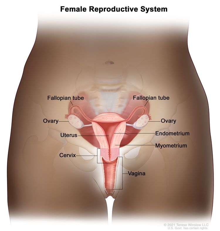
The endometrium is the innermost lining of the uterus. The uterus is a hollow, muscular organ in a woman’s pelvis. The uterus is where a fetus grows. In most nonpregnant women, the uterus is about 3 inches long.

EnlargeAnatomy of the female reproductive system; drawing shows the uterus, myometrium (muscular outer layer of the uterus), endometrium (inner lining of the uterus), ovaries, fallopian tubes, cervix, and vagina.
Anatomy of the female reproductive system. The organs in the female reproductive system include the uterus, ovaries, fallopian tubes, cervix, and vagina. The uterus has a muscular outer layer called the myometrium and an inner lining called the endometrium.

Cancer of the endometrium is different from cancer of the muscle of the uterus, which is called uterine sarcoma. For more information, visit Uterine Sarcoma Treatment.

Other PDQ summaries containing information related to endometrial cancer include:

Endometrial cancer is most common in postmenopausal women.

Endometrial cancer occurs most often in postmenopausal women, with 60 being the average age at diagnosis.

From 2012 to 2021, the number of new cases of endometrial cancer increased slightly in White women and by 2% to 3% each year in women of all other racial and ethnic groups. From 2013 to 2022, the number of deaths from endometrial cancer increased by just under 2% each year.

Endometrial Cancer Prevention

Key Points

  • Avoiding risk factors and increasing protective factors may help prevent cancer.
  • The following risk factors increase the risk of endometrial cancer:
    • Endometrial hyperplasia
    • Estrogen
    • Tamoxifen
    • Obesity, weight gain, metabolic syndrome, and diabetes
    • Genetic factors
  • The following protective factors decrease the risk of endometrial cancer:
    • Pregnancy and breast-feeding
    • Hormonal contraceptives
    • Weight loss
    • Physical activity
  • It is not known if the following factors affect the risk of endometrial cancer:
    • Fruits, vegetables, and vitamins
    • Hair products, including dyes, bleach, highlights, straighteners, and permanents
  • Cancer prevention clinical trials are used to study ways to prevent cancer.
  • New ways to prevent endometrial cancer are being studied in clinical trials.

Avoiding risk factors and increasing protective factors may help prevent cancer.

Avoiding cancer risk factors may help prevent certain cancers. Risk factors include smoking, having overweight, and not getting enough exercise. Increasing protective factors such as quitting smoking and exercising may also help prevent some cancers. Talk to your doctor or other health care professional about how you might lower your risk of cancer.

The following risk factors increase the risk of endometrial cancer:

Endometrial hyperplasia

Endometrial hyperplasia is an abnormal thickening of the endometrium (lining of the uterus). It is not cancer, but in some cases, it may lead to endometrial cancer.

Estrogen

Estrogen is a hormone made by the body. It helps the body develop and maintain female sex characteristics. Estrogen can affect the growth of some cancers, including endometrial cancer.

A woman’s risk of developing endometrial cancer is increased by being exposed to estrogen in the following ways:

  • Estrogen-only hormone therapy: Estrogen may be given to replace the estrogen no longer produced by the ovaries in postmenopausal women or women whose ovaries have been removed. This is called hormone therapy (HT). The use of HT that contains only estrogen increases the risk of endometrial cancer, and the risk grows higher the longer the estrogen is used. For this reason, estrogen therapy alone is usually prescribed only for women who do not have a uterus.

    When estrogen is combined with progestin (another hormone), it is called combination estrogen-progestin therapy. For postmenopausal women, taking estrogen in combination with progestin does not increase the risk of endometrial cancer, but it does increase the risk of breast cancer. For more information, visit Breast Cancer Prevention.

  • Early menstruation: Beginning to have menstrual periods at an early age increases the number of years the body is exposed to estrogen and increases a woman’s risk of endometrial cancer.
  • Late menopause: Women who reach menopause at an older age are exposed to estrogen for a longer time and have an increased risk of endometrial cancer.
  • Never being pregnant: Because estrogen levels are lower during pregnancy, women who have never been pregnant are exposed to estrogen for a longer time than women who have been pregnant. This increases the risk of endometrial cancer.

Tamoxifen

Tamoxifen is one of a group of drugs called selective estrogen receptor modulators, or SERMs. Tamoxifen acts like estrogen on some tissues in the body, such as the uterus, but blocks the effects of estrogen on other tissues, such as the breast. Tamoxifen is used to prevent breast cancer in women who are at high risk for the disease. However, using tamoxifen for more than 2 years increases the risk of endometrial cancer. This risk is greater in postmenopausal women.

Raloxifene is a SERM that is used to prevent bone weakness in postmenopausal women. However, it does not have estrogen-like effects on the uterus and has not been shown to increase the risk of endometrial cancer.

Obesity, weight gain, metabolic syndrome, and diabetes

Having obesity or gaining weight as an adult increases the risk of endometrial cancer. Obesity is related to other risk factors such as high estrogen levels, having extra fat around the waist, polycystic ovary syndrome, and lack of physical activity.

Having metabolic syndrome increases the risk of endometrial cancer. Metabolic syndrome is a condition that includes extra fat around the waist, high blood sugar, high blood pressure, and high levels of triglycerides (a type of fat) in the blood.

Having type 2 diabetes may increase the risk of endometrial cancer.

Genetic factors

Based on solid evidence, women with certain genetic conditions have an increased risk of developing endometrial cancer.

Lynch syndrome is an inherited disorder caused by changes in certain genes. Women who have Lynch syndrome have a much higher risk of developing endometrial cancer than women who do not have Lynch syndrome.

Polycystic ovary syndrome (a disorder of the hormones made by the ovaries) and Cowden syndrome are inherited conditions that are linked to an increased risk of endometrial cancer.

Women with a family history of endometrial cancer in a first-degree relative (mother, sister, or daughter) are also at increased risk of endometrial cancer.

The following protective factors decrease the risk of endometrial cancer:

Pregnancy and breast-feeding

Estrogen levels are lower during pregnancy and when breast-feeding. The risk of endometrial cancer is lower in women who have had children. Breast-feeding also decreases the risk of endometrial cancer.

Hormonal contraceptives

Taking hormonal contraceptives (birth control pills) that combine estrogen and progestin (combined oral contraceptives) decreases the risk of endometrial cancer. The protective effect of this type of birth control increases with the length of time they are used and can last for many years after oral contraceptive use has been stopped.

While taking birth control pills, women have a higher risk of blood clots, stroke, and heart attack, especially women who smoke and are older than 35 years.

New data suggest that other hormonal contraceptives, such as birth control devices that are inserted into a woman’s uterus, may also decrease the risk of endometrial cancer.

Weight loss

It is not known if losing weight decreases the risk of endometrial cancer. However, having bariatric surgery (a surgery that changes how your digestive system works so you will lose weight) decreases the risk of endometrial cancer. After bariatric surgery, other obesity-related conditions, such as diabetes and metabolic syndrome often improve or go away.

Having bariatric surgery also includes risks, such as infection, blood clots, breathing or heart problems, and digestive issues.

Physical activity

Physical activity (exercise) may lower the risk of endometrial cancer. This includes any physical activity you do at your job or at home.

It is not known if the following factors affect the risk of endometrial cancer:

Fruits, vegetables, and vitamins

A diet that includes, fruits, vegetables, phytoestrogen, soy, and vitamin D has not been found to affect the risk of endometrial cancer.

Taking multivitamins has little or no effect on the risk of common cancers, including endometrial cancer.

Hair products, including dyes, bleach, highlights, straighteners, and permanents

There is not enough evidence to show a link between hair products and endometrial cancer. One retrospective study discussed a possible link between certain hair products and uterine cancers, including endometrial cancers.

Cancer prevention clinical trials are used to study ways to prevent cancer.

Cancer prevention clinical trials are used to study ways to lower the risk of developing certain types of cancer. Some cancer prevention trials include healthy people who may or may not have an increased risk of cancer. Other prevention trials include people who have had cancer and are trying to prevent recurrence or a second cancer.

The purpose of some cancer prevention clinical trials is to find out whether actions people take can prevent cancer. These may include eating fruits and vegetables, exercising, quitting smoking, or taking certain medicines, vitamins, minerals, or food supplements.

New ways to prevent endometrial cancer are being studied in clinical trials.

Information about clinical trials supported by NCI can be found on NCI’s clinical trials search webpage. Clinical trials supported by other organizations can be found on the ClinicalTrials.gov website.

About This PDQ Summary

About PDQ

Physician Data Query (PDQ) is the National Cancer Institute’s (NCI’s) comprehensive cancer information database. The PDQ database contains summaries of the latest published information on cancer prevention, detection, genetics, treatment, supportive care, and complementary and alternative medicine. Most summaries come in two versions. The health professional versions have detailed information written in technical language. The patient versions are written in easy-to-understand, nontechnical language. Both versions have cancer information that is accurate and up to date and most versions are also available in Spanish.

PDQ is a service of the NCI. The NCI is part of the National Institutes of Health (NIH). NIH is the federal government’s center of biomedical research. The PDQ summaries are based on an independent review of the medical literature. They are not policy statements of the NCI or the NIH.

Purpose of This Summary

This PDQ cancer information summary has current information about endometrial cancer prevention. It is meant to inform and help patients, families, and caregivers. It does not give formal guidelines or recommendations for making decisions about health care.

Reviewers and Updates

Editorial Boards write the PDQ cancer information summaries and keep them up to date. These Boards are made up of experts in cancer treatment and other specialties related to cancer. The summaries are reviewed regularly and changes are made when there is new information. The date on each summary (“Updated”) is the date of the most recent change.

The information in this patient summary was taken from the health professional version, which is reviewed regularly and updated as needed, by the PDQ Screening and Prevention Editorial Board.

Clinical Trial Information

A clinical trial is a study to answer a scientific question, such as whether one treatment is better than another. Trials are based on past studies and what has been learned in the laboratory. Each trial answers certain scientific questions in order to find new and better ways to help cancer patients. During treatment clinical trials, information is collected about the effects of a new treatment and how well it works. If a clinical trial shows that a new treatment is better than one currently being used, the new treatment may become “standard.” Patients may want to think about taking part in a clinical trial. Some clinical trials are open only to patients who have not started treatment.

Clinical trials can be found online at NCI’s website. For more information, call the Cancer Information Service (CIS), NCI’s contact center, at 1-800-4-CANCER (1-800-422-6237).

Permission to Use This Summary

PDQ is a registered trademark. The content of PDQ documents can be used freely as text. It cannot be identified as an NCI PDQ cancer information summary unless the whole summary is shown and it is updated regularly. However, a user would be allowed to write a sentence such as “NCI’s PDQ cancer information summary about breast cancer prevention states the risks in the following way: [include excerpt from the summary].”

The best way to cite this PDQ summary is:

PDQ® Screening and Prevention Editorial Board. PDQ Endometrial Cancer Prevention. Bethesda, MD: National Cancer Institute. Updated <MM/DD/YYYY>. Available at: /types/uterine/patient/endometrial-prevention-pdq. Accessed <MM/DD/YYYY>. [PMID: 26389201]

Images in this summary are used with permission of the author(s), artist, and/or publisher for use in the PDQ summaries only. If you want to use an image from a PDQ summary and you are not using the whole summary, you must get permission from the owner. It cannot be given by the National Cancer Institute. Information about using the images in this summary, along with many other images related to cancer can be found in Visuals Online. Visuals Online is a collection of more than 3,000 scientific images.

Disclaimer

The information in these summaries should not be used to make decisions about insurance reimbursement. More information on insurance coverage is available on Cancer.gov on the Managing Cancer Care page.

Contact Us

More information about contacting us or receiving help with the Cancer.gov website can be found on our Contact Us for Help page. Questions can also be submitted to Cancer.gov through the website’s E-mail Us.

Endometrial Cancer Treatment (PDQ®)–Patient Version

Endometrial Cancer Treatment (PDQ®)–Patient Version

General Information About Endometrial Cancer

Key Points

  • Endometrial cancer is a disease in which malignant (cancer) cells form in the tissues of the endometrium.
  • Obesity and having metabolic syndrome may increase the risk of endometrial cancer.
  • Taking tamoxifen for breast cancer or taking estrogen alone (without progesterone) can increase the risk of endometrial cancer.
  • Signs and symptoms of endometrial cancer include unusual vaginal bleeding or pain in the pelvis.
  • Tests that examine the endometrium are used to diagnose endometrial cancer.
  • Certain factors affect prognosis (chance of recovery) and treatment options.

Endometrial cancer is a disease in which malignant (cancer) cells form in the tissues of the endometrium.

The endometrium is the lining of the uterus, a hollow, muscular organ in a woman’s pelvis. The uterus is where a fetus grows. In most nonpregnant women, the uterus is about 3 inches long. The lower, narrow end of the uterus is the cervix, which leads to the vagina.

EnlargeAnatomy of the female reproductive system; drawing shows the uterus, myometrium (muscular outer layer of the uterus), endometrium (inner lining of the uterus), ovaries, fallopian tubes, cervix, and vagina.
Anatomy of the female reproductive system. The organs in the female reproductive system include the uterus, ovaries, fallopian tubes, cervix, and vagina. The uterus has a muscular outer layer called the myometrium and an inner lining called the endometrium.

Cancer of the endometrium is different from cancer of the muscle of the uterus, which is called sarcoma of the uterus. See the PDQ summary on Uterine Sarcoma Treatment for more information about uterine sarcoma.

Obesity and having metabolic syndrome may increase the risk of endometrial cancer.

Anything that increases your chance of getting a disease is called a risk factor. Having a risk factor does not mean that you will get cancer; not having risk factors doesn’t mean that you will not get cancer. Talk to your doctor if you think you may be at risk for endometrial cancer.

Risk factors for endometrial cancer include the following:

Older age is the main risk factor for most cancers. The chance of getting cancer increases as you get older.

Taking tamoxifen for breast cancer or taking estrogen alone (without progesterone) can increase the risk of endometrial cancer.

Endometrial cancer may develop in breast cancer patients who have been treated with tamoxifen. A patient who takes this drug and has abnormal vaginal bleeding should have a follow-up exam and a biopsy of the endometrial lining if needed. Women taking estrogen (a hormone that can affect the growth of some cancers) alone also have an increased risk of endometrial cancer. Taking estrogen combined with progesterone (another hormone) does not increase a woman’s risk of endometrial cancer.

Signs and symptoms of endometrial cancer include unusual vaginal bleeding or pain in the pelvis.

These and other signs and symptoms may be caused by endometrial cancer or by other conditions. Check with your doctor if you have any of the following:

  • Vaginal bleeding or discharge not related to menstruation (periods).
  • Vaginal bleeding after menopause.
  • Difficult or painful urination.
  • Pain during sexual intercourse.
  • Pain in the pelvic area.

Tests that examine the endometrium are used to diagnose endometrial cancer.

Because endometrial cancer begins inside the uterus, it does not usually show up in the results of a Pap test. For this reason, a sample of endometrial tissue must be removed and checked under a microscope to look for cancer cells. One of the following procedures may be used:

  • Endometrial biopsy: The removal of tissue from the endometrium (inner lining of the uterus) by inserting a thin, flexible tube through the cervix and into the uterus. The tube is used to gently scrape a small amount of tissue from the endometrium and then remove the tissue samples. A pathologist views the tissue under a microscope to look for cancer cells.
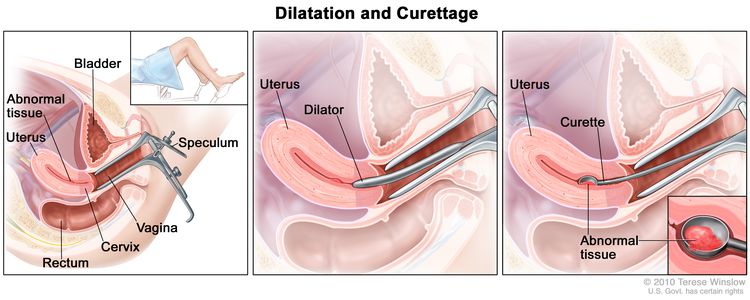
  • Dilatation and curettage: A procedure to remove samples of tissue from the inner lining of the uterus. The cervix is dilated and a curette (spoon-shaped instrument) is inserted into the uterus to remove tissue. The tissue samples are checked under a microscope for signs of disease. This procedure is also called a D&C.
    EnlargeDilatation and curettage (D and C). Three-panel drawing showing a side view of the female reproductive anatomy during a D and C procedure. The first panel shows a speculum widening the opening of the vagina. The cervix, uterus with abnormal tissue, bladder, and rectum are also shown; an inset shows the lower half of a woman covered by a drape on an exam table with her legs apart and her feet in stirrups. The middle panel shows the uterus and a dilator inserted through the vagina into the cervix. The third panel shows a curette scraping out abnormal tissue from the uterus; an inset shows a close up of the curette with the abnormal tissue in it.
    Dilatation and curettage (D and C). A speculum is inserted into the vagina to widen it in order to look at the cervix (first panel). A dilator is used to widen the cervix (middle panel). A curette is put through the cervix into the uterus to scrape out abnormal tissue (last panel).
  • Hysteroscopy: A procedure to look inside the uterus for abnormal areas. A hysteroscope is inserted through the vagina and cervix into the uterus. A hysteroscope is a thin, tube-like instrument with a light and a lens for viewing. It may also have a tool to remove tissue samples, which are checked under a microscope for signs of cancer.

Other tests and procedures used to diagnose endometrial cancer include the following:

  • Physical exam and health history: An exam of the body to check general signs of health, including checking for signs of disease, such as lumps or anything else that seems unusual. A history of the patient’s health habits and past illnesses and treatments will also be taken.
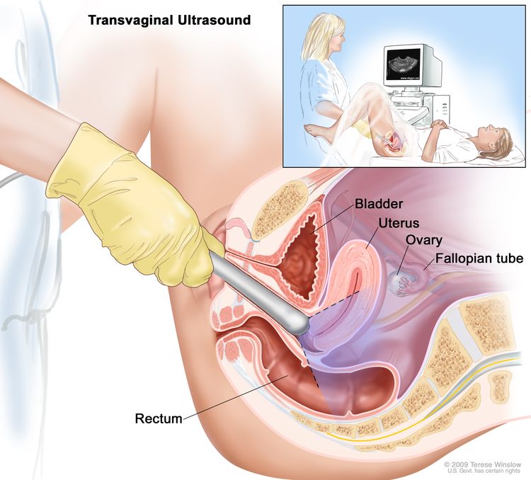
  • Transvaginal ultrasound exam: A procedure used to examine the vagina, uterus, fallopian tubes, and bladder. An ultrasound transducer (probe) is inserted into the vagina and used to bounce high-energy sound waves (ultrasound) off internal tissues or organs and make echoes. The echoes form a picture of body tissues called a sonogram. The doctor can identify tumors by looking at the sonogram.
    EnlargeTransvaginal ultrasound; drawing shows a side view of the female reproductive anatomy during a transvaginal ultrasound procedure. An ultrasound probe (a device that makes sound waves that bounce off tissues inside the body) is shown inserted into the vagina. The bladder, uterus, right fallopian tube, and right ovary are also shown. The inset shows the diagnostic sonographer (a person trained to perform ultrasound procedures) examining a woman on a table, and a computer screen shows an image of the patient’s internal tissues.
    Transvaginal ultrasound. An ultrasound probe connected to a computer is inserted into the vagina and is gently moved to show different organs. The probe bounces sound waves off internal organs and tissues to make echoes that form a sonogram (computer picture).

Certain factors affect prognosis (chance of recovery) and treatment options.

The prognosis and treatment options depend on the following:

  • The stage of the cancer (whether it is in the endometrium only, involves the uterus wall, or has spread to other places in the body).
  • How the cancer cells look under a microscope.
  • Whether the cancer cells are affected by progesterone.

Endometrial cancer can usually be cured because it is usually diagnosed early.

Stages of Endometrial Cancer

Key Points

  • After endometrial cancer has been diagnosed, tests are done to find out if cancer cells have spread within the uterus or to other parts of the body.
  • There are three ways that cancer spreads in the body.
  • Cancer may spread from where it began to other parts of the body.
  • The following stages are used for endometrial cancer:
    • Stage I
    • Stage II
    • Stage III
    • Stage IV
  • Endometrial cancer may be grouped for treatment as follows:
    • Low-risk endometrial cancer
    • High-risk endometrial cancer
  • Endometrial cancer can recur (come back) after it has been treated.

After endometrial cancer has been diagnosed, tests are done to find out if cancer cells have spread within the uterus or to other parts of the body.

The process used to find out whether the cancer has spread within the uterus or to other parts of the body is called staging. The information gathered from the staging process determines the stage of the disease. It is important to know the stage in order to plan treatment. Certain tests and procedures are used in the staging process. A hysterectomy (an operation in which the uterus is removed) will usually be done to treat endometrial cancer. Tissue samples are taken from the area around the uterus and checked under a microscope for signs of cancer to help find out whether the cancer has spread.

The following procedures may be used in the staging process:

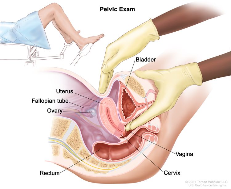
  • Pelvic exam: An exam of the vagina, cervix, uterus, fallopian tubes, ovaries, and rectum. A speculum is inserted into the vagina and the doctor or nurse looks at the vagina and cervix for signs of disease. A Pap test of the cervix is usually done. The doctor or nurse also inserts one or two lubricated, gloved fingers of one hand into the vagina and places the other hand over the lower abdomen to feel the size, shape, and position of the uterus and ovaries. The doctor or nurse also inserts a lubricated, gloved finger into the rectum to feel for lumps or abnormal areas.
    EnlargePelvic exam; drawing shows a side view of the female reproductive anatomy during a pelvic exam. The uterus, left fallopian tube, left ovary, cervix, vagina, bladder, and rectum are shown. Two gloved fingers of one hand of the doctor or nurse are shown inserted into the vagina, while the other hand is shown pressing on the lower abdomen. The inset shows a woman covered by a drape on an exam table with her legs apart and her feet in stirrups.
    Pelvic exam. A doctor or nurse inserts one or two lubricated, gloved fingers of one hand into the vagina and presses on the lower abdomen with the other hand. This is done to feel the size, shape, and position of the uterus and ovaries. The vagina, cervix, fallopian tubes, and rectum are also checked.
  • Chest x-ray: An x-ray of the organs and bones inside the chest. An x-ray is a type of energy beam that can go through the body and onto film, making a picture of areas inside the body.
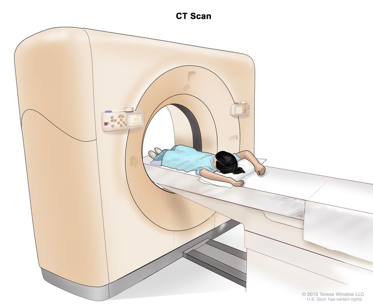
  • CT scan (CAT scan): A procedure that makes a series of detailed pictures of areas inside the body, taken from different angles. The pictures are made by a computer linked to an x-ray machine. A dye may be injected into a vein or swallowed to help the organs or tissues show up more clearly. This procedure is also called computed tomography, computerized tomography, or computerized axial tomography.
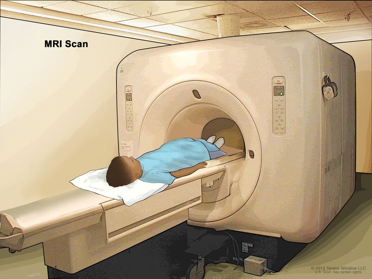
  • MRI (magnetic resonance imaging): A procedure that uses a magnet, radio waves, and a computer to make a series of detailed pictures of areas inside the body. This procedure is also called nuclear magnetic resonance imaging (NMRI).
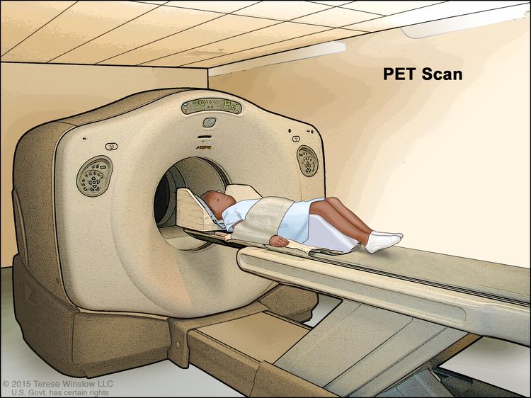
  • PET scan (positron emission tomography scan): A procedure to find malignant tumor cells in the body. A small amount of radioactive glucose (sugar) is injected into a vein. The PET scanner rotates around the body and makes a picture of where glucose is being used in the body. Malignant tumor cells show up brighter in the picture because they are more active and take up more glucose than normal cells do.
  • Lymph node dissection: A surgical procedure in which the lymph nodes are removed from the pelvic area and a sample of tissue is checked under a microscope for signs of cancer. This procedure is also called lymphadenectomy.

There are three ways that cancer spreads in the body.

Cancer can spread through tissue, the lymph system, and the blood:

  • Tissue. The cancer spreads from where it began by growing into nearby areas.
  • Lymph system. The cancer spreads from where it began by getting into the lymph system. The cancer travels through the lymph vessels to other parts of the body.
  • Blood. The cancer spreads from where it began by getting into the blood. The cancer travels through the blood vessels to other parts of the body.

Cancer may spread from where it began to other parts of the body.

When cancer spreads to another part of the body, it is called metastasis. Cancer cells break away from where they began (the primary tumor) and travel through the lymph system or blood.

  • Lymph system. The cancer gets into the lymph system, travels through the lymph vessels, and forms a tumor (metastatic tumor) in another part of the body.
  • Blood. The cancer gets into the blood, travels through the blood vessels, and forms a tumor (metastatic tumor) in another part of the body.

The metastatic tumor is the same type of cancer as the primary tumor. For example, if endometrial cancer spreads to the lung, the cancer cells in the lung are actually endometrial cancer cells. The disease is metastatic endometrial cancer, not lung cancer.

Many cancer deaths are caused when cancer moves from the original tumor and spreads to other tissues and organs. This is called metastatic cancer. This animation shows how cancer cells travel from the place in the body where they first formed to other parts of the body.

The following stages are used for endometrial cancer:

Stage I

EnlargeStage IA and stage IB endometrial cancer shown in two cross-section drawings of the uterus and cervix. Drawing on the left shows stage IA, with cancer in the endometrium and myometrium of the uterus. Drawing on the right shows stage IB, with cancer more than halfway through the myometrium. Also shown are the fallopian tubes, ovaries, and vagina.
Stage IA and stage IB endometrial cancer. In stage IA, cancer is in the endometrium only or less than halfway through the myometrium (the muscle layer of the uterus). In stage IB, cancer has spread halfway or more into the myometrium.

In stage I, cancer is found in the uterus only. Stage I is divided into stages IA and IB, based on how far the cancer has spread.

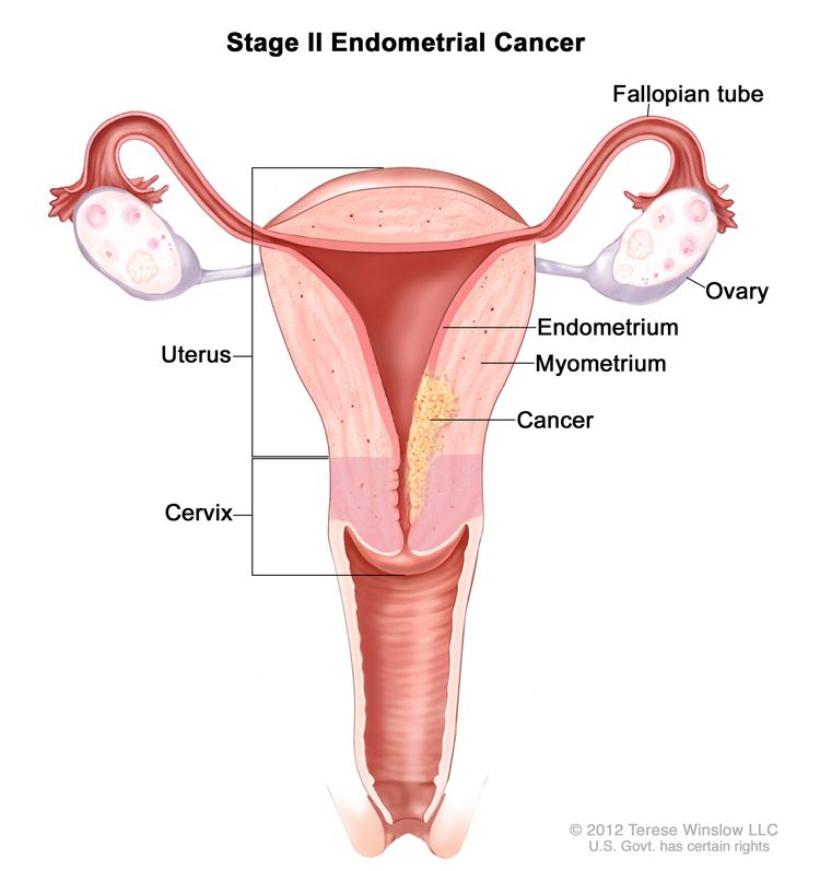
Stage II

EnlargeStage II endometrial cancer shown in a cross-section drawing of the uterus, cervix, fallopian tubes, ovaries, and vagina. Cancer is shown in the endometrium and myometrium of the uterus and in the cervix.
Stage II endometrial cancer. Cancer has spread into connective tissue of the cervix, but has not spread outside the uterus.

In stage II, cancer has spread into connective tissue of the cervix, but has not spread outside the uterus.

Stage III

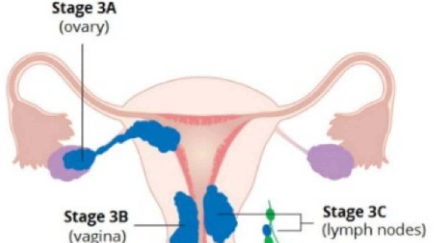
In stage III, cancer has spread beyond the uterus and cervix, but has not spread beyond the pelvis. Stage III is divided into stages IIIA, IIIB, and IIIC, based on how far the cancer has spread within the pelvis.

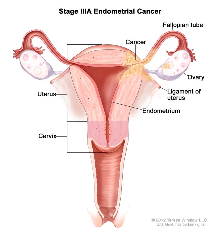
  • Stage IIIA: Cancer has spread to the outer layer of the uterus and/or to the fallopian tubes, ovaries, and ligaments of the uterus.
    EnlargeStage IIIA endometrial cancer shown in a cross-section drawing of the uterus, ligaments of the uterus, cervix, fallopian tubes, ovaries, and vagina. Cancer is shown in the endometrium of the uterus, the outer layer of the uterus, a fallopian tube, an ovary, and a ligament of the uterus.
    Stage IIIA endometrial cancer. Cancer has spread to the outer layer of the uterus and/or to the fallopian tubes, ovaries, or ligaments of the uterus.
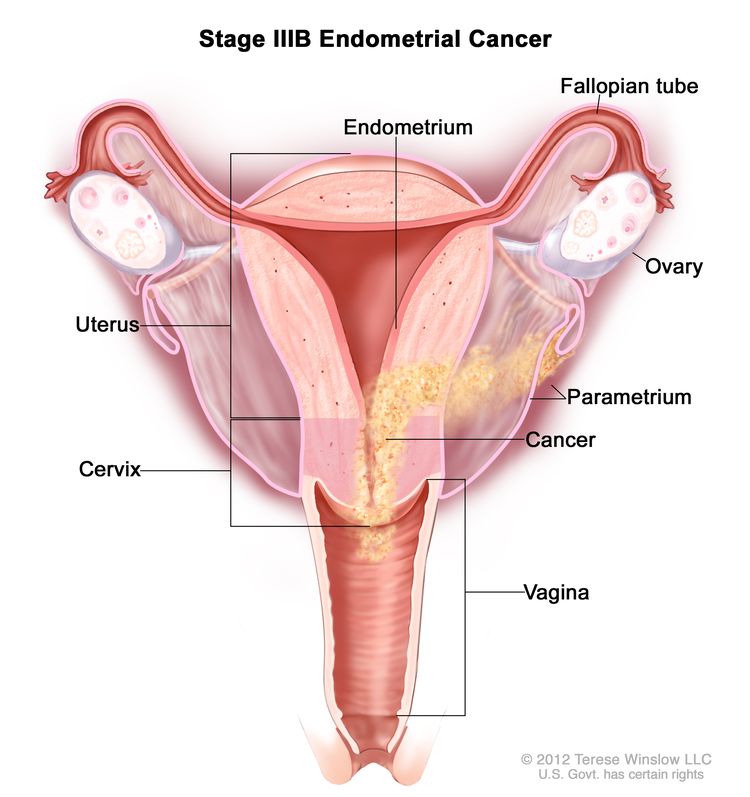
  • Stage IIIB: Cancer has spread to the vagina and/or to the parametrium (connective tissue and fat around the uterus).
    EnlargeStage IIIB endometrial cancer shown in a cross-section drawing of the uterus, cervix, fallopian tubes, ovaries, and vagina. Cancer is shown in the endometrium of the uterus, the parametrium, the cervix, and the vagina.
    Stage IIIB endometrial cancer. Cancer has spread to the vagina and/or to the parametrium (connective tissue and fat around the uterus and cervix).
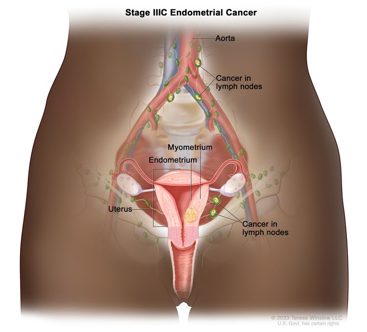
  • Stage IIIC: Cancer has spread to lymph nodes in the pelvis and/or around the aorta (largest artery in the body, which carries blood away from the heart).
    EnlargeStage IIIC endometrial cancer; drawing shows cancer in the endometrium and myometrium of the uterus. Also shown is cancer in lymph nodes in the pelvis and near the aorta.
    Stage IIIC endometrial cancer. Cancer has spread to lymph nodes in the pelvis and/or around the aorta (the largest artery in the body that carries blood away from the heart).

Stage IV

In stage IV, cancer has spread beyond the pelvis. Stage IV is divided into stages IVA and IVB, based on how far the cancer has spread.

  • Stage IVA: Cancer has spread to the bladder and/or bowel wall.
    EnlargeStage IVA endometrial cancer shown in a side-view cross-section drawing of the uterus, bladder, cervix, vagina, small intestine, and large intestine. Cancer is shown in the bladder, uterus, and bowel.
    Stage IVA endometrial cancer. Cancer has spread into the bladder and/or bowel.
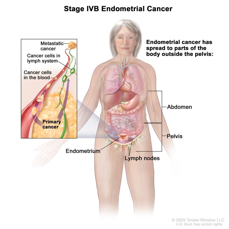
  • Stage IVB: Cancer has spread to other parts of the body beyond the pelvis, including the abdomen and/or lymph nodes in the groin.
    EnlargeStage IVB endometrial cancer; drawing shows cancer that has spread to parts of the body outside the pelvis, including the abdomen and lymph nodes in the groin. An inset shows cancer cells spreading from the endometrium, through the blood and lymph system, to another part of the body where metastatic cancer has formed.
    Stage IVB endometrial cancer. The cancer has spread to parts of the body outside the pelvis, including the abdomen and/or lymph nodes in the groin.

Endometrial cancer may be grouped for treatment as follows:

Low-risk endometrial cancer

Grades 1 and 2 tumors are usually considered low-risk. They usually do not spread to other parts of the body.

High-risk endometrial cancer

Grade 3 tumors are considered high-risk. They often spread to other parts of the body. Uterine papillary serous, clear cell, and carcinosarcoma are three subtypes of endometrial cancer that are considered grade 3.

Endometrial cancer can recur (come back) after it has been treated.

The cancer may come back in the uterus, the pelvis, in lymph nodes in the abdomen, or in other parts of the body.

Treatment Option Overview

Key Points

  • There are different types of treatment for patients with endometrial cancer.
  • Five types of standard treatment are used:
    • Surgery
    • Radiation therapy
    • Chemotherapy
    • Hormone therapy
    • Targeted therapy
  • New types of treatment are being tested in clinical trials.
  • Treatment for endometrial cancer may cause side effects.
  • Patients may want to think about taking part in a clinical trial.
  • Patients can enter clinical trials before, during, or after starting their cancer treatment.
  • Follow-up tests may be needed.

There are different types of treatment for patients with endometrial cancer.

Different types of treatment are available for patients with endometrial cancer. Some treatments are standard (the currently used treatment), and some are being tested in clinical trials. A treatment clinical trial is a research study meant to help improve current treatments or obtain information on new treatments for patients with cancer. When clinical trials show that a new treatment is better than the standard treatment, the new treatment may become the standard treatment. Patients may want to think about taking part in a clinical trial. Some clinical trials are open only to patients who have not started treatment.

Five types of standard treatment are used:

Surgery

Surgery (removing the cancer in an operation) is the most common treatment for endometrial cancer. The following surgical procedures may be used:

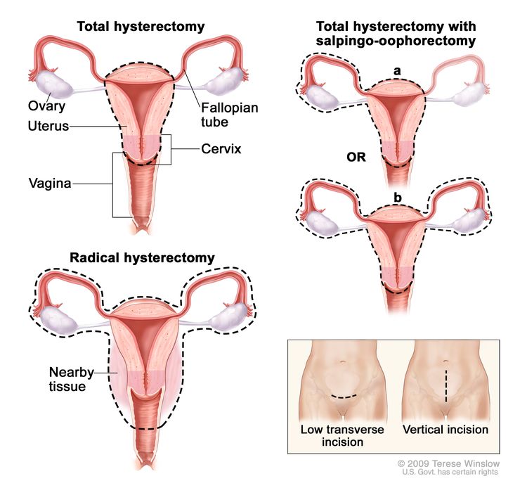
  • Total hysterectomy: Surgery to remove the uterus, including the cervix. If the uterus and cervix are taken out through the vagina, the operation is called a vaginal hysterectomy. If the uterus and cervix are taken out through a large incision (cut) in the abdomen, the operation is called a total abdominal hysterectomy. If the uterus and cervix are taken out through a small incision (cut) in the abdomen using a laparoscope, the operation is called a total laparoscopic hysterectomy.
    EnlargeHysterectomy; drawing shows the female reproductive anatomy, including the ovaries, uterus, vagina, fallopian tubes, and cervix. Dotted lines show which organs and tissues are removed in a total hysterectomy, a total hysterectomy with salpingo-oophorectomy, and a radical hysterectomy. An inset shows the location of two possible incisions on the abdomen: a low transverse incision is just above the pubic area and a vertical incision is between the navel and the pubic area.
    Hysterectomy. The uterus is surgically removed with or without other organs or tissues. In a total hysterectomy, the uterus and cervix are removed. In a total hysterectomy with salpingo-oophorectomy, (a) the uterus plus one (unilateral) ovary and fallopian tube are removed; or (b) the uterus plus both (bilateral) ovaries and fallopian tubes are removed. In a radical hysterectomy, the uterus, cervix, both ovaries, both fallopian tubes, and nearby tissue are removed. These procedures are done using a low transverse incision or a vertical incision.
  • Bilateral salpingo-oophorectomy: Surgery to remove both ovaries and both fallopian tubes.
  • Radical hysterectomy: Surgery to remove the uterus, cervix, and part of the vagina. The ovaries, fallopian tubes, or nearby lymph nodes may also be removed.
  • Lymph node dissection: A surgical procedure in which the lymph nodes are removed from the pelvic area and a sample of tissue is checked under a microscope for signs of cancer. This procedure is also called lymphadenectomy.

After the doctor removes all the cancer that can be seen at the time of the surgery, some patients may be given radiation therapy or hormone treatment after surgery to kill any cancer cells that are left. Treatment given after the surgery, to lower the risk that the cancer will come back, is called adjuvant therapy.

Radiation therapy

Radiation therapy is a cancer treatment that uses high-energy x-rays or other types of radiation to kill cancer cells or keep them from growing. There are two types of radiation therapy:

The way the radiation therapy is given depends on the type and stage of the cancer being treated. External and internal radiation therapy are used to treat endometrial cancer, and may also be used as palliative therapy to relieve symptoms and improve quality of life.

Chemotherapy

Chemotherapy is a cancer treatment that uses drugs to stop the growth of cancer cells, either by killing the cells or by stopping the cells from dividing. When chemotherapy is taken by mouth or injected into a vein or muscle, the drugs enter the bloodstream and can reach cancer cells throughout the body (systemic chemotherapy). When chemotherapy is placed directly into the cerebrospinal fluid, an organ, or a body cavity such as the abdomen, the drugs mainly affect cancer cells in those areas (regional chemotherapy).

The way the chemotherapy is given depends on the type and stage of the cancer being treated.

Hormone therapy

Hormone therapy is a cancer treatment that removes hormones or blocks their action and stops cancer cells from growing. Hormones are substances made by glands in the body and circulated in the bloodstream. Some hormones can cause certain cancers to grow. If tests show that the cancer cells have places where hormones can attach (receptors), drugs, surgery, or radiation therapy is used to reduce the production of hormones or block them from working.

Targeted therapy

Targeted therapy is a type of treatment that uses drugs or other substances to identify and attack specific cancer cells. Targeted therapies usually cause less harm to normal cells than chemotherapy or radiation therapy do. Monoclonal antibodies, mTOR inhibitors, and signal transduction inhibitors are three types of targeted therapy used to treat endometrial cancer.

  • Monoclonal antibody therapy: Monoclonal antibodies are immune system proteins made in the laboratory to treat many diseases, including cancer. As a cancer treatment, these antibodies can attach to a specific target on cancer cells or other cells that may help cancer cells grow. The antibodies are able to then kill the cancer cells, block their growth, or keep them from spreading. Monoclonal antibodies are given by infusion. They may be used alone or to carry drugs, toxins, or radioactive material directly to cancer cells. Bevacizumab is used to treat stage III, stage IV, and recurrent endometrial cancer.
    How do monoclonal antibodies work to treat cancer? This video shows how monoclonal antibodies, such as trastuzumab, pembrolizumab, and rituximab, block molecules cancer cells need to grow, flag cancer cells for destruction by the body’s immune system, or deliver harmful substances to cancer cells.
  • mTOR inhibitor therapy: mTOR inhibitors block a protein called mTOR, which helps control cell division. mTOR inhibitors may keep cancer cells from growing and prevent the growth of new blood vessels that tumors need to grow. Everolimus and ridaforolimus are used to treat stage III, stage IV, and recurrent endometrial cancer.
  • Signal transduction inhibitor therapy: Signal transduction inhibitors block signals that are passed from one molecule to another inside a cell. Blocking these signals may kill cancer cells. Metformin is being studied to treat stage III, stage IV, and recurrent endometrial cancer.

New types of treatment are being tested in clinical trials.

Information about clinical trials is available from the NCI website.

Treatment for endometrial cancer may cause side effects.

For information about side effects caused by treatment for cancer, visit our Side Effects page.

Patients may want to think about taking part in a clinical trial.

For some patients, taking part in a clinical trial may be the best treatment choice. Clinical trials are part of the cancer research process. Clinical trials are done to find out if new cancer treatments are safe and effective or better than the standard treatment.

Many of today’s standard treatments for cancer are based on earlier clinical trials. Patients who take part in a clinical trial may receive the standard treatment or be among the first to receive a new treatment.

Patients who take part in clinical trials also help improve the way cancer will be treated in the future. Even when clinical trials do not lead to effective new treatments, they often answer important questions and help move research forward.

Patients can enter clinical trials before, during, or after starting their cancer treatment.

Some clinical trials only include patients who have not yet received treatment. Other trials test treatments for patients whose cancer has not gotten better. There are also clinical trials that test new ways to stop cancer from recurring (coming back) or reduce the side effects of cancer treatment.

Clinical trials are taking place in many parts of the country. Information about clinical trials supported by NCI can be found on NCI’s clinical trials search webpage. Clinical trials supported by other organizations can be found on the ClinicalTrials.gov website.

Follow-up tests may be needed.

As you go through treatment, you will have follow-up tests or check-ups. Some tests that were done to diagnose or stage the cancer may be repeated to see how well the treatment is working. Decisions about whether to continue, change, or stop treatment may be based on the results of these tests.

Some of the tests will continue to be done from time to time after treatment has ended. The results of these tests can show if your condition has changed or if the cancer has recurred (come back).

Treatment of Stage I and Stage II Endometrial Cancer

For information about the treatments listed below, see the Treatment Option Overview section.

Low-risk endometrial cancer (grade 1 or grade 2)

Treatment of low-risk stage I endometrial cancer and stage II endometrial cancer may include the following:

If cancer has spread to the cervix, a radical hysterectomy with bilateral salpingo-oophorectomy may be done.

High-risk endometrial cancer (grade 3)

Treatment of high-risk stage I endometrial cancer and stage II endometrial cancer may include the following:

Use our clinical trial search to find NCI-supported cancer clinical trials that are accepting patients. You can search for trials based on the type of cancer, the age of the patient, and where the trials are being done. General information about clinical trials is also available.

Treatment of Stage III, Stage IV, and Recurrent Endometrial Cancer

For information about the treatments listed below, see the Treatment Option Overview section.

Treatment of stage III endometrial cancer, stage IV endometrial cancer, and recurrent endometrial cancer may include the following:

Use our clinical trial search to find NCI-supported cancer clinical trials that are accepting patients. You can search for trials based on the type of cancer, the age of the patient, and where the trials are being done. General information about clinical trials is also available.

To Learn More About Endometrial Cancer

About This PDQ Summary

About PDQ

Physician Data Query (PDQ) is the National Cancer Institute’s (NCI’s) comprehensive cancer information database. The PDQ database contains summaries of the latest published information on cancer prevention, detection, genetics, treatment, supportive care, and complementary and alternative medicine. Most summaries come in two versions. The health professional versions have detailed information written in technical language. The patient versions are written in easy-to-understand, nontechnical language. Both versions have cancer information that is accurate and up to date and most versions are also available in Spanish.

PDQ is a service of the NCI. The NCI is part of the National Institutes of Health (NIH). NIH is the federal government’s center of biomedical research. The PDQ summaries are based on an independent review of the medical literature. They are not policy statements of the NCI or the NIH.

Purpose of This Summary

This PDQ cancer information summary has current information about the treatment of endometrial cancer. It is meant to inform and help patients, families, and caregivers. It does not give formal guidelines or recommendations for making decisions about health care.

Reviewers and Updates

Editorial Boards write the PDQ cancer information summaries and keep them up to date. These Boards are made up of experts in cancer treatment and other specialties related to cancer. The summaries are reviewed regularly and changes are made when there is new information. The date on each summary (“Updated”) is the date of the most recent change.

The information in this patient summary was taken from the health professional version, which is reviewed regularly and updated as needed, by the PDQ Adult Treatment Editorial Board.

Clinical Trial Information

A clinical trial is a study to answer a scientific question, such as whether one treatment is better than another. Trials are based on past studies and what has been learned in the laboratory. Each trial answers certain scientific questions in order to find new and better ways to help cancer patients. During treatment clinical trials, information is collected about the effects of a new treatment and how well it works. If a clinical trial shows that a new treatment is better than one currently being used, the new treatment may become “standard.” Patients may want to think about taking part in a clinical trial. Some clinical trials are open only to patients who have not started treatment.

Clinical trials can be found online at NCI’s website. For more information, call the Cancer Information Service (CIS), NCI’s contact center, at 1-800-4-CANCER (1-800-422-6237).

Permission to Use This Summary

PDQ is a registered trademark. The content of PDQ documents can be used freely as text. It cannot be identified as an NCI PDQ cancer information summary unless the whole summary is shown and it is updated regularly. However, a user would be allowed to write a sentence such as “NCI’s PDQ cancer information summary about breast cancer prevention states the risks in the following way: [include excerpt from the summary].”

The best way to cite this PDQ summary is:

PDQ® Adult Treatment Editorial Board. PDQ Endometrial Cancer Treatment. Bethesda, MD: National Cancer Institute. Updated <MM/DD/YYYY>. Available at: /types/uterine/patient/endometrial-treatment-pdq. Accessed <MM/DD/YYYY>. [PMID: 26389334]

Images in this summary are used with permission of the author(s), artist, and/or publisher for use in the PDQ summaries only. If you want to use an image from a PDQ summary and you are not using the whole summary, you must get permission from the owner. It cannot be given by the National Cancer Institute. Information about using the images in this summary, along with many other images related to cancer can be found in Visuals Online. Visuals Online is a collection of more than 3,000 scientific images.

Disclaimer

The information in these summaries should not be used to make decisions about insurance reimbursement. More information on insurance coverage is available on Cancer.gov on the Managing Cancer Care page.

Contact Us

More information about contacting us or receiving help with the Cancer.gov website can be found on our Contact Us for Help page. Questions can also be submitted to Cancer.gov through the website’s E-mail Us.

Ewing Sarcoma Treatment (PDQ®)–Patient Version

Ewing Sarcoma Treatment (PDQ®)–Patient Version

General Information About Ewing Sarcoma

Key Points

  • Ewing sarcoma is a type of cancer that forms from a certain kind of cell in bone or soft tissue.
  • Undifferentiated small round cell sarcoma may also form in the bone or soft tissue.
  • A genetic condition may increase the risk of Ewing sarcoma and other sarcomas.
  • Symptoms of Ewing sarcoma include swelling and pain near the tumor.
  • Tests that examine the bone and soft tissue are used to diagnose and stage Ewing sarcoma.
  • Certain factors affect prognosis (chance of recovery).

Ewing sarcoma is a type of cancer that forms from a certain kind of cell in bone or soft tissue.

Ewing sarcoma may form in the bones of the legs, arms, feet, hands, chest, pelvis, spine, or skull. It may also form in the body’s soft tissues, which connect, support, and surround other body parts and organs.

Ewing sarcoma is most common in adolescents and young adults (teens through mid-20s).

Ewing sarcoma has also been called peripheral primitive neuroectodermal tumor, Askin tumor (Ewing sarcoma of the chest wall), extraosseous Ewing sarcoma (Ewing sarcoma in tissue other than bone), and Ewing sarcoma family of tumors.

Undifferentiated small round cell sarcoma may also form in the bone or soft tissue.

Undifferentiated small round cell sarcoma usually forms in the bones or the muscles that are attached to bones and that help the body move. Undifferentiated small round cell sarcoma is usually treated the same way as Ewing sarcoma. Types of undifferentiated small round cell sarcomas include:

  • Undifferentiated small round cell sarcoma with BCOR rearrangements. This type of round cell sarcoma usually forms in the pelvis, arms, or legs and may spread to other parts of the body. It is more common in children younger than 18 years. In this type of round cell sarcoma, the BCOR gene is joined to the CCNB3 gene or to other genes. To diagnose this round cell sarcoma, the tumor cells are checked for these gene changes.
  • Undifferentiated small round cell sarcoma with CIC::DUX4 rearrangements. This type of round cell sarcoma usually forms in the trunk, arms, or legs. It is most common in males and in young adults. In this type of round cell sarcoma, the CIC gene is joined to the DUX4 gene. To diagnose this round cell sarcoma, the tumor cells are checked for this gene change.
  • Undifferentiated small round cell sarcoma with CIC::NUTM1 rearrangements. This type of soft tissue tumor usually forms in the central nervous system, but it can also form in the trunk. It is most common in children about 6 years of age.
  • Undifferentiated small round cell sarcoma with ATXN1::NUTM2A or ATXN1L::NUTM2A fusions. This type of soft tissue tumor has been found in three children. All were newborns or infants, and the cancer was in the central nervous system or peritoneum.
  • Undifferentiated small round cell sarcoma with EWSR1::NFATC2 and FUS::NFATC2 rearrangements. This type of soft tissue tumor may be a benign cyst or a malignant (cancer) tumor. It usually forms in the arms and legs. The malignant tumor is more common in males and in adults.
  • Undifferentiated small round cell sarcoma with EWSR1::PATZ1 fusions. This type of soft tissue tumor usually forms in the trunk and is more common in adults.

A genetic condition may increase the risk of Ewing sarcoma and other sarcomas.

Ewing sarcoma is caused by certain changes to the way bone cells function, especially how they grow and divide into new cells. Often, the exact cause of these changes is unknown. Learn more about how cancer develops at What Is Cancer?

A risk factor is anything that increases the chance of getting a disease. Not every child with a risk factor will develop Ewing sarcoma or other sarcomas. And they can develop in some children who don’t have a known risk factor. Children with Fanconi anemia may be at increased risk of Ewing sarcoma. Talk with your child’s doctor if you think your child may be at risk.

Symptoms of Ewing sarcoma include swelling and pain near the tumor.

It’s important to check with your child’s doctor if your child has:

  • a lump (which may feel soft and warm) in the arms, legs, chest, or pelvis
  • pain and/or swelling near the lump
  • fever for no known reason
  • a bone that breaks for no known reason
  • a limp when walking
  • fatigue
  • weight loss
  • anemia
  • shortness of breath
  • back pain, weakness, numbness, or paralysis in the arms or legs

These symptoms may be caused by problems other than Ewing sarcoma. The only way to know is for your child to see a doctor.

Tests that examine the bone and soft tissue are used to diagnose and stage Ewing sarcoma.

If your child has symptoms that suggest Ewing sarcoma, the doctor will need to find out if these are due to cancer or another problem. The doctor will ask when the symptoms started and how often your child has been having them. They also will ask about your child’s personal and family medical history and do a physical exam. Depending on these results, they may recommend other tests. If your child is diagnosed with Ewing sarcoma, the results of these tests will help you and your child’s doctor plan treatment.

Cancer stage describes the extent of cancer in the body, such as the size of the tumor, whether it has spread, and how far it has spread from where it first formed. To plan treatment, it is important to know whether the cancer has spread to other parts of the body. Tests and procedures to detect, diagnose, and stage Ewing sarcoma are usually done at the same time.

The tests used to diagnose and stage Ewing sarcoma may include:

  • MRI (magnetic resonance imaging) uses a magnet, radio waves, and a computer to make a series of detailed pictures of areas inside the body, such as the area where the tumor formed. This procedure is also called nuclear magnetic resonance imaging (NMRI).
    EnlargeMagnetic resonance imaging (MRI) scan; drawing shows a child lying on a table that slides into the MRI machine, which takes a series of detailed pictures of areas inside the body.
    Magnetic resonance imaging (MRI) scan. The child lies on a table that slides into the MRI machine, which takes a series of detailed pictures of areas inside the body. The positioning of the child on the table depends on the part of the body being imaged.
  • CT scan (CAT scan) uses a computer linked to an x-ray machine to make a series of detailed pictures of areas inside the body, such as the area where the tumor formed or the chest. The pictures are taken from different angles and are used to create 3-D views of tissues and organs. A dye may be injected into a vein or swallowed to help the organs or tissues show up more clearly. This procedure is also called computed tomography, computerized tomography, or computerized axial tomography. Learn more about Computed Tomography (CT) Scans and Cancer.
    EnlargeComputed tomography (CT) scan; drawing shows a child lying on a table that slides through the CT scanner, which takes a series of detailed x-ray pictures of areas inside the body.
    Computed tomography (CT) scan. The child lies on a table that slides through the CT scanner, which takes a series of detailed x-ray pictures of areas inside the body.
  • PET-CT scan combines the pictures from a positron emission tomography scan (PET scan) and CT scan. The PET and CT scans are done at the same time on the same machine. The pictures from both scans are combined to make a more detailed picture than either test would make by itself. For the PET scan, a small amount of radioactive sugar (radioactive glucose) is injected into a vein. The scanner rotates around the body and makes a picture of where the sugar is being used in the body. Cancer cells show up brighter in the picture because they are more active and take up more sugar than normal cells do.
    EnlargePositron emission tomography (PET) scan; drawing shows a child lying on table that slides through the PET scanner.
    Positron emission tomography (PET) scan. The child lies on a table that slides through the PET scanner. The head rest and white strap help the child lie still. A small amount of radioactive glucose (sugar) is injected into the child’s vein, and a scanner makes a picture of where the glucose is being used in the body. Cancer cells show up brighter in the picture because they take up more glucose than normal cells do.
  • Bone scan checks if there are rapidly dividing cells, such as cancer cells, in the bone. A very small amount of radioactive material is injected into a vein and travels through the bloodstream. The radioactive material collects in the bones with cancer and is detected by a scanner.
    EnlargeBone scan; drawing shows a child lying on a table that slides under the scanner, a technician operating the scanner, and a computer monitor that will show images made during the scan.
    Bone scan. A small amount of radioactive material is injected into the child’s vein and travels through the blood. The radioactive material collects in the bones. As the child lies on a table that slides under the scanner, the radioactive material is detected and images are made on a computer screen.
  • Bone marrow aspiration and biopsy removes bone marrow and a small piece of bone by inserting a hollow needle into the hipbone. Samples are removed from both hipbones. A pathologist views the bone marrow and bone under a microscope to see if the cancer has spread. This test is only used if other tests have shown that the cancer may have spread from where it first formed.
    EnlargeBone marrow aspiration and biopsy; drawing shows a child lying face down on a table and a bone marrow needle being inserted into the right hip bone. An inset shows the bone marrow needle being inserted through the skin into the bone marrow of the hip bone.
    Bone marrow aspiration and biopsy. After a small area of skin is numbed, a bone marrow needle is inserted into the child’s hip bone. Samples of blood, bone, and bone marrow are removed for examination under a microscope.
  • X-ray is a type of radiation that can go through the body and make pictures of areas inside the body, such as the chest or the area where the tumor formed.
  • Complete blood count (CBC) checks a sample of blood for:
  • Blood chemistry study uses a blood sample to measure the amounts of certain substances, such as lactate dehydrogenase (LDH), released into the blood by organs and tissues in the body. An unusual amount of a substance can be a sign of disease.
  • Biopsy is a procedure in which a sample of tissue is removed from the tumor so that a pathologist can view it under a microscope to check for cancer. The following types of biopsies are used to check for Ewing sarcoma:
    • Incisional biopsy removes a sample of tissue through an incision in the skin.
    • Needle biopsy uses a needle to remove a sample of tissue.

    The child’s pathologist, radiation oncologist, and surgeon usually work together to decide the best site to place the needle or biopsy incision. The selection of the biopsy site is important. A biopsy site that is not properly selected may result in more extensive surgery to remove the tumor or a larger area that is treated with radiation therapy.

    If there is a chance that the cancer has spread to nearby lymph nodes, one or more lymph nodes may be removed and checked for cancer.

    The following tests may be done on the tissue that is removed:

    • Cytogenetic analysis is a test in which the chromosomes of cells in a sample of tissue are counted and checked for any changes, such as broken, missing, rearranged, or extra chromosomes. Changes in certain chromosomes may be a sign of cancer. Cytogenetic analysis is used to help diagnose cancer, plan treatment, or find out how well treatment is working.
    • Immunohistochemistry is a test that uses antibodies to check for certain antigens (markers) in a sample of a patient’s tissue. The antibodies are usually linked to an enzyme or a fluorescent dye. After the antibodies bind to a specific antigen in the tissue sample, the enzyme or dye is activated, and the antigen can then be seen under a microscope. This type of test is used to help diagnose cancer and to help tell one type of cancer from another type.
    • Flow cytometry measures the number of cells in a sample, the percentage of live cells in a sample, and certain characteristics of the cells, such as size, shape, and the presence of tumor (or other) markers on the cell surface. The cells from a sample of a patient’s blood, bone marrow, or other tissue are stained with a fluorescent dye, placed in a fluid, and then passed one at a time through a beam of light. The test results are based on how the cells that were stained with the fluorescent dye react to the beam of light.
    • Molecular test checks for certain genes, proteins, or other molecules in a sample of tissue, blood, or other body fluid. A molecular test may be done with other procedures, such as biopsies, to help diagnose some types of cancer. Molecular tests check for certain gene or chromosome changes that occur in some Ewing sarcomas.
    • Reverse transcription–polymerase chain reaction (RT–PCR) test measures the amount of a genetic substance called mRNA made by a specific gene. An enzyme called reverse transcriptase is used to convert a specific piece of RNA into a matching piece of DNA, which can be amplified (made in large numbers) by another enzyme called DNA polymerase. The amplified DNA copies help tell whether a specific mRNA is being made by a gene. RT–PCR can be used to check the activation of certain genes that may indicate the presence of cancer cells. This test may be used to look for certain changes in a gene or chromosome, which may help diagnose cancer.

Certain factors affect prognosis (chance of recovery).

If your child has been diagnosed with Ewing sarcoma, you likely have questions about how serious the cancer is and your child’s chances of survival. The likely outcome or course of a disease is called prognosis. The factors that affect prognosis are different before and after treatment.

Before any treatment is given, prognosis depends on:

  • whether the tumor has spread to lymph nodes or distant parts of the body
  • where in the body the tumor started
  • whether the tumor formed in the bone or in soft tissue
  • how large the tumor is when the tumor is diagnosed
  • whether the LDH level in the blood is higher than normal
  • whether the tumor has certain gene changes
  • whether tumor cells or DNA has been found in the blood
  • your child’s age
  • your child’s sex
  • whether your child has had treatment for a different cancer
  • whether the tumor has just been diagnosed or has come back

After treatment is given, prognosis is affected by:

  • whether the tumor was completely removed by surgery
  • whether the tumor responded to chemotherapy or radiation therapy

If the cancer recurs after initial treatment, prognosis depends on:

  • whether the cancer came back more than two years after the initial treatment
  • whether the cancer came back where it first formed and in other parts of the body, or whether the cancer came back in only one site

Stages of Ewing Sarcoma

Key Points

  • Ewing sarcoma is described as localized, metastatic, or recurrent.
    • Localized Ewing sarcoma
    • Metastatic Ewing sarcoma
    • Recurrent Ewing sarcoma

Ewing sarcoma is described as localized, metastatic, or recurrent.

Localized Ewing sarcoma

The cancer is found in the bone or soft tissue where it began and may have spread to nearby tissue, including nearby lymph nodes.

Metastatic Ewing sarcoma

The cancer has spread from the bone or soft tissue where it began to other parts of the body. In Ewing sarcoma of bone, the cancer most often spreads to the lung, other bones, and bone marrow.

Recurrent Ewing sarcoma

The cancer has recurred (come back) after it has been treated. The cancer may come back in the bone or soft tissue where it began or in another part of the body.

Treatment Option Overview

Key Points

  • Children with Ewing sarcoma should have their treatment planned by a team of health care providers who are experts in treating cancer in children.
  • There are different types of treatment for children with Ewing sarcoma.
  • The following types of treatment are used:
    • Chemotherapy
    • Radiation therapy
    • Surgery
    • Stem cell transplant
  • You may want to think about having your child take part in a clinical trial.
  • Treatment for Ewing sarcoma may cause side effects.
  • Follow-up care may be needed.

Children with Ewing sarcoma should have their treatment planned by a team of health care providers who are experts in treating cancer in children.

A pediatric oncologist, a doctor who specializes in treating children with cancer, oversees treatment of Ewing sarcoma. The pediatric oncologist works with other health care providers who are experts in treating children with Ewing sarcoma and who specialize in certain areas of medicine. Other specialists may include:

There are different types of treatment for children with Ewing sarcoma.

You and your child’s care team will work together to decide treatment. Many factors will be considered, such as where the cancer is located, your child’s age and overall health, and whether the cancer is newly diagnosed or has come back.

Your child’s treatment plan will include information about the tumor, the goals of treatment, treatment options, and the possible side effects. It will be helpful to talk with your child’s care team before treatment begins about what to expect. For help every step of the way, visit our booklet, Children with Cancer: A Guide for Parents.

The following types of treatment are used:

Chemotherapy

Chemotherapy (also called chemo) uses drugs to stop the growth of cancer cells. Chemotherapy either kills the cancer cells or stops them from dividing.

Chemotherapy for Ewing sarcoma is taken by mouth or injected into a vein. When given this way, the drugs enter the bloodstream and can reach cancer cells throughout the body. Systemic combination chemotherapy is often given to shrink the tumor before surgery or radiation therapy and to kill any cancer cells that have spread to other parts of the body. It is often the first treatment given and lasts for about 6 to 12 months.

Chemotherapy drugs used alone or in combination to treat Ewing sarcoma include:

Other chemotherapy drugs not listed here may also be used.

Learn more about how chemotherapy works, how it is given, common side effects, and more at Chemotherapy to Treat Cancer.

Radiation therapy

Radiation therapy uses high-energy x-rays or other types of radiation to kill cancer cells or keep them from growing. Ewing sarcoma is treated with external beam radiation therapy. This type of therapy uses a machine outside the body to send radiation toward the area of the body with cancer.

Radiation therapy is used when the tumor cannot be removed by surgery or when surgery to remove the tumor will affect important body functions or the way the child will look. It may be used to make the tumor smaller and decrease the amount of tissue that needs to be removed during surgery. It may also be used to treat any tumor that remains after surgery and tumors that have spread to other parts of the body.

Radiation therapy may also be used as palliative therapy to relieve symptoms caused by the tumor in the bone.

Learn more about External Beam Radiation Therapy for Cancer and Radiation Therapy Side Effects.

Surgery

Surgery is usually done to remove cancer that is left after chemotherapy or radiation therapy. When possible, the whole tumor is removed by surgery. Tissue and bone that are removed may be replaced with a graft, which uses tissue and bone taken from another part of the child’s body or a donor. Sometimes an implant, such as artificial bone, is used.

After the doctor removes all the cancer that can be seen at the time of the surgery, some children may be given chemotherapy or radiation therapy to kill any cancer cells that are left. Treatment given after the surgery to lower the risk that the cancer will come back is called adjuvant therapy.

Stem cell transplant

High doses of chemotherapy are given to kill cancer cells. These treatments destroy healthy cells, including blood-forming cells. Stem cell transplant is a treatment to replace the blood-forming cells. Stem cells (immature blood cells) are removed from the child’s blood or bone marrow and are frozen and stored. After the child completes chemotherapy, the stored stem cells are thawed and given back to the child through an infusion. These reinfused stem cells grow into (and restore) the body’s blood cells. Stem cell transplant is used to treat localized and recurrent Ewing sarcoma.

You may want to think about having your child take part in a clinical trial.

For some children, joining a clinical trial may be an option. There are different types of clinical trials for childhood cancer. For example, a treatment trial tests new treatments or new ways of using current treatments. Supportive care and palliative care trials look at ways to improve quality of life, especially for those who have side effects from cancer and its treatment.

You can use the clinical trial search to find NCI-supported cancer clinical trials accepting participants. The search allows you to filter trials based on the type of cancer, your child’s age, and where the trials are being done. Clinical trials supported by other organizations can be found on the ClinicalTrials.gov website.

Learn more about clinical trials, including how to find and join one, at Clinical Trials Information for Patients and Caregivers.

Treatment for Ewing sarcoma may cause side effects.

Cancer treatments can cause side effects. Which side effects your child might have depends on the type of treatment they receive, the dose, and how their body reacts. Talk with your child’s treatment team about which side effects to look for and ways to manage them.

To learn more about side effects that begin during treatment for cancer, visit Side Effects.

Problems from cancer treatment that begin 6 months or later after treatment and continue for months or years are called late effects. Late effects of cancer treatment may include:

Some late effects may be treated or controlled. It is important to talk with your child’s doctors about the effects cancer treatment can have on your child. Learn more about Late Effects of Treatment for Childhood Cancer.

Follow-up care may be needed.

As your child goes through treatment, they will have follow-up tests or check-ups. Some tests that were done to diagnose or stage the cancer may be repeated to see how well the treatment is working. Decisions about whether to continue, change, or stop treatment may be based on the results of these tests.

Some of the tests will continue to be done from time to time after treatment has ended. The results of these tests can show if your child’s condition has changed or if the cancer has recurred (come back).

To learn more about these follow-up tests, visit Tests to diagnose Ewing sarcoma.

Treatment of Localized Ewing Sarcoma

Treatments for newly diagnosed localized Ewing sarcoma include:

Learn more about these treatments in the Treatment Option Overview.

Use our clinical trial search to find NCI-supported cancer clinical trials that are accepting patients. You can search for trials based on the type of cancer, the age of the patient, and where the trials are being done. General information about clinical trials is also available.

Treatment of Metastatic Ewing Sarcoma

Treatments for newly diagnosed metastatic Ewing sarcoma include:

  • chemotherapy
  • surgery
  • radiation therapy

Learn more about these treatments in the Treatment Option Overview.

Use our clinical trial search to find NCI-supported cancer clinical trials that are accepting patients. You can search for trials based on the type of cancer, the age of the patient, and where the trials are being done. General information about clinical trials is also available.

Treatment of Recurrent Ewing Sarcoma

Treatment of recurrent Ewing sarcoma may include:

Learn more about these treatments in the Treatment Option Overview.

Use our clinical trial search to find NCI-supported cancer clinical trials that are accepting patients. You can search for trials based on the type of cancer, the age of the patient, and where the trials are being done. General information about clinical trials is also available.

Related resources

About This PDQ Summary

About PDQ

Physician Data Query (PDQ) is the National Cancer Institute’s (NCI’s) comprehensive cancer information database. The PDQ database contains summaries of the latest published information on cancer prevention, detection, genetics, treatment, supportive care, and complementary and alternative medicine. Most summaries come in two versions. The health professional versions have detailed information written in technical language. The patient versions are written in easy-to-understand, nontechnical language. Both versions have cancer information that is accurate and up to date and most versions are also available in Spanish.

PDQ is a service of the NCI. The NCI is part of the National Institutes of Health (NIH). NIH is the federal government’s center of biomedical research. The PDQ summaries are based on an independent review of the medical literature. They are not policy statements of the NCI or the NIH.

Purpose of This Summary

This PDQ cancer information summary has current information about the treatment of childhood Ewing sarcoma. It is meant to inform and help patients, families, and caregivers. It does not give formal guidelines or recommendations for making decisions about health care.

Reviewers and Updates

Editorial Boards write the PDQ cancer information summaries and keep them up to date. These Boards are made up of experts in cancer treatment and other specialties related to cancer. The summaries are reviewed regularly and changes are made when there is new information. The date on each summary (“Updated”) is the date of the most recent change.

The information in this patient summary was taken from the health professional version, which is reviewed regularly and updated as needed, by the PDQ Pediatric Treatment Editorial Board.

Clinical Trial Information

A clinical trial is a study to answer a scientific question, such as whether one treatment is better than another. Trials are based on past studies and what has been learned in the laboratory. Each trial answers certain scientific questions in order to find new and better ways to help cancer patients. During treatment clinical trials, information is collected about the effects of a new treatment and how well it works. If a clinical trial shows that a new treatment is better than one currently being used, the new treatment may become “standard.” Patients may want to think about taking part in a clinical trial. Some clinical trials are open only to patients who have not started treatment.

Clinical trials can be found online at NCI’s website. For more information, call the Cancer Information Service (CIS), NCI’s contact center, at 1-800-4-CANCER (1-800-422-6237).

Permission to Use This Summary

PDQ is a registered trademark. The content of PDQ documents can be used freely as text. It cannot be identified as an NCI PDQ cancer information summary unless the whole summary is shown and it is updated regularly. However, a user would be allowed to write a sentence such as “NCI’s PDQ cancer information summary about breast cancer prevention states the risks in the following way: [include excerpt from the summary].”

The best way to cite this PDQ summary is:

PDQ® Pediatric Treatment Editorial Board. PDQ Ewing Sarcoma Treatment. Bethesda, MD: National Cancer Institute. Updated <MM/DD/YYYY>. Available at: /types/bone/patient/ewing-treatment-pdq. Accessed <MM/DD/YYYY>. [PMID: 26389350]

Images in this summary are used with permission of the author(s), artist, and/or publisher for use in the PDQ summaries only. If you want to use an image from a PDQ summary and you are not using the whole summary, you must get permission from the owner. It cannot be given by the National Cancer Institute. Information about using the images in this summary, along with many other images related to cancer can be found in Visuals Online. Visuals Online is a collection of more than 3,000 scientific images.

Disclaimer

The information in these summaries should not be used to make decisions about insurance reimbursement. More information on insurance coverage is available on Cancer.gov on the Managing Cancer Care page.

Contact Us

More information about contacting us or receiving help with the Cancer.gov website can be found on our Contact Us for Help page. Questions can also be submitted to Cancer.gov through the website’s E-mail Us.

Kaposi Sarcoma Treatment (PDQ®)–Patient Version

Kaposi Sarcoma Treatment (PDQ®)–Patient Version

General Information About Kaposi Sarcoma

Key Points

  • Kaposi sarcoma is a disease in which malignant lesions (cancer) can form in the skin, mucous membranes, lymph nodes, and other organs.
  • Tests that examine the skin, lungs, and gastrointestinal tract are used to diagnose Kaposi sarcoma.
  • After Kaposi sarcoma has been diagnosed, tests are done to find out if cancer cells have spread to other parts of the body.
  • Certain factors affect prognosis (chance of recovery) and treatment options.

Kaposi sarcoma is a disease in which malignant lesions (cancer) can form in the skin, mucous membranes, lymph nodes, and other organs.

Kaposi sarcoma is a cancer that causes lesions (abnormal tissue) to grow in the skin; the mucous membranes lining the mouth, nose, and throat; lymph nodes; or other organs. The lesions are usually purple and are made of cancer cells, new blood vessels, red blood cells, and white blood cells. Kaposi sarcoma is different from other cancers in that lesions may begin in more than one place in the body at the same time.

Human herpesvirus-8 (HHV-8) is found in the lesions of all patients with Kaposi sarcoma. This virus is also called Kaposi sarcoma herpesvirus (KSHV). Most people with HHV-8 do not get Kaposi sarcoma. People with HHV-8 are more likely to develop Kaposi sarcoma if their immune system is weakened by disease, such as human immunodeficiency virus (HIV), or by drugs given after an organ transplant.

There are several types of Kaposi sarcoma. The two types discussed in this summary include:

Tests that examine the skin, lungs, and gastrointestinal tract are used to diagnose Kaposi sarcoma.

In addition to asking about your personal and family health history and doing a physical exam, your doctor may perform the following tests and procedures:

  • Chest x-ray: An x-ray of the organs and bones inside the chest. An x-ray is a type of energy beam that can go through the body and onto film, making a picture of areas inside the body. This is used to find Kaposi sarcoma in the lungs.
  • Biopsy: The removal of cells or tissues so they can be viewed under a microscope by a pathologist to check for signs of cancer.

    One of the following types of biopsies may be done to check for Kaposi sarcoma lesions in the skin:

    An endoscopy or bronchoscopy may be done to check for Kaposi sarcoma lesions in the gastrointestinal tract or lungs.

    • Endoscopy for biopsy: A procedure to look at organs and tissues inside the body to check for abnormal areas. An endoscope is inserted through an incision (cut) in the skin or opening in the body, such as the mouth. An endoscope is a thin, tube-like instrument with a light and a lens for viewing. It may also have a tool to remove tissue or lymph node samples, which are checked under a microscope for signs of disease. This is used to find Kaposi sarcoma lesions in the gastrointestinal tract.
    • Bronchoscopy for biopsy: A procedure to look inside the trachea and large airways in the lung for abnormal areas. A bronchoscope is inserted through the nose or mouth into the trachea and lungs. A bronchoscope is a thin, tube-like instrument with a light and a lens for viewing. It may also have a tool to remove tissue samples, which are checked under a microscope for signs of disease. This is used to find Kaposi sarcoma lesions in the lungs.

After Kaposi sarcoma has been diagnosed, tests are done to find out if cancer cells have spread to other parts of the body.

The following tests and procedures may be used to find out if cancer has spread to other parts of the body:

  • Blood chemistry studies: A procedure in which a blood sample is checked to measure the amounts of certain substances released into the blood by organs and tissues in the body. An unusual (higher or lower than normal) amount of a substance can be a sign of disease.
  • CT scan (CAT scan): A procedure that makes a series of detailed pictures of areas inside the body, such as the lung, liver, and spleen, taken from different angles. The pictures are made by a computer linked to an x-ray machine. A dye may be injected into a vein or swallowed to help the organs or tissues show up more clearly. This procedure is also called computed tomography, computerized tomography, or computerized axial tomography.
  • PET scan (positron emission tomography scan): A procedure to find malignant lesions in the body. A small amount of radioactive glucose (sugar) is injected into a vein. The PET scanner rotates around the body and makes a picture of where glucose is being used in the body. Malignant lesions show up brighter in the picture because they are more active and take up more glucose than normal cells do. This imaging test checks for signs of cancer in the lung, liver, and spleen.
  • CD34 lymphocyte count: A procedure in which a blood sample is checked to measure the amount of CD34 cells (a type of white blood cell). A lower than normal amount of CD34 cells can be a sign the immune system is not working well.

Certain factors affect prognosis (chance of recovery) and treatment options.

The prognosis and treatment options depend on the following:

  • The type of Kaposi sarcoma.
  • The general health of the patient, especially the patient’s immune system.
  • Whether the cancer has just been diagnosed or has recurred (come back).

Classic Kaposi Sarcoma

Key Points

  • Classic Kaposi sarcoma is found most often in older men of Italian or Eastern European Jewish origin.
  • Signs of classic Kaposi sarcoma may include slow-growing lesions on the legs and feet.
  • Another cancer may develop.

Classic Kaposi sarcoma is found most often in older men of Italian or Eastern European Jewish origin.

Classic Kaposi sarcoma is a rare disease that gets worse slowly over many years.

Signs of classic Kaposi sarcoma may include slow-growing lesions on the legs and feet.

Patients may have one or more red, purple, or brown skin lesions on the legs and feet, most often on the ankles or soles of the feet. Over time, lesions may form in other parts of the body, such as the stomach, intestines, or lymph nodes. The lesions usually don’t cause any symptoms but may grow in size and number over a period of 10 years or more. Pressure from the lesions may block the flow of lymph and blood in the legs and cause painful swelling. Lesions in the digestive tract may cause gastrointestinal bleeding.

Another cancer may develop.

Some patients with classic Kaposi sarcoma may develop another type of cancer before the Kaposi sarcoma lesions appear or later in life. Most often, this second cancer is non-Hodgkin lymphoma. Frequent follow-up is needed to watch for these second cancers.

Epidemic Kaposi Sarcoma (HIV-Associated Kaposi Sarcoma)

Key Points

  • Patients with HIV are at risk of developing epidemic Kaposi sarcoma (HIV-associated Kaposi sarcoma).
  • The use of drug therapy called highly active antiretroviral therapy (HAART) reduces the risk of epidemic Kaposi sarcoma in patients with HIV.
  • Signs of epidemic Kaposi sarcoma can include lesions that form in many parts of the body.

Patients with HIV are at risk of developing epidemic Kaposi sarcoma (HIV-associated Kaposi sarcoma).

AIDS is caused by HIV, which attacks and weakens the body’s immune system. A weakened immune system is unable to fight infection and disease. People with HIV have an increased risk of infection and cancer.

A person with HIV and certain types of infection or cancer, such as Kaposi sarcoma, is diagnosed as having AIDS. Sometimes, a person is diagnosed with AIDS and epidemic Kaposi sarcoma at the same time.

The use of drug therapy called highly active antiretroviral therapy (HAART) reduces the risk of epidemic Kaposi sarcoma in patients with HIV.

HAART is a combination of several drugs used to lessen the damage to the immune system caused by HIV infection. Treatment with HAART reduces the risk of epidemic Kaposi sarcoma, although it is possible for a person to develop epidemic Kaposi sarcoma while taking HAART.

For information about AIDS and its treatment, see the HIVinfo website.

Signs of epidemic Kaposi sarcoma can include lesions that form in many parts of the body.

The signs of epidemic Kaposi sarcoma can include lesions in different parts of the body, including any of the following:

Kaposi sarcoma is sometimes found in the lining of the mouth during a regular dental check-up.

In most patients with epidemic Kaposi sarcoma, the disease will spread to other parts of the body over time.

Treatment Option Overview

Key Points

  • There are different types of treatment for patients with Kaposi sarcoma.
  • The following types of treatment are used to treat Kaposi sarcoma:
    • HAART
    • Radiation therapy
    • Surgery
    • Cryosurgery
    • Chemotherapy
    • Immunotherapy
  • New types of treatment are being tested in clinical trials.
    • Targeted therapy
  • Treatment for Kaposi sarcoma may cause side effects.
  • Patients may want to think about taking part in a clinical trial.
  • Patients can enter clinical trials before, during, or after starting their cancer treatment.
  • Follow-up tests may be needed.

There are different types of treatment for patients with Kaposi sarcoma.

Different types of treatments are available for patients with Kaposi sarcoma. Some treatments are standard (the currently used treatment), and some are being tested in clinical trials. A treatment clinical trial is a research study meant to help improve current treatments or obtain information on new treatments for patients with cancer. When clinical trials show that a new treatment is better than the standard treatment, the new treatment may become the standard treatment. Patients may want to think about taking part in a clinical trial. Some clinical trials are open only to patients who have not started treatment.

The following types of treatment are used to treat Kaposi sarcoma:

Treatment of epidemic Kaposi sarcoma combines treatment for Kaposi sarcoma with treatment for acquired immunodeficiency syndrome (AIDS). The types of treatment used to treat Kaposi sarcoma include:

HAART

Highly active antiretroviral therapy (HAART) is a combination of several drugs used to lessen the damage to the immune system caused by human immunodeficiency virus (HIV) infection. For many patients, HAART alone may be enough to treat epidemic Kaposi sarcoma. For other patients, HAART may be combined with other standard treatments to treat epidemic Kaposi sarcoma.

For information about AIDS and its treatment, see the HIVinfo website.

Radiation therapy

Radiation therapy is a cancer treatment that uses high-energy x-rays or other types of radiation to kill cancer cells or keep them from growing. There are two types of radiation therapy:

The way the radiation therapy is given depends on the type of the cancer being treated. Certain types of external radiation therapy are used to treat Kaposi sarcoma lesions. Photon radiation therapy treats lesions with high-energy light. Electron beam radiation therapy uses tiny negatively charged particles called electrons.

Surgery

The following surgical procedures may be used for Kaposi sarcoma to treat small, surface lesions:

  • Local excision: The cancer is cut from the skin along with a small amount of normal tissue around it.
  • Electrodesiccation and curettage: The tumor is cut from the skin with a curette (a sharp, spoon-shaped tool). A needle-shaped electrode is then used to treat the area with an electric current that stops the bleeding and destroys cancer cells that remain around the edge of the wound. The process may be repeated one to three times during the surgery to remove all of the cancer.

Cryosurgery

Cryosurgery is a treatment that uses an instrument to freeze and destroy abnormal tissue. This type of treatment is also called cryotherapy.

Chemotherapy

Chemotherapy is a cancer treatment that uses drugs to stop the growth of cancer cells, either by killing the cells or by stopping them from dividing. When chemotherapy is taken by mouth or injected into a vein or muscle, the drugs enter the bloodstream and can reach cancer cells throughout the body (systemic chemotherapy). When chemotherapy is placed directly into the cerebrospinal fluid, an organ, tissue, or a body cavity such as the abdomen, the drugs mainly affect cancer cells in those areas (regional chemotherapy).

In electrochemotherapy, intravenous chemotherapy is given and a probe is used to send electric pulses to the tumor. The pulses make an opening in the membrane around the tumor cell and allow the chemotherapy to get inside.

The way the chemotherapy is given depends on where the Kaposi sarcoma lesions occur in the body. In Kaposi sarcoma, chemotherapy may be given in the following ways:

  • For local Kaposi sarcoma lesions, such as in the mouth, anticancer drugs may be injected directly into the lesion (intralesional chemotherapy).
  • For local lesions on the skin, a topical agent may be applied to the skin as a gel. Electrochemotherapy may also be used.
  • For widespread lesions on the skin, intravenous chemotherapy may be given.

Liposomal chemotherapy uses liposomes (very tiny fat particles) to carry anticancer drugs. Liposomal doxorubicin is used to treat Kaposi sarcoma. The liposomes build up in Kaposi sarcoma tissue more than in healthy tissue, and the doxorubicin is released slowly. This increases the effect of the doxorubicin and causes less damage to healthy tissue.

See Drugs Approved for Kaposi Sarcoma for more information.

Immunotherapy

Immunotherapy is a treatment that uses the patient’s immune system to fight cancer. Substances made by the body or made in a laboratory are used to boost, direct, or restore the body’s natural defenses against cancer. Immunotherapy with interferon alfa and interleukin-12 may be used to treat Kaposi sarcoma.

See Drugs Approved for Kaposi Sarcoma for more information.

New types of treatment are being tested in clinical trials.

This summary section describes treatments that are being studied in clinical trials. It may not mention every new treatment being studied. Information about clinical trials is available from the NCI website.

Targeted therapy

Targeted therapy is a type of treatment that uses drugs or other substances to identify and attack specific cancer cells. Monoclonal antibody therapy and tyrosine kinase inhibitors (TKIs) are types of targeted therapy being studied in the treatment of Kaposi sarcoma.

  • Monoclonal antibodies are immune system proteins made in the laboratory to treat many diseases, including cancer. As a cancer treatment, these antibodies can attach to a specific target on cancer cells or other cells that may help cancer cells grow. The antibodies are able to then kill the cancer cells, block their growth, or keep them from spreading. Monoclonal antibodies are given by infusion. They may be used alone or to carry drugs, toxins, or radioactive material directly to cancer cells. Bevacizumab is a monoclonal antibody that may be used to treat Kaposi sarcoma.

    How do monoclonal antibodies work to treat cancer? This video shows how monoclonal antibodies, such as trastuzumab, pembrolizumab, and rituximab, block molecules cancer cells need to grow, flag cancer cells for destruction by the body’s immune system, or deliver harmful substances to cancer cells.
  • TKIs block signals needed for tumors to grow. Imatinib mesylate is a TKI that may be used to treat Kaposi sarcoma.

Treatment for Kaposi sarcoma may cause side effects.

For information about side effects caused by treatment for cancer, visit our Side Effects page.

Patients may want to think about taking part in a clinical trial.

For some patients, taking part in a clinical trial may be the best treatment choice. Clinical trials are part of the cancer research process. Clinical trials are done to find out if new cancer treatments are safe and effective or better than the standard treatment.

Many of today’s standard treatments for cancer are based on earlier clinical trials. Patients who take part in a clinical trial may receive the standard treatment or be among the first to receive a new treatment.

Patients who take part in clinical trials also help improve the way cancer will be treated in the future. Even when clinical trials do not lead to effective new treatments, they often answer important questions and help move research forward.

Patients can enter clinical trials before, during, or after starting their cancer treatment.

Some clinical trials only include patients who have not yet received treatment. Other trials test treatments for patients whose cancer has not gotten better. There are also clinical trials that test new ways to stop cancer from recurring (coming back) or reduce the side effects of cancer treatment.

Clinical trials are taking place in many parts of the country. Information about clinical trials supported by NCI can be found on NCI’s clinical trials search webpage. Clinical trials supported by other organizations can be found on the ClinicalTrials.gov website.

Follow-up tests may be needed.

As you go through treatment, you will have follow-up tests or check-ups. Some tests that were done to diagnose or stage the cancer may be repeated to see how well the treatment is working. Decisions about whether to continue, change, or stop treatment may be based on the results of these tests.

Some of the tests will continue to be done from time to time after treatment has ended. The results of these tests can show if your condition has changed or if the cancer has recurred (come back).

Treatment of Classic Kaposi Sarcoma

For information about the treatments listed below, see the Treatment Option Overview section.

Treatment for single skin lesions may include the following:

Treatment for skin lesions all over the body may include the following:

Treatment for Kaposi sarcoma that affects lymph nodes or the gastrointestinal tract usually includes chemotherapy with or without radiation therapy.

Use our clinical trial search to find NCI-supported cancer clinical trials that are accepting patients. You can search for trials based on the type of cancer, the age of the patient, and where the trials are being done. General information about clinical trials is also available.

Treatment of Epidemic Kaposi Sarcoma

For information about the treatments listed below, see the Treatment Option Overview section.

Treatment for epidemic Kaposi sarcoma may include the following:

Use our clinical trial search to find NCI-supported cancer clinical trials that are accepting patients. You can search for trials based on the type of cancer, the age of the patient, and where the trials are being done. General information about clinical trials is also available.

To Learn More About Kaposi Sarcoma

About This PDQ Summary

About PDQ

Physician Data Query (PDQ) is the National Cancer Institute’s (NCI’s) comprehensive cancer information database. The PDQ database contains summaries of the latest published information on cancer prevention, detection, genetics, treatment, supportive care, and complementary and alternative medicine. Most summaries come in two versions. The health professional versions have detailed information written in technical language. The patient versions are written in easy-to-understand, nontechnical language. Both versions have cancer information that is accurate and up to date and most versions are also available in Spanish.

PDQ is a service of the NCI. The NCI is part of the National Institutes of Health (NIH). NIH is the federal government’s center of biomedical research. The PDQ summaries are based on an independent review of the medical literature. They are not policy statements of the NCI or the NIH.

Purpose of This Summary

This PDQ cancer information summary has current information about the treatment of Kaposi sarcoma. It is meant to inform and help patients, families, and caregivers. It does not give formal guidelines or recommendations for making decisions about health care.

Reviewers and Updates

Editorial Boards write the PDQ cancer information summaries and keep them up to date. These Boards are made up of experts in cancer treatment and other specialties related to cancer. The summaries are reviewed regularly and changes are made when there is new information. The date on each summary (“Updated”) is the date of the most recent change.

The information in this patient summary was taken from the health professional version, which is reviewed regularly and updated as needed, by the PDQ Adult Treatment Editorial Board.

Clinical Trial Information

A clinical trial is a study to answer a scientific question, such as whether one treatment is better than another. Trials are based on past studies and what has been learned in the laboratory. Each trial answers certain scientific questions in order to find new and better ways to help cancer patients. During treatment clinical trials, information is collected about the effects of a new treatment and how well it works. If a clinical trial shows that a new treatment is better than one currently being used, the new treatment may become “standard.” Patients may want to think about taking part in a clinical trial. Some clinical trials are open only to patients who have not started treatment.

Clinical trials can be found online at NCI’s website. For more information, call the Cancer Information Service (CIS), NCI’s contact center, at 1-800-4-CANCER (1-800-422-6237).

Permission to Use This Summary

PDQ is a registered trademark. The content of PDQ documents can be used freely as text. It cannot be identified as an NCI PDQ cancer information summary unless the whole summary is shown and it is updated regularly. However, a user would be allowed to write a sentence such as “NCI’s PDQ cancer information summary about breast cancer prevention states the risks in the following way: [include excerpt from the summary].”

The best way to cite this PDQ summary is:

PDQ® Adult Treatment Editorial Board. PDQ Kaposi Sarcoma Treatment. Bethesda, MD: National Cancer Institute. Updated <MM/DD/YYYY>. Available at: /types/soft-tissue-sarcoma/patient/kaposi-treatment-pdq. Accessed <MM/DD/YYYY>. [PMID: 26389178]

Images in this summary are used with permission of the author(s), artist, and/or publisher for use in the PDQ summaries only. If you want to use an image from a PDQ summary and you are not using the whole summary, you must get permission from the owner. It cannot be given by the National Cancer Institute. Information about using the images in this summary, along with many other images related to cancer can be found in Visuals Online. Visuals Online is a collection of more than 3,000 scientific images.

Disclaimer

The information in these summaries should not be used to make decisions about insurance reimbursement. More information on insurance coverage is available on Cancer.gov on the Managing Cancer Care page.

Contact Us

More information about contacting us or receiving help with the Cancer.gov website can be found on our Contact Us for Help page. Questions can also be submitted to Cancer.gov through the website’s E-mail Us.

Childhood Soft Tissue Sarcoma Treatment (PDQ®)–Patient Version

Childhood Soft Tissue Sarcoma Treatment (PDQ®)–Patient Version

General Information About Childhood Soft Tissue Sarcoma

Key Points

  • Childhood soft tissue sarcoma is a disease in which malignant (cancer) cells form in soft tissues of the body.
  • Soft tissue sarcoma occurs in children and adults.
  • Having certain diseases and inherited disorders can increase the risk of childhood soft tissue sarcoma.
  • The most common sign of childhood soft tissue sarcoma is a painless lump or swelling in soft tissues of the body.
  • Diagnostic tests are used to diagnose childhood soft tissue sarcoma.
  • If tests show there may be a soft tissue sarcoma, a biopsy is done.
  • There are many different types of soft tissue sarcomas.
    • Fat tissue tumors
    • Bone and cartilage tumors
    • Fibrous (connective) tissue tumors
    • Skeletal muscle tumors
    • Smooth muscle tumors
    • So-called fibrohistiocytic tumors
    • Nerve sheath tumors
    • Pericytic (Perivascular) Tumors
    • Tumors of unknown cell origin
    • Blood vessel tumors
  • Certain factors affect prognosis (chance of recovery) and treatment options.

Childhood soft tissue sarcoma is a disease in which malignant (cancer) cells form in soft tissues of the body.

Soft tissues of the body connect, support, and surround other body parts and organs. The soft tissue include the following:

Soft tissue sarcoma may be found anywhere in the body. In children, the tumors form most often in the arms, legs, chest, or abdomen.

EnlargeSoft tissue sarcoma; drawing shows different types of tissue in the body where soft tissue sarcomas form, including the lymph vessels, blood vessels, fat, muscles, tendons, ligaments, cartilage, and nerves.
Soft tissue sarcoma forms in the soft tissues of the body, including the muscles, tendons, ligaments, cartilage, fat, blood vessels, lymph vessels, nerves, and tissues around joints.

Soft tissue sarcoma occurs in children and adults.

Soft tissue sarcoma in children may respond differently to treatment, and may have a better prognosis than soft tissue sarcoma in adults. (See the PDQ summary on Soft Tissue Sarcoma for information on treatment in adults.)

Having certain diseases and inherited disorders can increase the risk of childhood soft tissue sarcoma.

Anything that increases your risk of getting a disease is called a risk factor. Having a risk factor does not mean that you will get cancer; not having risk factors doesn’t mean that you will not get cancer. Talk with your child’s doctor if you think your child may be at risk.

Risk factors for childhood soft tissue sarcoma include having the following inherited disorders:

Other risk factors include the following:

The most common sign of childhood soft tissue sarcoma is a painless lump or swelling in soft tissues of the body.

A sarcoma may appear as a painless lump under the skin, often on an arm, a leg, the chest, or the abdomen. There may be no other signs or symptoms at first. As the sarcoma gets bigger and presses on nearby organs, nerves, muscles, or blood vessels, it may cause signs or symptoms, such as pain or weakness.

Other conditions may cause the same signs and symptoms. Check with your child’s doctor if your child has any of these problems.

Diagnostic tests are used to diagnose childhood soft tissue sarcoma.

The following tests and procedures may be used:

  • Physical exam and health history: An exam of the body to check general signs of health, including checking for signs of disease, such as lumps or anything else that seems unusual. A history of the patient’s health habits and past illnesses and treatments will also be taken.
  • X-rays: An x-ray is a type of energy beam that can go through the body onto film, making pictures of areas inside the body.
  • MRI (magnetic resonance imaging): A procedure that uses a magnet, radio waves, and a computer to make a series of detailed pictures of areas of the body, such as the chest, abdomen, arms, or legs. This procedure is also called nuclear magnetic resonance imaging (NMRI).
    EnlargeMagnetic resonance imaging (MRI) scan; drawing shows a child lying on a table that slides into the MRI machine, which takes a series of detailed pictures of areas inside the body.
    Magnetic resonance imaging (MRI) scan. The child lies on a table that slides into the MRI machine, which takes a series of detailed pictures of areas inside the body. The positioning of the child on the table depends on the part of the body being imaged.
  • CT scan (CAT scan): A procedure that makes a series of detailed pictures of areas inside the body, such as the chest or abdomen, taken from different angles. The pictures are made by a computer linked to an x-ray machine. A dye may be injected into a vein or swallowed to help the organs or tissues show up more clearly. This procedure is also called computed tomography, computerized tomography, or computerized axial tomography.
    EnlargeComputed tomography (CT) scan; drawing shows a child lying on a table that slides through the CT scanner, which takes a series of detailed x-ray pictures of areas inside the body.
    Computed tomography (CT) scan. The child lies on a table that slides through the CT scanner, which takes a series of detailed x-ray pictures of areas inside the body.
  • Ultrasound exam: A procedure in which high-energy sound waves (ultrasound) are bounced off internal tissues or organs and make echoes. The echoes form a picture of body tissues called a sonogram. The picture can be printed to be looked at later.

If tests show there may be a soft tissue sarcoma, a biopsy is done.

The type of biopsy depends, in part, on the size of the mass and whether it is close to the surface of the skin or deeper in the tissue. One of the following types of biopsies is usually used:

  • Core needle biopsy: The removal of tissue using a wide needle. Multiple tissue samples are taken. This procedure may be guided using ultrasound, CT scan, or MRI.
  • Incisional biopsy: The removal of part of a lump or a sample of tissue.
  • Excisional biopsy: The removal of an entire lump or area of tissue that doesn’t look normal. A pathologist views the tissue under a microscope to look for cancer cells. An excisional biopsy may be used to completely remove smaller tumors that are near the surface of the skin. This type of biopsy is rarely used because cancer cells may remain after the biopsy. If cancer cells remain, the cancer may come back or it may spread to other parts of the body.

    An MRI of the tumor is done before the excisional biopsy. This is done to show where the original tumor formed and may be used to guide future surgery or radiation therapy.

If possible, the surgeon who will remove any tumor that is found should be involved in planning the biopsy. The placement of needles or incisions for the biopsy can affect whether the whole tumor can be removed during later surgery.

To plan the best treatment, the sample of tissue removed during the biopsy must be large enough to find out the type of soft tissue sarcoma and do other laboratory tests. Tissue samples will be taken from the primary tumor, lymph nodes, and other areas that may have cancer cells. A pathologist views the tissue under a microscope to look for cancer cells and to find out the type and grade of the tumor. The grade of a tumor depends on how abnormal the cancer cells look under a microscope and how quickly the cells are dividing. High-grade and mid-grade tumors usually grow and spread more quickly than low-grade tumors.

Because soft tissue sarcoma can be hard to diagnose, the tissue sample should be checked by a pathologist who has experience in diagnosing soft tissue sarcoma.

One or more of the following laboratory tests may be done to study the tissue samples:

  • Molecular test: A laboratory test to check for certain genes, proteins, or other molecules in a sample of tissue, blood, or other body fluid. A molecular test may be done with other procedures, such as biopsies, to help diagnose some types of cancer. Molecular tests check for certain gene or chromosome changes that occur in some soft tissue sarcomas.
  • Reverse transcription–polymerase chain reaction (RT–PCR) test: A laboratory test in which the amount of a genetic substance called mRNA made by a specific gene is measured. An enzyme called reverse transcriptase is used to convert a specific piece of RNA into a matching piece of DNA, which can be amplified (made in large numbers) by another enzyme called DNA polymerase. The amplified DNA copies help tell whether a specific mRNA is being made by a gene. RT–PCR can be used to check the activation of certain genes that may indicate the presence of cancer cells. This test may be used to look for certain changes in a gene or chromosome, which may help diagnose cancer.
  • Cytogenetic analysis: A laboratory test in which the chromosomes of cells in a sample of tumor tissue are counted and checked for any changes, such as broken, missing, rearranged, or extra chromosomes. Changes in certain chromosomes may be a sign of cancer. Cytogenetic analysis is used to help diagnose cancer, plan treatment, or find out how well treatment is working. Fluorescence in situ hybridization (FISH) is a type of cytogenetic analysis.
  • Immunocytochemistry: A laboratory test that uses antibodies to check for certain antigens (markers) in a sample of a patient’s cells. The antibodies are usually linked to an enzyme or a fluorescent dye. After the antibodies bind to the antigen in the sample of the patient’s cells, the enzyme or dye is activated, and the antigen can then be seen under a microscope. This type of test may be used to tell the difference between different types of soft tissue sarcoma.
  • Light and electron microscopy: A laboratory test in which cells in a sample of tissue are viewed under regular and high-powered microscopes to look for certain changes in the cells.

There are many different types of soft tissue sarcomas.

The cells of each type of sarcoma look different under a microscope. The soft tissue tumors are grouped based on the type of soft tissue cell where they first formed.

This summary is about the following types of soft tissue sarcoma:

Fat tissue tumors

Liposarcoma. This is a cancer of the fat cells. Liposarcoma usually forms in the fat layer just under the skin. In children and adolescents, liposarcoma is often low grade (likely to grow and spread slowly). There are several different types of liposarcoma, including:

Bone and cartilage tumors

Bone and cartilage tumors are a mix of bone cells and cartilage cells. Bone and cartilage tumors include the following types:

  • Extraskeletal mesenchymal chondrosarcoma. This type of bone and cartilage tumor often affects young adults and occurs in the head and neck. It is usually high grade (likely to grow quickly) and may spread to other parts of the body. It may also come back many years after treatment.
  • Extraskeletal osteosarcoma. This type of bone and cartilage tumor is very rare in children and adolescents. It is likely to come back after treatment and may spread to the lungs.

Fibrous (connective) tissue tumors

Fibrous (connective) tissue tumors include the following types:

  • Desmoid-type fibromatosis (also called desmoid tumor or aggressive fibromatosis). This fibrous tissue tumor is low grade (likely to grow slowly). It may come back in nearby tissues but usually does not spread to distant parts of the body. Sometimes desmoid-type fibromatosis can stop growing for a long time. Rarely, the tumor may disappear without treatment.

    Desmoid tumors sometimes occur in children with changes in the APC gene. Changes in this gene may also cause familial adenomatous polyposis (FAP). FAP is an inherited condition (passed on from parents to offspring) in which many polyps (growths on mucous membranes) form on the inside walls of the colon and rectum. Genetic counseling (a discussion with a trained professional about inherited diseases and options for gene testing) may be needed.

  • Dermatofibrosarcoma protuberans. This is a tumor of the deep layers of the skin that most often forms in the trunk, arms, or legs. The cells of this tumor have a certain genetic change called a translocation (part of the COL1A1 gene switches places with part of the PDGFRB gene). To diagnose dermatofibrosarcoma protuberans, the tumor cells are checked for this genetic change. Dermatofibrosarcoma protuberans usually does not spread to lymph nodes or other parts of the body.
  • Inflammatory myofibroblastic tumor. This cancer is made up of muscle cells, connective tissue cells, and certain immune cells. It occurs in children and adolescents. It most often forms in the soft tissue, lungs, spleen, and breast. It frequently comes back after treatment but rarely spreads to distant parts of the body. A certain genetic change has been found in about half of these tumors.
  • Fibrosarcoma.

    There are two types of fibrosarcoma in children and adolescents:

    • Infantile fibrosarcoma (also called congenital fibrosarcoma). This type of fibrosarcoma usually occurs in children aged 1 year and younger and may be seen in a prenatal ultrasound exam. This tumor is fast growing and is often large at diagnosis. It rarely spreads to distant parts of the body. The cells of this tumor usually have a certain genetic change called a translocation (part of one chromosome switches places with part of another chromosome). To diagnose infantile fibrosarcoma, the tumor cells are checked for this genetic change. A similar tumor has been seen in older children, but it does not have the translocation that is often seen in younger children.
    • Adult fibrosarcoma. This is the same type of fibrosarcoma found in adults. The cells of this tumor do not have the genetic change found in infantile fibrosarcoma. (See the PDQ summary on Adult Soft Tissue Sarcoma Treatment for more information.)
  • Myxofibrosarcoma. This is a rare fibrous tissue tumor that occurs less often in children than in adults.
  • Low-grade fibromyxoid sarcoma. This is a slow-growing tumor that forms deep in the arms or legs and mostly affects young and middle-aged adults. The tumor may come back many years after treatment and spread to the lungs and the lining of the chest wall. Lifelong follow-up is needed.
  • Sclerosing epithelioid fibrosarcoma. This is a rare fibrous tissue tumor that grows quickly. It can come back and spread to other parts of the body years after treatment. Long-term follow-up is needed.

Skeletal muscle tumors

Skeletal muscle is attached to bones and helps the body move.

Smooth muscle tumors

Smooth muscle lines the inside of blood vessels and hollow internal organs such as the stomach, intestines, bladder, and uterus.

  • Leiomyosarcoma. This smooth muscle tumor has been linked with Epstein-Barr virus in children who also have HIV or AIDS. Leiomyosarcoma may also form as a second cancer in survivors of inherited retinoblastoma, sometimes many years after the initial treatment for retinoblastoma.

So-called fibrohistiocytic tumors

  • Plexiform fibrohistiocytic tumor. This is a rare tumor that usually affects children and young adults. The tumor usually starts as a painless growth on or just under the skin on the arm, hand, or wrist. It may rarely spread to nearby lymph nodes or to the lungs.

Nerve sheath tumors

The nerve sheath is made up of protective layers of myelin that cover nerve cells that are not part of the brain or spinal cord. Nerve sheath tumors include the following types:

  • Malignant peripheral nerve sheath tumor. Some children who have a malignant peripheral nerve sheath tumor have a rare genetic condition called neurofibromatosis type 1 (NF1). This tumor may be low grade or high grade.
  • Malignant triton tumor. These are very fast-growing tumors that occur most often in children with NF1.
  • Ectomesenchymoma. This is a fast-growing tumor that occurs mainly in children. Ectomesenchymomas may form in the eye socket, abdomen, arms, or legs.

Pericytic (Perivascular) Tumors

Pericytic tumors form in cells that wrap around blood vessels. Pericytic tumors include the following types:

  • Myopericytoma. Infantile hemangiopericytoma is a type of myopericytoma. Children younger than 1 year at the time of diagnosis may have a better prognosis. In patients older than 1 year, infantile hemangiopericytoma is more likely to spread to other parts of the body, including the lymph nodes and lungs.
  • Infantile myofibromatosis. Infantile myofibromatosis is another type of myopericytoma. It is a fibrous tumor that often forms in the first 2 years of life. There may be one nodule under the skin, usually in the head and neck area (myofibroma), or several nodules in skin, muscle, or bone (myofibromatosis). In patients with infantile myofibromatosis, cancer may also spread to organs. These tumors may go away without treatment.

Tumors of unknown cell origin

Tumors of unknown cell origin (the type of cell the tumor first formed in is not known) include the following types:

  • Synovial sarcoma. Synovial sarcoma is a common type of soft tissue sarcoma in children and adolescents. It usually forms in the tissues around the joints in the arms or legs, but may also form in the trunk, head, or neck. The cells of this tumor usually have a certain genetic change called a translocation (part of one chromosome switches places with part of another chromosome). Larger tumors have a greater risk of spreading to other parts of the body, including the lungs. Children younger than 10 years whose tumor is 5 centimeters or smaller and has formed in the arms or legs have a better prognosis.
  • Epithelioid sarcoma. This is a rare sarcoma that usually starts deep in soft tissue as a slow growing, firm lump and may spread to the lymph nodes. If cancer formed in the arms, legs, or buttocks, a sentinel lymph node biopsy may be done to check for cancer in the lymph nodes.
  • Alveolar soft part sarcoma. This is a rare tumor of the soft supporting tissue that connects and surrounds the organs and other tissues. It most commonly forms in the arms and legs but can occur in the tissues of the mouth, jaws, and face. It may grow slowly and often spreads to other parts of the body. Alveolar soft part sarcoma may have a better prognosis when the tumor is 5 centimeters or smaller or when the tumor is completely removed by surgery. The cells of this tumor usually have a certain genetic change called a translocation (part of the ASSPL gene switches places with part of the TFE3 gene). To diagnose alveolar soft part sarcoma, the tumor cells are checked for this genetic change.
  • Clear cell sarcoma of soft tissue. This is a slow-growing soft tissue tumor that begins in a tendon (tough, fibrous, cord-like tissue that connects muscle to bone or to another part of the body). Clear cell sarcoma most commonly occurs in deep tissue of the foot, heel, and ankle. It may spread to nearby lymph nodes. The cells of this tumor usually have a certain genetic change called a translocation (part of the EWSR1 gene switches places with part of the ATF1 or CREB1 gene). To diagnose clear cell sarcoma of soft tissue, the tumor cells are checked for this genetic change.
  • Extraskeletal myxoid chondrosarcoma. This type of soft tissue sarcoma may occur in children and adolescents. Over time, it tends to spread to other parts of the body, including the lymph nodes and lungs. The tumor may come back many years after treatment.
  • Extraskeletal Ewing sarcoma. See the PDQ summary on Ewing Sarcoma Treatment for information.
  • Desmoplastic small round cell tumor. This tumor most often forms in the peritoneum in the abdomen, pelvis, and/or the peritoneum into the scrotum, but it may form in the kidney or other solid organs. Dozens of small tumors may occur in the peritoneum. Desmoplastic small round cell tumor may also spread to the lungs and other parts of the body. The cells of this tumor usually have a certain genetic change called a translocation (part of one chromosome switches places with part of another chromosome). To diagnose desmoplastic small round cell tumor, the tumor cells are checked for this genetic change.
  • Extra-renal (extracranial) rhabdoid tumor. This fast-growing tumor forms in soft tissues such as the liver and bladder. It usually occurs in young children, including newborns, but it can occur in older children and adults. Rhabdoid tumors may be linked to a change in a tumor suppressor gene called SMARCB1. This type of gene makes a protein that helps control cell growth. Changes in the SMARCB1 gene may be inherited. Genetic counseling (a discussion with a trained professional about inherited diseases and a possible need for gene testing) may be needed.
  • Perivascular epithelioid cell tumors (PEComas). Benign PEComas may occur in children with an inherited condition called tuberous sclerosis. They occur in the stomach, intestines, lungs, and genitourinary organs. PEComas grow slowly and most are not likely to spread.
  • Undifferentiated/unclassified sarcoma. These tumors usually occur in the bones or the muscles that are attached to bones and that help the body move.

Blood vessel tumors

Blood vessel tumors include the following types:

  • Epithelioid hemangioendothelioma. Epithelioid hemangioendotheliomas can occur in children, but are most common in adults between 30 and 50 years. They usually occur in the liver, lung, or in the bone. They may be fast growing or slow growing. In about a third of cases, the tumor spreads to other parts of the body very quickly. (See the PDQ summary on Childhood Vascular Tumors Treatment for more information.)
  • Angiosarcoma of the soft tissue. Angiosarcoma of the soft tissue is a fast-growing tumor that forms in blood vessels or lymph vessels in any part of the body. Most angiosarcomas are in or near the skin. Those in deeper soft tissue can form in the liver, spleen, or lung. These tumors are very rare in children. Children sometimes have more than one tumor in the skin and/or liver. Rarely, infantile hemangioma may become angiosarcoma of the soft tissue. (See the PDQ summary on Childhood Vascular Tumors Treatment for more information.)

See the following PDQ summaries for information about types of soft tissue sarcoma not included in this summary:

Certain factors affect prognosis (chance of recovery) and treatment options.

The prognosis and treatment options depend on the following:

  • The part of the body where the tumor first formed.
  • The size and grade of the tumor.
  • The type of soft tissue sarcoma.
  • How deep the tumor is under the skin.
  • Whether the tumor has spread to other places in the body and where it has spread.
  • The amount of tumor remaining after surgery to remove it.
  • Whether radiation therapy was used to treat the tumor.
  • Whether the cancer has just been diagnosed or has recurred (come back).

Stages of Childhood Soft Tissue Sarcoma

Key Points

  • After childhood soft tissue sarcoma has been diagnosed, tests are done to find out if cancer cells have spread to other parts of the body.
  • There are three ways that cancer spreads in the body.
  • Cancer may spread from where it began to other parts of the body.
  • Sometimes childhood soft tissue sarcoma continues to grow or comes back after treatment.

After childhood soft tissue sarcoma has been diagnosed, tests are done to find out if cancer cells have spread to other parts of the body.

The process used to find out if cancer has spread within the soft tissue or to other parts of the body is called staging. There is no standard staging system for childhood soft tissue sarcoma.

To plan treatment, it is important to know the type of soft tissue sarcoma, whether the tumor can be removed by surgery, and whether cancer has spread to other parts of the body.

The following procedures may be used to find out if cancer has spread:

  • Sentinel lymph node biopsy: The removal of the sentinel lymph node during surgery. The sentinel lymph node is the first lymph node in a group of lymph nodes to receive lymphatic drainage from the primary tumor. It is the first lymph node the cancer is likely to spread to from the primary tumor. A radioactive substance and/or blue dye is injected near the tumor. The substance or dye flows through the lymph ducts to the lymph nodes. The first lymph node to receive the substance or dye is removed. A pathologist views the tissue under a microscope to look for cancer cells. If cancer cells are not found, it may not be necessary to remove more lymph nodes. Sometimes, a sentinel lymph node is found in more than one group of nodes. This procedure is used for epithelioid and clear cell sarcoma.
  • CT scan (CAT scan): A procedure that makes a series of detailed pictures of areas inside the body, such as the chest, taken from different angles. The pictures are made by a computer linked to an x-ray machine. A dye may be injected into a vein or swallowed to help the organs or tissues show up more clearly. This procedure is also called computed tomography, computerized tomography, or computerized axial tomography.
  • PET scan: A PET scan is a procedure to find malignant tumor cells in the body. A small amount of radioactive glucose (sugar) is injected into a vein. The PET scanner rotates around the body and makes a picture of where glucose is being used in the body. Malignant tumor cells show up brighter in the picture because they are more active and take up more glucose than normal cells do. This procedure is also called positron emission tomography (PET) scan.
  • PET-CT scan: A procedure that combines the pictures from a PET scan and a computed tomography (CT) scan. The PET and CT scans are done at the same time on the same machine. The pictures from both scans are combined to make a more detailed picture than either test would make by itself.

There are three ways that cancer spreads in the body.

Cancer can spread through tissue, the lymph system, and the blood:

  • Tissue. The cancer spreads from where it began by growing into nearby areas.
  • Lymph system. The cancer spreads from where it began by getting into the lymph system. The cancer travels through the lymph vessels to other parts of the body.
  • Blood. The cancer spreads from where it began by getting into the blood. The cancer travels through the blood vessels to other parts of the body.

Cancer may spread from where it began to other parts of the body.

When cancer spreads to another part of the body, it is called metastasis. Cancer cells break away from where they began (the primary tumor) and travel through the lymph system or blood.

  • Lymph system. The cancer gets into the lymph system, travels through the lymph vessels, and forms a tumor (metastatic tumor) in another part of the body.
  • Blood. The cancer gets into the blood, travels through the blood vessels, and forms a tumor (metastatic tumor) in another part of the body.

The metastatic tumor is the same type of cancer as the primary tumor. For example, if soft tissue sarcoma spreads to the lung, the cancer cells in the lung are soft tissue sarcoma cells. The disease is metastatic soft tissue sarcoma, not lung cancer.

Many cancer deaths are caused when cancer moves from the original tumor and spreads to other tissues and organs. This is called metastatic cancer. This animation shows how cancer cells travel from the place in the body where they first formed to other parts of the body.

Sometimes childhood soft tissue sarcoma continues to grow or comes back after treatment.

Progressive childhood soft tissue sarcoma is cancer that continues to grow, spread, or get worse. Progressive disease may be a sign that the cancer has become refractory to treatment.

Recurrent childhood soft tissue sarcoma is cancer that has recurred (come back) after treatment. The cancer may come back in the same place or in other parts of the body.

Treatment Option Overview

Key Points

  • There are different types of treatment for patients with childhood soft tissue sarcoma.
  • Children with childhood soft tissue sarcoma should have their treatment planned by a team of health care providers who are experts in treating cancer in children.
  • Seven types of standard treatment are used:
    • Surgery
    • Radiation therapy
    • Chemotherapy
    • Observation
    • Targeted therapy
    • Immunotherapy
    • Other Drug Therapy
  • New types of treatment are being tested in clinical trials.
    • Gene therapy
  • Treatment for childhood soft tissue sarcoma may cause side effects.
  • Patients may want to think about taking part in a clinical trial.
  • Patients can enter clinical trials before, during, or after starting their cancer treatment.
  • Follow-up tests may be needed.

There are different types of treatment for patients with childhood soft tissue sarcoma.

Different types of treatments are available for patients with childhood soft tissue sarcoma. Some treatments are standard (the currently used treatment), and some are being tested in clinical trials. A treatment clinical trial is a research study meant to help improve current treatments or obtain information on new treatments for patients with cancer. When clinical trials show that a new treatment is better than the standard treatment, the new treatment may become the standard treatment.

Because cancer in children is rare, taking part in a clinical trial should be considered. Some clinical trials are open only to patients who have not started treatment.

Children with childhood soft tissue sarcoma should have their treatment planned by a team of health care providers who are experts in treating cancer in children.

Treatment will be overseen by a pediatric oncologist, a doctor who specializes in treating children with cancer. The pediatric oncologist works with other health care providers who are experts in treating children with soft tissue sarcoma and who specialize in certain areas of medicine. These may include a pediatric surgeon with special training in the removal of soft tissue sarcomas. The following specialists may also be included:

Seven types of standard treatment are used:

Surgery

Surgery to completely remove the soft tissue sarcoma is done when possible. If the tumor is very large, radiation therapy or chemotherapy may be given first, to make the tumor smaller and decrease the amount of tissue that needs to be removed during surgery. This is called neoadjuvant (preoperative) therapy.

The following types of surgery may be used:

A second surgery may be needed to:

  • Remove any remaining cancer cells.
  • Check the area around where the tumor was removed for cancer cells and then remove more tissue if needed.

If cancer is in the liver, a hepatectomy and liver transplant may be done (the liver is removed and replaced with a healthy one from a donor).

After the doctor removes all the cancer that can be seen at the time of the surgery, some patients may be given chemotherapy or radiation therapy after surgery to kill any cancer cells that are left. Treatment given after the surgery, to lower the risk that the cancer will come back, is called adjuvant therapy.

Radiation therapy

Radiation therapy is a cancer treatment that uses high-energy x-rays or other types of radiation to kill cancer cells or keep them from growing. There are two types of radiation therapy:

  • External radiation therapy uses a machine outside the body to send radiation toward the area of the body with cancer. Certain ways of giving radiation therapy can help keep radiation from damaging nearby healthy tissue. This type of radiation therapy may include the following:
    • Stereotactic body radiation therapy: Stereotactic body radiation therapy is a type of external radiation therapy. Special equipment is used to place the patient in the same position for each radiation treatment. Once a day for several days, a radiation machine aims a larger than usual dose of radiation directly at the tumor. By having the patient in the same position for each treatment, there is less damage to nearby healthy tissue. This procedure is also called stereotactic external-beam radiation therapy and stereotaxic radiation therapy.
    • Conformal radiation therapy: Conformal radiation therapy is a type of external radiation therapy that uses a computer to make a 3-dimensional (3-D) picture of the tumor and shapes the radiation beams to fit the tumor. This allows a high dose of radiation to reach the tumor and causes less damage to nearby healthy tissue.
    • Intensity-modulated radiation therapy (IMRT): IMRT is a type of 3-dimensional (3-D) radiation therapy that uses a computer to make pictures of the size and shape of the tumor. Thin beams of radiation of different intensities (strengths) are aimed at the tumor from many angles. This type of external radiation therapy causes less damage to nearby healthy tissue.
  • Internal radiation therapy uses a radioactive substance sealed in needles, seeds, wires, or catheters that are placed directly into or near the cancer.

Whether the radiation therapy is given before or after surgery to remove the cancer depends on the type and stage of the cancer being treated, if any cancer cells remain after surgery, and the expected side effects of treatment. External and internal radiation therapy are used to treat childhood soft tissue sarcoma.

Chemotherapy

Chemotherapy is a cancer treatment that uses drugs to stop the growth of cancer cells, either by killing the cells or by stopping them from dividing. When chemotherapy is taken by mouth or injected into a vein or muscle, the drugs enter the bloodstream and can reach cancer cells throughout the body (systemic chemotherapy). When chemotherapy is placed directly into the cerebrospinal fluid, an organ, or a body cavity such as the abdomen, the drugs mainly affect cancer cells in those areas (regional chemotherapy). Combination chemotherapy is the use of more than one anticancer drug.

Hyperthermic intraperitoneal chemotherapy (HIPEC) is a type of treatment used during surgery that is being studied for desmoplastic small round cell tumor. After the surgeon has removed as much tumor tissue as possible, warmed chemotherapy is sent directly into the peritoneal cavity.

The way the chemotherapy is given depends on the type of soft tissue sarcoma being treated. Most types of soft tissue sarcoma do not respond to treatment with chemotherapy.

See Drugs Approved for Soft Tissue Sarcoma for more information.

Observation

Observation is closely monitoring a patient’s condition without giving any treatment until signs or symptoms appear or change. Observation may be done when:

  • Complete removal of the tumor is not possible.
  • No other treatments are available.
  • The tumor is not likely to damage any vital organs.

Observation may be used to treat desmoid-type fibromatosis, infantile fibrosarcoma, PEComa, or epithelioid hemangioendothelioma.

Targeted therapy

Targeted therapy is a type of treatment that uses drugs or other substances to identify and attack specific cancer cells. Targeted therapies usually cause less harm to normal cells than chemotherapy or radiation therapy do.

New types of tyrosine kinase inhibitors are being studied such as:

  • Entrectinib and selitrectinib for infantile fibrosarcoma.
  • Trametinib for epithelioid hemangioendothelioma.

Other types of targeted therapy are being studied in clinical trials, including the following:

  • Angiogenesis inhibitors are a type of targeted therapy that prevent the growth of new blood vessels needed for tumors to grow. Angiogenesis inhibitors, such as cediranib, sunitinib, and thalidomide are being studied to treat alveolar soft part sarcoma and epithelioid hemangioendothelioma. Bevacizumab is being used to treat angiosarcoma.
  • Histone methyltransferase (HMT) inhibitors are targeted therapy drugs that work inside cancer cells and block signals needed for tumors to grow. HMT inhibitors, such as tazemetostat, are being studied for the treatment of malignant peripheral nerve sheath tumor, epithelioid sarcoma, extraskeletal myxoid chondrosarcoma, and extrarenal (extracranial) rhabdoid tumor.
  • Heat-shock protein inhibitors block certain proteins that protect tumor cells and help them grow. Ganetespib is a heat shock protein inhibitor being studied in combination with the mTOR inhibitor sirolimus for malignant peripheral nerve sheath tumors that cannot be removed by surgery.
  • NOTCH pathway inhibitors are a type of targeted therapy that works inside the cancer cells and blocks signals needed for tumors to grow. NOTCH pathway inhibitors are being studied for the treatment of desmoid-type fibromatosis. Gamma-secretase inhibitors, such as nirogacestat, are a type of NOTCH pathway inhibitors.

See Drugs Approved for Soft Tissue Sarcoma for more information.

Immunotherapy

Immunotherapy is a treatment that uses the patient’s immune system to fight cancer. Substances made by the body or made in a laboratory are used to boost, direct, or restore the body’s natural defenses against cancer. This cancer treatment is a type of biologic therapy.

Interferon and immune checkpoint inhibitor therapy are types of immunotherapy.

  • Interferon interferes with the division of tumor cells and can slow tumor growth. It is used to treat epithelioid hemangioendothelioma.
  • Immune checkpoint inhibitor therapy: Some types of immune cells, such as T cells, and some cancer cells have certain proteins, called checkpoint proteins, on their surface that keep immune responses in check. When cancer cells have large amounts of these proteins, they will not be attacked and killed by T cells. Immune checkpoint inhibitors block these proteins and the ability of T cells to kill cancer cells is increased.

    There are two types of immune checkpoint inhibitor therapy:

    • CTLA-4 inhibitor therapy: CTLA-4 is a protein on the surface of T cells that helps keep the body’s immune responses in check. When CTLA-4 attaches to another protein called B7 on a cancer cell, it stops the T cell from killing the cancer cell. CTLA-4 inhibitors attach to CTLA-4 and allow the T cells to kill cancer cells. Ipilimumab is a type of CTLA-4 inhibitor that is being studied to treat angiosarcoma.
      EnlargeImmune checkpoint inhibitor; the panel on the left shows the binding of the T-cell receptor (TCR) to antigen and MHC proteins on the antigen-presenting cell (APC) and the binding of CD28 on the T cell to B7-1/B7-2 on the APC. It also shows the binding of B7-1/B7-2 to CTLA-4 on the T cell, which keeps the T cells in the inactive state. The panel on the right shows immune checkpoint inhibitor (anti-CTLA antibody) blocking the binding of B7-1/B7-2 to CTLA-4, which allows the T cells to be active and to kill tumor cells.
      Immune checkpoint inhibitor. Checkpoint proteins, such as B7-1/B7-2 on antigen-presenting cells (APC) and CTLA-4 on T cells, help keep the body’s immune responses in check. When the T-cell receptor (TCR) binds to antigen and major histocompatibility complex (MHC) proteins on the APC and CD28 binds to B7-1/B7-2 on the APC, the T cell can be activated. However, the binding of B7-1/B7-2 to CTLA-4 keeps the T cells in the inactive state so they are not able to kill tumor cells in the body (left panel). Blocking the binding of B7-1/B7-2 to CTLA-4 with an immune checkpoint inhibitor (anti-CTLA-4 antibody) allows the T cells to be active and to kill tumor cells (right panel).
    • PD-1 and PD-L1 inhibitor therapy: PD-1 is a protein on the surface of T cells that helps keep the body’s immune responses in check. PD-L1 is a protein found on some types of cancer cells. When PD-1 attaches to PD-L1, it stops the T cell from killing the cancer cell. PD-1 and PD-L1 inhibitors keep PD-1 and PD-L1 proteins from attaching to each other. This allows the T cells to kill cancer cells. Pembrolizumab is a type of PD-1 inhibitor that is used to treat progressive and recurrent soft tissue sarcoma. Nivolumab is a type of PD-1 inhibitor that is being studied to treat angiosarcoma. Atezolizumab is a type of PD-L1 inhibitor that is being studied to treat alveolar soft part sarcoma.
      EnlargeImmune checkpoint inhibitor; the panel on the left shows the binding of proteins PD-L1 (on the tumor cell) to PD-1 (on the T cell), which keeps T cells from killing tumor cells in the body. Also shown are a tumor cell antigen and T cell receptor. The panel on the right shows immune checkpoint inhibitors (anti-PD-L1 and anti-PD-1) blocking the binding of PD-L1 to PD-1, which allows the T cells to kill tumor cells.
      Immune checkpoint inhibitor. Checkpoint proteins, such as PD-L1 on tumor cells and PD-1 on T cells, help keep immune responses in check. The binding of PD-L1 to PD-1 keeps T cells from killing tumor cells in the body (left panel). Blocking the binding of PD-L1 to PD-1 with an immune checkpoint inhibitor (anti-PD-L1 or anti-PD-1) allows the T cells to kill tumor cells (right panel).
      Immunotherapy uses the body’s immune system to fight cancer. This animation explains one type of immunotherapy that uses immune checkpoint inhibitors to treat cancer.

Other Drug Therapy

Steroid therapy has antitumor effects in inflammatory myofibroblastic tumors.

Hormone therapy is a cancer treatment that removes hormones or blocks their action and stops cancer cells from growing. Hormones are substances made by glands in the body and circulated in the bloodstream. Some hormones can cause certain cancers to grow. If tests show that the cancer cells have places where hormones can attach (receptors), drugs, surgery, or radiation therapy is used to reduce the production of hormones or block them from working. Antiestrogens (drugs that block estrogen), such as tamoxifen, may be used to treat desmoid-type fibromatosis. Prasterone is being studied for the treatment of synovial sarcoma.

Nonsteroidal anti-inflammatory drugs (NSAIDs) are drugs (such as aspirin, ibuprofen, and naproxen) that are commonly used to decrease fever, swelling, pain, and redness. In the treatment of desmoid-type fibromatosis, an NSAID called sulindac may be used to help block the growth of cancer cells.

New types of treatment are being tested in clinical trials.

This summary section describes treatments that are being studied in clinical trials. It may not mention every new treatment being studied. Information about clinical trials is available from the NCI website.

Gene therapy

Gene therapy is being studied for childhood synovial sarcoma that has recurred, spread, or cannot be removed by surgery. Some of the patient’s T cells (a type of white blood cell) are removed and the genes in the cells are changed in a laboratory (genetically engineered) so that they will attack specific cancer cells. They are then given back to the patient by infusion.

Treatment for childhood soft tissue sarcoma may cause side effects.

To learn more about side effects that begin during treatment for cancer, visit Side Effects.

Side effects from cancer treatment that begin after treatment and continue for months or years are called late effects. Late effects of cancer treatment may include:

  • Physical problems.
  • Changes in mood, feelings, thinking, learning, or memory.
  • Second cancers (new types of cancer).

Some late effects may be treated or controlled. It is important to talk with your child’s doctors about the effects cancer treatment can have on your child. (See the PDQ summary on Late Effects of Treatment for Childhood Cancer for more information.)

Patients may want to think about taking part in a clinical trial.

For some patients, taking part in a clinical trial may be the best treatment choice. Clinical trials are part of the cancer research process. Clinical trials are done to find out if new cancer treatments are safe and effective or better than the standard treatment.

Many of today’s standard treatments for cancer are based on earlier clinical trials. Patients who take part in a clinical trial may receive the standard treatment or be among the first to receive a new treatment.

Patients who take part in clinical trials also help improve the way cancer will be treated in the future. Even when clinical trials do not lead to effective new treatments, they often answer important questions and help move research forward.

Patients can enter clinical trials before, during, or after starting their cancer treatment.

Some clinical trials only include patients who have not yet received treatment. Other trials test treatments for patients whose cancer has not gotten better. There are also clinical trials that test new ways to stop cancer from recurring (coming back) or reduce the side effects of cancer treatment.

Clinical trials are taking place in many parts of the country. Information about clinical trials supported by NCI can be found on NCI’s clinical trials search webpage. Clinical trials supported by other organizations can be found on the ClinicalTrials.gov website.

Follow-up tests may be needed.

As your child goes through treatment, they will have follow-up tests or check-ups. Some tests that were done to diagnose or stage the cancer may be repeated to see how well the treatment is working. Decisions about whether to continue, change, or stop treatment may be based on the results of these tests.

Some of the tests will continue to be done from time to time after treatment has ended. The results of these tests can show if your child’s condition has changed or if the cancer has recurred (come back).

Treatment of Childhood Soft Tissue Sarcoma

For information about the treatments listed below, see the Treatment Option Overview section.

Fat Tissue Tumors

Liposarcoma

Treatment of newly diagnosed liposarcoma may include the following:

Bone and Cartilage Tumors

Extraskeletal mesenchymal chondrosarcoma

Treatment of newly diagnosed extraskeletal mesenchymal chondrosarcoma may include the following:

  • Surgery to completely remove the tumor. Radiation therapy may be given before and/or after surgery.
  • Chemotherapy followed by surgery. Chemotherapy with or without radiation therapy is given after surgery.

Extraskeletal osteosarcoma

Treatment of newly diagnosed extraskeletal osteosarcoma may include the following:

See the PDQ summary on Osteosarcoma and Malignant Fibrous Histiocytoma of Bone Treatment for more information about treatment of osteosarcoma.

Fibrous (Connective) Tissue Tumors

Desmoid-type fibromatosis

Treatment of newly diagnosed desmoid-type fibromatosis may include the following:

Dermatofibrosarcoma protuberans

Treatment of newly diagnosed dermatofibrosarcoma protuberans may include the following:

Inflammatory myofibroblastic tumor

Treatment of newly diagnosed inflammatory myofibroblastic tumor may include the following:

Fibrosarcoma

Infantile fibrosarcoma

Treatment of newly diagnosed infantile fibrosarcoma may include the following:

Adult fibrosarcoma

Treatment of adult newly diagnosed fibrosarcoma may include the following:

Myxofibrosarcoma

Treatment of newly diagnosed myxofibrosarcoma may include the following:

Low-grade fibromyxoid sarcoma

Treatment of newly diagnosed low-grade fibromyxoid sarcoma may include the following:

Sclerosing epithelioid fibrosarcoma

Treatment of newly diagnosed sclerosing epithelioid fibrosarcoma may include the following:

Skeletal Muscle Tumors

Rhabdomyosarcoma

See the PDQ summary on Childhood Rhabdomyosarcoma Treatment.

Smooth Muscle Tumors

Leiomyosarcoma

Treatment of newly diagnosed leiomyosarcoma may include the following:

So-called Fibrohistiocytic Tumors

Plexiform fibrohistiocytic tumor

Treatment of newly diagnosed plexiform fibrohistiocytic tumor may include the following:

Nerve Sheath Tumors

Malignant peripheral nerve sheath tumor

Treatment of newly diagnosed malignant peripheral nerve sheath tumor may include the following:

It is not clear whether giving radiation therapy or chemotherapy after surgery improves the tumor’s response to treatment.

Malignant triton tumor

Newly diagnosed malignant triton tumors may be treated the same as rhabdomyosarcomas and include surgery, chemotherapy, or radiation therapy. It is not clear whether giving radiation therapy or chemotherapy improve the tumor’s response to treatment.

Ectomesenchymoma

Treatment of newly diagnosed ectomesenchymoma may include the following:

Pericytic (Perivascular) Tumors

Infantile hemangiopericytoma

Treatment of newly diagnosed infantile hemangiopericytoma may include the following:

Infantile myofibromatosis

Treatment of newly diagnosed infantile myofibromatosis may include the following:

Tumors of Unknown Cell Origin (the place where the tumor first formed is not known)

Synovial sarcoma

Treatment of newly diagnosed synovial sarcoma may include the following:

Epithelioid sarcoma

Treatment of newly diagnosed epithelioid sarcoma may include the following:

Alveolar soft part sarcoma

Treatment of newly diagnosed alveolar soft part sarcoma may include the following:

Clear cell sarcoma of soft tissue

Treatment of newly diagnosed clear cell sarcoma of soft tissue may include the following:

Extraskeletal myxoid chondrosarcoma

Treatment of newly diagnosed extraskeletal myxoid chondrosarcoma may include the following:

Extraskeletal Ewing sarcoma

See the PDQ summary on Ewing Sarcoma Treatment.

Desmoplastic small round cell tumor

There is no standard treatment for newly diagnosed desmoplastic small round cell tumor. Treatment may include the following:

Extra-renal (extracranial) rhabdoid tumor

Treatment of newly diagnosed extra-renal (extracranial) rhabdoid tumor may include the following:

Perivascular epithelioid cell tumors (PEComas)

Treatment of newly diagnosed perivascular epithelioid cell tumors may include the following:

Undifferentiated/Unclassified Sarcoma

Undifferentiated pleomorphic sarcoma/malignant fibrous histiocytoma (high-grade)

There is no standard treatment for these tumors.

See the PDQ summary on Osteosarcoma Treatment for information about the treatment of malignant fibrous histiocytoma of bone.

Blood Vessel Tumors

Epithelioid hemangioendothelioma

Treatment of newly diagnosed epithelioid hemangioendothelioma may include the following:

Angiosarcoma of soft tissue

Treatment of newly diagnosed angiosarcoma may include the following:

Metastatic Childhood Soft Tissue Sarcoma

Treatment of childhood soft tissue sarcoma that has spread to other parts of the body at diagnosis may include the following:

For treatment of specific tumor types, see the Treatment Options for Childhood Soft Tissue Sarcoma section.

Use our clinical trial search to find NCI-supported cancer clinical trials that are accepting patients. You can search for trials based on the type of cancer, the age of the patient, and where the trials are being done. General information about clinical trials is also available.

Treatment of Progressive or Recurrent Childhood Soft Tissue Sarcoma

For information about the treatments listed below, see the Treatment Option Overview section.

Treatment of progressive or recurrent childhood soft tissue sarcoma may include the following:

Use our clinical trial search to find NCI-supported cancer clinical trials that are accepting patients. You can search for trials based on the type of cancer, the age of the patient, and where the trials are being done. General information about clinical trials is also available.

To Learn More About Childhood Soft Tissue Sarcoma

About This PDQ Summary

About PDQ

Physician Data Query (PDQ) is the National Cancer Institute’s (NCI’s) comprehensive cancer information database. The PDQ database contains summaries of the latest published information on cancer prevention, detection, genetics, treatment, supportive care, and complementary and alternative medicine. Most summaries come in two versions. The health professional versions have detailed information written in technical language. The patient versions are written in easy-to-understand, nontechnical language. Both versions have cancer information that is accurate and up to date and most versions are also available in Spanish.

PDQ is a service of the NCI. The NCI is part of the National Institutes of Health (NIH). NIH is the federal government’s center of biomedical research. The PDQ summaries are based on an independent review of the medical literature. They are not policy statements of the NCI or the NIH.

Purpose of This Summary

This PDQ cancer information summary has current information about the treatment of childhood soft tissue sarcoma. It is meant to inform and help patients, families, and caregivers. It does not give formal guidelines or recommendations for making decisions about health care.

Reviewers and Updates

Editorial Boards write the PDQ cancer information summaries and keep them up to date. These Boards are made up of experts in cancer treatment and other specialties related to cancer. The summaries are reviewed regularly and changes are made when there is new information. The date on each summary (“Updated”) is the date of the most recent change.

The information in this patient summary was taken from the health professional version, which is reviewed regularly and updated as needed, by the PDQ Pediatric Treatment Editorial Board.

Clinical Trial Information

A clinical trial is a study to answer a scientific question, such as whether one treatment is better than another. Trials are based on past studies and what has been learned in the laboratory. Each trial answers certain scientific questions in order to find new and better ways to help cancer patients. During treatment clinical trials, information is collected about the effects of a new treatment and how well it works. If a clinical trial shows that a new treatment is better than one currently being used, the new treatment may become “standard.” Patients may want to think about taking part in a clinical trial. Some clinical trials are open only to patients who have not started treatment.

Clinical trials can be found online at NCI’s website. For more information, call the Cancer Information Service (CIS), NCI’s contact center, at 1-800-4-CANCER (1-800-422-6237).

Permission to Use This Summary

PDQ is a registered trademark. The content of PDQ documents can be used freely as text. It cannot be identified as an NCI PDQ cancer information summary unless the whole summary is shown and it is updated regularly. However, a user would be allowed to write a sentence such as “NCI’s PDQ cancer information summary about breast cancer prevention states the risks in the following way: [include excerpt from the summary].”

The best way to cite this PDQ summary is:

PDQ® Pediatric Treatment Editorial Board. PDQ Childhood Soft Tissue Sarcoma Treatment. Bethesda, MD: National Cancer Institute. Updated <MM/DD/YYYY>. Available at: /types/soft-tissue-sarcoma/patient/child-soft-tissue-treatment-pdq. Accessed <MM/DD/YYYY>. [PMID: 26389342]

Images in this summary are used with permission of the author(s), artist, and/or publisher for use in the PDQ summaries only. If you want to use an image from a PDQ summary and you are not using the whole summary, you must get permission from the owner. It cannot be given by the National Cancer Institute. Information about using the images in this summary, along with many other images related to cancer can be found in Visuals Online. Visuals Online is a collection of more than 3,000 scientific images.

Disclaimer

The information in these summaries should not be used to make decisions about insurance reimbursement. More information on insurance coverage is available on Cancer.gov on the Managing Cancer Care page.

Contact Us

More information about contacting us or receiving help with the Cancer.gov website can be found on our Contact Us for Help page. Questions can also be submitted to Cancer.gov through the website’s E-mail Us.

Soft Tissue Sarcoma Treatment (PDQ®)–Health Professional Version

Soft Tissue Sarcoma Treatment (PDQ®)–Health Professional Version

General Information About Soft Tissue Sarcoma

Incidence and Mortality

Estimated new cases and deaths from soft tissue sarcoma in the United States in 2025:[1]

  • New cases: 13,520.
  • Deaths: 5,410.

The reported international incidence of soft tissue sarcoma ranges from 1.8 to 5 cases per 100,000 individuals per year.[2] The rate of new cases of soft tissue cancer in the United States was 3.4 per 100,000 people per year based on age-adjusted cases from 2017 to 2021. The death rate was 1.3 per 100,000 people per year based on age-adjusted deaths from 2018 to 2022.[3]

Risk Factors

Most soft tissue sarcomas are sporadic. Risk factors include:[46]

  • Previous radiation therapy.
  • Chronic lymphedema (risk factor for lymphangiosarcoma).
  • Exposure to the chemicals thorium dioxide (Thorotrast), vinyl chloride, or arsenic. These are established carcinogens for hepatic angiosarcomas.
  • HIV and human herpesvirus 8 infection. These viruses have been implicated in the pathogenesis of Kaposi sarcoma. For more information, see Kaposi Sarcoma Treatment.

Soft tissue sarcomas occur more frequently in patients with the following inherited syndromes:[46]

  • Li-Fraumeni syndrome (TP53 pathogenic variant).
  • von Recklinghausen disease (neurofibromatosis type 1; NF1 pathogenic variant).
  • Gardner syndrome (APC pathogenic variant).
  • Nevoid basal cell carcinoma syndrome (Gorlin syndrome; PTCH1 pathogenic variant).
  • Tuberous sclerosis (Bourneville disease; TSC1 or TSC2 pathogenic variant).
  • Werner syndrome (adult progeria; WRN pathogenic variant).

Clinical Features

Soft tissue sarcomas are a heterogenous family of malignant tumors that may arise in nearly any organ system. The anatomical distribution in adults is as follows:[5]

  • Extremities (45%).
  • Intra-abdominal organs (38%).
  • Trunk (10%).
  • Head and neck (5%).

Diagnostic Evaluation

Adequate tissue should be obtained via either image-guided core-needle biopsy or planned incisional biopsy (for select cases). The samples should be reviewed by a pathologist who is experienced in diagnosing sarcomas. Careful planning of the initial biopsy, with consultation among the surgeon, radiation oncologist, and interventional radiologist, is important to avoid compromising subsequent curative resection. In general, incisional biopsies are reserved for patients whose prior core-needle biopsies were nondiagnostic or when a core-needle biopsy cannot be safely performed because of anatomical constraints.

Before any intervention is initiated, imaging is performed to evaluate the sarcoma and determine if there are metastases. The following modalities may be used as clinically indicated:

  • Plain radiography.
  • Computed tomography (CT).
  • Magnetic resonance imaging (MRI).
  • Positron emission tomography (PET) scan and bone scan. May be used along with CT. PET imaging is particularly useful for sarcoma subtypes with a propensity for lymph node metastases (e.g., synovial sarcoma, clear cell sarcoma, angiosarcoma, rhabdomyosarcoma, and epithelioid sarcoma).

Prognostic Factors

Poor prognostic factors in adults with soft tissue sarcomas include:[415]

  • Age older than 60 years.
  • Tumor size (e.g., larger than 5 cm in greatest dimension, variable impact depending on sarcoma subtype).
  • High histological grade of the tumor (incorporating histology-specific differentiation, mitotic rate, and extent of tumor necrosis).
  • Advanced pathological stage of the tumor at the time of diagnosis.
  • Positive tumor margins after surgery.[10]

Small low-grade tumors, particularly in the trunk or extremities, are frequently curable by surgery alone. Higher-grade sarcomas are associated with higher local-treatment failure rates and increased metastatic potential.[16]

Prognostic nomograms (incorporating specific variables) have been developed for soft tissue sarcomas of the retroperitoneum and the extremities.[1114]

Follow-Up Testing

PET and CT imaging may have higher sensitivity than contrast-enhanced CT imaging when recurrent sarcoma is suspected. Late recurrences (more than 5 years from initial diagnosis) are seen with some histologies, such as synovial sarcoma or alveolar soft-part sarcoma.[17,18]

Evidence (posttreatment imaging surveillance):

  1. A retrospective review included 174 consecutive patients with soft tissue sarcoma of the limb who underwent follow-up by oncologists at a single center from 2003 to 2009.[19][Level of evidence C2] The rate and site of recurrence and mode of detection were analyzed. The following results were reported:
    1. Eighty-two patients (47%) experienced relapse.
    2. Isolated local recurrences occurred in 26 patients, and local relapse with synchronous pulmonary metastases occurred in 5 patients.
      • Local recurrences were detected clinically in 30 of the 31 patients.
      • MRI identified only one local recurrence.
    3. Twenty-eight patients (16%) developed isolated lung metastases.
      • The lung metastases were amenable to resections in nine patients, seven of whom were free of disease after treatment.
      • Lung metastases were detected by chest x-ray in 19 patients, by CT scan in 3 patients, and clinically in 11 patients.
    4. Twenty-three patients developed nonpulmonary metastases.
    5. More than 80% of the relapses occurred in the first 2 years of follow-up. Later recurrences were also observed.

This study supports imaging surveillance for detection of lung metastases. Local recurrences at the primary site were usually detected by clinical examination. The impact of metastases detection on overall survival or quality of life is unknown.[19]

References
  1. American Cancer Society: Cancer Facts and Figures 2025. American Cancer Society, 2025. Available online. Last accessed January 16, 2025.
  2. Wibmer C, Leithner A, Zielonke N, et al.: Increasing incidence rates of soft tissue sarcomas? A population-based epidemiologic study and literature review. Ann Oncol 21 (5): 1106-11, 2010. [PUBMED Abstract]
  3. National Cancer Institute: SEER Cancer Stat Facts: Soft Tissue including Heart Cancer. Bethesda, Md: National Cancer Institute. Available online. Last accessed February 21, 2025.
  4. Singer S, Antonescu CR: Molecular biology of sarcomas. In: DeVita VT Jr, Lawrence TS, Rosenberg SA, et al., eds.: DeVita, Hellman, and Rosenberg’s Cancer: Principles & Practice of Oncology. 11th ed. Wolters Kluwer, 2019, pp 1384-99.
  5. Singer S, Tap WD, Kirsch DG: Soft tissue sarcoma. In: DeVita VT Jr, Lawrence TS, Rosenberg SA, et al., eds.: DeVita, Hellman, and Rosenberg’s Cancer: Principles & Practice of Oncology. 11th ed. Wolters Kluwer, 2019, pp 1400-49.
  6. O’Donnell RJ, DuBois SG, Haas-Kogan DA: Sarcomas of bone. In: DeVita VT Jr, Lawrence TS, Rosenberg SA, et al., eds.: DeVita, Hellman, and Rosenberg’s Cancer: Principles & Practice of Oncology. 11th ed. Wolters Kluwer, 2019, pp 1450-74.
  7. Coindre JM, Terrier P, Guillou L, et al.: Predictive value of grade for metastasis development in the main histologic types of adult soft tissue sarcomas: a study of 1240 patients from the French Federation of Cancer Centers Sarcoma Group. Cancer 91 (10): 1914-26, 2001. [PUBMED Abstract]
  8. Kasper B, Ouali M, van Glabbeke M, et al.: Prognostic factors in adolescents and young adults (AYA) with high risk soft tissue sarcoma (STS) treated by adjuvant chemotherapy: a study based on pooled European Organisation for Research and Treatment of Cancer (EORTC) clinical trials 62771 and 62931. Eur J Cancer 49 (2): 449-56, 2013. [PUBMED Abstract]
  9. Zagars GK, Ballo MT, Pisters PW, et al.: Prognostic factors for patients with localized soft-tissue sarcoma treated with conservation surgery and radiation therapy: an analysis of 1225 patients. Cancer 97 (10): 2530-43, 2003. [PUBMED Abstract]
  10. Trovik LH, Ovrebo K, Almquist M, et al.: Adjuvant radiotherapy in retroperitoneal sarcomas. A Scandinavian Sarcoma Group study of 97 patients. Acta Oncol 53 (9): 1165-72, 2014. [PUBMED Abstract]
  11. Gronchi A, Miceli R, Shurell E, et al.: Outcome prediction in primary resected retroperitoneal soft tissue sarcoma: histology-specific overall survival and disease-free survival nomograms built on major sarcoma center data sets. J Clin Oncol 31 (13): 1649-55, 2013. [PUBMED Abstract]
  12. Callegaro D, Miceli R, Bonvalot S, et al.: Development and external validation of two nomograms to predict overall survival and occurrence of distant metastases in adults after surgical resection of localised soft-tissue sarcomas of the extremities: a retrospective analysis. Lancet Oncol 17 (5): 671-80, 2016. [PUBMED Abstract]
  13. Raut CP, Callegaro D, Miceli R, et al.: Predicting Survival in Patients Undergoing Resection for Locally Recurrent Retroperitoneal Sarcoma: A Study and Novel Nomogram from TARPSWG. Clin Cancer Res 25 (8): 2664-2671, 2019. [PUBMED Abstract]
  14. Callegaro D, Miceli R, Bonvalot S, et al.: Development and external validation of a dynamic prognostic nomogram for primary extremity soft tissue sarcoma survivors. EClinicalMedicine 17: 100215, 2019. [PUBMED Abstract]
  15. Vraa S, Keller J, Nielsen OS, et al.: Prognostic factors in soft tissue sarcomas: the Aarhus experience. Eur J Cancer 34 (12): 1876-82, 1998. [PUBMED Abstract]
  16. Yang JC, Chang AE, Baker AR, et al.: Randomized prospective study of the benefit of adjuvant radiation therapy in the treatment of soft tissue sarcomas of the extremity. J Clin Oncol 16 (1): 197-203, 1998. [PUBMED Abstract]
  17. Krieg AH, Hefti F, Speth BM, et al.: Synovial sarcomas usually metastasize after >5 years: a multicenter retrospective analysis with minimum follow-up of 10 years for survivors. Ann Oncol 22 (2): 458-67, 2011. [PUBMED Abstract]
  18. Guadagnolo BA, Zagars GK, Ballo MT, et al.: Long-term outcomes for synovial sarcoma treated with conservation surgery and radiotherapy. Int J Radiat Oncol Biol Phys 69 (4): 1173-80, 2007. [PUBMED Abstract]
  19. Rothermundt C, Whelan JS, Dileo P, et al.: What is the role of routine follow-up for localised limb soft tissue sarcomas? A retrospective analysis of 174 patients. Br J Cancer 110 (10): 2420-6, 2014. [PUBMED Abstract]

Cellular Classification of Soft Tissue Sarcoma

Soft tissue sarcomas are heterogeneous, with more than 100 different entities described in the World Health Organization (WHO) 2020 classification.[1]

Soft tissue sarcomas are classified histologically according to the presumed tissue of origin. Immunohistochemistry, flow cytometry, molecular profiling, and, rarely, electron microscopy may identify particular subtypes within the major histological categories. Some subtypes of sarcomas are characterized by genetic events such as chromosomal translocations (e.g., translocation t(X;18)(p11;q11) in synovial sarcomas and translocation t(12;16)(q13;p11) in myxoid liposarcomas).[24]

References
  1. WHO Classification of Tumours Editorial Board, eds: Soft Tissue and Bone Tumours. IARC Press; 2020. WHO Classification of Tumours. 5th ed; Vol 3.
  2. Singer S, Antonescu CR: Molecular biology of sarcomas. In: DeVita VT Jr, Lawrence TS, Rosenberg SA, et al., eds.: DeVita, Hellman, and Rosenberg’s Cancer: Principles & Practice of Oncology. 11th ed. Wolters Kluwer, 2019, pp 1384-99.
  3. Singer S, Tap WD, Kirsch DG: Soft tissue sarcoma. In: DeVita VT Jr, Lawrence TS, Rosenberg SA, et al., eds.: DeVita, Hellman, and Rosenberg’s Cancer: Principles & Practice of Oncology. 11th ed. Wolters Kluwer, 2019, pp 1400-49.
  4. O’Donnell RJ, DuBois SG, Haas-Kogan DA: Sarcomas of bone. In: DeVita VT Jr, Lawrence TS, Rosenberg SA, et al., eds.: DeVita, Hellman, and Rosenberg’s Cancer: Principles & Practice of Oncology. 11th ed. Wolters Kluwer, 2019, pp 1450-74.

Stage Information for Soft Tissue Sarcoma

Staging Evaluation

In addition to histology, identifying the location and extent of disease (e.g., localized, locally advanced, metastatic) is important in determining the most effective initial treatment for patients with soft tissue sarcoma.[1]

Imaging tests used in the staging evaluation may include:[15]

  • Magnetic resonance imaging (MRI) and/or computed tomography (CT) scan of the primary tumor area.
  • Chest CT scan for high-grade sarcomas to look for metastasis to the lung (the most common site of distant spread).
  • Abdominal/pelvic CT scan.
  • Positron emission tomography (PET) scan.

Some staging evaluation procedures depend on the tumor histology and site, including:

  • CT scan of the abdomen and pelvis and whole spine MRI for round cell and myxoid liposarcomas, as these histologies can have extrapulmonary spread to unusual sites.[6]
  • Brain imaging for subtypes that have a higher propensity for central nervous system involvement, such as angiosarcoma, epithelioid sarcoma, or alveolar soft-part sarcoma.

Knowledge of intracompartmental or extracompartmental extension of extremity sarcomas is important for surgical decision making. For complete staging, a thorough review of all biopsy specimens, including those from the primary tumor, lymph nodes, or other suspicious lesions, is essential. Nodal involvement is rare, occurring in less than 3% of patients with sarcoma, but it occurs more often in certain subtypes, such as rhabdomyosarcoma, angiosarcoma, synovial sarcoma, clear cell sarcoma, and epithelioid sarcoma.[7,8]

American Joint Committee on Cancer (AJCC) Staging System

The 2017 AJCC/Union for International Cancer Control (UICC) cancer staging classification system recommends the use of the three-tiered French Federation of Comprehensive Cancer Centers (Fédération Nationale des Centres de Lutte Contre Le Cancer [FNCLCC]) Sarcoma Group grading schema. Prognostic factors required for stage grouping are from FNCLCC. The definitions of grade are provided in Table 6. Of note, staging is primarily used as a research tool and does not routinely impact decision making outside of the factors listed above (sarcoma subtype and grade, primary location, extent of disease [localized, locally advanced, distant metastases]).

The AJCC staging system has designated stage by the following criteria:[15]

  • Tumor size, nodal status, metastasis, histological grade (TNMG).
  • Anatomical primary tumor site (head and neck, trunk and extremities, abdomen and thoracic visceral organs, retroperitoneum, and unusual histologies and sites).

For information on unusual histologies and sites, see the eighth edition of the AJCC Cancer Staging Manual,[1] which indicates that the TNMG staging classification has different T staging criteria and prognostic groups based on the primary tumor site (see Table 1). The characteristic molecular markers of some sarcomas are not formally incorporated in the staging system pending further evaluation of their impact on prognosis.[25]

Recurrent sarcomas are restaged using the same system that is used for primary tumors, with the notation that the tumor is recurrent.

TNM definitions

Table 1. Definitions of Primary Tumor (T) for Soft Tissue Sarcoma of the Trunk, Extremities, and Retroperitoneum; Head and Neck; and Abdomen and Thoracic Visceral Organsa
T Category Soft Tissue Sarcoma of the Trunk, Extremities, and Retroperitoneum Soft Tissue Sarcoma of the Head and Neck Soft Tissue Sarcoma of the Abdomen and Thoracic Visceral Organs
aAdapted from O’Sullivan et al.,[2] Yoon et al.,[3] Raut et al.,[4] and Pollock et al.[5]
TX Primary tumor cannot be assessed. Primary tumor cannot be assessed. Primary tumor cannot be assessed.
T0 No evidence of primary tumor.    
T1 Tumor ≤5 cm in greatest dimension. Tumor ≤2 cm. Organ confined.
T2 Tumor >5 cm and ≤10 cm in greatest dimension. Tumor >2 to ≤4 cm. Tumor extension into tissue beyond organ.
–T2a     Invades serosa or visceral peritoneum.
–T2b     Extension beyond serosa (mesentery).
T3 Tumor >10 cm and ≤15 cm in greatest dimension. Tumor >4 cm. Invades another organ.
T4 Tumor >15 cm in greatest dimension. Tumor with invasion of adjoining structures. Multifocal involvement.
–T4a   Tumor with orbital invasion, skull base/dural invasion, invasion of central compartment viscera, involvement of facial skeleton, or invasion of pterygoid muscles. Multifocal (2 sites).
–T4b   Tumor with brain parenchymal invasion, carotid artery encasement, prevertebral muscle invasion, or central nervous system involvement via perineural spread. Multifocal (3–5 sites).
–T4c     Multifocal (>5 sites).
Table 2. Definitions of Regional Lymph Node (N) for Soft Tissue Sarcoma of the Trunk, Extremities, and Retroperitoneum; Head and Neck; and Abdomen and Thoracic Visceral Organsa
N Category Soft Tissue Sarcoma of the Trunk, Extremities, and Retroperitoneum Soft Tissue Sarcoma of the Head and Neck Soft Tissue Sarcoma of the Abdomen and Thoracic Visceral Organs
aAdapted from O’Sullivan et al.,[2] Yoon et al.,[3] Raut et al.,[4] and Pollock et al.[5]
N0 No regional lymph node metastasis or unknown lymph node status. No regional lymph node metastasis or unknown lymph node status. No lymph node involvement or unknown lymph node status.
N1 Regional lymph node metastasis. Regional lymph node metastasis. Lymph node involvement present.
Table 3. Definitions of Distant Metastasis (M) for Soft Tissue Sarcoma of the Trunk, Extremities, and Retroperitoneum; Head and Neck; and Abdomen and Thoracic Visceral Organsa
M Category Soft Tissue Sarcoma of the Trunk, Extremities, and Retroperitoneum Soft Tissue Sarcoma of the Head and Neck Soft Tissue Sarcoma of the Abdomen and Thoracic Visceral Organs
aAdapted from O’Sullivan et al.,[2] Yoon et al.,[3] Raut et al.,[4] and Pollock et al.[5]
M0 No distant metastasis. No distant metastasis. No metastasis.
M1 Distant metastasis. Distant metastasis. Metastasis present.

FNCLCC histological grade

The histological grade of the sarcoma is an important factor in staging all soft tissue sarcomas. It is determined by the following three parameters:

  • Histology-specific tumor differentiation (see Table 4).
  • Mitotic activity.
  • Extent of tumor necrosis.

The purpose of the grading system is to predict which patients will develop metastasis and therefore benefit from postoperative chemotherapy.[9,10] Each parameter is scored, and the scores are then added to determine the FNCLCC histological grade (see Table 5 and Table 6).

Table 4. Histology-Specific Tumor Differentiation Scorea
Histology Type Score
aReprinted with permission from AJCC: Introduction to Soft Tissue Sarcoma. In: Amin MB, Edge SB, Greene FL, et al., eds.: AJCC Cancer Staging Manual. 8th ed. New York, NY: Springer, 2017, pp. 489–496.
Atypical lipomatous tumor/well-differentiated liposarcoma 1
Myxoid liposarcoma 2
Round cell liposarcoma 3
Pleomorphic liposarcoma 3
Dedifferentiated liposarcoma 3
Fibrosarcoma 2
Myxofibrosarcoma 2
Undifferentiated pleomorphic sarcoma (formerly termed malignant fibrous histiocytoma, pleomorphic type) 3
Well-differentiated leiomyosarcoma 1
Conventional leiomyosarcoma 2
Poorly differentiated/pleomorphic/epithelioid leiomyosarcoma 3
Biphasic/monophasic synovial sarcoma 3
Poorly differentiated synovial sarcoma 3
Pleomorphic rhabdomyosarcoma 3
Mesenchymal chondrosarcoma 3
Extraskeletal osteosarcoma 3
Ewing sarcoma/primitive neuroectodermal tumor (PNET) 3
Malignant rhabdoid tumor 3
Undifferentiated sarcoma, not otherwise specified 3
Table 5. Determinants of FNCLCC Histological Gradea
Determinants and Scores Description
FNCLCC = Fédération Nationale des Centres de Lutte Contre Le Cancer; HPF = high-power field.
aAdapted from Pollock et al.[11]
Tumor Differentiation  
Score 1 Sarcoma closely resembling normal adult mesenchymal tissue (e.g., low-grade liposarcoma)
Score 2 Sarcomas for which histological typing is certain (e.g., myxoid/round cell liposarcoma)
Score 3 Embryonal and undifferentiated sarcomas, sarcomas of doubtful type, synovial sarcomas, soft tissue osteosarcoma, Ewing sarcoma/primitive neuroectodermal tumor (PNET) of soft tissue
 
Mitotic Count  
Score 1 0–9 mitoses per 10 HPF
Score 2 10–19 mitoses per 10 HPF
Score 3 ≥20 mitoses per 10 HPF
 
Tumor Necrosis  
Score 0 No necrosis
Score 1 <50% tumor necrosis
Score 2 ≥50% tumor necrosis
Table 6. Definition of FNCLCC Histological Grade (G)a
G G Definition
FNCLCC = Fédération Nationale des Centres de Lutte Contre Le Cancer.
aReprinted with permission from AJCC: Soft tissue sarcoma of the head and neck. In: Amin MB, Edge SB, Greene FL, et al., eds.: AJCC Cancer Staging Manual. 8th ed. New York, NY: Springer, 2017, pp. 499–505.
GX Grade cannot be assessed.
G1 Total tumor differentiation, mitotic count, and necrosis score of 2 or 3.
G2 Total tumor differentiation, mitotic count, and necrosis score of 4 or 5.
G3 Total tumor differentiation, mitotic count, and necrosis score of 6, 7, or 8.

Prognostic stage groups

There is no recommended prognostic stage grouping for soft tissue sarcoma of the abdomen, thoracic visceral organs, and head and neck.

Table 7. AJCC Prognostic Stage Groups for Soft Tissue Sarcoma of the Trunk, Extremities, and Retroperitoneuma
Stage Tumor Node Metastasis Grade
T = primary tumor; N = regional lymph node; M = distant metastasis; G = grade.
aAdapted from Yoon et al.[3] and Pollock et al.[5]
bStage IIIB for soft tissue sarcoma of the retroperitoneum; stage IV for soft tissue sarcoma of the trunk and extremities.
IA T1 N0 M0 GX, G1
IB T2, T3, T4 N0 M0 GX, G1
II T1 N0 M0 G2, G3
IIIA T2 N0 M0 G2, G3
IIIB T3, T4 N0 M0 G2, G3
IIIBb/IVb Any T N1 M0 Any G
IV Any T Any N M1 Any G
References
  1. Maki RG, Folpe AL, Guadagnolo BA, et al.: Soft tissue sarcoma – unusual histologies and sites. In: Amin MB, Edge SB, Greene FL, et al., eds.: AJCC Cancer Staging Manual. 8th ed. Springer; 2017, pp 539-45.
  2. O’Sullivan B, Maki RG, Agulnik M, et al.: Soft tissue sarcoma of the head and neck. In: Amin MB, Edge SB, Greene FL, et al., eds.: AJCC Cancer Staging Manual. 8th ed. Springer; 2017, pp 499-505.
  3. Yoon SS, Maki RG, Asare EA, et al.: Soft tissue sarcoma of the trunk and extremities. In: Amin MB, Edge SB, Greene FL, et al., eds.: AJCC Cancer Staging Manual. 8th ed. Springer; 2017, pp 507-15.
  4. Raut CP, Maki RG, Baldini EH, et al.: Soft tissue sarcoma of the abdomen and thoracic visceral organs. In: Amin MB, Edge SB, Greene FL, et al., eds.: AJCC Cancer Staging Manual. 8th ed. Springer; 2017, pp 517-21.
  5. Pollock RE, Maki RG, Baldini EH, et al.: Soft tissue sarcoma of the retroperitoneum. In: Amin MB, Edge SB, Greene FL, et al., eds.: AJCC Cancer Staging Manual. 8th ed. Springer; 2017, pp 531-7.
  6. Schwab JH, Boland P, Guo T, et al.: Skeletal metastases in myxoid liposarcoma: an unusual pattern of distant spread. Ann Surg Oncol 14 (4): 1507-14, 2007. [PUBMED Abstract]
  7. Fong Y, Coit DG, Woodruff JM, et al.: Lymph node metastasis from soft tissue sarcoma in adults. Analysis of data from a prospective database of 1772 sarcoma patients. Ann Surg 217 (1): 72-7, 1993. [PUBMED Abstract]
  8. Mazeron JJ, Suit HD: Lymph nodes as sites of metastases from sarcomas of soft tissue. Cancer 60 (8): 1800-8, 1987. [PUBMED Abstract]
  9. Coindre JM, Terrier P, Guillou L, et al.: Predictive value of grade for metastasis development in the main histologic types of adult soft tissue sarcomas: a study of 1240 patients from the French Federation of Cancer Centers Sarcoma Group. Cancer 91 (10): 1914-26, 2001. [PUBMED Abstract]
  10. Guillou L, Coindre JM, Bonichon F, et al.: Comparative study of the National Cancer Institute and French Federation of Cancer Centers Sarcoma Group grading systems in a population of 410 adult patients with soft tissue sarcoma. J Clin Oncol 15 (1): 350-62, 1997. [PUBMED Abstract]
  11. Pollock RE, Maki RG: Introduction to soft tissue sarcoma. In: Amin MB, Edge SB, Greene FL, et al., eds.: AJCC Cancer Staging Manual. 8th ed. Springer; 2017, pp 489-97.

Treatment Option Overview for Soft Tissue Sarcoma

Planning Therapy

Complete staging and treatment planning by a multidisciplinary team of cancer specialists is required to determine the optimal treatment for patients with soft tissue sarcoma. In most cases, a combined modality approach of preoperative radiation therapy (preRT) or postoperative radiation therapy (PORT) is used for treatment, rather than a radical surgical procedure such as amputation. Surgery without PORT may be possible in selected cases. The role of chemotherapy is not well defined.

Specialized centers

There is evidence that favorable clinical outcomes may be associated with referral to a specialized sarcoma treatment center.

Evidence (referral to a specialized sarcoma treatment center):

  1. An analysis of a population-based consecutive series of 375 patients with soft tissue sarcoma in Sweden reported the following results:[1][Level of evidence C2]
    • The local recurrence rate was 45% (35 of 78 patients) in patients who were not referred.
    • The local recurrence rate was 24% (24 of 102 patients) in patients who were referred after initial surgery or incisional biopsy.
    • The local recurrence rate was 18% (36 of 195 patients) in patients who were referred before any surgical procedure.
    • Local recurrence rates in patients with resected tumors were higher in those who were not referred (for the primary tumor) to the specialized center (P = .0001 for the difference between those not referred vs. those referred before any surgical procedure).
    • There were no statistically significant differences in death from sarcoma among the groups.
  2. A British study of 260 patients with soft tissue sarcoma diagnosed within a 3-year period reported the following results:[2]
    • Thirty-seven percent of the patients had most of their treatment at a specialized center.
    • The remaining 63% were treated at 38 different hospitals.
    • The rate of local recurrence was 19% for patients treated at the specialized center.
    • The rate of local recurrence was 39% for patients treated at the district general hospitals, even though the tumors in these patients were smaller and of lower grade.
    • The most significant factors affecting survival were tumor grade (high grade vs. low grade) and the depth of the tumor.
    • Patients treated at the specialized center had a small survival advantage after multivariate analysis.

Treatment Options for Soft Tissue Sarcoma

Table 8. Treatment Options for Soft Tissue Sarcoma
Stage (TNMG Staging Criteria) Treatment Options
T = primary tumor; N = regional lymph node; M = distant metastasis; G = grade.
Stage I soft tissue sarcoma Surgery
Surgery with radiation therapy
High-dose radiation therapy
Stage II and node-negative stage III soft tissue sarcoma Surgery
Surgery with radiation therapy
Radiation therapy and/or chemotherapy followed by surgery
High-dose radiation therapy
Advanced stage III (N1) soft tissue sarcoma Surgery and lymphadenectomy
Surgery with neoadjuvant or adjuvant therapy
Stage IV soft tissue sarcoma Chemotherapy
Histology-specific targeted or immunotherapy treatment
Surgery
Recurrent soft tissue sarcoma Surgery with or without radiation therapy
Chemotherapy and targeted therapy
Immune checkpoint inhibitor therapy (under clinical evaluation)

Surgery

Surgical resection is the mainstay of therapy for soft tissue sarcomas.

In some small low-grade tumors of the extremities or trunk, surgery alone can be performed without the use of radiation. Evidence for this approach is limited to single-institution, relatively small case series [35] or analysis of outcomes in the National Cancer Institute’s Surveillance, Epidemiology, and End Results (SEER) Program tumor registry.[6] These comparisons suffer from low statistical power and differential evaluability rates that could have introduced bias.[3]

Patient selection factors may vary among surgeons. In general, surgery alone is considered in patients with low-grade tumors of the extremity or superficial trunk that are 5 cm or smaller in diameter (T1) and have microscopically negative surgical margins. In these patients, the rate of long-term local tumor control is about 90%.[7]

Extremity tumors

When feasible, wide-margin function-sparing surgical excision is the cornerstone of effective treatment for extremity tumors. This may be facilitated by soft tissue reconstructive surgery, which generally permits wider margins than those obtained when the surgical plan involves direct closure of the excision site.[8] Cutting into the tumor mass or shelling out the gross tumor is associated with an elevated risk of local recurrence. Even for high-grade disease, soft tissue sarcomas of the extremities can usually be effectively treated while preserving the limb with combined-modality treatment consisting of preRT or PORT to reduce local recurrence.[9] For more information, see the Radiation Therapy section.

Evidence (amputation vs. limb-sparing surgery):

  1. A retrospective study analyzed a prospective sarcoma database of 649 patients with extremity soft tissue sarcoma. Ninety-two patients underwent amputation, and 557 patients had a limb-sparing procedure. Patients who underwent amputation had large (≥5 cm) high-grade tumors that invaded major vascular or nervous structures.[10]
    • The patients who underwent amputation achieved significantly better local control than the patients who had a limb-sparing procedure (P = .007).
    • However, no survival benefit was demonstrated in the patients selected for amputation (i.e., patients with large high-grade tumors) when compared with patients undergoing a limb-sparing procedure with similar tumors.
    • Prevention of local recurrence by amputation did not improve survival in this group compared with similar patients undergoing limb-sparing surgery who did develop a local recurrence.
  2. A retrospective study included 769 patients with a high-grade sarcoma of the extremities who underwent a limb-sparing surgery. Eighty-nine patients were treated with neoadjuvant radiation therapy, 315 patients were treated with adjuvant irradiation, and 365 patients were treated with surgery alone.[11]
    • After a mean follow-up of 45 months, 95 local recurrences occurred, resulting in a local recurrence-free survival rate of 83.2% after 5 years and 75.9% after 10 years.
    • Neoadjuvant radiation therapy provided the best local recurrence-free rate for 5 years (90.0%), but after 10 years (78.3%) adjuvant irradiation resulted in better local control.
    • Patients treated with neoadjuvant radiation therapy had the highest rate of revision surgery (9.0%), followed by patients who were treated with surgery alone (5.5%) and patients who received adjuvant irradiation (4.4%) (P = .085).

Trunk and head/neck tumors

Local control of high-grade soft tissue sarcomas of the trunk and the head and neck can be achieved with surgery in combination with radiation therapy.[12] Surgery without PORT may be possible in selected patients. A case series was reported from a specialized sarcoma treatment referral center, in which 74 selected patients with primary extremity and trunk tumors 5 cm or smaller were found to have no histological involvement of the surgical margins.[3] These patients were observed without radiation therapy, and the estimated local recurrence rate after 10 years was 11%.[3][Level of evidence C3]

Retroperitoneal tumors

Effective treatment of retroperitoneal sarcomas requires removal of all gross disease while sparing adjacent viscera not invaded by tumor. The prognosis for patients with high-grade retroperitoneal sarcomas is less favorable than for patients with tumors at other sites, partly because of the difficulty in completely resecting these tumors and the dose-limiting toxicity of high-dose radiation therapy on visceral organs.[1316]

Local disease control is crucial in patients with retroperitoneal sarcomas. Disease-specific mortality caused by local recurrence (without synchronous metastasis) was reported in up to 77% of patients with retroperitoneal sarcomas compared with 9% of patients with extremity or trunk sarcomas.[17] An additional consideration in retroperitoneal sarcomas is the extent of surgery.

Evidence (extended surgical resection):

  1. A series of 382 patients with retroperitoneal sarcoma in a multivariate analysis showed the following results:[18]
    • Patients treated with extended surgical resection had a 3.3-fold lower rate of local recurrence compared with patients who underwent simple complete resection.
    • Extended surgical resection was not associated with improved survival.
    • In a follow-up analysis, a 66% overall survival (OS) rate was observed in the extended surgical resection cohort compared with a 48% OS rate in historic controls.[19]

An extended surgical approach has to be weighed against an increase in morbidity (resulting from surgical complications) and mortality.[2023]

Metastatic disease

In the setting of distant metastasis, surgery may be associated with long-term disease-free survival (DFS) in patients with pulmonary metastasis and optimal underlying disease biology. This includes patients with a limited number of metastases and slow nodule growth who have undergone or are undergoing complete resection of the primary tumor.[2426] It is not clear to what degree the favorable outcomes are attributable to the efficacy of surgery or the careful selection of patients based on factors that are associated with less-aggressive disease.

Radiation Therapy

A patterns-of-care study using SEER data was queried to identify patients undergoing surgery for trunk and extremity soft tissue sarcomas from 2004 to 2009.[27] Of 5,075 patients, 50% received radiation therapy. Radiation therapy was not given as recommended in a significant portion of patients undergoing treatment for soft tissue sarcoma in the United States. Although routine radiation therapy is not recommended for patients with stage I disease, 25% of them still underwent radiation. Even though routine radiation therapy is recommended for patients with stage II and III tumors, only 60% of them underwent radiation. The multivariate analysis identified predictors of radiation therapy efficacy:[27][Level of evidence C2]

  • Age younger than 50 years (odds ratio [OR], 1.57; 95% confidence interval [CI], 1.28–1.91).
  • Malignant fibrous histiocytoma histology (OR, 1.47; 95% CI, 1.3–1.92).
  • T2 disease (OR, 1.88; 95% CI, 1.60–2.20).
  • High tumor grade (G3) (OR, 6.27; 95% CI, 5.10–7.72).

Patients with stage III soft tissue sarcoma who received radiation therapy showed improved disease-specific survival at 5 years, compared with those who did not receive radiation therapy (68% vs. 46%, P < .001).

Sometimes the initial management of soft tissue sarcoma cannot include surgical excision because the morbidity would be unacceptable, or nearby critical organs make complete resection impossible. In such circumstances, radiation has been used as the primary therapy;[28] however, this must be considered a treatment of last resort. Experience of radiation as the primary therapy is limited to retrospective case series from single centers.[28][Level of evidence C3]

Extremity and trunk tumors

Radiation plays an important role in limb-sparing therapy. Pre- and postoperative radiation therapy has been shown to decrease the risk of local recurrence. These techniques have not prolonged OS in prospective trials, but they are used to avoid amputation for all but the most locally advanced tumors or in limbs seriously compromised by vascular disease, where acceptable functional preservation is not possible. In the case of external-bean radiation therapy (EBRT), irradiation of the entire limb circumference is avoided to preserve vascular and nerve structures that are critical to retain the function of the limb.

Evidence (PORT):

  1. PORT has been tested in a single-institution randomized trial of 141 patients with extremity sarcomas who were treated with limb-sparing surgery. Patients with high-grade tumors (n = 91) also received adjuvant chemotherapy (five 28-day cycles of doxorubicin and cyclophosphamide). Patients were randomly assigned to receive either radiation therapy (45 Gy to a wide field, plus a tumor-bed boost of 18 Gy over 6–7 weeks) concurrently with chemotherapy (in the case of high-grade tumors) or no radiation.[29][Level of evidence B1]
    • At up to 12 years of follow-up, there was one local recurrence in the 70 patients randomly assigned to receive radiation therapy versus 17 recurrences in the 71 control patients (P = .0001). There was a similar reduction in risk of local recurrence for both high- and low-grade tumors.
    • There was no difference in OS between the radiation therapy group and the control group.
    • Global quality of life was similar in the two groups, but the radiation therapy group had substantially worse functional deficits resulting from reduced strength, reduced joint motion, and increased edema.

To limit acute toxicity, smaller fields and lower doses of radiation are generally given with preRT than with PORT. PreRT has been directly compared with PORT for extremity soft tissue sarcomas in a multicenter randomized trial, and no significant difference in local control or OS rates was found.[3032]

Evidence (preRT vs. PORT):

  1. Though designed to include 266 patients, a trial was stopped after 190 patients were accrued because wound complications in the preRT group had increased. In the first phase of the trial, patients in the preRT group received wide-field radiation therapy of 50 Gy (in 2-Gy fractions). In the second phase of the trial, patients received an additional 16 Gy to 20 Gy to the tumor bed and a 2-cm margin (only if tumor cells were found at the surgical margins). Patients in the PORT group received radiation therapy during both phases of the trial.[3032]
    • The wound complication rates were 35% in the preRT group and 17% in the PORT group (P = .01). In addition, limb function at 6 weeks after surgery was worse in the preRT group (P = .01).[30]
    • At 5 years, the two groups had similar local control rates (93% for the preRT group vs. 92% for the PORT group) and OS rates (73% for the preRT group vs. 67% for the PORT group; P = .48).[31]
    • Of the 129 patients evaluated for limb function at 21 to 27 months after surgery (n = 73 for preRT and n = 56 for PORT), limb function was similar in both groups, but there was a statistical trend for less fibrosis in the preRT group (P = .07).[32]

Brachytherapy has also been investigated as an adjuvant therapy for soft tissue sarcomas. Although brachytherapy has the possible advantages of convenience and giving less radiation to normal surrounding tissue relative to EBRT, the two treatment strategies have not been directly compared in terms of efficacy or morbidity. However, adjuvant brachytherapy has been compared with surgery without radiation.

Evidence (surgery followed by brachytherapy vs. surgery alone):

  1. In a single-institution trial, 164 patients with sarcomas of the extremity or superficial trunk were randomly assigned during surgery (if all gross tumor could be excised) to receive an iridium Ir 192 implant delivering 42 Gy to 45 Gy over 4 to 6 days (78 patients) or to a control arm of no radiation (86 patients).[33,34] Thirty-four patients in each study arm with high-grade tumors who were believed to be at risk for metastasis received adjuvant doxorubicin-based chemotherapy.[33][Level of evidence B1]
    • With a median follow-up of 76 months, the 5-year actuarial local recurrence rates were 18% in the brachytherapy arm and 31% in the control arm (P = .04). This difference was limited to patients with high-grade tumors.
    • There was no discernible difference in sarcoma-specific survival rates between the brachytherapy arm (84%) and control arm (81%) (P = .65), and there was no difference in patients with high-grade tumors.
    • The rates of clinically important wound complications (e.g., the need for operative revision or repeated seroma drainage, wound separation, large hematomas, or purulent infection) were 24% in the brachytherapy arm and 14% in the control arm (P = .13). The wound reoperation rates were 10% in the brachytherapy arm and 0% in the control arm (P = .006).[34]

Intensity-modulated radiation therapy (IMRT) has been used to deliver preRT or PORT to patients with extremity soft tissue sarcomas to spare the femur, joints, and selected other normal tissues from the full prescription dose, and thus maintain local control while potentially reducing radiation therapy–related morbidity. Initial single-institution reports suggest that high rates of local control with some reduction in morbidity are possible with IMRT.[35,36]

Retrospective comparison of IMRT and 3-dimensional, conformal radiation therapy demonstrated that local recurrence for primary soft tissue sarcomas of the extremity was worse in the non-IMRT group.[37][Level of evidence C3]

Retroperitoneal tumors

Retrospective data support the use of preRT or PORT versus surgery alone to treat retroperitoneal sarcomas.

Evidence (preRT or PORT vs. surgery alone):

  1. Outcomes from a total of 9,068 patients in two case-control studies conducted between 2003 and 2011 were analyzed.[38]

    The 2,196 patients who received PORT were compared with 2,196 matched controls.

    • The median OS was 89 months for the patients in the PORT group versus 64 months for the patients who did not receive radiation therapy.
    • The 5-year survival rate was 60% for the patients in the PORT group and 52% for the patients who did not receive radiation therapy.

    The 563 patients who received preRT were compared with 1,126 matched controls.[38]

    • The median OS was 110 months for the patients who received preRT versus 66 months for the patients who did not receive radiation therapy.
    • The 5-year survival rate was 62% for the patients who received preRT versus 54% for the patients who did not receive radiation therapy.
  2. Two small prospective studies explored the role of neoadjuvant radiation therapy in patients with intermediate- or high-grade retroperitoneal sarcomas. In a combined analysis of the 54 patients who underwent R0 resection (resection for cure or complete remission) or R1 resection (resection to microscopic residual tumor) after preRT, the following results were reported:[39]
    • The 5-year local relapse-free survival (RFS) rate was 60%.
    • The 5-year DFS rate was 60%.
    • The 5-year OS rate was 61%. The median OS had not been reached (>60 months) at the time of the report.

Chemotherapy

Adjuvant chemotherapy for clinically localized tumors

The role of adjuvant chemotherapy remains controversial. Any potential benefits should be considered in the context of the short- and long-term toxicities of the chemotherapy regimen.

Several prospective, randomized trials were unable to determine conclusively whether doxorubicin-based adjuvant chemotherapy benefits adults with resectable soft tissue sarcomas. Most of these studies accrued small numbers of patients and did not demonstrate a metastasis-free survival or an OS benefit from adjuvant chemotherapy.[12] There was wide interstudy variability among the reported trials, including differences in therapeutic regimens, drug doses, sample sizes, tumor sites, and tumor histological grades.

Evidence (doxorubicin-based adjuvant chemotherapy):

  1. A quantitative meta-analysis of updated data from 1,568 individual patients in 14 trials of doxorubicin-based adjuvant chemotherapy was reported. Only a small proportion of patients in this meta-analysis were treated with ifosfamide, an agent with demonstrated activity against soft tissue sarcoma.[40,41][Level of evidence B1] The following results were reported:
    • An absolute benefit from adjuvant chemotherapy of 6% for local relapse-free interval (95% CI, 1%–10%), 10% for distant relapse-free interval (95% CI, 5%–15%), and 10% for RFS (95% CI, 5%–15%).
    • No statistically significant OS benefit at 10 years was detected; the absolute difference was 4% (95% CI, -1% to +9%).
    • A subset analysis suggested that patients with sarcomas of the extremities may have benefited from adjuvant chemotherapy, with a reported 7% absolute OS improvement at 10 years (hazard ratio [HR]death, 0.8; P = .029).[41]

Subsequent chemotherapy trials were performed using anthracycline and ifosfamide combinations in patients who primarily had extremity or trunk soft tissue sarcomas. The data are conflicting.

Evidence (anthracycline and ifosfamide or cyclophosphamide combinations):

  1. In a small feasibility study, 59 patients with high-risk soft tissue sarcomas (58 of whom had an extremity or the trunk as the primary site) underwent primary resection plus PORT. They were then randomly assigned to observation versus a dose-dense regimen of six 14-day cycles of ifosfamide, dacarbazine (DTIC), and doxorubicin (IFADIC regimen) with granulocyte colony-stimulating factor (G-CSF) bone marrow support.[42]
    • There were no statistically significant differences in OS or RFS. However, the study was severely underpowered.
  2. In a second trial performed by the Italian National Council for Research, high-risk patients were treated with local therapy (wide resection plus preRT or PORT, or amputation as clinically necessary) and were then randomly assigned to observation versus five 21-day cycles of 4-epidoxorubicin (epirubicin) plus ifosfamide (with mesna and G-CSF).[43,44]
    • Based on power calculations, the planned study size was 190 patients, but the trial was stopped after 104 patients had been entered because an interim analysis revealed a statistically significant (P = .001) difference in DFS favoring the chemotherapy arm. By the time of the initial peer-reviewed report of the study, the DFS still favored the chemotherapy arm (median DFS of 48 months vs. 16 months), but the P value had risen to .04.[43]
    • Although there was no difference in metastasis-free survival at the time of the report, there was an improvement in median OS (75 months for the chemotherapy arm vs. 46 months for the observation arm; P = .03).
    • At the follow-up report (median follow-up of 89.6 months in a range of 56–119 months), OS differences were no longer statistically significant (58.5% in the chemotherapy arm vs. 43.1% in the observation arm; P = .07).[44]
    • The DFS difference had also lost statistical significance (47.2% in the chemotherapy arm vs. 16.0% in the observation arm; P = .09).[44]

    In summary, the trial was underpowered because it was stopped early, and the promising early results that led to stopping the trial diminished as the trial matured.

  3. In a third, underpowered, single-center trial, 88 patients with high-risk soft tissue sarcomas (64 with extremity or trunk primary tumors) underwent surgery (with or without radiation) and were then randomly assigned to receive four 21-day cycles of chemotherapy, epirubicin (n = 26) or epirubicin plus ifosfamide (n = 19), versus no adjuvant chemotherapy (n = 43).[45] The trial was closed prematurely because of a slow accrual rate.
    • After a median follow-up of 94 months, the 5-year DFS rates were 69% in the chemotherapy arm and 44% in the control arm (P = .01).
    • The 5-year OS rates were 72% in the chemotherapy arm and 47% in the control arm (P = .06).
    • All the benefit associated with chemotherapy appeared restricted to the 19 patients who received epirubicin plus ifosfamide.
  4. In another underpowered trial, 134 patients with high-risk soft tissue sarcomas (93% with extremity or trunk primary tumors) were randomly assigned to undergo surgical resection (with or without radiation) or to receive three preoperative 21-day cycles of doxorubicin plus ifosfamide.[46] This multicenter trial from the European Organisation for Research and Treatment of Cancer (EORTC) (EORTC-62874) was closed because of slow accrual and results that were not promising enough to warrant continuation of the trial.
    • With a median follow-up of 7.3 years, the 5-year DFS rates were 52% in the surgery-alone arm and 56% in the chemotherapy-plus-surgery arm (P = .35).
    • The 5-year OS rates were 64% in the surgery-alone arm and 65% in the chemotherapy-plus-surgery arm (P = .22).
  5. The previous four trials have been combined with the 14 first-generation trials in a trial-level meta-analysis.[47] Of the 18 randomized trials of patients with resectable soft tissue sarcomas, 5 trials used a combination of doxorubicin (50–90 mg/m2 per cycle) plus ifosfamide (1,500–5,000 mg/m2 per cycle). The remaining 13 trials used doxorubicin (50–70 mg/m2 per cycle) alone or with other drugs.[47][Level of evidence A1]
    • The absolute risk reduction in local recurrence rates associated with any chemotherapy added to local therapy was 4% (95% CI, 0%–7%), and it was 5% (95% CI, 1%–12%) when ifosfamide was combined with doxorubicin.
    • The absolute reduction in overall mortality was 6% with any chemotherapy (95% CI, 2%–11%; this was a reduction from 46% to 40%), 11% for doxorubicin plus ifosfamide (95% CI, 3%–19%; this was a reduction from 41% to 30%), and 5% for doxorubicin without ifosfamide.
  6. An additional multicenter randomized trial (EORTC-62931 [NCT00002641]) that used adjuvant doxorubicin (75 mg/m2) plus ifosfamide (5,000 mg/m2) was not included in the previous meta-analysis.[48] After local therapy, 351 patients were randomly assigned to five 21-day cycles of adjuvant therapy versus observation. The results of EORTC-62931 differed from those reported in the previous meta-analysis.[47]
    • The OS rate did not differ significantly between groups (HR, 0.94; 95% CI, 0.68–1.31; P = .72); neither did the RFS rate (HR, 0.91, 0.67–1.22; P = .51).
    • The 5-year OS rates were 66.5% (95% CI, 58.8%–73%) in the chemotherapy group and 67.8% (95% CI, 60.3%–74.2%) in the control group.
  7. In a subsequent analysis of pooled individual patient data, the EORTC investigators combined data from this trial (EORTC-62931) with data from their previous trial (EORTC-62771) [49] of adjuvant cyclophosphamide plus doxorubicin plus dacarbazine (CYVADIC), representing the two largest trials of adjuvant therapy for adults with soft tissue sarcoma in the literature (N = 819 patients).[50] [Level of evidence A1];[51]
    • Large tumor size, high histological grade, and R1 resection emerged as independent adverse prognostic factors for RFS and OS.
    • Adjuvant chemotherapy was an independent favorable prognostic factor for RFS but not for OS.
    • Males and patients older than 40 years had a significantly better RFS in the treatment arms, while adjuvant chemotherapy was associated with a marginally worse OS in females and patients younger than 40 years.
    • The combined analysis showed no improvement in either RFS or OS associated with adjuvant chemotherapy.

In summary, the impact of adjuvant chemotherapy on survival is still controversial but is likely to be small in absolute magnitude.

Neoadjuvant chemotherapy

In prospective studies, neoadjuvant chemotherapy with or without radiation therapy has shown response rates of 17% to 32%, 10-year RFS rates of up to 58%, and 10-year OS rates of up to 64%.[46,5256]

In retrospective studies, neoadjuvant chemotherapy with or without radiation therapy has resulted in DFS rates of 80% to 90% compared with about 60% in historical controls.[5759]

A combined analysis of the RTOG-9514 study (NCT00002791) of neoadjuvant chemoradiation and the RTOG-0630 study (NCT00589121) of neoadjuvant radiation therapy showed rates of pathological complete response of 27.5% in patients on neoadjuvant chemoradiation and 19.4% in patients on neoadjuvant radiation therapy in 123 evaluable patients. At a median follow-up of more than 5 years, the OS rate was 100% in patients with pathological complete responses; 5-year survival rates were 76.5% (RTOG-9514) and 56.4% (RTOG-0630) for patients who did not achieve pathological complete responses.[60]

Evidence (neoadjuvant histotype-tailored chemotherapy vs. standard chemotherapy):

  1. The phase III ISG-STS1001 study (NCT01710176) enrolled 164 patients with high-risk features (grade 3 tumor or grade 2 tumor with more than 50% necrosis, size ≥5 cm, deep location).[56] Patients were randomly assigned to receive either standard chemotherapy or histotype-tailored chemotherapy for three cycles as listed below:
    1. Standard chemotherapy (every 21 days):
      • Epirubicin, 60 mg/m2, on days 1 and 2 plus ifosfamide, 3 g/m2, on days 1, 2, and 3.
    2. Histotype-tailored chemotherapy:
      • Undifferentiated pleomorphic sarcoma: docetaxel, 75 mg/m2, on day 8 plus gemcitabine, 900 mg/m2, on days 1 and 8 (every 21 days).
      • Myxoid liposarcoma: trabectedin, 1.3 mg/m2, continuous infusion (every 21 days).
      • Synovial sarcoma: ifosfamide, 14 g/m2, 14 days of continuous infusion (every 28 days).
      • Malignant peripheral nerve sheath tumor: ifosfamide, 3 g/m2, on days 1, 2, and 3 plus etoposide, 150 mg/m2, on days 1, 2, and 3 (every 21 days).
      • Leiomyosarcoma: gemcitabine, 1,800 mg/m2, on day 1 plus dacarbazine, 500 mg/m2, on day 1 (every 14 days).

    The following results were reported:

    • With a median follow-up of 12.3 months, the projected RFS rate at 46 months was 62% (95% CI, 48%–77%) for patients who received standard chemotherapy versus 38% (95% CI, 22%–P = .004).
    • The OS rate was 89% (95% CI, 78%–99%) in the standard chemotherapy group versus 64% (95% CI, 27%–100%) in the histotype-tailored chemotherapy group (P = .034).
    • Trabectedin for myxoid liposarcoma had outcomes similar to those for the standard chemotherapy group.[56]

Chemotherapy for advanced disease

Anthracyclines remain the first-line class of systemic therapy in managing most locally advanced and metastatic soft tissue sarcoma.[61]

Other regimens, approved for use as second-line therapy and beyond, include:

  • Gemcitabine/docetaxel.[62]
  • Ifosfamide.
  • Trabectedin (liposarcoma and leiomyosarcoma).[63]
  • Eribulin (liposarcoma).[64]
  • Pazopanib.[65]
  • Dacarbazine.
  • Pegylated liposomal (encapsulated) doxorubicin.[66]

Taxanes or taxane combinations are used for patients with angiosarcomas.

The clinical benefit of adding other drugs to the single-agent doxorubicin regimen is controversial.

In randomized studies, the combination of doxorubicin with ifosfamide has not demonstrated superiority over doxorubicin alone in terms of OS, but adding ifosfamide to doxorubicin may be considered in cases where reaching a better response to treatment, despite more toxicity, is the main treatment goal.[52,67]

References
  1. Gustafson P, Dreinhöfer KE, Rydholm A: Soft tissue sarcoma should be treated at a tumor center. A comparison of quality of surgery in 375 patients. Acta Orthop Scand 65 (1): 47-50, 1994. [PUBMED Abstract]
  2. Bhangu AA, Beard JA, Grimer RJ: Should Soft Tissue Sarcomas be Treated at a Specialist Centre? Sarcoma 8 (1): 1-6, 2004. [PUBMED Abstract]
  3. Pisters PW, Pollock RE, Lewis VO, et al.: Long-term results of prospective trial of surgery alone with selective use of radiation for patients with T1 extremity and trunk soft tissue sarcomas. Ann Surg 246 (4): 675-81; discussion 681-2, 2007. [PUBMED Abstract]
  4. Fabrizio PL, Stafford SL, Pritchard DJ: Extremity soft-tissue sarcomas selectively treated with surgery alone. Int J Radiat Oncol Biol Phys 48 (1): 227-32, 2000. [PUBMED Abstract]
  5. Rydholm A, Gustafson P, Rööser B, et al.: Limb-sparing surgery without radiotherapy based on anatomic location of soft tissue sarcoma. J Clin Oncol 9 (10): 1757-65, 1991. [PUBMED Abstract]
  6. Al-Refaie WB, Habermann EB, Jensen EH, et al.: Surgery alone is adequate treatment for early stage soft tissue sarcoma of the extremity. Br J Surg 97 (5): 707-13, 2010. [PUBMED Abstract]
  7. Rydholm A: Surgery without radiotherapy in soft tissue sarcoma. Acta Orthop Scand Suppl 273: 117-9, 1997. [PUBMED Abstract]
  8. Lohman RF, Nabawi AS, Reece GP, et al.: Soft tissue sarcoma of the upper extremity: a 5-year experience at two institutions emphasizing the role of soft tissue flap reconstruction. Cancer 94 (8): 2256-64, 2002. [PUBMED Abstract]
  9. Ramey SJ, Yechieli R, Zhao W, et al.: Limb-sparing surgery plus radiotherapy results in superior survival: an analysis of patients with high-grade, extremity soft-tissue sarcoma from the NCDB and SEER. Cancer Med 7 (9): 4228-4239, 2018. [PUBMED Abstract]
  10. Williard WC, Hajdu SI, Casper ES, et al.: Comparison of amputation with limb-sparing operations for adult soft tissue sarcoma of the extremity. Ann Surg 215 (3): 269-75, 1992. [PUBMED Abstract]
  11. Müller DA, Beltrami G, Scoccianti G, et al.: Combining limb-sparing surgery with radiation therapy in high-grade soft tissue sarcoma of extremities – Is it effective? Eur J Surg Oncol 42 (7): 1057-63, 2016. [PUBMED Abstract]
  12. O’Byrne K, Steward WP: The role of adjuvant chemotherapy in the treatment of adult soft tissue sarcomas. Crit Rev Oncol Hematol 27 (3): 221-7, 1998. [PUBMED Abstract]
  13. Singer S, Antonescu CR: Molecular biology of sarcomas. In: DeVita VT Jr, Lawrence TS, Rosenberg SA, et al., eds.: DeVita, Hellman, and Rosenberg’s Cancer: Principles & Practice of Oncology. 11th ed. Wolters Kluwer, 2019, pp 1384-99.
  14. Singer S, Tap WD, Kirsch DG: Soft tissue sarcoma. In: DeVita VT Jr, Lawrence TS, Rosenberg SA, et al., eds.: DeVita, Hellman, and Rosenberg’s Cancer: Principles & Practice of Oncology. 11th ed. Wolters Kluwer, 2019, pp 1400-49.
  15. O’Donnell RJ, DuBois SG, Haas-Kogan DA: Sarcomas of bone. In: DeVita VT Jr, Lawrence TS, Rosenberg SA, et al., eds.: DeVita, Hellman, and Rosenberg’s Cancer: Principles & Practice of Oncology. 11th ed. Wolters Kluwer, 2019, pp 1450-74.
  16. Lewis JJ, Leung D, Woodruff JM, et al.: Retroperitoneal soft-tissue sarcoma: analysis of 500 patients treated and followed at a single institution. Ann Surg 228 (3): 355-65, 1998. [PUBMED Abstract]
  17. Stojadinovic A, Yeh A, Brennan MF: Completely resected recurrent soft tissue sarcoma: primary anatomic site governs outcomes. J Am Coll Surg 194 (4): 436-47, 2002. [PUBMED Abstract]
  18. Bonvalot S, Rivoire M, Castaing M, et al.: Primary retroperitoneal sarcomas: a multivariate analysis of surgical factors associated with local control. J Clin Oncol 27 (1): 31-7, 2009. [PUBMED Abstract]
  19. Gronchi A, Miceli R, Colombo C, et al.: Frontline extended surgery is associated with improved survival in retroperitoneal low- to intermediate-grade soft tissue sarcomas. Ann Oncol 23 (4): 1067-73, 2012. [PUBMED Abstract]
  20. Bonvalot S, Miceli R, Berselli M, et al.: Aggressive surgery in retroperitoneal soft tissue sarcoma carried out at high-volume centers is safe and is associated with improved local control. Ann Surg Oncol 17 (6): 1507-14, 2010. [PUBMED Abstract]
  21. Gronchi A, Miceli R, Allard MA, et al.: Personalizing the approach to retroperitoneal soft tissue sarcoma: histology-specific patterns of failure and postrelapse outcome after primary extended resection. Ann Surg Oncol 22 (5): 1447-54, 2015. [PUBMED Abstract]
  22. Callegaro D, Fiore M, Gronchi A: Personalizing surgical margins in retroperitoneal sarcomas. Expert Rev Anticancer Ther 15 (5): 553-67, 2015. [PUBMED Abstract]
  23. Gronchi A, Lo Vullo S, Fiore M, et al.: Aggressive surgical policies in a retrospectively reviewed single-institution case series of retroperitoneal soft tissue sarcoma patients. J Clin Oncol 27 (1): 24-30, 2009. [PUBMED Abstract]
  24. van Geel AN, Pastorino U, Jauch KW, et al.: Surgical treatment of lung metastases: The European Organization for Research and Treatment of Cancer-Soft Tissue and Bone Sarcoma Group study of 255 patients. Cancer 77 (4): 675-82, 1996. [PUBMED Abstract]
  25. Casson AG, Putnam JB, Natarajan G, et al.: Five-year survival after pulmonary metastasectomy for adult soft tissue sarcoma. Cancer 69 (3): 662-8, 1992. [PUBMED Abstract]
  26. Putnam JB, Roth JA: Surgical treatment for pulmonary metastases from sarcoma. Hematol Oncol Clin North Am 9 (4): 869-87, 1995. [PUBMED Abstract]
  27. Bagaria SP, Ashman JB, Daugherty LC, et al.: Compliance with National Comprehensive Cancer Network guidelines in the use of radiation therapy for extremity and superficial trunk soft tissue sarcoma in the United States. J Surg Oncol 109 (7): 633-8, 2014. [PUBMED Abstract]
  28. Kepka L, DeLaney TF, Suit HD, et al.: Results of radiation therapy for unresected soft-tissue sarcomas. Int J Radiat Oncol Biol Phys 63 (3): 852-9, 2005. [PUBMED Abstract]
  29. Yang JC, Chang AE, Baker AR, et al.: Randomized prospective study of the benefit of adjuvant radiation therapy in the treatment of soft tissue sarcomas of the extremity. J Clin Oncol 16 (1): 197-203, 1998. [PUBMED Abstract]
  30. O’Sullivan B, Davis AM, Turcotte R, et al.: Preoperative versus postoperative radiotherapy in soft-tissue sarcoma of the limbs: a randomised trial. Lancet 359 (9325): 2235-41, 2002. [PUBMED Abstract]
  31. O’Sullivan B, Davis A, Turcotte R, et al.: Five-year results of a randomized phase III trial of pre-operative vs post-operative radiotherapy in extremity soft tissue sarcoma. [Abstract] J Clin Oncol 22 (Suppl 14): A-9007, 819s, 2004.
  32. Davis AM, O’Sullivan B, Turcotte R, et al.: Late radiation morbidity following randomization to preoperative versus postoperative radiotherapy in extremity soft tissue sarcoma. Radiother Oncol 75 (1): 48-53, 2005. [PUBMED Abstract]
  33. Pisters PW, Harrison LB, Leung DH, et al.: Long-term results of a prospective randomized trial of adjuvant brachytherapy in soft tissue sarcoma. J Clin Oncol 14 (3): 859-68, 1996. [PUBMED Abstract]
  34. Alektiar KM, Zelefsky MJ, Brennan MF: Morbidity of adjuvant brachytherapy in soft tissue sarcoma of the extremity and superficial trunk. Int J Radiat Oncol Biol Phys 47 (5): 1273-9, 2000. [PUBMED Abstract]
  35. Alektiar KM, Brennan MF, Healey JH, et al.: Impact of intensity-modulated radiation therapy on local control in primary soft-tissue sarcoma of the extremity. J Clin Oncol 26 (20): 3440-4, 2008. [PUBMED Abstract]
  36. Alektiar KM, Brennan MF, Singer S: Local control comparison of adjuvant brachytherapy to intensity-modulated radiotherapy in primary high-grade sarcoma of the extremity. Cancer 117 (14): 3229-34, 2011. [PUBMED Abstract]
  37. Folkert MR, Singer S, Brennan MF, et al.: Comparison of local recurrence with conventional and intensity-modulated radiation therapy for primary soft-tissue sarcomas of the extremity. J Clin Oncol 32 (29): 3236-41, 2014. [PUBMED Abstract]
  38. Nussbaum DP, Rushing CN, Lane WO, et al.: Preoperative or postoperative radiotherapy versus surgery alone for retroperitoneal sarcoma: a case-control, propensity score-matched analysis of a nationwide clinical oncology database. Lancet Oncol 17 (7): 966-975, 2016. [PUBMED Abstract]
  39. Pawlik TM, Pisters PW, Mikula L, et al.: Long-term results of two prospective trials of preoperative external beam radiotherapy for localized intermediate- or high-grade retroperitoneal soft tissue sarcoma. Ann Surg Oncol 13 (4): 508-17, 2006. [PUBMED Abstract]
  40. Adjuvant chemotherapy for localised resectable soft-tissue sarcoma of adults: meta-analysis of individual data. Sarcoma Meta-analysis Collaboration. Lancet 350 (9092): 1647-54, 1997. [PUBMED Abstract]
  41. Sarcoma Meta-analysis Collaboration (SMAC): Adjuvant chemotherapy for localised resectable soft tissue sarcoma in adults. Cochrane Database Syst Rev (4): CD001419, 2000. [PUBMED Abstract]
  42. Brodowicz T, Schwameis E, Widder J, et al.: Intensified Adjuvant IFADIC Chemotherapy for Adult Soft Tissue Sarcoma: A Prospective Randomized Feasibility Trial. Sarcoma 4 (4): 151-60, 2000. [PUBMED Abstract]
  43. Frustaci S, Gherlinzoni F, De Paoli A, et al.: Adjuvant chemotherapy for adult soft tissue sarcomas of the extremities and girdles: results of the Italian randomized cooperative trial. J Clin Oncol 19 (5): 1238-47, 2001. [PUBMED Abstract]
  44. Frustaci S, De Paoli A, Bidoli E, et al.: Ifosfamide in the adjuvant therapy of soft tissue sarcomas. Oncology 65 (Suppl 2): 80-4, 2003. [PUBMED Abstract]
  45. Petrioli R, Coratti A, Correale P, et al.: Adjuvant epirubicin with or without Ifosfamide for adult soft-tissue sarcoma. Am J Clin Oncol 25 (5): 468-73, 2002. [PUBMED Abstract]
  46. Gortzak E, Azzarelli A, Buesa J, et al.: A randomised phase II study on neo-adjuvant chemotherapy for ‘high-risk’ adult soft-tissue sarcoma. Eur J Cancer 37 (9): 1096-103, 2001. [PUBMED Abstract]
  47. Pervaiz N, Colterjohn N, Farrokhyar F, et al.: A systematic meta-analysis of randomized controlled trials of adjuvant chemotherapy for localized resectable soft-tissue sarcoma. Cancer 113 (3): 573-81, 2008. [PUBMED Abstract]
  48. Woll PJ, Reichardt P, Le Cesne A, et al.: Adjuvant chemotherapy with doxorubicin, ifosfamide, and lenograstim for resected soft-tissue sarcoma (EORTC 62931): a multicentre randomised controlled trial. Lancet Oncol 13 (10): 1045-54, 2012. [PUBMED Abstract]
  49. Bramwell V, Rouesse J, Steward W, et al.: Adjuvant CYVADIC chemotherapy for adult soft tissue sarcoma–reduced local recurrence but no improvement in survival: a study of the European Organization for Research and Treatment of Cancer Soft Tissue and Bone Sarcoma Group. J Clin Oncol 12 (6): 1137-49, 1994. [PUBMED Abstract]
  50. Le Cesne A, Van Glabbeke M, Woll PJ, et al.: The end of adjuvant chemotherapy (adCT) era with doxorubicin-based regimen in resected high-grade soft tissue sarcoma (STS): pooled analysis of the two STBSG-EORTC phase III clinical trials. [Abstract] J Clin Oncol 26 (Suppl 15): A-10525, 2008.
  51. Le Cesne A, Ouali M, Leahy MG, et al.: Doxorubicin-based adjuvant chemotherapy in soft tissue sarcoma: pooled analysis of two STBSG-EORTC phase III clinical trials. Ann Oncol 25 (12): 2425-32, 2014. [PUBMED Abstract]
  52. Antman K, Crowley J, Balcerzak SP, et al.: An intergroup phase III randomized study of doxorubicin and dacarbazine with or without ifosfamide and mesna in advanced soft tissue and bone sarcomas. J Clin Oncol 11 (7): 1276-85, 1993. [PUBMED Abstract]
  53. DeLaney TF, Spiro IJ, Suit HD, et al.: Neoadjuvant chemotherapy and radiotherapy for large extremity soft-tissue sarcomas. Int J Radiat Oncol Biol Phys 56 (4): 1117-27, 2003. [PUBMED Abstract]
  54. Kraybill WG, Harris J, Spiro IJ, et al.: Phase II study of neoadjuvant chemotherapy and radiation therapy in the management of high-risk, high-grade, soft tissue sarcomas of the extremities and body wall: Radiation Therapy Oncology Group Trial 9514. J Clin Oncol 24 (4): 619-25, 2006. [PUBMED Abstract]
  55. Gronchi A, Stacchiotti S, Verderio P, et al.: Short, full-dose adjuvant chemotherapy (CT) in high-risk adult soft tissue sarcomas (STS): long-term follow-up of a randomized clinical trial from the Italian Sarcoma Group and the Spanish Sarcoma Group. Ann Oncol 27 (12): 2283-2288, 2016. [PUBMED Abstract]
  56. Gronchi A, Ferrari S, Quagliuolo V, et al.: Histotype-tailored neoadjuvant chemotherapy versus standard chemotherapy in patients with high-risk soft-tissue sarcomas (ISG-STS 1001): an international, open-label, randomised, controlled, phase 3, multicentre trial. Lancet Oncol 18 (6): 812-822, 2017. [PUBMED Abstract]
  57. Pezzi CM, Pollock RE, Evans HL, et al.: Preoperative chemotherapy for soft-tissue sarcomas of the extremities. Ann Surg 211 (4): 476-81, 1990. [PUBMED Abstract]
  58. Spiro IJ, Rosenberg AE, Springfield D, et al.: Combined surgery and radiation therapy for limb preservation in soft tissue sarcoma of the extremity: the Massachusetts General Hospital experience. Cancer Invest 13 (1): 86-95, 1995. [PUBMED Abstract]
  59. Grobmyer SR, Maki RG, Demetri GD, et al.: Neo-adjuvant chemotherapy for primary high-grade extremity soft tissue sarcoma. Ann Oncol 15 (11): 1667-72, 2004. [PUBMED Abstract]
  60. Wang D, Harris J, Kraybill WG, et al.: Pathologic complete response and survival outcomes in patients with localized soft tissue sarcoma treated with neoadjuvant chemoradiotherapy or radiotherapy: Long-term update of NRG Oncology RTOG 9514 and 0630. [Abstract] J Clin Oncol 35 (Suppl 15): A-11012, 2017. Also available online. Last accessed February 21, 2025.
  61. Bramwell VH, Anderson D, Charette ML, et al.: Doxorubicin-based chemotherapy for the palliative treatment of adult patients with locally advanced or metastatic soft tissue sarcoma. Cochrane Database Syst Rev (3): CD003293, 2003. [PUBMED Abstract]
  62. Hensley ML, Maki R, Venkatraman E, et al.: Gemcitabine and docetaxel in patients with unresectable leiomyosarcoma: results of a phase II trial. J Clin Oncol 20 (12): 2824-31, 2002. [PUBMED Abstract]
  63. Demetri GD, von Mehren M, Jones RL, et al.: Efficacy and Safety of Trabectedin or Dacarbazine for Metastatic Liposarcoma or Leiomyosarcoma After Failure of Conventional Chemotherapy: Results of a Phase III Randomized Multicenter Clinical Trial. J Clin Oncol 34 (8): 786-93, 2016. [PUBMED Abstract]
  64. Schöffski P, Chawla S, Maki RG, et al.: Eribulin versus dacarbazine in previously treated patients with advanced liposarcoma or leiomyosarcoma: a randomised, open-label, multicentre, phase 3 trial. Lancet 387 (10028): 1629-37, 2016. [PUBMED Abstract]
  65. van der Graaf WT, Blay JY, Chawla SP, et al.: Pazopanib for metastatic soft-tissue sarcoma (PALETTE): a randomised, double-blind, placebo-controlled phase 3 trial. Lancet 379 (9829): 1879-86, 2012. [PUBMED Abstract]
  66. Judson I, Radford JA, Harris M, et al.: Randomised phase II trial of pegylated liposomal doxorubicin (DOXIL/CAELYX) versus doxorubicin in the treatment of advanced or metastatic soft tissue sarcoma: a study by the EORTC Soft Tissue and Bone Sarcoma Group. Eur J Cancer 37 (7): 870-7, 2001. [PUBMED Abstract]
  67. Edmonson JH, Ryan LM, Blum RH, et al.: Randomized comparison of doxorubicin alone versus ifosfamide plus doxorubicin or mitomycin, doxorubicin, and cisplatin against advanced soft tissue sarcomas. J Clin Oncol 11 (7): 1269-75, 1993. [PUBMED Abstract]

Treatment of Stage I Soft Tissue Sarcoma

Treatment Options for Stage I Soft Tissue Sarcoma

Treatment options for stage I soft tissue sarcoma include:

For more information on surgery and radiation therapy, see the Treatment Option Overview for Soft Tissue Sarcoma section.

Because of the low metastatic potential of these tumors, chemotherapy is usually not given to patients with stage I soft tissue sarcoma.[1,2]

Surgery

Low-grade soft tissue sarcomas have little metastatic potential, but they have a tendency to recur locally. The treatment of choice for patients with early-stage sarcomas (tumors ≤5 cm in diameters) is surgical excision with negative tissue margins (of 1 cm to 2 cm or larger) in all directions.[310]

The Mohs surgical technique may be considered as an alternative to wide surgical excision for very rare, small, well-differentiated primary sarcomas of the skin when cosmetic results are important, as margins can be assured with minimal normal tissue removal.[11]

Surgery with radiation therapy

Surgical excision with preoperative radiation therapy (preRT) or postoperative radiation therapy (PORT) may be indicated. Radiation therapy decreases the risk of local recurrence but has not been shown to increase overall survival.[1214]

Options for tumors of the retroperitoneum, trunk, and head and neck include:

  • Surgical resection with the option of PORT if negative margins cannot be obtained. Wide margins are unusual in these sites, and radiation therapy is usually advocated for trunk and head and neck primary tumor sites.[15]
  • PreRT followed by maximal surgical resection. Radiation therapy may be used in sarcomas of the trunk and head and neck to maximize local control when wide surgical margins cannot be obtained.[16]

High-dose radiation therapy

For unresectable tumors, higher doses of radiation therapy with curative intent may be used.[17] Carefully executed high-dose radiation therapy using a shrinking-field technique may be beneficial in the following cases:[18]

  • Unresectable tumors.
  • Resectable tumors with inadequate margins and a high likelihood of residual disease.
  • A wide resection requires an amputation or the removal of a vital organ.

Current Clinical Trials

Use our advanced clinical trial search to find NCI-supported cancer clinical trials that are now enrolling patients. The search can be narrowed by location of the trial, type of treatment, name of the drug, and other criteria. General information about clinical trials is also available.

References
  1. Sarcoma Meta-analysis Collaboration (SMAC): Adjuvant chemotherapy for localised resectable soft tissue sarcoma in adults. Cochrane Database Syst Rev (4): CD001419, 2000. [PUBMED Abstract]
  2. Pervaiz N, Colterjohn N, Farrokhyar F, et al.: A systematic meta-analysis of randomized controlled trials of adjuvant chemotherapy for localized resectable soft-tissue sarcoma. Cancer 113 (3): 573-81, 2008. [PUBMED Abstract]
  3. Singer S, Antonescu CR: Molecular biology of sarcomas. In: DeVita VT Jr, Lawrence TS, Rosenberg SA, et al., eds.: DeVita, Hellman, and Rosenberg’s Cancer: Principles & Practice of Oncology. 11th ed. Wolters Kluwer, 2019, pp 1384-99.
  4. Singer S, Tap WD, Kirsch DG: Soft tissue sarcoma. In: DeVita VT Jr, Lawrence TS, Rosenberg SA, et al., eds.: DeVita, Hellman, and Rosenberg’s Cancer: Principles & Practice of Oncology. 11th ed. Wolters Kluwer, 2019, pp 1400-49.
  5. O’Donnell RJ, DuBois SG, Haas-Kogan DA: Sarcomas of bone. In: DeVita VT Jr, Lawrence TS, Rosenberg SA, et al., eds.: DeVita, Hellman, and Rosenberg’s Cancer: Principles & Practice of Oncology. 11th ed. Wolters Kluwer, 2019, pp 1450-74.
  6. Al-Refaie WB, Habermann EB, Jensen EH, et al.: Surgery alone is adequate treatment for early stage soft tissue sarcoma of the extremity. Br J Surg 97 (5): 707-13, 2010. [PUBMED Abstract]
  7. Pisters PW, Pollock RE, Lewis VO, et al.: Long-term results of prospective trial of surgery alone with selective use of radiation for patients with T1 extremity and trunk soft tissue sarcomas. Ann Surg 246 (4): 675-81; discussion 681-2, 2007. [PUBMED Abstract]
  8. Fabrizio PL, Stafford SL, Pritchard DJ: Extremity soft-tissue sarcomas selectively treated with surgery alone. Int J Radiat Oncol Biol Phys 48 (1): 227-32, 2000. [PUBMED Abstract]
  9. Rydholm A, Gustafson P, Rööser B, et al.: Limb-sparing surgery without radiotherapy based on anatomic location of soft tissue sarcoma. J Clin Oncol 9 (10): 1757-65, 1991. [PUBMED Abstract]
  10. Rydholm A: Surgery without radiotherapy in soft tissue sarcoma. Acta Orthop Scand Suppl 273: 117-9, 1997. [PUBMED Abstract]
  11. Fish FS: Soft tissue sarcomas in dermatology. Dermatol Surg 22 (3): 268-73, 1996. [PUBMED Abstract]
  12. Yang JC, Chang AE, Baker AR, et al.: Randomized prospective study of the benefit of adjuvant radiation therapy in the treatment of soft tissue sarcomas of the extremity. J Clin Oncol 16 (1): 197-203, 1998. [PUBMED Abstract]
  13. O’Sullivan B, Davis AM, Turcotte R, et al.: Preoperative versus postoperative radiotherapy in soft-tissue sarcoma of the limbs: a randomised trial. Lancet 359 (9325): 2235-41, 2002. [PUBMED Abstract]
  14. Davis AM, O’Sullivan B, Turcotte R, et al.: Late radiation morbidity following randomization to preoperative versus postoperative radiotherapy in extremity soft tissue sarcoma. Radiother Oncol 75 (1): 48-53, 2005. [PUBMED Abstract]
  15. Singer S, Maki RG, O’Sullivan B: Soft tissue sarcoma. In: DeVita VT Jr, Lawrence TS, Rosenberg SA: Cancer: Principles and Practice of Oncology. 9th ed. Lippincott Williams & Wilkins, 2011, pp 1533-77.
  16. Baldini EH, Wang D, Haas RL, et al.: Treatment Guidelines for Preoperative Radiation Therapy for Retroperitoneal Sarcoma: Preliminary Consensus of an International Expert Panel. Int J Radiat Oncol Biol Phys 92 (3): 602-12, 2015. [PUBMED Abstract]
  17. Kepka L, DeLaney TF, Suit HD, et al.: Results of radiation therapy for unresected soft-tissue sarcomas. Int J Radiat Oncol Biol Phys 63 (3): 852-9, 2005. [PUBMED Abstract]
  18. Temple WJ, Temple CL, Arthur K, et al.: Prospective cohort study of neoadjuvant treatment in conservative surgery of soft tissue sarcomas. Ann Surg Oncol 4 (7): 586-90, 1997 Oct-Nov. [PUBMED Abstract]

Treatment of Stage II and Node-Negative Stage III Soft Tissue Sarcoma

Treatment Options for Stage II and Node-Negative Stage III Soft Tissue Sarcoma

Treatment options for stage II and node-negative stage III soft tissue sarcoma include:

For more information on surgery, radiation therapy, and chemotherapy, see the Treatment Option Overview for Soft Tissue Sarcoma section.

Surgery

Complete surgical resection (removal of the entire gross tumor) is the most important factor in preventing local recurrence and, in many instances, requires resection of adjacent viscera. Surgical excision with negative tissue margins in all directions is generally restricted to low-grade tumors of the extremities (≤5 cm in diameter) or superficial trunk tumors with microscopically negative surgical margins.[15]

Complete surgical resection of retroperitoneal sarcomas is often difficult because of their large size at detection and anatomical location.[6,7] Local recurrence is the most common cause of death in these patients.

Surgery with radiation therapy

High-grade localized soft tissue sarcomas have an increased potential for local recurrence and metastasis. For sarcomas of the extremities, local control comparable to that obtained with amputation may be achieved with limb-sparing surgery that involves wide local excision in combination with preoperative radiation therapy (preRT) or postoperative radiation therapy (PORT). Radiation decreases the risk of local recurrence but has not been shown to increase overall survival.[811]

A retrospective review that compared surgery with or without preRT for retroperitoneal sarcomas suggested that the addition of preRT was associated with improved local recurrence-free survival, but not disease-free survival.[12]

Radiation therapy and/or chemotherapy followed by surgery

In some situations, radiation therapy and/or chemotherapy may be used before surgery to convert a marginally resectable tumor to one that can be adequately resected with limb preservation. This treatment may be followed by PORT.

High-dose radiation therapy

For unresectable tumors, high-dose radiation therapy may be used, but poor local control is likely to result.[13]

Current Clinical Trials

Use our advanced clinical trial search to find NCI-supported cancer clinical trials that are now enrolling patients. The search can be narrowed by location of the trial, type of treatment, name of the drug, and other criteria. General information about clinical trials is also available.

References
  1. Al-Refaie WB, Habermann EB, Jensen EH, et al.: Surgery alone is adequate treatment for early stage soft tissue sarcoma of the extremity. Br J Surg 97 (5): 707-13, 2010. [PUBMED Abstract]
  2. Pisters PW, Pollock RE, Lewis VO, et al.: Long-term results of prospective trial of surgery alone with selective use of radiation for patients with T1 extremity and trunk soft tissue sarcomas. Ann Surg 246 (4): 675-81; discussion 681-2, 2007. [PUBMED Abstract]
  3. Fabrizio PL, Stafford SL, Pritchard DJ: Extremity soft-tissue sarcomas selectively treated with surgery alone. Int J Radiat Oncol Biol Phys 48 (1): 227-32, 2000. [PUBMED Abstract]
  4. Rydholm A, Gustafson P, Rööser B, et al.: Limb-sparing surgery without radiotherapy based on anatomic location of soft tissue sarcoma. J Clin Oncol 9 (10): 1757-65, 1991. [PUBMED Abstract]
  5. Rydholm A: Surgery without radiotherapy in soft tissue sarcoma. Acta Orthop Scand Suppl 273: 117-9, 1997. [PUBMED Abstract]
  6. Heslin MJ, Lewis JJ, Nadler E, et al.: Prognostic factors associated with long-term survival for retroperitoneal sarcoma: implications for management. J Clin Oncol 15 (8): 2832-9, 1997. [PUBMED Abstract]
  7. Jaques DP, Coit DG, Hajdu SI, et al.: Management of primary and recurrent soft-tissue sarcoma of the retroperitoneum. Ann Surg 212 (1): 51-9, 1990. [PUBMED Abstract]
  8. Yang JC, Chang AE, Baker AR, et al.: Randomized prospective study of the benefit of adjuvant radiation therapy in the treatment of soft tissue sarcomas of the extremity. J Clin Oncol 16 (1): 197-203, 1998. [PUBMED Abstract]
  9. Rosenberg SA, Tepper J, Glatstein E, et al.: The treatment of soft-tissue sarcomas of the extremities: prospective randomized evaluations of (1) limb-sparing surgery plus radiation therapy compared with amputation and (2) the role of adjuvant chemotherapy. Ann Surg 196 (3): 305-15, 1982. [PUBMED Abstract]
  10. O’Sullivan B, Davis AM, Turcotte R, et al.: Preoperative versus postoperative radiotherapy in soft-tissue sarcoma of the limbs: a randomised trial. Lancet 359 (9325): 2235-41, 2002. [PUBMED Abstract]
  11. Davis AM, O’Sullivan B, Turcotte R, et al.: Late radiation morbidity following randomization to preoperative versus postoperative radiotherapy in extremity soft tissue sarcoma. Radiother Oncol 75 (1): 48-53, 2005. [PUBMED Abstract]
  12. Kelly KJ, Yoon SS, Kuk D, et al.: Comparison of Perioperative Radiation Therapy and Surgery Versus Surgery Alone in 204 Patients With Primary Retroperitoneal Sarcoma: A Retrospective 2-Institution Study. Ann Surg 262 (1): 156-62, 2015. [PUBMED Abstract]
  13. Kepka L, DeLaney TF, Suit HD, et al.: Results of radiation therapy for unresected soft-tissue sarcomas. Int J Radiat Oncol Biol Phys 63 (3): 852-9, 2005. [PUBMED Abstract]

Treatment of Advanced Stage III (N1) Soft Tissue Sarcoma

Treatment Options for Advanced Stage III (N1) Soft Tissue Sarcoma

Treatment options for advanced stage III (N1) soft tissue sarcoma include:

For more information on surgery, radiation therapy, and chemotherapy, see the Treatment Option Overview for Soft Tissue Sarcoma section.

Surgery and lymphadenectomy

Regional lymph node involvement by soft tissue sarcomas in adults is rare but may occur in some sarcoma types. The sarcoma types that more commonly spread to lymph nodes include:[1]

  • High-grade rhabdomyosarcoma.
  • Vascular sarcomas.
  • Synovial sarcoma.
  • High-grade fibrosarcoma.
  • Clear cell sarcoma.
  • Epithelioid sarcomas.

Surgical resection and lymphadenectomy with or without postoperative radiation therapy may be indicated for patients with clinically positive lymph nodes.[1]

Surgery with neoadjuvant or adjuvant therapy

Neoadjuvant chemotherapy with or without radiation therapy or radiation therapy alone may be considered in selected cases where a limb-sparing surgery is advisable and/or there is a high probability of surgical resection with positive margins.[27]

Adjuvant chemotherapy may be considered but is not known to improve overall survival.[1,811] Clinical trials should be considered when available.

Current Clinical Trials

Use our advanced clinical trial search to find NCI-supported cancer clinical trials that are now enrolling patients. The search can be narrowed by location of the trial, type of treatment, name of the drug, and other criteria. General information about clinical trials is also available.

References
  1. Mazeron JJ, Suit HD: Lymph nodes as sites of metastases from sarcomas of soft tissue. Cancer 60 (8): 1800-8, 1987. [PUBMED Abstract]
  2. Antman K, Crowley J, Balcerzak SP, et al.: An intergroup phase III randomized study of doxorubicin and dacarbazine with or without ifosfamide and mesna in advanced soft tissue and bone sarcomas. J Clin Oncol 11 (7): 1276-85, 1993. [PUBMED Abstract]
  3. Gortzak E, Azzarelli A, Buesa J, et al.: A randomised phase II study on neo-adjuvant chemotherapy for ‘high-risk’ adult soft-tissue sarcoma. Eur J Cancer 37 (9): 1096-103, 2001. [PUBMED Abstract]
  4. DeLaney TF, Spiro IJ, Suit HD, et al.: Neoadjuvant chemotherapy and radiotherapy for large extremity soft-tissue sarcomas. Int J Radiat Oncol Biol Phys 56 (4): 1117-27, 2003. [PUBMED Abstract]
  5. Kraybill WG, Harris J, Spiro IJ, et al.: Phase II study of neoadjuvant chemotherapy and radiation therapy in the management of high-risk, high-grade, soft tissue sarcomas of the extremities and body wall: Radiation Therapy Oncology Group Trial 9514. J Clin Oncol 24 (4): 619-25, 2006. [PUBMED Abstract]
  6. Gronchi A, Stacchiotti S, Verderio P, et al.: Short, full-dose adjuvant chemotherapy (CT) in high-risk adult soft tissue sarcomas (STS): long-term follow-up of a randomized clinical trial from the Italian Sarcoma Group and the Spanish Sarcoma Group. Ann Oncol 27 (12): 2283-2288, 2016. [PUBMED Abstract]
  7. Gronchi A, Ferrari S, Quagliuolo V, et al.: Histotype-tailored neoadjuvant chemotherapy versus standard chemotherapy in patients with high-risk soft-tissue sarcomas (ISG-STS 1001): an international, open-label, randomised, controlled, phase 3, multicentre trial. Lancet Oncol 18 (6): 812-822, 2017. [PUBMED Abstract]
  8. Watson DI, Coventry BJ, Langlois SL, et al.: Soft-tissue sarcoma of the extremity. Experience with limb-sparing surgery. Med J Aust 160 (7): 412-6, 1994. [PUBMED Abstract]
  9. Cormier JN, Huang X, Xing Y, et al.: Cohort analysis of patients with localized, high-risk, extremity soft tissue sarcoma treated at two cancer centers: chemotherapy-associated outcomes. J Clin Oncol 22 (22): 4567-74, 2004. [PUBMED Abstract]
  10. O’Byrne K, Steward WP: The role of adjuvant chemotherapy in the treatment of adult soft tissue sarcomas. Crit Rev Oncol Hematol 27 (3): 221-7, 1998. [PUBMED Abstract]
  11. Adjuvant chemotherapy for localised resectable soft-tissue sarcoma of adults: meta-analysis of individual data. Sarcoma Meta-analysis Collaboration. Lancet 350 (9092): 1647-54, 1997. [PUBMED Abstract]

Treatment of Stage IV Soft Tissue Sarcoma

Treatment Options for Stage IV Soft Tissue Sarcoma

Treatment options for stage IV soft tissue sarcoma include:

For more information on surgery and chemotherapy, see the Treatment Option Overview for Soft Tissue Sarcoma section.

Chemotherapy

Doxorubicin has been the standard systemic therapy in managing metastatic soft tissue sarcoma for several decades.[16] Other drugs that may have clinical activity as single agents or in combination with doxorubicin are ifosfamide, other anthracyclines (epirubicin, pegylated liposomal doxorubicin), gemcitabine, trabectedin, eribulin, pazopanib, dacarbazine, and taxanes.[410] Despite improved response rates with anthracycline combinations, toxicity is markedly higher without an improvement in overall survival (OS) for patients with soft tissue sarcoma, with the exception of some specific subtypes.[1113] Thus, sequential use of single agents is the preferred strategy in most clinical settings.

A variety of other regimens have been used, but none have increased OS when compared with doxorubicin alone.[1,2]

There is some evidence that the addition of ifosfamide (with mesna) to doxorubicin increases response rates and progression-free survival (PFS), but it has not been shown to improve OS.[11] There is some evidence that the addition of trabectedin to doxorubicin improves PFS in patients with metastatic or unresectable leiomyosarcoma.[12,13]

Evidence (doxorubicin and ifosfamide vs. doxorubicin alone):

  1. A randomized study (NCT00061984) assessed whether dose intensification of doxorubicin and ifosfamide improved the survival of patients with advanced soft tissue sarcoma compared with doxorubicin alone.[11][Level of evidence A1] In this study, patients were randomly assigned to receive doxorubicin (n = 228) or doxorubicin and ifosfamide (n = 227). The median follow-up was 56 months (interquartile range [IQR], 31–77 months) in the doxorubicin-only group and 59 months (IQR, 36–72 months) in the combination group.
    1. There was no significant difference in OS between groups: the median OS was 12.8 months (95.5% confidence interval [CI], 10.5–14.3) in the doxorubicin-alone group versus 14.3 months (range, 12.5–16.5) in the doxorubicin and ifosfamide group (hazard ratio [HR], 0.83; 95.5% CI, 0.67–1.03; stratified log-rank test P = .076).
    2. Median PFS was significantly higher for the doxorubicin and ifosfamide group (7.4 months; 95% CI, 6.6–8.3) than for the doxorubicin-alone group (4.6 months; range, 2.9–5.6 ) (HR, 0.74; 95% CI, 0.60–0.90; stratified log-rank test P = .003).
    3. More patients in the doxorubicin and ifosfamide group had an overall response (60 of 227 patients [26%]) than did patients in the doxorubicin-alone group (31 of 228 patients [14%]) (P < .0006).
    4. The most common grade 3 and 4 toxic effects were all more common with doxorubicin and ifosfamide (n = 224) than with doxorubicin alone (n = 223), and included leukopenia (43% vs. 18%), neutropenia (42% vs. 37%), febrile neutropenia (46% vs. 13%), anemia (35% vs. 5%), and thrombocytopenia (33% vs. <1%).

Evidence (doxorubicin and trabectedin vs. doxorubicin alone in leiomyosarcoma):

  1. A randomized, open-label, phase III trial (LMS-04 [NCT02997358]) evaluated the combination of doxorubicin and trabectedin compared with doxorubicin alone for treatment-naïve patients with metastatic and unresectable leiomyosarcoma. Patients were randomly assigned to receive either doxorubicin (at a 20% dose reduction, or 60 mg/m2) with trabectedin (at a 27% dose reduction, or 1.1 mg/m2) for up to six cycles, followed by maintenance trabectedin (at the same dose) or doxorubicin alone (at a standard dose of 75 mg/m2) for up to six cycles. Surgery was allowed in both arms for nonprogressive residual lesions after the completion of six cycles, if judged to be beneficial. A total of 150 patients were enrolled (67 with uterine leiomyosarcoma, 83 with soft tissue leiomyosarcoma); 74 patients received doxorubicin and trabectedin, and 76 patients received doxorubicin alone. The median follow-up was 55 months (IQR, 49–63 months).[13]
    1. The median OS was significantly higher in the doxorubicin-plus-trabectedin group (33 months), compared with the doxorubicin-alone group (24 months) (HR, 0.65; 95% CI, 0.44–0.95). The 24-month OS rate was 68% (95% CI, 57%–78%) in the combination group and 49% (95% CI, 38%–60%) in the doxorubicin-alone group.[13][Level of evidence A1]
    2. The median PFS was significantly higher in the doxorubicin-plus-trabectedin group (12 months), compared with the doxorubicin-alone group (6 months) (HR, 0.37; 95% CI, 0.26–0.53). The 24-month PFS rate was 30% (95% CI, 21%–42%) in the combination group and 3% (95% CI, 1%–9%) in the doxorubicin-alone group.[13][Level of evidence A1]
    3. There are some important caveats to consider:
      • Only patients in the doxorubicin-plus-trabectedin group without progression after six cycles were eligible to receive maintenance trabectedin. The median number of maintenance cycles of trabectedin received was 10.5. However, a total of 17 patients (23%) in the combination group did not receive maintenance therapy.
      • In the doxorubicin-alone group, 22% of patients had disease progression during the first six cycles. In the same group, 37% of patients received trabectedin as their second-line treatment, and 23% of patients received trabectedin as third-line therapy or beyond.
      • Surgery was performed in 20% of patients in the combination group and 8% of patients in the doxorubicin-alone group. This treatment could bias PFS and OS results.
    4. Grade 3 and 4 adverse events, including neutropenia, anemia, thrombocytopenia, and febrile neutropenia, were more common in the doxorubicin-plus-trabectedin group (97%) than in the doxorubicin-alone group (56%) (P < .001). In addition, grade 3 and 4 liver toxicity was more common in the combination group (46%), compared with the doxorubicin-alone group (3%).

The combination of gemcitabine and docetaxel is used as second-line therapy in treating patients with soft tissue sarcoma. In selected cases, this combination may also be considered as first-line therapy.[5,14] Toxicity is increased with the use of combination chemotherapy. However, no quality-of-life studies have compared the use of single-agent therapy with combination therapy.

Evidence (gemcitabine and docetaxel vs. gemcitabine alone):

  1. A randomized phase II study of 122 patients with soft tissue sarcoma reported the following results:[5][Level of evidence B3]
    • The objective response rate was 16% in the gemcitabine and docetaxel group versus 8% in the gemcitabine-alone group.
    • Median PFS was 6.2 months in the gemcitabine and docetaxel group versus 3.0 months in the gemcitabine-alone group.
    • Median OS was 17.9 months in the gemcitabine and docetaxel group versus 11.5 months in the gemcitabine-alone group.
  2. In a retrospective series of 133 patients, gemcitabine and docetaxel showed activity in patients with leiomyosarcoma as well as other histologies with an overall response rate of 18.4%.[15]

Evidence (gemcitabine and docetaxel vs. doxorubicin alone):

  1. The GeDDis trial (ISRCTN07742377) randomly assigned 257 previously untreated patients to receive either gemcitabine and docetaxel or doxorubicin alone.[14]
    • The PFS rate at 24 weeks (46% in both groups) and the primary end point (median PFS, 23.3 weeks vs. 23.7 weeks) were identical in both groups.
    • This study may have been limited by the relatively low dose of gemcitabine used (675 mg/m2 on days 1 and 8 instead of 900 mg/m2 as in previous trials).

Histology-specific targeted or immunotherapy treatment

Although doxorubicin alone has traditionally been considered the standard when comparing new drugs or regimens in the context of phase III clinical trials, some sarcoma subtypes have shown higher sensitivity to specific agents. Table 9 provides examples of specific agents that can be used as frontline treatment for specific subtypes.

Table 9. Sarcoma Subtypes With Higher Sensitivity to Specific Agents
Sarcoma Subtype Specific Agent
ALK = anaplastic lymphoma kinase; mTOR = mammalian target of rapamycin; PEComa = perivascular epithelioid cell tumor; TKI = tyrosine kinase inhibitor.
Alveolar soft-part sarcoma TKIs, including sunitinib [16] and pazopanib [17]
Immunotherapy, including atezolizumab [18] and pembrolizumab [19]
Angiosarcoma Taxanes [20]
Dermatofibrosarcoma protuberans Imatinib [21,22]
Inflammatory myofibroblastic tumor ALK inhibitors [23]
PEComa mTOR inhibitors [24], including nab-sirolimus [25]
NTRK-fusion-associated sarcomas Larotrectinib [26]
Epithelioid sarcoma Tazemetostat [27]
Desmoid tumors Gamma secretase inhibitors, including nirogacestat [28]
TKIs, including sorafenib [29]
Treatment of desmoid tumors

Evidence (nirogacestat vs. placebo):

  1. A phase III placebo-controlled trial (NCT03785964) included adults with progressing desmoid tumors who had either not received prior therapy or whose disease was refractory to prior therapy. Patients were stratified according to tumor location (intra-abdominal vs. extra-abdominal). Patients were randomly assigned to receive either oral nirogacestat (150 mg twice daily) or placebo (twice daily). A total of 142 patients were assigned (70 to the nirogacestat group and 72 to the placebo group) across 37 sites in the United States, Canada, and Europe. Thirty-seven patients in each group were women of childbearing potential. A total of 110 patients (77%) had previously undergone systemic therapy, radiation therapy, or surgery. The median follow-up was 15.9 months.[28]
    1. The median PFS could not be estimated in the nirogacestat group (due to a low number of events) and was 15.1 months in the placebo group. The risk of disease progression or death was 71% lower in the nirogacestat group (HR, 0.29; 95% CI, 0.15–0.55; P < .001).[28][Level of evidence B1]
    2. The objective response rate was 41% in the nirogacestat group and 8% in the placebo group (P < .001). Complete responses occurred in 7% of patients in the nirogacestat group and 0% of patients in the placebo group.
    3. Several patient-reported outcome measures were collected and assessed, including:
      • Brief Pain Inventory-Short Form.
      • GOunder/Desmoid Tumor Research Foundation DEsmoid Symptom/Impact Scale (GODDESS) (comprising the Desmoid Tumor Impact Scale physical functioning domain score and the Desmoid Tumor Symptom Scale).
      • European Organisation for Research and Treatment of Cancer Quality of Life Questionnaire (EORTC QLQ-C30) scales for global health status-quality of life, physical functioning, and role functioning.

      Compared with patients in the placebo group, patients in the nirogacestat group had significantly improved scores across all assessments (P < .001).

    4. Most (95%) adverse events were grade 1 or 2. The most common events (i.e., affecting >20% of patients) were diarrhea, nausea, fatigue, hypophosphatemia, maculopapular rash, stomatitis, headache, dermatitis acneiform, and vomiting.
    5. Ovarian dysfunction (defined as amenorrhea, premature menopause, menopause, and ovarian failure) occurred in 27 of 36 women (75%) of childbearing potential. Of these 27 women, 20 (74%) had resolution of their adverse event, 5 (19%) had unresolved ovarian dysfunction, and 2 (7%) had an unknown resolution status.

Surgery

If the primary tumor is under control, resection of metastatic lung tumors may be associated with long-term disease-free survival in patients with optimal underlying disease biology, such as a limited number of metastases and slow tumor growth.[3032] It is not clear whether favorable outcomes are attributable to the efficacy of surgery or to patient selection bias.[3032]

Current Clinical Trials

Use our advanced clinical trial search to find NCI-supported cancer clinical trials that are now enrolling patients. The search can be narrowed by location of the trial, type of treatment, name of the drug, and other criteria. General information about clinical trials is also available.

References
  1. Bramwell VH, Anderson D, Charette ML, et al.: Doxorubicin-based chemotherapy for the palliative treatment of adult patients with locally advanced or metastatic soft tissue sarcoma. Cochrane Database Syst Rev (3): CD003293, 2003. [PUBMED Abstract]
  2. Verma S, Younus J, Stys-Norman D, et al.: Meta-analysis of ifosfamide-based combination chemotherapy in advanced soft tissue sarcoma. Cancer Treat Rev 34 (4): 339-47, 2008. [PUBMED Abstract]
  3. Grenader T, Goldberg A, Hadas-Halperin I, et al.: Long-term response to pegylated liposomal doxorubicin in patients with metastatic soft tissue sarcomas. Anticancer Drugs 20 (1): 15-20, 2009. [PUBMED Abstract]
  4. Lorigan P, Verweij J, Papai Z, et al.: Phase III trial of two investigational schedules of ifosfamide compared with standard-dose doxorubicin in advanced or metastatic soft tissue sarcoma: a European Organisation for Research and Treatment of Cancer Soft Tissue and Bone Sarcoma Group Study. J Clin Oncol 25 (21): 3144-50, 2007. [PUBMED Abstract]
  5. Maki RG, Wathen JK, Patel SR, et al.: Randomized phase II study of gemcitabine and docetaxel compared with gemcitabine alone in patients with metastatic soft tissue sarcomas: results of sarcoma alliance for research through collaboration study 002 [corrected]. J Clin Oncol 25 (19): 2755-63, 2007. [PUBMED Abstract]
  6. Okuno S, Ryan LM, Edmonson JH, et al.: Phase II trial of gemcitabine in patients with advanced sarcomas (E1797): a trial of the Eastern Cooperative Oncology Group. Cancer 97 (8): 1969-73, 2003. [PUBMED Abstract]
  7. Nielsen OS, Dombernowsky P, Mouridsen H, et al.: High-dose epirubicin is not an alternative to standard-dose doxorubicin in the treatment of advanced soft tissue sarcomas. A study of the EORTC soft tissue and bone sarcoma group. Br J Cancer 78 (12): 1634-9, 1998. [PUBMED Abstract]
  8. van der Graaf WT, Blay JY, Chawla SP, et al.: Pazopanib for metastatic soft-tissue sarcoma (PALETTE): a randomised, double-blind, placebo-controlled phase 3 trial. Lancet 379 (9829): 1879-86, 2012. [PUBMED Abstract]
  9. Demetri GD, von Mehren M, Jones RL, et al.: Efficacy and Safety of Trabectedin or Dacarbazine for Metastatic Liposarcoma or Leiomyosarcoma After Failure of Conventional Chemotherapy: Results of a Phase III Randomized Multicenter Clinical Trial. J Clin Oncol 34 (8): 786-93, 2016. [PUBMED Abstract]
  10. Schöffski P, Chawla S, Maki RG, et al.: Eribulin versus dacarbazine in previously treated patients with advanced liposarcoma or leiomyosarcoma: a randomised, open-label, multicentre, phase 3 trial. Lancet 387 (10028): 1629-37, 2016. [PUBMED Abstract]
  11. Judson I, Verweij J, Gelderblom H, et al.: Doxorubicin alone versus intensified doxorubicin plus ifosfamide for first-line treatment of advanced or metastatic soft-tissue sarcoma: a randomised controlled phase 3 trial. Lancet Oncol 15 (4): 415-23, 2014. [PUBMED Abstract]
  12. Pautier P, Italiano A, Piperno-Neumann S, et al.: Doxorubicin alone versus doxorubicin with trabectedin followed by trabectedin alone as first-line therapy for metastatic or unresectable leiomyosarcoma (LMS-04): a randomised, multicentre, open-label phase 3 trial. Lancet Oncol 23 (8): 1044-1054, 2022. [PUBMED Abstract]
  13. Pautier P, Italiano A, Piperno-Neumann S, et al.: Doxorubicin-Trabectedin with Trabectedin Maintenance in Leiomyosarcoma. N Engl J Med 391 (9): 789-799, 2024. [PUBMED Abstract]
  14. Seddon B, Strauss SJ, Whelan J, et al.: Gemcitabine and docetaxel versus doxorubicin as first-line treatment in previously untreated advanced unresectable or metastatic soft-tissue sarcomas (GeDDiS): a randomised controlled phase 3 trial. Lancet Oncol 18 (10): 1397-1410, 2017. [PUBMED Abstract]
  15. Bay JO, Ray-Coquard I, Fayette J, et al.: Docetaxel and gemcitabine combination in 133 advanced soft-tissue sarcomas: a retrospective analysis. Int J Cancer 119 (3): 706-11, 2006. [PUBMED Abstract]
  16. Stacchiotti S, Negri T, Zaffaroni N, et al.: Sunitinib in advanced alveolar soft part sarcoma: evidence of a direct antitumor effect. Ann Oncol 22 (7): 1682-90, 2011. [PUBMED Abstract]
  17. Stacchiotti S, Mir O, Le Cesne A, et al.: Activity of Pazopanib and Trabectedin in Advanced Alveolar Soft Part Sarcoma. Oncologist 23 (1): 62-70, 2018. [PUBMED Abstract]
  18. Chen AP, Sharon E, O’Sullivan-Coyne G, et al.: Atezolizumab for Advanced Alveolar Soft Part Sarcoma. N Engl J Med 389 (10): 911-921, 2023. [PUBMED Abstract]
  19. Wilky BA, Trucco MM, Subhawong TK, et al.: Axitinib plus pembrolizumab in patients with advanced sarcomas including alveolar soft-part sarcoma: a single-centre, single-arm, phase 2 trial. Lancet Oncol 20 (6): 837-848, 2019. [PUBMED Abstract]
  20. Penel N, Bui BN, Bay JO, et al.: Phase II trial of weekly paclitaxel for unresectable angiosarcoma: the ANGIOTAX Study. J Clin Oncol 26 (32): 5269-74, 2008. [PUBMED Abstract]
  21. Rutkowski P, Klimczak A, Ługowska I, et al.: Long-term results of treatment of advanced dermatofibrosarcoma protuberans (DFSP) with imatinib mesylate – The impact of fibrosarcomatous transformation. Eur J Surg Oncol 43 (6): 1134-1141, 2017. [PUBMED Abstract]
  22. Stacchiotti S, Pantaleo MA, Negri T, et al.: Efficacy and Biological Activity of Imatinib in Metastatic Dermatofibrosarcoma Protuberans (DFSP). Clin Cancer Res 22 (4): 837-46, 2016. [PUBMED Abstract]
  23. Butrynski JE, D’Adamo DR, Hornick JL, et al.: Crizotinib in ALK-rearranged inflammatory myofibroblastic tumor. N Engl J Med 363 (18): 1727-33, 2010. [PUBMED Abstract]
  24. Wagner AJ, Malinowska-Kolodziej I, Morgan JA, et al.: Clinical activity of mTOR inhibition with sirolimus in malignant perivascular epithelioid cell tumors: targeting the pathogenic activation of mTORC1 in tumors. J Clin Oncol 28 (5): 835-40, 2010. [PUBMED Abstract]
  25. Wagner AJ, Ravi V, Riedel RF, et al.: nab-Sirolimus for Patients With Malignant Perivascular Epithelioid Cell Tumors. J Clin Oncol 39 (33): 3660-3670, 2021. [PUBMED Abstract]
  26. Laetsch TW, DuBois SG, Mascarenhas L, et al.: Larotrectinib for paediatric solid tumours harbouring NTRK gene fusions: phase 1 results from a multicentre, open-label, phase 1/2 study. Lancet Oncol 19 (5): 705-714, 2018. [PUBMED Abstract]
  27. Gounder M, Schöffski P, Jones RL, et al.: Tazemetostat in advanced epithelioid sarcoma with loss of INI1/SMARCB1: an international, open-label, phase 2 basket study. Lancet Oncol 21 (11): 1423-1432, 2020. [PUBMED Abstract]
  28. Gounder M, Ratan R, Alcindor T, et al.: Nirogacestat, a γ-Secretase Inhibitor for Desmoid Tumors. N Engl J Med 388 (10): 898-912, 2023. [PUBMED Abstract]
  29. Gounder MM, Mahoney MR, Van Tine BA, et al.: Sorafenib for Advanced and Refractory Desmoid Tumors. N Engl J Med 379 (25): 2417-2428, 2018. [PUBMED Abstract]
  30. van Geel AN, Pastorino U, Jauch KW, et al.: Surgical treatment of lung metastases: The European Organization for Research and Treatment of Cancer-Soft Tissue and Bone Sarcoma Group study of 255 patients. Cancer 77 (4): 675-82, 1996. [PUBMED Abstract]
  31. Casson AG, Putnam JB, Natarajan G, et al.: Five-year survival after pulmonary metastasectomy for adult soft tissue sarcoma. Cancer 69 (3): 662-8, 1992. [PUBMED Abstract]
  32. Putnam JB, Roth JA: Surgical treatment for pulmonary metastases from sarcoma. Hematol Oncol Clin North Am 9 (4): 869-87, 1995. [PUBMED Abstract]

Treatment of Recurrent Soft Tissue Sarcoma

Treatment of patients with recurrent soft tissue sarcoma depends on the clinical presentation of the disease and previous treatment.

Treatment Options for Recurrent Soft Tissue Sarcoma

Treatment options for recurrent soft tissue sarcoma include:

For more information on surgery, radiation therapy, and chemotherapy, see the Treatment Option Overview for Soft Tissue Sarcoma section.

Surgery with or without radiation therapy

Patients who develop a local recurrence can often be treated with local therapy, such as surgical excision plus radiation therapy (after previous minimal therapy) or amputation (after previous aggressive treatment).[17] Resection of limited pulmonary metastases may be associated with favorable disease-free survival.[810][Level of evidence C3] However, the contribution of selection factors, such as low tumor burden, slow tumor growth, and long disease-free interval, to these favorable outcomes is not known.

Chemotherapy and targeted therapy

Single-agent chemotherapy may be used with other single agents for disease recurrence.[1116] Agents such as ifosfamide or gemcitabine may be used sequentially at the time of recurrence or progression.[1315,17][Level of evidence C3] However, as none of these agents have been shown to increase overall survival (OS) in this setting, clinical trials are an appropriate option.

The U.S. Food and Drug Administration (FDA) has approved eribulin, trabectedin, and pazopanib for the treatment of soft tissue sarcomas after failure of a first-line chemotherapy regimen.

Eribulin

Eribulin is a microtubule inhibitor that the FDA approved in 2016 to treat patients with unresectable or metastatic liposarcoma, who previously received anthracycline-containing chemotherapy.

Evidence (eribulin):

  1. The approval of eribulin was based on a phase III, multicenter, randomized study (NCT01327885) of 452 patients with advanced leiomyosarcoma or adipocytic sarcoma who received eribulin (1.4 mg/m2 intravenously [IV] on days 1 and 8 every 3 weeks) versus dacarbazine (850–1,200 mg/m2 IV on day 1 every 3 weeks).[18]
    • Although the median OS was 13.5 months for patients who received eribulin versus 11.5 months for patients who received dacarbazine (hazard ratio [HR]death, 0.77; 95% confidence interval [CI], 0.62–0.95), a preplanned subset analysis revealed a median survival of 15.6 months for eribulin versus 8.4 months for dacarbazine in patients with liposarcoma.
    • Median progression-free survival (PFS) was the same in both groups (2.6 months).
Trabectedin

Trabectedin is an FDA-approved option for second-line treatment of patients with advanced liposarcoma and leiomyosarcoma.

Evidence (trabectedin):

  1. The approval of trabectedin was based on a phase III randomized study (NCT01343277) of 518 patients who received trabectedin (1.5 mg/m2 over 24 hours on day 1 every 21 days) or dacarbazine (1,000 mg/m2 on day 1 every 21 days).[19]
    • Treatment with trabectedin significantly improved median PFS (4.2 months for patients who received trabectedin vs. 1.5 months for patients who received dacarbazine).
    • OS, the primary end point, was not statistically different (12.4 months for patients who received trabectedin vs. 12.9 months for patients who received dacarbazine).
    • Response rates were low in both arms (10% for patients who received trabectedin vs. 7% for patients who received dacarbazine), but clinical benefit (that included both response rates and stable disease) was higher for the trabectedin group (34%) than for the dacarbazine group (19%).
    • The most common grade 3 and 4 adverse events in the trabectedin group were myelosuppression and transient elevation of liver function test results.

Phase II studies have shown a particularly high response rate to trabectedin in patients with myxoid/round cell liposarcoma, with overall response rates up to 51%, and a 6-month PFS rate of 88%.[20]

Pazopanib

Pazopanib is a multitargeted, oral, small molecule inhibitor of several tyrosine kinases, including vascular endothelial growth factor receptor-1, -2, and -3; platelet-derived growth factor receptor alpha and beta; and fibroblast growth factor receptor-1, and -3.

Evidence (pazopanib):

  1. The phase III, randomized, double-blind PALETTE study (NCT00753688) from the European Organisation for Research and Treatment of Cancer compared pazopanib (800 mg daily) with placebo in 369 patients with different soft tissue sarcoma subtypes, excluding adipocytic sarcomas and gastrointestinal stromal tumors. Patients were enrolled after disease progression on a first-line anthracycline-based regimen.[21]
    • The median PFS was 4.6 months for patients who received pazopanib versus 1.6 months for patients who received placebo.
    • The OS difference was not statistically significant. OS was 12.5 months for patients who received pazopanib versus 10.7 months for patients who received placebo (HR, 0.86; 95% CI, 0.67–1.1).
    • Overall response rate was 6% for patients who received pazopanib versus 0% for patients who received placebo.
    • The stability of disease rate was 67% for patients who received pazopanib versus 38% for patients who received placebo.
    • The most common grade 3 or 4 toxicities in the pazopanib arm were fatigue, hypertension, diarrhea, anorexia, and transient elevation of liver function test results.

Based on these data, pazopanib was approved by the FDA in 2012 for the treatment of patients with soft tissue sarcomas (except the adipocytic subtypes) who have received previous chemotherapy.

After first-line chemotherapy, other agents can be considered, including:

  • Ifosfamide with or without etoposide.
  • Dacarbazine.
  • Temozolomide.
  • Vinorelbine.
  • Regorafenib.

Immune checkpoint inhibitor therapy

Two trials have explored the immune checkpoint inhibitors pembrolizumab, nivolumab, and ipilimumab. Although some activity has been shown in selected soft tissue sarcoma subtypes, the factors that may predict activity to treatment with immune checkpoint inhibitors remain unknown, and their use cannot be routinely recommended.

Evidence (immune checkpoint inhibitors):

  1. The phase II Sarcoma Alliance for Research through Collaboration trial (SARC028 [NCT02301039]) studied treatment with pembrolizumab alone in patients with four soft tissue sarcoma subtypes: undifferentiated pleomorphic sarcoma, synovial sarcoma, leiomyosarcoma, and poorly differentiated or dedifferentiated liposarcoma.[22] Eighty patients were evaluable for response, with half of the patients in the soft tissue sarcoma group and half in the bone sarcoma cohort.

    Responses were noted in the following subtypes:

    • Four of 10 patients with undifferentiated pleomorphic sarcoma.
    • Two of 10 patients with poorly differentiated or dedifferentiated liposarcoma.
    • One of 10 patients with synovial sarcoma.
    • Zero of 10 patients with leiomyosarcoma.
    • The overall response rate was 18%.
  2. The phase II Alliance A091401 study (NCT02500797) randomly assigned patients to receive nivolumab (n = 43) versus nivolumab and ipilimumab (n = 42) for the treatment of different soft tissue sarcomas.[23]
    • Two patients in the nivolumab group had an objective response (5%; one patient with alveolar soft-part sarcoma and one patient with leiomyosarcoma). Six patients in the combination group had an objective response (16%; two patients with leiomyosarcoma, one patient with myxofibrosarcoma, two patients with undifferentiated pleomorphic sarcoma, and one patient with angiosarcoma).

Current Clinical Trials

Use our advanced clinical trial search to find NCI-supported cancer clinical trials that are now enrolling patients. The search can be narrowed by location of the trial, type of treatment, name of the drug, and other criteria. General information about clinical trials is also available.

References
  1. Singer S, Antonescu CR: Molecular biology of sarcomas. In: DeVita VT Jr, Lawrence TS, Rosenberg SA, et al., eds.: DeVita, Hellman, and Rosenberg’s Cancer: Principles & Practice of Oncology. 11th ed. Wolters Kluwer, 2019, pp 1384-99.
  2. Singer S, Tap WD, Kirsch DG: Soft tissue sarcoma. In: DeVita VT Jr, Lawrence TS, Rosenberg SA, et al., eds.: DeVita, Hellman, and Rosenberg’s Cancer: Principles & Practice of Oncology. 11th ed. Wolters Kluwer, 2019, pp 1400-49.
  3. O’Donnell RJ, DuBois SG, Haas-Kogan DA: Sarcomas of bone. In: DeVita VT Jr, Lawrence TS, Rosenberg SA, et al., eds.: DeVita, Hellman, and Rosenberg’s Cancer: Principles & Practice of Oncology. 11th ed. Wolters Kluwer, 2019, pp 1450-74.
  4. Midis GP, Pollock RE, Chen NP, et al.: Locally recurrent soft tissue sarcoma of the extremities. Surgery 123 (6): 666-71, 1998. [PUBMED Abstract]
  5. Essner R, Selch M, Eilber FR: Reirradiation for extremity soft tissue sarcomas. Local control and complications. Cancer 67 (11): 2813-7, 1991. [PUBMED Abstract]
  6. Singer S, Antman K, Corson JM, et al.: Long-term salvageability for patients with locally recurrent soft-tissue sarcomas. Arch Surg 127 (5): 548-53; discussion 553-4, 1992. [PUBMED Abstract]
  7. Lewis JJ, Leung D, Heslin M, et al.: Association of local recurrence with subsequent survival in extremity soft tissue sarcoma. J Clin Oncol 15 (2): 646-52, 1997. [PUBMED Abstract]
  8. van Geel AN, Pastorino U, Jauch KW, et al.: Surgical treatment of lung metastases: The European Organization for Research and Treatment of Cancer-Soft Tissue and Bone Sarcoma Group study of 255 patients. Cancer 77 (4): 675-82, 1996. [PUBMED Abstract]
  9. Casson AG, Putnam JB, Natarajan G, et al.: Five-year survival after pulmonary metastasectomy for adult soft tissue sarcoma. Cancer 69 (3): 662-8, 1992. [PUBMED Abstract]
  10. Putnam JB, Roth JA: Surgical treatment for pulmonary metastases from sarcoma. Hematol Oncol Clin North Am 9 (4): 869-87, 1995. [PUBMED Abstract]
  11. Bramwell VH, Anderson D, Charette ML, et al.: Doxorubicin-based chemotherapy for the palliative treatment of adult patients with locally advanced or metastatic soft tissue sarcoma. Cochrane Database Syst Rev (3): CD003293, 2003. [PUBMED Abstract]
  12. Grenader T, Goldberg A, Hadas-Halperin I, et al.: Long-term response to pegylated liposomal doxorubicin in patients with metastatic soft tissue sarcomas. Anticancer Drugs 20 (1): 15-20, 2009. [PUBMED Abstract]
  13. Lorigan P, Verweij J, Papai Z, et al.: Phase III trial of two investigational schedules of ifosfamide compared with standard-dose doxorubicin in advanced or metastatic soft tissue sarcoma: a European Organisation for Research and Treatment of Cancer Soft Tissue and Bone Sarcoma Group Study. J Clin Oncol 25 (21): 3144-50, 2007. [PUBMED Abstract]
  14. Maki RG, Wathen JK, Patel SR, et al.: Randomized phase II study of gemcitabine and docetaxel compared with gemcitabine alone in patients with metastatic soft tissue sarcomas: results of sarcoma alliance for research through collaboration study 002 [corrected]. J Clin Oncol 25 (19): 2755-63, 2007. [PUBMED Abstract]
  15. Okuno S, Ryan LM, Edmonson JH, et al.: Phase II trial of gemcitabine in patients with advanced sarcomas (E1797): a trial of the Eastern Cooperative Oncology Group. Cancer 97 (8): 1969-73, 2003. [PUBMED Abstract]
  16. Verma S, Younus J, Stys-Norman D, et al.: Meta-analysis of ifosfamide-based combination chemotherapy in advanced soft tissue sarcoma. Cancer Treat Rev 34 (4): 339-47, 2008. [PUBMED Abstract]
  17. Nielsen OS, Dombernowsky P, Mouridsen H, et al.: High-dose epirubicin is not an alternative to standard-dose doxorubicin in the treatment of advanced soft tissue sarcomas. A study of the EORTC soft tissue and bone sarcoma group. Br J Cancer 78 (12): 1634-9, 1998. [PUBMED Abstract]
  18. Schöffski P, Chawla S, Maki RG, et al.: Eribulin versus dacarbazine in previously treated patients with advanced liposarcoma or leiomyosarcoma: a randomised, open-label, multicentre, phase 3 trial. Lancet 387 (10028): 1629-37, 2016. [PUBMED Abstract]
  19. Demetri GD, von Mehren M, Jones RL, et al.: Efficacy and Safety of Trabectedin or Dacarbazine for Metastatic Liposarcoma or Leiomyosarcoma After Failure of Conventional Chemotherapy: Results of a Phase III Randomized Multicenter Clinical Trial. J Clin Oncol 34 (8): 786-93, 2016. [PUBMED Abstract]
  20. Grosso F, Jones RL, Demetri GD, et al.: Efficacy of trabectedin (ecteinascidin-743) in advanced pretreated myxoid liposarcomas: a retrospective study. Lancet Oncol 8 (7): 595-602, 2007. [PUBMED Abstract]
  21. van der Graaf WT, Blay JY, Chawla SP, et al.: Pazopanib for metastatic soft-tissue sarcoma (PALETTE): a randomised, double-blind, placebo-controlled phase 3 trial. Lancet 379 (9829): 1879-86, 2012. [PUBMED Abstract]
  22. Tawbi HA, Burgess M, Bolejack V, et al.: Pembrolizumab in advanced soft-tissue sarcoma and bone sarcoma (SARC028): a multicentre, two-cohort, single-arm, open-label, phase 2 trial. Lancet Oncol 18 (11): 1493-1501, 2017. [PUBMED Abstract]
  23. D’Angelo SP, Mahoney MR, Van Tine BA, et al.: Nivolumab with or without ipilimumab treatment for metastatic sarcoma (Alliance A091401): two open-label, non-comparative, randomised, phase 2 trials. Lancet Oncol 19 (3): 416-426, 2018. [PUBMED Abstract]

Latest Updates to This Summary (02/21/2025)

The PDQ cancer information summaries are reviewed regularly and updated as new information becomes available. This section describes the latest changes made to this summary as of the date above.

General Information About Soft Tissue Sarcoma

Updated statistics with estimated new cases and deaths for 2025 (cited American Cancer Society as reference 1).

Revised text about the rate of new cases and deaths from soft tissue cancer in the United States.

Treatment of Stage IV Soft Tissue Sarcoma

Added Pautier et al. as reference 13.

Revised text about the results of a randomized, open-label, phase III trial that evaluated the combination of doxorubicin and trabectedin compared with doxorubicin alone for treatment-naïve patients with metastatic and unresectable leiomyosarcoma.

Added Treatment of desmoid tumors as a new subsection.

This summary is written and maintained by the PDQ Adult Treatment Editorial Board, which is editorially independent of NCI. The summary reflects an independent review of the literature and does not represent a policy statement of NCI or NIH. More information about summary policies and the role of the PDQ Editorial Boards in maintaining the PDQ summaries can be found on the About This PDQ Summary and PDQ® Cancer Information for Health Professionals pages.

About This PDQ Summary

Purpose of This Summary

This PDQ cancer information summary for health professionals provides comprehensive, peer-reviewed, evidence-based information about the treatment of soft tissue sarcoma. It is intended as a resource to inform and assist clinicians in the care of their patients. It does not provide formal guidelines or recommendations for making health care decisions.

Reviewers and Updates

This summary is reviewed regularly and updated as necessary by the PDQ Adult Treatment Editorial Board, which is editorially independent of the National Cancer Institute (NCI). The summary reflects an independent review of the literature and does not represent a policy statement of NCI or the National Institutes of Health (NIH).

Board members review recently published articles each month to determine whether an article should:

  • be discussed at a meeting,
  • be cited with text, or
  • replace or update an existing article that is already cited.

Changes to the summaries are made through a consensus process in which Board members evaluate the strength of the evidence in the published articles and determine how the article should be included in the summary.

The lead reviewers for Soft Tissue Sarcoma Treatment are:

  • Minh Tam Truong, MD (Boston University Medical Center)
  • Vinayak Venkataraman, MD (Dana Farber Cancer Institute)

Any comments or questions about the summary content should be submitted to Cancer.gov through the NCI website’s Email Us. Do not contact the individual Board Members with questions or comments about the summaries. Board members will not respond to individual inquiries.

Levels of Evidence

Some of the reference citations in this summary are accompanied by a level-of-evidence designation. These designations are intended to help readers assess the strength of the evidence supporting the use of specific interventions or approaches. The PDQ Adult Treatment Editorial Board uses a formal evidence ranking system in developing its level-of-evidence designations.

Permission to Use This Summary

PDQ is a registered trademark. Although the content of PDQ documents can be used freely as text, it cannot be identified as an NCI PDQ cancer information summary unless it is presented in its entirety and is regularly updated. However, an author would be permitted to write a sentence such as “NCI’s PDQ cancer information summary about breast cancer prevention states the risks succinctly: [include excerpt from the summary].”

The preferred citation for this PDQ summary is:

PDQ® Adult Treatment Editorial Board. PDQ Soft Tissue Sarcoma Treatment. Bethesda, MD: National Cancer Institute. Updated <MM/DD/YYYY>. Available at: /types/soft-tissue-sarcoma/hp/adult-soft-tissue-treatment-pdq. Accessed <MM/DD/YYYY>. [PMID: 26389481]

Images in this summary are used with permission of the author(s), artist, and/or publisher for use within the PDQ summaries only. Permission to use images outside the context of PDQ information must be obtained from the owner(s) and cannot be granted by the National Cancer Institute. Information about using the illustrations in this summary, along with many other cancer-related images, is available in Visuals Online, a collection of over 2,000 scientific images.

Disclaimer

Based on the strength of the available evidence, treatment options may be described as either “standard” or “under clinical evaluation.” These classifications should not be used as a basis for insurance reimbursement determinations. More information on insurance coverage is available on Cancer.gov on the Managing Cancer Care page.

Contact Us

More information about contacting us or receiving help with the Cancer.gov website can be found on our Contact Us for Help page. Questions can also be submitted to Cancer.gov through the website’s Email Us.