Primary Liver Cancer Treatment (PDQ®)–Health Professional Version

Primary Liver Cancer Treatment (PDQ®)–Health Professional Version

General Information About Primary Liver Cancer

Liver cancer includes two major types: hepatocellular carcinoma (HCC) and intrahepatic bile duct cancer. For information about bile duct cancer, see Bile Duct Cancer (Cholangiocarcinoma) Treatment. For more information about other, less common types of liver cancer, see the Cellular Classification of Primary Liver Cancer section.

Incidence and Mortality

Estimated new cases and deaths from liver and intrahepatic bile duct cancer in the United States in 2025:[1]

  • New cases: 42,240.
  • Deaths: 30,090.

HCC is relatively uncommon in the United States, although its incidence is rising, principally in relation to the spread of hepatitis C virus infection.[2] Worldwide, HCC is the sixth most prevalent cancer and the third leading cause of cancer-related deaths.[3]

Anatomy
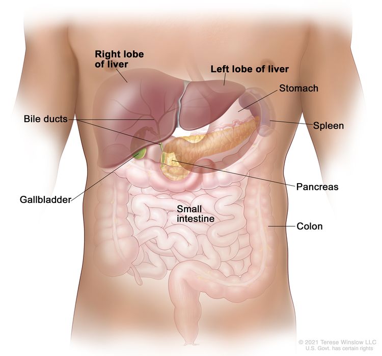
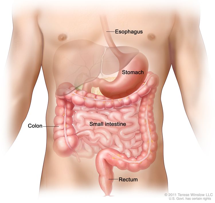
EnlargeAnatomy of the liver; drawing shows the right and left lobes of the liver. Also shown are the bile ducts, gallbladder, stomach, spleen, pancreas, small intestine, and colon.
Anatomy of the liver. The liver is in the upper abdomen near the stomach, intestines, gallbladder, and pancreas. The liver has a right lobe and a left lobe. Each lobe is divided into two sections (not shown).

Risk Factors

Increasing age is the most important risk factor for most cancers. Other risk factors for liver (hepatocellular) cancer include:

  • Chronic and/or persistent infection with hepatitis B and/or hepatitis C.[47]
  • Cirrhosis.[5,7,8]
  • Heavy alcohol use.[6,8,9]
  • Ingestion of foods contaminated with aflatoxin B1.[1013]
  • Nonalcoholic steatohepatitis (NASH).[1417]
  • Tobacco use.[1820]
  • Certain inherited or rare disorders that include:
    • Hereditary hemochromatosis.[7,10]
    • Alpha-1 antitrypsin deficiency.[21]
    • Glycogen storage disease.[10]
    • Porphyria cutanea tarda.[10]
    • Wilson disease.[10,22,23]

For more information, see Liver (Hepatocellular) Cancer Prevention.

Screening

For more information, see Liver (Hepatocellular) Cancer Screening.

Diagnostic Factors

Lesions smaller than 1 cm that are detected during screening in patients at high risk of HCC do not require further diagnostic evaluation. Most of these lesions will be cirrhotic lesions rather than HCC.[24][Level of evidence C1] Close follow-up at 3-month intervals is a common surveillance strategy, using the same technique that first documented the presence of the lesions.

For patients with liver lesions larger than 1 cm who are at risk of HCC, a diagnosis can be considered. The tests required to diagnose HCC may include imaging, biopsy, or both.

Diagnostic imaging

In patients with cirrhosis, liver disease, or other risk factors for HCC, and with lesions greater than 1 cm, triple-phase, contrast-enhanced studies (dynamic computed tomography [CT] or magnetic resonance imaging [MRI]) can be used to diagnose HCC.[25]

A triple-phase CT or MRI assesses the entire liver in distinct phases of perfusion. Following the controlled administration of intravenous contrast media, the arterial and venous phases of perfusion are imaged.

During the arterial phase of the study, HCC enhances more intensely than the surrounding liver because the arterial blood in the liver is diluted by venous blood that does not contain contrast, whereas the HCC contains only arterial blood. In the venous phase, the HCC enhances less than the surrounding liver (which is referred to as the venous washout of HCC), because the arterial blood flowing through the lesion no longer contains contrast; however, the portal blood in the liver now contains contrast.

The presence of arterial uptake followed by washout in a single dynamic study is highly specific (95%–100%) for HCC of 1 to 3 cm in diameter and virtually diagnostic of HCC.[2628][Level of evidence C1] In these cases, the diagnosis of HCC may be established without a second imaging modality, even in the absence of a biopsy confirmation.[2830][Level of evidence C1]

However, if a first imaging modality, such as a contrast-enhanced CT or MRI, is not conclusive, sequential imaging with a different modality can improve sensitivity for HCC detection (from 33% to 41% for either CT or MRI to 76% for both studies when performed sequentially) without a decrease in specificity.[27]

If, despite the use of two imaging modalities, a lesion larger than 1 cm remains uncharacterized in a patient at high risk of HCC (i.e., with no or only one classic enhancement pattern), a liver biopsy can be considered.[28,29]

Liver biopsy

A liver biopsy may be performed when a diagnosis of HCC is not established by a dynamic imaging modality (three-phase CT or MRI) for liver lesions 1 cm or larger in high-risk patients.

Alpha-fetoprotein (AFP) levels

AFP is insufficiently sensitive or specific for use as a diagnostic assay. AFP can be elevated in intrahepatic cholangiocarcinoma and in some cases in which there are metastases from colon cancer. Finding a mass in the liver of a patient with an elevated AFP does not automatically indicate HCC. However, if the AFP level is high, it can be used to monitor for recurrence.

Prognosis

The natural course of early tumors is poorly understood because most HCC patients receive treatment. However, older reports have described 3-year survival rates of 13% to 21% in patients who do not receive any specific treatment.[31,32] At present, only 10% to 23% of patients with HCC may be surgical candidates for curative-intent treatment.[33,34] The 5-year overall survival (OS) rate for patients with early HCC who undergo liver transplant is 44% to 78%. For patients who undergo a liver resection, the OS rate is 27% to 70%.[35]

Liver transplant, surgical resection, and ablation offer high rates of complete responses and a potential for cure in patients with early HCC.[29]

The natural course of advanced-stage HCC is better known. Untreated patients with advanced disease usually survive less than 6 months.[36] The survival rate of untreated patients in 25 randomized clinical trials ranged from 10% to 72% at 1 year and 8% to 50% at 2 years.[37]

Unlike most patients with solid tumors, the prognosis of patients with HCC is affected by the tumor stage at presentation and by the underlying liver function. The following prognostic factors guide the selection of treatment:

  • Anatomical extension of the tumor (i.e., tumor size, number of lesions, presence of vascular invasion, and extrahepatic spread).
  • Performance status.
  • Functional hepatic reserve based on the Child-Pugh score.[36,38,39]
References
  1. American Cancer Society: Cancer Facts and Figures 2025. American Cancer Society, 2025. Available online. Last accessed January 16, 2025.
  2. Altekruse SF, McGlynn KA, Reichman ME: Hepatocellular carcinoma incidence, mortality, and survival trends in the United States from 1975 to 2005. J Clin Oncol 27 (9): 1485-91, 2009. [PUBMED Abstract]
  3. Forner A, Llovet JM, Bruix J: Hepatocellular carcinoma. Lancet 379 (9822): 1245-55, 2012. [PUBMED Abstract]
  4. Bosetti C, Turati F, La Vecchia C: Hepatocellular carcinoma epidemiology. Best Pract Res Clin Gastroenterol 28 (5): 753-70, 2014. [PUBMED Abstract]
  5. El-Serag HB: Epidemiology of viral hepatitis and hepatocellular carcinoma. Gastroenterology 142 (6): 1264-1273.e1, 2012. [PUBMED Abstract]
  6. El-Serag HB, Kanwal F: Epidemiology of hepatocellular carcinoma in the United States: where are we? Where do we go? Hepatology 60 (5): 1767-75, 2014. [PUBMED Abstract]
  7. Lafaro KJ, Demirjian AN, Pawlik TM: Epidemiology of hepatocellular carcinoma. Surg Oncol Clin N Am 24 (1): 1-17, 2015. [PUBMED Abstract]
  8. Fattovich G, Stroffolini T, Zagni I, et al.: Hepatocellular carcinoma in cirrhosis: incidence and risk factors. Gastroenterology 127 (5 Suppl 1): S35-50, 2004. [PUBMED Abstract]
  9. Grewal P, Viswanathen VA: Liver cancer and alcohol. Clin Liver Dis 16 (4): 839-50, 2012. [PUBMED Abstract]
  10. London WT, McGlynn K: Liver cancer. In: Schottenfeld D, Fraumeni JF Jr, eds.: Cancer Epidemiology and Prevention. 3rd ed. Oxford University Press, 2006, pp 763-86.
  11. McGlynn KA, Petrick JL, London WT: Global epidemiology of hepatocellular carcinoma: an emphasis on demographic and regional variability. Clin Liver Dis 19 (2): 223-38, 2015. [PUBMED Abstract]
  12. Liu Y, Wu F: Global burden of aflatoxin-induced hepatocellular carcinoma: a risk assessment. Environ Health Perspect 118 (6): 818-24, 2010. [PUBMED Abstract]
  13. Chen JG, Egner PA, Ng D, et al.: Reduced aflatoxin exposure presages decline in liver cancer mortality in an endemic region of China. Cancer Prev Res (Phila) 6 (10): 1038-45, 2013. [PUBMED Abstract]
  14. Baffy G, Brunt EM, Caldwell SH: Hepatocellular carcinoma in non-alcoholic fatty liver disease: an emerging menace. J Hepatol 56 (6): 1384-91, 2012. [PUBMED Abstract]
  15. Diehl AM, Day C: Cause, Pathogenesis, and Treatment of Nonalcoholic Steatohepatitis. N Engl J Med 377 (21): 2063-2072, 2017. [PUBMED Abstract]
  16. White DL, Kanwal F, El-Serag HB: Association between nonalcoholic fatty liver disease and risk for hepatocellular cancer, based on systematic review. Clin Gastroenterol Hepatol 10 (12): 1342-1359.e2, 2012. [PUBMED Abstract]
  17. Ascha MS, Hanouneh IA, Lopez R, et al.: The incidence and risk factors of hepatocellular carcinoma in patients with nonalcoholic steatohepatitis. Hepatology 51 (6): 1972-8, 2010. [PUBMED Abstract]
  18. Chuang SC, Lee YC, Hashibe M, et al.: Interaction between cigarette smoking and hepatitis B and C virus infection on the risk of liver cancer: a meta-analysis. Cancer Epidemiol Biomarkers Prev 19 (5): 1261-8, 2010. [PUBMED Abstract]
  19. Lee YC, Cohet C, Yang YC, et al.: Meta-analysis of epidemiologic studies on cigarette smoking and liver cancer. Int J Epidemiol 38 (6): 1497-511, 2009. [PUBMED Abstract]
  20. Koh WP, Robien K, Wang R, et al.: Smoking as an independent risk factor for hepatocellular carcinoma: the Singapore Chinese Health Study. Br J Cancer 105 (9): 1430-5, 2011. [PUBMED Abstract]
  21. Lomas DA, Evans DL, Finch JT, et al.: The mechanism of Z alpha 1-antitrypsin accumulation in the liver. Nature 357 (6379): 605-7, 1992. [PUBMED Abstract]
  22. Huster D: Wilson disease. Best Pract Res Clin Gastroenterol 24 (5): 531-9, 2010. [PUBMED Abstract]
  23. Pfeiffenberger J, Mogler C, Gotthardt DN, et al.: Hepatobiliary malignancies in Wilson disease. Liver Int 35 (5): 1615-22, 2015. [PUBMED Abstract]
  24. Furuya K, Nakamura M, Yamamoto Y, et al.: Macroregenerative nodule of the liver. A clinicopathologic study of 345 autopsy cases of chronic liver disease. Cancer 61 (1): 99-105, 1988. [PUBMED Abstract]
  25. Brunello F, Cantamessa A, Gaia S, et al.: Radiofrequency ablation: technical and clinical long-term outcomes for single hepatocellular carcinoma up to 30 mm. Eur J Gastroenterol Hepatol 25 (7): 842-9, 2013. [PUBMED Abstract]
  26. Leoni S, Piscaglia F, Golfieri R, et al.: The impact of vascular and nonvascular findings on the noninvasive diagnosis of small hepatocellular carcinoma based on the EASL and AASLD criteria. Am J Gastroenterol 105 (3): 599-609, 2010. [PUBMED Abstract]
  27. Khalili K, Kim TK, Jang HJ, et al.: Optimization of imaging diagnosis of 1-2 cm hepatocellular carcinoma: an analysis of diagnostic performance and resource utilization. J Hepatol 54 (4): 723-8, 2011. [PUBMED Abstract]
  28. Sangiovanni A, Manini MA, Iavarone M, et al.: The diagnostic and economic impact of contrast imaging techniques in the diagnosis of small hepatocellular carcinoma in cirrhosis. Gut 59 (5): 638-44, 2010. [PUBMED Abstract]
  29. Bruix J, Sherman M; American Association for the Study of Liver Diseases: Management of hepatocellular carcinoma: an update. Hepatology 53 (3): 1020-2, 2011. [PUBMED Abstract]
  30. Khalili K, Kim TK, Jang HJ, et al.: Implementation of AASLD hepatocellular carcinoma practice guidelines in North America: two years of experience. [Abstract] Hepatology 48 (Suppl 1): A-128, 362A, 2008.
  31. Barbara L, Benzi G, Gaiani S, et al.: Natural history of small untreated hepatocellular carcinoma in cirrhosis: a multivariate analysis of prognostic factors of tumor growth rate and patient survival. Hepatology 16 (1): 132-7, 1992. [PUBMED Abstract]
  32. Ebara M, Ohto M, Shinagawa T, et al.: Natural history of minute hepatocellular carcinoma smaller than three centimeters complicating cirrhosis. A study in 22 patients. Gastroenterology 90 (2): 289-98, 1986. [PUBMED Abstract]
  33. Shah SA, Smith JK, Li Y, et al.: Underutilization of therapy for hepatocellular carcinoma in the medicare population. Cancer 117 (5): 1019-26, 2011. [PUBMED Abstract]
  34. Sonnenday CJ, Dimick JB, Schulick RD, et al.: Racial and geographic disparities in the utilization of surgical therapy for hepatocellular carcinoma. J Gastrointest Surg 11 (12): 1636-46; discussion 1646, 2007. [PUBMED Abstract]
  35. Dhir M, Lyden ER, Smith LM, et al.: Comparison of outcomes of transplantation and resection in patients with early hepatocellular carcinoma: a meta-analysis. HPB (Oxford) 14 (9): 635-45, 2012. [PUBMED Abstract]
  36. Okuda K, Ohtsuki T, Obata H, et al.: Natural history of hepatocellular carcinoma and prognosis in relation to treatment. Study of 850 patients. Cancer 56 (4): 918-28, 1985. [PUBMED Abstract]
  37. Llovet JM, Bruix J: Systematic review of randomized trials for unresectable hepatocellular carcinoma: Chemoembolization improves survival. Hepatology 37 (2): 429-42, 2003. [PUBMED Abstract]
  38. Llovet JM, Brú C, Bruix J: Prognosis of hepatocellular carcinoma: the BCLC staging classification. Semin Liver Dis 19 (3): 329-38, 1999. [PUBMED Abstract]
  39. A new prognostic system for hepatocellular carcinoma: a retrospective study of 435 patients: the Cancer of the Liver Italian Program (CLIP) investigators. Hepatology 28 (3): 751-5, 1998. [PUBMED Abstract]

Cellular Classification of Primary Liver Cancer

Malignant primary tumors of the liver consist of two major cell types, hepatocellular (90% of cases) and cholangiocarcinoma.[1]

Histological classification is as follows:

  • Hepatocellular carcinoma (HCC; liver cell carcinoma).
  • Fibrolamellar variant of HCC.

    It is important to distinguish between the fibrolamellar variant of HCC and HCC itself because an increased proportion of patients with the fibrolamellar variant may be cured if the tumor can be resected. Found more frequently in young women, this variant generally exhibits a slower clinical course than the more common HCC.[2]

  • Cholangiocarcinoma (intrahepatic bile duct carcinoma).
  • Mixed hepatocellular cholangiocarcinoma.
  • Undifferentiated.
  • Hepatoblastoma. This occurs more often in children than in adults. For more information, see Childhood Liver Cancer Treatment.
References
  1. Llovet JM, Burroughs A, Bruix J: Hepatocellular carcinoma. Lancet 362 (9399): 1907-17, 2003. [PUBMED Abstract]
  2. Mavros MN, Mayo SC, Hyder O, et al.: A systematic review: treatment and prognosis of patients with fibrolamellar hepatocellular carcinoma. J Am Coll Surg 215 (6): 820-30, 2012. [PUBMED Abstract]

Stage Information for Primary Liver Cancer

Prognostic modeling in hepatocellular carcinoma (HCC) is complex because cirrhosis is involved in as many as 80% of cases. Tumor features and the factors related to functional hepatic reserve must be considered. The key prognostic factors are only partially known and vary at different stages of the disease.

More than ten classifications are used throughout the world, but no system is accepted worldwide. New classifications have been proposed to overcome the difficulties of having several staging systems.

This summary discusses the following three staging systems:

Barcelona Clinic Liver Cancer (BCLC) Staging System

Currently, the BCLC staging classification is the most accepted staging system for HCC and is useful in the staging of early tumors. Evidence from an American cohort has shown that BCLC staging offers better prognostic stratification power than other staging systems.[1]

The BCLC staging system attempts to overcome the limitations of previous staging systems by including variables related to the following:[2]

  • Tumor stage.
  • Functional status of the liver.
  • Physical status.
  • Cancer-related symptoms.

Five stages (0 and A through D) are identified based on the variables mentioned above. The BCLC staging system links each HCC stage to appropriate treatment modalities as follows:

  • Patients with early-stage HCC may benefit from curative therapies (i.e., liver transplant, surgical resection, and radiofrequency ablation).
  • Patients with intermediate-stage or advanced-stage disease may benefit from palliative treatments (i.e., transcatheter arterial chemoembolization and sorafenib).
  • Patients with end-stage disease who have a very poor life expectancy are offered supportive care and palliation.

Okuda Staging System

The Okuda staging system has been extensively used in the past and includes variables related to tumor burden and liver function, such as bilirubin, albumin, and ascites. However, many significant prognostic tumor factors confirmed in both surgical and nonsurgical series (e.g., unifocal or multifocal, vascular invasion, portal venous thrombosis, or locoregional lymph node involvement) are not included.[3,4] As a result, Okuda staging is unable to stratify prognosis for early-stage cancers and mostly serves to recognize end-stage disease.

AJCC Staging System and TNM Definitions

The TNM (tumor, node, metastasis) classification for staging, proposed by the AJCC, is not widely used for liver cancer. Clinical use of TNM staging is limited because liver function is not considered. It is also difficult to use this system to select treatment options because TNM staging relies on detailed histopathological examination available only after tumor excision. TNM may be useful in prognostic prediction after liver resection.[5]

Table 1. Definitions of TNM Stages IA and IBa
Stage TNM Description
Tumor = primary tumor; N = regional lymph nodes; M = distant metastasis.
aReprinted with permission from AJCC: Liver. In: Amin MB, Edge SB, Greene FL, et al., eds.: AJCC Cancer Staging Manual. 8th ed. New York, NY: Springer, 2017, pp. 287–93.
IA T1a, N0, M0 T1a = Solitary tumor ≤2 cm.
N0 = No regional lymph node metastasis.
M0 = No distant metastasis.
IB T1b, N0, M0 T1b = Solitary tumor >2 cm without vascular invasion.
N0 = No regional lymph node metastasis.
M0 = No distant metastasis.
Table 2. Definitions of TNM Stage IIa
Stage TNM Description
T = primary tumor; N = regional lymph nodes; M = distant metastasis.
aReprinted with permission from AJCC: Liver. In: Amin MB, Edge SB, Greene FL, et al., eds.: AJCC Cancer Staging Manual. 8th ed. New York, NY: Springer, 2017, pp. 287–93.
II T2, N0, M0 T2 = Solitary tumor >2 cm with vascular invasion, or multiple tumors, none >5 cm.
N0 = No regional lymph node metastasis.
M0 = No distant metastasis.
Table 3. Definitions of TNM Stages IIIA and IIIBa
Stage TNM Description
T = primary tumor; N = regional lymph nodes; M = distant metastasis.
aReprinted with permission from AJCC: Liver. In: Amin MB, Edge SB, Greene FL, et al., eds.: AJCC Cancer Staging Manual. 8th ed. New York, NY: Springer, 2017, pp. 287–93.
IIIA T3, N0, M0 T3 = Multiple tumors, at least one of which is >5 cm.
N0 = No regional lymph node metastasis.
M0 = No distant metastasis.
IIIB T4, N0, M0 T4 = Single tumor or multiple tumors of any size involving a major branch of the portal vein or hepatic vein, or tumor(s) with direct invasion of adjacent organs other than the gallbladder or with perforation of visceral peritoneum.
N0 = No regional lymph node metastasis.
M0 = No distant metastasis.
Table 4. Definitions of TNM Stages IVA and IVBa
Stage TNM Description
T = primary tumor; N = regional lymph nodes; M = distant metastasis.
aReprinted with permission from AJCC: Liver. In: Amin MB, Edge SB, Greene FL, et al., eds.: AJCC Cancer Staging Manual. 8th ed. New York, NY: Springer, 2017, pp. 287–93.
IVA Any T, N1, M0 TX = Primary tumor cannot be assessed.
T0 = No evidence of primary tumor.
T1 = Solitary tumor ≤2 cm, or >2 cm without vascular invasion.
–T1a = Solitary tumor ≤2 cm.
–T1b = Solitary tumor >2 cm without vascular invasion.
T2 = Solitary tumor >2 cm with vascular invasion, or multiple tumors, none >5 cm.
T3 = Multiple tumors, at least one of which is >5 cm.
T4 = Single tumor or multiple tumors of any size involving a major branch of the portal vein or hepatic vein, or tumor(s) with direct invasion of adjacent organs other than the gallbladder or with perforation of visceral peritoneum.
N1 = Regional lymph node metastasis.
M0 = No distant metastasis.
IVB Any T, Any N, M1 Any T = See descriptions above in this table, stage IVA, Any T, N1, M0.
NX = Regional lymph nodes cannot be assessed.
N0 = No regional lymph node metastasis.
N1 = Regional lymph node metastasis.
M1 = Distant metastasis.
References
  1. Marrero JA, Fontana RJ, Barrat A, et al.: Prognosis of hepatocellular carcinoma: comparison of 7 staging systems in an American cohort. Hepatology 41 (4): 707-16, 2005. [PUBMED Abstract]
  2. Llovet JM, Brú C, Bruix J: Prognosis of hepatocellular carcinoma: the BCLC staging classification. Semin Liver Dis 19 (3): 329-38, 1999. [PUBMED Abstract]
  3. Poon RT, Ng IO, Fan ST, et al.: Clinicopathologic features of long-term survivors and disease-free survivors after resection of hepatocellular carcinoma: a study of a prospective cohort. J Clin Oncol 19 (12): 3037-44, 2001. [PUBMED Abstract]
  4. Pompili M, Rapaccini GL, Covino M, et al.: Prognostic factors for survival in patients with compensated cirrhosis and small hepatocellular carcinoma after percutaneous ethanol injection therapy. Cancer 92 (1): 126-35, 2001. [PUBMED Abstract]
  5. Liver. In: Amin MB, Edge SB, Greene FL, et al., eds.: AJCC Cancer Staging Manual. 8th ed. Springer; 2017, pp. 287–93.

Treatment Option Overview for Primary Liver Cancer

There is no agreement on a single treatment strategy for patients with hepatocellular carcinoma (HCC). Selection of treatment is complex due to several factors, including:

  • Underlying liver function.
  • Extent and location of the tumor.
  • General condition of the patient.

Several treatments for HCC are associated with long-term survival, including surgical resection, liver transplant, and ablation. There are no large, robust, randomized studies that compare treatments considered effective for early-stage disease, nor are there studies comparing these treatments with best supportive care. Often, patients with HCC are evaluated by a multidisciplinary team that includes hepatologists, radiologists, interventional radiologists, radiation oncologists, transplant surgeons, surgical oncologists, pathologists, and medical oncologists.

Best survivals are achieved when the HCC can be removed either by surgical resection or liver transplant. Surgical resection is usually performed in patients with localized HCC and enough functional hepatic reserve.

For patients with decompensated cirrhosis and a solitary lesion (<5 cm) or early multifocal disease (≤3 lesions, ≤3 cm in diameter), the best option is liver transplant.[1] However, the limited availability of liver donors restricts the use of this approach.

Transarterial chemoembolization, multikinase inhibitors, and immunotherapy are noncurative treatments for HCC that improve survival.[24]

Table 5 shows the standard treatment options for HCC.

Table 5. Treatment Options for HCC
Stage Treatment Options
Localized Surveillance
Surgical resection
Liver transplant
Ablation
Radiation therapy
Locally advanced or metastatic Transarterial embolization and transcatheter arterial chemoembolization in patients with nonmetastatic disease
First-line systemic therapy
Second-line systemic therapy
Radiation therapy
Recurrent (liver-limited disease without vascular involvement) Liver transplant
Surgical resection
Ablation
Radiation therapy
Recurrent (extrahepatic disease or vascular involvement) Palliative therapy
References
  1. Bruix J, Sherman M; American Association for the Study of Liver Diseases: Management of hepatocellular carcinoma: an update. Hepatology 53 (3): 1020-2, 2011. [PUBMED Abstract]
  2. Llovet JM, Ricci S, Mazzaferro V, et al.: Sorafenib in advanced hepatocellular carcinoma. N Engl J Med 359 (4): 378-90, 2008. [PUBMED Abstract]
  3. Llovet JM, Bruix J: Systematic review of randomized trials for unresectable hepatocellular carcinoma: Chemoembolization improves survival. Hepatology 37 (2): 429-42, 2003. [PUBMED Abstract]
  4. Cammà C, Schepis F, Orlando A, et al.: Transarterial chemoembolization for unresectable hepatocellular carcinoma: meta-analysis of randomized controlled trials. Radiology 224 (1): 47-54, 2002. [PUBMED Abstract]

Treatment of Localized Primary Liver Cancer

About 30% of hepatocellular carcinoma (HCC) cases present as localized disease, with a solitary mass in part of the liver or as a limited number of tumors (≤3 lesions, ≤3 cm in diameter) without major vascular invasion.

Treatment Options for Localized Primary Liver Cancer

Treatment options for localized primary liver cancer include:

Resection and transplant achieve the best outcomes in well-selected candidates, and are usually considered the first option for curative intent.

Surveillance

Surveillance is an option for patients at high risk of HCC with lesions smaller than 1 cm detected during screening.[1][Level of evidence C1] Close follow-up at 3-month intervals is a common surveillance strategy, using the same technique that first documented the presence of the lesions.

Surgical resection

Surgery is the mainstay of HCC treatment.

Preoperative assessment includes three-phase helical computed tomography, magnetic resonance imaging, or both to determine the presence of an extension of a tumor across interlobar planes and potential involvement of the hepatic hilus, hepatic veins, and inferior vena cava. Tumors can be resected only if enough liver parenchyma can be spared with adequate vascular and biliary inflow and outflow. Patients with well-compensated cirrhosis can generally tolerate resection of up to 50% of their liver parenchyma.

Surgical resection can be considered for patients who meet the following criteria:

  • A solitary mass.
  • Good performance status.
  • Normal or minimally abnormal liver function tests.
  • No evidence of portal hypertension.
  • No evidence of cirrhosis beyond Child-Pugh class A.

After considering the location and number of tumors and the patient’s hepatic function, only 5% to 10% of patients with liver cancer will prove to have localized disease amenable to resection.[26]

The principles of surgical resection involve obtaining a clear margin around the tumor, which may require any of the following procedures:

  • Segmental resection.
  • Hormone-lymphatic lobectomy.
  • Extended lobectomy.

The 5-year overall survival (OS) rate after curative resection ranges between 27% and 70% and depends on tumor stage and underlying liver function.[26]

In patients with limited multifocal disease, hepatic resection is controversial.

Liver transplant

Liver transplant is a potentially curative therapy for HCC and has the benefit of treating the underlying cirrhosis, but the scarcity of organ donors limits the availability of this treatment modality.[2]

According to the Milan criteria, patients with a single HCC lesion smaller than 5 cm, or 2 to 3 lesions smaller than 3 cm are eligible for liver transplant. Expansion of the accepted transplant criteria for HCC is not supported by consistent data. Liver transplant is considered if resection is precluded because of multiple small tumor lesions (≤3 lesions, each ≤3 cm), or impaired liver function (Child-Pugh class B and class C). In patients who meet the criteria, transplant is associated with a 5-year OS rate of approximately 70%.[7][Level of evidence C1]

Ablation

When tumor excision, either by transplant or resection, is not feasible or advisable, ablation may be used if the tumor can be accessed percutaneously or, if necessary, through minimally invasive or open surgery. Ablation may be particularly useful for patients with early-stage HCC that is centrally located in the liver and cannot be surgically removed without excessive sacrifice of functional parenchyma.

Ablation can be achieved in the following ways:

  • Change in temperature (e.g., radiofrequency ablation [RFA], microwave, or cryoablation).
  • Exposure to a chemical substance (e.g., percutaneous ethanol injection [PEI]).
  • Direct damage of the cellular membrane (definitive electroporation).

With ablation, a margin of normal liver around the tumor can be considered. Ablation is relatively contraindicated for lesions near bile ducts, the diaphragm, or other intra-abdominal organs that might be injured during the procedure. Furthermore, when tumors are located adjacent to major vessels, the blood flow in the vessels may keep thermal ablation techniques, such as RFA, from reaching optimal temperatures. This is known as the heat-sink effect, which may preclude complete tumor necrosis.

RFA achieves best results in patients with tumors smaller than 3 cm. In this subpopulation, 5-year OS rates may be as high as 59%, and the recurrence-free survival rates may not differ significantly from treatment with hepatic resection.[8,9] Local control success progressively diminishes as the tumor size increases beyond 3 cm.

PEI yields good results in patients with Child-Pugh class A cirrhosis and a single tumor smaller than 3 cm in diameter. In those cases, the 5-year OS rate can be as high as 40% to 59%.[10,11][Level of evidence C2]

In the few randomized controlled trials that included patients with Child-Pugh class A cirrhosis, RFA proved superior to PEI in rates of complete response and local recurrences. Some of those studies have also shown improved OS with RFA. Furthermore, RFA requires fewer treatment sessions than PEI to achieve comparable outcomes.[1215]

Of note, RFA may have higher complication rates than PEI,[13] but both techniques are associated with lower complication rates than excision procedures. RFA is a well-established technique in the treatment of HCC.

Radiation therapy

Radiation therapy can be delivered with curative or palliative intent for patients with primary liver cancer. One form of radiation, stereotactic body radiation therapy (SBRT), treats patients with a small number of fractions of precise, image-guided radiation therapy at a high biologically equivalent dose. Numerous retrospective studies have shown excellent local control for patients with HCC who receive SBRT (local control rates ranging from 70%–95% at 2 years for smaller HCCs).

Evidence (curative radiation therapy):

  1. The phase III NRG/RTOG 1112 study (NCT01730937) evaluated sorafenib alone or SBRT followed by sorafenib in patients with HCC. Patients were included if they had Child-Pugh class A, Barcelona Clinic Liver Cancer stage B or C, new or recurrent HCC. Patients also had five or fewer lesions, a tumor sum measuring 20 cm or less, and distant metastases measuring 3 cm or less. A total of 177 patients were randomly assigned (92 to sorafenib alone, 85 to SBRT followed by sorafenib). The primary end point was OS.[16][Level of evidence B1]
    • The median OS was 12.3 months for the sorafenib-alone group and 15.8 months for the SBRT-plus-sorafenib group (hazard ratio [HR], 0.77; one-sided P = .0554). This was not statistically significant.
    • Grade 3 or higher adverse events were not significantly different between the two groups: 42% for the sorafenib-alone group and 47% for the SBRT-plus-sorafenib group (P = .52).
    • There were three grade 5 adverse events: one hepatic failure and one death not otherwise specified in the sorafenib-alone group and one grade 5 lung infection in the SBRT-plus-sorafenib group.

Based on these results, SBRT is a standard of care treatment with curative intent for HCC. It can also be used to provide local control before liver transplant.

Evidence (radiation therapy for palliation):

  1. The Canadian Cancer Trials Group HE.1 trial (NCT02511522), a phase III study published in abstract form, included 66 patients with HCC. Patients were randomly assigned to receive either best supportive care alone or palliative radiation therapy to the liver (8 Gy in one fraction). Patients had end-stage disease unsuitable for local, regional, or systemic therapies. Patients also had to be more than 4 weeks from receiving chemotherapy or transcatheter arterial chemoembolization, more than 2 weeks from receiving targeted therapy or immunotherapy, and not planning any systemic therapy. The primary outcome of the study was the proportion of patients who reported an improvement of at least two points from baseline on the Brief Pain Inventory when asked to rate their liver cancer pain “intensity at worst.” A secondary end point included 3-month OS.[17][Level of evidence B3]
    • A significant improvement in the “worst” pain score from baseline to 1 month was seen in 67% of patients who received radiation therapy and 22% of patients who received best supportive care.
    • Although radiation therapy has not been historically used in this patient population, this study showed no decrease in OS for patients who received radiation. The 3-month OS rate was 51% for patients who received radiation therapy and 33% for patients who received best supportive care alone (P = .07), despite the study including patients with Child-Pugh class A, B, and C cirrhosis.

Current Clinical Trials

Use our advanced clinical trial search to find NCI-supported cancer clinical trials that are now enrolling patients. The search can be narrowed by location of the trial, type of treatment, name of the drug, and other criteria. General information about clinical trials is also available.

References
  1. Furuya K, Nakamura M, Yamamoto Y, et al.: Macroregenerative nodule of the liver. A clinicopathologic study of 345 autopsy cases of chronic liver disease. Cancer 61 (1): 99-105, 1988. [PUBMED Abstract]
  2. Llovet JM, Fuster J, Bruix J: Intention-to-treat analysis of surgical treatment for early hepatocellular carcinoma: resection versus transplantation. Hepatology 30 (6): 1434-40, 1999. [PUBMED Abstract]
  3. Chok KS, Ng KK, Poon RT, et al.: Impact of postoperative complications on long-term outcome of curative resection for hepatocellular carcinoma. Br J Surg 96 (1): 81-7, 2009. [PUBMED Abstract]
  4. Kianmanesh R, Regimbeau JM, Belghiti J: Selective approach to major hepatic resection for hepatocellular carcinoma in chronic liver disease. Surg Oncol Clin N Am 12 (1): 51-63, 2003. [PUBMED Abstract]
  5. Poon RT, Fan ST, Lo CM, et al.: Long-term survival and pattern of recurrence after resection of small hepatocellular carcinoma in patients with preserved liver function: implications for a strategy of salvage transplantation. Ann Surg 235 (3): 373-82, 2002. [PUBMED Abstract]
  6. Dhir M, Lyden ER, Smith LM, et al.: Comparison of outcomes of transplantation and resection in patients with early hepatocellular carcinoma: a meta-analysis. HPB (Oxford) 14 (9): 635-45, 2012. [PUBMED Abstract]
  7. Hemming AW, Cattral MS, Reed AI, et al.: Liver transplantation for hepatocellular carcinoma. Ann Surg 233 (5): 652-9, 2001. [PUBMED Abstract]
  8. Huang J, Hernandez-Alejandro R, Croome KP, et al.: Radiofrequency ablation versus surgical resection for hepatocellular carcinoma in Childs A cirrhotics-a retrospective study of 1,061 cases. J Gastrointest Surg 15 (2): 311-20, 2011. [PUBMED Abstract]
  9. Zhou YM, Shao WY, Zhao YF, et al.: Meta-analysis of laparoscopic versus open resection for hepatocellular carcinoma. Dig Dis Sci 56 (7): 1937-43, 2011. [PUBMED Abstract]
  10. Huang GT, Lee PH, Tsang YM, et al.: Percutaneous ethanol injection versus surgical resection for the treatment of small hepatocellular carcinoma: a prospective study. Ann Surg 242 (1): 36-42, 2005. [PUBMED Abstract]
  11. Yamamoto J, Okada S, Shimada K, et al.: Treatment strategy for small hepatocellular carcinoma: comparison of long-term results after percutaneous ethanol injection therapy and surgical resection. Hepatology 34 (4 Pt 1): 707-13, 2001. [PUBMED Abstract]
  12. Lencioni RA, Allgaier HP, Cioni D, et al.: Small hepatocellular carcinoma in cirrhosis: randomized comparison of radio-frequency thermal ablation versus percutaneous ethanol injection. Radiology 228 (1): 235-40, 2003. [PUBMED Abstract]
  13. Lin SM, Lin CJ, Lin CC, et al.: Randomised controlled trial comparing percutaneous radiofrequency thermal ablation, percutaneous ethanol injection, and percutaneous acetic acid injection to treat hepatocellular carcinoma of 3 cm or less. Gut 54 (8): 1151-6, 2005. [PUBMED Abstract]
  14. Brunello F, Veltri A, Carucci P, et al.: Radiofrequency ablation versus ethanol injection for early hepatocellular carcinoma: A randomized controlled trial. Scand J Gastroenterol 43 (6): 727-35, 2008. [PUBMED Abstract]
  15. Shiina S, Teratani T, Obi S, et al.: A randomized controlled trial of radiofrequency ablation with ethanol injection for small hepatocellular carcinoma. Gastroenterology 129 (1): 122-30, 2005. [PUBMED Abstract]
  16. Dawson LA, Winter KA, Knox JJ, et al.: Stereotactic Body Radiotherapy vs Sorafenib Alone in Hepatocellular Carcinoma: The NRG Oncology/RTOG 1112 Phase 3 Randomized Clinical Trial. JAMA Oncol 11 (2): 136-144, 2025. [PUBMED Abstract]
  17. Dawson LA, Fairchild AM, Dennis K, et al.: Canadian Cancer Trials Group HE.1: A phase III study of palliative radiotherapy for symptomatic hepatocellular carcinoma and liver metastases. [Abstract] J Clin Oncol 41 (Suppl 4): A-LBA492, 2023.

Treatment of Locally Advanced or Metastatic Primary Liver Cancer

Treatment Options for Locally Advanced or Metastatic Primary Liver Cancer

Treatment options for locally advanced or metastatic primary liver cancer not amenable to surgical or locoregional interventions include:

Transarterial embolization (TAE) and transcatheter arterial chemoembolization (TACE) in patients with nonmetastatic disease

TAE is the most widely used primary treatment for hepatocellular carcinoma (HCC) not amenable to curative treatment by excision or ablation. Most of the blood supply to the normal liver parenchyma comes from the portal vein, whereas blood flow to the tumor comes mainly from the hepatic artery. Furthermore, HCC tumors are generally hypervascular compared with the surrounding normal parenchyma. The obstruction of the arterial branch(es) feeding the tumor may reduce the blood flow to the tumor and result in tumor ischemia and necrosis.

Embolization agents, such as microspheres and particles, may also be administered along with concentrated doses of chemotherapeutic agents (generally doxorubicin or cisplatin) mixed with lipiodol or other emulsifying agents during chemoembolization, arterial chemoembolization (usually via percutaneous access), and TACE. TAE-TACE is considered for patients with HCC not amenable to surgery or percutaneous ablation in the absence of extrahepatic disease.

In patients with cirrhosis, any interference with arterial blood supply may be associated with significant morbidity and is relatively contraindicated in the presence of portal hypertension, portal vein thrombosis, or clinical jaundice. In patients with liver decompensation, TAE-TACE could increase the risk of liver failure.

A number of randomized controlled trials have compared TAE and TACE with supportive care.[1] Those trials have been heterogeneous in terms of patient baseline demographics and treatment. The survival advantage of TAE-TACE over supportive care has been demonstrated by two trials.[2,3] No standardized approach for TAE has been determined (e.g., embolizing agent, chemotherapy agent and dose, and treatment schedule). However, a meta-analysis has shown that TAE-TACE improves survival more than supportive treatment.[1]

The use of drug-eluting beads (DEB) for TACE may reduce the systemic side effects of chemotherapy and may increase objective tumor response.[47] Only one study suggested that DEB-TACE may offer an advantage in overall survival (OS).[8]

First-line systemic therapy

Historically, sorafenib (a multikinase inhibitor) has been the standard of care for patients with advanced HCC and intact liver function (Child-Pugh class A) who were not candidates for locoregional therapy. This standard was based on the results of the SHARP trial, which showed improved OS for patients who received sorafenib compared with placebo (10.7 vs. 7.9 months; hazard ratio [HR], 0.69; P < .001). However, treatment-related adverse events may make adherence to sorafenib regimens difficult, especially in a population with concurrent liver disease. Since 2018, additional drugs and drug combinations, including atezolizumab-bevacizumab and durvalumab-tremelimumab, have resulted in improved OS when compared with sorafenib, resulting in U.S. Food and Drug Administration (FDA) approval. Other regimens have demonstrated noninferiority when compared with sorafenib, including lenvatinib (a multikinase inhibitor) and immunotherapy monotherapy. In choosing first-line therapy, survival data, response rates, bleeding risk (i.e., active varices), and the likelihood of tolerating individual therapies should be considered.

Combination immunotherapy and targeted therapy

The combination of atezolizumab (an anti–PD-L1 inhibitor) and bevacizumab (a VEGF inhibitor) has produced improved OS compared with sorafenib. The FDA approved this combination for patients with advanced HCC and Child-Pugh class A liver function. Additional combination therapies are being evaluated.

Atezolizumab and bevacizumab

Evidence (atezolizumab and bevacizumab):

  1. The global, open-label, phase III Imbrave150 trial (NCT03434379) included 501 patients with unresectable HCC who had not received prior systemic therapy. Patients were randomly assigned in a 2:1 ratio to receive either atezolizumab (1,200 mg intravenously [IV]) and bevacizumab (15mg/kg IV) every 3 weeks (n = 336) or sorafenib (400 mg PO bid) (n = 165). Eligibility criteria included intact liver function (Child-Pugh class A), and the study excluded patients with untreated or incompletely treated esophageal or gastric varices.[9]
    • The OS was 19.2 months (95% confidence interval [CI], 17.0–23.7) in the atezolizumab-bevacizumab arm and 13.4 months (95% CI, 11.4–16.9) in the sorafenib arm (HR, 0.66; 95% CI, 0.52–0.85; P < .001).[9][Level of evidence A1]
    • The objective response rates were 30% (95% CI, 25%–35%) in the atezolizumab-bevacizumab arm and 11% (95% CI, 7%–17%) in the sorafenib arm.
    • In subgroup-analyses, the OS benefit was generally consistent, but with less effect in those with a nonviral etiology of HCC.
    • Grade 3 or higher treatment-related adverse events occurred in 63% of patients in the atezolizumab-bevacizumab arm and 57% of patients in the sorafenib arm.[10]
Atezolizumab and cabozantinib

Evidence (atezolizumab and cabozantinib):

  1. The global, open-label, phase III COSMIC-312 trial (NCT03755791) included 837 patients with unresectable HCC who had not received prior systemic therapy. Patients were randomly assigned in a 2:1:1 ratio to receive either cabozantinib (40 mg PO daily) with atezolizumab (1,200 mg IV every 3 weeks), sorafenib (400 mg PO bid), or cabozantinib (60 mg daily). Eligibility criteria included intact liver function (Child-Pugh class A), and the study excluded patients with gastric or esophageal varices with active bleeding in the 6 months before enrollment.[11]
    • The first primary end point explored median progression-free survival (PFS) in the first 372 patients randomly assigned to combination therapy or sorafenib. Among those patients, the median PFS was 6.8 months (99% CI, 5.6–8.3) in the cabozantinib-atezolizumab arm and 4.2 months (95% CI, 2.8–7.0) in the sorafenib arm (HR, 0.63; 99% CI, 0.44–0.91; P = .0012).[11][Level of evidence B1]
    • However, at interim analysis, the OS was similar, at 15.4 months (96% CI, 13.7–17.7) for patients in the cabozantinib-atezolizumab combination arm (n = 432) and 15.5 months (12.1–not estimable) for patients in the sorafenib arm (n = 217) (HR, 0.90; 96% CI, 0.69–1.18; P = .44).
    • At interim analysis, the PFS was 5.8 months (99% CI, 5.4–8.2) in the cabozantinib arm and 4.3 months (99% CI, 2.9–6.1) in the sorafenib arm (HR, 0.71; 99% CI, 0.51–1.01; P = .011).
    • Objective response rates were 11% (8.1%–14.2%) in the cabozantinib-atezolizumab arm, 4% (1.6%–7.1%) in the sorafenib arm, and 6% (3.3%–10.9%) in the cabozantinib arm.
    • Grade 3 or higher treatment-related adverse events occurred in 76% of patients in the cabozantinib-atezolizumab arm, 57% of patients in the sorafenib arm, and 76% of patients in the cabozantinib arm.
Combination immunotherapy alone

While single-agent immune checkpoint inhibitors have not demonstrated improved survival benefit over tyrosine kinase inhibitors (TKIs), dual immune checkpoint inhibitors have shown improved objective response rates and OS, but with increased autoimmune side effects. Optimal dosing of combination therapies is being evaluated. In 2022, based on the data below, the FDA approved the combination of a single priming dose of tremelimumab with durvalumab every 4 weeks.

Doublet immune checkpoint inhibitors

Evidence (doublet immune checkpoint inhibitors):

  1. The global, open-label, phase III HIMALAYA trial (NCT03298451) included 1,171 patients with unresectable HCC and Child-Pugh class A liver disease who had not received prior systemic treatment. Patients were randomly assigned in a 1:1:1 ratio to receive STRIDE (a single dose of tremelimumab 300 mg IV) with durvalumab (1,500 mg IV) every 4 weeks, durvalumab monotherapy (1,500 mg IV every 4 weeks), or sorafenib (500 mg PO bid).[12]
    • The median OS was 16.43 months (95% CI, 14.16–19.58) in the combination tremelimumab-durvalumab arm and 13.77 months (95% CI, 12.25–16.13) in the sorafenib arm (HR, 0.78; 96.02% CI, 0.65–0.93; P = .0035).[12][Level of evidence A1]
    • The objective response rate was 20.1% for patients who received STRIDE and 5.1% for patients who received sorafenib.
    • Grade 3 or higher treatment-related adverse events occurred in 50.5% of patients who received combination tremelimumab and durvalumab and 52.4% of patients who received sorafenib.
Single-agent immune checkpoint inhibitors

Evidence (single-agent immune checkpoint inhibitors):

  1. The HIMALAYA trial discussed above analyzed end points for patients randomly assigned to the durvalumab monotherapy arm (n = 389) or the sorafenib monotherapy arm (n = 389).[12]
    • The median OS for patients who received durvalumab monotherapy (16.56 months; 95% CI, 14.06–19.12) was noninferior to the median OS for patients who received sorafenib monotherapy (13.77 months; 95% CI, 12.25–16.13) (HR, 0.86; 95.67% CI, 0.73–1.03; noninferiority margin, 1.08).[12][Level of evidence B3]
    • The objective response rate was 8.2% in the durvalumab arm and 4.9% in the sorafenib arm.
    • Grade 3 or higher treatment-related adverse events occurred in 37.1% of patients who received durvalumab.
  2. The randomized, open-label, phase III CheckMate 459 trial (NCT02576509) included 743 patients with Child-Pugh class A liver disease and unresectable HCC who were naïve to systemic treatment. Patients were randomly assigned in a 1:1 ratio to receive either nivolumab (n = 371) or sorafenib (n = 372).[13]
    • The median OS was 16.4 months (95% CI, 13.9–18.4) in the nivolumab arm and 14.7 months (95% CI, 11.9–17.2) in the sorafenib arm (HR, 0.85; 95% CI, 0.72–1.02; P = .075).[13][Level of evidence B3]
    • The objective response rate was 15% (95% CI, 12%–19%) in the nivolumab arm and 7% (95% CI, 5%–10%) in the sorafenib arm.
    • Grade 3 or higher treatment-related adverse events occurred in 23% of patients who received nivolumab and 49% of patients who received sorafenib.
Targeted therapy (multikinase inhibitors)

The FDA has approved two oral multikinase inhibitors, lenvatinib and sorafenib, for first-line treatment of patients with advanced HCC with well-compensated liver function who are not amenable to local therapies.

There are limited data on treatment options for patients with decompensated liver function.

Lenvatinib

Evidence (lenvatinib):

  1. An international, open-label, phase III, noninferiority trial (E7080-G000-304 [NCT01761266]) that included patients from 20 countries in Asia, Europe, and North America enrolled 954 patients with advanced HCC and Child-Pugh class A disease. Patients were randomly assigned in a 1:1 ratio to receive either lenvatinib (12 mg qd for body weight >60 kg or 8 mg for body weight <60 kg) or sorafenib (400 mg bid in 28-day cycles).[14] Patients with more than 50% liver involvement and portal vein invasion were excluded.
    1. The median OS was 13.6 months, which reached noninferiority, for patients who received lenvatinib and 12.3 months for patients who received sorafenib (HR, 0.92; 95% CI, 0.79–1.06).[14][Level of evidence B1]
    2. The median PFS was 7.4 months for patients who received lenvatinib and 3.7 months for patients who received sorafenib (HR, 0.66; 95% CI, 0.57–0.77).
    3. Treatment-related adverse events were similar between the lenvatinib arm and the sorafenib arm.
      • In the lenvatinib arm, the most common side effects were hypertension (any grade, 42%), diarrhea (39%), decreased appetite (34%), and decreased weight (31%), with 11 treatment-related deaths (hepatic failure, hemorrhage, and respiratory failure).
      • In the sorafenib arm, the most common side effects were palmar-plantar erythrodysesthesia (any grade, 52%), diarrhea (46%), hypertension (30%), and decreased appetite (27%), with four treatment-related deaths (hemorrhage, stroke, respiratory failure, and sudden death).
Sorafenib

Evidence (sorafenib):

  1. The SHARP trial (NCT00105443) randomly assigned 602 patients with advanced HCC to receive either sorafenib 400 mg twice daily or a placebo. All but 20 of the patients had a Child-Pugh class A liver disease score; 13% were women.[15]
    • The study was stopped at the second planned interim analysis, after 321 deaths. The median survival was significantly longer in the sorafenib group than the placebo group (10.7 months vs. 7.9 months; HR favoring sorafenib, 0.69; 95% CI, 0.55–0.87; P < .001).
  2. A subsequent, similar trial was conducted in 23 centers in China, South Korea, and Taiwan. The study included 226 patients (97% with Child-Pugh class A liver function), and twice as many patients were randomly assigned to sorafenib than to placebo.[16]
    • The median OS was 6.5 months for the sorafenib group versus 4.2 months for the placebo group (HR, 0.68; 95% CI, 0.50–0.93; P = .014).

Adverse events attributed to sorafenib in both of these trials included hand-foot skin reaction and diarrhea.[15,16]

Studies are also ongoing to evaluate the role of sorafenib after TACE, with chemotherapy, or in the presence of more-advanced liver disease.

Second-line systemic therapy

TKIs (regorafenib, cabozantinib, and ramucirumab) and immune checkpoint inhibitors (pembrolizumab and combination nivolumab with ipilimumab) are approved for the second-line treatment of patients with advanced HCC who have progressed while receiving sorafenib. However, the most effective second-line treatment after first-line combination atezolizumab and bevacizumab has not been determined. Physicians may consider other therapies approved in the first line (e.g., lenvatinib after atezolizumab and bevacizumab or immune checkpoint inhibitors).

Targeted therapy (multikinase inhibitors)
Regorafenib

Evidence (regorafenib):

  1. An international, double-blind, placebo-controlled, phase III trial (RESORCE [NCT01774344]) included patients from 21 countries in Asia, Europe, North America, South America, and Australia. The trial enrolled 573 patients with advanced HCC and Child-Pugh class A disease who had tolerated sorafenib but had disease progression. Patients were randomly assigned in a 2:1 ratio to receive either regorafenib (160 mg/day on days 1–21 of a 28-day cycle) or placebo.[17]
    • The median OS was 10.6 months for patients who received regorafenib and 7.8 months for patients who received a placebo (HR, 0.63; 95% CI, 0.50–0.79).[17][Level of evidence A1]
    • The median PFS was 3.1 months for patients who received regorafenib and 1.5 months for patients who received placebo.
    • The most common grade 3 and 4 regorafenib-related side effects were hypertension (15%), hand-foot syndrome (13%), fatigue (9%), and diarrhea (3%).
Cabozantinib

Evidence (cabozantinib):

  1. An international, double-blind, placebo-controlled, phase III trial (CELESTIAL [NCT01908426]) that enrolled patients from 19 countries in Asia, Europe, North America, Australia, and New Zealand included 707 patients with advanced HCC and Child-Pugh class A disease. Patients had previously received sorafenib and progressed on at least one previous systemic therapy. Patients were randomly assigned in a 2:1 ratio to receive either cabozantinib (60 mg/day) or matching placebo. The primary end point was median OS.[18]
    • The median OS was 10.2 months for patients who received cabozantinib and 8.0 months for patients who received placebo (HR, 0.76; 95% CI, 0.63–0.92, P = .005).[18][Level of evidence A1]
    • The median PFS was 1.9 months for patients who received placebo and 5.2 months for patients who received cabozantinib (HR, 0.44; 95% CI, 0.36–0.52, P < .001).
    • Grade 3 or 4 side effects occurred in 68% of patients who received cabozantinib compared with 37% who received placebo. Common grade 3 or 4 side effects of cabozantinib included hand-foot syndrome (17%), hypertension (16%), abnormal aspartate aminotransferase level (12%), diarrhea (11%), and fatigue (10%).

While these findings are statistically significant for OS and PFS, the absolute improvement to OS was small, toxicity (including financial toxicity) was high, and the quality-of-life data are lacking to help guide selection of regimen and who should receive treatment. These factors should all be considered and individualized for each patient.

Ramucirumab

Ramucirumab is only used in patients with alpha-fetoprotein (AFP) levels of 400 ng/mL or higher.

Evidence (ramucirumab):

  1. The REACH trial (NCT01140347) randomly assigned 565 patients with advanced HCC to receive either ramucirumab or placebo after first-line sorafenib. The primary end point was OS.[19][Level of evidence A1]
    • The OS benefit was not statistically significant (9.2 months [95% CI, 8.0–10.6] in the ramucirumab arm and 7.6 months [95% CI, 6.0–9.3] in the placebo arm).
    • An unplanned subgroup analysis showed that patients with an AFP response had improved survival compared with patients who did not.
  2. The REACH-2 trial (NCT02435433) included 292 patients with an Eastern Cooperative Oncology Group performance status of 0 or 1, an AFP level of at least 400 ng/mL, and Child-Pugh class A liver disease who had previously received sorafenib. Patients were randomly assigned to receive either ramucirumab or placebo.[20,21][Level of evidence A1]
    • OS and PFS were improved in patients who received ramucirumab. The median OS was 8.5 months (95% CI, 7.0–10.6) for patients who received ramucirumab and 7.3 months (95% CI, 5.4–9.1) for patients who received placebo (HR, 0.710; 95% CI, 0.531–0.949; P = .0199).
  3. A pooled analysis of the patients in the REACH trial with an AFP greater than 400 ng/mL and the patients in REACH-2 showed improved survival regardless of Barcelona Clinic Liver Cancer (BCLC) stage.[22][Level of evidence C1]
    • Among patients with BCLC stage B disease, the median OS was 13.7 months for the ramucirumab group and 8.2 months for the placebo group (HR, 0.43; 95% CI, 0.23–0.83). Among patients with BCLC stage C disease, the median OS was 7.7 months for the ramucirumab group and 4.8 months for the placebo group (HR, 0.72; 95% CI, 0.59–0.89).
Combination immunotherapy and immunotherapy alone
Pembrolizumab

Evidence (pembrolizumab):

  1. In an international, phase II, open-label, single-arm study (KEYNOTE-224 [NCT02702414]), 104 patients with BCLC stage B or C disease were enrolled across Europe, North America, and Japan. Patients had advanced HCC refractory to, or intolerant of, sorafenib and received pembrolizumab (200 mg IV every 3 weeks).[23]
    • The objective response rate was 18.3% (95% CI, 11.4%–27.1%), and the median duration of response was 21.0 months (range, 3.1–39.5+).[23,24]
    • The median OS was 13.2 months (95% CI, 9.7–15.3).
  2. The phase III KEYNOTE-394 study (NCT03062358) included 453 patients in Asia with advanced HCC previously treated with sorafenib or oxaliplatin-based chemotherapy. Patients were randomly assigned in a 2:1 ratio to receive either pembrolizumab (200 mg IV) or placebo every 3 weeks for up to 35 cycles.[25]
    • The OS was 14.6 months (95% CI, 12.6–18.0) in the pembrolizumab arm and 13.0 months (95% CI, 10.5–15.1) in the placebo arm (HR, 0.79; 95% CI, 0.63–0.99; P = .0180). Notably, the 24-month OS rate was 34.3% in the pembrolizumab arm and 24.9% in the placebo arm.[25][Level of evidence A1]
    • Grade 3 or higher treatment-related adverse events occurred in 14.4% of patients in the pembrolizumab arm and 5.9% of patients in the placebo arm.

Based on these data, the FDA granted accelerated approval for pembrolizumab in patients with advanced HCC previously treated with sorafenib.

Nivolumab and ipilimumab

Evidence (nivolumab and ipilimumab):

  1. Cohort 4 of CheckMate 040 (NCT01658878), a multicenter, open-label, phase I/II study, enrolled 148 patients with advanced HCC and Child-Pugh class A liver function previously treated with sorafenib. Patients were randomly assigned in a 1:1:1 ratio to one of the following three dosages:[26]
    1. Arm A: Nivolumab 1 mg/kg with ipilimumab 3 mg/kg every 3 weeks for 4 doses, followed by maintenance nivolumab 240 mg every 2 weeks.
    2. Arm B: Nivolumab 3 mg/kg with ipilimumab 1 mg/kg every 3 weeks for 4 doses, followed by maintenance nivolumab 240 mg every 2 weeks.
    3. Arm C: Nivolumab 3 mg/kg every 2 weeks with ipilimumab 1 mg/kg every 6 weeks.
    • The median OS was 22.8 months (95% CI, 9.4–not reached) in arm A, 12.5 months (95% CI, 7.6–16.4) in arm B, and 12.7 months (95% CI, 7.4–33.0) in arm C.[26][Level of evidence A1]
    • The objective response rates were 32% (95% CI, 20%–47%) in arm A, 27% (95% CI, 15%–41%) in arm B, and 29% (95% CI, 17%–43%) in arm C.
    • Grade 3 or higher treatment-related adverse events occurred in 76% of patients in arm A, 65% of patients in arm B, and 69% of patients in arm C.

Based on these data, the FDA granted accelerated approval for nivolumab (1 mg/kg IV) with ipilimumab (3 mg/kg IV every 3 weeks for 4 doses), followed by nivolumab (240 mg IV every 2 weeks) for patients with advanced HCC previously treated with sorafenib.[27]

Nivolumab

Evidence (nivolumab):

  1. A phase I/II, open-label, single-arm, dose-escalation and dose-expansion trial (CheckMate 040 [NCT01658878]) included 262 patients with advanced HCC and well-compensated liver function. Of those patients, 48 were enrolled in the dose-escalation phase and 214 patients were enrolled in the dose-expansion phase with nivolumab 3 mg/kg. Patients were treated with nivolumab every 2 weeks.[28] Cohorts included patients with active hepatitis B virus (HBV) or hepatitis C virus (HCV) infection, uninfected patients with sorafenib-naïve disease, and uninfected patients with sorafenib-refractory disease.
    • The total overall objective response rate in the dose-expansion phase was 20% (95% CI, 15%–26%) with three complete responses. Results were similar in untreated, refractory, and HBV/HCV-infected patients.[28][Level of evidence B4]

However, based on postmarketing requirements showing lack of confirmatory benefit, the indication for nivolumab monotherapy in the second-line setting was withdrawn in 2021.

Radiation therapy

Several phase II studies have suggested a benefit of radiation therapy in local control and OS compared with historical controls for patients with locally advanced HCC unsuitable for standard locoregional therapies.[29,30][Level of evidence C2]

Curative-intent stereotactic body radiation therapy (SBRT) improved OS in a group of patients with HCC in the NRG/RTOG 1112 study (NCT01730937), which has been presented in abstract form. Most studies have included patients with Child-Pugh class A cirrhosis. Patients with Child-Pugh class B and C cirrhosis can also be treated with liver radiation, although with a higher risk of toxicity.[31][Level of evidence B1]

Many centers deliver photon-based SBRT, while others also offer proton-based (or other heavy-ion based) radiation therapy to the liver. Based on retrospective data, proton-based radiation therapy has the potential to offer a lower dose to the normal liver and dose-escalation to the liver tumor.[32,33] Clinical trials, including NRG-GI003 (NCT03186898), are evaluating whether photon or proton therapy is superior for patients with HCC.

Palliative radiation therapy improved pain response in a randomized trial presented in abstract form. Doses commonly used included 30 Gy in ten fractions and 8 Gy in one fraction. For more information, see the Radiation therapy section in Treatment of Localized Primary Liver Cancer.[34][Level of evidence B3]

Systemic chemotherapy

There is no evidence supporting a survival benefit for patients with advanced HCC receiving systemic cytotoxic chemotherapy when compared with no treatment or best supportive care.

Current Clinical Trials

Use our advanced clinical trial search to find NCI-supported cancer clinical trials that are now enrolling patients. The search can be narrowed by location of the trial, type of treatment, name of the drug, and other criteria. General information about clinical trials is also available.

References
  1. Llovet JM, Bruix J: Systematic review of randomized trials for unresectable hepatocellular carcinoma: Chemoembolization improves survival. Hepatology 37 (2): 429-42, 2003. [PUBMED Abstract]
  2. Llovet JM, Real MI, Montaña X, et al.: Arterial embolisation or chemoembolisation versus symptomatic treatment in patients with unresectable hepatocellular carcinoma: a randomised controlled trial. Lancet 359 (9319): 1734-9, 2002. [PUBMED Abstract]
  3. Lo CM, Ngan H, Tso WK, et al.: Randomized controlled trial of transarterial lipiodol chemoembolization for unresectable hepatocellular carcinoma. Hepatology 35 (5): 1164-71, 2002. [PUBMED Abstract]
  4. Malagari K, Pomoni M, Kelekis A, et al.: Prospective randomized comparison of chemoembolization with doxorubicin-eluting beads and bland embolization with BeadBlock for hepatocellular carcinoma. Cardiovasc Intervent Radiol 33 (3): 541-51, 2010. [PUBMED Abstract]
  5. Varela M, Real MI, Burrel M, et al.: Chemoembolization of hepatocellular carcinoma with drug eluting beads: efficacy and doxorubicin pharmacokinetics. J Hepatol 46 (3): 474-81, 2007. [PUBMED Abstract]
  6. Poon RT, Tso WK, Pang RW, et al.: A phase I/II trial of chemoembolization for hepatocellular carcinoma using a novel intra-arterial drug-eluting bead. Clin Gastroenterol Hepatol 5 (9): 1100-8, 2007. [PUBMED Abstract]
  7. Lammer J, Malagari K, Vogl T, et al.: Prospective randomized study of doxorubicin-eluting-bead embolization in the treatment of hepatocellular carcinoma: results of the PRECISION V study. Cardiovasc Intervent Radiol 33 (1): 41-52, 2010. [PUBMED Abstract]
  8. Dhanasekaran R, Kooby DA, Staley CA, et al.: Comparison of conventional transarterial chemoembolization (TACE) and chemoembolization with doxorubicin drug eluting beads (DEB) for unresectable hepatocelluar carcinoma (HCC). J Surg Oncol 101 (6): 476-80, 2010. [PUBMED Abstract]
  9. Finn RS, Qin S, Ikeda M, et al.: Atezolizumab plus Bevacizumab in Unresectable Hepatocellular Carcinoma. N Engl J Med 382 (20): 1894-1905, 2020. [PUBMED Abstract]
  10. Cheng AL, Qin S, Ikeda M, et al.: Updated efficacy and safety data from IMbrave150: Atezolizumab plus bevacizumab vs. sorafenib for unresectable hepatocellular carcinoma. J Hepatol 76 (4): 862-873, 2022. [PUBMED Abstract]
  11. Kelley RK, Rimassa L, Cheng AL, et al.: Cabozantinib plus atezolizumab versus sorafenib for advanced hepatocellular carcinoma (COSMIC-312): a multicentre, open-label, randomised, phase 3 trial. Lancet Oncol 23 (8): 995-1008, 2022. [PUBMED Abstract]
  12. Abou-Alfa GK, Lau G, Kudo M, et al.: Tremelimumab plus durvalumab in unresectable hepatocellular carcinoma. NEJM Evid 1 (8): 2022. Available online. Last accessed March 26, 2025.
  13. Yau T, Park JW, Finn RS, et al.: Nivolumab versus sorafenib in advanced hepatocellular carcinoma (CheckMate 459): a randomised, multicentre, open-label, phase 3 trial. Lancet Oncol 23 (1): 77-90, 2022. [PUBMED Abstract]
  14. Kudo M, Finn RS, Qin S, et al.: Lenvatinib versus sorafenib in first-line treatment of patients with unresectable hepatocellular carcinoma: a randomised phase 3 non-inferiority trial. Lancet 391 (10126): 1163-1173, 2018. [PUBMED Abstract]
  15. Llovet JM, Ricci S, Mazzaferro V, et al.: Sorafenib in advanced hepatocellular carcinoma. N Engl J Med 359 (4): 378-90, 2008. [PUBMED Abstract]
  16. Cheng AL, Kang YK, Chen Z, et al.: Efficacy and safety of sorafenib in patients in the Asia-Pacific region with advanced hepatocellular carcinoma: a phase III randomised, double-blind, placebo-controlled trial. Lancet Oncol 10 (1): 25-34, 2009. [PUBMED Abstract]
  17. Bruix J, Qin S, Merle P, et al.: Regorafenib for patients with hepatocellular carcinoma who progressed on sorafenib treatment (RESORCE): a randomised, double-blind, placebo-controlled, phase 3 trial. Lancet 389 (10064): 56-66, 2017. [PUBMED Abstract]
  18. Abou-Alfa GK, Meyer T, Cheng AL, et al.: Cabozantinib in Patients with Advanced and Progressing Hepatocellular Carcinoma. N Engl J Med 379 (1): 54-63, 2018. [PUBMED Abstract]
  19. Zhu AX, Park JO, Ryoo BY, et al.: Ramucirumab versus placebo as second-line treatment in patients with advanced hepatocellular carcinoma following first-line therapy with sorafenib (REACH): a randomised, double-blind, multicentre, phase 3 trial. Lancet Oncol 16 (7): 859-70, 2015. [PUBMED Abstract]
  20. Chau I, Park JO, Ryoo BY, et al.: Alpha-fetoprotein kinetics in patients with hepatocellular carcinoma receiving ramucirumab or placebo: an analysis of the phase 3 REACH study. Br J Cancer 119 (1): 19-26, 2018. [PUBMED Abstract]
  21. Zhu AX, Kang YK, Yen CJ, et al.: Ramucirumab after sorafenib in patients with advanced hepatocellular carcinoma and increased α-fetoprotein concentrations (REACH-2): a randomised, double-blind, placebo-controlled, phase 3 trial. Lancet Oncol 20 (2): 282-296, 2019. [PUBMED Abstract]
  22. Kudo M, Finn RS, Morimoto M, et al.: Ramucirumab for Patients with Intermediate-Stage Hepatocellular Carcinoma and Elevated Alpha-Fetoprotein: Pooled Results from Two Phase 3 Studies (REACH and REACH-2). Liver Cancer 10 (5): 451-460, 2021. [PUBMED Abstract]
  23. Zhu AX, Finn RS, Edeline J, et al.: Pembrolizumab in patients with advanced hepatocellular carcinoma previously treated with sorafenib (KEYNOTE-224): a non-randomised, open-label phase 2 trial. Lancet Oncol 19 (7): 940-952, 2018. [PUBMED Abstract]
  24. Kudo M, Finn RS, Edeline J, et al.: Updated efficacy and safety of KEYNOTE-224: a phase II study of pembrolizumab in patients with advanced hepatocellular carcinoma previously treated with sorafenib. Eur J Cancer 167: 1-12, 2022. [PUBMED Abstract]
  25. Qin S, Chen Z, Fang W, et al.: Pembrolizumab Versus Placebo as Second-Line Therapy in Patients From Asia With Advanced Hepatocellular Carcinoma: A Randomized, Double-Blind, Phase III Trial. J Clin Oncol 41 (7): 1434-1443, 2023. [PUBMED Abstract]
  26. Yau T, Kang YK, Kim TY, et al.: Efficacy and Safety of Nivolumab Plus Ipilimumab in Patients With Advanced Hepatocellular Carcinoma Previously Treated With Sorafenib: The CheckMate 040 Randomized Clinical Trial. JAMA Oncol 6 (11): e204564, 2020. [PUBMED Abstract]
  27. Saung MT, Pelosof L, Casak S, et al.: FDA Approval Summary: Nivolumab Plus Ipilimumab for the Treatment of Patients with Hepatocellular Carcinoma Previously Treated with Sorafenib. Oncologist 26 (9): 797-806, 2021. [PUBMED Abstract]
  28. El-Khoueiry AB, Sangro B, Yau T, et al.: Nivolumab in patients with advanced hepatocellular carcinoma (CheckMate 040): an open-label, non-comparative, phase 1/2 dose escalation and expansion trial. Lancet 389 (10088): 2492-2502, 2017. [PUBMED Abstract]
  29. Bujold A, Massey CA, Kim JJ, et al.: Sequential phase I and II trials of stereotactic body radiotherapy for locally advanced hepatocellular carcinoma. J Clin Oncol 31 (13): 1631-9, 2013. [PUBMED Abstract]
  30. Kawashima M, Furuse J, Nishio T, et al.: Phase II study of radiotherapy employing proton beam for hepatocellular carcinoma. J Clin Oncol 23 (9): 1839-46, 2005. [PUBMED Abstract]
  31. Dawson LA, Winter KA, Knox JJ, et al.: NRG/RTOG 1112: Randomized phase III study of sorafenib vs. stereotactic body radiation therapy (SBRT) followed by sorafenib in hepatocellular carcinoma (HCC). [Abstract] J Clin Oncol 41 (Suppl 4): A-489, 2023.
  32. Sugahara S, Oshiro Y, Nakayama H, et al.: Proton beam therapy for large hepatocellular carcinoma. Int J Radiat Oncol Biol Phys 76 (2): 460-6, 2010. [PUBMED Abstract]
  33. Hong TS, Wo JY, Yeap BY, et al.: Multi-Institutional Phase II Study of High-Dose Hypofractionated Proton Beam Therapy in Patients With Localized, Unresectable Hepatocellular Carcinoma and Intrahepatic Cholangiocarcinoma. J Clin Oncol 34 (5): 460-8, 2016. [PUBMED Abstract]
  34. Dawson LA, Fairchild AM, Dennis K, et al.: Canadian Cancer Trials Group HE.1: A phase III study of palliative radiotherapy for symptomatic hepatocellular carcinoma and liver metastases. [Abstract] J Clin Oncol 41 (Suppl 4): A-LBA492, 2023.

Treatment of Recurrent Primary Liver Cancer

Treatment Options for Recurrent Primary Liver Cancer

Intrahepatic recurrence is the most common pattern of failure after curative treatment.[1] Intrahepatic recurrence of hepatocellular carcinoma (HCC) may be the result of either intrahepatic metastasis or metachronous de novo tumor. Theoretically, intrahepatic metastasis may be associated with less favorable outcomes because it is most likely the result of concurrent hematogenous metastases. However, in clinical practice, the two causes of recurrence cannot be differentiated.

Treatment options for patients with recurrent primary liver cancer with liver-limited disease without vascular involvement include:

  1. Liver transplant.
  2. Surgical resection.
  3. Ablation.
  4. Radiation therapy.

Evidence (curative radiation therapy):

  1. A randomized controlled trial (NCT04047173) included 166 patients with recurrent HCC (after prior resection or ablation). Patients had a Karnofsky performance status score of at least 90, Child-Pugh class A cirrhosis, and a single HCC (measuring ≤5 cm). Patients were randomly assigned to receive either stereotactic body radiation therapy (SBRT) or radiofrequency ablation (RFA). The primary end point was local progression-free survival (PFS).[2]
    • The local PFS rate was better with SBRT than RFA (hazard ratio [HR], 0.45, 95% confidence interval [CI], 0.24–0.87; P = .04). The 2-year local PFS rates were 92.7% (95% CI, 87.3%–98.5%) with SBRT and 75.8% (95% CI, 67.2%–85.7%) with RFA.[2][Level of evidence B1]
    • There was no statistically significant difference in the 2-year OS rate between the two groups, at 97.6% (95% CI, 94.3%–100.0%) for SBRT and 93.9% (95% CI, 88.9%–99.2%) for RFA (HR, 0.91; 95% CI, 0.37–2.22; P = .830).
    • The rate of adverse events was not different between the two groups.

Treatment options for patients with recurrent primary liver cancer with extrahepatic disease or vascular involvement include:

  1. Palliative therapy (transcatheter arterial chemoembolization [TACE] and systemic therapy).

Regarding primary HCC, the treatment strategy for recurrent intrahepatic HCC is determined by the function of the liver and the macroscopic tumor features (e.g., number of lesions, site of recurrence, and invasion of major vessels). Using the same selection criteria that are used for primary HCC, either curative (i.e., salvage liver transplant, surgical resection, and ablation) or palliative treatments (e.g., TACE and sorafenib) can be offered for recurrent HCC.

Evidence (salvage liver transplant, resection, and ablation):

  1. In a retrospective study of 183 patients with intrahepatic recurrence, only 87 of the patients could be treated with curative intent (transplant, resection, and ablation).[3][Level of evidence A2]
    • The 5-year tumor-free survival rate was 57.9% for liver transplant, 49.3% for resection, and 10.6% for radiofrequency ablation. Subgroup analysis showed that transplant and resection led to comparable survival and that both treatments led to significantly better outcomes than did ablation (P < .001). However, selection bias was a major pitfall of this retrospective study.
    • Other than the use of ablation for secondary treatment, risk factors for shorter disease-free survival were identified as alpha-fetoprotein blood levels above 400 ng/mL and recurrence within 1 year of treatment (47.5% vs. 6.7% at 5 years, P < .001).

Other studies have also suggested that most of the recurrences that appear early during follow-up are caused by tumor dissemination and have a more aggressive biological pattern than primary tumors.[4,5]

Clinical trials are appropriate and can be offered to patients with recurrent HCC whenever possible.

Current Clinical Trials

Use our advanced clinical trial search to find NCI-supported cancer clinical trials that are now enrolling patients. The search can be narrowed by location of the trial, type of treatment, name of the drug, and other criteria. General information about clinical trials is also available.

References
  1. Fan ST, Poon RT, Yeung C, et al.: Outcome after partial hepatectomy for hepatocellular cancer within the Milan criteria. Br J Surg 98 (9): 1292-300, 2011. [PUBMED Abstract]
  2. Xi M, Yang Z, Hu L, et al.: Radiofrequency Ablation Versus Stereotactic Body Radiotherapy for Recurrent Small Hepatocellular Carcinoma: A Randomized, Open-Label, Controlled Trial. J Clin Oncol 43 (9): 1073-1082, 2025. [PUBMED Abstract]
  3. Chan AC, Chan SC, Chok KS, et al.: Treatment strategy for recurrent hepatocellular carcinoma: salvage transplantation, repeated resection, or radiofrequency ablation? Liver Transpl 19 (4): 411-9, 2013. [PUBMED Abstract]
  4. Minagawa M, Makuuchi M, Takayama T, et al.: Selection criteria for repeat hepatectomy in patients with recurrent hepatocellular carcinoma. Ann Surg 238 (5): 703-10, 2003. [PUBMED Abstract]
  5. Chen YJ, Yeh SH, Chen JT, et al.: Chromosomal changes and clonality relationship between primary and recurrent hepatocellular carcinoma. Gastroenterology 119 (2): 431-40, 2000. [PUBMED Abstract]

Latest Updates to This Summary (04/17/2025)

The PDQ cancer information summaries are reviewed regularly and updated as new information becomes available. This section describes the latest changes made to this summary as of the date above.

Treatment Option Overview for Primary Liver Cancer

Revised Table 5 to include radiation therapy as a treatment option for patients with recurrent primary liver cancer with liver-limited disease without vascular involvement.

Treatment of Localized Primary Liver Cancer

Revised text about a phase III study that evaluated sorafenib alone or stereotactic body radiation therapy (SBRT) followed by sorafenib in patients with hepatocellular carcinoma (HCC) to state that the median overall survival results were not statistically significant (cited Dawson et al. as reference 16).

Treatment of Recurrent Primary Liver Cancer

Added radiation therapy as a treatment option for patients with recurrent primary liver cancer with liver-limited disease without vascular involvement.

Added text about a randomized controlled trial that included 166 patients with recurrent HCC. Patients had a Karnofsky performance status score of at least 90, Child-Pugh class A cirrhosis, and a single HCC. Patients were randomly assigned to receive either SBRT or radiofrequency ablation. The primary end point was local progression-free survival (cited Xi et al. as reference 2 and level of evidence B1).

This summary is written and maintained by the PDQ Adult Treatment Editorial Board, which is editorially independent of NCI. The summary reflects an independent review of the literature and does not represent a policy statement of NCI or NIH. More information about summary policies and the role of the PDQ Editorial Boards in maintaining the PDQ summaries can be found on the About This PDQ Summary and PDQ® Cancer Information for Health Professionals pages.

About This PDQ Summary

Purpose of This Summary

This PDQ cancer information summary for health professionals provides comprehensive, peer-reviewed, evidence-based information about the treatment of adult primary liver cancer. It is intended as a resource to inform and assist clinicians in the care of their patients. It does not provide formal guidelines or recommendations for making health care decisions.

Reviewers and Updates

This summary is reviewed regularly and updated as necessary by the PDQ Adult Treatment Editorial Board, which is editorially independent of the National Cancer Institute (NCI). The summary reflects an independent review of the literature and does not represent a policy statement of NCI or the National Institutes of Health (NIH).

Board members review recently published articles each month to determine whether an article should:

  • be discussed at a meeting,
  • be cited with text, or
  • replace or update an existing article that is already cited.

Changes to the summaries are made through a consensus process in which Board members evaluate the strength of the evidence in the published articles and determine how the article should be included in the summary.

The lead reviewers for Primary Liver Cancer Treatment are:

  • Amit Chowdhry, MD, PhD (University of Rochester Medical Center)
  • Leon Pappas, MD, PhD (Massachusetts General Hospital)

Any comments or questions about the summary content should be submitted to Cancer.gov through the NCI website’s Email Us. Do not contact the individual Board Members with questions or comments about the summaries. Board members will not respond to individual inquiries.

Levels of Evidence

Some of the reference citations in this summary are accompanied by a level-of-evidence designation. These designations are intended to help readers assess the strength of the evidence supporting the use of specific interventions or approaches. The PDQ Adult Treatment Editorial Board uses a formal evidence ranking system in developing its level-of-evidence designations.

Permission to Use This Summary

PDQ is a registered trademark. Although the content of PDQ documents can be used freely as text, it cannot be identified as an NCI PDQ cancer information summary unless it is presented in its entirety and is regularly updated. However, an author would be permitted to write a sentence such as “NCI’s PDQ cancer information summary about breast cancer prevention states the risks succinctly: [include excerpt from the summary].”

The preferred citation for this PDQ summary is:

PDQ® Adult Treatment Editorial Board. PDQ Primary Liver Cancer Treatment. Bethesda, MD: National Cancer Institute. Updated <MM/DD/YYYY>. Available at: /types/liver/hp/adult-liver-treatment-pdq. Accessed <MM/DD/YYYY>. [PMID: 26389465]

Images in this summary are used with permission of the author(s), artist, and/or publisher for use within the PDQ summaries only. Permission to use images outside the context of PDQ information must be obtained from the owner(s) and cannot be granted by the National Cancer Institute. Information about using the illustrations in this summary, along with many other cancer-related images, is available in Visuals Online, a collection of over 2,000 scientific images.

Disclaimer

Based on the strength of the available evidence, treatment options may be described as either “standard” or “under clinical evaluation.” These classifications should not be used as a basis for insurance reimbursement determinations. More information on insurance coverage is available on Cancer.gov on the Managing Cancer Care page.

Contact Us

More information about contacting us or receiving help with the Cancer.gov website can be found on our Contact Us for Help page. Questions can also be submitted to Cancer.gov through the website’s Email Us.
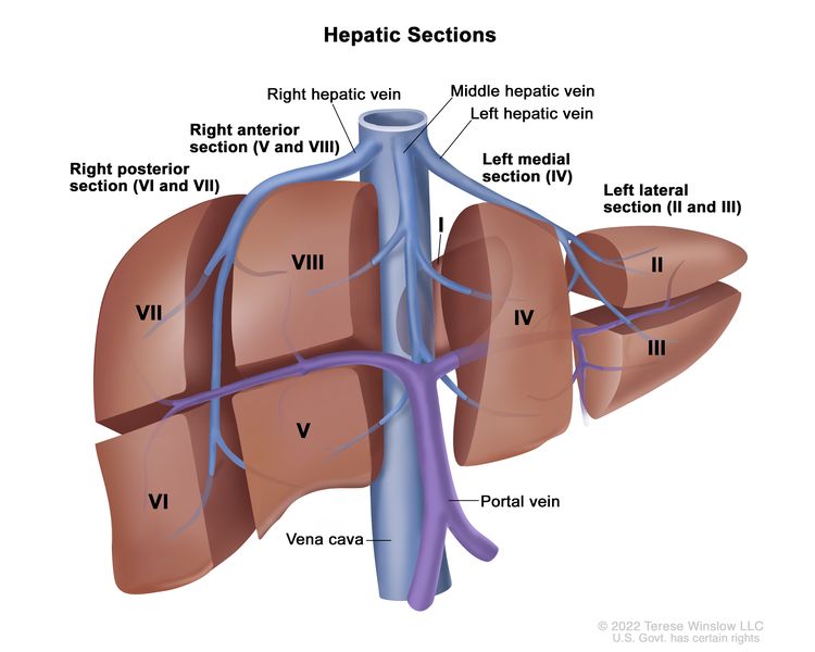
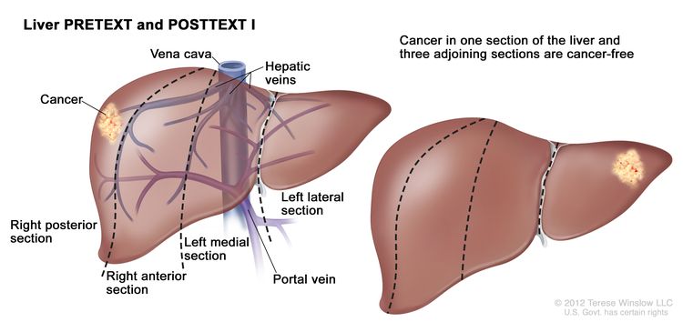
Childhood Rhabdomyosarcoma Treatment (PDQ®)–Health Professional Version

Childhood Rhabdomyosarcoma Treatment (PDQ®)–Health Professional Version

General Information About Childhood Rhabdomyosarcoma

Continual improvements in survival have been achieved for children and adolescents with cancer.[1] Between 1975 and 2020, childhood cancer mortality decreased by more than 50%.[13] Between 1975 and 2017, the 5-year relative survival rate for patients with rhabdomyosarcoma increased from 53% to 71% for children younger than 15 years and from 30% to 52% for adolescents aged 15 to 19 years.[1,2] In more recent years, improvements in outcome have plateaued.

Childhood and adolescent cancer survivors require close monitoring because side effects of cancer and its therapy may persist or develop months to years later. For specific information about the incidence, type, and monitoring of late effects in childhood and adolescent cancer survivors, see Late Effects of Treatment for Childhood Cancer.

Incidence

Childhood rhabdomyosarcoma is a soft tissue malignant tumor of mesenchymal origin. It accounts for approximately 2.7% of cancer cases among children aged 0 to 14 years and 1.4% of the cases among adolescents and young adults aged 15 to 19 years.[2] The incidence is 4.6 cases per 1 million children younger than 20 years, which translates into about 350 new cases per year. Fifty percent of these cases are seen in the first decade of life.[2,4]

The 2020 World Health Organization classification distinguishes four histological subtypes of rhabdomyosarcoma, including embryonal, alveolar, spindle cell/sclerosing, and pleomorphic.[5] While these subtypes classify rhabdomyosarcoma into prognostically useful histological categories, FOXO1 gene fusions uniquely occur in alveolar histology tumors; however, not all tumors that have been classified as alveolar histology have a FOXO1 fusion. Molecular characterization has replaced histopathological assessment for treatment risk assignment. Male patients have a higher incidence of embryonal tumors, and Black patients have a slightly higher incidence of alveolar tumors.[4] For more information, see the sections on Cellular Classification for Childhood Rhabdomyosarcoma and Molecular Characteristics of Rhabdomyosarcoma.

Incidence may depend on the histological subtype of rhabdomyosarcoma, as follows:

  • Embryonal: Patients with embryonal rhabdomyosarcoma are predominantly male (male-to-female ratio, 1.5). The peak incidence is in children between the ages of 0 and 4 years, with approximately 4 cases per 1 million children. The incidence rate is lower in adolescents, with approximately 1.5 cases per 1 million adolescents. This subtype constitutes 57% of patients in the Surveillance, Epidemiology, and End Results (SEER) Program database.[4]
  • Alveolar: The incidence of alveolar rhabdomyosarcoma does not vary by sex and is constant from ages 0 to 19 years, with approximately 1 case per 1 million children and adolescents. This subtype constitutes 23% of patients in the SEER database.[4]
  • Spindle cell/sclerosing: Spindle cell and sclerosing rhabdomyosarcoma are considered in the same diagnostic category. This uncommon variant accounts for 3% to 10% of all cases.[5]
  • Pleomorphic: Pleomorphic rhabdomyosarcoma is a high-grade pleomorphic sarcoma seen in adults. Childhood cases are considered to be rhabdomyosarcoma with diffuse anaplasia.[5]

Rhabdomyosarcoma may occur anywhere in the body. The most common primary sites include the following:[6,7]

  • Head and neck region (parameningeal) (approximately 25%).
  • Genitourinary tract (approximately 31%).
  • Extremities (approximately 13%). Within extremity tumors, tumors of the hand and foot occur more often in older patients and usually have an alveolar histology.[8]

Other less common primary sites include the trunk, chest wall, perineal/anal region, and abdomen, including the retroperitoneum and biliary tract.[7]

Risk Factors

Most cases of rhabdomyosarcoma occur sporadically, with no recognized predisposing risk factor.

Predisposition factors reported for rhabdomyosarcoma include the following:

  • Genetic factors:
    • Li-Fraumeni cancer susceptibility syndrome (with germline TP53 pathogenic variants).[911]
    • DICER1 syndrome.[12,13]
    • Neurofibromatosis type I (NF1).[14,15]
    • Costello syndrome (with germline HRAS pathogenic variants).[1619]
    • Beckwith-Wiedemann syndrome (more commonly associated with Wilms tumor and hepatoblastoma).[20,21]
    • Noonan syndrome.[19,22,23]
  • High birth weight and large size for gestational age are associated with an increased incidence of embryonal rhabdomyosarcoma.[24]

The Children’s Oncology Group (COG) performed retrospective exome sequencing on germline DNA to determine the prevalence of 63 autosomal dominant cancer-predisposing genes in 615 patients with newly diagnosed rhabdomyosarcoma.[25] They identified germline cancer-predisposition (pathogenic or likely pathogenic) variants in 45 patients with rhabdomyosarcoma (7.3%; all FOXO1 fusion negative) across 15 autosomal dominant genes. Specifically, 73.3% of the predisposition variants were found in predisposition syndrome genes previously associated with pediatric rhabdomyosarcoma risk, such as Li-Fraumeni syndrome (TP53, n = 11) and NF1 (NF1, n = 9). Notably, five patients had well-described oncogenic missense variants in HRAS (p.G12V and p.G12S) associated with Costello syndrome, and two patients each had variants in DICER1 and CBL, respectively. Germline pathogenic or likely pathogenic variants were more frequent in patients with embryonal rhabdomyosarcoma than in those with alveolar rhabdomyosarcoma (10% vs. 3%, P = .02), but all of the patients with alveolar rhabdomyosarcoma were FOXO1 negative, and no germline variants were identified in patients with FOXO1 translocations. Although patients with a cancer-predisposition variant tended to be younger at diagnosis (P = .00099), 40% of germline variants were identified in patients older than 3 years.

The COG reviewed the impact of germline pathogenic or likely pathogenic variants in cancer predisposition genes on patient outcomes.[26] In this study of 580 individuals with rhabdomyosarcoma, the median age was 5.9 years (range, 0.01–23.23 years), and the male-to-female ratio was 1.5:1 (351 [60.5%] male). For patients with congenital variants in rhabdomyosarcoma-associated cancer-predisposition genes, the event-free survival (EFS) rate was 48.4%, compared with 57.8% for patients without congenital predisposition variants (P = .10). The overall survival (OS) rate was 53.7% for patients with congenital predisposition variants, compared with 65.3% for patients without these variants (P = .06). Analyses were stratified by tumor histology and PAX3::FOXO1 or PAX7::FOXO1 gene fusion status. After adjustment, patients with congenital predisposition variants had significantly worse OS (adjusted hazard ratio [HR], 2.49; 95% confidence interval [CI], 1.39–4.45; P = .002), and patients with embryonal histology did not have better outcomes (EFS: adjusted HR, 2.25; 95% CI, 1.25–4.06; P = .007 and OS: adjusted HR, 2.83; 95% CI, 1.47–5.43; P = .002). These associations were not due to the development of a second malignant neoplasm. In addition, patients with fusion-negative rhabdomyosarcoma who harbored congenital predisposition variants had similarly inferior outcomes as patients with fusion-positive rhabdomyosarcoma who did not have congenital predisposition variants (EFS: adjusted HR, 1.35; 95% CI, 0.71–2.59; P = .37 and OS: adjusted HR, 1.71; 95% CI, 0.84–3.47; P = .14).

The COG reviewed the correlation between anaplastic histology and germline TP53 pathogenic variants in 239 patients with rhabdomyosarcoma. Among the 46 patients with anaplastic rhabdomyosarcoma, 11% (n = 5) carried a germline TP53 pathogenic variant, compared with 1% (n = 2) of the patients without anaplasia (P = .003). The rates of TP53 pathogenic variants in those with diffuse anaplasia and focal anaplasia were 9% (n = 3) and 17% (n = 2), respectively. Among the seven patients with TP53 pathogenic variants, 71% (5 of 7) had tumors with anaplastic histology.[27]

Prognostic Factors

Rhabdomyosarcoma is usually curable in children with localized disease who receive combined-modality therapy, with more than 70% of patients surviving 5 years after diagnosis.[6,7,28] Relapses are uncommon in patients who were alive and event free at 5 years, with a 10-year late-event rate of 9%. Relapses are more common in patients who have unresectable disease, tumor in an unfavorable site at diagnosis, or metastatic disease at diagnosis.[29]

The prognosis for children or adolescents with rhabdomyosarcoma is related to many clinical and biological factors, including the following:

Because treatment and prognosis partly depend on the histology and molecular characterization of the tumor, it is necessary that the tumor tissue be reviewed by expert pathologists with experience in the evaluation and diagnosis of tumors in children. Typically, accurate diagnosis requires additional molecular characterization. The diversity of primary sites, the distinctive surgical and radiation therapy treatments for each primary site, and the subsequent site-specific rehabilitation underscore the importance of treating children with rhabdomyosarcoma in medical centers with appropriate experience in all therapeutic modalities.

Age

Children aged 1 to 9 years have the best prognosis, while those younger than 1 year and older than 10 years fare less well. In Intergroup Rhabdomyosarcoma Study Group (IRSG) and COG trials, the 5-year failure-free survival (FFS) rate was 57% for patients younger than 1 year, 81% for patients aged 1 to 9 years, and 68% for patients older than 10 years. The 5-year OS rates were 76% for patients younger than 1 year, 87% for patients aged 1 to 9 years, and 76% for patients older than 10 years.[30] Historical data show that adults have fared less well than children (5-year OS rates, 27% ± 1.4% vs. 61% ± 1.4%; P < .0001).[3134]

  • Young age: Infants tend to do poorly, often because of treatment modifications to reduce toxicity. Typically, chemotherapy doses are reduced by 50% on the basis of reports that infants have higher death rates related to chemotherapy toxicity when compared with older patients; therefore, young patients may be underdosed.[35] In addition, infants younger than 1 year are less likely to receive radiation therapy for local control because of the high incidence of late effects in this age group.[28,36,37]

    The 5-year FFS rate was 67% for infants, compared with 81% in a matched group of older patients treated by the COG.[30,38] This inferior FFS rate was largely the result of a relatively high rate of local failure.

    In another retrospective study of 126 patients (aged ≤24 months) who were enrolled on the ARST0331 (NCT00075582) and ARST0531 (NCT00354835) trials, the 5-year local failure rate was 24%, the 5-year EFS rate was 68.3%, and the OS rate was 81.9%. Forty-three percent of the patients had an individualized local therapy plan that more frequently omitted radiation therapy. These patients had inferior local control and EFS rates.[38]

    Members of the Cooperative Weichteilsarkom Studiengruppe (CWS) reviewed 155 patients with rhabdomyosarcoma presenting from birth to age 12 months; 144 patients had localized disease; 11 patients had metastases; and 32 patients presented with alveolar rhabdomyosarcoma pathology. The following results were reported:[39][Level of evidence C1]

    • Of the 144 patients with localized disease, 129 had a complete response.
    • Fifty-one infants had a recurrence of their disease; 63% of patients with alveolar rhabdomyosarcoma had a relapse, and 28% of patients with embryonal rhabdomyosarcoma had a relapse.
    • The 5-year OS rates were 69% for patients with localized disease, 14% for patients with metastatic disease, and 41% for patients with relapsed disease.

    A retrospective analysis of five consecutive studies from the CWS group examined infants and older children with localized rhabdomyosarcoma of the female genitourinary tract.[40] Among 67 patients treated from 1981 to 2019, age of 12 months or younger at diagnosis was the only significant negative prognostic factor that influenced EFS.

    The European Paediatric Soft Tissue Sarcoma Study Group (EpSSG) enrolled 490 children younger than 36 months in their prospective RMS2005 study. The study included 110 patients younger than 12 months and 380 patients aged 12 to 36 months. Chemotherapy was given according to the risk group. Radiation therapy (22% received brachytherapy) was administered to 33.6% of the infants and 63.5% of the children aged 12 to 36 months. The 5-year OS rate was 88.4% for the infants, which was significantly better than the 72.5% rate observed in children aged 12 to 36 months. The treatment protocol in this trial, which used an increased application of adequate local therapy, may have contributed to these improved outcomes.[41][Level of evidence B4]

    The EpSSG analyzed neonates with congenital rhabdomyosarcoma, which they defined as infants younger than 2 months at diagnosis who were enrolled in EpSSG trials.[42] Twenty-four patients with congenital rhabdomyosarcoma were registered. All patients had favorable histology and localized disease, except for one patient with PAX3::FOXO1 fusion–positive metastatic rhabdomyosarcoma. Three patients had VGLL2::CITED2 or VGLL2::NCOA2 fusions. Complete tumor resection was achieved in ten patients. No radiation therapy was given. Chemotherapy doses were adjusted to age and weight. Only two patients required further dose reduction for toxicity. The 5-year EFS rate was 75.0% (95% CI, 52.6%–87.9%), and the OS rate was 87.3% (95% CI, 65.6%–95.7%).

    An international consortium identified 40 infants with spindle cell rhabdomyosarcoma.[43] The 5-year EFS rate for these infants with localized disease was 86% (± 11%; 95% CI), and the OS rate was 91% (± 9%; 95% CI). These outcomes compare favorably with those of all infants with localized rhabdomyosarcoma, for whom the 5-year failure-free survival rates range from 42% to 72% and the 5-year OS rates range from 61% to 88%. This finding suggests that infants with congenital spindle cell rhabdomyosarcoma have a favorable outcome compared with infants with other subtypes of rhabdomyosarcoma.

  • Older children: In older children, the upper dosage limits of vincristine and dactinomycin are based on body surface area (BSA), and these patients may require reduced vincristine doses because of neurotoxicity.[37,44]
  • Adolescents: A report from the Associazione Italiana Ematologia Oncologia Pediatrica Soft Tissue Sarcoma Committee suggests that adolescents may have more frequent unfavorable tumor characteristics, including alveolar histology, regional lymph node involvement, and metastatic disease at diagnosis, accounting for their poor prognosis. This study also found that 5-year OS and progression-free survival (PFS) rates were somewhat lower in adolescents than in children, but the differences among age groups younger than 1 year and aged 10 to 19 years at diagnosis were significantly worse than those in the group aged 1 to 9 years.[45]

    Two reports from the COG have documented inferior 5-year EFS rates in patients older than 10 years.[37,44] When compared with younger patients, this group of older patients was more likely to present with advanced-stage, large, and invasive alveolar tumors, with nodal involvement arising in the extremity and paratesticular sites. Older patients experienced less myelosuppression and more peripheral nervous system toxicity, suggesting that dose modifications during therapy cannot account for the age-related differences in EFS.

    Adolescent and young adult (AYA) patients were more likely to have worse survival outcomes than children.[46]

    • AYA patients were more likely to have metastatic tumors (61 of 257 [23.7%] vs. 197 of 1,720 [11.5%]; P < .0001), unfavorable histological subtypes (119 [46.3%] vs. 451 [26.2%]; P < .0001), tumors larger than 5 cm (177 [68.9%] vs. 891 [51.8%]; P < .0001), and regional lymph node involvement (109 [42.4%] vs. 339 [19.7%]; P < .0001) than children.
    • AYA patients had lower 5-year EFS rates (52.6% [95% CI, 46.3%–58.6%] vs. 67.8% [95% CI, 65.5%–70.0%]; P < .0001) and OS rates (57.1% [95% CI, 50.4%–63.1%] vs. 77.9% [95% CI, 75.8%–79.8%]; P < .0001) than children.
    • The multivariable analysis confirmed the inferior prognosis of patients aged 15 to 21 years (HR, 1.48 [95% CI, 1.20–1.83; P = .0002] for poorer EFS; HR, 1.73 [95% CI, 1.37–2.19; P < .0001] for poorer OS).
  • Adults: Adult patients with rhabdomyosarcoma have a higher incidence of pleomorphic histology (19%) than do children (<2%). Adults also have a higher incidence of tumors in unfavorable sites than do children.[31]

Site of origin

Prognosis for childhood rhabdomyosarcoma varies according to the primary tumor site (see Table 1).

Table 1. 5-Year Survival by Primary Site of Disease
Primary Site Number of Patients Survival at 5 Years (%)
aPatients treated on the ARST0331 study.[47]
bPatients treated on Intergroup Rhabdomyosarcoma Studies III–IV.[48]
cPooled analysis of European and North American groups.[49]
dCombined result from the Children’s Oncology Group, German Cooperative Soft Tissue Sarcoma Study, Italian Cooperative Group, and International Society of Pediatric Oncology groups.[50]
ePooled analysis of European and North American groups.[51]
fPatients treated on Intergroup Rhabdomyosarcoma Study III.[6]
gPatients treated on Intergroup Rhabdomyosarcoma Studies I–IV.[52]
hPatients treated on the D9602 and ARST0331 trials.[53]
Orbita 82 97
Head and neck (nonparameningeal)b 164 83
Cranial parameningealc 204 69.5
Genitourinary (excluding bladder/prostate)b 158 89
Localized bladder/prostated 322 84
Localized extremitye 643 67
Trunk, abdomen, perineum, etc.f 147 67
Biliaryg,h 25 76.5–78

Tumor size

Children with tumors 5 cm or smaller have improved survival, compared with children with tumors larger than 5 cm.[6] Both tumor volume and maximum tumor diameter are associated with outcome.[54][Level of evidence C1]

A retrospective review of soft tissue sarcomas in children and adolescents suggests that the 5-cm cutoff used for adults with soft tissue sarcoma may not be ideal for smaller children, especially infants. The review identified an interaction between tumor diameter and BSA.[55] This was not confirmed by a COG study of patients with intermediate-risk rhabdomyosarcoma.[56] This relationship requires prospective study to determine the therapeutic implications of the observation.

Resectability

The extent of disease after the primary surgical procedure (i.e., the Surgical-pathologic Group, also called the Clinical Group) is correlated with outcome.[6] In the IRS-III study, patients with localized, gross residual disease after initial surgery (Surgical-pathologic Group III) had a 5-year survival rate of approximately 70%, compared with a rate of more than 90% for patients without residual tumor after surgery (Group I) and a rate of approximately 80% for patients with microscopic residual tumor after surgery (Group II).[6,57] Groups I and II represent a minority of patients; approximately 50% of patients have unresectable Group III disease at time of diagnosis.[6]

Resectability without functional impairment is related to the tumor’s initial size and site and does not account for the biology of the disease. Outcome is optimized with the use of multimodality therapy. All patients require chemotherapy, and at least 85% of patients also benefit from radiation therapy, with favorable outcomes even for patients with nonresectable disease. In the IRS-IV study, the Group III patients with localized unresectable disease who were treated with chemotherapy and radiation therapy had a 5-year FFS rate of about 75% and a local control rate of 87%.[58] Two intermediate-risk COG rhabdomyosarcoma studies (D9803 and ARST0531 [NCT00354835]) were pooled to assess the benefit of delayed primary excision. In the D9803 study, local control with radiation therapy after either a partial or complete excision was completed at week 12. In the ARST0531 study, radiation was administered upfront at week 4. Patients with bladder or prostate rhabdomyosarcoma who received a delayed primary excision had no difference in survival, whereas patients with extremity rhabdomyosarcoma or nonbladder/nonprostate nonextremity rhabdomyosarcoma had an improved OS with delayed primary excision. Delayed primary excision strategy with a reduction in radiation dose resulted in superior OS for those sites.[59,60]

Histopathological subtype

The alveolar subtype of childhood rhabdomyosarcoma is more prevalent among patients with less favorable clinical features (e.g., younger than 1 year or older than 10 years, extremity and truncal primary tumors, and metastatic disease at diagnosis). It is generally associated with a worse outcome than in similar patients with embryonal rhabdomyosarcoma.

  • In the IRS-I and IRS-II studies, the alveolar subtype was associated with a less favorable outcome, even in patients whose primary tumor was completely resected (Group I).[61]
  • A statistically significant difference in 5-year survival by histopathological subtype (82% for embryonal rhabdomyosarcoma vs. 65% for alveolar rhabdomyosarcoma) was noted when 1,258 IRS-III and IRS-IV patients with rhabdomyosarcoma were analyzed.[62]
  • In the IRS-III study, the outcome for patients with Group I alveolar subtype tumors was similar to that for other patients with Group I tumors, but the alveolar patients received more intensive therapy.[6]
  • Patients with alveolar rhabdomyosarcoma who have regional lymph node involvement have significantly worse outcomes than patients who do not have regional lymph node involvement (5-year FFS rates, 43% vs. 73%).[63]
  • Local-control rates after radiation therapy are similar among patients with alveolar and embryonal tumors. However, patients who present with tumors 5 cm or larger have a significantly higher local failure rate.[64]

Anaplasia has been observed in 13% of embryonal rhabdomyosarcoma cases, with some studies suggesting the presence of anaplasia adversely influenced clinical outcome in patients with intermediate-risk disease. However, anaplasia has not been shown to be an independent prognostic variable.[65,66]

PAX3::FOXO1 or PAX7::FOXO1 gene fusion status

Approximately 80% of rhabdomyosarcoma cases morphologically defined as alveolar rhabdomyosarcoma express a FOXO1 fusion. FOXO1 gene fusions occur only in alveolar histology tumors.[67] Several retrospective studies found that fusion status is an independent prognostic factor. Patients with translocation-negative alveolar rhabdomyosarcoma have tumors with genetic and molecular profiles and outcomes similar to patients with embryonal rhabdomyosarcoma, and they fare better than patients with fusion-positive alveolar rhabdomyosarcoma.[68,69] Early retrospective studies relied on convenience samples of available tumor tissue.[68,69] A subsequent prospective study from the Soft Tissue Sarcoma Committee of the COG that examined 434 cases of intermediate-risk rhabdomyosarcoma treated on a single intermediate protocol (D9803) confirmed these observations.[70] Analysis of 38 patients enrolled in the COG D9802 (NCT00003955) low-risk study determined that fusion-positive, low-risk patients should be treated as intermediate risk.[71]

The specific fusion partner may have prognostic impact. In a COG study, fusion-positive patients with Stage 2 or 3, Group III, and PAX3-positive tumors had a lower EFS rate (54%) than those with PAX7-positive tumors (65%). Both fusion-positive groups did worse than those with embryonal rhabdomyosarcoma (EFS rate, 77%; P < .001). Patients with alveolar rhabdomyosarcoma and PAX3 fusions had a poorer OS rate (64%) than patients with alveolar rhabdomyosarcoma and PAX7 fusions (87%), patients with alveolar rhabdomyosarcoma who were fusion negative (89%), and patients with embryonal rhabdomyosarcoma (82%; P = .006).[70] Comparable results were observed in the U.K. study; patients with PAX7-positive tumors and patients with fusion-negative tumors had similar outcomes.[72]

Using data from six consecutive COG studies, a retrospective analysis of 1,727 patients with rhabdomyosarcoma refined the risk stratification for childhood rhabdomyosarcoma. The study reported that after metastatic status, FOXO1 status was the most important prognostic factor and improved the risk stratification of patients with localized rhabdomyosarcoma.[69]

The COG performed a retrospective analysis of 269 patients with confirmed FOXO1 fusion–positive rhabdomyosarcoma who were enrolled in three completed clinical trials for localized rhabdomyosarcoma.[73] The estimated 4-year EFS rate was 53% (95% CI, 47%–59%), and the OS rate was 69% (95% CI, 63%–74%). Multivariate analysis identified older age (≥10 years) and larger tumor size (>5 cm) as independent, adverse prognostic factors for EFS within this population. Patients who had both of these adverse features experienced substantially inferior outcomes.

An EpSSG study evaluated the role of clinical factors together with FOXO1 fusion status in patients with nonmetastatic rhabdomyosarcoma, using data from the EpSSG RMS2005 study. The multivariable analysis of 1,661 evaluable patients retained five prognostic variables: age at diagnosis, tumor size, primary site, IRS Group, and FOXO1 status. A nomogram was created, stratifying patients into four risk groups. The 5-year EFS rates were 94.1% for patients in the low-risk group, 78.4% for patients in the intermediate-risk group, 65.2% for patients in the high-risk group, and 52.1% for patients in the very high-risk group.[74]

These studies demonstrated that fusion status was a better predictor of outcome than histology. Similar conclusions were reached in a retrospective study of three consecutive trials in the United Kingdom. Fusion status has now been incorporated into the risk stratification of patients in the current COG ARST1431 (NCT02567435) study for patients with intermediate-risk rhabdomyosarcoma, in subsequent COG trials, and in the new international EpSSG trial.[74] The authors underscored the probable value of treating fusion-negative patients whose tumors have alveolar histology with therapy that is stage appropriate for embryonal histology tumors.[75][Level of evidence C1]

Metastases at diagnosis

Children with metastatic disease at diagnosis have the worst prognosis.

The prognostic significance of metastatic disease is modified by the following:

  • Tumor histology (embryonal rhabdomyosarcoma is more favorable than alveolar). Patients with localized alveolar histology and regional node disease have a similar prognosis as patients with a single site of metastatic disease, provided that the regional disease is treated with radiation therapy.[63]
  • Age at diagnosis (<10 years for children with embryonal rhabdomyosarcoma).
  • The site of primary disease. Patients with metastatic genitourinary (nonbladder, nonprostate) primary tumors have a more favorable outcome than patients with metastatic disease from other primary sites.[76]
  • The number of metastatic sites.[7780]

The COG performed a retrospective analysis of 179 patients who were diagnosed with rhabdomyosarcoma that was metastatic to the bone marrow. These patients were enrolled in one of four COG rhabdomyosarcoma clinical trials (D9802, D9803, ARST0431, and ARST08P1) between 1997 and 2013.[81] Patients were a median age of 14.8 years and 58% were male. Alveolar histology was the predominant type (76%), the extremity was the most common primary site (32%), and most patients had metastatic disease to additional sites (87%). The 3-year EFS rate was 9.4%, and the 5-year EFS rate was 8.2%. The 3-year OS rate was 26.1%, and the 5-year OS rate was 12.6%.

The COG performed a retrospective review of patients enrolled in high-risk protocols for rhabdomyosarcoma. FOXO1 fusion status correlated with clinical characteristics at diagnosis, including age, stage, histology, and extent of metastatic disease (Oberlin status). Among patients with metastatic disease, PAX::FOXO1 fusion status was not an independent predictor of outcome.[82][Level of evidence B1]

Lymph node involvement at diagnosis

Lymph node involvement at diagnosis is seen in about 23% of patients with rhabdomyosarcoma and is associated with an inferior prognosis.[62,83] Clinical and/or imaging evaluation is performed before treatment and preoperatively. These findings are incorporated into the initial staging and grouping of a patient with rhabdomyosarcoma. The updated TNM staging defines clinical node involvement as larger than 1 cm.[84]

Pathological assessment of nodal disease is determined by biopsy and incorporated in the Surgical/Pathologic Clinical Group classification. Core-needle or open biopsy of clinically enlarged nodes is appropriate to confirm the presence of disease. Approximately 25% of enlarged nodes will be pathologically negative. Suspicious nodes are sampled surgically with open biopsy, preferred to needle aspiration, although needle aspiration may occasionally be appropriate. Pathological evaluation of clinically uninvolved nodes is site specific. In COG studies, it is required for extremity sites and for boys older than 10 years with paratesticular primary tumors.[85] Given the poorer outcomes, pathological node evaluation is required for patients with fusion-positive disease in current European and North American clinical trials.

Data on the frequency of lymph node involvement in various sites are useful for making clinical decisions. For example, up to 40% of patients with rhabdomyosarcoma in genitourinary sites have lymph node involvement, while patients with certain head and neck sites have a much lower likelihood (<10%). Patients with nongenitourinary pelvic sites (e.g., anus/perineum) have an intermediate frequency of lymph node involvement.[86]

In the extremities and select truncal sites, sentinel lymph node evaluation is a more accurate form of diagnosis than random regional lymph node sampling. In clinically negative lymph nodes of the extremity or trunk, sentinel lymph node biopsy is the preferred form of node sampling by the COG. Technical considerations are obtained from surgical experts. Needle or open biopsy of clinically enlarged nodes is appropriate.[8790] Lymph node removal does not improve outcome, and it is useful for staging but not treatment.

The EpSSG performed a retrospective analysis of 109 patients with rhabdomyosarcoma with extremity primary tumors distal to the elbow or knee who were treated in the EpSSG RMS-2005 (NCT00379457) trial (2005–2016).[91] Thirty-seven of 109 patients (34%) had lymph node metastases at diagnosis. Of the 37 patients, 19 (51%) had in-transit metastases (ITM), especially in lower extremity rhabdomyosarcoma. The 5-year EFS rates were 88.9% for patients with ITM, 21.4% for patients with proximal lymph node involvement, and 20% for combined proximal lymph node involvement and ITM (P = .01). The 5-year OS rates were 100% for patients with ITM, 25.2% for patients with proximal lymph node involvement, and 15% for patients with combined proximal lymph node involvement and ITM (P =. 003). The authors concluded that popliteal and epitrochlear nodes should be considered as true (distal) regional nodes, instead of ITM. The authors recommended biopsy of these nodes, especially for distal extremity rhabdomyosarcoma of the lower limb.

The EpSSG reported a retrospective analysis of 1,294 children with embryonal rhabdomyosarcoma enrolled in the RMS-2005 protocol.[92] Of these patients, 143 had nodal involvement (N1). Patients with N1 disease were older and presented with tumors of unfavorable size, invasiveness, site, and resectability. Unlike alveolar rhabdomyosarcoma, nodal involvement was more frequent in the head and neck area and rare in extremity sites. The 5-year EFS rate was 75.5%, and the OS rate was 86.3% for patients with N0 disease. The 5-year EFS rate was 65.2%, and the OS rate was 70.7% for patients with N1 disease. Nodal involvement and the result of surgery at diagnosis (Intergroup Rhabdomyosarcoma Study group) were independent prognostic factors on multivariate analysis. Investigators concluded that regional nodal involvement is an independent prognostic factor in patients with embryonal rhabdomyosarcoma; therefore, it is appropriate to include this population in the high-risk category.

Bone marrow involvement

The COG performed a retrospective analysis of patients with rhabdomyosarcoma who had bone marrow metastasis at initial presentation and were treated in COG protocols.[93] Rhabdomyosarcoma metastatic to bone was identified in 154 patients (median age at diagnosis, 14.9 years). Fifty-eight percent of patients were male, 90% of patients had metastases at additional sites, 74% of patients had alveolar histology, and extremities were the most common primary site (31%). The 3-year EFS rate was 15.4%, and the 5-year EFS rate was 14.5%. The 3-year OS rate was 30.4%, and the 5-year OS rate was 18.0%. Alveolar histology, presence of FOXO1 gene fusions, unfavorable primary tumor location, higher Oberlin score, and lack of radiation therapy were poor prognostic characteristics for both EFS and OS in univariate analysis.

Biological characteristics

For more information, see the Molecular Characteristics of Rhabdomyosarcoma section.

Response to therapy

It is unlikely that response to induction chemotherapy or best tumor response during therapy, assessed by anatomic imaging, correlates with the likelihood of survival in patients with rhabdomyosarcoma. This finding was based on the IRSG, COG, and International Society of Pediatric Oncology (SIOP) studies that found no association.[94,95]; [96][Level of evidence C2]; [97][Level of evidence C1] However, an Italian study did find that patient response correlated with likelihood of survival.[54][Level of evidence C1] In patients with embryonal rhabdomyosarcoma who had metastases only in the lungs, the CWS assessed the relationship between complete response of the lung metastases at weeks 7 to 10 after chemotherapy and outcome in 53 patients.[98][Level of evidence C1] The 5-year survival rate was 68% for 26 complete responders at weeks 7 to 10 versus 36% for 27 patients who achieved complete responses at later time points (P = .004).

Other studies have investigated response to induction therapy, showing benefit to response. These data are somewhat flawed because therapy is usually tailored on the basis of response. Thus the situation is not as clear as the COG data suggest.[99104]

Response as judged by sequential functional imaging studies with fluorine F 18-fludeoxyglucose positron emission tomography (18F-FDG PET) may be an early indicator of outcome [105] and is under investigation by several pediatric cooperative groups. A retrospective analysis of 107 patients from a single institution examined PET scans performed at baseline, after induction chemotherapy, and after local therapy.[105] Standardized uptake value measured at baseline predicted PFS and OS, but not local control. A negative scan after induction chemotherapy correlated with statistically significantly better PFS. A positive scan after local therapy predicted worse PFS, OS, and local control. The COG evaluated the relationship between complete metabolic response, as assessed by 18F-FDG PET imaging, and EFS in patients with intermediate- or high-risk rhabdomyosarcoma.[106][Level of evidence B4] The maximum standard uptake values (SUVmax) at study entry did not correlate with EFS for intermediate-risk (P = .32) or high-risk (P = .86) patients. Compared with patients who did not achieve a complete metabolic response, EFS was not superior for intermediate-risk patients who achieved a complete metabolic response at weeks 4 (P = .66) or 15 (P = .46), or for high-risk patients who achieved a complete metabolic response at weeks 6 (P = .75) or 19 (P = .28). Change in SUVmax at weeks 4 (P = .21) or 15 (P = .91) for intermediate-risk patients and at weeks 6 (P = .75) or 19 (P = .61) for high-risk patients did not correlate with EFS.

PET scans have been shown to be useful in understanding patterns of spread, particularly in patients with extremity disease.[107][Level of evidence C2]

Circulating tumor DNA (ctDNA) and RNA

A retrospective study of 99 children with rhabdomyosarcoma used reverse transcription–polymerase chain reaction to analyze an 11-gene panel in peripheral blood and bone marrow samples at the time of initial diagnosis.[108] The 5-year EFS rate was 35.5% (95% CI, 17.5%–53.5%) for the 33 patients who were RNA positive, compared with 88.0% (95% CI, 78.9%–97.2%) for the 66 patients who were RNA negative (P < .0001). The predictive power of the assay was maintained in a multivariate analysis, which included the usual clinical characteristics that correlate with prognosis such as the presence of metastatic disease. These investigators also studied the diagnostic potential of ctDNA in 57 patients enrolled in the EpSSG RMS-2005 (NCT00379457) study. ctDNA was detected using both shallow whole-genome sequencing (WGS) and cell-free reduced representation bisulfite sequencing (cfRRBS). Of the 25 samples tested, 21 were correctly classified as embryonal histology by cfRRBS. The presence of methylated RASSF1A correlated with a poor outcome.[109]

The COG analyzed ctDNA in 124 patients with newly diagnosed, intermediate-risk rhabdomyosarcoma from the COG biorepository, which included 75 patients with fusion-negative rhabdomyosarcoma and 49 patients with fusion-positive rhabdomyosarcoma.[110] Ultralow passage WGS was used to detect copy number alterations. Rhabdo-Seq, a new custom sequencing assay, was used to detect rearrangements and single-nucleotide variants (SNVs).

  • The authors reported that ultralow passage WGS was a method that could detect ctDNA in all patients with fusion-negative rhabdomyosarcoma. ctDNA was detected in 13 of 75 serum samples (17%).
  • However, the use of Rhabdo-Seq in fusion-negative rhabdomyosarcoma samples also identified SNVs, such as the L122R variant in the MYOD1 gene. This variant was previously associated with a poor prognosis.
  • Identification of pathognomonic translocations between PAX3 or PAX7 and FOXO1 by Rhabdo-Seq was the best method for measuring ctDNA in fusion-positive rhabdomyosarcoma tumors. It detected ctDNA in 27 of 49 cases (55%).
  • Patients with fusion-negative rhabdomyosarcoma with detectable ctDNA at diagnosis had significantly worse outcomes than patients without detectable ctDNA (EFS rates, 33.3% vs. 68.9%; P = .0028; OS rates, 33.3% vs. 83.2%; P < .0001).
  • Patients with fusion-positive rhabdomyosarcoma with detectable ctDNA at diagnosis had significantly worse outcomes than patients without detectable ctDNA (EFS rates, 37% vs. 70%; P = .045; OS rates, 39.2% vs. 75%; P = .023).
  • In a multivariate analysis, ctDNA was independently associated with poor prognoses in patients with fusion-negative rhabdomyosarcoma but not in the smaller cohort of patients with fusion-positive rhabdomyosarcoma.
References
  1. Smith MA, Altekruse SF, Adamson PC, et al.: Declining childhood and adolescent cancer mortality. Cancer 120 (16): 2497-506, 2014. [PUBMED Abstract]
  2. National Cancer Institute: NCCR*Explorer: An interactive website for NCCR cancer statistics. Bethesda, MD: National Cancer Institute. Available online. Last accessed February 25, 2025.
  3. Surveillance Research Program, National Cancer Institute: SEER*Explorer: An interactive website for SEER cancer statistics. Bethesda, MD: National Cancer Institute. Available online. Last accessed December 30, 2024.
  4. Ognjanovic S, Linabery AM, Charbonneau B, et al.: Trends in childhood rhabdomyosarcoma incidence and survival in the United States, 1975-2005. Cancer 115 (18): 4218-26, 2009. [PUBMED Abstract]
  5. WHO Classification of Tumours Editorial Board: WHO Classification of Tumours. Volume 3: Soft Tissue and Bone Tumours. 5th ed., IARC Press, 2020.
  6. Crist W, Gehan EA, Ragab AH, et al.: The Third Intergroup Rhabdomyosarcoma Study. J Clin Oncol 13 (3): 610-30, 1995. [PUBMED Abstract]
  7. Maurer HM, Gehan EA, Beltangady M, et al.: The Intergroup Rhabdomyosarcoma Study-II. Cancer 71 (5): 1904-22, 1993. [PUBMED Abstract]
  8. Casanova M, Meazza C, Favini F, et al.: Rhabdomyosarcoma of the extremities: a focus on tumors arising in the hand and foot. Pediatr Hematol Oncol 26 (5): 321-31, 2009 Jul-Aug. [PUBMED Abstract]
  9. Li FP, Fraumeni JF: Rhabdomyosarcoma in children: epidemiologic study and identification of a familial cancer syndrome. J Natl Cancer Inst 43 (6): 1365-73, 1969. [PUBMED Abstract]
  10. Diller L, Sexsmith E, Gottlieb A, et al.: Germline p53 mutations are frequently detected in young children with rhabdomyosarcoma. J Clin Invest 95 (4): 1606-11, 1995. [PUBMED Abstract]
  11. Trahair T, Andrews L, Cohn RJ: Recognition of Li Fraumeni syndrome at diagnosis of a locally advanced extremity rhabdomyosarcoma. Pediatr Blood Cancer 48 (3): 345-8, 2007. [PUBMED Abstract]
  12. Dehner LP, Jarzembowski JA, Hill DA: Embryonal rhabdomyosarcoma of the uterine cervix: a report of 14 cases and a discussion of its unusual clinicopathological associations. Mod Pathol 25 (4): 602-14, 2012. [PUBMED Abstract]
  13. Doros L, Yang J, Dehner L, et al.: DICER1 mutations in embryonal rhabdomyosarcomas from children with and without familial PPB-tumor predisposition syndrome. Pediatr Blood Cancer 59 (3): 558-60, 2012. [PUBMED Abstract]
  14. Ferrari A, Bisogno G, Macaluso A, et al.: Soft-tissue sarcomas in children and adolescents with neurofibromatosis type 1. Cancer 109 (7): 1406-12, 2007. [PUBMED Abstract]
  15. Crucis A, Richer W, Brugières L, et al.: Rhabdomyosarcomas in children with neurofibromatosis type I: A national historical cohort. Pediatr Blood Cancer 62 (10): 1733-8, 2015. [PUBMED Abstract]
  16. Gripp KW, Lin AE, Stabley DL, et al.: HRAS mutation analysis in Costello syndrome: genotype and phenotype correlation. Am J Med Genet A 140 (1): 1-7, 2006. [PUBMED Abstract]
  17. Aoki Y, Niihori T, Kawame H, et al.: Germline mutations in HRAS proto-oncogene cause Costello syndrome. Nat Genet 37 (10): 1038-40, 2005. [PUBMED Abstract]
  18. Gripp KW: Tumor predisposition in Costello syndrome. Am J Med Genet C Semin Med Genet 137 (1): 72-7, 2005. [PUBMED Abstract]
  19. Kratz CP, Rapisuwon S, Reed H, et al.: Cancer in Noonan, Costello, cardiofaciocutaneous and LEOPARD syndromes. Am J Med Genet C Semin Med Genet 157 (2): 83-9, 2011. [PUBMED Abstract]
  20. Samuel DP, Tsokos M, DeBaun MR: Hemihypertrophy and a poorly differentiated embryonal rhabdomyosarcoma of the pelvis. Med Pediatr Oncol 32 (1): 38-43, 1999. [PUBMED Abstract]
  21. DeBaun MR, Tucker MA: Risk of cancer during the first four years of life in children from The Beckwith-Wiedemann Syndrome Registry. J Pediatr 132 (3 Pt 1): 398-400, 1998. [PUBMED Abstract]
  22. Moschovi M, Touliatou V, Vassiliki T, et al.: Rhabdomyosarcoma in a patient with Noonan syndrome phenotype and review of the literature. J Pediatr Hematol Oncol 29 (5): 341-4, 2007. [PUBMED Abstract]
  23. Hasle H: Malignant diseases in Noonan syndrome and related disorders. Horm Res 72 (Suppl 2): 8-14, 2009. [PUBMED Abstract]
  24. Ognjanovic S, Carozza SE, Chow EJ, et al.: Birth characteristics and the risk of childhood rhabdomyosarcoma based on histological subtype. Br J Cancer 102 (1): 227-31, 2010. [PUBMED Abstract]
  25. Li H, Sisoudiya SD, Martin-Giacalone BA, et al.: Germline Cancer Predisposition Variants in Pediatric Rhabdomyosarcoma: A Report From the Children’s Oncology Group. J Natl Cancer Inst 113 (7): 875-883, 2021. [PUBMED Abstract]
  26. Martin-Giacalone BA, Li H, Scheurer ME, et al.: Germline Genetic Testing and Survival Outcomes Among Children With Rhabdomyosarcoma: A Report From the Children’s Oncology Group. JAMA Netw Open 7 (3): e244170, 2024. [PUBMED Abstract]
  27. Fair D, Maese L, Chi YY, et al.: TP53 germline pathogenic variant frequency in anaplastic rhabdomyosarcoma: A Children’s Oncology Group report. Pediatr Blood Cancer : e30413, 2023. [PUBMED Abstract]
  28. Crist WM, Anderson JR, Meza JL, et al.: Intergroup rhabdomyosarcoma study-IV: results for patients with nonmetastatic disease. J Clin Oncol 19 (12): 3091-102, 2001. [PUBMED Abstract]
  29. Sung L, Anderson JR, Donaldson SS, et al.: Late events occurring five years or more after successful therapy for childhood rhabdomyosarcoma: a report from the Soft Tissue Sarcoma Committee of the Children’s Oncology Group. Eur J Cancer 40 (12): 1878-85, 2004. [PUBMED Abstract]
  30. Malempati S, Rodeberg DA, Donaldson SS, et al.: Rhabdomyosarcoma in infants younger than 1 year: a report from the Children’s Oncology Group. Cancer 117 (15): 3493-501, 2011. [PUBMED Abstract]
  31. Sultan I, Qaddoumi I, Yaser S, et al.: Comparing adult and pediatric rhabdomyosarcoma in the surveillance, epidemiology and end results program, 1973 to 2005: an analysis of 2,600 patients. J Clin Oncol 27 (20): 3391-7, 2009. [PUBMED Abstract]
  32. Streby KA, Ruymann FB, Whiteside S, et al.: Rhabdomyosarcoma in adolescents and young adults: A 25-year review at Nationwide Children’s Hospital. J Adolesc Young Adult Oncol 1 (4): 164-167, 2012.
  33. Van Gaal JC, Van Der Graaf WT, Rikhof B, et al.: The impact of age on outcome of embryonal and alveolar rhabdomyosarcoma patients. A multicenter study. Anticancer Res 32 (10): 4485-97, 2012. [PUBMED Abstract]
  34. Dumont SN, Araujo DM, Munsell MF, et al.: Management and outcome of 239 adolescent and adult rhabdomyosarcoma patients. Cancer Med 2 (4): 553-63, 2013. [PUBMED Abstract]
  35. Ragab AH, Heyn R, Tefft M, et al.: Infants younger than 1 year of age with rhabdomyosarcoma. Cancer 58 (12): 2606-10, 1986. [PUBMED Abstract]
  36. Ferrari A, Casanova M, Bisogno G, et al.: Rhabdomyosarcoma in infants younger than one year old: a report from the Italian Cooperative Group. Cancer 97 (10): 2597-604, 2003. [PUBMED Abstract]
  37. Joshi D, Anderson JR, Paidas C, et al.: Age is an independent prognostic factor in rhabdomyosarcoma: a report from the Soft Tissue Sarcoma Committee of the Children’s Oncology Group. Pediatr Blood Cancer 42 (1): 64-73, 2004. [PUBMED Abstract]
  38. Bradley JA, Kayton ML, Chi YY, et al.: Treatment Approach and Outcomes in Infants With Localized Rhabdomyosarcoma: A Report From the Soft Tissue Sarcoma Committee of the Children’s Oncology Group. Int J Radiat Oncol Biol Phys 103 (1): 19-27, 2019. [PUBMED Abstract]
  39. Sparber-Sauer M, Stegmaier S, Vokuhl C, et al.: Rhabdomyosarcoma diagnosed in the first year of life: Localized, metastatic, and relapsed disease. Outcome data from five trials and one registry of the Cooperative Weichteilsarkom Studiengruppe (CWS). Pediatr Blood Cancer 66 (6): e27652, 2019. [PUBMED Abstract]
  40. Sparber-Sauer M, Matle M, Vokuhl C, et al.: Rhabdomyosarcoma of the female genitourinary tract: Primary and relapsed disease in infants and older children. Treatment results of five Cooperative Weichteilsarkom Studiengruppe (CWS) trials and one registry. Pediatr Blood Cancer 68 (4): e28889, 2021. [PUBMED Abstract]
  41. Slater O, Gains JE, Kelsey AM, et al.: Localised rhabdomyosarcoma in infants (<12 months) and young children (12-36 months of age) treated on the EpSSG RMS 2005 study. Eur J Cancer 160: 206-214, 2022. [PUBMED Abstract]
  42. Bisogno G, Minard-Colin V, Arush MB, et al.: Congenital rhabdomyosarcoma: A report from the European paediatric Soft tissue sarcoma Study Group. Pediatr Blood Cancer 69 (2): e29376, 2022. [PUBMED Abstract]
  43. Whittle S, Venkatramani R, Schönstein A, et al.: Congenital spindle cell rhabdomyosarcoma: An international cooperative analysis. Eur J Cancer 168: 56-64, 2022. [PUBMED Abstract]
  44. Gupta AA, Anderson JR, Pappo AS, et al.: Patterns of chemotherapy-induced toxicities in younger children and adolescents with rhabdomyosarcoma: a report from the Children’s Oncology Group Soft Tissue Sarcoma Committee. Cancer 118 (4): 1130-7, 2012. [PUBMED Abstract]
  45. Bisogno G, Compostella A, Ferrari A, et al.: Rhabdomyosarcoma in adolescents: a report from the AIEOP Soft Tissue Sarcoma Committee. Cancer 118 (3): 821-7, 2012. [PUBMED Abstract]
  46. Ferrari A, Chisholm JC, Jenney M, et al.: Adolescents and young adults with rhabdomyosarcoma treated in the European paediatric Soft tissue sarcoma Study Group (EpSSG) protocols: a cohort study. Lancet Child Adolesc Health 6 (8): 545-554, 2022. [PUBMED Abstract]
  47. Walterhouse DO, Pappo AS, Meza JL, et al.: Shorter-duration therapy using vincristine, dactinomycin, and lower-dose cyclophosphamide with or without radiotherapy for patients with newly diagnosed low-risk rhabdomyosarcoma: a report from the Soft Tissue Sarcoma Committee of the Children’s Oncology Group. J Clin Oncol 32 (31): 3547-52, 2014. [PUBMED Abstract]
  48. Pappo AS, Meza JL, Donaldson SS, et al.: Treatment of localized nonorbital, nonparameningeal head and neck rhabdomyosarcoma: lessons learned from intergroup rhabdomyosarcoma studies III and IV. J Clin Oncol 21 (4): 638-45, 2003. [PUBMED Abstract]
  49. Merks JH, De Salvo GL, Bergeron C, et al.: Parameningeal rhabdomyosarcoma in pediatric age: results of a pooled analysis from North American and European cooperative groups. Ann Oncol 25 (1): 231-6, 2014. [PUBMED Abstract]
  50. Rodeberg DA, Anderson JR, Arndt CA, et al.: Comparison of outcomes based on treatment algorithms for rhabdomyosarcoma of the bladder/prostate: combined results from the Children’s Oncology Group, German Cooperative Soft Tissue Sarcoma Study, Italian Cooperative Group, and International Society of Pediatric Oncology Malignant Mesenchymal Tumors Committee. Int J Cancer 128 (5): 1232-9, 2011. [PUBMED Abstract]
  51. Oberlin O, Rey A, Brown KL, et al.: Prognostic Factors for Outcome in Localized Extremity Rhabdomyosarcoma. Pooled Analysis from Four International Cooperative Groups. Pediatr Blood Cancer 62 (12): 2125-31, 2015. [PUBMED Abstract]
  52. Spunt SL, Lobe TE, Pappo AS, et al.: Aggressive surgery is unwarranted for biliary tract rhabdomyosarcoma. J Pediatr Surg 35 (2): 309-16, 2000. [PUBMED Abstract]
  53. Aye JM, Xue W, Palmer JD, et al.: Suboptimal outcome for patients with biliary rhabdomyosarcoma treated on low-risk clinical trials: A report from the Children’s Oncology Group. Pediatr Blood Cancer 68 (4): e28914, 2021. [PUBMED Abstract]
  54. Ferrari A, Miceli R, Meazza C, et al.: Comparison of the prognostic value of assessing tumor diameter versus tumor volume at diagnosis or in response to initial chemotherapy in rhabdomyosarcoma. J Clin Oncol 28 (8): 1322-8, 2010. [PUBMED Abstract]
  55. Ferrari A, Miceli R, Meazza C, et al.: Soft tissue sarcomas of childhood and adolescence: the prognostic role of tumor size in relation to patient body size. J Clin Oncol 27 (3): 371-6, 2009. [PUBMED Abstract]
  56. Rodeberg DA, Stoner JA, Garcia-Henriquez N, et al.: Tumor volume and patient weight as predictors of outcome in children with intermediate risk rhabdomyosarcoma: a report from the Children’s Oncology Group. Cancer 117 (11): 2541-50, 2011. [PUBMED Abstract]
  57. Smith LM, Anderson JR, Qualman SJ, et al.: Which patients with microscopic disease and rhabdomyosarcoma experience relapse after therapy? A report from the soft tissue sarcoma committee of the children’s oncology group. J Clin Oncol 19 (20): 4058-64, 2001. [PUBMED Abstract]
  58. Donaldson SS, Meza J, Breneman JC, et al.: Results from the IRS-IV randomized trial of hyperfractionated radiotherapy in children with rhabdomyosarcoma–a report from the IRSG. Int J Radiat Oncol Biol Phys 51 (3): 718-28, 2001. [PUBMED Abstract]
  59. Rodeberg DA, Wharam MD, Lyden ER, et al.: Delayed primary excision with subsequent modification of radiotherapy dose for intermediate-risk rhabdomyosarcoma: a report from the Children’s Oncology Group Soft Tissue Sarcoma Committee. Int J Cancer 137 (1): 204-11, 2015. [PUBMED Abstract]
  60. Lautz TB, Chi YY, Li M, et al.: Benefit of delayed primary excision in rhabdomyosarcoma: A report from the Children’s Oncology Group. Cancer 127 (2): 275-283, 2021. [PUBMED Abstract]
  61. Crist WM, Garnsey L, Beltangady MS, et al.: Prognosis in children with rhabdomyosarcoma: a report of the intergroup rhabdomyosarcoma studies I and II. Intergroup Rhabdomyosarcoma Committee. J Clin Oncol 8 (3): 443-52, 1990. [PUBMED Abstract]
  62. Meza JL, Anderson J, Pappo AS, et al.: Analysis of prognostic factors in patients with nonmetastatic rhabdomyosarcoma treated on intergroup rhabdomyosarcoma studies III and IV: the Children’s Oncology Group. J Clin Oncol 24 (24): 3844-51, 2006. [PUBMED Abstract]
  63. Rodeberg DA, Garcia-Henriquez N, Lyden ER, et al.: Prognostic significance and tumor biology of regional lymph node disease in patients with rhabdomyosarcoma: a report from the Children’s Oncology Group. J Clin Oncol 29 (10): 1304-11, 2011. [PUBMED Abstract]
  64. Wolden SL, Lyden ER, Arndt CA, et al.: Local Control for Intermediate-Risk Rhabdomyosarcoma: Results From D9803 According to Histology, Group, Site, and Size: A Report From the Children’s Oncology Group. Int J Radiat Oncol Biol Phys 93 (5): 1071-6, 2015. [PUBMED Abstract]
  65. Qualman S, Lynch J, Bridge J, et al.: Prevalence and clinical impact of anaplasia in childhood rhabdomyosarcoma : a report from the Soft Tissue Sarcoma Committee of the Children’s Oncology Group. Cancer 113 (11): 3242-7, 2008. [PUBMED Abstract]
  66. Shenoy A, Alvarez E, Chi YY, et al.: The prognostic significance of anaplasia in childhood rhabdomyosarcoma: A report from the Children’s Oncology Group. Eur J Cancer 143: 127-133, 2021. [PUBMED Abstract]
  67. Sorensen PH, Lynch JC, Qualman SJ, et al.: PAX3-FKHR and PAX7-FKHR gene fusions are prognostic indicators in alveolar rhabdomyosarcoma: a report from the children’s oncology group. J Clin Oncol 20 (11): 2672-9, 2002. [PUBMED Abstract]
  68. Williamson D, Missiaglia E, de Reyniès A, et al.: Fusion gene-negative alveolar rhabdomyosarcoma is clinically and molecularly indistinguishable from embryonal rhabdomyosarcoma. J Clin Oncol 28 (13): 2151-8, 2010. [PUBMED Abstract]
  69. Hibbitts E, Chi YY, Hawkins DS, et al.: Refinement of risk stratification for childhood rhabdomyosarcoma using FOXO1 fusion status in addition to established clinical outcome predictors: A report from the Children’s Oncology Group. Cancer Med 8 (14): 6437-6448, 2019. [PUBMED Abstract]
  70. Skapek SX, Anderson J, Barr FG, et al.: PAX-FOXO1 fusion status drives unfavorable outcome for children with rhabdomyosarcoma: a children’s oncology group report. Pediatr Blood Cancer 60 (9): 1411-7, 2013. [PUBMED Abstract]
  71. Arnold MA, Anderson JR, Gastier-Foster JM, et al.: Histology, Fusion Status, and Outcome in Alveolar Rhabdomyosarcoma With Low-Risk Clinical Features: A Report From the Children’s Oncology Group. Pediatr Blood Cancer 63 (4): 634-9, 2016. [PUBMED Abstract]
  72. Missiaglia E, Williamson D, Chisholm J, et al.: PAX3/FOXO1 fusion gene status is the key prognostic molecular marker in rhabdomyosarcoma and significantly improves current risk stratification. J Clin Oncol 30 (14): 1670-7, 2012. [PUBMED Abstract]
  73. Heske CM, Chi YY, Venkatramani R, et al.: Survival outcomes of patients with localized FOXO1 fusion-positive rhabdomyosarcoma treated on recent clinical trials: A report from the Soft Tissue Sarcoma Committee of the Children’s Oncology Group. Cancer 127 (6): 946-956, 2021. [PUBMED Abstract]
  74. De Salvo GL, Del Bianco P, Minard-Colin V, et al.: Reappraisal of prognostic factors used in the European Pediatric Soft Tissue Sarcoma Study Group RMS 2005 study for localized rhabdomyosarcoma to optimize risk stratification and generate a prognostic nomogram. Cancer 130 (13): 2351-2360, 2024. [PUBMED Abstract]
  75. Selfe J, Olmos D, Al-Saadi R, et al.: Impact of fusion gene status versus histology on risk-stratification for rhabdomyosarcoma: Retrospective analyses of patients on UK trials. Pediatr Blood Cancer 64 (7): , 2017. [PUBMED Abstract]
  76. Koscielniak E, Rodary C, Flamant F, et al.: Metastatic rhabdomyosarcoma and histologically similar tumors in childhood: a retrospective European multi-center analysis. Med Pediatr Oncol 20 (3): 209-14, 1992. [PUBMED Abstract]
  77. Breneman JC, Lyden E, Pappo AS, et al.: Prognostic factors and clinical outcomes in children and adolescents with metastatic rhabdomyosarcoma–a report from the Intergroup Rhabdomyosarcoma Study IV. J Clin Oncol 21 (1): 78-84, 2003. [PUBMED Abstract]
  78. Bisogno G, Ferrari A, Prete A, et al.: Sequential high-dose chemotherapy for children with metastatic rhabdomyosarcoma. Eur J Cancer 45 (17): 3035-41, 2009. [PUBMED Abstract]
  79. Dantonello TM, Winkler P, Boelling T, et al.: Embryonal rhabdomyosarcoma with metastases confined to the lungs: report from the CWS Study Group. Pediatr Blood Cancer 56 (5): 725-32, 2011. [PUBMED Abstract]
  80. Oberlin O, Rey A, Lyden E, et al.: Prognostic factors in metastatic rhabdomyosarcomas: results of a pooled analysis from United States and European cooperative groups. J Clin Oncol 26 (14): 2384-9, 2008. [PUBMED Abstract]
  81. Schloemer NJ, Xue W, Qumseya A, et al.: Prognosis of children and young adults with newly diagnosed rhabdomyosarcoma metastatic to bone marrow treated on Children’s Oncology Group studies. Pediatr Blood Cancer 70 (12): e30701, 2023. [PUBMED Abstract]
  82. Rudzinski ER, Anderson JR, Chi YY, et al.: Histology, fusion status, and outcome in metastatic rhabdomyosarcoma: A report from the Children’s Oncology Group. Pediatr Blood Cancer 64 (12): , 2017. [PUBMED Abstract]
  83. Dasgupta R, Fuchs J, Rodeberg D: Rhabdomyosarcoma. Semin Pediatr Surg 25 (5): 276-283, 2016. [PUBMED Abstract]
  84. Crane JN, Xue W, Qumseya A, et al.: Clinical group and modified TNM stage for rhabdomyosarcoma: A review from the Children’s Oncology Group. Pediatr Blood Cancer 69 (6): e29644, 2022. [PUBMED Abstract]
  85. Walterhouse DO, Barkauskas DA, Hall D, et al.: Demographic and Treatment Variables Influencing Outcome for Localized Paratesticular Rhabdomyosarcoma: Results From a Pooled Analysis of North American and European Cooperative Groups. J Clin Oncol : JCO2018789388, 2018. [PUBMED Abstract]
  86. Lawrence W, Hays DM, Heyn R, et al.: Lymphatic metastases with childhood rhabdomyosarcoma. A report from the Intergroup Rhabdomyosarcoma Study. Cancer 60 (4): 910-5, 1987. [PUBMED Abstract]
  87. Dall’Igna P, De Corti F, Alaggio R, et al.: Sentinel node biopsy in pediatric patients: the experience in a single institution. Eur J Pediatr Surg 24 (6): 482-7, 2014. [PUBMED Abstract]
  88. Alcorn KM, Deans KJ, Congeni A, et al.: Sentinel lymph node biopsy in pediatric soft tissue sarcoma patients: utility and concordance with imaging. J Pediatr Surg 48 (9): 1903-6, 2013. [PUBMED Abstract]
  89. Wright S, Armeson K, Hill EG, et al.: The role of sentinel lymph node biopsy in select sarcoma patients: a meta-analysis. Am J Surg 204 (4): 428-33, 2012. [PUBMED Abstract]
  90. Parida L, Morrisson GT, Shammas A, et al.: Role of lymphoscintigraphy and sentinel lymph node biopsy in the management of pediatric melanoma and sarcoma. Pediatr Surg Int 28 (6): 571-8, 2012. [PUBMED Abstract]
  91. Terwisscha van Scheltinga CEJ, Wijnen MHWA, Martelli H, et al.: In transit metastases in children, adolescents and young adults with localized rhabdomyosarcoma of the distal extremities: Analysis of the EpSSG RMS 2005 study. Eur J Surg Oncol 48 (7): 1536-1542, 2022. [PUBMED Abstract]
  92. Ben-Arush M, Minard-Colin V, Scarzello G, et al.: Therapy and prognostic significance of regional lymph node involvement in embryonal rhabdomyosarcoma: a report from the European paediatric Soft tissue sarcoma Study Group. Eur J Cancer 172: 119-129, 2022. [PUBMED Abstract]
  93. Schloemer NJ, Xue W, Qumseya A, et al.: Children and young adults with newly diagnosed rhabdomyosarcoma metastatic to bone treated on Children’s Oncology Group studies. Pediatr Blood Cancer 71 (10): e31200, 2024. [PUBMED Abstract]
  94. Burke M, Anderson JR, Kao SC, et al.: Assessment of response to induction therapy and its influence on 5-year failure-free survival in group III rhabdomyosarcoma: the Intergroup Rhabdomyosarcoma Study-IV experience–a report from the Soft Tissue Sarcoma Committee of the Children’s Oncology Group. J Clin Oncol 25 (31): 4909-13, 2007. [PUBMED Abstract]
  95. Lautz TB, Chi YY, Tian J, et al.: Relationship between tumor response at therapy completion and prognosis in patients with Group III rhabdomyosarcoma: A report from the Children’s Oncology Group. Int J Cancer 147 (5): 1419-1426, 2020. [PUBMED Abstract]
  96. Rosenberg AR, Anderson JR, Lyden E, et al.: Early response as assessed by anatomic imaging does not predict failure-free survival among patients with Group III rhabdomyosarcoma: a report from the Children’s Oncology Group. Eur J Cancer 50 (4): 816-23, 2014. [PUBMED Abstract]
  97. Vaarwerk B, van der Lee JH, Breunis WB, et al.: Prognostic relevance of early radiologic response to induction chemotherapy in pediatric rhabdomyosarcoma: A report from the International Society of Pediatric Oncology Malignant Mesenchymal Tumor 95 study. Cancer 124 (5): 1016-1024, 2018. [PUBMED Abstract]
  98. Sparber-Sauer M, von Kalle T, Seitz G, et al.: The prognostic value of early radiographic response in children and adolescents with embryonal rhabdomyosarcoma stage IV, metastases confined to the lungs: A report from the Cooperative Weichteilsarkom Studiengruppe (CWS). Pediatr Blood Cancer 64 (10): , 2017. [PUBMED Abstract]
  99. Koscielniak E, Harms D, Henze G, et al.: Results of treatment for soft tissue sarcoma in childhood and adolescence: a final report of the German Cooperative Soft Tissue Sarcoma Study CWS-86. J Clin Oncol 17 (12): 3706-19, 1999. [PUBMED Abstract]
  100. Koscielniak E, Jürgens H, Winkler K, et al.: Treatment of soft tissue sarcoma in childhood and adolescence. A report of the German Cooperative Soft Tissue Sarcoma Study. Cancer 70 (10): 2557-67, 1992. [PUBMED Abstract]
  101. Dantonello TM, Int-Veen C, Harms D, et al.: Cooperative trial CWS-91 for localized soft tissue sarcoma in children, adolescents, and young adults. J Clin Oncol 27 (9): 1446-55, 2009. [PUBMED Abstract]
  102. Oberlin O, Rey A, Sanchez de Toledo J, et al.: Randomized comparison of intensified six-drug versus standard three-drug chemotherapy for high-risk nonmetastatic rhabdomyosarcoma and other chemotherapy-sensitive childhood soft tissue sarcomas: long-term results from the International Society of Pediatric Oncology MMT95 study. J Clin Oncol 30 (20): 2457-65, 2012. [PUBMED Abstract]
  103. Stevens MC, Rey A, Bouvet N, et al.: Treatment of nonmetastatic rhabdomyosarcoma in childhood and adolescence: third study of the International Society of Paediatric Oncology–SIOP Malignant Mesenchymal Tumor 89. J Clin Oncol 23 (12): 2618-28, 2005. [PUBMED Abstract]
  104. Dantonello TM, Stark M, Timmermann B, et al.: Tumour volume reduction after neoadjuvant chemotherapy impacts outcome in localised embryonal rhabdomyosarcoma. Pediatr Blood Cancer 62 (1): 16-23, 2015. [PUBMED Abstract]
  105. Casey DL, Wexler LH, Fox JJ, et al.: Predicting outcome in patients with rhabdomyosarcoma: role of [(18)f]fluorodeoxyglucose positron emission tomography. Int J Radiat Oncol Biol Phys 90 (5): 1136-42, 2014. [PUBMED Abstract]
  106. Harrison DJ, Chi YY, Tian J, et al.: Metabolic response as assessed by 18 F-fluorodeoxyglucose positron emission tomography-computed tomography does not predict outcome in patients with intermediate- or high-risk rhabdomyosarcoma: A report from the Children’s Oncology Group Soft Tissue Sarcoma Committee. Cancer Med 10 (3): 857-866, 2021. [PUBMED Abstract]
  107. La TH, Wolden SL, Rodeberg DA, et al.: Regional nodal involvement and patterns of spread along in-transit pathways in children with rhabdomyosarcoma of the extremity: a report from the Children’s Oncology Group. Int J Radiat Oncol Biol Phys 80 (4): 1151-7, 2011. [PUBMED Abstract]
  108. Lak NSM, Voormanns TL, Zappeij-Kannegieter L, et al.: Improving Risk Stratification for Pediatric Patients with Rhabdomyosarcoma by Molecular Detection of Disseminated Disease. Clin Cancer Res 27 (20): 5576-5585, 2021. [PUBMED Abstract]
  109. Lak NSM, van Zogchel LMJ, Zappeij-Kannegieter L, et al.: Cell-Free DNA as a Diagnostic and Prognostic Biomarker in Pediatric Rhabdomyosarcoma. JCO Precis Oncol 7: e2200113, 2023. [PUBMED Abstract]
  110. Abbou S, Klega K, Tsuji J, et al.: Circulating Tumor DNA Is Prognostic in Intermediate-Risk Rhabdomyosarcoma: A Report From the Children’s Oncology Group. J Clin Oncol 41 (13): 2382-2393, 2023. [PUBMED Abstract]

Cellular Classification for Childhood Rhabdomyosarcoma

Histological Subtypes

The 5th edition of the World Health Organization (WHO) Classification of Tumors of Soft Tissue and Bone recognizes the following four categories of rhabdomyosarcoma:[1]

Embryonal rhabdomyosarcoma

The embryonal subtype, which includes classic, dense, and botryoid variants, is the most frequently observed subtype in children, accounting for 70% to 75% of childhood rhabdomyosarcomas.[1,2] Tumors with embryonal histology typically arise in the head and neck region or in the genitourinary tract, although they may occur at any primary site.

Anaplasia has been observed in 13% of embryonal rhabdomyosarcoma cases, with some studies suggesting the presence of anaplasia adversely influenced clinical outcome in patients with intermediate-risk disease. However, anaplasia has not been shown to be an independent prognostic variable.[3,4]

Botryoid tumors, which represent about 10% of all rhabdomyosarcoma cases, are embryonal tumors that arise under the mucosal surface of body orifices such as the vagina, bladder, nasopharynx, and biliary tract. The WHO Classification of Tumors of Soft Tissue and Bone (4th and 5th editions) and the Children’s Oncology Group (COG) eliminated botryoid rhabdomyosarcoma as a separate entity, with these cases classified as typical embryonal rhabdomyosarcoma.[1,5]

A COG study of 2,192 children with embryonal rhabdomyosarcoma (including botryoid and spindle cell variants) enrolled in clinical trials showed improved event-free survival (EFS) rates for patients with botryoid tumors (80%; 95% confidence interval [CI], 74%–84%), compared with typical embryonal rhabdomyosarcoma (73%; 95% CI, 71%–75%).[6] However, after adjusting for primary site, resection, and metastatic status, there was no difference in EFS by histological subtype. In this COG report, botryoid tumors accounted for 14% of intermediate-risk patients and 15% of low-risk patients. The botryoid histology retained prognostic significance in only a small proportion of patients with low-risk head and neck tumors, who are known to have excellent outcomes. For these reasons, the COG concluded that the addition of this histological classification of rhabdomyosarcoma has limited clinical utility and endorsed the recommendations of the WHO to remove this subtype from the current COG pathology classification.

One study analyzed the clinical and variant spectrum of 24 pediatric fusion-negative rhabdomyosarcoma tumors with high levels of myogenic differentiation. The analysis revealed that most tumors arose in the head and neck or genitourinary region. The overall survival rate was 100% for these patients (median follow-up, 4.6 years).[7]

Alveolar rhabdomyosarcoma

Approximately 20% to 25% of children with rhabdomyosarcoma have the alveolar subtype, when histology alone is used to determine subtype.[1] An increased frequency of this subtype is noted in adolescents and in patients with primary sites involving the extremities, trunk, and perineum/perianal region.[2] Eighty percent of patients with alveolar histology tumors will have one of two gene fusions, PAX3 on chromosome 2 or PAX7 on chromosome 1, with the FOXO1 gene on chromosome 13.[810] Patients without a fusion have outcomes that are similar to those for patients with embryonal rhabdomyosarcoma.[1113]

The current trial for intermediate-risk patients from the Soft Tissue Sarcoma Committee of the COG (ARST1431 [NCT02567435]) and all future trials will use fusion status rather than histology to determine eligibility. Fusion-negative patients with alveolar histology will undergo the same treatments as patients with embryonal histology.

Spindle cell/sclerosing rhabdomyosarcoma

The 4th edition of the WHO Classification of Tumors of Soft Tissue and Bone added spindle cell/sclerosing rhabdomyosarcoma as a separate subtype of rhabdomyosarcoma.[5] The 5th edition of the WHO Classification of Tumors of Soft Tissue and Bone continues to identify this separate subtype.[1] The spindle cell variant of embryonal rhabdomyosarcoma is most frequently observed at the paratesticular site.[6,14]

A COG study of 2,192 children with embryonal rhabdomyosarcoma (including botryoid and spindle cell variants) and enrolled in clinical trials showed improved EFS rates for patients with spindle cell rhabdomyosarcoma (83%; 95% CI, 77%–87%) compared with typical embryonal rhabdomyosarcoma (73%; 95% CI, 71%–75%).[6] Patients with spindle cell rhabdomyosarcoma with parameningeal primary tumors (n = 18) were the exception to the overall favorable prognosis for this subtype, with a 5-year EFS rate of 28% (compared with >70% for parameningeal nonspindle cell embryonal rhabdomyosarcoma).

In the WHO classification, sclerosing rhabdomyosarcoma is considered a variant pattern of spindle cell rhabdomyosarcoma, as descriptions note increasing degrees of hyalinization and matrix formation in spindle cell tumors. There are at least two distinct molecular subtypes of spindle cell/sclerosing rhabdomyosarcoma in children:

  • One subtype affects patients in their first year of life, with a median age at presentation of 3 months. The tumors usually arise in the trunk and morphologically resemble infantile fibrosarcoma. This variant is characterized by fusions involving the VGLL2 gene with the CITED2 or NCOA2 genes. In a series of six patients with long-term follow-up data, two patients developed a local recurrence, but all were alive at a median of 7 years.[15,16] For more information on spindle cell/sclerosing histology, see the Molecular Characteristics of Rhabdomyosarcoma section.
  • Another subtype is characterized by MYOD1 (p.L122R) variants, and about one-third of this subset have coexistent PIK3CA variants.[17] These tumors can affect children, adolescents, and adults. They more frequently arise in the head and neck region and are characterized by an aggressive clinical course. In one series, 10 of 12 pediatric patients with follow-up data died of disease.[17]

Pleomorphic rhabdomyosarcoma

Pleomorphic rhabdomyosarcoma occurs in adults in their sixth and seventh decades, most commonly involves the extremities, and is associated with a poor prognosis. This histological variant is extremely rare and not well characterized in the pediatric population.[18,19] In children, tumors with extensive pleomorphism are considered anaplastic embryonal rhabdomyosarcoma.[1]

Machine learning of rhabdomyosarcoma histopathology can potentially provide predictive models for identifying the histological subtypes of rhabdomyosarcoma.[20,21] Digital whole-slide hematoxylin and eosin (H&E) images were collected from a cohort of 321 patients with rhabdomyosarcoma enrolled in COG trials from 1998 to 2017. These images were fed into deep learning convolutional neural networks (CNNs) to learn features associated with driver variants and patient outcomes.[22]

  • The trained CNNs accurately classified alveolar rhabdomyosarcoma (subtype associated with PAX3 or PAX7 fused with FOXO1) with a receiver operating characteristic (ROC) curve of 0.85.
  • CNN models identified tumors with RAS pathway variants with an ROC of 0.67. These models also identified high-risk variants in MYOD1 or TP53 with an ROC of 0.97 and 0.63, respectively.
  • CNN models were superior at predicting EFS and OS when compared with current molecular–clinical risk stratification models.

Molecular Characteristics of Rhabdomyosarcoma

Genomics of rhabdomyosarcoma

The four histological categories recognized in the 5th edition of the World Health Organization (WHO) Classification of Tumors of Soft Tissue and Bone have distinctive genomic alterations and are briefly summarized below.[1,2,23]

  • Embryonal rhabdosarcoma: Characterized by loss of heterozygosity at 11p15 and by a high frequency of variants in genes in the RAS pathway. For the purposes of this section, patients with embryonal rhabdomyosarcoma are considered negative for PAX3::FOXO1 and PAX7::FOXO1 gene fusions (i.e., fusion-negative rhabdomyosarcoma).
  • Alveolar rhabdomyosarcoma: Characterized by gene fusions involving FOXO1 with either PAX3 or PAX7 (i.e., FOXO1 fusion–positive rhabdomyosarcoma). Cases with alveolar rhabdomyosarcoma histology without FOXO1 gene fusions have clinical behavior, gene alteration patterns, and transcriptomic profiles like cases with embryonal rhabdomyosarcoma. Therefore, the discussion below focuses only on alveolar rhabdomyosarcoma with FOXO1 gene fusions.[12,13,2426]
  • Spindle cell/sclerosing rhabdomyosarcoma: Characterized by variants of MYOD1 in older patients and by VGLL2 and NCOA2 gene rearrangements in young children.
  • Pleomorphic rhabdomyosarcoma: Characterized by complex karyotypes with numerical and unbalanced structural changes that are indistinguishable from those of undifferentiated pleomorphic sarcomas.

The distribution of gene variants and gene amplifications (for CDK4 and MYCN) differs between patients with embryonal histology lacking a PAX::FOXO1 gene fusion (fusion-negative rhabdomyosarcoma) and patients with PAX::FOXO1 gene fusions (fusion-positive rhabdomyosarcoma). See Table 2 below and the text that follows. These frequencies are derived from a combined cohort of the Children’s Oncology Group (COG) and United Kingdom rhabdomyosarcoma patients (n = 641).[27]

Table 2. Frequency of Gene Alterations in Patients With Fusion-Negative (FN) and Fusion-Positive (FP) Rhabdomyosarcomaa
Gene % FN Cases With Gene Alteration % FP Cases With Gene Alteration
aAdapted from Shern et al.[27]
NRAS 17% 1%
KRAS 9% 1%
HRAS 8% 2%
FGFR4 13% 0%
NF1 15% 4%
BCOR 15% 6%
TP53 13% 4%
CTNNB1 6% 0%
CDK4 0% 13%
MYCN 0% 10%

Details of the genomic alterations that predominate within each of the WHO histological categories are as follows.

  1. Fusion-negative rhabdomyosarcoma (embryonal histology): Embryonal rhabdomyosarcoma tumors often show loss of heterozygosity at 11p15 and gains on chromosome 8.[9,2830] Embryonal tumors have a higher background variant rate and a higher single-nucleotide variant rate than do alveolar rhabdomyosarcoma tumors, and the number of somatic variants increases with older age at diagnosis.[30,31] The most common recurring variants include those in the RAS pathway (e.g., NRAS, KRAS, HRAS, and NF1), which together are observed in approximately one-half of cases.[27] Variants in NRAS are the most frequent RAS pathway gene variants beyond infancy, while variants in HRAS predominate during infancy.[27] The presence of a RAS variant does not confer prognostic significance.

    Among the RAS pathway genes, germline pathogenic variants in NF1 and HRAS predispose to rhabdomyosarcoma. In a study of 615 children with rhabdomyosarcoma, 347 had tumors with embryonal histology. Of these, nine patients had NF1 germline pathogenic variants, and five patients had HRAS germline pathogenic variants, representing 2.6% and 1.4% of embryonal histology cases, respectively.[32]

    Other genes with recurring variants in fusion-negative rhabdomyosarcoma tumors include FGFR4, PIK3CA, CTNNB1, FBXW7, and BCOR, all of which are present in fewer than 15% of cases.[27,30,31]

    TP53 variants: TP53 variants are observed in 10% to 15% of patients with fusion-negative rhabdomyosarcoma and occur less commonly (about 4%) in patients with alveolar rhabdomyosarcoma.[27] In other childhood cancers (e.g., Wilms tumor), TP53 variants are associated with anaplastic histology,[33] and the same is true for embryonal rhabdomyosarcoma. In a study of 146 rhabdomyosarcoma patients with known TP53 status, approximately two-thirds of tumors with TP53 variants showed anaplasia (69%), but only one-quarter of tumors with anaplasia had TP53 variants.[4]

    The presence of TP53 variants was associated with reduced EFS in both nonrisk-stratified and risk-stratified analyses for both a COG and a U.K. rhabdomyosarcoma cohort.[27] The poor prognosis associated with TP53 variants was observed for both embryonal and alveolar patients. Based on these results, the COG plans to consider TP53 variant as a high-risk defining characteristic in its upcoming trials.[34]

    Rhabdomyosarcoma is one of the childhood cancers associated with Li-Fraumeni syndrome. In a study of 614 pediatric patients with rhabdomyosarcoma, 11 patients (1.7%) had TP53 germline pathogenic variants. Variants were less common in patients with alveolar histology (0.6%), compared with patients with nonalveolar histologies (2.2%).[32] Rhabdomyosarcoma with nonalveolar anaplastic morphology may be a presenting feature for children with Li-Fraumeni syndrome and germline TP53 variants.[35]

    • Among eight consecutively presenting children with rhabdomyosarcoma and TP53 germline pathogenic variants, all showed anaplastic morphology. Among an additional seven children with anaplastic rhabdomyosarcoma and unknown TP53 germline variant status, three of the seven children had functionally relevant TP53 germline pathogenic variants. The median age at diagnosis of the 11 children with TP53 germline pathogenic variant status was 40 months (range, 19–67 months).[35]
    • In another series, 26 of 31 patients with germline TP53 pathogenic variants had tumors with embryonal histology. Of the 16 tumors that were submitted for central pathology review, 12 had focal or diffuse anaplasia. The median age of patients in this group was 2.3 years.[36]

    DICER1 variants in embryonal rhabdomyosarcoma: DICER1 variants are observed in a small subset of patients with embryonal rhabdomyosarcoma, most commonly arising in tumors of the female genitourinary tract.[27] More specifically, most cases of cervical embryonal rhabdomyosarcoma,[3739] which most commonly occurs in adolescents and young adults,[40,41] have DICER1 variants. In contrast, DICER1 variants are rarely observed in patients with vaginal primary sites, an entity occurring primarily in girls younger than 2 or 3 years.[38,40] DICER1 variants are also common in embryonal rhabdomyosarcoma arising in the uterine corpus, but this presentation is primarily observed in adults.[38,42] Cervical rhabdomyosarcoma generally shows a sarcoma botryoides histological pattern, and many cases show areas of cartilaginous differentiation, a feature also observed in other tumor types with DICER1 variants.[40,41,43] In support of the distinctive biology of embryonal rhabdomyosarcoma with DICER1 variants, these cases have a DNA methylation pattern that is distinctive from that of other embryonal rhabdomyosarcoma cases.[39] A diagnosis of cervical rhabdomyosarcoma is an indication for genetic testing for DICER1 syndrome.[38,44]

  2. Fusion-positive rhabdomyosarcoma (alveolar histology): About 70% to 80% of alveolar tumors are characterized by translocations between the FOXO1 gene on chromosome 13 and either the PAX3 gene on chromosome 2 (t(2;13)(q35;q14)) or the PAX7 gene on chromosome 1 (t(1;13)(p36;q14)).[810] Other rare fusions include PAX3::NCOA1 and PAX3::INO80D.[30] Translocations involving the PAX3 gene occur in approximately 60% of alveolar rhabdomyosarcoma cases, while the PAX7 gene appears to be involved in about 20% of cases.[8] Patients with solid-variant alveolar histology have a lower incidence of PAX::FOXO1 gene fusions than do patients showing classical alveolar histology.[45] The alveolar histology that is associated with the PAX7 gene in patients with or without metastatic disease appears to occur at a younger age and may be associated with longer EFS rates than those associated with PAX3 gene rearrangements.[4651] Patients with alveolar histology and the PAX3 gene are older and have a higher incidence of invasive tumor (T2). Around 20% of cases showing alveolar histology have no detectable PAX gene translocation.[25,45] These patients have clinical behaviors, gene alteration patterns, and transcriptomic profiles that align with patients who have embryonal rhabdomyosarcoma and are now classified together with embryonal rhabdomyosarcoma, as fusion-negative rhabdomyosarcoma.[12,13,2426]

    For the diagnosis of alveolar rhabdomyosarcoma, a FOXO1 gene rearrangement may be detected with good sensitivity and specificity using either fluorescence in situ hybridization or reverse transcription–polymerase chain reaction.[52]

    In addition to FOXO1 rearrangements, alveolar tumors are characterized by a lower mutational burden than are fusion-negative tumors, with fewer genes having recurring mutations.[30,31] The most frequently observed alterations in fusion-positive tumors are focal amplification of CDK4 (13%) or MYCN (10%), with small numbers of patients having recurring mutations in other genes (e.g., BCOR, 6%; NF1, 4%; TP53, 4%; and PIK3CA, 2%).[27] TP53 mutations in alveolar rhabdomyosarcoma appear to connote a high risk of treatment failure.[27]

  3. Spindle cell/sclerosing histology: Spindle cell/sclerosing rhabdomyosarcoma has been proposed as a separate entity in the WHO Classification of Tumors of Soft Tissue and Bone.[53] Within the spindle cell/sclerosing rhabdomyosarcoma category, several entities have distinctive molecular and clinical characteristics, described below.

    Congenital/infantile spindle cell rhabdomyosarcoma: Several reports have described cases of congenital or infantile spindle cell rhabdomyosarcoma with gene fusions involving VGLL2 and NCOA2 (e.g., VGLL2::CITED2, TEAD1::NCOA2, VGLL2::NCOA2, SRF::NCOA2).[15,54]

    • For congenital/infantile spindle cell rhabdomyosarcoma, a study reported that 10 of 11 patients showed recurrent fusion genes. Most of these patients had truncal primary tumors, and there were no paratesticular tumors. Novel VGLL2 rearrangements were observed in seven patients (63%), including the VGLL2::CITED2 fusion in four patients and the VGLL2::NCOA2 fusion in two patients.[15] Three patients (27%) harbored different NCOA2 gene fusions, including TEAD1::NCOA2 in two patients and SRF::NCOA2 in one patient. In this report, all fusion-positive congenital/infantile spindle cell rhabdomyosarcoma patients with long-term follow-up data were alive and well, and no patients developed distant metastases.[15]
    • While most studies of congenital/infantile spindle cell rhabdomyosarcoma have shown favorable outcomes, it was reported that four patients developed metastatic disease and two patients had fatal outcomes. Disease progression occurred a median of 3.5 years from diagnosis (range, 1–8 years).[55] All four patients had unresectable tumors and were treated with chemotherapy. However, most literature reported cases in which surgical resection was achieved. At disease progression, a tumor from one patient had a TP53 variant, and a tumor from another patient showed a homozygous CDKN2A and CDKN2B deletion.
    • A study of 40 patients with congenital/infantile spindle cell rhabdomyosarcoma (defined by diagnosis at age ≤12 months) found that almost all patients had localized disease (n = 39) and that one-half of patients who underwent molecular testing (13 of 26) had rearrangements of NCOA2 and/or VGLL2.[16] Because testing was limited to NCOA2 and VGLL2, it is possible that more comprehensive genomic analysis would identify a higher proportion of patients with relevant gene fusions. The 5-year EFS rate for the 13 patients with either a VGLL2 and/or a NCOA2 fusion was 90% (95% CI, ±19%), and the overall survival (OS) rate was 100% (95% CI, ±9%).
    • Further study is needed to better define the prevalence and prognostic significance of gene rearrangements in VGLL2, NCOA2, and other relevant genes in young children with congenital/infantile spindle cell rhabdomyosarcoma.

    MYOD1-altered spindle cell/sclerosing rhabdomyosarcoma: In older children and adults with spindle cell/sclerosing rhabdomyosarcoma, a specific MYOD1 variant (p.L122R) has been observed in a large proportion of patients.[15,5658] In the combined cohort of COG and U.K. rhabdomyosarcoma patients (n = 641), variants in MYOD1 were found in 3% (17 of 515) of all fusion-negative rhabdomyosarcoma cases and in no fusion-positive cases. The presenting age of patients with MYOD1 variants was 10.8 years.[27] Most cases in this cohort showed spindle or sclerosing features, but cases with densely packed cells that mimicked the dense pattern of embryonal rhabdomyosarcoma were also observed. Most cases in this cohort (15 of 17, 88%) had either head and neck or parameningeal region primary sites. Activating PIK3CA variants are seen in about one-half of cases with MYOD1 variants.[17,27] The presence of the MYOD1 variant is associated with a markedly increased risk of local and distant failure.[15,27,56,57]

    Intraosseous spindle cell rhabdomyosarcoma: Primary intraosseous rhabdomyosarcoma is a very uncommon presentation for rhabdomyosarcoma. Most cases present with gene rearrangements involving TFCP2, with either FUS or EWSR1.[5963] Rhabdomyosarcoma with a FUS::TFCP2 or EWSR1::TFCP2 gene fusion most commonly presents in young adults, although cases in older children and adolescents have been reported.[59,62,63] Craniofacial bones are the most common primary tumor location, and positivity for ALK and cytokeratins by immunohistochemistry is commonly observed. Other characteristics of this entity include a complex genomic profile, with most cases showing deletion of the CDKN2A tumor suppressor gene.[62] Intraosseous spindle cell rhabdomyosarcoma with a FUS::TFCP2 or EWSR1::TFCP2 gene fusion shows an aggressive clinical course. In one study, the median OS was only 8 months.[62]

Recurrent and refractory rhabdomyosarcomas from pediatric (n = 105) and young-adult patients (n = 15) underwent tumor sequencing in the National Cancer Institute–Children’s Oncology Group (NCI-COG) Pediatric MATCH trial. Actionable genomic alterations were found in 53 of 120 tumors (44.2%), and patients with these alterations qualified for treatment on MATCH study arms.[64] Variants of MAPK pathway genes (HRAS, KRAS, NRAS, NF1) were most frequent and were reported in 32 of 120 tumors (26.7%). Amplifications of cyclin-dependent kinase genes (CDK4, CDK6) were detected in 15 of 120 tumors (12.5%).

References
  1. WHO Classification of Tumours Editorial Board: WHO Classification of Tumours. Volume 3: Soft Tissue and Bone Tumours. 5th ed., IARC Press, 2020.
  2. Parham DM, Ellison DA: Rhabdomyosarcomas in adults and children: an update. Arch Pathol Lab Med 130 (10): 1454-65, 2006. [PUBMED Abstract]
  3. Qualman S, Lynch J, Bridge J, et al.: Prevalence and clinical impact of anaplasia in childhood rhabdomyosarcoma : a report from the Soft Tissue Sarcoma Committee of the Children’s Oncology Group. Cancer 113 (11): 3242-7, 2008. [PUBMED Abstract]
  4. Shenoy A, Alvarez E, Chi YY, et al.: The prognostic significance of anaplasia in childhood rhabdomyosarcoma: A report from the Children’s Oncology Group. Eur J Cancer 143: 127-133, 2021. [PUBMED Abstract]
  5. Fletcher CDM, Bridge JA, Hogendoorn P, et al., eds.: WHO Classification of Tumours of Soft Tissue and Bone. 4th ed. IARC Press, 2013.
  6. Rudzinski ER, Anderson JR, Hawkins DS, et al.: The World Health Organization Classification of Skeletal Muscle Tumors in Pediatric Rhabdomyosarcoma: A Report From the Children’s Oncology Group. Arch Pathol Lab Med 139 (10): 1281-7, 2015. [PUBMED Abstract]
  7. Teot LA, Schneider M, Thorner AR, et al.: Clinical and mutational spectrum of highly differentiated, paired box 3:forkhead box protein o1 fusion-negative rhabdomyosarcoma: A report from the Children’s Oncology Group. Cancer 124 (9): 1973-1981, 2018. [PUBMED Abstract]
  8. Barr FG, Smith LM, Lynch JC, et al.: Examination of gene fusion status in archival samples of alveolar rhabdomyosarcoma entered on the Intergroup Rhabdomyosarcoma Study-III trial: a report from the Children’s Oncology Group. J Mol Diagn 8 (2): 202-8, 2006. [PUBMED Abstract]
  9. Merlino G, Helman LJ: Rhabdomyosarcoma–working out the pathways. Oncogene 18 (38): 5340-8, 1999. [PUBMED Abstract]
  10. Dumont SN, Lazar AJ, Bridge JA, et al.: PAX3/7-FOXO1 fusion status in older rhabdomyosarcoma patient population by fluorescent in situ hybridization. J Cancer Res Clin Oncol 138 (2): 213-20, 2012. [PUBMED Abstract]
  11. Arnold MA, Anderson JR, Gastier-Foster JM, et al.: Histology, Fusion Status, and Outcome in Alveolar Rhabdomyosarcoma With Low-Risk Clinical Features: A Report From the Children’s Oncology Group. Pediatr Blood Cancer 63 (4): 634-9, 2016. [PUBMED Abstract]
  12. Williamson D, Missiaglia E, de Reyniès A, et al.: Fusion gene-negative alveolar rhabdomyosarcoma is clinically and molecularly indistinguishable from embryonal rhabdomyosarcoma. J Clin Oncol 28 (13): 2151-8, 2010. [PUBMED Abstract]
  13. Skapek SX, Anderson J, Barr FG, et al.: PAX-FOXO1 fusion status drives unfavorable outcome for children with rhabdomyosarcoma: a children’s oncology group report. Pediatr Blood Cancer 60 (9): 1411-7, 2013. [PUBMED Abstract]
  14. Leuschner I: Spindle cell rhabdomyosarcoma: histologic variant of embryonal rhabdomyosarcoma with association to favorable prognosis. Curr Top Pathol 89: 261-72, 1995. [PUBMED Abstract]
  15. Alaggio R, Zhang L, Sung YS, et al.: A Molecular Study of Pediatric Spindle and Sclerosing Rhabdomyosarcoma: Identification of Novel and Recurrent VGLL2-related Fusions in Infantile Cases. Am J Surg Pathol 40 (2): 224-35, 2016. [PUBMED Abstract]
  16. Whittle S, Venkatramani R, Schönstein A, et al.: Congenital spindle cell rhabdomyosarcoma: An international cooperative analysis. Eur J Cancer 168: 56-64, 2022. [PUBMED Abstract]
  17. Agaram NP, LaQuaglia MP, Alaggio R, et al.: MYOD1-mutant spindle cell and sclerosing rhabdomyosarcoma: an aggressive subtype irrespective of age. A reappraisal for molecular classification and risk stratification. Mod Pathol 32 (1): 27-36, 2019. [PUBMED Abstract]
  18. Sultan I, Qaddoumi I, Yaser S, et al.: Comparing adult and pediatric rhabdomyosarcoma in the surveillance, epidemiology and end results program, 1973 to 2005: an analysis of 2,600 patients. J Clin Oncol 27 (20): 3391-7, 2009. [PUBMED Abstract]
  19. Newton WA, Soule EH, Hamoudi AB, et al.: Histopathology of childhood sarcomas, Intergroup Rhabdomyosarcoma Studies I and II: clinicopathologic correlation. J Clin Oncol 6 (1): 67-75, 1988. [PUBMED Abstract]
  20. Frankel AO, Lathara M, Shaw CY, et al.: Machine learning for rhabdomyosarcoma histopathology. Mod Pathol 35 (9): 1193-1203, 2022. [PUBMED Abstract]
  21. Zhang X, Wang S, Rudzinski ER, et al.: Deep Learning of Rhabdomyosarcoma Pathology Images for Classification and Survival Outcome Prediction. Am J Pathol 192 (6): 917-925, 2022. [PUBMED Abstract]
  22. Milewski D, Jung H, Brown GT, et al.: Predicting Molecular Subtype and Survival of Rhabdomyosarcoma Patients Using Deep Learning of H&E Images: A Report from the Children’s Oncology Group. Clin Cancer Res 29 (2): 364-378, 2023. [PUBMED Abstract]
  23. Newton WA, Gehan EA, Webber BL, et al.: Classification of rhabdomyosarcomas and related sarcomas. Pathologic aspects and proposal for a new classification–an Intergroup Rhabdomyosarcoma Study. Cancer 76 (6): 1073-85, 1995. [PUBMED Abstract]
  24. Davicioni E, Anderson JR, Buckley JD, et al.: Gene expression profiling for survival prediction in pediatric rhabdomyosarcomas: a report from the children’s oncology group. J Clin Oncol 28 (7): 1240-6, 2010. [PUBMED Abstract]
  25. Davicioni E, Anderson MJ, Finckenstein FG, et al.: Molecular classification of rhabdomyosarcoma–genotypic and phenotypic determinants of diagnosis: a report from the Children’s Oncology Group. Am J Pathol 174 (2): 550-64, 2009. [PUBMED Abstract]
  26. Davicioni E, Finckenstein FG, Shahbazian V, et al.: Identification of a PAX-FKHR gene expression signature that defines molecular classes and determines the prognosis of alveolar rhabdomyosarcomas. Cancer Res 66 (14): 6936-46, 2006. [PUBMED Abstract]
  27. Shern JF, Selfe J, Izquierdo E, et al.: Genomic Classification and Clinical Outcome in Rhabdomyosarcoma: A Report From an International Consortium. J Clin Oncol 39 (26): 2859-2871, 2021. [PUBMED Abstract]
  28. Koufos A, Hansen MF, Copeland NG, et al.: Loss of heterozygosity in three embryonal tumours suggests a common pathogenetic mechanism. Nature 316 (6026): 330-4, 1985 Jul 25-31. [PUBMED Abstract]
  29. Scrable H, Witte D, Shimada H, et al.: Molecular differential pathology of rhabdomyosarcoma. Genes Chromosomes Cancer 1 (1): 23-35, 1989. [PUBMED Abstract]
  30. Shern JF, Chen L, Chmielecki J, et al.: Comprehensive genomic analysis of rhabdomyosarcoma reveals a landscape of alterations affecting a common genetic axis in fusion-positive and fusion-negative tumors. Cancer Discov 4 (2): 216-31, 2014. [PUBMED Abstract]
  31. Chen X, Stewart E, Shelat AA, et al.: Targeting oxidative stress in embryonal rhabdomyosarcoma. Cancer Cell 24 (6): 710-24, 2013. [PUBMED Abstract]
  32. Li H, Sisoudiya SD, Martin-Giacalone BA, et al.: Germline Cancer Predisposition Variants in Pediatric Rhabdomyosarcoma: A Report From the Children’s Oncology Group. J Natl Cancer Inst 113 (7): 875-883, 2021. [PUBMED Abstract]
  33. Ooms AH, Gadd S, Gerhard DS, et al.: Significance of TP53 Mutation in Wilms Tumors with Diffuse Anaplasia: A Report from the Children’s Oncology Group. Clin Cancer Res 22 (22): 5582-5591, 2016. [PUBMED Abstract]
  34. Haduong JH, Heske CM, Allen-Rhoades W, et al.: An update on rhabdomyosarcoma risk stratification and the rationale for current and future Children’s Oncology Group clinical trials. Pediatr Blood Cancer 69 (4): e29511, 2022. [PUBMED Abstract]
  35. Hettmer S, Archer NM, Somers GR, et al.: Anaplastic rhabdomyosarcoma in TP53 germline mutation carriers. Cancer 120 (7): 1068-75, 2014. [PUBMED Abstract]
  36. Pondrom M, Bougeard G, Karanian M, et al.: Rhabdomyosarcoma associated with germline TP53 alteration in children and adolescents: The French experience. Pediatr Blood Cancer 67 (9): e28486, 2020. [PUBMED Abstract]
  37. de Kock L, Yoon JY, Apellaniz-Ruiz M, et al.: Significantly greater prevalence of DICER1 alterations in uterine embryonal rhabdomyosarcoma compared to adenosarcoma. Mod Pathol 33 (6): 1207-1219, 2020. [PUBMED Abstract]
  38. Apellaniz-Ruiz M, McCluggage WG, Foulkes WD: DICER1-associated embryonal rhabdomyosarcoma and adenosarcoma of the gynecologic tract: Pathology, molecular genetics, and indications for molecular testing. Genes Chromosomes Cancer 60 (3): 217-233, 2021. [PUBMED Abstract]
  39. Kommoss FKF, Stichel D, Mora J, et al.: Clinicopathologic and molecular analysis of embryonal rhabdomyosarcoma of the genitourinary tract: evidence for a distinct DICER1-associated subgroup. Mod Pathol 34 (8): 1558-1569, 2021. [PUBMED Abstract]
  40. Dehner LP, Jarzembowski JA, Hill DA: Embryonal rhabdomyosarcoma of the uterine cervix: a report of 14 cases and a discussion of its unusual clinicopathological associations. Mod Pathol 25 (4): 602-14, 2012. [PUBMED Abstract]
  41. Daya DA, Scully RE: Sarcoma botryoides of the uterine cervix in young women: a clinicopathological study of 13 cases. Gynecol Oncol 29 (3): 290-304, 1988. [PUBMED Abstract]
  42. Bennett JA, Ordulu Z, Young RH, et al.: Embryonal rhabdomyosarcoma of the uterine corpus: a clinicopathological and molecular analysis of 21 cases highlighting a frequent association with DICER1 mutations. Mod Pathol 34 (9): 1750-1762, 2021. [PUBMED Abstract]
  43. McCluggage WG, Foulkes WD: DICER1-associated sarcomas: towards a unified nomenclature. Mod Pathol 34 (6): 1226-1228, 2021. [PUBMED Abstract]
  44. Schultz KAP, Williams GM, Kamihara J, et al.: DICER1 and Associated Conditions: Identification of At-risk Individuals and Recommended Surveillance Strategies. Clin Cancer Res 24 (10): 2251-2261, 2018. [PUBMED Abstract]
  45. Parham DM, Qualman SJ, Teot L, et al.: Correlation between histology and PAX/FKHR fusion status in alveolar rhabdomyosarcoma: a report from the Children’s Oncology Group. Am J Surg Pathol 31 (6): 895-901, 2007. [PUBMED Abstract]
  46. Sorensen PH, Lynch JC, Qualman SJ, et al.: PAX3-FKHR and PAX7-FKHR gene fusions are prognostic indicators in alveolar rhabdomyosarcoma: a report from the children’s oncology group. J Clin Oncol 20 (11): 2672-9, 2002. [PUBMED Abstract]
  47. Krsková L, Mrhalová M, Sumerauer D, et al.: Rhabdomyosarcoma: molecular diagnostics of patients classified by morphology and immunohistochemistry with emphasis on bone marrow and purged peripheral blood progenitor cells involvement. Virchows Arch 448 (4): 449-58, 2006. [PUBMED Abstract]
  48. Kelly KM, Womer RB, Sorensen PH, et al.: Common and variant gene fusions predict distinct clinical phenotypes in rhabdomyosarcoma. J Clin Oncol 15 (5): 1831-6, 1997. [PUBMED Abstract]
  49. Barr FG, Qualman SJ, Macris MH, et al.: Genetic heterogeneity in the alveolar rhabdomyosarcoma subset without typical gene fusions. Cancer Res 62 (16): 4704-10, 2002. [PUBMED Abstract]
  50. Missiaglia E, Williamson D, Chisholm J, et al.: PAX3/FOXO1 fusion gene status is the key prognostic molecular marker in rhabdomyosarcoma and significantly improves current risk stratification. J Clin Oncol 30 (14): 1670-7, 2012. [PUBMED Abstract]
  51. Duan F, Smith LM, Gustafson DM, et al.: Genomic and clinical analysis of fusion gene amplification in rhabdomyosarcoma: a report from the Children’s Oncology Group. Genes Chromosomes Cancer 51 (7): 662-74, 2012. [PUBMED Abstract]
  52. Thway K, Wang J, Wren D, et al.: The comparative utility of fluorescence in situ hybridization and reverse transcription-polymerase chain reaction in the diagnosis of alveolar rhabdomyosarcoma. Virchows Arch 467 (2): 217-24, 2015. [PUBMED Abstract]
  53. Nascimento AF, Barr FG: Spindle cell/sclerosing rhabdomyosarcoma. In: Fletcher CDM, Bridge JA, Hogendoorn P, et al., eds.: WHO Classification of Tumours of Soft Tissue and Bone. 4th ed. IARC Press, 2013, pp 134-5.
  54. Mosquera JM, Sboner A, Zhang L, et al.: Recurrent NCOA2 gene rearrangements in congenital/infantile spindle cell rhabdomyosarcoma. Genes Chromosomes Cancer 52 (6): 538-50, 2013. [PUBMED Abstract]
  55. Cyrta J, Gauthier A, Karanian M, et al.: Infantile Rhabdomyosarcomas With VGLL2 Rearrangement Are Not Always an Indolent Disease: A Study of 4 Aggressive Cases With Clinical, Pathologic, Molecular, and Radiologic Findings. Am J Surg Pathol 45 (6): 854-867, 2021. [PUBMED Abstract]
  56. Kohsaka S, Shukla N, Ameur N, et al.: A recurrent neomorphic mutation in MYOD1 defines a clinically aggressive subset of embryonal rhabdomyosarcoma associated with PI3K-AKT pathway mutations. Nat Genet 46 (6): 595-600, 2014. [PUBMED Abstract]
  57. Agaram NP, Chen CL, Zhang L, et al.: Recurrent MYOD1 mutations in pediatric and adult sclerosing and spindle cell rhabdomyosarcomas: evidence for a common pathogenesis. Genes Chromosomes Cancer 53 (9): 779-87, 2014. [PUBMED Abstract]
  58. Szuhai K, de Jong D, Leung WY, et al.: Transactivating mutation of the MYOD1 gene is a frequent event in adult spindle cell rhabdomyosarcoma. J Pathol 232 (3): 300-7, 2014. [PUBMED Abstract]
  59. Watson S, Perrin V, Guillemot D, et al.: Transcriptomic definition of molecular subgroups of small round cell sarcomas. J Pathol 245 (1): 29-40, 2018. [PUBMED Abstract]
  60. Dashti NK, Wehrs RN, Thomas BC, et al.: Spindle cell rhabdomyosarcoma of bone with FUS-TFCP2 fusion: confirmation of a very recently described rhabdomyosarcoma subtype. Histopathology 73 (3): 514-520, 2018. [PUBMED Abstract]
  61. Agaram NP, Zhang L, Sung YS, et al.: Expanding the Spectrum of Intraosseous Rhabdomyosarcoma: Correlation Between 2 Distinct Gene Fusions and Phenotype. Am J Surg Pathol 43 (5): 695-702, 2019. [PUBMED Abstract]
  62. Le Loarer F, Cleven AHG, Bouvier C, et al.: A subset of epithelioid and spindle cell rhabdomyosarcomas is associated with TFCP2 fusions and common ALK upregulation. Mod Pathol 33 (3): 404-419, 2020. [PUBMED Abstract]
  63. Xu B, Suurmeijer AJH, Agaram NP, et al.: Head and neck rhabdomyosarcoma with TFCP2 fusions and ALK overexpression: a clinicopathological and molecular analysis of 11 cases. Histopathology 79 (3): 347-357, 2021. [PUBMED Abstract]
  64. Parsons DW, Janeway KA, Patton DR, et al.: Actionable Tumor Alterations and Treatment Protocol Enrollment of Pediatric and Young Adult Patients With Refractory Cancers in the National Cancer Institute-Children’s Oncology Group Pediatric MATCH Trial. J Clin Oncol 40 (20): 2224-2234, 2022. [PUBMED Abstract]

Stage Information for Childhood Rhabdomyosarcoma

Staging Evaluation

Before a suspected tumor mass is biopsied, imaging studies of the mass and baseline laboratory studies should be obtained. After the patient is diagnosed with rhabdomyosarcoma, an extensive evaluation to determine the extent of the disease should be performed before instituting therapy. This evaluation typically includes the following:

  1. Chest x-ray.
  2. Computed tomography (CT) scan of the chest.

    The European Paediatric Soft Tissue Sarcoma Study Group reviewed 367 patients enrolled in the CCLG-EPSSG-RMS-2005 (NCT00379457) study.[1][Level of evidence B4] By prospective study design, patients with indeterminate pulmonary nodules identified on baseline CT scan of the chest (defined as ≤4 pulmonary nodules measuring <5 mm or 1 nodule measuring ≥5 mm and <10 mm) received the same treatment as did patients with no pulmonary nodules identified on baseline CT of the chest. Rates of event-free survival and overall survival for both groups were the same. The authors concluded that indeterminate pulmonary nodules at diagnosis, as defined in this summary, do not affect outcome in patients with localized rhabdomyosarcoma.

  3. CT scan of the abdomen and pelvis (for lower extremity or genitourinary primary tumors).
  4. Magnetic resonance imaging (MRI) of the base of the skull and brain (for parameningeal primary tumors) and of the primary site of other nonparameningeal primary tumors, as appropriate.
  5. Regional lymph node evaluation.
    • CT or MRI: Cross-sectional imaging (CT or MRI scan) of regional lymph nodes should be obtained.
    • Lymph node evaluation: Clearly enlarged lymph nodes should be biopsied when possible. Sentinel lymph node biopsy is more accurate than random lymph node sampling and is preferred in patients with extremity and trunk rhabdomyosarcoma, in which enlarged lymph nodes are not revealed on imaging or by physical examination.[2] Many studies have demonstrated that sentinel lymph node biopsies can be safely performed in children with rhabdomyosarcoma, and tumor-positive biopsies alter the treatment plan.[27]

      Pathological evaluation of normal-appearing regional nodes is currently required for all Soft Tissue Sarcoma Committee of the Children’s Oncology Group (COG-STS) study participants with extremity and trunk primary rhabdomyosarcoma. In boys aged 10 years and older with paratesticular rhabdomyosarcoma, retroperitoneal node sampling (ipsilateral nerve sparing) is currently required for normal-appearing lymph nodes because microscopic tumor is often documented, even when the nodes are not enlarged.[8] The International Society of Paediatric Oncology Malignant Mesenchymal Tumour Group has confirmed this is a necessary approach.[9] For more information, see the Regional and in-transit lymph nodes for extremity tumors section.

    • Positron emission tomography (PET): PET with fluorine F 18-fludeoxyglucose scans can identify areas of possible metastatic disease not seen by other imaging modalities.[1012]

    The efficacy of these imaging studies for identifying involved lymph nodes or other sites of disease is important for staging, and PET imaging is recommended on current COG-STS treatment protocols.

  6. Bilateral bone marrow aspirates and biopsies for selected patients.
  7. Bone scan for selected patients.

A retrospective study of 1,687 children with rhabdomyosarcoma enrolled in Intergroup Rhabdomyosarcoma Study Group (IRSG) and COG studies from 1991 to 2004 suggests those with localized negative regional lymph nodes, noninvasive embryonal tumors, and Group I alveolar tumors (about one-third of patients) can have limited staging procedures that eliminate bone marrow and bone scan examinations at diagnosis.[13]

Assessment of Extent of Disease

Assessing extent of disease of rhabdomyosarcoma is complex. The process includes the following steps:

  1. Assignment of Stage: Stage is a clinical assessment determined by primary site, tumor size (longest diameter), and clinical (imaging) presence or absence of regional lymph node and/or distant metastases (TNM criteria).
  2. Assignment of Group: Group is determined by status of the initial surgical procedure (resection/biopsy), with pathological assessment of the tumor margin and of lymph node involvement, before the initiation of therapy.
  3. Assignment of Risk Group: Determined by Stage, Group, and fusion status.

Prognosis for children with rhabdomyosarcoma depends predominantly on the primary tumor site, tumor size, surgical-pathological Group, presence or absence of nodal disease and distant metastasis, and fusion status. Favorable prognostic groups were identified in previous IRSG studies, and treatment plans were designed on the basis of patient assignment to different treatment protocols according to prognosis.

Assignment of clinical Stage

Current COG-STS protocols for rhabdomyosarcoma use the TNM-based pretreatment staging system that incorporates the primary tumor site, presence or absence of tumor invasion of surrounding tissues, tumor size, clinical (imaging) assessment of regional lymph node status, and the presence or absence of metastases. This staging system is described in Table 4 below.[1416]

Terms defining the TNM criteria are described in Table 3.

Table 3. Definition of Termsa
Term Definition
CSF = cerebrospinal fluid; CT = computed tomography; MRI = magnetic resonance imaging.
aAdapted from Crane et al.[16]
Favorable site Orbit; head and neck (excluding parameningeal); genitourinary tract (nonbladder/nonprostate).
Unfavorable site Any site other than a favorable site.
T1 Tumor confined to anatomical site of origin.
T2 Extension and/or fixative to surrounding tissue.
a Tumor ≤5 cm in longest diameter.
b Tumor >5 cm in longest diameter.
N0 Regional nodes not clinically involved.
N1 Regional nodes clinically involved as defined as >1 cm measured in short axis on CT or MRI.
NX Clinical status of regional nodes unknown (especially sites that preclude lymph node evaluation).
M0 No distant metastases.
M1 Distant metastases present (Note: the presence of positive cytology in pleural fluid, abdominal fluid, or CSF and the presence of pleural or peritoneal implants are considered evidence of metastases).
Table 4. Soft Tissue Sarcoma Committee of the Children’s Oncology Group: Pretreatment Staging System
Stage Sites of Primary Tumor Tumor Sizec Regional Lymph Nodesd Distant Metastasisd
cTumor size: (a) <5 cm in longest diameter; (b) >5 cm in longest diameter.
dFor definitions of the TNM criteria, see Table 3.
1 Favorable sites a or b N0 or N1 or NX M0
2 Unfavorable sites a N0 or NX M0
3 Unfavorable sites a N1 M0
b N0 or N1 or NX
4 Any site a or b N0 or N1 or NX M1

Assignment of Group

The IRS-I, IRS-II, IRS-III, and IRS-IV studies prescribed treatment plans on the basis of the surgical-pathological Group system. In this system, Groups are defined by the extent of disease and by the completeness or extent of initial surgical resection after pathological review of the tumor specimen(s). The definitions for these Groups are shown in Table 5 below.[1619]

Table 5. Soft Tissue Sarcoma Committee of the Children’s Oncology Group: Surgical-Pathological Group Systema
Group Incidence Definition
CSF = cerebrospinal fluid.
aAdapted from Crane et al.[16]
I Approximately 15% Localized disease, completely resected (regional lymph nodes not involved).
II Approximately 16% Localized disease, grossly resected with microscopic residual disease or regional disease, grossly resected with or without microscopic residual disease. (a) Localized disease, grossly resected tumor with microscopic residual disease, regional nodes not involved. (b) Regional disease with involved nodes, completely resected with no microscopic residual disease (including most distal node is histologically negative). (c) Regional disease with involved nodes, grossly resected with evidence of microscopic residual and/or histological involvement of the most distal regional node in the dissection.
III Approximately 50% Localized or regional disease, biopsy only or incomplete resection with gross residual disease.
IV Approximately 20% Distant metastatic disease present at onset. Although not limited to these, the following are considered evidence of metastatic disease: (a) presence of positive cytology in CSF, (b) positive cytology in pleural or abdominal fluids, (c) presence of implants on pleural or peritoneal surfaces. (Note: Regional lymph node involvement and adjacent organ infiltration are not considered metastatic disease. Presence of a pleural effusion or ascites, without positive cytological evaluation, is not considered evidence of metastatic disease.)

Assignment of Risk Group

After patients are categorized by Stage and surgical-pathological Group, a Risk Group is assigned on the basis of the Stage, Group, and FOXO1 fusion status. The planned COG low-risk study will also use TP53 and MYOD1 variant status to assign risk group. Patients are classified for protocol purposes as having a low risk, intermediate risk, or high risk of disease recurrence.[2022] Treatment assignment is based on Risk Group, as shown in Table 6.

Table 6. Soft Tissue Sarcoma Committee of the Children’s Oncology Group: Rhabdomyosarcoma Risk Group Classificationa
Risk Group Fusion Status/Molecular Profile Stage Group
Very low risk Fusion negative: MYOD1 wild-type, TP53 wild-type 1 I
Low risk Fusion negative: MYOD1 wild-type, TP53 wild-type 1 II, III (orbit only)
2 I, II
Intermediate risk Fusion negative 1 III (nonorbit)
2, 3 III
3 I, II
4 IV (age <10 years)
Fusion positive 1, 2, 3 I, II, III
High risk Fusion positive 4 IV
Fusion negative 4 IV (age ≥10 years)
aAdapted from Crane et al.[16]

The most recent COG protocols use fusion status and molecular findings, as opposed to histology, to define Risk Groups.

References
  1. Vaarwerk B, Bisogno G, McHugh K, et al.: Indeterminate Pulmonary Nodules at Diagnosis in Rhabdomyosarcoma: Are They Clinically Significant? A Report From the European Paediatric Soft Tissue Sarcoma Study Group. J Clin Oncol 37 (9): 723-730, 2019. [PUBMED Abstract]
  2. Wagner LM, Kremer N, Gelfand MJ, et al.: Detection of lymph node metastases in pediatric and adolescent/young adult sarcoma: Sentinel lymph node biopsy versus fludeoxyglucose positron emission tomography imaging-A prospective trial. Cancer 123 (1): 155-160, 2017. [PUBMED Abstract]
  3. Kayton ML, Delgado R, Busam K, et al.: Experience with 31 sentinel lymph node biopsies for sarcomas and carcinomas in pediatric patients. Cancer 112 (9): 2052-9, 2008. [PUBMED Abstract]
  4. Dall’Igna P, De Corti F, Alaggio R, et al.: Sentinel node biopsy in pediatric patients: the experience in a single institution. Eur J Pediatr Surg 24 (6): 482-7, 2014. [PUBMED Abstract]
  5. Alcorn KM, Deans KJ, Congeni A, et al.: Sentinel lymph node biopsy in pediatric soft tissue sarcoma patients: utility and concordance with imaging. J Pediatr Surg 48 (9): 1903-6, 2013. [PUBMED Abstract]
  6. Wright S, Armeson K, Hill EG, et al.: The role of sentinel lymph node biopsy in select sarcoma patients: a meta-analysis. Am J Surg 204 (4): 428-33, 2012. [PUBMED Abstract]
  7. Parida L, Morrisson GT, Shammas A, et al.: Role of lymphoscintigraphy and sentinel lymph node biopsy in the management of pediatric melanoma and sarcoma. Pediatr Surg Int 28 (6): 571-8, 2012. [PUBMED Abstract]
  8. Hamilton EC, Miller CC, Joseph M, et al.: Retroperitoneal lymph node staging in paratesticular rhabdomyosarcoma-are we meeting expectations? J Surg Res 224: 44-49, 2018. [PUBMED Abstract]
  9. Rogers T, Minard-Colin V, Cozic N, et al.: Paratesticular rhabdomyosarcoma in children and adolescents-Outcome and patterns of relapse when utilizing a nonsurgical strategy for lymph node staging: Report from the International Society of Paediatric Oncology (SIOP) Malignant Mesenchymal Tumour 89 and 95 studies. Pediatr Blood Cancer 64 (9): , 2017. [PUBMED Abstract]
  10. Völker T, Denecke T, Steffen I, et al.: Positron emission tomography for staging of pediatric sarcoma patients: results of a prospective multicenter trial. J Clin Oncol 25 (34): 5435-41, 2007. [PUBMED Abstract]
  11. Tateishi U, Hosono A, Makimoto A, et al.: Comparative study of FDG PET/CT and conventional imaging in the staging of rhabdomyosarcoma. Ann Nucl Med 23 (2): 155-61, 2009. [PUBMED Abstract]
  12. Federico SM, Spunt SL, Krasin MJ, et al.: Comparison of PET-CT and conventional imaging in staging pediatric rhabdomyosarcoma. Pediatr Blood Cancer 60 (7): 1128-34, 2013. [PUBMED Abstract]
  13. Weiss AR, Lyden ER, Anderson JR, et al.: Histologic and clinical characteristics can guide staging evaluations for children and adolescents with rhabdomyosarcoma: a report from the Children’s Oncology Group Soft Tissue Sarcoma Committee. J Clin Oncol 31 (26): 3226-32, 2013. [PUBMED Abstract]
  14. Lawrence W, Gehan EA, Hays DM, et al.: Prognostic significance of staging factors of the UICC staging system in childhood rhabdomyosarcoma: a report from the Intergroup Rhabdomyosarcoma Study (IRS-II). J Clin Oncol 5 (1): 46-54, 1987. [PUBMED Abstract]
  15. Lawrence W, Anderson JR, Gehan EA, et al.: Pretreatment TNM staging of childhood rhabdomyosarcoma: a report of the Intergroup Rhabdomyosarcoma Study Group. Children’s Cancer Study Group. Pediatric Oncology Group. Cancer 80 (6): 1165-70, 1997. [PUBMED Abstract]
  16. Crane JN, Xue W, Qumseya A, et al.: Clinical group and modified TNM stage for rhabdomyosarcoma: A review from the Children’s Oncology Group. Pediatr Blood Cancer 69 (6): e29644, 2022. [PUBMED Abstract]
  17. Crist WM, Garnsey L, Beltangady MS, et al.: Prognosis in children with rhabdomyosarcoma: a report of the intergroup rhabdomyosarcoma studies I and II. Intergroup Rhabdomyosarcoma Committee. J Clin Oncol 8 (3): 443-52, 1990. [PUBMED Abstract]
  18. Crist W, Gehan EA, Ragab AH, et al.: The Third Intergroup Rhabdomyosarcoma Study. J Clin Oncol 13 (3): 610-30, 1995. [PUBMED Abstract]
  19. Crist WM, Anderson JR, Meza JL, et al.: Intergroup rhabdomyosarcoma study-IV: results for patients with nonmetastatic disease. J Clin Oncol 19 (12): 3091-102, 2001. [PUBMED Abstract]
  20. Raney RB, Anderson JR, Barr FG, et al.: Rhabdomyosarcoma and undifferentiated sarcoma in the first two decades of life: a selective review of intergroup rhabdomyosarcoma study group experience and rationale for Intergroup Rhabdomyosarcoma Study V. J Pediatr Hematol Oncol 23 (4): 215-20, 2001. [PUBMED Abstract]
  21. Breneman JC, Lyden E, Pappo AS, et al.: Prognostic factors and clinical outcomes in children and adolescents with metastatic rhabdomyosarcoma–a report from the Intergroup Rhabdomyosarcoma Study IV. J Clin Oncol 21 (1): 78-84, 2003. [PUBMED Abstract]
  22. HaDuong JH, Martin AA, Skapek SX, et al.: Sarcomas. Pediatr Clin North Am 62 (1): 179-200, 2015. [PUBMED Abstract]

Treatment Option Overview for Childhood Rhabdomyosarcoma

Multimodality Therapy

All children with rhabdomyosarcoma require multimodality therapy with systemic chemotherapy, in conjunction with either surgery, radiation therapy (RT), or both modalities to maximize local tumor control.[13] Surgical resection is performed before chemotherapy if it will not result in disfigurement, functional compromise, or organ dysfunction. If this is not possible, only an initial biopsy is performed.

Low-risk Group I (complete tumor resection, about 15% of patients) patients are treated with multiagent chemotherapy after surgical resection. Group II patients typically require chemotherapy and local tumor bed irradiation (about 20% of patients). Most patients (about 50%) have Group III (gross residual) disease.[4] After initial chemotherapy, Group III patients receive definitive RT for local control of the primary tumor. Some patients with initially unresected tumors may undergo delayed primary excision after induction chemotherapy to remove residual tumor before the initiation of RT. This is appropriate only if the delayed excision is deemed feasible with acceptable functional and cosmetic outcome and if a grossly complete resection is anticipated. If a delayed primary excision results in complete resection or microscopic residual disease, a modest (15%–30%) reduction in RT could be utilized.[5] Patients with Group IV disease (about 15%) receive chemotherapy and RT to the primary tumor and metastatic disease sites when feasible.

RT is given to clinically suspicious lymph nodes (detected by palpation or imaging) unless the suspicious lymph nodes are biopsied and shown to be free of rhabdomyosarcoma. RT is also administered to lymph node basins where a sentinel lymph node biopsy has identified microscopic disease.[5]

The discussion of treatment options for children with rhabdomyosarcoma is divided into the following sections:

Rhabdomyosarcoma treatment options used by the Children’s Oncology Group (COG) and by groups in Europe (as exemplified by trials from the Soft Tissue Sarcoma Committee of the COG [COG-STS], the Intergroup Rhabdomyosarcoma Study Group [IRSG], the International Society of Pediatric Oncology Malignant Mesenchymal Tumor [MMT] Group, and the European Paediatric Soft Tissue Sarcoma Study Group [EpSSG]) differ in management and overall treatment philosophy, as noted below:[2]

  • The primary objective of the COG-STS, after the initial surgical resection or biopsy and induction chemotherapy, has been to use additional local control therapy, predominantly with RT or surgical resection when appropriate. Event-free survival is the target end point, attempting to avoid relapse and subsequent salvage therapy.[3]
  • In the MMT trials, the main objective has been to reduce the use of local therapies using initial front-line chemotherapy, followed by second-line therapy in the presence of poor response. Subsequent surgical resection is preferred over RT, which is used only after incomplete resection, documented regional lymph node involvement, or a poor clinical response to initial chemotherapy. This approach is designed to avoid major surgical procedures and long-term damaging effects from RT. Some patients have been spared aggressive local therapy, which may reduce the potential for morbidities associated with such therapy.[13]

    The MMT Group approach led to an overall survival (OS) rate of 71% in the European MMT89 study, compared with an OS rate of 84% in the IRS-IV study. Similarly, EFS rates at 5 years were 57% in the MMT89 study versus 78% in the IRS-IV study. Differences in outcomes were most striking for patients with extremity and head and neck nonparameningeal tumors. Failure-free survival was lower for patients with bladder/prostate primary tumors who did not receive RT as part of their initial treatment, but there was no difference in OS between the two strategies for these patients.[6] The overall impression is that survival for most patient subsets is superior with the use of early local therapy, including RT.[13]

  • The EpSSG RMS-2005 (NCT00379457) study reported comprehensive outcome data for 1,733 children and adolescents with nonmetastatic rhabdomyosarcoma. These patients were enrolled in two phase III randomized trials for high-risk patients and observational trials for low-risk, standard-risk, and very-high risk patients. Eighty percent of children with localized rhabdomyosarcoma were long-term survivors. This study established the standard of care across EpSSG countries, including the following:[7]
    • A 22-week vincristine/dactinomycin regimen for patients with low-risk rhabdomyosarcoma.
    • The reduction of the cumulative ifosfamide dose for patients with standard-risk disease.
    • The omission of doxorubicin and the addition of maintenance chemotherapy for patients with high-risk disease.
References
  1. Donaldson SS, Meza J, Breneman JC, et al.: Results from the IRS-IV randomized trial of hyperfractionated radiotherapy in children with rhabdomyosarcoma–a report from the IRSG. Int J Radiat Oncol Biol Phys 51 (3): 718-28, 2001. [PUBMED Abstract]
  2. Stevens MC, Rey A, Bouvet N, et al.: Treatment of nonmetastatic rhabdomyosarcoma in childhood and adolescence: third study of the International Society of Paediatric Oncology–SIOP Malignant Mesenchymal Tumor 89. J Clin Oncol 23 (12): 2618-28, 2005. [PUBMED Abstract]
  3. Donaldson SS, Anderson JR: Rhabdomyosarcoma: many similarities, a few philosophical differences. J Clin Oncol 23 (12): 2586-7, 2005. [PUBMED Abstract]
  4. Wexler LH, Skapek SX, Helman LJ: Rhabdomyosarcoma. In: Pizzo PA, Poplack DG, eds.: Principles and Practice of Pediatric Oncology. 7th ed. Lippincott Williams and Wilkins, 2015, pp 798-826.
  5. Wolden SL, Lyden ER, Arndt CA, et al.: Local Control for Intermediate-Risk Rhabdomyosarcoma: Results From D9803 According to Histology, Group, Site, and Size: A Report From the Children’s Oncology Group. Int J Radiat Oncol Biol Phys 93 (5): 1071-6, 2015. [PUBMED Abstract]
  6. Rodeberg DA, Anderson JR, Arndt CA, et al.: Comparison of outcomes based on treatment algorithms for rhabdomyosarcoma of the bladder/prostate: combined results from the Children’s Oncology Group, German Cooperative Soft Tissue Sarcoma Study, Italian Cooperative Group, and International Society of Pediatric Oncology Malignant Mesenchymal Tumors Committee. Int J Cancer 128 (5): 1232-9, 2011. [PUBMED Abstract]
  7. Bisogno G, Minard-Colin V, Zanetti I, et al.: Nonmetastatic Rhabdomyosarcoma in Children and Adolescents: Overall Results of the European Pediatric Soft Tissue Sarcoma Study Group RMS2005 Study. J Clin Oncol 41 (13): 2342-2349, 2023. [PUBMED Abstract]

Special Considerations for the Treatment of Children With Cancer

Cancer in children and adolescents is rare, although the overall incidence has slowly increased since 1975.[1] Children and adolescents with cancer should be referred to medical centers that have a multidisciplinary team of cancer specialists with experience treating the cancers that occur during childhood and adolescence. This multidisciplinary team approach incorporates the skills of the following pediatric specialists and others to ensure that children receive treatment, supportive care, and rehabilitation to achieve optimal survival and quality of life:

  • Primary care physicians.
  • Pediatric surgeons.
  • Transplant surgeons.
  • Pathologists.
  • Pediatric radiation oncologists.
  • Pediatric medical oncologists and hematologists.
  • Ophthalmologists.
  • Rehabilitation specialists.
  • Pediatric oncology nurses.
  • Social workers.
  • Child-life professionals.
  • Psychologists.
  • Nutritionists.

For specific information about supportive care for children and adolescents with cancer, see the summaries on Supportive and Palliative Care.

The American Academy of Pediatrics has outlined guidelines for pediatric cancer centers and their role in the treatment of children and adolescents with cancer.[2] At these centers, clinical trials are available for most types of cancer that occur in children and adolescents, and the opportunity to participate is offered to most patients and their families. Clinical trials for children and adolescents diagnosed with cancer are generally designed to compare potentially better therapy with current standard therapy. Other types of clinical trials test novel therapies when there is no standard therapy for a cancer diagnosis. Most of the progress in identifying curative therapies for childhood cancers has been achieved through clinical trials. Information about ongoing clinical trials is available from the NCI website.

References
  1. Smith MA, Seibel NL, Altekruse SF, et al.: Outcomes for children and adolescents with cancer: challenges for the twenty-first century. J Clin Oncol 28 (15): 2625-34, 2010. [PUBMED Abstract]
  2. American Academy of Pediatrics: Standards for pediatric cancer centers. Pediatrics 134 (2): 410-4, 2014. Also available online. Last accessed February 25, 2025.

Treatment of Childhood Rhabdomyosarcoma

Optimizing care for patients with rhabdomyosarcoma requires a multidisciplinary team approach. All patients require chemotherapy and effective local tumor control. Because rhabdomyosarcoma can arise from multiple sites, surgical care decisions and radiotherapeutic options must be tailored to the specific aspects of each site and should be discussed with a multidisciplinary team, including representatives of those specialties and pediatric oncologists. These multidisciplinary discussions ideally occur at the time of diagnosis, either before or after the diagnostic biopsy and before the initiation of therapy.

Local control remains a significant problem in children with rhabdomyosarcoma. The predominant site of treatment failure in patients with initially localized rhabdomyosarcoma has been local recurrence. In the Intergroup Rhabdomyosarcoma Study Group (IRS)-II trial, of patients who achieved a complete remission with chemotherapy and surgery, almost 20% of patients with Groups I to III disease relapsed locally or regionally, and 30% of patients with Group IV disease relapsed locally or regionally. Local or regional relapses accounted for 70% to 80% of all relapses in children with Groups I to III disease and 46% of all relapses in patients with Group IV disease.[1]

Both surgery and radiation therapy (RT) are procedures primarily focused on local tumor control, but each treatment has risks and benefits.

For more information about surgical and radiotherapeutic management of the more common primary sites, see the Surgery and RT by Primary Site of Disease (Local Control Management) section.

Treatment options for childhood rhabdomyosarcoma include the following:

Surgery (Local Control Management)

Surgical removal of the entire tumor should be considered initially, but only if functional and cosmetic impairment will not result.[2] With that stipulation, complete gross resection of the primary tumor, with a surrounding margin of normal tissue, and biopsy are recommended by the authors of one study. For some tumor sites, sampling of regional draining lymph nodes is necessary. Children’s Oncology Group (COG) protocols require regional draining node sampling in extremity tumors and paratesticular tumors in patients older than 10 years. Important exceptions to achieving an R0 resection (negative margins) are in tumors of the orbit and the genitourinary region.[3,4] Additionally, the principle of wide and complete resection of the primary tumor is less applicable for patients known to have metastatic disease at the initial operation, but it is an appropriate approach if easily accomplished without loss of form (cosmesis) and function.

Patients with microscopic residual tumor after their initial surgery appear to have improved prognoses if a second operation (primary re-excision) to resect the primary tumor bed before beginning chemotherapy can completely remove the tumor without loss of form and function.[5]

There is no evidence that debulking surgery (i.e., surgery that is expected to leave macroscopic residual tumor) improves outcomes, compared with biopsy alone; therefore, debulking surgery is not recommended for patients with rhabdomyosarcoma.[6][Level of evidence B4] Rather than debulking a tumor at the time of initial biopsy, it is preferable to delay definitive surgery until after induction chemotherapy (delayed primary excision). In a retrospective study of 73 selected patients, delayed primary excision allowed for the identification of viable tumor that remained after initial chemotherapy. Of the 73 patients, 65 also received RT. Patients with viable tumor had shorter event-free survival (EFS) rates than did patients without viable tumor, but there was no effect on overall survival (OS).[7] There is also no evidence that performing surgical resection on residual masses detected by imaging at completion of all planned therapy improves outcomes.[8] Thus, residual masses can be monitored without therapeutic intervention.

For children with low-risk rhabdomyosarcoma, local control was not diminished with reduced doses of RT after surgical resection.[9] Subsequently, delayed primary excision was evaluated by the Soft Tissue Sarcoma Committee of the COG (COG-STS) in the D9602 and D9803 studies.[8] Delayed primary excision at week 12 after induction chemotherapy was completed in 45% to 54% of patients with Group III rhabdomyosarcoma tumors when appropriate (anticipated complete resection with no loss of form or function at select sites such as bladder, prostrate, extremity, trunk, retroperitoneum, intrathoracic, perineum, or perianal). Of these patients, 81% to 84% were eligible for modest RT dose reduction. Approximately 50% of these patients had an R0 resection (negative margins) and received a reduced RT dose of 36 Gy, and 30% of patients had an R1 resection (margins were microscopically involved) and received a reduced RT dose of 41.4 Gy (from the standard 50.4-Gy dose). Local control and survival outcomes were similar to those of patients who received full-dose RT alone in the IRS-IV study.[7]

A retrospective analysis compared patients with clinical Group III rhabdomyosarcoma treated on consecutive COG protocols D9803 (encouraged delayed primary excision) and ARST0531 (NCT00354835) (discouraged delayed primary excision).[10] Among 369 patients in an adjusted-regression analysis, the risk of death (hazard ratio [HR], 0.71; 95% confidence interval [CI], 0.43–1.16) was similar for patients who did or did not undergo delayed primary excision. A subset of patients who had tumors of the trunk and retroperitoneum did have a reduced risk of death with delayed primary excision (HR, 0.44; 95% CI, 0.20–0.97).

RT (Local Control Management)

RT is an effective method for achieving local control of the tumor for patients with microscopic or gross residual disease after biopsy, initial surgical resection, or chemotherapy.

  • Group I: Patients with completely resected embryonal rhabdomyosarcoma at diagnosis before initiation of chemotherapy do well without RT. However, because approximately 75% of embryonal rhabdomyosarcoma patients are Groups II to IV, RT is used in most patients.[11]

    A study of Group I patients with alveolar rhabdomyosarcoma and undifferentiated soft tissue sarcoma found that omission of RT was followed by decreased local control.[12] A subsequent review of patients with only alveolar rhabdomyosarcoma found that the improvement in outcome with RT did not reach statistical significance for patients with Stage 1 and Stage 2 tumors. There were very few patients (n = 4) with large tumors (Stage 3, >5 cm) who did not receive RT, but their outcome was poor.[13][Level of evidence C2] COG recommends the use of RT for all patients with FOXO1 fusion–positive disease (previously called alveolar rhabdomyosarcoma).

  • Group II: In more than 50% of Group II rhabdomyosarcoma patients, local recurrence was the result of noncompliance with guidelines or omission of RT.[14]

    The German Cooperative Weichteilsarkom Studiengruppe (CWS) conducted a review of European trials between 1981 and 1998, in which RT was omitted for some Group II patients. This review demonstrated a benefit to using RT as a component of local tumor control for all Group II patient subsets, as defined by tumor histology, tumor size, and tumor site.[15]

  • Group III: The predominant type of relapse for patients with Group III disease is local failure. Approximately 35% of patients with Group III disease either fail to achieve a complete remission or relapse locally. Patients with tumor-involved regional lymph nodes at diagnosis also have a higher risk of local and distant failure than do patients whose lymph nodes are uninvolved.[16]

The CWS performed a retrospective analysis of 395 children with parameningeal rhabdomyosarcoma. Patients had IRS Groups II (n = 15) and III (n = 380) disease. Delayed resection was performed in 88 of 395 patients (22%), and RT was also given to 79 of the 88 patients (90%) who underwent resections. RT was the predominant local treatment for 355 of 395 patients (90%), which included hyperfractionated accelerated photon RT (HART) (n = 77), conventionally fractionated photon RT (n = 91) or proton-beam RT (n = 126), brachytherapy (n = 4), and heavy ions (n = 1). Details of the RT received were not available for 56 patients.[17]

  • In the subgroup of patients who received RT as the only local treatment (n = 278), significant positive prognostic factors included no intracranial tumor extension and complete remission at the end of treatment.
  • No significant differences in tumor outcomes were seen between the different RT concepts.

Investigators performed a retrospective analysis of 1,470 patients (aged 21 years or younger) with localized rhabdomyosarcoma. These patients were enrolled in the CWS-96, CWS-2002P, and Soft Tissue Sarcoma Registry (SoTiSaR) trials. The study analyzed and compared the indications, doses, and application methods of RT and their influence on prognosis.[18] The authors concluded the following:

  • RT can be omitted in patients with IRS Group I embryonal rhabdomyosarcoma (fusion-negative rhabdomyosarcoma).
  • RT improves EFS and local control survival (LCS) in patients with IRS Groups II and III disease.
  • Patients with tumors in the head and neck region (orbital, parameningeal, and nonparameningeal) and in other sites who received proton RT had EFS and LCS that were comparable with those of patients who received photon RT. Patients with parameningeal tumors had an improved OS with proton RT.
  • The efficacy of low RT doses of 32 Gy (HART) and 36 Gy to 41.4 Gy (conventional fractionated RT [CFRT]) in the favorable-risk groups of patients and higher doses of 44.8 Gy (HART) and 50.4 Gy to 55.4 Gy (CFRT) in the unfavorable-risk group of patients was comparable.

External-beam RT (EBRT)

As with the surgical management of patients with rhabdomyosarcoma, recommendations for RT depend on the following:

  • Site of primary tumor.
  • Histological subtype/fusion status.
  • Postsurgical amount of residual disease (none vs. microscopic vs. macroscopic), if surgery was performed.
  • Presence of involved lymph nodes.

For optimal care of pediatric patients undergoing radiation treatments, it is imperative that radiation oncologists, radiation therapists, and nurses who are experienced in treating children are available. An anesthesiologist may be necessary to sedate young patients. Computerized treatment planning with a 3-dimensional planning system is essential. Techniques to deliver radiation specifically to the tumor while sparing normal tissue (e.g., conformal radiation therapy, intensity-modulated radiation therapy [IMRT], volumetrical modulated arc therapy, proton-beam therapy [charged-particle radiation therapy], or brachytherapy) are appropriate.[1924]

Dosimetric comparison of proton-beam RT and photon IMRT treatment plans has shown that proton-beam treatment plans may spare more normal tissue adjacent to the targeted volume than IMRT plans, but with no difference in local control using photon RT. Late effects data are lacking.[25,26]

Evidence (radiation delivery techniques):

  1. A prospective, phase II trial compared proton-beam therapy with IMRT in pediatric rhabdomyosarcoma.[27]
    • Target coverage was comparable between proton-beam therapy and IMRT plans. However, the mean integral dose for IMRT was 1.8 to 3.5 times higher than with proton therapy, depending on the site. Proton radiation may lower the radiation dose in the uninvolved tissue surrounding the tumor and, thus, improves normal tissue sparing when compared with IMRT.
    • Follow-up of treated patients remains short, and there are no data available to determine whether the reduction in dose to adjacent tissue will result in improved functional outcomes or reduce the risk of secondary malignancy or other toxicities.
  2. A retrospective review of patients with intermediate-risk rhabdomyosarcoma compared 3-dimensional conformal RT with IMRT.[28][Level of evidence B4]
    • IMRT improved the target coverage but did not show a difference in local failure rate or EFS.
  3. In a study on the patterns of failure in 11 of 66 children with nonmetastatic rhabdomyosarcoma who were treated with proton RT, the following results were observed:[29]
    • The 2-year local control rate was 88%.
    • All 11 children with local recurrences were Group III (gross residual disease) and experienced relapse in the radiation field, suggesting that the conformality of the proton field did not lead to out-of-field failures. The radiation dose was 41.4 Gy (relative biological effectiveness [RBE]) to the prechemotherapy tumor volume and 50.4 Gy (RBE) to the visible disease at the time of RT.
    • Eight patients with local recurrences had tumors larger than 5 cm at diagnosis. The COG ARST1431 (NCT02567435) protocol is testing escalated doses to 59.4 Gy for these patients.
    • This study did not delineate whether the recurrence was in the 41.4 Gy or 50.4 Gy irradiated volumes.
  4. In the COG ARST0531 (NCT00354835) trial, local failure rates were similar among patients who were treated with proton and photon radiation therapy.[30]

The radiation doses according to Group, histology, and disease site for children with rhabdomyosarcoma are described in Table 7:

Table 7. Radiation Therapy (RT) Dose According to Rhabdomyosarcoma Group, Histology, and Site of Disease (Children’s Oncology Group [COG])
Group Treatment
N = regional lymph node.
Group I  
Fusion negative (embryonal) No RT required.
FOXO1 fusion positive 36 Gy to involved (prechemotherapy) site.
Group II  
N0 (microscopic residual disease after surgery) 36 Gy to involved (prechemotherapy) site.
N1 (resected regional lymph node involvement) 36 Gy to involved (prechemotherapy) site and 41.4 Gy to nodes.
Group III  
Orbital and nonorbital tumors 45 Gy for orbital tumors with complete response to chemotherapy. For other sites and orbital tumors in partial remission, 50.4 Gy with volume reduction after 36 Gy if excellent response to chemotherapy (or complete remission after delayed re-excision) and noninvasive pushing tumors; no volume reduction for invasive tumors. 59.4 Gy boost to residual disease at 9 weeks for tumors >5 cm at diagnosis (if enrolled on the COG ARST1431 [NCT02567435]) protocol.
N1 with gross residual disease after surgery/chemotherapy 50.4 Gy
Group IV  
  As for other Groups and including all metastatic sites, if safe and possible. Exception: lung (pulmonary metastases) treated with 12 Gy to 15 Gy depending on age.

In the COG ARST1431 (NCT02567435) study, risk group is in part determined by fusion status. The recommended dose of RT depends on the amount of residual disease, if any, after the initial primary surgical procedure and fusion status. For patients with fusion-positive rhabdomyosarcoma who have had an initial complete resection (Group I), radiation therapy with 36 Gy is recommended.

  • Group II. In general, patients with microscopic residual disease (Group II) receive 36 Gy of RT if they do not have involved lymph nodes and 41.4 Gy in the presence of involved nodes.[12,31] Low-risk patients (embryonal histology and favorable sites with microscopic residual disease) treated in a COG study had excellent local control with 36 Gy, which was comparable to historical controls who received 41.4 Gy.[9] For Group II patients, 36 Gy of RT is recommended to the prechemotherapy involved site, and 41.4 Gy to involved nodes.
  • Group III. IRS-II patients with gross residual disease (Group III) who received 40 Gy to more than 50 Gy had locoregional relapse rates greater than 30%, but higher doses of radiation (>60 Gy) were associated with unacceptable long-term toxic effects.[32,33] Group III patients on the standard treatment arm of the IRS-IV study received 50.4 Gy to 59.4 Gy, with 5-year progression-free survival (PFS) rates of 55% to 75% and local control rates of 85% to 88%.[34]

    Select COG subgroups with Group III disease received somewhat reduced radiation doses of 36 Gy after delayed gross-total resection with negative margins (R0 resection), and 41.4 Gy if the margins were microscopically involved (R1 resection) or the nodes were positive. In the COG-D9602 study, a limited number of low-risk patients had a greater than 85% likelihood of local control with 36 Gy.[9] Similarly, the intermediate-risk studies for patients with Group III disease investigated the paradigm of delayed resection in amenable patients (anticipated complete resection with no loss of form or function at select sites such as bladder, prostate, extremity, trunk, peritoneum, intrathoracic, perineum, and perianal), with a subsequent reduced dose of RT (36 Gy for R0 resections or 41.4 Gy for R1 resections). The study demonstrated that patients who received reduced doses of RT had outcomes equivalent to patients who were treated with full-dose RT of 50.4 Gy.[8,10]

  • Group IV. Radiation therapy is appropriate treatment for sites of metastatic disease (technique, timing, and volume discussed below). The European Paediatric Soft Tissue Sarcoma Study Group (EpSSG) examined 102 patients with metastatic rhabdomyosarcoma (97 analyzed). Patients received radical RT (all metastatic sites except those completely resected), partial RT, or no RT. The OS was superior in patients treated with radical RT than partial RT (HR, 0.245; P = .039); however, it should be noted that some component of the difference in survival likely relates to the patients selected to receive radical versus partial RT rather than the type of RT administered. The 3-year OS rate was 84% for patients who received radical RT, 54% for patients who received partial RT, and 23% for patients who received no RT.[35]

    Patients with lung metastases who were enrolled in four COG studies (D9802, D9803, ARST08P1, and ARST0431) were retrospectively analyzed. All four protocols required whole-lung irradiation therapy for patients with rhabdomyosarcoma and lung metastases. In 143 patients, 65 (45.5%) received whole-lung irradiation and 78 (54.5%) did not receive whole-lung irradiation, despite protocol mandates.[36] In this retrospective study, there was no statistically significant differences in known prognostic factors between those who did and did not receive whole-lung irradiation. The prognostic factors included patient age, tumor histology, FOXO1 gene fusion status, primary tumor site, tumor size, lymph node status, number of metastatic sites, and Oberlin score. The 5-year EFS rate was 38.3% (95% CI, 24.8%–51.8%) for patients who received whole-lung irradiation and 25.2% (95% CI, 13.8%–36.6%) for patients who did not receive whole-lung irradiation (P = .0496). The 5-year OS rate was 45.5% (95% CI, 31.8%–59.3%) for patients who received whole-lung irradiation and 32.4% (95% CI, 20.4%–44.4%) for patients who did not receive whole-lung irradiation (P = .08). These results highlight the potential importance of whole-lung irradiation.

In the D9803 study of patients with intermediate-risk rhabdomyosarcoma, local control was 90% in 41 patients with Groups I and II alveolar rhabdomyosarcoma but lower in 280 patients with Group III embryonal (80%) and alveolar (83%) rhabdomyosarcoma. Histology, regional lymph node status, and primary site were not related to the likelihood of local failure; however, the local failure rate for 47 patients with retroperitoneal tumors was 33% (probably caused by tumors ≥5 cm in diameter), compared with 14% to 19% for patients with bladder/prostate, extremity, and parameningeal tumors. Tumor size was the strongest predictor of local failure (10% for patients with primary tumors <5 cm vs. 25% for larger tumors; P = .0004).[37][Level of evidence C2]

Treatment volume

The treated radiation volume should be determined by the extent of tumor at diagnosis before surgical resection and before chemotherapy, including clinically involved regional lymph nodes. With conformal plans and image-guided RT, a margin of 1 cm to 1.3 cm to a clinical target volume or planning target volume may be used.[12] This clinical tumor volume can be modified on the basis of anatomical constraints, especially in situations where the tumor was pushing, rather than invading, the adjacent normal tissues, or when adjacent normal tissues are functionally critical (e.g., head and neck rhabdomyosarcoma). Thus, while the volume irradiated may be modified because of considerations for normal tissue tolerance, gross residual disease at the time of RT should receive full-dose RT. A reduction in volume after 36 Gy is appropriate in chemoresponsive disease for patients with noninvasive displacement (T1) that has regressed in size, but not for invasive tumors (T2). Gross residual disease still receives the full RT dose (50.4–59.4 Gy, the higher dose if >5 cm at diagnosis).

For involved nodal sites, the treated volume is defined as the extent of nodal involvement at diagnosis, factoring in changes in anatomy, plus a 3-cm margin superiorly and inferiorly in the direction of lymphatic drainage, or inclusion of the entire nodal chain where there is uncertainty.

For metastatic disease, the treated volume is the extent of metastases at diagnosis, with the exception of the lung or extensive brain metastases where the whole organ is irradiated, or diffuse peritoneal metastases where the entire peritoneal cavity is included. The use of novel techniques, such as stereotactic body RT to appropriate sites (e.g., bone or small volume soft tissue metastases), can be considered.

Timing of RT

The timing of RT generally allows for chemotherapy to be given for up to 3 months before RT is initiated. RT is usually administered over 5 to 6 weeks (e.g., 1.8 Gy once per day, 5 days per week), during which time chemotherapy is usually modified to avoid the radiosensitizing agents dactinomycin, doxorubicin, and temsirolimus. Another consideration is the administration of RT before a planned second surgical excision that will be R0 or R1, particularly if RT might facilitate surgical resection to decrease the chances of loss of form or function. This approach is protocol dependent.

  • The randomized IRS-IV trial reported that the administration of RT twice a day, using 6-hour interfractional intervals at 1.1 Gy per fraction (hyperfractionated schedule), 5 days per week, was feasible, did not improve local control, and was associated with increased acute toxicity.[38] The 5-year local control rate was 87% for all patients on this study.

For metastatic sites, RT is usually given after 16 to 20 weeks of chemotherapy or, rarely, as consolidation at the completion of planned chemotherapy.

Thus, conventional RT remains the standard for treating patients who have rhabdomyosarcoma with gross residual disease.[39]

Brachytherapy

Brachytherapy, using either intracavitary or interstitial implants, is another method of local control that has been used in selected situations for children with rhabdomyosarcoma, especially for patients with primary tumors at a vaginal site [4045] and selected bladder/prostate sites.[46][Level of evidence C1] This technique requires specialized technical skill and expertise and is limited to only a few institutions. In small series from one or two institutions, this treatment approach was associated with a high survival rate and retention of a functional organ or tissue in most patients.[41,47]; [48][Level of evidence C2] Other sites, especially head and neck, have also been treated with brachytherapy.[49]

Intraoperative RT (IORT)

Local control for pediatric solid tumors, such as rhabdomyosarcoma, often requires high doses of EBRT, which can cause unwanted damage to the normal tissues surrounding the tumor. After maximal tumor resection, delivering some or all of the radiation dose to the sites of highest recurrence risk intraoperatively could mitigate this issue, especially in relapsed patients who have received previous EBRT or very young children. This procedure is called intraoperative radiation therapy (IORT), and it can be used in challenging cases where standard full-dose EBRT is contraindicated. During IORT, a single large dose of radiation is administered during surgical exploration with direct visualization of the tumor bed and radiation field. IORT has been deemed safe to use for malignancies in pediatric patients, with minimal long-term toxicities.[50]

A single-institution retrospective study examined IORT (108 applications) in 96 pediatric patients with solid tumors (42 with rhabdomyosarcoma) who were treated from 1995 to 2022. The median age at time of IORT was 8 years (range, 0.8–20.9 years). The median follow-up was 16 months for all patients and 3 years for surviving patients. About one-half of patients (54%) were treated with upfront IORT to the primary tumor because of difficult circumstances, such as very young age or challenging anatomy. The median IORT dose was 12 Gy (range, 4–18 Gy). The cumulative incidence rate of local failure was 17% at 2 years and 23% at 5 years. A total of 15 patients (16%) experienced postoperative complications likely related to IORT.[51]

While IORT may be advantageous in the treatment of certain high-risk patients, there are important disadvantages. IORT is only available at certain institutions. In addition, while IORT minimizes the radiation dose to surrounding tissues by delivering one large fraction, any healthy tissue that is exposed is at risk of long-term treatment effects later in life.[51]

Local control treatment of children aged 3 years and younger

Very young children (aged ≤36 months) diagnosed with rhabdomyosarcoma pose a therapeutic challenge because of their increased risk of treatment-related morbidity.[9] Reduced radiation doses have been used when delayed surgery can provide negative margins. However, for most patients and those in whom surgical resection is not appropriate, higher doses of RT are given.[52] Radiation techniques are designed to maximize normal tissue sparing and should include conformal approaches, often with intensity-modulation or protons. When radiation is omitted, even in those with Stage 1 disease, there is a high risk of recurrence, with local recurrence being the most common, confirming the need for RT.[5355]

Delayed primary excision may allow for a radiation dose reduction and has been studied in select patients.[8] However, the youngest patients frequently do not get appropriate RT because of concerns about normal tissue toxicity, and these are the best patients for whom surgical resection by delayed primary excision is a particularly important consideration. Local control can be achieved by both RT and surgery. Both treatments are optimal, but at least one approach is necessary in addition to chemotherapy. Local control rates from delayed primary excision and reduced-dose RT are equivalent to that from RT alone.[8]

In studies of infants younger than 1 or 2 years, 77 patients with nonmetastatic rhabdomyosarcoma were included. These studies showed 5-year failure-free survival (FFS) rates of 57% to 68% and OS rates of 76% to 82%.[56] Most failures were local, often because RT was withheld in violation of protocol guidelines. In contrast, for infants treated according to guidelines, both FFS and OS were clearly superior.[57] This experience has been confirmed for children up to age 2 years.[56] Consequently, the COG recommends treating children aged 2 years or younger with the same guidelines as recommended for children older than 2 years.

Surgery and RT by Primary Site of Disease (Local Control Management)

Local control of primary disease in rhabdomyosarcoma has evolved with the use of more effective chemotherapy protocols, improved surgical approaches and techniques, and improvements in RT, including better definition of therapy fields, tailored dosing, and new techniques such as IMRT, brachytherapy, and proton therapy. Data are predominantly derived from retrospective reviews of primary tumor sites from cooperative group studies, including the IRSG, COG, EpSSG, CWS, Gesellschaft für Pädiatrische Onkologie und Hämatologie, International Society for Pediatric Oncology (SIOP) Malignant Mesenchymal Tumour (MMT), and the Associazione Italiana di Ematologia e Oncologia Pediatrica Soft Tissue Sarcoma Committee. These groups created the International Soft Tissue Sarcoma Consortium (INSTRuCT) and agreed to form a single data commons by merging multiple cooperative group databases. Leaders of INSTRuCT have initiated efforts to define international consensus statements for approaches to several primary tumor sites, predominantly through their expert review of published data, sometimes enhanced with new analyses of merged data.

Head and neck sites

Primary sites for childhood rhabdomyosarcoma within the head and neck include the orbit; nonorbital head and neck and cranial parameningeal; and nonparameningeal, nonorbital head and neck. Specific considerations for the surgical and radiotherapeutic management of tumors arising at each of these sites are discussed below.

For patients with head and neck primary tumors that are considered unresectable, chemotherapy and RT with organ preservation are the mainstay of primary management.[5863] Several studies have reported excellent local control in patients with rhabdomyosarcoma of the head and neck treated with IMRT, fractionated stereotactic radiation therapy, or proton RT, and chemotherapy. Further study is needed, but the use of IMRT and chemotherapy in patients with head and neck rhabdomyosarcoma may result in less-severe late effects.[6466]; [67][Level of evidence C1]

  1. Orbit.

    Rhabdomyosarcomas of the orbit should not undergo exenteration, but biopsy is needed for diagnosis.[68,69] Biopsy is followed by chemotherapy and RT, with orbital exenteration reserved for the small number of patients with locally persistent or recurrent disease.[60,70] RT and chemotherapy are the standard of care, with survival rates exceeding 90% to 95%. When RT is omitted, there is risk of local relapse. For patients with orbital tumors, precaution should be taken to limit the RT dose to the lens, conjunctiva, and cornea.

    The COG investigators have shown that patients with embryonal rhabdomyosarcoma of the orbit who achieve a complete response to induction chemotherapy have improved local control after radiation therapy of 45 Gy, compared with patients who fail to achieve a complete response.[71][Level of evidence B4] For patients in whom a complete response has not been achieved with induction chemotherapy, 50.4 Gy of RT is recommended by the investigators.

    The COG studied a lower dose of cyclophosphamide to reduce the risk of infertility. In the COG ARST0331 (NCT00075582) trial, only four cycles of therapy contained cyclophosphamide, for a total cyclophosphamide exposure of 4.8 g/m2. Sixty-two patients with Group III orbital embryonal rhabdomyosarcoma were treated. None of the 15 patients with radiographic complete response (CR) had local recurrences, compared with 6 of the 38 patients who had less than a CR after 12 weeks of vincristine, dactinomycin, and cyclophosphamide (VAC) chemotherapy (P = .11). The authors concluded that for patients with Group III orbital embryonal rhabdomyosarcoma achieving a CR after VAC chemotherapy that includes modest-dose cyclophosphamide, 45 Gy of RT may be sufficient for durable FFS. However, for patients with less than a CR who were treated with the ARST0331 systemic therapy, a radiation dose of 50.4 Gy or a higher dose of cyclophosphamide may be needed to achieve the control rate reported in the IRS-IV trial.[71][Level of evidence B4]

    Long-term outcomes were evaluated in 218 patients with orbital rhabdomyosarcoma enrolled in COG clinical trials between 1997 and 2013. The 192 patients with low-risk orbital rhabdomyosarcoma (clinical groups I–III with mostly embryonal histology treated on the low-risk D9602 and ARST0331 studies) had 10-year EFS and OS rates of 85.5% (95% CI, 77.0%–94.0%) and 95.6% (95% CI, 90.8%–100.0%), respectively. The 26 patients with non–low-risk orbital rhabdomyosarcoma (mostly tumors with alveolar histology that were treated with more intensive intermediate-risk protocols [D9802, D9803 and ARST0531]), had 5-year EFS and OS rates of 88.5% (95% CI, 75.6%–100.0%) and 95.8% (95% CI, 87.7%–100.0%), respectively. Twenty-eight patients experienced a recurrence, including 25 who were treated in low-risk trials (6 patients did not receive radiation therapy during initial therapy). Twenty-seven recurrences were local. One metastatic recurrence occurred in a patient with Group III, PAX3::FOXO1 fusion–positive alveolar rhabdomyosarcoma. Patients with recurrent orbital rhabdomyosarcoma had a 10-year OS rate of 69.4% (95% CI, 50.0%–88.8%) from time of recurrence, showing that a significant number of patients with recurrent orbital rhabdomyosarcoma may achieve long-term survival.[72]

  2. Nonorbital and cranial parameningeal.

    If the tumors are nonorbital and cranial parameningeal (arising in the middle ear/mastoid, nasopharynx/nasal cavity, paranasal sinus, parapharyngeal region, or pterygopalatine/infratemporal fossa), a magnetic resonance imaging (MRI) scan with contrast of the primary site and brain should be obtained to check for presence of base-of-skull erosion and possible extension onto or through the dura.[61,73,74] If skull erosion and/or transdural extension is equivocal, a computed tomography (CT) scan with contrast of the same regions is indicated. Also, if there is any suspicion of extension down the spinal cord, an MRI scan with contrast of the entire cord should be obtained. The cerebrospinal fluid (CSF) should be examined for malignant cells in patients with high-risk parameningeal tumors. Because complete removal of these tumors is not feasible, owing to their location, the initial surgical procedure for these patients is usually only a biopsy for diagnosis.

    Nonorbital head and neck rhabdomyosarcomas, including cranial parameningeal tumors, are optimally managed by conformal RT and chemotherapy. Patients with parameningeal disease with intracranial extension bordering the primary tumor and/or signs of meningeal impingement (i.e., cranial base bone erosion and/or cranial nerve palsy) do not require whole-brain irradiation or intrathecal therapy, unless tumor cells are present in the CSF at diagnosis.[73] Patients should receive RT to the site of primary tumor with a 1.5-cm margin to include the meninges adjacent to the primary tumor and the region of intracranial extension, if present, with a 1.5-cm margin.[74]

    Evidence (timing of RT for nonorbital and cranial parameningeal tumors):

    1. In a retrospective trial, starting RT within 2 weeks of diagnosis for patients with signs of meningeal impingement was associated with lower rates of local failure but was of borderline significance.[74]
      • When no signs of meningeal impingement were present, delay of RT for more than 10 weeks did not impact local failure rates.
    2. A comparison of local control, FFS, and OS rates showed no statistical difference between early irradiation (day 0) for Group III patients in the IRS-IV study with cranial nerve palsy and/or cranial base erosion versus later initiation of RT (week 12) for Group III patients in the D9803 study who had similar evidence of meningeal involvement. This suggested that early RT for this group of patients is not necessary.[75][Level of evidence B4]
    3. A retrospective analysis of 47 patients with parameningeal primary sites suggested that the subgroup of adolescent patients with alveolar rhabdomyosarcoma (n = 13) might benefit from the addition of prophylactic irradiation (36 Gy) to bilateral cervical nodes.[76][Level of evidence C2]
    4. A single-institution retrospective review identified 14 patients with head and neck alveolar rhabdomyosarcoma. All patients were treated with multiagent chemotherapy and RT to the primary site and clinically involved nodes.[77][Level of evidence C2]
      • There were ten relapses in the cohort: seven regional nodal, one combination local and regional nodal, and two leptomeningeal.
      • In six of eight patients (75%) with no nodal disease at diagnosis, isolated regional nodal relapse developed.
      • The authors recommended elective nodal irradiation to treat at-risk draining lymph node stations relative to the primary tumor site for patients who present with head and neck alveolar rhabdomyosarcoma.
    5. An analysis of 1,105 patients with localized parameningeal rhabdomyosarcoma treated from 1984 to 2004 in North America and Europe found that several prognostic factors could be used to define subgroups of patients with significantly different survival rates.[78][Level of evidence C1]
      • The OS rate at 10 years for the entire cohort was 66%.
      • Patients with zero or one adverse factor (age <3 or >10 years at diagnosis, presence of meningeal involvement, tumor diameter >5 cm, unfavorable primary parameningeal site) had a 10-year OS rate of 80.7%.
      • Patients with two adverse factors had a 10-year OS rate of 68.4%.
      • Patients with three or four adverse factors had a 10-year OS rate of 52.2%.
      • Patients who did not receive RT as a component of their initial therapy had a poor prognosis, and their tumors were not salvaged with introduction of RT after relapse. This finding establishes RT as a necessary component of initial treatment.
    6. A single-institution prospective registry identified 25 patients with head and neck parameningeal rhabdomyosarcoma who were treated with proton-beam RT.[79]
      • Of 25 total patients, 11 had intracranial extension at baseline, 6 of whom experienced a local recurrence.
      • This recurrence rate is similar to the rate reported in the IRS-IV and D9803 trials for patients with high-risk parameningeal rhabdomyosarcoma.[75]
    7. A study included 15 children and adolescents with intermediate- and high-risk parameningeal rhabdomyosarcoma who were treated with proton RT during cycle 1 or 2 of chemotherapy.[80]
      • At 3 weeks from RT simulation, most patients demonstrated a significant reduction in initial tumor volume.
      • This finding suggests that for patients who receive early RT after the initiation of chemotherapy, on-treatment imaging should be performed approximately 4 weeks from the initiation of chemotherapy.

    Children who present with tumor cells in the CSF (Stage 4) may or may not have other evidence of diffuse meningeal disease and/or distant metastases. In a review of experience from IRSG protocols II though IV, eight patients had tumor cells in the CSF at diagnosis. Three of four patients without other distant metastases were alive at 6 to 16 years after diagnosis, as was one of the four patients who had concomitant metastases elsewhere.[81]

    Patients may also have multiple intraparenchymal brain metastases from a distant primary tumor. They may be treated with central nervous system–directed RT in addition to treatment with chemotherapy and RT for the primary tumor. Craniospinal axis RT may also be indicated.[82,83]

  3. Nonparameningeal, nonorbital head and neck.

    For nonparameningeal, nonorbital head and neck tumors, wide excision of the primary tumor (when feasible without functional impairment) and ipsilateral neck lymph node sampling of clinically involved nodes may be appropriate but requires postoperative RT if margins or nodes are positive.[84]; [85][Level of evidence C1] Narrow resection margins (<1 mm) are acceptable because of anatomical restrictions. Cosmetic and functional factors should always be considered, but with modern techniques, complete resection in patients with superficial tumors is consistent with good cosmetic and functional results.

    The EpSSG RMS-2005 (NCT00379457) study prospectively enrolled 165 patients with localized head and neck, nonparameningeal rhabdomyosarcoma. Local therapy included surgery (58%) and/or RT (72%). Chemotherapy was given according to the patient’s risk group. Low-risk patients received vincristine and dactinomycin (VA) therapy. High-risk patients were randomly assigned to receive either neoadjuvant therapy with ifosfamide, vincristine, and dactinomycin (IVA) or IVA and doxorubicin for four courses followed by five courses of IVA. The 5-year EFS rate was 75% (95% CI, 67.3%–81.2%), and the OS rate was 84.9% (95% CI, 77.5%–89.7%). Favorable histology was associated with a better EFS rate (82.3% vs. 64.6%, P = .02), and nodal spread was associated with a worse OS rate (88.6% vs. 76.1%, P = .04). Locoregional relapse/progression was the main tumor failure event (84% of events).[86][Level of evidence B4]

    Specialized, multidisciplinary surgical teams also have performed resections of anterior skull-based tumors in areas previously considered inaccessible to definitive surgical management, including the nasal areas, paranasal sinuses, and temporal fossa. However, these procedures should be considered only in children with recurrent locoregional disease or residual disease after chemotherapy and RT.

Extremity sites

A pooled analysis of 642 patients from four international cooperative groups in Europe and North America was performed to identify prognostic factors in patients with localized extremity rhabdomyosarcoma. Regional lymph node involvement was approximately 2.5 times higher with alveolar rhabdomyosarcoma than with embryonal rhabdomyosarcoma. The 5-year OS rate was 67%. Multivariate analysis showed that decreased OS was correlated with age older than 3 years, T2 invasive disease and N1 nodal status, incomplete initial surgery, treatment before 1995, and treatment by European groups. This analysis also suggested that duration of chemotherapy might have an impact on outcome in these patients.[87]

Primary re-excision before initiation of chemotherapy (i.e., not delayed) may be appropriate in patients whose initial surgical procedure leaves microscopic residual disease that is deemed resectable by a second procedure without loss of cosmesis or function.[5] Chemotherapy or delayed primary excision does not improve outcome over chemotherapy and RT.[8]

Delayed primary excision has been studied in patients with extremity tumors enrolled in the COG intermediate-risk rhabdomyosarcoma trials. Two COG studies (D9803 and ARST0531 [NCT00354835]) were pooled to assess the benefit of delayed primary excision. In the D9803 study, local control with RT after a partial or complete excision was completed at week 12. In the ARST0531 study, RT was done upfront at week 4. Patients with bladder or prostate rhabdomyosarcoma who received a delayed primary excision had no difference in survival, whereas patients with extremity rhabdomyosarcoma had an improved OS with delayed primary excision. The delayed primary excision strategy with a reduction in RT dose resulted in superior OS for those sites.[8,10] Delayed primary excision may be most appropriate for infants because the late effects of RT are more severe than in older patients; thus, even a moderate reduction in radiation dose is desirable. For more information, see the Surgery (Local Control Management) section.

IMRT can be used to spare the bone yet provide optimal soft tissue coverage in extremity rhabdomyosarcoma. Complete primary tumor removal from the hand or foot is not feasible in most cases because of functional impairment.[88][Level of evidence C1] For children presenting with a primary tumor of the hands or feet, COG studies have shown a 10-year local control rate of 100% using RT along with chemotherapy, avoiding amputation in these children.[89][Level of evidence C1] Definitive RT and chemotherapy for Group III tumors resulted in a local control rate of 90% to 95% in the IRS-IV trial.[38]

Regional and in-transit lymph nodes for extremity tumors

Because of the significant incidence of regional nodal spread in patients with extremity primary tumors (often without clinical evidence of involvement) and because of the prognostic and therapeutic implications of nodal involvement, extensive pretreatment assessment of regional and in-transit nodes is warranted.[9094]; [95][Level of evidence C2] In-transit nodes are defined as epitrochlear and brachial for upper-extremity tumors and popliteal for lower-extremity tumors. Regional lymph nodes are defined as axillary/infraclavicular nodes for upper-extremity tumors and inguinal/femoral nodes for lower-extremity tumors.

  • In a review of 226 patients with primary extremity rhabdomyosarcoma, 5% had tumor-involved in-transit nodes. Over 5 years, the rate of in-transit node recurrence was 12%. Very few patients (n = 11) underwent in-transit node examination at diagnosis, but five of them, all with alveolar rhabdomyosarcoma, had tumor-involved nodes. However, the EFS rates were not significantly different among those evaluated initially and those not evaluated initially for in-transit nodal disease.[95]

Positron emission tomography (PET) scanning is recommended for evaluation and staging of extremity primary tumors before initiation of therapy [95] and is useful in RT treatment planning.[96]

For patients enrolled in clinical trials, the COG-STS recommends biopsy of all enlarged or clinically suspicious lymph nodes, if possible, without delay in therapy or adverse functional outcome. If biopsy is not feasible, clinically abnormal nodes need to be included in the RT treatment plan.

In the trunk and extremity, if no enlarged lymph nodes are identified in the draining nodal basin, a sentinel lymph node biopsy is recommended. This type of biopsy is a more accurate way of assessing regional lymph nodes than random lymph node sampling. Techniques for sentinel lymph node biopsy are standardized and should be completed by an experienced surgeon.[93,97103]

In a single-institution study of 28 patients aged 6 months to 32 years with soft tissue sarcomas, but not confined to rhabdomyosarcoma, sentinel lymph node biopsy was prospectively compared with PET-CT scan for detection of lymph node metastases. Forty-three percent of patients (3 of 7) with proven malignant sentinel lymph nodes had negative cross-sectional and functional imaging (PET-CT). Also, PET-CT suggested nodal involvement in 14 patients, whereas only 4 of those were proven to have metastatic disease. The study does not address relapse rate or follow-up in these patients. Therefore, the use of PET-CT staging to diagnose lymph node disease in soft tissue sarcomas is of uncertain utility.[104]

Truncal sites

Primary sites for childhood rhabdomyosarcoma within the trunk include the chest wall or abdominal wall, intrathoracic or intra-abdominal area, biliary tree, and perineum or anus. Specific considerations for the surgical and radiotherapeutic management of tumors arising at each of these sites are discussed below.

  1. Chest wall or abdominal wall.

    The surgical management of patients with lesions of the chest wall or abdominal wall follows the same guidelines as those used for lesions of the extremities (i.e., wide local excision and an attempt to achieve negative microscopic margins if cosmetic and functional outcomes are acceptable).[105] These resections may require use of prosthetic materials for subsequent reconstruction.

    Initial primary resection is performed if there is a realistic expectation of achieving negative margins (R0 resection). However, most patients who present with large tumors in these sites have localized disease that is unresectable at diagnosis but may become amenable to resection with negative margins after preoperative chemoradiation therapy. These patients may have excellent long-term survival.[105108]

    Chest wall rhabdomyosarcoma, which is usually Group III, does not require R0 resection (no microresidual disease) at delayed primary resection. The COG data show equivalent survival for R0 and R1 (microresidual disease at the margin) resections in chest wall rhabdomyosarcoma, likely because of the addition of postoperative RT.[108] Aggressive resections at diagnosis before chemotherapy are not necessary because rhabdomyosarcoma is chemosensitive and radiosensitive.

  2. Intrathoracic or intra-abdominal sarcomas.

    Intrathoracic or intra-abdominal sarcomas may not be resectable at diagnosis because of the massive size of the tumor and extension into vital organs or vessels.[109]

    For patients with initially unresectable retroperitoneal/pelvic tumors, complete surgical removal after induction chemotherapy, with or without RT, offers a significant survival advantage (73% vs. 34%–44% without removal).[109]

    Evidence (chemotherapy with or without RT followed by surgery):

    1. The SIOP-MMT Group found that RT improved local control in patients with localized pelvic rhabdomyosarcoma whose initial surgical procedure was biopsy only, leaving macroscopic residual tumor.[110][Level of evidence B4]
      • Age older than 10 years and lymph node involvement were unfavorable prognostic factors.
    2. A German study reported on 100 patients with intra-abdominal nonmetastatic embryonal rhabdomyosarcoma larger than 5 cm in dimension; 61% had tumors larger than 10 cm and 88% were T2. Eighty-one patients were treated with chemotherapy and delayed primary excision, while 19 patients with emergency presentations (tumor rupture, ileus, hydronephrosis, oliguria, and venous congestion) underwent initial debulking surgery.[111][Level of evidence C1]
      • The EFS rate was 52% (± 10%), and the OS rate was 65% (± 9%).
      • Unfavorable factors were initial diagnosis at age older than 10 years, lack of complete remission, and inadequate local control (incomplete secondary resection or no RT).
    3. A small series of seven patients with rhabdomyosarcoma who had peritoneal dissemination and/or malignant ascites achieved good outcomes with whole-abdomen irradiation using IMRT with dose painting.[112][Level of evidence C1] This technique involves simultaneously irradiating the whole abdomen with a lower dose than that used for the primary tumor (or resection-bed). The larger volume receives a lower (fractional) daily dose than the high-dose target receives.
  3. Biliary tree.

    With rhabdomyosarcoma of the biliary tree, total resection at diagnosis is rarely feasible. The standard treatment includes chemotherapy and RT. Outcomes for patients with this primary tumor site were considered favorable despite residual disease after surgery;[113] however, an analysis of COG low-risk studies found that patients with this site had suboptimal outcomes.[114] The CWS also reported poorer outcomes,[115] confirmed by a systematic review and meta-analysis.[116] The COG now recommends that this site be classified as unfavorable. External biliary drains significantly increase the risk of postoperative infectious complications. Thus, external biliary drainage is not warranted.[113]

    Evidence (chemotherapy, surgery, and RT):

    1. A retrospective review by the CWS identified 17 patients with rhabdomyosarcoma of the biliary tree.[115]
      • The 5-year OS rate was 58% (45%–71%), and the EFS rate was 47% (34%–50%).
      • Patients older than 10 years and those with alveolar histology had the worst prognosis (OS rate, 0%).
      • Patients with botryoid histology had an excellent survival (OS rate, 100%) compared with those with nonbotryoid histology (OS rate, 38%; 22%–54%; P = .047).
      • Microscopic complete tumor resection was achieved in five of six patients who received initial tumor biopsy followed by chemotherapy and delayed surgery.
    2. A COG analysis of 17 patients enrolled in two consecutive low-risk studies (D9602 and ARST0331) reported the following results:[114]
      • The 5-year EFS rate was 76.5% (95% CI, 54.6%–98.4%), and the OS rate was 70.6% (95% CI, 46.9%–94.3%).
      • Most patients (80%) who received RT did not have disease recurrence.
      • Of 14 patients with Group III disease, 5 underwent delayed primary excision, 2 of whom had local relapses.
      • Of the nine patients without delayed primary excision, two developed local relapses.
  4. Perineum or anus.

    Patients with rhabdomyosarcoma arising from tissue around the perineum or anus often present with advanced disease. These patients have a high likelihood of regional lymph node involvement, and about half of the tumors have alveolar histology.[117] The high frequency of nodal involvement and the prognostic association between nodal involvement and poorer outcome support the recommendation to sample the regional lymph nodes.[118] When feasible and without unacceptable morbidity, removing all gross tumor before chemotherapy may improve the likelihood of cure; however, chemotherapy and RT remain the standard of care. With the goal of organ preservation, patients with tumors of the perineum or anus are preferentially managed with chemotherapy and RT without aggressive surgery, as aggressive surgery may result in the loss of sphincter control. Very aggressive surgery is not indicated because of multiple critical structures that limit the ability to achieve negative margins near the anus and urethra.[118]

    • In IRSG protocols I through IV, the OS rate after aggressive therapy for 71 patients with tumors in this location was 49%, best for patients with Stage 2 disease (small tumors, negative regional nodes), intermediate for those with Stage 3 disease, and worst for those with Stage 4 disease at diagnosis.[118]
    • In a subsequent report from the German CWS trials, 32 patients had an EFS and OS rate of 47% at 5 years. In addition, patients with embryonal histology fared significantly better than did patients with alveolar histology.[119][Level of evidence C1]
    • A retrospective review examined 50 patients with nonmetastatic perianal/perineal rhabdomyosarcoma treated in the SIOP-MMT-95 (NCT00002898), Italian RMS-96, and EpSSG RMS-2005 trials. The study found a 5-year EFS rate of 47.8% and an OS rate of 52.6%. Eighty-seven percent of patients with relapse or disease progression died. Older patients and those with large tumors had the worst outcomes.[120][Level of evidence C1]

Genitourinary system sites

Primary sites for childhood rhabdomyosarcoma within the genitourinary system include the paratesticular area, bladder, prostate, kidney, vulva, vagina, and uterus. Specific considerations for the surgical and radiotherapeutic management of tumors arising at each of these sites are discussed below.[121]

  1. Testis or spermatic cord (paratesticular).

    Recommendations for paratesticular primary tumors are primarily based on the results from cooperative group trials and a recent INSTRuCT consensus opinion.[122]

    Lesions occurring adjacent to the testis or spermatic cord and up to the internal inguinal ring should be removed by orchiectomy with resection of the spermatic cord, using an inguinal incision with proximal vascular control (i.e., radical orchiectomy).[123] Resection of hemiscrotal skin is required when there is tumor fixation or invasion.

    Hemiscrotectomy had been recommended by the COG, German groups, and Italian groups when a previous transscrotal biopsy had been performed. A retrospective German CWS study of 28 patients with embryonal rhabdomyosarcoma found a 5-year EFS rate of 91.7% in 12 patients with an initial transscrotal excision followed by hemiscrotectomy, while the 5-year EFS rate was 93.8% in 16 patients without subsequent hemiscrotectomy. All of these patients also received chemotherapy with vincristine, dactinomycin, an alkylating agent, and other agents.[124][Level of evidence C2]

    A retrospective study examined 842 patients with localized paratesticular rhabdomyosarcoma who were enrolled in COG, CWS, EpSSG, Italian Cooperative Group, and MMT studies from 1988 to 2013. Of all patients, 7.7% had a transscrotal resection; however, this surgical factor did not contribute to an inferior EFS in stratified univariable and multivariable analysis.[125] A COG study evaluated 279 patients with paratesticular rhabdomyosarcoma. The study also found that hemiscrotectomy did not improve outcome after transscrotal violation.[126][Level of evidence C1] These findings support the consensus statement from INSTRuCT that hemiscrotectomy is no longer recommended for scrotal violation.[122]

    The EpSSG RMS-2005 (NCT00379457) study enrolled 237 patients with paratesticular tumors. Seventy-five patients (32%) had an inappropriate first surgery, defined as tumorectomy without orchidectomy, transscrotal orchidectomy without an inguinal approach, or biopsy in a resectable tumor. These patients required intensified therapy to maintain excellent OS and EFS. Ten patients required additional local surgery and intensified chemotherapy.[127]

    For patients with incompletely removed paratesticular tumors that require RT, temporarily repositioning the contralateral testicle into the adjacent thigh before scrotal radiation may preserve hormone production; however, more data are needed.[128][Level of evidence C1] A retrospective review of 49 patients with paratestis rhabdomyosarcoma referred to Memorial Sloan Kettering Cancer Center found that 20 patients had scrotal violation as a part of their original surgery. Fifteen of these patients underwent salvage surgery or RT. Eleven of these patients had continuous PFS, whereas four of the five patients who were treated without a salvage procedure developed recurrent disease.[129][Level of evidence C2]

    Paratesticular tumors have a relatively high incidence of lymphatic spread (26% in IRS-I and IRS-II).[90] All patients with paratesticular primary tumors should have thin-cut abdominal and pelvic CT scans with intravenous contrast to evaluate nodal involvement. For patients who have Group I disease, are younger than 10 years, and in whom CT scans show no evidence of lymph node enlargement, retroperitoneal node biopsy/sampling is unnecessary, but a repeat CT scan every 3 months is recommended.[130,131] For patients with suggestive or positive CT scans, retroperitoneal, ipsilateral, infra-renal vein lymph node sampling of 10 to 12 nodes (but not formal node dissection) is recommended, and treatment is based on the findings of this procedure.[4,39,132] Patients with suspicious or documented retroperitoneal/pelvic lymph nodes require nodal RT.

    In patients aged 10 years and older, only 9% will have clinical or radiological evidence of retroperitoneal node enlargement. However, pathological evaluation has shown that imaging alone will miss 50% of nodal disease. Therefore, patients aged 10 years and older should have an ipsilateral, nerve-sparing retroperitoneal node dissection, regardless of imaging findings.[133] Staging ipsilateral retroperitoneal lymph node sampling is currently required for all children aged 10 years and older with paratesticular rhabdomyosarcoma on COG-STS studies.

    Many European investigators relied on radiographic, rather than surgical-pathological assessment, for retroperitoneal lymph node involvement.[123,130] European studies, as well as an international pooled data analysis, demonstrated worse outcomes in this patient population when surgical lymph node evaluation was not performed.[125,127,134] On the basis of these results and with the high relapse rate and worse EFS in Stage N0 patients, investigators from SIOP, EpSSG, and COG recommended surgical resection, in the form of ipsilateral retroperitoneal lymph node sampling of clinically normal nodes (not enlarged by CT or MRI), in patients aged 10 years and older with paratesticular rhabdomyosarcoma.[125] A consensus document regarding paratesticular rhabdomyosarcoma from all North American and European cooperative groups concurred that all patients aged 10 years or older should undergo ipsilateral, infra-renal vein, retroperitoneal surgical lymph node evaluation by sampling 7 to 12 nodes or a nerve-sparing dissection.[122]

    Evidence (lymph node sampling):

    1. In the SIOP-MMT-89 and -95 studies, patients with paratesticular rhabdomyosarcoma were evaluated with imaging but did not undergo routine ipsilateral lymph node sampling.[135][Level of evidence B4]
      • Thirty-one percent of Stage N0 patients aged 10 years and older developed node relapse, compared with 8% of Stage N0 patients younger than 10 years (P = .0005).
      • The SIOP-MMT group subsequently recommended ipsilateral lymph node sampling for all patients aged 10 years and older.
    2. The North American and European cooperative groups performed a pooled analysis of 12 studies from five cooperative groups.[125][Level of evidence C1]
      • For patients with paratesticular rhabdomyosarcoma (N = 842), age 10 years and older at diagnosis and tumor size larger than 5 cm were unfavorable prognostic features.
      • At 7.5 years of median follow-up, the EFS rate was 87.7%, and the OS rate was 94.8% at 5 years.
      • The only treatment variable that was associated with EFS in patients aged 10 years or older was surgical assessment of regional nodes, which may most accurately identify patients who can benefit from RT.
    3. In the EpSSG RMS-2005 (NCT00379457) cooperative group study (n = 237) of patients with paratesticular rhabdomyosarcoma, retroperitoneal lymph node staging was based on conventional imaging with ultrasonography, CT, or MRI, not systematic surgical staging.[127][Level of evidence B4]
      • Twenty-one of 26 recurrences were in patients older than 10 years and were mainly locoregional in 16 of the 26 patients.
      • The 5-year OS and EFS rates were both significantly worse in patients older than 10 years, compared with those younger than 10 years (OS rates, 86.7% vs. 98.1%, respectively; P = .0013; EFS rates, 79.6% vs. 95.8%, respectively; P = .0004).
      • Eight of ten nodal relapses were in patients older than 10 years.
      • The EpSSG group advocates surgical staging for patients aged 10 years and older.
    4. The COG reviewed 279 patients with localized paratesticular rhabdomyosarcoma enrolled in one of four COG studies (D9602, ARST0331, D9803, or ARST0531 [NCT00354835]). Surgical resection of the primary tumor before chemotherapy and RT was encouraged, when possible, with retroperitoneal lymph node dissection (RPLND) recommended for patients aged 10 years and older. Most tumors were resected with negative margins (78.5%), and most patients did not have radiographic enlargement of regional lymph nodes (90.3%). Of 270 analyzed patients, 121 were older than 10 years. Of these patients, 25 (20.9%) underwent template RPLND, 35 (28.9%) had RPLN sampling, and for 12 of the patients (9.9%), the RPLN technique was unknown.[126][Level of evidence B4]
      • In patients older than 10 years, imaging alone will miss over 51.5% of nodal disease.
      • Sampling of ≥7 to 12 nodes appeared optimal.
      • The 5-year EFS rate was 92%.
      • There was a trend toward improved EFS among those who underwent template RPLND (P = .068).
      • Reliance on imaging alone to detect nodal involvement will miss pathological node involvement and may result in undertreatment.

    RT should be considered for patients whose nodes are biopsy positive.

  2. Bladder or prostate.

    Bladder preservation is a major goal of therapy for patients with tumors arising in the bladder and/or prostate. Two reviews provide information about the historical, current, and future treatment approaches for patients with bladder and prostate rhabdomyosarcomas.[136,137]

    The initial surgical procedure in most patients consists of a biopsy, which often can be performed using ultrasound guidance or cystoscopy, or by a direct-vision transanal route.[138]

    In rare cases, the tumor is confined to the dome of the bladder and can be completely resected, leaving a functional bladder intact. Otherwise, to preserve a functional bladder in patients with gross residual disease, chemotherapy and RT have been used in North America and some parts of Europe to reduce tumor bulk.[139,140] This is sometimes followed, when necessary, by a more limited surgical procedure such as partial cystectomy.[141] Early experience with this approach was disappointing, with only 20% to 40% of patients with bladder/prostate tumors alive and with functional bladders 3 years after diagnosis (3-year OS rate was 70% in IRS-II).[141,142] The later experience from the IRS-III and IRS-IV studies, which used more intensive chemotherapy and RT and had a greater emphasis on bladder preservation, showed 55% of patients alive with functional bladders at 3 years postdiagnosis, with 3-year OS rates exceeding 80%.[140,143,144]

    In a prospective registry study of 19 patients (median age, 1.8 years at diagnosis; range, 0.5–5.0 years) who were treated with proton therapy, the 5-year OS and PFS rates were 76%. The 5-year local-control rate was 76%. Tumor size predicted the local-control rate, with 5-year local-control rates of 43% for patients whose tumors were larger than 5 cm versus 100% for patients whose tumors were 5 cm or smaller (P = .006). The four patients who had a relapse all died.[145]

    Patients with a primary tumor of the bladder or prostate who present with a large pelvic mass, resulting from a distended bladder caused by outlet obstruction at diagnosis, receive RT. The RT volume is defined by imaging studies after initial chemotherapy to relieve outlet obstruction. This approach to therapy remains generally accepted, with the belief that more effective chemotherapy and RT will continue to increase the frequency of bladder salvage.

    For patients with biopsy-proven, residual malignant tumor after chemotherapy and RT, appropriate surgical management may include partial cystectomy, prostatectomy, or exenteration (usually approached anteriorly with preservation of the rectum). Very few studies report objective long-term assessment of bladder function. Urodynamic studies can accurately evaluate bladder function.[146]

    An alternative strategy, used in European SIOP protocols, has been to avoid major radical surgery when possible and omit EBRT if complete disappearance of tumor can be achieved by chemotherapy and conservative surgical procedures. The goal is to preserve a functional bladder and prostate without incurring the late effects of RT or having to perform a total cystectomy/prostatectomy. From 1984 to 2003, 172 patients with nonmetastatic bladder and/or bladder/prostate rhabdomyosarcoma were enrolled in a SIOP-MMT study. Of the 119 survivors, 50% had no significant local therapy, and only 26% received RT. The 5-year OS rate was 77%.[147][Level of evidence C1]

    Another alternative strategy in highly selected patients is to perform conservative surgery, followed by brachytherapy at a specialized center.[148]; [149][Level of evidence C2]; [150][Level of evidence C1] A prospective, nonrandomized analysis of this strategy reported the outcomes of 100 children. The 5-year disease-free survival rate was 84%, and the OS rate was 91%. At last follow-up, most survivors presented with only mild-to-moderate genitourinary sequelae and a normal diurnal urinary continence. Five patients required a secondary total cystectomy, three patients for a nonfunctional bladder and two patients for relapse. In another series, bladder-conserving surgery plus brachytherapy for boys with prostate or bladder-neck rhabdomyosarcoma led to excellent survival rates, bladder preservation, and short-term functional results.[46][Level of evidence C1]

    In patients who have been treated with chemotherapy and RT for rhabdomyosarcoma arising in the bladder or prostate region, the presence of well-differentiated rhabdomyoblasts in surgical specimens or biopsies obtained after treatment does not appear to be associated with a high risk of recurrence and is not an indication for a major surgical procedure such as total cystectomy.[143,151,152] One study suggested that in patients with residual bladder tumors with histological evidence of maturation, additional courses of chemotherapy should be given before cystectomy is considered.[143] Surgery should be considered if malignant tumor cells do not disappear over time after initial chemotherapy and RT. Because of limited data, it is unclear whether this situation is analogous for patients with rhabdomyosarcoma arising in other parts of the body.

  3. Kidney.

    The kidney is rarely the primary site for sarcoma. Ten patients were identified among 5,746 eligible patients enrolled in IRSG protocols, including six with embryonal rhabdomyosarcoma and four with undifferentiated sarcoma. The tumors were large (mean widest diameter, 12.7 cm), and anaplasia was present in four (67%) patients. Of the patients with embryonal rhabdomyosarcoma, three Group I and Group II patients survived, one Group III patient died of infection, and two Group IV patients died of recurrent disease; these children were aged 5.8 and 6.1 years at diagnosis. This limited experience concluded that the kidney is an unfavorable site for primary sarcoma.[153]

  4. Vulva, vagina, or uterus.

    For patients with genitourinary primary tumors of the vulva, vagina, or uterus, the initial surgical procedure is usually a vulvar or transvaginal biopsy. Initial radical surgery is not indicated for rhabdomyosarcoma of the vulva, vagina, or uterus.[4] Conservative surgical intervention for vaginal rhabdomyosarcoma, with primary chemotherapy and radiation (external beam or brachytherapy) for Group II or III disease results in excellent 5-year survival rates.[53,154,155][Level of evidence C1]

    In the COG-ARST0331 study, there was an unacceptably high rate of local recurrences in girls with Group III vaginal tumors who did not receive RT.[53][Level of evidence C2] In 21 girls with genitourinary tract disease who were not treated with RT (mostly Group III vaginal primary tumors), the 3-year FFS rate was 57%, compared with 77% in the other 45 patients with non–female genitourinary primary tumors (P = .02).[54][Level of evidence B4] Therefore, the COG-STS recommended that RT be administered to patients with residual viable vaginal tumor, beginning at week 12.[55][Level of evidence C1]

    Because of the small number of patients with uterine rhabdomyosarcoma, it is difficult to make a definitive treatment decision, but chemotherapy with or without RT is effective.[154,156] Twelve of 14 girls with primary cervical embryonal (mainly botryoid) rhabdomyosarcoma were disease-free after VAC chemotherapy and conservative surgery. Of note, two girls also had a pleuropulmonary blastoma and another had a Sertoli-Leydig cell tumor.[157] Exenteration is usually not required for primary tumors at these sites, but may be done if needed, with rectal preservation possible in most cases.

    Four cooperative groups in the United States and Europe evaluated patients with localized vaginal or uterine tumors (N = 427). Some patients received initial RT for local control of residual disease after induction chemotherapy, while others had it later, or not at all if no demonstrable disease was found. The 10-year EFS rate was 74%, and the 10-year OS rate was 92%. Unfavorable factors were positive lymph node disease and uterine corpus primary site. There was no statistical difference in outcomes between patients who received early RT and patients who received later RT. About one-half of these patients were cured without radical surgery or systematic RT.[45][Level of evidence C1]

    A study of five CWS trials (and one registry) included 67 patients with localized vaginal or uterine rhabdomyosarcoma diagnosed at a median age of 2.89 years (0.09–18.08). Multimodality treatment consisted of chemotherapy (n = 66), secondary surgery (n = 32), and RT (n = 11). The study reported the following results:[158][Level of evidence C1]

    • Diagnosis at age 12 months or younger was the only significant negative prognostic factor influencing EFS.
    • The 10-year EFS rate for infants aged 12 months or younger was 50%, and the OS rate was 81%.
    • In contrast, children with local disease older than 1 year to age 10 years had a 10-year EFS rate of 78% and an OS rate of 94% (P = .038). Children older than 10 years had a 10-year EFS rate of 82% and an OS rate of 88% (P = .53).
    • Metastatic disease was observed in four patients, three of whom are alive.
    • Relapsed disease occurred in 5 of 12 infants aged 1 year or younger and 10 of 55 children at a median of 1.38 years (0.53–2.97) after initial diagnosis.
    • Treatment of patients with relapsed disease consisted of multimodality treatment (n = 13) or resection only (n = 2). Nine patients (60%) were alive in clinical remission at a median of 7.02 years (1.23–16.72) after diagnosis of relapsed disease.

    The INSTRuCT group summarized its consensus expert opinion about local treatment of female genital tract tumors as follows:[159]

    • Prognosis for female genital tract tumors is favorable, with an excellent response to chemotherapy.
    • Definitive local control can often be achieved by chemotherapy alone.
    • Adequate biopsy is required and should provide sufficient tissue to establish the diagnosis and for further molecular or genetic analysis.
    • Initial complete surgical resection before chemotherapy can be avoided in most cases:
      • Vaginal: Vaginectomy is unnecessary.
      • Cervix: Up-front vaginectomy/hysterectomy is usually not indicated.
      • Uterus: Up-front hysterectomy is usually not indicated.
    • Primary re-excision to achieve complete resection is usually not indicated.
    • Patients with tumors that are localized to the vagina or cervix and who demonstrate incomplete response after induction chemotherapy receive local RT (brachytherapy).
    • Hysterectomy is indicated for patients with tumors of the corpus uteri who have persistent tumor after definitive initial therapy.
    • Fertility preservation is a consideration for all patients.[159]

    For girls with genitourinary primary tumors who will receive pelvic irradiation, ovarian transposition (oophoropexy) before radiation therapy should be considered unless dose estimations suggest that ovarian function is likely to be preserved.[160] Alternatively, ovarian tissue preservation is under investigation and can be considered.[161]

Unusual primary sites

Rhabdomyosarcoma occasionally arises in sites other than those previously discussed.

  1. Brain.

    Patients with localized primary rhabdomyosarcoma of the brain can occasionally be cured using a combination of tumor excision, RT, and chemotherapy.[162][Level of evidence C2]

  2. Larynx.

    Patients with laryngeal rhabdomyosarcoma will usually be treated with chemotherapy and RT after biopsy in an attempt to preserve the larynx.[163]

  3. Diaphragm.

    Patients with diaphragmatic tumors often have locally advanced disease that is not grossly resectable initially because of fixation to adjacent vital structures such as the lung, great vessels, pericardium, and/or liver. In such circumstances, chemotherapy and RT should be initiated after diagnostic biopsy. Removal of residual tumor at a later date if clinically indicated could be considered.[164]

  4. Ovary.

    Two well-documented cases of primary ovarian rhabdomyosarcoma (one Stage III and one Stage IV) have been reported to supplement the eight previously reported patients. These two patients were alive at 20 and 8 months after diagnosis. Six of the previously reported eight patients had died of their disease.[165][Level of evidence C2] Treatment with combination chemotherapy, followed by removal of the residual mass or masses, can sometimes be successful.[165]

Unknown primary sites

The EpSSG reported a retrospective analysis of ten patients with rhabdomyosarcoma and unknown primary sites, most of whom were adolescents (median age, 15.8 years; range, 4.6–20.4 years).[166] Nine patients had fusion-positive alveolar rhabdomyosarcoma. Seven patients had multiple organ involvement, two patients had only bone marrow disease, and one patient had only leptomeningeal dissemination. All patients received chemotherapy, four received radiation therapy, and none underwent surgery. Three patients underwent allogeneic bone marrow transplant. At the time of this analysis, only two patients were alive in complete remission: one who was treated with radiation therapy, and one who was treated with a bone marrow transplant.

Metastatic disease

Primary resection of metastatic disease at diagnosis (Stage 4, M1, Group IV) is rarely indicated. A site of gross disease is rarely cured with chemotherapy alone; thus, the COG recommends RT to sites of gross disease.

In the COG protocols, resection of the primary tumor in patients with metastatic disease may be considered before initiating chemotherapy if a complete resection is anticipated without the loss of form or function. After induction chemotherapy, delayed resection can be performed, with the same caveat regarding complete resection without loss of form or function, followed by RT of the primary tumor. The paradigm of aggressive local control of primary tumors in patients with metastases is supported by a European evaluation of 101 patients treated from 1998 to 2011 using MMT protocols. OS rates were best when both surgical resection and RT were combined (44%) versus surgical resection alone (19%) or RT alone (16%) (P < .006).[167][Level of evidence C1] Outcome also correlated with completeness of the surgical resection (R0, 41%; R1, 56%; R2, 20%; P < .03). Primary resection of metastatic disease at diagnosis (Stage 4, M1, Group IV) is rarely indicated. Treatment of metastatic disease occurs near the end of therapy using RT and, rarely, resection or other ablative techniques. The primary treatment for bony metastatic disease is RT.

Members of the EpSSG evaluated the role of indeterminate pulmonary nodules at diagnosis in patients with rhabdomyosarcoma. The criteria for indeterminate pulmonary nodules were one to four nodules smaller than 5 mm or one nodule measuring 5 mm to 10 mm. Of 316 patients, 67 patients had nodules and 249 patients did not have nodules. At a median follow-up of 75 months, the 5-year EFS rate was 77% for patients with nodules and 73.2% for patients without nodules (P = .68). The 5-year OS rate was 82% for patients with nodules and 80.8% for patients without nodules (P = .76). The authors concluded that there was no need to perform a biopsy on or upstage the patients with indeterminate pulmonary nodules at diagnosis.[168][Level of evidence C1]

Evidence (treatment of lung-only metastatic disease):

  1. The CWS reviewed four consecutive trials and identified 29 patients with M1 embryonal rhabdomyosarcoma and metastasis limited to the lung at diagnosis.[169][Level of evidence C1]
    • They reported a 5-year EFS rate of approximately 38% for the cohort.
    • The study did not identify any benefit for local control of pulmonary metastasis, whether by lung irradiation (n = 9), pulmonary metastasectomy (n = 3), or no targeted pulmonary therapy (n = 19).
  2. The IRSG reviewed 46 IRS-IV (1991–1997) patients with metastatic disease at diagnosis confined to the lungs. Only 11 patients (24%) had a biopsy of the lung, including six at the time of primary diagnosis. They were compared with 234 patients with single nonlung metastatic sites or multiple other sites of metastases. The lung-only patients were more likely to have embryonal rhabdomyosarcoma and parameningeal primary tumors than the larger group of 234 patients, and they were less likely to have regional lymph node disease at diagnosis.[170][Level of evidence C1]
    • At 4 years, the FFS rate was 35% and the OS rate was 42%, better than for those with two or more sites of metastases (P = .005 and .002, respectively).
    • Age younger than 10 years at diagnosis was also a favorable prognostic factor.
    • Lung irradiation was recommended by the protocols for the lung-only group, but many did not receive it. Patients who received lung irradiation had better FFS and OS at 4 years than those who did not receive lung irradiation (P = .01 and P = .039, respectively).

Chemotherapy

All children with rhabdomyosarcoma should receive chemotherapy. The intensity and duration of the chemotherapy are dependent on the Risk Group assignment.[171] For more information about Risk Groups, see Table 6.

Adolescents treated with chemotherapy for rhabdomyosarcoma experience less hematologic toxicity and more peripheral nerve toxicity than do younger patients.[172]

Low-risk Group

Cooperative group studies have defined low-risk patient populations who have better outcomes. The specific definition of the low-risk group is protocol dependent, and while outcomes have typically been excellent, some subgroups of low-risk patients have received relatively aggressive therapy. In the COG D9602 and ARST0331 studies, low-risk patients had localized (nonmetastatic) embryonal histology tumors in favorable sites that were grossly resected (Groups I and II), embryonal tumors in the orbit that were not completely resected (Group III), and localized tumors in unfavorable sites that were grossly resected (Groups I and II). Approximately 25% of newly diagnosed patients are low risk. For more information, see Table 5 in the Stage Information for Childhood Rhabdomyosarcoma section.

COG and EpSSG studies have evaluated two- and three-drug chemotherapy schedules with varying intensity of alkylator therapy and variations in length of therapy. The goals are to maximize cure rates while attempting to mitigate late effects of chemotherapy. These cooperative groups have evaluated different approaches in different patient subsets.

Evidence (chemotherapy for low-risk Group patients):

  1. The COG-D9602 study stratified 388 patients with low-risk embryonal rhabdomyosarcoma into two groups.[173] Treatment for subgroup A patients (n = 264; Stage 1 Group I/IIA, Stage 2 Group I, and Stage 1 Group III orbit) consisted of VA for 48 weeks with or without RT. Patients with subgroup B disease (n = 78; Stage 1 Group IIB/C, Stage I Group III nonorbit, Stage 2 Group II, and Stage 3 Group I/II disease) received VAC (total cumulative cyclophosphamide dose of 28.6 g/m2). Radiation doses were reduced from 41.4 Gy to 36 Gy for Stage 1 Group IIA patients and from 50 Gy or 59 Gy to 45 Gy for Group III orbit patients.
    • For subgroup A patients, the 5-year overall FFS rate was 89%, and the OS rate was 97%.
    • For subgroup B patients, the 5-year FFS rate was 85%, and the OS rate was 93%.
    Table 8. D9602 Risk Assignment for Low-Risk Patients
    Subset Tumor Site Tumor Size Surgical-Pathological Group Nodes
    N0 = absence of nodal spread; N1 = presence of regional nodal spread beyond the primary site.
    A Favorable Any I, IIA N0
    Orbital Any I, II, III N0
    Unfavorable ≤5 cm I N0
    B Favorable (orbital or nonorbital) Any IIB, IIC, III N0, N1
    Unfavorable <5 cm II N0
    Unfavorable >5 cm I, II N0, N1
  2. The COG-ARST0331 trial evaluated a refinement of therapy for two subsets of low-risk patients.[55] For subset 1 patients, this study reduced the length of therapy by using only four cycles of VAC (cumulative cyclophosphamide dose of 4.8 g/m2) followed by four VA cycles over 22 weeks. Group II and III patients received local RT. For subset 2 patients, the goal of this study was to reduce the total cumulative cyclophosphamide dose, compared with the previous IRS-IV study, without compromising FFS, and to decrease the risk of permanent infertility. Patients received four cycles of VAC (equivalent cyclophosphamide dose as subset 1) followed by VA over 46 weeks.[54][Level of evidence B4]
    1. Subset 1 enrolled 271 newly diagnosed patients with low-risk embryonal rhabdomyosarcoma, defined as patients who presented with Stage 1 or Stage 2 tumors; Group I or Group II tumors; or Stage 1, Group III orbital tumors. This noninferiority trial used a fixed outcome on the basis of expected FFS for similar patients treated in the D9602 trial.[55]
      • There were 35 treatment failures observed (48.8 expected).
      • The 3-year FFS rate was 89%, and the OS rate was 98%. Thus, shorter duration of therapy did not appear to compromise outcome in these patients.
    2. Subset 2 included patients with Stage 1, Group III nonorbital tumors or Stage 3, Group I/II embryonal tumors. Treatment consisted of four cycles of VAC chemotherapy followed by 12 cycles of VA therapy.[53,54]
      • Among 66 eligible patients, there were 20 failures, with an estimated 3-year FFS rate of 70% and an OS rate of 92%.
      • FFS rates at 3 years were even worse (57%) for girls with genital tract tumors.
      • Using reduced total cyclophosphamide, researchers observed suboptimal FFS rates among patients with subset 2 low-risk rhabdomyosarcoma. Eliminating RT for girls with Group III vaginal tumors in combination with reduced total cyclophosphamide appeared to contribute to the suboptimal outcome. However, the OS rate appeared to be similar to the OS rate in previous studies with higher-dose cyclophosphamide. These patients (Stage I, Group III nonorbit and Stage 3, Group I/II) are now being treated in the intermediate-risk ARST1431 (NCT02567435) trial.
    3. For patients with an orbital primary tumor who achieved only a partial response or stability after 12 weeks of induction chemotherapy, the 5-year FFS rate was only 84%, compared with 100% for patients who achieved a CR.[71][Level of evidence C2]
    Table 9. ARST0331 Risk Assignment for Low-Risk Patients
    Subset Tumor Site  Tumor Size  Surgical-Pathological Group  Nodes 
    N0 = absence of nodal spread; N1 = presence of regional nodal spread beyond the primary site.
    1 Favorable  Any  I N0
    II N0, N1
    Orbital  Any  III  N0 
    Unfavorable  <5 cm I, II N0 
    2 Favorable (nonorbital) Any III N0, N1
    Unfavorable >5 cm I, II N0, N1
  3. The EpSSG RMS-2005 study prospectively evaluated the reduction in chemotherapy for patients with low-risk embryonal histology rhabdomyosarcoma. The study enrolled patients from October 2005 to December 2016.
    1. The study enrolled 178 patients with Group 1 N0 disease (subgroups A and B).[174][Level of evidence B4]
      • The 5-year EFS rate was 90.8% (95% CI, 85.0%–94.4%), and the OS rate was 95.7% (95% CI, 90.5%–98.1%).
      • Subgroup A: Patients younger than 10 years with tumors smaller than 5 cm received eight courses of VA therapy for 22 weeks. The EFS rate for this subgroup of 70 patients was 95.5% (95% CI, 86.8%–98.5%), and the OS rate was 100%.
      • Subgroup B: Patients who were older than 10 years or had tumors larger than 5 cm received four courses of IVA (VA plus ifosfamide) and five courses of VA for 25 weeks. The EFS rate for this subgroup was 87.8% (95% CI, 79.3%–93.0%), and the OS rate was 93% (95% CI, 84.8%–96.8%).
      • Treatment with VA for eight courses was effective and well tolerated by subgroup A patients with low-risk embryonal rhabdomyosarcoma. A reduction from nine courses of IVA in previous studies to four courses of IVA plus five courses of VA also produced good results.
    2. The study enrolled 359 patients in subgroup C. Patients in subgroup C had localized, node-negative, IRS Group II/III, nonalveolar rhabdomyosarcoma at favorable sites: orbit (164; 45.7%), head and neck nonparameningeal (77; 21.4%), and genitourinary nonbladder/prostate (118; 32.9%).[175][Level of evidence B4]
      • The 5-year EFS rate was 77.4% (95% CI, 72.5%–81.6%), and the OS rate was 93.5% (95% CI, 90.1%–95.8%).
      • Patients were to receive nine cycles of chemotherapy. Those who were receiving local primary tumor radiation therapy were to receive five cycles of IVA followed by four cycles of VA. For patients who were not receiving radiation therapy, nine cycles of IVA were planned. The actual treatments delivered were:
        • Lower-dose alkylator chemotherapy (n = 7).
        • Lower-dose alkylator chemotherapy and radiation therapy (n = 132).
        • Higher-dose alkylator chemotherapy (n = 113).
        • Higher-dose alkylator chemotherapy and radiation therapy (n = 100).
      • Delayed primary tumor excision was considered for patients with IRS stage III tumors.
      • Lower-dose alkylator chemotherapy and radiation therapy achieved a 5-year OS rate of 93.7%, but the comparison with higher-dose alkylator chemotherapy with or without radiation therapy was not significant (P = .8003). Adjuvant radiation therapy improved the 5-year EFS rate (84.7% vs. 65.2% without radiation therapy; P < .0001), but not OS (P = .9298).
      • Omitting radiation therapy for orbital tumors reduced the 5-year OS rate (87.1% vs. 97.3% for those who received radiation therapy; P = .0257).
      • After an R0 resection (negative margins) (n = 60), radiation therapy did not significantly improve EFS or OS.
  4. The COG and EpSSG studies defined low-risk patient populations, largely based on histology. Genomic classification refined the risk classification for rhabdomyosarcoma and will be used in future COG studies.[176] Tumor samples from patients enrolled in COG trials (1988–2017), U.K. MMT, and RMS-2005 studies (1995–2016) were subjected to custom-capture sequencing. DNA from 641 patients was suitable for analysis. Variants, indels, gene deletions, and amplifications were identified, and survival analysis was performed.
    • A median of one variant per tumor was found.
    • In FOXO1 fusion–negative cases, any variant of RAS pathway members was found in more than 50% of cases. In 21% of cases, no putative driver variant was identified.
    • Variants in BCOR (15%), NF1 (15%), and TP53 (13%) were found at a higher incidence than previously reported.
    • TP53 variants were associated with worse outcomes in both fusion-negative and fusion-positive cases.
    • Variants of MYOD1 were associated with a dismal survival.
  5. The COG and European investigators pooled the results of patients with rhabdomyosarcoma who had definitive surgery of the primary tumor before the initiation of systemic chemotherapy.[177] A total of 113 patients aged 0 to 18 years were identified and enrolled from January 1995 to December 2016 in COG (n = 64) and European protocols. Patients with genitourinary nonbladder and prostate rhabdomyosarcomas were excluded. The recommended chemotherapy in the European protocols was VA for 24 weeks or ifosfamide plus VA. The COG protocols recommended VA for 48 weeks or VA plus cyclophosphamide.
    • With a median follow-up of 97.5 months, the 5-year PFS rate was 80.0% (71.2%–86.4%), and the OS rate was 92.5% (85.6%–96.2%). There were no significant differences in outcomes between the chemotherapy regimens.
    • Tumor size (<5 cm vs. >5 cm) significantly influenced OS (96.2% [88.6%–98.8%] vs. 80.6% [59.5%–91.4%]; P = .01).
    • The authors suggested that to reduce the burden of treatment, VA for 24 weeks may be considered in children with tumors smaller than 5 cm.

Intermediate-risk Group

Approximately 50% of newly diagnosed patients are in the intermediate-risk category. In North America, VAC is the standard multiagent chemotherapy regimen used for intermediate-risk patients. In Europe, ifosfamide is typically used in place of cyclophosphamide. COG studies for intermediate-risk rhabdomyosarcoma use VAC plus vincristine and irinotecan (VI).

Evidence (chemotherapy for intermediate-risk Group patients):

  1. The IRS-IV study randomly assigned intermediate-risk patients to receive either standard VAC therapy or one of two other chemotherapy regimens using ifosfamide as the alkylating agent. This category includes patients with embryonal rhabdomyosarcoma at unfavorable sites (Stages 2 and 3) with gross residual disease (i.e., Group III), and patients with nonmetastatic alveolar rhabdomyosarcoma (Stages 2 and 3) at any site (Groups I, II, and III).[39]
    • At 3 years, intermediate-risk patients had survival rates from 84% to 88%.[39]
    • There was no difference in outcome between these three treatments. The VAC regimen was easier to administer, confirming VAC as the standard chemotherapy combination for children with intermediate-risk rhabdomyosarcoma.[39]
    • Survival in patients with tumors of embryonal histology treated in the IRS-IV trial (who received higher doses of alkylating agents) was compared with similar patients treated in the IRS-III trial (who received lower doses of alkylating agents). A benefit was suggested with the use of higher doses of cyclophosphamide for certain groups of intermediate-risk patients. These included patients with tumors at favorable sites and positive lymph nodes, patients with gross residual disease, or patients with tumors at unfavorable sites who underwent grossly complete resections, but not patients with unresected embryonal rhabdomyosarcoma at unfavorable sites.[178] For other groups of intermediate-risk patients, an intensification of cyclophosphamide was feasible but did not improve outcome.[179] A single-institution retrospective review of patients with head and neck rhabdomyosarcoma identified an increased risk of local failure with the use of reduced-dose cyclophosphamide.[180]
  2. The COG has also evaluated whether the addition of topotecan and cyclophosphamide to standard VAC therapy improved outcome for children with intermediate-risk rhabdomyosarcoma. Topotecan was prioritized for evaluation on the basis of its preclinical activity in rhabdomyosarcoma xenograft models as well as its single-agent activity in previously untreated children with rhabdomyosarcoma, particularly those with alveolar rhabdomyosarcoma.[181,182] Furthermore, the combination of cyclophosphamide and topotecan demonstrated substantial activity, both in patients with recurrent disease and in newly diagnosed patients with metastatic disease.[183,184]
    1. The COG-D9803 clinical trial for newly diagnosed patients with intermediate-risk disease randomly assigned patients to receive either VAC therapy or VAC therapy with additional courses of topotecan and cyclophosphamide.[185][Level of evidence A1]
      • Patients who received topotecan and cyclophosphamide fared no better than those treated with VAC alone. The 4-year FFS rate was 73% with VAC and 68% with VAC plus vincristine, topotecan, and cyclophosphamide.
  3. In a limited-institution pilot study, a combination of vincristine/doxorubicin/cyclophosphamide (VDC) alternating with ifosfamide/etoposide (IE) was used to treat patients with intermediate-risk rhabdomyosarcoma.[186][Level of evidence C1]
    • The relative efficacy of this approach versus the standard approach requires further investigation.
  4. A European trial (SIOP-MMT-95) included 457 patients with incompletely resected embryonal rhabdomyosarcoma, alveolar rhabdomyosarcoma, undifferentiated sarcoma, or soft tissue primitive neuroectodermal tumor. In this study, carboplatin, epirubicin, and etoposide was added to standard IVA therapy.[187]
    • The addition of carboplatin, epirubicin, and etoposide did not improve outcome. The 3-year OS rate was 82% for patients who received IVA and 80% for patients who received IVA plus carboplatin, epirubicin, and etoposide.
    • Toxicity was significantly worse for patients in the six-drug arm.
  5. The COG reported a prospective randomized trial of two treatment strategies for patients with intermediate-risk rhabdomyosarcoma.[188][Level of evidence A1] Patients were randomly assigned to receive treatment with either VAC or VAC with half of the cyclophosphamide cycles replaced with vincristine/irinotecan (VAC/VI). All patients received a lower cumulative dose of cyclophosphamide and earlier introduction of RT than did patients who were treated in previous COG studies. Patients who were treated with VAC/VI received half as much cumulative cyclophosphamide than did patients who were treated with VAC.
    • At a median follow-up of 4.8 years, the 4-year EFS was 63% with VAC and 59% with VAC/VI (P = .51), and the 4-year OS was 73% for VAC and 72% for VAC/VI (P = .80). The COG concluded that the addition of VI to VAC did not improve EFS or OS for patients with intermediate-risk rhabdomyosarcoma.
    • Among patients with Group III embryonal tumors, local failure was higher in the ARST0531 (NCT00354835) trial than in the D9803 (NCT00003958) trial (27.9% vs. 19.4%) and was similar for the VAC and VAC/VI arms.
    • After adjusting for other prognostic factors, OS was inferior in the ARST0531 trial.
    • VAC/VI produced less hematologic toxicity, had a lower cumulative cyclophosphamide dose, and continues to be the backbone for the ARST1431 (NCT02567435) study.
  6. The EpSSG performed a randomized phase III trial to test the addition of vinorelbine and low-dose cyclophosphamide as maintenance chemotherapy in patients with high-risk rhabdomyosarcoma.[189]

    The patients classified as high risk by the EpSSG had:

    • Nonmetastatic incompletely resected embryonal rhabdomyosarcoma at unfavorable sites with unfavorable age (aged 10 years or older) or a tumor larger than 5 cm, or both;
    • Embryonal rhabdomyosarcoma with nodal involvement; or
    • Alveolar rhabdomyosarcoma without nodal involvement.

    These patients would be classified as intermediate risk by the COG.

    Patients received initial treatment with cycles of IVA—ifosfamide (6 g/m2), dactinomycin (1.5 mg/m2), and vincristine (1.5 mg/m2)—for 7 weeks, followed by randomization to continue IVA or IVA with doxorubicin (60 mg/m2). IVA represents a lower alkylating agent dose than the cyclophosphamide dose of 2.2 g/m2 used in COG rhabdomyosarcoma studies. Patients assessed to be in complete remission at the end of initial therapy were randomly assigned to either observation or the addition of six 4-week cycles of maintenance chemotherapy with vinorelbine (25 mg/m2) on days 1, 8, and 15 of each cycle with continuous daily cyclophosphamide (25 mg/m2/day).

    • The 5-year DFS rate was 69.8% for patients in the observation group and 77.6% for patients in the maintenance chemotherapy group (P = .061).
    • The 5-year OS rate was 73.7% for patients in the observation group and 86.5% for patients in the maintenance chemotherapy group (P = .0097).
    • The addition of doxorubicin did not appear to confer any improvement in outcomes.[190]
  7. The CWS conducted a phase III trial (RMS-96) in patients with high-risk nonmetastatic rhabdomyosarcoma and Ewing sarcoma recruited between 1995 to 2004 from the CWS and Italian Soft Tissue Sarcoma Committee institutions.[191] There were 557 evaluable patients with localized rhabdomyosarcoma. Patients were randomly assigned to receive either a four-drug regimen (vincristine, ifosfamide, doxorubicin, dactinomycin; 284 rhabdomyosarcoma patients) or six-drug regimen (carboplatin, epirubicin, vincristine, dactinomycin, ifosfamide, and etoposide; 273 rhabdomyosarcoma patients).
    • The addition of etoposide and carboplatin and increased single-dose ifosfamide did not improve the EFS and overall outcome.
    • Toxicities and secondary malignancies were identical in both treatment arms.
  8. In a randomized, open-label, phase III COG trial (ARST1431), all patients received VAC/VI and a maintenance phase of therapy with vinorelbine and cyclophosphamide.[192] Patients were randomly assigned to receive or not to receive temsirolimus. Among 297 evaluable patients, 148 were assigned to VAC/VI alone and 149 were assigned to VAC/VI with temsirolimus.
    • With a median follow-up of 3.6 years (interquartile range [IQR], 2.8–4.5), the 3-year EFS rate did not differ significantly between the two groups (64.8% [95% CI, 55.5%–74.1%] for the VAC/VI group vs. 66.8% [95% CI, 57.5%–76.2%] for the VAC/VI plus temsirolimus group; HR, 0.86 [95% CI, 0.58–1.26; log-rank P = .44]).

Approximately 20% of Group III patients have a residual mass at the completion of therapy. The presence of a residual mass had no adverse prognostic significance.[185,193] Aggressive alternative therapy is not warranted for patients with rhabdomyosarcoma who have a residual mass at the end of planned therapy unless it has biopsy-proven residual malignant disease. A 2009 analysis by the COG reported that for Group III patients, best response (complete resolution versus partial response or no response) to initial chemotherapy had no impact on overall outcome.[193] In 2020, the COG reported a retrospective analysis of 601 patients with clinical Group III disease. The patients were enrolled in two COG studies (ARST0531 [n = 285] and D9803 [n = 316]) and completed all protocol therapy without developing progressive disease.[194] Response was defined radiographically: 393 patients had complete resolution (65.4%), and 208 patients had partial response/no response (34.6%). The overall 5-year FFS rate was 75% for patients who achieved complete resolution and 66.5% for those who had a partial response/no response (adjusted [adj.] P = .094). Radiographic response was not associated with OS at any site of disease (adj. P = .21). Resection of the end-of-therapy mass did not improve FFS (P = .12) or OS (P = .37). Patients with parameningeal primary sites who achieved complete resolution had significantly improved FFS (adj. P = .037), while those with nonparameningeal primary sites had similar outcomes (adj. P = .47). In conclusion, complete resolution status at the end of protocol therapy in patients with parameningeal clinical Group III rhabdomyosarcoma was associated with improved FFS but not OS.

While induction chemotherapy is commonly administered for 9 to 12 weeks, 2.2% of patients with intermediate-risk rhabdomyosarcoma in the IRS-IV and D9803 studies were found to have early disease progression and did not receive their planned local control therapy.[188][Level of evidence A1]

High-risk Group

High-risk patients have metastatic disease in one or more sites at diagnosis (Stage IV, Group IV). These patients continue to have a relatively poor prognosis with current therapy (5-year survival rate of ≤50%), and new approaches to treatment are needed to improve survival in this group.[170,195,196] Two retrospective studies have examined patients who present with metastases limited to the lungs;[169,170] results are summarized in the Metastatic disease section of this summary.

The standard systemic therapy for children with metastatic rhabdomyosarcoma is the three-drug combination of VAC.

Evidence (chemotherapy for high-risk Group patients):

  1. A multinational pooled analysis included 788 patients with high-risk rhabdomyosarcoma who were treated with multiagent chemotherapy (all regimens used cyclophosphamide or ifosfamide plus dactinomycin and vincristine, with or without other agents), followed by local therapy (surgery with or without RT) within 3 to 5 months after starting chemotherapy.[197][Level of evidence C1]

    The analysis identified several adverse prognostic factors (Oberlin risk factors):

    • Age at diagnosis younger than 1 year or 10 years and older.
    • Unfavorable primary site (all sites that are not orbit, nonparameningeal head and neck, genitourinary tract other than bladder/prostate, and biliary tract).
    • Bone and/or bone marrow involvement.
    • Three or more different metastatic sites or tissues.

    The EFS rate at 3 years depended on the number of adverse prognostic factors:[197][Level of evidence C1]

    • The EFS rate was 50% for patients without any of these adverse prognostic factors.
    • The EFS rates were 42% for patients with one adverse prognostic factor, 18% for patients with two adverse prognostic factors, 12% for patients with three adverse prognostic factors, and 5% for patients with four adverse prognostic factors (P < .0001).

Many clinical trials have tried to improve outcomes by adding additional agents to standard VAC chemotherapy or substituting new agents for one or more components of VAC chemotherapy. To date, no chemotherapy regimens have been shown to be more effective than VAC, including the following:

  1. In the IRS-IV study, three combinations of drug pairs were studied in an up-front window: IE, vincristine/melphalan (VM),[198] and ifosfamide/doxorubicin (ID).[199] These patients received VAC after the up-front window agents were evaluated at weeks 6 and 12.
    • OS rates for patients treated with IE and ID were comparable (31% and 34%, respectively) and better than for those treated with VM (22%).[199]
    • Results with VAC chemotherapy for Stage IV rhabdomyosarcoma in the North American experience are similar.
  2. Results from a phase II window trial of patients with metastatic disease at presentation and treated with topotecan and cyclophosphamide showed activity for this two-drug combination.[183,184]
    • Survival was not different from that seen with previous regimens.
    • An up-front window trial of topotecan in previously untreated children and adolescents with metastatic rhabdomyosarcoma showed similar results.[182]
  3. Irinotecan and the VI combination have also been evaluated as up-front window trials by the COG-STS.[200]
    • The response rates were better when irinotecan was administered with vincristine than without it.
    • Survival in a preliminary analysis was not improved over previous experience.
  4. In a French study, 20 patients with metastatic disease at diagnosis received window therapy with doxorubicin for two courses.[201]
    • Thirteen of 20 patients responded to therapy, and four patients had progressive disease.
  5. A study from the SIOP demonstrated continued poor outcomes for patients with high-risk features such as age 10 years and older or bone/bone marrow involvement. This study compared a standard six-drug combination followed by vincristine/doxorubicin/cyclophosphamide (VDC) maintenance versus an arm that evaluated a window of single-agent doxorubicin or carboplatin followed by sequential high-dose monotherapy courses, including cyclophosphamide, etoposide, and carboplatin followed by maintenance VAC.[202]
    • No benefit was seen for the high-dose therapy arm.
  6. A study of patients with previously untreated metastatic rhabdomyosarcoma from the COG-STS examined outcomes of 109 patients with the disease.[197] Several treatment strategies, all given over 54 planned weeks, were used:
    1. A period of compressed (every 2 weeks) schedule of chemotherapy using VDC alternating with IE.
    2. The addition of VI, including during RT.
    3. A period of VDC therapy.

    The following results were observed:

    • Using Oberlin risk factors (age <1 or >10 years, unfavorable primary site, number of metastatic sites, and presence or absence of bone/bone marrow involvement), the strategy improved outcome compared with historical controls for patients with lower-risk disease. The 3-year EFS rates were 69% for those with an Oberlin risk factor score of zero or one and 60% for patients younger than 10 years with embryonal rhabdomyosarcoma.[203][Level of evidence C2]
    • However, patients with more than two Oberlin risk factors had a 3-year EFS rate of 20%, comparable to historical outcomes. This intensive protocol did not appear to improve outcome for the highest-risk patients.
  7. The EpSSG performed a randomized prospective phase III trial of patients with high-risk rhabdomyosarcoma. They compared a standard arm comprising nine cycles of IVA with an investigational arm comprising four cycles of IVA plus doxorubicin, followed by five cycles of IVA.[190][Level of evidence C1]
    • The investigational therapy was associated with increased toxicity, including treatment-related mortality, and was not associated with improvement in either EFS or OS.
  8. The COG performed two nonrandomized pilot trials in patients with high-risk rhabdomyosarcoma. All patients received 54 weeks of chemotherapy, including VI, interval-compressed VDC alternating with IE, and vincristine/dactinomycin/cyclophosphamide.[204][Level of evidence C2]
    1. In pilot 1, patients received intravenous cixutumumab (3, 6, or 9 mg/kg) once weekly throughout therapy. Cixutumumab is a monoclonal antibody against the insulin-like growth factor 1 receptor.
    2. In pilot 2, patients received oral temozolomide (100 mg/m2) daily for 5 days with irinotecan.

    The following results were observed:

    • With a median follow-up of 2.9 years, the 3-year EFS rate was 16% (95% CI, 7%–25%) for patients who received cixutumumab and 18% (95% CI, 2%–35%) for patients who received temozolomide.
    • These results did not differ from the results observed in the ARST0431 (NCT00354744) trial that used the same chemotherapy regimen.
  9. A European multinational collaboration investigated an intensive induction regimen followed by 1 year of maintenance therapy for patients with high-risk rhabdomyosarcoma who were aged 21 years or younger. Induction therapy consisted of four cycles of ifosfamide, vincristine, dactinomycin, and doxorubicin followed by five cycles of ifosfamide, vincristine, and dactinomycin. Maintenance therapy comprised 48 weeks of low-dose intravenous vinorelbine and low-dose oral cyclophosphamide. There were 270 evaluable patients.[205]
    • The 3-year EFS rate was 34.9% (95% CI, 29.1%–40.8%), and the OS rate was 47.9% (95% CI, 41.6%–53.9%).
    • The investigators simultaneously conducted a prospective randomized trial that tested the addition of bevacizumab to chemotherapy. In a subset of 102 patients, 50 were assigned to receive bevacizumab. The addition of bevacizumab did not improve EFS or OS.

Other Therapeutic Approaches

  • High-dose chemotherapy with autologous and allogeneic stem cell rescue has been evaluated in a limited number of patients with rhabdomyosarcoma.[206208] The use of this modality has failed to improve the outcomes of patients with newly diagnosed or recurrent rhabdomyosarcoma.[208]
  • The National Cancer Institute’s (NCI) intramural Pediatric Oncology Branch conducted a pilot study of cytoreductive treatment followed by consolidative immunotherapy incorporating T-cell reconstitution, plus a dendritic-cell and tumor-peptide vaccine that was given with minimal toxicity to patients with translocation-positive metastatic or recurrent Ewing sarcoma (n = 37) and alveolar rhabdomyosarcoma (n = 15). Ten patients with alveolar rhabdomyosarcoma had improved survival, compared with five patients who did not receive immunotherapy.[209][Level of evidence C1]

Treatment Options Under Clinical Evaluation for Childhood Rhabdomyosarcoma

Information about NCI-supported clinical trials can be found on the NCI website. For information about clinical trials sponsored by other organizations, see the ClinicalTrials.gov website.

The following is an example of a national and/or institutional clinical trial that is currently being conducted:

  • ARST2032 (NCT05304585) (Chemotherapy for the Treatment of Patients With Newly Diagnosed Very Low-Risk and Low-Risk, Fusion-Negative Rhabdomyosarcoma): The COG redefined low-risk rhabdomyosarcoma using both clinical and molecular criteria. The new criteria will be used in this study. Patients are required to enroll in the COG APEC14B1 trial and the Molecular Characterization Initiative. Low-risk patients have both fusion-negative and wild-type MYOD1 and TP53. In this trial, very low-risk patients will receive 24 weeks of VA therapy, and low-risk patients will receive four cycles of VAC followed by VA for a total of 24 weeks.
    Table ARST2032 Risk Assignment for Low-Risk Patients
    Subset Fusion status Tumor Site  Tumor Size  Surgical-pathological Group  MYOD1 or TP53 Status
    Very Low Risk Negative Favorable  Any  I Wild-type
    Low Risk Negative Favorable Any II Wild-type
    Unfavorable >5 cm I, II
    Orbit Any III

Current Clinical Trials

Use our advanced clinical trial search to find NCI-supported cancer clinical trials that are now enrolling patients. The search can be narrowed by location of the trial, type of treatment, name of the drug, and other criteria. General information about clinical trials is also available.

References
  1. Maurer HM, Gehan EA, Beltangady M, et al.: The Intergroup Rhabdomyosarcoma Study-II. Cancer 71 (5): 1904-22, 1993. [PUBMED Abstract]
  2. Leaphart C, Rodeberg D: Pediatric surgical oncology: management of rhabdomyosarcoma. Surg Oncol 16 (3): 173-85, 2007. [PUBMED Abstract]
  3. Lawrence W, Hays DM, Heyn R, et al.: Surgical lessons from the Intergroup Rhabdomyosarcoma Study (IRS) pertaining to extremity tumors. World J Surg 12 (5): 676-84, 1988. [PUBMED Abstract]
  4. Lawrence W, Neifeld JP: Soft tissue sarcomas. Curr Probl Surg 26 (11): 753-827, 1989. [PUBMED Abstract]
  5. Hays DM, Lawrence W, Wharam M, et al.: Primary reexcision for patients with ‘microscopic residual’ tumor following initial excision of sarcomas of trunk and extremity sites. J Pediatr Surg 24 (1): 5-10, 1989. [PUBMED Abstract]
  6. Cecchetto G, Bisogno G, De Corti F, et al.: Biopsy or debulking surgery as initial surgery for locally advanced rhabdomyosarcomas in children?: the experience of the Italian Cooperative Group studies. Cancer 110 (11): 2561-7, 2007. [PUBMED Abstract]
  7. Raney B, Stoner J, Anderson J, et al.: Impact of tumor viability at second-look procedures performed before completing treatment on the Intergroup Rhabdomyosarcoma Study Group protocol IRS-IV, 1991-1997: a report from the children’s oncology group. J Pediatr Surg 45 (11): 2160-8, 2010. [PUBMED Abstract]
  8. Rodeberg DA, Wharam MD, Lyden ER, et al.: Delayed primary excision with subsequent modification of radiotherapy dose for intermediate-risk rhabdomyosarcoma: a report from the Children’s Oncology Group Soft Tissue Sarcoma Committee. Int J Cancer 137 (1): 204-11, 2015. [PUBMED Abstract]
  9. Breneman J, Meza J, Donaldson SS, et al.: Local control with reduced-dose radiotherapy for low-risk rhabdomyosarcoma: a report from the Children’s Oncology Group D9602 study. Int J Radiat Oncol Biol Phys 83 (2): 720-6, 2012. [PUBMED Abstract]
  10. Lautz TB, Chi YY, Li M, et al.: Benefit of delayed primary excision in rhabdomyosarcoma: A report from the Children’s Oncology Group. Cancer 127 (2): 275-283, 2021. [PUBMED Abstract]
  11. Maurer HM, Beltangady M, Gehan EA, et al.: The Intergroup Rhabdomyosarcoma Study-I. A final report. Cancer 61 (2): 209-20, 1988. [PUBMED Abstract]
  12. Wolden SL, Anderson JR, Crist WM, et al.: Indications for radiotherapy and chemotherapy after complete resection in rhabdomyosarcoma: A report from the Intergroup Rhabdomyosarcoma Studies I to III. J Clin Oncol 17 (11): 3468-75, 1999. [PUBMED Abstract]
  13. Raney RB, Anderson JR, Brown KL, et al.: Treatment results for patients with localized, completely resected (Group I) alveolar rhabdomyosarcoma on Intergroup Rhabdomyosarcoma Study Group (IRSG) protocols III and IV, 1984-1997: a report from the Children’s Oncology Group. Pediatr Blood Cancer 55 (4): 612-6, 2010. [PUBMED Abstract]
  14. Million L, Anderson J, Breneman J, et al.: Influence of noncompliance with radiation therapy protocol guidelines and operative bed recurrences for children with rhabdomyosarcoma and microscopic residual disease: a report from the Children’s Oncology Group. Int J Radiat Oncol Biol Phys 80 (2): 333-8, 2011. [PUBMED Abstract]
  15. Schuck A, Mattke AC, Schmidt B, et al.: Group II rhabdomyosarcoma and rhabdomyosarcomalike tumors: is radiotherapy necessary? J Clin Oncol 22 (1): 143-9, 2004. [PUBMED Abstract]
  16. Wharam MD, Meza J, Anderson J, et al.: Failure pattern and factors predictive of local failure in rhabdomyosarcoma: a report of group III patients on the third Intergroup Rhabdomyosarcoma Study. J Clin Oncol 22 (10): 1902-8, 2004. [PUBMED Abstract]
  17. Sparber-Sauer M, Dietzschold M, Schönstein A, et al.: Radiotherapy and long-term sequelae in pediatric patients with parameningeal rhabdomyosarcoma: Results of two Cooperative Weichteilsarkom Studiengruppe (CWS) trials and one registry. Pediatr Blood Cancer 71 (1): e30742, 2024. [PUBMED Abstract]
  18. Koscielniak E, Timmermann B, Münter M, et al.: Which Patients With Rhabdomyosarcoma Need Radiotherapy? Analysis of the Radiotherapy Strategies of the CWS-96 and CWS-2002P Studies and SoTiSaR Registry. J Clin Oncol 41 (31): 4916-4926, 2023. [PUBMED Abstract]
  19. Hug EB, Adams J, Fitzek M, et al.: Fractionated, three-dimensional, planning-assisted proton-radiation therapy for orbital rhabdomyosarcoma: a novel technique. Int J Radiat Oncol Biol Phys 47 (4): 979-84, 2000. [PUBMED Abstract]
  20. Yock T, Schneider R, Friedmann A, et al.: Proton radiotherapy for orbital rhabdomyosarcoma: clinical outcome and a dosimetric comparison with photons. Int J Radiat Oncol Biol Phys 63 (4): 1161-8, 2005. [PUBMED Abstract]
  21. Laskar S, Bahl G, Ann Muckaden M, et al.: Interstitial brachytherapy for childhood soft tissue sarcoma. Pediatr Blood Cancer 49 (5): 649-55, 2007. [PUBMED Abstract]
  22. Yang JC, Dharmarajan KV, Wexler LH, et al.: Intensity modulated radiation therapy with dose painting to treat rhabdomyosarcoma. Int J Radiat Oncol Biol Phys 84 (3): e371-7, 2012. [PUBMED Abstract]
  23. Ladra MM, Szymonifka JD, Mahajan A, et al.: Preliminary results of a phase II trial of proton radiotherapy for pediatric rhabdomyosarcoma. J Clin Oncol 32 (33): 3762-70, 2014. [PUBMED Abstract]
  24. Folkert MR, Tong WY, LaQuaglia MP, et al.: 20-year experience with intraoperative high-dose-rate brachytherapy for pediatric sarcoma: outcomes, toxicity, and practice recommendations. Int J Radiat Oncol Biol Phys 90 (2): 362-8, 2014. [PUBMED Abstract]
  25. Cotter SE, Herrup DA, Friedmann A, et al.: Proton radiotherapy for pediatric bladder/prostate rhabdomyosarcoma: clinical outcomes and dosimetry compared to intensity-modulated radiation therapy. Int J Radiat Oncol Biol Phys 81 (5): 1367-73, 2011. [PUBMED Abstract]
  26. Leiser D, Calaminus G, Malyapa R, et al.: Tumour control and Quality of Life in children with rhabdomyosarcoma treated with pencil beam scanning proton therapy. Radiother Oncol 120 (1): 163-8, 2016. [PUBMED Abstract]
  27. Ladra MM, Edgington SK, Mahajan A, et al.: A dosimetric comparison of proton and intensity modulated radiation therapy in pediatric rhabdomyosarcoma patients enrolled on a prospective phase II proton study. Radiother Oncol 113 (1): 77-83, 2014. [PUBMED Abstract]
  28. Lin C, Donaldson SS, Meza JL, et al.: Effect of radiotherapy techniques (IMRT vs. 3D-CRT) on outcome in patients with intermediate-risk rhabdomyosarcoma enrolled in COG D9803–a report from the Children’s Oncology Group. Int J Radiat Oncol Biol Phys 82 (5): 1764-70, 2012. [PUBMED Abstract]
  29. Vern-Gross TZ, Indelicato DJ, Bradley JA, et al.: Patterns of Failure in Pediatric Rhabdomyosarcoma After Proton Therapy. Int J Radiat Oncol Biol Phys 96 (5): 1070-1077, 2016. [PUBMED Abstract]
  30. Casey DL, Chi YY, Donaldson SS, et al.: Increased local failure for patients with intermediate-risk rhabdomyosarcoma on ARST0531: A report from the Children’s Oncology Group. Cancer 125 (18): 3242-3248, 2019. [PUBMED Abstract]
  31. Mandell L, Ghavimi F, Peretz T, et al.: Radiocurability of microscopic disease in childhood rhabdomyosarcoma with radiation doses less than 4,000 cGy. J Clin Oncol 8 (9): 1536-42, 1990. [PUBMED Abstract]
  32. Heyn R, Ragab A, Raney RB, et al.: Late effects of therapy in orbital rhabdomyosarcoma in children. A report from the Intergroup Rhabdomyosarcoma Study. Cancer 57 (9): 1738-43, 1986. [PUBMED Abstract]
  33. Tefft M, Lattin PB, Jereb B, et al.: Acute and late effects on normal tissues following combined chemo- and radiotherapy for childhood rhabdomyosarcoma and Ewing’s sarcoma. Cancer 37 (2 Suppl): 1201-17, 1976. [PUBMED Abstract]
  34. Donaldson SS, Asmar L, Breneman J, et al.: Hyperfractionated radiation in children with rhabdomyosarcoma–results of an Intergroup Rhabdomyosarcoma Pilot Study. Int J Radiat Oncol Biol Phys 32 (4): 903-11, 1995. [PUBMED Abstract]
  35. Cameron AL, Elze MC, Casanova M, et al.: The Impact of Radiation Therapy in Children and Adolescents With Metastatic Rhabdomyosarcoma. Int J Radiat Oncol Biol Phys 111 (4): 968-978, 2021. [PUBMED Abstract]
  36. Luo LY, Xue W, Qumseya A, et al.: Whole Lung Irradiation in Rhabdomyosarcoma With Lung Metastases: A Report From the Soft Tissue Sarcoma Committee of the Children’s Oncology Group. J Clin Oncol 42 (36): 4263-4270, 2024. [PUBMED Abstract]
  37. Wolden SL, Lyden ER, Arndt CA, et al.: Local Control for Intermediate-Risk Rhabdomyosarcoma: Results From D9803 According to Histology, Group, Site, and Size: A Report From the Children’s Oncology Group. Int J Radiat Oncol Biol Phys 93 (5): 1071-6, 2015. [PUBMED Abstract]
  38. Donaldson SS, Meza J, Breneman JC, et al.: Results from the IRS-IV randomized trial of hyperfractionated radiotherapy in children with rhabdomyosarcoma–a report from the IRSG. Int J Radiat Oncol Biol Phys 51 (3): 718-28, 2001. [PUBMED Abstract]
  39. Crist WM, Anderson JR, Meza JL, et al.: Intergroup rhabdomyosarcoma study-IV: results for patients with nonmetastatic disease. J Clin Oncol 19 (12): 3091-102, 2001. [PUBMED Abstract]
  40. Curran WJ, Littman P, Raney RB: Interstitial radiation therapy in the treatment of childhood soft-tissue sarcomas. Int J Radiat Oncol Biol Phys 14 (1): 169-74, 1988. [PUBMED Abstract]
  41. Flamant F, Gerbaulet A, Nihoul-Fekete C, et al.: Long-term sequelae of conservative treatment by surgery, brachytherapy, and chemotherapy for vulval and vaginal rhabdomyosarcoma in children. J Clin Oncol 8 (11): 1847-53, 1990. [PUBMED Abstract]
  42. Flamant F, Chassagne D, Cosset JM, et al.: Embryonal rhabdomyosarcoma of the vagina in children: conservative treatment with curietherapy and chemotherapy. Eur J Cancer 15 (4): 527-32, 1979. [PUBMED Abstract]
  43. Nag S, Shasha D, Janjan N, et al.: The American Brachytherapy Society recommendations for brachytherapy of soft tissue sarcomas. Int J Radiat Oncol Biol Phys 49 (4): 1033-43, 2001. [PUBMED Abstract]
  44. Magné N, Haie-Meder C: Brachytherapy for genital-tract rhabdomyosarcomas in girls: technical aspects, reports, and perspectives. Lancet Oncol 8 (8): 725-9, 2007. [PUBMED Abstract]
  45. Minard-Colin V, Walterhouse D, Bisogno G, et al.: Localized vaginal/uterine rhabdomyosarcoma-results of a pooled analysis from four international cooperative groups. Pediatr Blood Cancer 65 (9): e27096, 2018. [PUBMED Abstract]
  46. Martelli H, Haie-Meder C, Branchereau S, et al.: Conservative surgery plus brachytherapy treatment for boys with prostate and/or bladder neck rhabdomyosarcoma: a single team experience. J Pediatr Surg 44 (1): 190-6, 2009. [PUBMED Abstract]
  47. Magné N, Oberlin O, Martelli H, et al.: Vulval and vaginal rhabdomyosarcoma in children: update and reappraisal of Institut Gustave Roussy brachytherapy experience. Int J Radiat Oncol Biol Phys 72 (3): 878-83, 2008. [PUBMED Abstract]
  48. Hentz C, Barrett W: Efficacy and morbidity of temporary (125)I brachytherapy in pediatric rhabdomyosarcomas. Brachytherapy 13 (2): 196-202, 2014 Mar-Apr. [PUBMED Abstract]
  49. Nag S, Fernandes PS, Martinez-Monge R, et al.: Use of brachytherapy to preserve function in children with soft-tissue sarcomas. Oncology (Huntingt) 13 (3): 361-69; discussion 369-70, 373-4, 1999. [PUBMED Abstract]
  50. Haase GM, Meagher DP, McNeely LK, et al.: Electron beam intraoperative radiation therapy for pediatric neoplasms. Cancer 74 (2): 740-7, 1994. [PUBMED Abstract]
  51. Conte B, Casey DL, Tringale KR, et al.: Intraoperative radiation therapy for pediatric sarcomas and other solid tumors. Pediatr Blood Cancer 71 (6): e30949, 2024. [PUBMED Abstract]
  52. Puri DR, Wexler LH, Meyers PA, et al.: The challenging role of radiation therapy for very young children with rhabdomyosarcoma. Int J Radiat Oncol Biol Phys 65 (4): 1177-84, 2006. [PUBMED Abstract]
  53. Walterhouse DO, Meza JL, Breneman JC, et al.: Local control and outcome in children with localized vaginal rhabdomyosarcoma: a report from the Soft Tissue Sarcoma committee of the Children’s Oncology Group. Pediatr Blood Cancer 57 (1): 76-83, 2011. [PUBMED Abstract]
  54. Walterhouse DO, Pappo AS, Meza JL, et al.: Reduction of cyclophosphamide dose for patients with subset 2 low-risk rhabdomyosarcoma is associated with an increased risk of recurrence: A report from the Soft Tissue Sarcoma Committee of the Children’s Oncology Group. Cancer 123 (12): 2368-2375, 2017. [PUBMED Abstract]
  55. Walterhouse DO, Pappo AS, Meza JL, et al.: Shorter-duration therapy using vincristine, dactinomycin, and lower-dose cyclophosphamide with or without radiotherapy for patients with newly diagnosed low-risk rhabdomyosarcoma: a report from the Soft Tissue Sarcoma Committee of the Children’s Oncology Group. J Clin Oncol 32 (31): 3547-52, 2014. [PUBMED Abstract]
  56. Bradley JA, Kayton ML, Chi YY, et al.: Treatment Approach and Outcomes in Infants With Localized Rhabdomyosarcoma: A Report From the Soft Tissue Sarcoma Committee of the Children’s Oncology Group. Int J Radiat Oncol Biol Phys 103 (1): 19-27, 2019. [PUBMED Abstract]
  57. Malempati S, Rodeberg DA, Donaldson SS, et al.: Rhabdomyosarcoma in infants younger than 1 year: a report from the Children’s Oncology Group. Cancer 117 (15): 3493-501, 2011. [PUBMED Abstract]
  58. Wharam MD, Beltangady MS, Heyn RM, et al.: Pediatric orofacial and laryngopharyngeal rhabdomyosarcoma. An Intergroup Rhabdomyosarcoma Study report. Arch Otolaryngol Head Neck Surg 113 (11): 1225-7, 1987. [PUBMED Abstract]
  59. Pappo AS, Meza JL, Donaldson SS, et al.: Treatment of localized nonorbital, nonparameningeal head and neck rhabdomyosarcoma: lessons learned from intergroup rhabdomyosarcoma studies III and IV. J Clin Oncol 21 (4): 638-45, 2003. [PUBMED Abstract]
  60. Raney RB, Anderson JR, Kollath J, et al.: Late effects of therapy in 94 patients with localized rhabdomyosarcoma of the orbit: Report from the Intergroup Rhabdomyosarcoma Study (IRS)-III, 1984-1991. Med Pediatr Oncol 34 (6): 413-20, 2000. [PUBMED Abstract]
  61. Hawkins DS, Anderson JR, Paidas CN, et al.: Improved outcome for patients with middle ear rhabdomyosarcoma: a children’s oncology group study. J Clin Oncol 19 (12): 3073-9, 2001. [PUBMED Abstract]
  62. Meazza C, Ferrari A, Casanova M, et al.: Evolving treatment strategies for parameningeal rhabdomyosarcoma: the experience of the Istituto Nazionale Tumori of Milan. Head Neck 27 (1): 49-57, 2005. [PUBMED Abstract]
  63. Defachelles AS, Rey A, Oberlin O, et al.: Treatment of nonmetastatic cranial parameningeal rhabdomyosarcoma in children younger than 3 years old: results from international society of pediatric oncology studies MMT 89 and 95. J Clin Oncol 27 (8): 1310-5, 2009. [PUBMED Abstract]
  64. Wolden SL, Wexler LH, Kraus DH, et al.: Intensity-modulated radiotherapy for head-and-neck rhabdomyosarcoma. Int J Radiat Oncol Biol Phys 61 (5): 1432-8, 2005. [PUBMED Abstract]
  65. Combs SE, Behnisch W, Kulozik AE, et al.: Intensity Modulated Radiotherapy (IMRT) and Fractionated Stereotactic Radiotherapy (FSRT) for children with head-and-neck-rhabdomyosarcoma. BMC Cancer 7: 177, 2007. [PUBMED Abstract]
  66. McDonald MW, Esiashvili N, George BA, et al.: Intensity-modulated radiotherapy with use of cone-down boost for pediatric head-and-neck rhabdomyosarcoma. Int J Radiat Oncol Biol Phys 72 (3): 884-91, 2008. [PUBMED Abstract]
  67. Curtis AE, Okcu MF, Chintagumpala M, et al.: Local control after intensity-modulated radiotherapy for head-and-neck rhabdomyosarcoma. Int J Radiat Oncol Biol Phys 73 (1): 173-7, 2009. [PUBMED Abstract]
  68. Wharam M, Beltangady M, Hays D, et al.: Localized orbital rhabdomyosarcoma. An interim report of the Intergroup Rhabdomyosarcoma Study Committee. Ophthalmology 94 (3): 251-4, 1987. [PUBMED Abstract]
  69. Oberlin O, Rey A, Anderson J, et al.: Treatment of orbital rhabdomyosarcoma: survival and late effects of treatment–results of an international workshop. J Clin Oncol 19 (1): 197-204, 2001. [PUBMED Abstract]
  70. Mannor GE, Rose GE, Plowman PN, et al.: Multidisciplinary management of refractory orbital rhabdomyosarcoma. Ophthalmology 104 (7): 1198-201, 1997. [PUBMED Abstract]
  71. Ermoian RP, Breneman J, Walterhouse DO, et al.: 45 Gy is not sufficient radiotherapy dose for Group III orbital embryonal rhabdomyosarcoma after less than complete response to 12 weeks of ARST0331 chemotherapy: A report from the Soft Tissue Sarcoma Committee of the Children’s Oncology Group. Pediatr Blood Cancer 64 (9): , 2017. [PUBMED Abstract]
  72. Metts J, Xue W, Gao Z, et al.: Survival of patients with orbital and eyelid rhabdomyosarcoma treated on Children’s Oncology Group studies from 1997 to 2013: A report from the Children’s Oncology Group. Cancer 129 (11): 1735-1743, 2023. [PUBMED Abstract]
  73. Raney RB, Meza J, Anderson JR, et al.: Treatment of children and adolescents with localized parameningeal sarcoma: experience of the Intergroup Rhabdomyosarcoma Study Group protocols IRS-II through -IV, 1978-1997. Med Pediatr Oncol 38 (1): 22-32, 2002. [PUBMED Abstract]
  74. Michalski JM, Meza J, Breneman JC, et al.: Influence of radiation therapy parameters on outcome in children treated with radiation therapy for localized parameningeal rhabdomyosarcoma in Intergroup Rhabdomyosarcoma Study Group trials II through IV. Int J Radiat Oncol Biol Phys 59 (4): 1027-38, 2004. [PUBMED Abstract]
  75. Spalding AC, Hawkins DS, Donaldson SS, et al.: The effect of radiation timing on patients with high-risk features of parameningeal rhabdomyosarcoma: an analysis of IRS-IV and D9803. Int J Radiat Oncol Biol Phys 87 (3): 512-6, 2013. [PUBMED Abstract]
  76. Yang JC, Wexler LH, Meyers PA, et al.: Parameningeal rhabdomyosarcoma: outcomes and opportunities. Int J Radiat Oncol Biol Phys 85 (1): e61-6, 2013. [PUBMED Abstract]
  77. Ludmir EB, Paulino AC, Grosshans DR, et al.: Regional Nodal Control for Head and Neck Alveolar Rhabdomyosarcoma. Int J Radiat Oncol Biol Phys 101 (1): 169-176, 2018. [PUBMED Abstract]
  78. Merks JH, De Salvo GL, Bergeron C, et al.: Parameningeal rhabdomyosarcoma in pediatric age: results of a pooled analysis from North American and European cooperative groups. Ann Oncol 25 (1): 231-6, 2014. [PUBMED Abstract]
  79. Ludmir EB, Grosshans DR, McAleer MF, et al.: Patterns of failure following proton beam therapy for head and neck rhabdomyosarcoma. Radiother Oncol 134: 143-150, 2019. [PUBMED Abstract]
  80. Uh J, Jordan JA, Pappo AS, et al.: Adaptive Proton Therapy for Pediatric Parameningeal Rhabdomyosarcoma: On-Treatment Anatomic Changes and Timing to Replanning. Clin Oncol (R Coll Radiol) 35 (4): 245-254, 2023. [PUBMED Abstract]
  81. Raney B, Anderson J, Breneman J, et al.: Results in patients with cranial parameningeal sarcoma and metastases (Stage 4) treated on Intergroup Rhabdomyosarcoma Study Group (IRSG) Protocols II-IV, 1978-1997: report from the Children’s Oncology Group. Pediatr Blood Cancer 51 (1): 17-22, 2008. [PUBMED Abstract]
  82. Wharam MD: Rhabdomyosarcoma of Parameningeal Sites. Semin Radiat Oncol 7 (3): 212-216, 1997. [PUBMED Abstract]
  83. Raney RB: Soft-tissue sarcoma in childhood and adolescence. Curr Oncol Rep 4 (4): 291-8, 2002. [PUBMED Abstract]
  84. Raney RB, Chintagumpala M, Anderson J, et al.: Results of treatment of patients with superficial facial rhabdomyosarcomas on protocols of the Intergroup Rhabdomyosarcoma Study Group (IRSG), 1984-1997. Pediatr Blood Cancer 50 (5): 958-64, 2008. [PUBMED Abstract]
  85. Affinita MC, Ferrari A, Milano GM, et al.: Long-term results in children with head and neck rhabdomyosarcoma: A report from the Italian Soft Tissue Sarcoma Committee. Pediatr Blood Cancer 65 (3): , 2018. [PUBMED Abstract]
  86. Glosli H, Bisogno G, Kelsey A, et al.: Non-parameningeal head and neck rhabdomyosarcoma in children, adolescents, and young adults: Experience of the European paediatric Soft tissue sarcoma Study Group (EpSSG) – RMS2005 study. Eur J Cancer 151: 84-93, 2021. [PUBMED Abstract]
  87. Oberlin O, Rey A, Brown KL, et al.: Prognostic Factors for Outcome in Localized Extremity Rhabdomyosarcoma. Pooled Analysis from Four International Cooperative Groups. Pediatr Blood Cancer 62 (12): 2125-31, 2015. [PUBMED Abstract]
  88. Casanova M, Meazza C, Favini F, et al.: Rhabdomyosarcoma of the extremities: a focus on tumors arising in the hand and foot. Pediatr Hematol Oncol 26 (5): 321-31, 2009 Jul-Aug. [PUBMED Abstract]
  89. La TH, Wolden SL, Su Z, et al.: Local therapy for rhabdomyosarcoma of the hands and feet: is amputation necessary? A report from the Children’s Oncology Group. Int J Radiat Oncol Biol Phys 80 (1): 206-12, 2011. [PUBMED Abstract]
  90. Lawrence W, Hays DM, Heyn R, et al.: Lymphatic metastases with childhood rhabdomyosarcoma. A report from the Intergroup Rhabdomyosarcoma Study. Cancer 60 (4): 910-5, 1987. [PUBMED Abstract]
  91. Mandell L, Ghavimi F, LaQuaglia M, et al.: Prognostic significance of regional lymph node involvement in childhood extremity rhabdomyosarcoma. Med Pediatr Oncol 18 (6): 466-71, 1990. [PUBMED Abstract]
  92. Andrassy RJ, Corpron CA, Hays D, et al.: Extremity sarcomas: an analysis of prognostic factors from the Intergroup Rhabdomyosarcoma Study III. J Pediatr Surg 31 (1): 191-6, 1996. [PUBMED Abstract]
  93. Neville HL, Andrassy RJ, Lobe TE, et al.: Preoperative staging, prognostic factors, and outcome for extremity rhabdomyosarcoma: a preliminary report from the Intergroup Rhabdomyosarcoma Study IV (1991-1997). J Pediatr Surg 35 (2): 317-21, 2000. [PUBMED Abstract]
  94. Rodeberg DA, Garcia-Henriquez N, Lyden ER, et al.: Prognostic significance and tumor biology of regional lymph node disease in patients with rhabdomyosarcoma: a report from the Children’s Oncology Group. J Clin Oncol 29 (10): 1304-11, 2011. [PUBMED Abstract]
  95. La TH, Wolden SL, Rodeberg DA, et al.: Regional nodal involvement and patterns of spread along in-transit pathways in children with rhabdomyosarcoma of the extremity: a report from the Children’s Oncology Group. Int J Radiat Oncol Biol Phys 80 (4): 1151-7, 2011. [PUBMED Abstract]
  96. Casey DL, Wexler LH, Fox JJ, et al.: Predicting outcome in patients with rhabdomyosarcoma: role of [(18)f]fluorodeoxyglucose positron emission tomography. Int J Radiat Oncol Biol Phys 90 (5): 1136-42, 2014. [PUBMED Abstract]
  97. Neville HL, Andrassy RJ, Lally KP, et al.: Lymphatic mapping with sentinel node biopsy in pediatric patients. J Pediatr Surg 35 (6): 961-4, 2000. [PUBMED Abstract]
  98. Neville HL, Raney RB, Andrassy RJ, et al.: Multidisciplinary management of pediatric soft-tissue sarcoma. Oncology (Huntingt) 14 (10): 1471-81; discussion 1482-6, 1489-90, 2000. [PUBMED Abstract]
  99. Kayton ML, Delgado R, Busam K, et al.: Experience with 31 sentinel lymph node biopsies for sarcomas and carcinomas in pediatric patients. Cancer 112 (9): 2052-9, 2008. [PUBMED Abstract]
  100. Dall’Igna P, De Corti F, Alaggio R, et al.: Sentinel node biopsy in pediatric patients: the experience in a single institution. Eur J Pediatr Surg 24 (6): 482-7, 2014. [PUBMED Abstract]
  101. Alcorn KM, Deans KJ, Congeni A, et al.: Sentinel lymph node biopsy in pediatric soft tissue sarcoma patients: utility and concordance with imaging. J Pediatr Surg 48 (9): 1903-6, 2013. [PUBMED Abstract]
  102. Wright S, Armeson K, Hill EG, et al.: The role of sentinel lymph node biopsy in select sarcoma patients: a meta-analysis. Am J Surg 204 (4): 428-33, 2012. [PUBMED Abstract]
  103. Parida L, Morrisson GT, Shammas A, et al.: Role of lymphoscintigraphy and sentinel lymph node biopsy in the management of pediatric melanoma and sarcoma. Pediatr Surg Int 28 (6): 571-8, 2012. [PUBMED Abstract]
  104. Wagner LM, Kremer N, Gelfand MJ, et al.: Detection of lymph node metastases in pediatric and adolescent/young adult sarcoma: Sentinel lymph node biopsy versus fludeoxyglucose positron emission tomography imaging-A prospective trial. Cancer 123 (1): 155-160, 2017. [PUBMED Abstract]
  105. Beech TR, Moss RL, Anderson JA, et al.: What comprises appropriate therapy for children/adolescents with rhabdomyosarcoma arising in the abdominal wall? A report from the Intergroup Rhabdomyosarcoma Study Group. J Pediatr Surg 34 (5): 668-71, 1999. [PUBMED Abstract]
  106. Saenz NC, Ghavimi F, Gerald W, et al.: Chest wall rhabdomyosarcoma. Cancer 80 (8): 1513-7, 1997. [PUBMED Abstract]
  107. Chui CH, Billups CA, Pappo AS, et al.: Predictors of outcome in children and adolescents with rhabdomyosarcoma of the trunk–the St Jude Children’s Research Hospital experience. J Pediatr Surg 40 (11): 1691-5, 2005. [PUBMED Abstract]
  108. Hayes-Jordan A, Stoner JA, Anderson JR, et al.: The impact of surgical excision in chest wall rhabdomyosarcoma: a report from the Children’s Oncology Group. J Pediatr Surg 43 (5): 831-6, 2008. [PUBMED Abstract]
  109. Cecchetto G, Bisogno G, Treuner J, et al.: Role of surgery for nonmetastatic abdominal rhabdomyosarcomas: a report from the Italian and German Soft Tissue Cooperative Groups Studies. Cancer 97 (8): 1974-80, 2003. [PUBMED Abstract]
  110. Réguerre Y, Martelli H, Rey A, et al.: Local therapy is critical in localised pelvic rhabdomyosarcoma: experience of the International Society of Pediatric Oncology Malignant Mesenchymal Tumor (SIOP-MMT) committee. Eur J Cancer 48 (13): 2020-7, 2012. [PUBMED Abstract]
  111. Dantonello TM, Lochbühler H, Schuck A, et al.: Challenges in the local treatment of large abdominal embryonal rhabdomyosarcoma. Ann Surg Oncol 21 (11): 3579-86, 2014. [PUBMED Abstract]
  112. Casey DL, Wexler LH, LaQuaglia MP, et al.: Favorable outcomes after whole abdominopelvic radiation therapy for pediatric and young adult sarcoma. Pediatr Blood Cancer 61 (9): 1565-9, 2014. [PUBMED Abstract]
  113. Spunt SL, Lobe TE, Pappo AS, et al.: Aggressive surgery is unwarranted for biliary tract rhabdomyosarcoma. J Pediatr Surg 35 (2): 309-16, 2000. [PUBMED Abstract]
  114. Aye JM, Xue W, Palmer JD, et al.: Suboptimal outcome for patients with biliary rhabdomyosarcoma treated on low-risk clinical trials: A report from the Children’s Oncology Group. Pediatr Blood Cancer 68 (4): e28914, 2021. [PUBMED Abstract]
  115. Urla C, Warmann SW, Sparber-Sauer M, et al.: Treatment and outcome of the patients with rhabdomyosarcoma of the biliary tree: Experience of the Cooperative Weichteilsarkom Studiengruppe (CWS). BMC Cancer 19 (1): 945, 2019. [PUBMED Abstract]
  116. Fuchs J, Murtha-Lemekhova A, Kessler M, et al.: Biliary Rhabdomyosarcoma in Pediatric Patients: A Systematic Review and Meta-Analysis of Individual Patient Data. Front Oncol 11: 701400, 2021. [PUBMED Abstract]
  117. Casey DL, Wexler LH, LaQuaglia MP, et al.: Patterns of failure for rhabdomyosarcoma of the perineal and perianal region. Int J Radiat Oncol Biol Phys 89 (1): 82-7, 2014. [PUBMED Abstract]
  118. Blakely ML, Andrassy RJ, Raney RB, et al.: Prognostic factors and surgical treatment guidelines for children with rhabdomyosarcoma of the perineum or anus: a report of Intergroup Rhabdomyosarcoma Studies I through IV, 1972 through 1997. J Pediatr Surg 38 (3): 347-53, 2003. [PUBMED Abstract]
  119. Fuchs J, Dantonello TM, Blumenstock G, et al.: Treatment and outcome of patients suffering from perineal/perianal rhabdomyosarcoma: results from the CWS trials–retrospective clinical study. Ann Surg 259 (6): 1166-72, 2014. [PUBMED Abstract]
  120. Rogers T, Zanetti I, Coppadoro B, et al.: Perianal/perineal rhabdomyosarcoma: Results of the SIOP MMT 95, Italian RMS 96, and EpSSG RMS 2005 studies. Pediatr Blood Cancer 69 (9): e29739, 2022. [PUBMED Abstract]
  121. Wu HY, Snyder HM, Womer RB: Genitourinary rhabdomyosarcoma: which treatment, how much, and when? J Pediatr Urol 5 (6): 501-6, 2009. [PUBMED Abstract]
  122. Rogers TN, Seitz G, Fuchs J, et al.: Surgical management of paratesticular rhabdomyosarcoma: A consensus opinion from the Children’s Oncology Group, European paediatric Soft tissue sarcoma Study Group, and the Cooperative Weichteilsarkom Studiengruppe. Pediatr Blood Cancer 68 (4): e28938, 2021. [PUBMED Abstract]
  123. Stewart RJ, Martelli H, Oberlin O, et al.: Treatment of children with nonmetastatic paratesticular rhabdomyosarcoma: results of the Malignant Mesenchymal Tumors studies (MMT 84 and MMT 89) of the International Society of Pediatric Oncology. J Clin Oncol 21 (5): 793-8, 2003. [PUBMED Abstract]
  124. Seitz G, Dantonello TM, Kosztyla D, et al.: Impact of hemiscrotectomy on outcome of patients with embryonal paratesticular rhabdomyosarcoma: results from the Cooperative Soft Tissue Sarcoma Group Studies CWS-86, 91, 96 and 2002P. J Urol 192 (3): 902-7, 2014. [PUBMED Abstract]
  125. Walterhouse DO, Barkauskas DA, Hall D, et al.: Demographic and Treatment Variables Influencing Outcome for Localized Paratesticular Rhabdomyosarcoma: Results From a Pooled Analysis of North American and European Cooperative Groups. J Clin Oncol : JCO2018789388, 2018. [PUBMED Abstract]
  126. Routh JC, Dasgupta R, Chi YY, et al.: Impact of local control and surgical lymph node evaluation in localized paratesticular rhabdomyosarcoma: A report from the Children’s Oncology Group Soft Tissue Sarcoma Committee. Int J Cancer 147 (11): 3168-3176, 2020. [PUBMED Abstract]
  127. Rogers TN, De Corti F, Burrieza GG, et al.: Paratesticular rhabdomyosarcoma-Impact of locoregional approach on patient outcome: A report from the European paediatric Soft tissue sarcoma Study Group (EpSSG). Pediatr Blood Cancer 67 (9): e28479, 2020. [PUBMED Abstract]
  128. Grüschow K, Kyank U, Stuhldreier G, et al.: Surgical repositioning of the contralateral testicle before irradiation of a paratesticular rhabdomyosarcoma for preservation of hormone production. Pediatr Hematol Oncol 24 (5): 371-7, 2007 Jul-Aug. [PUBMED Abstract]
  129. Hammond WJ, Farber BA, Price AP, et al.: Paratesticular rhabdomyosarcoma: Importance of initial therapy. J Pediatr Surg 52 (2): 304-308, 2017. [PUBMED Abstract]
  130. Ferrari A, Bisogno G, Casanova M, et al.: Paratesticular rhabdomyosarcoma: report from the Italian and German Cooperative Group. J Clin Oncol 20 (2): 449-55, 2002. [PUBMED Abstract]
  131. Ferrari A, Casanova M, Massimino M, et al.: The management of paratesticular rhabdomyosarcoma: a single institutional experience with 44 consecutive children. J Urol 159 (3): 1031-4, 1998. [PUBMED Abstract]
  132. Wiener ES, Lawrence W, Hays D, et al.: Retroperitoneal node biopsy in paratesticular rhabdomyosarcoma. J Pediatr Surg 29 (2): 171-7; discussion 178, 1994. [PUBMED Abstract]
  133. Hamilton EC, Miller CC, Joseph M, et al.: Retroperitoneal lymph node staging in paratesticular rhabdomyosarcoma-are we meeting expectations? J Surg Res 224: 44-49, 2018. [PUBMED Abstract]
  134. Seitz G, Fuchs J, Martus P, et al.: Outcome, Treatment, and Treatment Failures in Patients Suffering Localized Embryonal Paratesticular Rhabdomyosarcoma: Results From the “Cooperative Weichteilsarkom Studiengruppe” Trials CWS-86, -91, -96, and -2002P. Ann Surg 264 (6): 1148-1155, 2016. [PUBMED Abstract]
  135. Rogers T, Minard-Colin V, Cozic N, et al.: Paratesticular rhabdomyosarcoma in children and adolescents-Outcome and patterns of relapse when utilizing a nonsurgical strategy for lymph node staging: Report from the International Society of Paediatric Oncology (SIOP) Malignant Mesenchymal Tumour 89 and 95 studies. Pediatr Blood Cancer 64 (9): , 2017. [PUBMED Abstract]
  136. Ferrer FA, Isakoff M, Koyle MA: Bladder/prostate rhabdomyosarcoma: past, present and future. J Urol 176 (4 Pt 1): 1283-91, 2006. [PUBMED Abstract]
  137. Rodeberg DA, Anderson JR, Arndt CA, et al.: Comparison of outcomes based on treatment algorithms for rhabdomyosarcoma of the bladder/prostate: combined results from the Children’s Oncology Group, German Cooperative Soft Tissue Sarcoma Study, Italian Cooperative Group, and International Society of Pediatric Oncology Malignant Mesenchymal Tumors Committee. Int J Cancer 128 (5): 1232-9, 2011. [PUBMED Abstract]
  138. Castagnetti M, Herbst KW, Esposito C: Current treatment of pediatric bladder and prostate rhabdomyosarcoma (bladder preserving vs. radical cystectomy). Curr Opin Urol 29 (5): 487-492, 2019. [PUBMED Abstract]
  139. Hays DM, Raney RB, Wharam MD, et al.: Children with vesical rhabdomyosarcoma (RMS) treated by partial cystectomy with neoadjuvant or adjuvant chemotherapy, with or without radiotherapy. A report from the Intergroup Rhabdomyosarcoma Study (IRS) Committee. J Pediatr Hematol Oncol 17 (1): 46-52, 1995. [PUBMED Abstract]
  140. Lobe TE, Wiener E, Andrassy RJ, et al.: The argument for conservative, delayed surgery in the management of prostatic rhabdomyosarcoma. J Pediatr Surg 31 (8): 1084-7, 1996. [PUBMED Abstract]
  141. Pappo AS, Shapiro DN, Crist WM, et al.: Biology and therapy of pediatric rhabdomyosarcoma. J Clin Oncol 13 (8): 2123-39, 1995. [PUBMED Abstract]
  142. Raney RB, Gehan EA, Hays DM, et al.: Primary chemotherapy with or without radiation therapy and/or surgery for children with localized sarcoma of the bladder, prostate, vagina, uterus, and cervix. A comparison of the results in Intergroup Rhabdomyosarcoma Studies I and II. Cancer 66 (10): 2072-81, 1990. [PUBMED Abstract]
  143. Heyn R, Newton WA, Raney RB, et al.: Preservation of the bladder in patients with rhabdomyosarcoma. J Clin Oncol 15 (1): 69-75, 1997. [PUBMED Abstract]
  144. Arndt C, Rodeberg D, Breitfeld PP, et al.: Does bladder preservation (as a surgical principle) lead to retaining bladder function in bladder/prostate rhabdomyosarcoma? Results from intergroup rhabdomyosarcoma study iv. J Urol 171 (6 Pt 1): 2396-403, 2004. [PUBMED Abstract]
  145. Buszek SM, Ludmir EB, Grosshans DR, et al.: Patterns of failure and toxicity profile following proton beam therapy for pediatric bladder and prostate rhabdomyosarcoma. Pediatr Blood Cancer 66 (11): e27952, 2019. [PUBMED Abstract]
  146. Raney B, Anderson J, Jenney M, et al.: Late effects in 164 patients with rhabdomyosarcoma of the bladder/prostate region: a report from the international workshop. J Urol 176 (5): 2190-4; discussion 2194-5, 2006. [PUBMED Abstract]
  147. Jenney M, Oberlin O, Audry G, et al.: Conservative approach in localised rhabdomyosarcoma of the bladder and prostate: results from International Society of Paediatric Oncology (SIOP) studies: malignant mesenchymal tumour (MMT) 84, 89 and 95. Pediatr Blood Cancer 61 (2): 217-22, 2014. [PUBMED Abstract]
  148. Fuchs J, Paulsen F, Bleif M, et al.: Conservative surgery with combined high dose rate brachytherapy for patients suffering from genitourinary and perianal rhabdomyosarcoma. Radiother Oncol 121 (2): 262-267, 2016. [PUBMED Abstract]
  149. Chargari C, Martelli H, Guérin F, et al.: Pulsed-dose rate brachytherapy for pediatric bladder prostate rhabdomyosarcoma: Compliance and early clinical results. Radiother Oncol 124 (2): 285-290, 2017. [PUBMED Abstract]
  150. Chargari C, Haie-Meder C, Guérin F, et al.: Brachytherapy Combined With Surgery for Conservative Treatment of Children With Bladder Neck and/or Prostate Rhabdomyosarcoma. Int J Radiat Oncol Biol Phys 98 (2): 352-359, 2017. [PUBMED Abstract]
  151. Godbole P, Outram A, Wilcox DT, et al.: Myogenin and desmin immunohistochemistry in the assessment of post-chemotherapy genitourinary embryonal rhabdomyosarcoma: prognostic and management implications. J Urol 176 (4 Pt 2): 1751-4, 2006. [PUBMED Abstract]
  152. Arndt CA, Hammond S, Rodeberg D, et al.: Significance of persistent mature rhabdomyoblasts in bladder/prostate rhabdomyosarcoma: Results from IRS IV. J Pediatr Hematol Oncol 28 (9): 563-7, 2006. [PUBMED Abstract]
  153. Raney B, Anderson J, Arndt C, et al.: Primary renal sarcomas in the Intergroup Rhabdomyosarcoma Study Group (IRSG) experience, 1972-2005: A report from the Children’s Oncology Group. Pediatr Blood Cancer 51 (3): 339-43, 2008. [PUBMED Abstract]
  154. Arndt CA, Donaldson SS, Anderson JR, et al.: What constitutes optimal therapy for patients with rhabdomyosarcoma of the female genital tract? Cancer 91 (12): 2454-68, 2001. [PUBMED Abstract]
  155. Kirsch CH, Goodman M, Esiashvili N: Outcome of female pediatric patients diagnosed with genital tract rhabdomyosarcoma based on analysis of cases registered in SEER database between 1973 and 2006. Am J Clin Oncol 37 (1): 47-50, 2014. [PUBMED Abstract]
  156. Corpron CA, Andrassy RJ, Hays DM, et al.: Conservative management of uterine pediatric rhabdomyosarcoma: a report from the Intergroup Rhabdomyosarcoma Study III and IV pilot. J Pediatr Surg 30 (7): 942-4, 1995. [PUBMED Abstract]
  157. Dehner LP, Jarzembowski JA, Hill DA: Embryonal rhabdomyosarcoma of the uterine cervix: a report of 14 cases and a discussion of its unusual clinicopathological associations. Mod Pathol 25 (4): 602-14, 2012. [PUBMED Abstract]
  158. Sparber-Sauer M, Matle M, Vokuhl C, et al.: Rhabdomyosarcoma of the female genitourinary tract: Primary and relapsed disease in infants and older children. Treatment results of five Cooperative Weichteilsarkom Studiengruppe (CWS) trials and one registry. Pediatr Blood Cancer 68 (4): e28889, 2021. [PUBMED Abstract]
  159. Lautz TB, Martelli H, Fuchs J, et al.: Local treatment of rhabdomyosarcoma of the female genital tract: Expert consensus from the Children’s Oncology Group, the European Soft-Tissue Sarcoma Group, and the Cooperative Weichteilsarkom Studiengruppe. Pediatr Blood Cancer 70 (5): e28601, 2023. [PUBMED Abstract]
  160. Fernandez-Pineda I, Davidoff AM, Lu L, et al.: Impact of ovarian transposition before pelvic irradiation on ovarian function among long-term survivors of childhood Hodgkin lymphoma: A report from the St. Jude Lifetime Cohort Study. Pediatr Blood Cancer 65 (9): e27232, 2018. [PUBMED Abstract]
  161. Jensen AK, Kristensen SG, Macklon KT, et al.: Outcomes of transplantations of cryopreserved ovarian tissue to 41 women in Denmark. Hum Reprod 30 (12): 2838-45, 2015. [PUBMED Abstract]
  162. Guilcher GM, Hendson G, Goddard K, et al.: Successful treatment of a child with a primary intracranial rhabdomyosarcoma with chemotherapy and radiation therapy. J Neurooncol 86 (1): 79-82, 2008. [PUBMED Abstract]
  163. Kato MA, Flamant F, Terrier-Lacombe MJ, et al.: Rhabdomyosarcoma of the larynx in children: a series of five patients treated in the Institut Gustave Roussy (Villejuif, France). Med Pediatr Oncol 19 (2): 110-4, 1991. [PUBMED Abstract]
  164. Raney RB, Anderson JR, Andrassy RJ, et al.: Soft-tissue sarcomas of the diaphragm: a report from the Intergroup Rhabdomyosarcoma Study Group from 1972 to 1997. J Pediatr Hematol Oncol 22 (6): 510-4, 2000 Nov-Dec. [PUBMED Abstract]
  165. Cribbs RK, Shehata BM, Ricketts RR: Primary ovarian rhabdomyosarcoma in children. Pediatr Surg Int 24 (5): 593-5, 2008. [PUBMED Abstract]
  166. Affinita MC, Merks JHM, Chisholm JC, et al.: Rhabdomyosarcoma with unknown primary tumor site: A report from European pediatric Soft tissue sarcoma Study Group (EpSSG). Pediatr Blood Cancer 69 (12): e29967, 2022. [PUBMED Abstract]
  167. Ben Arush M, Minard-Colin V, Mosseri V, et al.: Does aggressive local treatment have an impact on survival in children with metastatic rhabdomyosarcoma? Eur J Cancer 51 (2): 193-201, 2015. [PUBMED Abstract]
  168. Vaarwerk B, Bisogno G, McHugh K, et al.: Indeterminate Pulmonary Nodules at Diagnosis in Rhabdomyosarcoma: Are They Clinically Significant? A Report From the European Paediatric Soft Tissue Sarcoma Study Group. J Clin Oncol 37 (9): 723-730, 2019. [PUBMED Abstract]
  169. Dantonello TM, Winkler P, Boelling T, et al.: Embryonal rhabdomyosarcoma with metastases confined to the lungs: report from the CWS Study Group. Pediatr Blood Cancer 56 (5): 725-32, 2011. [PUBMED Abstract]
  170. Rodeberg D, Arndt C, Breneman J, et al.: Characteristics and outcomes of rhabdomyosarcoma patients with isolated lung metastases from IRS-IV. J Pediatr Surg 40 (1): 256-62, 2005. [PUBMED Abstract]
  171. Mandell LR: Ongoing progress in the treatment of childhood rhabdomyosarcoma. Oncology (Huntingt) 7 (1): 71-83; discussion 84-6, 89-90, 1993. [PUBMED Abstract]
  172. Gupta AA, Anderson JR, Pappo AS, et al.: Patterns of chemotherapy-induced toxicities in younger children and adolescents with rhabdomyosarcoma: a report from the Children’s Oncology Group Soft Tissue Sarcoma Committee. Cancer 118 (4): 1130-7, 2012. [PUBMED Abstract]
  173. Beverly Raney R, Walterhouse DO, Meza JL, et al.: Results of the Intergroup Rhabdomyosarcoma Study Group D9602 protocol, using vincristine and dactinomycin with or without cyclophosphamide and radiation therapy, for newly diagnosed patients with low-risk embryonal rhabdomyosarcoma: a report from the Soft Tissue Sarcoma Committee of the Children’s Oncology Group. J Clin Oncol 29 (10): 1312-8, 2011. [PUBMED Abstract]
  174. Bergeron C, Jenney M, De Corti F, et al.: Embryonal rhabdomyosarcoma completely resected at diagnosis: The European paediatric Soft tissue sarcoma Study Group RMS2005 experience. Eur J Cancer 146: 21-29, 2021. [PUBMED Abstract]
  175. Mandeville HC, Bisogno G, Minard-Colin V, et al.: Localized incompletely resected standard risk rhabdomyosarcoma in children and adolescents: Results from the European Paediatric Soft Tissue Sarcoma Study Group RMS 2005 trial. Cancer 130 (23): 4071-4084, 2024. [PUBMED Abstract]
  176. Shern JF, Selfe J, Izquierdo E, et al.: Genomic Classification and Clinical Outcome in Rhabdomyosarcoma: A Report From an International Consortium. J Clin Oncol 39 (26): 2859-2871, 2021. [PUBMED Abstract]
  177. Bisogno G, Fuchs J, Dasgupta R, et al.: Patients with completely resected nongenitourinary low-risk embryonal rhabdomyosarcoma are candidates for reduced duration low-intensity chemotherapy. Cancer 128 (23): 4150-4156, 2022. [PUBMED Abstract]
  178. Baker KS, Anderson JR, Link MP, et al.: Benefit of intensified therapy for patients with local or regional embryonal rhabdomyosarcoma: results from the Intergroup Rhabdomyosarcoma Study IV. J Clin Oncol 18 (12): 2427-34, 2000. [PUBMED Abstract]
  179. Spunt SL, Smith LM, Ruymann FB, et al.: Cyclophosphamide dose intensification during induction therapy for intermediate-risk pediatric rhabdomyosarcoma is feasible but does not improve outcome: a report from the soft tissue sarcoma committee of the children’s oncology group. Clin Cancer Res 10 (18 Pt 1): 6072-9, 2004. [PUBMED Abstract]
  180. Casey DL, Wexler LH, Wolden SL: Worse Outcomes for Head and Neck Rhabdomyosarcoma Secondary to Reduced-Dose Cyclophosphamide. Int J Radiat Oncol Biol Phys 103 (5): 1151-1157, 2019. [PUBMED Abstract]
  181. Houghton PJ, Cheshire PJ, Myers L, et al.: Evaluation of 9-dimethylaminomethyl-10-hydroxycamptothecin against xenografts derived from adult and childhood solid tumors. Cancer Chemother Pharmacol 31 (3): 229-39, 1992. [PUBMED Abstract]
  182. Pappo AS, Lyden E, Breneman J, et al.: Up-front window trial of topotecan in previously untreated children and adolescents with metastatic rhabdomyosarcoma: an intergroup rhabdomyosarcoma study. J Clin Oncol 19 (1): 213-9, 2001. [PUBMED Abstract]
  183. Saylors RL, Stine KC, Sullivan J, et al.: Cyclophosphamide plus topotecan in children with recurrent or refractory solid tumors: a Pediatric Oncology Group phase II study. J Clin Oncol 19 (15): 3463-9, 2001. [PUBMED Abstract]
  184. Walterhouse DO, Lyden ER, Breitfeld PP, et al.: Efficacy of topotecan and cyclophosphamide given in a phase II window trial in children with newly diagnosed metastatic rhabdomyosarcoma: a Children’s Oncology Group study. J Clin Oncol 22 (8): 1398-403, 2004. [PUBMED Abstract]
  185. Arndt CA, Stoner JA, Hawkins DS, et al.: Vincristine, actinomycin, and cyclophosphamide compared with vincristine, actinomycin, and cyclophosphamide alternating with vincristine, topotecan, and cyclophosphamide for intermediate-risk rhabdomyosarcoma: children’s oncology group study D9803. J Clin Oncol 27 (31): 5182-8, 2009. [PUBMED Abstract]
  186. Arndt CA, Hawkins DS, Meyer WH, et al.: Comparison of results of a pilot study of alternating vincristine/doxorubicin/cyclophosphamide and etoposide/ifosfamide with IRS-IV in intermediate risk rhabdomyosarcoma: a report from the Children’s Oncology Group. Pediatr Blood Cancer 50 (1): 33-6, 2008. [PUBMED Abstract]
  187. Oberlin O, Rey A, Sanchez de Toledo J, et al.: Randomized comparison of intensified six-drug versus standard three-drug chemotherapy for high-risk nonmetastatic rhabdomyosarcoma and other chemotherapy-sensitive childhood soft tissue sarcomas: long-term results from the International Society of Pediatric Oncology MMT95 study. J Clin Oncol 30 (20): 2457-65, 2012. [PUBMED Abstract]
  188. Hawkins DS, Chi YY, Anderson JR, et al.: Addition of Vincristine and Irinotecan to Vincristine, Dactinomycin, and Cyclophosphamide Does Not Improve Outcome for Intermediate-Risk Rhabdomyosarcoma: A Report From the Children’s Oncology Group. J Clin Oncol 36 (27): 2770-2777, 2018. [PUBMED Abstract]
  189. Bisogno G, De Salvo GL, Bergeron C, et al.: Vinorelbine and continuous low-dose cyclophosphamide as maintenance chemotherapy in patients with high-risk rhabdomyosarcoma (RMS 2005): a multicentre, open-label, randomised, phase 3 trial. Lancet Oncol 20 (11): 1566-1575, 2019. [PUBMED Abstract]
  190. Bisogno G, Jenney M, Bergeron C, et al.: Addition of dose-intensified doxorubicin to standard chemotherapy for rhabdomyosarcoma (EpSSG RMS 2005): a multicentre, open-label, randomised controlled, phase 3 trial. Lancet Oncol 19 (8): 1061-1071, 2018. [PUBMED Abstract]
  191. Sparber-Sauer M, Ferrari A, Kosztyla D, et al.: Long-term results from the multicentric European randomized phase 3 trial CWS/RMS-96 for localized high-risk soft tissue sarcoma in children, adolescents, and young adults. Pediatr Blood Cancer 69 (9): e29691, 2022. [PUBMED Abstract]
  192. Gupta AA, Xue W, Harrison DJ, et al.: Addition of temsirolimus to chemotherapy in children, adolescents, and young adults with intermediate-risk rhabdomyosarcoma (ARST1431): a randomised, open-label, phase 3 trial from the Children’s Oncology Group. Lancet Oncol 25 (7): 912-921, 2024. [PUBMED Abstract]
  193. Rodeberg DA, Stoner JA, Hayes-Jordan A, et al.: Prognostic significance of tumor response at the end of therapy in group III rhabdomyosarcoma: a report from the children’s oncology group. J Clin Oncol 27 (22): 3705-11, 2009. [PUBMED Abstract]
  194. Lautz TB, Chi YY, Tian J, et al.: Relationship between tumor response at therapy completion and prognosis in patients with Group III rhabdomyosarcoma: A report from the Children’s Oncology Group. Int J Cancer 147 (5): 1419-1426, 2020. [PUBMED Abstract]
  195. Crist W, Gehan EA, Ragab AH, et al.: The Third Intergroup Rhabdomyosarcoma Study. J Clin Oncol 13 (3): 610-30, 1995. [PUBMED Abstract]
  196. Breneman JC, Lyden E, Pappo AS, et al.: Prognostic factors and clinical outcomes in children and adolescents with metastatic rhabdomyosarcoma–a report from the Intergroup Rhabdomyosarcoma Study IV. J Clin Oncol 21 (1): 78-84, 2003. [PUBMED Abstract]
  197. Oberlin O, Rey A, Lyden E, et al.: Prognostic factors in metastatic rhabdomyosarcomas: results of a pooled analysis from United States and European cooperative groups. J Clin Oncol 26 (14): 2384-9, 2008. [PUBMED Abstract]
  198. Breitfeld PP, Lyden E, Raney RB, et al.: Ifosfamide and etoposide are superior to vincristine and melphalan for pediatric metastatic rhabdomyosarcoma when administered with irradiation and combination chemotherapy: a report from the Intergroup Rhabdomyosarcoma Study Group. J Pediatr Hematol Oncol 23 (4): 225-33, 2001. [PUBMED Abstract]
  199. Sandler E, Lyden E, Ruymann F, et al.: Efficacy of ifosfamide and doxorubicin given as a phase II “window” in children with newly diagnosed metastatic rhabdomyosarcoma: a report from the Intergroup Rhabdomyosarcoma Study Group. Med Pediatr Oncol 37 (5): 442-8, 2001. [PUBMED Abstract]
  200. Pappo AS, Lyden E, Breitfeld P, et al.: Two consecutive phase II window trials of irinotecan alone or in combination with vincristine for the treatment of metastatic rhabdomyosarcoma: the Children’s Oncology Group. J Clin Oncol 25 (4): 362-9, 2007. [PUBMED Abstract]
  201. Bergeron C, Thiesse P, Rey A, et al.: Revisiting the role of doxorubicin in the treatment of rhabdomyosarcoma: an up-front window study in newly diagnosed children with high-risk metastatic disease. Eur J Cancer 44 (3): 427-31, 2008. [PUBMED Abstract]
  202. McDowell HP, Foot AB, Ellershaw C, et al.: Outcomes in paediatric metastatic rhabdomyosarcoma: results of The International Society of Paediatric Oncology (SIOP) study MMT-98. Eur J Cancer 46 (9): 1588-95, 2010. [PUBMED Abstract]
  203. Weigel BJ, Lyden E, Anderson JR, et al.: Intensive Multiagent Therapy, Including Dose-Compressed Cycles of Ifosfamide/Etoposide and Vincristine/Doxorubicin/Cyclophosphamide, Irinotecan, and Radiation, in Patients With High-Risk Rhabdomyosarcoma: A Report From the Children’s Oncology Group. J Clin Oncol 34 (2): 117-22, 2016. [PUBMED Abstract]
  204. Malempati S, Weigel BJ, Chi YY, et al.: The addition of cixutumumab or temozolomide to intensive multiagent chemotherapy is feasible but does not improve outcome for patients with metastatic rhabdomyosarcoma: A report from the Children’s Oncology Group. Cancer 125 (2): 290-297, 2019. [PUBMED Abstract]
  205. Schoot RA, Chisholm JC, Casanova M, et al.: Metastatic Rhabdomyosarcoma: Results of the European Paediatric Soft Tissue Sarcoma Study Group MTS 2008 Study and Pooled Analysis With the Concurrent BERNIE Study. J Clin Oncol 40 (32): 3730-3740, 2022. [PUBMED Abstract]
  206. Admiraal R, van der Paardt M, Kobes J, et al.: High-dose chemotherapy for children and young adults with stage IV rhabdomyosarcoma. Cochrane Database Syst Rev (12): CD006669, 2010. [PUBMED Abstract]
  207. Peinemann F, Kröger N, Bartel C, et al.: High-dose chemotherapy followed by autologous stem cell transplantation for metastatic rhabdomyosarcoma–a systematic review. PLoS One 6 (2): e17127, 2011. [PUBMED Abstract]
  208. Thiel U, Koscielniak E, Blaeschke F, et al.: Allogeneic stem cell transplantation for patients with advanced rhabdomyosarcoma: a retrospective assessment. Br J Cancer 109 (10): 2523-32, 2013. [PUBMED Abstract]
  209. Mackall CL, Rhee EH, Read EJ, et al.: A pilot study of consolidative immunotherapy in patients with high-risk pediatric sarcomas. Clin Cancer Res 14 (15): 4850-8, 2008. [PUBMED Abstract]

Treatment of Progressive or Recurrent Childhood Rhabdomyosarcoma

Prognosis and Prognostic Factors

Although patients with progressive or recurrent rhabdomyosarcoma sometimes achieve complete remission with secondary therapy, the long-term prognosis is usually poor.[1,2] Rhabdomyosarcoma may relapse locally or in the lung, bone, or bone marrow. Less commonly, the site of first recurrence can be the breast in adolescent females or the liver.[3]

The following studies reported on the prognostic factors associated with progressive or recurrent disease:

  • In a 1999 study of 605 children, the prognosis was most favorable (5-year survival rates, 50%–70%) for children who initially presented with Stage 1 or Group I disease and embryonal/botryoid histology with small tumors and for those with local or regional nodal recurrence. Patients with Group I alveolar rhabdomyosarcoma or undifferentiated sarcoma had 5-year overall survival (OS) rates of 40% to 50%. This population of patients with improved outcomes encompasses only 20% of all patients with a relapse.[1][Level of evidence C1]
  • In a 2014 study of 24 children, 22 (82%) children with initially localized orbital sarcoma survived at least 5 years after relapse following re-treatment with curative intent.[4][Level of evidence C1]
  • A 2005 study of 125 patients with nonmetastatic rhabdomyosarcoma whose disease recurred after previous complete remission observed that favorable factors at initial diagnosis included: nonalveolar histology; primary site in the orbit, genitourinary/nonbladder-prostate, or head/neck nonparameningeal regions; tumor size of 5 cm or smaller; local relapse; relapse after 18 months from the primary diagnosis; and lack of initial radiation therapy (RT).[2]
  • A report of 337 patients with nonmetastatic rhabdomyosarcoma in 2008 observed that favorable factors at initial diagnosis were age 10 years or younger, embryonal histology, tumor size of 5 cm or smaller, favorable site, and lack of initial RT.[5]
  • In a 2009 study of 234 patients who had a relapse after achieving complete remission and completing primary treatment, the favorable prognostic factors for 3-year OS were reported. These factors were favorable primary site, local relapse, time to relapse of more than 12 months, tumor size of 5 cm or smaller, and no previous RT.[6][Level of evidence C1]
  • A 2011 study of 474 patients with nonmetastatic rhabdomyosarcoma who had complete local control at the primary site noted the unfavorable factors for survival 3 years after first relapse. These unfavorable factors included relapse with metastatic disease, previous (initial) RT, tumor size more than 5 cm, time to relapse of less than 18 months, regional lymph node involvement, alveolar histology, and unfavorable disease at primary diagnosis.[7]
  • In 2013, 90 patients with nonmetastatic alveolar rhabdomyosarcoma were re-treated with additional chemotherapy, with or without local re-excision of the primary site (if indicated) and with or without RT. The four most important factors for survival after relapse were no lymph node involvement, no metastases, adequate local therapy, and a second complete remission. The OS rate at 5 years was 21%.[8][Level of evidence C1]
  • A single-institution, retrospective review identified 23 patients with central nervous system (CNS) relapse after initial treatment for rhabdomyosarcoma.[9][Level of evidence C1] High-risk features at initial presentation included 16 alveolar patients, 13 Stage 4 patients, and 13 patients with primary tumor in parameningeal locations. All of the patients died. Twenty-one patients died of CNS disease, and two died of metastatic disease at other sites. Median survival post-CNS relapse was 5 months (range, 0.1–49 months).

Treatment Options for Progressive or Recurrent Childhood Rhabdomyosarcoma

The selection of additional treatment depends on many factors, including the site(s) of progression or recurrence, previous treatment, and individual patient considerations.

Treatment options for progressive or recurrent childhood rhabdomyosarcoma include the following:

  1. Surgery. Treatment for local or regional recurrence may include wide local excision or aggressive surgical removal of tumor, particularly in the absence of widespread bony metastases.[10,11] Some survivors have also been reported after surgical removal of only one or a few metastases in the lung.[10] A review examined 108 Italian children with bladder or prostate tumors who did not achieve tumor eradication after chemotherapy, with or without RT. The study found that only two factors correlated with inability to achieve progression-free survival (PFS) at 5 or more years: initial histology showing undifferentiated sarcoma (P = .008) and diameter of the surgically removed tumor exceeding 5 cm. Positive tumor margins at the salvage operation did not predict ultimate failure.[12][Level of evidence C2]
  2. RT. RT should be considered for patients with rhabdomyosarcoma who have not already received RT in the area of recurrence, or selectively for those who have received previous RT, particularly for those in whom surgical excision is not possible. RT techniques may include external beam in fractionated or hypofractionated courses (e.g., stereotactic body radiation therapy, CyberKnife, or brachytherapy). The rationale is primarily to improve local control that can translate into a better quality of life. An impact on OS is unlikely because of the metastatic disease that often occurs. Even a benefit on local control is difficult to unequivocally demonstrate because of small patient numbers in available reports. For example, in a multi-institutional study of 23 patients with local relapse only (n = 19) or local relapse with distant failure (n = 4) who were managed with (n = 12) or without (n = 11) re-irradiation, the local failure-free survival and OS in re-irradiated versus unirradiated patients was 19.6 months versus 12.4 months (P = .1) and 26.1 months versus 18.8 months (P = .46). In this report, patients with favorable site and Group 3 disease local (only) failure, and/or embryonal histology had improved 3-year local relapse-free survival rates with re-irradiation (62.3% vs. 40%; P = .11).[13]
  3. Chemotherapy. A German study found that treatment with multiagent chemotherapy incorporating carboplatin and etoposide, plus RT, was efficacious for patients with embryonal rhabdomyosarcoma (5-year event-free survival [EFS] rate, 41%), but it was less effective for patients with alveolar rhabdomyosarcoma (5-year EFS rate, 25%).[14] Previously unused, active, single agents or combinations of drugs may also enhance the likelihood of disease control.

The following chemotherapy regimens have been used to treat progressive or recurrent rhabdomyosarcoma:

  1. Carboplatin and etoposide.[14]
  2. Ifosfamide, carboplatin, and etoposide.[15,16]
  3. Cyclophosphamide and topotecan.[17]
  4. Topotecan, carboplatin, cyclophosphamide, and etoposide.[18]
    1. In a 2018 Italian study, 38 patients with recurrent or refractory rhabdomyosarcoma were treated with topotecan, carboplatin, cyclophosphamide, and etoposide.[18][Level of evidence C1]
      • Nine of 32 patients had a complete or partial response. However, the 5-year OS rate was 17%, and the PFS rate was 14%.
  5. Single-agent vinorelbine.[19,20]
    • In one phase II trial, 4 of 11 patients with recurrent rhabdomyosarcoma responded to single-agent vinorelbine.[19]
    • In another trial, 6 of 12 young patients (aged 9–29 years) had a partial response.[20]
    • In a meta-analysis of five studies, patients with relapsed alveolar rhabdomyosarcoma responded better to vinorelbine, either alone or in combination with other agents, than patients with relapsed embryonal and unclassified rhabdomyosarcoma.[21]
  6. Vinorelbine and cyclophosphamide.[22,23]
    1. In a pilot study, three of nine patients with rhabdomyosarcoma had an objective response.[22]
    2. In a phase II study in France (N = 50), children with recurrent or refractory rhabdomyosarcoma were treated with vinorelbine and low-dose oral cyclophosphamide.[23][Level of evidence C3]
      • Four complete responses and 14 partial responses were observed, for an objective response rate of 36%.
  7. Gemcitabine and docetaxel.[24]
    • In a single-institution trial, two patients (N = 5) with recurrent rhabdomyosarcoma achieved an objective response.[24]
  8. Sirolimus.[25]
  9. Topotecan, vincristine, and doxorubicin.[26][Level of evidence C3]
  10. Vincristine, irinotecan, and temozolomide.[2729]
    1. One of four patients with recurrent alveolar rhabdomyosarcoma had a complete radiographic response sustained for 27 weeks with no grade 3 or 4 toxicities.[27]; [28][Level of evidence C2]
    2. A group of 15 patients with relapsed rhabdomyosarcoma were treated with vincristine, irinotecan, and temozolomide. Many of the patients had received previous relapse therapy.[29][Level of evidence C1]
      • There were no complete or partial remissions; four patients had stable disease, and 11 patients had progressive disease.
  11. Vincristine, irinotecan, doxorubicin, cyclophosphamide, etoposide, ifosfamide, and tirapazamine.[30]
    1. In 2019, the Children’s Oncology Group (COG) reported three trials of patients with recurrent or refractory rhabdomyosarcoma with specific criteria for eligibility. Unfavorable-risk patients with measurable disease could undergo a 6-week phase II window study of vincristine and irinotecan (VI). Patients with at least a partial response then received 44 weeks of assigned chemotherapy. Unfavorable-risk patients without measurable disease, no radiographic response, or refusal to go on window therapy received 31 weeks of multiagent chemotherapy plus tirapazamine.[30][Level of evidence C1]
      • Favorable-risk patients had a 3-year failure-free survival (FFS) rate of 79% and an OS rate of 84%.
      • Thirty patients with unfavorable-risk disease who were not treated with VI had a 3-year FFS rate of 21% and an OS rate of 39%.
  12. Irinotecan with or without vincristine and with or without temozolomide.[3136]
    1. A COG prospective, randomized, up-front window trial, COG-ARST0121, compared VI (20 mg/m2/d) daily × 5 days for 4 weeks per 6-week treatment cycle (Regimen 1A) and irinotecan (50 mg/m2/d) daily × 5 days for 2 weeks per 6-week treatment cycle (Regimen 1B) in poor-risk patients with relapsed or progressive rhabdomyosarcoma.[35][Level of evidence A1]
      • At 1 year after initiation of treatment for recurrence, the FFS rate was 37% and the OS rate was 55% for Regimen 1A.
      • At 1 year after initiation of treatment for recurrence, the FFS rate was 38% and OS rate was 60% for Regimen 1B.
      • The Soft Tissue Sarcoma Committee of the COG recommended the more convenient Regimen 1B for further investigation.
    2. In a European Soft Tissue Sarcoma Study Group study, 120 patients with recurrent or refractory rhabdomyosarcoma were randomly assigned to receive either VI or vincristine, irinotecan, and temozolomide (VIT).[37][Level of evidence A1]
      • The objective response rate was 44% (24 of 55 evaluable patients) for patients who received VIT, compared with 31% (18 of 58) for patients who received VI.
      • The patients in the VIT arm achieved significantly better OS (adjusted hazard ratio [HR], 0.55; 95% confidence interval [CI], 0.35–0.84; P = .006), than patients on the VI arm, with consistent PFS results (adjusted HR, 0.68; 95% CI, 0.46–1.01; P = .059).
      • Overall, patients experienced grade 3 or greater adverse events more frequently with VIT than VI (98% vs. 78%, respectively; P = .009), including a significant excess of hematological toxicity (81% vs. 61%; P = .025).
  13. Temsirolimus, irinotecan, and temozolomide.[38]
    1. In a phase I trial of these agents, four patients had rhabdomyosarcoma.[38]
      • The regimen was well tolerated.
      • One patient had a partial response, and another patient had stable disease.
  14. Temsirolimus, cyclophosphamide, and vinorelbine.[39]
    1. A COG randomized, phase II, selection-design study of patients with relapsed rhabdomyosarcoma compared bevacizumab with temsirolimus, both administered with cyclophosphamide and vinorelbine.[40][Level of evidence C2]
      • Patients on the temsirolimus arm had improved EFS (P = .003). The 6-month and 12-month EFS rates in the temsirolimus arm were 65% (95% CI, 44%–79%) and 40.5% (95% CI, 25.6%–55.3%), respectively, compared with 50% (95% CI, 32%–66%) and 18.2% (95% CI, 6.8%–29.6%) in the bevacizumab arm.
      • The complete response rate (complete remission plus partial remission) was higher on the temsirolimus arm (47%) than on the bevacizumab arm (28%). The difference was not statistically significant at the 0.05 level (P = .12).
      • These results are the basis for the subsequent COG trial randomizing the use of temsirolimus for newly diagnosed patients with nonmetastatic rhabdomyosarcoma (ARST1431 [NCT02567435]).
  15. Adriamycin, carboplatin, cyclophosphamide, topotecan, vincristine, and etoposide (ACCTTIVE) or topotecan, etoposide, carboplatin, and cyclophosphamide (TECC).
    1. The Cooperative Weichteilsarkom Studiengruppe (CWS) examined second-line treatment for patients who had recurrence of rhabdomyosarcoma after initial treatment. Second-line chemotherapy based on anthracyclines (ACCTTIVE) was recommended for patients in initial low-risk, standard-risk, and high-risk groups after their original treatment did not include anthracyclines. TECC was recommended for patients in the very high-risk group after their initial treatment included anthracycline-based regimens. The risk groups included low risk, standard risk, high risk, and very high risk.[41]
      • Initial risk stratification, pattern/time to relapse, and achievement of second complete remission were significant prognostic factors for postrelapse survival.
      • The 5-year OS rates were 80% (± 21%) for patients with relapsed disease in the standard-risk group, 20% (± 16%) for patients in the high-risk group, and 13% (± 23%) for patients in the very high-risk group (P = .008).

Very intensive chemotherapy followed by autologous bone marrow reinfusion is also under investigation for patients with recurrent rhabdomyosarcoma. However, a review of the published data did not determine a significant benefit for patients who underwent this salvage treatment approach.[4244]

Patients or families who desire additional disease-directed therapy should consider entering trials of novel therapeutic approaches because no standard agents have demonstrated clinically significant activity.

Regardless of whether a decision is made to pursue disease-directed therapy at the time of progression, palliative care remains a central focus of management. This ensures that quality of life is maximized while attempting to reduce symptoms and stress related to the terminal illness.

Palliation of painful lesions in children with recurrent or progressive disease can be achieved using a short course (10 or fewer fractions) of radiation therapy. In a retrospective study of 213 children with various malignancies, who were treated with short course radiation therapy, 85% of patients had complete or partial pain relief, with low levels of toxicity.[45]

Treatment Options Under Clinical Evaluation for Progressive or Recurrent Childhood Rhabdomyosarcoma

Information about National Cancer Institute (NCI)–supported clinical trials can be found on the NCI website. For information about clinical trials sponsored by other organizations, see the ClinicalTrials.gov website.

The following is an example of a national and/or institutional clinical trial that is currently being conducted:

  • ADVL1621 (NCT02332668) (A Study of Pembrolizumab [MK-3475] in Pediatric Participants With Advanced Melanoma or Advanced, Relapsed, or Refractory PD-L1-Positive Solid Tumors or Lymphoma [MK-3475-051/KEYNOTE-051]): This is a two-part study of pembrolizumab in pediatric participants who have either advanced melanoma or a programmed cell death ligand 1–positive advanced, relapsed, or refractory solid tumor or lymphoma. Part 1 will find the maximum tolerated dose/maximum administered dose, confirm the dose, and find the recommended phase II dose for pembrolizumab therapy. Part 2 will further evaluate the safety and efficacy at the pediatric recommended phase II dose.

New agents under clinical evaluation in phase I and phase II trials should be considered for relapsed patients.

Current Clinical Trials

Use our advanced clinical trial search to find NCI-supported cancer clinical trials that are now enrolling patients. The search can be narrowed by location of the trial, type of treatment, name of the drug, and other criteria. General information about clinical trials is also available.

References
  1. Pappo AS, Anderson JR, Crist WM, et al.: Survival after relapse in children and adolescents with rhabdomyosarcoma: A report from the Intergroup Rhabdomyosarcoma Study Group. J Clin Oncol 17 (11): 3487-93, 1999. [PUBMED Abstract]
  2. Mazzoleni S, Bisogno G, Garaventa A, et al.: Outcomes and prognostic factors after recurrence in children and adolescents with nonmetastatic rhabdomyosarcoma. Cancer 104 (1): 183-90, 2005. [PUBMED Abstract]
  3. Audino AN, Setty BA, Yeager ND: Rhabdomyosarcoma of the Breast in Adolescent and Young Adult (AYA) Women. J Pediatr Hematol Oncol 39 (1): 62-66, 2017. [PUBMED Abstract]
  4. Raney B, Huh W, Hawkins D, et al.: Outcome of patients with localized orbital sarcoma who relapsed following treatment on Intergroup Rhabdomyosarcoma Study Group (IRSG) Protocols-III and -IV, 1984-1997: a report from the Children’s Oncology Group. Pediatr Blood Cancer 60 (3): 371-6, 2013. [PUBMED Abstract]
  5. Dantonello TM, Int-Veen C, Winkler P, et al.: Initial patient characteristics can predict pattern and risk of relapse in localized rhabdomyosarcoma. J Clin Oncol 26 (3): 406-13, 2008. [PUBMED Abstract]
  6. Mattke AC, Bailey EJ, Schuck A, et al.: Does the time-point of relapse influence outcome in pediatric rhabdomyosarcomas? Pediatr Blood Cancer 52 (7): 772-6, 2009. [PUBMED Abstract]
  7. Chisholm JC, Marandet J, Rey A, et al.: Prognostic factors after relapse in nonmetastatic rhabdomyosarcoma: a nomogram to better define patients who can be salvaged with further therapy. J Clin Oncol 29 (10): 1319-25, 2011. [PUBMED Abstract]
  8. Dantonello TM, Int-Veen C, Schuck A, et al.: Survival following disease recurrence of primary localized alveolar rhabdomyosarcoma. Pediatr Blood Cancer 60 (8): 1267-73, 2013. [PUBMED Abstract]
  9. De B, Kinnaman MD, Wexler LH, et al.: Central nervous system relapse of rhabdomyosarcoma. Pediatr Blood Cancer 65 (1): , 2018. [PUBMED Abstract]
  10. Hayes-Jordan A, Doherty DK, West SD, et al.: Outcome after surgical resection of recurrent rhabdomyosarcoma. J Pediatr Surg 41 (4): 633-8; discussion 633-8, 2006. [PUBMED Abstract]
  11. De Corti F, Bisogno G, Dall’Igna P, et al.: Does surgery have a role in the treatment of local relapses of non-metastatic rhabdomyosarcoma? Pediatr Blood Cancer 57 (7): 1261-5, 2011. [PUBMED Abstract]
  12. Angelini L, Bisogno G, Alaggio R, et al.: Prognostic factors in children undergoing salvage surgery for bladder/prostate rhabdomyosarcoma. J Pediatr Urol 12 (4): 265.e1-8, 2016. [PUBMED Abstract]
  13. Wakefield DV, Eaton BR, Dove APH, et al.: Is there a role for salvage re-irradiation in pediatric patients with locoregional recurrent rhabdomyosarcoma? Clinical outcomes from a multi-institutional cohort. Radiother Oncol 129 (3): 513-519, 2018. [PUBMED Abstract]
  14. Klingebiel T, Pertl U, Hess CF, et al.: Treatment of children with relapsed soft tissue sarcoma: report of the German CESS/CWS REZ 91 trial. Med Pediatr Oncol 30 (5): 269-75, 1998. [PUBMED Abstract]
  15. Kung FH, Desai SJ, Dickerman JD, et al.: Ifosfamide/carboplatin/etoposide (ICE) for recurrent malignant solid tumors of childhood: a Pediatric Oncology Group Phase I/II study. J Pediatr Hematol Oncol 17 (3): 265-9, 1995. [PUBMED Abstract]
  16. Van Winkle P, Angiolillo A, Krailo M, et al.: Ifosfamide, carboplatin, and etoposide (ICE) reinduction chemotherapy in a large cohort of children and adolescents with recurrent/refractory sarcoma: the Children’s Cancer Group (CCG) experience. Pediatr Blood Cancer 44 (4): 338-47, 2005. [PUBMED Abstract]
  17. Saylors RL, Stine KC, Sullivan J, et al.: Cyclophosphamide plus topotecan in children with recurrent or refractory solid tumors: a Pediatric Oncology Group phase II study. J Clin Oncol 19 (15): 3463-9, 2001. [PUBMED Abstract]
  18. Compostella A, Affinita MC, Casanova M, et al.: Topotecan/carboplatin regimen for refractory/recurrent rhabdomyosarcoma in children: Report from the AIEOP Soft Tissue Sarcoma Committee. Tumori 105 (2): 138-143, 2019. [PUBMED Abstract]
  19. Kuttesch JF, Krailo MD, Madden T, et al.: Phase II evaluation of intravenous vinorelbine (Navelbine) in recurrent or refractory pediatric malignancies: a Children’s Oncology Group study. Pediatr Blood Cancer 53 (4): 590-3, 2009. [PUBMED Abstract]
  20. Casanova M, Ferrari A, Spreafico F, et al.: Vinorelbine in previously treated advanced childhood sarcomas: evidence of activity in rhabdomyosarcoma. Cancer 94 (12): 3263-8, 2002. [PUBMED Abstract]
  21. Allen-Rhoades W, Lupo PJ, Scheurer ME, et al.: Alveolar rhabdomyosarcoma has superior response rates to vinorelbine compared to embryonal rhabdomyosarcoma in patients with relapsed/refractory disease: A meta-analysis. Cancer Med 12 (9): 10222-10229, 2023. [PUBMED Abstract]
  22. Casanova M, Ferrari A, Bisogno G, et al.: Vinorelbine and low-dose cyclophosphamide in the treatment of pediatric sarcomas: pilot study for the upcoming European Rhabdomyosarcoma Protocol. Cancer 101 (7): 1664-71, 2004. [PUBMED Abstract]
  23. Minard-Colin V, Ichante JL, Nguyen L, et al.: Phase II study of vinorelbine and continuous low doses cyclophosphamide in children and young adults with a relapsed or refractory malignant solid tumour: good tolerance profile and efficacy in rhabdomyosarcoma–a report from the Société Française des Cancers et leucémies de l’Enfant et de l’adolescent (SFCE). Eur J Cancer 48 (15): 2409-16, 2012. [PUBMED Abstract]
  24. Rapkin L, Qayed M, Brill P, et al.: Gemcitabine and docetaxel (GEMDOX) for the treatment of relapsed and refractory pediatric sarcomas. Pediatr Blood Cancer 59 (5): 854-8, 2012. [PUBMED Abstract]
  25. Houghton PJ, Morton CL, Kolb EA, et al.: Initial testing (stage 1) of the mTOR inhibitor rapamycin by the pediatric preclinical testing program. Pediatr Blood Cancer 50 (4): 799-805, 2008. [PUBMED Abstract]
  26. Meazza C, Casanova M, Zaffignani E, et al.: Efficacy of topotecan plus vincristine and doxorubicin in children with recurrent/refractory rhabdomyosarcoma. Med Oncol 26 (1): 67-72, 2009. [PUBMED Abstract]
  27. McNall-Knapp RY, Williams CN, Reeves EN, et al.: Extended phase I evaluation of vincristine, irinotecan, temozolomide, and antibiotic in children with refractory solid tumors. Pediatr Blood Cancer 54 (7): 909-15, 2010. [PUBMED Abstract]
  28. Mixon BA, Eckrich MJ, Lowas S, et al.: Vincristine, irinotecan, and temozolomide for treatment of relapsed alveolar rhabdomyosarcoma. J Pediatr Hematol Oncol 35 (4): e163-6, 2013. [PUBMED Abstract]
  29. Setty BA, Stanek JR, Mascarenhas L, et al.: VIncristine, irinotecan, and temozolomide in children and adolescents with relapsed rhabdomyosarcoma. Pediatr Blood Cancer 65 (1): , 2018. [PUBMED Abstract]
  30. Mascarenhas L, Lyden ER, Breitfeld PP, et al.: Risk-based treatment for patients with first relapse or progression of rhabdomyosarcoma: A report from the Children’s Oncology Group. Cancer 125 (15): 2602-2609, 2019. [PUBMED Abstract]
  31. Cosetti M, Wexler LH, Calleja E, et al.: Irinotecan for pediatric solid tumors: the Memorial Sloan-Kettering experience. J Pediatr Hematol Oncol 24 (2): 101-5, 2002. [PUBMED Abstract]
  32. Pappo AS, Lyden E, Breitfeld P, et al.: Two consecutive phase II window trials of irinotecan alone or in combination with vincristine for the treatment of metastatic rhabdomyosarcoma: the Children’s Oncology Group. J Clin Oncol 25 (4): 362-9, 2007. [PUBMED Abstract]
  33. Vassal G, Couanet D, Stockdale E, et al.: Phase II trial of irinotecan in children with relapsed or refractory rhabdomyosarcoma: a joint study of the French Society of Pediatric Oncology and the United Kingdom Children’s Cancer Study Group. J Clin Oncol 25 (4): 356-61, 2007. [PUBMED Abstract]
  34. Furman WL, Stewart CF, Poquette CA, et al.: Direct translation of a protracted irinotecan schedule from a xenograft model to a phase I trial in children. J Clin Oncol 17 (6): 1815-24, 1999. [PUBMED Abstract]
  35. Mascarenhas L, Lyden ER, Breitfeld PP, et al.: Randomized phase II window trial of two schedules of irinotecan with vincristine in patients with first relapse or progression of rhabdomyosarcoma: a report from the Children’s Oncology Group. J Clin Oncol 28 (30): 4658-63, 2010. [PUBMED Abstract]
  36. Defachelles AS, Bogart E, Casanova M, et al.: Randomized phase 2 trial of the combination of vincristine and irinotecan with or without temozolomide, in children and adults with refractory or relapsed rhabdomyosarcoma (RMS). [Abstract] J Clin Oncol 37 (Suppl 15): A-10000, 2019. Also available online. Last accessed June 13, 2022.
  37. Defachelles AS, Bogart E, Casanova M, et al.: Randomized Phase II Trial of Vincristine-Irinotecan With or Without Temozolomide, in Children and Adults With Relapsed or Refractory Rhabdomyosarcoma: A European Paediatric Soft Tissue Sarcoma Study Group and Innovative Therapies for Children With Cancer Trial. J Clin Oncol 39 (27): 2979-2990, 2021. [PUBMED Abstract]
  38. Bagatell R, Norris R, Ingle AM, et al.: Phase 1 trial of temsirolimus in combination with irinotecan and temozolomide in children, adolescents and young adults with relapsed or refractory solid tumors: a Children’s Oncology Group Study. Pediatr Blood Cancer 61 (5): 833-9, 2014. [PUBMED Abstract]
  39. Mascarenhas L, Meyer WH, Lyden E, et al.: Randomized phase II trial of bevacizumab and temsirolimus in combination with vinorelbine (V) and cyclophosphamide (C) for first relapse/disease progression of rhabdomyosarcoma (RMS): a report from the Children’s Oncology Group (COG). [Abstract] J Clin Oncol 32 (Suppl 5): A-10003, 2014. Also available online. Last accessed June 13, 2022.
  40. Mascarenhas L, Chi YY, Hingorani P, et al.: Randomized Phase II Trial of Bevacizumab or Temsirolimus in Combination With Chemotherapy for First Relapse Rhabdomyosarcoma: A Report From the Children’s Oncology Group. J Clin Oncol 37 (31): 2866-2874, 2019. [PUBMED Abstract]
  41. Heinz AT, Ebinger M, Schönstein A, et al.: Second-line treatment of pediatric patients with relapsed rhabdomyosarcoma adapted to initial risk stratification: Data of the European Soft Tissue Sarcoma Registry (SoTiSaR). Pediatr Blood Cancer 70 (7): e30363, 2023. [PUBMED Abstract]
  42. Weigel BJ, Breitfeld PP, Hawkins D, et al.: Role of high-dose chemotherapy with hematopoietic stem cell rescue in the treatment of metastatic or recurrent rhabdomyosarcoma. J Pediatr Hematol Oncol 23 (5): 272-6, 2001 Jun-Jul. [PUBMED Abstract]
  43. Admiraal R, van der Paardt M, Kobes J, et al.: High-dose chemotherapy for children and young adults with stage IV rhabdomyosarcoma. Cochrane Database Syst Rev (12): CD006669, 2010. [PUBMED Abstract]
  44. Peinemann F, Kröger N, Bartel C, et al.: High-dose chemotherapy followed by autologous stem cell transplantation for metastatic rhabdomyosarcoma–a systematic review. PLoS One 6 (2): e17127, 2011. [PUBMED Abstract]
  45. Sudmeier LJ, Madden N, Zhang C, et al.: Palliative radiotherapy for children: Symptom response and treatment-associated toxicity according to radiation therapy dose and fractionation. Pediatr Blood Cancer 70 (4): e30195, 2023. [PUBMED Abstract]

Latest Updates to This Summary (04/11/2025)

The PDQ cancer information summaries are reviewed regularly and updated as new information becomes available. This section describes the latest changes made to this summary as of the date above.

General Information About Childhood Rhabdomyosarcoma

Added bone marrow involvement as a prognostic factor for children and adolescents with rhabdomyosarcoma.

Added Bone marrow involvement as a new subsection.

Treatment of Childhood Rhabdomyosarcoma

Added text about the results of a retrospective study that analyzed patients with rhabdomyosarcoma and lung metastases who were enrolled in four Children’s Oncology Group studies that required lung irradiation for patients with metastases (cited Luo et al. as reference 36).

Revised text about the 178 patients with Group 1 N0 disease who were enrolled in subgroups A and B of the European Paediatric Soft Tissue Sarcoma Study Group (EpSSG) RMS-2005 study. Also added text about the results of the 359 patients with localized, node-negative, IRS Group II/III, nonalveolar rhabdomyosarcoma at favorable sites who were enrolled in subgroup C of the EpSSG RMS-2005 study (cited Mandeville et al. as reference 175 and level of evidence B4).

This summary is written and maintained by the PDQ Pediatric Treatment Editorial Board, which is editorially independent of NCI. The summary reflects an independent review of the literature and does not represent a policy statement of NCI or NIH. More information about summary policies and the role of the PDQ Editorial Boards in maintaining the PDQ summaries can be found on the About This PDQ Summary and PDQ® Cancer Information for Health Professionals pages.

About This PDQ Summary

Purpose of This Summary

This PDQ cancer information summary for health professionals provides comprehensive, peer-reviewed, evidence-based information about the treatment of childhood rhabdomyosarcoma. It is intended as a resource to inform and assist clinicians in the care of their patients. It does not provide formal guidelines or recommendations for making health care decisions.

Reviewers and Updates

This summary is reviewed regularly and updated as necessary by the PDQ Pediatric Treatment Editorial Board, which is editorially independent of the National Cancer Institute (NCI). The summary reflects an independent review of the literature and does not represent a policy statement of NCI or the National Institutes of Health (NIH).

Board members review recently published articles each month to determine whether an article should:

  • be discussed at a meeting,
  • be cited with text, or
  • replace or update an existing article that is already cited.

Changes to the summaries are made through a consensus process in which Board members evaluate the strength of the evidence in the published articles and determine how the article should be included in the summary.

The lead reviewers for Childhood Rhabdomyosarcoma Treatment are:

  • Louis S. Constine, MD (James P. Wilmot Cancer Center at University of Rochester Medical Center)
  • Holcombe Edwin Grier, MD
  • Andrea A. Hayes-Dixon, MD, FACS, FAAP (Howard University)
  • William H. Meyer, MD
  • Paul A. Meyers, MD (Memorial Sloan-Kettering Cancer Center)
  • Malcolm A. Smith, MD, PhD (National Cancer Institute)

Any comments or questions about the summary content should be submitted to Cancer.gov through the NCI website’s Email Us. Do not contact the individual Board Members with questions or comments about the summaries. Board members will not respond to individual inquiries.

Levels of Evidence

Some of the reference citations in this summary are accompanied by a level-of-evidence designation. These designations are intended to help readers assess the strength of the evidence supporting the use of specific interventions or approaches. The PDQ Pediatric Treatment Editorial Board uses a formal evidence ranking system in developing its level-of-evidence designations.

Permission to Use This Summary

PDQ is a registered trademark. Although the content of PDQ documents can be used freely as text, it cannot be identified as an NCI PDQ cancer information summary unless it is presented in its entirety and is regularly updated. However, an author would be permitted to write a sentence such as “NCI’s PDQ cancer information summary about breast cancer prevention states the risks succinctly: [include excerpt from the summary].”

The preferred citation for this PDQ summary is:

PDQ® Pediatric Treatment Editorial Board. PDQ Childhood Rhabdomyosarcoma Treatment. Bethesda, MD: National Cancer Institute. Updated <MM/DD/YYYY>. Available at: /types/soft-tissue-sarcoma/hp/rhabdomyosarcoma-treatment-pdq. Accessed <MM/DD/YYYY>. [PMID: 26389243]

Images in this summary are used with permission of the author(s), artist, and/or publisher for use within the PDQ summaries only. Permission to use images outside the context of PDQ information must be obtained from the owner(s) and cannot be granted by the National Cancer Institute. Information about using the illustrations in this summary, along with many other cancer-related images, is available in Visuals Online, a collection of over 2,000 scientific images.

Disclaimer

Based on the strength of the available evidence, treatment options may be described as either “standard” or “under clinical evaluation.” These classifications should not be used as a basis for insurance reimbursement determinations. More information on insurance coverage is available on Cancer.gov on the Managing Cancer Care page.

Contact Us

More information about contacting us or receiving help with the Cancer.gov website can be found on our Contact Us for Help page. Questions can also be submitted to Cancer.gov through the website’s Email Us.
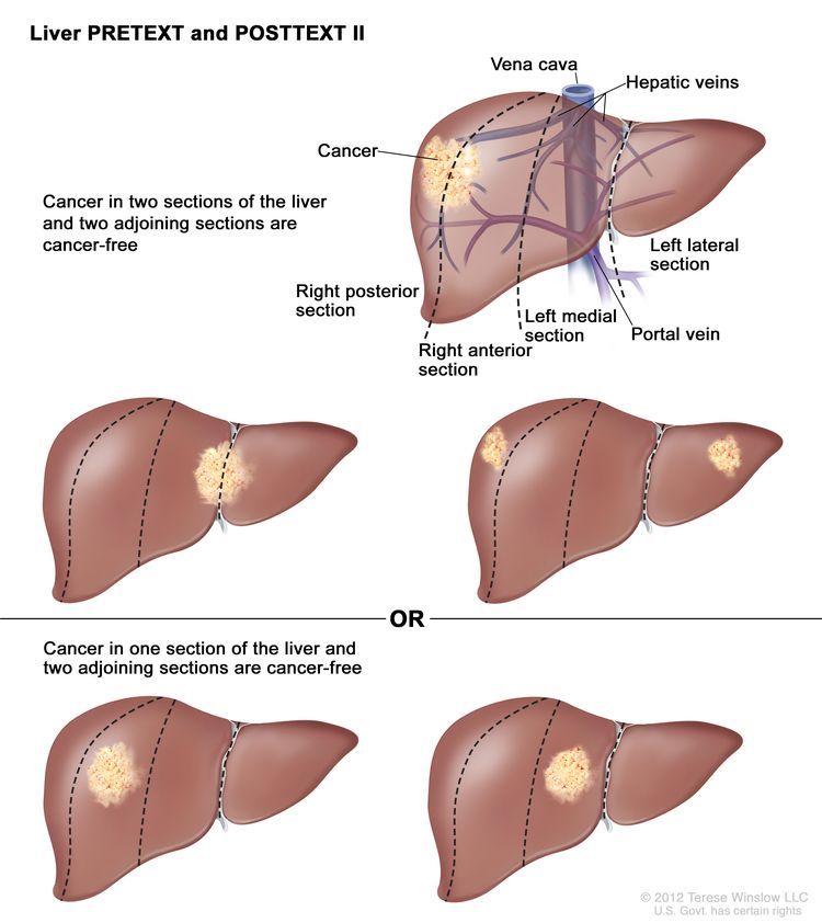
Childhood Liver Cancer Treatment (PDQ®)–Health Professional Version

Childhood Liver Cancer Treatment (PDQ®)–Health Professional Version

General Information About Childhood Liver Cancer

Liver cancer is a rare malignancy in children and adolescents and is divided into the following two major histological subgroups:

Other less common histologies include the following:

Harmonization of Childhood Liver Cancer Data and Definitions

Historically, four major study groups have performed prospective clinical trials in children with liver tumors: The International Childhood Liver Tumors Strategy Group (previously known as Société Internationale d’Oncologie Pédiatrique–Epithelial Liver Tumor Study Group [SIOPEL]), the Gesellschaft für Pädiatrische Onkologie und Hämatologie (Society for Paediatric Oncology and Haematology [GPOH]), the Japanese Study Group for Pediatric Liver Tumors (JPLT), and the Children’s Oncology Group (COG), including its predecessor groups the Children’s Cancer Group (CCG) and Pediatric Oncology Group (POG). These groups historically had disparate risk stratification categories, data elements that were monitored, and pathological and radiological definitions, making it difficult to compare outcomes across continents.

A collaborative effort among all four study groups collated their disparate data into a unified database called the Children’s Hepatic Tumor International Collaboration (CHIC). The CHIC group analyzed clinical features and outcomes in a database that included 1,605 patients with hepatoblastoma treated in eight separate multicenter clinical trials, with central review of all tumor imaging and histological details.[1] Patients who underwent orthotopic liver transplant were also included.[2]

References
  1. Czauderna P, Haeberle B, Hiyama E, et al.: The Children’s Hepatic tumors International Collaboration (CHIC): Novel global rare tumor database yields new prognostic factors in hepatoblastoma and becomes a research model. Eur J Cancer 52: 92-101, 2016. [PUBMED Abstract]
  2. Otte JB, Pritchard J, Aronson DC, et al.: Liver transplantation for hepatoblastoma: results from the International Society of Pediatric Oncology (SIOP) study SIOPEL-1 and review of the world experience. Pediatr Blood Cancer 42 (1): 74-83, 2004. [PUBMED Abstract]

Cellular Classification of Childhood Liver Cancer

Liver tumors are rare in children. A definitive pathological diagnosis may be challenging because of the rarity of the tumor and the lack of a universal classification system before the Children’s Hepatic Tumor International Collaboration (CHIC) harmonization efforts. Systematic central histopathological review of these tumors, performed as part of pediatric collaborative therapeutic protocols, has allowed the identification of histological subtypes with distinct clinical associations.

The Children’s Oncology Group (COG) Liver Tumor Committee sponsored an International Pathology Symposium in 2011 to discuss the histopathology and classification of pediatric liver tumors (hepatoblastoma, in particular) and develop an International Pediatric Liver Tumors Consensus Classification that would be required for international collaborative projects. The results were published in 2014.[1] In a post-hoc expert consensus review of 599 hepatoblastoma cases treated across five multicenter trials, 570 (95%) were validated and independently re-confirmed to be hepatoblastoma using the CHIC pathology guidelines.[2] This standardized, clinically meaningful classification has allowed the integration of new biological parameters and tumor genetics within a common pathological language to help improve future patient management and outcomes.

For information about the histology of each childhood liver cancer subtype, see the following sections:

References
  1. López-Terrada D, Alaggio R, de Dávila MT, et al.: Towards an international pediatric liver tumor consensus classification: proceedings of the Los Angeles COG liver tumors symposium. Mod Pathol 27 (3): 472-91, 2014. [PUBMED Abstract]
  2. Cho SJ, Ranganathan S, Alaggio R, et al.: Consensus classification of pediatric hepatocellular tumors: A report from the Children’s Hepatic tumors International Collaboration (CHIC). Pediatr Blood Cancer : e30505, 2023. [PUBMED Abstract]

Tumor Stratification by Imaging

A main treatment goal for children and adolescents with liver cancer is surgical extirpation of the primary tumor. Risk grouping depends heavily on factors determined by imaging that are related to safe surgical resection of the tumor, as well as the PRETEXT grouping. These imaging findings include the section or sections of the liver that are involved with the tumor and additional findings, called annotation factors, that impact surgical decision making and prognosis.

Risk stratification of children and adolescents with liver cancer involves the use of high-quality, cross-sectional imaging. Three-phase computed tomography scanning (noncontrast, arterial, and venous) or magnetic resonance imaging (MRI) with contrast agents are used. MRI with gadoxetate disodium, a gadolinium-based agent that is preferentially taken up and excreted by hepatocytes, is being used with increased frequency and may improve detection of multifocal disease.[1]

PRETEXT and POSTTEXT Group Definitions

The imaging grouping systems used to radiologically define the extent of liver involvement by the tumor are designated as the following:

  • PRETEXT (PRE-Treatment EXTent of disease): The extent of liver involvement is defined before therapy.
  • POSTTEXT (POST-Treatment EXTent of disease): The extent of liver involvement is defined in response to therapy.

PRETEXT

Major multicenter trial groups use PRETEXT as a central component of risk stratification schemes that guide treatment of hepatoblastoma. PRETEXT is based on the Couinaud eight-segment anatomical structure of the liver using cross-sectional imaging.

The PRETEXT system divides the liver into four parts, called sections. The left lobe of the liver consists of a lateral section (Couinaud segments I, II, and III) and a medial section (segment IV), whereas the right lobe consists of an anterior section (segments V and VIII) and a posterior section (segments VI and VII) (see Figure 1). PRETEXT groups were devised by the Société Internationale d’Oncologie Pédiatrique–Epithelial Liver Tumor Study Group (SIOPEL) for their first trial, SIOPEL-1,[2] and revised for the SIOPEL-3 trial in 2007.[3]

EnlargeDrawing showing 4 sections of the liver: the right posterior section, the right anterior section, the left medial section, and the left lateral section. Also shown are 8 segments (I-VIII), each corresponding to a specific section of the liver. The boundaries of each section are separated by the right hepatic vein, middle hepatic vein, and left hepatic vein. The vena cava and portal vein are also shown.
Figure 1. The liver is divided into four sections: the right posterior section, the right anterior section, the left medial section, and the left lateral section. Each section of the liver is further divided into segments: segments VI and VII make up the right posterior section, segments V and VIII make up the right anterior section, segment IV makes up the left medial section, and segments II and III make up the left lateral section. Segment I is found deep in the left side of the liver, in front of the inferior vena cava and behind the right, middle, and left hepatic veins.

PRETEXT group assignment I, II, III, or IV is determined by the number of uninvolved sections of the liver. PRETEXT is further described by annotation factors. Annotation factors include findings that are important for surgical management and evidence of tumor extension beyond the hepatic parenchyma of the major sections, including metastatic disease. For detailed descriptions of the PRETEXT groups, see Table 1. For descriptions of the annotation factors, see Table 2.

Annotation factors identify the extent of tumor involvement of the major vessels and its effect on venous inflow and outflow. These factors provide critical knowledge for the surgeon and can affect surgical outcomes. At one time, definitions of gross vascular involvement used by the Children’s Oncology Group (COG) and major liver surgery centers in the United States differed from those used by SIOPEL and in Europe. These differences have been resolved, and the new definitions are being used in an international trial.[4]

Although PRETEXT can be used to predict tumor resectability, it has limitations. It can be difficult to distinguish real invasion beyond the anatomical border of a given hepatic section from compression and displacement by the tumor, especially at diagnosis. Additionally, it can be difficult to distinguish between vessel encroachment and involvement, particularly if imaging is inadequate. The PRETEXT group assignment has a moderate degree of interobserver variability. In a report using data from the SIOPEL-1 study, the preoperative PRETEXT group aligned with postoperative pathological findings only 51% of the time, with overstaging in 37% of patients and understaging in 12% of patients.[5]

Because distinguishing PRETEXT group assignment is difficult, central review of imaging is critical and is generally performed in all major clinical trials. For patients not enrolled in clinical trials, expert radiological review should be considered in questionable cases in which the PRETEXT group assignment affects choice of treatment.

Table 1. Definitions of PRETEXT and POSTTEXT Groupsa
PRETEXT and POSTTEXT Groups Definition Image
aAdapted from Roebuck et al.[3]
I One section involved; three adjoining sections are tumor free.
EnlargeLiver PRETEXT and POSTTEXT I; drawing shows two livers. Dotted lines divide each liver into four vertical sections of about the same size. In the first liver, cancer is shown in the section on the far left. In the second liver, cancer is shown in the section on the far right.
II One or two sections involved; two adjoining sections are tumor free.
EnlargeLiver PRETEXT and POSTTEXT II; drawing shows five livers. Dotted lines divide each liver into four vertical sections that are about the same size. In the first liver, cancer is shown in the two sections on the left. In the second liver, cancer is shown in the two sections on the right. In the third liver, cancer is shown in the far left and far right sections. In the fourth liver, cancer is shown in the second section from the left. In the fifth liver, cancer is shown in the second section from the right.
III Two or three sections involved; one adjoining section is tumor free.
EnlargeLiver PRETEXT and POSTTEXT III; drawing shows seven livers. Dotted lines divide each liver into four vertical sections that are about the same size. In the first liver, cancer is shown in three sections on the left. In the second liver, cancer is shown in the two sections on the left and in the section on the far right. In the third liver, cancer is shown in the section on the far left and in the two sections on the right. In the fourth liver, cancer is shown in three sections on the right. In the fifth liver, cancer is shown in the two middle sections. In the sixth liver, cancer is shown in the section on the far left and in the second section from the right. In the seventh liver, cancer is shown in the section on the far right and in the second section from the left.
IV Four sections involved.
EnlargeLiver PRETEXT and POSTTEXT IV; drawing shows two livers. Dotted lines divide each liver into four vertical sections that are about the same size. In the first liver, cancer is shown across all four sections. In the second liver, cancer is shown in the two sections on the left and spots of cancer are shown in the two sections on the right.
Table 2. Annotation Factors for Describing PRETEXT and POSTTEXT Groupsa
Annotation Factors Definition
CT = computed tomography; MRI = magnetic resonance imaging; HU = Hounsfield unit.
aAdapted from Roebuck et al.[3]
bAdditional details describing the annotation factors have been published.[4]
Vb Venous involvement: Vascular involvement of the retrohepatic vena cava or involvement of all three major hepatic veins (right, middle, and left).
V0   Tumor within 1 cm.
V1   Tumor abutting.
V2   Tumor compressing or distorting.
V3   Tumor ingrowth, encasement, or thrombus.
Pb Portal involvement: Vascular involvement of the main portal vein and/or both right and left portal veins.
P0   Tumor within 1 cm.
P1   Tumor abutting the main portal vein, the right and left portal veins, or the portal vein bifurcation.
  P2   Tumor compressing the main portal vein, the right and left portal veins, or the portal vein bifurcation.
P3   Tumor ingrowth, encasement (>50% or >180 degrees), or intravascular thrombus within the main portal vein, the right and left portal veins, or the portal vein bifurcation.
Eb Extrahepatic spread of disease. Any one of the following criteria is met:
E1   Tumor crosses boundaries/tissue planes.
E2   Tumor is surrounded by normal tissue more than 180 degrees.
  E3   Peritoneal nodules (not lymph nodes) are present so that there is at least one nodule measuring ≥10 mm or at least two nodules measuring ≥5 mm.
Mb Distant metastases. Any one of the following criteria is met:
  M1   One noncalcified pulmonary nodule ≥5 mm in diameter.
  M2   Two or more noncalcified pulmonary nodules, each ≥3 mm in diameter.
  M3   Pathologically proven metastatic disease.
C Tumor involving the caudate.
F Multifocality. Two or more discrete hepatic tumors with normal intervening liver tissue.
Nb Lymph node metastases. Any one of the following criteria is met:
  N1   Lymph node with short-axis diameter of >1 cm.
  N2   Portocaval lymph node with short-axis diameter >1.5 cm.
  N3   Spherical lymph node shape with loss of fatty hilum.
Rb Tumor rupture. Free fluid in the abdomen or pelvis with one or more of the following findings of hemorrhage:
  R1   Internal complexity/septations within fluid.
  R2   High-density fluid on CT (>25 HU).
  R3   Imaging characteristics of blood or blood degradation products on MRI.
  R4   Heterogeneous fluid on ultrasound with echogenic debris.
  R5   Visible defect in tumor capsule OR tumor cells are present within the peritoneal fluid OR rupture diagnosed pathologically in patients who have received an upfront resection.

POSTTEXT

The POSTTEXT group is determined after patients receive chemotherapy. The greatest chemotherapy response, measured as decreases in tumor size and alpha-fetoprotein (AFP) level, occurs after the first two cycles of chemotherapy.[6,7] A study that evaluated surgical resectability after two versus four cycles of chemotherapy showed that many tumors may be resectable after two cycles.[6]

Evans Surgical Staging for Childhood Liver Cancer

The COG/Evans staging system, based on operative findings and surgical resectability, was used for many years in the United States to group and determine treatment for children with liver cancer (see Table 3).[810] Currently, other risk stratification systems are predominantly used to classify patients and determine treatment strategy, although the Paediatric Hepatic International Tumour Trial (PHITT) uses the Evans system for patients with hepatocellular carcinoma. For more information, see Table 5.

Table 3. Definition of Evans Surgical Staging
Evans Surgical Stage Definition
Stage I The tumor is completely resected.
Stage II Microscopic residual tumor remains after resection.
Stage III There are no distant metastases and at least one of the following is true: (1) the tumor is either unresectable or the tumor is resected with gross residual tumor; (2) there are positive extrahepatic lymph nodes.
Stage IV There is distant metastasis, regardless of the extent of liver involvement.
References
  1. Meyers AB, Towbin AJ, Geller JI, et al.: Hepatoblastoma imaging with gadoxetate disodium-enhanced MRI–typical, atypical, pre- and post-treatment evaluation. Pediatr Radiol 42 (7): 859-66, 2012. [PUBMED Abstract]
  2. Brown J, Perilongo G, Shafford E, et al.: Pretreatment prognostic factors for children with hepatoblastoma– results from the International Society of Paediatric Oncology (SIOP) study SIOPEL 1. Eur J Cancer 36 (11): 1418-25, 2000. [PUBMED Abstract]
  3. Roebuck DJ, Aronson D, Clapuyt P, et al.: 2005 PRETEXT: a revised staging system for primary malignant liver tumours of childhood developed by the SIOPEL group. Pediatr Radiol 37 (2): 123-32; quiz 249-50, 2007. [PUBMED Abstract]
  4. Towbin AJ, Meyers RL, Woodley H, et al.: 2017 PRETEXT: radiologic staging system for primary hepatic malignancies of childhood revised for the Paediatric Hepatic International Tumour Trial (PHITT). Pediatr Radiol 48 (4): 536-554, 2018. [PUBMED Abstract]
  5. Aronson DC, Schnater JM, Staalman CR, et al.: Predictive value of the pretreatment extent of disease system in hepatoblastoma: results from the International Society of Pediatric Oncology Liver Tumor Study Group SIOPEL-1 study. J Clin Oncol 23 (6): 1245-52, 2005. [PUBMED Abstract]
  6. Lovvorn HN, Ayers D, Zhao Z, et al.: Defining hepatoblastoma responsiveness to induction therapy as measured by tumor volume and serum alpha-fetoprotein kinetics. J Pediatr Surg 45 (1): 121-8; discussion 129, 2010. [PUBMED Abstract]
  7. Venkatramani R, Stein JE, Sapra A, et al.: Effect of neoadjuvant chemotherapy on resectability of stage III and IV hepatoblastoma. Br J Surg 102 (1): 108-13, 2015. [PUBMED Abstract]
  8. Ortega JA, Krailo MD, Haas JE, et al.: Effective treatment of unresectable or metastatic hepatoblastoma with cisplatin and continuous infusion doxorubicin chemotherapy: a report from the Childrens Cancer Study Group. J Clin Oncol 9 (12): 2167-76, 1991. [PUBMED Abstract]
  9. Douglass EC, Reynolds M, Finegold M, et al.: Cisplatin, vincristine, and fluorouracil therapy for hepatoblastoma: a Pediatric Oncology Group study. J Clin Oncol 11 (1): 96-9, 1993. [PUBMED Abstract]
  10. Ortega JA, Douglass EC, Feusner JH, et al.: Randomized comparison of cisplatin/vincristine/fluorouracil and cisplatin/continuous infusion doxorubicin for treatment of pediatric hepatoblastoma: A report from the Children’s Cancer Group and the Pediatric Oncology Group. J Clin Oncol 18 (14): 2665-75, 2000. [PUBMED Abstract]

Treatment Option Overview for Childhood Liver Cancer

Many of the improvements in survival in childhood cancer have been made using new therapies that have attempted to improve on the best available, accepted therapy. Clinical trials in pediatrics are designed to compare potentially better therapy with therapy that is currently accepted as standard. This comparison may be done in a randomized study of two treatment arms or by evaluating a single new treatment and comparing the results with those previously obtained with standard therapy.

Because of the relative rarity of cancer in children, all children with liver cancer should be considered for a clinical trial if available. Treatment planning by a multidisciplinary team of cancer specialists with experience treating tumors of childhood is required to determine and implement optimal treatment.[1]

Surgery

Historically, complete surgical resection of the primary tumor has been essential for cure of malignant liver tumors in children.[26]; [7][Level of evidence C1] This approach continues to be the goal of definitive surgical procedures, which are often combined with chemotherapy. The surgeon performs a highly sophisticated liver resection in children and adolescents with primary liver tumors after the diagnosis is confirmed by pathological investigation of intraoperative frozen sections. While complete surgical resection is important for all liver tumors, it is especially important for hepatocellular carcinoma because curative chemotherapy is not available. In patients with advanced hepatoblastoma, postoperative complications are associated with worsened overall survival (OS).[8]

The three surgical options to treat primary pediatric liver cancer include the following:

  • Initial surgical resection (alone or with adjuvant chemotherapy).
  • Delayed surgical resection (with neoadjuvant chemotherapy).
  • Orthotopic liver (cadaveric and living donor) transplant (most often with neoadjuvant chemotherapy).

The decision on which surgical approach to use (e.g., partial hepatectomy, extended resection, or transplant) depends on many factors, including the following:

  • PRE-Treatment EXTent of disease (PRETEXT) group and POST-Treatment EXTent of disease (POSTTEXT) group.
  • Size of the primary tumor.
  • Presence of multifocal hepatic disease.
  • Gross vascular involvement.
  • Alpha-fetoprotein (AFP) levels.
  • Whether preoperative chemotherapy is likely to convert an unresectable tumor into a resectable tumor.
  • Whether hepatic disease meets surgical and histopathological criteria for orthotopic liver transplant.

Timing of the surgical approach is critical. Surgeons who have experience performing pediatric liver resections and transplants are involved early in the decision-making process to determine optimal timing and extent of resection.

Early involvement, preferably at diagnosis, with an experienced pediatric liver surgeon is especially important in patients with PRETEXT group III or IV or involvement of major liver vessels (positive annotation factors V [venous] or P [portal]).[9] Although vascular involvement was initially thought to be a contraindication to resection, experienced liver surgeons are sometimes able to successfully resect the tumor and avoid performing a transplant.[1012]; [13][Level of evidence C1] Patients with vascular involvement and tumors that have been deemed nonresectable by the pediatric surgical expert should be referred to a transplant center to avoid unnecessary delays in evaluation and listing for transplant.

Intraoperative ultrasonography may result in further delineation of tumor extent and location and can affect intraoperative management.[14] Preoperative infusion of indocyanine green, a fluoroactive agent that is concentrated in the liver and retained by abnormal liver tumors, has also been used to provide visual intraoperative guidance to locate the tumor and assess proximity to surgical margins.[15,16]

If the tumor is determined to be unresectable, measures to reduce the tumor size to make a complete surgical resection possible need to be considered. These measures include preoperative intravenous chemotherapy, transarterial chemotherapy, or transarterial radioactive therapy. These efforts must be carefully coordinated with the surgical team to facilitate planning of resection. Prolonged chemotherapy can lead to unnecessary delays and, in rare cases, tumor progression. If the tumor can be completely excised by an experienced surgical team, less postoperative chemotherapy may be needed. Incomplete resection must be avoided because patients who undergo rescue transplants of incompletely resected tumors have an inferior outcome, compared with patients who undergo transplant as the primary surgical therapy.[17][Level of evidence C1] Accomplishing the appropriate surgery at resection is critical.

The approach taken by the Children’s Oncology Group (COG) in North American clinical trials is to perform surgery initially when a complete resection can be done with a simple, negative-margin hemihepatectomy. The COG AHEP0731 (NCT00980460) trial studied the use of PRETEXT and POSTTEXT to determine the optimal approach and timing of surgery. POSTTEXT imaging grouping was performed after two and four cycles of chemotherapy to determine the optimal time for definitive surgery.[6,18] For more information, see the Tumor Stratification by Imaging section.

Orthotopic liver transplant

Liver transplants have been associated with significant success in the treatment of children with unresectable hepatic tumors.[19]; [2022][Level of evidence C1] A review of the world experience has documented a posttransplant survival rate of 70% to 80% for children with hepatoblastomas.[17,2325] Intravenous vascular invasion, positive lymph nodes, and contiguous extrahepatic spread did not have significant adverse effects on outcome. Adjuvant chemotherapy after transplant may decrease the risk of tumor recurrence, but its use has not been studied definitively in a randomized clinical trial.[26]

Evidence (orthotopic liver transplant):

  1. The United Network for Organ Sharing (UNOS) database was queried for all patients younger than 18 years with a primary malignant liver tumor who underwent an orthotopic liver transplant between 1987 and 2012 (N = 544). The patients were diagnosed with hepatoblastoma (n = 376, 70%), hepatocellular carcinoma (n = 84, 15%), and other tumors (n = 84, 15%). Patients with hepatocellular carcinoma were older, more often hospitalized at the time of transplant, and more likely to receive a cadaveric organ than were patients with hepatoblastoma.[27]
    1. The 5-year patient survival rate was 73%, and the graft survival rate was 74% for the entire cohort, with most deaths resulting from malignancy. On multivariate analysis, independent predictors of 5-year patient and graft survival included the following:
      1. Diagnosis.
        • For the study period of 1987 to 2012, the 5-year survival rate was 76% and the graft survival rate was 77% for patients with hepatoblastoma. The survival and graft survival rates were 63% for patients with hepatocellular carcinoma.
        • For the study period of 2009 to 2012, the 3-year survival and graft survival rates were 84% for patients with hepatoblastoma. The survival and graft survival rates were 85% for patients with hepatocellular carcinoma.
      2. Transplant era.
        • The death rate by hazard ratio was 1.0 for the period before 2002, 0.72 for the period of 2002 to 2009, and 0.54 for the period of 2009 to 2012.
      3. Medical condition at transplant.
        • For hepatoblastoma patients, the survival rate by hazard ratio was 1.0 for hospitalized patients versus 1.81 for nonhospitalized patients at the time of transplant.
        • For hepatocellular carcinoma patients, the survival rate by hazard ratio was 1.0 for hospitalized patients versus 1.92 for nonhospitalized patients.
        • Patients hospitalized in the intensive care unit did not fare worse than patients not in the intensive care unit.
  2. A report of 149 patients with hepatocellular carcinoma younger than 21 years who underwent transplants between 1987 and 2015 used detailed data collected at all U.S. pediatric transplant centers.[19]
    • The 1-year graft survival rate of about 85% did not differ from the survival rate for patients with hepatoblastoma or biliary atresia. Survival rates continued to decline over time, from 85% at 1 year to 52% at 5 years and 43% at 10 years, a more dramatic decline than that seen for hepatoblastoma or biliary atresia.
    • The survival after transplant did not differ from that of adults who underwent transplant for hepatocellular carcinoma.
    • Of the patients with hepatocellular carcinoma, 22 received a diagnosis after transplant for medical cirrhotic disease such as tyrosinemia. They had a superior outcome, but it was not statistically significant compared with the rest of the patients.
  3. A review of the Surveillance, Epidemiology, and End Results (SEER) Program database and many single-institution series have reported results similar to the UNOS database study described above.[11,2022,28]; [25][Level of evidence C1]
  4. In a three-institution study of children with hepatocellular carcinoma, the overall 5-year disease-free survival rate was approximately 60%.[29]
  5. In a study that used the Society of Pediatric Liver Transplantation (SPLIT) database to identify patients who underwent liver transplant between 2011 and 2019, the following was reported:[30][Level of evidence C2]
    • The 3-year event-free survival (EFS) rate was 81% for patients with hepatoblastoma who received a transplant (n = 157).
    • The 3-year EFS rate was 62% for patients with hepatocellular carcinoma who received a transplant (n = 18).
    • Of the patients who received a transplant to treat hepatoblastoma, 6.9% had PRETEXT II disease and 15.3% had POSTTEXT I/II disease.
    • Tumor extent did not impact survival (P = NS).
    • Patients who received transplants for salvage (n = 13) and patients who received transplants for primary hepatoblastoma had similar 3-year EFS rates (62% vs. 78%; P = NS).
    • Among patients who received transplants for hepatocellular carcinoma, the 3-year EFS rate was poorer in older patients (38% for patients aged ≥8 years vs. 86% for patients aged <8 years; P < .001).

Application of the Milan criteria for UNOS selection of recipients of deceased donor livers is controversial.[31,32] The Milan criteria for liver transplant are directed toward adults with cirrhosis and hepatocellular carcinoma. The criteria do not apply to children and adolescents with hepatocellular carcinoma, especially those without cirrhosis.

Cirrhosis is an underlying risk factor for the development of hepatocellular carcinoma in children who suffer from certain diseases or conditions. These diseases include perinatally acquired hepatitis B, hepatorenal tyrosinemia, progressive familial intrahepatic cholestasis, glycogen storage disease, Alagille syndrome, and other conditions. Improvements in screening methodology have allowed for earlier identification and treatment of some of these conditions, as well as monitoring for development of hepatocellular carcinoma. Nevertheless, because of the poor prognosis of patients with hepatocellular carcinoma, liver transplant should be considered for diseases or conditions that have resulted in early findings of cirrhosis, before the development of liver failure or malignancy.[33]

Living-donor liver transplant for hepatic malignancy is more common in children than adults, and the outcome is similar to those undergoing cadaveric liver transplant.[34,35] In patients with hepatocellular carcinoma, gross vascular invasion, distant metastases, lymph node involvement, tumor size, and male sex were significant risk factors for recurrence. In one report, 33 patients with hepatoblastoma and 10 patients with hepatocellular carcinoma were treated with living-donor liver transplants. For the hepatoblastoma patients, the 5-year OS rate was 87.4%, and the EFS rate was 75.8%. The 5-year OS and EFS rates were 75.4% for the patients with hepatocellular carcinoma. The presence of renal vein invasion was associated with an increased incidence of recurrence and death (P = .28).[36][Level of evidence C1]

Surgical resection for metastatic disease

Surgical resection of metastatic disease is often recommended, but the rate of cure in children with hepatoblastoma has not been fully determined. Resection of metastases may be done for areas of locally invasive disease (e.g., diaphragm) and isolated brain metastases. Resection of pulmonary metastases should be considered if the number of metastases is limited.[3740] In a North American study of 38 patients who presented with pulmonary metastases at diagnosis, only nine patients underwent surgical resection. The timing of pulmonary resection in relation to definitive resection of the primary tumor varied (two patients before, five patients simultaneously, and two patients after primary resection). Eight of the nine patients survived. Of 20 children with relapse restricted to the lungs, all patients received salvage chemotherapy, 8 patients had a thoracotomy and pulmonary metastasectomy, and 5 patients had a thoracotomy and biopsy. Among the 13 patients who had surgery, only 4 were long-term survivors, 2 of whom presented with stage I disease and 2 of whom presented with stage IV disease.[39]

Radiofrequency ablation has also been used to treat oligometastatic hepatoblastoma when patients prefer to avoid surgical metastasectomy.[41][Level of evidence C1]

Chemotherapy

Chemotherapy regimens used in the treatment of hepatoblastoma and hepatocellular carcinoma are described in their respective sections. Chemotherapy has been much more successful in the treatment of hepatoblastoma than in the treatment of hepatocellular carcinoma.[6,28,42] For more information, see the sections on Treatment of Hepatoblastoma and Treatment of Hepatocellular Carcinoma.

The standard of care in the United States is preoperative chemotherapy when the tumor is unresectable and postoperative chemotherapy after complete resection, even if preoperative chemotherapy has already been given.[43] Preoperative chemotherapy has been shown to benefit children with hepatoblastoma. However, postoperative chemotherapy after definitive surgical resection or liver transplant has not been investigated in a randomized fashion.

Radiation Therapy

Radiation therapy, even in combination with chemotherapy, has not cured children with unresectable hepatic tumors. A study of 154 patients with hepatoblastoma showed that radiation therapy and/or second resection of positive margins may not be necessary in some patients with incompletely resected hepatoblastoma and microscopic residual tumor.[44] Although there is no standard indication, radiation therapy may have a role in the management of patients with incompletely resected hepatoblastomas.[45] Stereotactic body radiation therapy is a safe and effective alternative treatment that has been successfully used in adult patients with hepatocellular carcinoma who are unable to undergo liver ablation/resection.[46] This highly conformal radiotherapeutic technique, when available, may be considered on an individual basis in children with hepatocellular carcinoma.

Other Treatment Approaches

Other treatment approaches include the following:

  • Transarterial chemoembolization (TACE): TACE is an image-guided, minimally invasive, nonsurgical procedure that is used to treat malignant lesions in the liver. The procedure uses a catheter to deliver both chemotherapy medication and embolization materials into the blood vessels that lead to the tumor. The arterial catheter route is image guided, most often via the hepatic artery, and perfusion of the tumor by the targeted artery may be confirmed by imaging before therapeutic injection. This procedure allows for the treatment of tumors that are not accessible with conventional surgery or radiation treatments. TACE has been used for patients with inoperable hepatoblastoma.[4749] This procedure has also been used in a few children to successfully shrink tumors to permit resections.[48]
  • Transarterial radioembolization (TARE): TARE is an image-guided, minimally invasive, nonsurgical procedure that delivers radiation therapy to treat tumors in the liver. This procedure delivers radioactive beads and blocks arterial flow within the tumor to keep the radiation inside the tumor. Glass or resin microspheres, coated most commonly with yttrium Y 90 (90Y), are delivered to the tumor via catheters placed in arteries that supply the tumor. Usually, the hepatic artery or its branches are used, but tumors may be partially supplied by parasitized surrounding vessels. Because of the risk of radiation delivery to the nearby lung, technetium Tc 99m microaggregated albumin imaging is performed with delivery via the catheter that is in place before the administration of radioactive beads to carefully measure radiation exposure to the lung. If calculations determine that lung exposure is unsafe, TARE is not pursued. TARE with 90Y has been used in children with hepatoblastoma (n = 2) and hepatocellular carcinoma (n = 2) who have unresectable tumors. After treatment with 90Y TARE, all tumors were completely resected.[50][Level of evidence C3]; [51][Level of evidence C2] This approach has also been used for palliation in children with hepatocellular carcinoma.[52] For more information, see Primary Liver Cancer Treatment.
  • High-intensity focused ultrasonography (HIFU): HIFU is a noninvasive treatment for a wide range of tumors and diseases. HIFU uses an ultrasound transducer, similar to the ones used for diagnostic imaging, but with much higher energy. The transducer focuses sound waves to generate heat at a single point in the body and destroy the target tissue. The tissue can get as hot as 66°C in only 20 seconds. This process is repeated as many times as necessary until the target tissue is destroyed. Magnetic resonance imaging is used to plan the treatment and monitor the amount of heat in real time. A combination of chemotherapy followed by TACE and HIFU showed promising results in China for children with PRETEXT III and PRETEXT IV malignant liver tumors, some of whom had resectable tumors but did not undergo surgery because of parent refusal.[53]
References
  1. Tiao GM, Bobey N, Allen S, et al.: The current management of hepatoblastoma: a combination of chemotherapy, conventional resection, and liver transplantation. J Pediatr 146 (2): 204-11, 2005. [PUBMED Abstract]
  2. Czauderna P, Otte JB, Aronson DC, et al.: Guidelines for surgical treatment of hepatoblastoma in the modern era–recommendations from the Childhood Liver Tumour Strategy Group of the International Society of Paediatric Oncology (SIOPEL). Eur J Cancer 41 (7): 1031-6, 2005. [PUBMED Abstract]
  3. Czauderna P, Mackinlay G, Perilongo G, et al.: Hepatocellular carcinoma in children: results of the first prospective study of the International Society of Pediatric Oncology group. J Clin Oncol 20 (12): 2798-804, 2002. [PUBMED Abstract]
  4. Meyers RL, Czauderna P, Otte JB: Surgical treatment of hepatoblastoma. Pediatr Blood Cancer 59 (5): 800-8, 2012. [PUBMED Abstract]
  5. Aronson DC, Meyers RL: Malignant tumors of the liver in children. Semin Pediatr Surg 25 (5): 265-275, 2016. [PUBMED Abstract]
  6. Murawski M, Weeda VB, Maibach R, et al.: Hepatocellular Carcinoma in Children: Does Modified Platinum- and Doxorubicin-Based Chemotherapy Increase Tumor Resectability and Change Outcome? Lessons Learned From the SIOPEL 2 and 3 Studies. J Clin Oncol 34 (10): 1050-6, 2016. [PUBMED Abstract]
  7. Allan BJ, Wang B, Davis JS, et al.: A review of 218 pediatric cases of hepatocellular carcinoma. J Pediatr Surg 49 (1): 166-71; discussion 171, 2014. [PUBMED Abstract]
  8. Becker K, Furch C, Schmid I, et al.: Impact of postoperative complications on overall survival of patients with hepatoblastoma. Pediatr Blood Cancer 62 (1): 24-8, 2015. [PUBMED Abstract]
  9. D’Antiga L, Vallortigara F, Cillo U, et al.: Features predicting unresectability in hepatoblastoma. Cancer 110 (5): 1050-8, 2007. [PUBMED Abstract]
  10. Lautz TB, Ben-Ami T, Tantemsapya N, et al.: Successful nontransplant resection of POST-TEXT III and IV hepatoblastoma. Cancer 117 (9): 1976-83, 2011. [PUBMED Abstract]
  11. Fonseca A, Gupta A, Shaikh F, et al.: Extreme hepatic resections for the treatment of advanced hepatoblastoma: Are planned close margins an acceptable approach? Pediatr Blood Cancer 65 (2): , 2018. [PUBMED Abstract]
  12. Fuchs J, Cavdar S, Blumenstock G, et al.: POST-TEXT III and IV Hepatoblastoma: Extended Hepatic Resection Avoids Liver Transplantation in Selected Cases. Ann Surg 266 (2): 318-323, 2017. [PUBMED Abstract]
  13. Baertschiger RM, Ozsahin H, Rougemont AL, et al.: Cure of multifocal panhepatic hepatoblastoma: is liver transplantation always necessary? J Pediatr Surg 45 (5): 1030-6, 2010. [PUBMED Abstract]
  14. Felsted AE, Shi Y, Masand PM, et al.: Intraoperative ultrasound for liver tumor resection in children. J Surg Res 198 (2): 418-23, 2015. [PUBMED Abstract]
  15. Rossi G, Tarasconi A, Baiocchi G, et al.: Fluorescence guided surgery in liver tumors: applications and advantages. Acta Biomed 89 (9-S): 135-140, 2018. [PUBMED Abstract]
  16. Shen Q, Liu X, Pan S, et al.: Effectiveness of indocyanine green fluorescence imaging in resection of hepatoblastoma. Pediatr Surg Int 39 (1): 181, 2023. [PUBMED Abstract]
  17. Otte JB, Pritchard J, Aronson DC, et al.: Liver transplantation for hepatoblastoma: results from the International Society of Pediatric Oncology (SIOP) study SIOPEL-1 and review of the world experience. Pediatr Blood Cancer 42 (1): 74-83, 2004. [PUBMED Abstract]
  18. Venkatramani R, Stein JE, Sapra A, et al.: Effect of neoadjuvant chemotherapy on resectability of stage III and IV hepatoblastoma. Br J Surg 102 (1): 108-13, 2015. [PUBMED Abstract]
  19. Vinayak R, Cruz RJ, Ranganathan S, et al.: Pediatric liver transplantation for hepatocellular cancer and rare liver malignancies: US multicenter and single-center experience (1981-2015). Liver Transpl 23 (12): 1577-1588, 2017. [PUBMED Abstract]
  20. Guiteau JJ, Cotton RT, Karpen SJ, et al.: Pediatric liver transplantation for primary malignant liver tumors with a focus on hepatic epithelioid hemangioendothelioma: the UNOS experience. Pediatr Transplant 14 (3): 326-31, 2010. [PUBMED Abstract]
  21. Malek MM, Shah SR, Atri P, et al.: Review of outcomes of primary liver cancers in children: our institutional experience with resection and transplantation. Surgery 148 (4): 778-82; discussion 782-4, 2010. [PUBMED Abstract]
  22. Héry G, Franchi-Abella S, Habes D, et al.: Initial liver transplantation for unresectable hepatoblastoma after chemotherapy. Pediatr Blood Cancer 57 (7): 1270-5, 2011. [PUBMED Abstract]
  23. Suh MY, Wang K, Gutweiler JR, et al.: Safety of minimal immunosuppression in liver transplantation for hepatoblastoma. J Pediatr Surg 43 (6): 1148-52, 2008. [PUBMED Abstract]
  24. Zsíros J, Maibach R, Shafford E, et al.: Successful treatment of childhood high-risk hepatoblastoma with dose-intensive multiagent chemotherapy and surgery: final results of the SIOPEL-3HR study. J Clin Oncol 28 (15): 2584-90, 2010. [PUBMED Abstract]
  25. Khan AS, Brecklin B, Vachharajani N, et al.: Liver Transplantation for Malignant Primary Pediatric Hepatic Tumors. J Am Coll Surg 225 (1): 103-113, 2017. [PUBMED Abstract]
  26. Browne M, Sher D, Grant D, et al.: Survival after liver transplantation for hepatoblastoma: a 2-center experience. J Pediatr Surg 43 (11): 1973-81, 2008. [PUBMED Abstract]
  27. Hamilton EC, Balogh J, Nguyen DT, et al.: Liver transplantation for primary hepatic malignancies of childhood: The UNOS experience. J Pediatr Surg : , 2017. [PUBMED Abstract]
  28. McAteer JP, Goldin AB, Healey PJ, et al.: Surgical treatment of primary liver tumors in children: outcomes analysis of resection and transplantation in the SEER database. Pediatr Transplant 17 (8): 744-50, 2013. [PUBMED Abstract]
  29. Reyes JD, Carr B, Dvorchik I, et al.: Liver transplantation and chemotherapy for hepatoblastoma and hepatocellular cancer in childhood and adolescence. J Pediatr 136 (6): 795-804, 2000. [PUBMED Abstract]
  30. Boster JM, Superina R, Mazariegos GV, et al.: Predictors of survival following liver transplantation for pediatric hepatoblastoma and hepatocellular carcinoma: Experience from the Society of Pediatric Liver Transplantation (SPLIT). Am J Transplant 22 (5): 1396-1408, 2022. [PUBMED Abstract]
  31. Otte JB: Should the selection of children with hepatocellular carcinoma be based on Milan criteria? Pediatr Transplant 12 (1): 1-3, 2008. [PUBMED Abstract]
  32. de Ville de Goyet J, Meyers RL, Tiao GM, et al.: Beyond the Milan criteria for liver transplantation in children with hepatic tumours. Lancet Gastroenterol Hepatol 2 (6): 456-462, 2017. [PUBMED Abstract]
  33. Khanna R, Verma SK: Pediatric hepatocellular carcinoma. World J Gastroenterol 24 (35): 3980-3999, 2018. [PUBMED Abstract]
  34. Sevmis S, Karakayali H, Ozçay F, et al.: Liver transplantation for hepatocellular carcinoma in children. Pediatr Transplant 12 (1): 52-6, 2008. [PUBMED Abstract]
  35. Faraj W, Dar F, Marangoni G, et al.: Liver transplantation for hepatoblastoma. Liver Transpl 14 (11): 1614-9, 2008. [PUBMED Abstract]
  36. Pire A, Tambucci R, De Magnée C, et al.: Living donor liver transplantation for hepatic malignancies in children. Pediatr Transplant 25 (7): e14047, 2021. [PUBMED Abstract]
  37. Feusner JH, Krailo MD, Haas JE, et al.: Treatment of pulmonary metastases of initial stage I hepatoblastoma in childhood. Report from the Childrens Cancer Group. Cancer 71 (3): 859-64, 1993. [PUBMED Abstract]
  38. Zsiros J, Brugieres L, Brock P, et al.: Dose-dense cisplatin-based chemotherapy and surgery for children with high-risk hepatoblastoma (SIOPEL-4): a prospective, single-arm, feasibility study. Lancet Oncol 14 (9): 834-42, 2013. [PUBMED Abstract]
  39. Meyers RL, Katzenstein HM, Krailo M, et al.: Surgical resection of pulmonary metastatic lesions in children with hepatoblastoma. J Pediatr Surg 42 (12): 2050-6, 2007. [PUBMED Abstract]
  40. O’Neill AF, Towbin AJ, Krailo MD, et al.: Characterization of Pulmonary Metastases in Children With Hepatoblastoma Treated on Children’s Oncology Group Protocol AHEP0731 (The Treatment of Children With All Stages of Hepatoblastoma): A Report From the Children’s Oncology Group. J Clin Oncol 35 (30): 3465-3473, 2017. [PUBMED Abstract]
  41. Yevich S, Calandri M, Gravel G, et al.: Reiterative Radiofrequency Ablation in the Management of Pediatric Patients with Hepatoblastoma Metastases to the Lung, Liver, or Bone. Cardiovasc Intervent Radiol 42 (1): 41-47, 2019. [PUBMED Abstract]
  42. Weeda VB, Murawski M, McCabe AJ, et al.: Fibrolamellar variant of hepatocellular carcinoma does not have a better survival than conventional hepatocellular carcinoma–results and treatment recommendations from the Childhood Liver Tumour Strategy Group (SIOPEL) experience. Eur J Cancer 49 (12): 2698-704, 2013. [PUBMED Abstract]
  43. Czauderna P, Lopez-Terrada D, Hiyama E, et al.: Hepatoblastoma state of the art: pathology, genetics, risk stratification, and chemotherapy. Curr Opin Pediatr 26 (1): 19-28, 2014. [PUBMED Abstract]
  44. Schnater JM, Aronson DC, Plaschkes J, et al.: Surgical view of the treatment of patients with hepatoblastoma: results from the first prospective trial of the International Society of Pediatric Oncology Liver Tumor Study Group. Cancer 94 (4): 1111-20, 2002. [PUBMED Abstract]
  45. Habrand JL, Nehme D, Kalifa C, et al.: Is there a place for radiation therapy in the management of hepatoblastomas and hepatocellular carcinomas in children? Int J Radiat Oncol Biol Phys 23 (3): 525-31, 1992. [PUBMED Abstract]
  46. Wang PM, Chung NN, Hsu WC, et al.: Stereotactic body radiation therapy in hepatocellular carcinoma: Optimal treatment strategies based on liver segmentation and functional hepatic reserve. Rep Pract Oncol Radiother 20 (6): 417-24, 2015 Nov-Dec. [PUBMED Abstract]
  47. Xianliang H, Jianhong L, Xuewu J, et al.: Cure of hepatoblastoma with transcatheter arterial chemoembolization. J Pediatr Hematol Oncol 26 (1): 60-3, 2004. [PUBMED Abstract]
  48. Malogolowkin MH, Stanley P, Steele DA, et al.: Feasibility and toxicity of chemoembolization for children with liver tumors. J Clin Oncol 18 (6): 1279-84, 2000. [PUBMED Abstract]
  49. Hirakawa M, Nishie A, Asayama Y, et al.: Efficacy of preoperative transcatheter arterial chemoembolization combined with systemic chemotherapy for treatment of unresectable hepatoblastoma in children. Jpn J Radiol 32 (9): 529-36, 2014. [PUBMED Abstract]
  50. Aguado A, Dunn SP, Averill LW, et al.: Successful use of transarterial radioembolization with yttrium-90 (TARE-Y90) in two children with hepatoblastoma. Pediatr Blood Cancer 67 (9): e28421, 2020. [PUBMED Abstract]
  51. Whitlock RS, Loo C, Patel K, et al.: Transarterial Radioembolization Treatment as a Bridge to Surgical Resection in Pediatric Hepatocellular Carcinoma. J Pediatr Hematol Oncol 43 (8): e1181-e1185, 2021. [PUBMED Abstract]
  52. Hawkins CM, Kukreja K, Geller JI, et al.: Radioembolisation for treatment of pediatric hepatocellular carcinoma. Pediatr Radiol 43 (7): 876-81, 2013. [PUBMED Abstract]
  53. Wang S, Yang C, Zhang J, et al.: First experience of high-intensity focused ultrasound combined with transcatheter arterial embolization as local control for hepatoblastoma. Hepatology 59 (1): 170-7, 2014. [PUBMED Abstract]

Special Considerations for the Treatment of Children With Cancer

Cancer in children and adolescents is rare, although the overall incidence has slowly increased since 1975.[1] Children and adolescents with cancer should be referred to medical centers that have a multidisciplinary team of cancer specialists with experience treating the cancers that occur during childhood and adolescence. This multidisciplinary team approach incorporates the skills of the following pediatric specialists and others to ensure that children receive treatment, supportive care, and rehabilitation to achieve optimal survival and quality of life:

  • Primary care physicians.
  • Pediatric surgeons.
  • Transplant surgeons.
  • Pathologists.
  • Pediatric radiation oncologists.
  • Pediatric medical oncologists and hematologists.
  • Ophthalmologists.
  • Rehabilitation specialists.
  • Pediatric oncology nurses.
  • Social workers.
  • Child-life professionals.
  • Psychologists.
  • Nutritionists.

For specific information about supportive care for children and adolescents with cancer, see the summaries on Supportive and Palliative Care.

The American Academy of Pediatrics has outlined guidelines for pediatric cancer centers and their role in the treatment of children and adolescents with cancer.[2] At these centers, clinical trials are available for most types of cancer that occur in children and adolescents, and the opportunity to participate is offered to most patients and their families. Clinical trials for children and adolescents diagnosed with cancer are generally designed to compare potentially better therapy with current standard therapy. Other types of clinical trials test novel therapies when there is no standard therapy for a cancer diagnosis. Most of the progress in identifying curative therapies for childhood cancers has been achieved through clinical trials. Information about ongoing clinical trials is available from the NCI website.

Dramatic improvements in survival have been achieved for children and adolescents with cancer. Between 1975 and 2020, childhood cancer mortality decreased by more than 50%.[35] Childhood and adolescent cancer survivors require close monitoring because side effects of cancer therapy may persist or develop months or years after treatment. For specific information about the incidence, type, and monitoring of late effects in childhood and adolescent cancer survivors, see Late Effects of Treatment for Childhood Cancer.

References
  1. Smith MA, Seibel NL, Altekruse SF, et al.: Outcomes for children and adolescents with cancer: challenges for the twenty-first century. J Clin Oncol 28 (15): 2625-34, 2010. [PUBMED Abstract]
  2. American Academy of Pediatrics: Standards for pediatric cancer centers. Pediatrics 134 (2): 410-4, 2014. Also available online. Last accessed February 25, 2025.
  3. Smith MA, Altekruse SF, Adamson PC, et al.: Declining childhood and adolescent cancer mortality. Cancer 120 (16): 2497-506, 2014. [PUBMED Abstract]
  4. National Cancer Institute: NCCR*Explorer: An interactive website for NCCR cancer statistics. Bethesda, MD: National Cancer Institute. Available online. Last accessed February 25, 2025.
  5. Surveillance Research Program, National Cancer Institute: SEER*Explorer: An interactive website for SEER cancer statistics. Bethesda, MD: National Cancer Institute. Available online. Last accessed December 30, 2024.

Hepatoblastoma

Incidence

The annual incidence of hepatoblastoma in the United States has increased (more than doubled), from 0.8 (1975–1983) to 2.3 (2020) cases per 1 million children aged 19 years and younger.[13] The cause for this increase is unknown, but the improved survival of premature infants with very low birth weight, which is known to be associated with hepatoblastoma, may contribute.[4] In Japan, the risk of hepatoblastoma in children who weighed less than 1,000 g at birth is 15 times the risk in children with normal birth weight.[5] Other data have confirmed the high incidence of hepatoblastoma in premature infants with very low birth weight.[6] Attempts to identify factors resulting from treatment of infants born prematurely have not revealed any suggestive causation of the increased incidence of hepatoblastoma.[4]

The age of onset of liver cancer in children is related to tumor histology. Hepatoblastomas usually occur before the age of 3 years, and approximately 90% of malignant liver tumors in children aged 4 years and younger are hepatoblastomas.[7]

Risk Factors

Conditions associated with an increased risk of hepatoblastoma are described in Table 4.

Table 4. Conditions Associated With an Increased Risk of Hepatoblastoma
Associated Disorder Clinical Findings
Aicardi syndrome [8] For more information, see the Aicardi syndrome section.
Beckwith-Wiedemann syndrome [9,10] For more information, see the Beckwith-Wiedemann syndrome and hemihyperplasia section.
Familial adenomatous polyposis [1113] For more information, see the Familial adenomatous polyposis section.
Glycogen storage diseases I–IV [14] Symptoms vary by individual disorder.
Low-birth-weight infants [46,15,16] Preterm and small-for-gestation-age neonates.
Simpson-Golabi-Behmel syndrome [17] Macroglossia, macrosomia, renal and skeletal abnormalities, and increased risk of Wilms tumor.
Trisomy 18, other trisomies [18] Trisomy 18: Microcephaly and micrognathia, clenched fists with overlapping fingers, and failure to thrive. Most patients (>90%) die in the first year of life.

Aicardi syndrome

Aicardi syndrome is presumed to be an X-linked condition reported exclusively in females, leading to the hypothesis that an altered gene on the X chromosome causes lethality in males. The syndrome is classically defined as agenesis of the corpus callosum, chorioretinal lacunae, and infantile spasms, with a characteristic facies. Additional brain, eye, and costovertebral defects are often found.[8]

Beckwith-Wiedemann syndrome and hemihyperplasia

The incidence of hepatoblastoma increases 1,000-fold to 10,000-fold in infants and children with Beckwith-Wiedemann syndrome.[10,19] The risk of hepatoblastoma also increases in patients with hemihyperplasia, previously termed hemihypertrophy, a condition that results in asymmetry between the right and left side of the body when a body part grows faster than normal.[20,21]

Beckwith-Wiedemann syndrome is most commonly caused by epigenetic changes and is sporadic. The syndrome may also be caused by genetic variants and be familial. Either mechanism can be associated with an increased incidence of embryonal tumors, including Wilms tumor and hepatoblastoma.[10] The expression of both IGFR2 alleles and ensuing increased expression of insulin-like growth factor 2 (IGF-2) has been implicated in the macrosomia and embryonal tumors seen in patients with Beckwith-Wiedemann syndrome.[10,22] The types of embryonal tumors associated with sporadic Beckwith-Wiedemann syndrome have frequently undergone somatic changes in the Beckwith-Wiedemann syndrome locus and IGF-2.[23,24] The genetics of tumors in children with hemihyperplasia have not been clearly defined.

To detect abdominal malignancies at an early stage, all children with Beckwith-Wiedemann syndrome or isolated hemihyperplasia undergo regular screening for multiple tumor types by abdominal ultrasonography.[21] Screening using alpha-fetoprotein (AFP) levels has also been quite helpful in the early detection of hepatoblastoma in these children.[25] Because hepatoblastomas that are discovered early are small, treatment may minimize the use of adjuvant therapy after surgery.[19] However, a careful compilation of published data on 1,370 children with (epi)genotyped Beckwith-Wiedemann syndrome demonstrated that the prevalence of hepatoblastoma was 4.7% in those with Beckwith-Wiedemann syndrome caused by chromosome 11p15 paternal uniparental disomy, less than 1% in the two types of alteration in imprinting control regions, and absent in CDKN1C variants.[26] The authors recommended that only children with Beckwith-Wiedemann syndrome caused by uniparental disomy be screened for hepatoblastoma using abdominal ultrasonography and AFP levels every 3 months from age 3 months to 5 years.

Familial adenomatous polyposis

Hepatoblastoma is associated with familial adenomatous polyposis (FAP). Children in families that carry the APC gene have an 800-fold increased risk of hepatoblastoma. Screening for hepatoblastoma in members of families with FAP using ultrasonography and AFP levels is controversial because hepatoblastoma has been reported to occur in less than 1% of this group.[1113,27] However, one study of 50 consecutive children with apparent sporadic hepatoblastoma reported that five children (10%) had APC germline variants.[27]

Current evidence cannot rule out the possibility that predisposition to hepatoblastoma may be limited to a specific subset of APC variants. Another study of children with hepatoblastoma found a predominance of the variant in the 5′ region of the gene, but some patients had variants closer to the 3′ region.[28] This preliminary study provides some evidence that screening children with hepatoblastoma for APC variants and colon cancer may be appropriate.

In the absence of APC germline variants, childhood hepatoblastomas do not have somatic variants in the APC gene. However, hepatoblastomas frequently have variants in the CTNNB1 gene, whose function is closely related to APC.[29]

Screening children predisposed to hepatoblastoma

An American Association for Cancer Research publication suggested that all children with genetic syndromes that lead to a risk of 1% or greater for developing hepatoblastoma undergo screening. This group includes patients with Beckwith-Wiedemann syndrome, hemihyperplasia, Simpson-Golabi-Behmel syndrome, and trisomy 18 syndrome. Screening is by abdominal ultrasonography and AFP determination every 3 months from birth (or diagnosis) through the fourth birthday, which will identify 90% to 95% of hepatoblastomas that develop in these children.[30]

Genomics of Hepatoblastoma

Molecular features of hepatoblastoma

Genomic findings related to hepatoblastoma include the following:

  • The frequency of variants in hepatoblastoma, as determined by three groups using whole-exome sequencing, was very low (approximately three variants per tumor) in children younger than 5 years.[3134] A pediatric pan-cancer genomics study found that hepatoblastoma had the lowest gene variant rate among all childhood cancers studied.[35]
  • Hepatoblastoma is primarily a disease of WNT pathway activation. The primary mechanism for WNT pathway activation is CTNNB1 activating variants/deletions involving exon 3. CTNNB1 variants have been reported in more than 80% of cases.[31,33,34,36,37] A less common cause of WNT pathway activation in hepatoblastoma is variants in APC associated with familial adenomatosis polyposis coli.[36]
  • NFE2L2 variants were identified in 10 of 174 (6%), 4 of 88 (5%), and 5 of 112 (4%) cases of hepatoblastoma in three studies.[33,34,37] The presence of NFE2L2 variants was associated with a lower survival rate.[37]
  • Similar NFE2L2 variants have been found in many types of cancer, including hepatocellular carcinoma. These variants render NFE2L2 insensitive to KEAP1-mediated degradation, leading to activation of the NFE2L2-KEAP1 pathway, which activates resistance to oxidative stress and is believed to confer resistance to chemotherapy.
  • TERT and TP53 variants, which are common in adults with hepatocellular carcinoma,[38] are uncommon in children with hepatoblastoma.[31,33,34,36] Pediatric patients with TERT variants present with hepatoblastoma at a significantly older age, compared with patients without TERT variants (median age at diagnosis, approximately 10 years vs. 1.4 years).[37]
  • Uniparental disomy at 11p15.5 with loss of the maternal allele was reported in 6 of 15 cases of hepatoblastoma.[39] This finding has been confirmed in genomic characterization studies, in which 30% to 40% of cases showed allelic imbalance at the 11p15 locus.[34,36,37]

Gene expression and epigenetic profiling have been used to identify biological subtypes of hepatoblastoma and to evaluate the prognostic significance of these subtypes.[33,36,37,40]

  • A 16-gene expression signature divided hepatoblastoma cases into two subsets,[37,40] C1 and C2. The C1 subtype included most of the well-differentiated fetal (pure fetal) histology cases. The C2 subtype showed a more immature pattern and was associated with higher rates of metastatic disease at diagnosis. In a study of 174 patients with hepatoblastoma, the C2 subtype was a significant predictor of poor outcome in multivariable analysis.[37]
  • A second research group also found that gene expression profiling could be used to identify subsets of hepatoblastoma with favorable versus unfavorable prognosis.[33] The unfavorable prognosis group of patients showed elevated expression of genes associated with embryonic stem cell and progenitor cells (e.g., LIN28B, SALL4, and HMGA2). The favorable prognosis group of patients showed elevated expression of genes associated with liver differentiation (e.g., HNF1A).
  • A gene expression signature at chromosome 14q32 (e.g., DLK1) was identified, with a stronger expression signal being associated with higher risk of treatment failure.[34] A strong 14q32 expression signature was also observed in fetal liver tissue, further supporting the concept that patients with hepatoblastoma who have tumors with biological characteristics that are similar to those of hepatic precursor cells have an inferior prognosis.
  • Epigenetic profiling of hepatoblastoma has been used to identify molecularly defined hepatoblastoma subtypes. Tumors from 113 patients with hepatoblastoma were evaluated using DNA methylation arrays. Two distinctive subtypes were identified, epigenetic cluster A and B (Epi-CA and Epi-CB).[34] The methylation profile of Epi-CB resembled that of early embryonal/fetal phases of liver development. The methylation profile of Epi-CA was similar to that of late fetal or postnatal liver phases. Event-free survival was significantly lower for patients with the Epi-CB subtype than for those with the Epi-CA subtype.[34]

Delineating the clinical applications of these genomic, transcriptomic, and epigenomic profiling methods for the risk classification of patients with hepatoblastoma will require independent validation, which is one of the objectives of the Paediatric Hepatic International Tumour Trial (PHITT [NCT03017326]).

Diagnosis

Biopsy

A biopsy is always indicated to confirm the diagnosis of a pediatric liver tumor, except in the following circumstances:

  • Infantile hepatic hemangioma. Biopsy is not indicated for patients with infantile hemangioma of the liver with classic findings on magnetic resonance imaging (MRI). If the diagnosis is in doubt after high-quality imaging, a confirmatory biopsy is done.
  • Focal nodular hyperplasia. Biopsy may not be indicated or may be delayed for patients with focal nodular hyperplasia with classic features on MRI using hepatocyte-specific contrast agent. If the diagnosis is in doubt, a confirmatory biopsy is done.
  • Children’s Oncology Group (COG) surgical guidelines (AHEP0731 [NCT00980460] appendix) recommend tumor resection at diagnosis without preoperative chemotherapy in children with PRE-Treatment EXTent of disease (PRETEXT) group I tumors and PRETEXT group II tumors with greater than 1 cm radiographic margin on the vena cava and middle hepatic and portal veins. Therefore, biopsy is not usually recommended in this circumstance.
  • Infantile hepatic choriocarcinoma. In patients with infantile hepatic choriocarcinoma, which can be diagnosed by imaging and markedly elevated beta-human chorionic gonadotropin (beta-hCG), chemotherapy without biopsy is often indicated.[41]

Tumor markers

The AFP and beta-hCG tumor markers are helpful in the diagnosis and management of liver tumors. Although AFP is elevated in most children with hepatic malignancies, it is not pathognomonic for a malignant liver tumor.[42] The AFP level can be elevated with either a benign tumor or a malignant solid tumor. Markedly elevated AFP not caused by the tumor is normal in neonates and steadily falls after birth. The half-life of AFP is 5 to 7 days, and by age 1 year, it should be in the reference range, less than 10 ng/mL.[43,44] Beta-hCG levels may also be elevated in children with hepatoblastoma or hepatocellular carcinoma, which may result in isosexual precocity in boys.[45,46]

Prognosis and Prognostic Factors

Prognosis

The 5-year overall survival (OS) rate for children with hepatoblastoma is 70%.[47,48] Neonates with hepatoblastoma have outcomes comparable to those of older children up to age 5 years.[49]

Survival rates at 5 years, unrelated to annotation factors, were found to be the following:

  • 90% for patients with PRETEXT I group tumors.
  • 83% for patients with PRETEXT II group tumors.
  • 73% for patients with PRETEXT III group tumors.
  • 52% for patients with PRETEXT IV group tumors.

When each annotation factor was examined separately, regardless of the PRETEXT group or other annotation factors, the 5-year OS rates were found to be the following:

  • 51% for patients with positive V (involvement of all three hepatic veins and/or inferior vena cava).
  • 49% for patients with positive P (involvement of both right and left portal veins).
  • 53% for patients with positive E (contiguous extrahepatic tumor).
  • 52% for patients with positive F (multifocal).
  • 51% for patients with positive R (tumor rupture).
  • 41% for patients with positive M (distant metastasis).

For more information about PRETEXT grouping and annotation factors, see the PRETEXT and POSTTEXT Group Definitions section.

Hepatoblastoma prognosis by Evans surgical stage. Current study protocols use the PRETEXT staging for prognosis. The prognosis, based on Evans stage, is listed below. For more information, see the Evans Surgical Staging for Childhood Liver Cancer section.

  • Stages I and II.

    Approximately 20% to 30% of children with hepatoblastoma have stage I or II disease. Prognosis varies depending on the subtype of hepatoblastoma:

    • Patients with well-differentiated fetal (previously termed pure fetal) histology tumors (4% of hepatoblastomas) have a 3- to 5-year OS rate of 100% with minimal or no chemotherapy, whether PRETEXT I, II, or III.[5052]
    • Patients with non–well-differentiated fetal histology, non–small cell undifferentiated stage I and II hepatoblastomas have a 3- to 4-year OS rate of 90% to 100% with adjuvant chemotherapy.[50,51]
    • If any small cell undifferentiated elements are present in patients with stage I or II hepatoblastoma, the 3-year survival rate is 40% to 70%.[50,53]
  • Stage III.

    Approximately 50% to 70% of children with hepatoblastoma have stage III disease. The 3- to 5-year OS rate for these children is less than 70%.[50,51]

  • Stage IV.

    Approximately 10% to 20% of children with hepatoblastoma have stage IV disease. The 3- to 5-year OS rate for these children varies widely, from 20% to approximately 60%, based on published reports.[50,51,5457] Postsurgical stage IV is equivalent to any PRETEXT group with annotation factor M.[5860]

Prognostic factors

Individual childhood cancer study groups have attempted to define the relative importance of a variety of prognostic factors present at diagnosis and in response to therapy.[61,62] The CHIC study group retrospectively combined data from eight clinical trials (N = 1,605) conducted between 1988 and 2010. They published a univariate analysis of the effect of clinical prognostic factors present at the time of diagnosis on event-free survival (EFS).[58,63] The analysis confirmed many of the statistically significant adverse factors described below:[58]

  • Higher PRETEXT group. [58]
  • Positive PRETEXT annotation factors: [58]
    • V: Involvement of all three hepatic veins and/or intrahepatic inferior vena cava.
    • P: Involvement of both left and right portal veins.
    • E: Contiguous extrahepatic tumor extensions (e.g., diaphragm, adjacent organs).
    • F: Multifocal tumors.
    • R: Tumor rupture.
    • M: Distant metastases, usually lung.
  • Low AFP level (<100 ng/mL or 100–1,000 ng/mL to account for infants with elevated AFP levels). [63]
  • Older age. Patients aged 3 to 7 years have a worse outcome in the PRETEXT IV group.[58] Patients aged 8 years and older have a worse outcome than younger patients in all PRETEXT groups. In a subsequent report from the CHIC group, risk of an event increased with advancing age throughout all age cohorts.[64][Level of evidence C1] Increasing age attenuated the effect of other risk factors, including metastasis, AFP level less than 100 ng/mL, tumor rupture, and the presence of one annotation factor.

    In contrast, in the SIOPEL-2 and -3 studies, infants younger than 6 months had PRETEXT group, annotation factors, and outcomes similar to those of older children undergoing the same treatment.[65][Level of evidence C1]

In the CHIC study, sex, prematurity, birth weight, and Beckwith-Wiedemann syndrome had no effect on EFS.[58]

A multivariate analysis of these prognostic factors was published to help develop a new risk group classification for hepatoblastoma.[63] This classification was used to generate a risk stratification schema to be used in international clinical trials. For more information, see the International risk classification model section.

Other studies observed the following factors that affected prognosis:

  • PRETEXT group: In SIOPEL studies, having a low PRETEXT group at diagnosis (PRETEXT I, II, and III tumors) is a good prognostic factor, whereas PRETEXT IV is a poor prognostic factor.[58] For more information, see the Tumor Stratification by Imaging section.
  • Tumor stage: In COG studies, patients with classical hepatoblastoma histology and stage I tumors that were resected at diagnosis have a favorable outcome when treated with limited chemotherapy. Patients with tumors that have well-differentiated fetal histology have an excellent prognosis. These tumors are not generally treated with chemotherapy. Patients with tumors of other stages and histologies are treated more aggressively.[58]
  • Treatment-related factors:

    Chemotherapy: Chemotherapy often decreases the size and extent of hepatoblastoma tumors, allowing complete resection.[51,54,6668] Favorable response of the primary tumor to chemotherapy predicts its resectability, with favorable response defined as either a 30% decrease in tumor size by Response Evaluation Criteria In Solid Tumors (RECIST) or 90% or greater decrease in AFP levels. In turn, this favorable response predicted OS among all CHIC risk groups treated with neoadjuvant chemotherapy in the JPLT-2 Japanese national clinical trial.[69][Level of evidence B4]

    Surgery: Cure of hepatoblastoma requires gross tumor resection. Hepatoblastoma is most often unifocal, so resection may be possible. Most patients survive if a hepatoblastoma is completely removed. However, because of vascular or other involvement, less than one-third of patients have lesions that are amenable to complete resection at diagnosis.[58] It is critically important that a child with probable hepatoblastoma be evaluated by a pediatric surgeon who is experienced in the techniques of extreme liver resection with vascular reconstruction. The child should also have access to a liver transplant program. In advanced tumors, surgical treatment of hepatoblastoma is a demanding procedure. Postoperative complications in high-risk patients decrease the OS rate.[70]

    Orthotopic liver transplant: Orthotopic liver transplant is an additional treatment option for patients whose tumor remains unresectable after preoperative chemotherapy.[71,72] However, the presence of microscopic residual tumor at the surgical margin does not preclude a favorable outcome.[73,74] This outcome may result from additional courses of chemotherapy administered before or after resection.[66,67,73]

    For more information about the outcomes associated with specific chemotherapy regimens, see Table 6.

  • Tumor marker–related factors:

    Ninety percent of children with hepatoblastoma and two-thirds of children with hepatocellular carcinoma exhibit elevated levels of the serum tumor marker AFP, which parallels disease activity. The level of AFP at diagnosis and rate of decrease in AFP levels during treatment are compared with the age-adjusted reference range. Lack of a significant decrease in AFP levels with treatment may predict a poor response to therapy.[75] In an exploratory study of 34 children with hepatoblastoma, the rate of decrease in AFP and tumor volume, but not in RECIST I measurements, following two courses of treatment after diagnosis was predictive of EFS and OS.[76]

    Absence of elevated AFP levels at diagnosis (AFP <100 ng/mL) occurs in a small percentage of children with hepatoblastoma and appears to be associated with very poor prognosis, as well as with the small cell undifferentiated variant of hepatoblastoma.[58] Some of these variants do not express SMARCB1 and may be considered rhabdoid tumors of the liver, which require alternative therapy. All small cell undifferentiated hepatoblastomas are tested for loss of SMARCB1 expression by immunohistochemistry to determine those that should be treated as a hepatoblastoma versus those that should be treated as rhabdoid tumors of the liver.[50,53,56,57,77,78]

    Beta-hCG levels may also be elevated in children with hepatoblastoma or hepatocellular carcinoma, which may result in isosexual precocity in boys.[45,46]

  • Tumor histology:

    For more information, see the Histology section in the Hepatoblastoma section.

Other variables have been proposed to be poor prognostic factors, but their significance has been difficult to define. In the SIOPEL-1 study, a multivariate analysis of prognosis after positive response to chemotherapy showed that only one variable, PRETEXT group, predicted OS, while metastasis and PRETEXT group predicted EFS.[77] In an analysis of the U.S. intergroup study from the time of diagnosis, well-differentiated fetal histology, small cell undifferentiated histology, and AFP less than 100 ng/mL were prognostic in a log rank analysis. PRETEXT group was prognostic among patients designated group III, but not group IV.[50,79] The CHIC study incorporated detailed hepatoblastoma patient data from multiple groups, establishing a solid foundation of risk factors.[79]

Histology

Hepatoblastoma arises from precursors of hepatocytes and can have several morphologies, including the following:[80]

  • Small cells that reflect neither epithelial nor stromal differentiation. It is critical to discriminate between small cell undifferentiated hepatoblastoma expressing SMARCB1 and rhabdoid tumor of the liver, which lacks the SMARCB1 gene and SMARCB1 expression. Both diseases may share similar histology. Optimal treatment of rhabdoid tumor of the liver and small cell undifferentiated hepatoblastoma may require different approaches and different chemotherapy. For a more extensive discussion on the differences of these two diseases, see the Small cell undifferentiated histology hepatoblastoma and rhabdoid tumors of the liver section.
  • Embryonal epithelial cells resembling the liver epithelium at 6 to 8 weeks of gestation.
  • Well-differentiated fetal hepatocytes morphologically indistinguishable from normal fetal liver cells.

Most often the tumor consists of a mixture of epithelial hepatocyte precursors. About 20% of tumors have stromal derivatives such as osteoid, chondroid, and rhabdoid elements. Occasionally, neuronal, melanocytic, squamous, and enteroendocrine elements are found. The following histological subtypes have clinical relevance:

Well-differentiated fetal (pure fetal) histology hepatoblastoma

An analysis of patients with initially resected hepatoblastoma tumors (before receiving chemotherapy) has suggested that patients with well-differentiated fetal (previously termed pure fetal) histology tumors have a better prognosis than patients with an admixture of more primitive and rapidly dividing embryonal components or other undifferentiated tissues. Studies have reported the following:

  1. A study of patients with hepatoblastoma and well-differentiated fetal histology tumors observed the following:[51]
    • The survival rate was 100% for patients who received four doses of single-agent doxorubicin. This finding suggested that patients with well-differentiated fetal histology tumors might not need chemotherapy after complete resection.[81,82]
  2. In a COG study (COG-P9645), 16 patients with well-differentiated fetal histology hepatoblastoma with two or fewer mitoses per 10 high-power fields were not treated with chemotherapy. Retrospectively, their PRETEXT groups were group I (n = 4), group II (n = 6), and group III (n = 2).[52]
    • The survival rate was 100%.
    • All 16 patients were alive with no evidence of disease at a median follow-up of 4.9 years (range, 9 months to 9.2 years).

Thus, complete resection of a well-differentiated fetal hepatoblastoma may preclude the need for chemotherapy.

Small cell undifferentiated histology hepatoblastoma and rhabdoid tumors of the liver

Small cell undifferentiated hepatoblastoma (SMARCB1 retained) is an uncommon hepatoblastoma variant. Histologically, small cell undifferentiated hepatoblastoma is typified by a diffuse population of small cells with scant cytoplasm resembling neuroblasts.[83] It is now recognized that small cell undifferentiated hepatoblastoma may be difficult to distinguish from malignant rhabdoid tumor of the liver, which has been conflated with small cell undifferentiated hepatoblastoma in past studies.

Small cell undifferentiated histology hepatoblastoma and rhabdoid tumors of the livers can be distinguished by the following characteristic abnormalities:

  • Chromosomal abnormalities. These abnormalities in rhabdoid tumors include translocations involving a breakpoint on chromosome 22q11 and homozygous deletion at the chromosome 22q12 region that harbors the SMARCB1 gene.[84,85]
  • Lack of SMARCB1 expression. Lack of detection of SMARCB1 by immunohistochemistry is characteristic of malignant rhabdoid tumors.[84]

Historically, small cell undifferentiated hepatoblastoma was reported to occur at a younger age (6–10 months) than other cases of hepatoblastoma [50,84] and was associated with AFP levels that are in the reference range for age at presentation.[53,84] However, in a prospective study by the COG (AHEP0731 [NCT00980460]), the presence of small cell undifferentiated histology did not correlate with age, sex, or AFP levels at diagnosis.[86]

The Paediatric Hepatic International Tumour Trial (PHITT) designates any childhood liver tumor as rhabdoid tumor of the liver if it contains cells that lack SMARCB1 expression. Patients with SMARCB1-negative tumors, which are presumed to be related to rhabdoid tumors, may not be enrolled in the international trial, which addresses treatment of hepatoblastoma that includes small cell undifferentiated histology, hepatocellular carcinoma, and hepatic malignancy of childhood, not otherwise specified (NOS), but not rhabdoid tumor of the liver. In this trial, all patients with histology consistent with pure small cell undifferentiated hepatoblastoma, as assessed by the institutional pathologist, are required to have testing for SMARCB1 by immunohistochemistry according to the practices at the institution. In addition, presence of a blastemal component indicates conventional hepatoblastoma.[80]

A characteristic shared by both small cell undifferentiated hepatoblastoma and malignant rhabdoid tumor is the poor prognosis associated with each.[50,84,87] However, because small cell undifferentiated hepatoblastoma and rhabdoid tumor of the liver have not been discriminated in past studies, some of the prognostic features attributed to the former may have been contributed in part by the latter. Published studies of prognostic features related to small cell undifferentiated histology include the following:

  • In 2009, the results of a study of 11 young children with low AFP levels and small cell morphology were reported. Ten children died of disease progression, and one child died of complications. Six of six children tested were SMARCB1 negative, but only one child had any rhabdoid morphology. This finding suggests that many or all liver tumors with small cell morphology and very low AFP levels in young children may be rhabdoid tumors of the liver. These tumors have a poor prognosis that is associated with the driver variant.[84]
  • A single-institution study of seven children with small cell morphology liver tumors found that all retained expression of SMARCB1. Six children survived, and one child died of complications from liver transplant.[88]
  • A study of 23 liver tumors from the Kiel tumor bank found 12 tumors with small cell morphology. Nine tumors had malignant rhabdoid tumor classic histology, and two tumors had mixed small cell and rhabdoid histologies. Outcomes were not provided, but it was noted that rhabdoid brain tumors had small cell, not classic, rhabdoid histology.[89]
  • In a single-institution study of six children with SMARCB1-negative liver tumors, two children with small cell morphology died. The remaining four children with classic rhabdoid histology were not treated with cisplatin-based therapy; three children survived, and one child died of complications from transplant.[90]
  • A report from the COG AHEP0731 (NCT00980460) trial identified 35 of 177 evaluable patients (19%) with small cell undifferentiated hepatoblastoma confirmed by central review.[86] SMARCB1 nuclear expression was retained in 33 of 35 patients. Unlike previous reports, the presence of small cell undifferentiated histology did not correlate with age, sex, or AFP levels at diagnosis. The 5-year EFS rates for patients with low-, intermediate-, and high-risk small cell undifferentiated hepatoblastoma were 86% (95% confidence interval [CI], 33%–98%), 81% (95% CI, 51%–92%), and 29% (95% CI, 4%–81%), respectively. The 5-year EFS rates for patients with low-, intermediate-, and high-risk hepatoblastoma without small cell undifferentiated histology were 87% (95% CI, 72%–95%), 88% (95% CI, 79%–95%), and 55% (95% CI, 33%–74%); P = .17), respectively. In this trial, concordance between local and central review was poor, and they agreed in only 9 of 35 cases (26%). All tumors were tested for SMARCB1 expression by immunohistochemistry. In this study, hepatoblastoma that would otherwise be considered very low risk or low risk was upgraded to intermediate risk if any small cell undifferentiated elements were found. For more information, see Table 5.

The outcomes of the CHIC trial of childhood liver tumors may clarify some of the questions regarding these different histological and genetic findings.

Risk Stratification

There are significant differences among childhood cancer study groups in risk stratification used to determine treatment, making it difficult to compare results of the different treatments. Table 5 shows the variability in the definitions of risk groups.

Table 5. A Comparison of the Use of PRETEXT in Risk Stratification Schemes for Hepatoblastomaa,b
  COG (AHEP-0731) SIOPEL (SIOPEL-3, -3HR, -4, -6) GPOH JPLT (JPLT-2 and -3)
AFP = alpha-fetoprotein; COG = Children’s Oncology Group; GPOH = Gesellschaft für Pädiatrische Onkologie und Hämatologie (Society for Paediatric Oncology and Haematology); JPLT = Japanese Study Group for Pediatric Liver Tumor; PRETEXT = PRE-Treatment EXTent of disease; SIOPEL = International Childhood Liver Tumors Strategy Group.
aAdapted from Czauderna et al.[79]
bFor more information about the annotations used in PRETEXT, see Table 2.
cThe COG and PRETEXT definitions of vascular involvement differ.
Very low risk PRETEXT I or II; well-differentiated fetal histology; primary resection at diagnosis      
Low risk/standard risk PRETEXT I or II of any histology with primary resection at diagnosis PRETEXT I, II, or III PRETEXT I, II, or III PRETEXT I, II, or III
Intermediate riskb PRETEXT II, III, or IV unresectable at diagnosis; or V+c, P+, E+     PRETEXT IV or any PRETEXT with rupture; or N1, P2, P2a, V3, V3a; or multifocal
High riskb Any PRETEXT with M+; AFP level <100 ng/mL Any PRETEXT; V+, P+, E+, M+; AFP level <100 ng/mL; tumor rupture Any PRETEXT with V+, E+, P+, M+ or multifocal Any PRETEXT with M1 or N2; or AFP level <100 ng/mL

International risk classification model

The CHIC group developed a novel risk stratification system for use in international clinical trials on the basis of prognostic features present at diagnosis. CHIC unified the disparate definitions and staging systems used by pediatric cooperative multicenter trial groups, enabling the comparison of studies conducted by heterogeneous groups in different countries.[63] Original detailed clinical patient data were extracted from eight published clinical trials using central review of imaging and histology, and prognostic factors were identified by univariate analysis.[58]

Based on the initial univariate analysis of the data combined with historical clinical treatment patterns and data from previous large clinical trials, five backbone groups were selected, which allowed for further risk stratification. Subsequent multivariate analysis on the basis of these backbone groups defined the following clinical prognostic factors: PRETEXT group (I, II, III, or IV), presence of metastasis (yes or no), and AFP (≤100 ng/mL). The backbone groups are as follows:[63]

  • Backbone 1: PRETEXT I/II, not metastatic, AFP greater than 100 ng/mL.
  • Backbone 2: PRETEXT III, not metastatic, AFP greater than 100 ng/mL.
  • Backbone 3: PRETEXT IV, not metastatic, AFP greater than 100 ng/mL.
  • Backbone 4: Any PRETEXT group, metastatic disease at diagnosis, AFP greater than 100 ng/mL.
  • Backbone 5: Any PRETEXT group, metastatic or not, AFP less than or equal to 100 ng/mL at diagnosis.

Other diagnostic factors (e.g., age) were queried for each of the backbone categories, including the presence of at least one of the following PRETEXT annotations (defined as VPEFR+, see Table 2) or AFP less than or equal to 100 ng/mL:[63]

  • V: Involvement of vena cava or all three hepatic veins, or both.
  • P: Involvement of portal bifurcation or both right and left portal veins, or both.
  • E: Extrahepatic contiguous tumor extension.
  • F: Multifocal liver tumor.
  • R: Tumor rupture at diagnosis.

An assessment of surgical resectability at diagnosis was added for PRETEXT I and II patients. Patients in each of the five backbone categories were stratified on the basis of backwards stepwise elimination multivariable analysis of additional patient characteristics, including age and presence or absence of PRETEXT annotation factors (V, P, E, F, and R). Each of these subcategories received one of four risk designations (very low, low, intermediate, or high). The result of the multivariate analysis was used to assign patients to very low-, low-, intermediate-, and high-risk categories, as shown in Figure 2. For example, the finding of an AFP level of 100 to 1,000 ng/mL was significant only among patients younger than 8 years in the backbone PRETEXT III group. The analysis enables prognostically similar risk groups to be assigned to the appropriate treatment groups on upcoming international protocols.[63]

EnlargeDiagram showing risk stratification trees for the Children’s Hepatic tumors International Collaboration—Hepatoblastoma Stratification (CHIC-HS).
Figure 2. Risk stratification trees for the Children’s Hepatic tumors International Collaboration—Hepatoblastoma Stratification (CHIC-HS). Very low-risk group and low-risk group are separated only by their resectability at diagnosis, which has been defined by international consensus as part of the surgical guidelines for the collaborative trial, Paediatric Hepatic International Tumour Trial (PHITT). Separate risk stratification trees are used for each of the four PRETEXT groups. AFP = alpha-fetoprotein. M = metastatic disease. PRETEXT = PRETreatment EXTent of disease. Reprinted from The Lancet Oncology, Volume 18, Meyers RL, Maibach R, Hiyama E, Häberle B, Krailo M, Rangaswami A, Aronson DC, Malogolowkin MH, Perilongo G, von Schweinitz D, Ansari M, Lopez-Terrada D, Tanaka Y, Alaggio R, Leuschner I, Hishiki T, Schmid I, Watanabe K, Yoshimura K, Feng Y, Rinaldi E, Saraceno D, Derosa M, Czauderna P, Risk-stratified staging in paediatric hepatoblastoma: a unified analysis from the Children’s Hepatic tumors International Collaboration, Pages 122–131, Copyright (2017), with permission from Elsevier.

Treatment of Hepatoblastoma

Treatment options for newly diagnosed hepatoblastoma depend on the following:

  • Whether the cancer is resectable at diagnosis.
  • The tumor histology.
  • How the cancer responds to chemotherapy.
  • Whether the cancer has metastasized.

Cisplatin-based chemotherapy has resulted in a survival rate of more than 90% for children with PRETEXT and POST-Treatment EXTent (POSTTEXT) group I and II resectable disease before or after chemotherapy.[54,56,67]

Chemotherapy regimens used in the treatment of hepatoblastoma and their respective outcomes are described in Table 6. For information describing each stage, see the Tumor Stratification by Imaging section.

Table 6. Outcomes for Hepatoblastoma Multicenter Trialsa
Study Chemotherapy Regimen Number of Patients Outcomes
AFP = alpha-fetoprotein; C5V = cisplatin, fluorouracil (5-FU), and vincristine; CARBO = carboplatin; CCG = Children’s Cancer Group; CDDP = cisplatin; CITA = pirarubicin-cisplatin; COG = Children’s Oncology Group; DOXO = doxorubicin; EFS = event-free survival; GPOH = Gesellschaft für Pädiatrische Onkologie und Hämatologie (Society for Paediatric Oncology and Haematology); H+ = rupture or intraperitoneal hemorrhage; HR = high risk; IFOS = ifosfamide; IPA = ifosfamide, cisplatin, and doxorubicin; ITEC = ifosfamide, pirarubicin, etoposide, and carboplatin; JPLT = Japanese Study Group for Pediatric Liver Tumor; LR = low risk; NR = not reported; OS = overall survival; PLADO = cisplatin and doxorubicin; POG = Pediatric Oncology Group; PRETEXT = PRE-Treatment EXTent of disease; SIOPEL = International Childhood Liver Tumors Strategy Group; SR = standard risk; SUPERPLADO = cisplatin, doxorubicin, and carboplatin; THP = tetrahydropyranyl-adriamycin (pirarubicin); VP = vinorelbine and cisplatin; VPE+ = venous, portal, and extrahepatic involvement; VP16 = etoposide.
aAdapted from Czauderna et al.,[79] Meyers et al.,[91] and Malogolowkin et al.[92]
bStudy closed early because of inferior results in the CDDP/CARBO arm.
INT0098 (CCG/POG) 1989–1992 C5V vs. CDDP/DOXO Stage I/II: 50 4-Year EFS/OS:
I/II = 88%/100% vs. 96%/96%
Stage III: 83 III = 60%/68% vs. 68%/71%
Stage IV: 40 IV = 14%/33% vs. 37%/42%
P9645 (COG)b 1999–2002 C5V vs. CDDP/CARBO Stage III: 38 3-year EFS/OS:
III/IV: C5V = 60%/74%; CDDP/CARBO = 38%/54%
Stage IV: 50
AHEP0731 (COG) 2010–2014 [93][Level of evidence C1] LR: C5V (2 cycles) LR (stage I/II): 49 5-year EFS: 88%; 5-year OS: 91%
HB 94 (GPOH) 1994–1997 I/II: IFOS/CDDP/DOXO Stage I: 27 4-Year EFS/OS:
I = 89%/96%
Stage II: 3 II = 100%/100%
III/IV: IFOS/CDDP/DOXO + VP/CARBO Stage III: 25 III = 68%/76%
Stage IV: 14 IV = 21%/36%
HB 99 (GPOH) 1999–2004 SR: IPA SR: 58 3-Year EFS/OS:
SR = 90%/88%
HR: CARBO/VP16 HR: 42 HR = 52%/55%
SIOPEL-2 1994–1998 SR: PLADO PRETEXT I: 6 3-Year EFS/OS:
SR: 73%/91%
PRETEXT II: 36
PRETEXT III: 25
HR: CDDP/CARBO/DOXO PRETEXT IV: 21 HR: IV = 48%/61%
Metastases: 25 HR: metastases = 36%/44%
SIOPEL-3 1998–2006 SR: CDDP vs. PLADO SR: PRETEXT I: 18 3-Year EFS/OS:
SR: CDDP = 83%/95%; PLADO = 85%/93%
PRETEXT II: 133
PRETEXT III: 104
HR: SUPERPLADO HR: PRETEXT IV: 74 HR: Overall = 65%/69%
VPE+: 70  
Metastases: 70 Metastases = 57%/63%
AFP <100 ng/mL: 12  
SIOPEL-4 2005–2009 HR: Block A: Weekly; CDDP/3 weekly DOXO; Block B: CARBO/DOXO PRETEXT I: 2 3-Year EFS/OS:
All HR = 76%/83%
PRETEXT II: 17
PRETEXT III: 27
PRETEXT IV: 16 HR: IV = 75%/88%
Metastases: 39 HR: Metastases = 77%/79%
JPLT-1 1991–1999 I/II: CDDP(30)/THP-DOXO Stage I: 9 5-Year EFS/OS:
I = NR/100%
Stage II: 32 II = NR/76%
III/IV: CDDP(60)/THP-DOXO Stage IIIa: 48 IIIa = NR/50%
Stage IIIb: 25 IIIb = NR/64%
Stage IV: 20 IV = NR/77%
JPLT-2 1999–2010 [94][Level of evidence C1] Initial surgery and 2 cycles of CITA Stratum 1: PRETEXT I/II, 0 annotation factors except H+ (n = 40) 5-Year EFS/OS:
74.2%/89.9%
2 cycles of CITA followed by surgery and 2–4 cycles of CITA Stratum 2: PRETEXT II with multifocality (n = 80) 84.8%/90.8%
2 cycles of CITA followed by 2 cycles of CITA (responders); attempted surgery including transplant Stratum 3: PRETEXT I/II (annotation factors present) and III/IV (n = 176) responders 71.6%/85.9%
2 cycles of CITA followed by 2 cycles of ITEC (nonresponders); attempted surgery including transplant Stratum 4: PRETEXT I/II (annotation factors present) and III/IV (n = 59) nonresponders 59.1%/67.3%

Treatment options for hepatoblastoma that is resectable at diagnosis

Approximately 20% to 30% of children with hepatoblastoma have resectable disease at diagnosis. COG surgical guidelines (AHEP0731 [NCT00980460] appendix) recommend tumor resection at diagnosis without preoperative chemotherapy in children with PRETEXT I tumors and PRETEXT II tumors with greater than 1 cm radiographic margin on the vena cava and middle hepatic and portal veins. Outcomes for patients after undergoing a complete resection at diagnosis, compared with patients who had positive microscopic margins found at resection, are similar after receiving chemotherapy.[56,57,73]; [95][Level of evidence C1]

Prognosis varies depending on the histological subtype, as follows:

  • Patients with well-differentiated fetal histology (4% of hepatoblastomas) have a 3- to 5-year OS rate of 100% with minimal or no adjuvant chemotherapy.[5052,96]
  • Patients with non–well-differentiated fetal histology, non–small cell undifferentiated hepatoblastomas have a 3- to 4-year OS rate of 90% to 100% with adjuvant chemotherapy.[50,51,54,56,97]
  • If any small cell undifferentiated elements are present, the 3-year survival rate is 40% to 70%.[50,53]

Treatment options for hepatoblastoma resectable at diagnosis showing non–well-differentiated fetal histology include the following:

  1. Resection followed by two to four cycles of chemotherapy.[58]

Re-resection of positive microscopic margins may not be necessary. Conclusive evidence is lacking for tumors with resection at diagnosis compared with those with positive microscopic margins resected after preoperative chemotherapy.

Evidence (gross surgical resection, with or without microscopic margins, and postoperative chemotherapy):

  1. In the COG AHEP0731 (NCT00980460) trial, 49 of 51 patients with stage I or stage II hepatoblastoma (without pure fetal histology) received two cycles of adjuvant chemotherapy consisting of cisplatin, fluorouracil, and vincristine.[93][Level of evidence C1]
    • The 5-year EFS rate was 88%, and the 5-year OS rate was 91%.
    • This outcome is comparable to the outcomes for children treated with four cycles after initial resection, and to the outcomes for children treated with two cycles of neoadjuvant chemotherapy before resection followed by two cycles of chemotherapy after resection.
  2. There is no reliable data for local recurrence risk in patients with a positive microscopic margin status who underwent resection at diagnosis.[68] SIOPEL studies suggest that in patients who received preoperative chemotherapy, positive microscopic margin did not increase risk of local recurrence.[56,57,73]; [95][Level of evidence C1]
    • In a European study conducted between 1990 and 1994, 11 patients had tumor found at the surgical margins after hepatic resection and two patients died, neither of whom had a local recurrence. None of the 11 patients underwent a second resection, and only one patient received radiation therapy postoperatively. All of the patients were treated with four courses of cisplatin and doxorubicin before surgery and received two courses of postoperative chemotherapy.[73]
    • In another European study of high-risk hepatoblastoma, 11 patients had microscopic residual tumor remaining after initial surgery and received two to four postoperative cycles of chemotherapy with no additional surgery. Of these 11 patients, 9 survived.[57]
    • In the SIOPEL-2 study, 13 of 13 patients with microscopic positive resection margins survived.[56]
    • An unplanned retrospective study of the SIOPEL-2 and SIOPEL-3 trials found that after four courses of cisplatin for standard-risk patients and seven courses of cisplatin alternating with doxorubicin/carboplatin for high-risk patients, resection was performed where imaging suggested it would be safe. Of the 431 children treated in these trials, 58 patients had positive microscopic tumor margins, and 371 patients were in complete remission. There were no statistically significant differences in the rates of local recurrence, EFS, or OS between the two groups.[95][Level of evidence C1]
  3. A randomized clinical trial demonstrated comparable efficacy with postoperative cisplatin/vincristine/fluorouracil and cisplatin/doxorubicin in the treatment of patients with hepatoblastoma.[51]
    • Although survival outcomes were nominally higher for the children who received cisplatin/doxorubicin, this difference was not statistically significant.
    • The combination of cisplatin/vincristine/fluorouracil was significantly less toxic than were the doses of cisplatin/doxorubicin.

Results of chemotherapy clinical trials are described in Table 6.

Treatment options for hepatoblastoma of well-differentiated fetal (pure fetal) histology resectable at diagnosis include the following:

  1. Complete surgical resection followed by watchful waiting or chemotherapy.[52]

Evidence (complete surgical resection followed by watchful waiting or chemotherapy):

  1. In a COG prospective clinical trial (INT0098), nine children with stage I (completely resected) well-differentiated fetal histology and fewer than two mitoses per high-power field were treated with four cycles of adjuvant doxorubicin.[51]
    • At a median follow-up of 5.1 years, the EFS and OS rates were 100% for all nine children.
  2. In the COG P9645 (NCT00003994) study, 16 patients with stage I (completely resected) tumors had well-differentiated fetal histology and received no adjuvant chemotherapy. In a retrospective PRETEXT classification of 21 of these 25 patients with adequate data, PRETEXT I, II, and III tumors were found in 7, 10, and 4 patients, respectively.[52]
    • The EFS and OS rates were 100% for patients with stage I well-differentiated fetal histology, including one patient who had a second surgery to address a positive tumor margin.

Treatment options for hepatoblastoma that is not resectable or not resected at diagnosis

Approximately 70% to 80% of children with hepatoblastoma have tumors that are not resected at diagnosis. COG surgical guidelines (AHEP0731 [NCT00980460] appendix) recommend a diagnostic biopsy without an attempt to resect the tumor in children with PRETEXT II tumors with less than 1-cm radiographic margin on the vena cava and middle hepatic vein and in all children with PRETEXT III and IV tumors.

Treatment options for hepatoblastoma that is not resectable or is not resected at diagnosis include the following:

  1. Chemotherapy followed by reassessment of surgical resectability and complete surgical resection.
  2. Chemotherapy followed by reassessment of surgical resectability and orthotopic liver transplant.[54,71,98103]
  3. Transarterial chemoembolization (TACE) and transarterial radioembolization (TARE). TACE and TARE may be used to improve resectability before definitive surgical approaches.[104106]

Tumor rupture at presentation, resulting in major hemorrhage that can be controlled by transcatheter arterial embolization or partial resection to stabilize the patient, does not preclude a favorable outcome when followed by chemotherapy and definitive surgery.[107]

In recent years, most children with hepatoblastoma have been treated with chemotherapy. In European cancer centers, children with resectable hepatoblastoma at diagnosis are treated with preoperative chemotherapy, which may reduce the incidence of surgical complications at the time of resection.[54,56,73] Treatment with preoperative chemotherapy has been shown to benefit children with hepatoblastoma. In contrast, an American intergroup study of treatment of children with hepatoblastoma encouraged resection at the time of diagnosis for all tumors amenable to resection without undue risk. The study (COG-P9645) did not treat children with stage I tumors of well-differentiated fetal histology with preoperative or postoperative chemotherapy unless they developed progressive disease.[52] In this study, most patients with PRETEXT III and all PRETEXT IV tumors were treated with chemotherapy before resection or transplant.

Patients whose tumors remain unresectable after chemotherapy should consider a liver transplant.[54,71,98102] In the presence of features predicting unresectability, early coordination with a pediatric liver transplant service is critical.[78] In the COG AHEP0731 (NCT00980460) study, early referral (i.e., based on imaging done after the second cycle of chemotherapy) to a liver specialty center with transplant capability was recommended for patients with POSTTEXT III tumors with positive V or P and POSTTEXT IV tumors with positive F.

Evidence (chemotherapy followed by reassessment of surgical resectability and complete surgical resection or liver transplant):

  1. In the SIOPEL-1 study, preoperative chemotherapy (doxorubicin and cisplatin) was given to all children with hepatoblastoma with or without metastases. After chemotherapy, and excluding those who underwent a liver transplant (<5% of patients), complete resection was performed.[54]
    • The chemotherapy was well tolerated.
    • Complete resection was obtained in 87% of children.
    • This strategy resulted in an OS rate of 75% at 5 years after diagnosis.
  2. Identical results were seen in a follow-up international study (SIOPEL-2).[56]
  3. The SIOPEL-3 study compared cisplatin alone with cisplatin and doxorubicin in patients with preoperative standard-risk hepatoblastoma. Standard risk was defined as tumor confined to the liver and involving as many as three sectors.[97][Level of evidence A1]
    • The resection rates and OS rates were similar for the cisplatin (95%) and cisplatin/doxorubicin (93%) groups.
  4. In a pilot study, SIOPEL-3HR, cisplatin alternating with carboplatin/doxorubicin was administered in a dose-intensive fashion to high-risk patients with hepatoblastoma.[57]
    • In 74 patients with PRETEXT IV tumors, 22 of whom also had metastases, 31 patients had tumors that became resectable, and 26 patients underwent transplant. The 3-year OS rate was 69% (± 11%).
    • Of the 70 patients with metastases enrolled in the trial, the 3-year EFS rate was 56%, and the OS rate was 62%. Of patients with lung metastases, 50% were able to achieve complete remission of metastases with chemotherapy alone (without lung surgery).
  5. SIOPEL-4 (NCT00077389) was a multinational feasibility trial of dose-dense cisplatin/doxorubicin chemotherapy and radical surgery for a group of children with high-risk hepatoblastoma. Surgical removal of all remaining tumor lesions after chemotherapy was performed if feasible (including liver transplant and metastasectomy, if needed). Patients who underwent liver resection or liver transplant after three cycles of chemotherapy subsequently received two postoperative cycles of carboplatin and doxorubicin. Patients whose tumors remained unresectable after three cycles of chemotherapy received two cycles of very intensive carboplatin and doxorubicin before surgery. The primary tumor masses were identified as PRETEXT groups II (27%), III (44%), and IV (26%).[74][Level of evidence B4]
    • Ninety-seven percent of patients (60 of 61) had a partial response with chemotherapy.
    • Eighty-five percent of patients (53) underwent complete macroscopic resection; tumor was microscopically present in five patients, all of whom are disease-free survivors.
    • Two patients died postoperatively.
    • There were 37 partial hepatectomies and 16 liver transplants.
    • The study had a total of 62 high-risk patients; 74% of patients (62%–84%) underwent resection.
      • The 3-year disease-free survival (DFS) rate was 76% (95% CI, 65%–87%).
      • The 3-year OS rate was 83% (95% CI, 73%–93%).
    • Of the 16 patients with PRETEXT IV tumors, 11 were downstaged after chemotherapy—6 patients to PRETEXT group III, 4 patients to PRETEXT group II, and 1 patient to PRETEXT group I. Twelve tumors became resectable; subsequently, four patients underwent a partial hepatectomy and eight patients underwent a liver transplant. For patients who presented with PRETEXT IV disease:
      • The 3-year DFS rate was 73% (95% CI, 51%–96%).
      • The 3-year OS rate was 80% (95% CI, 60%–100%).
  6. In approximately 75% of children and adolescents with initially unresectable hepatoblastoma, tumors can be rendered resectable with cisplatin-based preoperative chemotherapy, and 60% to 65% of patients will survive disease-free.[108]

In the United States, patients with unresectable tumors have been treated with chemotherapy before resection or transplant.[51,52,66,67] On the basis of radiographic imaging, most stage III and IV hepatoblastomas are rendered resectable after two cycles of chemotherapy.[109] A combination of ifosfamide, cisplatin, and doxorubicin followed by postinduction resection has also been used in the treatment of advanced-stage disease.[110] Some centers have also used extended resection of selected POSTTEXT III and IV tumors rather than liver transplant.[78,111114] Other options, such as TARE and TACE, have been used to shrink residual tumor mass. TARE may also facilitate surgical resection by tumor shrinkage when added to chemotherapy.[106]

The COG conducted a single-arm phase III trial (AHEP0731 [NCT00980460]) for patients with intermediate-risk hepatoblastoma. The study included 93 patients with unresectable nonmetastatic disease and 9 patients with a complete resection at diagnosis. All of the tumors had small cell undifferentiated histology. The addition of doxorubicin to standard treatment (cisplatin, fluorouracil, and vincristine) was assessed for feasibility and efficacy. In the 93 patients with initially unresectable disease, the 5-year EFS rate was 85% (95% CI, 79%–93%), and the OS rate was 95% (95% CI, 87%–98%).[115]

Chemotherapy followed by TACE, then high-intensity focused ultrasound, showed promising results in China for patients with PRETEXT III and IV tumors, some of which were resectable. Patients did not undergo surgical resection because of parent refusal.[116]

Treatment options for hepatoblastoma with metastases at diagnosis

The outcomes of patients with metastatic hepatoblastoma at diagnosis are poor, but long-term survival and cure are possible.[51,66,67] Survival rates at 3 to 5 years range from 20% to 79%.[55,57,74,117] To date, the best outcomes for children with metastatic hepatoblastoma resulted from treatment with dose-dense cisplatin and doxorubicin, although significant toxicity was also noted (SIOPEL-4 [NCT00077389] trial).[74][Level of evidence B4]

Treatment options for hepatoblastoma with metastases at diagnosis include the following:

  1. Chemotherapy followed by reassessment of surgical resectability.
    • If the primary tumor and extrahepatic disease (usually pulmonary nodules) are resectable after chemotherapy, surgical resection is followed by additional chemotherapy.
    • If extrahepatic metastatic disease is in complete remission after chemotherapy and/or surgical resection of lung nodule but the primary tumor remains unresectable, orthotopic liver transplant is warranted.
  2. If extrahepatic metastatic disease is not resectable or the patient is not a transplant candidate, additional chemotherapy, TACE, TARE, or radiation therapy may be indicated.[106]

The standard combination chemotherapy regimen in North America is four courses of cisplatin/vincristine/fluorouracil [51] or doxorubicin/cisplatin,[52,54,55] followed by attempted complete tumor resection. If the tumor is completely removed, two postoperative courses of the same chemotherapy are usually given. Study results for different chemotherapy regimens have been reported. For more information, see Table 6.

High-dose chemotherapy with stem cell rescue does not appear to be more effective than standard multiagent chemotherapy.[118]

Evidence (chemotherapy followed by surgery to treat metastatic disease at diagnosis):

  1. A subset of 39 patients presenting with metastases were enrolled in the SIOPEL-4 (NCT00077389) trial, a multinational feasibility trial of dose-dense cisplatin/doxorubicin chemotherapy and radical surgery for a group of children with high-risk hepatoblastoma. Patients who underwent liver resection or transplant after three cycles of chemotherapy subsequently received two postoperative cycles of carboplatin and doxorubicin. Patients whose tumors were unresectable after three cycles of chemotherapy received two additional cycles of very intensive carboplatin and doxorubicin before surgery.[74][Level of evidence B4]
    • After three cycles of chemotherapy, there was a complete response (only in the metastases) in 20 of 39 patients and a partial response in 18 of 39 patients. Nineteen of the patients who achieved a complete response were alive without disease 3 years after diagnosis.
    • Of the patients who achieved a partial response, seven patients underwent metastasectomy near the time of resection or liver transplant, with an OS rate of 100%. An additional seven patients with residual small pulmonary nodules underwent resection without metastasectomy; of those, six patients did well and one patient had a recurrence.
    • Two patients with initial metastases eventually experienced a recurrence.
    • Liver transplant, rather than resection alone, was needed to treat 7 of the 39 patients who presented with metastases.
    • For the subset of 39 patients presenting with metastases, the 3-year DFS rate was 77% (95% CI, 63%–90%), and the OS rate was 79% (95% CI, 66%–92%).

In patients with resected primary tumors, any remaining pulmonary metastases should be surgically removed, if possible.[55] Resection of pulmonary metastases may be facilitated by computed tomography needle localization or preoperative indocyanine green administration with intraoperative fluorescence localization.[119] A review of patients treated on a U.S. intergroup trial suggested that resection of metastasis may be done at the time of resection of the primary tumor.[117][Level of evidence C1]

If extrahepatic disease is in complete remission after chemotherapy, and the primary tumor remains unresectable, an orthotopic liver transplant may be performed.[52,57,74,110]

The outcome results are discrepant for patients with lung metastases at diagnosis who undergo orthotopic liver transplant after complete resolution of lung disease in response to pretransplant chemotherapy. Some studies have reported favorable outcomes for these patients,[57,74,102,110] while others have noted high rates of hepatoblastoma recurrence.[71,98,101,104] All of these studies are limited by small patient numbers. Additional studies are needed to better define outcomes for this subset of patients. Recent clinical trials have resulted in few pulmonary recurrences in children who presented with metastatic disease and underwent liver transplants.[57,59,74]

If extrahepatic disease is not resectable after chemotherapy or the patient is not a transplant candidate, alternative treatment approaches include the following:

  • Other chemotherapy agents. Chemotherapy agents such as irinotecan, high-dose cisplatin/etoposide, or continuous-infusion doxorubicin have been used.[120122]; [123][Level of evidence C1]
  • TACE.[105,124]
  • Radiation therapy.[125]

Treatment of Progressive or Recurrent Hepatoblastoma

The prognosis for a patient with progressive or recurrent hepatoblastoma depends on several factors, including the following:[126]

  • Site of recurrence.
  • Previous treatment.
  • Individual patient considerations.

Treatment options for progressive or recurrent hepatoblastoma include the following:

  1. Surgical resection. In patients with hepatoblastoma that is completely resected at initial diagnosis, aggressive surgical treatment of isolated pulmonary metastases that develop in the course of the disease, in conjunction with an overall strategy that includes chemotherapy, may make extended DFS possible.[117,126,127]

    If possible, isolated metastases are resected completely in patients whose primary tumor is controlled.[128] A retrospective analysis of patients in the SIOPEL 1, 2, and 3 studies showed a 12% incidence of recurrence after complete remission by imaging and AFP levels. Outcome after recurrence was best if the tumor was amenable to surgery. Of patients who underwent chemotherapy and surgery, the 3-year EFS rate was 34%, and the OS rate was 43%.[126][Level of evidence C1]

    If all of the recurrent disease cannot be surgically removed, patients should consider enrolling in a clinical trial. Phase I and phase II clinical trials may be appropriate.

  2. Chemotherapy. Analysis of survival after recurrence demonstrated that some patients treated with cisplatin/vincristine/fluorouracil could be salvaged with doxorubicin-containing regimens, but patients treated with doxorubicin/cisplatin could not be salvaged with vincristine/fluorouracil.[129] The addition of doxorubicin to vincristine/fluorouracil/cisplatin was clinically evaluated in the COG study AHEP0731 (NCT00980460).

    Combined vincristine/irinotecan and single-agent irinotecan have been used with some success.[123]; [122][Level of evidence C1]

    A review of COG phase I and II studies found no promising agents for relapsed hepatoblastoma.[130]

  3. Liver transplant. Liver transplant should be considered for patients with nonmetastatic disease recurrence in the liver that is not amenable to resection.[71,98,101]
  4. Percutaneous ablation. Percutaneous radiofrequency ablation has been used as an alternative to surgical resection of oligometastatic hepatoblastoma.[131][Level of evidence C1] Percutaneous ablation techniques may also be considered for palliation.[132]

Treatment Options Under Clinical Evaluation for Hepatoblastoma

Information about National Cancer Institute (NCI)–supported clinical trials can be found on the NCI website. For information about clinical trials sponsored by other organizations, see the ClinicalTrials.gov website.

References
  1. Childhood cancer by the ICCC. In: Howlader N, Noone AM, Krapcho M, et al., eds.: SEER Cancer Statistics Review, 1975-2009 (Vintage 2009 Populations). National Cancer Institute, 2012, Section 29. Also available online. Last accessed August 11, 2022.
  2. Bulterys M, Goodman MT, Smith MA, et al.: Hepatic tumors. In: Ries LA, Smith MA, Gurney JG, et al., eds.: Cancer incidence and survival among children and adolescents: United States SEER Program 1975-1995. National Cancer Institute, SEER Program, 1999. NIH Pub.No. 99-4649, pp 91-98. Also available online. Last accessed August 11, 2022.
  3. National Cancer Institute: NCCR*Explorer: An interactive website for NCCR cancer statistics. Bethesda, MD: National Cancer Institute. Available online. Last accessed February 25, 2025.
  4. Turcotte LM, Georgieff MK, Ross JA, et al.: Neonatal medical exposures and characteristics of low birth weight hepatoblastoma cases: a report from the Children’s Oncology Group. Pediatr Blood Cancer 61 (11): 2018-23, 2014. [PUBMED Abstract]
  5. Tanimura M, Matsui I, Abe J, et al.: Increased risk of hepatoblastoma among immature children with a lower birth weight. Cancer Res 58 (14): 3032-5, 1998. [PUBMED Abstract]
  6. McLaughlin CC, Baptiste MS, Schymura MJ, et al.: Maternal and infant birth characteristics and hepatoblastoma. Am J Epidemiol 163 (9): 818-28, 2006. [PUBMED Abstract]
  7. Darbari A, Sabin KM, Shapiro CN, et al.: Epidemiology of primary hepatic malignancies in U.S. children. Hepatology 38 (3): 560-6, 2003. [PUBMED Abstract]
  8. Kamien BA, Gabbett MT: Aicardi syndrome associated with hepatoblastoma and pulmonary sequestration. Am J Med Genet A 149A (8): 1850-2, 2009. [PUBMED Abstract]
  9. DeBaun MR, Tucker MA: Risk of cancer during the first four years of life in children from The Beckwith-Wiedemann Syndrome Registry. J Pediatr 132 (3 Pt 1): 398-400, 1998. [PUBMED Abstract]
  10. Weksberg R, Shuman C, Smith AC: Beckwith-Wiedemann syndrome. Am J Med Genet C Semin Med Genet 137C (1): 12-23, 2005. [PUBMED Abstract]
  11. Iwama T, Mishima Y: Mortality in young first-degree relatives of patients with familial adenomatous polyposis. Cancer 73 (8): 2065-8, 1994. [PUBMED Abstract]
  12. Garber JE, Li FP, Kingston JE, et al.: Hepatoblastoma and familial adenomatous polyposis. J Natl Cancer Inst 80 (20): 1626-8, 1988. [PUBMED Abstract]
  13. Giardiello FM, Petersen GM, Brensinger JD, et al.: Hepatoblastoma and APC gene mutation in familial adenomatous polyposis. Gut 39 (96): 867-9, 1996. [PUBMED Abstract]
  14. Ito E, Sato Y, Kawauchi K, et al.: Type 1a glycogen storage disease with hepatoblastoma in siblings. Cancer 59 (10): 1776-80, 1987. [PUBMED Abstract]
  15. Ikeda H, Hachitanda Y, Tanimura M, et al.: Development of unfavorable hepatoblastoma in children of very low birth weight: results of a surgical and pathologic review. Cancer 82 (9): 1789-96, 1998. [PUBMED Abstract]
  16. Spector LG, Birch J: The epidemiology of hepatoblastoma. Pediatr Blood Cancer 59 (5): 776-9, 2012. [PUBMED Abstract]
  17. Buonuomo PS, Ruggiero A, Vasta I, et al.: Second case of hepatoblastoma in a young patient with Simpson-Golabi-Behmel syndrome. Pediatr Hematol Oncol 22 (7): 623-8, 2005 Oct-Nov. [PUBMED Abstract]
  18. Tan ZH, Lai A, Chen CK, et al.: Association of trisomy 18 with hepatoblastoma and its implications. Eur J Pediatr 173 (12): 1595-8, 2014. [PUBMED Abstract]
  19. Trobaugh-Lotrario AD, Venkatramani R, Feusner JH: Hepatoblastoma in children with Beckwith-Wiedemann syndrome: does it warrant different treatment? J Pediatr Hematol Oncol 36 (5): 369-73, 2014. [PUBMED Abstract]
  20. Hoyme HE, Seaver LH, Jones KL, et al.: Isolated hemihyperplasia (hemihypertrophy): report of a prospective multicenter study of the incidence of neoplasia and review. Am J Med Genet 79 (4): 274-8, 1998. [PUBMED Abstract]
  21. Clericuzio CL, Martin RA: Diagnostic criteria and tumor screening for individuals with isolated hemihyperplasia. Genet Med 11 (3): 220-2, 2009. [PUBMED Abstract]
  22. Algar EM, St Heaps L, Darmanian A, et al.: Paternally inherited submicroscopic duplication at 11p15.5 implicates insulin-like growth factor II in overgrowth and Wilms’ tumorigenesis. Cancer Res 67 (5): 2360-5, 2007. [PUBMED Abstract]
  23. Steenman M, Westerveld A, Mannens M: Genetics of Beckwith-Wiedemann syndrome-associated tumors: common genetic pathways. Genes Chromosomes Cancer 28 (1): 1-13, 2000. [PUBMED Abstract]
  24. Albrecht S, Hartmann W, Houshdaran F, et al.: Allelic loss but absence of mutations in the polyspecific transporter gene BWR1A on 11p15.5 in hepatoblastoma. Int J Cancer 111 (4): 627-32, 2004. [PUBMED Abstract]
  25. Clericuzio CL, Chen E, McNeil DE, et al.: Serum alpha-fetoprotein screening for hepatoblastoma in children with Beckwith-Wiedemann syndrome or isolated hemihyperplasia. J Pediatr 143 (2): 270-2, 2003. [PUBMED Abstract]
  26. Mussa A, Molinatto C, Baldassarre G, et al.: Cancer Risk in Beckwith-Wiedemann Syndrome: A Systematic Review and Meta-Analysis Outlining a Novel (Epi)Genotype Specific Histotype Targeted Screening Protocol. J Pediatr 176: 142-149.e1, 2016. [PUBMED Abstract]
  27. Aretz S, Koch A, Uhlhaas S, et al.: Should children at risk for familial adenomatous polyposis be screened for hepatoblastoma and children with apparently sporadic hepatoblastoma be screened for APC germline mutations? Pediatr Blood Cancer 47 (6): 811-8, 2006. [PUBMED Abstract]
  28. Hirschman BA, Pollock BH, Tomlinson GE: The spectrum of APC mutations in children with hepatoblastoma from familial adenomatous polyposis kindreds. J Pediatr 147 (2): 263-6, 2005. [PUBMED Abstract]
  29. Koch A, Denkhaus D, Albrecht S, et al.: Childhood hepatoblastomas frequently carry a mutated degradation targeting box of the beta-catenin gene. Cancer Res 59 (2): 269-73, 1999. [PUBMED Abstract]
  30. Kalish JM, Doros L, Helman LJ, et al.: Surveillance Recommendations for Children with Overgrowth Syndromes and Predisposition to Wilms Tumors and Hepatoblastoma. Clin Cancer Res 23 (13): e115-e122, 2017. [PUBMED Abstract]
  31. Eichenmüller M, Trippel F, Kreuder M, et al.: The genomic landscape of hepatoblastoma and their progenies with HCC-like features. J Hepatol 61 (6): 1312-20, 2014. [PUBMED Abstract]
  32. Jia D, Dong R, Jing Y, et al.: Exome sequencing of hepatoblastoma reveals novel mutations and cancer genes in the Wnt pathway and ubiquitin ligase complex. Hepatology 60 (5): 1686-96, 2014. [PUBMED Abstract]
  33. Sumazin P, Chen Y, Treviño LR, et al.: Genomic analysis of hepatoblastoma identifies distinct molecular and prognostic subgroups. Hepatology 65 (1): 104-121, 2017. [PUBMED Abstract]
  34. Carrillo-Reixach J, Torrens L, Simon-Coma M, et al.: Epigenetic footprint enables molecular risk stratification of hepatoblastoma with clinical implications. J Hepatol 73 (2): 328-341, 2020. [PUBMED Abstract]
  35. Gröbner SN, Worst BC, Weischenfeldt J, et al.: The landscape of genomic alterations across childhood cancers. Nature 555 (7696): 321-327, 2018. [PUBMED Abstract]
  36. Sekiguchi M, Seki M, Kawai T, et al.: Integrated multiomics analysis of hepatoblastoma unravels its heterogeneity and provides novel druggable targets. NPJ Precis Oncol 4: 20, 2020. [PUBMED Abstract]
  37. Cairo S, Armengol C, Maibach R, et al.: A combined clinical and biological risk classification improves prediction of outcome in hepatoblastoma patients. Eur J Cancer 141: 30-39, 2020. [PUBMED Abstract]
  38. Cancer Genome Atlas Research Network. Electronic address: wheeler@bcm.edu, Cancer Genome Atlas Research Network: Comprehensive and Integrative Genomic Characterization of Hepatocellular Carcinoma. Cell 169 (7): 1327-1341.e23, 2017. [PUBMED Abstract]
  39. Albrecht S, von Schweinitz D, Waha A, et al.: Loss of maternal alleles on chromosome arm 11p in hepatoblastoma. Cancer Res 54 (19): 5041-4, 1994. [PUBMED Abstract]
  40. Cairo S, Armengol C, De Reyniès A, et al.: Hepatic stem-like phenotype and interplay of Wnt/beta-catenin and Myc signaling in aggressive childhood liver cancer. Cancer Cell 14 (6): 471-84, 2008. [PUBMED Abstract]
  41. Yoon JM, Burns RC, Malogolowkin MH, et al.: Treatment of infantile choriocarcinoma of the liver. Pediatr Blood Cancer 49 (1): 99-102, 2007. [PUBMED Abstract]
  42. Boman F, Bossard C, Fabre M, et al.: Mesenchymal hamartomas of the liver may be associated with increased serum alpha foetoprotein concentrations and mimic hepatoblastomas. Eur J Pediatr Surg 14 (1): 63-6, 2004. [PUBMED Abstract]
  43. Blohm ME, Vesterling-Hörner D, Calaminus G, et al.: Alpha 1-fetoprotein (AFP) reference values in infants up to 2 years of age. Pediatr Hematol Oncol 15 (2): 135-42, 1998 Mar-Apr. [PUBMED Abstract]
  44. Wu JT, Book L, Sudar K: Serum alpha fetoprotein (AFP) levels in normal infants. Pediatr Res 15 (1): 50-2, 1981. [PUBMED Abstract]
  45. Schneider DT, Calaminus G, Göbel U: Diagnostic value of alpha 1-fetoprotein and beta-human chorionic gonadotropin in infancy and childhood. Pediatr Hematol Oncol 18 (1): 11-26, 2001 Jan-Feb. [PUBMED Abstract]
  46. Nakagawara A, Ikeda K, Tsuneyoshi M, et al.: Hepatoblastoma producing both alpha-fetoprotein and human chorionic gonadotropin. Clinicopathologic analysis of four cases and a review of the literature. Cancer 56 (7): 1636-42, 1985. [PUBMED Abstract]
  47. Perilongo G, Malogolowkin M, Feusner J: Hepatoblastoma clinical research: lessons learned and future challenges. Pediatr Blood Cancer 59 (5): 818-21, 2012. [PUBMED Abstract]
  48. Trobaugh-Lotrario AD, Katzenstein HM: Chemotherapeutic approaches for newly diagnosed hepatoblastoma: past, present, and future strategies. Pediatr Blood Cancer 59 (5): 809-12, 2012. [PUBMED Abstract]
  49. Trobaugh-Lotrario AD, Chaiyachati BH, Meyers RL, et al.: Outcomes for patients with congenital hepatoblastoma. Pediatr Blood Cancer 60 (11): 1817-25, 2013. [PUBMED Abstract]
  50. Meyers RL, Rowland JR, Krailo M, et al.: Predictive power of pretreatment prognostic factors in children with hepatoblastoma: a report from the Children’s Oncology Group. Pediatr Blood Cancer 53 (6): 1016-22, 2009. [PUBMED Abstract]
  51. Ortega JA, Douglass EC, Feusner JH, et al.: Randomized comparison of cisplatin/vincristine/fluorouracil and cisplatin/continuous infusion doxorubicin for treatment of pediatric hepatoblastoma: A report from the Children’s Cancer Group and the Pediatric Oncology Group. J Clin Oncol 18 (14): 2665-75, 2000. [PUBMED Abstract]
  52. Malogolowkin MH, Katzenstein HM, Meyers RL, et al.: Complete surgical resection is curative for children with hepatoblastoma with pure fetal histology: a report from the Children’s Oncology Group. J Clin Oncol 29 (24): 3301-6, 2011. [PUBMED Abstract]
  53. De Ioris M, Brugieres L, Zimmermann A, et al.: Hepatoblastoma with a low serum alpha-fetoprotein level at diagnosis: the SIOPEL group experience. Eur J Cancer 44 (4): 545-50, 2008. [PUBMED Abstract]
  54. Pritchard J, Brown J, Shafford E, et al.: Cisplatin, doxorubicin, and delayed surgery for childhood hepatoblastoma: a successful approach–results of the first prospective study of the International Society of Pediatric Oncology. J Clin Oncol 18 (22): 3819-28, 2000. [PUBMED Abstract]
  55. Perilongo G, Brown J, Shafford E, et al.: Hepatoblastoma presenting with lung metastases: treatment results of the first cooperative, prospective study of the International Society of Paediatric Oncology on childhood liver tumors. Cancer 89 (8): 1845-53, 2000. [PUBMED Abstract]
  56. Perilongo G, Shafford E, Maibach R, et al.: Risk-adapted treatment for childhood hepatoblastoma. final report of the second study of the International Society of Paediatric Oncology–SIOPEL 2. Eur J Cancer 40 (3): 411-21, 2004. [PUBMED Abstract]
  57. Zsíros J, Maibach R, Shafford E, et al.: Successful treatment of childhood high-risk hepatoblastoma with dose-intensive multiagent chemotherapy and surgery: final results of the SIOPEL-3HR study. J Clin Oncol 28 (15): 2584-90, 2010. [PUBMED Abstract]
  58. Czauderna P, Haeberle B, Hiyama E, et al.: The Children’s Hepatic tumors International Collaboration (CHIC): Novel global rare tumor database yields new prognostic factors in hepatoblastoma and becomes a research model. Eur J Cancer 52: 92-101, 2016. [PUBMED Abstract]
  59. Katzenstein HM, Furman WL, Malogolowkin MH, et al.: Upfront window vincristine/irinotecan treatment of high-risk hepatoblastoma: A report from the Children’s Oncology Group AHEP0731 study committee. Cancer 123 (12): 2360-2367, 2017. [PUBMED Abstract]
  60. O’Neill AF, Towbin AJ, Krailo MD, et al.: Characterization of Pulmonary Metastases in Children With Hepatoblastoma Treated on Children’s Oncology Group Protocol AHEP0731 (The Treatment of Children With All Stages of Hepatoblastoma): A Report From the Children’s Oncology Group. J Clin Oncol 35 (30): 3465-3473, 2017. [PUBMED Abstract]
  61. Fuchs J, Rydzynski J, Von Schweinitz D, et al.: Pretreatment prognostic factors and treatment results in children with hepatoblastoma: a report from the German Cooperative Pediatric Liver Tumor Study HB 94. Cancer 95 (1): 172-82, 2002. [PUBMED Abstract]
  62. Aronson DC, Schnater JM, Staalman CR, et al.: Predictive value of the pretreatment extent of disease system in hepatoblastoma: results from the International Society of Pediatric Oncology Liver Tumor Study Group SIOPEL-1 study. J Clin Oncol 23 (6): 1245-52, 2005. [PUBMED Abstract]
  63. Meyers RL, Maibach R, Hiyama E, et al.: Risk-stratified staging in paediatric hepatoblastoma: a unified analysis from the Children’s Hepatic tumors International Collaboration. Lancet Oncol 18 (1): 122-131, 2017. [PUBMED Abstract]
  64. Haeberle B, Rangaswami A, Krailo M, et al.: The importance of age as prognostic factor for the outcome of patients with hepatoblastoma: Analysis from the Children’s Hepatic tumors International Collaboration (CHIC) database. Pediatr Blood Cancer 67 (8): e28350, 2020. [PUBMED Abstract]
  65. Dall’Igna P, Brugieres L, Christin AS, et al.: Hepatoblastoma in children aged less than six months at diagnosis: A report from the SIOPEL group. Pediatr Blood Cancer 65 (1): , 2018. [PUBMED Abstract]
  66. Ortega JA, Krailo MD, Haas JE, et al.: Effective treatment of unresectable or metastatic hepatoblastoma with cisplatin and continuous infusion doxorubicin chemotherapy: a report from the Childrens Cancer Study Group. J Clin Oncol 9 (12): 2167-76, 1991. [PUBMED Abstract]
  67. Douglass EC, Reynolds M, Finegold M, et al.: Cisplatin, vincristine, and fluorouracil therapy for hepatoblastoma: a Pediatric Oncology Group study. J Clin Oncol 11 (1): 96-9, 1993. [PUBMED Abstract]
  68. Meyers RL, Czauderna P, Otte JB: Surgical treatment of hepatoblastoma. Pediatr Blood Cancer 59 (5): 800-8, 2012. [PUBMED Abstract]
  69. Hiyama E, Hishiki T, Watanabe K, et al.: Resectability and tumor response after preoperative chemotherapy in hepatoblastoma treated by the Japanese Study Group for Pediatric Liver Tumor (JPLT)-2 protocol. J Pediatr Surg 51 (12): 2053-2057, 2016. [PUBMED Abstract]
  70. Becker K, Furch C, Schmid I, et al.: Impact of postoperative complications on overall survival of patients with hepatoblastoma. Pediatr Blood Cancer 62 (1): 24-8, 2015. [PUBMED Abstract]
  71. Otte JB, Pritchard J, Aronson DC, et al.: Liver transplantation for hepatoblastoma: results from the International Society of Pediatric Oncology (SIOP) study SIOPEL-1 and review of the world experience. Pediatr Blood Cancer 42 (1): 74-83, 2004. [PUBMED Abstract]
  72. McAteer JP, Goldin AB, Healey PJ, et al.: Surgical treatment of primary liver tumors in children: outcomes analysis of resection and transplantation in the SEER database. Pediatr Transplant 17 (8): 744-50, 2013. [PUBMED Abstract]
  73. Schnater JM, Aronson DC, Plaschkes J, et al.: Surgical view of the treatment of patients with hepatoblastoma: results from the first prospective trial of the International Society of Pediatric Oncology Liver Tumor Study Group. Cancer 94 (4): 1111-20, 2002. [PUBMED Abstract]
  74. Zsiros J, Brugieres L, Brock P, et al.: Dose-dense cisplatin-based chemotherapy and surgery for children with high-risk hepatoblastoma (SIOPEL-4): a prospective, single-arm, feasibility study. Lancet Oncol 14 (9): 834-42, 2013. [PUBMED Abstract]
  75. Van Tornout JM, Buckley JD, Quinn JJ, et al.: Timing and magnitude of decline in alpha-fetoprotein levels in treated children with unresectable or metastatic hepatoblastoma are predictors of outcome: a report from the Children’s Cancer Group. J Clin Oncol 15 (3): 1190-7, 1997. [PUBMED Abstract]
  76. Nguyen R, McCarville MB, Sykes A, et al.: Rapid decrease of serum alpha-fetoprotein and tumor volume predicts outcome in children with hepatoblastoma treated with neoadjuvant chemotherapy. Int J Clin Oncol 23 (5): 900-907, 2018. [PUBMED Abstract]
  77. Brown J, Perilongo G, Shafford E, et al.: Pretreatment prognostic factors for children with hepatoblastoma– results from the International Society of Paediatric Oncology (SIOP) study SIOPEL 1. Eur J Cancer 36 (11): 1418-25, 2000. [PUBMED Abstract]
  78. D’Antiga L, Vallortigara F, Cillo U, et al.: Features predicting unresectability in hepatoblastoma. Cancer 110 (5): 1050-8, 2007. [PUBMED Abstract]
  79. Czauderna P, Lopez-Terrada D, Hiyama E, et al.: Hepatoblastoma state of the art: pathology, genetics, risk stratification, and chemotherapy. Curr Opin Pediatr 26 (1): 19-28, 2014. [PUBMED Abstract]
  80. López-Terrada D, Alaggio R, de Dávila MT, et al.: Towards an international pediatric liver tumor consensus classification: proceedings of the Los Angeles COG liver tumors symposium. Mod Pathol 27 (3): 472-91, 2014. [PUBMED Abstract]
  81. Weinberg AG, Finegold MJ: Primary hepatic tumors of childhood. Hum Pathol 14 (6): 512-37, 1983. [PUBMED Abstract]
  82. Haas JE, Muczynski KA, Krailo M, et al.: Histopathology and prognosis in childhood hepatoblastoma and hepatocarcinoma. Cancer 64 (5): 1082-95, 1989. [PUBMED Abstract]
  83. Rowland JM: Hepatoblastoma: assessment of criteria for histologic classification. Med Pediatr Oncol 39 (5): 478-83, 2002. [PUBMED Abstract]
  84. Trobaugh-Lotrario AD, Tomlinson GE, Finegold MJ, et al.: Small cell undifferentiated variant of hepatoblastoma: adverse clinical and molecular features similar to rhabdoid tumors. Pediatr Blood Cancer 52 (3): 328-34, 2009. [PUBMED Abstract]
  85. Gunawan B, Schäfer KL, Sattler B, et al.: Undifferentiated small cell hepatoblastoma with a chromosomal translocation t(22;22)(q11;q13). Histopathology 40 (5): 485-7, 2002. [PUBMED Abstract]
  86. Trobaugh-Lotrario A, Katzenstein HM, Ranganathan S, et al.: Small Cell Undifferentiated Histology Does Not Adversely Affect Outcome in Hepatoblastoma: A Report From the Children’s Oncology Group (COG) AHEP0731 Study Committee. J Clin Oncol 40 (5): 459-467, 2022. [PUBMED Abstract]
  87. Conran RM, Hitchcock CL, Waclawiw MA, et al.: Hepatoblastoma: the prognostic significance of histologic type. Pediatr Pathol 12 (2): 167-83, 1992 Mar-Apr. [PUBMED Abstract]
  88. Zhou S, Gomulia E, Mascarenhas L, et al.: Is INI1-retained small cell undifferentiated histology in hepatoblastoma unfavorable? Hum Pathol 46 (4): 620-4, 2015. [PUBMED Abstract]
  89. Vokuhl C, Oyen F, Häberle B, et al.: Small cell undifferentiated (SCUD) hepatoblastomas: All malignant rhabdoid tumors? Genes Chromosomes Cancer 55 (12): 925-931, 2016. [PUBMED Abstract]
  90. Cornet M, De Lambert G, Pariente D, et al.: Rhabdoid tumor of the liver: Report of 6 pediatric cases treated at a single institute. J Pediatr Surg 53 (3): 567-571, 2018. [PUBMED Abstract]
  91. Meyers RL, Tiao G, de Ville de Goyet J, et al.: Hepatoblastoma state of the art: pre-treatment extent of disease, surgical resection guidelines and the role of liver transplantation. Curr Opin Pediatr 26 (1): 29-36, 2014. [PUBMED Abstract]
  92. Malogolowkin MH, Katzenstein H, Krailo MD, et al.: Intensified platinum therapy is an ineffective strategy for improving outcome in pediatric patients with advanced hepatoblastoma. J Clin Oncol 24 (18): 2879-84, 2006. [PUBMED Abstract]
  93. Katzenstein HM, Langham MR, Malogolowkin MH, et al.: Minimal adjuvant chemotherapy for children with hepatoblastoma resected at diagnosis (AHEP0731): a Children’s Oncology Group, multicentre, phase 3 trial. Lancet Oncol 20 (5): 719-727, 2019. [PUBMED Abstract]
  94. Hiyama E, Hishiki T, Watanabe K, et al.: Outcome and Late Complications of Hepatoblastomas Treated Using the Japanese Study Group for Pediatric Liver Tumor 2 Protocol. J Clin Oncol 38 (22): 2488-2498, 2020. [PUBMED Abstract]
  95. Aronson DC, Weeda VB, Maibach R, et al.: Microscopically positive resection margin after hepatoblastoma resection: what is the impact on prognosis? A Childhood Liver Tumours Strategy Group (SIOPEL) report. Eur J Cancer 106: 126-132, 2019. [PUBMED Abstract]
  96. Vasudevan SA, Meyers RL, Finegold MJ, et al.: Outcomes of children with well-differentiated fetal hepatoblastoma treated with surgery only: Report from Children’s Oncology Group Trial, AHEP0731. J Pediatr Surg 57 (10): 251-256, 2022. [PUBMED Abstract]
  97. Perilongo G, Maibach R, Shafford E, et al.: Cisplatin versus cisplatin plus doxorubicin for standard-risk hepatoblastoma. N Engl J Med 361 (17): 1662-70, 2009. [PUBMED Abstract]
  98. Reyes JD, Carr B, Dvorchik I, et al.: Liver transplantation and chemotherapy for hepatoblastoma and hepatocellular cancer in childhood and adolescence. J Pediatr 136 (6): 795-804, 2000. [PUBMED Abstract]
  99. Molmenti EP, Wilkinson K, Molmenti H, et al.: Treatment of unresectable hepatoblastoma with liver transplantation in the pediatric population. Am J Transplant 2 (6): 535-8, 2002. [PUBMED Abstract]
  100. Czauderna P, Otte JB, Aronson DC, et al.: Guidelines for surgical treatment of hepatoblastoma in the modern era–recommendations from the Childhood Liver Tumour Strategy Group of the International Society of Paediatric Oncology (SIOPEL). Eur J Cancer 41 (7): 1031-6, 2005. [PUBMED Abstract]
  101. Austin MT, Leys CM, Feurer ID, et al.: Liver transplantation for childhood hepatic malignancy: a review of the United Network for Organ Sharing (UNOS) database. J Pediatr Surg 41 (1): 182-6, 2006. [PUBMED Abstract]
  102. Pham TA, Gallo AM, Concepcion W, et al.: Effect of Liver Transplant on Long-term Disease-Free Survival in Children With Hepatoblastoma and Hepatocellular Cancer. JAMA Surg 150 (12): 1150-8, 2015. [PUBMED Abstract]
  103. Khan AS, Brecklin B, Vachharajani N, et al.: Liver Transplantation for Malignant Primary Pediatric Hepatic Tumors. J Am Coll Surg 225 (1): 103-113, 2017. [PUBMED Abstract]
  104. Xianliang H, Jianhong L, Xuewu J, et al.: Cure of hepatoblastoma with transcatheter arterial chemoembolization. J Pediatr Hematol Oncol 26 (1): 60-3, 2004. [PUBMED Abstract]
  105. Malogolowkin MH, Stanley P, Steele DA, et al.: Feasibility and toxicity of chemoembolization for children with liver tumors. J Clin Oncol 18 (6): 1279-84, 2000. [PUBMED Abstract]
  106. Aguado A, Dunn SP, Averill LW, et al.: Successful use of transarterial radioembolization with yttrium-90 (TARE-Y90) in two children with hepatoblastoma. Pediatr Blood Cancer 67 (9): e28421, 2020. [PUBMED Abstract]
  107. Madanur MA, Battula N, Davenport M, et al.: Staged resection for a ruptured hepatoblastoma: a 6-year follow-up. Pediatr Surg Int 23 (6): 609-11, 2007. [PUBMED Abstract]
  108. Aronson DC, Meyers RL: Malignant tumors of the liver in children. Semin Pediatr Surg 25 (5): 265-275, 2016. [PUBMED Abstract]
  109. Venkatramani R, Stein JE, Sapra A, et al.: Effect of neoadjuvant chemotherapy on resectability of stage III and IV hepatoblastoma. Br J Surg 102 (1): 108-13, 2015. [PUBMED Abstract]
  110. von Schweinitz D, Hecker H, Harms D, et al.: Complete resection before development of drug resistance is essential for survival from advanced hepatoblastoma–a report from the German Cooperative Pediatric Liver Tumor Study HB-89. J Pediatr Surg 30 (6): 845-52, 1995. [PUBMED Abstract]
  111. Fuchs J, Cavdar S, Blumenstock G, et al.: POST-TEXT III and IV Hepatoblastoma: Extended Hepatic Resection Avoids Liver Transplantation in Selected Cases. Ann Surg 266 (2): 318-323, 2017. [PUBMED Abstract]
  112. Hemming AW, Reed AI, Fujita S, et al.: Role for extending hepatic resection using an aggressive approach to liver surgery. J Am Coll Surg 206 (5): 870-5; discussion 875-8, 2008. [PUBMED Abstract]
  113. Fonseca A, Gupta A, Shaikh F, et al.: Extreme hepatic resections for the treatment of advanced hepatoblastoma: Are planned close margins an acceptable approach? Pediatr Blood Cancer 65 (2): , 2018. [PUBMED Abstract]
  114. de Freitas Paganoti G, Tannuri ACA, Dantas Marques AC, et al.: Extensive Hepatectomy as an Alternative to Liver Transplant in Advanced Hepatoblastoma: A New Protocol Used in a Pediatric Liver Transplantation Center. Transplant Proc 51 (5): 1605-1610, 2019. [PUBMED Abstract]
  115. Katzenstein HM, Malogolowkin MH, Krailo MD, et al.: Doxorubicin in combination with cisplatin, 5-flourouracil, and vincristine is feasible and effective in unresectable hepatoblastoma: A Children’s Oncology Group study. Cancer 128 (5): 1057-1065, 2022. [PUBMED Abstract]
  116. Wang S, Yang C, Zhang J, et al.: First experience of high-intensity focused ultrasound combined with transcatheter arterial embolization as local control for hepatoblastoma. Hepatology 59 (1): 170-7, 2014. [PUBMED Abstract]
  117. Meyers RL, Katzenstein HM, Krailo M, et al.: Surgical resection of pulmonary metastatic lesions in children with hepatoblastoma. J Pediatr Surg 42 (12): 2050-6, 2007. [PUBMED Abstract]
  118. Karski EE, Dvorak CC, Leung W, et al.: Treatment of hepatoblastoma with high-dose chemotherapy and stem cell rescue: the pediatric blood and marrow transplant consortium experience and review of the literature. J Pediatr Hematol Oncol 36 (5): 362-8, 2014. [PUBMED Abstract]
  119. Partrick DA, Bensard DD, Teitelbaum DH, et al.: Successful thoracoscopic lung biopsy in children utilizing preoperative CT-guided localization. J Pediatr Surg 37 (7): 970-3; discussion 970-3, 2002. [PUBMED Abstract]
  120. Katzenstein HM, Rigsby C, Shaw PH, et al.: Novel therapeutic approaches in the treatment of children with hepatoblastoma. J Pediatr Hematol Oncol 24 (9): 751-5, 2002. [PUBMED Abstract]
  121. Palmer RD, Williams DM: Dramatic response of multiply relapsed hepatoblastoma to irinotecan (CPT-11). Med Pediatr Oncol 41 (1): 78-80, 2003. [PUBMED Abstract]
  122. Qayed M, Powell C, Morgan ER, et al.: Irinotecan as maintenance therapy in high-risk hepatoblastoma. Pediatr Blood Cancer 54 (5): 761-3, 2010. [PUBMED Abstract]
  123. Zsíros J, Brugières L, Brock P, et al.: Efficacy of irinotecan single drug treatment in children with refractory or recurrent hepatoblastoma–a phase II trial of the childhood liver tumour strategy group (SIOPEL). Eur J Cancer 48 (18): 3456-64, 2012. [PUBMED Abstract]
  124. Sue K, Ikeda K, Nakagawara A, et al.: Intrahepatic arterial injections of cisplatin-phosphatidylcholine-Lipiodol suspension in two unresectable hepatoblastoma cases. Med Pediatr Oncol 17 (6): 496-500, 1989. [PUBMED Abstract]
  125. Habrand JL, Nehme D, Kalifa C, et al.: Is there a place for radiation therapy in the management of hepatoblastomas and hepatocellular carcinomas in children? Int J Radiat Oncol Biol Phys 23 (3): 525-31, 1992. [PUBMED Abstract]
  126. Semeraro M, Branchereau S, Maibach R, et al.: Relapses in hepatoblastoma patients: clinical characteristics and outcome–experience of the International Childhood Liver Tumour Strategy Group (SIOPEL). Eur J Cancer 49 (4): 915-22, 2013. [PUBMED Abstract]
  127. Shi Y, Geller JI, Ma IT, et al.: Relapsed hepatoblastoma confined to the lung is effectively treated with pulmonary metastasectomy. J Pediatr Surg 51 (4): 525-9, 2016. [PUBMED Abstract]
  128. Matsunaga T, Sasaki F, Ohira M, et al.: Analysis of treatment outcome for children with recurrent or metastatic hepatoblastoma. Pediatr Surg Int 19 (3): 142-6, 2003. [PUBMED Abstract]
  129. Malogolowkin MH, Katzenstein HM, Krailo M, et al.: Redefining the role of doxorubicin for the treatment of children with hepatoblastoma. J Clin Oncol 26 (14): 2379-83, 2008. [PUBMED Abstract]
  130. Trobaugh-Lotrario AD, Meyers RL, Feusner JH: Outcomes of Patients With Relapsed Hepatoblastoma Enrolled on Children’s Oncology Group (COG) Phase I and II Studies. J Pediatr Hematol Oncol 38 (3): 187-90, 2016. [PUBMED Abstract]
  131. Yevich S, Calandri M, Gravel G, et al.: Reiterative Radiofrequency Ablation in the Management of Pediatric Patients with Hepatoblastoma Metastases to the Lung, Liver, or Bone. Cardiovasc Intervent Radiol 42 (1): 41-47, 2019. [PUBMED Abstract]
  132. Liu B, Zhou L, Huang G, et al.: First Experience of Ultrasound-guided Percutaneous Ablation for Recurrent Hepatoblastoma after Liver Resection in Children. Sci Rep 5: 16805, 2015. [PUBMED Abstract]

Hepatocellular Carcinoma

Incidence

The annual incidence of hepatocellular carcinoma in the United States is 0.4 cases per 1 million children between the ages of 0 and 14 years and 1.5 cases per 1 million adolescents aged 15 to 19 years.[1] The incidence of hepatocellular carcinoma in adults in the United States has steadily increased since the 1970s, possibly because of the increased frequency of chronic hepatitis C infection.[2] However, the incidence of hepatocellular carcinoma in children has not increased. In several Asian countries, the incidence of hepatocellular carcinoma in children is 10 times higher than in North America. The high incidence appears to be related to the incidence of perinatally acquired hepatitis B virus (HBV), which can be prevented in most cases by vaccination and administration of hepatitis B immune globulin to the newborn child.[3]

Fibrolamellar carcinoma of the liver was thought to be a subtype of hepatocellular carcinoma. However, it is now recognized as a distinct cancer. For more information, see the Fibrolamellar Carcinoma section.

Risk Factors

Conditions associated with hepatocellular carcinoma are described in Table 7.

Table 7. Conditions Associated With Hepatocellular Carcinoma
Associated Disorder Clinical Findings
Alagille syndrome [4] Broad prominent forehead, deep-set eyes, and small prominent chin. Abnormality of bile ducts leads to intrahepatic scarring. For more information, see the Alagille syndrome section.
Glycogen storage diseases I–IV [5] Symptoms vary by individual disorder.
Hepatitis B and C [68] For more information, see the Hepatitis B and hepatitis C infection section.
Progressive familial intrahepatic cholestasis [9,10] Symptoms of jaundice, pruritus, and failure to thrive begin in infancy and progress to portal hypertension and liver failure.
Tyrosinemia [11] First few months of life: failure to thrive, vomiting, jaundice.

Alagille syndrome

Alagille syndrome is an autosomal dominant genetic syndrome that is usually caused by a variant in or deletion of the JAG1 gene. It involves the bile ducts of the liver, the heart, and blood vessels in the brain and kidney. Patients develop a characteristic facies.[4]

Hepatitis B and hepatitis C infection

In children, hepatocellular carcinoma is associated with perinatally acquired HBV. In adults, it is associated with chronic HBV and hepatitis C virus (HCV) infection.[68] Widespread hepatitis B immunization has decreased the incidence of hepatocellular carcinoma in Asia.[3] Compared with adults, the incubation period from hepatitis virus infection to the genesis of hepatocellular carcinoma is extremely short in a small subset of children with perinatally acquired virus. Variants in the MET gene could be one mechanism that results in a shortened incubation period.[12]

HCV infection is associated with development of cirrhosis and hepatocellular carcinoma that takes decades to develop and is generally not seen in children.[8] Unlike in adults, cirrhosis in children is much less commonly involved in the development of hepatocellular carcinoma and is found in only 20% to 35% of children with hepatocellular carcinoma tumors.

Nonviral liver injury

Specific types of nonviral liver injury and cirrhosis that are associated with hepatocellular carcinoma in children include the following:

  • Tyrosinemia. Patients with tyrosinemia are regularly screened for hepatocellular carcinoma, even if they are treated with nitisinone.[11] Nitisinone can prevent cirrhosis and decrease the incidence of hepatocellular carcinoma, especially when administered during infancy, after neonatal screening is used to diagnose tyrosinemia.[13] As of 2014, only a minority of state screening programs had adopted a highly recommended, new, more predictive newborn screen that is much more effective in newborn children aged 24 to 48 hours.[14]

    In an Iranian study, 36 children underwent liver transplant for tyrosinemia.[15] Twenty-two children had liver nodules greater than 10 cm, and in 20 children, the nodules were cirrhotic. Median age at transplant was 3.9 years. Five of 19 children older than 2 years had hepatocellular carcinoma, and no children younger than 2 years had hepatocellular carcinoma in the resected liver.

  • Aggressive familial intrahepatic cholestasis. Hepatocellular carcinoma may also arise in very young children with variants in the ABCB11 gene (encodes bile salt export pump protein), which causes progressive familial intrahepatic cholestasis.[9]

Genomics of Hepatocellular Carcinoma

Molecular features of hepatocellular carcinoma

Genomic findings related to hepatocellular carcinoma include the following:

  • One case of pediatric hepatocellular carcinoma was analyzed by whole-exome sequencing, which showed a higher variant rate (53 variants) and the coexistence of CTNNB1 and NFE2L2 variants.[16]
  • One study investigated pediatric (nonfibrolamellar) hepatocellular carcinoma tumors (N = 15) using multiple analytic tools. These tumors were found to frequently carry aberrations in a subset of genes that are commonly altered in adult hepatocellular carcinoma, including CTNNB1 and TERT. However, the molecular mechanisms of the variants are different. The TP53 variant was rare in this pediatric hepatocellular carcinoma cohort. Pediatric hepatocellular carcinoma that arose in the background of underlying metabolic disease had fewer variants and a distinct molecular profile. Typical driver variants were lacking in this group of patients.[17]
  • A rare, more aggressive subtype of childhood liver cancer (hepatocellular neoplasm, not otherwise specified, also termed transitional liver cell tumor) occurs in older children. It has clinical and histopathological findings of both hepatoblastoma and hepatocellular carcinoma.

    TERT variants were observed in two of four transitional liver cell tumor cases tested.[18] TERT variants are also commonly observed in adults with hepatocellular carcinoma.[19]

To date, these genetic variants have not been used to select therapeutic agents for investigation in clinical trials.

Diagnosis

For more information, see the Diagnosis section in the Hepatoblastoma section.

Prognosis and Prognostic Factors

Prognosis

In the United states, the 5-year relative survival rate is 55% for children and adolescents with hepatocellular carcinoma.[1] The 5-year survival for patients with hepatocellular carcinoma may depend on the stage of the disease. In an intergroup chemotherapy study conducted in the 1990s, seven of eight stage I patients survived, and less than 10% of stage III and IV patients survived.[20,21] An analysis of Surveillance, Epidemiology, and End Results (SEER) Program data found a 5-year overall survival (OS) rate of 24%, a 10-year rate of 23%, and a 20-year rate of 8% in patients aged 19 years and younger, suggesting improved outcome related to more recent treatment. In a multivariate analysis of the SEER data, surgical resection, localized tumor, and non-Hispanic ethnicity were all associated with improved outcome. Patients who had a complete surgical resection had an OS rate of 60%, compared with an OS rate of 0% for patients who had an incomplete resection.[22][Level of evidence C1]

The 5-year OS rates by PRE-Treatment EXTent of disease (PRETEXT) group for patients with hepatocellular carcinoma in the SIOPEL-1 trial were found to be the following:[23]

  • 44% for patients with PRETEXT I group tumors.
  • 44% for patients with PRETEXT II group tumors.
  • 22% for patients with PRETEXT III group tumors.
  • 8% for patients with PRETEXT IV group tumors.

For more information about PRETEXT grouping, see the PRETEXT and POSTTEXT Group Definitions section.

Hepatocellular carcinoma prognosis by Evans surgical stage. Several staging systems exist for hepatocellular carcinoma, including the American Joint Committee on Cancer (AJCC) tumor-node-metastasis staging system (TNM) and the Barcelona Clinic Liver Cancer Staging System. However, the international prospective collaborative Paediatric Hepatic International Tumour Trial (PHITT) used the Evans Surgical Staging for childhood liver cancer. For more information, see the Evans Surgical Staging for Childhood Liver Cancer section.

  • Stage I.

    Children with stage I hepatocellular carcinoma have a good outcome.[24]

  • Stage II.

    Stage II is too rarely seen to predict outcome.

  • Stages III and IV.

    Stages III and IV are usually fatal.[21,23]

Prognostic factors

Factors affecting prognosis include the following:

  • Treatment-related factors: Cure of hepatocellular carcinoma requires gross tumor resection. However, hepatocellular carcinoma is often extensively invasive or multicentric, and less than 30% of tumors are resectable. Orthotopic liver transplant has been successful in selected children with hepatocellular carcinoma.[25,26]
  • PRETEXT group: PRETEXT group (resectability) is also a prognostic factor. For more information, see the Tumor Stratification by Imaging section.
  • Tumor histology: For more information, see the Histology section.

Histology

The cells of hepatocellular carcinoma are epithelial in appearance. Hepatocellular carcinoma commonly arises in the right lobe of the liver.

Hepatocellular neoplasm, not otherwise specified (NOS)

Hepatocellular neoplasm, NOS, is also known as transitional liver cell tumor. This tumor, with characteristics of both hepatoblastoma and hepatocellular carcinoma, is a rare neoplasm found in older children and adolescents. It has a putative intermediate position between hepatoblasts and more mature hepatocyte-like tumor cells. The tumor cells may vary in regions of the tumor between classical hepatoblastoma and obvious hepatocellular carcinoma. In the international consensus classification, these tumors are referred to as hepatocellular neoplasm, NOS.[27] The tumors are usually unifocal and may have central necrosis at presentation. Response to chemotherapy has not been rigorously studied, but it is thought to be similar to that of hepatocellular carcinoma.[28]

Treatment of Hepatocellular Carcinoma

Treatment options for newly diagnosed hepatocellular carcinoma depend on the following:

  1. Whether the cancer is resectable at diagnosis.
  2. How the cancer responds to chemotherapy.
  3. Whether the cancer has metastasized.
  4. Whether the cancer is HBV related.

Treatment options for hepatocellular carcinoma that is resectable at diagnosis

Treatment options for hepatocellular carcinoma that is resectable at diagnosis include the following:

  1. Complete surgical resection of the primary tumor followed by chemotherapy.
  2. Chemotherapy followed by complete surgical resection of the primary tumor.[23]
  3. Complete surgical resection without chemotherapy.

Surgical resection and chemotherapy are the mainstays of treatment for resectable hepatocellular carcinoma.

Evidence (complete surgical resection followed by chemotherapy):

  1. Seven of eight patients with stage I hepatocellular carcinoma who received adjuvant cisplatin-based chemotherapy survived disease free.[21]
  2. In a survey of childhood liver tumors treated before the consistent use of chemotherapy, only 12 of 33 patients with hepatocellular carcinoma who had complete excision of the tumor survived.[29] This suggests that treatment with adjuvant chemotherapy may benefit children with completely resected hepatocellular carcinoma.
  3. In an analysis of SEER data for children and adolescents younger than 20 years who were diagnosed between 1976 and 2009, patients who underwent a complete resection had a 5-year OS rate of 60%, and patients who did not have a complete resection had a 5-year OS rate of 0%.[22][Level of evidence C1]

Cisplatin and doxorubicin may be administered as adjuvant therapy because these agents may have activity in the treatment of hepatocellular carcinoma.[23]

Evidence (complete surgical resection without chemotherapy):

  1. In a single-institution retrospective report, 12 patients with stage I hepatocellular carcinoma were treated with surgery. Ten patients received no chemotherapy and two patients received a short course of chemotherapy based on oncologist preference.[30][Level of evidence C1]
    • All 12 patients were alive without evidence of disease at a median of 54 months.

Despite improvements in surgical techniques, chemotherapy delivery, and patient supportive care in the past 20 years, clinical trials of chemotherapy have not shown improved survival rates for pediatric patients with hepatocellular carcinoma.[23] The International Childhood Liver Tumors Strategy Group (SIOPEL) studies in Europe have observed no improvement in 5-year OS since 1990. The only long-term survivors were patients whose tumors were resectable at diagnosis, which was less than 30% of children entered in the study.[31] However, some liver transplant studies (complete resection with transplant with or without neoadjuvant chemotherapy) have shown OS rates that are superior to the SIOPEL studies.[26,3235]

Treatment options for nonmetastatic hepatocellular carcinoma that is not resectable at diagnosis

Treatment options for nonmetastatic hepatocellular carcinoma that is not resectable at diagnosis include the following:

  1. Chemotherapy followed by reassessment of surgical resectability. If the primary tumor is resectable, complete surgical resection.
  2. Chemotherapy with or without transarterial radioembolization (TARE) followed by reassessment of surgical resectability. If the primary tumor remains unresectable:
    • Orthotopic liver transplant.
    • Temporizing transarterial chemoembolization (TACE) or TARE followed by complete resection or liver transplant.
    • TACE or TARE alone.

The use of neoadjuvant chemotherapy or TACE to enhance resectability or liver transplant, which may result in complete resection of tumor, is necessary for a cure.

Evidence (chemotherapy followed by surgery):

  1. In a prospective study of 41 patients who received preoperative cisplatin/doxorubicin chemotherapy, the following was observed:[23]
    • Treatment resulted in a decrease in tumor size, with a decrease in alpha-fetoprotein (AFP) levels in about 50% of patients.
    • The patients who responded to chemotherapy had a superior tumor resectability and survival rate. However, the OS rate was 28%, and only those who underwent complete resection survived.

Evidence (chemotherapy, TARE, or TACE followed by reassessment of surgical resectability; treatment options, including liver transplant, for unresectable primary tumor after chemotherapy, TARE, or TACE):

  1. Liver transplant has been a successful therapy for children with unresectable hepatocellular carcinoma. The survival rate is about 60%, with most deaths resulting from tumor recurrence.[25,3538]
  2. A review of SEER data for hepatocellular carcinoma treatment in patients younger than 20 years revealed that 75% of patients underwent resection and 25% underwent liver transplant.[39]
    • The 5-year OS rate was 53.4% with resection and 85.3% with transplant, suggesting that the criteria for transplant in hepatocellular carcinoma might be liberalized for overall patient benefit. This data has not been verified in a prospective clinical trial.
  3. TACE followed by complete surgical resection of the primary tumor may be an alternative to the use of chemotherapy followed by surgical resection.
    • Studies in adults in China suggest that repeated hepatic TACE before surgery may improve the outcome of subsequent hepatectomy.[40]
    • A meta-analysis found seven randomized trials that compared resection alone with TACE followed by resection. There was no difference in the 3-year event-free survival (EFS) and OS rates between the two groups, but the 5-year EFS and OS rates favored TACE followed by resection.[41]
  4. TARE has been used in the treatment of adult patients with hepatocellular carcinoma for some time. In a small number of patients, TARE has provided both a palliative benefit and a possible bridge to liver transplant.[42,43]

If the primary tumor is not resectable after chemotherapy and the patient is not a transplant candidate, alternative treatment approaches used in adults include the following:

  • Sorafenib.
  • TACE or TARE.
  • Cryosurgery.
  • Intratumoral injection of alcohol.
  • Radiation therapy.

There are limited data on the use of these alternative treatment approaches in children.

Limited data from a European pilot study suggest that sorafenib was well tolerated in 12 children and adolescents with newly diagnosed advanced hepatocellular carcinoma when given in combination with standard chemotherapy of cisplatin and doxorubicin.[44] Additional study is needed to define its role in the treatment of children with hepatocellular carcinoma.

Cryosurgery, intratumoral injection of alcohol, and radiofrequency ablation can successfully treat small (<5 cm) tumors in adults with cirrhotic livers.[40,45,46] Some local approaches such as cryosurgery, radiofrequency ablation, and TACE, which suppress hepatocellular carcinoma tumor progression, are used as bridging therapy in adults to delay tumor growth while on a waiting list for cadaveric liver transplant.[47] In a pediatric study of eight patients with hepatocellular carcinoma, two patients died of progressive disease without transplant. Treatment with TACE stabilized disease in six patients, for a mean of 141 days to reach transplant.[48][Level of evidence C1] Five patients were alive at the end of the observation period, and one patient died of disease. For more information, see Primary Liver Cancer Treatment.

Most of the information about the use of targeted therapy or immunotherapy for patients with nonresectable hepatocellular carcinoma or metastatic disease has been informed by trials in adults. To learn more about these treatments in adults, see the Treatment of Locally Advanced or Metastatic Primary Liver Cancer section in Primary Liver Cancer Treatment.

Treatment options for hepatocellular carcinoma with metastases at diagnosis

No specific treatment has proven effective for metastatic hepatocellular carcinoma in children and adolescents.

In two prospective trials, cisplatin plus either vincristine/fluorouracil or continuous-infusion doxorubicin was ineffective in adequately treating 25 patients with metastatic hepatocellular carcinoma.[21,23] Occasional patients may transiently benefit from treatment with cisplatin/doxorubicin therapy, especially if the localized hepatic tumor shrinks adequately enough to allow resection of disease and the metastatic disease disappears or becomes resectable.

Treatment options for HBV-related hepatocellular carcinoma

Although HBV-related hepatocellular carcinoma is not common in children in the United States, nucleotide/nucleoside analog HBV inhibitor treatment improves postoperative prognosis in children and adults treated in China.[49]

Treatment options for HBV-related hepatocellular carcinoma include the following:

  1. Antiviral therapy.

Evidence (antiviral therapy):

  1. In a randomized controlled trial, 163 patients post–radical hepatectomy were evaluated for response to one of three antiviral treatments.[49]
    • Antiviral treatment significantly decreased hepatocellular carcinoma recurrence, with a hazard ratio (HR) of 0.48 (95% confidence interval [CI], 0.32–0.70), and hepatocellular carcinoma–related death, with an HR of 0.26 (95% CI, 0.14–0.50), in multivariate Cox analyses.
    • Patients who received antiviral treatment had significantly decreased early recurrence (HR, 0.41; 95% CI, 0.27–0.62) and improved liver function 6 months after surgery than did the control patients (P < .001).

Treatment of Progressive or Recurrent Hepatocellular Carcinoma

The prognosis for a patient with recurrent or progressive hepatocellular carcinoma is extremely poor.[50]

Treatment options for progressive or recurrent hepatocellular carcinoma include the following:

  1. Chemoembolization temporization before transplant or immediate liver transplant, for those with isolated recurrence in the liver.[25,35,36,51]
  2. Radiofrequency ablation.

    In a retrospective single-institution study, ten children aged 6 to 17 years with recurrent hepatocellular carcinoma were treated with radiofrequency ablation. After one ablation, 14 of 15 target lesions had complete responses. None of these lesions progressed. The 1-year OS rate was 77.8%, and the 3-year OS rate was 44.4%.[52][Level of evidence C1]

  3. Phase I and phase II clinical trials may be appropriate and should be considered.

Treatment with sorafenib has resulted in improved progression-free survival in adults with advanced hepatocellular carcinoma. For adult patients who received sorafenib, the median survival and time to radiological progression were about 3 months longer than for patients who received a placebo.[53]

Treatment Options Under Clinical Evaluation for Hepatocellular Carcinoma

Information about National Cancer Institute (NCI)–supported clinical trials can be found on the NCI website. For information about clinical trials sponsored by other organizations, see the ClinicalTrials.gov website.

References
  1. National Cancer Institute: NCCR*Explorer: An interactive website for NCCR cancer statistics. Bethesda, MD: National Cancer Institute. Available online. Last accessed February 25, 2025.
  2. El-Serag HB, Davila JA, Petersen NJ, et al.: The continuing increase in the incidence of hepatocellular carcinoma in the United States: an update. Ann Intern Med 139 (10): 817-23, 2003. [PUBMED Abstract]
  3. Chang MH, Chen TH, Hsu HM, et al.: Prevention of hepatocellular carcinoma by universal vaccination against hepatitis B virus: the effect and problems. Clin Cancer Res 11 (21): 7953-7, 2005. [PUBMED Abstract]
  4. Keeffe EB, Pinson CW, Ragsdale J, et al.: Hepatocellular carcinoma in arteriohepatic dysplasia. Am J Gastroenterol 88 (9): 1446-9, 1993. [PUBMED Abstract]
  5. Siciliano M, De Candia E, Ballarin S, et al.: Hepatocellular carcinoma complicating liver cirrhosis in type IIIa glycogen storage disease. J Clin Gastroenterol 31 (1): 80-2, 2000. [PUBMED Abstract]
  6. Ni YH, Chang MH, Hsu HY, et al.: Hepatocellular carcinoma in childhood. Clinical manifestations and prognosis. Cancer 68 (8): 1737-41, 1991. [PUBMED Abstract]
  7. Tsukuma H, Hiyama T, Tanaka S, et al.: Risk factors for hepatocellular carcinoma among patients with chronic liver disease. N Engl J Med 328 (25): 1797-801, 1993. [PUBMED Abstract]
  8. González-Peralta RP, Langham MR, Andres JM, et al.: Hepatocellular carcinoma in 2 young adolescents with chronic hepatitis C. J Pediatr Gastroenterol Nutr 48 (5): 630-5, 2009. [PUBMED Abstract]
  9. Knisely AS, Strautnieks SS, Meier Y, et al.: Hepatocellular carcinoma in ten children under five years of age with bile salt export pump deficiency. Hepatology 44 (2): 478-86, 2006. [PUBMED Abstract]
  10. Alonso EM, Snover DC, Montag A, et al.: Histologic pathology of the liver in progressive familial intrahepatic cholestasis. J Pediatr Gastroenterol Nutr 18 (2): 128-33, 1994. [PUBMED Abstract]
  11. van Spronsen FJ, Bijleveld CM, van Maldegem BT, et al.: Hepatocellular carcinoma in hereditary tyrosinemia type I despite 2-(2 nitro-4-3 trifluoro- methylbenzoyl)-1, 3-cyclohexanedione treatment. J Pediatr Gastroenterol Nutr 40 (1): 90-3, 2005. [PUBMED Abstract]
  12. Park WS, Dong SM, Kim SY, et al.: Somatic mutations in the kinase domain of the Met/hepatocyte growth factor receptor gene in childhood hepatocellular carcinomas. Cancer Res 59 (2): 307-10, 1999. [PUBMED Abstract]
  13. de Laet C, Dionisi-Vici C, Leonard JV, et al.: Recommendations for the management of tyrosinaemia type 1. Orphanet J Rare Dis 8: 8, 2013. [PUBMED Abstract]
  14. De Jesús VR, Adam BW, Mandel D, et al.: Succinylacetone as primary marker to detect tyrosinemia type I in newborns and its measurement by newborn screening programs. Mol Genet Metab 113 (1-2): 67-75, 2014 Sep-Oct. [PUBMED Abstract]
  15. Bahador A, Dehghani SM, Geramizadeh B, et al.: Liver Transplant for Children With Hepatocellular Carcinoma and Hereditary Tyrosinemia Type 1. Exp Clin Transplant 13 (4): 329-32, 2015. [PUBMED Abstract]
  16. Vilarinho S, Erson-Omay EZ, Harmanci AS, et al.: Paediatric hepatocellular carcinoma due to somatic CTNNB1 and NFE2L2 mutations in the setting of inherited bi-allelic ABCB11 mutations. J Hepatol 61 (5): 1178-83, 2014. [PUBMED Abstract]
  17. Haines K, Sarabia SF, Alvarez KR, et al.: Characterization of pediatric hepatocellular carcinoma reveals genomic heterogeneity and diverse signaling pathway activation. Pediatr Blood Cancer 66 (7): e27745, 2019. [PUBMED Abstract]
  18. Eichenmüller M, Trippel F, Kreuder M, et al.: The genomic landscape of hepatoblastoma and their progenies with HCC-like features. J Hepatol 61 (6): 1312-20, 2014. [PUBMED Abstract]
  19. Nault JC, Mallet M, Pilati C, et al.: High frequency of telomerase reverse-transcriptase promoter somatic mutations in hepatocellular carcinoma and preneoplastic lesions. Nat Commun 4: 2218, 2013. [PUBMED Abstract]
  20. Childhood cancer by the ICCC. In: Howlader N, Noone AM, Krapcho M, et al., eds.: SEER Cancer Statistics Review, 1975-2009 (Vintage 2009 Populations). National Cancer Institute, 2012, Section 29. Also available online. Last accessed August 11, 2022.
  21. Katzenstein HM, Krailo MD, Malogolowkin MH, et al.: Hepatocellular carcinoma in children and adolescents: results from the Pediatric Oncology Group and the Children’s Cancer Group intergroup study. J Clin Oncol 20 (12): 2789-97, 2002. [PUBMED Abstract]
  22. Allan BJ, Wang B, Davis JS, et al.: A review of 218 pediatric cases of hepatocellular carcinoma. J Pediatr Surg 49 (1): 166-71; discussion 171, 2014. [PUBMED Abstract]
  23. Czauderna P, Mackinlay G, Perilongo G, et al.: Hepatocellular carcinoma in children: results of the first prospective study of the International Society of Pediatric Oncology group. J Clin Oncol 20 (12): 2798-804, 2002. [PUBMED Abstract]
  24. Douglass E, Ortega J, Feusner J, et al.: Hepatocellular carcinoma (HCA) in children and adolescents: results from the Pediatric Intergroup Hepatoma Study (CCG 8881/POG 8945). [Abstract] Proceedings of the American Society of Clinical Oncology 13: A-1439, 420, 1994.
  25. Austin MT, Leys CM, Feurer ID, et al.: Liver transplantation for childhood hepatic malignancy: a review of the United Network for Organ Sharing (UNOS) database. J Pediatr Surg 41 (1): 182-6, 2006. [PUBMED Abstract]
  26. Vinayak R, Cruz RJ, Ranganathan S, et al.: Pediatric liver transplantation for hepatocellular cancer and rare liver malignancies: US multicenter and single-center experience (1981-2015). Liver Transpl 23 (12): 1577-1588, 2017. [PUBMED Abstract]
  27. López-Terrada D, Alaggio R, de Dávila MT, et al.: Towards an international pediatric liver tumor consensus classification: proceedings of the Los Angeles COG liver tumors symposium. Mod Pathol 27 (3): 472-91, 2014. [PUBMED Abstract]
  28. Prokurat A, Kluge P, Kościesza A, et al.: Transitional liver cell tumors (TLCT) in older children and adolescents: a novel group of aggressive hepatic tumors expressing beta-catenin. Med Pediatr Oncol 39 (5): 510-8, 2002. [PUBMED Abstract]
  29. Exelby PR, Filler RM, Grosfeld JL: Liver tumors in children in the particular reference to hepatoblastoma and hepatocellular carcinoma: American Academy of Pediatrics Surgical Section Survey–1974. J Pediatr Surg 10 (3): 329-37, 1975. [PUBMED Abstract]
  30. D’Souza AM, Shah R, Gupta A, et al.: Surgical management of children and adolescents with upfront completely resected hepatocellular carcinoma. Pediatr Blood Cancer 65 (11): e27293, 2018. [PUBMED Abstract]
  31. Murawski M, Weeda VB, Maibach R, et al.: Hepatocellular Carcinoma in Children: Does Modified Platinum- and Doxorubicin-Based Chemotherapy Increase Tumor Resectability and Change Outcome? Lessons Learned From the SIOPEL 2 and 3 Studies. J Clin Oncol 34 (10): 1050-6, 2016. [PUBMED Abstract]
  32. Kelly D, Sharif K, Brown RM, et al.: Hepatocellular carcinoma in children. Clin Liver Dis 19 (2): 433-47, 2015. [PUBMED Abstract]
  33. Malek MM, Shah SR, Atri P, et al.: Review of outcomes of primary liver cancers in children: our institutional experience with resection and transplantation. Surgery 148 (4): 778-82; discussion 782-4, 2010. [PUBMED Abstract]
  34. Ismail H, Broniszczak D, Kaliciński P, et al.: Liver transplantation in children with hepatocellular carcinoma. Do Milan criteria apply to pediatric patients? Pediatr Transplant 13 (6): 682-92, 2009. [PUBMED Abstract]
  35. Pham TA, Gallo AM, Concepcion W, et al.: Effect of Liver Transplant on Long-term Disease-Free Survival in Children With Hepatoblastoma and Hepatocellular Cancer. JAMA Surg 150 (12): 1150-8, 2015. [PUBMED Abstract]
  36. Reyes JD, Carr B, Dvorchik I, et al.: Liver transplantation and chemotherapy for hepatoblastoma and hepatocellular cancer in childhood and adolescence. J Pediatr 136 (6): 795-804, 2000. [PUBMED Abstract]
  37. Bilik R, Superina R: Transplantation for unresectable liver tumors in children. Transplant Proc 29 (7): 2834-5, 1997. [PUBMED Abstract]
  38. Romano F, Stroppa P, Bravi M, et al.: Favorable outcome of primary liver transplantation in children with cirrhosis and hepatocellular carcinoma. Pediatr Transplant 15 (6): 573-9, 2011. [PUBMED Abstract]
  39. McAteer JP, Goldin AB, Healey PJ, et al.: Surgical treatment of primary liver tumors in children: outcomes analysis of resection and transplantation in the SEER database. Pediatr Transplant 17 (8): 744-50, 2013. [PUBMED Abstract]
  40. Zhang Z, Liu Q, He J, et al.: The effect of preoperative transcatheter hepatic arterial chemoembolization on disease-free survival after hepatectomy for hepatocellular carcinoma. Cancer 89 (12): 2606-12, 2000. [PUBMED Abstract]
  41. Yu T, Xu X, Chen B: TACE combined with liver resection versus liver resection alone in the treatment of resectable HCC: a meta-analysis. Chinese-German J Clin Oncol 12 (11): 532-6, 2013.
  42. Tohme S, Bou Samra P, Kaltenmeier C, et al.: Radioembolization for Hepatocellular Carcinoma: A Nationwide 10-Year Experience. J Vasc Interv Radiol 29 (7): 912-919.e2, 2018. [PUBMED Abstract]
  43. Aguado A, Ristagno R, Towbin AJ, et al.: Transarterial radioembolization with yttrium-90 of unresectable primary hepatic malignancy in children. Pediatr Blood Cancer 66 (7): e27510, 2019. [PUBMED Abstract]
  44. Schmid I, Häberle B, Albert MH, et al.: Sorafenib and cisplatin/doxorubicin (PLADO) in pediatric hepatocellular carcinoma. Pediatr Blood Cancer 58 (4): 539-44, 2012. [PUBMED Abstract]
  45. Zhou XD, Tang ZY: Cryotherapy for primary liver cancer. Semin Surg Oncol 14 (2): 171-4, 1998. [PUBMED Abstract]
  46. Lencioni RA, Allgaier HP, Cioni D, et al.: Small hepatocellular carcinoma in cirrhosis: randomized comparison of radio-frequency thermal ablation versus percutaneous ethanol injection. Radiology 228 (1): 235-40, 2003. [PUBMED Abstract]
  47. Lubienski A: Hepatocellular carcinoma: interventional bridging to liver transplantation. Transplantation 80 (1 Suppl): S113-9, 2005. [PUBMED Abstract]
  48. Weiss KE, Sze DY, Rangaswami AA, et al.: Transarterial chemoembolization in children to treat unresectable hepatocellular carcinoma. Pediatr Transplant 22 (4): e13187, 2018. [PUBMED Abstract]
  49. Yin J, Li N, Han Y, et al.: Effect of antiviral treatment with nucleotide/nucleoside analogs on postoperative prognosis of hepatitis B virus-related hepatocellular carcinoma: a two-stage longitudinal clinical study. J Clin Oncol 31 (29): 3647-55, 2013. [PUBMED Abstract]
  50. Malogolowkin MH, Stanley P, Steele DA, et al.: Feasibility and toxicity of chemoembolization for children with liver tumors. J Clin Oncol 18 (6): 1279-84, 2000. [PUBMED Abstract]
  51. Otte JB, Pritchard J, Aronson DC, et al.: Liver transplantation for hepatoblastoma: results from the International Society of Pediatric Oncology (SIOP) study SIOPEL-1 and review of the world experience. Pediatr Blood Cancer 42 (1): 74-83, 2004. [PUBMED Abstract]
  52. Long H, Wu W, Zhou L, et al.: Radiofrequency ablation for pediatric recurrent hepatocellular carcinoma: a single-center experience. BMC Med Imaging 23 (1): 202, 2023. [PUBMED Abstract]
  53. Llovet JM, Ricci S, Mazzaferro V, et al.: Sorafenib in advanced hepatocellular carcinoma. N Engl J Med 359 (4): 378-90, 2008. [PUBMED Abstract]

Fibrolamellar Carcinoma

Fibrolamellar carcinoma was previously considered a rare subtype of hepatocellular carcinoma. It is also called fibrolamellar hepatocellular carcinoma and fibrolamellar liver cancer. With the 2014 discovery of a pathognomonic DNAJB::PRKACA chimera that defines this entity, it has been redefined as a distinct type of cancer, separate from hepatocellular carcinoma.[1]

Incidence

Fibrolamellar carcinoma most commonly arises in adolescents and adults, although it can also arise in young children and older adults.[2,3] The Surveillance, Epidemiology, and End Results (SEER) Program reports an annual fibrolamellar carcinoma incidence of 0.2 cases per 1 million based on pathology reporting. However, clinical practice estimates are much higher, and tiered computational analysis of clinical data places this estimate five to eight times higher.[4] Unlike hepatoblastoma in children and hepatocellular carcinoma in adults, fibrolamellar carcinoma in adolescents and young adults is not clearly increasing in incidence over time.[3,5] Fibrolamellar carcinoma, unlike hepatocellular carcinoma, is not strongly associated with a history of cirrhosis, hepatitis B virus (HBV), or hepatitis C virus (HCV) infection.[2]

Risk Factors

Carney complex is caused by heterozygous germline pathogenic variants in PRKAR1A and is an autosomal dominant genetic syndrome.[6] It is characterized by skin pigmentary abnormalities. It is also associated with cardiac, endocrine, cutaneous, and neural myxomatous tumors. Fibrolamellar carcinoma is observed, albeit rarely, in patients with Carney complex.[7] Fibrolamellar carcinoma arising in patients with Carney complex is negative for PRKACA rearrangements and instead shows loss of PRKAR1A protein expression.[7]

Diagnosis

Fibrolamellar carcinoma was first described as a distinct pathological entity by Edmonson in 1956. It is characterized by large cells with eosinophilic cytoplasm, central nuclei with vesiculated chromatin and prominent macronucleoli, along with dense bands of lamellar fibrosis that gives the tumor its name.[8]

Genomics of Fibrolamellar Carcinoma

Molecular features of fibrolamellar carcinoma

Fibrolamellar carcinoma is a rare subtype of hepatocellular carcinoma observed in older children and young adults. It is characterized by an approximately 400 kB deletion on chromosome 19, which produces a chimeric transcript. This chimeric RNA codes for a protein containing the amino-terminal domain of DNAJB1, a homolog of the molecular chaperone DNAJ, fused in frame with PRKACA, the catalytic domain of protein kinase A.[1]

Prognosis

Fibrolamellar carcinoma is not associated with cirrhosis of the liver. It was previously thought to confer a more favorable prognosis than hepatocellular carcinoma.[3,5,9] The improved outcomes of patients with fibrolamellar carcinoma in older studies may be related to a higher proportion of tumors being less invasive and more resectable in the absence of cirrhosis. However, the outcomes of patients with fibrolamellar carcinoma in recent prospective studies, when compared stage-to-stage and PRETEXT group–to–PRETEXT group, are the same as the outcomes of patients with conventional hepatocellular carcinomas.[10,11]; [12][Level of evidence C1]

Treatment of Fibrolamellar Carcinoma

Surgery is the standard of care for patients with radiographically localized fibrolamellar carcinoma. For patients with distant spread of disease, systemic therapy is based on the current treatments for pediatric or adult hepatocellular carcinoma, albeit with similarly poor effectiveness. For more information about the treatments used for fibrolamellar carcinoma, see the Treatment of Hepatocellular Carcinoma section.

Treatment Options Under Clinical Evaluation for Fibrolamellar Carcinoma

Information about National Cancer Institute (NCI)–supported clinical trials can be found on the NCI website. For information about clinical trials sponsored by other organizations, see the ClinicalTrials.gov website.

References
  1. Honeyman JN, Simon EP, Robine N, et al.: Detection of a recurrent DNAJB1-PRKACA chimeric transcript in fibrolamellar hepatocellular carcinoma. Science 343 (6174): 1010-4, 2014. [PUBMED Abstract]
  2. Cruz O, Laguna A, Vancells M, et al.: Fibrolamellar hepatocellular carcinoma in an infant and literature review. J Pediatr Hematol Oncol 30 (12): 968-71, 2008. [PUBMED Abstract]
  3. Eggert T, McGlynn KA, Duffy A, et al.: Fibrolamellar hepatocellular carcinoma in the USA, 2000-2010: A detailed report on frequency, treatment and outcome based on the Surveillance, Epidemiology, and End Results database. United European Gastroenterol J 1 (5): 351-7, 2013. [PUBMED Abstract]
  4. Zack T, Losert KP, Maisel SM, et al.: Defining incidence and complications of fibrolamellar liver cancer through tiered computational analysis of clinical data. NPJ Precis Oncol 7 (1): 29, 2023. [PUBMED Abstract]
  5. El-Serag HB, Davila JA, Petersen NJ, et al.: The continuing increase in the incidence of hepatocellular carcinoma in the United States: an update. Ann Intern Med 139 (10): 817-23, 2003. [PUBMED Abstract]
  6. Stratakis CA: Carney Complex. In: Adam MP, Feldman J, Mirzaa GM, et al., eds.: GeneReviews. University of Washington, Seattle, 1993-2024, pp. Available online. Last accessed February 29, 2024.
  7. Graham RP, Lackner C, Terracciano L, et al.: Fibrolamellar carcinoma in the Carney complex: PRKAR1A loss instead of the classic DNAJB1-PRKACA fusion. Hepatology 68 (4): 1441-1447, 2018. [PUBMED Abstract]
  8. EDMONDSON HA: Differential diagnosis of tumors and tumor-like lesions of liver in infancy and childhood. AMA J Dis Child 91 (2): 168-86, 1956. [PUBMED Abstract]
  9. Mayo SC, Mavros MN, Nathan H, et al.: Treatment and prognosis of patients with fibrolamellar hepatocellular carcinoma: a national perspective. J Am Coll Surg 218 (2): 196-205, 2014. [PUBMED Abstract]
  10. Czauderna P, Mackinlay G, Perilongo G, et al.: Hepatocellular carcinoma in children: results of the first prospective study of the International Society of Pediatric Oncology group. J Clin Oncol 20 (12): 2798-804, 2002. [PUBMED Abstract]
  11. Katzenstein HM, Krailo MD, Malogolowkin MH, et al.: Fibrolamellar hepatocellular carcinoma in children and adolescents. Cancer 97 (8): 2006-12, 2003. [PUBMED Abstract]
  12. Weeda VB, Murawski M, McCabe AJ, et al.: Fibrolamellar variant of hepatocellular carcinoma does not have a better survival than conventional hepatocellular carcinoma–results and treatment recommendations from the Childhood Liver Tumour Strategy Group (SIOPEL) experience. Eur J Cancer 49 (12): 2698-704, 2013. [PUBMED Abstract]

Undifferentiated Embryonal Sarcoma of the Liver

Incidence

Undifferentiated embryonal sarcoma of the liver (UESL) is a distinct clinical and pathological entity and accounts for 2% to 15% of pediatric hepatic malignancies.[1]

Diagnosis

UESL presents as an abdominal mass, often with pain or malaise, usually between the ages of 5 and 10 years. Widespread infiltration throughout the liver and pulmonary metastasis are common. It may appear solid or cystic on imaging, frequently with central necrosis. Undifferentiated sarcomas, like small cell undifferentiated hepatoblastomas, should be examined for loss of SMARCB1 expression by immunohistochemistry to help rule out rhabdoid tumor of the liver.

It is important to make the diagnostic distinction between UESL and biliary tract rhabdomyosarcoma because they share some common clinical and pathological features, but treatment differs between the two, as shown in Table 8.[1] For more information, see Childhood Rhabdomyosarcoma Treatment.

Table 8. Diagnostic Differences Between Undifferentiated Embryonal Sarcoma of the Liver and Biliary Tract Rhabdomyosarcomaa
  Undifferentiated Embryonal Sarcoma of the Liver Biliary Tract Rhabdomyosarcoma
aAdapted from Nicol et al.[1]
Median Age at Diagnosis 10.5 y 3.4 y
Tumor Location Often arises in the right lobe of the liver Often arises in the hilum of the liver
Biliary Obstruction Unusual Frequent; jaundice is a common presenting symptom
Treatment Surgery and chemotherapy Surgery (usually biopsy only), radiation therapy, and chemotherapy

Histology

Distinctive histological features of UESL include intracellular hyaline globules and marked anaplasia on a mesenchymal background.[2] Many UESL tumors contain diverse elements of mesenchymal cell maturation, such as smooth muscle and fat.

Strong clinical and histological evidence suggests that UESL can arise within preexisting mesenchymal hamartomas of the liver, which are large, benign, multicystic masses that present in the first 2 years of life.[1] In a report of 11 cases of UESL, 5 arose in association with mesenchymal hamartomas of the liver, and transition zones between the histologies were noted.[3] Many mesenchymal hamartomas of the liver have a characteristic translocation with a breakpoint at 19q13.4, and several UESLs have the same translocation.[4,5] Some UESLs arising from mesenchymal hamartomas of the liver may have complex karyotypes not involving 19q13.4.[4]

Prognosis and Prognostic Factors

The overall survival (OS) rates of children with UESL appear to be substantially higher than 50% when combining reports, although all series are small and may have selection bias.[6]; [717][Level of evidence C1]

The Childhood Cancer Database, which does not provide central review of pathology or reliable details of nonsurgical treatment, reported on 103 children with UESL diagnosed between 1998 and 2012. The 5-year OS rate was 86% for all patients and 92% for those treated with combination surgery and chemotherapy. A multivariate analysis of the nonsurgical data revealed statistically significant poorer outcomes for patients with tumors larger than 15 cm. Seven of ten children who presented with metastases and ten of ten children who underwent orthotopic liver transplant survived at least 5 years, but details of their treatment were not presented.[18]

A retrospective study combined data from three European studies to identify 64 patients with UESL.[19][Level of evidence C1] The tumors were staged according to Intergroup Rhabdomyosarcoma Study (IRS) clinical grouping. Fourteen patients had IRS group I disease, 9 had IRS group II disease, 38 had IRS group III disease, and 4 had IRS group IV disease. A variety of chemotherapy regimens were used, with either neoadjuvant or adjuvant chemotherapy. Most regimens included alkylators and anthracyclines. Some patients also received radiation therapy. The 5-year event-free survival (EFS) rate was 89.1% (95% confidence interval [CI], 78.4%–94.6%), and the OS rate was 90.1% (95% CI, 79.3%–95.3%).

Treatment Options for UESL

UESL is rare. Only small series have been published regarding treatment.[20]

Treatment options for UESL include the following:

  • Surgical resection and chemotherapy.
  • Liver transplant for unresectable tumors.

The generally accepted approach is resection of the primary tumor mass in the liver when possible.[18] Use of aggressive chemotherapy regimens seems to have improved the OS of patients with UESL. Neoadjuvant chemotherapy can be effective in decreasing the size of an unresectable primary tumor mass, resulting in resectability.[811] Most patients are treated with chemotherapy regimens used for pediatric rhabdomyosarcoma or Ewing sarcoma without cisplatin.[6]; [716,21][Level of evidence C1]

Evidence (surgical resection and chemotherapy):

  1. The Italian and German Soft Tissue Sarcoma Cooperative Groups prospectively studied patients with UESL. Patients were treated with conservative surgery or biopsy followed by neoadjuvant chemotherapy consisting of varying combinations of vincristine, cyclophosphamide, dactinomycin, doxorubicin, and ifosfamide. Disease evaluation, usually after four cycles of chemotherapy, was followed by second-look surgery when appropriate to try to remove residual primary tumor, followed by additional and/or adjuvant chemotherapy.[12]
    • Ten of 17 patients survived in first complete remission, and one patient survived in third complete remission.
  2. In a subset analysis from the Children’s Oncology Group ARST0332 (NCT00346164) study, 39 patients with embryonal sarcoma of the liver were available for analysis. Patients underwent upfront (n = 23) or delayed (n = 16) resection and received adjuvant or neoadjuvant chemotherapy (dose-intensive ifosfamide/doxorubicin). Eight patients received radiation therapy.[22]
    • The 5-year EFS rate was 79% (95% CI, 65%–93%), and the 5-year OS rate was 95% (95% CI, 87%–100%).
  3. In a single-center retrospective report, five patients with UESL were treated with surgery and adjuvant chemotherapy consisting of vincristine, doxorubicin, cyclophosphamide, ifosfamide, and etoposide. Four patients had stage I disease, and one patient had stage II disease. One patient received abdominal radiation for tumor rupture.[17][Level of evidence C1]
    • All patients were alive (range, 5–19 years), with EFS and OS rates of 100%.

Liver transplant has occasionally been used to successfully treat an otherwise unresectable primary tumor.[14,16,18,23]

Treatment Options Under Clinical Evaluation for UESL

Information about National Cancer Institute (NCI)–supported clinical trials can be found on the NCI website. For information about clinical trials sponsored by other organizations, see the ClinicalTrials.gov website.

References
  1. Nicol K, Savell V, Moore J, et al.: Distinguishing undifferentiated embryonal sarcoma of the liver from biliary tract rhabdomyosarcoma: a Children’s Oncology Group study. Pediatr Dev Pathol 10 (2): 89-97, 2007 Mar-Apr. [PUBMED Abstract]
  2. Stocker JT: Hepatic tumors in children. Clin Liver Dis 5 (1): 259-81, viii-ix, 2001. [PUBMED Abstract]
  3. Shehata BM, Gupta NA, Katzenstein HM, et al.: Undifferentiated embryonal sarcoma of the liver is associated with mesenchymal hamartoma and multiple chromosomal abnormalities: a review of eleven cases. Pediatr Dev Pathol 14 (2): 111-6, 2011 Mar-Apr. [PUBMED Abstract]
  4. Stringer MD, Alizai NK: Mesenchymal hamartoma of the liver: a systematic review. J Pediatr Surg 40 (11): 1681-90, 2005. [PUBMED Abstract]
  5. O’Sullivan MJ, Swanson PE, Knoll J, et al.: Undifferentiated embryonal sarcoma with unusual features arising within mesenchymal hamartoma of the liver: report of a case and review of the literature. Pediatr Dev Pathol 4 (5): 482-9, 2001 Sep-Oct. [PUBMED Abstract]
  6. Walther A, Geller J, Coots A, et al.: Multimodal therapy including liver transplantation for hepatic undifferentiated embryonal sarcoma. Liver Transpl 20 (2): 191-9, 2014. [PUBMED Abstract]
  7. Ismail H, Dembowska-Bagińska B, Broniszczak D, et al.: Treatment of undifferentiated embryonal sarcoma of the liver in children–single center experience. J Pediatr Surg 48 (11): 2202-6, 2013. [PUBMED Abstract]
  8. Chowdhary SK, Trehan A, Das A, et al.: Undifferentiated embryonal sarcoma in children: beware of the solitary liver cyst. J Pediatr Surg 39 (1): E9-12, 2004. [PUBMED Abstract]
  9. Baron PW, Majlessipour F, Bedros AA, et al.: Undifferentiated embryonal sarcoma of the liver successfully treated with chemotherapy and liver resection. J Gastrointest Surg 11 (1): 73-5, 2007. [PUBMED Abstract]
  10. Kim DY, Kim KH, Jung SE, et al.: Undifferentiated (embryonal) sarcoma of the liver: combination treatment by surgery and chemotherapy. J Pediatr Surg 37 (10): 1419-23, 2002. [PUBMED Abstract]
  11. Webber EM, Morrison KB, Pritchard SL, et al.: Undifferentiated embryonal sarcoma of the liver: results of clinical management in one center. J Pediatr Surg 34 (11): 1641-4, 1999. [PUBMED Abstract]
  12. Bisogno G, Pilz T, Perilongo G, et al.: Undifferentiated sarcoma of the liver in childhood: a curable disease. Cancer 94 (1): 252-7, 2002. [PUBMED Abstract]
  13. Urban CE, Mache CJ, Schwinger W, et al.: Undifferentiated (embryonal) sarcoma of the liver in childhood. Successful combined-modality therapy in four patients. Cancer 72 (8): 2511-6, 1993. [PUBMED Abstract]
  14. Okajima H, Ohya Y, Lee KJ, et al.: Management of undifferentiated sarcoma of the liver including living donor liver transplantation as a backup procedure. J Pediatr Surg 44 (2): e33-8, 2009. [PUBMED Abstract]
  15. Weitz J, Klimstra DS, Cymes K, et al.: Management of primary liver sarcomas. Cancer 109 (7): 1391-6, 2007. [PUBMED Abstract]
  16. Plant AS, Busuttil RW, Rana A, et al.: A single-institution retrospective cases series of childhood undifferentiated embryonal liver sarcoma (UELS): success of combined therapy and the use of orthotopic liver transplant. J Pediatr Hematol Oncol 35 (6): 451-5, 2013. [PUBMED Abstract]
  17. Mathias MD, Ambati SR, Chou AJ, et al.: A single-center experience with undifferentiated embryonal sarcoma of the liver. Pediatr Blood Cancer 63 (12): 2246-2248, 2016. [PUBMED Abstract]
  18. Shi Y, Rojas Y, Zhang W, et al.: Characteristics and outcomes in children with undifferentiated embryonal sarcoma of the liver: A report from the National Cancer Database. Pediatr Blood Cancer 64 (4): , 2017. [PUBMED Abstract]
  19. Guérin F, Martelli H, Rogers T, et al.: Outcome of patients with undifferentiated embryonal sarcoma of the liver treated according to European soft tissue sarcoma protocols. Pediatr Blood Cancer 70 (7): e30374, 2023. [PUBMED Abstract]
  20. Techavichit P, Masand PM, Himes RW, et al.: Undifferentiated Embryonal Sarcoma of the Liver (UESL): A Single-Center Experience and Review of the Literature. J Pediatr Hematol Oncol 38 (4): 261-8, 2016. [PUBMED Abstract]
  21. Merli L, Mussini C, Gabor F, et al.: Pitfalls in the surgical management of undifferentiated sarcoma of the liver and benefits of preoperative chemotherapy. Eur J Pediatr Surg 25 (1): 132-7, 2015. [PUBMED Abstract]
  22. Spunt SL, Xue W, Gao Z, et al.: Embryonal sarcoma of the liver in pediatric and young adult patients: A report from Children’s Oncology Group study ARST0332. Cancer 130 (15): 2683-2693, 2024. [PUBMED Abstract]
  23. Kelly MJ, Martin L, Alonso M, et al.: Liver transplant for relapsed undifferentiated embryonal sarcoma in a young child. J Pediatr Surg 44 (12): e1-3, 2009. [PUBMED Abstract]

Infantile Choriocarcinoma of the Liver

Choriocarcinoma of the liver is a very rare tumor that appears to originate in the placenta during gestation. It presents with a liver mass in the first few months of life. Metastasis from the placenta to maternal tissues occurs in many cases, necessitating beta-human chorionic gonadotropin (beta-hCG) testing of the mother. Infants are often unstable at diagnosis because of hemorrhage of the tumor.

Diagnosis

Clinical diagnosis may be made without biopsy on the basis of tumor imaging of the liver associated with extremely high serum beta-hCG levels and alpha-fetoprotein (AFP) levels in the reference range for age.[1]

Histology

Cytotrophoblasts and syncytiotrophoblasts are both present. The former are closely packed nests of medium-sized cells with clear cytoplasm, distinct cell margins, and vesicular nuclei. The latter are very large, multinucleated syncytia formed from the cytotrophoblasts.[2]

Prognosis

The prognosis of patients with infantile choriocarcinoma of the liver is often poor because of the instability at presentation from hemorrhage. A 2017 case report and literature review found 32 cases, with 6 long-term survivors. The authors emphasized the opportunity for early diagnosis and treatment of this very chemosensitive tumor.[3]

Treatment Options for Infantile Choriocarcinoma of the Liver

Treatment options for infantile choriocarcinoma of the liver include the following:

  1. Surgical resection.[1]
  2. Chemotherapy followed by surgical resection.
  3. Chemotherapy followed by liver transplant.

Initial surgical removal of the tumor mass may be difficult because of its friability and hemorrhagic tendency. Surgical removal of the primary tumor is often performed after neoadjuvant chemotherapy.[1]

Maternal gestational trophoblastic tumors are exquisitely sensitive to methotrexate. Many women, including those with distant metastases, are cured with single-agent chemotherapy. Maternal and infantile choriocarcinoma both come from the same placental malignancy. The combination of cisplatin, etoposide, and bleomycin, as used in other pediatric germ cell tumors, has been effective in some patients and is followed by resection of the residual mass. Use of neoadjuvant methotrexate in infantile choriocarcinoma, although often resulting in a response, has not been uniformly successful.[1]

A case report of neoadjuvant chemotherapy followed by successful liver transplant highlights the opportunity for this therapy in children whose tumors remain unresectable after chemotherapy.[4]

Treatment Options Under Clinical Evaluation for Infantile Choriocarcinoma of the Liver

Information about National Cancer Institute (NCI)–supported clinical trials can be found on the NCI website. For information about clinical trials sponsored by other organizations, see the ClinicalTrials.gov website.

References
  1. Yoon JM, Burns RC, Malogolowkin MH, et al.: Treatment of infantile choriocarcinoma of the liver. Pediatr Blood Cancer 49 (1): 99-102, 2007. [PUBMED Abstract]
  2. Olson T, Schneider D, Perlman E: Germ cell tumors. In: Pizzo PA, Poplack DG, eds.: Principles and Practice of Pediatric Oncology. 6th ed. Lippincott Williams and Wilkins, 2011, pp 1045-1067.
  3. Alsharif S, Karsou A: Infantile choriocarcinoma of the liver: case report and review of the literature. Oncol Cancer Case Rep 3 (1): 2017.
  4. Hanson D, Walter AW, Dunn S, et al.: Infantile choriocarcinoma in a neonate with massive liver involvement cured with chemotherapy and liver transplant. J Pediatr Hematol Oncol 33 (6): e258-60, 2011. [PUBMED Abstract]

Vascular Liver Tumors

Careful attention to clinical history, physical examination, laboratory evaluation, and radiological imaging is essential for an appropriate diagnosis of vascular liver tumors. If there is any doubt about the accuracy of the diagnosis, a biopsy should be performed.

The different diagnoses of vascular tumors of the liver include the following:

  • Benign tumors.
    • Focal congenital hemangiomas. For more information, see the Congenital Hemangiomas section in Childhood Vascular Tumors Treatment.
    • Multiple or diffuse infantile hemangiomas. For more information, see the Infantile Hemangioma section in Childhood Vascular Tumors Treatment.
  • Malignant tumors.
    • Epithelioid hemangioendothelioma. For more information, see the Epithelioid Hemangioendothelioma section in Childhood Vascular Tumors Treatment.
    • Angiosarcoma. For more information, see the Angiosarcoma section in Childhood Vascular Tumors Treatment.

Current Clinical Trials

Use our advanced clinical trial search to find NCI-supported cancer clinical trials that are now enrolling patients. The search can be narrowed by location of the trial, type of treatment, name of the drug, and other criteria. General information about clinical trials is also available.

Latest Updates to This Summary (01/06/2025)

The PDQ cancer information summaries are reviewed regularly and updated as new information becomes available. This section describes the latest changes made to this summary as of the date above.

Editorial changes were made to this summary.

This summary is written and maintained by the PDQ Pediatric Treatment Editorial Board, which is editorially independent of NCI. The summary reflects an independent review of the literature and does not represent a policy statement of NCI or NIH. More information about summary policies and the role of the PDQ Editorial Boards in maintaining the PDQ summaries can be found on the About This PDQ Summary and PDQ® Cancer Information for Health Professionals pages.

About This PDQ Summary

Purpose of This Summary

This PDQ cancer information summary for health professionals provides comprehensive, peer-reviewed, evidence-based information about the treatment of childhood liver cancer. It is intended as a resource to inform and assist clinicians in the care of their patients. It does not provide formal guidelines or recommendations for making health care decisions.

Reviewers and Updates

This summary is reviewed regularly and updated as necessary by the PDQ Pediatric Treatment Editorial Board, which is editorially independent of the National Cancer Institute (NCI). The summary reflects an independent review of the literature and does not represent a policy statement of NCI or the National Institutes of Health (NIH).

Board members review recently published articles each month to determine whether an article should:

  • be discussed at a meeting,
  • be cited with text, or
  • replace or update an existing article that is already cited.

Changes to the summaries are made through a consensus process in which Board members evaluate the strength of the evidence in the published articles and determine how the article should be included in the summary.

The lead reviewers for Childhood Liver Cancer Treatment are:

  • Denise Adams, MD (Children’s Hospital Boston)
  • Andrea A. Hayes-Dixon, MD, FACS, FAAP (Howard University)
  • Karen J. Marcus, MD, FACR (Dana-Farber of Boston Children’s Cancer Center and Blood Disorders Harvard Medical School)
  • Thomas A. Olson, MD (Aflac Cancer and Blood Disorders Center of Children’s Healthcare of Atlanta – Egleston Campus)
  • Michael V. Ortiz, MD (Memorial Sloan Kettering Cancer Center)
  • Stephen J. Shochat, MD (St. Jude Children’s Research Hospital)

Any comments or questions about the summary content should be submitted to Cancer.gov through the NCI website’s Email Us. Do not contact the individual Board Members with questions or comments about the summaries. Board members will not respond to individual inquiries.

Levels of Evidence

Some of the reference citations in this summary are accompanied by a level-of-evidence designation. These designations are intended to help readers assess the strength of the evidence supporting the use of specific interventions or approaches. The PDQ Pediatric Treatment Editorial Board uses a formal evidence ranking system in developing its level-of-evidence designations.

Permission to Use This Summary

PDQ is a registered trademark. Although the content of PDQ documents can be used freely as text, it cannot be identified as an NCI PDQ cancer information summary unless it is presented in its entirety and is regularly updated. However, an author would be permitted to write a sentence such as “NCI’s PDQ cancer information summary about breast cancer prevention states the risks succinctly: [include excerpt from the summary].”

The preferred citation for this PDQ summary is:

PDQ® Pediatric Treatment Editorial Board. PDQ Childhood Liver Cancer Treatment. Bethesda, MD: National Cancer Institute. Updated <MM/DD/YYYY>. Available at: /types/liver/hp/child-liver-treatment-pdq. Accessed <MM/DD/YYYY>. [PMID: 26389232]

Images in this summary are used with permission of the author(s), artist, and/or publisher for use within the PDQ summaries only. Permission to use images outside the context of PDQ information must be obtained from the owner(s) and cannot be granted by the National Cancer Institute. Information about using the illustrations in this summary, along with many other cancer-related images, is available in Visuals Online, a collection of over 2,000 scientific images.

Disclaimer

Based on the strength of the available evidence, treatment options may be described as either “standard” or “under clinical evaluation.” These classifications should not be used as a basis for insurance reimbursement determinations. More information on insurance coverage is available on Cancer.gov on the Managing Cancer Care page.

Contact Us

More information about contacting us or receiving help with the Cancer.gov website can be found on our Contact Us for Help page. Questions can also be submitted to Cancer.gov through the website’s Email Us.
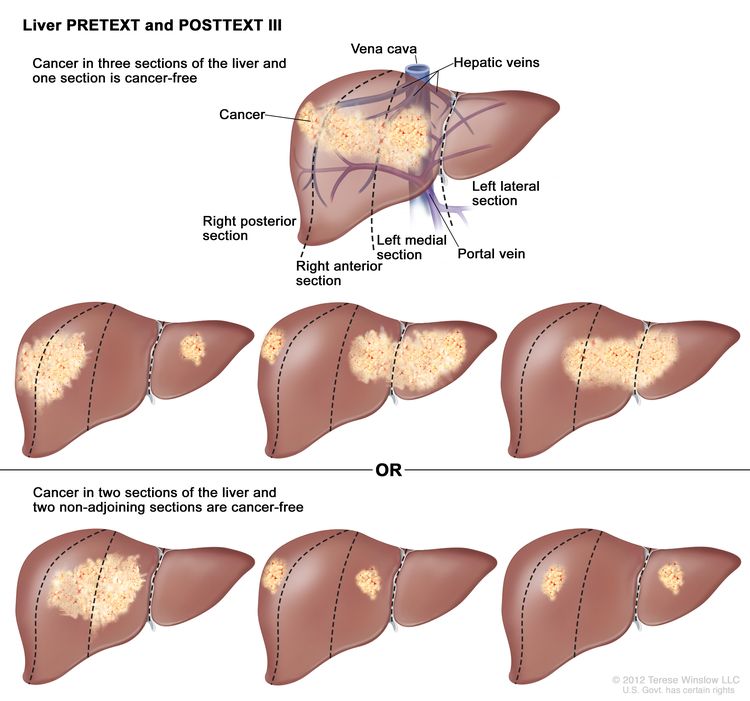
Screening Tests to Detect Colorectal Cancer and Polyps

What is colorectal cancer?

Colorectal cancer (cancer that develops in the colon and/or the rectum) is a disease in which abnormal cells in the colon or rectum divide uncontrollably, ultimately forming a malignant tumor.

Parts of the colon

Parts of the colon. Drawing of the front of the abdomen that shows the four sections of the colon: the ascending colon, the transverse colon, the descending colon, and the sigmoid colon. Also shown are the small intestine, the cecum, and the rectum. The cecum, colon, rectum, and anal canal make up the large intestine. The cecum, ascending colon, and transverse colon make up the upper, or proximal, colon; the descending colon and sigmoid colon make up the lower, or distal, colon.

Credit: © Terese Winslow

Most colorectal cancers begin as an abnormal growth, or lesion, in the tissue that lines the inner surface of the colon or rectum. Lesions may appear as raised polyps, or, less commonly, they may appear flat or slightly indented. Raised polyps may be attached to the inner surface of the colon or rectum with a stalk (pedunculated polyps), or they may grow along the surface without a stalk (sessile polyps). 

Colorectal polyps are common in people older than 50 years of age, and most do not become cancer. However, a certain type of polyp known as an adenoma is more likely to become a cancer.

Colorectal cancer is the third most common type of non-skin cancer in both men (after prostate cancer and lung cancer) and women (after breast cancer and lung cancer). It is the second leading cause of cancer death in the United States after lung cancer. In 2024, an estimated 152,810 people in the United States will be diagnosed with colorectal cancer and 53,010 people will die from it (1). 

Who is at risk for colorectal cancer?

Increasing age is a major risk factor for colorectal cancer. In the United States, colorectal cancer is most frequently diagnosed in adults aged 65 to 74 years. From 2017 through 2021, the colorectal cancer incidence rates were 8.6, 69.8, and 156.9 per 100,000 people for those younger than 50 years, those aged 50 to 64 years, and those 65 years and older, respectively. 

However, colorectal cancer incidence is increasing among younger age groups even as it is declining in older age groups. For example, from 2012 through 2021, the number of new colorectal cancer cases diagnosed per 100,000 people

  • increased 3.8% per year among those aged 15 to 39 years, from 4.1 to 5.6
  • increased 1.2% per year among those aged 40 to 64 years, from 46.1 to 52.1
  • decreased 2.2% per year among those aged 65 to 74 years, from 148.2 to 120.8
  • decreased 2.7% per year among those 75 years and older, from 244.9 to 192.7

The reason(s) for the increasing incidence among younger people is not known, but it may relate to changes in the prevalence of certain risk factors.

Besides age, other major risk factors for colorectal cancer include certain inherited conditions (such as Lynch syndrome and familial adenomatous polyposis), a family history of colorectal cancer, and a personal history of inflammatory bowel disease (such as ulcerative colitis or Crohn disease).

Several other factors are known to be associated with smaller increases in risk. These include moderate to heavy alcohol use, obesity, physical inactivity, and cigarette smoking.

In addition, people with a history of inflammatory bowel disease (such as ulcerative colitis or Crohn disease) have a higher risk of colorectal cancer than people without such conditions.  

What do colorectal cancer screening guidelines say about who should have colorectal cancer screening?

Expert medical groups, including the US Preventive Services Task Force (USPSTF) (2), strongly recommend screening for colorectal cancer. Although some details of the recommendations vary, most groups now generally recommend that people at average risk of colorectal cancer get screened at regular intervals beginning at age 45 (26).

The expert medical groups generally recommend that screening continue to age 75; for those aged 76 to 85 years, the decision to screen is based on the individual’s life expectancy, health conditions, and prior screening results. 

People who are at increased risk of colorectal cancer because of certain inherited conditions (such as Lynch syndrome and familial adenomatous polyposis), a family history of colorectal cancer, a personal history of advanced polyps, or because they have inflammatory bowel disease may be advised to start screening earlier and/or have more frequent screening.

What methods are used to screen people for colorectal cancer?

Several different screening tests are available that can help doctors find colorectal cancer before symptoms begin, when it may be more treatable. Some of these tests also allow adenomas and polyps to be found and removed before they become cancer. That is, some types of colorectal cancer screening may allow for cancer prevention in addition to early detection.

  • Stool tests. Both polyps and colorectal cancers can bleed, and stool tests check for tiny amounts of blood in feces (stool) that cannot be seen visually. (Hidden blood in stool—also called occult blood–may also indicate the presence of conditions that are not cancer, such as hemorrhoids.) With these tests, stool samples are collected by the patient using a kit, and the samples are sent to a laboratory for testing. People who have a positive finding with these tests are advised to have a colonoscopy.

    The US Food and Drug Administration (FDA) has approved several types of stool tests to screen for colorectal cancer, including:

    • Guaiac fecal occult blood test (gFOBT). gFOBT uses a chemical to detect heme, a component of the blood protein hemoglobin. Because gFOBT can also detect heme in some foods (for example, red meat), people must avoid certain foods before having this test. If gFOBT is the only type of colorectal cancer screening test performed, experts generally recommend testing every year or two (3).
    • Fecal immunochemical test (FIT or iFOBT). FIT uses antibodies to detect hemoglobin protein specifically (5, 6). Dietary restrictions are typically not required for FIT. If FIT is the only type of colorectal cancer screening test performed, experts generally recommend testing every year or two (3).
    • Multitarget stool DNA testing (sDNA-FIT). sDNA-FIT (Cologuard) detects hemoglobin, along with certain DNA biomarkers. The DNA comes from cells in the lining of the colon and rectum that are shed and collect in stool as it passes through the large intestine and rectum. Experts generally suggest sDNA-FIT testing at least every 3 years (2).
       
  • Direct visualization tests. There are three direct visualization tests used for colorectal cancer screening. All involve pumping air into the colon through a tube inserted through the anus into the rectum to expand the colon so the doctor can see the lining more clearly. Of these three tests, colonoscopy is the most common direct visualization test in the United States.
    • Colonoscopy. In this test, the rectum and entire colon are examined using a colonoscope, a flexible lighted tube with a lens for viewing and a tool for removing tissue, which is inserted through the anus into the rectum. During colonoscopy, any abnormal growths in the entire colon and the rectum can be removed. The preparation for colonoscopy requires a thorough cleansing of the entire colon before the test. Most patients receive some form of sedation during the test.

      Experts recommend screening colonoscopy every 10 years for people at average risk.

    • Virtual colonoscopy, also called computed tomographic (CT) colonography, is a screening method that uses special x-ray equipment (a CT scanner) to produce a series of pictures of the colon and the rectum from outside the body. A computer then assembles these pictures into detailed images that can show polyps and other abnormalities. As with standard colonoscopy, a thorough cleansing of the colon is necessary before this test. Virtual colonoscopy is much less invasive than standard colonoscopy (other than the pumping of air into the colon), but if polyps or other abnormal growths are found during a virtual colonoscopy a standard colonoscopy must usually be performed to remove them.

      Because virtual colonoscopy also produces images of areas outside the colon and rectum it can lead to the unintentional discovery of medical findings in these areas that require additional follow-up procedures. Virtual colonoscopy may also miss small polyps (7). However, many small polyps may not be likely to become cancer and so taking them out may not be of benefit. Experts recommend screening with virtual colonoscopy every 5 years.

    • Sigmoidoscopy. In this test, the rectum and sigmoid colon are examined using a sigmoidoscope, a flexible lighted tube with a lens for viewing and a tool for removing tissue, which is inserted through the anus into the rectum and sigmoid colon. During sigmoidoscopy, abnormal growths in the rectum and sigmoid colon can be removed for analysis (biopsied). The lower colon must be cleared of stool before sigmoidoscopy, but the preparation is not very extensive. People are not usually sedated for this test.

      Experts generally recommend screening sigmoidoscopy every 5 or 10 years for people at average risk (3). People who are screened with sigmoidoscopy may also be tested every few years with FIT.
       

  • Blood-based tests. A test for a molecular biomarker (methylated SEPT9) shed by colorectal cancer cells into the bloodstream, called Epi proColon 2.0, is FDA approved to be used to screen adults 50 years or older at average risk for colorectal cancer who have been offered and have a history of not completing colorectal cancer screening using a stool test or a direct visualization test.

    Another blood-based test for colorectal cancer (Shield) is approved for screening adults ages 45 and older who are at average risk for the disease. It analyzes plasma DNA for certain changes, including the presence of harmful gene variants.

    Blood-based tests have not yet been incorporated into clinical guidelines for first-line colorectal cancer screening.
     

  • Other methods. Several other tests to screen for colorectal cancer are sometimes used, although these are not generally recommended by expert groups.
    • Double-contrast barium enema (DCBE). This test is another method of visualizing the colon from outside the body. In DCBE, a series of x-ray images of the entire colon and rectum is taken after the patient is given an enema with a barium solution. The barium helps to outline the colon and the rectum on the images. DCBE is rarely used for colorectal cancer screening; however, it may be used for people who cannot undergo standard colonoscopy—for example, because they are at particular risk for complications.
    • Single-specimen gFOBT done in a doctor’s office. Doctors sometimes perform gFOBT on a stool sample collected during a digital rectal examination as part of a routine physical examination. However, this approach is not an effective way to screen for colorectal cancer (8).

How can people and their health care providers decide which colorectal cancer screening test(s) to use?

Different tests have different advantages and disadvantages, and people should talk with their health care provider about which test is best for them.

An individual’s decision about which test to have may depend on:

  • the person’s age, medical history, family history, and general health
  • potential harms of the test (more invasive tests have more potential harms than less invasive tests)
  • the preparation required for the test
  • whether sedation may be needed for the test
  • the follow-up care needed after the test
  • the convenience of the test
  • the cost of the test and the availability of insurance coverage

The table below summarizes key features of the different colorectal screening tests that people may want to consider when choosing a test.

Test Diet and medication changes before test? Invasive procedure? Preparation (colon cleansing) needed? Sedation needed? Test frequency Additional considerations
Stool tests Yes for gFOBT, no for FIT, sDNA-FIT, or mt-sRNA No No No Every year to every 3 years, depending on the test
  • Follow-up colonoscopy will be needed if test is positive
Sigmoidoscopy Yes Yes Yes (less extensive than for colonoscopy) Usually no Every 5 to 10 years, possibly with more frequent FIT
  • Abnormal tissue can be removed during exam
  • Very small risk of tearing or perforation of the lining of the colon
  • Not widely available in United States (9)
Colonoscopy Yes Yes Yes Yes Every 10 years
  • Abnormal tissue can be removed during exam
  • Small risk of tearing or perforation of the lining of the colon
Virtual colonoscopy No Yes (minimally) Yes No Every 5 years
  • Follow-up colonoscopy will be needed if test is positive
  • Not widely available and may not be covered by insurance
  • Can find abnormalities outside the colon that may need follow-up
  • Involves exposure to small amount of radiation 

Does health insurance pay for colorectal cancer screening?

Colorectal cancer screening is a preventive service that the Health Insurance Marketplace and many other health insurance plans are required to cover. Medicare covers several colorectal cancer screening tests for its beneficiaries. However, Medicare and some insurance companies currently do not pay for the costs of virtual colonoscopy. Specific information about Medicare benefits for colorectal cancer screening is available on the Medicare website.

A colonoscopy to follow up on a screening test with a positive result, such as an abnormal stool test or even a lesion detected on a screening colonoscopy, is considered a diagnostic exam and may or may not be covered (or not covered as fully as a screening colonoscopy). Some insurers consider a screening colonoscopy that reveals a polyp that must be removed to be a diagnostic exam and charge accordingly. People should check with their health insurance provider before their test to determine their colorectal cancer screening coverage and what their out-of-pocket expenses may be if the test finds an abnormality that needs to be followed up.

What happens if a colorectal cancer screening test finds an abnormality?

If an abnormality is found during a standard colonoscopy it will be removed (polypectomy) or a biopsy performed, and the cells will be examined to see if cancer is present. If an abnormality is found during a sigmoidoscopy, polypectomy or biopsy may or may not be performed, and a follow-up colonoscopy may be recommended. If a different screening test finds an abnormality, a colonoscopy will be needed to examine the colon directly.

What new tests are being developed for colorectal cancer screening?

Researchers are studying new blood markers to detect colorectal cancer early. For example, tumors release into the blood small fragments of RNA called microRNAs packaged into tiny sac-like structures called exosomes. Exosome-packaged microRNAs have shown promise for early detection of pancreatic cancer and may also be useful for early detection of colorectal cancer (10, 11).

Another approach being tested is whether artificial intelligence (AI)–based technology called computer-aided detection (CAD) can improve the interpretation of colonoscopy imaging by experienced doctors. Several clinical trials have found that the addition of CAD increased the detection of small polyps that are unlikely to become cancer but not of advanced adenomas. However, these studies used relatively primitive AI-based CADs. Newer AI technologies may improve the detection of advanced adenomas by CAD.      

Researchers are continuing to improve the sensitivity of stool-based screening for detecting advanced adenomatous polyps, which can potentially become colorectal cancer, by testing for the presence of other (non-DNA) types of biomarkers. For example, a multitarget stool RNA (mt-sRNA) test (ColoSense) that detects occult hemoglobin (with FIT), along with levels of eight colorectal cancer–associated RNA markers was approved in 2024 for colorectal cancer screening. (This test is not yet commercially available, and experts have not yet recommended a screening interval for it.)

Researchers have also found that measuring three protein biomarkers in stool—hemoglobin, calprotectin, and serpin family F member 2—improved the ability of FIT to detect advanced lesions (including colorectal cancer) without reducing its specificity (12).

Capsule colonoscopy (also called capsule endoscopy) is a technique that continues to be explored to improve visualization of the colon. A person swallows a pill-like capsule that contains a tiny wireless camera. The camera takes pictures of the inside of the digestive tract and sends them to a small recorder that is worn on the patient’s waist or shoulder. The pictures are then viewed on a computer by the doctor to check for signs of disease. The capsule passes out of the body during a bowel movement. Cleansing of the colon is still necessary before this test. This method is currently approved for patients with an incomplete colonoscopy and for detection of colon polyps in patients with evidence of lower GI bleeding but not as a stand-alone screening test for people at average risk.

Rectal Cancer Treatment (PDQ®)–Patient Version

Rectal Cancer Treatment (PDQ®)–Patient Version

General Information About Rectal Cancer

Key Points

  • Rectal cancer is a type of cancer that forms in the tissues of the rectum.
  • Health history affects the risk of developing rectal cancer.
  • Signs of rectal cancer include blood in the stool or a change in bowel habits.
  • Tests that examine the rectum and colon are used to diagnose rectal cancer.
  • After rectal cancer has been diagnosed, imaging tests are done to find out if cancer cells have spread within the rectum or to other parts of the body.
  • Some people decide to get a second opinion.
  • Certain factors affect prognosis (chance of recovery) and treatment options.

Rectal cancer is a type of cancer that forms in the tissues of the rectum.

The rectum is part of the body’s digestive system. The digestive system takes in nutrients (vitamins, minerals, carbohydrates, fats, proteins, and water) from foods and helps pass waste material out of the body. The digestive system is made up of the esophagus, stomach, and the small and large intestines. The colon (large bowel) is the main part of the large intestine and is about 5 feet long. Together, the rectum and anal canal make up the last part of the large intestine and are 6 to 8 inches long. The anal canal ends at the anus (the opening of the large intestine to the outside of the body).

EnlargeGastrointestinal (digestive) system anatomy; drawing shows the esophagus, liver, stomach, colon, small intestine, rectum, and anus.
Anatomy of the lower gastrointestinal (digestive) system showing the colon, rectum, and anus. Other organs that make up the digestive system are also shown.

Health history affects the risk of developing rectal cancer.

Colorectal cancer is caused by certain changes to the way colorectal cells function, especially how they grow and divide into new cells. There are many risk factors for colorectal cancer, but many do not directly cause cancer. Instead, they increase the chance of DNA damage in cells that may lead to colorectal cancer. To learn more about how cancer develops, see What Is Cancer?

A risk factor is anything that increases the chance of getting a disease. Some risk factors for colorectal cancer, like smoking, can be changed. However, risk factors also include things you cannot change, like your genetics, getting older, and your family history. Learning about risk factors for colorectal cancer can help you make changes that might lower your risk of getting it.

Risk factors for colorectal cancer include:

Older age is a main risk factor for most cancers. The chance of getting cancer increases as you get older.

Having one or more of these risk factors does not mean that you will get colorectal cancer. Many people with risk factors never develop colorectal cancer, while others with no known risk factors do. Talk with your doctor if you think you might be at increased risk.

Signs of rectal cancer include blood in the stool or a change in bowel habits.

These and other signs and symptoms may be caused by rectal cancer or by other conditions. Check with your doctor if you have:

  • blood (either bright red or very dark) in the stool
  • a change in bowel habits
    • diarrhea
    • constipation
    • feeling that the bowel does not empty completely
    • stools that are narrower or have a different shape than usual
  • general abdominal discomfort (frequent gas pains, bloating, fullness, or cramps)
  • change in appetite
  • weight loss for no known reason
  • fatigue

Tests that examine the rectum and colon are used to diagnose rectal cancer.

In addition to asking about your personal and family health history and doing a physical exam, your doctor may perform the following tests and procedures:

  • Digital rectal exam (DRE) is an exam of the rectum. The doctor or nurse inserts a lubricated, gloved finger into the lower part of the rectum to feel for lumps or anything else that seems unusual. In women, the vagina may also be examined.
  • Colonoscopy is a procedure that uses a colonoscope to look inside the rectum and colon for polyps (small pieces of bulging tissue), abnormal areas, or cancer. A colonoscope is a thin, tube-like instrument with a light and a lens for viewing. It may also have a tool to remove polyps or tissue samples, which are checked under a microscope for signs of cancer.
    EnlargeColonoscopy; drawing shows a colonoscope inserted through the anus and rectum and into the colon. An inset shows a patient lying on a table having a colonoscopy.
    Colonoscopy. A thin, lighted tube is inserted through the anus and rectum and into the colon to look for abnormal areas.
  • Biopsy is the removal of cells or tissues so they can be viewed under a microscope to check for signs of cancer. Tumor tissue that is removed during the biopsy may be checked to see if the patient is likely to have the gene mutation that causes Lynch syndrome (also known as hereditary nonpolyposis colorectal cancer). This may help to plan treatment. Learn about the type of information that can be found in a pathologist’s report about the cells or tissue removed during a biopsy at Pathology Reports.
  • Immunohistochemistry is a laboratory test that uses antibodies to check for certain antigens (markers) in a sample of a patient’s tissue. The antibodies are usually linked to an enzyme or a fluorescent dye. After the antibodies bind to a specific antigen in the tissue sample, the enzyme or dye is activated, and the antigen can then be seen under a microscope. This type of test is used to help diagnose cancer and to help tell one type of cancer from another type of cancer.
  • Microsatellite instability (MSI) is a laboratory test in which tumor tissue is checked for cells that may have a defect in genes involved in DNA repair. The findings may indicate whether or not the patient has a type of cancer linked to an inherited cancer syndrome such as Lynch syndrome.

After rectal cancer has been diagnosed, imaging tests are done to find out if cancer cells have spread within the rectum or to other parts of the body.

The process used to find out whether cancer has spread within the rectum or to other parts of the body is called staging. The information gathered from the staging process determines the stage of the disease. It is important to know the stage in order to plan treatment.

The following tests and procedures may be used in the staging process:

  • Chest x-ray is a type of radiation that can go through the body and make pictures of the organs and bones inside the chest.
  • CT scan (CAT scan) uses a computer linked to an x-ray machine to make a series of detailed pictures of areas inside the body, such as the abdomen, pelvis, or chest. The pictures are taken from different angles and are used to create 3-D views of tissues and organs. A dye may be injected into a vein or swallowed to help the organs or tissues show up more clearly. This procedure is also called computed tomography, computerized tomography, or computerized axial tomography.
  • MRI (magnetic resonance imaging) uses a magnet, radio waves, and a computer to make a series of detailed pictures of areas inside the rectum. A substance called gadolinium is injected into the patient through a vein. The gadolinium collects around the cancer cells so they show up brighter in the picture. This procedure is also called nuclear magnetic resonance imaging (NMRI).
  • PET scan (positron emission tomography scan) uses a small amount of radioactive sugar (also called glucose) that is injected into a vein. Then a scanner rotates around the body to make detailed, computerized pictures of areas inside the body where the glucose is taken up. Because cancer cells often take up more glucose than normal cells, the pictures can be used to find cancer cells in the body.
  • Endorectal ultrasound is used to examine the rectum and nearby organs. An ultrasound transducer (probe) is inserted into the rectum and used to bounce high-energy sound waves (ultrasound) off internal tissues or organs and make echoes. The echoes form a picture of body tissues called a sonogram. The doctor can identify tumors by looking at the sonogram. This procedure is also called transrectal ultrasound.
  • Carcinoembryonic antigen (CEA) assay is a test that measures the level of CEA in the blood. CEA is released into the bloodstream from both cancer cells and normal cells. When found in higher than normal amounts, it can be a sign of rectal cancer or other conditions.

Some people decide to get a second opinion.

You may want to get a second opinion to confirm your rectal cancer diagnosis and treatment plan. If you seek a second opinion, you will need to get medical test results and reports from the first doctor to share with the second doctor. The second doctor will review the pathology report, slides, and scans. They may agree with the first doctor, suggest changes or another treatment approach, or provide more information about your cancer.

Learn more about choosing a doctor and getting a second opinion at Finding Cancer Care. You can contact NCI’s Cancer Information Service via chat, email, or phone (both in English and Spanish) for help finding a doctor, hospital, or getting a second opinion. For questions you might want to ask at your appointments, visit Questions to Ask Your Doctor About Cancer.

Certain factors affect prognosis (chance of recovery) and treatment options.

The prognosis and treatment options depend on:

  • the stage of the cancer (whether it affects the inner lining of the rectum only, involves the whole rectum, or has spread to lymph nodes, nearby organs, or other places in the body)
  • whether the cancer is related to a defect in genes involved in DNA repair
  • whether the tumor has spread into or through the bowel wall
  • where the cancer is found in the rectum
  • whether the bowel is blocked or has a hole in it
  • whether all of the tumor can be removed by surgery
  • the patient’s general health
  • whether the cancer has just been diagnosed or has recurred (come back)

Stages of Rectal Cancer

Key Points

  • The following stages are used for rectal cancer:
    • Stage 0 (carcinoma in situ)
    • Stage I (also called stage 1) rectal cancer
    • Stage II (also called stage 2) rectal cancer
    • Stage III (also called stage 3) rectal cancer
    • Stage IV (also called stage 4) rectal cancer
  • Rectal cancer can recur (come back) after it has been treated.

Cancer stage describes the extent of cancer in the body, such as the size of the tumor, whether it has spread, and how far it has spread from where it first formed.

There are several staging systems for cancer that describe the extent of the cancer. Rectal cancer staging usually uses the TNM staging system. The cancer may be described by this staging system in your pathology report. Based on the TNM results, a stage (I, II, III, or IV, also written as 1, 2, 3, or 4) is assigned to your cancer. When talking to you about your diagnosis, your doctor may describe the cancer as one of these stages.

Learn about tests to stage rectal cancer. Learn more about Cancer Staging.

The following stages are used for rectal cancer:

Stage 0 (carcinoma in situ)

EnlargeStage 0 colorectal carcinoma in situ; drawing shows a cross-section of the colon/rectum. An inset shows the layers of the colon/rectum wall with abnormal cells in the mucosa layer. Also shown are the submucosa, muscle layers, serosa, a blood vessel, and lymph nodes.
Stage 0 (rectal carcinoma in situ). Abnormal cells are shown in the mucosa of the rectum wall.

In stage 0 rectal cancer, abnormal cells are found in the mucosa (innermost layer) of the rectum wall. These abnormal cells may become cancer and spread into nearby normal tissue. Stage 0 is also called carcinoma in situ.

Stage I (also called stage 1) rectal cancer

EnlargeStage I colorectal cancer; drawing shows a cross-section of the colon/rectum. An inset shows the layers of the colon/rectum wall with cancer in the mucosa and submucosa. Also shown are the muscle layers, serosa, a blood vessel, and lymph nodes.
Stage I rectal cancer. Cancer has spread from the mucosa of the rectum wall to the submucosa or to the muscle layer.

In stage I rectal cancer, cancer has formed in the mucosa (innermost layer) of the rectum wall and has spread to the submucosa (layer of tissue next to the mucosa) or to the muscle layer of the rectum wall.

Stage II (also called stage 2) rectal cancer

EnlargeStage II colorectal cancer; drawing shows a cross-section of the colon/rectum and a three-panel inset. Each panel shows the layers of the colon/rectum wall: the mucosa, submucosa, muscle layers, and serosa. Also shown are a blood vessel and lymph nodes. The first panel shows stage IIA with cancer in the mucosa, submucosa, muscle layers, and serosa. The second panel shows stage IIB with cancer in all layers and spreading through the serosa to the visceral peritoneum. The third panel shows stage IIC with cancer in all layers and spreading through the serosa to nearby organs.
Stage II rectal cancer. In stage IIA, cancer has spread through the muscle layer of the rectum wall to the serosa. In stage IIB, cancer has spread through the serosa but has not spread to nearby organs. In stage IIC, cancer has spread through the serosa to nearby organs.

Stage II rectal cancer is divided into stages IIA, IIB, and IIC.

  • Stage IIA: Cancer has spread through the muscle layer of the rectum wall to the serosa (outermost layer) of the rectum wall.
  • Stage IIB: Cancer has spread through the serosa (outermost layer) of the rectum wall to the tissue that lines the organs in the abdomen (visceral peritoneum).
  • Stage IIC: Cancer has spread through the serosa (outermost layer) of the rectum wall to nearby organs.

Stage III (also called stage 3) rectal cancer

Stage III rectal cancer is divided into stages IIIA, IIIB, and IIIC.

EnlargeStage IIIA colorectal cancer; drawing shows a cross-section of the colon/rectum and a two-panel inset. Each panel shows the layers of the colon/rectum wall: the mucosa, submucosa, muscle layers, and serosa. Also shown are a blood vessel and lymph nodes. The first panel shows cancer in the mucosa, submucosa, and muscle layers and in 2 lymph nodes. The second panel shows cancer in the mucosa and submucosa and in 5 lymph nodes.
Stage IIIA rectal cancer. Cancer has spread through the mucosa of the rectum wall to the submucosa and may have spread to the muscle layer, and has spread to one to three nearby lymph nodes or tissues near the lymph nodes. OR, cancer has spread through the mucosa to the submucosa and four to six nearby lymph nodes.

In stage IIIA, cancer has spread:

  • through the mucosa (innermost layer) of the rectum wall to the submucosa (layer of tissue next to the mucosa) or to the muscle layer of the rectum wall. Cancer has spread to one to three nearby lymph nodes, or cancer cells have formed in tissue near the lymph nodes; or
  • through the mucosa (innermost layer) of the rectum wall to the submucosa (layer of tissue next to the mucosa). Cancer has spread to four to six nearby lymph nodes.
EnlargeStage IIIB colorectal cancer; drawing shows a cross-section of the colon/rectum and a three-panel inset. Each panel shows the layers of the colon/rectum wall: the mucosa, submucosa, muscle layers, and serosa. Also shown are a blood vessel and lymph nodes. The first panel shows cancer in all layers, in 3 nearby lymph nodes, and in the visceral peritoneum. The second panel shows cancer in all layers and in 5 nearby lymph nodes. The third panel shows cancer in the mucosa, submucosa, and muscle layers and in 7 nearby lymph nodes.
Stage IIIB rectal cancer. Cancer has spread through the muscle layer of the rectum wall to the serosa or has spread through the serosa but not to nearby organs; cancer has spread to one to three nearby lymph nodes or to tissues near the lymph nodes. OR, cancer has spread to the muscle layer or to the serosa, and to four to six nearby lymph nodes. OR, cancer has spread through the mucosa to the submucosa and may have spread to the muscle layer; cancer has spread to seven or more nearby lymph nodes.

In stage IIIB, cancer has spread:

  • through the muscle layer of the rectum wall to the serosa (outermost layer) of the rectum wall or has spread through the serosa to the tissue that lines the organs in the abdomen (visceral peritoneum). Cancer has spread to one to three nearby lymph nodes, or cancer cells have formed in tissue near the lymph nodes; or
  • to the muscle layer or to the serosa (outermost layer) of the rectum wall. Cancer has spread to four to six nearby lymph nodes; or
  • through the mucosa (innermost layer) of the rectum wall to the submucosa (layer of tissue next to the mucosa) or to the muscle layer of the rectum wall. Cancer has spread to seven or more nearby lymph nodes.
EnlargeStage IIIC colorectal cancer; drawing shows a cross-section of the colon/rectum and a three-panel inset. Each panel shows the layers of the colon/rectum wall: the mucosa, submucosa, muscle layers, and serosa. Also shown are a blood vessel and lymph nodes. The first panel shows cancer in all layers, in 4 lymph nodes, and in the visceral peritoneum. The second panel shows cancer in all layers and in 7 lymph nodes. The third panel shows cancer in all layers, in 2 lymph nodes, and spreading to nearby organs.
Stage IIIC rectal cancer. Cancer has spread through the serosa of the rectum wall but not to nearby organs; cancer has spread to four to six nearby lymph nodes. OR, cancer has spread through the muscle layer to the serosa or has spread through the serosa but not to nearby organs; cancer has spread to seven or more nearby lymph nodes. OR, cancer has spread through the serosa to nearby organs and to one or more nearby lymph nodes or to tissues near the lymph nodes.

In stage IIIC, cancer has spread:

  • through the serosa (outermost layer) of the rectum wall to the tissue that lines the organs in the abdomen (visceral peritoneum). Cancer has spread to four to six nearby lymph nodes; or
  • through the muscle layer of the rectum wall to the serosa (outermost layer) of the rectum wall or has spread through the serosa to the tissue that lines the organs in the abdomen (visceral peritoneum). Cancer has spread to seven or more nearby lymph nodes; or
  • through the serosa (outermost layer) of the rectum wall to nearby organs. Cancer has spread to one or more nearby lymph nodes, or cancer cells have formed in tissue near the lymph nodes.

Stage IV (also called stage 4) rectal cancer

EnlargeStage IV rectal cancer; drawing shows other parts of the body where rectal cancer may spread, including the distant lymph nodes, lung, liver, abdominal wall, and prostate. An inset shows cancer cells spreading from the rectum, through the blood and lymph system, to another part of the body where metastatic cancer has formed.
Stage IV rectal cancer. The cancer has spread through the blood and lymph nodes to other parts of the body, such as the lung, liver, abdominal wall, or prostate.

Stage IV rectal cancer is divided into stages IVA, IVB, and IVC.

  • Stage IVA: Cancer has spread to one area or organ that is not near the rectum, such as the liver, lung, prostate, or a distant lymph node.
  • Stage IVB: Cancer has spread to more than one area or organ that is not near the rectum, such as the liver, lung, prostate, or a distant lymph node.
  • Stage IVC: Cancer has spread to the tissue that lines the wall of the abdomen and may have spread to other areas or organs.

Stage IV rectal cancer is also called metastatic rectal cancer. Metastatic cancer happens when cancer cells travel through the lymphatic system or blood and form tumors in other parts of the body. The metastatic tumor is the same type of cancer as the primary tumor. For example, if rectal cancer spreads to the liver, the cancer cells in the liver are actually rectal cancer cells. The disease is called metastatic rectal cancer, not liver cancer. Learn more in Metastatic Cancer: When Cancer Spreads.

Rectal cancer can recur (come back) after it has been treated.

Recurrent rectal cancer is cancer that has come back after it has been treated. If rectal cancer comes back, it may come back in the rectum or in other parts of the body, such as the colon, pelvis, liver, or lungs. Tests will be done to help determine where the cancer has returned. The type of treatment for recurrent rectal cancer will depend on where it has come back.

Learn more in Recurrent Cancer: When Cancer Comes Back.

Treatment Option Overview

Key Points

  • There are different types of treatment for people with rectal cancer.
  • The following types of treatment are used:
    • Surgery
    • Radiation therapy
    • Chemotherapy
    • Chemoradiation therapy
    • Active surveillance
    • Targeted therapy
    • Immunotherapy
  • New types of treatment are being tested in clinical trials.
  • Treatment for rectal cancer may cause side effects.
  • Follow-up care may be needed.

There are different types of treatment for people with rectal cancer.

Different types of treatments are available for rectal cancer. You and your cancer care team will work together to decide your treatment plan, which may include more than one type of treatment. Many factors will be considered, such as the stage of the cancer, your overall health, and your preferences. Your plan will include information about your cancer, the goals of treatment, your treatment options and the possible side effects, and the expected length of treatment.

Talking with your cancer care team before treatment begins about what to expect will be helpful. You’ll want to learn what you need to do before treatment begins, how you’ll feel while going through it, and what kind of help you will need. To learn more, visit Questions to Ask Your Doctor About Treatment.

The following types of treatment are used:

Surgery

Surgery is the most common treatment for all stages of rectal cancer. The cancer is removed using one of the following types of surgery:

  • Polypectomy: If the cancer is found in a polyp (a small piece of bulging tissue), the polyp is often removed during a colonoscopy.
  • Local excision: If the cancer is found on the inside surface of the rectum and has not spread into the wall of the rectum, the cancer and a small amount of surrounding healthy tissue are removed.
  • Resection: If the cancer has spread into the wall of the rectum, the section of the rectum with cancer and nearby healthy tissue are removed. Sometimes, the tissue between the rectum and the abdominal wall is also removed. The lymph nodes near the rectum are removed and checked under a microscope for signs of cancer.
  • Radiofrequency ablation: The use of a special probe with tiny electrodes that kill cancer cells. Sometimes, the probe is inserted directly through the skin, and only local anesthesia is needed. In other cases, the probe is inserted through an incision in the abdomen. This is done in the hospital with general anesthesia.
  • Cryosurgery: A treatment that uses an instrument to freeze and destroy abnormal tissue. This type of treatment is also called cryotherapy. Learn more about Cryosurgery to Treat Cancer.
  • Pelvic exenteration: If the cancer has spread to other organs near the rectum, the lower colon, rectum, and bladder are removed. In women, the cervix, vagina, ovaries, and nearby lymph nodes may be removed. In men, the prostate may be removed. Artificial openings (stoma) are made for urine and stool to flow from the body to a collection bag.

After the cancer is removed, the surgeon will either:

  • do an anastomosis (sew the healthy parts of the rectum together, sew the remaining rectum to the colon, or sew the colon to the anus);
    EnlargeThree-panel drawing showing rectal cancer surgery with anastomosis; the first panel shows area of rectum with cancer, the middle panel shows cancer and nearby tissue removed, and the last panel shows the colon and anus joined.
    Resection of the rectum with anastomosis. The rectum and part of the colon are removed, and then the colon and anus are joined.

    or

  • make a stoma (an opening) from the rectum to the outside of the body for waste to pass through. This procedure is done if the cancer is too close to the anus and is called a colostomy. A bag is placed around the stoma to collect the waste. Sometimes, the colostomy is needed only until the rectum has healed, and then it can be reversed. If the entire rectum is removed, however, the colostomy may be permanent.

Radiation therapy and/or chemotherapy may be given before surgery to shrink the tumor, make it easier to remove the cancer, and help with bowel control after surgery. Treatment given before surgery is called neoadjuvant therapy. After all the cancer that can be seen at the time of the surgery is removed, some patients may be given radiation therapy and/or chemotherapy after surgery to kill any cancer cells that are left. Treatment given after the surgery, to lower the risk that the cancer will come back, is called adjuvant therapy.

If the cancer has spread to the liver and cannot be removed by surgery, a total hepatectomy and liver transplant after chemotherapy may be done. Total hepatectomy and liver transplant is the removal of the entire liver by surgery, followed by a transplant of a healthy liver from a donor.

Radiation therapy

Radiation therapy uses high-energy x-rays or other types of radiation to kill cancer cells or keep them from growing. Rectal cancer is sometimes treated with external radiation therapy. This type of radiation therapy uses a machine outside the body to send radiation toward the area of the body with cancer.

Short-course preoperative radiation therapy is used in some types of rectal cancer. This type of external radiation therapy uses fewer and lower doses of radiation than standard treatment, followed by surgery several days after the last dose.

Learn more about External Beam Radiation Therapy for Cancer and Radiation Therapy Side Effects.

Chemotherapy

Chemotherapy (also called chemo) uses drugs to stop the growth of cancer cells, either by killing the cells or by stopping them from dividing.

Systemic chemotherapy is when chemotherapy drugs are taken by mouth or injected into a vein or muscle. When given this way, the drugs enter the bloodstream and can reach cancer cells throughout the body. Systemic chemotherapy used to treat rectal cancer includes:

Combinations of these drugs may be used. Other chemotherapy drugs not listed here may also be used.

Chemotherapy may also be combined with other kinds of drugs. For example, it might be combined with the targeted therapy drug bevacizumab, cetuximab, or panitumumab.

Regional chemotherapy for rectal cancer is when drugs are placed directly into the hepatic artery (the main artery that supplies blood to the liver) in a procedure called chemoembolization. Chemoembolization of the hepatic artery may be used to treat cancer that has spread to the liver. This is done by blocking the hepatic artery and injecting anticancer drugs between the blockage and the liver. The liver’s arteries then carry the drugs into the liver. Only a small amount of the drug reaches other parts of the body. The blockage may be temporary or permanent, depending on what is used to block the artery. The liver continues to receive some blood from the hepatic portal vein, which carries blood from the stomach and intestine.

The way the chemotherapy is given depends on the type and stage of the cancer being treated.

Learn more about how chemotherapy works, how it is given, common side effects, and more at Chemotherapy to Treat Cancer and Chemotherapy and You: Support for People With Cancer.

Chemoradiation therapy

Chemoradiation therapy combines chemotherapy and radiation therapy to increase the effects of both.

Active surveillance

Active surveillance is closely following a patient’s condition without giving any treatment unless there are changes in test results. It is used to find early signs that the condition is getting worse. In active surveillance, patients are given certain exams and tests to check if the cancer is growing. When the cancer begins to grow, treatment is given to cure the cancer. Tests include:

Targeted therapy

Targeted therapy uses drugs or other substances to identify and attack specific cancer cells. Your doctor may suggest biomarker tests to help predict your response to certain targeted therapy drugs. Learn more about Biomarker Testing for Cancer Treatment.

Targeted therapies used to treat rectal cancer include:

Learn more about Targeted Therapy to Treat Cancer.

Immunotherapy

Immunotherapy helps a person’s immune system fight cancer. Your doctor may suggest biomarker tests to help predict your response to certain immunotherapy drugs. Learn more about Biomarker Testing for Cancer Treatment.

Immunotherapy drugs used to treat rectal cancer include:

Learn more about Immunotherapy to Treat Cancer.

New types of treatment are being tested in clinical trials.

For some people, joining a clinical trial may be an option. There are different types of clinical trials for people with cancer. For example, a treatment trial tests new treatments or new ways of using current treatments. Supportive care and palliative care trials look at ways to improve quality of life, especially for those who have side effects from cancer and its treatment.

You can use the clinical trial search to find NCI-supported cancer clinical trials accepting participants. The search allows you to filter trials based on the type of cancer, your age, and where the trials are being done. Clinical trials supported by other organizations can be found on the ClinicalTrials.gov website.

Learn more about clinical trials, including how to find and join one, at Clinical Trials Information for Patients and Caregivers.

Treatment for rectal cancer may cause side effects.

For information about side effects caused by treatment for cancer, visit our Side Effects page.

Follow-up care may be needed.

As you go through treatment, you will have follow-up tests or check-ups. Some tests that were done to diagnose or stage the cancer may be repeated to see how well the treatment is working. Decisions about whether to continue, change, or stop treatment may be based on the results of these tests.

Some of the tests will continue to be done from time to time after treatment has ended. The results of these tests can show if your condition has changed or if the cancer has recurred (come back).

After treatment for rectal cancer, a blood test to measure amounts of carcinoembryonic antigen (a substance in the blood that may be increased when cancer is present) may be done to see if the cancer has come back.

Treatment of Stage 0 (carcinoma in situ)

Treatment of stage 0 may include the following types of surgery:

Learn more about these treatments in the Treatment Option Overview.

Use our clinical trial search to find NCI-supported cancer clinical trials that are accepting patients. You can search for trials based on the type of cancer, the age of the patient, and where the trials are being done. General information about clinical trials is also available.

Treatment of Stage I Rectal Cancer

Treatment of stage I rectal cancer may include:

Learn more about these treatments in the Treatment Option Overview.

Use our clinical trial search to find NCI-supported cancer clinical trials that are accepting patients. You can search for trials based on the type of cancer, the age of the patient, and where the trials are being done. General information about clinical trials is also available.

Treatment of Stages II and III Rectal Cancer

Treatment of stage II and stage III rectal cancer may include:

  • chemoradiation followed by surgery
  • chemotherapy alone followed by surgery, for people with lower-risk disease
  • short-course radiation therapy followed by surgery and chemotherapy
  • surgery followed by chemoradiation
  • surgery
  • chemoradiation followed by active surveillance and possibly surgery if the cancer recurs (comes back)
  • immunotherapy with dostarlimab (for treatment of tumors that may have a defect in genes involved in DNA repair)

Learn more about these treatments in the Treatment Option Overview.

Use our clinical trial search to find NCI-supported cancer clinical trials that are accepting patients. You can search for trials based on the type of cancer, the age of the patient, and where the trials are being done. General information about clinical trials is also available.

Treatment of Stage IV and Recurrent Rectal Cancer

Treatment of stage IV and recurrent rectal cancer may include:

Treatment of rectal cancer that has spread to other organs depends on where the cancer has spread.

  • Treatment for areas of cancer that have spread to the liver may include:
    • chemotherapy to shrink the tumor, if needed, followed by surgery
    • cryosurgery or radiofrequency ablation
    • chemoembolization and/or systemic chemotherapy
    • liver transplant after chemotherapy for patients with liver metastases that cannot be removed by surgery
    • a clinical trial of chemoembolization combined with radiation therapy to the tumors in the liver

Learn more about these treatments in the Treatment Option Overview.

Use our clinical trial search to find NCI-supported cancer clinical trials that are accepting patients. You can search for trials based on the type of cancer, the age of the patient, and where the trials are being done. General information about clinical trials is also available.

To Learn More About Rectal Cancer

About This PDQ Summary

About PDQ

Physician Data Query (PDQ) is the National Cancer Institute’s (NCI’s) comprehensive cancer information database. The PDQ database contains summaries of the latest published information on cancer prevention, detection, genetics, treatment, supportive care, and complementary and alternative medicine. Most summaries come in two versions. The health professional versions have detailed information written in technical language. The patient versions are written in easy-to-understand, nontechnical language. Both versions have cancer information that is accurate and up to date and most versions are also available in Spanish.

PDQ is a service of the NCI. The NCI is part of the National Institutes of Health (NIH). NIH is the federal government’s center of biomedical research. The PDQ summaries are based on an independent review of the medical literature. They are not policy statements of the NCI or the NIH.

Purpose of This Summary

This PDQ cancer information summary has current information about the treatment of rectal cancer. It is meant to inform and help patients, families, and caregivers. It does not give formal guidelines or recommendations for making decisions about health care.

Reviewers and Updates

Editorial Boards write the PDQ cancer information summaries and keep them up to date. These Boards are made up of experts in cancer treatment and other specialties related to cancer. The summaries are reviewed regularly and changes are made when there is new information. The date on each summary (“Updated”) is the date of the most recent change.

The information in this patient summary was taken from the health professional version, which is reviewed regularly and updated as needed, by the PDQ Adult Treatment Editorial Board.

Clinical Trial Information

A clinical trial is a study to answer a scientific question, such as whether one treatment is better than another. Trials are based on past studies and what has been learned in the laboratory. Each trial answers certain scientific questions in order to find new and better ways to help cancer patients. During treatment clinical trials, information is collected about the effects of a new treatment and how well it works. If a clinical trial shows that a new treatment is better than one currently being used, the new treatment may become “standard.” Patients may want to think about taking part in a clinical trial. Some clinical trials are open only to patients who have not started treatment.

Clinical trials can be found online at NCI’s website. For more information, call the Cancer Information Service (CIS), NCI’s contact center, at 1-800-4-CANCER (1-800-422-6237).

Permission to Use This Summary

PDQ is a registered trademark. The content of PDQ documents can be used freely as text. It cannot be identified as an NCI PDQ cancer information summary unless the whole summary is shown and it is updated regularly. However, a user would be allowed to write a sentence such as “NCI’s PDQ cancer information summary about breast cancer prevention states the risks in the following way: [include excerpt from the summary].”

The best way to cite this PDQ summary is:

PDQ® Adult Treatment Editorial Board. PDQ Rectal Cancer Treatment. Bethesda, MD: National Cancer Institute. Updated <MM/DD/YYYY>. Available at: /types/colorectal/patient/rectal-treatment-pdq. Accessed <MM/DD/YYYY>. [PMID: 26389378]

Images in this summary are used with permission of the author(s), artist, and/or publisher for use in the PDQ summaries only. If you want to use an image from a PDQ summary and you are not using the whole summary, you must get permission from the owner. It cannot be given by the National Cancer Institute. Information about using the images in this summary, along with many other images related to cancer can be found in Visuals Online. Visuals Online is a collection of more than 3,000 scientific images.

Disclaimer

The information in these summaries should not be used to make decisions about insurance reimbursement. More information on insurance coverage is available on Cancer.gov on the Managing Cancer Care page.

Contact Us

More information about contacting us or receiving help with the Cancer.gov website can be found on our Contact Us for Help page. Questions can also be submitted to Cancer.gov through the website’s E-mail Us.

Gastrointestinal Stromal Tumors Treatment (Adult) (PDQ®)–Patient Version

Gastrointestinal Stromal Tumors Treatment (Adult) (PDQ®)–Patient Version

General Information About Gastrointestinal Stromal Tumors

Key Points

  • Gastrointestinal stromal tumor is a disease in which abnormal cells form in the tissues of the gastrointestinal tract.
  • Genetic factors can increase the risk of having a gastrointestinal stromal tumor.
  • Signs of gastrointestinal stromal tumors include blood in the stool or vomit.
  • Tests that examine the GI tract are used to diagnose gastrointestinal stromal tumors.
  • Very small GISTs are common.
  • Certain factors affect prognosis (chance of recovery) and treatment options.

Gastrointestinal stromal tumor is a disease in which abnormal cells form in the tissues of the gastrointestinal tract.

The gastrointestinal (GI) tract is part of the body’s digestive system. It helps to digest food and takes nutrients (vitamins, minerals, carbohydrates, fats, proteins, and water) from food so they can be used by the body. The GI tract is made up of the following organs:

Some gastrointestinal stromal tumors (GISTs) grow slowly over time and may never cause a problem for a patient, while others can grow and spread very quickly. They are most common in the stomach and small intestine but may be found anywhere in or near the GI tract. Some scientists believe that GISTs begin in cells called interstitial cells of Cajal (ICC), in the wall of the GI tract.

EnlargeDrawing of the gastrointestinal tract showing the esophagus, stomach, colon, small intestine, and rectum.
Gastrointestinal stromal tumors (GISTs) may be found anywhere in or near the gastrointestinal tract.

See the PDQ summary about Childhood Gastrointestinal Stromal Tumors Treatment for information on the treatment of GIST in children.

Genetic factors can increase the risk of having a gastrointestinal stromal tumor.

Anything that increases your risk of getting a disease is called a risk factor. Having a risk factor does not mean that you will get cancer; not having risk factors doesn’t mean that you will not get cancer. Talk with your doctor if you think you may be at risk.

The genes in cells carry the hereditary information received from a person’s parents. The risk of GIST is increased in people who have inherited a mutation (change) in a certain gene. In rare cases, GISTs can be found in several members of the same family.

GIST may be part of a genetic syndrome, but this is rare. A genetic syndrome is a set of symptoms or conditions that occur together and is usually caused by abnormal genes. The following genetic syndromes have been linked to GIST:

Signs of gastrointestinal stromal tumors include blood in the stool or vomit.

These and other signs and symptoms may be caused by a GIST or by other conditions. Check with your doctor if you have any of the following:

  • Blood (either bright red or very dark) in the stool or vomit.
  • Pain in the abdomen, which may be severe.
  • Feeling very tired.
  • Trouble or pain when swallowing.
  • Feeling full after only a little food is eaten.

Tests that examine the GI tract are used to diagnose gastrointestinal stromal tumors.

The following tests and procedures may be used:

  • Physical exam and health history: An exam of the body to check general signs of health, including checking for signs of disease, such as lumps or anything else that seems unusual. A history of the patient’s health habits and past illnesses and treatments will also be taken.
  • CT scan (CAT scan): A procedure that makes a series of detailed pictures of areas inside the body, taken from different angles. The pictures are made by a computer linked to an x-ray machine. A dye may be injected into a vein or swallowed to help the organs or tissues show up more clearly. This procedure is also called computed tomography, computerized tomography, or computerized axial tomography.
  • MRI (magnetic resonance imaging): A procedure that uses a magnet, radio waves, and a computer to make a series of detailed pictures of areas inside the body. This procedure is also called nuclear magnetic resonance imaging (NMRI).
  • Endoscopic ultrasound and biopsy: Endoscopy and ultrasound are used to make an image of the upper GI tract and a biopsy is done. An endoscope (a thin, tube-like instrument with a light and a lens for viewing) is inserted through the mouth and into the esophagus, stomach, and first part of the small intestine. A probe at the end of the endoscope is used to bounce high-energy sound waves (ultrasound) off internal tissues or organs and make echoes. The echoes form a picture of body tissues called a sonogram. This procedure is also called endosonography. Guided by the sonogram, the doctor removes tissue using a thin, hollow needle. A pathologist views the tissue under a microscope to look for cancer cells.

If cancer is found, the following tests may be done to study the cancer cells:

  • Immunohistochemistry: A laboratory test that uses antibodies to check for certain antigens (markers) in a sample of a patient’s tissue. The antibodies are usually linked to an enzyme or a fluorescent dye. After the antibodies bind to a specific antigen in the tissue sample, the enzyme or dye is activated, and the antigen can then be seen under a microscope. This type of test is used to help diagnose cancer and to help tell one type of cancer from another type of cancer.
  • Mitotic rate: A measure of how fast the cancer cells are dividing and growing. The mitotic rate is found by counting the number of cells dividing in a certain amount of cancer tissue.

Very small GISTs are common.

Sometimes GISTs are smaller than the eraser on top of a pencil. Tumors may be found during a procedure that is done for another reason, such as an x-ray or surgery. Some of these small tumors will not grow and cause signs or symptoms or spread to the abdomen or other parts of the body. Doctors do not agree on whether these small tumors should be removed or whether they should be watched to see if they begin to grow.

Certain factors affect prognosis (chance of recovery) and treatment options.

The prognosis and treatment options depend on the following:

  • How quickly the cancer cells are growing and dividing.
  • The size of the tumor.
  • Where the tumor is in the body.
  • Whether the tumor can be completely removed by surgery.
  • Whether the tumor has spread to other parts of the body.

Stages of Gastrointestinal Stromal Tumors

Key Points

  • After a gastrointestinal stromal tumor has been diagnosed, tests are done to find out if cancer cells have spread within the gastrointestinal tract or to other parts of the body.
  • There are three ways that cancer spreads in the body.
  • Cancer may spread from where it began to other parts of the body.
  • The results of diagnostic and staging tests are used to plan treatment.

After a gastrointestinal stromal tumor has been diagnosed, tests are done to find out if cancer cells have spread within the gastrointestinal tract or to other parts of the body.

The process used to find out if cancer has spread within the gastrointestinal (GI) tract or to other parts of the body is called staging. The information gathered from the staging process determines the stage of the disease. The following tests and procedures may be used in the staging process:

  • PET scan (positron emission tomography scan): A procedure to find malignant tumor cells in the body. A small amount of radioactive glucose (sugar) is injected into a vein. The PET scanner rotates around the body and makes a picture of where glucose is being used in the body. Malignant tumor cells show up brighter in the picture because they are more active and take up more glucose than normal cells do.
  • CT scan (CAT scan): A procedure that makes a series of detailed pictures of areas inside the body, taken from different angles. The pictures are made by a computer linked to an x-ray machine. A dye may be injected into a vein or swallowed to help the organs or tissues show up more clearly. This procedure is also called computed tomography, computerized tomography, or computerized axial tomography.
  • MRI (magnetic resonance imaging): A procedure that uses a magnet, radio waves, and a computer to make a series of detailed pictures of areas inside the body. This procedure is also called nuclear magnetic resonance imaging (NMRI).
  • Chest x-ray: An x-ray of the organs and bones inside the chest. An x-ray is a type of energy beam that can go through the body and onto film, making a picture of areas inside the body.
  • Bone scan: A procedure to check if there are rapidly dividing cells, such as cancer cells, in the bone. A very small amount of radioactive material is injected into a vein and travels through the bloodstream. The radioactive material collects in the bones with cancer and is detected by a scanner.

There are three ways that cancer spreads in the body.

Cancer can spread through tissue, the lymph system, and the blood:

  • Tissue. The cancer spreads from where it began by growing into nearby areas.
  • Lymph system. The cancer spreads from where it began by getting into the lymph system. The cancer travels through the lymph vessels to other parts of the body.
  • Blood. The cancer spreads from where it began by getting into the blood. The cancer travels through the blood vessels to other parts of the body.

Cancer may spread from where it began to other parts of the body.

When cancer spreads to another part of the body, it is called metastasis. Cancer cells break away from where they began (the primary tumor) and travel through the lymph system or blood.

  • Lymph system. The cancer gets into the lymph system, travels through the lymph vessels, and forms a tumor (metastatic tumor) in another part of the body.
  • Blood. The cancer gets into the blood, travels through the blood vessels, and forms a tumor (metastatic tumor) in another part of the body.

The metastatic tumor is the same type of tumor as the primary tumor. For example, if a gastrointestinal stromal tumor (GIST) spreads to the liver, the tumor cells in the liver are actually GIST cells. The disease is metastatic GIST, not liver cancer.

Many cancer deaths are caused when cancer moves from the original tumor and spreads to other tissues and organs. This is called metastatic cancer. This animation shows how cancer cells travel from the place in the body where they first formed to other parts of the body.

The results of diagnostic and staging tests are used to plan treatment.

For many cancers it is important to know the stage of the cancer in order to plan treatment. However, the treatment of GIST is not based on the stage of the cancer. Treatment is based on whether the tumor can be removed by surgery and if the tumor has spread to other parts of the abdomen or to distant parts of the body.

Treatment is based on whether the tumor is:

  • Resectable: These tumors can be removed by surgery .
  • Unresectable: These tumors cannot be completely removed by surgery.
  • Metastatic and recurrent: Metastatic tumors have spread to other parts of the body. Recurrent tumors have recurred (come back) after treatment. Recurrent GISTs may come back in the gastrointestinal tract or in other parts of the body. They are usually found in the abdomen, peritoneum, and/or liver.
  • Refractory: These tumors have not gotten better with treatment.

Treatment Option Overview

Key Points

  • There are different types of treatment for patients with gastrointestinal stromal tumors.
  • Four types of standard treatment are used:
    • Surgery
    • Targeted therapy
    • Watchful waiting
    • Supportive care
  • New types of treatment are being tested in clinical trials.
  • Treatment for gastrointestinal stromal tumors may cause side effects.
  • Patients may want to think about taking part in a clinical trial.
  • Patients can enter clinical trials before, during, or after starting their cancer treatment.
  • Follow-up tests may be needed.

There are different types of treatment for patients with gastrointestinal stromal tumors.

Different types of treatments are available for patients with gastrointestinal stromal tumors (GISTs). Some treatments are standard (the currently used treatment), and some are being tested in clinical trials. A treatment clinical trial is a research study meant to help improve current treatments or obtain information on new treatments for patients with cancer. When clinical trials show that a new treatment is better than the standard treatment, the new treatment may become the standard treatment. Patients may want to think about taking part in a clinical trial. Some clinical trials are open only to patients who have not started treatment.

Four types of standard treatment are used:

Surgery

If the GIST has not spread and is in a place where surgery can be safely done, the tumor and some of the tissue around it may be removed. Sometimes surgery is done using a laparoscope (a thin, lighted tube) to see inside the body. Small incisions (cuts) are made in the wall of the abdomen and a laparoscope is inserted into one of the incisions. Instruments may be inserted through the same incision or through other incisions to remove organs or tissues.

Targeted therapy

Targeted therapy is a type of treatment that uses drugs or other substances to identify and attack specific cancer cells. Targeted therapies usually cause less harm to normal cells than chemotherapy or radiation therapy do.

Tyrosine kinase inhibitors (TKIs) are targeted therapy drugs that block signals needed for tumors to grow. TKIs may be used to treat GISTs that cannot be removed by surgery or to shrink GISTs so they become small enough to be removed by surgery. Imatinib mesylate and sunitinib are two TKIs used to treat GISTs. TKIs are sometimes given for as long as the tumor does not grow and serious side effects do not occur.

See Drugs Approved for Gastrointestinal Stromal Tumors for more information.

Watchful waiting

Watchful waiting is closely monitoring a patient’s condition without giving any treatment until signs or symptoms appear or change.

Supportive care

If a GIST gets worse during treatment or there are side effects, supportive care is usually given. The goal of supportive care is to prevent or treat the symptoms of a disease, side effects caused by treatment, and psychological, social, and spiritual problems related to a disease or its treatment. Supportive care helps improve the quality of life of patients who have a serious or life-threatening disease. Radiation therapy is sometimes given as supportive care to relieve pain in patients with large tumors that have spread.

New types of treatment are being tested in clinical trials.

Information about clinical trials is available from the NCI website.

Treatment for gastrointestinal stromal tumors may cause side effects.

For information about side effects caused by treatment for cancer, visit our Side Effects page.

Patients may want to think about taking part in a clinical trial.

For some patients, taking part in a clinical trial may be the best treatment choice. Clinical trials are part of the cancer research process. Clinical trials are done to find out if new cancer treatments are safe and effective or better than the standard treatment.

Many of today’s standard treatments for cancer are based on earlier clinical trials. Patients who take part in a clinical trial may receive the standard treatment or be among the first to receive a new treatment.

Patients who take part in clinical trials also help improve the way cancer will be treated in the future. Even when clinical trials do not lead to effective new treatments, they often answer important questions and help move research forward.

Patients can enter clinical trials before, during, or after starting their cancer treatment.

Some clinical trials only include patients who have not yet received treatment. Other trials test treatments for patients whose cancer has not gotten better. There are also clinical trials that test new ways to stop cancer from recurring (coming back) or reduce the side effects of cancer treatment.

Clinical trials are taking place in many parts of the country. Information about clinical trials supported by NCI can be found on NCI’s clinical trials search webpage. Clinical trials supported by other organizations can be found on the ClinicalTrials.gov website.

Follow-up tests may be needed.

As you go through treatment, you will have follow-up tests or check-ups. Some tests that were done to diagnose or stage the cancer may be repeated to see how well the treatment is working. Decisions about whether to continue, change, or stop treatment may be based on the results of these tests.

Some of the tests will continue to be done from time to time after treatment has ended. The results of these tests can show if your condition has changed or if the cancer has recurred (come back).

Follow-up for GISTs that were removed by surgery may include CT scan of the liver and pelvis or watchful waiting. For GISTs that are treated with tyrosine kinase inhibitors, follow-up tests, such as CT, MRI, or PET scans, may be done to check how well the targeted therapy is working.

Treatment of Resectable Gastrointestinal Stromal Tumors

For information about the treatments listed below, see the Treatment Option Overview section.

Resectable gastrointestinal stromal tumors (GISTs) can be completely or almost completely removed by surgery. Treatment may include the following:

Treatment of Unresectable Gastrointestinal Stromal Tumors

For information about the treatments listed below, see the Treatment Option Overview section.

Unresectable GISTs cannot be completely removed by surgery because they are too large or in a place where there would be too much damage to nearby organs if the tumor is removed. Treatment is usually a clinical trial of targeted therapy with imatinib mesylate to shrink the tumor, followed by surgery to remove as much of the tumor as possible.

Treatment of Metastatic and Recurrent Gastrointestinal Stromal Tumors

For information about the treatments listed below, see the Treatment Option Overview section.

Treatment of GISTs that are metastatic (spread to other parts of the body) or recurrent (came back after treatment) may include the following:

Treatment of Refractory Gastrointestinal Stromal Tumors

For information about the treatments listed below, see the Treatment Option Overview section.

Many GISTs treated with a tyrosine kinase inhibitor (TKI) become refractory (stop responding) to the drug after a while. Treatment is usually a clinical trial with a different TKI or a clinical trial of a new drug.

To Learn More About Gastrointestinal Stromal Tumors

About This PDQ Summary

About PDQ

Physician Data Query (PDQ) is the National Cancer Institute’s (NCI’s) comprehensive cancer information database. The PDQ database contains summaries of the latest published information on cancer prevention, detection, genetics, treatment, supportive care, and complementary and alternative medicine. Most summaries come in two versions. The health professional versions have detailed information written in technical language. The patient versions are written in easy-to-understand, nontechnical language. Both versions have cancer information that is accurate and up to date and most versions are also available in Spanish.

PDQ is a service of the NCI. The NCI is part of the National Institutes of Health (NIH). NIH is the federal government’s center of biomedical research. The PDQ summaries are based on an independent review of the medical literature. They are not policy statements of the NCI or the NIH.

Purpose of This Summary

This PDQ cancer information summary has current information about the treatment of adult gastrointestinal stromal tumors. It is meant to inform and help patients, families, and caregivers. It does not give formal guidelines or recommendations for making decisions about health care.

Reviewers and Updates

Editorial Boards write the PDQ cancer information summaries and keep them up to date. These Boards are made up of experts in cancer treatment and other specialties related to cancer. The summaries are reviewed regularly and changes are made when there is new information. The date on each summary (“Updated”) is the date of the most recent change.

The information in this patient summary was taken from the health professional version, which is reviewed regularly and updated as needed, by the PDQ Adult Treatment Editorial Board.

Clinical Trial Information

A clinical trial is a study to answer a scientific question, such as whether one treatment is better than another. Trials are based on past studies and what has been learned in the laboratory. Each trial answers certain scientific questions in order to find new and better ways to help cancer patients. During treatment clinical trials, information is collected about the effects of a new treatment and how well it works. If a clinical trial shows that a new treatment is better than one currently being used, the new treatment may become “standard.” Patients may want to think about taking part in a clinical trial. Some clinical trials are open only to patients who have not started treatment.

Clinical trials can be found online at NCI’s website. For more information, call the Cancer Information Service (CIS), NCI’s contact center, at 1-800-4-CANCER (1-800-422-6237).

Permission to Use This Summary

PDQ is a registered trademark. The content of PDQ documents can be used freely as text. It cannot be identified as an NCI PDQ cancer information summary unless the whole summary is shown and it is updated regularly. However, a user would be allowed to write a sentence such as “NCI’s PDQ cancer information summary about breast cancer prevention states the risks in the following way: [include excerpt from the summary].”

The best way to cite this PDQ summary is:

PDQ® Adult Treatment Editorial Board. PDQ Gastrointestinal Stromal Tumors Treatment (Adult). Bethesda, MD: National Cancer Institute. Updated <MM/DD/YYYY>. Available at: /types/soft-tissue-sarcoma/patient/gist-treatment-pdq. Accessed <MM/DD/YYYY>. [PMID: 26389367]

Images in this summary are used with permission of the author(s), artist, and/or publisher for use in the PDQ summaries only. If you want to use an image from a PDQ summary and you are not using the whole summary, you must get permission from the owner. It cannot be given by the National Cancer Institute. Information about using the images in this summary, along with many other images related to cancer can be found in Visuals Online. Visuals Online is a collection of more than 3,000 scientific images.

Disclaimer

The information in these summaries should not be used to make decisions about insurance reimbursement. More information on insurance coverage is available on Cancer.gov on the Managing Cancer Care page.

Contact Us

More information about contacting us or receiving help with the Cancer.gov website can be found on our Contact Us for Help page. Questions can also be submitted to Cancer.gov through the website’s E-mail Us.

Colon Cancer Treatment (PDQ®)–Patient Version

Colon Cancer Treatment (PDQ®)–Patient Version

General Information About Colon Cancer

Key Points

  • Colon cancer is a type of cancer that forms in the tissues of the colon.
  • Health history affects the risk of developing colon cancer.
  • Signs of colon cancer include blood in the stool or a change in bowel habits.
  • Tests that examine the colon and rectum are used to diagnose colon cancer.
  • After colon cancer has been diagnosed, imaging tests are done to find out if cancer cells have spread within the colon or to other parts of the body.
  • Some people decide to get a second opinion.
  • Certain factors affect prognosis (chance of recovery) and treatment options.

Colon cancer is a type of cancer that forms in the tissues of the colon.

The colon is part of the body’s digestive system. The digestive system takes in nutrients (vitamins, minerals, carbohydrates, fats, proteins, and water) from foods and helps pass waste material out of the body. The digestive system is made up of the esophagus, stomach, and the small and large intestines. The colon (large bowel) is the main part of the large intestine and is about 5 feet long. Together, the rectum and anal canal make up the last part of the large intestine and are about 6 to 8 inches long. The anal canal ends at the anus (the opening of the large intestine to the outside of the body).

EnlargeGastrointestinal (digestive) system anatomy; drawing shows the esophagus, liver, stomach, colon, small intestine, rectum, and anus.
Anatomy of the lower gastrointestinal (digestive) system showing the colon, rectum, and anus. Other organs that make up the digestive system are also shown.

Gastrointestinal stromal tumors can occur in the colon. For more information, visit Gastrointestinal Stromal Tumors Treatment.

Health history affects the risk of developing colon cancer.

Colorectal cancer is caused by certain changes to the way colorectal cells function, especially how they grow and divide into new cells. There are many risk factors for colorectal cancer, but many do not directly cause cancer. Instead, they increase the chance of DNA damage in cells that may lead to colorectal cancer. To learn more about how cancer develops, see What Is Cancer?

A risk factor is anything that increases the chance of getting a disease. Some risk factors for colorectal cancer, like smoking, can be changed. However, risk factors also include things you cannot change, like your genetics, getting older, and your family history. Learning about risk factors for colorectal cancer can help you make changes that might lower your risk of getting it.

Risk factors for colorectal cancer include:

Older age is a main risk factor for most cancers. The chance of getting cancer increases as you get older.

Having one or more of these risk factors does not mean that you will get colorectal cancer. Many people with risk factors never develop colorectal cancer, while others with no known risk factors do. Talk with your doctor if you think you might be at increased risk.

EnlargeColon polyps; shows two polyps (one flat and one pedunculated) inside the colon. Inset shows photo of a pedunculated polyp.
Polyps in the colon. Some polyps have a stalk and others do not. Inset shows a photo of a polyp with a stalk.

Signs of colon cancer include blood in the stool or a change in bowel habits.

These and other signs and symptoms may be caused by colon cancer or by other conditions. Check with your doctor if you have:

  • blood (either bright red or very dark) in the stool
  • a change in bowel habits
    • diarrhea
    • constipation
    • feeling that the bowel does not empty completely
    • stools that are narrower or have a different shape than usual
  • general abdominal discomfort (frequent gas pains, bloating, fullness, or cramps)
  • weight loss for no known reason
  • fatigue
  • vomiting

Tests that examine the colon and rectum are used to diagnose colon cancer.

In addition to asking about your personal and family health history and doing a physical exam, your doctor may perform the following tests and procedures:

  • Digital rectal exam (DRE) is an exam of the rectum. The doctor or nurse inserts a lubricated, gloved finger into the lower part of the rectum to feel for lumps or anything else that seems unusual.
  • Fecal occult blood test (FOBT) is a test to check stool (solid waste) for blood that can only be seen with a microscope. A small sample of stool is placed on a special card or in a special container and returned to the doctor or laboratory for testing. Blood in the stool may be a sign of polyps (small pieces of bulging tissue), cancer, or other conditions.

    There are two types of FOBTs:

    • Guaiac FOBT: The sample of stool on the special card is tested with a chemical. If there is blood in the stool, the special card changes color.
      EnlargeGuaiac fecal occult blood test (FOBT) kit; shows card, applicator, and return envelope.
      A guaiac fecal occult blood test (FOBT) checks for occult (hidden) blood in the stool. Small samples of stool are placed on a special card and returned to a doctor or laboratory for testing.
    • Immunochemical FOBT: A liquid is added to the stool sample. This mixture is injected into a machine that contains antibodies that can detect blood in the stool. If there is blood in the stool, a line appears in a window in the machine. This test is also called fecal immunochemical test or FIT.
      EnlargeFecal immunochemical test (FIT); drawing shows a FIT kit, which includes the package insert, the collection paper, and a collection tube with a small brush inside of it. Also shown are the biohazard bag, the return envelope, and a paper with information about colorectal cancer and colorectal cancer screening.
      A fecal immunochemical test (FIT) checks for occult (hidden) blood in the stool. A small sample of stool is placed in a special collection tube or on special cards and returned to a doctor or laboratory for testing.
  • Sigmoidoscopy is a procedure to look inside the rectum and sigmoid (lower) colon for polyps (small pieces of bulging tissue), abnormal areas, or cancer. A sigmoidoscope is inserted through the rectum into the sigmoid colon. A sigmoidoscope is a thin, tube-like instrument with a light and a lens for viewing. It may also have a tool to remove polyps or tissue samples, which are checked under a microscope for signs of cancer.
    EnlargeSigmoidoscopy; drawing shows a sigmoidoscope inserted through the anus and rectum and into the sigmoid colon. An inset shows a patient lying on a table having a sigmoidoscopy.
    Sigmoidoscopy. A thin, lighted tube is inserted through the anus and rectum and into the lower part of the colon to look for abnormal areas.
  • Colonoscopy is a procedure that uses a colonoscope to look inside the rectum and colon for polyps (small pieces of bulging tissue), abnormal areas, or cancer. A colonoscope is a thin, tube-like instrument with a light and a lens for viewing. It may also have a tool to remove polyps or tissue samples, which are checked under a microscope for signs of cancer.
    EnlargeColonoscopy; drawing shows a colonoscope inserted through the anus and rectum and into the colon. An inset shows a patient lying on a table having a colonoscopy.
    Colonoscopy. A thin, lighted tube is inserted through the anus and rectum and into the colon to look for abnormal areas.
  • Virtual colonoscopy is a procedure that uses a series of x-rays called computed tomography to make a series of pictures of the colon. A computer puts the pictures together to create detailed images that may show polyps and anything else that seems unusual on the inside surface of the colon. This test is also called colonography or CT colonography.
  • Biopsy is the removal of cells or tissues so they can be viewed under a microscope to check for signs of cancer. Tumor tissue that is removed during the biopsy may be checked to see if the patient is likely to have the gene mutation that causes Lynch syndrome (also known as hereditary nonpolyposis colorectal cancer). This may help to plan treatment. Learn about the type of information that can be found in a pathologist’s report about the cells or tissue removed during a biopsy at Pathology Reports.
  • DNA stool test checks DNA in stool cells for genetic changes that may be a sign of colorectal cancer.

After colon cancer has been diagnosed, imaging tests are done to find out if cancer cells have spread within the colon or to other parts of the body.

The process used to find out whether cancer has spread within the colon or to other parts of the body is called staging. The information gathered from the staging process determines the stage of the disease. It is important to know the stage in order to plan treatment.

The following tests and procedures may be used in the staging process:

  • CT scan (CAT scan) uses a computer linked to an x-ray machine to make a series of detailed pictures of areas inside the body, such as the abdomen, pelvis, or chest. The pictures are taken from different angles and are used to create 3-D views of tissues and organs. A dye may be injected into a vein or swallowed to help the organs or tissues show up more clearly. This procedure is also called computed tomography, computerized tomography, or computerized axial tomography.
  • MRI (magnetic resonance imaging) uses a magnet, radio waves, and a computer to make a series of detailed pictures of areas inside the colon. A substance called gadolinium is injected into the patient through a vein. The gadolinium collects around the cancer cells so they show up brighter in the picture. This procedure is also called nuclear magnetic resonance imaging (NMRI).
  • PET scan (positron emission tomography scan) uses a small amount of sugar (also called radioactive glucose) that is injected into a vein. The PET scanner rotates around the body and makes a picture of where the sugar is being used in the body. Cancer cells show up brighter in the picture because they are more active and take up more sugar than normal cells do.
  • Chest x-ray is a type of radiation that can go through the body and make pictures of the organs and bones inside the chest.
  • Surgery is a procedure to remove the tumor and see how far it has spread through the colon.
  • Lymph node biopsy is the removal of all or part of a lymph node. A pathologist views the lymph node tissue under a microscope to check for cancer cells. This may be done during surgery or by endoscopic ultrasound-guided fine needle aspiration biopsy.
  • Carcinoembryonic antigen (CEA) assay is a test that measures the level of CEA in the blood. CEA is released into the bloodstream from both cancer cells and normal cells. When found in higher than normal amounts, it can be a sign of colon cancer or other conditions.

Some people decide to get a second opinion.

You may want to get a second opinion to confirm your colon cancer diagnosis and treatment plan. If you seek a second opinion, you will need to get medical test results and reports from the first doctor to share with the second doctor. The second doctor will review the pathology report, slides, and scans. They may agree with the first doctor, suggest changes or another treatment approach, or provide more information about your cancer.

Learn more about choosing a doctor and getting a second opinion at Finding Cancer Care. You can contact NCI’s Cancer Information Service via chat, email, or phone (both in English and Spanish) for help finding a doctor, hospital, or getting a second opinion. For questions you might want to ask at your appointments, visit Questions to Ask Your Doctor About Cancer.

Certain factors affect prognosis (chance of recovery) and treatment options.

The prognosis and treatment options depend on:

  • the stage of the cancer (whether it affects the inner lining of the colon only, or has spread to lymph nodes, nearby organs, or other places in the body)
  • the level of CEA in the blood
  • whether the cancer is related to certain genetic changes in stool cells
  • whether the tumor has spread into or through the colon wall
  • whether the colon is blocked or has a hole in it
  • whether all of the tumor can be removed by surgery
  • the patient’s general health
  • whether the cancer has just been diagnosed or has recurred (come back)

Stages of Colon Cancer

Key Points

  • The following stages are used for colon cancer:
    • Stage 0 (carcinoma in situ)
    • Stage I (also called stage 1) colon cancer
    • Stage II (also called stage 2) colon cancer
    • Stage III (also called stage 3) colon cancer
    • Stage IV (also called stage 4) colon cancer
  • Colon cancer can recur (come back) after it has been treated.

Cancer stage describes the extent of cancer in the body, such as the size of the tumor, whether it has spread, and how far it has spread from where it first formed. It is important to know the stage of the colon cancer to plan the best treatment.

There are several staging systems for cancer that describe the extent of the cancer. Colon cancer staging usually uses the TNM staging system. The cancer may be described by this staging system in your pathology report. Based on the TNM results, a stage (I, II, III, or IV, also written as 1, 2, 3, or 4) is assigned to your cancer. When talking to you about your diagnosis, your doctor may describe the cancer as one of these stages.

Learn about tests to stage colon cancer. Learn more about Cancer Staging.

The following stages are used for colon cancer:

Stage 0 (carcinoma in situ)

EnlargeStage 0 colorectal carcinoma in situ; drawing shows a cross-section of the colon/rectum. An inset shows the layers of the colon/rectum wall with abnormal cells in the mucosa layer. Also shown are the submucosa, muscle layers, serosa, a blood vessel, and lymph nodes.
Stage 0 (colon carcinoma in situ). Abnormal cells are shown in the mucosa of the colon wall.

In stage 0 colon cancer, abnormal cells are found in the mucosa (innermost layer) of the colon wall. These abnormal cells may become cancer and spread into nearby normal tissue. Stage 0 is also called carcinoma in situ.

Stage I (also called stage 1) colon cancer

EnlargeStage I colorectal cancer; drawing shows a cross-section of the colon/rectum. An inset shows the layers of the colon/rectum wall with cancer in the mucosa and submucosa. Also shown are the muscle layers, serosa, a blood vessel, and lymph nodes.
Stage I colon cancer. Cancer has spread from the mucosa of the colon wall to the submucosa or to the muscle layer.

In stage I colon cancer, cancer has formed in the mucosa (innermost layer) of the colon wall and has spread to the submucosa (layer of tissue next to the mucosa) or to the muscle layer of the colon wall.

Stage II (also called stage 2) colon cancer

EnlargeStage II colorectal cancer; drawing shows a cross-section of the colon/rectum and a three-panel inset. Each panel shows the layers of the colon/rectum wall: the mucosa, submucosa, muscle layers, and serosa. Also shown are a blood vessel and lymph nodes. The first panel shows stage IIA with cancer in the mucosa, submucosa, muscle layers, and serosa. The second panel shows stage IIB with cancer in all layers and spreading through the serosa to the visceral peritoneum. The third panel shows stage IIC with cancer in all layers and spreading through the serosa to nearby organs.
Stage II colon cancer. In stage IIA, cancer has spread through the muscle layer of the colon wall to the serosa. In stage IIB, cancer has spread through the serosa but has not spread to nearby organs. In stage IIC, cancer has spread through the serosa to nearby organs.

Stage II colon cancer is divided into stages IIA, IIB, and IIC.

  • Stage IIA: Cancer has spread through the muscle layer of the colon wall to the serosa (outermost layer) of the colon wall.
  • Stage IIB: Cancer has spread through the serosa (outermost layer) of the colon wall to the tissue that lines the organs in the abdomen (visceral peritoneum).
  • Stage IIC: Cancer has spread through the serosa (outermost layer) of the colon wall to nearby organs.

Stage III (also called stage 3) colon cancer

Stage III colon cancer is divided into stages IIIA, IIIB, and IIIC.

EnlargeStage IIIA colorectal cancer; drawing shows a cross-section of the colon/rectum and a two-panel inset. Each panel shows the layers of the colon/rectum wall: the mucosa, submucosa, muscle layers, and serosa. Also shown are a blood vessel and lymph nodes. The first panel shows cancer in the mucosa, submucosa, and muscle layers and in 2 lymph nodes. The second panel shows cancer in the mucosa and submucosa and in 5 lymph nodes.
Stage IIIA colon cancer. Cancer has spread through the mucosa of the colon wall to the submucosa and may have spread to the muscle layer, and has spread to one to three nearby lymph nodes or tissues near the lymph nodes. OR, cancer has spread through the mucosa to the submucosa and four to six nearby lymph nodes.

In stage IIIA, cancer has spread:

  • through the mucosa (innermost layer) of the colon wall to the submucosa (layer of tissue next to the mucosa) or to the muscle layer of the colon wall. Cancer has spread to one to three nearby lymph nodes, or cancer cells have formed in tissue near the lymph nodes; or
  • through the mucosa (innermost layer) of the colon wall to the submucosa (layer of tissue next to the mucosa). Cancer has spread to four to six nearby lymph nodes.
EnlargeStage IIIB colorectal cancer; drawing shows a cross-section of the colon/rectum and a three-panel inset. Each panel shows the layers of the colon/rectum wall: the mucosa, submucosa, muscle layers, and serosa. Also shown are a blood vessel and lymph nodes. The first panel shows cancer in all layers, in 3 nearby lymph nodes, and in the visceral peritoneum. The second panel shows cancer in all layers and in 5 nearby lymph nodes. The third panel shows cancer in the mucosa, submucosa, and muscle layers and in 7 nearby lymph nodes.
Stage IIIB colon cancer. Cancer has spread through the muscle layer of the colon wall to the serosa or has spread through the serosa but not to nearby organs; cancer has spread to one to three nearby lymph nodes or to tissues near the lymph nodes. OR, cancer has spread to the muscle layer or to the serosa, and to four to six nearby lymph nodes. OR, cancer has spread through the mucosa to the submucosa and may have spread to the muscle layer; cancer has spread to seven or more nearby lymph nodes.

In stage IIIB, cancer has spread:

  • through the muscle layer of the colon wall to the serosa (outermost layer) of the colon wall or has spread through the serosa to the tissue that lines the organs in the abdomen (visceral peritoneum). Cancer has spread to one to three nearby lymph nodes, or cancer cells have formed in tissue near the lymph nodes; or
  • to the muscle layer or to the serosa (outermost layer) of the colon wall. Cancer has spread to four to six nearby lymph nodes; or
  • through the mucosa (innermost layer) of the colon wall to the submucosa (layer of tissue next to the mucosa) or to the muscle layer of the colon wall. Cancer has spread to seven or more nearby lymph nodes.
EnlargeStage IIIC colorectal cancer; drawing shows a cross-section of the colon/rectum and a three-panel inset. Each panel shows the layers of the colon/rectum wall: the mucosa, submucosa, muscle layers, and serosa. Also shown are a blood vessel and lymph nodes. The first panel shows cancer in all layers, in 4 lymph nodes, and in the visceral peritoneum. The second panel shows cancer in all layers and in 7 lymph nodes. The third panel shows cancer in all layers, in 2 lymph nodes, and spreading to nearby organs.
Stage IIIC colon cancer. Cancer has spread through the serosa of the colon wall but not to nearby organs; cancer has spread to four to six nearby lymph nodes. OR, cancer has spread through the muscle layer to the serosa or has spread through the serosa but not to nearby organs; cancer has spread to seven or more nearby lymph nodes. OR, cancer has spread through the serosa to nearby organs and to one or more nearby lymph nodes or to tissues near the lymph nodes.

In stage IIIC, cancer has spread:

  • through the serosa (outermost layer) of the colon wall to the tissue that lines the organs in the abdomen (visceral peritoneum). Cancer has spread to four to six nearby lymph nodes; or
  • through the muscle layer of the colon wall to the serosa (outermost layer) of the colon wall or has spread through the serosa to the tissue that lines the organs in the abdomen (visceral peritoneum). Cancer has spread to seven or more nearby lymph nodes; or
  • through the serosa (outermost layer) of the colon wall to nearby organs. Cancer has spread to one or more nearby lymph nodes, or cancer cells have formed in tissue near the lymph nodes.

Stage IV (also called stage 4) colon cancer

EnlargeStage IV colon cancer; drawing shows other parts of the body where colon cancer may spread, including the distant lymph nodes, lung, liver, and abdominal wall. An inset shows cancer cells spreading from the colon, through the blood and lymph system, to another part of the body where metastatic cancer has formed.
Stage IV colon cancer. The cancer has spread through the blood and lymph nodes to other parts of the body, such as the lung, liver, abdominal wall, or ovary (in females).

Stage IV colon cancer is divided into stages IVA, IVB, and IVC.

  • Stage IVA: Cancer has spread to one area or organ that is not near the colon, such as the liver, lung, ovary, or a distant lymph node.
  • Stage IVB: Cancer has spread to more than one area or organ that is not near the colon, such as the liver, lung, ovary, or a distant lymph node.
  • Stage IVC: Cancer has spread to the tissue that lines the wall of the abdomen and may have spread to other areas or organs.

Stage IV colon cancer is also called metastatic colon cancer. Metastatic cancer happens when cancer cells travel through the lymphatic system or blood and form tumors in other parts of the body. The metastatic tumor is the same type of cancer as the primary tumor. For example, if colon cancer spreads to the liver, the cancer cells in the liver are actually colon cancer cells. The disease is called metastatic colon cancer, not liver cancer. Learn more in Metastatic Cancer: When Cancer Spreads.

Colon cancer can recur (come back) after it has been treated.

Recurrent colon cancer is cancer that has come back after it has been treated. If colon cancer comes back, it may come back in the colon or in other parts of the body, such as the liver, lungs, or both. Tests will be done to help determine where the cancer has returned. The type of treatment for recurrent colon cancer will depend on where it has come back.

Learn more in Recurrent Cancer: When Cancer Comes Back.

Treatment Option Overview

Key Points

  • There are different types of treatment for people with colon cancer.
  • The following types of treatment are used:
    • Surgery
    • Chemotherapy
    • Radiation therapy
    • Targeted therapy
    • Immunotherapy
  • New types of treatment are being tested in clinical trials.
  • Treatment for colon cancer may cause side effects.
  • Follow-up care may be needed.

There are different types of treatment for people with colon cancer.

Different types of treatments are available for colon cancer. You and your cancer care team will work together to decide your treatment plan, which may include more than one type of treatment. Many factors will be considered, such as the stage of the cancer, your overall health, and your preferences. Your plan will include information about your cancer, the goals of treatment, your treatment options and the possible side effects, and the expected length of treatment.

Talking with your cancer care team before treatment begins about what to expect will be helpful. You’ll want to learn what you need to do before treatment begins, how you’ll feel while going through it, and what kind of help you will need. To learn more, visit Questions to Ask Your Doctor About Treatment. 

The following types of treatment are used:

Surgery

Surgery is the most common treatment for all stages of colon cancer. The cancer is removed using one of the following types of surgery:

  • Polypectomy: If the cancer is found in a polyp (a small piece of bulging tissue), the polyp is often removed during a colonoscopy.
  • Local excision: If the cancer is found at a very early stage, the doctor may remove it without cutting through the abdominal wall. Instead, the doctor may put a tube with a cutting tool through the rectum into the colon and cut the cancer out. This is called a local excision.
  • Resection of the colon with anastomosis: If the cancer is larger, the doctor will perform a partial colectomy (removing the cancer and a small amount of healthy tissue around it). The doctor may then perform an anastomosis (sewing the healthy parts of the colon together). The doctor will also usually remove lymph nodes near the colon and examine them under a microscope to see whether they contain cancer.
    EnlargeThree-panel drawing showing colon cancer surgery with anastomosis; first panel shows the area of the colon with cancer, middle panel shows the cancer and nearby tissue removed, last panel shows the cut ends of the colon joined.
    Resection of the colon with anastomosis. Part of the colon containing the cancer and nearby healthy tissue is removed, and then the cut ends of the colon are joined.
  • Resection of the colon with colostomy: If the doctor is not able to sew the two ends of the colon back together, a stoma (opening) is made on the outside of the body for waste to pass through. This procedure is called a colostomy. A bag is placed around the stoma to collect the waste. Sometimes the colostomy is needed only until the lower colon has healed, and then it can be reversed. If the doctor needs to remove the entire lower colon, however, the colostomy may be permanent.
    EnlargeThree-panel drawing showing colon cancer surgery with colostomy; first panel shows the area of the colon with cancer, middle panel shows the cancer and nearby tissue removed and a stoma created, last panel shows a colostomy bag attached to the stoma.
    Colon cancer surgery with colostomy. Part of the colon containing the cancer and nearby healthy tissue is removed, a stoma is created, and a colostomy bag is attached to the stoma.
  • Radiofrequency ablation: The use of a special probe with tiny electrodes that kill cancer cells. Sometimes, the probe is inserted directly through the skin, and only local anesthesia is needed. In other cases, the probe is inserted through an incision in the abdomen. This is done in the hospital with general anesthesia.
  • Cryosurgery: A treatment that uses an instrument to freeze and destroy abnormal tissue. This type of treatment is also called cryotherapy. Learn more about Cryosurgery to Treat Cancer.

Radiation therapy and/or chemotherapy may be given before surgery to shrink the tumor, make it easier to remove the cancer, and help with bowel control after surgery. Treatment given before surgery is called neoadjuvant therapy. After all the cancer that can be seen at the time of the surgery is removed, some patients may be given radiation therapy and/or chemotherapy after surgery to kill any cancer cells that are left. Treatment given after the surgery, to lower the risk that the cancer will come back, is called adjuvant therapy.

If the cancer has spread to the liver and cannot be removed by surgery, a total hepatectomy and liver transplant after chemotherapy may be done. Total hepatectomy and liver transplant is the removal of the entire liver by surgery, followed by a transplant of a healthy liver from a donor.

Chemotherapy

Chemotherapy (also called chemo) uses drugs to stop the growth of cancer cells, either by killing the cells or by stopping them from dividing.

Systemic chemotherapy is when chemotherapy drugs are taken by mouth or injected into a vein or muscle. When given this way, the drugs enter the bloodstream and can reach cancer cells throughout the body. Systemic chemotherapy used to treat colon cancer includes:

Combinations of these drugs may be used. Other chemotherapy drugs not listed here may also be used.

Chemotherapy may also be combined with other kinds of drugs. For example, it might be combined with the targeted therapy drug bevacizumab, cetuximab, or panitumumab.

Regional chemotherapy for colon cancer is when drugs are placed directly into the hepatic artery (the main artery that supplies blood to the liver) in a procedure called chemoembolization. Chemoembolization of the hepatic artery may be used to treat cancer that has spread to the liver. This is done by blocking the hepatic artery and injecting anticancer drugs between the blockage and the liver. The liver’s arteries then carry the drugs into the liver. Only a small amount of the drug reaches other parts of the body. The blockage may be temporary or permanent, depending on what is used to block the artery. The liver continues to receive some blood from the hepatic portal vein, which carries blood from the stomach and intestine.

The way the chemotherapy is given depends on the type and stage of the cancer being treated.

Learn more about how chemotherapy works, how it is given, common side effects, and more at Chemotherapy to Treat Cancer and Chemotherapy and You: Support for People With Cancer.

Radiation therapy

Radiation therapy uses high-energy x-rays or other types of radiation to kill cancer cells or keep them from growing. Colon cancer is sometimes treated with two types of radiation therapy used to treat colon cancer:

Learn more about Radiation Therapy to Treat Cancer and Radiation Therapy Side Effects.

Targeted therapy

Targeted therapy uses drugs or other substances to identify and attack specific cancer cells. Your doctor may suggest biomarker tests to help predict your response to certain targeted therapy drugs. Learn more about Biomarker Testing for Cancer Treatment.

Targeted therapies used to treat colon cancer include:

Learn more about Targeted Therapy to Treat Cancer.

Immunotherapy

Immunotherapy helps a person’s immune system fight cancer. Your doctor may suggest biomarker tests to help predict your response to certain immunotherapy drugs. Learn more about Biomarker Testing for Cancer Treatment.

Immunotherapy drugs used to treat colon cancer include:

Learn more about Immunotherapy to Treat Cancer.

New types of treatment are being tested in clinical trials.

For some people, joining a clinical trial may be an option. There are different types of clinical trials for people with cancer. For example, a treatment trial tests new treatments or new ways of using current treatments. Supportive care and palliative care trials look at ways to improve quality of life, especially for those who have side effects from cancer and its treatment.

You can use the clinical trial search to find NCI-supported cancer clinical trials accepting participants. The search allows you to filter trials based on the type of cancer, your age, and where the trials are being done. Clinical trials supported by other organizations can be found on the ClinicalTrials.gov website.

Learn more about clinical trials, including how to find and join one, at Clinical Trials Information for Patients and Caregivers.

Treatment for colon cancer may cause side effects.

For information about side effects caused by treatment for cancer, visit our Side Effects page.

Follow-up care may be needed.

As you go through treatment, you will have follow-up tests or check-ups. Some tests that were done to diagnose or stage the cancer may be repeated to see how well the treatment is working. Decisions about whether to continue, change, or stop treatment may be based on the results of these tests.

Some of the tests will continue to be done from time to time after treatment has ended. The results of these tests can show if your condition has changed or if the cancer has recurred (come back).

After treatment for colon cancer, a blood test to measure amounts of carcinoembryonic antigen (a substance in the blood that may be increased when cancer is present) may be done to see if the cancer has come back.

Treatment of Stage 0 (carcinoma in situ)

Treatment of stage 0 may include the following types of surgery:

Learn more about these treatments in the Treatment Option Overview.

Use our clinical trial search to find NCI-supported cancer clinical trials that are accepting patients. You can search for trials based on the type of cancer, the age of the patient, and where the trials are being done. General information about clinical trials is also available.

Treatment of Stages I and II Colon Cancer

Treatment of stage I colon cancer and stage II colon cancer may include resection and anastomosis.

Learn more about these treatments in the Treatment Option Overview.

Use our clinical trial search to find NCI-supported cancer clinical trials that are accepting patients. You can search for trials based on the type of cancer, the age of the patient, and where the trials are being done. General information about clinical trials is also available.

Treatment of Stage III Colon Cancer

Treatment of stage III colon cancer may include resection and anastomosis, which may be followed by chemotherapy.

Learn more about these treatments in the Treatment Option Overview.

Use our clinical trial search to find NCI-supported cancer clinical trials that are accepting patients. You can search for trials based on the type of cancer, the age of the patient, and where the trials are being done. General information about clinical trials is also available.

Treatment of Stage IV and Recurrent Colon Cancer

Treatment of stage IV colon cancer, recurrent colon cancer, and liver metastasis may include:

Treatment of cancer that has spread to the liver may also include:

Learn more about these treatments in the Treatment Option Overview.

Use our clinical trial search to find NCI-supported cancer clinical trials that are accepting patients. You can search for trials based on the type of cancer, the age of the patient, and where the trials are being done. General information about clinical trials is also available.

To Learn More About Colon Cancer

About This PDQ Summary

About PDQ

Physician Data Query (PDQ) is the National Cancer Institute’s (NCI’s) comprehensive cancer information database. The PDQ database contains summaries of the latest published information on cancer prevention, detection, genetics, treatment, supportive care, and complementary and alternative medicine. Most summaries come in two versions. The health professional versions have detailed information written in technical language. The patient versions are written in easy-to-understand, nontechnical language. Both versions have cancer information that is accurate and up to date and most versions are also available in Spanish.

PDQ is a service of the NCI. The NCI is part of the National Institutes of Health (NIH). NIH is the federal government’s center of biomedical research. The PDQ summaries are based on an independent review of the medical literature. They are not policy statements of the NCI or the NIH.

Purpose of This Summary

This PDQ cancer information summary has current information about the treatment of colon cancer. It is meant to inform and help patients, families, and caregivers. It does not give formal guidelines or recommendations for making decisions about health care.

Reviewers and Updates

Editorial Boards write the PDQ cancer information summaries and keep them up to date. These Boards are made up of experts in cancer treatment and other specialties related to cancer. The summaries are reviewed regularly and changes are made when there is new information. The date on each summary (“Updated”) is the date of the most recent change.

The information in this patient summary was taken from the health professional version, which is reviewed regularly and updated as needed, by the PDQ Adult Treatment Editorial Board.

Clinical Trial Information

A clinical trial is a study to answer a scientific question, such as whether one treatment is better than another. Trials are based on past studies and what has been learned in the laboratory. Each trial answers certain scientific questions in order to find new and better ways to help cancer patients. During treatment clinical trials, information is collected about the effects of a new treatment and how well it works. If a clinical trial shows that a new treatment is better than one currently being used, the new treatment may become “standard.” Patients may want to think about taking part in a clinical trial. Some clinical trials are open only to patients who have not started treatment.

Clinical trials can be found online at NCI’s website. For more information, call the Cancer Information Service (CIS), NCI’s contact center, at 1-800-4-CANCER (1-800-422-6237).

Permission to Use This Summary

PDQ is a registered trademark. The content of PDQ documents can be used freely as text. It cannot be identified as an NCI PDQ cancer information summary unless the whole summary is shown and it is updated regularly. However, a user would be allowed to write a sentence such as “NCI’s PDQ cancer information summary about breast cancer prevention states the risks in the following way: [include excerpt from the summary].”

The best way to cite this PDQ summary is:

PDQ® Adult Treatment Editorial Board. PDQ Colon Cancer Treatment. Bethesda, MD: National Cancer Institute. Updated <MM/DD/YYYY>. Available at: /types/colorectal/patient/colon-treatment-pdq. Accessed <MM/DD/YYYY>. [PMID: 26389319]

Images in this summary are used with permission of the author(s), artist, and/or publisher for use in the PDQ summaries only. If you want to use an image from a PDQ summary and you are not using the whole summary, you must get permission from the owner. It cannot be given by the National Cancer Institute. Information about using the images in this summary, along with many other images related to cancer can be found in Visuals Online. Visuals Online is a collection of more than 3,000 scientific images.

Disclaimer

The information in these summaries should not be used to make decisions about insurance reimbursement. More information on insurance coverage is available on Cancer.gov on the Managing Cancer Care page.

Contact Us

More information about contacting us or receiving help with the Cancer.gov website can be found on our Contact Us for Help page. Questions can also be submitted to Cancer.gov through the website’s E-mail Us.

Rectal Cancer Treatment (PDQ®)–Health Professional Version

Rectal Cancer Treatment (PDQ®)–Health Professional Version

General Information About Rectal Cancer

Incidence and Mortality

It is difficult to separate epidemiological considerations of rectal cancer from those of colon cancer because studies often consider colon and rectal cancer together (i.e., colorectal cancer).

Worldwide, colorectal cancer is the third most common form of cancer. In 2022, there were an estimated 1.93 million new cases of colorectal cancer and 903,859 deaths.[1]

Estimated new cases and deaths from rectal and colon cancer in the United States in 2025:[2]

  • New cases of rectal cancer: 46,950.
  • New cases of colon cancer: 107,320.
  • Deaths: 52,900 (rectal and colon cancers combined).

Colorectal cancer affects men and women almost equally. Among all racial groups in the United States, Black individuals have the highest sporadic colorectal cancer incidence and mortality rates.[3,4]

Anatomy

EnlargeGastrointestinal (digestive) system anatomy; drawing shows the esophagus, liver, stomach, colon, small intestine, rectum, and anus.
Anatomy of the lower gastrointestinal (digestive) system.

The rectum is located within the pelvis, extending from the transitional mucosa of the anal dentate line to the sigmoid colon at the peritoneal reflection. By rigid sigmoidoscopy, the rectum measures between 10 cm and 15 cm from the anal verge.[5] The location of a rectal tumor is usually indicated by the distance between the anal verge, dentate line, or anorectal ring and the lower edge of the tumor, with measurements differing depending on the use of a rigid or flexible endoscope or digital examination.[6]

The distance of the tumor from the anal sphincter musculature has implications for the ability to perform sphincter-sparing surgery. The bony constraints of the pelvis limit surgical access to the rectum, which results in a lower likelihood of attaining widely negative margins and a higher risk of local recurrence.[5]

Risk Factors

Increasing age is the most important risk factor for most cancers. Other risk factors for colorectal cancer include the following:

  • Family history of colorectal cancer in a first-degree relative.[7]
  • Personal history of colorectal adenomas, colorectal cancer, or ovarian cancer.[810]
  • Hereditary conditions, including familial adenomatous polyposis (FAP) and Lynch syndrome (hereditary nonpolyposis colorectal cancer [HNPCC]).[11]
  • Personal history of long-standing chronic ulcerative colitis or Crohn colitis.[12]
  • Excessive alcohol use.[13]
  • Cigarette smoking.[14]
  • Race and ethnicity: African American.[15,16]
  • Obesity.[17]

Screening

Evidence supports screening for rectal cancer as a part of routine care for all adults aged 50 years and older, especially for those with first-degree relatives with colorectal cancer. Reasons include the following:

  • Incidence of the disease in adults 50 years and older.
  • Ability to identify high-risk groups.
  • Slow growth of primary lesions.
  • Better survival of patients with early-stage lesions.
  • Relative simplicity and accuracy of screening tests.

For more information, see Colorectal Cancer Screening.

Clinical Features

Similar to colon cancer, symptoms of rectal cancer may include:[18]

  • Rectal bleeding.
  • Change in bowel habits.
  • Abdominal pain.
  • Intestinal obstruction.
  • Change in appetite.
  • Weight loss.
  • Weakness.

With the exception of obstructive symptoms, these symptoms do not necessarily correlate with the stage of disease or signify a particular diagnosis.[19]

Diagnostic Evaluation

The initial clinical evaluation may include:

  • Physical exam and history.
  • Digital rectal exam.
  • Colonoscopy.
  • Biopsy.
  • Carcinoembryonic antigen (CEA) assay.
  • Immunohistochemistry.
  • DNA mismatch repair/microsatellite instability (MSI) testing.

Physical examination may reveal a palpable mass and bright blood in the rectum. Adenopathy, hepatomegaly, or pulmonary signs may be present with metastatic disease.[6] Laboratory examination may reveal iron-deficiency anemia and electrolyte and liver function abnormalities.

Prognostic Factors

The prognosis of patients with rectal cancer is related to several factors, including:[6,2028]

  • Tumor adherence to or invasion of adjacent organs.[20]
  • Presence or absence of tumor involvement in the lymph nodes and the number of positive lymph nodes.[6,2124]
  • Presence or absence of distant metastases.[6,20]
  • Perforation or obstruction of the bowel.[6,28]
  • Presence or absence of high-risk pathological features, including:[26,27,29]
    • Positive surgical margins.
    • Lymphovascular invasion.
    • Perineural invasion.
    • Poorly differentiated histology.
  • Circumferential resection margin (CRM) or depth of penetration of the tumor through the bowel wall.[6,25,30] Measured in millimeters, CRM is defined as the retroperitoneal or peritoneal adventitial soft-tissue margin closest to the deepest penetration of the tumor.
  • Presence of MSI that results from impaired DNA mismatch repair.

Only disease stage (designated by tumor [T], nodal status [N], and distant metastasis [M]) has been validated as a prognostic factor in multi-institutional prospective studies.[2025] A major pooled analysis evaluating the impact of T and N stage and treatment on survival and relapse in patients with rectal cancer who are treated with adjuvant therapy confirmed these findings.[31]

Mismatch repair deficiency occurs in 5% to 10% of patients with rectal adenocarcinomas. Mismatch repair–deficient tumors do not respond well to chemotherapy applied in the neoadjuvant, adjuvant, or metastatic settings.[3234] In a population-based series of 607 patients aged 50 years or younger at the time of diagnosis, MSI-related colorectal cancer was associated with improved survival that was independent of tumor stage. MSI is also associated with Lynch syndrome.[35] In addition, gene expression profiling is useful for predicting the response of rectal adenocarcinomas to preoperative chemoradiation therapy. It can also help determine the prognosis of stages II and III rectal cancer after neoadjuvant fluorouracil-based chemoradiation therapy.[36,37]

Racial and ethnic differences in overall survival (OS) after adjuvant therapy for rectal cancer have been observed, with shorter OS for Black patients than for White patients. Factors contributing to this disparity may include tumor position, type of surgical procedure, and presence of comorbid conditions.[38]

Follow-Up After Treatment

The primary goals of postoperative surveillance programs for rectal cancer are to:[39]

  1. Assess the efficacy of initial therapy.
  2. Detect new or metachronous malignancies.
  3. Detect potentially curable recurrent or metastatic cancers.

Routine, periodic studies following treatment for rectal cancer may lead to earlier identification and management of recurrent disease.[3943] A statistically significant survival benefit has been demonstrated for more intensive follow-up protocols in two clinical trials. A meta-analysis that combined these two trials with four others reported a statistically significant improvement in survival for patients who were intensively followed.[39,44,45]

Guidelines for surveillance after initial treatment with curative intent for colorectal cancer vary between leading U.S. and European oncology societies, and optimal surveillance strategies remain uncertain.[46,47] Large, well-designed, prospective, multi-institutional, randomized studies are required to establish an evidence-based consensus for follow-up evaluation.

Carcinoembryonic antigen (CEA)

Measurement of CEA, a serum glycoprotein, is frequently used in the management and follow-up of patients with rectal cancer. A review of the use of this tumor marker for rectal cancer suggests the following:[39]

  • Serum CEA testing is not a valuable screening tool for rectal cancer because of its low sensitivity and low specificity.
  • Postoperative CEA testing is typically restricted to patients who are potential candidates for further intervention, as follows:
    • Patients with stage II or III rectal cancer (every 2–3 months for at least 2 years after diagnosis).
    • Patients with rectal cancer who would be candidates for resection of liver metastases.

In one Dutch retrospective study of total mesorectal excision for the treatment of rectal cancer, investigators found that the preoperative serum CEA level was normal in most patients with rectal cancer, and yet, serum CEA levels rose by at least 50% in patients with recurrence. The authors concluded that serial, postoperative CEA testing cannot be discarded based on a normal preoperative serum CEA level in patients with rectal cancer.[48,49]

References
  1. Bray F, Laversanne M, Sung H, et al.: Global cancer statistics 2022: GLOBOCAN estimates of incidence and mortality worldwide for 36 cancers in 185 countries. CA Cancer J Clin 74 (3): 229-263, 2024. [PUBMED Abstract]
  2. American Cancer Society: Cancer Facts and Figures 2025. American Cancer Society, 2025. Available online. Last accessed January 16, 2025.
  3. Albano JD, Ward E, Jemal A, et al.: Cancer mortality in the United States by education level and race. J Natl Cancer Inst 99 (18): 1384-94, 2007. [PUBMED Abstract]
  4. Kauh J, Brawley OW, Berger M: Racial disparities in colorectal cancer. Curr Probl Cancer 31 (3): 123-33, 2007 May-Jun. [PUBMED Abstract]
  5. Wolpin BM, Meyerhardt JA, Mamon HJ, et al.: Adjuvant treatment of colorectal cancer. CA Cancer J Clin 57 (3): 168-85, 2007 May-Jun. [PUBMED Abstract]
  6. Libutti SK, Willett CG, Saltz LB: Cancer of the rectum. In: DeVita VT Jr, Lawrence TS, Rosenberg SA: Cancer: Principles and Practice of Oncology. 9th ed. Lippincott Williams & Wilkins, 2011, pp 1127-41.
  7. Johns LE, Houlston RS: A systematic review and meta-analysis of familial colorectal cancer risk. Am J Gastroenterol 96 (10): 2992-3003, 2001. [PUBMED Abstract]
  8. Imperiale TF, Juluri R, Sherer EA, et al.: A risk index for advanced neoplasia on the second surveillance colonoscopy in patients with previous adenomatous polyps. Gastrointest Endosc 80 (3): 471-8, 2014. [PUBMED Abstract]
  9. Singh H, Nugent Z, Demers A, et al.: Risk of colorectal cancer after diagnosis of endometrial cancer: a population-based study. J Clin Oncol 31 (16): 2010-5, 2013. [PUBMED Abstract]
  10. Srinivasan R, Yang YX, Rubin SC, et al.: Risk of colorectal cancer in women with a prior diagnosis of gynecologic malignancy. J Clin Gastroenterol 41 (3): 291-6, 2007. [PUBMED Abstract]
  11. Mork ME, You YN, Ying J, et al.: High Prevalence of Hereditary Cancer Syndromes in Adolescents and Young Adults With Colorectal Cancer. J Clin Oncol 33 (31): 3544-9, 2015. [PUBMED Abstract]
  12. Laukoetter MG, Mennigen R, Hannig CM, et al.: Intestinal cancer risk in Crohn’s disease: a meta-analysis. J Gastrointest Surg 15 (4): 576-83, 2011. [PUBMED Abstract]
  13. Fedirko V, Tramacere I, Bagnardi V, et al.: Alcohol drinking and colorectal cancer risk: an overall and dose-response meta-analysis of published studies. Ann Oncol 22 (9): 1958-72, 2011. [PUBMED Abstract]
  14. Liang PS, Chen TY, Giovannucci E: Cigarette smoking and colorectal cancer incidence and mortality: systematic review and meta-analysis. Int J Cancer 124 (10): 2406-15, 2009. [PUBMED Abstract]
  15. Laiyemo AO, Doubeni C, Pinsky PF, et al.: Race and colorectal cancer disparities: health-care utilization vs different cancer susceptibilities. J Natl Cancer Inst 102 (8): 538-46, 2010. [PUBMED Abstract]
  16. Lansdorp-Vogelaar I, Kuntz KM, Knudsen AB, et al.: Contribution of screening and survival differences to racial disparities in colorectal cancer rates. Cancer Epidemiol Biomarkers Prev 21 (5): 728-36, 2012. [PUBMED Abstract]
  17. Ma Y, Yang Y, Wang F, et al.: Obesity and risk of colorectal cancer: a systematic review of prospective studies. PLoS One 8 (1): e53916, 2013. [PUBMED Abstract]
  18. Stein W, Farina A, Gaffney K, et al.: Characteristics of colon cancer at time of presentation. Fam Pract Res J 13 (4): 355-63, 1993. [PUBMED Abstract]
  19. Majumdar SR, Fletcher RH, Evans AT: How does colorectal cancer present? Symptoms, duration, and clues to location. Am J Gastroenterol 94 (10): 3039-45, 1999. [PUBMED Abstract]
  20. Compton CC, Greene FL: The staging of colorectal cancer: 2004 and beyond. CA Cancer J Clin 54 (6): 295-308, 2004 Nov-Dec. [PUBMED Abstract]
  21. Swanson RS, Compton CC, Stewart AK, et al.: The prognosis of T3N0 colon cancer is dependent on the number of lymph nodes examined. Ann Surg Oncol 10 (1): 65-71, 2003 Jan-Feb. [PUBMED Abstract]
  22. Le Voyer TE, Sigurdson ER, Hanlon AL, et al.: Colon cancer survival is associated with increasing number of lymph nodes analyzed: a secondary survey of intergroup trial INT-0089. J Clin Oncol 21 (15): 2912-9, 2003. [PUBMED Abstract]
  23. Prandi M, Lionetto R, Bini A, et al.: Prognostic evaluation of stage B colon cancer patients is improved by an adequate lymphadenectomy: results of a secondary analysis of a large scale adjuvant trial. Ann Surg 235 (4): 458-63, 2002. [PUBMED Abstract]
  24. Tepper JE, O’Connell MJ, Niedzwiecki D, et al.: Impact of number of nodes retrieved on outcome in patients with rectal cancer. J Clin Oncol 19 (1): 157-63, 2001. [PUBMED Abstract]
  25. Balch GC, De Meo A, Guillem JG: Modern management of rectal cancer: a 2006 update. World J Gastroenterol 12 (20): 3186-95, 2006. [PUBMED Abstract]
  26. Weiser MR, Landmann RG, Wong WD, et al.: Surgical salvage of recurrent rectal cancer after transanal excision. Dis Colon Rectum 48 (6): 1169-75, 2005. [PUBMED Abstract]
  27. Fujita S, Nakanisi Y, Taniguchi H, et al.: Cancer invasion to Auerbach’s plexus is an important prognostic factor in patients with pT3-pT4 colorectal cancer. Dis Colon Rectum 50 (11): 1860-6, 2007. [PUBMED Abstract]
  28. Griffin MR, Bergstralh EJ, Coffey RJ, et al.: Predictors of survival after curative resection of carcinoma of the colon and rectum. Cancer 60 (9): 2318-24, 1987. [PUBMED Abstract]
  29. DeVita VT Jr, Lawrence TS, Rosenberg SA: Cancer: Principles and Practice of Oncology. 9th ed. Lippincott Williams & Wilkins, 2011.
  30. Wieder HA, Rosenberg R, Lordick F, et al.: Rectal cancer: MR imaging before neoadjuvant chemotherapy and radiation therapy for prediction of tumor-free circumferential resection margins and long-term survival. Radiology 243 (3): 744-51, 2007. [PUBMED Abstract]
  31. Gunderson LL, Sargent DJ, Tepper JE, et al.: Impact of T and N stage and treatment on survival and relapse in adjuvant rectal cancer: a pooled analysis. J Clin Oncol 22 (10): 1785-96, 2004. [PUBMED Abstract]
  32. Le DT, Uram JN, Wang H, et al.: PD-1 Blockade in Tumors with Mismatch-Repair Deficiency. N Engl J Med 372 (26): 2509-20, 2015. [PUBMED Abstract]
  33. Overman MJ, Lonardi S, Wong KYM, et al.: Durable Clinical Benefit With Nivolumab Plus Ipilimumab in DNA Mismatch Repair-Deficient/Microsatellite Instability-High Metastatic Colorectal Cancer. J Clin Oncol 36 (8): 773-779, 2018. [PUBMED Abstract]
  34. André T, Shiu KK, Kim TW, et al.: Pembrolizumab in Microsatellite-Instability-High Advanced Colorectal Cancer. N Engl J Med 383 (23): 2207-2218, 2020. [PUBMED Abstract]
  35. Gryfe R, Kim H, Hsieh ET, et al.: Tumor microsatellite instability and clinical outcome in young patients with colorectal cancer. N Engl J Med 342 (2): 69-77, 2000. [PUBMED Abstract]
  36. Liersch T, Langer C, Ghadimi BM, et al.: Lymph node status and TS gene expression are prognostic markers in stage II/III rectal cancer after neoadjuvant fluorouracil-based chemoradiotherapy. J Clin Oncol 24 (25): 4062-8, 2006. [PUBMED Abstract]
  37. Ghadimi BM, Grade M, Difilippantonio MJ, et al.: Effectiveness of gene expression profiling for response prediction of rectal adenocarcinomas to preoperative chemoradiotherapy. J Clin Oncol 23 (9): 1826-38, 2005. [PUBMED Abstract]
  38. Dignam JJ, Ye Y, Colangelo L, et al.: Prognosis after rectal cancer in blacks and whites participating in adjuvant therapy randomized trials. J Clin Oncol 21 (3): 413-20, 2003. [PUBMED Abstract]
  39. Abir F, Alva S, Longo WE, et al.: The postoperative surveillance of patients with colon cancer and rectal cancer. Am J Surg 192 (1): 100-8, 2006. [PUBMED Abstract]
  40. Martin EW, Minton JP, Carey LC: CEA-directed second-look surgery in the asymptomatic patient after primary resection of colorectal carcinoma. Ann Surg 202 (3): 310-7, 1985. [PUBMED Abstract]
  41. Bruinvels DJ, Stiggelbout AM, Kievit J, et al.: Follow-up of patients with colorectal cancer. A meta-analysis. Ann Surg 219 (2): 174-82, 1994. [PUBMED Abstract]
  42. Lautenbach E, Forde KA, Neugut AI: Benefits of colonoscopic surveillance after curative resection of colorectal cancer. Ann Surg 220 (2): 206-11, 1994. [PUBMED Abstract]
  43. Khoury DA, Opelka FG, Beck DE, et al.: Colon surveillance after colorectal cancer surgery. Dis Colon Rectum 39 (3): 252-6, 1996. [PUBMED Abstract]
  44. Pietra N, Sarli L, Costi R, et al.: Role of follow-up in management of local recurrences of colorectal cancer: a prospective, randomized study. Dis Colon Rectum 41 (9): 1127-33, 1998. [PUBMED Abstract]
  45. Secco GB, Fardelli R, Gianquinto D, et al.: Efficacy and cost of risk-adapted follow-up in patients after colorectal cancer surgery: a prospective, randomized and controlled trial. Eur J Surg Oncol 28 (4): 418-23, 2002. [PUBMED Abstract]
  46. Pfister DG, Benson AB, Somerfield MR: Clinical practice. Surveillance strategies after curative treatment of colorectal cancer. N Engl J Med 350 (23): 2375-82, 2004. [PUBMED Abstract]
  47. Li Destri G, Di Cataldo A, Puleo S: Colorectal cancer follow-up: useful or useless? Surg Oncol 15 (1): 1-12, 2006. [PUBMED Abstract]
  48. Kapiteijn E, Kranenbarg EK, Steup WH, et al.: Total mesorectal excision (TME) with or without preoperative radiotherapy in the treatment of primary rectal cancer. Prospective randomised trial with standard operative and histopathological techniques. Dutch ColoRectal Cancer Group. Eur J Surg 165 (5): 410-20, 1999. [PUBMED Abstract]
  49. Grossmann I, de Bock GH, Meershoek-Klein Kranenbarg WM, et al.: Carcinoembryonic antigen (CEA) measurement during follow-up for rectal carcinoma is useful even if normal levels exist before surgery. A retrospective study of CEA values in the TME trial. Eur J Surg Oncol 33 (2): 183-7, 2007. [PUBMED Abstract]

Cellular Classification and Pathology of Rectal Cancer

Adenocarcinomas account for most rectal tumors in the United States. Other histological types account for an estimated 2% to 5% of colorectal tumors.[1]

The World Health Organization classification of tumors of the colon and rectum includes:[2]

Epithelial Tumors

Adenoma

  • Tubular.
  • Villous.
  • Tubulovillous.
  • Serrated.

Carcinoma

  • Adenocarcinoma.
  • Mucinous adenocarcinoma.
  • Signet-ring cell carcinoma.
  • Small cell carcinoma.
  • Adenosquamous carcinoma.
  • Medullary carcinoma.
  • Undifferentiated carcinoma.

Carcinoid (well-differentiated neuroendocrine neoplasm)

  • Enterochromaffin-cell, serotonin-producing neoplasm.
  • L-cell, glucagon-like peptide and pancreatic polypeptide/peptide YY–producing tumor.
  • Others.

Intraepithelial neoplasia (dysplasia) associated with chronic inflammatory diseases

  • Low-grade glandular intraepithelial neoplasia.
  • High-grade glandular intraepithelial neoplasia.

Mixed carcinoma-adenocarcinoma

  • Others.

Nonepithelial Tumors

Malignant lymphomas

  • Marginal zone B-cell lymphoma of mucosa-associated lymphoid tissue type.
  • Mantle cell lymphoma.
  • Diffuse large B-cell lymphoma.
  • Burkitt lymphoma.
  • Burkitt-like/atypical Burkitt lymphoma.

For more information, see Indolent B-Cell Non-Hodgkin Lymphoma Treatment.

References
  1. Kang H, O’Connell JB, Leonardi MJ, et al.: Rare tumors of the colon and rectum: a national review. Int J Colorectal Dis 22 (2): 183-9, 2007. [PUBMED Abstract]
  2. Hamilton SR, Aaltonen LA: Pathology and Genetics of Tumours of the Digestive System. International Agency for Research on Cancer, 2000.

Stage Information for Rectal Cancer

Accurate staging provides crucial information about the location and size of the primary tumor in the rectum, and, if present, the size, number, and location of any metastases. Accurate initial staging can influence therapy by helping to determine the type of surgical intervention and the choice of neoadjuvant therapy to maximize the likelihood of resection with clear margins. In primary rectal cancer, pelvic imaging helps determine the following factors:[17]

  • The depth of tumor invasion.
  • The distance from the sphincter complex.
  • The potential for achieving negative circumferential (radial) margins.
  • The involvement of locoregional lymph nodes or adjacent organs.

Staging Evaluation

Clinical evaluation and staging procedures may include:

  • Digital-rectal examination (DRE): DRE and/or rectovaginal exam and rigid proctoscopy to determine if sphincter-saving surgery is possible.[1,2,5]
  • Colonoscopy: Complete colonoscopy to rule out cancers elsewhere in the bowel.[5]
  • Computed tomography (CT): Pan-body CT scan to rule out metastatic disease.[5]
  • Magnetic resonance imaging (MRI): MRI of the abdomen and pelvis to determine the depth of penetration and the potential for achieving negative circumferential (radial) margins and to identify locoregional nodal metastases and distant metastatic disease. MRI may be particularly helpful in determining sacral involvement in local recurrence.[1]
  • Endorectal ultrasound: Endorectal ultrasound with a rigid probe or a flexible scope for stenotic lesions to determine the depth of penetration and identify locoregional nodal metastases.[2,4]
  • Positron emission tomography (PET): PET to image distant metastatic disease.[1]
  • Carcinoembryonic antigen (CEA): Measurement of the serum CEA level for prognostic assessment and the determination of response to therapy.[6,7]

In the tumor (T) staging of rectal carcinoma, several studies indicate that the accuracy of endorectal ultrasound ranges from 80% to 95% compared with 65% to 75% for CT and 75% to 85% for MRI. The accuracy in determining metastatic nodal involvement by endorectal ultrasound is approximately 70% to 75% compared with 55% to 65% for CT and 60% to 70% for MRI.[2] In a meta-analysis of 84 studies, none of the three imaging modalities, including endorectal ultrasound, CT, and MRI, were significantly superior to the others in staging nodal (N) status.[8] Endorectal ultrasound using a rigid probe may be similarly accurate in T and N staging when compared with endorectal ultrasound using a flexible scope. However, a technically difficult endorectal ultrasound may give an inconclusive or inaccurate result for both T stage and N stage. In this case, further assessment by MRI or flexible endorectal ultrasound may be considered.[4,9]

In patients with rectal cancer, the circumferential resection margin is an important pathological staging parameter. Measured in millimeters, it is defined as the retroperitoneal or peritoneal adventitial soft-tissue margin closest to the deepest penetration of tumor.[10]

AJCC Stage Groupings and TNM Definitions

The AJCC has designated staging by TNM (tumor, node, metastasis) classification to define rectal cancer.[11] The same classification is used for both clinical and pathological staging.[11] Treatment decisions are made with reference to the TNM classification system, rather than the older Dukes or Modified Astler-Coller classification schema.

Cancers staged using this staging system include adenocarcinomas, high-grade neuroendocrine carcinomas, and squamous carcinomas of the colon and rectum. Cancers not staged using this staging system include these histopathological types of cancer: appendiceal carcinomas, anal carcinomas, well-differentiated neuroendocrine tumors (carcinoids).[11] For more information, see Anal Cancer Treatment and Gastrointestinal Neuroendocrine Tumors Treatment.

Lymph node status

The AJCC and a National Cancer Institute-sponsored panel suggested that at least 10 to 14 lymph nodes be examined in radical colon and rectum resections in patients who did not receive neoadjuvant therapy. In cases in which a tumor is resected for palliation or in patients who have received preoperative radiation therapy, fewer lymph nodes may be present.[1012] This takes into consideration that the number of lymph nodes examined is a reflection of both the aggressiveness of lymphovascular mesenteric dissection at the time of surgical resection and the pathological identification of nodes in the specimen.

Retrospective studies, such as Intergroup trial INT-0089 (NCT00201331), have demonstrated that the number of lymph nodes examined during colon and rectal surgery may be associated with patient outcome.[1316]

A new tumor-metastasis staging strategy for node-positive rectal cancer has been proposed.[17]

Table 1. Definitions of TNM Stage 0a
Stage TNMb,c Description Illustration
T = primary tumor; N = regional lymph nodes; M = distant metastasis.
aReprinted with permission from AJCC: Colon and rectum. In: Amin MB, Edge SB, Greene FL, et al., eds.: AJCC Cancer Staging Manual. 8th ed. New York, NY: Springer, 2017, pp 251–74.
The explanations for superscripts b and c are at the end of Table 5.
0 Tis, N0, M0 Tis = Carcinoma in situ, intramucosal carcinoma (involvement of lamina propria with no extension through muscularis mucosae).
EnlargeStage 0 colorectal carcinoma in situ; drawing shows a cross-section of the colon/rectum. An inset shows the layers of the colon/rectum wall with abnormal cells in the mucosa layer. Also shown are the submucosa, muscle layers, serosa, a blood vessel, and lymph nodes.
N0 = No regional lymph node metastasis.
M0 = No distant metastasis by imaging, etc.; no evidence of tumor in distant sites or organs. (This category is not assigned by pathologists.)
Table 2. Definitions of TNM Stage Ia
Stage TNMb,c Description Illustration
T = primary tumor; N = regional lymph nodes; M = distant metastasis.
aReprinted with permission from AJCC: Colon and rectum. In: Amin MB, Edge SB, Greene FL, et al., eds.: AJCC Cancer Staging Manual. 8th ed. New York, NY: Springer, 2017, pp 251–74.
The explanations for superscripts b and c are at the end of Table 5.
I T1–T2, N0, M0 T1 = Tumor invades the submucosa (through the muscularis mucosa but not into the muscularis propria).
EnlargeStage I colorectal cancer; drawing shows a cross-section of the colon/rectum. An inset shows the layers of the colon/rectum wall with cancer in the mucosa and submucosa. Also shown are the muscle layers, serosa, a blood vessel, and lymph nodes.
T2 = Tumor invades the muscularis propria.
N0 = No regional lymph node metastasis.
M0 = No distant metastasis by imaging, etc.; no evidence of tumor in distant sites or organs. (This category is not assigned by pathologists.)
Table 3. Definitions of TNM Stages IIA, IIB, and IICa
Stage TNMb,c Description Illustration
T = primary tumor; N = regional lymph nodes; M = distant metastasis.
aReprinted with permission from AJCC: Colon and rectum. In: Amin MB, Edge SB, Greene FL, et al., eds.: AJCC Cancer Staging Manual. 8th ed. New York, NY: Springer, 2017, pp 251–74.
The explanations for superscripts b and c are at the end of Table 5.
IIA T3, N0, M0 T3 = Tumor invades through the muscularis propria into pericolorectal tissues.
EnlargeStage II colorectal cancer; drawing shows a cross-section of the colon/rectum and a three-panel inset. Each panel shows the layers of the colon/rectum wall: the mucosa, submucosa, muscle layers, and serosa. Also shown are a blood vessel and lymph nodes. The first panel shows stage IIA with cancer in the mucosa, submucosa, muscle layers, and serosa. The second panel shows stage IIB with cancer in all layers and spreading through the serosa to the visceral peritoneum. The third panel shows stage IIC with cancer in all layers and spreading through the serosa to nearby organs.
N0 = No regional lymph node metastasis.
M0 = No distant metastasis by imaging, etc.; no evidence of tumor in distant sites or organs. (This category is not assigned by pathologists.)
IIB T4a, N0, M0 T4a = Tumor invades through the visceral peritoneum (including gross perforation of the bowel through tumor and continuous invasion of tumor through areas of inflammation to the surface of the visceral peritoneum).
N0 = No regional lymph node metastasis.
M0 = No distant metastasis by imaging, etc.; no evidence of tumor in distant sites or organs. (This category is not assigned by pathologists.)
IIC T4b, N0, M0 T4b = Tumor directly invades or adheres to adjacent organs or structures.
N0 = No regional lymph node metastasis.
M0 = No distant metastasis by imaging, etc.; no evidence of tumor in distant sites or organs. (This category is not assigned by pathologists.)
Table 4. Definitions of TNM Stages IIIA, IIIB, and IIICa
Stage TNMb,c Description Illustration
T = primary tumor; N = regional lymph nodes; M = distant metastasis.
aReprinted with permission from AJCC: Colon and rectum. In: Amin MB, Edge SB, Greene FL, et al., eds.: AJCC Cancer Staging Manual. 8th ed. New York, NY: Springer, 2017, pp 251–74.
The explanations for superscripts b and c are at the end of Table 5.
IIIA T1, N2a, M0 T1 = Tumor invades the submucosa (through the muscularis mucosa but not into the muscularis propria).
EnlargeStage IIIA colorectal cancer; drawing shows a cross-section of the colon/rectum and a two-panel inset. Each panel shows the layers of the colon/rectum wall: the mucosa, submucosa, muscle layers, and serosa. Also shown are a blood vessel and lymph nodes. The first panel shows cancer in the mucosa, submucosa, and muscle layers and in 2 lymph nodes. The second panel shows cancer in the mucosa and submucosa and in 5 lymph nodes.
N2a = Four to six regional lymph nodes are positive.
M0 = No distant metastasis by imaging, etc.; no evidence of tumor in distant sites or organs. (This category is not assigned by pathologists.)
T1–2, N1/N1c, M0 T1 = Tumor invades the submucosa (through the muscularis mucosa but not into the muscularis propria).
T2 = Tumor invades the muscularis propria.
N1 = One to three regional lymph nodes are positive (tumor in lymph nodes measuring ≥0.2 mm), or any number of tumor deposits are present and all identifiable lymph nodes are negative.
–N1c = No regional lymph nodes are positive, but there are tumor deposits in the subserosa, mesentery, or nonperitonealized pericolic, or perirectal/mesorectal tissues.
M0 = No distant metastasis by imaging, etc.; no evidence of tumor in distant sites or organs. (This category is not assigned by pathologists.)
IIIB T1–T2, N2b, M0 T1 = Tumor invades the submucosa (through the muscularis mucosa but not into the muscularis propria).
EnlargeStage IIIB colorectal cancer; drawing shows a cross-section of the colon/rectum and a three-panel inset. Each panel shows the layers of the colon/rectum wall: the mucosa, submucosa, muscle layers, and serosa. Also shown are a blood vessel and lymph nodes. The first panel shows cancer in all layers, in 3 nearby lymph nodes, and in the visceral peritoneum. The second panel shows cancer in all layers and in 5 nearby lymph nodes. The third panel shows cancer in the mucosa, submucosa, and muscle layers and in 7 nearby lymph nodes.
T2 = Tumor invades the muscularis propria.
N2b = Seven or more regional lymph nodes are positive.
M0 = No distant metastasis by imaging, etc.; no evidence of tumor in distant sites or organs. (This category is not assigned by pathologists.)
T2–T3, N2a, M0 T2 = Tumor invades the muscularis propria.
T3 = Tumor invades through the muscularis propria into pericolorectal tissues.
N2a = Four to six regional lymph nodes are positive.
M0 = No distant metastasis by imaging, etc.; no evidence of tumor in distant sites or organs. (This category is not assigned by pathologists.)
T3–T4a, N1/N1c, M0 T3 = Tumor invades through the muscularis propria into pericolorectal tissues.
T4 = Tumor invades the visceral peritoneum or invades or adheres to adjacent organ or structure.
–T4a = Tumor invades through the visceral peritoneum (including gross perforation of the bowel through tumor and continuous invasion of tumor through areas of inflammation to the surface of the visceral peritoneum).
N1 = One to three regional lymph nodes are positive (tumor in lymph nodes measuring ≥0.2 mm), or any number of tumor deposits are present and all identifiable lymph nodes are negative.
–N1c = No regional lymph nodes are positive, but there are tumor deposits in the subserosa, mesentery, or nonperitonealized pericolic, or perirectal/mesorectal tissues.
M0 = No distant metastasis by imaging, etc.; no evidence of tumor in distant sites or organs. (This category is not assigned by pathologists.)
IIIC T3–T4a, N2b, M0 T3 = Tumor invades through the muscularis propria into pericolorectal tissues.
EnlargeStage IIIC colorectal cancer; drawing shows a cross-section of the colon/rectum and a three-panel inset. Each panel shows the layers of the colon/rectum wall: the mucosa, submucosa, muscle layers, and serosa. Also shown are a blood vessel and lymph nodes. The first panel shows cancer in all layers, in 4 lymph nodes, and in the visceral peritoneum. The second panel shows cancer in all layers and in 7 lymph nodes. The third panel shows cancer in all layers, in 2 lymph nodes, and spreading to nearby organs.
T4 = Tumor invades the visceral peritoneum or invades or adheres to adjacent organ or structure.
–T4a = Tumor invades through the visceral peritoneum (including gross perforation of the bowel through tumor and continuous invasion of tumor through areas of inflammation to the surface of the visceral peritoneum).
N2b = Seven or more regional lymph nodes are positive.
M0 = No distant metastasis by imaging, etc.; no evidence of tumor in distant sites or organs. (This category is not assigned by pathologists.)
T4a, N2a, M0 T4a = Tumor invades through the visceral peritoneum (including gross perforation of the bowel through tumor and continuous invasion of tumor through areas of inflammation to the surface of the visceral peritoneum).
N2a = Four to six regional lymph nodes are positive.
M0 = No distant metastasis by imaging, etc.; no evidence of tumor in distant sites or organs. (This category is not assigned by pathologists.)
T4b, N1–N2, M0 T4b = Tumor directly invades or adheres to adjacent organs or structures.
N1 = One to three regional lymph nodes are positive (tumor in lymph nodes measuring ≥0.2 mm), or any number of tumor deposits are present and all identifiable lymph nodes are negative.
–N1a = One regional lymph node is positive.
–N1b = Two or three regional lymph nodes are positive.
–N1c = No regional lymph nodes are positive, but there are tumor deposits in the subserosa, mesentery, or nonperitonealized pericolic, or perirectal/mesorectal tissues.
N2 = Four or more regional nodes are positive.
–N2a = Four to six regional lymph nodes are positive.
–N2b = Seven or more regional lymph nodes are positive.
M0 = No distant metastasis by imaging, etc.; no evidence of tumor in distant sites or organs. (This category is not assigned by pathologists.)
Table 5. Definitions of TNM Stages IVA, IVB, and IVCa
Stage TNMb,c Definition Illustration
T = primary tumor; N = regional lymph nodes; M = distant metastasis.
aReprinted with permission from AJCC: Colon and rectum. In: Amin MB, Edge SB, Greene FL, et al., eds.: AJCC Cancer Staging Manual. 8th ed. New York, NY: Springer, 2017, pp 251–74.
b Direct invasion in T4 includes invasion of other organs or other segments of the colorectum as a result of direct extension through the serosa, as confirmed on microscopic examination (e.g., invasion of the sigmoid colon by a carcinoma of the cecum) or, for cancers in a retroperitoneal or subperitoneal location, direct invasion of other organs or structures by virtue of extension beyond the muscularis propria (i.e., respectively, a tumor on the posterior wall of the descending colon invading the left kidney or lateral abdominal wall; or a mid or distal rectal cancer with invasion of prostate, seminal vesicles, cervix, or vagina).
cTumor that is adherent to other organs or structures, grossly, is classified cT4b. However, if no tumor is present in the adhesion, microscopically, the classification should be pT1-4a depending on the anatomical depth of wall invasion. The V and L classification should be used to identify the presence or absence of vascular or lymphatic invasion whereas the PN prognostic factor should be used for perineural invasion.
IVA Any T, Any N, M1a TX = Primary tumor cannot be assessed.
EnlargeStage IV rectal cancer; drawing shows other parts of the body where rectal cancer may spread, including the distant lymph nodes, lung, liver, abdominal wall, and prostate. An inset shows cancer cells spreading from the rectum, through the blood and lymph system, to another part of the body where metastatic cancer has formed.
T0 = No evidence of primary tumor.
Tis = Carcinoma in situ, intramucosal carcinoma (involvement of lamina propria with no extension through muscularis mucosae).
T1 = Tumor invades the submucosa (through the muscularis mucosa but not into the muscularis propria).
T2 = Tumor invades the muscularis propria.
T3 = Tumor invades through the muscularis propria into pericolorectal tissues.
T4 = Tumor invades the visceral peritoneum or invades or adheres to adjacent organ or structure.
–T4a = Tumor invades through the visceral peritoneum (including gross perforation of the bowel through tumor and continuous invasion of tumor through areas of inflammation to the surface of the visceral peritoneum).
–T4b = Tumor directly invades or adheres to adjacent organs or structures.
NX = Regional lymph nodes cannot be assessed.
N0 = No regional lymph node metastasis.
N1 = One to three regional lymph nodes are positive (tumor in lymph nodes measuring ≥0.2 mm), or any number of tumor deposits are present and all identifiable lymph nodes are negative.
–N1a = One regional lymph node is positive.
–N1b = Two or three regional lymph nodes are positive.
–N1c = No regional lymph nodes are positive, but there are tumor deposits in the subserosa, mesentery, or nonperitonealized pericolic, or perirectal/mesorectal tissues.
N2 = Four or more regional nodes are positive.
–N2a = Four to six regional lymph nodes are positive.
–N2b = Seven or more regional lymph nodes are positive.
M1a = Metastasis to one site or organ is identified without peritoneal metastasis.
IVB Any T, Any N, M1b Any T = See T descriptions above in Any T, Any N, M1a TNM stage group.
Any N = See N descriptions above in Any T, Any N1, M1a TNM stage group.
M1b = Metastasis to two or more sites or organs is identified without peritoneal metastasis.
IVC Any T, Any N, M1c Any T = See T descriptions above in Any T, Any N, M1a TNM stage group.
Any N = See N descriptions above in Any T, Any N1, M1a TNM stage group.
M1c = Metastasis to the peritoneal surface is identified alone or with other site or organ metastases.
References
  1. Schmidt CR, Gollub MJ, Weiser MR: Contemporary imaging for colorectal cancer. Surg Oncol Clin N Am 16 (2): 369-88, 2007. [PUBMED Abstract]
  2. Siddiqui AA, Fayiga Y, Huerta S: The role of endoscopic ultrasound in the evaluation of rectal cancer. Int Semin Surg Oncol 3: 36, 2006. [PUBMED Abstract]
  3. Søreide K: Molecular testing for microsatellite instability and DNA mismatch repair defects in hereditary and sporadic colorectal cancers–ready for prime time? Tumour Biol 28 (5): 290-300, 2007. [PUBMED Abstract]
  4. Zammit M, Jenkins JT, Urie A, et al.: A technically difficult endorectal ultrasound is more likely to be inaccurate. Colorectal Dis 7 (5): 486-91, 2005. [PUBMED Abstract]
  5. Libutti SK, Willett CG, Saltz LB: Cancer of the rectum. In: DeVita VT Jr, Lawrence TS, Rosenberg SA: Cancer: Principles and Practice of Oncology. 9th ed. Lippincott Williams & Wilkins, 2011, pp 1127-41.
  6. Goldstein MJ, Mitchell EP: Carcinoembryonic antigen in the staging and follow-up of patients with colorectal cancer. Cancer Invest 23 (4): 338-51, 2005. [PUBMED Abstract]
  7. Das P, Skibber JM, Rodriguez-Bigas MA, et al.: Predictors of tumor response and downstaging in patients who receive preoperative chemoradiation for rectal cancer. Cancer 109 (9): 1750-5, 2007. [PUBMED Abstract]
  8. Lahaye MJ, Engelen SM, Nelemans PJ, et al.: Imaging for predicting the risk factors–the circumferential resection margin and nodal disease–of local recurrence in rectal cancer: a meta-analysis. Semin Ultrasound CT MR 26 (4): 259-68, 2005. [PUBMED Abstract]
  9. Balch GC, De Meo A, Guillem JG: Modern management of rectal cancer: a 2006 update. World J Gastroenterol 12 (20): 3186-95, 2006. [PUBMED Abstract]
  10. Compton CC, Greene FL: The staging of colorectal cancer: 2004 and beyond. CA Cancer J Clin 54 (6): 295-308, 2004 Nov-Dec. [PUBMED Abstract]
  11. Jessup J, Benson A, Chen V: Colon and Rectum. In: Amin MB, Edge SB, Greene FL, et al., eds.: AJCC Cancer Staging Manual. 8th ed. Springer; 2017, pp 251–74.
  12. Nelson H, Petrelli N, Carlin A, et al.: Guidelines 2000 for colon and rectal cancer surgery. J Natl Cancer Inst 93 (8): 583-96, 2001. [PUBMED Abstract]
  13. Swanson RS, Compton CC, Stewart AK, et al.: The prognosis of T3N0 colon cancer is dependent on the number of lymph nodes examined. Ann Surg Oncol 10 (1): 65-71, 2003 Jan-Feb. [PUBMED Abstract]
  14. Le Voyer TE, Sigurdson ER, Hanlon AL, et al.: Colon cancer survival is associated with increasing number of lymph nodes analyzed: a secondary survey of intergroup trial INT-0089. J Clin Oncol 21 (15): 2912-9, 2003. [PUBMED Abstract]
  15. Prandi M, Lionetto R, Bini A, et al.: Prognostic evaluation of stage B colon cancer patients is improved by an adequate lymphadenectomy: results of a secondary analysis of a large scale adjuvant trial. Ann Surg 235 (4): 458-63, 2002. [PUBMED Abstract]
  16. Tepper JE, O’Connell MJ, Niedzwiecki D, et al.: Impact of number of nodes retrieved on outcome in patients with rectal cancer. J Clin Oncol 19 (1): 157-63, 2001. [PUBMED Abstract]
  17. Greene FL, Stewart AK, Norton HJ: New tumor-node-metastasis staging strategy for node-positive (stage III) rectal cancer: an analysis. J Clin Oncol 22 (10): 1778-84, 2004. [PUBMED Abstract]

Treatment Option Overview for Rectal Cancer

The management of rectal cancer varies somewhat from that of colon cancer because of the increased risk of local recurrence and a poorer overall prognosis. Differences include surgical technique, the use of radiation therapy, and the method of chemotherapy administration. In addition to determining the intent of rectal cancer surgery (i.e., curative or palliative), it is important to consider therapeutic issues related to the maintenance or restoration of normal anal sphincter, genitourinary function, and sexual function.[1,2]

The approach to the management of rectal cancer is multimodal and involves a multidisciplinary team of cancer specialists with expertise in gastroenterology, medical oncology, surgical oncology, radiation oncology, and radiology.

Table 6. Treatment Options for Rectal Cancer
Stage (TNM Definitions) Treatment Options
FOLFOX = leucovorin, fluorouracil, and oxaliplatin.
Stage 0 Rectal Cancer Polypectomy or surgery
Stage I Rectal Cancer Surgery with or without chemoradiation therapy
Stages II and III Rectal Cancer Preoperative chemoradiation therapy
Neoadjuvant chemotherapy with FOLFOX without preoperative chemoradiation therapy (for select patients with lower-risk disease)
Short-course preoperative radiation therapy followed by surgery and chemotherapy
Postoperative chemoradiation therapy
Surgery
Primary chemoradiation therapy followed by intensive surveillance for complete clinical responders
Immunotherapy (for patients with mismatch repair deficiency or high microsatellite instability)
Stages IV and Recurrent Rectal Cancer Surgery with or without chemotherapy or radiation therapy
Systemic therapy
Second-line chemotherapy
Immunotherapy
Palliative therapy
Liver Metastases Surgery
Neoadjuvant chemotherapy for unresectable liver metastases
Local ablation for unresectable liver metastases
Adjuvant chemotherapy
Intra-arterial chemotherapy after liver resection

Immunotherapy

Among patients with rectal adenocarcinomas, 5% to 10% of the tumors have mismatch repair deficiency or high microsatellite instability. Immune checkpoint inhibitors are efficacious as a first-line therapy for metastatic colorectal cancers, with overall response rates of 30% to 60%.[35] These responses proved durable, and prolonged overall survival (OS) was demonstrated in these settings.

Evidence (immunotherapy):

  1. A phase II trial (NCT04165772) studied dostarlimab, an anti-programmed death-1 (PD-1) monoclonal antibody, in 12 patients with locally advanced, mismatch repair–deficient, stage II or stage III rectal adenocarcinoma.[6]
    • All 12 patients had clinical complete responses of 100% (95% confidence interval [CI], 74%–100%) after a median follow-up of 12 months. Patients’ cancers did not recur when the follow-up period ranged from 6 to 25 months. At the time of follow-up, chemoradiation therapy and surgery had been avoided.[6][Level of evidence C3]
    • Before this approach becomes a new standard, more patients need to be evaluated. A longer follow-up period is required to ensure durability and assess the need for future surgery or chemoradiation therapy.

Primary Surgical Therapy

The primary treatment for patients with rectal cancer is surgical resection of the primary tumor. The surgical approach to treatment varies according to:

  • Tumor location.
  • Stage of disease.
  • Presence or absence of high-risk features (i.e., positive margins, lymphovascular invasion, perineural invasion, and poorly differentiated histology).

Types of surgical resection include:[1,2,7]

  • Polypectomy for select T1 cancers.
  • Transanal local excision and transanal endoscopic microsurgery for select clinically staged T1/T2, N0 rectal cancers.
  • Total mesorectal excision with autonomic nerve preservation techniques via low-anterior resection.
  • Total mesorectal excision via abdominoperineal resection for patients who are not candidates for sphincter-preservation, leaving patients with a permanent end-colostomy.

Polypectomy alone may be used in certain instances (T1) in which polyps with invasive cancer can be completely resected with clear margins and have favorable histological features.[8,9]

Local excision of clinical T1 tumors is an acceptable surgical technique for appropriately selected patients. For all other tumors, a mesorectal excision is the treatment of choice. Very select patients with T2 tumors may be candidates for local excision. Local failure rates in the range of 4% to 8% after rectal resection with appropriate mesorectal excision (total mesorectal excision for low/middle rectal tumors and mesorectal excision at least 5 cm below the tumor for high rectal tumors) have been reported.[1014]

For patients with advanced cancers of the mid- to upper rectum, low-anterior resection followed by the creation of a colorectal anastomosis may be the treatment of choice. For locally advanced rectal cancers for which radical resection is indicated, however, total mesorectal excision with autonomic nerve preservation techniques via low-anterior resection is preferable to abdominoperineal resection.[1,2]

The low incidence of local relapse after meticulous mesorectal excision has led some investigators to question the routine use of adjuvant radiation therapy. Because of an increased tendency for first failure in locoregional sites only, the impact of perioperative radiation therapy is greater in rectal cancer than in colon cancer.[15]

Chemoradiation Therapy

Preoperative chemoradiation therapy

Neoadjuvant therapy for rectal cancer, using preoperative chemoradiation therapy, is the preferred treatment option for patients with stages II and III disease. However, postoperative chemoradiation therapy for patients with stage II or III rectal cancer remains an acceptable option.[16][Level of evidence A1] Total neoadjuvant therapy (chemotherapy followed by [chemo]radiation or [chemo]radiation followed by chemotherapy) is also an option.

Preoperative chemoradiation therapy has become the standard of care for patients with clinically staged T3–T4 or node-positive disease (stages II/III), based on the results of several studies:

Multiple phase II and III studies examined the benefits of preoperative chemoradiation therapy, which include:[16]

  • Tumor regression and downstaging of the tumor.
  • Improved tumor resectability.
  • Higher rate of local control.
  • Improved toxicity profile of chemoradiation therapy.
  • Higher rate of sphincter preservation.

Complete pathological response rates of 10% to 25% may be achieved with preoperative chemoradiation therapy.[1926] However, preoperative radiation therapy is associated with increased complications compared with surgery alone. Some patients with cancers at a lower risk of local recurrence might be adequately treated with surgery and adjuvant chemotherapy.[2730] For more information about these studies, see the Preoperative chemoradiation therapy section in the Treatment of Stages II and III Rectal Cancer section.

Postoperative chemoradiation therapy

Preoperative chemoradiation therapy is the current standard of care for stages II and III rectal cancer. However, before 1990, the following studies noted an increase in both disease-free survival (DFS) and OS with the use of postoperative combined-modality therapy:

  1. The Gastrointestinal Tumor Study Group trial (GITSG-7175).
  2. The Mayo/North Central Cancer Treatment Group trial (NCCTG-794751).
  3. The National Surgical Adjuvant Breast and Bowel Project trial (NSABP-R-01).

Subsequent studies have attempted to increase the survival benefit by improving radiation sensitization and by identifying the optimal chemotherapeutic agents and delivery systems.

Fluorouracil (5-FU): The following studies examined optimal delivery methods for adjuvant 5-FU:

  1. Intergroup protocol 86-47-51 trial (MAYO-864751).[31][Level of evidence A1]
  2. Intergroup 0114 trial (INT-0114 [CLB-9081]).[29][Level of evidence A1]
  3. Intergroup 0144.[32]

For detailed information about these study results, see the Treatment of Stages II and III Rectal Cancer section.

Acceptable postoperative chemoradiation therapy for patients with stage II or III rectal cancer not enrolled in clinical trials includes continuous-infusion 5-FU during 45 Gy to 55 Gy pelvic radiation and four cycles of adjuvant maintenance chemotherapy with bolus 5-FU with or without modulation with leucovorin (LV).

Findings from the NSABP-R-01 trial compared surgery alone with surgery followed by chemotherapy or radiation therapy.[33] Subsequently, the NSABP-R-02 study (NCT00410579), addressed whether adding postoperative radiation therapy to chemotherapy would enhance the survival advantage reported in R-01.[34][Level of evidence A1]

In the NSABP-R-02 study, the addition of radiation therapy significantly reduced local recurrence at 5 years (8% for chemotherapy and radiation vs. 13% for chemotherapy alone, P = .02) but failed to demonstrate a significant survival benefit. Radiation therapy appeared to improve survival among patients younger than 60 years and among patients who underwent abdominoperineal resection.

While this trial has initiated discussion in the oncologic community about the proper role of postoperative radiation therapy, omission of radiation therapy seems premature because of the serious complications of locoregional recurrence.

Chemotherapy regimens

Table 7 describes the chemotherapy regimens used to treat rectal cancer.

Table 7. Drug Combinations Used to Treat Rectal Cancer
Regimen Name Drug Combination Dose
5-FU = fluorouracil; AIO = Arbeitsgemeinschaft Internistische Onkologie; bid = twice a day; IV = intravenous; LV = leucovorin.
AIO or German AIO LV, 5-FU, and irinotecan Irinotecan (100 mg/m2) and LV (500 mg/m2) administered as 2-h infusions on d 1, followed by 5-FU (2,000 mg/m2) IV bolus administered via ambulatory pump weekly over 24 h, 4 times a y (52 wk).
CAPOX Capecitabine and oxaliplatin Capecitabine (1,000 mg/m2) bid on d 1–14, plus oxaliplatin (70 mg/m2) on d 1 and 8 every 3 wk.
Douillard LV, 5-FU, and irinotecan Irinotecan (180 mg/m2) administered as a 2-h infusion on d 1, LV (200 mg/m2) administered as a 2-h infusion on d 1 and 2, followed by a loading dose of 5-FU (400 mg/m2) IV bolus, then 5-FU (600 mg/m2) administered via ambulatory pump over 22 h every 2 wk on d 1 and 2.
FOLFIRI LV, 5-FU, and irinotecan Irinotecan (180 mg/m2) and LV (400 mg/m2) administered as 2-h infusions on d 1, followed by a loading dose of 5-FU (400 mg/m2) IV bolus administered on d 1, then 5-FU (2,400–3,000 mg/m2) administered via ambulatory pump over 46 h every 2 wk.
FOLFOX4 Oxaliplatin, LV, and 5-FU Oxaliplatin (85 mg/m2) administered as a 2-h infusion on day 1, LV (200 mg/m2) administered as a 2-h infusion on d 1 and 2, followed by a loading dose of 5-FU (400 mg/m2) IV bolus, then 5-FU (600 mg/m2) administered via ambulatory pump over 22 h every 2 wk on d 1 and 2.
FOLFOX6 Oxaliplatin, LV, and 5-FU Oxaliplatin (85–100 mg/m2) and LV (400 mg/m2) administered as 2-h infusions on d 1, followed by a loading dose of 5-FU (400 mg/m2) IV bolus on d 1, then 5-FU (2,400–3,000 mg/m2) administered via ambulatory pump over 46 h every 2 wk.
FOLFOXIRI Irinotecan, oxaliplatin, LV, 5-FU Irinotecan (165 mg/m2) administered as a 60-min infusion, then concomitant infusion of oxaliplatin (85 mg/m2) and LV (200 mg/m2) over 120 min, followed by 5-FU (3,200 mg/m2) administered as a 48-h continuous infusion.
FUFOX 5-FU, LV, and oxaliplatin Oxaliplatin (50 mg/m2) plus LV (500 mg/m2) plus 5-FU (2,000 mg/m2) administered as a 22-h continuous infusion on d 1, 8, 22, and 29 every 36 d.
FUOX 5-FU plus oxaliplatin 5-FU (2,250 mg/m2) administered as a continuous infusion over 48 h on d 1, 8, 15, 22, 29, and 36 plus oxaliplatin (85 mg/m2) on d 1, 15, and 29 every 6 wk.
IFL (or Saltz) Irinotecan, 5-FU, and LV Irinotecan (125 mg/m2) plus 5-FU (500 mg/m2) IV bolus and LV (20 mg/m2) IV bolus administered weekly for 4 out of 6 wk.
XELOX Capecitabine plus oxaliplatin Oral capecitabine (1,000 mg/m2) administered bid for 14 d plus oxaliplatin (130 mg/m2) on d 1 every 3 wk.
Total neoadjuvant therapy

Data support giving all radiation therapy and chemotherapy neoadjuvantly.

The RAPIDO trial (NCT01558921) randomly assigned 920 patients to receive either short-course radiation therapy followed by six cycles of CAPOX (capecitabine and oxaliplatin) or nine cycles of FOLFOX (LV, 5-FU, and oxaliplatin) followed by surgery, or long-course chemoradiation therapy followed by surgery with the option to add adjuvant chemotherapy. The primary end point was 3-year disease-related treatment failure (defined as first occurrence of locoregional failure, distant metastasis, new primary colorectal tumor, or treatment-related death). The 3-year disease-related treatment failure rate was 23.7% (95% CI, 19.8%–27.6%) in the short-course radiation therapy group and 30.4% (95% CI, 26.1%–34.6%) in the long-course chemoradiation therapy group (hazard ratio [HR], 0.75; 95% CI, 0.60–0.95; P = .019).[35][Level of evidence B1]

In the randomized, phase III, French UNICANCER-PRODIGE 23 study (NCT01804790), 461 patients were randomly assigned to receive either six cycles of FOLFIRINOX (LV, 5-FU, irinotecan, and oxaliplatin) followed by chemoradiation therapy (experimental group) or chemoradiation therapy (standard-of-care group). Patients in both groups underwent total mesorectal excision. This was not fully a total neoadjuvant therapy trial as both groups also received adjuvant chemotherapy with modified FOLFOX or capecitabine for 3 months (experimental group) or 6 months (standard-of-care group). The 3-year DFS rate was 76% (95% CI, 69%–81%) in the experimental group and 69% (95% CI, 62%–74%) in the standard-of-care group (stratified HR, 0.69; 95% CI, 0.49–0.97; P = .034).[36][Level of evidence B1]

The total neoadjuvant approach was studied in clinical trials because data showed that many patients do not receive all of the recommended chemotherapy when given after surgery. For example, in the OPRA trial (NCT02008656), which used a total neoadjuvant therapy approach, approximately 85% of patients received all of the recommended chemotherapy, an improvement in adherence over trials that used adjuvant chemotherapy. Another potential benefit of this approach is that it allows more patients to receive nonoperative management (also known as the watch-and-wait approach), which is described in more detail below. This approach may interest patients who would otherwise require an abdominoperineal resection, which results in the need for lifelong stoma.[37,38][Level of evidence B1]

Select patients with locally advanced rectal cancer may omit radiation therapy if they receive escalated chemotherapy, but they would still need a total mesorectal excision. In the PROSPECT trial (NCT01515787), 1,194 patients were randomly assigned to receive either neoadjuvant FOLFOX chemotherapy (with chemoradiation therapy only given if the primary tumor decreased in size by <20% or if FOLFOX was discontinued because of side effects) or standard neoadjuvant chemoradiation therapy. All patients then underwent surgery and had the option to receive adjuvant FOLFOX (four or six cycles for the neoadjuvant chemotherapy group and eight cycles for the neoadjuvant chemoradiation therapy group). The study population included patients with T2, node-positive; T3, N0; or T3, node-positive disease who were eligible for sphincter-sparing surgery (thus, excluding most patients with low-rectal tumors). This study found that the omission of radiation therapy was possible in select patients without compromising oncologic outcomes based on a noninferiority study design. It should be noted that in Europe, many patients with T3, N0 disease do not undergo any neoadjuvant therapy prior to resection. Omission of radiation is beneficial for patients desiring to preserve fertility.[39]

Total neoadjuvant therapy is currently the preferred approach for most patients with locally advanced rectal cancer without distant metastases.

Treatment toxicity

The acute side effects of pelvic radiation therapy for rectal cancer are mainly the result of gastrointestinal toxicity, are self-limiting, and usually resolve within 4 to 6 weeks of completing treatment.

Of greater concern is the potential for late morbidity after rectal cancer treatment. Patients who undergo aggressive surgical procedures for rectal cancer can have chronic symptoms, particularly if there is impairment of the anal sphincter.[40] Patients treated with radiation therapy appear to have increased chronic bowel dysfunction, anorectal sphincter dysfunction (if the sphincter was surgically preserved), and sexual dysfunction than do patients who undergo surgical resection alone.[28,4146]

An analysis of patients treated with postoperative chemotherapy and radiation therapy suggests that these patients may have more chronic bowel dysfunction than do patients who undergo surgical resection alone.[47] A Cochrane review highlights the risks of increased surgical morbidity as well as late rectal and sexual function in association with radiation therapy.[40]

Improved radiation therapy planning and techniques may minimize these acute and late treatment-related complications. These techniques include:[4852]

  • The use of high-energy radiation machines.
  • The use of multiple pelvic radiation fields.
  • Prone patient positioning.
  • Customized patient molds (belly boards) to exclude as much small bowel as possible from the radiation fields and immobilize patients during treatment.
  • Bladder distention during radiation therapy to exclude as much small bowel as possible from the radiation fields.
  • Visualization of the small bowel through oral contrast during treatment planning so that, when possible, the small bowel can be excluded from the radiation field.
  • The use of 3-dimensional or other advanced radiation planning techniques.
Long-course versus short-course radiation therapy

There are two approaches commonly used for radiation therapy:

  • Long-course chemoradiation therapy (generally to doses of 50.4–54 Gy), commonly given with concurrent capecitabine or 5-FU/LV.
  • Short-course radiation therapy (25 Gy in five fractions), generally given without chemotherapy.

In Europe, preoperative radiation therapy is commonly delivered alone in 1 week (5 Gy × five daily treatments) followed by surgery one week later, rather than the long-course chemoradiation therapy approach used in the United States. One reason for this difference is the concern in the United States for heightened late effects when high radiation doses per fraction are given.

A Polish study randomly assigned 316 patients to receive either preoperative long-course chemoradiation therapy (50.4 Gy in 28 daily fractions with 5-FU/LV) or short-course preoperative radiation therapy (25 Gy in five fractions).[46] Although the primary end point was sphincter preservation, late toxicity was not statistically significantly different between the two treatment approaches (7% for the long-course group vs. 10% for the short-course group). Of note, data on anal sphincter and sexual function were not reported, and toxicity was determined by the physician, not patient reported.

The choice of long-course versus short-course radiation therapy for rectal cancer is an area of active study, and it is not known which is superior. Generally, long-course chemoradiation therapy results in a higher biologically equivalent dose being delivered to the patient (along with chemosensitization, most commonly with capecitabine or 5-FU), which would theoretically result in improved local control. This is supported by the RAPIDO trial, where a higher local recurrence rate was seen in the patients who received short-course radiation therapy rather than those who received long-course chemoradiation therapy.[35]

Alternatively, short-course radiation therapy requires a shorter break from stronger systemic therapy. Therefore, if a patient is at a relatively higher risk of local recurrence than distant recurrence, long-course chemoradiation therapy may be preferred, but if the patient is at a higher risk of distant recurrence, short-course therapy may be preferred to allow a quicker return to chemotherapy. Many physicians also do not offer short-course chemoradiation therapy when a nonoperative management approach is used, as it has not been studied, and given the potentially lower local control rates due to the lower biologically equivalent dose as compared with long-course chemoradiation therapy. The optimal sequencing of radiation therapy and chemotherapy when given as a part of total neoadjuvant therapy is still being evaluated. There are also some clinical situations where short-course radiation therapy may not be preferred, such as when a rectal stent is present (which may result in greater rectal toxicity).

Capecitabine and fluorouracil dosing

The DPYD gene encodes an enzyme that catabolizes pyrimidines and fluoropyrimidines, like capecitabine and fluorouracil. An estimated 1% to 2% of the population has germline pathogenic variants in DPYD, which lead to reduced DPD protein function and an accumulation of pyrimidines and fluoropyrimidines in the body.[53,54] Patients with the DPYD*2A variant who receive fluoropyrimidines may experience severe, life-threatening toxicities that are sometimes fatal. Many other DPYD variants have been identified, with a range of clinical effects.[5355] Fluoropyrimidine avoidance or a dose reduction of 50% may be recommended based on the patient’s DPYD genotype and number of functioning DPYD alleles.[5658] DPYD genetic testing costs less than $200, but insurance coverage varies due to a lack of national guidelines.[59] In addition, testing may delay therapy by 2 weeks, which would not be advisable in urgent situations. This controversial issue requires further evaluation.[60]

References
  1. Balch GC, De Meo A, Guillem JG: Modern management of rectal cancer: a 2006 update. World J Gastroenterol 12 (20): 3186-95, 2006. [PUBMED Abstract]
  2. Baxter NN, Garcia-Aguilar J: Organ preservation for rectal cancer. J Clin Oncol 25 (8): 1014-20, 2007. [PUBMED Abstract]
  3. Le DT, Uram JN, Wang H, et al.: PD-1 Blockade in Tumors with Mismatch-Repair Deficiency. N Engl J Med 372 (26): 2509-20, 2015. [PUBMED Abstract]
  4. Overman MJ, Lonardi S, Wong KYM, et al.: Durable Clinical Benefit With Nivolumab Plus Ipilimumab in DNA Mismatch Repair-Deficient/Microsatellite Instability-High Metastatic Colorectal Cancer. J Clin Oncol 36 (8): 773-779, 2018. [PUBMED Abstract]
  5. André T, Shiu KK, Kim TW, et al.: Pembrolizumab in Microsatellite-Instability-High Advanced Colorectal Cancer. N Engl J Med 383 (23): 2207-2218, 2020. [PUBMED Abstract]
  6. Cercek A, Lumish M, Sinopoli J, et al.: PD-1 Blockade in Mismatch Repair-Deficient, Locally Advanced Rectal Cancer. N Engl J Med 386 (25): 2363-2376, 2022. [PUBMED Abstract]
  7. Guillem JG, Cohen AM: Current issues in colorectal cancer surgery. Semin Oncol 26 (5): 505-13, 1999. [PUBMED Abstract]
  8. Cooper HS, Deppisch LM, Gourley WK, et al.: Endoscopically removed malignant colorectal polyps: clinicopathologic correlations. Gastroenterology 108 (6): 1657-65, 1995. [PUBMED Abstract]
  9. Seitz U, Bohnacker S, Seewald S, et al.: Is endoscopic polypectomy an adequate therapy for malignant colorectal adenomas? Presentation of 114 patients and review of the literature. Dis Colon Rectum 47 (11): 1789-96; discussion 1796-7, 2004. [PUBMED Abstract]
  10. MacFarlane JK, Ryall RD, Heald RJ: Mesorectal excision for rectal cancer. Lancet 341 (8843): 457-60, 1993. [PUBMED Abstract]
  11. Enker WE, Thaler HT, Cranor ML, et al.: Total mesorectal excision in the operative treatment of carcinoma of the rectum. J Am Coll Surg 181 (4): 335-46, 1995. [PUBMED Abstract]
  12. Zaheer S, Pemberton JH, Farouk R, et al.: Surgical treatment of adenocarcinoma of the rectum. Ann Surg 227 (6): 800-11, 1998. [PUBMED Abstract]
  13. Heald RJ, Smedh RK, Kald A, et al.: Abdominoperineal excision of the rectum–an endangered operation. Norman Nigro Lectureship. Dis Colon Rectum 40 (7): 747-51, 1997. [PUBMED Abstract]
  14. Lopez-Kostner F, Lavery IC, Hool GR, et al.: Total mesorectal excision is not necessary for cancers of the upper rectum. Surgery 124 (4): 612-7; discussion 617-8, 1998. [PUBMED Abstract]
  15. Gunderson LL, Sosin H: Areas of failure found at reoperation (second or symptomatic look) following “curative surgery” for adenocarcinoma of the rectum. Clinicopathologic correlation and implications for adjuvant therapy. Cancer 34 (4): 1278-92, 1974. [PUBMED Abstract]
  16. Sauer R, Becker H, Hohenberger W, et al.: Preoperative versus postoperative chemoradiotherapy for rectal cancer. N Engl J Med 351 (17): 1731-40, 2004. [PUBMED Abstract]
  17. Sauer R, Liersch T, Merkel S, et al.: Preoperative versus postoperative chemoradiotherapy for locally advanced rectal cancer: results of the German CAO/ARO/AIO-94 randomized phase III trial after a median follow-up of 11 years. J Clin Oncol 30 (16): 1926-33, 2012. [PUBMED Abstract]
  18. Roh MS, Colangelo LH, O’Connell MJ, et al.: Preoperative multimodality therapy improves disease-free survival in patients with carcinoma of the rectum: NSABP R-03. J Clin Oncol 27 (31): 5124-30, 2009. [PUBMED Abstract]
  19. Janjan NA, Khoo VS, Abbruzzese J, et al.: Tumor downstaging and sphincter preservation with preoperative chemoradiation in locally advanced rectal cancer: the M. D. Anderson Cancer Center experience. Int J Radiat Oncol Biol Phys 44 (5): 1027-38, 1999. [PUBMED Abstract]
  20. Crane CH, Skibber JM, Birnbaum EH, et al.: The addition of continuous infusion 5-FU to preoperative radiation therapy increases tumor response, leading to increased sphincter preservation in locally advanced rectal cancer. Int J Radiat Oncol Biol Phys 57 (1): 84-9, 2003. [PUBMED Abstract]
  21. Grann A, Minsky BD, Cohen AM, et al.: Preliminary results of preoperative 5-fluorouracil, low-dose leucovorin, and concurrent radiation therapy for clinically resectable T3 rectal cancer. Dis Colon Rectum 40 (5): 515-22, 1997. [PUBMED Abstract]
  22. Rich TA, Skibber JM, Ajani JA, et al.: Preoperative infusional chemoradiation therapy for stage T3 rectal cancer. Int J Radiat Oncol Biol Phys 32 (4): 1025-9, 1995. [PUBMED Abstract]
  23. Chari RS, Tyler DS, Anscher MS, et al.: Preoperative radiation and chemotherapy in the treatment of adenocarcinoma of the rectum. Ann Surg 221 (6): 778-86; discussion 786-7, 1995. [PUBMED Abstract]
  24. Hyams DM, Mamounas EP, Petrelli N, et al.: A clinical trial to evaluate the worth of preoperative multimodality therapy in patients with operable carcinoma of the rectum: a progress report of National Surgical Breast and Bowel Project Protocol R-03. Dis Colon Rectum 40 (2): 131-9, 1997. [PUBMED Abstract]
  25. Bosset JF, Magnin V, Maingon P, et al.: Preoperative radiochemotherapy in rectal cancer: long-term results of a phase II trial. Int J Radiat Oncol Biol Phys 46 (2): 323-7, 2000. [PUBMED Abstract]
  26. Hiotis SP, Weber SM, Cohen AM, et al.: Assessing the predictive value of clinical complete response to neoadjuvant therapy for rectal cancer: an analysis of 488 patients. J Am Coll Surg 194 (2): 131-5; discussion 135-6, 2002. [PUBMED Abstract]
  27. Lai LL, Fuller CD, Kachnic LA, et al.: Can pelvic radiotherapy be omitted in select patients with rectal cancer? Semin Oncol 33 (6 Suppl 11): S70-4, 2006. [PUBMED Abstract]
  28. Peeters KC, van de Velde CJ, Leer JW, et al.: Late side effects of short-course preoperative radiotherapy combined with total mesorectal excision for rectal cancer: increased bowel dysfunction in irradiated patients–a Dutch colorectal cancer group study. J Clin Oncol 23 (25): 6199-206, 2005. [PUBMED Abstract]
  29. Tepper JE, O’Connell M, Niedzwiecki D, et al.: Adjuvant therapy in rectal cancer: analysis of stage, sex, and local control–final report of intergroup 0114. J Clin Oncol 20 (7): 1744-50, 2002. [PUBMED Abstract]
  30. Gunderson LL, Sargent DJ, Tepper JE, et al.: Impact of T and N stage and treatment on survival and relapse in adjuvant rectal cancer: a pooled analysis. J Clin Oncol 22 (10): 1785-96, 2004. [PUBMED Abstract]
  31. O’Connell MJ, Martenson JA, Wieand HS, et al.: Improving adjuvant therapy for rectal cancer by combining protracted-infusion fluorouracil with radiation therapy after curative surgery. N Engl J Med 331 (8): 502-7, 1994. [PUBMED Abstract]
  32. Smalley SR, Benedetti JK, Williamson SK, et al.: Phase III trial of fluorouracil-based chemotherapy regimens plus radiotherapy in postoperative adjuvant rectal cancer: GI INT 0144. J Clin Oncol 24 (22): 3542-7, 2006. [PUBMED Abstract]
  33. Fisher B, Wolmark N, Rockette H, et al.: Postoperative adjuvant chemotherapy or radiation therapy for rectal cancer: results from NSABP protocol R-01. J Natl Cancer Inst 80 (1): 21-9, 1988. [PUBMED Abstract]
  34. Wolmark N, Wieand HS, Hyams DM, et al.: Randomized trial of postoperative adjuvant chemotherapy with or without radiotherapy for carcinoma of the rectum: National Surgical Adjuvant Breast and Bowel Project Protocol R-02. J Natl Cancer Inst 92 (5): 388-96, 2000. [PUBMED Abstract]
  35. Bahadoer RR, Dijkstra EA, van Etten B, et al.: Short-course radiotherapy followed by chemotherapy before total mesorectal excision (TME) versus preoperative chemoradiotherapy, TME, and optional adjuvant chemotherapy in locally advanced rectal cancer (RAPIDO): a randomised, open-label, phase 3 trial. Lancet Oncol 22 (1): 29-42, 2021. [PUBMED Abstract]
  36. Conroy T, Bosset JF, Etienne PL, et al.: Neoadjuvant chemotherapy with FOLFIRINOX and preoperative chemoradiotherapy for patients with locally advanced rectal cancer (UNICANCER-PRODIGE 23): a multicentre, randomised, open-label, phase 3 trial. Lancet Oncol 22 (5): 702-715, 2021. [PUBMED Abstract]
  37. Verheij FS, Omer DM, Williams H, et al.: Long-Term Results of Organ Preservation in Patients With Rectal Adenocarcinoma Treated With Total Neoadjuvant Therapy: The Randomized Phase II OPRA Trial. J Clin Oncol 42 (5): 500-506, 2024. [PUBMED Abstract]
  38. Garcia-Aguilar J, Patil S, Gollub MJ, et al.: Organ Preservation in Patients With Rectal Adenocarcinoma Treated With Total Neoadjuvant Therapy. J Clin Oncol 40 (23): 2546-2556, 2022. [PUBMED Abstract]
  39. Schrag D, Weiser M, Saltz L, et al.: Challenges and solutions in the design and execution of the PROSPECT Phase II/III neoadjuvant rectal cancer trial (NCCTG N1048/Alliance). Clin Trials 16 (2): 165-175, 2019. [PUBMED Abstract]
  40. Wong RK, Tandan V, De Silva S, et al.: Pre-operative radiotherapy and curative surgery for the management of localized rectal carcinoma. Cochrane Database Syst Rev (2): CD002102, 2007. [PUBMED Abstract]
  41. Randomised trial of surgery alone versus surgery followed by radiotherapy for mobile cancer of the rectum. Medical Research Council Rectal Cancer Working Party. Lancet 348 (9042): 1610-4, 1996. [PUBMED Abstract]
  42. Initial report from a Swedish multicentre study examining the role of preoperative irradiation in the treatment of patients with resectable rectal carcinoma. Swedish Rectal Cancer Trial. Br J Surg 80 (10): 1333-6, 1993. [PUBMED Abstract]
  43. Dahlberg M, Glimelius B, Graf W, et al.: Preoperative irradiation affects functional results after surgery for rectal cancer: results from a randomized study. Dis Colon Rectum 41 (5): 543-9; discussion 549-51, 1998. [PUBMED Abstract]
  44. Birgisson H, Påhlman L, Gunnarsson U, et al.: Adverse effects of preoperative radiation therapy for rectal cancer: long-term follow-up of the Swedish Rectal Cancer Trial. J Clin Oncol 23 (34): 8697-705, 2005. [PUBMED Abstract]
  45. Marijnen CA, van de Velde CJ, Putter H, et al.: Impact of short-term preoperative radiotherapy on health-related quality of life and sexual functioning in primary rectal cancer: report of a multicenter randomized trial. J Clin Oncol 23 (9): 1847-58, 2005. [PUBMED Abstract]
  46. Bujko K, Nowacki MP, Nasierowska-Guttmejer A, et al.: Long-term results of a randomized trial comparing preoperative short-course radiotherapy with preoperative conventionally fractionated chemoradiation for rectal cancer. Br J Surg 93 (10): 1215-23, 2006. [PUBMED Abstract]
  47. Kollmorgen CF, Meagher AP, Wolff BG, et al.: The long-term effect of adjuvant postoperative chemoradiotherapy for rectal carcinoma on bowel function. Ann Surg 220 (5): 676-82, 1994. [PUBMED Abstract]
  48. Martling A, Holm T, Johansson H, et al.: The Stockholm II trial on preoperative radiotherapy in rectal carcinoma: long-term follow-up of a population-based study. Cancer 92 (4): 896-902, 2001. [PUBMED Abstract]
  49. Dahlberg M, Glimelius B, Påhlman L: Improved survival and reduction in local failure rates after preoperative radiotherapy: evidence for the generalizability of the results of Swedish Rectal Cancer Trial. Ann Surg 229 (4): 493-7, 1999. [PUBMED Abstract]
  50. Guerrero Urbano MT, Henrys AJ, Adams EJ, et al.: Intensity-modulated radiotherapy in patients with locally advanced rectal cancer reduces volume of bowel treated to high dose levels. Int J Radiat Oncol Biol Phys 65 (3): 907-16, 2006. [PUBMED Abstract]
  51. Koelbl O, Richter S, Flentje M: Influence of patient positioning on dose-volume histogram and normal tissue complication probability for small bowel and bladder in patients receiving pelvic irradiation: a prospective study using a 3D planning system and a radiobiological model. Int J Radiat Oncol Biol Phys 45 (5): 1193-8, 1999. [PUBMED Abstract]
  52. Gunderson LL, Russell AH, Llewellyn HJ, et al.: Treatment planning for colorectal cancer: radiation and surgical techniques and value of small-bowel films. Int J Radiat Oncol Biol Phys 11 (7): 1379-93, 1985. [PUBMED Abstract]
  53. Sharma BB, Rai K, Blunt H, et al.: Pathogenic DPYD Variants and Treatment-Related Mortality in Patients Receiving Fluoropyrimidine Chemotherapy: A Systematic Review and Meta-Analysis. Oncologist 26 (12): 1008-1016, 2021. [PUBMED Abstract]
  54. Lam SW, Guchelaar HJ, Boven E: The role of pharmacogenetics in capecitabine efficacy and toxicity. Cancer Treat Rev 50: 9-22, 2016. [PUBMED Abstract]
  55. Shakeel F, Fang F, Kwon JW, et al.: Patients carrying DPYD variant alleles have increased risk of severe toxicity and related treatment modifications during fluoropyrimidine chemotherapy. Pharmacogenomics 22 (3): 145-155, 2021. [PUBMED Abstract]
  56. Amstutz U, Henricks LM, Offer SM, et al.: Clinical Pharmacogenetics Implementation Consortium (CPIC) Guideline for Dihydropyrimidine Dehydrogenase Genotype and Fluoropyrimidine Dosing: 2017 Update. Clin Pharmacol Ther 103 (2): 210-216, 2018. [PUBMED Abstract]
  57. Henricks LM, Lunenburg CATC, de Man FM, et al.: DPYD genotype-guided dose individualisation of fluoropyrimidine therapy in patients with cancer: a prospective safety analysis. Lancet Oncol 19 (11): 1459-1467, 2018. [PUBMED Abstract]
  58. Lau-Min KS, Varughese LA, Nelson MN, et al.: Preemptive pharmacogenetic testing to guide chemotherapy dosing in patients with gastrointestinal malignancies: a qualitative study of barriers to implementation. BMC Cancer 22 (1): 47, 2022. [PUBMED Abstract]
  59. Brooks GA, Tapp S, Daly AT, et al.: Cost-effectiveness of DPYD Genotyping Prior to Fluoropyrimidine-based Adjuvant Chemotherapy for Colon Cancer. Clin Colorectal Cancer 21 (3): e189-e195, 2022. [PUBMED Abstract]
  60. Baker SD, Bates SE, Brooks GA, et al.: DPYD Testing: Time to Put Patient Safety First. J Clin Oncol 41 (15): 2701-2705, 2023. [PUBMED Abstract]

Treatment of Stage 0 Rectal Cancer

Treatment Options for Stage 0 Rectal Cancer

Stage 0 rectal cancer or carcinoma in situ is the most superficial of all rectal lesions and is limited to the mucosa without invasion of the lamina propria.

Treatment options for stage 0 rectal cancer include:

Polypectomy or surgery

Local excision or simple polypectomy may be indicated for stage 0 rectal cancer tumors.[1] Because of its localized nature at presentation, stage 0 rectal cancer has a high cure rate. For large lesions not amenable to local excision, full-thickness rectal resection by the transanal or transcoccygeal route may be performed.

Current Clinical Trials

Use our advanced clinical trial search to find NCI-supported cancer clinical trials that are now enrolling patients. The search can be narrowed by location of the trial, type of treatment, name of the drug, and other criteria. General information about clinical trials is also available.

References
  1. Bailey HR, Huval WV, Max E, et al.: Local excision of carcinoma of the rectum for cure. Surgery 111 (5): 555-61, 1992. [PUBMED Abstract]

Treatment of Stage I Rectal Cancer

Treatment Options for Stage I Rectal Cancer

Stage I tumors extend beneath the mucosa into the submucosa (T1) or into, but not through, the bowel muscle wall (T2). Because of its localized nature at presentation, stage I rectal cancer has a high cure rate.

Treatment options for stage I rectal cancer include:

Surgery with or without chemoradiation therapy

There are three potential options for surgical resection in stage I rectal cancer:

  • Local excision. Local excision is restricted to tumors that are confined to the rectal wall and that do not, on rectal ultrasound or magnetic resonance imaging, involve the full thickness of the rectum (i.e., are not T3 tumors). The ideal candidate for local excision has a T1 tumor with well-to-moderate differentiation that occupies less than one-third of the circumference of the bowel wall. Local excision is associated with a higher risk of local and systemic failure and is applicable only to select patients with T2 tumors. Local transanal or other resection [1,2] with or without perioperative external-beam radiation therapy (EBRT) plus fluorouracil (5-FU) may be indicated.
  • Low-anterior resection. Wide surgical resection and anastomosis are options when an adequate low-anterior resection can be performed with sufficient distal rectum to allow a conventional anastomosis or coloanal anastomosis.
  • Abdominoperineal resection. Wide surgical resection with abdominoperineal resection is used for lesions too distal to permit low-anterior resection.

Patients with tumors that are pathologically T1 may not need postoperative therapy. Patients with tumors that are T2 or greater have lymph node involvement about 20% of the time. Patients may want to consider additional therapy, such as radiation therapy and chemotherapy, or wide surgical resection of the rectum.[3] Patients with poor histological features or positive margins after local excision may consider low-anterior resection or abdominoperineal resection and postoperative treatment as dictated by full surgical staging.

For patients with T1 and T2 tumors, no randomized trials are available to compare local excision with or without postoperative chemoradiation therapy to wide surgical resection (low-anterior resection and abdominoperineal resection).

Evidence (surgery):

  1. Investigators with the Cancer and Leukemia Group B enrolled patients with T1 and T2 rectal adenocarcinomas that were within 10 cm of the dentate line and not more than 4 cm in diameter, and involving not more than 40% of the rectal circumference, onto a prospective protocol, CLB-8984. Patients with T1 tumors received no additional treatment after surgery, whereas patients with T2 tumors were treated with EBRT (54 Gy in 30 fractions, 5 days/week) and 5-FU (500 mg/m2 on days 1 through 2 and days 29 through 31 of radiation therapy).[4]
    • For patients with T1 tumors, at 48 months median follow-up, the 6-year failure-free survival (FFS) rate was 83%, and the overall survival (OS) rate was 87%.
    • For patients with T2 tumors, the 6-year FFS rate was 71%, and the OS rate was 85%.

Current Clinical Trials

Use our advanced clinical trial search to find NCI-supported cancer clinical trials that are now enrolling patients. The search can be narrowed by location of the trial, type of treatment, name of the drug, and other criteria. General information about clinical trials is also available.

References
  1. Bailey HR, Huval WV, Max E, et al.: Local excision of carcinoma of the rectum for cure. Surgery 111 (5): 555-61, 1992. [PUBMED Abstract]
  2. Benson R, Wong CS, Cummings BJ, et al.: Local excision and postoperative radiotherapy for distal rectal cancer. Int J Radiat Oncol Biol Phys 50 (5): 1309-16, 2001. [PUBMED Abstract]
  3. Sitzler PJ, Seow-Choen F, Ho YH, et al.: Lymph node involvement and tumor depth in rectal cancers: an analysis of 805 patients. Dis Colon Rectum 40 (12): 1472-6, 1997. [PUBMED Abstract]
  4. Steele GD, Herndon JE, Bleday R, et al.: Sphincter-sparing treatment for distal rectal adenocarcinoma. Ann Surg Oncol 6 (5): 433-41, 1999 Jul-Aug. [PUBMED Abstract]

Treatment of Stages II and III Rectal Cancer

Treatment Options for Stages II and III Rectal Cancer

Treatment options for stages II and III rectal cancer include:

Preoperative chemoradiation therapy

Preoperative chemoradiation therapy has become the standard of care for patients with clinically staged T3 or T4 or node-positive disease, based on the results of several studies. The results of one study affirm neoadjuvant FOLFOX (leucovorin [LV], fluorouracil [5-FU], and oxaliplatin) as an alternative to chemoradiation therapy for select patients with lower-risk disease.[1]

Evidence (preoperative chemoradiation therapy):

  1. The German Rectal Cancer Study Group (CAO/ARO/AIO-94 [Working Group of Surgical Oncology/Working Group of Radiation Oncology/Working Group of Medical Oncology of the Germany Cancer Society]) randomly assigned 823 patients with ultrasound-staged T3 or T4 or lymph node-positive rectal cancer to either preoperative chemoradiation therapy or postoperative chemoradiation therapy (50.4 Gy in 28 daily fractions to the tumor and pelvic lymph nodes concurrent with infusional 5-FU 1,000 mg/m2 daily for 5 days during the first and fifth weeks of radiation therapy).[2][Level of evidence A1] All patients underwent total mesorectal excision and received four additional cycles of 5-FU–based chemotherapy.
    • The 5-year overall survival (OS) rates were 76% for preoperative chemoradiation therapy and 74% for postoperative chemoradiation therapy (P = .80). The 5-year cumulative incidence of local relapse was 6% for patients assigned to the preoperative chemoradiation therapy group and 13% for patients in the postoperative chemoradiation therapy group (P = .006).
    • Grade 3 or 4 acute toxic effects occurred in 27% of patients in the preoperative-treatment group and in 40% of patients in the postoperative-treatment group (P = .001). The corresponding rates of long-term toxic effects were 14% and 24%, respectively (P = .01).
    • The same number of patients underwent abdominoperineal resection in each arm. However, among the 194 patients with tumors that were determined by the surgeon before randomization to require an abdominoperineal excision, a statistically significant increase in sphincter preservation was achieved among patients who received preoperative chemoradiation therapy (P = .004). These results have now been updated with a median follow-up of 11 years.[3]
    • The 10-year OS was equivalent in both arms, (59.6% in the preoperative group vs. 59.9% in the postoperative group; P = .85). However, a local control benefit persists among patients treated with preoperative chemoradiation therapy compared with patients treated with postoperative chemoradiation therapy (10-year cumulative incidence of local relapse: 7.1% in the preoperative group vs. 10.1% in the postoperative group; P = .048). There were no significant differences detected for 10-year cumulative incidence of distant metastases or disease-free survival (DFS).[3]
    • Among the patients assigned to the postoperative chemoradiation therapy treatment arm, 18% actually had pathologically determined stage I disease and were overestimated by endorectal ultrasound to have T3 or T4 or N1 disease. A similar number of patients were possibly overtreated in the preoperative treatment group.
  2. The NSABP R-03 trial (NCT00410579) similarly compared preoperative versus postoperative chemoradiation therapy for patients with clinically staged T3 or T4 or lymph node-positive rectal cancer. Chemotherapy consisted of 5-FU/LV with 45 Gy in 25 fractions with a 5.4 Gy boost. Although the intended sample size was 900 patients, the study with 267 patients closed early because of poor accrual.[4][Level of evidence A1]
    • With a median follow-up of 8.4 years, preoperative chemoradiation therapy was found to confer a significant improvement in 5-year DFS (64.7% vs. 53.4% for postoperative patients; P = .011).
    • Similar to the German Rectal Cancer Study, there was no significant difference in OS between treatment arms (74.5% for preoperative chemoradiation therapy vs. 65.6% for postoperative chemoradiation therapy; P =. 065).

Neoadjuvant chemotherapy with FOLFOX without preoperative chemoradiation therapy (for select patients with lower-risk disease)

Evidence (neoadjuvant chemotherapy with FOLFOX without preoperative chemoradiation therapy [for select patients with lower-risk disease]):

  1. The PROSPECT trial (NCT01515787) included 1,194 patients with clinical T2, node-positive; T3, N0; or T3, node-positive rectal cancer who were candidates for sphincter-sparing surgery. A total of 1,128 patients were randomly assigned to receive either neoadjuvant FOLFOX (six cycles) or chemoradiation therapy.[1][Level of evidence B1]
    • Patients in the neoadjuvant FOLFOX group who had less than 20% clinical response upon restaging, or who were unable to tolerate at least five cycles of FOLFOX were selected to receive chemoradiation therapy. Neoadjuvant FOLFOX with only selective use of pelvic chemoradiation therapy was noninferior to up-front neoadjuvant pelvic chemoradiation therapy for DFS (hazard ratio [HR]disease recurrence or death, 0.92; 90.2% confidence interval [CI], 0.74–1.14; P = .005 for noninferiority).[1]

These results show that using six cycles of FOLFOX instead of neoadjuvant chemoradiation therapy is an acceptable option for this patient population, which is considered to represent potentially over one-half of all patients with locally advanced rectal cancer in the United States. Avoidance of chemoradiation therapy could potentially spare patients from long-term side effects, such as impairment in bowel, bladder and sexual function, increased risk of pelvic fractures and secondary malignancies, decreased bone marrow reserve, and fertility impacts.

Short-course preoperative radiation therapy followed by surgery and chemotherapy

The use of short-course radiation therapy before surgery has been a standard approach in parts of Europe and Australia.

Evidence (short-course preoperative radiation therapy):

  1. The use of short-course radiation therapy was evaluated in a randomized study in the Swedish Rectal Cancer Trial (NCT00337545).[5][Level of evidence A1] In the trial, 1,168 patients younger than 80 years with stage I to stage III resectable rectal adenocarcinoma were randomly assigned to receive preoperative radiation therapy (25 Gy in five fractions) or to undergo immediate surgery. Patients did not receive adjuvant chemotherapy.
    • The 5-year OS rate was 58% in the radiation therapy group and 48% in the surgery group (P = .005).
    • The rate of local control was 11% in the radiation therapy group and 27% in the surgery group (P < .001).

    Subsequently, the Polish Rectal Trial and the Trans-Tasman Radiation Oncology Group (TROG) compared short-course preoperative radiation therapy with the standard long-course preoperative chemoradiation therapy administered with 5-FU.

  2. In the Polish Rectal Trial, 312 patients with clinical stage T3 or T4 rectal cancer were randomly assigned to receive preoperative radiation therapy (25 Gy in five fractions) followed by total mesorectal excision within 7 days, 6 months of adjuvant 5-FU/LV or preoperative chemoradiation therapy (50.4 Gy in 28 fractions with concurrent bolus 5-FU/LV), total mesorectal excision in 4 to 6 weeks after completion of radiation therapy, and 4 months of adjuvant 5-FU/LV.[6] The primary end point of the study was to detect a difference of at least 15% in sphincter preservation with a power of 80%.
    • The rates of sphincter preservation were 61.2% in the short-course group and 58% in the chemoradiation therapy group (P = .570).
    • The actuarial 4-year survival rate was 67.2% in the short-course group and 66.2% in the chemoradiation therapy group (HR, 1.01; 95% CI, 0.69–1.48; P = .960).
    • The HR for local recurrence in the short-course group compared with the chemoradiation therapy group was 0.65 (95% CI, 0.32–1.28; P = .210).
    • There was no difference in late toxicity between the short-course group and the chemoradiation therapy group.
  3. In the TROG trial (TROG 01.04 [NCT00145769]), 326 patients with ultrasound-staged or magnetic resonance imaging (MRI)–staged T3, N0 to N2, M0 rectal adenocarcinoma within 12 cm from the anal verge were randomly assigned to receive short-course radiation therapy (25 Gy in five fractions) followed by surgery 3 to 7 days later or long-course chemoradiation therapy (50.4 Gy in 28 fractions with concurrent continuous infusional 5-FU) followed by surgery in 4 to 6 weeks. All patients received adjuvant chemotherapy (5-FU/LV) after surgery. The trial was designed to have 80% power to detect a 10% difference in local recurrence at 3 years with a two-sided test at the 5% level of significance.[7]
    • Cumulative incidence of local recurrence at 3 years was 7.5% for the short-course group and 4.4% for the long-course group (P = .24).
    • The OS rate at 5 years was 74% for the short-course group and 70% for the long-course group (HR, 1.12; 95% CI, 0.76–1.67; P = .62).
  4. The Medical Research Council of the United Kingdom and the National Cancer Institute of Canada built on the short-course experience and conducted a randomized study (MRC CR07 and NCIC-CTG C016 [NCT00003422]) that compared short-course preoperative radiation therapy with selective postoperative chemoradiation therapy.[8] In the trial, 1,350 patients from 80 centers who had resectable rectal adenocarcinomas that were less than 15 cm from the anal verge were randomly assigned. Of note, pelvic MRI or ultrasound was not mandated. Patients randomly assigned to short-course radiation therapy received 25 Gy in five fractions followed by total mesorectal excision and then adjuvant chemotherapy according to the local center policy about nodal and margin status. Patients randomly assigned to selective postoperative chemoradiation therapy received immediate surgery followed by postoperative chemoradiation (45 Gy in 25 fractions with concurrent 5-FU) if their circumferential resection margin was 1 mm or smaller. Adjuvant chemotherapy for the group that received selective chemoradiation therapy was again given based on local standards regarding nodal and margin status.[8]
    • The risk of local recurrence at 3 years was 4.4% in the preoperative short-course group and 10.6% in the selective chemoradiation therapy group (HR, 0.39; 95% CI, 0.27–0.58; P < .0001).
    • OS did not differ between the groups.

Taken together, these studies demonstrate that short-course preoperative radiation therapy and long-course preoperative chemoradiation therapy are both reasonable treatment strategies for patients with stage II or III rectal adenocarcinoma.

Postoperative chemoradiation therapy

Progress in the development of postoperative treatment regimens relates to the integration of systemic chemotherapy and radiation therapy, as well as redefining the techniques for both modalities. The efficacy of postoperative radiation therapy and 5-FU-based chemotherapy for stages II and III rectal cancer was established by a series of prospective, randomized clinical trials, including:[911][Level of evidence A1]

  • Gastrointestinal Tumor Study Group (GITSG-7175).
  • Mayo/North Central Cancer Treatment Group (NCCTG-794751).
  • National Surgical Adjuvant Breast and Bowel Project (NSABP-R-01).

These studies demonstrated an increase in DFS interval and OS when radiation therapy was combined with chemotherapy after surgical resection. After the publication in 1990 of the results of these trials, experts at a National Cancer Institute-sponsored Consensus Development Conference recommended postoperative combined-modality treatment for patients with stages II and III rectal carcinoma.[12] Since that time, preoperative chemoradiation therapy has become the standard of care, although postoperative chemoradiation therapy is still an acceptable alternative. For more information, see the Preoperative chemoradiation therapy section.

Additional evidence (postoperative chemoradiation therapy):

  1. Intergroup protocol 86-47-51 (MAYO-864751) compared continuous-infusion 5-FU (225 mg/m2/day throughout the entire course of radiation therapy) with bolus 5-FU (500 mg/m2/day for 3 consecutive days during the first and fifth weeks of radiation therapy).[13][Level of evidence A1]
    • A 10% improvement in OS was demonstrated with the use of continuous-infusion 5-FU.
  2. A three-arm randomized trial determined whether continuous-infusion 5-FU given throughout the entire standard six-cycle course of adjuvant chemotherapy was more effective than continuous infusion 5-FU given only during pelvic radiation therapy. Median follow-up was 5.7 years.[14]
    1. Arm 1 received bolus 5-FU in two 5-day cycles before (500 mg/m2/day) and after (450 mg/m2/day) radiation therapy, with protracted venous infusion 5-FU (225 mg/m2/day) during radiation therapy.
    2. Arm 2 received continuous infusion 5-FU before (300 mg/m2/day for 42 days), after (300 mg/m2/day for 56 days), and during (225 mg/m2/day) radiation therapy.
    3. Arm 3 received bolus 5-FU/LV in two 5-day cycles before (5-FU, 425 mg/m2/day; LV, 20 mg/m2/day) and after (5-FU, 380 mg/m2/day; LV, 20 mg/m2/day) radiation therapy, and bolus 5-FU/LV (5-FU, 400 mg/m2/day; LV, 20 mg/m2/day; days 1 to 4, every 28 days) during radiation therapy. Levamisole (150 mg/day) was administered in 3-day cycles every 14 days before and after radiation therapy.
      • No DFS, OS, or locoregional failure difference was detected (across all arms: 3-year DFS rate, 67%–69%; 3-year OS rate, 81%–83%; locoregional failure rate, 4.6%–8%).
      • Lethal toxicity was less than 1%, with grades 3 to 4 hematologic toxicity in 55% of patients in arm 1 and in 49% of the patients in arm 3, versus in 4% of patients in the continuous-infusion arm.[14][Level of evidence A1]
  3. The final study results of Intergroup trial 0114 (INT-0114) showed no survival or local-control benefit with the addition of LV, levamisole, or both to 5-FU administered postoperatively for patients with stages II and III rectal cancers at a median follow-up of 7.4 years.[15][Level of evidence A1]
  4. A pooled analysis of 3,791 patients enrolled in clinical trials demonstrated that, for patients with T3, N0 disease, the 5-year OS rate with surgery plus chemotherapy (OS, 84%) compared favorably with the survival rates of patients treated with surgery plus radiation therapy and bolus chemotherapy (OS rate, 76%) or surgery plus radiation therapy and protracted-infusion chemotherapy (OS rate, 80%).[16]

Surgery

Total mesorectal excision with either low anterior resection or abdominoperineal resection is usually performed for stages II and III rectal cancer before or after chemoradiation therapy.

Retrospective studies have demonstrated that some patients with pathological T3, N0 disease treated with surgery and no additional therapy have a very low risk of local and systemic recurrence.[17]

Primary chemoradiation therapy followed by intensive surveillance for complete clinical responders

Since the advent of preoperative chemoradiation therapy in rectal cancer, the standard approach has been to recommend definitive surgical resection by either abdominoperineal resection or laparoscopic-assisted resection. In most series, after long-course chemoradiation therapy, 10% to 20% of patients will have a complete clinical response in which there is no sign of persistent cancer by imaging, rectal exam, or direct visualization during sigmoidoscopy. It was a long-held belief that most patients who did not undergo surgery for personal or medical reasons would experience a local and/or systemic recurrence. However, it became clear that patients with a pathological complete response to preoperative chemoradiation therapy followed by definitive surgery had a better DFS than did patients who did not have a pathological clinical response.[18]

Several single-institution studies have challenged this standard of care by demonstrating that most patients with complete clinical response will be cured of rectal cancer without surgery and that many patients who experience a local recurrence can be treated with surgical resection (abdominoperineal resection or laparoscopic-assisted resection) at the time of their recurrence.[1922] These institutional series were hampered by their small size and inherent selection bias.

Evidence (primary chemoradiation therapy followed by intensive surveillance for complete clinical responders):

  1. Investigators in England performed the Oncological Outcomes after Clinical Complete Response in Patients with Rectal Cancer trial.[23] This was a propensity-score−matched cohort analysis. At a tertiary medical center in Manchester, 228 patients who chose watchful waiting from 2011 to 2013 after a complete clinical response to preoperative chemoradiation therapy were combined with 98 patients from a registry of three neighboring medical centers who chose watchful waiting after chemoradiation therapy beginning in 2005. A clinical complete response was considered in the absence of residual ulceration, stenosis, or mass within the rectum during digital rectal examination and endoscopic examination 8 weeks or more after completion of concurrent chemoradiation therapy. The only positive findings consistent with a complete clinical response during clinical or endoscopic examination were whitening of the mucosa and telangiectasia. Classification of complete clinical response required normal radiological imaging of the mesorectum and pelvis. Complete clinical responders (n = 129) were compared with a cohort of patients treated similarly who underwent surgery for complete resection (n = 228). Compared with all patients who underwent surgery, patients who chose watch and wait had tumors with an earlier T stage and N stage and that were less likely to be poorly differentiated.
    • After a median follow-up of 33 months, 44 (34%) of the 129 patients who chose watchful waiting had a local recurrence, and 36 patients had a salvage resection.
    • In the paired-cohort analysis, the 3-year non-regrowth DFS rate for all patients was 83% (95% CI, 76%–88%): 88% (95% CI, 75%–94%) for the watch-and-wait group and 78% (95% CI, 63%–87%) for the surgical resection group (log-rank P = .022).
    • The 3-year OS rate was 96% (95% CI, 88%–98%) in the watch-and-wait group versus 87% (95% CI, 77%–93%) for the surgical resection group (log-rank P = .015).
    • The 3-year colostomy-free survival rate was 74% (95% CI, 64%–82%) for the watch-and-wait group and 47% (95% CI, 37%–57%; log-rank P < .0001) for the surgical group.

    Patients managed by watch and wait underwent a more intensive follow-up protocol consisting of outpatient digital rectal examination; MRI (every 4–6 months in the first 2 years); examination under anesthesia or endoscopy; computed tomography scan of the chest, abdomen, and pelvis; and at least two carcinoembryonic antigen measurements in the first 2 years. The optimal follow-up has not been determined.

    For patients who have a complete clinical response to therapy, it is reasonable to consider a watch-and-wait approach with intensive surveillance instead of immediate surgical resection.

  2. In the OPRA study (NCT02008656), 324 patients with stage II/III rectal cancer were randomly assigned to receive either induction chemotherapy followed by chemoradiation therapy or chemoradiation therapy followed by consolidation chemotherapy. Patients had the potential to omit surgery based on response assessment.[24,25]
    • The 5-year DFS rate was 71% (95% CI, 64%–79%) and the total mesorectal excision–free survival rate was 39% (95% CI, 32%–48%) in the patients who received induction chemotherapy followed by chemoradiation therapy. The 5-year DFS rate was 69% (95% CI, 62%–77%) and the total mesorectal excision–free survival rate was 54% (95% CI, 46%–62%) in the patients who received chemoradiation therapy followed by consolidation chemotherapy.[24][Level of evidence B1]

    While the optimal surveillance regimen for patients undergoing nonoperative management is still under active study, the regimen in the OPRA trial involved periodic surveillance with digital rectal examination, sigmoidoscopy, and MRI. Digital rectal examination and flexible sigmoidoscopy were performed every 4 months for the first 2 years from the time of assessment of response, and every 6 months for the following 3 years. Rectal MRI was performed every 6 months for the first 2 years and yearly for the following 3 years. Patients could have additional assessments if clinically indicated.

    The optimal follow-up for these patients has not been determined. For patients who have a complete clinical response to therapy, nonoperative management with intensive surveillance instead of immediate surgical resection is a standard-of-care approach.

Immunotherapy

Among patients with rectal adenocarcinomas, 5% to 10% of the tumors have mismatch repair deficiency or high MSI. Immune checkpoint inhibitors are efficacious as a first-line therapy for metastatic colorectal cancers, with overall response rates of 30% to 60%.[2628] These responses proved durable, and prolonged OS was demonstrated in these settings.

Evidence (immunotherapy):

  1. A phase II trial (NCT04165772) studied dostarlimab, an anti-programmed death-1 (PD-1) monoclonal antibody, in 12 patients with locally advanced, mismatch repair–deficient, stage II or stage III rectal adenocarcinoma.[29]
    • All 12 patients had clinical complete responses of 100% (95% CI, 74%–100%) after a median follow-up of 12 months. Patients’ cancers did not recur when the follow-up period ranged from 6 to 25 months. At the time of follow-up, chemoradiation therapy and surgery had been avoided.[29][Level of evidence C3]
    • Before this approach becomes a new standard, more patients need to be evaluated. A longer follow-up period is required to ensure durability and assess the need for future surgery or chemoradiation therapy.

Chemotherapy regimens

Many academic oncologists suggest that FOLFOX be considered the standard for adjuvant chemotherapy in rectal cancer. However, there are no data about rectal cancer to support this consideration. FOLFOX has become the standard arm in the latest Intergroup study evaluating adjuvant chemotherapy in rectal cancer. An Eastern Cooperative Oncology Group trial (ECOG-E5202 [NCT00217737]) randomly assigned patients with stage II or III rectal cancer who received preoperative or postoperative chemoradiation therapy to receive 6 months of FOLFOX with or without bevacizumab, but this trial closed because of poor accrual. No efficacy data are available.

Preoperative oxaliplatin with chemoradiation therapy

Oxaliplatin has also shown radiosensitizing properties in preclinical models.[30] Phase II studies that combined oxaliplatin with fluoropyrimidine-based chemoradiation therapy have reported pathological complete response rates ranging from 14% to 30%.[3135] Data from multiple studies have demonstrated a correlation between rates of pathological complete response and end points including distant metastasis-free survival, DFS, and OS.[3638]

There is no current role for off-trial use of concurrent oxaliplatin and radiation therapy in the treatment of patients with rectal cancer.

Evidence (preoperative oxaliplatin with chemoradiation therapy):

  1. The ACCORD 12/0405-Prodige 2 trial (NCT00227747), which randomly assigned 598 patients with clinically staged T2 or T3 or resectable T4 rectal cancer accessible by digital rectal examination to either preoperative radiation therapy (45 Gy in 25 fractions over 5 weeks) with capecitabine (800 mg/m2 twice daily 5 of every 7 days) or to a higher dose of radiation (50 Gy in 25 fractions over 5 weeks) with the same dose of capecitabine and oxaliplatin (50 mg/m2 weekly). Total mesorectal excision was performed in 98% of both groups at a median interval of 6 weeks after chemoradiation therapy was completed.[39]
    • Pathological complete response was the primary end point (albeit never validated as a true surrogate of OS). A higher percentage of patients achieved a pathological complete response in the oxaliplatin-treated group (19.2% vs. 13.9%). However, the difference did not reach statistical significance (P = .09).
    • The rate of grade 3 or 4 toxicity was significantly higher in the oxaliplatin-treated group (25% vs. 11%; P < .001), and there was no difference in the rate of sphincter-sparing surgery (75% vs. 78%).
  2. Similarly, the STAR-01 trial investigated the role of oxaliplatin combined with 5-FU chemoradiation therapy for locally advanced rectal cancer.[40][Level of evidence A1] This Italian study randomly assigned 747 patients with resectable, locally advanced, clinically staged T3 or T4 and/or clinical N1 to N2 adenocarcinoma of the mid- to low-rectum to receive either continuous-infusion 5-FU with radiation therapy or to receive the same regimen in combination with oxaliplatin (60 mg/m2). Although the primary end point was OS, a protocol-planned analysis of response to preoperative therapy has been preliminarily reported.
    • The rate of pathological complete response was equivalent at 16% in both arms (odds ratio, 0.98; 95% CI, 0.66–1.44; P = .904).
    • There was no difference noted in the rate of pathologically positive lymph nodes, tumor infiltration beyond the muscularis propria, or the rate of circumferential margin positivity.
    • An increase in grades 3 to 4 treatment-related acute toxicity was noted with the addition of oxaliplatin (24% vs. 8%; P <.001). Longer-term outcomes including OS have not yet been reported.
  3. The NSABP-R-04 trial (NCT00058474) randomly assigned 1,608 patients with clinically staged T3 or T4 or clinical node-positive adenocarcinoma within 12 cm of the anal verge in a 2 × 2 factorial design to one of the following four treatment groups:
    • Intravenous (IV) continuous infusion 5-FU with radiation therapy.
    • Capecitabine with radiation therapy.
    • IV continuous infusion 5-FU plus weekly oxaliplatin with radiation therapy.
    • Capecitabine plus weekly oxaliplatin with radiation therapy.

    The primary objective of this study is locoregional disease control.[41][Level of evidence B1] Preliminary results, reported in abstract form at the 2011 American Society of Clinical Oncology annual meeting, demonstrated the following results:

    • There was no significant difference in the rates of pathological complete response, sphincter-sparing surgery, or surgical downstaging between the 5-FU and capecitabine regimens or between the regimens with and without oxaliplatin.
    • Patients treated with oxaliplatin had significantly higher rates of grade 3 and grade 4 acute toxicity (15.4% vs. 6.6%; P < .001).
  4. The German CAO/ARO/AIO-04 trial randomly assigned 1,236 patients with clinically staged T3 to T4 or clinical lymph node-positive adenocarcinoma within 12 cm from the anal verge to receive either concurrent chemoradiation therapy with 5-FU (week 1 and week 5) or concurrent chemoradiation therapy with 5-FU daily (250 mg/m2) and oxaliplatin (50 mg/m2).[42][Level of evidence B1]
    • In contrast to the previous studies, a significantly higher rate of pathological complete response was achieved in patients who received oxaliplatin (17% vs. 13%; P = .038).
    • There was no significant difference in rates of overall grades 3 and 4 toxicity; however, diarrhea and nausea and vomiting were more common among those treated with oxaliplatin.
    • The 5-FU schedules in this study differed between the two arms, which may have contributed to the difference in outcomes noted. Longer follow-up will be necessary to determine the effect on the primary end point of the study, DFS.
Postoperative oxaliplatin-containing regimens

Based on results of several studies, oxaliplatin as a radiation sensitizer does not appear to add any benefit in terms of primary tumor response, and it has been associated with increased acute treatment-related toxicity. The question of whether oxaliplatin should be added to adjuvant 5-FU/LV for postoperative management of stages II and III rectal cancer is an ongoing debate. There are no randomized phase III studies to support the use of oxaliplatin for the adjuvant treatment of rectal cancer. However, the addition of oxaliplatin to 5-FU/LV for the adjuvant treatment of colon cancer is now considered standard care.

Evidence (postoperative oxaliplatin):

  1. In the randomized Multicenter International Study of Oxaliplatin/5-FU/LV in the Adjuvant Treatment of Colon Cancer (MOSAIC) study, the toxic effects and efficacy of FOLFOX4 (a 2-hour infusion of 200 mg/m2 LV, followed by a bolus of 400 mg/m2 5-FU, and then a 22-hour infusion of 600 mg/m2 5-FU on 2 consecutive days every 14 days for 12 cycles, plus a 2-hour infusion of 85 mg/m2 oxaliplatin on day 1, given simultaneously with LV) were compared with the same 5-FU/LV regimen without oxaliplatin when administered for 6 months. Each arm of the trial included 1,123 patients.[43]
    1. Preliminary results of the study, with 37 months of follow-up, demonstrated a significant improvement in DFS at 3 years in favor of FOLFOX4 (77.8% vs. 72.9%; P = .01). When initially reported, there was no difference in OS.[44][Level of evidence B1]
    2. Further follow-up at 6 years demonstrated that the OS for all patients (both stage II and stage III) who entered the study was not significantly different (OS rate, 78.5% FOLFOX4 vs. 76.0% 5-FU/LV group; HR, 0.84; 95% CI, 0.71–1.00).
      • On subset analysis, the 6-year OS rate in patients with stage III colon cancer was 72.9% in the patients who received FOLFOX4 and 68.9% in the patients who received 5-FU/LV (HR, 0.80; 95% CI, 0.65–0.97; P = .023).[44][Level of evidence A1]
      • Patients treated with FOLFOX4 experienced more frequent toxic effects, consisting mainly of neutropenia (41% > grade 3) and reversible peripheral sensory neuropathy (12.4% > grade 3).
  2. The results of the completed NSABP-C-07 study confirmed and extended the results of the MOSAIC trial.[45] In NSABP C-07, 2,492 patients with stage II or III colon or rectal cancer were randomly assigned to receive either FLOX (2-hour IV infusion of 85 mg/m2 oxaliplatin on days 1, 15, and 29 of each 8-week treatment cycle, followed by a 2-hour IV infusion of 500 mg/m2 LV plus bolus 500 mg/m2 5-FU 1 hour after the start of the LV infusion on days 1, 8, 15, 22, 29, and 36, followed by a 2-week rest period, for a total of three cycles [24 weeks]) or the same chemotherapy without oxaliplatin (Roswell Park regimen).
    • The 3- and 4-year DFS rates were 71.8% and 67% for the Roswell Park regimen and 76.1% and 73.2% for FLOX, respectively.
    • The HR was 0.80 (95% CI, 0.69–0.93), a 20% risk reduction in favor of FLOX (P < .004).

It is unclear whether the results of these colon cancer trials can be applied to the management of patients with rectal cancer. There are no randomized phase III studies to support the routine practice of administering FOLFOX as adjuvant therapy to patients with rectal cancer.

Current Clinical Trials

Use our advanced clinical trial search to find NCI-supported cancer clinical trials that are now enrolling patients. The search can be narrowed by location of the trial, type of treatment, name of the drug, and other criteria. General information about clinical trials is also available.

References
  1. Schrag D, Shi Q, Weiser MR, et al.: Preoperative Treatment of Locally Advanced Rectal Cancer. N Engl J Med 389 (4): 322-334, 2023. [PUBMED Abstract]
  2. Sauer R, Becker H, Hohenberger W, et al.: Preoperative versus postoperative chemoradiotherapy for rectal cancer. N Engl J Med 351 (17): 1731-40, 2004. [PUBMED Abstract]
  3. Sauer R, Liersch T, Merkel S, et al.: Preoperative versus postoperative chemoradiotherapy for locally advanced rectal cancer: results of the German CAO/ARO/AIO-94 randomized phase III trial after a median follow-up of 11 years. J Clin Oncol 30 (16): 1926-33, 2012. [PUBMED Abstract]
  4. Roh MS, Colangelo LH, O’Connell MJ, et al.: Preoperative multimodality therapy improves disease-free survival in patients with carcinoma of the rectum: NSABP R-03. J Clin Oncol 27 (31): 5124-30, 2009. [PUBMED Abstract]
  5. Improved survival with preoperative radiotherapy in resectable rectal cancer. Swedish Rectal Cancer Trial. N Engl J Med 336 (14): 980-7, 1997. [PUBMED Abstract]
  6. Bujko K, Nowacki MP, Nasierowska-Guttmejer A, et al.: Long-term results of a randomized trial comparing preoperative short-course radiotherapy with preoperative conventionally fractionated chemoradiation for rectal cancer. Br J Surg 93 (10): 1215-23, 2006. [PUBMED Abstract]
  7. Ngan SY, Burmeister B, Fisher RJ, et al.: Randomized trial of short-course radiotherapy versus long-course chemoradiation comparing rates of local recurrence in patients with T3 rectal cancer: Trans-Tasman Radiation Oncology Group trial 01.04. J Clin Oncol 30 (31): 3827-33, 2012. [PUBMED Abstract]
  8. Sebag-Montefiore D, Stephens RJ, Steele R, et al.: Preoperative radiotherapy versus selective postoperative chemoradiotherapy in patients with rectal cancer (MRC CR07 and NCIC-CTG C016): a multicentre, randomised trial. Lancet 373 (9666): 811-20, 2009. [PUBMED Abstract]
  9. Thomas PR, Lindblad AS: Adjuvant postoperative radiotherapy and chemotherapy in rectal carcinoma: a review of the Gastrointestinal Tumor Study Group experience. Radiother Oncol 13 (4): 245-52, 1988. [PUBMED Abstract]
  10. Krook JE, Moertel CG, Gunderson LL, et al.: Effective surgical adjuvant therapy for high-risk rectal carcinoma. N Engl J Med 324 (11): 709-15, 1991. [PUBMED Abstract]
  11. Fisher B, Wolmark N, Rockette H, et al.: Postoperative adjuvant chemotherapy or radiation therapy for rectal cancer: results from NSABP protocol R-01. J Natl Cancer Inst 80 (1): 21-9, 1988. [PUBMED Abstract]
  12. NIH consensus conference. Adjuvant therapy for patients with colon and rectal cancer. JAMA 264 (11): 1444-50, 1990. [PUBMED Abstract]
  13. O’Connell MJ, Martenson JA, Wieand HS, et al.: Improving adjuvant therapy for rectal cancer by combining protracted-infusion fluorouracil with radiation therapy after curative surgery. N Engl J Med 331 (8): 502-7, 1994. [PUBMED Abstract]
  14. Smalley SR, Benedetti JK, Williamson SK, et al.: Phase III trial of fluorouracil-based chemotherapy regimens plus radiotherapy in postoperative adjuvant rectal cancer: GI INT 0144. J Clin Oncol 24 (22): 3542-7, 2006. [PUBMED Abstract]
  15. Tepper JE, O’Connell M, Niedzwiecki D, et al.: Adjuvant therapy in rectal cancer: analysis of stage, sex, and local control–final report of intergroup 0114. J Clin Oncol 20 (7): 1744-50, 2002. [PUBMED Abstract]
  16. Gunderson LL, Sargent DJ, Tepper JE, et al.: Impact of T and N stage and treatment on survival and relapse in adjuvant rectal cancer: a pooled analysis. J Clin Oncol 22 (10): 1785-96, 2004. [PUBMED Abstract]
  17. Willett CG, Badizadegan K, Ancukiewicz M, et al.: Prognostic factors in stage T3N0 rectal cancer: do all patients require postoperative pelvic irradiation and chemotherapy? Dis Colon Rectum 42 (2): 167-73, 1999. [PUBMED Abstract]
  18. Maas M, Nelemans PJ, Valentini V, et al.: Long-term outcome in patients with a pathological complete response after chemoradiation for rectal cancer: a pooled analysis of individual patient data. Lancet Oncol 11 (9): 835-44, 2010. [PUBMED Abstract]
  19. Maas M, Beets-Tan RG, Lambregts DM, et al.: Wait-and-see policy for clinical complete responders after chemoradiation for rectal cancer. J Clin Oncol 29 (35): 4633-40, 2011. [PUBMED Abstract]
  20. Lambregts DM, Maas M, Bakers FC, et al.: Long-term follow-up features on rectal MRI during a wait-and-see approach after a clinical complete response in patients with rectal cancer treated with chemoradiotherapy. Dis Colon Rectum 54 (12): 1521-8, 2011. [PUBMED Abstract]
  21. Smith JD, Ruby JA, Goodman KA, et al.: Nonoperative management of rectal cancer with complete clinical response after neoadjuvant therapy. Ann Surg 256 (6): 965-72, 2012. [PUBMED Abstract]
  22. Dalton RS, Velineni R, Osborne ME, et al.: A single-centre experience of chemoradiotherapy for rectal cancer: is there potential for nonoperative management? Colorectal Dis 14 (5): 567-71, 2012. [PUBMED Abstract]
  23. Renehan AG, Malcomson L, Emsley R, et al.: Watch-and-wait approach versus surgical resection after chemoradiotherapy for patients with rectal cancer (the OnCoRe project): a propensity-score matched cohort analysis. Lancet Oncol 17 (2): 174-83, 2016. [PUBMED Abstract]
  24. Verheij FS, Omer DM, Williams H, et al.: Long-Term Results of Organ Preservation in Patients With Rectal Adenocarcinoma Treated With Total Neoadjuvant Therapy: The Randomized Phase II OPRA Trial. J Clin Oncol 42 (5): 500-506, 2024. [PUBMED Abstract]
  25. Garcia-Aguilar J, Patil S, Gollub MJ, et al.: Organ Preservation in Patients With Rectal Adenocarcinoma Treated With Total Neoadjuvant Therapy. J Clin Oncol 40 (23): 2546-2556, 2022. [PUBMED Abstract]
  26. Le DT, Uram JN, Wang H, et al.: PD-1 Blockade in Tumors with Mismatch-Repair Deficiency. N Engl J Med 372 (26): 2509-20, 2015. [PUBMED Abstract]
  27. Overman MJ, Lonardi S, Wong KYM, et al.: Durable Clinical Benefit With Nivolumab Plus Ipilimumab in DNA Mismatch Repair-Deficient/Microsatellite Instability-High Metastatic Colorectal Cancer. J Clin Oncol 36 (8): 773-779, 2018. [PUBMED Abstract]
  28. André T, Shiu KK, Kim TW, et al.: Pembrolizumab in Microsatellite-Instability-High Advanced Colorectal Cancer. N Engl J Med 383 (23): 2207-2218, 2020. [PUBMED Abstract]
  29. Cercek A, Lumish M, Sinopoli J, et al.: PD-1 Blockade in Mismatch Repair-Deficient, Locally Advanced Rectal Cancer. N Engl J Med 386 (25): 2363-2376, 2022. [PUBMED Abstract]
  30. Cividalli A, Ceciarelli F, Livdi E, et al.: Radiosensitization by oxaliplatin in a mouse adenocarcinoma: influence of treatment schedule. Int J Radiat Oncol Biol Phys 52 (4): 1092-8, 2002. [PUBMED Abstract]
  31. Gérard JP, Chapet O, Nemoz C, et al.: Preoperative concurrent chemoradiotherapy in locally advanced rectal cancer with high-dose radiation and oxaliplatin-containing regimen: the Lyon R0-04 phase II trial. J Clin Oncol 21 (6): 1119-24, 2003. [PUBMED Abstract]
  32. Machiels JP, Duck L, Honhon B, et al.: Phase II study of preoperative oxaliplatin, capecitabine and external beam radiotherapy in patients with rectal cancer: the RadiOxCape study. Ann Oncol 16 (12): 1898-905, 2005. [PUBMED Abstract]
  33. Rödel C, Liersch T, Hermann RM, et al.: Multicenter phase II trial of chemoradiation with oxaliplatin for rectal cancer. J Clin Oncol 25 (1): 110-7, 2007. [PUBMED Abstract]
  34. Ryan DP, Niedzwiecki D, Hollis D, et al.: Phase I/II study of preoperative oxaliplatin, fluorouracil, and external-beam radiation therapy in patients with locally advanced rectal cancer: Cancer and Leukemia Group B 89901. J Clin Oncol 24 (16): 2557-62, 2006. [PUBMED Abstract]
  35. Valentini V, Coco C, Minsky BD, et al.: Randomized, multicenter, phase IIb study of preoperative chemoradiotherapy in T3 mid-distal rectal cancer: raltitrexed + oxaliplatin + radiotherapy versus cisplatin + 5-fluorouracil + radiotherapy. Int J Radiat Oncol Biol Phys 70 (2): 403-12, 2008. [PUBMED Abstract]
  36. García-Aguilar J, Hernandez de Anda E, Sirivongs P, et al.: A pathologic complete response to preoperative chemoradiation is associated with lower local recurrence and improved survival in rectal cancer patients treated by mesorectal excision. Dis Colon Rectum 46 (3): 298-304, 2003. [PUBMED Abstract]
  37. Guillem JG, Chessin DB, Cohen AM, et al.: Long-term oncologic outcome following preoperative combined modality therapy and total mesorectal excision of locally advanced rectal cancer. Ann Surg 241 (5): 829-36; discussion 836-8, 2005. [PUBMED Abstract]
  38. Rödel C, Martus P, Papadoupolos T, et al.: Prognostic significance of tumor regression after preoperative chemoradiotherapy for rectal cancer. J Clin Oncol 23 (34): 8688-96, 2005. [PUBMED Abstract]
  39. Gérard JP, Azria D, Gourgou-Bourgade S, et al.: Comparison of two neoadjuvant chemoradiotherapy regimens for locally advanced rectal cancer: results of the phase III trial ACCORD 12/0405-Prodige 2. J Clin Oncol 28 (10): 1638-44, 2010. [PUBMED Abstract]
  40. Aschele C, Cionini L, Lonardi S, et al.: Primary tumor response to preoperative chemoradiation with or without oxaliplatin in locally advanced rectal cancer: pathologic results of the STAR-01 randomized phase III trial. J Clin Oncol 29 (20): 2773-80, 2011. [PUBMED Abstract]
  41. Roh MS, Yothers GA, O’Connell MJ, et al.: The impact of capecitabine and oxaliplatin in the preoperative multimodality treatment in patients with carcinoma of the rectum: NSABP R-04. [Abstract] J Clin Oncol 29 (Suppl 15): A-3503, 2011.
  42. Rödel C, Liersch T, Becker H, et al.: Preoperative chemoradiotherapy and postoperative chemotherapy with fluorouracil and oxaliplatin versus fluorouracil alone in locally advanced rectal cancer: initial results of the German CAO/ARO/AIO-04 randomised phase 3 trial. Lancet Oncol 13 (7): 679-87, 2012. [PUBMED Abstract]
  43. André T, Boni C, Mounedji-Boudiaf L, et al.: Oxaliplatin, fluorouracil, and leucovorin as adjuvant treatment for colon cancer. N Engl J Med 350 (23): 2343-51, 2004. [PUBMED Abstract]
  44. André T, Boni C, Navarro M, et al.: Improved overall survival with oxaliplatin, fluorouracil, and leucovorin as adjuvant treatment in stage II or III colon cancer in the MOSAIC trial. J Clin Oncol 27 (19): 3109-16, 2009. [PUBMED Abstract]
  45. de Gramont A, Boni C, Navarro M, et al.: Oxaliplatin/5FU/LV in the adjuvant treatment of stage II and stage III colon cancer: efficacy results with a median follow-up of 4 years. [Abstract] J Clin Oncol 23 (Suppl 16): A-3501, 246s, 2005.

Treatment of Stage IV and Recurrent Rectal Cancer

Treatment of patients with advanced or recurrent rectal cancer depends on the location of the disease.

Treatment Options for Stage IV and Recurrent Rectal Cancer

Treatment options for stage IV and recurrent rectal cancer include:

Surgery with or without chemotherapy or radiation therapy

For patients with locally recurrent, liver-only, or lung-only metastatic disease, surgical resection, if feasible, is the only potentially curative treatment.[1] Patients with limited pulmonary metastasis, and patients with both pulmonary and hepatic metastasis, may also be considered for surgical resection, with 5-year survival possible in highly selected patients.[25] The presence of hydronephrosis associated with recurrence appears to be a contraindication to surgery with curative intent.[6]

Locally recurrent rectal cancer may be resectable, particularly if an inadequate prior operation was performed. For patients with local recurrence alone after an initial, attempted curative resection, aggressive local therapy with repeat low anterior resection and coloanal anastomosis, abdominoperineal resection, or posterior or total pelvic exenteration can lead to long-term disease-free survival.[7,8]

The use of induction chemoradiation therapy for previously nonirradiated patients with locally advanced pelvic recurrence (pelvic side-wall, sacral, and/or adjacent organ involvement) may increase resectability and allow for sphincter preservation.[9,10] Intraoperative radiation therapy in patients who underwent previous external-beam radiation therapy may improve local control in patients with locally recurrent disease, with acceptable morbidity.[11]

Systemic therapy

The following drugs are used alone and in combination with other drugs for patients with metastatic colorectal cancer:

5-FU

When 5-FU was the only active chemotherapy drug, trials in patients with locally advanced, unresectable, or metastatic disease demonstrated partial responses and prolongation of the time-to-progression (TTP) of disease,[12,13] and improved survival and quality of life in patients who received chemotherapy versus best supportive care.[1416] Several trials have analyzed the activity and toxic effects of various 5-FU/LV regimens using different doses and administration schedules and showed essentially equivalent results with a median survival time in the approximately 12-month range.[17]

Irinotecan and oxaliplatin

Three randomized studies in patients with metastatic colorectal cancer demonstrated improved response rates, progression-free survival (PFS), and overall survival (OS) when irinotecan or oxaliplatin was combined with 5-FU/LV.[1820]

Evidence (irinotecan vs. oxaliplatin):

  1. An intergroup study (NCCTG-N9741 [NCT00003594]) compared irinotecan/5-FU/LV (IFL) with oxaliplatin/LV/5-FU (FOLFOX4) in first-line treatment for patients with metastatic colorectal cancer.[21][Level of evidence A1]
    • Patients assigned to FOLFOX4 experienced an improved PFS compared with patients randomly assigned to IFL (median, 8.7 months vs. 6.9 months; P = .014; hazard ratio [HR], 0.74; 95% confidence interval [CI], 0.61–0.89) and OS (19.5 months vs. 15.0 months; P = .001; HR, 0.66; 95% CI, 0.54–0.82).
  2. Subsequently, two studies compared FOLFOX with LV/5-FU/irinotecan (FOLFIRI), and patients were allowed to cross over after progression on first-line therapy.[22,23][Level of evidence B1]
    • PFS and OS were identical between the treatment arms in both studies.
  3. The Bolus, Infusional, or Capecitabine with Camptosar-Celecoxib (BICC-C [NCT00094965]) trial evaluated several different irinotecan-based regimens in patients with previously untreated metastatic colorectal cancer: FOLFIRI, irinotecan plus bolus 5-FU/LV (mIFL), and capecitabine/irinotecan (CAPIRI).[24] The study randomly assigned 430 patients and was closed early due to poor accrual.
    • The patients who received FOLFIRI had a better PFS than the patients who received either mIFL (7.6 months vs. 5.9 months; P = .004) or CAPIRI (7.6 months vs. 5.8 months; P = .015).
    • Patients who received CAPIRI had the highest (grade 3 or higher) rates of nausea, vomiting, diarrhea, dehydration, and hand-foot syndrome.

Since the publication of these studies, the use of either FOLFOX or FOLFIRI is considered acceptable for first-line treatment of patients with metastatic colorectal cancer. However, when using an irinotecan-based regimen as first-line treatment of metastatic colorectal cancer, FOLFIRI is preferred.[24][Level of evidence B1]

Capecitabine

Before the advent of multiagent chemotherapy, two randomized studies demonstrated that capecitabine was associated with equivalent efficacy when compared with the Mayo Clinic regimen of 5-FU/LV.[25,26][Level of evidence A1]

Randomized phase III trials have addressed the equivalence of substituting capecitabine for infusional 5-FU. Two phase III studies have evaluated capecitabine/oxaliplatin (CAPOX) versus 5-FU/oxaliplatin regimens (FUOX or FUFOX).[27,28]

Evidence (oxaliplatin vs. capecitabine):

  1. The Arbeitsgemeinschaft Internische Onkologie (AIO) Colorectal Study Group randomly assigned 474 patients to either CAPOX or FUFOX.
    • The median PFS was 7.1 months for the CAPOX arm and 8.0 months for the FUFOX arm (HR, 1.17; 95% CI, 0.96–1.43; P = .117), and the HR was in the prespecified equivalence range.[28]
  2. The Spanish Cooperative Group randomly assigned 348 patients to CAPOX or FUOX.[27][Level of evidence B1]
    • The TTP was 8.9 months for CAPOX versus 9.5 months for FUOX (P = .153) and met the prespecified range for noninferiority.

When using an oxaliplatin-based regimen as first-line treatment of metastatic colorectal cancer, a CAPOX regimen is not inferior to a 5-FU/oxaliplatin regimen.

Bevacizumab

Bevacizumab can reasonably be added to either FOLFIRI or FOLFOX for patients undergoing first-line treatment of metastatic colorectal cancer. There are currently no completed randomized controlled studies evaluating whether continued use of bevacizumab in second-line or third-line treatment after progressing on a first-line bevacizumab regimen extends survival.

Evidence (bevacizumab):

  1. After bevacizumab was approved, the BICC-C trial was amended, and an additional 117 patients were randomly assigned to receive FOLFIRI/bevacizumab or mIFL/bevacizumab.[24]
    • Although the primary end point of PFS was not significantly different, patients who received FOLFIRI/bevacizumab had a significantly better OS (28.0 months vs. 19.2 months; P = .037; HR for death, 1.79; 95% CI, 1.12–2.88).
  2. In the Hurwitz study, patients with previously untreated metastatic colorectal cancer were randomly assigned to either IFL or IFL/bevacizumab.[29]
    • The patients randomly assigned to the IFL/bevacizumab arm experienced a significantly better PFS (10.6 months in the IFL/bevacizumab arm compared with 6.2 months in the IFL/placebo arm; HRdisease progression, 0.54; P < .001) and OS (20.3 months in the IFL/bevacizumab arm compared with 15.6 months in the IFL/placebo arm; HRdeath, 0.66; P < .001).[29]
  3. Despite the lack of direct data, in standard practice bevacizumab was added to FOLFOX as a standard first-line regimen based on the results of NCCTG-N9741.[21] Subsequently, in a randomized phase III study, 1,401 patients with untreated, stage IV colorectal cancer were randomly assigned in a 2 × 2 factorial design to CAPOX versus FOLFOX4, then to bevacizumab versus placebo. PFS was the primary end point.[30][Level of evidence B1]
    • The median PFS was 9.4 months for patients who received bevacizumab and 8.0 months for the patients who received placebo (HR, 0.83; 97.5% CI, 0.72–0.95; P = .0023).
    • Median OS was 21.3 months for patients who received bevacizumab and 19.9 months for patients who received placebo (HR, 0.89; 97.5% CI, 0.76–1.03; P = .077).
    • The median PFS (intention-to-treat analysis) was 8.0 months in the pooled CAPOX-containing arms versus 8.5 months in the FOLFOX4-containing arms (HR, 1.04; 97.5% CI, 0.93–1.16), with the upper limit of the 97.5% CI being below the predefined noninferiority margin of 1.23.[30,31]
    • The effect of bevacizumab on OS is likely to be less than what was seen in the original Hurwitz study.
  4. Investigators from the Eastern Cooperative Oncology Group randomly assigned patients who had progressed on 5-FU/LV and irinotecan to either FOLFOX or FOLFOX/bevacizumab.
    • Patients randomly assigned to FOLFOX/bevacizumab experienced a statistically significant improvement in PFS compared with patients assigned to FOLFOX alone (7.43 months vs. 4.7 months; HR, 0.61; P < .0001) and OS (12.9 months vs. 10.8 months; HR, 0.75; P = .0011).[32][Level of evidence A1]
FOLFOXIRI

Evidence (FOLFOXIRI):

  1. The combination of FOLFOXIRI with bevacizumab was compared with FOLFIRI with bevacizumab in a randomized, phase III study of 508 patients with untreated metastatic colorectal cancer.[33]
    • The median PFS was 12.1 months in the FOLFOXIRI group, compared with 9.7 months in the FOLFIRI group (HR for progression, 0.75; 95% CI, 0.62–0.90; P = .003). OS was not significantly different between the groups (31.0 vs. 25.8 months; HRdeath, 0.79; 95% CI, 0.63–1.00; P = .054).[33][Level of evidence B1]
    • Patients who received FOLFOXIRI had significantly more grade 3 and 4 toxicities, including neutropenia, stomatitis, and peripheral neuropathy.
Cetuximab

Cetuximab is a partially humanized monoclonal antibody against EGFR. Importantly, patients with KRAS-altered tumors may experience worse outcome when cetuximab is added to multiagent chemotherapy regimens containing bevacizumab.

Evidence (cetuximab):

  1. For patients who had disease progression while receiving irinotecan-containing regimens, a randomized phase II study was performed that used either cetuximab or irinotecan/cetuximab.[34][Level of evidence C3]
    • The median TTP for patients who received cetuximab was 1.5 months, compared with median TTP of 4.2 months for patients who received irinotecan and cetuximab. Based on this study, cetuximab was approved for use in patients with metastatic colorectal cancer refractory to 5-FU and irinotecan.
  2. The Crystal Study (EMR 62202-013 [NCT00154102]) randomly assigned 1,198 patients with stage IV colorectal cancer to FOLFIRI with or without cetuximab.[35][Level of evidence B1]
    • The addition of cetuximab was associated with an improved PFS (HR, 0.85; 95% CI, 0.72–0.99; stratified log-rank P = .048) but not OS.
    • Retrospective studies of patients with metastatic colorectal cancer have suggested that responses to anti-EGFR antibody therapy are confined to patients with tumors that harbor wild types of KRAS (i.e., lack activating variants at codon 12 or 13 of the KRAS gene).
    • A subset analysis evaluating efficacy in relation to KRAS status was done in patients enrolled in the Crystal Study. There was a significant interaction for KRAS variant status and treatment for tumor response (P = .03) but not for PFS (P = .07). Among patients with KRAS wild-type tumors, the HR favored the FOLFIRI/cetuximab group (HR, 0.68; 95% CI, 0.50–0.94).
  3. In a randomized trial, patients with metastatic colorectal cancer received capecitabine/oxaliplatin/bevacizumab with or without cetuximab.[36][Level of evidence B1]
    • The median PFS was 9.4 months in the group who received cetuximab and 10.7 months in the group who did not receive cetuximab (P = .01).
    • In a subset analysis, patients with KRAS-altered tumors who received cetuximab had significantly decreased PFS compared with patients with wild-type KRAS tumors who received cetuximab (8.1 months vs. 10.5 months; P = .04).
    • Among patients with KRAS-altered tumors, PFS was significantly shorter in those who received cetuximab than those did not receive cetuximab (8.1 months vs. 12.5 months; P = .003). OS was also significantly shorter (17.2 months vs. 24.9 months, respectively; P = .03).
  4. The Medical Research Council (MRC) COIN trial (NCT00182715) sought to determine if adding cetuximab to combination chemotherapy with a fluoropyrimidine and oxaliplatin in first-line treatment for patients with KRAS wild-type tumors was beneficial.[37,38] In addition, the MRC sought to evaluate the effect of intermittent chemotherapy versus continuous chemotherapy. The 1,630 patients were randomly assigned to three treatment groups:
    • Arm A: fluoropyrimidine/oxaliplatin.
    • Arm B: fluoropyrimidine/oxaliplatin/cetuximab.
    • Arm C: intermittent fluoropyrimidine/oxaliplatin.

    The comparisons between arms A and B and arms A and C were analyzed and published separately.[37,38]

    1. In patients with KRAS wild-type tumors (arm A, n = 367; arm B, n = 362), OS did not differ between treatment groups (median survival, 17.9 months [interquartile range (IQR), 10.3–29.2] in the control group vs. 17.0 months [IQR, 9.4–30.1] in the cetuximab group; HR, 1.04; 95% CI, 0.87–1.23; P = .67). Similarly, there was no effect on PFS (8.6 months [IQR, 5.0–12.5] in the control group vs. 8.6 months [IQR, 5.1–13.8] in the cetuximab group; HR, 0.96; 95% CI, 0.82–1.12, P = .60).[37,38][Level of evidence A1]
    2. The reasons for lack of benefit in adding cetuximab are unclear. Subset analyses suggest that the use of capecitabine was associated with an inferior outcome, and the use of second-line therapy was less in patients treated with cetuximab.
    3. There was no difference between the continuously treated patients (arm A) and the intermittently treated patients (arm C).
      • Median survival in the intent-to-treat population (n = 815 in both groups) was 15.8 months (IQR, 9.4–26.1) in arm A and 14.4 months (IQR, 8.0–24.7) in arm C (HR, 1.084; 80% CI, 1.008–1.165).
      • In the per-protocol population, which included only those patients who were free from progression at 12 weeks and randomly assigned to continue treatment or go on a chemotherapy holiday (arm A, n = 467; arm C, n = 511), median survival was 19.6 months (IQR, 13.0–28.1) in arm A and 18.0 months (IQR, 12.1–29.3) in arm C (HR, 1.087, 95% CI, 0.986–1.198).
    4. The upper limits of CIs for HRs in both analyses were greater than the predefined noninferiority boundary. While intermittent chemotherapy was not deemed noninferior, there appeared to be clinically insignificant differences in patient outcomes.
Ziv-aflibercept

Ziv-aflibercept is an anti-VEGF molecule and has been evaluated as a component of second-line therapy in patients with metastatic colorectal cancer.

Evidence (ziv-aflibercept):

  1. In one trial, 1,226 patients were randomly assigned to receive ziv-aflibercept (4 mg/kg intravenously) or placebo every 2 weeks in combination with FOLFIRI.[39][Level of evidence A2]
    • Patients who received ziv-aflibercept plus FOLFIRI had significantly improved OS rates, with median survival times of 13.50 months compared with patients who received placebo plus FOLFIRI, with median survival times of 12.06 months (HR, 0.817; 95.34% CI, 0.713–0.937; P = .0032).
    • Patients who received ziv-aflibercept plus FOLFIRI also had significantly improved PFS rates, with median PFS rates of 6.90 months compared with patients who received placebo plus FOLFIRI, with median PFS rates of 4.67 months (HR, 0.758; 95% CI, 0.661–0.869; P < .0001).
    • Based on these results, the use of FOLFIRI plus ziv-aflibercept is an acceptable second-line regimen for patients previously treated with FOLFOX-based chemotherapy. Whether to continue bevacizumab or initiate ziv-aflibercept in second-line therapy has not been addressed as yet in any clinical trial, and there are no data available.
Ramucirumab

Ramucirumab is a fully humanized monoclonal antibody that binds to vascular endothelial growth factor receptor-2 (VEGFR-2).

Evidence (ramucirumab):

  1. In the randomized, unblinded, phase III RAISE study (NCT01183780), 1,072 patients with stage IV colorectal cancer who had progressed on first-line chemotherapy were randomly assigned to FOLFIRI with or without ramucirumab (8 mg/kg).[40][Level of evidence A1]
    • Patients assigned to FOLFIRI plus ramucirumab had a significant improvement in median OS (13.3 months vs. 11.7 months; HR, 0.84; P = .0219) and PFS (5.7 months vs. 4.5 months; HR, 0.793; P = .0005).
    • Grade 3 adverse events were more common in the ramucirumab group, including grade 3 neutropenia.
    • Based on this data, FOLFIRI plus ramucirumab is an acceptable second-line regimen for patients previously treated with FOLFOX-bevacizumab. Whether to continue bevacizumab in second-line chemotherapy or use ramucirumab in second-line chemotherapy has not yet been addressed in a clinical trial.
Panitumumab

Panitumumab is a fully humanized antibody against the EGFR. The U.S. Food and Drug Administration (FDA) approved panitumumab for use in patients with metastatic colorectal cancer refractory to chemotherapy.[41] In clinical trials, panitumumab demonstrated efficacy as a single agent or in combination therapy, which was consistent with the effects on PFS and OS with cetuximab. There appears to be a consistent class effect.

Evidence (panitumumab):

  1. In a phase III trial, patients with chemotherapy-refractory colorectal cancer were randomly assigned to panitumumab or best supportive care.[41][Level of evidence B1]
    • Patients who received panitumumab experienced an improved PFS (8 weeks vs. 7.3 weeks; HR, 0.54; 95% CI, 0.44–0.66; P < .0001).
    • There was no difference in OS, which could be because 76% of patients on best supportive care crossed over to panitumumab.
  2. In the Panitumumab Randomized Trial in Combination With Chemotherapy for Metastatic Colorectal Cancer to Determine Efficacy (PRIME [NCT00364013]) study, 1,183 patients were randomly assigned to FOLFOX4 with or without panitumumab as first-line therapy for metastatic colorectal cancer. The study was amended to enlarge the sample size to address patients with KRAS wild-type tumors and patients withKRAS-altered tumors separately.[42][Level of evidence B1]
    1. For patients with KRAS wild-type tumors, a statistically significant improvement in PFS was observed in those who received panitumumab/FOLFOX4 compared with those who received only FOLFOX4 (HR, 0.80; 95% CI, 0.66–0.97; stratified log-rank P = .02).
    2. Median PFS was 9.6 months (95% CI, 9.2–11.1 months) for patients who received panitumumab/FOLFOX4 and 8.0 months (95% CI, 7.5–9.3 months) for patients who received FOLFOX4. OS was not significantly different between the groups (HR, 0.83; 95% CI, 0.67–1.02; P = .072).
    3. For patients with KRAS-altered tumors, PFS was worse with the addition of panitumumab (HR, 1.29; 95% CI, 1.04–1.62; stratified log-rank P = .02).
      • Median PFS was 7.3 months (95% CI, 6.3–8.0 months) for panitumumab/FOLFOX4 and 8.8 months (95% CI, 7.7–9.4 months) for FOLFOX4 alone.
    4. Subsequently, a retrospective analysis evaluated patients with wild-type KRAS exon 2 wild-type status for other KRAS and BRAF variants.[43][Level of evidence C1]
      • Of the 620 patients who were initially identified as not having a variant in exon 2 of KRAS, 108 patients (17%) were found to have additional RAS variants and 53 patients (8%) were found to have BRAF variants. In a retrospective analysis, patients without any RAS or BRAF variants had a longer PFS (10.8 months vs. 9.2 months, P = .002) and OS (28.3 months vs. 20.9 months, P = .02) when assigned to the FOLFOX-4/panitumumab arm than the patients assigned to the FOLFOX-4 arm.
  3. Similarly, the addition of panitumumab to a regimen of FOLFOX/bevacizumab resulted in a worse PFS and worse toxicity compared with a regimen of FOLFOX/bevacizumab alone in patients not selected for KRAS variant in metastatic rectal cancer (11.4 months vs. 10.0 months; HR, 1.27; 95% CI, 1.06–1.52).[44][Level of evidence B1]
  4. In another study (NCT00339183), patients with metastatic colorectal cancer who had already received a fluoropyrimidine regimen were randomly assigned to either FOLFIRI or FOLFIRI/panitumumab.[45][Level of evidence B1]
    1. In a post hoc analysis, patients with KRAS wild-type tumors experienced a statistically significant PFS advantage (HR, 0.73; 95% CI, 0.59–0.90; stratified log-rank P = .004).
      • Median PFS was 5.9 months (95% CI, 5.5–6.7 months) for FOLFIRI/panitumumab and 3.9 months (95% CI, 3.7–5.3 months) for FOLFIRI alone.
    2. OS was not significantly different. Median OS was 14.5 months for the FOLFIRI/panitumumab group versus 12.5 months for the FOLFIRI alone group.
    3. Patients with KRAS-altered tumors experienced no benefit from the addition of panitumumab.
Anti-EGFR antibody versus anti-VEGF antibody with first-line chemotherapy

In the management of patients with stage IV colorectal cancer, it is unknown whether patients with KRAS wild-type cancer should receive an anti-EGFR antibody with chemotherapy or an anti-VEGF antibody with chemotherapy. Two studies attempted to answer this question.[46,47]

Evidence (anti-EGFR antibody vs. anti-VEGF antibody with first-line chemotherapy):

  1. The FIRE-3 study (NCT00433927) randomly assigned 592 patients with KRAS exon 2 wild-type tumors who were previously untreated to FOLFIRI plus cetuximab (297 patients) or FOLFIRI plus bevacizumab (295 patients). The primary end point of the study was objective response rate.[46][Level of evidence A1]
    • The objective response rate was not significantly different between the groups (objective response rate, 62.0%; 95% CI, 56.2–67.5 vs. objective response rate, 58.0%; 95% CI, 52.1–63.7; odds ratio, 1.18; 95% CI, 0.85–1.64; P = .18).
    • Median PFS was 10.0 months (95% CI, 8.8–10.8) in the cetuximab group and 10.3 months (95% CI, 9.8–11.3) in the bevacizumab group (HR, 1.06; 95% CI, 0.88–1.26; P = .55).
    • Median OS was 28.7 months (95% CI, 24.0–36.6) in the cetuximab group compared with 25.0 months (range, 22.7–27.6 months) in the bevacizumab group (HR, 0.77; 95% CI, 0.62–0.96; P = .017).
    • In a post hoc analysis of patients with expanded RAS wild-type tumors (sequencing for mutational hot spots within KRAS and NRAS genes, including exon 2 codons 12 and 13; exon 3 codons 59 and 61; and exon 4 codons 117 and 146), the median OS was 33.1 months (95% CI, 24.5–39.4) in the cetuximab group compared with 25.0 months (95% CI, 23.0–28.1) in the bevacizumab group (HR, 0.70; 95% CI, 0.54–0.90; P = .0059).[48]
    • Of note, only 52% of patients assigned to the bevacizumab arm subsequently received cetuximab or panitumumab.[49]
  2. The Cancer and Leukemia Group B Intergroup study 80405 (NCT00265850) was presented at the American Society of Clinical Oncology meeting in 2014. This study randomly assigned 2,334 previously untreated patients with KRAS wild-type cancer to chemotherapy (FOLFOX or FOLFIRI) plus bevacizumab or chemotherapy plus cetuximab. OS was the primary end point.[47][Level of evidence B1]
    • There was no statistically significant difference in OS among the patients assigned to bevacizumab or cetuximab (for OS differences, chemotherapy/bevacizumab = 29.04 months [range, 25.66–31.21 months] vs. chemotherapy/cetuximab = 29.93 months [range, 27.56–31.21 months]; HR, 0.92 [0.78–1.09]; P = .34).

Based on these two studies, no apparent significant difference is evident about starting treatment with chemotherapy/bevacizumab or chemotherapy/cetuximab in patients with KRAS wild-type metastatic colorectal cancer. However, in patients with KRAS wild-type cancer, administration of an anti-EGFR antibody at some point during management improves OS.

Regorafenib

Regorafenib is an inhibitor of multiple tyrosine kinase pathways including VEGF. In 2012, the FDA granted approval for the use of regorafenib in patients who had progressed on previous therapy.

Evidence (regorafenib):

  1. The safety and effectiveness of regorafenib were evaluated in a single, clinical study of 760 patients with previously treated metastatic colorectal cancer. Patients were randomly assigned in a 2:1 fashion to receive regorafenib or a placebo in addition to the best supportive care.[50,51]
    • Patients treated with regorafenib had a statistically significant improvement in OS (6.4 months in the regorafenib group vs. 5.0 months in the placebo group; HR, 0.77; 95% CI, 0.64–0.94; one-sided P = .0052).
Fruquintinib

Fruquintinib is an inhibitor of VEGF receptors 1, 2 and 3. In 2023, the FDA approved fruquintinib for adults with metastatic colorectal cancer who had previously received fluoropyrimidine-, oxaliplatin-, and irinotecan-based chemotherapy and an anti-VEGF therapy. If patients had RAS wild-type disease and were medically appropriate, an anti-EGFR therapy was also required

Evidence (fruquintinib):

  1. The phase III, international, double-blind, placebo-controlled FRESCO-2 trial (NCT04322539) included 691 patients with metastatic colorectal adenocarcinoma who had received all standard approved cytotoxic and targeted therapies. Patients had disease progression during, or were intolerant of, trifluridine-tipiracil, regorafenib, or both. Patients had received a median of four lines of prior systemic therapy. Patients were randomly assigned in a 2:1 fashion to receive one of the following regimens:
    • Fruquintinib (5 mg orally once daily on days 1–21 of a 28-day cycle).
    • Placebo (orally once daily on days 1–21 of a 28-day cycle).

    All patients received best supportive care. A total of 461 patients received fruquintinib, and 230 received placebo. The primary end point was OS.[52] The results were as follows:

    • The median OS was significantly longer in the fruquintinib group (7.4 months; 95% CI, 6.7–8.2) than in the placebo group (4.8 months; 95% CI, 4.0–5.8) (HR, 0.66; 95% CI, 0.55–0.80; P < .0001).[52][Level of evidence A1]
    • The median PFS was 3.7 months (95% CI. 3.5–3.8) in the fruquintinib group and 1.8 months (95% CI, 1.8–1.9) in the placebo group (HR, 0.26; 95% CI, 0.21–0.34; P < .001).
    • Subgroup analyses demonstrated the benefit of fruquintinib as opposed to placebo in most subgroups. Notably, this benefit was seen in patients with both RAS variants and RAS wild-type disease, patients with liver metastases, and patients pretreated with trifluridine-tipiracil with or without regorafenib.
    • The disease control rate was significantly longer in the fruquintinib group (56%) than in the placebo group (16%), with an adjusted difference of 39% (95% CI, 32.8%–46.0%; P < .0001).
    • Grade 3 or higher adverse events occurred in 65% of patients in the fruquintinib group. The most common adverse events were hypertension (14%), asthenia (8%), abnormal hepatic function (8%), dermatologic toxicity (7%), and hand-foot syndrome (6%). Grade 3 or higher adverse events occurred in 50% of patients in the placebo group. The most common were abnormal hepatic function (9%), infection (6%), and asthenia (4%).
Trifluridine-tipiracil

Trifluridine-tipiracil (Lonsurf; also called TAS-102) is an orally administered combination of a thymidine-based nucleic acid analogue, trifluridine, and a thymidine phosphorylase inhibitor, tipiracil hydrochloride. Trifluridine, in its triphosphate form, inhibits thymidylate synthase; therefore, trifluridine, in this form, has an anti-tumor effect. Tipiracil hydrochloride is a potent inhibitor of thymidine phosphorylase, which actively degrades trifluridine. The combination of trifluridine and tipiracil allows for adequate plasma levels of trifluridine.

Evidence (trifluridine-tipiracil):

  1. A phase III double-blind study (RECOURSE [NCT01607957]) randomly assigned 800 patients with stage IV colorectal cancer whose cancer had been refractory to two previous therapies. Patients were required to have received 5-FU, oxaliplatin, irinotecan, bevacizumab and, if the patients had KRAS wild-type cancer, cetuximab or panitumumab. Patients were randomly assigned in a 2:1 ratio to receive best supportive care plus trifluridine-tipiracil (n = 534) or placebo (n = 266). The median age of patients was 63 years, and most patients (60%–63%) received four or more previous lines of therapy. All patients had formerly received a fluoropyrimidine, irinotecan, oxaliplatin, and bevacizumab, and 52% of them had received an EGFR inhibitor. Approximately 20% of the patients had received previous treatment with regorafenib.[53][Level of evidence A1]
    • Trifluridine-tipiracil was administered at 35 mg/m2 twice daily with meals for 5 days, with 2 days of rest for 2 weeks, followed by a 14-day rest period.
    • The primary end point of the study was OS. The median OS for patients with metastatic colorectal cancer who received trifluridine-tipiracil was 7.1 months compared with 5.3 months for those who received a placebo (HR, 0.68; P < .0001).
    • The median PFS in the trifluridine-tipiracil arm was 2 months versus 1.7 months with a placebo (HR, 0.48; P < .0001).
    • Secondary end points focused on PFS, overall response rate, and disease control rate.
    • The overall response rate was 1.6% with trifluridine-tipiracil, which consisted of a complete response in one patient and partial responses in other patients. The overall response rate with a placebo was 0.4% (P = .29).

The FDA approved trifluridine-tipiracil for the treatment of patients with metastatic colorectal cancer, based on the results of the RECOURSE trial.

Evidence (combination of trifluridine-tipiracil and bevacizumab):

  1. A phase III, international, multi-institutional trial (SUNLIGHT [NCT04737187]) included 492 patients with stage IV colorectal cancer whose cancer was refractory to up to two prior chemotherapy regimens. Patients were required to have received 5-FU, oxaliplatin, irinotecan, an anti-VEGF monoclonal antibody, or an anti-EGFR monoclonal antibody (for patients with RAS wild-type disease). Patients were randomly assigned 1:1 to receive either trifluridine-tipiracil monotherapy (n = 246) or trifluridine-tipiracil combined with bevacizumab (n = 246). The median patient age was 62 years for the combination arm, and 64 years for the monotherapy arm, and most patients (93% and 91%, respectively) had received two prior lines of therapy. More than 98% of patients in both arms had received 5-FU, irinotecan, and oxaliplatin, and more than 93% of patients with RAS wild-type variants in both arms had received anti-EGFR monoclonal antibody therapy. The median follow-up was 14.2 months. Trifluridine-tipiracil was given at 35 mg/m2 twice daily on days 1 to 5 and 8 to 12 of a 28-day cycle. Bevacizumab was given on days 1 and 15 of each cycle at 5 mg/kg. The primary end point of the study was OS.[54]
    • The median OS was 10.8 months for patients who received trifluridine-tipiracil and bevacizumab and 7.5 months for patients who received trifluridine-tipiracil monotherapy (HR, 0.61; 95% CI, 0.49–0.77; P < .001).[54][Level of evidence A1]
    • OS was significant across subgroups, including patients with RAS variants and wild-type variants, patients with microsatellite instability-high (MSI-H) and microsatellite stability cancers, and both for patients previously treated with bevacizumab and bevacizumab-naïve patients.
    • The median PFS was 5.6 months in the combination arm and 2.4 months in the trifluridine-tipiracil monotherapy arm (HR, 0.44; 95% CI, 0.36–0.54; P < .001).
    • Secondary end points focused on PFS, overall response rate, and safety.
    • The overall response rate was 6.1% for the combination arm and 1.2% for the monotherapy arm.
    • Adverse events leading to therapy discontinuation were observed in 12.6% of patients in both arms. Dose reductions occurred in 16.3% of patients in the combination group and 12.2% of patients in the trifluridine-tipiracil monotherapy group. The most common adverse event was neutropenia, with grade 3 or 4 neutropenia observed in 43% of patients in the combination arm and 32% of patients in the monotherapy arm.

The FDA approved the combination of trifluridine-tipiracil and bevacizumab for the treatment of patients with previously treated metastatic colorectal cancer based on the results of the SUNLIGHT trial.

Encorafenib with cetuximab for patients with BRAF V600E variants

BRAF V600E variants occur in about 10% of metastatic colorectal cancers and are an indicator of poor prognosis. Unlike in melanoma, BRAF inhibitor monotherapy has not shown a benefit in colorectal cancer, and multiple studies have evaluated concurrent targeting of the EGFR-MAPK pathway.

Evidence (encorafenib with cetuximab for patients with BRAF V600E variants):

  1. Encorafenib (BRAF inhibitor), binimetinib (MEK inhibitor), and cetuximab (EGFR inhibitor): In the international, open-label, randomized, phase III BEACON trial, patients with metastatic colorectal cancer and BRAF V600E variants who previously received one or two treatment regimens were enrolled.[55] The trial randomly assigned 665 patients in a 1:1:1 ratio to receive one of the following regimens:
    • Triplet therapy: encorafenib (300 mg PO daily), binimetinib (45 mg PO twice daily), and cetuximab (400 mg/m2 IV loading dose followed by 250 mg/m2 IV weekly) (n = 224).
    • Doublet therapy: encorafenib and cetuximab (as per triplet therapy dosing) (n = 220).
    • Control group: FOLFIRI or irinotecan (every 2 weeks) with cetuximab (400 mg/m2 IV loading dose followed by 250 mg/m2 IV weekly) (n = 221).

    The primary end points were OS and objective response in the triplet-therapy group when compared with the control group.

    • The OS was 9.0 months in the triplet-therapy arm and 5.4 months in the control group (HR, 0.52; 95% CI, 0.39–0.70, P < .0001).[55][Level of evidence A1]
    • Grade 3 or higher side effects occurred in 58% of patients in the triplet-therapy arm, with 10% of patients experiencing diarrhea and 11% of patients experiencing anemia. Grade 3 or higher side effects occurred in 50% of patients in the doublet-therapy arm and 61% of patients in the control arm. Fourteen percent of patients who received the doublet regimen developed melanocytic nevi.

    Updated data were presented in abstract form in 2020:[56]

    • The median OS was 9.3 months in both the triplet-therapy and doublet-therapy arms and 5.9 months in the control arm (HR, 0.60 for triplet therapy vs. control; 95% CI, 0.47–0.75; HR, 0.61 for doublet therapy vs. control; 95% CI, 0.48–0.77).
    • The objective response rate was 26.8% for patients who received triplet therapy (95% CI, 21.1%–33.1%) and 19.5% for patients who received doublet therapy (95% CI, 14.5%–

Based on these data, the FDA approved the combination of encorafenib with cetuximab for patients with previously treated metastatic colorectal cancer and BRAF V600E variants.

Sotorasib with panitumumab for patients with KRAS G12C variants

KRAS G12C variants are found in approximately 4% of patients with colorectal cancer and are associated with poor prognosis.[5760] Sotorasib and adagrasib are two of the first KRAS G12C–specific inhibitors to show benefit in patients with KRAS G12C–mutated cancers.[61,62] Given that EGFR reactivation is a well-described resistance mechanism to KRAS G12C inhibition, sotorasib was combined with the anti-EGFR antibody panitumumab in patients with colorectal cancer and KRAS G12C variants.

  1. The phase III, multicenter, open-label CodeBreaK 300 trial (NCT05198934) included patients with metastatic colorectal cancer and KRAS G12C variants who previously received treatment with a fluoropyrimidine, oxaliplatin, and irinotecan.[61] The trial randomly assigned 160 patients 1:1:1 to receive one of the following regimens:
    • Doublet therapy with sotorasib 960 mg once daily plus panitumumab (6 mg/kg IV every 2 weeks) (n = 53).
    • Doublet therapy with sotorasib 240 mg once daily plus panitumumab (6 mg/kg IV every 2 weeks) (n = 53).
    • Investigator’s choice standard-of-care therapy with trifluridine-tipiracil (35 mg/m2) or regorafenib (160 mg once daily) (control group).

    The primary end point was PFS assessed by blinded independent central review according to RECIST 1.1. Secondary end points included OS and objective response rate.

    • The median PFS was 5.6 months (95% CI, 4.2–6.3) in the 960 mg-sotorasib/panitumumab group, 3.9 months (95% CI, 3.7–5.8) in the 240 mg-sotorasib/panitumumab group, and 2.2 months (95% CI, 1.9–3.9) in the standard-of-care group.[61][Level of evidence B1]
    • The HR for progression of disease or death was 0.49 (95% CI, 0.3–0.8; P = .006) for the 960 mg-sotorasib/panitumumab group and 0.58 (95% CI, 0.36–0.98; P = .03) for the 240 mg-sotorasib/panitumumab group.
    • The objective response rate was 26.4% (95% CI, 15.3%–40.3%) in the 960 mg-sotorasib/panitumumab group, 5.7% (95% CI, 1.2%–15.7%) in the 240 mg-sotorasib/panitumumab group, and 0% (95% CI, 0.0%–6.6%) in the standard-of-care group. OS data are still not mature. However, at data cutoff the HRs were 0.77 (95% CI, 0.4–1.45) for the 960 mg-sotorasib/panitumumab group and 0.91 (95% CI, 0.48–1.71) for the 240 mg-sotorasib/panitumumab group when compared with standard-of-care therapy.
    • Grade 3 or higher side effects occurred in 35.8% of patients who received 960 mg sotorasib/panitumumab, 30.2% of patients who received 240 mg sotorasib/panitumumab, and 43.1% of patients who received the standard of care. The most common adverse effects with combined sotorasib and panitumumab therapy were skin-related toxicities and hypomagnesemia.

Second-line chemotherapy

Second-line chemotherapy with irinotecan in patients treated with 5-FU/LV as first-line therapy demonstrated improved OS when compared with either infusional 5-FU or supportive care.[6366]

Similarly, a phase III trial randomly assigned patients who progressed on irinotecan and 5-FU/LV to bolus and infusional 5-FU/LV, single-agent oxaliplatin, or FOLFOX4. The median TTP for FOLFOX4 versus 5-FU/LV was 4.6 months versus 2.7 months (stratified log-rank test, 2-sided P < .001).[67][Level of evidence B1]

Immunotherapy

Approximately 4% of patients with stage IV colorectal cancer have tumors that are mismatch repair deficient (dMMR) or microsatellite unstable/MSI-H. The MSI-H phenotype is associated with germline defects in the MLH1, MSH2, MSH6, and PMS2 genes and is the primary phenotype observed in tumors from patients with hereditary nonpolyposis colorectal cancer (HNPCC) or Lynch syndrome. Patients can also have the MSI-H phenotype because one of these genes was silenced via DNA methylation. Testing for microsatellite instability can be done with molecular genetic tests, which look for microsatellite instability in the tumor tissue, or with immunohistochemistry, which looks for the loss of mismatch repair proteins. MSI-H status has historically been prognostic of increased survival for patients with earlier-stage disease and since 2015, has also been found to predict tumor response to checkpoint inhibition.

The FDA approved pembrolizumab for use in patients with treatment-naïve, metastatic, dMMR/MSI-H colorectal cancer in 2020. Studies regarding first-line treatment with dual checkpoint inhibitors are ongoing. The FDA approved the anti-programmed cell death protein 1 (PD-1) antibodies pembrolizumab in 2017 and nivolumab in 2017 for the treatment of patients with microsatellite-unstable tumors who had previously received 5-FU, oxaliplatin, and irinotecan-based therapy. In 2018, the FDA granted accelerated approval for the combination of nivolumab with ipilimumab (a CTLA-4 inhibitor) to treat MSI-H colorectal cancers that progressed after prior 5-FU, oxaliplatin, and irinotecan-based therapies.

First-line immunotherapy
Pembrolizumab monotherapy

Evidence (pembrolizumab monotherapy):

  1. In the phase III, open-label, international, randomized KEYNOTE-177 trial (NCT02563002), 307 patients with treatment-naïve MSI-H or dMMR metastatic colorectal cancer were randomly assigned in a 1:1 ratio to receive either pembrolizumab (200 mg every 3 weeks) or chemotherapy (FOLFIRI or modified FOLFOX-6 with or without bevacizumab or cetuximab).[68]
    • The median PFS was 16.5 months for patients who received pembrolizumab and 8.2 months for patients who received chemotherapy (HR, 0.60; 95% CI, 0.45–0.80; P = .0002).[68][Level of evidence A3]
    • The PFS in prespecified subgroups showed HRs that favored the pembrolizumab arm, except in patients with KRAS or NRAS variants.
    • The objective response rate was 43.8% in the pembrolizumab arm and 33.3% in the chemotherapy arm. The median duration of response was not reached in the pembrolizumab arm (range, 2.3–41.4 months) and was 10.6 months in the chemotherapy arm (range, 2.8–37.5 months).
    • Grade 3 or higher adverse events occurred in 56% of patients who received pembrolizumab (with 9% experiencing grade 3 or higher infusion-related adverse events), compared with 78% of patients who received chemotherapy.
    • A final review of OS, presented in abstract form, showed that median OS was not reached in the pembrolizumab arm and was 36.7 months in the chemotherapy arm (HR, 0.74; 95% CI, 0.53–1.03; P = .0359).[69]
Nivolumab and ipilimumab

Evidence (nivolumab and ipilimumab):

  1. In a single-arm cohort of the phase II, multicenter CheckMate-142 study (NCT02060188) presented in abstract form, 45 treatment-naïve patients with MSI-H/dMMR metastatic colorectal cancer received nivolumab (3 mg/kg every 2 weeks) with ipilimumab (1 mg/kg every 6 weeks). The primary end point was objective response rate.[70]
    • The objective response rate was 69% among all enrolled patients and 80% for patients with KRAS variants (n = 10).[70][Level of evidence C2]
    • At a 2-year clinical follow-up, the median PFS and OS had not been reached.
  2. In the CheckMate 8HW trial (NCT04008030), published in abstract form, 303 patients who had received various lines of treatment were randomly assigned to receive either nivolumab and ipilimumab (n = 202) or chemotherapy alone (n = 101). Some patients were also randomly assigned to receive nivolumab, but results from these patients were not presented in the abstract. Treatments were continued until progression or unacceptable toxicity (all arms), or for up to 2 years (nivolumab-ipilimumab arm). A total of 171 patients who received nivolumab and ipilimumab and 84 patients who received chemotherapy alone were centrally confirmed to have dMMR/MSI-H metastatic colorectal cancer.[71]
    • At a median follow-up of 31.5 months, the PFS was superior for patients who received nivolumab and ipilimumab compared with those who received chemotherapy alone (HR, 0.21; 97.91% CI, 0.13–0.35; P < .0001). Of note, in the chemotherapy arm, 67% of patients received subsequent immunotherapy.
    • Two grade 5 deaths occurred in the nivolumab-ipilimumab arm. Grade 3 to 4 events occurred in 23% of patients in the nivolumab-ipilimumab arm and 48% of patients in the chemotherapy-alone arm.
Second-line immunotherapy
Pembrolizumab monotherapy

Evidence (pembrolizumab monotherapy):

  1. The FDA approval of pembrolizumab monotherapy was based on data from 149 patients with MSI-H or dMMR cancers enrolled across five uncontrolled, multicohort, multicenter, single-arm clinical trials. Ninety patients had colorectal cancer, and 59 patients were diagnosed with 1 of 14 other cancer types. Patients received either 200 mg of pembrolizumab every 3 weeks or 10 mg/kg of pembrolizumab every 2 weeks. Treatment continued until unacceptable toxicity or disease progression occurred. The major efficacy outcome measures were objective response rate (assessed by blinded independent central radiologists’ review in accordance with Response Evaluation Criteria in Solid Tumors [RECIST] 1.1) and response duration.
    • The objective response rate was 39.6% (95% CI, 31.7%–47.9%).
    • Responses lasted 6 months or longer for 78% of patients who responded to pembrolizumab. There were 11 complete responses and 48 partial responses.
    • The objective response rate was similar whether patients were diagnosed with colorectal cancer (36%) or a different cancer (46% across the 14 other cancer types).
Nivolumab monotherapy

Evidence (nivolumab monotherapy):

  1. In the CheckMate-142 trial (NCT02060188), 74 patients with previously treated dMMR/MSI-H colorectal cancer were enrolled in an open-label, single-arm, phase II study to receive nivolumab (3 mg/kg every 2 weeks). The primary end point was objective response as per RECIST 1.1.[72]
    • The objective response rate was 31.1% (95% CI, 20.8%–42.9%).
    • Grade 3 to 4 treatment-related adverse events occurred in 21% of patients.
Nivolumab and ipilimumab

Evidence (nivolumab and ipilimumab):

  1. CheckMate-142 (NCT02060188) was a multicenter, open-label, phase II trial with a cohort for patients with recurrent or metastatic dMMR and/or MSI-H colorectal cancer who had progressed on, were intolerant of, or declined at least one line of chemotherapy (including 5-FU and oxaliplatin and/or irinotecan). The trial enrolled 119 patients who received four doses of nivolumab (3 mg/kg) and ipilimumab (1 mg/kg) every 3 weeks (induction), then nivolumab (3 mg/kg IV) every 2 weeks (maintenance). The primary end point was objective response rate.[72]
    • The objective response rate was 55% (95% CI, 45.2%–63.8%).
    • Among patients experiencing a response, 83% had responses lasting more than 6 months.
    • Grade 3 to 4 treatment-related adverse events occurred in 32% of patients.

Palliative therapy

Palliative radiation therapy,[11,66] chemotherapy,[13,7378] and chemoradiation therapy [79,80] may be indicated. Palliative, endoscopically-placed stents may be used to relieve obstruction.[81]

Treatment of Liver Metastasis

Approximately 15% to 25% of patients with colorectal cancer will present with liver metastases at diagnosis, and another 25% to 50% will develop metachronous hepatic metastasis after resection of the primary tumor.[8284] Although only a small proportion of patients with liver metastasis are candidates for surgical resection, advances in tumor ablation techniques and in both regional and systemic chemotherapy administration provide a number of treatment options. These include:

Surgery

Hepatic metastasis may be considered resectable on the basis of the following factors:[65,8597]

  • Limited number of lesions.
  • Intrahepatic locations of lesions.
  • Lack of major vascular involvement.
  • Absent or limited extrahepatic disease.
  • Sufficient functional hepatic reserve.

For patients with resectable hepatic metastasis, a negative margin resection has been associated with 5-year survival rates of 25% to 40% in mostly nonrandomized studies, such as the North Central Cancer Treatment Group trial NCCTG-934653 (NCT00002575).[98102][Level of evidence C3] Improved surgical techniques and advances in preoperative imaging have improved patient selection for resection. In addition, multiple studies with multiagent chemotherapy have demonstrated that patients with metastatic disease isolated to the liver, which historically would be considered unresectable, can occasionally be made resectable after the administration of neoadjuvant chemotherapy.[103]

For patients with unresectable liver metastases, excellent outcomes have been achieved with liver transplant. The optimal patient cohort for this therapy is still being determined, but in general, the goal is to achieve good initial systemic control with chemotherapy, followed by transplant. In one study of 91 patients, 11% underwent live donor liver transplant. At a median follow-up of 1.5 years after transplant, the recurrence-free survival rate was 62%, and the OS rate was 100%.[104][Level of evidence C3]

In the TRANSMET study (NCT02597348), published in abstract form, 94 patients were randomly assigned to receive either chemotherapy and liver transplant (n = 47) or chemotherapy alone (n = 47). In an intent-to-treat analysis, the 5-year OS rate was 57% in the chemotherapy and liver transplant arm and 13% in the chemotherapy-alone arm. In a per-protocol analysis, the 5-year OS rate was 73% in the chemotherapy and liver transplant arm and 9% in the chemotherapy-alone arm.[105][Level of evidence A1]

Neoadjuvant chemotherapy for unresectable liver metastases

Patients with hepatic metastases that are deemed unresectable will occasionally become candidates for resection if they have a good response to chemotherapy. These patients have 5-year survival rates similar to patients who initially had resectable disease.[103]

Local ablation for unresectable liver metastases

Radiofrequency ablation has emerged as a safe technique (2% major morbidity and <1% mortality rate) that may provide long-term tumor control.[106112] Radiofrequency ablation and cryosurgical ablation remain options for patients with tumors that cannot be resected and for patients who are not candidates for liver resection.

Adjuvant chemotherapy

The role of adjuvant chemotherapy after potentially curative resection of liver metastases is uncertain.

Evidence (adjuvant chemotherapy):

  1. A trial of hepatic arterial floxuridine and dexamethasone plus systemic 5-FU/LV compared with systemic 5-FU/LV alone showed improved 2-year PFS (57% vs. 42%; P =.07) and OS (86% vs. 72%; P = .03) for patients in the combined therapy arm but did not show a significant statistical difference in median survival when compared with systemic 5-FU therapy alone.[113][Level of evidence A1]
    • Median survival in the combined therapy arm was 72.2 months versus 59.3 months in the monotherapy arm (P = .21).
  2. A second trial preoperatively randomly assigned patients with one to three potentially resectable colorectal hepatic metastases to either no further therapy or postoperative hepatic arterial floxuridine plus systemic 5-FU.[114] Among those randomly assigned patients, 27% were deemed ineligible at the time of surgery, leaving only 75 patients evaluable for recurrence and survival.
    • While liver recurrence was decreased, median or 4-year survival was not significantly different between the patient groups.

Additional studies are required to evaluate this treatment approach and to determine whether more effective systemic combination chemotherapy alone would provide results similar to hepatic intra-arterial therapy plus systemic treatment.

Intra-arterial chemotherapy after liver resection

Hepatic intra-arterial chemotherapy with floxuridine for liver metastases has produced higher overall response rates but no consistent improvement in survival when compared with systemic chemotherapy.[93,115119] Controversy regarding the efficacy of regional chemotherapy was the basis of a large, multicenter, phase III trial (Leuk-9481 [NCT00002716]) of hepatic arterial infusion versus systemic chemotherapy. The use of combination intra-arterial chemotherapy with hepatic radiation therapy, especially employing focal radiation of metastatic lesions, is under evaluation.[120]

Several studies show increased local toxic effects after hepatic infusional therapy, including liver function abnormalities and fatal biliary sclerosis.

Current Clinical Trials

Use our advanced clinical trial search to find NCI-supported cancer clinical trials that are now enrolling patients. The search can be narrowed by location of the trial, type of treatment, name of the drug, and other criteria. General information about clinical trials is also available.

References
  1. Wanebo HJ, Koness RJ, Vezeridis MP, et al.: Pelvic resection of recurrent rectal cancer. Ann Surg 220 (4): 586-95; discussion 595-7, 1994. [PUBMED Abstract]
  2. Girard P, Ducreux M, Baldeyrou P, et al.: Surgery for lung metastases from colorectal cancer: analysis of prognostic factors. J Clin Oncol 14 (7): 2047-53, 1996. [PUBMED Abstract]
  3. McAfee MK, Allen MS, Trastek VF, et al.: Colorectal lung metastases: results of surgical excision. Ann Thorac Surg 53 (5): 780-5; discussion 785-6, 1992. [PUBMED Abstract]
  4. Headrick JR, Miller DL, Nagorney DM, et al.: Surgical treatment of hepatic and pulmonary metastases from colon cancer. Ann Thorac Surg 71 (3): 975-9; discussion 979-80, 2001. [PUBMED Abstract]
  5. Tepper JE, O’Connell M, Donna H, et al.: Analysis of surgical salvage after failure of primary therapy in rectal cancer: results from INT 0114. [Abstract] Proceedings of the American Society of Clinical Oncology 21: A-507, 2002.
  6. Rodriguez-Bigas MA, Herrera L, Petrelli NJ: Surgery for recurrent rectal adenocarcinoma in the presence of hydronephrosis. Am J Surg 164 (1): 18-21, 1992. [PUBMED Abstract]
  7. Ogunbiyi OA, McKenna K, Birnbaum EH, et al.: Aggressive surgical management of recurrent rectal cancer–is it worthwhile? Dis Colon Rectum 40 (2): 150-5, 1997. [PUBMED Abstract]
  8. Vermaas M, Ferenschild FT, Verhoef C, et al.: Total pelvic exenteration for primary locally advanced and locally recurrent rectal cancer. Eur J Surg Oncol 33 (4): 452-8, 2007. [PUBMED Abstract]
  9. Lowy AM, Rich TA, Skibber JM, et al.: Preoperative infusional chemoradiation, selective intraoperative radiation, and resection for locally advanced pelvic recurrence of colorectal adenocarcinoma. Ann Surg 223 (2): 177-85, 1996. [PUBMED Abstract]
  10. Valentini V, Morganti AG, De Franco A, et al.: Chemoradiation with or without intraoperative radiation therapy in patients with locally recurrent rectal carcinoma: prognostic factors and long term outcome. Cancer 86 (12): 2612-24, 1999. [PUBMED Abstract]
  11. Haddock MG, Gunderson LL, Nelson H, et al.: Intraoperative irradiation for locally recurrent colorectal cancer in previously irradiated patients. Int J Radiat Oncol Biol Phys 49 (5): 1267-74, 2001. [PUBMED Abstract]
  12. Petrelli N, Herrera L, Rustum Y, et al.: A prospective randomized trial of 5-fluorouracil versus 5-fluorouracil and high-dose leucovorin versus 5-fluorouracil and methotrexate in previously untreated patients with advanced colorectal carcinoma. J Clin Oncol 5 (10): 1559-65, 1987. [PUBMED Abstract]
  13. Petrelli N, Douglass HO, Herrera L, et al.: The modulation of fluorouracil with leucovorin in metastatic colorectal carcinoma: a prospective randomized phase III trial. Gastrointestinal Tumor Study Group. J Clin Oncol 7 (10): 1419-26, 1989. [PUBMED Abstract]
  14. Scheithauer W, Rosen H, Kornek GV, et al.: Randomised comparison of combination chemotherapy plus supportive care with supportive care alone in patients with metastatic colorectal cancer. BMJ 306 (6880): 752-5, 1993. [PUBMED Abstract]
  15. Expectancy or primary chemotherapy in patients with advanced asymptomatic colorectal cancer: a randomized trial. Nordic Gastrointestinal Tumor Adjuvant Therapy Group. J Clin Oncol 10 (6): 904-11, 1992. [PUBMED Abstract]
  16. Buyse M, Thirion P, Carlson RW, et al.: Relation between tumour response to first-line chemotherapy and survival in advanced colorectal cancer: a meta-analysis. Meta-Analysis Group in Cancer. Lancet 356 (9227): 373-8, 2000. [PUBMED Abstract]
  17. Leichman CG, Fleming TR, Muggia FM, et al.: Phase II study of fluorouracil and its modulation in advanced colorectal cancer: a Southwest Oncology Group study. J Clin Oncol 13 (6): 1303-11, 1995. [PUBMED Abstract]
  18. Saltz LB, Cox JV, Blanke C, et al.: Irinotecan plus fluorouracil and leucovorin for metastatic colorectal cancer. Irinotecan Study Group. N Engl J Med 343 (13): 905-14, 2000. [PUBMED Abstract]
  19. de Gramont A, Figer A, Seymour M, et al.: Leucovorin and fluorouracil with or without oxaliplatin as first-line treatment in advanced colorectal cancer. J Clin Oncol 18 (16): 2938-47, 2000. [PUBMED Abstract]
  20. Douillard JY, Cunningham D, Roth AD, et al.: Irinotecan combined with fluorouracil compared with fluorouracil alone as first-line treatment for metastatic colorectal cancer: a multicentre randomised trial. Lancet 355 (9209): 1041-7, 2000. [PUBMED Abstract]
  21. Sanoff HK, Sargent DJ, Campbell ME, et al.: Five-year data and prognostic factor analysis of oxaliplatin and irinotecan combinations for advanced colorectal cancer: N9741. J Clin Oncol 26 (35): 5721-7, 2008. [PUBMED Abstract]
  22. Tournigand C, André T, Achille E, et al.: FOLFIRI followed by FOLFOX6 or the reverse sequence in advanced colorectal cancer: a randomized GERCOR study. J Clin Oncol 22 (2): 229-37, 2004. [PUBMED Abstract]
  23. Colucci G, Gebbia V, Paoletti G, et al.: Phase III randomized trial of FOLFIRI versus FOLFOX4 in the treatment of advanced colorectal cancer: a multicenter study of the Gruppo Oncologico Dell’Italia Meridionale. J Clin Oncol 23 (22): 4866-75, 2005. [PUBMED Abstract]
  24. Fuchs CS, Marshall J, Mitchell E, et al.: Randomized, controlled trial of irinotecan plus infusional, bolus, or oral fluoropyrimidines in first-line treatment of metastatic colorectal cancer: results from the BICC-C Study. J Clin Oncol 25 (30): 4779-86, 2007. [PUBMED Abstract]
  25. Van Cutsem E, Twelves C, Cassidy J, et al.: Oral capecitabine compared with intravenous fluorouracil plus leucovorin in patients with metastatic colorectal cancer: results of a large phase III study. J Clin Oncol 19 (21): 4097-106, 2001. [PUBMED Abstract]
  26. Hoff PM, Ansari R, Batist G, et al.: Comparison of oral capecitabine versus intravenous fluorouracil plus leucovorin as first-line treatment in 605 patients with metastatic colorectal cancer: results of a randomized phase III study. J Clin Oncol 19 (8): 2282-92, 2001. [PUBMED Abstract]
  27. Díaz-Rubio E, Tabernero J, Gómez-España A, et al.: Phase III study of capecitabine plus oxaliplatin compared with continuous-infusion fluorouracil plus oxaliplatin as first-line therapy in metastatic colorectal cancer: final report of the Spanish Cooperative Group for the Treatment of Digestive Tumors Trial. J Clin Oncol 25 (27): 4224-30, 2007. [PUBMED Abstract]
  28. Porschen R, Arkenau HT, Kubicka S, et al.: Phase III study of capecitabine plus oxaliplatin compared with fluorouracil and leucovorin plus oxaliplatin in metastatic colorectal cancer: a final report of the AIO Colorectal Study Group. J Clin Oncol 25 (27): 4217-23, 2007. [PUBMED Abstract]
  29. Hurwitz H, Fehrenbacher L, Novotny W, et al.: Bevacizumab plus irinotecan, fluorouracil, and leucovorin for metastatic colorectal cancer. N Engl J Med 350 (23): 2335-42, 2004. [PUBMED Abstract]
  30. Saltz LB, Clarke S, Díaz-Rubio E, et al.: Bevacizumab in combination with oxaliplatin-based chemotherapy as first-line therapy in metastatic colorectal cancer: a randomized phase III study. J Clin Oncol 26 (12): 2013-9, 2008. [PUBMED Abstract]
  31. Cassidy J, Clarke S, Díaz-Rubio E, et al.: Randomized phase III study of capecitabine plus oxaliplatin compared with fluorouracil/folinic acid plus oxaliplatin as first-line therapy for metastatic colorectal cancer. J Clin Oncol 26 (12): 2006-12, 2008. [PUBMED Abstract]
  32. Giantonio BJ, Catalano PJ, Meropol NJ, et al.: High-dose bevacizumab improves survival when combined with FOLFOX4 in previously treated advanced colorectal cancer: results from the Eastern Cooperative Oncology Group (ECOG) study E3200. [Abstract] J Clin Oncol 23 (Suppl 16): A-2, 1s, 2005.
  33. Loupakis F, Cremolini C, Masi G, et al.: Initial therapy with FOLFOXIRI and bevacizumab for metastatic colorectal cancer. N Engl J Med 371 (17): 1609-18, 2014. [PUBMED Abstract]
  34. Cunningham D, Humblet Y, Siena S, et al.: Cetuximab monotherapy and cetuximab plus irinotecan in irinotecan-refractory metastatic colorectal cancer. N Engl J Med 351 (4): 337-45, 2004. [PUBMED Abstract]
  35. Van Cutsem E, Köhne CH, Hitre E, et al.: Cetuximab and chemotherapy as initial treatment for metastatic colorectal cancer. N Engl J Med 360 (14): 1408-17, 2009. [PUBMED Abstract]
  36. Tol J, Koopman M, Cats A, et al.: Chemotherapy, bevacizumab, and cetuximab in metastatic colorectal cancer. N Engl J Med 360 (6): 563-72, 2009. [PUBMED Abstract]
  37. Maughan TS, Adams RA, Smith CG, et al.: Addition of cetuximab to oxaliplatin-based first-line combination chemotherapy for treatment of advanced colorectal cancer: results of the randomised phase 3 MRC COIN trial. Lancet 377 (9783): 2103-14, 2011. [PUBMED Abstract]
  38. Adams RA, Meade AM, Seymour MT, et al.: Intermittent versus continuous oxaliplatin and fluoropyrimidine combination chemotherapy for first-line treatment of advanced colorectal cancer: results of the randomised phase 3 MRC COIN trial. Lancet Oncol 12 (7): 642-53, 2011. [PUBMED Abstract]
  39. Van Cutsem E, Tabernero J, Lakomy R, et al.: Addition of aflibercept to fluorouracil, leucovorin, and irinotecan improves survival in a phase III randomized trial in patients with metastatic colorectal cancer previously treated with an oxaliplatin-based regimen. J Clin Oncol 30 (28): 3499-506, 2012. [PUBMED Abstract]
  40. Tabernero J, Yoshino T, Cohn AL, et al.: Ramucirumab versus placebo in combination with second-line FOLFIRI in patients with metastatic colorectal carcinoma that progressed during or after first-line therapy with bevacizumab, oxaliplatin, and a fluoropyrimidine (RAISE): a randomised, double-blind, multicentre, phase 3 study. Lancet Oncol 16 (5): 499-508, 2015. [PUBMED Abstract]
  41. Van Cutsem E, Peeters M, Siena S, et al.: Open-label phase III trial of panitumumab plus best supportive care compared with best supportive care alone in patients with chemotherapy-refractory metastatic colorectal cancer. J Clin Oncol 25 (13): 1658-64, 2007. [PUBMED Abstract]
  42. Douillard JY, Siena S, Cassidy J, et al.: Randomized, phase III trial of panitumumab with infusional fluorouracil, leucovorin, and oxaliplatin (FOLFOX4) versus FOLFOX4 alone as first-line treatment in patients with previously untreated metastatic colorectal cancer: the PRIME study. J Clin Oncol 28 (31): 4697-705, 2010. [PUBMED Abstract]
  43. Douillard JY, Oliner KS, Siena S, et al.: Panitumumab-FOLFOX4 treatment and RAS mutations in colorectal cancer. N Engl J Med 369 (11): 1023-34, 2013. [PUBMED Abstract]
  44. Hecht JR, Mitchell E, Chidiac T, et al.: A randomized phase IIIB trial of chemotherapy, bevacizumab, and panitumumab compared with chemotherapy and bevacizumab alone for metastatic colorectal cancer. J Clin Oncol 27 (5): 672-80, 2009. [PUBMED Abstract]
  45. Peeters M, Price TJ, Cervantes A, et al.: Randomized phase III study of panitumumab with fluorouracil, leucovorin, and irinotecan (FOLFIRI) compared with FOLFIRI alone as second-line treatment in patients with metastatic colorectal cancer. J Clin Oncol 28 (31): 4706-13, 2010. [PUBMED Abstract]
  46. Heinemann V, von Weikersthal LF, Decker T, et al.: FOLFIRI plus cetuximab versus FOLFIRI plus bevacizumab as first-line treatment for patients with metastatic colorectal cancer (FIRE-3): a randomised, open-label, phase 3 trial. Lancet Oncol 15 (10): 1065-75, 2014. [PUBMED Abstract]
  47. Venook AP, Niedzwiecki D, Lenz HJ, et al.: CALGB/SWOG 80405: Phase III trial of irinotecan/5-FU/leucovorin (FOLFIRI) or oxaliplatin/5-FU/leucovorin (mFOLFOX6) with bevacizumab (BV) or cetuximab (CET) for patients (pts) with KRAS wild-type (wt) untreated metastatic adenocarcinoma of the colon or rectum (MCRC). [Abstract] J Clin Oncol 32 (Suppl 5): A-LBA3, 2014.
  48. Stintzing S, Modest DP, Rossius L, et al.: FOLFIRI plus cetuximab versus FOLFIRI plus bevacizumab for metastatic colorectal cancer (FIRE-3): a post-hoc analysis of tumour dynamics in the final RAS wild-type subgroup of this randomised open-label phase 3 trial. Lancet Oncol 17 (10): 1426-1434, 2016. [PUBMED Abstract]
  49. Modest DP, Stintzing S, von Weikersthal LF, et al.: Impact of Subsequent Therapies on Outcome of the FIRE-3/AIO KRK0306 Trial: First-Line Therapy With FOLFIRI Plus Cetuximab or Bevacizumab in Patients With KRAS Wild-Type Tumors in Metastatic Colorectal Cancer. J Clin Oncol 33 (32): 3718-26, 2015. [PUBMED Abstract]
  50. Grothey A, Sobrero AF, Siena S, et al.: Results of a phase III randomized, double-blind, placebo-controlled, multicenter trial (CORRECT) of regorafenib plus best supportive care (BSC) versus placebo plus BSC in patients (pts) with metastatic colorectal cancer (mCRC) who have progressed after standard therapies. [Abstract] J Clin Oncol 30 (Suppl 4): A-LBA385, 2012.
  51. Grothey A, Van Cutsem E, Sobrero A, et al.: Regorafenib monotherapy for previously treated metastatic colorectal cancer (CORRECT): an international, multicentre, randomised, placebo-controlled, phase 3 trial. Lancet 381 (9863): 303-12, 2013. [PUBMED Abstract]
  52. Dasari A, Lonardi S, Garcia-Carbonero R, et al.: Fruquintinib versus placebo in patients with refractory metastatic colorectal cancer (FRESCO-2): an international, multicentre, randomised, double-blind, phase 3 study. Lancet 402 (10395): 41-53, 2023. [PUBMED Abstract]
  53. Mayer RJ, Van Cutsem E, Falcone A, et al.: Randomized trial of TAS-102 for refractory metastatic colorectal cancer. N Engl J Med 372 (20): 1909-19, 2015. [PUBMED Abstract]
  54. Prager GW, Taieb J, Fakih M, et al.: Trifluridine-Tipiracil and Bevacizumab in Refractory Metastatic Colorectal Cancer. N Engl J Med 388 (18): 1657-1667, 2023. [PUBMED Abstract]
  55. Kopetz S, Grothey A, Yaeger R, et al.: Encorafenib, Binimetinib, and Cetuximab in BRAF V600E-Mutated Colorectal Cancer. N Engl J Med 381 (17): 1632-1643, 2019. [PUBMED Abstract]
  56. Kopetz S, Grothey A, Van Cutsem E, et al.: Encorafenib plus cetuximab with or without binimetinib for BRAF V600E metastatic colorectal cancer: updated survival results from a randomized, three-arm, phase III study versus choice of either irinotecan or FOLFIRI plus cetuximab (BEACON CRC). [Abstract] J Clin Oncol 38 (15) (suppl): A-4001, 2020. Available online. Last accessed January 30, 2025.
  57. Fakih M, Tu H, Hsu H, et al.: Real-World Study of Characteristics and Treatment Outcomes Among Patients with KRAS p.G12C-Mutated or Other KRAS Mutated Metastatic Colorectal Cancer. Oncologist 27 (8): 663-674, 2022. [PUBMED Abstract]
  58. Lee JK, Sivakumar S, Schrock AB, et al.: Comprehensive pan-cancer genomic landscape of KRAS altered cancers and real-world outcomes in solid tumors. NPJ Precis Oncol 6 (1): 91, 2022. [PUBMED Abstract]
  59. Neumann J, Zeindl-Eberhart E, Kirchner T, et al.: Frequency and type of KRAS mutations in routine diagnostic analysis of metastatic colorectal cancer. Pathol Res Pract 205 (12): 858-62, 2009. [PUBMED Abstract]
  60. Henry JT, Coker O, Chowdhury S, et al.: Comprehensive Clinical and Molecular Characterization of KRAS G12C-Mutant Colorectal Cancer. JCO Precis Oncol 5: , 2021. [PUBMED Abstract]
  61. Fakih MG, Salvatore L, Esaki T, et al.: Sotorasib plus Panitumumab in Refractory Colorectal Cancer with Mutated KRAS G12C. N Engl J Med 389 (23): 2125-2139, 2023. [PUBMED Abstract]
  62. Yaeger R, Weiss J, Pelster MS, et al.: Adagrasib with or without Cetuximab in Colorectal Cancer with Mutated KRAS G12C. N Engl J Med 388 (1): 44-54, 2023. [PUBMED Abstract]
  63. Rothenberg ML, Eckardt JR, Kuhn JG, et al.: Phase II trial of irinotecan in patients with progressive or rapidly recurrent colorectal cancer. J Clin Oncol 14 (4): 1128-35, 1996. [PUBMED Abstract]
  64. Conti JA, Kemeny NE, Saltz LB, et al.: Irinotecan is an active agent in untreated patients with metastatic colorectal cancer. J Clin Oncol 14 (3): 709-15, 1996. [PUBMED Abstract]
  65. Rougier P, Van Cutsem E, Bajetta E, et al.: Randomised trial of irinotecan versus fluorouracil by continuous infusion after fluorouracil failure in patients with metastatic colorectal cancer. Lancet 352 (9138): 1407-12, 1998. [PUBMED Abstract]
  66. Cunningham D, Pyrhönen S, James RD, et al.: Randomised trial of irinotecan plus supportive care versus supportive care alone after fluorouracil failure for patients with metastatic colorectal cancer. Lancet 352 (9138): 1413-8, 1998. [PUBMED Abstract]
  67. Rothenberg ML, Oza AM, Bigelow RH, et al.: Superiority of oxaliplatin and fluorouracil-leucovorin compared with either therapy alone in patients with progressive colorectal cancer after irinotecan and fluorouracil-leucovorin: interim results of a phase III trial. J Clin Oncol 21 (11): 2059-69, 2003. [PUBMED Abstract]
  68. André T, Shiu KK, Kim TW, et al.: Pembrolizumab in Microsatellite-Instability-High Advanced Colorectal Cancer. N Engl J Med 383 (23): 2207-2218, 2020. [PUBMED Abstract]
  69. Andre T, Shiu K, Kim TW, et al.: Final overall survival for the phase III KN177 study: pembrolizumab versus chemotherapy in microsatellite instability-high/mismatch repair deficient (MSI-H/dMMR) metastatic colorectal cancer (mCRC). [Abstract] J Clin Oncol 39 (15) (suppl): A-3500, 2021. Available online. Last accessed January 30, 2025.
  70. Lenz H, Lonardi S, Zagonel V, et al.: Subgroup analyses of patients (pts) with microsatellite instability-high/mismatch repair-deficient (MSI-H/dMMR) metastatic colorectal cancer (mCRC) treated with nivolumab (NIVO) plus low-dose ipilimumab (IPI) as first-line (1L) therapy: two-year clinical update. [Abstract] J Clin Oncol 39 (3) (suppl): A-58, 2021. Available online. Last accessed January 30, 2025.
  71. Lenz HJ, Lonardi S, Elez E, et al.: Nivolumab (NIVO) plus ipilimumab (IPI) vs chemotherapy (chemo) as first-line (1L) treatment for microsatellite instability-high/mismatch repair-deficient (MSI-H/dMMR) metastatic colorectal cancer (mCRC): Expanded efficacy analysis from CheckMate 8HW. [Abstract] J Clin Oncol 42 (Suppl 16): A-3503, 2024.
  72. Overman MJ, McDermott R, Leach JL, et al.: Nivolumab in patients with metastatic DNA mismatch repair-deficient or microsatellite instability-high colorectal cancer (CheckMate 142): an open-label, multicentre, phase 2 study. Lancet Oncol 18 (9): 1182-1191, 2017. [PUBMED Abstract]
  73. Valone FH, Friedman MA, Wittlinger PS, et al.: Treatment of patients with advanced colorectal carcinomas with fluorouracil alone, high-dose leucovorin plus fluorouracil, or sequential methotrexate, fluorouracil, and leucovorin: a randomized trial of the Northern California Oncology Group. J Clin Oncol 7 (10): 1427-36, 1989. [PUBMED Abstract]
  74. Erlichman C, Fine S, Wong A, et al.: A randomized trial of fluorouracil and folinic acid in patients with metastatic colorectal carcinoma. J Clin Oncol 6 (3): 469-75, 1988. [PUBMED Abstract]
  75. Doroshow JH, Multhauf P, Leong L, et al.: Prospective randomized comparison of fluorouracil versus fluorouracil and high-dose continuous infusion leucovorin calcium for the treatment of advanced measurable colorectal cancer in patients previously unexposed to chemotherapy. J Clin Oncol 8 (3): 491-501, 1990. [PUBMED Abstract]
  76. Poon MA, O’Connell MJ, Wieand HS, et al.: Biochemical modulation of fluorouracil with leucovorin: confirmatory evidence of improved therapeutic efficacy in advanced colorectal cancer. J Clin Oncol 9 (11): 1967-72, 1991. [PUBMED Abstract]
  77. Wadler S, Lembersky B, Atkins M, et al.: Phase II trial of fluorouracil and recombinant interferon alfa-2a in patients with advanced colorectal carcinoma: an Eastern Cooperative Oncology Group study. J Clin Oncol 9 (10): 1806-10, 1991. [PUBMED Abstract]
  78. Grem JL, Jordan E, Robson ME, et al.: Phase II study of fluorouracil, leucovorin, and interferon alfa-2a in metastatic colorectal carcinoma. J Clin Oncol 11 (9): 1737-45, 1993. [PUBMED Abstract]
  79. Wong CS, Cummings BJ, Brierley JD, et al.: Treatment of locally recurrent rectal carcinoma–results and prognostic factors. Int J Radiat Oncol Biol Phys 40 (2): 427-35, 1998. [PUBMED Abstract]
  80. Crane CH, Janjan NA, Abbruzzese JL, et al.: Effective pelvic symptom control using initial chemoradiation without colostomy in metastatic rectal cancer. Int J Radiat Oncol Biol Phys 49 (1): 107-16, 2001. [PUBMED Abstract]
  81. Baron TH: Expandable metal stents for the treatment of cancerous obstruction of the gastrointestinal tract. N Engl J Med 344 (22): 1681-7, 2001. [PUBMED Abstract]
  82. Power DG, Healey-Bird BR, Kemeny NE: Regional chemotherapy for liver-limited metastatic colorectal cancer. Clin Colorectal Cancer 7 (4): 247-59, 2008. [PUBMED Abstract]
  83. Khatri VP, Chee KG, Petrelli NJ: Modern multimodality approach to hepatic colorectal metastases: solutions and controversies. Surg Oncol 16 (1): 71-83, 2007. [PUBMED Abstract]
  84. Pawlik TM, Choti MA: Surgical therapy for colorectal metastases to the liver. J Gastrointest Surg 11 (8): 1057-77, 2007. [PUBMED Abstract]
  85. Adson MA, van Heerden JA, Adson MH, et al.: Resection of hepatic metastases from colorectal cancer. Arch Surg 119 (6): 647-51, 1984. [PUBMED Abstract]
  86. Gayowski TJ, Iwatsuki S, Madariaga JR, et al.: Experience in hepatic resection for metastatic colorectal cancer: analysis of clinical and pathologic risk factors. Surgery 116 (4): 703-10; discussion 710-1, 1994. [PUBMED Abstract]
  87. Hughes KS, Simon R, Songhorabodi S, et al.: Resection of the liver for colorectal carcinoma metastases: a multi-institutional study of patterns of recurrence. Surgery 100 (2): 278-84, 1986. [PUBMED Abstract]
  88. Schlag P, Hohenberger P, Herfarth C: Resection of liver metastases in colorectal cancer–competitive analysis of treatment results in synchronous versus metachronous metastases. Eur J Surg Oncol 16 (4): 360-5, 1990. [PUBMED Abstract]
  89. Rosen CB, Nagorney DM, Taswell HF, et al.: Perioperative blood transfusion and determinants of survival after liver resection for metastatic colorectal carcinoma. Ann Surg 216 (4): 493-504; discussion 504-5, 1992. [PUBMED Abstract]
  90. Fong Y, Fortner J, Sun RL, et al.: Clinical score for predicting recurrence after hepatic resection for metastatic colorectal cancer: analysis of 1001 consecutive cases. Ann Surg 230 (3): 309-18; discussion 318-21, 1999. [PUBMED Abstract]
  91. Scheele J, Stangl R, Altendorf-Hofmann A: Hepatic metastases from colorectal carcinoma: impact of surgical resection on the natural history. Br J Surg 77 (11): 1241-6, 1990. [PUBMED Abstract]
  92. Scheele J, Stangl R, Altendorf-Hofmann A, et al.: Indicators of prognosis after hepatic resection for colorectal secondaries. Surgery 110 (1): 13-29, 1991. [PUBMED Abstract]
  93. Wagman LD, Kemeny MM, Leong L, et al.: A prospective, randomized evaluation of the treatment of colorectal cancer metastatic to the liver. J Clin Oncol 8 (11): 1885-93, 1990. [PUBMED Abstract]
  94. Coppa GF, Eng K, Ranson JH, et al.: Hepatic resection for metastatic colon and rectal cancer. An evaluation of preoperative and postoperative factors. Ann Surg 202 (2): 203-8, 1985. [PUBMED Abstract]
  95. Taylor M, Forster J, Langer B, et al.: A study of prognostic factors for hepatic resection for colorectal metastases. Am J Surg 173 (6): 467-71, 1997. [PUBMED Abstract]
  96. Jaeck D, Bachellier P, Guiguet M, et al.: Long-term survival following resection of colorectal hepatic metastases. Association Française de Chirurgie. Br J Surg 84 (7): 977-80, 1997. [PUBMED Abstract]
  97. Fernández-Trigo V, Shamsa F, Sugarbaker PH: Repeat liver resections from colorectal metastasis. Repeat Hepatic Metastases Registry. Surgery 117 (3): 296-304, 1995. [PUBMED Abstract]
  98. Weeks JC, Nelson H, Gelber S, et al.: Short-term quality-of-life outcomes following laparoscopic-assisted colectomy vs open colectomy for colon cancer: a randomized trial. JAMA 287 (3): 321-8, 2002. [PUBMED Abstract]
  99. Higgins GA, Amadeo JH, McElhinney J, et al.: Efficacy of prolonged intermittent therapy with combined 5-fluorouracil and methyl-CCNU following resection for carcinoma of the large bowel. A Veterans Administration Surgical Oncology Group report. Cancer 53 (1): 1-8, 1984. [PUBMED Abstract]
  100. Buyse M, Zeleniuch-Jacquotte A, Chalmers TC: Adjuvant therapy of colorectal cancer. Why we still don’t know. JAMA 259 (24): 3571-8, 1988. [PUBMED Abstract]
  101. Laurie JA, Moertel CG, Fleming TR, et al.: Surgical adjuvant therapy of large-bowel carcinoma: an evaluation of levamisole and the combination of levamisole and fluorouracil. The North Central Cancer Treatment Group and the Mayo Clinic. J Clin Oncol 7 (10): 1447-56, 1989. [PUBMED Abstract]
  102. Moertel CG, Fleming TR, Macdonald JS, et al.: Levamisole and fluorouracil for adjuvant therapy of resected colon carcinoma. N Engl J Med 322 (6): 352-8, 1990. [PUBMED Abstract]
  103. Leonard GD, Brenner B, Kemeny NE: Neoadjuvant chemotherapy before liver resection for patients with unresectable liver metastases from colorectal carcinoma. J Clin Oncol 23 (9): 2038-48, 2005. [PUBMED Abstract]
  104. Hernandez-Alejandro R, Ruffolo LI, Sasaki K, et al.: Recipient and Donor Outcomes After Living-Donor Liver Transplant for Unresectable Colorectal Liver Metastases. JAMA Surg 157 (6): 524-530, 2022. [PUBMED Abstract]
  105. Adam R, Piedvache C, Chiche L, et al.: Chemotherapy and liver transplantation versus chemotherapy alone in patients with definitively unresectable colorectal liver metastases: A prospective multicentric randomized trial (TRANSMET). [Abstract] J Clin Oncol 46 (Suppl 16): A-3500, 2024.
  106. Rossi S, Buscarini E, Garbagnati F, et al.: Percutaneous treatment of small hepatic tumors by an expandable RF needle electrode. AJR Am J Roentgenol 170 (4): 1015-22, 1998. [PUBMED Abstract]
  107. Solbiati L, Livraghi T, Goldberg SN, et al.: Percutaneous radio-frequency ablation of hepatic metastases from colorectal cancer: long-term results in 117 patients. Radiology 221 (1): 159-66, 2001. [PUBMED Abstract]
  108. Lencioni R, Goletti O, Armillotta N, et al.: Radio-frequency thermal ablation of liver metastases with a cooled-tip electrode needle: results of a pilot clinical trial. Eur Radiol 8 (7): 1205-11, 1998. [PUBMED Abstract]
  109. Curley SA, Izzo F, Delrio P, et al.: Radiofrequency ablation of unresectable primary and metastatic hepatic malignancies: results in 123 patients. Ann Surg 230 (1): 1-8, 1999. [PUBMED Abstract]
  110. Oshowo A, Gillams A, Harrison E, et al.: Comparison of resection and radiofrequency ablation for treatment of solitary colorectal liver metastases. Br J Surg 90 (10): 1240-3, 2003. [PUBMED Abstract]
  111. Livraghi T, Solbiati L, Meloni F, et al.: Percutaneous radiofrequency ablation of liver metastases in potential candidates for resection: the “test-of-time approach”. Cancer 97 (12): 3027-35, 2003. [PUBMED Abstract]
  112. Pawlik TM, Izzo F, Cohen DS, et al.: Combined resection and radiofrequency ablation for advanced hepatic malignancies: results in 172 patients. Ann Surg Oncol 10 (9): 1059-69, 2003. [PUBMED Abstract]
  113. Kemeny N, Huang Y, Cohen AM, et al.: Hepatic arterial infusion of chemotherapy after resection of hepatic metastases from colorectal cancer. N Engl J Med 341 (27): 2039-48, 1999. [PUBMED Abstract]
  114. Kemeny MM, Adak S, Gray B, et al.: Combined-modality treatment for resectable metastatic colorectal carcinoma to the liver: surgical resection of hepatic metastases in combination with continuous infusion of chemotherapy–an intergroup study. J Clin Oncol 20 (6): 1499-505, 2002. [PUBMED Abstract]
  115. Kemeny N, Daly J, Reichman B, et al.: Intrahepatic or systemic infusion of fluorodeoxyuridine in patients with liver metastases from colorectal carcinoma. A randomized trial. Ann Intern Med 107 (4): 459-65, 1987. [PUBMED Abstract]
  116. Chang AE, Schneider PD, Sugarbaker PH, et al.: A prospective randomized trial of regional versus systemic continuous 5-fluorodeoxyuridine chemotherapy in the treatment of colorectal liver metastases. Ann Surg 206 (6): 685-93, 1987. [PUBMED Abstract]
  117. Rougier P, Laplanche A, Huguier M, et al.: Hepatic arterial infusion of floxuridine in patients with liver metastases from colorectal carcinoma: long-term results of a prospective randomized trial. J Clin Oncol 10 (7): 1112-8, 1992. [PUBMED Abstract]
  118. Kemeny N, Cohen A, Seiter K, et al.: Randomized trial of hepatic arterial floxuridine, mitomycin, and carmustine versus floxuridine alone in previously treated patients with liver metastases from colorectal cancer. J Clin Oncol 11 (2): 330-5, 1993. [PUBMED Abstract]
  119. Reappraisal of hepatic arterial infusion in the treatment of nonresectable liver metastases from colorectal cancer. Meta-Analysis Group in Cancer. J Natl Cancer Inst 88 (5): 252-8, 1996. [PUBMED Abstract]
  120. McGinn CJ, Lawrence TS: Clinical Results of the Combination of Radiation and Fluoropyrimidines in the Treatment of Intrahepatic Cancer. Semin Radiat Oncol 7 (4): 313-323, 1997. [PUBMED Abstract]

Latest Updates to This Summary (02/12/2025)

The PDQ cancer information summaries are reviewed regularly and updated as new information becomes available. This section describes the latest changes made to this summary as of the date above.

General Information About Rectal Cancer

Updated statistics with estimated worldwide new cases and deaths for 2022 (cited Bray et al. as reference 1).

Updated statistics with estimated new cases and deaths for the United States in 2025 (cited American Cancer Society as reference 2).

Treatment of Stage IV and Recurrent Rectal Cancer

Added Fruquintinib as a new subsection.

This summary is written and maintained by the PDQ Adult Treatment Editorial Board, which is editorially independent of NCI. The summary reflects an independent review of the literature and does not represent a policy statement of NCI or NIH. More information about summary policies and the role of the PDQ Editorial Boards in maintaining the PDQ summaries can be found on the About This PDQ Summary and PDQ® Cancer Information for Health Professionals pages.

About This PDQ Summary

Purpose of This Summary

This PDQ cancer information summary for health professionals provides comprehensive, peer-reviewed, evidence-based information about the treatment of rectal cancer. It is intended as a resource to inform and assist clinicians in the care of their patients. It does not provide formal guidelines or recommendations for making health care decisions.

Reviewers and Updates

This summary is reviewed regularly and updated as necessary by the PDQ Adult Treatment Editorial Board, which is editorially independent of the National Cancer Institute (NCI). The summary reflects an independent review of the literature and does not represent a policy statement of NCI or the National Institutes of Health (NIH).

Board members review recently published articles each month to determine whether an article should:

  • be discussed at a meeting,
  • be cited with text, or
  • replace or update an existing article that is already cited.

Changes to the summaries are made through a consensus process in which Board members evaluate the strength of the evidence in the published articles and determine how the article should be included in the summary.

The lead reviewers for Rectal Cancer Treatment are:

  • Amit Chowdhry, MD, PhD (University of Rochester Medical Center)
  • Leon Pappas, MD, PhD (Massachusetts General Hospital)

Any comments or questions about the summary content should be submitted to Cancer.gov through the NCI website’s Email Us. Do not contact the individual Board Members with questions or comments about the summaries. Board members will not respond to individual inquiries.

Levels of Evidence

Some of the reference citations in this summary are accompanied by a level-of-evidence designation. These designations are intended to help readers assess the strength of the evidence supporting the use of specific interventions or approaches. The PDQ Adult Treatment Editorial Board uses a formal evidence ranking system in developing its level-of-evidence designations.

Permission to Use This Summary

PDQ is a registered trademark. Although the content of PDQ documents can be used freely as text, it cannot be identified as an NCI PDQ cancer information summary unless it is presented in its entirety and is regularly updated. However, an author would be permitted to write a sentence such as “NCI’s PDQ cancer information summary about breast cancer prevention states the risks succinctly: [include excerpt from the summary].”

The preferred citation for this PDQ summary is:

PDQ® Adult Treatment Editorial Board. PDQ Rectal Cancer Treatment. Bethesda, MD: National Cancer Institute. Updated <MM/DD/YYYY>. Available at: /types/colorectal/hp/rectal-treatment-pdq. Accessed <MM/DD/YYYY>. [PMID: 26389402]

Images in this summary are used with permission of the author(s), artist, and/or publisher for use within the PDQ summaries only. Permission to use images outside the context of PDQ information must be obtained from the owner(s) and cannot be granted by the National Cancer Institute. Information about using the illustrations in this summary, along with many other cancer-related images, is available in Visuals Online, a collection of over 2,000 scientific images.

Disclaimer

Based on the strength of the available evidence, treatment options may be described as either “standard” or “under clinical evaluation.” These classifications should not be used as a basis for insurance reimbursement determinations. More information on insurance coverage is available on Cancer.gov on the Managing Cancer Care page.

Contact Us

More information about contacting us or receiving help with the Cancer.gov website can be found on our Contact Us for Help page. Questions can also be submitted to Cancer.gov through the website’s Email Us.
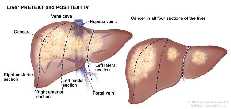
Colon Cancer Treatment (PDQ®)–Health Professional Version

Colon Cancer Treatment (PDQ®)–Health Professional Version

General Information About Colon Cancer

Cancer of the colon is a highly treatable and often curable disease when localized to the bowel. Surgery is the primary form of treatment and results in cure in approximately 50% of patients. However, recurrence following surgery is a major problem and is often the ultimate cause of death.

Incidence and Mortality

Worldwide, colorectal cancer is the third most common form of cancer. In 2022, there were an estimated 1.93 million new cases of colorectal cancer and 903,859 deaths.[1]

Estimated new cases and deaths from colon and rectal cancer in the United States in 2025:[2]

  • New cases of colon cancer: 107,320.
  • New cases of rectal cancer: 46,950.
  • Deaths: 52,900 (colon and rectal cancers combined).

Gastrointestinal stromal tumors can occur in the colon. For more information, see Gastrointestinal Stromal Tumors Treatment.

Anatomy

EnlargeGastrointestinal (digestive) system anatomy; drawing shows the esophagus, liver, stomach, colon, small intestine, rectum, and anus.
Anatomy of the lower gastrointestinal (digestive) system.

Risk Factors

Increasing age is the most important risk factor for most cancers. Other risk factors for colorectal cancer include the following:

  • Family history of colorectal cancer in a first-degree relative.[3]
  • Personal history of colorectal adenomas, colorectal cancer, or ovarian cancer.[46]
  • Hereditary conditions, including familial adenomatous polyposis (FAP) and Lynch syndrome (hereditary nonpolyposis colorectal cancer [HNPCC]).[7]
  • Personal history of long-standing chronic ulcerative colitis or Crohn colitis.[8]
  • Excessive alcohol use.[9]
  • Cigarette smoking.[10]
  • Race and ethnicity: African American.[11,12]
  • Obesity.[13]

Screening

Screening for colon cancer should be a part of routine care for all adults aged 50 years and older, especially for those with first-degree relatives with colorectal cancer. This recommendation is based on the frequency of the disease, ability to identify high-risk groups, slow growth of primary lesions, better survival of patients with early-stage lesions, and relative simplicity and accuracy of screening tests. For more information, see Colorectal Cancer Screening.

Prognostic Factors

The prognosis of patients with colon cancer is clearly related to:

  • The degree of penetration of the tumor through the bowel wall.
  • The presence or absence of nodal involvement.
  • The presence or absence of distant metastases.

These three characteristics form the basis for all staging systems developed for this disease.

Other prognostic factors for colon cancer include:

  • Bowel obstruction and bowel perforation are indicators of poor prognosis.[14]
  • Elevated pretreatment serum levels of carcinoembryonic antigen (CEA) have a negative prognostic significance.[15]

Many other prognostic markers have been evaluated retrospectively for patients with colon cancer, though most, including allelic loss of chromosome 18q or thymidylate synthase expression, have not been prospectively validated.[1625] Microsatellite instability, also associated with HNPCC, has been associated with improved survival independent of tumor stage in a population-based series of 607 patients younger than 50 years with colorectal cancer.[26] Patients with HNPCC reportedly have better prognoses in stage-stratified survival analysis than patients with sporadic colorectal cancer, but the retrospective nature of the studies and possibility of selection factors make this observation difficult to interpret.[27]

Treatment decisions depend on factors such as physician and patient preferences and the stage of the disease, rather than the age of the patient.[2830]

Racial differences in overall survival (OS) after adjuvant therapy have been observed, without differences in disease-free survival, suggesting that comorbid conditions play a role in survival outcome in different patient populations.[31]

Follow-Up and Survivorship

Limited data and no high-level evidence are available to guide patients and physicians about surveillance and management of patients after surgical resection and adjuvant therapy. The American Society of Clinical Oncology and the National Comprehensive Cancer Network recommend specific surveillance and follow-up strategies.[32,33]

Following treatment of colon cancer, periodic evaluations may lead to the earlier identification and management of recurrent disease.[3437] This monitoring has limited effect on overall mortality, as few localized, potentially curable metastases are found in patients with recurrent colon cancer. To date, no large-scale randomized trials have documented an OS benefit for standard, postoperative monitoring programs.[3842]

CEA is a serum glycoprotein frequently used in the management of patients with colon cancer. A review of the use of this tumor marker suggests:[43]

  • A CEA level is not a valuable screening test for colorectal cancer because of the large number of false-positive and false-negative reports.
  • Postoperative CEA testing should be restricted to patients who would be candidates for resection of liver or lung metastases.
  • Routine use of CEA levels alone for monitoring response to treatment is not recommended.

The optimal regimen and frequency of follow-up examinations are not well defined because the impact on patient survival is not clear and the quality of data is poor.[4042]

Factors Associated With Recurrence

Diet and exercise

Although cohort studies have suggested that a diet or exercise regimen may improve outcomes, no prospective randomized trials have confirmed these findings. The cohort studies contained multiple opportunities for unintended bias, and caution is needed when using the data from them.

Two prospective observational studies were performed with patients enrolled in the Cancer and Leukemia Group B CALGB-89803 trial (NCT00003835), an adjuvant chemotherapy trial for patients with stage III colon cancer.[44,45] In this trial, patients in the lowest quintile of the Western dietary pattern, compared with those patients in the highest quintile, experienced an adjusted hazard ratio (HR) for disease-free survival of 3.25 (95% confidence interval [CI], 2.04–5.19; P < .001) and an OS of 2.32 (95% CI, 1.36–3.96; P < .001). Additionally, stage III colon cancer patients in the highest quintile of dietary glycemic load experienced an adjusted HR for OS of 1.76 (95% CI, 1.22–2.54; P < .001), compared with those in the lowest quintile. Subsequently, in the Cancer Prevention Study II Nutrition Cohort, among 2,315 participants diagnosed with colorectal cancer, the degree of red and processed meat intake before diagnosis was associated with a higher risk of death (relative risk [RR], 1.29; 95% CI, 1.05–1.59; P = .03), but red meat consumption after diagnosis was not associated with overall mortality.[46][Level of evidence C1]

A meta-analysis of seven prospective cohort studies evaluating physical activity before and after a diagnosis of colorectal cancer demonstrated that patients who participated in any amount of physical activity before diagnosis had an RR of 0.75 (95% CI, 0.65–0.87; P < .001) for colorectal cancer-specific mortality, compared with patients who did not participate in any physical activity.[47] Patients who participated in a high amount of physical activity (vs. a low amount) before diagnosis had an RR of 0.70 (95% CI, 0.56–0.87; P = .002). Patients who participated in any physical activity (compared with no activity) after diagnosis had an RR of 0.74 (95% CI, 0.58–0.95; P = .02) for colorectal cancer-specific mortality. Those who participated in a high amount of physical activity (vs. a low amount) after diagnosis had an RR of 0.65 (95% CI, 0.47–0.92; P = .01).[47][Level of evidence C1]

Aspirin

A prospective cohort study examined the use of aspirin after a colorectal cancer diagnosis.[48] Regular users of aspirin after a diagnosis of colorectal cancer experienced an HRcolon cancer–specific mortality of 0.71 (95% CI, 0.53–0.95) and an HRoverall mortality of 0.79 (95% CI, 0.65–0.97).[48][Level of evidence C1] One study evaluated 964 patients with rectal or colon cancer from the Nurse’s Health Study and the Health Professionals Follow-up Study.[49] Among patients with colorectal cancer and PI3K variants, regular use of aspirin was associated with an HRdeath from any cause of 0.54 (95% CI, 0.31–0.94; P = .01)[49][Level of evidence C1]

References
  1. Bray F, Laversanne M, Sung H, et al.: Global cancer statistics 2022: GLOBOCAN estimates of incidence and mortality worldwide for 36 cancers in 185 countries. CA Cancer J Clin 74 (3): 229-263, 2024. [PUBMED Abstract]
  2. American Cancer Society: Cancer Facts and Figures 2025. American Cancer Society, 2025. Available online. Last accessed January 16, 2025.
  3. Johns LE, Houlston RS: A systematic review and meta-analysis of familial colorectal cancer risk. Am J Gastroenterol 96 (10): 2992-3003, 2001. [PUBMED Abstract]
  4. Imperiale TF, Juluri R, Sherer EA, et al.: A risk index for advanced neoplasia on the second surveillance colonoscopy in patients with previous adenomatous polyps. Gastrointest Endosc 80 (3): 471-8, 2014. [PUBMED Abstract]
  5. Singh H, Nugent Z, Demers A, et al.: Risk of colorectal cancer after diagnosis of endometrial cancer: a population-based study. J Clin Oncol 31 (16): 2010-5, 2013. [PUBMED Abstract]
  6. Srinivasan R, Yang YX, Rubin SC, et al.: Risk of colorectal cancer in women with a prior diagnosis of gynecologic malignancy. J Clin Gastroenterol 41 (3): 291-6, 2007. [PUBMED Abstract]
  7. Mork ME, You YN, Ying J, et al.: High Prevalence of Hereditary Cancer Syndromes in Adolescents and Young Adults With Colorectal Cancer. J Clin Oncol 33 (31): 3544-9, 2015. [PUBMED Abstract]
  8. Laukoetter MG, Mennigen R, Hannig CM, et al.: Intestinal cancer risk in Crohn’s disease: a meta-analysis. J Gastrointest Surg 15 (4): 576-83, 2011. [PUBMED Abstract]
  9. Fedirko V, Tramacere I, Bagnardi V, et al.: Alcohol drinking and colorectal cancer risk: an overall and dose-response meta-analysis of published studies. Ann Oncol 22 (9): 1958-72, 2011. [PUBMED Abstract]
  10. Liang PS, Chen TY, Giovannucci E: Cigarette smoking and colorectal cancer incidence and mortality: systematic review and meta-analysis. Int J Cancer 124 (10): 2406-15, 2009. [PUBMED Abstract]
  11. Laiyemo AO, Doubeni C, Pinsky PF, et al.: Race and colorectal cancer disparities: health-care utilization vs different cancer susceptibilities. J Natl Cancer Inst 102 (8): 538-46, 2010. [PUBMED Abstract]
  12. Lansdorp-Vogelaar I, Kuntz KM, Knudsen AB, et al.: Contribution of screening and survival differences to racial disparities in colorectal cancer rates. Cancer Epidemiol Biomarkers Prev 21 (5): 728-36, 2012. [PUBMED Abstract]
  13. Ma Y, Yang Y, Wang F, et al.: Obesity and risk of colorectal cancer: a systematic review of prospective studies. PLoS One 8 (1): e53916, 2013. [PUBMED Abstract]
  14. Steinberg SM, Barkin JS, Kaplan RS, et al.: Prognostic indicators of colon tumors. The Gastrointestinal Tumor Study Group experience. Cancer 57 (9): 1866-70, 1986. [PUBMED Abstract]
  15. Filella X, Molina R, Grau JJ, et al.: Prognostic value of CA 19.9 levels in colorectal cancer. Ann Surg 216 (1): 55-9, 1992. [PUBMED Abstract]
  16. McLeod HL, Murray GI: Tumour markers of prognosis in colorectal cancer. Br J Cancer 79 (2): 191-203, 1999. [PUBMED Abstract]
  17. Jen J, Kim H, Piantadosi S, et al.: Allelic loss of chromosome 18q and prognosis in colorectal cancer. N Engl J Med 331 (4): 213-21, 1994. [PUBMED Abstract]
  18. Lanza G, Matteuzzi M, Gafá R, et al.: Chromosome 18q allelic loss and prognosis in stage II and III colon cancer. Int J Cancer 79 (4): 390-5, 1998. [PUBMED Abstract]
  19. Griffin MR, Bergstralh EJ, Coffey RJ, et al.: Predictors of survival after curative resection of carcinoma of the colon and rectum. Cancer 60 (9): 2318-24, 1987. [PUBMED Abstract]
  20. Johnston PG, Fisher ER, Rockette HE, et al.: The role of thymidylate synthase expression in prognosis and outcome of adjuvant chemotherapy in patients with rectal cancer. J Clin Oncol 12 (12): 2640-7, 1994. [PUBMED Abstract]
  21. Shibata D, Reale MA, Lavin P, et al.: The DCC protein and prognosis in colorectal cancer. N Engl J Med 335 (23): 1727-32, 1996. [PUBMED Abstract]
  22. Bauer KD, Lincoln ST, Vera-Roman JM, et al.: Prognostic implications of proliferative activity and DNA aneuploidy in colonic adenocarcinomas. Lab Invest 57 (3): 329-35, 1987. [PUBMED Abstract]
  23. Bauer KD, Bagwell CB, Giaretti W, et al.: Consensus review of the clinical utility of DNA flow cytometry in colorectal cancer. Cytometry 14 (5): 486-91, 1993. [PUBMED Abstract]
  24. Sun XF, Carstensen JM, Zhang H, et al.: Prognostic significance of cytoplasmic p53 oncoprotein in colorectal adenocarcinoma. Lancet 340 (8832): 1369-73, 1992. [PUBMED Abstract]
  25. Roth JA: p53 prognostication: paradigm or paradox? Clin Cancer Res 5 (11): 3345, 1999. [PUBMED Abstract]
  26. Gryfe R, Kim H, Hsieh ET, et al.: Tumor microsatellite instability and clinical outcome in young patients with colorectal cancer. N Engl J Med 342 (2): 69-77, 2000. [PUBMED Abstract]
  27. Watson P, Lin KM, Rodriguez-Bigas MA, et al.: Colorectal carcinoma survival among hereditary nonpolyposis colorectal carcinoma family members. Cancer 83 (2): 259-66, 1998. [PUBMED Abstract]
  28. Iwashyna TJ, Lamont EB: Effectiveness of adjuvant fluorouracil in clinical practice: a population-based cohort study of elderly patients with stage III colon cancer. J Clin Oncol 20 (19): 3992-8, 2002. [PUBMED Abstract]
  29. Chiara S, Nobile MT, Vincenti M, et al.: Advanced colorectal cancer in the elderly: results of consecutive trials with 5-fluorouracil-based chemotherapy. Cancer Chemother Pharmacol 42 (4): 336-40, 1998. [PUBMED Abstract]
  30. Popescu RA, Norman A, Ross PJ, et al.: Adjuvant or palliative chemotherapy for colorectal cancer in patients 70 years or older. J Clin Oncol 17 (8): 2412-8, 1999. [PUBMED Abstract]
  31. Dignam JJ, Colangelo L, Tian W, et al.: Outcomes among African-Americans and Caucasians in colon cancer adjuvant therapy trials: findings from the National Surgical Adjuvant Breast and Bowel Project. J Natl Cancer Inst 91 (22): 1933-40, 1999. [PUBMED Abstract]
  32. Meyerhardt JA, Mangu PB, Flynn PJ, et al.: Follow-up care, surveillance protocol, and secondary prevention measures for survivors of colorectal cancer: American Society of Clinical Oncology clinical practice guideline endorsement. J Clin Oncol 31 (35): 4465-70, 2013. [PUBMED Abstract]
  33. Benson AB, Bekaii-Saab T, Chan E, et al.: Localized colon cancer, version 3.2013: featured updates to the NCCN Guidelines. J Natl Compr Canc Netw 11 (5): 519-28, 2013. [PUBMED Abstract]
  34. Martin EW, Minton JP, Carey LC: CEA-directed second-look surgery in the asymptomatic patient after primary resection of colorectal carcinoma. Ann Surg 202 (3): 310-7, 1985. [PUBMED Abstract]
  35. Bruinvels DJ, Stiggelbout AM, Kievit J, et al.: Follow-up of patients with colorectal cancer. A meta-analysis. Ann Surg 219 (2): 174-82, 1994. [PUBMED Abstract]
  36. Lautenbach E, Forde KA, Neugut AI: Benefits of colonoscopic surveillance after curative resection of colorectal cancer. Ann Surg 220 (2): 206-11, 1994. [PUBMED Abstract]
  37. Khoury DA, Opelka FG, Beck DE, et al.: Colon surveillance after colorectal cancer surgery. Dis Colon Rectum 39 (3): 252-6, 1996. [PUBMED Abstract]
  38. Safi F, Link KH, Beger HG: Is follow-up of colorectal cancer patients worthwhile? Dis Colon Rectum 36 (7): 636-43; discussion 643-4, 1993. [PUBMED Abstract]
  39. Moertel CG, Fleming TR, Macdonald JS, et al.: An evaluation of the carcinoembryonic antigen (CEA) test for monitoring patients with resected colon cancer. JAMA 270 (8): 943-7, 1993. [PUBMED Abstract]
  40. Rosen M, Chan L, Beart RW, et al.: Follow-up of colorectal cancer: a meta-analysis. Dis Colon Rectum 41 (9): 1116-26, 1998. [PUBMED Abstract]
  41. Desch CE, Benson AB, Smith TJ, et al.: Recommended colorectal cancer surveillance guidelines by the American Society of Clinical Oncology. J Clin Oncol 17 (4): 1312, 1999. [PUBMED Abstract]
  42. Benson AB, Desch CE, Flynn PJ, et al.: 2000 update of American Society of Clinical Oncology colorectal cancer surveillance guidelines. J Clin Oncol 18 (20): 3586-8, 2000. [PUBMED Abstract]
  43. Clinical practice guidelines for the use of tumor markers in breast and colorectal cancer. Adopted on May 17, 1996 by the American Society of Clinical Oncology. J Clin Oncol 14 (10): 2843-77, 1996. [PUBMED Abstract]
  44. Meyerhardt JA, Niedzwiecki D, Hollis D, et al.: Association of dietary patterns with cancer recurrence and survival in patients with stage III colon cancer. JAMA 298 (7): 754-64, 2007. [PUBMED Abstract]
  45. Meyerhardt JA, Sato K, Niedzwiecki D, et al.: Dietary glycemic load and cancer recurrence and survival in patients with stage III colon cancer: findings from CALGB 89803. J Natl Cancer Inst 104 (22): 1702-11, 2012. [PUBMED Abstract]
  46. McCullough ML, Gapstur SM, Shah R, et al.: Association between red and processed meat intake and mortality among colorectal cancer survivors. J Clin Oncol 31 (22): 2773-82, 2013. [PUBMED Abstract]
  47. Je Y, Jeon JY, Giovannucci EL, et al.: Association between physical activity and mortality in colorectal cancer: a meta-analysis of prospective cohort studies. Int J Cancer 133 (8): 1905-13, 2013. [PUBMED Abstract]
  48. Chan AT, Ogino S, Fuchs CS: Aspirin use and survival after diagnosis of colorectal cancer. JAMA 302 (6): 649-58, 2009. [PUBMED Abstract]
  49. Liao X, Lochhead P, Nishihara R, et al.: Aspirin use, tumor PIK3CA mutation, and colorectal-cancer survival. N Engl J Med 367 (17): 1596-606, 2012. [PUBMED Abstract]

Cellular Classification of Colon Cancer

Histological types of colon cancer include:

  • Adenocarcinoma (most colon cancers).
    • Mucinous (colloid) adenocarcinoma.
    • Signet ring adenocarcinoma.
  • Scirrhous tumors.
  • Neuroendocrine.[1] Tumors with neuroendocrine differentiation typically have a poorer prognosis than pure adenocarcinoma variants.
References
  1. Saclarides TJ, Szeluga D, Staren ED: Neuroendocrine cancers of the colon and rectum. Results of a ten-year experience. Dis Colon Rectum 37 (7): 635-42, 1994. [PUBMED Abstract]

Stage Information for Colon Cancer

Treatment decisions can be made with reference to the TNM (tumor, node, metastasis) classification [1] rather than to the older Dukes or the Modified Astler-Coller classification schema.

The AJCC and a National Cancer Institute–sponsored panel recommended that at least 12 lymph nodes be examined in patients with colon and rectal cancer to confirm the absence of nodal involvement by tumor.[13] This recommendation takes into consideration that the number of lymph nodes examined is a reflection of the aggressiveness of lymphovascular mesenteric dissection at the time of surgical resection and the pathological identification of nodes in the specimen. Retrospective studies demonstrated that the number of lymph nodes examined in colon and rectal surgery may be associated with patient outcome.[47]

AJCC Stage Groupings and TNM Definitions

The AJCC has designated staging by TNM classification to define colon cancer.[1] The same classification is used for both clinical and pathological staging.[1]

Table 1. Definitions of TNM Stage 0a
Stage TNMb,c Description Illustration
T = primary tumor; N = regional lymph nodes; M = distant metastasis.
aReprinted with permission from AJCC: Colon and rectum. In: Amin MB, Edge SB, Greene FL, et al., eds.: AJCC Cancer Staging Manual. 8th ed. New York, NY: Springer, 2017, pp 251–74.
The explanations for superscripts b and c are at the end of Table 5.
0 Tis, N0, M0 Tis = Carcinoma in situ, intramucosal carcinoma (involvement of lamina propria with no extension through muscularis mucosae).
EnlargeStage 0 colorectal carcinoma in situ; drawing shows a cross-section of the colon/rectum. An inset shows the layers of the colon/rectum wall with abnormal cells in the mucosa layer. Also shown are the submucosa, muscle layers, serosa, a blood vessel, and lymph nodes.
N0 = No regional lymph node metastasis.
M0 = No distant metastasis by imaging, etc.; no evidence of tumor in distant sites or organs. (This category is not assigned by pathologists.)
Table 2. Definitions of TNM Stage Ia
Stage TNMb,c Description Illustration
T = primary tumor; N = regional lymph nodes; M = distant metastasis.
aReprinted with permission from AJCC: Colon and rectum. In: Amin MB, Edge SB, Greene FL, et al., eds.: AJCC Cancer Staging Manual. 8th ed. New York, NY: Springer, 2017, pp 251–74.
The explanations for superscripts b and c are at the end of Table 5.
I T1, T2, N0, M0 T1 = Tumor invades the submucosa (through the muscularis mucosa but not into the muscularis propria).
EnlargeStage I colorectal cancer; drawing shows a cross-section of the colon/rectum. An inset shows the layers of the colon/rectum wall with cancer in the mucosa and submucosa. Also shown are the muscle layers, serosa, a blood vessel, and lymph nodes.
T2 = Tumor invades the muscularis propria.
N0 = No regional lymph node metastasis.
M0 = No distant metastasis by imaging, etc.; no evidence of tumor in distant sites or organs. (This category is not assigned by pathologists.)
Table 3. Definitions of TNM Stages IIA, IIB, and IICa
Stage TNMb,c Description Illustration
T = primary tumor; N = regional lymph nodes; M = distant metastasis.
aReprinted with permission from AJCC: Colon and rectum. In: Amin MB, Edge SB, Greene FL, et al., eds.: AJCC Cancer Staging Manual. 8th ed. New York, NY: Springer, 2017, pp 251–74.
The explanations for superscripts b and c are at the end of Table 5.
IIA T3, N0, M0 T3 = Tumor invades through the muscularis propria into pericolorectal tissues.
EnlargeStage II colorectal cancer; drawing shows a cross-section of the colon/rectum and a three-panel inset. Each panel shows the layers of the colon/rectum wall: the mucosa, submucosa, muscle layers, and serosa. Also shown are a blood vessel and lymph nodes. The first panel shows stage IIA with cancer in the mucosa, submucosa, muscle layers, and serosa. The second panel shows stage IIB with cancer in all layers and spreading through the serosa to the visceral peritoneum. The third panel shows stage IIC with cancer in all layers and spreading through the serosa to nearby organs.
N0 = No regional lymph node metastasis.
M0 = No distant metastasis by imaging, etc.; no evidence of tumor in distant sites or organs. (This category is not assigned by pathologists.)
IIB T4a, N0, M0 T4a = Tumor invades through the visceral peritoneum (including gross perforation of the bowel through tumor and continuous invasion of tumor through areas of inflammation to the surface of the visceral peritoneum).
N0 = No regional lymph node metastasis.
M0 = No distant metastasis by imaging, etc.; no evidence of tumor in distant sites or organs. (This category is not assigned by pathologists.)
IIC T4b, N0, M0 T4b = Tumor directly invades or adheres to adjacent organs or structures.
N0 = No regional lymph node metastasis.
M0 = No distant metastasis by imaging, etc.; no evidence of tumor in distant sites or organs. (This category is not assigned by pathologists.)
Table 4. Definitions of TNM Stages IIIA, IIIB, and IIICa
Stage TNMb,c Description Illustration
T = primary tumor; N = regional lymph nodes; M = distant metastasis.
aReprinted with permission from AJCC: Colon and rectum. In: Amin MB, Edge SB, Greene FL, et al., eds.: AJCC Cancer Staging Manual. 8th ed. New York, NY: Springer, 2017, pp 251–74.
The explanations for superscripts b and c are at the end of Table 5.
IIIA T1, N2a, M0 T1 = Tumor invades the submucosa (through the muscularis mucosa but not into the muscularis propria).
EnlargeStage IIIA colorectal cancer; drawing shows a cross-section of the colon/rectum and a two-panel inset. Each panel shows the layers of the colon/rectum wall: the mucosa, submucosa, muscle layers, and serosa. Also shown are a blood vessel and lymph nodes. The first panel shows cancer in the mucosa, submucosa, and muscle layers and in 2 lymph nodes. The second panel shows cancer in the mucosa and submucosa and in 5 lymph nodes.
N2a = Four to six regional lymph nodes are positive.
M0 = No distant metastasis by imaging, etc.; no evidence of tumor in distant sites or organs. (This category is not assigned by pathologists.)
T1–2, N1/N1c, M0 T1 = Tumor invades the submucosa (through the muscularis mucosa but not into the muscularis propria).
T2 = Tumor invades the muscularis propria.
N1 = One to three regional lymph nodes are positive (tumor in lymph nodes measuring ≥0.2 mm), or any number of tumor deposits are present and all identifiable lymph nodes are negative.
–N1c = No regional lymph nodes are positive, but there are tumor deposits in the subserosa, mesentery, or nonperitonealized pericolic, or perirectal/mesorectal tissues.
M0 = No distant metastasis by imaging, etc.; no evidence of tumor in distant sites or organs. (This category is not assigned by pathologists.)
IIIB T1–T2, N2b, M0 T1 = Tumor invades the submucosa (through the muscularis mucosa but not into the muscularis propria).
EnlargeStage IIIB colorectal cancer; drawing shows a cross-section of the colon/rectum and a three-panel inset. Each panel shows the layers of the colon/rectum wall: the mucosa, submucosa, muscle layers, and serosa. Also shown are a blood vessel and lymph nodes. The first panel shows cancer in all layers, in 3 nearby lymph nodes, and in the visceral peritoneum. The second panel shows cancer in all layers and in 5 nearby lymph nodes. The third panel shows cancer in the mucosa, submucosa, and muscle layers and in 7 nearby lymph nodes.
T2 = Tumor invades the muscularis propria.
N2b = Seven or more regional lymph nodes are positive.
M0 = No distant metastasis by imaging, etc.; no evidence of tumor in distant sites or organs. (This category is not assigned by pathologists.)
T2–T3, N2a, M0 T2 = Tumor invades the muscularis propria.
T3 = Tumor invades through the muscularis propria into pericolorectal tissues.
N2a = Four to six regional lymph nodes are positive.
M0 = No distant metastasis by imaging, etc.; no evidence of tumor in distant sites or organs. (This category is not assigned by pathologists.)
T3–T4a, N1/N1c, M0 T3 = Tumor invades through the muscularis propria into pericolorectal tissues.
T4 = Tumor invades the visceral peritoneum or invades or adheres to adjacent organ or structure.
–T4a = Tumor invades through the visceral peritoneum (including gross perforation of the bowel through tumor and continuous invasion of tumor through areas of inflammation to the surface of the visceral peritoneum).
N1 = One to three regional lymph nodes are positive (tumor in lymph nodes measuring ≥0.2 mm), or any number of tumor deposits are present and all identifiable lymph nodes are negative.
–N1c = No regional lymph nodes are positive, but there are tumor deposits in the subserosa, mesentery, or nonperitonealized pericolic, or perirectal/mesorectal tissues.
M0 = No distant metastasis by imaging, etc.; no evidence of tumor in distant sites or organs. (This category is not assigned by pathologists.)
IIIC T3–T4a, N2b, M0 T3 = Tumor invades through the muscularis propria into pericolorectal tissues.
EnlargeStage IIIC colorectal cancer; drawing shows a cross-section of the colon/rectum and a three-panel inset. Each panel shows the layers of the colon/rectum wall: the mucosa, submucosa, muscle layers, and serosa. Also shown are a blood vessel and lymph nodes. The first panel shows cancer in all layers, in 4 lymph nodes, and in the visceral peritoneum. The second panel shows cancer in all layers and in 7 lymph nodes. The third panel shows cancer in all layers, in 2 lymph nodes, and spreading to nearby organs.
T4 = Tumor invades the visceral peritoneum or invades or adheres to adjacent organ or structure.
–T4a = Tumor invades through the visceral peritoneum (including gross perforation of the bowel through tumor and continuous invasion of tumor through areas of inflammation to the surface of the visceral peritoneum).
N2b = Seven or more regional lymph nodes are positive.
M0 = No distant metastasis by imaging, etc.; no evidence of tumor in distant sites or organs. (This category is not assigned by pathologists.)
T4a, N2a, M0 T4a = Tumor invades through the visceral peritoneum (including gross perforation of the bowel through tumor and continuous invasion of tumor through areas of inflammation to the surface of the visceral peritoneum).
N2a = Four to six regional lymph nodes are positive.
M0 = No distant metastasis by imaging, etc.; no evidence of tumor in distant sites or organs. (This category is not assigned by pathologists.)
T4b, N1–N2, M0 T4b = Tumor directly invades or adheres to adjacent organs or structures.
N1 = One to three regional lymph nodes are positive (tumor in lymph nodes measuring ≥0.2 mm), or any number of tumor deposits are present and all identifiable lymph nodes are negative.
–N1a = One regional lymph node is positive.
–N1b = Two or three regional lymph nodes are positive.
–N1c = No regional lymph nodes are positive, but there are tumor deposits in the subserosa, mesentery, or nonperitonealized pericolic, or perirectal/mesorectal tissues.
N2 = Four or more regional nodes are positive.
–N2a = Four to six regional lymph nodes are positive.
–N2b = Seven or more regional lymph nodes are positive.
M0 = No distant metastasis by imaging, etc.; no evidence of tumor in distant sites or organs. (This category is not assigned by pathologists.)
Table 5. Definitions of TNM Stages IVA, IVB, and IVCa
Stage TNMb,c Definition Illustration
T = primary tumor; N = regional lymph nodes; M = distant metastasis.
aReprinted with permission from AJCC: Colon and rectum. In: Amin MB, Edge SB, Greene FL, et al., eds.: AJCC Cancer Staging Manual. 8th ed. New York, NY: Springer, 2017, pp 251–74.
bDirect invasion in T4 includes invasion of other organs or other segments of the colorectum as a result of direct extension through the serosa, as confirmed on microscopic examination (e.g., invasion of the sigmoid colon by a carcinoma of the cecum) or, for cancers in a retroperitoneal or subperitoneal location, direct invasion of other organs or structures by virtue of extension beyond the muscularis propria (i.e., respectively, a tumor on the posterior wall of the descending colon invading the left kidney or lateral abdominal wall; or a mid or distal rectal cancer with invasion of prostate, seminal vesicles, cervix, or vagina).
cTumor that is adherent to other organs or structures, grossly, is classified cT4b. However, if no tumor is present in the adhesion, microscopically, the classification should be pT1-4a depending on the anatomical depth of wall invasion. The V and L classification should be used to identify the presence or absence of vascular or lymphatic invasion whereas the PN prognostic factor should be used for perineural invasion.
IVA Any T, Any N, M1a TX = Primary tumor cannot be assessed.
EnlargeStage IV colon cancer; drawing shows other parts of the body where colon cancer may spread, including the distant lymph nodes, lung, liver, and abdominal wall. An inset shows cancer cells spreading from the colon, through the blood and lymph system, to another part of the body where metastatic cancer has formed.
T0 = No evidence of primary tumor.
Tis = Carcinoma in situ, intramucosal carcinoma (involvement of lamina propria with no extension through muscularis mucosae).
T1 = Tumor invades the submucosa (through the muscularis mucosa but not into the muscularis propria).
T2 = Tumor invades the muscularis propria.
T3 = Tumor invades through the muscularis propria into pericolorectal tissues.
T4 = Tumor invades the visceral peritoneum or invades or adheres to adjacent organ or structure.
–T4a = Tumor invades through the visceral peritoneum (including gross perforation of the bowel through tumor and continuous invasion of tumor through areas of inflammation to the surface of the visceral peritoneum).
–T4b = Tumor directly invades or adheres to adjacent organs or structures.
NX = Regional lymph nodes cannot be assessed.
N0 = No regional lymph node metastasis.
N1 = One to three regional lymph nodes are positive (tumor in lymph nodes measuring ≥0.2 mm), or any number of tumor deposits are present and all identifiable lymph nodes are negative.
–N1a = One regional lymph node is positive.
–N1b = Two or three regional lymph nodes are positive.
–N1c = No regional lymph nodes are positive, but there are tumor deposits in the subserosa, mesentery, or nonperitonealized pericolic, or perirectal/mesorectal tissues.
N2 = Four or more regional nodes are positive.
–N2a = Four to six regional lymph nodes are positive.
–N2b = Seven or more regional lymph nodes are positive.
M1a = Metastasis to one site or organ is identified without peritoneal metastasis.
IVB Any T, Any N, M1b Any T = See T descriptions above in Any T, Any N, M1a TNM stage group.
Any N = See N descriptions above in Any T, Any N1, M1a TNM stage group.
M1b = Metastasis to two or more sites or organs is identified without peritoneal metastasis.
IVC Any T, Any N, M1c Any T = See T descriptions above in Any T, Any N, M1a TNM stage group.
Any N = See N descriptions above in Any T, Any N1, M1a TNM stage group.
M1c = Metastasis to the peritoneal surface is identified alone or with other site or organ metastases.
References
  1. Jessup J, Benson A, Chen V: Colon and Rectum. In: Amin MB, Edge SB, Greene FL, et al., eds.: AJCC Cancer Staging Manual. 8th ed. Springer; 2017, pp 251–74.
  2. Compton CC, Greene FL: The staging of colorectal cancer: 2004 and beyond. CA Cancer J Clin 54 (6): 295-308, 2004 Nov-Dec. [PUBMED Abstract]
  3. Nelson H, Petrelli N, Carlin A, et al.: Guidelines 2000 for colon and rectal cancer surgery. J Natl Cancer Inst 93 (8): 583-96, 2001. [PUBMED Abstract]
  4. Swanson RS, Compton CC, Stewart AK, et al.: The prognosis of T3N0 colon cancer is dependent on the number of lymph nodes examined. Ann Surg Oncol 10 (1): 65-71, 2003 Jan-Feb. [PUBMED Abstract]
  5. Le Voyer TE, Sigurdson ER, Hanlon AL, et al.: Colon cancer survival is associated with increasing number of lymph nodes analyzed: a secondary survey of intergroup trial INT-0089. J Clin Oncol 21 (15): 2912-9, 2003. [PUBMED Abstract]
  6. Prandi M, Lionetto R, Bini A, et al.: Prognostic evaluation of stage B colon cancer patients is improved by an adequate lymphadenectomy: results of a secondary analysis of a large scale adjuvant trial. Ann Surg 235 (4): 458-63, 2002. [PUBMED Abstract]
  7. Tepper JE, O’Connell MJ, Niedzwiecki D, et al.: Impact of number of nodes retrieved on outcome in patients with rectal cancer. J Clin Oncol 19 (1): 157-63, 2001. [PUBMED Abstract]

Treatment Option Overview for Colon Cancer

Table 6. Treatment Options for Colon Cancer
Stage (TNM Staging Criteria) Treatment Options
Stage 0 Colon Cancer Surgery
Stage I Colon Cancer Surgery
Stage II Colon Cancer Surgery
Adjuvant chemotherapy (under clinical evaluation)
Stage III Colon Cancer Surgery
Clinical trials
Liver Metastasis Surgery
Neoadjuvant chemotherapy
Local ablation
Adjuvant chemotherapy
Intra-arterial chemotherapy
Clinical trials
Stage IV and Recurrent Colon Cancer Surgery
Systemic therapy
Immunotherapy
Clinical trials

Primary Surgical Therapy

Standard treatment for patients with colon cancer has been open surgical resection of the primary and regional lymph nodes for localized disease.

The role of laparoscopic techniques [14] in the treatment of colon cancer has been examined in two studies.

Evidence (laparoscopic techniques):

  1. A multicenter, prospective, randomized, noninferiority trial (NCCTG-934653 [NCT00002575]) compared laparoscopic-assisted colectomy (LAC) with open colectomy in 872 patients.
    • At a median follow-up of 4.4 years, 3-year recurrence rates (16% LAC vs. 18% open colectomy; hazard ratio [HR] for recurrence, 0.86; 95% confidence interval [CI], 0.63–1.17; P = .32) and 3-year overall survival (OS) rates (86% LAC vs. 85% open colectomy; HRdeath in LAC, 0.91; 95% CI, 0.68–1.21; P = .51) were similar in both groups for all stages of disease evaluated. Tumor recurrence in surgical incisions was less than 1% for both groups.[5][Level of evidence A1]
    • Decreased hospital stay (5 days LAC vs. 6 days open colectomy, P < .001) and decreased use of analgesics were reported in the LAC group. A 21% conversion rate from LAC to open procedure was shown.
    • This study excluded patients with locally advanced disease, transverse colon and rectal tumor locations, and perforated lesions. Each of the 66 surgeons participating in the trial had performed at least 20 LACs and were accredited for study participation after independent videotape review assured appropriate oncologic and surgical principles were maintained.[5] The quality-of-life component of this trial was published separately and minimal short-term quality-of-life benefits with LAC were reported.[6][Level of evidence A3]
  2. One small, single-institution randomized study of 219 patients showed that the LAC procedure was independently associated with reduced tumor recurrence on multivariate analysis.[7][Level of evidence A1]

Surgery is curative in 25% to 40% of highly selected patients who develop resectable metastases in the liver and lung. Improved surgical techniques and advances in preoperative imaging have allowed for better patient selection for resection.

Adjuvant Chemotherapy

The potential value of adjuvant chemotherapy for patients with stage II colon cancer is controversial. Pooled analyses and meta-analyses have suggested a 2% to 4% improvement in OS for patients treated with adjuvant fluorouracil (5-FU)–based therapy compared with observation.[810] For more information, see the Treatment of Stage II Colon Cancer section.

Before 2000, 5-FU was the only useful cytotoxic chemotherapy in the adjuvant setting for patients with stage III colon cancer. Since 2000, capecitabine has been established as an equivalent alternative to 5-FU and leucovorin (5-FU/LV). The addition of oxaliplatin to 5-FU/LV has been shown to improve OS compared with 5-FU/LV alone. For more information, see the Treatment of Stage III Colon Cancer section.

Chemotherapy regimens

Table 7 describes the chemotherapy regimens used to treat colon cancer.

Table 7. Drug Combinations Used to Treat Colon Cancer
Regimen Name Drug Combination Dose
5-FU = fluorouracil; AIO = Arbeitsgemeinschaft Internistische Onkologie; bid = twice a day; IV = intravenous; LV = leucovorin.
AIO or German AIO Folic acid, 5-FU, and irinotecan Irinotecan (100 mg/m2) and LV (500 mg/m2) administered as 2-hour infusions on d 1, followed by 5-FU (2,000 mg/m2) IV bolus administered via ambulatory pump weekly over 24 h, 4 times a y (52 wk).
CAPOX Capecitabine and oxaliplatin Capecitabine (1,000 mg/m2) bid on d 1–14, plus oxaliplatin (70 mg/m2) on d 1 and 8 every 3 wk.
Douillard Folic acid, 5-FU, and irinotecan Irinotecan (180 mg/m2) administered as a 2-h infusion on d 1, LV (200 mg/m2) administered as a 2-h infusion on d 1 and 2, followed by a loading dose of 5-FU (400 mg/m2) IV bolus, then 5-FU (600 mg/m2) administered via ambulatory pump over 22 h every 2 wk on d 1 and 2.
FOLFIRI LV, 5-FU, and irinotecan Irinotecan (180 mg/m2) and LV (400 mg/m2) administered as 2-h infusions on d 1, followed by a loading dose of 5-FU (400 mg/m2) IV bolus administered on d 1, then 5-FU (2,400–3,000 mg/m2) administered via ambulatory pump over 46 h every 2 wk.
FOLFOX-4 Oxaliplatin, LV, and 5-FU Oxaliplatin (85 mg/m2) administered as a 2-h infusion on d 1, LV (200 mg/m2) administered as a 2-h infusion on d 1 and 2, followed by a loading dose of 5-FU (400 mg/m2) IV bolus, then 5-FU (600 mg/m2) administered via ambulatory pump over 22 h every 2 wk on d 1 and 2.
FOLFOX-6 Oxaliplatin, LV, and 5-FU Oxaliplatin (85–100 mg/m2) and LV (400 mg/m2) administered as 2-h infusions on d 1, followed by a loading dose of 5-FU (400 mg/m2) IV bolus on d 1, then 5-FU (2,400–3,000 mg/m2) administered via ambulatory pump over 46 h every 2 wk.
FOLFOXIRI Irinotecan, oxaliplatin, LV, 5-FU Irinotecan (165 mg/m2) administered as a 60-min infusion, then concomitant infusion of oxaliplatin (85 mg/m2) and LV (200 mg/m2) over 120 min, followed by 5-FU (3,200 mg/m2) administered as a 48-h continuous infusion.
FUFOX 5-FU, LV, and oxaliplatin Oxaliplatin (50 mg/m2) plus LV (500 mg/m2) plus 5-FU (2,000 mg/m2) administered as a 22-h continuous infusion on d 1, 8, 22, and 29 every 36 d.
FUOX 5-FU plus oxaliplatin 5-FU (2,250 mg/m2) administered as a continuous infusion over 48 h on d 1, 8, 15, 22, 29, and 36 plus oxaliplatin (85 mg/m2) on d 1, 15, and 29 every 6 wk.
IFL (or Saltz) Irinotecan, 5-FU, and LV Irinotecan (125 mg/m2) plus 5-FU (500 mg/m2) IV bolus and LV (20 mg/m2) IV bolus administered weekly for 4 out of 6 wk.
XELOX Capecitabine plus oxaliplatin Oral capecitabine (1,000 mg/m2) administered bid for 14 d plus oxaliplatin (130 mg/m2) on d 1 every 3 wk.

Capecitabine and fluorouracil dosing

The DPYD gene encodes an enzyme that catabolizes pyrimidines and fluoropyrimidines, like capecitabine and fluorouracil. An estimated 1% to 2% of the population has germline pathogenic variants in DPYD, which lead to reduced DPD protein function and an accumulation of pyrimidines and fluoropyrimidines in the body.[11,12] Patients with the DPYD*2A variant who receive fluoropyrimidines may experience severe, life-threatening toxicities that are sometimes fatal. Many other DPYD variants have been identified, with a range of clinical effects.[1113] Fluoropyrimidine avoidance or a dose reduction of 50% may be recommended based on the patient’s DPYD genotype and number of functioning DPYD alleles.[1416] DPYD genetic testing costs less than $200, but insurance coverage varies due to a lack of national guidelines.[17] In addition, testing may delay therapy by 2 weeks, which would not be advisable in urgent situations. This controversial issue requires further evaluation.[18]

Adjuvant Radiation Therapy

While combined modality therapy with chemotherapy and radiation therapy has a significant role in the management of patients with rectal cancer (below the peritoneal reflection), the role of adjuvant radiation therapy for patients with colon cancer (above the peritoneal reflection) is not well defined. Patterns-of-care analyses and single-institution retrospective reviews suggest a role for radiation therapy in certain high-risk subsets of colon cancer patients (e.g., T4, tumor location in immobile sites, local perforation, obstruction, and residual disease postresection).[1924]

Evidence (adjuvant radiation therapy):

  1. A phase III, randomized, intergroup study evaluated the benefit of adding radiation therapy to surgery and chemotherapy with 5-FU-levamisole in selected patients with high-risk colon cancer (e.g., T4; or T3, N1–N2 ascending and/or descending colon).[25]
    • This clinical trial closed early secondary to inadequate patient accrual. An analysis of 222 enrolled patients (the original goal was 700 patients) demonstrated no relapse or OS benefit for the group that received radiation therapy, although the sample size and statistical power were inadequate to exclude benefit.

Adjuvant radiation therapy has no current standard role in the management of patients with colon cancer following curative resection, although it may have a role for patients with residual disease.

References
  1. Bokey EL, Moore JW, Chapuis PH, et al.: Morbidity and mortality following laparoscopic-assisted right hemicolectomy for cancer. Dis Colon Rectum 39 (10 Suppl): S24-8, 1996. [PUBMED Abstract]
  2. Franklin ME, Rosenthal D, Abrego-Medina D, et al.: Prospective comparison of open vs. laparoscopic colon surgery for carcinoma. Five-year results. Dis Colon Rectum 39 (10 Suppl): S35-46, 1996. [PUBMED Abstract]
  3. Fleshman JW, Nelson H, Peters WR, et al.: Early results of laparoscopic surgery for colorectal cancer. Retrospective analysis of 372 patients treated by Clinical Outcomes of Surgical Therapy (COST) Study Group. Dis Colon Rectum 39 (10 Suppl): S53-8, 1996. [PUBMED Abstract]
  4. Schwenk W, Böhm B, Müller JM: Postoperative pain and fatigue after laparoscopic or conventional colorectal resections. A prospective randomized trial. Surg Endosc 12 (9): 1131-6, 1998. [PUBMED Abstract]
  5. Clinical Outcomes of Surgical Therapy Study Group: A comparison of laparoscopically assisted and open colectomy for colon cancer. N Engl J Med 350 (20): 2050-9, 2004. [PUBMED Abstract]
  6. Weeks JC, Nelson H, Gelber S, et al.: Short-term quality-of-life outcomes following laparoscopic-assisted colectomy vs open colectomy for colon cancer: a randomized trial. JAMA 287 (3): 321-8, 2002. [PUBMED Abstract]
  7. Lacy AM, García-Valdecasas JC, Delgado S, et al.: Laparoscopy-assisted colectomy versus open colectomy for treatment of non-metastatic colon cancer: a randomised trial. Lancet 359 (9325): 2224-9, 2002. [PUBMED Abstract]
  8. Efficacy of adjuvant fluorouracil and folinic acid in B2 colon cancer. International Multicentre Pooled Analysis of B2 Colon Cancer Trials (IMPACT B2) Investigators. J Clin Oncol 17 (5): 1356-63, 1999. [PUBMED Abstract]
  9. Gill S, Loprinzi CL, Sargent DJ, et al.: Pooled analysis of fluorouracil-based adjuvant therapy for stage II and III colon cancer: who benefits and by how much? J Clin Oncol 22 (10): 1797-806, 2004. [PUBMED Abstract]
  10. Mamounas E, Wieand S, Wolmark N, et al.: Comparative efficacy of adjuvant chemotherapy in patients with Dukes’ B versus Dukes’ C colon cancer: results from four National Surgical Adjuvant Breast and Bowel Project adjuvant studies (C-01, C-02, C-03, and C-04) J Clin Oncol 17 (5): 1349-55, 1999. [PUBMED Abstract]
  11. Sharma BB, Rai K, Blunt H, et al.: Pathogenic DPYD Variants and Treatment-Related Mortality in Patients Receiving Fluoropyrimidine Chemotherapy: A Systematic Review and Meta-Analysis. Oncologist 26 (12): 1008-1016, 2021. [PUBMED Abstract]
  12. Lam SW, Guchelaar HJ, Boven E: The role of pharmacogenetics in capecitabine efficacy and toxicity. Cancer Treat Rev 50: 9-22, 2016. [PUBMED Abstract]
  13. Shakeel F, Fang F, Kwon JW, et al.: Patients carrying DPYD variant alleles have increased risk of severe toxicity and related treatment modifications during fluoropyrimidine chemotherapy. Pharmacogenomics 22 (3): 145-155, 2021. [PUBMED Abstract]
  14. Amstutz U, Henricks LM, Offer SM, et al.: Clinical Pharmacogenetics Implementation Consortium (CPIC) Guideline for Dihydropyrimidine Dehydrogenase Genotype and Fluoropyrimidine Dosing: 2017 Update. Clin Pharmacol Ther 103 (2): 210-216, 2018. [PUBMED Abstract]
  15. Henricks LM, Lunenburg CATC, de Man FM, et al.: DPYD genotype-guided dose individualisation of fluoropyrimidine therapy in patients with cancer: a prospective safety analysis. Lancet Oncol 19 (11): 1459-1467, 2018. [PUBMED Abstract]
  16. Lau-Min KS, Varughese LA, Nelson MN, et al.: Preemptive pharmacogenetic testing to guide chemotherapy dosing in patients with gastrointestinal malignancies: a qualitative study of barriers to implementation. BMC Cancer 22 (1): 47, 2022. [PUBMED Abstract]
  17. Brooks GA, Tapp S, Daly AT, et al.: Cost-effectiveness of DPYD Genotyping Prior to Fluoropyrimidine-based Adjuvant Chemotherapy for Colon Cancer. Clin Colorectal Cancer 21 (3): e189-e195, 2022. [PUBMED Abstract]
  18. Baker SD, Bates SE, Brooks GA, et al.: DPYD Testing: Time to Put Patient Safety First. J Clin Oncol 41 (15): 2701-2705, 2023. [PUBMED Abstract]
  19. Willett C, Tepper JE, Cohen A, et al.: Local failure following curative resection of colonic adenocarcinoma. Int J Radiat Oncol Biol Phys 10 (5): 645-51, 1984. [PUBMED Abstract]
  20. Willett C, Tepper JE, Cohen A, et al.: Obstructive and perforative colonic carcinoma: patterns of failure. J Clin Oncol 3 (3): 379-84, 1985. [PUBMED Abstract]
  21. Gunderson LL, Sosin H, Levitt S: Extrapelvic colon–areas of failure in a reoperation series: implications for adjuvant therapy. Int J Radiat Oncol Biol Phys 11 (4): 731-41, 1985. [PUBMED Abstract]
  22. Willett CG, Fung CY, Kaufman DS, et al.: Postoperative radiation therapy for high-risk colon carcinoma. J Clin Oncol 11 (6): 1112-7, 1993. [PUBMED Abstract]
  23. Willett CG, Goldberg S, Shellito PC, et al.: Does postoperative irradiation play a role in the adjuvant therapy of stage T4 colon cancer? Cancer J Sci Am 5 (4): 242-7, 1999 Jul-Aug. [PUBMED Abstract]
  24. Schild SE, Gunderson LL, Haddock MG, et al.: The treatment of locally advanced colon cancer. Int J Radiat Oncol Biol Phys 37 (1): 51-8, 1997. [PUBMED Abstract]
  25. Martenson JA, Willett CG, Sargent DJ, et al.: Phase III study of adjuvant chemotherapy and radiation therapy compared with chemotherapy alone in the surgical adjuvant treatment of colon cancer: results of intergroup protocol 0130. J Clin Oncol 22 (16): 3277-83, 2004. [PUBMED Abstract]

Treatment of Stage 0 Colon Cancer

Stage 0 colon cancer is the most superficial of all the lesions and is limited to the mucosa without invasion of the lamina propria. Because of its superficial nature, the surgical procedure may be limited.

Treatment Options for Stage 0 Colon Cancer

Treatment options for stage 0 colon cancer include:

Surgery

Surgical options include local excision or simple polypectomy with clear margins, or colon resection for larger lesions not amenable to local excision.

Current Clinical Trials

Use our advanced clinical trial search to find NCI-supported cancer clinical trials that are now enrolling patients. The search can be narrowed by location of the trial, type of treatment, name of the drug, and other criteria. General information about clinical trials is also available.

Treatment of Stage I Colon Cancer

Because of its localized nature, stage I colon cancer has a high cure rate.

Treatment Options for Stage I Colon Cancer

Treatment options for stage I colon cancer include:

  1. Surgery. Wide surgical resection and anastomosis.

Surgery

Evidence (laparoscopic techniques):

  1. The role of laparoscopic techniques [14] in the treatment of colon cancer was examined in a multicenter, prospective, randomized trial (NCCTG-934653 [NCT00002575]) comparing laparoscopic-assisted colectomy (LAC) with open colectomy.
    • Three-year recurrence rates and 3-year overall survival rates were similar in the two groups. For more information, see the Primary Surgical Therapy section.
    • The quality-of-life component of this trial has been published and minimal short-term quality-of-life benefits with LAC were reported.[5][Level of evidence A3]

Current Clinical Trials

Use our advanced clinical trial search to find NCI-supported cancer clinical trials that are now enrolling patients. The search can be narrowed by location of the trial, type of treatment, name of the drug, and other criteria. General information about clinical trials is also available.

References
  1. Bokey EL, Moore JW, Chapuis PH, et al.: Morbidity and mortality following laparoscopic-assisted right hemicolectomy for cancer. Dis Colon Rectum 39 (10 Suppl): S24-8, 1996. [PUBMED Abstract]
  2. Franklin ME, Rosenthal D, Abrego-Medina D, et al.: Prospective comparison of open vs. laparoscopic colon surgery for carcinoma. Five-year results. Dis Colon Rectum 39 (10 Suppl): S35-46, 1996. [PUBMED Abstract]
  3. Fleshman JW, Nelson H, Peters WR, et al.: Early results of laparoscopic surgery for colorectal cancer. Retrospective analysis of 372 patients treated by Clinical Outcomes of Surgical Therapy (COST) Study Group. Dis Colon Rectum 39 (10 Suppl): S53-8, 1996. [PUBMED Abstract]
  4. Schwenk W, Böhm B, Müller JM: Postoperative pain and fatigue after laparoscopic or conventional colorectal resections. A prospective randomized trial. Surg Endosc 12 (9): 1131-6, 1998. [PUBMED Abstract]
  5. Weeks JC, Nelson H, Gelber S, et al.: Short-term quality-of-life outcomes following laparoscopic-assisted colectomy vs open colectomy for colon cancer: a randomized trial. JAMA 287 (3): 321-8, 2002. [PUBMED Abstract]

Treatment of Stage II Colon Cancer

Treatment Options for Stage II Colon Cancer

Treatment options for stage II colon cancer include:

  1. Surgery. Wide surgical resection and anastomosis.
  2. Adjuvant chemotherapy (under clinical evaluation).

Surgery

Evidence (laparoscopic techniques):

  1. The role of laparoscopic techniques [14] in the treatment of colon cancer was examined in a multicenter, prospective, randomized trial (NCCTG-934653 [NCT00002575]) comparing laparoscopic-assisted colectomy (LAC) to open colectomy.
    • Three-year recurrence rates and 3-year overall survival (OS) rates were similar in the two groups. For more information, see the Primary Surgical Therapy section.
    • The quality-of-life component of this trial reported minimal short-term quality-of-life benefits with LAC.[4][Level of evidence A3]

Adjuvant chemotherapy

The potential value of adjuvant chemotherapy for patients with stage II colon cancer remains controversial. Although subgroups of patients with stage II colon cancer may be at higher-than-average risk for recurrence (including those with anatomical features such as tumor adherence to adjacent structures, perforation, and complete obstruction),[57] evidence is inconsistent that adjuvant fluorouracil (5-FU)–based chemotherapy is associated with an improved OS compared with surgery alone.[8]

Features in patients with stage II colon cancer that are associated with an increased risk of recurrence include:

  • Inadequate lymph node sampling.
  • T4 disease.
  • Involvement of the visceral peritoneum.
  • A poorly differentiated histology.

The decision to use adjuvant chemotherapy for patients with stage II colon cancer is complicated and requires thoughtful consideration by both patients and their physicians. Adjuvant therapy is not indicated for most patients unless they are entered into a clinical trial.

Evidence (adjuvant chemotherapy):

  1. The GRECCR-03 (NCT00046995) and NCRI-QUASAR1 (NCT00005586) trials evaluated the use of systemic or regional chemotherapy or biological therapy. Following surgery, patients can be considered for entry into a carefully controlled clinical trial.
  2. Investigators from the National Surgical Adjuvant Breast and Bowel Project have indicated that the reduction in risk of recurrence by adjuvant therapy in patients with stage II disease is of similar magnitude to the benefit seen in patients with stage III disease treated with adjuvant therapy, though an OS advantage has not been established.[9]
  3. A meta-analysis of 1,000 stage II patients whose experience was amalgamated from a series of trials indicates a 2% advantage in disease-free survival at 5 years when adjuvant therapy–treated patients treated with 5-FU/leucovorin are compared with untreated controls.[10][Level of evidence B1]; [11]
  4. The Cancer Care Ontario Practice Guideline Initiative Gastrointestinal Cancer Disease Site Group undertook a meta-analysis of the English language–published literature consisting of randomized trials in which adjuvant chemotherapy was compared with observation for patients with stage II colon cancer.
    • The mortality risk ratio was 0.87 (95% confidence interval, 0.75–1.01; P = .07).[12]

Based on these data, the American Society of Clinical Oncology issued a guideline stating “direct evidence from randomized controlled trials does not support the routine use of adjuvant chemotherapy for patients with stage II colon cancer.”[13]

Current Clinical Trials

Use our advanced clinical trial search to find NCI-supported cancer clinical trials that are now enrolling patients. The search can be narrowed by location of the trial, type of treatment, name of the drug, and other criteria. General information about clinical trials is also available.

References
  1. Bokey EL, Moore JW, Chapuis PH, et al.: Morbidity and mortality following laparoscopic-assisted right hemicolectomy for cancer. Dis Colon Rectum 39 (10 Suppl): S24-8, 1996. [PUBMED Abstract]
  2. Franklin ME, Rosenthal D, Abrego-Medina D, et al.: Prospective comparison of open vs. laparoscopic colon surgery for carcinoma. Five-year results. Dis Colon Rectum 39 (10 Suppl): S35-46, 1996. [PUBMED Abstract]
  3. Fleshman JW, Nelson H, Peters WR, et al.: Early results of laparoscopic surgery for colorectal cancer. Retrospective analysis of 372 patients treated by Clinical Outcomes of Surgical Therapy (COST) Study Group. Dis Colon Rectum 39 (10 Suppl): S53-8, 1996. [PUBMED Abstract]
  4. Weeks JC, Nelson H, Gelber S, et al.: Short-term quality-of-life outcomes following laparoscopic-assisted colectomy vs open colectomy for colon cancer: a randomized trial. JAMA 287 (3): 321-8, 2002. [PUBMED Abstract]
  5. Lanza G, Matteuzzi M, Gafá R, et al.: Chromosome 18q allelic loss and prognosis in stage II and III colon cancer. Int J Cancer 79 (4): 390-5, 1998. [PUBMED Abstract]
  6. Jen J, Kim H, Piantadosi S, et al.: Allelic loss of chromosome 18q and prognosis in colorectal cancer. N Engl J Med 331 (4): 213-21, 1994. [PUBMED Abstract]
  7. Merkel S, Wein A, Günther K, et al.: High-risk groups of patients with Stage II colon carcinoma. Cancer 92 (6): 1435-43, 2001. [PUBMED Abstract]
  8. Moertel CG, Fleming TR, Macdonald JS, et al.: Intergroup study of fluorouracil plus levamisole as adjuvant therapy for stage II/Dukes’ B2 colon cancer. J Clin Oncol 13 (12): 2936-43, 1995. [PUBMED Abstract]
  9. Mamounas E, Wieand S, Wolmark N, et al.: Comparative efficacy of adjuvant chemotherapy in patients with Dukes’ B versus Dukes’ C colon cancer: results from four National Surgical Adjuvant Breast and Bowel Project adjuvant studies (C-01, C-02, C-03, and C-04) J Clin Oncol 17 (5): 1349-55, 1999. [PUBMED Abstract]
  10. Efficacy of adjuvant fluorouracil and folinic acid in B2 colon cancer. International Multicentre Pooled Analysis of B2 Colon Cancer Trials (IMPACT B2) Investigators. J Clin Oncol 17 (5): 1356-63, 1999. [PUBMED Abstract]
  11. Harrington DP: The tea leaves of small trials. J Clin Oncol 17 (5): 1336-8, 1999. [PUBMED Abstract]
  12. Figueredo A, Charette ML, Maroun J, et al.: Adjuvant therapy for stage II colon cancer: a systematic review from the Cancer Care Ontario Program in evidence-based care’s gastrointestinal cancer disease site group. J Clin Oncol 22 (16): 3395-407, 2004. [PUBMED Abstract]
  13. Benson AB, Schrag D, Somerfield MR, et al.: American Society of Clinical Oncology recommendations on adjuvant chemotherapy for stage II colon cancer. J Clin Oncol 22 (16): 3408-19, 2004. [PUBMED Abstract]

Treatment of Stage III Colon Cancer

Stage III colon cancer denotes lymph node involvement. Studies have indicated that the number of lymph nodes involved affects prognosis; patients with one to three involved nodes have a significantly better survival than those with four or more involved nodes.

Treatment Options for Stage III Colon Cancer

Treatment options for stage III colon cancer include:

  1. Surgery.
  2. Adjuvant chemotherapy.
  3. Clinical trials. Eligible patients can consider enrollment in carefully controlled clinical trials comparing various postoperative chemotherapy regimens.[1]

Surgery

Surgery for stage III colon cancer is wide surgical resection and anastomosis.

Evidence (laparoscopic techniques):

  1. The role of laparoscopic techniques [25] in the treatment of colon cancer was examined in a multicenter, prospective, randomized trial (NCCTG-934653 [NCT00002575]) comparing laparoscopic-assisted colectomy (LAC) with open colectomy.
    • Three-year recurrence rates and 3-year overall survival (OS) rates were similar in the two groups. For more information, see the Primary Surgical Therapy section.
    • The quality-of-life component of this trial has been published and minimal short-term quality-of-life benefits with LAC were reported.[6][Level of evidence A3]

Adjuvant chemotherapy

Chemotherapy regimens before 2000

Before 2000, fluorouracil (5-FU) was the only useful cytotoxic chemotherapy in the adjuvant setting for patients with stage III colon cancer. Many of the early randomized studies of 5-FU in the adjuvant setting failed to show a significant improvement in survival for patients.[710] These trials employed 5-FU alone or 5-FU/semustine.

Evidence (5-FU alone and 5-FU/semustine):

  1. The North Central Cancer Treatment Group conducted a randomized trial comparing surgical resection alone with postoperative levamisole or levamisole/5-FU.[11][Level of evidence A1]
    • A significant improvement in disease-free survival (DFS) was observed for patients with stage III colon cancer who received levamisole/5-FU, but OS benefits were of borderline statistical significance.
    • An absolute survival benefit of approximately 12% (49% vs. 37%) was seen in patients with stage III disease treated with levamisole/5-FU.
  2. In a large confirmatory intergroup trial, levamisole/5-FU- prolonged DFS and OS in patients with stage III colon cancer compared with patients who received no treatment after surgery.[12][Level of evidence A1] Levamisole alone did not confer these benefits.
  3. Subsequent studies tested the combination of 5-FU/leucovorin (5-FU/LV) in the adjuvant treatment of patients with resected carcinoma of the colon.
    • Results of multiple randomized trials that have enrolled more than 4,000 patients comparing adjuvant chemotherapy with 5-FU/LV to surgery or 5-FU/semustine/vincristine demonstrate a relative reduction in mortality of between 22% and 33% (3-year OS of 71%–78% increased to 75%–84%).[1315]
  4. The completed Intergroup trial 0089 (INT-0089 [NCT00201331]) randomly assigned 3,794 patients with high-risk stage II or stage III colon cancer to one of the following four treatment arms:[16]
    • The Mayo Clinic regimen administered for a total of six cycles.
    • The Roswell Park regimen administered for a total of four cycles.
    • The Mayo Clinic regimen administered with levamisole for six cycles.
    • The levamisole regimen administered for a total of 1 year.

    Results:

    • Five-year OS ranged from 49% for the Mayo Clinic regimen with levamisole to 60% for the Mayo Clinic regimen, and there were no statistically significant differences among treatment arms.[16][Level of evidence A1]
    • A preliminary report in November 1997 demonstrated a statistically significant advantage for OS for the Mayo Clinic regimen with levamisole compared with the levamisole regimen. This difference became insignificant with longer follow-up.
    • Overall, grade 3 or greater toxicity occurred more frequently for the Mayo Clinic regimen and the Mayo Clinic regimen with levamisole. In addition, the Mayo Clinic regimen was significantly more toxic with levamisole than without levamisole.
    • The death rate for all four regimens ranged from 0.5% to 1%.
    • Because of its ease of use and its good toxicity profile, the Roswell Park regimen became the preferred adjuvant regimen used in the United States and was often the control arm in subsequent randomized studies.
  5. In addition to INT-0089, multiple studies have refined the use of 5-FU/LV in the adjuvant setting and can be summarized as follows:
    • Levamisole is unnecessary when using leucovorin.[16]
    • Treatment that includes 6 to 8 months of 5-FU/LV is equivalent to 12 months of therapy.[1719]
    • Treatment that includes 24 weeks of adjuvant 5-FU/LV is equivalent to 36 weeks of therapy.[20]
    • High-dose leucovorin is equivalent to low-dose leucovorin.[21]
    • A meta-analysis of seven trials revealed no significant difference in efficacy or toxicity among patients aged 70 years or younger compared with patients older than 70 years.[22]
    • An infusional de Gramont bolus and infusional 5-FU/LV schedule is safer than a bolus modified Mayo Clinic schedule of 5-FU/LV.[20]
Chemotherapy regimens after 2000

Capecitabine

Capecitabine is an oral fluoropyrimidine that undergoes a three-step enzymatic conversion to 5-FU with the last step occurring in the tumor cell. For patients with metastatic colon cancer, two studies have demonstrated the equivalence of capecitabine to 5-FU/LV.[23,24]

For patients with stage III colon cancer, capecitabine provides equivalent outcome to intravenous 5-FU/LV.

Evidence (capecitabine):

  1. A multicenter European study compared capecitabine (1,250 mg/m2) administered twice daily for days 1 to 14, then given every 21 days for eight cycles against the Mayo Clinic schedule of 5-FU and low-dose LV for patients with stage III colon cancer.[25]
    • The study demonstrated that DFS at 3 years is equivalent for patients who received capecitabine or 5-FU/LV (hazard ratio [HR], 0.87; P < .001).[25][Level of evidence B1]
    • Hand-foot syndrome and hyperbilirubinemia were significantly more common for patients receiving capecitabine, but diarrhea, nausea or vomiting, stomatitis, alopecia, and neutropenia were significantly less common.
    • Of patients receiving capecitabine, 57% required a dose modification.
    • For patients with stage III colon cancer in whom treatment with 5-FU/LV is planned, capecitabine is an equivalent alternative.

Oxaliplatin

Oxaliplatin has significant activity when combined with 5-FU/LV in patients with metastatic colorectal cancer.

Evidence (oxaliplatin):

  1. In the 2,246 patients with resected stage II or stage III colon cancer in the completed Multicenter International Study of Oxaliplatin/Fluorouracil/Leucovorin in the Adjuvant Treatment of Colon Cancer study (MOSAIC [NCT00275210]), the toxic effects and efficacy of FOLFOX-4 (oxaliplatin/LV/5-FU) were compared with the same 5-FU/LV regimen without oxaliplatin administered for 6 months.[26] Based on results from the MOSAIC trial, adjuvant FOLFOX-4 demonstrated prolonged OS for patients with stage III colon cancer compared with patients receiving 5-FU/LV without oxaliplatin.[27]
    • The preliminary results of the study with 37 months of follow-up demonstrated a significant improvement in DFS at 3 years (77.8% vs. 72.9%; P = .01) in favor of FOLFOX-4. When initially reported, there was no difference in OS.[27][Level of evidence B1]
    • Further follow-up at 6 years demonstrated that the OS for all patients (both stage II and stage III) who entered the study was not significantly different (OS, 78.5% vs. 76.0%; HR, 0.84; 95% confidence interval [CI], 0.71–1.00). On subset analysis, the 6-year OS in patients with stage III colon cancer was 72.9% in the patients receiving FOLFOX-4 and 68.7% in the patients receiving 5-FU/LV (HR, 0.80; 95% CI, 0.65–0.97; P = .023).[27][Level of evidence A1]
    • Patients treated with FOLFOX-4 experienced more frequent toxic effects consisting mainly of neutropenia (41% >grade 3) and reversible peripheral sensorial neuropathy (12.4% >grade 3).
  2. In a randomized phase III study (NSABP C-07 [NCT00004931]), 2,407 patients with stage II or stage III colon cancer were randomly assigned to adjuvant 5-FU/LV or fluorouracil-leucovorin-oxaliplatin (FLOX) (weekly 5-FU/LV with oxaliplatin administered on weeks 1, 3, and 5 of each 6-week cycle). DFS was the primary end point of the study.[28]
    • DFS was significantly longer in the treatment group who received FLOX, but OS was not significantly different. The DFS rate was 69.4% for patients who received FLOX and 64.2% for patients who received 5-FU/LV (HR, 0.82; 95% CI, 0.72–0.93; P = .0034).
    • The OS rate at 5 years was 80.2% for patients who received FLOX and 78.4% for patients who received 5-FU/LV (HR, 0.88; 95% CI, 0.75–1.02; P = .08).[28][Level of evidence B1]
    • Grade 3 and grade 4 diarrhea was experienced by 36.9% of patients who received FLOX, and grade 3 and grade 4 dehydration was experienced by 16.1% of patients who received FLOX.

Most physicians have adopted FOLFOX as the standard of care because of toxicity concerns with weekly FLOX. FOLFOX has become the reference standard for the next generation of clinical trials for patients with stage III colon cancer.[27]

Capecitabine and oxaliplatin

The combination of capecitabine and oxaliplatin (CAPOX) is an accepted standard therapy in patients with metastatic colorectal cancer.

Evidence (CAPOX):

  1. CAPOX was evaluated in the adjuvant setting for patients with resected stage III colon cancer (capecitabine 1,000 mg/m2 bid on days 1 to 14 every 21 days and oxaliplatin 130 mg/m2 every 21 days for a total of 8 cycles).[29] A randomized phase III trial (NO16968 [NCT00069121]), randomly assigned 1,886 patients with stage III colon cancer to receive CAPOX or bolus 5FU-LV (Roswell Park or Mayo Clinic schedule).
    • The 7-year DFS rates were 63% for patients who received CAPOX and 56% for patients who received bolus 5-FU/LV (HR, 0.8; 95% CI, 0.69–0.93; P = .004).
    • The 7-year OS rates were 73% for patients who received CAPOX and 67% for patients who received a bolus 5-FU/LV (HR, 0.83; 95% CI, 0.70–0.99; P = .04).[29][Level of evidence A1]

Based on this trial, CAPOX has become an acceptable standard regimen for patients with stage III colon cancer.

Oxaliplatin length of therapy

Given the high rate of disabling neuropathy, the duration of oxaliplatin adjuvant therapy became an open question.

Evidence (length of therapy for oxaliplatin):

  1. The International Duration Evaluation of Adjuvant Therapy (IDEA) collaboration consisted of six separate randomized trials with regimens of 6 months versus 3 months of adjuvant oxaliplatin-based chemotherapy. The IDEA study was a prospective, preplanned pooled analysis of these concurrently conducted studies to evaluate the noninferiority of adjuvant therapy of either FOLFOX or CAPOX administered for 3 months versus 6 months. Noninferiority could be claimed if the upper limit of the two-sided 95% CI of the HR did not exceed 1.12.[30]

    From 2007 through 2015, 13,025 patients with stage III colon cancer were enrolled in six concurrent phase III trials. Of these patients, 12,834 patients met the criteria for intention-to-treat analysis. At a median follow-up of 41.8 months, noninferiority of 3 months versus 6 months was not confirmed in the modified intention-to-treat population (HR, 1.07; 95% CI, 1.00–1.15, P = .11 for noninferiority of 3 months).

    1. The 3-year DFS rates were 74.6% in the 3-month group and 75.5% in the 6-month group.
    2. Neurotoxicity of grade 2 or higher was lower in the 3-month group (16.6% for patients who received FOLFOX and 14.2% for patients who received CAPOX) than in the 6-month group (47.7% for patients who received FOLFOX and 44.9% for patients who received CAPOX). Moreover, all other toxicities were substantially lower with 3 months of treatment than with 6 months.
    3. A subgroup analyses observed:
      • Among patients receiving FOLFOX, 6 months of therapy was superior to 3 months of therapy (HR, 1.16; 95% CI, 1.06–1.26; P = .001)
      • Among patients receiving CAPOX, 3 months of therapy was like 6 months of therapy (HRDFS, 0.95; 95% CI, 0.85–1.06) and met the prespecified margin for noninferiority.
      • Among patients with N1 tumors (<4 positive nodes), the HR was 1.07 (0.97–1.17), and among those patients with N2 tumors (≥4 positive nodes), the HR was 1.07 (0.96–1.19).
      • Among patients with T4 tumors, a therapy duration of 3 months was inferior to a duration of 6 months (HR, 1.16; 95% CI, 1.03–1.31).
      • Among patients with low-risk tumors (T1–3, N1), 3 months of therapy was noninferior to 6 months of therapy (HR, 1.01; 95% CI, 0.90–1.12) with a 3-year DFS rate of 83.1% for patients who received 3 months of therapy and 83.3% for patients who received 6 months of therapy.
      • Among patients with high-risk tumors (T4 or N2), 6 months of therapy was superior to 3 months of therapy (HR, 1.12; 95% CI, 1.03–1.23; P = .01).

The IDEA study has generated much debate regarding the optimal length of therapy. It is recommended that patients and doctors weigh the pros and cons of potential diminished efficacy of 3 months of therapy versus the definite increased risk of toxicity, particularly neuropathy. CAPOX appears to be slightly more active than FOLFOX in the adjuvant setting.

Current Clinical Trials

Use our advanced clinical trial search to find NCI-supported cancer clinical trials that are now enrolling patients. The search can be narrowed by location of the trial, type of treatment, name of the drug, and other criteria. General information about clinical trials is also available.

References
  1. Rougier P, Nordlinger B: Large scale trial for adjuvant treatment in high risk resected colorectal cancers. Rationale to test the combination of loco-regional and systemic chemotherapy and to compare l-leucovorin + 5-FU to levamisole + 5-FU. Ann Oncol 4 (Suppl 2): 21-8, 1993. [PUBMED Abstract]
  2. Bokey EL, Moore JW, Chapuis PH, et al.: Morbidity and mortality following laparoscopic-assisted right hemicolectomy for cancer. Dis Colon Rectum 39 (10 Suppl): S24-8, 1996. [PUBMED Abstract]
  3. Franklin ME, Rosenthal D, Abrego-Medina D, et al.: Prospective comparison of open vs. laparoscopic colon surgery for carcinoma. Five-year results. Dis Colon Rectum 39 (10 Suppl): S35-46, 1996. [PUBMED Abstract]
  4. Fleshman JW, Nelson H, Peters WR, et al.: Early results of laparoscopic surgery for colorectal cancer. Retrospective analysis of 372 patients treated by Clinical Outcomes of Surgical Therapy (COST) Study Group. Dis Colon Rectum 39 (10 Suppl): S53-8, 1996. [PUBMED Abstract]
  5. Schwenk W, Böhm B, Müller JM: Postoperative pain and fatigue after laparoscopic or conventional colorectal resections. A prospective randomized trial. Surg Endosc 12 (9): 1131-6, 1998. [PUBMED Abstract]
  6. Weeks JC, Nelson H, Gelber S, et al.: Short-term quality-of-life outcomes following laparoscopic-assisted colectomy vs open colectomy for colon cancer: a randomized trial. JAMA 287 (3): 321-8, 2002. [PUBMED Abstract]
  7. Panettiere FJ, Goodman PJ, Costanzi JJ, et al.: Adjuvant therapy in large bowel adenocarcinoma: long-term results of a Southwest Oncology Group Study. J Clin Oncol 6 (6): 947-54, 1988. [PUBMED Abstract]
  8. Adjuvant therapy of colon cancer–results of a prospectively randomized trial. Gastrointestinal Tumor Study Group. N Engl J Med 310 (12): 737-43, 1984. [PUBMED Abstract]
  9. Higgins GA, Amadeo JH, McElhinney J, et al.: Efficacy of prolonged intermittent therapy with combined 5-fluorouracil and methyl-CCNU following resection for carcinoma of the large bowel. A Veterans Administration Surgical Oncology Group report. Cancer 53 (1): 1-8, 1984. [PUBMED Abstract]
  10. Buyse M, Zeleniuch-Jacquotte A, Chalmers TC: Adjuvant therapy of colorectal cancer. Why we still don’t know. JAMA 259 (24): 3571-8, 1988. [PUBMED Abstract]
  11. Laurie JA, Moertel CG, Fleming TR, et al.: Surgical adjuvant therapy of large-bowel carcinoma: an evaluation of levamisole and the combination of levamisole and fluorouracil. The North Central Cancer Treatment Group and the Mayo Clinic. J Clin Oncol 7 (10): 1447-56, 1989. [PUBMED Abstract]
  12. Moertel CG, Fleming TR, Macdonald JS, et al.: Levamisole and fluorouracil for adjuvant therapy of resected colon carcinoma. N Engl J Med 322 (6): 352-8, 1990. [PUBMED Abstract]
  13. Wolmark N, Rockette H, Fisher B, et al.: The benefit of leucovorin-modulated fluorouracil as postoperative adjuvant therapy for primary colon cancer: results from National Surgical Adjuvant Breast and Bowel Project protocol C-03. J Clin Oncol 11 (10): 1879-87, 1993. [PUBMED Abstract]
  14. Efficacy of adjuvant fluorouracil and folinic acid in colon cancer. International Multicentre Pooled Analysis of Colon Cancer Trials (IMPACT) investigators. Lancet 345 (8955): 939-44, 1995. [PUBMED Abstract]
  15. O’Connell M, Mailliard J, Macdonald J, et al.: An intergroup trial of intensive course 5FU and low dose leucovorin as surgical adjuvant therapy for high risk colon cancer. [Abstract] Proceedings of the American Society of Clinical Oncology 12: A-552, 190, 1993.
  16. Haller DG, Catalano PJ, Macdonald JS, et al.: Phase III study of fluorouracil, leucovorin, and levamisole in high-risk stage II and III colon cancer: final report of Intergroup 0089. J Clin Oncol 23 (34): 8671-8, 2005. [PUBMED Abstract]
  17. Wolmark N, Bryant J, Smith R, et al.: Adjuvant 5-fluorouracil and leucovorin with or without interferon alfa-2a in colon carcinoma: National Surgical Adjuvant Breast and Bowel Project protocol C-05. J Natl Cancer Inst 90 (23): 1810-6, 1998. [PUBMED Abstract]
  18. Wolmark N, Rockette H, Mamounas E, et al.: Clinical trial to assess the relative efficacy of fluorouracil and leucovorin, fluorouracil and levamisole, and fluorouracil, leucovorin, and levamisole in patients with Dukes’ B and C carcinoma of the colon: results from National Surgical Adjuvant Breast and Bowel Project C-04. J Clin Oncol 17 (11): 3553-9, 1999. [PUBMED Abstract]
  19. Okuno SH, Woodhouse CL, Loprinzi CL, et al.: Phase III placebo-controlled clinical trial evaluation of glutamine for decreasing mucositis in patients receiving 5FU (fluorouracil)-base chemotherapy. [Abstract] Proceedings of the American Society of Clinical Oncology 17: A-256, 1998.
  20. Andre T, Colin P, Louvet C, et al.: Semimonthly versus monthly regimen of fluorouracil and leucovorin administered for 24 or 36 weeks as adjuvant therapy in stage II and III colon cancer: results of a randomized trial. J Clin Oncol 21 (15): 2896-903, 2003. [PUBMED Abstract]
  21. Comparison of flourouracil with additional levamisole, higher-dose folinic acid, or both, as adjuvant chemotherapy for colorectal cancer: a randomised trial. QUASAR Collaborative Group. Lancet 355 (9215): 1588-96, 2000. [PUBMED Abstract]
  22. Sargent DJ, Goldberg RM, Jacobson SD, et al.: A pooled analysis of adjuvant chemotherapy for resected colon cancer in elderly patients. N Engl J Med 345 (15): 1091-7, 2001. [PUBMED Abstract]
  23. Van Cutsem E, Twelves C, Cassidy J, et al.: Oral capecitabine compared with intravenous fluorouracil plus leucovorin in patients with metastatic colorectal cancer: results of a large phase III study. J Clin Oncol 19 (21): 4097-106, 2001. [PUBMED Abstract]
  24. Hoff PM, Ansari R, Batist G, et al.: Comparison of oral capecitabine versus intravenous fluorouracil plus leucovorin as first-line treatment in 605 patients with metastatic colorectal cancer: results of a randomized phase III study. J Clin Oncol 19 (8): 2282-92, 2001. [PUBMED Abstract]
  25. Twelves C, Wong A, Nowacki MP, et al.: Capecitabine as adjuvant treatment for stage III colon cancer. N Engl J Med 352 (26): 2696-704, 2005. [PUBMED Abstract]
  26. André T, Boni C, Mounedji-Boudiaf L, et al.: Oxaliplatin, fluorouracil, and leucovorin as adjuvant treatment for colon cancer. N Engl J Med 350 (23): 2343-51, 2004. [PUBMED Abstract]
  27. André T, Boni C, Navarro M, et al.: Improved overall survival with oxaliplatin, fluorouracil, and leucovorin as adjuvant treatment in stage II or III colon cancer in the MOSAIC trial. J Clin Oncol 27 (19): 3109-16, 2009. [PUBMED Abstract]
  28. Yothers G, O’Connell MJ, Allegra CJ, et al.: Oxaliplatin as adjuvant therapy for colon cancer: updated results of NSABP C-07 trial, including survival and subset analyses. J Clin Oncol 29 (28): 3768-74, 2011. [PUBMED Abstract]
  29. Schmoll HJ, Tabernero J, Maroun J, et al.: Capecitabine Plus Oxaliplatin Compared With Fluorouracil/Folinic Acid As Adjuvant Therapy for Stage III Colon Cancer: Final Results of the NO16968 Randomized Controlled Phase III Trial. J Clin Oncol 33 (32): 3733-40, 2015. [PUBMED Abstract]
  30. Grothey A, Sobrero AF, Shields AF, et al.: Duration of Adjuvant Chemotherapy for Stage III Colon Cancer. N Engl J Med 378 (13): 1177-1188, 2018. [PUBMED Abstract]

Treatment of Stage IV and Recurrent Colon Cancer

Stage IV colon cancer denotes distant metastatic disease. Treatment of recurrent colon cancer depends on the sites of recurrent disease demonstrable by physical examination and/or radiographic studies. In addition to standard radiographic procedures, radioimmunoscintography may add clinical information that may affect management.[1] Such approaches have not led to improvements in long-term outcome measures such as survival.

Treatment options for stage IV colon cancer, recurrent colon cancer, and liver metastases include:

  1. Surgical resection of locally recurrent cancer.
  2. Surgical resection and anastomosis or bypass of obstructing or bleeding primary lesions in selected metastatic cases.
  3. Resection of liver metastases in selected metastatic patients (5-year cure rate for resection of solitary or combination metastases exceeds 20%) or ablation in selected patients.[211]
  4. Resection of isolated pulmonary or ovarian metastases in selected patients.[12]
  5. Palliative radiation therapy.
  6. Palliative chemotherapy.
  7. Targeted therapy.
  8. Clinical trials comparing various new drugs, chemotherapy regimens, or biological therapies, alone or in combination.

Treatment Options for Liver Metastasis

Approximately 50% of colon cancer patients will be diagnosed with hepatic metastases, either at the time of initial presentation or because of disease recurrence. Although only a small proportion of patients with hepatic metastases are candidates for surgical resection, advances in tumor ablation techniques and in both regional and systemic chemotherapy administration provide for several treatment options. These include:

Surgery

Hepatic metastasis may be considered to be resectable based on the following factors:[5,7,1316]

  • Limited number of lesions.
  • Intrahepatic locations of lesions.
  • Lack of major vascular involvement.
  • Absent or limited extrahepatic disease.
  • Enough functional hepatic reserve.

For patients with resectable hepatic metastasis, a negative margin resection resulted in 5-year survival rates of 25% to 40% in mostly nonrandomized studies, such as the North Central Cancer Treatment Group trial (NCCTG-934653 [NCT00002575]).[5,7,1316][Level of evidence C3] Improved surgical techniques and advances in preoperative imaging have improved patient selection for resection. In addition, multiple studies with multiagent chemotherapy have demonstrated that patients with metastatic disease isolated to the liver, which historically would be considered unresectable, can occasionally be made resectable after the administration of chemotherapy.[17]

For patients with unresectable liver metastases, excellent outcomes have been achieved with liver transplant. The optimal patient cohort for this therapy is still being determined, but in general, the goal is to achieve good initial systemic control with chemotherapy, followed by transplant. In one study of 91 patients, 11% underwent live donor liver transplant. At a median follow-up of 1.5 years after transplant, the recurrence-free survival rate was 62% and the overall survival (OS) rate was 100%.[18][Level of evidence C3]

In the TRANSMET study (NCT02597348), published in abstract form, 94 patients were randomly assigned to receive either chemotherapy and liver transplant (n = 47) or chemotherapy alone (n = 47). In an intent-to-treat analysis, the 5-year OS rate was 57% in the chemotherapy-and-liver transplant arm and 13% in the chemotherapy-alone arm. In a per-protocol analysis, the 5-year OS rate was 73% in the chemotherapy-and-liver transplant arm and 9% in the chemotherapy-alone arm.[19][Level of evidence A1]

Neoadjuvant chemotherapy for unresectable liver metastases

Patients with hepatic metastases that are deemed unresectable will occasionally become candidates for resection if they have a good response to chemotherapy. These patients have 5-year survival rates similar to patients who initially had resectable disease.[17] There is no consensus on the best regimen to use to convert unresectable isolated liver metastases to resectable liver metastases.

Local ablation

Radiofrequency ablation has emerged as a safe technique (2% major morbidity and <1% mortality rate) that may provide for long-term tumor control.[2026] Radiofrequency ablation and cryosurgical ablation [2730] remain options for patients with tumors that cannot be resected and for patients who are not candidates for liver resection.

Other local ablative techniques that have been used to manage liver metastases include embolization and interstitial radiation therapy.[31,32] Patients with limited pulmonary metastases, and patients with both pulmonary and hepatic metastases, may also be considered for surgical resection, with 5-year survival possible in highly-selected patients.[12,33,34]

Adjuvant or neoadjuvant chemotherapy for resectable liver metastases

The role of adjuvant chemotherapy after potentially curative resection of liver metastases is uncertain.

Evidence (adjuvant or neoadjuvant chemotherapy for resectable liver metastases):

In the era before the use of FOLFOX (leucovorin calcium [LV], fluorouracil [5-FU], and oxaliplatin) and FOLFIRI (5-FU/LV/irinotecan), two trials attempted to randomly assign patients after resection of liver metastases to 5-FU/LV or observation, but both studies were closed early because of poor accrual.

  1. The FFCD-9902 trial (NCT00304135) randomly assigned 173 patients (200 patients were planned) to postoperative 5-FU/LV, which is the Mayo Clinic regimen, or observation.[35]
    • The 5-year disease-free survival (DFS) rate was 33.5% for patients in the chemotherapy group and 26.7% for patients in the control group (Cox multivariate analysis: odds ratio (OR) for recurrence or death, 0.66; 95% confidence interval [CI], 0.46–0.96; P = .028). The 5-year OS rate was not significantly different between the groups (chemotherapy group, 51.1% vs. the control group, 41.1%; ORdeath, 0.73; 95% CI, 0.48–1.10; P = .13).
  2. The European Organisation for Research and Treatment of Cancer/National Cancer Institute of Canada/Gruppo Interdisciplinare Valutazione Interventi in Oncologia (EORTC/NCIC/GIVIO) International trial attempted a similar random assignment of patients after surgical resection of liver metastases. The study closed because of poor accrual, and a combined analysis of the study and the FFCD-9902 study was done instead. In the combined analysis, 278 patients (138 of whom received chemotherapy; 14 of whom received surgery alone) were included.[36]
    • The median progression-free survival (PFS) was 27.9 months in the chemotherapy arm and 18.8 months in the surgery alone arm (hazard ratio [HR], 1.32; 95% CI, 1.00–1.76; P = .058).
    • The median OS was 62.2 months in the chemotherapy arm compared with 47.3 months in the surgery-alone arm (HR, 1.32; 95% CI, 0.95–1.82; P = .095).

In the era of multiagent chemotherapy, two subsequent studies evaluated its role in the adjuvant setting following resection of liver metastases from colorectal cancer.

  1. A phase III study randomly assigned 306 patients to 5-FU/LV or FOLFIRI after a resection of liver metastases.[37]
    • There was no difference in DFS (21.6 months for 5-FU/LV vs. 24.7 months for FOLFIRI; HR, 0.89; log-rank P = .44) or OS (HR, 1.09; 95% CI, 0.72–1.64).
  2. The EORTC-40983 trial (NCT00006479) randomly assigned 364 patients with up to four resectable liver metastases to perioperative FOLFOX (six cycles presurgery and six cycles postsurgery) or surgery alone.[38]
    • The PFS rate was 28.1% (95.66% CI, 21.3%–35.5%) for the surgery-alone group and 35.4% (28.1%–42.7%; HR, 0.79; 0.62–1.02; P = .058) for the perioperative chemotherapy group. There was no difference in OS. Subsequent post hoc analysis demonstrated that the difference in PFS in truly eligible patients rose 8.1% (from 28.1% [21.2%–36.6%] to 36.2% [28.7%–43.8%]; HR, 0.77 [0.60–1.00]; P = .041). In patients who underwent resection of liver metastases, the difference in PFS rose 9.2% (from 33.2% [25.3%–41.2%] to 42.4% [34.0%–50.5%]; HR, 0.73 [0.55–0.97]; P = .025).
    • Reversible postoperative complications occurred more often after chemotherapy than after surgery (40 [25%] of the 159 complications vs. 27 [16%] of the 170 complications; P = .04). After surgery, there were two deaths in the surgery-alone group and one in the perioperative chemotherapy group.

There is no high-level evidence to demonstrate that perioperative or postoperative chemotherapy improves OS for patients undergoing resection of liver metastases. Nevertheless, on the basis of post hoc subset analyses of the EORTC study, some physicians feel perioperative or postoperative therapy is reasonable in this setting.

Intra-arterial chemotherapy after liver resection

Hepatic intra-arterial chemotherapy with floxuridine for liver metastases has produced higher overall response rates but no consistent improvement in survival when compared with systemic chemotherapy.[2,3943] A meta-analysis of the randomized studies, which were all done in the era when only fluoropyrimidines were available for systemic therapy, did not demonstrate a survival advantage.[44]

Evidence (intra-arterial chemotherapy after liver resection):

Two trials evaluated hepatic arterial floxuridine in the adjuvant setting after liver resection.

  1. A trial of hepatic arterial floxuridine and dexamethasone plus systemic 5-FU/LV compared with systemic 5-FU/LV alone showed improved 2-year PFS rates (57% vs. 42%, P = .07) and OS rates (86% vs. 72%, P = .03) for patients in the combined therapy arm but did not show a significant statistical difference in median survival compared with systemic 5-FU therapy alone.[45][Level of evidence A1]
    • The median survival in the combined therapy arm was 72.2 months versus 59.3 months in the monotherapy arm (P = .21).
  2. A second trial preoperatively randomly assigned 109 patients who had one to three potentially resectable colorectal hepatic metastases to either no further therapy or postoperative hepatic arterial floxuridine plus systemic 5-FU.[46] Of those randomly assigned patients, 27% were deemed ineligible at the time of surgery, which left only 75 patients evaluable for recurrence and survival.
    • While liver cancer recurrence was decreased, median or 4-year survival was not significantly different between the patient groups.

Further studies are required to evaluate this treatment approach and to determine whether more effective systemic combination chemotherapy alone may provide similar results compared with hepatic intra-arterial therapy plus systemic treatment.

Several studies show increased local toxic effects with hepatic infusional therapy, including liver function abnormalities and fatal biliary sclerosis.

Treatment Options for Stage IV and Recurrent Colon Cancer

Treatment of patients with recurrent or advanced colon cancer depends on the location of the disease.

Surgery

For patients with locally recurrent and/or liver-only and/or lung-only metastatic disease, surgical resection, if feasible, is the only potentially curative treatment.

Systemic therapy

The following drugs are used alone and in combination with other drugs for patients with metastatic colorectal cancer:

5-FU

When 5-FU was the only active chemotherapy drug, trials in patients with locally advanced, unresectable, or metastatic disease demonstrated partial responses and prolongation of the time-to-progression (TTP) of disease,[47,48] and improved survival and quality of life for patients who received chemotherapy versus best supportive care.[4951] Several trials have analyzed the activity and toxic effects of various 5-FU/LV regimens using different doses and administration schedules and showed essentially equivalent results with a median survival time in the 12-month range.[52]

Capecitabine

Before the advent of multiagent chemotherapy, two randomized studies demonstrated that capecitabine was associated with equivalent efficacy when compared with the Mayo Clinic regimen of 5-FU/LV.[53,54][Level of evidence A1]

Irinotecan

Three randomized studies demonstrated improved response rates, PFS, and OS when irinotecan or oxaliplatin was combined with 5-FU/LV.[5557]

Evidence (irinotecan):

  1. An intergroup study (NCCTG-N9741 [NCT00003594]) compared irinotecan/5-FU/LV (IFL) with oxaliplatin/LV/5-FU (FOLFOX-4) in first-line treatment for patients with metastatic colorectal cancer.
    • Patients assigned to FOLFOX-4 experienced an improved PFS (median, 6.9 months vs. 8.7 months; P = .014; HR, 0.74; 95% CI, 0.61–0.89) and OS (15.0 months vs. 19.5 months, P = .001; HR, 0.66; 95% CI, 0.54–0.82) compared with patients randomly assigned to IFL.
  2. Subsequently, two studies compared FOLFOX with FOLFIRI, and patients were allowed to cross over upon progression on first-line therapy, respectively.[58,59][Level of evidence B1]
    • PFS and OS were identical between the treatment arms in both studies.
  3. The Bolus, Infusional, or Capecitabine with Camptosar-Celecoxib (BICC-C [NCT00094965]) trial evaluated several different irinotecan-based regimens in patients with previously untreated metastatic colorectal cancer, including FOLFIRI, irinotecan plus bolus 5-FU/LV (mIFL), and capecitabine/irinotecan (CAPIRI).[60][Level of evidence A1]
    • The study randomly assigned 430 patients and was closed early because of poor accrual.
    • The patients who received FOLFIRI had a better PFS than the patients who received either mIFL (7.6 months vs. 5.9 months, P = .004) or CAPIRI (7.6 months vs. 5.8 months, P = .015).
    • Patients who received CAPIRI had the highest grade 3 or higher rates of nausea, vomiting, diarrhea, dehydration, and hand-foot syndrome.

Since the publication of these studies, the use of either FOLFOX or FOLFIRI is considered acceptable for first-line treatment of patients with metastatic colorectal cancer.

When using an irinotecan-based regimen as first-line treatment of metastatic colorectal cancer, FOLFIRI is preferred.[60][Level of evidence B1]

Oxaliplatin

Randomized phase III trials have addressed the equivalence of substituting capecitabine for infusional 5-FU. Two phase III studies have evaluated 5-FU/oxaliplatin (FUOX) versus capecitabine/oxaliplatin (CAPOX).[61,62]

Evidence (oxaliplatin):

  1. The AIO Colorectal Study Group randomly assigned 474 patients to either 5-FU/LV/oxaliplatin (FUFOX) or CAPOX.
    • The median PFS was 7.1 months for the CAPOX arm and 8.0 months for the FUFOX arm (HR, 1.17; 95% CI, 0.96–1.43; P = .117), and the HR was in the prespecified equivalence range.
  2. The Spanish Grupo de Tratamiento de los Tumores Digestivos randomly assigned 348 patients to CAPOX or FUOX.[61][Level of evidence B1]
    • The TTP was 8.9 months versus 9.5 months (P = .153) and met the prespecified range for noninferiority.

When using an oxaliplatin-based regimen as first-line treatment of metastatic colorectal cancer, a CAPOX regimen is not inferior to a FUOX regimen.

Before the availability of cetuximab, panitumumab, bevacizumab, and ziv-aflibercept as second-line therapy, second-line chemotherapy with irinotecan in patients treated with 5-FU/LV as first-line therapy demonstrated improved OS when compared with either infusional 5-FU or supportive care.[6366]

Similarly, a phase III trial randomly assigned patients who progressed on irinotecan and 5-FU/LV to bolus and infusional 5-FU/LV (LV5FU2), single-agent oxaliplatin, or FOLFOX-4. The median TTP for FOLFOX-4 versus LV5FU2 was 4.6 months versus 2.7 months (stratified log-rank test, 2-sided P < .001).[67][Level of evidence B1]

Bevacizumab

Bevacizumab is a partially humanized monoclonal antibody that binds to VEGF. Bevacizumab can reasonably be added to either FOLFIRI or FOLFOX for patients undergoing first-line treatment of metastatic colorectal cancer.

Evidence (bevacizumab):

  1. After bevacizumab was approved, the BICC-C trial was amended, and an additional 117 patients were randomly assigned to receive FOLFIRI/bevacizumab or mIFL/bevacizumab.
    • Although the primary end point of PFS was not significantly different, patients who received FOLFIRI/bevacizumab had a significantly better OS (not yet reached with a median follow-up of 22.6 months vs. 19.2 months, P = .007).
  2. Patients with previously untreated metastatic colorectal cancer were randomly assigned to either IFL or IFL/bevacizumab.[68][Level of evidence A1]
    • The patients randomly assigned to IFL/bevacizumab experienced a significantly better PFS (10.6 months in the IFL/bevacizumab arm compared with 6.2 months in the IFL/placebo arm; HRdisease progression, 0.54; P < .001) and OS (20.3 months in the IFL/ bevacizumab arm compared with 15.6 months in the IFL/placebo arm; HRdeath , 0.66; P < .001).[68]
  3. Despite the lack of direct data, in standard practice, bevacizumab was added to FOLFOX as a standard first-line regimen based on the results of the NCCTG-N9741 trial.[69] Subsequently, in a randomized phase III study, patients with untreated, stage IV, colorectal cancer were randomly assigned in a 2 × 2 factorial design to CAPOX versus FOLFOX-4, then to bevacizumab versus placebo. PFS was the primary end point.
    • In this trial, 1,401 patients were randomly assigned, and the median PFS was 9.4 months for patients who received bevacizumab and 8.0 months for the patients who received placebo (HR, 0.83; 97.5% CI, 0.72–0.95; P = .0023).[70][Level of evidence B1]
    • The median OS was 21.3 months for patients who received bevacizumab and 19.9 months for patients who received placebo (HR, 0.89; 97.5% CI, 0.76–1.03; P = .077).
    • The median PFS (intention-to-treat analysis) was 8.0 months in the pooled CAPOX-containing arms versus 8.5 months in the FOLFOX-4-containing arms (HR, 1.04; 97.5% CI, 0.93–1.16), with the upper limit of the 97.5% CI being below the predefined noninferiority margin of 1.23.[70,71]
    • The effect of bevacizumab on OS is likely to be less than what was seen in the original Hurwitz study.[68]
  4. Investigators from the Eastern Cooperative Oncology Group randomly assigned patients who had progressed on 5-FU/leucovorin and irinotecan to either FOLFOX or FOLFOX /bevacizumab.
    • Patients randomly assigned to FOLFOX/bevacizumab experienced a statistically significant improvement in PFS (7.43 months vs. 4.7 months, HR, 0.61; P < .0001) and OS (12.9 months vs. 10.8 months, HR, 0.75; P = .0011).[72][Level of evidence A1]

Based on these studies, bevacizumab can reasonably be added to either FOLFIRI or FOLFOX for patients undergoing first-line treatment of metastatic colorectal cancer. A major question was whether the use of bevacizumab after first-line therapy was warranted when bevacizumab was used as a component of first-line therapy. At the 2012 American Society of Clinical Oncology (ASCO) Annual Meeting, data were presented from a randomized, controlled trial.[73] In the trial, 820 patients with metastatic colorectal cancer, after progressing on first-line chemotherapy that included bevacizumab, were randomly assigned to chemotherapy without bevacizumab or chemotherapy with bevacizumab. Patients who received bevacizumab experienced an improved OS compared with the patients who did not receive bevacizumab. The median OS was 11.2 months for patients who received bevacizumab/chemotherapy and 9.8 months for patients who received chemotherapy without bevacizumab (HR, 0.81; 95% CI, 0.69–0.94; unstratified log-rank test, P = .0062). The median PFS was 5.7 months for patients who received bevacizumab/chemotherapy and 4.1 months for those who received chemotherapy without bevacizumab (HR, 0.68; 95% CI, 0.59–0.78; unstratified log-rank test, P < .0001).[73][Level of evidence A1]

FOLFOXIRI

Evidence (FOLFOXIRI):

  1. The combination of FOLFOXIRI with bevacizumab was compared with FOLFIRI with bevacizumab in a randomized, phase III study of 508 patients with untreated metastatic colorectal cancer.[74][Level of evidence B1]
    • The median PFS was 12.1 months in the FOLFOXIRI group, compared with 9.7 months in the FOLFIRI group (HRprogression, 0.75; 95% CI, 0.62–0.90; P = .003). OS was not significantly different between the groups (31.0 vs. 25.8 months; HRdeath, 0.79; 95% CI, 0.63–1.00; P = .054).
    • Patients who received FOLFOXIRI had significantly more grade 3 and 4 toxicities, including neutropenia, stomatitis, and peripheral neuropathy.
Cetuximab

Cetuximab is a partially humanized monoclonal antibody against EGFR. Because cetuximab affects tyrosine kinase signaling at the surface of the cell membrane, tumors with activating variants of the pathway downstream of the EGFR, such as KRAS variants, are not sensitive to its effects. The addition of cetuximab to multiagent chemotherapy improves survival in patients with colon cancers that lack a KRAS variant (i.e., KRAS wild type). Importantly, patients with KRAS-altered tumors may experience worse outcome when cetuximab is added to multiagent chemotherapy regimens containing bevacizumab.

Evidence (cetuximab):

  1. For patients who had disease progression while receiving irinotecan-containing regimens, a randomized phase II study was performed using either cetuximab or irinotecan and cetuximab.[75][Level of evidence C3]
    • The median TTP for patients who received cetuximab was 1.5 months, compared with the median TTP of 4.2 months for patients who received irinotecan/cetuximab.[75][Level of evidence B1]
    • Based on this study, cetuximab was approved for use in patients with metastatic colorectal cancer refractory to 5-FU and irinotecan.
  2. The Crystal Study (EMR 62202-013 [NCT00154102]) randomly assigned 1,198 patients with stage IV colorectal cancer to FOLFIRI with or without cetuximab.[76][Level of evidence B1]
    • The addition of cetuximab was associated with an improved PFS (HR, 0.85; 95% CI, 0.72–0.99; stratified log-rank P = .048) but not OS.
    • Retrospective studies of patients with metastatic colorectal cancer have suggested that responses to anti-EGFR antibody therapy are confined to patients with tumors that harbor wild types of KRAS (i.e., lack activating variants at codon 12 or 13 of the KRAS gene).
    • A subset analysis evaluating efficacy in relation to KRAS status was done in patients enrolled in the Crystal Study. There was a significant interaction for KRAS variant status and treatment for tumor response (P = .03) but not for PFS (P = .07). Among patients with KRAS wild-type tumors, the HR favored the FOLFIRI/cetuximab group (HR, 0.68; 95% CI, 0.50–0.94).
  3. In a randomized trial, patients with metastatic colorectal cancer received capecitabine/oxaliplatin/bevacizumab with or without cetuximab.[77][Level of evidence B1]
    • The median PFS was 9.4 months in the group who received cetuximab and 10.7 months in the group who did not receive cetuximab (P = .01).
    • In a subset analysis, patients with tumors with KRAS variants who received cetuximab had significantly decreased PFS compared with patients with wild-type KRAS tumors who received cetuximab (8.1 months vs. 10.5 months; P = .04).
    • Among patients with KRAS-altered tumors, PFS was significantly shorter in those who received cetuximab than those did not receive cetuximab (8.1 months vs. 12.5 months; P = .003). OS was also significantly shorter (17.2 months vs. 24.9 months, respectively; P = .03).
  4. The Medical Research Council (MRC) COIN trial (NCT00182715) sought to determine if adding cetuximab to combination chemotherapy with a fluoropyrimidine and oxaliplatin in first-line treatment for patients with KRAS wild-type tumors was beneficial.[78,79] In addition, the MRC sought to evaluate the effect of intermittent chemotherapy versus continuous chemotherapy. The 1,630 patients were randomly assigned to three treatment groups:
    • Arm A: fluoropyrimidine/oxaliplatin.
    • Arm B: fluoropyrimidine/oxaliplatin/cetuximab.
    • Arm C: intermittent fluoropyrimidine/oxaliplatin.

    The comparisons between arms A and B and arms A and C were analyzed and published separately.[78,79]

    1. In patients with KRAS wild-type tumors (arm A, n = 367; arm B, n = 362), OS did not differ between treatment groups (median survival, 17.9 months [interquartile range (IQR) 10.3–29.2] in the control group vs. 17.0 months [IQR, 9.4–30.1] in the cetuximab group; HR, 1.04; 95% CI, 0.87–1.23; P = .67). Similarly, there was no effect on PFS (8.6 months [IQR, 5.0–12.5] in the control group versus 8.6 months [IQR, 5.1–13.8] in the cetuximab group; HR, 0.96; 95% CI, 0.82–1.12; P = .60).[78,79][Level of evidence A1]
    2. The reasons for lack of benefit in adding cetuximab are unclear. Subset analyses suggest that the use of capecitabine was associated with an inferior outcome, and the use of second-line therapy was less frequent in patients treated with cetuximab.
    3. There was no difference between the continuously treated patients (arm A) and the intermittently treated patients (arm C). Median survival in the intent-to-treat population (n = 815 in both groups) was 15.8 months (IQR, 9.4–26.1) in arm A and 14.4 months (IQR, 8.0–24.7) in arm C (HR, 1.084; 80% CI, 1.008–1.165). In the per-protocol population, which included only those patients who were free from progression at 12 weeks and randomly assigned to continue treatment or go on a chemotherapy holiday (arm A, n = 467; arm C, n = 511), median survival was 19.6 months (IQR, 13.0–28.1) in arm A and 18.0 months (IQR, 12.1–29.3) in arm C (HR, 1.087; 95% CI, 0.986–1.198).
    4. The upper limits of CIs for HRs in both analyses were greater than the predefined noninferiority boundary. While intermittent chemotherapy was not deemed noninferior, there appeared to be clinically insignificant differences in patient outcomes.
  5. The OPUS study sought to evaluate the effect of adding cetuximab to first-line treatment with a FOLFOX regimen in an open-label, randomized, multicenter, phase II study of patients with EGFR-expressing metastatic colorectal cancer.[80]
    • In the trial, 344 patients were randomly assigned to receive FOLFOX-4 alone or FOLFOX-4/cetuximab. There was no statistically significant difference in response rate or PFS.
    • On subset analysis, patients with KRAS wild-type tumors were analyzed separately. In the KRAS wild-type tumor population, there was a statistically significant improvement in response rate (61% vs. 37%, P = .011) and PFS (7.7 months vs. 7.2 months, P = .0163).
    • On subset analysis among patients with KRAS-altered tumors, those who received FOLFOX-4/cetuximab had a statistically significant worse PFS than patients who received FOLFOX-4 alone (5.5 months vs. 8.6 months; P = .0192).[80][Level of evidence B1]
Ziv-aflibercept

Ziv-aflibercept is an anti-VEGF molecule and has been evaluated as a component of second-line therapy in patients with metastatic colorectal cancer.

Evidence (ziv-aflibercept):

  1. In one trial, 1,226 patients were randomly assigned to receive ziv-aflibercept (4 mg/kg intravenously [IV]) or placebo every 2 weeks in combination with FOLFIRI.[81][Level of evidence A2]
    • Patients who received ziv-aflibercept/FOLFIRI had significantly improved OS rates, with median survival times of 13.50 months compared with patients who received placebo/FOLFIRI, with median survival times of 12.06 months (HR, 0.817; 95.34% CI, 0.713–0.937; P = .0032).
    • Patients who received ziv-aflibercept/FOLFIRI also had significantly improved PFS rates, with median PFS rates of 6.90 months compared with patients who received placebo/FOLFIRI, with median PFS rates of 4.67 months (HR, 0.758; 95% CI, 0.661–0.869; P < .0001).
    • Based on these results, the use of ziv-aflibercept/FOLFIRI is an acceptable second-line regimen for patients previously treated with FOLFOX-based chemotherapy. Whether to continue bevacizumab or initiate ziv-aflibercept in second-line therapy has not been addressed yet in any clinical trial, and there are no data available.
Ramucirumab

Ramucirumab is a fully humanized monoclonal antibody that binds to vascular endothelial growth factor receptor-2.

Evidence (ramucirumab):

  1. In the randomized, unblinded, phase III RAISE study (NCT01183780), 1,072 patients with stage IV colorectal cancer who had progressed on first-line chemotherapy were randomly assigned to FOLFIRI with or without ramucirumab (8 mg/kg).[82][Level of evidence A1]
    • Patients assigned to FOLFIRI/ramucirumab had a significant improvement in median OS (13.3 months vs. 11.7 months; HR, 0.84; P = .0219) and PFS (5.7 months vs. 4.5 months; HR, 0.793; P = .0005).
    • Grade 3 adverse events were more common in the ramucirumab group, including grade 3 neutropenia.
    • Based on these data, FOLFIRI/ramucirumab is an acceptable second-line regimen for patients previously treated with FOLFOX/bevacizumab. Whether to continue bevacizumab in second-line chemotherapy or use ramucirumab in second-line chemotherapy has not yet been addressed in a clinical trial.
Panitumumab

Panitumumab is a fully humanized antibody against the EGFR. The U.S. Food and Drug Administration (FDA) approved panitumumab for patients with metastatic colorectal cancer refractory to chemotherapy.[83] In clinical trials, panitumumab demonstrated efficacy as a single agent or in combination therapy, which was consistent with the effects on PFS and OS with cetuximab. There appears to be a consistent class effect.

Evidence (panitumumab):

  1. In a phase III trial, patients with chemotherapy-refractory colorectal cancer were randomly assigned to panitumumab or best supportive care.[83][Level of evidence B1]
    • Patients who received panitumumab experienced an improved PFS (8 weeks vs. 7.3 weeks; HR, 0.54; 95% CI, 0.44–0.66; P < .0001).
    • There was no difference in OS, which was thought to be the result of 76% of patients on best supportive care crossing over to panitumumab.
  2. In the Panitumumab Randomized Trial in Combination With Chemotherapy for Metastatic Colorectal Cancer to Determine Efficacy (PRIME) study [NCT00364013], 1,183 patients were randomly assigned to FOLFOX-4 with or without panitumumab as first-line therapy for metastatic colorectal cancer. The study was amended to enlarge the sample size to address patients with the KRAS wild-type tumors and patients with KRAS-altered tumors separately.[84][Level of evidence B1]
    1. For patients with KRAS wild-type tumors, a statistically significant improvement in PFS was observed in those who received panitumumab/FOLFOX-4 compared with those who received only FOLFOX-4 (HR, 0.80; 95% CI, 0.66–0.97; stratified log-rank P = .02).
    2. The median PFS was 9.6 months (95% CI, 9.2–11.1) for patients who received panitumumab/FOLFOX-4 and 8.0 months (95% CI, 7.5–9.3) for patients who received FOLFOX-4. OS was not significantly different between the groups (HR, 0.83; 95% CI, 0.67–1.02; P = .072).
    3. For patients with KRAS-altered tumors, there was worse PFS with the addition of panitumumab (HR, 1.29; 95% CI, 1.04–1.62; stratified log-rank P = .02).
      • The median PFS was 7.3 months (95% CI, 6.3–8.0) for panitumumab/FOLFOX-4 and 8.8 months (95% CI, 7.7–9.4) for FOLFOX-4 alone.
    4. Subsequently, a retrospective analysis evaluated patients with wild-type KRAS exon 2 status for other KRAS and BRAF variants.[85][Level of evidence C1]
      • Of the 620 patients who were initially identified as not having a variant in exon 2 of KRAS, 108 patients (17%) were found to have additional RAS variants and 53 patients (8%) were found to have BRAF variants. In a retrospective analysis, patients without any RAS or BRAF variants had a longer PFS (10.8 months vs. 9.2 months, P = .002) and OS (28.3 months vs. 20.9 months, P = .02) when assigned to the FOLFOX-4/panitumumab arm than the patients assigned to the FOLFOX-4 arm.
  3. Similarly, the addition of panitumumab to a regimen of FOLFOX/bevacizumab resulted in a worse PFS and worse toxicity compared with a regimen of FOLFOX/bevacizumab alone in patients not selected for KRAS variant in metastatic colon cancer (11.4 months vs. 10.0 months, HR, 1.27; 95% CI, 1.06–1.52).[86][Level of evidence B1]
  4. In another study (NCT00339183), patients with metastatic colorectal cancer who had already received a fluoropyrimidine regimen were randomly assigned to either FOLFIRI or FOLFIRI/panitumumab.[87][Level of evidence B1]
    1. In a post hoc analysis, patients with KRAS wild-type tumors experienced a statistically significant PFS advantage (HR, 0.73; 95% CI, 0.59–0.90; stratified log-rank P = .004).
      • The median PFS was 5.9 months (95% CI, 5.5–6.7) for panitumumab/FOLFIRI and 3.9 months (95% CI, 3.7–5.3) for FOLFIRI alone.
    2. OS was not significantly different. Patients with KRAS-altered tumors experienced no benefit from the addition of panitumumab.
Anti-EGFR antibody versus anti-VEGF antibody with first-line chemotherapy

In the management of patients with stage IV colorectal cancer, it is unknown whether patients with KRAS wild-type cancer should receive an anti-EGFR antibody with chemotherapy or an anti-VEGF antibody with chemotherapy. Two studies attempted to answer this question.[88,89]

Evidence (anti-EGFR antibody vs. anti-VEGF antibody with first-line chemotherapy)

  1. The FIRE-3 study (NCT00433927) randomly assigned 592 patients with KRAS exon 2 wild-type tumors who were previously untreated to FOLFIRI/cetuximab (297 patients) or FOLFIRI/bevacizumab (295 patients). The primary end point of the study was objective response rate.[88][Level of evidence A1]
    • The objective response rate was not significantly different between the groups (objective response rate, 62.0%; 95% CI, 56.2%–67.5% vs. objective response rate, 58.0%; 95% CI, 52.1%–63.7%; OR, 1.18; 95% CI, 0.85–1.64; P = .18).
    • The median PFS was 10.0 months (95% CI, 8.8–10.8) in the cetuximab group and 10.3 months (95% CI, 9.8–11.3) in the bevacizumab group (HR, 1.06; 95% CI, 0.88–1.26; P = .55).
    • The median OS was 28.7 months (95% CI, 24.0–36.6) in the cetuximab group compared with 25.0 months (22.7–27.6) in the bevacizumab group (HR, 0.77; 95% CI, 0.62–0.96; P = .017).
    • In a post hoc analysis of patients with expanded RAS wild-type tumors (sequencing for mutational hot spots within KRAS and NRAS genes, including exon 2 codons 12 and 13; exon 3 codons 59 and 61; and exon 4 codons 117 and 146), the median OS was 33.1 months (95% CI, 24.5–39.4) in the cetuximab group compared with 25.0 months (95% CI, 23.0–28.1) in the bevacizumab group (HR, 0.70; 95% CI, 0.54–0.90; P = .0059).[90]
    • Of note, only 52% of patients assigned to the bevacizumab arm subsequently received cetuximab or panitumumab.[91]
  2. The Cancer and Leukemia Group B Intergroup study 80405 (NCT00265850) was presented at the ASCO meeting in 2014. This study randomly assigned 2,334 previously untreated patients with KRAS wild-type cancer to chemotherapy (FOLFOX or FOLFIRI) plus bevacizumab or chemotherapy/cetuximab. OS was the primary end point.[89][Level of evidence B1]
    • There was no statistically significant difference in OS among the patients assigned to bevacizumab or cetuximab (for OS differences, chemotherapy/bevacizumab = 29.04 [25.66–31.21] months vs. chemotherapy/cetuximab = 29.93 [27.56–31.21] months; HR, 0.92 [0.78–1.09]; P = .34).

Based on these two studies, no apparent significant difference is evident about starting treatment with chemotherapy/bevacizumab or chemotherapy/cetuximab in patients with KRAS wild-type metastatic colorectal cancer. However, in patients with KRAS wild-type cancer, administration of an anti-EGFR antibody during management improves OS.

Trifluridine-tipiracil

Trifluridine-tipiracil (Lonsurf; also called TAS-102) is an orally administered combination of a thymidine-based nucleic acid analogue, trifluridine, and a thymidine phosphorylase inhibitor, tipiracil hydrochloride. Trifluridine, in its triphosphate form, inhibits thymidylate synthase; therefore, trifluridine, in this form, has an antitumor effect. Tipiracil hydrochloride is a potent inhibitor of thymidine phosphorylase, which actively degrades trifluridine. The combination of trifluridine and tipiracil allows for adequate plasma levels of trifluridine.

Evidence (trifluridine-tipiracil):

  1. A phase III, double-blind study (RECOURSE [NCT01607957]) randomly assigned 800 stage IV colorectal cancer patients whose cancer had been refractory to two previous therapies. Patients were required to have received 5-FU, oxaliplatin, irinotecan, bevacizumab and, if the patients had KRAS wild-type cancer, cetuximab or panitumumab. Patients were randomly assigned in a 2:1 ratio to receive best supportive care plus trifluridine-tipiracil (n = 534) or placebo (n = 266). The median age of patients was 63 years, and most patients (60%–63%) received four or more previous lines of therapy. All patients had formerly received fluoropyrimidine, irinotecan, oxaliplatin, and bevacizumab, and 52% of them had received an EGFR inhibitor. Approximately 20% of the patients had received previous treatment with regorafenib.[92][Level of evidence A1]
    • Trifluridine-tipiracil was administered at 35 mg/m2 twice daily with meals for 5 days, with 2 days of rest for 2 weeks, followed by a 14-day rest period.
    • The primary end point of the study was OS. The median OS for patients with metastatic colorectal cancer who received trifluridine-tipiracil was 7.1 months compared with 5.3 months for those who received a placebo (HR, 0.68; P < .0001).
    • The median PFS time in the trifluridine-tipiracil arm was 2 months versus 1.7 months with a placebo (HR, 0.48; P < .0001).
    • Secondary end points focused on PFS, overall response rate, and disease control rate.
    • The overall response rate was 1.6% with trifluridine-tipiracil, which consisted of a complete response in one patient and partial responses in other patients. The overall response rate with a placebo was 0.4% (P = .29).

The FDA approved trifluridine-tipiracil for the treatment of patients with metastatic colorectal cancer, based on the results of the RECOURSE trial.

Evidence (combination of trifluridine-tipiracil and bevacizumab):

  1. A phase III, international, multi-institutional trial (SUNLIGHT [NCT04737187]) included 492 patients with stage IV colorectal cancer whose cancer was refractory to up to two prior chemotherapy regimens. Patients were required to have received 5-FU, oxaliplatin, irinotecan, an anti-VEGF monoclonal antibody, or an anti-EGFR monoclonal antibody (for patients with RAS wild-type disease). Patients were randomly assigned 1:1 to receive either trifluridine-tipiracil monotherapy (n = 246) or trifluridine-tipiracil combined with bevacizumab (n = 246). The median patient age was 62 years for the combination arm, and 64 years for the monotherapy arm and most patients (93% and 91%) had received two prior lines of therapy. Over 98% of patients in both arms had received 5-FU, irinotecan, and oxaliplatin, and over 93% of patients with RAS wild-type variants in both arms had received anti-EGFR monoclonal antibody therapy. The median follow-up was 14.2 months. Trifluridine-tipiracil was given at 35 mg/m2 twice daily on days 1 to 5 and 8 to 12 of a 28-day cycle. Bevacizumab was given on days 1 and 15 of each cycle at 5 mg/kg. The primary end point of the study was OS.[93]
    • The median OS was 10.8 months for patients who received trifluridine-tipiracil and bevacizumab and 7.5 months for patients who received trifluridine-tipiracil monotherapy (HR, 0.61; 95% CI, 0.49–0.77; P < .001).[93][Level of evidence A1]
    • OS was significant across subgroups, including patients with RAS variants and wild-type variants, patients with microsatellite instability-high (MSI-H) and microsatellite stability (MSS) cancers, and both for patients previously treated with bevacizumab and bevacizumab-naïve patients.
    • The median PFS was 5.6 months in the combination arm and 2.4 months in the trifluridine-tipiracil monotherapy arm (HR, 0.44; 95% CI, 0.36–0.54; P < .001).
    • Secondary end points focused on PFS, overall response rate, and safety.
    • The overall response rate was 6.1% for the combination arm and 1.2% for the monotherapy arm.
    • Adverse events leading to therapy discontinuation were observed in 12.6% of patients in both arms. Dose reductions occurred in 16.3% of patients in the combination group and 12.2% of patients in the trifluridine-tipiracil monotherapy group. The most common adverse event was neutropenia, with grade 3 or 4 neutropenia observed in 43% of patients in the combination arm and 32% of patients in the monotherapy arm.

The FDA approved the combination of trifluridine-tipiracil and bevacizumab for the treatment of patients with previously treated metastatic colorectal cancer based on the results of the SUNLIGHT trial.

Regorafenib

Regorafenib is an inhibitor of multiple tyrosine kinase pathways, including VEGF. In 2012, the FDA approved regorafenib for patients who had progressed on previous therapy.

Evidence (regorafenib):

  1. The safety and effectiveness of regorafenib were evaluated in a single, clinical study of 760 patients with previously treated metastatic colorectal cancer. Patients were randomly assigned 2:1 to receive regorafenib or a placebo in addition to the best supportive care.[94,95]
    • Patients treated with regorafenib had a statistically significant improvement in OS (6.4 months in the regorafenib group vs. 5.0 months in the placebo group; HR, 0.77; 95% CI, 0.64–0.94; one-sided P = .0052).

With the improved OS from the combination of trifluridine-tipiracil and bevacizumab discussed above, regorafenib is now often considered a fourth-line or later option for treatment of patients with metastatic colorectal cancer.

Fruquintinib

Fruquintinib is an inhibitor of VEGF receptors 1, 2 and 3. In 2023, the FDA approved fruquintinib for adults with metastatic colorectal cancer who had previously received fluoropyrimidine-, oxaliplatin-, and irinotecan-based chemotherapy and an anti-VEGF therapy. If patients had RAS wild-type disease and were medically appropriate, an anti-EGFR therapy was also required.

Evidence (fruquintinib):

  1. The phase III, international, double-blind, placebo-controlled FRESCO-2 trial (NCT04322539) included 691 patients with metastatic colorectal adenocarcinoma who had received all standard approved cytotoxic and targeted therapies. Patients had disease progression during, or were intolerant of, trifluridine-tipiracil, regorafenib, or both. Patients had received a median of four lines of prior systemic therapy. Patients were randomly assigned in a 2:1 fashion to receive one of the following regimens:
    • Fruquintinib (5 mg orally once daily on days 1–21 of a 28-day cycle).
    • Placebo (orally once daily on days 1–21 of a 28-day cycle).

    All patients received best supportive care. A total of 461 patients received fruquintinib, and 230 received placebo. The primary end point was OS.[96] The results were as follows:

    • The median OS was significantly longer in the fruquintinib group (7.4 months; 95% CI, 6.7–8.2) than in the placebo group (4.8 months; 95% CI, 4.0–5.8) (HR, 0.66; 95% CI, 0.55–0.80; P < .0001).[96][Level of evidence A1]
    • The median PFS was 3.7 months (95% CI, 3.5–3.8) in the fruquintinib group and 1.8 months (95% CI, 1.8–1.9) in the placebo group (HR, 0.26; 95% CI, 0.21–0.34; P < .001).
    • Subgroup analyses demonstrated the benefit of fruquintinib as opposed to placebo in most subgroups. Notably, this benefit was seen in patients with both RAS variants and RAS wild-type disease, patients with liver metastases, and patients pretreated with trifluridine-tipiracil with or without regorafenib.
    • The disease control rate was significantly longer in the fruquintinib group (56%) than in the placebo group (16%), with an adjusted difference of 39% (95% CI, 32.8%–46.0%; P < .0001).
    • Grade 3 or higher adverse events occurred in 65% of patients in the fruquintinib group. The most common adverse events were hypertension (14%), asthenia (8%), abnormal hepatic function (8%), dermatologic toxicity (7%), and hand-foot syndrome (6%). Grade 3 or higher adverse events occurred in 50% of patients in the placebo group. The most common were abnormal hepatic function (9%), infection (6%), and asthenia (4%).
Encorafenib with cetuximab in patients with BRAF V600E variants

BRAF V600E variants occur in about 10% of metastatic colorectal cancers and are an indicator of poor prognosis. Unlike in melanoma, BRAF inhibitor monotherapy has not shown a benefit in colorectal cancer, and multiple studies have evaluated concurrent targeting of the EGFR-MAPK pathway.

Evidence (encorafenib with cetuximab in patients with BRAF V600E variants):

  1. Encorafenib (BRAF inhibitor), binimetinib (MEK inhibitor), and cetuximab (EGFR inhibitor): In the international, open-label, randomized, phase III BEACON trial, patients with metastatic colorectal cancer and BRAF V600E variants who previously received one or two treatment regimens were enrolled.[97] The trial randomly assigned 665 patients in a 1:1:1 ratio to receive one of the following:
    • Triplet therapy: encorafenib (300 mg PO daily), binimetinib (45 mg PO twice daily), and cetuximab (400 mg/m2 IV loading dose followed by 250 mg/m2 IV weekly) (n = 224).
    • Doublet therapy: encorafenib and cetuximab (as per triplet therapy dosing) (n = 220).
    • Control group: FOLFIRI or irinotecan (every 2 weeks) with cetuximab (400 mg/m2 IV loading dose followed by 250 mg/m2 IV weekly) (n = 221).

    The primary end points were OS and objective response in the triplet-therapy group when compared with the control group.

    • The OS was 9.0 months in the triplet-therapy arm and 5.4 months in the control group (HR, 0.52; 95% CI, 0.39–0.70, P < .0001).[97][Level of evidence A1]
    • Grade 3 or higher side effects occurred in 58% of patients in the triplet-therapy arm, with 10% of patients experiencing diarrhea and 11% of patients experiencing anemia. Grade 3 or higher side effects occurred in 50% of patients in the doublet-therapy arm and 61% of patients in the control arm. Fourteen percent of patients who received the doublet regimen developed melanocytic nevi.

    Updated data were presented in abstract form in 2020:[98]

    • The median OS was 9.3 months in both the triplet-therapy and doublet-therapy arms and 5.9 months in the control arm (HR, 0.60 for triplet therapy vs. control; 95% CI, 0.47–0.75; HR, 0.61 for doublet therapy vs. control; 95% CI, 0.48–0.77).
    • The objective response rate was 26.8% for patients who received triplet therapy (95% CI, 21.1%–33.1%) and 19.5% for patients who received doublet therapy (95% CI, 14.5%–25.4%).

Based on these data, the FDA approved the combination of encorafenib with cetuximab for patients with previously treated metastatic colon cancer and BRAF V600E variants.

Sotorasib with panitumumab for patients with KRAS G12C variants

KRAS G12C variants are found in approximately 4% of patients with colorectal cancer and are associated with poor prognosis.[99102] Sotorasib and adagrasib are two of the first KRAS G12C–specific inhibitors to show benefit in patients with KRAS G12C–altered cancers.[103,104] Given that EGFR reactivation is a well-described resistance mechanism to KRAS G12C inhibition, sotorasib was combined with the anti-EGFR antibody panitumumab in patients with colorectal cancer and KRAS G12C variants.

  1. The phase III, multicenter, open-label CodeBreaK 300 trial (NCT05198934) included patients with metastatic colorectal cancer and KRAS G12C variants who previously received treatment with a fluoropyrimidine, oxaliplatin, and irinotecan.[103] The trial randomly assigned 160 patients 1:1:1 to receive one of the following:
    • Doublet therapy with sotorasib 960 mg once daily plus panitumumab (6 mg/kg IV every 2 weeks) (n = 53).
    • Doublet therapy with sotorasib 240 mg once daily plus panitumumab (6 mg/kg IV every 2 weeks) (n = 53).
    • Investigator’s choice standard-of-care therapy with trifluridine-tipiracil (35 mg/m2) or regorafenib (160 mg once daily) (control group).

    The primary end point was PFS assessed by blinded independent central review according to RECIST 1.1. Secondary end points included OS and objective response rate.

    • The median PFS was 5.6 months (95% CI, 4.2–6.3) in the 960 mg-sotorasib/panitumumab group, 3.9 months (95% CI, 3.7–5.8) in the 240 mg-sotorasib/panitumumab group, and 2.2 months (95% CI, 1.9–3.9) in the standard-of-care group.[103][Level of evidence B1]
    • The HR for progression of disease or death was 0.49 (95% CI, 0.3–0.8; P = .006) for the 960 mg-sotorasib/panitumumab group and 0.58 (95% CI, 0.36–0.98; P = .03) for the 240 mg-sotorasib/panitumumab group.
    • The objective response rate was 26.4% (95% CI, 15.3%–40.3%) in the 960 mg-sotorasib/panitumumab group, 5.7% (95% CI, 1.2%–15.7%) in the 240 mg-sotorasib/panitumumab group, and 0% (95% CI, 0.0%–6.6%) in the standard-of-care group. OS data are still not mature. However, at data cutoff the HRs were 0.77 (95% CI, 0.4–1.45) for the 960 mg-sotorasib/panitumumab group and 0.91 (95% CI, 0.48–1.71) for the 240 mg-sotorasib/panitumumab group when compared with standard-of-care therapy.
    • Grade 3 or higher side effects occurred in 35.8% of patients who received 960 mg sotorasib/panitumumab, 30.2% of patients who received 240 mg sotorasib/panitumumab, and 43.1% of patients who received the standard of care. The most common adverse effects with combined sotorasib and panitumumab therapy were skin-related toxicities and hypomagnesemia.

Immunotherapy

Approximately 4% of patients with stage IV colorectal cancer have tumors that are mismatch repair deficient (dMMR) or microsatellite unstable/MSI-H. The MSI-H phenotype is associated with germline defects in the MLH1, MSH2, MSH6, and PMS2 genes and is the primary phenotype observed in tumors from patients with hereditary nonpolyposis colorectal cancer (HNPCC) or Lynch syndrome. Patients can also have the MSI-H phenotype because one of these genes was silenced via DNA methylation. Testing for microsatellite instability can be done with molecular genetic tests, which look for microsatellite instability in the tumor tissue, or with immunohistochemistry, which looks for the loss of mismatch repair proteins. MSI-H status has historically been prognostic of increased survival for patients with earlier-stage disease and since 2015, has also been found to predict tumor response to checkpoint inhibition.

The FDA approved pembrolizumab for patients with treatment-naïve, metastatic, dMMR/MSI-H colorectal cancer in 2020. Studies regarding first-line treatment with dual checkpoint inhibitors are ongoing. The FDA approved the anti-programmed cell death protein 1 (PD-1) antibodies pembrolizumab and nivolumab in 2017 for patients with microsatellite-unstable tumors who had previously received 5-FU, oxaliplatin, and irinotecan-based therapy. In 2018, the FDA granted accelerated approval for the combination of nivolumab with ipilimumab (a CTLA-4 inhibitor) to treat MSI-H colorectal cancers that progressed on prior 5-FU, oxaliplatin, and irinotecan-based therapies.

First-line immunotherapy
Pembrolizumab monotherapy

Evidence (pembrolizumab monotherapy):

  1. In the phase III, open-label, international, randomized KEYNOTE-177 trial (NCT02563002), 307 patients with treatment-naïve MSI-H or dMMR metastatic colorectal cancer were randomly assigned in a 1:1 ratio to receive either pembrolizumab (200 mg every 3 weeks) or chemotherapy (FOLFIRI or modified FOLFOX-6 with or without bevacizumab or cetuximab).[105]
    • The median PFS was 16.5 months for patients who received pembrolizumab and 8.2 months for patients who received chemotherapy (HR, 0.60; 95% CI, 0.45–0.80; P = .0002).[105][Level of evidence A3]
    • The PFS in prespecified subgroups showed HRs that favored the pembrolizumab arm, except in patients with KRAS/NRAS variants.
    • The objective response rate was 43.8% in the pembrolizumab arm and 33.3% in the chemotherapy arm. The median duration of response was not reached in the pembrolizumab arm (range, 2.3–41.4 months) and was 10.6 months in the chemotherapy arm (range, 2.8–37.5 months).
    • Grade 3 or higher adverse events occurred in 56% of patients who received pembrolizumab (with 9% experiencing grade 3 or higher infusion-related adverse events), compared with 78% of patients who received chemotherapy.
    • A final review of OS, presented in abstract form, showed that median OS was not reached in the pembrolizumab arm and was 36.7 months in the chemotherapy arm (HR, 0.74; 95% CI, 0.53–1.03; P = .0359).[106]
Nivolumab and ipilimumab

Evidence (nivolumab and ipilimumab):

  1. In a single-arm cohort of the phase II, multicenter CheckMate-142 study (NCT02060188) presented in abstract form, 45 treatment-naïve patients with MSI-H/dMMR metastatic colorectal cancer received nivolumab (3 mg/kg every 2 weeks) with ipilimumab (1 mg/kg every 6 weeks). The primary end point was objective response rate.[107]
    • The objective response rate was 69% among all enrolled patients and 80% for patients with KRAS variants (n = 10).[107][Level of evidence C2]
    • At a 2-year clinical follow-up, the median PFS and OS had not been reached.
  2. In the CheckMate 8HW trial (NCT04008030), published in abstract form, 303 patients who had received various lines of treatment were randomly assigned to receive either nivolumab and ipilimumab (n = 202) or chemotherapy alone (n = 101). Some patients were also randomly assigned to receive nivolumab, but results from these patients were not presented in the abstract. Treatments were continued until disease progression or unacceptable toxicity (all arms), or for up to 2 years (nivolumab-ipilimumab arm). A total of 171 patients who received nivolumab and ipilimumab and 84 patients who received chemotherapy alone were centrally confirmed to have dMMR/MSI-H metastatic colorectal cancer.[108]
    • At a median follow-up of 31.5 months, the PFS was superior for patients who received nivolumab and ipilimumab compared with those who received chemotherapy alone (HR, 0.21; 97.91% CI, 0.13–0.35; P < .0001). Of note, in the chemotherapy arm, 67% of patients received subsequent immunotherapy.
    • Two grade 5 deaths occurred in the nivolumab-ipilimumab arm. Grade 3 to 4 events occurred in 23% of patients in the nivolumab-ipilimumab arm and 48% of patients in the chemotherapy-alone arm.
Second-line immunotherapy
Pembrolizumab monotherapy

Evidence (pembrolizumab monotherapy):

  1. The FDA approval of pembrolizumab monotherapy was based on data from 149 patients with MSI-H or dMMR cancers enrolled across five uncontrolled, multicohort, multicenter, single-arm clinical trials. Ninety patients had colorectal cancer, and 59 patients were diagnosed with 1 of 14 other cancer types. Patients received either 200 mg of pembrolizumab every 3 weeks or 10 mg/kg of pembrolizumab every 2 weeks. Treatment continued until unacceptable toxicity or disease progression occurred. The major efficacy outcome measures were objective response rate (assessed by blinded independent central radiologists’ review in accordance with Response Evaluation Criteria in Solid Tumors [RECIST] 1.1) and response duration.
    • The objective response rate was 39.6% (95% CI, 31.7%–47.9%).
    • Responses lasted 6 months or longer for 78% of patients who responded to pembrolizumab. There were 11 complete responses and 48 partial responses.
    • The objective response rate was similar whether patients were diagnosed with colorectal cancer (36%) or a different cancer (46% across the 14 other cancer types).
Nivolumab monotherapy

Evidence (nivolumab monotherapy):

  1. In the CheckMate-142 trial (NCT02060188), 74 patients with previously treated dMMR/MSI-H colorectal cancer were enrolled in an open-label, single-arm, phase II study to receive nivolumab (3 mg/kg every 2 weeks). The primary end point was objective response as per RECIST 1.1.[109]
    • The objective response rate was 31.1% (95% CI, 20.8%–42.9%).
    • Grade 3 to 4 treatment-related adverse events occurred in 21% of patients.
Nivolumab and ipilimumab

Evidence (nivolumab and ipilimumab):

  1. CheckMate-142 (NCT02060188) was a multicenter, open-label, phase II trial with a cohort for patients with recurrent or metastatic dMMR and/or MSI-H colorectal cancer who had progressed on, were intolerant of, or declined at least one line of chemotherapy (including 5-FU and oxaliplatin and/or irinotecan). The trial enrolled 119 patients who received four doses of nivolumab (3 mg/kg) and ipilimumab (1 mg/kg) every 3 weeks (induction), then nivolumab (3 mg/kg IV) every 2 weeks (maintenance). The primary end point was objective response rate.[109]
    • The objective response rate was 55% (95% CI, 45.2%–63.8%).
    • Among patients experiencing a response, 83% had responses lasting more than 6 months.
    • Grade 3 to 4 treatment-related adverse events occurred in 32% of patients.

Current Clinical Trials

Use our advanced clinical trial search to find NCI-supported cancer clinical trials that are now enrolling patients. The search can be narrowed by location of the trial, type of treatment, name of the drug, and other criteria. General information about clinical trials is also available.

References
  1. Serafini AN, Klein JL, Wolff BG, et al.: Radioimmunoscintigraphy of recurrent, metastatic, or occult colorectal cancer with technetium 99m-labeled totally human monoclonal antibody 88BV59: results of pivotal, phase III multicenter studies. J Clin Oncol 16 (5): 1777-87, 1998. [PUBMED Abstract]
  2. Wagman LD, Kemeny MM, Leong L, et al.: A prospective, randomized evaluation of the treatment of colorectal cancer metastatic to the liver. J Clin Oncol 8 (11): 1885-93, 1990. [PUBMED Abstract]
  3. Scheele J, Stangl R, Altendorf-Hofmann A: Hepatic metastases from colorectal carcinoma: impact of surgical resection on the natural history. Br J Surg 77 (11): 1241-6, 1990. [PUBMED Abstract]
  4. Scheele J, Stangl R, Altendorf-Hofmann A, et al.: Indicators of prognosis after hepatic resection for colorectal secondaries. Surgery 110 (1): 13-29, 1991. [PUBMED Abstract]
  5. Adson MA, van Heerden JA, Adson MH, et al.: Resection of hepatic metastases from colorectal cancer. Arch Surg 119 (6): 647-51, 1984. [PUBMED Abstract]
  6. Coppa GF, Eng K, Ranson JH, et al.: Hepatic resection for metastatic colon and rectal cancer. An evaluation of preoperative and postoperative factors. Ann Surg 202 (2): 203-8, 1985. [PUBMED Abstract]
  7. Gayowski TJ, Iwatsuki S, Madariaga JR, et al.: Experience in hepatic resection for metastatic colorectal cancer: analysis of clinical and pathologic risk factors. Surgery 116 (4): 703-10; discussion 710-1, 1994. [PUBMED Abstract]
  8. Fernández-Trigo V, Shamsa F, Sugarbaker PH: Repeat liver resections from colorectal metastasis. Repeat Hepatic Metastases Registry. Surgery 117 (3): 296-304, 1995. [PUBMED Abstract]
  9. Jaeck D, Bachellier P, Guiguet M, et al.: Long-term survival following resection of colorectal hepatic metastases. Association Française de Chirurgie. Br J Surg 84 (7): 977-80, 1997. [PUBMED Abstract]
  10. Taylor M, Forster J, Langer B, et al.: A study of prognostic factors for hepatic resection for colorectal metastases. Am J Surg 173 (6): 467-71, 1997. [PUBMED Abstract]
  11. Elias D, Cavalcanti A, Sabourin JC, et al.: Resection of liver metastases from colorectal cancer: the real impact of the surgical margin. Eur J Surg Oncol 24 (3): 174-9, 1998. [PUBMED Abstract]
  12. Girard P, Ducreux M, Baldeyrou P, et al.: Surgery for lung metastases from colorectal cancer: analysis of prognostic factors. J Clin Oncol 14 (7): 2047-53, 1996. [PUBMED Abstract]
  13. Hughes KS, Simon R, Songhorabodi S, et al.: Resection of the liver for colorectal carcinoma metastases: a multi-institutional study of patterns of recurrence. Surgery 100 (2): 278-84, 1986. [PUBMED Abstract]
  14. Schlag P, Hohenberger P, Herfarth C: Resection of liver metastases in colorectal cancer–competitive analysis of treatment results in synchronous versus metachronous metastases. Eur J Surg Oncol 16 (4): 360-5, 1990. [PUBMED Abstract]
  15. Rosen CB, Nagorney DM, Taswell HF, et al.: Perioperative blood transfusion and determinants of survival after liver resection for metastatic colorectal carcinoma. Ann Surg 216 (4): 493-504; discussion 504-5, 1992. [PUBMED Abstract]
  16. Fong Y, Fortner J, Sun RL, et al.: Clinical score for predicting recurrence after hepatic resection for metastatic colorectal cancer: analysis of 1001 consecutive cases. Ann Surg 230 (3): 309-18; discussion 318-21, 1999. [PUBMED Abstract]
  17. Leonard GD, Brenner B, Kemeny NE: Neoadjuvant chemotherapy before liver resection for patients with unresectable liver metastases from colorectal carcinoma. J Clin Oncol 23 (9): 2038-48, 2005. [PUBMED Abstract]
  18. Hernandez-Alejandro R, Ruffolo LI, Sasaki K, et al.: Recipient and Donor Outcomes After Living-Donor Liver Transplant for Unresectable Colorectal Liver Metastases. JAMA Surg 157 (6): 524-530, 2022. [PUBMED Abstract]
  19. Adam R, Piedvache C, Chiche L, et al.: Chemotherapy and liver transplantation versus chemotherapy alone in patients with definitively unresectable colorectal liver metastases: A prospective multicentric randomized trial (TRANSMET). [Abstract] J Clin Oncol 46 (Suppl 16): A-3500, 2024.
  20. Rossi S, Buscarini E, Garbagnati F, et al.: Percutaneous treatment of small hepatic tumors by an expandable RF needle electrode. AJR Am J Roentgenol 170 (4): 1015-22, 1998. [PUBMED Abstract]
  21. Solbiati L, Livraghi T, Goldberg SN, et al.: Percutaneous radio-frequency ablation of hepatic metastases from colorectal cancer: long-term results in 117 patients. Radiology 221 (1): 159-66, 2001. [PUBMED Abstract]
  22. Lencioni R, Goletti O, Armillotta N, et al.: Radio-frequency thermal ablation of liver metastases with a cooled-tip electrode needle: results of a pilot clinical trial. Eur Radiol 8 (7): 1205-11, 1998. [PUBMED Abstract]
  23. Curley SA, Izzo F, Delrio P, et al.: Radiofrequency ablation of unresectable primary and metastatic hepatic malignancies: results in 123 patients. Ann Surg 230 (1): 1-8, 1999. [PUBMED Abstract]
  24. Oshowo A, Gillams A, Harrison E, et al.: Comparison of resection and radiofrequency ablation for treatment of solitary colorectal liver metastases. Br J Surg 90 (10): 1240-3, 2003. [PUBMED Abstract]
  25. Livraghi T, Solbiati L, Meloni F, et al.: Percutaneous radiofrequency ablation of liver metastases in potential candidates for resection: the “test-of-time approach”. Cancer 97 (12): 3027-35, 2003. [PUBMED Abstract]
  26. Pawlik TM, Izzo F, Cohen DS, et al.: Combined resection and radiofrequency ablation for advanced hepatic malignancies: results in 172 patients. Ann Surg Oncol 10 (9): 1059-69, 2003. [PUBMED Abstract]
  27. Jarnagin WR, Fong Y, Ky A, et al.: Liver resection for metastatic colorectal cancer: assessing the risk of occult irresectable disease. J Am Coll Surg 188 (1): 33-42, 1999. [PUBMED Abstract]
  28. Ravikumar TS, Kaleya R, Kishinevsky A: Surgical ablative therapy of liver tumors. Cancer: Principles and Practice of Oncology Updates 14 (3): 1-12, 2000.
  29. Seifert JK, Morris DL: Prognostic factors after cryotherapy for hepatic metastases from colorectal cancer. Ann Surg 228 (2): 201-8, 1998. [PUBMED Abstract]
  30. Bageacu S, Kaczmarek D, Lacroix M, et al.: Cryosurgery for resectable and unresectable hepatic metastases from colorectal cancer. Eur J Surg Oncol 33 (5): 590-6, 2007. [PUBMED Abstract]
  31. Thomas DS, Nauta RJ, Rodgers JE, et al.: Intraoperative high-dose rate interstitial irradiation of hepatic metastases from colorectal carcinoma. Results of a phase I-II trial. Cancer 71 (6): 1977-81, 1993. [PUBMED Abstract]
  32. Ravikumar TS: Interstitial therapies for liver tumors. Surg Oncol Clin N Am 5 (2): 365-77, 1996. [PUBMED Abstract]
  33. McAfee MK, Allen MS, Trastek VF, et al.: Colorectal lung metastases: results of surgical excision. Ann Thorac Surg 53 (5): 780-5; discussion 785-6, 1992. [PUBMED Abstract]
  34. Headrick JR, Miller DL, Nagorney DM, et al.: Surgical treatment of hepatic and pulmonary metastases from colon cancer. Ann Thorac Surg 71 (3): 975-9; discussion 979-80, 2001. [PUBMED Abstract]
  35. Portier G, Elias D, Bouche O, et al.: Multicenter randomized trial of adjuvant fluorouracil and folinic acid compared with surgery alone after resection of colorectal liver metastases: FFCD ACHBTH AURC 9002 trial. J Clin Oncol 24 (31): 4976-82, 2006. [PUBMED Abstract]
  36. Mitry E, Fields AL, Bleiberg H, et al.: Adjuvant chemotherapy after potentially curative resection of metastases from colorectal cancer: a pooled analysis of two randomized trials. J Clin Oncol 26 (30): 4906-11, 2008. [PUBMED Abstract]
  37. Ychou M, Hohenberger W, Thezenas S, et al.: A randomized phase III study comparing adjuvant 5-fluorouracil/folinic acid with FOLFIRI in patients following complete resection of liver metastases from colorectal cancer. Ann Oncol 20 (12): 1964-70, 2009. [PUBMED Abstract]
  38. Nordlinger B, Sorbye H, Glimelius B, et al.: Perioperative chemotherapy with FOLFOX4 and surgery versus surgery alone for resectable liver metastases from colorectal cancer (EORTC Intergroup trial 40983): a randomised controlled trial. Lancet 371 (9617): 1007-16, 2008. [PUBMED Abstract]
  39. Kemeny N, Daly J, Reichman B, et al.: Intrahepatic or systemic infusion of fluorodeoxyuridine in patients with liver metastases from colorectal carcinoma. A randomized trial. Ann Intern Med 107 (4): 459-65, 1987. [PUBMED Abstract]
  40. Chang AE, Schneider PD, Sugarbaker PH, et al.: A prospective randomized trial of regional versus systemic continuous 5-fluorodeoxyuridine chemotherapy in the treatment of colorectal liver metastases. Ann Surg 206 (6): 685-93, 1987. [PUBMED Abstract]
  41. Rougier P, Laplanche A, Huguier M, et al.: Hepatic arterial infusion of floxuridine in patients with liver metastases from colorectal carcinoma: long-term results of a prospective randomized trial. J Clin Oncol 10 (7): 1112-8, 1992. [PUBMED Abstract]
  42. Kemeny N, Cohen A, Seiter K, et al.: Randomized trial of hepatic arterial floxuridine, mitomycin, and carmustine versus floxuridine alone in previously treated patients with liver metastases from colorectal cancer. J Clin Oncol 11 (2): 330-5, 1993. [PUBMED Abstract]
  43. Reappraisal of hepatic arterial infusion in the treatment of nonresectable liver metastases from colorectal cancer. Meta-Analysis Group in Cancer. J Natl Cancer Inst 88 (5): 252-8, 1996. [PUBMED Abstract]
  44. Mocellin S, Pilati P, Lise M, et al.: Meta-analysis of hepatic arterial infusion for unresectable liver metastases from colorectal cancer: the end of an era? J Clin Oncol 25 (35): 5649-54, 2007. [PUBMED Abstract]
  45. Kemeny N, Huang Y, Cohen AM, et al.: Hepatic arterial infusion of chemotherapy after resection of hepatic metastases from colorectal cancer. N Engl J Med 341 (27): 2039-48, 1999. [PUBMED Abstract]
  46. Kemeny MM, Adak S, Gray B, et al.: Combined-modality treatment for resectable metastatic colorectal carcinoma to the liver: surgical resection of hepatic metastases in combination with continuous infusion of chemotherapy–an intergroup study. J Clin Oncol 20 (6): 1499-505, 2002. [PUBMED Abstract]
  47. Petrelli N, Herrera L, Rustum Y, et al.: A prospective randomized trial of 5-fluorouracil versus 5-fluorouracil and high-dose leucovorin versus 5-fluorouracil and methotrexate in previously untreated patients with advanced colorectal carcinoma. J Clin Oncol 5 (10): 1559-65, 1987. [PUBMED Abstract]
  48. Petrelli N, Douglass HO, Herrera L, et al.: The modulation of fluorouracil with leucovorin in metastatic colorectal carcinoma: a prospective randomized phase III trial. Gastrointestinal Tumor Study Group. J Clin Oncol 7 (10): 1419-26, 1989. [PUBMED Abstract]
  49. Scheithauer W, Rosen H, Kornek GV, et al.: Randomised comparison of combination chemotherapy plus supportive care with supportive care alone in patients with metastatic colorectal cancer. BMJ 306 (6880): 752-5, 1993. [PUBMED Abstract]
  50. Expectancy or primary chemotherapy in patients with advanced asymptomatic colorectal cancer: a randomized trial. Nordic Gastrointestinal Tumor Adjuvant Therapy Group. J Clin Oncol 10 (6): 904-11, 1992. [PUBMED Abstract]
  51. Buyse M, Thirion P, Carlson RW, et al.: Relation between tumour response to first-line chemotherapy and survival in advanced colorectal cancer: a meta-analysis. Meta-Analysis Group in Cancer. Lancet 356 (9227): 373-8, 2000. [PUBMED Abstract]
  52. Leichman CG, Fleming TR, Muggia FM, et al.: Phase II study of fluorouracil and its modulation in advanced colorectal cancer: a Southwest Oncology Group study. J Clin Oncol 13 (6): 1303-11, 1995. [PUBMED Abstract]
  53. Van Cutsem E, Twelves C, Cassidy J, et al.: Oral capecitabine compared with intravenous fluorouracil plus leucovorin in patients with metastatic colorectal cancer: results of a large phase III study. J Clin Oncol 19 (21): 4097-106, 2001. [PUBMED Abstract]
  54. Hoff PM, Ansari R, Batist G, et al.: Comparison of oral capecitabine versus intravenous fluorouracil plus leucovorin as first-line treatment in 605 patients with metastatic colorectal cancer: results of a randomized phase III study. J Clin Oncol 19 (8): 2282-92, 2001. [PUBMED Abstract]
  55. Saltz LB, Cox JV, Blanke C, et al.: Irinotecan plus fluorouracil and leucovorin for metastatic colorectal cancer. Irinotecan Study Group. N Engl J Med 343 (13): 905-14, 2000. [PUBMED Abstract]
  56. de Gramont A, Figer A, Seymour M, et al.: Leucovorin and fluorouracil with or without oxaliplatin as first-line treatment in advanced colorectal cancer. J Clin Oncol 18 (16): 2938-47, 2000. [PUBMED Abstract]
  57. Douillard JY, Cunningham D, Roth AD, et al.: Irinotecan combined with fluorouracil compared with fluorouracil alone as first-line treatment for metastatic colorectal cancer: a multicentre randomised trial. Lancet 355 (9209): 1041-7, 2000. [PUBMED Abstract]
  58. Tournigand C, André T, Achille E, et al.: FOLFIRI followed by FOLFOX6 or the reverse sequence in advanced colorectal cancer: a randomized GERCOR study. J Clin Oncol 22 (2): 229-37, 2004. [PUBMED Abstract]
  59. Colucci G, Gebbia V, Paoletti G, et al.: Phase III randomized trial of FOLFIRI versus FOLFOX4 in the treatment of advanced colorectal cancer: a multicenter study of the Gruppo Oncologico Dell’Italia Meridionale. J Clin Oncol 23 (22): 4866-75, 2005. [PUBMED Abstract]
  60. Fuchs CS, Marshall J, Mitchell E, et al.: Randomized, controlled trial of irinotecan plus infusional, bolus, or oral fluoropyrimidines in first-line treatment of metastatic colorectal cancer: results from the BICC-C Study. J Clin Oncol 25 (30): 4779-86, 2007. [PUBMED Abstract]
  61. Díaz-Rubio E, Tabernero J, Gómez-España A, et al.: Phase III study of capecitabine plus oxaliplatin compared with continuous-infusion fluorouracil plus oxaliplatin as first-line therapy in metastatic colorectal cancer: final report of the Spanish Cooperative Group for the Treatment of Digestive Tumors Trial. J Clin Oncol 25 (27): 4224-30, 2007. [PUBMED Abstract]
  62. Porschen R, Arkenau HT, Kubicka S, et al.: Phase III study of capecitabine plus oxaliplatin compared with fluorouracil and leucovorin plus oxaliplatin in metastatic colorectal cancer: a final report of the AIO Colorectal Study Group. J Clin Oncol 25 (27): 4217-23, 2007. [PUBMED Abstract]
  63. Rothenberg ML, Eckardt JR, Kuhn JG, et al.: Phase II trial of irinotecan in patients with progressive or rapidly recurrent colorectal cancer. J Clin Oncol 14 (4): 1128-35, 1996. [PUBMED Abstract]
  64. Conti JA, Kemeny NE, Saltz LB, et al.: Irinotecan is an active agent in untreated patients with metastatic colorectal cancer. J Clin Oncol 14 (3): 709-15, 1996. [PUBMED Abstract]
  65. Rougier P, Van Cutsem E, Bajetta E, et al.: Randomised trial of irinotecan versus fluorouracil by continuous infusion after fluorouracil failure in patients with metastatic colorectal cancer. Lancet 352 (9138): 1407-12, 1998. [PUBMED Abstract]
  66. Cunningham D, Pyrhönen S, James RD, et al.: Randomised trial of irinotecan plus supportive care versus supportive care alone after fluorouracil failure for patients with metastatic colorectal cancer. Lancet 352 (9138): 1413-8, 1998. [PUBMED Abstract]
  67. Rothenberg ML, Oza AM, Bigelow RH, et al.: Superiority of oxaliplatin and fluorouracil-leucovorin compared with either therapy alone in patients with progressive colorectal cancer after irinotecan and fluorouracil-leucovorin: interim results of a phase III trial. J Clin Oncol 21 (11): 2059-69, 2003. [PUBMED Abstract]
  68. Hurwitz H, Fehrenbacher L, Novotny W, et al.: Bevacizumab plus irinotecan, fluorouracil, and leucovorin for metastatic colorectal cancer. N Engl J Med 350 (23): 2335-42, 2004. [PUBMED Abstract]
  69. Sanoff HK, Sargent DJ, Campbell ME, et al.: Five-year data and prognostic factor analysis of oxaliplatin and irinotecan combinations for advanced colorectal cancer: N9741. J Clin Oncol 26 (35): 5721-7, 2008. [PUBMED Abstract]
  70. Saltz LB, Clarke S, Díaz-Rubio E, et al.: Bevacizumab in combination with oxaliplatin-based chemotherapy as first-line therapy in metastatic colorectal cancer: a randomized phase III study. J Clin Oncol 26 (12): 2013-9, 2008. [PUBMED Abstract]
  71. Cassidy J, Clarke S, Díaz-Rubio E, et al.: Randomized phase III study of capecitabine plus oxaliplatin compared with fluorouracil/folinic acid plus oxaliplatin as first-line therapy for metastatic colorectal cancer. J Clin Oncol 26 (12): 2006-12, 2008. [PUBMED Abstract]
  72. Giantonio BJ, Catalano PJ, Meropol NJ, et al.: High-dose bevacizumab improves survival when combined with FOLFOX4 in previously treated advanced colorectal cancer: results from the Eastern Cooperative Oncology Group (ECOG) study E3200. [Abstract] J Clin Oncol 23 (Suppl 16): A-2, 1s, 2005.
  73. Arnold D, Andre T, Bennouna J, et al.: Bevacizumab (BEV) plus chemotherapy (CT) continued beyond first progression in patients with metastatic colorectal cancer (mCRC) previously treated with BEV plus CT: results of a randomized phase III intergroup study (TML study). [Abstract] J Clin Oncol 30 (Suppl 15): A-CRA3503, 2012.
  74. Loupakis F, Cremolini C, Masi G, et al.: Initial therapy with FOLFOXIRI and bevacizumab for metastatic colorectal cancer. N Engl J Med 371 (17): 1609-18, 2014. [PUBMED Abstract]
  75. Cunningham D, Humblet Y, Siena S, et al.: Cetuximab monotherapy and cetuximab plus irinotecan in irinotecan-refractory metastatic colorectal cancer. N Engl J Med 351 (4): 337-45, 2004. [PUBMED Abstract]
  76. Van Cutsem E, Köhne CH, Hitre E, et al.: Cetuximab and chemotherapy as initial treatment for metastatic colorectal cancer. N Engl J Med 360 (14): 1408-17, 2009. [PUBMED Abstract]
  77. Tol J, Koopman M, Cats A, et al.: Chemotherapy, bevacizumab, and cetuximab in metastatic colorectal cancer. N Engl J Med 360 (6): 563-72, 2009. [PUBMED Abstract]
  78. Maughan TS, Adams RA, Smith CG, et al.: Addition of cetuximab to oxaliplatin-based first-line combination chemotherapy for treatment of advanced colorectal cancer: results of the randomised phase 3 MRC COIN trial. Lancet 377 (9783): 2103-14, 2011. [PUBMED Abstract]
  79. Adams RA, Meade AM, Seymour MT, et al.: Intermittent versus continuous oxaliplatin and fluoropyrimidine combination chemotherapy for first-line treatment of advanced colorectal cancer: results of the randomised phase 3 MRC COIN trial. Lancet Oncol 12 (7): 642-53, 2011. [PUBMED Abstract]
  80. Bokemeyer C, Cutsem EV, Rougier P, et al.: Addition of cetuximab to chemotherapy as first-line treatment for KRAS wild-type metastatic colorectal cancer: pooled analysis of the CRYSTAL and OPUS randomised clinical trials. Eur J Cancer 48 (10): 1466-75, 2012. [PUBMED Abstract]
  81. Van Cutsem E, Tabernero J, Lakomy R, et al.: Addition of aflibercept to fluorouracil, leucovorin, and irinotecan improves survival in a phase III randomized trial in patients with metastatic colorectal cancer previously treated with an oxaliplatin-based regimen. J Clin Oncol 30 (28): 3499-506, 2012. [PUBMED Abstract]
  82. Tabernero J, Yoshino T, Cohn AL, et al.: Ramucirumab versus placebo in combination with second-line FOLFIRI in patients with metastatic colorectal carcinoma that progressed during or after first-line therapy with bevacizumab, oxaliplatin, and a fluoropyrimidine (RAISE): a randomised, double-blind, multicentre, phase 3 study. Lancet Oncol 16 (5): 499-508, 2015. [PUBMED Abstract]
  83. Van Cutsem E, Peeters M, Siena S, et al.: Open-label phase III trial of panitumumab plus best supportive care compared with best supportive care alone in patients with chemotherapy-refractory metastatic colorectal cancer. J Clin Oncol 25 (13): 1658-64, 2007. [PUBMED Abstract]
  84. Douillard JY, Siena S, Cassidy J, et al.: Randomized, phase III trial of panitumumab with infusional fluorouracil, leucovorin, and oxaliplatin (FOLFOX4) versus FOLFOX4 alone as first-line treatment in patients with previously untreated metastatic colorectal cancer: the PRIME study. J Clin Oncol 28 (31): 4697-705, 2010. [PUBMED Abstract]
  85. Douillard JY, Oliner KS, Siena S, et al.: Panitumumab-FOLFOX4 treatment and RAS mutations in colorectal cancer. N Engl J Med 369 (11): 1023-34, 2013. [PUBMED Abstract]
  86. Hecht JR, Mitchell E, Chidiac T, et al.: A randomized phase IIIB trial of chemotherapy, bevacizumab, and panitumumab compared with chemotherapy and bevacizumab alone for metastatic colorectal cancer. J Clin Oncol 27 (5): 672-80, 2009. [PUBMED Abstract]
  87. Peeters M, Price TJ, Cervantes A, et al.: Randomized phase III study of panitumumab with fluorouracil, leucovorin, and irinotecan (FOLFIRI) compared with FOLFIRI alone as second-line treatment in patients with metastatic colorectal cancer. J Clin Oncol 28 (31): 4706-13, 2010. [PUBMED Abstract]
  88. Heinemann V, von Weikersthal LF, Decker T, et al.: FOLFIRI plus cetuximab versus FOLFIRI plus bevacizumab as first-line treatment for patients with metastatic colorectal cancer (FIRE-3): a randomised, open-label, phase 3 trial. Lancet Oncol 15 (10): 1065-75, 2014. [PUBMED Abstract]
  89. Venook AP, Niedzwiecki D, Lenz HJ, et al.: CALGB/SWOG 80405: Phase III trial of irinotecan/5-FU/leucovorin (FOLFIRI) or oxaliplatin/5-FU/leucovorin (mFOLFOX6) with bevacizumab (BV) or cetuximab (CET) for patients (pts) with KRAS wild-type (wt) untreated metastatic adenocarcinoma of the colon or rectum (MCRC). [Abstract] J Clin Oncol 32 (Suppl 5): A-LBA3, 2014.
  90. Stintzing S, Modest DP, Rossius L, et al.: FOLFIRI plus cetuximab versus FOLFIRI plus bevacizumab for metastatic colorectal cancer (FIRE-3): a post-hoc analysis of tumour dynamics in the final RAS wild-type subgroup of this randomised open-label phase 3 trial. Lancet Oncol 17 (10): 1426-1434, 2016. [PUBMED Abstract]
  91. Modest DP, Stintzing S, von Weikersthal LF, et al.: Impact of Subsequent Therapies on Outcome of the FIRE-3/AIO KRK0306 Trial: First-Line Therapy With FOLFIRI Plus Cetuximab or Bevacizumab in Patients With KRAS Wild-Type Tumors in Metastatic Colorectal Cancer. J Clin Oncol 33 (32): 3718-26, 2015. [PUBMED Abstract]
  92. Mayer RJ, Van Cutsem E, Falcone A, et al.: Randomized trial of TAS-102 for refractory metastatic colorectal cancer. N Engl J Med 372 (20): 1909-19, 2015. [PUBMED Abstract]
  93. Prager GW, Taieb J, Fakih M, et al.: Trifluridine-Tipiracil and Bevacizumab in Refractory Metastatic Colorectal Cancer. N Engl J Med 388 (18): 1657-1667, 2023. [PUBMED Abstract]
  94. Grothey A, Sobrero AF, Siena S, et al.: Results of a phase III randomized, double-blind, placebo-controlled, multicenter trial (CORRECT) of regorafenib plus best supportive care (BSC) versus placebo plus BSC in patients (pts) with metastatic colorectal cancer (mCRC) who have progressed after standard therapies. [Abstract] J Clin Oncol 30 (Suppl 4): A-LBA385, 2012.
  95. Grothey A, Van Cutsem E, Sobrero A, et al.: Regorafenib monotherapy for previously treated metastatic colorectal cancer (CORRECT): an international, multicentre, randomised, placebo-controlled, phase 3 trial. Lancet 381 (9863): 303-12, 2013. [PUBMED Abstract]
  96. Dasari A, Lonardi S, Garcia-Carbonero R, et al.: Fruquintinib versus placebo in patients with refractory metastatic colorectal cancer (FRESCO-2): an international, multicentre, randomised, double-blind, phase 3 study. Lancet 402 (10395): 41-53, 2023. [PUBMED Abstract]
  97. Kopetz S, Grothey A, Yaeger R, et al.: Encorafenib, Binimetinib, and Cetuximab in BRAF V600E-Mutated Colorectal Cancer. N Engl J Med 381 (17): 1632-1643, 2019. [PUBMED Abstract]
  98. Kopetz S, Grothey A, Van Cutsem E, et al.: Encorafenib plus cetuximab with or without binimetinib for BRAF V600E metastatic colorectal cancer: updated survival results from a randomized, three-arm, phase III study versus choice of either irinotecan or FOLFIRI plus cetuximab (BEACON CRC). [Abstract] J Clin Oncol 38 (15) (suppl): A-4001, 2020. Available online. Last accessed January 30, 2025.
  99. Fakih M, Tu H, Hsu H, et al.: Real-World Study of Characteristics and Treatment Outcomes Among Patients with KRAS p.G12C-Mutated or Other KRAS Mutated Metastatic Colorectal Cancer. Oncologist 27 (8): 663-674, 2022. [PUBMED Abstract]
  100. Lee JK, Sivakumar S, Schrock AB, et al.: Comprehensive pan-cancer genomic landscape of KRAS altered cancers and real-world outcomes in solid tumors. NPJ Precis Oncol 6 (1): 91, 2022. [PUBMED Abstract]
  101. Neumann J, Zeindl-Eberhart E, Kirchner T, et al.: Frequency and type of KRAS mutations in routine diagnostic analysis of metastatic colorectal cancer. Pathol Res Pract 205 (12): 858-62, 2009. [PUBMED Abstract]
  102. Henry JT, Coker O, Chowdhury S, et al.: Comprehensive Clinical and Molecular Characterization of KRAS G12C-Mutant Colorectal Cancer. JCO Precis Oncol 5: , 2021. [PUBMED Abstract]
  103. Fakih MG, Salvatore L, Esaki T, et al.: Sotorasib plus Panitumumab in Refractory Colorectal Cancer with Mutated KRAS G12C. N Engl J Med 389 (23): 2125-2139, 2023. [PUBMED Abstract]
  104. Yaeger R, Weiss J, Pelster MS, et al.: Adagrasib with or without Cetuximab in Colorectal Cancer with Mutated KRAS G12C. N Engl J Med 388 (1): 44-54, 2023. [PUBMED Abstract]
  105. André T, Shiu KK, Kim TW, et al.: Pembrolizumab in Microsatellite-Instability-High Advanced Colorectal Cancer. N Engl J Med 383 (23): 2207-2218, 2020. [PUBMED Abstract]
  106. Andre T, Shiu K, Kim TW, et al.: Final overall survival for the phase III KN177 study: pembrolizumab versus chemotherapy in microsatellite instability-high/mismatch repair deficient (MSI-H/dMMR) metastatic colorectal cancer (mCRC). [Abstract] J Clin Oncol 39 (15) (suppl): A-3500, 2021. Available online. Last accessed January 30, 2025.
  107. Lenz H, Lonardi S, Zagonel V, et al.: Subgroup analyses of patients (pts) with microsatellite instability-high/mismatch repair-deficient (MSI-H/dMMR) metastatic colorectal cancer (mCRC) treated with nivolumab (NIVO) plus low-dose ipilimumab (IPI) as first-line (1L) therapy: two-year clinical update. [Abstract] J Clin Oncol 39 (3) (suppl): A-58, 2021. Available online. Last accessed January 30, 2025.
  108. Lenz HJ, Lonardi S, Elez E, et al.: Nivolumab (NIVO) plus ipilimumab (IPI) vs chemotherapy (chemo) as first-line (1L) treatment for microsatellite instability-high/mismatch repair-deficient (MSI-H/dMMR) metastatic colorectal cancer (mCRC): Expanded efficacy analysis from CheckMate 8HW. [Abstract] J Clin Oncol 42 (Suppl 16): A-3503, 2024.
  109. Overman MJ, McDermott R, Leach JL, et al.: Nivolumab in patients with metastatic DNA mismatch repair-deficient or microsatellite instability-high colorectal cancer (CheckMate 142): an open-label, multicentre, phase 2 study. Lancet Oncol 18 (9): 1182-1191, 2017. [PUBMED Abstract]

Latest Updates to This Summary (02/12/2025)

The PDQ cancer information summaries are reviewed regularly and updated as new information becomes available. This section describes the latest changes made to this summary as of the date above.

General Information About Colon Cancer

Added text to state that, worldwide, colorectal cancer is the third most common form of cancer. In 2022, there were an estimated 1.93 million new cases of colorectal cancer and 903,859 deaths (cited Bray et al. as reference 1).

Updated statistics with estimated new cases and deaths for 2025 (cited American Cancer Society as reference 2). 

Treatment of Stage IV and Recurrent Colon Cancer

Added Fruquintinib as a new subsection.

This summary is written and maintained by the PDQ Adult Treatment Editorial Board, which is editorially independent of NCI. The summary reflects an independent review of the literature and does not represent a policy statement of NCI or NIH. More information about summary policies and the role of the PDQ Editorial Boards in maintaining the PDQ summaries can be found on the About This PDQ Summary and PDQ® Cancer Information for Health Professionals pages.

About This PDQ Summary

Purpose of This Summary

This PDQ cancer information summary for health professionals provides comprehensive, peer-reviewed, evidence-based information about the treatment of colon cancer. It is intended as a resource to inform and assist clinicians in the care of their patients. It does not provide formal guidelines or recommendations for making health care decisions.

Reviewers and Updates

This summary is reviewed regularly and updated as necessary by the PDQ Adult Treatment Editorial Board, which is editorially independent of the National Cancer Institute (NCI). The summary reflects an independent review of the literature and does not represent a policy statement of NCI or the National Institutes of Health (NIH).

Board members review recently published articles each month to determine whether an article should:

  • be discussed at a meeting,
  • be cited with text, or
  • replace or update an existing article that is already cited.

Changes to the summaries are made through a consensus process in which Board members evaluate the strength of the evidence in the published articles and determine how the article should be included in the summary.

The lead reviewers for Colon Cancer Treatment are:

  • Amit Chowdhry, MD, PhD (University of Rochester Medical Center)
  • Leon Pappas, MD, PhD (Massachusetts General Hospital)

Any comments or questions about the summary content should be submitted to Cancer.gov through the NCI website’s Email Us. Do not contact the individual Board Members with questions or comments about the summaries. Board members will not respond to individual inquiries.

Levels of Evidence

Some of the reference citations in this summary are accompanied by a level-of-evidence designation. These designations are intended to help readers assess the strength of the evidence supporting the use of specific interventions or approaches. The PDQ Adult Treatment Editorial Board uses a formal evidence ranking system in developing its level-of-evidence designations.

Permission to Use This Summary

PDQ is a registered trademark. Although the content of PDQ documents can be used freely as text, it cannot be identified as an NCI PDQ cancer information summary unless it is presented in its entirety and is regularly updated. However, an author would be permitted to write a sentence such as “NCI’s PDQ cancer information summary about breast cancer prevention states the risks succinctly: [include excerpt from the summary].”

The preferred citation for this PDQ summary is:

PDQ® Adult Treatment Editorial Board. PDQ Colon Cancer Treatment. Bethesda, MD: National Cancer Institute. Updated <MM/DD/YYYY>. Available at: /types/colorectal/hp/colon-treatment-pdq. Accessed <MM/DD/YYYY>. [PMID: 26389297]

Images in this summary are used with permission of the author(s), artist, and/or publisher for use within the PDQ summaries only. Permission to use images outside the context of PDQ information must be obtained from the owner(s) and cannot be granted by the National Cancer Institute. Information about using the illustrations in this summary, along with many other cancer-related images, is available in Visuals Online, a collection of over 2,000 scientific images.

Disclaimer

Based on the strength of the available evidence, treatment options may be described as either “standard” or “under clinical evaluation.” These classifications should not be used as a basis for insurance reimbursement determinations. More information on insurance coverage is available on Cancer.gov on the Managing Cancer Care page.

Contact Us

More information about contacting us or receiving help with the Cancer.gov website can be found on our Contact Us for Help page. Questions can also be submitted to Cancer.gov through the website’s Email Us.
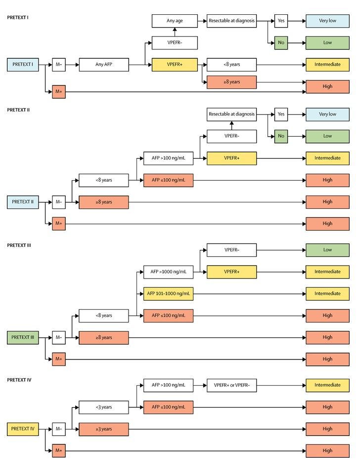
Colorectal Cancer Prevention (PDQ®)–Health Professional Version

Colorectal Cancer Prevention (PDQ®)–Health Professional Version

Who Is at Risk?

For most people, the major factor that increases a person’s risk for colorectal cancer (CRC) is advancing age. Risk increases dramatically after age 50 years; 90% of all CRCs are diagnosed after this age. Incidence and mortality rates are higher in African American individuals compared with other races. However, a meta-analysis found no evidence that African American individuals have higher rates of precancerous lesions.[1,2] The history of CRC in a first-degree relative, especially if diagnosed before the age of 55 years, roughly doubles the risk. A personal history of CRC, high-risk adenomas, or ovarian cancer also increase the risk.[3] Other risk factors for CRC have weaker associations than age and family history. People with inflammatory bowel disease, such as ulcerative colitis or Crohn disease, have a much higher risk of CRC starting about 8 years after disease onset and are recommended to have frequent colonoscopic surveillance.[4] A small percentage (<5%) of CRCs occur in people with a genetic predisposition, including familial adenomatous polyposis and Lynch syndrome (hereditary nonpolyposis CRC).

References
  1. Imperiale TF, Abhyankar PR, Stump TE, et al.: Prevalence of Advanced, Precancerous Colorectal Neoplasms in Black and White Populations: A Systematic Review and Meta-analysis. Gastroenterology 155 (6): 1776-1786.e1, 2018. [PUBMED Abstract]
  2. Lansdorp-Vogelaar I, Kuntz KM, Knudsen AB, et al.: Contribution of screening and survival differences to racial disparities in colorectal cancer rates. Cancer Epidemiol Biomarkers Prev 21 (5): 728-36, 2012. [PUBMED Abstract]
  3. Imperiale TF, Juluri R, Sherer EA, et al.: A risk index for advanced neoplasia on the second surveillance colonoscopy in patients with previous adenomatous polyps. Gastrointest Endosc 80 (3): 471-8, 2014. [PUBMED Abstract]
  4. Laukoetter MG, Mennigen R, Hannig CM, et al.: Intestinal cancer risk in Crohn’s disease: a meta-analysis. J Gastrointest Surg 15 (4): 576-83, 2011. [PUBMED Abstract]

Overview

Note: The Overview section summarizes the published evidence on this topic. The rest of the summary describes the evidence in more detail.

Other PDQ summaries on Colorectal Cancer Screening; Colon Cancer Treatment; and Rectal Cancer Treatment are also available.

Factors With Adequate Evidence of Increased Risk of Colorectal Cancer

Excessive alcohol use

Based on solid evidence from observational studies, excessive alcohol use is associated with an increased risk of colorectal cancer (CRC).[1,2]

Magnitude of Effect: A pooled analysis of eight cohort studies estimated an adjusted relative risk (RR) of 1.41 (95% confidence interval [CI], 1.16–1.72) for consumption exceeding 45 g/day.[1]

  • Study Design: Cohort studies.
  • Internal Validity: Fair.
  • Consistency: Fair.
  • External Validity: Fair.

Cigarette smoking

Based on solid evidence, cigarette smoking is associated with increased incidence of and mortality from CRC.

Magnitude of Effect: A pooled analysis of 106 observational studies estimated an adjusted RR (current smokers vs. never smokers) of 1.18 for developing CRC (95% CI, 1.11–1.25).[3,4]

  • Study Design: 106 observational studies.
  • Internal Validity: Fair.
  • Consistency: Good.
  • External Validity: Good.

Obesity

Based on solid evidence, obesity is associated with increased incidence of and mortality from CRC.

Magnitude of Effect: In one large cohort study, the adjusted RR of developing colon cancer for women with a body mass index greater than 29 was 1.45 (95% CI, 1.02–2.07).[5,6] A similar increase in CRC mortality was found in another large cohort study.[7,8]

  • Study Design: Large cohort studies.
  • Internal Validity: Fair.
  • Consistency: Good.
  • External Validity: Good.

Family/personal history of colorectal cancer and other hereditary conditions

Based on solid evidence, a family history of CRC in a first-degree relative or a personal history of CRC increases the risk of CRC.[912] Having a genetic predisposition, including familial adenomatous polyposis and Lynch syndrome (hereditary nonpolyposis CRC), also increases risk of CRC.[13]

Magnitude of Effect: In individuals with familial adenomatous polyposis, the risk of CRC by age 40 can be as high as 100%. Individuals with Lynch syndrome can have a lifetime risk of CRC of about 80%.

  • Study Design: Case-control and cohort studies.
  • Internal Validity: Good.
  • Consistency: Good.
  • External Validity: Good.

For more information about family history and hereditary conditions, see Genetics of Colorectal Cancer.

Factors With Adequate Evidence for a Decreased Risk of Colorectal Cancer

Physical activity

Based on solid evidence, regular physical activity is associated with a decreased incidence of CRC.

Magnitude of Effect: A meta-analysis of 52 observational studies found a statistically significant 24% reduction in CRC incidence (RR, 0.76; 95% CI, 0.72–0.81).[14]

  • Study Design: Cohort studies and meta-analysis.
  • Internal Validity: Fair.
  • Consistency: Good.
  • External Validity: Good.

Interventions With Adequate Evidence for a Decreased Risk of Colorectal Cancer

Aspirin: Benefits

Based on solid evidence, daily aspirin (acetylsalicylic acid [ASA]) reduces CRC incidence and mortality after 10 to 20 years. This is based on three individual participant-level data meta-analyses of trials of aspirin used for the primary and secondary prevention of cardiovascular disease.[1517]

Magnitude of Effect: ASA use reduces the long-term risk of developing CRC by 40% about 10 to 19 years after initiation (hazard ratio [HR], 0.60; 95% CI, 0.47–0.76).[18] Daily doses of 75 to 1,200 mg of ASA reduce the 20-year risk of CRC death by approximately 33% (HR, 0.67; 95% CI, 0.52–0.86).[16,17]

  • Study Design: Individual patient-level data meta-analyses of randomized controlled trials (RCTs) of ASA for primary and secondary cardiovascular prevention.
  • Internal Validity: Fair, some data from registries and death certificates, some loss to follow-up; variations in ASA dose and timing; adherence to ASA unknown after end of trials (5–9 years); trials designed to answer a different primary hypothesis (cardiovascular disease prevention).
  • Consistency: Generally consistent.
  • External Validity: Fair, most data (>75%) from men.

Aspirin: Harms

Based on solid evidence, harms of ASA use include excessive bleeding, including gastrointestinal bleeding and hemorrhagic stroke.

Magnitude of Effect: Very low-dose ASA use (i.e., ≤100 mg every day or every other day) results in an estimated 14 (95% CI, 7–23) additional major gastrointestinal bleeding events and 3.2 (95% CI, -0.5 to 0.82) extra hemorrhagic strokes per 1,000 individuals over 10 years. These risks increase with advancing age.[19]

  • Study Design: Evidence obtained from RCTs, cohort studies, and meta-analyses comparing ASA with placebo or no treatment for the primary prevention of cardiovascular disease.[19]
  • Internal Validity: Fair, data are from clinically and methodologically heterogeneous trials.
  • Consistency: Good.
  • External Validity: Fair, data on specific subgroups are limited.

Hormone therapy (estrogen plus progestin): Benefits

Based on solid evidence, combined hormone therapy (conjugated equine estrogen and progestin) decreases the incidence of invasive CRC.[20]

Based on fair evidence, combination conjugated equine estrogen and progestin has little or no benefit in reducing mortality from CRC. Data from the Women’s Health Initiative (WHI), a randomized, placebo-controlled trial evaluating estrogen plus progestin, with a mean intervention of 5.6 years and a follow-up of 11.6 years, showed that women taking combined hormone therapy had a statistically significant higher stage of cancer (regional and distant) at diagnosis but not a statistically significant number of deaths from CRC compared with women taking the placebo.[20]

Magnitude of Effect: There were fewer CRCs in the combined hormone therapy group than in the placebo group (0.12% vs. 0.16%; HR, 0.72; 95% CI, 0.56–0.94). A meta-analysis of cohort studies observed a RR of 0.86 (95% CI, 0.76–0.97) for incidence of CRC associated with combined hormone therapy.

There were 37 CRC deaths in the combined hormone therapy arm compared with 27 deaths in the placebo arm (0.04% vs. 0.03%; HR, 1.29; 95% CI, 0.78–2.11).

  • Study Design: RCT and cohort studies.
  • Internal Validity: Good.
  • Consistency: Good for effect on incidence; not applicable (N/A) for effect on mortality; results were based on one trial.
  • External Validity: Good.

Hormone therapy (estrogen plus progestin): Harms

Based on solid evidence, harms of postmenopausal combined estrogen-plus-progestin hormone use include increased risk of breast cancer, coronary heart disease, and thromboembolic events.

Magnitude of Effect: The WHI showed a 26% increase in invasive breast cancer in the combined hormone group, a 29% increase in coronary heart disease events, a 41% increase in stroke rates, and a twofold higher rate of thromboembolic events.[21]

  • Study Design: Evidence from RCTs.
  • Internal Validity: Good.
  • Consistency: Good.
  • External Validity: Fair.

Polyp removal: Benefits

Based on fair evidence, removal of adenomatous polyps reduces the risk of CRC. Much of this reduction likely comes from removal of large (i.e., >1.0 cm) polyps, while the benefit of removing smaller polyps—which are much more common—is unknown. Some but not all observational evidence indicates that this reduction may be greater for left-sided CRC than for right-sided CRC.[2224]

Magnitude of Effect: Unknown, probably greater for larger polyps (i.e., >1.0 cm) than for smaller ones.[25]

  • Study Design: Evidence obtained from cohort studies and one RCT of sigmoidoscopy.[23]
  • Internal Validity: Good.
  • Consistency: Consistent.
  • External Validity: Good.

Polyp removal: Harms

Based on solid evidence, the major harms of polyp removal include perforation of the colon and bleeding.

Magnitude of Effect: Seven to nine events per 1,000 procedures.[2628]

  • Study Design: Evidence from retrospective cohort studies.[27,28]
  • Internal Validity: Good.
  • Consistency: Good.
  • External Validity: Good.

Factors With Inadequate Evidence of an Association With Colorectal Cancer

Nonsteroidal anti-inflammatory drugs (NSAIDs): Benefits

There is inadequate evidence that the use of NSAIDs reduces the risk of CRC. In people without genetic predisposition but with a prior history of a colonic adenoma that had been removed, three RCTs found that celecoxib [29,30] and rofecoxib [31] decreased the incidence of recurrent adenoma, although follow-up was too short to determine whether CRC incidence or mortality would have been affected.

Based on solid evidence, NSAIDs reduce the risk of adenomas, but the extent to which this translates into a reduction of CRC is uncertain.[32]

  • Study Design: No adequate studies with CRC outcome.
  • Internal Validity: N/A.
  • Consistency: N/A.
  • External Validity: N/A.

NSAIDs: Harms

Based on solid evidence, harms of NSAID use are relatively common and potentially serious, and include upper gastrointestinal bleeding, chronic kidney disease, and serious cardiovascular events such as myocardial infarction, heart failure, and hemorrhagic stroke.[33] A recent report compared the cyclooxygenase-2 (COX-2) inhibitor celecoxib (200 mg/d) with the nonselective nonsteroidals naproxen (850 mg/d) and ibuprofen (2,000 mg/d) in individuals with severe arthritis (i.e., not using lower doses as for primary prevention). The results showed that serious cardiovascular events were not less common for those taking the nonselective nonsteroidals. However, this study did not assess the comparative safety of lower doses or the safety of the COX-2 inhibitor rofecoxib.[34]

Magnitude of Effect: The estimated average excess risk of upper gastrointestinal complications in average-risk people attributable to NSAIDs is 4 to 5 per 1,000 people per year.[35,36] The excess risk varies with the underlying gastrointestinal risk; however, it likely exceeds ten extra cases per 1,000 people per year in more than 10% of users.[37] Serious cardiovascular events are increased by 50% to 60%.[36]

  • Study Design: Evidence obtained from RCTs and high-quality systematic reviews and meta-analyses.[35,36]
  • Internal Validity: Good.
  • Consistency: Good.
  • External Validity: Good.

Calcium supplementation

The evidence is inadequate to determine whether calcium supplementation reduces the risk of CRC.

Dietary factors

There is no reliable evidence that a diet started in adulthood that is low in fat and meat and high in fiber, fruits, and vegetables reduces the risk of CRC by a clinically important degree.

Factors and Interventions With Adequate Evidence of no Association With Colorectal Cancer

Estrogen-only therapy: Benefits

Based on fair evidence, conjugated equine estrogens do not affect the incidence of or mortality from invasive CRC.[38]

Magnitude of Effect: N/A.

  • Study Design: Evidence from RCTs.
  • Internal Validity: Good.
  • Consistency: Good.
  • External Validity: Fair.

Statins: Benefits

Based on solid evidence, statins do not reduce the incidence of or mortality from CRC.

  • Study Design: Meta-analyses of RCTs.[3941]
  • Internal Validity: Good.
  • Consistency: Good.
  • External Validity: N/A.

Statins: Harms

Based on solid evidence, the harms of statins are small.

  • Study Design: Observational studies,[42] multiple RCTs, and a review.[43]
  • Internal Validity: Good.
  • Consistency: Good.
  • External Validity: Good.
References
  1. Cho E, Smith-Warner SA, Ritz J, et al.: Alcohol intake and colorectal cancer: a pooled analysis of 8 cohort studies. Ann Intern Med 140 (8): 603-13, 2004. [PUBMED Abstract]
  2. Fedirko V, Tramacere I, Bagnardi V, et al.: Alcohol drinking and colorectal cancer risk: an overall and dose-response meta-analysis of published studies. Ann Oncol 22 (9): 1958-72, 2011. [PUBMED Abstract]
  3. Botteri E, Iodice S, Bagnardi V, et al.: Smoking and colorectal cancer: a meta-analysis. JAMA 300 (23): 2765-78, 2008. [PUBMED Abstract]
  4. Liang PS, Chen TY, Giovannucci E: Cigarette smoking and colorectal cancer incidence and mortality: systematic review and meta-analysis. Int J Cancer 124 (10): 2406-15, 2009. [PUBMED Abstract]
  5. Martínez ME, Giovannucci E, Spiegelman D, et al.: Leisure-time physical activity, body size, and colon cancer in women. Nurses’ Health Study Research Group. J Natl Cancer Inst 89 (13): 948-55, 1997. [PUBMED Abstract]
  6. Giovannucci E, Ascherio A, Rimm EB, et al.: Physical activity, obesity, and risk for colon cancer and adenoma in men. Ann Intern Med 122 (5): 327-34, 1995. [PUBMED Abstract]
  7. Calle EE, Rodriguez C, Walker-Thurmond K, et al.: Overweight, obesity, and mortality from cancer in a prospectively studied cohort of U.S. adults. N Engl J Med 348 (17): 1625-38, 2003. [PUBMED Abstract]
  8. Ma Y, Yang Y, Wang F, et al.: Obesity and risk of colorectal cancer: a systematic review of prospective studies. PLoS One 8 (1): e53916, 2013. [PUBMED Abstract]
  9. Johns LE, Houlston RS: A systematic review and meta-analysis of familial colorectal cancer risk. Am J Gastroenterol 96 (10): 2992-3003, 2001. [PUBMED Abstract]
  10. Schoen RE, Razzak A, Yu KJ, et al.: Incidence and mortality of colorectal cancer in individuals with a family history of colorectal cancer. Gastroenterology 149 (6): 1438-1445.e1, 2015. [PUBMED Abstract]
  11. Butterworth AS, Higgins JP, Pharoah P: Relative and absolute risk of colorectal cancer for individuals with a family history: a meta-analysis. Eur J Cancer 42 (2): 216-27, 2006. [PUBMED Abstract]
  12. Samadder NJ, Curtin K, Tuohy TM, et al.: Increased risk of colorectal neoplasia among family members of patients with colorectal cancer: a population-based study in Utah. Gastroenterology 147 (4): 814-821.e5; quiz e15-6, 2014. [PUBMED Abstract]
  13. Mork ME, You YN, Ying J, et al.: High Prevalence of Hereditary Cancer Syndromes in Adolescents and Young Adults With Colorectal Cancer. J Clin Oncol 33 (31): 3544-9, 2015. [PUBMED Abstract]
  14. Wolin KY, Yan Y, Colditz GA, et al.: Physical activity and colon cancer prevention: a meta-analysis. Br J Cancer 100 (4): 611-6, 2009. [PUBMED Abstract]
  15. Flossmann E, Rothwell PM; British Doctors Aspirin Trial and the UK-TIA Aspirin Trial: Effect of aspirin on long-term risk of colorectal cancer: consistent evidence from randomised and observational studies. Lancet 369 (9573): 1603-13, 2007. [PUBMED Abstract]
  16. Rothwell PM, Wilson M, Elwin CE, et al.: Long-term effect of aspirin on colorectal cancer incidence and mortality: 20-year follow-up of five randomised trials. Lancet 376 (9754): 1741-50, 2010. [PUBMED Abstract]
  17. Rothwell PM, Fowkes FG, Belch JF, et al.: Effect of daily aspirin on long-term risk of death due to cancer: analysis of individual patient data from randomised trials. Lancet 377 (9759): 31-41, 2011. [PUBMED Abstract]
  18. Chubak J, Whitlock EP, Williams SB, et al.: Aspirin for the Prevention of Cancer Incidence and Mortality: Systematic Evidence Reviews for the U.S. Preventive Services Task Force. Ann Intern Med 164 (12): 814-25, 2016. [PUBMED Abstract]
  19. Whitlock EP, Burda BU, Williams SB, et al.: Bleeding Risks With Aspirin Use for Primary Prevention in Adults: A Systematic Review for the U.S. Preventive Services Task Force. Ann Intern Med 164 (12): 826-35, 2016. [PUBMED Abstract]
  20. Simon MS, Chlebowski RT, Wactawski-Wende J, et al.: Estrogen plus progestin and colorectal cancer incidence and mortality. J Clin Oncol 30 (32): 3983-90, 2012. [PUBMED Abstract]
  21. Writing Group for the Women’s Health Initiative Investigators: Risks and benefits of estrogen plus progestin in healthy postmenopausal women: principal results From the Women’s Health Initiative randomized controlled trial. JAMA 288 (3): 321-33, 2002. [PUBMED Abstract]
  22. Brenner H, Hoffmeister M, Arndt V, et al.: Protection from right- and left-sided colorectal neoplasms after colonoscopy: population-based study. J Natl Cancer Inst 102 (2): 89-95, 2010. [PUBMED Abstract]
  23. Atkin WS, Edwards R, Kralj-Hans I, et al.: Once-only flexible sigmoidoscopy screening in prevention of colorectal cancer: a multicentre randomised controlled trial. Lancet 375 (9726): 1624-33, 2010. [PUBMED Abstract]
  24. Brenner H, Chang-Claude J, Seiler CM, et al.: Protection from colorectal cancer after colonoscopy: a population-based, case-control study. Ann Intern Med 154 (1): 22-30, 2011. [PUBMED Abstract]
  25. Robertson DJ, Greenberg ER, Beach M, et al.: Colorectal cancer in patients under close colonoscopic surveillance. Gastroenterology 129 (1): 34-41, 2005. [PUBMED Abstract]
  26. Nelson DB, McQuaid KR, Bond JH, et al.: Procedural success and complications of large-scale screening colonoscopy. Gastrointest Endosc 55 (3): 307-14, 2002. [PUBMED Abstract]
  27. Levin TR, Zhao W, Conell C, et al.: Complications of colonoscopy in an integrated health care delivery system. Ann Intern Med 145 (12): 880-6, 2006. [PUBMED Abstract]
  28. Warren JL, Klabunde CN, Mariotto AB, et al.: Adverse events after outpatient colonoscopy in the Medicare population. Ann Intern Med 150 (12): 849-57, W152, 2009. [PUBMED Abstract]
  29. Bertagnolli MM, Eagle CJ, Zauber AG, et al.: Celecoxib for the prevention of sporadic colorectal adenomas. N Engl J Med 355 (9): 873-84, 2006. [PUBMED Abstract]
  30. Arber N, Eagle CJ, Spicak J, et al.: Celecoxib for the prevention of colorectal adenomatous polyps. N Engl J Med 355 (9): 885-95, 2006. [PUBMED Abstract]
  31. Lanas A, Baron JA, Sandler RS, et al.: Peptic ulcer and bleeding events associated with rofecoxib in a 3-year colorectal adenoma chemoprevention trial. Gastroenterology 132 (2): 490-7, 2007. [PUBMED Abstract]
  32. Chudy-Onwugaje K, Huang WY, Su LJ, et al.: Aspirin, ibuprofen, and reduced risk of advanced colorectal adenoma incidence and recurrence and colorectal cancer in the PLCO Cancer Screening Trial. Cancer 127 (17): 3145-3155, 2021. [PUBMED Abstract]
  33. Bresalier RS, Sandler RS, Quan H, et al.: Cardiovascular events associated with rofecoxib in a colorectal adenoma chemoprevention trial. N Engl J Med 352 (11): 1092-102, 2005. [PUBMED Abstract]
  34. Nissen SE, Yeomans ND, Solomon DH, et al.: Cardiovascular Safety of Celecoxib, Naproxen, or Ibuprofen for Arthritis. N Engl J Med 375 (26): 2519-29, 2016. [PUBMED Abstract]
  35. Rostom A, Dubé C, Lewin G, et al.: Nonsteroidal anti-inflammatory drugs and cyclooxygenase-2 inhibitors for primary prevention of colorectal cancer: a systematic review prepared for the U.S. Preventive Services Task Force. Ann Intern Med 146 (5): 376-89, 2007. [PUBMED Abstract]
  36. Kearney PM, Baigent C, Godwin J, et al.: Do selective cyclo-oxygenase-2 inhibitors and traditional non-steroidal anti-inflammatory drugs increase the risk of atherothrombosis? Meta-analysis of randomised trials. BMJ 332 (7553): 1302-8, 2006. [PUBMED Abstract]
  37. Hernández-Díaz S, García Rodríguez LA: Cardioprotective aspirin users and their excess risk of upper gastrointestinal complications. BMC Med 4: 22, 2006. [PUBMED Abstract]
  38. Ritenbaugh C, Stanford JL, Wu L, et al.: Conjugated equine estrogens and colorectal cancer incidence and survival: the Women’s Health Initiative randomized clinical trial. Cancer Epidemiol Biomarkers Prev 17 (10): 2609-18, 2008. [PUBMED Abstract]
  39. Baigent C, Keech A, Kearney PM, et al.: Efficacy and safety of cholesterol-lowering treatment: prospective meta-analysis of data from 90,056 participants in 14 randomised trials of statins. Lancet 366 (9493): 1267-78, 2005. [PUBMED Abstract]
  40. Dale KM, Coleman CI, Henyan NN, et al.: Statins and cancer risk: a meta-analysis. JAMA 295 (1): 74-80, 2006. [PUBMED Abstract]
  41. Browning DR, Martin RM: Statins and risk of cancer: a systematic review and metaanalysis. Int J Cancer 120 (4): 833-43, 2007. [PUBMED Abstract]
  42. Hippisley-Cox J, Coupland C: Unintended effects of statins in men and women in England and Wales: population based cohort study using the QResearch database. BMJ 340: c2197, 2010. [PUBMED Abstract]
  43. Joy TR, Hegele RA: Narrative review: statin-related myopathy. Ann Intern Med 150 (12): 858-68, 2009. [PUBMED Abstract]

Incidence and Mortality

Colorectal cancer (CRC) is the third most common malignant neoplasm worldwide [1] and the second leading cause of cancer deaths in men and women combined in the United States.[2] It is estimated that there will be 154,270 new cases diagnosed in the United States in 2025 and 52,900 deaths caused by this disease.[2] Between 2012 and 2021, incidence rates for CRC in the United States declined by about 1% per year overall. However, this declining incidence is confined to individuals aged 65 years and older. Between 2012 and 2021, incidence rates increased by 2.4% per year in individuals younger than 50 years and by 0.4% per year in individuals aged 50 to 64 years.[2] For the past 50 years, the mortality rate for CRC has been declining in both men and women. Over the last decade, the mortality rate declined by 1.7% per year.[2] Incidence and mortality rates are higher in Black individuals than in those of other races; however, a meta-analysis found no evidence that Black individuals have higher rates of precancerous lesions.[35]

The 5-year overall survival rate is 64% for CRC. About 4% of Americans are expected to develop CRC within their lifetimes.[2,6] The risk of CRC begins to increase after the age of 40 years and rises sharply at ages 50 to 55 years; the risk doubles with each succeeding decade and continues to rise exponentially. Despite advances in surgical techniques and adjuvant therapy, there has been only a modest improvement in survival for patients who present with advanced neoplasms.[7,8] Effective primary and secondary preventive approaches must be developed to reduce the morbidity and mortality from CRC.

References
  1. Bray F, Laversanne M, Sung H, et al.: Global cancer statistics 2022: GLOBOCAN estimates of incidence and mortality worldwide for 36 cancers in 185 countries. CA Cancer J Clin 74 (3): 229-263, 2024. [PUBMED Abstract]
  2. American Cancer Society: Cancer Facts and Figures 2025. American Cancer Society, 2025. Available online. Last accessed January 16, 2025.
  3. Imperiale TF, Abhyankar PR, Stump TE, et al.: Prevalence of Advanced, Precancerous Colorectal Neoplasms in Black and White Populations: A Systematic Review and Meta-analysis. Gastroenterology 155 (6): 1776-1786.e1, 2018. [PUBMED Abstract]
  4. Laiyemo AO, Doubeni C, Pinsky PF, et al.: Race and colorectal cancer disparities: health-care utilization vs different cancer susceptibilities. J Natl Cancer Inst 102 (8): 538-46, 2010. [PUBMED Abstract]
  5. Lansdorp-Vogelaar I, Kuntz KM, Knudsen AB, et al.: Contribution of screening and survival differences to racial disparities in colorectal cancer rates. Cancer Epidemiol Biomarkers Prev 21 (5): 728-36, 2012. [PUBMED Abstract]
  6. Surveillance Research Program, National Cancer Institute: SEER*Explorer: An interactive website for SEER cancer statistics. Bethesda, MD: National Cancer Institute. Available online. Last accessed December 30, 2024.
  7. Moertel CG, Fleming TR, Macdonald JS, et al.: Levamisole and fluorouracil for adjuvant therapy of resected colon carcinoma. N Engl J Med 322 (6): 352-8, 1990. [PUBMED Abstract]
  8. Krook JE, Moertel CG, Gunderson LL, et al.: Effective surgical adjuvant therapy for high-risk rectal carcinoma. N Engl J Med 324 (11): 709-15, 1991. [PUBMED Abstract]

Definition of Prevention

Primary prevention involves the use of medications or other interventions before the clinical appearance of colorectal cancer (CRC) with the intent of preventing clinical CRC and CRC mortality.

Etiology and Pathogenesis of Colorectal Cancer

Genetics,[1,2] experimental,[3,4] and epidemiologic [57] studies suggest that colorectal cancer (CRC) results from complex interactions between inherited susceptibility and environmental factors. The exact nature and contribution of these factors to CRC incidence and mortality is the subject of ongoing research.

References
  1. Willett W: The search for the causes of breast and colon cancer. Nature 338 (6214): 389-94, 1989. [PUBMED Abstract]
  2. Fearon ER, Vogelstein B: A genetic model for colorectal tumorigenesis. Cell 61 (5): 759-67, 1990. [PUBMED Abstract]
  3. Reddy B, Engle A, Katsifis S, et al.: Biochemical epidemiology of colon cancer: effect of types of dietary fiber on fecal mutagens, acid, and neutral sterols in healthy subjects. Cancer Res 49 (16): 4629-35, 1989. [PUBMED Abstract]
  4. Reddy BS, Tanaka T, Simi B: Effect of different levels of dietary trans fat or corn oil on azoxymethane-induced colon carcinogenesis in F344 rats. J Natl Cancer Inst 75 (4): 791-8, 1985. [PUBMED Abstract]
  5. Potter JD: Reconciling the epidemiology, physiology, and molecular biology of colon cancer. JAMA 268 (12): 1573-7, 1992 Sep 23-30. [PUBMED Abstract]
  6. Wynder EL, Reddy BS: Dietary fat and fiber and colon cancer. Semin Oncol 10 (3): 264-72, 1983. [PUBMED Abstract]
  7. Chen CD, Yen MF, Wang WM, et al.: A case-cohort study for the disease natural history of adenoma-carcinoma and de novo carcinoma and surveillance of colon and rectum after polypectomy: implication for efficacy of colonoscopy. Br J Cancer 88 (12): 1866-73, 2003. [PUBMED Abstract]

Factors With Adequate Evidence of Increased Risk of Colorectal Cancer

Excessive Alcohol Use

There is evidence of an association of colorectal cancer (CRC) with alcoholic beverage consumption. In a meta-analysis of eight cohort studies, the relative risk (RR) for consumption of 45 g/day (i.e., about three standard drinks per day) compared with nondrinkers was 1.41 (95% confidence interval [CI], 1.16–1.72).[1] Case-control studies suggest a modest-to-strong positive relationship between alcohol consumption and large bowel cancers.[2,3] A meta-analysis found that the association did not vary by sex or cancer location within the large bowel.[4]

Five studies have reported a positive association between alcohol intake and colorectal adenomas.[5] A case-control study of diet, genetic factors, and the adenoma-carcinoma sequence was conducted in Burgundy.[6] It separated adenomas smaller than 10.0 mm in diameter from larger adenomas. A positive association between current alcohol intake and adenomas was found to be limited to the larger adenomas, suggesting that alcohol intake could act at the promotional phase of the adenoma-carcinoma sequence.[6]

A large cohort study found a dose-response relationship between alcohol intake and death from CRC, with a RR of 1.2 (95% CI, 1.0–1.5) for four or more drinks per day compared with nondrinkers.[7]

Cigarette Smoking

Most case-control studies of cigarette exposure and adenomas have found an elevated risk for smokers.[8] In addition, a significantly increased risk of adenoma recurrence following polypectomy has been associated with smoking in both men and women.[8] In the Nurses’ Health Study, the minimum induction period for cancer appeared to be at least 35 years.[9] Similarly, in the Health Professionals Follow-up Study, a history of smoking was associated with both small and large adenomas and with a long induction period of at least 35 years for CRC.[10] In the Cancer Prevention Study II (CPS II), a large nationwide cohort study, multivariate-adjusted CRC mortality rates were highest among current smokers, intermediate among former smokers, and lowest in nonsmokers, with increased risk observed after 20 or more years of smoking in men and women combined.[11] On the basis of CPS II data, it was estimated that 12% of CRC deaths in the U.S. population in 1997 were attributable to smoking. A large population-based cohort study of Swedish twins found that heavy smoking of 35 or more years’ duration was associated with a nearly threefold increased risk of developing colon cancer, although subsite analysis found a statistically significant effect only for rectal cancer, but not colon cancer.[12] Another large population-based case-control study demonstated that current tobacco use and tobacco use within the last 10 years is associated with colon cancer. A 50% increase in risk was associated with smoking more than a pack a day relative to never smoking.[13] However, a 28-year follow-up of 57,000 Finns showed no association between the development of CRC and baseline smoking status, although there was a 57% to 71% increased risk in persistent smokers.[14] No relationship was found between cigarette smoking, even smoking of long duration, and recurrence of adenomas in a population followed for 4 years after initial colonoscopy.[15]

A meta-analysis of 106 observational studies found a RR (ever smokers vs. nonsmokers) for CRC incidence of 1.18 (95% CI, 1.11–1.25), with an absolute risk increase of 10.8 cases per 100,000 person-years (95% CI, 7.9–13.6). There was a statistically significant dose-response effect. In 17 studies with data on CRC mortality, cigarette smoking was associated with CRC death, with a RR (ever smokers vs. never smokers) of 1.25 (95% CI, 1.14–1.37), and an absolute increase in the death rate of 6.0 deaths per 100,000 person-years. For both incidence and mortality, the association was stronger for rectal cancer than for colon cancer.[16]

Obesity

At least three large cohort studies have found an association between obesity and CRC incidence or mortality.[1719] The Nurses’ Health Study found that women with a body mass index (BMI) of more than 29, compared with women with a BMI of less than 21, had an adjusted RR for CRC incidence of 1.45 (95% CI, 1.02–2.07).[17] In the CPS II,[19] men and women with a BMI of 30 to 34.9 had an adjusted RR for CRC mortality (compared with people with a BMI of 18.5–24.9) of 1.47 (95% CI, 1.30–1.66), with a statistically significant dose-response effect.[19] The effects were similar in men and women.

Family/Personal History of Colorectal Cancer and Other Hereditary Conditions

Some of the earliest studies of family history of CRC were those of Utah families that reported a higher number of deaths from CRC (3.9%) among the first-degree relatives of patients who had died from CRC than among sex-matched and age-matched controls (1.2%). This difference has since been replicated in numerous studies that have consistently found that first-degree relatives of affected cases are themselves at a twofold to threefold increased risk of CRC. Despite the various study designs (case-control, cohort), sampling frames, sample sizes, methods of data verification, analytic methods, and countries where the studies originated, the magnitude of risk is consistent.[2025]

A systematic review and meta-analysis of familial CRC risk was reported.[26] Of 24 studies included in the analysis, all but one reported an increased risk of CRC if there was an affected first-degree relative. The RR for CRC in the pooled study was 2.25 (95% CI, 2.00–2.53) if there was an affected first-degree family member. In 8 of 11 studies, if the index cancer arose in the colon, the risk was slightly higher than if it arose in the rectum. The pooled analysis revealed an RR in relatives of colon and rectal cancer patients of 2.42 (95% CI, 2.20–2.65) and 1.89 (95% CI, 1.62–2.21), respectively. The analysis did not reveal a difference in RR for colon cancer based on location of the tumor (right side vs. left side).[26]

Hereditary CRC has two well-described forms: Familial adenomatous polyposis (including an attenuated form of polyposis), due to germline mutations in the APC gene,[2734] and Lynch syndrome (hereditary nonpolyposis CRC), which is caused by germline mutations in DNA mismatch repair genes.[3538] Many other families exhibit aggregation of CRC and/or adenomas, but with no apparent association with an identifiable hereditary syndrome, and are known collectively as familial CRC.[38]

For more information about genetic risk factors for CRC, see Genetics of Colorectal Cancer.

References
  1. Cho E, Smith-Warner SA, Ritz J, et al.: Alcohol intake and colorectal cancer: a pooled analysis of 8 cohort studies. Ann Intern Med 140 (8): 603-13, 2004. [PUBMED Abstract]
  2. Newcomb PA, Storer BE, Marcus PM: Cancer of the large bowel in women in relation to alcohol consumption: a case-control study in Wisconsin (United States). Cancer Causes Control 4 (5): 405-11, 1993. [PUBMED Abstract]
  3. Meyer F, White E: Alcohol and nutrients in relation to colon cancer in middle-aged adults. Am J Epidemiol 138 (4): 225-36, 1993. [PUBMED Abstract]
  4. Longnecker MP, Orza MJ, Adams ME, et al.: A meta-analysis of alcoholic beverage consumption in relation to risk of colorectal cancer. Cancer Causes Control 1 (1): 59-68, 1990. [PUBMED Abstract]
  5. Boutron MC, Faivre J: Diet and the adenoma-carcinoma sequence. Eur J Cancer Prev 2 (Suppl 2): 95-8, 1993. [PUBMED Abstract]
  6. Boutron MC, Faivre J: Alcohol, tobacco and the adenoma-carcinoma sequence: a case-control study in Burgundy, France. [Abstract] Gastroenterology 104 (Suppl 4): A-390, 1993.
  7. Thun MJ, Peto R, Lopez AD, et al.: Alcohol consumption and mortality among middle-aged and elderly U.S. adults. N Engl J Med 337 (24): 1705-14, 1997. [PUBMED Abstract]
  8. Neugut AI, Jacobson JS, DeVivo I: Epidemiology of colorectal adenomatous polyps. Cancer Epidemiol Biomarkers Prev 2 (2): 159-76, 1993 Mar-Apr. [PUBMED Abstract]
  9. Giovannucci E, Colditz GA, Stampfer MJ, et al.: A prospective study of cigarette smoking and risk of colorectal adenoma and colorectal cancer in U.S. women. J Natl Cancer Inst 86 (3): 192-9, 1994. [PUBMED Abstract]
  10. Giovannucci E, Rimm EB, Stampfer MJ, et al.: A prospective study of cigarette smoking and risk of colorectal adenoma and colorectal cancer in U.S. men. J Natl Cancer Inst 86 (3): 183-91, 1994. [PUBMED Abstract]
  11. Chao A, Thun MJ, Jacobs EJ, et al.: Cigarette smoking and colorectal cancer mortality in the cancer prevention study II. J Natl Cancer Inst 92 (23): 1888-96, 2000. [PUBMED Abstract]
  12. Terry P, Ekbom A, Lichtenstein P, et al.: Long-term tobacco smoking and colorectal cancer in a prospective cohort study. Int J Cancer 91 (4): 585-7, 2001. [PUBMED Abstract]
  13. Slattery ML, Potter JD, Friedman GD, et al.: Tobacco use and colon cancer. Int J Cancer 70 (3): 259-64, 1997. [PUBMED Abstract]
  14. Knekt P, Hakama M, Järvinen R, et al.: Smoking and risk of colorectal cancer. Br J Cancer 78 (1): 136-9, 1998. [PUBMED Abstract]
  15. Baron JA, Sandler RS, Haile RW, et al.: Folate intake, alcohol consumption, cigarette smoking, and risk of colorectal adenomas. J Natl Cancer Inst 90 (1): 57-62, 1998. [PUBMED Abstract]
  16. Botteri E, Iodice S, Bagnardi V, et al.: Smoking and colorectal cancer: a meta-analysis. JAMA 300 (23): 2765-78, 2008. [PUBMED Abstract]
  17. Martínez ME, Giovannucci E, Spiegelman D, et al.: Leisure-time physical activity, body size, and colon cancer in women. Nurses’ Health Study Research Group. J Natl Cancer Inst 89 (13): 948-55, 1997. [PUBMED Abstract]
  18. Giovannucci E, Ascherio A, Rimm EB, et al.: Physical activity, obesity, and risk for colon cancer and adenoma in men. Ann Intern Med 122 (5): 327-34, 1995. [PUBMED Abstract]
  19. Calle EE, Rodriguez C, Walker-Thurmond K, et al.: Overweight, obesity, and mortality from cancer in a prospectively studied cohort of U.S. adults. N Engl J Med 348 (17): 1625-38, 2003. [PUBMED Abstract]
  20. Fuchs CS, Giovannucci EL, Colditz GA, et al.: A prospective study of family history and the risk of colorectal cancer. N Engl J Med 331 (25): 1669-74, 1994. [PUBMED Abstract]
  21. Slattery ML, Kerber RA: Family history of cancer and colon cancer risk: the Utah Population Database. J Natl Cancer Inst 86 (21): 1618-26, 1994. [PUBMED Abstract]
  22. Negri E, Braga C, La Vecchia C, et al.: Family history of cancer and risk of colorectal cancer in Italy. Br J Cancer 77 (1): 174-9, 1998. [PUBMED Abstract]
  23. St John DJ, McDermott FT, Hopper JL, et al.: Cancer risk in relatives of patients with common colorectal cancer. Ann Intern Med 118 (10): 785-90, 1993. [PUBMED Abstract]
  24. Duncan JL, Kyle J: Family incidence of carcinoma of the colon and rectum in north-east Scotland. Gut 23 (2): 169-71, 1982. [PUBMED Abstract]
  25. Rozen P, Fireman Z, Figer A, et al.: Family history of colorectal cancer as a marker of potential malignancy within a screening program. Cancer 60 (2): 248-54, 1987. [PUBMED Abstract]
  26. Johns LE, Houlston RS: A systematic review and meta-analysis of familial colorectal cancer risk. Am J Gastroenterol 96 (10): 2992-3003, 2001. [PUBMED Abstract]
  27. Kinzler KW, Nilbert MC, Su LK, et al.: Identification of FAP locus genes from chromosome 5q21. Science 253 (5020): 661-5, 1991. [PUBMED Abstract]
  28. Groden J, Thliveris A, Samowitz W, et al.: Identification and characterization of the familial adenomatous polyposis coli gene. Cell 66 (3): 589-600, 1991. [PUBMED Abstract]
  29. Leppert M, Burt R, Hughes JP, et al.: Genetic analysis of an inherited predisposition to colon cancer in a family with a variable number of adenomatous polyps. N Engl J Med 322 (13): 904-8, 1990. [PUBMED Abstract]
  30. Spirio L, Olschwang S, Groden J, et al.: Alleles of the APC gene: an attenuated form of familial polyposis. Cell 75 (5): 951-7, 1993. [PUBMED Abstract]
  31. Brensinger JD, Laken SJ, Luce MC, et al.: Variable phenotype of familial adenomatous polyposis in pedigrees with 3′ mutation in the APC gene. Gut 43 (4): 548-52, 1998. [PUBMED Abstract]
  32. Soravia C, Berk T, Madlensky L, et al.: Genotype-phenotype correlations in attenuated adenomatous polyposis coli. Am J Hum Genet 62 (6): 1290-301, 1998. [PUBMED Abstract]
  33. Pedemonte S, Sciallero S, Gismondi V, et al.: Novel germline APC variants in patients with multiple adenomas. Genes Chromosomes Cancer 22 (4): 257-67, 1998. [PUBMED Abstract]
  34. Sieber OM, Lamlum H, Crabtree MD, et al.: Whole-gene APC deletions cause classical familial adenomatous polyposis, but not attenuated polyposis or “multiple” colorectal adenomas. Proc Natl Acad Sci U S A 99 (5): 2954-8, 2002. [PUBMED Abstract]
  35. Leach FS, Nicolaides NC, Papadopoulos N, et al.: Mutations of a mutS homolog in hereditary nonpolyposis colorectal cancer. Cell 75 (6): 1215-25, 1993. [PUBMED Abstract]
  36. Papadopoulos N, Nicolaides NC, Wei YF, et al.: Mutation of a mutL homolog in hereditary colon cancer. Science 263 (5153): 1625-9, 1994. [PUBMED Abstract]
  37. Nicolaides NC, Papadopoulos N, Liu B, et al.: Mutations of two PMS homologues in hereditary nonpolyposis colon cancer. Nature 371 (6492): 75-80, 1994. [PUBMED Abstract]
  38. Miyaki M, Konishi M, Tanaka K, et al.: Germline mutation of MSH6 as the cause of hereditary nonpolyposis colorectal cancer. Nat Genet 17 (3): 271-2, 1997. [PUBMED Abstract]

Factors With Adequate Evidence for a Decreased Risk of Colorectal Cancer

Physical Activity

A sedentary lifestyle has been associated with an increased risk of colorectal cancer in some [1,2] but not all [3] studies. Numerous observational studies have examined the relationship between physical activity and colon cancer risk.[4] Most of these studies have shown an inverse relationship between level of physical activity and colon cancer incidence. The average relative risk (RR) reduction is reportedly 40% to 50%. Large U.S. cohort studies have found statistically significant adjusted RRs of 0.54 (95% confidence interval [CI], 0.33–0.90) [5] and 0.53 (95% CI, 0.32–0.88) [6] when comparing people with high versus low average energy expenditure. A meta-analysis of 52 observational studies found an overall adjusted RR of 0.76 (95% CI, 0.72–0.81), with similar results for men and women.[7]

References
  1. White E, Jacobs EJ, Daling JR: Physical activity in relation to colon cancer in middle-aged men and women. Am J Epidemiol 144 (1): 42-50, 1996. [PUBMED Abstract]
  2. Slattery ML, Schumacher MC, Smith KR, et al.: Physical activity, diet, and risk of colon cancer in Utah. Am J Epidemiol 128 (5): 989-99, 1988. [PUBMED Abstract]
  3. Kune GA, Kune S, Watson LF: Body weight and physical activity as predictors of colorectal cancer risk. Nutr Cancer 13 (1-2): 9-17, 1990. [PUBMED Abstract]
  4. Friedenreich CM: Physical activity and cancer prevention: from observational to intervention research. Cancer Epidemiol Biomarkers Prev 10 (4): 287-301, 2001. [PUBMED Abstract]
  5. Martínez ME, Giovannucci E, Spiegelman D, et al.: Leisure-time physical activity, body size, and colon cancer in women. Nurses’ Health Study Research Group. J Natl Cancer Inst 89 (13): 948-55, 1997. [PUBMED Abstract]
  6. Giovannucci E, Ascherio A, Rimm EB, et al.: Physical activity, obesity, and risk for colon cancer and adenoma in men. Ann Intern Med 122 (5): 327-34, 1995. [PUBMED Abstract]
  7. Wolin KY, Yan Y, Colditz GA, et al.: Physical activity and colon cancer prevention: a meta-analysis. Br J Cancer 100 (4): 611-6, 2009. [PUBMED Abstract]

Interventions With Adequate Evidence for a Decreased Risk of Colorectal Cancer

Aspirin

Evidence from individual participant-level data meta-analyses of randomized controlled trials (RCTs) and observational studies [1,2] investigating the use of aspirin for the prevention of cardiovascular disease indicates that acetylsalicylic acid (ASA) use reduces the incidence of colorectal cancer (CRC), but not until at least 10 years after initiation of therapy (pooled relative risk [RR] of CRC incidence within 10 years of initiation, 0.99 [95% confidence interval (CI), 0.85–1.15] vs. RR, 0.60 [95% CI, 0.47–0.76] at 10–19 years after initiation).[3] In the Women’s Health Study, a randomized 2 × 2 factorial trial of 100 mg of ASA every other day for an average of 10 years, CRC incidence was reduced by about 20% after 17.5 years (hazard ratio [HR], 0.80; 95% CI, 0.67–0.97).[4] In a report from the Nurses’ Health Study involving 82,911 women followed for 20 years, the multivariate RR for colon cancer was 0.77 (95% CI, 0.67–0.88) among women who regularly used ASA (≥2 standard 325-mg tablets per week) compared with nonregular use. Significant RR was not observed, however, until more than 10 years of use.[5]

The Cancer Prevention Programme (CAPP2), previously known as the Concerted Action Polyposis Prevention project, investigated chemoprevention of CRC in patients with known Lynch syndrome (hereditary nonpolyposis CRC) across 43 international centers. For more information, see the Lynch Syndrome section in Genetics of Colorectal Cancer. Patients were randomly assigned to receive aspirin (600 mg/day), aspirin-placebo, resistant starch (30 g/day), or starch-placebo for up to 4 years. A planned 10-year analysis of CAPP2 data found reduced CRC incidence in patients with Lynch syndrome who took aspirin for at least 2 years when compared with those who took placebo. An intention-to-treat analysis, using Cox proportional hazards regression, showed that aspirin protected against the primary end point of CRC (HR, 0.65; 95% CI, 0.43–0.97; P = .035).[6]

In a randomized study of 635 patients with prior CRC (T1–T2 N0 M0) who had undergone curative resection, ASA intake at 325 mg/day was associated with a decrease in the adjusted RR of any recurrent adenoma as compared with the placebo group (0.65; 95% CI, 0.46–0.91) after a median duration of treatment of 31 months. The likelihood of detection of a new colonic lesion was lower in the ASA group than in the placebo group (HR for the detection of a new polyp, 0.54; 95% CI, 0.43–0.94; P = .022).[7] In a study of 1,121 patients with a recent history of colorectal adenomas, after a mean duration of treatment of 33 months, the unadjusted RRs of any adenoma (as compared with the placebo group) were 0.81 in the 81 mg/day ASA group (95% CI, 0.69–0.96) and 0.96 in the 325 mg/day ASA group (95% CI, 0.81–1.13). For advanced neoplasms (adenomas ≥10.0 mm in diameter or with tubulovillous or villous features, severe dysplasia, or invasive cancer), the RRs were 0.59 (95% CI, 0.38–0.92) in the 81 mg/day ASA group, and 0.83 (95% CI, 0.55–1.23) in the 325 mg/day ASA group, respectively.[8]

ASA has also been evaluated for its potential effects on CRC mortality. A 2010 individual patient level data meta-analysis analyzed long-term (median follow-up, 18.3 years) data from four RCTs of primary and secondary cardiovascular disease prevention; it found that allocation to use of 75 to 1,200 mg of daily ASA for at least one year reduced the cumulative risk of colon cancer death compared with controls (HR, 0.67; 95% CI, 0.52–0.86). Aspirin reduced CRC mortality beginning 10 to 20 years after randomization, but not before.[2] A 2011 individual participant level data meta-analysis examined data from six RCTs of primary or secondary cardiovascular disease prevention. In trials with allocation to ASA after at least 5 years of in-trial follow-up, the HR for CRC mortality was 0.41 (95% CI, 0.71–1.00). There was no statistically significant effect during the first five years after randomization.[9]

Six RCTs, including five from the United Kingdom, were included in a meta-analysis in which patients were randomly assigned to receive either aspirin or placebo, and the mean scheduled duration of trial treatment was 4 years or more. Individual patient data for all in-trial cancer deaths were obtained. In the three United Kingdom trials, cancer deaths after completion of the trials were obtained via death certification and cancer registration, taking the follow-up to 20 years after randomization. Based on meta-analysis of odds ratios (ORs) from each trial rather than on more sensitive actuarial analysis of the individual patient data, allocation to aspirin in the RCTs reduced the 20-year risk of death due to CRC. ORs for maximum aspirin use were 0.55 for CRC risk (95% CI, 0.41–0.76) and for any aspirin use were 0.58 for CRC risk (95% CI, 0.44–0.78).[10]

The Women’s Health Study, the largest randomized trial of aspirin to date (N = 39,876), found no reduction in CRC mortality rates with the use of every other day low-dose aspirin during the first 10 years of follow-up. The study did not report on longer-term risk for CRC mortality.[4]

Aspirin has several important potential harms associated with its use that should be a part of any consideration of its use as a disease prevention strategy. Regular low-dose aspirin use increases the risks for major gastrointestinal bleeding and intracranial bleeding events, including hemorrhagic strokes. A systematic review of studies of aspirin use for primary cardiovascular disease prevention found that use of 100 mg or more of aspirin daily or every other day increased a person’s risk for a major gastrointestinal bleed by 58% (OR, 1.58; 95% CI, 1.29–1.95) or an intracranial hemorrhage by 30% (OR, 1.30; 95% CI, 1.00–1.68). These risks may be greater among older individuals, men, and those individuals with comorbid risk factors that promote a risk of bleeding.[11]

Hormone Therapy (Estrogen Plus Progestin)

Several observational studies have suggested a decreased risk of colon cancer among users of postmenopausal female hormone supplements.[1215] For rectal cancer, most studies have observed no association or a slightly elevated risk.[1618]

The Women’s Health Initiative (WHI) trial examined, as a secondary end point, the effect of combined estrogen-plus-progestin therapy and estrogen-only therapy on CRC incidence and mortality. Among women in the combined estrogen-plus-progestin group of the WHI, an extended follow-up (mean, 11.6 years) confirmed that fewer CRC were diagnosed in the combined hormone therapy group than in the placebo group (HR, 0.72; 95% CI, 0.56–0.94); the CRCs in women in the combined group were more likely to have lymph node involvement than the CRCs in women in the placebo group (50.5% vs. 28.6%; P < .001) and were classified at higher stages (regional and distant) (68.8% vs. 51.4%; P = .003). The number of CRC deaths in the combined group was higher than in the placebo group (37 vs. 27 deaths), but the difference was not statistically significant (HR, 1.29; 95% CI, 0.78–2.11).[19]

Polyp Removal

An analysis of data from the National Polyp Study (NPS), with external, historical controls, has commonly been cited to show a reduction of 76% to 90% in the subsequent incidence of CRC after colonoscopic polypectomy compared with three nonconcurrent, historical control groups.[20] This study may be biased in several ways that inflate the apparent efficacy of polyp removal; the main problem is that potential enrollees in the NPS were excluded if they had CRC at their baseline examination. Because no such exclusions (or baseline colonoscopy examinations) were done in the three comparison groups, individuals who had CRC at baseline would be counted as having incident CRC in subsequent follow-up. Although adjustments were attempted, it is not possible to know the magnitude of the impact of this problem on the result because it is not known how long CRC may be present without causing symptoms.

An additional long-term follow-up study (median follow-up, 15.8 years; maximum, 23 years) of the NPS cohort suggested an approximately 53% reduction in CRC mortality due to polypectomy (not just exclusion of individuals with CRC at initial exam). However, the degree of reduction must be viewed with caution because this study did not have a direct comparison group, relying mainly on comparison to expected data from the National Cancer Institute’s Surveillance, Epidemiology and End Results (SEER) Program. Further, details are not clear regarding factors that may have led to decreased mortality. Patients in the NPS were assigned to colonoscopy at years 1 and 3; colonoscopy was also offered to one of the two comparison groups at year 1; all participants were offered colonoscopy at year 6. However, following year 6, the exact surveillance that patients may have undergone and how that surveillance might have been associated with decreased CRC mortality were not well described.[21]

It is expected that further follow-up in the United Kingdom Flexible Sigmoidoscopy Screening Trial will be able to provide more detail about the long-term effect of polypectomy, at least on the left side of the colon.[21]

Other evidence about the benefit of sigmoidoscopy screening (at which time both polyps and early cancer would be removed) suggests that the impact of endoscopic screening, at least on the left side of the colon, is substantial and prolonged. In an RCT, 170,000 individuals were randomly assigned to one-time sigmoidoscopy versus usual care. At sigmoidoscopy, polyps were removed, cancer was detected, and patients were referred for treatment. Based on sigmoidoscopy findings, individuals were considered to have low risk if they had normal exams or only one or two small (<1 cm) tubular adenomas. These individuals were not referred either for colonoscopy workup, or for colonoscopic surveillance. In a follow-up of 10 years, the left-sided CRC incidence in the low-risk group (about 95% of attendees were low risk) was 0.02% to 0.04% per year—a very low risk of CRC compared with average risk. The cause of reduced risk—whether due to detection and removal of large polyps or small ones, or selection of individuals at lower risk—is yet unclear.[22] The natural history of large polyps is not well known, but some evidence suggests that such lesions become clinical CRC at a rate of approximately 1% per year.[23] As a result of the strong data about the impact of endoscopy on the left colon, evidence from multiple studies has raised questions about the ability of endoscopy to reduce CRC mortality in the right colon.[2426] Thus, it is unclear what the overall impact of endoscopy (e.g., colonoscopy screening) is, and whether there may be a large difference in impact on the left side of the colon compared with the right side.[24]

Other studies suggest that the polyps with the greatest potential to progress to CRC are larger polyps (i.e., >1.0 cm), which include most of those with villous or high-grade histological features. Retrospective cohort studies also show the harms associated with polypectomy, including bleeding.[27,28]

References
  1. Flossmann E, Rothwell PM; British Doctors Aspirin Trial and the UK-TIA Aspirin Trial: Effect of aspirin on long-term risk of colorectal cancer: consistent evidence from randomised and observational studies. Lancet 369 (9573): 1603-13, 2007. [PUBMED Abstract]
  2. Rothwell PM, Wilson M, Elwin CE, et al.: Long-term effect of aspirin on colorectal cancer incidence and mortality: 20-year follow-up of five randomised trials. Lancet 376 (9754): 1741-50, 2010. [PUBMED Abstract]
  3. Chubak J, Whitlock EP, Williams SB, et al.: Aspirin for the Prevention of Cancer Incidence and Mortality: Systematic Evidence Reviews for the U.S. Preventive Services Task Force. Ann Intern Med 164 (12): 814-25, 2016. [PUBMED Abstract]
  4. Cook NR, Lee IM, Zhang SM, et al.: Alternate-day, low-dose aspirin and cancer risk: long-term observational follow-up of a randomized trial. Ann Intern Med 159 (2): 77-85, 2013. [PUBMED Abstract]
  5. Wei EK, Colditz GA, Giovannucci EL, et al.: Cumulative risk of colon cancer up to age 70 years by risk factor status using data from the Nurses’ Health Study. Am J Epidemiol 170 (7): 863-72, 2009. [PUBMED Abstract]
  6. Burn J, Sheth H, Elliott F, et al.: Cancer prevention with aspirin in hereditary colorectal cancer (Lynch syndrome), 10-year follow-up and registry-based 20-year data in the CAPP2 study: a double-blind, randomised, placebo-controlled trial. Lancet 395 (10240): 1855-1863, 2020. [PUBMED Abstract]
  7. Sandler RS, Halabi S, Baron JA, et al.: A randomized trial of aspirin to prevent colorectal adenomas in patients with previous colorectal cancer. N Engl J Med 348 (10): 883-90, 2003. [PUBMED Abstract]
  8. Baron JA, Cole BF, Sandler RS, et al.: A randomized trial of aspirin to prevent colorectal adenomas. N Engl J Med 348 (10): 891-9, 2003. [PUBMED Abstract]
  9. Rothwell PM, Fowkes FG, Belch JF, et al.: Effect of daily aspirin on long-term risk of death due to cancer: analysis of individual patient data from randomised trials. Lancet 377 (9759): 31-41, 2011. [PUBMED Abstract]
  10. Algra AM, Rothwell PM: Effects of regular aspirin on long-term cancer incidence and metastasis: a systematic comparison of evidence from observational studies versus randomised trials. Lancet Oncol 13 (5): 518-27, 2012. [PUBMED Abstract]
  11. Whitlock EP, Burda BU, Williams SB, et al.: Bleeding Risks With Aspirin Use for Primary Prevention in Adults: A Systematic Review for the U.S. Preventive Services Task Force. Ann Intern Med 164 (12): 826-35, 2016. [PUBMED Abstract]
  12. Calle EE, Miracle-McMahill HL, Thun MJ, et al.: Estrogen replacement therapy and risk of fatal colon cancer in a prospective cohort of postmenopausal women. J Natl Cancer Inst 87 (7): 517-23, 1995. [PUBMED Abstract]
  13. Newcomb PA, Storer BE: Postmenopausal hormone use and risk of large-bowel cancer. J Natl Cancer Inst 87 (14): 1067-71, 1995. [PUBMED Abstract]
  14. Grodstein F, Newcomb PA, Stampfer MJ: Postmenopausal hormone therapy and the risk of colorectal cancer: a review and meta-analysis. Am J Med 106 (5): 574-82, 1999. [PUBMED Abstract]
  15. Terry MB, Neugut AI, Bostick RM, et al.: Risk factors for advanced colorectal adenomas: a pooled analysis. Cancer Epidemiol Biomarkers Prev 11 (7): 622-9, 2002. [PUBMED Abstract]
  16. Risch HA, Howe GR: Menopausal hormone use and colorectal cancer in Saskatchewan: a record linkage cohort study. Cancer Epidemiol Biomarkers Prev 4 (1): 21-8, 1995 Jan-Feb. [PUBMED Abstract]
  17. Gerhardsson de Verdier M, London S: Reproductive factors, exogenous female hormones, and colorectal cancer by subsite. Cancer Causes Control 3 (4): 355-60, 1992. [PUBMED Abstract]
  18. Prihartono N, Palmer JR, Louik C, et al.: A case-control study of use of postmenopausal female hormone supplements in relation to the risk of large bowel cancer. Cancer Epidemiol Biomarkers Prev 9 (4): 443-7, 2000. [PUBMED Abstract]
  19. Simon MS, Chlebowski RT, Wactawski-Wende J, et al.: Estrogen plus progestin and colorectal cancer incidence and mortality. J Clin Oncol 30 (32): 3983-90, 2012. [PUBMED Abstract]
  20. Winawer SJ, Zauber AG, Ho MN, et al.: Prevention of colorectal cancer by colonoscopic polypectomy. The National Polyp Study Workgroup. N Engl J Med 329 (27): 1977-81, 1993. [PUBMED Abstract]
  21. Zauber AG, Winawer SJ, O’Brien MJ, et al.: Colonoscopic polypectomy and long-term prevention of colorectal-cancer deaths. N Engl J Med 366 (8): 687-96, 2012. [PUBMED Abstract]
  22. Atkin WS, Edwards R, Kralj-Hans I, et al.: Once-only flexible sigmoidoscopy screening in prevention of colorectal cancer: a multicentre randomised controlled trial. Lancet 375 (9726): 1624-33, 2010. [PUBMED Abstract]
  23. Stryker SJ, Wolff BG, Culp CE, et al.: Natural history of untreated colonic polyps. Gastroenterology 93 (5): 1009-13, 1987. [PUBMED Abstract]
  24. Brenner H, Chang-Claude J, Seiler CM, et al.: Protection from colorectal cancer after colonoscopy: a population-based, case-control study. Ann Intern Med 154 (1): 22-30, 2011. [PUBMED Abstract]
  25. Baxter NN, Goldwasser MA, Paszat LF, et al.: Association of colonoscopy and death from colorectal cancer. Ann Intern Med 150 (1): 1-8, 2009. [PUBMED Abstract]
  26. Brenner H, Hoffmeister M, Arndt V, et al.: Protection from right- and left-sided colorectal neoplasms after colonoscopy: population-based study. J Natl Cancer Inst 102 (2): 89-95, 2010. [PUBMED Abstract]
  27. Levin TR, Zhao W, Conell C, et al.: Complications of colonoscopy in an integrated health care delivery system. Ann Intern Med 145 (12): 880-6, 2006. [PUBMED Abstract]
  28. Warren JL, Klabunde CN, Mariotto AB, et al.: Adverse events after outpatient colonoscopy in the Medicare population. Ann Intern Med 150 (12): 849-57, W152, 2009. [PUBMED Abstract]

Factors With Inadequate Evidence of an Association With Colorectal Cancer

Nonsteroidal Anti-Inflammatory Drugs

One large cohort study (301,240 people with 3,894 colorectal cancer [CRC] cases) found an association between daily or weekly nonaspirin (non-ASA) nonsteroidal anti-inflammatory drug (NSAID) use and reduced 10-year incidence of proximal and distal colon cancer, but not rectal cancer, with a hazard ratio (HR) of 0.67 (95% confidence interval [CI], 0.58–0.77) for daily use for colon cancer. Because exposure to non-ASA NSAIDs was assessed only once, assessment was by self-report, and there is no information on dose or duration of use, the certainty of this single study must be rated low. Further research is needed before this finding can be accepted.[1]

Although evidence is currently inadequate to determine whether NSAIDs reduce CRC incidence, proponents suggest that any effect of these drugs results from their ability to inhibit the activity of cyclooxygenase (COX). COX is important in the transformation of arachidonic acid into prostanoids, prostaglandins, and thromboxane A2. NSAIDs include not only aspirin (ASA, which is considered separately here) and other, first-generation nonselective inhibitors of the two functional isoforms of COX, termed COX-1 and COX-2, but also newer second-generation drugs that inhibit primarily COX-2. Normally, COX-1 is expressed in most tissues and primarily plays a housekeeping role (e.g., gastrointestinal mucosal protection and platelet aggregation). COX-2 activity is crucial in stress responses and in mediating and propagating the pain and inflammation that are characteristic of arthritis.[2]

Nonselective COX inhibitors include indomethacin (Indocin); sulindac (Clinoril); piroxicam (Feldene); diflunisal (Dolobid); ibuprofen (Advil, Motrin); ketoprofen (Orudis); naproxen (Naprosyn); and naproxen sodium (Aleve, Anaprox). Selective COX-2 inhibitors include celecoxib (Celebrex), rofecoxib (Vioxx), and valdecoxib (Bextra). Rofecoxib and valdecoxib are no longer marketed because of an associated increased risk of serious cardiovascular events.

Both celecoxib and rofecoxib have been associated with serious cardiovascular events including dose-related death from cardiovascular causes, myocardial infarction, stroke, or heart failure.[36] Four trials that demonstrated this increased risk are summarized in the Table 1. In addition, a network meta-analysis of all large scale randomized controlled trials (RCTs) comparing any NSAID to any other NSAID or placebo found that there is little evidence to suggest that any of the investigated drugs are safe in terms of cardiovascular effects. Naproxen seemed least harmful.[7]

Table 1. Cardiovascular Risks Associated With Celecoxib and Rofecoxib Dose/Drugs
Authors Dose/Trial Drug Risk Study Type
bid = twice a day; qd = every day; CI = confidence interval; HR = hazard ratio; OR = odds ratio; RR = relative risk; Rx = prescription.
[4] Rofecoxib <25 mg/qd; rofecoxib >25 mg/qd OR, 1.47 (0.99–2.17) vs. 3.58 (1.27–10.17) Nested case-control study all users
[6] Celecoxib 200 mg/qd vs. 400 mg bid 3.4%; HR, 3.4 (95% CI, 1.4–7.8) Sporadic adenoma prevention trial (N = 2,035)
[5] Rofecoxib 25 mg/qd RR, 1.92 (95% CI, 1.19–3.11; P = .008) Chemoprevention of sporadic adenoma
[3] Rofecoxib 25 mg/qd RR (estimated), 2.66 (95% CI, 1.03–6.86; P = .04) Chemoprevention of sporadic adenoma; median study Rx 7.4 months

Other major harms from all NSAIDs are gastrointestinal bleeding and renal impairment. The incidence of reported major gastrointestinal bleeding events appears to be dose-related.[8]

Celecoxib reduces the incidence of adenomas; however, celecoxib does not have a clinical role in reducing the risk of sporadic CRC. Its long-term efficacy in preventing CRC has not been shown because of increased risk of cardiovascular events, and because there are other effective ways, such as screening to reduce CRC mortality.[9] A population-based retrospective cohort study of nonaspirin NSAID use among individuals aged 65 years and older was associated with lower risk of CRC, particularly with longer durations of use.[10]

Several rigorous studies have demonstrated the effectiveness of sulindac in reducing the size and number of adenomas in familial polyposis.[11,12] In a randomized, double-blind, placebo-controlled study of 77 patients with familial adenomatous polyposis, patients receiving 400 mg of celecoxib twice a day had a 28.0% reduction in the mean number of colorectal adenomas (P = .003 for the comparison with placebo) and a 30.7% reduction in the polyp burden (sum of polyp diameters; P = .001) as compared with reductions of 4.5% and 4.9%, respectively, in the placebo group. The reductions in the group receiving 100 mg of celecoxib twice a day were 11.9% (P = .33 for the comparison with placebo) and 14.6% (P = .09), respectively. The incidence of adverse events was similar among the groups.[13]

The NSAID piroxicam, at a dose of 20 mg/day, reduced mean rectal prostaglandin concentration by 50% in individuals with a history of adenomas.[14] Several studies assessing the effect of ASA or other nonsteroidals on polyp recurrence following polypectomy are in progress.[15] In several of these studies, mucosal prostaglandin concentration is being measured.

The potential for use of NSAIDs as a primary prevention measure is being studied. There are, however, several unresolved issues that preclude making general recommendations for their use. These include a paucity of knowledge about the proper dose and duration for these agents, and concern about whether the potential preventive benefits such as a reduction in the frequency or intensity of screening or surveillance could counterbalance long-term risks such as gastrointestinal ulceration and hemorrhagic stroke for the average-risk individual.[16]

Calcium supplements

A randomized placebo-controlled trial tested the effect of calcium supplementation (3 g calcium carbonate daily [1,200 mg elemental calcium]) on the risk of recurrent adenoma.[17] The primary end point was the proportion of patients (72% of whom were male) in whom at least one adenoma was detected following a first and/or second follow-up endoscopy. A modest decrease in risk was found for both developing at least one recurrent adenoma (adjusted risk ratio [ARR], 0.81; 95% CI, 0.67–0.99) and in the average number of adenomas (ARR, 0.76; 95% CI, 0.60–0.96). The investigators found the effect of calcium was similar across age, sex, and baseline dietary intake categories of calcium, fat, or fiber. The study was limited to individuals with a recent history of colorectal adenomas and could not determine the effect of calcium on risk of the first adenoma, nor was it large enough or of sufficient duration to examine the risk of invasive CRC. After calcium supplementation is stopped, the lower risk may persist up to 5 years.[18] The results of other ongoing adenoma recurrence studies are awaited with interest. It is important to note that the dose of calcium salt administered may be important; the usual daily doses in trials have ranged from 1,250 to 2,000 mg of calcium.

In a randomized, double-blind, placebo-controlled trial involving 36,282 postmenopausal women, the administration of 500 mg of elemental calcium and 200 IU of vitamin D3 twice daily for an average of 7.0 years was not associated with a reduction in invasive CRC (HR, 1.08; 95% CI, 0.86–1.34; P = .051).[19] The relatively short duration of follow-up, considering the latency period of CRC of 10 to 15 years, and suboptimal doses of calcium and vitamin D, may account for the negative effects of this trial, although other factors may also be responsible.[20]

Dietary Factors

Dietary fat and meat intake

Colon cancer rates are high in populations with high total fat intakes and are lower in those consuming less fat.[21] On average, fat comprises 40% to 45% of total caloric intake in high-incidence Western countries; in low-risk populations, fat accounts for only 10% of dietary calories.[22] Several case-control studies have explored the association of colon cancer risk with meat or fat consumption, as well as protein and energy intake.[23,24] Although positive associations with meat consumption or with fat intake have been found, the results have been inconsistent.[25] A number of prospective cohort studies have been conducted in the United States and abroad; a systematic review of 13, including the Iowa Women’s Health Study and the Nurses’ Health Study, concluded that there appeared to be a positive association between meat consumption and CRC incidence. However, the authors noted that because only a few studies tried to investigate the independent effect of meat intake on cancer risk, the observed relationship might be attributed entirely to confounding.[26] Similarly, a 2019 systematic review of observational studies, evaluating the association between processed or unprocessed red meat consumption and CRC incidence and mortality, concluded that a reduction of three servings per week resulted in very small to no decreases in those outcomes, although the certainty around these findings was judged low to very low.[27]

A randomized controlled dietary modification study was undertaken among 48,835 postmenopausal women aged 50 to 79 years who were also enrolled in the WHI. The intervention promoted a goal of reducing total fat intake by 20%, while increasing daily intake of vegetables, fruits, and grains. The intervention group accomplished a reduction of fat intake of approximately 10% more than did the comparison group during the 8.1 years of follow-up. There was no evidence of reduction in invasive CRCs between the intervention and comparison groups with an HR of 1.08 (95% CI, 0.90–1.29).[28] Likewise, there was no benefit of the low-fat diet on all-cancer mortality, overall mortality, or cardiovascular disease.[29] This last observation was echoed in a 2019 systematic review of randomized controlled trials of the effect of variable red meat consumption on cancer outcomes. This review relied heavily on the WHI to reach the conclusion that there appears to be little to no effect of red meat intake on CRC incidence, although the certainty around this finding is low because of limitations in available studies.[30]

Explanations for the conflicting results regarding whether dietary fat or meat intake affects the risk of CRC [31] include the following:

  • Validity of dietary questionnaires used.
  • Differences in the average age of the population studied.
  • Variations in methods of meat preparation (in some instances, mutagenic and carcinogenic heterocyclic amines could have been released at high temperatures).[32]
  • Variability in the consumption of other foods such as vegetables.[33]
  • Possible unadjusted bias from differential screening uptake between meat intake groups.

Six case-control studies and two cohort studies have explored potential dietary risk factors for colorectal adenomas.[34,35] Three of the eight studies found that higher fat consumption was associated with increased risk. High fat intake has been found to increase the risk of adenoma recurrence following polypectomy.[36] In a multicenter RCT, a diet low in fat (20% of total calories) and high in fiber, fruits, and vegetables did not reduce the risk of recurrence of colorectal adenomas.[37]

Thus, the evidence is inadequate to determine whether reducing dietary fat and meat would reduce CRC incidence.

References
  1. Ruder EH, Laiyemo AO, Graubard BI, et al.: Non-steroidal anti-inflammatory drugs and colorectal cancer risk in a large, prospective cohort. Am J Gastroenterol 106 (7): 1340-50, 2011. [PUBMED Abstract]
  2. Hinz B, Brune K: Cyclooxygenase-2–10 years later. J Pharmacol Exp Ther 300 (2): 367-75, 2002. [PUBMED Abstract]
  3. Kerr DJ, Dunn JA, Langman MJ, et al.: Rofecoxib and cardiovascular adverse events in adjuvant treatment of colorectal cancer. N Engl J Med 357 (4): 360-9, 2007. [PUBMED Abstract]
  4. Graham DJ, Campen D, Hui R, et al.: Risk of acute myocardial infarction and sudden cardiac death in patients treated with cyclo-oxygenase 2 selective and non-selective non-steroidal anti-inflammatory drugs: nested case-control study. Lancet 365 (9458): 475-81, 2005. [PUBMED Abstract]
  5. Bresalier RS, Sandler RS, Quan H, et al.: Cardiovascular events associated with rofecoxib in a colorectal adenoma chemoprevention trial. N Engl J Med 352 (11): 1092-102, 2005. [PUBMED Abstract]
  6. Solomon SD, McMurray JJ, Pfeffer MA, et al.: Cardiovascular risk associated with celecoxib in a clinical trial for colorectal adenoma prevention. N Engl J Med 352 (11): 1071-80, 2005. [PUBMED Abstract]
  7. Trelle S, Reichenbach S, Wandel S, et al.: Cardiovascular safety of non-steroidal anti-inflammatory drugs: network meta-analysis. BMJ 342: c7086, 2011. [PUBMED Abstract]
  8. Chan AT, Giovannucci EL, Meyerhardt JA, et al.: Long-term use of aspirin and nonsteroidal anti-inflammatory drugs and risk of colorectal cancer. JAMA 294 (8): 914-23, 2005. [PUBMED Abstract]
  9. Arber N, Eagle CJ, Spicak J, et al.: Celecoxib for the prevention of colorectal adenomatous polyps. N Engl J Med 355 (9): 885-95, 2006. [PUBMED Abstract]
  10. Smalley W, Ray WA, Daugherty J, et al.: Use of nonsteroidal anti-inflammatory drugs and incidence of colorectal cancer: a population-based study. Arch Intern Med 159 (2): 161-6, 1999. [PUBMED Abstract]
  11. Labayle D, Fischer D, Vielh P, et al.: Sulindac causes regression of rectal polyps in familial adenomatous polyposis. Gastroenterology 101 (3): 635-9, 1991. [PUBMED Abstract]
  12. Giardiello FM, Hamilton SR, Krush AJ, et al.: Treatment of colonic and rectal adenomas with sulindac in familial adenomatous polyposis. N Engl J Med 328 (18): 1313-6, 1993. [PUBMED Abstract]
  13. Steinbach G, Lynch PM, Phillips RK, et al.: The effect of celecoxib, a cyclooxygenase-2 inhibitor, in familial adenomatous polyposis. N Engl J Med 342 (26): 1946-52, 2000. [PUBMED Abstract]
  14. Earnest DL, Hixson LJ, Fennerty MB, et al.: Inhibition of prostaglandin synthesis: potential for chemoprevention of human colon cancer. Cancer Bull 43(6): 561-568, 1991.
  15. Vargas PA, Alberts DS: Colon cancer: the quest for prevention. Oncology (Huntingt) 7 (11 Suppl): 33-40, 1993.
  16. Imperiale TF: Aspirin and the prevention of colorectal cancer. N Engl J Med 348 (10): 879-80, 2003. [PUBMED Abstract]
  17. Baron JA, Beach M, Mandel JS, et al.: Calcium supplements for the prevention of colorectal adenomas. Calcium Polyp Prevention Study Group. N Engl J Med 340 (2): 101-7, 1999. [PUBMED Abstract]
  18. Grau MV, Baron JA, Sandler RS, et al.: Prolonged effect of calcium supplementation on risk of colorectal adenomas in a randomized trial. J Natl Cancer Inst 99 (2): 129-36, 2007. [PUBMED Abstract]
  19. Wactawski-Wende J, Kotchen JM, Anderson GL, et al.: Calcium plus vitamin D supplementation and the risk of colorectal cancer. N Engl J Med 354 (7): 684-96, 2006. [PUBMED Abstract]
  20. Forman MR, Levin B: Calcium plus vitamin D3 supplementation and colorectal cancer in women. N Engl J Med 354 (7): 752-4, 2006. [PUBMED Abstract]
  21. Rose DP, Boyar AP, Wynder EL: International comparisons of mortality rates for cancer of the breast, ovary, prostate, and colon, and per capita food consumption. Cancer 58 (11): 2363-71, 1986. [PUBMED Abstract]
  22. Reddy BS: Dietary fat and its relationship to large bowel cancer. Cancer Res 41 (9 Pt 2): 3700-5, 1981. [PUBMED Abstract]
  23. Potter JD: Reconciling the epidemiology, physiology, and molecular biology of colon cancer. JAMA 268 (12): 1573-7, 1992 Sep 23-30. [PUBMED Abstract]
  24. Potter JD, McMichael AJ: Diet and cancer of the colon and rectum: a case-control study. J Natl Cancer Inst 76 (4): 557-69, 1986. [PUBMED Abstract]
  25. Bingham SA: Diet and large bowel cancer. J R Soc Med 83 (7): 420-2, 1990. [PUBMED Abstract]
  26. Hirayama T, Tannenbaum SR, Reddy BS, et al.: A large-scale cohort study on the relationship between diet and selected cancers of the digestive organs. In: Bruce WR, Correa P, Lipkin M, et al., eds.: Gastrointestinal cancer: endogenous factors. Cold Spring Harbor Laboratory, 1981, Branbury Report 7, 409-429.
  27. Bjelke E: Epidemiology of colorectal cancer, with emphasis on diet. Int Congr Ser 484: 158-174, 1980.
  28. Beresford SA, Johnson KC, Ritenbaugh C, et al.: Low-fat dietary pattern and risk of colorectal cancer: the Women’s Health Initiative Randomized Controlled Dietary Modification Trial. JAMA 295 (6): 643-54, 2006. [PUBMED Abstract]
  29. Howard BV, Van Horn L, Hsia J, et al.: Low-fat dietary pattern and risk of cardiovascular disease: the Women’s Health Initiative Randomized Controlled Dietary Modification Trial. JAMA 295 (6): 655-66, 2006. [PUBMED Abstract]
  30. Zeraatkar D, Johnston BC, Bartoszko J, et al.: Effect of Lower Versus Higher Red Meat Intake on Cardiometabolic and Cancer Outcomes: A Systematic Review of Randomized Trials. Ann Intern Med 171 (10): 721-731, 2019. [PUBMED Abstract]
  31. Goldbohm RA, van den Brandt PA, van ‘t Veer P, et al.: A prospective cohort study on the relation between meat consumption and the risk of colon cancer. Cancer Res 54 (3): 718-23, 1994. [PUBMED Abstract]
  32. Sugimura T: Carcinogenicity of mutagenic heterocyclic amines formed during the cooking process. Mutat Res 150 (1-2): 33-41, 1985 Jun-Jul. [PUBMED Abstract]
  33. Lee HP, Gourley L, Duffy SW, et al.: Colorectal cancer and diet in an Asian population–a case-control study among Singapore Chinese. Int J Cancer 43 (6): 1007-16, 1989. [PUBMED Abstract]
  34. Neugut AI, Jacobson JS, DeVivo I: Epidemiology of colorectal adenomatous polyps. Cancer Epidemiol Biomarkers Prev 2 (2): 159-76, 1993 Mar-Apr. [PUBMED Abstract]
  35. Kampman E, Giovannucci E, van ‘t Veer P, et al.: Calcium, vitamin D, dairy foods, and the occurrence of colorectal adenomas among men and women in two prospective studies. Am J Epidemiol 139 (1): 16-29, 1994. [PUBMED Abstract]
  36. Neugut AI, Garbowski GC, Lee WC, et al.: Dietary risk factors for the incidence and recurrence of colorectal adenomatous polyps. A case-control study. Ann Intern Med 118 (2): 91-5, 1993. [PUBMED Abstract]
  37. Schatzkin A, Lanza E, Corle D, et al.: Lack of effect of a low-fat, high-fiber diet on the recurrence of colorectal adenomas. Polyp Prevention Trial Study Group. N Engl J Med 342 (16): 1149-55, 2000. [PUBMED Abstract]

Factors and Interventions With Adequate Evidence of no Association With Colorectal Cancer

Estrogen-Only Therapy

The estrogen-only intervention component of the Women’s Health Initiative was conducted among women who had a hysterectomy, with colorectal cancer (CRC) incidence included as a secondary trial end point. CRC incidence was not decreased among women who had taken estrogens. After a median follow-up of 7.1 years, 58 invasive cancers occurred in the estrogen arm compared with 53 invasive cancers in the placebo arm (hazard ratio [HR], 1.12; 95% confidence interval [CI], 0.77–1.63). Tumor stage and grade were similar in the two groups; deaths after CRC were 34% in the hormone group compared with 30% in the placebo group (HR, 1.34; 95% CI, 0.58–3.19).[1]

Statins

Overall, evidence indicates that statin use neither increases nor decreases the incidence or mortality of CRC. Although some case-control studies have shown a reduction in risk, neither a large cohort study [2] nor a meta-analysis of four randomized controlled trials [3] found any effect of statin use.

References
  1. Ritenbaugh C, Stanford JL, Wu L, et al.: Conjugated equine estrogens and colorectal cancer incidence and survival: the Women’s Health Initiative randomized clinical trial. Cancer Epidemiol Biomarkers Prev 17 (10): 2609-18, 2008. [PUBMED Abstract]
  2. Jacobs EJ, Rodriguez C, Brady KA, et al.: Cholesterol-lowering drugs and colorectal cancer incidence in a large United States cohort. J Natl Cancer Inst 98 (1): 69-72, 2006. [PUBMED Abstract]
  3. Dale KM, Coleman CI, Henyan NN, et al.: Statins and cancer risk: a meta-analysis. JAMA 295 (1): 74-80, 2006. [PUBMED Abstract]

Latest Updates to This Summary (04/11/2025)

The PDQ cancer information summaries are reviewed regularly and updated as new information becomes available. This section describes the latest changes made to this summary as of the date above.

Incidence and Mortality

Updated statistic with estimated new cases and deaths for 2025 (cited Bray et al. as reference 1 and American Cancer Society as reference 2). Also revised text to state that between 2012 and 2021, incidence rates increased by 2.4% per year in individuals younger than 50 years and by 0.4% per year in individuals aged 50 to 64 years.

This summary is written and maintained by the PDQ Screening and Prevention Editorial Board, which is editorially independent of NCI. The summary reflects an independent review of the literature and does not represent a policy statement of NCI or NIH. More information about summary policies and the role of the PDQ Editorial Boards in maintaining the PDQ summaries can be found on the About This PDQ Summary and PDQ® Cancer Information for Health Professionals pages.

About This PDQ Summary

Purpose of This Summary

This PDQ cancer information summary for health professionals provides comprehensive, peer-reviewed, evidence-based information about colorectal cancer prevention. It is intended as a resource to inform and assist clinicians in the care of their patients. It does not provide formal guidelines or recommendations for making health care decisions.

Reviewers and Updates

This summary is reviewed regularly and updated as necessary by the PDQ Screening and Prevention Editorial Board, which is editorially independent of the National Cancer Institute (NCI). The summary reflects an independent review of the literature and does not represent a policy statement of NCI or the National Institutes of Health (NIH).

Board members review recently published articles each month to determine whether an article should:

  • be discussed at a meeting,
  • be cited with text, or
  • replace or update an existing article that is already cited.

Changes to the summaries are made through a consensus process in which Board members evaluate the strength of the evidence in the published articles and determine how the article should be included in the summary.

Any comments or questions about the summary content should be submitted to Cancer.gov through the NCI website’s Email Us. Do not contact the individual Board Members with questions or comments about the summaries. Board members will not respond to individual inquiries.

Levels of Evidence

Some of the reference citations in this summary are accompanied by a level-of-evidence designation. These designations are intended to help readers assess the strength of the evidence supporting the use of specific interventions or approaches. The PDQ Screening and Prevention Editorial Board uses a formal evidence ranking system in developing its level-of-evidence designations.

Permission to Use This Summary

PDQ is a registered trademark. Although the content of PDQ documents can be used freely as text, it cannot be identified as an NCI PDQ cancer information summary unless it is presented in its entirety and is regularly updated. However, an author would be permitted to write a sentence such as “NCI’s PDQ cancer information summary about breast cancer prevention states the risks succinctly: [include excerpt from the summary].”

The preferred citation for this PDQ summary is:

PDQ® Screening and Prevention Editorial Board. PDQ Colorectal Cancer Prevention. Bethesda, MD: National Cancer Institute. Updated <MM/DD/YYYY>. Available at: /types/colorectal/hp/colorectal-prevention-pdq. Accessed <MM/DD/YYYY>. [PMID: 26389222]

Images in this summary are used with permission of the author(s), artist, and/or publisher for use within the PDQ summaries only. Permission to use images outside the context of PDQ information must be obtained from the owner(s) and cannot be granted by the National Cancer Institute. Information about using the illustrations in this summary, along with many other cancer-related images, is available in Visuals Online, a collection of over 2,000 scientific images.

Disclaimer

The information in these summaries should not be used as a basis for insurance reimbursement determinations. More information on insurance coverage is available on Cancer.gov on the Managing Cancer Care page.

Contact Us

More information about contacting us or receiving help with the Cancer.gov website can be found on our Contact Us for Help page. Questions can also be submitted to Cancer.gov through the website’s Email Us.