Acute Myeloid Leukemia Treatment (PDQ®)–Health Professional Version

Acute Myeloid Leukemia Treatment (PDQ®)–Health Professional Version

General Information About Acute Myeloid Leukemia (AML)

AML is also called acute myelogenous leukemia and acute nonlymphocytic leukemia.

Incidence and Mortality

Estimated new cases and deaths from AML in the United States in 2025:[1]

  • New cases: 22,010.
  • Deaths: 11,090.

Based on Surveillance, Epidemiology, and End Results (SEER) Program data from 2014 to 2020, 31.9% of patients with AML were alive 5 years after diagnosis.[2]

Anatomy
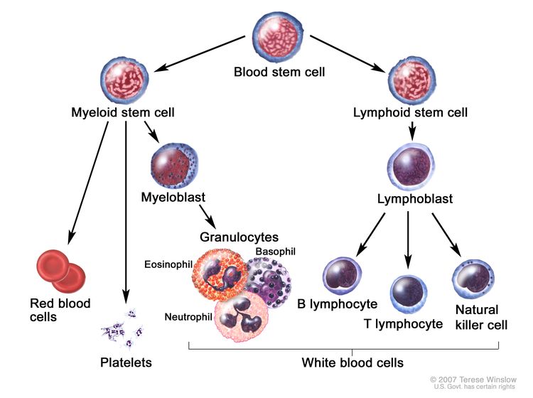
EnlargeBlood cell development; drawing shows the steps a blood stem cell goes through to become a red blood cell, platelet, or white blood cell. A myeloid stem cell becomes a red blood cell, a platelet, or a myeloblast, which then becomes a granulocyte (the types of granulocytes are eosinophils, basophils, and neutrophils). A lymphoid stem cell becomes a lymphoblast and then becomes a B-lymphocyte, T-lymphocyte, or natural killer cell.
Blood cell development. A blood stem cell goes through several steps to become a red blood cell, platelet, or white blood cell.

AML is a heterogenous group of blood cancers that result from clonal expansion of myeloid hematopoietic precursors in the bone marrow. Not only are circulating leukemia cells (also called blasts) seen in the peripheral blood, but granulocytopenia, anemia, and thrombocytopenia are also common as proliferating leukemia cells interfere with normal hematopoiesis.[3]

Clinical Presentation

The diagnosis of AML is uncommon before age 45 years; the median age at diagnosis is 69 years.[2] Patients may present with symptoms that include:

  • Weakness.
  • Fever.
  • Infection.
  • Pallor.
  • Bleeding.

The hampered production of normal blood cells due to leukemic infiltration of the bone marrow can also cause other symptoms and complications. Less commonly, patients have signs or symptoms related to the collection of leukemia cells in certain anatomical locations, such as central nervous system (CNS) or testicular involvement, or the presence of a myeloid sarcoma (also called chloroma). The symptoms of acute leukemia often arise over a 4- to 6-week period before diagnosis.[3]

Diagnostic Evaluation

The differentiation of AML from other forms of leukemia, in particular chronic myeloid leukemia and acute lymphocytic leukemia, has vital therapeutic implications. The primary diagnostic tool in this determination is flow cytometry to evaluate surface antigens on the leukemia cells. Simple morphology is not adequate in determining lineage and, at a minimum, special histochemical stains are needed. While a diagnosis can be made by evaluating peripheral blood, a bone marrow biopsy is used to evaluate morphology and cell surface markers, as well as provide material for cytogenetic and molecular analysis. A peripheral blood or bone marrow blast count of 20% or greater is required to make the diagnosis, except for cases with certain chromosomal abnormalities (i.e., t(15;17), t(8;21), inv(16), or t(16;16)).[4]

Prognosis and Prognostic Factors

While the rates of new cases of AML have not changed significantly over the last decade, age-adjusted death rates have dropped.[2] Treatment should be sufficiently aggressive to achieve complete remission (CR) because partial remission offers no substantial survival benefit. Approximately 60% to 70% of adults with AML can be expected to attain CR status after appropriate induction therapy. More than 25% of adults with AML (about 45% of those who attain CR) can be expected to survive 3 or more years and may be cured.

Approximately half of patients with AML will harbor chromosomal abnormalities; therefore, conventional cytogenetic analysis remains mandatory in the evaluation of suspected AML.[5,6] With the routine use of molecular diagnostics, the identification of recurrent somatic pathogenic variants in NPM1, FLT3, CEPBA, RUNX1, and other genes has become a routine part of determining prognosis. Cytogenetic and molecular analyses provide the strongest prognostic information available, predicting outcome of both remission induction and consolidation therapy.[7] Cytogenic and molecular information has been combined to form distinct prognostic groups.

Additional adverse prognostic factors for AML include:

  • Age at diagnosis. Remission rates in adult AML are inversely related to age, with an expected remission rate of more than 65% for those younger than 60 years. Data suggest that once attained, duration of remission may be shorter in older patients. Increased morbidity and mortality during induction appear to be directly related to age.
  • CNS involvement with leukemia.
  • Systemic infection at diagnosis.
  • Elevated white blood cell count (>100,000/mm3) at diagnosis.
  • Therapy-related myeloid neoplasms, resulting from alkylating agents and radiation therapy.
  • History of myelodysplastic syndrome or another antecedent hematologic disorder.

Long-Term Effects of Cancer Treatment

The risk of developing any long-term effects depends on the type and dose of treatment that was used and the age at which the patient underwent treatment.

A study of 30 patients who had AML that was in remission for at least 10 years demonstrated a 13% incidence of secondary malignancies.[8] Of 31 female long-term survivors of AML or acute lymphoblastic leukemia (ALL) diagnosed before age 40 years, 26 resumed normal menstruation after completion of therapy. Among 36 live offspring of survivors, two congenital problems occurred.[8]

Most patients with AML who undergo intensive therapy are treated with an anthracycline. Anthracyclines have been associated with increased risk of congestive heart failure (CHF).[9] Anthracycline cardiotoxicity is dose-dependent. In one study, doxorubicin-related CHF was 5% at a lifetime cumulative dose of 400 mg/m2, rising to 26% at a cumulative dose of 550 mg/m2.[10] In many cases, heart failure can manifest as a late effect.[11] In an analysis of children who underwent treatment for acute leukemia, the cumulative incidence of CHF at 10 years was 1.7% in ALL and 7.5% in AML.[12]

Patients who undergo allogeneic hematopoietic stem cell transplant can experience a large number of long-term or late side effects of treatment as a result of high-dose chemotherapy and/or radiation, and as an effect of chronic graft-versus-host disease and immunosuppression. These side effects may include chronic fatigue, thyroid and gonadal dysfunction, infertility, chronic infection, accelerated coronary heart disease, osteopenia, cataracts, iron overload, adverse psychological outcomes, and second cancers.[1315]

In the Bone Marrow Transplant Survivor Study, hematopoietic cell transplant survivors had accelerated aging and were 8.4 times more likely to be frail than their siblings (95% confidence interval [CI], 2.0−34.5; P = .003). In a multivariable analysis, frailty was associated with a 2.76-fold increase in the risk of death, compared with a nonfrail state (95% CI, 1.7−4.4; P < .001).[16]

References
  1. American Cancer Society: Cancer Facts and Figures 2025. American Cancer Society, 2025. Available online. Last accessed January 16, 2025.
  2. Surveillance, Epidemiology, and End Results Program: Cancer Stat Facts: Leukemia — Acute Myeloid Leukemia (AML). Bethesda, Md: National Cancer Institute, DCCPS, Surveillance Research Program, 2020. Available online. Last accessed January 24, 2025.
  3. Sekeres MA, Gerds AT: Mitigating Fear and Loathing in Managing Acute Myeloid Leukemia. Semin Hematol 52 (3): 249-55, 2015. [PUBMED Abstract]
  4. Swerdlow SH, Campo E, Harris NL, et al., eds.: WHO Classification of Tumours of Haematopoietic and Lymphoid Tissues. 4th rev. ed. International Agency for Research on Cancer, 2017.
  5. Slovak ML, Kopecky KJ, Cassileth PA, et al.: Karyotypic analysis predicts outcome of preremission and postremission therapy in adult acute myeloid leukemia: a Southwest Oncology Group/Eastern Cooperative Oncology Group Study. Blood 96 (13): 4075-83, 2000. [PUBMED Abstract]
  6. Grimwade D, Walker H, Harrison G, et al.: The predictive value of hierarchical cytogenetic classification in older adults with acute myeloid leukemia (AML): analysis of 1065 patients entered into the United Kingdom Medical Research Council AML11 trial. Blood 98 (5): 1312-20, 2001. [PUBMED Abstract]
  7. Döhner H, Estey E, Grimwade D, et al.: Diagnosis and management of AML in adults: 2017 ELN recommendations from an international expert panel. Blood 129 (4): 424-447, 2017. [PUBMED Abstract]
  8. Micallef IN, Rohatiner AZ, Carter M, et al.: Long-term outcome of patients surviving for more than ten years following treatment for acute leukaemia. Br J Haematol 113 (2): 443-5, 2001. [PUBMED Abstract]
  9. Steinherz LJ, Steinherz PG, Tan CT, et al.: Cardiac toxicity 4 to 20 years after completing anthracycline therapy. JAMA 266 (12): 1672-7, 1991. [PUBMED Abstract]
  10. Swain SM, Whaley FS, Ewer MS: Congestive heart failure in patients treated with doxorubicin: a retrospective analysis of three trials. Cancer 97 (11): 2869-79, 2003. [PUBMED Abstract]
  11. Hequet O, Le QH, Moullet I, et al.: Subclinical late cardiomyopathy after doxorubicin therapy for lymphoma in adults. J Clin Oncol 22 (10): 1864-71, 2004. [PUBMED Abstract]
  12. Chellapandian D, Pole JD, Nathan PC, et al.: Congestive heart failure among children with acute leukemia: a population-based matched cohort study. Leuk Lymphoma 60 (2): 385-394, 2019. [PUBMED Abstract]
  13. Inamoto Y, Lee SJ: Late effects of blood and marrow transplantation. Haematologica 102 (4): 614-625, 2017. [PUBMED Abstract]
  14. Sun CL, Francisco L, Baker KS, et al.: Adverse psychological outcomes in long-term survivors of hematopoietic cell transplantation: a report from the Bone Marrow Transplant Survivor Study (BMTSS). Blood 118 (17): 4723-31, 2011. [PUBMED Abstract]
  15. Armenian SH, Sun CL, Kawashima T, et al.: Long-term health-related outcomes in survivors of childhood cancer treated with HSCT versus conventional therapy: a report from the Bone Marrow Transplant Survivor Study (BMTSS) and Childhood Cancer Survivor Study (CCSS). Blood 118 (5): 1413-20, 2011. [PUBMED Abstract]
  16. Arora M, Sun CL, Ness KK, et al.: Physiologic Frailty in Nonelderly Hematopoietic Cell Transplantation Patients: Results From the Bone Marrow Transplant Survivor Study. JAMA Oncol 2 (10): 1277-1286, 2016. [PUBMED Abstract]

Classification of AML

World Health Organization (WHO) Classification

The classification of acute myeloid leukemia (AML) has been revised by a group of pathologists and clinicians under the auspices of the WHO.[1] While elements of the French-American-British (FAB) classification have been retained (i.e., morphology, immunophenotype, cytogenetics, and clinical features),[2,3] the WHO classification incorporates and interrelates morphology, cytogenetics, molecular genetics, and immunologic markers, which construct a classification that is universally applicable and has prognostic and therapeutic relevance.[1,3,4] Each criterion has prognostic and treatment implications but, for practical purposes, initial antileukemic therapy is similar for all subtypes.

In 2001, the WHO proposed a new classification system that incorporated diagnostic cytogenetic information and that more reliably correlated with outcome. This classification system also decreased the bone marrow percentage of leukemic blast requirement for the diagnosis of AML from 30% to 20%. An additional clarification was made so patients with recurrent cytogenetic abnormalities did not need to meet the minimum blast requirement to be considered as having an AML diagnosis.[57]

In 2008, the WHO expanded the number of cytogenetic abnormalities linked to AML classification and, for the first time, included specific pathogenic variants (CEBPA and NPM) in its classification system.[5,8] With the addition of these gene variants, FAB subclassification no longer provided prognostic information for patients with a diagnosis of AML, not otherwise specified (NOS).[9]

In 2016, the WHO classification underwent revisions to incorporate the expanding knowledge of leukemia biomarkers that are significantly important to the diagnosis, prognosis, and treatment of leukemia.[10] With emerging technologies aimed at genetic, epigenetic, proteomic, and immunophenotypic classification, AML classification will continue to evolve and provide informative prognostic and biological guidelines to clinicians and researchers.

2016 WHO classification of AML and related neoplasms

AML With Recurrent Genetic Abnormalities

AML with well-defined genetic abnormalities is characterized by recurrent genetic abnormalities.[10] The reciprocal translocations t(8;21), inv(16) or t(16;16), t(15;17), and translocations involving the 11q23 breakpoint are the most commonly identified chromosomal abnormalities. These structural chromosome rearrangements result in the formation of fusion genes that encode chimeric proteins that may contribute to the initiation or progression of leukemogenesis. Many of these translocations are detected by either reverse transcriptase–polymerase chain reaction (RT–PCR) or fluorescence in situ hybridization (FISH), which has a higher sensitivity than metaphase cytogenetics. Other recurring cytogenetic abnormalities are less common.

Molecular diagnostic platforms such as next-generation sequencing along with RT-PCR are used to identify recurrent molecular abnormalities in AML, helping to further refine diagnostic categories in the 2016 WHO classification system.[10]

AML with t(8;21)(q22;q22), RUNX1RUNX1T1

The translocation t(8;21)(q22;q22) is one of the most common chromosomal aberrations in AML and accounts for 5% to 12% of cases.[11] Myeloid sarcomas (chloromas) may be present and may be associated with a bone marrow blast percentage of less than 20%.

Common morphological features include:

  • Large blasts with abundant basophilic cytoplasm, often containing numerous azurophilic granules.
  • A few blasts in some cases show very large granules (pseudo Chediak-Higashi granules).
  • Auer rods, which may be detected in mature neutrophils.
  • Smaller blasts, predominantly in the peripheral blood.
  • Promyelocytes, myelocytes, and mature neutrophils with variable dysplasia in the bone marrow.
  • Abnormal nuclear segmentation (pseudo Pelger-Huët nuclei) and/or cytoplasmic staining abnormalities.
  • Increased eosinophil precursors.
  • Reduced or absent monocytes.
  • Normal erythroblasts and megakaryocytes.

Rarely, AML with this translocation presents with a bone marrow blast percentage of less than 20%.[5] Along with inv(16)(p13;q22) or t(16;16)(p13;q22), AML with t(8;21) makes up a category known as core binding factor AML. This category of AML is associated with long-term survival when treated with high-dose cytarabine.[1215]

The translocation t(8;21)(q22;q22) involves the RUNX1 gene, which encodes CBF-alpha, and the RUNX1T1 (8;21) gene.[5,16] The RUNX1::RUNX1T1 fusion transcript is consistently detected in patients with t(8;21) AML. This translocation is usually associated with a good response to chemotherapy and a high complete remission (CR) rate with long-term survival when treated with high-dose cytarabine in the consolidation phase, as demonstrated in the Cancer and Leukemia Group B (CLB-9022 and CLB-8525) trials.[1215] Additional chromosome abnormalities are common, for example, loss of a sex chromosome and del(9)(q22). Leukocytosis (i.e., white blood count >25 × 109/L) is associated with an inferior outcome,[17] as is the presence of a KIT pathogenic variant.[18]

AML with inv(16)(p13.1;q22) or t(16;16)(p13.1;q22), CBFB::MYH11

The inv(16)(p13;q22) abnormality or t(16;16)(p13;q22) translocation is found in approximately 10% to 12% of all cases of AML, predominantly in younger patients.[5,19] Myeloid sarcomas may be present at initial diagnosis or at relapse.

Common morphological features include:

  • Monocytic and granulocytic differentiation.
  • A characteristically abnormal eosinophil component with immature purple-violet eosinophil granules that may obscure cell morphology if present in great numbers.
  • Auer rods in myeloblasts.
  • Decreased neutrophils in bone marrow.

As is found in rare cases of AML with t(8;21), the bone marrow blast percentage in this AML is occasionally less than 20%.

Both inv(16)(p13;q22) and t(16;16)(p13;q22) result in the fusion of the CBFB gene at 16q22 to the smooth muscle MYH11 gene at 16p13, thereby forming the CBFB::MYH11 fusion gene .[11] The use of FISH and RT–PCR methods is sometimes necessary to document this fusion gene because its presence is not always documented by traditional cytogenetics banding techniques.[20] Similar to AML with t(8;21), patients with the CBFB::MYH11 fusion gene achieve higher CR rates and long-term survival when treated with high-dose cytarabine in the consolidation setting.[12,13,15] Unlike AML with t(8;21), the prognostic relevance of KIT pathogenic variants is unclear.[21]

APL with PML::RARA

APL is defined by the presence of the PML::RARA fusion protein, typically a result of t(15;17)(q22;q12), but can be cryptic or result from complex cytogenetic rearrangements other than t(15;17)(q22;q12). It is also an AML in which promyelocytes are the dominant leukemic cell type. APL exists as two subtypes, hypergranular or typical APL and microgranular or hypogranular APL. APL comprises 5% to 8% of cases of AML and occurs predominately in adults in midlife.[5] Both typical and microgranular APL are commonly associated with disseminated intravascular coagulation (DIC).[22,23] In microgranular APL, unlike typical APL, the leukocyte count can be very high with a rapid doubling time.[5]

Common morphological features of typical APL include:

  • Kidney-shaped or bilobed nuclei.
  • Cytoplasm densely packed with large granules (bright pink, red, or purple in Romanowsky stains).
  • Bundles of Auer rods within the cytoplasm (faggot cells).
  • Larger Auer rods than in other types of AML.
  • Strongly positive myeloperoxidase (MPO) reaction in all leukemic promyelocytes.
  • Only occasional leukemic promyelocytes in the blood.

Common morphological features of microgranular APL include:

  • Bilobed nuclear shape.
  • Apparent scarce or absent granules (submicroscopic azurophilic granules).
  • Small number of abnormal promyelocytes with visible granules and/or bundles of Auer rods (faggot cells).
  • High leukocyte count in the peripheral blood.
  • Strongly positive MPO reaction in all leukemic promyelocytes.

In APL, the RARA gene on 17q12 fuses with a nuclear regulatory factor on 15q22 (PML gene) resulting in a PML::RARA gene fusion transcript.[2426] Rare cases of cryptic or masked t(15;17) lack typical cytogenetic findings and involve complex variant translocations or submicroscopic insertion of the RARA gene into the PML gene, leading to the expression of the PML::RARA fusion transcript.[5] FISH and/or RT–PCR methods may be required to unmask these cryptic genetic rearrangements.[27,28] In approximately 1% of the patients with APL, variant chromosomal aberrations may be found in which the RARA gene is fused with other genes.[29] Variant translocations involving the RARA gene include t(11;17)(q23;q21), t(5;17)(q32;q12), and t(11;17)(q13;q21).[5]

APL has a specific sensitivity to treatment with all-trans retinoic acid (ATRA, tretinoin), which acts as a differentiating agent.[3032] High CR rates and long-term disease-free survival in APL may be obtained by combining ATRA treatment with chemotherapy,[33] or in a chemotherapy-free regimen with arsenic trioxide.[34]

AML with t(9;11)(p21.3;q23.3), MLLT3::KMT2A

AML with 11q23 abnormalities comprises 5% to 6% of cases of AML and is typically associated with monocytic features. This type of AML is more common in children. Two clinical subgroups who have a high frequency of AML with 11q23 abnormalities are infants with AML and patients with therapy-related AML, usually occurring after treatment with DNA topoisomerase inhibitors. Patients may present with DIC and extramedullary monocytic sarcomas and/or tissue infiltration (gingiva, skin).[5]

Common morphological features include:

  • Monoblasts and promonocytes predominate in the bone marrow.
  • Monoblasts and promonocytes with strong, positive nonspecific-esterase reactions.

The MLLT3 gene on 11q23, an epigenetic regulator, is involved in translocations with approximately 135 different rearrangements that have been identified.[35] Genes other than MLLT3 may be involved in 11q23 abnormalities.[36] FISH may be required to detect genetic abnormalities involving the KMT2A gene (also known as MLL).[3638] In general, risk categories and prognoses for individual 11q23 translocations are difficult to determine because of the lack of studies involving significant numbers of patients; however, patients with t(11;19)(q23;p13.1) have been reported to have poor outcomes.[13]

AML with t(6;9)(p23;q34.1), DEK::NUP214

The t(6;9) translocation leads to the formation of a leukemia-associated DEK::NUP214 fusion protein and accounts for approximately 1% of AML cases.[3941] NUP214 is a component of the nuclear pore complex. This subgroup of AML has been associated with a poor prognosis.[39,42,43]

AML with inv(3)(q21.3;q26.2) or t(3;3)(q21.3;q26.2), GATA2, MECOM

The inv(3) abnormality or t(3;3) translocation occur infrequently and account for approximately 1% of all AML cases.[41] MECOM at chromosome 3q26 codes for two proteins, EVI1 and MDS1-EVI1, both of which are transcription regulators. The inv(3) and t(3;3) abnormalities do not lead to a fusion gene, rather they reposition the distal GATA2 enhancer, resulting in overexpression of EVI1, and simultaneously confer GATA2 haploinsufficiency.[44,45] These abnormalities are associated with poor prognosis.[15,46,47] Abnormalities involving MECOM can be detected in some AML cases with other 3q abnormalities and are also associated with poor prognosis.

AML (megakaryoblastic) with t(1;22)(p13.3;q13.3), RBM15::MKL1

The t(1;22)(p13;q13) translocation that produces the RBM15::MKL1 fusion gene is an uncommon driver of pediatric AML (<1% of pediatric AML) and is restricted to acute megakaryocytic leukemia. For more information, see Childhood Acute Myeloid Leukemia Treatment.

AML with BCR::ABL1 (provisional entity)

This provisional entity was added by the WHO in 2016 in an effort to recognize that patients with the BCR::ABL1 fusion protein should be treated with a tyrosine kinase inhibitor.[10] However, this entity is very difficult to distinguish from chronic myeloid leukemia (CML) in blast phase (BP-CML). Loss of IKZF1 and/or CDKN2A may help distinguish true cases of AML with BCR::ABL1 from BP-CML.[48] For more information, see Chronic Myeloid Leukemia Treatment.

AML with NPM1 pathogenic variants

NPM1 is a protein that has been linked to ribosomal protein assembly and transport and is also a molecular chaperone involved in preventing protein aggregation in the nucleolus. Immunohistochemical methods can be used to accurately identify patients with NPM1 pathogenic variants by the demonstration of cytoplasmic localization of NPM.[49] Abnormal NPM1 protein diminishes its nuclear localization and lead to impaired hematopoietic differentiation. They are primarily associated with a normal karyotype (50%), and less commonly seen in conjunction with an abnormal karyotype (<10%), or complex karyotype (<3%).[5052] An NPM1 pathogenic variant confers improved prognosis in the absence of FLT3–internal tandem duplication (ITD) variants.[50,53,54]

AML with biallelic CEBPA pathogenic variants

In adults younger than 60 years, 10% to 15% of cytogenetically normal AML cases have CEBPA pathogenic variants.[53,55] The CEBPA gene is located on chromosome 19 and encodes a transcription factor that coordinates myeloid differentiation and cellular growth arrest.[56]

Outcomes for patients with AML and CEBPA pathogenic variants are relatively favorable and similar to that of patients with core-binding factor leukemias.[53,57] Studies have demonstrated that AML with biallelic CEBPA variants is independently associated with a favorable prognosis, while AML with monoallelic CEBPA variants is not.[55,5860] These findings led to the WHO 2016 revision of this subtype definition to require biallelic variants.[10]

AML with RUNX1 pathogenic variants (provisional entity)

AML with RUNX1 pathogenic variants, which is a provisional entity in the 2016 WHO classification of AML and related neoplasms, denotes a distinct population of de novo AML without myelodysplastic syndrome (MDS)-related features.[61] Variants in RUNX1 are associated with a high risk of treatment failure.[6264]

AML With Myelodysplasia-Related Features

AML with myelodysplasia-related features is characterized by 20% or more blasts in the blood or bone marrow and dysplasia in two or more myeloid cell lines, generally including megakaryocytes.[5] To make the diagnosis, dysplasia must be present in 50% or more of the cells of at least two lineages and must be present in a pretreatment bone marrow specimen or must have the presence of an MDS-related cytogenetic abnormality.[5] AML with myelodysplasia-related features may occur de novo or after MDS or a myelodysplastic/myeloproliferative neoplasm overlap. The diagnostic terminology AML with myelodysplasia-related features evolving from a myelodysplastic syndrome should be used when an MDS precedes AML.[5] When NPM1 variants or biallelic CEBPA variants are present, multilineage dysplasia alone will not classify a case as AML with myelodysplasia-related changes.[5] For more information, see Myelodysplastic Syndromes Treatment and Myelodysplastic/Myeloproliferative Neoplasms Treatment.

AML with myelodysplasia-related features occurs primarily in older patients.[5] Patients with AML with myelodysplasia-related features frequently present with severe pancytopenia.

Common morphological features include:

  • Multilineage dysplasia in the blood or bone marrow.
  • Dysplasia in 50% or more of the cells of two or more cell lines.
  • Dysgranulopoiesis (neutrophils with hypogranular cytoplasm, hyposegmented nuclei or bizarrely segmented nuclei).
  • Dyserythropoiesis (megaloblastic nuclei, karyorrhexis, or multinucleation of erythroid precursors and ringed sideroblasts).
  • Dysmegakaryopoiesis (micromegakaryocytes and normal size or large megakaryocytes with monolobed or multiple separated nuclei).

Chromosome abnormalities observed in AML with myelodysplasia-related features are similar to those found in MDS and frequently involve gain or loss of major segments of certain chromosomes, predominately chromosomes 5 and/or 7. The probability of achieving a CR has been reported to be affected adversely by a diagnosis of AML with myelodysplasia-related features.[6567]

Therapy-Related Myeloid Neoplasms

Therapy-related myeloid neoplasms (t-MN) include AML (t-AML) and MDS (t-MDS) that arise secondary to cytotoxic chemotherapy and/or radiation therapy.[5] The therapy-related (or secondary) MDS are included because of their close clinicopathological relationships to therapy-related AML. Although these therapy-related disorders can be distinguished by the specific mutagenic agents involved, this distinction may be difficult to make because of the frequent overlapping use of multiple potentially mutagenic agents in treating cancer.[68] Because the associated cytogenetic abnormality, not the mutagenetic agent, determines prognosis and treatment it should be noted in the diagnosis.[10]

Since t-MN have been associated with germline pathogenic variants in cancer susceptibility genes, considering germline genetic testing or genetic counseling is warranted in those with strong family histories of cancer.[69]

Alkylating agent-related t-MN

The alkylating agent/radiation-related acute leukemias and myelodysplastic syndromes typically occur 5 to 6 years after exposure to the mutagenic agent, with a reported range of approximately 10 to 192 months.[70,71] The risk of occurrence is related to both the total cumulative dose of the alkylating agent and the age of the patient.

Cytogenetic abnormalities have been observed in more than 90% of cases of t-MN and commonly include chromosomes 5 and/or 7.[70,72,73] Complex chromosomal abnormalities (≥3 distinct abnormalities) are the most common finding.[68,7274]

Topoisomerase II inhibitor-related t-MN

Topoisomerase II inhibitor-related t-MN occurs in patients treated with topoisomerase II inhibitors. The agents implicated are the epipodophyllotoxins etoposide and teniposide and the anthracyclines doxorubicin and 4-epi-doxorubicin.[70] The mean latency period from the time of institution of the causative therapy to the development of t-MN is approximately 2 years.[75]

As with alkylating agent/radiation-related t-MN, the cytogenetic abnormalities are often complex.[68,7274] The predominant cytogenetic finding involves chromosome 11q23 abnormalities and KMT2A pathogenic variants.[68,76]

AML, Not Otherwise Specified (NOS)

Cases of AML that do not fulfill the criteria for AML with recurrent genetic abnormalities, AML with myelodysplasia-related features, or t-MN fall within the category of AML, NOS.[10] As mentioned before, the subcategories of AML, NOS lack prognostic significance when it is unclear if NPM1 and CEBPA pathogenic variants are present.[9] Classification in this subset of AML is based on leukemic cell features of morphology, cytochemistry, and maturation (i.e., the FAB classification system) and include:[5]

  • AML with minimal differentiation.
  • AML without maturation.
  • AML with maturation.
  • Acute myelomonocytic leukemia.
  • Acute monoblastic/monocytic leukemia.
  • Pure erythroid leukemia.
  • Acute megakaryoblastic leukemia.
  • Acute basophilic leukemia.
  • Acute panmyelosis with myelofibrosis.

Myeloid Sarcoma

Myeloid sarcoma (also known as extramedullary myeloid tumor, granulocytic sarcoma, and chloroma) is a tumor mass that consists of myeloblasts or immature myeloid cells, occurring in an extramedullary site.[5] Development of myeloid sarcoma has been reported in 2% to 8% of patients with AML.[77] Clinical features include occurrence common in subperiosteal bone structures of the skull, paranasal sinuses, sternum, ribs, vertebrae, and pelvis; lymph nodes, skin, mediastinum, small intestine, and the epidural space; and occurrence de novo or concomitant with AML or a myeloproliferative disorder.[10,77,78]

Morphological and cytochemical features include:

  • Granulocytic sarcoma composed of myeloblasts, neutrophils, and neutrophil precursors with three subtypes based on degree of maturation (i.e., blastic, immature, and differentiated).
  • Monoblastic sarcoma preceding or occurring simultaneously with acute monoblastic leukemia.
  • Tumors with trilineage hematopoiesis occurring with transformation of chronic myeloproliferative disorders.
  • Myeloblasts and neutrophils that are positive for MPO.
  • Neutrophils that are positive for naphthol ASD chloroacetate esterase.

Immunophenotyping with antibodies to MPO, lysozyme, and chloroacetate is critical to the diagnosis of these lesions.[5] The myeloblasts in granulocytic sarcomas express myeloid-associated antigens (CD13, CD33, CD117, and MPO). The monoblasts in monoblastic sarcomas express acute monoblastic leukemia antigens (CD14, CD116, and CD11c) and usually react with antibodies to lysozyme and CD68. The main differential diagnosis includes non-Hodgkin lymphoma of the lymphoblastic type, Burkitt lymphoma, large-cell lymphoma, and small, round-cell tumors, especially in children (e.g., neuroblastoma, rhabdomyosarcoma, Ewing/primitive neuroectodermal tumors, and medulloblastoma). When able, FISH for common chromosomal abnormalities should be completed, as well as molecular studies to refine diagnosis and aid in prognosis.

No unique chromosomal abnormalities are associated with myeloid sarcoma.[77,79] The presence of myeloid sarcoma in patients with the otherwise good-risk t(8;21) AML may be associated with a lower CR rate and decreased remission duration.[80] Myeloid sarcoma occurring in the setting of MDS or myeloproliferative disorder is equivalent to blast transformation (progression to AML). In the case of AML, the prognosis is that of the underlying leukemia.[10] Although the initial presentation of myeloid sarcoma may appear to be isolated, it is a partial manifestation of a systemic disease and should be treated with intensive chemotherapy.[77,78,81,82]

Myeloid Proliferations Related to Down Syndrome

For more information about TAM and myeloid leukemia associated with Down syndrome, see Childhood Myeloid Proliferations Associated With Down Syndrome Treatment.

Acute Leukemias of Ambiguous Lineage

Acute leukemias of ambiguous lineage are rare types of acute leukemia in which the morphological, cytochemical, and immunophenotypic features of the blast population do not allow classification in myeloid or lymphoid categories; or the types have morphological and/or immunophenotypic features of both myeloid and lymphoid cells or both B and T lineages (i.e., acute bilineal leukemia and acute biphenotypic leukemia).[10,83,84]

They include the following subcategories:[5]

  • Acute undifferentiated leukemia.
  • Mixed phenotype acute leukemia (MPAL) with t(9;22)(q34.1;q11.2); BCR::ABL1.
  • MPAL with t(v;11q23.3); KMT2A rearranged.
  • MPAL, B/myeloid, NOS.
  • MPAL, T/myeloid, NOS.

The diagnosis of MPAL is made in leukemias with expression of antigens of more than one lineage:[5]

Table 1. Mixed Phenotype Acute Leukemia Diagnostic Criteria
Diagnosis Criteria
MPO = myeloperoxidase.
Myeloid Lineage MPO (flow cytometry, immunohistochemistry, or cytochemistry) or monocytic differentiation (≥ 2 of the following: nonspecific esterase cytochemistry, CD11c, CD14, CD64, lysozyme).
T-cell Lineage Strong cytoplasmic CD3 (with antibodies to CD3 epsilon chain) or surface CD3.
B-cell Lineage Strong CD19 with ≥1 of the following strongly expressed: cytoplasmic CD79a, cCD22, or CD10; or weak CD19 with at least two of the following strongly expressed: CD79a, cCD22, or CD10.

Cytogenetic abnormalities are observed in a high percentage of acute leukemias of ambiguous lineage.[8588] Approximately 33% of cases have the Philadelphia chromosome, and some cases are associated with t(4;11)(q21;q23) or other 11q23 abnormalities. In general, the prognosis appears to be unfavorable. The occurrence of 11q23 abnormalities or BCR::ABL1 are especially unfavorable prognostic indicators;[86,89,90] however, preliminary results indicate that tyrosine kinase inhibitors can be used successfully.[91,92]

References
  1. Brunning RD, Matutes E, Harris NL, et al.: Acute myeloid leukaemia: introduction. In: Jaffe ES, Harris NL, Stein H, et al., eds.: Pathology and Genetics of Tumours of Haematopoietic and Lymphoid Tissues. IARC Press, 2001. World Health Organization Classification of Tumours, 3, pp 77-80.
  2. Bennett JM, Catovsky D, Daniel MT, et al.: Proposed revised criteria for the classification of acute myeloid leukemia. A report of the French-American-British Cooperative Group. Ann Intern Med 103 (4): 620-5, 1985. [PUBMED Abstract]
  3. Cheson BD, Cassileth PA, Head DR, et al.: Report of the National Cancer Institute-sponsored workshop on definitions of diagnosis and response in acute myeloid leukemia. J Clin Oncol 8 (5): 813-9, 1990. [PUBMED Abstract]
  4. Bennett JM, Catovsky D, Daniel MT, et al.: Proposals for the classification of the acute leukaemias. French-American-British (FAB) co-operative group. Br J Haematol 33 (4): 451-8, 1976. [PUBMED Abstract]
  5. Swerdlow SH, Campo E, Harris NL, et al., eds.: WHO Classification of Tumours of Haematopoietic and Lymphoid Tissues. 4th rev. ed. International Agency for Research on Cancer, 2017.
  6. Jaffe ES, Harris NL, Stein H, et al., eds.: Pathology and Genetics of Tumours of Haematopoietic and Lymphoid Tissues. IARC Press, 2001. World Health Organization Classification of Tumours, 3.
  7. Hasle H, Niemeyer CM, Chessells JM, et al.: A pediatric approach to the WHO classification of myelodysplastic and myeloproliferative diseases. Leukemia 17 (2): 277-82, 2003. [PUBMED Abstract]
  8. Arber DA, Vardiman JW, Brunning RD: Acute myeloid leukaemia with recurrent genetic abnormalities. In: Swerdlow SH, Campo E, Harris NL, et al., eds.: WHO Classification of Tumours of Haematopoietic and Lymphoid Tissues. 4th ed. International Agency for Research on Cancer, 2008, pp 110-23.
  9. Walter RB, Othus M, Burnett AK, et al.: Significance of FAB subclassification of “acute myeloid leukemia, NOS” in the 2008 WHO classification: analysis of 5848 newly diagnosed patients. Blood 121 (13): 2424-31, 2013. [PUBMED Abstract]
  10. Arber DA, Orazi A, Hasserjian R, et al.: The 2016 revision to the World Health Organization classification of myeloid neoplasms and acute leukemia. Blood 127 (20): 2391-405, 2016. [PUBMED Abstract]
  11. Caligiuri MA, Strout MP, Gilliland DG: Molecular biology of acute myeloid leukemia. Semin Oncol 24 (1): 32-44, 1997. [PUBMED Abstract]
  12. Bloomfield CD, Lawrence D, Byrd JC, et al.: Frequency of prolonged remission duration after high-dose cytarabine intensification in acute myeloid leukemia varies by cytogenetic subtype. Cancer Res 58 (18): 4173-9, 1998. [PUBMED Abstract]
  13. Byrd JC, Mrózek K, Dodge RK, et al.: Pretreatment cytogenetic abnormalities are predictive of induction success, cumulative incidence of relapse, and overall survival in adult patients with de novo acute myeloid leukemia: results from Cancer and Leukemia Group B (CALGB 8461). Blood 100 (13): 4325-36, 2002. [PUBMED Abstract]
  14. Palmieri S, Sebastio L, Mele G, et al.: High-dose cytarabine as consolidation treatment for patients with acute myeloid leukemia with t(8;21). Leuk Res 26 (6): 539-43, 2002. [PUBMED Abstract]
  15. Grimwade D, Walker H, Oliver F, et al.: The importance of diagnostic cytogenetics on outcome in AML: analysis of 1,612 patients entered into the MRC AML 10 trial. The Medical Research Council Adult and Children’s Leukaemia Working Parties. Blood 92 (7): 2322-33, 1998. [PUBMED Abstract]
  16. Downing JR: The AML1-ETO chimaeric transcription factor in acute myeloid leukaemia: biology and clinical significance. Br J Haematol 106 (2): 296-308, 1999. [PUBMED Abstract]
  17. Schlenk RF, Benner A, Krauter J, et al.: Individual patient data-based meta-analysis of patients aged 16 to 60 years with core binding factor acute myeloid leukemia: a survey of the German Acute Myeloid Leukemia Intergroup. J Clin Oncol 22 (18): 3741-50, 2004. [PUBMED Abstract]
  18. Duployez N, Marceau-Renaut A, Boissel N, et al.: Comprehensive mutational profiling of core binding factor acute myeloid leukemia. Blood 127 (20): 2451-9, 2016. [PUBMED Abstract]
  19. Marlton P, Keating M, Kantarjian H, et al.: Cytogenetic and clinical correlates in AML patients with abnormalities of chromosome 16. Leukemia 9 (6): 965-71, 1995. [PUBMED Abstract]
  20. Poirel H, Radford-Weiss I, Rack K, et al.: Detection of the chromosome 16 CBF beta-MYH11 fusion transcript in myelomonocytic leukemias. Blood 85 (5): 1313-22, 1995. [PUBMED Abstract]
  21. Döhner K, Paschka P: Intermediate-risk acute myeloid leukemia therapy: current and future. Hematology Am Soc Hematol Educ Program 2014 (1): 34-43, 2014. [PUBMED Abstract]
  22. Kwaan HC, Wang J, Boggio LN: Abnormalities in hemostasis in acute promyelocytic leukemia. Hematol Oncol 20 (1): 33-41, 2002. [PUBMED Abstract]
  23. Barbui T, Falanga A: Disseminated intravascular coagulation in acute leukemia. Semin Thromb Hemost 27 (6): 593-604, 2001. [PUBMED Abstract]
  24. de Thé H, Chomienne C, Lanotte M, et al.: The t(15;17) translocation of acute promyelocytic leukaemia fuses the retinoic acid receptor alpha gene to a novel transcribed locus. Nature 347 (6293): 558-61, 1990. [PUBMED Abstract]
  25. Melnick A, Licht JD: Deconstructing a disease: RARalpha, its fusion partners, and their roles in the pathogenesis of acute promyelocytic leukemia. Blood 93 (10): 3167-215, 1999. [PUBMED Abstract]
  26. Kayser S, Schlenk RF, Platzbecker U: Management of patients with acute promyelocytic leukemia. Leukemia 32 (6): 1277-1294, 2018. [PUBMED Abstract]
  27. Lo Coco F, Diverio D, Falini B, et al.: Genetic diagnosis and molecular monitoring in the management of acute promyelocytic leukemia. Blood 94 (1): 12-22, 1999. [PUBMED Abstract]
  28. Zaccaria A, Valenti A, Toschi M, et al.: Cryptic translocation of PML/RARA on 17q. A rare event in acute promyelocytic leukemia. Cancer Genet Cytogenet 138 (2): 169-73, 2002. [PUBMED Abstract]
  29. Jansen JH, Löwenberg B: Acute promyelocytic leukemia with a PLZF-RARalpha fusion protein. Semin Hematol 38 (1): 37-41, 2001. [PUBMED Abstract]
  30. Castaigne S, Chomienne C, Daniel MT, et al.: All-trans retinoic acid as a differentiation therapy for acute promyelocytic leukemia. I. Clinical results. Blood 76 (9): 1704-9, 1990. [PUBMED Abstract]
  31. Tallman MS, Andersen JW, Schiffer CA, et al.: All-trans-retinoic acid in acute promyelocytic leukemia. N Engl J Med 337 (15): 1021-8, 1997. [PUBMED Abstract]
  32. Tallman MS, Andersen JW, Schiffer CA, et al.: All-trans retinoic acid in acute promyelocytic leukemia: long-term outcome and prognostic factor analysis from the North American Intergroup protocol. Blood 100 (13): 4298-302, 2002. [PUBMED Abstract]
  33. Fenaux P, Chastang C, Chevret S, et al.: A randomized comparison of all transretinoic acid (ATRA) followed by chemotherapy and ATRA plus chemotherapy and the role of maintenance therapy in newly diagnosed acute promyelocytic leukemia. The European APL Group. Blood 94 (4): 1192-200, 1999. [PUBMED Abstract]
  34. Lo-Coco F, Avvisati G, Vignetti M, et al.: Retinoic acid and arsenic trioxide for acute promyelocytic leukemia. N Engl J Med 369 (2): 111-21, 2013. [PUBMED Abstract]
  35. Meyer C, Burmeister T, Gröger D, et al.: The MLL recombinome of acute leukemias in 2017. Leukemia 32 (2): 273-284, 2018. [PUBMED Abstract]
  36. Giugliano E, Rege-Cambrin G, Scaravaglio P, et al.: Two new translocations involving the 11q23 region map outside the MLL locus in myeloid leukemias. Haematologica 87 (10): 1014-20, 2002. [PUBMED Abstract]
  37. König M, Reichel M, Marschalek R, et al.: A highly specific and sensitive fluorescence in situ hybridization assay for the detection of t(4;11)(q21;q23) and concurrent submicroscopic deletions in acute leukaemias. Br J Haematol 116 (4): 758-64, 2002. [PUBMED Abstract]
  38. Kim HJ, Cho HI, Kim EC, et al.: A study on 289 consecutive Korean patients with acute leukaemias revealed fluorescence in situ hybridization detects the MLL translocation without cytogenetic evidence both initially and during follow-up. Br J Haematol 119 (4): 930-9, 2002. [PUBMED Abstract]
  39. Ageberg M, Drott K, Olofsson T, et al.: Identification of a novel and myeloid specific role of the leukemia-associated fusion protein DEK-NUP214 leading to increased protein synthesis. Genes Chromosomes Cancer 47 (4): 276-87, 2008. [PUBMED Abstract]
  40. Shiba N, Ichikawa H, Taki T, et al.: NUP98-NSD1 gene fusion and its related gene expression signature are strongly associated with a poor prognosis in pediatric acute myeloid leukemia. Genes Chromosomes Cancer 52 (7): 683-93, 2013. [PUBMED Abstract]
  41. Döhner H, Estey E, Grimwade D, et al.: Diagnosis and management of AML in adults: 2017 ELN recommendations from an international expert panel. Blood 129 (4): 424-447, 2017. [PUBMED Abstract]
  42. Slovak ML, Gundacker H, Bloomfield CD, et al.: A retrospective study of 69 patients with t(6;9)(p23;q34) AML emphasizes the need for a prospective, multicenter initiative for rare ‘poor prognosis’ myeloid malignancies. Leukemia 20 (7): 1295-7, 2006. [PUBMED Abstract]
  43. Alsabeh R, Brynes RK, Slovak ML, et al.: Acute myeloid leukemia with t(6;9) (p23;q34): association with myelodysplasia, basophilia, and initial CD34 negative immunophenotype. Am J Clin Pathol 107 (4): 430-7, 1997. [PUBMED Abstract]
  44. Gröschel S, Sanders MA, Hoogenboezem R, et al.: A single oncogenic enhancer rearrangement causes concomitant EVI1 and GATA2 deregulation in leukemia. Cell 157 (2): 369-81, 2014. [PUBMED Abstract]
  45. Yamazaki H, Suzuki M, Otsuki A, et al.: A remote GATA2 hematopoietic enhancer drives leukemogenesis in inv(3)(q21;q26) by activating EVI1 expression. Cancer Cell 25 (4): 415-27, 2014. [PUBMED Abstract]
  46. Mrózek K, Heerema NA, Bloomfield CD: Cytogenetics in acute leukemia. Blood Rev 18 (2): 115-36, 2004. [PUBMED Abstract]
  47. Lugthart S, Gröschel S, Beverloo HB, et al.: Clinical, molecular, and prognostic significance of WHO type inv(3)(q21q26.2)/t(3;3)(q21;q26.2) and various other 3q abnormalities in acute myeloid leukemia. J Clin Oncol 28 (24): 3890-8, 2010. [PUBMED Abstract]
  48. Nacheva EP, Grace CD, Brazma D, et al.: Does BCR/ABL1 positive acute myeloid leukaemia exist? Br J Haematol 161 (4): 541-50, 2013. [PUBMED Abstract]
  49. Falini B, Martelli MP, Bolli N, et al.: Immunohistochemistry predicts nucleophosmin (NPM) mutations in acute myeloid leukemia. Blood 108 (6): 1999-2005, 2006. [PUBMED Abstract]
  50. Falini B, Mecucci C, Tiacci E, et al.: Cytoplasmic nucleophosmin in acute myelogenous leukemia with a normal karyotype. N Engl J Med 352 (3): 254-66, 2005. [PUBMED Abstract]
  51. Falini B, Nicoletti I, Martelli MF, et al.: Acute myeloid leukemia carrying cytoplasmic/mutated nucleophosmin (NPMc+ AML): biologic and clinical features. Blood 109 (3): 874-85, 2007. [PUBMED Abstract]
  52. Falini B, Martelli MP, Bolli N, et al.: Acute myeloid leukemia with mutated nucleophosmin (NPM1): is it a distinct entity? Blood 117 (4): 1109-20, 2011. [PUBMED Abstract]
  53. Schlenk RF, Döhner K, Krauter J, et al.: Mutations and treatment outcome in cytogenetically normal acute myeloid leukemia. N Engl J Med 358 (18): 1909-18, 2008. [PUBMED Abstract]
  54. Gale RE, Green C, Allen C, et al.: The impact of FLT3 internal tandem duplication mutant level, number, size, and interaction with NPM1 mutations in a large cohort of young adult patients with acute myeloid leukemia. Blood 111 (5): 2776-84, 2008. [PUBMED Abstract]
  55. Taskesen E, Bullinger L, Corbacioglu A, et al.: Prognostic impact, concurrent genetic mutations, and gene expression features of AML with CEBPA mutations in a cohort of 1182 cytogenetically normal AML patients: further evidence for CEBPA double mutant AML as a distinctive disease entity. Blood 117 (8): 2469-75, 2011. [PUBMED Abstract]
  56. Nerlov C: C/EBPalpha mutations in acute myeloid leukaemias. Nat Rev Cancer 4 (5): 394-400, 2004. [PUBMED Abstract]
  57. Marcucci G, Maharry K, Radmacher MD, et al.: Prognostic significance of, and gene and microRNA expression signatures associated with, CEBPA mutations in cytogenetically normal acute myeloid leukemia with high-risk molecular features: a Cancer and Leukemia Group B Study. J Clin Oncol 26 (31): 5078-87, 2008. [PUBMED Abstract]
  58. Wouters BJ, Löwenberg B, Erpelinck-Verschueren CA, et al.: Double CEBPA mutations, but not single CEBPA mutations, define a subgroup of acute myeloid leukemia with a distinctive gene expression profile that is uniquely associated with a favorable outcome. Blood 113 (13): 3088-91, 2009. [PUBMED Abstract]
  59. Dufour A, Schneider F, Metzeler KH, et al.: Acute myeloid leukemia with biallelic CEBPA gene mutations and normal karyotype represents a distinct genetic entity associated with a favorable clinical outcome. J Clin Oncol 28 (4): 570-7, 2010. [PUBMED Abstract]
  60. Fasan A, Haferlach C, Alpermann T, et al.: The role of different genetic subtypes of CEBPA mutated AML. Leukemia 28 (4): 794-803, 2014. [PUBMED Abstract]
  61. Schnittger S, Dicker F, Kern W, et al.: RUNX1 mutations are frequent in de novo AML with noncomplex karyotype and confer an unfavorable prognosis. Blood 117 (8): 2348-57, 2011. [PUBMED Abstract]
  62. Tang JL, Hou HA, Chen CY, et al.: AML1/RUNX1 mutations in 470 adult patients with de novo acute myeloid leukemia: prognostic implication and interaction with other gene alterations. Blood 114 (26): 5352-61, 2009. [PUBMED Abstract]
  63. Mendler JH, Maharry K, Radmacher MD, et al.: RUNX1 mutations are associated with poor outcome in younger and older patients with cytogenetically normal acute myeloid leukemia and with distinct gene and MicroRNA expression signatures. J Clin Oncol 30 (25): 3109-18, 2012. [PUBMED Abstract]
  64. Gaidzik VI, Bullinger L, Schlenk RF, et al.: RUNX1 mutations in acute myeloid leukemia: results from a comprehensive genetic and clinical analysis from the AML study group. J Clin Oncol 29 (10): 1364-72, 2011. [PUBMED Abstract]
  65. Díaz-Beyá M, Rozman M, Pratcorona M, et al.: The prognostic value of multilineage dysplasia in de novo acute myeloid leukemia patients with intermediate-risk cytogenetics is dependent on NPM1 mutational status. Blood 116 (26): 6147-8, 2010. [PUBMED Abstract]
  66. Rozman M, Navarro JT, Arenillas L, et al.: Multilineage dysplasia is associated with a poorer prognosis in patients with de novo acute myeloid leukemia with intermediate-risk cytogenetics and wild-type NPM1. Ann Hematol 93 (10): 1695-703, 2014. [PUBMED Abstract]
  67. Weinberg OK, Seetharam M, Ren L, et al.: Clinical characterization of acute myeloid leukemia with myelodysplasia-related changes as defined by the 2008 WHO classification system. Blood 113 (9): 1906-8, 2009. [PUBMED Abstract]
  68. Smith SM, Le Beau MM, Huo D, et al.: Clinical-cytogenetic associations in 306 patients with therapy-related myelodysplasia and myeloid leukemia: the University of Chicago series. Blood 102 (1): 43-52, 2003. [PUBMED Abstract]
  69. Churpek JE, Marquez R, Neistadt B, et al.: Inherited mutations in cancer susceptibility genes are common among survivors of breast cancer who develop therapy-related leukemia. Cancer 122 (2): 304-11, 2016. [PUBMED Abstract]
  70. Brunning RD, Matutes E, Flandrin G, et al.: Acute myeloid leukaemias and myelodysplastic syndromes, therapy related. In: Jaffe ES, Harris NL, Stein H, et al., eds.: Pathology and Genetics of Tumours of Haematopoietic and Lymphoid Tissues. IARC Press, 2001. World Health Organization Classification of Tumours, 3, pp 89-91.
  71. Ellis M, Ravid M, Lishner M: A comparative analysis of alkylating agent and epipodophyllotoxin-related leukemias. Leuk Lymphoma 11 (1-2): 9-13, 1993. [PUBMED Abstract]
  72. Olney HJ, Mitelman F, Johansson B, et al.: Unique balanced chromosome abnormalities in treatment-related myelodysplastic syndromes and acute myeloid leukemia: report from an international workshop. Genes Chromosomes Cancer 33 (4): 413-23, 2002. [PUBMED Abstract]
  73. Mauritzson N, Albin M, Rylander L, et al.: Pooled analysis of clinical and cytogenetic features in treatment-related and de novo adult acute myeloid leukemia and myelodysplastic syndromes based on a consecutive series of 761 patients analyzed 1976-1993 and on 5098 unselected cases reported in the literature 1974-2001. Leukemia 16 (12): 2366-78, 2002. [PUBMED Abstract]
  74. Pedersen-Bjergaard J, Andersen MK, Christiansen DH, et al.: Genetic pathways in therapy-related myelodysplasia and acute myeloid leukemia. Blood 99 (6): 1909-12, 2002. [PUBMED Abstract]
  75. Leone G, Voso MT, Sica S, et al.: Therapy related leukemias: susceptibility, prevention and treatment. Leuk Lymphoma 41 (3-4): 255-76, 2001. [PUBMED Abstract]
  76. Bloomfield CD, Archer KJ, Mrózek K, et al.: 11q23 balanced chromosome aberrations in treatment-related myelodysplastic syndromes and acute leukemia: report from an international workshop. Genes Chromosomes Cancer 33 (4): 362-78, 2002. [PUBMED Abstract]
  77. Yamauchi K, Yasuda M: Comparison in treatments of nonleukemic granulocytic sarcoma: report of two cases and a review of 72 cases in the literature. Cancer 94 (6): 1739-46, 2002. [PUBMED Abstract]
  78. Yilmaz AF, Saydam G, Sahin F, et al.: Granulocytic sarcoma: a systematic review. Am J Blood Res 3 (4): 265-70, 2013. [PUBMED Abstract]
  79. Brunning RD, Matutes E, Flandrin G, et al.: Acute myeloid leukaemia not otherwise categorised. In: Jaffe ES, Harris NL, Stein H, et al., eds.: Pathology and Genetics of Tumours of Haematopoietic and Lymphoid Tissues. IARC Press, 2001. World Health Organization Classification of Tumours, 3, pp 91-105.
  80. Byrd JC, Weiss RB, Arthur DC, et al.: Extramedullary leukemia adversely affects hematologic complete remission rate and overall survival in patients with t(8;21)(q22;q22): results from Cancer and Leukemia Group B 8461. J Clin Oncol 15 (2): 466-75, 1997. [PUBMED Abstract]
  81. Hayashi T, Kimura M, Satoh S, et al.: Early detection of AML1/MTG8 fusion mRNA by RT-PCR in the bone marrow cells from a patient with isolated granulocytic sarcoma. Leukemia 12 (9): 1501-3, 1998. [PUBMED Abstract]
  82. Imrie KR, Kovacs MJ, Selby D, et al.: Isolated chloroma: the effect of early antileukemic therapy. Ann Intern Med 123 (5): 351-3, 1995. [PUBMED Abstract]
  83. Matutes E, Pickl WF, Van’t Veer M, et al.: Mixed-phenotype acute leukemia: clinical and laboratory features and outcome in 100 patients defined according to the WHO 2008 classification. Blood 117 (11): 3163-71, 2011. [PUBMED Abstract]
  84. van den Ancker W, Terwijn M, Westers TM, et al.: Acute leukemias of ambiguous lineage: diagnostic consequences of the WHO2008 classification. Leukemia 24 (7): 1392-6, 2010. [PUBMED Abstract]
  85. Hanson CA, Abaza M, Sheldon S, et al.: Acute biphenotypic leukaemia: immunophenotypic and cytogenetic analysis. Br J Haematol 84 (1): 49-60, 1993. [PUBMED Abstract]
  86. Legrand O, Perrot JY, Simonin G, et al.: Adult biphenotypic acute leukaemia: an entity with poor prognosis which is related to unfavourable cytogenetics and P-glycoprotein over-expression. Br J Haematol 100 (1): 147-55, 1998. [PUBMED Abstract]
  87. Carbonell F, Swansbury J, Min T, et al.: Cytogenetic findings in acute biphenotypic leukaemia. Leukemia 10 (8): 1283-7, 1996. [PUBMED Abstract]
  88. Pane F, Frigeri F, Camera A, et al.: Complete phenotypic and genotypic lineage switch in a Philadelphia chromosome-positive acute lymphoblastic leukemia. Leukemia 10 (4): 741-5, 1996. [PUBMED Abstract]
  89. Brunning RD, Matutes E, Borowitz M: Acute leukaemias of ambiguous lineage. In: Jaffe ES, Harris NL, Stein H, et al., eds.: Pathology and Genetics of Tumours of Haematopoietic and Lymphoid Tissues. IARC Press, 2001. World Health Organization Classification of Tumours, 3, pp 106-7.
  90. Killick S, Matutes E, Powles RL, et al.: Outcome of biphenotypic acute leukemia. Haematologica 84 (8): 699-706, 1999. [PUBMED Abstract]
  91. Kawajiri C, Tanaka H, Hashimoto S, et al.: Successful treatment of Philadelphia chromosome-positive mixed phenotype acute leukemia by appropriate alternation of second-generation tyrosine kinase inhibitors according to BCR-ABL1 mutation status. Int J Hematol 99 (4): 513-8, 2014. [PUBMED Abstract]
  92. Shimizu H, Yokohama A, Hatsumi N, et al.: Philadelphia chromosome-positive mixed phenotype acute leukemia in the imatinib era. Eur J Haematol 93 (4): 297-301, 2014. [PUBMED Abstract]

Treatment Option Overview for AML

Phases of Therapy

The treatment of patients with acute myeloid leukemia (AML) is based on whether the disease is newly diagnosed (previously untreated), in remission, or recurrent. Also, the intensity of the treatment and the patient’s overall health status are considered when choosing a treatment approach. Successful treatment of AML requires the control of bone marrow and systemic disease, and specific treatment of central nervous system (CNS) disease, if present. The cornerstone of this strategy includes systemically administered combination chemotherapy. Because only 5% or fewer of patients with AML develop CNS disease, prophylactic treatment is not indicated.[1,2]

  • Newly diagnosed (untreated): Untreated AML is defined as newly diagnosed leukemia that has not been previously treated. The initial treatment for patients with newly diagnosed AML is often induction therapy that aims to induce a remission. In patients with AML, a complete remission (CR) is defined as a normal peripheral blood cell count (absolute neutrophil count >1,000/mm3 and platelet count >100,000/mm3) and normocellular marrow with less than 5% blasts in the marrow and no signs or symptoms of the disease. In addition, no signs or symptoms are evident of CNS leukemia or other extramedullary infiltration.[3]

    Modifications to the definition of CR have been proposed because some responses are deeper than a CR, and others may not meet all the criteria for a complete response. In addition, most AML patients meeting the criteria for CR have residual leukemia.[3]

Table 2. Treatment Response Categories for Newly Diagnosed Acute Myeloid Leukemia
Response Category Definition
ANC = absolute neutrophil count; CR = complete remission; CRh = CR with partial hematologic recovery; CRi = CR with incomplete hematologic recovery; CRMRD− = CR without measurable residual disease; MLFS = morphological leukemia-free state; PR = partial remission; RT–qPCR = reverse transcription–quantitative polymerase chain reaction.
CRMRD− If studied pretreatment, CR with negativity for a genetic marker by RT–qPCR, or CR with negativity by multicolor flow cytometry.
CR Bone marrow blasts <5%; absence of circulating blasts and blasts with Auer rods; absence of extramedullary disease; ANC ≥1.0 × 10⁹/L (1,000/microL); platelet count ≥100 × 109/L (100,000/microL).
CRh ANC ≥0.5 x 10⁹/L (500/microL) and platelet count ≥50 x 109/L (100,000/microL); otherwise all other CR criteria met.[4]
CRi All CR criteria except for residual neutropenia (<1.0 × 10⁹/L [1,000/microL]) or thrombocytopenia (<100 × 109/L [100,000/microL]).
MLFS Bone marrow blasts <5%; absence of blasts with Auer rods; absence of extramedullary disease; no hematologic recovery required.
PR All hematologic criteria of CR; decrease of bone marrow blast percentage to 5 to 25%; and decrease of pretreatment bone marrow blast percentage by at least 50%.
No response Patients evaluable for response but not meeting criteria for CR, CRh, CRi, MLFS, or PR are categorized as having no response prior to the response landmark. Patients failing to achieve response by the designated landmark are designated as having refractory disease.
  • In remission: When patients are in a remission after induction chemotherapy, consolidation chemotherapy is given, with the aim of deepening the response and consolidating the remission. Maintenance therapy is not included in most current treatment protocols and clinical trials. Consolidation therapy appears to be effective when given immediately after remission is achieved.[5]
  • Persistent/recurrent disease: Despite intensive chemotherapy, some patients with newly diagnosed AML will not go into remission and have primary refractory disease. Also, some patients who are in a remission after induction and consolidation chemotherapy may have a return of their disease.[3] The rates of primary refractory disease and relapse vary with the age of the patient, genomic variants seen in the leukemia cells, and initial treatment given.
Table 3. Treatment Response Categories for Persistent/Recurrent Acute Myeloid Leukemia
Response Category Definition
CR = complete remission; CRh = CR with partial hematologic recovery; CRhMRD-LL = CR with partial hematologic recovery and measurable residual disease at a low level; CRi = CR with incomplete hematologic recovery; CRiMRD-LL = CR with incomplete hematologic recovery and measurable residual disease at a low level; CRMRD- = CR without measurable residual disease; CRMRD-LL = CR with measurable residual disease detection at a low level; MRD = measurable residual disease; MRD- = absence of measurable residual disease; MRD+ = presence of measurable residual disease; MLFS = morphological leukemia-free state; PR = partial response; RT–qPCR = reverse transcription–quantitative polymerase chain reaction.
Primary refractory disease No CR, CRh, or CRi at the response landmark (i.e., after two courses of intensive induction treatment) or a defined landmark (e.g., 180 days after commencing less-intensive therapy).
Relapsed disease (after CR, CRh, or CRi) Bone marrow blasts ≥5%; or reappearance of blasts in the blood in at least two peripheral blood samples obtained at least 1 week apart; or development of extramedullary disease.
MRD relapse (after CR, CRh, or CRi without MRD-) Defined by one of the following:
  Conversion from MRD- to MRD+, independent of method; or
  Increase of MRD copy numbers ≥1 log10 between any two positive samples in patients with CRMRD-LL, CRhMRD-LL, or CRiMRD-LL by qPCR.
Either result should be rapidly confirmed in a second consecutive sample from the same tissue source.
Stable disease Absence of CRMRD-, CR, CRi, PR, MLFS; and criteria for progressive disease not met.
Progressive disease Evidence for an increase in bone marrow blast percentage and/or increase of absolute blast counts in the blood:
>50% increase in marrow blasts; or
>50% increase in peripheral blasts in the absence of differentiation syndrome; or
  New extramedullary disease.

Supportive Care During Therapy

Because myelosuppression is an anticipated consequence of both the leukemia and its treatment with chemotherapy, patients must be closely monitored during therapy. Facilities must be available for hematologic support with multiple blood fractions, including platelet transfusions, and for the treatment of related infectious complications.[6]

Transfusion therapy

Supportive care during remission induction treatment should routinely include red blood cell and platelet transfusions, when appropriate.[7,8] Rapid marrow ablation with consequent earlier marrow regeneration decreases morbidity and mortality. Randomized trials have shown similar outcomes for patients who received prophylactic platelet transfusions at a level of 10,000/mm3 rather than 20,000/mm3.[9] The incidence of platelet alloimmunization was similar among groups randomly assigned to receive pooled platelet concentrates from random donors; filtered, pooled platelet concentrates from random donors; ultraviolet B-irradiated, pooled platelet concentrates from random donors; or filtered platelets obtained by apheresis from single random donors.[10]

No good evidence exists to support granulocyte transfusions in the treatment of AML. A multicenter randomized trial (RING [NCT00627393]) was conducted to address the utility of granulocyte transfusions in the setting of infections.[11] There was no difference between the granulocyte and control arms for the composite primary end point of survival plus microbial response at 42 days after randomization. However, the power to detect a true beneficial effect was low because enrollment was half that of the planned study size.

Growth factors

The following growth factors have been studied in the treatment of AML:

  • Colony-stimulating factors: Granulocyte colony–stimulating factor and granulocyte-macrophage colony–stimulating factor have been studied in an effort to shorten the period of granulocytopenia associated with leukemia treatment.[12] If used, these agents are administered after administration of chemotherapy. Although the use of growth factors decreases the time to neutrophil recovery by 2 to 5 days, and decreases rates of febrile neutropenia and duration of hospitalization, randomized clinical trials have not shown an impact of growth factors on overall survival and their cost-effectiveness has been inconsistently reported.[13,14] Use of growth factors is not routinely recommended in the remission induction setting.
  • Erythropoiesis-stimulating agents: Anemia associated with the diagnosis of AML and induction chemotherapy is managed primarily with red blood transfusions. Unlike myelodysplastic syndromes, there is no role for the use of erythropoiesis stimulating agents (e.g., epoetin alfa and darbepoetin) during the treatment of AML.
  • Thrombopoietin mimetics: Clinical trials have assessed the use of thrombopoietin mimetics in the treatment of AML. Eltrombopag as a single agent was tested in a multicenter, randomized, placebo-controlled, double-blind, phase I/II trial of 98 patients with platelet counts less than 30 × 109/L as a result of AML or MDS. No significant improvements in platelet counts were recorded. Significant hemorrhage was reported in ten (16%) patients given eltrombopag and nine (26%) patients given placebo. No difference in disease progression or overall survival was observed.[15]

    Eltrombopag appeared to hasten platelet recovery and reduce the number of platelet transfusions needed when added in an unblinded fashion to induction chemotherapy in older FLT3-negative AML patients.[16] However, in a separate, randomized double-blind study of 148 patients, eltrombopag or placebo was added to high-dose induction chemotherapy.[17] The results of this study did not indicate any clinical benefit of eltrombopag over placebo. Given the minimal efficacy signal at this point, eltrombopag is not routinely recommended in the supportive care or remission induction setting.

Antimicrobial therapy

Empiric broad spectrum antimicrobial therapy is an absolute necessity for febrile patients who are profoundly neutropenic.[18,19] Careful instruction in personal hand hygiene, dental care, and recognition of early signs of infection are appropriate in all patients. Elaborate isolation facilities (including filtered air, sterile food, and gut flora sterilization) are not indicated.[20,21] Likewise, there are no advantages to eating a cooked neutropenic diet, as demonstrated in randomized trials.[22]

Antibiotic prophylaxis with a fluoroquinolone and antifungal prophylaxis with an oral triazole or parenteral echinocandin is appropriate for patients with expected prolonged, profound neutropenia (<100/mm3 for 2 weeks for profound neutropenia lasting >7 days).[23] Unlike patients undergoing treatment for acute lymphoblastic lymphoma, Pneumocystis jirovecii prophylaxis is not routinely employed.

Nucleoside analog-based antiviral prophylaxis, such as acyclovir, is appropriate for patients who are seropositive for herpes simplex virus undergoing induction chemotherapy.[23]

References
  1. Rozovski U, Ohanian M, Ravandi F, et al.: Incidence of and risk factors for involvement of the central nervous system in acute myeloid leukemia. Leuk Lymphoma 56 (5): 1392-7, 2015. [PUBMED Abstract]
  2. Alakel N, Stölzel F, Mohr B, et al.: Symptomatic central nervous system involvement in adult patients with acute myeloid leukemia. Cancer Manag Res 9: 97-102, 2017. [PUBMED Abstract]
  3. Döhner H, Estey EH, Amadori S, et al.: Diagnosis and management of acute myeloid leukemia in adults: recommendations from an international expert panel, on behalf of the European LeukemiaNet. Blood 115 (3): 453-74, 2010. [PUBMED Abstract]
  4. Döhner H, Wei AH, Appelbaum FR, et al.: Diagnosis and management of AML in adults: 2022 recommendations from an international expert panel on behalf of the ELN. Blood 140 (12): 1345-1377, 2022. [PUBMED Abstract]
  5. Cassileth PA, Lynch E, Hines JD, et al.: Varying intensity of postremission therapy in acute myeloid leukemia. Blood 79 (8): 1924-30, 1992. [PUBMED Abstract]
  6. Supportive Care. In: Wiernik PH, Canellos GP, Dutcher JP, et al., eds.: Neoplastic Diseases of the Blood. 3rd ed. Churchill Livingstone, 1996, pp 779-967.
  7. Slichter SJ: Controversies in platelet transfusion therapy. Annu Rev Med 31: 509-40, 1980. [PUBMED Abstract]
  8. Murphy MF, Metcalfe P, Thomas H, et al.: Use of leucocyte-poor blood components and HLA-matched-platelet donors to prevent HLA alloimmunization. Br J Haematol 62 (3): 529-34, 1986. [PUBMED Abstract]
  9. Rebulla P, Finazzi G, Marangoni F, et al.: The threshold for prophylactic platelet transfusions in adults with acute myeloid leukemia. Gruppo Italiano Malattie Ematologiche Maligne dell’Adulto. N Engl J Med 337 (26): 1870-5, 1997. [PUBMED Abstract]
  10. Leukocyte reduction and ultraviolet B irradiation of platelets to prevent alloimmunization and refractoriness to platelet transfusions. The Trial to Reduce Alloimmunization to Platelets Study Group. N Engl J Med 337 (26): 1861-9, 1997. [PUBMED Abstract]
  11. Price TH, Boeckh M, Harrison RW, et al.: Efficacy of transfusion with granulocytes from G-CSF/dexamethasone-treated donors in neutropenic patients with infection. Blood 126 (18): 2153-61, 2015. [PUBMED Abstract]
  12. Geller RB: Use of cytokines in the treatment of acute myelocytic leukemia: a critical review. J Clin Oncol 14 (4): 1371-82, 1996. [PUBMED Abstract]
  13. Rowe JM, Andersen JW, Mazza JJ, et al.: A randomized placebo-controlled phase III study of granulocyte-macrophage colony-stimulating factor in adult patients (> 55 to 70 years of age) with acute myelogenous leukemia: a study of the Eastern Cooperative Oncology Group (E1490). Blood 86 (2): 457-62, 1995. [PUBMED Abstract]
  14. Stone RM, Berg DT, George SL, et al.: Granulocyte-macrophage colony-stimulating factor after initial chemotherapy for elderly patients with primary acute myelogenous leukemia. Cancer and Leukemia Group B. N Engl J Med 332 (25): 1671-7, 1995. [PUBMED Abstract]
  15. Platzbecker U, Wong RS, Verma A, et al.: Safety and tolerability of eltrombopag versus placebo for treatment of thrombocytopenia in patients with advanced myelodysplastic syndromes or acute myeloid leukaemia: a multicentre, randomised, placebo-controlled, double-blind, phase 1/2 trial. Lancet Haematol 2 (10): e417-26, 2015. [PUBMED Abstract]
  16. Mukherjee S, Li H, Hobbs BP: A single arm, phase II study of eltrombopag to enhance platelet count recovery in older patients with acute myeloid leukemia (AML) undergoing remission induction therapy. [Abstract] Blood 134 (Suppl 1): 2595, 2019.
  17. Frey N, Jang JH, Szer J, et al.: Eltrombopag treatment during induction chemotherapy for acute myeloid leukaemia: a randomised, double-blind, phase 2 study. Lancet Haematol 6 (3): e122-e131, 2019. [PUBMED Abstract]
  18. Hughes WT, Armstrong D, Bodey GP, et al.: From the Infectious Diseases Society of America. Guidelines for the use of antimicrobial agents in neutropenic patients with unexplained fever. J Infect Dis 161 (3): 381-96, 1990. [PUBMED Abstract]
  19. Rubin M, Hathorn JW, Pizzo PA: Controversies in the management of febrile neutropenic cancer patients. Cancer Invest 6 (2): 167-84, 1988. [PUBMED Abstract]
  20. Armstrong D: Symposium on infectious complications of neoplastic disease (Part II). Protected environments are discomforting and expensive and do not offer meaningful protection. Am J Med 76 (4): 685-9, 1984. [PUBMED Abstract]
  21. Sherertz RJ, Belani A, Kramer BS, et al.: Impact of air filtration on nosocomial Aspergillus infections. Unique risk of bone marrow transplant recipients. Am J Med 83 (4): 709-18, 1987. [PUBMED Abstract]
  22. Gardner A, Mattiuzzi G, Faderl S, et al.: Randomized comparison of cooked and noncooked diets in patients undergoing remission induction therapy for acute myeloid leukemia. J Clin Oncol 26 (35): 5684-8, 2008. [PUBMED Abstract]
  23. Taplitz RA, Kennedy EB, Bow EJ, et al.: Antimicrobial Prophylaxis for Adult Patients With Cancer-Related Immunosuppression: ASCO and IDSA Clinical Practice Guideline Update. J Clin Oncol 36 (30): 3043-3054, 2018. [PUBMED Abstract]

Treatment of Newly Diagnosed AML

Treatment Options for Newly Diagnosed (Untreated; Remission Induction) AML

Treatment options for newly diagnosed (untreated; remission induction) acute myeloid leukemia (AML) include:

Chemotherapy

Chemotherapy for AML is divided into the following two general categories:

  1. Intensive remission-induction chemotherapy.
  2. Nonintensive chemotherapy.

One of the following combination chemotherapy regimens may be used as intensive remission induction therapy:

  • Cytarabine plus daunorubicin.[1,2]
  • Cytarabine plus idarubicin.[36]
  • Cytarabine plus mitoxantrone.[7]
  • Cytarabine plus anthracycline plus midostaurin.[8]
  • Cytarabine plus anthracycline plus gemtuzumab ozogamicin.[9]
  • Liposomal daunorubicin-cytarabine (CPX-351).[10]
  • Intrathecal cytarabine or methotrexate may be used to treat central nervous system (CNS) leukemia, if present.

The two-drug regimen of cytarabine given as a continuous infusion for 7 days and a 3-day course of anthracycline (the so-called 7 + 3 induction therapy) results in a complete response rate of approximately 65%. In most instances, there is no further clinical benefit when adding potentially non-cross−resistant drugs (such as fludarabine, topoisomerase inhibitors, thioguanine, mitoxantrone, histone deacetylases inhibitors, or clofarabine) to a 7 + 3 regimen. Cladribine, when added to 7 + 3 induction chemotherapy, showed improved remission rates [11] and survival rates [12] across two randomized controlled trials, but this regimen has not been widely adopted in the absence of confirmatory trials. The addition of midostaurin and gemtuzumab ozogamicin to intensive induction chemotherapy is discussed below.

The choice of anthracycline and the dose-intensity of anthracycline may influence the survival of patients with AML. Idarubicin appeared to be more effective than daunorubicin, particularly in younger adults, although the doses of idarubicin and daunorubicin may not have been equivalent.[36] No significant survival difference between daunorubicin and mitoxantrone has been reported.[13]

Selection of an anthracycline

At present, there is no conclusive evidence to recommend one anthracycline over another.

Evidence (anthracyclines):

  1. In a systematic review and meta-analysis, 18 randomized controlled trials that included 6,755 patients assessed the use of idarubicin versus daunorubicin as part of induction chemotherapy.[14]
    • The use of idarubicin led to improved outcomes, including overall survival (OS), when compared with daunorubicin (12 studies, 5,976 patients; hazard ratio [HR], 0.90; 95% confidence interval [CI], 0.84−0.96; P = .0008). However, there was an increased risk of death during induction (14 studies, 6,349 patients; relative risk [RR], 1.18; 95% CI, 1.01−1.36; P = .03) and mucositis (five studies, 2,000 patients; RR, 1.22; 95% CI, 1.04−1.44; P = .02) with idarubicin as compared with daunorubicin. Moreover, the survival benefit for idarubicin was no longer present if a daunorubicin dose of at least 180 mg/m2 was used (four studies, 2,867 patients; HR, 0.91; 95% CI, 0.82−1.00; P = .06).
    • In patients aged 60 years and younger, outcomes for those who received daunorubicin (90 mg/m2/dose, total induction dosing at 270 mg/m2) were superior to those who received more traditional dosing (45 mg/m2/dose; total dose = 135 mg/m2). The complete remission (CR) rate was 71% versus 57% (P < .001), and the median survival was 24 months versus 16 months (P = .003).[15]
    • No randomized comparison data between daunorubicin at 270 mg/m2 and daunorubicin at 180 mg/m2, or between daunorubicin at 270 mg/m2 and idarubicin, are available.
Addition of an FLT3 inhibitor

Variants in the tyrosine kinase domain (TKD) and internal tandem duplications (ITD) of the FLT3 gene are frequent in AML and are often associated with an inferior outcome.

Midostaurin

Evidence (midostaurin):

  1. A multicenter, randomized, phase III trial (NCT00651261) included patients with FLT3-altered AML. Patients received either the FLT3/multikinase inhibitor, midostaurin, or placebo in addition to cytarabine and daunorubicin induction chemotherapy. The addition of midostaurin led to improved survival (median, 75 vs. 26 months; HR for death, 0.78; one-sided P = .009).[8]
    • The event-free survival (defined as the time from randomization to relapse, death from any cause, or failure to achieve protocol-specified CR) was improved for patients in the midostaurin arm (HR for event or death, 0.78; one-sided P = .002), and the survival benefit was consistent across all FLT3 variant subtypes. The rates of CR (59% vs. 54%) and time to neutrophil count recovery were similar between the two arms.[8][Level of evidence A1]

The U.S. Food and Drug Administration (FDA) approved midostaurin in combination with induction therapy for patients with AML and any FLT3 variant.

Quizartinib

Evidence (quizartinib):

  1. A multicenter, randomized, phase III trial (NCT02668653) included patients with FLT3-ITD–altered AML. Patients received either the selective ITD-specific FLT3 inhibitor, quizartinib, or placebo in addition to cytarabine and daunorubicin induction chemotherapy. The addition of quizartinib led to improved survival (median, 31.9 vs. 15.1 months; HR for death, 0.78; P = .032).[16]
    • The EFS (defined as the time from randomization to lack of CR within 42 days from the start of the last induction cycle, relapse, or death from any cause, whichever occurred first) was similar for patients in the quizartinib and placebo arms (HR for event or death, 0.92; 95% CI, 0.75–1.11; P = .24). The rates of CR and time to neutrophil count recovery were similar between the two arms.[16][Level of evidence A1]

The FDA approved quizartinib in combination with induction therapy for patients with AML and an FLT3-ITD variant but not for patients with other FLT3 variants, such as FLT3-TKD.

The addition of an FLT3 inhibitor to induction chemotherapy is the standard of care for patients with FLT3-altered AML who are eligible for intensive chemotherapy. An ongoing study (NCT03836209) is evaluating which FLT3 inhibitor is best for patients with FLT3-ITD AML receiving up-front chemotherapy. Additional studies are evaluating FLT3 inhibitors in combination with hypomethylating agents and venetoclax in patients who are not candidates for intensive therapy.

Addition of gemtuzumab ozogamicin

Evidence (gemtuzumab ozogamicin):

  1. In a meta-analysis of more than 3,000 patients, the addition of the CD33-directed immunotoxin gemtuzumab ozogamicin to cytarabine plus anthracycline or clofarabine plus anthracycline led to a small increase in the OS rate at 5 years (30.7% vs. 34.6%; HR, 0.90; 95% CI, 0.82−0.98; P = .01).[9]
    • The improvement in the 5-year OS rate was seen across all ages, but this effect was greatest in patients with favorable-risk cytogenetics (55.2% vs. 76.3%; HR, 0.47; 95% CI, 0.31−0.73; P = .0005), and to a lesser extent with intermediate-risk cytogenetics (34.1% vs. 39.4%; HR, 0.84; 95% CI, 0.75−0.95; P = .007). It was not seen in patients with an adverse-risk karyotype.[9][Level of evidence A1]
    • In contrast, gemtuzumab ozogamicin did not improve the 1-year survival rate of older patients who received low-dose cytarabine, although the CR rate increased from 17% to 30% (odds ratio [OR], 0.48; 95% CI, 0.32–0.73; P = .006).[17]

    The FDA label for gemtuzumab ozogamicin includes a boxed warning about the risk of hepatotoxicity, including severe or fatal hepatic sinusoidal obstruction syndrome.

Liposomal daunorubicin-cytarabine (CPX-351)

CPX-351 is a two-drug liposomal encapsulation that delivers cytarabine and daunorubicin at a fixed 5:1 synergistic molar ratio.

Evidence (CPX-351):

  1. A multicenter trial investigated CPX-351 in patients aged 60 to 75 years with therapy-related AML, AML with a history of myelodysplastic syndrome (MDS), or AML with myelodysplasia-related changes.[10]
    • Compared with 7 + 3 induction chemotherapy, CPX-351 resulted in a better overall remission rate (47.7% vs. 33.3%; P = .016), and improved median OS (9.56 vs. 5.95 months; HR, 0.69; 95% CI, 0.52−0.90; P = .003).[10][Level of evidence A1]
    • The rates of early mortality and toxicities were similar between the two arms. However, the median time to recovery of neutrophils and platelets was longer for CPX-351 (35.0 and 36.5 days, respectively) as compared with 7 + 3 induction chemotherapy (29 and 29 days, respectively).
Older adults or adults with significant comorbid conditions

Some patients may decline or be too frail for intensive induction chemotherapy. Low-dose cytarabine, decitabine, azacitidine, or best supportive care can be considered equivalently effective treatment approaches for older patients with AML who decline traditional 7 + 3 induction chemotherapy. Unlike a succinct course of 7 + 3 induction, these less-intensive therapies are continued indefinitely, as long as the patient is deriving benefit (i.e., until disease progression or significant toxicity occurs).

One of the following chemotherapy regimens may be used as less-intensive therapy:

  • Hypomethylating agents (azacitidine and decitabine).
  • Low-dose cytarabine.
  • Venetoclax plus hypomethylating agents or low-dose cytarabine.
  • Glasdegib plus low-dose cytarabine.
  • Ivosidenib with or without azacitidine.
  • Enasidenib.
  • Intrathecal cytarabine or methotrexate may be used to treat CNS leukemia, if present.

Evidence (chemotherapy for patients who decline intensive remission induction therapy):

  1. Hypomethylating agents: The hypomethylating agents azacitidine and decitabine are used commonly in this population of older patients, particularly in the United States. Although approval of the drugs by the FDA is for an MDS indication, the registration studies leading to approval included patients with 20% to 30% myeloblasts, or what would now be considered oligoblastic AML.[18,19]
    1. Azacitidine: An international phase III trial (NCT01074047) randomly assigned patients with AML who were 65 years or older to receive either azacitidine or conventional regimens (7 + 3 induction, low-dose cytarabine, or best supportive care alone).[20]
      • Azacitidine led to a median OS of 10.4 months (95% CI, 8.0−12.7) as compared with 6.5 months (95% CI, 5.0−8.6) with conventional regimens (HR, 0.85; 95% CI, 0.69−1.03; P = .1009).[20][Level of evidence A1]
    2. Decitabine: One phase III trial randomly assigned 485 patients with AML who were older than 65 years to receive either decitabine (n = 242) or their preferred choice (n = 243) of either supportive care (n = 28) or low-dose cytarabine (n = 215).[21]
      • Although rates of CR + CRp (CR with incomplete platelet recovery) were more than double in the decitabine arm (17.8%) compared with the treatment-choice arm (7.8%) (P = .001), median OS was not significantly improved for patients receiving decitabine (7.7 months) compared with the treatment of choice (5.0 months) (HR for death for decitabine, 0.85; 95% CI, .69–1.04; P = .11).

      Compared with treatment for 5 consecutive days, treatment for 10 consecutive days may lead to higher response rates, particularly in those with TP53 variants and/or unfavorable cytogenetic features.[22][Level of evidence C3]

  2. Low-dose cytarabine: Older adults who decline intensive remission-induction therapy or are considered ineligible for intensive remission-induction therapy may derive benefit from low-dose cytarabine, administered twice daily for 10 days in cycles repeated every 4 to 6 weeks.[23]
    • The CR rate using this regimen was 18% compared with 1% for patients treated with hydroxyurea (P = .006).
    • Survival with low-dose cytarabine was better than was survival with hydroxyurea (OR, 0.60; 95% CI, 0.44–0.81; P = .009).[23][Level of evidence A1]
  3. Venetoclax plus hypomethylating agents or low-dose cytarabine: The FDA approved venetoclax, an inhibitor of the anti-apoptotic protein BCL2, in combination with low-dose cytarabine or a hypomethylating agent for the treatment of AML in patients aged 75 years or older, and those who cannot undergo 7 + 3 induction chemotherapy because of comorbidities. Approval was granted based on the results of two studies.
    1. The first study (NCT02203773) was a nonrandomized, open-label, phase Ib clinical trial of venetoclax in combination with azacitidine or decitabine.[24]
      • In this study, 35 (61%; 95% CI, 47.6%−74.0%) of 57 patients had CR or CR with incomplete hematologic recovery (CRi). The median duration of response for all patients with a response (CR + CRi + partial remission [PR]) was 8.4 months (range, 4.7−11.7; n = 36).
    2. The second study (NCT02287233) was a phase I/IIb study of venetoclax in combination with low-dose cytarabine in 82 patients with newly diagnosed AML, including patients with previous exposure to a hypomethylating agent for an antecedent hematologic disorder such as MDS.[25]
      • In the 82 patients enrolled in the study, the CR/CRi rate was 54% (95% CI, 42%−65%), with a median duration of remission of 8.1 months (95% CI, 5.3−14.9 months). The median OS for all patients, irrespective of response, was 10.1 months (95% CI, 5.7−14.2 months).
      • The most common adverse events with venetoclax combinations are gastrointestinal symptoms and myelosuppression, which may require delays between cycles, growth factor support, or decreased duration of venetoclax administration per cycle. Additionally, appropriate prophylactic measures are typically taken to prevent tumor lysis syndrome.[25][Level of evidence C3]
  4. Glasdegib plus low-dose cytarabine: Glasdegib, an oral inhibitor of the hedgehog pathway, combined with low-dose cytarabine was compared with low-dose cytarabine alone in a randomized, phase II open-label study that included 116 patients with AML who were aged 75 years or older or who had severe comorbid conditions (cardiac disease, renal impairment, or Eastern Cooperative Oncology Group performance status 2).[26]
    • Of the 78 patients with AML who received glasdegib plus low-dose cytarabine, 24% (n = 19) of patients had a CR or CRi compared with 5% (2 of 38) of patients who received low-dose cytarabine alone. The median OS was 8.3 months (80%; CI, 6.6–9.5) for patients who received glasdegib/low-dose cytarabine and 4.3 months (80%; CI, 2.9–4.9) for patients who received low-dose cytarabine alone in patients with AML (HR, 0.46; 80% CI, 0.35–0.62; P = .0002).[26][Level of evidence A1]

    Similar to venetoclax, the FDA approved glasdegib in combination with low-dose cytarabine for the treatment of AML in patients aged 75 years or older or who are unable to receive intensive induction chemotherapy.

  5. Ivosidenib: For older or frail patients with AML and IDH1 variants, the IDH1 inhibitor ivosidenib is an option. The FDA approved ivosidenib alone or in combination with azacitidine for the treatment of AML that has a susceptible IDH1 variant (detected by an FDA-approved diagnostic test) in adults aged 75 years or older with newly diagnosed AML or who have comorbidities that preclude the use of intensive induction chemotherapy. Within the context of a phase I study, 34 patients with newly diagnosed IDH1-altered AML who were not candidates for intensive induction chemotherapy were treated with single-agent ivosidenib.[27]
    • Differentiation syndrome was reported in 6 patients (18%), but did not require treatment discontinuation.
    • The rate of CR plus CRi was 42.4% (95% CI, 25.5%−60.8%), with a median duration of response that was not reached (lower bound of the 95% CI was 4.6 months).
    • For all 34 patients on the study, the median OS was 12.6 months (95% CI, 4.5−25.7).[27][Level of evidence C3]

    The combination of azacitidine and ivosidenib was evaluated in a double-blind, randomized, placebo-controlled, phase III trial in patients with newly diagnosed AML who were not eligible for intensive induction chemotherapy. The intention-to-treat analysis included 72 patients treated with azacitidine and ivosidenib and 74 patients treated with azacitidine and placebo. A supplemental new drug application for ivosidenib in combination with azacitidine for patients with untreated IDH1-altered AML is under priority review with the FDA.[28]

    • At a median follow-up of 12.4 months, the primary end point of EFS was improved in patients who received azacitidine and ivosidenib, compared with patients who received azacitidine and placebo (HR, 0.33; 95% CI, 0.16–0.69).
    • The median OS was 24 months for patients who received azacitidine and ivosidenib and 7.9 months for patients who received azacitidine and placebo (HR for death, 0.44; 95% CI, 0.27–0.73).
    • Differentiation syndrome occurred in 14% of patients who received azacitidine and ivosidenib and 8% of patients who received azacitidine and placebo. The incidence of bleeding events was 41% with azacitidine and ivosidenib and 29% with azacitidine and placebo. Infection of any grade was seen in 28% of patients who received azacitidine and ivosidenib and 49% of patients who received azacitidine and placebo. For more information about differentiation syndrome, see the Treatment of Newly Diagnosed Acute Promyelocytic Leukemia section.[28][Level of evidence A1]
  6. Enasidenib: For patients with AML and IDH2 variants, the IDH2 inhibitor enasidenib is an option for older or frail patients. Although it does not have approval in this setting, enasidenib monotherapy was evaluated in a phase I/II trial in patients with newly diagnosed AML who were not eligible for standard chemotherapy.[29]
    • Of the 39 patients enrolled, seven (18%) had CR, one (3%) had CRi, and two (5%) had a PR.
    • Median OS for all patients was 11.3 months (95% CI, 5.7−15.1), and was not reached for patients who had a response.[29][Level of evidence C3]

Current Clinical Trials

Use our advanced clinical trial search to find NCI-supported cancer clinical trials that are now enrolling patients. The search can be narrowed by location of the trial, type of treatment, name of the drug, and other criteria. General information about clinical trials is also available.

References
  1. Yates J, Glidewell O, Wiernik P, et al.: Cytosine arabinoside with daunorubicin or adriamycin for therapy of acute myelocytic leukemia: a CALGB study. Blood 60 (2): 454-62, 1982. [PUBMED Abstract]
  2. Dillman RO, Davis RB, Green MR, et al.: A comparative study of two different doses of cytarabine for acute myeloid leukemia: a phase III trial of Cancer and Leukemia Group B. Blood 78 (10): 2520-6, 1991. [PUBMED Abstract]
  3. Wiernik PH, Banks PL, Case DC, et al.: Cytarabine plus idarubicin or daunorubicin as induction and consolidation therapy for previously untreated adult patients with acute myeloid leukemia. Blood 79 (2): 313-9, 1992. [PUBMED Abstract]
  4. Vogler WR, Velez-Garcia E, Weiner RS, et al.: A phase III trial comparing idarubicin and daunorubicin in combination with cytarabine in acute myelogenous leukemia: a Southeastern Cancer Study Group Study. J Clin Oncol 10 (7): 1103-11, 1992. [PUBMED Abstract]
  5. Berman E, Heller G, Santorsa J, et al.: Results of a randomized trial comparing idarubicin and cytosine arabinoside with daunorubicin and cytosine arabinoside in adult patients with newly diagnosed acute myelogenous leukemia. Blood 77 (8): 1666-74, 1991. [PUBMED Abstract]
  6. Mandelli F, Petti MC, Ardia A, et al.: A randomised clinical trial comparing idarubicin and cytarabine to daunorubicin and cytarabine in the treatment of acute non-lymphoid leukaemia. A multicentric study from the Italian Co-operative Group GIMEMA. Eur J Cancer 27 (6): 750-5, 1991. [PUBMED Abstract]
  7. Löwenberg B, Suciu S, Archimbaud E, et al.: Mitoxantrone versus daunorubicin in induction-consolidation chemotherapy–the value of low-dose cytarabine for maintenance of remission, and an assessment of prognostic factors in acute myeloid leukemia in the elderly: final report. European Organization for the Research and Treatment of Cancer and the Dutch-Belgian Hemato-Oncology Cooperative Hovon Group. J Clin Oncol 16 (3): 872-81, 1998. [PUBMED Abstract]
  8. Stone RM, Mandrekar SJ, Sanford BL, et al.: Midostaurin plus Chemotherapy for Acute Myeloid Leukemia with a FLT3 Mutation. N Engl J Med 377 (5): 454-464, 2017. [PUBMED Abstract]
  9. Hills RK, Castaigne S, Appelbaum FR, et al.: Addition of gemtuzumab ozogamicin to induction chemotherapy in adult patients with acute myeloid leukaemia: a meta-analysis of individual patient data from randomised controlled trials. Lancet Oncol 15 (9): 986-96, 2014. [PUBMED Abstract]
  10. Lancet JE, Uy GL, Cortes JE, et al.: CPX-351 (cytarabine and daunorubicin) Liposome for Injection Versus Conventional Cytarabine Plus Daunorubicin in Older Patients With Newly Diagnosed Secondary Acute Myeloid Leukemia. J Clin Oncol 36 (26): 2684-2692, 2018. [PUBMED Abstract]
  11. Holowiecki J, Grosicki S, Robak T, et al.: Addition of cladribine to daunorubicin and cytarabine increases complete remission rate after a single course of induction treatment in acute myeloid leukemia. Multicenter, phase III study. Leukemia 18 (5): 989-97, 2004. [PUBMED Abstract]
  12. Holowiecki J, Grosicki S, Giebel S, et al.: Cladribine, but not fludarabine, added to daunorubicin and cytarabine during induction prolongs survival of patients with acute myeloid leukemia: a multicenter, randomized phase III study. J Clin Oncol 30 (20): 2441-8, 2012. [PUBMED Abstract]
  13. Arlin Z, Case DC, Moore J, et al.: Randomized multicenter trial of cytosine arabinoside with mitoxantrone or daunorubicin in previously untreated adult patients with acute nonlymphocytic leukemia (ANLL). Lederle Cooperative Group. Leukemia 4 (3): 177-83, 1990. [PUBMED Abstract]
  14. Li X, Xu S, Tan Y, et al.: The effects of idarubicin versus other anthracyclines for induction therapy of patients with newly diagnosed leukaemia. Cochrane Database Syst Rev (6): CD010432, 2015. [PUBMED Abstract]
  15. Fernandez HF, Sun Z, Yao X, et al.: Anthracycline dose intensification in acute myeloid leukemia. N Engl J Med 361 (13): 1249-59, 2009. [PUBMED Abstract]
  16. Erba HP, Montesinos P, Kim HJ, et al.: Quizartinib plus chemotherapy in newly diagnosed patients with FLT3-internal-tandem-duplication-positive acute myeloid leukaemia (QuANTUM-First): a randomised, double-blind, placebo-controlled, phase 3 trial. Lancet 401 (10388): 1571-1583, 2023. [PUBMED Abstract]
  17. Burnett AK, Hills RK, Hunter AE, et al.: The addition of gemtuzumab ozogamicin to low-dose Ara-C improves remission rate but does not significantly prolong survival in older patients with acute myeloid leukaemia: results from the LRF AML14 and NCRI AML16 pick-a-winner comparison. Leukemia 27 (1): 75-81, 2013. [PUBMED Abstract]
  18. Silverman LR, Demakos EP, Peterson BL, et al.: Randomized controlled trial of azacitidine in patients with the myelodysplastic syndrome: a study of the cancer and leukemia group B. J Clin Oncol 20 (10): 2429-40, 2002. [PUBMED Abstract]
  19. Kantarjian H, O’brien S, Cortes J, et al.: Results of intensive chemotherapy in 998 patients age 65 years or older with acute myeloid leukemia or high-risk myelodysplastic syndrome: predictive prognostic models for outcome. Cancer 106 (5): 1090-8, 2006. [PUBMED Abstract]
  20. Dombret H, Seymour JF, Butrym A, et al.: International phase 3 study of azacitidine vs conventional care regimens in older patients with newly diagnosed AML with >30% blasts. Blood 126 (3): 291-9, 2015. [PUBMED Abstract]
  21. Kantarjian HM, Thomas XG, Dmoszynska A, et al.: Multicenter, randomized, open-label, phase III trial of decitabine versus patient choice, with physician advice, of either supportive care or low-dose cytarabine for the treatment of older patients with newly diagnosed acute myeloid leukemia. J Clin Oncol 30 (21): 2670-7, 2012. [PUBMED Abstract]
  22. Welch JS, Petti AA, Miller CA, et al.: TP53 and Decitabine in Acute Myeloid Leukemia and Myelodysplastic Syndromes. N Engl J Med 375 (21): 2023-2036, 2016. [PUBMED Abstract]
  23. Burnett AK, Milligan D, Prentice AG, et al.: A comparison of low-dose cytarabine and hydroxyurea with or without all-trans retinoic acid for acute myeloid leukemia and high-risk myelodysplastic syndrome in patients not considered fit for intensive treatment. Cancer 109 (6): 1114-24, 2007. [PUBMED Abstract]
  24. DiNardo CD, Pratz KW, Letai A, et al.: Safety and preliminary efficacy of venetoclax with decitabine or azacitidine in elderly patients with previously untreated acute myeloid leukaemia: a non-randomised, open-label, phase 1b study. Lancet Oncol 19 (2): 216-228, 2018. [PUBMED Abstract]
  25. Wei AH, Strickland SA, Hou JZ, et al.: Venetoclax Combined With Low-Dose Cytarabine for Previously Untreated Patients With Acute Myeloid Leukemia: Results From a Phase Ib/II Study. J Clin Oncol 37 (15): 1277-1284, 2019. [PUBMED Abstract]
  26. Cortes JE, Heidel FH, Hellmann A, et al.: Randomized comparison of low dose cytarabine with or without glasdegib in patients with newly diagnosed acute myeloid leukemia or high-risk myelodysplastic syndrome. Leukemia 33 (2): 379-389, 2019. [PUBMED Abstract]
  27. Roboz GJ, DiNardo CD, Stein EM, et al.: Ivosidenib induces deep durable remissions in patients with newly diagnosed IDH1-mutant acute myeloid leukemia. Blood 135 (7): 463-471, 2020. [PUBMED Abstract]
  28. Montesinos P, Recher C, Vives S, et al.: Ivosidenib and Azacitidine in IDH1-Mutated Acute Myeloid Leukemia. N Engl J Med 386 (16): 1519-1531, 2022. [PUBMED Abstract]
  29. Pollyea DA, Tallman MS, de Botton S, et al.: Enasidenib, an inhibitor of mutant IDH2 proteins, induces durable remissions in older patients with newly diagnosed acute myeloid leukemia. Leukemia 33 (11): 2575-2584, 2019. [PUBMED Abstract]

Treatment of AML in Remission

Although individual patients with acute myeloid leukemia (AML) have been reported to have long disease-free survival (DFS) or cure with a single cycle of chemotherapy,[1] consolidation therapy is always indicated in therapy that is planned with curative intent. In a small randomized study conducted by the Eastern Cooperative Oncology Group, all patients who did not receive consolidation therapy experienced a relapse after a short median complete remission (CR) duration.[2]

Treatment options for AML in remission (consolidation phase) include:

  1. Chemotherapy with short-term (3−4 cycles), relatively intensive chemotherapy with cytarabine-based regimens similar to standard induction clinical trials (consolidation chemotherapy) and consolidation chemotherapy with more dose-intensive cytarabine-based treatment.
  2. Maintenance therapy (longer-term therapy at lower doses) has not been shown to benefit AML patients in a number of studies, but two strategies have shown a benefit:
  3. Hematopoietic cell (bone marrow or stem cell) transplant.
    • High-dose chemotherapy with autologous peripheral blood stem cell rescue.
    • High-dose marrow-ablative or reduced-intensity therapy followed by allogeneic hematopoietic cell transplant (HCT).

Chemotherapy

Nontransplant consolidation therapy using cytarabine-containing regimens has treatment-related death rates that are usually less than 10% to 20% and has reported long-term DFS rates from 20% to 50%.[36] The optimal doses, schedules, and duration of consolidation chemotherapy have not been determined.

The standard consolidation therapy for AML patients in remission is high-dose cytarabine; however, there exists some controversy about whether it benefits all younger AML patients in first complete response versus selected subgroups, such as those with core-binding factor abnormalities.[711] The duration of consolidation therapy has ranged from one cycle [4,6] to four or more cycles.[3,5]

Evidence (chemotherapy):

  1. A large, randomized trial that compared three different cytarabine-containing consolidation therapy regimens showed a clear benefit in survival to patients younger than 60 years who received high-dose cytarabine.[3]
  2. Intensification of cytarabine dose or duration of consolidation chemotherapy with conventionally dosed cytarabine did not improve DFS or overall survival (OS) in patients aged 60 years or older, as evidenced in the Medical Research Council (MRC-LEUK-AML11) trial.[12,13]

Dose-intensive cytarabine-based chemotherapy can be complicated by severe neurological [14] and/or pulmonary toxic effects [15] and should be administered by physicians experienced in these regimens at centers that are equipped to treat potential complications. In a retrospective analysis of 256 patients who received high-dose bolus cytarabine at a single institution, the most powerful predictor of cytarabine neurotoxicity was renal insufficiency. The incidence of neurotoxicity was significantly greater in patients treated with twice daily doses of 3 g/m2/dose when compared with 2 g/m2/dose.

Maintenance Therapy

While a number of older studies have included longer-term therapy at lower doses (maintenance), there has been no convincing evidence that maintenance therapy provides prolonged DFS or OS. However, maintenance therapy with midostaurin or oral azacitidine may improve outcomes.

Midostaurin

Evidence (midostaurin):

Variants in the tyrosine kinase domain and internal tandem duplications of the FLT3 gene are frequent in AML and are often associated with an inferior outcome.

  1. In the multicenter, randomized, phase III RATIFY trial (NCT00651261), the FLT3/multikinase inhibitor midostaurin plus cytarabine and daunorubicin induction chemotherapy was compared with placebo in patients with FLT3-altered AML.[16][Level of evidence A1]
    • The addition of the FLT3/multikinase inhibitor midostaurin to cytarabine and daunorubicin induction chemotherapy led to improved survival (median, 75 vs. 26 months; hazard ratio [HR] for death, 0.78; one-sided P = .009).
    • Patients who remained in remission after completion of consolidation therapy entered a maintenance phase in which they received midostaurin or placebo, administered at a dose of 50 mg orally twice daily, for twelve 28-day cycles. A maintenance regimen was administered for the full 12 cycles in 120 patients (69 in the midostaurin group, and 51 in the placebo group).
    • An ad-hoc landmark analysis from the end of maintenance revealed no difference in DFS between the two arms. Moreover, from the start of maintenance therapy there was no difference in OS between the two arms.[17]

    While maintenance was well tolerated in the RATIFY study, only a small subset of patients tolerated midostaurin as maintenance therapy after chemotherapy or transplant in a separate phase II study.[18]

  2. In the multicenter, randomized phase II RADIUS trial (NCT01883362), the addition of midostaurin to standard of care was compared with standard of care alone after allogeneic HCT.[19] Similar to the RATIFY trial, midostaurin was administered at a dose of 50 mg orally twice daily, for twelve 28-day cycles. Thirty patients were enrolled in each arm, but only half were able to complete the entire 12 cycles of therapy.
    • The estimated 18-month relapse-free survival (RFS) rate was 89% in the midostaurin arm and 76% in the standard of care arm (HR, 0.46; 95% confidence interval [CI], 0.12−1.86; P = 0.27). The predicted relative reduction in the risk of relapse with the addition of midostaurin was 54%.[19][Level of evidence B1]

Oral azacitidine

Evidence (oral azacitidine):

  1. The randomized, double-blind, placebo-controlled phase III QUAZAR AML-001 trial (NCT01757535) provided the basis for regulatory approval of oral azacitidine as maintenance therapy for AML.[20] The study included 472 patients with AML who were aged 55 years or older, were within 4 months of first CR or CR with incomplete hematologic recovery after induction chemotherapy with or without consolidation treatment, and were not candidates for allogeneic HCT. Oral azacitidine or placebo was administered at 300 mg daily for 14 days every 28 days. Treatment was continued until disease progression or unacceptable toxicity.
    • For patients who received azacitidine, the median OS from randomization was 24.7 months (95% CI, 18.7−30.5) compared with 14.8 months (95% CI, 11.7−17.6) for patients receiving placebo (HR, 0.69; 95% CI, 0.55−0.86; P = .0009).[20][Level of evidence A1]

Hematopoietic Cell (Bone Marrow or Stem Cell) Transplant

Allogeneic HCT

Allogeneic HCT, even with minimal conditioning chemotherapy, results in the lowest incidence of leukemic relapse, even when compared with HCT from an identical twin (syngeneic HCT). This finding led to the concept of an immunologic graft-versus-leukemia effect, similar to (and related to) graft-versus-host disease. The improvement in freedom from relapse using allogeneic HCT as the primary consolidation therapy is offset, at least in part, by the increased morbidity and mortality caused by graft-versus-host disease, veno-occlusive disease of the liver, and infection. The DFS rates using allogeneic transplant in first complete remission have ranged from 45% to 60%.[2124]

The use of allogeneic HCT in adults requires either a HLA-matched sibling donor, an HLA-matched unrelated donor, a haploidentical donor (“half HLA-matched”), or two well-matched umbilical cord blood units. Including patients who underwent HCT from 2007 to 2017, the 3-year probabilities of survival after HLA-matched sibling transplant were 59% (±1%) for patients with early disease, 53% (±1%) for patients with intermediate disease, and 29% (±1%) for patients with advanced disease, according to the Center for International Blood and Marrow Transplant Research registry.[24] The probabilities of survival after an unrelated donor transplant were 53% (±1%) for patients with early disease, 50% (±1%) for intermediate disease, and 27% (±1%) for patients with advanced disease. [Level of evidence C1]

Because HCT can cure more than 30% of patients who experience relapse after chemotherapy, some investigators suggested that allogeneic bone marrow transplant (BMT) can be reserved for early first relapse or second CR without compromising the number of patients who are ultimately cured.[25] Clinical and cytogenetic information can define certain subsets of patients with predictable better or worse prognoses according to favorable- and adverse-risk factors in those using consolidation chemotherapy.[26]

  • Favorable-risk AML: Favorable-risk factors include t(8;21), inv(16), normal karyotype with an NPM1 variant (in absence of an FLT3 variant), and normal karyotype with double cytosine-cytosine-adenosine-adenosine-thymidine (CCAAT)-enhancer binding protein (C/EBP)-alpha variants. Patients in the favorable-risk group have a reasonable chance of cure with intensive consolidation chemotherapy, and it may be reasonable to defer transplant in that group until early first relapse.
  • Adverse-risk AML: Adverse-risk factors include deletion of 5q, 7q, 17p, inversion of 3 or t(3;3), abnormality of (17p), trisomy 8, t(6;9), t(9;22), most translocations involving chromosome 11q23, KMT2A variants, a complex or monosomal karyotype, a history of myelodysplasia or antecedent hematologic disorder, TP53 variants, RUNX1 variants, ASXL1 variants, and normal karyotype with FLT3 variants. The adverse-risk group is highly unlikely to be cured with consolidation chemotherapy, and allogeneic BMT in first CR is a reasonable option for patients. However, even with allogeneic stem cell transplant, the outcome for patients with high-risk AML is poor (5-year DFS rate of 8% to 30% for patients with treatment-related leukemia or myelodysplasia).[27]
  • Normal cytogenetics: Patients with normal cytogenetics are in an intermediate-risk group, and consolidation management should be individualized based on additional molecular markers, patient comorbid factors, and patient preference or, preferably, managed according to a clinical trial.

A common clinical trial design used to evaluate the benefit of allogeneic transplant as consolidation therapy for AML in first remission is the so-called donor-no donor comparison. In this design, newly diagnosed AML patients who achieve a CR are deemed medically eligible for allogeneic transplant and undergo HLA typing. If a matched sibling or matched unrelated donor is identified, the patient is allocated to the transplant arm. Analysis of outcome is by intention to treat; that is, patients assigned to the donor arm who do not receive a transplant are grouped in the analysis with the patients who did actually receive a transplant. RFS is the usual end point for this type of trial. OS from the time of diagnosis is less frequently reported in these trials.

Investigators attempted to address this issue with a meta-analysis using data from 18 separate prospective trials of AML patients using the donor-no donor design, with data from an additional six trials included for sensitivity analysis.[28] The trials included in this meta-analysis enrolled adult patients aged 60 years and younger from 1982 to 2006. Median follow-up ranged from 42 months to 142 months. Preparative regimens were similar among the different trials. Allogeneic transplant was compared with autologous transplant (six trials) or with a variety of consolidation chemotherapy regimens, with high-dose cytarabine being the most common.

  • Treatment-related mortality ranged from 5% to 42% in the donor groups compared with 3% to 27% in the no-donor group.
  • Of 18 trials reporting RFS across all cytogenetic risk groups, the combined HR for overall RFS benefit with allogeneic transplant was 0.80, indicating a statistically significant reduction in death or relapse in a first CR.
  • Of the 15 trials reporting OS across all cytogenetic risk groups, the combined HR for OS was 0.90, again indicating a statistically significant reduction in death or relapse in a first CR.
  • In subgroup analysis according to cytogenetic risk , there was no RFS or OS benefit of allogeneic transplant for patients with favorable-risk AML (RFS: HR, 1.07; 95% CI, 0.83–1.38; P = .59; OS: HR, 1.06; 95% CI, 0.64–1.76; P = .81). However, a transplant benefit was seen for patients with intermediate (RFS: HR, 0.83; 95% CI, 0.74–0.93; P < .01; OS: HR, 0.84; 95% CI, 0.71–0.99; P = .03) or adverse-risk cytogenetics (RFS: HR, 0.73; 95% CI, 0.59–0.90; P < .01; OS: HR, 0.60; 95% CI, 0.40–0.90; P = .01).[28][Level of evidence B4] The conclusion from this meta-analysis was that allogeneic transplant from a sibling donor in a first CR is justified on the basis of improved RFS and OS for patients with intermediate- or adverse-risk, but not favorable-risk, cytogenetics.

    An important caveat to this analysis is that induction and consolidation strategies for AML among studies included in the meta-analysis were not uniform; nor were definitions of cytogenetic risk groups uniform. This may have resulted in inferior survival rates among chemotherapy-only treated patients.

Most physicians who treat patients with leukemia agree that transplant should be offered to AML patients in first CR in the setting of adverse-risk cytogenetics and should not be offered to patients in first CR with favorable-risk cytogenetics.[26] However, older patients with favorable-risk AML who are unlikely to tolerate intensive cytarabine-based consolidation therapy can be considered for allogeneic HCT as consolidation therapy.[29]

Autologous hematopoietic stem cell transplant

The role of autologous transplant for AML patients has diminished over time because of the improvements in the nonrelapse mortality associated with allogeneic HCT, as well as the advent of haploidentical and umbilical cord transplant expanding the potential donor pool so that nearly every patient has a donor.[3033] Autologous HCT can yield DFS rates between 35% and 50% in patients with AML in first remission. Autologous HCT has also cured a smaller proportion of patients in second remission.[3440] Treatment-related mortality rates of patients who have had autologous peripheral blood or marrow transplant range from 10% to 20%.

Current Clinical Trials

Use our advanced clinical trial search to find NCI-supported cancer clinical trials that are now enrolling patients. The search can be narrowed by location of the trial, type of treatment, name of the drug, and other criteria. General information about clinical trials is also available.

References
  1. Vaughan WP, Karp JE, Burke PJ: Long chemotherapy-free remissions after single-cycle timed-sequential chemotherapy for acute myelocytic leukemia. Cancer 45 (5): 859-65, 1980. [PUBMED Abstract]
  2. Cassileth PA, Harrington DP, Hines JD, et al.: Maintenance chemotherapy prolongs remission duration in adult acute nonlymphocytic leukemia. J Clin Oncol 6 (4): 583-7, 1988. [PUBMED Abstract]
  3. Mayer RJ, Davis RB, Schiffer CA, et al.: Intensive postremission chemotherapy in adults with acute myeloid leukemia. Cancer and Leukemia Group B. N Engl J Med 331 (14): 896-903, 1994. [PUBMED Abstract]
  4. Champlin R, Gajewski J, Nimer S, et al.: Postremission chemotherapy for adults with acute myelogenous leukemia: improved survival with high-dose cytarabine and daunorubicin consolidation treatment. J Clin Oncol 8 (7): 1199-206, 1990. [PUBMED Abstract]
  5. Rohatiner AZ, Gregory WM, Bassan R, et al.: Short-term therapy for acute myelogenous leukemia. J Clin Oncol 6 (2): 218-26, 1988. [PUBMED Abstract]
  6. Geller RB, Burke PJ, Karp JE, et al.: A two-step timed sequential treatment for acute myelocytic leukemia. Blood 74 (5): 1499-506, 1989. [PUBMED Abstract]
  7. Löwenberg B: Sense and nonsense of high-dose cytarabine for acute myeloid leukemia. Blood 121 (1): 26-8, 2013. [PUBMED Abstract]
  8. Weick JK, Kopecky KJ, Appelbaum FR, et al.: A randomized investigation of high-dose versus standard-dose cytosine arabinoside with daunorubicin in patients with previously untreated acute myeloid leukemia: a Southwest Oncology Group study. Blood 88 (8): 2841-51, 1996. [PUBMED Abstract]
  9. Löwenberg B, Pabst T, Vellenga E, et al.: Cytarabine dose for acute myeloid leukemia. N Engl J Med 364 (11): 1027-36, 2011. [PUBMED Abstract]
  10. Schaich M, Röllig C, Soucek S, et al.: Cytarabine dose of 36 g/m² compared with 12 g/m² within first consolidation in acute myeloid leukemia: results of patients enrolled onto the prospective randomized AML96 study. J Clin Oncol 29 (19): 2696-702, 2011. [PUBMED Abstract]
  11. Miyawaki S, Ohtake S, Fujisawa S, et al.: A randomized comparison of 4 courses of standard-dose multiagent chemotherapy versus 3 courses of high-dose cytarabine alone in postremission therapy for acute myeloid leukemia in adults: the JALSG AML201 Study. Blood 117 (8): 2366-72, 2011. [PUBMED Abstract]
  12. Stone RM, Berg DT, George SL, et al.: Postremission therapy in older patients with de novo acute myeloid leukemia: a randomized trial comparing mitoxantrone and intermediate-dose cytarabine with standard-dose cytarabine. Blood 98 (3): 548-53, 2001. [PUBMED Abstract]
  13. Goldstone AH, Burnett AK, Wheatley K, et al.: Attempts to improve treatment outcomes in acute myeloid leukemia (AML) in older patients: the results of the United Kingdom Medical Research Council AML11 trial. Blood 98 (5): 1302-11, 2001. [PUBMED Abstract]
  14. Baker WJ, Royer GL, Weiss RB: Cytarabine and neurologic toxicity. J Clin Oncol 9 (4): 679-93, 1991. [PUBMED Abstract]
  15. Haupt HM, Hutchins GM, Moore GW: Ara-C lung: noncardiogenic pulmonary edema complicating cytosine arabinoside therapy of leukemia. Am J Med 70 (2): 256-61, 1981. [PUBMED Abstract]
  16. Stone RM, Mandrekar SJ, Sanford BL, et al.: Midostaurin plus Chemotherapy for Acute Myeloid Leukemia with a FLT3 Mutation. N Engl J Med 377 (5): 454-464, 2017. [PUBMED Abstract]
  17. Larson RA, Mandrekar SJ, Sanford BL: An Analysis of Maintenance Therapy and Post-Midostaurin Outcomes in the International Prospective Randomized, Placebo-Controlled, Double-Blind Trial (CALGB 10603/RATIFY [Alliance]) for Newly Diagnosed Acute Myeloid Leukemia (AML) Patients with FLT3 Mutations. [Abstract] Blood 130 (Suppl 1):145, 2017.
  18. Schlenk RF, Weber D, Fiedler W, et al.: Midostaurin added to chemotherapy and continued single-agent maintenance therapy in acute myeloid leukemia with FLT3-ITD. Blood 133 (8): 840-851, 2019. [PUBMED Abstract]
  19. Maziarz RT, Levis M, Patnaik MM, et al.: Midostaurin after allogeneic stem cell transplant in patients with FLT3-internal tandem duplication-positive acute myeloid leukemia. Bone Marrow Transplant 56 (5): 1180-1189, 2021. [PUBMED Abstract]
  20. Wei AH, Döhner H, Pocock C, et al.: Oral Azacitidine Maintenance Therapy for Acute Myeloid Leukemia in First Remission. N Engl J Med 383 (26): 2526-2537, 2020. [PUBMED Abstract]
  21. Clift RA, Buckner CD, Thomas ED, et al.: The treatment of acute non-lymphoblastic leukemia by allogeneic marrow transplantation. Bone Marrow Transplant 2 (3): 243-58, 1987. [PUBMED Abstract]
  22. Reiffers J, Gaspard MH, Maraninchi D, et al.: Comparison of allogeneic or autologous bone marrow transplantation and chemotherapy in patients with acute myeloid leukaemia in first remission: a prospective controlled trial. Br J Haematol 72 (1): 57-63, 1989. [PUBMED Abstract]
  23. Bostrom B, Brunning RD, McGlave P, et al.: Bone marrow transplantation for acute nonlymphocytic leukemia in first remission: analysis of prognostic factors. Blood 65 (5): 1191-6, 1985. [PUBMED Abstract]
  24. D’Souza A, Fretham C, Lee SJ, et al.: Current Use of and Trends in Hematopoietic Cell Transplantation in the United States. Biol Blood Marrow Transplant 26 (8): e177-e182, 2020. [PUBMED Abstract]
  25. Schiller GJ, Nimer SD, Territo MC, et al.: Bone marrow transplantation versus high-dose cytarabine-based consolidation chemotherapy for acute myelogenous leukemia in first remission. J Clin Oncol 10 (1): 41-6, 1992. [PUBMED Abstract]
  26. Döhner H, Estey E, Grimwade D, et al.: Diagnosis and management of AML in adults: 2017 ELN recommendations from an international expert panel. Blood 129 (4): 424-447, 2017. [PUBMED Abstract]
  27. Witherspoon RP, Deeg HJ, Storer B, et al.: Hematopoietic stem-cell transplantation for treatment-related leukemia or myelodysplasia. J Clin Oncol 19 (8): 2134-41, 2001. [PUBMED Abstract]
  28. Koreth J, Schlenk R, Kopecky KJ, et al.: Allogeneic stem cell transplantation for acute myeloid leukemia in first complete remission: systematic review and meta-analysis of prospective clinical trials. JAMA 301 (22): 2349-61, 2009. [PUBMED Abstract]
  29. Tallman MS, Wang ES, Altman JK, et al.: Acute Myeloid Leukemia, Version 3.2019, NCCN Clinical Practice Guidelines in Oncology. J Natl Compr Canc Netw 17 (6): 721-749, 2019. [PUBMED Abstract]
  30. Gerds AT, Appelbaum FR: To transplant or not to transplant for adult acute myeloid leukemia: an ever-evolving decision. Clin Adv Hematol Oncol 10 (10): 655-62, 2012. [PUBMED Abstract]
  31. Gooley TA, Chien JW, Pergam SA, et al.: Reduced mortality after allogeneic hematopoietic-cell transplantation. N Engl J Med 363 (22): 2091-101, 2010. [PUBMED Abstract]
  32. Ciurea SO, Zhang MJ, Bacigalupo AA, et al.: Haploidentical transplant with posttransplant cyclophosphamide vs matched unrelated donor transplant for acute myeloid leukemia. Blood 126 (8): 1033-40, 2015. [PUBMED Abstract]
  33. Ballen KK, Lazarus H: Cord blood transplant for acute myeloid leukaemia. Br J Haematol 173 (1): 25-36, 2016. [PUBMED Abstract]
  34. Chao NJ, Stein AS, Long GD, et al.: Busulfan/etoposide–initial experience with a new preparatory regimen for autologous bone marrow transplantation in patients with acute nonlymphoblastic leukemia. Blood 81 (2): 319-23, 1993. [PUBMED Abstract]
  35. Linker CA, Ries CA, Damon LE, et al.: Autologous bone marrow transplantation for acute myeloid leukemia using busulfan plus etoposide as a preparative regimen. Blood 81 (2): 311-8, 1993. [PUBMED Abstract]
  36. Sanz MA, de la Rubia J, Sanz GF, et al.: Busulfan plus cyclophosphamide followed by autologous blood stem-cell transplantation for patients with acute myeloblastic leukemia in first complete remission: a report from a single institution. J Clin Oncol 11 (9): 1661-7, 1993. [PUBMED Abstract]
  37. Cassileth PA, Andersen J, Lazarus HM, et al.: Autologous bone marrow transplant in acute myeloid leukemia in first remission. J Clin Oncol 11 (2): 314-9, 1993. [PUBMED Abstract]
  38. Jones RJ, Santos GW: Autologous bone marrow transplantation with 4-hydroperoxycyclophosphamide purging. In: Gale RP, ed.: Acute Myelogenous Leukemia: Progress and Controversies: Proceedings of a Wyeth-Ayerst-UCLA Symposia Western Workshop Held at Lake Lanier, Georgia, November 28-December 1, 1989. Wiley-Liss, 1990, pp 411-419.
  39. Gorin NC, Aegerter P, Auvert B, et al.: Autologous bone marrow transplantation for acute myelocytic leukemia in first remission: a European survey of the role of marrow purging. Blood 75 (8): 1606-14, 1990. [PUBMED Abstract]
  40. Robertson MJ, Soiffer RJ, Freedman AS, et al.: Human bone marrow depleted of CD33-positive cells mediates delayed but durable reconstitution of hematopoiesis: clinical trial of MY9 monoclonal antibody-purged autografts for the treatment of acute myeloid leukemia. Blood 79 (9): 2229-36, 1992. [PUBMED Abstract]

Treatment of Refractory or Recurrent AML

No standard treatment regimen exists for patients with refractory or recurrent acute myeloid leukemia (AML).[1,2]

Treatment options for refractory or recurrent AML include:

Chemotherapy

Intensive salvage chemotherapy

A number of intensive salvage chemotherapy regimens have demonstrated efficacy in recurrent AML, including:

Fludarabine, cytarabine, and filgrastim (FLAG)

FLAG has shown antileukemic activity in patients with relapsed and refractory AML.

Evidence (FLAG):

  1. A multicenter phase II study included 83 patients with relapsed or refractory AML or de novo refractory anemia with excess blasts in transformation. The primary end point was the rate of complete remission (CR) achieved after one or two courses of FLAG induction chemotherapy.[3]
    • In patients with relapsed leukemia whose first remission lasted for 6 months or more, the CR rate was 81% (17 of 21 patients).
    • In patients with AML whose first remission lasted less than 6 months, or in patients with primary refractory disease, the CR rate was 30% (13 of 44 patients).[3][Level of evidence C3]
    • Myelosuppression and mucositis were common therapy-related toxicities.

Idarubicin has been added to this regimen as well (FLAG-Ida).[4]

Mitoxantrone, etoposide, and cytarabine (MEC)

Evidence (MEC):

  1. A study evaluated one course of MEC chemotherapy in 74 patients with AML and a poor prognosis. The population included 30 patients with relapsed AML, 28 patients with primary refractory AML, and 16 patients with secondary AML.[5]
  2. A phase III randomized Eastern Cooperative Oncology Group (ECOG) trial (E-2995) assessed 129 patients with one of the following disease statuses: relapsed AML less than 6 months after first CR; relapsed AML after allogeneic or autologous bone marrow transplant; second or greater AML relapse; primary induction failure; secondary AML; or high-risk myelodysplastic syndromes. Patients were randomly assigned to receive MEC with or without valspodar, a multidrug resistance modulator.
Standard or high-dose cytarabine and mitoxantrone

Evidence (standard or high-dose cytarabine and mitoxantrone):

  1. A study of cytarabine and mitoxantrone included 49 patients with relapsed or refractory AML.[7]
    • Treatment was successful in 50% to 60% of patients who experienced relapse after initially obtaining a CR, with 62.5% of patients with AML in first relapse achieving M1 marrow.[7,8]
High-dose etoposide and cyclophosphamide

Evidence (high-dose etoposide and cyclophosphamide):

  1. Reported results have been similar to those for the combination of cytarabine and mitoxantrone.[9]
Idarubicin and cytarabine

Evidence (idarubicin and cytarabine):

  1. Reported results have been similar to those for the combination of cytarabine and mitoxantrone.[10,11]
Other intensive regimens
  • High-dose cytarabine.[12]
  • Cytarabine, daunorubicin, and etoposide (ADE).[13]
  • Clofarabine plus cytarabine with or without filgrastim (CLAG).[14,15]

Reduced-intensity therapy, including targeted therapy

Patients who are unable or unwilling to undergo intensive therapy can be treated with reduced-intensity therapies.

Gilteritinib

Gilteritinib is an oral FLT3 inhibitor with activity in both internal tandem duplication (ITD) and tyrosine kinase domain (TKD) subtypes.

Evidence (gilteritinib):

  1. In a phase III trial, 371 patients were randomly assigned in a 2:1 ratio to receive either gilteritinib or preselected salvage chemotherapy (MEC, FLAG-Ida, azacitidine, or low-dose cytarabine).[16,17]
    • With a median follow-up of 37.1 months, the median overall survival (OS) was 9.3 months for patients who received gilteritinib versus 5.6 months for patients who received chemotherapy (hazard ratio [HR] for death, 0.665; 95% confidence interval [CI], 0.52–0.85). The estimated OS rate at 2 years was 20.6% (95% CI, 15.8%–26.0%) for patients who received gilteritinib and 14.2% (95% CI, 8.3%–21.6%) for patients who received chemotherapy.[16] Among those treated with gilteritinib, the median duration of complete response or complete response with partial hematologic recovery was 10 months (interquartile range [IQR], 2.08–not evaluable), and 23 months (IQR, 4.9–not evaluable) for just those achieving a complete response.[17]
    • The rates of complete response and complete response with partial hematologic recovery were higher in patients who received gilteritinib than in patients who received chemotherapy (34% vs. 15.3%; HR, 18.6; 95% CI, 9.8–27.4).
    • Adverse events of grade 3 or higher were less common in patients who received gilteritinib when adjusted for exposure time.[16][Level of evidence A1] The most common adverse event of interest was increased liver transaminases for patients who received gilteritinib therapy. If liver transaminase levels (considered related to treatment) increase to more than 5 times the upper limit of normal, gilteritinib therapy is paused. When the enzymes return to less than 2.5 times the upper limit of normal, therapy can resume at a reduced dose of 80 mg once daily.[17][Level of evidence A1]
Enasidenib

Enasidenib is an oral small molecule inhibitor with activity against the altered IDH2 enzyme.

Evidence (enasidenib):

  1. In a phase I/II study, 214 patients with relapsed or refractory AML were treated with enasidenib.[18]
    • The overall response rate was 40.3% (95% CI, 33.0%–48.0%), with a median response duration of 5.8 months.
    • This included 46 patients (26.1%) with a CR or a CR with incomplete hematologic recovery (CRi).
    • Grade 3 or 4 treatment-related adverse events included indirect hyperbilirubinemia (12%) and differentiation syndrome (7%). As a result, the U.S. Food and Drug Administration (FDA) added a boxed warning to enasidenib, which alerts prescribers that differentiation syndrome can occur.[18][Level of evidence C3]
Ivosidenib

Ivosidenib is an oral small molecule inhibitor with activity against the altered IDH1 enzyme.

Evidence (ivosidenib):

  1. In a phase I/II study, 179 patients with relapsed or refractory AML were treated with ivosidenib.[19]
    • The overall response rate was 39.1% (95% CI, 31.9%–46.7%) with a median duration response of 6.5 months (range, 4.6–9.3). This included 54 patients (30.2%) with a CR or a CRi.
    • Over one-third of patients who initially required transfusions became transfusion independent.
    • Grade 3 or higher prolongation of the QT interval was seen in 14 patients (7.8%), and grade 3 or higher differentiation was seen in 7 patients (3.9%). The FDA added a boxed warning to ivosidenib, which alerts prescribers that differentiation syndrome can occur.[19][Level of evidence C3]
Revumenib

Revumenib is an oral menin inhibitor that is approved by the FDA for the treatment of relapsed or refractory acute leukemia with a KMT2A translocation in adult and pediatric patients aged 1 year and older.

  1. In the phase I/II AUGMENT-101 study (NCT04065399), 104 patients with relapsed or refractory acute leukemia with a KMT2A rearrangement received revumenib. Patients had to have a corrected QT interval (QTc) using Fridericia’s formula of less than 450 milliseconds at baseline.[20]
    • The overall rate of CR plus CRh (complete remission with partial hematologic recovery) was 21.2% (95% CI, 13.8%–30.3%). The median duration of CR plus CRh was 6.4 months (95% CI, 2.7–not reached).[20][Level of evidence C3]
    • Of the 83 patients who were dependent on red blood cell and/or platelet transfusions, 14% became independent of transfusions during a 56-day postbaseline period.
    • Revumenib has a boxed warning for differentiation syndrome, which was observed in 29% of patients. Grade 3 or 4 differentiation syndrome was observed in 13% of patients, and one patient died (0.7%).
    • Grade 3 or higher QTc prolongation was observed in 12% of patients.
    • Because revumenib is metabolized by CYP3A4, the dose must be reduced in patients receiving strong CYP3A4 inhibitors. The standard dose for patients who weigh more than 40 kg is 270 mg orally twice daily, with a dose reduction to 160 mg orally twice daily for patients receiving a strong CYP3A4 inhibitor. Furthermore, the dose is adjusted according to body surface area in patients who weigh less than 40 kg.
Hypomethylating agents

Evidence (hypomethylating agents):

  1. In a retrospective analysis, 655 patients with relapsed or primary treatment-refractory AML received either azacitidine (57%) or decitabine (43%).[21]
    • Overall, 16% of patients achieved a CR or a CRi and experienced a median OS of 21 months.[21][Level of evidence C3]
Gemtuzumab ozogamicin

The antibody-targeted chemotherapy agent gemtuzumab ozogamicin has been evaluated in patients who had relapsed AML and expressed CD33.

Evidence (gemtuzumab ozogamicin):

  1. A pooled analysis of three open-label, single-arm, phase II studies included 277 patients.[22]
    • The overall response rate was 26%. Thirty-five patients (13%) achieved a CR and 36 patients (13%) achieved a CR without platelet recovery. It is unclear whether the inadequate platelet recovery was the result of megakaryocyte toxic effects of gemtuzumab or subclinical residual leukemia. [22]

The long-term outcomes of patients who receive gemtuzumab and achieve CR without platelet recovery are unclear. Gemtuzumab induces profound bone marrow aplasia similar to leukemia induction chemotherapy and also has substantial hepatic toxic effects, including hepatic veno-occlusive disease.[22][Level of evidence C3]

Clofarabine with or without cytarabine

Evidence (clofarabine with or without cytarabine):

  1. Clofarabine, a purine nucleoside analogue, was studied as a single agent in 62 patients with relapsed or refractory AML.[23]
    • Eight of 19 patients who received treatment for their first relapse experienced a complete response.
  2. Clofarabine was administered in combination with intermediate-dose cytarabine in patients with relapsed or refractory AML.[24]
    • Seven of 29 patients with AML or myelodysplastic syndrome who received treatment for their first relapse had a CR.[24][Level of evidence C3]

Allogeneic Hematopoietic Cell Transplant

When patients with relapsed disease are treated aggressively, they may have extended disease-free survival (DFS); however, patients with relapsed disease can only be cured with HCT.[25][Level of evidence C2] Allogeneic HCT for patients in their second CR provides better DFS rates than transplant for patients in relapse.[26][Level of evidence C1]

Evidence (allogeneic HCT):

  1. Current transplant outcomes are reported by the Center for International Blood and Marrow Transplantation Registry. The group reported the following outcomes for transplants done in the United States between 2008 and 2018:[27][Level of evidence C2]
    • The 3-year survival rate was 53% for patients with AML in second or subsequent CR versus 31% for patients with relapsed disease (or never in a CR) who underwent a matched sibling donor transplant.
    • The 3-year survival rate was 50% for patients with AML in second or subsequent CR versus 30% for patients with relapsed disease (or who never achieved CR) who underwent a matched unrelated donor transplant.

Allogeneic HCT can be effective salvage therapy in some patients whose disease fails to go into remission with intensive chemotherapy (primary refractory leukemia). A number of retrospective studies have demonstrated the ability of allogeneic HCT to induce remission in primary refractory disease.[28]

Evidence (allogeneic HCT to induce remission):

  1. In one retrospective analysis of 168 patients with AML and primary refractory disease who underwent allogeneic HCT, the 5-year OS rate was 22%.[29]
  2. Another analysis was conducted among patients enrolled in a prospective cooperative group trial, SWOG S0106 (NCT00085709).[30]
    • Of the 589 patients treated in the cooperative group trial, 150 (25%) had primary refractory disease.
    • Among the 64 patients with primary refractory disease who received an allogeneic HCT, the 4-year survival rate was 48% compared with 4% among the 86 patients who did not undergo transplant.[30][Level of evidence C3]

Randomized trials testing the efficacy of this approach are not available.

Current Clinical Trials

Use our advanced clinical trial search to find NCI-supported cancer clinical trials that are now enrolling patients. The search can be narrowed by location of the trial, type of treatment, name of the drug, and other criteria. General information about clinical trials is also available.

References
  1. Döhner H, Estey E, Grimwade D, et al.: Diagnosis and management of AML in adults: 2017 ELN recommendations from an international expert panel. Blood 129 (4): 424-447, 2017. [PUBMED Abstract]
  2. Sekeres MA, Guyatt G, Abel G, et al.: American Society of Hematology 2020 guidelines for treating newly diagnosed acute myeloid leukemia in older adults. Blood Adv 4 (15): 3528-3549, 2020. [PUBMED Abstract]
  3. Jackson G, Taylor P, Smith GM, et al.: A multicentre, open, non-comparative phase II study of a combination of fludarabine phosphate, cytarabine and granulocyte colony-stimulating factor in relapsed and refractory acute myeloid leukaemia and de novo refractory anaemia with excess of blasts in transformation. Br J Haematol 112 (1): 127-37, 2001. [PUBMED Abstract]
  4. Pastore D, Specchia G, Carluccio P, et al.: FLAG-IDA in the treatment of refractory/relapsed acute myeloid leukemia: single-center experience. Ann Hematol 82 (4): 231-5, 2003. [PUBMED Abstract]
  5. Spadea A, Petti MC, Fazi P, et al.: Mitoxantrone, etoposide and intermediate-dose Ara-C (MEC): an effective regimen for poor risk acute myeloid leukemia. Leukemia 7 (4): 549-52, 1993. [PUBMED Abstract]
  6. Greenberg PL, Lee SJ, Advani R, et al.: Mitoxantrone, etoposide, and cytarabine with or without valspodar in patients with relapsed or refractory acute myeloid leukemia and high-risk myelodysplastic syndrome: a phase III trial (E2995). J Clin Oncol 22 (6): 1078-86, 2004. [PUBMED Abstract]
  7. Paciucci PA, Dutcher JP, Cuttner J, et al.: Mitoxantrone and ara-C in previously treated patients with acute myelogenous leukemia. Leukemia 1 (7): 565-7, 1987. [PUBMED Abstract]
  8. Hiddemann W, Kreutzmann H, Straif K, et al.: High-dose cytosine arabinoside and mitoxantrone: a highly effective regimen in refractory acute myeloid leukemia. Blood 69 (3): 744-9, 1987. [PUBMED Abstract]
  9. Brown RA, Herzig RH, Wolff SN, et al.: High-dose etoposide and cyclophosphamide without bone marrow transplantation for resistant hematologic malignancy. Blood 76 (3): 473-9, 1990. [PUBMED Abstract]
  10. Lambertenghi-Deliliers G, Maiolo AT, Annaloro C, et al.: Idarubicin in sequential combination with cytosine arabinoside in the treatment of relapsed and refractory patients with acute non-lymphoblastic leukemia. Eur J Cancer Clin Oncol 23 (7): 1041-5, 1987. [PUBMED Abstract]
  11. Harousseau JL, Reiffers J, Hurteloup P, et al.: Treatment of relapsed acute myeloid leukemia with idarubicin and intermediate-dose cytarabine. J Clin Oncol 7 (1): 45-9, 1989. [PUBMED Abstract]
  12. Herzig RH, Lazarus HM, Wolff SN, et al.: High-dose cytosine arabinoside therapy with and without anthracycline antibiotics for remission reinduction of acute nonlymphoblastic leukemia. J Clin Oncol 3 (7): 992-7, 1985. [PUBMED Abstract]
  13. Liu Yin JA, Wheatley K, Rees JK, et al.: Comparison of ‘sequential’ versus ‘standard’ chemotherapy as re-induction treatment, with or without cyclosporine, in refractory/relapsed acute myeloid leukaemia (AML): results of the UK Medical Research Council AML-R trial. Br J Haematol 113 (3): 713-26, 2001. [PUBMED Abstract]
  14. Becker PS, Kantarjian HM, Appelbaum FR, et al.: Clofarabine with high dose cytarabine and granulocyte colony-stimulating factor (G-CSF) priming for relapsed and refractory acute myeloid leukaemia. Br J Haematol 155 (2): 182-9, 2011. [PUBMED Abstract]
  15. Scappini B, Gianfaldoni G, Caracciolo F, et al.: Cytarabine and clofarabine after high-dose cytarabine in relapsed or refractory AML patients. Am J Hematol 87 (12): 1047-51, 2012. [PUBMED Abstract]
  16. Perl AE, Martinelli G, Cortes JE, et al.: Gilteritinib or Chemotherapy for Relapsed or Refractory FLT3-Mutated AML. N Engl J Med 381 (18): 1728-1740, 2019. [PUBMED Abstract]
  17. Perl AE, Larson RA, Podoltsev NA, et al.: Follow-up of patients with R/R FLT3-mutation-positive AML treated with gilteritinib in the phase 3 ADMIRAL trial. Blood 139 (23): 3366-3375, 2022. [PUBMED Abstract]
  18. Stein EM, DiNardo CD, Pollyea DA, et al.: Enasidenib in mutant IDH2 relapsed or refractory acute myeloid leukemia. Blood 130 (6): 722-731, 2017. [PUBMED Abstract]
  19. DiNardo CD, Stein EM, de Botton S, et al.: Durable Remissions with Ivosidenib in IDH1-Mutated Relapsed or Refractory AML. N Engl J Med 378 (25): 2386-2398, 2018. [PUBMED Abstract]
  20. Issa GC, Aldoss I, Thirman MJ, et al.: Menin Inhibition With Revumenib for KMT2A-Rearranged Relapsed or Refractory Acute Leukemia (AUGMENT-101). J Clin Oncol 43 (1): 75-84, 2025. [PUBMED Abstract]
  21. Stahl M, DeVeaux M, Montesinos P, et al.: Hypomethylating agents in relapsed and refractory AML: outcomes and their predictors in a large international patient cohort. Blood Adv 2 (8): 923-932, 2018. [PUBMED Abstract]
  22. Larson RA, Sievers EL, Stadtmauer EA, et al.: Final report of the efficacy and safety of gemtuzumab ozogamicin (Mylotarg) in patients with CD33-positive acute myeloid leukemia in first recurrence. Cancer 104 (7): 1442-52, 2005. [PUBMED Abstract]
  23. Kantarjian H, Gandhi V, Cortes J, et al.: Phase 2 clinical and pharmacologic study of clofarabine in patients with refractory or relapsed acute leukemia. Blood 102 (7): 2379-86, 2003. [PUBMED Abstract]
  24. Faderl S, Gandhi V, O’Brien S, et al.: Results of a phase 1-2 study of clofarabine in combination with cytarabine (ara-C) in relapsed and refractory acute leukemias. Blood 105 (3): 940-7, 2005. [PUBMED Abstract]
  25. Forman SJ, Schmidt GM, Nademanee AP, et al.: Allogeneic bone marrow transplantation as therapy for primary induction failure for patients with acute leukemia. J Clin Oncol 9 (9): 1570-4, 1991. [PUBMED Abstract]
  26. Clift RA, Buckner CD, Thomas ED, et al.: The treatment of acute non-lymphoblastic leukemia by allogeneic marrow transplantation. Bone Marrow Transplant 2 (3): 243-58, 1987. [PUBMED Abstract]
  27. Phelan R, Arora M, Chen M: The US Summary Slides – HCT Trends and Survival Data. Center for International Blood and Marrow Transplant Research, 2020. Available online. Last accessed January 24, 2025.
  28. Gyurkocza B, Lazarus HM, Giralt S: Allogeneic hematopoietic cell transplantation in patients with AML not achieving remission: potentially curative therapy. Bone Marrow Transplant 52 (8): 1083-1090, 2017. [PUBMED Abstract]
  29. Craddock C, Labopin M, Pillai S, et al.: Factors predicting outcome after unrelated donor stem cell transplantation in primary refractory acute myeloid leukaemia. Leukemia 25 (5): 808-13, 2011. [PUBMED Abstract]
  30. Othus M, Appelbaum FR, Petersdorf SH, et al.: Fate of patients with newly diagnosed acute myeloid leukemia who fail primary induction therapy. Biol Blood Marrow Transplant 21 (3): 559-64, 2015. [PUBMED Abstract]

Treatment of Acute Promyelocytic Leukemia (APL)

Special consideration must be given to induction therapy for APL. Treatment is centered around the use of differentiating agents to clear the leukemic cells. Early mortality is related to bleeding, differentiation syndrome, or infection. High complete remission (CR) rates are very common across treatment regimens, and persistent disease or relapse is rare.

Treatment of Newly Diagnosed APL

Treatment options for newly diagnosed APL include:

ATRA induces terminal differentiation of the leukemic cells followed by restoration of nonclonal hematopoiesis. Administration of ATRA leads to rapid resolution of coagulopathy in most patients, and heparin administration is not required in patients receiving ATRA. However, randomized trials have not shown a reduction in morbidity and mortality during ATRA induction when compared with chemotherapy. ATRA administration may result in the following conditions:

  • Differentiation syndrome: Administration of ATRA can lead to a syndrome of respiratory distress, known as differentiation syndrome. Prompt recognition of the syndrome and aggressive administration of steroids can prevent severe respiratory distress.[1]
  • Hyperleukocytosis: The optimal management of ATRA-induced hyperleukocytosis has not been established. Hyperleukocytosis in APL is typically treated with the addition of cytotoxic chemotherapy. Leukopheresis is not recommended because of an increased risk of complications such as bleeding events and disseminated intravascular coagulation.

Studies performed in the 1990s demonstrated that overall survival (OS) rates improved in patients receiving ATRA in addition to chemotherapy.[2,3] ATO, an agent with both differentiation-inducing and apoptosis-inducing properties against APL cells, is also used in the treatment of APL. Induction remission therapy for APL is determined by disease risk. Low- to intermediate-risk APL (white blood cell [WBC] count ≤10 × 109/L) is treated without chemotherapy (ATRA and ATO), and high-risk is treated with a combination of ATRA and ATO plus chemotherapy.

ATRA plus ATO for low- to intermediate-risk disease

Evidence (ATRA plus ATO for low- to intermediate-risk disease):

  1. In a phase III, randomized controlled, multicenter trial in patients with APL classified as low-to-intermediate risk (WBC count, ≤10 × 109/L) ATRA plus chemotherapy was compared with ATRA plus ATO.[4]
    • CR rates were equally high for both groups. CR occurred in all 77 patients (100%) who could be evaluated in the ATRA plus ATO group and in 75 of 79 patients (95%) in the ATRA plus chemotherapy group (P = .12).
    • With a median follow-up of 34.4 months, the 2-year OS rate was 99% (95% confidence interval [CI], 96%−100%) in the ATRA plus ATO group, and 91% (95% CI, 85%−97%) in the ATRA plus chemotherapy group (P = .02).
    • The 2-year cumulative incidence of relapse was similarly low in both groups, 1% (95% CI, 0%−4%) in the ATRA plus ATO group and 6% (95% CI, 0%−11%) in the ATRA plus chemotherapy group (P = .24).
    • The primary end point was event-free survival (EFS) (defined as no achievement of hematologic CR after induction therapy, no achievement of molecular CR after three consolidation courses, molecular relapse, hematologic relapse, or death). The 2-year EFS rate was 97% for the ATRA plus ATO group and 85% in the ATRA plus chemotherapy group (P < .001 for noninferiority).
  2. The results of this chemotherapy-free regimen for low- to intermediate-risk APL were confirmed in a second, phase III, randomized controlled trial.[5][Level of evidence A1]

ATRA plus chemotherapy, followed by ATO-based consolidation therapy for high-risk disease

Remission induction with a combination of anthracycline and ATRA is used for remission induction in patients with high-risk disease (WBC count, >10 × 109/L).

Evidence (ATRA plus chemotherapy, followed by ATO-based consolidation therapy for high-risk disease):

  1. The AIDA-2000 study (NCT00180128) combined oral ATRA until CR or for a maximum of 45 days and four doses of intravenous idarubicin (12 mg/m2) on days 2, 4, 6, and 8. Consolidation was then risk stratified so low- to intermediate-risk patients received additional cycles of anthracycline and ATRA, and high-risk patients also received cytarabine, etoposide, 6-thioguanine, and CHT chemotherapy (6-mercaptopurine plus methotrexate) as consolidation.[6]
    • After induction, 420 of 445 patients (94.4%) treated on the AIDA-2000 protocol were in CR. The 6-year OS rate was 87.4% and the cumulative incidence of relapse rate was 10.7%.[6][Level of evidence B4]
  2. In the phase II APML4 study, ATO was added to the ATRA-and-idarubicin remission−induction backbone.[7]
    • Of the 124 patients who could be evaluated, there were 4 (3.2%) early deaths and 118 (95%) hematologic CRs.
    • The 2-year freedom-from-relapse rate was 97.5% (95% CI, 90.4%−99.4%), the failure-free survival rate was 88.1% (95% CI, 80.7%−92.8%), and the OS rate was 93.2% (95% CI, 85.8%−96.8%).[7][Level of evidence B4]

    An ATO-based regimen, which includes gemtuzumab ozogamicin as the only cytotoxic drug, has been developed.

  3. In a single-institution study, patients received ATRA plus ATO induction. They also received a dose of gemtuzumab ozogamicin (or idarubicin) if the WBC was greater than 10 × 109/L on presentation or rose to more than 30 × 109/L during induction.[8] Patients in remission received ATO and ATRA in alternating months for a total of seven cycles as consolidation; gemtuzumab ozogamicin was substituted if either ATO or ATRA were discontinued because of toxicity.
    • Of the 54 patients with high-risk disease treated on the protocol, one patient received both gemtuzumab ozogamicin and idarubicin (12 mg/m2 daily for 3 days) because of persistent leukocytosis despite receiving gemtuzumab ozogamicin.
    • The CR rate was 96%, and five (9%) patients relapsed.
    • The 5-year rates were 81% for EFS, 89% for disease-free survival (DFS), and 86% for OS, indicating that the responses are durable.
    • Across the entire study population, the 5-year OS rate was similar among the 45 high-risk patients who received gemtuzumab ozogamicin (84%) compared with the seven patients who received a dose of idarubicin (100%, P = .453).[8][Level of evidence B4]

    Long-term follow up from this study has been published.[9]

It is important to note that most current regimens for the treatment of APL include some form of maintenance therapy. A meta-analysis of randomized trials has indicated that maintenance clearly improves DFS but not OS; however, these trials did not include ATO-containing regimens.

Treatment of Recurrent APL

Treatment options for recurrent APL include:

ATO with or without chemotherapy

ATO has high rates of second remission in patients with relapsed APL.[10] As a single agent, ATO can lead to complete response rates of 80% to 90% in patients with hematologic relapse, and 70% to 80% in patients with molecular remission.[1114] The choice of salvage therapy is based on the previous therapy and interval of time between first remission and relapse.

  • In patients with early relapse less than 6 months after ATRA and ATO, anthracycline-based regimens and ATRA as given for initial remission induction for high-risk disease should be considered.
  • In patients who relapse less than 6 months after ATRA and anthracycline-based regimens (no previous ATO exposure), ATO-based regimens should be considered.
  • In all patients with late relapse (>6 months), ATO-based regimens with or without anthracycline or gemtuzumab ozogamicin should be considered.

For patients receiving ATO as salvage therapy, a small randomized trial suggested that the addition of ATRA does not confer any benefit over ATO alone in patients who previously received ATRA.[14] In this 20-patient study, the complete response rate after one cycle of ATO with or without ATRA was 80%.

HCT

Some patients in second remission with ATO have experienced long-term DFS after autologous stem cell transplant,[15,16] and it can be considered in patients who are in molecular remission (negative quantitative polymerase chain reaction [PCR] on a marrow sample). Patients who do not go into remission or have evidence of measurable residual disease by quantitative PCR on a marrow sample after salvage therapy are considered for an allogeneic HCT.[17] A registry study reported a 3-year OS rate after transplant in second CR of 80% compared with 59% in patients without transplant (P = .03).[10]

Current Clinical Trials

Use our advanced clinical trial search to find NCI-supported cancer clinical trials that are now enrolling patients. The search can be narrowed by location of the trial, type of treatment, name of the drug, and other criteria. General information about clinical trials is also available.

References
  1. Frankel SR, Eardley A, Lauwers G, et al.: The “retinoic acid syndrome” in acute promyelocytic leukemia. Ann Intern Med 117 (4): 292-6, 1992. [PUBMED Abstract]
  2. Adès L, Guerci A, Raffoux E, et al.: Very long-term outcome of acute promyelocytic leukemia after treatment with all-trans retinoic acid and chemotherapy: the European APL Group experience. Blood 115 (9): 1690-6, 2010. [PUBMED Abstract]
  3. Sanz MA, Montesinos P, Vellenga E, et al.: Risk-adapted treatment of acute promyelocytic leukemia with all-trans retinoic acid and anthracycline monochemotherapy: long-term outcome of the LPA 99 multicenter study by the PETHEMA Group. Blood 112 (8): 3130-4, 2008. [PUBMED Abstract]
  4. Lo-Coco F, Avvisati G, Vignetti M, et al.: Retinoic acid and arsenic trioxide for acute promyelocytic leukemia. N Engl J Med 369 (2): 111-21, 2013. [PUBMED Abstract]
  5. Burnett AK, Russell NH, Hills RK, et al.: Arsenic trioxide and all-trans retinoic acid treatment for acute promyelocytic leukaemia in all risk groups (AML17): results of a randomised, controlled, phase 3 trial. Lancet Oncol 16 (13): 1295-305, 2015. [PUBMED Abstract]
  6. Lo-Coco F, Avvisati G, Vignetti M, et al.: Front-line treatment of acute promyelocytic leukemia with AIDA induction followed by risk-adapted consolidation for adults younger than 61 years: results of the AIDA-2000 trial of the GIMEMA Group. Blood 116 (17): 3171-9, 2010. [PUBMED Abstract]
  7. Iland HJ, Bradstock K, Supple SG, et al.: All-trans-retinoic acid, idarubicin, and IV arsenic trioxide as initial therapy in acute promyelocytic leukemia (APML4). Blood 120 (8): 1570-80; quiz 1752, 2012. [PUBMED Abstract]
  8. Ravandi F, Estey E, Jones D, et al.: Effective treatment of acute promyelocytic leukemia with all-trans-retinoic acid, arsenic trioxide, and gemtuzumab ozogamicin. J Clin Oncol 27 (4): 504-10, 2009. [PUBMED Abstract]
  9. Abaza Y, Kantarjian H, Garcia-Manero G, et al.: Long-term outcome of acute promyelocytic leukemia treated with all-trans-retinoic acid, arsenic trioxide, and gemtuzumab. Blood 129 (10): 1275-1283, 2017. [PUBMED Abstract]
  10. Lengfelder E, Lo-Coco F, Ades L, et al.: Arsenic trioxide-based therapy of relapsed acute promyelocytic leukemia: registry results from the European LeukemiaNet. Leukemia 29 (5): 1084-91, 2015. [PUBMED Abstract]
  11. Leoni F, Gianfaldoni G, Annunziata M, et al.: Arsenic trioxide therapy for relapsed acute promyelocytic leukemia: a bridge to transplantation. Haematologica 87 (5): 485-9, 2002. [PUBMED Abstract]
  12. Soignet SL, Frankel SR, Douer D, et al.: United States multicenter study of arsenic trioxide in relapsed acute promyelocytic leukemia. J Clin Oncol 19 (18): 3852-60, 2001. [PUBMED Abstract]
  13. Thirugnanam R, George B, Chendamarai E, et al.: Comparison of clinical outcomes of patients with relapsed acute promyelocytic leukemia induced with arsenic trioxide and consolidated with either an autologous stem cell transplant or an arsenic trioxide-based regimen. Biol Blood Marrow Transplant 15 (11): 1479-84, 2009. [PUBMED Abstract]
  14. Raffoux E, Rousselot P, Poupon J, et al.: Combined treatment with arsenic trioxide and all-trans-retinoic acid in patients with relapsed acute promyelocytic leukemia. J Clin Oncol 21 (12): 2326-34, 2003. [PUBMED Abstract]
  15. Yanada M, Tsuzuki M, Fujita H, et al.: Phase 2 study of arsenic trioxide followed by autologous hematopoietic cell transplantation for relapsed acute promyelocytic leukemia. Blood 121 (16): 3095-102, 2013. [PUBMED Abstract]
  16. Holter Chakrabarty JL, Rubinger M, Le-Rademacher J, et al.: Autologous is superior to allogeneic hematopoietic cell transplantation for acute promyelocytic leukemia in second complete remission. Biol Blood Marrow Transplant 20 (7): 1021-5, 2014. [PUBMED Abstract]
  17. de Botton S, Fawaz A, Chevret S, et al.: Autologous and allogeneic stem-cell transplantation as salvage treatment of acute promyelocytic leukemia initially treated with all-trans-retinoic acid: a retrospective analysis of the European acute promyelocytic leukemia group. J Clin Oncol 23 (1): 120-6, 2005. [PUBMED Abstract]

Latest Updates to This Summary (03/14/2025)

The PDQ cancer information summaries are reviewed regularly and updated as new information becomes available. This section describes the latest changes made to this summary as of the date above.

Treatment Option Overview for Acute Myeloid Leukemia (AML)

Revised Table 2, Treatment Response Categories for Newly Diagnosed Acute Myeloid Leukemia.

Revised Table 3, Treatment Response Categories for Persistent/Recurrent Acute Myeloid Leukemia.

Treatment of Refractory or Recurrent AML

Added Revumenib as a new subsection.

This summary is written and maintained by the PDQ Adult Treatment Editorial Board, which is editorially independent of NCI. The summary reflects an independent review of the literature and does not represent a policy statement of NCI or NIH. More information about summary policies and the role of the PDQ Editorial Boards in maintaining the PDQ summaries can be found on the About This PDQ Summary and PDQ® Cancer Information for Health Professionals pages.

About This PDQ Summary

Purpose of This Summary

This PDQ cancer information summary for health professionals provides comprehensive, peer-reviewed, evidence-based information about the treatment of acute myeloid leukemia. It is intended as a resource to inform and assist clinicians in the care of their patients. It does not provide formal guidelines or recommendations for making health care decisions.

Reviewers and Updates

This summary is reviewed regularly and updated as necessary by the PDQ Adult Treatment Editorial Board, which is editorially independent of the National Cancer Institute (NCI). The summary reflects an independent review of the literature and does not represent a policy statement of NCI or the National Institutes of Health (NIH).

Board members review recently published articles each month to determine whether an article should:

  • be discussed at a meeting,
  • be cited with text, or
  • replace or update an existing article that is already cited.

Changes to the summaries are made through a consensus process in which Board members evaluate the strength of the evidence in the published articles and determine how the article should be included in the summary.

The lead reviewers for Acute Myeloid Leukemia Treatment are:

  • Alexander Ambinder, MD, MPH (Johns Hopkins University )
  • Aaron Gerds, MD (Cleveland Clinic Taussig Cancer Institute)

Any comments or questions about the summary content should be submitted to Cancer.gov through the NCI website’s Email Us. Do not contact the individual Board Members with questions or comments about the summaries. Board members will not respond to individual inquiries.

Levels of Evidence

Some of the reference citations in this summary are accompanied by a level-of-evidence designation. These designations are intended to help readers assess the strength of the evidence supporting the use of specific interventions or approaches. The PDQ Adult Treatment Editorial Board uses a formal evidence ranking system in developing its level-of-evidence designations.

Permission to Use This Summary

PDQ is a registered trademark. Although the content of PDQ documents can be used freely as text, it cannot be identified as an NCI PDQ cancer information summary unless it is presented in its entirety and is regularly updated. However, an author would be permitted to write a sentence such as “NCI’s PDQ cancer information summary about breast cancer prevention states the risks succinctly: [include excerpt from the summary].”

The preferred citation for this PDQ summary is:

PDQ® Adult Treatment Editorial Board. PDQ Acute Myeloid Leukemia Treatment. Bethesda, MD: National Cancer Institute. Updated <MM/DD/YYYY>. Available at: /types/leukemia/hp/adult-aml-treatment-pdq. Accessed <MM/DD/YYYY>. [PMID: 26389432]

Images in this summary are used with permission of the author(s), artist, and/or publisher for use within the PDQ summaries only. Permission to use images outside the context of PDQ information must be obtained from the owner(s) and cannot be granted by the National Cancer Institute. Information about using the illustrations in this summary, along with many other cancer-related images, is available in Visuals Online, a collection of over 2,000 scientific images.

Disclaimer

Based on the strength of the available evidence, treatment options may be described as either “standard” or “under clinical evaluation.” These classifications should not be used as a basis for insurance reimbursement determinations. More information on insurance coverage is available on Cancer.gov on the Managing Cancer Care page.

Contact Us

More information about contacting us or receiving help with the Cancer.gov website can be found on our Contact Us for Help page. Questions can also be submitted to Cancer.gov through the website’s Email Us.

Skin Cancer (Including Melanoma)—Health Professional Version

Skin Cancer (Including Melanoma)—Health Professional Version

Causes & Prevention

Screening

PDQ Screening Information for Health Professionals

Supportive & Palliative Care

We offer evidence-based supportive and palliative care information for health professionals on the assessment and management of cancer-related symptoms and conditions.

Cancer Pain Nausea and Vomiting Nutrition in Cancer Care Transition to End-of-Life Care Last Days of Life View all Supportive and Palliative Care Summaries

Skin Cancer Treatment (PDQ®)–Health Professional Version

Skin Cancer Treatment (PDQ®)–Health Professional Version

General Information About Skin Cancer

There are three main types of skin cancer:

  • Basal cell carcinoma (BCC).
  • Squamous cell carcinoma (SCC).
  • Melanoma.

BCC and SCC are the most common forms of skin cancer and together are referred to as nonmelanoma skin cancers. This summary addresses the treatment of BCC and SCC of the skin and the related noninvasive lesion actinic keratosis. For more information about the treatment of melanoma, see Melanoma Treatment.

Incidence and Mortality

Nonmelanoma skin cancer is the most common cancer in the United States. BCC is the more common type, accounting for about three-quarters of nonmelanoma skin cancers.[1] The incidence of nonmelanoma skin cancer appears to be increasing in some,[2] but not all,[3] areas of the United States. Overall U.S. incidence rates have likely been increasing for a number of years.[4] At least some of this increase may be attributable to increasing skin cancer awareness and the resulting examination and biopsy of skin lesions.

The total number and incidence rate of nonmelanoma skin cancers cannot be estimated precisely because reporting to cancer registries is not required. However, based on extrapolation of Medicare fee-for-service data to the U.S. population, it has been estimated that the total number of people treated for nonmelanoma skin cancers in 2012 was about 3.3 million.[5,6] That number exceeds all other annual new cases of cancer estimated by the American Cancer Society, which total about 2 million.[6] Although nonmelanoma skin cancer is the most common of all malignancies, it accounts for less than 0.1% of patient deaths caused by cancer.

Anatomy

EnlargeAnatomy of the skin; drawing shows the epidermis (including the squamous cell and basal cell layers), dermis, and subcutaneous tissue. Also shown are the hair shafts, hair follicles, oil glands, lymph vessels, nerves, fatty tissue, veins, arteries, and sweat glands.
Anatomy of the skin showing the epidermis (including the squamous cell and basal cell layers), dermis, subcutaneous tissue, and other parts of the skin.

Risk Factors

Risk factors for nonmelanoma skin cancer include:

  • Sun and UV radiation exposure (including tanning beds). Epidemiological evidence suggests that cumulative exposure to UV radiation and the sensitivity of an individual’s skin to UV radiation are risk factors for skin cancer, though the type of exposure (i.e., high-intensity exposure and short-duration exposure vs. chronic exposure) and pattern of exposure (i.e., continuous pattern vs. intermittent pattern) may differ among the three main skin cancer types.[79] Skin cancers are more common in the southern latitudes of the Northern hemisphere.
  • History of sunburns. People who have had sunburns are predisposed to the development of SCC.
  • Light complexion and eye color. Individuals with a light complexion (fair skin that freckles and burns easily), light-colored eyes (blue, green, or other light-colored eyes), and light-colored hair (red or blond) who have had substantial exposure to sunlight are at an increased risk of developing nonmelanoma skin cancer.
  • Family history or personal history of BCC, SCC, actinic keratosis, familial dysplastic nevus syndrome, or atypical nevi.
  • Chronic cutaneous inflammation. People with chronic cutaneous inflammation, as seen in long-standing skin ulcers, are predisposed to the development of SCC.
  • Immune suppression. Organ transplant recipients receiving immunosuppressive drugs and individuals with immunosuppressive diseases are at an elevated risk of developing skin cancers, particularly SCC.[1]
  • Other environmental exposure. Arsenic exposure and therapeutic radiation increase the risk of cutaneous SCC.[1]

Types of Skin Cancer

This evidence-based summary covers basal cell carcinoma (BCC) and squamous cell carcinoma (SCC) of the skin and the related noninvasive lesion actinic keratosis (viewed by some pathologists as a variant of in situ SCC).[1] BCC and SCC are both of epithelial origin. Although BCC and SCC are by far the most frequent types of nonmelanoma skin cancers, approximately 82 types of skin malignancies, with a wide range of clinical behaviors, fall into the category of nonmelanoma skin cancer.[10]

Other types of malignant disease of the skin include:

  • Melanoma.
  • Merkel cell carcinoma.
  • Cutaneous T-cell lymphomas (e.g., mycosis fungoides).
  • Kaposi sarcoma.
  • Extramammary Paget disease.
  • Apocrine carcinoma of the skin.
  • Metastatic malignancies from various primary sites.

For more information, see Melanoma Treatment, Merkel Cell Carcinoma Treatment, Mycosis Fungoides and Other Cutaneous T-Cell Lymphomas Treatment, and Kaposi Sarcoma Treatment.

Basal cell carcinoma

BCC is at least three times more common than SCC in nonimmunocompromised patients. It usually occurs in sun-exposed areas of skin, with the nose being the most common site. Although there are many different clinical presentations for BCC, the most characteristic type is the asymptomatic nodular or nodular ulcerative lesion that is elevated from the surrounding skin, has a pearly quality, and contains telangiectatic vessels.

BCCs are composed of nonkeratinizing cells derived from the basal cell layer of the epidermis. They are slow growing and rarely metastasize. BCC tends to be locally destructive and can result in serious deforming damage if left untreated or if local recurrences cannot be completely excised. High-risk areas for tumor recurrence after initial treatment include the central face (e.g., periorbital region, eyelids, nasolabial fold, or nose-cheek angle), postauricular region, pinna, ear canal, forehead, and scalp.[11]

Morpheaform type is a specific BCC subtype. This subtype typically appears as a scar-like, firm plaque. Because of indistinct clinical tumor margins, morpheaform type is difficult to treat adequately with traditional treatments.[12]

BCCs often have a characteristic variant in the PTCH1 tumor suppressor gene, although the mechanism of carcinogenesis is not clear.[1]

Squamous cell carcinoma

People with chronic sun damage, history of sunburns, arsenic exposure, chronic cutaneous inflammation (as seen in long-standing skin ulcers), and previous radiation therapy are predisposed to the development of SCC. SCCs tend to occur on sun-exposed portions of the skin, such as the ears, lower lip, and dorsa of the hands. SCCs that develop from actinic keratosis on sun-exposed skin are less likely to metastasize and these patients have a better prognosis than those who develop de novo SCCs or SCCs on non–sun-exposed skin.[12]

SCCs are composed of keratinizing cells. These tumors are more aggressive than BCCs and have a range of growth, invasive, and metastatic potential. Prognosis is associated with the degree of differentiation, and tumor grade is reported as part of the staging system.[10] A four-grade system (G1–G4) is most common, but two- and three-grade systems may also be used.

Variants in the PTCH1 tumor suppressor gene have been reported in SCCs removed from patients with a prior history of multiple BCCs.[13]

SCC in situ (also called Bowen disease) is a noninvasive lesion. Distinguishing SCC in situ pathologically from a benign inflammatory process may be difficult.[1] The risk of development into invasive SCC is low, reportedly in the range of 3% to 4%.[14]

Actinic keratosis

Actinic keratoses are potential precursors of SCC, but the rate of progression is extremely low, and most do not become SCCs. These typically red, scaly patches usually arise on areas of chronically sun-exposed skin and are likely to be found on the face and dorsal aspects of the hand.

Diagnostic and Staging Evaluation

BCC and SCC are usually diagnosed based on routine histopathology obtained from a shave, punch, incisional, or excisional biopsy.[1]

Other tests and procedures that may be used to diagnose and stage BCC and SCC of the skin include:

  • Physical examination, including skin examination and history.
  • Chest x-ray.
  • Computed tomography (CT) scan or positron emission tomography (PET)–CT scan of the head and neck or chest.
  • Ultrasonography of the regional lymph nodes.
  • Lymph node biopsy.

Ophthalmic examination or evaluation is performed to diagnose and stage eyelid carcinoma.

References
  1. Reszko A, Aasi SZ, Wilson LD, et al.: Cancer of the skin. In: DeVita VT Jr, Lawrence TS, Rosenberg SA: Cancer: Principles and Practice of Oncology. 9th ed. Lippincott Williams & Wilkins, 2011, pp 1610-33.
  2. Athas WF, Hunt WC, Key CR: Changes in nonmelanoma skin cancer incidence between 1977-1978 and 1998-1999 in Northcentral New Mexico. Cancer Epidemiol Biomarkers Prev 12 (10): 1105-8, 2003. [PUBMED Abstract]
  3. Harris RB, Griffith K, Moon TE: Trends in the incidence of nonmelanoma skin cancers in southeastern Arizona, 1985-1996. J Am Acad Dermatol 45 (4): 528-36, 2001. [PUBMED Abstract]
  4. Rogers HW, Weinstock MA, Harris AR, et al.: Incidence estimate of nonmelanoma skin cancer in the United States, 2006. Arch Dermatol 146 (3): 283-7, 2010. [PUBMED Abstract]
  5. Rogers HW, Weinstock MA, Feldman SR, et al.: Incidence Estimate of Nonmelanoma Skin Cancer (Keratinocyte Carcinomas) in the U.S. Population, 2012. JAMA Dermatol 151 (10): 1081-6, 2015. [PUBMED Abstract]
  6. American Cancer Society: Cancer Facts and Figures 2025. American Cancer Society, 2025. Available online. Last accessed January 16, 2025.
  7. Koh HK: Cutaneous melanoma. N Engl J Med 325 (3): 171-82, 1991. [PUBMED Abstract]
  8. Preston DS, Stern RS: Nonmelanoma cancers of the skin. N Engl J Med 327 (23): 1649-62, 1992. [PUBMED Abstract]
  9. English DR, Armstrong BK, Kricker A, et al.: Case-control study of sun exposure and squamous cell carcinoma of the skin. Int J Cancer 77 (3): 347-53, 1998. [PUBMED Abstract]
  10. Cutaneous carcinoma of the head and neck. In: Amin MB, Edge SB, Greene FL, et al., eds.: AJCC Cancer Staging Manual. 8th ed. Springer; 2017, pp 171–81.
  11. Dubin N, Kopf AW: Multivariate risk score for recurrence of cutaneous basal cell carcinomas. Arch Dermatol 119 (5): 373-7, 1983. [PUBMED Abstract]
  12. Wagner RF, Casciato DA: Skin cancers. In: Casciato DA, Lowitz BB, eds.: Manual of Clinical Oncology. 4th ed. Lippincott, Williams, and Wilkins, 2000, pp 336-373.
  13. Ping XL, Ratner D, Zhang H, et al.: PTCH mutations in squamous cell carcinoma of the skin. J Invest Dermatol 116 (4): 614-6, 2001. [PUBMED Abstract]
  14. Kao GF: Carcinoma arising in Bowen’s disease. Arch Dermatol 122 (10): 1124-6, 1986. [PUBMED Abstract]

Stage Information for Skin Cancer

There are separate staging systems in the 8th edition of the American Joint Committee on Cancer’s (AJCC’s) AJCC Cancer Staging Manual for carcinoma of the eyelid and for cutaneous carcinoma of the head and neck. The cutaneous carcinoma staging system addresses cutaneous squamous cell carcinoma (SCC) and cutaneous basal cell carcinoma (BCC).[1,2] The staging system for carcinomas of the eyelid addresses carcinomas of all histologies.

Regional lymph nodes should be routinely examined in all cases of SCC, especially for the following cases:

  • High-risk tumors appearing on the lips, on the ears, and in the perianal and perigenital regions.
  • High-risk areas of the hand.
  • Sites of chronic ulceration or inflammation, or burn scars.
  • Sites of previous radiation therapy treatment.

BCC rarely metastasizes, so a metastatic workup is usually not necessary.

There are several factors that correlate with poor prognosis for recurrence and metastasis. They apply primarily to patients with SCC and an aggressive subset of nonmelanoma skin carcinoma, but rarely to patients with BCC, and include:[1]

  • Extranodal extension.
  • Tumor diameter.
  • Depth of tumor.
  • Anatomical site.
  • Perineural invasion.
  • Histopathological grade or differentiation and desmoplasia.
  • Extension to bony structures.
  • Nodal disease.
  • Immunosuppression and advanced disease.
  • Overall health.
  • Comorbidity.
  • Lifestyle factors.
  • Tobacco use.

Even with relatively small tumor sizes, SCCs that occur in immunosuppressed patients tend to behave more aggressively than SCCs in nonimmunosuppressed patients. Although immunosuppression is not a formal part of the AJCC staging system, it is recommended that centers prospectively studying SCCs record the presence and type of immunosuppression.

Staging for Cutaneous Carcinoma of the Head and Neck (Excluding Carcinomas of the Eyelid)

The AJCC has designated staging by TNM (tumor, node, metastasis) classification for cutaneous carcinoma of the head and neck, excluding carcinomas of the eyelid.[1]

Table 1. Definitions of Primary Tumor (T) for Cutaneous Carcinoma of the Head and Necka
T Category T Criteria
aReprinted with permission from AJCC: Cutaneous carcinoma of the head and neck. In: Amin MB, Edge SB, Greene FL, et al., eds.: AJCC Cancer Staging Manual. 8th ed. New York, NY: Springer, 2017, pp. 171–81.
bDeep invasion is defined as invasion beyond the subcutaneous fat or >6 mm (as measured from the granular layer of adjacent normal epidermis to the base of the tumor); perineural invasion for T3 classification is defined as tumor cells within the nerve sheath of a nerve lying deeper than the dermis or measuring ≥0.1 mm in caliber, or presenting with clinical or radiographic involvement of named nerves without skull base invasion or transgression.
TX Primary tumor cannot be identified.
Tis Carcinoma in situ.
T1 Tumor ≤2 cm in greatest dimension.
T2 Tumor >2 cm, but ≤4 cm in greatest dimension.
T3 Tumor >4 cm in maximum dimension or minor bone erosion or perineural invasion or deep invasion.b
T4 Tumor with gross cortical bone/marrow, skull base invasion and/or skull base foramen invasion.
–T4a Tumor with gross cortical bone/marrow invasion.
–T4b Tumor with skull base invasion and/or skull base foramen involvement.
Table 2. Definitions of Pathological Regional Lymph Nodes (pN) for Cutaneous Carcinoma of the Head and Necka,b
N Category N Criteria
ENE = extranodal extension.
aReprinted with permission from AJCC: Cutaneous carcinoma of the head and neck. In: Amin MB, Edge SB, Greene FL, et al., eds.: AJCC Cancer Staging Manual. 8th ed. New York, NY: Springer, 2017, pp. 171–81.
bA designation of “U” or “L” may be used for any N category to indicate metastasis above the lower border of the cricoid (U) or below the lower border of the cricoid (L). Similarly, clinical and pathological ENE should be recorded as ENE negative or ENE positive.
NX Regional lymph nodes cannot be assessed.
N0 No regional lymph node metastasis.
N1 Metastasis in a single ipsilateral lymph node, ≤3 cm in greatest dimension and ENE negative.
N2 Metastasis in a single ipsilateral lymph node, ≤3 cm in greatest dimension and ENE positive; or >3 cm but ≤6 cm in greatest dimension and ENE negative; or metastases in multiple ipsilateral lymph nodes, none >6 cm in greatest dimension and ENE negative; or in bilateral or contralateral lymph node(s), none >6 cm in greatest dimension, ENE negative.
–N2a Metastasis in single ipsilateral node ≤3 cm in greatest dimension and ENE positive; or a single ipsilateral node >3 cm but ≤6 cm in greatest dimension and ENE negative.
–N2b Metastasis in multiple ipsilateral nodes, none >6 cm in greatest dimension and ENE negative.
–N2c Metastasis in bilateral or contralateral lymph node(s), none >6 cm in greatest dimension and ENE negative.
N3 Metastasis in a lymph node >6 cm in greatest dimension and ENE negative; or in a single ipsilateral node >3 cm in greatest dimension and ENE positive; or multiple ipsilateral, contralateral, or bilateral nodes, any with ENE-positive status; or a single contralateral node of any size and ENE positive.
–N3a Metastasis in a lymph node >6 cm in greatest dimension and ENE negative.
–N3b Metastasis in a single ipsilateral node >3 cm in greatest dimension and ENE positive; or multiple ipsilateral, contralateral, or bilateral nodes, any with ENE-positive status; or a single contralateral node of any size and ENE positive.
Table 3. Definitions of Clinical Regional Lymph Nodes (cN) for Cutaneous Carcinoma of the Head and Necka,b
N Category N Criteria
ENE = extranodal extension.
aReprinted with permission from AJCC: Cutaneous carcinoma of the head and neck. In: Amin MB, Edge SB, Greene FL, et al., eds.: AJCC Cancer Staging Manual. 8th ed. New York, NY: Springer, 2017, pp. 171–81.
bA designation of “U” or “L” may be used for any N category to indicate metastasis above the lower border of the cricoid (U) or below the lower border of the cricoid (L). Similarly, clinical and pathological ENE should be recorded as ENE negative or ENE positive.
NX Regional lymph nodes cannot be assessed.
N0 No regional lymph node metastasis.
N1 Metastasis in a single ipsilateral lymph node, ≤3 cm in greatest dimension and ENE negative.
N2 Metastasis in a single ipsilateral node >3 cm but ≤6 cm in greatest dimension and ENE negative; or metastases in multiple ipsilateral lymph nodes, none >6 cm in greatest dimension and ENE negative; or in bilateral and contralateral lymph nodes, none >6 cm in greatest dimension and ENE negative.
–N2a Metastasis in a single ipsilateral node >3 cm but ≤6 cm in greatest dimension and ENE negative.
–N2b Metastasis in multiple ipsilateral lymph nodes, none >6 cm in greatest dimension and ENE negative.
–N2c Metastasis in bilateral or contralateral lymph nodes, none >6 cm in greatest dimension and ENE negative.
N3 Metastasis in a lymph node >6 cm in greatest dimension and ENE negative; or metastasis in any node(s) and clinically overt ENE (ENE positive).
–N3a Metastasis in a lymph node >6 cm in greatest dimension and ENE negative.
–N3b Metastasis in any node(s) and ENE positive.
Table 4. Distant Metastasis (M) for Cutaneous Carcinoma of the Head and Necka
M Category M Criteria
aReprinted with permission from AJCC: Cutaneous carcinoma of the head and neck. In: Amin MB, Edge SB, Greene FL, et al., eds.: AJCC Cancer Staging Manual. 8th ed. New York, NY: Springer, 2017, pp. 171–81.
M0 No distant metastasis.
M1 Distant metastasis.
Table 5. AJCC Prognostic Stage Groups for Cutaneous Carcinoma of the Head and Necka
Stage T N M Illustration
M = distant metastasis; N = regional lymph nodes; T = primary tumor.
aReprinted with permission from AJCC: Cutaneous carcinoma of the head and neck. In: Amin MB, Edge SB, Greene FL, et al., eds.: AJCC Cancer Staging Manual. 8th ed. New York, NY: Springer, 2017, pp. 171–81.
0 Tis N0 M0
EnlargeNonmelanoma skin cancer of the head and neck (carcinoma in situ); drawing shows abnormal squamous cells and basal cells in the epidermis. Also shown are the dermis and the subcutaneous tissue below the dermis. There are two insets: the inset on the left shows a close up of normal and abnormal squamous cells; the inset on the right shows a close up of normal and abnormal basal cells.
I T1 N0 M0
EnlargeStage I nonmelanoma skin cancer of the head and neck; drawing shows cancer in the epidermis (the outer layer of the skin). An inset shows that the tumor is 2 centimeters or smaller and that 2 centimeters is about the size of a peanut. Also shown are the dermis (the inner layer of the skin) and the subcutaneous tissue below the dermis.
II T2 N0 M0
EnlargeStage II nonmelanoma skin cancer of the head and neck; drawing shows cancer in the epidermis and the dermis. An inset shows that the tumor is larger than 2 centimeters but not larger than 4 centimeters and that 2 centimeters is about the size of a peanut and 4 centimeters is about the size of a walnut. Also shown is the subcutaneous tissue below the dermis.
III T1 N1 M0
EnlargeStage III nonmelanoma skin cancer of the head and neck (1); drawing shows (a) an inset showing that the tumor is larger than 4 centimeters and that 4 centimeters is about the size of a walnut. Also shown is cancer spreading through the epidermis to (b) tissue covering the nerves below the dermis; (c) below the subcutaneous tissue; and (d) bone.
EnlargeStage III nonmelanoma skin cancer of the head and neck (2); drawing shows a primary tumor on the face and cancer in one lymph node on the same side of the body as the tumor. The top inset shows that the tumor is 4 centimeters or smaller and that 4 centimeters is about the size of a walnut. The bottom inset shows that the lymph node with cancer is 3 centimeters or smaller and that 3 centimeters is about the size of a grape.
T2 N1 M0
T3 N0 M0
T3 N1 M0
IV T1 N2 M0
EnlargeStage IV nonmelanoma skin cancer of the head and neck (1); drawing shows a primary tumor on the face and cancer that has spread to: (a) one lymph node on the same side of the body as the tumor, the node is 3 centimeters or smaller, and cancer has spread through to the outside covering of the lymph node; (b) one lymph node on the same side of the body as the tumor and the node is larger than 3 centimeters but not larger than 6 centimeters; (c) more than one lymph node on the same side of the body as the tumor and the nodes are 6 centimeters or smaller; and (d) one or more lymph nodes on the opposite or both sides of the body as the tumor and the nodes are 6 centimeters or smaller. Also shown is a 10-centimeter ruler and a 4-inch ruler.
EnlargeStage IV nonmelanoma skin cancer of the head and neck (2); drawing shows a primary skin tumor on the face and cancer that has spread to: (a) one lymph node that is larger than 6 centimeters; (b) one lymph node on the same side of the body as the tumor, the affected node is larger than 3 centimeters, and cancer has spread through to the outside covering of the lymph node; (c) one lymph node on the opposite side of the body as the tumor, the affected node is any size, and cancer has spread through to the outside covering of the lymph node; and (d) more than one lymph node on one or both sides of the body and cancer has spread through to the outside covering of the lymph nodes. Also shown is a 10-centimeter ruler and a 4-inch ruler.
T2 N2 M0
T3 N2 M0
T4 Any N M0
Any T N3 M0
Any T Any N M1
EnlargeStage IV nonmelanoma skin cancer of the head and neck (3); drawing shows a primary skin tumor on the face and other parts of the body where nonmelanoma skin cancer may spread, including the base of the skull, the lung, the bone, and the bone marrow. An inset shows cancer cells spreading through the blood and lymph system to another part of the body where metastatic cancer has formed.

Staging for Carcinomas of the Eyelid

The AJCC has designated staging by TNM classification.[1] The TNM classification is used to stage all cell types of eyelid carcinomas, except melanoma.

Table 6. Definitions of Primary Tumor (T) for Eyelid Carcinomaa
T Category T Criteria
aReprinted with permission from AJCC: Eyelid carcinoma. In: Amin MB, Edge SB, Greene FL, et al., eds.: AJCC Cancer Staging Manual. 8th ed. New York, NY: Springer, 2017, pp. 779–85.
TX Primary tumor cannot be assessed.
T0 No evidence of primary tumor.
Tis Carcinoma in situ.
T1 Tumor ≤10 mm in greatest dimension.
–T1a Tumor does not invade the tarsal plate or eyelid margin.
–T1b Tumor invades the tarsal plate or eyelid margin.
–T1c Tumor involves full thickness of the eyelid.
T2 Tumor >10 mm but ≤20 mm in greatest dimension.
–T2a Tumor does not invade the tarsal plate or eyelid margin.
–T2b Tumor invades the tarsal plate or eyelid margin.
–T2c Tumor involves full thickness of the eyelid.
T3 Tumor >20 mm but ≤30 mm in greatest dimension.
–T3a Tumor does not invade the tarsal plate or eyelid margin.
–T3b Tumor invades the tarsal plate or eyelid margin.
–T3c Tumor involves full thickness of the eyelid.
T4 Any eyelid tumor that invades adjacent ocular, orbital, or facial structures.
–T4a Tumor invades ocular or intraorbital structures.
–T4b Tumor invades (or erodes through) the bony walls of the orbit or extends to the paranasal sinuses or invades the lacrimal sac/nasolacrimal duct or brain.
Table 7. Definitions of Regional Lymph Node (N) for Eyelid Carcinomaa
N Category N Criteria
aReprinted with permission from AJCC: Eyelid carcinoma. In: Amin MB, Edge SB, Greene FL, et al., eds.: AJCC Cancer Staging Manual. 8th ed. New York, NY: Springer, 2017, pp. 779–85.
NX Regional lymph nodes cannot be assessed.
N0 No evidence of lymph node involvement.
N1 Metastasis in a single ipsilateral regional lymph node, ≤3 cm in greatest dimension.
–N1a Metastasis in a single ipsilateral lymph node based on clinical evaluation or imaging findings.
–N1b Metastasis in a single ipsilateral lymph node based on lymph node biopsy.
N2 Metastasis in a single ipsilateral lymph node, >3 cm in greatest dimension; or in bilateral or contralateral lymph nodes.
–N2a Metastasis documented based on clinical evaluation or imaging findings.
–N2b Metastasis documented based on microscopic findings on lymph node biopsy.
Table 8. Definitions of Distant Metastasis (M) for Eyelid Carcinomaa
M Category M Criteria
aReprinted with permission from AJCC: Eyelid carcinoma. In: Amin MB, Edge SB, Greene FL, et al., eds.: AJCC Cancer Staging Manual. 8th ed. New York, NY: Springer, 2017, pp. 779–85.
M0 No distant metastasis.
M1 Distant metastasis.
Table 9. AJCC Prognostic Stage Groups for Eyelid Carcinomaa
Stage T N M
M = distant metastasis; N = regional lymph nodes; T = primary tumor.
aReprinted with permission from AJCC: Eyelid carcinoma. In: Amin MB, Edge SB, Greene FL, et al., eds.: AJCC Cancer Staging Manual. 8th ed. New York, NY: Springer, 2017, pp. 779–85.
0 Tis N0 M0
IA T1 N0 M0
IB T2a N0 M0
IIA T2b–c N0 M0
T3 N0 M0
IIB T4 N0 M0
IIIA Any T N1 M0
IIIB Any T N2 M0
IV Any T Any N M1
References
  1. Cutaneous carcinoma of the head and neck. In: Amin MB, Edge SB, Greene FL, et al., eds.: AJCC Cancer Staging Manual. 8th ed. Springer; 2017, pp 171–81.
  2. Esmaeli B, Dutton JJ, Graue GF, et al.: Eyelid carcinoma. In: Amin MB, Edge SB, Greene FL, et al., eds.: AJCC Cancer Staging Manual. 8th ed. Springer; 2017, pp 779-85.

Treatment Option Overview

Treatments for squamous cell carcinoma and basal cell carcinoma of the skin are described in Table 10.

Table 10. Treatment Option Overview for Nonmelanoma Skin Cancer
Stage (American Joint Committee on Cancer Staging Criteria) Treatment Options
Basal cell carcinoma Localized disease Surgical excision with margin evaluation
Mohs micrographic surgery
Radiation therapy
Curettage and electrodesiccation
Cryosurgery
Photodynamic therapy
Topical fluorouracil (5-FU)
Imiquimod topical therapy
Carbon dioxide laser
Metastatic or locally advanced disease untreatable by local modalities Hedgehog pathway inhibitors
Recurrent nonmetastatic disease Surgical excision
Mohs micrographic surgery
Squamous cell carcinoma Localized disease Surgical excision with margin evaluation
Mohs micrographic surgery
Radiation therapy
Curettage and electrodesiccation
Cryosurgery
Metastatic or locally advanced disease untreatable by local modalities Immunotherapy (PD-1 inhibitors)
Recurrent nonmetastatic disease Surgical excision
Mohs micrographic surgery
Radiation therapy
Actinic keratosis Localized disease Topical agents
Chemical peels
Surgery
Photodynamic therapy
Laser therapy

Treatment of Basal Cell Carcinoma of the Skin

There is a wide range of approaches for treating basal cell carcinoma (BCC) of the skin, including excision, radiation therapy, cryosurgery, electrodesiccation and curettage, photodynamic or laser-beam light exposure, and topical therapies. Each of these approaches is useful in specific clinical situations. Depending on case selection, these approaches produce recurrence-free rates ranging from 85% to 95%.[19]

A systematic review of 27 randomized controlled trials comparing various treatments for BCC has been published.[10] Eighteen of the studies were published in full, and nine were published in abstract form only. Only 19 of the 27 trials were analyzed by intention-to-treat criteria. Because the case fatality rate of BCC is so low, the primary end point of most trials is complete response and/or recurrence rate after treatment. Most of the identified studies were not of high quality and had short follow-up periods, which will lead to overestimations of tumor control; only one study had a follow-up period of as long as 4 years. A literature review of recurrence rates in case series with long-term follow-up after treatment of BCCs indicated that only 50% of recurrences occurred within the first 2 years, 66% after 3 years, and 18% after 5 years.[11] A common finding was that the 10-year recurrence rates were about double the 2-year recurrence rates.

Treatment of Basal Cell Carcinoma of the Skin (Localized Disease)

Treatment options for BCC of the skin (localized disease) include:

Surgical excision with margin evaluation

A traditional surgical treatment, surgical excision with margin evaluation usually relies on surgical margins ranging from 3 mm to 10 mm, depending on the diameter of the tumor. Re-excision may be required if the surgical margin is found to be inadequate on permanent sectioning. In one trial, 35 of 199 primary BCCs (18%) were incompletely excised by the initial surgery and underwent a re-excision.[12] In addition, many laboratories examine only a small fraction of the total tumor margin pathologically. Therefore, the declaration of tumor-free margins can be subject to sampling error.[13]

In randomized trials, excision has been compared with radiation therapy, Mohs micrographic surgery, photodynamic therapy (PDT), and cryosurgery.

Evidence (surgical excision with margin evaluation):

  1. In a single-center trial, 360 patients with facial BCCs smaller than 4 cm in diameter were randomly assigned to undergo either surgical excision or radiation therapy (55% interstitial brachytherapy, 33% contact radiation therapy, and 12% conventional external-beam radiation therapy [EBRT]).[14][Level of evidence B1] Excisional margins, assessed by frozen section during the procedure in 91% of cases, had to be at least 2 mm, with re-excision if necessary. Thirteen patients were not treated and were dropped from the analysis.
    • At 4 years (mean follow-up of 41 months), the actuarial failure rates (confirmed persistent or recurrent tumor) were 0.7% in the surgery arm and 7.5% in the radiation therapy arm (P = .003).[15][Level of evidence B1]
    • The cosmetic results were also rated as better after surgery by both patients and dermatologists, and also by three independent professionals. At 4 years, 87% of surgery patients rated cosmesis as good, versus 69% of radiation therapy patients.[15]
  2. In a two-center, intent-to-treat analysis, 374 patients with 408 primary facial BCCs were randomly assigned to undergo either surgical excision or Mohs micrographic surgery with at least a 3-mm margin around the visible tumor until there were no positive margins in either case.[12][Level of evidence B1]
    • After 30 months of follow-up, the recurrence rate was 5 out of 171 tumors (3%) in the excision group and 3 out of 160 (2%) in the Mohs micrographic surgery group (absolute difference, 1%; 95% confidence interval [CI], -2.5% to +3.7%; P = .724). There was no difference in complication rates, and overall cosmetic outcomes were similar.[12][Level of evidence B1]
    • Total operative costs were nearly twice as high in the Mohs group (405.79 Euros vs. 216.86 Euros; P < .001).
  3. A multicenter randomized trial included 101 adults with previously untreated nodular skin BCCs, excluding lesions of the midface, orbital areas, and ears. Patients were treated with either excision (at least 5-mm margins) or PDT using topical methyl aminolevulinate cream (160 mg/g) followed by red-light exposure (wavelength 570–670 nm, 75 J/cm2) twice, 7 days apart.[16][Level of evidence B3] A per-protocol/per-lesion analysis was performed on the 97 patients who had an excision or at least one cycle of PDT.
    • At 3 months, the complete response (CR) rate was 98% of lesions (51 of 52) in the surgery group versus 91% of lesions (48 of 53) in the PDT group (P = .25). CR rates assessed at 12 months were 96% for the surgery group versus 83% for the PDT group (P = .15).[16][Level of evidence B3] The investigators interpreted the results as noninferiority of PDT, but the study may have been underpowered.
    • Both the investigators and the patients rated the cosmetic results as either excellent or good in a higher proportion of PDT treatments at each time point of follow-up. At 12 months, patient ratings of excellent or good were 98% in the PDT group versus 84% in the surgery group (P = .03), and investigator ratings were 79% versus 38% (P = .001).
  4. In a randomized single-center trial, 96 primary BCCs (patient number unclear) smaller than 2 cm in diameter involving the head and neck area were randomly assigned to either excision with a 3-mm safe margin or cryosurgery (i.e., curettage plus two freeze-thaw cycles by liquid nitrogen spray gun).[17][Level of evidence B3]
    • At 1 year, there were no recurrences in the excision group versus three recurrences in the cryosurgery group (P = NS), but this is a very short follow-up time.[17][Level of evidence B3]
    • Patients and five independent professionals who were blinded to the treatment arm rated the cosmetic outcomes. Their overall assessments favored excision.

Mohs micrographic surgery

Mohs micrographic surgery is a form of tumor excision that involves progressive radial sectioning and real-time examination of the resection margins until adequate uninvolved margins have been achieved, avoiding wider margins than needed. It is a specialized technique used to achieve the narrowest margins necessary to avoid tumor recurrence while maximally preserving cosmesis. The tumor is microscopically delineated, with serial radial resection, until it is completely removed as assessed with real-time frozen sections. Noncontrolled case series suggested that the disease control rates were superior to other treatment methods for BCC.[1820] However, as noted in the Surgical excision with margin evaluation section, the disease control rate was not clearly better when it was directly compared with the disease control rate for surgical excision of facial BCCs in a randomized trial of primary BCCs.[12]

EnlargeMohs surgery; drawing shows a patient with skin cancer on the face. The pullout shows a block of skin with cancer in the epidermis (outer layer of the skin) and the dermis (inner layer of the skin). A visible lesion is shown on the skin’s surface. Four numbered blocks show the removal of thin layers of the skin one at a time until all the cancer is removed.
Mohs surgery. A surgical procedure to remove skin cancer in several steps. First, a thin layer of cancerous tissue is removed. Then, a second thin layer of tissue is removed and viewed under a microscope to check for cancer cells. More layers are removed one at a time until the tissue viewed under a microscope shows no remaining cancer. This type of surgery is used to remove as little normal tissue as possible and is often used to remove skin cancer on the face.

This surgery is best suited to the management of tumors that have recurred after initial incision or of tumors in cosmetically sensitive areas (e.g., eyelid periorbital area, nasolabial fold, nose-cheek angle, posterior cheek sulcus, pinna, ear canal, forehead, scalp, fingers, and genitalia).[19,21] It is also used to treat tumors with poorly defined clinical borders.

Radiation therapy

Radiation therapy is particularly useful in the management of patients with primary lesions that would otherwise require difficult or extensive surgery (e.g., lesions on the nose or ears).[22] Radiation therapy eliminates the need for skin grafting when surgery would result in an extensive defect. Cosmetic results are generally good, with a small amount of hypopigmentation or telangiectasia in the treatment port. Radiation therapy can also be used for lesions that recur after a primary surgical approach.[23]

Radiation therapy is avoided in patients with conditions that predispose them to radiation-induced cancers, such as xeroderma pigmentosum or basal cell nevus syndrome.

Evidence (radiation therapy):

  1. As noted above, radiation therapy has been compared with excision in a randomized trial that showed better response and cosmesis associated with surgery.[14,15][Level of evidence B1]
  2. In a single-center trial, 93 patients with BCC were randomly assigned to receive either EBRT (130 kV x-rays, dosimetry depending on lesion size) or cryotherapy (two freeze-thaw cycles with liquid nitrogen by spray gun). Patients with lesions on the nose or ear were excluded because the investigators felt that EBRT is the treatment of choice for tumors in these locations.[24][Level of evidence B3]
    • Radiation was superior to cryotherapy in local control at 2 years.
    • By 1 year, the recurrence rate was 4% in the radiation arm and 39% in the cryotherapy arm in a per-protocol analysis. The investigators did not perform a statistical analysis, but the authors of a systematic literature review calculated a relative risk of 0.11 in favor of radiation (95% CI, 0.03–0.43).[10][Level of evidence B3]

Curettage and electrodesiccation

Curettage and electrodesiccation is a widely employed method for removing primary BCCs, especially superficial lesions of the neck, trunk, and extremities that are considered to be at low risk of recurrence. A sharp curette is used to scrape the tumor down to its base, followed by electrodesiccation of the lesion base. Although curettage and electrodesiccation is a quick method for destroying the tumor, the adequacy of treatment cannot be assessed immediately because the surgeon cannot visually detect the depth of microscopic tumor invasion. This procedure is also sometimes called electrosurgery.

Evidence (curettage and electrodesiccation):

  1. A Cochrane Collaboration systematic review found no randomized trials comparing this treatment method with other approaches.[10]
  2. In a large, single-center case series of 2,314 previously untreated BCCs managed at a major skin cancer unit, the 5-year recurrence rate of BCCs of the neck, trunk, and extremities after curettage and electrodesiccation was 3.3%. However, rates increased substantially for tumors larger than 6 mm in diameter at other anatomical sites.[25][Level of evidence C2]

Cryosurgery

Cryosurgery may be considered for patients with small, clinically well-defined primary tumors.[2628] It is infrequently used for the management of BCC, but cryosurgery may be useful for patients with medical conditions that preclude other types of surgery.[8,2935] Contraindications for cryosurgery include:

  • Abnormal cold tolerance.
  • Cryoglobulinemia or cryofibrinogenemia.
  • Raynaud disease (in the case of lesions on the hands and feet).
  • Platelet deficiency disorders.
  • Tumors of the scalp, ala nasi, nasolabial fold, tragus, postauricular sulcus, free eyelid margin, upper lip vermillion border, and lower legs.
  • Tumors near nerves.

Caution should also be used before treating nodular ulcerative neoplasia more than 3 cm in diameter, carcinomas fixed to the underlying bone or cartilage, tumors situated on the lateral margins of the fingers and at the ulnar fossa of the elbow, or recurrent carcinomas following surgical excision. Permanent pigment loss at the treatment site is unavoidable, so the treatment is not well suited to patients with dark skin.

Edema is common after treatment, especially around the periorbital region, temple, and forehead. Treated tumors usually exude necrotic material, after which an eschar forms and persists for about 4 weeks. Atrophy and hypertrophic scarring have been reported, as have instances of motor and sensory neuropathy.

Evidence (cryosurgery):

  1. As noted in the Radiation therapy section, a small 93-patient trial compared cryosurgery with radiation therapy, with only 1 year of follow-up.[24][Level of evidence B3]
    • There was a statistically significant higher recurrence rate with cryosurgery than with radiation therapy (39% vs. 4%).
  2. In a small, single-center, randomized study, 88 patients were assigned to undergo either cryosurgery in two freeze-thaw cycles; or PDT using delta-aminolevulinic acid as the photosensitizing agent and 635 nm wavelength light with 60 J/cm2 energy delivered by neodymium-doped yttrium aluminum garnet (Nd:YAG) laser.[36][Level of evidence B1]
    • Overall clinical efficacy was similar in evaluable lesions at 1 year (5 of 39 recurrences for cryosurgery vs. 2 of 44 recurrences for PDT), but more re-treatments were needed with PDT to achieve complete responses.[36][Level of evidence B1]
    • Cosmetic outcomes favored PDT (93% good or excellent after PDT vs. 54% after cryosurgery, P < .001).
  3. In another randomized study of 118 patients, reported in abstract form, cryosurgery was compared with PDT using methyl aminolevulinic acid.[37,38][Level of evidence B3]
    • Tumor control rates at 3 years were similar (74%), but cosmetic outcomes were better in the PDT group. These cryosurgery-PDT comparisons were reported on a per-protocol basis rather than an intent-to-treat basis.[37,38][Level of evidence B3]

Photodynamic therapy

PDT with photosensitizers is used in the management of a wide spectrum of superficial epithelial tumors.[39] A topical photosensitizing agent such as 5-aminolevulinic acid or methyl aminolevulinate is applied to the tumor, followed by exposure to a specific wavelength of light (laser or broad band), depending on the absorption characteristics of the photosensitizer. In the case of multiple BCCs, the use of short-acting systemic (intravenous) photosensitizers such as verteporfin has been investigated.[40] Upon light activation, the photosensitizer reacts with oxygen in the tissue to form singlet oxygen species, resulting in local cell destruction.

Evidence (PDT):

  1. In case series, PDT has been associated with high initial CR rates. However, substantial regrowth rates of up to 50% have been reported with long-term follow-up.[39]
  2. A randomized trial of PDT versus excision is described in the Surgical excision with margin evaluation section.[16]
  3. Two small trials, one reported in abstract form, comparing PDT with cryosurgery are summarized in the Cryosurgery section, showing similar antitumor efficacy but better cosmesis with PDT.[3638]

Topical fluorouracil (5-FU)

Topical 5-FU, as a 5% cream, may be useful in specific limited circumstances. The U.S. Food and Drug Administration (FDA) approved this treatment for superficial BCCs in patients for whom conventional methods are impractical, such as individuals with multiple lesions or difficult treatment sites. Safety and efficacy in other indications have not been established.[41,42][Level of evidence C3] Given the superficial nature of the effects of topical 5-FU, nonvisible dermal involvement may persist, giving a false impression of treatment success. In addition, the brisk accompanying inflammatory reaction may cause substantial skin toxicity and discomfort in a large proportion of patients.

Imiquimod topical therapy

Imiquimod is an agonist for the toll-like receptor 7 and/or 8, inducing a helper T-cell cytokine cascade and interferon production. It purportedly acts as an immunomodulator.

Although the FDA approved imiquimod for treatment of superficial BCCs, some investigators in the field do not recommend it for initial monotherapy for BCC. Some reserve its use for patients with small lesions in low-risk sites who cannot undergo treatment with more established therapies.[42] Imiquimod is available as a 5% cream and is used in schedules ranging from twice weekly to twice daily over 5 to 15 weeks. Most of the experience is limited to case series of BCCs that are smaller than 2 cm2 in area and that are not in high-risk locations (e.g., within 1 cm of the hairline, eyes, nose, mouth, or ear; or in the anogenital, hand, or foot regions).[42] Follow-up times have also been generally short. Reported CR rates vary widely, from about 40% to 100%.[42][Level of evidence C3]

There have been a number of randomized trials of imiquimod.[4348] However, the designs of all of them make interpretation of long-term efficacy impossible. Most were industry-sponsored dose-finding studies, with small numbers of patients on any given regimen. In addition, patients were only monitored for 6 to 12 weeks, with excision at that time to determine histological response.[42][Level of evidence B3]

Carbon dioxide laser

The carbon dioxide laser is used very infrequently in the management of BCC because of the difficulty in controlling tumor margins.[49] Few clinicians have extensive experience with the technique for BCC treatment. There are no randomized trials comparing it with other modalities.

Treatment of Metastatic Basal Cell Carcinoma (or Locally Advanced Disease Untreatable by Local Modalities)

Treatment options for metastatic BCC of the skin (or locally advanced disease untreatable by local modalities) include:

  1. Hedgehog pathway inhibitors.
    • Vismodegib.
    • Sonidegib.
  2. Chemotherapy.

Hedgehog pathway inhibitors

BCCs frequently exhibit constitutive activation of the Hedgehog/PTCH1 signaling pathway. Vismodegib and sonidegib, two inhibitors of Smoothened, a transmembrane protein involved in the Hedgehog pathway, are approved for the treatment of adults with metastatic BCC, patients with locally advanced BCC that has recurred after surgery, and patients who are not candidates for surgery or radiation therapy.

Evidence (vismodegib):

  1. FDA approval was supported by an international, multicenter, open-label, two-cohort trial enrolling 104 patients: 33 with metastatic BCC and 71 with locally advanced BCC with inoperable disease or for whom surgery was inappropriate. Patients received vismodegib 150 mg daily.[50][Level of evidence C3] Objective response rate (RR) assessed by an independent review committee was the primary end point. The study was sized to test whether the RR was higher than 10% in patients with metastatic BCC and higher than 20% in patients with locally advanced BCC by exact binomial 1-sided tests. Of the 104 patients, 96 were evaluable for RR, with 8 patients who had locally advanced BCC excluded from analysis after the independent pathologist did not identify BCC in the biopsy specimens. In both cohorts, the median duration of treatment was 10.2 months (range, 0.7–18.7 months).
    • In 33 patients with metastatic BCC, the RR was 30% (95% CI, 16%–48%; P = .001). In 63 patients with locally advanced BCC, the RR was 43% (95% CI, 31%–56%; P < .001), with complete responses in 13 patients (21%). In both cohorts, the median duration of response was 7.6 months.[50][Level of evidence C3]
    • The most common adverse events were muscle spasms, alopecia, dysgeusia, weight loss, and fatigue. Adverse events led to the discontinuation of vismodegib in 12% of patients.
    • There were fatal adverse events in seven patients: three deaths from unknown causes; and one death each from hypovolemic shock, myocardial infarction, meningeal disease, and ischemic stroke. The relationship between the study drug and the deaths is unknown.

Evidence (sonidegib):

  1. Sonidegib was evaluated at two doses in a multinational, double-blind, multiple-cohort trial conducted in patients with metastatic BCC (n = 36) or locally advanced BCC (n = 194).[51]Level of evidence B3] Patients were randomly assigned (in a 2:1 fashion) to receive either 200 mg or 800 mg orally, once a day. The primary end point was RR, with data collected up to 6 months after randomization of the last patient and determined by blinded central review. A sample size of 210 patients was targeted to ensure 150 patients for the primary efficacy analysis, which required locally advanced disease to be assessable by modified Response Evaluation Criteria In Solid Tumors (RECIST) criteria. Success was prespecified as a 30% RR.
    • In the 200-mg cohort, a central review identified 18 of 42 patients with locally advanced BCC (43%; 95% CI, 28%–59%) and 2 of 13 patients with metastatic BCC (15%; 95% CI, 2%–45%) who had an objective response and qualified for the primary efficacy analysis. The median duration of response was not reached. RR was similar in the two-dose cohorts, with fewer adverse events at the lower dose, leading to FDA approval of the 200-mg once-daily dose.[51][Level of evidence B3]
    • Frequent adverse events included muscle spasms, alopecia, dysgeusia, fatigue, nausea, vomiting, decreased appetite, decreased weight, myalgia, and pain.
    • Four patients in the 800-mg cohort died during the study: two from cardiac death and two from metastatic disease progression.

Chemotherapy

No standard chemotherapy regimens exist, and there are only anecdotal reports in the literature.[52]

Because there is no curative therapy for metastatic BCC of the skin, clinical trials are appropriate. Information about ongoing clinical trials is available from the NCI website.

Treatment of Recurrent Nonmetastatic Basal Cell Carcinoma of the Skin

After treatment of BCC, patients are monitored clinically and examined regularly. Most recurrences occur within 5 years, but about 18% of recurrences are diagnosed beyond that point.[11]

Patients who develop a primary BCCs are also at increased risk of subsequent primary skin cancers because their sun-damaged skin is susceptible to additional cancers.[5355] This effect is sometimes termed field carcinogenesis. Age at diagnosis of the first BCC (<65 years), red hair, and initial BCC on the upper extremities appear to be associated with a higher risk of subsequent new BCCs.[56]

Treatment options for recurrent nonmetastatic BCC of the skin include:

  1. Surgical excision.
  2. Mohs micrographic surgery.

Mohs micrographic surgery is commonly used for local recurrences of BCC.

Evidence (surgical excision vs. Mohs micrographic surgery):

  1. In a separate group within a randomized trial that compared excision with Mohs micrographic surgery for primary BCCs, 204 patients with recurrent BCCs were randomly assigned to undergo either excision or Mohs micrographic surgery.[12][Level of evidence B1]
    • The recurrence rates were 8 of 102 patients assigned to excision and 2 of 102 patients assigned to Mohs micrographic surgery, after a mean follow-up of 2.08 years (P = NS).[12][Level of evidence B1]
    • There were more postoperative complications—including wound infections, graft necrosis, or bleeding—in the excision group than in the Mohs surgery group (19% vs. 8%, P = .021).
    • As with primary tumors, the operative costs associated with Mohs surgery were higher than those associated with excision (489.06 Euros vs. 323.49 Euros; P = .001).

Current Clinical Trials

Use our advanced clinical trial search to find NCI-supported cancer clinical trials that are now enrolling patients. The search can be narrowed by location of the trial, type of treatment, name of the drug, and other criteria. General information about clinical trials is also available.

References
  1. Shokrollahi K, Javed M, Aeuyung K, et al.: Combined carbon dioxide laser with photodynamic therapy for nodular and superficial basal cell carcinoma. Ann Plast Surg 73 (5): 552-8, 2014. [PUBMED Abstract]
  2. Allen KJ, Cappel MA, Killian JM, et al.: Basosquamous carcinoma and metatypical basal cell carcinoma: a review of treatment with Mohs micrographic surgery. Int J Dermatol 53 (11): 1395-403, 2014. [PUBMED Abstract]
  3. Clark CM, Furniss M, Mackay-Wiggan JM: Basal cell carcinoma: an evidence-based treatment update. Am J Clin Dermatol 15 (3): 197-216, 2014. [PUBMED Abstract]
  4. Roozeboom MH, Arits AH, Nelemans PJ, et al.: Overall treatment success after treatment of primary superficial basal cell carcinoma: a systematic review and meta-analysis of randomized and nonrandomized trials. Br J Dermatol 167 (4): 733-56, 2012. [PUBMED Abstract]
  5. Betz CS, Rauschning W, Stranadko EP, et al.: Long-term outcomes following Foscan®-PDT of basal cell carcinomas. Lasers Surg Med 44 (7): 533-40, 2012. [PUBMED Abstract]
  6. Jebodhsingh KN, Calafati J, Farrokhyar F, et al.: Recurrence rates of basal cell carcinoma of the periocular skin: what to do with patients who have positive margins after resection. Can J Ophthalmol 47 (2): 181-4, 2012. [PUBMED Abstract]
  7. Paoli J, Daryoni S, Wennberg AM, et al.: 5-year recurrence rates of Mohs micrographic surgery for aggressive and recurrent facial basal cell carcinoma. Acta Derm Venereol 91 (6): 689-93, 2011. [PUBMED Abstract]
  8. Peikert JM: Prospective trial of curettage and cryosurgery in the management of non-facial, superficial, and minimally invasive basal and squamous cell carcinoma. Int J Dermatol 50 (9): 1135-8, 2011. [PUBMED Abstract]
  9. Maghami EG, Talbot SG, Patel SG, et al.: Craniofacial surgery for nonmelanoma skin malignancy: report of an international collaborative study. Head Neck 29 (12): 1136-43, 2007. [PUBMED Abstract]
  10. Bath-Hextall FJ, Perkins W, Bong J, et al.: Interventions for basal cell carcinoma of the skin. Cochrane Database Syst Rev (1): CD003412, 2007. [PUBMED Abstract]
  11. Rowe DE, Carroll RJ, Day CL: Long-term recurrence rates in previously untreated (primary) basal cell carcinoma: implications for patient follow-up. J Dermatol Surg Oncol 15 (3): 315-28, 1989. [PUBMED Abstract]
  12. Smeets NW, Krekels GA, Ostertag JU, et al.: Surgical excision vs Mohs’ micrographic surgery for basal-cell carcinoma of the face: randomised controlled trial. Lancet 364 (9447): 1766-72, 2004 Nov 13-19. [PUBMED Abstract]
  13. Abide JM, Nahai F, Bennett RG: The meaning of surgical margins. Plast Reconstr Surg 73 (3): 492-7, 1984. [PUBMED Abstract]
  14. Avril MF, Auperin A, Margulis A, et al.: Basal cell carcinoma of the face: surgery or radiotherapy? Results of a randomized study. Br J Cancer 76 (1): 100-6, 1997. [PUBMED Abstract]
  15. Petit JY, Avril MF, Margulis A, et al.: Evaluation of cosmetic results of a randomized trial comparing surgery and radiotherapy in the treatment of basal cell carcinoma of the face. Plast Reconstr Surg 105 (7): 2544-51, 2000. [PUBMED Abstract]
  16. Rhodes LE, de Rie M, Enström Y, et al.: Photodynamic therapy using topical methyl aminolevulinate vs surgery for nodular basal cell carcinoma: results of a multicenter randomized prospective trial. Arch Dermatol 140 (1): 17-23, 2004. [PUBMED Abstract]
  17. Thissen MR, Nieman FH, Ideler AH, et al.: Cosmetic results of cryosurgery versus surgical excision for primary uncomplicated basal cell carcinomas of the head and neck. Dermatol Surg 26 (8): 759-64, 2000. [PUBMED Abstract]
  18. Malhotra R, Huilgol SC, Huynh NT, et al.: The Australian Mohs database, part II: periocular basal cell carcinoma outcome at 5-year follow-up. Ophthalmology 111 (4): 631-6, 2004. [PUBMED Abstract]
  19. Thomas RM, Amonette RA: Mohs micrographic surgery. Am Fam Physician 37 (3): 135-42, 1988. [PUBMED Abstract]
  20. Thissen MR, Neumann MH, Schouten LJ: A systematic review of treatment modalities for primary basal cell carcinomas. Arch Dermatol 135 (10): 1177-83, 1999. [PUBMED Abstract]
  21. Rowe DE, Carroll RJ, Day CL: Mohs surgery is the treatment of choice for recurrent (previously treated) basal cell carcinoma. J Dermatol Surg Oncol 15 (4): 424-31, 1989. [PUBMED Abstract]
  22. Caccialanza M, Piccinno R, Moretti D, et al.: Radiotherapy of carcinomas of the skin overlying the cartilage of the nose: results in 405 lesions. Eur J Dermatol 13 (5): 462-5, 2003 Sep-Oct. [PUBMED Abstract]
  23. Lovett RD, Perez CA, Shapiro SJ, et al.: External irradiation of epithelial skin cancer. Int J Radiat Oncol Biol Phys 19 (2): 235-42, 1990. [PUBMED Abstract]
  24. Hall VL, Leppard BJ, McGill J, et al.: Treatment of basal-cell carcinoma: comparison of radiotherapy and cryotherapy. Clin Radiol 37 (1): 33-4, 1986. [PUBMED Abstract]
  25. Silverman MK, Kopf AW, Grin CM, et al.: Recurrence rates of treated basal cell carcinomas. Part 2: Curettage-electrodesiccation. J Dermatol Surg Oncol 17 (9): 720-6, 1991. [PUBMED Abstract]
  26. Divine J, Stefaniwksy L, Reddy R, et al.: A comprehensive guide to the surgical management of nonmelanoma skin cancer. Curr Probl Cancer 39 (4): 216-25, 2015 Jul-Aug. [PUBMED Abstract]
  27. Weshahy AH, Abdel Hay RM, Metwally D, et al.: The efficacy of intralesional cryosurgery in the treatment of small- and medium-sized basal cell carcinoma: A pilot study. J Dermatolog Treat 26 (2): 147-50, 2015. [PUBMED Abstract]
  28. Gaitanis G, Bassukas ID: Immunocryosurgery for non-superficial basal cell carcinoma: a pro-spective, open-label phase III study for tumours ≤ 2 cm in diameter. Acta Derm Venereol 94 (1): 38-44, 2014. [PUBMED Abstract]
  29. Har-Shai Y, Sommer A, Gil T, et al.: Intralesional cryosurgery for the treatment of basal cell carcinoma of the lower extremities in elderly subjects: a feasibility study. Int J Dermatol 55 (3): 342-50, 2016. [PUBMED Abstract]
  30. Gaitanis G, Kalogeropoulos CD, Bassukas ID: Cryosurgery during Imiquimod (Immunocryosurgery) for Periocular Basal Cell Carcinomas: An Efficacious Minimally Invasive Treatment Alternative. Dermatology 232 (1): 17-21, 2016. [PUBMED Abstract]
  31. Samain A, Boullié MC, Duval-Modeste AB, et al.: Cryosurgery and curettage-cryosurgery for basal cell carcinomas of the mid-face. J Eur Acad Dermatol Venereol 29 (7): 1291-6, 2015. [PUBMED Abstract]
  32. Lindgren G, Larkö O: Cryosurgery of eyelid basal cell carcinomas including 781 cases treated over 30 years. Acta Ophthalmol 92 (8): 787-92, 2014. [PUBMED Abstract]
  33. Nakuçi M, Bassukas ID: Office-based treatment of basal cell carcinoma with immunocryosurgery: feasibility and efficacy. Acta Dermatovenerol Alp Pannonica Adriat 22 (2): 35-8, 2013. [PUBMED Abstract]
  34. Lindemalm-Lundstam B, Dalenbäck J: Prospective follow-up after curettage-cryosurgery for scalp and face skin cancers. Br J Dermatol 161 (3): 568-76, 2009. [PUBMED Abstract]
  35. Gaitanis G, Alexopoulos EC, Bassukas ID: Cryosurgery is more effective in the treatment of primary, non-superficial basal cell carcinomas when applied during and not prior to a five week imiquimod course: a randomized, prospective, open-label study. Eur J Dermatol 21 (6): 952-8, 2011 Nov-Dec. [PUBMED Abstract]
  36. Wang I, Bendsoe N, Klinteberg CA, et al.: Photodynamic therapy vs. cryosurgery of basal cell carcinomas: results of a phase III clinical trial. Br J Dermatol 144 (4): 832-40, 2001. [PUBMED Abstract]
  37. Basset-Séguin N, Ibbotson S, Emtestam L, et al.: Photodynamic therapy using methyl aminolaevulinate is as efficacious as cryotherapy in basal cell carcinoma, with better cosmetic results. [Abstract] Br J Dermatol 149 (Suppl 64): A-P-66, 46, 2003.
  38. Basset-Séguin N, Ibbotson S, Emtestam L, et al.: Methyl aminolaevulinate photodynamic therapy vs. cryotherapy in primary superficial basal cell carcinoma: results of a 36-month follow-up. [Abstract] Br J Dermatol 153 (Suppl 1): A-P-30, 29. 2005.
  39. Hsi RA, Rosenthal DI, Glatstein E: Photodynamic therapy in the treatment of cancer: current state of the art. Drugs 57 (5): 725-34, 1999. [PUBMED Abstract]
  40. Lui H, Hobbs L, Tope WD, et al.: Photodynamic therapy of multiple nonmelanoma skin cancers with verteporfin and red light-emitting diodes: two-year results evaluating tumor response and cosmetic outcomes. Arch Dermatol 140 (1): 26-32, 2004. [PUBMED Abstract]
  41. Efudex® (fluorouracil) cream, 5% [package insert]. Aliso Viejo, Ca: Valeant Pharmaceuticals International, 2005. Available online. Last accessed March 7, 2025.
  42. Love WE, Bernhard JD, Bordeaux JS: Topical imiquimod or fluorouracil therapy for basal and squamous cell carcinoma: a systematic review. Arch Dermatol 145 (12): 1431-8, 2009. [PUBMED Abstract]
  43. Beutner KR, Geisse JK, Helman D, et al.: Therapeutic response of basal cell carcinoma to the immune response modifier imiquimod 5% cream. J Am Acad Dermatol 41 (6): 1002-7, 1999. [PUBMED Abstract]
  44. Geisse JK, Rich P, Pandya A, et al.: Imiquimod 5% cream for the treatment of superficial basal cell carcinoma: a double-blind, randomized, vehicle-controlled study. J Am Acad Dermatol 47 (3): 390-8, 2002. [PUBMED Abstract]
  45. Geisse J, Caro I, Lindholm J, et al.: Imiquimod 5% cream for the treatment of superficial basal cell carcinoma: results from two phase III, randomized, vehicle-controlled studies. J Am Acad Dermatol 50 (5): 722-33, 2004. [PUBMED Abstract]
  46. Shumack S, Robinson J, Kossard S, et al.: Efficacy of topical 5% imiquimod cream for the treatment of nodular basal cell carcinoma: comparison of dosing regimens. Arch Dermatol 138 (9): 1165-71, 2002. [PUBMED Abstract]
  47. Marks R, Gebauer K, Shumack S, et al.: Imiquimod 5% cream in the treatment of superficial basal cell carcinoma: results of a multicenter 6-week dose-response trial. J Am Acad Dermatol 44 (5): 807-13, 2001. [PUBMED Abstract]
  48. Schulze HJ, Cribier B, Requena L, et al.: Imiquimod 5% cream for the treatment of superficial basal cell carcinoma: results from a randomized vehicle-controlled phase III study in Europe. Br J Dermatol 152 (5): 939-47, 2005. [PUBMED Abstract]
  49. Reszko A, Aasi SZ, Wilson LD, et al.: Cancer of the skin. In: DeVita VT Jr, Lawrence TS, Rosenberg SA: Cancer: Principles and Practice of Oncology. 9th ed. Lippincott Williams & Wilkins, 2011, pp 1610-33.
  50. Sekulic A, Migden MR, Oro AE, et al.: Efficacy and safety of vismodegib in advanced basal-cell carcinoma. N Engl J Med 366 (23): 2171-9, 2012. [PUBMED Abstract]
  51. Migden MR, Guminski A, Gutzmer R, et al.: Treatment with two different doses of sonidegib in patients with locally advanced or metastatic basal cell carcinoma (BOLT): a multicentre, randomised, double-blind phase 2 trial. Lancet Oncol 16 (6): 716-28, 2015. [PUBMED Abstract]
  52. Khandekar JD: Complete response of metastatic basal cell carcinoma to cisplatin chemotherapy: a report on two patients. Arch Dermatol 126 (12): 1660, 1990. [PUBMED Abstract]
  53. Robinson JK: Risk of developing another basal cell carcinoma. A 5-year prospective study. Cancer 60 (1): 118-20, 1987. [PUBMED Abstract]
  54. Karagas MR, Stukel TA, Greenberg ER, et al.: Risk of subsequent basal cell carcinoma and squamous cell carcinoma of the skin among patients with prior skin cancer. Skin Cancer Prevention Study Group. JAMA 267 (24): 3305-10, 1992. [PUBMED Abstract]
  55. Schinstine M, Goldman GD: Risk of synchronous and metachronous second nonmelanoma skin cancer when referred for Mohs micrographic surgery. J Am Acad Dermatol 44 (3): 497-9, 2001. [PUBMED Abstract]
  56. Kiiski V, de Vries E, Flohil SC, et al.: Risk factors for single and multiple basal cell carcinomas. Arch Dermatol 146 (8): 848-55, 2010. [PUBMED Abstract]

Treatment of Squamous Cell Carcinoma of the Skin

Localized squamous cell carcinoma (SCC) of the skin is a highly curable disease.[1] There are a variety of treatment approaches to localized SCC, including excision, radiation therapy, cryosurgery, and electrodesiccation and curettage.

There is little to no good-quality evidence that allows direct comparison of outcomes for patients with sporadic, clinically localized SCCs treated with local therapies. A systematic literature review found only one randomized controlled trial in the management of such patients, and that trial compared adjuvant therapy with observation after initial local therapy rather than different local therapies.[2] In that small single-center trial, 66 patients with high-risk, clinically localized SCC were randomly assigned, after surgical excision of the primary tumor (with or without radiation, depending on clinical judgment), to either receive combined isotretinoin (1 mg/kg orally per day) plus interferon alfa (3 × 10⁶ U subcutaneously 3 times/week) for 6 months or undergo observation.[3] In the 65 evaluable patients after a median follow-up of 21.5 months, there was no difference in the combined (primary) end point of SCC recurrence or second primary tumor (45% vs. 38%; hazard ratio, 1.13; 95% confidence interval [CI], 0.53–2.41), or in either of the individual components of the primary end point.[3][Level of evidence B1]

Cemiplimab and pembrolizumab, programmed death receptor-1 (PD-1) inhibitors, are the only systemic therapies for the treatment of locally advanced and metastatic cutaneous SCC. The U.S. Food and Drug Administration (FDA) approved cemiplimab and pembrolizumab based on objective response rates (RRs) from early-phase trials.[46][Level of evidence C3] Toxicities associated with checkpoint inhibitors were seen, including death. Clinical trials are recommended to further identify optimal treatment. Ongoing trials include PD-1 inhibitors in the neoadjuvant, adjuvant, and advanced/metastatic settings, as monotherapy and in combinations.

Treatment of Squamous Cell Carcinoma of the Skin (Localized Disease)

Treatment options for SCC of the skin (localized disease) include:

Surgical excision with margin evaluation

Excision is probably the most common therapy for SCC.[7] This traditional surgical treatment usually relies on surgical margins ranging from 4 mm to 10 mm, depending on the diameter of the tumor and degree of differentiation. In a prospective case series of 141 SCCs, a 4-mm margin was adequate to encompass all subclinical microscopic tumor extension in more than 95% of well-differentiated tumors up to 19 mm in diameter. Wider margins of 6 mm to 10 mm were needed for larger or less-differentiated tumors and tumors in high-risk locations (e.g., scalp, ears, eyelids, nose, and lips).[8] Re-excision may be required if the surgical margin is inadequate on permanent sectioning.

Mohs micrographic surgery

Mohs micrographic surgery is a form of tumor excision that involves progressive radial sectioning and real-time examination of the resection margins until adequate uninvolved margins have been achieved, avoiding wider margins than needed. It is a specialized technique used to achieve the narrowest margins necessary to avoid tumor recurrence while maximally preserving cosmesis. The tumor is microscopically delineated, with serial radial resection, until it is completely removed as assessed with real-time frozen sections. However, because the technique removes tumor growing in contiguity and may miss noncontiguous in-transit cutaneous micrometastases, some practitioners remove an additional margin of skin in high-risk lesions, even after the Mohs surgical procedure confirms uninvolved margins.[7][Level of evidence C3] In case series, Mohs surgery has been associated with a lower local recurrence rate than the other local modalities,[9] but there are no randomized trials allowing direct comparison.[2]

EnlargeMohs surgery; drawing shows a patient with skin cancer on the face. The pullout shows a block of skin with cancer in the epidermis (outer layer of the skin) and the dermis (inner layer of the skin). A visible lesion is shown on the skin’s surface. Four numbered blocks show the removal of thin layers of the skin one at a time until all the cancer is removed.
Mohs surgery. A surgical procedure to remove skin cancer in several steps. First, a thin layer of cancerous tissue is removed. Then, a second thin layer of tissue is removed and viewed under a microscope to check for cancer cells. More layers are removed one at a time until the tissue viewed under a microscope shows no remaining cancer. This type of surgery is used to remove as little normal tissue as possible and is often used to remove skin cancer on the face.

This surgery is best suited to the management of tumors in cosmetically sensitive areas (e.g., eyelid periorbital area, nasolabial fold, nose-cheek angle, posterior cheek sulcus, pinna, ear canal, forehead, scalp, fingers, and genitalia) or for tumors that have recurred after initial excision.[10,11] Mohs micrographic surgery is also used to treat high-risk tumors with poorly defined clinical borders or with perineural invasion.

Radiation therapy

Radiation therapy is a logical treatment choice, particularly for patients with primary lesions requiring difficult or extensive surgery (e.g., lesions on the nose, lips, or ears).[7,12] Radiation therapy eliminates the need for skin grafting in cases where surgery would result in an extensive defect. Cosmetic results are generally good, with a small amount of hypopigmentation or telangiectasia in the treatment port. Radiation therapy can also be used for lesions that recur after a primary surgical approach.[13]

Radiation therapy is avoided in patients with conditions that predispose them to radiation-induced cancers, such as xeroderma pigmentosum or basal cell nevus syndrome.

Although radiation therapy, with or without excision of the primary tumor, is used for histologically proven clinical lymph node metastases and has been associated with favorable disease-free survival rates, the retrospective nature of these case series makes it difficult to know the impact of nodal radiation on survival.[14,15][Level of evidence C2]

Curettage and electrodesiccation

Curettage and electrodesiccation is used to treat SCC of the skin. A sharp curette is used to scrape the tumor down to its base, followed by electrodesiccation of the lesion base. Although curettage and electrodesiccation is a quick method for destroying the tumor, the adequacy of treatment cannot be assessed immediately because the surgeon cannot visually detect the depth of microscopic tumor invasion. Its use is limited to small (<1 cm), well-defined, and well-differentiated tumors.[7][Level of evidence C2] This procedure is also sometimes called electrosurgery.

Cryosurgery

Cryosurgery may be considered for patients with small, clinically well-defined primary tumors. It may be useful for patients with medical conditions that preclude other types of surgery.[16,17] Contraindications for cryosurgery include:

  • Abnormal cold tolerance.
  • Cryoglobulinemia or cryofibrinogenemia.
  • Raynaud disease (in the case of lesions on the hands and feet).
  • Platelet deficiency disorders.
  • Tumors of the scalp, ala nasi, nasolabial fold, tragus, postauricular sulcus, free eyelid margin, upper lip vermillion border, and lower legs.
  • Tumors near nerves.

Caution should also be used before treating nodular ulcerative neoplasia larger than 3 cm in diameter, carcinomas fixed to the underlying bone or cartilage, tumors situated on the lateral margins of the fingers and at the ulnar fossa of the elbow, or recurrent carcinomas following surgical excision. Permanent pigment loss at the treatment site is unavoidable, so the treatment is not well suited to patients with dark skin.

Edema is common after treatment, especially around the periorbital region, temple, and forehead. Treated tumors usually exude necrotic material, after which an eschar forms and persists for about 4 weeks. Atrophy and hypertrophic scarring have been reported, as have instances of motor and sensory neuropathy.

Treatment of SCC in situ (Bowen disease)

The management of SCC in situ (Bowen disease) is similar to that for good-risk SCC. However, because Bowen disease is noninvasive, surgical excision, including Mohs micrographic surgery, is usually not necessary. In addition, high complete response (CR) rates are achievable with photodynamic therapy (PDT).

Evidence (PDT):

  1. In a multicenter trial, 229 patients (209 evaluated in a per-protocol/per-lesion analysis) were randomly assigned to receive PDT (methyl aminolevulinate + 570–670 nm red light; n = 91); placebo cream with red light (n = 15); or treatment by physician choice (cryotherapy, n = 77; topical fluorouracil [5-FU], n = 26).[18][Level of evidence B1]
    • The sustained complete clinical RRs at 12 months were 80% for PDT, 67% for placebo cream with red light, and 69% for treatment of physician choice (P = .04 for the comparison between PDT and the two combined physician-choice groups).[18][Level of evidence B1]
    • The cosmetic results were best in the PDT group. (For comparison, the CR rates at 3 months were 93% for PDT and 21% for placebo/PDT.)

Treatment of Metastatic Squamous Cell Carcinoma (or Advanced Disease Untreatable by Local Modalities)

As is the case with basal cell carcinoma (BCC), metastatic and far-advanced SCC is unusual, and reports of systemic therapy are limited to case reports, small case series, or early-phase trials with tumor response as the end point.[Level of evidence C3] The metastatic rates are 5% for primary tumors of sun-exposed skin, 9% for tumors of the external ear, and 14% for tumors of the lip. Metastases occur at an even higher rate (about 38%) for primary SCCs in scar carcinomas or in nonexposed areas of skin.[9] About 69% of metastases are diagnosed within 1 year, 91% within 3 years, and 96% within 5 years.

Immunotherapy (PD-1 inhibitors)

Two PD-1 inhibitors, cemiplimab and pembrolizumab, have been approved by the FDA as systemic therapy for recurrent or metastatic SCC not amenable to curative surgery or radiation therapy (cemiplimab, pembrolizumab) and locally advanced SCC not amenable to curative surgery (cemiplimab).

Cemiplimab

The FDA approved cemiplimab for systemic therapy for metastatic or locally advanced SCC not amenable to curative surgery or radiation therapy. Approval was based on RR as assessed by an independent review committee in two open-label, multicenter, early-phase trials.[4,5][Level of evidence C3] The FDA-approved dose is a fixed-dose equivalent (350 mg as a 30-minute intravenous [IV] infusion administered every 3 weeks) of the trial dose given as 3 mg/kg IV over 30 minutes every 2 weeks. Toxicities associated with checkpoint inhibitors were seen, including death.

Evidence (cemiplimab):

  1. A phase I expansion cohort trial (NCT02383212) that required patients to have at least one measurable lesion enrolled patients with metastatic (n = 16) or locally advanced (n = 10) SCC. Cemiplimab was administered as 3 mg/kg IV over 30 minutes every 2 weeks.[4]
    • Formal hypothesis testing was not included in phase I. However, responses, as assessed by an independent review committee, were seen in 13 of 26 patients using Response Evaluation Criteria In Solid Tumors (RECIST) criteria for radiological scans.[4]
  2. A phase II trial (NCT02760498) entered patients into one of two cohorts: patients with metastatic SCC (nodal or distant; 59 patients) [4] or patients with locally advanced SCC who were not eligible for local surgery or radiation therapy (78 patients).[5]

    Patients were required to have at least one measurable lesion and were excluded for an autoimmune disease that required systemic therapy within 5 years, previous checkpoint inhibitor therapy, solid organ transplant, Eastern Cooperative Oncology Group performance status below 1, and hepatitis or infection with HIV. Patients received treatment with 3 mg/kg IV every 2 weeks until progressive disease. RRs were assessed by an independent review committee using RECIST criteria for radiological scans and World Health Organization criteria for medical photography for a composite response. RRs were assessed after all patients had at least 6 months of follow-up.

    • Metastatic disease: Twenty-eight of 59 patients had a response (47%; 95% CI, 34%−61%); 4 patients (7%) had a CR. The median duration of follow-up was 7.9 months with median duration of response not reached. When results from the 16 patients with metastatic disease in the phase I study were pooled with the results from the 59 patients with metastatic disease in the phase II cohort, the RR in 75 patients remained 47% (95% CI, 35%−59%).
    • Locally advanced disease: A total of 78 patients with locally advanced disease (i.e., no nodal metastases) were enrolled in the phase II cohort. The RR was 44% (34 patients; 95% CI, 32−55), with CRs reported in 10 patients (13%). The median duration of follow-up was 9.3 months at the time of data cut-off, with a median duration of response not reached.
    • Exploratory immunohistochemistry analysis did not show predictive value of baseline PD-L1.
    • Adverse events were consistent with PD-1 inhibitors. In the phase II trial, approximately 7% to 8% of patients discontinued treatment because of adverse events, and there were five treatment-emergent adverse events leading to death.
Pembrolizumab

Pembrolizumab is approved for systemic therapy for recurrent or metastatic SCC not amenable to surgery or radiation therapy. Approval was based on RR as assessed by an independent review committee of a multicenter, multicohort, open-label phase II trial in patients with recurrent or metastatic SCC not amenable to surgery or radiation therapy.[6][Level of evidence C3] The cohort of patients with locally advanced disease is not yet reported. Patients received pembrolizumab 200 mg every 3 weeks. An alternate dosing regimen of pembrolizumab, 400 mg every 6 weeks, is approved across all adult indications based on pharmacokinetic modeling and exposure-response analyses.

Evidence (pembrolizumab):

  1. A single-arm phase II trial (KEYNOTE-629 [NCT03284424]) enrolled 105 patients into the cohort of recurrent or metastatic SCC.[6] Patients were required to have measurable disease and were excluded for an autoimmune disease or a medical condition requiring immunosuppression or an Eastern Cooperative Oncology Group Performance Status above grade 1. Treatment continued for up to 2 years in the absence of disease progression, unacceptable toxicity, or investigator or patient decision to withdraw. If stable, patients with radiological-only evidence of progressive disease at first assessment were permitted to continue treatment until progressive disease was confirmed. The primary end point was RR per RECIST v1.1.
    • An interim analysis at 11 months of follow-up (range, 0.4−16.3 months) demonstrated an RR of 34% (95% CI, 25%−44%) with four CRs. Median duration of response has not been reached (range, 3−13± months).
    • Exploratory analysis of programmed death ligand 1 combined positive score by immunohistochemistry did not predict response to treatment.
    • Adverse events were consistent with PD-1 inhibitors. Treatment-emergent adverse events led to discontinuation in 13 patients (12%); five (5%) were considered treatment-emergent adverse events, including pneumonitis, cranial nerve neuropathy, and renal failure. Grade 5 treatment-emergent adverse events occurred in 12 patients (11%) and included infection, cardiac failure, and respiratory failure. One death from cranial nerve neuropathy was considered treatment related.

As treatment options and long-term outcomes are limited, clinical trials are recommended. Trial options include PD-1 inhibitors and cemiplimab in the advanced setting, as well as in the neoadjuvant and adjuvant settings; other checkpoint inhibitors; checkpoint inhibitor combinations; and combinations with epidermal growth factor receptor inhibitors.

Treatment of Recurrent Nonmetastatic Squamous Cell Carcinoma of the Skin

SCCs have definite metastatic potential, and patients are monitored regularly after initial treatment. Overall, local recurrence rates after treatment of primary SCCs have ranged from about 3% to 23%, depending on anatomical site.[9] About 58% of local recurrences manifest within 1 year, 83% within 3 years, and 95% within 5 years. Tumors that are 2 cm or larger in diameter, 4 mm or greater in depth, or poorly differentiated have a relatively poor prognosis [19] and even higher local recurrence and metastasis rates than those listed.[9] Reported local recurrence rates also vary by treatment modality, with the lowest rates associated with Mohs micrographic surgery. However, at least some of the variation may be the result of patient selection factors. No randomized trials directly compare the various local treatment modalities.

Treatment options for recurrent nonmetastatic SCCs include:

  1. Surgical excision.
  2. Mohs micrographic surgery.
  3. Radiation therapy.

Recurrent nonmetastatic SCCs are considered high risk and are generally treated with excision, often using Mohs micrographic surgery. Radiation therapy is used for lesions that cannot be completely resected.

As is the case with BCC, patients who develop a primary SCC are also at increased risk of subsequent primary skin cancers.[20,21]

Current Clinical Trials

Use our advanced clinical trial search to find NCI-supported cancer clinical trials that are now enrolling patients. The search can be narrowed by location of the trial, type of treatment, name of the drug, and other criteria. General information about clinical trials is also available.

References
  1. Preston DS, Stern RS: Nonmelanoma cancers of the skin. N Engl J Med 327 (23): 1649-62, 1992. [PUBMED Abstract]
  2. Lansbury L, Leonardi-Bee J, Perkins W, et al.: Interventions for non-metastatic squamous cell carcinoma of the skin. Cochrane Database Syst Rev (4): CD007869, 2010. [PUBMED Abstract]
  3. Brewster AM, Lee JJ, Clayman GL, et al.: Randomized trial of adjuvant 13-cis-retinoic acid and interferon alfa for patients with aggressive skin squamous cell carcinoma. J Clin Oncol 25 (15): 1974-8, 2007. [PUBMED Abstract]
  4. Migden MR, Rischin D, Schmults CD, et al.: PD-1 Blockade with Cemiplimab in Advanced Cutaneous Squamous-Cell Carcinoma. N Engl J Med 379 (4): 341-351, 2018. [PUBMED Abstract]
  5. Migden MR, Khushalani NI, Chang ALS, et al.: Cemiplimab in locally advanced cutaneous squamous cell carcinoma: results from an open-label, phase 2, single-arm trial. Lancet Oncol 21 (2): 294-305, 2020. [PUBMED Abstract]
  6. Grob JJ, Gonzalez R, Basset-Seguin N, et al.: Pembrolizumab Monotherapy for Recurrent or Metastatic Cutaneous Squamous Cell Carcinoma: A Single-Arm Phase II Trial (KEYNOTE-629). J Clin Oncol 38 (25): 2916-2925, 2020. [PUBMED Abstract]
  7. Motley R, Kersey P, Lawrence C, et al.: Multiprofessional guidelines for the management of the patient with primary cutaneous squamous cell carcinoma. Br J Dermatol 146 (1): 18-25, 2002. [PUBMED Abstract]
  8. Brodland DG, Zitelli JA: Surgical margins for excision of primary cutaneous squamous cell carcinoma. J Am Acad Dermatol 27 (2 Pt 1): 241-8, 1992. [PUBMED Abstract]
  9. Rowe DE, Carroll RJ, Day CL: Prognostic factors for local recurrence, metastasis, and survival rates in squamous cell carcinoma of the skin, ear, and lip. Implications for treatment modality selection. J Am Acad Dermatol 26 (6): 976-90, 1992. [PUBMED Abstract]
  10. Thomas RM, Amonette RA: Mohs micrographic surgery. Am Fam Physician 37 (3): 135-42, 1988. [PUBMED Abstract]
  11. Rowe DE, Carroll RJ, Day CL: Mohs surgery is the treatment of choice for recurrent (previously treated) basal cell carcinoma. J Dermatol Surg Oncol 15 (4): 424-31, 1989. [PUBMED Abstract]
  12. Caccialanza M, Piccinno R, Moretti D, et al.: Radiotherapy of carcinomas of the skin overlying the cartilage of the nose: results in 405 lesions. Eur J Dermatol 13 (5): 462-5, 2003 Sep-Oct. [PUBMED Abstract]
  13. Lovett RD, Perez CA, Shapiro SJ, et al.: External irradiation of epithelial skin cancer. Int J Radiat Oncol Biol Phys 19 (2): 235-42, 1990. [PUBMED Abstract]
  14. Shimm DS, Wilder RB: Radiation therapy for squamous cell carcinoma of the skin. Am J Clin Oncol 14 (5): 383-6, 1991. [PUBMED Abstract]
  15. Veness MJ, Palme CE, Smith M, et al.: Cutaneous head and neck squamous cell carcinoma metastatic to cervical lymph nodes (nonparotid): a better outcome with surgery and adjuvant radiotherapy. Laryngoscope 113 (10): 1827-33, 2003. [PUBMED Abstract]
  16. Gaitanis G, Bassukas ID: Immunocryosurgery – an effective combinational modality for Bowen’s disease. Dermatol Ther 29 (5): 334-337, 2016. [PUBMED Abstract]
  17. Almeida Gonçalves JC: Advanced cancer of the extremities treated by cryosurgery. G Ital Dermatol Venereol 146 (4): 249-55, 2011. [PUBMED Abstract]
  18. Morton C, Horn M, Leman J, et al.: Comparison of topical methyl aminolevulinate photodynamic therapy with cryotherapy or Fluorouracil for treatment of squamous cell carcinoma in situ: Results of a multicenter randomized trial. Arch Dermatol 142 (6): 729-35, 2006. [PUBMED Abstract]
  19. Cherpelis BS, Marcusen C, Lang PG: Prognostic factors for metastasis in squamous cell carcinoma of the skin. Dermatol Surg 28 (3): 268-73, 2002. [PUBMED Abstract]
  20. Karagas MR, Stukel TA, Greenberg ER, et al.: Risk of subsequent basal cell carcinoma and squamous cell carcinoma of the skin among patients with prior skin cancer. Skin Cancer Prevention Study Group. JAMA 267 (24): 3305-10, 1992. [PUBMED Abstract]
  21. Schinstine M, Goldman GD: Risk of synchronous and metachronous second nonmelanoma skin cancer when referred for Mohs micrographic surgery. J Am Acad Dermatol 44 (3): 497-9, 2001. [PUBMED Abstract]

Treatment of Actinic Keratosis

Actinic keratoses commonly appear in areas of chronic sun exposure, such as the face and dorsa of the hands. Actinic cheilitis is a related condition that usually appears on the lower lips.[1] These conditions represent early epithelial transformation that may eventually evolve into invasive squamous cell carcinoma (SCC).

Actinic keratoses are noninvasive lesions. The progression rate is extremely low. In a prospective study, the progression rate to SCC was less than 1 in 1,000 per year, calling into question the cost-effectiveness of treating all actinic keratoses to prevent SCC.[2] Moreover, in a population-based longitudinal study, there was a spontaneous regression rate of approximately 26% for solar keratoses within 1 year of a screening examination.[3] Therefore, studies designed to test the efficacy of any treatment for progression of actinic keratoses to SCC are impractical (or impossible). Nevertheless, a variety of treatment approaches have been reviewed.[4]

Treatment options for actinic keratosis depend on whether the lesions are isolated or whether there are multiple lesions in the same field.

Treatment options for actinic keratosis (not listed hierarchically) include:

  1. Topical agents.
    • Fluorouracil (5-FU).
    • Imiquimod cream.
    • Diclofenac sodium 3% gel.
    • Ingenol mebutate.
  2. Chemical peels.
    • Trichloroacetic acid.
  3. Surgery.
    • Surgical excision.
    • Shave excision.
    • Curettage with or without electrodesiccation.
    • Dermabrasion.
  4. Photodynamic therapy.
  5. Laser therapy (carbon dioxide or erbium-doped yttrium aluminum garnet [Er:YAG] laser).

Current Clinical Trials

Use our advanced clinical trial search to find NCI-supported cancer clinical trials that are now enrolling patients. The search can be narrowed by location of the trial, type of treatment, name of the drug, and other criteria. General information about clinical trials is also available.

References
  1. Picascia DD, Robinson JK: Actinic cheilitis: a review of the etiology, differential diagnosis, and treatment. J Am Acad Dermatol 17 (2 Pt 1): 255-64, 1987. [PUBMED Abstract]
  2. Marks R, Rennie G, Selwood TS: Malignant transformation of solar keratoses to squamous cell carcinoma. Lancet 1 (8589): 795-7, 1988. [PUBMED Abstract]
  3. Marks R, Foley P, Goodman G, et al.: Spontaneous remission of solar keratoses: the case for conservative management. Br J Dermatol 115 (6): 649-55, 1986. [PUBMED Abstract]
  4. Berman B, ed.: Treatment of actinic keratosis. UpToDate Inc, 2021. Available online with free registration. Last accessed March 7, 2025.

Latest Updates to This Summary (05/12/2025)

The PDQ cancer information summaries are reviewed regularly and updated as new information becomes available. This section describes the latest changes made to this summary as of the date above.

Editorial changes were made to this summary.

This summary is written and maintained by the PDQ Adult Treatment Editorial Board, which is editorially independent of NCI. The summary reflects an independent review of the literature and does not represent a policy statement of NCI or NIH. More information about summary policies and the role of the PDQ Editorial Boards in maintaining the PDQ summaries can be found on the About This PDQ Summary and PDQ® Cancer Information for Health Professionals pages.

About This PDQ Summary

Purpose of This Summary

This PDQ cancer information summary for health professionals provides comprehensive, peer-reviewed, evidence-based information about the treatment of skin cancer. It is intended as a resource to inform and assist clinicians in the care of their patients. It does not provide formal guidelines or recommendations for making health care decisions.

Reviewers and Updates

This summary is reviewed regularly and updated as necessary by the PDQ Adult Treatment Editorial Board, which is editorially independent of the National Cancer Institute (NCI). The summary reflects an independent review of the literature and does not represent a policy statement of NCI or the National Institutes of Health (NIH).

Board members review recently published articles each month to determine whether an article should:

  • be discussed at a meeting,
  • be cited with text, or
  • replace or update an existing article that is already cited.

Changes to the summaries are made through a consensus process in which Board members evaluate the strength of the evidence in the published articles and determine how the article should be included in the summary.

The lead reviewer for Skin Cancer Treatment is:

  • Shaheer A. Khan, DO (Columbia University Irving Medical Center)

Any comments or questions about the summary content should be submitted to Cancer.gov through the NCI website’s Email Us. Do not contact the individual Board Members with questions or comments about the summaries. Board members will not respond to individual inquiries.

Levels of Evidence

Some of the reference citations in this summary are accompanied by a level-of-evidence designation. These designations are intended to help readers assess the strength of the evidence supporting the use of specific interventions or approaches. The PDQ Adult Treatment Editorial Board uses a formal evidence ranking system in developing its level-of-evidence designations.

Permission to Use This Summary

PDQ is a registered trademark. Although the content of PDQ documents can be used freely as text, it cannot be identified as an NCI PDQ cancer information summary unless it is presented in its entirety and is regularly updated. However, an author would be permitted to write a sentence such as “NCI’s PDQ cancer information summary about breast cancer prevention states the risks succinctly: [include excerpt from the summary].”

The preferred citation for this PDQ summary is:

PDQ® Adult Treatment Editorial Board. PDQ Skin Cancer Treatment. Bethesda, MD: National Cancer Institute. Updated <MM/DD/YYYY>. Available at: /types/skin/hp/skin-treatment-pdq. Accessed <MM/DD/YYYY>. [PMID: 26389366]

Images in this summary are used with permission of the author(s), artist, and/or publisher for use within the PDQ summaries only. Permission to use images outside the context of PDQ information must be obtained from the owner(s) and cannot be granted by the National Cancer Institute. Information about using the illustrations in this summary, along with many other cancer-related images, is available in Visuals Online, a collection of over 2,000 scientific images.

Disclaimer

Based on the strength of the available evidence, treatment options may be described as either “standard” or “under clinical evaluation.” These classifications should not be used as a basis for insurance reimbursement determinations. More information on insurance coverage is available on Cancer.gov on the Managing Cancer Care page.

Contact Us

More information about contacting us or receiving help with the Cancer.gov website can be found on our Contact Us for Help page. Questions can also be submitted to Cancer.gov through the website’s Email Us.
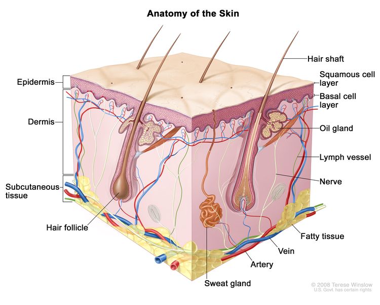
Lung Cancer Screening (PDQ®)–Patient Version

Lung Cancer Screening (PDQ®)–Patient Version

What Is Screening?

Screening is looking for cancer before a person has any symptoms. This can help find cancer at an early stage. When abnormal tissue or cancer is found early, it may be easier to treat. By the time symptoms appear, cancer may have begun to spread.

Scientists are trying to better understand which people are more likely to get certain types of cancer. They also study the things we do and the things around us to see if they cause cancer. This information helps doctors recommend who should be screened for cancer, which screening tests should be used, and how often the tests should be done.

It is important to remember that your doctor does not necessarily think you have cancer if he or she suggests a screening test. Screening tests are given when you have no cancer symptoms.

If a screening test result is abnormal, you may need to have more tests done to find out if you have cancer. These are called diagnostic tests.

General Information About Lung Cancer

Key Points

  • Lung cancer is a disease in which malignant (cancer) cells form in the tissues of the lung.
  • Lung cancer is the leading cause of cancer death in the United States.
  • Different factors increase or decrease the risk of lung cancer.

Lung cancer is a disease in which malignant (cancer) cells form in the tissues of the lung.

The lungs are a pair of cone-shaped breathing organs in the chest. The lungs bring oxygen into the body as you breathe in. They release carbon dioxide, a waste product of the body’s cells, as you breathe out. Each lung has sections called lobes. The left lung has two lobes. The right lung is slightly larger, and has three lobes. A thin membrane called the pleura surrounds the lungs. Two tubes called bronchi lead from the trachea (windpipe) to the right and left lungs. The bronchi are sometimes also involved in lung cancer. Tiny air sacs called alveoli and small tubes called bronchioles make up the inside of the lungs.

EnlargeRespiratory system anatomy; drawing shows the right lung with the upper, middle, and lower lobes, the left lung with the upper and lower lobes, and the trachea, bronchi, lymph nodes, and diaphragm. An inset shows the bronchioles, alveoli, artery, and vein.
Anatomy of the respiratory system showing the trachea, the right and left lungs and their lobes, and the bronchi. The lymph nodes and the diaphragm are also shown. Oxygen is inhaled into the lungs and passes through the alveoli (the tiny air sacs at the end of the bronchioles) and into the bloodstream (see inset), where it travels to the tissues throughout the body.

There are two main types of lung cancer: small cell lung cancer and non-small cell lung cancer.

Other PDQ summaries containing information related to lung cancer include:

Lung cancer is the leading cause of cancer death in the United States.

Lung cancer is the second most common type of non-skin cancer in the United States. Lung cancer is the leading cause of cancer death in men and in women.

Different factors increase or decrease the risk of lung cancer.

Anything that increases your chance of getting a disease is called a risk factor. Anything that decreases your chance of getting a disease is called a protective factor.

Tobacco smoking is the most important risk factor for lung cancer. Cigarette, cigar, and pipe smoking all increase the risk of lung cancer. Tobacco smoking causes about 9 out of 10 cases of lung cancer in men and about 8 out of 10 cases of lung cancer in women. The best way to prevent lung cancer is to not smoke.

For information about risk factors and protective factors for lung cancer, see Lung Cancer Prevention.

Lung Cancer Screening

Key Points

  • Tests are used to screen for different types of cancer when a person does not have symptoms.
  • Three screening tests have been studied to see if they decrease the risk of dying from lung cancer.
  • Screening with LDCT scans has been shown to decrease the risk of dying from lung cancer in heavy smokers.
  • Screening with chest x-rays and/or sputum cytology does not decrease the risk of dying from lung cancer.
  • Screening tests for lung cancer are being studied in clinical trials.

Tests are used to screen for different types of cancer when a person does not have symptoms.

Scientists study screening tests to find those with the fewest harms and most benefits. Cancer screening trials also are meant to show whether early detection (finding cancer before it causes symptoms) helps a person live longer or decreases a person’s chance of dying from the disease. For some types of cancer, the chance of recovery is better if the disease is found and treated at an early stage.

Three screening tests have been studied to see if they decrease the risk of dying from lung cancer.

The following screening tests have been studied to see if they decrease the risk of dying from lung cancer:

  • Low-dose computed tomography (LDCT): A procedure that uses low-dose radiation to make a series of very detailed pictures of areas inside the body using an x-ray machine that scans the body in a spiral path. This procedure is also called spiral scan or helical scan.
  • Chest x-ray: An x-ray of the organs and bones inside the chest. An x-ray is a type of energy beam that can go through the body and onto film, making a picture of areas inside the body.
  • Sputum cytology: Sputum cytology is a procedure in which a sample of sputum (mucus that is coughed up from the lungs) is viewed under a microscope to check for cancer cells.

Screening with LDCT scans has been shown to decrease the risk of dying from lung cancer in heavy smokers.

The National Lung Screening Trial studied people aged 55 years to 74 years who had smoked at least 1 pack of cigarettes per day for 30 years or more. Participants were either current smokers or former smokers who had quit within the last 15 years. The trial used chest x-rays or LDCT scans to check for signs of lung cancer.

Screening with LDCT once a year for three years was better than chest x-rays at finding early-stage lung cancer and decreased the risk of dying from lung cancer in current and former heavy smokers.

Current smokers whose LDCT scan result shows possible signs of cancer may be more likely to quit smoking.

Screening with LDCT can cause possible harms, including:

For more information about these possible harms, see the Risks of Lung Cancer Screening below.

A Guide is available for patients and doctors to learn more about the benefits and harms of screening for lung cancer.

Screening with chest x-rays and/or sputum cytology does not decrease the risk of dying from lung cancer.

Chest x-ray and sputum cytology are two screening tests that have been used to check for signs of lung cancer. Screening with chest x-ray, sputum cytology, or both of these tests does not decrease the risk of dying from lung cancer.

Screening tests for lung cancer are being studied in clinical trials.

Information about clinical trials supported by NCI can be found on NCI’s clinical trials search webpage. Clinical trials supported by other organizations can be found on the ClinicalTrials.gov website.

Risks of Lung Cancer Screening

Key Points

  • Screening tests have risks.
  • The risks of lung cancer screening tests include the following:
    • Finding lung cancer may not improve health or help you live longer.
    • False-negative test results can occur.
    • False-positive test results can occur.
    • Chest x-rays and CT scans expose the chest to radiation.
    • Talk to your doctor about your risk for lung cancer and your need for screening tests.

Screening tests have risks.

Decisions about screening tests can be difficult. Not all screening tests are helpful and most have risks. Before having any screening test, you may want to discuss the test with your doctor. It is important to know the risks of the test and whether it has been proven to reduce the risk of dying from cancer.

The risks of lung cancer screening tests include the following:

Finding lung cancer may not improve health or help you live longer.

Screening may not improve your health or help you live longer if you have lung cancer that has already spread to other places in your body.

When a screening test result leads to the diagnosis and treatment of a disease that may never have caused symptoms or become life-threatening, it is called overdiagnosis. It is not known if treatment of these cancers would help you live longer than if no treatment were given, and treatments for cancer may have serious side effects. Harms of treatment may happen more often in people who have medical problems caused by heavy or long-term smoking.

False-negative test results can occur.

Screening test results may appear to be normal even though lung cancer is present. A person who receives a false-negative test result (one that shows there is no cancer when there really is) may delay seeking medical care even if there are symptoms.

False-positive test results can occur.

Screening test results may appear to be abnormal even though no cancer is present. A false-positive test result (one that shows there is cancer when there really isn’t) can cause anxiety and is usually followed by more tests (such as biopsy), which also have risks. A biopsy to diagnose lung cancer can cause part of the lung to collapse. Sometimes surgery is needed to reinflate the lung. Harms of diagnostic tests may happen more often in patients who have medical problems caused by heavy or long-term smoking.

Chest x-rays and CT scans expose the chest to radiation.

Radiation exposure from chest x-rays and low-dose CT scans may increase the risk of cancer. Younger people and people at low risk for lung cancer are more likely to develop lung cancer caused by radiation exposure from screening than to be spared death from lung cancer.

Talk to your doctor about your risk for lung cancer and your need for screening tests.

Talk to your doctor or other health care provider about your risk for lung cancer, whether a screening test is right for you, and about the benefits and harms of the screening test. You should take part in the decision about whether a screening test is right for you. For more information, see Cancer Screening Overview.

About This PDQ Summary

About PDQ

Physician Data Query (PDQ) is the National Cancer Institute’s (NCI’s) comprehensive cancer information database. The PDQ database contains summaries of the latest published information on cancer prevention, detection, genetics, treatment, supportive care, and complementary and alternative medicine. Most summaries come in two versions. The health professional versions have detailed information written in technical language. The patient versions are written in easy-to-understand, nontechnical language. Both versions have cancer information that is accurate and up to date and most versions are also available in Spanish.

PDQ is a service of the NCI. The NCI is part of the National Institutes of Health (NIH). NIH is the federal government’s center of biomedical research. The PDQ summaries are based on an independent review of the medical literature. They are not policy statements of the NCI or the NIH.

Purpose of This Summary

This PDQ cancer information summary has current information about lung cancer screening. It is meant to inform and help patients, families, and caregivers. It does not give formal guidelines or recommendations for making decisions about health care.

Reviewers and Updates

Editorial Boards write the PDQ cancer information summaries and keep them up to date. These Boards are made up of experts in cancer treatment and other specialties related to cancer. The summaries are reviewed regularly and changes are made when there is new information. The date on each summary (“Updated”) is the date of the most recent change.

The information in this patient summary was taken from the health professional version, which is reviewed regularly and updated as needed, by the PDQ Screening and Prevention Editorial Board.

Clinical Trial Information

A clinical trial is a study to answer a scientific question, such as whether one treatment is better than another. Trials are based on past studies and what has been learned in the laboratory. Each trial answers certain scientific questions in order to find new and better ways to help cancer patients. During treatment clinical trials, information is collected about the effects of a new treatment and how well it works. If a clinical trial shows that a new treatment is better than one currently being used, the new treatment may become “standard.” Patients may want to think about taking part in a clinical trial. Some clinical trials are open only to patients who have not started treatment.

Clinical trials can be found online at NCI’s website. For more information, call the Cancer Information Service (CIS), NCI’s contact center, at 1-800-4-CANCER (1-800-422-6237).

Permission to Use This Summary

PDQ is a registered trademark. The content of PDQ documents can be used freely as text. It cannot be identified as an NCI PDQ cancer information summary unless the whole summary is shown and it is updated regularly. However, a user would be allowed to write a sentence such as “NCI’s PDQ cancer information summary about breast cancer prevention states the risks in the following way: [include excerpt from the summary].”

The best way to cite this PDQ summary is:

PDQ® Screening and Prevention Editorial Board. PDQ Lung Cancer Screening. Bethesda, MD: National Cancer Institute. Updated <MM/DD/YYYY>. Available at: /types/lung/patient/lung-screening-pdq. Accessed <MM/DD/YYYY>. [PMID: 26389428]

Images in this summary are used with permission of the author(s), artist, and/or publisher for use in the PDQ summaries only. If you want to use an image from a PDQ summary and you are not using the whole summary, you must get permission from the owner. It cannot be given by the National Cancer Institute. Information about using the images in this summary, along with many other images related to cancer can be found in Visuals Online. Visuals Online is a collection of more than 3,000 scientific images.

Disclaimer

The information in these summaries should not be used to make decisions about insurance reimbursement. More information on insurance coverage is available on Cancer.gov on the Managing Cancer Care page.

Contact Us

More information about contacting us or receiving help with the Cancer.gov website can be found on our Contact Us for Help page. Questions can also be submitted to Cancer.gov through the website’s E-mail Us.

Small Cell Lung Cancer Treatment (PDQ®)–Patient Version

Small Cell Lung Cancer Treatment (PDQ®)–Patient Version

General Information About Small Cell Lung Cancer

Key Points

  • Small cell lung cancer is a type of fast-growing cancer that forms in the tissues of the lung.
  • There are two main types of small cell lung cancer.
  • Smoking is the major risk factor for small cell lung cancer.
  • Signs and symptoms of small cell lung cancer include coughing and shortness of breath.
  • Tests and procedures that examine the lungs are used to diagnose and stage small cell lung cancer.
  • After small cell lung cancer has been diagnosed, tests are done to find out if cancer cells have spread within the chest or to other parts of the body.
  • Some people decide to get a second opinion.
  • Certain factors affect prognosis (chance of recovery) and treatment options.

Small cell lung cancer is a type of fast-growing cancer that forms in the tissues of the lung.

The lungs are a pair of cone-shaped breathing organs in the chest. The lungs bring oxygen into the body as you breathe in. They release carbon dioxide, a waste product of the body’s cells, as you breathe out. Each lung has sections called lobes. The left lung has two lobes. The right lung is slightly larger and has three lobes. A thin membrane called the pleura surrounds the lungs. Two tubes called bronchi lead from the trachea (windpipe) to the right and left lungs. Lung cancer may also form in the bronchi. Small tubes called bronchioles and tiny air sacs called alveoli make up the inside of the lungs.

EnlargeRespiratory system anatomy; drawing shows the right lung with the upper, middle, and lower lobes, the left lung with the upper and lower lobes, and the trachea, bronchi, lymph nodes, and diaphragm. An inset shows the bronchioles, alveoli, artery, and vein.
Anatomy of the respiratory system showing the trachea, the right and left lungs and their lobes, and the bronchi. The lymph nodes and the diaphragm are also shown. Oxygen is inhaled into the lungs and passes through the alveoli (the tiny air sacs at the end of the bronchioles) and into the bloodstream (see inset), where it travels to the tissues throughout the body.

There are two types of lung cancer: small cell lung cancer and non-small cell lung cancer. Small cell lung cancer is less common than non-small cell lung cancer.

There are two main types of small cell lung cancer.

The cancer cells of each type grow and spread in different ways. The types of small cell lung cancer are named for the kinds of cells found in the cancer and how the cells look when viewed under a microscope. The cancer cells of each type grow and spread in different ways:

  • Small cell carcinoma (also called oat cell cancer) is a type of lung cancer that can grow and spread quickly, often leading to the cancer spreading to other parts of the body early in the disease process. This is the most common type of small cell lung cancer.
  • Combined small cell carcinoma is a rare subtype of lung cancer that has characteristics of small cell lung cancer and non-small cell lung cancer in the same tumor.

Smoking is the major risk factor for small cell lung cancer.

Lung cancer is caused by certain changes to the way lung cells function, especially how they grow and divide into new cells. There are many risk factors for lung cancer, but many do not directly cause cancer. Instead, they increase the chance of DNA damage in cells that may lead to lung cancer. Learn more about how cancer develops at What Is Cancer?

A risk factor is anything that increases the chance of getting a disease. Some risk factors for lung cancer, like smoking, can be changed. However, risk factors also include things you cannot change, like your genetics, age, and family history. Learning about risk factors for lung cancer can help you make changes that might lower your risk of getting it.

Smoking tobacco now or in the past is the most important risk factor for lung cancer. Smoking cigarettes, pipes, or cigars increases the risk of lung cancer. The earlier in life a person starts smoking, the more often a person smokes, and the more years a person smokes, the greater the risk of lung cancer.

Other risk factors for lung cancer include:

Older age is the main risk factor for most cancers. The chance of getting cancer increases as you get older.

Having one or more of these risk factors does not necessarily mean you will get lung cancer. Many people with risk factors never develop lung cancer, whereas others with no known risk factors do. Talk with your doctor if you think you might be at increased risk.

When smoking is combined with other risk factors, the risk of lung cancer is increased.

Signs and symptoms of small cell lung cancer include coughing and shortness of breath.

These and other signs and symptoms may be caused by small cell lung cancer or by other conditions. Check with your doctor if you have:

  • chest discomfort or pain
  • a cough that doesn’t go away or gets worse over time
  • trouble breathing
  • wheezing
  • blood in sputum (mucus coughed up from the lungs)
  • hoarseness
  • trouble swallowing
  • loss of appetite
  • weight loss for no known reason
  • feeling very tired
  • swelling in the face and veins in the neck

Tests and procedures that examine the lungs are used to diagnose and stage small cell lung cancer.

In addition to asking about your personal and family health history and doing a physical exam, your doctor may perform the following tests and procedures:

  • Laboratory tests are medical procedures that test samples of tissue, blood, urine, or other substances in the body. These tests help to diagnose disease, plan and check treatment, or monitor the disease over time.
  • Chest x-ray is a type of radiation that can go through the body and make pictures of the organs and bones inside the chest.
    EnlargeChest x-ray; drawing shows a patient standing with their back to the x-ray machine. X-rays pass through the patient's body onto film or a computer and take pictures of the structures and organs inside the chest.
    A chest x-ray is used to take pictures of the structures and organs inside the chest. X-rays pass through the patient’s body onto film or a computer.
  • CT scan (CAT scan) uses a computer linked to an x-ray machine to make a series of detailed pictures of areas inside the body, such as the brain, chest, and abdomen. The pictures are taken from different angles and are used to create 3-D views of tissues and organs. A dye may be injected into a vein or swallowed to help the organs or tissues show up more clearly. This procedure is also called computed tomography, computerized tomography, or computerized axial tomography. Learn more about Computed Tomography (CT) Scans and Cancer.
  • Sputum cytology uses a microscope to check for cancer cells in the sputum (mucus coughed up from the lungs).
  • Biopsy is the removal of a sample of cells or tissue from the tumor so that a pathologist can view it under a microscope to check for cancer. The different ways a biopsy can be done include:
    • Fine-needle aspiration (FNA) biopsy of the lung is the removal of tissue or fluid from the lung, using a thin needle. A CT scan, ultrasound, or other imaging procedure is used to find the abnormal tissue or fluid in the lung. A small incision may be made in the skin where the biopsy needle is inserted into the abnormal tissue or fluid. A sample is removed with the needle and sent to the laboratory. A pathologist then views the sample under a microscope to look for cancer cells. A chest x-ray is done after the procedure to make sure no air is leaking from the lung into the chest.
      EnlargeFine-needle aspiration biopsy of the lung; drawing shows a patient lying on a table that slides through the computed tomography (CT) machine with an x-ray picture of a cross-section of the lung on a monitor above the patient. Drawing also shows a doctor using the x-ray picture to help place the biopsy needle through the chest wall and into the area of abnormal lung tissue. Inset shows a side view of the chest cavity and lungs with the biopsy needle inserted into the area of abnormal tissue.
      Fine-needle aspiration biopsy of the lung. The patient lies on a table that slides through the computed tomography (CT) machine, which takes x-ray pictures of the inside of the body. The x-ray pictures help the doctor see where the abnormal tissue is in the lung. A biopsy needle is inserted through the chest wall and into the area of abnormal lung tissue. A small piece of tissue is removed through the needle and checked under the microscope for signs of cancer.
    • Bronchoscopy is a procedure to look inside the trachea and large airways in the lung for abnormal areas. A bronchoscope is inserted through the nose or mouth into the trachea and lungs. A bronchoscope is a thin, tube-like instrument with a light and a lens for viewing. It may also have a tool to remove tissue samples, which are checked under a microscope for signs of cancer.
      EnlargeBronchoscopy; drawing shows a bronchoscope inserted through the mouth, trachea, and bronchus into the lung; lymph nodes along trachea and bronchi; and cancer in one lung. Inset shows patient lying on a table having a bronchoscopy.
      Bronchoscopy. A bronchoscope is inserted through the mouth, trachea, and major bronchi into the lung, to look for abnormal areas. A bronchoscope is a thin, tube-like instrument with a light and a lens for viewing. It may also have a cutting tool. Tissue samples may be taken to be checked under a microscope for signs of disease.
    • Thoracoscopy is a surgical procedure to look at the organs inside the chest to check for abnormal areas. An incision (cut) is made between two ribs, and a thoracoscope is inserted into the chest. A thoracoscope is a thin, tube-like instrument with a light and a lens for viewing. It may also have a tool to remove tissue or lymph node samples, which are checked under a microscope for signs of cancer. In some cases, this procedure is used to remove part of the esophagus or lung. If certain tissues, organs, or lymph nodes can’t be reached, a thoracotomy may be done. In this procedure, a larger incision is made between the ribs and the chest is opened.
    • Thoracentesis is the removal of fluid from the space between the lining of the chest and the lung, using a needle. A pathologist views the fluid under a microscope to look for cancer cells.
    • Mediastinoscopy is a surgical procedure to look at the organs, tissues, and lymph nodes between the lungs for abnormal areas. An incision (cut) is made at the top of the breastbone and a mediastinoscope is inserted into the chest. A mediastinoscope is a thin, tube-like instrument with a light and a lens for viewing. It may also have a tool to remove tissue or lymph node samples, which are checked under a microscope for signs of cancer.
  • Light and electron microscopy is a laboratory test in which cells in a sample of tissue are viewed under regular and high-powered microscopes to look for certain changes in the cells.
  • Immunohistochemistry uses antibodies to check for certain antigens (markers) in a sample of a patient’s cells or tissue. The antibodies are usually linked to an enzyme or fluorescent dye. After the antibodies bind to a specific antigen in the tissue sample, the enzyme or dye is activated, and the antigen can then be seen under a microscope. This type of test is used to help diagnose cancer and help tell one type of cancer from another type.

After small cell lung cancer has been diagnosed, tests are done to find out if cancer cells have spread within the chest or to other parts of the body.

The process used to find out if cancer has spread within the chest or to other parts of the body is called staging. The information gathered from the staging process determines the stage of the disease. It is important to know the stage in order to plan treatment. Some of the tests used to diagnose small cell lung cancer are also used to stage the disease.

Imaging tests that may be used in the staging process include:

  • MRI (magnetic resonance imaging) uses a magnet, radio waves, and a computer to make a series of detailed pictures of areas inside the brain, such as the brain. This procedure is also called nuclear magnetic resonance imaging (NMRI).
  • PET scan (positron emission tomography scan) uses a small amount of radioactive sugar (also called radioactive glucose) that is injected into a vein. The PET scanner rotates around the body and makes pictures of where sugar is being used by the body. Cancer cells show up brighter in the pictures because they are more active and take up more sugar than normal cells do. When this procedure is done at the same time as a CT scan, it is called a PET-CT scan.
  • Bone scan checks if there are rapidly dividing cells, such as cancer cells, in the bone. A very small amount of radioactive material is injected into a vein and travels through the bloodstream. The radioactive material collects in the areas of the bones with cancer and is detected by a scanner.

Some people decide to get a second opinion.

You may want to get a second opinion to confirm your cancer diagnosis and treatment plan. If you seek a second opinion, you will need to get medical test results and reports from the first doctor to share with the second doctor. The second doctor will review the pathology report, slides, and scans. They may agree with the first doctor, suggest changes or another treatment approach, or provide more information about your cancer.

To learn more about choosing a doctor and getting a second opinion, visit Finding Cancer Care. You can contact NCI’s Cancer Information Service via chat, email, or phone (both in English and Spanish) for help finding a doctor, hospital, or getting a second opinion. For questions you might want to ask at your appointments, visit Questions to Ask Your Doctor about Cancer.

Certain factors affect prognosis (chance of recovery) and treatment options.

The prognosis and treatment options depend on:

  • the stage of the cancer (whether it is in the chest cavity only or has spread to other places in the body)
  • the patient’s age, sex, and general health

For certain patients, prognosis also depends on whether the patient is treated with both chemotherapy and radiation.

For most people with small cell lung cancer, current treatments do not cure the cancer. If lung cancer is found, you may want to think about taking part in one of the many clinical trials being done to improve treatment. Clinical trials are taking place in most parts of the country for patients with all stages of small cell lung cancer. Information about ongoing clinical trials is available from the NCI website.

Stages of Small Cell Lung Cancer

Key Points

  • The following stages are used for small cell lung cancer:
    • Limited-stage small cell lung cancer
    • Extensive-stage small cell lung cancer
  • Small cell lung cancer can recur (come back) after it has been treated.

Cancer stage describes the extent of cancer in the body, such as the size of the tumor, whether it has spread, and how far it has spread from where it first formed. It is important to know the stage of small cell lung cancer to plan the best treatment.

Small cell lung cancer is usually classified into two stages due to its tendency to spread early.

The following stages are used for small cell lung cancer:

Limited-stage small cell lung cancer

In limited-stage, cancer is in the lung where it started and may have spread to the area between the lungs or to the lymph nodes above the collarbone.

Extensive-stage small cell lung cancer

In extensive-stage, cancer has spread beyond the lung or the area between the lungs or the lymph nodes above the collarbone to other places in the body.

Small cell lung cancer can recur (come back) after it has been treated.

Recurrent cancer is cancer that has recurred (come back) after it has been treated. If small cell lung cancer comes back, it may come back in the chest, central nervous system, or in other parts of the body. Tests will be done to help determine where the cancer has returned. The type of treatment for small cell lung cancer will depend on where it has come back.

Learn more in Recurrent Cancer: When Cancer Comes Back.

Treatment Option Overview

Key Points

  • There are different types of treatment for patients with small cell lung cancer.
  • The following types of treatment are used:
    • Surgery
    • Chemotherapy
    • Radiation therapy
    • Immunotherapy
    • Laser therapy
    • Endoscopic stent placement
  • New types of treatment are being tested in clinical trials.
  • Treatment for small cell lung cancer may cause side effects.
  • Follow-up care may be needed.

There are different types of treatment for patients with small cell lung cancer.

Different types of treatments are available for people with small cell lung cancer. You and your cancer care team will work together to decide your treatment plan, which may include more than one type of treatment. Many factors will be considered, such as the stage of the cancer, your overall health, and your preferences. Your plan will include information about your cancer, the goals of treatment, your treatment options and the possible side effects, and the expected length of treatment.

Talking with your cancer care team before treatment begins about what to expect will be helpful. You’ll want to learn what you need to do before treatment begins, how you’ll feel while going through it, and what kind of help you will need. To learn more, visit Questions to Ask Your Doctor about Treatment.

The following types of treatment are used:

Surgery

Surgery may be used if the cancer is found in one lung and in nearby lymph nodes only. Because this type of lung cancer is usually found in both lungs, surgery alone is not often used. During surgery, the doctor will also remove lymph nodes to find out if they have cancer in them. Sometimes, surgery may be used to remove a sample of lung tissue to find out the exact type of lung cancer.

After the doctor removes all the cancer that can be seen at the time of the surgery, some patients may be given chemotherapy or radiation therapy after surgery to kill any cancer cells that are left. Treatment given after the surgery, to lower the risk that the cancer will come back, is called adjuvant therapy.

Chemotherapy

Chemotherapy (also called chemo) uses drugs to stop the growth of cancer cells, either by killing the cells or by stopping them from dividing.

Chemotherapy for small cell lung cancer is usually systemic, meaning it is injected into a vein or given by mouth. When given this way, the drugs enter the bloodstream to reach cancer cells throughout the body.

Chemotherapy drugs used to treat small cell lung cancer may include:

Combinations of these chemotherapy drugs may be used. Other chemotherapy drugs not listed here may also be used.

Chemotherapy may also be combined with other kinds of treatment. For example, it may be combined with radiation therapy or immunotherapy drugs.

To learn more about how chemotherapy works, how it is given, common side effects, and more, visit Chemotherapy to Treat Cancer and Chemotherapy and You: Support for People With Cancer.

Radiation therapy

Radiation therapy is a cancer treatment that uses high-energy x-rays or other types of radiation to kill cancer cells or keep them from growing. External radiation therapy uses a machine outside the body to send radiation toward the area of the body with cancer. External radiation therapy is used to treat small cell lung cancer, and may also be used as palliative therapy to relieve symptoms and improve quality of life. Radiation therapy to the brain to lessen the risk that cancer will spread to the brain may also be given.

Learn more about External Beam Radiation Therapy for Cancer and Radiation Therapy Side Effects.

Immunotherapy

Immunotherapy helps a person’s immune system fight cancer. Your doctor may suggest biomarker tests to help predict your response to certain immunotherapy drugs. Learn more about Biomarker Testing for Cancer Treatment.

Immunotherapy drugs used to treat small cell lung cancer include:

Learn more about Immunotherapy to Treat Cancer.

Laser therapy

Laser therapy is a cancer treatment that uses a laser beam (a narrow beam of intense light) to kill cancer cells.

Endoscopic stent placement

An endoscope is a thin, tube-like instrument used to look at tissues inside the body. An endoscope has a light and a lens for viewing and may be used to place a stent in a body structure to keep the structure open. An endoscopic stent can be used to open an airway blocked by abnormal tissue.

New types of treatment are being tested in clinical trials.

For some people, joining a clinical trial may be an option. There are different types of clinical trials for people with cancer. For example, a treatment trial tests new treatments or new ways of using current treatments. Supportive care and palliative care trials look at ways to improve quality of life, especially for those who have side effects from cancer and its treatment.

You can use the clinical trial search to find NCI-supported cancer clinical trials accepting participants. The search allows you to filter trials based on the type of cancer, your age, and where the trials are being done. Clinical trials supported by other organizations can be found on the ClinicalTrials.gov website.

Learn more about clinical trials, including how to find and join one, at Clinical Trials Information for Patients and Caregivers.

Treatment for small cell lung cancer may cause side effects.

For information about side effects caused by treatment for cancer, visit our Side Effects page.

Follow-up care may be needed.

As you go through treatment, you will have follow-up tests or check-ups. Some tests that were done to diagnose or stage the cancer may be repeated to see how well the treatment is working. Decisions about whether to continue, change, or stop treatment may be based on the results of these tests.

Some of the tests will continue to be done from time to time after treatment has ended. The results of these tests can show if your condition has changed or if the cancer has recurred (come back).

Treatment of Limited-Stage Small Cell Lung Cancer

Treatment of limited-stage small cell lung cancer may include:

Learn more about these treatments in the Treatment Option Overview.

Use our clinical trial search to find NCI-supported cancer clinical trials that are accepting patients. You can search for trials based on the type of cancer, the age of the patient, and where the trials are being done. General information about clinical trials is also available.

Treatment of Extensive-Stage Small Cell Lung Cancer

Treatment of extensive-stage small cell lung cancer may include:

Learn more about these treatments in the Treatment Option Overview.

Use our clinical trial search to find NCI-supported cancer clinical trials that are accepting patients. You can search for trials based on the type of cancer, the age of the patient, and where the trials are being done. General information about clinical trials is also available.

Treatment of Recurrent Small Cell Lung Cancer

Treatment of recurrent small cell lung cancer may include:

Learn more about these treatments in the Treatment Option Overview.

Use our clinical trial search to find NCI-supported cancer clinical trials that are accepting patients. You can search for trials based on the type of cancer, the age of the patient, and where the trials are being done. General information about clinical trials is also available.

To Learn More About Small Cell Lung Cancer

About This PDQ Summary

About PDQ

Physician Data Query (PDQ) is the National Cancer Institute’s (NCI’s) comprehensive cancer information database. The PDQ database contains summaries of the latest published information on cancer prevention, detection, genetics, treatment, supportive care, and complementary and alternative medicine. Most summaries come in two versions. The health professional versions have detailed information written in technical language. The patient versions are written in easy-to-understand, nontechnical language. Both versions have cancer information that is accurate and up to date and most versions are also available in Spanish.

PDQ is a service of the NCI. The NCI is part of the National Institutes of Health (NIH). NIH is the federal government’s center of biomedical research. The PDQ summaries are based on an independent review of the medical literature. They are not policy statements of the NCI or the NIH.

Purpose of This Summary

This PDQ cancer information summary has current information about the treatment of small cell lung cancer. It is meant to inform and help patients, families, and caregivers. It does not give formal guidelines or recommendations for making decisions about health care.

Reviewers and Updates

Editorial Boards write the PDQ cancer information summaries and keep them up to date. These Boards are made up of experts in cancer treatment and other specialties related to cancer. The summaries are reviewed regularly and changes are made when there is new information. The date on each summary (“Updated”) is the date of the most recent change.

The information in this patient summary was taken from the health professional version, which is reviewed regularly and updated as needed, by the PDQ Adult Treatment Editorial Board.

Clinical Trial Information

A clinical trial is a study to answer a scientific question, such as whether one treatment is better than another. Trials are based on past studies and what has been learned in the laboratory. Each trial answers certain scientific questions in order to find new and better ways to help cancer patients. During treatment clinical trials, information is collected about the effects of a new treatment and how well it works. If a clinical trial shows that a new treatment is better than one currently being used, the new treatment may become “standard.” Patients may want to think about taking part in a clinical trial. Some clinical trials are open only to patients who have not started treatment.

Clinical trials can be found online at NCI’s website. For more information, call the Cancer Information Service (CIS), NCI’s contact center, at 1-800-4-CANCER (1-800-422-6237).

Permission to Use This Summary

PDQ is a registered trademark. The content of PDQ documents can be used freely as text. It cannot be identified as an NCI PDQ cancer information summary unless the whole summary is shown and it is updated regularly. However, a user would be allowed to write a sentence such as “NCI’s PDQ cancer information summary about breast cancer prevention states the risks in the following way: [include excerpt from the summary].”

The best way to cite this PDQ summary is:

PDQ® Adult Treatment Editorial Board. PDQ Small Cell Lung Cancer Treatment. Bethesda, MD: National Cancer Institute. Updated <MM/DD/YYYY>. Available at: /types/lung/patient/small-cell-lung-treatment-pdq. Accessed <MM/DD/YYYY>. [PMID: 26389478]

Images in this summary are used with permission of the author(s), artist, and/or publisher for use in the PDQ summaries only. If you want to use an image from a PDQ summary and you are not using the whole summary, you must get permission from the owner. It cannot be given by the National Cancer Institute. Information about using the images in this summary, along with many other images related to cancer can be found in Visuals Online. Visuals Online is a collection of more than 3,000 scientific images.

Disclaimer

The information in these summaries should not be used to make decisions about insurance reimbursement. More information on insurance coverage is available on Cancer.gov on the Managing Cancer Care page.

Contact Us

More information about contacting us or receiving help with the Cancer.gov website can be found on our Contact Us for Help page. Questions can also be submitted to Cancer.gov through the website’s E-mail Us.

Small Cell Lung Cancer Treatment (PDQ®)–Health Professional Version

Small Cell Lung Cancer Treatment (PDQ®)–Health Professional Version

General Information About Small Cell Lung Cancer (SCLC)

SCLC accounts for approximately 15% of bronchogenic carcinomas.

At the time of diagnosis, approximately 30% of patients with SCLC have tumors confined to the hemithorax of origin, mediastinum, or supraclavicular lymph nodes. These patients have limited-stage disease (LD).[1] Patients with tumors that have spread beyond the supraclavicular areas have extensive-stage disease (ED).

SCLC is more responsive to chemotherapy and radiation therapy than other cell types of lung cancer. However, a cure is difficult to achieve because SCLC has a greater tendency to be widely disseminated by the time of diagnosis.

Incidence and Mortality

The overall incidence and mortality rates of SCLC in the United States have decreased during the past few decades.[2]

Estimated new cases and deaths from lung cancer (SCLC and non-small cell lung cancer [NSCLC] combined) in the United States in 2025:[3]

  • New cases: 226,650.
  • Deaths: 124,730.

Risk Factors

Increasing age is the most important risk factor for most cancers. Other risk factors for lung cancer include:

  • History of or current tobacco use: cigarettes, pipes, and cigars.[4]
  • Exposure to cancer-causing substances in secondhand smoke.[5,6]
  • Occupational exposure to asbestos, arsenic, chromium, beryllium, nickel, and other agents.[7]
  • Radiation exposure from any of the following sources:
    • Radiation therapy to the breast or chest.[8]
    • Radon exposure in the home or workplace.[9]
    • Medical imaging tests, such as computed tomography (CT) scans.[10]
    • Atomic bomb radiation.[11]
  • Living in an area with air pollution.[1214]
  • Family history of lung cancer.[15]
  • HIV infection.[16]
  • Beta carotene supplements in heavy smokers.[17,18]

Clinical Features

Lung cancer may present with symptoms or be found incidentally on chest imaging. Symptoms and signs may result from the location of the primary local invasion or compression of adjacent thoracic structures, distant metastases, or paraneoplastic phenomena. The most common symptoms at presentation are worsening cough and dyspnea. Other presenting symptoms include:

  • Chest pain.
  • Hoarseness.
  • Malaise.
  • Anorexia.
  • Weight loss.
  • Hemoptysis.

Symptoms may result from local invasion or compression of adjacent thoracic structures, such as compression involving the esophagus causing dysphagia, compression involving the laryngeal nerves causing hoarseness, or compression involving the superior vena cava causing facial edema and distension of the superficial veins of the head and neck. Symptoms from distant metastases may also be present and include neurological defects or personality changes from brain metastases and pain from bone metastases.

Infrequently, patients with SCLC may present with symptoms and signs of one of the following paraneoplastic syndromes:

  • Inappropriate antidiuretic hormone secretion.
  • Cushing syndrome from secretion of adrenocorticotropic hormone.
  • Paraneoplastic cerebellar degeneration.
  • Lambert-Eaton myasthenic syndrome.[2]

Physical examination may identify enlarged supraclavicular lymphadenopathy, pleural effusion or lobar collapse, unresolved pneumonia, or signs of associated disease such as chronic obstructive pulmonary disease.

Diagnosis

Treatment options for patients are determined by histology, stage, and general health and comorbidities of the patient. Investigations of patients with suspected SCLC focus on confirming the diagnosis and determining the extent of the disease.

The procedures used to determine the presence of cancer include:

  • History.
  • Physical examination.
  • Routine laboratory evaluations.
  • Chest x-ray.
  • Chest CT scan with infusion of contrast material.
  • Biopsy.

Before a patient begins lung cancer treatment, an experienced lung cancer pathologist must review the pathological material. This is critical because SCLC, which responds well to chemotherapy and is generally not treated surgically, can be confused on microscopic examination with NSCLC.[19] Immunohistochemistry and electron microscopy are invaluable techniques for diagnosis and subclassification, but most lung tumors can be classified by light microscopic criteria.

For more information, see the Staging Evaluation section.

Prognosis and Survival

Regardless of stage, the prognosis for patients with SCLC is unsatisfactory despite improvements in diagnosis and therapy during the past 25 years. Without treatment, SCLC has the most aggressive clinical course of any type of pulmonary tumor, with a median survival from diagnosis of only 2 to 4 months. About 10% of people with SCLC remain free of disease during the 2 years from the start of therapy, which is the time period during which most relapses occur. However, even these patients are at risk of dying of lung cancer (both small and non-small cell types).[20] The overall survival rate at 5 years is 5% to 10%.[1,2022]

An important prognostic factor for SCLC is the extent of disease. Patients with LD have a better prognosis than patients with ED. For patients with LD, the median survival is 16 to 24 months and the 5-year survival rates is 14% with current forms of treatment.[1,21,23,24] Patients diagnosed with LD who smoke should be encouraged to stop smoking before undergoing combined-modality therapy because continued smoking may compromise survival.[25]

Patients with LD have improved long-term survival with combined-modality therapy.[24,26][Level of evidence A1] Although long-term survivors have been reported among patients who received either surgery or chemotherapy alone, chemotherapy combined with thoracic radiation therapy (TRT) is considered the standard of care.[27] Adding TRT increases absolute survival by approximately 5% over chemotherapy alone.[26,28] Multiple trials and meta-analyses have evaluated the optimal timing of TRT relative to chemotherapy, with the weight of evidence suggesting a small benefit to early TRT.[1,29,30][Level of evidence A1]

In patients with ED, the median survival 6 to 12 months with currently available therapy, but long-term disease-free survival is rare.

Prophylactic cranial radiation prevents central nervous system recurrence and can improve survival in patients with good performance status who have had a complete response or a very good partial response to chemoradiation in LD or chemotherapy in ED.[31,32][Level of evidence A1]

Thoracic radiation may also improve long-term outcomes for these patients.[33]

All patients with this type of cancer may appropriately be considered for inclusion in clinical trials at the time of diagnosis. Information about ongoing clinical trials is available from the NCI website.

References
  1. Murray N, Coy P, Pater JL, et al.: Importance of timing for thoracic irradiation in the combined modality treatment of limited-stage small-cell lung cancer. The National Cancer Institute of Canada Clinical Trials Group. J Clin Oncol 11 (2): 336-44, 1993. [PUBMED Abstract]
  2. Govindan R, Page N, Morgensztern D, et al.: Changing epidemiology of small-cell lung cancer in the United States over the last 30 years: analysis of the surveillance, epidemiologic, and end results database. J Clin Oncol 24 (28): 4539-44, 2006. [PUBMED Abstract]
  3. American Cancer Society: Cancer Facts and Figures 2025. American Cancer Society, 2025. Available online. Last accessed January 16, 2025.
  4. Alberg AJ, Ford JG, Samet JM, et al.: Epidemiology of lung cancer: ACCP evidence-based clinical practice guidelines (2nd edition). Chest 132 (3 Suppl): 29S-55S, 2007. [PUBMED Abstract]
  5. Tulunay OE, Hecht SS, Carmella SG, et al.: Urinary metabolites of a tobacco-specific lung carcinogen in nonsmoking hospitality workers. Cancer Epidemiol Biomarkers Prev 14 (5): 1283-6, 2005. [PUBMED Abstract]
  6. Anderson KE, Kliris J, Murphy L, et al.: Metabolites of a tobacco-specific lung carcinogen in nonsmoking casino patrons. Cancer Epidemiol Biomarkers Prev 12 (12): 1544-6, 2003. [PUBMED Abstract]
  7. Straif K, Benbrahim-Tallaa L, Baan R, et al.: A review of human carcinogens–part C: metals, arsenic, dusts, and fibres. Lancet Oncol 10 (5): 453-4, 2009. [PUBMED Abstract]
  8. Friedman DL, Whitton J, Leisenring W, et al.: Subsequent neoplasms in 5-year survivors of childhood cancer: the Childhood Cancer Survivor Study. J Natl Cancer Inst 102 (14): 1083-95, 2010. [PUBMED Abstract]
  9. Gray A, Read S, McGale P, et al.: Lung cancer deaths from indoor radon and the cost effectiveness and potential of policies to reduce them. BMJ 338: a3110, 2009. [PUBMED Abstract]
  10. Berrington de González A, Kim KP, Berg CD: Low-dose lung computed tomography screening before age 55: estimates of the mortality reduction required to outweigh the radiation-induced cancer risk. J Med Screen 15 (3): 153-8, 2008. [PUBMED Abstract]
  11. Shimizu Y, Kato H, Schull WJ: Studies of the mortality of A-bomb survivors. 9. Mortality, 1950-1985: Part 2. Cancer mortality based on the recently revised doses (DS86). Radiat Res 121 (2): 120-41, 1990. [PUBMED Abstract]
  12. Katanoda K, Sobue T, Satoh H, et al.: An association between long-term exposure to ambient air pollution and mortality from lung cancer and respiratory diseases in Japan. J Epidemiol 21 (2): 132-43, 2011. [PUBMED Abstract]
  13. Cao J, Yang C, Li J, et al.: Association between long-term exposure to outdoor air pollution and mortality in China: a cohort study. J Hazard Mater 186 (2-3): 1594-600, 2011. [PUBMED Abstract]
  14. Hales S, Blakely T, Woodward A: Air pollution and mortality in New Zealand: cohort study. J Epidemiol Community Health 66 (5): 468-73, 2012. [PUBMED Abstract]
  15. Lissowska J, Foretova L, Dabek J, et al.: Family history and lung cancer risk: international multicentre case-control study in Eastern and Central Europe and meta-analyses. Cancer Causes Control 21 (7): 1091-104, 2010. [PUBMED Abstract]
  16. Shiels MS, Cole SR, Kirk GD, et al.: A meta-analysis of the incidence of non-AIDS cancers in HIV-infected individuals. J Acquir Immune Defic Syndr 52 (5): 611-22, 2009. [PUBMED Abstract]
  17. The effect of vitamin E and beta carotene on the incidence of lung cancer and other cancers in male smokers. The Alpha-Tocopherol, Beta Carotene Cancer Prevention Study Group. N Engl J Med 330 (15): 1029-35, 1994. [PUBMED Abstract]
  18. Omenn GS, Goodman GE, Thornquist MD, et al.: Effects of a combination of beta carotene and vitamin A on lung cancer and cardiovascular disease. N Engl J Med 334 (18): 1150-5, 1996. [PUBMED Abstract]
  19. Travis WD, Colby TV, Corrin B, et al.: Histological typing of lung and pleural tumours. 3rd ed. Springer-Verlag, 1999.
  20. Johnson BE, Grayson J, Makuch RW, et al.: Ten-year survival of patients with small-cell lung cancer treated with combination chemotherapy with or without irradiation. J Clin Oncol 8 (3): 396-401, 1990. [PUBMED Abstract]
  21. Fry WA, Menck HR, Winchester DP: The National Cancer Data Base report on lung cancer. Cancer 77 (9): 1947-55, 1996. [PUBMED Abstract]
  22. Lassen U, Osterlind K, Hansen M, et al.: Long-term survival in small-cell lung cancer: posttreatment characteristics in patients surviving 5 to 18+ years–an analysis of 1,714 consecutive patients. J Clin Oncol 13 (5): 1215-20, 1995. [PUBMED Abstract]
  23. Turrisi AT, Kim K, Blum R, et al.: Twice-daily compared with once-daily thoracic radiotherapy in limited small-cell lung cancer treated concurrently with cisplatin and etoposide. N Engl J Med 340 (4): 265-71, 1999. [PUBMED Abstract]
  24. Jänne PA, Freidlin B, Saxman S, et al.: Twenty-five years of clinical research for patients with limited-stage small cell lung carcinoma in North America. Cancer 95 (7): 1528-38, 2002. [PUBMED Abstract]
  25. Videtic GM, Stitt LW, Dar AR, et al.: Continued cigarette smoking by patients receiving concurrent chemoradiotherapy for limited-stage small-cell lung cancer is associated with decreased survival. J Clin Oncol 21 (8): 1544-9, 2003. [PUBMED Abstract]
  26. Pignon JP, Arriagada R, Ihde DC, et al.: A meta-analysis of thoracic radiotherapy for small-cell lung cancer. N Engl J Med 327 (23): 1618-24, 1992. [PUBMED Abstract]
  27. Chandra V, Allen MS, Nichols FC, et al.: The role of pulmonary resection in small cell lung cancer. Mayo Clin Proc 81 (5): 619-24, 2006. [PUBMED Abstract]
  28. Warde P, Payne D: Does thoracic irradiation improve survival and local control in limited-stage small-cell carcinoma of the lung? A meta-analysis. J Clin Oncol 10 (6): 890-5, 1992. [PUBMED Abstract]
  29. Perry MC, Eaton WL, Propert KJ, et al.: Chemotherapy with or without radiation therapy in limited small-cell carcinoma of the lung. N Engl J Med 316 (15): 912-8, 1987. [PUBMED Abstract]
  30. Takada M, Fukuoka M, Kawahara M, et al.: Phase III study of concurrent versus sequential thoracic radiotherapy in combination with cisplatin and etoposide for limited-stage small-cell lung cancer: results of the Japan Clinical Oncology Group Study 9104. J Clin Oncol 20 (14): 3054-60, 2002. [PUBMED Abstract]
  31. Aupérin A, Arriagada R, Pignon JP, et al.: Prophylactic cranial irradiation for patients with small-cell lung cancer in complete remission. Prophylactic Cranial Irradiation Overview Collaborative Group. N Engl J Med 341 (7): 476-84, 1999. [PUBMED Abstract]
  32. Slotman B, Faivre-Finn C, Kramer G, et al.: Prophylactic cranial irradiation in extensive small-cell lung cancer. N Engl J Med 357 (7): 664-72, 2007. [PUBMED Abstract]
  33. Slotman BJ, van Tinteren H, Praag JO, et al.: Use of thoracic radiotherapy for extensive stage small-cell lung cancer: a phase 3 randomised controlled trial. Lancet 385 (9962): 36-42, 2015. [PUBMED Abstract]

Cellular Classification of SCLC

Before initiating treatment for a patient with small cell lung cancer (SCLC), an experienced lung cancer pathologist should review the pathological material.

Pathological Classification

The current classification of subtypes of SCLC includes:[1]

  • Small cell carcinoma.
  • Combined small cell carcinoma (i.e., SCLC combined with additional components from any of the non-small cell lung carcinoma histological types).

SCLC arising from neuroendocrine cells forms one extreme of the spectrum of neuroendocrine carcinomas of the lung.

Neuroendocrine tumors include:

  • Low-grade typical carcinoid.
  • Intermediate-grade atypical carcinoid.
  • High-grade neuroendocrine tumors, including large-cell neuroendocrine carcinoma (LCNEC) and SCLC.

Because of differences in clinical behavior, therapy, and epidemiology, these tumors are classified separately in the World Health Organization (WHO) revised classification. The variant form of SCLC called mixed small cell/large cell carcinoma was not retained in the revised WHO classification. Instead, SCLC is now divided into SCLC and combined SCLC.[2] No minimum percentage of the additional component is required for a combined SCLC diagnosis with the exception of mixed LCNEC and SCLC, in which a 10% minimum LCNEC component is required.[3]

SCLC presents as a proliferation of small cells with the following morphological features:[4]

  • Scant cytoplasm.
  • Ill-defined borders.
  • Finely granular salt and pepper chromatin.
  • Absent or inconspicuous nucleoli.
  • Frequent nuclear molding.
  • A high mitotic count.

Nearly all SCLC are immunoreactive for keratin, thyroid transcription factor 1, and epithelial membrane antigen. Neuroendocrine and neural differentiation result in the expression of dopa decarboxylase, calcitonin, neuron-specific enolase, chromogranin A, CD56 (also known as nucleosomal histone kinase 1 or neural-cell adhesion molecule), gastrin-releasing peptide, and insulin-like growth factor 1. One or more markers of neuroendocrine differentiation can be found in approximately 75% of SCLC.[5]

Although preinvasive and in situ malignant changes are frequently found in patients with non-small cell lung cancer, these findings are rare in patients with SCLC.[6]

References
  1. Travis WD, Colby TV, Corrin B, et al.: Histological typing of lung and pleural tumours. 3rd ed. Springer-Verlag, 1999.
  2. Lei Y, Feng H, Qiang H, et al.: Clinical characteristics and prognostic factors of surgically resected combined small cell lung cancer: a retrospective study. Lung Cancer 146: 244-251, 2020. [PUBMED Abstract]
  3. Simbolo M, Centonze G, Ali G, et al.: Integrative molecular analysis of combined small-cell lung carcinomas identifies major subtypes with different therapeutic opportunities. ESMO Open 7 (1): 100308, 2022. [PUBMED Abstract]
  4. Brambilla E, Travis WD, Colby TV, et al.: The new World Health Organization classification of lung tumours. Eur Respir J 18 (6): 1059-68, 2001. [PUBMED Abstract]
  5. Guinee DG, Fishback NF, Koss MN, et al.: The spectrum of immunohistochemical staining of small-cell lung carcinoma in specimens from transbronchial and open-lung biopsies. Am J Clin Pathol 102 (4): 406-14, 1994. [PUBMED Abstract]
  6. Kumar V, Abbas A, Fausto N, eds.: Robins and Cotran Pathologic Basis of Disease. 7th ed. Elsevier Inc, 2005.

Stage Information for SCLC

Staging Systems

Several staging systems have been proposed for small cell lung cancer (SCLC).

  • American Joint Committee on Cancer (AJCC) Tumor, Node, and Metastasis (TNM).[1] The 8th edition of the AJCC Cancer Staging Manual recommends the use of the TNM to classify SCLC. For more information, see the AJCC Stage Groupings and TNM Definitions section in Non-Small Cell Lung Cancer Treatment.
  • Veterans Administration Lung Study Group (VALG).[2]
  • International Association for the Study of Lung Cancer (IASLC).[3]

Limited-Stage Disease

No universally accepted definition of this term is available. Limited-stage disease (LD) SCLC is confined to the hemithorax of origin, mediastinum, or supraclavicular nodes, which can be encompassed within a tolerable radiation therapy port.

Patients with pleural effusion, massive pulmonary tumor, and contralateral supraclavicular nodes have been both included in and excluded from LD by various groups.

Extensive-Stage Disease

Extensive-stage disease (ED) SCLC has spread beyond the supraclavicular areas and is too widespread to be included within the definition of LD. Patients with distant metastases (M1) are always considered to have ED.[3,4]

IASLC-AJCC TNM Staging System

The AJCC TNM defines LD as any T, except for T3–4, due to multiple lung nodules that do not fit in a tolerable radiation field, any N, and M0.[1] This corresponds to TNM stages I to IIIB. Extensive disease is TNM stage IV with distant metastases (M1), including malignant pleural effusions.[3,4] For more information, see the AJCC Stage Groupings and TNM Definitions section in Non-Small Cell Lung Cancer Treatment.

The IASLC conducted an analysis of clinical TNM staging for SCLC using the sixth edition of the AJCC TNM staging system for lung cancer. Survival rates for patients with clinical stages I and II disease are significantly different from those for patients with stage III disease with N2 or N3 involvement.[3] Patients with pleural effusion have an intermediate prognosis between LD and ED with hematogenous metastases and will be classified as having M1 disease (or ED). Application of the TNM system will not change how patients are managed; however, the analysis suggests that, in the context of clinical trials in LD, accurate TNM staging and stratification may be important.[3]

Staging Evaluation

Staging procedures for SCLC are important to distinguish patients with disease limited to their thorax from those with distant metastases. At the time of initial diagnosis, approximately two-thirds of patients with SCLC have clinical evidence of metastases; most of the remaining patients have clinical evidence of extensive nodal involvement in the hilar, mediastinal, and sometimes supraclavicular regions.

Determining the stage of cancer allows an assessment of prognosis and a determination of treatment, particularly when chest radiation therapy or surgical excision is added to chemotherapy for patients with LD. If ED is confirmed, further evaluation should be individualized according to the signs and symptoms unique to the individual patient. Standard staging procedures include:

  • A thorough physical examination.
  • Routine blood counts and serum chemistries.
  • Chest and upper abdominal computed tomography (CT) scanning.
  • A radionuclide bone scan.
  • A brain magnetic resonance imaging scan or CT scan.
  • Bone marrow aspirate or biopsy in selected patients in which treatment would change based on the results.

The role of positron emission tomography (PET) is still under study. SCLC is fluorine F 18-fludeoxyglucose (18F-FDG) avid at the primary site and at metastatic sites. PET may be used in staging patients with SCLC who are potential candidates for the addition of thoracic radiation therapy to chemotherapy, as PET may lead to upstaging or downstaging of patients and to alteration of radiation fields resulting from the identification of additional sites of nodal metastases.

Evidence (18F-FDG PET):

  1. In a study of 120 patients with LD SCLC or ED SCLC, FDG-PET led to restaging of ten patients to a higher stage and three patients to a lower stage.[5] FDG-PET was more sensitive and specific than CT scans for nonbrain distant metastases.
  2. In a small series of 24 patients with LD by conventional staging, two patients were restaged to ED.[2] Unsuspected nodal metastases were documented in 25% of patients, which altered the radiation plan in these patients. At this time, sensitivity, specificity, and positive- or negative-predictive value of PET scanning and its enhancement of staging accuracy are uncertain.
References
  1. Lung. In: Amin MB, Edge SB, Greene FL, et al., eds.: AJCC Cancer Staging Manual. 8th ed. Springer; 2017, pp. 431–56.
  2. Bradley JD, Dehdashti F, Mintun MA, et al.: Positron emission tomography in limited-stage small-cell lung cancer: a prospective study. J Clin Oncol 22 (16): 3248-54, 2004. [PUBMED Abstract]
  3. Shepherd FA, Crowley J, Van Houtte P, et al.: The International Association for the Study of Lung Cancer lung cancer staging project: proposals regarding the clinical staging of small cell lung cancer in the forthcoming (seventh) edition of the tumor, node, metastasis classification for lung cancer. J Thorac Oncol 2 (12): 1067-77, 2007. [PUBMED Abstract]
  4. Ihde D, Souhami B, Comis R, et al.: Small cell lung cancer. Lung Cancer 17 (Suppl 1): S19-21, 1997. [PUBMED Abstract]
  5. Brink I, Schumacher T, Mix M, et al.: Impact of [18F]FDG-PET on the primary staging of small-cell lung cancer. Eur J Nucl Med Mol Imaging 31 (12): 1614-20, 2004. [PUBMED Abstract]

Treatment Option Overview for SCLC

Chemotherapy and radiation therapy have been shown to improve survival for patients with small cell lung cancer (SCLC).

Chemotherapy

Chemotherapy improves the survival of patients with limited-stage disease (LD) or extensive-stage disease (ED), but it is curative in only a few patients.[1,2] Because patients with SCLC tend to develop distant metastases, localized forms of treatment, such as surgical resection or radiation therapy, rarely produce long-term survival.[3] Incorporating current chemotherapy regimens into the treatment program prolongs survival, with at least a fourfold to fivefold improvement in median survival compared with patients who are given no therapy.

The combination of platinum and etoposide is the most widely used standard chemotherapeutic regimen.[46][Level of evidence A1] No consistent survival benefit has resulted from platinum versus nonplatinum combinations, increased dose intensity or dose density, altered mode of administration (e.g., alternating or sequential administration) of various chemotherapeutic agents, or maintenance chemotherapy.[712][Level of evidence A1]

Radiation Therapy

SCLC is highly radiosensitive and thoracic radiation therapy improves survival of patients with LD and ED tumors.[1316][Level of evidence A1] Prophylactic cranial irradiation prevents central nervous system recurrence and may improve the long-term survival of patients with good performance status who have responded to chemoradiation therapy.[1719][Level of evidence A1] This type of irradiation also offers palliation of symptomatic metastatic disease.

Treatment options for patients with LD, ED, or recurrent SCLC are summarized in Table 1.

Table 1. Treatment Options for Patients With SCLC
Stage Treatment Options
ED = extensive-stage disease; LD = limited-stage disease.
LD Chemotherapy and radiation therapy
Adjuvant treatment after chemoradiation therapy
Combination chemotherapy alone
Surgery followed by chemotherapy or chemoradiation therapy
Prophylactic cranial irradiation
Clinical trials evaluating new drug regimens, surgical resection of the primary tumor, or new radiation therapy schedules and techniques (e.g., timing, three-dimensional treatment planning, and dose fractionation)
ED Immune checkpoint modulation and combination chemotherapy
Combination chemotherapy
Radiation therapy
Clinical trials evaluating new drug regimens or alternative drug doses and schedules
Recurrent disease Chemotherapy
Immunotherapy
Immune checkpoint modulation
Palliative therapy
Phase I and II clinical trials evaluating new drugs

Despite treatment advances, most patients with SCLC die of their tumor even with the best available therapy. Most of the improvements in survival of patients with SCLC are attributable to clinical trials that have attempted to improve on the best available and most accepted therapy. Patient entry into such studies is highly desirable.

Information about ongoing clinical trials is available from the NCI website.

References
  1. Comis RL, Friedland DM, Good BC: Small-cell lung cancer: a perspective on the past and a preview of the future. Oncology (Huntingt) 12 (1 Suppl 2): 44-50, 1998. [PUBMED Abstract]
  2. Agra Y, Pelayo M, Sacristan M, et al.: Chemotherapy versus best supportive care for extensive small cell lung cancer. Cochrane Database Syst Rev (4): CD001990, 2003. [PUBMED Abstract]
  3. Prasad US, Naylor AR, Walker WS, et al.: Long term survival after pulmonary resection for small cell carcinoma of the lung. Thorax 44 (10): 784-7, 1989. [PUBMED Abstract]
  4. Johnson BE, Grayson J, Makuch RW, et al.: Ten-year survival of patients with small-cell lung cancer treated with combination chemotherapy with or without irradiation. J Clin Oncol 8 (3): 396-401, 1990. [PUBMED Abstract]
  5. Lassen U, Osterlind K, Hansen M, et al.: Long-term survival in small-cell lung cancer: posttreatment characteristics in patients surviving 5 to 18+ years–an analysis of 1,714 consecutive patients. J Clin Oncol 13 (5): 1215-20, 1995. [PUBMED Abstract]
  6. Fry WA, Menck HR, Winchester DP: The National Cancer Data Base report on lung cancer. Cancer 77 (9): 1947-55, 1996. [PUBMED Abstract]
  7. Ihde DC, Mulshine JL, Kramer BS, et al.: Prospective randomized comparison of high-dose and standard-dose etoposide and cisplatin chemotherapy in patients with extensive-stage small-cell lung cancer. J Clin Oncol 12 (10): 2022-34, 1994. [PUBMED Abstract]
  8. Arriagada R, Le Chevalier T, Pignon JP, et al.: Initial chemotherapeutic doses and survival in patients with limited small-cell lung cancer. N Engl J Med 329 (25): 1848-52, 1993. [PUBMED Abstract]
  9. Klasa RJ, Murray N, Coldman AJ: Dose-intensity meta-analysis of chemotherapy regimens in small-cell carcinoma of the lung. J Clin Oncol 9 (3): 499-508, 1991. [PUBMED Abstract]
  10. Elias AD, Ayash L, Frei E, et al.: Intensive combined modality therapy for limited-stage small-cell lung cancer. J Natl Cancer Inst 85 (7): 559-66, 1993. [PUBMED Abstract]
  11. Murray N, Livingston RB, Shepherd FA, et al.: Randomized study of CODE versus alternating CAV/EP for extensive-stage small-cell lung cancer: an Intergroup Study of the National Cancer Institute of Canada Clinical Trials Group and the Southwest Oncology Group. J Clin Oncol 17 (8): 2300-8, 1999. [PUBMED Abstract]
  12. Amarasena IU, Walters JA, Wood-Baker R, et al.: Platinum versus non-platinum chemotherapy regimens for small cell lung cancer. Cochrane Database Syst Rev (4): CD006849, 2008. [PUBMED Abstract]
  13. Pignon JP, Arriagada R, Ihde DC, et al.: A meta-analysis of thoracic radiotherapy for small-cell lung cancer. N Engl J Med 327 (23): 1618-24, 1992. [PUBMED Abstract]
  14. Warde P, Payne D: Does thoracic irradiation improve survival and local control in limited-stage small-cell carcinoma of the lung? A meta-analysis. J Clin Oncol 10 (6): 890-5, 1992. [PUBMED Abstract]
  15. Murray N, Coy P, Pater JL, et al.: Importance of timing for thoracic irradiation in the combined modality treatment of limited-stage small-cell lung cancer. The National Cancer Institute of Canada Clinical Trials Group. J Clin Oncol 11 (2): 336-44, 1993. [PUBMED Abstract]
  16. Slotman BJ, van Tinteren H, Praag JO, et al.: Use of thoracic radiotherapy for extensive stage small-cell lung cancer: a phase 3 randomised controlled trial. Lancet 385 (9962): 36-42, 2015. [PUBMED Abstract]
  17. Turrisi AT, Glover DJ: Thoracic radiotherapy variables: influence on local control in small cell lung cancer limited disease. Int J Radiat Oncol Biol Phys 19 (6): 1473-9, 1990. [PUBMED Abstract]
  18. Aupérin A, Arriagada R, Pignon JP, et al.: Prophylactic cranial irradiation for patients with small-cell lung cancer in complete remission. Prophylactic Cranial Irradiation Overview Collaborative Group. N Engl J Med 341 (7): 476-84, 1999. [PUBMED Abstract]
  19. Slotman B, Faivre-Finn C, Kramer G, et al.: Prophylactic cranial irradiation in extensive small-cell lung cancer. N Engl J Med 357 (7): 664-72, 2007. [PUBMED Abstract]

Treatment of Limited-Stage SCLC

Treatment Options for Patients With Limited-Stage SCLC

Treatment options for patients with limited-stage small cell lung cancer (SCLC) include:

  1. Chemotherapy and radiation therapy.
  2. Adjuvant treatment after chemoradiation therapy.
  3. Combination chemotherapy alone.
  4. Surgery followed by chemotherapy or chemoradiation therapy.
  5. Prophylactic cranial irradiation (PCI).
  6. Clinical trials evaluating new drug regimens, surgical resection of the primary tumor, or new radiation therapy schedules and techniques (e.g., timing, three-dimensional treatment planning, and dose fractionation).

Chemotherapy and radiation therapy

Combined-modality treatment with etoposide and cisplatin with thoracic radiation therapy (TRT) is the most widely used treatment for patients with limited-stage disease (LD) SCLC.

Evidence (combined-modality treatment):

  1. Survival. The following results have been reported in clinical trials:
    1. Mature results of prospective randomized trials suggest that combined-modality therapy produces a modest but significant improvement in survival of 5% at 3 years compared with chemotherapy alone.[13][Level of evidence A1]
    2. Clinical trials have consistently achieved median survivals of 18 to 24 months and 2-year survival rates of 40% to 50%, with a treatment-related mortality rate of less than 3%.[37][Level of evidence A1]
    3. No consistent survival benefit has resulted from the following treatment approaches:[816]
      • Increased dose intensity.
      • Increased dose density.
      • Administration of additional drugs or other (non–etoposide-containing) platinum-based combination regimens.
      • Altered modes of administration of various chemotherapeutic agents.
      • Maintenance chemotherapy.
  2. Length of treatment. The optimal duration of chemotherapy for patients with LD SCLC is not clearly defined, but no improvement exists in survival after the duration of drug administration exceeds 3 to 6 months. The preponderance of evidence available from randomized trials indicates that maintenance chemotherapy does not prolong survival for patients with LD SCLC.[815][Level of evidence A1]
  3. Dose and timing. The optimal dose and timing of TRT remain controversial.
    1. Multiple clinical trials and meta-analyses addressing the timing of TRT have been published, with the weight of evidence suggesting a small benefit to early TRT (i.e., TRT administered during the first or second cycle of chemotherapy administration).[36,8,9,15,1720][Level of evidence A1]
    2. The amount of time from start to completion of TRT in LD SCLC may also affect overall survival (OS). In an analysis of four trials, the completion of therapy in less than 30 days was associated with an improved 5-year survival rate (relative risk, 0.62; 95% confidence interval [CI], 0.49–0.80; P = .0003).[20][Level of evidence A1]
    3. Both once-daily and twice-daily chest radiation schedules have been used in regimens with etoposide and cisplatin.
      • One randomized study showed a modest survival advantage in favor of twice-daily radiation therapy given for 3 weeks, compared with once-daily radiation therapy to 45 Gy given for 5 weeks (26% vs. 16% at 5 years; P = .04).[17][Level of evidence A1] Esophagitis was increased with twice-daily treatment.
      • The phase III CONVERT study (NCT00433563) randomly assigned patients to receive either 45 Gy radiation therapy in 30 twice-daily fractions of 1.5 Gy over 19 days or 66 Gy in 33 once-daily fractions of 2 Gy over 45 days, starting on day 22 after commencing cisplatin-etoposide chemotherapy (given as four to six cycles every 3 weeks in both groups).[21] The primary end point was OS, defined as time from randomization until death from any cause, analyzed by modified intention-to-treat. A 12% higher OS at 2 years in the once-daily group versus the twice-daily group was considered clinically significant to show superiority of the once-daily regimen.
        • At a median follow-up of 45 months (interquartile range [IQR], 35–58), median OS was 30 months (95% CI, 24–34) in the twice-daily group versus 25 months (95% CI, 21–31) in the once-daily group (hazard ratio for death in the once-daily group, 1.18 [95% CI, 0.95–1.45]; P = .14).
        • The two-year OS rate was 56% (95% CI, 50%–62%) in the twice-daily group and 51% (45%–57%) in the once-daily group (absolute difference between the treatment groups, 5.3% [95% CI, -3.2% to 13.7%]).
        • Most toxicities were similar between the groups, except there was significantly more grade 4 neutropenia with twice-daily radiation therapy (129 [49%] vs. 101 [38%]; P = .05). In contrast to the earlier study, there was no difference between the groups in terms of rates of grade 3 to 4 esophagitis or pneumonitis.
      • Twice-daily radiation therapy has not been broadly adopted. Once-daily fractions to doses higher than 60 Gy are feasible and commonly used; their clinical benefits are yet to be defined in phase III trials.[21][Level of evidence A1]

Adjuvant treatment after chemoradiation therapy

Evidence (adjuvant treatment after chemoradiation therapy):

  1. ADRIATIC (NCT03703297) was a phase III, randomized, double-blind, placebo-controlled, multicenter, global study, published in abstract form, that assessed durvalumab with or without tremelimumab as consolidation treatment for patients with LD SCLC. Patients had stage I to III disease (inoperable if stage I or II) that had not progressed after chemoradiation therapy. PCI was permitted before randomization. Patients were randomly assigned 1 to 42 days after concurrent chemoradiation therapy to receive durvalumab (1,500 mg) plus placebo, durvalumab (1,500 mg) plus tremelimumab (75 mg), or placebo plus placebo every 4 weeks for four cycles, followed by durvalumab or placebo every 4 weeks until investigator-determined progression or intolerable toxicity, or for a maximum of 24 months. The dual primary end points were OS and progression-free survival (PFS) per blinded independent central review for durvalumab versus placebo. The trial randomly assigned 730 patients, including 264 to receive durvalumab and 266 to receive placebo. The abstract was published after the first planned interim analysis of durvalumab versus placebo.[22]
    • The median duration of follow-up was 37.2 months for OS and 27.6 months for PFS.[22][Level of evidence A1]
    • The median OS was 55.9 months in the durvalumab arm (95% CI, 37.3–not estimable) and 33.4 months in the placebo arm (95% CI, 25.5–39.9). The 24-month OS rate was 68.0% in the durvalumab arm and 58.5% in the placebo arm. The 36-month OS rate was 56.5% in the durvalumab arm and 47.6% in the placebo arm.
    • The median PFS was 16.6 months in the durvalumab arm (95% CI, 10.2–28.2) and 9.2 months (95% CI, 7.4–12.9) in the placebo arm. The 18-month PFS rate was 48.8% in the durvalumab arm and 36.1% in the placebo arm. The 24-month PFS rate was 46.2% in the durvalumab arm and 34.2% in the placebo arm.
    • Grade 3 or 4 all-cause adverse events occurred in 24.3% of patients in the durvalumab arm and 24.2% of patients in the placebo arm. Adverse events led to treatment discontinuation in 16.3% of patients in the durvalumab arm and 10.6% of patients in the placebo arm, and to death in 2.7% of patients in the durvalumab arm and 1.9% of patients in the placebo arm. Any-grade pneumonitis/radiation pneumonitis was reported in 38.0% of patients who received durvalumab and 30.2% of patients who received the placebo.

    This is the first new treatment option for patients with LD SCLC in over 35 years.

Combination chemotherapy alone

Patients with a contraindication to radiation therapy may receive chemotherapy alone. Patients presenting with superior vena cava syndrome are treated immediately with combination chemotherapy, radiation therapy, or both, depending on the severity of presentation.[23,24] For more information, see Cardiopulmonary Syndromes.

Surgery followed by chemotherapy or chemoradiation therapy

The role of surgery in the management of patients with SCLC is unproven. Small case series and population studies have reported favorable outcomes for the minority of LD patients with very limited disease, with small tumors pathologically confined to the lung of origin or the lung and ipsilateral hilar lymph nodes from surgical resection with adjuvant chemotherapy.[2529][Level of evidence C2] Patients who have undergone surgery and then been diagnosed with SCLC generally receive adjuvant chemotherapy with or without radiation therapy. In patients who receive chemotherapy with radiation therapy, there is no improvement in survival with the addition of surgery.[29][Level of evidence C2] Given the absence of data from randomized trials, the potential benefits and risks of surgery in the management of individual patients with SCLC must be considered.

Evidence (role of surgery):

  1. A randomized study evaluated the role of surgery in addition to chemoradiation therapy for 328 patients with LD SCLC. The study found no OS benefit with the addition of pulmonary resection.[30][Level of evidence A1]

PCI

Patients who have achieved a complete remission may receive PCI. Patients whose cancer can be controlled outside the brain have a 60% actuarial risk of developing central nervous system (CNS) metastases within 2 to 3 years after starting treatment.[29,31,32] Most of these patients have disease that relapses only in the brain, and nearly all of those with CNS relapse die of their cranial metastases. The risk of developing CNS metastases can be reduced by more than 50% with the administration of PCI.[31]

Evidence (role of PCI):

  1. A meta-analysis of seven randomized trials evaluated the value of PCI for patients in complete remission. The addition of PCI improved the rates of brain recurrence, disease-free survival, and OS. The 3-year OS rate increased from 15% to 21% with PCI.[31][Level of evidence A1]
  2. The randomized RTOG-0212 study (NCT00005062) included 720 patients with LD SCLC in complete remission after chemoradiation therapy. The trial demonstrated that standard-dose PCI (25 Gy in 10 fractions) was as effective as, and less toxic than, higher doses of brain radiation.[33]
  3. Randomized trials such as RTOG-0212 (NCT00005062) showed that doses higher than 25 Gy in 10 daily fractions do not improve long-term survival.[3335]

Neurological sequelae

Retrospective studies have shown that long-term survivors of SCLC (>2 years from the start of treatment) have a high incidence of CNS impairment.[29,32,3638] Prospective studies have shown that patients treated with PCI do not have significantly worse neuropsychological function than patients not treated with PCI.[38] Most patients with SCLC have neuropsychological abnormalities before the start of PCI and have no detectable neurological decline for as long as 2 years after the start of PCI.[38] Patients treated for SCLC continue to have declining neuropsychological function after 2 years from the start of treatment.[3638] Additional neuropsychological testing of patients beyond 2 years from the start of treatment is needed before concluding that PCI does not contribute to the decline in intellectual function.

Treatment options for older patients

The optimal therapeutic approach in older patients remains unclear. A population analysis showed that increasing age was associated with decreased performance status and increased comorbidity.[39] Older patients were less likely to be treated with combined chemoradiation therapy, more intensive chemotherapy, and PCI. Older patients were also less likely to respond to therapy and had poorer survival outcomes. Whether these findings were a result of age and its associated comorbidities or suboptimal treatment delivery remains uncertain.

No specific phase III trial in older patients with LD SCLC has been reported. However, three secondary analyses of two cooperative group trials evaluating outcomes in patients aged 70 years or older have been published.[4042] The survival outcomes for the older patients were identical to those of their younger counterparts in both trials. The older patients experienced more toxic effects, particularly hematological, compared with younger patients. There was a significant increase in treatment-related mortality in the EST-3588 trial that compared etoposide and cisplatin with either once-daily or twice-daily radiation therapy (1% for patients aged <70 years vs. 10% for patients aged ≥70 years; P = .01).[41] Because the older patients enrolled in these phase III trials may not be representative of LD SCLC patients in the general population, caution must be exercised in extrapolating these results to the general population of older patients.

Current Clinical Trials

Use our advanced clinical trial search to find NCI-supported cancer clinical trials that are now enrolling patients. The search can be narrowed by location of the trial, type of treatment, name of the drug, and other criteria. General information about clinical trials is also available.

References
  1. Pignon JP, Arriagada R, Ihde DC, et al.: A meta-analysis of thoracic radiotherapy for small-cell lung cancer. N Engl J Med 327 (23): 1618-24, 1992. [PUBMED Abstract]
  2. Warde P, Payne D: Does thoracic irradiation improve survival and local control in limited-stage small-cell carcinoma of the lung? A meta-analysis. J Clin Oncol 10 (6): 890-5, 1992. [PUBMED Abstract]
  3. Murray N, Coy P, Pater JL, et al.: Importance of timing for thoracic irradiation in the combined modality treatment of limited-stage small-cell lung cancer. The National Cancer Institute of Canada Clinical Trials Group. J Clin Oncol 11 (2): 336-44, 1993. [PUBMED Abstract]
  4. Turrisi AT, Glover DJ: Thoracic radiotherapy variables: influence on local control in small cell lung cancer limited disease. Int J Radiat Oncol Biol Phys 19 (6): 1473-9, 1990. [PUBMED Abstract]
  5. McCracken JD, Janaki LM, Crowley JJ, et al.: Concurrent chemotherapy/radiotherapy for limited small-cell lung carcinoma: a Southwest Oncology Group Study. J Clin Oncol 8 (5): 892-8, 1990. [PUBMED Abstract]
  6. Takada M, Fukuoka M, Kawahara M, et al.: Phase III study of concurrent versus sequential thoracic radiotherapy in combination with cisplatin and etoposide for limited-stage small-cell lung cancer: results of the Japan Clinical Oncology Group Study 9104. J Clin Oncol 20 (14): 3054-60, 2002. [PUBMED Abstract]
  7. Johnson BE, Bridges JD, Sobczeck M, et al.: Patients with limited-stage small-cell lung cancer treated with concurrent twice-daily chest radiotherapy and etoposide/cisplatin followed by cyclophosphamide, doxorubicin, and vincristine. J Clin Oncol 14 (3): 806-13, 1996. [PUBMED Abstract]
  8. Spiro SG, James LE, Rudd RM, et al.: Early compared with late radiotherapy in combined modality treatment for limited disease small-cell lung cancer: a London Lung Cancer Group multicenter randomized clinical trial and meta-analysis. J Clin Oncol 24 (24): 3823-30, 2006. [PUBMED Abstract]
  9. De Ruysscher D, Pijls-Johannesma M, Vansteenkiste J, et al.: Systematic review and meta-analysis of randomised, controlled trials of the timing of chest radiotherapy in patients with limited-stage, small-cell lung cancer. Ann Oncol 17 (4): 543-52, 2006. [PUBMED Abstract]
  10. Giaccone G, Dalesio O, McVie GJ, et al.: Maintenance chemotherapy in small-cell lung cancer: long-term results of a randomized trial. European Organization for Research and Treatment of Cancer Lung Cancer Cooperative Group. J Clin Oncol 11 (7): 1230-40, 1993. [PUBMED Abstract]
  11. Goodman GE, Crowley JJ, Blasko JC, et al.: Treatment of limited small-cell lung cancer with etoposide and cisplatin alternating with vincristine, doxorubicin, and cyclophosphamide versus concurrent etoposide, vincristine, doxorubicin, and cyclophosphamide and chest radiotherapy: a Southwest Oncology Group Study. J Clin Oncol 8 (1): 39-47, 1990. [PUBMED Abstract]
  12. Fukuoka M, Furuse K, Saijo N, et al.: Randomized trial of cyclophosphamide, doxorubicin, and vincristine versus cisplatin and etoposide versus alternation of these regimens in small-cell lung cancer. J Natl Cancer Inst 83 (12): 855-61, 1991. [PUBMED Abstract]
  13. Bleehen NM, Girling DJ, Machin D, et al.: A randomised trial of three or six courses of etoposide cyclophosphamide methotrexate and vincristine or six courses of etoposide and ifosfamide in small cell lung cancer (SCLC). I: Survival and prognostic factors. Medical Research Council Lung Cancer Working Party. Br J Cancer 68 (6): 1150-6, 1993. [PUBMED Abstract]
  14. Sculier JP, Paesmans M, Bureau G, et al.: Randomized trial comparing induction chemotherapy versus induction chemotherapy followed by maintenance chemotherapy in small-cell lung cancer. European Lung Cancer Working Party. J Clin Oncol 14 (8): 2337-44, 1996. [PUBMED Abstract]
  15. Fried DB, Morris DE, Poole C, et al.: Systematic review evaluating the timing of thoracic radiation therapy in combined modality therapy for limited-stage small-cell lung cancer. J Clin Oncol 22 (23): 4837-45, 2004. [PUBMED Abstract]
  16. Kubota K, Hida T, Ishikura S, et al.: Etoposide and cisplatin versus irinotecan and cisplatin in patients with limited-stage small-cell lung cancer treated with etoposide and cisplatin plus concurrent accelerated hyperfractionated thoracic radiotherapy (JCOG0202): a randomised phase 3 study. Lancet Oncol 15 (1): 106-13, 2014. [PUBMED Abstract]
  17. Turrisi AT, Kim K, Blum R, et al.: Twice-daily compared with once-daily thoracic radiotherapy in limited small-cell lung cancer treated concurrently with cisplatin and etoposide. N Engl J Med 340 (4): 265-71, 1999. [PUBMED Abstract]
  18. Huncharek M, McGarry R: A meta-analysis of the timing of chest irradiation in the combined modality treatment of limited-stage small cell lung cancer. Oncologist 9 (6): 665-72, 2004. [PUBMED Abstract]
  19. Pijls-Johannesma MC, De Ruysscher D, Lambin P, et al.: Early versus late chest radiotherapy for limited stage small cell lung cancer. Cochrane Database Syst Rev (1): CD004700, 2005. [PUBMED Abstract]
  20. De Ruysscher D, Pijls-Johannesma M, Bentzen SM, et al.: Time between the first day of chemotherapy and the last day of chest radiation is the most important predictor of survival in limited-disease small-cell lung cancer. J Clin Oncol 24 (7): 1057-63, 2006. [PUBMED Abstract]
  21. Faivre-Finn C, Snee M, Ashcroft L, et al.: Concurrent once-daily versus twice-daily chemoradiotherapy in patients with limited-stage small-cell lung cancer (CONVERT): an open-label, phase 3, randomised, superiority trial. Lancet Oncol 18 (8): 1116-1125, 2017. [PUBMED Abstract]
  22. Spigel DR, Cheng Y, Cho BC, et al.: ADRIATIC: Durvalumab (D) as consolidation treatment (tx) for patients (pts) with limited-stage small-cell lung cancer (LS-SCLC). [Abstract] J Clin Oncol 42 (Suppl 17): A-LBA5, 2024.
  23. Urban T, Lebeau B, Chastang C, et al.: Superior vena cava syndrome in small-cell lung cancer. Arch Intern Med 153 (3): 384-7, 1993. [PUBMED Abstract]
  24. Würschmidt F, Bünemann H, Heilmann HP: Small cell lung cancer with and without superior vena cava syndrome: a multivariate analysis of prognostic factors in 408 cases. Int J Radiat Oncol Biol Phys 33 (1): 77-82, 1995. [PUBMED Abstract]
  25. Osterlind K, Hansen M, Hansen HH, et al.: Treatment policy of surgery in small cell carcinoma of the lung: retrospective analysis of a series of 874 consecutive patients. Thorax 40 (4): 272-7, 1985. [PUBMED Abstract]
  26. Shepherd FA, Ginsberg RJ, Patterson GA, et al.: A prospective study of adjuvant surgical resection after chemotherapy for limited small cell lung cancer. A University of Toronto Lung Oncology Group study. J Thorac Cardiovasc Surg 97 (2): 177-86, 1989. [PUBMED Abstract]
  27. Prasad US, Naylor AR, Walker WS, et al.: Long term survival after pulmonary resection for small cell carcinoma of the lung. Thorax 44 (10): 784-7, 1989. [PUBMED Abstract]
  28. Smit EF, Groen HJ, Timens W, et al.: Surgical resection for small cell carcinoma of the lung: a retrospective study. Thorax 49 (1): 20-2, 1994. [PUBMED Abstract]
  29. Chandra V, Allen MS, Nichols FC, et al.: The role of pulmonary resection in small cell lung cancer. Mayo Clin Proc 81 (5): 619-24, 2006. [PUBMED Abstract]
  30. Lad T, Piantadosi S, Thomas P, et al.: A prospective randomized trial to determine the benefit of surgical resection of residual disease following response of small cell lung cancer to combination chemotherapy. Chest 106 (6 Suppl): 320S-323S, 1994. [PUBMED Abstract]
  31. Nugent JL, Bunn PA, Matthews MJ, et al.: CNS metastases in small cell bronchogenic carcinoma: increasing frequency and changing pattern with lengthening survival. Cancer 44 (5): 1885-93, 1979. [PUBMED Abstract]
  32. Aupérin A, Arriagada R, Pignon JP, et al.: Prophylactic cranial irradiation for patients with small-cell lung cancer in complete remission. Prophylactic Cranial Irradiation Overview Collaborative Group. N Engl J Med 341 (7): 476-84, 1999. [PUBMED Abstract]
  33. Le Péchoux C, Dunant A, Senan S, et al.: Standard-dose versus higher-dose prophylactic cranial irradiation (PCI) in patients with limited-stage small-cell lung cancer in complete remission after chemotherapy and thoracic radiotherapy (PCI 99-01, EORTC 22003-08004, RTOG 0212, and IFCT 99-01): a randomised clinical trial. Lancet Oncol 10 (5): 467-74, 2009. [PUBMED Abstract]
  34. Le Péchoux C, Laplanche A, Faivre-Finn C, et al.: Clinical neurological outcome and quality of life among patients with limited small-cell cancer treated with two different doses of prophylactic cranial irradiation in the intergroup phase III trial (PCI99-01, EORTC 22003-08004, RTOG 0212 and IFCT 99-01). Ann Oncol 22 (5): 1154-63, 2011. [PUBMED Abstract]
  35. Wolfson AH, Bae K, Komaki R, et al.: Primary analysis of a phase II randomized trial Radiation Therapy Oncology Group (RTOG) 0212: impact of different total doses and schedules of prophylactic cranial irradiation on chronic neurotoxicity and quality of life for patients with limited-disease small-cell lung cancer. Int J Radiat Oncol Biol Phys 81 (1): 77-84, 2011. [PUBMED Abstract]
  36. Johnson BE, Patronas N, Hayes W, et al.: Neurologic, computed cranial tomographic, and magnetic resonance imaging abnormalities in patients with small-cell lung cancer: further follow-up of 6- to 13-year survivors. J Clin Oncol 8 (1): 48-56, 1990. [PUBMED Abstract]
  37. Laukkanen E, Klonoff H, Allan B, et al.: The role of prophylactic brain irradiation in limited stage small cell lung cancer: clinical, neuropsychologic, and CT sequelae. Int J Radiat Oncol Biol Phys 14 (6): 1109-17, 1988. [PUBMED Abstract]
  38. Cull A, Gregor A, Hopwood P, et al.: Neurological and cognitive impairment in long-term survivors of small cell lung cancer. Eur J Cancer 30A (8): 1067-74, 1994. [PUBMED Abstract]
  39. Ludbrook JJ, Truong PT, MacNeil MV, et al.: Do age and comorbidity impact treatment allocation and outcomes in limited stage small-cell lung cancer? a community-based population analysis. Int J Radiat Oncol Biol Phys 55 (5): 1321-30, 2003. [PUBMED Abstract]
  40. Schild SE, Stella PJ, Geyer SM, et al.: The outcome of combined-modality therapy for stage III non-small-cell lung cancer in the elderly. J Clin Oncol 21 (17): 3201-6, 2003. [PUBMED Abstract]
  41. Yuen AR, Zou G, Turrisi AT, et al.: Similar outcome of elderly patients in intergroup trial 0096: Cisplatin, etoposide, and thoracic radiotherapy administered once or twice daily in limited stage small cell lung carcinoma. Cancer 89 (9): 1953-60, 2000. [PUBMED Abstract]
  42. Siu LL, Shepherd FA, Murray N, et al.: Influence of age on the treatment of limited-stage small-cell lung cancer. J Clin Oncol 14 (3): 821-8, 1996. [PUBMED Abstract]

Treatment of Extensive-Stage SCLC

Treatment Options for Patients With Extensive-Stage SCLC

Treatment options for patients with extensive-stage disease (ED) small cell lung cancer (SCLC) include:

Immune checkpoint modulation and combination chemotherapy

Studies have evaluated the role of immune checkpoint inhibitors (programmed cell death-1 [PD-1] or programmed death-ligand 1 [PD-L1] inhibitors) in frontline treatment of patients with ED SCLC. Two PD-L1 inhibitors, atezolizumab and durvalumab, prolonged overall survival (OS) when combined with platinum and etoposide, compared with the same combination chemotherapy regimen alone. For more information, see the Combination chemotherapy section. Treatment with a PD-1 inhibitor, pembrolizumab, in combination with chemotherapy, did not meet statistical significance for the prespecified end point of OS in the KEYNOTE-604 (NCT03066778) phase III trial.[1][Level of evidence B1]

Evidence (immune checkpoint modulation and combination chemotherapy):

  1. Atezolizumab. IMpower133 (NCT02763579), a double-blind, placebo-controlled, randomized, phase III trial, compared four cycles of carboplatin and etoposide with either atezolizumab (n = 201) or placebo (n = 202), followed by maintenance with either atezolizumab or placebo (as per the original randomization) until unacceptable toxic effects, disease progression, or no additional benefit.[2]
    • At the planned interim analysis for OS, with a median follow-up of 22.9 months, the median OS was 12.3 months in the atezolizumab group and 10.3 months in the placebo group (hazard ratio [HR] for death, 0.76; 95% confidence interval [CI], 0.60−0.95; descriptive P = .0154).[2][Level of evidence A1]
    • The median progression-free survival (PFS) was 5.2 months for the atezolizumab group and 4.3 months for the placebo group (HR for disease progression or death, 0.77; 95% CI, 0.63−0.95; P = .02).
    • The toxicity event rate was similar for both arms, consistent with known adverse events of the individual agents. Grade 3 or 4 adverse events occurred in 56.6% of patients in the atezolizumab group and 56.1% of patients in the placebo group.
  2. Durvalumab. CASPIAN (NCT03043872) was a randomized, open-label, phase III trial that assigned patients to durvalumab plus platinum-etoposide (n = 268), durvalumab plus tremelimumab (a cytotoxic T lymphocyte antigen-4 [CTLA-4] inhibitor) plus platinum-etoposide (n = 268), or platinum-etoposide alone (n = 269). In the immunotherapy arms, patients received four cycles of platinum-etoposide plus durvalumab with or without tremelimumab, followed by durvalumab maintenance until intolerance or disease progression or until other discontinuation criteria were met. In the chemotherapy-alone arm, up to six cycles of chemotherapy could be given, as well as PCI, at the investigator’s discretion. At the time of planned interim OS analysis, the durvalumab plus platinum-etoposide group met the predefined threshold for statistical significance.[3]
    • The median OS was 12.9 months in the durvalumab plus platinum-etoposide group versus 10.5 months in the platinum-etoposide group (HR for OS, 0.75; 95% CI, 0.62−0.91; nominal P = .0032).[3][Level of evidence A1]
    • The toxicity event rate was similar in both arms, consistent with known adverse events of the individual agents. Grade 3 or 4 events occurred in 62% of patients in both study arms.

Combination chemotherapy

Chemotherapy for patients with ED SCLC is commonly given as a two-drug combination of platinum and etoposide in doses associated with at least moderate toxic effects (as in limited-stage [LD] SCLC).[4] Cisplatin is associated with significant toxic effects and requires fluid hydration, which can be problematic in patients with cardiovascular disease. Carboplatin is active in SCLC, is dosed according to renal function, and is associated with less nonhematological toxic effects.

Other regimens appear to produce similar survival outcomes but have been studied less extensively or are in less common use.

Table 2. Combination Chemotherapy for Extensive-Stage SCLC
Standard treatment Etoposide + cisplatin
Etoposide + carboplatin
Other regimens Cisplatin + irinotecan
Ifosfamide + cisplatin + etoposide
Cyclophosphamide + doxorubicin + etoposide
Cyclophosphamide + doxorubicin + etoposide + vincristine
Cyclophosphamide + etoposide + vincristine
Cyclophosphamide + doxorubicin + vincristine

Doses and schedules used in current programs yield overall response rates of 50% to 80% and complete response rates of 0% to 30% in patients with ED SCLC.[5,6][Level of evidence A1]

Intracranial metastases from small cell carcinoma may respond to chemotherapy as readily as metastases in other organs.[7,8]

Evidence (standard regimens):

  1. Two meta-analyses evaluated the role of platinum combinations versus nonplatinum combinations.
    • A Cochrane analysis did not identify a difference in 6-, 12-, or 24-month survival.[9]
    • A meta-analysis of 19 trials published between 1981 and 1999 showed a significant survival advantage for patients receiving platinum-based chemotherapy compared with those receiving a nonplatinum agent.[6][Level of evidence A1]
  2. The Hellenic Oncology Group conducted a phase III trial comparing cisplatin and etoposide with carboplatin plus etoposide.[10] The median survival was 11.8 months in the cisplatin arm and 12.5 months in the carboplatin arm.[10][Level of evidence A1] Although this difference was not statistically significant, the trial was underpowered to prove equivalence of the two treatment regimens in patients with either LD or ED.

Evidence (other combination chemotherapy regimens):

  1. Irinotecan. Five trials and two meta-analyses have evaluated the combination of etoposide and cisplatin versus irinotecan and cisplatin. Only one of the trials showed the superiority of the irinotecan-and-cisplatin combination.[11][Level of evidence A1] Subsequent trials and the meta-analyses support that the regimens provide equivalent clinical benefit with differing toxicity profiles.[1217][Level of evidence A1] Irinotecan-and-cisplatin regimens led to less grade 3 to 4 anemia, neutropenia, and thrombocytopenia but more grade 3 to 4 vomiting and diarrhea than etoposide-and-cisplatin regimens. Treatment-related deaths were comparable between the two groups.
  2. Topotecan. In a randomized trial of 784 patients, the combination of oral topotecan given with cisplatin for 5 days was not superior to etoposide and cisplatin.[18] The 1-year survival rate was 31% (95% CI, 27%–36%) and was deemed to be noninferior, as the difference of -0.03 met the predefined criteria of no more than 10% absolute difference in 1-year survival.[18][Level of evidence A1]
  3. Paclitaxel. No consistent survival benefit has resulted from the addition of paclitaxel to etoposide and cisplatin.[19,20]

Evidence (duration of treatment):

  1. The optimal duration of chemotherapy is not clearly defined, but no obvious improvement in survival occurs when the duration of drug administration exceeds 6 months.[10,21,22]
  2. Reported data from randomized trials show no clear evidence that maintenance chemotherapy improves survival duration.[2325][Level of evidence A1] However, a meta-analysis of 14 published randomized trials assessing the benefit of duration/maintenance therapy reported an odds ratio of 0.67 for both 1- and 2-year OS (95% CI, 0.56–0.79; P < .001 for 1-year OS and 0.53–0.86; P < .001 for 2-year OS). This corresponded to an increase of 9% in 1-year OS and 4% in 2-year OS.[26][Level of evidence A1]

Evidence (dose intensification):

  1. The role of dose intensification in patients with SCLC remains unclear.[2731] Early studies showed that under-treatment compromised outcome and suggested that early dose intensification may improve survival.[27,28] A number of clinical trials have examined the use of colony-stimulating factors to support dose-intensified chemotherapy in SCLC.[2937] These studies have yielded conflicting results.
    • Four studies have shown that a modest increase in dose intensity (25%–34%) was associated with a significant improvement in survival, with no compromise in quality of life (QOL).[2932][Level of evidence A1]
    • Two of three studies that examined combinations of the variables of interval, dose per cycle, and number of cycles showed no advantage.[3235][Level of evidence A1]
    • The European Organisation for Research and Treatment of Cancer trial (EORTC-08923) reported a randomized comparison of standard-dose cyclophosphamide, doxorubicin, and etoposide given every 3 weeks for five cycles versus intensified treatment given at 125% of the dose every 2 weeks for four cycles with granulocyte colony-stimulating factor (G-CSF) support.[35] The median dose intensity delivered was 70% higher in the experimental arm; the median cumulative dose was similar in both arms. There was no difference between treatment groups in median or 2-year survival.
    • A randomized phase III trial compared ifosfamide, cisplatin, and etoposide (ICE), which was given every 4 weeks, with twice weekly ICE with G-CSF and autologous blood support.[36] Despite achieving a relative dose intensity of 1.84 in the dose-accelerated arm, there was no difference in response rate (88% vs. 80%, respectively), median survival (14.4 vs. 13.9 months, respectively), or 2-year survival (19% vs. 22%, respectively) for dose-dense treatment compared with standard treatment.[36][Level of evidence A1] Patients who received dose-dense treatment spent less time on treatment and had fewer episodes of infection.
    • A randomized phase II study of identical design reported a significantly better median survival for the dose-dense arm (29.8 vs. 17.4 months, respectively; P = .02) and 2-year survival (62% vs. 36%, respectively; P = .05).[37] However, given the small study size (only 70 patients), these results should be viewed with caution.
Factors influencing treatment with chemotherapy
  1. Performance status.

    More patients with ED SCLC have greatly impaired performance status at the time of diagnosis than patients with LD. Such patients have a poor prognosis and tolerate aggressive chemotherapy or combined-modality therapy poorly. Single-agent intravenous, oral, and low-dose biweekly regimens have been developed for these patients.[33,3844]

    Prospective randomized studies have shown that patients with a poor prognosis who are treated with conventional regimens live longer than those treated with single-agent, low-dose regimens or abbreviated courses of therapy. A study comparing chemotherapy every 3 weeks with treatment given as required for symptom control showed an improvement in QOL in patients receiving regular treatment.[41][Level of evidence B1]

    Other studies have tested intensive one-drug or two-drug regimens. A study conducted by the Medical Research Council demonstrated similar efficacy for an etoposide-plus-vincristine regimen and a four-drug regimen.[42] The latter regimen was associated with a greater risk of toxic effects and early death but was superior with respect to palliation of symptoms and psychological distress.[42][Level of evidence A3] Studies comparing a convenient oral treatment with single-agent oral etoposide versus combination therapy showed that the overall response rate and OS were significantly worse in the oral etoposide arm.[38,43][Level of evidence A1]

  2. Age.

    Subgroup analyses of phase II and III trials of patients with SCLC by age showed that myelosuppression and doxorubicin-induced cardiac toxic effects were more severe in older patients than in younger patients, and that the incidence of treatment-related death tended to be higher in older patients.[44] About 80% of older patients, however, received optimal treatment, and their survival was comparable with that of younger patients. The standard chemotherapy regimens for the general population could be applied to older patients in good general condition (i.e., performance status of 0–1, normal organ function, and no comorbidity). There is no evidence of a difference in response rate, disease-free survival (DFS), or OS in older patients compared with younger patients.

Radiation therapy

Radiation therapy to sites of metastatic disease unlikely to be immediately palliated by chemotherapy, especially brain, epidural, and bone metastases, is a standard treatment option for patients with ED SCLC. Brain metastases are treated with whole-brain radiation therapy.

Chest radiation therapy is sometimes given for superior vena cava syndrome, but chemotherapy alone, with radiation reserved for nonresponding patients, is appropriate initial treatment. For more information, see Cardiopulmonary Syndromes.

Thoracic radiation therapy for patients who respond to chemotherapy

Patients with ED SCLC treated with chemotherapy who have achieved a response may receive thoracic radiation therapy.

Evidence (thoracic radiation therapy):

  1. A randomized trial of 498 patients who responded after receiving four to six cycles of chemotherapy compared thoracic radiation therapy with 30 Gy in 10 fractions versus no radiation therapy. All patients received PCI.[45][Level of evidence A1]
    • OS was the primary study end point and not statistically different between the two groups at 1 year (33% for the thoracic radiation therapy group vs. 28% for the control group, P = .066).
    • However, in a secondary analysis, the 2-year OS rate was 13% in the thoracic radiation group (95% CI, 9%–19%) versus 3% in the control group (95% CI, 2%–8%; P = .004). The OS during the entire course of follow-up was not reported.
    • Thoracic radiation therapy resulted in a 6-month PFS rate of 24% in the thoracic radiation group (95% CI, 19%–30%) versus 7% in the control group (95% CI, 4%–11%; P = .001).
    • Intrathoracic recurrences, both isolated (19.8% vs. 46.0%) and in combination with recurrences at other sites (43.7% vs. 79.8%), were reduced by approximately 50%.
    • Thoracic radiation therapy was well tolerated.
PCI

Patients with ED treated with chemotherapy who have achieved a response can be considered for administration of PCI.

Evidence (PCI):

  1. A randomized trial of 286 patients who responded after four to six cycles of chemotherapy compared PCI with no further therapy.[46][Level of evidence B1 
    • The cumulative risk of brain metastases within 1 year was 14.6% in the radiation group (95% CI, 8.3%–20.9%) and 40.4% in the control group (95% CI, 32.1%– 48.6%).
    • Radiation was associated with an increase in median DFS from 12.0 weeks to 14.7 weeks and in median OS from 5.4 months to 6.7 months after randomization.
    • The 1-year survival rate was 27.1% (95% CI, 19.4%–35.5%) in the radiation group and 13.3% (95% CI, 8.1%–19.9%) in the control group.[46]
    • Radiation had side effects but did not have a clinically significant effect on global health status.[46]
    • Only 29% of the randomly assigned patients had brain imaging at diagnosis.[47]
Combination chemotherapy and radiation therapy

Combination chemotherapy plus chest radiation therapy does not appear to improve survival compared with chemotherapy alone in patients with ED SCLC.

Current Clinical Trials

Use our advanced clinical trial search to find NCI-supported cancer clinical trials that are now enrolling patients. The search can be narrowed by location of the trial, type of treatment, name of the drug, and other criteria. General information about clinical trials is also available.

References
  1. Rudin CM, Awad MM, Navarro A, et al.: Pembrolizumab or Placebo Plus Etoposide and Platinum as First-Line Therapy for Extensive-Stage Small-Cell Lung Cancer: Randomized, Double-Blind, Phase III KEYNOTE-604 Study. J Clin Oncol 38 (21): 2369-2379, 2020. [PUBMED Abstract]
  2. Liu SV, Reck M, Mansfield AS, et al.: Updated Overall Survival and PD-L1 Subgroup Analysis of Patients With Extensive-Stage Small-Cell Lung Cancer Treated With Atezolizumab, Carboplatin, and Etoposide (IMpower133). J Clin Oncol 39 (6): 619-630, 2021. [PUBMED Abstract]
  3. Goldman JW, Dvorkin M, Chen Y, et al.: Durvalumab, with or without tremelimumab, plus platinum-etoposide versus platinum-etoposide alone in first-line treatment of extensive-stage small-cell lung cancer (CASPIAN): updated results from a randomised, controlled, open-label, phase 3 trial. Lancet Oncol 22 (1): 51-65, 2021. [PUBMED Abstract]
  4. Okamoto H, Watanabe K, Kunikane H, et al.: Randomised phase III trial of carboplatin plus etoposide vs split doses of cisplatin plus etoposide in elderly or poor-risk patients with extensive disease small-cell lung cancer: JCOG 9702. Br J Cancer 97 (2): 162-9, 2007. [PUBMED Abstract]
  5. Roth BJ, Johnson DH, Einhorn LH, et al.: Randomized study of cyclophosphamide, doxorubicin, and vincristine versus etoposide and cisplatin versus alternation of these two regimens in extensive small-cell lung cancer: a phase III trial of the Southeastern Cancer Study Group. J Clin Oncol 10 (2): 282-91, 1992. [PUBMED Abstract]
  6. Pujol JL, Carestia L, Daurès JP: Is there a case for cisplatin in the treatment of small-cell lung cancer? A meta-analysis of randomized trials of a cisplatin-containing regimen versus a regimen without this alkylating agent. Br J Cancer 83 (1): 8-15, 2000. [PUBMED Abstract]
  7. Twelves CJ, Souhami RL, Harper PG, et al.: The response of cerebral metastases in small cell lung cancer to systemic chemotherapy. Br J Cancer 61 (1): 147-50, 1990. [PUBMED Abstract]
  8. Nugent JL, Bunn PA, Matthews MJ, et al.: CNS metastases in small cell bronchogenic carcinoma: increasing frequency and changing pattern with lengthening survival. Cancer 44 (5): 1885-93, 1979. [PUBMED Abstract]
  9. Amarasena IU, Walters JA, Wood-Baker R, et al.: Platinum versus non-platinum chemotherapy regimens for small cell lung cancer. Cochrane Database Syst Rev (4): CD006849, 2008. [PUBMED Abstract]
  10. Controlled trial of twelve versus six courses of chemotherapy in the treatment of small-cell lung cancer. Report to the Medical Research Council by its Lung Cancer Working Party. Br J Cancer 59 (4): 584-90, 1989. [PUBMED Abstract]
  11. Noda K, Nishiwaki Y, Kawahara M, et al.: Irinotecan plus cisplatin compared with etoposide plus cisplatin for extensive small-cell lung cancer. N Engl J Med 346 (2): 85-91, 2002. [PUBMED Abstract]
  12. Hanna N, Bunn PA, Langer C, et al.: Randomized phase III trial comparing irinotecan/cisplatin with etoposide/cisplatin in patients with previously untreated extensive-stage disease small-cell lung cancer. J Clin Oncol 24 (13): 2038-43, 2006. [PUBMED Abstract]
  13. Lara PN, Natale R, Crowley J, et al.: Phase III trial of irinotecan/cisplatin compared with etoposide/cisplatin in extensive-stage small-cell lung cancer: clinical and pharmacogenomic results from SWOG S0124. J Clin Oncol 27 (15): 2530-5, 2009. [PUBMED Abstract]
  14. Schmittel A, Sebastian M, Fischer von Weikersthal L, et al.: A German multicenter, randomized phase III trial comparing irinotecan-carboplatin with etoposide-carboplatin as first-line therapy for extensive-disease small-cell lung cancer. Ann Oncol 22 (8): 1798-804, 2011. [PUBMED Abstract]
  15. Zatloukal P, Cardenal F, Szczesna A, et al.: A multicenter international randomized phase III study comparing cisplatin in combination with irinotecan or etoposide in previously untreated small-cell lung cancer patients with extensive disease. Ann Oncol 21 (9): 1810-6, 2010. [PUBMED Abstract]
  16. Jiang J, Liang X, Zhou X, et al.: A meta-analysis of randomized controlled trials comparing irinotecan/platinum with etoposide/platinum in patients with previously untreated extensive-stage small cell lung cancer. J Thorac Oncol 5 (6): 867-73, 2010. [PUBMED Abstract]
  17. Guo S, Liang Y, Zhou Q: Complement and correction for meta-analysis of patients with extensive-stage small cell lung cancer managed with irinotecan/cisplatin versus etoposide/cisplatin as first-line chemotherapy. J Thorac Oncol 6 (2): 406-8; author reply 408, 2011. [PUBMED Abstract]
  18. Eckardt JR, von Pawel J, Papai Z, et al.: Open-label, multicenter, randomized, phase III study comparing oral topotecan/cisplatin versus etoposide/cisplatin as treatment for chemotherapy-naive patients with extensive-disease small-cell lung cancer. J Clin Oncol 24 (13): 2044-51, 2006. [PUBMED Abstract]
  19. Mavroudis D, Papadakis E, Veslemes M, et al.: A multicenter randomized clinical trial comparing paclitaxel-cisplatin-etoposide versus cisplatin-etoposide as first-line treatment in patients with small-cell lung cancer. Ann Oncol 12 (4): 463-70, 2001. [PUBMED Abstract]
  20. Niell HB, Herndon JE, Miller AA, et al.: Randomized phase III intergroup trial of etoposide and cisplatin with or without paclitaxel and granulocyte colony-stimulating factor in patients with extensive-stage small-cell lung cancer: Cancer and Leukemia Group B Trial 9732. J Clin Oncol 23 (16): 3752-9, 2005. [PUBMED Abstract]
  21. Spiro SG, Souhami RL, Geddes DM, et al.: Duration of chemotherapy in small cell lung cancer: a Cancer Research Campaign trial. Br J Cancer 59 (4): 578-83, 1989. [PUBMED Abstract]
  22. Bleehen NM, Girling DJ, Machin D, et al.: A randomised trial of three or six courses of etoposide cyclophosphamide methotrexate and vincristine or six courses of etoposide and ifosfamide in small cell lung cancer (SCLC). I: Survival and prognostic factors. Medical Research Council Lung Cancer Working Party. Br J Cancer 68 (6): 1150-6, 1993. [PUBMED Abstract]
  23. Giaccone G, Dalesio O, McVie GJ, et al.: Maintenance chemotherapy in small-cell lung cancer: long-term results of a randomized trial. European Organization for Research and Treatment of Cancer Lung Cancer Cooperative Group. J Clin Oncol 11 (7): 1230-40, 1993. [PUBMED Abstract]
  24. Sculier JP, Paesmans M, Bureau G, et al.: Randomized trial comparing induction chemotherapy versus induction chemotherapy followed by maintenance chemotherapy in small-cell lung cancer. European Lung Cancer Working Party. J Clin Oncol 14 (8): 2337-44, 1996. [PUBMED Abstract]
  25. Schiller JH, Adak S, Cella D, et al.: Topotecan versus observation after cisplatin plus etoposide in extensive-stage small-cell lung cancer: E7593–a phase III trial of the Eastern Cooperative Oncology Group. J Clin Oncol 19 (8): 2114-22, 2001. [PUBMED Abstract]
  26. Bozcuk H, Artac M, Ozdogan M, et al.: Does maintenance/consolidation chemotherapy have a role in the management of small cell lung cancer (SCLC)? A metaanalysis of the published controlled trials. Cancer 104 (12): 2650-7, 2005. [PUBMED Abstract]
  27. Cohen MH, Creaven PJ, Fossieck BE, et al.: Intensive chemotherapy of small cell bronchogenic carcinoma. Cancer Treat Rep 61 (3): 349-54, 1977 May-Jun. [PUBMED Abstract]
  28. Arriagada R, Le Chevalier T, Pignon JP, et al.: Initial chemotherapeutic doses and survival in patients with limited small-cell lung cancer. N Engl J Med 329 (25): 1848-52, 1993. [PUBMED Abstract]
  29. Fukuoka M, Masuda N, Negoro S, et al.: CODE chemotherapy with and without granulocyte colony-stimulating factor in small-cell lung cancer. Br J Cancer 75 (2): 306-9, 1997. [PUBMED Abstract]
  30. Woll PJ, Hodgetts J, Lomax L, et al.: Can cytotoxic dose-intensity be increased by using granulocyte colony-stimulating factor? A randomized controlled trial of lenograstim in small-cell lung cancer. J Clin Oncol 13 (3): 652-9, 1995. [PUBMED Abstract]
  31. Steward WP, von Pawel J, Gatzemeier U, et al.: Effects of granulocyte-macrophage colony-stimulating factor and dose intensification of V-ICE chemotherapy in small-cell lung cancer: a prospective randomized study of 300 patients. J Clin Oncol 16 (2): 642-50, 1998. [PUBMED Abstract]
  32. Thatcher N, Girling DJ, Hopwood P, et al.: Improving survival without reducing quality of life in small-cell lung cancer patients by increasing the dose-intensity of chemotherapy with granulocyte colony-stimulating factor support: results of a British Medical Research Council Multicenter Randomized Trial. Medical Research Council Lung Cancer Working Party. J Clin Oncol 18 (2): 395-404, 2000. [PUBMED Abstract]
  33. James LE, Gower NH, Rudd RM, et al.: A randomised trial of low-dose/high-frequency chemotherapy as palliative treatment of poor-prognosis small-cell lung cancer: a Cancer research Campaign trial. Br J Cancer 73 (12): 1563-8, 1996. [PUBMED Abstract]
  34. Pujol JL, Douillard JY, Rivière A, et al.: Dose-intensity of a four-drug chemotherapy regimen with or without recombinant human granulocyte-macrophage colony-stimulating factor in extensive-stage small-cell lung cancer: a multicenter randomized phase III study. J Clin Oncol 15 (5): 2082-9, 1997. [PUBMED Abstract]
  35. Ardizzoni A, Tjan-Heijnen VC, Postmus PE, et al.: Standard versus intensified chemotherapy with granulocyte colony-stimulating factor support in small-cell lung cancer: a prospective European Organization for Research and Treatment of Cancer-Lung Cancer Group Phase III Trial-08923. J Clin Oncol 20 (19): 3947-55, 2002. [PUBMED Abstract]
  36. Lorigan P, Woll PJ, O’Brien ME, et al.: Randomized phase III trial of dose-dense chemotherapy supported by whole-blood hematopoietic progenitors in better-prognosis small-cell lung cancer. J Natl Cancer Inst 97 (9): 666-74, 2005. [PUBMED Abstract]
  37. Buchholz E, Manegold C, Pilz L, et al.: Standard versus dose-intensified chemotherapy with sequential reinfusion of hematopoietic progenitor cells in small cell lung cancer patients with favorable prognosis. J Thorac Oncol 2 (1): 51-8, 2007. [PUBMED Abstract]
  38. Girling DJ: Comparison of oral etoposide and standard intravenous multidrug chemotherapy for small-cell lung cancer: a stopped multicentre randomised trial. Medical Research Council Lung Cancer Working Party. Lancet 348 (9027): 563-6, 1996. [PUBMED Abstract]
  39. Murray N, Grafton C, Shah A, et al.: Abbreviated treatment for elderly, infirm, or noncompliant patients with limited-stage small-cell lung cancer. J Clin Oncol 16 (10): 3323-8, 1998. [PUBMED Abstract]
  40. Westeel V, Murray N, Gelmon K, et al.: New combination of the old drugs for elderly patients with small-cell lung cancer: a phase II study of the PAVE regimen. J Clin Oncol 16 (5): 1940-7, 1998. [PUBMED Abstract]
  41. Earl HM, Rudd RM, Spiro SG, et al.: A randomised trial of planned versus as required chemotherapy in small cell lung cancer: a Cancer Research Campaign trial. Br J Cancer 64 (3): 566-72, 1991. [PUBMED Abstract]
  42. Randomised trial of four-drug vs less intensive two-drug chemotherapy in the palliative treatment of patients with small-cell lung cancer (SCLC) and poor prognosis. Medical Research Council Lung Cancer Working Party. Br J Cancer 73 (3): 406-13, 1996. [PUBMED Abstract]
  43. Souhami RL, Spiro SG, Rudd RM, et al.: Five-day oral etoposide treatment for advanced small-cell lung cancer: randomized comparison with intravenous chemotherapy. J Natl Cancer Inst 89 (8): 577-80, 1997. [PUBMED Abstract]
  44. Sekine I, Yamamoto N, Kunitoh H, et al.: Treatment of small cell lung cancer in the elderly based on a critical literature review of clinical trials. Cancer Treat Rev 30 (4): 359-68, 2004. [PUBMED Abstract]
  45. Slotman BJ, van Tinteren H, Praag JO, et al.: Use of thoracic radiotherapy for extensive stage small-cell lung cancer: a phase 3 randomised controlled trial. Lancet 385 (9962): 36-42, 2015. [PUBMED Abstract]
  46. Slotman B, Faivre-Finn C, Kramer G, et al.: Prophylactic cranial irradiation in extensive small-cell lung cancer. N Engl J Med 357 (7): 664-72, 2007. [PUBMED Abstract]
  47. Shivnani AT: Prophylactic cranial irradiation in small-cell lung cancer. N Engl J Med 357 (19): 1977; author reply 1978, 2007. [PUBMED Abstract]

Treatment of Recurrent SCLC

Treatment Options for Patients With Recurrent SCLC

Treatment options for patients with recurrent small cell lung cancer (SCLC) include:

  1. Chemotherapy.
  2. Immunotherapy.
  3. Immune checkpoint modulation.
  4. Palliative therapy.
  5. Phase I and II clinical trials evaluating new drugs, including bispecific T-cell engager (BiTE) immunotherapies.

At the time of recurrence, many patients with SCLC are potential candidates for further therapy.

For patients with recurrent SCLC, immune checkpoint modulation with anti–programmed death-ligand 1 (anti–PD-L1) antibodies may lead to durable responses either as single agents or in combination with cytotoxic T lymphocyte antigen-4 (anti–CTLA-4). Impacts on long-term survival from these approaches are being assessed in randomized trials.

Chemotherapy

Although second-line chemotherapy has produced tumor regression, responses are usually short lived. The median survival is rarely more than 12 months and usually less than 6 months after second-line therapy.[1] Response to first-line chemotherapy predicts subsequent response to second-line therapy.

As in other chemosensitive tumors (e.g., Hodgkin lymphoma and ovarian epithelial cancer), two main categories of patients receiving second-line chemotherapy have been described: sensitive and resistant. Sensitive patients have a first-line response that lasted more than 90 days after treatment was completed. These patients have the greatest benefit from second-line chemotherapy. Patients with sensitive disease respond to the same initial regimen in approximately 50% of cases; however, cumulative toxic effects may ensue.[2] Resistant patients either did not respond to first-line chemotherapy or responded initially but relapsed within 90 days of completion of their primary therapy.[3] Results from phase II studies of drugs such as topotecan, irinotecan, and gemcitabine indicate that response rates to agents vary depending on whether patients have sensitive, resistant, or refractory disease.[48][Level of evidence C2]

Topotecan is a standard chemotherapy for recurrent SCLC.[9,10] Lurbinectedin, a selective inhibitor of oncogenic transcription, is another option.[11] Patients with sensitive disease may achieve response to a number of agents including topotecan, irinotecan, taxanes, vinorelbine, paclitaxel, or gemcitabine.[48,1214][Level of evidence C2] Response rates for combination agents are generally higher than those reported for single agents,[15,16] and one phase III study reported improved survival for patients with sensitive disease treated with combination cisplatin, etoposide, and irinotecan. However, higher rates of toxicity have been seen.[17]

Topotecan

Evidence (topotecan and other chemotherapy agents):

  1. A randomized comparison of second-line treatment with either cyclophosphamide, doxorubicin, and vincristine or topotecan in patients with sensitive disease reported no significant difference in response rates or survival. However, palliation of common lung cancer symptoms was better with topotecan.[9][Level of evidence A3]
  2. A phase III trial comparing chemotherapy with best supportive care (BSC) in patients with relapsed SCLC demonstrated that the addition of oral topotecan to BSC significantly increased overall survival (OS) and resulted in better symptom control compared with BSC alone.[10][Level of evidence A1] The study enrolled 141 patients with chemosensitive or chemoresistant relapsed SCLC who were unsuitable for further standard intravenous chemotherapy. The median survivals for patients receiving topotecan plus BSC were 25.9 weeks versus 13.9 weeks for BSC alone (P = .01).[10]
  3. A randomized phase III trial (CWRU-SKF-1598 [NCT00003917]) of 304 patients assessed the use of oral topotecan (2.3 mg/m2/day for 5 days every 21 days) or intravenous topotecan (1.5 mg/m2/day for 5 days every 21 days). Confirmed response rates were 18.3% and 21.9%, respectively.[12][Level of evidence B1] Secondary end points of median survival and 1-year survival rates were also similar (33 weeks vs. 35 weeks and 33% vs. 29%, respectively). Patients receiving oral topotecan experienced less grade 4 neutropenia (47% vs. 64.2%) but more diarrhea of all grades (35.9% vs. 19.9%) than with intravenous topotecan. Quality-of-life (QOL) analysis (using a nonvalidated QOL questionnaire) demonstrated no significant difference between the two arms.
Lurbinectedin

Evidence (lurbinectedin):

  1. A single-arm, open-label, phase II basket trial (NCT02454972) enrolled 105 patients with SCLC who had been previously treated with one line of a chemotherapy-containing regimen.[11]
    • The overall response rate after treatment with lurbinectedin was 35% (95% confidence interval [CI], 26%−45%), with a median response duration of 5.3 months (95% CI, 4.1−6.4).
    • The overall response rate was 45% (95% CI, 32%−58%) for patients with chemosensitive disease and 22% (95% CI, 11%−37%) for patients with chemoresistant disease.
    • The median duration of response was 6.2 months (95% CI, 3.5−7.3) for patients with chemosensitive disease and 4.7 months (95% CI, 2.6−5.6) for patients with chemoresistant disease.[11][Level of evidence C3]
Other chemotherapy agents

Evidence (other chemotherapy agents):

  1. A phase III trial (University Hospital Medical Information Network Clinical Trials Registry [UMIN000000828]) in Japan included 180 patients with extensive-stage SCLC, who had responded to first-line platinum-doublet chemotherapy but had their disease progress more than 90 days after completion of chemotherapy. Patients were randomly assigned 1:1 to receive intravenous topotecan for four cycles, which is the standard of care, or five 2-week cycles of cisplatin, etoposide, and irinotecan.[17]
    • The primary end point of OS was significantly prolonged with the combination of cisplatin, etoposide, and irinotecan (18.2 months; 95% CI, 15.7–20.6) compared with topotecan alone (12.5 months; 95% CI, 10.8–14.9; hazard ratio, 0.67; 90% CI, 0.51–0.88; P = .0079).
    • Rates of grade 3 to 4 toxicities were higher in patients treated with the combination regimen. Toxicities included febrile neutropenia (31% for the combination arm vs. 7% for topotecan alone) and thrombocytopenia (41% for the combination arm vs. 28% for topotecan alone).[17][Level of evidence A1]

Immunotherapy

Tarlatamab

Tarlatamab is a BiTE immunotherapy drug that targets delta-like ligand 3 (DLL3) and CD3. BiTE technology is a targeted immuno-oncology platform designed to engage a patient’s own T cells to any tumor-specific antigen, activating the cytotoxic potential of T cells to eliminate detectable cancer.

Evidence (tarlatamab):

  1. The phase II DeLLphi-301 trial (NCT05060016) evaluated the efficacy and safety of two doses of tarlatamab (10 mg or 100 mg every 2 weeks) in 220 patients with previously treated SCLC. Patients had previously received a median of two lines of treatment. The primary end point was objective response rate by blinded independent central review.[18]
    • The objective response rates were 40% (97.5% CI, 29%–52%) for patients who received 10 mg of tarlatamab and 32% (97.5% CI, 21%–44%) for patients who received 100 mg of tarlatamab. The median duration of response was not reached.
    • The median progression-free survival was 4.9 months (95% CI, 2.9–6.7) with 10 mg of tarlatamab and 3.9 months (95% CI, 2.6–4.4) with 100 mg of tarlatamab.
    • The most common adverse events were cytokine release syndrome (51% with 10 mg, 61% with 100 mg), decreased appetite, pyrexia, constipation, and anemia.
    • Cytokine release syndrome occurred after the first one to two doses and was managed with supportive care.
    • Neurological adverse events suggestive of immune effector cell–associated neurotoxicity syndrome were higher for patients who received 100 mg (28%) compared with patients who received 10 mg (8%).
    • The 10-mg dose showed improved tolerability over the 100-mg dose, with similar efficacy.

Immune checkpoint modulation

Early phase Ib and II trials showed objective response rates of 10% to 33% with nivolumab or pembrolizumab treatment in patients with disease progression after one or more lines of chemotherapy, resulting in accelerated approval from the U.S. Food and Drug Administration. However, both agents were voluntarily withdrawn after subsequent trials failed to confirm benefit.[19,20]

Palliative therapy

Patients with central nervous system (CNS) recurrences can often obtain palliation of symptoms with additional chemotherapy and/or radiation therapy. A retrospective review showed that 43% of patients had disease response when treated with additional chemotherapy at the time of CNS relapse.[21] Most patients treated with radiation therapy obtain objective responses and improvement after radiation therapy.[22]

Some patients with intrinsic endobronchial obstructing lesions or extrinsic compression caused by the tumor have achieved successful palliation with endobronchial laser therapy (for endobronchial lesions only) and/or brachytherapy.[23] Expandable metal stents can be safely inserted under local anesthesia via the bronchoscope, which results in improved symptoms and pulmonary function in patients with malignant airways obstruction.[24]

Patients with progressive intrathoracic tumor after failing initial chemotherapy can achieve significant tumor responses, palliation of symptoms, and short-term local control with external-beam radiation therapy. Only the rare patient, however, will experience long-term survival after receiving salvage radiation therapy.[25]

Current Clinical Trials

Use our advanced clinical trial search to find NCI-supported cancer clinical trials that are now enrolling patients. The search can be narrowed by location of the trial, type of treatment, name of the drug, and other criteria. General information about clinical trials is also available.

References
  1. Davies AM, Evans WK, Mackay JA, et al.: Treatment of recurrent small cell lung cancer. Hematol Oncol Clin North Am 18 (2): 387-416, 2004. [PUBMED Abstract]
  2. Postmus PE, Berendsen HH, van Zandwijk N, et al.: Retreatment with the induction regimen in small cell lung cancer relapsing after an initial response to short term chemotherapy. Eur J Cancer Clin Oncol 23 (9): 1409-11, 1987. [PUBMED Abstract]
  3. Giaccone G, Donadio M, Bonardi G, et al.: Teniposide in the treatment of small-cell lung cancer: the influence of prior chemotherapy. J Clin Oncol 6 (8): 1264-70, 1988. [PUBMED Abstract]
  4. Sandler AB: Irinotecan in small-cell lung cancer: the US experience. Oncology (Williston Park) 15 (1 Suppl 1): 11-2, 2001. [PUBMED Abstract]
  5. van der Lee I, Smit EF, van Putten JW, et al.: Single-agent gemcitabine in patients with resistant small-cell lung cancer. Ann Oncol 12 (4): 557-61, 2001. [PUBMED Abstract]
  6. Masuda N, Fukuoka M, Kusunoki Y, et al.: CPT-11: a new derivative of camptothecin for the treatment of refractory or relapsed small-cell lung cancer. J Clin Oncol 10 (8): 1225-9, 1992. [PUBMED Abstract]
  7. Perez-Soler R, Glisson BS, Lee JS, et al.: Treatment of patients with small-cell lung cancer refractory to etoposide and cisplatin with the topoisomerase I poison topotecan. J Clin Oncol 14 (10): 2785-90, 1996. [PUBMED Abstract]
  8. Masters GA, Declerck L, Blanke C, et al.: Phase II trial of gemcitabine in refractory or relapsed small-cell lung cancer: Eastern Cooperative Oncology Group Trial 1597. J Clin Oncol 21 (8): 1550-5, 2003. [PUBMED Abstract]
  9. von Pawel J, Schiller JH, Shepherd FA, et al.: Topotecan versus cyclophosphamide, doxorubicin, and vincristine for the treatment of recurrent small-cell lung cancer. J Clin Oncol 17 (2): 658-67, 1999. [PUBMED Abstract]
  10. O’Brien ME, Ciuleanu TE, Tsekov H, et al.: Phase III trial comparing supportive care alone with supportive care with oral topotecan in patients with relapsed small-cell lung cancer. J Clin Oncol 24 (34): 5441-7, 2006. [PUBMED Abstract]
  11. Trigo J, Subbiah V, Besse B, et al.: Lurbinectedin as second-line treatment for patients with small-cell lung cancer: a single-arm, open-label, phase 2 basket trial. Lancet Oncol 21 (5): 645-654, 2020. [PUBMED Abstract]
  12. Eckardt JR, von Pawel J, Pujol JL, et al.: Phase III study of oral compared with intravenous topotecan as second-line therapy in small-cell lung cancer. J Clin Oncol 25 (15): 2086-92, 2007. [PUBMED Abstract]
  13. Ardizzoni A, Hansen H, Dombernowsky P, et al.: Topotecan, a new active drug in the second-line treatment of small-cell lung cancer: a phase II study in patients with refractory and sensitive disease. The European Organization for Research and Treatment of Cancer Early Clinical Studies Group and New Drug Development Office, and the Lung Cancer Cooperative Group. J Clin Oncol 15 (5): 2090-6, 1997. [PUBMED Abstract]
  14. Furuse K, Kubota K, Kawahara M, et al.: Phase II study of vinorelbine in heavily previously treated small cell lung cancer. Japan Lung Cancer Vinorelbine Study Group. Oncology 53 (2): 169-72, 1996 Mar-Apr. [PUBMED Abstract]
  15. Smit EF, Fokkema E, Biesma B, et al.: A phase II study of paclitaxel in heavily pretreated patients with small-cell lung cancer. Br J Cancer 77 (2): 347-51, 1998. [PUBMED Abstract]
  16. Rocha-Lima CM, Herndon JE, Lee ME, et al.: Phase II trial of irinotecan/gemcitabine as second-line therapy for relapsed and refractory small-cell lung cancer: Cancer and Leukemia Group B Study 39902. Ann Oncol 18 (2): 331-7, 2007. [PUBMED Abstract]
  17. Goto K, Ohe Y, Shibata T, et al.: Combined chemotherapy with cisplatin, etoposide, and irinotecan versus topotecan alone as second-line treatment for patients with sensitive relapsed small-cell lung cancer (JCOG0605): a multicentre, open-label, randomised phase 3 trial. Lancet Oncol 17 (8): 1147-1157, 2016. [PUBMED Abstract]
  18. Ahn MJ, Cho BC, Felip E, et al.: Tarlatamab for Patients with Previously Treated Small-Cell Lung Cancer. N Engl J Med 389 (22): 2063-2075, 2023. [PUBMED Abstract]
  19. Antonia SJ, López-Martin JA, Bendell J, et al.: Nivolumab alone and nivolumab plus ipilimumab in recurrent small-cell lung cancer (CheckMate 032): a multicentre, open-label, phase 1/2 trial. Lancet Oncol 17 (7): 883-895, 2016. [PUBMED Abstract]
  20. Ott PA, Elez E, Hiret S, et al.: Pembrolizumab in Patients With Extensive-Stage Small-Cell Lung Cancer: Results From the Phase Ib KEYNOTE-028 Study. J Clin Oncol 35 (34): 3823-3829, 2017. [PUBMED Abstract]
  21. Kristensen CA, Kristjansen PE, Hansen HH: Systemic chemotherapy of brain metastases from small-cell lung cancer: a review. J Clin Oncol 10 (9): 1498-502, 1992. [PUBMED Abstract]
  22. Carmichael J, Crane JM, Bunn PA, et al.: Results of therapeutic cranial irradiation in small cell lung cancer. Int J Radiat Oncol Biol Phys 14 (3): 455-9, 1988. [PUBMED Abstract]
  23. Miller JI, Phillips TW: Neodymium:YAG laser and brachytherapy in the management of inoperable bronchogenic carcinoma. Ann Thorac Surg 50 (2): 190-5; discussion 195-6, 1990. [PUBMED Abstract]
  24. Wilson GE, Walshaw MJ, Hind CR: Treatment of large airway obstruction in lung cancer using expandable metal stents inserted under direct vision via the fibreoptic bronchoscope. Thorax 51 (3): 248-52, 1996. [PUBMED Abstract]
  25. Ochs JJ, Tester WJ, Cohen MH, et al.: “Salvage” radiation therapy for intrathoracic small cell carcinoma of the lung progressing on combination chemotherapy. Cancer Treat Rep 67 (12): 1123-6, 1983. [PUBMED Abstract]

Latest Updates to This Summary (05/14/2025)

The PDQ cancer information summaries are reviewed regularly and updated as new information becomes available. This section describes the latest changes made to this summary as of the date above.

Editorial changes were made to this summary.

This summary is written and maintained by the PDQ Adult Treatment Editorial Board, which is editorially independent of NCI. The summary reflects an independent review of the literature and does not represent a policy statement of NCI or NIH. More information about summary policies and the role of the PDQ Editorial Boards in maintaining the PDQ summaries can be found on the About This PDQ Summary and PDQ® Cancer Information for Health Professionals pages.

About This PDQ Summary

Purpose of This Summary

This PDQ cancer information summary for health professionals provides comprehensive, peer-reviewed, evidence-based information about the treatment of small cell lung cancer. It is intended as a resource to inform and assist clinicians in the care of their patients. It does not provide formal guidelines or recommendations for making health care decisions.

Reviewers and Updates

This summary is reviewed regularly and updated as necessary by the PDQ Adult Treatment Editorial Board, which is editorially independent of the National Cancer Institute (NCI). The summary reflects an independent review of the literature and does not represent a policy statement of NCI or the National Institutes of Health (NIH).

Board members review recently published articles each month to determine whether an article should:

  • be discussed at a meeting,
  • be cited with text, or
  • replace or update an existing article that is already cited.

Changes to the summaries are made through a consensus process in which Board members evaluate the strength of the evidence in the published articles and determine how the article should be included in the summary.

The lead reviewers for Small Cell Lung Cancer Treatment are:

  • Janet Dancey, MD, FRCPC (Ontario Institute for Cancer Research & NCIC Clinical Trials Group)
  • Monaliben Patel, MD (University of Rochester Medical Center)
  • Arun Rajan, MD (National Cancer Institute)
  • Eva Szabo, MD (National Cancer Institute)

Any comments or questions about the summary content should be submitted to Cancer.gov through the NCI website’s Email Us. Do not contact the individual Board Members with questions or comments about the summaries. Board members will not respond to individual inquiries.

Levels of Evidence

Some of the reference citations in this summary are accompanied by a level-of-evidence designation. These designations are intended to help readers assess the strength of the evidence supporting the use of specific interventions or approaches. The PDQ Adult Treatment Editorial Board uses a formal evidence ranking system in developing its level-of-evidence designations.

Permission to Use This Summary

PDQ is a registered trademark. Although the content of PDQ documents can be used freely as text, it cannot be identified as an NCI PDQ cancer information summary unless it is presented in its entirety and is regularly updated. However, an author would be permitted to write a sentence such as “NCI’s PDQ cancer information summary about breast cancer prevention states the risks succinctly: [include excerpt from the summary].”

The preferred citation for this PDQ summary is:

PDQ® Adult Treatment Editorial Board. PDQ Small Cell Lung Cancer Treatment. Bethesda, MD: National Cancer Institute. Updated <MM/DD/YYYY>. Available at: /types/lung/hp/small-cell-lung-treatment-pdq. Accessed <MM/DD/YYYY>. [PMID: 26389347]

Images in this summary are used with permission of the author(s), artist, and/or publisher for use within the PDQ summaries only. Permission to use images outside the context of PDQ information must be obtained from the owner(s) and cannot be granted by the National Cancer Institute. Information about using the illustrations in this summary, along with many other cancer-related images, is available in Visuals Online, a collection of over 2,000 scientific images.

Disclaimer

Based on the strength of the available evidence, treatment options may be described as either “standard” or “under clinical evaluation.” These classifications should not be used as a basis for insurance reimbursement determinations. More information on insurance coverage is available on Cancer.gov on the Managing Cancer Care page.

Contact Us

More information about contacting us or receiving help with the Cancer.gov website can be found on our Contact Us for Help page. Questions can also be submitted to Cancer.gov through the website’s Email Us.
Lung Cancer Screening (PDQ®)–Health Professional Version

Lung Cancer Screening (PDQ®)–Health Professional Version

Overview

Note: The Overview section summarizes the published evidence on this topic. The rest of the summary describes the evidence in more detail.

Other PDQ summaries containing information related to lung cancer screening include the following:

Evidence of Benefit Associated With Screening

Screening by low-dose computed tomography (LDCT): Benefit

Two randomized trials have reported statistically significant reductions in lung cancer mortality associated with low-dose computed tomography (LDCT) screening. One trial reported that screening higher-risk individuals (30+ pack-years and either current smokers or quit within the past 15 years) aged 55 to 74 years three times, once annually, with LDCT reduced lung cancer mortality by 20% (95% confidence interval [CI], 6.8%–26.7%; P = .004) and all-cause mortality by 6.7% (95% CI, 1.2%–13.6%; P = .02) over screening with chest radiographs.[1] An updated analysis showed that the estimated reduction in lung cancer mortality was 16% (95% CI, 5%–25%).[2] The other trial reported that among high-risk current and former smokers, men who were randomly assigned to four rounds of LDCT screening had a 24% reduction (95% CI, 6%–39%) in lung cancer mortality, compared with men who were randomly assigned to no screening.[3]

Magnitude of Effect: About 20% to 24% relative reduction in lung cancer–specific mortality.

  • Study Design: Evidence obtained from randomized controlled trials (RCTs).
  • Internal Validity: Good.
  • Consistency: Good.
  • External Validity: Fair.

Screening by LDCT: Harms

False-positive exams

False-positive rates with LDCT screening have been high, although the magnitude of the rates varies with the definition of a positive screen.[1,4] False-positive exams may result in unnecessary invasive diagnostic procedures.

Magnitude of Effect: Two large randomized trials, the National Lung Screening Trial (NLST) and the Nederlands–Leuvens Longkanker Screenings Onderzoek Trial (NELSON), found that the average false-positive rate per screening round was 23.3% and 10.4%, respectively.[1,3,4] Using a more recent definition of a positive LDCT screening on the basis of the Lung-RADS criteria yields a false-positive rate that may be somewhat lower than that seen in the NLST.[4] A total of 0.06% of all false-positive screening results in the NLST led to a major complication after an invasive procedure was performed as a diagnostic follow-up to the screening. Over three screening rounds, 1.8% of NLST participants who did not have lung cancer had an invasive procedure after a positive screening result.

  • Study Design: Evidence obtained from an RCT.
  • Internal Validity: Good.
  • Consistency: Good.
  • External Validity: Fair.
Overdiagnosis from LDCT

Based on fair evidence, some lung cancers detected by LDCT screening appear to represent overdiagnosed cancer. However, estimates of overdiagnosis rates, derived typically by using data from randomized trials of LDCT screening, vary greatly. Therefore, the magnitude of overdiagnosis with LDCT screening is not clear. Overdiagnosed cancers result in unnecessary diagnostic procedures and also lead to unnecessary treatment. The harms of diagnostic procedures and treatment occur at the highest rate among long-term and/or heavy smokers because of smoking-associated comorbidities that increase risk propagation.

Magnitude of Effect: Uncertain.

  • Study Design: RCTs.
  • Internal Validity: Good.
  • Consistency: Evidence is consistent for the overall existence of overdiagnosis but is poor for determining the exact magnitude of effect.
  • External Validity: Fair.

Evidence of No Benefit Associated With Screening

Screening by chest x-ray and/or sputum cytology: Benefits

Based on solid evidence, screening with chest x-ray and/or sputum cytology does not reduce mortality from lung cancer in the general population or in ever-smokers.

Magnitude of Effect: Not applicable.

  • Study Design: RCTs.
  • Internal Validity: Good.
  • Consistency: Good.
  • External Validity: Good.

Screening by chest x-ray and/or sputum cytology: Harms

False-positive exams

Based on solid evidence, false-positive rates with chest x-rays are in the range of 5% to 10% per exam. False-positive exams may result in unnecessary invasive diagnostic procedures.

  • Study Design: RCTs.
  • Internal Validity: Good.
  • Consistency: Good.
  • External Validity: Good.
Overdiagnosis from chest x-ray and/or sputum cytology

Based on fair evidence, some of the lung cancers detected by screening chest x-ray and/or sputum cytology appear to represent overdiagnosed cancer; however, the magnitude of overdiagnosis is not clear. These cancers result in unnecessary diagnostic procedures and also lead to unnecessary treatment. The harms of diagnostic procedures and treatment occur at the highest rate among long-term and/or heavy smokers because of smoking-associated comorbidities that increase risk propagation.

Magnitude of Effect: Uncertain.

  • Study Design: RCTs.
  • Internal Validity: Good.
  • Consistency: Evidence is consistent for the overall existence of overdiagnosis but is poor for determining the exact magnitude of effect.
  • External Validity: Good.
References
  1. Aberle DR, Adams AM, Berg CD, et al.: Reduced lung-cancer mortality with low-dose computed tomographic screening. N Engl J Med 365 (5): 395-409, 2011. [PUBMED Abstract]
  2. Pinsky PF, Church TR, Izmirlian G, et al.: The National Lung Screening Trial: results stratified by demographics, smoking history, and lung cancer histology. Cancer 119 (22): 3976-83, 2013. [PUBMED Abstract]
  3. de Koning HJ, van der Aalst CM, de Jong PA, et al.: Reduced Lung-Cancer Mortality with Volume CT Screening in a Randomized Trial. N Engl J Med 382 (6): 503-513, 2020. [PUBMED Abstract]
  4. Pinsky PF, Gierada DS, Black W, et al.: Performance of Lung-RADS in the National Lung Screening Trial: a retrospective assessment. Ann Intern Med 162 (7): 485-91, 2015. [PUBMED Abstract]

Incidence and Mortality

Lung cancer is the second most common form of noncutaneous cancer in the United States and is the leading cause of cancer death in both men and women. In 2025 alone, an estimated 110,680 men and 115,970 women will be diagnosed with lung cancer, and 64,190 men and 60,540 women will die of this disease. After rising rapidly over several decades in both sexes, the lung cancer death rate declined by 61% for men starting in 1990 and by 38% for women starting in 2002. From 2013 to 2022, death rates decreased by 4.8% per year in men and 3.7% per year in women.[1]

References
  1. American Cancer Society: Cancer Facts and Figures 2025. American Cancer Society, 2025. Available online. Last accessed January 16, 2025.

Risk Factors

The most important risk factor for lung cancer (as for many other cancers) is tobacco use.[1,2] Cigarette smoking has been definitively established by epidemiological and preclinical animal experimental data as the primary cause of lung cancer. This causative link has been widely recognized since the 1960s, when national reports in Great Britain and the United States brought the cancer risk of smoking prominently to public attention.[2] The percentages of lung cancers estimated to be caused by tobacco smoking in men and women are 90% and 78%, respectively.

For a complete description of factors associated with an increased or decreased risk of lung cancer, see Lung Cancer Prevention.

References
  1. The Health Consequences of Smoking: A Report of the Surgeon General. U.S. Department of Health and Human Services, CDC, National Center for Chronic Disease Prevention and Health Promotion, Office on Smoking and Health, 2004. Also available online. Last accessed April 9, 2025.
  2. Smoking and Health: Report of the Advisory Committee to the Surgeon General of the Public Health Service. US Department of Health, Education, and Welfare, 1965. PHS Publ No 1103.

Evidence of Benefit Associated With Screening

Screening by Low-Dose Computed Tomography

There have been intensive efforts to improve lung cancer screening with newer technologies, including low-dose computed tomography (LDCT).[1,2] LDCT was shown to be more sensitive than chest radiography. In the Early Lung Cancer Action Project (ELCAP),[2] LDCT detected almost six times as many stage I lung cancers as chest radiography, and most of these tumors were no larger than 1 cm in diameter.

A systematic analysis [3] summarized 13 observational studies of LDCT, which included 60 to 5,201 participants and were conducted between 1993 and 2004. Some Japanese studies included nonsmokers, but the other studies were limited to current and former smokers. Variability in detection of nodules—between 3% and 51%—may be attributed to several factors:

  • The definition of nodules (some studies required a size threshold).
  • The computed tomography (CT) technology (thin slice detects more and smaller nodules).
  • Geographic variation in endemic granulomatous disease.

Overall, lung cancer was diagnosed in 1.1% to 4.7% of screened participants. Most of these diagnoses were early-stage disease.[3]

The National Lung Screening Trial (NLST) provided the first solid evidence that screening with LDCT can reduce lung cancer mortality risk in ever-smokers who have smoked 30 pack-years or longer and in former smokers who have quit within the past 15 years. The NLST included 33 centers across the United States. Eligible participants were aged of 55 years to 74 years at randomization, had a history of at least 30 pack-years of cigarette smoking, and, if former smokers, had quit within the past 15 years. A total of 53,454 individuals were enrolled; 26,722 participants were randomly assigned to receive screening with LDCT, and 26,732 participants were randomly assigned to receive screening with chest x-ray. Any noncalcified nodule found with LDCT that measured at least 4 mm in any diameter and any noncalcified nodule or mass identified on x-ray images were classified as positive. Radiologists, however, had the option of calling a final screen negative if a noncalcified nodule had been stable on the three screening exams. The LDCT group had a substantially higher rate of positive screening tests than did the radiography group (round 1, 27.3% vs. 9.2%; round 2, 27.9% vs. 6.2%; and round 3, 16.8% vs. 5.0%). Overall, 39.1% of participants in the LDCT group and 16.0% in the radiography group had at least one positive screening result. Of those who screened positive, the proportion with lung cancer (i.e., positive predictive value) was 3.6% in the LDCT group and 5.5% in the radiography group.[4]

In the LDCT group, 649 cancers were diagnosed after a positive screening test, 44 after a negative screening test, and 367 among participants who either missed the screening or received the diagnosis after the completion of the screening phase. In the radiography group, 279 cancers were diagnosed after a positive screening test, 137 after a negative screening test, and 525 among participants who either missed the screening or received the diagnosis after the completion of the screening phase. Three hundred fifty-six deaths from lung cancer occurred in the LDCT group, and 443 deaths from lung cancer occurred in the chest x-ray group; the relative reduction in the rate of death from lung cancer was 20% (95% confidence interval [CI], 6.8%–26.7%) with LDCT screening at a median duration of follow-up of 6.5 years.[4] An updated analysis showed that the estimated reduction in lung cancer mortality was 16% (95% CI, 5%–25%).[5] Overall, mortality was reduced by 6.7% (95% CI, 1.2%–13.6%). The number needed to screen with LDCT to prevent one death from lung cancer was 320.[4]

An extended follow-up analysis of the NLST reported mortality data after a median of 12.3 years of follow-up. The estimated number needed to screen with LDCT to prevent one lung cancer death was 303.[6]

Since the publication of the results of the NLST, more has been learned about who may benefit the most from screening for lung cancer using LDCT.[79] One group of investigators developed an individual risk model to assess who might benefit from screening. The model used additional factors not used as inclusion criteria in the NLST, such as a history of chronic obstructive pulmonary disease, personal or family history of lung cancer, and a more detailed smoking history. More individuals would have been eligible to be screened using the trial’s criteria as opposed to the inclusion criteria of the NLST without missing patients with cancer.[8] A second group performed a reanalysis of the NLST data, calculated each patient’s risk of developing lung cancer, and estimated each patient’s lung cancer mortality.[9] The investigators then divided the NLST participants into five groups on the basis of risk. The number needed to screen to avoid a lung cancer death in the low-risk group was 5,276; 161 screens were needed in the high-risk group to avoid a lung cancer death. Furthermore, the number of false-positive screens decreased from 1,648 in the lowest quintile of risk to 65 in the highest-risk group. The three highest quintiles of risk accounted for 88% of the mortality reduction from screening, whereas the lowest quintile accounted for only a 1% reduction in mortality. These studies illustrate possible improvements for determining the population of patients who may benefit the most from screening, potentially reducing the number of false positives and reducing the potential harm related to the adverse events associated with their evaluation. One other benefit of calculating individual risk is the ability to incorporate the findings into a shared decision-making process so that patients can decide whether to undergo screening.[9] However, a comparison of ten models used for predicting lung cancer or lung cancer mortality risk found that four of the models were well calibrated with reasonable discrimination, but none of these models were considered superior to the others for identifying lung cancer risk among individuals who had ever smoked. Additional work is needed to address modeling weaknesses.[10]

The NELSON trial (Nederlands–Leuvens Longkanker Screenings Onderzoek) conducted in Belgium and the Netherlands examined screening for lung cancer in smokers (13,195 men, 2,594 women, and 3 unknown) with CT, using a volume criterion for positivity.[11] Participants were recruited from population registries in the two countries based on responses to questionnaires about their smoking history and other data. Those who either smoked currently or had quit for fewer than 10 years and had smoked more than 15 cigarettes a day for over 25 years or more than 10 cigarettes a day for over 30 years were eligible. Those with serious comorbidities or previous cancers were excluded. All participants were randomly assigned equally to either usual care or an initial screen and three subsequent screens at intervals of 1, 2, and 2.5 years. The screening test was LDCT, which was retrospectively analyzed by supervised software to determine nodule segmentation and volume. After a minimum follow-up of 10 years, 90% of men assigned to screening complied with each opportunity on average, with a 2.1% rate of being referred for diagnosis. These men experienced a lung cancer incidence rate of 5.58 per 1,000 person-years and a lung cancer–specific mortality rate of 2.5 per 1,000 person-years, compared with 4.91 cases and 3.3 deaths per 1,000 person-years in men assigned to usual care. The mortality rate ratio for screening was 0.76 (95% CI, 0.61–0.94).[11]

Although the NELSON study used a usual-care arm instead of a chest x-ray arm, the results are consistent with the main NLST results discussed above, both in the impact on lung cancer mortality and in overdiagnosis. The mortality results were even more similar when the NELSON cohort was constrained to the NLST smoking eligibility subgroup. The two studies diverged in several ways, however. The NLST observed an all-cause mortality reduction consistent with the dominant effect of lung cancer on mortality among smokers. NELSON did not find such an effect. In addition, no healthy volunteer effect was observed in NELSON, while the NLST reported a substantial effect. However, these differences between the studies do not cast doubt on the main effect on lung cancer mortality but may invite further analyses to understand the inconsistencies better.

Other, smaller randomized clinical trials (RCTs) of LDCT that compare a nonscreening arm with LDCT are under way or are already completed in a number of countries.[1218] These smaller trials are not powered to assess mortality as an endpoint, but there is an effort to combine the findings from these studies with the NELSON data, once the data are fully mature. These studies may also assess consistency with the NLST findings. In addition to the data gleaned from ongoing trials, data from the NLST, NELSON, and other completed trials are being analyzed to examine other important issues in lung cancer screening, including cost-effectiveness, quality of life, and whether screening would benefit individuals younger than those enrolled in the NLST and those with fewer than 30 pack-years of smoking exposure. Data from the U.S. Prostate, Lung, Colorectal and Ovarian (PLCO) Cancer Screening Trial suggest that, in the absence of screening, the risk of lung cancer death for current smokers who have a smoking history of 20 to 29 pack-years is no different from that of former smokers who have quit within 15 years and have a smoking history of more than 30 pack-years (hazard ratio, 1.07; CI, 0.75–1.5).[19] Although the risk for the 20-to-29-pack-years current-smokers group is no different from that of the former-smokers group (for whom LDCT screening is recommended by the U.S. Preventive Services Task Force), the efficacy of screening is unknown in the 20-to-29-pack-years current-smokers group.[19]

[Note: A Guide has been developed to help patients and physicians assess the benefits and harms of LDCT screening for lung cancer.20]

Screening and Smoking Cessation

The target population for lung cancer screening has a high prevalence of current smokers compared with the general population. A lung cancer screening program could potentially impact the likelihood of smoking cessation, theoretically promoting cessation among those screened who have lung abnormalities detected on their screen. Conversely, screening could also be a deterrent to cessation among those with no evidence of lung abnormalities on their screen. The Danish Lung Cancer Screening Trial is a randomized trial that compared LDCT with no intervention among participants aged 50 to 70 years who had at least a 20 pack-year smoking history.[21] The proportion of participants who had quit smoking was monitored every year for 5 years of follow-up and remained virtually identical in the two groups from baseline (CT group and control group each had 23% ex-smokers) until the 5-year follow-up (43% ex-smokers in both groups). The comparison of these two randomized groups indicates that the CT screening program had zero net effect on the likelihood of smoking cessation.

Another report used data from the NLST to address the question of whether the screening result influenced the likelihood of smoking cessation.[22] The NLST compared CT with chest x-ray, and data from both arms were pooled to examine the impact of abnormal findings on the likelihood of smoking cessation. Compared with those who did not have abnormal findings, current smokers who had a screening examination that was suspicious for lung cancer (but was not lung cancer) were significantly more likely to have stopped smoking 1 year later. The associations with quitting smoking among those who had a major lung abnormality that was not suspicious for lung cancer, or those who had a minor abnormality, were weaker and not uniformly statistically significant.

A third study from the U.K. Lung Cancer Screening pilot trial of an LDCT scan found that screening was associated with a statistically significant increase in short- and long-term cessation, and this effect was greatest among those whose initial screening test was positive, warranting additional clinical investigation.[23]

The results of these studies suggest that the net impact of a CT program on smoking cessation varied,[21] but there appears to be a higher likelihood of smoking cessation among current smokers who have findings suspicious for lung cancer.[22] This is an important research area that needs to be clarified.

A meta-analysis that includes 85 RCTs published between 2010 and 2017 [24] concluded that electronic/Web-based, in-person counseling, and pharmacotherapy treatment interventions significantly increased the odds of successful smoking cessation among populations eligible for lung cancer screening.

References
  1. Ahrendt SA, Chow JT, Xu LH, et al.: Molecular detection of tumor cells in bronchoalveolar lavage fluid from patients with early stage lung cancer. J Natl Cancer Inst 91 (4): 332-9, 1999. [PUBMED Abstract]
  2. Henschke CI, McCauley DI, Yankelevitz DF, et al.: Early Lung Cancer Action Project: overall design and findings from baseline screening. Lancet 354 (9173): 99-105, 1999. [PUBMED Abstract]
  3. Bach PB, Mirkin JN, Oliver TK, et al.: Benefits and harms of CT screening for lung cancer: a systematic review. JAMA 307 (22): 2418-29, 2012. [PUBMED Abstract]
  4. Aberle DR, Adams AM, Berg CD, et al.: Reduced lung-cancer mortality with low-dose computed tomographic screening. N Engl J Med 365 (5): 395-409, 2011. [PUBMED Abstract]
  5. Pinsky PF, Church TR, Izmirlian G, et al.: The National Lung Screening Trial: results stratified by demographics, smoking history, and lung cancer histology. Cancer 119 (22): 3976-83, 2013. [PUBMED Abstract]
  6. National Lung Screening Trial Research Team: Lung Cancer Incidence and Mortality with Extended Follow-up in the National Lung Screening Trial. J Thorac Oncol 14 (10): 1732-1742, 2019. [PUBMED Abstract]
  7. Moyer VA; U.S. Preventive Services Task Force: Screening for lung cancer: U.S. Preventive Services Task Force recommendation statement. Ann Intern Med 160 (5): 330-8, 2014. [PUBMED Abstract]
  8. Tammemägi MC, Katki HA, Hocking WG, et al.: Selection criteria for lung-cancer screening. N Engl J Med 368 (8): 728-36, 2013. [PUBMED Abstract]
  9. Kovalchik SA, Tammemagi M, Berg CD, et al.: Targeting of low-dose CT screening according to the risk of lung-cancer death. N Engl J Med 369 (3): 245-54, 2013. [PUBMED Abstract]
  10. Katki HA, Kovalchik SA, Petito LC, et al.: Implications of Nine Risk Prediction Models for Selecting Ever-Smokers for Computed Tomography Lung Cancer Screening. Ann Intern Med 169 (1): 10-19, 2018. [PUBMED Abstract]
  11. de Koning HJ, van der Aalst CM, de Jong PA, et al.: Reduced Lung-Cancer Mortality with Volume CT Screening in a Randomized Trial. N Engl J Med 382 (6): 503-513, 2020. [PUBMED Abstract]
  12. Paci E, Puliti D, Lopes Pegna A, et al.: Mortality, survival and incidence rates in the ITALUNG randomised lung cancer screening trial. Thorax 72 (9): 825-831, 2017. [PUBMED Abstract]
  13. Wille MM, Dirksen A, Ashraf H, et al.: Results of the Randomized Danish Lung Cancer Screening Trial with Focus on High-Risk Profiling. Am J Respir Crit Care Med 193 (5): 542-51, 2016. [PUBMED Abstract]
  14. Infante M, Cavuto S, Lutman FR, et al.: Long-Term Follow-up Results of the DANTE Trial, a Randomized Study of Lung Cancer Screening with Spiral Computed Tomography. Am J Respir Crit Care Med 191 (10): 1166-75, 2015. [PUBMED Abstract]
  15. Pastorino U, Rossi M, Rosato V, et al.: Annual or biennial CT screening versus observation in heavy smokers: 5-year results of the MILD trial. Eur J Cancer Prev 21 (3): 308-15, 2012. [PUBMED Abstract]
  16. Pastorino U, Silva M, Sestini S, et al.: Prolonged lung cancer screening reduced 10-year mortality in the MILD trial: new confirmation of lung cancer screening efficacy. Ann Oncol 30 (7): 1162-1169, 2019. [PUBMED Abstract]
  17. Doria-Rose VP, Szabo E: Screening and prevention of lung cancer. In: Kernstine KH, Reckamp KL, eds.: Lung Cancer: A Multidisciplinary Approach to Diagnosis and Management. Demos Medical, 2011, pp 53-72.
  18. Becker N, Motsch E, Trotter A, et al.: Lung cancer mortality reduction by LDCT screening-Results from the randomized German LUSI trial. Int J Cancer 146 (6): 1503-1513, 2020. [PUBMED Abstract]
  19. Pinsky PF, Kramer BS: Lung Cancer Risk and Demographic Characteristics of Current 20-29 Pack-year Smokers: Implications for Screening. J Natl Cancer Inst 107 (11): , 2015. [PUBMED Abstract]
  20. Woloshin S, Schwartz LM, Black WC, et al.: Cancer screening campaigns–getting past uninformative persuasion. N Engl J Med 367 (18): 1677-9, 2012. [PUBMED Abstract]
  21. Ashraf H, Saghir Z, Dirksen A, et al.: Smoking habits in the randomised Danish Lung Cancer Screening Trial with low-dose CT: final results after a 5-year screening programme. Thorax 69 (6): 574-9, 2014. [PUBMED Abstract]
  22. Tammemägi MC, Berg CD, Riley TL, et al.: Impact of lung cancer screening results on smoking cessation. J Natl Cancer Inst 106 (6): dju084, 2014. [PUBMED Abstract]
  23. Brain K, Carter B, Lifford KJ, et al.: Impact of low-dose CT screening on smoking cessation among high-risk participants in the UK Lung Cancer Screening Trial. Thorax 72 (10): 912-918, 2017. [PUBMED Abstract]
  24. Cadham CJ, Jayasekera JC, Advani SM, et al.: Smoking cessation interventions for potential use in the lung cancer screening setting: A systematic review and meta-analysis. Lung Cancer 135: 205-216, 2019. [PUBMED Abstract]

Evidence of No Benefit Associated With Screening

Screening by Chest X-ray and/or Sputum Cytology

The question of lung cancer screening dates back to the 1950s, when rising lung cancer incidence and mortality rates indicated a need for intervention. In response to the emerging lung cancer problem, five studies of chest imaging, two of which were controlled, were undertaken during the 1950s and 1960s.[18] Two studies also included sputum cytology.[15] The results of these studies suggested no overall benefit of screening, although design limitations prevented the studies from providing definitive evidence.

In the early 1970s, the National Cancer Institute funded the Cooperative Early Lung Cancer Detection Program,[9] which was designed to assess the ability of screening with radiologic chest imaging and sputum cytology to reduce lung cancer mortality in male smokers. The program comprised three separate randomized controlled trials (RCTs), each enrolling about 10,000 male participants aged 45 years and older who smoked at least one pack of cigarettes a day in the previous year. One study was conducted at the Mayo Clinic,[1012] one at Johns Hopkins University,[1315] and one at Memorial Sloan-Kettering Cancer Center.[1518] The Hopkins and Sloan-Kettering studies employed the same design: participants randomly assigned to the intervention arm received sputum cytology every 4 months and annual chest imaging, while participants randomly assigned to the control arm received annual chest imaging. Neither study observed a reduction in lung cancer mortality with screening.[15] The two studies were interpreted as showing no benefit of frequent sputum cytology when added to an annual regimen of chest x-ray.

The design of the Mayo Clinic study (known as the Mayo Lung Project, or MLP), was different. All potential participants were screened with chest imaging and sputum cytology, and those known or suspected to have lung cancer, as well as those in poor health, were excluded. Remaining participants were randomly assigned to either an intervention arm that received chest imaging and sputum cytology every 4 months for 6 years, or to a control arm that received a one-time recommendation at trial entry to receive the same tests on an annual basis. No reduction in lung cancer mortality was observed. The MLP was interpreted in the 1970s as showing no benefit of an intense screening regimen with chest x-ray and sputum cytology.

One RCT of lung cancer screening with chest imaging was conducted in Europe in the 1970s. This Czechoslovakian study began with a prevalence screen (chest imaging and sputum cytology) of 6,364 men aged 40 to 64 years who were current smokers with a lifetime consumption of at least 150,000 cigarettes.[19,20] All participants except the 18 diagnosed with lung cancer as a result of the prevalence screen were randomly assigned to either an intervention arm or a control arm. Participants in the intervention arm received semiannual screening for 3 years. Participants in the control arm received screening during the third year only. The investigators reported 19 lung cancer deaths in the intervention arm and 13 in the control arm. They concluded that frequent screening was not necessary.

By 1990, the medical community was still unsure about the relationship between screening with chest imaging (using traditional chest x-ray) and lung cancer mortality. Although previous studies showed no benefit, findings were not definitive because of a lack of statistical power. A multiphasic trial with ample statistical power, the Prostate, Lung, Colorectal and Ovarian (PLCO) Cancer Screening Trial,[21] began in 1992. PLCO enrolled 154,901 participants aged 55 to 74 years, including women (50%) and never-smokers (45%). One-half of the participants were randomly assigned to screening, and the other half of them were advised to receive their usual medical care. PLCO had 90% power to detect a 20% reduction in lung cancer mortality.

The lung component of PLCO addressed the question of whether annual single-view (posterior-anterior) chest x-ray was capable of reducing lung cancer mortality as compared with usual medical care. When the study began, all participants randomly assigned to screening were invited to receive a baseline and three annual chest x-ray screens, although the protocol ultimately was changed to screen never-smokers only three times. At 13 years of follow-up, 1,213 lung cancer deaths were observed in the intervention group, compared with 1,230 lung cancer deaths in the usual-care group (mortality relative risk, 0.99; 95% confidence interval, 0.87–1.22). Subanalyses suggested no differential effect by sex or smoking status.[21]

Given the abundance and consistency of evidence, as well as the lack of benefit observed in the PLCO trial, it is appropriate to conclude that lung cancer screening with chest x-ray and/or sputum cytology, regardless of sex or smoking status, does not reduce lung cancer mortality.

References
  1. An evaluation of radiologic and cytologic screening for the early detection of lung cancer: a cooperative pilot study of the American Cancer Society and the Veterans Administration. Cancer Res 26 (10): 2083-121, 1966. [PUBMED Abstract]
  2. Boucot KR, Weiss W: Is curable lung cancer detected by semiannual screening? JAMA 224 (10): 1361-5, 1973. [PUBMED Abstract]
  3. Brett GZ: The value of lung cancer detection by six-monthly chest radiographs. Thorax 23 (4): 414-20, 1968. [PUBMED Abstract]
  4. Brett GZ: Earlier diagnosis and survival in lung cancer. Br Med J 4 (678): 260-2, 1969. [PUBMED Abstract]
  5. Dales LG, Friedman GD, Collen MF: Evaluating periodic multiphasic health checkups: a controlled trial. J Chronic Dis 32 (5): 385-404, 1979. [PUBMED Abstract]
  6. Nash FA, Morgan JM, Tomkins JG: South London Lung Cancer Study. Br Med J 2 (607): 715-21, 1968. [PUBMED Abstract]
  7. Weiss W, Boucot KR, Cooper DA: The Philadelphia pulmonary neoplasm research project. Survival factors in bronchogenic carcinoma. JAMA 216 (13): 2119-23, 1971. [PUBMED Abstract]
  8. Weiss W, Boucot KR: The Philadelphia Pulmonary Neoplasm Research Project. Early roentgenographic appearance of bronchogenic carcinoma. Arch Intern Med 134 (2): 306-11, 1974. [PUBMED Abstract]
  9. Berlin NI: Overview of the NCI Cooperative Early Lung Cancer Detection Program. Cancer 89 (11 Suppl): 2349-51, 2000. [PUBMED Abstract]
  10. Fontana RS, Sanderson DR, Taylor WF, et al.: Early lung cancer detection: results of the initial (prevalence) radiologic and cytologic screening in the Mayo Clinic study. Am Rev Respir Dis 130 (4): 561-5, 1984. [PUBMED Abstract]
  11. Fontana RS, Sanderson DR, Woolner LB, et al.: Lung cancer screening: the Mayo program. J Occup Med 28 (8): 746-50, 1986. [PUBMED Abstract]
  12. Fontana RS, Sanderson DR, Woolner LB, et al.: Screening for lung cancer. A critique of the Mayo Lung Project. Cancer 67 (4 Suppl): 1155-64, 1991. [PUBMED Abstract]
  13. Frost JK, Ball WC, Levin ML, et al.: Early lung cancer detection: results of the initial (prevalence) radiologic and cytologic screening in the Johns Hopkins study. Am Rev Respir Dis 130 (4): 549-54, 1984. [PUBMED Abstract]
  14. Levin ML, Tockman MS, Frost JK, et al.: Lung cancer mortality in males screened by chest X-ray and cytologic sputum examination: a preliminary report. Recent Results Cancer Res 82: 138-46, 1982. [PUBMED Abstract]
  15. Doria-Rose VP, Marcus PM, Szabo E, et al.: Randomized controlled trials of the efficacy of lung cancer screening by sputum cytology revisited: a combined mortality analysis from the Johns Hopkins Lung Project and the Memorial Sloan-Kettering Lung Study. Cancer 115 (21): 5007-17, 2009. [PUBMED Abstract]
  16. Flehinger BJ, Kimmel M, Polyak T, et al.: Screening for lung cancer. The Mayo Lung Project revisited. Cancer 72 (5): 1573-80, 1993. [PUBMED Abstract]
  17. Melamed M, Flehinger B, Miller D, et al.: Preliminary report of the lung cancer detection program in New York. Cancer 39 (2): 369-82, 1977. [PUBMED Abstract]
  18. Melamed MR, Flehinger BJ, Zaman MB, et al.: Screening for early lung cancer. Results of the Memorial Sloan-Kettering study in New York. Chest 86 (1): 44-53, 1984. [PUBMED Abstract]
  19. Kubík A, Polák J: Lung cancer detection. Results of a randomized prospective study in Czechoslovakia. Cancer 57 (12): 2427-37, 1986. [PUBMED Abstract]
  20. Kubik A, Parkin DM, Khlat M, et al.: Lack of benefit from semi-annual screening for cancer of the lung: follow-up report of a randomized controlled trial on a population of high-risk males in Czechoslovakia. Int J Cancer 45 (1): 26-33, 1990. [PUBMED Abstract]
  21. Oken MM, Hocking WG, Kvale PA, et al.: Screening by chest radiograph and lung cancer mortality: the Prostate, Lung, Colorectal, and Ovarian (PLCO) randomized trial. JAMA 306 (17): 1865-73, 2011. [PUBMED Abstract]

Harms of Screening

Screening by Low-Dose Computed Tomography

False-positive exams

False-positive exams are particularly problematic in the context of lung cancer screening. The individuals most likely to be screened for lung cancer, (i.e., heavy smokers) have comorbidities, such as chronic obstructive pulmonary disease and heart disease, that make them poor candidates for certain diagnostic procedures.

False-positive test results must be considered when lung cancer screening with low-dose computed tomography (LDCT) is being evaluated. A false-positive test may lead to anxiety and invasive diagnostic procedures, such as percutaneous needle biopsy or thoracotomy. The percentage of false-positive findings varies substantially among studies and is primarily attributable to differences in how a positive scan is defined (the size criteria), the thickness of the slice used between cuts (smaller slice thicknesses lead to detection of more nodules), and whether the subject resides in a geographic location where granulomatous disease is highly prevalent.

In the National Lung Screening Trial (NLST), the false-positive rate was 24% at baseline, and 27% and 16% for the two subsequent screening rounds.[1] In a systematic review of 20 studies (including the NLST), the median false-positive rate was 20.5% (range, 1%–49%) on baseline screens and 9.5% (range, 1%–42%) on postbaseline screens.[2] False-positive rates are generally lower on postbaseline screens because a nodule’s growth rate can be assessed when there is a previous screen available, and stable (nongrowing) nodules are often denoted as negative screens. The Lung-RADs criteria for assessing LDCT findings, which are in wide use in the United States, are stricter than the NLST criteria for defining a positive screen, and have the potential to lower the false-positive rate from that seen in the NLST.[3]

Diagnostic evaluations and downstream complications

A systematic review of the benefits and harms of computed tomography (CT) screening for lung cancer summarized 21 studies with respect to various diagnostic outcomes, although not all studies reported on all outcomes.[2] The rate of diagnostic CT imaging after a reported nodule varied from 0% to 45% of all individuals who were screened. Positron emission tomography scanning was performed in 2.5% to 5.5% of individuals who were screened. The frequency of nonsurgical biopsies or procedures ranged from 0.7% to 4.4% of individuals who were screened, with the finding of a benign result on biopsy ranging from 6% to 79%. The rate of surgical resection for screen-detected nodules was between 0.9% and 5.6% of individuals who were screened; the proportion among these with a benign result ranged from 6% to 45%.

In the NLST, most major complications were related to invasive procedures and surgeries performed on patients diagnosed with lung cancer, with a major complication rate of 11.8%. The rates of complications from the NLST may not be generalizable to a community setting; participants in the NLST were younger, better educated, and less likely to be current smokers (therefore, healthier) than the population of smokers and former smokers in the general U.S. population who would be eligible for screening. Of note, 82% of the participants were enrolled at large academic medical centers, and 76% of the participants were enrolled at National Cancer Institute–designated cancer centers. However, diagnostic follow-up did not necessarily occur at the NLST screening centers and could have been carried out in community settings. This may account for the low complication rate and surgical mortality rate (1%) found in the NLST. These findings led the multisociety position paper to strongly recommend that screening be carried out at centers with the same patient-management resources as those in the NLST.[4]

A retrospective cohort study of community practices indirectly estimated the complication rates and downstream medical costs of invasive diagnostic procedures performed for lung abnormalities identified through lung cancer screening.[5] The observed complications rates of 22.2% (in patients aged 55–64 years) and 23.8% (in patients aged 65–77 years) were more than twice that reported in the NLST (8.5%–9.8%). The mean costs of managing complications ranged from $6,320 (minor complication) to $56,845 (major complication). These data suggest that the NLST, which was conducted in the context of a controlled clinical trial, may have underestimated the potential for adverse events and high downstream costs in the community setting. Study limitations include a lack of information about patient eligibility for lung cancer screening, the fact that the diagnostic procedures were not generally performed as follow-up to screening, and the extent to which complications were affected by poorer patient health and lower quality of care. Despite limitations, these results reinforce the need for the discussion about risks, benefits, and shared decision making.[5]

Overdiagnosis

A less familiar harm is overdiagnosis, which means the diagnosis of a condition that would not have become clinically significant had it not been detected by screening [6]—that is, had the patient not been diagnosed with the cancer, the patient would have died of other causes. In the case of screening with LDCT, overdiagnosis could lead to unnecessary diagnosis of lung cancer requiring some combination of therapy (e.g., lobectomy, chemotherapy, and radiation therapy). Autopsy studies suggest that a significant number of individuals die with lung cancer rather than die of lung cancer. In one study, about one-sixth of all lung cancers found at autopsy had not been clinically recognized before death.[7] This may be an underestimate; depending on the extent of the autopsy, many small lung cancers that are detectable by CT may go unrecorded in an autopsy record.[8] Studies in Japan provided additional evidence that screening with LDCT could lead to a substantial amount of overdiagnosis.[9]

One approach to assessing overdiagnosis involves examining the volume-doubling time of lung tumors detected on LDCT. In one study, the volume-doubling times of 61 lung cancers were estimated by using an exponential model and successive CT images. Lesions were classified into the three following types: type G (ground glass opacity), type GS (focal glass opacity with a solid central component), and type S (solid nodule).

The mean volume-doubling times were 813 days, 457 days, and 149 days for types G, GS, and S, respectively. In this study, annual CT screening identified a large number of slowly growing adenocarcinomas that were not visible on chest x-ray, suggesting overdiagnosis.[10]

In a screening cohort with more than 5,000 participants, volume-doubling time was used as a surrogate for overdiagnosis. Patients with a calculated volume-doubling time of more than 400 days before surgical resection were considered to have a slow-growing or indolent cancer.[11] The investigators discovered that 25% of incident cancers (31 of 120) met the criteria of a slow-growing or indolent tumor.[11] This rate is consistent with previous chest radiograph screening studies and for other solid tumors.

Another approach to assessing overdiagnosis is to compare lung cancer incidence rates across arms in randomized trials of LDCT screening. Data from the NLST showed a gap of about 120 excess lung cancer cases in the LDCT group compared with the chest radiograph group after a medium follow-up of 6.5 years (i.e., 4.5 years after the last scheduled screen). This suggests that 18% of screen-detected lung cancers (N = 649) were overdiagnosed.[12] However, an extended follow-up analysis of the NLST based on a median of 11.3 years of follow-up for incident cancer found a much smaller, and nonstatistically significant, excess of only 20 cancers in the LDCT group, resulting in an estimate of the percentage of overdiagnosed LDCT screen-detected cancers of 3%. Note that the NLST control group was screened with chest x-rays, so technically the above overdiagnosis estimates were in comparison with what would have been diagnosed with chest x-ray screening, not with what would have been diagnosed with no screening.

Additional evidence of overdiagnosis with LDCT screening was observed in the randomized Danish Lung Cancer Screening Trial. At 10 years of follow-up (5 years after the last screening exam), almost twice as many lung cancers had been diagnosed in the screening group as in the control group: 5.1 vs. 2.7 cases per 1,000 person-years or 100 vs. 53 lung cancer cases in 4,104 total participants, respectively. Most of the lung cancers were early-stage adenocarcinomas, with no statistically significant difference in the number of stage III and IV cancers between the two groups.[13] Overdiagnosis was estimated at 67%.[14] In three other small trials of LDCT screening, one showed a borderline significant increase in lung cancer incidence in the LDCT versus the control arm (P = .04), suggesting overdiagnosis, while there was no significant difference in lung cancer incidence across arms in the other two trials.[1517] In the NELSON trial (Nederlands–Leuvens Longkanker Screenings Onderzoek), with 4.5 years follow-up after the last screen, the overdiagnosis rate was 19.7% (95% confidence interval [CI], -5% to 42%).[18]

The overdiagnosis estimates from the NLST are compared with what would have been diagnosed with chest x-ray screening; therefore, in order to interpret them, it is necessary to have an estimate of the level of overdiagnosis using chest x-ray screening, preferably, covering a time period and population similar to those in the NLST. Such an estimate comes from the U.S. Prostate, Lung, Colorectal and Ovarian (PLCO) Cancer Screening Trial of chest x-ray screening versus usual care, specifically in the subset of PLCO trial participants who met the NLST eligibility criteria. These data showed no evidence of overdiagnosis, with essentially equivalent numbers of diagnosed lung cancers in the chest x-ray and usual-care arms after 3 years of follow-up after the last scheduled screen (rate ratio, 1.00).[19]

A meta-analysis of overdiagnosis from six randomized controlled trials, including the NLST and the NELSON, showed an aggregate overdiagnosis rate of 0.30 (95% CI, 0.06–0.55). The overdiagnosis rate was defined as the difference across arms in incident lung cancers divided by the number of screen-detected cases in the LDCT arm. However, there was significant heterogeneity (P = .0001) in the overdiagnosis rate across trials, with two small trials showing rates around 0.65 and the NLST showing a low rate of 0.04.[20]

Radiation exposure

Another potential risk from screening with LDCT is radiation exposure. The average exposure is low; the mean effective dose for LDCT in the NLST was 1.4 (SD = 0.5) mSv. It is estimated that over a 3-year period of screening, NLST participants were exposed to an average of 8 mSv of radiation (which accounts for radiation from screens and additional imaging for screen-detected nodules). A study of LDCT screens that were performed on more than 12,000 patients from 2016 to 2017 at 72 U.S. institutions found a mean effective dose of 1.2 (SD = 1.1) mSv. Almost two-thirds (65%) of the institutions had a median effective dose higher than the American College of Radiology guideline of 1 mSv. Modeling from previous work on radiation exposure and the development of cancer suggests that there could be one death per 2,500 screens in those participating in a screening program such as the NLST, although the benefit of screening of about one death avoided per 960 screens substantially outweighs the risk. Younger individuals and those without a significant risk of lung cancer may be more likely to suffer a radiation-induced lung cancer from screening than to be spared a lung cancer death.[2]

Screening by Chest X-ray and/or Sputum Cytology

False-positive exams

In the PLCO Cancer Screening Trial, the false-positive rate with chest x-ray screening ranged from 6.8% to 8.7% per exam over the four screening rounds.[21] In the NLST chest x-ray arm, false-positive rates were generally similar (range, 4.7%–8.7% over three rounds).[1]

Diagnostic evaluation and downstream complications

In the NLST chest x-ray arm, among subjects with positive screens at baseline, 86% received imaging as diagnostic follow-up, 5% received a bronchoscopy, and 5% underwent a surgical procedure. Diagnostic imaging rates were modestly lower after postbaseline positive screens, while bronchoscopy and surgery rates were similar. A total of 0.3% of false-positive screens were associated with a complication of an invasive diagnostic procedure.[1]

In the PLCO trial, 0.4% of participants with at least one false-positive screen who had a diagnostic evaluation had a complication associated with a diagnostic procedure.[19] The most common of the 69 complications were pneumothorax (29%), atelectasis (15%), and infection (10%).

Overdiagnosis

In the Mayo Lung Project trial of screening with chest x-ray and sputum cytology, after 5 years of follow-up after the last scheduled screen, 206 cancers were diagnosed in the screening arm compared with 160 cancers in the control arm.[22] Based on 90 screen-detected cancers in the screened arm, the overdiagnosis rate would be computed as 51% (i.e., [206–160]/90). After 13 years of follow-up in the PLCO trial, 1,696 lung cancers had been diagnosed in the intervention arm as compared with 1,620 cancers diagnosed in the usual-care arm, suggesting that about 25% of the 307 chest x-ray screen-detected cancers in the trial were overdiagnosed.[19] However, the incidence of lung cancer was not statistically different between the intervention and usual-care arms in the PLCO trial (rate ratio, 1.05; 95% CI, 0.98–1.12), indicating that the null hypothesis of no overdiagnosis could not be rejected.

References
  1. Aberle DR, Adams AM, Berg CD, et al.: Reduced lung-cancer mortality with low-dose computed tomographic screening. N Engl J Med 365 (5): 395-409, 2011. [PUBMED Abstract]
  2. Bach PB, Mirkin JN, Oliver TK, et al.: Benefits and harms of CT screening for lung cancer: a systematic review. JAMA 307 (22): 2418-29, 2012. [PUBMED Abstract]
  3. Pinsky PF, Gierada DS, Black W, et al.: Performance of Lung-RADS in the National Lung Screening Trial: a retrospective assessment. Ann Intern Med 162 (7): 485-91, 2015. [PUBMED Abstract]
  4. Mazzone P, Powell CA, Arenberg D, et al.: Components necessary for high-quality lung cancer screening: American College of Chest Physicians and American Thoracic Society Policy Statement. Chest 147 (2): 295-303, 2015. [PUBMED Abstract]
  5. Huo J, Xu Y, Sheu T, et al.: Complication Rates and Downstream Medical Costs Associated With Invasive Diagnostic Procedures for Lung Abnormalities in the Community Setting. JAMA Intern Med 179 (3): 324-332, 2019. [PUBMED Abstract]
  6. Black WC: Overdiagnosis: An underrecognized cause of confusion and harm in cancer screening. J Natl Cancer Inst 92 (16): 1280-2, 2000. [PUBMED Abstract]
  7. Chan CK, Wells CK, McFarlane MJ, et al.: More lung cancer but better survival. Implications of secular trends in “necropsy surprise” rates. Chest 96 (2): 291-6, 1989. [PUBMED Abstract]
  8. Dammas S, Patz EF, Goodman PC: Identification of small lung nodules at autopsy: implications for lung cancer screening and overdiagnosis bias. Lung Cancer 33 (1): 11-6, 2001. [PUBMED Abstract]
  9. Marcus PM, Fagerstrom RM, Prorok PC, et al.: Screening for lung cancer with helical CT scanning. Clinical Pulmonary Medicine 9 (6): 323-9, 2002.
  10. Hasegawa M, Sone S, Takashima S, et al.: Growth rate of small lung cancers detected on mass CT screening. Br J Radiol 73 (876): 1252-9, 2000. [PUBMED Abstract]
  11. Veronesi G, Maisonneuve P, Bellomi M, et al.: Estimating overdiagnosis in low-dose computed tomography screening for lung cancer: a cohort study. Ann Intern Med 157 (11): 776-84, 2012. [PUBMED Abstract]
  12. Patz EF, Pinsky P, Gatsonis C, et al.: Overdiagnosis in low-dose computed tomography screening for lung cancer. JAMA Intern Med 174 (2): 269-74, 2014. [PUBMED Abstract]
  13. Wille MM, Dirksen A, Ashraf H, et al.: Results of the Randomized Danish Lung Cancer Screening Trial with Focus on High-Risk Profiling. Am J Respir Crit Care Med 193 (5): 542-51, 2016. [PUBMED Abstract]
  14. Heleno B, Siersma V, Brodersen J: Estimation of Overdiagnosis of Lung Cancer in Low-Dose Computed Tomography Screening: A Secondary Analysis of the Danish Lung Cancer Screening Trial. JAMA Intern Med 178 (10): 1420-1422, 2018. [PUBMED Abstract]
  15. Paci E, Puliti D, Lopes Pegna A, et al.: Mortality, survival and incidence rates in the ITALUNG randomised lung cancer screening trial. Thorax 72 (9): 825-831, 2017. [PUBMED Abstract]
  16. Infante M, Cavuto S, Lutman FR, et al.: Long-Term Follow-up Results of the DANTE Trial, a Randomized Study of Lung Cancer Screening with Spiral Computed Tomography. Am J Respir Crit Care Med 191 (10): 1166-75, 2015. [PUBMED Abstract]
  17. Pastorino U, Silva M, Sestini S, et al.: Prolonged lung cancer screening reduced 10-year mortality in the MILD trial: new confirmation of lung cancer screening efficacy. Ann Oncol 30 (7): 1162-1169, 2019. [PUBMED Abstract]
  18. de Koning HJ, van der Aalst CM, de Jong PA, et al.: Reduced Lung-Cancer Mortality with Volume CT Screening in a Randomized Trial. N Engl J Med 382 (6): 503-513, 2020. [PUBMED Abstract]
  19. Oken MM, Hocking WG, Kvale PA, et al.: Screening by chest radiograph and lung cancer mortality: the Prostate, Lung, Colorectal, and Ovarian (PLCO) randomized trial. JAMA 306 (17): 1865-73, 2011. [PUBMED Abstract]
  20. Passiglia F, Cinquini M, Bertolaccini L, et al.: Benefits and Harms of Lung Cancer Screening by Chest Computed Tomography: A Systematic Review and Meta-Analysis. J Clin Oncol 39 (23): 2574-2585, 2021. [PUBMED Abstract]
  21. Hocking WG, Hu P, Oken MM, et al.: Lung cancer screening in the randomized Prostate, Lung, Colorectal, and Ovarian (PLCO) Cancer Screening Trial. J Natl Cancer Inst 102 (10): 722-31, 2010. [PUBMED Abstract]
  22. Fontana RS, Sanderson DR, Woolner LB, et al.: Screening for lung cancer. A critique of the Mayo Lung Project. Cancer 67 (4 Suppl): 1155-64, 1991. [PUBMED Abstract]

Informed Medical Decision Making

Informed medical decision making is increasingly recommended for individuals who are considering cancer screening. Many different types and formats of decision aids have been studied. For more information, see Cancer Screening Overview.

Latest Updates to This Summary (04/17/2025)

The PDQ cancer information summaries are reviewed regularly and updated as new information becomes available. This section describes the latest changes made to this summary as of the date above.

Incidence and Mortality

Updated statistics with estimated new cases and deaths for 2025 (cited American Cancer Society as reference 1). Also revised text to state that after rising rapidly over several decades in both sexes, the lung cancer death rate declined by 61% for men starting in 1990 and by 38% for women starting in 2002. From 2013 to 2022, death rates decreased by 4.8% per year in men and 3.7% per year in women.

This summary is written and maintained by the PDQ Screening and Prevention Editorial Board, which is editorially independent of NCI. The summary reflects an independent review of the literature and does not represent a policy statement of NCI or NIH. More information about summary policies and the role of the PDQ Editorial Boards in maintaining the PDQ summaries can be found on the About This PDQ Summary and PDQ® Cancer Information for Health Professionals pages.

About This PDQ Summary

Purpose of This Summary

This PDQ cancer information summary for health professionals provides comprehensive, peer-reviewed, evidence-based information about lung cancer screening. It is intended as a resource to inform and assist clinicians in the care of their patients. It does not provide formal guidelines or recommendations for making health care decisions.

Reviewers and Updates

This summary is reviewed regularly and updated as necessary by the PDQ Screening and Prevention Editorial Board, which is editorially independent of the National Cancer Institute (NCI). The summary reflects an independent review of the literature and does not represent a policy statement of NCI or the National Institutes of Health (NIH).

Board members review recently published articles each month to determine whether an article should:

  • be discussed at a meeting,
  • be cited with text, or
  • replace or update an existing article that is already cited.

Changes to the summaries are made through a consensus process in which Board members evaluate the strength of the evidence in the published articles and determine how the article should be included in the summary.

Any comments or questions about the summary content should be submitted to Cancer.gov through the NCI website’s Email Us. Do not contact the individual Board Members with questions or comments about the summaries. Board members will not respond to individual inquiries.

Levels of Evidence

Some of the reference citations in this summary are accompanied by a level-of-evidence designation. These designations are intended to help readers assess the strength of the evidence supporting the use of specific interventions or approaches. The PDQ Screening and Prevention Editorial Board uses a formal evidence ranking system in developing its level-of-evidence designations.

Permission to Use This Summary

PDQ is a registered trademark. Although the content of PDQ documents can be used freely as text, it cannot be identified as an NCI PDQ cancer information summary unless it is presented in its entirety and is regularly updated. However, an author would be permitted to write a sentence such as “NCI’s PDQ cancer information summary about breast cancer prevention states the risks succinctly: [include excerpt from the summary].”

The preferred citation for this PDQ summary is:

PDQ® Screening and Prevention Editorial Board. PDQ Lung Cancer Screening. Bethesda, MD: National Cancer Institute. Updated <MM/DD/YYYY>. Available at: /types/lung/hp/lung-screening-pdq. Accessed <MM/DD/YYYY>. [PMID: 26389268]

Images in this summary are used with permission of the author(s), artist, and/or publisher for use within the PDQ summaries only. Permission to use images outside the context of PDQ information must be obtained from the owner(s) and cannot be granted by the National Cancer Institute. Information about using the illustrations in this summary, along with many other cancer-related images, is available in Visuals Online, a collection of over 2,000 scientific images.

Disclaimer

The information in these summaries should not be used as a basis for insurance reimbursement determinations. More information on insurance coverage is available on Cancer.gov on the Managing Cancer Care page.

Contact Us

More information about contacting us or receiving help with the Cancer.gov website can be found on our Contact Us for Help page. Questions can also be submitted to Cancer.gov through the website’s Email Us.
Lung Cancer Prevention (PDQ®)–Health Professional Version

Lung Cancer Prevention (PDQ®)–Health Professional Version

Overview

Note: The Overview section summarizes the published evidence on this topic. The rest of the summary describes the evidence in more detail.

Other PDQ summaries containing information related to lung cancer prevention include the following:

Who Is at Risk?

Lung cancer risk is largely a function of older age combined with extensive cigarette smoking history. Lung cancer is more common in men than women and in those of lower socioeconomic status. Patterns of lung cancer according to demographic characteristics tend to be strongly correlated with historical patterns of cigarette smoking prevalence. An exception is the very high rate of lung cancer in African American men, a group whose very high lung cancer death rate is not explainable simply by historical smoking patterns.[1]

In nonsmokers, important lung cancer risk factors are exposure to secondhand smoke, exposure to ionizing radiation, and occupational exposure to lung carcinogens, such as asbestos. Radiation exposures relevant to the general population include environmental exposure to radon and radiation exposures administered in the medical care setting, particularly when administered at high doses, such as radiation therapy to the chest or breast.[2] Cigarette smoking often interacts with these other factors. There are several examples, including radon exposure and asbestos exposure, in which the combined exposure to cigarette smoke plus another risk factor results in an increase in risk that is much greater than the sum of the risks associated with each factor alone.

Factors associated with increased risk of lung cancer

Cigarette smoking

Starting with the 1964 Surgeon General’s Report and followed by each subsequent Surgeon General’s Report that has included a review of the evidence on smoking and lung cancer, an enormous body of scientific evidence clearly documents that cigarette smoking causes lung cancer, and that cigarette smoking is the major cause of lung cancer.

Based on solid evidence, cigarette smoking causes lung cancer. The risks of lung cancer associated with cigarette smoking are dose-dependent and increase markedly according to the number of cigarettes smoked per day and the number of years smoked. On average, current smokers have approximately 20 times the risk of lung cancer as nonsmokers.

Magnitude of Effect: Increased risk, very large.

  • Study Design: Numerous prospective cohort and case-control studies, combined with quasi-experimental evidence showing population-level smoking prevalence predicts the population-level burden of lung cancer.
  • Internal Validity: Good.
  • Consistency: Good.
  • External Validity: Good.
Exposure to secondhand smoke

Based on solid evidence, exposure to secondhand smoke is an established cause of lung cancer.

Magnitude of Effect: Increased risk, small magnitude. Compared with nonsmokers not exposed to secondhand smoke, nonsmokers exposed to secondhand smoke have approximately a 20% increased risk of lung cancer.

  • Study Design: Prospective cohort and case-control studies.
  • Internal Validity: Good.
  • Consistency: Good.
  • External Validity: Good.
Radiation exposure

Based on solid evidence, exposure to radiation increases lung cancer incidence and mortality. Cigarette smoking greatly potentiates this effect.

Magnitude of Effect: Increased risk that follows a dose-response gradient, with smaller increases in risk for low levels of exposure and greater increases in risk for high levels of exposure.

  • Study Design: Cohort and case-control studies.
  • Internal Validity: Fair.
  • Consistency: Good.
  • External Validity: Good.
Occupational exposure to lung carcinogens

Based on solid evidence, workplace exposure to asbestos, arsenic, beryllium, cadmium, chromium, and nickel increases lung cancer incidence and mortality.

Magnitude of Effect: Increased risk, large magnitude (more than fivefold). Risks follow a dose-response gradient, with high-level exposures associated with large increases in risk. Cigarette smoking also potentiates the effect of many of these lung carcinogens so that the risks are even greater in smokers.

  • Study Design: Cohort and case-control studies.
  • Internal Validity: Good.
  • Consistency: Good.
  • External Validity: Good.
Air pollution

Based on solid evidence, exposure to outdoor air pollution, specifically small particles, increases lung cancer incidence and mortality.

Magnitude of Effect: Increased risk; compared with the lowest exposure categories, those in the highest exposure categories have approximately a 40% increased risk of lung cancer.

  • Study Design: Cohort and case-control studies.
  • Internal Validity: Good.
  • Consistency: Good.
  • External Validity: Good.

Factors of uncertain association with risk

Dietary factors

Based on equivocal evidence, the observed inverse associations between lung cancer and dietary factors, such as fruit and vegetable consumption, are difficult to disentangle from cigarette smoking.

Magnitude of Effect: Inverse association, moderate magnitude, but difficult to determine if true cause-effect association or due to confounding by cigarette smoking.

  • Study Design: Numerous cohort and case-control studies, and meta-analyses.
  • Internal Validity: Fair.
  • Consistency: Fair.
  • External Validity: Good.
Physical activity

Based on equivocal evidence, the observed inverse associations between lung cancer and higher levels of physical activity are difficult to disentangle from cigarette smoking.

Magnitude of Effect: Inverse association, moderate magnitude, but difficult to determine if true cause-effect association or due to confounding by cigarette smoking.

  • Study Design: Numerous cohort and case-control studies, and meta-analyses.
  • Internal Validity: Fair.
  • Consistency: Fair.
  • External Validity: Good.

Interventions Associated With Decreased Risk of Lung Cancer

Smoking avoidance

Based on solid evidence, cigarette smoking causes lung cancer and therefore, smoking avoidance results in decreased mortality from primary lung cancers.

Magnitude of Effect: Decreased risk, substantial magnitude.

  • Study Design: Cohort and case-control studies.
  • Internal Validity: Good.
  • Consistency: Good.
  • External Validity: Good.

Smoking cessation

Based on solid evidence, long-term sustained smoking cessation results in decreased incidence of lung cancer and of second primary lung tumors.

Magnitude of Effect: Decreased risk, moderate magnitude.

  • Study Design: Cohort and case-control studies.
  • Internal Validity: Good.
  • Consistency: Good.
  • External Validity: Good.

Eliminating secondhand smoke

Based on solid evidence, exposure to secondhand smoke causes lung cancer and therefore, preventing exposure to secondhand smoke results in decreased incidence and mortality from primary lung cancers.

Magnitude of Effect: Decreased risk, small magnitude.

  • Study Design: Cohort and case-control studies.
  • Internal Validity: Good.
  • Consistency: Good.
  • External Validity: Good.

Reducing or eliminating occupational exposure to lung carcinogens

Based on solid evidence, occupational exposures such as asbestos, arsenic, nickel, and chromium are causally associated with lung cancer. Reducing or eliminating workplace exposures to known lung carcinogens would be expected to result in a corresponding decrease in the risk of lung cancer.

Magnitude of Effect: Decreased risk, with a larger effect, the greater the reduction in exposure.

  • Study Design: Cohort and case-control studies.
  • Internal Validity: Good.
  • Consistency: Good.
  • External Validity: Good.

Reducing or eliminating exposure to radon

Based on solid evidence, indoor exposure to radon increases lung cancer incidence and mortality, particularly among cigarette smokers. In homes with high radon concentrations, taking steps to prevent radon from entering homes by sealing the basement would be expected to result in a corresponding decrease in the risk of lung cancer.

Magnitude of Effect: Increased risk that follows a dose-response gradient, with small increases in risk for levels experienced in most at-risk homes to greater increases in risk for high-level exposures.

  • Study Design: Cohort and case-control studies.
  • Internal Validity: Fair.
  • Consistency: Good.
  • External Validity: Fair.

Interventions Associated With an Increased Risk of Lung Cancer

Beta-carotene supplementation in current smokers

Based on solid evidence, high-intensity smokers who take pharmacological doses of beta-carotene have an increased lung cancer incidence and mortality that is associated with taking the supplement.

Magnitude of Effect: Increased risk, small magnitude.

  • Study Design: Two randomized controlled trials (RCTs) with consistent results.
  • Internal Validity: Good.
  • Consistency: Good.
  • External Validity: Good.

Interventions That Do Not Decrease Risk of Lung Cancer

Beta-carotene in nonsmokers

Based on solid evidence, nonsmokers who take pharmacological doses of beta-carotene do not experience significantly different lung cancer incidence or mortality compared with taking a placebo.

Magnitude of Effect: No substantive effect.

  • Study Design: RCT.
  • Internal Validity: Good.
  • Consistency: Good.
  • External Validity: Fair.

Vitamin E (Tocopherol)

Based on solid evidence, taking vitamin E supplements does not affect the risk of lung cancer.

Magnitude of Effect: Strong evidence of no association.

  • Study Design: RCTs.
  • Internal Validity: Good.
  • Consistency: Fair.
  • External Validity: Good.
References
  1. Pinsky PF: Racial and ethnic differences in lung cancer incidence: how much is explained by differences in smoking patterns? (United States). Cancer Causes Control 17 (8): 1017-24, 2006. [PUBMED Abstract]
  2. Friedman DL, Whitton J, Leisenring W, et al.: Subsequent neoplasms in 5-year survivors of childhood cancer: the Childhood Cancer Survivor Study. J Natl Cancer Inst 102 (14): 1083-95, 2010. [PUBMED Abstract]

Incidence and Mortality

Lung cancer has a tremendous impact on the health of the American public, with an estimated 226,650 new cases and 124,730 deaths predicted in 2025 in men and women combined.[1] Lung cancer incidence and mortality rates increased markedly throughout most of the 20th century, first in men and then in women. The trends in lung cancer incidence and mortality rates have closely mirrored historical patterns of smoking prevalence, after accounting for an appropriate latency period. Because of historical differences in smoking prevalence between men and women, lung cancer rates in men have consistently declined since the mid-1980s, but rates in women have only declined since the mid-2000s.[2] The incidence rate in men declined from a high of 96.57 cases per 100,000 men in 1984 to 46.68 cases per 100,000 men in 2021. The incidence rate in women declined from a high of 51.01 cases per 100,000 women in 2005 to 41.58 cases per 100,000 women in 2021.[3] In the United States, lung cancer will account for about 11% of new cancer cases and about 20% of all cancer deaths in 2025. Lung cancer is the leading cause of cancer deaths in both men and women. In 2025, an estimated 60,540 deaths due to lung cancer will occur among U.S. women, compared with an estimated 42,170 deaths due to breast cancer.[1]

Lung cancer incidence and mortality are highest in Black men compared with other racial and ethnic groups in the United States.[4] Between 2017 and 2021, the incidence rate in Black men was higher than in White men (66.5 vs. 60.0 cases per 100,000 men, respectively), whereas among women, the incidence rate in Black women was lower than in White women (43.2 vs. 52.2 cases per 100,000 women, respectively). Similarly, between 2018 and 2022, the mortality rate in Black men was higher than in White men (46.7 vs. 41.2 deaths per 100,000 men, respectively), whereas the mortality rate in Black women was lower than in White women (25.9 vs. 31.0 deaths per 100,000 women, respectively).[4]

Surgical treatment or radiation therapy is the treatment of choice for early stages of cancer.[5] The overall 5-year relative survival rate from lung cancer was 26.7% from 2014 to 2020. Lung cancer survival is worse for men compared with women and for Black individuals compared with White individuals. For example, 5-year lung cancer survival rates were lower in Black men than in White men (20.1% vs. 22.6%, respectively) and lower in Black women than in White women (28.2% vs. 30.8%, respectively).[3]

The hypothesis has been proposed that women may be more susceptible than men to lung cancer caused by smoking. However, the results of studies that compared the association between smoking and lung cancer in men and women using appropriate comparisons do not support this hypothesis.[6]

The results of the Multi-Ethnic Cohort Study indicated that for a given degree of cigarette smoking, African American individuals had a higher risk of lung cancer than other racial and ethnic groups.[7] Menthol cigarettes have been hypothesized as one potential factor contributing to the observed greater susceptibility to smoking-caused lung cancer in African American individuals, but menthol cigarettes have not been observed to be associated with a higher risk of lung cancer than nonmenthol cigarettes.[8,9]

References
  1. American Cancer Society: Cancer Facts and Figures 2025. American Cancer Society, 2025. Available online. Last accessed January 16, 2025.
  2. Edwards BK, Brown ML, Wingo PA, et al.: Annual report to the nation on the status of cancer, 1975-2002, featuring population-based trends in cancer treatment. J Natl Cancer Inst 97 (19): 1407-27, 2005. [PUBMED Abstract]
  3. Surveillance Research Program, National Cancer Institute: SEER*Explorer: An interactive website for SEER cancer statistics. Bethesda, MD: National Cancer Institute. Available online. Last accessed December 30, 2024.
  4. National Cancer Institute: SEER Stat Fact Sheets: Lung and Bronchus. Bethesda, Md: National Institutes of Health. Available online. Last accessed December 12, 2024.
  5. Spira A, Ettinger DS: Multidisciplinary management of lung cancer. N Engl J Med 350 (4): 379-92, 2004. [PUBMED Abstract]
  6. Bain C, Feskanich D, Speizer FE, et al.: Lung cancer rates in men and women with comparable histories of smoking. J Natl Cancer Inst 96 (11): 826-34, 2004. [PUBMED Abstract]
  7. Haiman CA, Stram DO, Wilkens LR, et al.: Ethnic and racial differences in the smoking-related risk of lung cancer. N Engl J Med 354 (4): 333-42, 2006. [PUBMED Abstract]
  8. Blot WJ, Cohen SS, Aldrich M, et al.: Lung cancer risk among smokers of menthol cigarettes. J Natl Cancer Inst 103 (10): 810-6, 2011. [PUBMED Abstract]
  9. Alberg AJ, Ford JG, Samet JM, et al.: Epidemiology of lung cancer: ACCP evidence-based clinical practice guidelines (2nd edition). Chest 132 (3 Suppl): 29S-55S, 2007. [PUBMED Abstract]

Cigarette Smoking Is the Primary Risk Factor

The epidemic of lung cancer in the 20th century was primarily due to increases in cigarette smoking, the predominant cause of lung cancer. The threefold variation in lung cancer mortality rates across the United States more or less parallels long-standing state-specific differences in the prevalence of cigarette smoking. For example, average annual age-adjusted lung cancer death rates for 1996 to 2000 were highest in Kentucky (78 deaths per 100,000 individuals), where 31% of residents were current smokers in 2001. Lung cancer death rates were lowest in Utah (26 deaths per 100,000 individuals), which had the lowest prevalence of cigarette smoking (13%).[1]

References
  1. Weir HK, Thun MJ, Hankey BF, et al.: Annual report to the nation on the status of cancer, 1975-2000, featuring the uses of surveillance data for cancer prevention and control. J Natl Cancer Inst 95 (17): 1276-99, 2003. [PUBMED Abstract]

The Biology of Carcinogenesis

Understanding the biology of carcinogenesis is crucial to the development of effective prevention and treatment strategies. Two important concepts in this regard are the multistep nature of carcinogenesis and the diffuse field-wide carcinogenic process. Epithelial cancers in the lung appear to develop in a series of steps extending over years. Epithelial carcinogenesis is conceptually divided into three phases: initiation, promotion, and progression. This process has been inferred from human studies identifying clinical-histological premalignant lesions (e.g., metaplasia and dysplasia). The concept of field carcinogenesis is that multiple independent neoplastic lesions occurring within the lung can result from repeated exposure to carcinogens, primarily tobacco. Patients developing cancers of the aerodigestive tract secondary to cigarette smoke also are likely to have multiple premalignant lesions of independent origin within the carcinogen-exposed field. The concepts of multistep and field carcinogenesis provide a model for prevention studies.[1]

References
  1. Lippman SM, Benner SE, Hong WK: Cancer chemoprevention. J Clin Oncol 12 (4): 851-73, 1994. [PUBMED Abstract]

Risk Factors

Factors Associated With Increased Risk of Lung Cancer

Cigarette smoking

The most important risk factor for lung cancer (and for many other cancers) is cigarette smoking.[13] Epidemiological data have established that cigarette smoking is the predominant cause of lung cancer. This causative link has been widely recognized since the 1960s, when national reports in Great Britain and the United States brought the cancer risk of smoking prominently to the public’s attention.[2] The lifetime risk of lung cancer was estimated in a Swiss population to be 15% in men who smoke and 12% in women who smoke, compared with 2% or less in nonsmokers.[4] The percentages of lung cancers estimated to be caused by tobacco smoking in males and females are 90% and 78%, respectively. The manufactured cigarette has changed over time, but there is no evidence to suggest that changes such as low tar or low nicotine cigarettes have resulted in reduced lung cancer risks.[5,6] Cigarette smoking is the most important contributor to the lung cancer burden because of its high prevalence of use and because cigarette smokers tend to smoke frequently, but cigar and pipe smoking have also been associated independently in case-control and cohort studies with increased lung cancer risk.[7,8] The cigar risks are of particular concern because of the increased prevalence of cigar use in the United States.[9]

The development of lung cancer is the culmination of multistep carcinogenesis. Genetic damage caused by chronic exposure to carcinogens, such as those in cigarette smoke, is the driving force behind the multistep process. Evidence of genetic damage is the association of cigarette smoking with the formation of the DNA adducts in human lung tissue. A strong link between tobacco smoke and lung carcinogenesis has been established by molecular data.[10,11]

Secondhand tobacco smoke

Secondhand tobacco smoke is also an established cause of lung cancer.[12] Secondhand smoke has the same components as inhaled mainstream smoke, though in lower absolute concentrations, between 1% and 10% depending on the constituent. Elevated biomarkers of tobacco exposure, including urinary cotinine, urinary 4-(methylnitrosamino)-1-(3-pyridyl)-1-butanone (NNK) metabolites, and carcinogen-protein adducts, are seen in those who are exposed to secondhand cigarette smoke.[1315]

Family history

A positive family history of lung cancer is a risk factor for lung cancer. The results of a meta-analysis of epidemiological studies indicated that those with a positive family history of lung cancer were at approximately twice the risk of lung cancer compared with those with no affected relatives.[16,17] Cigarette smoking behavior tends to run in families and family members are exposed to secondhand smoke, so the extent to which measured family history represents a genetic predisposition to lung cancer independent of the shared risk factor of cigarette smoking is uncertain.

Human immunodeficiency virus (HIV) infection

HIV infection has been observed to be statistically associated with an increased lung cancer risk; for example, the results of a meta-analysis of 13 studies indicated HIV-infected individuals had a 2.6-fold higher risk of lung cancer than non-HIV-infected individuals (standard incidence ratio, 2.6; 95% confidence interval [CI], 2.1–3.1).[18] The clinical significance of this association remains to be elucidated, as it raises the possibility that HIV infection increases susceptibility to lung cancer, but may merely reflect the high smoking prevalence (study estimates ranged from 59% to 96%) among those infected with HIV compared with the general population (smoking prevalence approximately 20%).

Other environmental causes of lung cancer

Occupational exposures to lung carcinogens

Several environmental exposures other than tobacco smoke are causally associated with lung cancer, but the proportion of the lung cancer burden due to these exposures is small compared with cigarette smoking. Many lung carcinogens have been identified in studies of high occupational exposures. Considered in total, occupational exposures have been estimated to account for approximately 10% of lung cancers.[19] These carcinogens include asbestos, radon, tar and soot (sources of polycyclic aromatic hydrocarbons), arsenic, chromium, nickel, beryllium, and cadmium.[20] For many of these workplace carcinogens, cigarette smoking interacts synergistically to increase the risk.[21] In developed countries, workplace exposures to these agents have largely been controlled.

Radiation exposure

Based on studies of populations exposed to high doses of radiation, lung cancer has been determined to be one of the cancers that is causally associated with exposure to ionizing radiation.[22] Two types of radiation that are relevant to lung cancer include high-energy ionizing electromagnetic radiation (such as x-rays and gamma rays) and particles (such as alpha particles and neutrons).

An important early source of data about radiation exposure came from studies of atomic bomb survivors in Japan; these studies demonstrated that a single high-dose exposure to gamma rays is sufficient to increase the risk of lung cancer in a dose-dependent fashion.[23] Lung cancer risk in patients treated with radiation for a number of medical conditions has also been evaluated. Studies of patients with tuberculosis who were treated with pneumothorax and monitored with frequent fluoroscopy, with resulting cumulative radiation doses of about 85 rads (0.85 Gy) staggered over time, indicated that any lung cancer risks associated with this exposure pattern, if they exist, are difficult to detect.[24,25] In contrast, the results of many studies provide clear-cut evidence that radiation therapy to the chest to treat cancer results in an increased risk of lung cancer in a dose-dependent manner. The evidence is most abundant for breast cancer [2629] and Hodgkin lymphoma.[30] The risk of lung cancer after radiation therapy is amplified among patients who smoke cigarettes, compared with nonsmokers.[26,27,29,30]

The association between radiation exposure and lung cancer has implications for the general population in countries such as the United States, where computed tomography (CT) scans are relatively common and may contribute to an excess of cancer at the population level.[31] In light of the established association between exposure to ionizing radiation and lung cancer risk, researchers have urged caution to minimize risks when cancer screening involves ionizing radiation exposure, such as using of low-dose spiral CT screening for lung cancer instead of higher-dose techniques.[32,33]

Because they deposit concentrated energy in tissue, particles (e.g., alpha particles) produce more biological damage at an equivalent dose than radiation (e.g., x-rays).[34] A public health concern is radon, the primary source of alpha particles. Radon is an inert gas produced naturally in the decay series of uranium. Along with other supportive scientific evidence, studies of underground uranium miners exposed to very high levels of radon have demonstrated that radon exposure causes lung cancer.[22] This effect is amplified considerably in miners who smoke.[35] Radon has broader societal interest because it can enter buildings as a soil-derived gas and is a prevalent population-level exposure.

Estimates of the proportion of lung cancer deaths attributable to indoor exposure to radon vary by method of estimation and by the levels of radon exposure in a country, but the median estimates are 26% for lifelong nonsmokers (range, 13%–50%) and 10% for ever smokers (range, 7%–13%).[3638] Because of a synergistic interaction between cigarette smoking and radon exposure, the radon-associated risk of lung cancer among smokers is considerably greater than for nonsmokers.[39] The prevention strategy for residents of homes with high radon concentrations is to have the basement sealed to prevent radon gas from leaking into the home.[40]

Air pollution

Although early evidence from case-control and cohort studies did not support an association between air pollution and lung cancer, the evidence now points to a genuine association.[41] In particular, two prospective cohort studies provide evidence to suggest that air pollution is weakly associated with the risk of lung cancer. In an extended follow-up of a study of six U.S. cities, the adjusted relative risk (RR) of lung cancer mortality for each 10 µg/m3 increase in concentration of fine-particulate was 1.27 (95% CI, 0.96–1.69).[42] Using data from the American Cancer Society’s Cancer Prevention Study II, it was observed that compared with the least polluted areas, residence in areas with high sulfate concentrations was associated with an increased risk of lung cancer (adjusted RR, 1.4; 95% CI, 1.1–1.7) after adjustment for occupational exposures and the factors mentioned above.[43] In a subsequent update to this report, the risk of lung cancer was observed to increase 14% for each 10 μg/m3 increase in concentration of fine particles.[44] The evidence indicating an association between constituents of ambient air pollution and increased lung cancer mortality continues to strengthen, with reports from Asia,[45,46] New Zealand,[47] and Europe,[48] documenting increased risks with exposure to certain components of particulate matter.

Factors of Uncertain Association With Risk

Dietary factors

Studies of dietary factors have yielded intriguing findings, but because the diets of smokers tend to be less healthy than those of nonsmokers, it is challenging to separate the influence of dietary factors from the effects of smoking. When considering the relationships between lung cancer and dietary factors, confounding factors related to cigarette smoking cannot be dismissed as a possible explanation.

While the focus has been on fruit and vegetable consumption and micronutrients, a wide range of dietary and anthropometric factors have been investigated. Anthropometric measures have been studied, indicating a tendency for leaner persons to have increased lung cancer risk relative to those with greater body mass index.[49,50] The results of a meta-analysis showed that alcohol drinking in the highest consumption categories only (in excess of about a drink a day) was associated with an increased risk of lung cancer.

Physical activity

A meta-analysis of leisure-time physical activity and lung cancer risk revealed that higher levels of physical activity protect against lung cancer.[51] The overall evidence for physical activity has been mixed, but several studies have reported that individuals who are more physically active have a lower risk of lung cancer than those who are more sedentary,[5254] even after adjustment for cigarette smoking. The WCRF evidence review rated the inverse association between physical activity and lung cancer as limited suggestive evidence.[55]

Studies of physical activity yield findings consistent with an inverse association, but because physical activity behaviors differ between smokers and nonsmokers, it is difficult to infer that there is a direct relationship between physical activity and lung cancer risk.

Lung cancer in never smokers

In countries where cigarette smoking is common, about 10% to 20% of lung cancer cases occur in never smokers.[56] Radon and secondhand smoke exposure are established causes of lung cancer in never smokers. An increase in lung cancer risk among never smokers also has been observed with exposure to asbestos, ionizing radiation from sources other than radon, and indoor air pollution caused by combustion of coal or other solid fuel.[57] Limited data are available about the association of lung cancer in never smokers with physical activity, diet, alcohol, and anthropometry, yet they typically suggest that the relationships do not differ markedly from those in ever smokers.[49,5254,5860] Nevertheless, the inability to fully control for confounding by smoking in epidemiological analyses of ever smokers and the possibility of different lung cancer causal pathways from never and ever smokers warrants care when extrapolating results for ever smokers to never smokers.

References
  1. Schottenfeld D, Fraumeni JF Jr, eds.: Cancer Epidemiology and Prevention. 2nd ed. Oxford University Press, 1996.
  2. Smoking and Health: Report of the Advisory Committee to the Surgeon General of the Public Health Service. US Department of Health, Education, and Welfare, 1965. PHS Publ No 1103.
  3. Gazdar AF, Minna JD: Cigarettes, sex, and lung adenocarcinoma. J Natl Cancer Inst 89 (21): 1563-5, 1997. [PUBMED Abstract]
  4. Bruder C, Bulliard JL, Germann S, et al.: Estimating lifetime and 10-year risk of lung cancer. Prev Med Rep 11: 125-130, 2018. [PUBMED Abstract]
  5. Song MA, Benowitz NL, Berman M, et al.: Cigarette Filter Ventilation and its Relationship to Increasing Rates of Lung Adenocarcinoma. J Natl Cancer Inst 109 (12): , 2017. [PUBMED Abstract]
  6. Alberg AJ, Ford JG, Samet JM, et al.: Epidemiology of lung cancer: ACCP evidence-based clinical practice guidelines (2nd edition). Chest 132 (3 Suppl): 29S-55S, 2007. [PUBMED Abstract]
  7. Iribarren C, Tekawa IS, Sidney S, et al.: Effect of cigar smoking on the risk of cardiovascular disease, chronic obstructive pulmonary disease, and cancer in men. N Engl J Med 340 (23): 1773-80, 1999. [PUBMED Abstract]
  8. Boffetta P, Pershagen G, Jöckel KH, et al.: Cigar and pipe smoking and lung cancer risk: a multicenter study from Europe. J Natl Cancer Inst 91 (8): 697-701, 1999. [PUBMED Abstract]
  9. Satcher D: Cigars and public health. N Engl J Med 340 (23): 1829-31, 1999. [PUBMED Abstract]
  10. Mao L, Lee JS, Kurie JM, et al.: Clonal genetic alterations in the lungs of current and former smokers. J Natl Cancer Inst 89 (12): 857-62, 1997. [PUBMED Abstract]
  11. Wistuba II, Lam S, Behrens C, et al.: Molecular damage in the bronchial epithelium of current and former smokers. J Natl Cancer Inst 89 (18): 1366-73, 1997. [PUBMED Abstract]
  12. Hackshaw AK, Law MR, Wald NJ: The accumulated evidence on lung cancer and environmental tobacco smoke. BMJ 315 (7114): 980-8, 1997. [PUBMED Abstract]
  13. Anderson KE, Carmella SG, Ye M, et al.: Metabolites of a tobacco-specific lung carcinogen in nonsmoking women exposed to environmental tobacco smoke. J Natl Cancer Inst 93 (5): 378-81, 2001. [PUBMED Abstract]
  14. Anderson KE, Kliris J, Murphy L, et al.: Metabolites of a tobacco-specific lung carcinogen in nonsmoking casino patrons. Cancer Epidemiol Biomarkers Prev 12 (12): 1544-6, 2003. [PUBMED Abstract]
  15. Tulunay OE, Hecht SS, Carmella SG, et al.: Urinary metabolites of a tobacco-specific lung carcinogen in nonsmoking hospitality workers. Cancer Epidemiol Biomarkers Prev 14 (5): 1283-6, 2005. [PUBMED Abstract]
  16. Lissowska J, Foretova L, Dabek J, et al.: Family history and lung cancer risk: international multicentre case-control study in Eastern and Central Europe and meta-analyses. Cancer Causes Control 21 (7): 1091-104, 2010. [PUBMED Abstract]
  17. Matakidou A, Eisen T, Houlston RS: Systematic review of the relationship between family history and lung cancer risk. Br J Cancer 93 (7): 825-33, 2005. [PUBMED Abstract]
  18. Shiels MS, Cole SR, Kirk GD, et al.: A meta-analysis of the incidence of non-AIDS cancers in HIV-infected individuals. J Acquir Immune Defic Syndr 52 (5): 611-22, 2009. [PUBMED Abstract]
  19. Alberg AJ, Samet JM: Epidemiology of lung cancer. Chest 123 (1 Suppl): 21S-49S, 2003. [PUBMED Abstract]
  20. Straif K, Benbrahim-Tallaa L, Baan R, et al.: A review of human carcinogens–part C: metals, arsenic, dusts, and fibres. Lancet Oncol 10 (5): 453-4, 2009. [PUBMED Abstract]
  21. Saracci R: The interactions of tobacco smoking and other agents in cancer etiology. Epidemiol Rev 9: 175-93, 1987. [PUBMED Abstract]
  22. Committee on Health Risks of Exposure to Radon (BEIR VI): Health Effects of Exposure to Radon: BEIR VI. National Academies Press, 1999. Also available online. Last accessed December 12, 2024.
  23. Shimizu Y, Kato H, Schull WJ: Studies of the mortality of A-bomb survivors. 9. Mortality, 1950-1985: Part 2. Cancer mortality based on the recently revised doses (DS86). Radiat Res 121 (2): 120-41, 1990. [PUBMED Abstract]
  24. Davis FG, Boice JD, Hrubec Z, et al.: Cancer mortality in a radiation-exposed cohort of Massachusetts tuberculosis patients. Cancer Res 49 (21): 6130-6, 1989. [PUBMED Abstract]
  25. Howe GR: Lung cancer mortality between 1950 and 1987 after exposure to fractionated moderate-dose-rate ionizing radiation in the Canadian fluoroscopy cohort study and a comparison with lung cancer mortality in the Atomic Bomb survivors study. Radiat Res 142 (3): 295-304, 1995. [PUBMED Abstract]
  26. Lorigan P, Califano R, Faivre-Finn C, et al.: Lung cancer after treatment for breast cancer. Lancet Oncol 11 (12): 1184-92, 2010. [PUBMED Abstract]
  27. Grantzau T, Thomsen MS, Væth M, et al.: Risk of second primary lung cancer in women after radiotherapy for breast cancer. Radiother Oncol 111 (3): 366-73, 2014. [PUBMED Abstract]
  28. Grantzau T, Overgaard J: Risk of second non-breast cancer after radiotherapy for breast cancer: a systematic review and meta-analysis of 762,468 patients. Radiother Oncol 114 (1): 56-65, 2015. [PUBMED Abstract]
  29. Kaufman EL, Jacobson JS, Hershman DL, et al.: Effect of breast cancer radiotherapy and cigarette smoking on risk of second primary lung cancer. J Clin Oncol 26 (3): 392-8, 2008. [PUBMED Abstract]
  30. Lorigan P, Radford J, Howell A, et al.: Lung cancer after treatment for Hodgkin’s lymphoma: a systematic review. Lancet Oncol 6 (10): 773-9, 2005. [PUBMED Abstract]
  31. Alberg AJ, Brock MV, Ford JG, et al.: Epidemiology of lung cancer: Diagnosis and management of lung cancer, 3rd ed: American College of Chest Physicians evidence-based clinical practice guidelines. Chest 143 (5 Suppl): e1S-29S, 2013. [PUBMED Abstract]
  32. Berrington de González A, Kim KP, Berg CD: Low-dose lung computed tomography screening before age 55: estimates of the mortality reduction required to outweigh the radiation-induced cancer risk. J Med Screen 15 (3): 153-8, 2008. [PUBMED Abstract]
  33. Mascalchi M, Belli G, Zappa M, et al.: Risk-benefit analysis of X-ray exposure associated with lung cancer screening in the Italung-CT trial. AJR Am J Roentgenol 187 (2): 421-9, 2006. [PUBMED Abstract]
  34. Hendee WR: Estimation of radiation risks. BEIR V and its significance for medicine. JAMA 268 (5): 620-4, 1992. [PUBMED Abstract]
  35. Saccomanno G, Huth GC, Auerbach O, et al.: Relationship of radioactive radon daughters and cigarette smoking in the genesis of lung cancer in uranium miners. Cancer 62 (7): 1402-8, 1988. [PUBMED Abstract]
  36. Kim SH, Hwang WJ, Cho JS, et al.: Attributable risk of lung cancer deaths due to indoor radon exposure. Ann Occup Environ Med 28: 8, 2016. [PUBMED Abstract]
  37. Lubin JH, Boice JD: Lung cancer risk from residential radon: meta-analysis of eight epidemiologic studies. J Natl Cancer Inst 89 (1): 49-57, 1997. [PUBMED Abstract]
  38. Krewski D, Lubin JH, Zielinski JM, et al.: Residential radon and risk of lung cancer: a combined analysis of 7 North American case-control studies. Epidemiology 16 (2): 137-45, 2005. [PUBMED Abstract]
  39. Environmental Protection Agency: Exposure to Radon Causes Lung Cancer In Non-smokers and Smokers Alike. Washington, DC: Environmental Protection Agency, 2011. Available online. Last accessed December 12, 2024.
  40. Gray A, Read S, McGale P, et al.: Lung cancer deaths from indoor radon and the cost effectiveness and potential of policies to reduce them. BMJ 338: a3110, 2009. [PUBMED Abstract]
  41. Vineis P, Forastiere F, Hoek G, et al.: Outdoor air pollution and lung cancer: recent epidemiologic evidence. Int J Cancer 111 (5): 647-52, 2004. [PUBMED Abstract]
  42. Laden F, Schwartz J, Speizer FE, et al.: Reduction in fine particulate air pollution and mortality: Extended follow-up of the Harvard Six Cities study. Am J Respir Crit Care Med 173 (6): 667-72, 2006. [PUBMED Abstract]
  43. Pope CA, Thun MJ, Namboodiri MM, et al.: Particulate air pollution as a predictor of mortality in a prospective study of U.S. adults. Am J Respir Crit Care Med 151 (3 Pt 1): 669-74, 1995. [PUBMED Abstract]
  44. Pope CA, Burnett RT, Thun MJ, et al.: Lung cancer, cardiopulmonary mortality, and long-term exposure to fine particulate air pollution. JAMA 287 (9): 1132-41, 2002. [PUBMED Abstract]
  45. Katanoda K, Sobue T, Satoh H, et al.: An association between long-term exposure to ambient air pollution and mortality from lung cancer and respiratory diseases in Japan. J Epidemiol 21 (2): 132-43, 2011. [PUBMED Abstract]
  46. Cao J, Yang C, Li J, et al.: Association between long-term exposure to outdoor air pollution and mortality in China: a cohort study. J Hazard Mater 186 (2-3): 1594-600, 2011. [PUBMED Abstract]
  47. Hales S, Blakely T, Woodward A: Air pollution and mortality in New Zealand: cohort study. J Epidemiol Community Health 66 (5): 468-73, 2012. [PUBMED Abstract]
  48. Raaschou-Nielsen O, Andersen ZJ, Beelen R, et al.: Air pollution and lung cancer incidence in 17 European cohorts: prospective analyses from the European Study of Cohorts for Air Pollution Effects (ESCAPE). Lancet Oncol 14 (9): 813-22, 2013. [PUBMED Abstract]
  49. Calle EE, Rodriguez C, Walker-Thurmond K, et al.: Overweight, obesity, and mortality from cancer in a prospectively studied cohort of U.S. adults. N Engl J Med 348 (17): 1625-38, 2003. [PUBMED Abstract]
  50. Olson JE, Yang P, Schmitz K, et al.: Differential association of body mass index and fat distribution with three major histologic types of lung cancer: evidence from a cohort of older women. Am J Epidemiol 156 (7): 606-15, 2002. [PUBMED Abstract]
  51. Tardon A, Lee WJ, Delgado-Rodriguez M, et al.: Leisure-time physical activity and lung cancer: a meta-analysis. Cancer Causes Control 16 (4): 389-97, 2005. [PUBMED Abstract]
  52. Lee IM, Sesso HD, Paffenbarger RS: Physical activity and risk of lung cancer. Int J Epidemiol 28 (4): 620-5, 1999. [PUBMED Abstract]
  53. Thune I, Lund E: The influence of physical activity on lung-cancer risk: A prospective study of 81,516 men and women. Int J Cancer 70 (1): 57-62, 1997. [PUBMED Abstract]
  54. Mao Y, Pan S, Wen SW, et al.: Physical activity and the risk of lung cancer in Canada. Am J Epidemiol 158 (6): 564-75, 2003. [PUBMED Abstract]
  55. Wiseman M: The second World Cancer Research Fund/American Institute for Cancer Research expert report. Food, nutrition, physical activity, and the prevention of cancer: a global perspective. Proc Nutr Soc 67 (3): 253-6, 2008. [PUBMED Abstract]
  56. Peto R, Lopez AD, Boreham J, et al.: Mortality from Smoking in Developed Countries, 1950-2000: Indirect Estimates from National Vital Statistics. Oxford University Press, 1994.
  57. Samet JM, Avila-Tang E, Boffetta P, et al.: Lung cancer in never smokers: clinical epidemiology and environmental risk factors. Clin Cancer Res 15 (18): 5626-45, 2009. [PUBMED Abstract]
  58. Lam TK, Moore SC, Brinton LA, et al.: Anthropometric measures and physical activity and the risk of lung cancer in never-smokers: a prospective cohort study. PLoS One 8 (8): e70672, 2013. [PUBMED Abstract]
  59. Gallicchio L, Boyd K, Matanoski G, et al.: Carotenoids and the risk of developing lung cancer: a systematic review. Am J Clin Nutr 88 (2): 372-83, 2008. [PUBMED Abstract]
  60. Korte JE, Brennan P, Henley SJ, et al.: Dose-specific meta-analysis and sensitivity analysis of the relation between alcohol consumption and lung cancer risk. Am J Epidemiol 155 (6): 496-506, 2002. [PUBMED Abstract]

Interventions Associated With Decreased Risk of Lung Cancer

Smoking Avoidance and Cessation

Substantial harm to public health accrues from addiction to cigarette smoking. Compared with nonsmokers, smokers experience a dose-dependent increase in the risk of developing lung cancer (and many other malignancies).[1,2]

Approximately 85% of all lung cancer deaths are estimated to be attributed to cigarette smoking. Substantial benefits accrue to the smoker by quitting smoking. For more information, see Cigarette Smoking: Health Risks and How to Quit. Avoidance of tobacco use is the most effective measure to prevent lung cancer. The preventive effect of smoking cessation depends on the duration and intensity of prior smoking and upon time since cessation. Compared with the risk in persistent smokers, a 30% to 60% reduction in lung cancer mortality risk has been noted after 10 years of cessation.[26] Although lung cancer mortality risk can be greatly reduced by quitting for a long period of time, the risk will never be as low as the risk in nonsmokers.[6] This emphasizes the importance of discouraging smoking initiation in younger people.

The benefits of tobacco control at the population level provide strong quasi-experimental evidence that reducing population-level exposure to cigarettes has resulted in population-level declines in the occurrence of lung cancer. Reduced tobacco consumption, resulting from decreases in smoking initiation and increases in smoking cessation, led to a decline in overall age-adjusted lung cancer mortality among men since the mid-1980s, consistent with reductions in smoking prevalence among men since the 1960s.[7] Gender differences in time trends for lung cancer are a reflection of (1) the later adoption of cigarette smoking in women compared with men and (2) the later reduction in smoking prevalence among women compared with men.

Smoking cessation guidelines

Nicotine dependence exposes smokers in a dose-dependent fashion to carcinogenic and genotoxic elements that cause lung cancer.[4] Overcoming nicotine dependence is often extremely difficult. The Agency for Healthcare Research and Quality (formerly the Agency for Health Care Policy and Research [AHCPR]) developed a set of clinical smoking-cessation guidelines for helping nicotine-dependent patients and health care providers.[5] The six major elements of the guidelines include the following:

  1. Clinicians must document the tobacco-use status of every patient.
  2. Every patient using tobacco should be offered one or more of the effective smoking cessation treatments that are available.
  3. Every patient using tobacco should be provided with at least one of the effective brief cessation interventions that are available.
  4. More intense interventions are more effective than less intense interventions in producing long-term tobacco abstinence, reflecting the dose-response relationship between the intervention and its outcome.
  5. One or more of the three treatment elements identified as being particularly effective should be included in smoking-cessation treatment:
    1. Nicotine-replacement (e.g., nicotine patches and gum) or other evidence-based smoking cessation pharmacotherapy (e.g., varenicline or bupropion).
    2. Social support from clinician in the form of encouragement and assistance.
    3. Skills training and problem solving (cessation and abstinence techniques).
  6. To be effective, health care systems must make institutional changes resulting in systematic identification of tobacco users and intervention with these patients at every visit.

Pharmacotherapy for smoking cessation

Many pharmacotherapies for smoking cessation, including nicotine replacement therapies (e.g., gum, patch, spray, lozenge, and inhaler) and other smoking cessation pharmacotherapies (e.g., varenicline and bupropion), result in statistically significant increases in smoking cessation rates compared with placebo. Based on a synthesis of the results of 110 randomized trials, nicotine replacement therapy treatments, alone or in combination, improve cessation rates over placebos after 6 months (relative risk, 1.58; 95% confidence interval, 1.50–1.66).[8] Since the AHCPR guidelines were published, additional evidence of the effectiveness of such pharmacotherapies for smoking cessation has been published.[911] The choice of therapy should be individualized based on a number of factors, including past experience, preference, and potential agent side effects. For more information on pharmacotherapy for smoking cessation, see Cigarette Smoking: Health Risks and How to Quit.

Population-level interventions

In addition to individually focused cessation efforts, a number of tobacco control strategies at the community, state, and national level have been credited with reducing the prevalence of smoking. Strategies include the following:[12,13]

  • Reducing minors’ access to tobacco products.
  • Disseminating effective school-based prevention curricula together with media strategies.
  • Raising the cost of tobacco products by raising taxes.
  • Using tobacco excise taxes to fund community-level interventions including mass media.
  • Providing proven quitting strategies through health care organizations.
  • Adopting smoke-free laws and policies.

Smoke-free workplace legislation

A review of more than 50 studies found that smoke-free workplace legislation was consistently associated with reduced secondhand smoke exposure, whether measured in reduced time of exposure (71%–100% reduction) or prevalence of persons exposed to secondhand smoke (22%–85% reduction), with particularly marked reductions among hospitality workers.[14] Smoke-free workplace legislation was associated with consistent and statistically significant reductions in levels of nicotine, dust, benzene, and particulate matter. Health indicators including respiratory systems, sensory symptoms, and hospital admissions were reported as outcomes in 25 studies. With respect to health outcomes, a consistent finding was reduced hospital admissions for cardiac events. Evidence suggested that smoke-free workplace legislation may also result in reduced prevalence of active cigarette smoking; for example, one study observed a 32% decreased smoking prevalence in a county that enacted smoke-free workplace legislation compared with a 2.8% decrease in nearby counties with no smoke-free workplace legislation.

Preventing Occupational Exposure to Lung Carcinogens

After cigarette smoking and exposure to secondhand smoke, occupational exposure to lung carcinogens, such as asbestos, arsenic, nickel, and chromium, is the most important contributor to the lung cancer burden. When occupational exposure to lung carcinogens are all considered together, 9% to 15% of all lung cancer deaths can be attributed to occupational exposure to lung carcinogens.[15] Reducing or eliminating workplace exposures to known lung carcinogens would be expected to result in a corresponding decrease in the risk of lung cancer. Consequently, the proportion of the lung cancer burden attributable to occupational exposures is declining over time in countries like the United States that have taken steps to protect the workforce from exposure to known lung carcinogens.

References
  1. The Health Consequences of Smoking: A Report of the Surgeon General. U.S. Department of Health and Human Services, CDC, National Center for Chronic Disease Prevention and Health Promotion, Office on Smoking and Health, 2004. Also available online. Last accessed April 9, 2025.
  2. The Health Benefits of Smoking Cessation: a report of the Surgeon General. US Department of Health and Human Services, Public Health Service, Centers for Disease Control, Centers for Chronic Disease Prevention and Health Promotion, Office on Smoking and Health, DHHS Publ No (CDC) 90-8416, 1990.
  3. Schottenfeld D, Fraumeni JF Jr, eds.: Cancer Epidemiology and Prevention. 2nd ed. Oxford University Press, 1996.
  4. Cinciripini PM, Hecht SS, Henningfield JE, et al.: Tobacco addiction: implications for treatment and cancer prevention. J Natl Cancer Inst 89 (24): 1852-67, 1997. [PUBMED Abstract]
  5. Fiore MC, Bailey WC, Cohen SJ, et al.: Smoking Cessation: Clinical Practice Guideline No 18. US Department of Health and Human Services, Public Health Service, Agency for Health Care Policy and Research, 1996. AHCPR Publ No 96-0692.
  6. Tindle HA, Stevenson Duncan M, Greevy RA, et al.: Lifetime Smoking History and Risk of Lung Cancer: Results From the Framingham Heart Study. J Natl Cancer Inst 110 (11): 1201-1207, 2018. [PUBMED Abstract]
  7. Greenlee RT, Murray T, Bolden S, et al.: Cancer statistics, 2000. CA Cancer J Clin 50 (1): 7-33, 2000 Jan-Feb. [PUBMED Abstract]
  8. Silagy C, Lancaster T, Stead L, et al.: Nicotine replacement therapy for smoking cessation. Cochrane Database Syst Rev (3): CD000146, 2004. [PUBMED Abstract]
  9. Hurt RD, Sachs DP, Glover ED, et al.: A comparison of sustained-release bupropion and placebo for smoking cessation. N Engl J Med 337 (17): 1195-202, 1997. [PUBMED Abstract]
  10. Jorenby DE, Leischow SJ, Nides MA, et al.: A controlled trial of sustained-release bupropion, a nicotine patch, or both for smoking cessation. N Engl J Med 340 (9): 685-91, 1999. [PUBMED Abstract]
  11. Hughes JR, Goldstein MG, Hurt RD, et al.: Recent advances in the pharmacotherapy of smoking. JAMA 281 (1): 72-6, 1999. [PUBMED Abstract]
  12. Wingo PA, Ries LA, Giovino GA, et al.: Annual report to the nation on the status of cancer, 1973-1996, with a special section on lung cancer and tobacco smoking. J Natl Cancer Inst 91 (8): 675-90, 1999. [PUBMED Abstract]
  13. Koh HK: The end of the “tobacco and cancer” century. J Natl Cancer Inst 91 (8): 660-1, 1999. [PUBMED Abstract]
  14. Callinan JE, Clarke A, Doherty K, et al.: Legislative smoking bans for reducing secondhand smoke exposure, smoking prevalence and tobacco consumption. Cochrane Database Syst Rev (4): CD005992, 2010. [PUBMED Abstract]
  15. Alberg AJ, Ford JG, Samet JM, et al.: Epidemiology of lung cancer: ACCP evidence-based clinical practice guidelines (2nd edition). Chest 132 (3 Suppl): 29S-55S, 2007. [PUBMED Abstract]

Interventions Associated With Increased Risk of Lung Cancer

Beta-Carotene Supplementation in Smokers

Results of the National Cancer Institute (NCI) Alpha-Tocopherol, Beta-Carotene Cancer Prevention (ATBC) trial were first published in 1994.[1] This trial included 29,133 Finnish male chronic smokers aged 50 to 69 years in a 2 × 2 factorial design of alpha-tocopherol (50 mg/day) and beta-carotene (20 mg/day). Participants were randomly assigned to one of the following four groups for 5 to 8 years: beta-carotene alone, alpha-tocopherol alone, beta-carotene plus alpha-tocopherol, or placebo. Participants who received beta-carotene (alone or with alpha-tocopherol) had a higher incidence of lung cancer (relative risk [RR], 1.18; 95% confidence interval [CI], 1.03–1.36) and higher total mortality (RR, 1.08; 95% CI, 1.01–1.16). This effect appeared to be associated with heavier smoking (one or more packs/day) and alcohol intake (at least one drink/day).[2] Supplementation with alpha-tocopherol produced no overall effect on lung cancer (RR, 0.99; 95% CI, 0.87–1.13).

In 1996, the results of the U.S. Beta-Carotene and Retinol Efficacy Trial (CARET) were published.[3] This multicenter trial involved 18,314 smokers, former smokers, and asbestos-exposed workers who were randomly assigned to beta-carotene (at a higher dose than the ATBC trial, 30 mg/day) plus retinyl palmitate (25,000 IU/day) or placebo. The primary endpoint was lung cancer incidence. The trial was terminated early by the Data Monitoring Committee and NCI because its results confirmed the ATBC finding of a harmful effect of beta-carotene over that of placebo, which increased lung cancer incidence (RR, 1.28; 95% CI, 1.04–1.57) and total mortality (RR, 1.17; 95% CI, 1.03–1.33). In a follow-up study of CARET participants after the intervention discontinued, these effects attenuated for a period of time. After 6 years of postintervention follow-up, the postintervention RR for lung cancer incidence was 1.12 (95% CI, 0.97–1.31) and for total mortality was 1.08 (95% CI, 0.99–1.71). During the postintervention phase a larger RR among women, rather than men, emerged for both outcomes in subgroup analyses; the reason for this observation, if reliable, is not known.[4]

The overall findings from the ATBC [1,2] and CARET [3,5] studies, which include over 47,000 participants, demonstrated that pharmacological doses of beta-carotene increase lung cancer risk in relatively high-intensity smokers. The mechanism of this adverse effect is not known. Lung cancer risks were not increased in subsets of moderate-intensity smokers (less than a pack per day) in the ATBC study, or in former smokers in the CARET study. Evidence from other studies, such as the Physicians’ Health Study,[6] does not indicate that beta-carotene supplementation increases lung cancer risk in nonsmokers. Subsequent analyses of participants in these trials and cohorts suggest that the beneficial outcomes associated with high beta-carotene plasma levels may be due to increased dietary intake of fruits and vegetables. These findings show the importance of randomized controlled trials to confirm epidemiological studies.

References
  1. The effect of vitamin E and beta carotene on the incidence of lung cancer and other cancers in male smokers. The Alpha-Tocopherol, Beta Carotene Cancer Prevention Study Group. N Engl J Med 330 (15): 1029-35, 1994. [PUBMED Abstract]
  2. Albanes D, Heinonen OP, Taylor PR, et al.: Alpha-Tocopherol and beta-carotene supplements and lung cancer incidence in the alpha-tocopherol, beta-carotene cancer prevention study: effects of base-line characteristics and study compliance. J Natl Cancer Inst 88 (21): 1560-70, 1996. [PUBMED Abstract]
  3. Omenn GS, Goodman GE, Thornquist MD, et al.: Effects of a combination of beta carotene and vitamin A on lung cancer and cardiovascular disease. N Engl J Med 334 (18): 1150-5, 1996. [PUBMED Abstract]
  4. Goodman GE, Thornquist MD, Balmes J, et al.: The Beta-Carotene and Retinol Efficacy Trial: incidence of lung cancer and cardiovascular disease mortality during 6-year follow-up after stopping beta-carotene and retinol supplements. J Natl Cancer Inst 96 (23): 1743-50, 2004. [PUBMED Abstract]
  5. Omenn GS, Goodman GE, Thornquist MD, et al.: Risk factors for lung cancer and for intervention effects in CARET, the Beta-Carotene and Retinol Efficacy Trial. J Natl Cancer Inst 88 (21): 1550-9, 1996. [PUBMED Abstract]
  6. Hennekens CH, Buring JE, Manson JE, et al.: Lack of effect of long-term supplementation with beta carotene on the incidence of malignant neoplasms and cardiovascular disease. N Engl J Med 334 (18): 1145-9, 1996. [PUBMED Abstract]

Interventions With Adequate Evidence That They Do Not Reduce Risk

Chemoprevention

Studies have examined whether it is possible to prevent cancer development in the lung using chemopreventive agents. Chemoprevention is defined as the use of specific natural or synthetic chemical agents to reverse, suppress, or prevent carcinogenesis before the development of invasive malignancy. So far, agents tested for efficacy in lung cancer chemoprevention have been micronutrients, such as beta-carotene and vitamin E.

Beta-carotene supplementation in nonsmokers

Two other randomized controlled trials of beta-carotene were carried out in populations that were not at excess risk of lung cancer. The Physicians’ Health Study was designed to study the effects of beta-carotene and aspirin in cancer and cardiovascular disease. The study was a randomized, double-blind, placebo-controlled trial begun in 1982 involving 22,071 male physicians aged 40 to 84 years. After 12 years of follow-up, beta-carotene was not associated with overall risk of cancer (relative risk [RR], 0.98) or lung cancer among current (11% of study population) or former (39% of study population) smokers.[1]

In the Women’s Health Study (WHS) approximately 40,000 female health professionals were randomly assigned to 50 mg beta carotene on alternate days or placebo. After a median of 2.1 years of beta-carotene treatment and 2 additional years of follow-up, there was no evidence that beta-carotene protected against lung cancer, as there were more lung cancer cases observed in the beta-carotene (n = 30) than placebo (n = 21) group.[2] The strong evidence from rigorous randomized, placebo-controlled trials clearly indicated that beta-carotene supplementation does not lower the risk of lung cancer in populations that are not high-risk for lung cancer.

Vitamin E supplementation

The Heart Outcomes Prevention Evaluation (HOPE) trial began in 1993 and continued follow-up as the HOPE-The Ongoing Outcomes (HOPE-TOO) through 2003. In this randomized, placebo-controlled trial, patients aged 55 years or older with vascular disease or diabetes were assigned to 400 IU vitamin E or placebo. With a median follow-up of 7 years, the group randomly assigned to vitamin E had a significantly lower lung cancer incidence rate (1.4%) than the placebo group (2.0%) (RR, 0.72; 95% confidence interval [CI], 0.53–0.98).[3] However, the protective association between vitamin E supplements and lung cancer in the HOPE-TOO study needs to be interpreted in the context of evidence from other randomized trials. In the National Cancer Institute Alpha-Tocopherol, Beta-Carotene Cancer Prevention (ATBC) study, supplementation with alpha-tocopherol produced no overall effect on lung cancer (RR, 0.99; 95% CI, 0.87–1.13). In the WHS of 40,000 female health professionals, using 600 IU of vitamin E every other day showed no evidence of protection against lung cancer in women (RR, 1.09; 95% CI, 0.83–1.44).[4] The Medical Research Council/British Heart Foundation Heart Protection Study (HPS) is a randomized, placebo-controlled trial to test antioxidant vitamin supplementation with vitamin E, vitamin C, and beta-carotene among 20,536 United Kingdom adults with coronary disease, other occlusive arterial disease, or diabetes. The trial began recruitment in 1994, and as of the 2001 follow-up the results showed a slightly higher rate of lung cancer in the vitamin group compared with the placebo group (1.6% vs. 1.4%, respectively).[5]

Looking at the vitamin E results for the ATBC, HPS, and HOPE-TOO studies combined, the summary odds ratio was 0.97 (95% CI, 0.87–1.08),[3] and adding the results from the WHS to this would bring the association even closer to the null. The combined evidence for vitamin E supplementation thus continues to be consistent with no effect on lung cancer risk.

References
  1. Hennekens CH, Buring JE, Manson JE, et al.: Lack of effect of long-term supplementation with beta carotene on the incidence of malignant neoplasms and cardiovascular disease. N Engl J Med 334 (18): 1145-9, 1996. [PUBMED Abstract]
  2. Lee IM, Cook NR, Manson JE, et al.: Beta-carotene supplementation and incidence of cancer and cardiovascular disease: the Women’s Health Study. J Natl Cancer Inst 91 (24): 2102-6, 1999. [PUBMED Abstract]
  3. Lonn E, Bosch J, Yusuf S, et al.: Effects of long-term vitamin E supplementation on cardiovascular events and cancer: a randomized controlled trial. JAMA 293 (11): 1338-47, 2005. [PUBMED Abstract]
  4. Lee IM, Cook NR, Gaziano JM, et al.: Vitamin E in the primary prevention of cardiovascular disease and cancer: the Women’s Health Study: a randomized controlled trial. JAMA 294 (1): 56-65, 2005. [PUBMED Abstract]
  5. Heart Protection Study Collaborative Group: MRC/BHF Heart Protection Study of antioxidant vitamin supplementation in 20,536 high-risk individuals: a randomised placebo-controlled trial. Lancet 360 (9326): 23-33, 2002. [PUBMED Abstract]

Latest Updates to This Summary (04/17/2025)

The PDQ cancer information summaries are reviewed regularly and updated as new information becomes available. This section describes the latest changes made to this summary as of the date above.

Incidence and Mortality

Updated statistics with estimated new cases and deaths for 2025 (cited American Cancer Society as reference 1).

This summary is written and maintained by the PDQ Screening and Prevention Editorial Board, which is editorially independent of NCI. The summary reflects an independent review of the literature and does not represent a policy statement of NCI or NIH. More information about summary policies and the role of the PDQ Editorial Boards in maintaining the PDQ summaries can be found on the About This PDQ Summary and PDQ® Cancer Information for Health Professionals pages.

About This PDQ Summary

Purpose of This Summary

This PDQ cancer information summary for health professionals provides comprehensive, peer-reviewed, evidence-based information about lung cancer prevention. It is intended as a resource to inform and assist clinicians in the care of their patients. It does not provide formal guidelines or recommendations for making health care decisions.

Reviewers and Updates

This summary is reviewed regularly and updated as necessary by the PDQ Screening and Prevention Editorial Board, which is editorially independent of the National Cancer Institute (NCI). The summary reflects an independent review of the literature and does not represent a policy statement of NCI or the National Institutes of Health (NIH).

Board members review recently published articles each month to determine whether an article should:

  • be discussed at a meeting,
  • be cited with text, or
  • replace or update an existing article that is already cited.

Changes to the summaries are made through a consensus process in which Board members evaluate the strength of the evidence in the published articles and determine how the article should be included in the summary.

Any comments or questions about the summary content should be submitted to Cancer.gov through the NCI website’s Email Us. Do not contact the individual Board Members with questions or comments about the summaries. Board members will not respond to individual inquiries.

Levels of Evidence

Some of the reference citations in this summary are accompanied by a level-of-evidence designation. These designations are intended to help readers assess the strength of the evidence supporting the use of specific interventions or approaches. The PDQ Screening and Prevention Editorial Board uses a formal evidence ranking system in developing its level-of-evidence designations.

Permission to Use This Summary

PDQ is a registered trademark. Although the content of PDQ documents can be used freely as text, it cannot be identified as an NCI PDQ cancer information summary unless it is presented in its entirety and is regularly updated. However, an author would be permitted to write a sentence such as “NCI’s PDQ cancer information summary about breast cancer prevention states the risks succinctly: [include excerpt from the summary].”

The preferred citation for this PDQ summary is:

PDQ® Screening and Prevention Editorial Board. PDQ Lung Cancer Prevention. Bethesda, MD: National Cancer Institute. Updated <MM/DD/YYYY>. Available at: /types/lung/hp/lung-prevention-pdq. Accessed <MM/DD/YYYY>. [PMID: 26389452]

Images in this summary are used with permission of the author(s), artist, and/or publisher for use within the PDQ summaries only. Permission to use images outside the context of PDQ information must be obtained from the owner(s) and cannot be granted by the National Cancer Institute. Information about using the illustrations in this summary, along with many other cancer-related images, is available in Visuals Online, a collection of over 2,000 scientific images.

Disclaimer

The information in these summaries should not be used as a basis for insurance reimbursement determinations. More information on insurance coverage is available on Cancer.gov on the Managing Cancer Care page.

Contact Us

More information about contacting us or receiving help with the Cancer.gov website can be found on our Contact Us for Help page. Questions can also be submitted to Cancer.gov through the website’s Email Us.
Lung Cancer Prevention (PDQ®)–Patient Version

Lung Cancer Prevention (PDQ®)–Patient Version

What Is Prevention?

Cancer prevention is action taken to lower the chance of getting cancer. By preventing cancer, the number of new cases of cancer in a group or population is lowered. Hopefully, this will lower the number of deaths caused by cancer.

To prevent new cancers from starting, scientists look at risk factors and protective factors. Anything that increases your chance of developing cancer is called a cancer risk factor; anything that decreases your chance of developing cancer is called a cancer protective factor.

Some risk factors for cancer can be avoided, but many cannot. For example, both smoking and inheriting certain genes are risk factors for some types of cancer, but only smoking can be avoided. Regular exercise and a healthy diet may be protective factors for some types of cancer. Avoiding risk factors and increasing protective factors may lower your risk, but it does not mean that you will not get cancer.

Different ways to prevent cancer are being studied, including:

  • changing lifestyle or eating habits
  • avoiding things known to cause cancer
  • taking medicines to treat a precancerous condition or to keep cancer from starting

General Information About Lung Cancer

Key Points

  • Lung cancer is a disease in which malignant (cancer) cells form in the tissues of the lung.
  • Lung cancer is the leading cause of cancer death in both men and women.

Lung cancer is a disease in which malignant (cancer) cells form in the tissues of the lung.

The lungs are a pair of cone-shaped breathing organs in the chest. The lungs bring oxygen into the body as you breathe in. They release carbon dioxide, a waste product of the body’s cells, as you breathe out. Each lung has sections called lobes. The left lung has two lobes. The right lung is slightly larger, and has three lobes. A thin membrane called the pleura surrounds the lungs. Two tubes called bronchi lead from the trachea (windpipe) to the right and left lungs. The bronchi are sometimes also involved in lung cancer. Tiny air sacs called alveoli and small tubes called bronchioles make up the inside of the lungs.

EnlargeRespiratory system anatomy; drawing shows the right lung with the upper, middle, and lower lobes, the left lung with the upper and lower lobes, and the trachea, bronchi, lymph nodes, and diaphragm. An inset shows the bronchioles, alveoli, artery, and vein.
Anatomy of the respiratory system showing the trachea, the right and left lungs and their lobes, and the bronchi. The lymph nodes and the diaphragm are also shown. Oxygen is inhaled into the lungs and passes through the alveoli (the tiny air sacs at the end of the bronchioles) and into the bloodstream (see inset), where it travels to the tissues throughout the body.

There are two main types of lung cancer: small cell lung cancer and non-small cell lung cancer.

Other PDQ summaries containing information related to lung cancer include:

Lung cancer is the leading cause of cancer death in both men and women.

Lung cancer rates and deaths are higher in Black men than in other racial and ethnic group in the United States.

Lung Cancer Prevention

Key Points

  • Avoiding risk factors and increasing protective factors may help prevent lung cancer.
  • The following are risk factors for lung cancer:
    • Cigarette, cigar, and pipe smoking
    • Secondhand smoke
    • Family history
    • HIV infection
    • Environmental risk factors
    • Beta carotene supplements in heavy smokers
  • The following are protective factors for lung cancer:
    • Not smoking
    • Quitting smoking
    • Lower exposure to workplace risk factors
    • Lower exposure to radon
  • It is not clear if the following decrease the risk of lung cancer:
    • Diet
    • Physical activity
  • The following do not decrease the risk of lung cancer:
    • Beta carotene supplements in nonsmokers
    • Vitamin E supplements
  • Cancer prevention clinical trials are used to study ways to prevent cancer.
  • New ways to prevent lung cancer are being studied in clinical trials.

Avoiding risk factors and increasing protective factors may help prevent lung cancer.

Avoiding cancer risk factors may help prevent certain cancers. Risk factors include smoking, having overweight, and not getting enough exercise. Increasing protective factors such as quitting smoking and exercising may also help prevent some cancers. Talk to your doctor or other health care professional about how you might lower your risk of cancer.

The following are risk factors for lung cancer:

Cigarette, cigar, and pipe smoking

Tobacco smoking is the most important risk factor for lung cancer. Cigarette, cigar, and pipe smoking all increase the risk of lung cancer. Tobacco smoking causes about 9 out of 10 cases of lung cancer in men and about 8 out of 10 cases of lung cancer in women.

Studies have shown that smoking low tar or low nicotine cigarettes does not lower the risk of lung cancer.

Studies also show that the risk of lung cancer from smoking cigarettes increases with the number of cigarettes smoked per day and the number of years smoked. People who smoke have about 20 times the risk of lung cancer compared to those who do not smoke.

Secondhand smoke

Being exposed to secondhand tobacco smoke is also a risk factor for lung cancer. Secondhand smoke is the smoke that comes from a burning cigarette or other tobacco product, or that is exhaled by smokers. People who inhale secondhand smoke are exposed to the same cancer-causing agents as smokers, although in smaller amounts. Inhaling secondhand smoke is called involuntary or passive smoking.

Family history

Having a family history of lung cancer is a risk factor for lung cancer. People with a relative who has had lung cancer may be twice as likely to have lung cancer as people who do not have a relative who has had lung cancer. Because cigarette smoking tends to run in families and family members are exposed to secondhand smoke, it is hard to know whether the increased risk of lung cancer is from the family history of lung cancer or from being exposed to cigarette smoke.

HIV infection

Being infected with the human immunodeficiency virus (HIV), the cause of acquired immunodeficiency syndrome (AIDS), is linked with a higher risk of lung cancer. People infected with HIV may have more than twice the risk of lung cancer than those who are not infected. Since smoking rates are higher in those infected with HIV than in those not infected, it is not clear whether the increased risk of lung cancer is from HIV infection or from being exposed to cigarette smoke.

Environmental risk factors

  • Radiation exposure: Being exposed to radiation is a risk factor for lung cancer. Atomic bomb radiation, radiation therapy, imaging tests, and radon are sources of radiation exposure:
    • Atomic bomb radiation: Being exposed to radiation after an atomic bomb explosion increases the risk of lung cancer.
    • Radiation therapy: Radiation therapy to the chest may be used to treat certain cancers, including breast cancer and Hodgkin lymphoma. Radiation therapy uses x-rays, gamma rays, or other types of radiation that may increase the risk of lung cancer. The higher the dose of radiation received, the higher the risk. The risk of lung cancer following radiation therapy is higher in patients who smoke than in nonsmokers.
    • Imaging tests: Imaging tests, such as CT scans, expose patients to radiation. Low-dose spiral CT scans expose patients to less radiation than higher dose CT scans. In lung cancer screening, the use of low-dose spiral CT scans can lessen the harmful effects of radiation.
    • Radon: Radon is a radioactive gas that comes from the breakdown of uranium in rocks and soil. It seeps up through the ground, and leaks into the air or water supply. Radon can enter homes through cracks in floors, walls, or the foundation, and levels of radon can build up over time.

    Studies show that high levels of radon gas inside the home or workplace increase the number of new cases of lung cancer and the number of deaths caused by lung cancer. The risk of lung cancer is higher in smokers exposed to radon than in nonsmokers who are exposed to it. In people who have never smoked, about 26% of deaths caused by lung cancer have been linked to being exposed to radon.

  • Workplace exposure: Studies show that being exposed to the following substances increases the risk of lung cancer:

    These substances can cause lung cancer in people who are exposed to them in the workplace and have never smoked. As the level of exposure to these substances increases, the risk of lung cancer also increases. The risk of lung cancer is even higher in people who are exposed and also smoke.

  • Air pollution: Studies show that living in areas with higher levels of air pollution increases the risk of lung cancer.

Beta carotene supplements in heavy smokers

Taking beta carotene supplements (pills) increases the risk of lung cancer, especially in smokers who smoke one or more packs a day. The risk is higher in smokers who have at least one alcoholic drink every day.

The following are protective factors for lung cancer:

Not smoking

The best way to prevent lung cancer is to not smoke.

Quitting smoking

Smokers can decrease their risk of lung cancer by quitting. In smokers who have been treated for lung cancer, quitting smoking lowers the risk of new lung cancers. Counseling, the use of nicotine replacement products, and antidepressant therapy have helped smokers quit for good.

In a person who has quit smoking, the chance of preventing lung cancer depends on how many years and how much the person smoked and the length of time since quitting. After a person has quit smoking for 10 years, the risk of lung cancer decreases 30% to 60%.

Although the risk of dying from lung cancer can be greatly decreased by quitting smoking for a long period of time, the risk will never be as low as the risk in nonsmokers. This is why it is important for young people not to start smoking.

See the following for more information on quitting smoking:

Lower exposure to workplace risk factors

Laws that protect workers from being exposed to cancer-causing substances, such as asbestos, arsenic, nickel, and chromium, may help lower their risk of developing lung cancer. Laws that prevent smoking in the workplace help lower the risk of lung cancer caused by secondhand smoke.

Lower exposure to radon

Lowering radon levels may lower the risk of lung cancer, especially among cigarette smokers. High levels of radon in homes may be reduced by taking steps to prevent radon leakage, such as sealing basements.

It is not clear if the following decrease the risk of lung cancer:

Diet

Some studies show that people who eat high amounts of fruits or vegetables have a lower risk of lung cancer than those who eat low amounts. However, since smokers tend to have less healthy diets than nonsmokers, it is hard to know whether the decreased risk is from having a healthy diet or from not smoking.

Physical activity

Some studies show that people who are physically active have a lower risk of lung cancer than people who are not. However, since smokers tend to have different levels of physical activity than nonsmokers, it is hard to know if physical activity affects the risk of lung cancer.

The following do not decrease the risk of lung cancer:

Beta carotene supplements in nonsmokers

Studies of nonsmokers show that taking beta carotene supplements does not lower their risk of lung cancer.

Vitamin E supplements

Studies show that taking vitamin E supplements does not affect the risk of lung cancer.

Cancer prevention clinical trials are used to study ways to prevent cancer.

Cancer prevention clinical trials are used to study ways to lower the risk of developing certain types of cancer. Some cancer prevention trials include healthy people who may or may not have an increased risk of cancer. Other prevention trials include people who have had cancer and are trying to prevent recurrence or a second cancer.

The purpose of some cancer prevention clinical trials is to find out whether actions people take can prevent cancer. These may include eating fruits and vegetables, exercising, quitting smoking, or taking certain medicines, vitamins, minerals, or food supplements.

New ways to prevent lung cancer are being studied in clinical trials.

Information about clinical trials supported by NCI can be found on NCI’s clinical trials search webpage. Clinical trials supported by other organizations can be found on the ClinicalTrials.gov website.

About This PDQ Summary

About PDQ

Physician Data Query (PDQ) is the National Cancer Institute’s (NCI’s) comprehensive cancer information database. The PDQ database contains summaries of the latest published information on cancer prevention, detection, genetics, treatment, supportive care, and complementary and alternative medicine. Most summaries come in two versions. The health professional versions have detailed information written in technical language. The patient versions are written in easy-to-understand, nontechnical language. Both versions have cancer information that is accurate and up to date and most versions are also available in Spanish.

PDQ is a service of the NCI. The NCI is part of the National Institutes of Health (NIH). NIH is the federal government’s center of biomedical research. The PDQ summaries are based on an independent review of the medical literature. They are not policy statements of the NCI or the NIH.

Purpose of This Summary

This PDQ cancer information summary has current information about lung cancer prevention. It is meant to inform and help patients, families, and caregivers. It does not give formal guidelines or recommendations for making decisions about health care.

Reviewers and Updates

Editorial Boards write the PDQ cancer information summaries and keep them up to date. These Boards are made up of experts in cancer treatment and other specialties related to cancer. The summaries are reviewed regularly and changes are made when there is new information. The date on each summary (“Updated”) is the date of the most recent change.

The information in this patient summary was taken from the health professional version, which is reviewed regularly and updated as needed, by the PDQ Screening and Prevention Editorial Board.

Clinical Trial Information

A clinical trial is a study to answer a scientific question, such as whether one treatment is better than another. Trials are based on past studies and what has been learned in the laboratory. Each trial answers certain scientific questions in order to find new and better ways to help cancer patients. During treatment clinical trials, information is collected about the effects of a new treatment and how well it works. If a clinical trial shows that a new treatment is better than one currently being used, the new treatment may become “standard.” Patients may want to think about taking part in a clinical trial. Some clinical trials are open only to patients who have not started treatment.

Clinical trials can be found online at NCI’s website. For more information, call the Cancer Information Service (CIS), NCI’s contact center, at 1-800-4-CANCER (1-800-422-6237).

Permission to Use This Summary

PDQ is a registered trademark. The content of PDQ documents can be used freely as text. It cannot be identified as an NCI PDQ cancer information summary unless the whole summary is shown and it is updated regularly. However, a user would be allowed to write a sentence such as “NCI’s PDQ cancer information summary about breast cancer prevention states the risks in the following way: [include excerpt from the summary].”

The best way to cite this PDQ summary is:

PDQ® Screening and Prevention Editorial Board. PDQ Lung Cancer Prevention. Bethesda, MD: National Cancer Institute. Updated <MM/DD/YYYY>. Available at: /types/lung/patient/lung-prevention-pdq. Accessed <MM/DD/YYYY>. [PMID: 26389497]

Images in this summary are used with permission of the author(s), artist, and/or publisher for use in the PDQ summaries only. If you want to use an image from a PDQ summary and you are not using the whole summary, you must get permission from the owner. It cannot be given by the National Cancer Institute. Information about using the images in this summary, along with many other images related to cancer can be found in Visuals Online. Visuals Online is a collection of more than 3,000 scientific images.

Disclaimer

The information in these summaries should not be used to make decisions about insurance reimbursement. More information on insurance coverage is available on Cancer.gov on the Managing Cancer Care page.

Contact Us

More information about contacting us or receiving help with the Cancer.gov website can be found on our Contact Us for Help page. Questions can also be submitted to Cancer.gov through the website’s E-mail Us.
Non-Small Cell Lung Cancer Treatment (PDQ®)–Patient Version

Non-Small Cell Lung Cancer Treatment (PDQ®)–Patient Version

General Information About Non-Small Cell Lung Cancer

Key Points

  • Non-small cell lung cancer is a type of cancer that forms in the tissues of the lung.
  • There are several types of non-small cell lung cancer.
  • Smoking is the major risk factor for non-small cell lung cancer.
  • Signs and symptoms of non-small cell lung cancer include coughing and shortness of breath.
  • Tests that examine the lungs are used to diagnose and stage non-small cell lung cancer.
  • If lung cancer is suspected, you will have a biopsy.
  • After non-small cell lung cancer has been diagnosed, tests are done to find out if cancer cells have spread within the chest or to other parts of the body.
  • Some people decide to get a second opinion.
  • Certain factors affect the prognosis (chance of recovery) and treatment options.

Non-small cell lung cancer is a type of cancer that forms in the tissues of the lung.

The lungs are a pair of cone-shaped breathing organs in the chest. The lungs bring oxygen into the body as you breathe in. They release carbon dioxide, a waste product of the body’s cells, as you breathe out. Each lung has sections called lobes. The left lung has two lobes. The right lung is slightly larger and has three lobes. Two tubes called bronchi lead from the trachea (windpipe) to the right and left lungs. Lung cancer may also form in the bronchi. Tiny air sacs called alveoli and small tubes called bronchioles make up the inside of the lungs.

EnlargeRespiratory system anatomy; drawing shows the right lung with the upper, middle, and lower lobes, the left lung with the upper and lower lobes, and the trachea, bronchi, lymph nodes, and diaphragm. An inset shows the bronchioles, alveoli, artery, and vein.
Anatomy of the respiratory system showing the trachea, the right and left lungs and their lobes, and the bronchi. The lymph nodes and the diaphragm are also shown. Oxygen is inhaled into the lungs and passes through the alveoli (the tiny air sacs at the end of the bronchioles) and into the bloodstream (see inset), where it travels to the tissues throughout the body.

A thin membrane called the pleura covers the outside of each lung and lines the inside wall of the chest cavity. This creates a sac called the pleural cavity. The pleural cavity normally contains a small amount of fluid that helps the lungs move smoothly in the chest when you breathe.

There are two main types of lung cancer: non-small cell lung cancer and small cell lung cancer. Non-small cell lung cancer is more common than small cell lung cancer.

There are several types of non-small cell lung cancer.

Each type of non-small cell lung cancer has different kinds of cancer cells. The cancer cells of each type grow and spread in different ways. The types of non-small cell lung cancer are named for the kinds of cells found in the cancer and how the cells look under a microscope:

  • Squamous cell carcinoma is a type of lung cancer that forms in the thin, flat cells lining the inside of the lungs. This is also called epidermoid carcinoma.
  • Large cell carcinoma is a type of lung cancer that may begin in several types of large cells.
  • Adenocarcinoma is a type of lung cancer that begins in the cells that line the alveoli and make substances such as mucus.

Less common types of non-small cell lung cancer include adenosquamous carcinoma, sarcomatoid carcinoma, salivary gland carcinoma, carcinoid tumor, and unclassified carcinoma.

Smoking is the major risk factor for non-small cell lung cancer.

Lung cancer is caused by certain changes to the way lung cells function, especially how they grow and divide into new cells. There are many risk factors for lung cancer, but many do not directly cause cancer. Instead, they increase the chance of DNA damage in cells that may lead to lung cancer. Learn more about how cancer develops at What Is Cancer?

A risk factor is anything that increases the chance of getting a disease. Some risk factors for lung cancer, like smoking, can be changed. However, risk factors also include things you cannot change, like your genetics, age, and family history. Learning about risk factors for lung cancer can help you make changes that might lower your risk of getting it.

Smoking tobacco now or in the past is the most important risk factor for lung cancer. Smoking cigarettes, pipes, or cigars increases the risk of lung cancer. The earlier in life a person starts smoking, the more often a person smokes, and the more years a person smokes, the greater the risk of lung cancer.

Other risk factors for lung cancer include:

Older age is the main risk factor for most cancers. The chance of getting cancer increases as you get older.

Having one or more of these risk factors does not necessarily mean you will get lung cancer. Many people with risk factors never develop lung cancer, whereas others with no known risk factors do. Talk with your doctor if you think you might be at increased risk.

When smoking is combined with other risk factors, the risk of lung cancer is increased.

Signs and symptoms of non-small cell lung cancer include coughing and shortness of breath.

Sometimes lung cancer does not cause any signs or symptoms. It may be found during a chest x-ray done for another condition. Signs and symptoms may be caused by lung cancer or by other conditions. Check with your doctor if you have:

  • chest discomfort or pain
  • a cough that doesn’t go away or gets worse over time
  • trouble breathing
  • wheezing
  • blood in sputum (mucus coughed up from the lungs)
  • hoarseness
  • loss of appetite
  • weight loss for no known reason
  • fatigue
  • trouble swallowing
  • swelling in the face and/or veins in the neck

Tests that examine the lungs are used to diagnose and stage non-small cell lung cancer.

Non-small cell lung cancer is usually diagnosed with tests and procedures that make pictures of the lung and the area around it. The process used to find out if cancer cells have spread within and around the lung is called staging. Tests and procedures to detect, diagnose, and stage non-small cell lung cancer are usually done at the same time. To plan treatment, it is important to know the stage of the disease and whether the cancer can be removed by surgery.

In addition to asking about your personal and family health history and doing a physical exam, your doctor may perform the following tests and procedures:

  • Laboratory tests are medical procedures that test samples of tissue, blood, urine, or other substances in the body. These tests help to diagnose disease, plan and check treatment, or monitor the disease over time.
  • Chest x-ray is a type of radiation that can go through the body and make pictures of the organs and bones inside the chest.
    EnlargeChest x-ray; drawing shows a patient standing with their back to the x-ray machine. X-rays pass through the patient's body onto film or a computer and take pictures of the structures and organs inside the chest.
    A chest x-ray is used to take pictures of the structures and organs inside the chest. X-rays pass through the patient’s body onto film or a computer.
  • CT scan (CAT scan) of the brain, chest, and abdomen uses a computer linked to an x-ray machine to make a series of detailed pictures of areas inside the body. The pictures are taken from different angles and are used to create 3-D views of tissues and organs. A dye may be injected into a vein or swallowed to help the organs or tissues show up more clearly. This procedure is also called computed tomography, computerized tomography, or computerized axial tomography.

If lung cancer is suspected, you will have a biopsy.

You may have one of the following types of biopsies:

  • Fine-needle aspiration (FNA) biopsy of the lung is the removal of tissue or fluid from the lung using a thin needle. A CT scan, ultrasound, or other imaging procedure is used to locate the abnormal tissue or fluid in the lung. A small incision may be made in the skin where the biopsy needle is inserted into the abnormal tissue or fluid. A sample is removed with the needle and sent to the laboratory. A pathologist then views the sample under a microscope to look for cancer cells. A chest x-ray is done after the procedure to make sure no air is leaking from the lung into the chest.
    EnlargeFine-needle aspiration biopsy of the lung; drawing shows a patient lying on a table that slides through the computed tomography (CT) machine with an x-ray picture of a cross-section of the lung on a monitor above the patient. Drawing also shows a doctor using the x-ray picture to help place the biopsy needle through the chest wall and into the area of abnormal lung tissue. Inset shows a side view of the chest cavity and lungs with the biopsy needle inserted into the area of abnormal tissue.
    Fine-needle aspiration biopsy of the lung. The patient lies on a table that slides through the computed tomography (CT) machine, which takes x-ray pictures of the inside of the body. The x-ray pictures help the doctor see where the abnormal tissue is in the lung. A biopsy needle is inserted through the chest wall and into the area of abnormal lung tissue. A small piece of tissue is removed through the needle and checked under the microscope for signs of cancer.

    An endoscopic ultrasound (EUS) is a type of ultrasound that may be used to guide an FNA biopsy of the lung, lymph nodes, or other areas. EUS is a procedure in which an endoscope is inserted into the body. An endoscope is a thin, tube-like instrument with a light and a lens for viewing. A probe at the end of the endoscope is used to bounce high-energy sound waves (ultrasound) off internal tissues or organs and make echoes. The echoes form a picture of body tissues called a sonogram.

    EnlargeEndoscopic ultrasound-guided fine-needle aspiration biopsy; drawing shows an endoscope with an ultrasound probe and biopsy needle inserted through the mouth and into the esophagus. Also shown are the lymph nodes near the esophagus and cancer in one lung. An inset shows the ultrasound probe locating the lymph nodes with cancer and the biopsy needle removing tissue from one of the lymph nodes near the esophagus.
    Endoscopic ultrasound-guided fine-needle aspiration biopsy. An endoscope that has an ultrasound probe and a biopsy needle is inserted through the mouth and into the esophagus. The probe bounces sound waves off body tissues to make echoes that form a sonogram (computer picture) of the lymph nodes near the esophagus. The sonogram helps the doctor see where to place the biopsy needle to remove tissue from the lymph nodes. This tissue is checked under a microscope for signs of cancer.
  • Bronchoscopy is a procedure to look inside the trachea and large airways in the lung for abnormal areas. A bronchoscope is inserted through the nose or mouth into the trachea and lungs. A bronchoscope is a thin, tube-like instrument with a light and a lens for viewing. It may also have a tool to remove tissue samples, which are checked under a microscope for signs of cancer.
    EnlargeBronchoscopy; drawing shows a bronchoscope inserted through the mouth, trachea, and bronchus into the lung; lymph nodes along trachea and bronchi; and cancer in one lung. Inset shows patient lying on a table having a bronchoscopy.
    Bronchoscopy. A bronchoscope is inserted through the mouth, trachea, and major bronchi into the lung, to look for abnormal areas. A bronchoscope is a thin, tube-like instrument with a light and a lens for viewing. It may also have a cutting tool. Tissue samples may be taken to be checked under a microscope for signs of disease.
  • Thoracoscopy is a surgical procedure to look at the organs inside the chest to check for abnormal areas. An incision (cut) is made between two ribs, and a thoracoscope is inserted into the chest. A thoracoscope is a thin, tube-like instrument with a light and a lens for viewing. It may also have a tool to remove tissue or lymph node samples, which are checked under a microscope for signs of cancer. In some cases, this procedure is used to remove part of the esophagus or lung. If certain tissues, organs, or lymph nodes can’t be reached, a thoracotomy may be done. In this procedure, a larger incision is made between the ribs and the chest is opened.
  • Thoracentesis is the removal of fluid from the space between the lining of the chest and the lung using a needle. A pathologist views the fluid under a microscope to look for cancer cells.
  • Mediastinoscopy is a surgical procedure to look at the organs, tissues, and lymph nodes between the lungs for abnormal areas. An incision (cut) is made at the top of the breastbone and a mediastinoscope is inserted into the chest. A mediastinoscope is a thin, tube-like instrument with a light and a lens for viewing. It may also have a tool to remove tissue or lymph node samples, which are checked under a microscope for signs of cancer.
  • Anterior mediastinotomy is a surgical procedure to look at the organs and tissues between the lungs and between the breastbone and heart for abnormal areas. An incision (cut) is made next to the breastbone and a mediastinoscope is inserted into the chest. A mediastinoscope is a thin, tube-like instrument with a light and a lens for viewing. It may also have a tool to remove tissue or lymph node samples, which are checked under a microscope for signs of cancer. This is also called the Chamberlain procedure.
  • Lymph node biopsy is the removal of all or part of a lymph node. A pathologist views the lymph node tissue under a microscope to check for cancer cells. A lymph node biopsy may be done at the same time as other types of biopsies.

One or more of the following laboratory tests may be done to study the tissue from the biopsy:

  • Molecular tests check for certain genes, proteins, or other molecules in a sample of tissue, blood, or other body fluid. Molecular tests check for certain gene or chromosome changes that occur in non-small cell lung cancer.
  • Immunohistochemistry uses antibodies to check for certain antigens (markers) in a sample of a patient’s tissue. The antibodies are usually linked to an enzyme or a fluorescent dye. After the antibodies bind to a specific antigen in the tissue sample, the enzyme or dye is activated, and the antigen can then be seen under a microscope. This type of test is used to help diagnose cancer and to help tell one type of cancer from another type of cancer.

After non-small cell lung cancer has been diagnosed, tests are done to find out if cancer cells have spread within the chest or to other parts of the body.

The process used to find out if cancer has spread within the chest or to other parts of the body is called staging. The information gathered from the staging process determines the stage of the disease. It is important to know the stage in order to plan treatment. Some of the tests used to diagnose non-small cell lung cancer are also used to stage the disease.

Imaging tests that may be used in the staging process include:

  • MRI (magnetic resonance imaging) of the brain uses a magnet, radio waves, and a computer to make a series of detailed pictures of areas inside the brain. This procedure is also called nuclear magnetic resonance imaging (NMRI).
  • PET scan (positron emission tomography scan) uses a small amount of radioactive glucose (sugar) that is injected into a vein. Then a scanner rotates around the body to make detailed, computerized pictures of areas inside the body where the glucose is taken up. Because cancer cells often take up more glucose than normal cells, the pictures can be used to find cancer cells in the body. A PET scan and CT scan may be done at the same time. This is called a PET-CT.
  • Bone scan checks for cancer cells in the bone. A very small amount of radioactive material is injected into a vein and travels through the bloodstream. The radioactive material collects in the bones with cancer and is detected by a scanner.
  • Pulmonary function test (PFT) checks how well the lungs are working. It measures how much air the lungs can hold and how quickly air moves into and out of the lungs. It also measures how much oxygen is used and how much carbon dioxide is given off during breathing. This is also called lung function test.
  • Bone marrow aspiration and biopsy is the removal of bone marrow, blood, and a small piece of bone by inserting a hollow needle into the hipbone or breastbone. A pathologist views the bone marrow, blood, and bone under a microscope to look for signs of cancer.

Some people decide to get a second opinion.

You may want to get a second opinion to confirm your non-small cell lung cancer diagnosis and treatment plan. If you seek a second opinion, you will need to get medical test results and reports from the first doctor to share with the second doctor. The second doctor will review the pathology report, slides, and scans. They may agree with the first doctor, suggest changes or another treatment approach, or provide more information about your cancer.

Learn more about choosing a doctor and getting a second opinion at Finding Cancer Care. You can contact NCI’s Cancer Information Service via chat, email, or phone (both in English and Spanish) for help finding a doctor, hospital, or getting a second opinion. For questions you might want to ask at your appointments, visit Questions to Ask Your Doctor About Cancer.

Certain factors affect the prognosis (chance of recovery) and treatment options.

The prognosis and treatment options depend on:

For most people with non-small cell lung cancer, current treatments do not cure the cancer. If lung cancer is found, you may want to think about taking part in one of the many clinical trials being done to improve treatment or quality of life. Clinical trials are taking place in most parts of the country for people with all stages of non-small cell lung cancer. Information about ongoing clinical trials is available at Clinical Trials Information for Patients and Caregivers.

Stages of Non-Small Cell Lung Cancer

Key Points

  • The following stages are used for non-small cell lung cancer:
    • Occult (hidden) stage non-small cell lung cancer
    • Stage 0 (carcinoma in situ)
    • Stage I (also called stage 1) non-small cell lung cancer
    • Stage II (also called stage 2) non-small cell lung cancer
    • Stage III (also called stage 3) non-small cell lung cancer
    • Stage IV (also called stage 4) non-small cell lung cancer
  • Non-small cell lung cancer can recur (come back) after it has been treated.

Cancer stage describes the extent of cancer in the body, such as the size of the tumor, whether it has spread, and how far it has spread from where it first formed. It is important to know the stage of non-small cell lung cancer to plan the best treatment.

There are several staging systems for cancer that describe the extent of the cancer. Non-small cell lung cancer staging usually uses the TNM staging system. The cancer may be described by this staging system in your pathology report. Based on the TNM results, a stage (I, II, III, or IV, also written as 1, 2, 3, or 4) is assigned to your cancer. When talking to you about your diagnosis, your doctor may describe the cancer as one of these stages.

Learn about tests to stage non-small lung cell cancer. Learn more about Cancer Staging.

The following stages are used for non-small cell lung cancer:

Occult (hidden) stage non-small cell lung cancer

In the occult (hidden) stage, cancer cannot be seen by imaging or bronchoscopy. Cancer cells are found in sputum or bronchial washings (a sample of cells taken from inside the airways that lead to the lungs). Cancer may have spread to other parts of the body.

Stage 0 (carcinoma in situ)

In stage 0, abnormal cells are found in the lining of the airways. These abnormal cells may become cancer and spread into nearby normal tissue. Stage 0 may be adenocarcinoma in situ (AIS) or squamous cell carcinoma in situ (SCIS).

Stage I (also called stage 1) non-small cell lung cancer

In stage I, cancer has formed. Stage I is divided into stages IA and IB.

  • Stage IA:
    EnlargeStage IA lung cancer; drawing shows a tumor (3 cm or smaller) in the right lung. Also shown are the lymph nodes, trachea, pleura, and diaphragm.
    Stage IA lung cancer. The tumor is in the lung only and is 3 centimeters or smaller. Cancer has not spread to the lymph nodes.

    The tumor is in the lung only and is 3 centimeters or smaller. Cancer has not spread to the lymph nodes.

  • Stage IB:
    EnlargeTwo-panel drawing of stage IB lung cancer; the panel on the left shows a tumor (larger than 3 cm but not larger than 4 cm) in the right lung. Also shown are the pleura and diaphragm. The panel on the right shows a primary tumor (4 cm or smaller) in the left lung and cancer in (a) the left main bronchus and (b) the inner membrane covering the lung (inset). Also shown is (c) part or all of the lung has collapsed or has pneumonitis (inflammation). The carina and a rib (inset) are also shown.
    Stage IB lung cancer. The tumor is larger than 3 centimeters but not larger than 4 centimeters. Cancer has not spread to the lymph nodes; OR the tumor is 4 centimeters or smaller. Cancer has not spread to the lymph nodes and one or more of the following is found: (a) cancer has spread to the main bronchus, but has not spread to the carina; and/or (b) cancer has spread to the inner membrane that covers the lung; and/or (c) part of the lung or the whole lung has collapsed or has pneumonitis (inflammation of the lung).

    The tumor is larger than 3 centimeters but not larger than 4 centimeters. Cancer has not spread to the lymph nodes.

    or

    The tumor is 4 centimeters or smaller and one or more of the following is found:

    • Cancer has spread to the main bronchus, but has not spread to the carina.
    • Cancer has spread to the innermost layer of the membrane that covers the lung.
    • Part of the lung or the whole lung has collapsed or has developed pneumonitis.

    Cancer has not spread to the lymph nodes.

Stage II (also called stage 2) non-small cell lung cancer

Stage II is divided into stages IIA and IIB.

  • Stage IIA:
    EnlargeStage IIA lung cancer; drawing shows a primary tumor (larger than 4 cm but not larger than 5 cm) in the left lung and cancer in (a) the left main bronchus and (b) the inner membrane covering the lung (inset). Also shown is (c) part or all of the lung has collapsed or has pneumonitis (inflammation). The carina, pleura, and a rib (inset) are also shown.
    Stage IIA lung cancer. The tumor is larger than 4 centimeters but not larger than 5 centimeters. Cancer has not spread to the lymph nodes and one or more of the following may be found: (a) cancer has spread to the main bronchus, but has not spread to the carina; and/or (b) cancer has spread to the inner membrane that covers the lung; and/or (c) part of the lung or the whole lung has collapsed or has pneumonitis (inflammation of the lung).

    The tumor is larger than 4 centimeters but not larger than 5 centimeters. Cancer has not spread to the lymph nodes and one or more of the following may be found:

    • Cancer has spread to the main bronchus, but has not spread to the carina.
    • Cancer has spread to the innermost layer of the membrane that covers the lung.
    • Part of the lung or the whole lung has collapsed or has developed pneumonitis.
  • Stage IIB:
    EnlargeStage IIB lung cancer (1); drawing shows a primary tumor (5 cm or smaller) in the right lung and cancer in lymph nodes in the same lung as the primary tumor. Also shown are the trachea, main bronchus, pleura, and diaphragm.
    Stage IIB lung cancer (1). The primary tumor is 5 centimeters or smaller and cancer has spread to the lymph nodes on the same side of the chest as the primary tumor. The lymph nodes with cancer are in the lung or near the bronchus.

    The tumor is 5 centimeters or smaller and cancer has spread to lymph nodes on the same side of the chest as the primary tumor. The lymph nodes with cancer are in the lung or near the bronchus. Also, one or more of the following may be found:

    • Cancer has spread to the main bronchus, but has not spread to the carina.
    • Cancer has spread to the innermost layer of the membrane that covers the lung.
    • Part of the lung or the whole lung has collapsed or has developed pneumonitis.

    or

    EnlargeStage IIB lung cancer (2); drawing shows (a) a primary tumor (larger than 5 cm but not larger than 7 cm) in the left lung (top inset) and (b) a separate tumor in the same lobe of the lung as the primary tumor. Also shown is cancer that has spread to (c) the chest wall and the membranes covering the lung and chest wall (middle inset); (d) the nerve that controls the diaphragm; and (e) the sac around the heart (bottom inset). The pleura, diaphragm, heart, and a rib (middle inset) are also shown.
    Stage IIB lung cancer (2). Cancer has not spread to lymph nodes and one or more of the following is found: (a) the primary tumor is larger than 5 centimeters but not larger than 7 centimeters; and/or (b) there are one or more separate tumors in the same lobe of the lung as the primary tumor; and/or cancer has spread to any of the following: (c) the chest wall and/or the membrane that lines the inside of the chest wall, (d) the nerve that controls the diaphragm, and/or (e) the outer layer of tissue of the sac around the heart.

    Cancer has not spread to the lymph nodes and one or more of the following is found:

    • The tumor is larger than 5 centimeters but not larger than 7 centimeters.
    • There are one or more separate tumors in the same lobe of the lung as the primary tumor.
    • Cancer has spread to any of the following:
      • the membrane that lines the inside of the chest wall
      • the chest wall
      • the nerve that controls the diaphragm
      • the outer layer of tissue of the sac around the heart

Stage III (also called stage 3) non-small cell lung cancer

Stage III is divided into stages IIIA, IIIB, and IIIC.

  • Stage IIIA:
    EnlargeStage IIIA lung cancer (1); drawing shows a primary tumor (5 cm or smaller) in the left lung (top inset) and cancer in lymph nodes around the trachea. Also shown is cancer that has spread to (a) the left main bronchus and (b) the membrane covering the lung (bottom inset). Also shown is (c) part or all of the lung has collapsed or has pneumonitis (inflammation). The carina, pleura, and a rib (bottom inset) are also shown.
    Stage IIIA lung cancer (1). The tumor is 5 centimeters or smaller and cancer has spread to lymph nodes on the same side of the chest as the primary tumor. The lymph nodes with cancer are around the trachea or aorta (not shown), or where the trachea divides into the bronchi. Also, one or more of the following may be found: (a) cancer has spread to the main bronchus, but has not spread to the carina; and/or (b) cancer has spread to the inner membrane that covers the lung; and/or (c) part of the lung or the whole lung has collapsed or has pneumonitis (inflammation of the lung).

    The tumor is 5 centimeters or smaller and cancer has spread to lymph nodes on the same side of the chest as the primary tumor. The lymph nodes with cancer are around the trachea or aorta, or where the trachea divides into the bronchi. Also, one or more of the following may be found:

    • Cancer has spread to the main bronchus, but has not spread to the carina.
    • Cancer has spread to the innermost layer of the membrane that covers the lung.
    • Part of the lung or the whole lung has collapsed or has developed pneumonitis.

    or

    EnlargeStage IIIA lung cancer (2); drawing shows (a) a primary tumor (larger than 5 cm but not larger than 7 cm) in the left lung and cancer in lymph nodes in the lung or near the bronchus on the same side of the chest as the primary tumor. Also shown is (b) separate tumors in the same lobe of the lung as the primary tumor and cancer that has spread to (c) the chest wall and the membranes covering the lung and chest wall (top right inset); (d) the nerve that controls the diaphragm; and (e) the sac around the heart (bottom right inset). The trachea, left main bronchus, diaphragm, heart, and a rib (top right inset) are also shown.
    Stage IIIA lung cancer (2). Cancer has spread to lymph nodes on the same side of the chest as the primary tumor. The lymph nodes with cancer are in the lung or near the bronchus. Also, one or more of the following is found: (a) the tumor is larger than 5 centimeters but not larger than 7 centimeters; and/or (b) there are one or more separate tumors in the same lobe of the lung as the primary tumor; and/or cancer has spread to any of the following: (c) the chest wall and/or the membrane that lines the inside of the chest wall, (d) the nerve that controls the diaphragm, and/or (e) the outer layer of tissue of the sac around the heart.

    Cancer has spread to lymph nodes on the same side of the chest as the primary tumor. The lymph nodes with cancer are in the lung or near the bronchus. Also, one or more of the following is found:

    • The tumor is larger than 5 centimeters but not larger than 7 centimeters.
    • There are one or more separate tumors in the same lobe of the lung as the primary tumor.
    • Cancer has spread to any of the following:
      • the membrane that lines the inside of the chest wall
      • the chest wall
      • the nerve that controls the diaphragm
      • the outer layer of tissue of the sac around the heart

    or

    EnlargeStage IIIA lung cancer (3); drawing shows (a) a primary tumor (larger than 7 cm) in the left lung and (b) separate tumors in a different lobe of the lung with the primary tumor. Also shown is cancer that has spread to the (c) trachea, (d) carina, (e) esophagus, (f) breastbone, (g) diaphragm, (h) heart, and (i) the aorta and vena cava.
    Stage IIIA lung cancer (3). Cancer may have spread to lymph nodes on the same side of the chest as the primary tumor. The lymph nodes with cancer are in the lung or near the bronchus. Also, one or more of the following is found: (a) the primary tumor is larger than 7 centimeters; and/or (b) there are one or more separate tumors in a different lobe of the lung with the primary tumor; and/or the tumor is any size and cancer has spread to any of the following: (c) trachea, (d) carina, (e) esophagus, (f) breastbone or backbone, (g) diaphragm, (h) heart, (i) major blood vessels that lead to or from the heart (aorta or vena cava), or the nerve that controls the larynx (not shown).

    Cancer may have spread to lymph nodes on the same side of the chest as the primary tumor. The lymph nodes with cancer are in the lung or near the bronchus. Also, one or more of the following is found:

    • The tumor is larger than 7 centimeters.
    • There are one or more separate tumors in a different lobe of the lung with the primary tumor.
    • The tumor is any size and cancer has spread to any of the following:
  • Stage IIIB:
    EnlargeStage IIIB lung cancer (1); drawing shows a primary tumor (5 cm or smaller) in the left lung and cancer in lymph nodes above the collarbone on the same side of the chest as the primary tumor and in lymph nodes on the opposite side of the chest as the primary tumor. Also shown is cancer that has spread to (a) the left main bronchus and (b) the membrane covering the lung. Also shown is (c) part or all of the lung has collapsed or has pneumonitis (inflammation). The carina, pleura, and a rib (inset) are also shown.
    Stage IIIB lung cancer (1). The primary tumor is 5 centimeters or smaller and cancer has spread to lymph nodes above the collarbone on the same side of the chest as the primary tumor or to any lymph nodes on the opposite side of the chest as the primary tumor. Also, one or more of the following may be found: (a) cancer has spread to the main bronchus, but has not spread to the carina; and/or (b) cancer has spread to the inner membrane that covers the lung; and/or (c) part of the lung or the whole lung has collapsed or has pneumonitis (inflammation of the lung).

    The tumor is 5 centimeters or smaller and cancer has spread to lymph nodes above the collarbone on the same side of the chest as the primary tumor or to any lymph nodes on the opposite side of the chest as the primary tumor. Also, one or more of the following may be found:

    • Cancer has spread to the main bronchus, but has not spread to the carina.
    • Cancer has spread to the innermost layer of the membrane that covers the lung.
    • Part of the lung or the whole lung has collapsed or has developed pneumonitis.

    or

    EnlargeStage IIIB lung cancer (2); drawing shows a primary tumor in the left lung and (a) a separate tumor in a different lobe of the lung with the primary tumor. Also shown is cancer in lymph nodes on the same side of the chest as the primary tumor. The lymph nodes with cancer are around the trachea or where the trachea divides into the bronchi. Also shown is (b) cancer that has spread to the following: the chest wall and the lining of the chest wall and lung, the nerve that controls the voice box, the trachea, the carina, the esophagus, the breastbone, the diaphragm, the nerve that controls the diaphragm, the aorta and vena cava, the heart, and the sac around the heart.
    Stage IIIB lung cancer (2). The tumor may be any size and cancer has spread to lymph nodes on the same side of the chest as the primary tumor. The lymph nodes with cancer are around the trachea or aorta (not shown), or where the trachea divides into the bronchi. Also, one or more of the following is found: (a) there are one or more separate tumors in the same lobe or a different lobe of the lung with the primary tumor; and/or (b) cancer has spread to any of the following: the chest wall or the membrane that lines the inside of the chest wall, the nerve that controls the voice box, the trachea, the carina, the esophagus, the breastbone or backbone (not shown), the diaphragm, the nerve that controls the diaphragm, the heart, the major blood vessels that lead to or from the heart (aorta or vena cava), or the outer layer of tissue of the sac around the heart.

    The tumor may be any size and cancer has spread to lymph nodes on the same side of the chest as the primary tumor. The lymph nodes with cancer are around the trachea or aorta, or where the trachea divides into the bronchi. Also, one or more of the following is found:

    • There are one or more separate tumors in the same lobe or a different lobe of the lung with the primary tumor.
    • Cancer has spread to any of the following:
      • the membrane that lines the inside of the chest wall
      • the chest wall
      • the nerve that controls the diaphragm
      • the outer layer of tissue of the sac around the heart
      • the trachea
      • the carina
      • the esophagus
      • the breastbone or backbone
      • the diaphragm
      • the heart
      • the major blood vessels that lead to or from the heart (aorta or vena cava)
      • the nerve that controls the larynx (voice box)
  • Stage IIIC:
    EnlargeStage IIIC lung cancer; drawing shows a primary tumor in the left lung and (a) separate tumors in the same lobe of the lung with the primary tumor. Also shown is cancer in lymph nodes above the collarbone on the same side and opposite side of the chest as the primary tumor. Also shown is (b) cancer that has spread to the following: the chest wall and the lining of the chest wall and lung, the nerve that controls the voice box, the trachea, the carina, the esophagus, the breastbone, the diaphragm, the nerve that controls the diaphragm, the heart, the aorta and vena cava, and the sac around the heart.
    Stage IIIC lung cancer. The tumor may be any size and cancer has spread to lymph nodes above the collarbone on the same side of the chest as the primary tumor or to any lymph nodes on the opposite side of the chest as the primary tumor. Also, one or more of the following is found: (a) there are one or more separate tumors in the same lobe or a different lobe of the lung with the primary tumor; and/or (b) cancer has spread to any of the following: the chest wall or the membrane that lines the inside of the chest wall, the nerve that controls the voice box, the trachea, the carina, the esophagus, the breastbone or backbone (not shown), the diaphragm, the nerve that controls the diaphragm, the heart, the major blood vessels that lead to or from the heart (aorta or vena cava), or the outer layer of tissue of the sac around the heart.

    The tumor may be any size and cancer has spread to lymph nodes above the collarbone on the same side of the chest as the primary tumor or to any lymph nodes on the opposite side of the chest as the primary tumor. Also, one or more of the following is found:

    • There are one or more separate tumors in the same lobe or a different lobe of the lung with the primary tumor.
    • Cancer has spread to any of the following:
      • the membrane that lines the inside of the chest wall
      • the chest wall
      • the nerve that controls the diaphragm
      • the outer layer of tissue of the sac around the heart
      • the trachea
      • the carina
      • the esophagus
      • the breastbone or backbone
      • the diaphragm
      • the heart
      • the major blood vessels that lead to or from the heart (aorta or vena cava)
      • the nerve that controls the larynx (voice box)

Stage IV (also called stage 4) non-small cell lung cancer

Stage IV is divided into stages IVA and IVB.

  • Stage IVA:
    EnlargeStage IVA lung cancer; drawing shows a primary tumor in the right lung and (a) a tumor in the left lung. Also shown is (b) fluid or cancer nodules around the lungs or heart (inset), and (c) other organs or tissues where lung cancer may spread, including the brain, adrenal gland, kidney, liver, bone, and distant lymph nodes.
    Stage IVA lung cancer. The tumor may be any size and cancer may have spread to the lymph nodes. One or more of the following is found: (a) there are one or more tumors in the lung that does not have the primary tumor; and/or (b) cancer is found in fluid around the lungs or heart or there are cancer nodules in the lining around the lungs or the sac around the heart; and/or (c) cancer has spread to one place in an organ or tissue not near the lung, such as the brain, adrenal gland, kidney, liver, or bone, or to a lymph node that is not near the lung.

    The tumor may be any size and cancer may have spread to the lymph nodes. One or more of the following is found:

    • There are one or more tumors in the lung that does not have the primary tumor.
    • Cancer is found in the lining around the lungs or the sac around the heart.
    • Cancer is found in fluid around the lungs or the heart.
    • Cancer has spread to one place in an organ not near the lung, such as the brain, liver, adrenal gland, kidney, bone, or to a lymph node that is not near the lung.
  • Stage IVB:
    EnlargeStage IVB lung cancer; drawing shows a primary cancer in the right lung and other parts of the body where lung cancer may spread, including the brain, adrenal gland, kidney, liver, distant lymph nodes, and bone. An inset shows cancer cells spreading from the lung, through the blood and lymph system, to another part of the body where metastatic cancer has formed.
    Stage IVB lung cancer. The cancer has spread to multiple places in one or more organs that are not near the lung, such as the brain, adrenal gland, kidney, liver, distant lymph nodes, or bone.

    Cancer has spread to multiple places in one or more organs that are not near the lung.

Non-small cell lung cancer can recur (come back) after it has been treated.

Recurrent non-small cell lung cancer is cancer that has come back after it has been treated. If non-small cell lung cancer comes back, it may come back in the brain, lung, chest, or in other parts of the body. Tests will be done to help determine where the cancer has returned. The type of treatment for non-small cell lung cancer will depend on where it has come back.

Learn more in Recurrent Cancer: When Cancer Comes Back.

Treatment Option Overview

Key Points

  • There are different types of treatment for people with non-small cell lung cancer.
  • The following types of treatment are used:
    • Surgery
    • Radiation therapy
    • Chemotherapy
    • Targeted therapy
    • Immunotherapy
    • Laser therapy
    • Photodynamic therapy (PDT)
    • Cryosurgery
    • Electrocautery
  • New types of treatment are being tested in clinical trials.
  • Treatment for non-small cell lung cancer may cause side effects.
  • Follow-up care may be needed.

There are different types of treatment for people with non-small cell lung cancer.

Different types of treatments are available for people with non-small cell lung cancer. You and your cancer care team will work together to decide your treatment plan, which may include more than one type of treatment. Many factors will be considered, such as the stage of the cancer, your overall health, and your preferences. Your plan will include information about your cancer, the goals of treatment, your treatment options and the possible side effects, and the expected length of treatment.

Talking with your cancer care team before treatment begins about what to expect will be helpful. You’ll want to learn what you need to do before treatment begins, how you’ll feel while going through it, and what kind of help you will need. To learn more, visit Questions to Ask Your Doctor About Treatment. 

The following types of treatment are used:

Surgery

Four types of surgery are used to treat lung cancer:

  • Wedge resection is surgery to remove a tumor and some of the normal tissue around it. When a slightly larger amount of tissue is taken, it is called a segmental resection.
    EnlargeWedge resection of the lung; shows trachea and lungs with cancer in a lung lobe. The removed lung tissue with the cancer and small amount of healthy tissue around it is shown next to the lung lobe it was removed from.
    Wedge resection of the lung. Part of the lung lobe containing the cancer and a small amount of healthy tissue around it is removed.
  • Lobectomy is surgery to remove a whole lobe (section) of the lung.
    EnlargeLobectomy; drawing shows lobes of both lungs, trachea, bronchi, bronchioles, and lymph nodes. Cancer is shown in one lobe. The removed lobe is shown next to the lung from which it was removed.
    Lobectomy. A lobe of the lung is removed.
  • Pneumonectomy is surgery to remove one whole lung.
    EnlargePneumonectomy; drawing shows the trachea, lymph nodes, and lungs, with cancer in one lung. The removed lung with the cancer is shown.
    Pneumonectomy. The whole lung is removed.
  • Sleeve resection is surgery to remove part of the bronchus.

After the doctor removes all the cancer that can be seen at the time of the surgery, some people may be given chemotherapy or radiation therapy after surgery to kill any cancer cells that are left. Treatment given after the surgery to lower the risk that the cancer will come back is called adjuvant therapy.

Radiation therapy

Radiation therapy is a cancer treatment that uses high-energy x-rays or other types of radiation to kill cancer cells or keep them from growing. There are two types of radiation therapy:

  • External radiation therapy uses a machine outside the body to send radiation toward the area of the body with cancer. Certain ways of giving external radiation therapy can help keep radiation from damaging nearby healthy tissue:
    • Stereotactic body radiation therapy uses special equipment to ensure you are in the same position for each radiation treatment. Once a day for several days, a radiation machine aims a larger than usual dose of radiation directly at the tumor. By having you in the same position for each treatment, there is less damage to nearby healthy tissue. This procedure is also called stereotactic external beam radiation therapy and stereotaxic radiation therapy.
    • Stereotactic radiosurgery is used to treat lung cancer that has spread to the brain. A rigid head frame is attached to the skull to keep the head still during the radiation treatment. A machine aims a single large dose of radiation directly at the tumor in the brain. This procedure does not involve surgery. It is also called stereotaxic radiosurgery, radiosurgery, and radiation surgery.
  • Internal radiation therapy uses a radioactive substance sealed in needles, seeds, wires, or catheters that are placed directly into or near the cancer.

For tumors in the airways, radiation is given directly to the tumor through an endoscope.

The way the radiation therapy is given depends on the type and stage of the cancer being treated. It also depends on where the cancer is found. External and internal radiation therapy are used to treat non-small cell lung cancer.

Chemotherapy

Chemotherapy (also called chemo) uses drugs to stop the growth of cancer cells, either by killing the cells or by stopping them from dividing.

Chemotherapy for non-small cell lung cancer is usually systemic, meaning it is injected into a vein or given by mouth. When given this way, the drugs enter the bloodstream to reach cancer cells throughout the body. 

Chemotherapy drugs used to treat non-small cell lung cancer may include:

Combinations of these chemotherapy drugs may be used. Other chemotherapy drugs not listed here may also be used.

Chemotherapy may also be combined with other kinds of treatment. For example, it may be combined with radiation therapy or immunotherapy drugs.

Learn more about how chemotherapy works, how it is given, common side effects, and more at Chemotherapy to Treat Cancer and Chemotherapy and You: Support for People With Cancer.

Targeted therapy

Targeted therapy uses drugs or other substances to identify and attack specific cancer cells. Your doctor may suggest biomarker tests to help predict your response to certain targeted therapy drugs. Learn more about Biomarker Testing for Cancer Treatment.

Targeted therapies used to treat non-small cell lung cancer include:

Learn more about Targeted Therapy to Treat Cancer.

Immunotherapy

Immunotherapy helps a person’s immune system fight cancer. Your doctor may suggest biomarker tests to help predict your response to certain immunotherapy drugs. Learn more about Biomarker Testing for Cancer Treatment.

Immunotherapy drugs used to treat non-small cell lung cancer include:

Learn more about Immunotherapy to Treat Cancer.

Laser therapy

Laser therapy is a cancer treatment that uses a laser beam (a narrow beam of intense light) to kill cancer cells.

Learn more about Lasers to Treat Cancer.

Photodynamic therapy (PDT)

Photodynamic therapy (PDT) is a cancer treatment that uses a drug and a certain type of laser light to kill cancer cells. A drug that is not active until it is exposed to light is injected into a vein. The drug collects more in cancer cells than in normal cells. Fiberoptic tubes are then used to carry the laser light to the cancer cells, where the drug becomes active and kills the cells. Photodynamic therapy causes little damage to healthy tissue. It is used mainly to treat tumors on or just under the skin or in the lining of internal organs. When the tumor is in the airways, PDT is given directly to the tumor through an endoscope.

Learn more about Photodynamic Therapy to Treat Cancer.

Cryosurgery

Cryosurgery is a treatment that uses an instrument to freeze and destroy abnormal tissue, such as carcinoma in situ. This type of treatment is also called cryotherapy. For tumors in the airways, cryosurgery is done through an endoscope.

Learn more about Cryosurgery to Treat Cancer.

Electrocautery

Electrocautery is a treatment that uses a probe or needle heated by an electric current to destroy abnormal tissue. For tumors in the airways, electrocautery is done through an endoscope.

New types of treatment are being tested in clinical trials.

For some people, joining a clinical trial may be an option. There are different types of clinical trials for people with cancer. For example, a treatment trial tests new treatments or new ways of using current treatments. Supportive care and palliative care trials look at ways to improve quality of life, especially for those who have side effects from cancer and its treatment.

You can use the clinical trial search to find NCI-supported cancer clinical trials accepting participants. The search allows you to filter trials based on the type of cancer, your age, and where the trials are being done. Clinical trials supported by other organizations can be found on the ClinicalTrials.gov website.

Learn more about clinical trials, including how to find and join one, at Clinical Trials Information for Patients and Caregivers.

Treatment for non-small cell lung cancer may cause side effects.

For information about side effects caused by treatment for cancer, visit our Side Effects page.

Follow-up care may be needed.

As you go through treatment, you will have follow-up tests or check-ups. Some tests that were done to diagnose or stage the cancer may be repeated to see how well the treatment is working. Decisions about whether to continue, change, or stop treatment may be based on the results of these tests.

Some of the tests will continue to be done from time to time after treatment has ended. The results of these tests can show if your condition has changed or if the cancer has recurred (come back).

Treatment of Occult Non-Small Cell Lung Cancer

Treatment of occult non-small cell lung cancer depends on the stage of the disease. Occult tumors are often found at an early stage (the tumor is in the lung only) and sometimes can be cured by surgery.

Use our clinical trial search to find NCI-supported cancer clinical trials that are accepting patients. You can search for trials based on the type of cancer, the age of the patient, and where the trials are being done. General information about clinical trials is also available.

Treatment of Stage 0 (carcinoma in situ)

Treatment of stage 0 may include:

Learn more about these treatments in the Treatment Option Overview.

Use our clinical trial search to find NCI-supported cancer clinical trials that are accepting patients. You can search for trials based on the type of cancer, the age of the patient, and where the trials are being done. General information about clinical trials is also available.

Treatment of Stage I Non-Small Cell Lung Cancer

Treatment of stage IA non-small cell lung cancer and stage IB non-small cell lung cancer may include:

Learn more about these treatments and find a list of chemotherapy, targeted therapy, and immunotherapy drugs for lung cancer in the Treatment Option Overview.

Use our clinical trial search to find NCI-supported cancer clinical trials that are accepting patients. You can search for trials based on the type of cancer, the age of the patient, and where the trials are being done. General information about clinical trials is also available.

Treatment of Stage II Non-Small Cell Lung Cancer

Treatment of stage IIA non-small cell lung cancer and stage IIB non-small cell lung cancer may include:

Learn more about these treatments and find a list of chemotherapy, targeted therapy, and immunotherapy drugs for lung cancer in the Treatment Option Overview.

Use our clinical trial search to find NCI-supported cancer clinical trials that are accepting patients. You can search for trials based on the type of cancer, the age of the patient, and where the trials are being done. General information about clinical trials is also available.

Treatment of Stage IIIA Non-Small Cell Lung Cancer

Treatment of stage IIIA non-small cell lung cancer that can be removed with surgery may include:

  • chemotherapy followed by surgery
  • chemotherapy and radiation therapy followed by surgery
  • immunotherapy and chemotherapy followed by surgery
  • immunotherapy and chemotherapy followed by surgery and more immunotherapy
  • surgery followed by chemotherapy
  • surgery followed by targeted therapy
  • surgery followed by chemotherapy and immunotherapy
  • surgery followed by immunotherapy
  • surgery followed by chemotherapy and radiation therapy
  • surgery followed by radiation therapy

Treatment of stage IIIA non-small cell lung cancer that cannot be removed with surgery may include:

Learn more about supportive care for signs and symptoms including cough, shortness of breath, and chest pain at Cardiopulmonary Syndromes and Cancer Pain.

Non-small cell lung cancer of the superior sulcus, often called Pancoast tumor, begins in the upper part of the lung and spreads to nearby tissues such as the chest wall, large blood vessels, and spine. Treatment of Pancoast tumors may include:

  • surgery
  • chemotherapy and radiation therapy followed by surgery
  • radiation therapy alone

Some stage IIIA non-small cell lung tumors that have grown into the chest wall may be completely removed. Treatment of chest wall tumors may include:

  • surgery
  • surgery and radiation therapy
  • radiation therapy alone
  • chemotherapy combined with radiation therapy and/or surgery

Learn more about these treatments and find a list of chemotherapy drugs for lung cancer in the Treatment Option Overview.

Use our clinical trial search to find NCI-supported cancer clinical trials that are accepting patients. You can search for trials based on the type of cancer, the age of the patient, and where the trials are being done. General information about clinical trials is also available.

Treatment of Stage IIIB and Stage IIIC Non-Small Cell Lung Cancer

Treatment of stage IIIB non-small cell lung cancer and stage IIIC non-small cell lung cancer may include:

Learn more about these treatments and find a list of chemotherapy, targeted therapy, and immunotherapy drugs for lung cancer in the Treatment Option Overview.

Learn more about supportive care for signs and symptoms such as cough, shortness of breath, and chest pain at Cardiopulmonary Syndromes and Cancer Pain.

Use our clinical trial search to find NCI-supported cancer clinical trials that are accepting patients. You can search for trials based on the type of cancer, the age of the patient, and where the trials are being done. General information about clinical trials is also available.

Treatment of Newly Diagnosed Stage IV, Relapsed, and Recurrent Non-Small Cell Lung Cancer

Treatment of newly stage IV, relapsed, and recurrent non-small cell lung cancer may include:

Learn more about these treatments and find a list of chemotherapy, targeted therapy, and immunotherapy drugs for lung cancer in the Treatment Option Overview.

Use our clinical trial search to find NCI-supported cancer clinical trials that are accepting patients. You can search for trials based on the type of cancer, the age of the patient, and where the trials are being done. General information about clinical trials is also available.

Treatment of Progressive Stage IV, Relapsed, and Recurrent Non-Small Cell Lung Cancer

Treatment of progressive stage IV, relapsed, and recurrent non-small cell lung cancer may include:

Learn more about these treatments and find a list of chemotherapy, targeted therapy, and immunotherapy drugs for lung cancer in the Treatment Option Overview.

Use our clinical trial search to find NCI-supported cancer clinical trials that are accepting patients. You can search for trials based on the type of cancer, the age of the patient, and where the trials are being done. General information about clinical trials is also available.

To Learn More About Non-Small Cell Lung Cancer

About This PDQ Summary

About PDQ

Physician Data Query (PDQ) is the National Cancer Institute’s (NCI’s) comprehensive cancer information database. The PDQ database contains summaries of the latest published information on cancer prevention, detection, genetics, treatment, supportive care, and complementary and alternative medicine. Most summaries come in two versions. The health professional versions have detailed information written in technical language. The patient versions are written in easy-to-understand, nontechnical language. Both versions have cancer information that is accurate and up to date and most versions are also available in Spanish.

PDQ is a service of the NCI. The NCI is part of the National Institutes of Health (NIH). NIH is the federal government’s center of biomedical research. The PDQ summaries are based on an independent review of the medical literature. They are not policy statements of the NCI or the NIH.

Purpose of This Summary

This PDQ cancer information summary has current information about the treatment of non-small cell lung cancer. It is meant to inform and help patients, families, and caregivers. It does not give formal guidelines or recommendations for making decisions about health care.

Reviewers and Updates

Editorial Boards write the PDQ cancer information summaries and keep them up to date. These Boards are made up of experts in cancer treatment and other specialties related to cancer. The summaries are reviewed regularly and changes are made when there is new information. The date on each summary (“Updated”) is the date of the most recent change.

The information in this patient summary was taken from the health professional version, which is reviewed regularly and updated as needed, by the PDQ Adult Treatment Editorial Board.

Clinical Trial Information

A clinical trial is a study to answer a scientific question, such as whether one treatment is better than another. Trials are based on past studies and what has been learned in the laboratory. Each trial answers certain scientific questions in order to find new and better ways to help cancer patients. During treatment clinical trials, information is collected about the effects of a new treatment and how well it works. If a clinical trial shows that a new treatment is better than one currently being used, the new treatment may become “standard.” Patients may want to think about taking part in a clinical trial. Some clinical trials are open only to patients who have not started treatment.

Clinical trials can be found online at NCI’s website. For more information, call the Cancer Information Service (CIS), NCI’s contact center, at 1-800-4-CANCER (1-800-422-6237).

Permission to Use This Summary

PDQ is a registered trademark. The content of PDQ documents can be used freely as text. It cannot be identified as an NCI PDQ cancer information summary unless the whole summary is shown and it is updated regularly. However, a user would be allowed to write a sentence such as “NCI’s PDQ cancer information summary about breast cancer prevention states the risks in the following way: [include excerpt from the summary].”

The best way to cite this PDQ summary is:

PDQ® Adult Treatment Editorial Board. PDQ Non-Small Cell Lung Cancer Treatment. Bethesda, MD: National Cancer Institute. Updated <MM/DD/YYYY>. Available at: /types/lung/patient/non-small-cell-lung-treatment-pdq. Accessed <MM/DD/YYYY>. [PMID: 26389355]

Images in this summary are used with permission of the author(s), artist, and/or publisher for use in the PDQ summaries only. If you want to use an image from a PDQ summary and you are not using the whole summary, you must get permission from the owner. It cannot be given by the National Cancer Institute. Information about using the images in this summary, along with many other images related to cancer can be found in Visuals Online. Visuals Online is a collection of more than 3,000 scientific images.

Disclaimer

The information in these summaries should not be used to make decisions about insurance reimbursement. More information on insurance coverage is available on Cancer.gov on the Managing Cancer Care page.

Contact Us

More information about contacting us or receiving help with the Cancer.gov website can be found on our Contact Us for Help page. Questions can also be submitted to Cancer.gov through the website’s E-mail Us.