NSCLC is any type of epithelial lung cancer other than small cell lung cancer (SCLC). The most common types of NSCLC are squamous cell carcinoma, large cell carcinoma, and adenocarcinoma, but there are several other types that occur less frequently, and all types can occur in unusual histological variants. Although NSCLCs are associated with cigarette smoke, adenocarcinomas may be found in patients who never smoked.
As a class, NSCLC is usually less sensitive to chemotherapy and radiation therapy than SCLC. Patients with resectable disease may be cured by surgery or surgery followed by chemotherapy. Local control can be achieved with radiation therapy in many patients with unresectable disease, but cure is seen in relatively few patients. Patients with locally advanced unresectable disease may achieve long-term survival with radiation therapy combined with chemotherapy. Patients with advanced metastatic disease may achieve improved survival and palliation of symptoms with chemotherapy, targeted agents, and other supportive measures.
Incidence and Mortality
Estimated new cases and deaths from lung cancer (NSCLC and SCLC combined) in the United States in 2025:[1]
New cases: 226,650.
Deaths: 124,730.
Lung cancer is the leading cause of cancer-related mortality in the United States. The 5-year relative survival rate from 2014 to 2020 for patients with lung cancer was 27%. The 5-year relative survival rate varies markedly for patients diagnosed at local stage (64%), regional stage (36%), or distant stage (9%).[1]
Anatomy
NSCLC arises from the epithelial cells of the lung of the central bronchi to terminal alveoli. The histological type of NSCLC correlates with site of origin, reflecting the variation in respiratory tract epithelium of the bronchi to alveoli. Squamous cell carcinoma usually starts near a central bronchus. Adenocarcinoma and bronchioloalveolar carcinoma usually originate in peripheral lung tissue.
Smoking-related lung carcinogenesis is a multistep process. Squamous cell carcinoma and adenocarcinoma have defined premalignant precursor lesions. Before becoming invasive, lung epithelium may undergo morphological changes that include:
Hyperplasia.
Metaplasia.
Dysplasia.
Carcinoma in situ.
Dysplasia and carcinoma in situ are considered the principal premalignant lesions because they are more likely to progress to invasive cancer and less likely to spontaneously regress.
Risk Factors
Increasing age is the most important risk factor for most cancers. Other risk factors for lung cancer include:
History of or current tobacco use: cigarettes, pipes, and cigars.[2]
Exposure to cancer-causing substances in secondhand smoke.[3,4]
Occupational exposure to asbestos, arsenic, chromium, beryllium, nickel, and other agents.[5]
Radiation exposure from any of the following sources:
Beta carotene supplements in heavy smokers.[15,16]
The single most important risk factor for the development of lung cancer is smoking. For a smoker, the risk of lung cancer is, on average, tenfold higher than in a lifetime nonsmoker (defined as a person who has smoked <100 cigarettes in his or her lifetime). The risk increases with the quantity of cigarettes, duration of smoking, and starting age.
Smoking cessation results in a decrease in precancerous lesions and a reduction in lung cancer risk. Former smokers continue to have an elevated risk of lung cancer for years after quitting. Asbestos exposure may exert a synergistic effect of cigarette smoking on lung cancer risk.[17]
In addition, after resection of a lung cancer, there is a 1% to 2% risk per patient per year that a second lung cancer will occur.[18]
A significant number of patients cured of their smoking-related lung cancer may develop a second malignancy. In the Lung Cancer Study Group trial of 907 patients with stage T1, N0 resected tumors, the rate was 1.8% per year for nonpulmonary second cancers and 1.6% per year for new lung cancers.[19] Other studies have reported even higher risks of second tumors in long-term survivors, including rates of 10% for second lung cancers and 20% for all second cancers.[20]
Because of the persistent risk of developing second lung cancers in former smokers, various chemoprevention strategies have been evaluated in randomized control trials. None of the phase III trials using the agents beta carotene, retinol, 13-cis-retinoic acid, [alpha]-tocopherol, N-acetylcysteine, or acetylsalicylic acid has demonstrated beneficial, reproducible results.[16,21–24][Level of evidence A1] Chemoprevention of second primary cancers of the upper aerodigestive tract is undergoing clinical evaluation in patients with early-stage lung cancer.
In patients considered at high risk of developing lung cancer, the only screening modality for early detection that has been shown to alter mortality is low-dose helical CT scanning.[25] Studies have failed to demonstrate that screening with chest radiography and sputum cytology lowers lung cancer mortality rates.
Lung cancer may present with symptoms or be found incidentally on chest imaging. The most common symptoms at presentation include:
Worsening cough.
Chest pain.
Hemoptysis.
Malaise.
Weight loss.
Dyspnea.
Hoarseness.
Symptoms may result from local invasion or compression of adjacent thoracic structures, such as compression involving the esophagus causing dysphagia, compression involving the laryngeal nerves causing hoarseness, or compression involving the superior vena cava causing facial edema and distension of the superficial veins of the head and neck.
Symptoms from distant metastases may also be present and include neurological defect or personality change from brain metastases or pain from bone metastases. Infrequently, patients may present with symptoms and signs of paraneoplastic diseases such as hypertrophic osteoarthropathy with digital clubbing or hypercalcemia from parathyroid hormone-related protein.
Physical examination may identify enlarged supraclavicular lymphadenopathy, pleural effusion or lobar collapse, unresolved pneumonia, or signs of associated disease such as chronic obstructive pulmonary disease or pulmonary fibrosis.
Diagnosis
Investigations of patients with suspected NSCLC focus on confirming the diagnosis and determining the extent of the disease. Treatment options are determined by histology, stage, and general health and comorbidities of the patient.
The procedures used to determine the presence of cancer include:
History.
Physical examination.
Routine laboratory evaluations.
Chest x-ray.
Chest CT scan with infusion of contrast material.
Biopsy.
Before a patient begins lung cancer treatment, an experienced lung cancer pathologist must review the pathological material. This is critical because SCLC, which responds well to chemotherapy and is generally not treated surgically, can be confused on microscopic examination with NSCLC.[26] Immunohistochemistry and electron microscopy are invaluable techniques for diagnosis and subclassification, but most lung tumors can be classified by light microscopic criteria.
For more information on tests and procedures used for staging, see the General Staging Evaluation section.
Prognostic Factors
Multiple studies have attempted to identify the prognostic importance of a variety of clinicopathological factors.[20,27–30] Factors that have correlated with adverse prognosis include:
For patients with inoperable disease, prognosis is adversely affected by poor performance status and weight loss of more than 10%. These patients have been excluded from clinical trials evaluating aggressive multimodality interventions.
In multiple retrospective analyses of clinical trial data, advanced age alone has not been shown to influence response or survival with therapy.[45]
Because treatment is not satisfactory for almost all patients with NSCLC, eligible patients should consider clinical trials. Information about ongoing clinical trials is available from the NCI website.
References
American Cancer Society: Cancer Facts and Figures 2025. American Cancer Society, 2025. Available online. Last accessed January 16, 2025.
Alberg AJ, Ford JG, Samet JM, et al.: Epidemiology of lung cancer: ACCP evidence-based clinical practice guidelines (2nd edition). Chest 132 (3 Suppl): 29S-55S, 2007. [PUBMED Abstract]
Tulunay OE, Hecht SS, Carmella SG, et al.: Urinary metabolites of a tobacco-specific lung carcinogen in nonsmoking hospitality workers. Cancer Epidemiol Biomarkers Prev 14 (5): 1283-6, 2005. [PUBMED Abstract]
Anderson KE, Kliris J, Murphy L, et al.: Metabolites of a tobacco-specific lung carcinogen in nonsmoking casino patrons. Cancer Epidemiol Biomarkers Prev 12 (12): 1544-6, 2003. [PUBMED Abstract]
Straif K, Benbrahim-Tallaa L, Baan R, et al.: A review of human carcinogens–part C: metals, arsenic, dusts, and fibres. Lancet Oncol 10 (5): 453-4, 2009. [PUBMED Abstract]
Friedman DL, Whitton J, Leisenring W, et al.: Subsequent neoplasms in 5-year survivors of childhood cancer: the Childhood Cancer Survivor Study. J Natl Cancer Inst 102 (14): 1083-95, 2010. [PUBMED Abstract]
Gray A, Read S, McGale P, et al.: Lung cancer deaths from indoor radon and the cost effectiveness and potential of policies to reduce them. BMJ 338: a3110, 2009. [PUBMED Abstract]
Berrington de González A, Kim KP, Berg CD: Low-dose lung computed tomography screening before age 55: estimates of the mortality reduction required to outweigh the radiation-induced cancer risk. J Med Screen 15 (3): 153-8, 2008. [PUBMED Abstract]
Shimizu Y, Kato H, Schull WJ: Studies of the mortality of A-bomb survivors. 9. Mortality, 1950-1985: Part 2. Cancer mortality based on the recently revised doses (DS86). Radiat Res 121 (2): 120-41, 1990. [PUBMED Abstract]
Katanoda K, Sobue T, Satoh H, et al.: An association between long-term exposure to ambient air pollution and mortality from lung cancer and respiratory diseases in Japan. J Epidemiol 21 (2): 132-43, 2011. [PUBMED Abstract]
Cao J, Yang C, Li J, et al.: Association between long-term exposure to outdoor air pollution and mortality in China: a cohort study. J Hazard Mater 186 (2-3): 1594-600, 2011. [PUBMED Abstract]
Hales S, Blakely T, Woodward A: Air pollution and mortality in New Zealand: cohort study. J Epidemiol Community Health 66 (5): 468-73, 2012. [PUBMED Abstract]
Lissowska J, Foretova L, Dabek J, et al.: Family history and lung cancer risk: international multicentre case-control study in Eastern and Central Europe and meta-analyses. Cancer Causes Control 21 (7): 1091-104, 2010. [PUBMED Abstract]
Shiels MS, Cole SR, Kirk GD, et al.: A meta-analysis of the incidence of non-AIDS cancers in HIV-infected individuals. J Acquir Immune Defic Syndr 52 (5): 611-22, 2009. [PUBMED Abstract]
The effect of vitamin E and beta carotene on the incidence of lung cancer and other cancers in male smokers. The Alpha-Tocopherol, Beta Carotene Cancer Prevention Study Group. N Engl J Med 330 (15): 1029-35, 1994. [PUBMED Abstract]
Omenn GS, Goodman GE, Thornquist MD, et al.: Effects of a combination of beta carotene and vitamin A on lung cancer and cardiovascular disease. N Engl J Med 334 (18): 1150-5, 1996. [PUBMED Abstract]
Wingo PA, Ries LA, Giovino GA, et al.: Annual report to the nation on the status of cancer, 1973-1996, with a special section on lung cancer and tobacco smoking. J Natl Cancer Inst 91 (8): 675-90, 1999. [PUBMED Abstract]
Johnson BE: Second lung cancers in patients after treatment for an initial lung cancer. J Natl Cancer Inst 90 (18): 1335-45, 1998. [PUBMED Abstract]
Thomas P, Rubinstein L: Cancer recurrence after resection: T1 N0 non-small cell lung cancer. Lung Cancer Study Group. Ann Thorac Surg 49 (2): 242-6; discussion 246-7, 1990. [PUBMED Abstract]
Martini N, Bains MS, Burt ME, et al.: Incidence of local recurrence and second primary tumors in resected stage I lung cancer. J Thorac Cardiovasc Surg 109 (1): 120-9, 1995. [PUBMED Abstract]
van Boxem AJ, Westerga J, Venmans BJ, et al.: Photodynamic therapy, Nd-YAG laser and electrocautery for treating early-stage intraluminal cancer: which to choose? Lung Cancer 31 (1): 31-6, 2001. [PUBMED Abstract]
Blumberg J, Block G: The Alpha-Tocopherol, Beta-Carotene Cancer Prevention Study in Finland. Nutr Rev 52 (7): 242-5, 1994. [PUBMED Abstract]
Lippman SM, Lee JJ, Karp DD, et al.: Randomized phase III intergroup trial of isotretinoin to prevent second primary tumors in stage I non-small-cell lung cancer. J Natl Cancer Inst 93 (8): 605-18, 2001. [PUBMED Abstract]
van Zandwijk N, Dalesio O, Pastorino U, et al.: EUROSCAN, a randomized trial of vitamin A and N-acetylcysteine in patients with head and neck cancer or lung cancer. For the EUropean Organization for Research and Treatment of Cancer Head and Neck and Lung Cancer Cooperative Groups. J Natl Cancer Inst 92 (12): 977-86, 2000. [PUBMED Abstract]
Aberle DR, Adams AM, Berg CD, et al.: Reduced lung-cancer mortality with low-dose computed tomographic screening. N Engl J Med 365 (5): 395-409, 2011. [PUBMED Abstract]
Travis WD, Colby TV, Corrin B, et al.: Histological typing of lung and pleural tumours. 3rd ed. Springer-Verlag, 1999.
Albain KS, Crowley JJ, LeBlanc M, et al.: Survival determinants in extensive-stage non-small-cell lung cancer: the Southwest Oncology Group experience. J Clin Oncol 9 (9): 1618-26, 1991. [PUBMED Abstract]
Ichinose Y, Yano T, Asoh H, et al.: Prognostic factors obtained by a pathologic examination in completely resected non-small-cell lung cancer. An analysis in each pathologic stage. J Thorac Cardiovasc Surg 110 (3): 601-5, 1995. [PUBMED Abstract]
Fontanini G, Bigini D, Vignati S, et al.: Microvessel count predicts metastatic disease and survival in non-small cell lung cancer. J Pathol 177 (1): 57-63, 1995. [PUBMED Abstract]
Sayar A, Turna A, Kiliçgün A, et al.: Prognostic significance of surgical-pathologic multiple-station N1 disease in non-small cell carcinoma of the lung. Eur J Cardiothorac Surg 25 (3): 434-8, 2004. [PUBMED Abstract]
Osaki T, Nagashima A, Yoshimatsu T, et al.: Survival and characteristics of lymph node involvement in patients with N1 non-small cell lung cancer. Lung Cancer 43 (2): 151-7, 2004. [PUBMED Abstract]
Ichinose Y, Kato H, Koike T, et al.: Overall survival and local recurrence of 406 completely resected stage IIIa-N2 non-small cell lung cancer patients: questionnaire survey of the Japan Clinical Oncology Group to plan for clinical trials. Lung Cancer 34 (1): 29-36, 2001. [PUBMED Abstract]
Tanaka F, Yanagihara K, Otake Y, et al.: Prognostic factors in patients with resected pathologic (p-) T1-2N1M0 non-small cell lung cancer (NSCLC). Eur J Cardiothorac Surg 19 (5): 555-61, 2001. [PUBMED Abstract]
Asamura H, Suzuki K, Kondo H, et al.: Where is the boundary between N1 and N2 stations in lung cancer? Ann Thorac Surg 70 (6): 1839-45; discussion 1845-6, 2000. [PUBMED Abstract]
Riquet M, Manac’h D, Le Pimpec-Barthes F, et al.: Prognostic significance of surgical-pathologic N1 disease in non-small cell carcinoma of the lung. Ann Thorac Surg 67 (6): 1572-6, 1999. [PUBMED Abstract]
van Velzen E, Snijder RJ, Brutel de la Rivière A, et al.: Lymph node type as a prognostic factor for survival in T2 N1 M0 non-small cell lung carcinoma. Ann Thorac Surg 63 (5): 1436-40, 1997. [PUBMED Abstract]
Vansteenkiste JF, De Leyn PR, Deneffe GJ, et al.: Survival and prognostic factors in resected N2 non-small cell lung cancer: a study of 140 cases. Leuven Lung Cancer Group. Ann Thorac Surg 63 (5): 1441-50, 1997. [PUBMED Abstract]
Izbicki JR, Passlick B, Karg O, et al.: Impact of radical systematic mediastinal lymphadenectomy on tumor staging in lung cancer. Ann Thorac Surg 59 (1): 209-14, 1995. [PUBMED Abstract]
Martini N, Burt ME, Bains MS, et al.: Survival after resection of stage II non-small cell lung cancer. Ann Thorac Surg 54 (3): 460-5; discussion 466, 1992. [PUBMED Abstract]
Naruke T, Goya T, Tsuchiya R, et al.: Prognosis and survival in resected lung carcinoma based on the new international staging system. J Thorac Cardiovasc Surg 96 (3): 440-7, 1988. [PUBMED Abstract]
Thomas P, Doddoli C, Thirion X, et al.: Stage I non-small cell lung cancer: a pragmatic approach to prognosis after complete resection. Ann Thorac Surg 73 (4): 1065-70, 2002. [PUBMED Abstract]
Macchiarini P, Fontanini G, Hardin MJ, et al.: Relation of neovascularisation to metastasis of non-small-cell lung cancer. Lancet 340 (8812): 145-6, 1992. [PUBMED Abstract]
Khan OA, Fitzgerald JJ, Field ML, et al.: Histological determinants of survival in completely resected T1-2N1M0 nonsmall cell cancer of the lung. Ann Thorac Surg 77 (4): 1173-8, 2004. [PUBMED Abstract]
Earle CC, Tsai JS, Gelber RD, et al.: Effectiveness of chemotherapy for advanced lung cancer in the elderly: instrumental variable and propensity analysis. J Clin Oncol 19 (4): 1064-70, 2001. [PUBMED Abstract]
Cellular and Molecular Classification of NSCLC
Malignant non-small cell epithelial tumors of the lung are classified by the World Health Organization (WHO)/International Association for the Study of Lung Cancer (IASLC). The three main subtypes of non-small cell lung cancer (NSCLC) include:
Squamous cell carcinoma (25% of lung cancers).
Adenocarcinoma (40% of lung cancers).
Large cell carcinoma (10% of lung cancers).
Additional types include adenosquamous carcinoma, sarcomatoid carcinomas, salivary gland type tumors, carcinoid tumors, and other unclassified carcinomas. There are many subtypes in these categories.[1]
Tumor Types
Squamous cell carcinoma
Most squamous cell carcinomas of the lung are located centrally, in the larger bronchi of the lung. Squamous cell carcinomas are linked more strongly with smoking than other forms of NSCLC. The incidence of squamous cell carcinoma of the lung has been decreasing in recent years.
Adenocarcinoma
Adenocarcinoma is now the most common histological subtype in many countries, and subclassification of adenocarcinoma is important. One of the biggest problems with lung adenocarcinomas is the frequent histological heterogeneity. Mixtures of adenocarcinoma histological subtypes are more common than tumors consisting purely of a single pattern of acinar, papillary, bronchioloalveolar, and solid adenocarcinoma with mucin formation.
Criteria for the diagnosis of bronchioloalveolar carcinoma have varied widely in the past. The current WHO/IASLC definition is much more restrictive than that previously used by many pathologists because it is limited to only noninvasive tumors.
If stromal, vascular, or pleural invasion are identified in an adenocarcinoma that has an extensive bronchioloalveolar carcinoma component, the classification would be an adenocarcinoma of mixed subtype with predominant bronchioloalveolar pattern and a focal acinar, solid, or papillary pattern, depending on which pattern is seen in the invasive component. However, the future of bronchioloalveolar carcinoma as a distinct clinical entity is unclear; a multidisciplinary expert panel representing the IASLC, the American Thoracic Society, and the European Respiratory Society proposed a major revision of the classification of adenocarcinomas in 2011 that entails a reclassification of what was called bronchioloalveolar carcinoma into newly defined histological subgroups.
The following variants of adenocarcinoma are recognized in the WHO/IASLC classification:
Well-differentiated fetal adenocarcinoma.
Mucinous (colloid) adenocarcinoma.
Mucinous cystadenocarcinoma.
Signet ring adenocarcinoma.
Clear cell adenocarcinoma.
Large cell carcinoma
In addition to the general category of large cell carcinoma, several uncommon variants are recognized in the WHO/IASLC classification, including:
Large cell neuroendocrine carcinoma (LCNEC).
Basaloid carcinoma.
Lymphoepithelioma-like carcinoma.
Clear cell carcinoma.
Large cell carcinoma with rhabdoid phenotype.
Basaloid carcinoma is also recognized as a variant of squamous cell carcinoma, and rarely, adenocarcinomas may have a basaloid pattern; however, in tumors without either of these features, they are regarded as a variant of large cell carcinoma.
Neuroendocrine tumors
LCNEC is recognized as a histologically high-grade non-small cell carcinoma. It has a very poor prognosis similar to that of small cell lung cancer (SCLC). Atypical carcinoid is recognized as an intermediate-grade neuroendocrine tumor with a prognosis that falls between typical carcinoid and high-grade SCLC and LCNEC.
Neuroendocrine differentiation can be demonstrated by immunohistochemistry or electron microscopy in 10% to 20% of common NSCLCs that do not have any neuroendocrine morphology. These tumors are not formally recognized within the WHO/IASLC classification scheme because the clinical and therapeutic significance of neuroendocrine differentiation in NSCLC is not firmly established. These tumors are referred to collectively as NSCLC with neuroendocrine differentiation.
Carcinomas with pleomorphic, sarcomatoid, or sarcomatous elements
This is a group of rare tumors. Spindle cell carcinomas and giant cell carcinomas comprise only 0.4% of all lung malignancies, and carcinosarcomas comprise only 0.1% of all lung malignancies. In addition, this group of tumors reflects a continuum in histological heterogeneity, as well as epithelial and mesenchymal differentiation. On the basis of clinical and molecular data, biphasic pulmonary blastoma is regarded as part of the spectrum of carcinomas with pleomorphic, sarcomatoid, or sarcomatous elements.
Molecular Features
The identification of genetic variants in lung cancer has led to the development of molecularly targeted therapies to improve the survival of subsets of patients with metastatic disease.[2] In particular, subsets of adenocarcinoma now have been associated with specific variants in genes encoding components of the EGFR, MAPK, and PI3K signaling pathways. These variants may affect drug sensitivity and primary or acquired resistance to kinase inhibitors. Genomic alterations that can be targeted with approved therapies or for which treatments are under development include:
EGFR.
ALK.
BRAF.
ROS1.
RET.
NTRK1, NTRK2, and NTRK3.
MET.
KRAS.
HER2.
EGFR and ALK variants predominate in adenocarcinomas that develop in nonsmokers, and KRAS and BRAF variants are more common in smokers or former smokers. EGFR variants strongly predict the improved response rate and progression-free survival of patients who take EGFR inhibitors. In a set of 2,142 lung adenocarcinoma specimens from patients treated at Memorial Sloan Kettering Cancer Center, EGFR exon 19 deletions and L858R were found in 15% of tumors from former smokers (181 of 1,218; 95% confidence interval [CI], 13%–17%), 6% from current smokers (20 of 344; 95% CI, 4%–9%), and 52% from never-smokers (302 of 580; 95% CI, 48%–56%; P < .001 for ever- vs. never-smokers).[3]
ALK::EML4 fusion genes form translocation products that occur in 3% to 7% of unselected NSCLC cases and are responsive to pharmacological inhibition of ALK by agents like crizotinib. Sensitizing fusions of ALK with other genes have also been reported.
References
Travis WD, Brambilla E, Nicholson AG, et al.: The 2015 World Health Organization Classification of Lung Tumors: Impact of Genetic, Clinical and Radiologic Advances Since the 2004 Classification. J Thorac Oncol 10 (9): 1243-1260, 2015. [PUBMED Abstract]
Pao W, Girard N: New driver mutations in non-small-cell lung cancer. Lancet Oncol 12 (2): 175-80, 2011. [PUBMED Abstract]
D’Angelo SP, Pietanza MC, Johnson ML, et al.: Incidence of EGFR exon 19 deletions and L858R in tumor specimens from men and cigarette smokers with lung adenocarcinomas. J Clin Oncol 29 (15): 2066-70, 2011. [PUBMED Abstract]
Stage Information for NSCLC
General Staging Evaluation
In non-small cell lung cancer (NSCLC), the determination of stage has important therapeutic and prognostic implications. Careful initial diagnostic evaluation to define the location and to determine the extent of primary and metastatic tumor involvement is critical for the appropriate care of patients.
In general, symptoms, physical signs, laboratory findings, and perceived risk of distant metastasis lead to an evaluation for distant metastatic disease. Additional tests such as bone scans and computed tomography (CT)/magnetic resonance imaging (MRI) of the brain may be performed if initial assessments suggest metastases or if patients with stage III disease are being evaluated for aggressive local and combined modality treatments.
Stage has a critical role in the selection of therapy. The stage of disease is based on a combination of clinical factors and pathological factors.[1] The distinction between clinical stage and pathological stage should be considered when evaluating reports of survival outcome.
Procedures used to determine stage include:
History.
Physical examination.
Routine laboratory evaluations.
Chest x-ray.
Chest CT scan with infusion of contrast material.
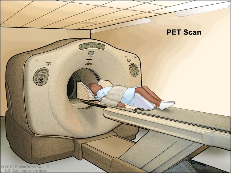
Fluorine F 18-fludeoxyglucose positron emission tomography (18F-FDG PET) scanning.
Procedures used to obtain tissue samples include bronchoscopy, mediastinoscopy, or anterior mediastinotomy.
Pathological staging of NSCLC requires examination of the tumor, knowledge of resection margins, and determination of lymph node status.
At diagnosis, patients with NSCLC can be divided into the following three groups that reflect both the extent of the disease and the treatment approach:
Surgically resectable disease (generally stage I, stage II, and selected stage III tumors).
Has the best prognosis, which depends on a variety of tumor and host factors.
Patients with resectable disease who have medical contraindications to surgery are candidates for curative radiation therapy.
Postoperative cisplatin-based combination chemotherapy may provide a survival advantage for patients with resected stage II or stage IIIA NSCLC.
Selected patients with locally advanced tumors may benefit from combined modality treatments.
Patients with unresectable or N2–N3 disease are treated with radiation therapy in combination with chemotherapy.
Selected patients with T3 or N2 disease can be treated effectively with surgical resection and either preoperative or postoperative chemotherapy or chemoradiation therapy.
Distant metastatic disease (includes distant metastases [M1] that were found at the time of diagnosis).
May be treated with systemic therapy (chemotherapy and/or immunotherapy or targeted therapy). Radiation therapy can be used for palliation.
Evaluation of mediastinal lymph node metastasis
Surgical evaluation
Surgical staging of the mediastinum is considered standard if accurate evaluation of the nodal status is needed to determine therapy.
Accurate staging of the mediastinal lymph nodes provides important prognostic information.
Evidence (nodal status):
The association between survival and the number of examined lymph nodes during surgery for patients with stage I NSCLC treated with definitive surgical resection was assessed from the population-based Surveillance, Epidemiology, and End Results (SEER) Program database for the period from 1990 to 2000.[2] A total of 16,800 patients were included in the study.
The overall survival analysis for patients without radiation therapy was done by comparing the reference group (one to four lymph nodes) with the following groups:
Patients with five to eight lymph nodes examined during surgery had a modest but statistically significant increase in survival, with a proportionate hazard ratio (HR) of 0.90 (95% confidence interval [CI], 0.84–0.97).
For patients with 9 to 12 examined lymph nodes, the HR was 0.86 (95% CI, 0.79–0.95).
For patients with 13 to 16 examined lymph nodes, the HR was 0.78 (95% CI, 0.68–0.90).
There appeared to be no incremental improvement after evaluating more than 16 lymph nodes.
The corresponding results for lung cancer–specific mortality and for patients who received radiation therapy were not substantially different.
These results indicate that patient survival following resection for NSCLC is associated with the number of lymph nodes evaluated during surgery. Because this is most likely the result of a reduction-of-staging error, namely, a decreased likelihood of missing positive lymph nodes with an increasing number of lymph nodes sampled, it suggests that an evaluation of nodal status should include 11 to 16 lymph nodes.
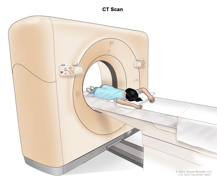
CT imaging
CT scanning is primarily used for determining the size of the tumor. The CT scan should extend inferiorly to include the liver and adrenal glands. MRI scans of the thorax and upper abdomen do not appear to yield advantages over CT scans.[3]
Evidence (CT scan):
A systematic review of the medical literature relating to the accuracy of CT scanning for noninvasive staging of the mediastinum in patients with lung cancer was conducted. In the 35 studies published between 1991 and June 2006, 5,111 evaluable patients were identified. Almost all studies specified that CT scanning was performed following the administration of intravenous contrast material and that a positive test result was defined as the presence of one or more lymph nodes that measured larger than 1 cm on the short-axis diameter.[4]
The median prevalence of mediastinal metastasis was 28% (range, 18%–56%).
The pooled estimates of sensitivity and specificity of CT scanning for identifying mediastinal lymph node metastasis were 51% (95% CI, 47%–54%) for sensitivity and 86% (95% CI, 84%–88%) for specificity. Corresponding positive (3.4%) and negative (0.6%) likelihood ratios were provided.
The results from the systematic review are similar to those of a large meta-analysis that reported the median sensitivity and specificity of CT scanning for identifying malignant mediastinal nodes as 61% for sensitivity and 79% for specificity.[5]
An earlier meta-analysis reported an average sensitivity rate of 64% and specificity rate of 74%.[6]
18F-FDG PET scanning
The wider availability and use of 18F-FDG PET scanning for staging has modified the approach to staging mediastinal lymph nodes and distant metastases.
Randomized trials evaluating the utility of 18F-FDG PET scanning in potentially resectable NSCLC patients reported conflicting results in terms of the relative reduction in the number of noncurative thoracotomies.
Although the current evidence is conflicting, 18F-FDG PET scanning may improve results of early-stage lung cancer by identifying patients who have evidence of metastatic disease that is beyond the scope of surgical resection and that is not evident by standard preoperative staging procedures.
Evidence (18F-FDG PET scan):
A systematic review, an expansion of a health technology assessment conducted in 2001 by the Institute for Clinical and Evaluative Sciences, evaluated the accuracy and utility of 18F-FDG PET scanning in the diagnosis and staging of lung cancer.[7] Through a systematic search of the literature, 12 evidence summary reports and 15 prospective studies of the diagnostic accuracy of 18F-FDG PET scanning were identified.
18F-FDG PET scanning appears to be superior to CT imaging for mediastinal staging in NSCLC.
18F-FDG PET scanning also appears to have high sensitivity and reasonable specificity for differentiating benign from malignant lesions as small as 1 cm.
A systematic review of the medical literature relating to the accuracy of 18F-FDG PET scanning for noninvasive staging of the mediastinum in patients with lung cancer identified 44 studies published between 1994 and 2006 with 2,865 evaluable patients.[4]
The median prevalence of mediastinal metastases was 29% (range, 5%–64%).
Pooled estimates of sensitivity and specificity for identifying mediastinal metastasis were 74% (95% CI, 69%–79%) for sensitivity and 85% (95% CI, 82%–88%) for specificity.
Corresponding positive (4.9%) and negative (0.3%) likelihood ratios were provided for mediastinal staging with 18F-FDG PET scanning.
These findings demonstrated that 18F-FDG PET scanning is more accurate than CT scanning for staging of the mediastinum in patients with lung cancer.
Decision analyses demonstrate that 18F-FDG PET scanning may reduce the overall costs of medical care by identifying patients with falsely negative CT scans in the mediastinum or otherwise undetected sites of metastases.[8–10] Studies concluded that the money saved by forgoing mediastinoscopy in 18F-FDG PET-positive mediastinal lesions was not justified because of the unacceptably high number of false-positive results.[8–10] A randomized study found that the addition of 18F-FDG PET scanning to conventional staging was associated with significantly fewer thoracotomies.[11] A second randomized trial evaluating the impact of 18F-FDG PET scanning on clinical management found that 18F-FDG PET scanning provided additional information regarding appropriate stage but did not lead to significantly fewer thoracotomies.[12]
Combination of CT imaging and 18F-FDG PET scanning
The combination of CT imaging and 18F-FDG PET scanning has greater sensitivity and specificity than CT imaging alone.[13]
Evidence (CT/18F-FDG PET scan):
If there is no evidence of distant metastatic disease on CT scan, 18F-FDG PET scanning complements CT scan staging of the mediastinum. Numerous nonrandomized studies of 18F-FDG PET scanning have evaluated mediastinal lymph nodes using surgery (i.e., mediastinoscopy and/or thoracotomy with mediastinal lymph node dissection) as the gold standard of comparison.
A meta-analysis evaluated the conditional test performance of 18F-FDG PET scanning and CT scanning.
The median sensitivity and specificity of 18F-FDG PET scans were reported as 100% for sensitivity and 78% for specificity in patients with enlarged lymph nodes.[5]
18F-FDG PET scanning is considered very accurate in identifying malignant nodal involvement when lymph nodes are enlarged. However, 18F-FDG PET scanning will falsely identify a malignancy in approximately one-fourth of patients with lymph nodes that are enlarged for other reasons, usually as a result of inflammation or infection.[14,15]
The median sensitivity and specificity of 18F-FDG PET scanning in patients with normal-sized mediastinal lymph nodes were 82% for sensitivity and 93% for specificity.[5] These data indicate that nearly 20% of patients with normal-sized lymph nodes but with malignant involvement had falsely negative 18F-FDG PET scan findings.
For patients with clinically operable NSCLC, the evidence supports performing a biopsy of mediastinal lymph nodes that are found to be larger than 1 cm in shortest transverse axis on chest CT scan or are found to be positive on 18F-FDG PET scan. Negative 18F-FDG PET scanning does not preclude biopsy of radiographically enlarged mediastinal lymph nodes. Mediastinoscopy is necessary for the detection of cancer in mediastinal lymph nodes when the results of the CT scan and 18F-FDG PET scan do not corroborate each other.
Evaluation of brain metastasis
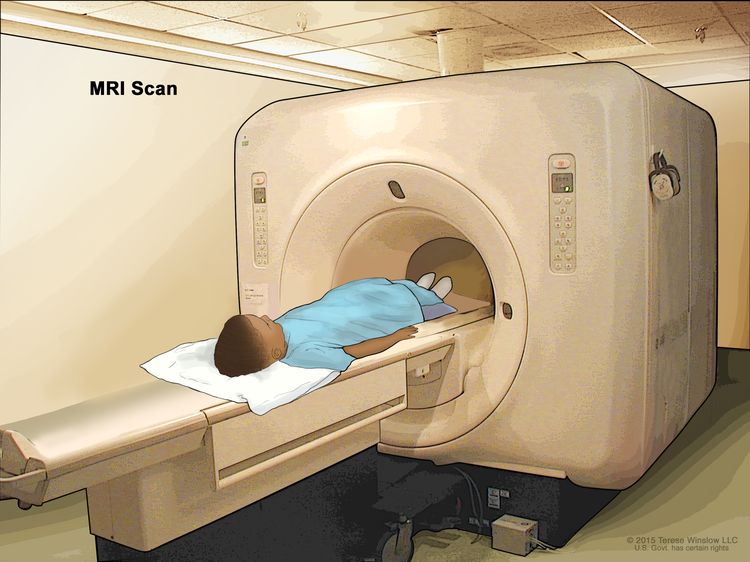
Patients at risk of brain metastases may be staged with CT or MRI scans.
Evidence (staging with CT or MRI):
One study randomly assigned 332 patients with potentially operable NSCLC and no neurological symptoms to brain CT or MRI imaging to detect occult brain metastasis before lung surgery.[16]
MRI showed a trend towards a higher preoperative detection rate than CT scan (P = .069), with an overall detection rate of approximately 7% from pretreatment to 12 months after surgery.
Patients with stage I or stage II disease had a detection rate of 4% (i.e., 8 detections out of 200 patients); however, individuals with stage III disease had a detection rate of 11.4% (i.e., 15 detections out of 132 patients).
The mean maximal diameter of the brain metastases was significantly smaller in the MRI group.
Whether the improved detection rate of MRI translates into improved outcome remains unknown. Not all patients are able to tolerate MRI, and for these patients contrast-enhanced CT scan is a reasonable substitute.
Evaluation of distant metastasis to sites other than the brain
Numerous nonrandomized, prospective, and retrospective studies have demonstrated that 18F-FDG PET scanning offers diagnostic advantages over conventional imaging in staging distant metastatic disease; however, standard 18F-FDG PET scans have limitations. 18F-FDG PET scans may not extend below the pelvis and may not detect bone metastases in the long bones of the lower extremities. Because the metabolic tracer used in 18F-FDG PET scanning accumulates in the brain and urinary tract, 18F-FDG PET scanning is not reliable for detection of metastases in these sites.[16]
The Revised International System for Staging Lung Cancer
The Revised International System for Staging Lung Cancer, based on information from a clinical database of more than 5,000 patients, was adopted in 2010 by the American Joint Committee on Cancer (AJCC) and the Union Internationale Contre le Cancer.[17,18] These revisions provide greater prognostic specificity for patient groups; however, the correlation between stage and prognosis predates the widespread availability of PET imaging.
AJCC Stage Groupings and TNM Definitions
The AJCC has designated staging by TNM (tumor, node, metastasis) classification to define NSCLC.[18]
Table 1. Definitions of Primary Tumor (T) for Lung Cancera
T Category
T Criteria
aReprinted with permission from AJCC: Lung. In: Amin MB, Edge SB, Greene FL, et al., eds.: AJCC Cancer Staging Manual. 8th ed. New York, NY: Springer, 2017, pp. 431–56.
TX
Primary tumor cannot be assessed, or tumor proven by the presence of malignant cells in sputum or bronchial washings but not visualized by imaging or bronchoscopy.
T0
No evidence of primary tumor.
Tis
Carcinoma in situ; SCIS =Squamous cell carcinoma in situ; AIS: Adenocarcinoma in situ; Adenocarcinoma with pure lepidic pattern, ≤3 cm in greatest dimension.
T1
Tumor ≤3 cm in greatest dimension, surrounded by lung or visceral pleura, without bronchoscopic evidence of invasion more proximal than the lobar bronchus (i.e., not in the main bronchus).
T1mi
Minimally invasive adenocarcinoma: adenocarcinoma (≤3 cm in greatest dimension) with a predominantly lepidic pattern and ≤5 mm invasion in greatest dimension.
T1a
Tumor ≤1 cm in greatest dimension. A superficial, spreading tumor of any size whose invasive component is limited to the bronchial wall and may extend proximal to the main bronchus also is classified as T1a, but these tumors are uncommon.
T1b
Tumor >1 cm but ≤2 cm in greatest dimension.
T1c
Tumor >2 cm but ≤3 cm in greatest dimension.
T2
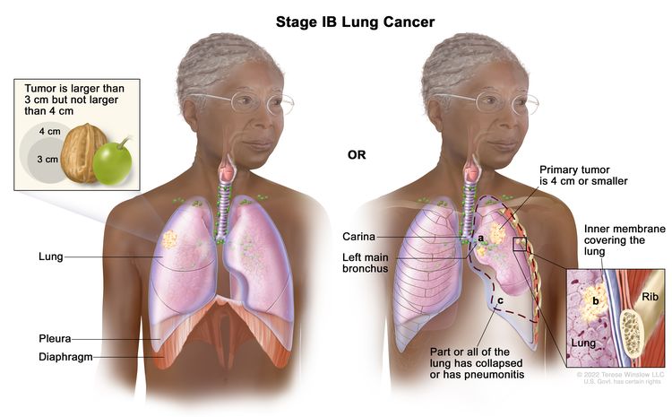
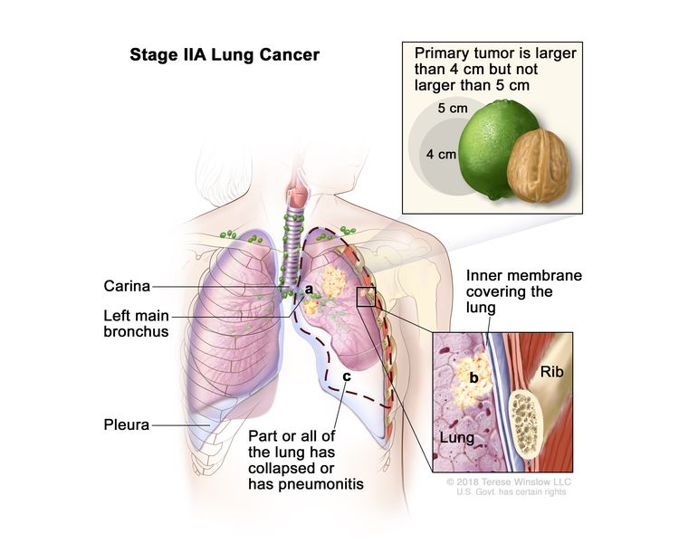
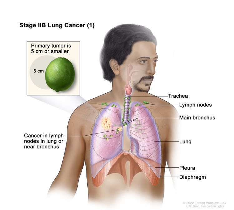
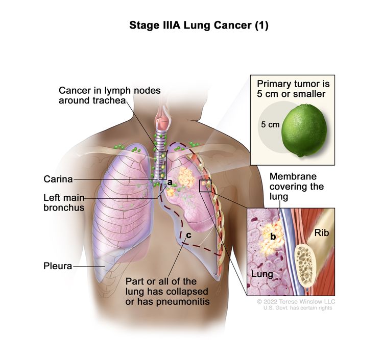
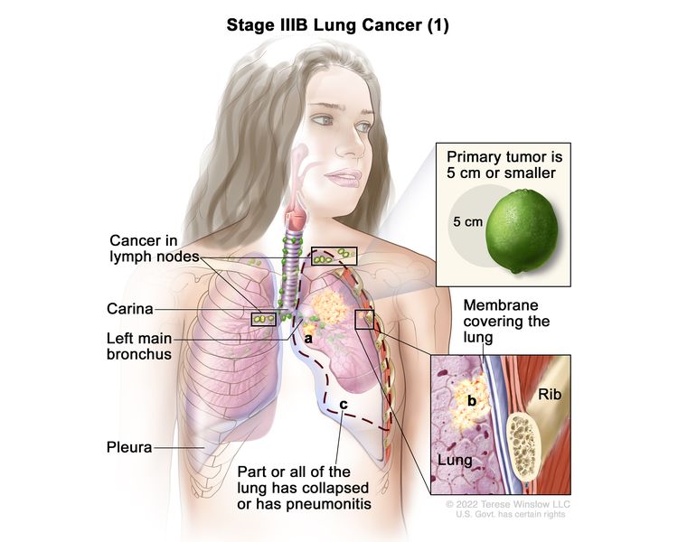
Tumor >3 cm but ≤5 cm or having any of the following features: involves the main bronchus regardless of distance to the carina, but without involvement of the carina; invades visceral pleura (PL1 or PL2); associated with atelectasis or obstructive pneumonitis that extends to the hilar region, involving part or all of the lung. T2 tumors with these features are classified as T2a if ≤4 cm or if the size cannot be determined and T2b if >4 cm but ≤5 cm.
T2a
Tumor >3 cm but ≤4 cm in greatest dimension.
T2b
Tumor >4 cm but ≤5 cm in greatest dimension.
T3
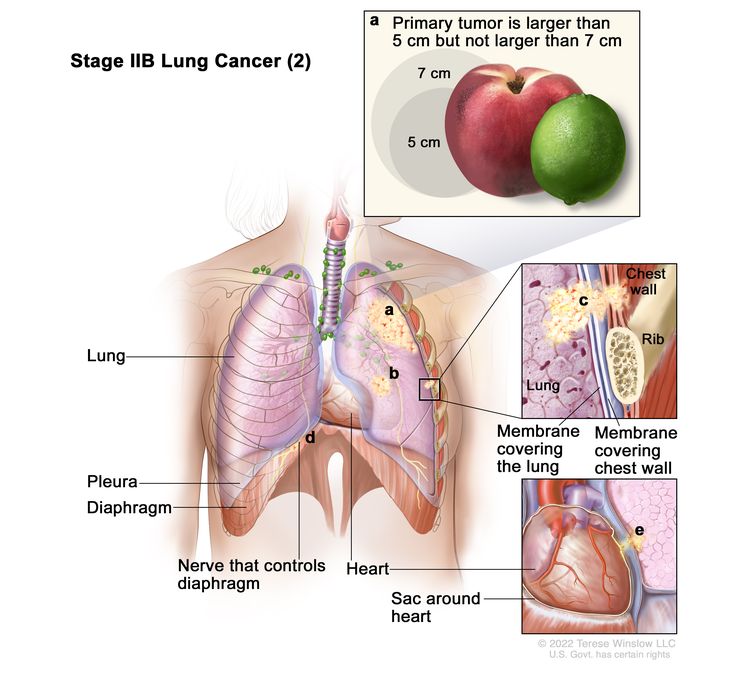
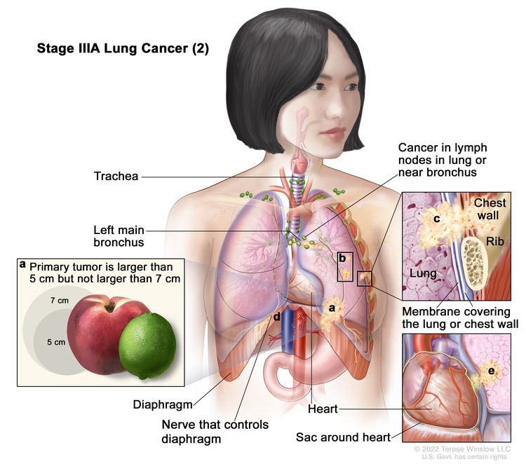
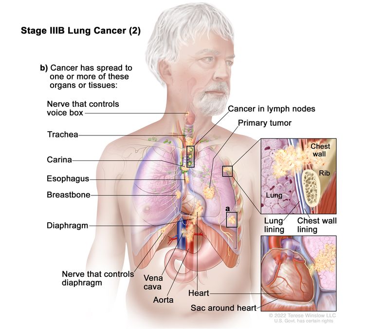
Tumor >5 cm but ≤7 cm in greatest dimension or directly invading any of the following: parietal pleura (PL3), chest wall (including superior sulcus tumors), phrenic nerve, parietal pericardium; or separate tumor nodule(s) in the same lobe as the primary.
T4
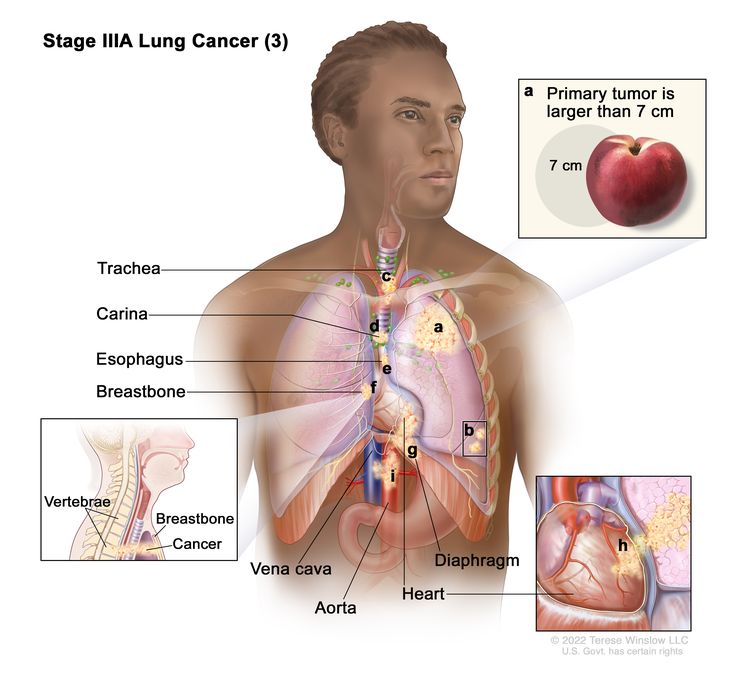
Tumor >7 cm or tumor of any size invading one or more of the following: diaphragm, mediastinum, heart, great vessels, trachea, recurrent laryngeal nerve, esophagus, vertebral body, or carina; separate tumor nodule(s) in an ipsilateral lobe different from that of the primary.
Table 2. Definitions of Regional Lymph Node (N) for Lung Cancera
N Category
N Criteria
aReprinted with permission from AJCC: Lung. In: Amin MB, Edge SB, Greene FL, et al., eds.: AJCC Cancer Staging Manual. 8th ed. New York, NY: Springer, 2017, pp. 431–56.
NX
Regional lymph nodes cannot be assessed.
N0
No regional lymph node metastasis.
N1
Metastasis in ipsilateral peribronchial and/or ipsilateral hilar lymph nodes and intrapulmonary nodes, including involvement by direct extension.
N2
Metastasis in ipsilateral mediastinal and/or subcarinal lymph node(s).
N3
Metastasis in contralateral mediastinal, contralateral hilar, ipsilateral or contralateral scalene, or supraclavicular lymph node(s).
Table 3. Definitions of Distant Metastasis (M) for Lung Cancera
M Category
M Criteria
aReprinted with permission from AJCC: Lung. In: Amin MB, Edge SB, Greene FL, et al., eds.: AJCC Cancer Staging Manual. 8th ed. New York, NY: Springer, 2017, pp. 431–56.
M0
No distant metastasis.
M1
Distant metastasis.
M1a
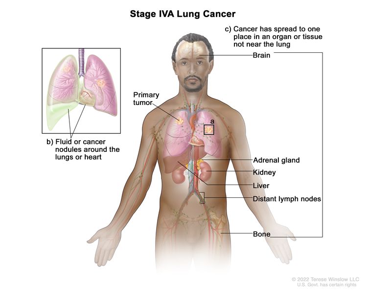
Separate tumor nodule(s) in a contralateral lobe; tumor with pleural or pericardial nodules or malignant pleural or pericardial effusion. Most pleural (pericardial) effusions with lung cancer are a result of the tumor. In a few patients, however, multiple microscopic examinations of pleural (pericardial) fluid are negative for tumor, and the fluid is nonbloody and not an exudate. If these elements and clinical judgment dictate that the effusion is not related to the tumor, the effusion should be excluded as a staging descriptor.
M1b
Single extrathoracic metastases in a single organ (including involvement of a single nonregional node).
M1c
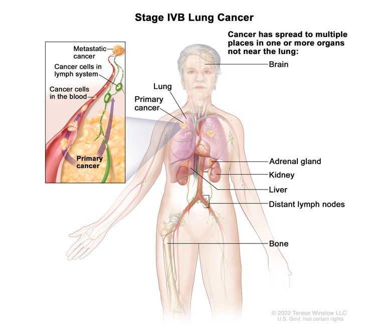
Multiple extrathoracic metastases in a single organ or in multiple organs.
Table 4. AJCC Prognostic Stage Groups for Lung Cancera
Stage
TNM Classification
Illustration
T = primary tumor; N = regional lymph node; M = distant metastasis.
aReprinted with permission from AJCC: Lung. In: Amin MB, Edge SB, Greene FL, et al., eds.: AJCC Cancer Staging Manual. 8th ed. New York, NY: Springer, 2017, pp. 431–56.
Pfister DG, Johnson DH, Azzoli CG, et al.: American Society of Clinical Oncology treatment of unresectable non-small-cell lung cancer guideline: update 2003. J Clin Oncol 22 (2): 330-53, 2004. [PUBMED Abstract]
Ludwig MS, Goodman M, Miller DL, et al.: Postoperative survival and the number of lymph nodes sampled during resection of node-negative non-small cell lung cancer. Chest 128 (3): 1545-50, 2005. [PUBMED Abstract]
Webb WR, Gatsonis C, Zerhouni EA, et al.: CT and MR imaging in staging non-small cell bronchogenic carcinoma: report of the Radiologic Diagnostic Oncology Group. Radiology 178 (3): 705-13, 1991. [PUBMED Abstract]
Toloza EM, Harpole L, McCrory DC: Noninvasive staging of non-small cell lung cancer: a review of the current evidence. Chest 123 (1 Suppl): 137S-146S, 2003. [PUBMED Abstract]
Gould MK, Kuschner WG, Rydzak CE, et al.: Test performance of positron emission tomography and computed tomography for mediastinal staging in patients with non-small-cell lung cancer: a meta-analysis. Ann Intern Med 139 (11): 879-92, 2003. [PUBMED Abstract]
Dwamena BA, Sonnad SS, Angobaldo JO, et al.: Metastases from non-small cell lung cancer: mediastinal staging in the 1990s–meta-analytic comparison of PET and CT. Radiology 213 (2): 530-6, 1999. [PUBMED Abstract]
Ung YC, Maziak DE, Vanderveen JA, et al.: 18Fluorodeoxyglucose positron emission tomography in the diagnosis and staging of lung cancer: a systematic review. J Natl Cancer Inst 99 (23): 1753-67, 2007. [PUBMED Abstract]
Dietlein M, Weber K, Gandjour A, et al.: Cost-effectiveness of FDG-PET for the management of potentially operable non-small cell lung cancer: priority for a PET-based strategy after nodal-negative CT results. Eur J Nucl Med 27 (11): 1598-609, 2000. [PUBMED Abstract]
Scott WJ, Shepherd J, Gambhir SS: Cost-effectiveness of FDG-PET for staging non-small cell lung cancer: a decision analysis. Ann Thorac Surg 66 (6): 1876-83; discussion 1883-5, 1998. [PUBMED Abstract]
Gambhir SS, Hoh CK, Phelps ME, et al.: Decision tree sensitivity analysis for cost-effectiveness of FDG-PET in the staging and management of non-small-cell lung carcinoma. J Nucl Med 37 (9): 1428-36, 1996. [PUBMED Abstract]
van Tinteren H, Hoekstra OS, Smit EF, et al.: Effectiveness of positron emission tomography in the preoperative assessment of patients with suspected non-small-cell lung cancer: the PLUS multicentre randomised trial. Lancet 359 (9315): 1388-93, 2002. [PUBMED Abstract]
Viney RC, Boyer MJ, King MT, et al.: Randomized controlled trial of the role of positron emission tomography in the management of stage I and II non-small-cell lung cancer. J Clin Oncol 22 (12): 2357-62, 2004. [PUBMED Abstract]
Vansteenkiste JF, Stroobants SG, De Leyn PR, et al.: Lymph node staging in non-small-cell lung cancer with FDG-PET scan: a prospective study on 690 lymph node stations from 68 patients. J Clin Oncol 16 (6): 2142-9, 1998. [PUBMED Abstract]
Roberts PF, Follette DM, von Haag D, et al.: Factors associated with false-positive staging of lung cancer by positron emission tomography. Ann Thorac Surg 70 (4): 1154-9; discussion 1159-60, 2000. [PUBMED Abstract]
Liewald F, Grosse S, Storck M, et al.: How useful is positron emission tomography for lymphnode staging in non-small-cell lung cancer? Thorac Cardiovasc Surg 48 (2): 93-6, 2000. [PUBMED Abstract]
Yokoi K, Kamiya N, Matsuguma H, et al.: Detection of brain metastasis in potentially operable non-small cell lung cancer: a comparison of CT and MRI. Chest 115 (3): 714-9, 1999. [PUBMED Abstract]
Mountain CF: Revisions in the International System for Staging Lung Cancer. Chest 111 (6): 1710-7, 1997. [PUBMED Abstract]
Lung. In: Amin MB, Edge SB, Greene FL, et al., eds.: AJCC Cancer Staging Manual. 8th ed. Springer; 2017, pp. 431–56.
Treatment Option Overview for NSCLC
In non-small cell lung cancer (NSCLC), results of standard treatment are poor except for the most localized cancers. All newly diagnosed patients with NSCLC are potential candidates for studies evaluating new forms of treatment.
Treatment decisions are based on some of the following factors:
Knowledge of histological type and molecular features.
Tumor size and location.
Involvement of pleura.
Surgical margins.
Status and location of lymph nodes by station.
Tumor grade.
Lymphovascular invasion.
Surgery is potentially the most curative therapeutic option for this disease. Postoperative chemotherapy may provide an additional benefit to patients with resected NSCLC. Radiation therapy combined with chemotherapy can produce a cure in a small number of patients and can provide palliation in most patients. Prophylactic cranial irradiation may reduce the incidence of brain metastases, but there is no evidence of a survival benefit and the effect of prophylactic cranial irradiation on quality of life is not known.[1,2] In patients with advanced-stage disease, chemotherapy or EGFR kinase inhibitors offer modest improvements in median survival, although overall survival is poor.[3,4]
Chemotherapy has produced short-term improvement in disease-related symptoms in patients with advanced NSCLC. Several clinical trials have attempted to assess the impact of chemotherapy on tumor-related symptoms and quality of life. In total, these studies suggest that tumor-related symptoms may be controlled by chemotherapy without adversely affecting overall quality of life;[5,6] however, the impact of chemotherapy on quality of life requires more study. In general, medically eligible older patients with good performance status obtain the same benefits from treatment as younger patients.
The identification of pathogenic variants has led to the development of molecularly targeted therapies for lung cancer that have improved the survival of some patients with metastatic disease.[7] In particular, genetic abnormalities in EGFR, MAPK, and PI3K signaling pathways in subsets of NSCLC may affect drug sensitivity and primary or acquired resistance to kinase inhibitors. EGFR variants strongly predict the improved response rate and progression-free survival in patients who receive EGFR inhibitors. ALK::EML4 fusion genes and other genes form translocation products that occur in 3% to 7% of unselected NSCLC cases and are responsive to pharmacological inhibition of ALK by agents like alectinib. The MET oncogene encodes hepatocyte growth factor receptor. Amplification of this gene has been associated with secondary resistance to EGFR tyrosine kinase inhibitors. Recurrent fusions involving the ROS1 gene are observed in up to 2% of NSCLCs and are responsive to treatment with crizotinib and entrectinib. NTRK gene fusions can occur in up to 1% of NSCLCs and can be treated with the TRK inhibitors, larotrectinib and entrectinib. For more information, see the Molecular Features section.
The treatment options for each stage of NSCLC are presented in Table 5.
mTOR inhibitors (for patients with unresectable, locally advanced or metastatic, progressive, well-differentiated, nonfunctional, neuroendocrine tumors)
In addition to the treatment options presented in Table 5, treatment options under clinical evaluation include:
Combining local treatment (surgery).
Regional treatment (radiation therapy).
Systemic treatments (chemotherapy, immunotherapy, and targeted agents).
Developing more effective systemic therapy.
Current Clinical Trials
Use our advanced clinical trial search to find NCI-supported cancer clinical trials that are now enrolling patients. The search can be narrowed by location of the trial, type of treatment, name of the drug, and other criteria. General information about clinical trials is also available.
References
Lester JF, MacBeth FR, Coles B: Prophylactic cranial irradiation for preventing brain metastases in patients undergoing radical treatment for non-small-cell lung cancer: a Cochrane Review. Int J Radiat Oncol Biol Phys 63 (3): 690-4, 2005. [PUBMED Abstract]
Pöttgen C, Eberhardt W, Grannass A, et al.: Prophylactic cranial irradiation in operable stage IIIA non small-cell lung cancer treated with neoadjuvant chemoradiotherapy: results from a German multicenter randomized trial. J Clin Oncol 25 (31): 4987-92, 2007. [PUBMED Abstract]
Chemotherapy in non-small cell lung cancer: a meta-analysis using updated data on individual patients from 52 randomised clinical trials. Non-small Cell Lung Cancer Collaborative Group. BMJ 311 (7010): 899-909, 1995. [PUBMED Abstract]
Spiro SG, Rudd RM, Souhami RL, et al.: Chemotherapy versus supportive care in advanced non-small cell lung cancer: improved survival without detriment to quality of life. Thorax 59 (10): 828-36, 2004. [PUBMED Abstract]
Clegg A, Scott DA, Hewitson P, et al.: Clinical and cost effectiveness of paclitaxel, docetaxel, gemcitabine, and vinorelbine in non-small cell lung cancer: a systematic review. Thorax 57 (1): 20-8, 2002. [PUBMED Abstract]
Pao W, Girard N: New driver mutations in non-small-cell lung cancer. Lancet Oncol 12 (2): 175-80, 2011. [PUBMED Abstract]
Treatment of Occult NSCLC
In occult lung cancer, a diagnostic evaluation often includes chest x-ray and selective bronchoscopy with close follow-up (e.g., computed tomography scan), when needed, to define the site and nature of the primary tumor; tumors discovered in this fashion are generally early stage and curable by surgery.
After discovery of the primary tumor, treatment involves establishing the stage of the tumor. Therapy is identical to that recommended for other non-small cell lung cancer (NSCLC) patients with similar-stage disease.
Use our advanced clinical trial search to find NCI-supported cancer clinical trials that are now enrolling patients. The search can be narrowed by location of the trial, type of treatment, name of the drug, and other criteria. General information about clinical trials is also available.
Treatment of Stage 0 NSCLC
Stage 0 non-small cell lung cancer (NSCLC) frequently progresses to invasive cancer.[1–3] Patients may be offered surveillance bronchoscopies and, if lesions are detected, potentially curative therapies.
Endobronchial therapies, including photodynamic therapy, electrocautery, cryotherapy, and neodymium-doped yttrium aluminum garnet (Nd-YAG) laser therapy.
Surgery
Segmentectomy or wedge resection are used to preserve maximum normal pulmonary tissue because patients with stage 0 NSCLC are at a high risk of second lung cancers. Because these tumors are, by definition, noninvasive and incapable of metastasizing, they should be curable with surgical resection; however, such lesions, when identified, are often centrally located and may require a lobectomy.
Endobronchial therapies
Patients with central lesions may be candidates for curative endobronchial therapy. Endobronchial therapies that preserve lung function include photodynamic therapy, electrocautery, cryotherapy, and Nd-YAG laser therapy.[3–6]
Evidence (endobronchial therapies):
Small case series have reported high complete response rates and long-term survival in selected patients.[7,8][Level of evidence C2]
Efficacy of these treatment modalities in the management of patients with early NSCLC remains to be proven in definitive randomized controlled trials.
A high incidence of second primary cancers develop in these patients.[1,2]
Current Clinical Trials
Use our advanced clinical trial search to find NCI-supported cancer clinical trials that are now enrolling patients. The search can be narrowed by location of the trial, type of treatment, name of the drug, and other criteria. General information about clinical trials is also available.
References
Woolner LB, Fontana RS, Cortese DA, et al.: Roentgenographically occult lung cancer: pathologic findings and frequency of multicentricity during a 10-year period. Mayo Clin Proc 59 (7): 453-66, 1984. [PUBMED Abstract]
Venmans BJ, van Boxem TJ, Smit EF, et al.: Outcome of bronchial carcinoma in situ. Chest 117 (6): 1572-6, 2000. [PUBMED Abstract]
Jeremy George P, Banerjee AK, Read CA, et al.: Surveillance for the detection of early lung cancer in patients with bronchial dysplasia. Thorax 62 (1): 43-50, 2007. [PUBMED Abstract]
Kennedy TC, McWilliams A, Edell E, et al.: Bronchial intraepithelial neoplasia/early central airways lung cancer: ACCP evidence-based clinical practice guidelines (2nd edition). Chest 132 (3 Suppl): 221S-233S, 2007. [PUBMED Abstract]
Corti L, Toniolo L, Boso C, et al.: Long-term survival of patients treated with photodynamic therapy for carcinoma in situ and early non-small-cell lung carcinoma. Lasers Surg Med 39 (5): 394-402, 2007. [PUBMED Abstract]
Deygas N, Froudarakis M, Ozenne G, et al.: Cryotherapy in early superficial bronchogenic carcinoma. Chest 120 (1): 26-31, 2001. [PUBMED Abstract]
van Boxem TJ, Venmans BJ, Schramel FM, et al.: Radiographically occult lung cancer treated with fibreoptic bronchoscopic electrocautery: a pilot study of a simple and inexpensive technique. Eur Respir J 11 (1): 169-72, 1998. [PUBMED Abstract]
van Boxem AJ, Westerga J, Venmans BJ, et al.: Photodynamic therapy, Nd-YAG laser and electrocautery for treating early-stage intraluminal cancer: which to choose? Lung Cancer 31 (1): 31-6, 2001. [PUBMED Abstract]
Radiation therapy (for patients who cannot have surgery or choose not to have surgery).
Chemotherapy and radiation therapy have not been shown to improve survival in patients with stage I NSCLC that has been completely resected.
Surgery
Surgery is the treatment of choice for patients with stage I NSCLC. A lobectomy or segmental, wedge, or sleeve resection may be performed as appropriate. Patients with impaired pulmonary function are candidates for segmental or wedge resection of the primary tumor. Careful preoperative assessment of the patient’s overall medical condition, especially the patient’s pulmonary reserve, is critical in considering the benefits of surgery. The immediate postoperative mortality rate is age related, but a 3% to 5% mortality rate with lobectomy can be expected.[1]
Evidence (surgery):
The Lung Cancer Study Group conducted a randomized study (LCSG-821) that compared lobectomy with limited resection for patients with stage I lung cancer. Results of the study showed:[2]
A reduction in local recurrence for patients treated with lobectomy compared with those treated with limited excision.
No significant difference in overall survival (OS).
Similar results were reported from a nonrandomized comparison of anatomical segmentectomy and lobectomy.[3]
A survival advantage was noted with lobectomy for patients with tumors larger than 3 cm but not for those with tumors smaller than 3 cm.
The rate of locoregional recurrence was significantly less after lobectomy, regardless of primary tumor size.
Those treated with wedge or segmental resections had a local recurrence rate of 50% (i.e., 31 recurrences out of 62 patients) despite having undergone complete resections.[4]
A multicenter, noninferiority, phase III trial (NCT00499330) evaluated lobar or sublobar resection in patients with peripheral stage IA NSCLC. A total of 697 patients with clinical stage T1a, N0 tumors (tumor size <2 cm) were randomly assigned to undergo sublobar resection or lobar resection after intraoperative confirmation of node-negative disease in the hilar and mediastinal lymph nodes. The primary end point was disease-free survival (DFS).[5][Level of evidence B1]
After a median follow-up of 7 years, sublobar resection was noninferior to lobar resection for DFS (hazard ratio [HR], 1.01; 90% confidence interval [CI], 0.83–1.24; one-sided P = .02 for noninferiority).
OS was similar after sublobar resection or lobar resection (HR, 0.95; 95% CI, 0.72–1.26).
No substantial differences were noted in the incidence of locoregional or distant disease recurrence between the two groups.
At 6 months after surgery, the magnitude of reduction from baseline in the percentage of predicted FEV1 (forced expiratory volume in first second of expiration) was greater in the lobectomy group (-6%; 95% CI, -8% to -5%) versus the sublobar resection group (-4%; 95% CI, -5% to -2%). The magnitude of reduction in the percentage of predicted FVC (forced vital capacity) was also greater after lobectomy (-5%; 95% CI, -7% to -3%) than after sublobar resection (-3%; 95% CI, -4% to -1%).
These results suggest that sublobar resection by anatomical segmentectomy or wedge resection is effective for management of clinical stage T1a, N0 NSCLC when intraoperative sampling of hilar and mediastinal lymph nodes is negative.
The Cochrane Collaboration reviewed 11 randomized trials with a total of 1,910 patients who underwent surgical interventions for early-stage (I–IIIA) lung cancer.[6] A pooled analysis of three trials reported the following:
Four-year survival was superior in patients with resectable stage I, II, or IIIA NSCLC who underwent resection and complete ipsilateral mediastinal lymph node dissection (CMLND), compared with those who underwent resection and lymph node sampling; the HR was estimated to be 0.78 (95% CI, 0.65–0.93, P = .005).[6][Level of evidence A1]
There was a significant reduction in any cancer recurrence (local or distant) in the CMLND group (relative risk [RR], 0.79; 95% CI, 0.66–0.95; P = .01) that appeared mainly because of a reduction in the number of distant recurrences (RR, 0.78; 95% CI, 0.61–1.00; P = .05).
There was no difference in operative mortality.
Air leak lasting more than 5 days was significantly more common in patients assigned to CMLND (RR, 2.94; 95% CI, 1.01–8.54; P = .05).
CMLND versus lymph node sampling was evaluated in a large, randomized, phase III trial (ACOSOG-Z0030 [NCT00003831]).[7,8]
Preliminary analyses of operative morbidity and mortality showed comparable rates from the procedures.[7,8]
There was no difference in OS, DFS, local recurrence, and regional recurrence.[8][Level of evidence A1]
Current evidence suggests that lung cancer resection combined with CMLND is not associated with improvement in survival compared with lung cancer resection combined with systematic sampling of mediastinal lymph nodes in patients with stage I, II, or IIIA NSCLC.[8][Level of evidence A1]
Conclusions about the efficacy of surgery for patients with local and locoregional NSCLC are limited by the small number of participants studied to date and the potential methodological weaknesses of the trials.
Adjuvant therapy
Many patients who have surgery subsequently develop regional or distant metastases.[9] Such patients are candidates for entry into clinical trials evaluating postoperative treatment with chemotherapy or radiation therapy following surgery. At present, neither chemotherapy nor radiation therapy has been found to improve survival in patients with stage I NSCLC that has been completely resected.
Adjuvant chemotherapy
Based on a meta-analysis, postoperative chemotherapy is not recommended outside of a clinical trial for patients with completely resected stage I NSCLC.[10] However, there may be some benefit of adjuvant chemotherapy in patients with stage IB tumors that are larger than 4 cm.
Evidence (adjuvant chemotherapy for patients with stage IB NSCLC):
The Cancer and Leukemia Group B study (CALGB-9633 [NCT00002852]) addressed the results of adjuvant carboplatin and paclitaxel versus observation for OS in 344 patients with resected stage IB (i.e., pathological T2, N0) NSCLC. Within 4 to 8 weeks of resection, patients were randomly assigned to postoperative chemotherapy or observation.[11]
Survival was not significantly different (HR, 0.83; 90% CI, 0.64–1.08; P = .12) at a median follow-up of 74 months.
Grades 3 to 4 neutropenia were the predominant toxicity; there were no treatment-related deaths.
A post-hoc exploratory analysis demonstrated a significant survival difference in favor of postoperative chemotherapy for patients who had tumors 4 cm or larger in diameter (HR, 0.69; 90% CI, 0.48–0.99; P = .043).
Given the magnitude of observed survival differences, CALGB-9633 may have been underpowered to detect small but clinically meaningful improvements in survival. In addition, the use of a carboplatin versus a cisplatin combination might have affected the results. At present, there is no reliable evidence that postoperative chemotherapy improves survival of patients with stage IB NSCLC.[11][Level of evidence A1]
Adjuvant targeted therapy (for patients with stage IB NSCLC and EGFR variants)
Adjuvant targeted therapy with osimertinib for patients with resected stage IB to IIIA NSCLC and an EGFR pathogenic variant was studied in a phase III clinical trial and showed improved OS.
Evidence (adjuvant targeted therapy with osimertinib for patients with stage IB NSCLC and EGFR variants):
The phase III, double-blind, placebo-controlled ADAURA (NCT02511106) trial included 682 patients with surgically resected stage IB to stage IIIA NSCLC and EGFR pathogenic variants (centrally determined, deletion in exon 19 or L858R variant). Patients were randomly assigned to receive either 80 mg of osimertinib by mouth daily (n = 399) or a placebo (n = 342) for 3 years. Standard postoperative adjuvant chemotherapy was allowed but not mandatory; decisions regarding adjuvant chemotherapy were made by the physician and patient before trial enrollment.[12][Level of evidence A1]
In the overall population, the 5-year OS rate was 88% in the osimertinib group and 78% in the placebo group (overall HRdeath, 0.49; 95.03% CI, 0.34–0.70; P < .001).
Among patients with stage II to IIIA disease, the 5-year OS rate was 85% in the osimertinib group and 73% in the placebo group (overall HRdeath, 0.49; 95.03% CI, 0.33–0.73; P < .001).
The adverse event profile is consistent with other studies that used osimertinib except for pneumonia related to COVID-19, which was reported later.
The U.S. Food and Drug Administration (FDA) approved osimertinib as adjuvant therapy for patients with stage IB to IIIA NSCLC with EGFR exon 19 deletions or EGFR L858R variants.
Adjuvant targeted therapy (for patients with stage IB tumors and ALK variants)
Evidence (adjuvant targeted therapy for patients with stage IB tumors with ALK variants):
The global, phase III, open-label ALINA trial (NCT03456076) included 257 patients with completely resected stage IB (tumors ≥4 cm), II, or IIIA (American Joint Committee on Cancer 7th edition staging criteria) NSCLC and an ALK variant. Patients were randomly assigned in a 1:1 ratio to receive either oral alectinib (600 mg twice daily) for 24 months or intravenous platinum-based chemotherapy in four 21-day cycles. The primary end point was DFS, tested hierarchically among patients with stage II or IIIA disease and then in the intention-to-treat (ITT) population. Secondary end points included central nervous system (CNS) DFS, OS, and safety.[13]
The median duration of follow-up for survival was 27.8 months (27.8 months in the alectinib group and 28.4 months in the chemotherapy group).
The 2-year DFS rate was 93.8% in the alectinib group and 63.0% in the chemotherapy group (stage II/IIIA) (HRdisease recurrence or death, 0.24; 95% CI, 0.13–0.45; P < .001).[13][Level of evidence B1]
The ITT population included patients with stage IB, II, or IIIA disease who had been randomly assigned. The DFS rate in the ITT population was 93.6% for patients who received alectinib and 63.7% for patients who received chemotherapy (HR, 0.24; 95% CI, 0.13–0.43; P < .001).
The HRCNS disease recurrence or death was 0.22 (95% CI, 0.08–0.58) in favor of alectinib.
OS data are immature.
The FDA approved alectinib for adjuvant treatment following tumor resection in patients with NSCLC and an ALK variant.
Adjuvant immunotherapy
Evidence (adjuvant immunotherapy with pembrolizumab for patients with stage IB tumors >4 cm):
The phase III, multicenter, open-label PEARLS/KEYNOTE-091 trial (NCT02504372) randomly assigned 1,177 patients with completely resected stage IB (tumor >4 cm) to stage IIIA NSCLC to receive pembrolizumab (200 mg every 3 weeks) or placebo for up to 18 cycles, or until disease progression or unacceptable toxicity. Patients started study treatment after resection or, if indicated, after adjuvant chemotherapy (maximum of four cycles). The dual primary end points were DFS in the overall study population and DFS in patients with a programmed death-ligand 1 (PD-L1) tumor proportion score (TPS) of 50% or greater, as determined using the PD-L1 IHC 22C3 pharmDx assay. These end points were reported in a prespecified interim analysis after a median follow-up of 35.6 months (interquartile range, 27.1–45.5).[14][Level of evidence B1]
In the overall study population, the median DFS was 53.6 months (95% CI, 39.2–not reached [NR]) in the pembrolizumab group and 42.0 months (95% CI, 31.3–NR) in the placebo group (HR, 0.76; 95% CI, 0.63–0.91; P = .0014).
In the PD-L1 TPS ≥50% population, the median DFS was not reached with either pembrolizumab (95% CI, 44.3–NR) or placebo (95% CI, 35.8–NR) (HR, 0.82; 95% CI, 0.57–1.18; P = .14).
OS data were immature at the time of the prespecified interim analysis.
No new safety signals were identified in this study.
The FDA approved pembrolizumab as a single agent for adjuvant treatment following resection and platinum-based chemotherapy for patients with stage IB (T2a ≥4 cm), II, or IIIA NSCLC. Of note, the FDA label specifies that pembrolizumab can be used as adjuvant therapy after platinum-based chemotherapy. However, chemotherapy was not required in the overall study patient population evaluated in KEYNOTE-091.
Adjuvant external radiation therapy
The value of postoperative (adjuvant) radiation therapy (PORT) has been evaluated and has not been found to improve the outcome of patients with completely resected stage I NSCLC.[15]
Adjuvant brachytherapy
The value of intraoperative (adjuvant) brachytherapy applied to the suture line has been evaluated in patients undergoing sublobar resections for stage I NSCLC to improve local control; it has not been found to improve outcomes.
Evidence (adjuvant brachytherapy):
A phase III trial that randomly assigned 222 patients to undergo sublobar resection with or without suture line brachytherapy reported the following:[16]
No difference in the primary end point of local recurrence (5-year estimate, 14.0% vs. 16.7%; P = .59).
A substantial number of patients are ineligible for standard surgical resection because of comorbid conditions that are associated with unacceptably high perioperative risk. Patients with potentially resectable tumors with medical contraindications to surgery or those with inoperable stage I disease and with sufficient pulmonary reserve may be candidates for radiation therapy with curative intent.[17–19] Nonrandomized observational studies comparing treatment outcomes associated with resection, radiation therapy, and observation have demonstrated shorter survival times and higher mortality for patients who undergo observation only.[17,20]
Conventional radiation therapy
Historically, conventional primary radiation therapy consisted of approximately 60 Gy to 70 Gy delivered with megavoltage equipment to the midplane of the known tumor volume using conventional fractionation (1.8–2.0 Gy per day).
Improvements in radiation techniques include planning techniques to account for tumor motion, more conformal planning techniques (e.g., 3-D conformal radiation therapy and intensity-modulated radiation therapy), and image guidance during treatment. Modern approaches to delivery of external-beam radiation therapy (EBRT) include hypofractionated radiation therapy and stereotactic body radiation therapy (SBRT). However, there are limited reliable data from comparative trials to determine which approaches yield superior outcomes.[18,19]
Evidence (conventional radiation therapy):
In the largest retrospective conventional radiation therapy series, patients with inoperable disease were treated with definitive radiation therapy.[21–23]
Patients achieved 5-year survival rates of 10% to 30%.[21–23]
Several series demonstrated that patients with T1, N0 tumors had better outcomes, and 5-year survival rates of 30% to 60% were found in this subgroup.[21,22,24]
However, local-only failure occurs in as many as 50% of patients treated with conventional radiation therapy to doses in the range of 60 Gy to 65 Gy.[25,26]
A single report of patients older than 70 years who had resectable lesions smaller than 4 cm but who had medically inoperable disease or who refused surgery reported the following:[24]
Survival at 5 years after radiation therapy with curative intent was comparable with a historical control group of patients of similar age who were resected with curative intent.
A small case series using matched controls reported the following:[4]
The addition of endobronchial brachytherapy improved local disease control compared with EBRT.[4][Level of evidence C2]
Hypofractionated radiation therapy
Hypofractionated radiation therapy involves the delivery of a slightly higher dose of radiation therapy per day (e.g., 2.4–4.0 Gy) over a shorter period of time compared with conventionally fractionated radiation therapy. Multiple prospective phase I/II trials have demonstrated that hypofractionated radiation therapy to a dose of 60 Gy to 70 Gy delivered over 3 to 4 weeks with 2.4 Gy to 4.0 Gy per day resulted in a low incidence of moderate to severe toxicity, 2-year OS rates of 50% to 60%, and 2-year tumor local control of 80% to 90%.[27–29][Level of evidence C1]
Stereotactic body radiation therapy (SBRT)
SBRT involves the delivery of highly conformal, high-dose radiation therapy over an extremely hypofractionated course (e.g., one to five treatments) delivered over 1 to 2 weeks. Commonly used regimens include 18 Gy × 3, 12 Gy to 12.5 Gy × 4, and 10 Gy to 12 Gy × 5, and deliver a substantially higher biologically effective dose compared with historic conventional radiation therapy regimens.
Multiple prospective phase I/II trials and institutional series have demonstrated that SBRT results in a low incidence of pulmonary toxicity (<10% risk of symptomatic radiation pneumonitis), 2-year OS rates of 50% to 60%, and 2-year tumor control of 90% to 95%.[30–36][Level of evidence C1]
Evidence (SBRT):
Early phase I/II trials from Indiana University identified the maximum tolerated dose of three-fraction SBRT at 18 Gy × 3 for T1 tumors.
This regimen resulted in a 2-year OS rate of 55% and 2-year local tumor control of 95%.
An unacceptably high incidence (8.6%) of grade 5 toxicity was observed in patients with central tumors (defined as within 2 cm of the tracheobronchial tree from the trachea to the level of the lobar bronchi).[31]
A subsequent multicenter trial (RTOG-0236 [NCT00087438]) studied the 18 Gy × 3 regimen in 55 patients with peripheral T1 to T2 tumors only.
This trial demonstrated a 3-year OS rate of 56% and 3-year primary tumor control of 98%.
The incidence of moderate to severe toxicity was low, with grade 3 toxicity in 24% of patients, grade 4 toxicity in 4% of patients, and no grade 5 toxicity, with a 4% incidence of grade 3 radiation pneumonitis.[35]
In the largest reported series from VU University Medical Center Amsterdam, 676 patients with T1 to T2 tumors were treated with three-, five-, and eight-fraction SBRT using a risk-adapted approach (a tailored fractionation regimen based on tumor proximity to critical organs).
With a median follow-up of 32.9 months, the median OS was 40.7 months, and 2-year local tumor control was 95%.[36]
While central location is a contraindication to three-fraction SBRT based on data from the Indiana phase II study, a subsequent systematic review of published reports of 315 patients with 563 central tumors demonstrated a much lower incidence of severe toxicity, including a 1% to 5% risk of grade 5 events with more protracted SBRT regimens (e.g., four to ten fractions).[37]
A multicenter phase I/II trial (RTOG-0813 [NCT00750269]) is ongoing to identify the maximum tolerated dose for a five-fraction SBRT regimen for central tumors.
A randomized trial of hypofractionated radiation therapy versus SBRT (LUSTRE [NCT01968941]) is ongoing to determine the optimal radiation therapy regimen, but SBRT has been widely adopted for patients with medically inoperable stage I NSCLC.
Current Clinical Trials
Use our advanced clinical trial search to find NCI-supported cancer clinical trials that are now enrolling patients. The search can be narrowed by location of the trial, type of treatment, name of the drug, and other criteria. General information about clinical trials is also available.
References
Ginsberg RJ, Hill LD, Eagan RT, et al.: Modern thirty-day operative mortality for surgical resections in lung cancer. J Thorac Cardiovasc Surg 86 (5): 654-8, 1983. [PUBMED Abstract]
Ginsberg RJ, Rubinstein LV: Randomized trial of lobectomy versus limited resection for T1 N0 non-small cell lung cancer. Lung Cancer Study Group. Ann Thorac Surg 60 (3): 615-22; discussion 622-3, 1995. [PUBMED Abstract]
Warren WH, Faber LP: Segmentectomy versus lobectomy in patients with stage I pulmonary carcinoma. Five-year survival and patterns of intrathoracic recurrence. J Thorac Cardiovasc Surg 107 (4): 1087-93; discussion 1093-4, 1994. [PUBMED Abstract]
Mantz CA, Dosoretz DE, Rubenstein JH, et al.: Endobronchial brachytherapy and optimization of local disease control in medically inoperable non-small cell lung carcinoma: a matched-pair analysis. Brachytherapy 3 (4): 183-90, 2004. [PUBMED Abstract]
Altorki N, Wang X, Kozono D, et al.: Lobar or Sublobar Resection for Peripheral Stage IA Non-Small-Cell Lung Cancer. N Engl J Med 388 (6): 489-498, 2023. [PUBMED Abstract]
Manser R, Wright G, Hart D, et al.: Surgery for early stage non-small cell lung cancer. Cochrane Database Syst Rev (1): CD004699, 2005. [PUBMED Abstract]
Allen MS, Darling GE, Pechet TT, et al.: Morbidity and mortality of major pulmonary resections in patients with early-stage lung cancer: initial results of the randomized, prospective ACOSOG Z0030 trial. Ann Thorac Surg 81 (3): 1013-9; discussion 1019-20, 2006. [PUBMED Abstract]
Darling GE, Allen MS, Decker PA, et al.: Randomized trial of mediastinal lymph node sampling versus complete lymphadenectomy during pulmonary resection in the patient with N0 or N1 (less than hilar) non-small cell carcinoma: results of the American College of Surgery Oncology Group Z0030 Trial. J Thorac Cardiovasc Surg 141 (3): 662-70, 2011. [PUBMED Abstract]
Martini N, Bains MS, Burt ME, et al.: Incidence of local recurrence and second primary tumors in resected stage I lung cancer. J Thorac Cardiovasc Surg 109 (1): 120-9, 1995. [PUBMED Abstract]
Pignon JP, Tribodet H, Scagliotti GV, et al.: Lung adjuvant cisplatin evaluation: a pooled analysis by the LACE Collaborative Group. J Clin Oncol 26 (21): 3552-9, 2008. [PUBMED Abstract]
Strauss GM, Herndon JE, Maddaus MA, et al.: Adjuvant paclitaxel plus carboplatin compared with observation in stage IB non-small-cell lung cancer: CALGB 9633 with the Cancer and Leukemia Group B, Radiation Therapy Oncology Group, and North Central Cancer Treatment Group Study Groups. J Clin Oncol 26 (31): 5043-51, 2008. [PUBMED Abstract]
Tsuboi M, Herbst RS, John T, et al.: Overall Survival with Osimertinib in Resected EGFR-Mutated NSCLC. N Engl J Med 389 (2): 137-147, 2023. [PUBMED Abstract]
Wu YL, Dziadziuszko R, Ahn JS, et al.: Alectinib in Resected ALK-Positive Non-Small-Cell Lung Cancer. N Engl J Med 390 (14): 1265-1276, 2024. [PUBMED Abstract]
O’Brien M, Paz-Ares L, Marreaud S, et al.: Pembrolizumab versus placebo as adjuvant therapy for completely resected stage IB-IIIA non-small-cell lung cancer (PEARLS/KEYNOTE-091): an interim analysis of a randomised, triple-blind, phase 3 trial. Lancet Oncol 23 (10): 1274-1286, 2022. [PUBMED Abstract]
PORT Meta-analysis Trialists Group: Postoperative radiotherapy for non-small cell lung cancer. Cochrane Database Syst Rev (2): CD002142, 2005. [PUBMED Abstract]
Fernando HC, Landreneau RJ, Mandrekar SJ, et al.: Impact of brachytherapy on local recurrence rates after sublobar resection: results from ACOSOG Z4032 (Alliance), a phase III randomized trial for high-risk operable non-small-cell lung cancer. J Clin Oncol 32 (23): 2456-62, 2014. [PUBMED Abstract]
McGarry RC, Song G, des Rosiers P, et al.: Observation-only management of early stage, medically inoperable lung cancer: poor outcome. Chest 121 (4): 1155-8, 2002. [PUBMED Abstract]
Lanni TB, Grills IS, Kestin LL, et al.: Stereotactic radiotherapy reduces treatment cost while improving overall survival and local control over standard fractionated radiation therapy for medically inoperable non-small-cell lung cancer. Am J Clin Oncol 34 (5): 494-8, 2011. [PUBMED Abstract]
Grutters JP, Kessels AG, Pijls-Johannesma M, et al.: Comparison of the effectiveness of radiotherapy with photons, protons and carbon-ions for non-small cell lung cancer: a meta-analysis. Radiother Oncol 95 (1): 32-40, 2010. [PUBMED Abstract]
Raz DJ, Zell JA, Ou SH, et al.: Natural history of stage I non-small cell lung cancer: implications for early detection. Chest 132 (1): 193-9, 2007. [PUBMED Abstract]
Dosoretz DE, Katin MJ, Blitzer PH, et al.: Radiation therapy in the management of medically inoperable carcinoma of the lung: results and implications for future treatment strategies. Int J Radiat Oncol Biol Phys 24 (1): 3-9, 1992. [PUBMED Abstract]
Gauden S, Ramsay J, Tripcony L: The curative treatment by radiotherapy alone of stage I non-small cell carcinoma of the lung. Chest 108 (5): 1278-82, 1995. [PUBMED Abstract]
Sibley GS, Jamieson TA, Marks LB, et al.: Radiotherapy alone for medically inoperable stage I non-small-cell lung cancer: the Duke experience. Int J Radiat Oncol Biol Phys 40 (1): 149-54, 1998. [PUBMED Abstract]
Noordijk EM, vd Poest Clement E, Hermans J, et al.: Radiotherapy as an alternative to surgery in elderly patients with resectable lung cancer. Radiother Oncol 13 (2): 83-9, 1988. [PUBMED Abstract]
Dosoretz DE, Galmarini D, Rubenstein JH, et al.: Local control in medically inoperable lung cancer: an analysis of its importance in outcome and factors determining the probability of tumor eradication. Int J Radiat Oncol Biol Phys 27 (3): 507-16, 1993. [PUBMED Abstract]
Kaskowitz L, Graham MV, Emami B, et al.: Radiation therapy alone for stage I non-small cell lung cancer. Int J Radiat Oncol Biol Phys 27 (3): 517-23, 1993. [PUBMED Abstract]
Bradley J, Graham MV, Winter K, et al.: Toxicity and outcome results of RTOG 9311: a phase I-II dose-escalation study using three-dimensional conformal radiotherapy in patients with inoperable non-small-cell lung carcinoma. Int J Radiat Oncol Biol Phys 61 (2): 318-28, 2005. [PUBMED Abstract]
Bogart JA, Hodgson L, Seagren SL, et al.: Phase I study of accelerated conformal radiotherapy for stage I non-small-cell lung cancer in patients with pulmonary dysfunction: CALGB 39904. J Clin Oncol 28 (2): 202-6, 2010. [PUBMED Abstract]
Cheung P, Faria S, Ahmed S, et al.: Phase II study of accelerated hypofractionated three-dimensional conformal radiotherapy for stage T1-3 N0 M0 non-small cell lung cancer: NCIC CTG BR.25. J Natl Cancer Inst 106 (8): , 2014. [PUBMED Abstract]
Timmerman R, Papiez L, McGarry R, et al.: Extracranial stereotactic radioablation: results of a phase I study in medically inoperable stage I non-small cell lung cancer. Chest 124 (5): 1946-55, 2003. [PUBMED Abstract]
Timmerman R, McGarry R, Yiannoutsos C, et al.: Excessive toxicity when treating central tumors in a phase II study of stereotactic body radiation therapy for medically inoperable early-stage lung cancer. J Clin Oncol 24 (30): 4833-9, 2006. [PUBMED Abstract]
Lagerwaard FJ, Haasbeek CJ, Smit EF, et al.: Outcomes of risk-adapted fractionated stereotactic radiotherapy for stage I non-small-cell lung cancer. Int J Radiat Oncol Biol Phys 70 (3): 685-92, 2008. [PUBMED Abstract]
Baumann P, Nyman J, Hoyer M, et al.: Outcome in a prospective phase II trial of medically inoperable stage I non-small-cell lung cancer patients treated with stereotactic body radiotherapy. J Clin Oncol 27 (20): 3290-6, 2009. [PUBMED Abstract]
Fakiris AJ, McGarry RC, Yiannoutsos CT, et al.: Stereotactic body radiation therapy for early-stage non-small-cell lung carcinoma: four-year results of a prospective phase II study. Int J Radiat Oncol Biol Phys 75 (3): 677-82, 2009. [PUBMED Abstract]
Timmerman R, Paulus R, Galvin J, et al.: Stereotactic body radiation therapy for inoperable early stage lung cancer. JAMA 303 (11): 1070-6, 2010. [PUBMED Abstract]
Senthi S, Lagerwaard FJ, Haasbeek CJ, et al.: Patterns of disease recurrence after stereotactic ablative radiotherapy for early stage non-small-cell lung cancer: a retrospective analysis. Lancet Oncol 13 (8): 802-9, 2012. [PUBMED Abstract]
Senthi S, Haasbeek CJ, Slotman BJ, et al.: Outcomes of stereotactic ablative radiotherapy for central lung tumours: a systematic review. Radiother Oncol 106 (3): 276-82, 2013. [PUBMED Abstract]
Clinical trials of radiation therapy after curative surgery.
Adjuvant radiation therapy has not been shown to improve outcomes in patients with stage II NSCLC.
Surgery with or without adjuvant or neoadjuvant therapy
Surgery alone
Surgery is the treatment of choice for patients with stage II NSCLC. A lobectomy, pneumonectomy, segmental resection, wedge resection, or sleeve resection may be performed as appropriate. Careful preoperative assessment of the patient’s overall medical condition, especially the patient’s pulmonary reserve, is critical in considering the benefits of surgery. In addition to the immediate and age-related postoperative mortality rate, a 5% to 8% mortality rate with pneumonectomy or a 3% to 5% mortality rate with lobectomy can be expected.
Evidence (surgery):
The Cochrane Collaboration reviewed 11 randomized trials with a total of 1,910 patients who underwent surgical interventions for early-stage (I–IIIA) lung cancer.[1] A pooled analysis of three trials reported the following:
Four-year survival was superior in patients with resectable stage I, II, or IIIA NSCLC who underwent resection and complete ipsilateral mediastinal lymph node dissection (CMLND), compared with those who underwent resection and lymph node sampling; the hazard ratio (HR) was estimated to be 0.78 (95% confidence interval [CI], 0.65–0.93; P = .005).[1][Level of evidence A1]
There was a significant reduction in any cancer recurrence (local or distant) in the CMLND group (relative risk [RR], 0.79; 95% CI, 0.66–0.95; P = .01) that appeared mainly as the result of a reduction in the number of distant recurrences (RR, 0.78; 95% CI, 0.61–1.00; P = .05).
There was no difference in operative mortality.
Air leak lasting more than 5 days was significantly more common in patients assigned to CMLND (RR, 2.94; 95% CI, 1.01–8.54; P = .05).
CMLND versus lymph node sampling was evaluated in a large randomized phase III trial (ACOSOG-Z0030 [NCT00003831]).[2]
Preliminary analyses of operative morbidity and mortality showed comparable rates from the procedures.[2]
There was no difference in overall survival (OS), disease-free survival (DFS), local recurrence, and regional recurrence.[3][Level of evidence A1]
Evidence suggests that lung cancer resection combined with CMLND is not associated with improvement in survival compared with lung cancer resection combined with systematic sampling of mediastinal lymph nodes in patients with stage I, II, or IIIA NSCLC.[3][Level of evidence A1]
Conclusions about the efficacy of surgery for patients with local and locoregional NSCLC are limited by the small number of participants studied and potential methodological weaknesses of the trials.
Adjuvant chemotherapy
The preponderance of evidence indicates that postoperative cisplatin combination chemotherapy provides a significant survival advantage to patients with resected stage II NSCLC. Preoperative chemotherapy may also provide survival benefit. The optimal sequence of surgery and chemotherapy and the benefits and risks of postoperative radiation therapy in patients with resectable NSCLC remain to be determined.
After surgery, many patients develop regional or distant metastases.[4] Several randomized controlled trials and meta-analyses have evaluated the use of postoperative chemotherapy in patients with stage I, II, and IIIA NSCLC.[5–11]
Evidence (adjuvant chemotherapy):
Data on individual patient outcomes were collected and pooled into a meta-analysis from the five largest trials (4,584 patients) of cisplatin-based chemotherapy in patients with completely resected NSCLC that were conducted after 1995.[7]
With a median follow-up time of 5.2 years, the overall HRdeath was 0.89 (95% CI, 0.82–0.96; P = .005), corresponding to a 5-year absolute benefit of 5.4% from chemotherapy.
The benefit varied with stage (test for trend, P = .04; HR for stage IA, 1.40; 95% CI, 0.95–2.06; HR for stage IB, 0.93; 95% CI, 0.78–1.10; HR for stage II, 0.83; 95% CI, 0.73–0.95; and HR for stage III, 0.83; 95% CI, 0.72–0.94).
The effect of chemotherapy did not vary significantly (test for interaction, P = .11) with the associated drugs, including vinorelbine (HR, 0.80; 95% CI, 0.70–0.91), etoposide or vinca alkaloid (HR, 0.92; 95% CI, 0.80–1.07), or other drugs (HR, 0.97; 95% CI, 0.84–1.13).
The greater effect on survival observed with the doublet of cisplatin plus vinorelbine compared with other regimens should be interpreted cautiously as the total dose of cisplatin received was significantly higher in patients treated with vinorelbine.
The meta-analysis [7] and the individual studies [5,12] support the administration of postoperative cisplatin-based chemotherapy in combination with vinorelbine.
Superior OS for the trial population and patients with stage II disease was reported for the Lung Adjuvant Cisplatin Evaluation (LACE) pooled analysis (pooled HR, 0.83; 95% CI, 0.73–0.95); the Adjuvant Navelbine International Trialist Association (ANITA) trial (HR, 0.71; 95% CI, 0.49–1.03); and the National Cancer Institute of Canada Clinical Trials Group JBR.10 trial (HR, 0.59; 95% CI, 0.42–0.85).
Chemotherapy effect was higher in patients with better performance status.
There was no interaction between chemotherapy effect and any of the following:
Sex.
Age.
Histology.
Type of surgery.
Planned radiation therapy.
Planned total dose of cisplatin.
In a retrospective analysis of a phase III trial of postoperative cisplatin and vinorelbine, patients older than 65 years were found to benefit from treatment.[13]
Chemotherapy significantly prolonged OS for patients older than 65 years (HR, 0.61; 95% CI, 0.38–0.98; P = .04).
There were no significant differences in toxic effects, hospitalization, or treatment-related death by age group, although patients older than 65 years received less treatment.[13]
Several other randomized controlled trials and meta-analyses have evaluated the use of postoperative chemotherapy in patients with stages I, II, and IIIA NSCLC.[5–11]
Based on these data, patients with completely resected stage II lung cancer may benefit from postoperative cisplatin-based chemotherapy.[13][Level of evidence A1]
Adjuvant targeted therapy (for patients with EGFR variants)
Adjuvant targeted therapy with osimertinib for patients with resected stage IB to IIIA NSCLC and an EGFR pathogenic variant was studied in a phase III clinical trial and showed improved OS.
Evidence (adjuvant targeted therapy with osimertinib for patients with stages IIA and IIB NSCLC and an EGFR variant):
The phase III, double-blind, placebo-controlled ADAURA (NCT02511106) trial included 682 patients with surgically resected stage IB to stage IIIA NSCLC and EGFR pathogenic variants (centrally determined, deletion in exon 19 or L858R variant). Patients were randomly assigned to receive 80 mg of osimertinib by mouth daily (n = 399) or a placebo (n = 342) for 3 years. Standard postoperative adjuvant chemotherapy was allowed but not mandatory; decisions regarding adjuvant chemotherapy were made by the physician and patient before trial enrollment.[14][Level of evidence A1]
In the overall population, the 5-year OS rate was 88% in the osimertinib group and 78% in the placebo group (overall HRdeath, 0.49; 95.03% CI, 0.34–0.70; P < .001).
Among patients with stage II to IIIA disease, the 5-year OS rate was 85% in the osimertinib group and 73% in the placebo group (overall HRdeath, 0.49; 95.03% CI, 0.33–0.73; P < .001).
The adverse event profile is consistent with other studies that used osimertinib except for pneumonia related to COVID-19, which was reported later.
The U.S. Food and Drug Administration (FDA) approved osimertinib as adjuvant therapy for patients with stage IB to IIIA NSCLC with EGFR exon 19 deletions or EGFR L858R variants.
Adjuvant targeted therapy (for patients with ALK variants)
Evidence (adjuvant targeted therapy (for patients with ALK variants).
The global, phase III, open-label ALINA trial (NCT03456076) included 257 patients with completely resected stage IB (tumors ≥4 cm), II, or IIIA (American Joint Committee on Cancer 7th edition staging criteria) NSCLC and an ALK variant. Patients were randomly assigned in a 1:1 ratio to receive either oral alectinib (600 mg twice daily) for 24 months or intravenous platinum-based chemotherapy in four 21-day cycles. The primary end point was DFS, tested hierarchically among patients with stage II or IIIA disease and then in the intention-to-treat (ITT) population. Secondary end points included central nervous system (CNS) DFS, OS, and safety.[15]
The median duration of follow-up for survival was 27.8 months (27.8 months in the alectinib group and 28.4 months in the chemotherapy group).
The 2-year DFS rate was 93.8% in the alectinib group and 63.0% in the chemotherapy group (stage II/IIIA) (HRdisease recurrence or death, 0.24; 95% CI, 0.13–0.45; P < .001).[15][Level of evidence B1]
The ITT population included patients with stage IB, II, or IIIA disease who had been randomly assigned. The DFS rate in the ITT population was 93.6% for patients who received alectinib and 63.7% for patients who received chemotherapy (HR, 0.24; 95% CI, 0.13–0.43; P < .001).
The HRCNS disease recurrence or death was 0.22 (95% CI, 0.08–0.58) in favor of alectinib.
OS data are immature.
The FDA approved alectinib for adjuvant treatment following tumor resection in patients with NSCLC and an ALK variant.
Adjuvant immunotherapy
Adjuvant immunotherapy for patients with resected stage IB to IIIA NSCLC has been found to significantly increase DFS.[16,17]
Evidence (adjuvant immunotherapy with pembrolizumab for patients with stage IIA and IIB tumors >4 cm):
The phase III, multicenter, open-label PEARLS/KEYNOTE-091 trial (NCT02504372) randomly assigned 1,177 patients with completely resected stage IB (tumor >4 cm) to stage IIIA NSCLC to receive pembrolizumab (200 mg every 3 weeks) or placebo for up to 18 cycles, or until disease progression or unacceptable toxicity. Patients started study treatment after resection or, if indicated, after adjuvant chemotherapy (maximum of four cycles). The dual primary end points were DFS in the overall study population and DFS in patients with a programmed death-ligand 1 (PD-L1) tumor proportion score (TPS) of 50% or greater, as determined using the PD-L1 IHC 22C3 pharmDx assay. These end points were reported in a prespecified interim analysis after a median follow-up of 35.6 months (interquartile range [IQR], 27.1–45.5).[16][Level of evidence B1]
In the overall study population, the median DFS was 53.6 months (95% CI, 39.2–not reached [NR]) in the pembrolizumab group and 42.0 months (95% CI, 31.3–NR) in the placebo group (HR, 0.76; 95% CI, 0.63–0.91; P = .0014).
In the PD-L1 TPS ≥50% population, the median DFS was not reached with either pembrolizumab (95% CI, 44.3–NR) or placebo (95% CI, 35.8–NR) (HR, 0.82; 95% CI, 0.57–1.18; P = .14).
OS data were immature at the time of the prespecified interim analysis.
No new safety signals were identified in this study.
The FDA approved pembrolizumab as a single agent for adjuvant treatment following resection and platinum-based chemotherapy for patients with stage IB (T2a ≥4 cm), II, or IIIA NSCLC. Of note, the FDA label specifies that pembrolizumab can be used as adjuvant therapy after platinum-based chemotherapy. However, chemotherapy was not required in the overall study patient population evaluated in KEYNOTE-091.
Evidence (adjuvant immunotherapy with atezolizumab for patients with stages IIA and IIB NSCLC):
IMpower010 (NCT02486718) was a phase III, multicenter, open-label trial that randomly assigned 1,005 patients with surgically resected stage IB (tumor >4 cm) to stage IIIA NSCLC. Patients received atezolizumab (1,200 mg every 21 days intravenously) or best supportive care for 16 cycles or 1 year after standard adjuvant platinum-based chemotherapy. Patients were enrolled after resection if they were eligible for cisplatin-based chemotherapy and were randomized after completion of chemotherapy if they remained eligible and did not experience disease progression. The primary end point was investigator-assessed DFS.[17]
The primary end point was tested hierarchically, first in the stage II to IIIA population subgroup whose tumors expressed PD-L1 on at least 1% of tumor cells (using the SP263 antibody), then in all patients in the stage II to IIIA population, and finally in the ITT population (stage IB to IIIA). Of the 882 patients who were randomly assigned and had stage II to IIIA disease, 476 had tumors expressing PD-L1 on at least 1% of tumor cells per SP263.[17][Level of evidence B1]
After a median follow-up of 32.2 months, atezolizumab treatment improved DFS compared with best supportive care in patients in the stage II to IIIA population whose tumors expressed PD-L1 on at least 1% of tumor cells (HR, 0.66; 95% CI, 0.50–0.88; P = .0039). At 24 months, the DFS rate was 74.6% for the atezolizumab group and 61.0% for the best supportive care group.
Atezolizumab also improved DFS in all patients in the stage II to IIIA population (HR, 0.79; 95% CI, 0.64–0.96; P = .020). At 24 months, the DFS rate was 70.2% for the atezolizumab group and 61.6% for the best supportive care group.
In the ITT population, which included patients with stage IB to IIIA disease, HRDFS was 0.81 (95% CI, 0.67–0.99; P = .040). However, the boundary for statistical significance for DFS was not crossed.
OS data are immature.
No new safety signals were noted.
The FDA approved atezolizumab for adjuvant treatment of patients with stage II to IIIA NSCLC whose tumors express PD-L1 on at least 1% of tumor cells.
Adjuvant radiation therapy
The value of postoperative (adjuvant) radiation therapy (PORT) has been evaluated.[18]
Evidence (adjuvant radiation therapy):
A meta-analysis, based on the results of ten randomized controlled trials and 2,232 individuals, reported the following:[18]
An 18% relative increase in the risk of death for patients who received PORT compared with surgery alone (HR, 1.18; P = .002). This is equivalent to an absolute detriment of 6% at 2 years (95% CI, 2%–9%), reducing OS from 58% to 52%. Exploratory subgroup analyses suggested that this detrimental effect was most pronounced for patients with stage I/II, N0 to N1 disease, whereas for patients with stage III, N2 disease there was no clear evidence of an adverse effect.
Results for local (HR, 1.13; P = .02), distant (HR, 1.14; P = .02), and overall (HR, 1.10; P = .06) recurrence-free survival similarly showed a detriment of PORT.[18][Level of evidence A1]
Further analysis is needed to determine whether these outcomes can potentially be modified with technical improvements, better definitions of target volumes, and limitation of cardiac volume in the radiation portals.
Neoadjuvant chemotherapy
The role of chemotherapy before surgery was tested in clinical trials. The proposed benefits of preoperative chemotherapy include:
A reduction in tumor size that may facilitate surgical resection.
Early eradication of micrometastases.
Better tolerability.
Preoperative chemotherapy may, however, delay potentially curative surgery.
Evidence (neoadjuvant chemotherapy):
The Cochrane Collaboration reported a systematic review and meta-analysis of seven randomized controlled trials that included 988 patients and evaluated the addition of preoperative chemotherapy to surgery versus surgery alone. These trials evaluated patients with stages I, II, and IIIA NSCLC.[19]
Preoperative chemotherapy provided an absolute benefit in survival of 6% across all stages of disease, from 14% to 20% at 5 years (HR, 0.82; 95% CI, 0.69–0.97; P = .022).[19][Level of evidence A1]
This analysis was unable to address questions such as whether particular types of patients may benefit more or less from preoperative chemotherapy.
In the largest trial reported to date, 519 patients were randomly assigned to receive either surgery alone or three cycles of platinum-based chemotherapy followed by surgery. Most patients (61%) had clinical stage I disease; 31% had stage II disease; and 7% had stage III disease.[20]
Postoperative complications were similar between groups, and no impairment of quality of life was observed.
There was no evidence of a benefit in terms of OS (HR, 1.02; 95% CI, 0.80–1.31; P = .86).
Updating the systematic review by addition of the present result suggests a 12% relative survival benefit with the addition of neoadjuvant (preoperative) chemotherapy (1,507 patients; HR, 0.88; 95% CI, 0.76–1.01; P = .07), equivalent to an absolute improvement in survival of 5% at 5 years.
Neoadjuvant immunotherapy with chemotherapy
Nivolumab plus platinum-based chemotherapy
Evidence (nivolumab plus platinum-based chemotherapy):
Checkmate 77T (NCT04025879) was a phase III double-blind trial that enrolled 735 patients with resectable stage IIA (>4 cm) to IIIB (N2 node stage, single- or multistation) NSCLC. Patients had no EGFR variants or known ALK translocations. Patients were randomly assigned to receive either neoadjuvant nivolumab plus chemotherapy or neoadjuvant chemotherapy plus placebo every 3 weeks for four cycles, followed by surgery and adjuvant nivolumab or placebo every 4 weeks for 1 year. The primary end point was event-free survival (EFS) according to blinded independent review. Secondary end points were pathological complete response and major pathological response according to blinded independent review, OS, and safety.[21]
At median follow-up of 25.4 months (range, 15.7–44.2), the 18-month EFS rate was 70.2% in the nivolumab group and 50.0% in the chemotherapy group (HRdisease progression or recurrence, abandoned surgery, or death, 0.58; 97.36% CI, 0.42–0.81; P < .001).[21][Level of evidence B1]
The pathological complete response rate was 25.3% in the nivolumab group and 4.7% in the chemotherapy group (odds ratio [OR], 6.64; 95% CI, 3.40–12.97).
The major pathological response rate (≤10% residual viable tumor cells after surgery in the primary tumor and sampled lymph nodes) was 35.4% in the nivolumab group and 12.1% in the chemotherapy group (OR, 4.01; 95% CI, 2.48–6.49).
Grade 3 or 4 treatment-related adverse events occurred in 32.5% of patients in the nivolumab group and 25.2% of patients in the chemotherapy group.
The FDA approved nivolumab with platinum-doublet chemotherapy as neoadjuvant treatment, followed by single-agent nivolumab after surgery as adjuvant treatment, for adults with resectable (tumors ≥4 cm and/or node positive) NSCLC and no EGFR variants or ALK rearrangements.
Perioperative (neoadjuvant and adjuvant) immunotherapy with chemotherapy
Several immune checkpoint inhibitors have been approved by the FDA for select patient populations with potentially resectable NSCLC, either in the neoadjuvant setting (nivolumab) or adjuvant setting (atezolizumab, durvalumab, or pembrolizumab). Ongoing phase III trials are evaluating the role of perioperative immune checkpoint inhibitors. These regimens for patients with potentially resectable stages II to III NSCLC include neoadjuvant immune checkpoint inhibitors with chemotherapy followed by surgery and adjuvant immune checkpoint inhibitors. Compared with neoadjuvant chemotherapy alone, early results from studies of perioperative immune checkpoint inhibitor regimens have shown improvements in several key outcomes including EFS, major pathological response, pathological complete response, and OS.
Perioperative pembrolizumab plus platinum-based chemotherapy
Evidence (neoadjuvant pembrolizumab plus chemotherapy and adjuvant pembrolizumab):
The phase III double-blind KEYNOTE-671 (NCT03425643) trial included 797 patients with untreated stage II, IIIA, or IIIB (≥1 ipsilateral mediastinal node or subcarinal node) NSCLC. Patients were randomly assigned to receive neoadjuvant pembrolizumab (200 mg every 3 weeks) or placebo with cisplatin-based chemotherapy for four cycles, followed by surgery and adjuvant pembrolizumab or placebo for up to 13 cycles, or until disease progression or unacceptable toxicity. The dual primary end points were EFS (defined as the time from randomization to local progression precluding surgery, unresectable tumor, progression or recurrence, or death) and OS. At the second interim analysis, the median follow-up was 36.6 months (range, 27.6–47.8).[22][Level of evidence A1]
The median EFS was 47.2 months (95% CI, 32.9–NR) in the pembrolizumab group and 18.3 months (95% CI, 14.8–22.1) in the placebo group (HR, 0.59; 95% CI, 0.48–0.72).
The 36-month OS rate estimates were 71% (95% CI, 66%–76%) in the pembrolizumab group and 64% (95% CI, 58%–69%) in the placebo group (HR, 0.72; 95% CI, 0.56–0.93; one-sided P = .0052; threshold, one-sided P = .0054).
Secondary end points were reported as follows:
Major pathological response occurred in 30.2% of patients in the pembrolizumab group and 11.0% of patients in the placebo group (P < .0001).
Pathological complete response occurred in 18.1% of patients in the pembrolizumab group and 4.0% of patients in the placebo group (P < .0001).
Grade 3 to 5 treatment-related adverse events occurred in 179 of 396 patients (45%) in the pembrolizumab group and in 151 of 399 patients (38%) in the placebo group. Treatment-related adverse events led to death in four patients (1%) in the pembrolizumab group and three patients (1%) in the placebo group.
Perioperative durvalumab plus platinum-based chemotherapy
Evidence (durvalumab plus platinum-based chemotherapy):
The phase III AEGEAN trial (NCT03800134) investigated perioperative durvalumab plus neoadjuvant chemotherapy compared with neoadjuvant chemotherapy alone in patients with resectable (stage II to IIIB [N2]) NSCLC. Patients received four cycles of treatment every 3 weeks before surgery, followed by adjuvant durvalumab or placebo intravenously every 4 weeks for 12 cycles. The modified intention-to-treat population (740 patients) included all patients who were randomly assigned, excluding patients with documented EGFR or ALK alterations. The first planned interim analysis occurred with 31.9% data maturity and at a median follow-up of 1 year. The primary end points were EFS and pathological complete response.[23][Level of evidence B1]
At 12 months, the EFS rate was 73.4% for patients who received durvalumab (95% CI, 67.9%–78.1%), and 64.5% for patients who received chemotherapy alone (95% CI, 58.8%–69.6%).
Pathological complete response was significantly higher with perioperative durvalumab (17.2%), compared with chemotherapy alone (4.3%, P < .001).
The EFS and pathological complete response benefit were observed regardless of stage and PD-L1 expression.
The safety profile was consistent with known profiles of durvalumab and chemotherapy.
Perioperative nivolumab plus platinum-based chemotherapy
Evidence (neoadjuvant nivolumab plus chemotherapy and adjuvant nivolumab):
Checkmate 77T (NCT04025879) was a phase III double-blind trial that enrolled 735 patients with resectable stage IIA (>4 cm) to IIIB (N2 node stage, single- or multistation) NSCLC. Patients had no EGFR variants or known ALK translocations. Patients were randomly assigned to receive either neoadjuvant nivolumab plus chemotherapy or neoadjuvant chemotherapy plus placebo every 3 weeks for four cycles, followed by surgery and adjuvant nivolumab or placebo every 4 weeks for 1 year. The primary end point was EFS according to blinded independent review. Secondary end points were pathological complete response and major pathological response according to blinded independent review, OS, and safety.[21]
At median follow-up of 25.4 months (range, 15.7–44.2), the 18-month EFS rate was 70.2% in the nivolumab group and 50.0% in the chemotherapy group (HRdisease progression or recurrence, abandoned surgery, or death, 0.58; 97.36% CI, 0.42–0.81; P < .001).[21][Level of evidence B1]
The pathological complete response rate was 25.3% in the nivolumab group and 4.7% in the chemotherapy group (OR, 6.64; 95% CI, 3.40–12.97).
The major pathological response rate (≤10% residual viable tumor cells after surgery in the primary tumor and sampled lymph nodes) was 35.4% in the nivolumab group and 12.1% in the chemotherapy group (OR, 4.01; 95% CI, 2.48–6.49).
Grade 3 or 4 treatment-related adverse events occurred in 32.5% of patients in the nivolumab group and 25.2% of patients in the chemotherapy group.
Perioperative toripalimab plus platinum-based chemotherapy
Evidence (toripalimab plus platinum-based chemotherapy):
A phase III randomized trial (Neotorch [NCT04158440]) evaluated the efficacy and safety of toripalimab in combination with neoadjuvant platinum-based chemotherapy followed by maintenance toripalimab versus chemotherapy alone in patients with resectable stage II, IIIA, or IIIB (N2) NSCLC without EGFR or ALK alterations. Patients were stratified by disease stage (II, IIIA, or IIIB), PD-L1 tumor expression status (≥1%, <1%, or not evaluable using the JS311IHC staining assay), planned surgical approach (pneumonectomy or lobectomy), and histological subtype (squamous vs. nonsquamous). A total of 501 patients with stage II to III resectable NSCLC were randomly assigned to receive either (1) toripalimab plus platinum-based chemotherapy for three cycles before surgery and one cycle after surgery followed by single-agent maintenance toripalimab for up to 13 cycles or (2) platinum-based chemotherapy alone for three cycles before surgery and one cycle after surgery. Coprimary end points were EFS (defined as time from randomization to the first documentation of disease progression leading to the inability to operate, postoperative progression, or local or distant recurrence/death from any cause) and major pathological response (≤10% or less viable tumor cells in the tumor bed). Secondary end points included OS, pathological complete response, DFS after surgery, and safety.[24]
In a prespecified interim analysis of EFS in patients with stage III NSCLC (n = 404) after a median follow-up of 18.3 months (IQR, 12.7–22.5 months), the median EFS was not reached (95% CI, 24.4 months–NR) in the toripalimab group and was 15.1 months (95% CI, 10.6–21.9) in the placebo group (HR, 0.40; 95% CI, 0.28–0.57; P < .001).
The 1- and 2-year EFS rates were 84.4% and 64.7%, respectively, in the toripalimab group and 57.0% and 38.7%, respectively, in the placebo group. A consistent effect on EFS, favoring toripalimab, was observed in all subgroups.
After surgical resection, a major pathological response occurred in 98 patients (48.5%) in the toripalimab group and 17 patients (8.4%) in the placebo group (between group difference, 40.2%; 95% CI, 32.2%–48.1%; P < .001).
The FDA has not approved this drug for patients with lung cancer.
Radiation therapy
Patients with potentially operable tumors with medical contraindications to surgery or those with inoperable stage II disease and with sufficient pulmonary reserve are candidates for radiation therapy with curative intent.[25] Primary radiation therapy often consists of approximately 60 Gy delivered with megavoltage equipment to the midplane of the volume of the known tumor using conventional fractionation. A boost to the cone down field of the primary tumor is frequently used to enhance local control. Careful treatment planning with precise definition of target volume and avoidance of critical normal structures, to the extent possible, is needed for optimal results; this requires the use of a simulator.
Among patients with excellent performance status, a 3-year survival rate of 20% may be expected if a course of radiation therapy with curative intent can be completed.
Evidence (radiation therapy):
In the largest retrospective series reported to date, 152 patients with medically inoperable NSCLC were treated with definitive radiation therapy. The study reported the following:[26]
A 5-year OS rate of 10%.
Forty-four patients with T1 tumors achieved an actuarial DFS rate of 60%.
This retrospective study also suggested that improved DFS was obtained with radiation therapy doses greater than 60 Gy.[26]
Current Clinical Trials
Use our advanced clinical trial search to find NCI-supported cancer clinical trials that are now enrolling patients. The search can be narrowed by location of the trial, type of treatment, name of the drug, and other criteria. General information about clinical trials is also available.
References
Manser R, Wright G, Hart D, et al.: Surgery for early stage non-small cell lung cancer. Cochrane Database Syst Rev (1): CD004699, 2005. [PUBMED Abstract]
Allen MS, Darling GE, Pechet TT, et al.: Morbidity and mortality of major pulmonary resections in patients with early-stage lung cancer: initial results of the randomized, prospective ACOSOG Z0030 trial. Ann Thorac Surg 81 (3): 1013-9; discussion 1019-20, 2006. [PUBMED Abstract]
Darling GE, Allen MS, Decker PA, et al.: Randomized trial of mediastinal lymph node sampling versus complete lymphadenectomy during pulmonary resection in the patient with N0 or N1 (less than hilar) non-small cell carcinoma: results of the American College of Surgery Oncology Group Z0030 Trial. J Thorac Cardiovasc Surg 141 (3): 662-70, 2011. [PUBMED Abstract]
Martini N, Bains MS, Burt ME, et al.: Incidence of local recurrence and second primary tumors in resected stage I lung cancer. J Thorac Cardiovasc Surg 109 (1): 120-9, 1995. [PUBMED Abstract]
Winton T, Livingston R, Johnson D, et al.: Vinorelbine plus cisplatin vs. observation in resected non-small-cell lung cancer. N Engl J Med 352 (25): 2589-97, 2005. [PUBMED Abstract]
Arriagada R, Bergman B, Dunant A, et al.: Cisplatin-based adjuvant chemotherapy in patients with completely resected non-small-cell lung cancer. N Engl J Med 350 (4): 351-60, 2004. [PUBMED Abstract]
Pignon JP, Tribodet H, Scagliotti GV, et al.: Lung adjuvant cisplatin evaluation: a pooled analysis by the LACE Collaborative Group. J Clin Oncol 26 (21): 3552-9, 2008. [PUBMED Abstract]
Scagliotti GV, Fossati R, Torri V, et al.: Randomized study of adjuvant chemotherapy for completely resected stage I, II, or IIIA non-small-cell Lung cancer. J Natl Cancer Inst 95 (19): 1453-61, 2003. [PUBMED Abstract]
Hotta K, Matsuo K, Ueoka H, et al.: Role of adjuvant chemotherapy in patients with resected non-small-cell lung cancer: reappraisal with a meta-analysis of randomized controlled trials. J Clin Oncol 22 (19): 3860-7, 2004. [PUBMED Abstract]
Edell ES, Cortese DA: Photodynamic therapy in the management of early superficial squamous cell carcinoma as an alternative to surgical resection. Chest 102 (5): 1319-22, 1992. [PUBMED Abstract]
Corti L, Toniolo L, Boso C, et al.: Long-term survival of patients treated with photodynamic therapy for carcinoma in situ and early non-small-cell lung carcinoma. Lasers Surg Med 39 (5): 394-402, 2007. [PUBMED Abstract]
Douillard JY, Rosell R, De Lena M, et al.: Adjuvant vinorelbine plus cisplatin versus observation in patients with completely resected stage IB-IIIA non-small-cell lung cancer (Adjuvant Navelbine International Trialist Association [ANITA]): a randomised controlled trial. Lancet Oncol 7 (9): 719-27, 2006. [PUBMED Abstract]
Pepe C, Hasan B, Winton TL, et al.: Adjuvant vinorelbine and cisplatin in elderly patients: National Cancer Institute of Canada and Intergroup Study JBR.10. J Clin Oncol 25 (12): 1553-61, 2007. [PUBMED Abstract]
Tsuboi M, Herbst RS, John T, et al.: Overall Survival with Osimertinib in Resected EGFR-Mutated NSCLC. N Engl J Med 389 (2): 137-147, 2023. [PUBMED Abstract]
Wu YL, Dziadziuszko R, Ahn JS, et al.: Alectinib in Resected ALK-Positive Non-Small-Cell Lung Cancer. N Engl J Med 390 (14): 1265-1276, 2024. [PUBMED Abstract]
O’Brien M, Paz-Ares L, Marreaud S, et al.: Pembrolizumab versus placebo as adjuvant therapy for completely resected stage IB-IIIA non-small-cell lung cancer (PEARLS/KEYNOTE-091): an interim analysis of a randomised, triple-blind, phase 3 trial. Lancet Oncol 23 (10): 1274-1286, 2022. [PUBMED Abstract]
Felip E, Altorki N, Zhou C, et al.: Adjuvant atezolizumab after adjuvant chemotherapy in resected stage IB-IIIA non-small-cell lung cancer (IMpower010): a randomised, multicentre, open-label, phase 3 trial. Lancet 398 (10308): 1344-1357, 2021. [PUBMED Abstract]
PORT Meta-analysis Trialists Group: Postoperative radiotherapy for non-small cell lung cancer. Cochrane Database Syst Rev (2): CD002142, 2005. [PUBMED Abstract]
Burdett SS, Stewart LA, Rydzewska L: Chemotherapy and surgery versus surgery alone in non-small cell lung cancer. Cochrane Database Syst Rev (3): CD006157, 2007. [PUBMED Abstract]
Gilligan D, Nicolson M, Smith I, et al.: Preoperative chemotherapy in patients with resectable non-small cell lung cancer: results of the MRC LU22/NVALT 2/EORTC 08012 multicentre randomised trial and update of systematic review. Lancet 369 (9577): 1929-37, 2007. [PUBMED Abstract]
Cascone T, Awad MM, Spicer JD, et al.: Perioperative Nivolumab in Resectable Lung Cancer. N Engl J Med 390 (19): 1756-1769, 2024. [PUBMED Abstract]
Spicer JD, Garassino MC, Wakelee H, et al.: Neoadjuvant pembrolizumab plus chemotherapy followed by adjuvant pembrolizumab compared with neoadjuvant chemotherapy alone in patients with early-stage non-small-cell lung cancer (KEYNOTE-671): a randomised, double-blind, placebo-controlled, phase 3 trial. Lancet 404 (10459): 1240-1252, 2024. [PUBMED Abstract]
Heymach JV, Harpole D, Mitsudomi T, et al.: Perioperative Durvalumab for Resectable Non-Small-Cell Lung Cancer. N Engl J Med 389 (18): 1672-1684, 2023. [PUBMED Abstract]
Lu S, Zhang W, Wu L, et al.: Perioperative Toripalimab Plus Chemotherapy for Patients With Resectable Non-Small Cell Lung Cancer: The Neotorch Randomized Clinical Trial. JAMA 331 (3): 201-211, 2024. [PUBMED Abstract]
Komaki R, Cox JD, Hartz AJ, et al.: Characteristics of long-term survivors after treatment for inoperable carcinoma of the lung. Am J Clin Oncol 8 (5): 362-70, 1985. [PUBMED Abstract]
Dosoretz DE, Katin MJ, Blitzer PH, et al.: Radiation therapy in the management of medically inoperable carcinoma of the lung: results and implications for future treatment strategies. Int J Radiat Oncol Biol Phys 24 (1): 3-9, 1992. [PUBMED Abstract]
Treatment of Stage IIIA NSCLC
Patients with stage IIIA non-small cell lung cancer (NSCLC) are a heterogenous group. Patients may have metastases to ipsilateral mediastinal nodes, potentially resectable T3 tumors invading the chest wall, or mediastinal involvement with metastases to peribronchial or hilar lymph nodes (N1). Presentations of disease range from resectable tumors with microscopic metastases to lymph nodes to unresectable, bulky disease involving multiple nodal stations.
Patients with clinical stage IIIA N2 disease have a 5-year overall survival (OS) rate of 10% to 15%; however, patients with bulky mediastinal involvement (i.e., visible on chest radiography) have a 5-year survival rate of 2% to 5%. Depending on clinical circumstances, the principal forms of treatment that are considered for patients with stage IIIA NSCLC are radiation therapy, chemotherapy, surgery, and combinations of these modalities.
Treatment options vary according to the location of the tumor and whether it is resectable.
Treatment Options for Resected/Resectable Stage IIIA NSCLC
Treatment options for resected/resectable disease include:
Despite careful preoperative staging, some patients will be found to have metastases to mediastinal N2 lymph nodes at thoracotomy.
The preponderance of evidence indicates that postoperative cisplatin combination chemotherapy provides a significant survival advantage to patients with resected NSCLC with occult N2 disease discovered at surgery. The optimal sequence of surgery and chemotherapy and the benefits and risks of postoperative radiation therapy in patients with resectable NSCLC are yet to be determined.
Surgery
If complete resection of tumor and lymph nodes is possible, such patients may benefit from surgery followed by postoperative chemotherapy. Current evidence suggests that lung cancer resection combined with complete ipsilateral mediastinal lymph node dissection (CMLND) is not associated with improvement in survival compared with lung cancer resection combined with systematic sampling of mediastinal lymph nodes in patients with stage I, II, or IIIA NSCLC.[1][Level of evidence A1]
The addition of surgery to chemoradiation therapy for patients with stage IIIA NSCLC did not result in improved OS in a phase III trial but did improve progression-free survival (PFS) and local control.[2][Level of evidence B1]
Evidence (surgery):
The Cochrane Collaboration reviewed 11 randomized trials with a total of 1,910 patients who underwent surgical interventions for early-stage (I–IIIA) lung cancer.[3] A pooled analysis of three trials reported the following:
Four-year survival was superior in patients with resectable stage I, II, or IIIA NSCLC who underwent resection and CMLND, compared with those who underwent resection and lymph node sampling; the hazard ratio (HR) was estimated to be 0.78 (95% confidence interval [CI], 0.65–0.93; P = .005).[3][Level of evidence A1]
CMLND versus lymph node sampling was evaluated in a large randomized phase III trial (ACOSOG-Z0030). Preliminary analyses of operative morbidity and mortality showed comparable rates from the procedures.[4]
There was no difference in OS, disease-free survival (DFS), local recurrence, and regional recurrence.[1][Level of evidence A1]
Conclusions about the efficacy of surgery for patients with local and locoregional NSCLC are limited by the small number of participants studied to date and by the potential methodological weaknesses of the trials.
Neoadjuvant therapy
Neoadjuvant chemotherapy
The role of chemotherapy before surgery in patients with stage IIIA NSCLC has been extensively tested in clinical trials. The proposed benefits of preoperative (neoadjuvant) chemotherapy include:
A reduction in tumor size that may facilitate surgical resection.
Early eradication of micrometastases.
Better tolerability.
Evidence (neoadjuvant chemotherapy):
The Cochrane Collaboration provided a systematic review and meta-analysis of seven randomized controlled trials that included 988 patients and evaluated the addition of preoperative chemotherapy to surgery versus surgery alone.[5] These trials evaluated patients with stages I, II, and IIIA NSCLC.
Preoperative chemotherapy provided an absolute benefit in survival of 6% across all stages of disease, from 14% to 20% at 5 years (HR, 0.82; 95% CI, 0.69–0.97; P = .022).[5][Level of evidence A1]
This analysis was unable to address questions such as whether particular types of patients may benefit more or less from preoperative chemotherapy.[6]
In the largest trial reported to date, 519 patients were randomly assigned to receive either surgery alone or three cycles of platinum-based chemotherapy followed by surgery.[7] Most patients (61%) had clinical stage I disease, 31% had stage II disease, and 7% had stage III disease.
Postoperative complications were similar between groups, and no impairment of quality of life was observed.
There was no evidence of a benefit in terms of OS (HR, 1.02; 95% CI, 0.80–1.31; P = .86).
Updating the systematic review by addition of the present result suggests a 12% relative survival benefit with the addition of preoperative chemotherapy (1,507 patients; HR, 0.88; 95% CI, 0.76–1.01; P = .07), equivalent to an absolute improvement in survival of 5% at 5 years.[7]
Neoadjuvant chemoradiation therapy
Administering concurrent neoadjuvant chemotherapy and radiation therapy before surgery may intensify treatment and increase the likelihood of downstaging the tumor burden. Commonly used regimens that have been tested in the phase II setting include cisplatin/etoposide (EP5050) and weekly carboplatin/paclitaxel.[8,9] In a randomized trial of neoadjuvant chemoradiation therapy and surgery versus concurrent chemoradiation therapy alone, there was no difference in OS, but surgery improved PFS and local control.[2][Level of evidence B1]
Evidence (neoadjuvant chemoradiation therapy):
The Intergroup-0139 trial (NCT00002550) compared chemoradiation therapy alone with neoadjuvant chemoradiation followed by surgery in 396 patients with stage IIIA (N2) NSCLC.[2]
Surgery did not improve OS (5-year OS rate, 27% vs. 20%; HR, 0.87; 0.70–1.10; P = .24).
Surgery improved PFS (5-year PFS rate, 22% vs. 11%; HR, 0.77; 0.62–0.96; P = .017) and decreased the risk of local recurrence (10% vs. 22%; P = .002).
There was increased treatment mortality with neoadjuvant chemoradiation with surgery (8% vs. 2%), particularly in the subset of patients who underwent pneumonectomy.
A direct comparison of neoadjuvant chemotherapy versus neoadjuvant chemoradiation therapy using modern treatment regimens has not been performed to date; the optimal neoadjuvant approach remains unclear.
Neoadjuvant immunotherapy with chemotherapy
Nivolumab plus platinum-based chemotherapy
The CheckMate 816 trial evaluated the combination of nivolumab (an anti-programmed death 1 antibody) and platinum-based chemotherapy as neoadjuvant therapy in patients with resectable (≥4 cm or node positive) NSCLC. Nivolumab therapy improved event-free survival (EFS) and pathological complete response rates compared with chemotherapy alone.
Evidence (nivolumab plus platinum-based chemotherapy):
CheckMate 816 (NCT02998528) was a phase III open-label trial that enrolled 358 patients with resectable stage IB to stage IIIA NSCLC. Notably, patients with stage IB disease had tumors measuring at least 4 cm and were classified according to the American Joint Committee on Cancer (AJCC) 7th edition staging criteria used for this trial; these tumors are now classified as stage IIA according to the AJCC 8th edition staging criteria. Patients were randomly assigned to receive nivolumab (360 mg) in combination with platinum-doublet chemotherapy or platinum-doublet chemotherapy alone every 3 weeks for three cycles before undergoing definitive surgery. The primary end points were EFS (defined as the time from randomization to any progression of disease precluding surgery, progression or recurrence of disease after surgery, progression of disease in the absence of surgery, or death from any cause) and pathological complete response (defined as 0% residual viable tumor cells in the primary tumor and sampled lymph nodes) according to blinded independent central review.[10][Level of evidence B1]
With a minimum follow-up of 21 months, the median EFS was 31.6 months (95% CI, 30.2–not reached [NR]) in the nivolumab-plus-chemotherapy group and 20.8 months (95% CI, 14.0–26.7) in the chemotherapy-alone group (HR, 0.63; 97.38% CI, 0.43–0.91; P = .005).
The estimated percentage of patients surviving without disease progression or disease recurrence at 1 year was 76.1% for patients who received nivolumab plus chemotherapy and 63.4% for patients who received chemotherapy alone. The magnitude of benefit was greater in 1) patients with stage IIIA disease versus patients with stage IB or II disease (HR, 0.54; 95% CI, 0.37–0.80 vs. HR, 0.87; 95% CI, 0.48–1.56), 2) patients with tumor programmed death-ligand 1 (PD-L1) expression ≥1% versus <1% (HR, 0.41; 95% CI, 0.24–0.70 vs. HR, 0.85; 95% CI, 0.54–1.32), and 3) patients with nonsquamous histology versus squamous histology.
Pathological complete response was observed in 24% (95% CI, 18.0%– 31.0%) of patients who received nivolumab plus chemotherapy and 2.2% (95% CI, 0.6%–5.6%) of patients who received chemotherapy alone (odds ratio, 13.94; 99% CI, 3.49–55.75; P < .001).
Median OS was not reached in either group (HRdeath, 0.57; 99.67% CI, 0.30–1.07; P = .008).
Grade 3 or 4 treatment-related adverse events occurred in 33.5% of patients in the nivolumab-plus-chemotherapy group and in 36.9% of patients in the chemotherapy-alone group. Treatment-related adverse events led to treatment discontinuation in 10.2% of patients in the nivolumab-plus-chemotherapy group and in 9.7% of patients in the chemotherapy-alone group.
NADIM (NCT03081689) was an open-label, multicenter, single-arm trial for patients with stage IIIA NSCLC with surgically resectable disease. Patients received neoadjuvant paclitaxel and carboplatin plus nivolumab (360 mg) every 21 days for three cycles, followed by adjuvant nivolumab monotherapy for 1 year (240 mg once every 2 weeks for 4 months, followed by 480 mg once every 4 weeks for 8 months). The intention-to-treat (ITT) population included all patients who received neoadjuvant treatment. The per-protocol population included all patients who underwent tumor resection and received at least one cycle of adjuvant therapy. The median follow-up time was 60 months.[11,12]
In the ITT population, the 5-year PFS rate was 65.0% (95% CI, 49.4%–76.9%), and the OS rate was 69.3% (53.7%–80.6%).[12][Level of evidence C1]
Major pathological response was defined as the presence of 10% or fewer tumor cells in the primary tumor. A total of 82.9% of patients had a major pathological response, including 63.4% of patients who had a complete pathological response. A total of 17.1% patients had an incomplete response.
Disease progression occurred in 11 patients (24%).
A total of 14 patients (30%) died, including 9 (20%) of disease relapse and 5 (11%) of causes not related to the tumor.
Tumor mutational burden and PD-L1 status were not predictive of survival.
Circulating tumor DNA (ctDNA) from plasma samples obtained before and after neoadjuvant treatment (but before surgery) was analyzed with the hybridization capture–based TruSight Oncology 500 ctDNA next-generation sequencing assay on a NovaSeq sequencer (Illumina).
Low pretreatment levels of ctDNA were significantly associated with improved PFS (HR, 0.20; 95% CI, 0.06–0.63) and OS (HR, 0.07; 95% CI, 0.01–0.39).
Undetectable ctDNA levels after neoadjuvant treatment were significantly associated with PFS (HR, 0.26; 95% CI, 0.07–0.93) and OS (HR, 0.04; 95% CI, 0.00–0.55).
The U.S. Food and Drug Administration (FDA) approved nivolumab in combination with platinum-doublet chemotherapy for neoadjuvant treatment of patients with resectable (tumors ≥4 cm or node positive) NSCLC.
Perioperative (neoadjuvant and adjuvant) immunotherapy with chemotherapy
Several immune checkpoint inhibitors have been approved by the FDA for select patient populations with potentially resectable NSCLC, either in the neoadjuvant setting (nivolumab) or adjuvant setting (atezolizumab, durvalumab, or pembrolizumab). Ongoing phase III trials are evaluating the role of perioperative immune checkpoint inhibitors. These regimens for patients with potentially resectable stages II to III NSCLC include neoadjuvant immune checkpoint inhibitors with chemotherapy followed by surgery and adjuvant immune checkpoint inhibitors. Compared with neoadjuvant chemotherapy alone, early results from studies of perioperative immune checkpoint inhibitor regimens have shown improvements in several key outcomes including EFS, major pathological response, pathological complete response, and OS.
Perioperative pembrolizumab plus platinum-based chemotherapy
Evidence (neoadjuvant pembrolizumab plus chemotherapy and adjuvant pembrolizumab):
The phase III double-blind KEYNOTE-671 (NCT03425643) trial included 797 patients with untreated stage II, IIIA, or IIIB (≥1 ipsilateral mediastinal node or subcarinal node) NSCLC. Patients were randomly assigned to receive neoadjuvant pembrolizumab (200 mg every 3 weeks) or placebo with cisplatin-based chemotherapy for four cycles, followed by surgery and adjuvant pembrolizumab or placebo for up to 13 cycles, or until disease progression or unacceptable toxicity. The dual primary end points were EFS (defined as the time from randomization to local progression precluding surgery, unresectable tumor, progression or recurrence, or death) and OS. At the second interim analysis, the median follow-up was 36.6 months (range, 27.6–47.8).[13][Level of evidence A1]
The median EFS was 47.2 months (95% CI, 32.9–NR) in the pembrolizumab group and 18.3 months (95% CI, 14.8–22.1) in the placebo group (HR, 0.59; 95% CI, 0.48–0.72).
The 36-month OS rate estimates were 71% (95% CI, 66%–76%) in the pembrolizumab group and 64% (95% CI, 58%–69%) in the placebo group (HR, 0.72; 95% CI, 0.56–0.93; one-sided P = .0052; threshold, one-sided P = .0054).
Secondary end points were reported as follows:
Major pathological response occurred in 30.2% of patients in the pembrolizumab group and 11.0% of patients in the placebo group (P < .0001).
Pathological complete response occurred in 18.1% of patients in the pembrolizumab group and 4.0% of patients in the placebo group (P < .0001).
Grade 3 to 5 treatment-related adverse events occurred in 179 of 396 patients (45%) in the pembrolizumab group and in 151 of 399 patients (38%) in the placebo group. Treatment-related adverse events led to death in four patients (1%) in the pembrolizumab group and three patients (1%) in the placebo group.
Perioperative durvalumab plus platinum-based chemotherapy
Evidence (durvalumab plus platinum-based chemotherapy):
The phase III AEGEAN trial (NCT03800134) investigated perioperative durvalumab plus neoadjuvant chemotherapy compared with neoadjuvant chemotherapy alone in patients with resectable (stage II to IIIB [N2]) NSCLC. Patients received four cycles of treatment every 3 weeks before surgery, followed by adjuvant durvalumab or placebo intravenously every 4 weeks for 12 cycles. The modified intention-to-treat population (740 patients) included all patients who were randomly assigned, excluding patients with documented EGFR or ALK alterations. The first planned interim analysis occurred with 31.9% data maturity and at a median follow-up of 1 year. The primary end points were EFS and pathological complete response.[14][Level of evidence B1]
At 12 months, the EFS rate was 73.4% for patients who received durvalumab (95% CI, 67.9%–78.1%), and 64.5% for patients who received chemotherapy alone (95% CI, 58.8%–69.6%).
Pathological complete response was significantly higher with perioperative durvalumab (17.2%), compared with chemotherapy alone (4.3%, P < .001).
The EFS and pathological complete response benefit were observed regardless of stage and PD-L1 expression.
The safety profile was consistent with known profiles of durvalumab and chemotherapy.
Perioperative nivolumab plus platinum-based chemotherapy
Evidence (neoadjuvant nivolumab plus chemotherapy and adjuvant nivolumab):
Checkmate 77T (NCT04025879) was a phase III double-blind trial that enrolled 735 patients with resectable stage IIA (>4 cm) to IIIB (N2 node stage, single- or multistation) NSCLC. Patients had no EGFR variants or known ALK translocations. Patients were randomly assigned to receive either neoadjuvant nivolumab plus chemotherapy or neoadjuvant chemotherapy plus placebo every 3 weeks for four cycles, followed by surgery and adjuvant nivolumab or placebo every 4 weeks for 1 year. The primary end point was EFS according to blinded independent review. Secondary end points were pathological complete response and major pathological response according to blinded independent review, OS, and safety.[15]
At median follow-up of 25.4 months (range, 15.7–44.2), the 18-month EFS rate was 70.2% in the nivolumab group and 50.0% in the chemotherapy group (HRdisease progression or recurrence, abandoned surgery, or death, 0.58; 97.36% CI, 0.42–0.81; P < .001).[15][Level of evidence B1]
The pathological complete response rate was 25.3% in the nivolumab group and 4.7% in the chemotherapy group (OR, 6.64; 95% CI, 3.40–12.97).
The major pathological response rate (≤10% residual viable tumor cells after surgery in the primary tumor and sampled lymph nodes) was 35.4% in the nivolumab group and 12.1% in the chemotherapy group (OR, 4.01; 95% CI, 2.48–6.49).
Grade 3 or 4 treatment-related adverse events occurred in 32.5% of patients in the nivolumab group and 25.2% of patients in the chemotherapy group.
Perioperative toripalimab plus platinum-based chemotherapy
Evidence (toripalimab plus platinum-based chemotherapy):
A phase III randomized trial (Neotorch [NCT04158440]) evaluated the efficacy and safety of toripalimab in combination with neoadjuvant platinum-based chemotherapy followed by maintenance toripalimab versus chemotherapy alone in patients with resectable stage II, IIIA, or IIIB (N2) NSCLC without EGFR or ALK alterations. Patients were stratified by disease stage (II, IIIA, or IIIB), PD-L1 tumor expression status (≥1%, <1%, or not evaluable using the JS311IHC staining assay), planned surgical approach (pneumonectomy or lobectomy), and histological subtype (squamous vs. nonsquamous). A total of 501 patients with stage II to III resectable NSCLC were randomly assigned to receive either (1) toripalimab plus platinum-based chemotherapy for three cycles before surgery and one cycle after surgery followed by single-agent maintenance toripalimab for up to 13 cycles or (2) platinum-based chemotherapy alone for three cycles before surgery and one cycle after surgery. Coprimary end points were EFS (defined as time from randomization to the first documentation of disease progression leading to the inability to operate, postoperative progression, or local or distant recurrence/death from any cause) and major pathological response (≤10% or less viable tumor cells in the tumor bed). Secondary end points included OS, pathological complete response, DFS after surgery, and safety.[16]
In a prespecified interim analysis of EFS in patients with stage III NSCLC (n = 404) after a median follow-up of 18.3 months (IQR, 12.7–22.5 months), the median EFS was not reached (95% CI, 24.4 months–NR) in the toripalimab group and was 15.1 months (95% CI, 10.6–21.9) in the placebo group (HR, 0.40; 95% CI, 0.28–0.57; P < .001).
The 1- and 2-year EFS rates were 84.4% and 64.7%, respectively, in the toripalimab group and 57.0% and 38.7%, respectively, in the placebo group. A consistent effect on EFS, favoring toripalimab, was observed in all subgroups.
After surgical resection, a major pathological response occurred in 98 patients (48.5%) in the toripalimab group and 17 patients (8.4%) in the placebo group (between group difference, 40.2%; 95% CI, 32.2%–48.1%; P < .001).
The FDA has not approved this drug for patients with lung cancer.
Adjuvant therapy
Adjuvant chemotherapy
Patients with completely resected stage IIIA NSCLC may benefit from postoperative cisplatin-based chemotherapy.[17][Level of evidence A1]
Evidence (adjuvant chemotherapy):
Evidence from randomized controlled clinical trials indicates that when stage IIIA NSCLC is encountered unexpectedly at surgery, chemotherapy given after complete resection improves survival.
Several randomized, controlled trials and meta-analyses have evaluated the use of postoperative chemotherapy in patients with stages I, II, and IIIA NSCLC.[17–23]
Data on individual patient outcomes from the five largest trials (4,584 patients) that were conducted after 1995 of cisplatin-based chemotherapy in patients with completely resected NSCLC were collected and pooled into a meta-analysis.[17]
With a median follow-up of 5.2 years, the overall HRdeath was 0.89 (95% CI, 0.82–0.96; P = .005), corresponding to a 5-year absolute benefit of 5.4% from chemotherapy.
The effect of chemotherapy did not vary significantly (test for interaction, P = .11) with the associated drugs, including vinorelbine (HR, 0.80; 95% CI, 0.70–0.91), etoposide or vinca alkaloid (HR, 0.92; 95% CI, 0.80–1.07), or other drugs (HR, 0.97; 95% CI, 0.84–1.13).
The benefit varied with stage (HR for stage IIIA, 0.83; 95% CI, 0.72–0.94).
The greater effect on survival observed with the doublet of cisplatin plus vinorelbine compared with other regimens should be interpreted with caution as the total dose of cisplatin received was significantly higher in patients treated with vinorelbine.
Two trials (FRE-IALT and the Adjuvant Navelbine International Trialist Association [ANITA] trial) reported significant OS benefits associated with postoperative chemotherapy in stage IIIA disease.[6,19]
For the subgroup of stage IIIA patients in the ANITA trial (n = 325), the HR was 0.69 (95% CI, 0.53–0.90), and the result for the FRE-IALT trial (n = 728) was HR, 0.79 (95% CI, 0.66–0.95).
The chemotherapy effect was higher in patients with a better performance status.
There was no interaction between the chemotherapy effect and any of the following:
Sex.
Age.
Histology.
Type of surgery.
Planned radiation therapy.
Planned total dose of cisplatin.
In a retrospective analysis of a phase III trial of postoperative cisplatin and vinorelbine, patients older than 65 years were found to benefit from treatment.[24]
Chemotherapy significantly prolonged OS for patients older than 65 years (HR, 0.61; 95% CI, 0.38–0.98; P = .04).
There were no significant differences in toxic effects, hospitalization, or treatment-related death by age group, although patients older than 65 years received less treatment.
Adjuvant targeted therapy (for patients with EGFR variants)
Adjuvant targeted therapy with osimertinib for patients with resected stage IB to IIIA NSCLC and an EGFR pathogenic variant was studied in a phase III clinical trial and showed improved OS.
Evidence (adjuvant targeted therapy with osimertinib for patients with stage IIIA NSCLC and an EGFR variant):
The phase III, double-blind, placebo-controlled ADAURA (NCT02511106) trial included 682 patients with surgically resected stage IB to stage IIIA NSCLC and EGFR pathogenic variants (centrally determined, deletion in exon 19 or L858R variant). Patients were randomly assigned to receive 80 mg of osimertinib by mouth daily (n = 399) or a placebo (n = 342) for 3 years. Standard postoperative adjuvant chemotherapy was allowed but not mandatory; decisions regarding adjuvant chemotherapy were made by the physician and patient before trial enrollment.[25][Level of evidence A1]
In the overall population, the 5-year OS rate was 88% in the osimertinib group and 78% in the placebo group (overall HRdeath, 0.49; 95.03% CI, 0.34–0.70; P < .001).
Among patients with stage II to IIIA disease, the 5-year OS rate was 85% in the osimertinib group and 73% in the placebo group (overall HRdeath, 0.49; 95.03% CI, 0.33–0.73; P < .001).
The adverse event profile is consistent with other studies that used osimertinib except for pneumonia related to COVID-19, which was reported later.
The FDA approved osimertinib as adjuvant therapy for patients with stage IB to IIIA NSCLC with EGFR exon 19 deletions or EGFR L858R variants.
Adjuvant targeted therapy (for patients with ALK variants)
Evidence (adjuvant targeted therapy for patients with ALK variants).
The global, phase III, open-label ALINA trial (NCT03456076) included 257 patients with completely resected stage IB (tumors ≥4 cm), II, or IIIA (AJCC 7th edition staging criteria) NSCLC and an ALK variant. Patients were randomly assigned in a 1:1 ratio to receive either oral alectinib (600 mg twice daily) for 24 months or intravenous platinum-based chemotherapy in four 21-day cycles. The primary end point was DFS, tested hierarchically among patients with stage II or IIIA disease and then in the ITT population. Secondary end points included central nervous system (CNS) DFS, OS, and safety.[26]
The median duration of follow-up for survival was 27.8 months (27.8 months in the alectinib group and 28.4 months in the chemotherapy group).
The 2-year DFS rate was 93.8% in the alectinib group and 63.0% in the chemotherapy group (stage II/IIIA) (HRdisease recurrence or death, 0.24; 95% CI, 0.13–0.45; P < .001).[26][Level of evidence B1]
The ITT population included patients with stage IB, II, or IIIA disease who had been randomly assigned. The DFS rate in the ITT population was 93.6% for patients who received alectinib and 63.7% for patients who received chemotherapy (HR, 0.24; 95% CI, 0.13–0.43; P < .001).
The HRCNS disease recurrence or death was 0.22 (95% CI, 0.08–0.58) in favor of alectinib.
OS data are immature.
The FDA approved alectinib for adjuvant treatment following tumor resection in patients with NSCLC and an ALK variant.
Adjuvant immunotherapy
Adjuvant immunotherapy for patients with resected stage IB to IIIA NSCLC has been found to significantly increase DFS.[27,28]
Evidence (adjuvant immunotherapy with pembrolizumab for patients with stage IIIA NSCLC):
The phase III, multicenter, open-label PEARLS/KEYNOTE-091 trial (NCT02504372) randomly assigned 1,177 patients with completely resected stage IB (tumor >4 cm) to stage IIIA NSCLC to receive pembrolizumab (200 mg every 3 weeks) or placebo for up to 18 cycles, or until disease progression, or unacceptable toxicity. Patients started study treatment after resection or, if indicated, after adjuvant chemotherapy (maximum of four cycles). The dual primary end points were DFS in the overall study population and DFS in patients with a PD-L1 tumor proportion score (TPS) of 50% or greater, as determined using the PD-L1 IHC 22C3 pharmDx assay. These end points were reported in a prespecified interim analysis after a median follow-up of 35.6 months (interquartile range, 27.1–45.5).[27][Level of evidence B1]
In the overall study population, the median DFS was 53.6 months (95% CI, 39.2 to NR) in the pembrolizumab group and 42.0 months (95% CI, 31.3–NR) in the placebo group (HR, 0.76; 95% CI, 0.63–0.91, P = .0014).
In the PD-L1 TPS ≥50% population, the median DFS was not reached with either pembrolizumab (95% CI, 44.3–NR) or placebo (95% CI, 35.8–NR) (HR, 0.82; 95% CI, 0.57–1.18; P = .14).
OS data were immature at the time of prespecified interim analysis.
No new safety signals were identified in this study.
The FDA approved pembrolizumab as a single agent for adjuvant treatment following resection and platinum-based chemotherapy for patients with stage IB (T2a ≥4 cm), II, or IIIA NSCLC. Of note, the FDA label specifies that pembrolizumab can be used as adjuvant therapy after platinum-based chemotherapy. However, chemotherapy was not required in the overall study patient population evaluated in KEYNOTE-091.
Evidence (adjuvant immunotherapy with atezolizumab for patients with resected stage IIIA NSCLC):
IMpower010 (NCT02486718) was a phase III, multicenter, open-label trial that randomly assigned 1,005 patients with surgically resected stage IB (tumor >4 cm) to stage IIIA NSCLC. Patients received atezolizumab (1,200 mg every 21 days intravenously) or best supportive care for 16 cycles or 1 year after standard adjuvant platinum-based chemotherapy. Patients were enrolled after resection if they were eligible for cisplatin-based chemotherapy and were randomized after completion of chemotherapy if they remained eligible and did not experience disease progression. The primary end point was investigator-assessed DFS.[28]
The primary end point was tested hierarchically, first in the stage II to IIIA population subgroup whose tumors expressed PD-L1 on at least 1% of tumor cells (using the SP263 antibody), then in all patients in the stage II to IIIA population, and finally in the ITT population (stage IB to IIIA). Of the 882 patients who were randomly assigned and had stage II to IIIA disease, 476 had tumors expressing PD-L1 on at least 1% of tumor cells per SP263.[28][Level of evidence B1]
After a median follow-up of 32.2 months, atezolizumab treatment improved DFS compared with best supportive care in patients in the stage II to IIIA population whose tumors expressed PD-L1 on at least 1% of tumor cells (HR, 0.66; 95% CI, 0.50–0.88; P = .0039). At 24 months, the DFS rate was 74.6% for the atezolizumab group and 61.0% for the best supportive care group.
Atezolizumab also improved DFS in all patients in the stage II to IIIA population (HR, 0.79; 95% CI, 0.64–0.96; P = .020). At 24 months, the DFS rate was 70.2% for the atezolizumab group and 61.6% for the best supportive care group.
In the ITT population, which included patients with stage IB to IIIA disease, HRDFS was 0.81 (95% CI, 0.67–0.99; P = .040). However, the boundary for statistical significance for DFS was not crossed.
OS data are immature.
No new safety signals were noted.
The FDA approved atezolizumab for adjuvant treatment of patients with stage II to IIIA NSCLC whose tumors express PD-L1 on at least 1% of tumor cells.
Adjuvant chemoradiation therapy
Combination chemotherapy and radiation therapy administered before or following surgery should be viewed as investigational and requiring evaluation in future clinical trials.
Evidence (adjuvant chemoradiation therapy):
Five randomized trials have assessed the value of postoperative combination chemoradiation therapy versus radiation therapy following surgical resection.[5,7,29–31][Level of evidence A1]
Only one trial reported improved DFS, and no trial reported improved OS.
Three trials have evaluated platinum-based combination chemotherapy followed by surgery versus platinum-based chemotherapy followed by radiation therapy (60–69.6 Gy) alone to determine whether surgery or radiation therapy was most efficacious.[31–33] Although the studies were small, enrolling 73 (Radiation Therapy Oncology Group [RTOG]) (RTOG 89-01), 107 (The University of Texas M.D. Anderson Cancer Center), and 333 (European Organisation for Research and Treatment of Cancer [EORTC-08941; NCT00002623]) patients with stage IIIA (N2) disease, no trial reported a difference in local control or survival.[31–33][Level of evidence A1]
In the largest series (EORTC-08941), 579 patients with histologically- or cytologically-proven stage IIIA (N2) NSCLC were given three cycles of platinum-based induction chemotherapy.[33] The 333 responding patients were subsequently randomly assigned to surgical resection or radiation therapy. Of the 154 patients (92%) who underwent surgery, 50% had a radical resection, 42% had a pathological downstaging, and 5% had a pathological complete response; 4% died after surgery. Postoperative (adjuvant) radiation therapy (PORT) was administered to 62 patients (40%) in the surgery arm. Among the 154 patients (93%) who received radiation therapy, overall compliance to the radiation therapy prescription was 55%, and grade 3 to 4 acute and late esophageal and pulmonary toxic effects occurred in 4% and 7% of patients; one patient died of radiation pneumonitis.
Median OS was 16.4 months for patients assigned to resection versus 17.5 months for patients assigned to radiation therapy; the 5-year OS rate was 15.7% for patients assigned to resection versus 14% for patients assigned to radiation therapy (HR, 1.06; 95% CI, 0.84–1.35).[33]
Rates of PFS were also similar in both groups. In view of its low morbidity and mortality, it was concluded that radiation therapy should be considered the preferred locoregional treatment for these patients.[33]
Adjuvant radiation therapy
The value of PORT has been assessed.[29] Although some studies suggest that PORT can improve local control for node-positive patients whose tumors were resected, it remains controversial whether it can improve survival. The optimal dose of thoracic PORT is not known at this time. Most studies cited used doses ranging from 30 Gy to 60 Gy, typically provided in 2 Gy to 2.5 Gy fractions.[29]
As referred to in the National Cancer Institute of Canada (NCIC) Clinical Trials Group JBR.10 study (NCT00002583), PORT may be considered in selected patients to reduce the risk of local recurrence, if any of the following are present:[24]
Involvement of multiple nodal stations.
Extracapsular tumor spread.
Close or microscopically positive resection margins.
Evidence (adjuvant radiation therapy):
Evidence from one large meta-analysis, subset analyses of randomized trials, and one large population study suggest that PORT may reduce local recurrence. Results from these studies on the effect of PORT on OS are conflicting.
A meta-analysis of ten randomized trials that evaluated PORT versus surgery alone showed the following:
No difference in OS for the entire PORT group or for the subset of N2 patients.[19][Level of evidence A1]
Results from a nonrandomized subanalysis of the ANITA trial, comparing 5-year OS in N2 patients who did or did not receive PORT, found the following:[6]
Higher survival rates in patients who received radiation therapy in the observation arm (21% in patients who received PORT vs. 17% in patients who did not receive PORT) and in the chemotherapy arm (47% with PORT vs. 34% without PORT); however, statistical tests of comparison were not conducted.[6]
Results from the Surveillance, Epidemiology, and End Results (SEER) Program [30] suggest the following:
The large SEER retrospective study (N = 7,465) found superior survival rates associated with radiation therapy in N2 disease (HR, 0.855; 95% CI, 0.762–0.959).
There is benefit of PORT in stage IIIA (N2) disease, and the role of PORT in early stages of NSCLC should be clarified in ongoing phase III trials. Further analysis is needed to determine whether these outcomes can be modified with technical improvements, better definitions of target volumes, and limitation of cardiac volume in the radiation portals.[19]
Treatment Options for Unresectable Stage IIIA NSCLC
Treatment options for patients with unresectable stage IIIA NSCLC include:
The addition of sequential and concurrent chemotherapy to radiation therapy has been evaluated in prospective randomized trials and meta-analyses. Overall, concurrent treatment may provide the greatest benefit in survival with an increase in toxic effects.
Concomitant platinum-based radiation chemotherapy may improve survival of patients with locally advanced NSCLC. However, the available data are insufficient to accurately define the size of such a potential treatment benefit and the optimal schedule of chemotherapy.[34]
Evidence (chemoradiation therapy):
A meta-analysis of patient data from 11 randomized clinical trials showed the following:[35]
Cisplatin-based combinations plus radiation therapy resulted in a 10% reduction in the risk of death compared with radiation therapy alone.[35][Level of evidence A1]
A meta-analysis of 13 trials (based on 2,214 evaluable patients) showed the following:[36]
The addition of concurrent chemotherapy to radical radiation therapy reduced the risk of death at 2 years (relative risk [RR], 0.93; 95% CI, 0.88–0.98; P = .01).
For the 11 trials with platinum-based chemotherapy, RR was 0.93 (95% CI, 0.87–0.99; P = .02).[36]
A meta-analysis of individual data from 1,764 patients was based on nine trials and showed the following:[34]
The HRdeath among patients treated with radiation therapy and chemotherapy compared with radiation therapy alone was 0.89 (95% CI, 0.81–0.98; P = .02), corresponding to an absolute benefit of chemotherapy of 4% at 2 years.
The combination of platinum with etoposide appeared to be more effective than platinum alone.
Concurrent versus sequential chemoradiation therapy
The results from two randomized trials (including RTOG-9410 [NCT01134861]) and a meta-analysis indicate that concurrent chemotherapy and radiation therapy may provide greater survival benefit, albeit with more toxic effects, than sequential chemotherapy and radiation therapy.[37–39][Level of evidence A1]
Evidence (concurrent vs. sequential chemoradiation therapy):
In the first trial, the combination of mitomycin C, vindesine, and cisplatin were given concurrently with split-course daily radiation therapy to 56 Gy compared with chemotherapy followed by continuous daily radiation therapy to 56 Gy.[37]
Five-year OS rates favored concurrent therapy (27% vs. 9%).
Myelosuppression was greater among patients in the concurrent arm, but treatment-related mortality was less than 1% in both arms.[37]
In the second trial, 610 patients were randomly assigned to sequential chemotherapy with cisplatin and vinblastine followed by 63 Gy of radiation therapy, concurrent chemoradiation therapy using the same regimen, or concurrent chemotherapy with cisplatin and etoposide with twice-daily radiation therapy.[39]
Median and 5-year survival were superior in the concurrent chemotherapy with daily radiation therapy arm (17 months vs. 14.6 months and 16% vs. 10% for sequential regimen; P = .046).[39]
Two smaller studies also reported OS results that favored concurrent over sequential chemotherapy and radiation, although the results did not reach statistical significance.[38,40][Level of evidence A1]
A meta-analysis of three trials evaluated concurrent versus sequential treatment (711 patients).[36]
The analysis indicated a significant benefit of concurrent over sequential treatment (RR, 0.86; 95% CI, 0.78–0.95; P = .003). All studies used cisplatin-based regimens and once-daily radiation therapy.[36]
More deaths (3% OS rate) were reported in the concurrent arm, but this did not reach statistical significance (RR, 1.60; 0.75–3.44; P = .2).
There was more acute esophagitis (grade 3 or worse) with concurrent treatment (range, 17%–26%) compared with sequential treatment (range, 0%–4%; RR, 6.77; P = .001). Overall, the incidence of neutropenia (grade 3 or worse) was similar in both arms.
Radiation therapy dose escalation for concurrent chemoradiation
With improvement in radiation therapy–delivery technology in the 1990s, including tumor-motion management and image guidance, phase I/II trials demonstrated the feasibility of dose-escalation radiation therapy to 74 Gy with concurrent chemotherapy.[41–43] However, a phase III trial of a conventional dose of 60 Gy versus dose escalation to 74 Gy with concurrent weekly carboplatin/paclitaxel did not demonstrate improved local control or PFS, and OS was worse with dose escalation (HR, 1.38; 95% CI, 1.09–1.76; P = .004). There was a nonsignificant increase in grade 5 events with dose escalation (10% vs. 2%) and higher incidence of grade 3 esophagitis (21% vs. 7%; P = .0003). Thus, there is no clear benefit in radiation dose escalation beyond 60 Gy for stage III NSCLC.[44][Level of evidence A1]
Consolidation therapy following concurrent chemoradiation
Evidence (consolidation therapy following concurrent chemoradiation):
The randomized phase III PROCLAIM study [NCT00686959] enrolled 598 patients with newly diagnosed, stage IIIA/B, unresectable, nonsquamous NSCLC.[45] Patients were randomly assigned on a 1:1 ratio to either of two arms:
Arm A: Pemetrexed (500 mg/m2) and cisplatin (75 mg/m2) intravenously every 3 weeks for three cycles plus concurrent thoracic radiation therapy (60 to 66 Gy) followed by pemetrexed consolidation every 3 weeks for four cycles.
Arm B: Standard therapy with etoposide (50 mg/m2) and cisplatin (50 mg/m2) intravenously every 4 weeks for two cycles plus concurrent thoracic radiation therapy (60 to 66 Gy) followed by two cycles of consolidation platinum-based doublet chemotherapy.
The primary objective was OS. The study was designed as a superiority trial with 80% power to detect an OS HR of 0.74 with a type 1 error of .05. This study randomly assigned 598 patients (arm A, 301; arm B, 297) and treated 555 patients (arm A, 283; arm B, 272).
Enrollment was stopped early because of futility.
OS in arm A was not superior to arm B (HR, 0.98; 95% CI, 0.79–1.20; median, 26.8 vs. 25.0 months; P = .831).
Arm A had a significantly lower incidence of any drug-related grade 3 to 4 adverse events (64.0% vs. 76.8%; P = .001), including neutropenia (24.4% vs. 44.5%; P < .001), during the overall treatment period.
Consolidation immunotherapy
Durvalumab
Durvalumab is a selective human IgG1 monoclonal antibody that blocks PD-L1 binding to programmed death 1 (PD-1) and CD80, allowing T cells to recognize and kill tumor cells.[46]
Evidence (durvalumab following concurrent chemoradiation):
The phase III PACIFIC trial (NCT02125461) enrolled 713 patients with stage III NSCLC whose disease had not progressed after two or more cycles of platinum-based chemoradiation therapy. Patients were randomly assigned in a 2:1 ratio to receive durvalumab (10 mg/kg intravenously) or placebo (every 2 weeks for up to 12 months).[46]
At a median follow-up of 34.2 months for all patients and 61.6 months for censored patients, the median OS was 47.5 months for all patients and 29.1 months for censored patients (stratified HR, 0.72; 95% CI, 0.59–0.89). The median PFS was 16.9 months in the durvalumab group and 5.6 months in the placebo group (stratified HR, 0.55; 95% CI, 0.45–0.68).
The estimated 5-year OS rates were 42.9% (95% CI, 38.2%–47.4%) in the durvalumab group and 33.4% (95% CI, 27.3%–39.6%) in the placebo group.[47][Level of evidence A1]
The estimated 5-year PFS rates were 33.1% (95% CI, 28.0%–38.2%) in the durvalumab group and 19% (95% CI, 13.6%–25.2%) in the placebo group.
Grade 3 or 4 adverse events occurred in 29.9% of patients treated with durvalumab and in 26.1% of patients treated with placebo. The most common adverse event of grade 3 or 4 was pneumonia in 4.4% of the patients who received durvalumab and in 3.8% of the patients who received placebo.
Osimertinib (for patients with EGFR variants)
Evidence (osimertinib following concurrent chemoradiation therapy):
The phase III, double-blind, placebo-controlled LAURA trial (NCT03521154) included patients with unresectable stage III NSCLC and EGFR variants. Patients had not progressed during or after definitive chemoradiation therapy. A total of 216 patients who had undergone chemoradiation therapy were randomly assigned to receive either osimertinib (n = 143) or placebo (n = 73). The primary end point was PFS as assessed by blinded independent central review.[48]
The median PFS was 39.1 months with osimertinib and 5.6 months with placebo (HRdisease progression or death, 0.16; 95% CI, 0.10–0.24; P < .001).
At 36 months, the OS rate was 84% for patients in the osimertinib group (95% CI, 75%–89%) and 74% for patients in the placebo group (95% CI, 57%–85%) (HRdeath, 0.81; 95% CI, 0.42–1.56; P = .53) (at 20% data maturity).[48][Level of evidence A1]
Grade 3 or higher adverse events occurred in 35% of patients in the osimertinib group and 12% of patients in the placebo group. Radiation pneumonitis was reported in 48% of patients in the osimertinib group and 38% of patients in the placebo group.
Other systemic consolidation therapies
The addition of induction chemotherapy before concurrent chemotherapy and radiation therapy has not been shown to improve survival.[49][Level of evidence A1]
Randomized trials of other consolidation systemic therapies, including docetaxel,[50] gefitinib,[51] and tecemotide (MUC1 antigen-specific immunotherapy) [52] have not shown an improvement in OS.[Level of evidence A1]
Radiation therapy
Locally advanced unresectable tumors
Radiation therapy alone may provide benefit to patients with locally advanced unresectable stage IIIA NSCLC.
Radiation therapy with traditional dose and fractionation schedules (1.8–2.0 Gy per fraction per day to 60–70 Gy in 6–7 weeks) results in reproducible long-term survival benefit in 5% to 10% of patients and significant palliation of symptoms.[53]
Evidence (radiation therapy for locally advanced unresectable tumor):
One prospective randomized clinical study showed the following:[54]
Radiation therapy given continuously (including weekends) as three daily fractions (continuous hyperfractionated accelerated radiation therapy) improved OS compared with radiation therapy given as one daily fraction.[54][Level of evidence A1]
Patterns of failure for patients treated with radiation therapy alone included both locoregional and distant failures.
Although patients with unresectable stage IIIA disease may benefit from radiation therapy, long-term outcomes have generally been poor because of local and systemic relapse.
Palliative treatment
Radiation therapy may be effective in palliating symptomatic local involvement with NSCLC, such as:
Tracheal, esophageal, or bronchial compression.
Pain.
Vocal cord paralysis.
Hemoptysis.
Superior vena cava syndrome.
In some cases, endobronchial laser therapy and/or brachytherapy has been used to alleviate proximal obstructing lesions.[55]
Evidence (radiation therapy for palliative treatment):
A systematic review identified six randomized trials of high-dose rate endobronchial brachytherapy (HDREB) alone or with external-beam radiation therapy (EBRT) or laser therapy.[56]
Better overall symptom palliation and fewer re-treatments were required in previously untreated patients using EBRT alone.[56][Level of evidence A3]
Although EBRT is frequently prescribed for symptom palliation, there is no consensus about when the fractionation scheme should be used.
For EBRT, different multifraction regimens appear to provide similar symptom relief;[57–62] however, single-fraction radiation therapy may be insufficient for symptom relief compared with hypofractionated or standard regimens, as seen in the NCIC Clinical Trials Group trial (NCT00003685).[59][Level of evidence A3]
Evidence of a modest increase in survival in patients with better performance status given high-dose EBRT is available.[57,58][Level of evidence A1]
HDREB provided palliation of symptomatic patients with recurrent endobronchial obstruction previously treated by EBRT, when it was technically feasible.
Treatment Options for Superior Sulcus Tumors
Treatment options for superior sulcus tumors include:
NSCLC of the superior sulcus, frequently termed Pancoast tumors, occurs in less than 5% of patients.[63,64] Superior sulcus tumors usually arise from the apex of the lung and are challenging to treat because of their proximity to structures at the thoracic inlet. At this location, tumors may invade the parietal pleura, chest wall, brachial plexus, subclavian vessels, stellate ganglion, and adjacent vertebral bodies. However, Pancoast tumors are amenable to curative treatment, especially in patients with T3, N0 disease.
Adverse prognostic factors include the presence of mediastinal nodal metastases (N2 disease), spine or subclavian-vessel involvement (T4 disease), and limited resection (R1 or R2).
Surgery
Evidence (surgery):
Retrospective case series have reported that complete resection was achieved in only 64% of T3, N0 tumors and 39% of T4, N0 tumors.[65]
Chemoradiation therapy followed by surgery
Evidence (chemoradiation therapy):
Two large, prospective, multicenter phase II trials have evaluated induction chemoradiation therapy followed by resection.[66,67]
In the first trial (NCT00002642), 110 eligible patients were enrolled with mediastinoscopy negative, clinical T3–4, N0–1 tumors of the superior sulcus.[67] Induction treatment was two cycles of etoposide and cisplatin with 45 Gy of concurrent radiation therapy.
The induction regimen was well tolerated, and only five participants had grade 3 or higher toxic effects.
Induction chemoradiation therapy could sterilize the primary lesion. Induction therapy was completed by 104 patients (95%). Of the 95 patients eligible for surgery, 88 (80%) underwent thoracotomy, two (1.8%) died postoperatively, and 83 (76%) had complete resections.
Pathological complete response or minimal microscopic disease was seen in 61 (56%) resection specimens. Pathological complete response led to better survival than when any residual disease was present (P = .02).
Five-year survival was 44% for all patients and 54% after complete resection, with no difference between T3 and T4 tumors. Disease progression occurred mainly in distant sites.
In the second trial, 75 patients were enrolled and treated with induction therapy with mitomycin C, vindesine, and cisplatin combined with 45 Gy of radiation therapy.[66] Fifty-seven patients (76%) underwent surgical resection, and complete resection was achieved in 51 patients (68%).
There were 12 patients with pathological complete response.
Major postoperative morbidity, including chylothorax, empyema, pneumonitis, adult respiratory distress syndrome, and bleeding, was observed in eight patients. There were three treatment-related deaths.
At 3 years, the DFS rate was 49%, and the OS rate was 61%; at 5 years, the DFS rate was 45%, and the OS rate was 56%.[66][Level of evidence C2]
Radiation therapy dose escalation for concurrent chemoradiation
With improvement in radiation therapy–delivery technology in the 1990s, including tumor-motion management and image guidance, phase I/II trials demonstrated the feasibility of dose-escalation radiation therapy to 74 Gy with concurrent chemotherapy.[41–43] However, a phase III trial of a conventional dose of 60 Gy versus dose escalation to 74 Gy with concurrent weekly carboplatin/paclitaxel did not demonstrate improved local control or PFS, and OS was worse with dose escalation (HR, 1.38 [1.09–1.76]; P = .004). There was a nonsignificant increase in grade 5 events with dose escalation (10% vs. 2%) and higher incidence of grade 3 esophagitis (21% vs. 7%; P = .0003). Thus, there is no clear benefit in radiation dose escalation beyond 60 Gy for stage III NSCLC.[44][Level of evidence A1]
Radiation therapy alone
While radiation therapy is an integral part of the treatment of Pancoast tumors, variations in dose, treatment technique, and staging that were used in various published series make it difficult to determine its effectiveness.[63,64]
Small, retrospective series of radiation therapy in patients who were only clinically staged have reported 5-year survival rates of 0% to 40%, depending on T stage, total radiation dose, and other prognostic factors. Induction radiation therapy and en bloc resection was shown to be potentially curative.
Evidence (radiation therapy):
In the preoperative setting, a dose of 45 Gy over 5 weeks is generally recommended, while a dose of approximately 61 Gy is required when using definitive radiation therapy as the primary modality.[63,64]
Treatment Options for Tumors That Invade the Chest Wall
Treatment options for tumors that invade the chest wall include:
Surgery.
Surgery and radiation therapy.
Radiation therapy alone.
Chemotherapy combined with radiation therapy and/or surgery.
Selected patients with bulky primary tumors that directly invade the chest wall can obtain long-term survival with surgical management provided that their tumor is completely resected.
Evidence (radical surgery):
In a small case series of 97 patients, the 5-year survival rate of patients who had completely resected T3, N0, M0 disease was 44.2%. For patients with completely resected T3, N1, M0 disease, the 5-year survival rate was 40.0%. In patients with completely resected T3, N2, M0 disease, the 5-year survival rate was 6.2%.[68][Level of evidence C2]
In a small case series of 104 patients, the 5-year survival rate of patients who had completely resected T3, N0, M0 disease was 67.3%. For patients with completely resected T3, N1, M0 disease, the 5-year survival rate was 100.0%. In patients with completely resected T3, N2, M0 disease, the 5-year survival rate was 17.9%.[69][Level of evidence C2]
In a case series of 309 patients treated at three centers, patients who underwent en bloc resection had superior outcomes compared with patients who underwent extrapleural resections (60.3% vs. 39.1%; P = .03).[70][Level of evidence C2]
Adjuvant chemotherapy is recommended, and radiation therapy is reserved for cases with unclear resection margins. Survival rates were lower in patients who underwent incomplete resection and had mediastinal lymph node involvement. Combined-modality approaches have been evaluated to improve ability to achieve complete resection.
Current Clinical Trials
Use our advanced clinical trial search to find NCI-supported cancer clinical trials that are now enrolling patients. The search can be narrowed by location of the trial, type of treatment, name of the drug, and other criteria. General information about clinical trials is also available.
References
Darling GE, Allen MS, Decker PA, et al.: Randomized trial of mediastinal lymph node sampling versus complete lymphadenectomy during pulmonary resection in the patient with N0 or N1 (less than hilar) non-small cell carcinoma: results of the American College of Surgery Oncology Group Z0030 Trial. J Thorac Cardiovasc Surg 141 (3): 662-70, 2011. [PUBMED Abstract]
Albain KS, Swann RS, Rusch VW, et al.: Radiotherapy plus chemotherapy with or without surgical resection for stage III non-small-cell lung cancer: a phase III randomised controlled trial. Lancet 374 (9687): 379-86, 2009. [PUBMED Abstract]
Manser R, Wright G, Hart D, et al.: Surgery for early stage non-small cell lung cancer. Cochrane Database Syst Rev (1): CD004699, 2005. [PUBMED Abstract]
Allen MS, Darling GE, Pechet TT, et al.: Morbidity and mortality of major pulmonary resections in patients with early-stage lung cancer: initial results of the randomized, prospective ACOSOG Z0030 trial. Ann Thorac Surg 81 (3): 1013-9; discussion 1019-20, 2006. [PUBMED Abstract]
Burdett SS, Stewart LA, Rydzewska L: Chemotherapy and surgery versus surgery alone in non-small cell lung cancer. Cochrane Database Syst Rev (3): CD006157, 2007. [PUBMED Abstract]
Douillard JY, Rosell R, De Lena M, et al.: Adjuvant vinorelbine plus cisplatin versus observation in patients with completely resected stage IB-IIIA non-small-cell lung cancer (Adjuvant Navelbine International Trialist Association [ANITA]): a randomised controlled trial. Lancet Oncol 7 (9): 719-27, 2006. [PUBMED Abstract]
Gilligan D, Nicolson M, Smith I, et al.: Preoperative chemotherapy in patients with resectable non-small cell lung cancer: results of the MRC LU22/NVALT 2/EORTC 08012 multicentre randomised trial and update of systematic review. Lancet 369 (9577): 1929-37, 2007. [PUBMED Abstract]
Albain KS, Rusch VW, Crowley JJ, et al.: Concurrent cisplatin/etoposide plus chest radiotherapy followed by surgery for stages IIIA (N2) and IIIB non-small-cell lung cancer: mature results of Southwest Oncology Group phase II study 8805. J Clin Oncol 13 (8): 1880-92, 1995. [PUBMED Abstract]
Suntharalingam M, Paulus R, Edelman MJ, et al.: Radiation therapy oncology group protocol 02-29: a phase II trial of neoadjuvant therapy with concurrent chemotherapy and full-dose radiation therapy followed by surgical resection and consolidative therapy for locally advanced non-small cell carcinoma of the lung. Int J Radiat Oncol Biol Phys 84 (2): 456-63, 2012. [PUBMED Abstract]
Forde PM, Spicer J, Lu S, et al.: Neoadjuvant Nivolumab plus Chemotherapy in Resectable Lung Cancer. N Engl J Med 386 (21): 1973-1985, 2022. [PUBMED Abstract]
Provencio M, Serna-Blasco R, Nadal E, et al.: Overall Survival and Biomarker Analysis of Neoadjuvant Nivolumab Plus Chemotherapy in Operable Stage IIIA Non-Small-Cell Lung Cancer (NADIM phase II trial). J Clin Oncol 40 (25): 2924-2933, 2022. [PUBMED Abstract]
Provencio M, Nadal E, Insa A, et al.: Perioperative chemotherapy and nivolumab in non-small-cell lung cancer (NADIM): 5-year clinical outcomes from a multicentre, single-arm, phase 2 trial. Lancet Oncol 25 (11): 1453-1464, 2024. [PUBMED Abstract]
Spicer JD, Garassino MC, Wakelee H, et al.: Neoadjuvant pembrolizumab plus chemotherapy followed by adjuvant pembrolizumab compared with neoadjuvant chemotherapy alone in patients with early-stage non-small-cell lung cancer (KEYNOTE-671): a randomised, double-blind, placebo-controlled, phase 3 trial. Lancet 404 (10459): 1240-1252, 2024. [PUBMED Abstract]
Heymach JV, Harpole D, Mitsudomi T, et al.: Perioperative Durvalumab for Resectable Non-Small-Cell Lung Cancer. N Engl J Med 389 (18): 1672-1684, 2023. [PUBMED Abstract]
Cascone T, Awad MM, Spicer JD, et al.: Perioperative Nivolumab in Resectable Lung Cancer. N Engl J Med 390 (19): 1756-1769, 2024. [PUBMED Abstract]
Lu S, Zhang W, Wu L, et al.: Perioperative Toripalimab Plus Chemotherapy for Patients With Resectable Non-Small Cell Lung Cancer: The Neotorch Randomized Clinical Trial. JAMA 331 (3): 201-211, 2024. [PUBMED Abstract]
Pignon JP, Tribodet H, Scagliotti GV, et al.: Lung adjuvant cisplatin evaluation: a pooled analysis by the LACE Collaborative Group. J Clin Oncol 26 (21): 3552-9, 2008. [PUBMED Abstract]
Winton T, Livingston R, Johnson D, et al.: Vinorelbine plus cisplatin vs. observation in resected non-small-cell lung cancer. N Engl J Med 352 (25): 2589-97, 2005. [PUBMED Abstract]
Arriagada R, Bergman B, Dunant A, et al.: Cisplatin-based adjuvant chemotherapy in patients with completely resected non-small-cell lung cancer. N Engl J Med 350 (4): 351-60, 2004. [PUBMED Abstract]
Scagliotti GV, Fossati R, Torri V, et al.: Randomized study of adjuvant chemotherapy for completely resected stage I, II, or IIIA non-small-cell Lung cancer. J Natl Cancer Inst 95 (19): 1453-61, 2003. [PUBMED Abstract]
Hotta K, Matsuo K, Ueoka H, et al.: Role of adjuvant chemotherapy in patients with resected non-small-cell lung cancer: reappraisal with a meta-analysis of randomized controlled trials. J Clin Oncol 22 (19): 3860-7, 2004. [PUBMED Abstract]
Edell ES, Cortese DA: Photodynamic therapy in the management of early superficial squamous cell carcinoma as an alternative to surgical resection. Chest 102 (5): 1319-22, 1992. [PUBMED Abstract]
Corti L, Toniolo L, Boso C, et al.: Long-term survival of patients treated with photodynamic therapy for carcinoma in situ and early non-small-cell lung carcinoma. Lasers Surg Med 39 (5): 394-402, 2007. [PUBMED Abstract]
Pepe C, Hasan B, Winton TL, et al.: Adjuvant vinorelbine and cisplatin in elderly patients: National Cancer Institute of Canada and Intergroup Study JBR.10. J Clin Oncol 25 (12): 1553-61, 2007. [PUBMED Abstract]
Tsuboi M, Herbst RS, John T, et al.: Overall Survival with Osimertinib in Resected EGFR-Mutated NSCLC. N Engl J Med 389 (2): 137-147, 2023. [PUBMED Abstract]
Wu YL, Dziadziuszko R, Ahn JS, et al.: Alectinib in Resected ALK-Positive Non-Small-Cell Lung Cancer. N Engl J Med 390 (14): 1265-1276, 2024. [PUBMED Abstract]
O’Brien M, Paz-Ares L, Marreaud S, et al.: Pembrolizumab versus placebo as adjuvant therapy for completely resected stage IB-IIIA non-small-cell lung cancer (PEARLS/KEYNOTE-091): an interim analysis of a randomised, triple-blind, phase 3 trial. Lancet Oncol 23 (10): 1274-1286, 2022. [PUBMED Abstract]
Felip E, Altorki N, Zhou C, et al.: Adjuvant atezolizumab after adjuvant chemotherapy in resected stage IB-IIIA non-small-cell lung cancer (IMpower010): a randomised, multicentre, open-label, phase 3 trial. Lancet 398 (10308): 1344-1357, 2021. [PUBMED Abstract]
PORT Meta-analysis Trialists Group: Postoperative radiotherapy for non-small cell lung cancer. Cochrane Database Syst Rev (2): CD002142, 2005. [PUBMED Abstract]
Lally BE, Zelterman D, Colasanto JM, et al.: Postoperative radiotherapy for stage II or III non-small-cell lung cancer using the surveillance, epidemiology, and end results database. J Clin Oncol 24 (19): 2998-3006, 2006. [PUBMED Abstract]
Johnstone DW, Byhardt RW, Ettinger D, et al.: Phase III study comparing chemotherapy and radiotherapy with preoperative chemotherapy and surgical resection in patients with non-small-cell lung cancer with spread to mediastinal lymph nodes (N2); final report of RTOG 89-01. Radiation Therapy Oncology Group. Int J Radiat Oncol Biol Phys 54 (2): 365-9, 2002. [PUBMED Abstract]
Taylor NA, Liao ZX, Cox JD, et al.: Equivalent outcome of patients with clinical Stage IIIA non-small-cell lung cancer treated with concurrent chemoradiation compared with induction chemotherapy followed by surgical resection. Int J Radiat Oncol Biol Phys 58 (1): 204-12, 2004. [PUBMED Abstract]
van Meerbeeck JP, Kramer GW, Van Schil PE, et al.: Randomized controlled trial of resection versus radiotherapy after induction chemotherapy in stage IIIA-N2 non-small-cell lung cancer. J Natl Cancer Inst 99 (6): 442-50, 2007. [PUBMED Abstract]
Aupérin A, Le Péchoux C, Pignon JP, et al.: Concomitant radio-chemotherapy based on platin compounds in patients with locally advanced non-small cell lung cancer (NSCLC): a meta-analysis of individual data from 1764 patients. Ann Oncol 17 (3): 473-83, 2006. [PUBMED Abstract]
Chemotherapy in non-small cell lung cancer: a meta-analysis using updated data on individual patients from 52 randomised clinical trials. Non-small Cell Lung Cancer Collaborative Group. BMJ 311 (7010): 899-909, 1995. [PUBMED Abstract]
Furuse K, Fukuoka M, Kawahara M, et al.: Phase III study of concurrent versus sequential thoracic radiotherapy in combination with mitomycin, vindesine, and cisplatin in unresectable stage III non-small-cell lung cancer. J Clin Oncol 17 (9): 2692-9, 1999. [PUBMED Abstract]
Fournel P, Robinet G, Thomas P, et al.: Randomized phase III trial of sequential chemoradiotherapy compared with concurrent chemoradiotherapy in locally advanced non-small-cell lung cancer: Groupe Lyon-Saint-Etienne d’Oncologie Thoracique-Groupe Français de Pneumo-Cancérologie NPC 95-01 Study. J Clin Oncol 23 (25): 5910-7, 2005. [PUBMED Abstract]
Curran WJ, Paulus R, Langer CJ, et al.: Sequential vs. concurrent chemoradiation for stage III non-small cell lung cancer: randomized phase III trial RTOG 9410. J Natl Cancer Inst 103 (19): 1452-60, 2011. [PUBMED Abstract]
Zatloukal P, Petruzelka L, Zemanova M, et al.: Concurrent versus sequential chemoradiotherapy with cisplatin and vinorelbine in locally advanced non-small cell lung cancer: a randomized study. Lung Cancer 46 (1): 87-98, 2004. [PUBMED Abstract]
Rosenman JG, Halle JS, Socinski MA, et al.: High-dose conformal radiotherapy for treatment of stage IIIA/IIIB non-small-cell lung cancer: technical issues and results of a phase I/II trial. Int J Radiat Oncol Biol Phys 54 (2): 348-56, 2002. [PUBMED Abstract]
Socinski MA, Blackstock AW, Bogart JA, et al.: Randomized phase II trial of induction chemotherapy followed by concurrent chemotherapy and dose-escalated thoracic conformal radiotherapy (74 Gy) in stage III non-small-cell lung cancer: CALGB 30105. J Clin Oncol 26 (15): 2457-63, 2008. [PUBMED Abstract]
Bradley JD, Bae K, Graham MV, et al.: Primary analysis of the phase II component of a phase I/II dose intensification study using three-dimensional conformal radiation therapy and concurrent chemotherapy for patients with inoperable non-small-cell lung cancer: RTOG 0117. J Clin Oncol 28 (14): 2475-80, 2010. [PUBMED Abstract]
Bradley JD, Paulus R, Komaki R, et al.: Standard-dose versus high-dose conformal radiotherapy with concurrent and consolidation carboplatin plus paclitaxel with or without cetuximab for patients with stage IIIA or IIIB non-small-cell lung cancer (RTOG 0617): a randomised, two-by-two factorial phase 3 study. Lancet Oncol 16 (2): 187-99, 2015. [PUBMED Abstract]
Senan S, Brade A, Wang LH, et al.: PROCLAIM: Randomized Phase III Trial of Pemetrexed-Cisplatin or Etoposide-Cisplatin Plus Thoracic Radiation Therapy Followed by Consolidation Chemotherapy in Locally Advanced Nonsquamous Non-Small-Cell Lung Cancer. J Clin Oncol 34 (9): 953-62, 2016. [PUBMED Abstract]
Antonia SJ, Villegas A, Daniel D, et al.: Durvalumab after Chemoradiotherapy in Stage III Non-Small-Cell Lung Cancer. N Engl J Med 377 (20): 1919-1929, 2017. [PUBMED Abstract]
Spigel DR, Faivre-Finn C, Gray JE, et al.: Five-Year Survival Outcomes From the PACIFIC Trial: Durvalumab After Chemoradiotherapy in Stage III Non-Small-Cell Lung Cancer. J Clin Oncol 40 (12): 1301-1311, 2022. [PUBMED Abstract]
Lu S, Kato T, Dong X, et al.: Osimertinib after Chemoradiotherapy in Stage III EGFR-Mutated NSCLC. N Engl J Med 391 (7): 585-597, 2024. [PUBMED Abstract]
Vokes EE, Herndon JE, Kelley MJ, et al.: Induction chemotherapy followed by chemoradiotherapy compared with chemoradiotherapy alone for regionally advanced unresectable stage III Non-small-cell lung cancer: Cancer and Leukemia Group B. J Clin Oncol 25 (13): 1698-704, 2007. [PUBMED Abstract]
Hanna N, Neubauer M, Yiannoutsos C, et al.: Phase III study of cisplatin, etoposide, and concurrent chest radiation with or without consolidation docetaxel in patients with inoperable stage III non-small-cell lung cancer: the Hoosier Oncology Group and U.S. Oncology. J Clin Oncol 26 (35): 5755-60, 2008. [PUBMED Abstract]
Kelly K, Chansky K, Gaspar LE, et al.: Phase III trial of maintenance gefitinib or placebo after concurrent chemoradiotherapy and docetaxel consolidation in inoperable stage III non-small-cell lung cancer: SWOG S0023. J Clin Oncol 26 (15): 2450-6, 2008. [PUBMED Abstract]
Butts C, Socinski MA, Mitchell PL, et al.: Tecemotide (L-BLP25) versus placebo after chemoradiotherapy for stage III non-small-cell lung cancer (START): a randomised, double-blind, phase 3 trial. Lancet Oncol 15 (1): 59-68, 2014. [PUBMED Abstract]
Komaki R, Cox JD, Hartz AJ, et al.: Characteristics of long-term survivors after treatment for inoperable carcinoma of the lung. Am J Clin Oncol 8 (5): 362-70, 1985. [PUBMED Abstract]
Saunders M, Dische S, Barrett A, et al.: Continuous hyperfractionated accelerated radiotherapy (CHART) versus conventional radiotherapy in non-small-cell lung cancer: a randomised multicentre trial. CHART Steering Committee. Lancet 350 (9072): 161-5, 1997. [PUBMED Abstract]
Miller JI, Phillips TW: Neodymium:YAG laser and brachytherapy in the management of inoperable bronchogenic carcinoma. Ann Thorac Surg 50 (2): 190-5; discussion 195-6, 1990. [PUBMED Abstract]
Ung YC, Yu E, Falkson C, et al.: The role of high-dose-rate brachytherapy in the palliation of symptoms in patients with non-small-cell lung cancer: a systematic review. Brachytherapy 5 (3): 189-202, 2006 Jul-Sep. [PUBMED Abstract]
Sundstrøm S, Bremnes R, Aasebø U, et al.: Hypofractionated palliative radiotherapy (17 Gy per two fractions) in advanced non-small-cell lung carcinoma is comparable to standard fractionation for symptom control and survival: a national phase III trial. J Clin Oncol 22 (5): 801-10, 2004. [PUBMED Abstract]
Lester JF, Macbeth FR, Toy E, et al.: Palliative radiotherapy regimens for non-small cell lung cancer. Cochrane Database Syst Rev (4): CD002143, 2006. [PUBMED Abstract]
Bezjak A, Dixon P, Brundage M, et al.: Randomized phase III trial of single versus fractionated thoracic radiation in the palliation of patients with lung cancer (NCIC CTG SC.15). Int J Radiat Oncol Biol Phys 54 (3): 719-28, 2002. [PUBMED Abstract]
Erridge SC, Gaze MN, Price A, et al.: Symptom control and quality of life in people with lung cancer: a randomised trial of two palliative radiotherapy fractionation schedules. Clin Oncol (R Coll Radiol) 17 (1): 61-7, 2005. [PUBMED Abstract]
Kramer GW, Wanders SL, Noordijk EM, et al.: Results of the Dutch National study of the palliative effect of irradiation using two different treatment schemes for non-small-cell lung cancer. J Clin Oncol 23 (13): 2962-70, 2005. [PUBMED Abstract]
Senkus-Konefka E, Dziadziuszko R, Bednaruk-Młyński E, et al.: A prospective, randomised study to compare two palliative radiotherapy schedules for non-small-cell lung cancer (NSCLC). Br J Cancer 92 (6): 1038-45, 2005. [PUBMED Abstract]
Narayan S, Thomas CR: Multimodality therapy for Pancoast tumor. Nat Clin Pract Oncol 3 (9): 484-91, 2006. [PUBMED Abstract]
Rusch VW, Parekh KR, Leon L, et al.: Factors determining outcome after surgical resection of T3 and T4 lung cancers of the superior sulcus. J Thorac Cardiovasc Surg 119 (6): 1147-53, 2000. [PUBMED Abstract]
Kunitoh H, Kato H, Tsuboi M, et al.: Phase II trial of preoperative chemoradiotherapy followed by surgical resection in patients with superior sulcus non-small-cell lung cancers: report of Japan Clinical Oncology Group trial 9806. J Clin Oncol 26 (4): 644-9, 2008. [PUBMED Abstract]
Rusch VW, Giroux DJ, Kraut MJ, et al.: Induction chemoradiation and surgical resection for superior sulcus non-small-cell lung carcinomas: long-term results of Southwest Oncology Group Trial 9416 (Intergroup Trial 0160). J Clin Oncol 25 (3): 313-8, 2007. [PUBMED Abstract]
Matsuoka H, Nishio W, Okada M, et al.: Resection of chest wall invasion in patients with non-small cell lung cancer. Eur J Cardiothorac Surg 26 (6): 1200-4, 2004. [PUBMED Abstract]
Facciolo F, Cardillo G, Lopergolo M, et al.: Chest wall invasion in non-small cell lung carcinoma: a rationale for en bloc resection. J Thorac Cardiovasc Surg 121 (4): 649-56, 2001. [PUBMED Abstract]
Doddoli C, D’Journo B, Le Pimpec-Barthes F, et al.: Lung cancer invading the chest wall: a plea for en-bloc resection but the need for new treatment strategies. Ann Thorac Surg 80 (6): 2032-40, 2005. [PUBMED Abstract]
Treatment of Stages IIIB and IIIC NSCLC
On the basis of the Surveillance, Epidemiology, and End Results (SEER) Program registry, the estimated incidence of stage IIIB non-small cell lung cancer (NSCLC) is 17.6%.[1] The anticipated 5-year survival rate for most patients who present with clinical stage IIIB NSCLC is 3% to 7%.[2] In small case series, selected patients with T4, N0–1 disease, solely as the result of satellite tumor nodule(s) within the primary lobe, had 5-year survival rates of 20%.[3,4][Level of evidence C1]
Incorporation of targeted agents into combined modality therapy in patients with EGFR variants or ALK translocations (RTOG-1306 [NCT01822496]; 11-464 [NCT01553942]) (under clinical evaluation).
Adaptive radiation therapy using positron emission tomography–based response assessment during treatment (RTOG-1106/ACRIN-6697) (under clinical evaluation).
In general, patients with stages IIIB and IIIC NSCLC do not benefit from surgery alone and are best managed by initial chemotherapy, chemotherapy plus radiation therapy, or radiation therapy alone, depending on:
Sites of tumor involvement.
The patient’s performance status.
Most patients with excellent performance status are candidates for combined-modality chemotherapy and radiation therapy with the following exceptions:
Selected patients with T4, N0 disease may be treated with combined-modality therapy and surgery similar to patients with superior sulcus tumors.
Patients with stages IIIB or IIIC NSCLC are candidates for clinical trials, which may lead to improvement in the control of disease.
Sequential or concurrent chemotherapy and radiation therapy
Many randomized studies of patients with unresectable stage III NSCLC show that treatment with preoperative or concurrent cisplatin-based chemotherapy and radiation therapy to the chest is associated with improved survival compared with treatment that uses radiation therapy alone. Although patients with unresectable stages IIIB or IIIC disease may benefit from radiation therapy, long-term outcomes have generally been poor, often the result of local and systemic relapse. The addition of sequential and concurrent chemotherapy to radiation therapy has been evaluated in prospective randomized trials.
Evidence (sequential or concurrent chemotherapy and radiation therapy):
A meta-analysis of patient data from 11 randomized clinical trials showed the following:[5]
Cisplatin-based combinations plus radiation therapy resulted in a 10% reduction in the risk of death compared with radiation therapy alone.[5][Level of evidence A1]
A meta-analysis of 13 trials (based on 2,214 evaluable patients) showed the following:[6]
The addition of concurrent chemotherapy to radical radiation therapy reduced the risk of death at 2 years (relative risk [RR], 0.93; 95% confidence interval [CI], 0.88–0.98; P = .01).
For the 11 trials with platinum-based chemotherapy, RR was 0.93 (95% CI, 0.87–0.99; P = .02).[6]
A meta-analysis of individual data from 1,764 patients evaluated nine trials.[7]
The hazard ratio (HR)death among patients treated with radiation therapy and chemotherapy compared with radiation therapy alone was 0.89 (95% CI, 0.81–0.98; P = .02) corresponding to an absolute benefit of chemotherapy of 4% at 2 years.
The combination of platinum with etoposide appeared to be more effective than platinum alone. Concomitant platinum-based chemotherapy and radiation therapy may improve survival of patients with locally advanced NSCLC. However, the available data are insufficient to accurately define the size of such a potential treatment benefit and the optimal schedule of chemotherapy.[7]
The results from two randomized trials (including RTOG-9410 [NCT01134861]) and a meta-analysis indicate that concurrent chemotherapy and radiation therapy provide greater survival benefit, albeit with more toxic effects, than sequential chemotherapy and radiation therapy.[8–10][Level of evidence A1]
In the first trial, the combination of mitomycin C, vindesine, and cisplatin were given concurrently with split-course daily radiation therapy to 56 Gy compared with chemotherapy followed by continuous daily radiation therapy to 56 Gy.[8]
Myelosuppression was greater among patients in the concurrent arm, but treatment-related mortality was less than 1% in both arms.[8]
In the second trial, 610 patients were randomly assigned to sequential chemotherapy with cisplatin and vinblastine followed by 63 Gy of radiation therapy, concurrent chemoradiation therapy using the same regimen, or concurrent chemotherapy with cisplatin and etoposide with twice-daily radiation therapy.[9,10]
Median and 5-year survival were superior in the concurrent chemotherapy with daily radiation therapy arm (17 months vs. 14.6 months and 16% vs. 10% for sequential regimen [P = .046]).
Two smaller studies also reported OS results that favored concurrent over sequential chemotherapy and radiation, although the results did not reach statistical significance.[10][Level of evidence A1]; [11]
A meta-analysis of three trials evaluated concurrent versus sequential treatment (711 patients).[6]
The analysis indicated a significant benefit of concurrent versus sequential treatment (RR, 0.86; 95% CI, 0.78–0.95; P = .003). All used cisplatin-based regimens and once-daily radiation therapy.[6]
More deaths (3% overall) were reported in the concurrent arm, but this did not reach statistical significance (RR, 1.60; 0.75–3.44; P = .2).
There was more acute esophagitis (grade 3 or worse) with concurrent treatment (range, 17%–26%) compared with sequential treatment (range, 0%–4%; RR, 6.77; P = .001). Overall, the incidence of neutropenia (grade 3 or worse) was similar in both arms.
Radiation therapy dose escalation for concurrent chemoradiation
With improvement in radiation therapy–delivery technology in the 1990s, including tumor-motion management and image guidance, phase I/II trials demonstrated the feasibility of dose-escalation radiation therapy to 74 Gy with concurrent chemotherapy.[12–14] However, a phase III trial of a conventional dose of 60 Gy versus dose escalation to 74 Gy with concurrent weekly carboplatin/paclitaxel did not demonstrate improved local control or progression-free survival (PFS), and OS was worse with dose escalation (HR, 1.38 [1.09–1.76]; P = .004). There was a nonsignificant increase in grade 5 events with dose escalation (10% vs. 2%) and higher incidence of grade 3 esophagitis (21% vs. 7%; P = .0003).[15][Level of evidence A1]
Systemic consolidation therapy before or after concurrent chemoradiation therapy
Consolidation immunotherapy
Durvalumab
Durvalumab is a selective human IgG1 monoclonal antibody that blocks programmed death-ligand 1 (PD-L1) binding to programmed death 1 (PD-1) and CD80, allowing T cells to recognize and kill tumor cells.[16]
Evidence (durvalumab):
The phase III PACIFIC trial (NCT02125461) enrolled 713 patients with stage III NSCLC whose disease had not progressed after two or more cycles of platinum-based chemoradiation therapy. Patients were randomly assigned in a 2:1 ratio to receive durvalumab (10 mg/kg intravenously) or placebo (every 2 weeks for up to 12 months).[16] The coprimary end points were PFS assessed by blinded independent central review and OS (unplanned for the interim analysis).
At the interim analysis, the coprimary end point of PFS was met. The median PFS was 16.8 months with durvalumab versus 5.6 months with placebo (HR, 0.52; 95% CI, 0.42–0.65; P < .001).[16][Level of evidence B1] The 18-month PFS rate was 44.2% with durvalumab versus 27% with placebo.
PFS benefit was seen across all prespecified subgroups and was irrespective of PD-L1 expression before chemoradiation therapy or smoking status. EGFR variants were observed in 6% of patients (29 treated with durvalumab vs. 14 treated with placebo). The unstratified HR for the subgroup with EGFR variants was 0.76 (95% CI, 0.35–1.64).
Grade 3 or 4 adverse events occurred in 29.9% of patients treated with durvalumab and in 26.1% of patients treated with placebo. The most common adverse event of grade 3 or 4 was pneumonia in 4.4% of patients treated with durvalumab and in 3.8% of patients treated with placebo.
OS was not assessed at the interim analysis.
Osimertinib (for patients with EGFR variants)
Evidence (osimertinib following concurrent chemoradiation therapy):
The phase III, double-blind, placebo-controlled LAURA trial (NCT03521154) included patients with unresectable stage III NSCLC and EGFR variants . Patients had not progressed during or after definitive chemoradiation therapy. A total of 216 patients who had undergone chemoradiation therapy were randomly assigned to receive either osimertinib (n = 143) or placebo (n = 73). The primary end point was PFS as assessed by blinded independent central review.[17]
The median PFS was 39.1 months with osimertinib and 5.6 months with placebo (HRdisease progression or death, 0.16; 95% CI, 0.10–0.24; P < .001).
At 36 months, the OS rate was 84% for patients in the osimertinib group (95% CI, 75%–89%) and 74% for patients in the placebo group (95% CI, 57%–85%) (HRdeath, 0.81; 95% CI, 0.42–1.56; P = .53) (at 20% data maturity).[17][Level of evidence A1]
Grade 3 or higher adverse events occurred in 35% of patients in the osimertinib group and 12% of patients in the placebo group. Radiation pneumonitis was reported in 48% of patients in the osimertinib group and 38% of patients in the placebo group.
Other systemic consolidation therapies
The addition of induction chemotherapy before concurrent chemotherapy and radiation therapy has not been shown to improve survival.[18][Level of evidence A1]
Randomized trials of other consolidation systemic therapies, including docetaxel,[19] gefitinib,[20] and tecemotide (MUC1 antigen-specific immunotherapy) [21] have not shown an improvement in OS.[Level of evidence A1]
The role of consolidation systemic therapy after concurrent chemotherapy and radiation therapy for unresectable NSCLC remains unclear. Phase III trials of consolidation systemic therapy including conventional chemotherapy (docetaxel),[19] tyrosine kinase inhibitors (gefitinib),[20] and immunotherapy (tecemotide: MUC1 antigen-specific immunotherapy) [21] have not shown an improvement in OS.[Level of evidence A1]
Radiation therapy alone
For treatment of locally advanced unresectable tumor in patients who are not candidates for chemotherapy
Radiation therapy alone may provide benefit to patients with locally advanced unresectable stage III NSCLC.
Radiation therapy with traditional dose and fractionation schedules (1.8–2.0 Gy per fraction per day to 60–70 Gy in 6–7 weeks) results in reproducible long-term survival benefit in 5% to 10% of patients and significant palliation of symptoms.[22]
Evidence (radiation therapy for locally advanced unresectable tumor):
One prospective randomized clinical study showed the following:
Radiation therapy given as three daily fractions improved OS compared with radiation therapy given as one daily fraction.[23][Level of evidence A1]
Patterns of failure for patients treated with radiation therapy alone included both locoregional and distant failures.
For patients requiring palliative treatment
Radiation therapy may be effective in palliating symptomatic local involvement with NSCLC, such as:
Tracheal, esophageal, or bronchial compression.
Pain.
Vocal cord paralysis.
Hemoptysis.
Superior vena cava syndrome.
In some cases, endobronchial laser therapy and/or brachytherapy has been used to alleviate proximal obstructing lesions.[24]
Evidence (radiation therapy for palliative treatment):
A systematic review identified six randomized trials of high-dose rate endobronchial brachytherapy (HDREB) alone or with external-beam radiation therapy (EBRT) or laser therapy.[25]
Better overall symptom palliation and fewer re-treatments were required in previously untreated patients using EBRT alone.[25][Level of evidence A3]
HDREB provided palliation of symptomatic patients with recurrent endobronchial obstruction previously treated by EBRT, when it was technically feasible.
Although EBRT is frequently prescribed for symptom palliation, there is no consensus about when the fractionation scheme should be used.
Although different multifraction regimens appear to provide similar symptom relief,[26–31] single-fraction radiation may be insufficient for symptom relief compared with hypofractionated or standard regimens, as shown in the National Cancer Institute of Canada Clinical Trials Group trial (NCT00003685).[28][Level of evidence A3]
Evidence of a modest increase in survival in patients with better performance status given high-dose radiation therapy is available.[26,27][Level of evidence A1]
Patients with stages IIIB or IIIC disease with poor performance status are candidates for chest radiation therapy to palliate pulmonary symptoms (e.g., cough, shortness of breath, hemoptysis, or pain).[22][Level of evidence C1] For more information, see Cardiopulmonary Syndromes and Cancer Pain.
Current Clinical Trials
Use our advanced clinical trial search to find NCI-supported cancer clinical trials that are now enrolling patients. The search can be narrowed by location of the trial, type of treatment, name of the drug, and other criteria. General information about clinical trials is also available.
References
Wisnivesky JP, Yankelevitz D, Henschke CI: Stage of lung cancer in relation to its size: part 2. Evidence. Chest 127 (4): 1136-9, 2005. [PUBMED Abstract]
Mountain CF: Revisions in the International System for Staging Lung Cancer. Chest 111 (6): 1710-7, 1997. [PUBMED Abstract]
Deslauriers J, Brisson J, Cartier R, et al.: Carcinoma of the lung. Evaluation of satellite nodules as a factor influencing prognosis after resection. J Thorac Cardiovasc Surg 97 (4): 504-12, 1989. [PUBMED Abstract]
Urschel JD, Urschel DM, Anderson TM, et al.: Prognostic implications of pulmonary satellite nodules: are the 1997 staging revisions appropriate? Lung Cancer 21 (2): 83-7; discussion 89-91, 1998. [PUBMED Abstract]
Chemotherapy in non-small cell lung cancer: a meta-analysis using updated data on individual patients from 52 randomised clinical trials. Non-small Cell Lung Cancer Collaborative Group. BMJ 311 (7010): 899-909, 1995. [PUBMED Abstract]
Aupérin A, Le Péchoux C, Pignon JP, et al.: Concomitant radio-chemotherapy based on platin compounds in patients with locally advanced non-small cell lung cancer (NSCLC): a meta-analysis of individual data from 1764 patients. Ann Oncol 17 (3): 473-83, 2006. [PUBMED Abstract]
Furuse K, Fukuoka M, Kawahara M, et al.: Phase III study of concurrent versus sequential thoracic radiotherapy in combination with mitomycin, vindesine, and cisplatin in unresectable stage III non-small-cell lung cancer. J Clin Oncol 17 (9): 2692-9, 1999. [PUBMED Abstract]
Curran WJ, Paulus R, Langer CJ, et al.: Sequential vs. concurrent chemoradiation for stage III non-small cell lung cancer: randomized phase III trial RTOG 9410. J Natl Cancer Inst 103 (19): 1452-60, 2011. [PUBMED Abstract]
Fournel P, Robinet G, Thomas P, et al.: Randomized phase III trial of sequential chemoradiotherapy compared with concurrent chemoradiotherapy in locally advanced non-small-cell lung cancer: Groupe Lyon-Saint-Etienne d’Oncologie Thoracique-Groupe Français de Pneumo-Cancérologie NPC 95-01 Study. J Clin Oncol 23 (25): 5910-7, 2005. [PUBMED Abstract]
Zatloukal P, Petruzelka L, Zemanova M, et al.: Concurrent versus sequential chemoradiotherapy with cisplatin and vinorelbine in locally advanced non-small cell lung cancer: a randomized study. Lung Cancer 46 (1): 87-98, 2004. [PUBMED Abstract]
Rosenman JG, Halle JS, Socinski MA, et al.: High-dose conformal radiotherapy for treatment of stage IIIA/IIIB non-small-cell lung cancer: technical issues and results of a phase I/II trial. Int J Radiat Oncol Biol Phys 54 (2): 348-56, 2002. [PUBMED Abstract]
Socinski MA, Blackstock AW, Bogart JA, et al.: Randomized phase II trial of induction chemotherapy followed by concurrent chemotherapy and dose-escalated thoracic conformal radiotherapy (74 Gy) in stage III non-small-cell lung cancer: CALGB 30105. J Clin Oncol 26 (15): 2457-63, 2008. [PUBMED Abstract]
Bradley JD, Bae K, Graham MV, et al.: Primary analysis of the phase II component of a phase I/II dose intensification study using three-dimensional conformal radiation therapy and concurrent chemotherapy for patients with inoperable non-small-cell lung cancer: RTOG 0117. J Clin Oncol 28 (14): 2475-80, 2010. [PUBMED Abstract]
Bradley JD, Paulus R, Komaki R, et al.: Standard-dose versus high-dose conformal radiotherapy with concurrent and consolidation carboplatin plus paclitaxel with or without cetuximab for patients with stage IIIA or IIIB non-small-cell lung cancer (RTOG 0617): a randomised, two-by-two factorial phase 3 study. Lancet Oncol 16 (2): 187-99, 2015. [PUBMED Abstract]
Antonia SJ, Villegas A, Daniel D, et al.: Durvalumab after Chemoradiotherapy in Stage III Non-Small-Cell Lung Cancer. N Engl J Med 377 (20): 1919-1929, 2017. [PUBMED Abstract]
Lu S, Kato T, Dong X, et al.: Osimertinib after Chemoradiotherapy in Stage III EGFR-Mutated NSCLC. N Engl J Med 391 (7): 585-597, 2024. [PUBMED Abstract]
Vokes EE, Herndon JE, Kelley MJ, et al.: Induction chemotherapy followed by chemoradiotherapy compared with chemoradiotherapy alone for regionally advanced unresectable stage III Non-small-cell lung cancer: Cancer and Leukemia Group B. J Clin Oncol 25 (13): 1698-704, 2007. [PUBMED Abstract]
Hanna N, Neubauer M, Yiannoutsos C, et al.: Phase III study of cisplatin, etoposide, and concurrent chest radiation with or without consolidation docetaxel in patients with inoperable stage III non-small-cell lung cancer: the Hoosier Oncology Group and U.S. Oncology. J Clin Oncol 26 (35): 5755-60, 2008. [PUBMED Abstract]
Kelly K, Chansky K, Gaspar LE, et al.: Phase III trial of maintenance gefitinib or placebo after concurrent chemoradiotherapy and docetaxel consolidation in inoperable stage III non-small-cell lung cancer: SWOG S0023. J Clin Oncol 26 (15): 2450-6, 2008. [PUBMED Abstract]
Butts C, Socinski MA, Mitchell PL, et al.: Tecemotide (L-BLP25) versus placebo after chemoradiotherapy for stage III non-small-cell lung cancer (START): a randomised, double-blind, phase 3 trial. Lancet Oncol 15 (1): 59-68, 2014. [PUBMED Abstract]
Langendijk JA, ten Velde GP, Aaronson NK, et al.: Quality of life after palliative radiotherapy in non-small cell lung cancer: a prospective study. Int J Radiat Oncol Biol Phys 47 (1): 149-55, 2000. [PUBMED Abstract]
Komaki R, Cox JD, Hartz AJ, et al.: Characteristics of long-term survivors after treatment for inoperable carcinoma of the lung. Am J Clin Oncol 8 (5): 362-70, 1985. [PUBMED Abstract]
Miller JI, Phillips TW: Neodymium:YAG laser and brachytherapy in the management of inoperable bronchogenic carcinoma. Ann Thorac Surg 50 (2): 190-5; discussion 195-6, 1990. [PUBMED Abstract]
Ung YC, Yu E, Falkson C, et al.: The role of high-dose-rate brachytherapy in the palliation of symptoms in patients with non-small-cell lung cancer: a systematic review. Brachytherapy 5 (3): 189-202, 2006 Jul-Sep. [PUBMED Abstract]
Sundstrøm S, Bremnes R, Aasebø U, et al.: Hypofractionated palliative radiotherapy (17 Gy per two fractions) in advanced non-small-cell lung carcinoma is comparable to standard fractionation for symptom control and survival: a national phase III trial. J Clin Oncol 22 (5): 801-10, 2004. [PUBMED Abstract]
Lester JF, Macbeth FR, Toy E, et al.: Palliative radiotherapy regimens for non-small cell lung cancer. Cochrane Database Syst Rev (4): CD002143, 2006. [PUBMED Abstract]
Bezjak A, Dixon P, Brundage M, et al.: Randomized phase III trial of single versus fractionated thoracic radiation in the palliation of patients with lung cancer (NCIC CTG SC.15). Int J Radiat Oncol Biol Phys 54 (3): 719-28, 2002. [PUBMED Abstract]
Erridge SC, Gaze MN, Price A, et al.: Symptom control and quality of life in people with lung cancer: a randomised trial of two palliative radiotherapy fractionation schedules. Clin Oncol (R Coll Radiol) 17 (1): 61-7, 2005. [PUBMED Abstract]
Kramer GW, Wanders SL, Noordijk EM, et al.: Results of the Dutch National study of the palliative effect of irradiation using two different treatment schemes for non-small-cell lung cancer. J Clin Oncol 23 (13): 2962-70, 2005. [PUBMED Abstract]
Senkus-Konefka E, Dziadziuszko R, Bednaruk-Młyński E, et al.: A prospective, randomised study to compare two palliative radiotherapy schedules for non-small-cell lung cancer (NSCLC). Br J Cancer 92 (6): 1038-45, 2005. [PUBMED Abstract]
Treatment of Newly Diagnosed Stage IV, Relapsed, and Recurrent NSCLC
Factors Affecting Treatment Selection
Forty percent of patients with newly diagnosed non-small cell lung cancer (NSCLC) have stage IV disease. Treatment goals are to prolong survival and control disease-related symptoms. Treatment options include cytotoxic chemotherapy, targeted agents, and immunotherapy. Factors influencing treatment selection include comorbidity, performance status, histology, and molecular and immunologic features of the cancer. Therefore, assessment of tumor-genomic changes and programmed death-ligand 1 (PD-L1) expression is critical before initiating therapy. Radiation therapy and surgery are generally used in selective cases for symptom palliation.
Factors that affect selection of treatment include:
Patients with nonsquamous cell histology, good performance status, no history of hemoptysis or other bleeding, or recent history of cardiovascular events may benefit from the addition of bevacizumab to paclitaxel and carboplatin. Patients with sensitizing variants in exon 19 or exon 21 of EGFR (particularly those from East Asia, never smokers, and those with adenocarcinoma) may benefit from EGFR tyrosine kinase inhibitors (TKIs) as an alternative to first- or second-line chemotherapy. Patients with tumors harboring ALK translocations, ROS1 rearrangements, or NTRK fusions may benefit from ALK, ROS1, or NTRK inhibitors as an alternative to first- or second-line chemotherapy.
Patients with tumors expressing PD-L1 (>50% by immunohistochemistry) have improved survival with pembrolizumab. The addition of pembrolizumab to carboplatin-plus-pemetrexed chemotherapy for nonsquamous advanced lung cancer improves survival irrespective of PD-L1 expression.[1][Level of evidence A1] For patients with stage IV or recurrent NSCLC and PD-L1 expression on at least 1% of tumor cells, frontline combination immunotherapy with nivolumab and ipilimumab increases overall survival (OS).[2][Level of evidence A1] Second-line systemic therapy with nivolumab, docetaxel, pemetrexed, or pembrolizumab for PD-L1−positive tumors also improves survival in patients with good performance status (who have not received the same or a similar agent in the first-line setting).[3][Level of evidence A1]
The role of systemic therapy in patients with an Eastern Cooperative Oncology Group (ECOG) performance status below 2 is less certain.
Patients with adenocarcinoma may benefit from pemetrexed [4] and bevacizumab, as well as from combination chemotherapy with pembrolizumab. Patients with unresectable, locally advanced or metastatic, well-differentiated, nonfunctional, neuroendocrine tumors benefit from the mTOR inhibitor, everolimus.
Age and comorbidity
Evidence supports the concept that older patients with good performance status and limited comorbidity may benefit from combination chemotherapy. Age alone should not dictate treatment-related decisions in patients with advanced NSCLC. Older patients with a good performance status enjoy longer survival and a better quality of life when treated with chemotherapy compared with supportive care alone. Caution should be exercised when extrapolating data for patients aged 70 to 79 years to patients aged 80 years or older because only a very small number of patients aged 80 years or older have been enrolled in clinical trials, and the benefit in this group is uncertain.[5,6]
Evidence (age and comorbidity):
Platinum-containing combination chemotherapy regimens provide clinical benefit when compared with supportive care or single-agent therapy; however, such treatment may be contraindicated in some older patients because of the age-related reduction in the functional reserve of many organs and/or comorbid conditions. Approximately two-thirds of patients with NSCLC are aged 65 years or older, and approximately 40% are aged 70 years or older.[7] Surveillance, Epidemiology, and End Results (SEER) Program data suggest that the percentage of patients older than 70 years is closer to 50%.
A review of the SEER Medicare data from 1994 to 1999 found a much lower rate of chemotherapy use than expected for the overall population.[8] The same data suggested that older patients may have more comorbidities or a higher rate of functional compromise that would make study participation difficult, if not contraindicated; lack of clinical trial data may influence decisions to treat individual patients with standard chemotherapy.
Single-agent chemotherapy and combination chemotherapy clearly benefit at least some older patients. In the Elderly Lung Cancer Vinorelbine Italian Study, 154 patients who were older than 70 years were randomly assigned to vinorelbine or supportive care.[9]
Patients who were treated with vinorelbine had a 1-year survival rate of 32%, compared with 14% for those who were treated with supportive care alone. Quality-of-life parameters were also significantly improved in the chemotherapy arm, and toxic effects were acceptable.
A trial from Japan compared single-agent docetaxel with vinorelbine in 180 patients older than 70 years with good performance status.[10]
Response rates (22% vs. 10%) and progression-free survival (PFS) (5.4 months vs. 3.1 months) were significantly better with docetaxel, but median survival (14.3 months vs. 9.9 months) and 1-year survival rates (59% vs. 37%) did not reach statistical significance.
Retrospective data analyzing and comparing younger (age <70 years) patients with older (age ≥70 years) patients who participated in large randomized trials of doublet combinations have also shown that older patients may derive the same survival benefit, but with a higher risk of toxic effects in the bone marrow.[5,6,11–14]
Performance status
Performance status is among the most important prognostic factors for survival of patients with NSCLC.[15] The benefit of therapy for this group of patients has been evaluated through retrospective analyses and prospective clinical trials.
The results support further evaluation of chemotherapeutic approaches for both metastatic and locally advanced NSCLC; however, the efficacy of current platinum-based chemotherapy combinations is such that no specific regimen can be regarded as standard therapy. Outside of a clinical trial setting, chemotherapy should only be given to patients with good performance status and evaluable tumor lesions, who desire this treatment after being fully informed of its anticipated risks and limited benefits.
Randomized controlled trials of patients with stage IV disease and good performance status have shown that cisplatin-based chemotherapy improves survival and palliates disease-related symptoms.[3][Level of evidence A1]
Evidence (performance status):
The Cancer and Leukemia Group B trial (CLB-9730 [NCT00003117]), which compared carboplatin and paclitaxel with single-agent paclitaxel, enrolled 99 patients with a performance status of 2 (18% of the study’s population).[13]
When compared with patients with a performance status of 0 to 1, who had a median survival of 8.8 months and a 1-year survival rate of 38%, the corresponding median survival figures for patients with a performance status of 2 were 3.0 months and a 1-year survival rate of 14%; this demonstrates the poor prognosis conferred by a lower performance status. These differences were statistically significant.
When patients with a performance status of 2 were analyzed by treatment arm, those who received combination chemotherapy had a significantly higher response rate (24% vs. 10%), longer median survival (4.7 months vs. 2.4 months), and a superior 1-year survival rate (18% vs. 10%), compared with those who were treated with single-agent paclitaxel.[13]
A phase III trial compared single-agent pemetrexed with the combination of carboplatin and pemetrexed in 205 patients with a performance status of 2 who had not had any previous chemotherapy.[16][Level of evidence A1]
Median OS was 5.3 months for the pemetrexed-alone group and 9.3 months for the carboplatin-and-pemetrexed group (hazard ratio [HR], 0.62; 95% confidence interval [CI], 0.46–0.83; P = .001).
Median PFS was 2.8 months for the pemetrexed-alone group and 5.8 months for the carboplatin-and-pemetrexed group (P < .001).
The response rates were 10.3% for the pemetrexed-alone group and 23.8% for the carboplatin-and-pemetrexed group (P = .032).
Side effects were more frequent in the combination arm, as expected.
This study, which was performed in eight centers in Brazil and one center in the United States, reported rates of OS and PFS that were higher than has historically been noted in most, although not all, other published studies. This may indicate differences in patient selection.
A subset analysis of 68 patients with a performance status of 2 from a trial that randomly assigned more than 1,200 patients to four platinum-based regimens has been published.
Despite a high incidence of adverse events, including five deaths, the final analysis showed that the overall toxic effects experienced by patients with a performance status of 2 was not significantly different from that experienced by patients with a performance status of 0 to 1.
An efficacy analysis demonstrated an overall response rate of 14%, median survival time of 4.1 months, and a 1-year survival rate of 19%; all were substantially inferior to the patients with performance status of 0 to 1.
A phase II randomized trial (E-1599 [NCT00006004]) of attenuated dosages of cisplatin plus gemcitabine and carboplatin plus paclitaxel included 102 patients with a performance status of 2.[17]
Response rates were 25% in the cisplatin-plus-gemcitabine arm and 16% in the carboplatin-plus-paclitaxel arm; median survival times were 6.8 months in the cisplatin-plus-gemcitabine arm and 6.1 months in the carboplatin-plus-paclitaxel arm; 1-year survival rates were 25% in the cisplatin-plus-gemcitabine arm and 19% in the carboplatin-plus-paclitaxel arm. None of these differences was statistically significant, but the survival figures were longer than expected, based on historical controls.
Results from two trials suggest that patients with a performance status of 2 may experience symptom improvement.[18,19]
Treatment Options for Newly Diagnosed Stage IV, Relapsed, and Recurrent NSCLC (First-Line Therapy)
Treatment options for patients with newly diagnosed stage IV, relapsed, and recurrent disease include:
Cytotoxic combination chemotherapy with platinum (cisplatin or carboplatin) and paclitaxel, gemcitabine, docetaxel, vinorelbine, irinotecan, protein-bound paclitaxel, or pemetrexed.
Clinical trials can be considered as first-line therapy.
Cytotoxic combination chemotherapy
Combination chemotherapy
The type and number of chemotherapy drugs to be used for the treatment of patients with advanced NSCLC has been extensively evaluated in randomized controlled trials and meta-analyses.
Several randomized trials have evaluated various drugs combined with either cisplatin or carboplatin in previously untreated patients with advanced NSCLC. On the basis of meta-analyses of the trials, the following conclusions can be drawn:
Certain three-drug combinations that add so-called targeted agents may result in superior survival.
EGFR inhibitors may benefit selected patients with EGFR variants.
Maintenance chemotherapy after four cycles of platinum combination chemotherapy may improve PFS and OS.
Platinum combinations with vinorelbine, paclitaxel, docetaxel, gemcitabine, irinotecan, protein-bound paclitaxel, and pemetrexed yield similar improvements in survival. Types and frequencies of toxic effects differ, and these may determine the preferred regimen for an individual patient. Patients with adenocarcinoma may benefit from pemetrexed.
Cisplatin and carboplatin yield similar improvements in outcome with different toxic effects. Some, but not all, trials and meta-analyses of trials suggest that outcomes with cisplatin may be superior, although with a higher risk of certain toxicities such as nausea and vomiting.
Nonplatinum combinations offer no advantage to platinum-based chemotherapy, and some studies demonstrate inferiority.
Three-drug combinations of the commonly used chemotherapy drugs do not result in superior survival and are more toxic than two-drug combinations.
Evidence (combination chemotherapy):
The Cochrane Collaboration reviewed data from all randomized controlled trials published between January 1980 and June 2006, comparing a doublet regimen with a single-agent regimen or comparing a triplet regimen with a doublet regimen in patients with advanced NSCLC.[24] Sixty-five trials (13,601 patients) were identified.
In the trials that compared a doublet regimen with a single-agent regimen, a significant increase was observed in tumor response (odds ratio [OR], 0.42; 95% CI, 0.37–0.47; P < .001) and 1-year survival (OR, 0.80; 95% CI, 0.70–0.91; P < .001) in favor of the doublet regimen. The absolute benefit in 1-year survival was 5%, which corresponds to an increase in 1-year survival from 30% with a single-agent regimen to 35% with a doublet regimen. The rates of grades 3 and 4 toxic effects caused by doublet regimens were statistically increased compared with rates after single-agent therapy, with ORs ranging from 1.2 to 6.2. Infection rates did not increase in doublet regimens.
There was no increase in 1-year survival (OR, 1.01; 95% CI, 0.85–1.21; P = .88) for triplet regimens versus doublet regimens. The median survival ratio was 1.00 (95% CI, 0.94–1.06; P = .97).
Several meta-analyses have evaluated whether cisplatin or carboplatin regimens are superior, with variable results.[25–27] One meta-analysis reported individual patient data for 2,968 patients entered in nine randomized trials.[25]
The objective response rate was higher for patients treated with cisplatin (30%) than for patients treated with carboplatin (24%); (OR, 1.37; 95% CI, 1.16–1.61; P < .001).
Carboplatin treatment was associated with a nonstatistically significant increase in the hazard of mortality relative to treatment with cisplatin (HR, 1.07; 95% CI, 0.99–1.15; P = .100).
In patients with nonsquamous cell tumors and in patients treated with third-generation chemotherapy, carboplatin-based chemotherapy was associated with a statistically significant increase in mortality (HR, 1.12; 95% CI, 1.01–1.23 in patients with nonsquamous cell tumors and HR, 1.11; 95% CI, 1.01–1.21 in patients treated with third-generation chemotherapy).
Treatment-related toxic effects were also assessed in the meta-analysis. More thrombocytopenia was seen with carboplatin than with cisplatin (12% vs. 6%; OR, 2.27; 95% CI, 1.71–3.01; P < .001), but cisplatin caused more nausea and vomiting (8% vs. 18%; OR, 0.42; 95% CI, 0.33–0.53; P < .001) and renal toxic effects (0.5% vs. 1.5%; OR, 0.37; 95% CI, 0.15–0.88; P = .018).
The authors concluded that treatment with cisplatin was not associated with a substantial increase in the overall risk of severe toxic effects. This comprehensive individual-patient meta-analysis is consistent with the conclusions of other meta-analyses that were based on essentially the same clinical trials, but which used only published data.
Three literature-based meta-analyses have trials that compared platinum with nonplatinum combinations.[28–30]
The first meta-analysis identified 37 assessable trials that included 7,633 patients.[28]
A 62% increase in the OR for response was attributable to platinum-based therapy (OR, 1.62; 95% CI, 1.46–1.8; P < .001). The 1-year survival rate was increased by 5% with platinum-based regimens (34% vs. 29%; OR, 1.21; 95% CI, 1.09–1.35; P = .003).
No statistically significant increase in 1-year survival was found when platinum therapies were compared with third-generation-based combination regimens (OR, 1.11; 95% CI, 0.96–1.28; P = .17).
The toxic effects of platinum-based regimens was significantly higher for hematologic toxic effects, nephrotoxic effects, and nausea and vomiting but not for neurological toxic effects, febrile neutropenia rate, or toxic death rate. These results are consistent with the second literature-based meta-analysis.
The second meta-analysis identified 17 trials that included 4,920 patients.[29]
The use of platinum-based doublet regimens was associated with a slightly higher survival at 1 year (relative risk [RR], 1.08; 95% CI, 1.01%–1.16%; P = .03) and a better partial response (RR, 1.11; 95% CI, 1.02–1.21; P = .02), with a higher risk of anemia, nausea, and neurological toxic effects.
In subanalyses, cisplatin-based doublet regimens improved survival at 1 year (RR, 1.16%; 95% CI, 1.06–1.27; P = .001), complete response (RR, 2.29; 95% CI, 1.08–4.88; P = .03), and partial response (RR, 1.19; 95% CI, 1.07–1.32; P = .002), with an increased risk of anemia, neutropenia, neurological toxic effects, and nausea.
Conversely, carboplatin-based doublet regimens did not increase survival at 1 year (RR, 0.95; 95% CI, 0.85–1.07; P = .43).
The third meta-analysis of phase III trials randomizing platinum-based versus nonplatinum combinations as first-line chemotherapy identified 14 trials.[30] Experimental arms were gemcitabine and vinorelbine (n = 4), gemcitabine and taxane (n = 7), gemcitabine and epirubicin (n = 1), paclitaxel and vinorelbine (n = 1), and gemcitabine and ifosfamide (n = 1). This meta-analysis was limited to the set of 11 phase III studies that used a platinum-based doublet (2,298 patients in the platinum-based arm and 2,304 patients in the nonplatinum arm).
Patients treated with a platinum-based regimen benefited from a statistically significant reduction in the risk of death at 1 year (OR, 0.88; 95% CI, 0.78–0.99; P = .044) and a lower risk of being refractory to chemotherapy (OR, 0.87; CI, 0.73–0.99; P = .049).
Forty-four (1.9%) toxic-related deaths were reported for platinum-based regimens and 29 (1.3%) toxic-related deaths were reported for nonplatinum regimens (OR, 1.53; CI, 0.96–2.49; P = .08). An increased risk of grade 3 to 4 gastrointestinal and hematologic toxic effects for patients treated with platinum-based chemotherapy was statistically demonstrated. There was no statistically significant increase in the risk of febrile neutropenia (OR, 1.23; CI, 0.94–1.60; P = .063).
Drug and dose schedule
Among the active combinations, definitive recommendations regarding drug dose and schedule cannot be made, except for carboplatin, pemetrexed, and pembrolizumab for patients with nonsquamous tumor histology.
Evidence (drug and dose schedule):
One meta-analysis of seven trials that included 2,867 patients assessed the benefit of docetaxel versus vinorelbine.[31] Docetaxel was administered with a platinum agent in three trials, with gemcitabine in two trials, or as monotherapy in two trials. Vinca alkaloid (vinorelbine in six trials and vindesine in one trial) was administered with cisplatin in six trials or alone in one trial.
The pooled estimate for OS showed an 11% improvement in favor of docetaxel (HR, 0.89; 95% CI, 0.82–0.96; P = .004). Sensitivity analyses that considered only vinorelbine as a comparator or only the doublet regimens showed similar improvements.
Grade 3 to 4 neutropenia and grade 3 to 4 serious adverse events were less frequent with docetaxel-based regimens (OR, 0.59; 95% CI, 0.38–0.89; P = .013) versus vinca alkaloid-based regimens (OR, 0.68; 95% CI, 0.55–0.84; P < .001).
Two randomized trials compared weekly versus every-3-week dosing of paclitaxel and carboplatin, which reported no significant difference in efficacy and better tolerability for weekly administration.[32,33] Although meta-analyses of randomized controlled trials suggest that cisplatin combinations may be superior to carboplatin or nonplatinum combinations, the clinical relevance of the differences in efficacy must be balanced against the anticipated tolerability, logistics of administration, and familiarity of the medical staff in making treatment decisions for individual patients.
A large, noninferiority, phase III randomized study compared the OS in 1,725 chemotherapy-naïve patients with stage IIIB/IV NSCLC and a performance status of 0 to 1.[4] Patients received cisplatin 75 mg/m2 on day 1 and gemcitabine 1,250 mg/m2 on days 1 and 8 (n = 863) or cisplatin 75 mg/m2 and pemetrexed 500 mg/m2 on day 1 (n = 862) every 3 weeks for up to six cycles.
OS for cisplatin and pemetrexed (median survival, 10.3 months) was noninferior to cisplatin and gemcitabine (median survival, 10.3 months; HR, 0.94; 95% CI, 0.84%–1.05%).
In patients with adenocarcinoma (n = 847), OS was statistically superior for cisplatin and pemetrexed (12.6 months) versus cisplatin and gemcitabine (10.9 months); in patients with large cell carcinoma (n = 153), OS was statistically superior for cisplatin and pemetrexed (10.4 months) versus cisplatin and gemcitabine (6.7 months).
In contrast, in patients with squamous cell histology (n = 473), there was a significant improvement in survival with cisplatin and gemcitabine (10.8 months) versus cisplatin and pemetrexed (9.4 months). For cisplatin and pemetrexed, rates of grade 3 or 4 neutropenia, anemia, and thrombocytopenia (P ≤ .001); febrile neutropenia (P = .002); and alopecia (P < .001) were significantly lower, whereas grade 3 or 4 nausea (P = .004) was more common.
The results of this study suggested that the cisplatin and pemetrexed doublet is another alternative doublet for first-line chemotherapy for advanced NSCLC. The results also suggested that there may be differences in outcome depending on histology.
Combination chemotherapy with monoclonal antibodies
Bevacizumab
Evidence (bevacizumab):
Two randomized trials have evaluated the addition of bevacizumab, an antibody targeting vascular endothelial growth factor, to standard first-line combination chemotherapy.
In a randomized study of 878 patients with recurrent or advanced stage IIIB/IV NSCLC, 444 patients received paclitaxel and carboplatin alone, and 434 patients received paclitaxel and carboplatin plus bevacizumab.[34] Chemotherapy was administered every 3 weeks for six cycles, and bevacizumab was administered every 3 weeks until disease progression was evident or toxic effects were intolerable. Patients with squamous cell tumors, brain metastases, clinically significant hemoptysis, or inadequate organ function or performance status (ECOG performance status >1) were excluded.
Median survival was 12.3 months in the group assigned to chemotherapy plus bevacizumab, as compared with 10.3 months in the chemotherapy-alone group (HRdeath, 0.79; P = .003).
Median PFS was 6.2 months in the group assigned to chemotherapy plus bevacizumab (HRdisease progression, 0.66; P < .001), with a 35% response rate (P < .001), and 4.5 months in the chemotherapy-alone group (HRdisease progression, 0.66; P < .001), with a 15% response rate (P < .001).
Rates of clinically significant bleeding were 4.4% in the group assigned to chemotherapy plus bevacizumab and 0.7% in the chemotherapy-alone group (P < .001). There were 15 treatment-related deaths in the chemotherapy-plus-bevacizumab group, including five from pulmonary hemorrhage.
For this subgroup of patients with NSCLC, the addition of bevacizumab to paclitaxel and carboplatin may provide survival benefit.[34][Level of evidence A1]
Another randomized, phase III trial investigated the efficacy and safety of cisplatin-gemcitabine plus bevacizumab.[35] Patients were randomly assigned to receive cisplatin (80 mg/m2) and gemcitabine (1,250 mg/m2) for up to six cycles, plus low-dose bevacizumab (7.5 mg/kg), high-dose bevacizumab (15 mg/kg), or placebo every 3 weeks until disease progression. The primary end point was amended from OS to PFS during the course of the study. A total of 1,043 patients were accrued (placebo group, n = 347; low-dose group, n = 345; high-dose group, n = 351).
PFS was significantly prolonged with the addition of bevacizumab; the HRs for PFS were 0.75 in the low-dose group (median PFS, 6.7 months vs. 6.1 months for the placebo group; P = .03) and 0.82 in the high-dose group compared with the placebo group (median PFS, 6.5 months vs. 6.1 months for the placebo group; P = .03).[35][Level of evidence A1]
Objective response rates were also improved with the addition of bevacizumab, and they were 20.1% for placebo, 34.1% for low-dose bevacizumab, and 30.4% for high-dose bevacizumab plus cisplatin/gemcitabine.
Incidence of grade 3 or greater adverse events was similar across arms.
Grade 3 or greater pulmonary hemorrhage rates were 1.5% or less for all arms, despite 9% of patients receiving therapeutic anticoagulation.
These results support the addition of bevacizumab to platinum-containing chemotherapy, but the results are far less impressive than when the carboplatin-paclitaxel combination was used.
Furthermore, no significant difference in survival was shown in this study, as reported in abstract form.
Altogether, these findings may suggest that the backbone of chemotherapy may be important when bevacizumab is added.
Cetuximab
Evidence (cetuximab):
Two trials have evaluated the addition of cetuximab to first-line combination chemotherapy.[36,37]
In the first trial, 676 chemotherapy-naïve patients with stage IIIB (pleural effusion) or stage IV NSCLC, without restrictions by histology or EGFR expression, received cetuximab with taxane (paclitaxel or docetaxel with carboplatin) or combination chemotherapy.[36]
The addition of cetuximab did not result in a statistically significant improvement in PFS, the primary study end point, or OS.
Median PFS was 4.40 months for patients in the cetuximab-chemotherapy arm versus 4.24 months for patients in the taxane-carboplatin arm (HR, 0.902; 95% CI, 0.761–1.069; P = .236).
Median OS was 9.69 months for patients in the cetuximab-chemotherapy arm versus 8.38 months for patients in the chemotherapy-alone arm (HR, 0.890; 95% CI, 0.754–1.051; P = .169).
In treatment-specific analyses, PFS, OS, and response were not significantly associated with EGFR expression, EGFR variants, EGFR copy number, or KRAS variants.[38]
The second trial was composed of 1,125 chemotherapy-naïve patients with advanced EGFR-expressing stage IIIB/IV NSCLC treated with cisplatin-vinorelbine chemotherapy plus cetuximab or chemotherapy alone.[37]
The primary study end point, OS, was longer for patients treated with cetuximab and chemotherapy (median 11.3 months vs. 10.1 months; HRdeath, 0.871; 95% CI, 0.762–0.996; P = .044).
A survival benefit was seen in all histological subgroups; however, survival benefit was not seen in non-White or Asian patients. Only the interaction between the treatment and the ethnic origin was significant (P = .011).
The main cetuximab-related adverse event was acne-like rash (grade 3, 10%).
It is not clear whether the differences in outcome in these two studies are the result of differences in the study populations, tumor characterization for EGFR expression, or chemotherapy regimens.
Necitumumab
Evidence (necitumumab):
Two phase III trials have evaluated the addition of the second-generation, recombinant, human immunoglobulin G1 EGFR antibody, necitumumab, to platinum-doublet chemotherapy in the first-line treatment of patients with advanced nonsquamous cell and squamous cell NSCLC.[39,40]
The SQUIRE trial (NCT00981058) randomly assigned 1,093 patients with advanced squamous NSCLC to receive either first-line chemotherapy with cisplatin and gemcitabine or the same regimen with the addition of necitumumab (800 mg on day 1 and day 8 of each cycle).[40]
Median OS was prolonged with the addition of necitumumab (11.5 months vs. 9.9 months; P = .01).
PFS was also prolonged with the addition of necitumumab (5.7 months vs. 5.5 months); however, the overall response rate was similar in both groups (31% vs. 28%).
Grades 3 and 4 adverse events were higher in the necitumumab-containing arm (72% vs. 62%).
Necitumumab is associated with higher toxicity and relatively modest benefit.
The INSPIRE trial (NCT00982111) randomly assigned 633 patients with advanced nonsquamous NSCLC to receive either first-line chemotherapy with cisplatin and pemetrexed or to cisplatin and pemetrexed with the addition of necitumumab (800 mg on day 1 and day 8 of each cycle).[39]
This study showed no benefit from the addition of necitumumab to standard first-line chemotherapy for advanced nonsquamous NSCLC.
OS was 11.3 months (95% CI, 9.5–13.4) for patients in the necitumumab-containing arm versus 11.5 months (95% CI, 10.1–13.1) for patients in the chemotherapy alone arm; P = .96. Similarly, there was no difference between the arms in terms of objective response or PFS.
Serious adverse events and rates of grades 3 and 4 adverse events, including thromboembolic events, were higher in patients in the necitumumab-containing arm; the incidence of treatment-related deaths was also higher (5% vs. 3%).
On the basis of these results, necitumumab is not recommended as combination therapy with standard first-line chemotherapy for patients with advanced nonsquamous NSCLC.
Maintenance therapy after first-line chemotherapy (for patients with stable or responding disease after four cycles of platinum-based combination chemotherapy)
One extensively investigated treatment strategy in NSCLC is maintenance therapy after initial response to chemotherapy. Options for maintenance therapy that have been investigated include:
Continuing the initial combination chemotherapy regimen.
Continuing only single-agent chemotherapy.
Introducing a new agent as maintenance.
Multiple randomized trials have evaluated the efficacy of continuing first-line combination cytotoxic chemotherapy beyond three to four cycles.
Evidence (maintenance therapy following first-line chemotherapy):
None of the trials of continued cytotoxic combinations showed a significant OS advantage with additional or longer durations beyond four cycles. For patients with nonsquamous NSCLC, two studies have demonstrated improved PFS and OS with either switch or continuous maintenance chemotherapy (e.g., maintenance pemetrexed after initial cisplatin and gemcitabine or maintenance pemetrexed after initial cisplatin and pemetrexed).[41]
Three trials found statistically significantly improved PFS or time to progression with additional chemotherapy.[42–44]
No consistent improvement in quality of life was reported.[43,45,46]
Chemotherapy-related toxicities were greater with prolonged chemotherapy.[45,46]
These data suggest that PFS and OS for patients with nonsquamous NSCLC may be improved either by continuing an effective chemotherapy beyond four cycles or by immediate initiation of alternative chemotherapy. The improvement in PFS, however, is tempered by an increase in adverse events including additional cytotoxic chemotherapy and no consistent improvement in quality of life. For patients who have stable disease or who respond to first-line therapy, evidence does not support the continuation of combination cytotoxic chemotherapy until disease progression or the initiation of a different chemotherapy before disease progression. Collectively, these trials suggest that first-line cytotoxic combination chemotherapy should be stopped at disease progression or after four cycles in patients whose disease is not responding to treatment; it can be administered for no more than six cycles.[42,43,45,46] For patients with nonsquamous NSCLC who have a response or stable disease after four to six cycles of platinum combination chemotherapy, maintenance chemotherapy with pemetrexed should be considered.[41]
Evidence (first-line platinum-based combination chemotherapy followed by pemetrexed):
The findings of two randomized trials (NCT00102804 and NCT00789373) have shown improved outcomes with the addition of pemetrexed after standard first-line platinum-based combination chemotherapy.[44,47]
In the first trial, 663 patients with stage IIIB/IV disease who had not progressed during four cycles of nonpemetrexed platinum–based chemotherapy were randomly assigned (in a 2:1 ratio) to receive pemetrexed or placebo until disease progression.[47]
Both the primary end point of PFS and the secondary end point of OS were statistically significantly prolonged with the addition of maintenance pemetrexed (median PFS, 4.3 months vs. 2.6 months; HR, 0.50; 95% CI, 0.42–0.61; P < .0001; median OS, 13.4 months vs. 10.6 months; HR, 0.79; 95% CI, 0.65–0.95; P = .012).
Benefit was not seen in patients with squamous histology.
Higher than grade 3 toxicity and treatment discontinuations that resulted from drug-related toxic effects were higher in the pemetrexed group than in the placebo group.
No pemetrexed-related deaths occurred.
Relatively fewer patients in the pemetrexed group than in the placebo group received systemic postdiscontinuation therapy (227 [51%] vs. 149 [67%]; P = .0001).
Quality of life during maintenance therapy with pemetrexed was similar to placebo, except for a small increase in loss of appetite and significantly delayed worsening of pain and hemoptysis as assessed using the Lung Cancer Symptom Scale.[48] The quality-of-life results require cautious evaluation because there was a high degree of censoring (>50%) with the primary quality-of-life end point, which was time to worsening of symptoms.
Trials have not evaluated maintenance pemetrexed versus pemetrexed at progression.
In the second trial, 539 patients with nonsquamous NSCLC with nonprogression after treatment with pemetrexed and cisplatin were randomly assigned to continued pemetrexed or placebo.[44]
There was a statistically significant improvement in the primary end point of PFS (4.1 months vs. 2.8 months, HR, 0.62; 95% CI, 0.49–0.79) and in the secondary end point of OS (13.9 months vs. 11 months, HR, 0.78; 95% CI, 0.64–0.96).[41,44][Level of evidence B1]
EGFR tyrosine kinase inhibitors (TKIs) with or without chemotherapy (for patients with EGFR variants)
Select patients with activating EGFR variants may benefit from single-agent EGFR TKIs. Randomized controlled trials of patients with chemotherapy-naïve NSCLC and EGFR variants have shown that EGFR inhibitors alone improved both PFS and OS and have favorable toxicity profiles compared with combination chemotherapy. The combination of EGFR TKIs with chemotherapy showed improved PFS compared with EGFR TKI monotherapy and represents another treatment option.
Osimertinib alone
Evidence (osimertinib alone):
A phase III, multicenter, randomized, double-blind, controlled trial (FLAURA [NCT02296125]) compared osimertinib, an oral, third-generation, irreversible EGFR TKI that inhibits both EGFR-TKI–sensitizing variants and the EGFR T790M resistance variant, with standard of care EGFR TKIs (gefitinib or erlotinib) as first-line treatment of patients with previously untreated, EGFR-positive (exon 19 deletion or L858R), advanced NSCLC, as detected by a U.S. Food and Drug Administration (FDA)-approved test.[49] The 556 patients were randomly assigned in a 1:1 ratio.
The primary end point of PFS was significantly longer with osimertinib (18.9 months vs. 10.2 months; HR, 0.46; 95% CI, 0.37–0.57, P < .001).[49][Level of evidence B1]
The objective response rate was similar for both groups (80% for the osimertinib group vs. 76% for the standard EGFR TKI group).
Central nervous system (CNS) progression was observed less often in the osimertinib group compared with the standard EGFR TKI group (6% vs. 15%).
The median duration of response (DOR) was 17.2 months (95% CI, 13.8–22.0) with osimertinib versus 8.5 months (95% CI, 7.3–9.8) with standard EGFR TKIs.
OS was a key secondary end point. With a follow-up of at least 39 months in each group, the median OS was 38.6 months (95% CI, 34.5–41.8) in the osimertinib group and 31.8 months (95% CI, 26.6–36.0) in the standard EGFR TKI group (HRdeath 0.80; 95.05% CI, 0.64–1.00; P = .046).[50][Level of evidence A1]
The crossover rate from the standard EGFR TKI group to the osimertinib group was 31% (85 of 277) among patients assigned to the standard EGFR TKI group and 47% (85 of 180) among patients discontinuing the EGFR TKI. The authors noted that this crossover probably contributed to the duration of OS in the EGFR TKI (31.8 months).
No new safety signals were observed. Rates of adverse events of grade 3 or higher and adverse events leading treatment discontinuations were similar between groups. Adverse events leading to dose interruptions, reductions, or permanent discontinuations were 43%, 5%, and 15%, respectively, in the osimertinib group and 41%, 4%, and 18%, respectively, in the EGFR TKI group.
The FDA approved osimertinib for first-line treatment of NSCLC with an EGFR variant (EGFR exon 19 deletion or EGFR L858R variant).
Longer PFS and OS, activity against the EGFR T790M variant and the EGFR-TKI−sensitizing variant, decreased frequency of CNS progression, and good tolerability make osimertinib the preferred choice for treatment of patients with advanced EGFR-positive NSCLC compared with first- and second-generation EGFR TKIs.
Osimertinib plus chemotherapy
Evidence (osimertinib plus chemotherapy):
A multicenter, randomized, open-label, phase III trial (FLAURA2 [NCT04035486]) compared osimertinib plus chemotherapy with osimertinib alone in patients with advanced NSCLC and EGFR variants (EGFR exon 19 deletion or EGFR L858R variant). Patients had not previously received treatment for advanced disease. Patients received osimertinib (80 mg once daily) with chemotherapy (pemetrexed [500 mg/m2 of body-surface area] plus either cisplatin [75 mg/m2] or carboplatin [pharmacologically guided dose, area under the curve (AUC) = 5]) or osimertinib monotherapy (80 mg once daily). Chemotherapy in the combination arm was given for four 21-day cycles and was followed by osimertinib and pemetrexed (500 mg/m2) maintenance every 3 weeks. A total of 557 eligible patients were randomly assigned in a 1:1 ratio. The primary end point was investigator-assessed PFS.[51]
PFS was 25.5 months (24.7–not calculable) for patients in the osimertinib-plus-chemotherapy group and 16.7 months (14.1–21.3) for patients in the osimertinib-monotherapy group (HR, 0.62; 95% CI, 0.49–0.79; P < .001).[51][Level of evidence B1] PFS was assessed according to blinded independent central review and was consistent with the primary analysis (HR, 0.62; 95% CI, 0.48–0.80).
At 24 months, 57% (95% CI, 50%–63%) of the patients in the osimertinib-plus-chemotherapy group and 41% (95% CI, 35%–47%) of those in the osimertinib-alone group were alive and progression-free.
An objective response (complete or partial) occurred in 83% of patients who received osimertinib plus chemotherapy and 76% of patients who received osimertinib alone.
The median DOR was 24.0 months (95% CI, 20.9–27.8) in the osimertinib-plus-chemotherapy group and 15.3 months (95% CI, 12.7–19.4), in the osimertinib-alone group.
Grade 3 or higher adverse events from any cause were more common with the combination (64%) than with monotherapy (27%); this is consistent with known chemotherapy-related adverse events. Osimertinib plus pemetrexed with a platinum-based agent had a safety profile that was consistent with the established profiles of these agents.
Analysis of OS, a secondary end point, requires further follow-up (data maturity, 27%).
Amivantamab plus lazertinib
The FDA previously approved amivantamab for patients with locally advanced or metastatic NSCLC and EGFR exon 20 insertions whose disease progressed during or after platinum-based chemotherapy. Lazertinib is a third-generation EGFR TKI.
Evidence (amivantamab plus lazertinib):
The international phase III MARIPOSA trial (NCT04487080) included 1,074 patients with untreated advanced or metastatic NSCLC and EGFR variants (EGFR exon 19 deletion or EGFR L858R). Patients were randomly assigned in a 2:2:1 ratio to receive amivantamab plus lazertinib (429 patients, in an open-label fashion), osimertinib (429 patients, in a blinded fashion), or lazertinib (216 patients, in a blinded fashion). The primary end point of the study was PFS for the amivantamab-lazertinib group compared with the osimertinib group, as assessed by blinded independent central review. At a median follow-up of 22.0 months, the median duration of treatment was 18.5 months (range, 0.2–31.4) in the amivantamab-lazertinib group and 18.0 months (range, 0.2–32.7) in the osimertinib group.[52]
The median PFS was 23.7 months in the amivantamab-lazertinib group and 16.6 months in the osimertinib group (HRdisease progression or death, 0.70; 95% CI, 0.58–0.85; P < .001).[52][Level of evidence B1]
The objective response rate was 86% in the amivantamab-lazertinib group (95% CI, 83%–89%) and 85% in the osimertinib group (95% CI, 81%–88%).
The median DOR was 25.8 months (95% CI, 20.1–not estimable [NE]) in the amivantamab-lazertinib group and 16.8 months (95% CI, 14.8-18.5) in the osimertinib group.
The HRdeath was 0.80 (95% CI, 0.61–1.05).
The treatment discontinuation rate due to adverse events was 10% in the amivantamab-lazertinib group and 3% in the osimertinib group.
The FDA approved the amivantamab-lazertinib combination as first-line treatment for patients with locally advanced or metastatic NSCLC and EGFR exon 19 or EGFR L858R variants.
Dacomitinib
Evidence (dacomitinib):
A multicenter open-label, phase III trial (ARCHER 1050 [NCT01774721]) compared dacomitinib, a second-generation, irreversible EGFR TKI, administered orally at a dose of 45 mg per day with gefitinib administered orally at a dose of 250 mg per day, as first-line therapy in patients with newly diagnosed advanced NSCLC and the following EGFR variants: EGFR exon 19 deletion or EGFR L858R variants, as detected by an FDA-approved test.[53] Four hundred and fifty-two eligible patients were randomly assigned in a 1:1 ratio. The primary end point was PFS assessed by masked independent review in the intention-to-treat (ITT) population.
Median PFS was 14.7 months in the dacomitinib group and 9.2 months in the gefitinib group (HR, 0.59; 95% CI, 0.47−0.74; P < .0001).[53][Level of evidence B1]
The objective response rate was similar between the two groups (75% for the dacomitinib group vs. 72% for the gefitinib group; P = 0.42).
The median DOR was longer in the dacomitinib group (14.8 months vs. 8.3 months; HR, 0.4; 95% CI, 0.31−0.53; P < .0001).
The median OS was 34.1 months with dacomitinib vs. 26.8 months with gefitinib (HR, 0.76; 95% CI, 0.58−0.99; P = .44).[53]
Grade 3 or higher adverse events of any cause occurred in 63% of patients who received dacomitinib and 41% of patients who received gefitinib. The most common grade 3 or 4 adverse events were dermatitis acneiform (14% in the dacomitinib group vs. none in the gefitinib group), diarrhea (8% vs. 1%), and raised alanine aminotransferase (ALT) levels (1% vs. 8%). Serious treatment-related adverse events were more frequent in the dacomitinib group (9% vs. 4%). Permanent discontinuation of the study drug because of treatment-related adverse events occurred more often in the dacomitinib group (10% vs. 7%). Dose reductions were also more frequent in the dacomitinib group (66% vs. 8%).
The FDA approved dacomitinib for first-line treatment of patients with metastatic NSCLC with EGFR exon 19 deletion or exon 21 L858R substitution variants as detected by an FDA-approved test.
Gefitinib
Evidence (gefitinib):
A phase III, multicenter, randomized trial compared gefitinib with carboplatin plus paclitaxel as first-line treatment in clinically selected patients in East Asia who had advanced adenocarcinoma of the lung and had never smoked or were former light smokers.[54]
The study met its primary objective of demonstrating the superiority of gefitinib compared with the carboplatin-paclitaxel combination for PFS (HRprogression or death, 0.74; 95% CI, 0.65–0.85; P < .001).
The median PFS was 5.7 months in the gefitinib group and 5.8 months in the carboplatin-paclitaxel group.[54][Level of evidence B1]
Following the time that chemotherapy was discontinued and while gefitinib was continued, the PFS curves clearly separated and favored gefitinib.
The 12-month PFS rates were 24.9% with the gefitinib group and 6.7% with the carboplatin-paclitaxel group.
More than 90% of the patients in the trial with variants had either EGFR del19 or L858R variants, which have been shown to be sensitive to EGFR inhibitors. In the subgroup of patients with these variants, PFS was significantly longer among those who received gefitinib (HR, 0.48; 95% CI, 0.36–0.64; P < .001); however, in the subgroup of patients who were negative for EGFR variants, PFS was significantly longer in those who received the carboplatin-paclitaxel combination (HR with gefitinib, 2.85; 95% CI, 2.05–3.98; P < .001). There was a significant interaction between treatment and EGFR variants with respect to PFS (P < .001).[54]
OS was similar for patients who received gefitinib and carboplatin-paclitaxel, with no significant difference in treatments (HR, 0.90; 95% CI, 0.79–1.02; P = .109) or in EGFR variant–positive (HR, 1.00; 95% CI, 0.76–1.33; P = .990) or EGFR variant–negative (HR, 1.18; 95% CI, 0.86–1.63; P = .309; treatment by EGFR variant interaction P = .480) subgroups. A high proportion (64.3%) of EGFR variant–positive patients randomly assigned to the carboplatin-paclitaxel regimen received subsequent EGFR TKIs. PFS was significantly longer with gefitinib for patients who had both high EGFR gene copy number and EGFR variants (HR, 0.48; 95% CI, 0.34–0.67) but significantly shorter when high EGFR gene copy number was not accompanied by an EGFR variant (HR, 3.85; 95% CI, 2.09–7.09).
A phase III trial from Japan prospectively confirmed that patients with NSCLC and EGFR variants have improved PFS but not OS when treated with gefitinib. The trial included 230 chemotherapy-naïve patients with metastatic NSCLC and EGFR variants who were randomly assigned to receive gefitinib or carboplatin-paclitaxel.[55]
In the planned interim analysis of data for the first 200 patients, PFS was significantly longer in the gefitinib group than in the standard-chemotherapy group (HRdeath or disease progression with gefitinib, 0.36; P < .001), resulting in early termination of the study.
The gefitinib group had a significantly longer median PFS (10.8 months vs. 5.4 months in the chemotherapy group; HR, 0.30; 95% CI, 0.22–0.41; P < .001).[55][Level of evidence B1] The median OS was 30.5 months in the gefitinib group and 23.6 months in the standard chemotherapy group (P = .31).
Another phase III trial from Japan also prospectively confirmed that patients with NSCLC and EGFR variants have improved PFS but not OS when treated with gefitinib. In the second trial, the West Japanese Oncology Group conducted a phase III study (WJTOG3405) in 177 chemotherapy-naïve patients aged 75 years or younger and diagnosed with stage IIIB/IV NSCLC or postoperative recurrence and EGFR variants (either the EGFR exon 19 deletion or EGFR L858R single nucleotide variant). Patients were randomly assigned to receive either gefitinib or cisplatin plus docetaxel (given every 21 days for three to six cycles). The primary end point was PFS.[56]
The gefitinib group had significantly longer PFS than the cisplatin-plus-docetaxel group, with a median PFS of 9.2 months (95% CI, 8.0–13.9) versus 6.3 months (range, 5.8–7.8 months; HR, 0.489; 95% CI, 0.336–0.710; log-rank P < .0001).[56][Level of evidence B1]
Erlotinib
Evidence (erlotinib):
An open-label phase III trial (NCT00874419) from China included 165 patients older than 18 years with histologically confirmed stage IIIB/IV NSCLC and confirmed activating EGFR variants (i.e., EGFR exon 19 deletion or EGFR L858R single nucleotide variant). Patients were randomly assigned to receive either oral erlotinib (150 mg/day) until they experienced disease progression or unacceptable toxic effects, or up to four cycles of gemcitabine plus carboplatin.[57]
The median PFS was significantly longer in patients who received erlotinib than in patients who received chemotherapy (13.1 months [95% CI, 10.58–16.53] vs. 4.6 months [range, 4.21–5.42 months]; HR, 0.16; 95% CI, 0.10–0.26; P < .0001).[57][Level of evidence B1]
In a European study (EURTAC [NCT00446225]), 1,227 patients with advanced NSCLC were screened for EGFR variants. Of these, 174 patients with EGFR variants were randomly assigned to receive erlotinib or platinum-based chemotherapy.[58] The primary end point was PFS.
In an interim analysis of the first 153 patients, PFS in the chemotherapy arm was 5.2 months (95% CI, 4.5–5.8) compared with 9.7 months (95% CI, 8.4–12.3) in the erlotinib arm (HR, 0.37; P < .0001). Median survival was 19.3 months in patients in the chemotherapy arm and 19.5 months in patients in the erlotinib arm (HR, 0.80; P = .42).[59][Level of evidence B1]
Afatinib
Evidence (afatinib):
An open-label, randomized, phase III study (LUX-Lung 3 [NCT00949650]) included 345 Asian (72%) and White (26%) patients with stage IIIB/IV NSCLC and confirmed EGFR variants (i.e., EGFR exon 19 deletion, EGFR L858R variant, or other [38 of 345 patients had other less-common EGFR variants]). Patients were screened, and 340 patients received at least one dose of study medication, which was either 40 mg of oral afatinib, an irreversible EGFR/human epidermal receptor TKI, daily or up to six cycles of cisplatin and pemetrexed for first-line treatment.[60]
The primary end point was PFS. In this study, the afatinib group had significantly longer PFS than the cisplatin-plus-pemetrexed group, with a median PFS of 11.1 months for afatinib and 6.9 months for chemotherapy (HR, 0.58; 95% CI, 0.43–0.78; P = .001).[60][Level of evidence B1]
Assessment of OS was a secondary end point and was reported separately.[61] Similar to the PFS analysis, OS was stratified based on EGFR variant type and the patient’s ethnic origin.
With a median follow-up of 41 months, median OS was 28.2 months in patients in both arms (HR, 0.88; 95% CI, 0.66–1.17; P = .39).
In patients with common EGFR variants (i.e., EGFR exon 19 deletion or EGFR L858R variant), survival did not differ significantly between treatment arms (HR, 0.78; 95% CI, 0.58–1.06; P = .11). However, prespecified subgroup analyses demonstrated a survival advantage with afatinib compared with chemotherapy in patients with EGFR del19 variants (median OS, 33.3 months vs. 21.1 months; HR, 0.54; 95% CI, 0.36–0.79; P = .0015) but no significant difference between treatment arms in patients with EGFR L858R variants (median OS, 27.6 months vs. 40.3 months; HR, 1.30; 95% CI, 0.80–2.11; P = .29).
First-line afatinib was associated with a significant survival advantage compared with chemotherapy in patients with NSCLC and EGFR del19 variants, but not in patients with EGFR L858R variants or in the overall EGFR-positive patient population.[61][Level of evidence A1]
An open-label phase III study (LUX-Lung 6 [NCT01121393]) included 364 East Asian patients with stage IIIB/IV NSCLC and confirmed EGFR variants (i.e., EGFR exon 19 deletion, EGFR L858R variant, or other EGFR variants). Patients were randomly assigned in a 2:1 ratio to receive 40 mg of afatinib daily or gemcitabine and cisplatin for up to six cycles for first-line treatment.[62]
The primary end point was PFS. Median PFS was significantly longer in the afatinib group (11.0 months; 95% CI, 9.7–13.7) than in the gemcitabine and cisplatin group (5.6 months, [range, 5.1–6.7 months]; HR, 0.28; 95% CI, 0.20–0.39; P < .0001).[62][Level of evidence B1]
Assessment of OS was a prespecified secondary end point and was reported separately.[61] Similar to the PFS analysis, OS was stratified on the basis of EGFR variant type and the patient’s ethnic origin.
With a median follow-up of 33 months, median OS was 23.1 months in patients in the afatinib arm and 23.5 months in patients in the chemotherapy arm (HR, 0.93; 95% CI, 0.72–1.22; P = .61).
In patients with common EGFR variants (i.e., EGFR exon 19 deletion or EGFR L858R variant), survival did not differ significantly between treatment arms (HR, 0.83; 95% CI, 0.62–1.09; P = .18). However, prespecified subgroup analyses demonstrated a survival advantage with afatinib compared with chemotherapy in patients with EGFR del19 variants (median OS, 31.4 months vs. 18.4 months; HR, 0.64; 95% CI, 0.44–0.94; P = .023), but no significant difference between treatment arms was seen in patients with EGFR L858R variants (median OS, 19.6 months vs. 24.3 months; HR, 1.22; 95% CI, 0.81–1.83; P = .34).
First-line afatinib was associated with a significant survival advantage compared with chemotherapy in patients with NSCLC and EGFR del19 variants but not in patients with EGFR L858R variants or in the overall EGFR-positive patient population.[61][Level of evidence A1]
EGFR-directed therapy (for patients with EGFR exon 20 insertions)
Amivantamab
Amivantamab has been previously approved for patients with locally advanced or metastatic NSCLC and EGFR exon 20 insertions whose disease has progressed during or after platinum-based chemotherapy.
Evidence (amivantamab plus chemotherapy):
PAPILLON (NCT04538664) was a phase III randomized trial that compared amivantamab plus chemotherapy with chemotherapy alone as first-line treatment for patients with advanced NSCLC and EGFR exon 20 insertions. Patients in the chemotherapy-alone group who had disease progression were allowed to cross over to receive amivantamab monotherapy. A total of 308 patients were randomly assigned 1:1 to receive either amivantamab plus carboplatin and pemetrexed or carboplatin plus pemetrexed. The primary end point was PFS according to blinded independent central review. The median follow-up was 14.9 months.[63][Level of evidence B1]
At 18 months, the PFS rate was 31% for patients who received amivantamab plus chemotherapy, and 3% for patients who received chemotherapy alone.
The objective response rate was higher in the amivantamab-plus-chemotherapy group (73%) than the chemotherapy-alone group (47%).
In an interim OS analysis, there was no statistically significant difference.
The most common adverse events with amivantamab plus chemotherapy were hematologic toxicities, rash, and paronychia. Infusion reactions occurred in 42% of patients.
The study supports amivantamab plus chemotherapy as an effective first-line treatment option for patients with NSCLC and EGFR exon 20 insertions based on superior PFS when compared with chemotherapy alone.[63]
ALK inhibitors (for patients with ALK translocations)
Alectinib
Evidence (alectinib):
In an open-label, randomized, phase III study (the ALEX trial [NCT02075840]), 303 patients with previously untreated, advanced ALK-rearranged NSCLC received either alectinib (600 mg twice a day) or crizotinib (250 mg twice a day).[64] The primary end point was investigator-assessed PFS.
The rate of PFS was significantly higher with alectinib than crizotinib; the 12-month event-free survival rate was 68.4% for the alectinib group (95% CI, 61.0%–75.9%) compared with 48.7% for the crizotinib group (95% CI, 40.4%–56.9%) (HR, 0.47; 95% CI, 0.34–0.65; P < .001). The median PFS was not reached with alectinib. The results of independent review committee-assessed PFS were consistent.[64][Level of evidence B1]
CNS progression events were less frequent with alectinib (12%) than with crizotinib (45%) (HR, 0.16; 95% CI, 0.10–0.28; P <.001).
The response rate was similar for both groups, 82.9% for the alectinib group compared with 75.5% for the crizotinib group (P = .09).
Grade 3 to 5 adverse events were less frequent with alectinib (41%) than with crizotinib (50%).
A second, open-label, randomized, phase III trial (J-ALEX) recruited 207 ALK-inhibitor–naïve Japanese patients with ALK-positive NSCLC who were chemotherapy-naïve or had received one previous chemotherapy regimen. Patients were randomly assigned in a 1:1 ratio to receive alectinib (300 mg twice daily, which is the dose approved in Japan and is lower than the 600 mg twice daily dose approved elsewhere) versus crizotinib (250 mg twice daily).[65] The primary end point was PFS-assessed by an independent review committee.
At data cutoff for the second primary interim analysis, the independent data monitoring committee determined that the primary end point was met (HR, 0.34; 99.7% CI, 0.17–0.71; P <.0001) and recommended immediate release of the data. Median PFS had not been reached with alectinib but was reached at 10.2 months with crizotinib.
Grade 3 or 4 adverse events occurred less frequently with alectinib (26% occurrence rate) than with crizotinib (52% occurrence rate).
Lorlatinib
Evidence (lorlatinib):
The phase III CROWN trial (NCT03052608) included patients with advanced ALK-rearranged NSCLC who had received no prior systemic therapy for metastatic disease. The trial randomly assigned 296 patients to receive either lorlatinib (100 mg daily) or crizotinib (250 mg twice daily). The primary end point was PFS as determined by blinded independent central review.[66][Level of evidence B1]
With a median follow-up of 60.2 months, the median PFS was not reached (NR) (95% CI, 64.3–NR) in patients who received lorlatinib. With a median follow-up of 55.1 months, the median PFS was 9.1 months (95% CI, 7.4–10.9) in patients who received crizotinib (HR, 0.19; 95% CI, 0.13–0.27).[67]
The 5-year PFS rate was 60% (95% CI, 51%–68%) in patients who received lorlatinib and 8% (95% CI, 3%–14%) in patients who received crizotinib.
The median time to intracranial progression was not reached (95% CI, NR–NR) in the lorlatinib group and was 16.4 months (95% CI, 12.7–21.9) in the crizotinib group (HR, 0.06; 95% CI, 0.03–0.12).
Lorlatinib was associated with more grade 3 to 4 adverse events than crizotinib (72% vs. 56%), the most common being altered lipid levels. Treatment discontinuation occurred in 7% of patients who received lorlatinib and 9% of patients who received crizotinib.[66]
The FDA approved lorlatinib for patients with metastatic NSCLC whose tumors are ALK-positive, as detected by an FDA-approved test.
Crizotinib
Evidence (crizotinib):
In an open-label, randomized, phase III study, 343 patients with stage IIIB/IV NSCLC harboring translocations in ALK received either 250 mg of crizotinib orally twice a day or the combination of pemetrexed and cisplatin or carboplatin for up to six cycles.[68] At the time of disease progression, patients on the chemotherapy arm were allowed to cross over to crizotinib; 60% of patients in the chemotherapy arm subsequently received crizotinib. The primary end point of this study was PFS.
The study met its primary end point and demonstrated that crizotinib is superior to chemotherapy in prolonging PFS (median, 10.9 months vs. 7.0 months; HR, 0.454; 95% CI, 0.346–0.596; P < .0001).[69][Level of evidence B1]
Ceritinib
Evidence (ceritinib):
In an open-label, randomized, phase III study, 376 patients with stage IIIB/IV ALK-rearranged nonsquamous NSCLC received either oral ceritinib 750 mg daily or platinum-based chemotherapy (cisplatin or carboplatin and pemetrexed) every 3 weeks for four cycles, followed by maintenance pemetrexed.[70] The primary end point was PFS and crossover from chemotherapy to ceritinib was allowed upon documented progression.
Median PFS, assessed by blinded independent review, was 16.6 months in the ceritinib group and 8.1 months in the chemotherapy group (HR, 0.55; 95% CI, 0.42–0.73; P < .00001).
The median OS was not reached with ceritinib, and it was 26.2 months with chemotherapy (HR, 0.73; 95% CI, 0.50–1.08; P = .056).[70][Level of evidence B1]
Brigatinib
Evidence (brigatinib):
A phase II, open-label trial (NCT02094573) enrolled 222 patients with ALK-translocated locally advanced or metastatic NSCLC who had disease progression after crizotinib treatment. Patients were randomly assigned to receive 90 mg every day (n = 112; 109 treated) or 180 mg every day with a 7-day lead-in at 90 mg every day (n = 110).[71]
The primary end point assessed by the investigators was objective response rate. The objective response rate was 45% (97.5% CI, 34%–56%) for patients who received the 90 mg dose and 54% (97.5% CI, 43%–65%) for patients who received the 180 mg dose.
Median PFS was 9.2 months (95% CI, 7.4–15.6) for patients who received the 90 mg dose and 12.9 months (95% CI, 11.1–NR) for patients who received the 180 mg dose.
At data cutoff, the median DOR was 13.8 months (95% CI, 5.6–13.8) for patients who received the 90 mg dose and 11.1 months (95% CI, 9.2–13.8) for patients who received the 180 mg dose.[71][Level of evidence B3]
The CNS objective response rate in patients with measurable CNS lesions was 42% in patients who received 90 mg every day (n = 26) and 67% in patients who received 180 mg every day (n = 18).
Common adverse events, which were mainly grade 1 or 2 and occurred in 27% to 38% of patients at the higher dose, were nausea, diarrhea, headache, and cough. A subset of pulmonary adverse events with early onset (median onset, day 2) occurred in 14 of 219 treated patients (all grades, 6%; grade ≥3, 3%); none occurred after escalation to 180 mg. These events included dyspnea, hypoxia, cough, pneumonia, or pneumonitis. They were managed with dose interruption. Seven of the 14 patients were successfully retreated with brigatinib.
The FDA-approved dose of brigatinib is 90 mg every day for 7 days; if tolerated, the dose is increased to 180 mg every day.
BRAF V600E and MEK inhibitors (for patients with BRAF V600E variants)
BRAF V600E variants occur in 1% to 2% of lung adenocarcinomas.
Dabrafenib and trametinib
Evidence (dabrafenib and trametinib):
In a phase II, multicenter, nonrandomized, open-label study (NCT01336634), 36 patients with previously untreated metastatic NSCLC who tested positive for BRAF V600E variants were treated with dabrafenib (a BRAF inhibitor) 150 mg twice a day and trametinib (a MEK inhibitor) 2 mg every day.[72] BRAF V600E variants were identified by the Oncomine Dx Target Test (ThermoFisher Scientific). The primary end point was investigator-assessed overall response.
The overall response rate was 64% (95% CI, 46%–79%). Six percent of patients had a complete response, and 58% of patients had a partial response.
The median investigator-assessed PFS was 10.9 months (95% CI, 7.0–16.6). The estimated median DOR was 10.4 months (95% CI, 8.3–17.9). At data cutoff, 47% of patients had died, and the median OS was 24.6 months (95% CI, 12.3–NE).
Sixty-nine percent of patients had at least one grade 3 or 4 adverse event, of which the most common were pyrexia, ALT increase, hypertension, or vomiting. Adverse events led to permanent discontinuation in 22% of patients, dose interruption or delay in 75% of patients, and dose reduction in 39% of patients.[72][Level of evidence C3]
The FDA approved the combination of dabrafenib and trametinib in the treatment of patients with NSCLC and BRAF V600E variants as detected by an FDA-approved test.
ROS1 inhibitors (for patients with ROS1 rearrangements)
ROS1 rearrangements occur in approximately 1% to 2% of patients with NSCLC.[73] ROS1 TKI therapy is the current standard of care for patients with locally advanced or metastatic NSCLC and a ROS1 variant. Crizotinib, entrectinib, and repotrectinib are all approved by the FDA for patients with NSCLC and ROS1 fusions. Notably, repotrectinib is the only ROS1 TKI that is approved by the FDA for patients previously treated with a ROS1 TKI. Entrectinib and repotrectinib have both shown intracranial activity, while repotrectinib also appears to be active in patients with known ROS1 resistance variants, including G2032R.
Entrectinib
The FDA approved entrectinib for treatment of patients with metastatic NSCLC and ROS1 variants.
Evidence (entrectinib):
The safety and clinical activity of entrectinib in ROS1 fusion–positive metastatic NSCLC was determined by integrated analysis of three multicenter, single-arm, open-label clinical trials (ALKA-372-001/EudraCT, 2012-000148-88, STARTRK-1 [NCT02097810], and STARTRK-2 [NCT02568267]).[74] Entrectinib was given orally at a dose of at least 600 mg once daily. Primary end points were objective response rate and the DOR determined by blinded independent central review. Of note, time-to-event end points are difficult to interpret in the absence of a control arm. Evaluation of tumor samples for the ROS1 gene fusion was conducted prospectively in local laboratories using either a FISH or next-generation sequencing (NGS) laboratory-developed test.
Seventeen (32%) patients had received no previous systemic therapy, 23 (43%) had received one previous therapy, and 13 (25%) had received two or more lines of treatment. CNS disease was present in 23 (43%) patients at baseline. Thirty-one (59%) patients were never-smokers and 52 (98%) patients had adenocarcinoma histology.
The objective response rate in 53 efficacy-evaluable patients was 77% (95% CI, 64%−88%). Six percent of patients had a complete response and 72% had a partial response. Among patients with CNS disease at baseline, the objective response rate was 74% (95% CI, 52%−90%) and all patients had a partial response. Among patients without CNS disease at baseline, the overall response rate was 80% (95% CI, 61%−92%) (10% had a complete response and 70% had a partial response).[74][Level of evidence C3]
The median DOR was 24.6 months (95% CI, 11.4−34.8) in efficacy-evaluable patients; 12.6 months (95% CI, 6.5−NE) in patients with baseline CNS disease, and 24.6 months (95% CI, 11.4−34.8) in those without CNS disease at baseline.
Treatment-related adverse events were assessed in 134 patients in the safety-evaluable population. Grade 1 or 2 treatment-related adverse events were observed in 79 patients (59%). Grade 3 or 4 treatment-related adverse events were observed in 46 patients (34%). Fifteen patients (11%) had serious treatment-related adverse events. There were no treatment-related deaths.
The median PFS was 19 months (95% CI, 12.2−36.6) in efficacy-evaluable patients; 13.6 months (95% CI, 4.5−NE) in patients with baseline CNS disease, and 26.3 months (95% CI, 15.7−36.6) in patients with no baseline CNS disease.
Crizotinib
Crizotinib is approved by the FDA for patients with metastatic NSCLC and ROS1 variants.
Evidence (crizotinib):
In an expansion cohort of a phase I study of crizotinib, 50 patients with advanced NSCLC who tested positive for ROS1 rearrangement were treated with oral crizotinib 250 mg twice daily.[75] ROS1 rearrangements were identified using break-apart FISH or reverse transcriptase−polymerase chain reaction assay. Seven patients (14%) had not had any previous treatment for advanced disease, 21 patients (42%) had one prior treatment, and 22 patients (44%) had more than one prior treatment. The primary end point was response rate.
The overall response rate was 72% (95% CI, 58%–84%). Six percent of patients had a complete response, 66% had a partial response, and 18% had stable disease as their best response.
Median PFS was 19.2 months (95% CI, 14.4–NR). The estimated DOR was 17.6 months (95% CI, 14.5–NR).[75][Level of evidence C3]
In a phase II, open-label, single-arm trial, 127 East Asian patients with ROS1-positive NSCLC were treated with crizotinib 250 mg twice daily.[76] Twenty-four patients (18.9%) had not had any previous treatment for advanced disease, 53 patients (41.7%) had one previous treatment, and 50 patients (39%) had two or three previous treatments. The primary end point was objective response rate by independent review.
The objective response rate was 71.7% (95% CI, 63.0%–79.3%). Response rates were similar, irrespective of the number of previous therapies. Complete responses occurred in 13.4% of patients, while 58.3% of patients had partial responses, and 16.5% of patients had stable disease as their best response.[76][Level of evidence C3]
Median PFS was 15.9 months (95% CI, 12.9–24). The DOR was 19.7 months (95% CI, 14.1–NR).
OS was 32.5 months (95% CI, 32.5–NR).
Repotrectinib (for both ROS1 TKI-naïve and ROS1 TKI-treated patients)
Repotrectinib is approved by the FDA for patients with locally advanced or metastatic ROS1 fusion–positive NSCLC. This approval specifically includes patients who have previously received a ROS1 TKI.[77]
Evidence (repotrectinib for both ROS1 TKI-naïve and ROS1 TKI-treated patients):
The phase I/II, single-arm TRIDENT-1 trial (NCT03093116) evaluated the safety and efficacy of repotrectinib in patients with advanced solid tumors with ALK, ROS1, NTRK1, NTRK2, or NTRK3 rearrangements. In total, 352 patients with ROS1 fusion–positive NSCLC were assigned to one of four cohorts based on previous exposure to a ROS1 TKI (yes vs. no) and/or to chemotherapy or immunotherapy (yes vs. no). The primary efficacy end point was objective response rate in two cohorts (n = 127): patients who had not previously received either a ROS1 TKI or chemotherapy/immunotherapy and patients who had previously received one ROS1 TKI but had not received chemotherapy/immunotherapy. Secondary end points included PFS, DOR, OS, and intracranial response.[78]
Among ROS1 TKI-naïve patients (n = 71), the objective response rate was 79% (95% CI, 68%–88%). Among patients who had received one prior ROS1 TKI and never received chemotherapy/immunotherapy (n = 56), the objective response rate was 38% (95% CI, 25%–52%). Responses were seen in six of eight pretreated patients with detectable TKI-resistance variants.[78][Level of evidence C3]
Among ROS1 TKI-naïve patients, the median PFS was 35.7 months (95% CI, 27.4–NE), and the median DOR was 34.1 months (95% CI, 26–NE). Among ROS1 TKI-treated patients, the median PFS was 9.0 months (95% CI, 6.8–19.6) and the median DOR was 14.8 months (95% CI, 7.6–NE). The median time to response was 1.8 months in both cohorts.
Among all patients with advanced solid tumors treated at the phase II recommended dose (n = 426), adverse events of grade 3 or higher occurred in 29% of patients. The most common grade 3 or higher adverse events were anemia (4%) and increased blood creatine kinase (4%). Adverse events led to dose reductions in 38% of patients, dose interruptions in 50% of patients, and treatment discontinuation in 7% of patients.
NTRK inhibitors (for patients with NTRK fusions)
Somatic gene fusions in NTRK occur across a range of solid tumors including in fewer than 0.5% of NSCLC tumors.[79,80] These fusions appear to occur more frequently in nonsmokers with lung adenocarcinoma.
Larotrectinib
Evidence (larotrectinib):
Larotrectinib was studied in three protocols: a phase I study involving adults, a phase I/II study involving children, and a phase II study involving adolescents and adults.[81] Fusions were confirmed in the tumors using either FISH or NGS methods. The primary end point for the combined analysis was objective response rate by independent review and was conducted with input from regulators with the goal of excluding a lower bound of less than 30% for response rate. In total, 55 patients with a median age of 45 years (range, 4 months‒76 years) were enrolled across 17 different NTRK fusion-positive tumor types. All patients had either metastatic disease (82%) or locally advanced unresectable disease (18%). Enrolled patients had received a median of two previous systemic therapies.
The objective response rate was 75% (95% CI, 61%‒75%) and 73% of these responses lasted at least 6 months.[81][Level of evidence C3]
Treatment was well tolerated with 93% of adverse events being grade 1 to 2; the most common grade 3 to 4 adverse events were anemia (11% of patients), transaminitis (7%), and neutropenia (7%).
The FDA approved larotrectinib for the treatment of patients with locally advanced or metastatic tumors and NTRK gene fusions without a known acquired resistance variant, and who have no satisfactory alternative treatments or whose cancer has progressed following treatment.
Entrectinib
The FDA granted accelerated approval to entrectinib for the treatment of solid tumors that have an NTRK gene fusion without a known acquired resistance variant, are metastatic, have progressed after treatment, have no satisfactory alternative therapy, or for cases in which surgical resection is likely to result in severe morbidity.
Evidence (entrectinib):
The safety and clinical activity of entrectinib in NTRK inhibitor-naïve patients with metastatic or locally-advanced solid tumors (including NSCLC) and NTRK1, NTRK2, or NTRK3 gene fusions was determined by integrated analysis of three early-phase, multicenter, single-arm, open-label clinical trials (ALKA-372-001/EudraCT, 2012-000148-88, STARTRK-1 [NCT02097810], and STARTRK-2 [NCT02568267]).[82] Treatment consisted of entrectinib administered orally at a dose of at least 600 mg once per day. The primary end points were objective response rate and median DOR, which were assessed by blinded independent central review. Of note, time-to-event end points are difficult to interpret in the absence of a control arm. Identification of positive NTRK gene fusion status was conducted prospectively in local laboratories or a central laboratory using various nucleic acid–based tests.
Of 54 patients in the NTRK gene fusion–positive efficacy-evaluable population, 20 (37%) had received no previous systemic therapy, 11 (20%) had received one previous systemic therapy, and 23 (43%) had received two or more systemic therapies. Twelve (22%) patients had CNS disease at baseline. Ten (19%) patients had NSCLC. Fifty-two (96%) patients had NTRK gene fusions detected by NGS and 2 (4%) had NTRK gene fusions detected by other nucleic acid–based tests.
The objective response rate in 54 patients was 57% (95% CI, 43.2%−70.8%). Seven percent of patients had a complete response and 50% had a partial response. In patients with baseline CNS disease, 50% achieved a response (all partial responses), whereas in patients without baseline CNS disease, 60% achieved a response (10% complete response; 50% partial response).[82][Level of evidence C3]
The median DOR in efficacy-evaluable patients was 10.4 months (95% CI, 7.1−NE). In patients with baseline CNS disease DOR was not estimable, and in patients with no baseline CNS disease it was 12.9 months (95% CI, 7.1−NE).
Among 10 patients with NSCLC, the response rate was 70% (95% CI, 35%−93%) and DOR ranged between 1.9 months and 20.1 months. For more information, see the prescribing information.
The safety-evaluable population consisted of 68 patients with NTRK fusion–positive tumors. Most treatment-related adverse events were grade 1 or 2 and reversible. The most frequent grade 3 or 4 treatment-related adverse events were increased weight gain (10%) and anemia (12%). Serious treatment-related adverse events were reported in 7 (10%) patients. Three (4%) patients had dose interruptions and 27 (40%) patients had dose reductions due to treatment-related adverse events. There were no treatment-related deaths.
Median PFS was 11.2 months (95% CI, 8.0−14.9). In patients with baseline CNS disease, median PFS was 7.7 months (95% CI, 4.7−NE), and it was 12 months (95% CI, 8.7−15.7) in patients with no baseline CNS disease.
RET inhibitors (for patients with RET fusions)
Somatic gene fusions of RET occur in 1% to 2% of patients with NSCLC and in patients with thyroid cancer.[83]
Selpercatinib
Evidence (selpercatinib):
A phase I/II study (LIBRETTO-001 [NCT03157128]) enrolled patients with RET fusion−positive solid tumors. RET fusion status was determined by local molecular testing (NGS, FISH, or polymerase chain reaction assay) without central confirmation. The primary end point was objective response.[84][Level of evidence C3]
Updated analysis was conducted in 316 patients with RET fusion–positive NSCLC.[84]
Among the 69 treatment-naïve patients, the objective response rate was 84% (95% CI, 73%–92%), and 6% achieved complete responses. The median DOR was 20.2 months (95% CI, 13.0–could not be evaluated); 40% of responses were ongoing at the data cutoff (median follow-up, 20.3 months). The median PFS was 22.0 months; 35% of patients were alive and progression-free at the data cutoff (median follow-up, 21.9 months).
Among the 247 patients who had received prior platinum-based chemotherapy, the objective response rate was 61% (95% CI, 55%–67%), and 7% achieved complete responses. The median DOR was 28.6 months (95% CI, 20.4–could not be evaluated); 49% of responses were ongoing (median follow-up, 21.2 months). The median PFS was 24.9 months; 38% of patients were alive and progression-free at the data cutoff (median follow-up, 24.7 months).
Among the 26 patients with measurable baseline CNS metastasis by the independent review committee, the intracranial objective response rate was 85% (95% CI, 65%–96%), and 27% had complete responses.
In the full safety population (n = 796), the median treatment duration was 36.1 months.
There was no significant change in the safety profile. Most adverse events were grade 1 to 2. The most common adverse events were edema, diarrhea, fatigue, dry mouth, hypertension, increased ALT and aspartate aminotransferase (AST), and rash.
The FDA approved selpercatinib to treat adults with locally advanced or metastatic NSCLC with RET gene fusions, as detected by an FDA-approved test.
Pralsetinib
Evidence (pralsetinib):
A phase I/II study (ARROW [NCT03037385]) enrolled patients with RET fusion−positive solid tumors. Two hundred thirty-three patients had RET fusion−positive NSCLC. RET fusion status was determined by local molecular testing of tumor or circulating tumor nucleic acid (ctDNA) in blood, without central confirmation. The primary end point was objective response.[85][Level of evidence C3]
Ninety-two patients who had received platinum-based chemotherapy and 29 patients who were treatment-naïve (and not candidates for standard platinum-based treatment) received pralsetinib before the efficacy enrollment cutoff (July 11, 2019). Eighty-seven previously treated patients and 27 treatment-naïve patients had centrally adjudicated baseline measurable disease, and thus formed the efficacy cohort.
The overall response rate was 61% (95% CI, 50%–71%) in the 87 patients who had received platinum-based chemotherapy, including complete responses in 6%. The median DOR was not reached (15.2 months–NE).
The overall response rate was 70% (95% CI, 50%–86%) in the 27 treatment-naïve patients, including complete responses in 11%. The median DOR was 9.0 months (6.3–NE).
In the 233-patient safety cohort, 93% had treatment-related adverse events, including 48% with grade 3 or worse events. The most common grade 3 or worse treatment-related adverse events were neutropenia (18%), hypertension (11%), and anemia (10%). Dose reductions occurred in 38% of patients, and 6% discontinued treatment because of adverse events.
MET inhibitors (for patients with MET exon 14-skipping variants)
Dysregulation of the MET proto-oncogene resulting from disruption of distinct splice sites leads to loss of MET exon 14 and enhanced MET signaling. These MET variants drive tumor proliferation, survival, invasion, and metastasis, and occur in 3% to 4% of patients with NSCLC.[86]
Tepotinib
Evidence (tepotinib):
An open-label phase II study (VISION [NCT02864992]) enrolled patients with MET exon 14-skipping variants. The trial included 152 patients who received tepotinib (500 mg orally once daily). MET status was determined centrally, either via liquid biopsy (from circulating free DNA obtained from plasma; n = 66) or via tissue biopsy (n = 60). Twenty-seven patients had positive results from both methods. The primary end point was objective response.[87][Level of evidence C3]
Among the 99 patients who had been followed for at least 9 months (i.e., the efficacy population), the objective response rate as assessed by independent review was 46% (95% CI, 36%–57%), with a median DOR of 11.1 months (95% CI, 7.2–NE). Response rates were similar in the liquid biopsy and tissue biopsy groups.
Responses were similar regardless of prior therapy.
Grade 3 or higher adverse events occurred in 28% of patients, including peripheral edema in 7% of patients. Adverse events led to therapy discontinuation in 11% of patients.
Capmatinib
Evidence (capmatinib):
A phase II study (GEOMETRY [NCT02414139]) evaluated capmatinib (400 mg orally twice daily) in patients with MET exon 14-skipping variants or MET amplification. MET status was performed centrally. A total of 373 patients were enrolled and treated. Of those patients, 160 had NSCLC with MET exon 14-skipping variants. Patients were assigned to cohorts based on MET status and previous treatments (cohorts 1a–7). There were 60 treatment-naïve patients (cohorts 5b and 7) and 100 previously treated patients (cohorts 4 and 6). The primary end point was overall response rate.[88][Level of evidence B3]
The overall median follow-up was 46.4 months (interquartile range [IQR], 41.8–65.4) for treatment-naïve patients and 66.9 months (IQR, 56.7–73.9) for previously treated patients.
The overall response rate was 68% (95% CI, 55.0%–79.7%) in treatment-naïve patients and 44% (95% CI, 34.1%–54.3%) in previously treated patients.
The most common treatment-related adverse events were peripheral edema (n = 174; 47%), nausea (n = 130; 35%), increased blood creatinine (n = 78; 21%), and vomiting (n = 74; 20%). Grade 3 to 4 serious adverse events occurred in 44% of patients, with dyspnea as the most common (occurring in 5% of patients).
The FDA approved capmatinib for patients with metastatic NSCLC and MET exon 14-skipping variants.
Immune checkpoint inhibitors with or without chemotherapy
Pembrolizumab is a humanized monoclonal antibody that inhibits the interaction between the programmed death protein 1 (PD-1) coinhibitory immune checkpoint expressed on tumor cells and infiltrating immune cells and its ligands, PD-L1 and PD-L2.[89]
Pembrolizumab plus chemotherapy
Evidence (pembrolizumab plus chemotherapy):
A phase III double-blind trial (KEYNOTE-189 [NCT02578680]) randomly assigned, in a 2:1 ratio, 616 patients with metastatic nonsquamous NSCLC without sensitizing EGFR variants or ALK rearrangements who had received no previous treatment for metastatic disease. Patients received pemetrexed and a platinum-based drug plus either 200 mg of pembrolizumab or placebo every 3 weeks for 4 cycles, followed by pembrolizumab or placebo for up to a total of 35 cycles plus pemetrexed maintenance.[1] Crossover to pembrolizumab monotherapy was permitted after verified progression among patients in the placebo-containing combination group. The primary end points were OS and PFS as assessed by blinded independent central committee radiological review.
In the 5-year updated analysis, the median time from random assignment to data cutoff was 64.6 months (range, 60.1–72.4).[90]
After 5 years, pembrolizumab plus pemetrexed-platinum was associated with improved OS and PFS, compared with placebo plus pemetrexed-platinum in patients with metastatic nonsquamous NSCLC, regardless of PD-L1 expression. In the ITT population, 5-year OS rates were 19.4% in the pembrolizumab plus pemetrexed-platinum group, compared with 11.3% in the placebo plus pemetrexed-platinum group.
Survival was higher in patients with a higher PD-L1 tumor proportion score (TPS), especially in the TPS >50% subgroup (29.6% vs. 21.4%).
There were 57 patients who completed 35 cycles of pembrolizumab. For these patients, the objective response rate was 86.0% and the 3-year OS rate after completing 35 cycles (approximately 5 years after random assignment) was 71.9%.[90]
Immune-mediated adverse events and infusion reactions occurred in 113 (27.9%) and 27 (13.4%) patients.
Adverse events of grade 3 or higher occurred with similar frequency in both treatment groups (71.9% in the pembrolizumab combination group vs. 66.8% in the placebo combination group).
A phase III, randomized, double-blind study (KEYNOTE-407 [NCT02775435]) included previously untreated patients with metastatic squamous cell NSCLC. Patients were randomly assigned 1:1 to receive pembrolizumab 200 mg or placebo plus carboplatin and paclitaxel/nab-paclitaxel once every 3 weeks for four cycles, followed by pembrolizumab or placebo for up to 35 cycles (pembrolizumab-plus-chemotherapy, n = 5,278; placebo-plus-chemotherapy, n = 5,281). Primary end points were OS and PFS per RECIST version 1.1 by blinded independent central review.[91]
The median time from random assignment to data cutoff was 56.9 months (range, 49.9–66.2). OS and PFS were improved with pembrolizumab-plus-chemotherapy versus placebo-plus-chemotherapy (HR, 0.71 [0.59–0.85] and 0.62 [0.52–0.74]), respectively; 95% CI). The 5-year OS rates were 18.4% and 9.7%, respectively.[91][Level of evidence A1]
A total of 55 patients completed 35 cycles of pembrolizumab. The objective response rate was 90.9% and the 3-year OS rate after completion of 35 cycles (approximately 5 years after random assignment) was 69.5%.
Pembrolizumab alone
Evidence (pembrolizumab alone):
A phase III open-label study (KEYNOTE-024) randomly assigned 305 patients with previously untreated, advanced NSCLC with PD-L1 expression on 50% or more tumor cells and no sensitizing EGFR variants or ALK translocations to either intravenous pembrolizumab (200 mg every 3 weeks for up to 35 cycles) or platinum-based chemotherapy (four to six cycles, investigator’s choice; pemetrexed maintenance was allowed for nonsquamous tumors).[89] The primary end point was PFS.
PD-L1 expression was centrally assessed using the PD-L1 immunohistochemistry 22C3 pharmDx assay. PD-L1 tumor expression of 50% or more was found in 30.2% of 1,653 patient samples that were examined.
Pembrolizumab demonstrated significant improvement in median PFS (10.3 months vs. 6.0 months; HR, 0.50; 95% CI, 0.37–0.68; P < .001). The overall response rate (44.8% vs. 27.8%), the median DOR (NR, [range, 1.9–14.5 months] vs. 6.3 months [range, 2.1–12.6 months]), and the estimated rate of OS at 6 months (80.2% vs. 72.4%; HR, 0.60; 95% CI, 0.41–0.89; P = .005) were all higher with pembrolizumab than with chemotherapy.
Further follow-up of this study confirmed an OS advantage in favor of pembrolizumab; the median OS for patients who received pembrolizumab was 30 months (95% CI, 18.3 months–NR) versus 14.2 months for patients who received chemotherapy, with a 75% crossover to immunotherapy afterwards, suggesting the crossover did not impact survival.[92]
Adverse events (any grade) were less frequent with pembrolizumab than with chemotherapy (73.4% vs. 90.0%).
Grade 3 to 5 adverse events occurred in 26.6% of patients treated with pembrolizumab and 53.3% of patients treated with chemotherapy.
Grade 3 or 4 immune-related events occurred in 9.7% of patients treated with pembrolizumab and 0.7% of patients treated with chemotherapy.
The most common grade 3 or 4 immune-related events associated with pembrolizumab were severe skin reactions (3.9%), pneumonitis (2.6%), and colitis (1.3%).
There were no grade 5 immune-related events.
Pembrolizumab treatment demonstrated significant improvement in PFS, OS, and DOR with less frequent adverse events compared with chemotherapy treatment.[89][Level of evidence B1]
A phase III open-label study (KEYNOTE-042 [NCT02220894]) included patients with locally advanced or metastatic NSCLC without EGFR or ALK alterations and with a PD-L1 TPS score of greater than 1%. Patients were randomly assigned to receive either pembrolizumab 200 mg once every 3 weeks for 35 cycles or chemotherapy (carboplatin plus paclitaxel or pemetrexed) for four to six cycles with optional maintenance pemetrexed (pembrolizumab, n = 637; chemotherapy, n = 637). The primary end points were OS in the populations with a PD-L1 TPS greater than 50%, greater than 20%, and greater than 1%.[93]
The median follow-up was 61.1 months (range, 50.0–76.3).
OS outcomes favored pembrolizumab versus chemotherapy, regardless of the PD-L1 TPS.
The HR was 0.68 (95% CI, 0.57–0.81) for the TPS >50% group, 0.75 (95% CI, 0.64–0.87) for the TPS >20% group, and 0.79 (95% CI, 0.70–0.89) for the TPS >1% group.
The OS rates for patients who received pembrolizumab were 21.9% (TPS >50%), 19.4% (TPS >1%), and 16.6% (TPS >1%).
The most common adverse reactions reported in at least 10% of patients who received pembrolizumab as a single agent in KEYNOTE-042 included fatigue, decreased appetite, dyspnea, cough, rash, constipation, diarrhea, nausea, hypothyroidism, pneumonia, pyrexia, and weight loss.
The FDA approved pembrolizumab in combination with pemetrexed and carboplatin as first-line treatment of patients with metastatic nonsquamous NSCLC, regardless of PD-L1 expression. The FDA also approved pembrolizumab as a first-line monotherapy for patients with NSCLC whose tumors express PD-L1 (>1%) (staining as determined by an FDA-approved test). Patients with EGFR or ALK genomic tumor aberrations should have disease progression while receiving FDA-approved therapies before receiving pembrolizumab (see the FDA label for pembrolizumab).
Cemiplimab-rwlc plus chemotherapy
Evidence (cemiplimab-rwlc plus chemotherapy):
A phase III, double-blind, placebo-controlled trial (EMPOWER-Lung 3 [NCT03409614]) examined cemiplimab-rwlc plus platinum-doublet chemotherapy in 466 patients with stage III or IV advanced NSCLC. Patients had not received previous chemotherapy and had no EGFR, ALK, or ROS1 genomic tumor aberrations. Patients were randomly assigned (2:1) to receive cemiplimab-rwlc 350 mg (n = 312) or placebo (n = 154) every 3 weeks for up to 108 weeks along with four cycles of platinum-doublet chemotherapy. Patients also received pemetrexed maintenance as indicated. The primary end point was OS. The trial met preset OS efficacy criteria and was stopped early on the recommendation of the independent data monitoring committee.[94][Level of evidence A1]
The median OS was 21.9 months (95% CI, 15.5–not evaluable [NE]) in the cemiplimab-rwlc-plus-chemotherapy group and 13 months (95% CI, 11.9–16.1) in the placebo-plus-chemotherapy group (HR, 0.71; 95% CI, 0.53–0.93; P = .014).
The secondary end point of median PFS was 8.2 months (95% CI, 6.4–9.3) in the cemiplimab-rwlc-plus-chemotherapy group and 5.0 months (95% CI, 4.3–6.2) in the placebo-plus-chemotherapy group (HR, 0.56; 95% CI, 0.44–0.70; P < .0001).
Another secondary end point, the estimated proportion of patients alive at 12 months, was 65.7% (95% CI, 59.9%–70.9%) in the cemiplimab-rwlc-plus-chemotherapy group and 56.1% (95% CI, 47.5%–63.8%) in the placebo-plus-chemotherapy group.
Grade 3 or greater adverse events occurred in 43.6% (136 of 312) of patients who received cemiplimab-rwlc plus chemotherapy and 31.4% (48 of 153) of patients who received placebo plus chemotherapy.
The most common (≥15%) adverse reactions were alopecia, musculoskeletal pain, nausea, fatigue, peripheral neuropathy, and decreased appetite.
The FDA approved cemiplimab-rwlc in combination with platinum-based chemotherapy for adult patients with advanced NSCLC and no EGFR, ALK, or ROS1 aberrations.
Cemiplimab-rwlc alone
Evidence (cemiplimab-rwlc alone):
A phase III open-label study (EMPOWER-Lung 1 [NCT03088540]) enrolled 710 patients with advanced NSCLC and PD-L1 tumor expression of at least 50%. Patients were randomly assigned (1:1) to receive cemiplimab-rwlc 350mg every 3 weeks for up to 108 weeks or platinum-doublet chemotherapy. Patients could cross over from chemotherapy to cemiplimab-rwlc in the event of disease progression. There was also the option to cross over to continue cemiplimab-rwlc plus four cycles of chemotherapy in the event of progression with cemiplimab alone. Primary end points were OS and PFS per blinded independent central review. The median follow-up was 37 months for the ITT population.[95,96][Level of evidence A1]
At 35 months of follow-up, in patients with PD-L1 expression ≥50%, the median OS was 26.1 months with cemiplimab-rwlc and 13.3 months with chemotherapy (HR, 0.57; P < .0001).
The median PFS was 8.1 months for patients who received cemiplimab-rwlc and 5.3 months for patients who received chemotherapy (HR, 0.51; P < .0001).
The objective response rate was 46% for patients who received cemiplimab-rwlc and 21% for patients who received chemotherapy (OR, 3.264; P < .0001).
Benefits were greater in patients with PD-L1 expression ≥90% versus 50% to 89%.
Among 64 patients who received cemiplimab-rwlc plus chemotherapy after initial progression while receiving cemiplimab-rwlc alone, the median PFS was 6.6 months, the objective response rate was 31%, and the OS was 15.1 months.
The most common adverse reactions (>10%) with cemiplimab-rwlc were musculoskeletal pain, rash, anemia, fatigue, decreased appetite, pneumonia, and cough. The safety profile of cemiplimab-rwlc was consistent over longer follow-up.
The FDA approved cemiplimab-rwlc for patients with advanced NSCLC (locally advanced who are not candidates for surgical resection or definitive chemoradiation or metastatic) and PD-L1 tumor expression of at least 50% with no EGFR, ALK, or ROS1 genomic aberrations.
Tremelimumab
Tremelimumab is a fully human monoclonal antibody against cytotoxic T-lymphocyte associated antigen 4 (CTLA-4). It is an immune checkpoint blocker.
Durvalumab plus tremelimumab plus chemotherapy
Evidence (durvalumab plus tremelimumab plus chemotherapy):
POSEIDON (NCT03164616), a phase III open-label trial, studied tremelimumab plus durvalumab and chemotherapy, durvalumab plus chemotherapy, and chemotherapy alone as first-line therapy in patients with metastatic NSCLC. The primary end points were PFS and OS for durvalumab plus chemotherapy versus chemotherapy. Key alpha-controlled secondary end points were PFS and OS for tremelimumab plus durvalumab and chemotherapy versus chemotherapy. Patients were randomly assigned (1:1:1) to one of the following three arms:[97][Level of evidence B1]
Arm 1: Tremelimumab 75 mg plus durvalumab 1,500 mg with platinum-based chemotherapy for up to four 21-day cycles followed by durvalumab once every 4 weeks until progression and one additional tremelimumab dose at week 16.
Arm 2: Durvalumab plus chemotherapy for up to four 21-day cycles followed by durvalumab once every 4 weeks until progression.
Arm 3: Platinum-based chemotherapy for up to six 21-day cycles (with or without maintenance pemetrexed).
The following results were observed:
Treatment with durvalumab significantly improved PFS compared with chemotherapy alone. The median PFS was 5.5 months for patients who received durvalumab plus chemotherapy and 4.8 months for patients who received chemotherapy alone (HR, 0.74; 95% CI, 0.62–0.89; P = .0009).
A trend for improved OS did not reach statistical significance for patients in arms 2 and 3. The median OS was 13.3 months for patients who received durvalumab plus chemotherapy and 11.7 months for patients who received chemotherapy alone. (HR, 0.86; 95% CI, 0.72–1.02; P = .0758). The 24-month OS rate was 29.6% in the durvalumab-plus-chemotherapy arm and 22.1% in the chemotherapy-alone arm.
Both PFS and OS were significantly improved when tremelimumab therapy was added to durvalumab and chemotherapy compared with chemotherapy alone. The median PFS was 6.2 months for patients who received tremelimumab plus durvalumab and chemotherapy and 4.8 months for patients who received chemotherapy alone (HR, 0.72; 95% CI, 0.60–0.86; P = .0003). The median OS was 14.0 months in the tremelimumab arm and 11.7 months in the chemotherapy-alone arm (HR, 0.77; 95% CI, 0.65–0.92; P = .0030). The 24-month OS rate was 32.9% in the tremelimumab-plus durvalumab-and-chemotherapy arm and 22.1% in the chemotherapy-alone arm. In addition, this combination demonstrated consistent OS results across levels of PD-L1 expression.
Grade 3 to 4 treatment-related events occurred in 51.8% of patients who received tremelimumab plus durvalumab and chemotherapy, 14.1% of patients who received durvalumab and chemotherapy, and 9.9% of patients who received chemotherapy alone.
The FDA approved tremelimumab in combination with durvalumab and platinum-based chemotherapy for adult patients with metastatic NSCLC with no sensitizing EGFR or ALK genomic tumor aberrations. The approval is based on a comparison of treatment arms one and three.
Atezolizumab alone
Evidence (atezolizumab alone):
A phase III open-label study (IMpower110 [NCT02409342]) included 572 patients with previously untreated metastatic nonsquamous or squamous NSCLC. Patients had PD-L1 expression on at least 1% of tumor cells or on at least 1% of tumor-infiltrating immune cells. Patients were randomly assigned to receive either atezolizumab (1,200 mg intravenously) or platinum-based chemotherapy (4 or 6 cycles) once every 3 weeks. The primary end point was OS in the PD-L1–selected population that excluded sensitizing EGFR variants or ALK translocations.[98][Level of evidence A1]
PD-L1 expression was assessed by the SP142 immunohistochemical assay. High expression was defined as more than 50% of tumor cells or more than 10% of tumor-infiltrating immune cells expressing PD-L1.
In the 205 patients with high PD-L1 expression, the median OS was 20.2 months for patients who received atezolizumab and 13.1 months for patients who received chemotherapy (HRdeath, 0.59; P = .01).
Grade 3 to 4 adverse events occurred in 30.1% of patients who received atezolizumab and 52.5% of patients who received chemotherapy.
Atezolizumab monotherapy is approved for first-line treatment of patients with high PD-L1 expression (PD-L1 staining ≥50% of tumor cells or PD-L1 stained tumor-infiltrating immune cells covering ≥10% of the tumor area), as determined by an FDA-approved test, in the absence of EGFR or ALK genomic aberrations.
Atezolizumab plus chemotherapy
Evidence (atezolizumab in combination with carboplatin and nab-paclitaxel chemotherapy):
A phase III open-label study (IMpower130 [NCT02367781]) included 724 patients with previously untreated, stage IV, nonsquamous NSCLC. Patients were randomly assigned 2:1 to receive atezolizumab (1,200 mg intravenously every 3 weeks) plus chemotherapy (carboplatin, AUC 6 mg/mL per minute every 3 weeks with nab-paclitaxel 100 mg/m2 intravenously every week), or chemotherapy alone given once every 3 weeks for four or six cycles. All patients received maintenance therapy as follows: (1) patients in the atezolizumab-plus-chemotherapy group received atezolizumab 1,200 mg intravenously every 3 weeks until investigator-assessed loss of clinical benefit or toxicity, and (2) patients in the chemotherapy-alone group received best supportive care or pemetrexed switch maintenance therapy until disease progression or toxicity. Coprimary end points were investigator-assessed PFS and OS in the ITT population with EGFR wild-type and ALK wild-type tumors.[99][Level of evidence A1]
In the ITT wild-type population, the median OS was 18.6 months (95% CI, 16.0–21.2) in the atezolizumab-plus-chemotherapy group and 13.9 months (95% CI, 12.0–18.7) in the chemotherapy group (stratified HR, 0.79; 95% CI, 0.64–0.98; P = .033).
The median PFS was 7 months (95% CI, 6.2–7.3) in the atezolizumab-plus-chemotherapy group and 5.5 months (95% CI, 4.4–5.9) in the chemotherapy group (stratified HR, 0.64; 95% CI, 0.54–0.77; P < .0001).
Subgroup analyses showed OS and PFS benefit with atezolizumab across several clinical subgroups, with the exception of patients with liver metastases where the additional of atezolizumab did not improve OS versus chemotherapy alone, and for patients with EGFR and ALK genomic alterations.
OS and PFS benefit with atezolizumab was also observed in the ITT wild-type population independent of PD-L1 expression.
Grade 3 or 4 adverse events occurred in 81% of patients who received atezolizumab plus chemotherapy versus 71% of patients who received chemotherapy alone. Immune-related adverse events occurred in 45% of patients treated with atezolizumab plus chemotherapy and most were grade 1 or 2 in severity. The most common immune-related adverse events were rash (24%), hypothyroidism (15%), and hepatitis (10%).
Atezolizumab in combination with nab-paclitaxel and carboplatin is approved for the first-line treatment of patients with metastatic nonsquamous NSCLC with no EGFR or ALK genomic aberrations.
Atezolizumab plus bevacizumab plus chemotherapy
Evidence (atezolizumab in combination with carboplatin, paclitaxel, and bevacizumab):
In a phase III open-label study (IMpower150 [NCT02366143]),1,202 patients with stage IV or recurrent metastatic nonsquamous NSCLC were randomly assigned in a 1:1:1 ratio to receive either atezolizumab plus carboplatin plus paclitaxel (ACP group), atezolizumab plus bevacizumab plus carboplatin plus paclitaxel (ABCP group), or bevacizumab plus carboplatin plus paclitaxel (BCP group). Treatment consisted of four or six 21-day cycles. Atezolizumab was given intravenously at a dose of 1,200 mg, bevacizumab at a dose of 15 mg per kilogram of body weight, carboplatin at an area under the concentration-time curve of 6 mg/mL per minute and paclitaxel at a dose of 200 mg/m2 (175 mg/m2 for Asian patients). Patients continued to receive atezolizumab, bevacizumab, or both until disease progression or development of intolerable toxicity. Coprimary end points were PFS, both in the ITT population with EGFR wild-type and ALK wild-type tumors and among patients with wild-type tumors who had high expression of an effector T-cell (Teff) gene signature in the tumor, and OS in the wild-type population.[100][Level of evidence A1]
Median PFS was longer in the ABCP group (8.3 months) than the BCP group (6.8 months) (HR, 0.62; 95% CI, 0.52–0.74; P < .001). In the Teff-high wild-type population, PFS was 11.3 months versus 6.8 months (HR, 0.51; 95% CI, 0.38–0.68; P .001). PFS was also longer in the ABCP group versus the BCP group in the ITT population with EGFR and ALK genomic alterations, among patients with low or negative PD-L1 expression, low Teff gene-signature expression, and in patients with liver metastases.
Median OS among patients with wild-type tumors was longer in the ABCP group (19.2 months), compared with the BCP group (14.7 months) (HR, 0.78; 95% CI, 0.64–0.96; P = .02).
Grade 3 or 4 treatment-related adverse events occurred in 56% of patients in the ABCP group versus 48% of patients in the BCP group. Most immune-related adverse events in the ABCP group were grade 1 or 2, and rash, hypothyroidism, hyperthyroidism, hepatitis, pneumonitis, and colitis were most common. Treatment-related deaths occurred in 11 patients (2.8%) in the ABCP group and 9 patients (2.3%) in the BCP group. Five deaths in the ABCP group were caused by pulmonary hemorrhage or hemoptysis, and four of five occurred in patients with high-risk features, including tumors infiltrating great vessels or tumor cavitation.
Atezolizumab in combination with bevacizumab, paclitaxel, and carboplatin is approved for the first-line treatment of patients with metastatic nonsquamous NSCLC with no EGFR or ALK genomic aberrations.
Nivolumab plus ipilimumab
Nivolumab, a fully human anti–PD-1 antibody, and ipilimumab, a fully human anti–CTLA-4 antibody, are immune checkpoint inhibitors with distinct but complementary mechanisms of action.[101]
Evidence (nivolumab plus ipilimumab):
A phase III open-label study (CheckMate 227 [NCT02477826]) evaluated nivolumab in combination with ipilimumab versus chemotherapy as first-line treatment for stage IV or recurrent NSCLC without sensitizing EGFR variants or ALK translocations. Patients (n = 1,739) were grouped by PD-L1 tumor status (either ≥1% or <1%). Patients with PD-L1 expression of at least 1% were randomly assigned to receive either nivolumab (3 mg/kg every 2 weeks) plus ipilimumab (1 mg/kg every 6 weeks), nivolumab (240 mg every 2 weeks) alone, or platinum-doublet chemotherapy every 3 weeks for up to four cycles. Patients with PD-L1 expression less than 1% were randomly assigned to receive nivolumab with ipilimumab, nivolumab with platinum-doublet chemotherapy, or platinum-doublet chemotherapy (every 3 weeks). Patients were treated until disease progression or unacceptable toxicity or up to 2 years for immunotherapy. Coprimary end points were OS with nivolumab-plus-ipilimumab compared with chemotherapy in patients with tumor PD-L1 expression of at least 1%, and PFS with nivolumab-plus-ipilimumab compared with chemotherapy in patients with high tumor mutational burden (TMB) (≥10 mutations per megabase).[2,102][Level of evidence A1]
Among patients with tumor PD-L1 ≥1% (n = 1,189), the median OS was 17.1 months (95% CI, 15.0–20.2) with nivolumab-plus-ipilimumab and 14.9 months (95% CI, 12.7–16.7) with chemotherapy (HR, 0.77; 95% CI, 0.66–0.91; P = .007). Five-year outcomes with nivolumab-plus-ipilimumab versus chemotherapy showed durable clinical benefit, with an OS rate of 24% with nivolumab-plus-ipilimumab and 14% for chemotherapy alone.[101]
In patients with TMB-high NSCLC, the median PFS was 7.2 months (95% CI, 5.5–13.2) with nivolumab-plus-ipilimumab, versus 5.5 months (95% CI, 4.4–5.8) with chemotherapy alone (HR, 0.58; 97.5% CI, 0.41–0.81; P < .001).
Among patients with tumor PD-L1 <1% (n = 550), the median OS was 17.4 months (95% CI, 13.2–22.0) with nivolumab-plus-ipilimumab, and 12.2 months (95% CI, 9.2–14.3) with chemotherapy alone (HR, 0.65; 95% CI, 0.52–0.81). Five-year outcomes with nivolumab-plus-ipilimumab versus chemotherapy alone showed durable clinical benefit, with an OS rate of 19% for nivolumab-plus-ipilimumab and 7% for chemotherapy alone.[101]
The frequency of grade 3 to 4 treatment-related adverse events was similar in both groups (32.8% with nivolumab-plus-ipilimumab vs. 36.0% with chemotherapy alone). Treatment-related adverse events leading to therapy discontinuation were more common with nivolumab-plus-ipilimumab than with chemotherapy alone (24.5% vs. 13.9%).
Treatment-related deaths occurred in eight patients who received nivolumab-plus-ipilimumab (pneumonitis in four patients; shock, myocarditis, acute tubular necrosis, and cardiac tamponade in one patient each) and in six patients who received chemotherapy (sepsis in two patients; febrile neutropenia, multifocal brain infarctions, interstitial lung disease, and thrombocytopenia in one patient each).
The FDA approved nivolumab-plus-ipilimumab as first-line therapy for patients with advanced NSCLC with PD-L1 expression of at least 1% and no EGFR or ALK genomic aberrations. While this regimen is not FDA-approved for patients with PD-L1 expression less than 1%, these patients were noted to have durable clinical benefit in CheckMate 227.
mTOR inhibitors
Everolimus
Everolimus is used for patients with unresectable, locally advanced or metastatic, progressive, well-differentiated, nonfunctional, neuroendocrine tumors.
Everolimus, an oral mTOR inhibitor, is clinically active against advanced pancreatic and nonpancreatic neuroendocrine tumors.[103] Based on the results of the RADIANT-4 clinical trial,[103] the FDA approved everolimus for the treatment of adult patients with unresectable, locally advanced or metastatic, progressive, well-differentiated (low or intermediate grade), nonfunctional neuroendocrine tumors of lung or gastrointestinal origin.
Evidence (everolimus):
A randomized, double-blind, placebo-controlled, phase III trial (RADIANT-4 [NCT01524783]) evaluated everolimus in patients older than 18 years with advanced, progressive, well-differentiated, nonfunctional neuroendocrine tumors of lung or gastrointestinal origin.[103] Eligible patients were randomly assigned in a 2:1 ratio to received everolimus 10 mg daily orally or placebo, both with best supportive care. A total of 302 patients were enrolled (205 in the everolimus arm and 97 in the placebo arm), including 90 patients with neuroendocrine tumors of lung origin (63 in the everolimus arm and 27 in the placebo arm). The primary end point was PFS assessed by central radiology review in the ITT population.
Median PFS was 11.0 months in the everolimus arm and 3.9 months in the placebo group (HR, 0.48; 95% CI, 0.35−0.67; P < .00001).[103][Level of evidence B1]
In a post hoc analysis of the lung subgroup, median PFS by central review was 9.2 months in the everolimus arm and 3.6 months in the placebo arm (HR, 0.50; 95% CI, 0.28−0.88).[103]
The objective response rate was 2% in patients who received everolimus and 1% in patients who received placebo. Disease stabilization was observed in 81% of patients in the everolimus arm and 64% of patients in the placebo arm.
The median duration of treatment was longer in the patients who received everolimus compared with those who received placebo (40.4 weeks vs. 19.6 weeks).
A planned interim analysis of OS showed a 36% reduction in the estimated risk of death with everolimus relative to placebo (HR, 0.64; 95% CI, 0.40−1.05). These results were not statistically significant.
The most common treatment-related adverse events were stomatitis, diarrhea, fatigue, infections, rash, and peripheral edema. The most common drug-related grade 3 or 4 adverse events were stomatitis, diarrhea, infections, anemia, and fatigue. Grade 3 or 4 adverse events resulted in treatment discontinuation in 12% of patients in the everolimus group and 3% of patients in the placebo group.
Radiation therapy may be effective in palliating symptomatic patients with local involvement of NSCLC with any of the following:
Tracheal, esophageal, or bronchial compression.
Pain.
Vocal cord paralysis.
Hemoptysis.
Superior vena cava syndrome.
In some cases, endobronchial laser therapy and/or brachytherapy have been used to alleviate proximal obstructing lesions.[20]
EBRT (primarily for palliation of local symptomatic tumor growth)
Although EBRT is frequently prescribed for symptom palliation, there is no consensus on which fractionation scheme should be used. Although different multifraction regimens appear to provide similar symptom relief,[104–109] single-fraction radiation may be insufficient for symptom relief compared with hypofractionated or standard regimens, as evidenced in the NCT00003685 trial.[21][Level of evidence A3] Evidence of a modest increase in survival in patients with a better performance status given high-dose radiation therapy is available.[23,110][Level of evidence A1] In closely observed asymptomatic patients, treatment may often be appropriately deferred until symptoms or signs of a progressive tumor develop.
Evidence (radiation therapy):
A systematic review identified six randomized trials of high-dose rate endobronchial brachytherapy (HDREB) alone or with EBRT or laser therapy.[111]
Better overall symptom palliation and fewer re-treatments were required in previously untreated patients using EBRT alone.[111][Level of evidence A3]
HDREB provided palliation of symptomatic patients with recurrent endobronchial obstruction previously treated by EBRT, when it was technically feasible.
Treatment of second primary tumor
A solitary pulmonary metastasis from an initially resected bronchogenic carcinoma is unusual. The lung is frequently the site of second primary malignancies in patients with primary lung cancers. Whether the new lesion is a new primary cancer or a metastasis may be difficult to determine. Studies have indicated that in most patients the new lesion is a second primary tumor, and after its resection, some patients may achieve long-term survival. Thus, if the first primary tumor has been controlled, the second primary tumor should be resected, if possible.[112,113]
Treatment of brain metastases
Patients who present with a solitary cerebral metastasis after resection of a primary NSCLC lesion and who have no evidence of extracranial tumor can achieve prolonged disease-free survival with surgical excision of the brain metastasis and postoperative whole-brain radiation therapy.[114,115] Unresectable brain metastases in this setting may be treated with stereotactic radiosurgery.[116]
Approximately 50% of patients treated with resection and postoperative radiation therapy will develop recurrence in the brain; some of these patients will be suitable for additional treatment.[117] In those selected patients with good performance status and without progressive metastases outside of the brain, treatment options include reoperation or stereotactic radiation surgery.[116,117] For most patients, additional radiation therapy can be considered; however, the palliative benefit of this treatment is limited.[118][Level of evidence C2]
Current Clinical Trials
Use our advanced clinical trial search to find NCI-supported cancer clinical trials that are now enrolling patients. The search can be narrowed by location of the trial, type of treatment, name of the drug, and other criteria. General information about clinical trials is also available.
References
Gandhi L, Rodríguez-Abreu D, Gadgeel S, et al.: Pembrolizumab plus Chemotherapy in Metastatic Non-Small-Cell Lung Cancer. N Engl J Med 378 (22): 2078-2092, 2018. [PUBMED Abstract]
Hellmann MD, Paz-Ares L, Bernabe Caro R, et al.: Nivolumab plus Ipilimumab in Advanced Non-Small-Cell Lung Cancer. N Engl J Med 381 (21): 2020-2031, 2019. [PUBMED Abstract]
Weick JK, Crowley J, Natale RB, et al.: A randomized trial of five cisplatin-containing treatments in patients with metastatic non-small-cell lung cancer: a Southwest Oncology Group study. J Clin Oncol 9 (7): 1157-62, 1991. [PUBMED Abstract]
Scagliotti GV, Parikh P, von Pawel J, et al.: Phase III study comparing cisplatin plus gemcitabine with cisplatin plus pemetrexed in chemotherapy-naive patients with advanced-stage non-small-cell lung cancer. J Clin Oncol 26 (21): 3543-51, 2008. [PUBMED Abstract]
Langer CJ, Vangel M, Schiller J, et al.: Age-specific subanalysis of ECOG 1594: fit elderly patients (70-80 YRS) with NSCLC do as well as younger pts (<70). [Abstract] Proceedings of the American Society of Clinical Oncology 22: A-2571, 2003.
Langer CJ, Manola J, Bernardo P, et al.: Cisplatin-based therapy for elderly patients with advanced non-small-cell lung cancer: implications of Eastern Cooperative Oncology Group 5592, a randomized trial. J Natl Cancer Inst 94 (3): 173-81, 2002.
We offer evidence-based supportive and palliative care information for health professionals on the assessment and management of cancer-related symptoms and conditions.
Kidney cancer occurs when cells in one or both kidneys start to grow without control and form a tumor. There are many types of kidney tumors that can occur in children and adolescents. Some are benign (not cancer), and others are cancerous. Most kidney tumors in children are cancer. Even though benign tumors don’t spread like cancer, they can affect a child’s health and kidney function. Treatment is usually needed for benign and cancerous tumors.
The kidneys are two bean-shaped organs located on either side of the spine, just above the waist. Their main job is to remove waste and extra fluids from the body to make urine:
Tiny tubules in the kidneys filter and clean the blood.
Urine passes from each kidney through a thin tube called a ureter into the bladder.
The bladder stores the urine until it leaves the body through a tube called the urethra.
EnlargeAnatomy of the urinary system showing the kidneys, ureters, bladder, and urethra. The inside of the left kidney shows the renal pelvis. An inset shows the renal tubules and urine. Also shown is the spine and adrenal glands. Urine is made in the renal tubules and collects in the renal pelvis of each kidney. The urine flows from the kidneys through the ureters to the bladder. The urine is stored in the bladder until it leaves the body through the urethra.
Symptoms of kidney cancer in children
Sometimes childhood kidney cancer causes symptoms. But sometimes a parent finds a tumor in the abdomen by chance. Or a doctor finds a tumor during a well-child health check-up. It’s important to check with your child’s doctor if your child has:
hypercalcemia with symptoms of loss of appetite, nausea and vomiting, weakness, or feeling very tired
These symptoms may also be caused by other problems. The only way to know is for your child to see a doctor.
Tests to diagnose kidney cancer in children
If your child has symptoms that suggest kidney cancer, the doctor will need to find out if these are due to cancer or another problem. The doctor will ask when the symptoms started and how often your child has been having them. The doctor will also ask about your child’s personal and family medical history and do a physical exam. Depending on these results, they may recommend other tests. If your child is diagnosed with kidney cancer, the results of these tests will help plan treatment.
The tests used to diagnose kidney cancer may include:
the amount of hemoglobin (the protein that carries oxygen) in the red blood cells
the amount of hematocrit (whole blood that is made up of red blood cells)
Blood chemistry studies
Blood chemistry studies use a blood sample to measure the amounts of certain substances released into the blood by organs and tissues. An unusual amount of a substance can be a sign that the liver and kidneys are not working as they should.
Renal function test
A renal function test uses blood or urine samples to measure the amounts of certain substances released into the blood or urine by the kidneys. An unusual amount of a substance can be a sign that the kidneys are not working as they should.
Urinalysis
A urinalysis checks the color of urine and its contents, such as sugar, protein, blood, and bacteria.
Ultrasound exam
Ultrasound exam uses high-energy sound waves that bounce off internal tissues or organs and make echoes. The echoes form a picture of body tissues called a sonogram.
EnlargeAbdominal ultrasound. An ultrasound transducer connected to a computer is pressed against the skin of the abdomen. The transducer bounces sound waves off internal organs and tissues to make echoes that form a sonogram (computer picture).
CT scan (CAT scan)
CT scan uses a computer linked to an x-ray machine to make a series of detailed pictures of areas inside the body, such as the chest, abdomen, and pelvis. The pictures are taken from different angles and are used to create 3-D views of tissue and organs. A dye is injected into a vein or swallowed to help the organs or tissues show up more clearly. This procedure is also called computed tomography, computerized tomography, or computerized axial tomography. Learn more about Computed Tomography (CT) Scans and Cancer.
EnlargeComputed tomography (CT) scan. The child lies on a table that slides through the CT scanner, which takes a series of detailed x-ray pictures of areas inside the body.
Magnetic resonance imaging (MRI) with gadolinium
MRI uses a magnet, radio waves, and a computer to make a series of detailed pictures of areas inside the body, such as the abdomen or pelvis. A substance called gadolinium is injected into a vein. The gadolinium collects around the cancer cells so they show up brighter in the picture. This procedure is also called nuclear magnetic resonance imaging (NMRI).
EnlargeMagnetic resonance imaging (MRI) scan. The child lies on a table that slides into the MRI machine, which takes a series of detailed pictures of areas inside the body. The positioning of the child on the table depends on the part of the body being imaged.
X-ray
An x-ray is a type of radiation that can go through the body and make pictures of areas inside the body.
PET-CT Scan
PET-CT scan combines the pictures from a positron emission tomography (PET) scan and a computed tomography (CT) scan. The PET and CT scans are done at the same time on the same machine. The combined scans make more detailed pictures than either test would make by itself.
For the PET scan, a small amount of radioactive sugar (also called radioactive glucose) is injected into a vein. The PET scanner rotates around the body and makes a picture of where sugar is being used in the body. Cancer cells show up brighter in the picture because they are more active and take up more sugar than normal cells.
For the CT scan (CAT scan), a series of detailed x-ray pictures of areas inside the body is taken from different angles to create 3-D views of tissues and organs. A dye may be injected into a vein or swallowed to help the organs or tissues show up more clearly. Learn more about Computed Tomography (CT) Scans and Cancer.
Biopsy
Biopsy is the removal of a sample of cells or tissue from the tumor so that a pathologist can view it under a microscope to check for cancer. Whether your child will have a biopsy is based on:
The size of the tumor.
The stage of the cancer. There will not be a biopsy if the tumor looks like it can be removed with surgery or it is stage 1 or stage 2. The reason for not doing a biopsy is to avoid the spread of tumor cells during the procedure.
Whether cancer is in one or both kidneys.
Whether imaging tests clearly show the cancer.
Your child may have a biopsy before they receive any treatment, after chemotherapy to shrink the tumor, or during surgery to remove the tumor.
Getting a second opinion
You may want to get a second opinion to confirm your child’s diagnosis and treatment plan. If you seek a second opinion, you will need to get medical test results and reports from the first doctor to share with the second doctor. The second doctor will review the pathology report, slides, and scans. They may agree with the first doctor, suggest changes to the treatment plan, or provide more information about your child’s tumor.
To learn more about choosing a doctor and getting a second opinion, visit Finding Cancer Care. You can contact NCI’s Cancer Information Service via chat, email, or phone (both in English and Spanish) for help finding a doctor or hospital that can provide a second opinion. For questions you might want to ask at your child’s doctor visits, visit Questions to Ask Your Doctor about Cancer.
Genetic counseling for children with kidney cancer
If your child is diagnosed with kidney cancer, it may not be clear from the family medical history whether it was caused by an inherited condition that increased their risk. Genetic counseling can assess the likelihood that your child’s cancer is inherited and whether genetic testing is needed. Genetic counselors and other specially trained health professionals can discuss your child’s diagnosis and your family’s medical history to help you understand the:
options for testing for changes in the SMARCB1gene, for children diagnosed with rhabdoid tumor of the kidney
options for testing for changes in the DICER1 gene, for children diagnosed with multilocular cystic nephroma or anaplasticsarcoma of the kidney
risk of your child developing other types of cancer
risk of kidney tumors and other cancers for your child’s siblings
risks and benefits of learning genetic information
Genetic counselors can also help you cope with your child’s genetic testing results. This can include how to discuss the results with family members. They can advise you about whether other members in your family should receive genetic testing.
After your child is diagnosed with kidney cancer, they will be referred to a pediatric oncologist. This is a doctor who specializes in staging and treating cancer in children.
Staging is the process of learning the extent of the cancer in the body. Sometimes cancer is found only in the kidney. Or, it may have spread to other parts of the body. The doctor will recommend tests to see if the cancer has spread, and if so, how far.
When kidney cancer comes back after treatment, it is called recurrent kidney cancer. If there are signs that the cancer has returned, your child will have tests to find out where the cancer is in your child’s body and if it has spread. The type of treatment will depend on the type of kidney cancer and where in the body it has come back.
Types of kidney cancer in children
There are many types of kidney cancer that occur in children. The type of kidney cancer is often based on how the cancer cells look under a microscope, certain genetic changes that may be present in the tumor, and other tests.
Wilms tumor
Wilms tumor is the most common kidney cancer in children younger than 15 years. It can occur in one or both kidneys. This cancer can spread to the lungs, liver, bone, brain, or nearby lymph nodes. Learn more at Wilms Tumor.
Renal cell cancer (RCC)
Renal cell cancer is the most common type of kidney cancer in adolescents aged 15 to 19 years and in adults. This cancer is rare in children younger than 15 years. RCC is often diagnosed at a later stage in children and adolescents. When the cancer is found, it may already have spread to the lungs, liver, bone, brain, or lymph nodes.
Renal medullary cancer is a rare subtype of renal cell cancer that grows and spreads quickly.
Causes and risk factors for RCC
Childhood RCC is caused by certain changes in the way kidney cells function, especially how they grow and divide into new cells. The exact cause of these changes is often unknown. Learn more about how cancer develops at What Is Cancer?
A risk factor is anything that increases the chance of getting a disease. Not every child with one or more of these risk factors will develop RCC. And it will develop in some children who don’t have a known risk factor.
Von Hippel-Lindau disease (VHL), an inherited condition that can increase the risk of kidney tumors
tuberous sclerosis, a genetic disorder that causes fatty cysts in the kidney and other parts of the body
familial RCC, a condition that occurs when certain changes in the genes that cause kidney cancer are passed down from the parent to the child
sickle cell hemoglobinopathy, a genetic disorder that causes sickle cell disease and may be associated with renal medullary cancer (a rare subtype of renal cell carcinoma)
Talk with your child’s doctor if you think your child may be at risk.
Monitoring children at risk of RCC
If your child has VHL disease, they have a higher risk of developing RCC. Your child may be monitored with yearly check-ups, including an abdominal ultrasound and MRI, starting between the ages of 8 to 11 years to look for kidney tumors.
Treatment of RCC
Treatment of renal cell cancer may include:
surgery, which may be either nephrectomy or partial nephrectomy with removal of lymph nodes
If the cancer comes back after treatment, your child’s doctor will talk with you about what to expect and next steps. There might be treatment options that shrink the cancer or control its growth. If there are no treatment options, your child can receive care to control their cancer symptoms so they can be as comfortable as possible.
Rhabdoid tumor of the kidney
Rhabdoid tumor of the kidney is a type of cancer that occurs mostly in infants and young children. It is often advanced at the time of diagnosis and grows and spreads quickly, often to the lungs or brain.
Children with a certain change in the SMARCB1 or SMARCA4 genes can have cancer grow in the kidney, brain, or soft tissues. Children with SMARCB1 are checked regularly to see if a rhabdoid tumor has formed in the kidney or the brain.
Causes and risk factors for rhabdoid tumor of the kidney
Rhabdoid tumor of the kidney is caused by certain changes in the way kidney cells function, especially how they grow and divide into new cells. The exact cause of these changes is often unknown. Learn more about how cancer develops at What Is Cancer?
A risk factor is anything that increases the chance of getting a disease. Children with a certain change in the SMARCB1 or SMARCA4 gene may have an increased risk of developing cancer in the kidney, brain, or soft tissues. Not every child with these risk factors will develop rhabdoid tumor of the kidney. And it will develop in some children who don’t have a known risk factor.
Talk with your child’s doctor if you think your child may be at risk.
Monitoring children at risk of rhabdoid tumor of the kidney
If your child has an inherited change to the SMARCB1 gene, they may have a higher risk of developing rhabdoid tumor of the kidney and other types of cancer. Your child may have check-ups from birth until age 5 years, including a brain MRI and an abdominal ultrasound every 3 months, to look for cancer.
Treatment of rhabdoid tumor of the kidney
There is no standard treatment for rhabdoid tumor of the kidney. Treatment may include:
a combination of surgery, chemotherapy, or radiation therapy
If the cancer comes back after treatment, your child’s doctor will talk with you about what to expect and possible next steps. There might be treatment options that may shrink the cancer or control its growth. If there are no treatments, your child can receive care to control symptoms from cancer so they can be as comfortable as possible.
Clear cell sarcoma of the kidney
Clear cell sarcoma of the kidney is a rare cancer that occurs most often before age 3 years. It may spread to the bone, lungs, brain, liver, or soft tissue. Most relapses occur within 3 years of treatment. But it may come back up to 14 years after treatment, though this is rare. Clear cell sarcoma often comes back in the brain or lungs.
Treatment of stage I clear cell sarcoma of the kidney may include:
surgery with removal of lymph nodes to test them for cancer cells followed by chemotherapy
Treatment of stages II, III, and IV clear cell sarcoma of the kidney may include:
surgery with removal of lymph nodes to test them for cancer cells followed by combination chemotherapy and radiation therapy
Treatment of recurrent clear cell sarcoma of the kidney may include chemotherapy, surgical resection (if possible), and radiation therapy.
Congenital mesoblastic nephroma is a tumor of the kidney that is often diagnosed during the first year of life or before birth. It is the most common kidney tumor found in infants younger than 6 months old and is found more often in males than in females.
Some of these tumors have cells with a certain genetic change called a translocation. This means that part of one chromosome switches places with part of another chromosome. In congenital mesoblastic nephroma an abnormal gene is formed when the ETV6 gene on chromosome 12 switches places with the NTRK3 gene on another chromosome.
Treatment of stages I, II, and some children with stage III congenital mesoblastic nephroma may include surgery.
Treatment for some children with stage III congenital mesoblastic nephroma may include surgery followed by chemotherapy.
Treatment of recurrent congenital mesoblastic nephroma may include:
Ewing sarcoma of the kidney is a rare cancer that usually occurs in young adults. Young adults are usually diagnosed with large tumors that grow and spread to other parts of the body quickly. The cancer may spread to the lungs, liver or bone.
There is no standard treatment for Ewing sarcoma of the kidney. Treatment may include a combination of surgery, chemotherapy, and radiation therapy.
It may also be treated in the same way that Ewing sarcoma is treated. To learn more, visit Ewing Sarcoma Treatment.
Primary renal myoepithelial carcinoma
Primary renal myoepithelial carcinoma is a rare cancer that grows and spreads quickly. It usually affects soft tissues, but it sometimes forms in the internal organs, such as the kidney.
There is no standard treatment for primary renal myoepithelial carcinoma. Treatment may include a combination of surgery, chemotherapy, and radiation therapy.
If the cancer comes back after treatment, your child’s doctor will talk with you about what to expect and possible next steps. There might be treatment options that may shrink the cancer or control its growth. If there are no treatments, your child can receive care to control symptoms from cancer so they can be as comfortable as possible.
Multilocular cystic nephroma
Multilocular cystic nephromas are benign tumors made up of cysts. They are most common in infants, young children, and adult women. These tumors can occur in one or both kidneys.
Multilocular cystic nephroma is caused by certain changes in the way kidney cells function, especially how they grow and divide into new cells. The exact cause of these changes is often unknown. Learn more about how cancer develops at What Is Cancer?
A risk factor is anything that increases the chance of getting a disease. Multilocular cystic nephroma may be an inherited condition that is caused by a change in the DICER1 gene. Not every child with this risk factor will develop multilocular cystic nephroma. And it will develop in some children who don’t have a known risk factor. Talk with your child’s doctor if you think your child may be at risk.
Treatment of multilocular cystic nephroma includes surgery. Learn more about this treatment in the Types of treatment section.
If the cancer comes back after treatment, your child’s doctor will talk with you about what to expect and possible next steps. There might be treatment options that may shrink the cancer or control its growth. If there are no treatments, your child can receive care to control symptoms from cancer so they can be as comfortable as possible.
Anaplastic sarcoma of the kidney
Anaplastic sarcoma of the kidney is a rare cancer that is most common in children or adolescents younger than age 15 years. Anaplastic sarcoma of the kidney often spreads to the lungs, liver, or bones.
Anaplastic sarcoma of the kidney is caused by certain changes in the way kidney cells function, especially how they grow and divide into new cells. The exact cause of these changes is often unknown. Learn more about how cancer develops at What Is Cancer?
A risk factor is anything that increases the chance of getting a disease. Anaplastic sarcoma of the kidney may be an inherited condition that is caused by a change in the DICER1 gene. Not every child with this risk factor will develop anaplastic sarcoma of the kidney. And it will develop in some children who don’t have a known risk factor. Talk with your child’s doctor if you think your child may be at risk.
Children with a change in the DICER1 gene may also have imaging tests to check the lungs for cysts or solid tumors called pleuropulmonary blastoma. To learn more, visit Pleuropulmonary Blastoma.
There is no standard treatment for anaplastic sarcoma of the kidney. Treatment is usually the same treatment given for anaplastic Wilms tumor or Ewing sarcoma. To learn more, visit Wilms Tumor or Ewing Sarcoma Treatment.
Primary renal synovial sarcoma
Primary renal synovial sarcoma is a cyst-like cancer that is often found in the right kidney of young adults. These tumors grow and spread quickly.
Some tumors have cells with a certain genetic change called a translocation. This means that part of one chromosome switches places with part of another chromosome. In primary renal synovial sarcoma, the SS18 gene on chromosome 18 switches places with the SSX gene on another chromosome to make an abnormal gene. To diagnose primary renal synovial sarcoma, the tumor cells may be checked for this genetic change.
Treatment of primary renal synovial sarcoma usually includes surgery and maybe chemotherapy. Learn more about these treatments in the Types of treatment section.
If the cancer comes back after treatment, your child’s doctor will talk with you about what to expect and possible next steps. There might be treatment options that may shrink the cancer or control its growth. If there are no treatments, your child can receive care to control symptoms from cancer so they can be as comfortable as possible.
Nephroblastomatosis
Nephroblastomatosis (also called diffuse hyperplastic perilobar nephroblastomatosis) is not cancer. But if untreated, it can become a Wilms tumor, which is cancer.
Sometimes, after the kidneys form in the fetus, abnormal groups of kidney cells remain. These abnormal groups of cells may grow in many places inside the kidney or make a thick layer around the kidney. They most often occur in both kidneys. If these cells are found in one kidney after surgery for Wilms tumor, the child has an increased risk of Wilms tumor in the other kidney.
Treatment of nephroblastomatosis may include chemotherapy followed by nephrectomy. Sometimes a partial nephrectomy may be done to keep as much kidney function as possible. Learn more about these treatments in the Types of treatment section.
Types of treatment for kidney cancer in children
Who treats children with kidney cancer?
A pediatric oncologist, a doctor who specializes in treating children with cancer, oversees treatment of kidney cancer. The pediatric oncologist works with other health care providers who are experts in treating children with childhood kidney tumors and also specialize in other areas of medicine. Other specialists may include:
There are different types of treatment for children and adolescents with kidney cancer. You and your child’s cancer team will work together to decide treatment. Treatment will depend on factors such as your child’s overall health and whether the cancer is newly diagnosed or has come back.
Your child’s treatment plan will include information about the cancer, the goals for treatment, treatment options, and the possible side effects. It will be helpful to talk with your child’s cancer care team before treatment begins about what to expect. For help every step of the way, visit our booklet, Children with Cancer: A Guide for Parents.
Types of treatment your child might have include:
Surgery
Children with kidney cancer may have surgery to obtain a biopsy sample or to remove the cancer. There are two types of surgery used to treat kidney cancer:
Nephrectomy is surgery to remove the whole kidney. This surgery is the most common treatment for kidney cancer. Nearby lymph nodes may also be removed and checked for cancer.
If the cancer is in both kidneys and they are not working well, your child may have a kidney transplant. A transplant is surgery to remove a kidney and replace it with one from a donor.
Partial nephrectomy is the removal of the cancer in the kidney and a small amount of normal tissue around it. Your child may have this surgery if cancer is found in both kidneys or is likely to spread to both kidneys. The goal of the surgery is to keep as much of the kidney as possible. A partial nephrectomy is also called renal-sparing surgery.
After the doctor removes all the cancer that can be seen at the time of the surgery, your child may have chemotherapy, radiation therapy, or both to kill any cancer cells that are left. Sometimes after chemotherapy or radiation therapy, doctors will do a second-look surgery to see if cancer remains.
Sometimes kidney cancer cannot be removed for one of these reasons:
the tumor is too close to important organs or blood vessels
the tumor is too large to remove
the cancer is in both kidneys, unless the tumors are very small
there is a blood clot in the vessels near the liver
your child has trouble breathing because cancer has spread to the lungs
In this case, your child will have a biopsy first. Then they will receive chemotherapy to reduce the size of the tumor before surgery. The goal of this approach is to save as much healthy tissue as possible and reduce problems after surgery.
Radiation therapy
Radiation therapy uses high-energy x-rays or other types of radiation to kill cancer cells or keep them from growing. A type of radiation therapy called external beam radiation is used to treat childhood kidney tumors. It uses a machine outside the body to send radiation toward the area of the body with cancer. Radiation therapy may be given alone or with other treatments, such as chemotherapy.
Chemotherapy (also called chemo) uses drugs to kill cancer cells or stop them from dividing. Chemotherapy may be given alone or with other types of treatment, such as radiation therapy.
For children with kidney cancer, chemotherapy is taken by mouth or injected into a vein. When given this way, chemotherapy enters the bloodstream and can reach cancer cells throughout the body. Chemotherapy that is used alone or in combination to treat kidney cancer include:
Other chemotherapy not listed here may also be used.
Sometimes chemotherapy is given before surgery to reduce the size of the tumor. Shrinking the tumor before surgery can help save as much healthy tissue as possible and reduce problems after surgery. This is called neoadjuvant chemotherapy.
Learn more about how chemotherapy works, how it is given, common side effects, and more at Chemotherapy to Treat Cancer.
Immunotherapy
Immunotherapy uses your child’s immune system to fight cancer. Immunotherapy that may be used to treat kidney cancer in children includes interleukin-2 (IL-2).
Stem cell transplant (stem cell rescue) is a procedure to replace the blood-forming stem cells that are destroyed when high doses of chemotherapy are given to kill cancer cells. Before high-dose chemotherapy, stem cells (immature blood cells) are removed from the blood or bone marrow of your child and stored in a freezer. After your child completes chemotherapy, the frozen stem cells are thawed and given back to them through an infusion. These stem cells grow into new blood cells.
Targeted therapy
Targeted therapy uses drugs or other substances to identify and attack specific cancer cells. Targeted therapy that is used or being studied to treat childhood kidney cancer may include:
For some children, joining a clinical trial may be an option. There are different types of clinical trials for childhood cancer. For example, a treatment trial tests new treatments or new ways of using current treatments. Supportive care and palliative care trials look at ways to improve quality of life, especially for those who have side effects from cancer and its treatment.
You can use the clinical trial search to find NCI-supported cancer clinical trials accepting participants. The search allows you to filter trials based on the type of cancer, your child’s age, and where the trials are being done. Clinical trials supported by other organizations can be found on the ClinicalTrials.gov website.
If your child has been diagnosed with kidney cancer, you likely have questions about how serious the cancer is and your child’s chances of survival. The likely outcome or course of a disease is called prognosis.
The prognosis for renal cell cancer depends on:
the stage of the cancer
whether the cancer has spread to the lymph nodes
The prognosis for rhabdoid tumor of the kidney depends on:
your child’s age at the time of diagnosis
the stage of the cancer
whether the cancer has spread to the brain or spinal cord
The prognosis for clear cell sarcoma of the kidney depends on:
your child’s age at the time of diagnosis
the stage of the cancer
No two people are alike, and responses to treatment can vary greatly. Your child’s cancer care team is in the best position to talk with you about your child’s prognosis.
Side effects and late effects of treatment
Cancer treatments can cause side effects. Which side effects your child has depends on the type of treatment they receive, the dose, and how their body reacts. Talk with your child’s treatment team about which side effects to look for and ways to manage them.
Problems from cancer treatment that begin 6 months or later after treatment and continue for months or years are called late effects. Late effects of cancer treatment may include:
physical problems, such as trouble with exercise, mobility, strength, and flexibility
chronic health conditions that could affect the heart, lungs, kidneys, intestines, and hormone levels
infertility or problems during pregnancy including high blood pressure, early labor, or the baby being in an unusual position as birth approaches
neurological symptoms, such as changes in mood, feelings, thinking, learning, or memory
Some late effects may be treated or controlled. It is important to talk with your child’s doctors about the effects cancer treatment can have on your child. Learn more about Late Effects of Treatment for Childhood Cancer.
Follow-up care
As your child goes through treatment, they will have follow-up tests or check-ups. Some of the tests that they had to diagnose the cancer may be repeated. Some tests will be repeated to see how well the treatment is working. Decisions about whether to continue, change, or stop treatment may be based on the results of these tests.
Your child will continue to have some tests from time to time after treatment has ended. The results of these tests can show if your child’s condition has changed or if the cancer has come back.
When your child has a kidney tumor, every member of the family needs support. Taking care of yourself during this difficult time is important. Reach out to your child’s treatment team and to people in your family and community for support. To learn more, visit Support for Families: Childhood Cancer and the booklet Children with Cancer: A Guide for Parents.
Related resources
For more childhood cancer information and other general cancer resources, visit:
Physician Data Query (PDQ) is the National Cancer Institute’s (NCI’s) comprehensive cancer information database. The PDQ database contains summaries of the latest published information on cancer prevention, detection, genetics, treatment, supportive care, and complementary and alternative medicine. Most summaries come in two versions. The health professional versions have detailed information written in technical language. The patient versions are written in easy-to-understand, nontechnical language. Both versions have cancer information that is accurate and up to date and most versions are also available in Spanish.
PDQ is a service of the NCI. The NCI is part of the National Institutes of Health (NIH). NIH is the federal government’s center of biomedical research. The PDQ summaries are based on an independent review of the medical literature. They are not policy statements of the NCI or the NIH.
Purpose of This Summary
This PDQ cancer information summary has current information about the treatment of kidney cancer in children. It is meant to inform and help patients, families, and caregivers. It does not give formal guidelines or recommendations for making decisions about health care.
Reviewers and Updates
Editorial Boards write the PDQ cancer information summaries and keep them up to date. These Boards are made up of experts in cancer treatment and other specialties related to cancer. The summaries are reviewed regularly and changes are made when there is new information. The date on each summary (“Updated”) is the date of the most recent change.
The information in this patient summary was taken from the health professional version, which is reviewed regularly and updated as needed, by the PDQ Pediatric Treatment Editorial Board.
Clinical Trial Information
A clinical trial is a study to answer a scientific question, such as whether one treatment is better than another. Trials are based on past studies and what has been learned in the laboratory. Each trial answers certain scientific questions in order to find new and better ways to help cancer patients. During treatment clinical trials, information is collected about the effects of a new treatment and how well it works. If a clinical trial shows that a new treatment is better than one currently being used, the new treatment may become “standard.” Patients may want to think about taking part in a clinical trial. Some clinical trials are open only to patients who have not started treatment.
Clinical trials can be found online at NCI’s website. For more information, call the Cancer Information Service (CIS), NCI’s contact center, at 1-800-4-CANCER (1-800-422-6237).
Permission to Use This Summary
PDQ is a registered trademark. The content of PDQ documents can be used freely as text. It cannot be identified as an NCI PDQ cancer information summary unless the whole summary is shown and it is updated regularly. However, a user would be allowed to write a sentence such as “NCI’s PDQ cancer information summary about breast cancer prevention states the risks in the following way: [include excerpt from the summary].”
The best way to cite this PDQ summary is:
PDQ® Pediatric Treatment Editorial Board. PDQ Kidney Cancer in Children. Bethesda, MD: National Cancer Institute. Updated <MM/DD/YYYY>. Available at: /types/kidney/patient/childhood-kidney-cancer. Accessed <MM/DD/YYYY>.
Images in this summary are used with permission of the author(s), artist, and/or publisher for use in the PDQ summaries only. If you want to use an image from a PDQ summary and you are not using the whole summary, you must get permission from the owner. It cannot be given by the National Cancer Institute. Information about using the images in this summary, along with many other images related to cancer can be found in Visuals Online. Visuals Online is a collection of more than 3,000 scientific images.
Disclaimer
The information in these summaries should not be used to make decisions about insurance reimbursement. More information on insurance coverage is available on Cancer.gov on the Managing Cancer Care page.
Contact Us
More information about contacting us or receiving help with the Cancer.gov website can be found on our Contact Us for Help page. Questions can also be submitted to Cancer.gov through the website’s E-mail Us.
Wilms tumor (also called nephroblastoma) is the most common type of kidney cancer in children younger than 15 years. In the United States, about 650 children are diagnosed with it each year, and most are between the ages of 2 and 5 years. Wilms tumor can occur in older adolescents and adults, but this is rare.
The kidneys are bean-shaped organs located on either side of the spine, above the waist. Their main job is to remove waste and extra fluids from the body to make urine:
Tiny tubules in the kidneys filter and clean the blood.
Urine passes from each kidney through a thin tube called a ureter into the bladder.
The bladder stores the urine until it leaves the body through a tube called the urethra.
EnlargeAnatomy of the urinary system showing the kidneys, ureters, bladder, and urethra. The inside of the left kidney shows the renal pelvis. An inset shows the renal tubules and urine. Also shown is the spine and adrenal glands. Urine is made in the renal tubules and collects in the renal pelvis of each kidney. The urine flows from the kidneys through the ureters to the bladder. The urine is stored in the bladder until it leaves the body through the urethra.
Wilms tumor can affect one or both kidneys. It may spread to other parts of the body, such as the lungs, liver, bone, brain, or lymph nodes.
A rare type of Wilms tumor called cystic partially differentiated nephroblastoma is made of cysts and usually occurs in young children.
During fetal development, some kidney cells may not develop normally. These abnormal groups of kidney cells may remain in one or both kidneys after birth, and in some cases, may lead to nephroblastomatosis (also called diffuse hyperplastic perilobar nephroblastomatosis) or Wilms tumor. In nephroblastomatosis (a non-cancerous kidney condition), these abnormal groups of cells may grow in many places inside the kidney or make a thick layer around the kidney. These abnormal groups of cells most often occur in both kidneys. Although nephroblastomatosis is not cancer, it can develop into Wilms tumor if left untreated. If a child has one kidney removed because of Wilms tumor and doctors find nephroblastomatosis in the removed kidney, the child has a higher risk of Wilms tumor in the remaining kidney.
Causes and risk factors for Wilms tumor
Wilms tumor is caused by certain changes to the way the kidney cells function, especially how they grow and divide into new cells. The exact cause of these changes is often unknown. Learn more about how cancer develops at What Is Cancer?
A risk factor is anything that increases the chance of getting a disease. Not every child with one or more of these risk factors will develop a kidney tumor. And it will develop in some children who don’t have a known risk factor.
Wilms tumor may be part of a geneticsyndrome that affects growth or development. A genetic syndrome is a set of signs and symptoms or conditions that occur together and is caused by certain changes in the genes. Certain conditions or environmental exposures can also increase your child’s risk of developing Wilms tumor. The following have been linked to Wilms tumor:
Beckwith-Wiedemann syndrome includes an abnormally large growth of one or more body parts, a large tongue, an umbilical hernia at birth, and an abnormal genitourinary system
Talk with your child’s doctor if you think your child may be at risk.
Monitoring children at risk of Wilms tumor
Some children have a higher risk of Wilms tumor. Regular testing can help find cancer at an earlier stage and improve your child’s chance of survival. If your child has a higher risk of Wilms tumor, they may have an abdominal ultrasound for Wilms tumor every 3 months until they are at least 8 years old. This test can find small Wilms tumors before symptoms occur.
Testing for children with Beckwith-Wiedemann syndrome or hemihypertrophy
Children with Beckwith-Wiedemann syndrome or hemihypertrophy are at risk of tumors in the liver, adrenal glands, and kidneys. These children may receive testing to find Wilms tumor before symptoms occur. The testing schedule may include:
Until age 4 years, a blood test to check for alpha-fetoprotein (AFP) levels and an abdominal ultrasound.
For ages 4 to 8 years, an ultrasound of the kidneys and a physical exam by a geneticist or pediatric oncologist twice a year. Some children with certain gene changes may have a different schedule for an abdominal ultrasound.
Testing for children with aniridia
Children with aniridia and a certain gene change may have an abdominal ultrasound every 3 months until age 8 years to look for Wilms tumor.
Testing for children at risk of Wilms tumor in the second kidney
Some children are diagnosed with Wilms tumor in both kidneys at the same time. Wilms tumor may also occur in the second kidney after your child is successfully treated for Wilms tumor in one kidney. If your child is at risk of developing Wilms tumor in the second kidney, they should have an abdominal ultrasound every 3 months for up to 8 years to monitor for any new tumors.
Symptoms of Wilms tumor
Sometimes Wilms tumor causes symptoms. But sometimes a parent may notice a lump in their child’s abdomen or notice that their child’s stomach looks bigger than before. In some cases, a doctor finds the tumor during a routine check-up. It’s important to check with your child’s doctor if your child has:
These symptoms may also be caused by other problems. The only way to know is for your child to see a doctor.
Diagnosis of Wilms tumor
If your child has symptoms that suggest Wilms tumor, the doctor will need to find out if these are due to cancer or another problem. The doctor will ask when the symptoms started and how often your child has been having them. The doctor will also ask about your child’s personal and family medical history and do a physical exam. Depending on these results, they may recommend tests to find out if your child has Wilms tumor, and if so, its extent (stage).
Tests to diagnose Wilms tumor
The following tests may be used to diagnose Wilms tumor. The results of these tests will help plan treatment.
the amount of hemoglobin (the protein that carries oxygen) in the red blood cells
the amount of hematocrit (whole blood that is made up of red blood cells)
Blood chemistry study uses a blood sample to measure the amounts of certain substances released into the blood by organs and tissues in the body. An unusual amount of a substance can be a sign that the liver and kidneys are not working as they should.
Renal function test uses a blood sample to measure the amounts of certain substances released into the blood by the kidneys. An unusual amount of a substance can be a sign that the kidneys are not working as they should.
Urinalysis checks the color of urine and its contents, such as sugar, protein, blood, and bacteria. An unusual amount of a substance can be a sign that the kidneys are not working as they should.
Imaging tests and procedures
Ultrasound uses high-energy sound waves (ultrasound) that bounce off internal tissues or organs and make echoes. The echoes form a picture of body tissues called a sonogram. An ultrasound of the abdomen is done to diagnose a kidney tumor. EnlargeAbdominal ultrasound. An ultrasound transducer connected to a computer is pressed against the skin of the abdomen. The transducer bounces sound waves off internal organs and tissues to make echoes that form a sonogram (computer picture).
CT scan (CAT scan) uses a computer linked to an x-ray machine to make a series of detailed pictures of areas inside the body, such as the chest, abdomen, and pelvis. The pictures are taken from different angles and are used to create 3-D views of tissues and organs. A dye may be injected into a vein or swallowed to help the organs or tissues show up more clearly. This procedure is also called computed tomography, computerized tomography, or computerized axial tomography. Learn more about Computed Tomography (CT) Scans and Cancer. EnlargeComputed tomography (CT) scan. The child lies on a table that slides through the CT scanner, which takes a series of detailed x-ray pictures of areas inside the body.
Magnetic resonance imaging (MRI) with gadolinium uses a magnet, radio waves, and a computer to make a series of detailed pictures of areas inside the body, such as the abdomen or pelvis. A substance called gadolinium is injected into a vein. The gadolinium collects around the cancer cells so they show up brighter in the picture. This procedure is also called nuclear magnetic resonance imaging (NMRI). EnlargeMagnetic resonance imaging (MRI) scan. The child lies on a table that slides into the MRI machine, which takes a series of detailed pictures of areas inside the body. The positioning of the child on the table depends on the part of the body being imaged.
X-ray is a type of radiation that can go through the body and make pictures of areas inside the body, such as the chest and abdomen.
PET-CT scan combines the pictures from a positron emission tomography (PET) scan and a computed tomography (CT) scan. The PET and CT scans are done at the same time on the same machine. The combined scans make more detailed pictures than either test would make by itself.
For the PET scan, a small amount of radioactive sugar (also called radioactive glucose) is injected into a vein. The PET scanner rotates around the body and makes pictures of where sugar is being used in the body. Cancer cells show up brighter in the picture because they are more active and take up more sugar than normal cells do.
For the CT scan (CAT scan), a series of detailed x-ray pictures are taken from different angles to create 3-D views of tissues and organs. A dye may be injected into a vein or swallowed to help the organs or tissues show up more clearly.
Biopsy is the removal of a sample of cells or tissue from the tumor so that a pathologist can view it under a microscope to check for cancer. The decision of whether to do a biopsy is based on:
The size of the tumor.
The stage of the cancer. There will not be a biopsy if the tumor can be removed with surgery or is stage I or stage II Wilms tumor. The reason for not doing a biopsy is to avoid the spread of tumor cells during the procedure.
Where in the kidney the tumor is and whether cancer is in one or both kidneys.
A biopsy may be done before your child has any treatment, after chemotherapy, or during surgery.
Tests to stage Wilms tumor
If your child is diagnosed with a Wilms tumor, you will be referred to a pediatric oncologist. This is a doctor who specializes in childhood cancers. They will recommend tests to determine the extent of the cancer. The cancer may only be in one spot, but sometimes it spreads to other parts of the body. The process of learning the extent of the cancer in the body is called staging. It is important to know the stage of the tumor to plan the best treatment.
The following tests and procedures may be used to determine the stage of the Wilms tumor:
Liver function test uses a blood sample to check how the liver is functioning. The blood sample is used to check the liver enzymes and to measure the amounts of certain substances (such as bilirubin) that are released into the blood by the liver. A higher-than-normal amount of a substance can be a sign that the liver is not working as it should.
Imaging tests and procedures
Lymph node biopsy is the removal of all or part of a lymph node in the abdomen. A pathologist views the lymph node tissue under a microscope to check for cancer cells. This procedure is also called lymph node sampling.
Bone scan checks if there are rapidly dividing cells, such as cancer cells, in the bone. A very small amount of radioactive material is injected into a vein and travels through the bloodstream. The radioactive material collects in the areas of the bones with cancer and is detected by a scanner.
Ultrasound of the major blood vessels of the heart is done to see if the tumor has started to grow in a blood vessel that returns the blood to the heart.
Getting a second opinion
You may want to get a second opinion to confirm your child’s diagnosis and treatment plan. If you seek a second opinion, you will need to get medical test results and reports from the first doctor to share with the second doctor. The second doctor will review the genetic testing report, pathology report, slides, and scans. This doctor may agree with the first doctor, suggest changes to the treatment plan, or provide more information about your child’s tumor.
To learn more about choosing a doctor and getting a second opinion, visit Finding Cancer Care. You can contact NCI’s Cancer Information Service via chat, email, or phone (both in English and Spanish) for help finding a doctor or hospital that can provide a second opinion. For questions you might want to ask at your child’s appointments, visit Questions to Ask Your Doctor About Cancer.
Genetic counseling for children with Wilms tumor
It is not always clear from the family medical history whether a child with Wilms tumor has an inherited condition that increased their risk. Genetic counseling can assess the likelihood that your child’s cancer is inherited and whether genetic testing is needed. Genetic counselors and other specially trained health professionals can discuss your child’s diagnosis and your family’s medical history to help you understand the:
options for testing for syndromes that may increase the risk of cancer
risk of your child developing other types of cancer
risk of kidney tumors and other cancers for your child’s siblings
risks and benefits of learning genetic information
Genetic counselors can also help you cope with your child’s genetic testing results, including how to discuss the results with family members. They can advise you about whether other members in your family should receive genetic testing.
Genetic counseling may be done if your child has:
a genetic syndrome or condition that increases the risk of Wilms tumor
Staging is the process of learning the extent of the cancer in the body. Sometimes cancer is only in the kidney. Or, it may have spread to other parts of the body.
Besides stages, Wilms tumors are described by their histology, which refers to how the cells look under a microscope. The histology affects the prognosis and the treatment of Wilms tumor.
The histology may be favorable or anaplastic (unfavorable):
Tumor cells that are anaplastic divide quickly and do not look like the type of cells they came from when viewed under a microscope. Anaplastic tumors are harder to treat with chemotherapy than other Wilms tumors at the same stage.
Stage I
In stage I, the tumor was completely removed by surgery and all of the following are true:
cancer was found only in the kidney and did not spread to blood vessels in the renal sinus (the part of the kidney where it joins the ureter) or to the lymph nodes
no cancer cells were found at the edges of the area where the tumor was removed
the outer layer of the kidney did not break open
the tumor did not break open
a biopsy was not done before the tumor was removed
Stage II
In stage II, the tumor was completely removed by surgery and no cancer cells were found at the edges of the area where the cancer was removed. Cancer has not spread to the lymph nodes. Before the tumor was removed, one of the following was also true:
cancer had spread to the renal sinus (the part of the kidney where it joins the ureter)
cancer had spread to blood vessels outside the area of the kidney where urine is made, such as the renal sinus
Stage III
In stage III, cancer remains in the abdomen after surgery and at least one of the following is true:
cancer has spread to the lymph nodes in the abdomen or pelvis (the part of the body between the hips)
cancer has spread to or through the surface of the peritoneum (the tissue that lines the abdominalcavity and covers most organs in the abdomen)
a biopsy of the tumor was done before it was removed
the tumor broke open before or during surgery to remove it
the tumor was removed in more than one piece
cancer cells are found at the edges of the area where the tumor was removed
the entire tumor could not be removed because important organs or tissues in the body would be damaged
Stage IV
In stage IV, cancer has spread through the blood to other parts of the body such as the lungs, liver, bone, or brain, or to lymph nodes outside the abdomen and pelvis.
Stage V
In stage V (bilateral) Wilms tumor, cancer cells are found in both kidneys when the cancer is first diagnosed. The cancer in each kidney is staged separately as stage I, II, III, or IV.
Recurrent Wilms tumor
Recurrent Wilms tumor is cancer that has come back after it has been treated. If there are signs that the cancer has returned, your child will have tests to find out where the cancer is in your child’s body and if it has spread.
The type of treatment that your child will have depends on whether the Wilms tumor came back in the kidney or came back in other places in the body, such as the lungs, abdomen, or liver.
A pediatric oncologist, a doctor who specializes in treating children with cancer, oversees treatment of Wilms tumor. The pediatric oncologist works with other health care providers who are experts in treating children and specialize in certain areas of medicine. Other specialists may include:
There are different types of treatment for children and adolescents with Wilms tumor. You and your child’s care team will work together to decide treatment. Many factors will be considered, such as:
your child’s overall health
the stage of your child’s tumor
the histology (favorable or anaplastic)
the risk of cancer coming back after treatment
whether the cancer has spread to other areas in the body
whether both kidneys have cancer
whether your child has an inherited cancer syndrome
whether the cancer is newly diagnosed or has come back
The goal of treatment is to kill the cancer cells and decrease the risk of late effects from treatment.
Your child’s treatment plan will include information about the cancer, the goals of treatment, treatment options, and possible side effects. It will be helpful to talk with your child’s care team before treatment begins about what to expect. For help every step of the way, visit our booklet, Children with Cancer: A Guide for Parents.
Types of treatment your child might have include:
Surgery
Children with Wilms tumor may have surgery to obtain a biopsy sample or to remove the cancer. There are two types of surgery used to treat Wilms tumors:
Nephrectomy is surgery to remove the whole kidney. This is the most common treatment for Wilms tumor. Nearby lymph nodes may also be removed and checked for cancer. If the cancer is in both kidneys and they are not working well, your child may need a kidney transplant. A transplant is surgery to remove the kidney and replace it with one from a donor.
Partial nephrectomy is the removal of the cancer in the kidney and a small amount of normal tissue around it. Your child may have this surgery if cancer is found in both kidneys or if it is likely to spread to both kidneys. The goal of the surgery is to keep as much of the kidney as possible. A partial nephrectomy is also called renal-sparing surgery.
After the doctor removes all the cancer that can be seen at the time of the surgery, your child may have chemotherapy or radiation therapy to kill any cancer cells that are left.
Sometimes, the tumor cannot be removed because:
the tumor is too close to important organs or blood vessels or is growing in important blood vessels (such as the inferior vena cava)
the tumor is too large to remove
the cancer is in both kidneys, unless the tumors are very small
there is a blood clot in the vessels near the liver
the cancer has spread to the lungs, and your child has trouble breathing
If the tumor cannot be removed, your child will have a biopsy first. Then, they will receive chemotherapy to reduce the size of the tumor to make surgery possible. The goal of this approach is to save as much healthy kidney tissue as possible and reduce problems after surgery. Chemotherapy given before surgery is called neoadjuvant chemotherapy.
Radiation therapy
Radiation therapy uses high-energy x-rays or other types of radiation to kill cancer cells or keep them from growing. Wilms tumor is treated with external beam radiation therapy. This type of therapy uses a machine outside the body to send radiation toward the area of the body with cancer. Radiation therapy may be given alone or with other treatments, such as chemotherapy.
Chemotherapy (also called chemo) uses drugs to stop the growth of cancer cells. Chemotherapy either kills the cells or stops them from dividing. Chemotherapy may be given alone or with other types of treatment, such as radiation therapy.
For children with Wilms tumor, chemotherapy is injected into a vein. When given this way, chemotherapy enters the bloodstream and can reach cancer cells throughout the body.
Chemotherapy drugs used alone or in combination to treat Wilms tumor include:
Other chemotherapy not listed here may also be used.
Chemotherapy may be given before or after surgery. Chemotherapy given after surgery to kill any cancer cells that remain is called adjuvant therapy.
Learn more about how chemotherapy works, how it is given, common side effects, and more at Chemotherapy to Treat Cancer.
Stem cell transplant
Stem cell transplant (or stem cell rescue) is a procedure to replace the blood-forming stem cells that are destroyed when high doses of chemotherapy are given to kill cancer cells. Before high-dose chemotherapy, stem cells (immature blood cells) are removed from the blood or bone marrow of your child and are stored in a freezer. After your child completes chemotherapy, the frozen stem cells are thawed and given back to them through an infusion. These stem cells grow into new blood cells.
Clinical trials
For some children, joining a clinical trial may be an option. There are different types of clinical trials for childhood cancer. For example, a treatment trial tests new treatments or new ways of using current treatments. Supportive care and palliative care trials look at ways to improve quality of life, especially for those who have side effects from cancer and its treatment.
You can use the clinical trial search to find NCI-supported cancer clinical trials accepting participants. The search allows you to filter trials based on the type of cancer, your child’s age, and where the trials are being done. Clinical trials supported by other organizations can be found on the ClinicalTrials.gov website.
nephrectomy with removal of lymph nodes, followed by combination chemotherapy
nephrectomy with removal of lymph nodes for children younger than 2 years and who have a certain weight
Treatment of stage I anaplastic Wilms tumor is nephrectomy with removal of lymph nodes, followed by combination chemotherapy and radiation therapy to the flank area (either side of the body between the ribs and hipbone).
Use our clinical trial search to find NCI-supported cancer clinical trials that are accepting patients. You can search for trials based on the type of cancer, the age of the patient, and where the trials are being done. General information about clinical trials is also available.
Stage II Wilms tumor
Treatment of stage II Wilms tumor with favorable histology is nephrectomy with removal of lymph nodes, followed by combination chemotherapy.
Treatment of stage II anaplastic Wilms tumor is nephrectomy with removal of lymph nodes, followed by radiation therapy to the abdomen and combination chemotherapy.
Use our clinical trial search to find NCI-supported cancer clinical trials that are accepting patients. You can search for trials based on the type of cancer, the age of the patient, and where the trials are being done. General information about clinical trials is also available.
Stage III Wilms tumor
Treatment of stage III Wilms tumor with favorable histology is nephrectomy with removal of lymph nodes, followed by radiation therapy to the abdomen and combination chemotherapy.
Treatment of stage III anaplastic Wilms tumor may include:
nephrectomy with removal of lymph nodes, followed by radiation therapy to the abdomen and combination chemotherapy
combination chemotherapy, followed by nephrectomy with removal of lymph nodes, followed by radiation therapy to the abdomen
Use our clinical trial search to find NCI-supported cancer clinical trials that are accepting patients. You can search for trials based on the type of cancer, the age of the patient, and where the trials are being done. General information about clinical trials is also available.
Nephrectomy with removal of lymph nodes, followed by combination chemotherapy. Radiation therapy may be given to the area where the tumor was or to other parts of the abdomen.
Radiation therapy to treat cancer that has spread to other parts of the body.
Treatment of stage IV anaplastic Wilms tumor may include:
nephrectomy with removal of lymph nodes, followed by radiation therapy to the abdomen and combination chemotherapy
chemotherapy given before nephrectomy with removal of lymph nodes, followed by radiation therapy to the abdomen
radiation therapy to treat cancer that has spread to other parts of the body
Use our clinical trial search to find NCI-supported cancer clinical trials that are accepting patients. You can search for trials based on the type of cancer, the age of the patient, and where the trials are being done. General information about clinical trials is also available.
Stage V Wilms tumor and children at high risk of developing bilateral Wilms tumor
Treatment of stage V Wilms tumor or bilateral Wilms tumor may be different for each child and may include:
Combination chemotherapy to shrink the tumor, followed by repeat imaging at 6 to 12 weeks to decide on further treatment. A biopsy may be done or treatment may be given, including partial nephrectomy, continued chemotherapy, or radiation therapy.
A biopsy of the kidneys, followed by combination chemotherapy to shrink the tumor. Then, surgery is done to remove as much of the cancer as possible. If cancer remains after surgery, your child may have chemotherapy and possibly radiation therapy.
If a kidney transplant is needed because of kidney problems, it is usually delayed until 1 to 2 years after treatment is completed and there are no signs of cancer.
Use our clinical trial search to find NCI-supported cancer clinical trials that are accepting patients. You can search for trials based on the type of cancer, the age of the patient, and where the trials are being done. General information about clinical trials is also available.
Treatment of cystic partially differentiated nephroblastoma
Treatment of cystic partially differentiated nephroblastoma is surgery that may be followed by chemotherapy.
a combination of chemotherapy, surgery, and radiation therapy
high-dose chemotherapy followed by a stem cell transplant using your child’s own blood stem cells
Use our clinical trial search to find NCI-supported cancer clinical trials that are accepting patients. You can search for trials based on the type of cancer, the age of the patient, and where the trials are being done. General information about clinical trials is also available.
Prognostic factors for Wilms tumor
If your child has been diagnosed with Wilms tumor, you likely have questions about how serious the cancer is and your child’s chances of survival. The likely outcome or course of a disease is called prognosis.
The prognosis for Wilms tumor depends on:
how different the tumor cells are from normal kidney cells when looked at under a microscope (favorable histology or anaplastic)
the stage of the cancer
your child’s age
whether there are certain changes in chromosomes or genes
whether the cancer has just been diagnosed or has come back after treatment
No two people are alike, and responses to treatment can vary greatly. Your child’s care team is in the best position to talk with you about your child’s prognosis.
Side effects and late effects of treatment
Cancer treatments can cause side effects. Which side effects your child might have depends on the type of treatment they receive, the dose, and how their body reacts. Talk with your child’s treatment team about which side effects to look for and ways to manage them.
Problems from cancer treatment that begin 6 months or later after treatment and continue for months or years are called late effects. Late effects of cancer treatment may include:
physical problems, such as trouble with exercise, mobility, strength, and flexibility
chronic health conditions that affect the heart, lungs, kidneys, intestines, and hormone levels
infertility or problems during pregnancy, including high blood pressure, early labor, or the baby being in an unusual position as birth approaches
neurological symptoms, such as changes in mood, feelings, thinking, learning, or memory
Some late effects may be treated or controlled. It is important to talk with your child’s doctors about the effects cancer treatment can have on your child. Learn more about Late Effects of Treatment for Childhood Cancer.
Children with Wilms tumor and related problems may be monitored for late effects involving the kidneys.
Children with WAGR syndrome are monitored throughout their lives because they are at increased risk of developing hypertension and kidney disease.
Children with Wilms tumor and aniridia without an abnormal genitourinary system are at lower risk but are monitored for kidney disease or kidney failure.
Clinical trials are being done to find out if lower doses of chemotherapy and radiation can be used to lessen the late effects of treatment without changing how well the treatment works.
Follow-up care
As your child goes through treatment, they will have follow-up tests or check-ups. Some of the tests that were done to diagnose the cancer or to find out the stage of the cancer may be repeated. Some tests will be repeated to see how well the treatment is working. Decisions about whether to continue, change, or stop treatment may be based on the results of these tests.
Some of the tests will continue to be done for years after treatment has ended. The results of these tests can show if your child’s condition has changed, the health of your child’s kidney or other organs, or if the cancer has come back.
When your child has cancer, every member of the family needs support. Taking care of yourself during this difficult time is important. Reach out to your child’s treatment team and to people in your family and community for support. To learn more, visit Support for Families: Childhood Cancer and the booklet Children with Cancer: A Guide for Parents.
Related resources
For more childhood cancer information and other general cancer resources, visit:
Physician Data Query (PDQ) is the National Cancer Institute’s (NCI’s) comprehensive cancer information database. The PDQ database contains summaries of the latest published information on cancer prevention, detection, genetics, treatment, supportive care, and complementary and alternative medicine. Most summaries come in two versions. The health professional versions have detailed information written in technical language. The patient versions are written in easy-to-understand, nontechnical language. Both versions have cancer information that is accurate and up to date and most versions are also available in Spanish.
PDQ is a service of the NCI. The NCI is part of the National Institutes of Health (NIH). NIH is the federal government’s center of biomedical research. The PDQ summaries are based on an independent review of the medical literature. They are not policy statements of the NCI or the NIH.
Purpose of This Summary
This PDQ cancer information summary has current information about the treatment of Wilms tumor and other childhood kidney tumors. It is meant to inform and help patients, families, and caregivers. It does not give formal guidelines or recommendations for making decisions about health care.
Reviewers and Updates
Editorial Boards write the PDQ cancer information summaries and keep them up to date. These Boards are made up of experts in cancer treatment and other specialties related to cancer. The summaries are reviewed regularly and changes are made when there is new information. The date on each summary (“Updated”) is the date of the most recent change.
The information in this patient summary was taken from the health professional version, which is reviewed regularly and updated as needed, by the PDQ Pediatric Treatment Editorial Board.
Clinical Trial Information
A clinical trial is a study to answer a scientific question, such as whether one treatment is better than another. Trials are based on past studies and what has been learned in the laboratory. Each trial answers certain scientific questions in order to find new and better ways to help cancer patients. During treatment clinical trials, information is collected about the effects of a new treatment and how well it works. If a clinical trial shows that a new treatment is better than one currently being used, the new treatment may become “standard.” Patients may want to think about taking part in a clinical trial. Some clinical trials are open only to patients who have not started treatment.
Clinical trials can be found online at NCI’s website. For more information, call the Cancer Information Service (CIS), NCI’s contact center, at 1-800-4-CANCER (1-800-422-6237).
Permission to Use This Summary
PDQ is a registered trademark. The content of PDQ documents can be used freely as text. It cannot be identified as an NCI PDQ cancer information summary unless the whole summary is shown and it is updated regularly. However, a user would be allowed to write a sentence such as “NCI’s PDQ cancer information summary about breast cancer prevention states the risks in the following way: [include excerpt from the summary].”
The best way to cite this PDQ summary is:
PDQ® Pediatric Treatment Editorial Board. PDQ Wilms Tumor. Bethesda, MD: National Cancer Institute. Updated <MM/DD/YYYY>. Available at: /types/kidney/patient/wilms-treatment-pdq. Accessed <MM/DD/YYYY>. [PMID: 26389390]
Images in this summary are used with permission of the author(s), artist, and/or publisher for use in the PDQ summaries only. If you want to use an image from a PDQ summary and you are not using the whole summary, you must get permission from the owner. It cannot be given by the National Cancer Institute. Information about using the images in this summary, along with many other images related to cancer can be found in Visuals Online. Visuals Online is a collection of more than 3,000 scientific images.
Disclaimer
The information in these summaries should not be used to make decisions about insurance reimbursement. More information on insurance coverage is available on Cancer.gov on the Managing Cancer Care page.
Contact Us
More information about contacting us or receiving help with the Cancer.gov website can be found on our Contact Us for Help page. Questions can also be submitted to Cancer.gov through the website’s E-mail Us.
Childhood Colorectal Cancer (PDQ®)–Patient Version
What is childhood colorectal cancer?
Childhood colorectal cancer is a rare cancer that forms in the tissues of the colon or the rectum. In the United States, there are fewer than 100 children diagnosed with colorectal cancer each year.
The colon is part of the body’s digestive system. The digestive system removes and processes nutrients (vitamins, minerals, carbohydrates, fats, proteins, and water) from foods and helps pass waste material out of the body. The digestive system is made up of the mouth, throat, esophagus, stomach, and the small and large intestines. The colon (large bowel) is the main part of the large intestine and is about 5 feet long in an adult. Together, the rectum and anal canal make up the last part of the large intestine and are 6 to 8 inches long. The anal canal ends at the anus (the opening of the large intestine to the outside of the body).
EnlargeAnatomy of the lower gastrointestinal (digestive) system showing the colon, rectum, and anus. Other organs that make up the digestive system are also shown.
Causes and risk factors for childhood colorectal cancer
Childhood colorectal cancer is caused by certain changes to the way the cells in the colon or rectum function, especially how they grow and divide into new cells. Often, the exact cause of these changes is unknown. Learn more about how cancer develops at What Is Cancer?
A risk factor is anything that increases the chance of getting a disease. Not every child with one or more of these risk factors will develop colorectal cancer. And it will develop in some children who don’t have a known risk factor.
Childhood colorectal cancer may be part of an inherited cancer syndrome. Inherited cancer syndromes are caused by changes in certain genes passed from parents to children. The following inherited cancer syndromes increase a child’s risk of colorectal cancer:
Polyps that form in the colon of children who do not have an inherited syndrome are not linked to an increased risk of cancer.
Talk with your child’s doctor if you think your child may be at risk.
Genetic counseling for children with colorectal cancer
It may not be clear from the family medical history whether your child’s colorectal cancer is part of an inherited condition. Genetic counseling can assess the likelihood that your child’s cancer is inherited and whether genetic testing is needed. Genetic counselors and other specially trained health professionals can discuss your child’s diagnosis and your family’s medical history to help you understand the:
options for testing for changes in the APC, NF1, MUTYH, NTHL1, and other genes
risk of other cancers for your child
risk of colorectal cancer or other cancers for your child’s siblings
risks and benefits of learning genetic information
Genetic counselors can also help you cope with your child’s genetic testing results, including how to discuss the results with family members. They can also advise about whether other members of your family should receive genetic testing.
anemia (tiredness, dizziness, fast or irregular heartbeat, shortness of breath, pale skin)
These symptoms may be caused by problems other than colorectal cancer. The only way to know is for your child to see a doctor.
Tests to diagnose childhood colorectal cancer
If your child has symptoms that suggest colorectal cancer, the doctor will need to find out if these are due to cancer or another problem. The doctor will ask when the symptoms started and how often your child has been having them. They will also ask about your child’s personal and family medical history and do a physical exam. Depending on these results, they may recommend other tests.
Diagnosis of childhood colorectal cancer
The following tests and procedures are used to diagnose colorectal cancer. The results will also help you and your child’s doctor plan treatment.
Colonoscopy
A colonoscopy looks inside the rectum and colon for polyps, abnormal areas, or cancer. A colonoscope is inserted through the rectum into the colon. A colonoscope is a thin, tube-like instrument with a light and a lens for viewing. It also has a tool to remove polyps or tissue samples, which are checked under a microscope for cancer.
Barium enema
A barium enema is a series of x-rays of the lower gastrointestinal tract. A liquid that contains barium (a silver-white metalliccompound) is put into the rectum. The barium coats the lower gastrointestinal tract and x-rays are taken. This procedure is also called a lower GI series.
Fecal occult blood test
A fecal occult blood test checks stool for blood that can only be seen with a microscope. Small samples of stool are placed on special cards and returned to the doctor or laboratory for testing.
the amount of hemoglobin (the protein that carries oxygen) in the red blood cells
the amount of hematocrit (whole blood that is made up of red blood cells)
Carcinoembryonic antigen (CEA) assay
A CEA test measures the level of carcinoembryonic antigen (CEA) in the blood. CEA is released into the bloodstream from both cancer cells and normal cells. When found in higher-than-normal amounts, CEA can be a sign of colorectal cancer or other conditions.
Molecular testing
A molecular test checks for certain genes, proteins, or other molecules in a sample of tissue, blood, or bone marrow. Molecular tests also check for certain changes in a gene or chromosome that may cause or affect the chance of developing colorectal cancer. A molecular test may be used to help plan treatment, find out how well treatment is working, or make a prognosis.
The Molecular Characterization Initiative offers free molecular testing to children, adolescents, and young adults with certain types of newly diagnosed cancer. The program is offered through NCI’s Childhood Cancer Data Initiative. To learn more, visit About the Molecular Characterization Initiative.
Tests to stage childhood colorectal cancer
If your child has been diagnosed with colorectal cancer, your child will be referred to a pediatric oncologist. This is a doctor who specializes in diagnosing and treating childhood cancers. Your child’s doctor will recommend tests to find out if the cancer has spread and if so, how far. The process of learning the extent of cancer in the body is called staging. At the time of diagnosis, colorectal cancer in children has often spread to the lymph nodes, outside the colon or rectum, or to other organs in the abdomen.
The following imaging tests and procedures may be used to determine the stage of colorectal cancer:
PET scan
A PET scan (positron emission tomography scan) uses a small amount of radioactive sugar (also called radioactive glucose) that is injected into a vein. The PET scanner rotates around the body and makes a picture of where sugar is being used by the body. Cancer cells show up brighter in the pictures because they are more active and take up more sugar than normal cells do. When this procedure is done at the same time as a CT scan or an MRI, it is called a PET-CT scan or a PET-MRI.
Magnetic resonance imaging (MRI)
MRI uses a magnet, radio waves, and a computer to make a series of detailed pictures of areas in the body, such as the chest, abdomen, and pelvis. This procedure is also called nuclear magnetic resonance imaging (NMRI).
CT scan
CT scan (CAT scan) uses a computer linked to an x-ray machine to make a series of detailed pictures of areas inside the body, such as the chest. The pictures are taken from different angles and are used to create 3-D views of tissues and organs. A dye may be injected into a vein or swallowed to help the organs or tissues show up more clearly. This procedure is also called computed tomography, computerized tomography, or computerized axial tomography. Learn more at Computed Tomography (CT) Scans and Cancer.
Getting a second opinion
You may want to get a second opinion to confirm your child’s cancer diagnosis and treatment plan. If you seek a second opinion, you will need to get medical test results and reports from the first doctor to share with the second doctor. The second doctor will review the genetic test results, pathology report, slides, and scans. This doctor may agree with the first doctor, suggest changes to the treatment plan, or provide more information about your child’s cancer.
To learn more about choosing a doctor and getting a second opinion, visit Finding Cancer Care. You can contact NCI’s Cancer Information Service via chat, email, or phone (both in English and Spanish) for help finding a doctor or hospital that can provide a second opinion. For questions you might want to ask at your child’s appointments, visit Questions to Ask Your Doctor About Cancer.
Stages of childhood colorectal cancer
Cancer stage describes the extent of cancer in the body, such as the size of the tumor, whether it has spread, and how far it has spread from where it first formed. It is important to know the stage of colorectal cancer to plan the best treatment.
There are several staging systems for cancer that describe the extent of the cancer. Colorectal cancer staging for adults and children usually uses the TNM staging system. You may see your child’s cancer described by this staging system in your pathology report. Based on the TNM results, a stage (I, II, III, or IV, also written as 1, 2, 3, 4) is assigned to your child’s cancer. When talking to you about your child’s cancer, the doctor may describe it as one of these stages.
For information about how doctors stage colorectal cancer, visit the Tests to stage colorectal cancer section. Learn more about the TNM colorectal cancer staging system in the Stages of Colon Cancer section of Colon Cancer Treatment.
Types of treatment for childhood cancer
Who treats children with colorectal cancer?
A pediatric oncologist, a doctor who specializes in treating children with cancer, oversees treatment of colorectal cancer. The pediatric oncologist works with other health care providers who are experts in treating children with cancer and also specialize in certain areas of medicine. Other specialists may include:
There are different types of treatment for children and adolescents with colorectal cancer. You and your child’s care team will work together to decide treatment. Many factors will be considered, such as your child’s overall health and whether the cancer is newly diagnosed or has come back.
Your child’s treatment plan will include information about the cancer, the goals of treatment, treatment options, and the possible side effects. It will be helpful to talk with your child’s care team before treatment begins about what to expect. For help every step of the way, visit our booklet, Children with Cancer: A Guide for Parents.
Types of treatment your child might have include:
Surgery
Surgery to remove the cancer is done if the cancer has not spread to other parts of the body at diagnosis. Learn more about Surgery to Treat Cancer.
Radiation therapy
Radiation therapy uses high-energy x-rays or other types of radiation to kill cancer cells or keep them from growing. Colorectal cancer may be treated with external beam radiation therapy. This type of radiation therapy uses a machine outside the body to send radiation toward the area of the body with cancer. Radiation therapy may be given alone or with other treatments, such as chemotherapy.
Chemotherapy (also called chemo) uses drugs to stop the growth of cancer cells. Chemotherapy either kills the cancer cells or stops them from dividing. Chemotherapy may be given alone or with other types of treatment, such as radiation therapy.
For colorectal cancer, chemotherapy is taken by mouth or injected into a vein. When given this way, the drugs enter the bloodstream to reach cancer cells throughout the body. Chemotherapy drugs used alone or in combination to treat colorectal cancer in children include:
Learn more about how immunotherapy works against cancer, how it is given, possible side effects, and more at Immunotherapy to Treat Cancer.
Clinical trials
For some children, joining a clinical trial may be an option. There are different types of clinical trials for childhood cancer. For example, a treatment trial tests new treatments or new ways of using current treatments. Supportive care and palliative care trials look at ways to improve quality of life, especially for those who have side effects from cancer and its treatment.
You can use the clinical trial search to find NCI-supported cancer clinical trials accepting participants. The search allows you to filter trials based on the type of cancer, your child’s age, and where the trials are being done. Clinical trials supported by other organizations can be found on the ClinicalTrials.gov website.
Treatment of newly diagnosed colorectal cancer in children may include:
surgery to remove the tumor if it has not spread
radiation therapy and chemotherapy for tumors in the rectum or lower colon
combination chemotherapy, for advanced colorectal cancer
Children with certain inherited cancer syndromes may be treated with:
surgery to remove the colon before cancer forms
medicine to decrease the number of polyps in the colon
Treatment of colorectal cancer that cannot be removed by surgery, has spread to other parts of the body, or has continued to grow and spread after treatment may include immunotherapy with nivolumab or pembrolizumab. Immunotherapy is only given if your child has certain inherited cancer syndromes or if the cancer has specific gene changes.
If the cancer comes back after treatment, your child’s doctor will talk with you about what to expect and possible next steps. There might be treatment options that may shrink the cancer or control its growth. If there are no treatments, your child can receive care to control symptoms from cancer so they can be as comfortable as possible.
Prognostic factors for childhood colorectal cancer
If your child has been diagnosed with colorectal cancer, you likely have questions about how serious the cancer is and your child’s chances of survival. The likely outcome or course of a disease is called prognosis.
The prognosis depends on:
whether the tumor was completely removed by surgery
whether the cancer has spread to other parts of the body, such as the lymph nodes, lung, liver, pelvis, ovaries, or bone
whether the cancer has just been diagnosed or has recurred (come back)
Childhood colorectal cancer is challenging to treat because it has usually spread to other areas in the body at diagnosis. Your child’s care team is in the best position to talk with you about your child’s prognosis.
Side effects and late effects of treatment
Cancer treatments can cause side effects. Which side effects your child might have depends on the type of treatment they receive, the dose, and how their body reacts. Talk with your child’s treatment team about which side effects to look for and ways to manage them.
Problems from cancer treatment that begin 6 months or later after treatment and continue for months or years are called late effects. Late effects of cancer treatment may include:
physical problems
changes in mood, feelings, thinking, learning, or memory
second cancers (new types of cancer) or other problems
Some late effects may be treated or controlled. It is important to talk with your child’s doctors about the possible late effects caused by some treatments. Learn more about Late Effects of Treatment for Childhood Cancer.
Follow-up care
As your child goes through treatment, they will have follow-up tests or check-ups. Some of the tests that were done to diagnose the cancer may be repeated to see how well the treatment is working. Decisions about whether to continue, change, or stop treatment may be based on the results of these tests.
Some of the tests will continue to be done from time to time after treatment has ended. The results of these tests can show if your child’s condition has changed or if the cancer has recurred (come back).
When your child has cancer, every member of the family needs support. Taking care of yourself during this difficult time is important. Reach out to your child’s treatment team and to people in your family and community for support. Learn more at Support for Families: Childhood Cancer and in the booklet Children with Cancer: A Guide for Parents.
Related resources
For more childhood cancer information and other general cancer resources, visit:
Physician Data Query (PDQ) is the National Cancer Institute’s (NCI’s) comprehensive cancer information database. The PDQ database contains summaries of the latest published information on cancer prevention, detection, genetics, treatment, supportive care, and complementary and alternative medicine. Most summaries come in two versions. The health professional versions have detailed information written in technical language. The patient versions are written in easy-to-understand, nontechnical language. Both versions have cancer information that is accurate and up to date and most versions are also available in Spanish.
PDQ is a service of the NCI. The NCI is part of the National Institutes of Health (NIH). NIH is the federal government’s center of biomedical research. The PDQ summaries are based on an independent review of the medical literature. They are not policy statements of the NCI or the NIH.
Purpose of This Summary
This PDQ cancer information summary has current information about the treatment of childhood colorectal cancer. It is meant to inform and help patients, families, and caregivers. It does not give formal guidelines or recommendations for making decisions about health care.
Reviewers and Updates
Editorial Boards write the PDQ cancer information summaries and keep them up to date. These Boards are made up of experts in cancer treatment and other specialties related to cancer. The summaries are reviewed regularly and changes are made when there is new information. The date on each summary (“Updated”) is the date of the most recent change.
The information in this patient summary was taken from the health professional version, which is reviewed regularly and updated as needed, by the PDQ Pediatric Treatment Editorial Board.
Clinical Trial Information
A clinical trial is a study to answer a scientific question, such as whether one treatment is better than another. Trials are based on past studies and what has been learned in the laboratory. Each trial answers certain scientific questions in order to find new and better ways to help cancer patients. During treatment clinical trials, information is collected about the effects of a new treatment and how well it works. If a clinical trial shows that a new treatment is better than one currently being used, the new treatment may become “standard.” Patients may want to think about taking part in a clinical trial. Some clinical trials are open only to patients who have not started treatment.
Clinical trials can be found online at NCI’s website. For more information, call the Cancer Information Service (CIS), NCI’s contact center, at 1-800-4-CANCER (1-800-422-6237).
Permission to Use This Summary
PDQ is a registered trademark. The content of PDQ documents can be used freely as text. It cannot be identified as an NCI PDQ cancer information summary unless the whole summary is shown and it is updated regularly. However, a user would be allowed to write a sentence such as “NCI’s PDQ cancer information summary about breast cancer prevention states the risks in the following way: [include excerpt from the summary].”
The best way to cite this PDQ summary is:
PDQ® Pediatric Treatment Editorial Board. PDQ Childhood Colorectal Cancer. Bethesda, MD: National Cancer Institute. Updated <MM/DD/YYYY>. Available at: /types/colorectal/patient/child-colorectal-treatment-pdq. Accessed <MM/DD/YYYY>.
Images in this summary are used with permission of the author(s), artist, and/or publisher for use in the PDQ summaries only. If you want to use an image from a PDQ summary and you are not using the whole summary, you must get permission from the owner. It cannot be given by the National Cancer Institute. Information about using the images in this summary, along with many other images related to cancer can be found in Visuals Online. Visuals Online is a collection of more than 3,000 scientific images.
Disclaimer
The information in these summaries should not be used to make decisions about insurance reimbursement. More information on insurance coverage is available on Cancer.gov on the Managing Cancer Care page.
Contact Us
More information about contacting us or receiving help with the Cancer.gov website can be found on our Contact Us for Help page. Questions can also be submitted to Cancer.gov through the website’s E-mail Us.
Individuals who have light-hair and -eye color, freckles, and who sunburn easily are particularly susceptible to developing skin cancer.[1] There are two primary types of skin cancer, keratinocyte carcinoma (including basal cell carcinoma and squamous cell carcinoma [SCC]) and melanoma. Observational and analytic epidemiological studies have consistently shown that increased cumulative sun exposure is a risk factor for keratinocyte carcinoma.[1,2] Melanoma risk correlates with common and atypical nevi.[3] Some studies suggest that there may be an interplay between genetic phenotype and sun exposure and that there may be two pathways to melanoma development.[4–7]
Organ transplant recipients taking immunosuppressive drugs are at an elevated risk of developing skin cancer, particularly SCC.[8,9] Arsenic exposure also increases the risk of keratinocytic cancers [10] and melanoma.[11]
References
Preston DS, Stern RS: Nonmelanoma cancers of the skin. N Engl J Med 327 (23): 1649-62, 1992. [PUBMED Abstract]
English DR, Armstrong BK, Kricker A, et al.: Case-control study of sun exposure and squamous cell carcinoma of the skin. Int J Cancer 77 (3): 347-53, 1998. [PUBMED Abstract]
Gandini S, Sera F, Cattaruzza MS, et al.: Meta-analysis of risk factors for cutaneous melanoma: I. Common and atypical naevi. Eur J Cancer 41 (1): 28-44, 2005. [PUBMED Abstract]
Armstrong BK, Cust AE: Sun exposure and skin cancer, and the puzzle of cutaneous melanoma: A perspective on Fears et al. Mathematical models of age and ultraviolet effects on the incidence of skin cancer among whites in the United States. American Journal of Epidemiology 1977; 105: 420-427. Cancer Epidemiol 48: 147-156, 2017. [PUBMED Abstract]
Olsen CM, Pandeya N, Law MH, et al.: Does polygenic risk influence associations between sun exposure and melanoma? A prospective cohort analysis. Br J Dermatol 183 (2): 303-310, 2020. [PUBMED Abstract]
Davis LE, Shalin SC, Tackett AJ: Current state of melanoma diagnosis and treatment. Cancer Biol Ther 20 (11): 1366-1379, 2019. [PUBMED Abstract]
Gershenwald JE, Guy GP: Stemming the Rising Incidence of Melanoma: Calling Prevention to Action. J Natl Cancer Inst 108 (1): , 2016. [PUBMED Abstract]
Ascha M, Ascha MS, Tanenbaum J, et al.: Risk Factors for Melanoma in Renal Transplant Recipients. JAMA Dermatol 153 (11): 1130-1136, 2017. [PUBMED Abstract]
Rollan MP, Cabrera R, Schwartz RA: Current knowledge of immunosuppression as a risk factor for skin cancer development. Crit Rev Oncol Hematol 177: 103754, 2022. [PUBMED Abstract]
Tseng WP, Chu HM, How SW, et al.: Prevalence of skin cancer in an endemic area of chronic arsenicism in Taiwan. J Natl Cancer Inst 40 (3): 453-63, 1968. [PUBMED Abstract]
Beane Freeman LE, Dennis LK, Lynch CF, et al.: Toenail arsenic content and cutaneous melanoma in Iowa. Am J Epidemiol 160 (7): 679-87, 2004. [PUBMED Abstract]
Overview
Note: The Overview section summarizes the published evidence on this topic. The rest of the summary describes the evidence in more detail.
Other PDQ summaries containing information related to skin cancer prevention include the following:
Factors Associated With an Increased Risk of Keratinocyte Carcinoma (Basal Cell Carcinoma, Squamous Cell Carcinoma)
Fair skin
Based on solid evidence, individuals with fair skin types (light or pale skin, light-hair and -eye color, freckles, or those who burn easily) are associated with an increased risk of squamous cell carcinoma (SCC) and basal cell carcinoma (BCC).
Magnitude of Effect: Substantial, depending on the amount of exposure.
Study Design: Observational studies.
Internal Validity: Good.
Consistency: Good.
External Validity: Good.
Sun and UV radiation exposure
Based on solid evidence, sun and UV radiation exposure are associated with an increased risk of SCC and BCC.
Magnitude of Effect: Substantial, depending on the amount of exposure.
Study Design: Observational studies.
Internal Validity: Good.
Consistency: Good.
External Validity: Good.
Immunosuppression
Based on solid evidence, immunosuppression after organ transplant is associated with an increased risk of SCC and BCC.
Magnitude of Effect: Substantial, although not consistently quantitated.
Study Design: Observational studies.
Internal Validity: Good.
Consistency: Good.
External Validity: Good.
Arsenic exposure
Based on fair evidence, arsenic exposure is associated with an increased risk of keratinocyte carcinoma.
Magnitude of Effect: Arsenic exposure is associated with keratinocyte carcinoma.
Study Design: One case-control study.
Internal Validity: Good.
Consistency: Fair.
External Validity: Fair.
Factors Associated With an Increased Risk of Melanoma
Sun and UV radiation exposure
Based on fair evidence, intermittent acute sun exposure leading to sunburn is associated with an increased risk of melanoma.
Magnitude of Effect: Unknown.
Study Design: Observational studies.
Internal Validity: Fair.
Consistency: Fair.
External Validity: Poor.
Arsenic exposure
Based on fair evidence, arsenic exposure is associated with an increased risk of melanoma.
Magnitude of Effect: Arsenic exposure is associated with double the incidence of melanoma.
Study Design: One case-control study.
Internal Validity: Good.
Consistency: Fair.
External Validity: Fair.
Interventions for Skin Cancer Prevention With Adequate Evidence
Treatment of sun-damaged skin to prevent skin cancer: Benefits
There is one well designed randomized controlled trial (RCT) that demonstrated the use of topical fluorouracil on sun-damaged skin prevents additional actinic keratoses and SCC requiring surgery.[1]
Magnitude of Effect: Moderate net benefit in preventing SCC requiring surgery.
Study Design: RCT.
Internal Validity: Good.
Consistency: N/A (single study).
External Validity: Fair.
Treatment of sun-damaged skin to prevent skin cancer: Harms
The primary side effect is local erythema, irritation, and crusting.
Interventions for Skin Cancer Prevention With Inadequate Evidence
Behavior counseling to change sun-protection practices: Benefits
Evidence from 21 RCTs demonstrated that behavior counseling for children and families and for adults improves sun protective behaviors. These trials showed an inconsistent effect on reducing sunburns and do not provide direct evidence on reduction of SCC, BCC, or melanoma.[2]
Magnitude of Benefit: Moderate net benefit for improving sun protective behaviors, but there is inadequate direct evidence to determine the impact on the development of skin cancer.
Study Design: Systematic review including 21 RCTs.
Internal Validity: Good.
Consistency: Good for behaviors. Poor for sunburns.
External Validity: Good.
Behavior counseling to change sun-protection practices: Harms
Avoiding sun exposure can result in harms, such as mood disorders, sleep disturbances, elevated blood pressure, and impaired vitamin D metabolism, which is associated with increased incidence of colon, ovary, and breast cancers, and multiple myeloma.[3]
Topical treatments to prevent skin cancer—sunscreen: Benefits
Sunscreen has been shown to prevent sunburns and actinic keratoses. RCTs showed inconsistent benefit in preventing SCC and showed no benefit in preventing melanoma.
Magnitude of Effect: Inadequate evidence to assess magnitude of effect for sunscreen.
Study Design: RCTs and observational cohort studies.
Internal Validity: Poor.
Consistency: Inconsistent.
External Validity: Poor.
Topical treatments to prevent skin cancer—sunscreen: Harms
Harms of sunscreen for the user are mild and mainly include skin allergic reactions. Because sunscreen use prevents sunburns, it may encourage more sun exposure to fair skinned people at risk of developing skin cancer.
Systemic treatments to prevent skin cancer (nonsteroidal anti-inflammatory drugs [NSAIDs], nicotinamide, isotretinoin, selenium, beta carotene, alpha-difluoromethylornithine [DFMO]): Benefits
There is no evidence showing that NSAIDs and nicotinamide prevent SCC. RCTs found no benefit in preventing SCC, BCC, or melanoma for topical or oral retinoids, selenium, and beta carotene. One RCT showed a slight reduction in BCC for DFMO, but no change in SCC or melanoma.
Magnitude of Effect: Inadequate evidence to assess magnitude of effect for topical retinoids, and nicotinamide. Harms likely outweigh potential benefits for NSAIDs, oral retinoids, beta carotene, and DFMO.
Study Design: RCTs and observational cohort studies.
Internal Validity: Poor.
Consistency: Inconsistent.
External Validity: Poor.
Systemic treatments to prevent skin cancer (NSAIDs, nicotinamide, isotretinoin, selenium, beta carotene, DFMO): Harms
NSAIDs are associated with adverse cardiovascular effects, gastrointestinal bleeding, and kidney damage. Oral retinoids are hepatotoxic and cause hypertriglyceridemia. In RCTs, beta carotene is associated with an increased risk of lung cancer incidence and mortality in smokers. Isotretinoin has dose-related skin toxicity. Patients discontinue DFMO at high rates because of hearing loss.
References
Weinstock MA, Thwin SS, Siegel JA, et al.: Chemoprevention of Basal and Squamous Cell Carcinoma With a Single Course of Fluorouracil, 5%, Cream: A Randomized Clinical Trial. JAMA Dermatol 154 (2): 167-174, 2018. [PUBMED Abstract]
Henrikson NB, Morrison CC, Blasi PR, et al.: Behavioral Counseling for Skin Cancer Prevention: Evidence Report and Systematic Review for the US Preventive Services Task Force. JAMA 319 (11): 1143-1157, 2018. [PUBMED Abstract]
Mead MN: Benefits of sunlight: a bright spot for human health. Environ Health Perspect 116 (4): A160-7, 2008. [PUBMED Abstract]
Incidence and Mortality of Skin Cancer
There are two main types of skin cancer:
Keratinocyte carcinoma.
Basal cell carcinoma (BCC).
Squamous cell carcinoma (SCC).
Melanoma.
BCC and SCC are the most common forms of skin cancer but have substantially better prognoses than the less common, generally more aggressive, melanoma.
Keratinocyte carcinomas are the most commonly occurring cancer in the United States, but exact incidence figures are unavailable because cases are not required to be reported to cancer registries. Incidence rates appear to have been increasing for a number of years,[1] in part due to increased screening and biopsy of skin lesions. Based on an extrapolation of Medicare fee-for-service data to the U.S. population, about 3 million individuals were estimated to have been diagnosed with keratinocyte carcinomas in 2012,[1,2] exceeding all other cancer cases (approximately 2 million) estimated by the American Cancer Society in 2025.[1]
Melanoma cases are reported to U.S. cancer registries, so data are available. In 2025, an estimated 104,960 individuals in the United States will be diagnosed with melanoma and approximately 8,430 will die of the disease.[1] While only 2% of skin cancers are melanomas, melanoma causes more than 80% of deaths from skin cancer.[3]
References
American Cancer Society: Cancer Facts and Figures 2025. American Cancer Society, 2025. Available online. Last accessed January 16, 2025.
Rogers HW, Weinstock MA, Feldman SR, et al.: Incidence Estimate of Nonmelanoma Skin Cancer (Keratinocyte Carcinomas) in the U.S. Population, 2012. JAMA Dermatol 151 (10): 1081-6, 2015. [PUBMED Abstract]
Weinstock MA, Bogaars HA, Ashley M, et al.: Nonmelanoma skin cancer mortality. A population-based study. Arch Dermatol 127 (8): 1194-7, 1991. [PUBMED Abstract]
Accuracy of Making a Clinical Diagnosis of Melanoma
Observer variability among physicians has been noted in the evaluation of skin lesions and subsequent biopsy specimens. A systematic review of 32 studies that compared the accuracy of dermatologists and primary care physicians in making a clinical diagnosis of melanoma concluded that there was no statistically significant difference in accuracy. However, the results were inconclusive, owing to small sample sizes and study design weaknesses.[1] Subsequent studies have noted a higher accuracy for dermatologists in the diagnosis of melanocytic lesions,[2,3] yet there is a shortage of dermatologists to meet the demands of population-level screening.
A study of 187 pathologists in the United States found that cases of moderately dysplastic nevi to early-stage invasive melanoma had less than 50% agreement with a reference diagnosis defined by consensus of experienced pathologists.[4] At a U.S. population level, it is estimated that 82.8% (95% confidence interval, 81.0%–84.5%) of melanocytic skin biopsy diagnoses would be verified if they were reviewed by a consensus reference panel of experienced pathologists.[4] In addition, differentiating between benign and malignant melanocytic tumors during histological examinations of biopsy specimens has been shown to be inconsistent, even in the hands of experienced dermatopathologists.[5,6] This variability in the diagnosis of melanocytic lesions undermines the results of studies that examine screening effectiveness and also may undermine the effectiveness of any screening intervention. Furthermore, this finding suggests that requesting a second opinion regarding the pathology of biopsy specimens may be important.[5–7] A standard approach to the classification of melanocytic skin lesions by pathologists may also reduce confusion and improve communication between clinicians.[4,6,8,9]
References
Chen SC, Bravata DM, Weil E, et al.: A comparison of dermatologists’ and primary care physicians’ accuracy in diagnosing melanoma: a systematic review. Arch Dermatol 137 (12): 1627-34, 2001. [PUBMED Abstract]
Chen SC, Pennie ML, Kolm P, et al.: Diagnosing and managing cutaneous pigmented lesions: primary care physicians versus dermatologists. J Gen Intern Med 21 (7): 678-82, 2006. [PUBMED Abstract]
Corbo MD, Wismer J: Agreement between dermatologists and primary care practitioners in the diagnosis of malignant melanoma: review of the literature. J Cutan Med Surg 16 (5): 306-10, 2012 Sep-Oct. [PUBMED Abstract]
Elmore JG, Barnhill RL, Elder DE, et al.: Pathologists’ diagnosis of invasive melanoma and melanocytic proliferations: observer accuracy and reproducibility study. BMJ 357: j2813, 2017. [PUBMED Abstract]
Farmer ER, Gonin R, Hanna MP: Discordance in the histopathologic diagnosis of melanoma and melanocytic nevi between expert pathologists. Hum Pathol 27 (6): 528-31, 1996. [PUBMED Abstract]
Lott JP, Elmore JG, Zhao GA, et al.: Evaluation of the Melanocytic Pathology Assessment Tool and Hierarchy for Diagnosis (MPATH-Dx) classification scheme for diagnosis of cutaneous melanocytic neoplasms: Results from the International Melanoma Pathology Study Group. J Am Acad Dermatol 75 (2): 356-63, 2016. [PUBMED Abstract]
Piepkorn MW, Longton GM, Reisch LM, et al.: Assessment of Second-Opinion Strategies for Diagnoses of Cutaneous Melanocytic Lesions. JAMA Netw Open 2 (10): e1912597, 2019. [PUBMED Abstract]
Piepkorn MW, Barnhill RL, Elder DE, et al.: The MPATH-Dx reporting schema for melanocytic proliferations and melanoma. J Am Acad Dermatol 70 (1): 131-41, 2014. [PUBMED Abstract]
Radick AC, Reisch LM, Shucard HL, et al.: Terminology for melanocytic skin lesions and the MPATH-Dx classification schema: A survey of dermatopathologists. J Cutan Pathol 48 (6): 733-738, 2021. [PUBMED Abstract]
Risk Factors for Skin Cancer
Epidemiological evidence suggests that exposure to UV radiation and the sensitivity of an individual’s skin to UV radiation are the main risk factors for skin cancer, although the type of exposure (high-intensity and short-duration vs. chronic exposure) and the pattern of exposure (continuous vs. intermittent) may differ among the two main skin cancer types.[1–3]
The immune system plays a role in the pathogenesis of skin cancer: organ transplant recipients taking immunosuppressive drugs are at an elevated risk of skin cancer, both squamous cell carcinoma (SCC) and melanoma.[4] Arsenic exposure also increases the risk of cutaneous SCC.[4]
The visible evidence of susceptibility to skin cancer (skin type and precancerous lesions), presence of sun-induced skin damage (sunburn and solar keratoses), and increased number of nevi and atypical nevi are associated with an increased risk of melanoma.[5,6]
Factors Associated With Increased Risk of Keratinocyte Carcinoma
UV radiation exposure
Most evidence about UV radiation exposure and the prevention of skin cancer comes from observational and analytic epidemiological studies. Such studies have consistently shown that increased cumulative sun exposure is a risk factor for keratinocyte carcinomas.[2,3] Individuals whose skin tans poorly or burns easily after sun exposure are particularly susceptible.[2]
Actinic keratoses
It is generally felt that one-half or more of SCCs arise from actinic keratoses. However, nearly one-half of SCCs occur in clinically normal skin.[7] A longitudinal study has shown that the progression rate from actinic keratoses to SCC is about 0.075% to 0.096% per year, or less than 1 case in 1,000 per year.[7] Moreover, in a population-based longitudinal study, there was an approximately 26% spontaneous regression rate of actinic keratoses within 1 year of a screening examination.[8]
Factors Associated With an Increased Risk of Melanoma
UV radiation exposure
The relationship between UV radiation exposure and cutaneous melanoma is less clear than the relationship between UV exposure and keratinocyte carcinoma. In the case of melanoma, it seems that intermittent acute sun exposure leading to sunburn is more important than cumulative sun exposure;[9] such exposures during childhood or adolescence may be particularly important.[1]
Multiple case control studies have also documented the association between sun exposure and melanoma. Total sun exposure in childhood is associated with an increased risk of melanoma (odds ratio, 1.81–4.4) as is recreational sun exposure during childhood and adulthood, while occupational sun exposure may be associated with a decreased risk of melanoma.[10,11] Fair skin that sunburns easily has a twofold risk of melanoma compared with skin phenotypes that never burn. Natural red and blond hair and natural blond hair also confers a twofold to fourfold increased risk of melanoma.[12]
References
Koh HK: Cutaneous melanoma. N Engl J Med 325 (3): 171-82, 1991. [PUBMED Abstract]
Preston DS, Stern RS: Nonmelanoma cancers of the skin. N Engl J Med 327 (23): 1649-62, 1992. [PUBMED Abstract]
English DR, Armstrong BK, Kricker A, et al.: Case-control study of sun exposure and squamous cell carcinoma of the skin. Int J Cancer 77 (3): 347-53, 1998. [PUBMED Abstract]
Beane Freeman LE, Dennis LK, Lynch CF, et al.: Toenail arsenic content and cutaneous melanoma in Iowa. Am J Epidemiol 160 (7): 679-87, 2004. [PUBMED Abstract]
Gandini S, Sera F, Cattaruzza MS, et al.: Meta-analysis of risk factors for cutaneous melanoma: I. Common and atypical naevi. Eur J Cancer 41 (1): 28-44, 2005. [PUBMED Abstract]
Cho E, Rosner BA, Colditz GA: Risk factors for melanoma by body site. Cancer Epidemiol Biomarkers Prev 14 (5): 1241-4, 2005. [PUBMED Abstract]
Marks R, Rennie G, Selwood TS: Malignant transformation of solar keratoses to squamous cell carcinoma. Lancet 1 (8589): 795-7, 1988. [PUBMED Abstract]
Marks R, Foley P, Goodman G, et al.: Spontaneous remission of solar keratoses: the case for conservative management. Br J Dermatol 115 (6): 649-55, 1986. [PUBMED Abstract]
Gandini S, Sera F, Cattaruzza MS, et al.: Meta-analysis of risk factors for cutaneous melanoma: II. Sun exposure. Eur J Cancer 41 (1): 45-60, 2005. [PUBMED Abstract]
Lin JS, Eder M, Weinmann S, et al.: Behavioral Counseling to Prevent Skin Cancer: Systematic Evidence Review to Update the 2003 U.S. Preventive Services Task Force Recommendation. Agency for Healthcare Research and Quality, 2011. Report No.: 11-05152-EF-1. Also available online. Last accessed April 8, 2025.
Henrikson NB, Morrison CC, Blasi PR, et al.: Behavioral Counseling for Skin Cancer Prevention: Evidence Report and Systematic Review for the US Preventive Services Task Force. JAMA 319 (11): 1143-1157, 2018. [PUBMED Abstract]
Gandini S, Sera F, Cattaruzza MS, et al.: Meta-analysis of risk factors for cutaneous melanoma: III. Family history, actinic damage and phenotypic factors. Eur J Cancer 41 (14): 2040-59, 2005. [PUBMED Abstract]
Interventions for Skin Cancer Prevention With Adequate Evidence of Benefit
Treatment of Sun-Damaged Skin to Prevent Skin Cancer
Topical fluorouracil
Daily application of topical fluorouracil for up to 4 weeks onto actinic keratosis has been shown to reduce the development of new actinic keratoses.[1,2] A randomized controlled trial included 932 veterans with sun-damaged skin (two or more keratinocyte carcinomas in the 5 years before enrollment). Participants were randomly assigned to a 2- to 4-week single course of 5% topical fluorouracil or vehicle control cream. The fluorouracil group had fewer actinic keratosis cases when compared with the control group at 6 months (3.0 vs. 8.1; P < .001) and for the overall study duration (P < .001). Topical fluorouracil also reduced the risk of squamous cell carcinoma (SCC) requiring surgery at those sites for 1 year, but no effect was seen on basal cell carcinoma (BCC) in year 1 or on SCC or BCC over 4 years. Erythema, crusting, and irritation were reported by 82% of participants, with 40% reporting symptoms as severe. However, at study completion, 87% reported a willingness to repeat the treatment if needed.[1]
References
Weinstock MA, Thwin SS, Siegel JA, et al.: Chemoprevention of Basal and Squamous Cell Carcinoma With a Single Course of Fluorouracil, 5%, Cream: A Randomized Clinical Trial. JAMA Dermatol 154 (2): 167-174, 2018. [PUBMED Abstract]
Rosenberg AR, Tabacchi M, Ngo KH, et al.: Skin cancer precursor immunotherapy for squamous cell carcinoma prevention. JCI Insight 4 (6): , 2019. [PUBMED Abstract]
Interventions for Skin Cancer Prevention With Inadequate Evidence of Benefit
Behavioral Interventions to Change Sun-Protective Practices
The U.S. Preventive Services Task Force (USPSTF) commissioned a systematic review of primary care behavioral counseling interventions for skin cancer prevention.[1] The review identified 21 trials on promoting protective behaviors in 27 publications with 20,561 participants. Protective behaviors included use of protective clothing to limit UV radiation exposure, sun-avoidance behaviors, and use of sunscreen. Interventions included physician counseling, tailored mailings and texts, educational presentations, and interactive web programs involving patients and families. Five of six trials in children found that interventions reduced parent-reported composite sun protection scores at 3 months to 3 years.[2–6] Six of twelve trials in adults also showed that interventions resulted in a reduced patient-reported composite sun protection score, with the greatest change being increased use of sunscreen.[7–13]
The trials did not show a consistent change in sunburns for children or adults. In the three trials of children that assessed changes in sunburn frequency (n = 2,508),[14–16] only one trial showed a reduction in nonsevere burns, but no change in severe burns.[15] In the six trials of adults that assessed changes in sunburn frequency (n = 3,959),[17–22] only one trial showed a slight reduction in red or painful burns at 3 months.[17] There were no changes reported in any trial showing reductions in skin cancer (keratinocyte carcinoma or melanoma) or skin cancer precursors (nevi or actinic keratosis).
While direct evidence is lacking, the USPSTF linked the evidence demonstrating that behavioral counseling interventions promote sun protective practices with the epidemiological data on UV exposure and skin cancer prevalence. This led to a recommendation for counseling children, adolescents, and young adults aged 6 months to 24 years and adults older than 24 years with fair skin on protective practices to reduce skin cancer.[23]
Topical Treatment to Prevent Skin Cancer—Sunscreen
Sunscreen use has been shown to decrease the rate of developing new actinic keratoses [24] and to increase the remission rate of existing lesions.[25] Another trial found no difference in keratinocyte cancers in daily versus discretionary sunscreen users.[26] An 8-year observational posttrial follow-up showed reductions in both squamous cancers [27] and melanomas [28] associated with sunscreen use, but the confidence intervals (CIs) were very wide, and the participation outside the initial trial introduced uncertainty.
A meta-analysis of 18 studies that explored the association between melanoma risk and previous sunscreen use illustrated widely differing study qualities and suggested little or no association.[29] A systematic review of the association between sunscreen use and the development of melanocytic nevi in children reported similar issues with study quality and heterogeneity, hindering conclusive assessments. However, of the 15 studies that met inclusion criteria, 12 found either an increased incidence or no association.[30]
Systemic Medications to Prevent Skin Cancer
Nonsteroidal anti-inflammatory drugs (NSAIDS)
A randomized controlled trial (RCT) included 240 people at high risk of skin cancer (each with 10–40 actinic keratoses and a history of previous skin cancer) who were given celecoxib 200 mg twice daily or a placebo for 9 months. The trial found no difference in the incidence of actinic keratosis, but a post hoc analysis revealed a statistically significant difference in the mean number of keratinocyte carcinomas per patient (rate ratio, 0.43; 95% CI, 0.24–0.75; absolute difference, 0.2 lesions per patient).[31] A meta-analysis of nine studies (five case-control, three cohort, and one intervention) reported a small reduction in squamous cell carcinoma (SCC) risk associated with the use of nonaspirin NSAIDs (relative risk [RR], 0.85; 95% CI, 0.78–0.94), with the effect seen particularly in those with previous actinic skin tumors.[32]
NSAIDs are associated with known adverse cardiovascular effects, gastrointestinal bleeding, and kidney damage.[33]
Nicotinamide (vitamin B3)
The effect of nicotinamide on the development of new actinic keratosis lesions has been studied with inadequate evidence for efficacy, even in higher-risk populations. Studies include a clinical trial of patients with four or fewer actinic keratosis lesions at baseline (Oral Nicotinamide to Reduce Actinic Cancer [ONTRAC]) [34] and a trial of immunosuppressed organ-transplant recipients (Oral Nicotinamide to Reduce Actinic Cancer after Transplant [ONTRANS]).[35] The ONTRAC trial showed a lower rate of new lesions while individuals received treatment, but not during the 6-month postintervention follow-up period.[36] The ONTRANS trial was impacted by slow recruitment and was stopped early but showed no efficacy in the limited sample size.
Isotretinoin and related systemic retinoids such as acitretin
Retinoids are vitamin A derivatives that are available in topical and oral preparations. Oral retinoids have been studied in high-risk populations, such as those with a history of multiple nonmelanoma skin cancers, genetic disorders such as xeroderma pigmentosum, transplant recipients, and those exposed to high cumulative levels of psoralen plus UV A (PUVA) therapy.[37–43] However, side effects of oral retinoids, including hypertriglyceridemia and hepatic toxicity, are significant.
Topical tretinoin 0.1% cream was compared with a control for 1.5 to 5.5 years in an RCT. No difference was found in the proportions of patients who developed SCC or basal cell carcinoma (BCC) or actinic keratosis.[44]
Selenium
A multicenter, double-blind, randomized, placebo-controlled trial included 1,312 patients with a history of BCC or SCC and a mean follow-up of 6.4 years. The study showed that 200 µg of selenium (in brewer’s yeast tablets) did not have a statistically significant effect on the primary end point of BCC development, but selenium instead increased the risk of SCC and total keratinocyte carcinomas (unadjusted RR, 1.27; 95% CI, 1.11–1.45).[45,46]
Beta carotene
In the Physicians’ Health Study, 21,884 male physicians with no reported history of BCC or SCC were randomly assigned to take 50 mg doses of daily oral beta carotene versus placebo in a 2 × 2 factorial trial of beta carotene and aspirin.[47] After 12 years, there was no difference in incidence of either BCC or SCC between the beta carotene and placebo groups. Similar findings were noted in 10 years and 14 years of follow-up among the participants in the Nurses’ Health Study and the Health Professionals Follow-up Study.[48] Reanalysis of data from these two cohorts after an additional 16 years of follow-up noted higher intake of some carotenoids was associated with a modest reduction in SCC risk.[49] Data on the use of sun protection behaviors were not available, and as participants with higher intake tended to have higher levels of physical activity, lower smoking, and alcohol consumption, it is possible that there was a confounding effect of sun protection behaviors. RCTs of long-term treatment with beta carotene in individuals previously treated for keratinocyte carcinoma also showed no benefit in preventing the occurrence of new keratinocyte carcinomas.[26,50]
Several RCTs show that beta carotene supplementation can increase cardiovascular disease mortality and increase the risk of lung cancer.[51,52]
Alpha-difluoromethylornithine (DFMO)
An RCT of oral DFMO (500 mg/m2/day) versus placebo for up to 5 years (n = 250 participants) showed no difference in the number of new keratinocyte carcinomas.[53] A subset analysis showed a difference in BCC events favoring the DFMO group (0.28 vs. 0.40 per year; P = .03) but no difference in SCC rates. However, the DFMO group experienced greater hearing loss than the placebo group (4 dB vs. 2 dB, P = .003), resulting in a higher study drug discontinuation rate (10.8% vs. 4.5%).
References
Henrikson NB, Morrison CC, Blasi PR, et al.: Behavioral Counseling for Skin Cancer Prevention: Evidence Report and Systematic Review for the US Preventive Services Task Force. JAMA 319 (11): 1143-1157, 2018. [PUBMED Abstract]
Crane LA, Deas A, Mokrohisky ST, et al.: A randomized intervention study of sun protection promotion in well-child care. Prev Med 42 (3): 162-70, 2006. [PUBMED Abstract]
Glasser A, Shaheen M, Glenn BA, et al.: The sun sense study: an intervention to improve sun protection in children. Am J Health Behav 34 (4): 500-10, 2010 Jul-Aug. [PUBMED Abstract]
Norman GJ, Adams MA, Calfas KJ, et al.: A randomized trial of a multicomponent intervention for adolescent sun protection behaviors. Arch Pediatr Adolesc Med 161 (2): 146-52, 2007. [PUBMED Abstract]
Crane LA, Asdigian NL, Barón AE, et al.: Mailed intervention to promote sun protection of children: a randomized controlled trial. Am J Prev Med 43 (4): 399-410, 2012. [PUBMED Abstract]
Glanz K, Steffen AD, Schoenfeld E, et al.: Randomized trial of tailored skin cancer prevention for children: the Project SCAPE family study. J Health Commun 18 (11): 1368-83, 2013. [PUBMED Abstract]
Youl PH, Soyer HP, Baade PD, et al.: Can skin cancer prevention and early detection be improved via mobile phone text messaging? A randomised, attention control trial. Prev Med 71: 50-6, 2015. [PUBMED Abstract]
Prochaska JO, Velicer WF, Redding C, et al.: Stage-based expert systems to guide a population of primary care patients to quit smoking, eat healthier, prevent skin cancer, and receive regular mammograms. Prev Med 41 (2): 406-16, 2005. [PUBMED Abstract]
Glanz K, Schoenfeld ER, Steffen A: A randomized trial of tailored skin cancer prevention messages for adults: Project SCAPE. Am J Public Health 100 (4): 735-41, 2010. [PUBMED Abstract]
Janda M, Neale RE, Youl P, et al.: Impact of a video-based intervention to improve the prevalence of skin self-examination in men 50 years or older: the randomized skin awareness trial. Arch Dermatol 147 (7): 799-806, 2011. [PUBMED Abstract]
Walton AE, Janda M, Youl PH, et al.: Uptake of skin self-examination and clinical examination behavior by outdoor workers. Arch Environ Occup Health 69 (4): 214-22, 2014. [PUBMED Abstract]
Glazebrook C, Garrud P, Avery A, et al.: Impact of a multimedia intervention “Skinsafe” on patients’ knowledge and protective behaviors. Prev Med 42 (6): 449-54, 2006. [PUBMED Abstract]
Heckman CJ, Darlow SD, Ritterband LM, et al.: Efficacy of an Intervention to Alter Skin Cancer Risk Behaviors in Young Adults. Am J Prev Med 51 (1): 1-11, 2016. [PUBMED Abstract]
Mahler HI, Kulik JA, Gerrard M, et al.: Long-term effects of appearance-based interventions on sun protection behaviors. Health Psychol 26 (3): 350-60, 2007. [PUBMED Abstract]
Weinstock MA, Risica PM, Martin RA, et al.: Melanoma early detection with thorough skin self-examination: the “Check It Out” randomized trial. Am J Prev Med 32 (6): 517-24, 2007. [PUBMED Abstract]
Lin SW, Wheeler DC, Park Y, et al.: Prospective study of ultraviolet radiation exposure and risk of cancer in the United States. Int J Cancer 131 (6): E1015-23, 2012. [PUBMED Abstract]
Lazovich D, Vogel RI, Berwick M, et al.: Melanoma risk in relation to use of sunscreen or other sun protection methods. Cancer Epidemiol Biomarkers Prev 20 (12): 2583-93, 2011. [PUBMED Abstract]
Veierød MB, Couto E, Lund E, et al.: Host characteristics, sun exposure, indoor tanning and risk of squamous cell carcinoma of the skin. Int J Cancer 135 (2): 413-22, 2014. [PUBMED Abstract]
Ferrucci LM, Vogel RI, Cartmel B, et al.: Indoor tanning in businesses and homes and risk of melanoma and nonmelanoma skin cancer in 2 US case-control studies. J Am Acad Dermatol 71 (5): 882-7, 2014. [PUBMED Abstract]
Weinstock MA, Lott JP, Wang Q, et al.: Skin biopsy utilization and melanoma incidence among Medicare beneficiaries. Br J Dermatol 176 (4): 949-954, 2017. [PUBMED Abstract]
Committee opinion no. 626: the transition from pediatric to adult health care: preventive care for young women aged 18-26 years. Obstet Gynecol 125 (3): 752-754, 2015. [PUBMED Abstract]
American Academy of Dermatology: Skin Cancer. Washington, DC: American Academy of Dermatology Association, 2020. Available online. Last accessed April 8, 2025.
Grossman DC, Curry SJ, Owens DK, et al.: Behavioral Counseling to Prevent Skin Cancer: US Preventive Services Task Force Recommendation Statement. JAMA 319 (11): 1134-1142, 2018. [PUBMED Abstract]
Naylor MF, Boyd A, Smith DW, et al.: High sun protection factor sunscreens in the suppression of actinic neoplasia. Arch Dermatol 131 (2): 170-5, 1995. [PUBMED Abstract]
Thompson SC, Jolley D, Marks R: Reduction of solar keratoses by regular sunscreen use. N Engl J Med 329 (16): 1147-51, 1993. [PUBMED Abstract]
Green A, Williams G, Neale R, et al.: Daily sunscreen application and betacarotene supplementation in prevention of basal-cell and squamous-cell carcinomas of the skin: a randomised controlled trial. Lancet 354 (9180): 723-9, 1999. [PUBMED Abstract]
van der Pols JC, Williams GM, Pandeya N, et al.: Prolonged prevention of squamous cell carcinoma of the skin by regular sunscreen use. Cancer Epidemiol Biomarkers Prev 15 (12): 2546-8, 2006. [PUBMED Abstract]
Green AC, Williams GM, Logan V, et al.: Reduced melanoma after regular sunscreen use: randomized trial follow-up. J Clin Oncol 29 (3): 257-63, 2011. [PUBMED Abstract]
Dennis LK, Beane Freeman LE, VanBeek MJ: Sunscreen use and the risk for melanoma: a quantitative review. Ann Intern Med 139 (12): 966-78, 2003. [PUBMED Abstract]
de Maleissye MF, Beauchet A, Saiag P, et al.: Sunscreen use and melanocytic nevi in children: a systematic review. Pediatr Dermatol 30 (1): 51-9, 2013 Jan-Feb. [PUBMED Abstract]
Elmets CA, Viner JL, Pentland AP, et al.: Chemoprevention of nonmelanoma skin cancer with celecoxib: a randomized, double-blind, placebo-controlled trial. J Natl Cancer Inst 102 (24): 1835-44, 2010. [PUBMED Abstract]
Solomon SD, McMurray JJ, Pfeffer MA, et al.: Cardiovascular risk associated with celecoxib in a clinical trial for colorectal adenoma prevention. N Engl J Med 352 (11): 1071-80, 2005. [PUBMED Abstract]
Bhala N, Emberson J, Merhi A, et al.: Vascular and upper gastrointestinal effects of non-steroidal anti-inflammatory drugs: meta-analyses of individual participant data from randomised trials. Lancet 382 (9894): 769-79, 2013. [PUBMED Abstract]
Surjana D, Halliday GM, Martin AJ, et al.: Oral nicotinamide reduces actinic keratoses in phase II double-blinded randomized controlled trials. J Invest Dermatol 132 (5): 1497-500, 2012. [PUBMED Abstract]
Allen NC, Martin AJ, Snaidr VA, et al.: Nicotinamide for Skin-Cancer Chemoprevention in Transplant Recipients. N Engl J Med 388 (9): 804-812, 2023. [PUBMED Abstract]
Chen AC, Martin AJ, Choy B, et al.: A Phase 3 Randomized Trial of Nicotinamide for Skin-Cancer Chemoprevention. N Engl J Med 373 (17): 1618-26, 2015. [PUBMED Abstract]
Nijsten TE, Stern RS: Oral retinoid use reduces cutaneous squamous cell carcinoma risk in patients with psoriasis treated with psoralen-UVA: a nested cohort study. J Am Acad Dermatol 49 (4): 644-50, 2003. [PUBMED Abstract]
DiGiovanna JJ: Retinoid chemoprevention in patients at high risk for skin cancer. Med Pediatr Oncol 36 (5): 564-7, 2001. [PUBMED Abstract]
Moon TE, Levine N, Cartmel B, et al.: Effect of retinol in preventing squamous cell skin cancer in moderate-risk subjects: a randomized, double-blind, controlled trial. Southwest Skin Cancer Prevention Study Group. Cancer Epidemiol Biomarkers Prev 6 (11): 949-56, 1997. [PUBMED Abstract]
Kraemer KH, DiGiovanna JJ, Moshell AN, et al.: Prevention of skin cancer in xeroderma pigmentosum with the use of oral isotretinoin. N Engl J Med 318 (25): 1633-7, 1988. [PUBMED Abstract]
Kraemer KH, DiGiovanna JJ, Peck GL: Chemoprevention of skin cancer in xeroderma pigmentosum. J Dermatol 19 (11): 715-8, 1992. [PUBMED Abstract]
McKenna DB, Murphy GM: Skin cancer chemoprophylaxis in renal transplant recipients: 5 years of experience using low-dose acitretin. Br J Dermatol 140 (4): 656-60, 1999. [PUBMED Abstract]
Harwood CA, Leedham-Green M, Leigh IM, et al.: Low-dose retinoids in the prevention of cutaneous squamous cell carcinomas in organ transplant recipients: a 16-year retrospective study. Arch Dermatol 141 (4): 456-64, 2005. [PUBMED Abstract]
Weinstock MA, Bingham SF, Digiovanna JJ, et al.: Tretinoin and the prevention of keratinocyte carcinoma (Basal and squamous cell carcinoma of the skin): a veterans affairs randomized chemoprevention trial. J Invest Dermatol 132 (6): 1583-90, 2012. [PUBMED Abstract]
Clark LC, Combs GF, Turnbull BW, et al.: Effects of selenium supplementation for cancer prevention in patients with carcinoma of the skin. A randomized controlled trial. Nutritional Prevention of Cancer Study Group. JAMA 276 (24): 1957-63, 1996. [PUBMED Abstract]
Duffield-Lillico AJ, Slate EH, Reid ME, et al.: Selenium supplementation and secondary prevention of nonmelanoma skin cancer in a randomized trial. J Natl Cancer Inst 95 (19): 1477-81, 2003. [PUBMED Abstract]
Frieling UM, Schaumberg DA, Kupper TS, et al.: A randomized, 12-year primary-prevention trial of beta carotene supplementation for nonmelanoma skin cancer in the physician’s health study. Arch Dermatol 136 (2): 179-84, 2000. [PUBMED Abstract]
Fung TT, Spiegelman D, Egan KM, et al.: Vitamin and carotenoid intake and risk of squamous cell carcinoma of the skin. Int J Cancer 103 (1): 110-5, 2003. [PUBMED Abstract]
Kim J, Park MK, Li WQ, et al.: Association of Vitamin A Intake With Cutaneous Squamous Cell Carcinoma Risk in the United States. JAMA Dermatol 155 (11): 1260-1268, 2019. [PUBMED Abstract]
Greenberg ER, Baron JA, Stukel TA, et al.: A clinical trial of beta carotene to prevent basal-cell and squamous-cell cancers of the skin. The Skin Cancer Prevention Study Group. N Engl J Med 323 (12): 789-95, 1990. [PUBMED Abstract]
Omenn GS, Goodman GE, Thornquist MD, et al.: Effects of a combination of beta carotene and vitamin A on lung cancer and cardiovascular disease. N Engl J Med 334 (18): 1150-5, 1996. [PUBMED Abstract]
The effect of vitamin E and beta carotene on the incidence of lung cancer and other cancers in male smokers. The Alpha-Tocopherol, Beta Carotene Cancer Prevention Study Group. N Engl J Med 330 (15): 1029-35, 1994. [PUBMED Abstract]
Bailey HH, Kim K, Verma AK, et al.: A randomized, double-blind, placebo-controlled phase 3 skin cancer prevention study of {alpha}-difluoromethylornithine in subjects with previous history of skin cancer. Cancer Prev Res (Phila) 3 (1): 35-47, 2010. [PUBMED Abstract]
Latest Updates to This Summary (04/08/2025)
The PDQ cancer information summaries are reviewed regularly and updated as new information becomes available. This section describes the latest changes made to this summary as of the date above.
Updated statistics with estimated new cases and deaths of melanoma for 2025.
This summary is written and maintained by the PDQ Screening and Prevention Editorial Board, which is editorially independent of NCI. The summary reflects an independent review of the literature and does not represent a policy statement of NCI or NIH. More information about summary policies and the role of the PDQ Editorial Boards in maintaining the PDQ summaries can be found on the About This PDQ Summary and PDQ® Cancer Information for Health Professionals pages.
About This PDQ Summary
Purpose of This Summary
This PDQ cancer information summary for health professionals provides comprehensive, peer-reviewed, evidence-based information about skin cancer prevention. It is intended as a resource to inform and assist clinicians in the care of their patients. It does not provide formal guidelines or recommendations for making health care decisions.
Reviewers and Updates
This summary is reviewed regularly and updated as necessary by the PDQ Screening and Prevention Editorial Board, which is editorially independent of the National Cancer Institute (NCI). The summary reflects an independent review of the literature and does not represent a policy statement of NCI or the National Institutes of Health (NIH).
Board members review recently published articles each month to determine whether an article should:
be discussed at a meeting,
be cited with text, or
replace or update an existing article that is already cited.
Changes to the summaries are made through a consensus process in which Board members evaluate the strength of the evidence in the published articles and determine how the article should be included in the summary.
Any comments or questions about the summary content should be submitted to Cancer.gov through the NCI website’s Email Us. Do not contact the individual Board Members with questions or comments about the summaries. Board members will not respond to individual inquiries.
Levels of Evidence
Some of the reference citations in this summary are accompanied by a level-of-evidence designation. These designations are intended to help readers assess the strength of the evidence supporting the use of specific interventions or approaches. The PDQ Screening and Prevention Editorial Board uses a formal evidence ranking system in developing its level-of-evidence designations.
Permission to Use This Summary
PDQ is a registered trademark. Although the content of PDQ documents can be used freely as text, it cannot be identified as an NCI PDQ cancer information summary unless it is presented in its entirety and is regularly updated. However, an author would be permitted to write a sentence such as “NCI’s PDQ cancer information summary about breast cancer prevention states the risks succinctly: [include excerpt from the summary].”
The preferred citation for this PDQ summary is:
PDQ® Screening and Prevention Editorial Board. PDQ Skin Cancer Prevention. Bethesda, MD: National Cancer Institute. Updated <MM/DD/YYYY>. Available at: /types/skin/hp/skin-prevention-pdq. Accessed <MM/DD/YYYY>. [PMID: 26389494]
Images in this summary are used with permission of the author(s), artist, and/or publisher for use within the PDQ summaries only. Permission to use images outside the context of PDQ information must be obtained from the owner(s) and cannot be granted by the National Cancer Institute. Information about using the illustrations in this summary, along with many other cancer-related images, is available in Visuals Online, a collection of over 2,000 scientific images.
Disclaimer
The information in these summaries should not be used as a basis for insurance reimbursement determinations. More information on insurance coverage is available on Cancer.gov on the Managing Cancer Care page.
Contact Us
More information about contacting us or receiving help with the Cancer.gov website can be found on our Contact Us for Help page. Questions can also be submitted to Cancer.gov through the website’s Email Us.
Mantle Cell Lymphoma Treatment (PDQ®)–Health Professional Version
General Information About Mantle Cell Lymphoma
Incidence
Mantle cell lymphoma (MCL) is a less common type of B-cell non-Hodgkin lymphoma (NHL). With about 4,000 new cases each year,[1] MCL accounts for about 5% of all NHLs in the United States. The median age at diagnosis is approximately 65 years, with most cases occurring in men.
Anatomy
NHL usually originates in lymphoid tissues.
EnlargeThe lymph system is part of the body’s immune system and is made up of tissues and organs that help protect the body from infection and disease. These include the tonsils, adenoids (not shown), thymus, spleen, bone marrow, lymph vessels, and lymph nodes. Lymph tissue is also found in many other parts of the body, including the small intestine.
Clinical Features
MCL presents in the lymph nodes, spleen, bone marrow, and sometimes as gastrointestinal polyposis (especially in the colon).[1] Most patients with MCL have stage III or IV disease at diagnosis. Like low-grade lymphomas, MCL is highly responsive to treatment, but not curable in most cases. MCL is characterized by CD5- and CD20-positive B cells derived from the mantle region of the lymphoid follicle, most often with a translocation of chromosomes 11 and 14 (t(11;14)(q13;q32)), resulting in overexpression of cyclin D1.[2] Histopathology typically shows CD5-positive, CD20-positive, cyclin D1-positive, CD10-negative, and CD23-negative or low disease. More than 95% of cases are cyclin D1-positive MCL with a classic IGH::CCND1 fusion.[3] Rarely, the free kappa (chromosome 2) or free lambda (chromosome 22) enhancer may partner with CCND1, and other times CCND2, CCND3, or CCNE may be the rearrangement partner.
MCL may be divided into two clinical subtypes: indolent (often a non-nodal leukemic version) and aggressive (a nodal version).[2]
Indolent MCL
The more indolent version occurs in 20% of patients with MCL and is also called indolent non-nodal leukemia. Indolent MCL characteristics include:[2]
Small (<3 cm) lymph nodes.
Leukemic presentation.
Early stage.
Lack of constitutional B symptoms (fever, recurrent night sweats, or weight loss).
Negative or low (<10%) SOX11 expression.
Hypermutation of IGHV.
CD23 and CD20 positivity.
Absence of ATM or CCND1 variants or deletions.
Isolated gastrointestinal polyposis also has an indolent course. These patients have a significantly better prognosis (with a median survival exceeding 15 years), and many can defer therapy on initial presentation and be followed with a watchful waiting approach (as is done with other indolent lymphomas, such as follicular lymphoma).[4–6]
Aggressive MCL
Most patients with MCL (80%) present with more aggressive disease, which is also called aggressive nodal leukemia. Patients have a median survival exceeding 8 to 10 years. Aggressive MCL characteristics include:[2,7]
Extensive enlarged lymph nodes.
Rapid progression.
Constitutional B symptoms.
High (≥10%) SOX11 expression.
Unmutated IGHV.
CCND1 or ATM variants or deletions, or other genomic complexity.
Patients with a worse prognosis (median survival, 4 to 7 years) can be identified by the presence of blastoid or pleomorphic variants by microscopy, a high Ki-67 (≥30%), and TP53 variants or deletions.[2,3,8–11] Age and comorbidities may impact the prognosis and treatment options for any patient with aggressive MCL. Any patient with indolent or moderately aggressive MCL may later convert to a blastoid or TP53 variant/deletion phenotype, which is resistant to treatment due to genomic instability or selection of resistant clones through destruction of the predominant sensitive cells after prior therapy.[8,9] Standard chemoimmunotherapy is particularly ineffective for patients with TP53 pathogenic variants. Targeted therapies for the B-cell receptor (Bruton tyrosine kinase inhibitors), surface antigens (like chimeric antigen receptor T-cell and bispecific antibodies), and BCL-2 inhibitors are more applicable.
Prognosis
MCL is not considered curable in the standard sense because eventual relapse is a certainty. However, many older patients achieve a functional cure, surviving until death from other causes while in MCL remission. There is no evidence that the distinction between the nodal and non-nodal subtypes maintains its relevance in patients with multiply relapsed or refractory disease.
References
Armitage JO, Longo DL: Mantle-Cell Lymphoma. N Engl J Med 386 (26): 2495-2506, 2022. [PUBMED Abstract]
Campo E, Jaffe ES, Cook JR, et al.: The International Consensus Classification of Mature Lymphoid Neoplasms: a report from the Clinical Advisory Committee. Blood 140 (11): 1229-1253, 2022. [PUBMED Abstract]
Alaggio R, Amador C, Anagnostopoulos I, et al.: The 5th edition of the World Health Organization Classification of Haematolymphoid Tumours: Lymphoid Neoplasms. Leukemia 36 (7): 1720-1748, 2022. [PUBMED Abstract]
Clot G, Jares P, Giné E, et al.: A gene signature that distinguishes conventional and leukemic nonnodal mantle cell lymphoma helps predict outcome. Blood 132 (4): 413-422, 2018. [PUBMED Abstract]
Fenske TS: Frontline Therapy in Mantle Cell Lymphoma: When Clinical Trial and Real-World Data Collide. J Clin Oncol 41 (3): 452-459, 2023. [PUBMED Abstract]
Cohen JB, Han X, Jemal A, et al.: Deferred therapy is associated with improved overall survival in patients with newly diagnosed mantle cell lymphoma. Cancer 122 (15): 2356-63, 2016. [PUBMED Abstract]
Greenwell IB, Staton AD, Lee MJ, et al.: Complex karyotype in patients with mantle cell lymphoma predicts inferior survival and poor response to intensive induction therapy. Cancer 124 (11): 2306-2315, 2018. [PUBMED Abstract]
Dreyling M, Klapper W, Rule S: Blastoid and pleomorphic mantle cell lymphoma: still a diagnostic and therapeutic challenge! Blood 132 (26): 2722-2729, 2018. [PUBMED Abstract]
Jain P, Dreyling M, Seymour JF, et al.: High-Risk Mantle Cell Lymphoma: Definition, Current Challenges, and Management. J Clin Oncol 38 (36): 4302-4316, 2020. [PUBMED Abstract]
Lew TE, Minson A, Dickinson M, et al.: Treatment approaches for patients with TP53-mutated mantle cell lymphoma. Lancet Haematol 10 (2): e142-e154, 2023. [PUBMED Abstract]
Jain P, Wang M: High-risk MCL: recognition and treatment. Blood 145 (7): 683-695, 2025. [PUBMED Abstract]
Stage Information for Mantle Cell Lymphoma
Stage is important in selecting a treatment for patients with mantle cell lymphoma (MCL). Positron emission tomography–computed tomography (PET-CT) is usually part of the staging evaluation for all patients with lymphoma.
Patients with MCL commonly have involvement of the following sites:
Contiguous or noncontiguous lymph nodes.
Gastrointestinal tract, especially colonic polyposis.
Extranodal presentations.
Bone marrow.
Spleen.
Rarely, cytological examination of cerebrospinal fluid may be positive in patients with MCL. Lumbar puncture is not a typical staging procedure, but it is considered for patients with mantle cell blastoid variant or 17p deletion/TP53-altered disease.
Most patients with MCL present with advanced (stage III or stage IV) disease, often identified by PET-CT scans or biopsies of the bone marrow when indicated by PET positivity. In a retrospective review of over 32,000 cases of lymphoma in France, up to 40% of diagnoses were made by core needle biopsy, and 60% were made by excisional biopsy.[1] After expert review, core needle biopsy provided a definite diagnosis in 92.3% of cases; excisional biopsy provided a definite diagnosis in 98.1% of cases (P < .0001). Laparoscopic biopsy or laparotomy is not required for staging but rarely may be necessary to establish a diagnosis or histological type.[2]
PET-CT scans with fluorine F 18-fludeoxyglucose are used for initial staging and may also be used for follow-up after therapy.[3] Multiple studies have demonstrated that routine interim PET scans after two to four cycles of therapy do not provide reliable prognostic information in aggressive lymphomas, and they are not recommended for MCL.[4–7]
For patients with MCL, a positive PET result after therapy confers a worse prognosis. However, it is unclear whether a positive PET result is predictive when alternative therapy is implemented.[8]
Staging Subclassification System
Lugano classification
The American Joint Committee on Cancer (AJCC) has adopted the Lugano classification to evaluate and stage lymphoma.[9] The Lugano classification system replaces the Ann Arbor classification system, which was adopted in 1971 at the Ann Arbor Conference,[10] with some modifications 18 years later from the Cotswolds meeting.[11,12]
aHodgkin and Non-Hodgkin Lymphomas. In: Amin MB, Edge SB, Greene FL, et al., eds.: AJCC Cancer Staging Manual. 8th ed. New York, NY: Springer, 2017, pp. 937–58.
bStage II bulky may be considered either early or advanced stage based on lymphoma histology and prognostic factors.
cThe definition of disease bulk varies according to lymphoma histology. Precise measurements have not been determined for MCL, and proposals range from ≥5 cm to ≥10 cm.
Limited stage
I
Involvement of a single lymphatic site (i.e., nodal region, Waldeyer’s ring, thymus, or spleen).
Diffuse or disseminated involvement of one or more extralymphatic organs, with or without associated lymph node involvement; or noncontiguous extralymphatic organ involvement in conjunction with nodal stage II disease; or any extralymphatic organ involvement in nodal stage III disease. Stage IV includes any involvement of the CSF, bone marrow, liver, or multiple lung lesions (other than by direct extension in stage IIE disease).
Note: Hodgkin lymphoma uses A or B designation with stage group. A/B is no longer used in NHL.
Occasionally, specialized staging systems are used. The physician should be aware of the system used in a specific report.
The E designation is used when extranodal lymphoid malignancies arise in tissues separate from, but near, the major lymphatic aggregates. Stage IV refers to disease that is diffusely spread throughout an extranodal site, such as the liver. If pathological proof of involvement of one or more extralymphatic sites has been documented, the symbol for the site of involvement, followed by a plus sign (+), is listed.
Table 2. Notation to Identify Specific Sites
N = nodes
H = liver
L = lung
M = bone marrow
S = spleen
P = pleura
O = bone
D = skin
Current practice assigns a clinical stage based on the findings of the clinical evaluation and a pathological stage based on the findings from invasive procedures beyond the initial biopsy.
Several other factors that are not included in the above staging system are important for the staging and prognosis of patients with MCL. These factors include:
Age.
Performance status.
Tumor size.
Lactate dehydrogenase level.
The number of extranodal sites.
TP53 status (pathogenic variant or deletion).
Ki-67 cell proliferation rate.
Complex karyotype or gene expression.
CCND1 or ATM pathogenic variants or deletions.
Hypermutated or unmutated IGHV.
Beta-2 microglobulin.
MCL has demonstrated heterogeneous and variable clinical courses. Many prognostic factors have been identified, and mantle cell international prognostic scores have been devised. While these indicators help designate the need for therapy, they have not proven useful for selection of treatment. The one exception is the poor performance of standard chemotherapeutic agents with immunotherapy in patients with the highest-risk disease, as described previously.
References
Syrykh C, Chaouat C, Poullot E, et al.: Lymph node excisions provide more precise lymphoma diagnoses than core biopsies: a French Lymphopath network survey. Blood 140 (24): 2573-2583, 2022. [PUBMED Abstract]
Mann GB, Conlon KC, LaQuaglia M, et al.: Emerging role of laparoscopy in the diagnosis of lymphoma. J Clin Oncol 16 (5): 1909-15, 1998. [PUBMED Abstract]
Barrington SF, Mikhaeel NG, Kostakoglu L, et al.: Role of imaging in the staging and response assessment of lymphoma: consensus of the International Conference on Malignant Lymphomas Imaging Working Group. J Clin Oncol 32 (27): 3048-58, 2014. [PUBMED Abstract]
Horning SJ, Juweid ME, Schöder H, et al.: Interim positron emission tomography scans in diffuse large B-cell lymphoma: an independent expert nuclear medicine evaluation of the Eastern Cooperative Oncology Group E3404 study. Blood 115 (4): 775-7; quiz 918, 2010. [PUBMED Abstract]
Moskowitz CH, Schöder H, Teruya-Feldstein J, et al.: Risk-adapted dose-dense immunochemotherapy determined by interim FDG-PET in Advanced-stage diffuse large B-Cell lymphoma. J Clin Oncol 28 (11): 1896-903, 2010. [PUBMED Abstract]
Pregno P, Chiappella A, Bellò M, et al.: Interim 18-FDG-PET/CT failed to predict the outcome in diffuse large B-cell lymphoma patients treated at the diagnosis with rituximab-CHOP. Blood 119 (9): 2066-73, 2012. [PUBMED Abstract]
Sun N, Zhao J, Qiao W, et al.: Predictive value of interim PET/CT in DLBCL treated with R-CHOP: meta-analysis. Biomed Res Int 2015: 648572, 2015. [PUBMED Abstract]
Pyo J, Won Kim K, Jacene HA, et al.: End-therapy positron emission tomography for treatment response assessment in follicular lymphoma: a systematic review and meta-analysis. Clin Cancer Res 19 (23): 6566-77, 2013. [PUBMED Abstract]
Hodgkin and non-Hodgkin lymphoma. In: Amin MB, Edge SB, Greene FL, et al., eds.: AJCC Cancer Staging Manual. 8th ed. Springer; 2017, pp. 937–58.
Carbone PP, Kaplan HS, Musshoff K, et al.: Report of the Committee on Hodgkin’s Disease Staging Classification. Cancer Res 31 (11): 1860-1, 1971. [PUBMED Abstract]
Lister TA, Crowther D, Sutcliffe SB, et al.: Report of a committee convened to discuss the evaluation and staging of patients with Hodgkin’s disease: Cotswolds meeting. J Clin Oncol 7 (11): 1630-6, 1989. [PUBMED Abstract]
National Cancer Institute sponsored study of classifications of non-Hodgkin’s lymphomas: summary and description of a working formulation for clinical usage. The Non-Hodgkin’s Lymphoma Pathologic Classification Project. Cancer 49 (10): 2112-35, 1982. [PUBMED Abstract]
Treatment Option Overview for Mantle Cell Lymphoma
Once the diagnosis of mantle cell lymphoma (MCL) is established and staging is completed (usually with positron emission tomography–computed tomography, although colonoscopy, bone marrow biopsy, or lumbar puncture may be indicated in selected cases), laboratory testing is performed. This testing allows a distinction to be made among the indolent non-nodal leukemic subtype (20% of patients with MCL), the more aggressive nodal subtype, or the hyperaggressive blastoid or TP53-altered subtype which confers the worst prognosis. Clinical judgment may be required for patients with both indolent and aggressive features. For more information about laboratory testing, see the sections on Clinical Features and Stage Information for Mantle Cell Lymphoma.
Consolidation with T-cell directed therapy such as allogeneic stem cell transplant, CAR T-cell therapy, or bispecific antibody therapy
Clinical trials
Before beginning systemic therapy, patients should be screened for active hepatitis B, hepatitis C, or HIV.[1] Patients with detectable hepatitis B virus (HBV) benefit from prophylaxis with entecavir if their treatment plan includes rituximab, Bruton tyrosine kinase (BTK) inhibitors, or chemoimmunotherapy. Patients with a resolved HBV infection (defined as hepatitis B surface antigen-negative but hepatitis B core antibody-positive) are at risk of reactivation of HBV and require active monitoring of HBV DNA. For patients who received rituximab or obinutuzumab therapy, the risk of reactivation was 10% to 15%; prophylaxis reduced this risk to 2% in a retrospective study.[2] Similarly, prophylaxis for herpes zoster with valacyclovir or acyclovir and prophylaxis for pneumocystis with trimethoprim/sulfa or dapsone are usually given to all patients receiving systemic therapy.
Summary of Therapy for MCL
The following agents, alone or in combination, represent targeted biological therapy options that may enable chemotherapy-free treatment strategies for most patients with MCL:[3]
BTK inhibitors: acalabrutinib, zanubrutinib, ibrutinib, and pirtobrutinib.
Anti-CD20 monoclonal antibodies: rituximab and obinutuzumab.
BCL-2 inhibitor: venetoclax.
Immune stimulator: lenalidomide.
Chemoimmunotherapy with BR (bendamustine and rituximab) or with R-CHOP (rituximab, cyclophosphamide, doxorubicin, vincristine, and prednisone)/R-DHAP (rituximab, dexamethasone, high-dose cytarabine, and cisplatin) is still considered a standard option for younger fit patients. Most patients can avoid autologous stem cell transplant (SCT), but maintenance therapy with rituximab for at least 2 to 3 years remains the standard of care after first-line induction therapy. The highest-risk patients may best respond to combinations of biological targeted therapies, followed by consolidation with allogeneic SCT or chimeric antigen receptor T-cell therapy.[4,5]
Routine administration of central nervous system (CNS) prophylaxis in patients with high-risk MCL has never been studied in a prospective randomized trial. The use of intrathecal or intravenous high-dose methotrexate or the use of systemic therapies with CNS penetration such as BTK inhibitors, high-dose cytarabine, or venetoclax have not been studied or proven efficacious in this situation.[6]
Outside the context of clinical trials, the use of measurable residual disease (MRD) testing has not been shown to be predictive for directing therapy for patients with MCL. In a retrospective analysis of a prospective randomized clinical trial, while MRD negativity after rituximab maintenance therapy was prognostic for a better outcome, continuation of maintenance rituximab prolonged progression-free survival and overall survival the most among patients with MRD-negative disease.[7][Level of evidence C1] Stopping maintenance rituximab was not indicated in patients with MRD-negative disease, negating any possible change in therapy based on that status.
References
Dong HJ, Ni LN, Sheng GF, et al.: Risk of hepatitis B virus (HBV) reactivation in non-Hodgkin lymphoma patients receiving rituximab-chemotherapy: a meta-analysis. J Clin Virol 57 (3): 209-14, 2013. [PUBMED Abstract]
Kusumoto S, Arcaini L, Hong X, et al.: Risk of HBV reactivation in patients with B-cell lymphomas receiving obinutuzumab or rituximab immunochemotherapy. Blood 133 (2): 137-146, 2019. [PUBMED Abstract]
Martin P, Ruan J, Leonard JP: The potential for chemotherapy-free strategies in mantle cell lymphoma. Blood 130 (17): 1881-1888, 2017. [PUBMED Abstract]
Fenske TS, Zhang MJ, Carreras J, et al.: Autologous or reduced-intensity conditioning allogeneic hematopoietic cell transplantation for chemotherapy-sensitive mantle-cell lymphoma: analysis of transplantation timing and modality. J Clin Oncol 32 (4): 273-81, 2014. [PUBMED Abstract]
Jain P, Wang M: High-risk MCL: recognition and treatment. Blood 145 (7): 683-695, 2025. [PUBMED Abstract]
Jain P, Dreyling M, Seymour JF, et al.: High-Risk Mantle Cell Lymphoma: Definition, Current Challenges, and Management. J Clin Oncol 38 (36): 4302-4316, 2020. [PUBMED Abstract]
Hoster E, Delfau-Larue MH, Macintyre E, et al.: Predictive Value of Minimal Residual Disease for Efficacy of Rituximab Maintenance in Mantle Cell Lymphoma: Results From the European Mantle Cell Lymphoma Elderly Trial. J Clin Oncol 42 (5): 538-549, 2024. [PUBMED Abstract]
Treatment of Indolent Mantle Cell Lymphoma
Asymptomatic patients with indolent mantle cell lymphoma (MCL), a low burden of lymphadenopathy, and no significant splenomegaly or cytopenias may benefit from a watchful waiting approach. This approach has been demonstrated in several retrospective series.[1,2][Level of evidence C3] Because most patients with MCL are older than 60 years and MCL is not treated with curative intent, the quality-of life impact of treatment-related toxicities (both physical and financial) must be considered.
When patients with indolent MCL require therapy, a lower-intensity approach is preferred, but there is no standard approach due to the lack of clinical trials for this subgroup.
Induction chemotherapy regimens may be used for symptomatic progressive disease. These regimens range in intensity from rituximab alone to rituximab plus acalabrutinib (or ibrutinib or zanubrutinib), rituximab plus lenalidomide (R2), or bendamustine and rituximab (BR).
A prospective randomized trial included 373 patients with previously untreated MCL. A total of 87% of patients were aged 60 years or older. The study compared (1) BR versus BR plus bortezomib (BVR) as induction regimens and (2) rituximab versus R2 as maintenance regimens.[3]
With a median follow-up of 7.5 years, there was no difference in the median progression-free survival (PFS) for patients who received BR compared with patients who received BVR (5.5 vs. 6.4 years; hazard ratio [HR], 0.90; 90% confidence interval [CI], 0.70–1.16).[3][Level of evidence B1]
Independent of the induction therapy, there was no difference in the median PFS for patients who received rituximab versus patients who received R2 (5.9 vs. 7.2 years; HR, 0.84; 90% CI, 0.62–1.15).[3][Level of evidence B1]
Summary: BR induction therapy followed by maintenance therapy with rituximab remains a standard-of-care option. No benefit was noted in trials that incorporated early bortezomib or lenalidomide in the standard option. However, for many patients with indolent MCL, BR can be avoided by starting with rituximab alone and adding a Bruton tyrosine kinase inhibitor (such as acalabrutinib, ibrutinib, or zanubrutinib) if the response is not adequate after 4 to 8 weeks of therapy.
Current Clinical Trials
Use our advanced clinical trial search to find NCI-supported cancer clinical trials that are now enrolling patients. The search can be narrowed by location of the trial, type of treatment, name of the drug, and other criteria. General information about clinical trials is also available.
References
Martin P, Chadburn A, Christos P, et al.: Outcome of deferred initial therapy in mantle-cell lymphoma. J Clin Oncol 27 (8): 1209-13, 2009. [PUBMED Abstract]
Cohen JB, Han X, Jemal A, et al.: Deferred therapy is associated with improved overall survival in patients with newly diagnosed mantle cell lymphoma. Cancer 122 (15): 2356-63, 2016. [PUBMED Abstract]
Smith MR, Jegede OA, Martin P, et al.: Randomized study of induction with bendamustine-rituximab ± bortezomib and maintenance with rituximab ± lenalidomide for MCL. Blood 144 (10): 1083-1092, 2024. [PUBMED Abstract]
Treatment of Aggressive Mantle Cell Lymphoma
Clinical trials have not determined which therapeutic option offers the best long-term survival for patients with previously untreated mantle cell lymphoma (MCL). The situation is unclear because MCL is a relatively rare disease (4,000 new cases per year in the United States), and study evidence has accrued slowly over the past decade. A historical perspective may help to explain the state of the evidence.
MCL was first described in the 1980s as a distinct entity from small lymphocytic lymphoma/chronic lymphocytic lymphoma or follicular lymphoma. When treated with oral alkylators and infusional cytotoxic agents available at the time, MCL appeared to relapse sooner and more frequently than other indolent lymphomas. When purine analogues also proved ineffective in the 1990s, MCL was viewed as an aggressive lymphoma without a discernible cure. This view ultimately led to an aggressive treatment paradigm that incorporated all available modalities in the early 2000s: R-CHOP (rituximab, cyclophosphamide, doxorubicin, vincristine, and prednisone) followed by high-dose cytarabine with or without a platinum agent, plus autologous stem cell transplant (SCT) plus rituximab maintenance.[1,2]
Chemoimmunotherapy
Evidence (chemoimmunotherapy):
In a prospective trial, 560 patients older than 60 years and not eligible for SCT were randomly assigned to receive induction therapy with either R-CHOP or R-FC (rituximab, fludarabine, cyclophosphamide) for six to eight cycles. Patients with disease response (n = 316) were then randomly assigned to receive maintenance therapy with either rituximab or interferon alfa.[3]
Focusing on the randomized induction therapy (n = 560), with a median follow-up of 7.6 years, the median overall survival (OS) was 6.4 years in the R-CHOP group and 3.9 years in the R-FC group (P = .0054).[3][Level of evidence A1]
Focusing on the randomized maintenance therapy (n = 316 responders), with a median follow-up of 8 years, the median OS was 9.8 years in the rituximab group and 7.1 years in the interferon alfa group (P = .009).[3][Level of evidence A1]
A randomized trial compared bendamustine and rituximab (BR) with R-CHOP. Progression free-survival (PFS) improved in patients who received BR (35 months) compared with patients who received R-CHOP (22 months) (hazard ratio [HR], 0.49; 95% confidence interval [CI], 0.28–0.79; P = .004). There was no difference in OS.[4][Level of evidence B1]
This trial failed to show any benefit for rituximab maintenance therapy after BR.
A prospective randomized trial of 487 patients compared VR-CAP (bortezomib, rituximab, cyclophosphamide, doxorubicin, prednisone) with R-CHOP.[5]
With a median follow-up of 82 months, the median OS was longer in the VR-CAP group (90.7 months) than in the R-CHOP group (55.7 months) (HR, 0.66; 95% CI, 0.51−0.85; P = .001).[5][Level of evidence A1]
A prospective randomized trial from the European MCL Network included 497 patients younger than 65 years. The trial compared six cycles of R-CHOP with six cycles of alternating R-CHOP and R-DHAP (rituximab, dexamethasone, cytarabine, and cisplatin), with both groups then receiving autologous SCT.[1,2][Level of evidence B1]
With a median follow-up of 10.6 years, the 10-year PFS rate was 73% for patients who received R-DHAP and 57% for patients who received R-CHOP (HR, 0.56; P = .038). There was no difference in the 10-year OS rates (60% [R-DHAP] vs. 55% [R-CHOP]; HR, 0.80; 95% CI, 0.61–1.06; P = .12).[6][Level of evidence B1]
This trial is often referenced by subsequent articles to assert a role for cytarabine in induction therapy, but the ultimate lack of survival advantage casts doubt on this assertion. This regimen is clearly not applicable for older, less fit patients with comorbidities who are not eligible for transplant.
Summary: By the 2010s, the standard of care for older patients with comorbidities was the BR regimen. For younger fit patients with MCL, the standard of care was R-CHOP/R-DHAP, followed by autologous SCT consolidation and rituximab maintenance therapy, based on the results of the trial by the European MCL Network.[1,2]
Since 2015, clinical trials have focused on the necessity of autologous SCT consolidation and the use of high-dose cytarabine during induction therapy. As a result of randomized trials that incorporated the Bruton tyrosine kinase (BTK) inhibitors ibrutinib or acalabrutinib, sufficient evidence exists to avoid autologous SCT and high-dose cytarabine in most patients.
Treatment Options to Avoid Autologous SCT Consolidation
Evidence (treatment options to avoid autologous SCT consolidation):
The prospective, randomized TRIANGLE trial (NCT02858258) trial included 870 patients aged 65 years or younger with previously untreated MCL who were fit enough for autologous SCT. There were three study arms. The primary end point was failure-free survival (FFS), and the secondary end point was OS.[7,8] The three arms included:
Arm A: Standard therapy at the start of the trial with R-CHOP/R-DHAP followed by autologous SCT consolidation and rituximab maintenance therapy.
Arm A+I: Ibrutinib plus R-CHOP/R-DHAP followed by autologous SCT consolidation and ibrutinib plus rituximab maintenance therapy.
Arm I: Ibrutinib plus R-CHOP/R-DHAP with no consolidation and ibrutinib plus rituximab maintenance therapy.
With a median follow-up of 53 months, results were published in abstract form. The 3-year OS rate was 90% for arm A+I versus 85% for arm A (HR, 0.61; P = .0069), and the 3-year OS rate was 91% for arm I versus 85% for arm A (HR, 0.59; P = .0041).[8][Level of evidence A1]
Also published in abstract form, after a median follow-up of 53 months, the role of autologous SCT was evaluated by comparing the FFS of arm A+I (with autologous SCT) with the FFS of arm I (without autologous SCT) (86% vs. 85%, respectively; HR, 0.86; P = .56), which was not significantly different.[8][Level of evidence B1]
Toxicity was highest in the autologous SCT arms, as expected.
Summary: When patients received ibrutinib and a high-dose chemoimmunotherapy regimen (including cytarabine), the addition of ibrutinib led to superior outcomes by 5% for OS. Autologous SCT did not add efficacy to the ibrutinib-containing regimens but did add toxicity.
The ECOG 4151 trial (NCT03267433), published in abstract form, included 650 patients younger than 71 years with previously untreated MCL who were eligible for autologous SCT. The trial allowed any standard induction therapy regimen. Most patients received R-CHOP or R-DHAP induction therapy, and 27% received BR. All patients received rituximab maintenance therapy. Patients found to have measurable residual disease (MRD)–negative MCL in the blood and marrow (80%, n = 516) were randomly assigned to receive either autologous SCT plus 3 years of maintenance therapy with rituximab or 3 years of maintenance therapy with rituximab alone.[9]
With a median follow-up of 42 months, there was no difference in 3-year OS rates, at 82.1% for patients who received autologous SCT and 82.7% for patients who did not receive autologous SCT (HR, 1.11; 95% CI, 0.71–1.74; P = .66). This OS HR crossed the futility boundary.[9][Level of evidence A1]
Summary: With the introduction of ibrutinib and other BTK inhibitors that can be used during induction therapy, maintenance therapy, or at relapse, most patients can avoid autologous SCT.
A retrospective analysis included 1,265 patients aged 65 years or younger with MCL who were transplant-eligible. The analysis showed no benefit for autologous SCT in time-to-next treatment (HR, 0.84; 95% CI, 0.68–1.03) or OS (HR, 0.86; 95% CI, 0.63–1.18).[10][Level of evidence C3]
Treatment Options to Avoid High-Dose Cytarabine
Evidence (treatment options to avoid high-dose cytarabine):
A three-arm prospective randomized trial, published in abstract form, included 359 patients with previously untreated MCL. The trial evaluated complete remission rates after induction therapy (defined as a complete metabolic response by positron emission tomography–computed tomography, undetectable MRD by blood and bone marrow, and PFS).[11] The three treatment arms included:
BR for three cycles plus CR (high-dose cytarabine plus rituximab).
BR plus CR plus A (acalabrutinib).
BR plus A (omitting cytarabine).
With a median follow-up of 27.9 months, the 1-year PFS rate was 86% for BR plus CR, 89% for BR plus CR plus A, and 87% for BR plus A. The 1-year OS rate was 94% for BR plus CR, 98% for BR plus CR plus A, and 95% for BR plus A.
The trial was closed because of an interim futility analysis for superiority of any treatment arm. However, BR plus A was the least toxic arm.
Summary: Although adding acalabrutinib to BR plus CR did not improve efficacy, adding acalabrutinib to BR (and avoiding cytarabine) was equally effective. Since the pivotal initial trial by the European Mantle Cell Lymphoma Network [1,2] failed to confirm OS benefit at 10 years (without post-hoc adjustments), this finding suggests that cytarabine is not a mandatory agent in some induction therapy regimens.
It remains unclear whether induction therapy that combines chemotherapy with BTK inhibitors can be replaced by BTK inhibitors alone or BTK inhibitors in combination with CD20-directed monoclonal antibodies like rituximab or obinutuzumab.
BTK inhibitors With or Without Other Drugs
Evidence (BTK inhibitors with or without other drugs):
A prospective trial, published in abstract form, included 397 patients aged 60 years and older with previously untreated MCL. Patients were randomly assigned to receive either ibrutinib plus rituximab or BR.[12]
With a median follow-up of 47.9 months, the PFS rate was 65.3% for patients who received ibrutinib plus rituximab and 42.4% for patients who received BR (HR, 0.69; 95% CI, 0.52–0.90; P = .003).[12][Level of evidence B1]
A prospective randomized trial (SHINE [NCT01776840]) included 523 patients aged 65 years and older with previously untreated MCL. Patients were randomly assigned to receive either ibrutinib plus BR or placebo plus BR. The primary end point was PFS.[13]
With a median follow-up of 84.7 months, the median PFS was 80.6 months for patients who received ibrutinib plus BR and 52.9 months for patients who received placebo plus BR (HR, 0.75; 95% CI, 0.59–0.96; P = .01). There was no difference in the 7-year OS rate (55.0% vs. 56.8%; HR, 1.07; 95% CI, 0.81–1.40).[13][Level of evidence B1]
The magnitude of benefit for PFS results contrasted with the lower 7-year OS may cast doubt on the long-term safety of the ibrutinib plus BR combination. Infectious deaths in the combination group contributed to the lack of survival advantage.
Further trials are required to determine if ibrutinib alone can achieve the same results without adding BR, potentially avoiding the increased toxicities and infectious deaths from including bendamustine.
In a prospective randomized trial, 280 patients with relapsed or refractory MCL received either ibrutinib or temsirolimus.[14]
With a median follow-up of 15 months, the median PFS was 14.6 months in the ibrutinib group and 6.2 months in the temsirolimus group (HR, 0.43; 95% CI, 0.32–0.58; P < .0001).[14][Level of evidence B1]
In a phase II trial of previously untreated patients older than 64 years with MCL, 50 patients received ibrutinib plus rituximab.[15]
With a median follow-up of 45 months, the overall response rate was 96%, the complete response rate was 71%, the 3-year PFS rate was 87%, and the 3-year OS rate was 94%.[15][Level of evidence C3]
In a phase II trial of 131 previously untreated patients with MCL aged 65 years or younger, 1 year of ibrutinib plus 4 weeks of rituximab resulted in a complete response rate of 89% prior to any chemotherapy consolidation.[16][Level of evidence C3]
A phase II trial using ibrutinib plus rituximab included asymptomatic patients with previously untreated MCL.[17]
Ibrutinib was combined with another active agent, venetoclax, in a phase II study of 23 patients with relapsed or refractory MCL.[18]
With a median follow-up of 7 years 4 months, the 7-year PFS rate was 30% (95% CI, 14%–49%), and the 7-year OS rate was 43% (95% CI, 23%–26%).[18][Level of evidence C3]
A randomized prospective trial (SYMPATICO [NCT03112174]) included 267 patients with relapsed or refractory MCL. The trial compared (1) ibrutinib plus venetoclax for 2 years followed by ibrutinib until disease progression versus (2) ibrutinib plus placebo followed by ibrutinib.[19]
With a median follow-up of 51.2 months (interquartile range, 48.2–55.3), the median PFS was 31.9 months (95% CI, 22.8–47.0) in the ibrutinib-venetoclax group and 22.1 months (16.5–29.5) in the ibrutinib-placebo group (HR, 0.65; 95% CI, 0.47–0.88; P = .0052).[19]
Acalabrutinib was evaluated in a phase II study of 124 patients with relapsed or refractory MCL.[20]
There was an 81% overall response rate, 40% complete response rate, and 67% 1-year PFS rate.[20][Level of evidence C3]
Zanubrutinib was evaluated in a phase II study of 86 patients with relapsed or refractory MCL.[21]
After a median follow-up of 35.3 months, the overall response rate was 84%, the complete response rate was 78%, and the median PFS was 33.0 months.[21][Level of evidence C3]
Summary: Ibrutinib and other BTK inhibitors such as acalabrutinib, zanubrutinib, and the noncovalent inhibitor pirtobrutinib are used in multiple clinical trials either alone or mostly in combination with rituximab, obinutuzumab, or venetoclax. Multiple agents are combined in clinical trials for the highest-risk patients with TP53 alterations, blastoid morphology, or high Ki-67. Further clinical trials may establish BTK inhibitors without chemotherapy as a standard first-line regimen for patients with standard-risk MCL.
Highest-Risk Patients With Blastoid Morphology and/or a TP53 Pathogenic Variant
Although the prior standard of care for untreated MCL was chemoimmunotherapy including high-dose cytarabine and autologous SCT, patients with TP53-altered MCL have had poor outcomes with this regimen, with a median PFS of under 1 year.[22,23] BTK inhibitors combined with other immunological or targeted molecules are particularly applicable for testing. Consolidation with allogeneic SCT or trials studying chimeric antigen receptor T cells or bispecific antibodies are also warranted after treatment response.[24]
Evidence (new combinations for highest-risk patients):
A phase II trial included 25 patients with TP53 pathogenic variants who required therapy because of significant constitutional symptoms, cytopenias, symptomatic splenomegaly, progressive nodal involvement, or significant organ compression or involvement. Patients received zanubrutinib (the selective BTK inhibitor), obinutuzumab (the humanized anti-CD20 monoclonal antibody), and venetoclax (the BCL inhibitor, with a 5-week ramp-up beginning on day 1 of the third cycle).[25]
With a median follow-up of 28.2 months, the overall response rate was 96% and the complete response rate was 68% at the start of cycle three. The 2-year PFS rate was 72% (95% CI, 56%–92%), and the 2-year OS rate was 76% (95% CI, 79%–100%).[25][Level of evidence C3]
Current Clinical Trials
Use our advanced clinical trial search to find NCI-supported cancer clinical trials that are now enrolling patients. The search can be narrowed by location of the trial, type of treatment, name of the drug, and other criteria. General information about clinical trials is also available.
References
Hermine O, Hoster E, Walewski J, et al.: Addition of high-dose cytarabine to immunochemotherapy before autologous stem-cell transplantation in patients aged 65 years or younger with mantle cell lymphoma (MCL Younger): a randomised, open-label, phase 3 trial of the European Mantle Cell Lymphoma Network. Lancet 388 (10044): 565-75, 2016. [PUBMED Abstract]
Hermine O, Jiang L, Walewski J, et al.: Addition of high-dose cytarabine to immunochemotherapy before autologous stem-cell transplantation in patients aged 65 years or younger with mantle cell lymphoma (MCL younger): a long-term follow-up of the randomized, open-label, phase 3 trial of the European Mantle Cell Lymphoma Network. [Abstract] Blood 138 (Suppl 1); A-380, 2021.
Kluin-Nelemans HC, Hoster E, Hermine O, et al.: Treatment of Older Patients With Mantle Cell Lymphoma (MCL): Long-Term Follow-Up of the Randomized European MCL Elderly Trial. J Clin Oncol 38 (3): 248-256, 2020. [PUBMED Abstract]
Rummel MJ, Niederle N, Maschmeyer G, et al.: Bendamustine plus rituximab versus CHOP plus rituximab as first-line treatment for patients with indolent and mantle-cell lymphomas: an open-label, multicentre, randomised, phase 3 non-inferiority trial. Lancet 381 (9873): 1203-10, 2013. [PUBMED Abstract]
Robak T, Jin J, Pylypenko H, et al.: Frontline bortezomib, rituximab, cyclophosphamide, doxorubicin, and prednisone (VR-CAP) versus rituximab, cyclophosphamide, doxorubicin, vincristine, and prednisone (R-CHOP) in transplantation-ineligible patients with newly diagnosed mantle cell lymphoma: final overall survival results of a randomised, open-label, phase 3 study. Lancet Oncol 19 (11): 1449-1458, 2018. [PUBMED Abstract]
Hermine O, Jiang L, Walewski J, et al.: High-Dose Cytarabine and Autologous Stem-Cell Transplantation in Mantle Cell Lymphoma: Long-Term Follow-Up of the Randomized Mantle Cell Lymphoma Younger Trial of the European Mantle Cell Lymphoma Network. J Clin Oncol 41 (3): 479-484, 2023. [PUBMED Abstract]
Dreyling M, Doorduijn J, Giné E, et al.: Ibrutinib combined with immunochemotherapy with or without autologous stem-cell transplantation versus immunochemotherapy and autologous stem-cell transplantation in previously untreated patients with mantle cell lymphoma (TRIANGLE): a three-arm, randomised, open-label, phase 3 superiority trial of the European Mantle Cell Lymphoma Network. Lancet 403 (10441): 2293-2306, 2024. [PUBMED Abstract]
Dreyling M, Doorduijn JK, Gine E, et al.: Role of autologous stem cell transplantation in the context of ibrutinib-containing first-line treatment in younger patients with mantle cell lymphoma: results from the randomized Triangle trial by the European MCL Network. [Abstract] Blood 144 (Suppl 1) A-240, 240-2, 2024.
Fenske TS, Wang XV, Till BG, et al.: Lack of benefit of autologous hematopoietic cell transplantation (auto-HCT) in mantle cell lymphoma (MCL) patients (pts) in first complete remission (CR) with undetectable minimal residual disease (uMRD): initial report from the ECOG-ACRIN EA4151 phase 3 randomized trial. [Abstract] Blood 144 (Suppl 2): A-LBA-6, 2024.
Martin P, Cohen JB, Wang M, et al.: Treatment Outcomes and Roles of Transplantation and Maintenance Rituximab in Patients With Previously Untreated Mantle Cell Lymphoma: Results From Large Real-World Cohorts. J Clin Oncol 41 (3): 541-554, 2023. [PUBMED Abstract]
Wagner-Johnston N, Jegede O, Spurgeon SE, et al.: Addition or substitution of acalabrutinib in intensive frontline chemoimmunotherapy for patients ≤ 70 years old with mantle cell lymphoma: outcomes of the 3-arm randomized phase II intergroup trial ECOG-ACRIN EA4181. [Abstract] Blood 144 (Suppl 1): A-236, 2024.
Lewis DJ, Jerkeman M, Sorrell L, et al.: Ibrutinib-rituximab is superior to rituximab-chemotherapy in previously untreated older mantle cell lymphoma patients: results from the international randomised controlled trial, Enrich. [Abstract] Blood 144 (Suppl 1): A-235, 2024.
Wang ML, Jurczak W, Jerkeman M, et al.: Ibrutinib plus Bendamustine and Rituximab in Untreated Mantle-Cell Lymphoma. N Engl J Med 386 (26): 2482-2494, 2022. [PUBMED Abstract]
Dreyling M, Jurczak W, Jerkeman M, et al.: Ibrutinib versus temsirolimus in patients with relapsed or refractory mantle-cell lymphoma: an international, randomised, open-label, phase 3 study. Lancet 387 (10020): 770-8, 2016. [PUBMED Abstract]
Jain P, Zhao S, Lee HJ, et al.: Ibrutinib With Rituximab in First-Line Treatment of Older Patients With Mantle Cell Lymphoma. J Clin Oncol 40 (2): 202-212, 2022. [PUBMED Abstract]
Wang ML, Jain P, Zhao S, et al.: Ibrutinib-rituximab followed by R-HCVAD as frontline treatment for young patients (≤65 years) with mantle cell lymphoma (WINDOW-1): a single-arm, phase 2 trial. Lancet Oncol 23 (3): 406-415, 2022. [PUBMED Abstract]
Giné E, de la Cruz F, Jiménez Ubieto A, et al.: Ibrutinib in Combination With Rituximab for Indolent Clinical Forms of Mantle Cell Lymphoma (IMCL-2015): A Multicenter, Open-Label, Single-Arm, Phase II Trial. J Clin Oncol 40 (11): 1196-1205, 2022. [PUBMED Abstract]
Handunnetti SM, Anderson MA, Burbury K, et al.: Seven-year outcomes of venetoclax-ibrutinib therapy in mantle cell lymphoma: durable responses and treatment-free remissions. Blood 144 (8): 867-872, 2024. [PUBMED Abstract]
Wang M, Jurczak W, Trneny M, et al.: Ibrutinib plus venetoclax in relapsed or refractory mantle cell lymphoma (SYMPATICO): a multicentre, randomised, double-blind, placebo-controlled, phase 3 study. Lancet Oncol 26 (2): 200-213, 2025. [PUBMED Abstract]
Wang M, Rule S, Zinzani PL, et al.: Acalabrutinib in relapsed or refractory mantle cell lymphoma (ACE-LY-004): a single-arm, multicentre, phase 2 trial. Lancet 391 (10121): 659-667, 2018. [PUBMED Abstract]
Song Y, Zhou K, Zou D, et al.: Zanubrutinib in relapsed/refractory mantle cell lymphoma: long-term efficacy and safety results from a phase 2 study. Blood 139 (21): 3148-3158, 2022. [PUBMED Abstract]
Eskelund CW, Dahl C, Hansen JW, et al.: TP53 mutations identify younger mantle cell lymphoma patients who do not benefit from intensive chemoimmunotherapy. Blood 130 (17): 1903-1910, 2017. [PUBMED Abstract]
Ferrero S, Rossi D, Rinaldi A, et al.: KMT2D mutations and TP53 disruptions are poor prognostic biomarkers in mantle cell lymphoma receiving high-dose therapy: a FIL study. Haematologica 105 (6): 1604-1612, 2020. [PUBMED Abstract]
Fenske TS, Zhang MJ, Carreras J, et al.: Autologous or reduced-intensity conditioning allogeneic hematopoietic cell transplantation for chemotherapy-sensitive mantle-cell lymphoma: analysis of transplantation timing and modality. J Clin Oncol 32 (4): 273-81, 2014. [PUBMED Abstract]
Kumar A, Soumerai J, Abramson JS, et al.: Zanubrutinib, obinutuzumab, and venetoclax for first-line treatment of mantle cell lymphoma with a TP53 mutation. Blood 145 (5): 497-507, 2025. [PUBMED Abstract]
Maintenance Therapy After Induction Therapy for Mantle Cell Lymphoma
The use of maintenance therapy with rituximab alone or combined with a Bruton tyrosine kinase (BTK) inhibitor after induction therapy or any consolidation has been the standard of care for mantle cell lymphoma (MCL). The duration of maintenance therapy has ranged from 3 years until time of disease relapse.
Rituximab Maintenance Therapy Alone or Combined With a BTK Inhibitor
Evidence (use of rituximab maintenance therapy alone or combined with a BTK inhibitor):
A prospective randomized trial included 299 patients with MCL who underwent chemoimmunotherapy and autologous stem cell transplant (SCT) consolidation. Patients were then randomly assigned to receive either 3 years of rituximab maintenance therapy or observation.[1]
With a median follow-up of 50.2 months after autologous SCT, the overall survival (OS) rate was 89% (95% confidence interval [CI], 81%–94%) in the rituximab group and 80% (95% CI 72%–88%) in the observation group (hazard ratio [HR], 0.50; 95% CI, 0.26–0.99; P = .04).[1][Level of evidence A1]
The 4-year event-free survival rate was 79% (95% CI, 70%–86%) in the rituximab group and 61% (95% CI, 51%–70%) in the observation group (P = .001).[1]
The 4-year progression-free survival (PFS) rate was 83% (95% CI, 73%–88%) in the rituximab group and 64% (95% CI, 55%–73%) in the observation group (P < .001).[1]
In a prospective trial of 299 patients with untreated MCL, 257 responders received four courses of R-DHAP (rituximab, dexamethasone, cytarabine, and cisplatin) and autologous SCT. These patients were then randomly assigned to receive either rituximab maintenance therapy for 3 years or no maintenance therapy.[2]
The 7-year PFS rate was significantly higher for the rituximab maintenance group at 78.5 % (95% CI, 69.9%–85.0%) versus 47.4% (95% CI, 39.9%–56.3%) for the group that did not receive maintenance therapy (HR, 0.36; 95% CI, 0.23–0.56; P < .0001).[2][Level of evidence B1]
After randomization, with a median follow-up of 7.5 years, the 7-year OS rate in the rituximab maintenance group was not significantly better than in the no-maintenance group (83.2% [95% CI, 74.7%–89.0%] vs. 72.2% [95% CI, 62.9%–79.5%]; HR, 0.63; 95% CI, 0.37–1.08).
In a prospective randomized trial, 500 patients aged 60 years or older and not transplant-eligible received induction therapy with R-CHOP (rituximab, cyclophosphamide, doxorubicin, vincristine, and prednisone) or R-FC (rituximab, fludarabine, cyclophosphamide) for six to eight cycles. Responders were randomly assigned to receive either rituximab or interferon alfa maintenance therapy until disease relapse.[3]
With a median follow-up of 8.0 years for the 316 responding patients, the median OS was 9.8 years in the rituximab maintenance group and 7.1 years in the interferon alfa maintenance group (P = .0054).[3][Level of evidence A1]
As previously described, the TRIANGLE trial (NCT02858258) was a prospective randomized trial that included 870 patients aged 65 years or younger with previously untreated MCL who were transplant-eligible.[4,5] Patients received one of three induction regimens with chemoimmunotherapy (R-CHOP/R-DHAP), with or without ibrutinib and with or without autologous SCT consolidation in the ibrutinib arms. All patients were recommended to have maintenance therapy with at least rituximab or with both rituximab and ibrutinib in the ibrutinib induction therapy arms.[6] Not all patients could tolerate or receive maintenance therapy, and some deferred the recommended maintenance therapy (33%–41% of patients in each group).
In a retrospective analysis with a median follow-up of 4.0 years, the 4-year PFS rate was 10% to 29% higher for patients who received rituximab maintenance (P ranged from .016 to < .001).[6][Level of evidence C3]
A retrospective analysis of 1,265 patients aged 65 years and younger evaluated rituximab maintenance therapy after bendamustine and rituximab induction.[7]
A benefit was seen for rituximab maintenance therapy in time-to-next treatment (HR, 1.96; 95% CI, 1.61–2.38; P < .001) and OS (HR, 1.51; 95% CI, 1.19–1.92; P < .001).[7][Level of evidence C3]
In a prospective randomized trial, 319 patients with follicular lymphoma or MCL received R-FCM (rituximab, fludarabine, cyclophosphamide, and mitoxantrone) or FCM (a subsequent analysis confirmed the superiority of R-FCM and all patients received that induction). The 267 patients with disease response were randomly assigned to either rituximab maintenance therapy or observation. Most patients had follicular lymphoma, but 47 patients with disease response had MCL.[8]
With a median follow-up of 26 months, the 2-year PFS rate was 45% in the rituximab maintenance group and 9% in the observation group (P = .049).[8][Level of evidence B1]
Current Clinical Trials
Use our advanced clinical trial search to find NCI-supported cancer clinical trials that are now enrolling patients. The search can be narrowed by location of the trial, type of treatment, name of the drug, and other criteria. General information about clinical trials is also available.
References
Le Gouill S, Thieblemont C, Oberic L, et al.: Rituximab after Autologous Stem-Cell Transplantation in Mantle-Cell Lymphoma. N Engl J Med 377 (13): 1250-1260, 2017. [PUBMED Abstract]
Sarkozy C, Thieblemont C, Oberic L, et al.: Long-Term Follow-Up of Rituximab Maintenance in Young Patients With Mantle-Cell Lymphoma Included in the LYMA Trial: A LYSA Study. J Clin Oncol 42 (7): 769-773, 2024. [PUBMED Abstract]
Kluin-Nelemans HC, Hoster E, Hermine O, et al.: Treatment of Older Patients With Mantle Cell Lymphoma (MCL): Long-Term Follow-Up of the Randomized European MCL Elderly Trial. J Clin Oncol 38 (3): 248-256, 2020. [PUBMED Abstract]
Dreyling M, Doorduijn J, Giné E, et al.: Ibrutinib combined with immunochemotherapy with or without autologous stem-cell transplantation versus immunochemotherapy and autologous stem-cell transplantation in previously untreated patients with mantle cell lymphoma (TRIANGLE): a three-arm, randomised, open-label, phase 3 superiority trial of the European Mantle Cell Lymphoma Network. Lancet 403 (10441): 2293-2306, 2024. [PUBMED Abstract]
Dreyling M, Doorduijn JK, Gine E, et al.: Role of autologous stem cell transplantation in the context of ibrutinib-containing first-line treatment in younger patients with mantle cell lymphoma: results from the randomized Triangle trial by the European MCL Network. [Abstract] Blood 144 (Suppl 1) A-240, 240-2, 2024.
Ladetto M, Gutmair K, Doorduijn JK, et al.: Impact of rituximab maintenance added to ibrutinib-containing regimens with and without ASCT in younger, previously untreated MCL patients: an analysis of the Triangle data embedded in the Multiply Project. [Abstract] Blood 144 (Suppl 1): A-237, 2024.
Martin P, Cohen JB, Wang M, et al.: Treatment Outcomes and Roles of Transplantation and Maintenance Rituximab in Patients With Previously Untreated Mantle Cell Lymphoma: Results From Large Real-World Cohorts. J Clin Oncol 41 (3): 541-554, 2023. [PUBMED Abstract]
Forstpointner R, Unterhalt M, Dreyling M, et al.: Maintenance therapy with rituximab leads to a significant prolongation of response duration after salvage therapy with a combination of rituximab, fludarabine, cyclophosphamide, and mitoxantrone (R-FCM) in patients with recurring and refractory follicular and mantle cell lymphomas: Results of a prospective randomized study of the German Low Grade Lymphoma Study Group (GLSG). Blood 108 (13): 4003-8, 2006. [PUBMED Abstract]
Treatment of Relapsed/Refractory Mantle Cell Lymphoma
Patients with mantle cell lymphoma (MCL) whose disease relapses after standard chemoimmunotherapy with or without autologous stem cell transplant (SCT) typically receive a Bruton tyrosine kinase (BTK) inhibitor. A series of retrospective trials have shown that a BTK inhibitor has superior outcomes compared with repeat chemoimmunotherapy. However, it must be emphasized that some patients do respond well to repeat chemoimmunotherapy (e.g., BR [bendamustine plus rituximab] after R-CHOP [rituximab, cyclophosphamide, doxorubicin, vincristine, and prednisone]/R-DHAP [rituximab, dexamethasone, cytarabine, and cisplatin]).[1] Because of the relatively short durations of third and subsequent remissions, consolidation therapy should be considered with allogeneic SCT, chimeric antigen receptor (CAR) T-cell therapy, or clinical trials of bispecific antibodies.[1] Patients with disease relapse who are considered to be at highest risk include those with refractory disease, blastoid morphology, a TP53 pathogenic variant, a Ki-67 level of at least 30%, or progression of disease within 24 months.[1,2]
Treatment for Patients Who Have Not Received a Prior BTK Inhibitor
Evidence (patients who have not received a prior BTK inhibitor):
An observational cohort study included 385 patients with MCL and disease relapse after 2 years. Patients received BTK inhibitors (usually ibrutinib) or chemoimmunotherapy in a nonrandomized fashion.[3]
With a median follow-up of 53 months, the median overall survival (OS) was better in patients who received BTK inhibitors (not reached [NR]) than in patients who received chemoimmunotherapy (56 months) (P = .03).[3][Level of evidence C1]
Acalabrutinib, a selective BTK inhibitor, was studied in a phase II trial of 124 patients with relapsed/refractory MCL. The results of this trial led the U.S. Food and Drug Administration (FDA) to approve acalabrutinib in 2017, before the approval of ibrutinib.[4]
With a median follow-up of 15.2 months, the overall response rate was 81% (95% confidence interval [CI], 73%–87%), the complete response rate was 40% (95% CI, 31%–49%), and the 1-year progression-free survival (PFS) rate was 67% (95% CI, 58%–75%).[4][Level of evidence C2]
Acalabrutinib is a more selective BTK inhibitor with lower rates of atrial fibrillation than ibrutinib (3%–4% vs. 10%–12%).
Zanubrutinib, a selective BTK inhibitor, was evaluated in a phase II trial of 86 patients with relapsed/refractory MCL.[5]
With a median follow-up of 35.3 months, the overall response rate was 84%, the complete response rate was 98%, and the median PFS was 33 months (95% CI, 19.4–not estimable [NE]).[5][Level of evidence C2]
Zanubrutinib is a more selective BTK inhibitor with lower rates of atrial fibrillation than ibrutinib (3%–4% vs. 10%–12%).
Multiple phase II studies of ibrutinib in patients with relapsed or refractory MCL, including a pooled analysis of 370 patients, showed overall response rates of 66% to 68% and median PFS of 12.5 months to 13.9 months.[6–8] Long-term follow-up of the pooled analysis showed an OS of 61.6 months.[9]
Treatment for Patients Who Have Received a Prior BTK Inhibitor
Evidence (patients who have received a prior BTK inhibitor):
The reversible, noncovalent BTK inhibitor pirtobrutinib was evaluated in a phase I/II trial of 164 patients with relapsed/refractory MCL.[10]
With a median follow-up of 12 months, among the 90 patients previously treated with covalent BTK inhibitors (ibrutinib, acalabrutinib, or zanubrutinib), the overall response rate was 57.8% (95% CI, 46.9%–68.1%), including a complete response rate of 20.0%.[10][Level of evidence C3]
The median duration of response was 2.6 months (95% CI, 7.5–NR).[10][Level of evidence C2]
Only 3% of patients discontinued therapy because of side effects, which included atrial fibrillation in 1.2% of patients, grade 3 or higher bleeding in 3.7%, dyspnea in 16.5%, diarrhea in 21.3%, and fatigue in 29.9%.
The FDA approved pirtobrutinib for patients who received two prior lines of therapy, including a covalent BTK inhibitor.
The combination of lenalidomide plus rituximab has been studied in several phase II trials, with an overall response rate of approximately 50% in patients with relapsed MCL.[11–13][Level of evidence C3]
A prospective phase II trial (ZUMA-2 [NCT02601313]) included 68 patients with relapsed or refractory MCL whose disease failed to respond to BTK inhibitors. Patients received CAR T-cell therapy with brexucabtagene autoleucel, which targets CD19.[14]
With a median follow-up of 36 months, the objective response rate was 91% (95% CI, 82%–97%), the complete response rate was 68% (95% CI, 55%–78%), the median PFS was 25.8 months (95% CI, 10–48), and the OS was 46.6 months (95% CI, 24.9–NE).[14][Level of evidence C3]
Grade 3 or higher cytokine release syndrome occurred in 15% of patients, and neurological events occurred in 31% of patients.
A retrospective evaluation at 16 institutions included 168 patients who received brexucabtagene autoleucel as part of the U.S. Lymphoma CAR-T Consortium. The study showed similar response rates and PFS as the ZUMA-2 trial.[15][Level of evidence C3]
Patients with relapsed or refractory MCL who had received a median of three prior lines of therapy were enrolled in a phase I/II trial of lisocabtagene maraleucel, an anti-CD19 CAR T-cell therapy.[16]
With a median follow-up of 16.1 months, the objective response rate was 83.1% (95% CI, 73.3%–90.5%), and the complete response rate was 72.3% (95% CI, 61.4%–81.6%). The median duration of response was 15.7 months (95% CI, 6.2–24.0).[16][Level of evidence C3]
Grade 3 or higher cytokine release syndrome occurred in 1% of patients.
Patients with relapsed or refractory MCL received the CD20 × CD3 bispecific antibody glofitamab in a phase I/II trial.[17]
With a median follow-up of 19.6 months, 60 patients were evaluable. The overall response rate was 85.0% (95% CI, 73.4%–92.9%), and the complete response rate was 78.3% (95% CI, 65.8%–87.9%).[17][Level of evidence C3]
The median duration of complete response was 15.4 months (95% CI, 12.7–NE), and the 1-year duration of complete response was 71.0% (95% CI, 56.8%–85.2%).[17][Level of evidence C3]
Grade 3 or higher cytokine release syndrome occurred in 25% of patients. No grade 3 or higher neurological symptoms were reported.
Treatment for Patients With Highest-Risk Disease
Evidence (treatment for patients with highest-risk disease with blastoid morphology, Ki-67 ≥30%, a TP53 pathogenic variant, or progression of disease <2 years after initial therapy):
A prospective randomized trial of patients with relapsed or refractory MCL compared ibrutinib plus venetoclax (the BCL-2 inhibitor) versus ibrutinib alone. TP53 pathogenic variants were present in 49% of patients.[18]
With a median follow-up of 51.2 months, the PFS favored the ibrutinib combination compared with ibrutinib alone (31.9 months vs. 22.1 months; HR, 0.65; 95% CI, 0.47–0.88; P = .0052). This benefit was seen for the patients with blastoid morphology or TP53 pathogenic variants.[18][Level of evidence B1]
The complete response rate was 54% in the combination group and 32% in the ibrutinib-alone group (P = .004).[18][Level of evidence C3]
The median OS was 44.9 months for the ibrutinib-plus-venetoclax arm and 38.6 months for the ibrutinib-alone arm, but this was not statistically significant (P = .346).
The triple combination of obinutuzumab, ibrutinib, and venetoclax is being studied in clinical trials for the highest-risk patients with relapsed or refractory MCL.
Current Clinical Trials
Use our advanced clinical trial search to find NCI-supported cancer clinical trials that are now enrolling patients. The search can be narrowed by location of the trial, type of treatment, name of the drug, and other criteria. General information about clinical trials is also available.
References
Silkenstedt E, Dreyling M: Treatment of relapsed/refractory MCL. Blood 145 (7): 673-682, 2025. [PUBMED Abstract]
Nadeu F, Martin-Garcia D, Clot G, et al.: Genomic and epigenomic insights into the origin, pathogenesis, and clinical behavior of mantle cell lymphoma subtypes. Blood 136 (12): 1419-1432, 2020. [PUBMED Abstract]
Malinverni C, Bernardelli A, Glimelius I, et al.: Outcomes of younger patients with mantle cell lymphoma experiencing late relapse (>24 months): the LATE-POD study. Blood 144 (9): 1001-1009, 2024. [PUBMED Abstract]
Wang M, Rule S, Zinzani PL, et al.: Acalabrutinib in relapsed or refractory mantle cell lymphoma (ACE-LY-004): a single-arm, multicentre, phase 2 trial. Lancet 391 (10121): 659-667, 2018. [PUBMED Abstract]
Song Y, Zhou K, Zou D, et al.: Zanubrutinib in relapsed/refractory mantle cell lymphoma: long-term efficacy and safety results from a phase 2 study. Blood 139 (21): 3148-3158, 2022. [PUBMED Abstract]
Dreyling M, Jurczak W, Jerkeman M, et al.: Ibrutinib versus temsirolimus in patients with relapsed or refractory mantle-cell lymphoma: an international, randomised, open-label, phase 3 study. Lancet 387 (10020): 770-8, 2016. [PUBMED Abstract]
Visco C, Di Rocco A, Evangelista A, et al.: Outcomes in first relapsed-refractory younger patients with mantle cell lymphoma: results from the MANTLE-FIRST study. Leukemia 35 (3): 787-795, 2021. [PUBMED Abstract]
Rule S, Dreyling M, Goy A, et al.: Outcomes in 370 patients with mantle cell lymphoma treated with ibrutinib: a pooled analysis from three open-label studies. Br J Haematol 179 (3): 430-438, 2017. [PUBMED Abstract]
Dreyling M, Goy A, Hess G, et al.: Long-term Outcomes With Ibrutinib Treatment for Patients With Relapsed/Refractory Mantle Cell Lymphoma: A Pooled Analysis of 3 Clinical Trials With Nearly 10 Years of Follow-up. Hemasphere 6 (5): e712, 2022. [PUBMED Abstract]
Wang ML, Jurczak W, Zinzani PL, et al.: Pirtobrutinib in Covalent Bruton Tyrosine Kinase Inhibitor Pretreated Mantle-Cell Lymphoma. J Clin Oncol 41 (24): 3988-3997, 2023. [PUBMED Abstract]
Ruan J, Martin P, Shah B, et al.: Lenalidomide plus Rituximab as Initial Treatment for Mantle-Cell Lymphoma. N Engl J Med 373 (19): 1835-44, 2015. [PUBMED Abstract]
Ruan J, Martin P, Christos P, et al.: Five-year follow-up of lenalidomide plus rituximab as initial treatment of mantle cell lymphoma. Blood 132 (19): 2016-2025, 2018. [PUBMED Abstract]
Wang M, Fayad L, Wagner-Bartak N, et al.: Lenalidomide in combination with rituximab for patients with relapsed or refractory mantle-cell lymphoma: a phase 1/2 clinical trial. Lancet Oncol 13 (7): 716-23, 2012. [PUBMED Abstract]
Wang M, Munoz J, Goy A, et al.: Three-Year Follow-Up of KTE-X19 in Patients With Relapsed/Refractory Mantle Cell Lymphoma, Including High-Risk Subgroups, in the ZUMA-2 Study. J Clin Oncol 41 (3): 555-567, 2023. [PUBMED Abstract]
Wang Y, Jain P, Locke FL, et al.: Brexucabtagene Autoleucel for Relapsed or Refractory Mantle Cell Lymphoma in Standard-of-Care Practice: Results From the US Lymphoma CAR T Consortium. J Clin Oncol 41 (14): 2594-2606, 2023. [PUBMED Abstract]
Wang M, Siddiqi T, Gordon LI, et al.: Lisocabtagene Maraleucel in Relapsed/Refractory Mantle Cell Lymphoma: Primary Analysis of the Mantle Cell Lymphoma Cohort From TRANSCEND NHL 001, a Phase I Multicenter Seamless Design Study. J Clin Oncol 42 (10): 1146-1157, 2024. [PUBMED Abstract]
Phillips TJ, Carlo-Stella C, Morschhauser F, et al.: Glofitamab in Relapsed/Refractory Mantle Cell Lymphoma: Results From a Phase I/II Study. J Clin Oncol 43 (3): 318-328, 2025. [PUBMED Abstract]
Sawalha Y, Goyal S, Switchenko JM, et al.: A multicenter analysis of the outcomes with venetoclax in patients with relapsed mantle cell lymphoma. Blood Adv 7 (13): 2983-2993, 2023. [PUBMED Abstract]
Key References for Mantle Cell Lymphoma
These references have been identified by members of the PDQ Adult Treatment Editorial Board as significant in the field of mantle cell lymphoma treatment. This list is provided to inform users of important studies that have helped shape the current understanding of and treatment options for mantle cell lymphoma. Listed after each reference are the sections within this summary where the reference is cited.
Dreyling M, Doorduijn J, Giné E, et al.: Ibrutinib combined with immunochemotherapy with or without autologous stem-cell transplantation versus immunochemotherapy and autologous stem-cell transplantation in previously untreated patients with mantle cell lymphoma (TRIANGLE): a three-arm, randomised, open-label, phase 3 superiority trial of the European Mantle Cell Lymphoma Network. Lancet 403 (10441): 2293-2306, 2024. [PUBMED Abstract]
Dreyling M, Doorduijn JK, Gine E, et al.: Role of autologous stem cell transplantation in the context of ibrutinib-containing first-line treatment in younger patients with mantle cell lymphoma: results from the randomized Triangle trial by the European MCL Network. [Abstract] Blood 144 (Suppl 1) A-240, 240-2, 2024.
Fenske TS, Wang XV, Till BG, et al.: Lack of benefit of autologous hematopoietic cell transplantation (auto-HCT) in mantle cell lymphoma (MCL) patients (pts) in first complete remission (CR) with undetectable minimal residual disease (uMRD): initial report from the ECOG-ACRIN EA4151 phase 3 randomized trial. [Abstract] Blood 144 (Suppl 2): A-LBA-6, 2024.
Hermine O, Jiang L, Walewski J, et al.: High-Dose Cytarabine and Autologous Stem-Cell Transplantation in Mantle Cell Lymphoma: Long-Term Follow-Up of the Randomized Mantle Cell Lymphoma Younger Trial of the European Mantle Cell Lymphoma Network. J Clin Oncol 41 (3): 479-484, 2023. [PUBMED Abstract]
Le Gouill S, Thieblemont C, Oberic L, et al.: Rituximab after Autologous Stem-Cell Transplantation in Mantle-Cell Lymphoma. N Engl J Med 377 (13): 1250-1260, 2017. [PUBMED Abstract]
Lewis DJ, Jerkeman M, Sorrell L, et al.: Ibrutinib-rituximab is superior to rituximab-chemotherapy in previously untreated older mantle cell lymphoma patients: results from the international randomised controlled trial, Enrich. [Abstract] Blood 144 (Suppl 1): A-235, 2024.
The PDQ cancer information summaries are reviewed regularly and updated as new information becomes available. This section describes the latest changes made to this summary as of the date above.
This is a new summary.
This summary is written and maintained by the PDQ Adult Treatment Editorial Board, which is editorially independent of NCI. The summary reflects an independent review of the literature and does not represent a policy statement of NCI or NIH. More information about summary policies and the role of the PDQ Editorial Boards in maintaining the PDQ summaries can be found on the About This PDQ Summary and PDQ® Cancer Information for Health Professionals pages.
About This PDQ Summary
Purpose of This Summary
This PDQ cancer information summary for health professionals provides comprehensive, peer-reviewed, evidence-based information about the treatment of mantle cell lymphoma. It is intended as a resource to inform and assist clinicians in the care of their patients. It does not provide formal guidelines or recommendations for making health care decisions.
Reviewers and Updates
This summary is reviewed regularly and updated as necessary by the PDQ Adult Treatment Editorial Board, which is editorially independent of the National Cancer Institute (NCI). The summary reflects an independent review of the literature and does not represent a policy statement of NCI or the National Institutes of Health (NIH).
Board members review recently published articles each month to determine whether an article should:
be discussed at a meeting,
be cited with text, or
replace or update an existing article that is already cited.
Changes to the summaries are made through a consensus process in which Board members evaluate the strength of the evidence in the published articles and determine how the article should be included in the summary.
The lead reviewers for Mantle Cell Lymphoma Treatment are:
Eric J. Seifter, MD (Johns Hopkins University)
Cole H. Sterling, MD (Johns Hopkins University)
Any comments or questions about the summary content should be submitted to Cancer.gov through the NCI website’s Email Us. Do not contact the individual Board Members with questions or comments about the summaries. Board members will not respond to individual inquiries.
Levels of Evidence
Some of the reference citations in this summary are accompanied by a level-of-evidence designation. These designations are intended to help readers assess the strength of the evidence supporting the use of specific interventions or approaches. The PDQ Adult Treatment Editorial Board uses a formal evidence ranking system in developing its level-of-evidence designations.
Permission to Use This Summary
PDQ is a registered trademark. Although the content of PDQ documents can be used freely as text, it cannot be identified as an NCI PDQ cancer information summary unless it is presented in its entirety and is regularly updated. However, an author would be permitted to write a sentence such as “NCI’s PDQ cancer information summary about breast cancer prevention states the risks succinctly: [include excerpt from the summary].”
The preferred citation for this PDQ summary is:
PDQ® Adult Treatment Editorial Board. PDQ Mantle Cell Lymphoma Treatment. Bethesda, MD: National Cancer Institute. Updated <MM/DD/YYYY>. Available at: /types/lymphoma/hp/mantle-cell-lymphoma-treatment. Accessed <MM/DD/YYYY>.
Images in this summary are used with permission of the author(s), artist, and/or publisher for use within the PDQ summaries only. Permission to use images outside the context of PDQ information must be obtained from the owner(s) and cannot be granted by the National Cancer Institute. Information about using the illustrations in this summary, along with many other cancer-related images, is available in Visuals Online, a collection of over 2,000 scientific images.
Disclaimer
Based on the strength of the available evidence, treatment options may be described as either “standard” or “under clinical evaluation.” These classifications should not be used as a basis for insurance reimbursement determinations. More information on insurance coverage is available on Cancer.gov on the Managing Cancer Care page.
Contact Us
More information about contacting us or receiving help with the Cancer.gov website can be found on our Contact Us for Help page. Questions can also be submitted to Cancer.gov through the website’s Email Us.
Childhood Gastrointestinal Stromal Tumors (PDQ®)–Patient Version
What is childhood gastrointestinal stromal tumor?
Childhood gastrointestinal stromal tumor (GIST) is a cancer that forms in the tissues of the wall of the stomach or intestines. Childhood GIST usually occurs in the stomach. It is most common in girls and typically appears in the teen years.
The gastrointestinal (GI) tract is part of the body’s digestive system. It helps digest food and takes nutrients (vitamins, minerals, carbohydrates, fats, proteins, and water) from food so they can be used by the body. The GI tract is made up of the:
GISTs usually begin in cells in the tissues of the wall of the stomach or intestines that help food move along the digestive tract.
EnlargeGastrointestinal stromal tumors (GISTs) are most common in the stomach and small intestine but may be found anywhere in or near the gastrointestinal tract.
GISTs in children are not the same as GISTs in adults. Children should be seen at centers that specialize in treating GISTs in children and adolescents.
Causes and risk factors for childhood gastrointestinal stromal tumor
Childhood GIST is caused by certain changes to the way the cells in the wall of the stomach and intestines function, especially how they grow and divide into new cells. Often, the exact cause of these changes is unknown. Learn more about how cancer develops at What Is Cancer?
A risk factor is anything that increases the chance of getting a disease. Not every child with a risk factor will develop a GIST. And it will develop in some children who don’t have a known risk factor.
GIST may occur as part of the following syndromes:
a blockage in the intestine, which causes cramping pain in the abdomen, nausea, vomiting, diarrhea, constipation, and swelling of the abdomen
These symptoms may be caused by problems other than a GIST. The only way to know is for your child to see a doctor.
Tests to diagnose childhood gastrointestinal stromal tumor
If your child has symptoms that suggest a stomach or intestinal tumor, the doctor will need to find out if these are due to cancer or another problem. The doctor will ask when the symptoms started and how often your child has been having them. They will also ask about your child’s personal and family medical history and do a physical exam. Depending on these results, they may recommend other tests. If your child is diagnosed with a GIST, the results of these tests will help plan treatment.
The tests used to diagnose GIST may include:
Gene testing
Gene testing analyzes cells or tissues from the tumor to look for changes in the KIT, PDGFRA, and SDHgenes. Knowing whether there are changes in these genes can help diagnose GIST and plan treatment.
Magnetic resonance imaging (MRI)
MRI uses a magnet, radio waves, and a computer to make a series of detailed pictures of areas in the body. This procedure is also called nuclear magnetic resonance imaging (NMRI).
CT scan
CT scan (CAT scan) uses a computer linked to an x-ray machine to make a series of detailed pictures of areas inside the body. The pictures are taken from different angles and are used to create 3-D views of tissues and organs. A dye may be injected into a vein or swallowed to help the organs or tissues show up more clearly. This procedure is also called computed tomography, computerized tomography, or computerized axial tomography. Learn more about Computed Tomography (CT) Scans and Cancer.
PET scan
A PET scan (positron emission tomography scan) uses a small amount of radioactive sugar (also called radioactive glucose) that is injected into a vein. The PET scanner rotates around the body and makes pictures of where sugar is being used by the body. Cancer cells show up brighter in the pictures because they are more active and take up more sugar than normal cells do.
EnlargePositron emission tomography (PET) scan. The child lies on a table that slides through the PET scanner. The head rest and white strap help the child lie still. A small amount of radioactive glucose (sugar) is injected into the child’s vein, and a scanner makes a picture of where the glucose is being used in the body. Cancer cells show up brighter in the picture because they take up more glucose than normal cells do.
X-ray
X-ray is a type of radiation that can go through the body and make pictures of areas inside the body, such as the abdomen or the area where the tumor formed.
Biopsy
Biopsy is the removal of a sample of tissue from the tumor so that a pathologist can view it under a microscope to check for cancer. The following types of biopsies may be used to check for GIST:
Fine-needle aspiration uses a thin needle to remove tissue from the tumor.
Endoscopy looks at organs and tissues inside the body to check for abnormal areas. An endoscope is inserted through an incision (cut) in the skin or opening in the body, such as the mouth or anus. An endoscope is a thin, tube-like instrument with a light and a lens for viewing. It may also have a tool to remove tissue or lymph node samples, which are checked under a microscope for cancer.
The following laboratory test may be done to study the tissue samples:
Immunohistochemistry uses antibodies to check for certain antigens (markers) in a sample of a patient’s cells or tissue. The antibodies are usually linked to an enzyme or a fluorescent dye. After the antibodies bind to a specific antigen in the tissue sample, the enzyme or dye is activated, and the antigen can then be seen under a microscope. This type of test looks for the enzyme SDH in the patient’s tissue. When SDH is not present, it is called SDH-deficient GIST. Knowing whether the cancer is SDH-deficient can help plan treatment.
Getting a second opinion
You may want to get a second opinion to confirm your child’s cancer diagnosis and treatment plan. If you seek a second opinion, you will need to get medical test results and reports from the first doctor to share with the second doctor. The second doctor will review the pathology report, slides, and scans. They may agree with the first doctor, suggest changes to the treatment plan, or provide more information about your child’s cancer.
To learn more about choosing a doctor and getting a second opinion, visit Finding Cancer Care. You can contact NCI’s Cancer Information Service via chat, email, or phone (both in English and Spanish) for help finding a doctor or hospital that can provide a second opinion. For questions you might want to ask at your child’s appointments, visit Questions to Ask Your Doctor About Cancer.
Who treats children with gastrointestinal stromal tumor?
A pediatric oncologist, a doctor who specializes in treating children with cancer, oversees the treatment of GIST. The pediatric oncologist works with other health care providers who are experts in treating children with cancer and who specialize in certain areas of medicine. Other specialists may include:
Treatment of childhood gastrointestinal stromal tumor
There are different types of treatment for children and adolescents with GIST. You and your child’s care team will work together to decide treatment. Many factors will be considered, such as your child’s overall health, whether the tumor has changes to the KIT, PDGFRA, or SDH genes, and whether the cancer is newly diagnosed or has come back.
Your child’s treatment plan will include information about the cancer, the goals of treatment, treatment options, and the possible side effects. It will be helpful to talk with your child’s care team before treatment begins about what to expect. For help every step of the way, visit our booklet, Children with Cancer: A Guide for Parents.
Types of treatment your child might have include:
Children with a GIST that has changes in the KIT or PDGFRA gene are treated with targeted therapy. Targeted therapy uses drugs or other substances to block the action of specific enzymes, proteins, or other molecules involved in the growth and spread of cancer cells. Imatinib and sunitinib are targeted therapies approved for adults with GIST and may be used in children and adolescents. Learn more about Targeted Therapy to Treat Cancer.
Children with a GIST that is SDH-deficient are treated with surgery to remove the tumor. More surgery may be needed if an intestinal blockage or bleeding occurs.
If the cancer comes back after treatment, your child’s doctor will talk with you about what to expect and possible next steps. There might be treatment options that may shrink the cancer or control its growth. If there are no treatments, your child can receive care to control symptoms from cancer so they can be as comfortable as possible.
Clinical trials
For some children, joining a clinical trial may be an option. There are different types of clinical trials for childhood cancer. For example, a treatment trial tests new treatments or new ways of using current treatments. Supportive care and palliative care trials look at ways to improve quality of life, especially for those who have side effects from cancer and its treatment.
You can use the clinical trial search to find NCI-supported cancer clinical trials accepting participants. The search allows you to filter trials based on the type of cancer, your child’s age, and where the trials are being done. Clinical trials supported by other organizations can be found on the ClinicalTrials.gov website.
Cancer treatments can cause side effects. Which side effects your child might have depends on the type of treatment they receive, the dose, and how their body reacts. Talk with your child’s treatment team about which side effects to look for and ways to manage them.
Problems from cancer that begin 6 months or later after treatment and continue for months or years are called late effects. Late effects of cancer treatment may include:
physical problems
changes in mood, feelings, thinking, learning, or memory
second cancers (new types of cancer) or other conditions
Some late effects may be treated or controlled. It is important to talk with your child’s doctors about the possible late effects caused by some treatments. Learn more about Late Effects of Treatment for Childhood Cancer.
Follow-up care
As your child goes through treatment, they will have follow-up tests or check-ups. Some tests that were done to diagnose or stage the cancer may be repeated to see how well the treatment is working. Decisions about whether to continue, change, or stop treatment may be based on the results of these tests.
Some of the tests will continue to be done from time to time after treatment has ended. The results of these tests can show if your child’s condition has changed or if the cancer has recurred (come back).
When your child has cancer, every member of the family needs support. Taking care of yourself during this difficult time is important. Reach out to your child’s treatment team and to people in your family and community for support. To learn more, visit Support for Families: Childhood Cancer and the booklet Children with Cancer: A Guide for Parents.
Related resources
For more childhood cancer information and other general cancer resources, visit:
Physician Data Query (PDQ) is the National Cancer Institute’s (NCI’s) comprehensive cancer information database. The PDQ database contains summaries of the latest published information on cancer prevention, detection, genetics, treatment, supportive care, and complementary and alternative medicine. Most summaries come in two versions. The health professional versions have detailed information written in technical language. The patient versions are written in easy-to-understand, nontechnical language. Both versions have cancer information that is accurate and up to date and most versions are also available in Spanish.
PDQ is a service of the NCI. The NCI is part of the National Institutes of Health (NIH). NIH is the federal government’s center of biomedical research. The PDQ summaries are based on an independent review of the medical literature. They are not policy statements of the NCI or the NIH.
Purpose of This Summary
This PDQ cancer information summary has current information about the treatment of childhood gastrointestinal stromal tumors. It is meant to inform and help patients, families, and caregivers. It does not give formal guidelines or recommendations for making decisions about health care.
Reviewers and Updates
Editorial Boards write the PDQ cancer information summaries and keep them up to date. These Boards are made up of experts in cancer treatment and other specialties related to cancer. The summaries are reviewed regularly and changes are made when there is new information. The date on each summary (“Updated”) is the date of the most recent change.
The information in this patient summary was taken from the health professional version, which is reviewed regularly and updated as needed, by the PDQ Pediatric Treatment Editorial Board.
Clinical Trial Information
A clinical trial is a study to answer a scientific question, such as whether one treatment is better than another. Trials are based on past studies and what has been learned in the laboratory. Each trial answers certain scientific questions in order to find new and better ways to help cancer patients. During treatment clinical trials, information is collected about the effects of a new treatment and how well it works. If a clinical trial shows that a new treatment is better than one currently being used, the new treatment may become “standard.” Patients may want to think about taking part in a clinical trial. Some clinical trials are open only to patients who have not started treatment.
Clinical trials can be found online at NCI’s website. For more information, call the Cancer Information Service (CIS), NCI’s contact center, at 1-800-4-CANCER (1-800-422-6237).
Permission to Use This Summary
PDQ is a registered trademark. The content of PDQ documents can be used freely as text. It cannot be identified as an NCI PDQ cancer information summary unless the whole summary is shown and it is updated regularly. However, a user would be allowed to write a sentence such as “NCI’s PDQ cancer information summary about breast cancer prevention states the risks in the following way: [include excerpt from the summary].”
The best way to cite this PDQ summary is:
PDQ® Pediatric Treatment Editorial Board. PDQ Childhood Gastrointestinal Stromal Tumors. Bethesda, MD: National Cancer Institute. Updated <MM/DD/YYYY>. Available at: /types/soft-tissue-sarcoma/patient/child-gist-treatment-pdq. Accessed <MM/DD/YYYY>.
Images in this summary are used with permission of the author(s), artist, and/or publisher for use in the PDQ summaries only. If you want to use an image from a PDQ summary and you are not using the whole summary, you must get permission from the owner. It cannot be given by the National Cancer Institute. Information about using the images in this summary, along with many other images related to cancer can be found in Visuals Online. Visuals Online is a collection of more than 3,000 scientific images.
Disclaimer
The information in these summaries should not be used to make decisions about insurance reimbursement. More information on insurance coverage is available on Cancer.gov on the Managing Cancer Care page.
Contact Us
More information about contacting us or receiving help with the Cancer.gov website can be found on our Contact Us for Help page. Questions can also be submitted to Cancer.gov through the website’s E-mail Us.
We offer evidence-based supportive and palliative care information for health professionals on the assessment and management of cancer-related symptoms and conditions.
Male Breast Cancer Treatment (PDQ®)–Health Professional Version
General Information About Male Breast Cancer
Incidence and Mortality
Estimated new cases and deaths from breast cancer (men only) in the United States in 2025:[1]
New cases: 2,800.
Deaths: 510.
Male breast cancer is rare.[2] Fewer than 1% of all breast carcinomas occur in men.[3,4] The mean age at diagnosis is between 60 and 70 years; however, men of all ages can be affected by the disease.
Anatomy
EnlargeAnatomy of the male breast. The nipple and areola are shown on the outside of the breast. The lymph nodes, fatty tissue, ducts, and other parts of the inside of the breast are also shown.
Risk Factors
Predisposing risk factors for male breast cancer appear to include:[5,6]
Radiation exposure to breast/chest.
Estrogen use.
Diseases associated with hyperestrogenism, such as cirrhosis or Klinefelter syndrome.
Family health history: Definite familial tendencies are evident, with an increased incidence seen in men who have a number of female relatives with breast cancer.
Major inheritance susceptibility: Increased male breast cancer risk has been reported in families with BRCA pathogenic variants, although risk appears to be higher with inherited BRCA2 variants than with BRCA1 variants.[7,8] At age 70 years, men have an estimated cumulative breast cancer risk of 1.2% if they have BRCA1 pathogenic variants and 6.8% if they have BRCA2 pathogenic variants.[9] Other genes may be involved in male breast cancer predisposition, including pathogenic variants in the PTEN tumor suppressor gene, TP53 (Li-Fraumeni syndrome), PALB2, and in mismatch repair genes associated with Lynch syndrome (also called hereditary nonpolyposis colorectal cancer).[10–12] For more information, see the sections on High-Penetrance Breast and/or Gynecologic Cancer Susceptibility Genes in Genetics of Breast and Gynecologic Cancers, and Male Breast Cancer Screening and Surveillance for BRCA1/2 Carriers in BRCA1 and BRCA2: Cancer Risks and Management.
Clinical Features
Most breast cancers in men present with a retroareolar mass. Other signs include:
Nipple retraction.
Bleeding from the nipple.
Skin ulceration.
Peau d’orange.
Palpable axillary adenopathy.
Because of delays in diagnosis, breast cancer in men is more likely to present at an advanced stage.[2,5,13]
Diagnostic Evaluation
Breast imaging should be performed when breast cancer is suspected. The American College of Radiology recommends ultrasonography as the first imaging modality in men younger than 25 years because breast cancer is highly unlikely. Mammography is performed if ultrasonography findings are suspicious.
For men aged 25 years or older, or those who have a highly concerning physical examination, mammography is recommended as the initial test and ultrasonography is useful if mammography is inconclusive or suspicious.[14] Suspicious findings should be confirmed with a core biopsy. If the presence of tumor is confirmed, estrogen receptor, progesterone receptor, and human epidermal growth factor type 2 (HER2) expression/amplification should be evaluated.[15]
For more information, see the Diagnosis section in Breast Cancer Treatment.
Histopathologic Classification
Infiltrating ductal cancer is the most common tumor type of breast cancer in men, while invasive lobular carcinoma is very rare.[16] Breast cancer in men is almost always hormone receptor positive. In a male breast cancer series, 99% of the tumors were estrogen receptor positive, 82% were progesterone receptor positive, 9% were HER2 positive, and 0.3% were triple negative.[16]
Prognosis and Predictive Factors
Tumor size, lymph node involvement, and grade are anatomical prognostic factors, while estrogen receptor, progesterone receptor, and HER2 status are predictive of response to therapy.
A more advanced stage at diagnosis confers a worse prognosis for men with breast cancer.[2,5,13] A study found that mortality after breast cancer diagnosis was higher in male patients than in female patients. This disparity appeared to persist after accounting for clinical characteristics, treatment factors, and access to care, suggesting that biological factors and treatment efficacy may play a role.[17]
References
American Cancer Society: Cancer Facts and Figures 2025. American Cancer Society, 2025. Available online. Last accessed January 16, 2025.
Giordano SH, Cohen DS, Buzdar AU, et al.: Breast carcinoma in men: a population-based study. Cancer 101 (1): 51-7, 2004. [PUBMED Abstract]
Borgen PI, Wong GY, Vlamis V, et al.: Current management of male breast cancer. A review of 104 cases. Ann Surg 215 (5): 451-7; discussion 457-9, 1992. [PUBMED Abstract]
Fentiman IS, Fourquet A, Hortobagyi GN: Male breast cancer. Lancet 367 (9510): 595-604, 2006. [PUBMED Abstract]
Giordano SH, Buzdar AU, Hortobagyi GN: Breast cancer in men. Ann Intern Med 137 (8): 678-87, 2002. [PUBMED Abstract]
Hultborn R, Hanson C, Köpf I, et al.: Prevalence of Klinefelter’s syndrome in male breast cancer patients. Anticancer Res 17 (6D): 4293-7, 1997 Nov-Dec. [PUBMED Abstract]
Wooster R, Bignell G, Lancaster J, et al.: Identification of the breast cancer susceptibility gene BRCA2. Nature 378 (6559): 789-92, 1995 Dec 21-28. [PUBMED Abstract]
Thorlacius S, Tryggvadottir L, Olafsdottir GH, et al.: Linkage to BRCA2 region in hereditary male breast cancer. Lancet 346 (8974): 544-5, 1995. [PUBMED Abstract]
Tai YC, Domchek S, Parmigiani G, et al.: Breast cancer risk among male BRCA1 and BRCA2 mutation carriers. J Natl Cancer Inst 99 (23): 1811-4, 2007. [PUBMED Abstract]
Ding YC, Steele L, Kuan CJ, et al.: Mutations in BRCA2 and PALB2 in male breast cancer cases from the United States. Breast Cancer Res Treat 126 (3): 771-8, 2011. [PUBMED Abstract]
Silvestri V, Rizzolo P, Zanna I, et al.: PALB2 mutations in male breast cancer: a population-based study in Central Italy. Breast Cancer Res Treat 122 (1): 299-301, 2010. [PUBMED Abstract]
Boyd J, Rhei E, Federici MG, et al.: Male breast cancer in the hereditary nonpolyposis colorectal cancer syndrome. Breast Cancer Res Treat 53 (1): 87-91, 1999. [PUBMED Abstract]
Ravandi-Kashani F, Hayes TG: Male breast cancer: a review of the literature. Eur J Cancer 34 (9): 1341-7, 1998. [PUBMED Abstract]
Mainiero MB, Lourenco AP, Barke LD, et al.: ACR Appropriateness Criteria Evaluation of the Symptomatic Male Breast. J Am Coll Radiol 12 (7): 678-82, 2015. [PUBMED Abstract]
Giordano SH: A review of the diagnosis and management of male breast cancer. Oncologist 10 (7): 471-9, 2005. [PUBMED Abstract]
Cardoso F, Paluch-Shimon S, Senkus E, et al.: 5th ESO-ESMO international consensus guidelines for advanced breast cancer (ABC 5). Ann Oncol 31 (12): 1623-1649, 2020. [PUBMED Abstract]
Wang F, Shu X, Meszoely I, et al.: Overall Mortality After Diagnosis of Breast Cancer in Men vs Women. JAMA Oncol 5 (11): 1589-1596, 2019. [PUBMED Abstract]
Stage Information for Male Breast Cancer
Staging for male breast cancer is identical to staging for female breast cancer. For more information, see the TNM Definitions section in Breast Cancer Treatment.
Treatment Option Overview for Male Breast Cancer
The approach to the treatment of men with breast cancer is similar to that for women. Because male breast cancer is rare, there is a lack of randomized data to support specific treatment modalities. Treatment options for men with breast cancer are described in Table 1.
Human epidermal growth factor receptor 2 (HER2)–directed therapy.
Surgery With or Without Radiation Therapy
Primary treatment is a mastectomy with axillary lymph node dissection.[1–3] Responses in men are generally similar to those seen in women with breast cancer.[2] Breast conservation surgery with lumpectomy and radiation therapy has also been used and can be offered if standard criteria for breast conservation therapy are met. Results in men have been similar to those seen in women with breast cancer.[4]
The optimal systemic treatment in men with breast cancer has not been studied in randomized clinical trials. Adjuvant therapy should be administered according to the same criteria used for women. Adjuvant therapies used to treat early/localized/operable male breast cancer are outlined in Table 2. For more information, see the Systemic Therapy for Stages I, II, and III Breast Cancer section in Breast Cancer Treatment.
Table 2. Adjuvant Therapy Used to Treat Early/Localized/Operable Male Breast Cancer
A retrospective analysis of 257 men with stage I to stage III breast cancer included 50 men who were treated with an aromatase inhibitor (AI) and 207 men who were treated with tamoxifen.[10]
With a median follow-up of 42 months, treatment with an AI was associated with a higher risk of death compared with tamoxifen (32% with AI vs. 18% with tamoxifen; hazard ratio, 1.55; 95% confidence interval, 1.13–2.13).
In men with contraindications for tamoxifen, single-agent AI therapy is not recommended. AIs should be combined with gonadotropin-releasing hormone (GnRH) analogues.[6]
In male breast cancer patients, tamoxifen use is associated with a high rate of treatment-limiting symptoms such as hot flashes and impotence.[11]
The German Breast Group conducted a randomized phase II clinical trial (NCT01638247) of tamoxifen with or without a GnRH analogue versus AI plus a GnRH analogue in men with early-stage, hormone receptor–positive breast cancer. Results of this trial are pending.
Current Clinical Trials
Use our advanced clinical trial search to find NCI-supported cancer clinical trials that are now enrolling patients. The search can be narrowed by location of the trial, type of treatment, name of the drug, and other criteria. General information about clinical trials is also available.
References
Borgen PI, Wong GY, Vlamis V, et al.: Current management of male breast cancer. A review of 104 cases. Ann Surg 215 (5): 451-7; discussion 457-9, 1992. [PUBMED Abstract]
Giordano SH, Buzdar AU, Hortobagyi GN: Breast cancer in men. Ann Intern Med 137 (8): 678-87, 2002. [PUBMED Abstract]
Kinne DW: Management of male breast cancer. Oncology (Huntingt) 5 (3): 45-7; discussion 47-8, 1991. [PUBMED Abstract]
Golshan M, Rusby J, Dominguez F, et al.: Breast conservation for male breast carcinoma. Breast 16 (6): 653-6, 2007. [PUBMED Abstract]
Giordano SH: A review of the diagnosis and management of male breast cancer. Oncologist 10 (7): 471-9, 2005. [PUBMED Abstract]
Giordano SH, Hortobagyi GN: Leuprolide acetate plus aromatase inhibition for male breast cancer. J Clin Oncol 24 (21): e42-3, 2006. [PUBMED Abstract]
Cocconi G, Bisagni G, Ceci G, et al.: Low-dose aminoglutethimide with and without hydrocortisone replacement as a first-line endocrine treatment in advanced breast cancer: a prospective randomized trial of the Italian Oncology Group for Clinical Research. J Clin Oncol 10 (6): 984-9, 1992. [PUBMED Abstract]
Gale KE, Andersen JW, Tormey DC, et al.: Hormonal treatment for metastatic breast cancer. An Eastern Cooperative Oncology Group Phase III trial comparing aminoglutethimide to tamoxifen. Cancer 73 (2): 354-61, 1994. [PUBMED Abstract]
Zagouri F, Sergentanis TN, Koutoulidis V, et al.: Aromatase inhibitors with or without gonadotropin-releasing hormone analogue in metastatic male breast cancer: a case series. Br J Cancer 108 (11): 2259-63, 2013. [PUBMED Abstract]
Eggemann H, Ignatov A, Smith BJ, et al.: Adjuvant therapy with tamoxifen compared to aromatase inhibitors for 257 male breast cancer patients. Breast Cancer Res Treat 137 (2): 465-70, 2013. [PUBMED Abstract]
Anelli TF, Anelli A, Tran KN, et al.: Tamoxifen administration is associated with a high rate of treatment-limiting symptoms in male breast cancer patients. Cancer 74 (1): 74-7, 1994. [PUBMED Abstract]
Treatment of Locally Advanced Male Breast Cancer
Treatment options for men with locally advanced breast cancer include:[1]
Neoadjuvant chemotherapy.
Surgical excision.
Radiation therapy and endocrine therapy.
The decisions regarding the order and choice of treatments in men are guided by the same principles used for the treatment of breast cancer in women (in particular, evaluation of pathological response).[1,2]
Use our advanced clinical trial search to find NCI-supported cancer clinical trials that are now enrolling patients. The search can be narrowed by location of the trial, type of treatment, name of the drug, and other criteria. General information about clinical trials is also available.
References
Giordano SH, Buzdar AU, Hortobagyi GN: Breast cancer in men. Ann Intern Med 137 (8): 678-87, 2002. [PUBMED Abstract]
Kamila C, Jenny B, Per H, et al.: How to treat male breast cancer. Breast 16 (Suppl 2): S147-54, 2007. [PUBMED Abstract]
Treatment of Metastatic Male Breast Cancer
Treatment options for men with metastatic breast cancer include:
Aromatase inhibitor (AI) therapy in conjunction with a gonadotropin-releasing hormone (GnRH) agonist.
The management of metastatic hormone receptor–positive male breast cancer relies on the same treatment options used in women. However, data regarding the activity of AIs with GnRH agonists and fulvestrant in men are limited to case series.[1–4] The administration of an AI in conjunction with a GnRH agonist is recommended on the basis of the adjuvant data. There are no data comparing the activity of fulvestrant alone with fulvestrant in combination with a GnRH agonist.
Based on real world data and limited studies, it is reasonable to extrapolate the use of additional treatment options for men. These treatment options include cyclin-dependent kinase (CDK) 4/6 inhibitors, mammalian target of rapamycin (mTOR) inhibitors, and phosphatidylinositol-3 kinase (PI3K) inhibitors, used in combination with endocrine therapy.
The use of chemotherapy, human epidermal growth factor receptor 2 (HER2)-targeted therapy, immunotherapy, and poly (ADP-ribose) polymerase (PARP) inhibitors in men with metastatic breast cancer is guided by similar treatment principles as in women.[5,6]
Use our advanced clinical trial search to find NCI-supported cancer clinical trials that are now enrolling patients. The search can be narrowed by location of the trial, type of treatment, name of the drug, and other criteria. General information about clinical trials is also available.
References
Di Lauro L, Vici P, Barba M, et al.: Antiandrogen therapy in metastatic male breast cancer: results from an updated analysis in an expanded case series. Breast Cancer Res Treat 148 (1): 73-80, 2014. [PUBMED Abstract]
Zagouri F, Sergentanis TN, Chrysikos D, et al.: Fulvestrant and male breast cancer: a pooled analysis. Breast Cancer Res Treat 149 (1): 269-75, 2015. [PUBMED Abstract]
Zagouri F, Sergentanis TN, Koutoulidis V, et al.: Aromatase inhibitors with or without gonadotropin-releasing hormone analogue in metastatic male breast cancer: a case series. Br J Cancer 108 (11): 2259-63, 2013. [PUBMED Abstract]
Di Lauro L, Vici P, Del Medico P, et al.: Letrozole combined with gonadotropin-releasing hormone analog for metastatic male breast cancer. Breast Cancer Res Treat 141 (1): 119-23, 2013. [PUBMED Abstract]
Giordano SH, Buzdar AU, Hortobagyi GN: Breast cancer in men. Ann Intern Med 137 (8): 678-87, 2002. [PUBMED Abstract]
Kamila C, Jenny B, Per H, et al.: How to treat male breast cancer. Breast 16 (Suppl 2): S147-54, 2007. [PUBMED Abstract]
Latest Updates to This Summary (02/28/2025)
The PDQ cancer information summaries are reviewed regularly and updated as new information becomes available. This section describes the latest changes made to this summary as of the date above.
Updated statistics with estimated new cases and deaths for 2025 (cited American Cancer Society as reference 1).
This summary is written and maintained by the PDQ Adult Treatment Editorial Board, which is editorially independent of NCI. The summary reflects an independent review of the literature and does not represent a policy statement of NCI or NIH. More information about summary policies and the role of the PDQ Editorial Boards in maintaining the PDQ summaries can be found on the About This PDQ Summary and PDQ® Cancer Information for Health Professionals pages.
About This PDQ Summary
Purpose of This Summary
This PDQ cancer information summary for health professionals provides comprehensive, peer-reviewed, evidence-based information about the treatment of male breast cancer. It is intended as a resource to inform and assist clinicians in the care of their patients. It does not provide formal guidelines or recommendations for making health care decisions.
Reviewers and Updates
This summary is reviewed regularly and updated as necessary by the PDQ Adult Treatment Editorial Board, which is editorially independent of the National Cancer Institute (NCI). The summary reflects an independent review of the literature and does not represent a policy statement of NCI or the National Institutes of Health (NIH).
Board members review recently published articles each month to determine whether an article should:
be discussed at a meeting,
be cited with text, or
replace or update an existing article that is already cited.
Changes to the summaries are made through a consensus process in which Board members evaluate the strength of the evidence in the published articles and determine how the article should be included in the summary.
The lead reviewers for Male Breast Cancer Treatment are:
Fumiko Chino, MD (MD Anderson Cancer Center)
Tarek Hijal, MD (McGill University Health Centre)
Joseph L. Pater, MD (NCIC-Clinical Trials Group)
Carol Tweed, MD (Maryland Oncology Hematology)
Any comments or questions about the summary content should be submitted to Cancer.gov through the NCI website’s Email Us. Do not contact the individual Board Members with questions or comments about the summaries. Board members will not respond to individual inquiries.
Levels of Evidence
Some of the reference citations in this summary are accompanied by a level-of-evidence designation. These designations are intended to help readers assess the strength of the evidence supporting the use of specific interventions or approaches. The PDQ Adult Treatment Editorial Board uses a formal evidence ranking system in developing its level-of-evidence designations.
Permission to Use This Summary
PDQ is a registered trademark. Although the content of PDQ documents can be used freely as text, it cannot be identified as an NCI PDQ cancer information summary unless it is presented in its entirety and is regularly updated. However, an author would be permitted to write a sentence such as “NCI’s PDQ cancer information summary about breast cancer prevention states the risks succinctly: [include excerpt from the summary].”
The preferred citation for this PDQ summary is:
PDQ® Adult Treatment Editorial Board. PDQ Male Breast Cancer Treatment. Bethesda, MD: National Cancer Institute. Updated <MM/DD/YYYY>. Available at: /types/breast/hp/male-breast-treatment-pdq. Accessed <MM/DD/YYYY>. [PMID: 26389234]
Images in this summary are used with permission of the author(s), artist, and/or publisher for use within the PDQ summaries only. Permission to use images outside the context of PDQ information must be obtained from the owner(s) and cannot be granted by the National Cancer Institute. Information about using the illustrations in this summary, along with many other cancer-related images, is available in Visuals Online, a collection of over 2,000 scientific images.
Disclaimer
Based on the strength of the available evidence, treatment options may be described as either “standard” or “under clinical evaluation.” These classifications should not be used as a basis for insurance reimbursement determinations. More information on insurance coverage is available on Cancer.gov on the Managing Cancer Care page.
Contact Us
More information about contacting us or receiving help with the Cancer.gov website can be found on our Contact Us for Help page. Questions can also be submitted to Cancer.gov through the website’s Email Us.
Leaving ChemoPrescribe
You are now leaving ChemoPrescribe website.
To return to the website, click on the back arrow on your browser.