Adrenocortical Carcinoma—Health Professional Version

Adrenocortical Carcinoma—Health Professional Version

Causes & Prevention

NCI does not have PDQ evidence-based information about prevention of adrenocortical carcinoma.

Screening

NCI does not have PDQ evidence-based information about screening for adrenocortical carcinoma.

Supportive & Palliative Care

We offer evidence-based supportive and palliative care information for health professionals on the assessment and management of cancer-related symptoms and conditions.

Cancer Pain Nausea and Vomiting Nutrition in Cancer Care Transition to End-of-Life Care Last Days of Life View all Supportive and Palliative Care Summaries

Pancreatic Cancer Treatment (PDQ®)–Health Professional Version

Pancreatic Cancer Treatment (PDQ®)–Health Professional Version

General Information About Pancreatic Cancer

This summary provides information about the treatment of exocrine pancreatic cancer.

Incidence and Mortality

Estimated new cases and deaths from pancreatic cancer in the United States in 2025:[1]

  • New cases: 67,440.
  • Deaths: 51,980.

The incidence of pancreatic cancer has markedly increased over the past several decades. In the United States, it ranks as the fourth leading cause of cancer death in men and the third leading cause of cancer death in women.[1] Despite the high mortality rate associated with pancreatic cancer, its etiology is poorly understood.

Risk Factors

Risk factors for development of pancreatic cancer include:[2,3]

  • A family history of pancreatic cancer.
  • Cigarette smoking.
  • Obesity.
  • Chronic pancreatitis.
  • Certain genetic disorders (such as those associated with the BRCA1, BRCA2, PALB2, and ATM genes).

Anatomy

EnlargePancreas
Anatomy of the pancreas.

Cancers of the pancreas are commonly identified by the site of involvement within the pancreas. Surgical approaches differ for masses in the head, body, tail, or uncinate process of the pancreas.

Clinical Features

Pancreatic cancer symptoms depend on the site of the tumor within the pancreas and the degree of tumor involvement.

In the early stages of pancreatic cancer, there are not many noticeable symptoms. As the cancer grows, symptoms may include:

  • Jaundice.
  • Light-colored stools or dark urine.
  • Pain in the upper or middle abdomen and back.
  • Weight loss for no known reason.
  • Loss of appetite.
  • Fatigue.

Diagnostic and Staging Evaluation

Pancreatic cancer is difficult to detect and diagnose for the following reasons:

  • There are no noticeable signs or symptoms in the early stages of pancreatic cancer.
  • The signs of pancreatic cancer, when present, are like the signs of many other illnesses, such as pancreatitis or an ulcer.
  • The pancreas is obscured by other organs in the abdomen and is difficult to visualize clearly on imaging tests.

To appropriately treat pancreatic cancer, it is crucial to evaluate whether the cancer can be resected.

Imaging

Imaging tests may help diagnose pancreatic cancer and identify patients with disease that is not amenable to resection. Imaging tests may include:[4]

  • Helical computed tomographic scan.
  • Magnetic resonance imaging scan.
  • Endoscopic ultrasonography.
  • Minimally invasive techniques, such as laparoscopy and laparoscopic ultrasonography, which may be used to decrease the use of laparotomy.[5,6]

Peritoneal cytology

In a case series of 228 patients, positive peritoneal cytology had a positive predictive value of 94%, specificity of 98%, and sensitivity of 25% for determining unresectability.[7]

Tumor markers

No tumor-specific markers exist for pancreatic cancer. Markers such as serum cancer antigen (CA) 19-9 have low specificity. Most patients with pancreatic cancer have an elevated CA 19-9 level at diagnosis. Increased CA 19-9 levels during or after definitive therapy may identify patients with progressive tumor growth.[8][Level of evidence C2] However, the presence of a normal CA 19-9 level does not preclude recurrence.

Prognosis and Survival

The primary factors that influence prognosis are:

  • Whether the tumor is localized and can be completely resected.
  • Whether the tumor has spread to lymph nodes or elsewhere.

Exocrine pancreatic cancer is rarely curable and has an overall survival (OS) rate of less than 6%.[9] Pancreatic cancer is associated with significant morbidity and mortality, and treatment decisions are complex. Management with a comprehensive multidisciplinary team should be considered.

The highest cure rate occurs when the tumor is truly localized to the pancreas; however, this stage of disease accounts for less than 20% of cases. For patients with localized disease and small cancers (<2 cm) with no lymph node metastases and no extension beyond the capsule of the pancreas, complete surgical resection is associated with an actuarial 5-year survival rate of 18% to 24%.[10][Level of evidence C1]

Surgical resection is the mainstay of curative treatment and provides a survival benefit in patients with small, localized pancreatic tumors, but it should be considered only alongside systemic therapy. Patients with unresectable, metastatic, or recurrent disease are unlikely to benefit from surgical resection.

Patients with any stage of pancreatic cancer are candidates for clinical trials because of the poor response to chemotherapy, radiation therapy, and surgery as conventionally used.

Information about ongoing clinical trials for pancreatic cancer is available from the NCI website.

Palliative Therapy

Palliation of symptoms may be achieved with conventional treatment (systemic chemotherapy).

Palliative measures that may improve quality of life without affecting OS include:[11,12]

  • Surgical or radiological biliary decompression.
  • Relief of gastric outlet obstruction.
  • Pain control.
  • Psychological care to address the potentially disabling psychological events associated with the diagnosis and treatment of pancreatic cancer.[13]
References
  1. American Cancer Society: Cancer Facts and Figures 2025. American Cancer Society, 2025. Available online. Last accessed January 16, 2025.
  2. Tersmette AC, Petersen GM, Offerhaus GJ, et al.: Increased risk of incident pancreatic cancer among first-degree relatives of patients with familial pancreatic cancer. Clin Cancer Res 7 (3): 738-44, 2001. [PUBMED Abstract]
  3. Nöthlings U, Wilkens LR, Murphy SP, et al.: Meat and fat intake as risk factors for pancreatic cancer: the multiethnic cohort study. J Natl Cancer Inst 97 (19): 1458-65, 2005. [PUBMED Abstract]
  4. Riker A, Libutti SK, Bartlett DL: Advances in the early detection, diagnosis, and staging of pancreatic cancer. Surg Oncol 6 (3): 157-69, 1997. [PUBMED Abstract]
  5. John TG, Greig JD, Carter DC, et al.: Carcinoma of the pancreatic head and periampullary region. Tumor staging with laparoscopy and laparoscopic ultrasonography. Ann Surg 221 (2): 156-64, 1995. [PUBMED Abstract]
  6. Minnard EA, Conlon KC, Hoos A, et al.: Laparoscopic ultrasound enhances standard laparoscopy in the staging of pancreatic cancer. Ann Surg 228 (2): 182-7, 1998. [PUBMED Abstract]
  7. Merchant NB, Conlon KC, Saigo P, et al.: Positive peritoneal cytology predicts unresectability of pancreatic adenocarcinoma. J Am Coll Surg 188 (4): 421-6, 1999. [PUBMED Abstract]
  8. Willett CG, Daly WJ, Warshaw AL: CA 19-9 is an index of response to neoadjunctive chemoradiation therapy in pancreatic cancer. Am J Surg 172 (4): 350-2, 1996. [PUBMED Abstract]
  9. Siegel R, Naishadham D, Jemal A: Cancer statistics, 2013. CA Cancer J Clin 63 (1): 11-30, 2013. [PUBMED Abstract]
  10. Yeo CJ, Abrams RA, Grochow LB, et al.: Pancreaticoduodenectomy for pancreatic adenocarcinoma: postoperative adjuvant chemoradiation improves survival. A prospective, single-institution experience. Ann Surg 225 (5): 621-33; discussion 633-6, 1997. [PUBMED Abstract]
  11. Sohn TA, Lillemoe KD, Cameron JL, et al.: Surgical palliation of unresectable periampullary adenocarcinoma in the 1990s. J Am Coll Surg 188 (6): 658-66; discussion 666-9, 1999. [PUBMED Abstract]
  12. Baron TH: Expandable metal stents for the treatment of cancerous obstruction of the gastrointestinal tract. N Engl J Med 344 (22): 1681-7, 2001. [PUBMED Abstract]
  13. Passik SD, Breitbart WS: Depression in patients with pancreatic carcinoma. Diagnostic and treatment issues. Cancer 78 (3 Suppl): 615-26, 1996. [PUBMED Abstract]

Cellular Classification of Pancreatic Cancer

Pancreatic cancer includes the following carcinomas:

Malignant

  • Duct cell carcinoma (90% of all cases).
  • Acinar cell carcinoma.
  • Adenosquamous carcinoma.
  • Cystadenocarcinoma (serous and mucinous types).
  • Giant cell carcinoma.
  • Invasive adenocarcinoma associated with cystic mucinous neoplasm or intraductal papillary mucinous neoplasm.
  • Mixed type (ductal-endocrine or acinar-endocrine).
  • Mucinous carcinoma.
  • Pancreatoblastoma.
  • Papillary-cystic neoplasm (Frantz tumor). This tumor has lower malignant potential and may be cured with surgery alone.[1,2]
  • Papillary mucinous carcinoma.
  • Signet ring carcinoma.
  • Small cell carcinoma.
  • Unclassified.
  • Undifferentiated carcinoma.

Borderline Malignancies

  • Intraductal papillary mucinous tumor with dysplasia.[3]
  • Mucinous cystic tumor with dysplasia.
  • Pseudopapillary solid tumor.
References
  1. Sanchez JA, Newman KD, Eichelberger MR, et al.: The papillary-cystic neoplasm of the pancreas. An increasingly recognized clinicopathologic entity. Arch Surg 125 (11): 1502-5, 1990. [PUBMED Abstract]
  2. Warshaw AL, Compton CC, Lewandrowski K, et al.: Cystic tumors of the pancreas. New clinical, radiologic, and pathologic observations in 67 patients. Ann Surg 212 (4): 432-43; discussion 444-5, 1990. [PUBMED Abstract]
  3. Sohn TA, Yeo CJ, Cameron JL, et al.: Intraductal papillary mucinous neoplasms of the pancreas: an increasingly recognized clinicopathologic entity. Ann Surg 234 (3): 313-21; discussion 321-2, 2001. [PUBMED Abstract]

Stage Information for Pancreatic Cancer

The staging system for pancreatic exocrine cancer continues to evolve. Clinical staging is guided by resectability, which is strongly influenced by surgical judgment. Consensus guidelines for surgical resectability (e.g., National Comprehensive Cancer Network, MD Anderson Cancer Center, American Hepato-Pancreato-Biliary Association, and International Hepato-Pancreato-Biliary Association) continue to be refined, but are traditionally stratified by the following tumor characteristics:

  • Resectable: tumors without vascular involvement.
  • Borderline resectable: tumors with involvement of vasculature, involvement of local structures, or other evidence of a high risk of R1 resection.
  • Locally advanced: tumors with local invasion (primarily vascular involvement) that preclude surgical intervention.
  • Metastatic: cancer that has spread beyond the primary pancreatic tumor to other organs.

The American Joint Committee on Cancer (AJCC) has designated staging by TNM (tumor, node, metastasis) classification.[1]

AJCC Stage Groupings and TNM Definitions

Table 1. Definitions for Exocrine Pancreas TNM Stage 0a
Stage TNM Description Illustration
T = primary tumor; N = regional lymph node; M = distant metastasis.
aReprinted with permission from AJCC: Exocrine Pancreas. In: Amin MB, Edge SB, Greene FL, et al., eds.: AJCC Cancer Staging Manual. 8th ed. New York, NY: Springer, 2017, pp. 337–47.
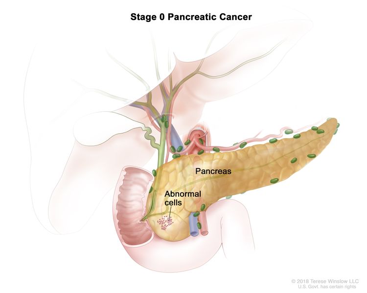
0 Tis, N0, M0 Tis = Carcinoma in situ. This includes high-grade pancreatic intraepithelial neoplasia (PanIn-3), intraductal papillary mucinous neoplasm with high-grade dysplasia, intraductal tubulopapillary neoplasm with high-grade dysplasia, and mucinous cystic neoplasm with high-grade dysplasia.
EnlargeStage 0 pancreatic cancer; drawing shows abnormal cells in the pancreas.
N0 = No regional lymph node metastases.
M0 = No distant metastasis.
Table 2. Definitions for Exocrine Pancreas TNM Stages IA and IBa
Stage TNM Description Illustration
T = primary tumor; N = regional lymph node; M = distant metastasis.
aReprinted with permission from AJCC: Exocrine Pancreas. In: Amin MB, Edge SB, Greene FL, et al., eds.: AJCC Cancer Staging Manual. 8th ed. New York, NY: Springer, 2017, pp. 337–47.
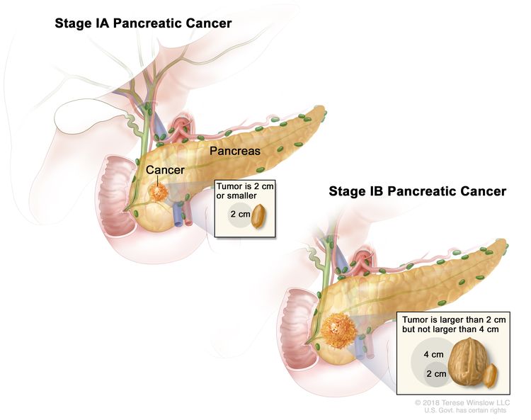
IA T1, N0, M0 T1 = Tumor ≤2 cm in greatest dimension.
EnlargeStage I pancreatic cancer; drawing on the left shows stage IA pancreatic cancer. The cancer is in the pancreas and the tumor is 2 centimeters or smaller. An inset shows 2 centimeters is about the size of a peanut. The drawing on the right shows stage IB pancreatic cancer. The cancer is in the pancreas and the tumor is larger than 2 centimeters but not larger than 4 centimeters. An inset shows 2 centimeters is about the size of a peanut and 4 centimeters is about the size of a walnut.
–T1a = Tumor ≤0.5 cm in greatest dimension.
–T1b = Tumor >0.5 cm and <1 cm in greatest dimension.
–T1c = Tumor 1–2 cm in greatest dimension.
N0 = No regional lymph node metastases.
M0 = No distant metastasis.
IB T2, N0, M0 T2 = Tumor >2 cm and ≤4 cm in greatest dimension.
N0 = No regional lymph node metastases.
M0 = No distant metastasis.
Table 3. Definitions for Exocrine Pancreas TNM Stages IIA and IIBa
Stage TNM Description Illustration
T = primary tumor; N = regional lymph node; M = distant metastasis.
aReprinted with permission from AJCC: Exocrine Pancreas. In: Amin MB, Edge SB, Greene FL, et al., eds.: AJCC Cancer Staging Manual. 8th ed. New York, NY: Springer, 2017, pp. 337–47.
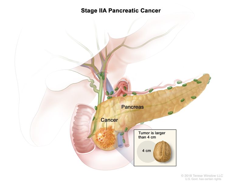
IIA T3, N0, M0 T3 = Tumor >4 cm in greatest dimension.
EnlargeStage IIA pancreatic cancer; drawing shows cancer in the pancreas and the tumor is larger than 4 centimeters. An inset shows 4 centimeters is about the size of a walnut.
N0 = No regional lymph node metastases.
M0 = No distant metastasis.
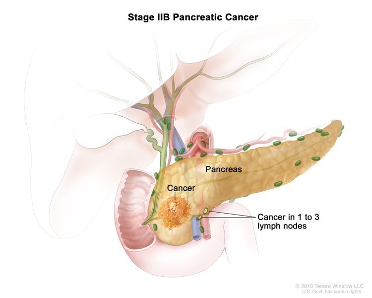
IIB T1, N1, M0 T1 = Tumor ≤2 cm in greatest dimension.
EnlargeStage IIB pancreatic cancer; drawing shows cancer in the pancreas and in 1 to 3 nearby lymph nodes.
–T1a = Tumor ≤0.5 cm in greatest dimension.
–T1b = Tumor >0.5 cm and <1 cm in greatest dimension.
–T1c = Tumor 1–2 cm in greatest dimension.
N1 = Metastasis in one to three regional lymph nodes.
M0 = No distant metastasis.
T2, N1, M0 T2 = Tumor >2 cm and ≤4 cm in greatest dimension.
N1 = Metastasis in one to three regional lymph nodes.
M0 = No distant metastasis.
T3, N1, M0 T3 = Tumor >4 cm in greatest dimension.
N1 = Metastasis in one to three regional lymph nodes.
M0 = No distant metastasis.
Table 4. Definitions for Exocrine Pancreas TNM Stage IIIa
Stage TNM Description Illustration
T = primary tumor; N = regional lymph node; M = distant metastasis.
aReprinted with permission from AJCC: Exocrine Pancreas. In: Amin MB, Edge SB, Greene FL, et al., eds.: AJCC Cancer Staging Manual. 8th ed. New York, NY: Springer, 2017, pp. 337–47.
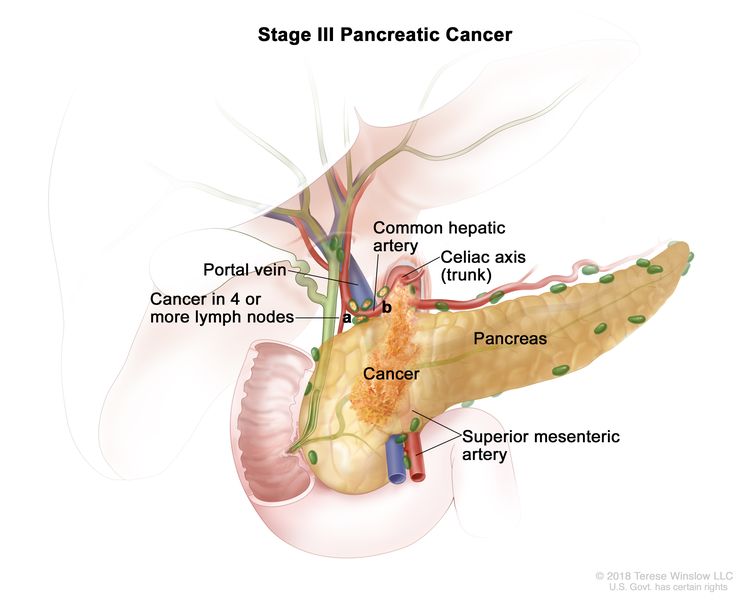
III T1, N2, M0 T1 = Tumor ≤2 cm in greatest dimension.
EnlargeStage III pancreatic cancer; drawing shows cancer in the pancreas and in (a) 4 or more nearby lymph nodes and (b) the common hepatic artery. Also shown are the portal vein, celiac axis (trunk), and superior mesenteric artery.
–T1a = Tumor ≤0.5 cm in greatest dimension.
–T1b = Tumor >0.5 cm and <1 cm in greatest dimension.
–T1c = Tumor 1–2 cm in greatest dimension.
N2 = Metastasis in four or more regional lymph nodes.
M0 = No distant metastasis.
T2, N2, M0 T2 = Tumor >2 cm and ≤4 cm in greatest dimension.
N2 = Metastasis in four or more regional lymph nodes.
M0 = No distant metastasis.
T3, N2, M0 T3 = Tumor >4 cm in greatest dimension.
N2 = Metastasis in four or more regional lymph nodes.
M0 = No distant metastasis.
T4, Any N, M0 T4 = Tumor involves celiac axis, superior mesenteric artery, and/or common hepatic artery, regardless of size.
NX = Regional lymph nodes cannot be assessed.
N0 = No regional lymph node metastases.
N1 = Metastasis in one to three regional lymph nodes.
N2 = Metastasis in four or more regional lymph nodes.
M0 = No distant metastasis.
Table 5. Definitions for Exocrine Pancreas TNM Stage IVa
Stage TNM Description Illustration
T = primary tumor; N = regional lymph node; M = distant metastasis.
aReprinted with permission from AJCC: Exocrine Pancreas. In: Amin MB, Edge SB, Greene FL, et al., eds.: AJCC Cancer Staging Manual. 8th ed. New York, NY: Springer, 2017, pp. 337–47.
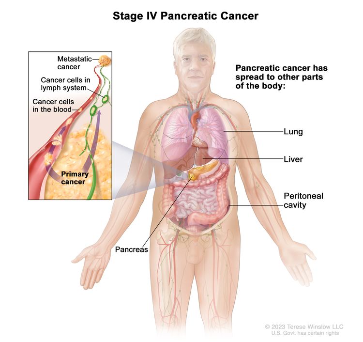
IV Any T, Any N, M1 TX = Primary tumor cannot be assessed.
EnlargeStage IV pancreatic cancer; drawing shows other parts of the body where pancreatic cancer may spread, including the lung, liver, and peritoneal cavity. An inset shows cancer cells spreading from the pancreas, through the blood and lymph system, to another part of the body where metastatic cancer has formed.
T0 = No evidence of primary tumor.
Tis = Carcinoma in situ. This includes high-grade pancreatic intraepithelial neoplasia (PanIn-3), intraductal papillary mucinous neoplasm with high-grade dysplasia, intraductal tubulopapillary neoplasm with high-grade dysplasia, and mucinous cystic neoplasm with high-grade dysplasia.
T1 = Tumor ≤2 cm in greatest dimension.
–T1a = Tumor ≤0.5 cm in greatest dimension.
–T1b = Tumor >0.5 cm and <1 cm in greatest dimension.
–T1c = Tumor 1–2 cm in greatest dimension.
T2 = Tumor >2 cm and ≤4 cm in greatest dimension.
T3 = Tumor >4 cm in greatest dimension.
T4 = Tumor involves celiac axis, superior mesenteric artery, and/or common hepatic artery, regardless of size.
NX = Regional lymph nodes cannot be assessed.
N0 = No regional lymph node metastases.
N1 = Metastasis in one to three regional lymph nodes.
N2 = Metastasis in four or more regional lymph nodes.
M1 = Distant metastasis.
References
  1. Kakar S, Pawlik TM, Allen PJ: Exocrine Pancreas. In: Amin MB, Edge SB, Greene FL, et al., eds.: AJCC Cancer Staging Manual. 8th ed. Springer; 2017, pp. 337–47.

Treatment Option Overview for Pancreatic Cancer

Surgical resection, when feasible, remains the primary treatment modality for patients with pancreatic cancer. On occasion, resection can lead to long-term survival, and it provides effective palliation.[13][Level of evidence C1] Treatment is often guided by resectability, but this may vary depending on surgical judgment and experience. Referral to a high-volume center should be considered.[4]

Postoperative chemotherapy improves overall survival, but the role of chemoradiation remains controversial.

Complications of pancreatic cancer include:

  • Malabsorption: Frequently, malabsorption caused by exocrine insufficiency contributes to malnutrition. Pancreatic enzyme replacement can help alleviate this problem.
  • Pain: Celiac axis and intrapleural nerve blocks can provide highly effective and long-lasting control of pain for some patients. For more information, see Cancer Pain.

The survival rate of patients with any stage of pancreatic exocrine cancer is poor. Clinical trials are appropriate for patients with any stage of disease and should be considered before palliative approaches are selected.

Information about ongoing clinical trials for pancreatic cancer is available from the NCI website.

Table 6. Treatment Options for Pancreatic Cancer
Clinical Stage Treatment Options
Resectable or borderline resectable pancreatic cancer Neoadjuvant therapy
Surgery
Postoperative chemotherapy
Postoperative chemoradiation therapy
Preoperative chemotherapy and/or radiation therapy (under clinical evaluation)
Alternative radiation techniques (under clinical evaluation)
Locally advanced pancreatic cancer Chemotherapy with or without targeted therapy
Chemoradiation therapy
Surgery
Palliative surgery
Clinical trials evaluating novel agents in combination with chemotherapy or chemoradiation therapy for patients with unresectable tumors
Intraoperative radiation therapy and/or implantation of radioactive sources (under clinical evaluation)
Metastatic or recurrent pancreatic cancer Chemotherapy with or without targeted therapy
Clinical trials evaluating new anticancer agents alone or in combination with chemotherapy

Palliative therapies can be considered in patients with any stage of disease. For more information, see the Palliative Therapy section.

Capecitabine and Fluorouracil Dosing

The DPYD gene encodes an enzyme that catabolizes pyrimidines and fluoropyrimidines, like capecitabine and fluorouracil. An estimated 1% to 2% of the population has germline pathogenic variants in DPYD, which lead to reduced DPD protein function and an accumulation of pyrimidines and fluoropyrimidines in the body.[5,6] Patients with the DPYD*2A variant who receive fluoropyrimidines may experience severe, life-threatening toxicities that are sometimes fatal. Many other DPYD variants have been identified, with a range of clinical effects.[57] Fluoropyrimidine avoidance or a dose reduction of 50% may be recommended based on the patient’s DPYD genotype and number of functioning DPYD alleles.[810] DPYD genetic testing costs less than $200, but insurance coverage varies due to a lack of national guidelines.[11] In addition, testing may delay therapy by 2 weeks, which would not be advisable in urgent situations. This controversial issue requires further evaluation.[12]

References
  1. Yeo CJ, Cameron JL, Lillemoe KD, et al.: Pancreaticoduodenectomy for cancer of the head of the pancreas. 201 patients. Ann Surg 221 (6): 721-31; discussion 731-3, 1995. [PUBMED Abstract]
  2. Conlon KC, Klimstra DS, Brennan MF: Long-term survival after curative resection for pancreatic ductal adenocarcinoma. Clinicopathologic analysis of 5-year survivors. Ann Surg 223 (3): 273-9, 1996. [PUBMED Abstract]
  3. Yeo CJ, Abrams RA, Grochow LB, et al.: Pancreaticoduodenectomy for pancreatic adenocarcinoma: postoperative adjuvant chemoradiation improves survival. A prospective, single-institution experience. Ann Surg 225 (5): 621-33; discussion 633-6, 1997. [PUBMED Abstract]
  4. Lidsky ME, Sun Z, Nussbaum DP, et al.: Going the Extra Mile: Improved Survival for Pancreatic Cancer Patients Traveling to High-volume Centers. Ann Surg 266 (2): 333-338, 2017. [PUBMED Abstract]
  5. Sharma BB, Rai K, Blunt H, et al.: Pathogenic DPYD Variants and Treatment-Related Mortality in Patients Receiving Fluoropyrimidine Chemotherapy: A Systematic Review and Meta-Analysis. Oncologist 26 (12): 1008-1016, 2021. [PUBMED Abstract]
  6. Lam SW, Guchelaar HJ, Boven E: The role of pharmacogenetics in capecitabine efficacy and toxicity. Cancer Treat Rev 50: 9-22, 2016. [PUBMED Abstract]
  7. Shakeel F, Fang F, Kwon JW, et al.: Patients carrying DPYD variant alleles have increased risk of severe toxicity and related treatment modifications during fluoropyrimidine chemotherapy. Pharmacogenomics 22 (3): 145-155, 2021. [PUBMED Abstract]
  8. Amstutz U, Henricks LM, Offer SM, et al.: Clinical Pharmacogenetics Implementation Consortium (CPIC) Guideline for Dihydropyrimidine Dehydrogenase Genotype and Fluoropyrimidine Dosing: 2017 Update. Clin Pharmacol Ther 103 (2): 210-216, 2018. [PUBMED Abstract]
  9. Henricks LM, Lunenburg CATC, de Man FM, et al.: DPYD genotype-guided dose individualisation of fluoropyrimidine therapy in patients with cancer: a prospective safety analysis. Lancet Oncol 19 (11): 1459-1467, 2018. [PUBMED Abstract]
  10. Lau-Min KS, Varughese LA, Nelson MN, et al.: Preemptive pharmacogenetic testing to guide chemotherapy dosing in patients with gastrointestinal malignancies: a qualitative study of barriers to implementation. BMC Cancer 22 (1): 47, 2022. [PUBMED Abstract]
  11. Brooks GA, Tapp S, Daly AT, et al.: Cost-effectiveness of DPYD Genotyping Prior to Fluoropyrimidine-based Adjuvant Chemotherapy for Colon Cancer. Clin Colorectal Cancer 21 (3): e189-e195, 2022. [PUBMED Abstract]
  12. Baker SD, Bates SE, Brooks GA, et al.: DPYD Testing: Time to Put Patient Safety First. J Clin Oncol 41 (15): 2701-2705, 2023. [PUBMED Abstract]

Treatment of Resectable or Borderline Resectable Pancreatic Cancer

Treatment Options for Resectable or Borderline Resectable Pancreatic Cancer

Treatment options for resectable or borderline resectable pancreatic cancer include:

  1. Neoadjuvant therapy: Neoadjuvant chemotherapy with or without chemoradiation therapy.
  2. Surgery: Radical pancreatic resection including:
    • Whipple procedure (pancreaticoduodenal resection).
    • Total pancreatectomy when necessary for adequate margins.
    • Distal pancreatectomy for tumors of the body and tail of the pancreas.[1,2]
  3. Postoperative chemotherapy: Radical pancreatic resection followed by chemotherapy.[3]
  4. Postoperative chemoradiation therapy: Radical pancreatic resection followed by fluorouracil (5-FU) chemotherapy and radiation therapy.[48]
  5. Preoperative chemotherapy and/or radiation therapy (under clinical evaluation).
  6. Alternative radiation techniques (under clinical evaluation).

Palliative therapies can be considered in patients with any stage of disease. For more information, see the Palliative Therapy section.

Neoadjuvant therapy

Neoadjuvant therapy is chemotherapy with or without chemoradiation therapy given before surgery. The role of neoadjuvant therapy has been evaluated in retrospective studies (Surveillance, Epidemiology, and End Results [SEER] Program database and National Cancer Database) and is recommended by multiple consensus guidelines for the management of patients with borderline resectable pancreatic cancer. It is being evaluated in patients with resectable or borderline resectable pancreatic cancer in several ongoing trials.[911]

Evidence (neoadjuvant chemotherapy with or without chemoradiation therapy):

  1. The phase II, multicenter, randomized A021501 trial (NCT02839343) enrolled 126 patients with borderline resectable pancreatic cancer from institutions in the National Clinical Trials Network cooperative groups between 2017 and 2019. Patients were assigned to receive either eight 2-week cycles of modified FOLFIRINOX (oxaliplatin, leucovorin, irinotecan, and 5-FU) (n = 65) or seven 2-week cycles of modified FOLFIRINOX followed by stereotactic body radiotherapy (33 Gy–40 Gy in 5 fractions) or hypofractionated image-guided radiotherapy (25 Gy in 5 fractions) (n = 55). Patients without disease progression then underwent surgery followed by four 2-week cycles of adjuvant FOLFOX6 (oxaliplatin, leucovorin, and 5-FU).[12]
    • In the neoadjuvant chemotherapy-alone arm, the median overall survival (OS) was 29.8 months (95% confidence interval [CI], 21.1–36.6) with a 43% microscopically margin-negative (R0) resection rate. This was compared with estimated historical controls of median OS at 18 months.[12][Level of evidence C3]
    • Grade 3 or greater treatment-related adverse events occurred in 57% of patients who received neoadjuvant chemotherapy alone.
    • The neoadjuvant chemotherapy with radiation arm was closed at interim futility analysis because of low R0 resection rates (33%) in the first 30 patients enrolled.
  2. The multicenter phase III PREOPANC trial included 246 patients diagnosed with resectable or borderline resectable pancreatic cancer between 2013 and 2017. Patients at 16 Dutch centers were randomly assigned to receive either diagnostic laparoscopy, neoadjuvant chemoradiation therapy, surgical resection, and four cycles of adjuvant gemcitabine or up-front surgery and six cycles of adjuvant gemcitabine. Neoadjuvant chemoradiation therapy included the following: cycle 1 (21 days) with gemcitabine 1,000 mg/m2 on days 1 and 8; cycle 2 (28 days) with gemcitabine on days 1, 8, and 15 with 15 concurrent fractions of hypofractionated radiation (36 Gy) to the tumor and suspected associated lymph nodes; and cycle 3 (21 days) with gemcitabine on days 1 and 8.[13]
    • The 5-year OS rate was 20.5% (95% CI, 14.2%–29.8%) for patients who received neoadjuvant chemoradiation therapy and 6.5% (95% CI, 3.1%–13.7%) for patients who received up-front surgery (hazard ratio [HR], 0.73; 95% CI, 0.56–0.96; P = .025).[13][Level of evidence A1]
    • The median OS was 15.7 months in the neoadjuvant chemoradiation therapy group and 14.3 months in the up-front surgery group.
    • In the intention-to-treat arm, 61% of patients who received neoadjuvant chemoradiation therapy underwent resection, resulting in R0 resection for 41% of patients and node-negative disease for 65% of patients. The resection rate was 72% in the up-front surgery arm, resulting in R0 resection in 28% of patients and node-negative disease in 18% of patients.

    The optimal neoadjuvant therapy regimen is unknown, and additional chemotherapy regimens are being evaluated in the following trials: ALLIANCE (NCT04340141), PREOPANC-3 (NCT04927780), PANACHE-01-PRODIGE (NCT02959879), and NorPACT-01 (NCT02919787).

Surgery

Complete resection can yield 5-year survival rates of 18% to 24%, but ultimate control remains poor because of the high incidence of both local and distant tumor recurrence. Thus, systemic therapy is also recommended.[1416][Level of evidence C1]

Approximately 20% of patients present with pancreatic cancer amenable to local surgical resection, with operative mortality rates of approximately 1% to 16%.[1721] Using information from the Medicare claims database, a national cohort study of more than 7,000 patients undergoing pancreaticoduodenectomy between 1992 and 1995 revealed higher in-hospital mortality rates at low-volume hospitals (<1 pancreaticoduodenectomy per year) versus high-volume hospitals (>5 per year) (16% vs. 4%, respectively; P < .01).[17]

Postoperative chemotherapy

Historically, multiple randomized trials have established that adjuvant gemcitabine monotherapy [22] or adjuvant 5-FU monotherapy [3] improve OS for 6 months after surgical resection compared with surgery alone. More recent studies have looked at newer combination regimens that might further improve outcomes after surgical resection.

For patients with good performance status, adjuvant FOLFIRINOX chemotherapy or the combination of gemcitabine and capecitabine should be considered. However, for older patients or patients with marginal performance status, adjuvant gemcitabine or 5-FU monotherapy can be considered. In Asia, S-1 (tegafur, gimeracil, and oteracil potassium) is an appropriate alternative to gemcitabine-based therapies.

Evidence (postoperative chemotherapy):

  1. FOLFIRINOX: In the randomized, open-label, phase III PRODIGE-24 trial (NCT01526135), 493 patients with R0/R1 resections were randomly assigned 1:1 to receive six cycles of gemcitabine (1,000 mg/m2 on days 1, 8, and 15 of a 28-day cycle) or 12 cycles of FOLFIRINOX (oxaliplatin 85 mg/m2, leucovorin 400 mg/m2, irinotecan 150 mg/m2, and 5-FU 2,400 mg/m2 over 46 hours every 2 weeks).[23,24][Level of evidence A1]
    • With a median follow-up of 69.7 months, the median disease-free survival (DFS) was 21.4 months (95% CI, 17.5–26.7) in the FOLFIRINOX group and 12.8 months in the gemcitabine group (95% CI, 11.6–15.2) (HR, 0.66; 95% CI, 0.54–0.82; P < .001).
    • The median OS was 53.5 months (95% CI, 43.5–58.4) in the FOLFIRINOX group and 35.5 months (95% CI, 30.1–40.3) in the gemcitabine group (HR, 0.68; 95% CI, 0.54–0.85; P = .001). The 5-year OS rate was 43.2% in the FOLFIRINOX group and 31.4% in the gemcitabine group.
    • Toxicity was higher with combination therapy; 75.9% of patients who received FOLFIRINOX had grade 3 or 4 toxicities, compared with 52.9% of those who received gemcitabine, with similar rates of neutropenia (although 62.2% of patients on FOLFIRINOX received granulocyte colony-stimulating factor). Thirty-three percent of patients who received FOLFIRINOX stopped treatment prematurely, compared with 21% of patients who received gemcitabine alone.
  2. Gemcitabine and capecitabine: The European Study for Pancreatic Cancer (ESPAC-4 [NCT00058201]) trial randomly assigned 732 patients with resected pancreatic cancer to receive either six cycles of gemcitabine alone (1,000 mg/m2 given weekly for 3 weeks of every 4 weeks) or oral capecitabine (1,660 mg/m2 given for 21 days followed by 7 days of rest [one cycle]).[25][Level of evidence A1]
    • With a median follow-up of 43.2 months, the median OS for patients in the gemcitabine/capecitabine group was 28.0 months (95% CI, 23.5–31.5) compared with 25.5 months for the gemcitabine-alone group (95% CI, 22.7–27.9; HR, 0.82; P = .032). Treatment with gemcitabine/capecitabine yielded an improvement in the estimated 5-year OS rate from 16.3% with gemcitabine alone to 28.8% with gemcitabine/capecitabine.
    • There was no significant difference in overall rates of grade 3/4 toxicities between treatment arms. Compared with gemcitabine alone, capecitabine was associated with higher rates of grade 3/4 diarrhea (5% vs. 2%), neutropenia (38% vs. 24%), and hand-foot syndrome (7% vs. 0%).
    • There was no significant effect on the quality of life in the treatment groups.
    • Based on these findings, the adjuvant combination of gemcitabine and capecitabine is the standard of care after a resection for pancreatic cancer.
  3. S-1: The Japan Adjuvant Study Group of Pancreatic Cancer (JASPAC-01) study was a phase III, multicenter, noninferiority trial conducted in Japan that randomly assigned 385 patients to receive either gemcitabine (1,000 mg/m2 weekly for 3 weeks of every 4 weeks) for six cycles or S-1 (tegafur, gimeracil, and oteracil potassium) (given orally twice a day for 4 weeks followed by a 2-week break).[26][Level of evidence A1]
    • The prespecified criteria for early discontinuation was met at interim analysis for efficacy with all of the protocol treatments completed. On early interim analysis, the HRmortality was 0.57 (95% CI, 0.44–0.72; P for noninferiority < .001; P for superiority < .001). These results were associated with a 5-year OS rate of 24.4% in the gemcitabine group and 44.1% in the S-1 group.
    • Grade 3 or 4 leukopenia, neutropenia, and liver transaminitis were observed more frequently in the gemcitabine group, and stomatitis and diarrhea were experienced more frequently in the S-1 group.
    • Among Japanese patients, adjuvant chemotherapy with S-1 can be a new standard of care for resected pancreatic patients. Additional studies are needed to validate these results in patients of other races and ethnicities.
    • The pharmacokinetics and pharmacodynamics of S-1 may be different between Eastern and Western patient populations because grade 3/4 gastrointestinal toxicities, especially diarrhea, have been reported more commonly in the Western patient population. S-1 is not currently approved by the U.S. Food and Drug Administration for use in the United States.
  4. Gemcitabine: Charité Onkologie (CONKO)-001 was a multicenter phase III trial of 368 patients with resected pancreatic cancer who were randomly assigned to receive six cycles of adjuvant gemcitabine versus observation.[22][Level of evidence B1] In contrast to the previous trials, the primary end point was DFS.
    • With a median follow-up of 136 months, long-term follow-up of the CONKO-001 study demonstrated a significant improvement in OS that favors gemcitabine (median survival, 22.8 months vs. 20.2 months; HR, 0.76; 95% CI, 0.61–0.95, P = .01). Gemcitabine compared with observation alone yielded improved survival rates at 5 years of 20.7% for the gemcitabine arm versus 10.4% for the observation-alone arm, and the survival rates at 10 years were 12.2% for the gemcitabine arm versus 7.7% for the observation-alone arm.[27][Level of evidence A1]
  5. Gemcitabine or 5-FU: The ESPAC-3 trial (NCT00058201) randomly assigned 1,088 patients who had undergone complete macroscopic resection to either 6 months of 5-FU (425 mg/m2) and leucovorin (20 mg/m2) on days 1 to 5 every 28 days or 6 months of gemcitabine (1,000 mg/m2) on days 1, 8, and 15 every 28 days.[3][Level of evidence A1]
    • Median OS was 23.0 months (95% CI, 21.1–25.0) for patients treated with 5-FU plus leucovorin and 23.6 months (95% CI, 21.4–26.4) for those treated with gemcitabine (HR, 0.94; 95% CI, 0.81–1.08; P = .39).

Postoperative chemoradiation therapy

The role of postoperative therapy (chemotherapy with or without chemoradiation therapy) in the management of this disease remains controversial because much of the randomized clinical trial data available are statistically underpowered and provide conflicting results.[48]

Evidence (postoperative chemoradiation therapy):

Several phase III trials examined the potential OS benefit of postoperative adjuvant 5-FU–based chemoradiation therapy:

  1. Gastrointestinal Study Group (GITSG): A small randomized trial conducted by the GITSG in 1985 compared surgery alone with surgery followed by chemoradiation.[4][Level of evidence A1];[5][Level of evidence B4]
    • The investigators reported a significant but modest improvement in median-term and long-term survival over resection alone with postoperative bolus 5-FU and regional split-course radiation given at a dose of 40 Gy.
  2. European Organisation for the Research and Treatment of Cancer (EORTC): An attempt by the EORTC to reproduce the results of the GITSG trial failed to confirm a significant benefit for adjuvant chemoradiation therapy over resection alone;[6][Level of evidence A1] however, this trial treated patients with pancreatic and periampullary cancers (with a potentially better prognosis).
    • A subset analysis of the patients with primary pancreatic tumors indicated a trend toward improved median, 2-year, and 5-year OS with adjuvant therapy (17.1 months, 37%, and 20%, respectively) compared with surgery alone (12.6 months, 23%, and 10%, respectively); P = .09 for median survival).
  3. An updated analysis of a subsequent ESPAC-1 trial examined only patients who underwent strict randomization after pancreatic resection. The patients were assigned to one of four groups (observation, bolus 5-FU chemotherapy, bolus 5-FU chemoradiation therapy, or chemoradiation therapy followed by additional chemotherapy).[7,8,28][Level of evidence A1]
    • With a 2 × 2 factorial design reported at a median follow-up of 47 months, a median survival benefit was observed for only the patients who received postoperative 5-FU chemotherapy. However, these results were difficult to interpret because of a high rate of protocol nonadherence and the lack of a separate analysis for each of the four groups in the 2 × 2 design.
  4. U.S. Gastrointestinal Intergroup: The U.S. Gastrointestinal Intergroup has reported the results of a randomized phase III trial (Radiation Therapy Oncology Group [RTOG]-9704) that included 451 patients with resected pancreatic cancers who were assigned to receive either postoperative infusional 5-FU plus infusional 5-FU and concurrent radiation or adjuvant gemcitabine plus infusional 5-FU and concurrent radiation.[29][Level of evidence A1] The primary end points were OS for all patients and OS for patients with pancreatic head tumors.
    • A 5-year update of RTOG-9704 reported that patients with pancreatic head tumors (n = 388) had a median survival of 20.5 months and a 5-year OS rate of 22% with gemcitabine, versus a median survival of 17.1 months and a 5-year OS rate of 18% with 5-FU (HR, 0.84; 95% CI, 0.67–1.05; P = .12).[30]
    • Univariate analysis showed no difference in OS. However, on multivariate analysis, patients on the gemcitabine arm with pancreatic head tumors experienced a trend toward improved OS (P = .08). Distant relapse remained the predominant site of first failure (78%).

The EORTC/U.S. Gastrointestinal Intergroup RTOG-0848 phase III adjuvant trial evaluating the impact of chemoradiation therapy after completion of a full course of gemcitabine with or without erlotinib has closed and results are pending.

Additional trials are still warranted to determine more effective systemic therapy for this disease.

Current Clinical Trials

Use our advanced clinical trial search to find NCI-supported cancer clinical trials that are now enrolling patients. The search can be narrowed by location of the trial, type of treatment, name of the drug, and other criteria. General information about clinical trials is also available.

References
  1. Dalton RR, Sarr MG, van Heerden JA, et al.: Carcinoma of the body and tail of the pancreas: is curative resection justified? Surgery 111 (5): 489-94, 1992. [PUBMED Abstract]
  2. Brennan MF, Moccia RD, Klimstra D: Management of adenocarcinoma of the body and tail of the pancreas. Ann Surg 223 (5): 506-11; discussion 511-2, 1996. [PUBMED Abstract]
  3. Neoptolemos JP, Stocken DD, Bassi C, et al.: Adjuvant chemotherapy with fluorouracil plus folinic acid vs gemcitabine following pancreatic cancer resection: a randomized controlled trial. JAMA 304 (10): 1073-81, 2010. [PUBMED Abstract]
  4. Further evidence of effective adjuvant combined radiation and chemotherapy following curative resection of pancreatic cancer. Gastrointestinal Tumor Study Group. Cancer 59 (12): 2006-10, 1987. [PUBMED Abstract]
  5. Kalser MH, Ellenberg SS: Pancreatic cancer. Adjuvant combined radiation and chemotherapy following curative resection. Arch Surg 120 (8): 899-903, 1985. [PUBMED Abstract]
  6. Klinkenbijl JH, Jeekel J, Sahmoud T, et al.: Adjuvant radiotherapy and 5-fluorouracil after curative resection of cancer of the pancreas and periampullary region: phase III trial of the EORTC gastrointestinal tract cancer cooperative group. Ann Surg 230 (6): 776-82; discussion 782-4, 1999. [PUBMED Abstract]
  7. Neoptolemos JP, Dunn JA, Stocken DD, et al.: Adjuvant chemoradiotherapy and chemotherapy in resectable pancreatic cancer: a randomised controlled trial. Lancet 358 (9293): 1576-85, 2001. [PUBMED Abstract]
  8. Neoptolemos JP, Stocken DD, Friess H, et al.: A randomized trial of chemoradiotherapy and chemotherapy after resection of pancreatic cancer. N Engl J Med 350 (12): 1200-10, 2004. [PUBMED Abstract]
  9. Stessin AM, Meyer JE, Sherr DL: Neoadjuvant radiation is associated with improved survival in patients with resectable pancreatic cancer: an analysis of data from the surveillance, epidemiology, and end results (SEER) registry. Int J Radiat Oncol Biol Phys 72 (4): 1128-33, 2008. [PUBMED Abstract]
  10. Versteijne E, Vogel JA, Besselink MG, et al.: Meta-analysis comparing upfront surgery with neoadjuvant treatment in patients with resectable or borderline resectable pancreatic cancer. Br J Surg 105 (8): 946-958, 2018. [PUBMED Abstract]
  11. Mokdad AA, Minter RM, Zhu H, et al.: Neoadjuvant Therapy Followed by Resection Versus Upfront Resection for Resectable Pancreatic Cancer: A Propensity Score Matched Analysis. J Clin Oncol 35 (5): 515-522, 2017. [PUBMED Abstract]
  12. Katz MHG, Shi Q, Meyers J, et al.: Efficacy of Preoperative mFOLFIRINOX vs mFOLFIRINOX Plus Hypofractionated Radiotherapy for Borderline Resectable Adenocarcinoma of the Pancreas: The A021501 Phase 2 Randomized Clinical Trial. JAMA Oncol 8 (9): 1263-1270, 2022. [PUBMED Abstract]
  13. Versteijne E, van Dam JL, Suker M, et al.: Neoadjuvant Chemoradiotherapy Versus Upfront Surgery for Resectable and Borderline Resectable Pancreatic Cancer: Long-Term Results of the Dutch Randomized PREOPANC Trial. J Clin Oncol 40 (11): 1220-1230, 2022. [PUBMED Abstract]
  14. Cameron JL, Crist DW, Sitzmann JV, et al.: Factors influencing survival after pancreaticoduodenectomy for pancreatic cancer. Am J Surg 161 (1): 120-4; discussion 124-5, 1991. [PUBMED Abstract]
  15. Yeo CJ, Cameron JL, Lillemoe KD, et al.: Pancreaticoduodenectomy for cancer of the head of the pancreas. 201 patients. Ann Surg 221 (6): 721-31; discussion 731-3, 1995. [PUBMED Abstract]
  16. Yeo CJ, Abrams RA, Grochow LB, et al.: Pancreaticoduodenectomy for pancreatic adenocarcinoma: postoperative adjuvant chemoradiation improves survival. A prospective, single-institution experience. Ann Surg 225 (5): 621-33; discussion 633-6, 1997. [PUBMED Abstract]
  17. Birkmeyer JD, Finlayson SR, Tosteson AN, et al.: Effect of hospital volume on in-hospital mortality with pancreaticoduodenectomy. Surgery 125 (3): 250-6, 1999. [PUBMED Abstract]
  18. Cameron JL, Pitt HA, Yeo CJ, et al.: One hundred and forty-five consecutive pancreaticoduodenectomies without mortality. Ann Surg 217 (5): 430-5; discussion 435-8, 1993. [PUBMED Abstract]
  19. Spanknebel K, Conlon KC: Advances in the surgical management of pancreatic cancer. Cancer J 7 (4): 312-23, 2001 Jul-Aug. [PUBMED Abstract]
  20. Balcom JH, Rattner DW, Warshaw AL, et al.: Ten-year experience with 733 pancreatic resections: changing indications, older patients, and decreasing length of hospitalization. Arch Surg 136 (4): 391-8, 2001. [PUBMED Abstract]
  21. Sohn TA, Yeo CJ, Cameron JL, et al.: Resected adenocarcinoma of the pancreas-616 patients: results, outcomes, and prognostic indicators. J Gastrointest Surg 4 (6): 567-79, 2000 Nov-Dec. [PUBMED Abstract]
  22. Oettle H, Post S, Neuhaus P, et al.: Adjuvant chemotherapy with gemcitabine vs observation in patients undergoing curative-intent resection of pancreatic cancer: a randomized controlled trial. JAMA 297 (3): 267-77, 2007. [PUBMED Abstract]
  23. Conroy T, Hammel P, Hebbar M, et al.: FOLFIRINOX or Gemcitabine as Adjuvant Therapy for Pancreatic Cancer. N Engl J Med 379 (25): 2395-2406, 2018. [PUBMED Abstract]
  24. Conroy T, Castan F, Lopez A, et al.: Five-Year Outcomes of FOLFIRINOX vs Gemcitabine as Adjuvant Therapy for Pancreatic Cancer: A Randomized Clinical Trial. JAMA Oncol 8 (11): 1571-1578, 2022. [PUBMED Abstract]
  25. Neoptolemos JP, Palmer DH, Ghaneh P, et al.: Comparison of adjuvant gemcitabine and capecitabine with gemcitabine monotherapy in patients with resected pancreatic cancer (ESPAC-4): a multicentre, open-label, randomised, phase 3 trial. Lancet 389 (10073): 1011-1024, 2017. [PUBMED Abstract]
  26. Uesaka K, Boku N, Fukutomi A, et al.: Adjuvant chemotherapy of S-1 versus gemcitabine for resected pancreatic cancer: a phase 3, open-label, randomised, non-inferiority trial (JASPAC 01). Lancet 388 (10041): 248-57, 2016. [PUBMED Abstract]
  27. Oettle H, Neuhaus P, Hochhaus A, et al.: Adjuvant chemotherapy with gemcitabine and long-term outcomes among patients with resected pancreatic cancer: the CONKO-001 randomized trial. JAMA 310 (14): 1473-81, 2013. [PUBMED Abstract]
  28. Choti MA: Adjuvant therapy for pancreatic cancer–the debate continues. N Engl J Med 350 (12): 1249-51, 2004. [PUBMED Abstract]
  29. Regine WF, Winter KA, Abrams RA, et al.: Fluorouracil vs gemcitabine chemotherapy before and after fluorouracil-based chemoradiation following resection of pancreatic adenocarcinoma: a randomized controlled trial. JAMA 299 (9): 1019-26, 2008. [PUBMED Abstract]
  30. Regine WF, Winter KA, Abrams R, et al.: Fluorouracil-based chemoradiation with either gemcitabine or fluorouracil chemotherapy after resection of pancreatic adenocarcinoma: 5-year analysis of the U.S. Intergroup/RTOG 9704 phase III trial. Ann Surg Oncol 18 (5): 1319-26, 2011. [PUBMED Abstract]

Treatment of Locally Advanced Pancreatic Cancer

Treatment Options for Locally Advanced Pancreatic Cancer

While locally advanced and metastatic pancreatic cancer are both incurable, their natural histories may be different. An autopsy series demonstrated that 30% of patients presenting with locally advanced disease died without evidence of distant metastases.[1][Level of evidence A1] Therefore, investigators have struggled to determine whether chemoradiation therapy for patients presenting with locally advanced disease is warranted.

Treatment options for locally advanced pancreatic cancer include:

  1. Chemotherapy with or without targeted therapy.
  2. Chemoradiation therapy: Chemotherapy followed by chemoradiation for patients without metastatic disease.
  3. Surgery: Radical pancreatic resection.
  4. Palliative surgery: Palliative surgical biliary and/or gastric bypass, percutaneous radiologic biliary stent placement, or endoscopic biliary stent placement.[2,3]
  5. Clinical trials evaluating novel agents in combination with chemotherapy or chemoradiation therapy for patients with unresectable tumors.
  6. Intraoperative radiation therapy and/or implantation of radioactive sources (under clinical evaluation).[4,5]

Palliative therapies can be considered in patients with any stage of disease. For more information, see the Palliative Therapy section.

Chemotherapy with or without targeted therapy

Chemotherapy is the primary treatment modality for patients with locally advanced pancreatic cancers and uses the same regimens as those used to treat patients with metastatic disease.

Evidence (chemotherapy):

  1. FOLFIRINOX versus gemcitabine: A multicenter phase II/III trial included 342 patients with metastatic pancreatic adenocarcinoma with an Eastern Cooperative Oncology Group (ECOG) performance status score of 0 or 1.[6][Level of evidence A1] The patients were randomly assigned to receive FOLFIRINOX (oxaliplatin [85 mg/m2], irinotecan [180 mg/m2], leucovorin [400 mg/m2], and fluorouracil [5-FU; 400 mg/m2] given as a bolus followed by 2,400 mg/m2 given as a 46-hour continuous infusion, every 2 weeks) or gemcitabine (1,000 mg/m2 weekly for 7 of 8 weeks and then weekly for 3 of 4 weeks).
    • The median overall survival (OS) was 11.1 months in the FOLFIRINOX group compared with 6.8 months in the gemcitabine group (hazard ratio [HR]death, 0.57; 95% confidence interval [CI], 0.45–0.73; P < .001).
    • Median progression-free survival (PFS) was 6.4 months in the FOLFIRINOX group and 3.3 months in the gemcitabine group (HRdisease progression, 0.47; 95% CI, 0.37–0.59; P < .001).
    • FOLFIRINOX was more toxic than gemcitabine; 5.4% of patients in this group had febrile neutropenia. At 6 months, 31% of the patients in the FOLFIRINOX group had a definitive degradation of quality of life, versus 66% in the gemcitabine group (HR, 0.47; 95% CI, 0.30–0.70; P < .001).
    • Based on this trial, FOLFIRINOX is considered a standard treatment option for patients with advanced pancreatic cancer.
  2. Gemcitabine and nab-paclitaxel versus gemcitabine: A multicenter, international, phase III trial (NCT00844649) included 861 patients with metastatic pancreatic adenocarcinoma. Patients had a Karnofsky Performance Status of at least 70 and had not previously received chemotherapy for metastatic disease.[7][Level of evidence A1] Patients who received adjuvant gemcitabine or any other chemotherapy were excluded. The patients were randomly assigned to receive gemcitabine (1,000 mg/m2) and nab-paclitaxel (125 mg/m2 of body-surface area) weekly for 3 of 4 weeks or gemcitabine monotherapy (1,000 mg/m2 weekly for 7 of 8 weeks and then weekly for 3 of 4 weeks).
    • The median OS was 8.5 months in the nab-paclitaxel/gemcitabine group compared with 6.7 months in the gemcitabine group (HRdeath, 0.72; 95% CI, 0.62–0.83; P < .001).
    • Median PFS was 5.5 months in the nab-paclitaxel/gemcitabine group and 3.7 months in the gemcitabine group (HRdisease progression, 0.69; 95% CI, 0.58–0.82; P < .001).
    • Nab-paclitaxel/gemcitabine was more toxic than gemcitabine. The most common grade 3 toxicities in the nab-paclitaxel/gemcitabine group were neutropenia (38%), fatigue (17%), and neuropathy (17%); febrile neutropenia occurred in 3% of patients. In the gemcitabine-alone group, the most common grade 3 toxicities were neutropenia (27%), fatigue (1%), and neuropathy (1%); febrile neutropenia occurred in 1% of patients.
    • In the nab-paclitaxel/gemcitabine group, the median time from grade 3 neuropathy to grade 1 or resolution was 29 days. Of patients with grade 3 peripheral neuropathy, 44% were able to resume treatment at a reduced dose within a median of 23 days after onset of a grade 3 event.
    • Based on this trial, nab-paclitaxel/gemcitabine is a standard treatment option for patients with advanced pancreatic cancer.
    • Quality-of-life data were not measured for this regimen, and this study did not address the efficacy of nab-paclitaxel/gemcitabine versus FOLFIRINOX.
  3. Gemcitabine versus 5-FU: Gemcitabine has demonstrated activity in patients with pancreatic cancer and is a useful palliative agent.[810] A phase III trial of gemcitabine versus 5-FU as first-line therapy in patients with advanced or metastatic adenocarcinoma of the pancreas reported a significant improvement in survival among patients treated with gemcitabine (the 1-year survival rate was 18% with gemcitabine compared with 2% with 5-FU; P = .003).[9][Level of evidence A1]
  4. Gemcitabine alone versus gemcitabine and erlotinib: The National Cancer Institute of Canada performed a phase III trial (CAN-NCIC-PA3 [NCT00026338]) that compared gemcitabine alone with the combination of gemcitabine and erlotinib (100 mg/d) in patients with advanced or metastatic pancreatic carcinomas.[11][Level of evidence A1]
    • The addition of erlotinib modestly prolonged survival when combined with gemcitabine versus gemcitabine alone (HR, 0.81; 95% CI, 0.69–0.99; P = .038).
    • The median and 1-year survival rates for patients who received erlotinib were 6.2 months and 23%. The median and 1-year survival rates for patients who received placebo were 5.9 months and 17%.
  5. Platinum analogue or fluoropyrimidine versus single-agent gemcitabine: Many phase III studies have evaluated a combination regimen with either a platinum analogue (cisplatin or oxaliplatin) or fluoropyrimidine versus single-agent gemcitabine.[12,13]
    • None of these phase III trials have demonstrated a statistically significant advantage favoring the use of combination chemotherapy in the first-line treatment of metastatic pancreatic cancer.
  6. 5-FU, leucovorin, and oxaliplatin (OFF regimen) versus best supportive care (BSC): Second-line chemotherapy after progression on a gemcitabine-based regimen may be beneficial. The Charité Onkologie (CONKO)-003 investigators randomly assigned patients requiring a second line of chemotherapy to either the OFF regimen or BSC.[14,15][Level of evidence C1] The OFF regimen consisted of leucovorin (200 mg/m2) followed by 5-FU (2,000 mg/m2 [24-hour continuous infusion] on days 1, 8, 15, and 22) and oxaliplatin (85 mg/m2 on days 8 and 22). After a rest of 3 weeks, the next cycle was started on day 43. The trial was terminated early because of poor accrual, and only 46 patients were randomly assigned to either the OFF regimen or BSC.
    • The median survival was 4.82 months (95% CI, 4.29–5.35) with the OFF treatment regimen and 2.30 months (95% CI, 1.76–2.83) with BSC alone (HR, 0.45; 95% CI, 0.24–0.83).
    • Median OS was 9.09 months for the sequence of gemcitabine/OFF and 7.90 months for gemcitabine/BSC.
    • The early closure of the study and the very small number of patients made the P values misleading. Therefore, second-line chemotherapy with the OFF regimen may be falsely associated with improved survival.

Chemoradiation therapy

The role of chemoradiation in locally advanced pancreatic cancer remains controversial. Table 7 summarizes phase III randomized studies of chemoradiation for locally advanced pancreatic cancer.

Table 7. Randomized Studies in Locally Advanced Pancreatic Cancer: Median Survival
Trial Regimen Chemoradiation Radiation Alone Chemotherapy Alone P Value
5-FU = fluorouracil; ECOG = Eastern Cooperative Oncology Group; FFCD = Fédération Francophone de Cancérologie Digestive; GEM = gemcitabine; GITSG = Gastrointestinal Tumor Study Group; Gy = gray (unit of absorbed radiation of ionizing radiation); P value = probability value; XRT = radiation therapy.
Pre-2000  
GITSG [16] Radiation alone vs. 5-FU/60 Gy XRT 40 wk 20 wk   <.01
ECOG [17] Radiation vs. 5-FU, mitomycin C/59 Gy XRT 8.4 mo 7.1 mo   .16
Post-2000  
FFCD [18] GEM vs. GEM, cisplatin, 60 Gy XRT 8.6 mo   13 mo .03
ECOG [19] GEM vs. GEM/50.4 Gy XRT 11.1 mo   9.2 mo .017

Evidence (chemoradiation therapy):

Three trials evaluated combined modality therapy versus radiation therapy alone.[1618] The trials had substantial deficiencies in design or analysis. Initially, the standard of practice was to give chemoradiation therapy based on data from the first two studies. However, with the publication of the third study, standard practice changed to chemotherapy followed by chemoradiation in the absence of metastases.

  1. LAP07 (NCT00634725): The LAP07 study was an international, randomized, phase III study based on the results of the Groupe Coopérateur Multidisciplinaire en Oncologie (GERCOR) study. In total, 449 patients were enrolled between 2008 and 2011, with random assignment via a two-step randomization process. In the first step, patients were randomly assigned to induction gemcitabine (n = 223) or gemcitabine plus erlotinib (n = 219) for four cycles. For the second step, patients with controlled tumors were randomly assigned (n = 269) a second time to receive either chemotherapy (n = 136) or chemoradiation therapy (n = 133). A total dose of 54 Gy in 30 daily fractions was prescribed with concurrent capecitabine at a dose of 800 mg/m2 twice daily on days of radiation therapy.[20][Level of evidence A1]
    • The primary end point was OS. After interim analysis, the study was stopped early because of futility.
    • With a median follow-up of 36.7 months, the median OS from the date of the first randomization was not significantly different between chemotherapy at 16.5 months (95% CI, 14.5–18.5) and chemoradiation therapy at 15.2 months (95% CI, 13.9–17.3; P = .83).
    • Median OS after the first randomization was 13.6 months (95% CI, 12.3–15.3) for the patients who received gemcitabine and was 11.9 months (95% CI, 10.4–13.5; P = .09) for the patients who received gemcitabine plus erlotinib.

    The LAP07 study represents the most robust, prospective, randomized phase III data regarding the role of chemoradiation therapy in the setting of gemcitabine-based induction chemotherapy that demonstrates no OS benefit. However, this study was initiated before the advent of FOLFIRINOX chemotherapy, which has been widely adopted into the locally advanced setting. The role of chemoradiation in the setting of more active chemotherapy regimens, including gemcitabine/paclitaxel and FOLFIRINOX, has yet to be evaluated.

  2. Gastrointestinal Tumor Study Group (GITSG) GITSG-9273 trial: Before 2000, several phase III trials evaluated combined modality therapy versus radiation therapy alone. Before the use of gemcitabine for patients with locally advanced or metastatic pancreatic cancer, investigators from the GITSG randomly assigned 106 patients with locally advanced pancreatic adenocarcinoma to receive external-beam radiation therapy (EBRT) (60 Gy) alone or concurrent EBRT (either 40 Gy or 60 Gy) plus bolus 5-FU.[16][Level of evidence A1]
    • The study was stopped early when the chemoradiation therapy groups were found to have better efficacy. The 1-year survival rate was 11% for patients who received EBRT alone compared with 38% for patients who received chemoradiation therapy with 40 Gy and 36% for patients who received chemoradiation therapy with 60 Gy.
    • After an additional 88 patients were enrolled in the combined modality arms, there was a trend toward improved survival with 60 Gy EBRT plus 5-FU, but the difference in time-to-progression and OS was not statistically significant when compared with the 40 Gy arm.[21]
  3. ECOG E-8282 trial: Investigators from the ECOG randomly assigned 114 patients to receive radiation therapy (59.4 Gy) alone or with concurrent infusional 5-FU (1,000 mg/m2/d on days 2–5 and 28–31) plus mitomycin (10 mg/m2 on day 2).[17]
    • The trial reported no difference in OS between the two groups.
  4. Fédération Francophone de Cancérologie Digestive–Société Française de Radiothérapie Oncologie (FFCD-SFRO) trial: As it became clear that radiation therapy alone was an inadequate treatment, investigators evaluated combined modality approaches versus chemotherapy alone. Investigators from the FFCD-SFRO randomly assigned 119 patients to induction chemoradiation therapy (60 Gy in 2 Gy fractions with 300 mg/m2/d of continuous-infusion 5-FU on days 1–5 for 6 weeks and 20 mg/m2/d of cisplatin on days 1–5 during weeks 1 and 5) or induction gemcitabine (1,000 mg/m2 weekly for 7 weeks). Maintenance gemcitabine was administered to both groups until stopped by disease progression or treatment discontinuation as a result of toxicity.[22][Level of evidence A1]
    • Median survival was superior in the gemcitabine group (13 vs. 8.6 months; P = .03).
    • Nonhematological grade 3 to 4 toxicities (primarily gastrointestinal) were significantly more common in the chemoradiation therapy group (44% vs. 18%; P = .004), and fewer patients completed at least 75% of induction therapy (42% vs. 73%).
    • Nonetheless, the survival benefit persisted in a per-protocol analysis of patients receiving at least 75% of planned therapy. Notably, the dose intensity of maintenance gemcitabine was significantly less in the chemoradiation therapy group because of a greater incidence of grades 3 to 4 hematological toxicities (71% vs. 27%; P = .0001).
    • As a result of this study, giving induction chemoradiation therapy has lost support.
  5. ECOG: The results of the FFCD-SFRO study counter the results of a study from ECOG in which investigators randomly assigned 74 patients to either gemcitabine alone or gemcitabine with radiation followed by gemcitabine.[19] Of note, the study was closed early as the result of poor accrual.
    • The primary end point was survival, which was 9.2 months (95% CI, 7.9–11.4) for chemotherapy and 11.1 months (95% CI, 7.6–15.5) for combined modality therapy (one-sided P = .017 by stratified log-rank test).
    • Grades 4 and 5 toxicity were greater in the chemoradiation therapy arm than in the chemotherapy arm (41% vs. 9%).
  6. GERCOR: Given the increased toxicity of chemoradiation therapy and the early development of metastatic disease in a large percentage of patients with locally advanced pancreatic cancer, investigators are pursuing a strategy of selecting patients with localized disease for chemoradiation therapy. With this strategy, the selected patients have an absence of progressive disease locally or systemically after several months of chemotherapy.[23][Level of evidence C1]
    • A retrospective analysis of 181 patients enrolled in prospective phase II and III GERCOR studies revealed that 29% had metastatic disease after 3 months of gemcitabine-based chemotherapy.
    • For the remaining 71%, median OS was significantly longer among patients treated with chemoradiation therapy than among patients treated with additional chemotherapy (15.0 months vs. 11.7 months; P = .0009).

Surgery

Patients with locally advanced pancreatic cancer have tumors that are technically unresectable because of local vessel impingement or invasion by tumor. However, with the combination of chemotherapy and chemoradiation therapy, some patients may become candidates for radical pancreatic resection.

Palliative surgery

A significant proportion (approximately one-third) of patients with pancreatic cancer present with locally advanced disease. Patients may benefit from palliation of biliary obstruction by endoscopic, surgical, or radiological means.[24]

Current Clinical Trials

Use our advanced clinical trial search to find NCI-supported cancer clinical trials that are now enrolling patients. The search can be narrowed by location of the trial, type of treatment, name of the drug, and other criteria. General information about clinical trials is also available.

References
  1. Iacobuzio-Donahue CA, Fu B, Yachida S, et al.: DPC4 gene status of the primary carcinoma correlates with patterns of failure in patients with pancreatic cancer. J Clin Oncol 27 (11): 1806-13, 2009. [PUBMED Abstract]
  2. van den Bosch RP, van der Schelling GP, Klinkenbijl JH, et al.: Guidelines for the application of surgery and endoprostheses in the palliation of obstructive jaundice in advanced cancer of the pancreas. Ann Surg 219 (1): 18-24, 1994. [PUBMED Abstract]
  3. Baron TH: Expandable metal stents for the treatment of cancerous obstruction of the gastrointestinal tract. N Engl J Med 344 (22): 1681-7, 2001. [PUBMED Abstract]
  4. Tepper JE, Noyes D, Krall JM, et al.: Intraoperative radiation therapy of pancreatic carcinoma: a report of RTOG-8505. Radiation Therapy Oncology Group. Int J Radiat Oncol Biol Phys 21 (5): 1145-9, 1991. [PUBMED Abstract]
  5. Reni M, Panucci MG, Ferreri AJ, et al.: Effect on local control and survival of electron beam intraoperative irradiation for resectable pancreatic adenocarcinoma. Int J Radiat Oncol Biol Phys 50 (3): 651-8, 2001. [PUBMED Abstract]
  6. Conroy T, Desseigne F, Ychou M, et al.: FOLFIRINOX versus gemcitabine for metastatic pancreatic cancer. N Engl J Med 364 (19): 1817-25, 2011. [PUBMED Abstract]
  7. Von Hoff DD, Ervin T, Arena FP, et al.: Increased survival in pancreatic cancer with nab-paclitaxel plus gemcitabine. N Engl J Med 369 (18): 1691-703, 2013. [PUBMED Abstract]
  8. Rothenberg ML, Moore MJ, Cripps MC, et al.: A phase II trial of gemcitabine in patients with 5-FU-refractory pancreas cancer. Ann Oncol 7 (4): 347-53, 1996. [PUBMED Abstract]
  9. Burris HA, Moore MJ, Andersen J, et al.: Improvements in survival and clinical benefit with gemcitabine as first-line therapy for patients with advanced pancreas cancer: a randomized trial. J Clin Oncol 15 (6): 2403-13, 1997. [PUBMED Abstract]
  10. Storniolo AM, Enas NH, Brown CA, et al.: An investigational new drug treatment program for patients with gemcitabine: results for over 3000 patients with pancreatic carcinoma. Cancer 85 (6): 1261-8, 1999. [PUBMED Abstract]
  11. Moore MJ, Goldstein D, Hamm J, et al.: Erlotinib plus gemcitabine compared with gemcitabine alone in patients with advanced pancreatic cancer: a phase III trial of the National Cancer Institute of Canada Clinical Trials Group. J Clin Oncol 25 (15): 1960-6, 2007. [PUBMED Abstract]
  12. Poplin E, Feng Y, Berlin J, et al.: Phase III, randomized study of gemcitabine and oxaliplatin versus gemcitabine (fixed-dose rate infusion) compared with gemcitabine (30-minute infusion) in patients with pancreatic carcinoma E6201: a trial of the Eastern Cooperative Oncology Group. J Clin Oncol 27 (23): 3778-85, 2009. [PUBMED Abstract]
  13. Colucci G, Labianca R, Di Costanzo F, et al.: Randomized phase III trial of gemcitabine plus cisplatin compared with single-agent gemcitabine as first-line treatment of patients with advanced pancreatic cancer: the GIP-1 study. J Clin Oncol 28 (10): 1645-51, 2010. [PUBMED Abstract]
  14. Pelzer U, Kubica K, Stieler J, et al.: A randomized trial in patients with gemcitabine refractory pancreatic cancer. Final results of the CONKO 003 study. [Abstract] J Clin Oncol 26 (Suppl 15): A-4508, 2008.
  15. Pelzer U, Schwaner I, Stieler J, et al.: Best supportive care (BSC) versus oxaliplatin, folinic acid and 5-fluorouracil (OFF) plus BSC in patients for second-line advanced pancreatic cancer: a phase III-study from the German CONKO-study group. Eur J Cancer 47 (11): 1676-81, 2011. [PUBMED Abstract]
  16. A multi-institutional comparative trial of radiation therapy alone and in combination with 5-fluorouracil for locally unresectable pancreatic carcinoma. The Gastrointestinal Tumor Study Group. Ann Surg 189 (2): 205-8, 1979. [PUBMED Abstract]
  17. Cohen SJ, Dobelbower R, Lipsitz S, et al.: A randomized phase III study of radiotherapy alone or with 5-fluorouracil and mitomycin-C in patients with locally advanced adenocarcinoma of the pancreas: Eastern Cooperative Oncology Group study E8282. Int J Radiat Oncol Biol Phys 62 (5): 1345-50, 2005. [PUBMED Abstract]
  18. Chauffert B, Mornex F, Bonnetain F, et al.: Phase III trial comparing initial chemoradiotherapy (intermittent cisplatin and infusional 5-FU) followed by gemcitabine vs. gemcitabine alone in patients with locally advanced non metastatic pancreatic cancer: a FFCD-SFRO study. [Abstract] J Clin Oncol 24 (Suppl 18): A-4008, 180s, 2006.
  19. Loehrer PJ, Feng Y, Cardenes H, et al.: Gemcitabine alone versus gemcitabine plus radiotherapy in patients with locally advanced pancreatic cancer: an Eastern Cooperative Oncology Group trial. J Clin Oncol 29 (31): 4105-12, 2011. [PUBMED Abstract]
  20. Hammel P, Huguet F, van Laethem JL, et al.: Effect of Chemoradiotherapy vs Chemotherapy on Survival in Patients With Locally Advanced Pancreatic Cancer Controlled After 4 Months of Gemcitabine With or Without Erlotinib: The LAP07 Randomized Clinical Trial. JAMA 315 (17): 1844-53, 2016. [PUBMED Abstract]
  21. Moertel CG, Frytak S, Hahn RG, et al.: Therapy of locally unresectable pancreatic carcinoma: a randomized comparison of high dose (6000 rads) radiation alone, moderate dose radiation (4000 rads + 5-fluorouracil), and high dose radiation + 5-fluorouracil: The Gastrointestinal Tumor Study Group. Cancer 48 (8): 1705-10, 1981. [PUBMED Abstract]
  22. Chauffert B, Mornex F, Bonnetain F, et al.: Phase III trial comparing intensive induction chemoradiotherapy (60 Gy, infusional 5-FU and intermittent cisplatin) followed by maintenance gemcitabine with gemcitabine alone for locally advanced unresectable pancreatic cancer. Definitive results of the 2000-01 FFCD/SFRO study. Ann Oncol 19 (9): 1592-9, 2008. [PUBMED Abstract]
  23. Huguet F, André T, Hammel P, et al.: Impact of chemoradiotherapy after disease control with chemotherapy in locally advanced pancreatic adenocarcinoma in GERCOR phase II and III studies. J Clin Oncol 25 (3): 326-31, 2007. [PUBMED Abstract]
  24. Sohn TA, Lillemoe KD, Cameron JL, et al.: Surgical palliation of unresectable periampullary adenocarcinoma in the 1990s. J Am Coll Surg 188 (6): 658-66; discussion 666-9, 1999. [PUBMED Abstract]

Treatment of Metastatic or Recurrent Pancreatic Cancer

Treatment Options for Metastatic or Recurrent Pancreatic Cancer

Treatment options for metastatic or recurrent pancreatic cancer include:

  1. Chemotherapy with or without targeted therapy.
  2. Clinical trials evaluating new anticancer agents alone or in combination with chemotherapy.

Palliative therapies can be considered in patients with any stage of disease. For more information, see the Palliative Therapy section.

Chemotherapy with or without targeted therapy

Because of the low objective response rate and limited efficacy of palliative chemotherapy regimens, all newly diagnosed patients should consider enrolling in clinical trials. Multiagent chemotherapy combinations have been shown to prolong outcomes compared with single-agent gemcitabine.[13]

Evidence (single-agent chemotherapy):

  1. Gemcitabine versus fluorouracil (5-FU): A phase III trial of gemcitabine versus 5-FU as first-line therapy in patients with advanced or metastatic adenocarcinoma of the pancreas reported a significant improvement in survival among patients treated with gemcitabine (the 1-year survival rate was 18% with gemcitabine vs. 2% with 5-FU; P = .003).[1][Level of evidence A1]

Evidence (multiagent chemotherapy):

  1. FOLFIRINOX (leucovorin, 5-FU, irinotecan, and oxaliplatin) versus gemcitabine: A multicenter phase II/III trial included 342 patients with metastatic pancreatic adenocarcinoma with an Eastern Cooperative Oncology Group performance status score of 0 or 1.[4][Level of evidence A1] The patients were randomly assigned to receive FOLFIRINOX (oxaliplatin [85 mg/m2], irinotecan [180 mg/m2], leucovorin [400 mg/m2], and 5-FU [400 mg/m2] given as a bolus followed by 2,400 mg/m2 given as a 46-hour continuous infusion, every 2 weeks) or gemcitabine (1,000 mg/m2 weekly for 7 of 8 weeks and then weekly for 3 of 4 weeks).
    • The median overall survival (OS) was 11.1 months in the FOLFIRINOX group compared with 6.8 months in the gemcitabine group (hazard ratio [HR]death, 0.57; 95% confidence interval [CI], 0.45–0.73; P < .001).
    • Median progression-free survival (PFS) was 6.4 months in the FOLFIRINOX group and 3.3 months in the gemcitabine group (HR for disease progression, 0.47; 95% CI, 0.37–0.59; P < .001).
    • FOLFIRINOX was more toxic than gemcitabine; 5.4% of patients in this group had febrile neutropenia. At 6 months, 31% of the patients in the FOLFIRINOX group had a definitive degradation of quality of life versus 66% in the gemcitabine group (HR, 0.47; 95% CI, 0.30–0.70; P < .001).
    • Based on this trial, FOLFIRINOX is considered a standard treatment option for patients with advanced pancreatic cancer.
  2. NALIRIFOX (5-FU, irinotecan sucrosofate [also called nanoliposomal irinotecan], and oxaliplatin) versus gemcitabine and nab-paclitaxel: The multicenter, open-label, phase III NAPOLI 3 study (NCT04083235) included 770 patients with confirmed pancreatic ductal adenocarcinoma who had not been treated previously for metastatic disease. The patients were randomly assigned 1:1 to receive NALIRIFOX (irinotecan sucrosofate [50 mg/m2], oxaliplatin [60 mg/m2], leucovorin [400 mg/m2], and 5-FU [2,400 mg/m2] as an intravenous infusion over 46 hours on days 1 and 15 of cycles lasting 28 days) or nab-paclitaxel (125 mg/m2) and gemcitabine (1,000 mg/m2 given intravenously on days 1, 8, and 15 of cycles lasting 28 days). The primary end point was OS from randomization to death due to any cause, for NALIRIFOX versus gemcitabine/nab-paclitaxel. Secondary end points included PFS and overall response rate by RECIST version 1.1.[5]
    • The median OS was 11.1 months (95% CI, 10.0–12.1) in the NALIRIFOX group and 9.2 months (95% CI, 8.3–10.6) in the nab-paclitaxel/gemcitabine group (HR, 0.83; 95% CI, 0.7–0.99; P = .036). The median follow-up was 16.1 months.[5][Level of evidence A1]
    • The median PFS was 7.4 months in the NALIRIFOX group and 5.6 months in the nab-paclitaxel/gemcitabine group (HR, 0.69; 95% CI, 0.58–0.83; P < .0001).
    • The overall response rate was 42% in the NALIRIFOX group and 36% in the nab-paclitaxel/gemcitabine group (P = .11). The median duration of response was 7.3 months in the NALIRIFOX group and 5 months in the nab-paclitaxel/gemcitabine group (HR, 0.67; 95% CI, 0.48–0.93).
    • There were 369 patients (>99%) with any adverse event in the NALIRIFOX group and 376 patients (99%) with any adverse event in the nab-paclitaxel/gemcitabine group. The most common grade 3 to 4 toxicities in the NALIRIFOX group were diarrhea (20%), hypokalemia (15%), neutropenia (14%), and nausea (12%). The most common grade 3 to 4 toxicities in the nab-paclitaxel/gemcitabine group were neutropenia (25%) and anemia (17%). Of note, hematological toxicities (i.e., neutropenia, anemia, thrombocytopenia) were lower in the NALIRIFOX group than the nab-paclitaxel/gemcitabine group. Peripheral neuropathy was noted in 3% of patients in the NALIRIFOX group compared with 6% of patients in the gemcitabine/nab-paclitaxel group.

    Based on this trial, NALIRIFOX is a standard first-line treatment option for patients with advanced pancreatic cancer.

  3. Gemcitabine and nab-paclitaxel versus gemcitabine: A multicenter, international, phase III trial (NCT00844649) included 861 patients with metastatic pancreatic adenocarcinoma. Patients had a Karnofsky Performance Status of at least 70 and had not previously received chemotherapy for metastatic disease.[6][Level of evidence A1] Patients who received adjuvant gemcitabine or any other chemotherapy were excluded. The patients were randomly assigned to receive gemcitabine (1,000 mg/m2) and nab-paclitaxel (125 mg/m2 of body-surface area) weekly for 3 of 4 weeks or gemcitabine monotherapy (1,000 mg/m2 weekly for 7 of 8 weeks and then weekly for 3 of 4 weeks).
    • The median OS was 8.5 months in the nab-paclitaxel/gemcitabine group compared with 6.7 months in the gemcitabine group (HRdeath, 0.72; 95% CI, 0.62–0.83; P < .001).
    • Median PFS was 5.5 months in the nab-paclitaxel/gemcitabine group and 3.7 months in the gemcitabine group (HRdisease progression, 0.69; 95% CI, 0.58–0.82; P < .001).
    • Nab-paclitaxel/gemcitabine was more toxic than gemcitabine. The most common grade 3 toxicities in the nab-paclitaxel/gemcitabine group were neutropenia (38%), fatigue (17%), and neuropathy (17%); febrile neutropenia occurred in 3% of patients. In the gemcitabine-alone group, the most common grade 3 toxicities were neutropenia (27%), fatigue (1%), and neuropathy (1%); febrile neutropenia occurred in 1% of patients.
    • In the nab-paclitaxel/gemcitabine group, the median time from grade 3 neuropathy to grade 1 neuropathy or resolution was 29 days. Of patients with grade 3 peripheral neuropathy, 44% were able to resume treatment at a reduced dose within a median of 23 days after onset of a grade 3 event.
    • Based on this trial, nab-paclitaxel plus gemcitabine is a standard treatment option for patients with advanced pancreatic cancer.
    • Quality-of-life data were not measured for this regimen, and this study did not address the efficacy of nab-paclitaxel/gemcitabine versus FOLFIRINOX.
  4. Gemcitabine alone versus gemcitabine and erlotinib: The National Cancer Institute of Canada performed a phase III trial (CAN-NCIC-PA3 [NCT00026338]) that compared gemcitabine alone with the combination of gemcitabine and erlotinib (100 mg/d) in patients with advanced or metastatic pancreatic carcinomas.[7][Level of evidence A1]
    • The addition of erlotinib modestly prolonged survival when combined with gemcitabine alone (HR, 0.81; 95% CI, 0.69–0.99; P = .038).
    • The corresponding median survival rate for patients receiving erlotinib was 6.2 months versus 5.9 months for patients receiving placebo. The 1-year survival rate for patients receiving erlotinib was 23% versus 17% for patients receiving placebo.

Evidence (second-line chemotherapy):

  1. Irinotecan sucrosofate with or without 5-FU and leucovorin: The NAPOLI-1 trial (NCT01494506) evaluated the role of irinotecan sucrosofate in patients with metastatic pancreatic cancer who were previously treated with gemcitabine-based therapies.[8] Irinotecan sucrosofate is an encapsulated formulation of irinotecan designed to increase intratumoral levels of irinotecan and its active metabolite. In this study, a total of 417 patients were randomly assigned to receive either irinotecan sucrosofate monotherapy (120 mg/m2 every 3 weeks; n = 151), 5-FU and leucovorin (n = 149), or irinotecan sucrosofate (80 mg/m2 every 2 weeks plus 5-FU) and leucovorin (n = 117).[8][Level of evidence B1]
    • Median OS was 6.1 months (95% CI, 4.8–8.9) in patients who received irinotecan sucrosofate with 5-FU and 4.2 months (95% CI, 3.6–4.9) in patients who received 5-FU and leucovorin (P = .012). Median OS was 4.9 months (95% CI, 4.2–5.6) for patients who received irinotecan sucrosofate monotherapy, compared with 4.2 months (95% CI, 3.6–4.9) for those who received 5-FU and leucovorin (unstratified HR, 0.99; P = .94). On multivariate analysis, irinotecan sucrosofate plus 5-FU and leucovorin was associated with improved OS (HR, 0.58; 95% CI, 0.42–0.81).
    • Grade 3 or 4 adverse events occurred most frequently in the patients who received irinotecan sucrosofate plus 5-FU and leucovorin and included neutropenia (27%), diarrhea (13%), vomiting (11%), and fatigue (14%).
    • Despite differences in survival and toxicity between regimens, quality of life was not significantly different between treatment groups.
    • The benefit of using irinotecan sucrosofate rather than unencapsulated irinotecan has not been established because the regimen for the control arm of this study was 5-FU/leucovorin. Additionally, the value of using irinotecan sucrosofate after FOLFIRINOX in the first-line setting is not clear.
  2. 5-FU, leucovorin, and oxaliplatin (OFF regimen) versus best supportive care (BSC): Second-line chemotherapy after progression on a gemcitabine-based regimen may be beneficial. The Charité Onkologie (CONKO)-003 investigators randomly assigned patients requiring a second line of chemotherapy to either an OFF regimen or BSC.[2]; [3][Level of evidence C1] The OFF regimen consisted of leucovorin (200 mg/m2) followed by 5-FU (2,000 mg/m2 [24 hours continuous infusion] on days 1, 8, 15, and 22) and oxaliplatin (85 mg/m2 on days 8 and 22). After a rest of 3 weeks, the next cycle was started on day 43. The trial was terminated early because of poor accrual, and only 46 patients were randomly assigned to either the OFF regimen or BSC.
    • The median survival was 4.82 months (95% CI, 4.29–5.35) with the OFF treatment regimen and 2.30 months (95% CI, 1.76–2.83) with BSC alone (HR, 0.45; 95% CI, 0.24–0.83).
    • Median OS was 9.09 months for the sequence of gemcitabine/OFF and 7.90 months for gemcitabine/BSC.
    • The early closure of the study and the very small number of patients made the P values misleading. Therefore, second-line chemotherapy with the OFF regimen may be falsely associated with improved survival.
  3. FOLFOX (leucovorin, 5-FU, and oxaliplatin) versus 5-FU/leucovorin after gemcitabine chemotherapy: The prospective, multicenter PANCREOX trial included 108 patients with advanced pancreatic cancer who had previously received first-line gemcitabine-based chemotherapy. Patients were randomly assigned to receive 5-FU/leucovorin without oxaliplatin (n = 54) or with oxaliplatin (n = 54), administered as modified FOLFOX-6 (mFOLFOX-6).[9][Level of evidence C1] With a target accrual of 128 patients, the study closed prematurely because of slow accrual.
    • After a median follow-up of 8.8 months, the median PFS was 3.1 months in the mFOLFOX-6 arm and 2.9 months in the infusional 5-FU arm (HR, 1.00; 95% CI, 0.66–1.53, P = .989).
    • Overall response rate and quality of life was not significantly different in the two arms.
    • The overall incidence of grade 3 or 4 adverse events was 63% in the mFOLFOX-6 arm and 11% in the 5-FU/leucovorin arm. However, more patients in the mFOLFOX-6 arm withdrew from the study because of adverse events than did patients in the 5-FU/leucovorin arm (20% vs. 2%).
    • Based on this study, no benefit was seen with the addition of oxaliplatin, administered in the mFOLFOX-6 regimen, versus infusional 5-FU/leucovorin among patients with advanced pancreatic cancer after first-line gemcitabine-based chemotherapy. These results may suggest that oxaliplatin-based regimens for metastatic pancreatic cancer may yield the greatest benefit in the first-line setting.
  4. Gemcitabine/paclitaxel versus gemcitabine after FOLFIRINOX failure or intolerance: The prospective, open-label, phase III GEMPAX study (NCT03943667) included 211 patients with metastatic pancreatic cancer that progressed during or within 3 months of completing first-line FOLFIRINOX (including FOLFIRINOX treatment as adjuvant therapy) or were intolerant of this therapy. Patients were randomly assigned to one of the following treatment arms:[10]
    1. Gemcitabine/paclitaxel: Gemcitabine (1,000 mg/m2) and solvent-based paclitaxel (80 mg/m2) on days 1, 8, and 15 of a 28-day cycle.
    2. Gemcitabine alone: Gemcitabine (1,000 mg/m2) on days 1, 8, and 15 of a 29-day cycle.

    The following results were observed:

    • There was no significant difference in OS among patients who received gemcitabine/paclitaxel (6.4 months after a median follow-up of 13.4 months) or gemcitabine alone (5.9 months after 13.8 months of follow-up) (HR, 0.87; 95% CI, 0.63–1.20; P = .4095). However, OS was improved in the following subgroups of patients who received gemcitabine/paclitaxel: (1) patients aged 65 years or younger (HR, 0.66; 95% CI, 0.44–0.99), and (2) patients with CA 19-9 levels of 59 times the upper limit of normal or higher at baseline (HR, 0.64; 95% CI, 0.42–0.97).[10][Level of evidence B1]
    • PFS was 3.1 months in the gemcitabine/paclitaxel group and 2.0 months in the gemcitabine-alone group (HR, 0.64; 95% CI, 0.47–0.89, P = .0067). The objective response rate was 17.1% in the gemcitabine/paclitaxel group and 4.2% in the gemcitabine-alone group (P = .008).
    • Third-line therapies were given to 32.1% of patients in the gemcitabine/paclitaxel group and 46.5% of patients in the gemcitabine-alone group.
    • The overall incidence of grade 3 or greater treatment-related adverse events was 58% in the gemcitabine/paclitaxel group and 27.1% in the gemcitabine-alone group. In the gemcitabine/paclitaxel group, 13% of patients experienced serious treatment-related adverse events, versus 7.1% of patients in the gemcitabine-alone group. The most common grade 3 or greater treatment-related adverse events among patients in the gemcitabine/paclitaxel group versus those in the gemcitabine-alone group were anemia (15.2% vs. 4.3%), thrombocytopenia (19.6% vs. 4.3%), neutropenia (15.9% vs. 15.7%), peripheral neuropathy (12.3% vs. 0%), and asthenia (10.1% vs. 2.9%). There was one grade 5 adverse event associated with gemcitabine/paclitaxel therapy, which was attributed as acute respiratory distress.
    • Based on this study, no OS benefit was seen with second-line gemcitabine/paclitaxel over gemcitabine alone. However, there was an OS benefit in certain subgroups (patients aged 65 years or younger and patients with high CA 19-9 levels at baseline). PFS and objective response rate were significantly higher in the gemcitabine/paclitaxel group. Thus, combination therapy with gemcitabine/paclitaxel can be considered for the appropriate patient, recognizing the higher rate of treatment-related adverse events.

Special considerations for patients with germline BRCA1/BRCA2 variants

Germline variants in BRCA1 or BRCA2 are present in 4% to 8% of patients with pancreatic adenocarcinoma.[11,12] BRCA1/BRCA2 encode for proteins in the homologous repair pathway and DNA double-stranded break repair, and thus may be more sensitive to further DNA damage. Pancreatic tumors with BRCA1/BRCA2 variants demonstrate improved responses to platinum-based therapies.[13] Poly (ADP-ribose) polymerase (PARP) inhibition has been posited to act synergistically with BRCA1/BRCA2 variants by inhibiting single-stranded break repair. Several PARP inhibitors have been approved for treatment of patients with BRCA1/BRCA2-mutated advanced ovarian and breast cancers and are actively being studied for the management of patients with BRCA1/BRCA2-mutated pancreatic adenocarcinoma.

Olaparib

Olaparib (a PARP inhibitor) maintenance therapy can be considered for patients with germline BRCA1/BRCA2 variants and metastatic pancreatic adenocarcinoma who have responded to first-line platinum-based therapy for more than 4 months.

Evidence (olaparib):

  1. POLO trial (NCT02184195): A multicenter, phase III, randomized, double-blind, placebo-controlled trial that included 154 patients with metastatic pancreatic adenocarcinoma with germline BRCA1 or BRCA2 variants whose disease had not progressed after 16 weeks of first-line platinum-based chemotherapy.[14][Level of evidence B1] The patients were assigned 3:2 to receive olaparib (300 mg twice daily) or placebo and were assessed for PFS.
    • Of 3,315 patients screened, 247 patients were identified with germline BRCA variants.
    • The median PFS was 7.4 months in the olaparib arm and 3.8 months in the placebo arm (HR, 0.53; 95% CI, 0.35‒0.82; P = .004)
    • Median OS was 18.9 months in the olaparib group and 18.1 months in the placebo arm (HR, 0.91; 95% CI, 0.56‒1.46; P = .68).
    • There were more grade 3 or 4 treatment-related adverse events with olaparib (40%) compared with placebo (23%).

Current Clinical Trials

Use our advanced clinical trial search to find NCI-supported cancer clinical trials that are now enrolling patients. The search can be narrowed by location of the trial, type of treatment, name of the drug, and other criteria. General information about clinical trials is also available.

References
  1. Burris HA, Moore MJ, Andersen J, et al.: Improvements in survival and clinical benefit with gemcitabine as first-line therapy for patients with advanced pancreas cancer: a randomized trial. J Clin Oncol 15 (6): 2403-13, 1997. [PUBMED Abstract]
  2. Pelzer U, Kubica K, Stieler J, et al.: A randomized trial in patients with gemcitabine refractory pancreatic cancer. Final results of the CONKO 003 study. [Abstract] J Clin Oncol 26 (Suppl 15): A-4508, 2008.
  3. Pelzer U, Schwaner I, Stieler J, et al.: Best supportive care (BSC) versus oxaliplatin, folinic acid and 5-fluorouracil (OFF) plus BSC in patients for second-line advanced pancreatic cancer: a phase III-study from the German CONKO-study group. Eur J Cancer 47 (11): 1676-81, 2011. [PUBMED Abstract]
  4. Conroy T, Desseigne F, Ychou M, et al.: FOLFIRINOX versus gemcitabine for metastatic pancreatic cancer. N Engl J Med 364 (19): 1817-25, 2011. [PUBMED Abstract]
  5. Wainberg ZA, Melisi D, Macarulla T, et al.: NALIRIFOX versus nab-paclitaxel and gemcitabine in treatment-naive patients with metastatic pancreatic ductal adenocarcinoma (NAPOLI 3): a randomised, open-label, phase 3 trial. Lancet 402 (10409): 1272-1281, 2023. [PUBMED Abstract]
  6. Von Hoff DD, Ervin T, Arena FP, et al.: Increased survival in pancreatic cancer with nab-paclitaxel plus gemcitabine. N Engl J Med 369 (18): 1691-703, 2013. [PUBMED Abstract]
  7. Moore MJ, Goldstein D, Hamm J, et al.: Erlotinib plus gemcitabine compared with gemcitabine alone in patients with advanced pancreatic cancer: a phase III trial of the National Cancer Institute of Canada Clinical Trials Group. J Clin Oncol 25 (15): 1960-6, 2007. [PUBMED Abstract]
  8. Wang-Gillam A, Li CP, Bodoky G, et al.: Nanoliposomal irinotecan with fluorouracil and folinic acid in metastatic pancreatic cancer after previous gemcitabine-based therapy (NAPOLI-1): a global, randomised, open-label, phase 3 trial. Lancet 387 (10018): 545-57, 2016. [PUBMED Abstract]
  9. Gill S, Ko YJ, Cripps C, et al.: PANCREOX: A Randomized Phase III Study of Fluorouracil/Leucovorin With or Without Oxaliplatin for Second-Line Advanced Pancreatic Cancer in Patients Who Have Received Gemcitabine-Based Chemotherapy. J Clin Oncol 34 (32): 3914-3920, 2016. [PUBMED Abstract]
  10. De La Fouchardière C, Malka D, Cropet C, et al.: Gemcitabine and Paclitaxel Versus Gemcitabine Alone After 5-Fluorouracil, Oxaliplatin, and Irinotecan in Metastatic Pancreatic Adenocarcinoma: A Randomized Phase III PRODIGE 65-UCGI 36-GEMPAX UNICANCER Study. J Clin Oncol 42 (9): 1055-1066, 2024. [PUBMED Abstract]
  11. Holter S, Borgida A, Dodd A, et al.: Germline BRCA Mutations in a Large Clinic-Based Cohort of Patients With Pancreatic Adenocarcinoma. J Clin Oncol 33 (28): 3124-9, 2015. [PUBMED Abstract]
  12. Cancer Genome Atlas Research Network. Electronic address: andrew_aguirre@dfci.harvard.edu, Cancer Genome Atlas Research Network: Integrated Genomic Characterization of Pancreatic Ductal Adenocarcinoma. Cancer Cell 32 (2): 185-203.e13, 2017. [PUBMED Abstract]
  13. Golan T, Kanji ZS, Epelbaum R, et al.: Overall survival and clinical characteristics of pancreatic cancer in BRCA mutation carriers. Br J Cancer 111 (6): 1132-8, 2014. [PUBMED Abstract]
  14. Golan T, Hammel P, Reni M, et al.: Maintenance Olaparib for Germline BRCA-Mutated Metastatic Pancreatic Cancer. N Engl J Med 381 (4): 317-327, 2019. [PUBMED Abstract]

Palliative Therapy

Palliative therapy options for patients with pancreatic cancer include:

  1. Palliative surgical bypass procedures such as endoscopic or radiologically placed stents.[1,2]
  2. Palliative radiation procedures.
  3. Pain relief by celiac axis nerve or intrapleural block (percutaneous).[3]
  4. Other palliative medical care alone.
References
  1. Sohn TA, Lillemoe KD, Cameron JL, et al.: Surgical palliation of unresectable periampullary adenocarcinoma in the 1990s. J Am Coll Surg 188 (6): 658-66; discussion 666-9, 1999. [PUBMED Abstract]
  2. Baron TH: Expandable metal stents for the treatment of cancerous obstruction of the gastrointestinal tract. N Engl J Med 344 (22): 1681-7, 2001. [PUBMED Abstract]
  3. Polati E, Finco G, Gottin L, et al.: Prospective randomized double-blind trial of neurolytic coeliac plexus block in patients with pancreatic cancer. Br J Surg 85 (2): 199-201, 1998. [PUBMED Abstract]

Latest Updates to This Summary (02/12/2025)

The PDQ cancer information summaries are reviewed regularly and updated as new information becomes available. This section describes the latest changes made to this summary as of the date above.

General Information About Pancreatic Cancer

Updated statistics with estimated new cases and deaths for 2025 (cited American Cancer Society as reference 1). 

Treatment of Resectable or Borderline Resectable Pancreatic Cancer

Revised text about the results of the randomized, open-label, phase III PRODIGE-24 trial, which randomly assigned 493 patients with R0/R1 resections to receive six cycles of gemcitabine or 12 cycles of FOLFIRINOX (oxaliplatin, leucovorin, irinotecan, and fluorouracil) (cited Conroy et al. as reference 24).

This summary is written and maintained by the PDQ Adult Treatment Editorial Board, which is editorially independent of NCI. The summary reflects an independent review of the literature and does not represent a policy statement of NCI or NIH. More information about summary policies and the role of the PDQ Editorial Boards in maintaining the PDQ summaries can be found on the About This PDQ Summary and PDQ® Cancer Information for Health Professionals pages.

About This PDQ Summary

Purpose of This Summary

This PDQ cancer information summary for health professionals provides comprehensive, peer-reviewed, evidence-based information about the treatment of adult pancreatic cancer. It is intended as a resource to inform and assist clinicians in the care of their patients. It does not provide formal guidelines or recommendations for making health care decisions.

Reviewers and Updates

This summary is reviewed regularly and updated as necessary by the PDQ Adult Treatment Editorial Board, which is editorially independent of the National Cancer Institute (NCI). The summary reflects an independent review of the literature and does not represent a policy statement of NCI or the National Institutes of Health (NIH).

Board members review recently published articles each month to determine whether an article should:

  • be discussed at a meeting,
  • be cited with text, or
  • replace or update an existing article that is already cited.

Changes to the summaries are made through a consensus process in which Board members evaluate the strength of the evidence in the published articles and determine how the article should be included in the summary.

The lead reviewers for Pancreatic Cancer Treatment are:

  • Amit Chowdhry, MD, PhD (University of Rochester Medical Center)
  • Leon Pappas, MD, PhD (Massachusetts General Hospital)

Any comments or questions about the summary content should be submitted to Cancer.gov through the NCI website’s Email Us. Do not contact the individual Board Members with questions or comments about the summaries. Board members will not respond to individual inquiries.

Levels of Evidence

Some of the reference citations in this summary are accompanied by a level-of-evidence designation. These designations are intended to help readers assess the strength of the evidence supporting the use of specific interventions or approaches. The PDQ Adult Treatment Editorial Board uses a formal evidence ranking system in developing its level-of-evidence designations.

Permission to Use This Summary

PDQ is a registered trademark. Although the content of PDQ documents can be used freely as text, it cannot be identified as an NCI PDQ cancer information summary unless it is presented in its entirety and is regularly updated. However, an author would be permitted to write a sentence such as “NCI’s PDQ cancer information summary about breast cancer prevention states the risks succinctly: [include excerpt from the summary].”

The preferred citation for this PDQ summary is:

PDQ® Adult Treatment Editorial Board. PDQ Pancreatic Cancer Treatment. Bethesda, MD: National Cancer Institute. Updated <MM/DD/YYYY>. Available at: /types/pancreatic/hp/pancreatic-treatment-pdq. Accessed <MM/DD/YYYY>. [PMID: 26389394]

Images in this summary are used with permission of the author(s), artist, and/or publisher for use within the PDQ summaries only. Permission to use images outside the context of PDQ information must be obtained from the owner(s) and cannot be granted by the National Cancer Institute. Information about using the illustrations in this summary, along with many other cancer-related images, is available in Visuals Online, a collection of over 2,000 scientific images.

Disclaimer

Based on the strength of the available evidence, treatment options may be described as either “standard” or “under clinical evaluation.” These classifications should not be used as a basis for insurance reimbursement determinations. More information on insurance coverage is available on Cancer.gov on the Managing Cancer Care page.

Contact Us

More information about contacting us or receiving help with the Cancer.gov website can be found on our Contact Us for Help page. Questions can also be submitted to Cancer.gov through the website’s Email Us.

Pancreatic Cancer—Health Professional Version

Pancreatic Cancer—Health Professional Version

Causes & Prevention

NCI does not have PDQ evidence-based information about prevention of pancreatic cancer.

Screening

NCI does not have PDQ evidence-based information about screening for pancreatic cancer.

Supportive & Palliative Care

We offer evidence-based supportive and palliative care information for health professionals on the assessment and management of cancer-related symptoms and conditions.

Cancer Pain Nausea and Vomiting Nutrition in Cancer Care Transition to End-of-Life Care Last Days of Life View all Supportive and Palliative Care Summaries

Pancreatic Neuroendocrine Tumors (Islet Cell Tumors) Treatment (PDQ®)–Patient Version

Pancreatic Neuroendocrine Tumors (Islet Cell Tumors) Treatment (PDQ®)–Patient Version

General Information About Pancreatic Neuroendocrine Tumors (Islet Cell Tumors)

Key Points

  • Pancreatic neuroendocrine tumors form in hormone-making cells (islet cells) of the pancreas.
  • Pancreatic NETs may or may not cause signs or symptoms.
  • There are different kinds of functional pancreatic NETs.
  • Having certain syndromes can increase the risk of pancreatic NETs.
  • Different types of pancreatic NETs have different signs and symptoms.
  • Lab tests and imaging tests are used to diagnose pancreatic NETs.
  • Other kinds of lab tests are used to check for the specific type of pancreatic NETs.
  • Certain factors affect prognosis (chance of recovery) and treatment options.

Pancreatic neuroendocrine tumors form in hormone-making cells (islet cells) of the pancreas.

The pancreas is a gland about 6 inches long that is shaped like a thin pear lying on its side. The wider end of the pancreas is called the head, the middle section is called the body, and the narrow end is called the tail. The pancreas lies behind the stomach and in front of the spine.

EnlargeAnatomy of the pancreas; drawing shows the pancreas, stomach, spleen, liver, bile ducts, gallbladder, small intestine, and colon. An inset shows the head, body, and tail of the pancreas. The bile duct and pancreatic duct are also shown.
Anatomy of the pancreas. The pancreas has three areas: the head, body, and tail. It is found in the abdomen near the stomach, intestines, and other organs.

There are two kinds of cells in the pancreas:

This summary discusses islet cell tumors of the endocrine pancreas. See the PDQ summary on Pancreatic Cancer Treatment for information on exocrine pancreatic cancer.

Pancreatic neuroendocrine tumors (NETs) may be benign (not cancer) or malignant (cancer). When pancreatic NETs are malignant, they are called pancreatic endocrine cancer or islet cell carcinoma.

Pancreatic NETs are much less common than pancreatic exocrine tumors and have a better prognosis.

Pancreatic NETs may or may not cause signs or symptoms.

Pancreatic NETs may be functional or nonfunctional:

  • Functional tumors make extra amounts of hormones, such as gastrin, insulin, and glucagon, that cause signs and symptoms.
  • Nonfunctional tumors do not make extra amounts of hormones. Signs and symptoms are caused by the tumor as it spreads and grows. Most nonfunctional tumors are malignant (cancer).

Most pancreatic NETs are functional tumors.

There are different kinds of functional pancreatic NETs.

Pancreatic NETs make different kinds of hormones such as gastrin, insulin, and glucagon. Functional pancreatic NETs include the following:

  • Gastrinoma: A tumor that forms in cells that make gastrin. Gastrin is a hormone that causes the stomach to release an acid that helps digest food. Both gastrin and stomach acid are increased by gastrinomas. When increased stomach acid, stomach ulcers, and diarrhea are caused by a tumor that makes gastrin, it is called Zollinger-Ellison syndrome. A gastrinoma usually forms in the head of the pancreas and sometimes forms in the small intestine. Most gastrinomas are malignant (cancer).
  • Insulinoma: A tumor that forms in cells that make insulin. Insulin is a hormone that controls the amount of glucose (sugar) in the blood. It moves glucose into the cells, where it can be used by the body for energy. Insulinomas are usually slow-growing tumors that rarely spread. An insulinoma forms in the head, body, or tail of the pancreas. Insulinomas are usually benign (not cancer).
  • Glucagonoma: A tumor that forms in cells that make glucagon. Glucagon is a hormone that increases the amount of glucose in the blood. It causes the liver to break down glycogen. Too much glucagon causes hyperglycemia (high blood sugar). A glucagonoma usually forms in the tail of the pancreas. Most glucagonomas are malignant (cancer).
  • Other types of tumors: There are other rare types of functional pancreatic NETs that make hormones, including hormones that control the balance of sugar, salt, and water in the body. These tumors include:
    • VIPomas, which make vasoactive intestinal peptide. VIPoma may also be called Verner-Morrison syndrome.
    • Somatostatinomas, which make somatostatin.

    These other types of tumors are grouped together because they are treated in much the same way.

Having certain syndromes can increase the risk of pancreatic NETs.

Anything that increases your risk of getting a disease is called a risk factor. Having a risk factor does not mean that you will get cancer; not having risk factors doesn’t mean that you will not get cancer. Talk with your doctor if you think you may be at risk.

Multiple endocrine neoplasia type 1 (MEN1) syndrome is a risk factor for pancreatic NETs.

Different types of pancreatic NETs have different signs and symptoms.

Signs or symptoms can be caused by the growth of the tumor and/or by hormones the tumor makes or by other conditions. Some tumors may not cause signs or symptoms. Check with your doctor if you have any of these problems.

Signs and symptoms of a non-functional pancreatic NET

A non-functional pancreatic NET may grow for a long time without causing signs or symptoms. It may grow large or spread to other parts of the body before it causes signs or symptoms, such as:

  • Diarrhea.
  • Indigestion.
  • A lump in the abdomen.
  • Pain in the abdomen or back.
  • Yellowing of the skin and whites of the eyes.

Signs and symptoms of a functional pancreatic NET

The signs and symptoms of a functional pancreatic NET depend on the type of hormone being made.

Too much gastrin may cause:

  • Stomach ulcers that keep coming back.
  • Pain in the abdomen, which may spread to the back. The pain may come and go and it may go away after taking an antacid.
  • The flow of stomach contents back into the esophagus (gastroesophageal reflux).
  • Diarrhea.

Too much insulin may cause:

  • Low blood sugar. This can cause blurred vision, headache, and feeling lightheaded, tired, weak, shaky, nervous, irritable, sweaty, confused, or hungry.
  • Fast heartbeat.

Too much glucagon may cause:

  • Skin rash on the face, stomach, or legs.
  • High blood sugar. This can cause headaches, frequent urination, dry skin and mouth, or feeling hungry, thirsty, tired, or weak.
  • Blood clots. Blood clots in the lung can cause shortness of breath, cough, or pain in the chest. Blood clots in the arm or leg can cause pain, swelling, warmth, or redness of the arm or leg.
  • Diarrhea.
  • Weight loss for no known reason.
  • Sore tongue or sores at the corners of the mouth.

Too much vasoactive intestinal peptide (VIP) may cause:

  • Very large amounts of watery diarrhea.
  • Dehydration. This can cause feeling thirsty, making less urine, dry skin and mouth, headaches, dizziness, or feeling tired.
  • Low potassium level in the blood. This can cause muscle weakness, aching, or cramps, numbness and tingling, frequent urination, fast heartbeat, and feeling confused or thirsty.
  • Cramps or pain in the abdomen.
  • Weight loss for no known reason.

Too much somatostatin may cause:

  • High blood sugar. This can cause headaches, frequent urination, dry skin and mouth, or feeling hungry, thirsty, tired, or weak.
  • Diarrhea.
  • Steatorrhea (very foul-smelling stool that floats).
  • Gallstones.
  • Yellowing of the skin and whites of the eyes.
  • Weight loss for no known reason.

A pancreatic NET may also make too much adrenocorticotropic hormone (ACTH) and cause Cushing syndrome. Signs and symptoms of Cushing syndrome include the following:

  • Headache.
  • Some loss of vision.
  • Weight gain in the face, neck, and trunk of the body, and thin arms and legs.
  • A lump of fat on the back of the neck.
  • Thin skin that may have purple or pink stretch marks on the chest or abdomen.
  • Easy bruising.
  • Growth of fine hair on the face, upper back, or arms.
  • Bones that break easily.
  • Sores or cuts that heal slowly.
  • Anxiety, irritability, and depression.

The treatment of pancreatic NETs that make too much ACTH and Cushing syndrome are not discussed in this summary.

Lab tests and imaging tests are used to diagnose pancreatic NETs.

The following tests and procedures may be used:

  • Physical exam and health history: An exam of the body to check general signs of health, including checking for signs of disease, such as lumps or anything else that seems unusual. A history of the patient’s health habits and past illnesses and treatments will also be taken.
  • Blood chemistry studies: A procedure in which a blood sample is checked to measure the amounts of certain substances, such as glucose (sugar), released into the blood by organs and tissues in the body. An unusual (higher or lower than normal) amount of a substance can be a sign of disease.
  • Chromogranin A test: A test in which a blood sample is checked to measure the amount of chromogranin A in the blood. A higher than normal amount of chromogranin A and normal amounts of hormones such as gastrin, insulin, and glucagon can be a sign of a non-functional pancreatic NET.
  • Abdominal CT scan (CAT scan): A procedure that makes a series of detailed pictures of the abdomen, taken from different angles. The pictures are made by a computer linked to an x-ray machine. A dye may be injected into a vein or swallowed to help the organs or tissues show up more clearly. This procedure is also called computed tomography, computerized tomography, or computerized axial tomography.
  • MRI (magnetic resonance imaging): A procedure that uses a magnet, radio waves, and a computer to make a series of detailed pictures of areas inside the body. This procedure is also called nuclear magnetic resonance imaging (NMRI).
  • Somatostatin receptor scintigraphy: A type of radionuclide scan that may be used to find small pancreatic NETs. A small amount of radioactive octreotide (a hormone that attaches to tumors) is injected into a vein and travels through the blood. The radioactive octreotide attaches to the tumor and a special camera that detects radioactivity is used to show where the tumors are in the body. This procedure is also called octreotide scan and SRS.
  • Endoscopic ultrasound (EUS): A procedure in which an endoscope is inserted into the body, usually through the mouth or rectum. An endoscope is a thin, tube-like instrument with a light and a lens for viewing. A probe at the end of the endoscope is used to bounce high-energy sound waves (ultrasound) off internal tissues or organs and make echoes. The echoes form a picture of body tissues called a sonogram. This procedure is also called endosonography.
  • Endoscopic retrograde cholangiopancreatography (ERCP): A procedure used to x-ray the ducts (tubes) that carry bile from the liver to the gallbladder and from the gallbladder to the small intestine. Sometimes pancreatic cancer causes these ducts to narrow and block or slow the flow of bile, causing jaundice. An endoscope is passed through the mouth, esophagus, and stomach into the first part of the small intestine. An endoscope is a thin, tube-like instrument with a light and a lens for viewing. A catheter (a smaller tube) is then inserted through the endoscope into the pancreatic ducts. A dye is injected through the catheter into the ducts and an x-ray is taken. If the ducts are blocked by a tumor, a fine tube may be inserted into the duct to unblock it. This tube (or stent) may be left in place to keep the duct open. Tissue samples may also be taken and checked under a microscope for signs of cancer.
  • Angiogram: A procedure to look at blood vessels and the flow of blood. A contrast dye is injected into the blood vessel. As the contrast dye moves through the blood vessel, x-rays are taken to see if there are any blockages.
  • Laparotomy: A surgical procedure in which an incision (cut) is made in the wall of the abdomen to check the inside of the abdomen for signs of disease. The size of the incision depends on the reason the laparotomy is being done. Sometimes organs are removed or tissue samples are taken and checked under a microscope for signs of disease.
  • Intraoperative ultrasound: A procedure that uses high-energy sound waves (ultrasound) to create images of internal organs or tissues during surgery. A transducer placed directly on the organ or tissue is used to make the sound waves, which create echoes. The transducer receives the echoes and sends them to a computer, which uses the echoes to make pictures called sonograms.
  • Biopsy: The removal of cells or tissues so they can be viewed under a microscope by a pathologist to check for signs of cancer. There are several ways to do a biopsy for pancreatic NETs. Cells may be removed using a fine or wide needle inserted into the pancreas during an x-ray or ultrasound. Tissue may also be removed during a laparoscopy (a surgical incision made in the wall of the abdomen).
  • Bone scan: A procedure to check if there are rapidly dividing cells, such as cancer cells, in the bone. A very small amount of radioactive material is injected into a vein and travels through the bloodstream. The radioactive material collects in bones with cancer and is detected by a scanner.

Other kinds of lab tests are used to check for the specific type of pancreatic NETs.

The following tests and procedures may be used:

Gastrinoma

  • Fasting serum gastrin test: A test in which a blood sample is checked to measure the amount of gastrin in the blood. This test is done after the patient has had nothing to eat or drink for at least 8 hours. Conditions other than gastrinoma can cause an increase in the amount of gastrin in the blood.
  • Basal acid output test: A test to measure the amount of acid made by the stomach. The test is done after the patient has had nothing to eat or drink for at least 8 hours. A tube is inserted through the nose or throat, into the stomach. The stomach contents are removed and four samples of gastric acid are removed through the tube. These samples are used to find out the amount of gastric acid made during the test and the pH level of the gastric secretions.
  • Secretin stimulation test: If the basal acid output test result is not normal, a secretin stimulation test may be done. The tube is moved into the small intestine and samples are taken from the small intestine after a drug called secretin is injected. Secretin causes the small intestine to make acid. When there is a gastrinoma, the secretin causes an increase in how much gastric acid is made and the level of gastrin in the blood.
  • Somatostatin receptor scintigraphy: A type of radionuclide scan that may be used to find small pancreatic NETs. A small amount of radioactive octreotide (a hormone that attaches to tumors) is injected into a vein and travels through the blood. The radioactive octreotide attaches to the tumor and a special camera that detects radioactivity is used to show where the tumors are in the body. This procedure is also called octreotide scan and SRS.

Insulinoma

  • Fasting serum glucose and insulin test: A test in which a blood sample is checked to measure the amounts of glucose (sugar) and insulin in the blood. The test is done after the patient has had nothing to eat or drink for at least 24 hours.

Glucagonoma

  • Fasting serum glucagon test: A test in which a blood sample is checked to measure the amount of glucagon in the blood. The test is done after the patient has had nothing to eat or drink for at least 8 hours.

Other tumor types

  • VIPoma
    • Serum VIP (vasoactive intestinal peptide) test: A test in which a blood sample is checked to measure the amount of VIP.
    • Blood chemistry studies: A procedure in which a blood sample is checked to measure the amounts of certain substances released into the blood by organs and tissues in the body. An unusual (higher or lower than normal) amount of a substance can be a sign of disease. In VIPoma, there is a lower than normal amount of potassium.
    • Stool analysis: A stool sample is checked for a higher than normal sodium (salt) and potassium levels.
  • Somatostatinoma
    • Fasting serum somatostatin test: A test in which a blood sample is checked to measure the amount of somatostatin in the blood. The test is done after the patient has had nothing to eat or drink for at least 8 hours.
    • Somatostatin receptor scintigraphy: A type of radionuclide scan that may be used to find small pancreatic NETs. A small amount of radioactive octreotide (a hormone that attaches to tumors) is injected into a vein and travels through the blood. The radioactive octreotide attaches to the tumor and a special camera that detects radioactivity is used to show where the tumors are in the body. This procedure is also called octreotide scan and SRS.

Certain factors affect prognosis (chance of recovery) and treatment options.

Pancreatic NETs can often be cured. The prognosis and treatment options depend on the following:

  • The type of cancer cell.
  • Where the tumor is found in the pancreas.
  • Whether the tumor has spread to more than one place in the pancreas or to other parts of the body.
  • Whether the patient has MEN1 syndrome.
  • The patient’s age and general health.
  • Whether the cancer has just been diagnosed or has recurred (come back).

Stages of Pancreatic Neuroendocrine Tumors

Key Points

  • The plan for cancer treatment depends on where the NET is found in the pancreas and whether it has spread.
  • There are three ways that cancer spreads in the body.
  • Cancer may spread from where it began to other parts of the body.
  • Pancreatic NETs can recur (come back) after they have been treated.

The plan for cancer treatment depends on where the NET is found in the pancreas and whether it has spread.

The process used to find out if cancer has spread within the pancreas or to other parts of the body is called staging. The results of the tests and procedures used to diagnose pancreatic neuroendocrine tumors (NETs) are also used to find out whether the cancer has spread. See the General Information section for a description of these tests and procedures.

Although there is a standard staging system for pancreatic NETs, it is not used to plan treatment. Treatment of pancreatic NETs is based on the following:

  • Whether the cancer is found in one place in the pancreas.
  • Whether the cancer is found in several places in the pancreas.
  • Whether the cancer has spread to lymph nodes near the pancreas or to other parts of the body such as the liver, lung, peritoneum, or bone.

There are three ways that cancer spreads in the body.

Cancer can spread through tissue, the lymph system, and the blood:

  • Tissue. The cancer spreads from where it began by growing into nearby areas.
  • Lymph system. The cancer spreads from where it began by getting into the lymph system. The cancer travels through the lymph vessels to other parts of the body.
  • Blood. The cancer spreads from where it began by getting into the blood. The cancer travels through the blood vessels to other parts of the body.

Cancer may spread from where it began to other parts of the body.

When cancer spreads to another part of the body, it is called metastasis. Cancer cells break away from where they began (the primary tumor) and travel through the lymph system or blood.

  • Lymph system. The cancer gets into the lymph system, travels through the lymph vessels, and forms a tumor (metastatic tumor) in another part of the body.
  • Blood. The cancer gets into the blood, travels through the blood vessels, and forms a tumor (metastatic tumor) in another part of the body.

The metastatic tumor is the same type of tumor as the primary tumor. For example, if a pancreatic neuroendocrine tumor spreads to the liver, the tumor cells in the liver are actually neuroendocrine tumor cells. The disease is metastatic pancreatic neuroendocrine tumor, not liver cancer.

Many cancer deaths are caused when cancer moves from the original tumor and spreads to other tissues and organs. This is called metastatic cancer. This animation shows how cancer cells travel from the place in the body where they first formed to other parts of the body.

Pancreatic NETs can recur (come back) after they have been treated.

The tumors may come back in the pancreas or in other parts of the body.

Treatment Option Overview

Key Points

  • There are different types of treatment for patients with pancreatic NETs.
  • The following types of treatment are used:
    • Surgery
    • Chemotherapy
    • Hormone therapy
    • Hepatic arterial occlusion or chemoembolization
    • Targeted therapy
    • Supportive care
  • New types of treatment are being tested in clinical trials.
  • Treatment for pancreatic neuroendocrine tumors may cause side effects.
  • Patients may want to think about taking part in a clinical trial.
  • Patients can enter clinical trials before, during, or after starting their cancer treatment.
  • Follow-up tests may be needed.

There are different types of treatment for patients with pancreatic NETs.

Different types of treatments are available for patients with pancreatic neuroendocrine tumors (NETs). Some treatments are standard (the currently used treatment), and some are being tested in clinical trials. A treatment clinical trial is a research study meant to help improve current treatments or obtain information on new treatments for patients with cancer. When clinical trials show that a new treatment is better than the standard treatment, the new treatment may become the standard treatment. Patients may want to think about taking part in a clinical trial. Some clinical trials are open only to patients who have not started treatment.

The following types of treatment are used:

Surgery

An operation may be done to remove the tumor. One of the following types of surgery may be used:

Chemotherapy

Chemotherapy is a cancer treatment that uses drugs to stop the growth of cancer cells, either by killing the cells or by stopping them from dividing. When chemotherapy is taken by mouth or injected into a vein or muscle, the drugs enter the bloodstream and can reach cancer cells throughout the body (systemic chemotherapy). When chemotherapy is placed directly into the cerebrospinal fluid, an organ, or a body cavity such as the abdomen, the drugs mainly affect cancer cells in those areas (regional chemotherapy). Combination chemotherapy is the use of more than one anticancer drug. The way the chemotherapy is given depends on the type of the cancer being treated.

Hormone therapy

Hormone therapy is a cancer treatment that removes hormones or blocks their action and stops cancer cells from growing. Hormones are substances made by glands in the body and circulated in the bloodstream. Some hormones can cause certain cancers to grow. If tests show that the cancer cells have places where hormones can attach (receptors), drugs, surgery, or radiation therapy is used to reduce the production of hormones or block them from working.

Hepatic arterial occlusion or chemoembolization

Hepatic arterial occlusion uses drugs, small particles, or other agents to block or reduce the flow of blood to the liver through the hepatic artery (the major blood vessel that carries blood to the liver). This is done to kill cancer cells growing in the liver. The tumor is prevented from getting the oxygen and nutrients it needs to grow. The liver continues to receive blood from the hepatic portal vein, which carries blood from the stomach and intestine.

Chemotherapy delivered during hepatic arterial occlusion is called chemoembolization. The anticancer drug is injected into the hepatic artery through a catheter (thin tube). The drug is mixed with the substance that blocks the artery and cuts off blood flow to the tumor. Most of the anticancer drug is trapped near the tumor and only a small amount of the drug reaches other parts of the body.

The blockage may be temporary or permanent, depending on the substance used to block the artery.

Targeted therapy

Targeted therapy is a type of treatment that uses drugs or other substances to identify and attack specific cancer cells. Targeted therapies usually cause less harm to normal cells than chemotherapy or radiation therapy do. Certain types of targeted therapies are being studied in the treatment of pancreatic NETs.

Supportive care

Supportive care is given to lessen the problems caused by the disease or its treatment. Supportive care for pancreatic NETs may include treatment for the following:

New types of treatment are being tested in clinical trials.

Information about clinical trials is available from the NCI website.

Treatment for pancreatic neuroendocrine tumors may cause side effects.

For information about side effects caused by treatment for cancer, visit our Side Effects page.

Patients may want to think about taking part in a clinical trial.

For some patients, taking part in a clinical trial may be the best treatment choice. Clinical trials are part of the cancer research process. Clinical trials are done to find out if new cancer treatments are safe and effective or better than the standard treatment.

Many of today’s standard treatments for cancer are based on earlier clinical trials. Patients who take part in a clinical trial may receive the standard treatment or be among the first to receive a new treatment.

Patients who take part in clinical trials also help improve the way cancer will be treated in the future. Even when clinical trials do not lead to effective new treatments, they often answer important questions and help move research forward.

Patients can enter clinical trials before, during, or after starting their cancer treatment.

Some clinical trials only include patients who have not yet received treatment. Other trials test treatments for patients whose cancer has not gotten better. There are also clinical trials that test new ways to stop cancer from recurring (coming back) or reduce the side effects of cancer treatment.

Clinical trials are taking place in many parts of the country. Information about clinical trials supported by NCI can be found on NCI’s clinical trials search webpage. Clinical trials supported by other organizations can be found on the ClinicalTrials.gov website.

Follow-up tests may be needed.

As you go through treatment, you will have follow-up tests or check-ups. Some tests that were done to diagnose or stage the cancer may be repeated to see how well the treatment is working. Decisions about whether to continue, change, or stop treatment may be based on the results of these tests.

Some of the tests will continue to be done from time to time after treatment has ended. The results of these tests can show if your condition has changed or if the cancer has recurred (come back).

Treatment of Gastrinoma

For information about the treatments listed below, see the Treatment Option Overview section.

Treatment of gastrinoma may include supportive care and the following:

  • For symptoms caused by too much stomach acid, treatment may be a drug that decreases the amount of acid made by the stomach.
  • For a single tumor in the head of the pancreas:
    • Surgery to remove the tumor.
    • Surgery to cut the nerve that causes stomach cells to make acid and treatment with a drug that decreases stomach acid.
    • Surgery to remove the whole stomach (rare).
  • For a single tumor in the body or tail of the pancreas, treatment is usually surgery to remove the body or tail of the pancreas.
  • For several tumors in the pancreas, treatment is usually surgery to remove the body or tail of the pancreas. If tumor remains after surgery, treatment may include either:
    • Surgery to cut the nerve that causes stomach cells to make acid and treatment with a drug that decreases stomach acid; or
    • Surgery to remove the whole stomach (rare).
  • For one or more tumors in the duodenum (the part of the small intestine that connects to the stomach), treatment is usually pancreatoduodenectomy (surgery to remove the head of the pancreas, the gallbladder, nearby lymph nodes and part of the stomach, small intestine, and bile duct).
  • If no tumor is found, treatment may include the following:
    • Surgery to cut the nerve that causes stomach cells to make acid and treatment with a drug that decreases stomach acid.
    • Surgery to remove the whole stomach (rare).
  • If the cancer has spread to the liver, treatment may include:
  • If cancer has spread to other parts of the body or does not get better with surgery or drugs to decrease stomach acid, treatment may include:
  • If the cancer mostly affects the liver and the patient has severe symptoms from hormones or from the size of tumor, treatment may include:

Use our clinical trial search to find NCI-supported cancer clinical trials that are accepting patients. You can search for trials based on the type of cancer, the age of the patient, and where the trials are being done. General information about clinical trials is also available.

Treatment of Insulinoma

For information about the treatments listed below, see the Treatment Option Overview section.

Treatment of insulinoma may include the following:

Use our clinical trial search to find NCI-supported cancer clinical trials that are accepting patients. You can search for trials based on the type of cancer, the age of the patient, and where the trials are being done. General information about clinical trials is also available.

Treatment of Glucagonoma

For information about the treatments listed below, see the Treatment Option Overview section.

Treatment may include the following:

Use our clinical trial search to find NCI-supported cancer clinical trials that are accepting patients. You can search for trials based on the type of cancer, the age of the patient, and where the trials are being done. General information about clinical trials is also available.

Treatment of Other Pancreatic Neuroendocrine Tumors (Islet Cell Tumors)

For information about the treatments listed below, see the Treatment Option Overview section.

For VIPoma, treatment may include the following:

For somatostatinoma, treatment may include the following:

  • Surgery to remove the tumor.
  • For cancer that has spread to distant parts of the body, surgery to remove as much of the cancer as possible to relieve symptoms and improve quality of life.
  • For tumors that continue to grow during treatment or have spread to other parts of the body, treatment may include the following:
    • Chemotherapy.
    • Targeted therapy.

Treatment of other types of pancreatic neuroendocrine tumors (NETs) may include the following:

  • Surgery to remove the tumor.
  • For cancer that has spread to distant parts of the body, surgery to remove as much of the cancer as possible or hormone therapy to relieve symptoms and improve quality of life.
  • For tumors that continue to grow during treatment or have spread to other parts of the body, treatment may include the following:
    • Chemotherapy.
    • Targeted therapy.

Use our clinical trial search to find NCI-supported cancer clinical trials that are accepting patients. You can search for trials based on the type of cancer, the age of the patient, and where the trials are being done. General information about clinical trials is also available.

Treatment of Recurrent or Progressive Pancreatic Neuroendocrine Tumors
(Islet Cell Tumors)

For information about the treatments listed below, see the Treatment Option Overview section.

Treatment of pancreatic neuroendocrine tumors (NETs) that continue to grow during treatment or recur (come back) may include the following:

To Learn More About Pancreatic Neuroendocrine Tumors (Islet Cell Tumors)

For more information from the National Cancer Institute about pancreatic neuroendocrine tumors (NETs), see the following:

For general cancer information and other resources from the National Cancer Institute, visit:

Use our clinical trial search to find NCI-supported cancer clinical trials that are accepting patients. You can search for trials based on the type of cancer, the age of the patient, and where the trials are being done. General information about clinical trials is also available.

About This PDQ Summary

About PDQ

Physician Data Query (PDQ) is the National Cancer Institute’s (NCI’s) comprehensive cancer information database. The PDQ database contains summaries of the latest published information on cancer prevention, detection, genetics, treatment, supportive care, and complementary and alternative medicine. Most summaries come in two versions. The health professional versions have detailed information written in technical language. The patient versions are written in easy-to-understand, nontechnical language. Both versions have cancer information that is accurate and up to date and most versions are also available in Spanish.

PDQ is a service of the NCI. The NCI is part of the National Institutes of Health (NIH). NIH is the federal government’s center of biomedical research. The PDQ summaries are based on an independent review of the medical literature. They are not policy statements of the NCI or the NIH.

Purpose of This Summary

This PDQ cancer information summary has current information about the treatment of pancreatic neuroendocrine tumors (islet cell tumors). It is meant to inform and help patients, families, and caregivers. It does not give formal guidelines or recommendations for making decisions about health care.

Reviewers and Updates

Editorial Boards write the PDQ cancer information summaries and keep them up to date. These Boards are made up of experts in cancer treatment and other specialties related to cancer. The summaries are reviewed regularly and changes are made when there is new information. The date on each summary (“Updated”) is the date of the most recent change.

The information in this patient summary was taken from the health professional version, which is reviewed regularly and updated as needed, by the PDQ Adult Treatment Editorial Board.

Clinical Trial Information

A clinical trial is a study to answer a scientific question, such as whether one treatment is better than another. Trials are based on past studies and what has been learned in the laboratory. Each trial answers certain scientific questions in order to find new and better ways to help cancer patients. During treatment clinical trials, information is collected about the effects of a new treatment and how well it works. If a clinical trial shows that a new treatment is better than one currently being used, the new treatment may become “standard.” Patients may want to think about taking part in a clinical trial. Some clinical trials are open only to patients who have not started treatment.

Clinical trials can be found online at NCI’s website. For more information, call the Cancer Information Service (CIS), NCI’s contact center, at 1-800-4-CANCER (1-800-422-6237).

Permission to Use This Summary

PDQ is a registered trademark. The content of PDQ documents can be used freely as text. It cannot be identified as an NCI PDQ cancer information summary unless the whole summary is shown and it is updated regularly. However, a user would be allowed to write a sentence such as “NCI’s PDQ cancer information summary about breast cancer prevention states the risks in the following way: [include excerpt from the summary].”

The best way to cite this PDQ summary is:

PDQ® Adult Treatment Editorial Board. PDQ Pancreatic Neuroendocrine Tumors (Islet Cell Tumors) Treatment. Bethesda, MD: National Cancer Institute. Updated <MM/DD/YYYY>. Available at: /types/pancreatic/patient/pnet-treatment-pdq. Accessed <MM/DD/YYYY>. [PMID: 26389340]

Images in this summary are used with permission of the author(s), artist, and/or publisher for use in the PDQ summaries only. If you want to use an image from a PDQ summary and you are not using the whole summary, you must get permission from the owner. It cannot be given by the National Cancer Institute. Information about using the images in this summary, along with many other images related to cancer can be found in Visuals Online. Visuals Online is a collection of more than 3,000 scientific images.

Disclaimer

The information in these summaries should not be used to make decisions about insurance reimbursement. More information on insurance coverage is available on Cancer.gov on the Managing Cancer Care page.

Contact Us

More information about contacting us or receiving help with the Cancer.gov website can be found on our Contact Us for Help page. Questions can also be submitted to Cancer.gov through the website’s E-mail Us.

Myelodysplastic/ Myeloproliferative Neoplasms Treatment (PDQ®)–Patient Version

Myelodysplastic/ Myeloproliferative Neoplasms Treatment (PDQ®)–Patient Version

General Information About Myelodysplastic/ Myeloproliferative Neoplasms

Key Points

  • Myelodysplastic/myeloproliferative neoplasms are a group of diseases in which the bone marrow makes too many white blood cells.
  • Myelodysplastic/myeloproliferative neoplasms have features of both myelodysplastic syndromes and myeloproliferative neoplasms.
  • There are two main types of myelodysplastic/myeloproliferative neoplasms.
  • Tests that examine the blood and bone marrow are used to diagnose myelodysplastic/myeloproliferative neoplasms.

Myelodysplastic/myeloproliferative neoplasms are a group of diseases in which the bone marrow makes too many white blood cells.

Myelodysplastic/myeloproliferative neoplasms are diseases of the blood and bone marrow.

EnlargeAnatomy of the bone; drawing shows spongy bone, red marrow, and yellow marrow. A cross section of the bone shows compact bone and blood vessels in the bone marrow. Also shown are red blood cells, white blood cells, platelets, and a blood stem cell.
Anatomy of the bone. The bone is made up of compact bone, spongy bone, and bone marrow. Compact bone makes up the outer layer of the bone. Spongy bone is found mostly at the ends of bones and contains red marrow. Bone marrow is found in the center of most bones and has many blood vessels. There are two types of bone marrow: red and yellow. Red marrow contains blood stem cells that can become red blood cells, white blood cells, or platelets. Yellow marrow is made mostly of fat.

Normally, the bone marrow makes blood stem cells (immature cells) that become mature blood cells over time. A blood stem cell may become a myeloid stem cell or a lymphoid stem cell. A lymphoid stem cell becomes a white blood cell. A myeloid stem cell becomes one of three types of mature blood cells:

EnlargeBlood cell development; drawing shows the steps a blood stem cell goes through to become a red blood cell, platelet, or white blood cell. Drawing shows a myeloid stem cell becoming a red blood cell, platelet, or myeloblast, which then becomes a white blood cell. Drawing also shows a lymphoid stem cell becoming a lymphoblast and then one of several different types of white blood cells.
Blood cell development. A blood stem cell goes through several steps to become a red blood cell, platelet, or white blood cell.

Myelodysplastic/myeloproliferative neoplasms have features of both myelodysplastic syndromes and myeloproliferative neoplasms.

In myelodysplastic diseases, the blood stem cells do not mature into healthy red blood cells, white blood cells, or platelets. The immature blood cells, called blasts, do not work the way they should and die in the bone marrow or soon after they enter the blood. As a result, there are fewer healthy red blood cells, white blood cells, and platelets.

In myeloproliferative diseases, a greater than normal number of blood stem cells become one or more types of blood cells and the total number of blood cells slowly increases.

This summary is about neoplasms that have features of both myelodysplastic and myeloproliferative diseases. For more information about related diseases, see:

There are two main types of myelodysplastic/myeloproliferative neoplasms.

The two main types of myelodysplastic/myeloproliferative neoplasms in adults include:

When a myelodysplastic/myeloproliferative neoplasm does not match any of these types, it is called myelodysplastic/myeloproliferative neoplasm, unclassifiable (MDS/MPN-UC).

Myelodysplastic/myeloproliferative neoplasms may progress to acute leukemia.

Tests that examine the blood and bone marrow are used to diagnose myelodysplastic/myeloproliferative neoplasms.

In addition to asking about your personal and family health history and doing a physical exam, your doctor may perform the following tests and procedures:

  • Complete blood count (CBC) with differential: A procedure in which a sample of blood is drawn and checked for:
    • the number of red blood cells and platelets
    • the number and type of white blood cells
    • the amount of hemoglobin (the protein that carries oxygen) in the red blood cells
    • the portion of the sample made up of red blood cells
    EnlargeComplete blood count (CBC); left panel shows blood being drawn from a vein on the inside of the elbow using a tube attached to a syringe; right panel shows a laboratory test tube with blood cells separated into layers: plasma, white blood cells, platelets, and red blood cells.
    Complete blood count (CBC). Blood is collected by inserting a needle into a vein and allowing the blood to flow into a tube. The blood sample is sent to the laboratory and the red blood cells, white blood cells, and platelets are counted. The CBC is used to test for, diagnose, and monitor many different conditions.
  • Peripheral blood smear: A procedure in which a sample of blood is checked for blast cells, the number and kinds of white blood cells, the number of platelets, and changes in the shape of blood cells.
  • Blood chemistry studies: A procedure in which a blood sample is checked to measure the amounts of certain substances released into the blood by organs and tissues in the body. An unusual (higher or lower than normal) amount of a substance can be a sign of disease.
  • Bone marrow aspiration and biopsy: The removal of a small piece of bone and bone marrow by inserting a needle into the hipbone or breastbone. A pathologist views both the bone and bone marrow samples under a microscope to look for abnormal cells.
    EnlargeBone marrow aspiration and biopsy; drawing shows a patient lying face down on a table and a bone marrow needle being inserted into the hip bone. An inset shows a close up of the needle being inserted through the skin and hip bone into the bone marrow.
    Bone marrow aspiration and biopsy. After a small area of skin is numbed, a long, hollow needle is inserted through the patient’s skin and hip bone into the bone marrow. A sample of bone marrow and a small piece of bone are removed for examination under a microscope.

    The following tests may be done on the sample of tissue that is removed:

    • Cytogenetic analysis: A laboratory test in which the chromosomes of cells in a sample of bone marrow or blood are counted and checked for any changes, such as broken, missing, rearranged, or extra chromosomes. Changes in certain chromosomes may be a sign of cancer. Cytogenetic analysis is used to help diagnose cancer, plan treatment, or find out how well treatment is working. The cancer cells in myelodysplastic/myeloproliferative neoplasms do not contain the Philadelphia chromosome that is present in chronic myeloid leukemia.
    • Immunocytochemistry: A laboratory test that uses antibodies to check for certain antigens (markers) in a sample of a patient’s bone marrow. The antibodies are usually linked to an enzyme or a fluorescent dye. After the antibodies bind to the antigen in the sample of the patient’s bone marrow, the enzyme or dye is activated, and the antigen can then be seen under a microscope. This type of test is used to help diagnose cancer and to tell the difference between myelodysplastic/myeloproliferative neoplasms, leukemia, and other conditions.

Chronic Myelomonocytic Leukemia

Key Points

  • Chronic myelomonocytic leukemia is a disease in which too many myelocytes and monocytes (immature white blood cells) are made in the bone marrow.
  • Older age and being male increase the risk of chronic myelomonocytic leukemia.
  • Signs and symptoms of chronic myelomonocytic leukemia include fever, weight loss, and feeling very tired.
  • Certain factors affect prognosis (chance of recovery) and treatment options.

Chronic myelomonocytic leukemia is a disease in which too many myelocytes and monocytes (immature white blood cells) are made in the bone marrow.

In chronic myelomonocytic leukemia (CMML), the body tells too many blood stem cells to become two types of white blood cells called myelocytes and monocytes. Some of these blood stem cells never become mature white blood cells. These immature white blood cells are called blasts. Over time, the myelocytes, monocytes, and blasts crowd out the red blood cells and platelets in the bone marrow. When this happens, infection, anemia, or easy bleeding may occur.

Older age and being male increase the risk of chronic myelomonocytic leukemia.

Anything that increases a person’s chance of getting a disease is called a risk factor. Not every person with one or more of these risk factors will develop CMML, and it will develop in some people who don’t have any known risk factors. Talk with your doctor if you think you may be at risk. Risk factors for CMML include:

  • older age
  • being male
  • being exposed to certain substances at work or in the environment
  • being exposed to radiation
  • past treatment with certain anticancer drugs

Signs and symptoms of chronic myelomonocytic leukemia include fever, weight loss, and feeling very tired.

These and other signs and symptoms may be caused by CMML or by other conditions. Check with your doctor if you have:

  • fever for no known reason
  • infection
  • tiredness
  • weight loss for no known reason
  • easy bruising or bleeding
  • pain or a feeling of fullness below the ribs

Certain factors affect prognosis (chance of recovery) and treatment options.

The prognosis and treatment options for CMML depend on:

  • the number of white blood cells or platelets in the blood or bone marrow
  • whether the patient is anemic
  • the amount of blasts in the blood or bone marrow
  • the amount of hemoglobin in red blood cells
  • whether there are certain changes in the chromosomes

Atypical Chronic Myeloid Leukemia

Key Points

  • Atypical chronic myeloid leukemia is a disease in which too many granulocytes (immature white blood cells) are made in the bone marrow.
  • Signs and symptoms of atypical chronic myeloid leukemia include easy bruising or bleeding and feeling tired and weak.
  • Certain factors affect prognosis (chance of recovery).

Atypical chronic myeloid leukemia is a disease in which too many granulocytes (immature white blood cells) are made in the bone marrow.

In atypical chronic myeloid leukemia (CML), the body tells too many blood stem cells to become a type of white blood cell called granulocytes. Some of these blood stem cells never become mature white blood cells. These immature white blood cells are called blasts. Over time, the granulocytes and blasts crowd out the red blood cells and platelets in the bone marrow.

The leukemia cells in atypical CML and CML look alike under a microscope. However, in atypical CML a certain chromosome change, called the “Philadelphia chromosome,” is not there.

Signs and symptoms of atypical chronic myeloid leukemia include easy bruising or bleeding and feeling tired and weak.

These and other signs and symptoms may be caused by atypical CML or by other conditions. Check with your doctor if you have:

  • shortness of breath
  • pale skin
  • tiredness and weakness
  • easy bruising or bleeding
  • petechiae (flat, pinpoint spots under the skin caused by bleeding)
  • pain or a feeling of fullness below the ribs on the left side

Certain factors affect prognosis (chance of recovery).

The prognosis for atypical CML depends on the number of red blood cells and platelets in the blood.

Myelodysplastic/ Myeloproliferative Neoplasm, Unclassifiable

Key Points

  • Myelodysplastic/myeloproliferative neoplasm, unclassifiable, is a disease that has features of both myelodysplastic and myeloproliferative diseases but is not chronic myelomonocytic leukemia or atypical chronic myeloid leukemia.
  • Signs and symptoms of myelodysplastic/myeloproliferative neoplasm, unclassifiable, include fever, weight loss, and feeling very tired.

Myelodysplastic/myeloproliferative neoplasm, unclassifiable, is a disease that has features of both myelodysplastic and myeloproliferative diseases but is not chronic myelomonocytic leukemia or atypical chronic myeloid leukemia.

In myelodysplastic/myeloproliferative neoplasm, unclassifiable (MDS/MPD-UC), the body tells too many blood stem cells to become red blood cells, white blood cells, or platelets. Some of these blood stem cells never become mature blood cells. These immature blood cells are called blasts. Over time, the abnormal blood cells and blasts in the bone marrow crowd out the healthy red blood cells, white blood cells, and platelets.

MDS/MPN-UC is a very rare disease. Because it is so rare, the factors that affect risk and prognosis are not known.

Signs and symptoms of myelodysplastic/myeloproliferative neoplasm, unclassifiable, include fever, weight loss, and feeling very tired.

These and other signs and symptoms may be caused by MDS/MPN-UC or by other conditions. Check with your doctor if you have:

  • fever or frequent infections
  • shortness of breath
  • tiredness and weakness
  • pale skin
  • easy bruising or bleeding
  • petechiae (flat, pinpoint spots under the skin caused by bleeding)
  • pain or a feeling of fullness below the ribs

Stages of Myelodysplastic/ Myeloproliferative Neoplasms

Key Points

  • There is no standard staging system for myelodysplastic/myeloproliferative neoplasms.

There is no standard staging system for myelodysplastic/myeloproliferative neoplasms.

The process used to find out if cancer has spread is called staging. There is no standard staging system for myelodysplastic/myeloproliferative neoplasms. It is important to know the type of myelodysplastic/myeloproliferative neoplasm in order to plan treatment.

Treatment Option Overview

Key Points

  • There are different types of treatment for patients with myelodysplastic/myeloproliferative neoplasms.
  • The following types of treatment are used:
    • Watchful waiting
    • Chemotherapy
    • Other drug therapy
    • Stem cell transplant
    • Supportive care
    • Targeted therapy
  • New types of treatment are being tested in clinical trials.
  • Treatment for myelodysplastic/myeloproliferative neoplasms may cause side effects.
  • Follow-up care may be needed.

There are different types of treatment for patients with myelodysplastic/myeloproliferative neoplasms.

Different types of treatments are available for patients with myelodysplastic/myeloproliferative neoplasms. Some treatments are standard (the currently used treatment), and some are being tested in clinical trials. A treatment clinical trial is a research study meant to help improve current treatments or obtain information on new treatments for patients with cancer. When clinical trials show that a new treatment is better than the standard treatment, the new treatment may become the standard treatment. Patients may want to think about taking part in a clinical trial. Some clinical trials are open only to patients who have not started treatment.

The following types of treatment are used:

Watchful waiting

Watchful waiting is closely monitoring a patient’s condition without giving any treatment until signs or symptoms appear or change. It is sometimes used to treat chronic myelomonocytic leukemia in patients with no or mild symptoms.

Chemotherapy

Chemotherapy is a cancer treatment that uses drugs to stop the growth of cancer cells, either by killing the cells or by stopping them from dividing. When chemotherapy is taken by mouth or injected into a vein or muscle, the drugs enter the bloodstream and can reach cancer cells throughout the body (systemic chemotherapy). Combination chemotherapy is treatment using more than one anticancer drug.

For more information, see Drugs Approved for Myeloproliferative Neoplasms or Myelodysplastic Syndromes.

Other drug therapy

13-cis retinoic acid is a vitamin-like drug that slows the cancer’s ability to make more cancer cells and changes the way these cells look and act.

Stem cell transplant

Chemotherapy is given to kill abnormal cells or cancer cells. Healthy cells, including blood-forming cells, are also destroyed by the cancer treatment. Stem cell transplant is a treatment to replace the blood-forming cells. Stem cells (immature blood cells) are removed from the blood or bone marrow of the patient or a donor and are frozen and stored. After the patient completes chemotherapy, the stored stem cells are thawed and given back to the patient through an infusion. These reinfused stem cells grow into (and restore) the body’s blood cells.

EnlargeDonor stem cell transplant; (Panel 1): Drawing of stem cells being collected from a donor's bloodstream using an apheresis machine. Blood is removed from a vein in the donor's arm and flows through the machine where the stem cells are removed. The rest of the blood is then returned to the donor through a vein in their other arm. (Panel 2): Drawing of a health care provider giving a patient an infusion of chemotherapy through a catheter in the patient's chest. The chemotherapy is given to kill cancer cells and prepare the patient's body for the donor stem cells. (Panel 3): Drawing of a patient receiving an infusion of the donor stem cells through a catheter in the chest.
Donor stem cell transplant. (Step 1): Four to five days before donor stem cell collection, the donor receives a medicine to increase the number of stem cells circulating through their bloodstream (not shown). The blood-forming stem cells are then collected from the donor through a large vein in their arm. The blood flows through an apheresis machine that removes the stem cells. The rest of the blood is returned to the donor through a vein in their other arm. (Step 2): The patient receives chemotherapy to kill cancer cells and prepare their body for the donor stem cells. The patient may also receive radiation therapy (not shown). (Step 3): The patient receives an infusion of the donor stem cells.

Supportive care

Supportive care is given to lessen the problems caused by the disease or its treatment. Supportive care may include transfusion therapy or drug therapy, such as antibiotics to fight infection.

Targeted therapy

Targeted therapy is a type of treatment that uses drugs or other substances to identify and attack specific cancer cells.

  • Tyrosine kinase inhibitor (TKI) therapy: TKI therapy blocks signals that tumors need to grow. TKIs block the enzyme tyrosine kinase that causes stem cells to become more blood cells (blasts) than the body needs. Imatinib mesylate (Gleevec) is used to treat myelodysplastic/myeloproliferative neoplasm, unclassifiable.

For more information, see Drugs Approved for Myeloproliferative Neoplasms or Myelodysplastic Syndromes.

New types of treatment are being tested in clinical trials.

For some people, joining a clinical trial may be an option. There are different types of clinical trials for people with cancer. For example, a treatment trial tests new treatments or new ways of using current treatments. Supportive care and palliative care trials look at ways to improve quality of life, especially for those who have side effects from cancer and its treatment.

You can use the clinical trial search to find NCI-supported cancer clinical trials accepting participants. The search allows you to filter trials based on the type of cancer, your age, and where the trials are being done. Clinical trials supported by other organizations can be found on the ClinicalTrials.gov website.

Learn more about clinical trials, including how to find and join one, at Clinical Trials Information for Patients and Caregivers.

Treatment for myelodysplastic/myeloproliferative neoplasms may cause side effects.

For information about side effects caused by treatment for cancer, visit our Side Effects page.

Follow-up care may be needed.

As you go through treatment, you will have follow-up tests or check-ups. Some tests that were done to diagnose or stage the cancer may be repeated to see how well the treatment is working. Decisions about whether to continue, change, or stop treatment may be based on the results of these tests.

Some of the tests will continue to be done from time to time after treatment has ended. The results of these tests can show if your condition has changed or if the cancer has recurred (come back).

Treatment of Chronic Myelomonocytic Leukemia

For information about the treatments listed below, see the Treatment Option Overview section.

Treatment of chronic myelomonocytic leukemia (CMML) may include:

Use our clinical trial search to find NCI-supported cancer clinical trials that are accepting patients. You can search for trials based on the type of cancer, the age of the patient, and where the trials are being done. General information about clinical trials is also available.

Treatment of Atypical Chronic Myeloid Leukemia

For information about the treatments listed below, see the Treatment Option Overview section.

Treatment of atypical chronic myeloid leukemia (CML) may include chemotherapy.

Use our clinical trial search to find NCI-supported cancer clinical trials that are accepting patients. You can search for trials based on the type of cancer, the age of the patient, and where the trials are being done. General information about clinical trials is also available.

Treatment of Myelodysplastic/ Myeloproliferative Neoplasm, Unclassifiable

For information about the treatments listed below, see the Treatment Option Overview section.

Because myelodysplastic/myeloproliferative neoplasm, unclassifiable (MDS/MPN-UC) is a rare disease, little is known about its treatment. Treatment may include:

Use our clinical trial search to find NCI-supported cancer clinical trials that are accepting patients. You can search for trials based on the type of cancer, the age of the patient, and where the trials are being done. General information about clinical trials is also available.

To Learn More About Myelodysplastic/ Myeloproliferative Neoplasms

About This PDQ Summary

About PDQ

Physician Data Query (PDQ) is the National Cancer Institute’s (NCI’s) comprehensive cancer information database. The PDQ database contains summaries of the latest published information on cancer prevention, detection, genetics, treatment, supportive care, and complementary and alternative medicine. Most summaries come in two versions. The health professional versions have detailed information written in technical language. The patient versions are written in easy-to-understand, nontechnical language. Both versions have cancer information that is accurate and up to date and most versions are also available in Spanish.

PDQ is a service of the NCI. The NCI is part of the National Institutes of Health (NIH). NIH is the federal government’s center of biomedical research. The PDQ summaries are based on an independent review of the medical literature. They are not policy statements of the NCI or the NIH.

Purpose of This Summary

This PDQ cancer information summary has current information about the treatment of myelodysplastic/ myeloproliferative neoplasms. It is meant to inform and help patients, families, and caregivers. It does not give formal guidelines or recommendations for making decisions about health care.

Reviewers and Updates

Editorial Boards write the PDQ cancer information summaries and keep them up to date. These Boards are made up of experts in cancer treatment and other specialties related to cancer. The summaries are reviewed regularly and changes are made when there is new information. The date on each summary (“Updated”) is the date of the most recent change.

The information in this patient summary was taken from the health professional version, which is reviewed regularly and updated as needed, by the PDQ Adult Treatment Editorial Board.

Clinical Trial Information

A clinical trial is a study to answer a scientific question, such as whether one treatment is better than another. Trials are based on past studies and what has been learned in the laboratory. Each trial answers certain scientific questions in order to find new and better ways to help cancer patients. During treatment clinical trials, information is collected about the effects of a new treatment and how well it works. If a clinical trial shows that a new treatment is better than one currently being used, the new treatment may become “standard.” Patients may want to think about taking part in a clinical trial. Some clinical trials are open only to patients who have not started treatment.

Clinical trials can be found online at NCI’s website. For more information, call the Cancer Information Service (CIS), NCI’s contact center, at 1-800-4-CANCER (1-800-422-6237).

Permission to Use This Summary

PDQ is a registered trademark. The content of PDQ documents can be used freely as text. It cannot be identified as an NCI PDQ cancer information summary unless the whole summary is shown and it is updated regularly. However, a user would be allowed to write a sentence such as “NCI’s PDQ cancer information summary about breast cancer prevention states the risks in the following way: [include excerpt from the summary].”

The best way to cite this PDQ summary is:

PDQ® Adult Treatment Editorial Board. PDQ Myelodysplastic/ Myeloproliferative Neoplasms Treatment. Bethesda, MD: National Cancer Institute. Updated <MM/DD/YYYY>. Available at: /types/myeloproliferative/patient/mds-mpd-treatment-pdq. Accessed <MM/DD/YYYY>. [PMID: 26389360]

Images in this summary are used with permission of the author(s), artist, and/or publisher for use in the PDQ summaries only. If you want to use an image from a PDQ summary and you are not using the whole summary, you must get permission from the owner. It cannot be given by the National Cancer Institute. Information about using the images in this summary, along with many other images related to cancer can be found in Visuals Online. Visuals Online is a collection of more than 3,000 scientific images.

Disclaimer

The information in these summaries should not be used to make decisions about insurance reimbursement. More information on insurance coverage is available on Cancer.gov on the Managing Cancer Care page.

Contact Us

More information about contacting us or receiving help with the Cancer.gov website can be found on our Contact Us for Help page. Questions can also be submitted to Cancer.gov through the website’s E-mail Us.

Myelodysplastic/Myeloproliferative Neoplasms Treatment (PDQ®)–Health Professional Version

Myelodysplastic/Myeloproliferative Neoplasms Treatment (PDQ®)–Health Professional Version

General Information About Myelodysplastic/Myeloproliferative Neoplasms (MDS/MPN)

Disease Overview

The myelodysplastic/myeloproliferative neoplasms (MDS/MPN) are clonal myeloid disorders that have both dysplastic and proliferative features but are not properly classified as either myelodysplastic syndromes (MDS) or chronic myeloproliferative disorders (CMPD).[13] This category includes three major myeloid disorders: chronic myelomonocytic leukemia (CMML), juvenile myelomonocytic leukemia (JMML), and atypical chronic myeloid leukemia (aCML). Myeloid disease that shows features of both MDS and CMPD but does not meet the criteria for any of the three major MDS/MPN entities is designated as myelodysplastic/myeloproliferative neoplasm, unclassifiable (MDS/MPN-UC). The World Health Organization created the MDS/MPN category to provide a less restrictive view of myeloid disorders, which in some instances clearly overlap.[13]

Incidence and Mortality

The etiology of MDS/MPN is not known. The incidence of MDS/MPN varies widely, ranging from approximately 3 per 100,000 individuals older than 60 years annually for CMML to as few as 0.13 per 100,000 children from birth to 14 years annually for JMML.[4] Reliable data concerning the incidence of aCML, a recently defined entity, are not available. The incidence of MDS/MPN-UC is unknown.

Histopathology

The pathophysiology of MDS/MPN involves abnormalities in the regulation of myeloid pathways for cellular proliferation, maturation, and survival. Clinical symptoms result from the following complications:[5]

  • Cytopenia(s).
  • Dysplastic cells that function abnormally.
  • Leukemic infiltration of various organ systems, especially the spleen and liver.
  • General constitutional symptoms, such as fever and malaise.

Patients with MDS/MPN do not have a Philadelphia chromosome or a BCR::ABL1 gene fusion.

An international consortium has proposed uniform response criteria to be used in clinical trials because of the spectrum of presentations ranging from the myelodysplastic to the myeloproliferative.[6]

References
  1. Arber DA, Orazi A, Hasserjian R, et al.: The 2016 revision to the World Health Organization classification of myeloid neoplasms and acute leukemia. Blood 127 (20): 2391-405, 2016. [PUBMED Abstract]
  2. Vardiman JW, Thiele J, Arber DA, et al.: The 2008 revision of the World Health Organization (WHO) classification of myeloid neoplasms and acute leukemia: rationale and important changes. Blood 114 (5): 937-51, 2009. [PUBMED Abstract]
  3. Loghavi S, Sui D, Wei P, et al.: Validation of the 2017 revision of the WHO chronic myelomonocytic leukemia categories. Blood Adv 2 (15): 1807-1816, 2018. [PUBMED Abstract]
  4. Vardiman JW, Harris NL, Brunning RD: The World Health Organization (WHO) classification of the myeloid neoplasms. Blood 100 (7): 2292-302, 2002. [PUBMED Abstract]
  5. Bain BJ: The relationship between the myelodysplastic syndromes and the myeloproliferative disorders. Leuk Lymphoma 34 (5-6): 443-9, 1999. [PUBMED Abstract]
  6. Savona MR, Malcovati L, Komrokji R, et al.: An international consortium proposal of uniform response criteria for myelodysplastic/myeloproliferative neoplasms (MDS/MPN) in adults. Blood 125 (12): 1857-65, 2015. [PUBMED Abstract]

Treatment of Chronic Myelomonocytic Leukemia

Disease Overview

The World Health Organization (WHO) classifies chronic myelomonocytic leukemia (CMML) as a myelodysplastic/myeloproliferative neoplasm (MDS/MPN).[1] The WHO recognizes a dysplastic subtype and a proliferative subtype, with prognostic groups differentiated by the circulating white blood cell (WBC) count or the percentage of blasts in the bone marrow (higher percentage with worse prognosis).[2]

CMML is a clonal disorder of a bone marrow stem cell. Monocytosis is a major defining feature. CMML exhibits heterogenous clinical, hematological, and morphological features, varying from predominantly myelodysplastic to predominantly myeloproliferative. Evolution to acute myeloid leukemia (AML) portends a particularly poor prognosis.[3]

CMML is characterized pathologically by:[4,5]

  • Persistent monocytosis is greater than 1 × 10⁹/L in the peripheral blood.
  • No Philadelphia chromosome or BCR::ABL1 gene fusion.
  • No PDGFRA and PDGFRB rearrangement.
  • Fewer than 20% blasts in the blood or bone marrow (including monoblasts/promonocytes).
  • Dysplasia involving one or more myeloid lineages or, if myelodysplasia is absent or minimal, either an acquired clonal cytogenetic bone marrow abnormality or at least 3 months of persistent peripheral blood monocytosis, if all other causes are ruled out.

Clinical features of CMML include:[4,5]

  • Fever, fatigue, night sweats, and weight loss. For more information, see Fatigue, Hot Flashes and Night Sweats, and Nutrition in Cancer Care.
  • Infection.
  • Bleeding caused by thrombocytopenia.
  • Hepatomegaly (in some patients).
  • Splenomegaly (in some patients).
  • In patients with a WBC count that is within reference range or slightly decreased, clinical features may be identical to MDS.
  • In patients with elevated WBC count, features are more like chronic myeloproliferative disorders, including more frequent splenomegaly and hepatomegaly.

The median age at diagnosis of CMML is 65 to 75 years with a male predominance of 1.5 to 3.1.[4,5] Because CMML is grouped with chronic myeloid leukemia in some epidemiologic surveys and with MDS in others, no reliable incidence data are available for CMML.[6] Although the specific etiology of CMML is unknown, exposure to occupational and environmental carcinogens, ionizing radiation, and cytotoxic agents has been associated in some cases.[6]

Morphologically, the disease is characterized by a persistent peripheral blood monocytosis (always >1 × 10⁹/L) that may exceed 80 × 109/L with monocytes typically accounting for more than 10% of the WBCs.[4,5] Monocytes, though typically mature with an unremarkable morphology, can exhibit abnormal granulation, unusual nuclear lobation, or finely dispersed nuclear chromatin.[7] Fewer than 20% blasts are seen in the blood or bone marrow. Neutrophilia occurs in nearly 50% of patients with neutrophil precursors (e.g., promyelocytes and myelocytes) accounting for more than 10% of the WBCs.[8] Mild normocytic anemia is common. Moderate thrombocytopenia is often present. Bone marrow findings include:[4,5,9,10]

  • Hypercellularity (75% of cases).
  • Blast count less than 20%.
  • Granulocytic proliferation (with dysgranulopoiesis).
  • Monocytic proliferation, dyserythropoiesis (e.g., megaloblastic changes, abnormal nuclear contours, ringed sideroblasts, etc.).
  • Micromegakaryocytes and/or megakaryocytes with abnormally lobated nuclei (as many as 80% of the cases).
  • Fibrosis (30% of the cases).

Hepatosplenomegaly may be present.[4,5] Autoimmune phenomena, including pyoderma gangrenosum, vasculitis, and idiopathic thrombocytopenia have been observed in CMML.[11] Care should be taken to identify cases of CMML with eosinophilia, a subtype of CMML, because of its association with severe tissue damage secondary to eosinophil degranulation. In CMML with eosinophilia, all criteria for CMML are present, and the eosinophil count in the peripheral blood is more than 1.5 × 10⁹/L.[6]

Recurrent somatic pathogenic variants have been identified in most patients with CMML, resulting in altered signaling molecules (especially NRAS, KRAS, JAK2, and SETBP1), epigenetic regulators (especially TET2 and ASXL1), splicing factors (especially SRSF2), and transcription factors (especially RUNX1).[1215] A CMML-specific prognostic scoring system can distinguish four risk groups based on the following factors:[16]

  1. Red blood cell transfusion dependency.
  2. WBC count at least 13 × 109/L.
  3. Bone marrow blasts at least 5%.
  4. Genetic risk group based on cytogenetics (trisomy 8, ≥3 abnormalities on karyotype, or chromosome 7 abnormalities are high risk), and pathogenic variants in ASXL1, NRAS, RUNX1, or SETBP1.

The best prognostic group has a median survival of more than 10 years with no leukemic evolution in the first decade of follow-up. The worst prognostic group has a median survival of 20 months with a 50% evolution to AML by 2 years.[16]

Prognostic factors associated with shorter survival include:[17,18]

  • Low hemoglobin level.
  • Low platelet count; high WBC, monocyte, and lymphocyte counts.
  • Presence of circulating immature myeloid cells.
  • High percentage of marrow blasts.
  • Low percentage of marrow erythroid cells.
  • Abnormal molecular genetic data.
  • High levels of serum lactate dehydrogenase and beta-2-microglobulin.

Progression to acute leukemia occurs in approximately 15% to 20% of cases.[17,18]

CPSS-Mol is a CMML-specific prognostic scoring system that incorporates molecular genetic data, especially pathogenic variants in RUNX1, NRAS, SETBP1, and ASXL1. This system distinguishes low-risk disease with median survivals longer than 10 years from high-risk disease with median survivals of 2 to 4 years.[16]

Treatment Overview

CMML is a diagnosis typically made after age 70 years. The clinical course of CMML ranges from indolent or smoldering disease to an aggressive disease progression culminating in severe cytopenias or evolution to acute leukemia. Assessment of the risk factors and the pace of disease over time may help to distinguish patients who require therapy from those who would be best managed with a watchful waiting approach. Asymptomatic patients at low risk of progression may be best served by forgoing therapy.[19,20]

Allogeneic stem cell transplant (SCT)

Patients with high-risk disease who are young enough and fit enough may undergo allogeneic SCT. This represents the only potential cure for CMML. Hypomethylating agents like azacitidine and decitabine are usually given prior to allogeneic SCT for cytoreduction or to ameliorate cytopenias.[21,22] Retrospective reports that included small numbers of patients with CMML (range, 12–80) who underwent allogeneic SCT reported recurrence rates of 20% to 40% and 5-year overall survival (OS) rates of approximately 20% to 30%.[2328][Level of evidence C3]

A retrospective review of 1,114 patients with CMML diagnosed between 2000 and 2014 included 384 patients who underwent allogeneic SCT.[29] With a median follow-up of 51 to 78 months (in two data sets), allogeneic SCT in patients with low-risk CMML was detrimental, with a 5-year OS rates of 20% for patients who underwent allogeneic SCT and 42% for patients who did not undergo allogeneic SCT (P < .001).[29][Level of evidence C1] For patients with high-risk CMML, there was no statistically significant difference in 5-year OS rates among patients treated with or without allogeneic SCT (27% vs. 15%, respectively; P = .13).

Hypomethylating agents

Two randomized prospective clinical trials compared the hypomethylating agent, azacitidine, with best supportive care in patients with myelodysplastic syndromes (MDS). The trials involved large numbers of patients with MDS but also included small numbers of patients (fewer than 25) with CMML.[30,31] The overall response rates exceeded 60% for all patients who received azacitidine, but the data did not allow an assessment specifically for patients with CMML.[30,31][Level of evidence C3] Several phase II trials reported response rates of 30% to 60% for patients with CMML who received hypomethylating agents.[3236] Azacitidine and decitabine may reverse cytopenias, cytoreduce elevated WBC counts, reduce splenic size, and improve clinical symptoms (like decreased appetite or itching).

Hydroxyurea

Hydroxyurea has been given for other diseases with chronic myeloproliferation, such as thrombocythemia and myelofibrosis. These applications suggest the use of hydroxyurea for CMML with leukocytosis, thrombocytosis, or splenomegaly.[37] In a randomized prospective clinical trial of 105 patients with CMML, hydroxyurea (up to 4 g/day) was compared with etoposide.[38] With a median follow-up of 11 months, the median OS was 20 months in patients who received hydroxyurea and 9 months in patients who received etoposide (P < .0001).[38][Level of evidence A1]

Hypomethylating agent versus hydroxyurea

In a prospective randomized trial, 170 patients with newly diagnosed advanced CMML received intravenous decitabine or hydroxyurea (1–4 g/day). With a median follow-up of 17.5 months, there was no statistically significant difference in event-free survival (12.1 months for patients who received decitabine and 10.3 months for patients who received hydroxyurea; hazard ratio, 0.83; 95% confidence interval [CI], 0.59–1.16; P = .27). There was also no statistically significant difference in median OS (16.3 months for patients who received decitabine and 21.9 months for patients who received hydroxyurea; P = .67).[39][Level of evidence A1] Although decitabine reduced CMML progression or transformation to AML by 38% compared with hydroxyurea, this was offset by a 55% increase in deaths that were not caused by progression (the deaths were usually related to infection). There are no data to suggest that systematic antibiotic prophylaxis would have helped the patients who received decitabine.

Other regimens

In a phase II trial, 13 hypomethylating agent–naive patients with high-risk CMML were treated with azacitidine or decitabine plus venetoclax. With a median follow-up of 14.1 months, the overall response rate was 85% (11 of 13 patients), including two with complete response and a median duration of response of 17.9 months.[40,41][Level of evidence C3] Six of these patients underwent subsequent allogeneic SCT.

A retrospective study included 21 patients with high-risk CMML who received cladribine plus low-dose cytarabine alternating with azacitidine or decitabine. The patients had an objective response rate of 33% (50% in patients with hypomethylating agent–naive CMML and 23% in patients with hypomethylating agent–failure CMML).[41][Level of evidence C3]

A phase I/II study of 23 patients with mostly high-risk MDS and greater than 5% marrow blast cells involved 10 patients with CMML. All patients received azacitidine plus venetoclax. With a median follow-up of 13.2 months, the overall response rate was 87% (95% CI, 66%–97%).[42][Level of evidence C3]

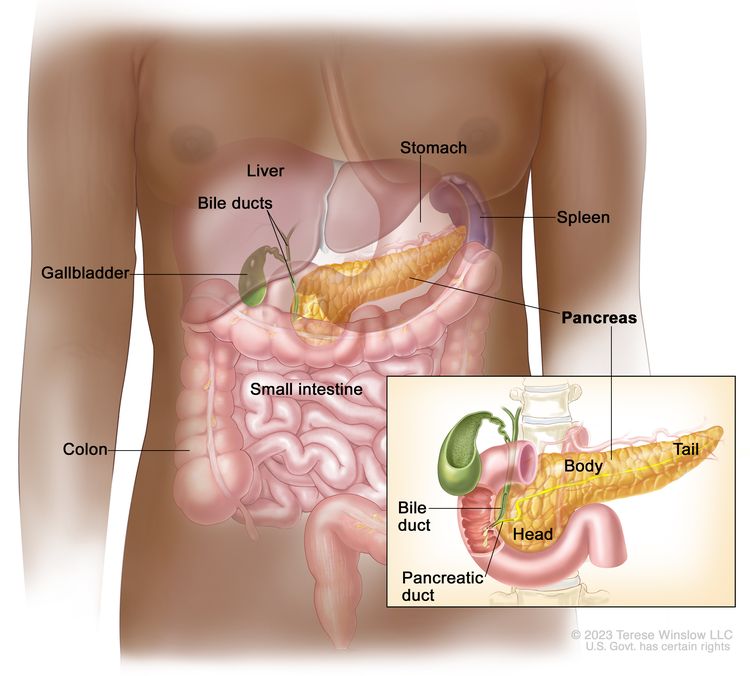
Current Clinical Trials

Use our advanced clinical trial search to find NCI-supported cancer clinical trials that are now enrolling patients. The search can be narrowed by location of the trial, type of treatment, name of the drug, and other criteria. General information about clinical trials is also available.

References
  1. Orazi A, Germing U: The myelodysplastic/myeloproliferative neoplasms: myeloproliferative diseases with dysplastic features. Leukemia 22 (7): 1308-19, 2008. [PUBMED Abstract]
  2. Arber DA, Orazi A, Hasserjian R, et al.: The 2016 revision to the World Health Organization classification of myeloid neoplasms and acute leukemia. Blood 127 (20): 2391-405, 2016. [PUBMED Abstract]
  3. Germing U, Strupp C, Knipp S, et al.: Chronic myelomonocytic leukemia in the light of the WHO proposals. Haematologica 92 (7): 974-7, 2007. [PUBMED Abstract]
  4. Onida F, Beran M: Chronic myelomonocytic leukemia: myeloproliferative variant. Curr Hematol Rep 3 (3): 218-26, 2004. [PUBMED Abstract]
  5. Emanuel PD: Juvenile myelomonocytic leukemia and chronic myelomonocytic leukemia. Leukemia 22 (7): 1335-42, 2008. [PUBMED Abstract]
  6. Aul C, Bowen DT, Yoshida Y: Pathogenesis, etiology and epidemiology of myelodysplastic syndromes. Haematologica 83 (1): 71-86, 1998. [PUBMED Abstract]
  7. Kouides PA, Bennett JM: Morphology and classification of the myelodysplastic syndromes and their pathologic variants. Semin Hematol 33 (2): 95-110, 1996. [PUBMED Abstract]
  8. Bennett JM, Catovsky D, Daniel MT, et al.: The chronic myeloid leukaemias: guidelines for distinguishing chronic granulocytic, atypical chronic myeloid, and chronic myelomonocytic leukaemia. Proposals by the French-American-British Cooperative Leukaemia Group. Br J Haematol 87 (4): 746-54, 1994. [PUBMED Abstract]
  9. Michaux JL, Martiat P: Chronic myelomonocytic leukaemia (CMML)–a myelodysplastic or myeloproliferative syndrome? Leuk Lymphoma 9 (1-2): 35-41, 1993. [PUBMED Abstract]
  10. Maschek H, Georgii A, Kaloutsi V, et al.: Myelofibrosis in primary myelodysplastic syndromes: a retrospective study of 352 patients. Eur J Haematol 48 (4): 208-14, 1992. [PUBMED Abstract]
  11. Saif MW, Hopkins JL, Gore SD: Autoimmune phenomena in patients with myelodysplastic syndromes and chronic myelomonocytic leukemia. Leuk Lymphoma 43 (11): 2083-92, 2002. [PUBMED Abstract]
  12. Meggendorfer M, Roller A, Haferlach T, et al.: SRSF2 mutations in 275 cases with chronic myelomonocytic leukemia (CMML). Blood 120 (15): 3080-8, 2012. [PUBMED Abstract]
  13. Kosmider O, Gelsi-Boyer V, Ciudad M, et al.: TET2 gene mutation is a frequent and adverse event in chronic myelomonocytic leukemia. Haematologica 94 (12): 1676-81, 2009. [PUBMED Abstract]
  14. Malcovati L, Papaemmanuil E, Ambaglio I, et al.: Driver somatic mutations identify distinct disease entities within myeloid neoplasms with myelodysplasia. Blood 124 (9): 1513-21, 2014. [PUBMED Abstract]
  15. Patnaik MM, Itzykson R, Lasho TL, et al.: ASXL1 and SETBP1 mutations and their prognostic contribution in chronic myelomonocytic leukemia: a two-center study of 466 patients. Leukemia 28 (11): 2206-12, 2014. [PUBMED Abstract]
  16. Elena C, Gallì A, Such E, et al.: Integrating clinical features and genetic lesions in the risk assessment of patients with chronic myelomonocytic leukemia. Blood 128 (10): 1408-17, 2016. [PUBMED Abstract]
  17. Onida F, Kantarjian HM, Smith TL, et al.: Prognostic factors and scoring systems in chronic myelomonocytic leukemia: a retrospective analysis of 213 patients. Blood 99 (3): 840-9, 2002. [PUBMED Abstract]
  18. Germing U, Kündgen A, Gattermann N: Risk assessment in chronic myelomonocytic leukemia (CMML). Leuk Lymphoma 45 (7): 1311-8, 2004. [PUBMED Abstract]
  19. Hunter AM, Zhang L, Padron E: Current Management and Recent Advances in the Treatment of Chronic Myelomonocytic Leukemia. Curr Treat Options Oncol 19 (12): 67, 2018. [PUBMED Abstract]
  20. Patnaik MM, Tefferi A: Chronic Myelomonocytic leukemia: 2020 update on diagnosis, risk stratification and management. Am J Hematol 95 (1): 97-115, 2020. [PUBMED Abstract]
  21. Kongtim P, Popat U, Jimenez A, et al.: Treatment with Hypomethylating Agents before Allogeneic Stem Cell Transplant Improves Progression-Free Survival for Patients with Chronic Myelomonocytic Leukemia. Biol Blood Marrow Transplant 22 (1): 47-53, 2016. [PUBMED Abstract]
  22. Sekeres MA, Othus M, List AF, et al.: Randomized Phase II Study of Azacitidine Alone or in Combination With Lenalidomide or With Vorinostat in Higher-Risk Myelodysplastic Syndromes and Chronic Myelomonocytic Leukemia: North American Intergroup Study SWOG S1117. J Clin Oncol 35 (24): 2745-2753, 2017. [PUBMED Abstract]
  23. Elliott MA, Tefferi A, Hogan WJ, et al.: Allogeneic stem cell transplantation and donor lymphocyte infusions for chronic myelomonocytic leukemia. Bone Marrow Transplant 37 (11): 1003-8, 2006. [PUBMED Abstract]
  24. Ocheni S, Kröger N, Zabelina T, et al.: Outcome of allo-SCT for chronic myelomonocytic leukemia. Bone Marrow Transplant 43 (8): 659-61, 2009. [PUBMED Abstract]
  25. Krishnamurthy P, Lim ZY, Nagi W, et al.: Allogeneic haematopoietic SCT for chronic myelomonocytic leukaemia: a single-centre experience. Bone Marrow Transplant 45 (10): 1502-7, 2010. [PUBMED Abstract]
  26. Eissa H, Gooley TA, Sorror ML, et al.: Allogeneic hematopoietic cell transplantation for chronic myelomonocytic leukemia: relapse-free survival is determined by karyotype and comorbidities. Biol Blood Marrow Transplant 17 (6): 908-15, 2011. [PUBMED Abstract]
  27. Park S, Labopin M, Yakoub-Agha I, et al.: Allogeneic stem cell transplantation for chronic myelomonocytic leukemia: a report from the Societe Francaise de Greffe de Moelle et de Therapie Cellulaire. Eur J Haematol 90 (5): 355-64, 2013. [PUBMED Abstract]
  28. Symeonidis A, van Biezen A, de Wreede L, et al.: Achievement of complete remission predicts outcome of allogeneic haematopoietic stem cell transplantation in patients with chronic myelomonocytic leukaemia. A study of the Chronic Malignancies Working Party of the European Group for Blood and Marrow Transplantation. Br J Haematol 171 (2): 239-246, 2015. [PUBMED Abstract]
  29. Robin M, de Wreede LC, Padron E, et al.: Role of allogeneic transplantation in chronic myelomonocytic leukemia: an international collaborative analysis. Blood 140 (12): 1408-1418, 2022. [PUBMED Abstract]
  30. Silverman LR, Demakos EP, Peterson BL, et al.: Randomized controlled trial of azacitidine in patients with the myelodysplastic syndrome: a study of the cancer and leukemia group B. J Clin Oncol 20 (10): 2429-40, 2002. [PUBMED Abstract]
  31. Fenaux P, Mufti GJ, Hellstrom-Lindberg E, et al.: Efficacy of azacitidine compared with that of conventional care regimens in the treatment of higher-risk myelodysplastic syndromes: a randomised, open-label, phase III study. Lancet Oncol 10 (3): 223-32, 2009. [PUBMED Abstract]
  32. Braun T, Itzykson R, Renneville A, et al.: Molecular predictors of response to decitabine in advanced chronic myelomonocytic leukemia: a phase 2 trial. Blood 118 (14): 3824-31, 2011. [PUBMED Abstract]
  33. Drummond MW, Pocock C, Boissinot M, et al.: A multi-centre phase 2 study of azacitidine in chronic myelomonocytic leukaemia. Leukemia 28 (7): 1570-2, 2014. [PUBMED Abstract]
  34. Tantravahi SK, Szankasi P, Khorashad JS, et al.: A phase II study of the efficacy, safety, and determinants of response to 5-azacitidine (Vidaza®) in patients with chronic myelomonocytic leukemia. Leuk Lymphoma 57 (10): 2441-4, 2016. [PUBMED Abstract]
  35. Santini V, Allione B, Zini G, et al.: A phase II, multicentre trial of decitabine in higher-risk chronic myelomonocytic leukemia. Leukemia 32 (2): 413-418, 2018. [PUBMED Abstract]
  36. Coston T, Pophali P, Vallapureddy R, et al.: Suboptimal response rates to hypomethylating agent therapy in chronic myelomonocytic leukemia; a single institutional study of 121 patients. Am J Hematol 94 (7): 767-779, 2019. [PUBMED Abstract]
  37. Bennett JM: Chronic myelomonocytic leukemia. Curr Treat Options Oncol 3 (3): 221-3, 2002. [PUBMED Abstract]
  38. Wattel E, Guerci A, Hecquet B, et al.: A randomized trial of hydroxyurea versus VP16 in adult chronic myelomonocytic leukemia. Groupe Français des Myélodysplasies and European CMML Group. Blood 88 (7): 2480-7, 1996. [PUBMED Abstract]
  39. Itzykson R, Santini V, Thepot S, et al.: Decitabine Versus Hydroxyurea for Advanced Proliferative Chronic Myelomonocytic Leukemia: Results of a Randomized Phase III Trial Within the EMSCO Network. J Clin Oncol 41 (10): 1888-1897, 2023. [PUBMED Abstract]
  40. Montalban-Bravo G, Hammond D, DiNardo CD, et al.: Activity of venetoclax-based therapy in chronic myelomonocytic leukemia. Leukemia 35 (5): 1494-1499, 2021. [PUBMED Abstract]
  41. Bazinet A, Darbaniyan F, Kadia TM, et al.: A retrospective study of cladribine and low-dose cytarabine-based regimens for the treatment of chronic myelomonocytic leukemia and secondary acute myeloid leukemia. Cancer 129 (4): 560-568, 2023. [PUBMED Abstract]
  42. Bazinet A, Darbaniyan F, Jabbour E, et al.: Azacitidine plus venetoclax in patients with high-risk myelodysplastic syndromes or chronic myelomonocytic leukaemia: phase 1 results of a single-centre, dose-escalation, dose-expansion, phase 1-2 study. Lancet Haematol 9 (10): e756-e765, 2022. [PUBMED Abstract]

Treatment of Juvenile Myelomonocytic Leukemia

For more information, see Juvenile Myelomonocytic Leukemia Treatment.

Treatment of Atypical Chronic Myeloid Leukemia

Disease Overview

Atypical chronic myeloid leukemia (aCML) is a leukemic disorder that exhibits both myelodysplastic and myeloproliferative features at the time of diagnosis.

Atypical CML is characterized pathologically by:[1]

  • Peripheral blood leukocytosis with increased numbers of mature and immature neutrophils.
  • Prominent dysgranulopoiesis.
  • No Philadelphia chromosome or BCR::ABL1 gene fusion.
  • Neutrophil precursors (e.g., promyelocytes, myelocytes, and metamyelocytes) accounting for more than 10% of white blood cells.
  • Minimal absolute basophilia with basophils accounting for less than 2% of white blood cells.
  • Absolute monocytosis with monocytes typically account for less than 10% of white blood cells.
  • Hypercellular bone marrow with granulocytic proliferation and granulocytic dysplasia.
  • Fewer than 20% blasts in the blood or bone marrow.
  • Thrombocytopenia.

Clinical features of aCML include:[14]

  • Anemia. For more information on anemia, see Fatigue.
  • Thrombocytopenia.
  • Splenomegaly (in 75% of cases).

Although cytogenetic abnormalities are found in as many as 80% of the patients with aCML, none is specific.[13,5] No Philadelphia chromosome or BCR::ABL1 gene fusion is present.

The exact incidence of aCML is unknown. The median age at the time of diagnosis of this rare leukemic disorder is in the seventh or eighth decade of life.[13]

Morphologically, aCML is characterized by myelodysplasia associated with bone marrow and peripheral blood patterns similar to chronic myeloid leukemia, but cytogenetically it lacks a Philadelphia chromosome or BCR::ABL1 gene fusion.[1] The white blood cell count in the peripheral blood is variable. Median values range from 35 × 109/L to 96 × 109/L, and some patients may have white blood cell counts greater than 300 × 109/L.[13,5] Blasts in the peripheral blood typically account for less than 5% of the white blood cells. Immature neutrophils usually total 10% to 20% or more.[1] The percentage of monocytes is rarely more than 10%. Minimal basophilia may be present.[13,5] Nuclear abnormalities, such as acquired Pelger-Huët anomaly, may be seen in the neutrophils. Moderate anemia (often showing changes indicative of dyserythropoiesis) and thrombocytopenia are common.[14] Bone marrow findings include: [13,5]

  • Granulocytic hypercellularity.
  • Blast count less than 20%.
  • Dysgranulopoiesis
  • Megakaryocytic dysplasia.
  • Erythroid precursors accounting for more than 30% of marrow cells with dyserythropoiesis present (in some cases).

The median survival times for aCML are reported to be less than 20 months, and thrombocytopenia and marked anemia are poor prognostic factors.[1,2] Atypical CML evolves to acute leukemia in approximately 25% to 40% of patients.[1,3] In the remaining patients, fatal complications include resistant leukocytosis, anemia, thrombocytopenia, hepatosplenomegaly, cerebral bleeding associated with thrombocytopenia, and infection.[3,4]

Treatment Overview

The optimal treatment of aCML is uncertain because of the rare incidence of this chronic leukemic disorder. Treatment with hydroxyurea may lead to short-lived partial remissions of 2 to 4 months in duration.[4] Atypical CML appears to respond poorly to treatment with interferon alfa.[4]

Current Clinical Trials

Use our advanced clinical trial search to find NCI-supported cancer clinical trials that are now enrolling patients. The search can be narrowed by location of the trial, type of treatment, name of the drug, and other criteria. General information about clinical trials is also available.

References
  1. Orazi A, Germing U: The myelodysplastic/myeloproliferative neoplasms: myeloproliferative diseases with dysplastic features. Leukemia 22 (7): 1308-19, 2008. [PUBMED Abstract]
  2. Hernández JM, del Cañizo MC, Cuneo A, et al.: Clinical, hematological and cytogenetic characteristics of atypical chronic myeloid leukemia. Ann Oncol 11 (4): 441-4, 2000. [PUBMED Abstract]
  3. Costello R, Sainty D, Lafage-Pochitaloff M, et al.: Clinical and biological aspects of Philadelphia-negative/BCR-negative chronic myeloid leukemia. Leuk Lymphoma 25 (3-4): 225-32, 1997. [PUBMED Abstract]
  4. Kurzrock R, Bueso-Ramos CE, Kantarjian H, et al.: BCR rearrangement-negative chronic myelogenous leukemia revisited. J Clin Oncol 19 (11): 2915-26, 2001. [PUBMED Abstract]
  5. Bennett JM, Catovsky D, Daniel MT, et al.: The chronic myeloid leukaemias: guidelines for distinguishing chronic granulocytic, atypical chronic myeloid, and chronic myelomonocytic leukaemia. Proposals by the French-American-British Cooperative Leukaemia Group. Br J Haematol 87 (4): 746-54, 1994. [PUBMED Abstract]

Treatment of MDS/MPN, Unclassifiable

Disease Overview

Myelodysplastic/myeloproliferative neoplasm, unclassifiable (MDS/MPN-UC) (also known as mixed myeloproliferative/myelodysplastic syndrome, unclassifiable and overlap syndrome, unclassifiable) shows features of both myeloproliferative disease and myelodysplastic disease but does not meet the criteria for any of the other MDS/MPN entities.[1]

Diagnostic criteria for MDS/MPN-UC can be either:[1]

  1. The combination of four sets of criteria (a–d):
    1. Clinical, laboratory, and morphological features of myelodysplastic syndrome (MDS) (e.g., refractory anemia, refractory anemia with ringed sideroblasts, refractory cytopenia with multilineage dysplasia, and refractory anemia with excess of blasts) with fewer than 20% blasts in the blood and bone marrow. For more information on anemia, see Fatigue.
    2. Prominent myeloproliferative features, e.g. platelet count greater than 600 × 109/L associated with megakaryocytic proliferation, or white blood cell count greater than 13.0 × 10⁹/L with or without splenomegaly.
    3. No history of an underlying chronic myeloproliferative disorder (CMPD), MDS, or recent cytotoxic or growth factor therapy that could cause the myelodysplastic or myeloproliferative features.
    4. No Philadelphia chromosome or BCR::ABL1 gene fusion, del(5q), t(3;3)(q21;q26), or inv(3)(q21q26).
  2. Mixed myeloproliferative and myelodysplastic features that cannot be assigned to any other category of MDS, CMPD, or MDS/MPN.

Clinical characteristics of MDS/MPN-UC include:

  • Features of both MDS and CMPD.
  • Hepatomegaly.
  • Splenomegaly.

The incidence and etiology of MDS/MPN-UC are unknown.

Laboratory features typically include anemia and dimorphic erythrocytes on the peripheral blood smear.[1] Thrombocytosis (platelet count >600 × 109/L) or leukocytosis (white blood cell count >13 × 109/L) are present. Neutrophils may exhibit dysplastic features, and giant or hypogranular platelets may be present. Blasts make up less than 20% of the white blood cells and of the nucleated cells of the bone marrow. The bone marrow is hypercellular and may exhibit proliferation in any or all of the myeloid lineages. Dysplastic features are present in at least one cell line.[1]

No cytogenetic or molecular findings are available that are specific for MDS/MPN-UC. In one small series, six of nine patients (those with ringed sideroblasts associated with marked thrombocytosis [RARS-T]) had JAK2 V617F variants, which caused constitutive activation of the JAK2 tyrosine kinase. This JAK2 pathogenic variant is also commonly observed in patients with polycythemia vera, essential thrombocythemia, and idiopathic myelofibrosis.[2] Because of its rare occurrence, the prognosis and predictive factors are unknown.[1]

Treatment Overview

Adult patients with MDS/MPN associated with platelet-derived growth factor receptor gene rearrangements are candidates for imatinib mesylate at standard dosages.[3] Because of its rare occurrence, the literature only minimally addresses other treatment options for MDS/MPN-UC. Supportive care involves treating cytopenias and infection as necessary.

Current Clinical Trials

Use our advanced clinical trial search to find NCI-supported cancer clinical trials that are now enrolling patients. The search can be narrowed by location of the trial, type of treatment, name of the drug, and other criteria. General information about clinical trials is also available.

References
  1. Orazi A, Germing U: The myelodysplastic/myeloproliferative neoplasms: myeloproliferative diseases with dysplastic features. Leukemia 22 (7): 1308-19, 2008. [PUBMED Abstract]
  2. Szpurka H, Tiu R, Murugesan G, et al.: Refractory anemia with ringed sideroblasts associated with marked thrombocytosis (RARS-T), another myeloproliferative condition characterized by JAK2 V617F mutation. Blood 108 (7): 2173-81, 2006. [PUBMED Abstract]
  3. GLEEVEC – imatinib mesylate tablet. Novartis Pharmaceuticals Corporation, 2020. Available online. Last accessed February 18, 2025.

Latest Updates to This Summary (05/14/2025)

The PDQ cancer information summaries are reviewed regularly and updated as new information becomes available. This section describes the latest changes made to this summary as of the date above.

Editorial changes were made to this summary.

This summary is written and maintained by the PDQ Adult Treatment Editorial Board, which is editorially independent of NCI. The summary reflects an independent review of the literature and does not represent a policy statement of NCI or NIH. More information about summary policies and the role of the PDQ Editorial Boards in maintaining the PDQ summaries can be found on the About This PDQ Summary and PDQ® Cancer Information for Health Professionals pages.

About This PDQ Summary

Purpose of This Summary

This PDQ cancer information summary for health professionals provides comprehensive, peer-reviewed, evidence-based information about the treatment of myelodysplastic/myeloproliferative neoplasms. It is intended as a resource to inform and assist clinicians in the care of their patients. It does not provide formal guidelines or recommendations for making health care decisions.

Reviewers and Updates

This summary is reviewed regularly and updated as necessary by the PDQ Adult Treatment Editorial Board, which is editorially independent of the National Cancer Institute (NCI). The summary reflects an independent review of the literature and does not represent a policy statement of NCI or the National Institutes of Health (NIH).

Board members review recently published articles each month to determine whether an article should:

  • be discussed at a meeting,
  • be cited with text, or
  • replace or update an existing article that is already cited.

Changes to the summaries are made through a consensus process in which Board members evaluate the strength of the evidence in the published articles and determine how the article should be included in the summary.

The lead reviewers for Myelodysplastic/Myeloproliferative Neoplasms Treatment are:

  • Aaron Gerds, MD (Cleveland Clinic Taussig Cancer Institute)
  • Eric J. Seifter, MD (Johns Hopkins University)

Any comments or questions about the summary content should be submitted to Cancer.gov through the NCI website’s Email Us. Do not contact the individual Board Members with questions or comments about the summaries. Board members will not respond to individual inquiries.

Levels of Evidence

Some of the reference citations in this summary are accompanied by a level-of-evidence designation. These designations are intended to help readers assess the strength of the evidence supporting the use of specific interventions or approaches. The PDQ Adult Treatment Editorial Board uses a formal evidence ranking system in developing its level-of-evidence designations.

Permission to Use This Summary

PDQ is a registered trademark. Although the content of PDQ documents can be used freely as text, it cannot be identified as an NCI PDQ cancer information summary unless it is presented in its entirety and is regularly updated. However, an author would be permitted to write a sentence such as “NCI’s PDQ cancer information summary about breast cancer prevention states the risks succinctly: [include excerpt from the summary].”

The preferred citation for this PDQ summary is:

PDQ® Adult Treatment Editorial Board. PDQ Myelodysplastic/Myeloproliferative Neoplasms Treatment. Bethesda, MD: National Cancer Institute. Updated <MM/DD/YYYY>. Available at: /types/myeloproliferative/hp/mds-mpd-treatment-pdq. Accessed <MM/DD/YYYY>. [PMID: 26389321]

Images in this summary are used with permission of the author(s), artist, and/or publisher for use within the PDQ summaries only. Permission to use images outside the context of PDQ information must be obtained from the owner(s) and cannot be granted by the National Cancer Institute. Information about using the illustrations in this summary, along with many other cancer-related images, is available in Visuals Online, a collection of over 2,000 scientific images.

Disclaimer

Based on the strength of the available evidence, treatment options may be described as either “standard” or “under clinical evaluation.” These classifications should not be used as a basis for insurance reimbursement determinations. More information on insurance coverage is available on Cancer.gov on the Managing Cancer Care page.

Contact Us

More information about contacting us or receiving help with the Cancer.gov website can be found on our Contact Us for Help page. Questions can also be submitted to Cancer.gov through the website’s Email Us.

Myelodysplastic Syndromes Treatment (PDQ®)–Patient Version

Myelodysplastic Syndromes Treatment (PDQ®)–Patient Version

General Information About Myelodysplastic Syndromes

Key Points

  • Myelodysplastic syndromes are a group of cancers in which immature blood cells in the bone marrow do not mature or become healthy blood cells.
  • The different types of myelodysplastic syndromes are diagnosed based on certain changes in the blood cells and bone marrow.
  • Age and past treatment with chemotherapy or radiation therapy affect the risk of a myelodysplastic syndrome.
  • Signs and symptoms of a myelodysplastic syndrome include shortness of breath and feeling tired.
  • Tests that examine the blood and bone marrow are used to diagnose myelodysplastic syndromes.
  • Certain factors affect prognosis (chance of recovery) and treatment options.

Myelodysplastic syndromes are a group of cancers in which immature blood cells in the bone marrow do not mature or become healthy blood cells.

In a healthy person, the bone marrow makes blood stem cells (immature cells) that become mature blood cells over time.

EnlargeAnatomy of the bone; drawing shows spongy bone, red marrow, and yellow marrow. A cross section of the bone shows compact bone and blood vessels in the bone marrow. Also shown are red blood cells, white blood cells, platelets, and a blood stem cell.
Anatomy of the bone. The bone is made up of compact bone, spongy bone, and bone marrow. Compact bone makes up the outer layer of the bone. Spongy bone is found mostly at the ends of bones and contains red marrow. Bone marrow is found in the center of most bones and has many blood vessels. There are two types of bone marrow: red and yellow. Red marrow contains blood stem cells that can become red blood cells, white blood cells, or platelets. Yellow marrow is made mostly of fat.

A blood stem cell may become a lymphoid stem cell or a myeloid stem cell. A lymphoid stem cell becomes a white blood cell. A myeloid stem cell becomes one of three types of mature blood cells:

EnlargeBlood cell development; drawing shows the steps a blood stem cell goes through to become a red blood cell, platelet, or white blood cell. Drawing shows a myeloid stem cell becoming a red blood cell, platelet, or myeloblast, which then becomes a white blood cell. Drawing also shows a lymphoid stem cell becoming a lymphoblast and then one of several different types of white blood cells.
Blood cell development. A blood stem cell goes through several steps to become a red blood cell, platelet, or white blood cell.

In a patient with a myelodysplastic syndrome, the blood stem cells (immature cells) do not become mature red blood cells, white blood cells, or platelets in the bone marrow. These immature blood cells, called blasts, do not work the way they should and either die in the bone marrow or soon after they go into the blood. This leaves less room for healthy white blood cells, red blood cells, and platelets to form in the bone marrow. When there are fewer healthy blood cells, infection, anemia, or easy bleeding may occur.

The different types of myelodysplastic syndromes are diagnosed based on certain changes in the blood cells and bone marrow.

  • Refractory anemia: There are too few red blood cells in the blood and the patient has anemia. The number of white blood cells and platelets is normal.
  • Refractory anemia with ring sideroblasts: There are too few red blood cells in the blood and the patient has anemia. The red blood cells have too much iron inside the cell. The number of white blood cells and platelets is normal.
  • Refractory anemia with excess blasts: There are too few red blood cells in the blood and the patient has anemia. Five percent to 19% of the cells in the bone marrow are blasts. There also may be changes to the white blood cells and platelets. Refractory anemia with excess blasts may progress to acute myeloid leukemia (AML). For more information, see Acute Myeloid Leukemia Treatment.
  • Refractory cytopenia with multilineage dysplasia: There are too few of at least two types of blood cells (red blood cells, platelets, or white blood cells). Less than 5% of the cells in the bone marrow are blasts and less than 1% of the cells in the blood are blasts. If red blood cells are affected, they may have extra iron. Refractory cytopenia may progress to acute myeloid leukemia (AML).
  • Refractory cytopenia with unilineage dysplasia: There are too few of one type of blood cell (red blood cells, platelets, or white blood cells). There are changes in 10% or more of two other types of blood cells. Less than 5% of the cells in the bone marrow are blasts and less than 1% of the cells in the blood are blasts.
  • Unclassifiable myelodysplastic syndrome: The numbers of blasts in the bone marrow and blood are normal, and the disease is not one of the other myelodysplastic syndromes.
  • Myelodysplastic syndrome associated with an isolated del(5q) chromosome abnormality: There are too few red blood cells in the blood and the patient has anemia. Less than 5% of the cells in the bone marrow and blood are blasts. There is a specific change in the chromosome.
  • Chronic myelomonocytic leukemia (CMML): For more information, see Myelodysplastic/Myeloproliferative Neoplasms Treatment.

Age and past treatment with chemotherapy or radiation therapy affect the risk of a myelodysplastic syndrome.

Anything that increases a person’s chance of getting a disease is called a risk factor. Not every person with one or more of these risk factors will develop myelodysplastic syndromes, and they will develop in people who don’t have any known risk factors. Talk with your doctor if you think you may be at risk. Risk factors for myelodysplastic syndromes include the following:

The cause of myelodysplastic syndromes in most patients is not known.

Signs and symptoms of a myelodysplastic syndrome include shortness of breath and feeling tired.

Myelodysplastic syndromes often do not cause early signs or symptoms. They may be found during a routine blood test. Signs and symptoms may be caused by myelodysplastic syndromes or by other conditions. Check with your doctor if you have any of the following:

  • Shortness of breath.
  • Weakness or feeling tired.
  • Having skin that is paler than usual.
  • Easy bruising or bleeding.
  • Petechiae (flat, pinpoint spots under the skin caused by bleeding).

Tests that examine the blood and bone marrow are used to diagnose myelodysplastic syndromes.

In addition to asking about your personal and family health history and doing a physical exam, your doctor may perform the following tests and procedures:

  • Complete blood count (CBC) with differential: A procedure in which a sample of blood is drawn and checked for the following:
    • The number of red blood cells and platelets.
    • The number and type of white blood cells.
    • The amount of hemoglobin (the protein that carries oxygen) in the red blood cells.
    • The portion of the blood sample made up of red blood cells.
    EnlargeComplete blood count (CBC); left panel shows blood being drawn from a vein on the inside of the elbow using a tube attached to a syringe; right panel shows a laboratory test tube with blood cells separated into layers: plasma, white blood cells, platelets, and red blood cells.
    Complete blood count (CBC). Blood is collected by inserting a needle into a vein and allowing the blood to flow into a tube. The blood sample is sent to the laboratory and the red blood cells, white blood cells, and platelets are counted. The CBC is used to test for, diagnose, and monitor many different conditions.
  • Peripheral blood smear: A procedure in which a sample of blood is checked for changes in the number, type, shape, and size of blood cells and for too much iron in the red blood cells.
  • Cytogenetic analysis: A laboratory test in which the chromosomes of cells in a sample of bone marrow or blood are counted and checked for any changes, such as broken, missing, rearranged, or extra chromosomes. Changes in certain chromosomes may be a sign of cancer. Cytogenetic analysis is used to help diagnose cancer, plan treatment, or find out how well treatment is working.
  • Blood chemistry studies: A procedure in which a blood sample is checked to measure the amounts of certain substances, such as vitamin B12 and folate, released into the blood by organs and tissues in the body. An unusual (higher or lower than normal) amount of a substance can be a sign of disease.
  • Bone marrow aspiration and biopsy: The removal of bone marrow, blood, and a small piece of bone by inserting a hollow needle into the hipbone or breastbone. A pathologist views the bone marrow, blood, and bone under a microscope to look for abnormal cells.
    EnlargeBone marrow aspiration and biopsy; drawing shows a patient lying face down on a table and a bone marrow needle being inserted into the hip bone. An inset shows a close up of the needle being inserted through the skin and hip bone into the bone marrow.
    Bone marrow aspiration and biopsy. After a small area of skin is numbed, a long, hollow needle is inserted through the patient’s skin and hip bone into the bone marrow. A sample of bone marrow and a small piece of bone are removed for examination under a microscope.

    The following tests may be done on the sample of tissue that is removed:

    • Immunocytochemistry: A laboratory test that uses antibodies to check for certain antigens (markers) in a sample of a patient’s bone marrow. The antibodies are usually linked to an enzyme or a fluorescent dye. After the antibodies bind to the antigen in the sample of the patient’s cells, the enzyme or dye is activated, and the antigen can then be seen under a microscope. This type of test is used to help diagnose cancer and to tell the difference between myelodysplastic syndromes, leukemia, and other conditions.
    • Immunophenotyping: A laboratory test that uses antibodies to identify cancer cells based on the types of antigens or markers on the surface of the cells. This test is used to help diagnose specific types of leukemia and other blood disorders.
    • Flow cytometry: A laboratory test that measures the number of cells in a sample, the percentage of live cells in a sample, and certain characteristics of the cells, such as size, shape, and the presence of tumor (or other) markers on the cell surface. The cells from a sample of a patient’s blood, bone marrow, or other tissue are stained with a fluorescent dye, placed in a fluid, and then passed one at a time through a beam of light. The test results are based on how the cells that were stained with the fluorescent dye react to the beam of light. This test is used to help diagnose and manage certain types of cancers, such as leukemia and lymphoma.
    • FISH (fluorescence in situ hybridization): A laboratory test used to look at and count genes or chromosomes in cells and tissues. Pieces of DNA that contain fluorescent dyes are made in the laboratory and added to a sample of a patient’s cells or tissues. When these dyed pieces of DNA attach to certain genes or areas of chromosomes in the sample, they light up when viewed under a fluorescent microscope. The FISH test is used to help diagnose cancer and help plan treatment.

Certain factors affect prognosis (chance of recovery) and treatment options.

The prognosis and treatment options depend on the following:

  • The number of blast cells in the bone marrow.
  • Whether one or more types of blood cells are affected.
  • Whether the patient has signs or symptoms of anemia, bleeding, or infection.
  • Whether the patient has a low or high risk of leukemia.
  • Certain changes in the chromosomes.
  • Whether the myelodysplastic syndrome occurred after chemotherapy or radiation therapy for cancer.
  • The patient’s age and general health.

Treatment Option Overview

Key Points

  • There are different types of treatment for patients with myelodysplastic syndromes.
  • Treatment for myelodysplastic syndromes includes supportive care, drug therapy, and stem cell transplant.
  • The following types of treatment are used:
    • Supportive care
    • Drug therapy
    • Chemotherapy with stem cell transplant
  • New types of treatment are being tested in clinical trials.
  • Treatment for myelodysplastic syndromes may cause side effects.
  • Patients may want to think about taking part in a clinical trial.
  • Patients can enter clinical trials before, during, or after starting their treatment.
  • Follow-up tests may be needed.

There are different types of treatment for patients with myelodysplastic syndromes.

Different types of treatment are available for patients with myelodysplastic syndromes. Some treatments are standard (the currently used treatment), and some are being tested in clinical trials. A treatment clinical trial is a research study meant to help improve current treatments or obtain information on new treatments for patients with cancer. When clinical trials show that a new treatment is better than the standard treatment, the new treatment may become the standard treatment. Patients may want to think about taking part in a clinical trial. Some clinical trials are open only to patients who have not started treatment.

Treatment for myelodysplastic syndromes includes supportive care, drug therapy, and stem cell transplant.

Patients with a myelodysplastic syndrome who have symptoms caused by low blood counts are given supportive care to relieve symptoms and improve quality of life. Drug therapy may be used to slow progression of the disease. Certain patients can be cured with aggressive treatment with chemotherapy followed by stem cell transplant using stem cells from a donor.

The following types of treatment are used:

Supportive care

Supportive care is given to lessen the problems caused by the disease or its treatment. Supportive care may include the following:

  • Transfusion therapy

    Transfusion therapy (blood transfusion) is a method of giving red blood cells, white blood cells, or platelets to replace blood cells destroyed by disease or treatment. A red blood cell transfusion is given when the red blood cell count is low and signs or symptoms of anemia, such as shortness of breath or feeling very tired, occur. A platelet transfusion is usually given when the patient is bleeding, is having a procedure that may cause bleeding, or when the platelet count is very low.

    Patients who receive many blood cell transfusions may have tissue and organ damage caused by the buildup of extra iron. These patients may be treated with iron chelation therapy to remove the extra iron from the blood.

  • Erythropoiesis-stimulating agents

    Erythropoiesis-stimulating agents (ESAs) may be given to increase the number of mature red blood cells made by the body and to lessen the effects of anemia. Sometimes granulocyte colony-stimulating factor (G-CSF) is given with ESAs to help the treatment work better.

  • Antibiotic therapy

    Antibiotics may be given to fight infection.

Drug therapy

  • Lenalidomide

    Patients with myelodysplastic syndrome associated with an isolated del(5q) chromosome abnormality who need frequent red blood cell transfusions may be treated with lenalidomide. Lenalidomide is used to lessen the need for red blood cell transfusions.

  • Immunosuppressive therapy

    Antithymocyte globulin (ATG) works to suppress or weaken the immune system. It is used to lessen the need for red blood cell transfusions.

  • Azacitidine and decitabine

    Azacitidine and decitabine are used to treat myelodysplastic syndromes by killing cells that are dividing rapidly. They also help genes that are involved in cell growth to work the way they should. Treatment with azacitidine and decitabine may slow the progression of myelodysplastic syndromes to acute myeloid leukemia.

  • Chemotherapy used in acute myeloid leukemia (AML)

    Patients with a myelodysplastic syndrome and a high number of blasts in their bone marrow have a high risk of acute leukemia. They may be treated with the same chemotherapy regimen used in patients with acute myeloid leukemia.

Chemotherapy with stem cell transplant

Chemotherapy is given to kill cancer cells. Healthy cells, including blood-forming cells, are also destroyed by the cancer treatment. Stem cell transplant is a treatment to replace the blood-forming cells. Stem cells (immature blood cells) are removed from the blood or bone marrow of the patient or a donor and are frozen and stored. After the patient completes chemotherapy, the stored stem cells are thawed and given back to the patient through an infusion. These reinfused stem cells grow into (and restore) the body’s blood cells.

This treatment may not work as well in patients whose myelodysplastic syndrome was caused by past treatment for cancer.

EnlargeDonor stem cell transplant; (Panel 1): Drawing of stem cells being collected from a donor's bloodstream using an apheresis machine. Blood is removed from a vein in the donor's arm and flows through the machine where the stem cells are removed. The rest of the blood is then returned to the donor through a vein in their other arm. (Panel 2): Drawing of a health care provider giving a patient an infusion of chemotherapy through a catheter in the patient's chest. The chemotherapy is given to kill cancer cells and prepare the patient's body for the donor stem cells. (Panel 3): Drawing of a patient receiving an infusion of the donor stem cells through a catheter in the chest.
Donor stem cell transplant. (Step 1): Four to five days before donor stem cell collection, the donor receives a medicine to increase the number of stem cells circulating through their bloodstream (not shown). The blood-forming stem cells are then collected from the donor through a large vein in their arm. The blood flows through an apheresis machine that removes the stem cells. The rest of the blood is returned to the donor through a vein in their other arm. (Step 2): The patient receives chemotherapy to kill cancer cells and prepare their body for the donor stem cells. The patient may also receive radiation therapy (not shown). (Step 3): The patient receives an infusion of the donor stem cells.

New types of treatment are being tested in clinical trials.

Information about clinical trials is available from the NCI website.

Treatment for myelodysplastic syndromes may cause side effects.

For information about side effects caused by treatment for cancer, visit our Side Effects page.

Patients may want to think about taking part in a clinical trial.

For some patients, taking part in a clinical trial may be the best treatment choice. Clinical trials are part of the cancer research process. Clinical trials are done to find out if new cancer treatments are safe and effective or better than the standard treatment.

Many of today’s standard treatments for cancer are based on earlier clinical trials. Patients who take part in a clinical trial may receive the standard treatment or be among the first to receive a new treatment.

Patients who take part in clinical trials also help improve the way cancer will be treated in the future. Even when clinical trials do not lead to effective new treatments, they often answer important questions and help move research forward.

Patients can enter clinical trials before, during, or after starting their treatment.

Some clinical trials only include patients who have not yet received treatment. Other trials test treatments for patients whose cancer has not gotten better. There are also clinical trials that test new ways to stop cancer from recurring (coming back) or reduce the side effects of cancer treatment.

Clinical trials are taking place in many parts of the country. Information about clinical trials supported by NCI can be found on NCI’s clinical trials search webpage. Clinical trials supported by other organizations can be found on the ClinicalTrials.gov website.

Follow-up tests may be needed.

As you go through treatment, you will have follow-up tests or check-ups. Some tests that were done to diagnose or stage the cancer may be repeated to see how well the treatment is working. Decisions about whether to continue, change, or stop treatment may be based on the results of these tests.

Some of the tests will continue to be done from time to time after treatment has ended. The results of these tests can show if your condition has changed or if the cancer has recurred (come back).

Treatment of Myelodysplastic Syndromes

For information about the treatments listed below, see the Treatment Option Overview section.

The treatment of myelodysplastic syndromes may include the following:

Patients who were treated in the past with chemotherapy or radiation therapy may develop myeloid neoplasms related to that therapy. Treatment options are the same as for other myelodysplastic syndromes.

Use our clinical trial search to find NCI-supported cancer clinical trials that are accepting patients. You can search for trials based on the type of cancer, the age of the patient, and where the trials are being done. General information about clinical trials is also available.

Treatment of Relapsed or Refractory Myelodysplastic Syndromes

For information about the treatments listed below, see the Treatment Option Overview section.

There is no standard treatment for refractory or relapsed myelodysplastic syndromes. Patients whose cancer does not respond to treatment or has come back after treatment may want to take part in a clinical trial.

Use our clinical trial search to find NCI-supported cancer clinical trials that are accepting patients. You can search for trials based on the type of cancer, the age of the patient, and where the trials are being done. General information about clinical trials is also available.

To Learn More About Myelodysplastic Syndromes

About This PDQ Summary

About PDQ

Physician Data Query (PDQ) is the National Cancer Institute’s (NCI’s) comprehensive cancer information database. The PDQ database contains summaries of the latest published information on cancer prevention, detection, genetics, treatment, supportive care, and complementary and alternative medicine. Most summaries come in two versions. The health professional versions have detailed information written in technical language. The patient versions are written in easy-to-understand, nontechnical language. Both versions have cancer information that is accurate and up to date and most versions are also available in Spanish.

PDQ is a service of the NCI. The NCI is part of the National Institutes of Health (NIH). NIH is the federal government’s center of biomedical research. The PDQ summaries are based on an independent review of the medical literature. They are not policy statements of the NCI or the NIH.

Purpose of This Summary

This PDQ cancer information summary has current information about the treatment of myelodysplastic syndromes. It is meant to inform and help patients, families, and caregivers. It does not give formal guidelines or recommendations for making decisions about health care.

Reviewers and Updates

Editorial Boards write the PDQ cancer information summaries and keep them up to date. These Boards are made up of experts in cancer treatment and other specialties related to cancer. The summaries are reviewed regularly and changes are made when there is new information. The date on each summary (“Updated”) is the date of the most recent change.

The information in this patient summary was taken from the health professional version, which is reviewed regularly and updated as needed, by the PDQ Adult Treatment Editorial Board.

Clinical Trial Information

A clinical trial is a study to answer a scientific question, such as whether one treatment is better than another. Trials are based on past studies and what has been learned in the laboratory. Each trial answers certain scientific questions in order to find new and better ways to help cancer patients. During treatment clinical trials, information is collected about the effects of a new treatment and how well it works. If a clinical trial shows that a new treatment is better than one currently being used, the new treatment may become “standard.” Patients may want to think about taking part in a clinical trial. Some clinical trials are open only to patients who have not started treatment.

Clinical trials can be found online at NCI’s website. For more information, call the Cancer Information Service (CIS), NCI’s contact center, at 1-800-4-CANCER (1-800-422-6237).

Permission to Use This Summary

PDQ is a registered trademark. The content of PDQ documents can be used freely as text. It cannot be identified as an NCI PDQ cancer information summary unless the whole summary is shown and it is updated regularly. However, a user would be allowed to write a sentence such as “NCI’s PDQ cancer information summary about breast cancer prevention states the risks in the following way: [include excerpt from the summary].”

The best way to cite this PDQ summary is:

PDQ® Adult Treatment Editorial Board. PDQ Myelodysplastic Syndromes Treatment. Bethesda, MD: National Cancer Institute. Updated <MM/DD/YYYY>. Available at: /types/myeloproliferative/patient/myelodysplastic-treatment-pdq. Accessed <MM/DD/YYYY>. [PMID: 26389239]

Images in this summary are used with permission of the author(s), artist, and/or publisher for use in the PDQ summaries only. If you want to use an image from a PDQ summary and you are not using the whole summary, you must get permission from the owner. It cannot be given by the National Cancer Institute. Information about using the images in this summary, along with many other images related to cancer can be found in Visuals Online. Visuals Online is a collection of more than 3,000 scientific images.

Disclaimer

The information in these summaries should not be used to make decisions about insurance reimbursement. More information on insurance coverage is available on Cancer.gov on the Managing Cancer Care page.

Contact Us

More information about contacting us or receiving help with the Cancer.gov website can be found on our Contact Us for Help page. Questions can also be submitted to Cancer.gov through the website’s E-mail Us.

Myelodysplastic Syndromes Treatment (PDQ®)–Health Professional Version

Myelodysplastic Syndromes Treatment (PDQ®)–Health Professional Version

General Information About Myelodysplastic Syndromes (MDS)

Incidence and Mortality

The MDS are a collection of myeloid malignancies characterized by one or more peripheral blood cytopenias. MDS are diagnosed in slightly more than 10,000 people in the United States yearly, for an annual age-adjusted incidence rate of approximately 4.4 to 4.6 cases per 100,000 people.[1] They are more common in men and White individuals. The syndromes may arise de novo or secondarily after treatment with chemotherapy and/or radiation therapy for other cancers or, rarely, after environmental exposures.

Prognosis

Prognosis is directly related to the number of bone marrow blast cells, to certain cytogenetic abnormalities, and to the amount of peripheral blood cytopenias. By convention, MDS are reclassified as acute myeloid leukemia (AML) with myelodysplastic features when blood or bone marrow blasts reach or exceed 20%. Many patients succumb to complications of cytopenias before progression to this stage. For more information, see the Pathological and Prognostic Systems for MDS section. The acute leukemic phase is less responsive to chemotherapy than is de novo AML.

Pathology

MDS are characterized by abnormal bone marrow and blood cell morphology. Megaloblastoid erythroid hyperplasia with macrocytic anemia, associated with normal vitamin B12 and folate levels, is frequently observed. Circulating granulocytes are often hypogranular or hypergranular and may display the acquired pseudo-Pelger-Huët abnormality. Early, abnormal myeloid progenitors are identified in the marrow in varying percentages. Abnormally small megakaryocytes (micromegakaryocytes) may be seen in the marrow, and hypogranular or giant platelets may appear in the blood.

Clinical Features

MDS occur predominantly in older patients (usually older than 60 years), with a median age at diagnosis of approximately 70 years,[2] although patients as young as 2 years have been reported.[3] Anemia, bleeding, easy bruising, and fatigue are common initial findings. For more information, see Fatigue. Splenomegaly or hepatosplenomegaly may indicate an overlapping myeloproliferative neoplasm. Approximately 50% of patients have a detectable cytogenetic abnormality, most commonly a deletion of all or part of chromosome 5 or 7, or trisomy 8. Single-nucleotide polymorphism array technology may increase the detection of genetic abnormalities to 80%.[4,5] Although the bone marrow is usually hypercellular at diagnosis, 10% of patients present with a hypoplastic bone marrow.[6] Hypoplastic myelodysplastic patients tend to have profound cytopenias and may respond more frequently to immunosuppressive therapy.

Risk Factors

Approximately 90% of MDS cases occur de novo with no identifiable cause. Potential environmental risk factors for developing MDS include exposure to:[7,8]

  • Tobacco smoke.
  • Ionizing radiation.
  • Organic chemicals (e.g., benzene, toluene, xylene, and chloramphenicol).
  • Heavy metals.
  • Herbicides.
  • Pesticides.
  • Fertilizers.
  • Stone and cereal dusts.
  • Exhaust gases.
  • Nitro-organic explosives.
  • Petroleum and diesel derivatives.
References
  1. Ma X, Does M, Raza A, et al.: Myelodysplastic syndromes: incidence and survival in the United States. Cancer 109 (8): 1536-42, 2007. [PUBMED Abstract]
  2. Sekeres MA, Schoonen WM, Kantarjian H, et al.: Characteristics of US patients with myelodysplastic syndromes: results of six cross-sectional physician surveys. J Natl Cancer Inst 100 (21): 1542-51, 2008. [PUBMED Abstract]
  3. Tuncer MA, Pagliuca A, Hicsonmez G, et al.: Primary myelodysplastic syndrome in children: the clinical experience in 33 cases. Br J Haematol 82 (2): 347-53, 1992. [PUBMED Abstract]
  4. Gyger M, Infante-Rivard C, D’Angelo G, et al.: Prognostic value of clonal chromosomal abnormalities in patients with primary myelodysplastic syndromes. Am J Hematol 28 (1): 13-20, 1988. [PUBMED Abstract]
  5. Tiu RV, Gondek LP, O’Keefe CL, et al.: Prognostic impact of SNP array karyotyping in myelodysplastic syndromes and related myeloid malignancies. Blood 117 (17): 4552-60, 2011. [PUBMED Abstract]
  6. Nand S, Godwin JE: Hypoplastic myelodysplastic syndrome. Cancer 62 (5): 958-64, 1988. [PUBMED Abstract]
  7. Du Y, Fryzek J, Sekeres MA, et al.: Smoking and alcohol intake as risk factors for myelodysplastic syndromes (MDS). Leuk Res 34 (1): 1-5, 2010. [PUBMED Abstract]
  8. Strom SS, Gu Y, Gruschkus SK, et al.: Risk factors of myelodysplastic syndromes: a case-control study. Leukemia 19 (11): 1912-8, 2005. [PUBMED Abstract]

Pathological and Prognostic Systems for MDS

Myelodysplastic syndromes (MDS) are classified according to features of cellular morphology, etiology, and clinical presentation. The morphological classification of MDS is largely based on the percent of myeloblasts in the bone marrow and blood, the type and degree of myeloid dysplasia, and the presence of ring sideroblasts.[1] The clinical classification of the MDS depends on whether there is an identifiable etiology and whether the MDS has been treated previously.

Pathological Systems

The World Health Organization (WHO) classification [2] has supplanted the historic French-American-British (FAB) classification,[1] as shown in Table 1.

Table 1. Myelodysplastic Syndromes: Comparison of the FAB and WHO Classifications
FAB (1982) WHO (2008)
AML = acute myeloid leukemia; FAB = French-American-British classification scheme; MDS = myelodysplastic syndromes; WHO = World Health Organization.
Myelodysplastic Syndromes
Refractory anemia. Refractory anemia.
  Refractory cytopenia with multilineage dysplasia. Refractory cytopenia with unilineage dysplasia.
Refractory anemia with ring sideroblasts. Refractory anemia with ring sideroblasts.
Refractory anemia with excess blasts. Refractory anemia with excess blasts -1 and -2.
  Myelodysplastic syndrome, unclassifiable.
  Myelodysplastic syndrome associated with del(5q).
  Reclassified from MDS to:
Refractory anemia with excess blasts in transformation. Acute myeloid leukemia identified as AML with multilineage dysplasia following a myelodysplastic syndrome.
Chronic myelomonocytic leukemia. Myelodysplastic and myeloproliferative diseases.

MDS cellular types and subtypes in either cellular classification scheme have different degrees of disordered hematopoiesis, frequencies of transformation to acute leukemia, and prognoses.

Refractory anemia (RA)

In patients with RA, the myeloid and megakaryocytic series in the bone marrow appear normal, but megaloblastoid erythroid hyperplasia is present. Dysplasia is usually minimal. Marrow blasts are less than 5%, and no peripheral blasts are present. Macrocytic anemia with reticulocytopenia is present in the blood. Transformation to acute leukemia is rare, and median survival varies from 2 years to 5 years in most series. RA accounts for 20% to 30% of all patients with MDS.

Refractory anemia with ring sideroblasts (RARS)

In patients with RARS, the blood and marrow are identical to those in patients with RA, except that at least 15% of marrow red cell precursors are ring sideroblasts. Approximately 10% to 12% of patients present with this type, and prognosis is identical to that of RA. Approximately 1% to 2% of RARS evolve to acute myeloid leukemia (AML).

Refractory anemia with excess blasts (RAEB)

In patients with RAEB, there is significant evidence of disordered myelopoiesis and megakaryocytopoiesis in addition to abnormal erythropoiesis. Because of differences in prognosis related to progression to a frank AML, this cellular classification is composed of two categories: RAEB-1 and RAEB-2. Combined, the two categories account for approximately 40% of all patients with MDS. RAEB-1 is characterized by 5% to 9% blasts in the bone marrow and less than 5% blasts in the blood. Approximately 25% of cases of RAEB-1 progress to AML. Median survival is approximately 18 months. RAEB-2 is characterized by 10% to 19% blasts in the bone marrow. Approximately 33% of cases of RAEB-2 progress to AML. Median survival for RAEB-2 is approximately 10 months.

Refractory cytopenia with multilineage dysplasia (RCMD)

In patients with RCMD, bicytopenia or pancytopenia is present. In addition, dysplastic changes are present in 10% or more of the cells in two or more myeloid cell lines. There are less than 1% blasts in the blood and less than 5% blasts in the bone marrow. Auer rods are not present. Monocytes in the blood are less than 1 × 10⁹. RCMD accounts for approximately 24% of cases of MDS. The frequency of evolution to acute leukemia is 11%. The overall median survival is 33 months. Refractory cytopenia with multilineage dysplasia and ring sideroblasts (RCMD-RS) represents another category of RCMD. In RCMD-RS, features of RCMD are present, and more than 15% of erythroid precursors in the bone marrow are ring sideroblasts. RCMD-RS accounts for approximately 15% of cases of MDS. Survival in RCMD-RS is similar to that in primary RCMD.

Refractory cytopenia with unilineage dysplasia (RCUD)

In patients with RCUD, a single cytopenia is present, involving either erythrocytes, neutrophils, or platelets. In addition, dysplastic changes are present in 10% or more of the cells in two or more myeloid cell lines. There are less than 1% blasts in the blood and less than 5% blasts in the bone marrow. Auer rods are not present. Monocytes in the blood are less than 1 × 10⁹.

Unclassifiable myelodysplastic syndrome (MDS-U)

The cellular subtype MDS-U lacks findings appropriate for classification as RA, RARS, RCMD, or RAEB. Blasts in the blood and bone marrow are not increased.

Myelodysplastic syndrome associated with an isolated del(5q) chromosome abnormality

This MDS cellular subtype, the 5q- syndrome, is associated with an isolated del(5q) cytogenetic abnormality. Blasts in both blood and bone marrow are less than 5%. This subtype is associated with a long survival. Karyotypic evolution is uncommon. Additional cytogenetic abnormalities may be associated with a more aggressive MDS cellular subtype or may evolve to AML.

Therapy-related myeloid neoplasms

The latest version of the WHO pathological classification system identifies patients with therapy-related MDS or AML and places them in the same category as “therapy-related myeloid neoplasms.” This group of disorders evolves in patients who were previously treated with chemotherapy or radiation therapy for other cancers and in whom there is a clinical suspicion that the prior therapy caused the myeloid neoplasm. Classic chemotherapy agents associated with these disorders include alkylating agents, topoisomerase inhibitors, and purine nucleoside analogues.

Chronic myelomonocytic leukemia (CMML)

Although previously classified with the myelodysplastic syndromes, CMML is now assigned to a group of overlap myelodysplastic/myeloproliferative neoplasms. For more information, see Myelodysplastic/ Myeloproliferative Neoplasms Treatment.

Prognostic Scoring Systems

A variety of pathological and risk classification systems have been developed to predict the overall survival of patients with MDS and the evolution from MDS to AML. Major prognostic classification systems include the International Prognostic Scoring System (IPSS), revised as the IPSS-R;[3] the WHO Prognostic Scoring System (WPSS); and the MD Anderson Cancer Center Prognostic Scoring Systems.[4,5] Clinical variables in these systems have included bone marrow and blood myeloblast percentage, specific cytopenias, transfusion requirements, age, performance status, and bone marrow cytogenetic abnormalities.

IPSS

The IPSS incorporates bone marrow blast percentage, number of peripheral blood cytopenias, and cytogenetic risk group.

IPSS-R

Compared with the IPSS, the IPSS-R updates and gives greater weight to cytogenetic abnormalities and severity of cytopenias, while reassigning the weighting for blast percentages.[3]

WPSS

In contrast to the IPSS and IPSS-R, which should be applied only at the time of diagnosis, the WPSS is dynamic, meaning that patients can be reassigned categories as their disease progresses.

MD Anderson

The MD Anderson Cancer Center has published two prognostic scoring systems, one of which is focused on lower-risk patients.[4,5]

References
  1. Bennett JM, Catovsky D, Daniel MT, et al.: Proposals for the classification of the myelodysplastic syndromes. Br J Haematol 51 (2): 189-99, 1982. [PUBMED Abstract]
  2. Vardiman JW, Thiele J, Arber DA, et al.: The 2008 revision of the World Health Organization (WHO) classification of myeloid neoplasms and acute leukemia: rationale and important changes. Blood 114 (5): 937-51, 2009. [PUBMED Abstract]
  3. Greenberg PL, Tuechler H, Schanz J, et al.: Revised international prognostic scoring system for myelodysplastic syndromes. Blood 120 (12): 2454-65, 2012. [PUBMED Abstract]
  4. Garcia-Manero G, Shan J, Faderl S, et al.: A prognostic score for patients with lower risk myelodysplastic syndrome. Leukemia 22 (3): 538-43, 2008. [PUBMED Abstract]
  5. Kantarjian H, O’Brien S, Ravandi F, et al.: Proposal for a new risk model in myelodysplastic syndrome that accounts for events not considered in the original International Prognostic Scoring System. Cancer 113 (6): 1351-61, 2008. [PUBMED Abstract]

Treatment of MDS

Therapies for myelodysplastic syndromes (MDS) are initiated in patients with a shorter predicted survival or in patients with clinically significant cytopenias. The impact of most MDS therapies on survival remains unproven.

Treatment options:

Supportive Care

The mainstay of treatment for MDS has traditionally been supportive care, particularly for patients with symptomatic cytopenias or who are at high risk of infection or bleeding.[1,2] Transfusions are reserved for the treatment of active bleeding; many centers offer prophylactic platelet transfusions for patients with platelet counts lower than 10,000/mm3. Anemia should be treated with red-cell transfusions to avoid symptoms. For more information, see Fatigue.

No prospective trials have demonstrated the benefit of prophylactic use of myeloid growth factors in asymptomatic neutropenic MDS patients. Similarly, the use of prophylactic antibiotics in such patients is of uncertain benefit. While appropriate use of antibiotics in febrile patients is standard clinical practice, the benefit of myeloid growth factors in such settings is unknown.

The use of erythropoiesis-stimulating agents (ESAs) may improve anemia. The likelihood of response to exogenous erythropoietin administration depends on the pretreatment serum erythropoietin level and baseline transfusion needs.

A meta-analysis summarized the data on erythropoietin in 205 patients with MDS from 17 studies. Responses were most likely in patients who were anemic but who did not yet require a transfusion, patients who did not have ring sideroblasts, and patients who had a serum erythropoietin level lower than 200 IU/L.[3] Effective treatment requires substantially higher doses of erythropoietin than are used for other indications; the minimum effective dose studied is 60,000 IU per week.[4] The use of high-dose darbepoetin (300 µg/dose weekly or 500 µg/dose every 2–3 weeks) has been reported to produce a major erythroid response rate of almost 50% in patients whose endogenous erythropoietin level was lower than 500 mIU/mL.[5,6] Most studies discontinued ESAs in patients who failed to show hematologic improvement after 3 to 4 months of therapy. Average response duration is approximately 2 years.[7]

One decision model found that the likelihood of responding to growth factors was higher in patients with a low serum erythropoietin level (<500 IU/L) and low transfusion needs (<2 units of packed red blood cells every month), but growth factors were rarely effective in patients with a high erythropoietin level and high transfusion needs.[8] Some patients with poor response to erythropoietin alone may have improved response with the addition of low doses of granulocyte colony-stimulating factor (G-CSF) (0.5–1.0 µg/kg/day).[911] Rates of response to the combination treatment vary with classification, with responses more likely in patients with refractory anemia and ring sideroblasts (RARS) and less likely in patients with excess blasts.[7] Patients with RARS are unlikely to respond to erythropoietin alone.[3]

The availability of the oral iron-chelating agent deferasirox has led to its widespread use in patients with MDS. While some consensus panels advocate prophylactic iron chelation in patients with ongoing transfusion needs and substantial transfusion history, the impact of iron chelation on survival and disease progression is unknown.[12]

Disease-Modifying Agents

Lower-risk patients (conventionally defined as International Prognostic Scoring System (IPSS) low-risk and intermediate-1–risk groups) who have failed to respond or have ceased responding to ESAs may be treated with one of several disease-modifying agents. The impact of this practice on survival in lower-risk patients is unknown. Whether these drugs should be used following an ESA failure or as up-front therapy has never been determined. In contrast, in higher-risk patients, azacitidine has been shown to improve survival. For more information, see the DNA methyltransferase inhibitors section.

Lenalidomide

The U.S. Food and Drug Administration (FDA) approved lenalidomide for the treatment of lower-risk, transfusion-dependent patients with MDS who harbor a del(5q) cytogenic abnormality. In a phase II registration study of 148 transfusion-dependent low-risk and intermediate-1–risk patients with del(5q) chromosomal abnormalities (alone, or associated with other abnormalities), lenalidomide induced transfusion independence in 67%, with a median time to response of 4 to 5 weeks.[13] The median duration of transfusion independence had not been reached after a median of 104 weeks of follow-up. Of 62 evaluable patients, 38 patients developed complete cytogenetic remission.

Lenalidomide administration is limited by dose-limiting neutropenia and thrombocytopenia.[14][Level of evidence C3] Treatment-related thrombocytopenia also correlated with cytogenetic responses, emphasizing the importance of successful suppression of the del(5q) clone with lenalidomide to achieve meaningful responses.[15]

A subsequent phase III study randomly assigned lower-risk del(5q) MDS patients to receive placebo and lenalidomide at either 5 mg daily for 28 days or 10 mg daily for 21 days of a 28-day cycle.[16] Transfusion independence responses lasting longer than 6 months occurred in 43% to 52% of subjects treated on the lenalidomide arms, compared with 6% of controls. The cytogenetic response rate was 25% to 50% on the active treatment arms, and the 3-year risk of AML transformation was 25%.

Lenalidomide has limited activity in lower-risk, red blood cell transfusion–dependent patients in MDS who do not harbor the del(5q) lesion. In a phase II study similar in design to the registration study, 56 of 215 patients (26%) achieved transfusion independence.[17] Median duration of response was 41 weeks (range, 8–136 weeks). Grade 3 or 4 myelosuppression occurred in only 20% to 25% of patients and, unlike for del(5q) patients, was not associated with subsequent attainment of a transfusion independence response to therapy.

Immunosuppressive therapy

Antithymocyte globulin (ATG) has shown activity in MDS patients in several small series. The National Heart, Lung, and Blood Institute conducted a phase II trial including 25 MDS patients with less than 20% blasts. Of all the patients studied, 11 (or 44%) responded and became transfusion-independent after ATG (three complete responses, six partial responses, and two minimal responses).[18] Multivariate analysis identified HLA-DR-15 (phenotype) expression, briefer period of red cell transfusion dependence, and younger age as predictors of response to ATG.[19] One study used alemtuzumab to treat a heavily preselected population of lower-risk MDS patients, in whom the response rate was 80%.[20]

DNA methyltransferase inhibitors

The nucleoside analogues azacitidine and decitabine are inhibitors of DNA methyltransferase. Both drugs require prolonged administration before benefits are seen. The median number of cycles required to see first hematologic response to azacitidine was 3; 90% of responders showed response by 6 cycles; and the median number of cycles of decitabine required to see first response was 2.2.[21] Azacitidine received FDA approval based on the results of a randomized trial that was not designed to study survival.[22]

A phase III randomized controlled trial (AZA PH GL 2003 CL 001 [NCT00071799]) of azacitidine versus other regimens, including low-dose cytarabine, AML-type remission induction chemotherapy, or best supportive care, was limited to patients with higher-risk MDS subtypes (IPSS intermediate-2 risk and high risk).[17] The median and 2-year overall survival (OS) favored the azacitidine arm, at 24 months versus 16 months (P = .0001) and 51% versus 26% (P < .0001), respectively.[17][Level of evidence A1] The FDA-approved azacitidine dose schedule used in this study (75 mg/m2 per day for 7 consecutive days) has proven inconvenient to some practitioners. A community-based study has suggested that alternate dosing schedules may provide similar hematologic benefits; however, the impact of such dosing schedules on survival is not known.[23]

While the azacitidine congener decitabine demonstrated similar activity in phase II trials, two randomized trials of decitabine versus supportive care failed to show a survival benefit.[21,24] Both decitabine studies used the FDA-approved dose schedule (15 mg/m2 every 8 hours for nine doses). In the European phase III study in higher-risk patients, median OS was similar for patients in both the decitabine and best supportive care arms, at 10.1 months versus 8.5 months, respectively (P = .38). A combined OS and delay in AML transformation end point was 8.8 months versus 6.1 months, respectively (P = .24).[25][Level of evidence A1]

Decitabine can be given as daily intravenous or subcutaneous infusions at doses that differ from the original labeled schedule, with hematologic response rates that appear comparable to the phase III study.[26,27]

Both of these drugs have been approved for refractory anemia, RARS (if accompanied by neutropenia or thrombocytopenia or requiring transfusions), refractory anemia with excess blasts, and refractory anemia with excess blasts in transformation. However, the highest response rates and levels of evidence have been generated in trials in which patients with higher-risk MDS (IPSS risk groups of intermediate-2 or high) were treated.[28] In lower-risk patients, response rates appear similar to those in higher-risk patients, although the survival benefit is unknown. The use of these drugs in low-risk patients may preclude their subsequent use upon disease progression.

Combinations of azacitidine with lenalidomide [29] and vorinostat [30] were compared with single-agent azacitidine in a national randomized phase II trial (S1117 [NCT01522976]).

AML induction-type chemotherapy

Induction chemotherapy typically used to treat AML may be used to treat patients with higher-risk MDS with excess blasts.[31] Low-dose cytarabine has benefitted some patients; however, this treatment was associated with a higher infection rate when compared with observation in a randomized trial. No difference in time to progression or OS was observed for patients treated with low-dose cytarabine or supportive care.

Allogeneic Hematopoietic Stem Cell Transplant (HSCT)

Allogeneic HSCT is the only potentially curative treatment for MDS. Retrospective data suggest cure rates in selected patients ranging from 30% to 60%; outcomes varied with IPSS score at time of transplant, with inferior survival in patients with higher IPSS scores.[32][Level of evidence C3] The role of cytoreductive therapy in reducing the blast percentage before HSCT remains uncertain. Outcomes may not be as good for patients with treatment-related MDS (5-year disease-free survival rate of 8% to 30%).[33]
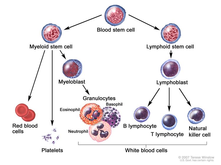
Although HSCT represents the only treatment modality with curative potential, the relatively high morbidity and mortality of this approach limits its use. A decision analysis predating approval of azacitidine, in patients with a median age younger than 50 years, suggested optimal survival when transplant was delayed until disease progression for lower-risk patients but implemented at diagnosis for higher-risk patients.[34]

Allogeneic stem cell transplant with reduced-intensity conditioning (RIC) has extended transplant as a possible modality for treatment of older patients.[35] In a retrospective analysis of 1,333 patients aged 50 years or older (median, 56 years) who underwent allogeneic transplants for MDS using HLA-matched sibling and unrelated donors, 62% of the patients received RIC HSCT, and the others received standard-dose HSCT. On multivariate analysis, use of RIC and advanced disease stage at transplant were associated with increased relapse (hazard ratio [HR] of 1.44 and 1.51, respectively).[35][Level of evidence C3] The predictors of non-relapse mortality included advanced disease stage (HR, 1.43), use of an unrelated donor, and standard-dose HSCT (HR, 1.27). The 4-year OS rate was similar in both groups (30% after myeloablative conditioning vs. 32% in RIC.[35]
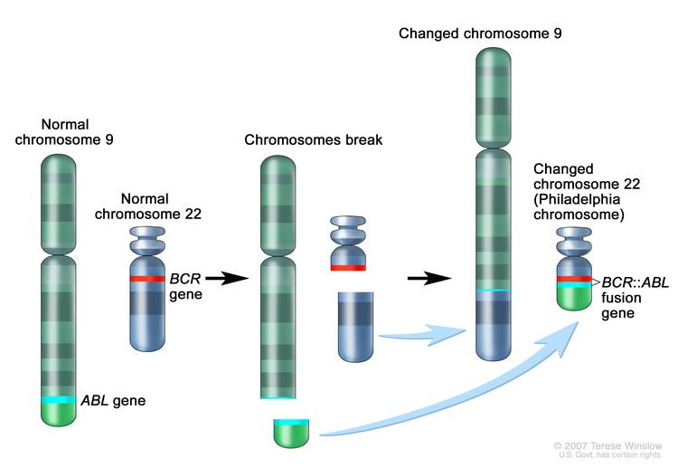
Therapy-Related Myeloid Neoplasms

In the absence of prospective data, therapy-related myeloid neoplasms are treated similarly to de novo MDS.

Current Clinical Trials

Use our advanced clinical trial search to find NCI-supported cancer clinical trials that are now enrolling patients. The search can be narrowed by location of the trial, type of treatment, name of the drug, and other criteria. General information about clinical trials is also available.

References
  1. Tricot GJ, Lauer RC, Appelbaum FR, et al.: Management of the myelodysplastic syndromes. Semin Oncol 14 (4): 444-53, 1987. [PUBMED Abstract]
  2. Boogaerts MA: Progress in the therapy of myelodysplastic syndromes. Blut 58 (6): 265-70, 1989. [PUBMED Abstract]
  3. Hellström-Lindberg E: Efficacy of erythropoietin in the myelodysplastic syndromes: a meta-analysis of 205 patients from 17 studies. Br J Haematol 89 (1): 67-71, 1995. [PUBMED Abstract]
  4. Park S, Grabar S, Kelaidi C, et al.: Predictive factors of response and survival in myelodysplastic syndrome treated with erythropoietin and G-CSF: the GFM experience. Blood 111 (2): 574-82, 2008. [PUBMED Abstract]
  5. Mannone L, Gardin C, Quarre MC, et al.: High-dose darbepoetin alpha in the treatment of anaemia of lower risk myelodysplastic syndrome results of a phase II study. Br J Haematol 133 (5): 513-9, 2006. [PUBMED Abstract]
  6. Gabrilove J, Paquette R, Lyons RM, et al.: Phase 2, single-arm trial to evaluate the effectiveness of darbepoetin alfa for correcting anaemia in patients with myelodysplastic syndromes. Br J Haematol 142 (3): 379-93, 2008. [PUBMED Abstract]
  7. Jädersten M, Montgomery SM, Dybedal I, et al.: Long-term outcome of treatment of anemia in MDS with erythropoietin and G-CSF. Blood 106 (3): 803-11, 2005. [PUBMED Abstract]
  8. Hellström-Lindberg E, Gulbrandsen N, Lindberg G, et al.: A validated decision model for treating the anaemia of myelodysplastic syndromes with erythropoietin + granulocyte colony-stimulating factor: significant effects on quality of life. Br J Haematol 120 (6): 1037-46, 2003. [PUBMED Abstract]
  9. Hellström-Lindberg E, Ahlgren T, Beguin Y, et al.: Treatment of anemia in myelodysplastic syndromes with granulocyte colony-stimulating factor plus erythropoietin: results from a randomized phase II study and long-term follow-up of 71 patients. Blood 92 (1): 68-75, 1998. [PUBMED Abstract]
  10. Hellström-Lindberg E, Kanter-Lewensohn L, Ost A: Morphological changes and apoptosis in bone marrow from patients with myelodysplastic syndromes treated with granulocyte-CSF and erythropoietin. Leuk Res 21 (5): 415-25, 1997. [PUBMED Abstract]
  11. Negrin RS, Stein R, Doherty K, et al.: Maintenance treatment of the anemia of myelodysplastic syndromes with recombinant human granulocyte colony-stimulating factor and erythropoietin: evidence for in vivo synergy. Blood 87 (10): 4076-81, 1996. [PUBMED Abstract]
  12. Greenberg PL, Rigsby CK, Stone RM, et al.: NCCN Task Force: Transfusion and iron overload in patients with myelodysplastic syndromes. J Natl Compr Canc Netw 7 (Suppl 9): S1-16, 2009. [PUBMED Abstract]
  13. List A, Dewald G, Bennett J, et al.: Lenalidomide in the myelodysplastic syndrome with chromosome 5q deletion. N Engl J Med 355 (14): 1456-65, 2006. [PUBMED Abstract]
  14. List A, Kurtin S, Roe DJ, et al.: Efficacy of lenalidomide in myelodysplastic syndromes. N Engl J Med 352 (6): 549-57, 2005. [PUBMED Abstract]
  15. Sekeres MA, Maciejewski JP, Giagounidis AA, et al.: Relationship of treatment-related cytopenias and response to lenalidomide in patients with lower-risk myelodysplastic syndromes. J Clin Oncol 26 (36): 5943-9, 2008. [PUBMED Abstract]
  16. Fenaux P, Giagounidis A, Selleslag D, et al.: RBC transfusion independence and safety profile of lenalidomide 5 or 10 mg in pts with low- or int-1-risk MDS with Del5q: results from a randomized phase III trial (MDS-004). [Abstract] Blood 114 (22): A-944, 2009.
  17. Fenaux P, Mufti GJ, Hellstrom-Lindberg E, et al.: Efficacy of azacitidine compared with that of conventional care regimens in the treatment of higher-risk myelodysplastic syndromes: a randomised, open-label, phase III study. Lancet Oncol 10 (3): 223-32, 2009. [PUBMED Abstract]
  18. Molldrem JJ, Caples M, Mavroudis D, et al.: Antithymocyte globulin for patients with myelodysplastic syndrome. Br J Haematol 99 (3): 699-705, 1997. [PUBMED Abstract]
  19. Saunthararajah Y, Nakamura R, Nam JM, et al.: HLA-DR15 (DR2) is overrepresented in myelodysplastic syndrome and aplastic anemia and predicts a response to immunosuppression in myelodysplastic syndrome. Blood 100 (5): 1570-4, 2002. [PUBMED Abstract]
  20. Sloand EM, Olnes MJ, Shenoy A, et al.: Alemtuzumab treatment of intermediate-1 myelodysplasia patients is associated with sustained improvement in blood counts and cytogenetic remissions. J Clin Oncol 28 (35): 5166-73, 2010. [PUBMED Abstract]
  21. Kantarjian H, Issa JP, Rosenfeld CS, et al.: Decitabine improves patient outcomes in myelodysplastic syndromes: results of a phase III randomized study. Cancer 106 (8): 1794-803, 2006. [PUBMED Abstract]
  22. Silverman LR, Demakos EP, Peterson BL, et al.: Randomized controlled trial of azacitidine in patients with the myelodysplastic syndrome: a study of the cancer and leukemia group B. J Clin Oncol 20 (10): 2429-40, 2002. [PUBMED Abstract]
  23. Lyons RM, Cosgriff TM, Modi SS, et al.: Hematologic response to three alternative dosing schedules of azacitidine in patients with myelodysplastic syndromes. J Clin Oncol 27 (11): 1850-6, 2009. [PUBMED Abstract]
  24. Wijermans P, Lübbert M, Verhoef G, et al.: Low-dose 5-aza-2′-deoxycytidine, a DNA hypomethylating agent, for the treatment of high-risk myelodysplastic syndrome: a multicenter phase II study in elderly patients. J Clin Oncol 18 (5): 956-62, 2000. [PUBMED Abstract]
  25. Lübbert M, Suciu S, Baila L, et al.: Low-dose decitabine versus best supportive care in elderly patients with intermediate- or high-risk myelodysplastic syndrome (MDS) ineligible for intensive chemotherapy: final results of the randomized phase III study of the European Organisation for Research and Treatment of Cancer Leukemia Group and the German MDS Study Group. J Clin Oncol 29 (15): 1987-96, 2011. [PUBMED Abstract]
  26. Issa JP, Garcia-Manero G, Giles FJ, et al.: Phase 1 study of low-dose prolonged exposure schedules of the hypomethylating agent 5-aza-2′-deoxycytidine (decitabine) in hematopoietic malignancies. Blood 103 (5): 1635-40, 2004. [PUBMED Abstract]
  27. Kantarjian H, Oki Y, Garcia-Manero G, et al.: Results of a randomized study of 3 schedules of low-dose decitabine in higher-risk myelodysplastic syndrome and chronic myelomonocytic leukemia. Blood 109 (1): 52-7, 2007. [PUBMED Abstract]
  28. Kaminskas E, Farrell A, Abraham S, et al.: Approval summary: azacitidine for treatment of myelodysplastic syndrome subtypes. Clin Cancer Res 11 (10): 3604-8, 2005. [PUBMED Abstract]
  29. Sekeres MA, List AF, Cuthbertson D, et al.: Phase I combination trial of lenalidomide and azacitidine in patients with higher-risk myelodysplastic syndromes. J Clin Oncol 28 (13): 2253-8, 2010. [PUBMED Abstract]
  30. Garcia-Manero G, Estey EH, Jabbour E, et al.: Final report of a phase II study of 5-azacitidine and vorinostat in patients with newly diagnosed myelodysplastic syndrome or acute myelogenous leukemia not eligible for clinical trials because poor performance and presence of other comorbidities. [Abstract] Blood 118 (21): A-608, 2011.
  31. de Witte T, Suciu S, Verhoef G, et al.: Intensive chemotherapy followed by allogeneic or autologous stem cell transplantation for patients with myelodysplastic syndromes (MDSs) and acute myeloid leukemia following MDS. Blood 98 (8): 2326-31, 2001. [PUBMED Abstract]
  32. Deeg HJ, Storer B, Slattery JT, et al.: Conditioning with targeted busulfan and cyclophosphamide for hemopoietic stem cell transplantation from related and unrelated donors in patients with myelodysplastic syndrome. Blood 100 (4): 1201-7, 2002. [PUBMED Abstract]
  33. Witherspoon RP, Deeg HJ, Storer B, et al.: Hematopoietic stem-cell transplantation for treatment-related leukemia or myelodysplasia. J Clin Oncol 19 (8): 2134-41, 2001. [PUBMED Abstract]
  34. Cutler CS, Lee SJ, Greenberg P, et al.: A decision analysis of allogeneic bone marrow transplantation for the myelodysplastic syndromes: delayed transplantation for low-risk myelodysplasia is associated with improved outcome. Blood 104 (2): 579-85, 2004. [PUBMED Abstract]
  35. Schetelig J, van Biezen A, Brand R, et al.: Allogeneic hematopoietic stem-cell transplantation for chronic lymphocytic leukemia with 17p deletion: a retrospective European Group for Blood and Marrow Transplantation analysis. J Clin Oncol 26 (31): 5094-100, 2008. [PUBMED Abstract]

Treatment of Relapsed or Refractory MDS

Lack of response or progression after the use of erythropoiesis-stimulating agents is not considered relapsed or refractory myelodysplastic syndromes (MDS).

With the exception of the use of lenalidomide for low-risk patients with abnormalities of chromosome 5, there are no clinical trials informing the appropriate selection of therapies for patients with specific subtypes of MDS. Patients who have ceased to respond or did not respond to one therapy are frequently offered another from the therapies described in the previous sections. Retrospective data suggest that patients who do not respond or have ceased responding to DNA methyltransferase inhibitors have a median survival of only 4 to 6 months.[1,2] Patients with relapses should be considered for enrollment in clinical trials.

Current Clinical Trials

Use our advanced clinical trial search to find NCI-supported cancer clinical trials that are now enrolling patients. The search can be narrowed by location of the trial, type of treatment, name of the drug, and other criteria. General information about clinical trials is also available.

References
  1. Prébet T, Gore SD, Esterni B, et al.: Outcome of high-risk myelodysplastic syndrome after azacitidine treatment failure. J Clin Oncol 29 (24): 3322-7, 2011. [PUBMED Abstract]
  2. Jabbour E, Garcia-Manero G, Batty N, et al.: Outcome of patients with myelodysplastic syndrome after failure of decitabine therapy. Cancer 116 (16): 3830-4, 2010. [PUBMED Abstract]

Current Clinical Trials

Use our advanced clinical trial search to find NCI-supported cancer clinical trials that are now enrolling patients. The search can be narrowed by location of the trial, type of treatment, name of the drug, and other criteria. General information about clinical trials is also available.

Latest Updates to This Summary (09/19/2024)

The PDQ cancer information summaries are reviewed regularly and updated as new information becomes available. This section describes the latest changes made to this summary as of the date above.

Editorial changes were made to this summary.

This summary is written and maintained by the PDQ Adult Treatment Editorial Board, which is editorially independent of NCI. The summary reflects an independent review of the literature and does not represent a policy statement of NCI or NIH. More information about summary policies and the role of the PDQ Editorial Boards in maintaining the PDQ summaries can be found on the About This PDQ Summary and PDQ® Cancer Information for Health Professionals pages.

About This PDQ Summary

Purpose of This Summary

This PDQ cancer information summary for health professionals provides comprehensive, peer-reviewed, evidence-based information about the treatment of myelodysplastic syndromes. It is intended as a resource to inform and assist clinicians in the care of their patients. It does not provide formal guidelines or recommendations for making health care decisions.

Reviewers and Updates

This summary is reviewed regularly and updated as necessary by the PDQ Adult Treatment Editorial Board, which is editorially independent of the National Cancer Institute (NCI). The summary reflects an independent review of the literature and does not represent a policy statement of NCI or the National Institutes of Health (NIH).

Board members review recently published articles each month to determine whether an article should:

  • be discussed at a meeting,
  • be cited with text, or
  • replace or update an existing article that is already cited.

Changes to the summaries are made through a consensus process in which Board members evaluate the strength of the evidence in the published articles and determine how the article should be included in the summary.

The lead reviewer for Myelodysplastic Syndromes Treatment is:

  • Aaron Gerds, MD (Cleveland Clinic Taussig Cancer Institute)

Any comments or questions about the summary content should be submitted to Cancer.gov through the NCI website’s Email Us. Do not contact the individual Board Members with questions or comments about the summaries. Board members will not respond to individual inquiries.

Levels of Evidence

Some of the reference citations in this summary are accompanied by a level-of-evidence designation. These designations are intended to help readers assess the strength of the evidence supporting the use of specific interventions or approaches. The PDQ Adult Treatment Editorial Board uses a formal evidence ranking system in developing its level-of-evidence designations.

Permission to Use This Summary

PDQ is a registered trademark. Although the content of PDQ documents can be used freely as text, it cannot be identified as an NCI PDQ cancer information summary unless it is presented in its entirety and is regularly updated. However, an author would be permitted to write a sentence such as “NCI’s PDQ cancer information summary about breast cancer prevention states the risks succinctly: [include excerpt from the summary].”

The preferred citation for this PDQ summary is:

PDQ® Adult Treatment Editorial Board. PDQ Myelodysplastic Syndromes Treatment. Bethesda, MD: National Cancer Institute. Updated <MM/DD/YYYY>. Available at: /types/myeloproliferative/hp/myelodysplastic-treatment-pdq. Accessed <MM/DD/YYYY>. [PMID: 26389450]

Images in this summary are used with permission of the author(s), artist, and/or publisher for use within the PDQ summaries only. Permission to use images outside the context of PDQ information must be obtained from the owner(s) and cannot be granted by the National Cancer Institute. Information about using the illustrations in this summary, along with many other cancer-related images, is available in Visuals Online, a collection of over 2,000 scientific images.

Disclaimer

Based on the strength of the available evidence, treatment options may be described as either “standard” or “under clinical evaluation.” These classifications should not be used as a basis for insurance reimbursement determinations. More information on insurance coverage is available on Cancer.gov on the Managing Cancer Care page.

Contact Us

More information about contacting us or receiving help with the Cancer.gov website can be found on our Contact Us for Help page. Questions can also be submitted to Cancer.gov through the website’s Email Us.

Myeloproliferative Neoplasms Treatment (PDQ®)–Patient Version

Myeloproliferative Neoplasms Treatment (PDQ®)–Patient Version

General Information About Myeloproliferative Neoplasms

Key Points

  • Myeloproliferative neoplasms are a group of diseases in which the bone marrow makes too many red blood cells, white blood cells, or platelets.
  • The following are types of myeloproliferative neoplasms.
  • Tests that examine the blood and bone marrow are used to diagnose myeloproliferative neoplasms.

Myeloproliferative neoplasms are a group of diseases in which the bone marrow makes too many red blood cells, white blood cells, or platelets.

Normally, the bone marrow makes blood stem cells (immature cells) that become mature blood cells over time.

EnlargeAnatomy of the bone; drawing shows spongy bone, red marrow, and yellow marrow. A cross section of the bone shows compact bone and blood vessels in the bone marrow. Also shown are red blood cells, white blood cells, platelets, and a blood stem cell.
Anatomy of the bone. The bone is made up of compact bone, spongy bone, and bone marrow. Compact bone makes up the outer layer of the bone. Spongy bone is found mostly at the ends of bones and contains red marrow. Bone marrow is found in the center of most bones and has many blood vessels. There are two types of bone marrow: red and yellow. Red marrow contains blood stem cells that can become red blood cells, white blood cells, or platelets. Yellow marrow is made mostly of fat.

A blood stem cell may become a myeloid stem cell or a lymphoid stem cell. A lymphoid stem cell becomes a white blood cell. A myeloid stem cell becomes one of three types of mature blood cells:

EnlargeBlood cell development; drawing shows the steps a blood stem cell goes through to become a red blood cell, platelet, or white blood cell. Drawing shows a myeloid stem cell becoming a red blood cell, platelet, or myeloblast, which then becomes a white blood cell. Drawing also shows a lymphoid stem cell becoming a lymphoblast and then one of several different types of white blood cells.
Blood cell development. A blood stem cell goes through several steps to become a red blood cell, platelet, or white blood cell.

In myeloproliferative neoplasms, too many blood stem cells become one or more types of blood cells. The neoplasms usually get worse slowly as the number of extra blood cells increases.

The following are types of myeloproliferative neoplasms.

The type of myeloproliferative neoplasm is based on whether too many red blood cells, white blood cells, or platelets are being made. Sometimes the body will make too many of more than one type of blood cell, but usually one type of blood cell is affected more than the others are. Myeloproliferative neoplasms include:

These types are described below. Myeloproliferative neoplasms sometimes become acute leukemia, in which too many abnormal white blood cells are made.

Tests that examine the blood and bone marrow are used to diagnose myeloproliferative neoplasms.

In addition to asking about your personal and family health history and doing a physical exam, your doctor may perform the following tests and procedures:

  • Complete blood count (CBC) with differential checks a sample of blood for:
    • the number of red blood cells and platelets
    • the number and type of white blood cells
    • the amount of hemoglobin (the protein that carries oxygen) in the red blood cells
    • the amount of hematocrit (whole blood that is made up of red blood cells)
    EnlargeComplete blood count (CBC); left panel shows blood being drawn from a vein on the inside of the elbow using a tube attached to a syringe; right panel shows a laboratory test tube with blood cells separated into layers: plasma, white blood cells, platelets, and red blood cells.
    Complete blood count (CBC). Blood is collected by inserting a needle into a vein and allowing the blood to flow into a tube. The blood sample is sent to the laboratory and the red blood cells, white blood cells, and platelets are counted. The CBC is used to test for, diagnose, and monitor many different conditions.
  • Peripheral blood smear checks a sample of blood for:
    • whether there are red blood cells shaped like teardrops
    • the number and kinds of white blood cells
    • the number of platelets
    • whether there are blast cells
  • Blood chemistry study uses a blood sample to measure the amounts of certain substances released into the blood by organs and tissues in the body. An unusual amount of a substance can be a sign of disease.
  • Bone marrow aspiration and biopsy is the removal of bone marrow, blood, and a small piece of bone by inserting a hollow needle into the hipbone or breastbone. A pathologist views the bone marrow, blood, and bone under a microscope to look for abnormal cells.
    EnlargeBone marrow aspiration and biopsy; drawing shows a patient lying face down on a table and a bone marrow needle being inserted into the hip bone. An inset shows a close up of the needle being inserted through the skin and hip bone into the bone marrow.
    Bone marrow aspiration and biopsy. After a small area of skin is numbed, a long, hollow needle is inserted through the patient’s skin and hip bone into the bone marrow. A sample of bone marrow and a small piece of bone are removed for examination under a microscope.
  • Cytogenetic analysis checks the chromosomes of cells in a sample of bone marrow, blood, tumor, or other tissue for broken, missing, rearranged, or extra chromosomes. Changes in certain chromosomes may be a sign of cancer. Cytogenetic analysis is used to help diagnose cancer, plan treatment, or find out how well treatment is working.
  • Genetic testing is done on a bone marrow or blood sample to check for mutations in JAK2, MPL, or CALR genes. A JAK2 gene mutation is often found in patients with polycythemia vera, essential thrombocythemia, or primary myelofibrosis. MPL or CALR gene mutations are found in patients with essential thrombocythemia or primary myelofibrosis.

Chronic Myeloid Leukemia

Chronic myeloid leukemia is a disease in which too many white blood cells are made in the bone marrow. To learn more about diagnosis, staging, and treatment, visit Chronic Myeloid Leukemia Treatment.

Polycythemia Vera

Key Points

  • Polycythemia vera is a disease in which too many red blood cells are made in the bone marrow.
  • Symptoms of polycythemia vera include headaches and a feeling of fullness below the ribs on the left side.
  • Special blood tests are used to diagnose polycythemia vera.

Polycythemia vera is a disease in which too many red blood cells are made in the bone marrow.

In polycythemia vera, the blood becomes thickened with too many red blood cells. The number of white blood cells and platelets may also increase. These extra blood cells may collect in the spleen and cause it to swell. The increased number of red blood cells, white blood cells, or platelets in the blood can cause bleeding problems and make clots form in blood vessels. This can increase the risk of stroke or heart attack. In patients who are older than 65 years or who have a history of blood clots, the risk of stroke or heart attack is higher. Patients also have an increased risk of acute myeloid leukemia or primary myelofibrosis.

Symptoms of polycythemia vera include headaches and a feeling of fullness below the ribs on the left side.

Polycythemia vera often does not cause early signs or symptoms. It may be found during a routine blood test. Signs and symptoms may occur as the number of blood cells increases. Other conditions may cause the same signs and symptoms. Check with your doctor if you have:

  • a feeling of pressure or fullness below the ribs on the left side
  • headaches
  • double vision or seeing dark or blind spots that come and go
  • itching all over the body, especially after being in warm or hot water
  • reddened face that looks like a blush or sunburn
  • weakness
  • dizziness
  • weight loss for no known reason

Special blood tests are used to diagnose polycythemia vera.

In addition to a complete blood count, bone marrow aspiration and biopsy, and cytogenetic analysis, a serum erythropoietin test is used to diagnose polycythemia vera. In this test, a sample of blood is checked for the level of erythropoietin (a hormone that stimulates new red blood cells to be made). In polycythemia vera, the erythropoietin level would be lower than normal because the body does not need to make more red blood cells.

Essential Thrombocythemia

Key Points

  • Essential thrombocythemia is a disease in which too many platelets are made in the bone marrow.
  • Patients with essential thrombocythemia may have no signs or symptoms.
  • Certain factors affect prognosis (chance of recovery) and treatment options for essential thrombocythemia.

Essential thrombocythemia is a disease in which too many platelets are made in the bone marrow.

Essential thrombocythemia causes an abnormal increase in the number of platelets made in the blood and bone marrow.

Patients with essential thrombocythemia may have no signs or symptoms.

Essential thrombocythemia often does not cause early signs or symptoms. It may be found during a routine blood test. Signs and symptoms may be caused by essential thrombocythemia or by other conditions. Check with your doctor if you have:

  • headaches
  • burning or tingling in the hands or feet
  • redness and warmth of the hands or feet
  • vision or hearing problems

Platelets are sticky. When there are too many platelets, they may clump together and make it hard for the blood to flow. Clots may form in blood vessels and there may also be increased bleeding. These can cause serious health problems such as stroke and heart attack, or pulmonary embolism and deep vein thrombosis in people older than 60 years, who have had blood clots or high white blood cell counts in the past. In some people, essential thrombocythemia may become acute leukemia.

Certain factors affect prognosis (chance of recovery) and treatment options for essential thrombocythemia.

Prognosis and treatment options depend on:

  • the age of the patient
  • whether the patient has signs or symptoms or other problems related to essential thrombocythemia

Overt and Prefibrotic Primary Myelofibrosis

Key Points

  • Primary myelofibrosis is a disease in which abnormal blood cells and fibers build up inside the bone marrow.
  • Symptoms of primary myelofibrosis include pain below the ribs on the left side and feeling very tired.
  • Certain factors affect prognosis (chance of recovery) and treatment options for primary myelofibrosis.

Primary myelofibrosis is a disease in which abnormal blood cells and fibers build up inside the bone marrow.

The bone marrow is made of tissues that make blood cells (red blood cells, white blood cells, and platelets) and a web of fibers that support the blood-forming tissues. In primary myelofibrosis (also called chronic idiopathic myelofibrosis), large numbers of blood stem cells become blood cells that do not mature properly (blasts). The web of fibers inside the bone marrow also becomes very thick (like scar tissue) and slows the blood-forming tissue’s ability to make blood cells. This causes the blood-forming tissues to make fewer and fewer blood cells. In order to make up for the low number of blood cells made in the bone marrow, the liver and spleen begin to make the blood cells.

Symptoms of primary myelofibrosis include pain below the ribs on the left side and feeling very tired.

Primary myelofibrosis often does not cause early signs or symptoms. It may be found during a routine blood test. Signs and symptoms may be caused by primary myelofibrosis or by other conditions. Check with your doctor if you have:

  • pain or a feeling of fullness below the ribs on the left side
  • early satiety (feeling full sooner than normal when eating)
  • anemia
  • bone pain
  • tiredness
  • shortness of breath
  • easy bruising or bleeding
  • petechiae (flat, red, pinpoint spots under the skin that are caused by bleeding)
  • fever
  • drenching night sweats
  • weight loss

Certain factors affect prognosis (chance of recovery) and treatment options for primary myelofibrosis.

The prognosis depends on:

  • the age of the patient
  • the number of abnormal red blood cells and white blood cells
  • the number of blasts in the blood
  • whether there are certain changes in the chromosomes
  • whether the patient has signs such as fever, drenching night sweats, or weight loss

Chronic Neutrophilic Leukemia

Chronic neutrophilic leukemia is a disease in which too many blood stem cells become a type of white blood cell called neutrophils. Neutrophils are infection-fighting blood cells that surround and destroy dead cells and foreign substances (such as bacteria). The spleen and liver may swell because of the extra neutrophils. Chronic neutrophilic leukemia may stay the same or it may progress quickly to acute leukemia.

Chronic Eosinophilic Leukemia

Key Points

  • Chronic eosinophilic leukemia is a disease in which too many white blood cells (eosinophils) are made in the bone marrow.
  • Signs and symptoms of chronic eosinophilic leukemia include fever and feeling very tired.

Chronic eosinophilic leukemia is a disease in which too many white blood cells (eosinophils) are made in the bone marrow.

Eosinophils are white blood cells that react to allergens (substances that cause an allergic response) and help fight infections caused by certain parasites. In chronic eosinophilic leukemia, there are too many eosinophils in the blood, bone marrow, and other tissues. Chronic eosinophilic leukemia may stay the same for many years or it may progress quickly to acute leukemia.

Signs and symptoms of chronic eosinophilic leukemia include fever and feeling very tired.

Chronic eosinophilic leukemia may not cause early signs or symptoms. It may be found during a routine blood test. Signs and symptoms may be caused by chronic eosinophilic leukemia or by other conditions. Check with your doctor if you have:

  • fever
  • tiredness
  • cough
  • swelling under the skin around the eyes and lips, in the throat, or on the hands and feet
  • muscle pain
  • itching
  • diarrhea

Stages of Myeloproliferative Neoplasms

Key Points

  • There is no standard staging system for myeloproliferative neoplasms.

There is no standard staging system for myeloproliferative neoplasms.

The process used to find out if cancer has spread to other parts of the body is called staging. There is no standard staging system for myeloproliferative neoplasms. It is important to know the type of myeloproliferative neoplasm in order to plan treatment.

Treatment Option Overview

Key Points

  • There are different types of treatment for patients with myeloproliferative neoplasms.
  • The following types of treatment are used:
    • Watchful waiting
    • Phlebotomy
    • Platelet apheresis
    • Transfusion therapy
    • Chemotherapy
    • Radiation therapy
    • Other drug therapy
    • Surgery
    • Immunotherapy
    • Targeted therapy
    • High-dose chemotherapy with stem cell transplant
  • New types of treatment are being tested in clinical trials.
  • Treatment for myeloproliferative neoplasms may cause side effects.
  • Follow-up care may be needed.

There are different types of treatment for patients with myeloproliferative neoplasms.

Different types of treatments are available for patients with myeloproliferative neoplasms. Some treatments are standard (the currently used treatment), and some are being tested in clinical trials. A treatment clinical trial is a research study meant to help improve current treatments or obtain information on new treatments. When clinical trials show that a new treatment is better than the standard treatment, the new treatment may become the standard treatment. Patients may want to think about taking part in a clinical trial. Some clinical trials are open only to patients who have not started treatment.

The following types of treatment are used:

Watchful waiting

Watchful waiting is closely monitoring a patient’s condition without giving any treatment until signs or symptoms appear or change.

Phlebotomy

Phlebotomy is a procedure in which blood is taken from a vein. A sample of blood may be taken for tests such as a CBC or blood chemistry. Sometimes phlebotomy is used as a treatment and blood is taken from the body to remove extra red blood cells. Phlebotomy is used in this way to treat some myeloproliferative neoplasms.

Platelet apheresis

Platelet apheresis is a treatment that uses a special machine to remove platelets from the blood. Blood is taken from the patient and put through a blood cell separator where the platelets are removed. The rest of the blood is then returned to the patient’s bloodstream.

Transfusion therapy

Blood transfusion is a method of giving red blood cells, white blood cells, or platelets to replace blood cells destroyed by disease or cancer treatment.

Chemotherapy

Chemotherapy is a cancer treatment that uses drugs to stop the growth of cancer cells, either by killing the cells or by stopping them from dividing. When chemotherapy is taken by mouth or injected into a vein or muscle, the drugs enter the bloodstream and can reach cancer cells throughout the body (systemic chemotherapy).

Learn more at Drugs Approved for Myeloproliferative Neoplasms.

Radiation therapy

Radiation therapy is a cancer treatment that uses high-energy x-rays or other types of radiation to kill cancer cells or keep them from growing. External radiation therapy uses a machine outside the body to send radiation toward the area of the body, such as the spleen, with cancer.

Other drug therapy

Prednisone and danazol are drugs that may be used to treat anemia in patients with primary myelofibrosis.

Anagrelide therapy is used to reduce the risk of blood clots in patients who have too many platelets in their blood. Low-dose aspirin may also be used to reduce the risk of blood clots.

Thalidomide, lenalidomide, and pomalidomide are drugs that prevent blood vessels from growing into areas of tumor cells.

Erythropoietic growth factors are used to stimulate the bone marrow to make red blood cells.

Learn more at Drugs Approved for Myeloproliferative Neoplasms.

Surgery

Splenectomy (surgery to remove the spleen) may be done if the spleen is enlarged.

Immunotherapy

Immunotherapy is a treatment that uses the patient’s immune system to fight cancer. Substances made by the body or made in a laboratory are used to boost, direct, or restore the body’s natural defenses against cancer.

Targeted therapy

Targeted therapy is a type of treatment that uses drugs or other substances to identify and attack specific cancer cells.

Learn more at Drugs Approved for Myeloproliferative Neoplasms.

Other types of targeted therapies are being studied in clinical trials.

High-dose chemotherapy with stem cell transplant

High doses of chemotherapy are given to kill cancer cells. Healthy cells, including blood-forming cells, are also destroyed by the cancer treatment. Stem cell transplant is a treatment to replace the blood-forming cells. Stem cells (immature blood cells) are removed from the blood or bone marrow of the patient or a donor and are frozen and stored. After the patient completes chemotherapy, the stored stem cells are thawed and given back to the patient through an infusion. These reinfused stem cells grow into (and restore) the body’s blood cells.

EnlargeDonor stem cell transplant; (Panel 1): Drawing of stem cells being collected from a donor's bloodstream using an apheresis machine. Blood is removed from a vein in the donor's arm and flows through the machine where the stem cells are removed. The rest of the blood is then returned to the donor through a vein in their other arm. (Panel 2): Drawing of a health care provider giving a patient an infusion of chemotherapy through a catheter in the patient's chest. The chemotherapy is given to kill cancer cells and prepare the patient's body for the donor stem cells. (Panel 3): Drawing of a patient receiving an infusion of the donor stem cells through a catheter in the chest.
Donor stem cell transplant. (Step 1): Four to five days before donor stem cell collection, the donor receives a medicine to increase the number of stem cells circulating through their bloodstream (not shown). The blood-forming stem cells are then collected from the donor through a large vein in their arm. The blood flows through an apheresis machine that removes the stem cells. The rest of the blood is returned to the donor through a vein in their other arm. (Step 2): The patient receives chemotherapy to kill cancer cells and prepare their body for the donor stem cells. The patient may also receive radiation therapy (not shown). (Step 3): The patient receives an infusion of the donor stem cells.

New types of treatment are being tested in clinical trials.

For some people, joining a clinical trial may be an option. There are different types of clinical trials for people with cancer. For example, a treatment trial tests new treatments or new ways of using current treatments. Supportive care and palliative care trials look at ways to improve quality of life, especially for those who have side effects from cancer and its treatment.

You can use the clinical trial search to find NCI-supported cancer clinical trials accepting participants. The search allows you to filter trials based on the type of cancer, your age, and where the trials are being done. Clinical trials supported by other organizations can be found on the ClinicalTrials.gov website.

Learn more about clinical trials, including how to find and join one, at Clinical Trials Information for Patients and Caregivers.

Treatment for myeloproliferative neoplasms may cause side effects.

For information about side effects caused by treatment for cancer, visit our Side Effects page.

Follow-up care may be needed.

As you go through treatment, you will have follow-up tests or check-ups. Some tests that were done to diagnose or stage the cancer may be repeated to see how well the treatment is working. Decisions about whether to continue, change, or stop treatment may be based on the results of these tests.

Some of the tests will continue to be done from time to time after treatment has ended. The results of these tests can show if your condition has changed or if the cancer has recurred (come back).

Treatment of Chronic Myeloid Leukemia

Learn more at Chronic Myeloid Leukemia Treatment.

Use our clinical trial search to find NCI-supported cancer clinical trials that are accepting patients. You can search for trials based on the type of cancer, the age of the patient, and where the trials are being done. General information about clinical trials is also available.

Treatment of Polycythemia Vera

The purpose of treatment for polycythemia vera is to reduce the number of extra blood cells. Treatment of polycythemia vera may include:

Learn more about these treatments in the Treatment Option Overview.

Use our clinical trial search to find NCI-supported cancer clinical trials that are accepting patients. You can search for trials based on the type of cancer, the age of the patient, and where the trials are being done. General information about clinical trials is also available.

Treatment of Essential Thrombocythemia

Treatment of essential thrombocythemia in patients younger than 60 years who have no signs or symptoms and an acceptable platelet count is usually watchful waiting. Treatment of other patients may include:

Learn more about these treatments in the Treatment Option Overview.

Use our clinical trial search to find NCI-supported cancer clinical trials that are accepting patients. You can search for trials based on the type of cancer, the age of the patient, and where the trials are being done. General information about clinical trials is also available.

Treatment of Primary Myelofibrosis

Treatment of primary myelofibrosis in patients without signs or symptoms is usually watchful waiting.

Patients with primary myelofibrosis may have signs or symptoms of anemia. Anemia is usually treated with transfusion of red blood cells to relieve symptoms and improve quality of life. In addition, anemia may be treated with:

Treatment of primary myelofibrosis in patients with other signs or symptoms may include:

Learn more about these treatments in the Treatment Option Overview.

Use our clinical trial search to find NCI-supported cancer clinical trials that are accepting patients. You can search for trials based on the type of cancer, the age of the patient, and where the trials are being done. General information about clinical trials is also available.

Treatment of Chronic Neutrophilic Leukemia

Treatment of chronic neutrophilic leukemia may include:

Learn more about these treatments in the Treatment Option Overview.

Use our clinical trial search to find NCI-supported cancer clinical trials that are accepting patients. You can search for trials based on the type of cancer, the age of the patient, and where the trials are being done. General information about clinical trials is also available.

Treatment of Chronic Eosinophilic Leukemia

Treatment of chronic eosinophilic leukemia may include:

Learn more about these treatments in the Treatment Option Overview.

Use our clinical trial search to find NCI-supported cancer clinical trials that are accepting patients. You can search for trials based on the type of cancer, the age of the patient, and where the trials are being done. General information about clinical trials is also available.

To Learn More About Myeloproliferative Neoplasms

About This PDQ Summary

About PDQ

Physician Data Query (PDQ) is the National Cancer Institute’s (NCI’s) comprehensive cancer information database. The PDQ database contains summaries of the latest published information on cancer prevention, detection, genetics, treatment, supportive care, and complementary and alternative medicine. Most summaries come in two versions. The health professional versions have detailed information written in technical language. The patient versions are written in easy-to-understand, nontechnical language. Both versions have cancer information that is accurate and up to date and most versions are also available in Spanish.

PDQ is a service of the NCI. The NCI is part of the National Institutes of Health (NIH). NIH is the federal government’s center of biomedical research. The PDQ summaries are based on an independent review of the medical literature. They are not policy statements of the NCI or the NIH.

Purpose of This Summary

This PDQ cancer information summary has current information about the treatment of myeloproliferative neoplasms. It is meant to inform and help patients, families, and caregivers. It does not give formal guidelines or recommendations for making decisions about health care.

Reviewers and Updates

Editorial Boards write the PDQ cancer information summaries and keep them up to date. These Boards are made up of experts in cancer treatment and other specialties related to cancer. The summaries are reviewed regularly and changes are made when there is new information. The date on each summary (“Updated”) is the date of the most recent change.

The information in this patient summary was taken from the health professional version, which is reviewed regularly and updated as needed, by the PDQ Adult Treatment Editorial Board.

Clinical Trial Information

A clinical trial is a study to answer a scientific question, such as whether one treatment is better than another. Trials are based on past studies and what has been learned in the laboratory. Each trial answers certain scientific questions in order to find new and better ways to help cancer patients. During treatment clinical trials, information is collected about the effects of a new treatment and how well it works. If a clinical trial shows that a new treatment is better than one currently being used, the new treatment may become “standard.” Patients may want to think about taking part in a clinical trial. Some clinical trials are open only to patients who have not started treatment.

Clinical trials can be found online at NCI’s website. For more information, call the Cancer Information Service (CIS), NCI’s contact center, at 1-800-4-CANCER (1-800-422-6237).

Permission to Use This Summary

PDQ is a registered trademark. The content of PDQ documents can be used freely as text. It cannot be identified as an NCI PDQ cancer information summary unless the whole summary is shown and it is updated regularly. However, a user would be allowed to write a sentence such as “NCI’s PDQ cancer information summary about breast cancer prevention states the risks in the following way: [include excerpt from the summary].”

The best way to cite this PDQ summary is:

PDQ® Adult Treatment Editorial Board. PDQ Myeloproliferative Neoplasms Treatment. Bethesda, MD: National Cancer Institute. Updated <MM/DD/YYYY>. Available at: /types/myeloproliferative/patient/chronic-treatment-pdq. Accessed <MM/DD/YYYY>. [PMID: 26389435]

Images in this summary are used with permission of the author(s), artist, and/or publisher for use in the PDQ summaries only. If you want to use an image from a PDQ summary and you are not using the whole summary, you must get permission from the owner. It cannot be given by the National Cancer Institute. Information about using the images in this summary, along with many other images related to cancer can be found in Visuals Online. Visuals Online is a collection of more than 3,000 scientific images.

Disclaimer

The information in these summaries should not be used to make decisions about insurance reimbursement. More information on insurance coverage is available on Cancer.gov on the Managing Cancer Care page.

Contact Us

More information about contacting us or receiving help with the Cancer.gov website can be found on our Contact Us for Help page. Questions can also be submitted to Cancer.gov through the website’s E-mail Us.

Chronic Myeloid Leukemia Treatment (PDQ®)–Patient Version

Chronic Myeloid Leukemia Treatment (PDQ®)–Patient Version

General Information About Chronic Myeloid Leukemia

Key Points

  • Chronic myeloid leukemia is a disease in which the bone marrow makes too many white blood cells.
  • Leukemia may affect red blood cells, white blood cells, and platelets.
  • Signs and symptoms of chronic myeloid leukemia include weight loss and tiredness.
  • Most people with chronic myeloid leukemia have a gene mutation (change) called the Philadelphia (Ph) chromosome.
  • Tests that examine the blood and bone marrow are used to diagnose chronic myeloid leukemia.
  • Certain factors affect prognosis (chance of recovery) and treatment options.

Chronic myeloid leukemia is a disease in which the bone marrow makes too many white blood cells.

Chronic myeloid leukemia (also called CML or chronic myelogenous leukemia) is a slowly progressing blood and bone marrow disease that usually occurs during or after middle age and rarely occurs in children.

EnlargeAnatomy of the bone; drawing shows spongy bone, red marrow, and yellow marrow. A cross section of the bone shows compact bone and blood vessels in the bone marrow. Also shown are red blood cells, white blood cells, platelets, and a blood stem cell.
Anatomy of the bone. The bone is made up of compact bone, spongy bone, and bone marrow. Compact bone makes up the outer layer of the bone. Spongy bone is found mostly at the ends of bones and contains red marrow. Bone marrow is found in the center of most bones and has many blood vessels. There are two types of bone marrow: red and yellow. Red marrow contains blood stem cells that can become red blood cells, white blood cells, or platelets. Yellow marrow is made mostly of fat.

Leukemia may affect red blood cells, white blood cells, and platelets.

Normally, the bone marrow makes blood stem cells (immature cells) that become mature blood cells over time. A blood stem cell may become a myeloid stem cell or a lymphoid stem cell.

A myeloid stem cell becomes one of three types of mature blood cells:

EnlargeBlood cell development; drawing shows the steps a blood stem cell goes through to become a red blood cell, platelet, or white blood cell. A myeloid stem cell becomes a red blood cell, a platelet, or a myeloblast, which then becomes a granulocyte (the types of granulocytes are eosinophils, basophils, and neutrophils). A lymphoid stem cell becomes a lymphoblast and then becomes a B-lymphocyte, T-lymphocyte, or natural killer cell.
Blood cell development. A blood stem cell goes through several steps to become a red blood cell, platelet, or white blood cell.

In CML, too many myeloblasts (a type of immature white blood cell) form in the blood and bone marrow, and the disease worsens as the number of myeloblasts increases.

CML is one of a group of diseases called myeloproliferative neoplasms.

Signs and symptoms of chronic myeloid leukemia include weight loss and tiredness.

These and other signs and symptoms may be caused by CML or by other conditions. Check with your doctor if you have:

  • fatigue (feeling very tired)
  • weight loss for no known reason
  • drenching night sweats
  • fever
  • pain or a feeling of fullness below the ribs on the left side

Sometimes CML does not cause any symptoms at all.

Most people with chronic myeloid leukemia have a gene mutation (change) called the Philadelphia (Ph) chromosome.

Every cell in the body contains DNA (genetic material) that determines how the cell looks and acts. DNA is contained inside chromosomes. In CML, part of the DNA from one chromosome moves to another chromosome. This change is called the “Philadelphia chromosome.” It results in the bone marrow making a protein, called tyrosine kinase, that causes too many stem cells to become white blood cells (granulocytes or blasts).

The Philadelphia chromosome is not passed from parent to child.

EnlargePhiladelphia chromosome; three-panel drawing shows a piece of chromosome 9 and a piece of chromosome 22 breaking off and trading places, creating a changed chromosome 22 called the Philadelphia chromosome. In the left panel, the drawing shows a normal chromosome 9 with the ABL1 gene and a normal chromosome 22 with the BCR gene. In the center panel, the drawing shows part of the ABL1 gene breaking off from chromosome 9 and a piece of chromosome 22 breaking off, below the BCR gene. In the right panel, the drawing shows chromosome 9 with the piece from chromosome 22 attached. It also shows a shortened version of chromosome 22 with the piece from chromosome 9 containing part of the ABL1 gene attached. The ABL1 gene joins to the BCR gene on chromosome 22 to form the BCR::ABL1 fusion gene. The changed chromosome 22 with the BCR::ABL1 fusion gene on it is called the Philadelphia chromosome.
The Philadelphia (Ph) chromosome is an abnormal chromosome that is made when pieces of chromosomes 9 and 22 break off and trade places. The ABL1 gene from chromosome 9 joins to the BCR gene on chromosome 22 to form the BCR::ABL1 fusion gene. The changed chromosome 22 with the fusion gene on it is called the Ph chromosome.

Tests that examine the blood and bone marrow are used to diagnose chronic myeloid leukemia.

In addition to asking about your personal and family health history and doing a physical exam to check for signs of disease, such as an enlarged spleen, your doctor may perform the following tests and procedures:

  • Complete blood count (CBC) with differential checks a sample of blood for:
    • the number of red blood cells and platelets
    • the number and type of white blood cells
    • the amount of hemoglobin (the protein that carries oxygen) in the red blood cells
    • the amount of hematocrit (whole blood that is made up of red blood cells)
    EnlargeComplete blood count (CBC); left panel shows blood being drawn from a vein on the inside of the elbow using a tube attached to a syringe; right panel shows a laboratory test tube with blood cells separated into layers: plasma, white blood cells, platelets, and red blood cells.
    Complete blood count (CBC). Blood is collected by inserting a needle into a vein and allowing the blood to flow into a tube. The blood sample is sent to the laboratory and the red blood cells, white blood cells, and platelets are counted. The CBC is used to test for, diagnose, and monitor many different conditions.
  • Blood chemistry study uses a blood sample to measure the amounts of certain substances released into the blood by organs and tissues in the body. An unusual amount of a substance can be a sign of disease.
  • Bone marrow aspiration and biopsy is the removal of bone marrow, blood, and a small piece of bone by inserting a needle into the hipbone or breastbone. A pathologist views the bone marrow, blood, and bone under a microscope to look for abnormal cells.
    EnlargeBone marrow aspiration and biopsy; drawing shows a patient lying face down on a table and a bone marrow needle being inserted into the hip bone. An inset shows a close up of the needle being inserted through the skin and hip bone into the bone marrow.
    Bone marrow aspiration and biopsy. After a small area of skin is numbed, a long, hollow needle is inserted through the patient’s skin and hip bone into the bone marrow. A sample of bone marrow and a small piece of bone are removed for examination under a microscope.

    One of the following tests may be done on the samples of blood or bone marrow tissue that are removed.

    • Cytogenetic analysis checks the chromosomes of cells in a sample of bone marrow, blood, tumor, or other tissue for broken, missing, rearranged, or extra chromosomes. Changes in certain chromosomes, such as the Philadelphia chromosome, may be a sign of cancer. Cytogenetic analysis is used to help diagnose cancer, plan treatment, or find out how well treatment is working.
    • FISH (fluorescence in situ hybridization) is a laboratory test used to look at and count genes or chromosomes in cells and tissues. Pieces of DNA that contain fluorescent dyes are made in the laboratory and added to a sample of a patient’s cells or tissues. When these dyed pieces of DNA attach to certain genes or areas of chromosomes in the sample, they light up when viewed under a fluorescent microscope. The FISH test is used to help diagnose cancer and help plan treatment.
    • Reverse transcription–polymerase chain reaction test (RT-PCR) is a laboratory test in which the amount of a genetic substance called mRNA made by a specific gene is measured. An enzyme called reverse transcriptase is used to convert a specific piece of RNA into a matching piece of DNA, which can be amplified (made in large numbers) by another enzyme called DNA polymerase. The amplified DNA copies help tell whether a specific mRNA is being made by a gene. RT-PCR can be used to check the activation of certain genes that may indicate the presence of cancer cells. This test may be used to look for certain changes in a gene or chromosome, which may help diagnose cancer.

Certain factors affect prognosis (chance of recovery) and treatment options.

The prognosis and treatment options depend on:

  • the patient’s age
  • the phase of CML
  • the amount of blasts in the blood or bone marrow
  • the patient’s general health

Stages of Chronic Myeloid Leukemia

Key Points

  • After chronic myeloid leukemia has been diagnosed, tests are done to find out if the cancer has spread.
  • Chronic myeloid leukemia has 3 phases.
    • Chronic phase
    • Accelerated phase
    • Blastic phase
  • Chronic myeloid leukemia can relapse (return) after it has been treated.

After chronic myeloid leukemia has been diagnosed, tests are done to find out if the cancer has spread.

The extent or spread of cancer is usually described as stages. In chronic myeloid leukemia (CML), the disease is classified by phase: chronic phase, accelerated phase, or blastic phase. It is important to know the phase in order to plan treatment. The information from tests and procedures done to diagnose chronic myeloid leukemia is also used to plan treatment.

Chronic myeloid leukemia has 3 phases.

As the amount of blast cells increases in the blood and bone marrow, there is less room for healthy white blood cells, red blood cells, and platelets. This may result in infections, anemia, and easy bleeding, as well as bone pain and pain or a feeling of fullness below the ribs on the left side. The number of blast cells in the blood and bone marrow and the severity of signs or symptoms determine the phase of the disease.

Chronic phase

In chronic phase CML, fewer than 10% of the cells in the blood and bone marrow are blast cells.

Accelerated phase

In accelerated phase CML, 10% to 19% of the cells in the blood and bone marrow are blast cells.

Blastic phase

In blastic phase CML, 20% or more of the cells in the blood or bone marrow are blast cells. When tiredness, fever, and an enlarged spleen occur during the blastic phase, it is called blast crisis.

Chronic myeloid leukemia can relapse (return) after it has been treated.

In relapsed CML, the number of blast cells increases after a remission.

Treatment Option Overview

Key Points

  • There are different types of treatment for patients with chronic myeloid leukemia.
  • The following types of treatment are used:
    • Targeted therapy
    • Chemotherapy
    • Immunotherapy
    • High-dose chemotherapy with stem cell transplant (SCT)
    • Donor lymphocyte infusion (DLI)
    • Surgery
  • New types of treatment are being tested in clinical trials.
  • Treatment for chronic myeloid leukemia may cause side effects.
  • Follow-up care may be needed.

There are different types of treatment for patients with chronic myeloid leukemia.

Different types of treatments are available for chronic myeloid leukemia (CML). You and your cancer care team will work together to decide your treatment plan, which may include more than one type of treatment. Many factors will be considered, such as the stage of the cancer, your overall health, and your preferences. Your plan will include information about your cancer, the goals of treatment, your treatment options and the possible side effects, and the expected length of treatment.

Talking with your cancer care team before treatment begins about what to expect will be helpful. You’ll want to learn what you need to do before treatment begins, how you’ll feel while going through it, and what kind of help you will need. To learn more, visit Questions to Ask Your Doctor About Treatment.

The following types of treatment are used:

Targeted therapy

Targeted therapy is a type of treatment that uses drugs or other substances to identify and attack specific cancer cells.

Learn more about Targeted Therapy to Treat Cancer and Drugs Approved for Chronic Myeloid Leukemia.

Chemotherapy

Chemotherapy is a cancer treatment that uses drugs to stop the growth of cancer cells, either by killing the cells or by stopping them from dividing. When chemotherapy is taken by mouth or injected into a vein or muscle, the drugs enter the bloodstream and can reach cancer cells throughout the body (systemic chemotherapy).

Learn more about Chemotherapy to Treat Cancer and Drugs Approved for Chronic Myeloid Leukemia.

Immunotherapy

Immunotherapy is a treatment that uses the patient’s immune system to fight cancer. Substances made by the body or made in a laboratory are used to boost, direct, or restore the body’s natural defenses against cancer. Interferon is a type of immunotherapy used to treat CML. It affects the division of cancer cells and can slow tumor growth.

Learn more about Immunotherapy to Treat Cancer and Drugs Approved for Chronic Myeloid Leukemia.

High-dose chemotherapy with stem cell transplant (SCT)

High doses of chemotherapy are given to kill cancer cells. Healthy cells, including blood-forming cells, are also destroyed by the cancer treatment. stem cell transplant is a treatment to replace the blood-forming cells. Stem cells (immature blood cells) are removed from the blood or bone marrow of the patient or a donor and are frozen and stored. After the patient completes chemotherapy, the stored stem cells are thawed and given back to the patient through an infusion. These reinfused stem cells grow into (and restore) the body’s blood cells.

Learn more about Stem Cell Transplants in Cancer Treatment.

EnlargeDonor stem cell transplant; (Panel 1): Drawing of stem cells being collected from a donor's bloodstream using an apheresis machine. Blood is removed from a vein in the donor's arm and flows through the machine where the stem cells are removed. The rest of the blood is then returned to the donor through a vein in their other arm. (Panel 2): Drawing of a health care provider giving a patient an infusion of chemotherapy through a catheter in the patient's chest. The chemotherapy is given to kill cancer cells and prepare the patient's body for the donor stem cells. (Panel 3): Drawing of a patient receiving an infusion of the donor stem cells through a catheter in the chest.
Donor stem cell transplant. (Step 1): Four to five days before donor stem cell collection, the donor receives a medicine to increase the number of stem cells circulating through their bloodstream (not shown). The blood-forming stem cells are then collected from the donor through a large vein in their arm. The blood flows through an apheresis machine that removes the stem cells. The rest of the blood is returned to the donor through a vein in their other arm. (Step 2): The patient receives chemotherapy to kill cancer cells and prepare their body for the donor stem cells. The patient may also receive radiation therapy (not shown). (Step 3): The patient receives an infusion of the donor stem cells.

Donor lymphocyte infusion (DLI)

Donor lymphocyte infusion (DLI) is a cancer treatment that may be used after stem cell transplant. Lymphocytes (a type of white blood cell) from the stem cell transplant donor are removed from the donor’s blood and may be frozen for storage. The donor’s lymphocytes are thawed if they were frozen and then given to the patient through one or more infusions. The lymphocytes see the patient’s cancer cells as not belonging to the body and attack them.

Surgery

Splenectomy is surgery to remove the spleen.

New types of treatment are being tested in clinical trials.

For some people, joining a clinical trial may be an option. There are different types of clinical trials for people with cancer. For example, a treatment trial tests new treatments or new ways of using current treatments. Supportive care and palliative care trials look at ways to improve quality of life, especially for those who have side effects from cancer and its treatment.

You can use the clinical trial search to find NCI-supported cancer clinical trials accepting participants. The search allows you to filter trials based on the type of cancer, your age, and where the trials are being done. Clinical trials supported by other organizations can be found on the ClinicalTrials.gov website.

Learn more about clinical trials, including how to find and join one, at Clinical Trials Information for Patients and Caregivers.

Treatment for chronic myeloid leukemia may cause side effects.

For information about side effects caused by treatment for cancer, visit our Side Effects page.

Follow-up care may be needed.

As you go through treatment, you will have follow-up tests or check-ups. Some tests that were done to diagnose or stage the cancer may be repeated to see how well the treatment is working. Decisions about whether to continue, change, or stop treatment may be based on the results of these tests.

Some of the tests will continue to be done from time to time after treatment has ended. The results of these tests can show if your condition has changed or if the cancer has recurred (come back).

Treatment of Chronic Phase Chronic Myeloid Leukemia

Treatment of chronic phase chronic myeloid leukemia may include:

Learn more about these treatments in the Treatment Option Overview.

Use our clinical trial search to find NCI-supported cancer clinical trials that are accepting patients. You can search for trials based on the type of cancer, the age of the patient, and where the trials are being done. General information about clinical trials is also available.

Treatment of Accelerated Phase Chronic Myeloid Leukemia

Treatment of accelerated phase chronic myeloid leukemia may include:

  • targeted therapy (bosutinib)
  • targeted therapy (imatinib mesylate) followed by allogeneic stem cell transplant

Learn more about these treatments in the Treatment Option Overview.

Use our clinical trial search to find NCI-supported cancer clinical trials that are accepting patients. You can search for trials based on the type of cancer, the age of the patient, and where the trials are being done. General information about clinical trials is also available.

Treatment of Blastic Phase Chronic Myeloid Leukemia

Treatment of blastic phase chronic myeloid leukemia may include:

Learn more about these treatments in the Treatment Option Overview.

Use our clinical trial search to find NCI-supported cancer clinical trials that are accepting patients. You can search for trials based on the type of cancer, the age of the patient, and where the trials are being done. General information about clinical trials is also available.

Treatment of Relapsed Chronic Myeloid Leukemia

In relapsed chronic myeloid leukemia (CML), the number of blast cells increases after a remission. Treatment of relapsed CML may include targeted therapy (ponatinib or asciminib).

Learn more about these treatments in the Treatment Option Overview.

Use our clinical trial search to find NCI-supported cancer clinical trials that are accepting patients. You can search for trials based on the type of cancer, the age of the patient, and where the trials are being done. General information about clinical trials is also available.

To Learn More About Chronic Myeloid Leukemia

About This PDQ Summary

About PDQ

Physician Data Query (PDQ) is the National Cancer Institute’s (NCI’s) comprehensive cancer information database. The PDQ database contains summaries of the latest published information on cancer prevention, detection, genetics, treatment, supportive care, and complementary and alternative medicine. Most summaries come in two versions. The health professional versions have detailed information written in technical language. The patient versions are written in easy-to-understand, nontechnical language. Both versions have cancer information that is accurate and up to date and most versions are also available in Spanish.

PDQ is a service of the NCI. The NCI is part of the National Institutes of Health (NIH). NIH is the federal government’s center of biomedical research. The PDQ summaries are based on an independent review of the medical literature. They are not policy statements of the NCI or the NIH.

Purpose of This Summary

This PDQ cancer information summary has current information about the treatment of chronic myeloid leukemia. It is meant to inform and help patients, families, and caregivers. It does not give formal guidelines or recommendations for making decisions about health care.

Reviewers and Updates

Editorial Boards write the PDQ cancer information summaries and keep them up to date. These Boards are made up of experts in cancer treatment and other specialties related to cancer. The summaries are reviewed regularly and changes are made when there is new information. The date on each summary (“Updated”) is the date of the most recent change.

The information in this patient summary was taken from the health professional version, which is reviewed regularly and updated as needed, by the PDQ Adult Treatment Editorial Board.

Clinical Trial Information

A clinical trial is a study to answer a scientific question, such as whether one treatment is better than another. Trials are based on past studies and what has been learned in the laboratory. Each trial answers certain scientific questions in order to find new and better ways to help cancer patients. During treatment clinical trials, information is collected about the effects of a new treatment and how well it works. If a clinical trial shows that a new treatment is better than one currently being used, the new treatment may become “standard.” Patients may want to think about taking part in a clinical trial. Some clinical trials are open only to patients who have not started treatment.

Clinical trials can be found online at NCI’s website. For more information, call the Cancer Information Service (CIS), NCI’s contact center, at 1-800-4-CANCER (1-800-422-6237).

Permission to Use This Summary

PDQ is a registered trademark. The content of PDQ documents can be used freely as text. It cannot be identified as an NCI PDQ cancer information summary unless the whole summary is shown and it is updated regularly. However, a user would be allowed to write a sentence such as “NCI’s PDQ cancer information summary about breast cancer prevention states the risks in the following way: [include excerpt from the summary].”

The best way to cite this PDQ summary is:

PDQ® Adult Treatment Editorial Board. PDQ Chronic Myeloid Leukemia Treatment. Bethesda, MD: National Cancer Institute. Updated <MM/DD/YYYY>. Available at: /types/leukemia/patient/cml-treatment-pdq. Accessed <MM/DD/YYYY>. [PMID: 26389183]

Images in this summary are used with permission of the author(s), artist, and/or publisher for use in the PDQ summaries only. If you want to use an image from a PDQ summary and you are not using the whole summary, you must get permission from the owner. It cannot be given by the National Cancer Institute. Information about using the images in this summary, along with many other images related to cancer can be found in Visuals Online. Visuals Online is a collection of more than 3,000 scientific images.

Disclaimer

The information in these summaries should not be used to make decisions about insurance reimbursement. More information on insurance coverage is available on Cancer.gov on the Managing Cancer Care page.

Contact Us

More information about contacting us or receiving help with the Cancer.gov website can be found on our Contact Us for Help page. Questions can also be submitted to Cancer.gov through the website’s E-mail Us.