Laryngeal cancer is a disease in which malignant (cancer) cells form in the tissues of the larynx.
Use of tobacco products and drinking too much alcohol can affect the risk of laryngeal cancer.
Signs and symptoms of laryngeal cancer include a sore throat and ear pain.
Tests that examine the throat and neck are used to help diagnose and stage laryngeal cancer.
Certain factors affect prognosis (chance of recovery) and treatment options.
Laryngeal cancer is a disease in which malignant (cancer) cells form in the tissues of the larynx.
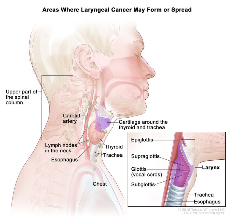
The larynx is a part of the throat, between the base of the tongue and the trachea. The larynx contains the vocal cords, which vibrate and make sound when air is directed against them. The sound echoes through the pharynx, mouth, and nose to make a person’s voice.
There are three main parts of the larynx:
Supraglottis: The upper part of the larynx above the vocal cords, including the epiglottis.
Glottis: The middle part of the larynx where the vocal cords are located.
Subglottis: The lower part of the larynx between the vocal cords and the trachea (windpipe).
EnlargeLaryngeal cancer forms in the tissues of the larynx (area of the throat that contains the vocal cords). The larynx includes the supraglottis, glottis (vocal cords), and subglottis. The cancer may spread to nearby tissues or to the thyroid, trachea, or esophagus. It may also spread to the lymph nodes in the neck, the carotid artery, the upper part of the spinal column, the chest, and to other parts of the body (not shown).
Use of tobacco products and drinking too much alcohol can affect the risk of laryngeal cancer.
Anything that increases a person’s chance of getting a disease is called a risk factor. Not every person with one or more of these risk factors will develop laryngeal cancer, and it will develop in people who don’t have any known risk factors. Talk with your doctor if you think you may be at risk.
Signs and symptoms of laryngeal cancer include a sore throat and ear pain.
These and other signs and symptoms may be caused by laryngeal cancer or by other conditions. Check with your doctor if you have any of the following:
A sore throat or cough that does not go away.
Trouble or pain when swallowing.
Ear pain.
A lump in the neck or throat.
A change or hoarseness in the voice.
Tests that examine the throat and neck are used to help diagnose and stage laryngeal cancer.
In addition to asking about your personal and family health history, your doctor may perform the following tests and procedures:
Physical exam of the throat and neck: An exam to check the throat and neck for abnormal areas. The doctor will feel the inside of the mouth with a gloved finger and examine the mouth and throat with a small long-handled mirror and light. This will include checking the insides of the cheeks and lips; the gums; the back, roof, and floor of the mouth; the top, bottom, and sides of the tongue; and the throat. The neck will be felt for swollen lymph nodes. A history of the patient’s health habits and past illnesses and medical treatments will also be taken.
Biopsy: The removal of cells or tissues so they can be viewed under a microscope by a pathologist to check for signs of cancer. The sample of tissue may be removed during one of the following procedures:
Laryngoscopy: A procedure in which the doctor checks the larynx (voice box) with a mirror or a laryngoscope to check for abnormal areas. A laryngoscope is a thin, tube-like instrument with a light and a lens for viewing the inside of the throat and voice box. It may also have a tool to remove tissue samples, which are checked under a microscope for signs of cancer.
Endoscopy: A procedure to look at organs and tissues inside the body, such as the throat, esophagus, and trachea to check for abnormal areas. An endoscope (a thin, lighted tube with a light and a lens for viewing) is inserted through an opening in the body, such as the mouth. A special tool on the endoscope may be used to remove samples of tissue.
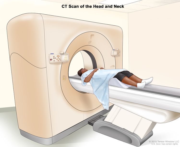
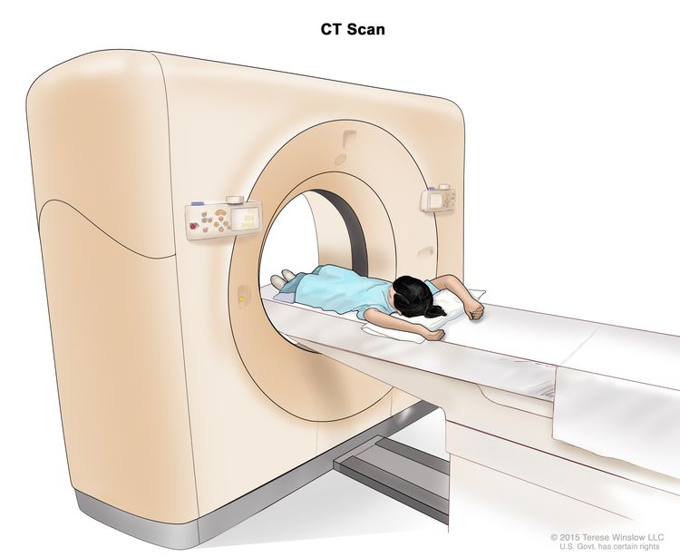
CT scan (CAT scan): A procedure that makes a series of detailed pictures of areas inside the body, taken from different angles. The pictures are made by a computer linked to an x-ray machine. A dye may be injected into a vein or swallowed to help the organs or tissues show up more clearly. This procedure is also called computed tomography, computerized tomography, or computerized axial tomography. EnlargeComputed tomography (CT) scan of the head and neck. The patient lies on a table that slides through the CT scanner, which takes x-ray pictures of the inside of the head and neck.
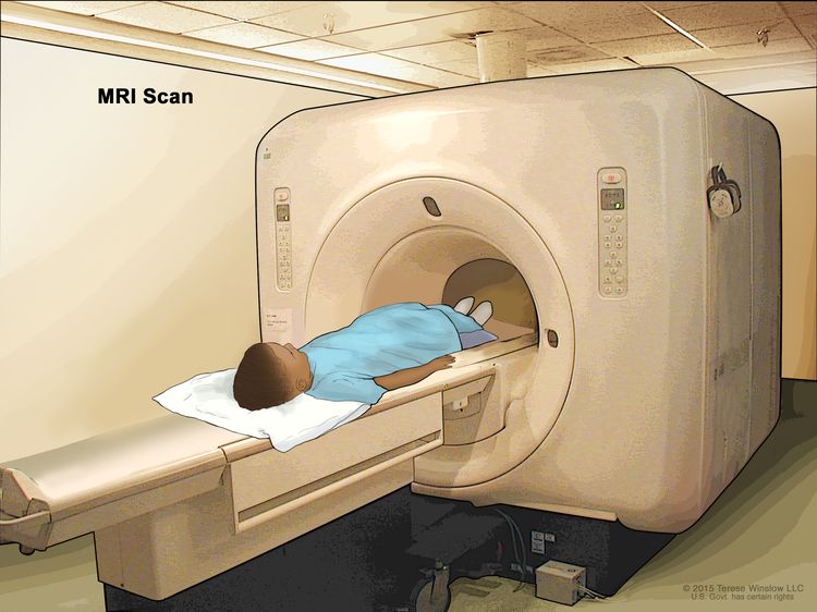
MRI (magnetic resonance imaging): A procedure that uses a magnet, radio waves, and a computer to make a series of detailed pictures of areas inside the body. This procedure is also called nuclear magnetic resonance imaging (NMRI).
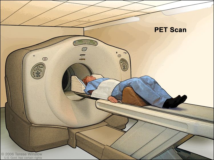
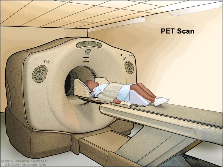
PET scan (positron emission tomography scan): A procedure to find malignanttumor cells in the body. A small amount of radioactiveglucose (sugar) is injected into a vein. The PET scanner rotates around the body and makes a picture of where glucose is being used in the body. Malignant tumor cells show up brighter in the picture because they are more active and take up more glucose than normal cells do.
PET-CT scan: A procedure that combines the pictures from a positron emission tomography (PET) scan and a computed tomography (CT) scan. The PET and CT scans are done at the same time with the same machine. The combined scans give more detailed pictures of areas inside the body than either scan gives by itself. A PET-CT scan may be used to help diagnose disease, such as cancer, plan treatment, or find out how well treatment is working.
Bone scan: A procedure to check if there are rapidly dividing cells, such as cancer cells, in the bone. A very small amount of radioactive material is injected into a vein and travels through the bloodstream. The radioactive material collects in the bones with cancer and is detected by a scanner.
Barium swallow: A series of x-rays of the esophagus and stomach. The patient drinks a liquid that contains barium (a silver-white metalliccompound). The liquid coats the esophagus and stomach, and x-rays are taken. This procedure is also called an upper GI series.
Certain factors affect prognosis (chance of recovery) and treatment options.
Smoking tobacco and drinking alcohol decrease the effectiveness of treatment for laryngeal cancer. Patients with laryngeal cancer who continue to smoke and drink are less likely to be cured and more likely to develop a second tumor. After treatment for laryngeal cancer, frequent and careful follow-up is important.
Stages of Laryngeal Cancer
Key Points
After laryngeal cancer has been diagnosed, tests are done to find out if cancer cells have spread within the larynx or to other parts of the body.
There are three ways that cancer spreads in the body.
Cancer may spread from where it began to other parts of the body.
The following stages are used for laryngeal cancer:
Stage 0 (Carcinoma in Situ)
Stage I
Stage II
Stage III
Stage IV
After surgery, the stage of the cancer may change and more treatment may be needed.
Laryngeal cancer can recur (come back) after it has been treated.
After laryngeal cancer has been diagnosed, tests are done to find out if cancer cells have spread within the larynx or to other parts of the body.
The process used to find out if cancer has spread within the larynx or to other parts of the body is called staging. The information gathered from the staging process determines the stage of the disease. It is important to know the stage of the disease in order to plan treatment. The results of some of the tests used to diagnoselaryngeal cancer are often also used to stage the disease.
There are three ways that cancer spreads in the body.
Tissue. The cancer spreads from where it began by growing into nearby areas.
Lymph system. The cancer spreads from where it began by getting into the lymph system. The cancer travels through the lymph vessels to other parts of the body.
Blood. The cancer spreads from where it began by getting into the blood. The cancer travels through the blood vessels to other parts of the body.
Cancer may spread from where it began to other parts of the body.
When cancer spreads to another part of the body, it is called metastasis. Cancer cells break away from where they began (the primary tumor) and travel through the lymph system or blood.
Lymph system. The cancer gets into the lymph system, travels through the lymph vessels, and forms a tumor (metastatic tumor) in another part of the body.
Blood. The cancer gets into the blood, travels through the blood vessels, and forms a tumor (metastatic tumor) in another part of the body.
The metastatic tumor is the same type of cancer as the primary tumor. For example, if laryngeal cancer spreads to the lung, the cancer cells in the lung are actually laryngeal cancer cells. The disease is metastatic laryngeal cancer, not lung cancer.
Many cancer deaths are caused when cancer moves from the original tumor and spreads to other tissues and organs. This is called metastatic cancer. This animation shows how cancer cells travel from the place in the body where they first formed to other parts of the body.
The following stages are used for laryngeal cancer:
Supraglottis: Cancer is in more than one area of the supraglottis or has spread to the area at the base of the tongue or to tissues near the vocal cords. The vocal cords work normally.
Glottis: Cancer has spread to the supraglottis, subglottis, or both, and/or the vocal cords do not work normally.
Subglottis: Cancer has spread to one or both vocal cords and the vocal cords may not work normally.
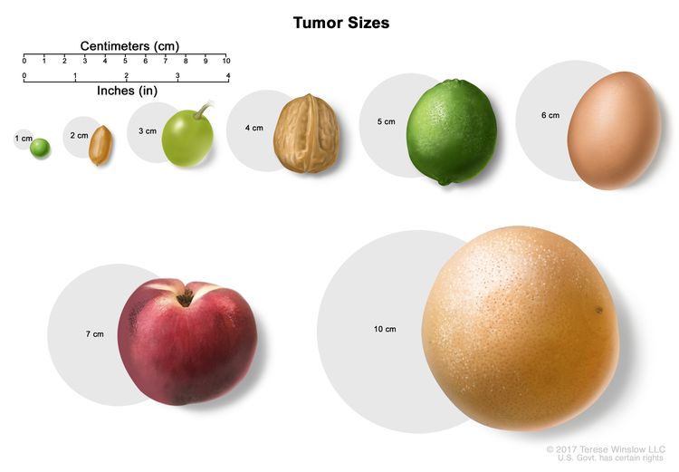
EnlargeTumor sizes are often measured in centimeters (cm) or inches. Common food items that can be used to show tumor size in cm include: a pea (1 cm), a peanut (2 cm), a grape (3 cm), a walnut (4 cm), a lime (5 cm or 2 inches), an egg (6 cm), a peach (7 cm), and a grapefruit (10 cm or 4 inches).
In stage III cancer of the supraglottis:
cancer is in the larynx only and the vocal cords do not work, and/or cancer has spread near or through the inner part of the thyroidcartilage. Cancer may have also spread to one lymph node on the same side of the neck as the primary tumor and the lymph node is 3 centimeters or smaller; or
cancer is in one area of the supraglottis and the vocal cords work normally. Cancer has spread to one lymph node on the same side of the neck as the primary tumor and the lymph node is 3 centimeters or smaller; or
cancer is in more than one area of the supraglottis or has spread to the area at the base of the tongue or to tissues near the vocal cords. The vocal cords work normally. Cancer has also spread to one lymph node on the same side of the neck as the primary tumor and the lymph node is 3 centimeters or smaller.
In stage III cancer of the glottis:
cancer is in the larynx only and the vocal cords do not work, and/or cancer has spread near or through the inner part of the thyroid cartilage. Cancer may have also spread to one lymph node on the same side of the neck as the primary tumor and the lymph node is 3 centimeters or smaller; or
cancer is in one or both vocal cords and the vocal cords work normally. Cancer has spread to one lymph node on the same side of the neck as the primary tumor and the lymph node is 3 centimeters or smaller; or
cancer has spread to the supraglottis, subglottis, or both, and/or the vocal cords do not work normally. Cancer has also spread to one lymph node on the same side of the neck as the primary tumor and the lymph node is 3 centimeters or smaller.
In stage III cancer of the subglottis:
cancer is in the larynx only and the vocal cords do not work, and/or cancer has spread near or through the inner part of the thyroid cartilage. Cancer may have also spread to one lymph node on the same side of the neck as the primary tumor and the lymph node is 3 centimeters or smaller; or
cancer is in the subglottis only. Cancer has spread to one lymph node on the same side of the neck as the primary tumor and the lymph node is 3 centimeters or smaller; or
cancer has spread to one or both vocal cords and the vocal cords may not work normally. Cancer has also spread to one lymph node on the same side of the neck as the primary tumor and the lymph node is 3 centimeters or smaller.
Cancer may have spread from the supraglottis, glottis, or subglottis to tissues beyond the larynx, such as the neck, trachea, thyroid, or esophagus. The vocal cords may not work normally. Cancer has spread:
to one lymph node on the same side of the neck as the primary tumor and the lymph node is 3 centimeters or smaller. Cancer has spread through the outside covering of the lymph node; or
to one lymph node on the same side of the neck as the primary tumor and the lymph node is larger than 3 centimeters but not larger than 6 centimeters. Cancer has not spread through the outside covering of the lymph node; or
to more than one lymph node on the same side of the neck as the primary tumor and the lymph nodes are not larger than 6 centimeters. Cancer has not spread through the outside covering of the lymph nodes; or
to lymph nodes on both sides of the neck or on the side of the neck opposite the primary tumor and the lymph nodes are not larger than 6 centimeters. Cancer has not spread through the outside covering of the lymph nodes.
In stage IVB:
Cancer may have spread from the supraglottis, glottis, or subglottis to the space in front of the spine, the area around the carotid artery, or the area between the lungs. The vocal cords may not work normally. Cancer has spread:
to one lymph node that is larger than 6 centimeters. Cancer has not spread through the outside covering of the lymph node; or
to one lymph node on the same side of the neck as the primary tumor and the lymph node is larger than 3 centimeters. Cancer has spread through the outside covering of the lymph node; or
to more than one lymph node anywhere in the neck. Cancer has spread through the outside covering of the lymph nodes; or
to one lymph node of any size on the side of the neck opposite the primary tumor. Cancer has spread through the outside covering of the lymph node;
or
Cancer has spread from the supraglottis, glottis, or subglottis to the space in front of the spine, the area around the carotid artery, or the area between the lungs. Cancer may have also spread to one or more lymph nodes anywhere in the neck and the lymph nodes may be any size.
In stage IVC, cancer has spread to other parts of the body, such as the lungs, liver, or bone.
After surgery, the stage of the cancer may change and more treatment may be needed.
If the cancer is removed by surgery, a pathologist will examine a sample of the cancer tissue under a microscope. Sometimes, the pathologist’s review will result in a change to the stage of the cancer and more treatment after surgery.
Laryngeal cancer can recur (come back) after it has been treated.
The cancer may come back in the larynx or in other parts of the body, such as lungs, liver, or bone. It is most likely to come back in the first 2 to 3 years.
Treatment Option Overview
Key Points
There are different types of treatment for patients with laryngeal cancer.
The following types of treatment are used:
Radiation therapy
Surgery
Chemotherapy
Immunotherapy
New types of treatment are being tested in clinical trials.
Targeted therapy
Radiosensitizers
Treatment for laryngeal cancer may cause side effects.
Patients may want to think about taking part in a clinical trial.
Patients can enter clinical trials before, during, or after starting their cancer treatment.
Follow-up tests may be needed.
There are different types of treatment for patients with laryngeal cancer.
Different types of treatment are available for patients with laryngeal cancer. Some treatments are standard (the currently used treatment), and some are being tested in clinical trials. A treatment clinical trial is a research study meant to help improve current treatments or obtain information on new treatments for patients with cancer. When clinical trials show that a new treatment is better than the standard treatment, the new treatment may become the standard treatment. Patients may want to think about taking part in a clinical trial. Some clinical trials are open only to patients who have not started treatment.
The following types of treatment are used:
Radiation therapy
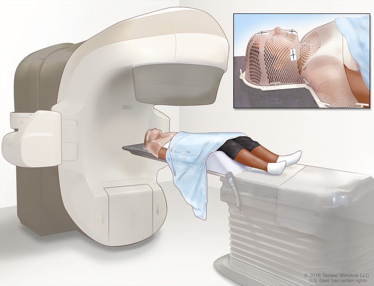
Radiation therapy is a cancer treatment that uses high-energy x-rays or other types of radiation to kill cancer cells or keep them from growing. External radiation therapy uses a machine outside the body to send radiation toward the area of the body with cancer.
EnlargeExternal-beam radiation therapy of the head and neck. A machine is used to aim high-energy radiation at the cancer. The machine can rotate around the patient, delivering radiation from many different angles to provide highly conformal treatment. A mesh mask helps keep the patient’s head and neck from moving during treatment. Small ink marks are put on the mask. The ink marks are used to line up the radiation machine in the same position before each treatment.
Radiation therapy may work better in patients who have stopped smoking before beginning treatment. External radiation therapy to the thyroid or the pituitary gland may change the way the thyroid gland works. A blood test to check the thyroid hormone level in the body may be done before and after therapy to make sure the thyroid gland is working properly.
Hyperfractionated radiation therapy may be used to treat laryngeal cancer. Hyperfractionated radiation therapy is radiation treatment in which a smaller than usual total daily dose of radiation is divided into two doses and the treatments are given twice a day. Hyperfractionated radiation therapy is given over the same period of time (days or weeks) as standard radiation therapy. New types of radiation therapy are being studied in the treatment of laryngeal cancer.
Surgery
Surgery (removing the cancer in an operation) is a common treatment for all stages of laryngeal cancer. The following surgical procedures may be used:
Hemilaryngectomy: Surgery to remove half of the larynx (voice box). A hemilaryngectomy saves the voice.
Partial laryngectomy: Surgery to remove part of the larynx (voice box). A partial laryngectomy helps keep the patient’s ability to talk.
Total laryngectomy: Surgery to remove the whole larynx. During this operation, a hole is made in the front of the neck to allow the patient to breathe. This is called a tracheostomy.
Thyroidectomy: The removal of all or part of the thyroid gland.
Laser surgery: A surgical procedure that uses a laser beam (a narrow beam of intense light) as a knife to make bloodless cuts in tissue or to remove a surface lesion such as a tumor in the larynx.
After the doctor removes all the cancer that can be seen at the time of the surgery, some patients may be given chemotherapy or radiation therapy after surgery to kill any cancer cells that are left. Treatment given after the surgery, to lower the risk that the cancer will come back, is called adjuvant therapy.
Chemotherapy
Chemotherapy is a cancer treatment that uses drugs to stop the growth of cancer cells, either by killing the cells or by stopping the cells from dividing. When chemotherapy is taken by mouth or injected into a vein or muscle, the drugs enter the bloodstream and can reach cancer cells throughout the body (systemic chemotherapy).
Immunotherapy is a treatment that uses the patient’s immune system to fight cancer. Substances made by the body or made in a laboratory are used to boost, direct, or restore the body’s natural defenses against cancer. This cancer treatment is a type of biologic therapy.
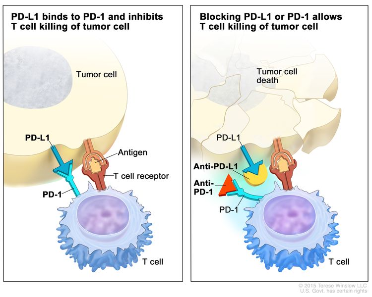
PD-1 and PD-L1 inhibitor therapy: PD-1 is a protein on the surface of T cells that helps keep the body’s immune responses in check. PD-L1 is a protein found on some types of cancer cells. When PD-1 attaches to PD-L1, it stops the T cell from killing the cancer cell. PD-1 and PD-L1 inhibitors keep PD-1 and PD-L1 proteins from attaching to each other. This allows the T cells to kill cancer cells. Nivolumab and pembrolizumab are types of PD-1 inhibitors used to treat metastatic or recurrent laryngeal cancer.
EnlargeImmune checkpoint inhibitor. Checkpoint proteins, such as PD-L1 on tumor cells and PD-1 on T cells, help keep immune responses in check. The binding of PD-L1 to PD-1 keeps T cells from killing tumor cells in the body (left panel). Blocking the binding of PD-L1 to PD-1 with an immune checkpoint inhibitor (anti-PD-L1 or anti-PD-1) allows the T cells to kill tumor cells (right panel).
Immunotherapy uses the body’s immune system to fight cancer. This animation explains one type of immunotherapy that uses immune checkpoint inhibitors to treat cancer.
New types of treatment are being tested in clinical trials.
This summary section describes treatments that are being studied in clinical trials. It may not mention every new treatment being studied. Information about clinical trials is available from the NCI website.
Targeted therapy
Targeted therapy is a type of treatment that uses drugs or other substances to identify and attack specific cancer cells.
Monoclonal antibodies: Monoclonal antibodies are immune system proteins made in the laboratory to treat many diseases, including cancer. As a cancer treatment, these antibodies can attach to a specific target on cancer cells or other cells that may help cancer cells grow. The antibodies are able to then kill the cancer cells, block their growth, or keep them from spreading. Monoclonal antibodies are given by infusion. They may be used alone or to carry drugs, toxins, or radioactive material directly to cancer cells. Cetuximab is being studied in the treatment of laryngeal cancer.
How do monoclonal antibodies work to treat cancer? This video shows how monoclonal antibodies, such as trastuzumab, pembrolizumab, and rituximab, block molecules cancer cells need to grow, flag cancer cells for destruction by the body’s immune system, or deliver harmful substances to cancer cells.
Radiosensitizers
Radiosensitizers are drugs that make tumor cells more sensitive to radiation therapy. Combining radiation therapy with radiosensitizers may kill more tumor cells.
Treatment for laryngeal cancer may cause side effects.
Patients may want to think about taking part in a clinical trial.
For some patients, taking part in a clinical trial may be the best treatment choice. Clinical trials are part of the cancer research process. Clinical trials are done to find out if new cancer treatments are safe and effective or better than the standard treatment.
Many of today’s standard treatments for cancer are based on earlier clinical trials. Patients who take part in a clinical trial may receive the standard treatment or be among the first to receive a new treatment.
Patients who take part in clinical trials also help improve the way cancer will be treated in the future. Even when clinical trials do not lead to effective new treatments, they often answer important questions and help move research forward.
Patients can enter clinical trials before, during, or after starting their cancer treatment.
Some clinical trials only include patients who have not yet received treatment. Other trials test treatments for patients whose cancer has not gotten better. There are also clinical trials that test new ways to stop cancer from recurring (coming back) or reduce the side effects of cancer treatment.
Clinical trials are taking place in many parts of the country. Information about clinical trials supported by NCI can be found on NCI’s clinical trials search webpage. Clinical trials supported by other organizations can be found on the ClinicalTrials.gov website.
Follow-up tests may be needed.
As you go through treatment, you will have follow-up tests or check-ups. Some tests that were done to diagnose or stage the cancer may be repeated to see how well the treatment is working. Decisions about whether to continue, change, or stop treatment may be based on the results of these tests.
Some of the tests will continue to be done from time to time after treatment has ended. The results of these tests can show if your condition has changed or if the cancer has recurred (come back).
Use our clinical trial search to find NCI-supported cancer clinical trials that are accepting patients. You can search for trials based on the type of cancer, the age of the patient, and where the trials are being done. General information about clinical trials is also available.
Use our clinical trial search to find NCI-supported cancer clinical trials that are accepting patients. You can search for trials based on the type of cancer, the age of the patient, and where the trials are being done. General information about clinical trials is also available.
Radiation therapy followed by surgery if cancer comes back in the same area.
Radiation therapy alone for patients who cannot be treated with chemotherapy and surgery.
Chemotherapy followed by chemotherapy and radiation therapy given together. Laryngectomy may be done if cancer remains.
A clinical trial of radiation therapy alone compared with radiation and targeted therapy (cetuximab).
A clinical trial of immunotherapy, chemotherapy, radiosensitizers, or radiation therapy.
Use our clinical trial search to find NCI-supported cancer clinical trials that are accepting patients. You can search for trials based on the type of cancer, the age of the patient, and where the trials are being done. General information about clinical trials is also available.
Chemotherapy followed by chemotherapy and radiation therapy given together. Laryngectomy may be done if cancer remains.
Radiation therapy alone for patients who cannot be treated with chemotherapy and surgery.
Surgery followed by radiation therapy. Chemotherapy may be given with the radiation therapy.
A clinical trial of radiation therapy alone compared with radiation and targeted therapy (cetuximab).
A clinical trial of immunotherapy, chemotherapy, radiosensitizers, or radiation therapy.
If cancer is in the subglottis, treatment may include the following:
Laryngectomy plus total thyroidectomy and removal of lymph nodes in the throat, usually followed by radiation therapy with or without chemotherapy.
Chemotherapy and radiation therapy given together.
A clinical trial of radiation therapy alone compared with radiation and targeted therapy (cetuximab).
A clinical trial of immunotherapy, chemotherapy, radiosensitizers, or radiation therapy.
Use our clinical trial search to find NCI-supported cancer clinical trials that are accepting patients. You can search for trials based on the type of cancer, the age of the patient, and where the trials are being done. General information about clinical trials is also available.
Treatment of Metastatic and Recurrent Laryngeal Cancer
Use our clinical trial search to find NCI-supported cancer clinical trials that are accepting patients. You can search for trials based on the type of cancer, the age of the patient, and where the trials are being done. General information about clinical trials is also available.
Physician Data Query (PDQ) is the National Cancer Institute’s (NCI’s) comprehensive cancer information database. The PDQ database contains summaries of the latest published information on cancer prevention, detection, genetics, treatment, supportive care, and complementary and alternative medicine. Most summaries come in two versions. The health professional versions have detailed information written in technical language. The patient versions are written in easy-to-understand, nontechnical language. Both versions have cancer information that is accurate and up to date and most versions are also available in Spanish.
PDQ is a service of the NCI. The NCI is part of the National Institutes of Health (NIH). NIH is the federal government’s center of biomedical research. The PDQ summaries are based on an independent review of the medical literature. They are not policy statements of the NCI or the NIH.
Purpose of This Summary
This PDQ cancer information summary has current information about the treatment of adult laryngeal cancer. It is meant to inform and help patients, families, and caregivers. It does not give formal guidelines or recommendations for making decisions about health care.
Reviewers and Updates
Editorial Boards write the PDQ cancer information summaries and keep them up to date. These Boards are made up of experts in cancer treatment and other specialties related to cancer. The summaries are reviewed regularly and changes are made when there is new information. The date on each summary (“Updated”) is the date of the most recent change.
The information in this patient summary was taken from the health professional version, which is reviewed regularly and updated as needed, by the PDQ Adult Treatment Editorial Board.
Clinical Trial Information
A clinical trial is a study to answer a scientific question, such as whether one treatment is better than another. Trials are based on past studies and what has been learned in the laboratory. Each trial answers certain scientific questions in order to find new and better ways to help cancer patients. During treatment clinical trials, information is collected about the effects of a new treatment and how well it works. If a clinical trial shows that a new treatment is better than one currently being used, the new treatment may become “standard.” Patients may want to think about taking part in a clinical trial. Some clinical trials are open only to patients who have not started treatment.
Clinical trials can be found online at NCI’s website. For more information, call the Cancer Information Service (CIS), NCI’s contact center, at 1-800-4-CANCER (1-800-422-6237).
Permission to Use This Summary
PDQ is a registered trademark. The content of PDQ documents can be used freely as text. It cannot be identified as an NCI PDQ cancer information summary unless the whole summary is shown and it is updated regularly. However, a user would be allowed to write a sentence such as “NCI’s PDQ cancer information summary about breast cancer prevention states the risks in the following way: [include excerpt from the summary].”
The best way to cite this PDQ summary is:
PDQ® Adult Treatment Editorial Board. PDQ Laryngeal Cancer Treatment. Bethesda, MD: National Cancer Institute. Updated <MM/DD/YYYY>. Available at: /types/head-and-neck/patient/adult/laryngeal-treatment-pdq. Accessed <MM/DD/YYYY>. [PMID: 26389298]
Images in this summary are used with permission of the author(s), artist, and/or publisher for use in the PDQ summaries only. If you want to use an image from a PDQ summary and you are not using the whole summary, you must get permission from the owner. It cannot be given by the National Cancer Institute. Information about using the images in this summary, along with many other images related to cancer can be found in Visuals Online. Visuals Online is a collection of more than 3,000 scientific images.
Disclaimer
The information in these summaries should not be used to make decisions about insurance reimbursement. More information on insurance coverage is available on Cancer.gov on the Managing Cancer Care page.
Contact Us
More information about contacting us or receiving help with the Cancer.gov website can be found on our Contact Us for Help page. Questions can also be submitted to Cancer.gov through the website’s E-mail Us.
Estimated new cases and deaths from laryngeal cancer in the United States in 2025:[1]
New cases: 13,020.
Deaths: 3,910.
Anatomy
The larynx is divided into the following three anatomical regions:
The supraglottic larynx includes the epiglottis, false vocal cords, ventricles, aryepiglottic folds, and arytenoids.
The glottis includes the true vocal cords and the anterior and posterior commissures.
The subglottic region begins about 1 cm below the true vocal cords and extends to the lower border of the cricoid cartilage or the first tracheal ring.
The supraglottic area is rich in lymphatic drainage. After penetrating the pre-epiglottic space and thyrohyoid membrane, lymphatic drainage is initially to the jugulodigastric and midjugular nodes. About 25% to 50% of patients present with involved lymph nodes. The precise figure depends on the T (tumor) stage. The true vocal cords are devoid of lymphatics. As a result, vocal cord cancer confined to the true cords rarely, if ever, presents with involved lymph nodes. Extension above or below the cords may, however, lead to lymph node involvement. Primary subglottic cancers, which are quite rare, drain through the cricothyroid and cricotracheal membranes to the pretracheal, paratracheal, and inferior jugular nodes, and occasionally to mediastinal nodes.[2]
Risk Factors
There is a clear association among smoking, excess alcohol ingestion, and the development of squamous cell cancers of the upper aerodigestive tract.[3] For smokers, the risk of laryngeal cancer decreases after they stop smoking but remains elevated, even years later, compared with that of nonsmokers.[4] If a patient who has had a single cancer continues to smoke and drink alcoholic beverages, the likelihood of a cure for the initial cancer, by any modality, is diminished, and the risk of second tumor is enhanced. Because of clinical problems related to smoking and alcohol use in this population, many patients die of intercurrent illness rather than the primary cancer.
Clinical Features
Supraglottic cancers typically present with sore throat, painful swallowing, referred ear pain, change in voice quality, or enlarged neck nodes. Early vocal cord cancers are usually detected because of hoarseness. By the time they are detected, cancers arising in the subglottic area commonly involve the vocal cords; thus, symptoms usually relate to contiguous spread.
Prognostic Factors
The most important adverse prognostic factors for laryngeal cancers include increasing T stage and N (regional lymph node) stage. Other prognostic factors may include sex, age, performance status, and a variety of pathological features of the tumor, including grade and depth of invasion.[5]
Prognosis for small laryngeal cancers that have not spread to lymph nodes is very good. Cure rates are 75% to 95% depending on the site, tumor bulk,[6] and degree of infiltration. Although most patients with early lesions can be cured by either radiation therapy or surgery, radiation therapy may be reasonable to preserve the voice, leaving surgery for salvage. Patients with a preradiation hemoglobin level higher than 13 g/dL have higher local control and survival rates than patients who are anemic.[7]
Locally advanced lesions are treated with combined modality treatment involving radiation and chemotherapy with or without surgery. The aim is laryngeal preservation in appropriately selected candidates.[8] Distant metastases are also common, even if the primary tumor is controlled.
Intermediate lesions have intermediate prognoses, depending on the site, T stage, N stage, and performance status. Therapy recommendations for patients with these lesions are based on a variety of complex anatomical, clinical, and social factors, which should be individualized and discussed in multidisciplinary consultation (surgery, radiation therapy, and dental and oral surgery) prior to prescribing therapy.
Follow-Up and Survivorship
Second primary tumors, often in the aerodigestive tract, have been reported in as many as 25% of patients whose initial lesion is controlled. A study has shown that daily treatment of these patients with moderate doses of isotretinoin (i.e., 13-cis-retinoic acid) for 1 year can significantly reduce the incidence of second tumors.[9] No survival advantage has been demonstrated, partially because of recurrence and death from the primary malignancy.
Patients treated for laryngeal cancers are at the highest risk of recurrence in the first 2 to 3 years. Recurrences after 5 years are rare and usually represent new primary malignancies. Close, regular follow-up is crucial to maximize the chance for salvage. Follow-up includes careful clinical examination and repetition of any abnormal staging study, along with attention to any treatment-related toxic effect or complication.
References
American Cancer Society: Cancer Facts and Figures 2025. American Cancer Society, 2025. Available online. Last accessed January 16, 2025.
Spaulding CA, Hahn SS, Constable WC: The effectiveness of treatment of lymph nodes in cancers of the pyriform sinus and supraglottis. Int J Radiat Oncol Biol Phys 13 (7): 963-8, 1987. [PUBMED Abstract]
Spitz MR: Epidemiology and risk factors for head and neck cancer. Semin Oncol 21 (3): 281-8, 1994. [PUBMED Abstract]
Bosetti C, Garavello W, Gallus S, et al.: Effects of smoking cessation on the risk of laryngeal cancer: an overview of published studies. Oral Oncol 42 (9): 866-72, 2006. [PUBMED Abstract]
Yilmaz T, Hoşal S, Gedikoglu G, et al.: Prognostic significance of depth of invasion in cancer of the larynx. Laryngoscope 108 (5): 764-8, 1998. [PUBMED Abstract]
Reddy SP, Mohideen N, Marra S, et al.: Effect of tumor bulk on local control and survival of patients with T1 glottic cancer. Radiother Oncol 47 (2): 161-6, 1998. [PUBMED Abstract]
Fein DA, Lee WR, Hanlon AL, et al.: Pretreatment hemoglobin level influences local control and survival of T1-T2 squamous cell carcinomas of the glottic larynx. J Clin Oncol 13 (8): 2077-83, 1995. [PUBMED Abstract]
Forastiere AA, Zhang Q, Weber RS, et al.: Long-term results of RTOG 91-11: a comparison of three nonsurgical treatment strategies to preserve the larynx in patients with locally advanced larynx cancer. J Clin Oncol 31 (7): 845-52, 2013. [PUBMED Abstract]
Hong WK, Lippman SM, Itri LM, et al.: Prevention of second primary tumors with isotretinoin in squamous-cell carcinoma of the head and neck. N Engl J Med 323 (12): 795-801, 1990. [PUBMED Abstract]
Cellular Classification of Laryngeal Cancer
Most laryngeal cancers are of squamous cell histology. Squamous cell subtypes include keratinizing and nonkeratinizing and well-differentiated to poorly differentiated grade. A variety of nonsquamous cell laryngeal cancers also occur.[1] These are not staged using the American Joint Cancer Committee staging system, and their management, which is not discussed here, can differ from that of squamous cell laryngeal cancers. In situ squamous cell carcinoma of the larynx is usually managed by a conservative surgical procedure such as mucosal stripping or superficial laser excision. Radiation therapy may also be appropriate treatment of selected patients with in situ carcinoma of the glottic larynx.
References
Mendenhall WM, Werning JW, Pfister DG: Treatment of head and neck cancer. In: DeVita VT Jr, Lawrence TS, Rosenberg SA: Cancer: Principles and Practice of Oncology. 9th ed. Lippincott Williams & Wilkins, 2011, pp 729-80.
Stage Information for Laryngeal Cancer
The staging system for laryngeal cancer is clinical and based on the best possible estimate of the extent of disease before treatment. The assessment of the primary tumor is based on inspection and palpation, when possible, and by fiberoptic laryngoscopy. Panendoscopy under anesthesia ensures careful clinical examination to determine clinical extent of local disease. The tumor must be confirmed histologically, and any other pathological data obtained on biopsy may be included. Head and neck magnetic resonance imaging, computed tomography, or positron emission tomography-computed tomography should be done before therapy to supplement inspection and palpation.[1] Additional radiographic studies may be included. The appropriate nodal drainage areas in the neck should be examined by careful palpation.
Definitions of TNM
The American Joint Committee on Cancer (AJCC) has designated staging by TNM (tumor, node, metastasis) classification to define laryngeal cancer.[2]
Table 1. Definition of Supraglottis, Glottis, and Subglottis Primary Tumor (T) for Laryngeal Cancera,b
T Category
T Criteria
aReprinted with permission from AJCC: Larynx. In: Amin MB, Edge SB, Greene FL, et al., eds.: AJCC Cancer Staging Manual. 8th ed. New York, NY: Springer, 2017, pp 149–61.
TX
Primary tumor cannot be assessed.
Tis
Carcinoma in situ.
Supraglottis
T1
Tumor limited to one subsite of supraglottis with normal vocal cord mobility.
T2
Tumor invades mucosa of more than one adjacent subsite of supraglottis or glottis or region outside the supraglottis (e.g., mucosa of the base of the tongue, vallecula, medial wall of pyriform sinus) without fixation of the larynx.
T3
Tumor limited to larynx with vocal cord fixation and/or invades any of the following: postcricoid area, pre-epiglottic space, paraglottic space, and/or inner cortex of thyroid cartilage.
T4
Moderately advanced or very advanced.
–T4a
Moderately advanced local disease. Tumor invades through the outer cortex of the thyroid cartilage and/or invades tissues beyond the larynx (e.g., trachea, soft tissues of the neck including deep extrinsic muscle of the tongue, strap muscles, thyroid, or esophagus).
–T4b
Very advanced local disease. Tumor invades prevertebral space, encases carotid artery, or invades mediastinal structures.
Glottis
T1
Tumor limited to the vocal cord(s) (may involve anterior or posterior commissure) with normal mobility.
–T1a
Tumor limited to one vocal cord.
–T1b
Tumor involves both vocal cords.
T2
Tumor extends to supraglottis and/or subglottis, and/or with impaired vocal cord mobility.
T3
Tumor limited to the larynx with vocal cord fixation and/or invasion of paraglottic space and/or inner cortex of the thyroid cartilage.
T4
Moderately advanced or very advanced.
–T4a
Moderately advanced local disease. Tumor invades through the outer cortex of the thyroid cartilage and/or invades tissues beyond the larynx (e.g., trachea, cricoid cartilage, soft tissues of the neck including deep extrinsic muscle of the tongue, strap muscles, thyroid, or esophagus).
–T4b
Very advanced local disease. Tumor invades prevertebral space, encases carotid artery, or invades mediastinal structures.
Subglottis
T1
Tumor limited to the subglottis.
T2
Tumor extends to vocal cord(s) with normal or impaired mobility.
T3
Tumor limited to the larynx with vocal cord fixation and/or invasion of paraglottic space and/or inner cortex of the thyroid cartilage.
T4
Moderately advanced or very advanced.
–T4a
Moderately advanced local disease. Tumor invades cricoid or thyroid cartilage and/or invades tissues beyond the larynx (e.g., trachea, soft tissues of the neck including deep extrinsic muscles of the tongue, strap muscles, thyroid, or esophagus).
–T4b
Very advanced local disease. Tumor invades prevertebral space, encases carotid artery, or invades mediastinal structures.
Table 2. Definition of Clinical (cN) Regional Lymph Nodes (N) for Laryngeal Cancer a,b
N Category
N Criteria
ENE = extranodal extension.
aReprinted with permission from AJCC: Larynx. In: Amin MB, Edge SB, Greene FL, et al., eds.: AJCC Cancer Staging Manual. 8th ed. New York, NY: Springer, 2017, pp 149–61.
bA designation of “U” or “L” may be used for any N category to indicate metastasis above the lower border of the cricoid (U) or below the lower border of the cricoid (L). Similarly, clinical and pathological ENE should be recorded as ENE(–) or ENE(+).
NX
Regional lymph nodes cannot be assessed.
N0
No regional lymph node metastasis.
N1
Metastasis in a single ipsilateral lymph node ≤3 cm in greatest dimension and ENE(–).
N2
Metastasis in a single ipsilateral node, >3 cm but not >6 cm in greatest dimension and ENE(–); or metastases in multiple ipsilateral lymph nodes, none >6 cm in greatest dimension and ENE(–); or metastases in bilateral or contralateral lymph nodes, none >6 cm in greatest dimension and ENE(–).
–N2a
Metastasis in a single ipsilateral node >3 cm but not >6 cm in greatest dimension and ENE(–).
–N2b
Metastases in multiple ipsilateral nodes, none >6 cm in greatest dimension and ENE(–).
–N2c
Metastases in bilateral or contralateral lymph nodes, none >6 cm in greatest dimension and ENE(–).
N3
Metastasis in a lymph node >6 cm in greatest dimension and ENE(–); or metastasis in any lymph nodes(s) with clinically overt ENE(+).
–N3a
Metastasis in a lymph node >6 cm in greatest dimension and ENE(–).
–N3b
Metastasis in any lymph node(s) with clinically overt ENE(+).
Table 3. Definition of Pathological (pN) Regional Lymph Nodes (N) for Laryngeal Cancera,b
N Category
N Criteria
ENE = extranodal extension.
aReprinted with permission from AJCC: Larynx. In: Amin MB, Edge SB, Greene FL, et al., eds.: AJCC Cancer Staging Manual. 8th ed. New York, NY: Springer, 2017, pp 149–61.
bA designation of “U” or “L” may be used for any N category to indicate metastasis above the lower border of the cricoid (U) or below the lower border of the cricoid (L). Similarly, clinical and pathological ENE should be recorded as ENE(–) or ENE(+).
NX
Regional lymph nodes cannot be assessed.
N0
No regional lymph node metastasis.
N1
Metastasis in a single ipsilateral lymph node ≤3 cm in greatest dimension and ENE(–).
N2
Metastasis in a single ipsilateral lymph node, ≤3 cm in greatest dimension and ENE(+); or metastasis in a single ipsilateral lymph node, >3 cm but not >6 cm in greatest dimension and ENE(–); or metastases in multiple ipsilateral lymph nodes, none >6 cm in greatest dimension and ENE(–); or metastases in bilateral or contralateral lymph nodes, none >6 cm in greatest dimension and ENE(–).
–N2a
Metastasis in a single ipsilateral node ≤3 cm in greatest dimension and ENE(+); or metastasis in a single ipsilateral node >3 cm but not >6 cm in greatest dimension and ENE.
–N2b
Metastases in multiple ipsilateral nodes, none >6 cm in greatest dimension and ENE(–).
–N2c
Metastases in bilateral or contralateral lymph node(s), none >6 cm in greatest dimension and ENE(–).
N3
Metastasis in a lymph node >6 cm in greatest dimension and ENE(–); or metastasis in a single ipsilateral node >3 cm in greatest dimension and ENE(+); or metastases in multiple ipsilateral, contralateral, or bilateral lymph nodes and any with ENE(+); or a single contralateral node of any size and ENE(+).
–N3a
Metastasis in a lymph node, >6 cm in greatest dimension and ENE(–).
–N3b
Metastasis in a single ipsilateral node >3 cm in greatest dimension and ENE(+); or metastases in multiple ipsilateral, contralateral, or bilateral nodes and any with ENE(+); or a single contralateral node of any size and ENE(+).
Table 4. Definition of Distant Metastasis (M) for Laryngeal Cancera
M Category
M Criteria
aReprinted with permission from AJCC: Larynx. In: Amin MB, Edge SB, Greene FL, et al., eds.: AJCC Cancer Staging Manual. 8th ed. New York, NY: Springer, 2017, pp 149–61.
M0
No distant metastasis.
M1
Distant metastasis.
AJCC Prognostic Stage Groups
Table 5. Definition of TNM Stage 0a
Stage
TNM
Description
T = primary tumor; N = regional lymph node; M = metastasis.
aReprinted with permission from AJCC: Larynx. In: Amin MB, Edge SB, Greene FL, et al., eds.: AJCC Cancer Staging Manual. 8th ed. New York, NY: Springer, 2017, pp 149–61.
0
Tis, N0, M0
Tis = Carcinoma in situ.
N0 (cN and pN) = No regional lymph node metastasis.
M0 = No distant metastasis.
Table 6. Definition of TNM Stage Ia
Stage
TNM
Description
T = primary tumor; N = regional lymph node; M = metastasis; cN = clinical N; pN = pathological N.
aReprinted with permission from AJCC: Larynx. In: Amin MB, Edge SB, Greene FL, et al., eds.: AJCC Cancer Staging Manual. 8th ed. New York, NY: Springer, 2017, pp 149–61.
I
T1, N0, M0
Supraglottis
T1 = Tumor limited to one subsite of supraglottis with normal vocal cord mobility.
Glottis
T1 = Tumor limited to the vocal cord(s) (may involve anterior or posterior commissure) with normal mobility.
–T1a = Tumor limited to one vocal cord.
–T1b = Tumor involves both vocal cords.
Subglottis
T1 = Tumor limited to the subglottis.
N0 (cN and pN) = No regional lymph node metastasis.
M0 = No distant metastasis.
Table 7. Definition of TNM Stage IIa
Stage
TNM
Description
T = primary tumor; N = regional lymph node; M = metastasis; cN = clinical N; pN = pathological N.
aReprinted with permission from AJCC: Larynx. In: Amin MB, Edge SB, Greene FL, et al., eds.: AJCC Cancer Staging Manual. 8th ed. New York, NY: Springer, 2017, pp 149–61.
II
T2, N0, M0
Supraglottis
T2 = Tumor invades mucosa of more than one adjacent subsite of supraglottis or glottis or region outside the supraglottis (e.g., mucosa of the base of the tongue, vallecula, medial wall of pyriform sinus) without fixation of the larynx.
Glottis
T2 = Tumor extends to supraglottis and/or subglottis, and/or with impaired vocal cord mobility.
Subglottis
T2 = Tumor extends to vocal cord(s) with normal or impaired mobility.
N0 (cN and pN) = No regional lymph node metastasis.
M0 = No distant metastasis.
Table 8. Definition of TNM Stage IIIa
Stage
TNM
Description
T = primary tumor; N = regional lymph node; M = metastasis; cN = clinical N; ENE = extranodal extension; pN = pathological N.
aReprinted with permission from AJCC: Larynx. In: Amin MB, Edge SB, Greene FL, et al., eds.: AJCC Cancer Staging Manual. 8th ed. New York, NY: Springer, 2017, pp 149–61.
III
T3, N0, M0
Supraglottis
T3 = Tumor limited to larynx with vocal cord fixation and/or invades any of the following: postcricoid area, pre-epiglottic space, paraglottic space, and/or inner cortex of thyroid cartilage.
Glottis
T3 = Tumor limited to the larynx with vocal cord fixation and/or invasion of paraglottic space and/or inner cortex of the thyroid cartilage.
Subglottis
T3 = Tumor limited to larynx with vocal cord fixation and/or invasion of paraglottic space and/or inner cortex of the thyroid cartilage.
N0 (cN or pN) = No regional lymph node metastasis.
M0 = No distant metastasis.
T1, T2, T3, N1, M0
Supraglottis
T1 = Tumor limited to one subsite of supraglottis with normal vocal cord mobility.
T2 = Tumor invades mucosa of more than one adjacent subsite of supraglottis or glottis or region outside the supraglottis (e.g., mucosa of the base of the tongue, vallecula, medial wall of pyriform sinus) without fixation of the larynx.
T3 = Tumor limited to larynx with vocal cord fixation and/or invades any of the following: postcricoid area, pre-epiglottic space, paraglottic space, and/or inner cortex of thyroid cartilage.
Glottis
T1 = Tumor limited to the vocal cord(s) (may involve anterior or posterior commissure) with normal mobility.
T1a = Tumor limited to one vocal cord.
T1b = Tumor involves both vocal cords.
T2 = Tumor extends to supraglottis and/or subglottis, and/or with impaired vocal cord mobility.
T3 = Tumor limited to the larynx with vocal cord fixation and/or invasion of paraglottic space and/or inner cortex of the thyroid cartilage.
Subglottis
T1 = Tumor limited to the subglottis.
T2 = Tumor extends to vocal cord(s) with normal or impaired mobility.
T3 = Tumor limited to the larynx with vocal cord fixation and/or invasion of paraglottic space and/or inner cortex of the thyroid cartilage.
N1 (cN or pN) = Metastasis in a single ipsilateral node, ≤3 cm in greatest dimension and ENE (–).
M0 = No distant metastasis.
Table 9. Definition of TNM Stage IVA, IVB, and IVCa
Stage
TNM
Description
T = primary tumor; N = regional lymph node; M = metastasis; cN = clinical N; ENE = extranodal extension; pN = pathological N.
aReprinted with permission from AJCC: Larynx. In: Amin MB, Edge SB, Greene FL, et al., eds.: AJCC Cancer Staging Manual. 8th ed. New York, NY: Springer, 2017, pp 149–61.
IVA
T4a, N0, N1, M0
Supraglottis
–T4a = Moderately advanced local disease. Tumor invades through the outer cortex of the thyroid cartilage and/or invades tissues beyond the larynx (e.g., trachea, soft tissues of the neck including deep extrinsic muscle of the tongue, strap muscles, thyroid, or esophagus).
Glottis
–T4a = Moderately advanced local disease. Tumor invades through the outer cortex of the thyroid cartilage and/or invades tissues beyond the larynx (e.g., trachea, cricoid cartilage, soft tissues of the neck including deep extrinsic muscle of the tongue, strap muscles, thyroid, or esophagus).
Subglottis
–T4a = Moderately advanced local disease. Tumor invades cricoid or thyroid cartilage and/or invades tissues beyond the larynx (e.g., trachea, soft tissues of the neck including deep extrinsic muscles of the tongue, strap muscles, thyroid, or esophagus).
N0 (cN and pN) = Metastasis in a single ipsilateral node, ≤3 cm in greatest dimension and ENE (–).
N1 (cN and pN) = Metastasis in a single ipsilateral node, ≤3 cm in greatest dimension and ENE (–).
M0 = No distant metastasis.
T1, T2, T3, T4a, N2, M0
Supraglottis
T1 = Tumor limited to one subsite of supraglottis with normal vocal cord mobility.
T2 = Tumor invades mucosa of more than one adjacent subsite of supraglottis or glottis or region outside the supraglottis (e.g., mucosa of the base of the tongue, vallecula, medial wall of pyriform sinus) without fixation of the larynx.
T3 = Tumor limited to larynx with vocal cord fixation and/or invades any of the following: postcricoid area, pre-epiglottic space, paraglottic space, and/or inner cortex of thyroid cartilage.
–T4a = Moderately advanced local disease. Tumor invades through the outer cortex of the thyroid cartilage and/or invades tissues beyond the larynx (e.g., trachea, soft tissues of the neck including deep extrinsic muscle of the tongue, strap muscles, thyroid, or esophagus).
Glottis
T1 = Tumor limited to the vocal cord(s) (may involve anterior or posterior commissure) with normal mobility.
–T1a = Tumor limited to one vocal cord.
–T1b = Tumor involves both vocal cords.
T2 = Tumor extends to supraglottis and/or subglottis, and/or with impaired vocal cord mobility.
T3 = Tumor limited to the larynx with vocal cord fixation and/or invasion of paraglottic space and/or inner cortex of the thyroid cartilage.
–T4a = Moderately advanced local disease. Tumor invades through the outer cortex of the thyroid cartilage and/or invades tissues beyond the larynx (e.g., trachea, cricoid cartilage, soft tissues of the neck including deep extrinsic muscle of the tongue, strap muscles, thyroid, or esophagus).
Subglottis
T1 = Tumor limited to the subglottis.
T2 = Tumor extends to vocal cord(s) with normal or impaired mobility.
T3 = Tumor limited to the larynx with vocal cord fixation and/or invasion of paraglottic space and/or inner cortex of the thyroid cartilage.
–T4a = Moderately advanced local disease. Tumor invades through the outer cortex of the thyroid cartilage and/or invades tissues beyond the larynx (e.g., trachea, cricoid cartilage, soft tissues of the neck including deep extrinsic muscle of the tongue, strap muscles, thyroid, or esophagus).
cN2 = Metastasis in a single ipsilateral node >3 cm but not >6 cm in greatest dimension and ENE(–); or metastases in multiple ipsilateral lymph nodes, none >6 cm in greatest dimension and ENE(–); or metastases in bilateral or contralateral lymph nodes, none >6 cm in greatest dimension and ENE(–).
‒cN2a = Metastasis in a single ipsilateral node, larger than 3 cm but not larger than 6 cm in greatest dimension and ENE(–).
‒cN2b = Metastases in multiple ipsilateral nodes, none larger than 6 cm in greatest dimension and ENE(–).
‒cN2c = Metastasis in bilateral of contralateral lymph nodes, none larger than 6 cm in greatest dimension and ENE(–).
pN2 = Metastasis in a single ipsilateral lymph node, ≤3 cm in greatest dimension and ENE(+); or metastasis in a single ipsilateral lymph node >3 cm but not >6 cm in greatest dimension and ENE(–); or metastases in multiple ipsilateral lymph nodes, none >6 cm in greatest dimension and ENE(–); or metastases in bilateral or contralateral lymph nodes, none >6 cm in greatest dimension and ENE(–).
‒pN2a = Metastasis in a single ipsilateral or contralateral node, 3 cm or smaller in greatest dimension and ENE(+); or metastasis in a single ipsilateral node, larger than 3 cm but not larger than 6 cm in greatest dimension and ENE(–).
‒pN2b = Metastases in multiple ipsilateral nodes, none larger than 6 cm in greatest dimension and ENE(–).
‒pN2c = Metastasis in bilateral or contralateral lymph nodes, none larger than 6 cm in greatest dimension and ENE(–).
cN3 = Metastasis in a lymph node >6 cm in greatest dimension and ENE(–); or metastasis in any lymph node(s) with clinically overt ENE(+).
–cN3a = Metastasis in a lymph node >6 cm in greatest dimension and ENE(–).
–cN3b = Metastasis in any lymph node(s) with clinically overt ENE(+).
pN3 = Metastasis in a lymph node >6 cm in greatest dimension and ENE(–); or metastasis in a single ipsilateral node >3 cm in greatest dimension and ENE(+); or metastases in multiple ipsilateral, contralateral, or bilateral lymph nodes and any with ENE(+).
–pN3a = Metastasis in a lymph mode >6 cm in greatest dimension and ENE(–).
–pN3b = Metastasis in a single ipsilateral node >3 cm in greatest dimension and ENE(+); or metastases in multiple ipsilateral, contralateral, or bilateral lymph nodes and any with ENE(+).
M0 = No distant metastasis.
T4b, Any N, M0
Supraglottis
–T4b = Very advanced local disease. Tumor invades prevertebral space, encases carotid artery or invades mediastinal structures.
Glottis
–T4b = Very advanced local disease. Tumor invades prevertebral space, encases carotid artery, or invades mediastinal structures.
Subglottis
–T4b = Very advanced local disease. Tumor invades prevertebral space, encases carotid artery, or invades mediastinal structures.
Thabet HM, Sessions DG, Gado MH, et al.: Comparison of clinical evaluation and computed tomographic diagnostic accuracy for tumors of the larynx and hypopharynx. Laryngoscope 106 (5 Pt 1): 589-94, 1996. [PUBMED Abstract]
Surgery and radiation therapy have been the standard treatments for laryngeal cancer. However, outcome data from randomized trials are limited. Studies have attempted to evaluate the use of surgery or radiation but have been underpowered.[1] Selection of primary surgery versus radiation therapy–based treatment should be made in a multidisciplinary setting with consideration of disease stage, comorbidities, and functional status, including voice and swallowing outcomes and lung capacity.
Small superficial cancers without laryngeal fixation or lymph node involvement are successfully treated by radiation therapy or surgery alone, including laser excision surgery. Radiation therapy may be selected to preserve the voice and to reserve surgery for salvaging failures. The radiation field and dose are determined by the location and size of the primary tumor. A variety of curative surgical procedures are also recommended for laryngeal cancers, some of which preserve vocal function. An appropriate surgical procedure must be considered for each patient, given the anatomical problem, performance status, and clinical expertise of the treatment team. Advanced laryngeal cancers are often treated by combining radiation with concurrent chemotherapy for larynx preservation and total laryngectomy for bulky T4 disease or salvage.[2–4]
Evaluation of treatment outcome can be reported in various ways: locoregional control, disease-free survival, determinate survival, and overall survival (OS) at 2 to 5 years. Preservation of voice is an important parameter to evaluate. Outcome should be reported after initial surgery, initial radiation, planned combined treatment, or surgical salvage of radiation failures. Primary source material should be consulted to review these differences.
A review of published clinical results of definitive radiation therapy for head and neck cancer suggests a significant loss of local control when radiation therapy was prolonged. Extending standard treatment schedules should be avoided whenever possible.[5,6]
Radiation therapy has not been directly compared with endolaryngeal surgery (with or without laser) for the treatment of patients with early-stage laryngeal cancer. The evidence is insufficient to show a clear difference in local control or OS for these two treatment options. Retrospective data suggest that, compared with surgery, radiation therapy might cause less perturbation of voice quality without a significant difference in patient perception.[7]
Concurrent Chemoradiation Therapy
Concurrent chemoradiation therapy is a standard treatment option for patients with locally advanced (stage III and stage IV) laryngeal cancer.
Evidence (concurrent chemoradiation therapy):
A meta-analysis of 93 randomized prospective head and neck cancer trials published between 1965 and 2000 showed the following:[8][Level of evidence B4]
The subset of patients receiving chemotherapy and radiation therapy had a 4.5% absolute survival advantage.
Patients who received concurrent chemotherapy had a greater survival benefit than those who received neoadjuvant chemotherapy.
In a randomized trial of patients with locally advanced head and neck cancer, curative-intent radiation therapy alone (213 patients) was compared with radiation therapy plus weekly cetuximab (211 patients).[9] The initial dose of cetuximab was 400 mg/m2 of body-surface area 1 week before radiation therapy was started, followed by a weekly dose of 250 mg/m2 of body-surface area for the duration of the radiation therapy. This study allowed altered-fractionation regimens to be used in both arms.[9,10][Level of evidence A1]
At a median follow-up of 54 months, patients treated with cetuximab and radiation therapy demonstrated significantly higher progression-free survival (PFS) (hazard ratio [HR] for disease progression or death, 0.70; P = .006).
Patients in the cetuximab arm experienced higher rates of acneiform rash and infusion reactions, although the incidence of other grade 3 or higher toxicities, including mucositis, did not differ significantly between the two groups.
Neoadjuvant Chemotherapy Followed by Concurrent Chemoradiation Therapy
In a meta-analysis of five randomized trials, a total of 1,022 patients with locally advanced head and neck squamous cell cancer were randomly assigned to receive either neoadjuvant chemotherapy with TPF (docetaxel, cisplatin, and fluorouracil [5-FU]) followed by concurrent chemoradiation therapy or concurrent chemoradiation therapy alone. The analysis failed to show an OS (HR, 1.01; 95% confidence limits [CLs], 0.84–1.21; P = .92) or PFS (HR, 0.91; 95% CLs, 0.75–1.1; P = .32) advantage for neoadjuvant chemotherapy using the TPF regimen over concurrent chemoradiation therapy alone.[11][Level of evidence A1]
Evidence (neoadjuvant chemotherapy followed by concurrent chemoradiation therapy):
The Department of Veterans Affairs (VA) Laryngeal Cancer Study Group directly compared chemotherapy followed by radiation therapy versus up-front surgery with postoperative radiation therapy. A total of 332 patients were randomly assigned to either three cycles of chemotherapy (cisplatin and 5-FU) and radiation therapy or surgery and radiation therapy.[12]
After two cycles of chemotherapy, the clinical tumor response was complete in 31% of the patients, and there was a partial response in 54% of the patients. Survival was similar in both arms; however, larynx preservation was possible in 64% of the patients in the chemotherapy-followed-by-radiation therapy arm.
The VA study was followed by a randomized study, RTOG 9111 (NCT00002496), in which the laryngeal preservation arm of the VA study was compared with the concurrent chemoradiation therapy and radiation therapy-alone arms. The primary end point was laryngectomy-free survival.[4] RTOG 9111 evaluated 547 patients with locally advanced laryngeal cancer who were enrolled between August 1992 and May 2000, with a median follow-up for surviving patients of 10.8 years (range, 0.07–17 years). Three regimens were compared, including neoadjuvant chemotherapy plus radiation therapy, concurrent chemoradiation therapy, and radiation therapy alone.
Both chemotherapy regimens improved laryngectomy-free survival compared with radiation therapy alone (neoadjuvant chemotherapy vs. radiation therapy alone, HR, 0.75; 95% confidence interval [CI], 0.59–0.95; P = .02; concurrent chemotherapy vs. radiation therapy alone, HR, 0.78; 95% CI, 0.78–0.98; P = .03).
Concurrent radiation therapy plus cisplatin resulted in a statistically significantly higher percentage of patients with an intact larynx at 10 years (67.5% for patients who had neoadjuvant chemotherapy; 81.7% for patients who had concurrent chemotherapy; and 63.8% for patients who received radiation therapy alone); 80% of laryngectomies were performed during the first 2 years (84 laryngectomies during year 1 and 35 laryngectomies during year 2).
Concurrent cisplatin with radiation therapy resulted in a 41% reduction in risk of locoregional failure compared with radiation therapy alone (HR, 0.59; 95% CI, 0.43–0.82; P = .0015) and a 34% reduction in risk compared with neoadjuvant chemotherapy (HR, 0.66; 95% CI, 0.48–0.92; P = .004). Both chemotherapy regimens had a lower incidence of distant metastases, although this did not reach statistical significance compared with radiation therapy alone.
The 10-year cumulative rates of late toxicity (grades 3–5) were 30.6% for neoadjuvant chemotherapy, 33.3% for concurrent chemotherapy, and 38% for radiation therapy alone, and were not significantly different between the arms.
OS was not significantly different between the groups, although there was possibly a worse outcome in the concurrent groups compared with the neoadjuvant chemotherapy group (HR, 1.25; 95% CI, 0.98–1.61; P = .08). The OS rates were 58% (5 year) and 39% (10 year) for neoadjuvant chemotherapy, 55% (5 year) and 28% (10 year) for concurrent chemoradiation therapy, and 54% (5 year) and 32% (10 year) for radiation therapy alone.
The number of deaths not attributed to larynx cancer or treatment were higher with concurrent chemotherapy (30.8% vs. 20.8% with neoadjuvant chemotherapy and 16.9% with radiation alone), because after approximately 4.5 years, the survival curves began to separate and favor neoadjuvant chemotherapy, although the difference was not statistically significant.
Fluorouracil dosing
The DPYD gene encodes an enzyme that catabolizes pyrimidines and fluoropyrimidines, like capecitabine and fluorouracil. An estimated 1% to 2% of the population has germline pathogenic variants in DPYD, which lead to reduced DPD protein function and an accumulation of pyrimidines and fluoropyrimidines in the body.[13,14] Patients with the DPYD*2A variant who receive fluoropyrimidines may experience severe, life-threatening toxicities that are sometimes fatal. Many other DPYD variants have been identified, with a range of clinical effects.[13–15] Fluoropyrimidine avoidance or a dose reduction of 50% may be recommended based on the patient’s DPYD genotype and number of functioning DPYD alleles.[16–18] DPYD genetic testing costs less than $200, but insurance coverage varies due to a lack of national guidelines.[19] In addition, testing may delay therapy by 2 weeks, which would not be advisable in urgent situations. This controversial issue requires further evaluation.[20]
Altered Fractionation Versus Standard Fractionation Radiation Therapy
Radiation therapy alone with altered fractionation may be used for patients with locally advanced laryngeal cancer who are not candidates for chemotherapy. Altered fractionation radiation therapy yields a higher locoregional control rate compared with standard fractionated radiation therapy for patients with stage III and stage IV head and neck cancer.
Evidence (altered fractionation vs. standard fractionation radiation therapy):
Standard fractionation (SFX) to 70 Gy in 35 daily fractions for 7 weeks.
Hyperfractionation (HFX) to 81.6 Gy in 68 twice-daily fractions for 7 weeks.
Accelerated fractionation split course (AFX-S) to 67.2 Gy in 42 fractions for 6 weeks with a 2-week rest after 38.4 Gy.
Accelerated concurrent boost fractionation (AFX-C) to 72 Gy in 42 fractions for 6 weeks.
In a long-term analysis, the three investigational arms were compared with SFX.
Only the HFX arm showed superior locoregional control and survival at 5 years compared with the SFX arm (HR, 0.79; 95% CI, 0.62–1.00; P = .05).
AFX-C was associated with increased late toxicity compared with SFX.
The following results were shown in a meta-analysis of 15 randomized trials with a total of 6,515 patients and a median follow-up of 6 years involving the assessment of HFX or AFX-S for patients with stage III and stage IV oropharyngeal cancer:[23][Level of evidence A1]
There was a significant survival benefit with altered-fractionated radiation therapy and a 3.4% absolute benefit at 5 years (HR, 0.92; 95% CI, 0.86–0.97; P = .003).
Altered-fractionation radiation therapy improves locoregional control, with greater benefit shown in younger patients.
HFX demonstrated a greater survival benefit (8% at 5 years) than did AFX-S (2% with accelerated fractionation without total dose-reduction and 1.7% with total dose-reduction at 5 years; P = .02).
An additional late effect from radiation therapy is hypothyroidism, which occurs in 30% to 40% of patients who have received external-beam radiation therapy to the entire thyroid gland. Thyroid function testing of patients is a consideration before therapy and as part of posttreatment follow-up.[24,25]
Prospective data from two randomized controlled trials reported the incidence of hypothyroidism.[26]
At a median follow-up of 41 months, 55.1% of the patients developed hypothyroidism (39.3% subclinical, 15.7% biochemical).
Patients who underwent intensity-modulated radiation therapy (IMRT) had higher subclinical hypothyroidism (51.1% vs. 27.3%; P = .021), peaking around 1 year after radiation therapy.
Younger age, hypopharynx/larynx primary, node positivity, higher dose/fraction (IMRT arm), and D100 were statistically significant factors for developing hypothyroidism.[26][Level of evidence A3]
For patients with well-lateralized oropharyngeal cancer, such as a T1 or T2 tonsil primary tumor with limited extension into the palate or tongue base and limited ipsilateral lymph node involvement without extracapsular extension, elective treatment to the ipsilateral lymph nodes results in only minimal risk of spread to the contralateral neck.[27] For T3 and T4 tumors that are midline or approach the midline, bilateral nodal treatment is a consideration. In addition to the cervical lymph node chain, retropharyngeal lymph nodes can also be encompassed in the elective nodal treatment.
Surgery Followed by Postoperative Radiation Therapy (PORT) With or Without Chemotherapy for Patients With Locally Advanced Disease
New surgical techniques for resection and reconstruction that provide access and functional preservation have extended the surgical options for patients with stage III or stage IV laryngeal cancer. Specific surgical procedures and their modifications are not described here because of the wide variety of surgical approaches, the variety of opinions about the role of modified neck dissections, and the multiple reconstructive techniques that may give the same results. This group of patients is managed by head and neck surgeons who are skilled in the multiple procedures available and are actively and frequently involved in the care of these patients.
Depending on pathological findings after primary surgery, PORT with or without chemotherapy is used in the adjuvant setting for the following histological findings:
T4 disease.
Perineural invasion.
Lymphovascular invasion.
Positive margins or margins less than 5 mm.
Extracapsular extension of a lymph node.
Two or more involved lymph nodes.
The addition of chemotherapy to PORT for laryngeal cancer squamous cell carcinoma demonstrates a locoregional control and OS benefit compared with radiation therapy alone in patients who have high-risk pathological risk factors, extracapsular extension of a lymph node, or positive margins, based on a pooled analysis of the EORTC-22931 [NCT00002555] and RTOG-9501 [NCT00002670] studies.[28–31][Level of evidence A1]
For patients with intermediate pathological risk factors, the addition of cisplatin chemotherapy given concurrently with PORT is unclear. Intermediate pathological risk factors include:
T3 and T4 disease (or stage III and stage IV disease).
Perineural infiltration.
Vascular embolisms.
Clinically enlarged level IV–V lymph nodes secondary to tumors arising in the oral cavity or oropharynx.
Two or more histopathologically involved lymph nodes without extracapsular extension.
Close margins less than 5 mm.
The addition of cetuximab with radiation therapy in the postoperative setting for these intermediate pathological risk factors is being tested in a randomized trial (RTOG-0920 [NCT00956007]).
The incidence of lymph node metastases in patients with stage I glottic cancer ranges from 0% to 2%; for more advanced disease, such as stage II, 10%; and for stage III glottic, 15%. Thus, there is no need to treat glottic cancer cervical lymph nodes electively in patients with stage I tumors and small stage II tumors. Elective neck radiation is a consideration for T3 or T4 glottic tumors or T1 to T4 supraglottic tumors.[32]
For patients with cancer of the subglottis, combined modality therapy is generally preferred for the uncommon small lesions (i.e., stage I or stage II); however, radiation therapy alone may be used.
Patients who smoke during radiation therapy appear to have lower response rates and shorter survival durations than those who do not.[33] Such patients should be counseled on smoking cessation before beginning radiation therapy.
References
Iyer NG, Tan DS, Tan VK, et al.: Randomized trial comparing surgery and adjuvant radiotherapy versus concurrent chemoradiotherapy in patients with advanced, nonmetastatic squamous cell carcinoma of the head and neck: 10-year update and subset analysis. Cancer 121 (10): 1599-607, 2015. [PUBMED Abstract]
Silver CE, Ferlito A: Surgery for Cancer of the Larynx and Related Structures. 2nd ed. Saunders, 1996.
Mendenhall WM, Werning JW, Pfister DG: Treatment of head and neck cancer. In: DeVita VT Jr, Lawrence TS, Rosenberg SA: Cancer: Principles and Practice of Oncology. 9th ed. Lippincott Williams & Wilkins, 2011, pp 729-80.
Forastiere AA, Zhang Q, Weber RS, et al.: Long-term results of RTOG 91-11: a comparison of three nonsurgical treatment strategies to preserve the larynx in patients with locally advanced larynx cancer. J Clin Oncol 31 (7): 845-52, 2013. [PUBMED Abstract]
Fowler JF, Lindstrom MJ: Loss of local control with prolongation in radiotherapy. Int J Radiat Oncol Biol Phys 23 (2): 457-67, 1992. [PUBMED Abstract]
Hansen O, Overgaard J, Hansen HS, et al.: Importance of overall treatment time for the outcome of radiotherapy of advanced head and neck carcinoma: dependency on tumor differentiation. Radiother Oncol 43 (1): 47-51, 1997. [PUBMED Abstract]
Yoo J, Lacchetti C, Hammond JA, et al.: Role of endolaryngeal surgery (with or without laser) compared with radiotherapy in the management of early (T1) glottic cancer: a clinical practice guideline. Curr Oncol 20 (2): e132-5, 2013. [PUBMED Abstract]
Pignon JP, le Maître A, Maillard E, et al.: Meta-analysis of chemotherapy in head and neck cancer (MACH-NC): an update on 93 randomised trials and 17,346 patients. Radiother Oncol 92 (1): 4-14, 2009. [PUBMED Abstract]
Bonner JA, Harari PM, Giralt J, et al.: Radiotherapy plus cetuximab for squamous-cell carcinoma of the head and neck. N Engl J Med 354 (6): 567-78, 2006. [PUBMED Abstract]
Curran D, Giralt J, Harari PM, et al.: Quality of life in head and neck cancer patients after treatment with high-dose radiotherapy alone or in combination with cetuximab. J Clin Oncol 25 (16): 2191-7, 2007. [PUBMED Abstract]
Budach W, Bölke E, Kammers K, et al.: Induction chemotherapy followed by concurrent radio-chemotherapy versus concurrent radio-chemotherapy alone as treatment of locally advanced squamous cell carcinoma of the head and neck (HNSCC): A meta-analysis of randomized trials. Radiother Oncol 118 (2): 238-43, 2016. [PUBMED Abstract]
Induction chemotherapy plus radiation compared with surgery plus radiation in patients with advanced laryngeal cancer. The Department of Veterans Affairs Laryngeal Cancer Study Group. N Engl J Med 324 (24): 1685-90, 1991. [PUBMED Abstract]
Sharma BB, Rai K, Blunt H, et al.: Pathogenic DPYD Variants and Treatment-Related Mortality in Patients Receiving Fluoropyrimidine Chemotherapy: A Systematic Review and Meta-Analysis. Oncologist 26 (12): 1008-1016, 2021. [PUBMED Abstract]
Lam SW, Guchelaar HJ, Boven E: The role of pharmacogenetics in capecitabine efficacy and toxicity. Cancer Treat Rev 50: 9-22, 2016. [PUBMED Abstract]
Shakeel F, Fang F, Kwon JW, et al.: Patients carrying DPYD variant alleles have increased risk of severe toxicity and related treatment modifications during fluoropyrimidine chemotherapy. Pharmacogenomics 22 (3): 145-155, 2021. [PUBMED Abstract]
Amstutz U, Henricks LM, Offer SM, et al.: Clinical Pharmacogenetics Implementation Consortium (CPIC) Guideline for Dihydropyrimidine Dehydrogenase Genotype and Fluoropyrimidine Dosing: 2017 Update. Clin Pharmacol Ther 103 (2): 210-216, 2018. [PUBMED Abstract]
Henricks LM, Lunenburg CATC, de Man FM, et al.: DPYD genotype-guided dose individualisation of fluoropyrimidine therapy in patients with cancer: a prospective safety analysis. Lancet Oncol 19 (11): 1459-1467, 2018. [PUBMED Abstract]
Lau-Min KS, Varughese LA, Nelson MN, et al.: Preemptive pharmacogenetic testing to guide chemotherapy dosing in patients with gastrointestinal malignancies: a qualitative study of barriers to implementation. BMC Cancer 22 (1): 47, 2022. [PUBMED Abstract]
Brooks GA, Tapp S, Daly AT, et al.: Cost-effectiveness of DPYD Genotyping Prior to Fluoropyrimidine-based Adjuvant Chemotherapy for Colon Cancer. Clin Colorectal Cancer 21 (3): e189-e195, 2022. [PUBMED Abstract]
Baker SD, Bates SE, Brooks GA, et al.: DPYD Testing: Time to Put Patient Safety First. J Clin Oncol 41 (15): 2701-2705, 2023. [PUBMED Abstract]
Fu KK, Pajak TF, Trotti A, et al.: A Radiation Therapy Oncology Group (RTOG) phase III randomized study to compare hyperfractionation and two variants of accelerated fractionation to standard fractionation radiotherapy for head and neck squamous cell carcinomas: first report of RTOG 9003. Int J Radiat Oncol Biol Phys 48 (1): 7-16, 2000. [PUBMED Abstract]
Beitler JJ, Zhang Q, Fu KK, et al.: Final results of local-regional control and late toxicity of RTOG 9003: a randomized trial of altered fractionation radiation for locally advanced head and neck cancer. Int J Radiat Oncol Biol Phys 89 (1): 13-20, 2014. [PUBMED Abstract]
Baujat B, Bourhis J, Blanchard P, et al.: Hyperfractionated or accelerated radiotherapy for head and neck cancer. Cochrane Database Syst Rev (12): CD002026, 2010. [PUBMED Abstract]
Turner SL, Tiver KW, Boyages SC: Thyroid dysfunction following radiotherapy for head and neck cancer. Int J Radiat Oncol Biol Phys 31 (2): 279-83, 1995. [PUBMED Abstract]
Constine LS: What else don’t we know about the late effects of radiation in patients treated for head and neck cancer? Int J Radiat Oncol Biol Phys 31 (2): 427-9, 1995. [PUBMED Abstract]
Murthy V, Narang K, Ghosh-Laskar S, et al.: Hypothyroidism after 3-dimensional conformal radiotherapy and intensity-modulated radiotherapy for head and neck cancers: prospective data from 2 randomized controlled trials. Head Neck 36 (11): 1573-80, 2014. [PUBMED Abstract]
O’Sullivan B, Warde P, Grice B, et al.: The benefits and pitfalls of ipsilateral radiotherapy in carcinoma of the tonsillar region. Int J Radiat Oncol Biol Phys 51 (2): 332-43, 2001. [PUBMED Abstract]
Cooper JS, Pajak TF, Forastiere AA, et al.: Postoperative concurrent radiotherapy and chemotherapy for high-risk squamous-cell carcinoma of the head and neck. N Engl J Med 350 (19): 1937-44, 2004. [PUBMED Abstract]
Bernier J, Domenge C, Ozsahin M, et al.: Postoperative irradiation with or without concomitant chemotherapy for locally advanced head and neck cancer. N Engl J Med 350 (19): 1945-52, 2004. [PUBMED Abstract]
Bernier J, Cooper JS, Pajak TF, et al.: Defining risk levels in locally advanced head and neck cancers: a comparative analysis of concurrent postoperative radiation plus chemotherapy trials of the EORTC (#22931) and RTOG (# 9501). Head Neck 27 (10): 843-50, 2005. [PUBMED Abstract]
Cooper JS, Zhang Q, Pajak TF, et al.: Long-term follow-up of the RTOG 9501/intergroup phase III trial: postoperative concurrent radiation therapy and chemotherapy in high-risk squamous cell carcinoma of the head and neck. Int J Radiat Oncol Biol Phys 84 (5): 1198-205, 2012. [PUBMED Abstract]
Spaulding CA, Hahn SS, Constable WC: The effectiveness of treatment of lymph nodes in cancers of the pyriform sinus and supraglottis. Int J Radiat Oncol Biol Phys 13 (7): 963-8, 1987. [PUBMED Abstract]
Browman GP, Wong G, Hodson I, et al.: Influence of cigarette smoking on the efficacy of radiation therapy in head and neck cancer. N Engl J Med 328 (3): 159-63, 1993. [PUBMED Abstract]
Treatment of Stage I Laryngeal Cancer
Supraglottis
Treatment options for stage I cancer of the supraglottis include:
Supraglottic laryngectomy. Total laryngectomy may be reserved for patients unable to tolerate potential respiratory complications of surgery or the supraglottic laryngectomy.
Glottis
Treatment options for stage I cancer of the glottis include:
Selection of treatment should include an evaluation of voice function and quality after treatment. Endoscopic CO2 laser resections may also achieve similar results in terms of local control and function [8] compared with radiation therapy, although no randomized studies have been performed.[9]
Evidence (transoral CO2 laser excision vs. EBRT):
A meta-analysis examined oncologic control in 22 consecutive case series.
No clear differences were demonstrated between transoral CO2 laser excision and EBRT in terms of local control (odds ratio [OR], 0.81; 95% confidence interval [CI], 0.51–1.3) and laryngectomy-free survival (OR, 0.84; 95% CI, 0.42–1.66).
There was a trend for improved posttreatment voice quality with radiation therapy. Transoral CO2 laser–excision surgery dominates radiation therapy from a cost-utility standpoint.[5][Level of evidence B4]
Conventional radiation therapy versus hypofractionated radiation therapy
Conventional and hypofractionated radiation therapy regimens have been studied regarding radiation-dose fractionation for patients with early-stage larynx cancer.
Evidence (conventional radiation therapy vs. hypofractionated radiation therapy):
In a randomized study of patients with early-stage larynx cancer, patients were assigned to standard fractionation in 2 Gy daily fractions or a hypofractionated regimen of 2.25 Gy daily; 82 patients were allocated to a conventional fractionation (CONV) arm (66 Gy/33 fractions for T1 and 70 Gy/35 fractions for T2), with 74 patients to the hypofractionation (HYPO) arm (63 Gy/28 fractions for T1 and 67.5 Gy/30 fractions for T2).[10] The study was underpowered and closed early because of a lack of accrual, although no statistically significant differences were seen between treatment arms in terms of local progression-free survival (PFS).
With a median follow-up of 67 months (range, 2–122 months), the 5-year local PFS rate was 77.8% for the CONV arm and 88.5% for the HYPO arm (hazard ratio [HR], 1.55; P = .213).
No significant difference was observed in the toxicity profile between the two arms.
In a subgroup exploratory analysis for T1a disease, the 5-year local PFS rate trended positively in the HYPO arm (76.7% vs. 93.0%; HR, 3.65; P = .056).[10][Level of evidence B1]
Earlier single-institution reports support hypofractionated regimens using 2.25 Gy per fraction for early T1 and T2 larynx cancer with high local control rates.[11][Level of evidence C3]
Current Clinical Trials
Use our advanced clinical trial search to find NCI-supported cancer clinical trials that are now enrolling patients. The search can be narrowed by location of the trial, type of treatment, name of the drug, and other criteria. General information about clinical trials is also available.
References
Mittal B, Rao DV, Marks JE, et al.: Role of radiation in the management of early vocal cord carcinoma. Int J Radiat Oncol Biol Phys 9 (7): 997-1002, 1983. [PUBMED Abstract]
Wang CC: Factors influencing the success of radiation therapy for T2 and T3 glottic carcinomas. Importance of cord mobility and sex. Am J Clin Oncol 9 (6): 517-20, 1986. [PUBMED Abstract]
Mendenhall WM, Amdur RJ, Morris CG, et al.: T1-T2N0 squamous cell carcinoma of the glottic larynx treated with radiation therapy. J Clin Oncol 19 (20): 4029-36, 2001. [PUBMED Abstract]
Foote RL, Olsen KD, Kunselman SJ, et al.: Early-stage squamous cell carcinoma of the glottic larynx managed with radiation therapy. Mayo Clin Proc 67 (7): 629-36, 1992. [PUBMED Abstract]
Higgins KM: What treatment for early-stage glottic carcinoma among adult patients: CO2 endolaryngeal laser excision versus standard fractionated external beam radiation is superior in terms of cost utility? Laryngoscope 121 (1): 116-34, 2011. [PUBMED Abstract]
Steiner W: Results of curative laser microsurgery of laryngeal carcinomas. Am J Otolaryngol 14 (2): 116-21, 1993 Mar-Apr. [PUBMED Abstract]
Olsen KD, Thomas JV, DeSanto LW, et al.: Indications and results of cordectomy for early glottic carcinoma. Otolaryngol Head Neck Surg 108 (3): 277-82, 1993. [PUBMED Abstract]
Agrawal A, Moon J, Davis RK, et al.: Transoral carbon dioxide laser supraglottic laryngectomy and irradiation in stage I, II, and III squamous cell carcinoma of the supraglottic larynx: report of Southwest Oncology Group Phase 2 Trial S9709. Arch Otolaryngol Head Neck Surg 133 (10): 1044-50, 2007. [PUBMED Abstract]
Dey P, Arnold D, Wight R, et al.: Radiotherapy versus open surgery versus endolaryngeal surgery (with or without laser) for early laryngeal squamous cell cancer. Cochrane Database Syst Rev (2): CD002027, 2002. [PUBMED Abstract]
Fein DA, Mendenhall WM, Parsons JT, et al.: T1-T2 squamous cell carcinoma of the glottic larynx treated with radiotherapy: a multivariate analysis of variables potentially influencing local control. Int J Radiat Oncol Biol Phys 25 (4): 605-11, 1993. [PUBMED Abstract]
Moon SH, Cho KH, Chung EJ, et al.: A prospective randomized trial comparing hypofractionation with conventional fractionation radiotherapy for T1-2 glottic squamous cell carcinomas: results of a Korean Radiation Oncology Group (KROG-0201) study. Radiother Oncol 110 (1): 98-103, 2014. [PUBMED Abstract]
Treatment of Stage II Laryngeal Cancer
Supraglottis
Treatment options for stage II cancer of the supraglottis include:
External-beam radiation therapy alone for the smaller lesions encompassing the primary disease and regional elective nodes.[1]
Supraglottic laryngectomy with bilateral neck dissections, depending on location of the lesion, clinical status of the patient, and expertise of the treatment team. Careful selection must be made to ensure adequate pulmonary and swallowing function postoperatively.
Postoperative radiation therapy (PORT) is indicated for positive or close surgical margins or other adverse pathological risk factors.
Radiation therapy should be preferred because of the good results, preservation of the voice, and the possibility of surgical salvage in patients whose disease recurs locally.
Glottis
Treatment options for stage II cancer of the glottis include:
Partial or hemilaryngectomy or total laryngectomy, depending on anatomical considerations. Under certain circumstances, laser microsurgery may be appropriate.[6]
Subglottis
Treatment options for stage II cancer of the subglottis include:
Lesions can be treated successfully by radiation therapy alone with preservation of normal voice.[1]
Surgery is reserved for failure of radiation therapy or for patients in whom follow-up is likely to be difficult.
Use our advanced clinical trial search to find NCI-supported cancer clinical trials that are now enrolling patients. The search can be narrowed by location of the trial, type of treatment, name of the drug, and other criteria. General information about clinical trials is also available.
References
Mendenhall WM, Werning JW, Pfister DG: Treatment of head and neck cancer. In: DeVita VT Jr, Lawrence TS, Rosenberg SA: Cancer: Principles and Practice of Oncology. 9th ed. Lippincott Williams & Wilkins, 2011, pp 729-80.
Mittal B, Marks JE, Ogura JH: Transglottic carcinoma. Cancer 53 (1): 151-61, 1984. [PUBMED Abstract]
Medini E, Medini I, Lee CK, et al.: Curative radiotherapy for stage II-III squamous cell carcinoma of the glottic larynx. Am J Clin Oncol 21 (3): 302-5, 1998. [PUBMED Abstract]
Mendenhall WM, Amdur RJ, Morris CG, et al.: T1-T2N0 squamous cell carcinoma of the glottic larynx treated with radiation therapy. J Clin Oncol 19 (20): 4029-36, 2001. [PUBMED Abstract]
Higgins KM: What treatment for early-stage glottic carcinoma among adult patients: CO2 endolaryngeal laser excision versus standard fractionated external beam radiation is superior in terms of cost utility? Laryngoscope 121 (1): 116-34, 2011. [PUBMED Abstract]
Steiner W: Results of curative laser microsurgery of laryngeal carcinomas. Am J Otolaryngol 14 (2): 116-21, 1993 Mar-Apr. [PUBMED Abstract]
Treatment of Stage III Laryngeal Cancer
Supraglottis
Treatment options for stage III cancer of the supraglottis include:
Definitive radiation therapy alone with altered fractionation in patients who are not candidates for concurrent chemotherapy and surgery (total laryngectomy) for salvage of radiation failures.[7]
Surgery with or without postoperative radiation therapy (PORT).[8]
Glottis
Treatment options for stage III cancer of the glottis include:
Definitive radiation therapy alone with altered fractionation in patients who are not candidates for concurrent chemotherapy and surgery (total laryngectomy) for salvage of radiation failures.[7]
Treatment options for stage III cancer of the subglottis include:
Laryngectomy plus isolated thyroidectomy and tracheoesophageal node dissection usually followed by PORT.[10]
Treatment by radiation therapy alone is indicated for patients who are not candidates for surgery. Patients should be closely followed, and surgical salvage should be planned for recurrences that are local or in the neck.
Definitive radiation therapy alone with altered fractionation in patients who are not candidates for concurrent chemotherapy and surgery (total laryngectomy) for salvage of radiation failures.[6,7]
Role of Neck Dissection in the Post-Radiation Therapy Setting
A prospective randomized trial included 564 patients with head and neck cancer and N2 or N3 disease. Patients were assigned to undergo planned neck dissection or surveillance with positron emission tomography–computed tomography (PET-CT). With a median follow-up of 36 months, PET-CT resulted in fewer neck dissections compared with the surgical arm (54 vs. 221), with a 2-year survival rate of 84.9% versus 81.5%, respectively. The hazard ratio (HR)death slightly favored PET-CT–guided surveillance and indicated noninferiority (upper boundary, 95% confidence interval for HR, <1.50; P = .004).[11][Level of evidence A1]
Current Clinical Trials
Use our advanced clinical trial search to find NCI-supported cancer clinical trials that are now enrolling patients. The search can be narrowed by location of the trial, type of treatment, name of the drug, and other criteria. General information about clinical trials is also available.
References
Forastiere AA, Zhang Q, Weber RS, et al.: Long-term results of RTOG 91-11: a comparison of three nonsurgical treatment strategies to preserve the larynx in patients with locally advanced larynx cancer. J Clin Oncol 31 (7): 845-52, 2013. [PUBMED Abstract]
Spaulding MB, Fischer SG, Wolf GT: Tumor response, toxicity, and survival after neoadjuvant organ-preserving chemotherapy for advanced laryngeal carcinoma. The Department of Veterans Affairs Cooperative Laryngeal Cancer Study Group. J Clin Oncol 12 (8): 1592-9, 1994. [PUBMED Abstract]
Adelstein DJ, Saxton JP, Lavertu P, et al.: A phase III randomized trial comparing concurrent chemotherapy and radiotherapy with radiotherapy alone in resectable stage III and IV squamous cell head and neck cancer: preliminary results. Head Neck 19 (7): 567-75, 1997. [PUBMED Abstract]
Jeremic B, Shibamoto Y, Milicic B, et al.: Hyperfractionated radiation therapy with or without concurrent low-dose daily cisplatin in locally advanced squamous cell carcinoma of the head and neck: a prospective randomized trial. J Clin Oncol 18 (7): 1458-64, 2000. [PUBMED Abstract]
Bernier J, Domenge C, Ozsahin M, et al.: Postoperative irradiation with or without concomitant chemotherapy for locally advanced head and neck cancer. N Engl J Med 350 (19): 1945-52, 2004. [PUBMED Abstract]
Lefebvre JL, Pointreau Y, Rolland F, et al.: Induction chemotherapy followed by either chemoradiotherapy or bioradiotherapy for larynx preservation: the TREMPLIN randomized phase II study. J Clin Oncol 31 (7): 853-9, 2013. [PUBMED Abstract]
MacKenzie RG, Franssen E, Balogh JM, et al.: Comparing treatment outcomes of radiotherapy and surgery in locally advanced carcinoma of the larynx: a comparison limited to patients eligible for surgery. Int J Radiat Oncol Biol Phys 47 (1): 65-71, 2000. [PUBMED Abstract]
Induction chemotherapy plus radiation compared with surgery plus radiation in patients with advanced laryngeal cancer. The Department of Veterans Affairs Laryngeal Cancer Study Group. N Engl J Med 324 (24): 1685-90, 1991. [PUBMED Abstract]
Adelstein DJ, Lavertu P, Saxton JP, et al.: Mature results of a phase III randomized trial comparing concurrent chemoradiotherapy with radiation therapy alone in patients with stage III and IV squamous cell carcinoma of the head and neck. Cancer 88 (4): 876-83, 2000. [PUBMED Abstract]
Mendenhall WM, Werning JW, Pfister DG: Treatment of head and neck cancer. In: DeVita VT Jr, Lawrence TS, Rosenberg SA: Cancer: Principles and Practice of Oncology. 9th ed. Lippincott Williams & Wilkins, 2011, pp 729-80.
Mehanna H, Wong WL, McConkey CC, et al.: PET-CT Surveillance versus Neck Dissection in Advanced Head and Neck Cancer. N Engl J Med 374 (15): 1444-54, 2016. [PUBMED Abstract]
Treatment of Stage IV Laryngeal Cancer
Supraglottis
Treatment options for stage IV cancer of the supraglottis include:
Concurrent chemoradiation therapy can be considered for patients who would require total laryngectomy for control of disease, including those with nonbulky T4a disease.[1]
Definitive radiation therapy alone in patients who are not candidates for concurrent chemotherapy and surgery (total laryngectomy) for salvage of radiation failures.[7]
Treatment options for stage IV cancer of the glottis include:
Concurrent chemoradiation therapy can be considered for patients who would require total laryngectomy for control of disease, including those with nonbulky T4a disease.[1]
Definitive radiation therapy alone in patients who are not candidates for concurrent chemotherapy and surgery (total laryngectomy) for salvage of radiation failures.[7]
Treatment options for stage IV cancer of the subglottis include:
Laryngectomy plus total thyroidectomy and bilateral tracheoesophageal node dissection usually followed by PORT with or without concurrent chemotherapy based on pathological risk factors.[10]
Concurrent chemoradiation therapy can be considered for patients who would require total laryngectomy for control of disease, including those with nonbulky T4a disease.[1]
Use our advanced clinical trial search to find NCI-supported cancer clinical trials that are now enrolling patients. The search can be narrowed by location of the trial, type of treatment, name of the drug, and other criteria. General information about clinical trials is also available.
References
Forastiere AA, Zhang Q, Weber RS, et al.: Long-term results of RTOG 91-11: a comparison of three nonsurgical treatment strategies to preserve the larynx in patients with locally advanced larynx cancer. J Clin Oncol 31 (7): 845-52, 2013. [PUBMED Abstract]
Spaulding MB, Fischer SG, Wolf GT: Tumor response, toxicity, and survival after neoadjuvant organ-preserving chemotherapy for advanced laryngeal carcinoma. The Department of Veterans Affairs Cooperative Laryngeal Cancer Study Group. J Clin Oncol 12 (8): 1592-9, 1994. [PUBMED Abstract]
Adelstein DJ, Saxton JP, Lavertu P, et al.: A phase III randomized trial comparing concurrent chemotherapy and radiotherapy with radiotherapy alone in resectable stage III and IV squamous cell head and neck cancer: preliminary results. Head Neck 19 (7): 567-75, 1997. [PUBMED Abstract]
Jeremic B, Shibamoto Y, Milicic B, et al.: Hyperfractionated radiation therapy with or without concurrent low-dose daily cisplatin in locally advanced squamous cell carcinoma of the head and neck: a prospective randomized trial. J Clin Oncol 18 (7): 1458-64, 2000. [PUBMED Abstract]
Bernier J, Domenge C, Ozsahin M, et al.: Postoperative irradiation with or without concomitant chemotherapy for locally advanced head and neck cancer. N Engl J Med 350 (19): 1945-52, 2004. [PUBMED Abstract]
Lefebvre JL, Pointreau Y, Rolland F, et al.: Induction chemotherapy followed by either chemoradiotherapy or bioradiotherapy for larynx preservation: the TREMPLIN randomized phase II study. J Clin Oncol 31 (7): 853-9, 2013. [PUBMED Abstract]
MacKenzie RG, Franssen E, Balogh JM, et al.: Comparing treatment outcomes of radiotherapy and surgery in locally advanced carcinoma of the larynx: a comparison limited to patients eligible for surgery. Int J Radiat Oncol Biol Phys 47 (1): 65-71, 2000. [PUBMED Abstract]
Bernier J, Cooper JS: Chemoradiation after surgery for high-risk head and neck cancer patients: how strong is the evidence? Oncologist 10 (3): 215-24, 2005. [PUBMED Abstract]
Adelstein DJ, Lavertu P, Saxton JP, et al.: Mature results of a phase III randomized trial comparing concurrent chemoradiotherapy with radiation therapy alone in patients with stage III and IV squamous cell carcinoma of the head and neck. Cancer 88 (4): 876-83, 2000. [PUBMED Abstract]
Mendenhall WM, Werning JW, Pfister DG: Treatment of head and neck cancer. In: DeVita VT Jr, Lawrence TS, Rosenberg SA: Cancer: Principles and Practice of Oncology. 9th ed. Lippincott Williams & Wilkins, 2011, pp 729-80.
Treatment of Metastatic and Recurrent Laryngeal Cancer
Treatment Options for Metastatic and Recurrent Laryngeal Cancer
Treatment options for metastatic and recurrent laryngeal cancer include:
Surgery [1] and/or radiation therapy. Salvage is possible for failures of surgery alone or of radiation therapy alone, and further surgery [1] and/or radiation therapy should be attempted, as indicated. Selected patients may be candidates for partial laryngectomy after high-dose radiation therapy has failed.[2]
Radiation therapy. Re-irradiation for laryngeal salvage following radiation therapy failure has resulted in long-term survival in a small number of patients; it may be considered for small recurrences after radiation therapy, especially in patients who refuse or are not candidates for laryngectomy.[3]
Platinum-based chemotherapy is often used as first-line treatment for patients with recurrent or metastatic squamous cell carcinoma (SCC) of the head and neck. A response of variable duration may be achieved after systemic chemotherapy.[4]
Evidence (chemotherapy):
In a phase III randomized trial of 442 patients with untreated metastatic or recurrent SCC of the head and neck, adding cetuximab to platinum plus fluorouracil (5-FU) compared with platinum plus 5-FU alone improved overall survival (OS), with a median survival of 10.1 months versus 7.4 months (hazard ratio [HR]death, 0.80; 95% confidence interval [CI], 0.64–0.99; P = .04).[5]
Quality of life was not adversely affected by adding cetuximab to this platinum-based regimen.[6]
Tumor EGFR gene copy number was not found to be a predictive biomarker for the efficacy of cetuximab plus platinum and 5-FU as first-line therapy for patients with recurrent or metastatic SCC of the head and neck.[7][Level of evidence A1]
A phase III, open-label, randomized trial demonstrated improvements in progression-free survival (PFS) for patients who received afatinib compared with patients who received methotrexate.[8]
After a median follow-up of 6.7 months, the median PFS was 2.6 months (95% CI, 2.0–2.7) for the afatinib group and 1.7 months (95% CI, 1.5–2.4) for the methotrexate group (HR, 0.80; 95% CI, 0.65–0.98; P = .030).
The most frequent grade 3 or grade 4 drug-related adverse events for patients treated with afatinib or methotrexate included:
Rash or acne (10% for afatinib vs. 0% for methotrexate).
Diarrhea (9% for afatinib vs. 2% for methotrexate).
Stomatitis (6% for afatinib vs. 8% for methotrexate).
Fatigue (6% for afatinib vs. 3% for methotrexate).
Neutropenia (<1% for afatinib vs. 7% for methotrexate).
Overall serious adverse events occurred in 14% of patients treated with afatinib and 11% of patients treated with methotrexate.
Immunotherapy
Immunotherapy (inhibitor of the programmed death-ligand 1 [PD-L1] pathway) can be used after platinum-based failure in patients with metastatic or locally recurrent disease.[9,10]
Pembrolizumab
Pembrolizumab is a monoclonal antibody and an inhibitor of the programmed death-1 (PD-1) pathway. Studies have evaluated pembrolizumab in patients with incurable metastatic or recurrent head and neck squamous cell carcinoma (SCC).
Evidence (pembrolizumab as first-line therapy):
KEYNOTE-048 (NCT02358031) was a nonblinded, randomized, phase III study of participants with untreated locally incurable metastatic or recurrent head and neck SCC that was performed at 200 sites in 37 countries.[11] A total of 882 patients were randomly assigned in a 1:1:1 ratio to receive pembrolizumab alone (n = 301), pembrolizumab plus a platinum and fluorouracil (5-FU) (pembrolizumab with chemotherapy) (n = 281), or cetuximab plus a platinum and 5-FU (cetuximab with chemotherapy) (n = 300). Investigators, patients, and representatives of the sponsor were masked to the programmed death-ligand 1 (PD-L1) combined positive score (CPS) results; PD-L1 positivity was not required for study entry. A total of 754 patients (85%) had a CPS of 1 or higher and 381 patients (43%) had a CPS of 20 or higher.
The primary end points were overall survival (OS) and progression-free survival (PFS). Progression was defined as radiographically confirmed disease progression or death from any cause, whichever came first, in the intention-to-treat population.
At the second interim analysis, pembrolizumab alone showed improved or noninferior OS compared with cetuximab with chemotherapy. The median OS results were reported as follows:[11][Level of evidence A1]
Among the population with a CPS of 20 or higher, the median OS was 14.9 months in patients who received pembrolizumab alone and 10.7 months in patients who received cetuximab with chemotherapy (hazard ratio [HR], 0.61; 95% confidence interval [CI], 0.45–0.83; P = .0007).
Among the population with a CPS of 1 or higher, the median OS was 12.3 months in patients who received pembrolizumab alone and 10.3 months in patients who received cetuximab with chemotherapy (HR, 0.78; 95% CI, 0.64–0.96; P = .0086).
Among the total population, patients who received pembrolizumab alone had noninferior OS (11.6 months) compared with patients who received cetuximab with chemotherapy (10.7 months) (HR, 0.85; 95% CI, 0.71–1.03; P = .0456).
Pembrolizumab with chemotherapy showed improved OS versus cetuximab with chemotherapy. The OS results were reported as follows:
At the second interim analysis, among the total population, the median OS was 13.0 months in patients who received pembrolizumab with chemotherapy and 10.7 months in patients who received cetuximab with chemotherapy (HR, 0.77; 95% CI, 0.63–0.93; P = .0034).
At the final analysis, among the population with a CPS of 20 or higher, the median OS was 14.7 months in patients who received pembrolizumab with chemotherapy and 11.0 months in patients who received cetuximab with chemotherapy (HR, 0.60; 95% CI, 0.45–0.82; P = .0004).
At the final analysis, among the population with a CPS of 1 or higher, the median OS was 13.6 months in patients who received pembrolizumab with chemotherapy and 10.4 months in patients who received cetuximab with chemotherapy (HR, 0.65; 95% CI, 0.53–0.80; P < .0001).
At the second interim analysis, neither pembrolizumab alone nor pembrolizumab with chemotherapy improved PFS.
At the final analysis, grade 3 or higher all-cause adverse events occurred in 164 of 300 patients (55%) in the pembrolizumab-alone group, 235 of 276 patients (85%) who received pembrolizumab with chemotherapy, and 239 of 287 patients (83%) who received cetuximab with chemotherapy.
Adverse events led to death in 25 patients (8%) in the pembrolizumab-alone group, 32 patients (12%) who received pembrolizumab with chemotherapy, and 28 patients (10%) who received cetuximab with chemotherapy.
Pembrolizumab plus a platinum and 5-FU is an appropriate first-line treatment for patients with metastatic or recurrent head and neck SCC. Pembrolizumab monotherapy is an appropriate first-line treatment for patients with PD-L1–positive metastatic or recurrent head and neck SCC. These results were confirmed at a longer median follow-up of 45 months (interquartile range, 41.0–49.2).[12]
Evidence (pembrolizumab after progression on platinum-based treatment):
The phase III KEYNOTE-040 (NCT02252042) trial included patients with incurable metastatic or recurrent head and neck SCC who had received platinum-based treatment within 3 to 6 months.[9] Patients were randomly assigned to the pembrolizumab arm (200 mg every 3 weeks [247 patients]) or to the standard therapy arm of the investigator’s choice (methotrexate, docetaxel, or cetuximab [248 patients]). Patients received treatment until progression or toxicity. The maximum duration of pembrolizumab was 24 months. The primary end point was OS in the intention-to-treat population.
The median OS was 8.4 months in the pembrolizumab arm and 6.9 months in the standard therapy arm (HR, 0.80; 95% CI, 0.65–0.98; nominal P = .0161).[9][Level of evidence A1]
Pembrolizumab was associated with fewer grade 3 or higher adverse events (pembrolizumab, 13% vs. standard therapy, 36%). The most common treatment-related adverse events were hypothyroidism (13%) in the pembrolizumab arm and fatigue (18%) in the standard therapy arm.
In patients who received pembrolizumab, there were four treatment-related deaths resulting from large intestinal perforation, Stevens-Johnson syndrome, and unspecified malignant progression. Two treatment-related deaths in the standard therapy arm resulted from malignant progression and pneumonia.
The PD-L1 CPS was 1 or higher in 79% of the patients in the pembrolizumab arm and 77% of the patients in the standard therapy arm.
Compared with patients treated with standard therapy, a reduced HRdeath was noted for patients who received pembrolizumab and had PD-1 expression on their tumors or in the tumor microenvironment as noted by a PD-L1 CPS of 1 or higher (HR, 0.74; 95% CI, 0.58–0.93; nominal P = .0049) or a PD-L1 tumor proportion score of 50% or higher (HR, 0.53; 95% CI, 0.35–0.81; nominal P = .0014).
Nivolumab
Nivolumab is a fully human immunoglobulin G4 anti–PD-1 monoclonal antibody.
Evidence (nivolumab combined with ipilimumab in patients who have not previously received systemic therapy):
The CheckMate 651 trial (NCT02741570) evaluated first-line nivolumab plus ipilimumab versus EXTREME (cetuximab, cisplatin/carboplatin, and 5-FU for up to six cycles followed by cetuximab maintenance) in patients with recurrent or metastatic head and neck SCC.[13] The primary end points were OS in all randomly assigned patients and patients with a PD-L1 CPS of 20 or higher. Secondary end points included OS in patients with a PD-L1 CPS of 1 or higher and PFS, objective response rate, and duration of response in all randomly assigned patients and patients with a PD-L1 CPS of 20 or higher.
Among all randomly assigned patients, there was no statistically significant difference in OS with nivolumab plus ipilimumab versus EXTREME (median OS, 13.9 vs. 13.5 months; HR, 0.95; 97.9% CI, 0.80–1.13; P = .4951). Among patients with a PD-L1 CPS of 20 or higher, there was also no statistically significant OS difference between the two treatments (median OS, 17.6 vs. 14.6 months; HR, 0.78; 97.51% CI, 0.59–1.03; P = .0469).[13][Level of evidence A1]
In patients with a CPS of 1 or higher, the median OS was 15.7 months for patients who received nivolumab plus ipilimumab versus 13.2 months for patients who received EXTREME (HR, 0.82; 95% CI, 0.69–0.97).
Among patients with a CPS of 20 or higher, the median PFS was 5.4 months for patients who received nivolumab plus ipilimumab and 7.0 months for patients who received EXTREME. The objective response rate was 34.1% for patients who received nivolumab plus ipilimumab and 36.0% for patients who received EXTREME.
Grade 3 or 4 treatment-related adverse events occurred in 28.2% of patients who received nivolumab plus ipilimumab and 70.7% of patients who received EXTREME.
CheckMate 651 did not meet its primary end points of OS in the randomly assigned or CPS of 20 or higher populations.
The absence of a survival benefit for immune checkpoint inhibitors in this trial was an unexpected outcome, given the similarity of nivolumab to pembrolizumab in the studies of patients with cisplatin-refractory disease.[9,10] An editorial accompanying the CheckMate 651 trial analyzed some of the factors that may have contributed to a different result. The editorial suggested that survival in the control group, which was longer than that reported in prior studies, may have been impacted by the greater availability of second-line immunotherapy in the control group (46% in CheckMate 651 compared with 25% in the KEYNOTE-048 trial). The authors also suggested that the coadministration of ipilimumab detracted from the activity of nivolumab, as shown in the CheckMate 714 trial.[14]
CheckMate 714 (NCT02823574), a double-blind phase II trial, evaluated the clinical benefit of first-line nivolumab plus ipilimumab versus nivolumab alone in 425 patients with recurrent or metastatic head and neck SCC.[15] Patients were randomly assigned in a 2:1 ratio to receive either nivolumab (3 mg/kg intravenously [IV] every 2 weeks) plus ipilimumab (1 mg/kg IV every 6 weeks) or nivolumab (3 mg/kg IV every 2 weeks) plus placebo. Treatment continued for up to 2 years or until disease progression, unacceptable toxic effects, or consent withdrawal. The primary end points were objective response rate and duration of response between treatment arms by blinded independent central review in the population with platinum-refractory recurrent or metastatic disease. These were patients who had recurrent disease less than 6 months after completion of platinum-based chemotherapy (adjuvant or neoadjuvant, or as part of multimodal treatment [chemotherapy, surgery, and/or radiation therapy]). Among the 241 patients (56.7%) with platinum-refractory disease, 159 were assigned to receive nivolumab plus ipilimumab and 82 were assigned to receive nivolumab alone. Among the 184 patients (43.3%) with platinum-eligible disease, 123 were assigned to receive nivolumab plus ipilimumab and 61 were assigned to receive nivolumab alone.[15][Level of evidence B3]
At primary database lock, the objective response rate in the population with platinum-refractory disease was 13.2% (95% CI, 8.4%–19.5%) with nivolumab plus ipilimumab and 18.3% (95% CI, 10.6%–28.4%) with nivolumab alone (odds ratio, 0.68; 95.5% CI, 0.33–1.43; P = .29).
The median duration of response was not reached (NR) in the nivolumab-plus-ipilimumab group (95% CI, 11.0 months–NR) and was 11.1 months (95% CI, 4.1–NR) in the nivolumab-alone group. In the population with platinum-eligible disease, the objective response rate was 20.3% (95% CI, 13.6%–28.5%) with nivolumab plus ipilimumab and 29.5% (95% CI, 18.5%–42.6%) with nivolumab alone.
Among the population with platinum-refractory disease, grade 3 or 4 treatment-related adverse events occurred in 25 of 158 patients (15.8%) who received nivolumab plus ipilimumab and in 12 of 82 patients (14.6%) who received nivolumab alone. Among the population with platinum-eligible disease, grade 3 or 4 treatment-related adverse events occurred in 30 of 122 patients (24.6%) who received nivolumab plus ipilimumab and in 8 of 61 patients (13.1%) who received nivolumab alone.
This trial did not meet its primary end point of objective response rate benefit with first-line nivolumab plus ipilimumab versus nivolumab alone in patients with platinum-refractory recurrent or metastatic head and neck SCC.
Evidence (nivolumab after progression on platinum-based treatment):
A phase III open-label trial included 361 patients with recurrent SCC of the head and neck and disease progression within 6 months after platinum-based chemotherapy. Patients were randomly assigned in a 2:1 ratio to receive either nivolumab (at a dose of 3 mg/kg of body weight) every 2 weeks or standard single-agent systemic therapy (methotrexate, docetaxel, or cetuximab). The primary end point was OS.[10]
The median OS was 7.5 months (95% CI, 5.5–9.1) in the nivolumab group versus 5.1 months (95% CI, 4.0–6.0) in the standard therapy group. OS was statistically significantly longer with nivolumab than with standard therapy (HRdeath, 0.70; 97.73% CI, 0.51–0.96; P = .01). The estimated 1-year survival rate was approximately 19% higher in patients who received nivolumab (36.0%) than in those who received standard therapy (16.6%).[10][Level of evidence A1]
There was no statistically significant difference in median PFS between treatment groups. The 6-month PFS rate was 19.7% with nivolumab versus 9.9% with standard therapy.
The response rate was 13.3% in the nivolumab group versus 5.8% in the standard therapy group.
Grade 3 or 4 treatment-related adverse events occurred in 13.1% of the patients in the nivolumab group compared with 35.1% of the patients in the standard therapy group.
Quality-of-life outcomes—including physical, role, and social functioning and pain, sensory, and social contact problems—were stable in the nivolumab group but worse in the standard therapy group. These outcomes were assessed using the European Organisation for Research and Treatment of Cancer Quality of Life Questionnaire (QLQ) Core Module (QLQ-C30) and the Head and Neck Module (QLQ-H&N35).
In the subgroup of patients with a PD-L1 expression level of 1% or higher, the HRdeath among patients treated with nivolumab versus standard therapy was 0.55 (95% CI, 0.36–0.83). In the subgroup of patients with a PD-L1 expression level lower than 1%, the HR was 0.89 (95% CI, 0.54–1.45; P = .17 for interaction).
A randomized, phase III, superiority study in India evaluated the dose of immune checkpoint inhibitors in the setting of palliative care for patients with advanced head and neck cancer. Low-dose IV nivolumab (20 mg every 3 weeks) was added to a triple metronomic chemotherapy regimen of oral methotrexate (9 mg/m2 once weekly), celecoxib (200 mg twice daily), and erlotinib (150 mg once daily). Notably, this nivolumab dose is less than 10% of the dose recommended by the U.S. Food and Drug Administration and the European Medicines Agency. A total of 151 patients were randomly assigned to receive either triple metronomic chemotherapy alone (n = 75) or triple metronomic chemotherapy with nivolumab (n = 76). The primary end point was 1-year OS.[16]
The addition of low-dose nivolumab to triple metronomic chemotherapy improved the 1-year OS rate from 16.3% (95% CI, 8.0%–27.4%) to 43.4% (95% CI, 30.8%–55.3%) (HR, 0.545; 95% CI, 0.362–0.820; P = .0036).[16][Level of evidence A1]
The median OS was 6.7 months (95% CI, 5.8–8.1) for patients who received triple metronomic chemotherapy alone and 10.1 months (95% CI, 7.4–12.6) for patients who received triple metronomic chemotherapy with nivolumab (P = .0052).
The rate of grade 3 or higher adverse events was 50% for patients who received triple metronomic chemotherapy alone and 46.1% for patients who received triple metronomic chemotherapy with nivolumab (P = .744).
Although the control arm in this study cannot be considered standard care, lower doses of immunotherapy appeared to have some benefit in this setting.[17]
Salvage after previous combined total laryngectomy and radiation therapy is poor.
Current Clinical Trials
Use our advanced clinical trial search to find NCI-supported cancer clinical trials that are now enrolling patients. The search can be narrowed by location of the trial, type of treatment, name of the drug, and other criteria. General information about clinical trials is also available.
References
Wong LY, Wei WI, Lam LK, et al.: Salvage of recurrent head and neck squamous cell carcinoma after primary curative surgery. Head Neck 25 (11): 953-9, 2003. [PUBMED Abstract]
Paleri V, Thomas L, Basavaiah N, et al.: Oncologic outcomes of open conservation laryngectomy for radiorecurrent laryngeal carcinoma: a systematic review and meta-analysis of English-language literature. Cancer 117 (12): 2668-76, 2011. [PUBMED Abstract]
Wang CC, McIntyre J: Re-irradiation of laryngeal carcinoma–techniques and results. Int J Radiat Oncol Biol Phys 26 (5): 783-5, 1993. [PUBMED Abstract]
Al-Sarraf M: Head and neck cancer: chemotherapy concepts. Semin Oncol 15 (1): 70-85, 1988. [PUBMED Abstract]
Vermorken JB, Mesia R, Rivera F, et al.: Platinum-based chemotherapy plus cetuximab in head and neck cancer. N Engl J Med 359 (11): 1116-27, 2008. [PUBMED Abstract]
Mesía R, Rivera F, Kawecki A, et al.: Quality of life of patients receiving platinum-based chemotherapy plus cetuximab first line for recurrent and/or metastatic squamous cell carcinoma of the head and neck. Ann Oncol 21 (10): 1967-73, 2010. [PUBMED Abstract]
Licitra L, Mesia R, Rivera F, et al.: Evaluation of EGFR gene copy number as a predictive biomarker for the efficacy of cetuximab in combination with chemotherapy in the first-line treatment of recurrent and/or metastatic squamous cell carcinoma of the head and neck: EXTREME study. Ann Oncol 22 (5): 1078-87, 2011. [PUBMED Abstract]
Machiels JP, Haddad RI, Fayette J, et al.: Afatinib versus methotrexate as second-line treatment in patients with recurrent or metastatic squamous-cell carcinoma of the head and neck progressing on or after platinum-based therapy (LUX-Head & Neck 1): an open-label, randomised phase 3 trial. Lancet Oncol 16 (5): 583-94, 2015. [PUBMED Abstract]
Cohen EEW, Soulières D, Le Tourneau C, et al.: Pembrolizumab versus methotrexate, docetaxel, or cetuximab for recurrent or metastatic head-and-neck squamous cell carcinoma (KEYNOTE-040): a randomised, open-label, phase 3 study. Lancet 393 (10167): 156-167, 2019. [PUBMED Abstract]
Ferris RL, Blumenschein G, Fayette J, et al.: Nivolumab for Recurrent Squamous-Cell Carcinoma of the Head and Neck. N Engl J Med 375 (19): 1856-1867, 2016. [PUBMED Abstract]
Burtness B, Harrington KJ, Greil R, et al.: Pembrolizumab alone or with chemotherapy versus cetuximab with chemotherapy for recurrent or metastatic squamous cell carcinoma of the head and neck (KEYNOTE-048): a randomised, open-label, phase 3 study. Lancet 394 (10212): 1915-1928, 2019. [PUBMED Abstract]
Harrington KJ, Burtness B, Greil R, et al.: Pembrolizumab With or Without Chemotherapy in Recurrent or Metastatic Head and Neck Squamous Cell Carcinoma: Updated Results of the Phase III KEYNOTE-048 Study. J Clin Oncol 41 (4): 790-802, 2023. [PUBMED Abstract]
Haddad RI, Harrington K, Tahara M, et al.: Nivolumab Plus Ipilimumab Versus EXTREME Regimen as First-Line Treatment for Recurrent/Metastatic Squamous Cell Carcinoma of the Head and Neck: The Final Results of CheckMate 651. J Clin Oncol 41 (12): 2166-2180, 2023. [PUBMED Abstract]
Burtness B: First-Line Nivolumab Plus Ipilimumab in Recurrent/Metastatic Head and Neck Cancer-What Happened? J Clin Oncol 41 (12): 2134-2137, 2023. [PUBMED Abstract]
Harrington KJ, Ferris RL, Gillison M, et al.: Efficacy and Safety of Nivolumab Plus Ipilimumab vs Nivolumab Alone for Treatment of Recurrent or Metastatic Squamous Cell Carcinoma of the Head and Neck: The Phase 2 CheckMate 714 Randomized Clinical Trial. JAMA Oncol 9 (6): 779-789, 2023. [PUBMED Abstract]
Patil VM, Noronha V, Menon N, et al.: Low-Dose Immunotherapy in Head and Neck Cancer: A Randomized Study. J Clin Oncol 41 (2): 222-232, 2023. [PUBMED Abstract]
Mitchell AP, Goldstein DA: Cost Savings and Increased Access With Ultra-Low-Dose Immunotherapy. J Clin Oncol 41 (2): 170-172, 2023. [PUBMED Abstract]
Latest Updates to This Summary (05/14/2025)
The PDQ cancer information summaries are reviewed regularly and updated as new information becomes available. This section describes the latest changes made to this summary as of the date above.
Editorial changes were made to this summary.
This summary is written and maintained by the PDQ Adult Treatment Editorial Board, which is editorially independent of NCI. The summary reflects an independent review of the literature and does not represent a policy statement of NCI or NIH. More information about summary policies and the role of the PDQ Editorial Boards in maintaining the PDQ summaries can be found on the About This PDQ Summary and PDQ® Cancer Information for Health Professionals pages.
About This PDQ Summary
Purpose of This Summary
This PDQ cancer information summary for health professionals provides comprehensive, peer-reviewed, evidence-based information about the treatment of adult laryngeal cancer. It is intended as a resource to inform and assist clinicians in the care of their patients. It does not provide formal guidelines or recommendations for making health care decisions.
Reviewers and Updates
This summary is reviewed regularly and updated as necessary by the PDQ Adult Treatment Editorial Board, which is editorially independent of the National Cancer Institute (NCI). The summary reflects an independent review of the literature and does not represent a policy statement of NCI or the National Institutes of Health (NIH).
Board members review recently published articles each month to determine whether an article should:
be discussed at a meeting,
be cited with text, or
replace or update an existing article that is already cited.
Changes to the summaries are made through a consensus process in which Board members evaluate the strength of the evidence in the published articles and determine how the article should be included in the summary.
The lead reviewers for Laryngeal Cancer Treatment are:
Andrea Bonetti, MD (Pederzoli Hospital)
Minh Tam Truong, MD (Boston University Medical Center)
Any comments or questions about the summary content should be submitted to Cancer.gov through the NCI website’s Email Us. Do not contact the individual Board Members with questions or comments about the summaries. Board members will not respond to individual inquiries.
Levels of Evidence
Some of the reference citations in this summary are accompanied by a level-of-evidence designation. These designations are intended to help readers assess the strength of the evidence supporting the use of specific interventions or approaches. The PDQ Adult Treatment Editorial Board uses a formal evidence ranking system in developing its level-of-evidence designations.
Permission to Use This Summary
PDQ is a registered trademark. Although the content of PDQ documents can be used freely as text, it cannot be identified as an NCI PDQ cancer information summary unless it is presented in its entirety and is regularly updated. However, an author would be permitted to write a sentence such as “NCI’s PDQ cancer information summary about breast cancer prevention states the risks succinctly: [include excerpt from the summary].”
The preferred citation for this PDQ summary is:
PDQ® Adult Treatment Editorial Board. PDQ Laryngeal Cancer Treatment. Bethesda, MD: National Cancer Institute. Updated <MM/DD/YYYY>. Available at: /types/head-and-neck/hp/adult/laryngeal-treatment-pdq. Accessed <MM/DD/YYYY>. [PMID: 26389189]
Images in this summary are used with permission of the author(s), artist, and/or publisher for use within the PDQ summaries only. Permission to use images outside the context of PDQ information must be obtained from the owner(s) and cannot be granted by the National Cancer Institute. Information about using the illustrations in this summary, along with many other cancer-related images, is available in Visuals Online, a collection of over 2,000 scientific images.
Disclaimer
Based on the strength of the available evidence, treatment options may be described as either “standard” or “under clinical evaluation.” These classifications should not be used as a basis for insurance reimbursement determinations. More information on insurance coverage is available on Cancer.gov on the Managing Cancer Care page.
Contact Us
More information about contacting us or receiving help with the Cancer.gov website can be found on our Contact Us for Help page. Questions can also be submitted to Cancer.gov through the website’s Email Us.
Cancer of the hypopharynx is uncommon; approximately 2,500 new cases are diagnosed in the United States each year.[1] The peak incidence of this cancer occurs in people aged 50 to 60 years.[2] Excessive alcohol and tobacco use are the primary risk factors for hypopharyngeal cancer.[3,4] In the United States, hypopharyngeal cancers are more common in men than in women.[5] In Europe and Asia, high incidences of pharyngeal cancers, namely, oropharyngeal and hypopharyngeal, have been found among men in France, in the counties of Bas-Rhin and Herault; Switzerland, in the section of Vaud; Spain, in the Basque Country region; Slovakia; Slovenia; and India, in the cities of Bombay and Madras.[6] This cancer is extremely rare in children.[7]
Upper hypopharyngeal cancers appear to be associated more with heavy drinking and smoking, whereas the lower hypopharyngeal, or postcricoid, cancers are more often associated with nutritional deficiencies.[1,8] Although earlier reports from northern Europe, particularly from Sweden, indicated a link between Plummer-Vinson syndrome, which consisted of sideropenic anemia and epithelial changes of the aerodigestive tract, and other nutritional deficiencies in women, current cases of hypopharyngeal cancer among women are more likely to be associated with excessive use of alcohol and tobacco, rather than with deficiency diseases.[2,9–11]
Anatomy
Anatomically, the hypopharynx extends from the plane of the hyoid bone above to the plane of the inferior border of the cricoid cartilage below. The hypopharynx is composed of the following three parts and does not include the larynx:
The pyriform sinus.
The postcricoid area.
The posterior pharyngeal wall.
Clinical Features
The lymphatic drainage from the pharynx is into the jugulodigastric, jugulo-omohyoid, upper and middle deep cervical, and retropharyngeal nodes. In the United States and Canada, 65% to 85% of hypopharyngeal carcinomas involve the pyriform sinuses, 10% to 20% involve the posterior pharyngeal wall, and 5% to 15% involve the postcricoid area.[12] Pyriform sinus and postcricoid carcinomas are typically flat plaques with raised edges and superficial ulceration. In contrast, posterior hypopharyngeal wall tumors tend to be exophytic and are often large (i.e., 80% >5 cm) at presentation.[13] Hypopharyngeal carcinomas tend to spread within the mucosa, beneath intact epithelium, and are prone to skip metastasis and to resurface at various locations remote from the primary site.[1,13] Because of this fact and the abundant lymphatic network of the region, a localized hypopharyngeal tumor is the exception.[1]
Almost all hypopharyngeal cancers are mucosal squamous cell carcinomas (SCCs).[1] Multiple primary tumors are not uncommon. Approximately 25% of patients in a retrospective study of 150 cases were found to have second primary tumors.[14] Field cancerization may be responsible, in part, for the multiple, synchronous, primary malignant neoplasms that occur in patients with hypopharyngeal cancer.[1,14–16] The concept of field cancerization, originally described in 1953, proposes that tumors develop in a multifocal fashion within a field of tissue that has been chronically exposed to carcinogens.[17]
Clinically, cancers of the hypopharynx tend to be aggressive and demonstrate a natural history that is characterized by diffuse local spread, early metastasis, and a relatively high rate of distant spread. More than 50% of patients with hypopharyngeal cancer have clinically positive cervical nodes at the time of presentation. In 50% of these individuals, a neck mass is the presenting symptom.[2,18,19] In a retrospective study of 78 cases of hypopharyngeal cancer, other symptoms in addition to a neck mass (25.6%) included dysphagia (46.1%), odynophagia (44.8%), voice change (16.3%), and otalgia (14.2%).[2] A voice change resulting from pyriform sinus or postcricoid lesions is a late symptom that usually indicates invasion into the larynx or the recurrent laryngeal nerve.[1]
In a large retrospective study of patients with SCC of the larynx and hypopharynx, 87% of patients with pyriform sinus SCC were found to have stage III or stage IV disease; 82% of patients with SCC of the posterior pharyngeal wall were found to have stage III or stage IV disease.[20] As many as 17% of hypopharyngeal SCCs may be associated with distant metastases when clinically diagnosed.[20] This is quite different from the rate of distant metastasis detected at autopsy, which has been reported to be as high as 60%.[21] A relatively high incidence of delayed regional (i.e., 2 or more years after completion of primary therapy) and distant metastatic disease in hypopharyngeal SCC is related to the advanced stage of the disease at diagnosis. Almost 33% of pyriform sinus tumors may be associated with delayed regional metastases.[20]
The treatment of hypopharyngeal cancer is controversial, in part because of its low incidence and the inherent difficulty in conducting adequately powered, prospective, randomized clinical studies.[22] Therefore, it is difficult to define the ideal therapy for a specific site or stage of hypopharyngeal cancer. In general, both surgery and radiation therapy are the mainstays of most curative efforts. In recent years, chemotherapy has been added to the treatment strategies for selected advanced presentations of hypopharyngeal cancer.[23] In pyriform sinus cancer, neoadjuvant chemotherapy followed by radiation therapy may achieve larynx preservation without jeopardizing survival.[24]
Prognosis and Survival
Chronic pulmonary and hepatic diseases related to the excessive use of tobacco and alcohol are found in patients with hypopharyngeal cancer. Recognition of these comorbidities is essential when planning appropriate treatment.[1] The primary prognostic factors for hypopharyngeal SCC include:[1,25,26]
Stage.
Age.
Performance status.
Factors that contribute to an overall poor prognosis with hypopharyngeal SCC include:
Presentation at a late stage.
Multisite involvement within the hypopharynx.
Unrestricted soft-tissue tumor growth.
An extensive regional lymphatic network conducive to metastases.
Restricted surgical options for complete resection.
In many patients, a poor prognosis is related to poor overall health.[13] The most common cause of failure of treatment of the primary tumor is local and/or regional recurrence. Most treatment failures occur within the first 2 years following definitive therapy. The burden of lymph node metastases may yield information of prognostic value. In a retrospective study, a total volume of metastatic disease of more than 100 cm3 indicated a particularly poor prognosis.[25]
Risk Factors
In addition to the risk of delayed regional metastases, the risk of developing a second primary tumor in patients with tumors of the upper aerodigestive tract has been estimated to be 4% to 7% per year.[20,26–28] Because of these risks, surveillance of patients with hypopharyngeal cancer should be lifelong.
Histopathology
SCC of the hypopharynx has not been associated with any specific chromosomal or genetic abnormalities;[13] however, loss of chromosome 18 was observed in 57% of hypopharyngeal tumors in one study.[29] Several other studies have emphasized the importance of chromosome 11q13 amplification, which may be related to the presence of nodal metastases, greater local aggressiveness, and a higher incidence of tumor recurrence.[30–33]
References
Mendenhall WM, Werning JW, Pfister DG: Treatment of head and neck cancer. In: DeVita VT Jr, Lawrence TS, Rosenberg SA: Cancer: Principles and Practice of Oncology. 9th ed. Lippincott Williams & Wilkins, 2011, pp 729-80.
Uzcudun AE, Bravo Fernández P, Sánchez JJ, et al.: Clinical features of pharyngeal cancer: a retrospective study of 258 consecutive patients. J Laryngol Otol 115 (2): 112-8, 2001. [PUBMED Abstract]
Blot WJ, McLaughlin JK, Winn DM, et al.: Smoking and drinking in relation to oral and pharyngeal cancer. Cancer Res 48 (11): 3282-7, 1988. [PUBMED Abstract]
Day GL, Blot WJ, Shore RE, et al.: Second cancers following oral and pharyngeal cancers: role of tobacco and alcohol. J Natl Cancer Inst 86 (2): 131-7, 1994. [PUBMED Abstract]
Canto MT, Devesa SS: Oral cavity and pharynx cancer incidence rates in the United States, 1975-1998. Oral Oncol 38 (6): 610-7, 2002. [PUBMED Abstract]
Franceschi S, Bidoli E, Herrero R, et al.: Comparison of cancers of the oral cavity and pharynx worldwide: etiological clues. Oral Oncol 36 (1): 106-15, 2000. [PUBMED Abstract]
Siddiqui F, Sarin R, Agarwal JP, et al.: Squamous carcinoma of the larynx and hypopharynx in children: a distinct clinical entity? Med Pediatr Oncol 40 (5): 322-4, 2003. [PUBMED Abstract]
WYNDER EL, HULTBERG S, JACOBSSON F, et al.: Environmental factors in cancer of the upper alimentary tract; a Swedish study with special reference to Plummer-Vinson (Paterson-Kelly) syndrome. Cancer 10 (3): 470-87, 1957 May-Jun. [PUBMED Abstract]
Ahlbom HE: Simple achlorhydric anaemia, Plummer-Vinson syndrome, and carcinoma of the mouth, pharynx, and oesophagus in women: observations at Radiumhemmet, Stockholm. Br Med J 2 (3945): 331-3, 1936. [PUBMED Abstract]
Larsson LG, Sandström A, Westling P: Relationship of Plummer-Vinson disease to cancer of the upper alimentary tract in Sweden. Cancer Res 35 (11 Pt. 2): 3308-16, 1975. [PUBMED Abstract]
Amos A: Women and smoking. Br Med Bull 52 (1): 74-89, 1996. [PUBMED Abstract]
Barnes L, Johnson JT: Pathologic and clinical considerations in the evaluation of major head and neck specimens resected for cancer. Part I. Pathol Annu 21 Pt 1: 173-250, 1986. [PUBMED Abstract]
Helliwell TR: acp Best Practice No 169. Evidence based pathology: squamous carcinoma of the hypopharynx. J Clin Pathol 56 (2): 81-5, 2003. [PUBMED Abstract]
Raghavan U, Quraishi S, Bradley PJ: Multiple primary tumors in patients diagnosed with hypopharyngeal cancer. Otolaryngol Head Neck Surg 128 (3): 419-25, 2003. [PUBMED Abstract]
Tabor MP, Brakenhoff RH, van Houten VM, et al.: Persistence of genetically altered fields in head and neck cancer patients: biological and clinical implications. Clin Cancer Res 7 (6): 1523-32, 2001. [PUBMED Abstract]
Braakhuis BJ, Tabor MP, Kummer JA, et al.: A genetic explanation of Slaughter’s concept of field cancerization: evidence and clinical implications. Cancer Res 63 (8): 1727-30, 2003. [PUBMED Abstract]
Slaughter DP, Southwick HW, Smejkal W: Field cancerization in oral stratified squamous epithelium: clinical implications of multicentric origin. Cancer 6 (5): 963-8, 1953. [PUBMED Abstract]
Horwitz SD, Caldarelli DD, Hendrickson FR: Treatment of carcinoma of the hypopharynx. Head Neck Surg 2 (2): 107-11, 1979 Nov-Dec. [PUBMED Abstract]
Keane TJ: Carcinoma of the hypopharynx. J Otolaryngol 11 (4): 227-31, 1982. [PUBMED Abstract]
Spector JG, Sessions DG, Haughey BH, et al.: Delayed regional metastases, distant metastases, and second primary malignancies in squamous cell carcinomas of the larynx and hypopharynx. Laryngoscope 111 (6): 1079-87, 2001. [PUBMED Abstract]
Kotwall C, Sako K, Razack MS, et al.: Metastatic patterns in squamous cell cancer of the head and neck. Am J Surg 154 (4): 439-42, 1987. [PUBMED Abstract]
Godballe C, Jørgensen K, Hansen O, et al.: Hypopharyngeal cancer: results of treatment based on radiation therapy and salvage surgery. Laryngoscope 112 (5): 834-8, 2002. [PUBMED Abstract]
Lefebvre JL, Andry G, Chevalier D, et al.: Laryngeal preservation with induction chemotherapy for hypopharyngeal squamous cell carcinoma: 10-year results of EORTC trial 24891. Ann Oncol 23 (10): 2708-14, 2012. [PUBMED Abstract]
Jakobsen J, Hansen O, Jørgensen KE, et al.: Lymph node metastases from laryngeal and pharyngeal carcinomas–calculation of burden of metastasis and its impact on prognosis. Acta Oncol 37 (5): 489-93, 1998. [PUBMED Abstract]
Khuri FR, Lippman SM, Spitz MR, et al.: Molecular epidemiology and retinoid chemoprevention of head and neck cancer. J Natl Cancer Inst 89 (3): 199-211, 1997. [PUBMED Abstract]
Pfister DG, Shaha AR, Harrison LB: The role of chemotherapy in the curative treatment of head and neck cancer. Surg Oncol Clin N Am 6 (4): 749-68, 1997. [PUBMED Abstract]
León X, Quer M, Diez S, et al.: Second neoplasm in patients with head and neck cancer. Head Neck 21 (3): 204-10, 1999. [PUBMED Abstract]
Poetsch M, Kleist B, Lorenz G, et al.: Different numerical chromosomal aberrations detected by FISH in oropharyngeal, hypopharyngeal and laryngeal squamous cell carcinoma. Histopathology 34 (3): 234-40, 1999. [PUBMED Abstract]
Meredith SD, Levine PA, Burns JA, et al.: Chromosome 11q13 amplification in head and neck squamous cell carcinoma. Association with poor prognosis. Arch Otolaryngol Head Neck Surg 121 (7): 790-4, 1995. [PUBMED Abstract]
Muller D, Millon R, Velten M, et al.: Amplification of 11q13 DNA markers in head and neck squamous cell carcinomas: correlation with clinical outcome. Eur J Cancer 33 (13): 2203-10, 1997. [PUBMED Abstract]
Rodrigo JP, García LA, Ramos S, et al.: EMS1 gene amplification correlates with poor prognosis in squamous cell carcinomas of the head and neck. Clin Cancer Res 6 (8): 3177-82, 2000. [PUBMED Abstract]
Rodrigo JP, González MV, Lazo PS, et al.: Genetic alterations in squamous cell carcinomas of the hypopharynx with correlations to clinicopathological features. Oral Oncol 38 (4): 357-63, 2002. [PUBMED Abstract]
Cellular Classification of Hypopharyngeal Cancer
Almost all hypopharyngeal cancers are epithelial in origin, predominantly squamous cell (i.e., epidermoid) carcinomas (SCCs), and may be preceded by various precancerous lesions.[1,2] Rare types of hypopharyngeal carcinomas include:
Nonepithelial tumors, including lymphomas, sarcomas, and melanomas, require separate consideration and are not included in the staging and treatment options discussed in this summary.[1,3–8]
Invasive SCCs are usually moderately or poorly differentiated and invariably stain positively for keratin.[1] In situ carcinoma is often seen adjacent to invasive SCC.[1,9]
The term, leukoplakia, should be used only as a clinically descriptive term meaning that the observer sees a white patch that does not rub off, the significance of which depends on the histological findings.[10] Based on this description, leukoplakia can range from hyperkeratosis to an actual early invasive carcinoma or may represent only a fungal infection, lichen planus, or other benign oral disease.
Mendenhall WM, Werning JW, Pfister DG: Treatment of head and neck cancer. In: DeVita VT Jr, Lawrence TS, Rosenberg SA: Cancer: Principles and Practice of Oncology. 9th ed. Lippincott Williams & Wilkins, 2011, pp 729-80.
Ibrahim NB, Briggs JC, Corbishley CM: Extrapulmonary oat cell carcinoma. Cancer 54 (8): 1645-61, 1984. [PUBMED Abstract]
Stanley RJ, Weiland LH, DeSanto LW, et al.: Lymphoepithelioma (undifferentiated carcinoma) of the laryngohypopharynx. Laryngoscope 95 (9 Pt 1): 1077-81, 1985. [PUBMED Abstract]
McKay MJ, Bilous AM: Basaloid-squamous carcinoma of the hypopharynx. Cancer 63 (12): 2528-31, 1989. [PUBMED Abstract]
Frank DK, Cheron F, Cho H, et al.: Nonnasopharyngeal lymphoepitheliomas (undifferentiated carcinomas) of the upper aerodigestive tract. Ann Otol Rhinol Laryngol 104 (4 Pt 1): 305-10, 1995. [PUBMED Abstract]
Olsen KD, Lewis JE, Suman VJ: Spindle cell carcinoma of the larynx and hypopharynx. Otolaryngol Head Neck Surg 116 (1): 47-52, 1997. [PUBMED Abstract]
Lengyel E, Gilde K, Remenár E, et al.: Malignant mucosal melanoma of the head and neck. Pathol Oncol Res 9 (1): 7-12, 2003. [PUBMED Abstract]
Helliwell TR: acp Best Practice No 169. Evidence based pathology: squamous carcinoma of the hypopharynx. J Clin Pathol 56 (2): 81-5, 2003. [PUBMED Abstract]
Neville BW, Day TA: Oral cancer and precancerous lesions. CA Cancer J Clin 52 (4): 195-215, 2002 Jul-Aug. [PUBMED Abstract]
Stage Information for Hypopharyngeal Cancer
The staging systems are all clinical staging and are based on the best possible estimate of the extent of disease before treatment. The assessment of the primary tumor is based on inspection and palpation, when possible, and by both indirect mirror examination and direct endoscopy. The tumor must be confirmed histologically, and any other pathological data obtained from a biopsy may be included. Additional radiographic studies may be included. As an adjunct to clinical examination, computed tomography and/or magnetic resonance imaging are needed for an accurate staging of laryngeal and hypopharyngeal carcinomas because both cross-sectional imaging modalities are known to reliably evaluate deep tumor infiltration.[1–3] The appropriate nodal drainage areas are examined by careful palpation. If a patient relapses, complete restaging must be done to select the appropriate additional therapy.
American Joint Committee on Cancer (AJCC) Stage Groupings and TNM Definitions
The AJCC has designated staging by TNM (tumor, node, metastasis) classification to define hypopharyngeal cancer.[4]
Table 1. Definitions of Primary Tumor (T) for Hypopharyngeal Cancera
T Category
T Criteria
aReprinted with permission from AJCC: Oropharynx (p16-) and Hypopharynx. In: Amin MB, Edge SB, Greene FL, et al., eds.: AJCC Cancer Staging Manual. 8th ed. New York, NY: Springer, 2017, pp 123–35.
bCentral compartment soft tissue includes prelaryngeal strap muscles and subcutaneous fat.
TX
Primary tumor cannot be assessed.
Tis
Carcinoma in situ.
T1
Tumor limited to one subsite of hypopharynx and/or ≤2 cm in greatest dimension.
T2
Tumor invades more than one subsite of hypopharynx or an adjacent site, or measures >2 cm but ≤4 cm in greatest dimension without fixation of hemilarynx.
T3
Tumor >4 cm in greatest dimension or with fixation of hemilarynx or extension to esophageal mucosa.
T4
Moderately advanced and very advanced local disease.
‒T4a
Moderately advanced local disease. Tumor invades thyroid/cricoid cartilage, hyoid bone, thyroid gland, esophageal muscle, or central compartment soft tissue.b
‒T4b
Very advanced local disease. Tumor invades prevertebral fascia, encases carotid artery, or involves mediastinal structures.
Table 2. Definitions of Regional Lymph Nodes (N) for Hypopharyngeal Cancera
N Category
Clinical N (cN) Criteria
Pathological N (pN) Criteria
ENE = extranodal extension.
aReprinted with permission from AJCC: Oropharynx (p16-) and Hypopharynx. In: Amin MB, Edge SB, Greene FL, et al., eds.: AJCC Cancer Staging Manual. 8th ed. New York, NY: Springer, 2017, pp 123–35.
Note: A designation of U or L may be used for any N category to indicate metastasis above the lower border of the cricoid (U) or below the lower border of the cricoid (L). Similarly, clinical and pathological ENE should be recorded as ENE(‒) or ENE(+).
NX
Regional lymph nodes cannot be assessed.
Regional lymph nodes cannot be assessed.
N0
No regional lymph node metastasis.
No regional lymph node metastasis.
N1
Metastasis in a single ipsilateral lymph node, ≤3 cm in greatest dimension and ENE(‒).
Metastasis in a single ipsilateral lymph node, ≤3 cm in greatest dimension and ENE(‒).
N2
Metastasis in a single ipsilateral node >3 cm but ≤6 cm in greatest dimension and ENE(‒); or metastases in multiple ipsilateral lymph nodes, none >6 cm in greatest dimension and ENE(‒); or in bilateral or contralateral lymph nodes, none >6 cm in greatest dimension and ENE(‒).
Metastasis in a single ipsilateral lymph node, ≤3 cm in greatest dimension and ENE(+); or >3 cm but ≤6 cm in greatest dimension and ENE(‒); or metastases in multiple ipsilateral lymph nodes, none >6 cm in greatest dimension and ENE(‒); or in bilateral or contralateral lymph nodes, none >6 cm in greatest dimension and ENE(‒).
‒N2a
Metastasis in a single ipsilateral node >3 cm but ≤6 cm in greatest dimension and ENE(‒).
Metastasis in single ipsilateral node ≤3 cm in greatest dimension and ENE(+); or a single ipsilateral node >3 cm but ≤6 cm in greatest dimension and ENE(‒).
‒N2b
Metastases in multiple ipsilateral nodes, none >6 cm in greatest dimension and ENE(‒).
Metastases in multiple ipsilateral nodes, none >6 cm in greatest dimension and ENE(‒).
‒N2c
Metastases in bilateral or contralateral lymph nodes, none >6 cm in greatest dimension and ENE(‒).
Metastases in bilateral or contralateral lymph nodes, none >6 cm in greatest dimension and ENE(‒).
N3
Metastasis in a lymph node >6 cm in greatest dimension and ENE(‒); or metastasis in any node(s) and clinically overt ENE(+).
Metastasis in a lymph node >6 cm in greatest dimension and ENE(‒); or metastasis in a single ipsilateral node >3 cm in greatest dimension and ENE(+); or multiple ipsilateral, contralateral, or bilateral nodes, any with ENE(+); or a single contralateral node of any size and ENE(+).
‒N3a
Metastasis in a lymph node >6 cm in greatest dimension and ENE(‒).
Metastasis in a lymph node >6 cm in greatest dimension and ENE(‒).
‒N3b
Metastasis in any node(s) and clinically overt ENE(+).
Metastasis in a single ipsilateral node >3 cm in greatest dimension and ENE(+); or multiple ipsilateral, contralateral, or bilateral nodes, any with ENE(+); or a single contralateral node of any size and ENE(+).
Table 3. Definitions of Distant Metastasis (M)a
M Category
M Criteria
aReprinted with permission from AJCC: Oropharynx (p16-) and Hypopharynx. In: Amin MB, Edge SB, Greene FL, et al., eds.: AJCC Cancer Staging Manual. 8th ed. New York, NY: Springer, 2017, pp 123–35.
M0
No distant metastasis.
M1
Distant metastasis.
Table 4. Definition of TNM Stage 0a
Stage
TNM
Description
T = primary tumor; N = regional lymph node; M = distant metastasis.
aReprinted with permission from AJCC: Oropharynx (p16-) and Hypopharynx. In: Amin MB, Edge SB, Greene FL, et al., eds.: AJCC Cancer Staging Manual. 8th ed. New York, NY: Springer, 2017, pp 123–35.
0
Tis, N0, M0
Tis = Carcinoma in situ.
N0 = No regional lymph node metastasis.
M0 = No distant metastasis.
Table 5. Definition of TNM Stage Ia
Stage
TNM
Description
T = primary tumor; N = regional lymph node; M = distant metastasis.
aReprinted with permission from AJCC: Oropharynx (p16-) and Hypopharynx. In: Amin MB, Edge SB, Greene FL, et al., eds.: AJCC Cancer Staging Manual. 8th ed. New York, NY: Springer, 2017, pp 123–35.
I
T1, N0, M0
T1 = Tumor limited to one subsite of hypopharynx and/or ≤2 cm in greatest dimension.
N0 = No regional lymph node metastasis.
M0 = No distant metastasis.
Table 6. Definition of TNM Stage IIa
Stage
TNM
Description
T = primary tumor; N = regional lymph node; M = distant metastasis.
aReprinted with permission from AJCC: Oropharynx (p16-) and Hypopharynx. In: Amin MB, Edge SB, Greene FL, et al., eds.: AJCC Cancer Staging Manual. 8th ed. New York, NY: Springer, 2017, pp 123–35.
II
T2, N0, M0
T2 = Tumor invades more than one subsite of hypopharynx or an adjacent site, or measures >2 cm but ≤4 cm in greatest dimension without fixation of hemilarynx.
N0 = No regional lymph node metastasis.
M0 = No distant metastasis.
Table 7. Definitions of TNM Stage IIIa
Stage
TNM
Description
T = primary tumor; N = regional lymph node; M = distant metastasis; ENE = extranodal extension.
aReprinted with permission from AJCC: Oropharynx (p16-) and Hypopharynx. In: Amin MB, Edge SB, Greene FL, et al., eds.: AJCC Cancer Staging Manual. 8th ed. New York, NY: Springer, 2017, pp 123–35.
III
T3, N0, M0
T3 = Tumor >4 cm in greatest dimension or with fixation of hemilarynx or extension to esophageal mucosa.
N1 = Metastasis in a single ipsilateral lymph node, ≤3 cm in greatest dimension and ENE(‒).
M0 = No distant metastasis.
Table 8. Definitions of TNM Stage IVa
Stage
TNM
Description
T = primary tumor; N = regional lymph node; M = distant metastasis; ENE = extranodal extension.
aReprinted with permission from AJCC: Oropharynx (p16-) and Hypopharynx. In: Amin MB, Edge SB, Greene FL, et al., eds.: AJCC Cancer Staging Manual. 8th ed. New York, NY: Springer, 2017, pp 123–35.
bCentral compartment soft tissue includes prelaryngeal strap muscles and subcutaneous fat.
IVA
T4a, N0, N1, M0
T4a = Moderately advanced local disease. Tumor invades thyroid/cricoid cartilage, hyoid bone, thyroid gland, esophageal muscle, or central compartment soft tissue.b
N0 = No regional lymph node metastasis.
N1 = Metastasis in a single ipsilateral lymph node, ≤3 cm in greatest dimension and ENE(‒).
Thabet HM, Sessions DG, Gado MH, et al.: Comparison of clinical evaluation and computed tomographic diagnostic accuracy for tumors of the larynx and hypopharynx. Laryngoscope 106 (5 Pt 1): 589-94, 1996. [PUBMED Abstract]
Becker M: Larynx and hypopharynx. Radiol Clin North Am 36 (5): 891-920, vi, 1998. [PUBMED Abstract]
Keberle M, Kenn W, Hahn D: Current concepts in imaging of laryngeal and hypopharyngeal cancer. Eur Radiol 12 (7): 1672-83, 2002. [PUBMED Abstract]
Oropharynx (p16-) and Hypopharynx. In: Amin MB, Edge SB, Greene FL, et al., eds.: AJCC Cancer Staging Manual. 8th ed. Springer; 2017, pp. 123-35.
Treatment Option Overview for Hypopharyngeal Cancer
Hypopharyngeal cancer usually does not cause symptoms until late in the course of the disease. Coupled with the high incidence of early metastasis, survival rates for carcinoma of the hypopharynx are perhaps the lowest of all cancer sites in the head and neck.
No single therapeutic regimen offers a superior survival advantage over other regimens. Although the literature highlights various therapeutic options, few reports present any valid comparative studies. The ultimate therapeutic choice will depend on a careful review of each individual case, paying attention to the staging of the neoplasm, the patient’s general physical condition and emotional status, the experience of the treating team, and the available treatment facilities.[1,2]
Treatment Overview
Except for very early stage (T1) cancers of this region, treatment has primarily been surgery, usually followed with postoperative radiation therapy (PORT). Some early stage (T1 and T2), low-volume, exophytic pyriform sinus carcinomas have been successfully treated with radiation therapy alone.[3–5] Single-modality therapy of advanced-stage hypopharyngeal cancer, with either surgery or radiation therapy, has resulted in consistently poor survival.[6–8]
Combined-modality treatment should be considered for patients with stage III or stage IV disease.[4,6,9,10] When used with surgery, radiation therapy is typically administered postoperatively. In selected advanced cases, alternative strategies using neoadjuvant chemotherapy and radiation therapy may increase the chance for local control as much as resection and PORT.[4]
A review of published clinical results of radical radiation therapy for head and neck cancer suggests a significant loss of local control when radiation therapy was prolonged; therefore, lengthening of standard treatment schedules should be avoided whenever possible.[11,12]
Chronic pulmonary and hepatic diseases related to excessive tobacco and alcohol use are common in patients with head and neck cancer; recognition of these comorbidities is essential when planning appropriate treatment.[6] Patients who smoke during radiation therapy appear to have lower response rates and shorter survival durations than those who do not.[13] Consequently, patients should be counseled to stop smoking before beginning radiation therapy. Evidence has demonstrated a high incidence (i.e., >30%–40%) of hypothyroidism in patients who have received external-beam radiation therapy to the entire thyroid gland or to the pituitary gland. Thyroid function testing of patients should be considered before therapy and as part of posttreatment follow-up.[14,15]
Fluorouracil Dosing
The DPYD gene encodes an enzyme that catabolizes pyrimidines and fluoropyrimidines, like capecitabine and fluorouracil. An estimated 1% to 2% of the population has germline pathogenic variants in DPYD, which lead to reduced DPD protein function and an accumulation of pyrimidines and fluoropyrimidines in the body.[16,17] Patients with the DPYD*2A variant who receive fluoropyrimidines may experience severe, life-threatening toxicities that are sometimes fatal. Many other DPYD variants have been identified, with a range of clinical effects.[16–18] Fluoropyrimidine avoidance or a dose reduction of 50% may be recommended based on the patient’s DPYD genotype and number of functioning DPYD alleles.[19–21] DPYD genetic testing costs less than $200, but insurance coverage varies due to a lack of national guidelines.[22] In addition, testing may delay therapy by 2 weeks, which would not be advisable in urgent situations. This controversial issue requires further evaluation.[23]
References
Thawley SE, Panje WR, Batsakis JG, et al., eds.: Comprehensive Management of Head and Neck Tumors. 2nd ed. WB Saunders, 1999.
Murthy AK, Galinsky D, Hendrickson FR: Hypopharynx. In: Laramore GE, ed.: Radiation Therapy of Head and Neck Cancer. Springer-Verlag, 1989, pp 107-24.
Pameijer FA, Mancuso AA, Mendenhall WM, et al.: Evaluation of pretreatment computed tomography as a predictor of local control in T1/T2 pyriform sinus carcinoma treated with definitive radiotherapy. Head Neck 20 (2): 159-68, 1998. [PUBMED Abstract]
Mendenhall WM, Parsons JT, Stringer SP, et al.: Radiotherapy alone or combined with neck dissection for T1-T2 carcinoma of the pyriform sinus: an alternative to conservation surgery. Int J Radiat Oncol Biol Phys 27 (5): 1017-27, 1993. [PUBMED Abstract]
Mendenhall WM, Werning JW, Pfister DG: Treatment of head and neck cancer. In: DeVita VT Jr, Lawrence TS, Rosenberg SA: Cancer: Principles and Practice of Oncology. 9th ed. Lippincott Williams & Wilkins, 2011, pp 729-80.
Godballe C, Jørgensen K, Hansen O, et al.: Hypopharyngeal cancer: results of treatment based on radiation therapy and salvage surgery. Laryngoscope 112 (5): 834-8, 2002. [PUBMED Abstract]
Spector JG, Sessions DG, Emami B, et al.: Squamous cell carcinoma of the pyriform sinus: a nonrandomized comparison of therapeutic modalities and long-term results. Laryngoscope 105 (4 Pt 1): 397-406, 1995. [PUBMED Abstract]
Jones AS, Stell PM: Squamous carcinoma of the posterior pharyngeal wall. Clin Otolaryngol 16 (5): 462-5, 1991. [PUBMED Abstract]
Fowler JF, Lindstrom MJ: Loss of local control with prolongation in radiotherapy. Int J Radiat Oncol Biol Phys 23 (2): 457-67, 1992. [PUBMED Abstract]
Hansen O, Overgaard J, Hansen HS, et al.: Importance of overall treatment time for the outcome of radiotherapy of advanced head and neck carcinoma: dependency on tumor differentiation. Radiother Oncol 43 (1): 47-51, 1997. [PUBMED Abstract]
Browman GP, Wong G, Hodson I, et al.: Influence of cigarette smoking on the efficacy of radiation therapy in head and neck cancer. N Engl J Med 328 (3): 159-63, 1993. [PUBMED Abstract]
Turner SL, Tiver KW, Boyages SC: Thyroid dysfunction following radiotherapy for head and neck cancer. Int J Radiat Oncol Biol Phys 31 (2): 279-83, 1995. [PUBMED Abstract]
Constine LS: What else don’t we know about the late effects of radiation in patients treated for head and neck cancer? Int J Radiat Oncol Biol Phys 31 (2): 427-9, 1995. [PUBMED Abstract]
Sharma BB, Rai K, Blunt H, et al.: Pathogenic DPYD Variants and Treatment-Related Mortality in Patients Receiving Fluoropyrimidine Chemotherapy: A Systematic Review and Meta-Analysis. Oncologist 26 (12): 1008-1016, 2021. [PUBMED Abstract]
Lam SW, Guchelaar HJ, Boven E: The role of pharmacogenetics in capecitabine efficacy and toxicity. Cancer Treat Rev 50: 9-22, 2016. [PUBMED Abstract]
Shakeel F, Fang F, Kwon JW, et al.: Patients carrying DPYD variant alleles have increased risk of severe toxicity and related treatment modifications during fluoropyrimidine chemotherapy. Pharmacogenomics 22 (3): 145-155, 2021. [PUBMED Abstract]
Amstutz U, Henricks LM, Offer SM, et al.: Clinical Pharmacogenetics Implementation Consortium (CPIC) Guideline for Dihydropyrimidine Dehydrogenase Genotype and Fluoropyrimidine Dosing: 2017 Update. Clin Pharmacol Ther 103 (2): 210-216, 2018. [PUBMED Abstract]
Henricks LM, Lunenburg CATC, de Man FM, et al.: DPYD genotype-guided dose individualisation of fluoropyrimidine therapy in patients with cancer: a prospective safety analysis. Lancet Oncol 19 (11): 1459-1467, 2018. [PUBMED Abstract]
Lau-Min KS, Varughese LA, Nelson MN, et al.: Preemptive pharmacogenetic testing to guide chemotherapy dosing in patients with gastrointestinal malignancies: a qualitative study of barriers to implementation. BMC Cancer 22 (1): 47, 2022. [PUBMED Abstract]
Brooks GA, Tapp S, Daly AT, et al.: Cost-effectiveness of DPYD Genotyping Prior to Fluoropyrimidine-based Adjuvant Chemotherapy for Colon Cancer. Clin Colorectal Cancer 21 (3): e189-e195, 2022. [PUBMED Abstract]
Baker SD, Bates SE, Brooks GA, et al.: DPYD Testing: Time to Put Patient Safety First. J Clin Oncol 41 (15): 2701-2705, 2023. [PUBMED Abstract]
Treatment of Stage I Hypopharyngeal Cancer
Treatment Options for Stage I Hypopharyngeal Cancer
Except for the very early T1 cancers of this region, treatment has been primarily surgery, usually followed with postoperative radiation therapy. Because these tumors are clinically silent until they reach advanced stages, it is very unusual to diagnose them at the T1 N0 stage. In most available retrospective reviews, T1 N0 cases represent only 1% to 2% of all patients seen. In the case of exophytic T1 N0 lesions, radiation therapy alone may be considered.[1,2]
Treatment options for stage I hypopharyngeal cancer include:
Laryngopharyngectomy and neck dissection has been the most frequently used therapy for hypopharyngeal cancers.
In very selected cases of pyriform sinus cancers, that is, those arising in the upper lateral wall, a partial laryngopharyngectomy may be successfully used to preserve vocal function. All groups who use radiation therapy advocate high-dose treatment to the primary site and to both sides of the neck to include the retropharyngeal and lateral cervical nodes.[1]
Current Clinical Trials
Use our advanced clinical trial search to find NCI-supported cancer clinical trials that are now enrolling patients. The search can be narrowed by location of the trial, type of treatment, name of the drug, and other criteria. General information about clinical trials is also available.
References
Mendenhall WM, Parsons JT, Devine JW, et al.: Squamous cell carcinoma of the pyriform sinus treated with surgery and/or radiotherapy. Head Neck Surg 10 (2): 88-92, 1987 Nov-Dec. [PUBMED Abstract]
Murthy AK, Galinsky D, Hendrickson FR: Hypopharynx. In: Laramore GE, ed.: Radiation Therapy of Head and Neck Cancer. Springer-Verlag, 1989, pp 107-24.
Treatment of Stage II Hypopharyngeal Cancer
Treatment Options for Stage II Hypopharyngeal Cancer
Treatment has been primarily surgery, which is usually followed with postoperative radiation therapy (PORT). Because these tumors are clinically silent until they reach advanced stages, it is very unusual to diagnose these tumors at the T2 N0 stage.
Treatment options for stage II hypopharyngeal cancer include:
Laryngopharyngectomy and neck dissection has been the most frequently used therapy for hypopharyngeal cancers.
In very selected cases of pyriform sinus cancers, that is, those arising in the upper medial wall, a partial laryngopharyngectomy may be successfully used to preserve vocal function. In T2 cases, PORT has been given in combination with surgery in an effort to improve the local control rates of surgery alone. There are advocates of preoperative radiation therapy, but all groups giving radiation therapy advocate high-dose treatment to the primary site and to both sides of the neck to include the retropharyngeal and lateral cervical nodes.[1,2]
Neoadjuvant chemotherapy is commonly used to treat patients who present with advanced disease to improve locoregional control or survival, despite the lack of data from randomized, prospective trials.[3]
The use of neoadjuvant chemotherapy to increase organ preservation has also been advocated. In a prospective randomized trial (GORTEC-TREMPLIN trial [NCT00169247]), the European Organisation for the Research and Treatment of Cancer compared surgery plus PORT with neoadjuvant chemotherapy (i.e., cisplatin plus fluorouracil) followed by radiation therapy in responding patients. Local and regional failures were similar in both groups. Although median survival was 25 months in the immediate surgery arm of the study and 44 months in the induction chemotherapy arm (P = .006), 5-year disease-free and overall survival were the same. A functional larynx was preserved in 42% of patients at 3 years and 35% at 5 years in patients who received induction chemotherapy. These data have not been confirmed by other phase III trials but suggest that larynx preservation may be feasible without jeopardizing survival.[4][Level of evidence A1 and A3]
Most neoadjuvant chemotherapy clinical trials have included patients with stage II hypopharyngeal carcinoma because of the low survival rates for this population.[5]
Current Clinical Trials
Use our advanced clinical trial search to find NCI-supported cancer clinical trials that are now enrolling patients. The search can be narrowed by location of the trial, type of treatment, name of the drug, and other criteria. General information about clinical trials is also available.
References
Mendenhall WM, Parsons JT, Devine JW, et al.: Squamous cell carcinoma of the pyriform sinus treated with surgery and/or radiotherapy. Head Neck Surg 10 (2): 88-92, 1987 Nov-Dec. [PUBMED Abstract]
Murthy AK, Galinsky D, Hendrickson FR: Hypopharynx. In: Laramore GE, ed.: Radiation Therapy of Head and Neck Cancer. Springer-Verlag, 1989, pp 107-24.
Harari PM: Why has induction chemotherapy for advanced head and neck cancer become a United States community standard of practice? J Clin Oncol 15 (5): 2050-5, 1997. [PUBMED Abstract]
Lefebvre JL, Andry G, Chevalier D, et al.: Laryngeal preservation with induction chemotherapy for hypopharyngeal squamous cell carcinoma: 10-year results of EORTC trial 24891. Ann Oncol 23 (10): 2708-14, 2012. [PUBMED Abstract]
Meoz-Mendez RT, Fletcher GH, Guillamondegui OM, et al.: Analysis of the results of irradiation in the treatment of squamous cell carcinomas of the pharyngeal walls. Int J Radiat Oncol Biol Phys 4 (7-8): 579-85, 1978 Jul-Aug. [PUBMED Abstract]
Treatment of Stage III Hypopharyngeal Cancer
Treatment Options for Stage III Hypopharyngeal Cancer
The management of patients with stage III hypopharyngeal cancer is complex and requires multidisciplinary input to establish the optimal treatment regimen. New surgical techniques and reconstructions (using the gastric pull-up operation or free jejunal transfers) have greatly reduced the morbidity associated with resection of these tumors and have almost eliminated the need for multistage reconstructions. This has greatly aided the combined treatment regimens because these patients have a high likelihood of beginning postoperative radiation therapy (PORT) within 3 to 4 weeks following resection.
Details of surgical procedures and modifications of radiation fields or dosage schedules are not specifically designated here because of legitimate variations in techniques that, according to various retrospective data, give similar survival results in different treatment centers. This group of patients should be managed by surgeons and radiation oncologists who are skilled in the multiple procedures and techniques available, and who are actively and frequently involved in the care of these patients.
Treatment options for stage III hypopharyngeal cancer include:
The combination of surgery and radiation, most often postoperative as seen in a follow-up study of preoperative versus PORT (RTOG-7303), has become the usual form of therapy for this group of patients in the United States.[1–3]
Neoadjuvant chemotherapy is commonly used to treat patients who present with advanced disease to improve locoregional control or survival, despite the lack of data from randomized prospective trials.[4]
The use of neoadjuvant chemotherapy to increase organ preservation has also been advocated. In a prospective randomized trial (GORTEC-TREMPLIN [NCT00169247]), the European Organisation for the Treatment and Research of Cancer compared surgery plus PORT with induction chemotherapy (i.e., cisplatin plus fluorouracil [5-FU]) followed by radiation in responding patients.[5] Local and regional failures were similar in both groups. Although median survival was 25 months in the immediate surgery arm of the study and 44 months in the induction chemotherapy arm (P = .006), 5-year disease-free survival (DFS) and overall survival (OS) were the same. A functional larynx was preserved in 42% of patients at 3 years and 35% at 5 years in patients who received induction chemotherapy.[5][Level of evidence A1 and A3]
In contrast to this, another randomized prospective trial has demonstrated a statistically significant survival advantage for patients undergoing chemotherapy (i.e., cisplatin plus 5-FU) followed by laryngopharyngectomy and PORT when compared with chemotherapy and radiation therapy.[6][Level of evidence A1 and A3] Although organ preservation was not discussed in this study, chemotherapy in combination with radiation therapy without surgery should not be considered standard treatment.
Patients with stage III hypopharyngeal cancer should consider combined postoperative, adjuvant radiation therapy and chemotherapy.
In a prospective randomized trial, postoperative adjuvant radiation therapy alone was compared with postoperative adjuvant radiation therapy plus concurrent chemotherapy. Both the OS (P < .01) and the DFS (P < .02) were better in the group of patients receiving radiation therapy plus concurrent chemotherapy.[7][Level of evidence A1] In another study, primary site preservation was improved, though OS was not improved when chemotherapy was administered concomitantly with radiation therapy.[8,9]
Chemotherapy combined with radiation therapy for patients with locally advanced disease (under clinical evaluation).[10–12]
Concurrent chemotherapy is a standard treatment option for patients with locally advanced (stage III and stage IV) hypopharyngeal cancer. A meta-analysis of 93 randomized prospective head and neck cancer trials published between 1965 and 2000 showed a 4.5% absolute survival advantage in the subset of patients who received chemotherapy and radiation therapy.[13][Level of evidence B4] Patients who received concurrent chemotherapy had a greater survival benefit than those who received induction chemotherapy.
Use our advanced clinical trial search to find NCI-supported cancer clinical trials that are now enrolling patients. The search can be narrowed by location of the trial, type of treatment, name of the drug, and other criteria. General information about clinical trials is also available.
References
Arriagada R, Eschwege F, Cachin Y, et al.: The value of combining radiotherapy with surgery in the treatment of hypopharyngeal and laryngeal cancers. Cancer 51 (10): 1819-25, 1983. [PUBMED Abstract]
Mendenhall WM, Parsons JT, Devine JW, et al.: Squamous cell carcinoma of the pyriform sinus treated with surgery and/or radiotherapy. Head Neck Surg 10 (2): 88-92, 1987 Nov-Dec. [PUBMED Abstract]
Tupchong L, Scott CB, Blitzer PH, et al.: Randomized study of preoperative versus postoperative radiation therapy in advanced head and neck carcinoma: long-term follow-up of RTOG study 73-03. Int J Radiat Oncol Biol Phys 20 (1): 21-8, 1991. [PUBMED Abstract]
Harari PM: Why has induction chemotherapy for advanced head and neck cancer become a United States community standard of practice? J Clin Oncol 15 (5): 2050-5, 1997. [PUBMED Abstract]
Lefebvre JL, Andry G, Chevalier D, et al.: Laryngeal preservation with induction chemotherapy for hypopharyngeal squamous cell carcinoma: 10-year results of EORTC trial 24891. Ann Oncol 23 (10): 2708-14, 2012. [PUBMED Abstract]
Beauvillain C, Mahé M, Bourdin S, et al.: Final results of a randomized trial comparing chemotherapy plus radiotherapy with chemotherapy plus surgery plus radiotherapy in locally advanced resectable hypopharyngeal carcinomas. Laryngoscope 107 (5): 648-53, 1997. [PUBMED Abstract]
Bachaud JM, Cohen-Jonathan E, Alzieu C, et al.: Combined postoperative radiotherapy and weekly cisplatin infusion for locally advanced head and neck carcinoma: final report of a randomized trial. Int J Radiat Oncol Biol Phys 36 (5): 999-1004, 1996. [PUBMED Abstract]
Adelstein DJ, Lavertu P, Saxton JP, et al.: Mature results of a phase III randomized trial comparing concurrent chemoradiotherapy with radiation therapy alone in patients with stage III and IV squamous cell carcinoma of the head and neck. Cancer 88 (4): 876-83, 2000. [PUBMED Abstract]
Bernier J, Domenge C, Ozsahin M, et al.: Postoperative irradiation with or without concomitant chemotherapy for locally advanced head and neck cancer. N Engl J Med 350 (19): 1945-52, 2004. [PUBMED Abstract]
Browman GP, Cripps C, Hodson DI, et al.: Placebo-controlled randomized trial of infusional fluorouracil during standard radiotherapy in locally advanced head and neck cancer. J Clin Oncol 12 (12): 2648-53, 1994. [PUBMED Abstract]
Merlano M, Benasso M, Corvò R, et al.: Five-year update of a randomized trial of alternating radiotherapy and chemotherapy compared with radiotherapy alone in treatment of unresectable squamous cell carcinoma of the head and neck. J Natl Cancer Inst 88 (9): 583-9, 1996. [PUBMED Abstract]
Jeremic B, Shibamoto Y, Milicic B, et al.: Hyperfractionated radiation therapy with or without concurrent low-dose daily cisplatin in locally advanced squamous cell carcinoma of the head and neck: a prospective randomized trial. J Clin Oncol 18 (7): 1458-64, 2000. [PUBMED Abstract]
Pignon JP, le Maître A, Maillard E, et al.: Meta-analysis of chemotherapy in head and neck cancer (MACH-NC): an update on 93 randomised trials and 17,346 patients. Radiother Oncol 92 (1): 4-14, 2009. [PUBMED Abstract]
Treatment of Stage IV Hypopharyngeal Cancer
Treatment Options for Resectable Stage IV Hypopharyngeal Cancer
The management of patients with resectable hypopharyngeal cancer is complex and requires multidisciplinary input to establish the optimal treatment regimen. New surgical techniques and reconstructions using the gastric pull-up operation or free jejunal transfers have greatly reduced the morbidity associated with resection of these tumors and have almost eliminated the need for multistage reconstructions. This has greatly aided the combined treatment regimens because these patients have a high likelihood of beginning postoperative radiation therapy within 3 to 4 weeks following resection.
Details of surgical procedures and modifications of radiation fields or dosage schedules are not specifically designated here because of legitimate variations in techniques that, according to various retrospective data, give similar survival results in different treatment centers. This group of patients should be managed by surgeons and radiation oncologists who are skilled in the multiple procedures and techniques available, and who are actively and frequently involved in the care of these patients.
Treatment options for resectable stage IV hypopharyngeal cancer include:
The combination of surgery and radiation, most often postoperative as seen in a follow-up study of preoperative versus postoperative radiation therapy (PORT) (RTOG-7303), has become the usual form of therapy for this group of patients in the United States.[1,2]
Neoadjuvant chemotherapy is commonly used to treat patients presenting with advanced disease to improve locoregional control or survival, despite the lack of data from randomized prospective trials.[3]
The use of neoadjuvant chemotherapy to increase organ preservation has also been advocated. In a prospective randomized trial (GORTEC-TREMPLIN [NCT00169247]), the European Organisation for the Research and Treatment of Cancer compared surgery plus PORT with induction chemotherapy (i.e., cisplatin plus fluorouracil [5-FU]) followed by radiation in responding patients.[4] Local and regional failures were similar in both groups. Although median survival was 25 months in the immediate surgery arm of the study and 44 months in the induction chemotherapy arm (P = .006), 5-year disease-free survival (DFS) and overall survival (OS) were the same. A functional larynx was preserved in 42% of patients at 3 years and 35% at 5 years in patients who received induction chemotherapy.[4][Level of evidence A1 and A3]
In contrast to this, another randomized prospective trial has demonstrated a statistically significant survival advantage for patients undergoing chemotherapy (i.e., cisplatin plus 5-FU) followed by laryngopharyngectomy and PORT when compared with chemotherapy and radiation therapy.[5][Level of evidence A1 and A3] Although organ preservation was not discussed, chemotherapy in combination with radiation therapy without surgery should not be considered standard treatment.
Patients with stage IV hypopharyngeal cancer should consider combined postoperative, adjuvant radiation therapy and chemotherapy.
In a prospective randomized trial, postoperative adjuvant radiation therapy alone was compared with postoperative adjuvant radiation therapy plus concurrent chemotherapy. Both the OS (P < .01) and the DFS (P < .02) were better in the group of patients who received radiation therapy plus concurrent chemotherapy.[6][Level of evidence A1] In another study, primary site preservation was improved, though OS was not improved when chemotherapy was given concomitantly with radiation therapy.[7,8]
Treatment Options for Unresectable Stage IV Hypopharyngeal Cancer
Treatment options for unresectable stage IV hypopharyngeal cancer include:
Radiation therapy.
Chemotherapy has been combined with radiation therapy in patients who have locally advanced disease.[9–11] In a randomized trial, the 3-year projected OS rate was 37% (P = .14) for patients with stage III or stage IV inoperable disease receiving single daily fractionated radiation with concurrent cisplatin.[11][Level of evidence A1]
Radiation therapy clinical trials evaluating hyperfractionation schedules may be considered with chemotherapy (under clinical evaluation).[12–17]
Concurrent chemotherapy is a standard treatment option for patients with locally advanced (stage III and stage IV) hypopharyngeal cancer. A meta-analysis of 93 randomized prospective head and neck cancer trials published between 1965 and 2000 showed a 4.5% absolute survival advantage in the subset of patients who received chemotherapy and radiation therapy.[18][Level of evidence B4] Patients who received concurrent chemotherapy had a greater survival benefit than those who received induction chemotherapy.
Posttreatment follow-up for unresectable stage IV hypopharyngeal cancer
These patients should have a careful head and neck examination, looking for recurrence monthly for the first posttreatment year, every 2 months for the second year, every 3 months the third year, and every 6 months thereafter.
Current Clinical Trials
Use our advanced clinical trial search to find NCI-supported cancer clinical trials that are now enrolling patients. The search can be narrowed by location of the trial, type of treatment, name of the drug, and other criteria. General information about clinical trials is also available.
References
Arriagada R, Eschwege F, Cachin Y, et al.: The value of combining radiotherapy with surgery in the treatment of hypopharyngeal and laryngeal cancers. Cancer 51 (10): 1819-25, 1983. [PUBMED Abstract]
Tupchong L, Scott CB, Blitzer PH, et al.: Randomized study of preoperative versus postoperative radiation therapy in advanced head and neck carcinoma: long-term follow-up of RTOG study 73-03. Int J Radiat Oncol Biol Phys 20 (1): 21-8, 1991. [PUBMED Abstract]
Harari PM: Why has induction chemotherapy for advanced head and neck cancer become a United States community standard of practice? J Clin Oncol 15 (5): 2050-5, 1997. [PUBMED Abstract]
Lefebvre JL, Andry G, Chevalier D, et al.: Laryngeal preservation with induction chemotherapy for hypopharyngeal squamous cell carcinoma: 10-year results of EORTC trial 24891. Ann Oncol 23 (10): 2708-14, 2012. [PUBMED Abstract]
Beauvillain C, Mahé M, Bourdin S, et al.: Final results of a randomized trial comparing chemotherapy plus radiotherapy with chemotherapy plus surgery plus radiotherapy in locally advanced resectable hypopharyngeal carcinomas. Laryngoscope 107 (5): 648-53, 1997. [PUBMED Abstract]
Bachaud JM, Cohen-Jonathan E, Alzieu C, et al.: Combined postoperative radiotherapy and weekly cisplatin infusion for locally advanced head and neck carcinoma: final report of a randomized trial. Int J Radiat Oncol Biol Phys 36 (5): 999-1004, 1996. [PUBMED Abstract]
Adelstein DJ, Lavertu P, Saxton JP, et al.: Mature results of a phase III randomized trial comparing concurrent chemoradiotherapy with radiation therapy alone in patients with stage III and IV squamous cell carcinoma of the head and neck. Cancer 88 (4): 876-83, 2000. [PUBMED Abstract]
Bernier J, Domenge C, Ozsahin M, et al.: Postoperative irradiation with or without concomitant chemotherapy for locally advanced head and neck cancer. N Engl J Med 350 (19): 1945-52, 2004. [PUBMED Abstract]
Al-Sarraf M, Pajak TF, Marcial VA, et al.: Concurrent radiotherapy and chemotherapy with cisplatin in inoperable squamous cell carcinoma of the head and neck. An RTOG Study. Cancer 59 (2): 259-65, 1987. [PUBMED Abstract]
Merlano M, Benasso M, Corvò R, et al.: Five-year update of a randomized trial of alternating radiotherapy and chemotherapy compared with radiotherapy alone in treatment of unresectable squamous cell carcinoma of the head and neck. J Natl Cancer Inst 88 (9): 583-9, 1996. [PUBMED Abstract]
Adelstein DJ, Li Y, Adams GL, et al.: An intergroup phase III comparison of standard radiation therapy and two schedules of concurrent chemoradiotherapy in patients with unresectable squamous cell head and neck cancer. J Clin Oncol 21 (1): 92-8, 2003. [PUBMED Abstract]
Weissler MC, Melin S, Sailer SL, et al.: Simultaneous chemoradiation in the treatment of advanced head and neck cancer. Arch Otolaryngol Head Neck Surg 118 (8): 806-10, 1992. [PUBMED Abstract]
Jeremic B, Shibamoto Y, Milicic B, et al.: Hyperfractionated radiation therapy with or without concurrent low-dose daily cisplatin in locally advanced squamous cell carcinoma of the head and neck: a prospective randomized trial. J Clin Oncol 18 (7): 1458-64, 2000. [PUBMED Abstract]
Staar S, Rudat V, Stuetzer H, et al.: Intensified hyperfractionated accelerated radiotherapy limits the additional benefit of simultaneous chemotherapy–results of a multicentric randomized German trial in advanced head-and-neck cancer. Int J Radiat Oncol Biol Phys 50 (5): 1161-71, 2001. [PUBMED Abstract]
Wendt TG, Grabenbauer GG, Rödel CM, et al.: Simultaneous radiochemotherapy versus radiotherapy alone in advanced head and neck cancer: a randomized multicenter study. J Clin Oncol 16 (4): 1318-24, 1998. [PUBMED Abstract]
Brizel DM, Albers ME, Fisher SR, et al.: Hyperfractionated irradiation with or without concurrent chemotherapy for locally advanced head and neck cancer. N Engl J Med 338 (25): 1798-804, 1998. [PUBMED Abstract]
Semrau R, Mueller RP, Stuetzer H, et al.: Efficacy of intensified hyperfractionated and accelerated radiotherapy and concurrent chemotherapy with carboplatin and 5-fluorouracil: updated results of a randomized multicentric trial in advanced head-and-neck cancer. Int J Radiat Oncol Biol Phys 64 (5): 1308-16, 2006. [PUBMED Abstract]
Pignon JP, le Maître A, Maillard E, et al.: Meta-analysis of chemotherapy in head and neck cancer (MACH-NC): an update on 93 randomised trials and 17,346 patients. Radiother Oncol 92 (1): 4-14, 2009. [PUBMED Abstract]
Treatment of Metastatic and Recurrent Hypopharyngeal Cancer
Treatment Options for Metastatic and Recurrent Hypopharyngeal Cancer
Treatment options for metastatic and recurrent hypopharyngeal cancer include:
Surgical resection if radiation therapy fails and if technically feasible.[1]
Radiation therapy, if not previously used in curative doses that preclude further treatment, if surgery fails.
Surgical salvage, if technically feasible, when surgery fails.
Clinical trials evaluating the use of chemotherapy should be considered.[12]
Immunotherapy
Pembrolizumab
Pembrolizumab is a monoclonal antibody and an inhibitor of the programmed death-1 (PD-1) pathway. Studies have evaluated pembrolizumab in patients with incurable metastatic or recurrent head and neck squamous cell carcinoma (SCC).
Evidence (pembrolizumab as first-line therapy):
KEYNOTE-048 (NCT02358031) was a nonblinded, randomized, phase III study of participants with untreated locally incurable metastatic or recurrent head and neck SCC that was performed at 200 sites in 37 countries.[3] A total of 882 patients were randomly assigned in a 1:1:1 ratio to receive pembrolizumab alone (n = 301), pembrolizumab plus a platinum and fluorouracil (5-FU) (pembrolizumab with chemotherapy) (n = 281), or cetuximab plus a platinum and 5-FU (cetuximab with chemotherapy) (n = 300). Investigators, patients, and representatives of the sponsor were masked to the programmed death-ligand 1 (PD-L1) combined positive score (CPS) results; PD-L1 positivity was not required for study entry. A total of 754 patients (85%) had a CPS of 1 or higher and 381 patients (43%) had a CPS of 20 or higher.
The primary end points were overall survival (OS) and progression-free survival (PFS). Progression was defined as radiographically confirmed disease progression or death from any cause, whichever came first, in the intention-to-treat population.
At the second interim analysis, pembrolizumab alone showed improved or noninferior OS compared with cetuximab with chemotherapy. The median OS results were reported as follows:[3][Level of evidence A1]
Among the population with a CPS of 20 or higher, the median OS was 14.9 months in patients who received pembrolizumab alone and 10.7 months in patients who received cetuximab with chemotherapy (hazard ratio [HR], 0.61; 95% confidence interval [CI], 0.45–0.83; P = .0007).
Among the population with a CPS of 1 or higher, the median OS was 12.3 months in patients who received pembrolizumab alone and 10.3 months in patients who received cetuximab with chemotherapy (HR, 0.78; 95% CI, 0.64–0.96; P = .0086).
Among the total population, patients who received pembrolizumab alone had noninferior OS (11.6 months) compared with patients who received cetuximab with chemotherapy (10.7 months) (HR, 0.85; 95% CI, 0.71–1.03; P = .0456).
Pembrolizumab with chemotherapy showed improved OS versus cetuximab with chemotherapy. The OS results were reported as follows:
At the second interim analysis, among the total population, the median OS was 13.0 months in patients who received pembrolizumab with chemotherapy and 10.7 months in patients who received cetuximab with chemotherapy (HR, 0.77; 95% CI, 0.63–0.93; P = .0034).
At the final analysis, among the population with a CPS of 20 or higher, the median OS was 14.7 months in patients who received pembrolizumab with chemotherapy and 11.0 months in patients who received cetuximab with chemotherapy (HR, 0.60; 95% CI, 0.45–0.82; P = .0004).
At the final analysis, among the population with a CPS of 1 or higher, the median OS was 13.6 months in patients who received pembrolizumab with chemotherapy and 10.4 months in patients who received cetuximab with chemotherapy (HR, 0.65; 95% CI, 0.53–0.80; P < .0001).
At the second interim analysis, neither pembrolizumab alone nor pembrolizumab with chemotherapy improved PFS.
At the final analysis, grade 3 or higher all-cause adverse events occurred in 164 of 300 patients (55%) in the pembrolizumab-alone group, 235 of 276 patients (85%) who received pembrolizumab with chemotherapy, and 239 of 287 patients (83%) who received cetuximab with chemotherapy.
Adverse events led to death in 25 patients (8%) in the pembrolizumab-alone group, 32 patients (12%) who received pembrolizumab with chemotherapy, and 28 patients (10%) who received cetuximab with chemotherapy.
Pembrolizumab plus a platinum and 5-FU is an appropriate first-line treatment for patients with metastatic or recurrent head and neck SCC. Pembrolizumab monotherapy is an appropriate first-line treatment for patients with PD-L1–positive metastatic or recurrent head and neck SCC. These results were confirmed at a longer median follow-up of 45 months (interquartile range, 41.0–49.2).[9]
Evidence (pembrolizumab after progression on platinum-based treatment):
The phase III KEYNOTE-040 (NCT02252042) trial included patients with incurable metastatic or recurrent head and neck SCC who had received platinum-based treatment within 3 to 6 months.[4] Patients were randomly assigned to the pembrolizumab arm (200 mg every 3 weeks [247 patients]) or to the standard therapy arm of the investigator’s choice (methotrexate, docetaxel, or cetuximab [248 patients]). Patients received treatment until progression or toxicity. The maximum duration of pembrolizumab was 24 months. The primary end point was OS in the intention-to-treat population.
The median OS was 8.4 months in the pembrolizumab arm and 6.9 months in the standard therapy arm (HR, 0.80; 95% CI, 0.65–0.98; nominal P = .0161).[4][Level of evidence A1]
Pembrolizumab was associated with fewer grade 3 or higher adverse events (pembrolizumab, 13% vs. standard therapy, 36%). The most common treatment-related adverse events were hypothyroidism (13%) in the pembrolizumab arm and fatigue (18%) in the standard therapy arm.
In patients who received pembrolizumab, there were four treatment-related deaths resulting from large intestinal perforation, Stevens-Johnson syndrome, and unspecified malignant progression. Two treatment-related deaths in the standard therapy arm resulted from malignant progression and pneumonia.
The PD-L1 CPS was 1 or higher in 79% of the patients in the pembrolizumab arm and 77% of the patients in the standard therapy arm.
Compared with patients treated with standard therapy, a reduced HRdeath was noted for patients who received pembrolizumab and had PD-1 expression on their tumors or in the tumor microenvironment as noted by a PD-L1 CPS of 1 or higher (HR, 0.74; 95% CI, 0.58–0.93; nominal P = .0049) or a PD-L1 tumor proportion score of 50% or higher (HR, 0.53; 95% CI, 0.35–0.81; nominal P = .0014).
Nivolumab
Nivolumab is a fully human immunoglobulin G4 anti–PD-1 monoclonal antibody.
Evidence (nivolumab combined with ipilimumab in patients who have not previously received systemic therapy):
The CheckMate 651 trial (NCT02741570) evaluated first-line nivolumab plus ipilimumab versus EXTREME (cetuximab, cisplatin/carboplatin, and 5-FU for up to six cycles followed by cetuximab maintenance) in patients with recurrent or metastatic head and neck SCC.[5] The primary end points were OS in all randomly assigned patients and patients with a PD-L1 CPS of 20 or higher. Secondary end points included OS in patients with a PD-L1 CPS of 1 or higher and PFS, objective response rate, and duration of response in all randomly assigned patients and patients with a PD-L1 CPS of 20 or higher.
Among all randomly assigned patients, there was no statistically significant difference in OS with nivolumab plus ipilimumab versus EXTREME (median OS, 13.9 vs. 13.5 months; HR, 0.95; 97.9% CI, 0.80–1.13; P = .4951). Among patients with a PD-L1 CPS of 20 or higher, there was also no statistically significant OS difference between the two treatments (median OS, 17.6 vs. 14.6 months; HR, 0.78; 97.51% CI, 0.59–1.03; P = .0469).[5][Level of evidence A1]
In patients with a CPS of 1 or higher, the median OS was 15.7 months for patients who received nivolumab plus ipilimumab versus 13.2 months for patients who received EXTREME (HR, 0.82; 95% CI, 0.69–0.97).
Among patients with a CPS of 20 or higher, the median PFS was 5.4 months for patients who received nivolumab plus ipilimumab and 7.0 months for patients who received EXTREME. The objective response rate was 34.1% for patients who received nivolumab plus ipilimumab and 36.0% for patients who received EXTREME.
Grade 3 or 4 treatment-related adverse events occurred in 28.2% of patients who received nivolumab plus ipilimumab and 70.7% of patients who received EXTREME.
CheckMate 651 did not meet its primary end points of OS in the randomly assigned or CPS of 20 or higher populations.
The absence of a survival benefit for immune checkpoint inhibitors in this trial was an unexpected outcome, given the similarity of nivolumab to pembrolizumab in the studies of patients with cisplatin-refractory disease.[4,7] An editorial accompanying the CheckMate 651 trial analyzed some of the factors that may have contributed to a different result. The editorial suggested that survival in the control group, which was longer than that reported in prior studies, may have been impacted by the greater availability of second-line immunotherapy in the control group (46% in CheckMate 651 compared with 25% in the KEYNOTE-048 trial). The authors also suggested that the coadministration of ipilimumab detracted from the activity of nivolumab, as shown in the CheckMate 714 trial.[10]
CheckMate 714 (NCT02823574), a double-blind phase II trial, evaluated the clinical benefit of first-line nivolumab plus ipilimumab versus nivolumab alone in 425 patients with recurrent or metastatic head and neck SCC.[6] Patients were randomly assigned in a 2:1 ratio to receive either nivolumab (3 mg/kg intravenously [IV] every 2 weeks) plus ipilimumab (1 mg/kg IV every 6 weeks) or nivolumab (3 mg/kg IV every 2 weeks) plus placebo. Treatment continued for up to 2 years or until disease progression, unacceptable toxic effects, or consent withdrawal. The primary end points were objective response rate and duration of response between treatment arms by blinded independent central review in the population with platinum-refractory recurrent or metastatic disease. These were patients who had recurrent disease less than 6 months after completion of platinum-based chemotherapy (adjuvant or neoadjuvant, or as part of multimodal treatment [chemotherapy, surgery, and/or radiation therapy]). Among the 241 patients (56.7%) with platinum-refractory disease, 159 were assigned to receive nivolumab plus ipilimumab and 82 were assigned to receive nivolumab alone. Among the 184 patients (43.3%) with platinum-eligible disease, 123 were assigned to receive nivolumab plus ipilimumab and 61 were assigned to receive nivolumab alone.[6][Level of evidence B3]
At primary database lock, the objective response rate in the population with platinum-refractory disease was 13.2% (95% CI, 8.4%–19.5%) with nivolumab plus ipilimumab and 18.3% (95% CI, 10.6%–28.4%) with nivolumab alone (odds ratio, 0.68; 95.5% CI, 0.33–1.43; P = .29).
The median duration of response was not reached (NR) in the nivolumab-plus-ipilimumab group (95% CI, 11.0 months–NR) and was 11.1 months (95% CI, 4.1–NR) in the nivolumab-alone group. In the population with platinum-eligible disease, the objective response rate was 20.3% (95% CI, 13.6%–28.5%) with nivolumab plus ipilimumab and 29.5% (95% CI, 18.5%–42.6%) with nivolumab alone.
Among the population with platinum-refractory disease, grade 3 or 4 treatment-related adverse events occurred in 25 of 158 patients (15.8%) who received nivolumab plus ipilimumab and in 12 of 82 patients (14.6%) who received nivolumab alone. Among the population with platinum-eligible disease, grade 3 or 4 treatment-related adverse events occurred in 30 of 122 patients (24.6%) who received nivolumab plus ipilimumab and in 8 of 61 patients (13.1%) who received nivolumab alone.
This trial did not meet its primary end point of objective response rate benefit with first-line nivolumab plus ipilimumab versus nivolumab alone in patients with platinum-refractory recurrent or metastatic head and neck SCC.
Evidence (nivolumab after progression on platinum-based treatment):
A phase III open-label trial included 361 patients with recurrent SCC of the head and neck and disease progression within 6 months after platinum-based chemotherapy. Patients were randomly assigned in a 2:1 ratio to receive either nivolumab (at a dose of 3 mg/kg of body weight) every 2 weeks or standard single-agent systemic therapy (methotrexate, docetaxel, or cetuximab). The primary end point was OS.[7]
The median OS was 7.5 months (95% CI, 5.5–9.1) in the nivolumab group versus 5.1 months (95% CI, 4.0–6.0) in the standard therapy group. OS was statistically significantly longer with nivolumab than with standard therapy (HRdeath, 0.70; 97.73% CI, 0.51–0.96; P = .01). The estimated 1-year survival rate was approximately 19% higher in patients who received nivolumab (36.0%) than in those who received standard therapy (16.6%).[7][Level of evidence A1]
There was no statistically significant difference in median PFS between treatment groups. The 6-month PFS rate was 19.7% with nivolumab versus 9.9% with standard therapy.
The response rate was 13.3% in the nivolumab group versus 5.8% in the standard therapy group.
Grade 3 or 4 treatment-related adverse events occurred in 13.1% of the patients in the nivolumab group compared with 35.1% of the patients in the standard therapy group.
Quality-of-life outcomes—including physical, role, and social functioning and pain, sensory, and social contact problems—were stable in the nivolumab group but worse in the standard therapy group. These outcomes were assessed using the European Organisation for Research and Treatment of Cancer Quality of Life Questionnaire (QLQ) Core Module (QLQ-C30) and the Head and Neck Module (QLQ-H&N35).
In the subgroup of patients with a PD-L1 expression level of 1% or higher, the HRdeath among patients treated with nivolumab versus standard therapy was 0.55 (95% CI, 0.36–0.83). In the subgroup of patients with a PD-L1 expression level lower than 1%, the HR was 0.89 (95% CI, 0.54–1.45; P = .17 for interaction).
A randomized, phase III, superiority study in India evaluated the dose of immune checkpoint inhibitors in the setting of palliative care for patients with advanced head and neck cancer. Low-dose IV nivolumab (20 mg every 3 weeks) was added to a triple metronomic chemotherapy regimen of oral methotrexate (9 mg/m2 once weekly), celecoxib (200 mg twice daily), and erlotinib (150 mg once daily). Notably, this nivolumab dose is less than 10% of the dose recommended by the U.S. Food and Drug Administration and the European Medicines Agency. A total of 151 patients were randomly assigned to receive either triple metronomic chemotherapy alone (n = 75) or triple metronomic chemotherapy with nivolumab (n = 76). The primary end point was 1-year OS.[8]
The addition of low-dose nivolumab to triple metronomic chemotherapy improved the 1-year OS rate from 16.3% (95% CI, 8.0%–27.4%) to 43.4% (95% CI, 30.8%–55.3%) (HR, 0.545; 95% CI, 0.362–0.820; P = .0036).[8][Level of evidence A1]
The median OS was 6.7 months (95% CI, 5.8–8.1) for patients who received triple metronomic chemotherapy alone and 10.1 months (95% CI, 7.4–12.6) for patients who received triple metronomic chemotherapy with nivolumab (P = .0052).
The rate of grade 3 or higher adverse events was 50% for patients who received triple metronomic chemotherapy alone and 46.1% for patients who received triple metronomic chemotherapy with nivolumab (P = .744).
Although the control arm in this study cannot be considered standard care, lower doses of immunotherapy appeared to have some benefit in this setting.[11]
Posttreatment follow-up for metastatic and recurrent hypopharyngeal cancer
These patients should have a careful head and neck examination, looking for recurrence monthly for the first posttreatment year, every 2 months for the second year, every 3 months the third year, and every 6 months thereafter.
Current Clinical Trials
Use our advanced clinical trial search to find NCI-supported cancer clinical trials that are now enrolling patients. The search can be narrowed by location of the trial, type of treatment, name of the drug, and other criteria. General information about clinical trials is also available.
References
Wong LY, Wei WI, Lam LK, et al.: Salvage of recurrent head and neck squamous cell carcinoma after primary curative surgery. Head Neck 25 (11): 953-9, 2003. [PUBMED Abstract]
Adelstein DJ, Tan EH, Lavertu P: Treatment of head and neck cancer: the role of chemotherapy. Crit Rev Oncol Hematol 24 (2): 97-116, 1996. [PUBMED Abstract]
Burtness B, Harrington KJ, Greil R, et al.: Pembrolizumab alone or with chemotherapy versus cetuximab with chemotherapy for recurrent or metastatic squamous cell carcinoma of the head and neck (KEYNOTE-048): a randomised, open-label, phase 3 study. Lancet 394 (10212): 1915-1928, 2019. [PUBMED Abstract]
Cohen EEW, Soulières D, Le Tourneau C, et al.: Pembrolizumab versus methotrexate, docetaxel, or cetuximab for recurrent or metastatic head-and-neck squamous cell carcinoma (KEYNOTE-040): a randomised, open-label, phase 3 study. Lancet 393 (10167): 156-167, 2019. [PUBMED Abstract]
Haddad RI, Harrington K, Tahara M, et al.: Nivolumab Plus Ipilimumab Versus EXTREME Regimen as First-Line Treatment for Recurrent/Metastatic Squamous Cell Carcinoma of the Head and Neck: The Final Results of CheckMate 651. J Clin Oncol 41 (12): 2166-2180, 2023. [PUBMED Abstract]
Harrington KJ, Ferris RL, Gillison M, et al.: Efficacy and Safety of Nivolumab Plus Ipilimumab vs Nivolumab Alone for Treatment of Recurrent or Metastatic Squamous Cell Carcinoma of the Head and Neck: The Phase 2 CheckMate 714 Randomized Clinical Trial. JAMA Oncol 9 (6): 779-789, 2023. [PUBMED Abstract]
Ferris RL, Blumenschein G, Fayette J, et al.: Nivolumab for Recurrent Squamous-Cell Carcinoma of the Head and Neck. N Engl J Med 375 (19): 1856-1867, 2016. [PUBMED Abstract]
Patil VM, Noronha V, Menon N, et al.: Low-Dose Immunotherapy in Head and Neck Cancer: A Randomized Study. J Clin Oncol 41 (2): 222-232, 2023. [PUBMED Abstract]
Harrington KJ, Burtness B, Greil R, et al.: Pembrolizumab With or Without Chemotherapy in Recurrent or Metastatic Head and Neck Squamous Cell Carcinoma: Updated Results of the Phase III KEYNOTE-048 Study. J Clin Oncol 41 (4): 790-802, 2023. [PUBMED Abstract]
Burtness B: First-Line Nivolumab Plus Ipilimumab in Recurrent/Metastatic Head and Neck Cancer-What Happened? J Clin Oncol 41 (12): 2134-2137, 2023. [PUBMED Abstract]
Mitchell AP, Goldstein DA: Cost Savings and Increased Access With Ultra-Low-Dose Immunotherapy. J Clin Oncol 41 (2): 170-172, 2023. [PUBMED Abstract]
Jacobs C, Lyman G, Velez-García E, et al.: A phase III randomized study comparing cisplatin and fluorouracil as single agents and in combination for advanced squamous cell carcinoma of the head and neck. J Clin Oncol 10 (2): 257-63, 1992. [PUBMED Abstract]
Latest Updates to This Summary (05/14/2025)
The PDQ cancer information summaries are reviewed regularly and updated as new information becomes available. This section describes the latest changes made to this summary as of the date above.
Editorial changes were made to this summary.
This summary is written and maintained by the PDQ Adult Treatment Editorial Board, which is editorially independent of NCI. The summary reflects an independent review of the literature and does not represent a policy statement of NCI or NIH. More information about summary policies and the role of the PDQ Editorial Boards in maintaining the PDQ summaries can be found on the About This PDQ Summary and PDQ® Cancer Information for Health Professionals pages.
About This PDQ Summary
Purpose of This Summary
This PDQ cancer information summary for health professionals provides comprehensive, peer-reviewed, evidence-based information about the treatment of adult hypopharyngeal cancer. It is intended as a resource to inform and assist clinicians in the care of their patients. It does not provide formal guidelines or recommendations for making health care decisions.
Reviewers and Updates
This summary is reviewed regularly and updated as necessary by the PDQ Adult Treatment Editorial Board, which is editorially independent of the National Cancer Institute (NCI). The summary reflects an independent review of the literature and does not represent a policy statement of NCI or the National Institutes of Health (NIH).
Board members review recently published articles each month to determine whether an article should:
be discussed at a meeting,
be cited with text, or
replace or update an existing article that is already cited.
Changes to the summaries are made through a consensus process in which Board members evaluate the strength of the evidence in the published articles and determine how the article should be included in the summary.
The lead reviewers for Hypopharyngeal Cancer Treatment are:
Andrea Bonetti, MD (Pederzoli Hospital)
Minh Tam Truong, MD (Boston University Medical Center)
Any comments or questions about the summary content should be submitted to Cancer.gov through the NCI website’s Email Us. Do not contact the individual Board Members with questions or comments about the summaries. Board members will not respond to individual inquiries.
Levels of Evidence
Some of the reference citations in this summary are accompanied by a level-of-evidence designation. These designations are intended to help readers assess the strength of the evidence supporting the use of specific interventions or approaches. The PDQ Adult Treatment Editorial Board uses a formal evidence ranking system in developing its level-of-evidence designations.
Permission to Use This Summary
PDQ is a registered trademark. Although the content of PDQ documents can be used freely as text, it cannot be identified as an NCI PDQ cancer information summary unless it is presented in its entirety and is regularly updated. However, an author would be permitted to write a sentence such as “NCI’s PDQ cancer information summary about breast cancer prevention states the risks succinctly: [include excerpt from the summary].”
The preferred citation for this PDQ summary is:
PDQ® Adult Treatment Editorial Board. PDQ Hypopharyngeal Cancer Treatment. Bethesda, MD: National Cancer Institute. Updated <MM/DD/YYYY>. Available at: /types/head-and-neck/hp/adult/hypopharyngeal-treatment-pdq. Accessed <MM/DD/YYYY>. [PMID: 26389199]
Images in this summary are used with permission of the author(s), artist, and/or publisher for use within the PDQ summaries only. Permission to use images outside the context of PDQ information must be obtained from the owner(s) and cannot be granted by the National Cancer Institute. Information about using the illustrations in this summary, along with many other cancer-related images, is available in Visuals Online, a collection of over 2,000 scientific images.
Disclaimer
Based on the strength of the available evidence, treatment options may be described as either “standard” or “under clinical evaluation.” These classifications should not be used as a basis for insurance reimbursement determinations. More information on insurance coverage is available on Cancer.gov on the Managing Cancer Care page.
Contact Us
More information about contacting us or receiving help with the Cancer.gov website can be found on our Contact Us for Help page. Questions can also be submitted to Cancer.gov through the website’s Email Us.
Hypopharyngeal cancer is a disease in which malignant (cancer) cells form in the tissues of the hypopharynx.
Use of tobacco products and heavy drinking can affect the risk of developing hypopharyngeal cancer.
Signs and symptoms of hypopharyngeal cancer include a sore throat and ear pain.
Tests that examine the throat and neck are used to help diagnose hypopharyngeal cancer and find out whether the cancer has spread.
Certain factors affect prognosis (chance of recovery) and treatment options.
Hypopharyngeal cancer is a disease in which malignant (cancer) cells form in the tissues of the hypopharynx.
The hypopharynx is the bottom part of the pharynx. The pharynx is a hollow tube about 5 inches long that starts behind the nose, goes down the neck, and ends at the top of the trachea (windpipe) and esophagus (the tube that goes from the throat to the stomach). Air and food pass through the pharynx on the way to the trachea or the esophagus.
EnlargeHypopharyngeal cancer forms in the tissues of the hypopharynx (the bottom part of the throat). It may spread to nearby tissues or to cartilage around the thyroid or trachea, the bone under the tongue (hyoid bone), the thyroid, the trachea, the larynx, or the esophagus. It may also spread to the lymph nodes in the neck, the carotid artery, the tissues around the upper part of the spinal column, the lining of the chest cavity, and to other parts of the body (not shown).
Most hypopharyngeal cancers form in squamous cells, the thin, flat cells lining the inside of the hypopharynx. The hypopharynx has 3 different areas. Cancer may be found in 1 or more of these areas.
Use of tobacco products and heavy drinking can affect the risk of developing hypopharyngeal cancer.
Anything that increases your risk of getting a disease is called a risk factor. Having a risk factor does not mean that you will get cancer; not having risk factors doesn’t mean that you will not get cancer. Talk with your doctor if you think you may be at risk. Risk factors include:
Tests that examine the throat and neck are used to help diagnose hypopharyngeal cancer and find out whether the cancer has spread.
The following tests and procedures may be used:
Physical exam and health history: An exam of the body to check general signs of health, including checking for signs of disease, such as lumps or anything else that seems unusual. A history of the patient’s health habits and past illnesses and treatments will also be taken.
Physical exam of the throat: An exam in which the doctor feels for swollen lymph nodes in the neck and looks down the throat with a small, long-handled mirror to check for abnormal areas.
Neurological exam: A series of questions and tests to check the brain, spinal cord, and nerve function. The exam checks a person’s mental status, coordination, and ability to walk normally, and how well the muscles, senses, and reflexes work. This may also be called a neuro exam or a neurologic exam.
CT scan (CAT scan): A procedure that makes a series of detailed pictures of areas inside the body, such as the head, neck, chest, and lymph nodes, taken from different angles. The pictures are made by a computer linked to an x-ray machine. A dye may be injected into a vein or swallowed to help the organs or tissues show up more clearly. This procedure is also called computed tomography, computerized tomography, or computerized axial tomography. EnlargeComputed tomography (CT) scan of the head and neck. The patient lies on a table that slides through the CT scanner, which takes x-ray pictures of the inside of the head and neck.
PET scan (positron emission tomography scan): A procedure to find malignanttumor cells in the body. A small amount of radioactive glucose (sugar) is injected into a vein. The PET scanner rotates around the body and makes a picture of where glucose is being used in the body. Malignant tumor cells show up brighter in the picture because they are more active and take up more glucose than normal cells do. A PET scan and CT scan may be done at the same time. This is called a PET-CT. EnlargePET (positron emission tomography) scan. The patient lies on a table that slides through the PET machine. The head rest and white strap help the patient lie still. A small amount of radioactive glucose (sugar) is injected into the patient’s vein, and a scanner makes a picture of where the glucose is being used in the body. Cancer cells show up brighter in the picture because they take up more glucose than normal cells do.
MRI (magnetic resonance imaging): A procedure that uses a magnet, radio waves, and a computer to make a series of detailed pictures of areas inside the body, such as the head, neck, chest, and lymph nodes. This procedure is also called nuclear magnetic resonance imaging (NMRI).
Endoscopy: A procedure used to look at areas in the throat that cannot be seen with a mirror during the physical exam of the throat. An endoscope (a thin, lighted tube) is inserted through the nose or mouth to check the throat for anything that seems unusual. Tissue samples may be taken for biopsy.
Biopsy: The removal of cells or tissues so they can be viewed under a microscope to check for signs of cancer.
Bone scan: A procedure to check if there are rapidly dividing cells, such as cancer cells, in the bone. A very small amount of radioactive material is injected into a vein and travels through the bloodstream. The radioactive material collects in the bones with cancer and is detected by a scanner.
Barium esophagogram: An x-ray of the esophagus. The patient drinks a liquid that contains barium (a silver-white metalliccompound). The liquid coats the esophagus and x-rays are taken.
Esophagoscopy: A procedure to look inside the esophagus to check for abnormal areas. An esophagoscope (a thin, lighted tube) is inserted through the mouth or nose and down the throat into the esophagus. Tissue samples may be taken for biopsy.
Bronchoscopy: A procedure to look inside the trachea and large airways in the lung for abnormal areas. A bronchoscope (a thin, lighted tube) is inserted through the nose or mouth into the trachea and lungs. Tissue samples may be taken for biopsy.
Certain factors affect prognosis (chance of recovery) and treatment options.
The stage of the cancer (whether it affects part of the hypopharynx, involves the whole hypopharynx, or has spread to other places in the body). Hypopharyngeal cancer is usually detected in later stages because early signs and symptoms rarely occur.
Keeping the patient’s ability to talk, eat, and breathe as normal as possible.
The patient’s general health.
Patients who have had hypopharyngeal cancer are at an increased risk of developing a second cancer in the head or neck. Frequent and careful follow-up is important.
Stages of Hypopharyngeal Cancer
Key Points
After hypopharyngeal cancer has been diagnosed, tests are done to find out if cancer cells have spread within the hypopharynx or to other parts of the body.
There are three ways that cancer spreads in the body.
Cancer may spread from where it began to other parts of the body.
The following stages are used for hypopharyngeal cancer:
Stage 0 (carcinoma in situ)
Stage I
Stage II
Stage III
Stage IV
After surgery, the stage of the cancer may change and more treatment may be needed.
Hypopharyngeal cancer can recur (come back) after it has been treated.
After hypopharyngeal cancer has been diagnosed, tests are done to find out if cancer cells have spread within the hypopharynx or to other parts of the body.
The process used to find out if cancer has spread within the hypopharynx or to other parts of the body is called staging. The information gathered from the staging process determines the stage of the disease. It is important to know the stage of the disease in order to plan treatment. The results of some of the tests and procedures used to diagnosehypopharyngeal cancer are often also used to stage the disease.
There are three ways that cancer spreads in the body.
Tissue. The cancer spreads from where it began by growing into nearby areas.
Lymph system. The cancer spreads from where it began by getting into the lymph system. The cancer travels through the lymph vessels to other parts of the body.
Blood. The cancer spreads from where it began by getting into the blood. The cancer travels through the blood vessels to other parts of the body.
Cancer may spread from where it began to other parts of the body.
When cancer spreads to another part of the body, it is called metastasis. Cancer cells break away from where they began (the primary tumor) and travel through the lymph system or blood.
Lymph system. The cancer gets into the lymph system, travels through the lymph vessels, and forms a tumor (metastatic tumor) in another part of the body.
Blood. The cancer gets into the blood, travels through the blood vessels, and forms a tumor (metastatic tumor) in another part of the body.
The metastatic tumor is the same type of cancer as the primary tumor. For example, if hypopharyngeal cancer spreads to the lung, the cancer cells in the lung are actually hypopharyngeal cancer cells. The disease is metastatic hypopharyngeal cancer, not lung cancer.
Many cancer deaths are caused when cancer moves from the original tumor and spreads to other tissues and organs. This is called metastatic cancer. This animation shows how cancer cells travel from the place in the body where they first formed to other parts of the body.
The following stages are used for hypopharyngeal cancer:
The staging described below is only used for patients who have not had lymph nodes in the neck removed and checked for signs of cancer.
EnlargeTumor sizes are often measured in centimeters (cm) or inches. Common food items that can be used to show tumor size in cm include: a pea (1 cm), a peanut (2 cm), a grape (3 cm), a walnut (4 cm), a lime (5 cm or 2 inches), an egg (6 cm), a peach (7 cm), and a grapefruit (10 cm or 4 inches).
is larger than 4 centimeters or has spread to the larynx (voice box) or the mucosa (inner lining) of the esophagus. Cancer may have spread to one lymph node on the same side of the neck as the tumor. The affected lymph node is 3 centimeters or smaller; or
has spread to one lymph node on the same side of the neck as the tumor. The affected lymph node is 3 centimeters or smaller. Cancer is also found:
in only one area of the hypopharynx and/or the tumor is 2 centimeters or smaller; or
in more than one area of the hypopharynx or in a nearby area, or the tumor is larger than 2 centimeters but not larger than 4 centimeters and has not spread to the larynx.
Stage IV
Stage IV is divided into stages IVA, IVB, and IVC as follows:
is found in the hypopharynx and may have spread to the thyroid cartilage, the bone above the thyroid cartilage, the thyroid gland, the cartilage around the trachea, the esophagus, or the nearby muscles and fatty tissue in the neck. Cancer has spread to one of the following:
one lymph node on the same side of the neck as the tumor. The affected lymph node is larger than 3 centimeters but not larger than 6 centimeters; or
more than one lymph node anywhere in the neck. The affected lymph nodes are 6 centimeters or smaller.
may be any size and cancer may have spread to the thyroid cartilage, the bone above the thyroid cartilage, the thyroid gland, the cartilage around the trachea, the esophagus, or the nearby muscles and fatty tissue in the neck. Cancer has spread to a lymph node that is larger than 6 centimeters or has spread through the outside covering of a lymph node into nearby connective tissue; or
has spread to the connective tissue covering the muscles that support the spinal column, the area around the carotid artery, or the area between the lungs. Cancer may have also spread to lymph nodes in the neck.
In stage IVC, cancer has spread to other parts of the body, such as the lung, liver, or bone.
After surgery, the stage of the cancer may change and more treatment may be needed.
If the cancer is removed by surgery, a pathologist will examine a sample of the cancer tissue under a microscope. Sometimes, the pathologist’s review results in a change to the stage of the cancer and more treatment is needed after surgery.
Hypopharyngeal cancer can recur (come back) after it has been treated.
The cancer may come back in the hypopharynx or in other parts of the body.
Treatment Option Overview
Key Points
There are different types of treatment for patients with hypopharyngeal cancer.
The following types of treatment are used:
Surgery
Radiation therapy
Chemotherapy
Immunotherapy
New types of treatment are being tested in clinical trials.
Treatment for hypopharyngeal cancer may cause side effects.
Patients may want to think about taking part in a clinical trial.
Patients can enter clinical trials before, during, or after starting their cancer treatment.
Follow-up care may be needed.
There are different types of treatment for patients with hypopharyngeal cancer.
Different types of treatment are available for patients with hypopharyngeal cancer. Some treatments are standard (the currently used treatment), and some are being tested in clinical trials. A treatment clinical trial is a research study meant to help improve current treatments or obtain information on new treatments for patients with cancer. When clinical trials show that a new treatment is better than the standard treatment, the new treatment may become the standard treatment. Patients may want to think about taking part in a clinical trial. Some clinical trials are open only to patients who have not started treatment.
The following types of treatment are used:
Surgery
Surgery (removing the cancer in an operation) is a common treatment for all stages of hypopharyngeal cancer. The following surgical procedures may be used:
Laryngopharyngectomy: Surgery to remove the larynx (voice box) and part of the pharynx (throat).
Partial laryngopharyngectomy: Surgery to remove part of the larynx and part of the pharynx. A partial laryngopharyngectomy prevents loss of the voice.
After the doctor removes all the cancer that can be seen at the time of the surgery, some patients may be given chemotherapy or radiation therapy after surgery to kill any cancer cells that are left. Treatment given after the surgery, to lower the risk that the cancer will come back, is called adjuvant therapy.
Radiation therapy
Radiation therapy is a cancer treatment that uses high-energy x-rays or other types of radiation to kill cancer cells or keep them from growing. External radiation therapy uses a machine outside the body to send radiation toward the area of the body with cancer.
EnlargeExternal-beam radiation therapy of the head and neck. A machine is used to aim high-energy radiation at the cancer. The machine can rotate around the patient, delivering radiation from many different angles to provide highly conformal treatment. A mesh mask helps keep the patient’s head and neck from moving during treatment. Small ink marks are put on the mask. The ink marks are used to line up the radiation machine in the same position before each treatment.
Radiation therapy may work better in patients who have stopped smoking before beginning treatment. External radiation therapy to the thyroid or the pituitary gland may change the way the thyroid gland works. A blood test to check the thyroid hormone level in the body may be done before and after therapy to make sure the thyroid gland is working properly.
Chemotherapy
Chemotherapy is a cancer treatment that uses drugs to stop the growth of cancer cells, either by killing the cells or by stopping the cells from dividing. When chemotherapy is taken by mouth or injected into a vein or muscle, the drugs enter the bloodstream and can reach cancer cells throughout the body (systemic chemotherapy).
Chemotherapy may be used to shrink the tumor before surgery or radiation therapy. This is called neoadjuvant chemotherapy.
Immunotherapy is a treatment that uses a person’s immune system to fight cancer. Your doctor may suggest biomarker tests to help predict your response to certain immunotherapy drugs. Learn more about Biomarker Testing for Cancer Treatment.
Patients may want to think about taking part in a clinical trial.
For some patients, taking part in a clinical trial may be the best treatment choice. Clinical trials are part of the cancer research process. Clinical trials are done to find out if new cancer treatments are safe and effective or better than the standard treatment.
Many of today’s standard treatments for cancer are based on earlier clinical trials. Patients who take part in a clinical trial may receive the standard treatment or be among the first to receive a new treatment.
Patients who take part in clinical trials also help improve the way cancer will be treated in the future. Even when clinical trials do not lead to effective new treatments, they often answer important questions and help move research forward.
Patients can enter clinical trials before, during, or after starting their cancer treatment.
Some clinical trials only include patients who have not yet received treatment. Other trials test treatments for patients whose cancer has not gotten better. There are also clinical trials that test new ways to stop cancer from recurring (coming back) or reduce the side effects of cancer treatment.
Clinical trials are taking place in many parts of the country. Information about clinical trials supported by NCI can be found on NCI’s clinical trials search webpage. Clinical trials supported by other organizations can be found on the ClinicalTrials.gov website.
Follow-up care may be needed.
As you go through treatment, you will have follow-up tests or check-ups. Some tests that were done to diagnose or stage the cancer may be repeated to see how well the treatment is working. Decisions about whether to continue, change, or stop treatment may be based on the results of these tests.
Some of the tests will continue to be done from time to time after treatment has ended. The results of these tests can show if your condition has changed or if the cancer has recurred (come back).
For hypopharyngeal cancer, follow-up to check for recurrence should include careful head and neck exams once a month in the first year after treatment ends, every 2 months in the second year, every 3 months in the third year, and every 6 months thereafter.
Partial laryngopharyngectomy with or without high-dose radiation therapy to the lymph nodes on both sides of the neck.
Use our clinical trial search to find NCI-supported cancer clinical trials that are accepting patients. You can search for trials based on the type of cancer, the age of the patient, and where the trials are being done. General information about clinical trials is also available.
A clinical trial of chemotherapy followed by radiation therapy or surgery.
Use our clinical trial search to find NCI-supported cancer clinical trials that are accepting patients. You can search for trials based on the type of cancer, the age of the patient, and where the trials are being done. General information about clinical trials is also available.
Chemotherapy given during or after radiation therapy or after surgery.
A clinical trial of chemotherapy followed by surgery and/or radiation therapy.
A clinical trial of surgery followed by chemotherapy given at the same time as radiation therapy.
A clinical trial of chemotherapy given at the same time as radiation therapy.
Treatment and follow-up of stage III hypopharyngeal cancer is complex and is ideally overseen by a team of specialists with experience and expertise in treating this type of cancer. If all or part of the hypopharynx is removed, the patient may need plastic surgery and other special help with breathing, eating, and talking.
Use our clinical trial search to find NCI-supported cancer clinical trials that are accepting patients. You can search for trials based on the type of cancer, the age of the patient, and where the trials are being done. General information about clinical trials is also available.
A clinical trial of surgery followed by chemotherapy given at the same time as radiation therapy.
Surgical treatment and follow-up of stage IV hypopharyngeal cancer is complex and is ideally overseen by a team of specialists with experience and expertise in treating this type of cancer. If all or part of the hypopharynx is removed, the patient may need plastic surgery and other special help with breathing, eating, and talking.
Chemotherapy given at the same time as radiation therapy.
A clinical trial of radiation therapy with chemotherapy.
Use our clinical trial search to find NCI-supported cancer clinical trials that are accepting patients. You can search for trials based on the type of cancer, the age of the patient, and where the trials are being done. General information about clinical trials is also available.
Treatment of Recurrent and Metastatic Hypopharyngeal Cancer
Use our clinical trial search to find NCI-supported cancer clinical trials that are accepting patients. You can search for trials based on the type of cancer, the age of the patient, and where the trials are being done. General information about clinical trials is also available.
Physician Data Query (PDQ) is the National Cancer Institute’s (NCI’s) comprehensive cancer information database. The PDQ database contains summaries of the latest published information on cancer prevention, detection, genetics, treatment, supportive care, and complementary and alternative medicine. Most summaries come in two versions. The health professional versions have detailed information written in technical language. The patient versions are written in easy-to-understand, nontechnical language. Both versions have cancer information that is accurate and up to date and most versions are also available in Spanish.
PDQ is a service of the NCI. The NCI is part of the National Institutes of Health (NIH). NIH is the federal government’s center of biomedical research. The PDQ summaries are based on an independent review of the medical literature. They are not policy statements of the NCI or the NIH.
Purpose of This Summary
This PDQ cancer information summary has current information about the treatment of hypopharyngeal cancer. It is meant to inform and help patients, families, and caregivers. It does not give formal guidelines or recommendations for making decisions about health care.
Reviewers and Updates
Editorial Boards write the PDQ cancer information summaries and keep them up to date. These Boards are made up of experts in cancer treatment and other specialties related to cancer. The summaries are reviewed regularly and changes are made when there is new information. The date on each summary (“Updated”) is the date of the most recent change.
The information in this patient summary was taken from the health professional version, which is reviewed regularly and updated as needed, by the PDQ Adult Treatment Editorial Board.
Clinical Trial Information
A clinical trial is a study to answer a scientific question, such as whether one treatment is better than another. Trials are based on past studies and what has been learned in the laboratory. Each trial answers certain scientific questions in order to find new and better ways to help cancer patients. During treatment clinical trials, information is collected about the effects of a new treatment and how well it works. If a clinical trial shows that a new treatment is better than one currently being used, the new treatment may become “standard.” Patients may want to think about taking part in a clinical trial. Some clinical trials are open only to patients who have not started treatment.
Clinical trials can be found online at NCI’s website. For more information, call the Cancer Information Service (CIS), NCI’s contact center, at 1-800-4-CANCER (1-800-422-6237).
Permission to Use This Summary
PDQ is a registered trademark. The content of PDQ documents can be used freely as text. It cannot be identified as an NCI PDQ cancer information summary unless the whole summary is shown and it is updated regularly. However, a user would be allowed to write a sentence such as “NCI’s PDQ cancer information summary about breast cancer prevention states the risks in the following way: [include excerpt from the summary].”
The best way to cite this PDQ summary is:
PDQ® Adult Treatment Editorial Board. PDQ Hypopharyngeal Cancer Treatment. Bethesda, MD: National Cancer Institute. Updated <MM/DD/YYYY>. Available at: /types/head-and-neck/patient/adult/hypopharyngeal-treatment-pdq. Accessed <MM/DD/YYYY>. [PMID: 26389254]
Images in this summary are used with permission of the author(s), artist, and/or publisher for use in the PDQ summaries only. If you want to use an image from a PDQ summary and you are not using the whole summary, you must get permission from the owner. It cannot be given by the National Cancer Institute. Information about using the images in this summary, along with many other images related to cancer can be found in Visuals Online. Visuals Online is a collection of more than 3,000 scientific images.
Disclaimer
The information in these summaries should not be used to make decisions about insurance reimbursement. More information on insurance coverage is available on Cancer.gov on the Managing Cancer Care page.
Contact Us
More information about contacting us or receiving help with the Cancer.gov website can be found on our Contact Us for Help page. Questions can also be submitted to Cancer.gov through the website’s E-mail Us.
We offer evidence-based supportive and palliative care information for health professionals on the assessment and management of cancer-related symptoms and conditions.
Childhood craniopharyngioma is a rare tumor usually found near the pituitary gland (a pea-sized organ at the bottom of the brain that controls other glands) and the hypothalamus (a small cone-shaped organ connected to the pituitary gland by nerves). Craniopharyngiomas can occur at any age but are most often diagnosed in children aged 5 to 14 years and older adults. They are rare in children younger than 2.
EnlargeCraniopharyngiomas are rare brain tumors that usually form near the pituitary gland and the hypothalamus. They are benign (not cancer) and do not spread to other parts of the brain or to other parts of the body. However, they may grow and press on nearby parts of the brain, including the pituitary gland, optic chiasm, and optic nerve. Craniopharyngiomas usually occur in children and young adults.
Craniopharyngiomas are usually part solid mass and part fluid-filled cyst. They are not cancer and do not spread to other parts of the brain or other parts of the body. However, they can grow and press on nearby parts of the brain, such as the pituitary gland. Or they may press on other areas, such as:
Craniopharyngiomas may affect many brain functions, including hormone production, growth, and vision. Treatments help stop the tumor from pushing on other areas of the brain.
Causes and risk factors for childhood craniopharyngioma
Craniopharyngioma is caused by certain changes to the way the brain cells function, especially how they grow and divide into new cells. Often, the exact cause of these changes is unknown.
There are no known risk factors for childhood craniopharyngioma.
Symptoms of childhood craniopharyngioma
The symptoms of childhood craniopharyngioma depend on where the tumor grows in the brain. It’s important to check with your child’s doctor if your child has:
headaches, including morning headache or headache that goes away after vomiting
These symptoms may be caused by problems other than craniopharyngioma. The only way to know is for your child to see a doctor.
Some symptoms caused by the tumor may continue for months or years after treatment. It is important to talk with your child’s doctors about problems that may continue after treatment.
Tests to diagnose childhood craniopharyngioma
If your child has symptoms that suggest a craniopharyngioma, the doctor will need to find out if they are due to a tumor or another problem. The doctor will ask when the symptoms started and how often your child has been having them. They will also ask about your child’s personal and family medical history and do a physical exam, including a neurologic exam. Depending on these results, they may recommend other tests. If your child is diagnosed with craniopharyngioma, the results of these tests will help you and your child’s doctor plan treatment.
The tests to diagnose craniopharyngioma may include:
Visual field exam
A visual field exam checks a person’s field of vision (the total area in which objects can be seen). This test measures both central vision (how much a person can see when looking straight ahead) and peripheral vision (how much a person can see in all other directions while staring straight ahead). Any loss of vision may be a sign of a tumor that has damaged or pressed on the parts of the brain that affect eyesight.
CT scan (CAT scan)
CT scan uses a computer linked to an x-ray machine to make a series of detailed pictures of areas inside the body. The pictures are taken from different angles and are used to create 3-D views of tissues and organs. A dye may be injected into a vein or swallowed to help the organs or tissues show up more clearly. This procedure is also called computed tomography, computerized tomography, or computerized axial tomography. Learn more about Computed Tomography (CT) Scans and Cancer.
Magnetic resonance imaging (MRI) with gadolinium
MRI uses a magnet, radio waves, and a computer to make a series of detailed pictures of areas inside the body, such as the brain and the spine. A substance called gadolinium is injected into a vein. The gadolinium collects around the tumor cells so they show up brighter in the picture. This procedure is also called nuclear magnetic resonance imaging (NMRI).
Blood hormone studies
Blood hormone studies use a blood sample to measure the amounts of certain hormones released into the blood by organs and tissues in the body. If the amount of a hormone is higher or lower than normal, it can be a sign of disease in the organ or tissue that makes it. For craniopharyngioma, the blood may be checked for unusual levels of thyroid-stimulating hormone (TSH) or adrenocorticotropic hormone (ACTH). These hormones are made by the pituitary gland.
Biopsy
If the CT scan or MRI show there may be a brain tumor, your child will have a biopsy to remove a sample of the tumor.
Types of biopsy that may be used to take the sample of tissue include:
Open biopsy: A surgeon inserts a hollow needle through a hole in the skull into the brain.
Computer-guided needle biopsy: A surgeon inserts a hollow needle guided by a computer through a small hole in the skull into the brain.
Transsphenoidal biopsy: The surgeon inserts instruments through the nose and sphenoid bone (a butterfly-shaped bone at the base of the skull) and into the brain.
A pathologist views the tissue under a microscope to look for tumor cells. If they find tumor cells, the surgeon will remove as much tumor as safely possible during the same surgery.
Immunohistochemistry
Immunohistochemistry uses antibodies to check for certain antigens (markers) in a sample of a patient’s cells or tissue. The antibodies are usually linked to an enzyme or a fluorescent dye. After the antibodies bind to a specific antigen in the tissue sample, the enzyme or dye is activated, and the antigen can then be seen under a microscope. This type of test is used to help diagnose cancer and to help tell one type of cancer from another type of cancer.
Getting a second opinion
You may want to get a second opinion to confirm your child’s diagnosis and treatment plan. If you seek a second opinion, you will need to get medical test results and reports from the first doctor to share with the second doctor. The second doctor will review the pathology report, slides, and scans. This doctor may agree with the first doctor, suggest changes to the treatment plan, or provide more information about your child’s tumor.
To learn more about choosing a doctor and getting a second opinion, visit Finding Cancer Care. You can contact NCI’s Cancer Information Service via chat, email, or phone (both in English and Spanish) for help finding a doctor or hospital that can provide a second opinion. For questions you might want to ask at your child’s appointments, visit Questions to Ask Your Doctor about Cancer.
Types of treatment for childhood craniopharyngioma
Who treats children with craniopharyngioma?
A pediatric oncologist, a doctor who specializes in treating children with cancer, oversees treatment for childhood craniopharyngioma. The pediatric oncologist works with other health care providers who are experts in treating children with brain tumors and who specialize in certain areas of medicine. Other specialists may include:
There are different types of treatment for children and adolescents with craniopharyngioma. Although craniopharyngioma is not cancer, treatment is often similar to cancer treatment and may include surgery, radiation therapy, and other approaches. You and your child’s care team will work together to decide treatment. Many factors will be considered, such as your child’s age and overall health, where the tumor is located and whether it has spread into nearby tissue, and the possible side effects and late effects of treatment.
Your child’s treatment plan will include information about the tumor, the goals of treatment, treatment options, and the possible side effects. It will be helpful to talk with your child’s care team before treatment begins about what to expect. For help every step of the way, visit our booklet, Children with Cancer: A Guide for Parents.
Types of treatment your child might have include:
Surgery
The type of surgery your child will have depends on the size of the tumor, where it is in the brain, and whether it has grown into nearby tissue in a finger-like way. It also depends on expected late effects that may occur after surgery.
The types of surgery that may be used to remove the tumor that can be seen with the eye include:
Transsphenoidal surgery is a type of surgery in which a surgeon inserts instruments into the brain by going through a cut made under the upper lip or at the bottom of the nose between the nostrils. Then they go through the sphenoid bone (a butterfly-shaped bone at the base of the skull) to reach the tumor near the pituitary gland and hypothalamus. EnlargeTranssphenoidal surgery. An endoscope and a curette are inserted through the nose and sphenoid sinus to remove the tumor.
A craniotomy is surgery to remove the tumor through an opening made in the skull. EnlargeCraniotomy. An opening is made in the skull and a piece of the skull is removed to show part of the brain.
To help make a diagnosis, sometimes the surgeon will remove only part of the tumor. If a tumor is near the pituitary gland or hypothalamus, it will not be removed. Leaving the tumor helps reduce serious side effects from the surgery.
Sometimes, the surgeon will remove all of the tumor that they can see and no further treatment is needed. At other times, they may not be able to remove the tumor because it is growing into or pressing on nearby organs.
Surgery for cysts
If your child’s tumor is mostly a fluid-filled cyst, they may have surgery to drain it. Draining it lowers the pressure in the brain and relieves symptoms.
A surgery called a partial resection can be used to remove fluid from cystic craniopharyngiomas. Or a thin tube called a catheter can be inserted into the cyst, and a small container placed under the skin. The fluid drains into the container and is later removed.
Sometimes, after the cyst is drained, a drug is put through the catheter into the cyst. This causes the inside wall of the cyst to scar and stops the cyst from making fluid. Or it can slow down how long it takes for the fluid to build up again. Surgery to remove the tumor or radiation therapy may be done after the cyst is drained.
Radiation therapy
Radiation therapy uses high-energy x-rays or other types of radiation to kill tumor cells or keep them from growing. It is often given after surgery to kill any tumor that is left in the brain.
Both external radiation therapy and internal radiation therapy (also called brachytherapy) are used to treat craniopharyngiomas.
External radiation therapy uses a machine outside the body to send radiation toward the area of the body with the tumor.
Internal radiation therapy uses a radioactive substance sealed in needles, seeds, wires, or catheters that are placed directly into or near the tumor.
Radiation therapy to the brain can affect growth and development in young children, so ways of giving radiation therapy that have fewer side effects are often used. These include:
Stereotactic radiosurgery may be used for very small craniopharyngiomas at the base of the brain. For this treatment, a rigid head frame is attached to the skull to keep the head still during the treatment. Then, a machine aims a single large dose of radiation directly at the tumor. This procedure is a type of radiation therapy and does not involve surgery. It is also called stereotaxic radiosurgery, radiosurgery, and radiation surgery.
Intracavitary radiation therapy is a type of internal radiation therapy that may be used in tumors that are part solid mass and part fluid-filled cyst. For this treatment, radioactive material is placed inside the tumor. This type of radiation therapy causes less damage to the nearby hypothalamus and optic nerves.
Intensity-modulated photon therapy is a type of radiation therapy that uses x-rays or gamma rays that come from a special machine called a linear accelerator (linac) to kill tumor cells. A computer is used to target the exact shape and location of the tumor. Then thin beams of photons of different strengths are aimed at the tumor from many angles. This type of 3-dimensional radiation therapy may cause less damage to healthy tissue in the brain and other parts of the body.
Proton-beam radiation therapy is a type of radiation therapy that uses streams of protons (tiny particles with a positive charge) to kill tumor cells. This treatment can reduce the amount of radiation damage to healthy tissue near a tumor.
Chemotherapy (also called chemo) uses drugs to stop the growth of tumor cells. Chemotherapy either kills the tumor cells or stops them from dividing.
Chemotherapy can be placed directly into a cavity, such as a cyst. This way of giving chemotherapy is intracavitary chemotherapy. Bleomycin is a type of chemotherapy that can be placed directly into a cystic craniopharyngioma.
Observation means that your child’s condition is closely watched without receiving treatment until symptoms appear or change.
Clinical trials
For some children, joining a clinical trial may be an option. There are different types of clinical trials for childhood cancer. For example, a treatment trial tests new treatments or new ways of using current treatments. Supportive care and palliative care trials look at ways to improve quality of life, especially for those who have side effects from cancer and its treatment.
You can use the clinical trial search to find NCI-supported cancer clinical trials accepting participants. The search allows you to filter trials based on the type of cancer, your child’s age, and where the trials are being done. Clinical trials supported by other organizations can be found on the ClinicalTrials.gov website.
Treatment of newly diagnosed childhood craniopharyngioma
Treatment of newly diagnosed childhood craniopharyngioma may include:
complete removal of the tumor with surgery with or without radiation therapy
partial removal of the tumor with surgery followed by radiation therapy
cyst drainage, followed by observation, radiation therapy, or surgery
brachytherapy or chemotherapy placed directly in the cyst or tumor
Use our clinical trial search to find NCI-supported cancer clinical trials that are accepting patients. You can search for trials based on the type of cancer, the age of the patient, and where the trials are being done. General information about clinical trials is also available.
Treatment of progressive or recurrent childhood craniopharyngioma
Treatment options for progressive or recurrent childhood craniopharyngioma depend on the type of treatment that your child received when the tumor was first diagnosed and your child’s needs.
Treatment may include:
surgery
external-beam radiation therapy
brachytherapy or intracavitary chemotherapy
observation
Use our clinical trial search to find NCI-supported cancer clinical trials that are accepting patients. You can search for trials based on the type of cancer, the age of the patient, and where the trials are being done. General information about clinical trials is also available.
Prognostic factors for childhood craniopharyngioma
If your child has been diagnosed with craniopharyngioma, you likely have questions about how serious the tumor is and your child’s chances of survival. The likely outcome or course of a disease is called prognosis.
The prognosis depends on:
the size of the tumor
where the tumor is in the brain
whether there are tumor cells left after surgery
your child’s age
side effects that may occur months or years after treatment
whether the tumor has just been diagnosed or has recurred (come back)
No two people are alike, and responses to treatment can vary greatly. While the prognosis for childhood craniopharyngioma is generally good, the tumor often comes back after surgery. Your child’s treatment team is in the best position to talk with you about your child’s prognosis.
Side effects and late effects of treatment
Cancer treatments used for craniopharyngioma can cause side effects. Which side effects your child might have depends on the type of treatment they receive, the dose, and how their body reacts. Talk with your child’s treatment team about which side effects to look for and ways to manage them.
Problems from treatment that begin 6 months or later after treatment and continue for months or years are called late effects. Late effects of treatment may include:
changes in mood, feelings, thinking, learning, or memory
second cancers (new types of cancer)
Serious physical problems may occur if the pituitary gland, hypothalamus, optic nerves, or carotid artery are affected during surgery or radiation therapy. These problems include:
Some late effects may be treated or controlled. Your child may need life-long hormone replacement therapy with several medicines. It is important to talk with your child’s doctors about the effects treatment can have on your child. Learn more about Late Effects of Treatment for Childhood Cancer.
Follow-up care
Some of the tests that were done to diagnose the disease or decide how to treat it may be repeated. Some tests will be repeated in order to see how well the treatment is working. Decisions about whether to continue, change, or stop treatment may be based on the results of these tests.
Some of the tests will continue to be done from time to time after treatment has ended. The results of these tests can show if your child’s condition has changed. These tests are sometimes called follow-up tests or check-ups.
After treatment, follow-up testing with MRI will be done for several years to check if the tumor has come back.
Coping with your child's diagnosis
When your child has a tumor, every member of the family needs support. Taking care of yourself during this difficult time is important. Reach out to your child’s treatment team and to people in your family and community for support. To learn more, visit Support for Families When a Child Has Cancer and the booklet Children with Cancer: A Guide for Parents.
Related resources
For more childhood cancer information and other general cancer resources, visit:
Physician Data Query (PDQ) is the National Cancer Institute’s (NCI’s) comprehensive cancer information database. The PDQ database contains summaries of the latest published information on cancer prevention, detection, genetics, treatment, supportive care, and complementary and alternative medicine. Most summaries come in two versions. The health professional versions have detailed information written in technical language. The patient versions are written in easy-to-understand, nontechnical language. Both versions have cancer information that is accurate and up to date and most versions are also available in Spanish.
PDQ is a service of the NCI. The NCI is part of the National Institutes of Health (NIH). NIH is the federal government’s center of biomedical research. The PDQ summaries are based on an independent review of the medical literature. They are not policy statements of the NCI or the NIH.
Purpose of This Summary
This PDQ cancer information summary has current information about the treatment of childhood craniopharyngioma. It is meant to inform and help patients, families, and caregivers. It does not give formal guidelines or recommendations for making decisions about health care.
Reviewers and Updates
Editorial Boards write the PDQ cancer information summaries and keep them up to date. These Boards are made up of experts in cancer treatment and other specialties related to cancer. The summaries are reviewed regularly and changes are made when there is new information. The date on each summary (“Updated”) is the date of the most recent change.
The information in this patient summary was taken from the health professional version, which is reviewed regularly and updated as needed, by the PDQ Pediatric Treatment Editorial Board.
Clinical Trial Information
A clinical trial is a study to answer a scientific question, such as whether one treatment is better than another. Trials are based on past studies and what has been learned in the laboratory. Each trial answers certain scientific questions in order to find new and better ways to help cancer patients. During treatment clinical trials, information is collected about the effects of a new treatment and how well it works. If a clinical trial shows that a new treatment is better than one currently being used, the new treatment may become “standard.” Patients may want to think about taking part in a clinical trial. Some clinical trials are open only to patients who have not started treatment.
Clinical trials can be found online at NCI’s website. For more information, call the Cancer Information Service (CIS), NCI’s contact center, at 1-800-4-CANCER (1-800-422-6237).
Permission to Use This Summary
PDQ is a registered trademark. The content of PDQ documents can be used freely as text. It cannot be identified as an NCI PDQ cancer information summary unless the whole summary is shown and it is updated regularly. However, a user would be allowed to write a sentence such as “NCI’s PDQ cancer information summary about breast cancer prevention states the risks in the following way: [include excerpt from the summary].”
The best way to cite this PDQ summary is:
PDQ® Pediatric Treatment Editorial Board. PDQ Childhood Craniopharyngioma. Bethesda, MD: National Cancer Institute. Updated <MM/DD/YYYY>. Available at: /types/brain/patient/child-cranio-treatment-pdq. Accessed <MM/DD/YYYY>. [PMID: 26389237]
Images in this summary are used with permission of the author(s), artist, and/or publisher for use in the PDQ summaries only. If you want to use an image from a PDQ summary and you are not using the whole summary, you must get permission from the owner. It cannot be given by the National Cancer Institute. Information about using the images in this summary, along with many other images related to cancer can be found in Visuals Online. Visuals Online is a collection of more than 3,000 scientific images.
Disclaimer
The information in these summaries should not be used to make decisions about insurance reimbursement. More information on insurance coverage is available on Cancer.gov on the Managing Cancer Care page.
Contact Us
More information about contacting us or receiving help with the Cancer.gov website can be found on our Contact Us for Help page. Questions can also be submitted to Cancer.gov through the website’s E-mail Us.
Brain tumors account for 85% to 90% of all primary central nervous system (CNS) tumors.[1] Estimated new cases and deaths from brain tumors and other nervous system tumors in the United States in 2025:[2]
New cases: 24,820.
Deaths: 18,330.
Data from the Surveillance, Epidemiology, and End Results (SEER) Program database for 2017 to 2021 indicated that the combined incidence of brain and other CNS tumors in the United States was 6.2 cases per 100,000 people per year. The mortality rate was 4.4 deaths per 100,000 people per year based on age-adjusted deaths from 2018 to 2022.[3] Worldwide, approximately 321,476 new cases of brain and other CNS tumors were diagnosed in the year 2022, with an estimated 248,305 deaths.[4]
In general, the incidence of primary CNS tumors is higher in White individuals than in Black individuals, and mortality is higher in men than in women.[3]
Primary brain tumors include the following in decreasing order of frequency:[1]
Anaplastic astrocytomas and glioblastomas (38% of primary brain tumors).
Meningiomas and other mesenchymal tumors (27% of primary brain tumors).
Pituitary tumors.
Schwannomas.
CNS lymphomas.
Oligodendrogliomas.
Ependymomas.
Low-grade astrocytomas.
Medulloblastomas.
Primary spinal tumors include the following in decreasing order of frequency:
Schwannomas, meningiomas, and ependymomas (79% of primary spinal tumors).
Sarcomas.
Astrocytomas.
Vascular tumors.
Chordomas.
Primary brain tumors rarely spread to other areas of the body, but they can spread to other parts of the brain and to the spinal axis.
Anatomy
EnlargeAnatomy of the inside of the brain. The supratentorium contains the cerebrum, ventricles (with cerebrospinal fluid shown in blue), choroid plexus, hypothalamus, pineal gland, pituitary gland, and optic nerve. The infratentorium contains the cerebellum and brain stem.
Risk Factors
Few definitive observations have been made about environmental or occupational causes of primary CNS tumors.[1]
The following potential risk factors have been considered:
Exposure to vinyl chloride may be a risk factor for glioma.
Epstein-Barr virus infection has been implicated in the etiology of primary CNS lymphoma.
The following familial tumor syndromes and related chromosomal abnormalities are associated with CNS neoplasms:[6,7]
Neurofibromatosis type 1 (17q11).
Neurofibromatosis type 2 (22q12).
von Hippel-Lindau disease (3p25-26).
Tuberous sclerosis (9q34, 16p13).
Li-Fraumeni syndrome (17p13).
Turcot syndrome type 1 (3p21, 7p22).
Turcot syndrome type 2 (5q21).
Nevoid basal cell carcinoma syndrome (9q22.3).
Clinical Features
The clinical presentation of various brain tumors is best appreciated by considering the relationship of signs and symptoms to anatomy.[1]
General signs and symptoms include:
Headaches.
Seizures.
Visual changes.
Gastrointestinal symptoms such as loss of appetite, nausea, and vomiting.
Changes in personality, mood, mental capacity, and concentration.
Seizures are a presenting symptom in approximately 20% of patients with supratentorial brain tumors and may antedate the clinical diagnosis by months to years in patients with slow-growing tumors. Among all patients with brain tumors, 70% with primary parenchymal tumors and 40% with metastatic brain tumors develop seizures at some time during the clinical course.[8]
Diagnostic Evaluation
All brain tumors, whether primary, metastatic, malignant, or benign, must be differentiated from other space-occupying lesions that can have similar clinical presentations, such as abscesses, arteriovenous malformations, and infarctions.[9]
Imaging tests
Contrast-enhanced computed tomography (CT) and magnetic resonance imaging (MRI) have complementary roles in the diagnosis of CNS neoplasms.[1,9,10]
The speed of CT is desirable for evaluating clinically unstable patients. CT is superior for detecting calcifications, skull lesions, and hyperacute hemorrhages (bleeding less than 24 hours old) and helps direct differential diagnosis and immediate management.
MRI has superior soft-tissue resolution. MRI can better detect isodense lesions, tumor enhancements, and associated findings such as edema, all phases of hemorrhagic states (except hyperacute), and infarctions. High-quality MRI is the diagnostic study of choice in the evaluation of intramedullary and extramedullary spinal cord lesions.[1]
In posttherapy imaging, single-photon emission computed tomography (SPECT) and positron emission tomography (PET) may be useful in differentiating tumor recurrence from radiation necrosis.[9]
Biopsy
Biopsy confirmation to corroborate the suspected diagnosis of a primary brain tumor is critical, whether before surgery by needle biopsy or at the time of surgical resection. The exception is cases in which the clinical and radiological evidence clearly points to a benign tumor, which could potentially be managed with active surveillance without biopsy or treatment. For other cases, radiological patterns may be misleading, and a definitive biopsy is needed to rule out other causes of space-occupying lesions, such as metastatic cancer or infection.
CT- or MRI-guided stereotactic techniques can be used to place a needle safely and accurately into almost all locations in the brain.
Prognostic Factors
Several genetic alterations have emerged as powerful prognostic factors in diffuse glioma (astrocytoma, oligodendroglioma, mixed glioma, and glioblastoma), and these alterations may guide patient management. Specific alterations include:
DNA methylation of the MGMT gene promoter.
IDH1 or IDH2 variants.
Codeletion of chromosomes 1p and 19q.
Other prognostic factors that confer poor prognosis include:[11,12]
Age older than 40 years.
Progressive disease.
Tumor size larger than 5 cm.
Tumor crossing the midline.
Contrast enhancement on MRI.
World Health Organization performance status (≥1).
Neurological symptoms.
Less than a gross total resection.
An exploratory analysis of 318 patients with low-grade glioma treated with either radiation therapy alone or temozolomide chemotherapy alone reported the following results for patients with a combination of these prognostic factors:[11]
Progression-free survival (PFS) was prolonged in patients with IDH variants without codeletion of 1p/19q when treated with radiation therapy (hazard ratio, 1.86; 95% confidence interval, 1.21–2.87; log-rank P = .0043).
There were no significant treatment-dependent differences in PFS for patients with IDH variants with codeletion of 1p/19q and IDH wild-type tumors.
Patients with wild-type IDH tumors had the worst prognosis independent of treatment type.
Patients with IDH variants with codeletion of 1p/19q had the best prognosis.
The O6-methylguanine-DNA methyltransferase (MGMT) promoter status in low-grade tumors was methylated in:
All IDH variants with codeletion of 1p/19q (45/45).
Most, but not all (86%, 62/72), of the IDH variants without codeletion of 1p/19q.
Fifty-six percent (5/9) of the IDH wild-type cases.
Mehta M, Vogelbaum MA, Chang S, et al.: Neoplasms of the central nervous system. In: DeVita VT Jr, Lawrence TS, Rosenberg SA: Cancer: Principles and Practice of Oncology. 9th ed. Lippincott Williams & Wilkins, 2011, pp 1700-49.
American Cancer Society: Cancer Facts and Figures 2025. American Cancer Society, 2025. Available online. Last accessed January 16, 2025.
National Cancer Institute: SEER Cancer Stat Facts: Brain and Other Nervous System Cancer. Bethesda, Md: National Cancer Institute. Available online. Last accessed January 24, 2025.
Bray F, Laversanne M, Sung H, et al.: Global cancer statistics 2022: GLOBOCAN estimates of incidence and mortality worldwide for 36 cancers in 185 countries. CA Cancer J Clin 74 (3): 229-263, 2024. [PUBMED Abstract]
Hutter A, Schwetye KE, Bierhals AJ, et al.: Brain neoplasms: epidemiology, diagnosis, and prospects for cost-effective imaging. Neuroimaging Clin N Am 13 (2): 237-50, x-xi, 2003. [PUBMED Abstract]
Ricci PE: Imaging of adult brain tumors. Neuroimaging Clin N Am 9 (4): 651-69, 1999. [PUBMED Abstract]
Baumert BG, Hegi ME, van den Bent MJ, et al.: Temozolomide chemotherapy versus radiotherapy in high-risk low-grade glioma (EORTC 22033-26033): a randomised, open-label, phase 3 intergroup study. Lancet Oncol 17 (11): 1521-1532, 2016. [PUBMED Abstract]
Reijneveld JC, Taphoorn MJ, Coens C, et al.: Health-related quality of life in patients with high-risk low-grade glioma (EORTC 22033-26033): a randomised, open-label, phase 3 intergroup study. Lancet Oncol 17 (11): 1533-1542, 2016. [PUBMED Abstract]
World Health Organization (WHO) Classification of Primary CNS Tumors
This classification is based on the World Health Organization (WHO) classification of central nervous system (CNS) tumors.[1] The WHO approach incorporates and interrelates morphology, cytogenetics, molecular genetics, and immunological markers in an attempt to construct a cellular classification that is universally applicable and prognostically valid. Earlier attempts to develop a TNM (tumor, node, metastasis)-based classification were dropped for the following reasons:[2]
Tumor size (T) is less relevant than are tumor histology and location.
Nodal status (N) does not apply because the brain and spinal cord have no lymphatics.
Metastatic spread (M) rarely applies because most patients with CNS neoplasms do not live long enough to develop metastatic disease.
The WHO grading of CNS tumors establishes a malignancy scale based on histological features of the tumor.[3]
WHO grade I includes lesions with low proliferative potential, a frequently discrete nature, and the possibility of cure following surgical resection alone.
WHO grade II includes lesions that are generally infiltrating and low in mitotic activity but recur more frequently than do grade I malignant tumors after local therapy. Some tumor types tend to progress to higher grades of malignancy.
WHO grade III includes lesions with histological evidence of malignancy, including nuclear atypia and increased mitotic activity. These lesions have anaplastic histology and infiltrative capacity. They are usually treated with aggressive adjuvant therapy.
WHO grade IV includes lesions that are mitotically active, necrosis prone, and generally associated with a rapid preoperative and postoperative progression and fatal outcomes. The lesions are usually treated with aggressive adjuvant therapy.
Table 1 lists the tumor types and grades.[4] Tumors limited to the peripheral nervous system are not included. Histopathology, grading methods, incidence, and what is known about etiology specific to each tumor type have been described in detail elsewhere.[4,5]
Table 1. WHO Grades of CNS Tumorsa
I
II
III
IV
aReprinted with permission from Louis, DN, Ohgaki H, Wiestler, OD, Cavenee, WK. World Health Organization Classification of Tumours of the Central Nervous System. IARC, Lyon, 2007.
Astrocytic tumors
Subependymal giant cell astrocytoma
X
Pilocytic astrocytoma
X
Pilomyxoid astrocytoma
X
Diffuse astrocytoma
X
Pleomorphic xanthoastrocytoma
X
Anaplastic astrocytoma
X
Glioblastoma
X
Giant cell glioblastoma
X
Gliosarcoma
X
Oligodendroglial tumors
Oligodendroglioma
X
Anaplastic oligodendroglioma
X
Oligoastrocytic tumors
Oligoastrocytoma
X
Anaplastic oligoastrocytoma
X
Ependymal tumors
Subependymoma
X
Myxopapillary ependymoma
X
Ependymoma
X
Anaplastic ependymoma
X
Choroid plexus tumors
Choroid plexus papilloma
X
Atypical choroid plexus papilloma
X
Choroid plexus carcinoma
X
Other neuroepithelial tumors
Angiocentric glioma
X
Chordoid glioma of the third ventricle
X
Neuronal and mixed neuronal-glial tumors
Gangliocytoma
X
Ganglioglioma
X
Anaplastic ganglioma
X
Desmoplastic infantile astrocytoma and ganglioglioma
X
Dysembryoplastic neuroepithelial tumor
X
Central neurocytoma
X
Extraventricular neurocytoma
X
Cerebellar liponeurocytoma
X
Paraganglioma of the spinal cord
X
Papillary glioneuronal tumor
X
Rosette-forming glioneural tumor of the fourth ventricle
X
Pineal tumors
Pineocytoma
X
Pineal parenchymal tumor of intermediate differentiation
X
X
Pineoblastoma
X
Papillary tumor of the pineal region
X
X
Embryonal tumors
Medulloblastoma
X
CNS primitive neuroectodermal tumor
X
Atypical teratoid/rhabdoid tumor
X
Tumors of the cranial and paraspinal nerves
Schwannoma
X
Neurofibroma
X
Perineurioma
X
X
X
Malignant peripheral nerve sheath tumor
X
X
X
Meningeal tumors
Meningioma
X
Atypical meningioma
X
Anaplastic/malignant meningioma
X
Hemangiopericytoma
X
Anaplastic hemangiopericytoma
X
Hemangioblastoma
X
Tumors of the sellar region
Craniopharyngioma
X
Granular cell tumor of the neurohypophysis
X
Pituicytoma
X
Spindle cell oncocytoma of the adenohypophysis
X
Genomic Alterations
Alterations in the BRAF, IDH1, and IDH2 genes, and genomic 1p/19q codeletion, appear to be hallmark aberrations in particular glioma subtypes. Assessment for the presence of these variants aids diagnosis and prognosis and, with regard to 1p/19q codeletion, predicts for response to chemotherapy.
In pilocytic astrocytomas (WHO grade I), tandem duplication at 7q34 leading to a KIAA1549::BRAF gene fusion is found in approximately 70% of pilocytic astrocytomas.[6–8] Activating single nucleotide variants in BRAF (V600E) are found in an additional 5% to 9% of these tumors. Overall, RAF alterations occur in approximately 80% of pilocytic astrocytomas.
BRAF V600E variants are observed (in about 60%) of other benign gliomas, including pleomorphic xanthoastrocytoma and ganglioglioma, while BRAF tandem duplications are not found in these variant glioma tumors.[9–11]
Most WHO grade II and III diffuse gliomas (astrocytomas, oligodendrogliomas, and oligoastrocytomas) and 5% to 10% of glioblastomas (WHO grade IV) harbor single nucleotide variants in the R132 position of IDH1 or, rarely, the analogous codon in IDH2 (R172).[12–16] The presence of an IDH1 or IDH2 variant is a strong prognostic factor. Patients with these tumor variants have significantly longer survival independent of WHO grade or histological subtype.
Deletion of chromosomes 1p and 19q occurs through a translocation event [17] and is common in oligodendrogliomas. 1p/19q codeletion is a powerful prognostic factor and may predict for response to chemotherapy. For more information, see the Anaplastic oligodendrogliomas treatment section.
These genetic alterations have potential diagnostic utility. Presence of the IDH1 and IDH2 variants may distinguish diffuse gliomas from other gliomas, which often have BRAF genetic alterations, and nonneoplastic reactive astrocytosis.[18] Most (90%) IDH variants in gliomas result in an R132H substitution, which can be detected with a highly sensitive and specific monoclonal antibody. A rapid immunohistochemical analysis using the variant-specific IDH1 antibody can aid diagnostic analysis.[19]
Other CNS tumors are associated with characteristic patterns of altered oncogenes, altered tumor suppressor genes, and chromosomal abnormalities. Familial tumor syndromes with defined chromosomal abnormalities are associated with gliomas.
References
Kleihues P, Cavenee WK, eds.: Pathology and Genetics of Tumours of the Nervous System. International Agency for Research on Cancer, 2000.
Brain and Spinal Cord. In: Amin MB, Edge SB, Greene FL, et al., eds.: AJCC Cancer Staging Manual. 8th ed. Springer; 2017, pp. 857–69.
Kleihues P, Burger PC, Scheithauer BW: The new WHO classification of brain tumours. Brain Pathol 3 (3): 255-68, 1993. [PUBMED Abstract]
Louis DN, Ohgaki H, Wiestler OD, et al.: The 2007 WHO classification of tumours of the central nervous system. Acta Neuropathol 114 (2): 97-109, 2007. [PUBMED Abstract]
Louis DN, Ohgaki H, Wiestler OD, et al., eds.: WHO Classification of Tumours of the Central Nervous System. 4th ed. IARC Press, 2007.
Sievert AJ, Jackson EM, Gai X, et al.: Duplication of 7q34 in pediatric low-grade astrocytomas detected by high-density single-nucleotide polymorphism-based genotype arrays results in a novel BRAF fusion gene. Brain Pathol 19 (3): 449-58, 2009. [PUBMED Abstract]
Pfister S, Janzarik WG, Remke M, et al.: BRAF gene duplication constitutes a mechanism of MAPK pathway activation in low-grade astrocytomas. J Clin Invest 118 (5): 1739-49, 2008. [PUBMED Abstract]
Jones DT, Kocialkowski S, Liu L, et al.: Tandem duplication producing a novel oncogenic BRAF fusion gene defines the majority of pilocytic astrocytomas. Cancer Res 68 (21): 8673-7, 2008. [PUBMED Abstract]
Dias-Santagata D, Lam Q, Vernovsky K, et al.: BRAF V600E mutations are common in pleomorphic xanthoastrocytoma: diagnostic and therapeutic implications. PLoS One 6 (3): e17948, 2011. [PUBMED Abstract]
MacConaill LE, Campbell CD, Kehoe SM, et al.: Profiling critical cancer gene mutations in clinical tumor samples. PLoS One 4 (11): e7887, 2009. [PUBMED Abstract]
Parsons DW, Jones S, Zhang X, et al.: An integrated genomic analysis of human glioblastoma multiforme. Science 321 (5897): 1807-12, 2008. [PUBMED Abstract]
Yan H, Parsons DW, Jin G, et al.: IDH1 and IDH2 mutations in gliomas. N Engl J Med 360 (8): 765-73, 2009. [PUBMED Abstract]
Dubbink HJ, Taal W, van Marion R, et al.: IDH1 mutations in low-grade astrocytomas predict survival but not response to temozolomide. Neurology 73 (21): 1792-5, 2009. [PUBMED Abstract]
Sanson M, Marie Y, Paris S, et al.: Isocitrate dehydrogenase 1 codon 132 mutation is an important prognostic biomarker in gliomas. J Clin Oncol 27 (25): 4150-4, 2009. [PUBMED Abstract]
Hartmann C, Hentschel B, Wick W, et al.: Patients with IDH1 wild type anaplastic astrocytomas exhibit worse prognosis than IDH1-mutated glioblastomas, and IDH1 mutation status accounts for the unfavorable prognostic effect of higher age: implications for classification of gliomas. Acta Neuropathol 120 (6): 707-18, 2010. [PUBMED Abstract]
Hartmann C, Meyer J, Balss J, et al.: Type and frequency of IDH1 and IDH2 mutations are related to astrocytic and oligodendroglial differentiation and age: a study of 1,010 diffuse gliomas. Acta Neuropathol 118 (4): 469-74, 2009. [PUBMED Abstract]
Jenkins RB, Blair H, Ballman KV, et al.: A t(1;19)(q10;p10) mediates the combined deletions of 1p and 19q and predicts a better prognosis of patients with oligodendroglioma. Cancer Res 66 (20): 9852-61, 2006. [PUBMED Abstract]
Camelo-Piragua S, Jansen M, Ganguly A, et al.: A sensitive and specific diagnostic panel to distinguish diffuse astrocytoma from astrocytosis: chromosome 7 gain with mutant isocitrate dehydrogenase 1 and p53. J Neuropathol Exp Neurol 70 (2): 110-5, 2011. [PUBMED Abstract]
Capper D, Weissert S, Balss J, et al.: Characterization of R132H mutation-specific IDH1 antibody binding in brain tumors. Brain Pathol 20 (1): 245-54, 2010. [PUBMED Abstract]
Treatment Option Overview for Primary CNS Tumors
Primary CNS Tumors
This section discusses general treatment modalities for primary central nervous system (CNS) tumors. For a description of specific treatment options for each tumor type, see the Treatment of Primary CNS Tumors by Tumor Type section.
Radiation therapy and chemotherapy options vary according to histology and anatomical site of the CNS tumor. For glioblastoma, combined-modality therapy with resection, radiation, and chemotherapy is standard. Anaplastic astrocytomas, anaplastic oligodendrogliomas, and anaplastic oligoastrocytomas represent only a small proportion of CNS gliomas. Therefore, phase III randomized trials restricted to these tumor types are not generally practical. The natural histories of these tumors are variable, depending on histological and molecular factors; therefore, treatment guidelines for these tumors are evolving. Therapy involving surgically implanted carmustine-impregnated polymer wafers combined with postoperative external-beam radiation therapy (EBRT) may play a role in the treatment of high-grade (grades III and IV) gliomas in some patients.[1]
For most types of CNS tumors in most locations, complete or near-complete surgical removal is generally attempted, within the constraints of preserving neurological function and the patient’s underlying health. This practice is based on observational evidence that survival is better in patients who undergo tumor resection than in those who have closed biopsy alone.[2,3] The benefit of resection has not been tested in randomized trials. Selection bias can enter into observational studies despite attempts to adjust for patient differences that guide the decision to resect the tumor. Therefore, the actual difference in outcome between radical surgery and biopsy alone may not be as large as noted in the retrospective studies.[3]
An exception to the use of resection is the case of deep-seated tumors such as pontine gliomas, which are diagnosed on clinical evidence and treated without initial surgery approximately 50% of the time. In most cases, however, diagnosis by biopsy is preferred. Stereotactic biopsy can be used for lesions that are difficult to reach and resect.
The primary goals of surgical resection include:[4]
To establish a histological diagnosis.
To reduce intracranial pressure by removing as much tumor as is safely possible to preserve neurological function.
Total elimination of primary malignant intraparenchymal tumors by surgery alone is rarely achievable. Therefore, intraoperative techniques have been developed to reach a balance between removing as much tumor as is practical and preserving functional status. For example, craniotomies with stereotactic resections of primary gliomas can be performed in cooperative patients while they are awake, with real-time assessment of neurological function.[5] Examples of intraoperative neurological assessment include:
Resection proceeds until either the magnetic resonance imaging (MRI) signal abnormality being used to monitor the extent of surgery is completely removed or subtle neurological dysfunction appears (e.g., a slight decrease in rapid alternating motor movement or anomia).
When the tumor is located in or near language centers in the cortex, intraoperative language mapping can be performed by electrode discharge-induced speech arrest while the patient is asked to count or read.[6]
As is the case with several other specialized operations [7,8] in which postoperative mortality has been associated with the number of procedures performed, postoperative mortality after surgery for primary brain tumors may be associated with hospital and/or surgeon volume.[9] The following results were reported after an analysis of the Nationwide Inpatient Sample hospital discharge database for the years 1988 to 2000, which represented 20% of inpatient admissions to nonfederal U.S. hospitals:[9]
Large-volume hospitals had lower in-hospital mortality rates after craniotomies for primary brain tumors (odds ratio [OR], 0.75 for a tenfold higher caseload; 95% confidence interval [CI], 0.62–0.90) and after needle biopsies (OR, 0.54; 95% CI, 0.35–0.83).
Although there was no specific sharp threshold in all-cause mortality outcomes between low-volume hospitals and high-volume hospitals, craniotomy-associated in-hospital mortality was 4.5% for hospitals with 5 or fewer procedures per year and 1.5% for hospitals with at least 42 procedures per year.
In-hospital mortality rates decreased over the study years (perhaps because the proportion of elective nonemergent operations increased from 45% to 57%), but the decrease was more rapid in high-volume hospitals than in low-volume hospitals.
High-volume surgeons had lower in-hospital patient mortality rates after craniotomy (OR, 0.60; 95% CI, 0.45–0.79).
As with any study of volume-outcome associations, these results may not be causal because of residual confounding factors such as referral patterns, private insurance, and patient selection, despite multivariable adjustment.
Radiation therapy
High-grade tumors
Radiation therapy has a major role in the treatment of patients with high-grade gliomas.
A systematic review and meta-analysis of five randomized trials (plus one trial with allocation by birth date) comparing PORT with no radiation therapy showed a statistically significant survival advantage with radiation (risk ratio, 0.81; 95% CI, 0.74–0.88).[10][Level of evidence A1]
A randomized trial comparing 60 Gy (in 30 fractions over 6 weeks) with 45 Gy (in 25 fractions over 4 weeks) showed superior survival in the first group (12 months vs. 9 months median survival; hazard ratio [HR], 0.81; 95% CI, 0.66–0.99). The accepted standard dose of EBRT for malignant gliomas is 60 Gy.[11][Level of evidence A1]
EBRT using either 3-dimensional conformal radiation therapy (3D-CRT) or intensity-modulated radiation therapy (IMRT) is considered an acceptable technique in radiation therapy delivery. Typically used are 2- to 3-cm margins on the MRI-based volumes (T1-weighted and fluid-attenuated inversion recovery [FLAIR]) to create the planning target volume.
Dose escalation using radiosurgery has not improved outcomes. A randomized trial tested radiosurgery as a boost added to standard EBRT, but the trial found no improvement in survival, quality of life, or patterns of relapse compared with EBRT without the boost.[12,13]
Brachytherapy has been used to deliver high doses of radiation locally to the tumor while sparing normal brain tissue. However, this approach is technically demanding and is less common since the advent of 3D-CRT and IMRT.
Low-grade tumors
Treatment options for patients with low-grade gliomas (i.e., low-grade astrocytomas, oligodendrogliomas, and mixed oligoastrocytomas) are not as clear as in the case of high-grade tumors and include observation, PORT, and chemotherapy with temozolomide.
Evidence (PORT vs. observation):
The European Organisation for Research and Treatment of Cancer (EORTC) randomly assigned 311 patients with low-grade gliomas to undergo either radiation or observation in the EORTC-22845 trial.[14,15] On review of central pathology, about 25% of patients in the trial were reported to have high-grade tumors. Most of the control patients received radiation therapy at the time of progression.
After a median follow-up of 93 months, the median progression-free survival (PFS) was 5.3 years in the radiation arm versus 3.4 years in the control arm (HR, 0.59; 95% CI, 0.45–0.77).[14,15][Level of evidence B1]
There was no difference in the overall survival (OS). The median survival was 7.4 years in the radiation arm and 7.2 years in the control arm (HR, 0.97; 95% CI, 0.71–1.34; P = .87).[14,15][Level of evidence A1] This was caused by a longer survival after progression in the control arm (3.4 years) than in the radiation arm (1.0 year) (P < .0001).
The investigators did not collect reliable quality-of-life measurements, so it is not clear whether the delay in initial relapse in the radiation therapy arm translated into improved function or quality of life.
Evidence (PORT versus temozolomide for patients with low-grade World Health Organization [WHO] grade II tumors with at least one high-risk feature):
The EORTC 22033-26033 trial (NCT00182819) included 707 patients with low-grade glioma (WHO grade II astrocytoma, oligoastrocytoma, or oligodendroglioma) and at least one high-risk feature (age >40 years, progressive disease, tumor size >5 cm, tumor crossing the midline, or neurological symptoms). Patients were randomly assigned to receive either radiation therapy (n = 240) or temozolomide chemotherapy (n = 237). Radiation therapy consisted of conformal treatment (up to 50.4 Gy; 28 doses of 1.8 Gy daily, 5 days a week, for up to 6.5 weeks). Chemotherapy was dose-dense oral temozolomide (75 mg/m2 daily for 21 days, repeated every 28 days [one cycle], for a maximum of 12 cycles).[16,17]
There was no significant difference in PFS (primary end point) or health-related quality of life (secondary end point).
At a median follow-up of 48 months (interquartile range, 31–56), median PFS was 39 months (95% CI, 35–44) in the temozolomide group and 46 months (95% CI, 40–56) in the radiation therapy group (unadjusted HR, 1.16; 95% CI, 0.9–1.5; P = .22).[16][Level of evidence B1]
An exploratory analysis of 318 molecularly defined patients found that patients with IDH gene variants without codeletion of 1p/19q displayed a significantly longer PFS when treated with radiation therapy (HR, 1.86; 95% CI, 1.21–2.87; log-rank P = .0043).
There were no significant treatment-dependent differences in PFS for patients with IDH variants with codeletion of 1p/19q and IDH wild-type tumors.
Patients with wild-type IDH tumors had the worst prognosis independent of treatment type.
Patients with IDH variants with codeletion of 1p/19q had the best prognosis.
The O6-methylguanine-DNA methyltransferase (MGMT) promoter status was methylated in the following cases:
All IDH variants with codeletion of 1p/19q (45/45).
Sixty-two of 72 (86%) of the IDH variants without codeletion of 1p/19q.
Five of nine (56%) of the IDH wild-type cases.
Disease progression, subsequent neoplasms, or recurrences
There are no randomized trials to delineate the role of repeat radiation after disease progression or the development of radiation-induced cancers. The literature is limited to small retrospective case series, which makes interpretation difficult.[18] The decision to repeat radiation must be made carefully because of the risk of neurocognitive deficits and radiation-induced necrosis. One advantage of radiosurgery is the ability to deliver therapeutic doses to recurrent tumors that may require the re-irradiation of previously irradiated brain tissue beyond tolerable dose limits.
Chemotherapy
Systemic chemotherapy
For many years, the nitrosourea carmustine ([bis-chloroethylnitrosourea] BCNU) was the standard chemotherapy agent added to surgery and radiation therapy for malignant gliomas, based on the Radiation Therapy Oncology Group’s (RTOG’s) randomized trial (RTOG-8302).[19][Level of evidence A1] A modest impact on survival with the use of nitrosourea-containing chemotherapy regimens for malignant gliomas was confirmed in a patient-level meta-analysis of 12 randomized trials (combined HRdeath, 0.85; 95% CI, 0.78–0.91).[20]
A large multicenter trial (NCT00006353) of patients with glioblastoma, conducted by the EORTC-National Cancer Institute of Canada, reported a survival advantage with the use of temozolomide in addition to radiation therapy.[21,22][Level of evidence A1] Based on these results, the oral agent temozolomide has replaced BCNU as the standard systemic chemotherapy for malignant gliomas. For more information, see the Glioblastomas treatment section.
Long-term results of randomized trials in high-risk, low-grade (WHO grade II) gliomas [23][Level of evidence A1] and anaplastic (WHO grade III) oligodendroglial tumors [24,25][Level of evidence A1] have demonstrated that the addition of procarbazine, lomustine, and vincristine (PCV) chemotherapy to radiation therapy after surgery extends survival. Radiation and PCV chemotherapy should be considered for patients deemed appropriate for therapy. For more information, see the Treatment of Primary CNS Tumors by Tumor Type section.
Localized chemotherapy (carmustine wafer)
The ability to give high doses of chemotherapy while avoiding systemic toxicity is desirable because malignant glioma–related deaths are usually due to uncontrolled intracranial disease rather than distant metastases. A biodegradable carmustine wafer has been developed for that purpose. The wafers contain 3.85% carmustine, and up to eight wafers are implanted into the tumor bed lining at the time of open resection, with an intended total dose of about 7.7 mg per wafer (61.6 mg maximum per patient) over a period of 2 to 3 weeks.
Two randomized placebo-controlled trials of this focal drug-delivery method have shown an OS advantage associated with the carmustine wafers versus radiation therapy alone. In both trials, the upper age limit for patients was 65 years.
Evidence (carmustine wafer):
A small trial was closed because of a lack of continued availability of the carmustine wafers after 32 patients with high-grade gliomas had been entered.[26]
Although OS was better in the carmustine-wafer group (median 58.1 vs. 39.9 weeks; P = .012), there was an imbalance in the study arms (only 11 of 16 patients in the carmustine-wafer group vs. 16 of the 16 patients in the placebo-wafer group had grade IV glioblastoma tumors).
A multicenter study of 240 patients with primary malignant gliomas, 207 of whom had glioblastoma, was more informative.[27,28] At initial surgery, patients received either carmustine wafers or placebo wafers, followed by radiation therapy (55–60 Gy). Systemic therapy was not allowed until recurrence, except in the case of anaplastic oligodendrogliomas (n = 9). Unlike the initial trial, patient characteristics were well balanced between the study arms.
Median survival in the two groups was 13.8 months in patients treated with carmustine wafers versus 11.6 months in placebo-treated patients (HR, 0.73; 95% CI, 0.56–0.96; P = .017).
A systematic review combining both studies [26–28] estimated an HR for overall mortality of 0.65; 95% CI, 0.48–0.86; P = .003.[29][Level of evidence A1]
Active surveillance
Active surveillance is appropriate in some circumstances. With the increasing use of sensitive neuroimaging tools, detection of asymptomatic low-grade meningiomas has increased; most appear to show minimal growth and can often be safely observed, with therapy deferred until the detection of tumor growth or the development of symptoms.[30,31]
Supportive therapy
Dexamethasone, mannitol, and furosemide are used to treat the peritumoral edema associated with brain tumors. The use of anticonvulsants is mandatory for patients with seizures.[4]
References
Lallana EC, Abrey LE: Update on the therapeutic approaches to brain tumors. Expert Rev Anticancer Ther 3 (5): 655-70, 2003. [PUBMED Abstract]
Laws ER, Parney IF, Huang W, et al.: Survival following surgery and prognostic factors for recently diagnosed malignant glioma: data from the Glioma Outcomes Project. J Neurosurg 99 (3): 467-73, 2003. [PUBMED Abstract]
Chang SM, Parney IF, Huang W, et al.: Patterns of care for adults with newly diagnosed malignant glioma. JAMA 293 (5): 557-64, 2005. [PUBMED Abstract]
Meyer FB, Bates LM, Goerss SJ, et al.: Awake craniotomy for aggressive resection of primary gliomas located in eloquent brain. Mayo Clin Proc 76 (7): 677-87, 2001. [PUBMED Abstract]
Sanai N, Mirzadeh Z, Berger MS: Functional outcome after language mapping for glioma resection. N Engl J Med 358 (1): 18-27, 2008. [PUBMED Abstract]
Begg CB, Cramer LD, Hoskins WJ, et al.: Impact of hospital volume on operative mortality for major cancer surgery. JAMA 280 (20): 1747-51, 1998. [PUBMED Abstract]
Birkmeyer JD, Finlayson EV, Birkmeyer CM: Volume standards for high-risk surgical procedures: potential benefits of the Leapfrog initiative. Surgery 130 (3): 415-22, 2001. [PUBMED Abstract]
Barker FG, Curry WT, Carter BS: Surgery for primary supratentorial brain tumors in the United States, 1988 to 2000: the effect of provider caseload and centralization of care. Neuro Oncol 7 (1): 49-63, 2005. [PUBMED Abstract]
Laperriere N, Zuraw L, Cairncross G, et al.: Radiotherapy for newly diagnosed malignant glioma in adults: a systematic review. Radiother Oncol 64 (3): 259-73, 2002. [PUBMED Abstract]
Bleehen NM, Stenning SP: A Medical Research Council trial of two radiotherapy doses in the treatment of grades 3 and 4 astrocytoma. The Medical Research Council Brain Tumour Working Party. Br J Cancer 64 (4): 769-74, 1991. [PUBMED Abstract]
Tsao MN, Mehta MP, Whelan TJ, et al.: The American Society for Therapeutic Radiology and Oncology (ASTRO) evidence-based review of the role of radiosurgery for malignant glioma. Int J Radiat Oncol Biol Phys 63 (1): 47-55, 2005. [PUBMED Abstract]
Souhami L, Seiferheld W, Brachman D, et al.: Randomized comparison of stereotactic radiosurgery followed by conventional radiotherapy with carmustine to conventional radiotherapy with carmustine for patients with glioblastoma multiforme: report of Radiation Therapy Oncology Group 93-05 protocol. Int J Radiat Oncol Biol Phys 60 (3): 853-60, 2004. [PUBMED Abstract]
Karim AB, Afra D, Cornu P, et al.: Randomized trial on the efficacy of radiotherapy for cerebral low-grade glioma in the adult: European Organization for Research and Treatment of Cancer Study 22845 with the Medical Research Council study BRO4: an interim analysis. Int J Radiat Oncol Biol Phys 52 (2): 316-24, 2002. [PUBMED Abstract]
van den Bent MJ, Afra D, de Witte O, et al.: Long-term efficacy of early versus delayed radiotherapy for low-grade astrocytoma and oligodendroglioma in adults: the EORTC 22845 randomised trial. Lancet 366 (9490): 985-90, 2005. [PUBMED Abstract]
Baumert BG, Hegi ME, van den Bent MJ, et al.: Temozolomide chemotherapy versus radiotherapy in high-risk low-grade glioma (EORTC 22033-26033): a randomised, open-label, phase 3 intergroup study. Lancet Oncol 17 (11): 1521-1532, 2016. [PUBMED Abstract]
Reijneveld JC, Taphoorn MJ, Coens C, et al.: Health-related quality of life in patients with high-risk low-grade glioma (EORTC 22033-26033): a randomised, open-label, phase 3 intergroup study. Lancet Oncol 17 (11): 1533-1542, 2016. [PUBMED Abstract]
Paulino AC, Mai WY, Chintagumpala M, et al.: Radiation-induced malignant gliomas: is there a role for reirradiation? Int J Radiat Oncol Biol Phys 71 (5): 1381-7, 2008. [PUBMED Abstract]
Walker MD, Green SB, Byar DP, et al.: Randomized comparisons of radiotherapy and nitrosoureas for the treatment of malignant glioma after surgery. N Engl J Med 303 (23): 1323-9, 1980. [PUBMED Abstract]
Stewart LA: Chemotherapy in adult high-grade glioma: a systematic review and meta-analysis of individual patient data from 12 randomised trials. Lancet 359 (9311): 1011-8, 2002. [PUBMED Abstract]
Stupp R, Mason WP, van den Bent MJ, et al.: Radiotherapy plus concomitant and adjuvant temozolomide for glioblastoma. N Engl J Med 352 (10): 987-96, 2005. [PUBMED Abstract]
Stupp R, Hegi ME, Mason WP, et al.: Effects of radiotherapy with concomitant and adjuvant temozolomide versus radiotherapy alone on survival in glioblastoma in a randomised phase III study: 5-year analysis of the EORTC-NCIC trial. Lancet Oncol 10 (5): 459-66, 2009. [PUBMED Abstract]
Buckner JC, Pugh SL, Shaw EG, et al.: Phase III study of radiation therapy with or without procarbazine, CCNU, and vincristine (PCV) in low-grade glioma: RTOG 9802 with Alliance, ECOG, and SWOG. [Abstract] J Clin Oncol 32 (Suppl 5): A-2000, 2014.
van den Bent MJ, Brandes AA, Taphoorn MJ, et al.: Adjuvant procarbazine, lomustine, and vincristine chemotherapy in newly diagnosed anaplastic oligodendroglioma: long-term follow-up of EORTC brain tumor group study 26951. J Clin Oncol 31 (3): 344-50, 2013. [PUBMED Abstract]
Cairncross G, Wang M, Shaw E, et al.: Phase III trial of chemoradiotherapy for anaplastic oligodendroglioma: long-term results of RTOG 9402. J Clin Oncol 31 (3): 337-43, 2013. [PUBMED Abstract]
Valtonen S, Timonen U, Toivanen P, et al.: Interstitial chemotherapy with carmustine-loaded polymers for high-grade gliomas: a randomized double-blind study. Neurosurgery 41 (1): 44-8; discussion 48-9, 1997. [PUBMED Abstract]
Westphal M, Hilt DC, Bortey E, et al.: A phase 3 trial of local chemotherapy with biodegradable carmustine (BCNU) wafers (Gliadel wafers) in patients with primary malignant glioma. Neuro-oncol 5 (2): 79-88, 2003. [PUBMED Abstract]
Westphal M, Ram Z, Riddle V, et al.: Gliadel wafer in initial surgery for malignant glioma: long-term follow-up of a multicenter controlled trial. Acta Neurochir (Wien) 148 (3): 269-75; discussion 275, 2006. [PUBMED Abstract]
Hart MG, Grant R, Garside R, et al.: Chemotherapeutic wafers for high grade glioma. Cochrane Database Syst Rev (3): CD007294, 2008. [PUBMED Abstract]
Nakamura M, Roser F, Michel J, et al.: The natural history of incidental meningiomas. Neurosurgery 53 (1): 62-70; discussion 70-1, 2003. [PUBMED Abstract]
Yano S, Kuratsu J; Kumamoto Brain Tumor Research Group: Indications for surgery in patients with asymptomatic meningiomas based on an extensive experience. J Neurosurg 105 (4): 538-43, 2006. [PUBMED Abstract]
Treatment of Primary CNS Tumors by Tumor Type
Table 2. Treatment of Primary Central Nervous System Tumors by Tumor Type
Patients with brain stem gliomas have relatively poor prognoses that correlate with histology (when biopsies are performed), location, and extent of tumor. The overall median survival time of patients in studies has been 44 to 74 weeks.
Treatment options for brain stem gliomas include:
Radiation therapy.
Pineal astrocytic tumors treatment
Depending on the degree of anaplasia, patients with pineal astrocytomas have variable prognoses. Patients with higher-grade tumors have worse prognoses.
Treatment options for pineal astrocytic tumors include:
Surgery plus radiation therapy for pineal astrocytoma.
Surgery plus radiation therapy and chemotherapy for higher-grade tumors.
Pilocytic astrocytomas treatment
This astrocytic tumor is classified as a World Health Organization (WHO) grade I tumor and is often curable.
Treatment options for pilocytic astrocytomas include:
Surgery alone if the tumor is totally resectable.
Surgery followed by radiation therapy to known or suspected residual tumor.
Diffuse astrocytomas treatment
This WHO grade II astrocytic tumor is less often curable than is a pilocytic astrocytoma.
Treatment options for diffuse astrocytomas (WHO grade II) include:
Surgery with or without radiation therapy.
Surgery followed by radiation therapy and chemotherapy.
Controversy exists about the timing of radiation therapy after surgery. For more information, see the Low-grade tumors section.
Radiation therapy improved progression-free survival (PFS) in patients who received early radiation therapy in the European Organisation for Research and Treatment of Cancer (EORTC) EORTC-22845 trial. For more information, see the Oligodendrogliomas treatment section.[1][Level of evidence A1]
In the same trial, there was no difference in overall survival (OS) between patients who had radiation therapy after surgery and those who were treated with radiation therapy at the time of progression.[1][Level of evidence A1]
Some physicians use surgery alone if a patient has clinical factors that are considered low risk, such as age younger than 40 years and the lack of contrast enhancement on a computed tomography scan.[2]
Evidence (surgery followed by radiation therapy and chemotherapy):
For patients with low-grade (WHO grade II) tumors, which are considered high risk, radiation therapy followed by six cycles of vincristine (PCV) chemotherapy is a recommended option. This recommendation is based on the long-term follow-up results of the Radiation Therapy Oncology Group’s (RTOG’s) 1986-initiated randomized trial (RTOG 9802 [NCT00003375]).[3][Level of evidence A1] In this trial, patients with high-risk, low-grade glioma, defined as patients aged 18 to 39 years with biopsy or subtotal resection, or patients aged 40 years or older, were randomly assigned to either 54 Gy of radiation therapy or radiation therapy followed by six cycles of PCV chemotherapy.
The addition of PCV to radiation therapy increased median PFS from 4.0 years to 10.4 years (hazard ratio [HR], 0.50; P = .002) and median OS from 7.8 years to 13.3 years (HR, 0.59; P = .03).
Notably, the RTOG 9802 study enrolled patients with a variety of tumors, including astrocytomas, oligodendrogliomas, and mixed oligoastrocytomas.
In a risk-adjusted multivariate analysis, patients treated with PCV and patients with an oligodendroglial histology had better survival outcomes. A subset analysis of histological type suggested that the addition of PCV mainly benefited patients with oligodendroglial tumors, although this data is yet to be validated.[4]
Median OS for PCV versus the control arm was not reached versus 10.8 years for oligodendrogliomas (P = .008), 11.4 years versus 5.9 years for oligoastrocytomas (P = .05), and 7.7 years versus 4.4 years for astrocytomas (P = .31).
The discovery of the IDH1 and IDH2 variants in diffuse gliomas has greatly helped to identify patients with high-risk disease. Large retrospective studies have demonstrated that IDH1 and IDH2 variants are powerful independent prognostic factors for improved survival.[5–9] Most WHO grade II and III gliomas harbor IDH1 and IDH2 variants,[6,10,11] and, therefore, those variants should be included in the assessment of high risk. Molecular correlative data from the RTOG 98-02 trial, which would be informative about which patients benefited the most from the addition of PCV, have not been reported.
Anaplastic astrocytomas treatment
Patients with anaplastic astrocytomas (WHO grade III) have a low cure rate with standard local treatment.
Treatment options for anaplastic astrocytomas include:
Surgery plus radiation therapy with or without chemotherapy.
Surgery plus chemotherapy.
A subset of anaplastic astrocytomas is aggressive; these tumors are frequently managed in the same way as glioblastomas, with surgery and radiation, and often with chemotherapy. However, the optimal treatment for these tumors is not established. Two phase III randomized trials restricted to patients with anaplastic gliomas (NCT00626990 and NCT00887146) are active, but efficacy data are not available. It is not known whether the improved survival of patients with chemotherapy-treated glioblastoma can be extrapolated to patients with anaplastic astrocytomas.
IDH1 and IDH2 variants are present in 50% to 70% of anaplastic astrocytomas and are independently associated with significantly improved survival.[6,9] Assessment of IDH1 and IDH2 variant status may guide decisions about treatment options.
Evidence (surgery plus radiation therapy or chemotherapy):
Postoperative radiation alone has been compared with postoperative chemotherapy alone in patients with anaplastic gliomas (i.e., 144 astrocytomas, 91 oligoastrocytomas, and 39 oligodendrogliomas), with crossover to the other modality at the time of tumor progression. Of the 139 patients randomly assigned to undergo radiation therapy, 135 were randomly assigned to receive chemotherapy, with a 32-week course of either PCV or single-agent temozolomide (2:1:1 randomization).[12][Levels of evidence A1 and B1]
The order of the modalities did not affect time-to-treatment failure (TTF) or OS.
Neither TTF nor OS differed across the treatment arms.
Patients with anaplastic astrocytomas are appropriate candidates for clinical trials designed to improve local control by adding newer forms of treatment to standard treatment. Information about ongoing clinical trials is available from the NCI website.
Glioblastomas treatment
For patients with glioblastoma (WHO grade IV), the cure rate is very low with standard local treatment.
Methylation of the promoter of the MGMT DNA repair enzyme gene is an independent prognostic factor for improved survival in newly diagnosed glioblastoma.[13,14] MGMT promoter methylation and concomitant inactivation of the DNA repair enzyme activities may also predict for response to temozolomide chemotherapy.[13] However, the clinical data that MGMT promoter methylation is a predictive marker is less certain.
Treatment options for patients with newly diagnosed glioblastoma include:
Surgery plus radiation therapy and chemotherapy.
Surgery plus radiation therapy.
Carmustine-impregnated polymer implanted during initial surgery.
Radiation therapy and concurrent chemotherapy.
The standard treatment for patients with newly diagnosed glioblastoma is surgery followed by concurrent radiation therapy and daily temozolomide, and then followed by six cycles of temozolomide. The addition of bevacizumab to radiation therapy and temozolomide did not improve OS.
Evidence (surgery plus radiation therapy and chemotherapy):
Standard therapy is based on a large, multicenter, randomized trial (NCT00006353) conducted by the EORTC and National Cancer Institute of Canada (NCIC). This trial reported a survival benefit with concurrent radiation therapy and temozolomide, compared with radiation therapy alone.[15,16][Level of evidence A1] In this study, 573 patients with glioblastoma were randomly assigned to receive standard radiation to the tumor volume with a 2- to 3-cm margin (60 Gy, 2 Gy per fraction, over 6 weeks) alone or with temozolomide (75 mg/m2 orally per day during radiation therapy for up to 49 days, followed by a 4-week break and then up to six cycles of five daily doses every 28 days at a dose of 150 mg/m2, increasing to 200 mg/m2 after the first cycle).
OS was statistically significantly better in the combined radiation therapy–temozolomide group (HRdeath, 0.6; 95% confidence interval [CI], 0.5–0.7; OS rate at 3 years was 16.0% for the radiation therapy–temozolomide group vs. 4.4% in the radiation therapy–alone group).
A companion molecular correlation subset study to the EORTC-NCIC trial provided strong evidence that epigenetic silencing of the MGMT DNA-repair gene by promoter DNA methylation was associated with increased OS in patients with newly diagnosed glioblastoma.[13]
MGMT promoter methylation was an independent favorable prognostic factor (HR, 0.45; 95% CI, 0.32–0.61; log-rank P < .001).
The median OS for patients with MGMT methylation was 18.2 months (95% CI, 15.5–22.0), compared with 12.2 months (95% CI, 11.4–13.5) for patients without MGMT methylation.
To test whether protracted (dose-dense) temozolomide enhances treatment response in patients with newly diagnosed glioblastoma, a multicenter, randomized, phase III trial conducted by the RTOG, EORTC, and the North Central Cancer Therapy Group, RTOG 0525 (NCT00304031), compared standard adjuvant temozolomide treatment (days 1–5 of a 28-day cycle) with a dose-dense schedule (days 1–21 of a 28-day cycle). All patients were treated with surgery followed by radiation therapy and concurrent daily temozolomide. Patients were then randomly assigned to receive either standard adjuvant temozolomide or dose-dense temozolomide.[14][Level of evidence A1]
Among 833 randomly assigned patients, no statistically significant difference between standard and dose-dense temozolomide was observed for median OS (16.6 months for standard temozolomide vs. 14.9 months for dose-dense temozolomide; HR, 1.03; P = .63) or for median PFS (5.5 vs. 6.7 months; HR, 0.87; P = .06).
Protracted temozolomide, which depletes intracellular MGMT, was predicted to have greater efficacy in tumors with MGMT-promoter methylation. To test this retrospectively, MGMT status was determined in 86% of randomly assigned patients. No difference in efficacy was observed in either the MGMT-methylated or MGMT-unmethylated subsets. There was no survival advantage for the use of dose-dense temozolomide versus standard-dose temozolomide in newly diagnosed glioblastoma patients, regardless of MGMT status. However, this study confirmed the strong prognostic effect of MGMT methylation because the median OS was 21.2 months (95% CI, 17.9–24.8) for patients with methylation versus 14 months (HR, 1.74; 95% CI, 12.9–14.7; P < .001) for patients without methylation.
The efficacy of dose-dense temozolomide for patients who have recurrent glioblastoma, however, is yet to be determined.
Evidence (surgery and chemoradiation therapy with or without bevacizumab):
In 2013, final data from two multicenter, phase III, randomized, double-blind, placebo-controlled trials of bevacizumab in patients who had newly diagnosed glioblastoma were reported: RTOG 0825 (NCT00884741) and the Roche-sponsored AVAglio (NCT00943826).[17,18][Level of evidence A1] Bevacizumab did not improve OS in either trial.
There was significant crossover in both trials. Approximately 40% of RTOG 0825 patients and approximately 30% of AVAglio patients received bevacizumab at the first sign of disease progression.
RTOG 0825 (NCT00884741): Patients were randomly assigned to receive standard therapy (chemoradiation therapy with temozolomide) or standard therapy plus bevacizumab. OS and PFS were coprimary end points.[17][Level of evidence A1]
Bevacizumab did not improve OS (median OS was 16–17 months for each arm). However, it increased median PFS (10.7 months in the bevacizumab arm vs. 7.3 months in the placebo arm; HR, 0.79; P = .007).
The PFS result in the RTOG 0825 trial did not meet the prespecified significance level (P = .004).
AVAglio (NCT00943826): Patients were randomly assigned to receive standard therapy (chemoradiation therapy with temozolomide) or standard therapy plus bevacizumab. OS and PFS were coprimary end points.[18][Level of evidence A1]
Bevacizumab did not improve OS (median OS was 16–17 months for each arm). However, it increased median PFS (10.6 months in the bevacizumab arm vs. 6.2 months in the placebo arm; HR, 0.64; P < .0001).
The PFS result was statistically significant and associated with clinical benefit because patients who received bevacizumab remained functionally independent longer (9.0 months in the bevacizumab arm vs. 6.0 months in the standard therapy arm) and had a longer time until their Karnofsky Performance status deteriorated (HR, 0.65; P < .0001).
Patients who received bevacizumab also had delayed initiation of corticosteroids (12.3 months vs. 3.7 months; HR, 0.71; P = .002), and more patients were able to discontinue corticosteroids if they were already taking them (66% in the bevacizumab arm vs. 47% in the standard therapy arm).
The two trials had contradictory results in health-related quality of life (HRQOL) and neurocognitive outcomes studies. In the mandatory HRQOL studies in the AVAglio trial, bevacizumab-treated patients experienced improved HRQOL, but bevacizumab-treated patients in the elective RTOG 0825 studies showed more decline in patient-reported HRQOL and neurocognitive function. The reasons for these discrepancies are unclear.
Based on these results, there is no definite evidence that the addition of bevacizumab to standard therapy is beneficial for all newly diagnosed glioblastoma patients. Certain subgroups may benefit from the addition of bevacizumab, but this is not yet known.
Patients with glioblastoma are appropriate candidates for clinical trials designed to improve local control by adding newer forms of treatment to standard treatment. Information about ongoing clinical trials is available from the NCI website.
Oligodendroglial Tumors Treatment
Oligodendrogliomas treatment
Patients who have oligodendrogliomas (WHO grade II) generally have better prognoses than do patients who have diffuse astrocytomas. In particular, patients who have oligodendrogliomas with 1p/19q codeletion have a much longer survival.[3] Most of the oligodendrogliomas eventually progress.
Treatment options for oligodendrogliomas include:
Surgery with or without radiation therapy.
Surgery with radiation therapy and chemotherapy.
Controversy exists concerning the timing of radiation therapy after surgery. A study (EORTC-22845) of 300 patients with low-grade gliomas who had surgery and were randomly assigned to either radiation therapy or watchful waiting, did not show a difference in OS between the two groups.[1][Level of evidence A1] For more information, see the Low-grade tumors section.
For low-grade (WHO grade II) tumors that are considered high risk, radiation therapy followed by six cycles of PCV chemotherapy is a recommended option based on the long-term follow-up results of RTOG-9802, a randomized trial for high-risk, low-grade gliomas.[3][Level of evidence A1] Notably, RTOG-9802 enrolled patients with a variety of tumors, including astrocytomas, oligodendrogliomas, and mixed oligoastrocytomas. In a retrospective subset analysis, only the oligodendroglial tumors appeared to benefit from the addition of PCV.[4] For more information, see the Diffuse astrocytomas treatment section.
The discovery of the IDH1 and IDH2 variants, which are independent prognostic factors for significantly improved survival in diffuse gliomas, has greatly helped to identify patients with high-risk disease. For more information, see the Diffuse astrocytomas treatment section. In addition, a high proportion of WHO grade II oligodendrogliomas have 1p/19q codeletion, which is a powerful prognostic factor for improved survival.[19–21] Therefore, the presence of IDH1 and IDH2 variants and 1p/19q codeletion should be included in the assessment of high risk. Molecular correlative data from the RTOG-9802 trial, which would be informative about which patients benefited most from the addition of PCV, have not been reported.
Anaplastic oligodendrogliomas treatment
Patients with anaplastic oligodendrogliomas (WHO grade III) have a low cure rate with standard local treatment, but their prognoses are generally better than those of patients with anaplastic astrocytomas. Prognoses are particularly better for patients with 1p/19q codeletion, which occurs in most of these tumors. Two phase III randomized trials restricted to patients with anaplastic gliomas (NCT00626990 and NCT00887146) are active. However, efficacy data are not yet available. For more information, see the Anaplastic astrocytomas treatment section. These patients are appropriate candidates for clinical trials designed to improve local control by adding newer forms of treatment.
Information about ongoing clinical trials is available from the NCI website.
Treatment options for anaplastic oligodendrogliomas include:
Surgery plus radiation therapy with or without chemotherapy.[22]
Evidence (surgery followed by radiation therapy with or without chemotherapy):
Mature results from the EORTC Brain Tumor Group Study 26951 (NCT00002840), a phase III randomized study with 11.7 years of follow-up, demonstrated increased OS and PFS in patients with anaplastic oligodendroglial tumors with six cycles of adjuvant PCV chemotherapy after radiation therapy, compared with radiation therapy alone.[23][Level of evidence A1]
OS was significantly longer in the radiation therapy and PCV arm (42.3 months vs. 30.6 months; HR, 0.75; 95% CI, 0.60–0.95).
Patients with 1p/19q-codeleted tumors derived more benefit from adjuvant PCV chemotherapy than did those with non–1p/19q-deleted tumors.[23]
In contrast, the RTOG trial (RTOG-9402 [NCT00002569]) demonstrated no differences in median survival by treatment arm between an 8-week, intensive PCV chemotherapy regimen followed by immediate involved-field-plus-radiation therapy and radiation therapy alone.[24]
In an unplanned subgroup analysis, patients with 1p/19q-codeleted anaplastic oligodendrogliomas and mixed anaplastic astrocytomas demonstrated a median survival of 14.7 years versus 7.3 years (HR, 0.59; 95% CI, 0.37–0.95; P = .03).
For patients with non-codeleted tumors, there was no difference in median survival by treatment arm (2.6 vs. 2.7 years; HR, 0.85; 95% CI, 0.58–1.23; P = .39).[24][Level of evidence A1]
Postoperative radiation therapy alone has been compared with postoperative chemotherapy alone in patients with anaplastic gliomas (including 144 astrocytomas, 91 oligoastrocytomas, and 39 oligodendrogliomas) with crossover to the other modality at the time of tumor progression. Of the 139 patients randomly assigned to undergo radiation therapy, 135 were randomly assigned to receive chemotherapy, with a 32-week course of either PCV or single-agent temozolomide (2:1:1 randomization).[12][Levels of evidence A1 and B1]
TTF or OS did not differ across the treatment arms and were not affected by the order of the modalities.
Based on these data, CODEL (NCT00887146), a study that randomly assigned patients to receive radiation therapy alone (control arm), radiation therapy with temozolomide, and temozolomide alone (exploratory arm), was halted because radiation therapy alone was no longer considered adequate treatment in patients with anaplastic oligodendroglioma with 1p/19q-codeletions.[25] Temozolomide and PCV chemotherapy in anaplastic oligodendroglioma have not been compared, although in the setting of grade III anaplastic gliomas, no survival difference was seen between PCV chemotherapy and temozolomide.[12,26]
The combination of radiation and chemotherapy is not known to be superior in outcome to sequential modality therapy.
A high proportion of anaplastic oligodendrogliomas have IDH1 andIDH2 variants and 1p/19q codeletion, both powerful prognostic factors for improved survival. For more information, see the Diffuse astrocytomas treatment section.[23,24] In addition, PCV chemotherapy has been shown to be predictive in a retrospective analysis of the phase III trials described earlier. Therefore, assessment of these molecular markers may aid management decisions for anaplastic oligodendrogliomas.
Mixed Gliomas Treatment
Patients with mixed glial tumors, which include oligoastrocytoma (WHO grade II) and anaplastic oligoastrocytoma (WHO grade III), have highly variable prognoses based on their status of the IDH1 and IDH2 genes and 1p/19q chromosomes.[27–29] Therefore, the optimal treatment for these tumors as a group is uncertain. Often, they are treated similarly to astrocytic tumors because a subset of tumors may have outcomes similar to WHO grade III astrocytic or WHO grade IV glioblastoma tumors. Testing for these known, strong prognostic molecular markers should be performed, which may help to guide the assessment of risk and subsequent management.
Treatment options for mixed gliomas include:
Surgery plus radiation therapy with or without chemotherapy.
Ependymal tumors (WHO grade I) and ependymomas (WHO grade II)—i.e., subependymomas and myxopapillary ependymomas—are often curable.
Treatment options for grades I and II ependymal tumors include:
Surgery alone if the tumor is totally resectable.
Surgery followed by radiation therapy to known or suspected residual tumor.
Patients with anaplastic ependymomas (WHO grade III) have variable prognoses that depend on the location and extent of disease. Frequently, but not invariably, patients with anaplastic ependymomas have worse prognoses than do those patients with lower-grade ependymal tumors.
Treatment options for anaplastic ependymomas include:
Surgery plus craniospinal radiation therapy for patients with good-risk disease.[32]
Surgery plus craniospinal radiation therapy and various chemotherapy regimens for patients with poor-risk disease (under clinical evaluation).[32]
Pineal Parenchymal Tumors Treatment
Pineocytomas (WHO grade II), pineoblastomas (WHO grade IV), and pineal parenchymal tumors of intermediate differentiation are diverse tumors that require special consideration. Pineocytomas are slow-growing tumors and prognosis varies.
Pineoblastomas grow more rapidly and patients with these tumors have worse prognoses. Pineal parenchymal tumors of intermediate differentiation have unpredictable growth and clinical behavior.
Treatment options for pineal parenchymal tumors include:
Surgery plus radiation therapy for pineocytoma.
Surgery plus radiation therapy and chemotherapy for pineoblastoma.
Meningeal Tumors Treatment
WHO grade I meningiomas are usually curable when they are resectable. With the increasing use of sensitive neuroimaging tools, there has been more detection of asymptomatic low-grade meningiomas. Most appear to show minimal growth and can often be safely observed while therapy is deferred until growth or the development of symptoms.[33,34]
Treatment options for meningeal tumors include:
Active surveillance with deferred treatment, especially for incidentally discovered asymptomatic tumors.[33,34]
Surgery.
Stereotactic radiosurgery for tumors smaller than 3 cm.
Surgery plus radiation therapy in selected cases, such as for patients with known or suspected residual disease or with recurrence after previous surgery.
Fractionated radiation therapy for patients with unresectable tumors.[35]
The prognoses for patients with WHO grade II meningiomas (atypical, clear cell, and chordoid), WHO grade III meningiomas (anaplastic/malignant, rhabdoid, and papillary), and hemangiopericytomas are worse than the prognoses for patients with low-grade meningiomas because complete resections are less commonly feasible, and the proliferative capacity is greater.
Treatment options for grades II and III meningiomas and hemangiopericytomas include:
Surgery plus radiation therapy.
Germ Cell Tumors Treatment
The prognoses and treatment of patients with germ cell tumors—which include germinomas, embryonal carcinomas, choriocarcinomas, and teratomas—depend on tumor histology, tumor location, presence and levels of biological markers, and surgical resectability.
Treatment of Tumors of the Sellar Region
Craniopharyngiomas (WHO grade I) are often curable.
Treatment options for craniopharyngiomas include:
Surgery alone if the tumor is totally resectable.
Debulking surgery plus radiation therapy if the tumor is unresectable.
Treatment Options Under Clinical Evaluation for Primary CNS Tumors
Patients who have central nervous system (CNS) tumors that are either infrequently curable or unresectable should consider enrollment in clinical trials. Information about ongoing clinical trials is available from the NCI website.
Heavy-particle radiation, such as proton-beam therapy, carries the theoretical advantage of delivering high doses of ionizing radiation to the tumor bed while sparing surrounding brain tissue. The data are preliminary for this investigational technique and are not widely available.
Novel biological therapies under clinical evaluation for patients with CNS tumors include:[36]
Use our advanced clinical trial search to find NCI-supported cancer clinical trials that are now enrolling patients. The search can be narrowed by location of the trial, type of treatment, name of the drug, and other criteria. General information about clinical trials is also available.
References
van den Bent MJ, Afra D, de Witte O, et al.: Long-term efficacy of early versus delayed radiotherapy for low-grade astrocytoma and oligodendroglioma in adults: the EORTC 22845 randomised trial. Lancet 366 (9490): 985-90, 2005. [PUBMED Abstract]
Buckner JC, Pugh SL, Shaw EG, et al.: Phase III study of radiation therapy with or without procarbazine, CCNU, and vincristine (PCV) in low-grade glioma: RTOG 9802 with Alliance, ECOG, and SWOG. [Abstract] J Clin Oncol 32 (Suppl 5): A-2000, 2014.
Buckner JC, Shaw E, Pugh S, et al.: R9802: Phase III study of radiation therapy with or without procarbazine, CCNU, and vincristine (PCV) in low-grade glioma: Results by histologic type. [Abstract] Neuro Oncol 16 (Suppl 5): A-AT-13, v11, 2014.
Parsons DW, Jones S, Zhang X, et al.: An integrated genomic analysis of human glioblastoma multiforme. Science 321 (5897): 1807-12, 2008. [PUBMED Abstract]
Yan H, Parsons DW, Jin G, et al.: IDH1 and IDH2 mutations in gliomas. N Engl J Med 360 (8): 765-73, 2009. [PUBMED Abstract]
Dubbink HJ, Taal W, van Marion R, et al.: IDH1 mutations in low-grade astrocytomas predict survival but not response to temozolomide. Neurology 73 (21): 1792-5, 2009. [PUBMED Abstract]
Sanson M, Marie Y, Paris S, et al.: Isocitrate dehydrogenase 1 codon 132 mutation is an important prognostic biomarker in gliomas. J Clin Oncol 27 (25): 4150-4, 2009. [PUBMED Abstract]
Hartmann C, Hentschel B, Wick W, et al.: Patients with IDH1 wild type anaplastic astrocytomas exhibit worse prognosis than IDH1-mutated glioblastomas, and IDH1 mutation status accounts for the unfavorable prognostic effect of higher age: implications for classification of gliomas. Acta Neuropathol 120 (6): 707-18, 2010. [PUBMED Abstract]
Hartmann C, Meyer J, Balss J, et al.: Type and frequency of IDH1 and IDH2 mutations are related to astrocytic and oligodendroglial differentiation and age: a study of 1,010 diffuse gliomas. Acta Neuropathol 118 (4): 469-74, 2009. [PUBMED Abstract]
Watanabe T, Nobusawa S, Kleihues P, et al.: IDH1 mutations are early events in the development of astrocytomas and oligodendrogliomas. Am J Pathol 174 (4): 1149-53, 2009. [PUBMED Abstract]
Wick W, Hartmann C, Engel C, et al.: NOA-04 randomized phase III trial of sequential radiochemotherapy of anaplastic glioma with procarbazine, lomustine, and vincristine or temozolomide. J Clin Oncol 27 (35): 5874-80, 2009. [PUBMED Abstract]
Hegi ME, Diserens AC, Gorlia T, et al.: MGMT gene silencing and benefit from temozolomide in glioblastoma. N Engl J Med 352 (10): 997-1003, 2005. [PUBMED Abstract]
Gilbert MR, Wang M, Aldape KD, et al.: Dose-dense temozolomide for newly diagnosed glioblastoma: a randomized phase III clinical trial. J Clin Oncol 31 (32): 4085-91, 2013. [PUBMED Abstract]
Stupp R, Mason WP, van den Bent MJ, et al.: Radiotherapy plus concomitant and adjuvant temozolomide for glioblastoma. N Engl J Med 352 (10): 987-96, 2005. [PUBMED Abstract]
Stupp R, Hegi ME, Mason WP, et al.: Effects of radiotherapy with concomitant and adjuvant temozolomide versus radiotherapy alone on survival in glioblastoma in a randomised phase III study: 5-year analysis of the EORTC-NCIC trial. Lancet Oncol 10 (5): 459-66, 2009. [PUBMED Abstract]
Gilbert MR, Dignam JJ, Armstrong TS, et al.: A randomized trial of bevacizumab for newly diagnosed glioblastoma. N Engl J Med 370 (8): 699-708, 2014. [PUBMED Abstract]
Chinot OL, Wick W, Mason W, et al.: Bevacizumab plus radiotherapy-temozolomide for newly diagnosed glioblastoma. N Engl J Med 370 (8): 709-22, 2014. [PUBMED Abstract]
Fallon KB, Palmer CA, Roth KA, et al.: Prognostic value of 1p, 19q, 9p, 10q, and EGFR-FISH analyses in recurrent oligodendrogliomas. J Neuropathol Exp Neurol 63 (4): 314-22, 2004. [PUBMED Abstract]
Smith JS, Perry A, Borell TJ, et al.: Alterations of chromosome arms 1p and 19q as predictors of survival in oligodendrogliomas, astrocytomas, and mixed oligoastrocytomas. J Clin Oncol 18 (3): 636-45, 2000. [PUBMED Abstract]
Okamoto Y, Di Patre PL, Burkhard C, et al.: Population-based study on incidence, survival rates, and genetic alterations of low-grade diffuse astrocytomas and oligodendrogliomas. Acta Neuropathol 108 (1): 49-56, 2004. [PUBMED Abstract]
van den Bent MJ, Chinot O, Boogerd W, et al.: Second-line chemotherapy with temozolomide in recurrent oligodendroglioma after PCV (procarbazine, lomustine and vincristine) chemotherapy: EORTC Brain Tumor Group phase II study 26972. Ann Oncol 14 (4): 599-602, 2003. [PUBMED Abstract]
van den Bent MJ, Brandes AA, Taphoorn MJ, et al.: Adjuvant procarbazine, lomustine, and vincristine chemotherapy in newly diagnosed anaplastic oligodendroglioma: long-term follow-up of EORTC brain tumor group study 26951. J Clin Oncol 31 (3): 344-50, 2013. [PUBMED Abstract]
Cairncross G, Wang M, Shaw E, et al.: Phase III trial of chemoradiotherapy for anaplastic oligodendroglioma: long-term results of RTOG 9402. J Clin Oncol 31 (3): 337-43, 2013. [PUBMED Abstract]
Gilbert MR: Minding the Ps and Qs: perseverance and quality studies lead to major advances in patients with anaplastic oligodendroglioma. J Clin Oncol 31 (3): 299-300, 2013. [PUBMED Abstract]
Brada M, Stenning S, Gabe R, et al.: Temozolomide versus procarbazine, lomustine, and vincristine in recurrent high-grade glioma. J Clin Oncol 28 (30): 4601-8, 2010. [PUBMED Abstract]
Jiao Y, Killela PJ, Reitman ZJ, et al.: Frequent ATRX, CIC, FUBP1 and IDH1 mutations refine the classification of malignant gliomas. Oncotarget 3 (7): 709-22, 2012. [PUBMED Abstract]
Killela PJ, Reitman ZJ, Jiao Y, et al.: TERT promoter mutations occur frequently in gliomas and a subset of tumors derived from cells with low rates of self-renewal. Proc Natl Acad Sci U S A 110 (15): 6021-6, 2013. [PUBMED Abstract]
Killela PJ, Pirozzi CJ, Healy P, et al.: Mutations in IDH1, IDH2, and in the TERT promoter define clinically distinct subgroups of adult malignant gliomas. Oncotarget 5 (6): 1515-25, 2014. [PUBMED Abstract]
Oya N, Shibamoto Y, Nagata Y, et al.: Postoperative radiotherapy for intracranial ependymoma: analysis of prognostic factors and patterns of failure. J Neurooncol 56 (1): 87-94, 2002. [PUBMED Abstract]
Brandes AA, Ermani M, Amista P, et al.: The treatment of adults with medulloblastoma: a prospective study. Int J Radiat Oncol Biol Phys 57 (3): 755-61, 2003. [PUBMED Abstract]
Brandes AA, Franceschi E, Tosoni A, et al.: Long-term results of a prospective study on the treatment of medulloblastoma in adults. Cancer 110 (9): 2035-41, 2007. [PUBMED Abstract]
Nakamura M, Roser F, Michel J, et al.: The natural history of incidental meningiomas. Neurosurgery 53 (1): 62-70; discussion 70-1, 2003. [PUBMED Abstract]
Yano S, Kuratsu J; Kumamoto Brain Tumor Research Group: Indications for surgery in patients with asymptomatic meningiomas based on an extensive experience. J Neurosurg 105 (4): 538-43, 2006. [PUBMED Abstract]
Debus J, Wuendrich M, Pirzkall A, et al.: High efficacy of fractionated stereotactic radiotherapy of large base-of-skull meningiomas: long-term results. J Clin Oncol 19 (15): 3547-53, 2001. [PUBMED Abstract]
Fine HA: Promising new therapies for malignant gliomas. Cancer J 13 (6): 349-54, 2007 Nov-Dec. [PUBMED Abstract]
Fecci PE, Mitchell DA, Archer GE, et al.: The history, evolution, and clinical use of dendritic cell-based immunization strategies in the therapy of brain tumors. J Neurooncol 64 (1-2): 161-76, 2003 Aug-Sep. [PUBMED Abstract]
Newton HB: Molecular neuro-oncology and development of targeted therapeutic strategies for brain tumors. Part 1: Growth factor and Ras signaling pathways. Expert Rev Anticancer Ther 3 (5): 595-614, 2003. [PUBMED Abstract]
Kew Y, Levin VA: Advances in gene therapy and immunotherapy for brain tumors. Curr Opin Neurol 16 (6): 665-70, 2003. [PUBMED Abstract]
Chiocca EA, Aghi M, Fulci G: Viral therapy for glioblastoma. Cancer J 9 (3): 167-79, 2003 May-Jun. [PUBMED Abstract]
Treatment of Primary Tumors of the Spinal Axis
Surgery and radiation therapy are the primary modalities used to treat tumors of the spinal axis. Therapeutic options vary according to the histology of the tumor.[1] The experience with chemotherapy for primary spinal cord tumors is limited. No reports of controlled clinical trials are available for these types of tumors.[1,2] Chemotherapy is indicated for most patients with leptomeningeal involvement from a primary or metastatic tumor and positive cerebrospinal fluid cytology.[1] Most patients require treatment with corticosteroids, particularly if they are receiving radiation therapy.
Patients who have spinal axis tumors that are either infrequently curable or unresectable should consider enrollment in clinical trials. Information about ongoing clinical trials is available from the NCI website.
Mehta M, Vogelbaum MA, Chang S, et al.: Neoplasms of the central nervous system. In: DeVita VT Jr, Lawrence TS, Rosenberg SA: Cancer: Principles and Practice of Oncology. 9th ed. Lippincott Williams & Wilkins, 2011, pp 1700-49.
Metastatic Brain Tumors
General Information About Metastatic Brain Tumors
Brain metastases outnumber primary neoplasms by at least 10 to 1, and they occur in 20% to 40% of cancer patients, with subsequent median survival generally less than 6 months.[1] The exact incidence is unknown because no national cancer registry documents brain metastases, but it has been estimated that 98,000 to 170,000 new cases are diagnosed in the United States each year.[2,3] This number may be increasing because of the capacity of magnetic resonance imaging (MRI) to detect small metastases and because of prolonged survival resulting from improved systemic therapy.[1,2]
The most common primary tumors with brain metastases and the percentage of patients affected are as follows:[1,2]
Lung (18%–64%).
Breast (2%–21%).
Cancer of unknown primary (1%–18%).
Melanoma (4%–16%).
Colorectal (2%–12%).
Kidney (1%–8%).
Eighty percent of brain metastases occur in the cerebral hemispheres, 15% occur in the cerebellum, and 5% occur in the brain stem.[2] Metastases to the brain are multiple in more than 70% of cases, but solitary metastases also occur.[1]
Brain involvement can occur with cancers of the nasopharyngeal region by direct extension along the cranial nerves or through the foramina at the base of the skull. Dural metastases may constitute as much as 9% of total brain metastases.
Clinical Features
The diagnosis of brain metastases in cancer patients is based on:
Patient history.
Neurological examination.
Diagnostic procedures, including a contrast MRI of the brain.
Patients may describe any of the following symptoms:
Headaches.
Weakness.
Seizures.
Sensory defects.
Gait problems.
Often, family members or friends may notice the following changes:
Lethargy.
Emotional lability.
Personality change.
Diagnostic Evaluation
A physical examination may show objective neurological findings or only minor cognitive changes. The presence of multiple lesions and a high predilection of primary tumor metastasis may be sufficient to make the diagnosis of brain metastasis.
A lesion in the brain should not be assumed to be a metastasis just because a patient has had a previous cancer; such an assumption could result in overlooking appropriate treatment of a curable tumor.
Imaging tests
Computed tomography scans with contrast or MRIs with gadolinium are quite sensitive in diagnosing the presence of metastases. Positron emission tomography scanning and spectroscopic evaluation are new strategies to diagnose cerebral metastases and to differentiate the metastases from other intracranial lesions.[4]
Biopsy
In the case of a solitary lesion or a questionable relationship to the primary tumor, a brain biopsy (via resection or stereotactic biopsy) may be necessary.
Treatment of Metastatic Brain Tumors
The optimal therapy for patients with brain metastases continues to evolve.[1,2,5] The following treatments have been used in the management of metastatic brain tumors:
Radiation therapy.
Radiosurgery.
Surgical resection.
Corticosteroids.
Anticonvulsants.
Because most cases of brain metastases involve multiple metastases, a mainstay of therapy has historically been whole-brain radiation therapy (WBRT). However, stereotactic radiosurgery has become increasingly common. The role of radiosurgery continues to be defined. Stereotactic radiosurgery in combination with WBRT has been assessed.
Surgery is indicated to obtain tissue from a metastasis with an unknown primary tumor or to decompress a symptomatic dominant lesion that is causing significant mass effect.
Chemotherapy is usually not the primary therapy for most patients; however, it may have a role in the treatment of patients with brain metastases from chemosensitive tumors and can even be curative when combined with radiation for metastatic testicular germ cell tumors.[1,6] Intrathecal chemotherapy is also used for meningeal spread of metastatic tumors.
Treatment for patients with one to four metastases
Treatment options for patients with one to four metastases
About 10% to 15% of patients with cancer will have a single brain metastasis. Radiation therapy is the mainstay of palliation for these patients. The extent of extracranial disease can influence treatment of the brain lesions. In the presence of extensive active systemic disease, surgery provides little benefit for overall survival (OS). In patients with stable minimal extracranial disease, combined-modality treatment may be considered, using surgical resection followed by radiation therapy. However, the published literature does not provide clear guidance.
Treatment options for patients with one to four metastases include:
WBRT with or without surgical resection.
WBRT with or without stereotactic radiosurgery.
Focal therapy alone (surgical resection or stereotactic radiosurgery).
Evidence (treatment for one to four metastases):
Three randomized trials examined resection of solitary brain metastases followed by WBRT versus WBRT alone, totaling 195 randomly assigned patients.[7–9] The process that necessarily goes into selecting appropriate patients for surgical resection may account for the small numbers in each trial. In the first trial,[7][Level of evidence B1] performed at a single center, all patients were selected and operated upon by one surgeon.
The first two trials showed an improvement in survival in the surgery group,[7,8] but the third trial showed a trend in favor of the WBRT-only group.[9]
The three trials were combined in a trial-level meta-analysis.[10]
The combined analysis did not show a statistically significant difference in OS (hazard ratio [HR], 0.72; 95% confidence interval [CI], 0.34–1.53; P = .4); or in death from neurological causes (relative riskdeath, 0.68; 95% CI, 0.43–1.09; P = .11).[10]
One of the trials reported that combined therapy increased the duration of functionally independent survival.[7][Level of evidence B1]
None of the trials assessed or reported quality of life.
The need for WBRT after resection of solitary brain metastases has been studied.[11] Patients were randomly assigned to either undergo postoperative WBRT or receive no further treatment after resection.
Patients in the WBRT group were less likely to have tumor progression in the brain and were significantly less likely to die of neurological causes.
OS was the same in each group, and there was no difference in duration of functional independence.
One additional randomized study of observation versus WBRT after either surgery or stereotactic radiosurgery for solitary brain metastases was closed after 19 patients had been entered because of slow accrual; therefore, little can be deduced from the trial.[12]
A Radiation Therapy Oncology Group (RTOG) study (RTOG-9508) randomly assigned 333 patients with one to three metastases with a maximum diameter of 4 cm to WBRT (37.5 Gy over 3 weeks) with or without a stereotactic boost.[13] Patients with active systemic disease requiring therapy were excluded. The primary end point was OS with predefined hypotheses in both the full study population and the 186 patients with a solitary metastasis (and no statistical adjustment of P values for the two separate hypotheses).[13][Levels of evidence B1 for the full study population and A1 for patients with solitary metastases]
Mean OS in the combined-therapy group was 5.7 months, and mean OS in the WBRT-alone group was 6.5 months (P = .14).
In the subgroup with solitary metastases, OS was better in the combined-therapy group (6.5 months vs. 4.9 months; P = .039 in univariate analysis; P = .053 in a multivariable analysis adjusting for baseline prognostic factors).
In patients with multiple metastases, survival was 5.8 months in the combined-therapy group versus 6.7 months in the WBRT-only group (P = .98).
The combined-treatment group had a survival advantage of 2.5 months in patients with a single metastasis but not in patients with multiple lesions.
Local control was better in the full population with combined therapy.
At the 6-month follow-up, Karnofsky Performance status (considered a soft end point because of its imprecision and subjectivity) was better in the combined-therapy group, but there was no difference in mental status between the treatment groups. Acute and late toxicities were similar in both treatment arms. Quality of life was not assessed.
A phase III randomized trial compared adjuvant WBRT with observation after surgery or radiosurgery for a limited number of brain metastases in patients with stable solid tumors.[14][Level of evidence A3]
Health-related quality of life was improved in the observation-only arm, compared with WBRT.
Patients in the observation arm had better mean scores in physical, role, and cognitive functioning at 9 months.
In an exploratory analysis, statistically significant worse scores for bladder control, communication deficit, drowsiness, hair loss, motor dysfunction, leg weakness, appetite loss, constipation, nausea/vomiting, pain, and social functioning were observed in patients who underwent WBRT, compared with those who underwent observation only.
A meta-analysis of two trials with a total of 358 participants found no statistically significant difference in OS between the WBRT plus stereotactic radiosurgery group and the WBRT-alone group (HR, 0.82; 95% CI, 0.65–1.02).[15][Level of evidence B1]
Patients in the WBRT plus stereotactic radiosurgery group had decreased local failure, compared with patients who received WBRT alone (HR, 0.27; 95% CI, 0.14–0.52).
Unchanged or improved Karnofsky Performance status at 6 months was seen in 43% of patients in the combined-therapy group versus 28% in the WBRT-alone group (P = .03).
A study that had a primary end point of learning and neurocognition, using a standardized test for total recall, was stopped by the Data and Safety Monitoring Board because of worse outcomes in the WBRT group.[16][Level of evidence B1]
Given this body of information, focal therapy plus WBRT or focal therapy alone, with close follow-up with serial MRIs and initiation of salvage therapy when clinically indicated, appear to be reasonable treatment options. The pros and cons of each approach should be discussed with the patient.
Several randomized trials have been performed that were designed with varying primary end points to address whether WBRT is necessary after focal treatment. The results can be summarized as follows:[16–18]
Studies consistently show that the addition of WBRT to focal therapy decreases the risk of progression and new metastases in the brain.
The addition of WBRT does not improve OS.
The decrease in risk of intracranial disease progression does not translate into improved functional or neurological status, nor does it appear to decrease the risk of death from neurological deterioration.
About one-half or more of the patients who receive focal therapy alone ultimately require salvage therapy, such as WBRT or radiosurgery, compared with about one-quarter of the patients who are given up-front WBRT.
The impact of better local control associated with WBRT on quality of life has not been reported and remains an open question.
Leptomeningeal Carcinomatosis (LC)
LC occurs in about 5% of all cancer patients. The most common types of cancer to spread to the leptomeninges are:
Breast tumors (35%).
Lung tumors (24%).
Hematologic malignancies (16%).
Diagnosis includes a combination of neurospinal axis imaging and cerebrospinal fluid (CSF) cytology. Median OS is in the range of 10 to 12 weeks.
The management of LC includes:
Intrathecal chemotherapy.
Intrathecal chemotherapy and systemic chemotherapy.
Intrathecal chemotherapy and radiation therapy.
Supportive care.
In a series of 149 patients with metastatic non-small cell lung carcinoma, cytologically proven LC, poor performance status, high protein level in the CSF, and a high initial CSF white blood cell count were significant poor prognostic factors for survival.[19] Patients received active treatment, including intrathecal chemotherapy, WBRT, or epidermal growth factor receptor-tyrosine kinase inhibitors, or underwent a ventriculoperitoneal shunt procedure.
In a retrospective series of 38 patients with metastatic breast cancer and LC, the proportion of LC cases varied by breast cancer subtype:[20]
Luminal A (18.4%).
Luminal B (31.6%).
Human epidermal growth factor receptor 2 (HER2) positive (26.3%).
Triple-negative breast cancer subtype (23.7%).
Patients with triple-negative breast cancer had a shorter interval between metastatic breast cancer diagnosis and the development of LC. Median survival did not differ across breast cancer subtypes. Consideration of intrathecal administration of trastuzumab in patients with HER2-positive LC has also been described in case reports.[21]
References
Patchell RA: The management of brain metastases. Cancer Treat Rev 29 (6): 533-40, 2003. [PUBMED Abstract]
Mehta M, Vogelbaum MA, Chang S, et al.: Neoplasms of the central nervous system. In: DeVita VT Jr, Lawrence TS, Rosenberg SA: Cancer: Principles and Practice of Oncology. 9th ed. Lippincott Williams & Wilkins, 2011, pp 1700-49.
Hutter A, Schwetye KE, Bierhals AJ, et al.: Brain neoplasms: epidemiology, diagnosis, and prospects for cost-effective imaging. Neuroimaging Clin N Am 13 (2): 237-50, x-xi, 2003. [PUBMED Abstract]
Schaefer PW, Budzik RF, Gonzalez RG: Imaging of cerebral metastases. Neurosurg Clin N Am 7 (3): 393-423, 1996. [PUBMED Abstract]
Soffietti R, Cornu P, Delattre JY, et al.: EFNS Guidelines on diagnosis and treatment of brain metastases: report of an EFNS Task Force. Eur J Neurol 13 (7): 674-81, 2006. [PUBMED Abstract]
Ogawa K, Yoshii Y, Nishimaki T, et al.: Treatment and prognosis of brain metastases from breast cancer. J Neurooncol 86 (2): 231-8, 2008. [PUBMED Abstract]
Patchell RA, Tibbs PA, Walsh JW, et al.: A randomized trial of surgery in the treatment of single metastases to the brain. N Engl J Med 322 (8): 494-500, 1990. [PUBMED Abstract]
Vecht CJ, Haaxma-Reiche H, Noordijk EM, et al.: Treatment of single brain metastasis: radiotherapy alone or combined with neurosurgery? Ann Neurol 33 (6): 583-90, 1993. [PUBMED Abstract]
Mintz AH, Kestle J, Rathbone MP, et al.: A randomized trial to assess the efficacy of surgery in addition to radiotherapy in patients with a single cerebral metastasis. Cancer 78 (7): 1470-6, 1996. [PUBMED Abstract]
Hart MG, Grant R, Garside R, et al.: Chemotherapeutic wafers for high grade glioma. Cochrane Database Syst Rev (3): CD007294, 2008. [PUBMED Abstract]
Patchell RA, Tibbs PA, Regine WF, et al.: Postoperative radiotherapy in the treatment of single metastases to the brain: a randomized trial. JAMA 280 (17): 1485-9, 1998. [PUBMED Abstract]
Roos DE, Wirth A, Burmeister BH, et al.: Whole brain irradiation following surgery or radiosurgery for solitary brain metastases: mature results of a prematurely closed randomized Trans-Tasman Radiation Oncology Group trial (TROG 98.05). Radiother Oncol 80 (3): 318-22, 2006. [PUBMED Abstract]
Andrews DW, Scott CB, Sperduto PW, et al.: Whole brain radiation therapy with or without stereotactic radiosurgery boost for patients with one to three brain metastases: phase III results of the RTOG 9508 randomised trial. Lancet 363 (9422): 1665-72, 2004. [PUBMED Abstract]
Soffietti R, Kocher M, Abacioglu UM, et al.: A European Organisation for Research and Treatment of Cancer phase III trial of adjuvant whole-brain radiotherapy versus observation in patients with one to three brain metastases from solid tumors after surgical resection or radiosurgery: quality-of-life results. J Clin Oncol 31 (1): 65-72, 2013. [PUBMED Abstract]
Patil CG, Pricola K, Sarmiento JM, et al.: Whole brain radiation therapy (WBRT) alone versus WBRT and radiosurgery for the treatment of brain metastases. Cochrane Database Syst Rev 9: CD006121, 2012. [PUBMED Abstract]
Chang EL, Wefel JS, Hess KR, et al.: Neurocognition in patients with brain metastases treated with radiosurgery or radiosurgery plus whole-brain irradiation: a randomised controlled trial. Lancet Oncol 10 (11): 1037-44, 2009. [PUBMED Abstract]
Aoyama H, Shirato H, Tago M, et al.: Stereotactic radiosurgery plus whole-brain radiation therapy vs stereotactic radiosurgery alone for treatment of brain metastases: a randomized controlled trial. JAMA 295 (21): 2483-91, 2006. [PUBMED Abstract]
Kocher M, Soffietti R, Abacioglu U, et al.: Adjuvant whole-brain radiotherapy versus observation after radiosurgery or surgical resection of one to three cerebral metastases: results of the EORTC 22952-26001 study. J Clin Oncol 29 (2): 134-41, 2011. [PUBMED Abstract]
Lee SJ, Lee JI, Nam DH, et al.: Leptomeningeal carcinomatosis in non-small-cell lung cancer patients: impact on survival and correlated prognostic factors. J Thorac Oncol 8 (2): 185-91, 2013. [PUBMED Abstract]
Torrejón D, Oliveira M, Cortes J, et al.: Implication of breast cancer phenotype for patients with leptomeningeal carcinomatosis. Breast 22 (1): 19-23, 2013. [PUBMED Abstract]
Bartsch R, Berghoff AS, Preusser M: Optimal management of brain metastases from breast cancer. Issues and considerations. CNS Drugs 27 (2): 121-34, 2013. [PUBMED Abstract]
Treatment of Recurrent CNS Tumors
Patients who have recurrent central nervous system (CNS) tumors are rarely curable and should consider enrollment in clinical trials. Information about ongoing clinical trials is available from the NCI website.
Treatment options for recurrent CNS tumors include:
Carmustine wafers have been investigated for the treatment of recurrent malignant gliomas, but the impact on survival is less clear than at the time of initial diagnosis and resection.
Evidence (localized chemotherapy):
In a multicenter, randomized, placebo-controlled trial, 222 patients with recurrent malignant primary brain tumors requiring reoperation were randomly assigned to receive implanted carmustine wafers or placebo biodegradable wafers.[1][Level of evidence A1] Approximately one-half of the patients had received previous systemic chemotherapy. The two treatment groups were well balanced at baseline.
Median survival was 31 weeks in the group receiving carmustine wafers versus 23 weeks in the group receiving placebo wafers. The statistical significance between the two overall survival curves depended on the method of analysis.
The hazard ratio (HR) for risk of dying in the direct intention-to-treat comparison between the two groups was 0.83 (95% confidence interval [CI], 0.63–1.10; P = .19). The baseline characteristics were similar in the two groups, but the investigators performed an additional analysis, adjusting for prognostic factors, because they felt that even small differences in baseline characteristics could have a powerful influence on outcomes. In the adjusted proportional hazards model, the HR for risk of death was 0.67 (95% CI, 0.51–0.90; P = .006). The investigators emphasized this latter analysis and reported this as a positive trial.[1][Level of evidence A1]
A Cochrane Collaboration systematic review of chemotherapeutic wafers for high-grade glioma focused on the unadjusted analysis and reported the same trial as negative.[2]
Systemic chemotherapy
Systemic therapy (e.g., temozolomide, lomustine, or the combination of procarbazine, a nitrosourea, and vincristine [PCV] in patients who have not previously received the drugs) has been used at the time of recurrence of primary malignant brain tumors. However, this approach has not been tested in controlled studies. Patient-selection factors likely play a strong role in determining outcomes, so the impact of therapy on survival is not clear.
Antiangiogenesis Therapy
In 2009, the U.S. Food and Drug Administration (FDA) granted accelerated approval of bevacizumab monotherapy for patients with progressive glioblastoma. The indication was granted under the FDA’s accelerated approval program that permits the use of certain surrogate end points or an effect on a clinical end point other than survival or irreversible morbidity as bases for approvals of products intended for serious or life-threatening illnesses or conditions.
The approval was based on the demonstration of improved objective response rates observed in two historically controlled, single-arm, or noncomparative phase II trials.[3,4][Level of evidence C3] Based on these data and the FDA approval, bevacizumab monotherapy has become standard therapy for recurrent glioblastoma.
Evidence (antiangiogenesis therapy):
The FDA independently reviewed an open-label, multicenter, noncomparative phase II study that randomly assigned 167 patients with recurrent glioblastoma multiforme (GBM) to receive bevacizumab alone or bevacizumab in combination with irinotecan.[3] However, only efficacy data from the bevacizumab monotherapy arm (n = 85) were used to support drug approval.
Tumor responses were observed in 26% of patients treated with bevacizumab alone, and the median duration of response in these patients was 4.2 months.
Based on this externally controlled trial, the incidence of adverse events associated with bevacizumab did not appear to be significantly increased in GBM patients.
The FDA independently assessed another single-arm, single-institution trial in which 56 patients with recurrent glioblastoma were treated with bevacizumab alone.[4]
Responses were observed in 20% of patients, and the median duration of response was 3.9 months.
No data are available from prospective randomized controlled trials demonstrating improvement in health outcomes, such as disease-related symptoms or increased survival with the use of bevacizumab to treat glioblastoma.
Radiation Therapy
Because there are no randomized trials, the role of repeat radiation after disease progression or the development of radiation-induced cancers is also ill defined. Interpretation is difficult because the literature is limited to small retrospective case series.[5] The decision must be made carefully because of the risk of neurocognitive deficits and radiation necrosis.
Surgery
Re-resection of recurrent CNS tumors is an option for some patients. However, most patients do not qualify because of a deteriorating condition or technically inoperable tumors. The evidence is limited to noncontrolled studies and case series of patients who are healthy enough and have tumors that are small enough to technically debulk. The impact on survival of reoperation versus patient selection is not known.
Current Clinical Trials
Use our advanced clinical trial search to find NCI-supported cancer clinical trials that are now enrolling patients. The search can be narrowed by location of the trial, type of treatment, name of the drug, and other criteria. General information about clinical trials is also available.
References
Brem H, Piantadosi S, Burger PC, et al.: Placebo-controlled trial of safety and efficacy of intraoperative controlled delivery by biodegradable polymers of chemotherapy for recurrent gliomas. The Polymer-brain Tumor Treatment Group. Lancet 345 (8956): 1008-12, 1995. [PUBMED Abstract]
Hart MG, Grant R, Garside R, et al.: Chemotherapeutic wafers for high grade glioma. Cochrane Database Syst Rev (3): CD007294, 2008. [PUBMED Abstract]
Friedman HS, Prados MD, Wen PY, et al.: Bevacizumab alone and in combination with irinotecan in recurrent glioblastoma. J Clin Oncol 27 (28): 4733-40, 2009. [PUBMED Abstract]
Kreisl TN, Kim L, Moore K, et al.: Phase II trial of single-agent bevacizumab followed by bevacizumab plus irinotecan at tumor progression in recurrent glioblastoma. J Clin Oncol 27 (5): 740-5, 2009. [PUBMED Abstract]
Paulino AC, Mai WY, Chintagumpala M, et al.: Radiation-induced malignant gliomas: is there a role for reirradiation? Int J Radiat Oncol Biol Phys 71 (5): 1381-7, 2008. [PUBMED Abstract]
Latest Updates to This Summary (03/28/2025)
The PDQ cancer information summaries are reviewed regularly and updated as new information becomes available. This section describes the latest changes made to this summary as of the date above.
This summary was renamed from Adult Central Nervous System Tumors Treatment.
This summary is written and maintained by the PDQ Adult Treatment Editorial Board, which is editorially independent of NCI. The summary reflects an independent review of the literature and does not represent a policy statement of NCI or NIH. More information about summary policies and the role of the PDQ Editorial Boards in maintaining the PDQ summaries can be found on the About This PDQ Summary and PDQ® Cancer Information for Health Professionals pages.
About This PDQ Summary
Purpose of This Summary
This PDQ cancer information summary for health professionals provides comprehensive, peer-reviewed, evidence-based information about the treatment of adult central nervous system tumors. It is intended as a resource to inform and assist clinicians in the care of their patients. It does not provide formal guidelines or recommendations for making health care decisions.
Reviewers and Updates
This summary is reviewed regularly and updated as necessary by the PDQ Adult Treatment Editorial Board, which is editorially independent of the National Cancer Institute (NCI). The summary reflects an independent review of the literature and does not represent a policy statement of NCI or the National Institutes of Health (NIH).
Board members review recently published articles each month to determine whether an article should:
be discussed at a meeting,
be cited with text, or
replace or update an existing article that is already cited.
Changes to the summaries are made through a consensus process in which Board members evaluate the strength of the evidence in the published articles and determine how the article should be included in the summary.
The lead reviewers for Central Nervous System Tumors Treatment are:
Solmaz Sahebjam, MD (Johns Hopkins at Sibley Memorial Hospital)
Minh Tam Truong, MD (Boston University Medical Center)
Any comments or questions about the summary content should be submitted to Cancer.gov through the NCI website’s Email Us. Do not contact the individual Board Members with questions or comments about the summaries. Board members will not respond to individual inquiries.
Levels of Evidence
Some of the reference citations in this summary are accompanied by a level-of-evidence designation. These designations are intended to help readers assess the strength of the evidence supporting the use of specific interventions or approaches. The PDQ Adult Treatment Editorial Board uses a formal evidence ranking system in developing its level-of-evidence designations.
Permission to Use This Summary
PDQ is a registered trademark. Although the content of PDQ documents can be used freely as text, it cannot be identified as an NCI PDQ cancer information summary unless it is presented in its entirety and is regularly updated. However, an author would be permitted to write a sentence such as “NCI’s PDQ cancer information summary about breast cancer prevention states the risks succinctly: [include excerpt from the summary].”
The preferred citation for this PDQ summary is:
PDQ® Adult Treatment Editorial Board. PDQ Central Nervous System Tumors Treatment. Bethesda, MD: National Cancer Institute. Updated <MM/DD/YYYY>. Available at: /types/brain/hp/adult-brain-treatment-pdq. Accessed <MM/DD/YYYY>. [PMID: 26389419]
Images in this summary are used with permission of the author(s), artist, and/or publisher for use within the PDQ summaries only. Permission to use images outside the context of PDQ information must be obtained from the owner(s) and cannot be granted by the National Cancer Institute. Information about using the illustrations in this summary, along with many other cancer-related images, is available in Visuals Online, a collection of over 2,000 scientific images.
Disclaimer
Based on the strength of the available evidence, treatment options may be described as either “standard” or “under clinical evaluation.” These classifications should not be used as a basis for insurance reimbursement determinations. More information on insurance coverage is available on Cancer.gov on the Managing Cancer Care page.
Contact Us
More information about contacting us or receiving help with the Cancer.gov website can be found on our Contact Us for Help page. Questions can also be submitted to Cancer.gov through the website’s Email Us.
Childhood gastrointestinal neuroendocrine tumor (also called gastrointestinal carcinoid tumor) is a rare cancer that develops in neuroendocrine cells. These cells have features of both nerve cells and hormone-producing cells and are found throughout the body, most often in the chest and abdomen. In the digestive tract, these cells help control digestion and the movement of food through the stomach and intestines.
In children, these tumors most often form in the appendix, a small pouch connected to the beginning of the large intestine. They are usually found by accident during surgery to remove the appendix. Appendiceal neuroendocrine tumors in children tend to grow slowly and almost never spread to other parts of the body.
Rarely, neuroendocrine tumors can also form in other parts of the digestive system, such as the stomach, intestines, pancreas, or liver. Cancer that forms in these areas have a higher chance of spreading and may need more treatment.
EnlargeGastrointestinal neuroendocrine tumors form in the lining of the gastrointestinal tract and other organs in the abdomen. Most gastrointestinal neuroendocrine tumors in children form in the appendix, but they can also form in the stomach, intestines, pancreas, and liver.
Causes and risk factors for childhood gastrointestinal neuroendocrine tumors
Gastrointestinal neuroendocrine tumors in children are caused by certain changes to the way gastrointestinal neuroendocrine cells function, especially how they grow and divide into new cells. Often, the exact cause of these changes is unknown. Learn more about how cancer develops at What Is Cancer?
A risk factor is anything that increases the chance of getting a disease. Children with multiple endocrine neoplasia type 1 (MEN1) or von Hipple Lindau (VHL) syndrome may have a higher risk of gastrointestinal neuroendocrine tumors. Talk with your child’s doctor if you think your child may be at risk.
Genetic counseling for children with gastrointestinal neuroendocrine tumor that is not in the appendix
It may not be clear from the family medical history whether your child’s gastrointestinal neuroendocrine tumor is part of an inherited condition. Genetic counseling can assess the likelihood that your child’s cancer is inherited and whether genetic testing is needed. Genetic counselors and other specially trained health professionals can discuss your child’s diagnosis and your family’s medical history to help you understand:
the options for testing for changes in the MEN1 and VHL genes
the risk of other cancers for your child
the risk of gastrointestinal neuroendocrine tumor or other cancers for your child’s siblings
the risk and benefits of learning genetic information
Genetic counselors can also help you cope with your child’s genetic testing results, including how to discuss the results with family members. They can also advise you about whether other members of your family should receive genetic testing.
Symptoms of childhood gastrointestinal neuroendocrine tumor
The symptoms of a gastrointestinal neuroendocrine tumor depend on where the tumor forms in the abdomen. It’s important to check with your child’s doctor if your child has any symptoms below.
Neuroendocrine tumors in the appendix are often found during an appendectomy for appendicitis. Appendicitis is a medical emergency that can cause:
abdominal pain, especially on the lower right side of the abdomen
fever
nausea and vomiting
diarrhea
A gastrointestinal neuroendocrine tumor that is not in the appendix may release hormones and other substances. Carcinoid syndrome occurs when a neuroendocrine tumor in the digestive tract releases the hormone serotonin and other substances. It may cause:
redness and a warm feeling in the face, neck, and upper chest
a fast heartbeat
trouble breathing
sudden drop in blood pressure which can cause restlessness, confusion, weakness, dizziness, and pale, cool, and clammy skin
diarrhea
These symptoms may be caused by problems other than a gastrointestinal neuroendocrine tumor. The only way to know is for your child to see a doctor.
Tests to diagnose childhood gastrointestinal neuroendocrine tumor
If your child has symptoms that suggest a gastrointestinal tumor, the doctor will need to find out if they are due to cancer or another problem. The doctor will ask when the symptoms started and how often your child has been having them. The doctor will also ask about your child’s personal and family medical history and do a physical exam. Depending on these results, they may recommend other tests. If your child is diagnosed with gastrointestinal neuroendocrine tumor, the results of these tests will help plan treatment.
The tests used to diagnose gastrointestinal neuroendocrine tumor may include:
Blood chemistry study
Blood chemistry study uses a blood sample to measure the amounts of certain substances released into the blood by organs and tissues in the body. An unusual amount of a substance can be a sign of disease.
Magnetic resonance imaging (MRI)
MRI uses a magnet, radio waves, and a computer to make a series of detailed pictures of areas in the body. This procedure is also called nuclear magnetic resonance imaging (NMRI).
EnlargeMagnetic resonance imaging (MRI) scan. The child lies on a table that slides into the MRI machine, which takes a series of detailed pictures of areas inside the body. The positioning of the child on the table depends on the part of the body being imaged.
PET scan
PET scan (positron emission tomography scan) uses a small amount of radioactive sugar (also called radioactive glucose) that is injected into a vein. The PET scanner rotates around the body and makes pictures of where sugar is being used by the body. Cancer cells show up brighter in the pictures because they are more active and take up more sugar than normal cells do.
EnlargePositron emission tomography (PET) scan. The child lies on a table that slides through the PET scanner. The head rest and white strap help the child lie still. A small amount of radioactive glucose (sugar) is injected into the child’s vein, and a scanner makes a picture of where the glucose is being used in the body. Cancer cells show up brighter in the picture because they take up more glucose than normal cells do.
CT scan
CT scan (CAT scan) uses a computer linked to an x-ray machine to make a series of detailed pictures of areas inside the body. The pictures are taken from different angles and are used to create 3-D views of tissues and organs. A dye may be injected into a vein or swallowed to help the organs or tissues show up more clearly. This procedure is also called computed tomography, computerized tomography, or computerized axial tomography. Learn more about Computed Tomography (CT) Scans and Cancer.
EnlargeComputed tomography (CT) scan. The child lies on a table that slides through the CT scanner, which takes a series of detailed x-ray pictures of areas inside the body.
Ultrasound
Ultrasound uses high-energy sound waves (ultrasound) that bounce off internal tissues or organs and make echoes. The echoes form a picture of body tissues called a sonogram.
EnlargeAbdominal ultrasound. An ultrasound transducer connected to a computer is pressed against the skin of the abdomen. The transducer bounces sound waves off internal organs and tissues to make echoes that form a sonogram (computer picture).
24-hour urine test
A 24-hour urine test collects urine for 24 hours to measure the amounts of certain substances, such as hormones. An unusual amount of a substance can be a sign of disease in the organ or tissue that makes it. The urine sample is checked to see if it contains 5-HIAA (a breakdown product of the hormone serotonin which may be made by neuroendocrine tumors). This test is used to help diagnose carcinoid syndrome.
Somatostatin receptor scintigraphy
Somatostatin receptor scintigraphy is a type of radionuclide scan that may be used to find tumors. A very small amount of radioactiveoctreotide (a hormone that attaches to tumors) is injected into a vein and travels through the blood. The radioactive octreotide attaches to the tumor and a special camera that detects radioactivity is used to show where the tumors are in the body. This procedure is also called octreotide scan and SRS.
Getting a second opinion
You may want to get a second opinion to confirm your child’s cancer diagnosis and treatment plan. If you seek a second opinion, you will need to get medical test results and reports from the first doctor to share with the second doctor. The second doctor will review the genetic test report, pathology report, slides, and scans. This doctor may agree with the first doctor, suggest changes to the treatment plan, or provide more information about your child’s cancer.
To learn more about choosing a doctor and getting a second opinion, visit Finding Cancer Care. You can contact NCI’s Cancer Information Service via chat, email, or phone (both in English and Spanish) for help finding a doctor or hospital that can provide a second opinion. For questions you might want to ask at your appointments, visit Questions to Ask Your Doctor About Cancer.
Types of treatment for childhood gastrointestinal neuroendocrine tumor
Who treats children with gastrointestinal neuroendocrine tumor?
A pediatric oncologist, a doctor who specializes in treating children with cancer, oversees treatment of gastrointestinal neuroendocrine tumor. The pediatric oncologist works with other pediatric health care providers who are experts in treating children with cancer and who specialize in certain areas of medicine. Other specialists may include:
There are different types of treatment for children and adolescents with a gastrointestinal neuroendocrine tumor. You and your child’s cancer care team will work together to decide treatment. Many factors will be considered, such as your child’s overall health and whether the cancer is newly diagnosed or has come back.
Your child’s treatment plan will include information about the cancer, the goals of treatment, treatment options, and the possible side effects. It will be helpful to talk with your child’s cancer care team before treatment begins about what to expect. For help every step of the way, visit our booklet, Children with Cancer: A Guide for Parents.
Types of treatment your child might have include:
Surgery
Surgery to remove the tumor is the only treatment needed for neuroendocrine tumor in the appendix. Surgery is also used alone or with other therapies to treat neuroendocrine tumors outside the appendix.
Embolization
Embolization is a treatment in which contrast dye and particles are injected into the hepatic artery through a catheter (thin tube). The particles block the artery, cutting off blood flow to the tumor. Sometimes a small amount of a radioactive substance is attached to the particles. Most of the radiation is trapped near the tumor to kill the cancer cells. This is called radioembolization.
Radioactive drug
A radioactive drug contains a radioactive substance and is used to diagnose or treat disease, including cancer. Lutetium Lu 177-dotatate is a radioactive drug used to treat gastrointestinal neuroendocrine tumor.
Clinical trials
For some children, joining a clinical trial may be an option. There are different types of clinical trials for childhood cancer. For example, a treatment trial tests new treatments or new ways of using current treatments. Supportive care and palliative care trials look at ways to improve quality of life, especially for those who have side effects from cancer and its treatment.
You can use the clinical trial search to find NCI-supported cancer clinical trials accepting participants. The search allows you to filter trials based on the type of cancer, your child’s age, and where the trials are being done. Clinical trials supported by other organizations can be found on the ClinicalTrials.gov website.
Treatment of childhood gastrointestinal neuroendocrine tumor
Treatment of neuroendocrine tumor in the appendix in children is surgery to remove the appendix.
Treatment of neuroendocrine tumor in the large intestine, pancreas, or stomach is usually surgery.
Treatment of neuroendocrine tumor that cannot be removed by surgery, multiple tumors, or tumors that have spread may include:
embolization
radioactive drug (lutetium Lu 177-dotatate)
If the cancer comes back after treatment, your child’s doctor will talk with you about what to expect and possible next steps. There might be treatment options that may shrink the cancer or control its growth. If there are no treatments, your child can receive care to control symptoms from cancer so they can be as comfortable as possible.
Prognostic factors for childhood gastrointestinal neuroendocrine tumor
If your child has been diagnosed with a gastrointestinal neuroendocrine tumor, you likely have questions about how serious the cancer is and your child’s chances of survival. The likely outcome or course of a disease is called prognosis.
Prognosis depends on:
where the tumor first formed in the body
the size of the tumor
whether the tumor has spread to other parts of the body
whether the tumor is newly diagnosed or has recurred (come back)
The prognosis for neuroendocrine tumors in the appendix in children is usually excellent after surgery to remove the tumor. Gastrointestinal neuroendocrine tumors that are not in the appendix may be larger or have spread to other parts of the body at the time of diagnosis and usually do not respond well to chemotherapy. Larger tumors are more likely to recur (come back).
No two people are alike, and responses to treatment can vary greatly. Your child’s cancer care team is in the best position to talk with you about your child’s prognosis.
Side effects and late effects of treatment
Cancer treatments can cause side effects. Which side effects your child might have depends on the type of treatment they receive, the dose, and how their body reacts. Talk with your child’s treatment team about which side effects to look for and ways to manage them.
Problems from cancer treatment that begin 6 months or later after treatment and continue for months or years are called late effects. Late effects of cancer treatment may include:
physical problems
changes in mood, feelings, thinking, learning, or memory
second cancers (new types of cancer) or other problems
Some late effects may be treated or controlled. It is important to talk with your child’s doctors about the possible late effects caused by some treatments. Learn more about Late Effects of Treatment for Childhood Cancer.
Follow-up care
As your child goes through treatment, they will have follow-up tests or check-ups. Some of the tests that were done to diagnose the cancer may be repeated to see how well the treatment is working. Decisions about whether to continue, change, or stop treatment may be based on the results of these tests.
Some of the tests will continue to be done from time to time after treatment has ended. The results of these tests can show if your child’s condition has changed or if the cancer has recurred (come back).
When your child has cancer, every member of the family needs support. Taking care of yourself during this difficult time is important. Reach out to your child’s treatment team and to people in your family and community for support. To learn more, visit Support for Families: Childhood Cancer and the booklet Children with Cancer: A Guide for Parents.
Related resources
For more childhood cancer information and other general cancer resources, visit:
Physician Data Query (PDQ) is the National Cancer Institute’s (NCI’s) comprehensive cancer information database. The PDQ database contains summaries of the latest published information on cancer prevention, detection, genetics, treatment, supportive care, and complementary and alternative medicine. Most summaries come in two versions. The health professional versions have detailed information written in technical language. The patient versions are written in easy-to-understand, nontechnical language. Both versions have cancer information that is accurate and up to date and most versions are also available in Spanish.
PDQ is a service of the NCI. The NCI is part of the National Institutes of Health (NIH). NIH is the federal government’s center of biomedical research. The PDQ summaries are based on an independent review of the medical literature. They are not policy statements of the NCI or the NIH.
Purpose of This Summary
This PDQ cancer information summary has current information about the treatment of childhood gastrointestinal neuroendocrine tumors. It is meant to inform and help patients, families, and caregivers. It does not give formal guidelines or recommendations for making decisions about health care.
Reviewers and Updates
Editorial Boards write the PDQ cancer information summaries and keep them up to date. These Boards are made up of experts in cancer treatment and other specialties related to cancer. The summaries are reviewed regularly and changes are made when there is new information. The date on each summary (“Updated”) is the date of the most recent change.
The information in this patient summary was taken from the health professional version, which is reviewed regularly and updated as needed, by the PDQ Pediatric Treatment Editorial Board.
Clinical Trial Information
A clinical trial is a study to answer a scientific question, such as whether one treatment is better than another. Trials are based on past studies and what has been learned in the laboratory. Each trial answers certain scientific questions in order to find new and better ways to help cancer patients. During treatment clinical trials, information is collected about the effects of a new treatment and how well it works. If a clinical trial shows that a new treatment is better than one currently being used, the new treatment may become “standard.” Patients may want to think about taking part in a clinical trial. Some clinical trials are open only to patients who have not started treatment.
Clinical trials can be found online at NCI’s website. For more information, call the Cancer Information Service (CIS), NCI’s contact center, at 1-800-4-CANCER (1-800-422-6237).
Permission to Use This Summary
PDQ is a registered trademark. The content of PDQ documents can be used freely as text. It cannot be identified as an NCI PDQ cancer information summary unless the whole summary is shown and it is updated regularly. However, a user would be allowed to write a sentence such as “NCI’s PDQ cancer information summary about breast cancer prevention states the risks in the following way: [include excerpt from the summary].”
Images in this summary are used with permission of the author(s), artist, and/or publisher for use in the PDQ summaries only. If you want to use an image from a PDQ summary and you are not using the whole summary, you must get permission from the owner. It cannot be given by the National Cancer Institute. Information about using the images in this summary, along with many other images related to cancer can be found in Visuals Online. Visuals Online is a collection of more than 3,000 scientific images.
Disclaimer
The information in these summaries should not be used to make decisions about insurance reimbursement. More information on insurance coverage is available on Cancer.gov on the Managing Cancer Care page.
Contact Us
More information about contacting us or receiving help with the Cancer.gov website can be found on our Contact Us for Help page. Questions can also be submitted to Cancer.gov through the website’s E-mail Us.
Adrenocortical carcinoma is a rare disease in which malignant (cancer) cells form in the outer layer of the adrenal gland.
Having certain genetic conditions increases the risk of adrenocortical carcinoma.
Symptoms of adrenocortical carcinoma include pain in the abdomen.
Imaging studies and tests that examine the blood and urine are used to diagnose adrenocortical carcinoma.
Certain factors affect the prognosis (chance of recovery) and treatment options.
Adrenocortical carcinoma is a rare disease in which malignant (cancer) cells form in the outer layer of the adrenal gland.
There are two adrenal glands. The adrenal glands are small and shaped like a triangle. One adrenal gland sits on top of each kidney. Each adrenal gland has two parts. The outer layer of the adrenal gland is the adrenal cortex. The center of the adrenal gland is the adrenal medulla.
EnlargeAnatomy of the adrenal gland. There are two adrenal glands, one on top of each kidney. The outer part of each gland is the adrenal cortex and the inner part is the adrenal medulla.
Cause the body to have masculine or feminine characteristics.
Adrenocortical carcinoma is also called cancer of the adrenal cortex. A tumor of the adrenal cortex may be functioning (makes more hormones than normal) or nonfunctioning (does not make more hormones than normal). Most adrenocortical tumors are functioning. The hormones made by functioning tumors may cause certain signs or symptoms of disease.
The adrenal medulla makes hormones that help the body react to stress. Cancer that forms in the adrenal medulla is called pheochromocytoma and is not discussed in this summary. Learn more about Pheochromocytoma and Paraganglioma.
Having certain genetic conditions increases the risk of adrenocortical carcinoma.
Anything that increases a person’s risk of getting a disease is called a risk factor. Not every person with one or more of these risk factors will develop adrenocortical carcinoma, and it will develop in some people who don’t have any known risk factors. Talk with your doctor if you think you may be at risk.
Risk factors for adrenocortical carcinoma include having the following hereditary diseases:
These and other signs and symptoms may be caused by adrenocortical carcinoma or by other conditions. Check with your doctor if you have any of these problems.
Imaging studies and tests that examine the blood and urine are used to diagnose adrenocortical carcinoma.
The tests and procedures used to diagnose adrenocortical carcinoma depend on the patient’s signs and symptoms. In addition to asking about your personal and family health history and doing a physical exam, your doctor may perform the following tests and procedures:
Twenty-four-hour urine test: A test in which urine is collected for 24 hours to measure the amounts of cortisol or 17-ketosteroids. A higher-than-normal amount of these in the urine may be a sign of disease in the adrenal cortex.
Low-dosedexamethasone suppression test: A test in which one or more small doses of dexamethasone are given. The level of cortisol is checked from a sample of blood or from urine that is collected for three days. This test is done to check if the adrenal gland is making too much cortisol.
High-dose dexamethasone suppression test: A test in which one or more high doses of dexamethasone are given. The level of cortisol is checked from a sample of blood or from urine that is collected for three days. This test is done to check if the adrenal gland is making too much cortisol or if the pituitary gland is telling the adrenal glands to make too much cortisol.
Blood chemistry study: A procedure in which a blood sample is checked to measure the amounts of certain substances, such as potassium or sodium, released into the blood by organs and tissues in the body. An unusual (higher or lower than normal) amount of a substance can be a sign of disease.
CT scan (CAT scan): A procedure that makes a series of detailed pictures of areas inside the body, taken from different angles. The pictures are made by a computer linked to an x-ray machine. A dye may be injected into a vein or swallowed to help the organs or tissues show up more clearly. This procedure is also called computed tomography, computerized tomography, or computerized axial tomography.
MRI (magnetic resonance imaging): A procedure that uses a magnet, radio waves, and a computer to make a series of detailed pictures of areas inside the body. This procedure is also called nuclear magnetic resonance imaging (NMRI). An MRI of the abdomen is done to diagnose adrenocortical carcinoma.
Adrenal angiography: A procedure to look at the arteries and the flow of blood near the adrenal glands. A contrast dye is injected into the adrenal arteries. As the dye moves through the arteries, a series of x-rays are taken to see if any arteries are blocked.
Adrenal venography: A procedure to look at the adrenal veins and the flow of blood near the adrenal glands. A contrast dye is injected into an adrenal vein. As the contrast dye moves through the veins, a series of x-rays are taken to see if any veins are blocked. A catheter (very thin tube) may be inserted into the vein to take a blood sample, which is checked for abnormal hormone levels.
PET scan (positron emission tomography scan): A procedure to find malignant tumor cells in the body. A small amount of radioactive glucose (sugar) is injected into a vein. The PET scanner rotates around the body and makes a picture of where glucose is being used in the body. Malignant tumor cells show up brighter in the picture because they are more active and take up more glucose than normal cells do.
MIBG scan: A very small amount of radioactive material called MIBG is injected into a vein and travels through the bloodstream. Adrenal gland cells take up the radioactive material and are detected by a device that measures radiation. This scan is done to tell the difference between adrenocortical carcinoma and pheochromocytoma.
The stage of the cancer (the size of the tumor and whether it is in the adrenal gland only or has spread to other places in the body).
Whether the tumor can be completely removed in surgery.
Whether the cancer has been treated in the past.
The patient’s general health.
The grade of tumor cells (how different they look from normal cells under a microscope).
Adrenocortical carcinoma may be cured if treated at an early stage.
Stages of Adrenocortical Carcinoma
Key Points
After adrenocortical carcinoma has been diagnosed, tests are done to find out if cancer cells have spread within the adrenal gland or to other parts of the body.
There are three ways that cancer spreads in the body.
Cancer may spread from where it began to other parts of the body.
The following stages are used for adrenocortical carcinoma:
Stage I
Stage II
Stage III
Stage IV
Adrenocortical carcinoma can recur (come back) after it has been treated.
After adrenocortical carcinoma has been diagnosed, tests are done to find out if cancer cells have spread within the adrenal gland or to other parts of the body.
The process used to find out if cancer has spread within the adrenal gland or to other parts of the body is called staging. The information gathered from the staging process determines the stage of the disease. It is important to know the stage in order to plan treatment. The following tests and procedures may be used in the staging process:
CT scan (CAT scan): A procedure that makes a series of detailed pictures of areas inside the body, such as the abdomen or chest, taken from different angles. The pictures are made by a computer linked to an x-ray machine. A dye may be injected into a vein or swallowed to help the organs or tissues show up more clearly. This procedure is also called computed tomography, computerized tomography, or computerized axial tomography.
MRI (magnetic resonance imaging) with gadolinium: A procedure that uses a magnet, radio waves, and a computer to make a series of detailed pictures of areas inside the body, such as the abdomen. A substance called gadolinium may be injected into a vein. The gadolinium collects around the cancer cells so they show up brighter in the picture. This procedure is also called nuclear magnetic resonance imaging (NMRI).
PET scan (positron emission tomography scan): A procedure to find malignanttumor cells in the body. A small amount of radioactive glucose (sugar) is injected into a vein. The PET scanner rotates around the body and makes a picture of where glucose is being used in the body. Malignant tumor cells show up brighter in the picture because they are more active and take up more glucose than normal cells do.
Ultrasound exam: A procedure in which high-energy sound waves (ultrasound) are bounced off internal tissues or organs, such as the vena cava, and make echoes. The echoes form a picture of body tissues called a sonogram.
Adrenalectomy: A procedure to remove the affected adrenal gland. A tissue sample is viewed under a microscope by a pathologist to check for signs of cancer.
There are three ways that cancer spreads in the body.
Tissue. The cancer spreads from where it began by growing into nearby areas.
Lymph system. The cancer spreads from where it began by getting into the lymph system. The cancer travels through the lymph vessels to other parts of the body.
Blood. The cancer spreads from where it began by getting into the blood. The cancer travels through the blood vessels to other parts of the body.
Cancer may spread from where it began to other parts of the body.
When cancer spreads to another part of the body, it is called metastasis. Cancer cells break away from where they began (the primary tumor) and travel through the lymph system or blood.
Lymph system. The cancer gets into the lymph system, travels through the lymph vessels, and forms a tumor (metastatic tumor) in another part of the body.
Blood. The cancer gets into the blood, travels through the blood vessels, and forms a tumor (metastatic tumor) in another part of the body.
The metastatic tumor is the same type of cancer as the primary tumor. For example, if adrenocortical carcinoma spreads to the lung, the cancer cells in the lung are actually adrenocortical carcinoma cells. The disease is metastatic adrenocortical carcinoma, not lung cancer.
Many cancer deaths are caused when cancer moves from the original tumor and spreads to other tissues and organs. This is called metastatic cancer. This animation shows how cancer cells travel from the place in the body where they first formed to other parts of the body.
The following stages are used for adrenocortical carcinoma:
EnlargeTumor sizes are often measured in centimeters (cm) or inches. Common food items that can be used to show tumor size in cm include: a pea (1 cm), a peanut (2 cm), a grape (3 cm), a walnut (4 cm), a lime (5 cm or 2 inches), an egg (6 cm), a peach (7 cm), and a grapefruit (10 cm or 4 inches).
In stage IV, the tumor is any size, may have spread to nearby lymph nodes, and has spread to other parts of the body, such as the lung, bone, or peritoneum.
Adrenocortical carcinoma can recur (come back) after it has been treated.
The cancer may come back in the adrenal cortex or in other parts of the body.
Treatment Option Overview
Key Points
There are different types of treatment for patients with adrenocortical carcinoma.
The following types of treatment are used:
Surgery
Radiation therapy
Chemotherapy
New types of treatment are being tested in clinical trials.
Immunotherapy
Targeted therapy
Treatment for adrenocortical carcinoma may cause side effects.
Patients may want to think about taking part in a clinical trial.
Patients can enter clinical trials before, during, or after starting their cancer treatment.
Follow-up care may be needed.
There are different types of treatment for patients with adrenocortical carcinoma.
Different types of treatments are available for patients with adrenocortical carcinoma. Some treatments are standard (the currently used treatment), and some are being tested in clinical trials. A treatment clinical trial is a research study meant to help improve current treatments or obtain information on new treatments for patients with cancer. When clinical trials show that a new treatment is better than the standard treatment, the new treatment may become the standard treatment. Patients may want to think about taking part in a clinical trial. Some clinical trials are open only to patients who have not started treatment.
The following types of treatment are used:
Surgery
Surgery to remove the adrenal gland (adrenalectomy) is often used to treat adrenocortical carcinoma. Sometimes surgery is done to remove the nearby lymph nodes and other tissue where the cancer has spread.
Radiation therapy
Radiation therapy is a cancer treatment that uses high-energy x-rays or other types of radiation to kill cancer cells or keep them from growing. There are two types of radiation therapy:
External radiation therapy uses a machine outside the body to send radiation toward the area of the body with cancer.
The way the radiation therapy is given depends on the type and stage of the cancer being treated.
Chemotherapy
Chemotherapy is a cancer treatment that uses drugs to stop the growth of cancer cells, either by killing the cells or by stopping them from dividing. When chemotherapy is taken by mouth or injected into a vein or muscle, the drugs enter the bloodstream and can reach cancer cells throughout the body (systemic chemotherapy). When chemotherapy is placed directly into the cerebrospinal fluid, an organ, or a body cavity such as the abdomen, the drugs mainly affect cancer cells in those areas (regional chemotherapy). Combination chemotherapy is treatment using more than one anticancer drug. The way the chemotherapy is given depends on the type and stage of the cancer being treated.
New types of treatment are being tested in clinical trials.
This summary section describes treatments that are being studied in clinical trials. It may not mention every new treatment being studied. Information about clinical trials is available from the NCI website.
Immunotherapy
Immunotherapy is a treatment that uses the patient’s immune system to fight cancer. Substances made by the body or made in a laboratory are used to boost, direct, or restore the body’s natural defenses against cancer.
Targeted therapy
Targeted therapy is a type of treatment that uses drugs or other substances to identify and attack specific cancer cells.
Treatment for adrenocortical carcinoma may cause side effects.
Patients may want to think about taking part in a clinical trial.
For some patients, taking part in a clinical trial may be the best treatment choice. Clinical trials are part of the cancer research process. Clinical trials are done to find out if new cancer treatments are safe and effective or better than the standard treatment.
Many of today’s standard treatments for cancer are based on earlier clinical trials. Patients who take part in a clinical trial may receive the standard treatment or be among the first to receive a new treatment.
Patients who take part in clinical trials also help improve the way cancer will be treated in the future. Even when clinical trials do not lead to effective new treatments, they often answer important questions and help move research forward.
Patients can enter clinical trials before, during, or after starting their cancer treatment.
Some clinical trials only include patients who have not yet received treatment. Other trials test treatments for patients whose cancer has not gotten better. There are also clinical trials that test new ways to stop cancer from recurring (coming back) or reduce the side effects of cancer treatment.
Clinical trials are taking place in many parts of the country. Information about clinical trials supported by NCI can be found on NCI’s clinical trials search webpage. Clinical trials supported by other organizations can be found on the ClinicalTrials.gov website.
Follow-up care may be needed.
As you go through treatment, you will have follow-up tests or check-ups. Some tests that were done to diagnose or stage the cancer may be repeated to see how well the treatment is working. Decisions about whether to continue, change, or stop treatment may be based on the results of these tests.
Some of the tests will continue to be done from time to time after treatment has ended. The results of these tests can show if your condition has changed or if the cancer has recurred (come back).
Use our clinical trial search to find NCI-supported cancer clinical trials that are accepting patients. You can search for trials based on the type of cancer, the age of the patient, and where the trials are being done. General information about clinical trials is also available.
Use our clinical trial search to find NCI-supported cancer clinical trials that are accepting patients. You can search for trials based on the type of cancer, the age of the patient, and where the trials are being done. General information about clinical trials is also available.
Use our clinical trial search to find NCI-supported cancer clinical trials that are accepting patients. You can search for trials based on the type of cancer, the age of the patient, and where the trials are being done. General information about clinical trials is also available.
Use our clinical trial search to find NCI-supported cancer clinical trials that are accepting patients. You can search for trials based on the type of cancer, the age of the patient, and where the trials are being done. General information about clinical trials is also available.
Use our clinical trial search to find NCI-supported cancer clinical trials that are accepting patients. You can search for trials based on the type of cancer, the age of the patient, and where the trials are being done. General information about clinical trials is also available.
Physician Data Query (PDQ) is the National Cancer Institute’s (NCI’s) comprehensive cancer information database. The PDQ database contains summaries of the latest published information on cancer prevention, detection, genetics, treatment, supportive care, and complementary and alternative medicine. Most summaries come in two versions. The health professional versions have detailed information written in technical language. The patient versions are written in easy-to-understand, nontechnical language. Both versions have cancer information that is accurate and up to date and most versions are also available in Spanish.
PDQ is a service of the NCI. The NCI is part of the National Institutes of Health (NIH). NIH is the federal government’s center of biomedical research. The PDQ summaries are based on an independent review of the medical literature. They are not policy statements of the NCI or the NIH.
Purpose of This Summary
This PDQ cancer information summary has current information about the treatment of adult adrenocortical carcinoma. It is meant to inform and help patients, families, and caregivers. It does not give formal guidelines or recommendations for making decisions about health care.
Reviewers and Updates
Editorial Boards write the PDQ cancer information summaries and keep them up to date. These Boards are made up of experts in cancer treatment and other specialties related to cancer. The summaries are reviewed regularly and changes are made when there is new information. The date on each summary (“Updated”) is the date of the most recent change.
The information in this patient summary was taken from the health professional version, which is reviewed regularly and updated as needed, by the PDQ Adult Treatment Editorial Board.
Clinical Trial Information
A clinical trial is a study to answer a scientific question, such as whether one treatment is better than another. Trials are based on past studies and what has been learned in the laboratory. Each trial answers certain scientific questions in order to find new and better ways to help cancer patients. During treatment clinical trials, information is collected about the effects of a new treatment and how well it works. If a clinical trial shows that a new treatment is better than one currently being used, the new treatment may become “standard.” Patients may want to think about taking part in a clinical trial. Some clinical trials are open only to patients who have not started treatment.
Clinical trials can be found online at NCI’s website. For more information, call the Cancer Information Service (CIS), NCI’s contact center, at 1-800-4-CANCER (1-800-422-6237).
Permission to Use This Summary
PDQ is a registered trademark. The content of PDQ documents can be used freely as text. It cannot be identified as an NCI PDQ cancer information summary unless the whole summary is shown and it is updated regularly. However, a user would be allowed to write a sentence such as “NCI’s PDQ cancer information summary about breast cancer prevention states the risks in the following way: [include excerpt from the summary].”
The best way to cite this PDQ summary is:
PDQ® Adult Treatment Editorial Board. PDQ Adrenocortical Carcinoma Treatment. Bethesda, MD: National Cancer Institute. Updated <MM/DD/YYYY>. Available at: /types/adrenocortical/patient/adrenocortical-treatment-pdq. Accessed <MM/DD/YYYY>. [PMID: 26389225]
Images in this summary are used with permission of the author(s), artist, and/or publisher for use in the PDQ summaries only. If you want to use an image from a PDQ summary and you are not using the whole summary, you must get permission from the owner. It cannot be given by the National Cancer Institute. Information about using the images in this summary, along with many other images related to cancer can be found in Visuals Online. Visuals Online is a collection of more than 3,000 scientific images.
Disclaimer
The information in these summaries should not be used to make decisions about insurance reimbursement. More information on insurance coverage is available on Cancer.gov on the Managing Cancer Care page.
Contact Us
More information about contacting us or receiving help with the Cancer.gov website can be found on our Contact Us for Help page. Questions can also be submitted to Cancer.gov through the website’s E-mail Us.
We offer evidence-based supportive and palliative care information for health professionals on the assessment and management of cancer-related symptoms and conditions.