Oral Cavity, Oropharyngeal, Hypopharyngeal, and Laryngeal Cancers Prevention (PDQ®)–Health Professional Version

Oral Cavity, Oropharyngeal, Hypopharyngeal, and Laryngeal Cancers Prevention (PDQ®)–Health Professional Version

Overview

Note: The Overview section summarizes the published evidence on this topic. The rest of the summary describes the evidence in more detail.

Oral cavity, oropharyngeal, hypopharyngeal, and laryngeal cancers may be referred to as head and neck squamous cell cancers. Head and neck squamous cell cancers most commonly arise from the mucosal surfaces lining the oral cavity, oropharynx, hypopharynx, and larynx. Anatomically, the pharynx includes the nasopharynx, oropharynx, and hypopharynx, but cancers in these sites have distinct clinical and epidemiologic characteristics. Thus, it is inappropriate to group them together.[1] For more information, see the following PDQ summaries:

Figure 1 shows the anatomy of the pharynx.
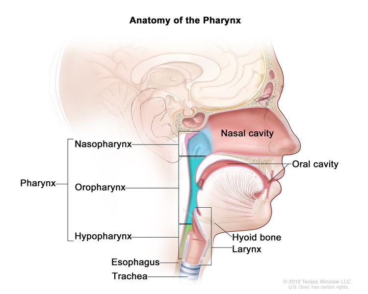
EnlargeAnatomy of the pharynx; drawing shows the nasopharynx, oropharynx, and hypopharynx. Also shown are the nasal cavity, oral cavity, hyoid bone, larynx, esophagus, and trachea.
Figure 1. Anatomy of the pharynx.

Who Is at Risk?

Head and neck squamous cell cancers have common risk factors. People who use tobacco in any of the commonly available forms (cigarettes, cigars, pipes, and smokeless tobacco) or have a high alcohol intake are at elevated risk of oral cavity, oropharyngeal, hypopharyngeal, and laryngeal cancers; they are at particularly high risk if they use both tobacco and alcohol.[2] Individuals who chew betel quid (whether mixed with tobacco or not) are also at high risk of cancer of the oral cavity and oropharynx.[3,4] Individuals who have a personal history of cancer of the head and neck region also are at elevated risk of a future primary cancer of the head and neck.[5] Human papillomavirus type 16 (HPV-16) is a sufficient, but not necessary, cause of oral, tongue, and oropharyngeal cancers.[2,6]

Note: Separate PDQ summaries on Oral Cavity and Nasopharyngeal Cancers Screening and Cigarette Smoking: Health Risks and How to Quit are also available.

Factors With Adequate Evidence of an Increased Risk of Oral Cavity, Oropharyngeal, Hypopharyngeal, and Laryngeal Cancers

Tobacco use

Based on solid evidence from numerous observational studies, tobacco use increases the risk of cancers of the oral cavity, oropharynx, hypopharynx, and larynx.[79]

Magnitude of Effect: Large. Risk for current smokers ranges from fourfold to fivefold for oral cavity, oropharyngeal, and hypopharyngeal cancers to tenfold for laryngeal cancer compared with never-smokers, and is dose related. Most cancers of the oral cavity, oropharynx, hypopharynx, and larynx are attributable to the use of tobacco products.

  • Study Design: Case-control and cohort studies.
  • Internal Validity: Good.
  • Consistency: Good.
  • External Validity: Good.

Alcohol use

Based on solid evidence, alcohol use is a risk factor for the development of head and neck cancers. Its effects are independent of those of tobacco use.[912]

Magnitude of Effect: Lower than the risk associated with tobacco use, but the risk is approximately twofold to sixfold for people who drink two or more alcoholic beverages per day compared with nondrinkers, and is dose related.

  • Study Design: Case-control and cohort studies.
  • Internal Validity: Good.
  • Consistency: Good.
  • External Validity: Good.

Tobacco and alcohol use

The risk of oral cavity, oropharyngeal, hypopharyngeal, and laryngeal cancers is highest in people who consume large amounts of both alcohol and tobacco. When both risk factors are present, the risk of cancer is greater than a simple multiplicative effect of the two individual risks.[1315]

Magnitude of Effect: About two to three times greater for oral cavity, oropharyngeal, hypopharyngeal, and laryngeal cancers than the simple multiplicative effect, with risks for persons who both smoke and drink heavily approximately 5- to 14-fold that of persons who both never smoke and never drink.

  • Study Design: Case-control and cohort studies.
  • Internal Validity: Good.
  • Consistency: Good.
  • External Validity: Good.

Betel-quid chewing

Based on solid evidence, chewing betel quid alone or with added tobacco increases the risk of both oral cavity and oropharyngeal cancers.[3,4] Of the three primary components of betel quid (betel leaf, areca nut, and lime), the areca nut is the only one considered to be carcinogenic when chewed.

Magnitude of Effect: Relative risks for oral cavity cancer are high and typically stronger for betel quid with tobacco than for betel quid alone. Both products appear to confer a statistically significant increase in risk of oropharyngeal cancer.[4]

  • Study Design: Case-control, and cohort studies.
  • Internal Validity: Good.
  • Consistency: Good.
  • External Validity: Good.

Human papillomavirus (HPV) infection

Based on solid evidence, HPV type 16 (HPV-16) infection causes oropharyngeal cancer.[6,16] Other high-risk HPV subtypes, including HPV type 18 (HPV-18), have been found in a small percentage of oropharyngeal cancers.[17,18]

Magnitude of Effect: Large. Oral infection with HPV-16 confers about a 15-fold increase in risk of oropharyngeal cancer relative to individuals without oral HPV-16 infection.

  • Study Design: Case-control and cohort studies.
  • Internal Validity: Good.
  • Consistency: Good.
  • External Validity: Good.

Interventions With Adequate Evidence of a Decreased Risk of Oral Cavity, Oropharyngeal, Hypopharyngeal, and Laryngeal Cancers

Tobacco cessation

Based on solid evidence, cessation of exposure to tobacco (e.g., cigarettes, pipes, cigars, and smokeless tobacco) leads to a decrease in the risk of oral cavity, oropharyngeal, hypopharyngeal, and laryngeal cancers.[19]

Magnitude of Effect: Decreased risk, moderate to large magnitude.

  • Study Design: Case-control and cohort studies.
  • Internal Validity: Good.
  • Consistency: Good.
  • External Validity: Good.

Interventions With Inadequate Evidence of a Reduced Risk of Oral Cavity, Oropharyngeal, Hypopharyngeal, and Laryngeal Cancers

Cessation of alcohol consumption

Based on fair evidence, cessation of alcohol consumption leads to a decrease in oral cavity and laryngeal cancer risk 20 years or more after cessation.[19]

Magnitude of Effect: Decreased risk, small to moderate magnitude.

  • Study Design: Case-control studies.
  • Internal Validity: Fair.
  • Consistency: Fair.
  • External Validity: Fair.

Vaccination against HPV-16 and the other high-risk subtypes

Vaccination against HPV-16 and HPV-18 has been shown to prevent approximately 90% of oral HPV-16/HPV-18 infections within 4 years of vaccination.[20] However, no data are available to assess whether vaccination at any age will lead to reduced risk of oropharyngeal cancer at current typical ages of diagnosis.[21]

  • Study Design: No studies available.
  • Internal Validity: Not applicable (N/A).
  • Consistency: N/A.
  • External Validity: N/A.
References
  1. IARC Working Group on the Evaluation of Carcinogenic Risks to Humans: Personal habits and indoor combustions. Volume 100 E. A review of human carcinogens. IARC Monogr Eval Carcinog Risks Hum 100 (Pt E): 1-538, 2012. [PUBMED Abstract]
  2. Castellsagué X, Alemany L, Quer M, et al.: HPV Involvement in Head and Neck Cancers: Comprehensive Assessment of Biomarkers in 3680 Patients. J Natl Cancer Inst 108 (6): djv403, 2016. [PUBMED Abstract]
  3. Song H, Wan Y, Xu YY: Betel quid chewing without tobacco: a meta-analysis of carcinogenic and precarcinogenic effects. Asia Pac J Public Health 27 (2): NP47-57, 2015. [PUBMED Abstract]
  4. Guha N, Warnakulasuriya S, Vlaanderen J, et al.: Betel quid chewing and the risk of oral and oropharyngeal cancers: a meta-analysis with implications for cancer control. Int J Cancer 135 (6): 1433-43, 2014. [PUBMED Abstract]
  5. Atienza JA, Dasanu CA: Incidence of second primary malignancies in patients with treated head and neck cancer: a comprehensive review of literature. Curr Med Res Opin 28 (12): 1899-909, 2012. [PUBMED Abstract]
  6. Kreimer AR, Johansson M, Waterboer T, et al.: Evaluation of human papillomavirus antibodies and risk of subsequent head and neck cancer. J Clin Oncol 31 (21): 2708-15, 2013. [PUBMED Abstract]
  7. U.S. Department of Health and Human Services: The Health Consequences of Smoking—50 Years of Progress: A Report of the Surgeon General. U.S. Department of Health and Human Services, CDC, National Center for Chronic Disease Prevention and Health Promotion, Office on Smoking and Health, 2014. Also available online. Last accessed December 30, 2024.
  8. Vineis P, Alavanja M, Buffler P, et al.: Tobacco and cancer: recent epidemiological evidence. J Natl Cancer Inst 96 (2): 99-106, 2004. [PUBMED Abstract]
  9. Hashibe M, Brennan P, Benhamou S, et al.: Alcohol drinking in never users of tobacco, cigarette smoking in never drinkers, and the risk of head and neck cancer: pooled analysis in the International Head and Neck Cancer Epidemiology Consortium. J Natl Cancer Inst 99 (10): 777-89, 2007. [PUBMED Abstract]
  10. IARC Working Group on the Evaluation of Carcinogenic Risks to Humans: Smokeless tobacco and some tobacco-specific N-nitrosamines. IARC Monogr Eval Carcinog Risks Hum 89: 1-592, 2007. [PUBMED Abstract]
  11. Purdue MP, Hashibe M, Berthiller J, et al.: Type of alcoholic beverage and risk of head and neck cancer–a pooled analysis within the INHANCE Consortium. Am J Epidemiol 169 (2): 132-42, 2009. [PUBMED Abstract]
  12. Islami F, Tramacere I, Rota M, et al.: Alcohol drinking and laryngeal cancer: overall and dose-risk relation–a systematic review and meta-analysis. Oral Oncol 46 (11): 802-10, 2010. [PUBMED Abstract]
  13. Hashibe M, Brennan P, Chuang SC, et al.: Interaction between tobacco and alcohol use and the risk of head and neck cancer: pooled analysis in the International Head and Neck Cancer Epidemiology Consortium. Cancer Epidemiol Biomarkers Prev 18 (2): 541-50, 2009. [PUBMED Abstract]
  14. Lubin JH, Purdue M, Kelsey K, et al.: Total exposure and exposure rate effects for alcohol and smoking and risk of head and neck cancer: a pooled analysis of case-control studies. Am J Epidemiol 170 (8): 937-47, 2009. [PUBMED Abstract]
  15. Mello FW, Melo G, Pasetto JJ, et al.: The synergistic effect of tobacco and alcohol consumption on oral squamous cell carcinoma: a systematic review and meta-analysis. Clin Oral Investig 23 (7): 2849-2859, 2019. [PUBMED Abstract]
  16. Hobbs CG, Sterne JA, Bailey M, et al.: Human papillomavirus and head and neck cancer: a systematic review and meta-analysis. Clin Otolaryngol 31 (4): 259-66, 2006. [PUBMED Abstract]
  17. D’Souza G, Kreimer AR, Viscidi R, et al.: Case-control study of human papillomavirus and oropharyngeal cancer. N Engl J Med 356 (19): 1944-56, 2007. [PUBMED Abstract]
  18. Steinau M, Saraiya M, Goodman MT, et al.: Human papillomavirus prevalence in oropharyngeal cancer before vaccine introduction, United States. Emerg Infect Dis 20 (5): 822-8, 2014. [PUBMED Abstract]
  19. Marron M, Boffetta P, Zhang ZF, et al.: Cessation of alcohol drinking, tobacco smoking and the reversal of head and neck cancer risk. Int J Epidemiol 39 (1): 182-96, 2010. [PUBMED Abstract]
  20. Herrero R, Quint W, Hildesheim A, et al.: Reduced prevalence of oral human papillomavirus (HPV) 4 years after bivalent HPV vaccination in a randomized clinical trial in Costa Rica. PLoS One 8 (7): e68329, 2013. [PUBMED Abstract]
  21. Chaturvedi AK, Graubard BI, Broutian T, et al.: Effect of Prophylactic Human Papillomavirus (HPV) Vaccination on Oral HPV Infections Among Young Adults in the United States. J Clin Oncol 36 (3): 262-267, 2018. [PUBMED Abstract]

Incidence and Mortality

Oral cavity and oropharynx cancers

From 2017 to 2021, the estimated age-adjusted incidence rate for cancers of the oral cavity and oropharynx in the United States was 11.5 cases per 100,000 persons per year.[1] Cancers of the tongue, oropharynx, and tonsil comprise the majority of cases (30% tongue and 24% oropharynx and tonsil). The estimated age-adjusted mortality rate for cancers of the oral cavity and oropharynx from 2018 to 2022 was 2.6 per 100,000 persons per year. U.S. incidence rates are about 1.7 times higher in men than in women. However, mortality rates are about 1.9 times higher in men than in women.[1] It is estimated that there will be 59,660 new cases of oral cavity and oropharynx cancer diagnosed in the United States in 2025 and 12,770 deaths due to these diseases.[2] Rates of oral cavity cancer vary greatly across the world, primarily because of differences in alcohol use, tobacco use, and betel-quid chewing and the products chewed.[3]

Although localized cancers of the oral cavity and oropharynx have an excellent anticipated 5-year survival rate of about 87.5%, only 26.4% of these cancer cases are diagnosed at this stage.[1] The overall 5-year relative survival rate for patients with cancers of the oral cavity and oropharynx combined is only 69%.[2] However, the 5-year relative survival rate is much lower in Black patients (57%) than in White patients (71%).[2] Additionally, the 5-year survival rate varies greatly by cancer site, with 52.1% survival among patients with cancer on the floor of the mouth and 73.8% survival among patients with cancer in the oropharynx and tonsil.[4]

Hypopharyngeal cancer

Hypopharyngeal cancers are rare, with approximately 2,500 new cases diagnosed in the United States each year and an annual incidence of 0.6 cases per 100,000 persons.[5] The 5-year survival rate for hypopharyngeal cancer is 35%.[5] New cases have been falling an average of 2% per year over the last 20 years.[5] This has been attributed to a reduction in cigarette smoking.

Laryngeal cancer

Laryngeal cancers are less common, with an annual incidence of 2.6 cases per 100,000 persons.[6] It is estimated that 13,020 new cases of laryngeal cancer will be diagnosed in the United States in 2025, and an estimated 3,910 individuals will die of this disease.[2] The 5-year relative survival rate for laryngeal cancer is 62%.[2] Age-adjusted death rates have been falling on average 1.6% each year from 2013 to 2022.[6] This has been attributed to a reduction in cigarette smoking.

References
  1. National Cancer Institute: SEER Cancer Stat Facts: Oral Cavity and Pharynx Cancer. National Cancer Institute. Available online. Last accessed December 30, 2024.
  2. American Cancer Society: Cancer Facts and Figures 2025. American Cancer Society, 2025. Available online. Last accessed January 16, 2025.
  3. Shield KD, Ferlay J, Jemal A, et al.: The global incidence of lip, oral cavity, and pharyngeal cancers by subsite in 2012. CA Cancer J Clin 67 (1): 51-64, 2017. [PUBMED Abstract]
  4. Surveillance Research Program, National Cancer Institute: SEER*Explorer: An interactive website for SEER cancer statistics. Bethesda, MD: National Cancer Institute. Available online. Last accessed December 30, 2024.
  5. Kuo P, Chen MM, Decker RH, et al.: Hypopharyngeal cancer incidence, treatment, and survival: temporal trends in the United States. Laryngoscope 124 (9): 2064-9, 2014. [PUBMED Abstract]
  6. National Cancer Institute: SEER Cancer Stat Facts: Laryngeal Cancer. National Cancer Institute. Available online. Last accessed April 14, 2025.

Factors With Adequate Evidence of an Increased Risk of Oral Cavity, Oropharyngeal, Hypopharyngeal, and Laryngeal Cancers

Tobacco Use

Tobacco use is implicated in most cases of oral cavity, oropharyngeal, hypopharyngeal, and laryngeal cancers.[1] All forms of tobacco use (cigarettes, pipes, cigars, snuff, chewing tobacco, gutka [betel quid with tobacco added], and other smoked and smokeless products) increase the risk of these cancers.[1,2] Epidemiological studies consistently demonstrate that cigarette smokers have a higher incidence of mortality from head and neck squamous cell cancers compared with lifetime nonsmokers, and there is general consensus that the relationship is causal. Risk for current smokers ranges from fourfold to fivefold for oral cavity, oropharyngeal, and hypopharyngeal cancers to tenfold for laryngeal cancer when compared with never-smokers.[3] However, other epidemiological studies have observed smaller and larger increases in risk, with some variation by anatomic location. Betel-quid chewing is prevalent in many countries in south and southeast Asia, including China and India, and is an important risk factor for both oral cavity and oropharyngeal cancers.[4]

Alcohol Use

Alcohol use is a major independent risk factor for the development of head and neck squamous cell cancer.[5] Most epidemiological studies demonstrate an increase in risk with increasing drinks per day, with a more than twofold to sixfold increase in risk for individuals who consume two or more drinks a day relative to nonconsumers.[6] Associations are observed in studies that adjust for confounding by smoking, as well as in studies of nonsmokers.[7] There is a suggestion that consumption of beer and hard liquor confers a greater risk than does wine consumption.[6]

Tobacco and Alcohol Use

Head and neck squamous cell cancer risk is highest in people who consume large amounts of both alcohol and tobacco.[810] When both risk factors are present, the risk of oral cavity and oropharyngeal cancer is typically about two to three times greater than a simple multiplicative or additive effect.[11] In a study that pooled data from 17 case-control studies, individuals who consumed more than a pack of cigarettes and three or more alcoholic drinks per day had a 15-fold increased risk of oral cavity cancer, 14-fold increased risk of oropharyngeal cancer, and 36-fold increased risk of laryngeal cancer relative to individuals who neither smoked nor drank.[8,11]

Betel-Quid Chewing

Betel quid is composed of betel leaf, areca nut, and lime; gutka is betel quid with added tobacco. Both betel-quid and gutka chewing increase the risk of cancer of the oral cavity and oropharynx.[4,12] The carcinogenic component of chewed betel quid arises from the areca nut.[4]

Relative risks are typically stronger for betel quid with tobacco than for betel quid alone.[12] A meta-analysis of oral cavity cancer studies conducted on the Indian subcontinent calculated a statistically significant eightfold increase in risk for betel quid with tobacco and a statistically significant twofold increase in risk for betel quid alone.[12] A statistically significant tenfold increase in oral cavity cancer risk for betel-quid chewing was demonstrated by studies conducted in China or Taiwan. A meta-analysis of oropharyngeal cancer studies conducted on the Indian subcontinent calculated a statistically significant fourfold increase in risk for betel quid with tobacco and a statistically significant twofold increase in risk for betel quid alone.[12] Studies of head and neck cancer (without specification of subsite) suggest that increases in risk are positively correlated with chewing frequency and duration.[4]

Human Papillomavirus (HPV) Infection

HPV type 16 (HPV-16) infection is a sufficient, but not necessary, cause of head and neck cancers, with a greater causal relationship with oropharyngeal cancer.[13,14] A meta-analysis of five case-control studies of HPV-16 positivity in either serum or tissue calculated an odds ratio of 15.1 (95% confidence interval [CI], 6.8–33.7) for cancer of the tonsils, 4.3 (95% CI, 2.1–8.9) for other oropharyngeal sites, and 2.0 for both oral cavity (95% CI, 1.2–3.4) and larynx (95% CI, 1.0–4.2).[14] In a case-control study, the observed strong association of HPV-16 serologic status and oropharyngeal cancer did not vary at different levels of tobacco or alcohol use.[15] HPV-16 E6 seroconversion was shown to occur over a range of 6 to 28 years before oropharyngeal cancer diagnosis, at a median age of 52 years.[16] Thus, the HPV-16 infection that increases the risk of oropharyngeal cancer may occur in individuals aged 20 to 40 years. A recent national survey observed that men have a higher prevalence of oral HPV than women (11.5% vs. 3.2%), including high-risk HPV subtypes (7.3% of men; 1.4% of women).[17]

Other high-risk HPV subtypes, including HPV type 18, have been found in a smaller proportion of oropharyngeal cancers.[15,18]

References
  1. IARC Working Group on the Evaluation of Carcinogenic Risks to Humans: Personal habits and indoor combustions. Volume 100 E. A review of human carcinogens. IARC Monogr Eval Carcinog Risks Hum 100 (Pt E): 1-538, 2012. [PUBMED Abstract]
  2. U.S. Department of Health and Human Services: The Health Consequences of Smoking—50 Years of Progress: A Report of the Surgeon General. U.S. Department of Health and Human Services, CDC, National Center for Chronic Disease Prevention and Health Promotion, Office on Smoking and Health, 2014. Also available online. Last accessed December 30, 2024.
  3. Vineis P, Alavanja M, Buffler P, et al.: Tobacco and cancer: recent epidemiological evidence. J Natl Cancer Inst 96 (2): 99-106, 2004. [PUBMED Abstract]
  4. IARC Working Group on the Evaluation of Carcinogenic Risks to Humans: Betel-quid and areca-nut chewing and some areca-nut derived nitrosamines. IARC Monogr Eval Carcinog Risks Hum 85: 1-334, 2004. [PUBMED Abstract]
  5. Hashibe M, Brennan P, Benhamou S, et al.: Alcohol drinking in never users of tobacco, cigarette smoking in never drinkers, and the risk of head and neck cancer: pooled analysis in the International Head and Neck Cancer Epidemiology Consortium. J Natl Cancer Inst 99 (10): 777-89, 2007. [PUBMED Abstract]
  6. Purdue MP, Hashibe M, Berthiller J, et al.: Type of alcoholic beverage and risk of head and neck cancer–a pooled analysis within the INHANCE Consortium. Am J Epidemiol 169 (2): 132-42, 2009. [PUBMED Abstract]
  7. Goldstein BY, Chang SC, Hashibe M, et al.: Alcohol consumption and cancers of the oral cavity and pharynx from 1988 to 2009: an update. Eur J Cancer Prev 19 (6): 431-65, 2010. [PUBMED Abstract]
  8. Hashibe M, Brennan P, Chuang SC, et al.: Interaction between tobacco and alcohol use and the risk of head and neck cancer: pooled analysis in the International Head and Neck Cancer Epidemiology Consortium. Cancer Epidemiol Biomarkers Prev 18 (2): 541-50, 2009. [PUBMED Abstract]
  9. Lubin JH, Purdue M, Kelsey K, et al.: Total exposure and exposure rate effects for alcohol and smoking and risk of head and neck cancer: a pooled analysis of case-control studies. Am J Epidemiol 170 (8): 937-47, 2009. [PUBMED Abstract]
  10. Mello FW, Melo G, Pasetto JJ, et al.: The synergistic effect of tobacco and alcohol consumption on oral squamous cell carcinoma: a systematic review and meta-analysis. Clin Oral Investig 23 (7): 2849-2859, 2019. [PUBMED Abstract]
  11. Blot WJ, McLaughlin JK, Winn DM, et al.: Smoking and drinking in relation to oral and pharyngeal cancer. Cancer Res 48 (11): 3282-7, 1988. [PUBMED Abstract]
  12. Guha N, Warnakulasuriya S, Vlaanderen J, et al.: Betel quid chewing and the risk of oral and oropharyngeal cancers: a meta-analysis with implications for cancer control. Int J Cancer 135 (6): 1433-43, 2014. [PUBMED Abstract]
  13. Kreimer AR, Johansson M, Waterboer T, et al.: Evaluation of human papillomavirus antibodies and risk of subsequent head and neck cancer. J Clin Oncol 31 (21): 2708-15, 2013. [PUBMED Abstract]
  14. Hobbs CG, Sterne JA, Bailey M, et al.: Human papillomavirus and head and neck cancer: a systematic review and meta-analysis. Clin Otolaryngol 31 (4): 259-66, 2006. [PUBMED Abstract]
  15. D’Souza G, Kreimer AR, Viscidi R, et al.: Case-control study of human papillomavirus and oropharyngeal cancer. N Engl J Med 356 (19): 1944-56, 2007. [PUBMED Abstract]
  16. Kreimer AR, Ferreiro-Iglesias A, Nygard M, et al.: Timing of HPV16-E6 antibody seroconversion before OPSCC: findings from the HPVC3 consortium. Ann Oncol 30 (8): 1335-1343, 2019. [PUBMED Abstract]
  17. Sonawane K, Suk R, Chiao EY, et al.: Oral Human Papillomavirus Infection: Differences in Prevalence Between Sexes and Concordance With Genital Human Papillomavirus Infection, NHANES 2011 to 2014. Ann Intern Med 167 (10): 714-724, 2017. [PUBMED Abstract]
  18. Steinau M, Saraiya M, Goodman MT, et al.: Human papillomavirus prevalence in oropharyngeal cancer before vaccine introduction, United States. Emerg Infect Dis 20 (5): 822-8, 2014. [PUBMED Abstract]

Interventions With Adequate Evidence of a Decreased Risk of Oral Cavity, Oropharyngeal, Hypopharyngeal, and Laryngeal Cancers

Tobacco Cessation

The cessation of cigarette smoking is associated with an approximately 50% reduction in risk of developing oral cavity, oropharyngeal, and hypopharyngeal cancers within 5 to 9 years,[1] and a return to a cancer risk comparable to that of never-smokers within 20 years.[1] For laryngeal cancer, the risk reduction is approximately 40% within 5 to 9 years, and a similar return to cancer risk comparable to that of never-smokers within 20 years.[1]

References
  1. Marron M, Boffetta P, Zhang ZF, et al.: Cessation of alcohol drinking, tobacco smoking and the reversal of head and neck cancer risk. Int J Epidemiol 39 (1): 182-96, 2010. [PUBMED Abstract]

Interventions With Inadequate Evidence of a Reduced Risk of Oral Cavity, Oropharyngeal, Hypopharyngeal, and Laryngeal Cancers

Cessation of Alcohol Consumption

Because alcohol is associated with oral cavity, oropharyngeal, hypopharyngeal, and laryngeal cancers in a dose-dependent fashion, it is believed that cessation or avoidance of alcohol use would result in reduced incidence. Most studies suggest that the risk of oral cavity and laryngeal cancer decreases as time from cessation increases; one pooled analysis of 13 studies observed a statistically significant reduction (odds ratio [OR], 0.45; 95% confidence interval [CI], 0.26–0.78) for oral cavity and laryngeal cancers (OR, 0.69; 95% CI, 0.52–0.91), relative to current drinkers, for those who ceased consumption 20 years or more ago. Among never-smokers, cessation of alcohol consumption leads to a decrease in oral cavity and laryngeal cancer risk more than 1 to 4 years after cessation. Data for oropharyngeal and hypopharyngeal cancers did not support risk reduction with alcohol cessation.[1]

Vaccination Against Human Papillomavirus (HPV) Type 16 (HPV-16) and Other High-Risk Subtypes

Vaccination against HPV-16 and type 18 (HPV-18) has been shown to prevent approximately 90% of oral HPV-16/HPV-18 infections within 4 years of vaccination.[2,3] Given the relatively recent adoption of vaccination and the age at which individuals are vaccinated, there is not yet evidence that vaccination at a young age will lead to a substantially reduced risk of HPV-associated oropharyngeal cancer later in life. In addition, no data are available to examine whether incidence or mortality would be reduced if vaccination occurred at an age closer to that at which oropharyngeal cancers tend to present.

References
  1. Marron M, Boffetta P, Zhang ZF, et al.: Cessation of alcohol drinking, tobacco smoking and the reversal of head and neck cancer risk. Int J Epidemiol 39 (1): 182-96, 2010. [PUBMED Abstract]
  2. Herrero R, Quint W, Hildesheim A, et al.: Reduced prevalence of oral human papillomavirus (HPV) 4 years after bivalent HPV vaccination in a randomized clinical trial in Costa Rica. PLoS One 8 (7): e68329, 2013. [PUBMED Abstract]
  3. Chaturvedi AK, Graubard BI, Broutian T, et al.: Effect of Prophylactic Human Papillomavirus (HPV) Vaccination on Oral HPV Infections Among Young Adults in the United States. J Clin Oncol 36 (3): 262-267, 2018. [PUBMED Abstract]

Latest Updates to This Summary (04/14/2025)

The PDQ cancer information summaries are reviewed regularly and updated as new information becomes available. This section describes the latest changes made to this summary as of the date above.

Incidence and Mortality

Updated statistics with estimated new cases and deaths for oral cavity and pharynx cancer for 2025 (cited American Cancer Society as reference 2).

Updated statistics with estimated new cases and deaths for laryngeal cancer for 2025.

This summary is written and maintained by the PDQ Screening and Prevention Editorial Board, which is editorially independent of NCI. The summary reflects an independent review of the literature and does not represent a policy statement of NCI or NIH. More information about summary policies and the role of the PDQ Editorial Boards in maintaining the PDQ summaries can be found on the About This PDQ Summary and PDQ® Cancer Information for Health Professionals pages.

About This PDQ Summary

Purpose of This Summary

This PDQ cancer information summary for health professionals provides comprehensive, peer-reviewed, evidence-based information about oral cavity, oropharyngeal, hypopharyngeal, and laryngeal cancers prevention. It is intended as a resource to inform and assist clinicians in the care of their patients. It does not provide formal guidelines or recommendations for making health care decisions.

Reviewers and Updates

This summary is reviewed regularly and updated as necessary by the PDQ Screening and Prevention Editorial Board, which is editorially independent of the National Cancer Institute (NCI). The summary reflects an independent review of the literature and does not represent a policy statement of NCI or the National Institutes of Health (NIH).

Board members review recently published articles each month to determine whether an article should:

  • be discussed at a meeting,
  • be cited with text, or
  • replace or update an existing article that is already cited.

Changes to the summaries are made through a consensus process in which Board members evaluate the strength of the evidence in the published articles and determine how the article should be included in the summary.

Any comments or questions about the summary content should be submitted to Cancer.gov through the NCI website’s Email Us. Do not contact the individual Board Members with questions or comments about the summaries. Board members will not respond to individual inquiries.

Levels of Evidence

Some of the reference citations in this summary are accompanied by a level-of-evidence designation. These designations are intended to help readers assess the strength of the evidence supporting the use of specific interventions or approaches. The PDQ Screening and Prevention Editorial Board uses a formal evidence ranking system in developing its level-of-evidence designations.

Permission to Use This Summary

PDQ is a registered trademark. Although the content of PDQ documents can be used freely as text, it cannot be identified as an NCI PDQ cancer information summary unless it is presented in its entirety and is regularly updated. However, an author would be permitted to write a sentence such as “NCI’s PDQ cancer information summary about breast cancer prevention states the risks succinctly: [include excerpt from the summary].”

The preferred citation for this PDQ summary is:

PDQ® Screening and Prevention Editorial Board. PDQ Oral Cavity, Oropharyngeal, Hypopharyngeal, and Laryngeal Cancers Prevention. Bethesda, MD: National Cancer Institute. Updated <MM/DD/YYYY>. Available at: /types/head-and-neck/hp/oral-prevention-pdq. Accessed <MM/DD/YYYY>. [PMID: 26389416]

Images in this summary are used with permission of the author(s), artist, and/or publisher for use within the PDQ summaries only. Permission to use images outside the context of PDQ information must be obtained from the owner(s) and cannot be granted by the National Cancer Institute. Information about using the illustrations in this summary, along with many other cancer-related images, is available in Visuals Online, a collection of over 2,000 scientific images.

Disclaimer

The information in these summaries should not be used as a basis for insurance reimbursement determinations. More information on insurance coverage is available on Cancer.gov on the Managing Cancer Care page.

Contact Us

More information about contacting us or receiving help with the Cancer.gov website can be found on our Contact Us for Help page. Questions can also be submitted to Cancer.gov through the website’s Email Us.
Oral Cavity and Nasopharyngeal Cancers Screening (PDQ®)–Health Professional Version

Oral Cavity and Nasopharyngeal Cancers Screening (PDQ®)–Health Professional Version

Overview

Oral cavity, oropharyngeal, hypopharyngeal, and laryngeal cancers may be referred to as head and neck squamous cell cancers. Oral cavity squamous cell cancers most commonly arise from the mucosal surfaces lining the oral cavity. Pharyngeal squamous cell cancers can be categorized into nasopharyngeal, oropharyngeal, and hypopharyngeal cancers on the basis of anatomical landmarks. Figure 1 shows the anatomy of the pharynx.

EnlargeAnatomy of the pharynx; drawing shows the nasopharynx, oropharynx, and hypopharynx. Also shown are the nasal cavity, oral cavity, hyoid bone, larynx, esophagus, and trachea.
Figure 1. Anatomy of the pharynx.

Note: The Overview section summarizes the published evidence on this topic. The rest of the summary describes the evidence in more detail.

Other PDQ summaries on Oral Cavity, Oropharyngeal, Hypopharyngeal, and Laryngeal Cancers Prevention and Lip and Oral Cavity Cancer Treatment are also available.

Benefits

There is inadequate evidence to establish whether screening would result in a decrease in mortality from oral cavity and nasopharyngeal cancers.

Magnitude of Effect: No evidence of benefit, and harms have not been quantified.

  • Study Design: Evidence obtained from one randomized controlled trial and observational studies.
  • Internal Validity: Poor.
  • Consistency: Not applicable (N/A).
  • External Validity: Poor.

Harms

Harms, although unavoidable, have not been quantified on the basis of the literature. However, there are some unavoidable harms that would be associated with routine screening, including:

  • Unnecessary treatment associated with overdiagnosis.
  • Psychological consequences of false-positive tests.
  • Misdiagnosis because of variability in assessment of biopsies.

Magnitude of Effect: Unknown.

  • Study Design: Observational studies.
  • Internal Validity: Poor.
  • Consistency: N/A.
  • External Validity: Poor.

Incidence and Mortality

An estimated 59,660 new cases of oral cavity and oropharynx cancers will be diagnosed in the United States in 2025, and an estimated 12,770 people will die of these diseases.[1] The overall annual incidence in the United States is about 11 cases per 100,000 men and women; the incidence rate is highest in individuals aged 75 to 84 years.[2]

Between 2012 and 2021, incidence rates increased by 0.7% per year.[1] The incidence has been increasing for oral cavity and oropharyngeal cancers related to human papillomavirus (HPV) infection. About 60% of oral/pharyngeal cancers are moderately advanced (regional stage) or metastatic at the time of diagnosis.[2] The 5-year relative survival rate is 69%.[1]

The estimated annual worldwide number of cases of oral cavity and oropharyngeal cancers is about 275,000, with an approximate 20-fold variation geographically.[3] South and Southeast Asia (India, Sri Lanka, Pakistan, and Bangladesh), France, and Brazil have particularly high rates. In most countries, men have higher rates of oral cavity cancer than women (caused by tobacco use) and higher rates of lip cancer (caused by sunlight exposure from outdoor occupations).[3]

Nasopharyngeal cancers are rare in the United States, with an annual incidence rate of 0.7 cases per 100,000 persons.[4] However, there are marked geographic differences, with an overall incidence in China that is 40- to 380-fold higher than that in the United States.[4] There are elevated rates of nasopharyngeal cancers in the Cantonese population of southern China (including Hong Kong), and intermediate rates are observed in several indigenous populations in Southeast Asia and in natives of the Arctic region, North Africa, and the Middle East. First-generation Chinese immigrants to the United States maintain a high incidence rate, while their descendants born in the United States show a decreased incidence. The 5-year survival rate for keratinizing squamous cell carcinoma, the most common subtype of nasopharyngeal cancer in the United States, is 46%.[5]

References
  1. American Cancer Society: Cancer Facts and Figures 2025. American Cancer Society, 2025. Available online. Last accessed January 16, 2025.
  2. Surveillance Research Program, National Cancer Institute: SEER*Explorer: An interactive website for SEER cancer statistics. Bethesda, MD: National Cancer Institute. Available online. Last accessed December 30, 2024.
  3. Warnakulasuriya S: Global epidemiology of oral and oropharyngeal cancer. Oral Oncol 45 (4-5): 309-16, 2009 Apr-May. [PUBMED Abstract]
  4. Richey LM, Olshan AF, George J, et al.: Incidence and survival rates for young blacks with nasopharyngeal carcinoma in the United States. Arch Otolaryngol Head Neck Surg 132 (10): 1035-40, 2006. [PUBMED Abstract]
  5. Ou SH, Zell JA, Ziogas A, et al.: Epidemiology of nasopharyngeal carcinoma in the United States: improved survival of Chinese patients within the keratinizing squamous cell carcinoma histology. Ann Oncol 18 (1): 29-35, 2007. [PUBMED Abstract]

Risk Factors

The primary risk factors for oral cavity cancers in American men and women are tobacco (including smokeless tobacco) use, alcohol use, betel-quid chewing, and human papillomavirus infection (HPV).

Risk factors for nasopharyngeal cancer include Epstein-Barr virus (EBV) persistent infection.[1]

For more information about factors associated with an increased or decreased risk of oral cavity squamous cell cancers, see Oral Cavity, Oropharyngeal, Hypopharyngeal, and Laryngeal Cancers Prevention and Lip and Oral Cavity Cancer Treatment.

Epstein-Barr Virus (EBV) Infection

Based on solid evidence, EBV infection causes nasopharyngeal cancer in high-incidence areas.[1] Collective evidence includes numerous case-control studies and cohort studies that show a higher proportion of patients with nasopharyngeal cancer who have anti-EBV antibodies than controls and that seropositive status precedes tumor diagnosis.[2,3] Recent studies have also found circulating cell-free EBV DNA in patients with nasopharyngeal cancer but not in controls.[4] EBV alone is not a sufficient cause because 90% of adults worldwide are infected with the virus, but only a small proportion develop nasopharyngeal cancer.[5] EBV infection is subclinical and occurs early in childhood. The pathogenesis is thought to involve the virus establishing latency in epithelial cells that have already undergone premalignant genetic changes.

One of the first studies to show an association was a cohort study that found higher anti-EBV titers in 84% of the 235 East African and Chinese patients with nasopharyngeal carcinoma.[2,5] The same study found higher anti-EBV titers with higher-stage tumor, and a case-control component of the study revealed that high anti-EBV titers were six times more likely in patients with nasopharyngeal carcinoma than in patients with head and neck cancers at other sites.

Other studies show elevation in both IgG and IgA antibody titers to EBV viral capsid antigen and other latent viral antigens, which precede tumor development by several years and are correlated with tumor burden, remission, and recurrence.[2,3] A large cohort study with 9,699 men measured both IgA antibodies against EBV capsid antigen and neutralizing antibodies against EBV-specific DNase and followed them for a later diagnosis of nasopharyngeal cancer.[3] The relative risk of nasopharyngeal carcinoma was 32.8 for individuals with both antibody markers (95% confidence interval [CI], 7.3–147.2; P < .001), and 4.0 for individuals with one marker (95% CI, 1.6–10.2; P = .003), compared with individuals with neither marker. There was a temporal relationship in that the difference in cumulative incidence between seropositive and seronegative patients increased with a longer duration of follow-up. Another study found circulating cell-free EBV DNA in 95% of patients with advanced nasopharyngeal cancer but not in controls or cured patients.[4]

References
  1. IARC Working Group on the Evaluation of Carcinogenic Risks to Humans: Biological agents. Volume 100 B. A review of human carcinogens. IARC Monogr Eval Carcinog Risks Hum 100 (Pt B): 1-441, 2012. [PUBMED Abstract]
  2. Henle W, Henle G, Ho HC, et al.: Antibodies to Epstein-Barr virus in nasopharyngeal carcinoma, other head and neck neoplasms, and control groups. J Natl Cancer Inst 44 (1): 225-31, 1970. [PUBMED Abstract]
  3. Chien YC, Chen JY, Liu MY, et al.: Serologic markers of Epstein-Barr virus infection and nasopharyngeal carcinoma in Taiwanese men. N Engl J Med 345 (26): 1877-82, 2001. [PUBMED Abstract]
  4. Lin JC, Wang WY, Chen KY, et al.: Quantification of plasma Epstein-Barr virus DNA in patients with advanced nasopharyngeal carcinoma. N Engl J Med 350 (24): 2461-70, 2004. [PUBMED Abstract]
  5. Chang ET, Adami HO: The enigmatic epidemiology of nasopharyngeal carcinoma. Cancer Epidemiol Biomarkers Prev 15 (10): 1765-77, 2006. [PUBMED Abstract]

Evidence of Benefit Associated With Screening

No population-based screening programs for oral cavity squamous cell cancers have been implemented in developed countries, although opportunistic screening or screening as part of a periodic health examination has been advocated for the oral cavity, which is the only site accessible without endoscopy.[1,2]

Screening for Oral Cavity Cancers

There are different methods of screening for oral cavity cancers. Oral cavity cancers occur in a region of the body that is generally accessible to physical examination by the patient, the dentist, and the physician. Visual examination is the most common method used to detect visible lesions. Other methods have been used to augment clinical detection of oral lesions and include toluidine blue, brush biopsy, and fluorescence staining.

An inspection of the oral cavity is often part of a physical examination in a dentist’s or physician’s office. Of note, high-risk individuals visit their medical doctors more frequently than they visit their dentists. Although physicians are more likely to provide risk-factor counseling (such as tobacco cessation), they are less likely than dentists to perform an oral cancer examination.[3] Overall, only a fraction (~20%) of Americans receive an oral cancer examination. Black patients, Hispanic patients, and those who have a lower level of education are less likely to have such an examination, perhaps because they lack access to medical care.[3] An oral examination often includes looking for leukoplakia and erythroplakia lesions, which can progress to cancer.[4,5] One study has shown that direct fluorescence visualization (using a simple hand-held device in the operating room) could identify subclinical high-risk fields with cancerous or precancerous changes extending up to 25 mm beyond the primary tumor in 19 of 20 patients undergoing oral surgery for invasive or in situ squamous cell tumors.[6] However, this finding has not yet been tested in a screening setting. Data suggest that molecular markers may be useful in the prognosis of these premalignant oral lesions.[7]

The routine examination of asymptomatic and symptomatic patients can lead to detection of earlier-stage cancers and premalignant lesions. There is no definitive evidence, however, to show that this screening can reduce oral cancer mortality, and there are no randomized controlled trials (RCTs) in any Western or other low-risk populations.[5,811]

In a single RCT of screening versus usual care, 13 geographic clusters in the Trivandrum district of Kerala, India, were randomly assigned to receive systematic oral visual screening by trained health workers (seven screened clusters, six control clusters) every 3 years for four screening rounds between 1996 and 2008. During a 15-year follow-up period, there were 138 deaths from oral cancer in the screening group, with a cause-specific mortality rate of 15.4 per 100,000 person-years, and 154 deaths in the control group, with a mortality rate of 17.1 per 100,000 person-years (relative risk [RR], 0.88; 95% confidence interval [CI], 0.69–1.12). In a subset analysis restricted to tobacco or alcohol users, the mortality rates were 30 and 39 per 100,000 person-years, respectively (RR, 0.76; 95% CI, 0.60–0.97). There was no apparent adjustment of the CIs for the cluster design. In another subgroup analysis, mortality hazard ratios were calculated for groups defined by number of times screened, but the inappropriate comparison in each case was to the control group of the whole study. No data on treatment of oral cancers were presented.[1215]

Aside from the issues of generalizability to other populations and lack of an overall statistically significant result in cause-specific mortality, interpretation of the results is made difficult by serious lacks in methodological detail about the randomization process, allocation concealment, adjustment for clustering effect, and information about treatment. The total number of clusters randomized was small, and there were different distributions of income and household possessions between the two study arms. Withdrawals and dropouts were not clearly described. In summary, the sole randomized trial does not provide solid evidence of a cause-specific mortality benefit associated with systematic oral cavity visual examination.

Techniques such as toluidine blue staining, brush biopsy/cytology, or fluorescence imaging as the primary screening tool or as an adjunct for screening have not been shown to have superior sensitivity and specificity for visual examination alone or to yield better health outcomes.[5,16] In an RCT conducted in Keelung County, Taiwan, 7,975 individuals at high risk of oral cancer due to cigarette smoking or betel-quid chewing were randomly assigned to receive a one-time oral cancer examination after gargling with toluidine blue or a blue placebo dye.[17] The positive test rates were 9.5% versus 8.3%, respectively (P = .047). The detection of premalignant lesions was not statistically different (rate ratio, 1.05; 95% CI, 0.74–1.41). The number of overall oral cancers diagnosed within the short follow-up period of 5 years was too small for valid comparison (six in each group).

The operating characteristics of the various techniques used as an adjunct to oral visual examination are not well established. A systematic literature review of toluidine blue, a variety of other visualization adjuncts, and cytopathology in the screening setting revealed a very broad range of reported sensitivities, specificities, and positive predictive values when biopsy confirmation was used as the gold standard outcome.[18] In part, this range of findings can be attributed to varying study populations, sample size and settings, and criteria for positive-clinical examinations and for scoring a biopsy as positive.

Screening for Nasopharyngeal Cancer

Serum Epstein-Barr virus (EBV)–associated antibodies and circulating cell-free EBV DNA testing have been used for nasopharyngeal cancer diagnosis and screening. In an observational study of 20,349 men aged 40 to 62 years, circulating cell-free EBV DNA testing was used to screen for nasopharyngeal cancer.[19,20] A total of 1.5% of participants tested positive twice for EBV DNA and had further workup, leading to a diagnosis of nasopharyngeal cancer for 34 patients. The EBV DNA test had a sensitivity of 97.1% (95% CI, 95.5%–98.7%) and specificity of 98.6% (95% CI, 98.6%–98.7%). Without a control group, the study compared stage of disease at diagnosis with a historical cohort and found a higher proportion of stage I and stage II disease (71% vs. 20%; P < .001) and superior 3-year progression-free survival in the screen-detected population. However, the survival benefit in the study may also be caused by lead-time bias.

Other screening programs in southern China use EBV-associated antibodies, but their effects are difficult to determine because of lack of controls for comparison of survival outcomes.[2022] In summary, current screening studies for nasopharyngeal cancer do not provide solid evidence of a benefit associated with screening for nasopharyngeal cancer, especially in nonendemic regions such as the United States.

References
  1. Opportunistic oral cancer screening: a management strategy for dental practice. BDA Occasional Paper 6: 1-36, 2000. Also available online. Last accessed April 14, 2025.
  2. Smith RA, Cokkinides V, Brooks D, et al.: Cancer screening in the United States, 2011: A review of current American Cancer Society guidelines and issues in cancer screening. CA Cancer J Clin 61 (1): 8-30, 2011 Jan-Feb. [PUBMED Abstract]
  3. Kerr AR, Changrani JG, Gany FM, et al.: An academic dental center grapples with oral cancer disparities: current collaboration and future opportunities. J Dent Educ 68 (5): 531-41, 2004. [PUBMED Abstract]
  4. Warnakulasuriya S, Johnson NW, van der Waal I: Nomenclature and classification of potentially malignant disorders of the oral mucosa. J Oral Pathol Med 36 (10): 575-80, 2007. [PUBMED Abstract]
  5. Brocklehurst P, Kujan O, Glenny AM, et al.: Screening programmes for the early detection and prevention of oral cancer. Cochrane Database Syst Rev (11): CD004150, 2010. [PUBMED Abstract]
  6. Poh CF, Zhang L, Anderson DW, et al.: Fluorescence visualization detection of field alterations in tumor margins of oral cancer patients. Clin Cancer Res 12 (22): 6716-22, 2006. [PUBMED Abstract]
  7. Poh CF, Zhang L, Lam WL, et al.: A high frequency of allelic loss in oral verrucous lesions may explain malignant risk. Lab Invest 81 (4): 629-34, 2001. [PUBMED Abstract]
  8. Screening for oral cancer. In: Fisher M, Eckhart C, eds.: Guide to Clinical Preventive Services: an Assessment of the Effectiveness of 169 Interventions. Report of the U.S. Preventive Services Task Force. Williams & Wilkins, 1989, pp 91-94.
  9. Antunes JL, Biazevic MG, de Araujo ME, et al.: Trends and spatial distribution of oral cancer mortality in São Paulo, Brazil, 1980-1998. Oral Oncol 37 (4): 345-50, 2001. [PUBMED Abstract]
  10. U.S. Preventive Services Task Force: Screening for Oral Cancer: Recommendation Statement. Rockville, Md: U.S. Preventive Services Task Force, 2013. Available online. Last accessed April 14, 2025.
  11. Scattoloni J: Screening for Oral Cancer: Brief Evidence Update. Rockville, Md: U.S. Preventive Services Task Force, 2004. Available online. Last accessed April 14, 2025.
  12. Sankaranarayanan R, Mathew B, Jacob BJ, et al.: Early findings from a community-based, cluster-randomized, controlled oral cancer screening trial in Kerala, India. The Trivandrum Oral Cancer Screening Study Group. Cancer 88 (3): 664-73, 2000. [PUBMED Abstract]
  13. Ramadas K, Sankaranarayanan R, Jacob BJ, et al.: Interim results from a cluster randomized controlled oral cancer screening trial in Kerala, India. Oral Oncol 39 (6): 580-8, 2003. [PUBMED Abstract]
  14. Sankaranarayanan R, Ramadas K, Thomas G, et al.: Effect of screening on oral cancer mortality in Kerala, India: a cluster-randomised controlled trial. Lancet 365 (9475): 1927-33, 2005 Jun 4-10. [PUBMED Abstract]
  15. Sankaranarayanan R, Ramadas K, Thara S, et al.: Long term effect of visual screening on oral cancer incidence and mortality in a randomized trial in Kerala, India. Oral Oncol 49 (4): 314-21, 2013. [PUBMED Abstract]
  16. Lingen MW, Kalmar JR, Karrison T, et al.: Critical evaluation of diagnostic aids for the detection of oral cancer. Oral Oncol 44 (1): 10-22, 2008. [PUBMED Abstract]
  17. Su WW, Yen AM, Chiu SY, et al.: A community-based RCT for oral cancer screening with toluidine blue. J Dent Res 89 (9): 933-7, 2010. [PUBMED Abstract]
  18. Patton LL, Epstein JB, Kerr AR: Adjunctive techniques for oral cancer examination and lesion diagnosis: a systematic review of the literature. J Am Dent Assoc 139 (7): 896-905; quiz 993-4, 2008. [PUBMED Abstract]
  19. Chan KCA, Woo JKS, King A, et al.: Analysis of Plasma Epstein-Barr Virus DNA to Screen for Nasopharyngeal Cancer. N Engl J Med 377 (6): 513-522, 2017. [PUBMED Abstract]
  20. Cao SM, Simons MJ, Qian CN: The prevalence and prevention of nasopharyngeal carcinoma in China. Chin J Cancer 30 (2): 114-9, 2011. [PUBMED Abstract]
  21. Zeng Y, Zhang LG, Li HY, et al.: Serological mass survey for early detection of nasopharyngeal carcinoma in Wuzhou City, China. Int J Cancer 29 (2): 139-41, 1982. [PUBMED Abstract]
  22. Zeng Y, Zhong JM, Li LY, et al.: Follow-up studies on Epstein-Barr virus IgA/VCA antibody-positive persons in Zangwu County, China. Intervirology 20 (4): 190-4, 1983. [PUBMED Abstract]

Evidence of Harm Associated With Screening

Harms associated with screening for oral cavity squamous cell cancers are poorly studied in any quantifiable way.[1] However, there are some unavoidable harms that would be associated with routine screening, including:

  • Unnecessary treatment of lesions that would not have progressed (overdiagnosis).
  • Psychological consequences of false-positive tests.[2]

An additional potential harm is misdiagnosis and resulting under- or overtreatment, given the subjective pathology judgments in the reading of biopsies of oral lesions. When 87 biopsy diagnoses of oral lesions were compared between 21 local pathologists and double-reading by two of three central pathologists in a multicenter study of patients with prior upper aerodigestive tract cancers, agreement was only fair to good (kappa-weighted statistic, 0.59; 95% confidence interval [CI], 0.45–0.72).[3] In a bivariate categorization of carcinoma in situ plus carcinoma versus less serious lesions, the agreement was poor, but with very wide CIs (kappa statistic, 0.39; 95% CI, -0.12 to -0.97). The investigators in the same study analyzed an agreement between the local and central pathologists on clinically normal tissue adjacent to 67 biopsied, clinically suspicious lesions. The agreement on clinically normal tissue was better than for visibly abnormal lesions, but still not in the excellent range (kappa-weighted statistic, 0.75; 95% CI, 0.64–0.86).[4]

References
  1. Scattoloni J: Screening for Oral Cancer: Brief Evidence Update. Rockville, Md: U.S. Preventive Services Task Force, 2004. Available online. Last accessed April 14, 2025.
  2. Speight PM, Zakrzewska J, Downer MC: Screening for oral cancer and precancer. Eur J Cancer B Oral Oncol 28B (1): 45-8, 1992. [PUBMED Abstract]
  3. Fischer DJ, Epstein JB, Morton TH, et al.: Interobserver reliability in the histopathologic diagnosis of oral pre-malignant and malignant lesions. J Oral Pathol Med 33 (2): 65-70, 2004. [PUBMED Abstract]
  4. Fischer DJ, Epstein JB, Morton TH, et al.: Reliability of histologic diagnosis of clinically normal intraoral tissue adjacent to clinically suspicious lesions in former upper aerodigestive tract cancer patients. Oral Oncol 41 (5): 489-96, 2005. [PUBMED Abstract]

Latest Updates to This Summary (04/14/2025)

The PDQ cancer information summaries are reviewed regularly and updated as new information becomes available. This section describes the latest changes made to this summary as of the date above.

Incidence and Mortality

Updated statistics with estimated new cases and deaths for 2025 (cited American Cancer Society as reference 1).

Revised text to state that between 2012 and 2021, incidence rates increased by 0.7% per year.

This summary is written and maintained by the PDQ Screening and Prevention Editorial Board, which is editorially independent of NCI. The summary reflects an independent review of the literature and does not represent a policy statement of NCI or NIH. More information about summary policies and the role of the PDQ Editorial Boards in maintaining the PDQ summaries can be found on the About This PDQ Summary and PDQ® Cancer Information for Health Professionals pages.

About This PDQ Summary

Purpose of This Summary

This PDQ cancer information summary for health professionals provides comprehensive, peer-reviewed, evidence-based information about oral cavity and nasopharyngeal cancers screening. It is intended as a resource to inform and assist clinicians in the care of their patients. It does not provide formal guidelines or recommendations for making health care decisions.

Reviewers and Updates

This summary is reviewed regularly and updated as necessary by the PDQ Screening and Prevention Editorial Board, which is editorially independent of the National Cancer Institute (NCI). The summary reflects an independent review of the literature and does not represent a policy statement of NCI or the National Institutes of Health (NIH).

Board members review recently published articles each month to determine whether an article should:

  • be discussed at a meeting,
  • be cited with text, or
  • replace or update an existing article that is already cited.

Changes to the summaries are made through a consensus process in which Board members evaluate the strength of the evidence in the published articles and determine how the article should be included in the summary.

Any comments or questions about the summary content should be submitted to Cancer.gov through the NCI website’s Email Us. Do not contact the individual Board Members with questions or comments about the summaries. Board members will not respond to individual inquiries.

Levels of Evidence

Some of the reference citations in this summary are accompanied by a level-of-evidence designation. These designations are intended to help readers assess the strength of the evidence supporting the use of specific interventions or approaches. The PDQ Screening and Prevention Editorial Board uses a formal evidence ranking system in developing its level-of-evidence designations.

Permission to Use This Summary

PDQ is a registered trademark. Although the content of PDQ documents can be used freely as text, it cannot be identified as an NCI PDQ cancer information summary unless it is presented in its entirety and is regularly updated. However, an author would be permitted to write a sentence such as “NCI’s PDQ cancer information summary about breast cancer prevention states the risks succinctly: [include excerpt from the summary].”

The preferred citation for this PDQ summary is:

PDQ® Screening and Prevention Editorial Board. PDQ Oral Cavity and Nasopharyngeal Cancers Screening. Bethesda, MD: National Cancer Institute. Updated <MM/DD/YYYY>. Available at: /types/head-and-neck/hp/oral-screening-pdq. Accessed <MM/DD/YYYY>. [PMID: 26389219]

Images in this summary are used with permission of the author(s), artist, and/or publisher for use within the PDQ summaries only. Permission to use images outside the context of PDQ information must be obtained from the owner(s) and cannot be granted by the National Cancer Institute. Information about using the illustrations in this summary, along with many other cancer-related images, is available in Visuals Online, a collection of over 2,000 scientific images.

Disclaimer

The information in these summaries should not be used as a basis for insurance reimbursement determinations. More information on insurance coverage is available on Cancer.gov on the Managing Cancer Care page.

Contact Us

More information about contacting us or receiving help with the Cancer.gov website can be found on our Contact Us for Help page. Questions can also be submitted to Cancer.gov through the website’s Email Us.
Oropharyngeal Cancer Treatment (PDQ®)–Health Professional Version

Oropharyngeal Cancer Treatment (PDQ®)–Health Professional Version

General Information About Oropharyngeal Cancer

Incidence and Mortality

Estimated new cases and deaths from cancer of the oral cavity and pharynx in the United States in 2025:[1]

  • New cases: 59,660.
  • Deaths: 12,770.

The increasing incidence of oropharyngeal cancer is attributed to the rise in human papillomavirus (HPV)-associated cases. Men are almost three times as likely as women to have oropharyngeal cancer.[13]

Anatomy

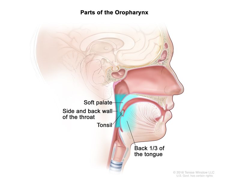
Anatomically, the oropharynx is located between the soft palate superiorly and the hyoid bone inferiorly. It is continuous with the oral cavity anteriorly and communicates with the nasopharynx superiorly and the supraglottic larynx and hypopharynx inferiorly.

The oropharynx is divided into the following parts:[4]

  • Base of the tongue, which includes the pharyngoepiglottic folds and the glossoepiglottic folds.
  • Vallecula.
  • Tonsillar region, which includes the fossa and the anterior and posterior pillars.
  • Soft palate, which includes the uvula.
  • Posterior and lateral pharyngeal walls.

Regional lymph node anatomy of the head and neck

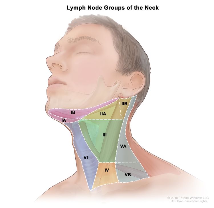
The regional lymph nodes of the head and neck include the lymph nodes that run parallel to the jugular veins, spinal accessory nerve, and facial artery and into the submandibular triangle. An understanding of regional anatomy and the status of regional lymph nodes is critical to the care of patients with head and neck cancer.[3,5,6] To facilitate communication regarding lymph node anatomy, the regions of the neck are described as levels I to V and retropharyngeal:

  • Level I contains the submental and submandibular lymph nodes.
  • Level II contains the upper jugular lymph nodes, which are above the digastric muscle.
  • Level III contains the midjugular lymph nodes, which are between the omohyoid muscle and the digastric muscle.
  • Level IV contains the lower jugular lymph nodes.
  • Level V contains the lymph nodes of the posterior triangle.
  • Retropharyngeal lymph nodes.
EnlargeLymph node groups of the neck; drawing shows six groups of lymph nodes in the neck: group IA and IB, group IIA and IIB, group III, group IV, group VA and VB, and group VI.

The retropharyngeal lymph nodes are a possible site for nodal spread in oropharyngeal cancer. A large retrospective cohort study from the MD Anderson Cancer Center described the clinical features of 981 patients with oropharyngeal cancer who underwent primary radiation therapy.[7][Level of evidence C1][Level of evidence C2]

  • The base of the tongue (47%) and the tonsil (46%) were the most common primary sites.
  • Most patients had stage T1 to T2 primary tumors (64%) and stage III to IVB disease (94%).
  • The incidence of radiographic retropharyngeal node involvement was 10% and was highest for the pharyngeal wall (23%) and lowest for the base of the tongue (6%).
  • Retropharyngeal lymph node involvement was associated with inferior 5-year local control and inferior recurrence-free survival, distant metastases−free survival, and overall survival on multivariate analysis.

Risk Factors

Risk factors for oropharyngeal squamous cell carcinoma (SCC) include:[8]

For more information, see Oral Cavity, Oropharyngeal, Hypopharyngeal, and Laryngeal Cancers Prevention.

HPV infection

Because of the decreased incidence of smoking in the United States, HPV-negative, smoking-related oropharyngeal cancer is decreasing; however, HPV-positive oropharyngeal cancer is increasing. According to the Surveillance, Epidemiology, and End Results (SEER) Program’s tissue repository data from 1988 to 2004, the prevalence of HPV-negative oropharyngeal cancer declined by 50%, while HPV-positive oropharyngeal cancers increased by 225%.[14][Level of evidence C1]

HPV-positive oropharyngeal cancers may represent a distinct disease entity that is caused by HPV infection and associated with an improved prognosis. Several studies indicate that individuals with HPV-positive tumors have significantly improved survival.[12,1517] Due to the prognostic impact of the HPV status in oropharyngeal cancer, the American Joint Committee on Cancer 8th edition staging separates oropharyngeal staging by HPV status.[5,6] In a prospective study of 253 patients with newly diagnosed or recurrent head and neck SCC, HPV was detected in 25% of the patients. Poor tumor grade and an oropharyngeal site independently increased the probability of the presence of HPV.[12] Oropharyngeal tumors are more likely to be HPV positive (57%) than oral cavity (12%) tumor sites and non-oropharyngeal (14%) sites. HPV-positive oropharyngeal cancers predominantly arise in the palatine or lingual tonsils. For tonsil or base-of-tongue sites, 62% of tumors were HPV positive, compared with 25% for other oropharyngeal sites.

Personal history of head and neck cancer

The risk of developing a second primary tumor in patients with tumors of the upper aerodigestive tract has been estimated to be 3% to 7% per year.[18,19] Because of this risk, patients require lifelong surveillance. Smoking and alcohol consumption after treatment are associated with the development of second primary tumors of the aerodigestive tract.[2022] Patients may need counseling to discontinue smoking and alcohol consumption.

The process of field cancerization may be partly responsible for the multiple, synchronous, primary SCCs that occur in oropharyngeal cancer and that are associated with a smoking history. Originally described in 1953, the concept of field cancerization holds that tumors develop in a multifocal fashion within a field of tissue chronically exposed to carcinogens.[23] Molecular studies that detect genetic alterations in histologically normal tissue from high-risk individuals have provided strong support for this concept.[2428]

A comparison of patients (N = 2,230) with index SCC of the oropharynx site and index SCC of non-oropharyngeal sites (i.e., oral cavity, larynx, and hypopharynx) was performed to determine the likelihood of developing second primary malignancies. The second primary malignancy rate was lower for patients with index oropharyngeal SCC than for patients with index non-oropharyngeal cancer (P < .001). Among patients with oropharyngeal SCC, former smokers had a 50% higher risk of second primary malignancy than never-smokers, and current smokers had a 100% higher risk than never-smokers (P trend = .008). These data suggest that patients who fit the typical HPV phenotype have a very low risk of second primary malignancy.[29]

Betel quid

The chewing of betel quid, a stimulant preparation commonly used in parts of Asia, increases the risk of oropharyngeal cancer.[30]

Other risk factors

Other risk factors may include:[8]

  • Defective elimination of acetaldehyde, a carcinogen generated by alcohol metabolism. In individuals, primarily those of East Asian race, who carry an inactive mutant allele of alcohol dehydrogenase-2, alcohol consumption is associated with a susceptibility to multiple metachronous oropharyngeal cancers that are caused by the decreased elimination of acetaldehyde.[31]

SCC of the oropharynx has not been associated with any specific chromosomal or genetic abnormalities. Genetic and chromosomal aberrations in these cancers are complex.[32,33] Despite the lack of specific genetic abnormalities, testing for genetic alterations or ploidy in early oropharyngeal lesions may identify patients who are at the greatest risk of disease progression and may lead to more-definitive therapy.[34]

Clinical Presentation

The clinical presentation of oropharyngeal cancer depends on the tumor’s location in the oropharynx. Oropharyngeal cancer may present in the following locations:

Tonsil

The anterior tonsillar pillar and tonsil are the most common location for a primary tumor of the oropharynx.[4] Lesions involving the anterior tonsillar pillar may appear as areas of dysplasia, inflammation, or a superficial spreading lesion. These cancers can spread across a broad region, including the lateral soft palate, retromolar trigone and buccal mucosa, and tonsillar fossa.[3,4] The lymphatic drainage is primarily to level II nodes.

Tumors of the posterior tonsillar pillar can extend inferiorly to involve the pharyngoepiglottic fold and the posterior aspect of the thyroid cartilage. These lesions more frequently involve level V nodes.

Lesions of the tonsillar fossa may be either exophytic or ulcerative and have a pattern of extension similar to those of the anterior tonsillar pillar. These tumors present as advanced-stage disease more often than do cancers of the tonsillar pillar. Approximately 75% of patients will present with stage III or stage IV disease.[3,4] The lymphatic drainage is primarily to level V nodes. Tumors of the posterior tonsillar pillar can extend inferiorly to involve the pharyngoepiglottic fold and the posterior aspect of the thyroid cartilage. These lesions more frequently involve level V nodes.

Signs and symptoms of tonsillar lesions may include:[3,4]

  • Pain.
  • Dysphagia.
  • Weight loss.
  • Ipsilateral referred otalgia.
  • A mass in the neck.

Base of the tongue

Clinically, cancers of the base of the tongue are insidious. These cancers can grow in either an infiltrative or exophytic pattern. Because the base of the tongue is devoid of pain fibers, these tumors are often asymptomatic until there is significant tumor progression.[4]

Signs and symptoms of advanced base-of-the-tongue cancers may include:[3,4]

  • Pain.
  • Dysphagia.
  • Weight loss.
  • Referred otalgia secondary to cranial nerve involvement.
  • Trismus secondary to pterygoid muscle involvement.
  • Fixation of the tongue that is caused by infiltration of the deep muscle.
  • A mass in the neck.

Lymph node metastasis is common because of the rich lymphatic drainage of the base of the tongue. Approximately 70% or more of patients with advanced base-of-the-tongue cancers have ipsilateral cervical nodal metastases; 30% or fewer of such patients have bilateral, cervical lymph–node metastases.[4,35] The cervical lymph nodes involved commonly include levels II, III, IV, and V and retropharyngeal lymph nodes.

Soft palate

Soft palate tumors are primarily found on the anterior surface.[4] Lesions in this area may remain superficial and in early stages.[3] The lymphatic drainage is primarily to level II nodes.

Pharyngeal wall

Pharyngeal wall lesions can spread superiorly to involve the nasopharynx, posteriorly to infiltrate the prevertebral fascia, and inferiorly to involve the pyriform sinuses and hypopharyngeal walls. Primary lymphatic drainage is to the retropharyngeal nodes and level II and III nodes. Because most pharyngeal tumors extend past the midline, bilateral cervical metastases are common.

Early-stage tumors are often asymptomatic. Tumors of the pharyngeal wall are typically diagnosed in an advanced stage.[3,4]

Signs and symptoms of advanced pharyngeal wall tumors may include:

  • Pain.
  • Bleeding.
  • Dysphagia.
  • Weight loss.
  • A mass in the neck.

Leukoplakia

Leukoplakia is used only as a clinically descriptive term meaning that the observer sees a white patch that does not rub off, the significance of which depends on the histological findings.[8] Leukoplakia can range from hyperkeratosis to an actual early invasive carcinoma or may represent a fungal infection, lichen planus, or other benign oral disease.

Diagnostic Evaluation

The assessment of the primary tumor is based on inspection and palpation, when possible, and by indirect mirror examination. The appropriate nodal drainage areas are examined by careful palpation. The presence of tumor must be confirmed histologically. Any other pathological data obtained from a biopsy and additional radiographical studies are also considered.

The following procedures may be done to evaluate the primary tumor:

  • Positron emission tomography–computed tomography (PET-CT) scan.
  • Magnetic resonance imaging.
  • Endoscopy.
  • Laryngoscopy.
  • Biopsy and p16 testing to assess for HPV status.

A PET-CT scan yields morphological and metabolic data to assess the detection of primary tumor, nodal disease, and distant metastatic disease. It may also be used to guide radiation therapy planning. Retrospective data demonstrate that morphological and PET-glycolytic parameters (as measured by fluorodeoxyglucose PET-CT) are significantly larger (as measured by Response Evaluation Criteria In Solid Tumors [RECIST] longest diameter) and more heterogenous in HPV-negative disease than in HPV-positive disease in the primary tumor for oropharyngeal carcinoma. These PET-CT parameters also show higher standardized uptake value (SUV) max, SUV mean, and metabolic tumor volume in HPV-negative disease. However, the same PET parameters are frequently larger in the regional nodal disease in patients with HPV-positive disease.[36][Level of evidence C3]

Prognostic Factors and Survival

Prognostic factors for oropharyngeal carcinoma include:

  • HPV status.
  • Smoking history (10 or more pack-years).
  • Tumor stage and nodal status.

The criteria described in Table 1 are used to determine whether patients have low-, intermediate-, or high-risk oropharyngeal carcinoma. These criteria have been defined by using recursive partitioning analysis in a retrospective analysis of a randomized trial of patients with stage III and IV oropharyngeal SCC treated with chemoradiation.[17]

Table 1. Characteristics Associated With the Risk of Oropharyngeal Cancera
Degree of Risk Characteristics 3-y OS Rate
CI = confidence interval; HPV = human papillomavirus; OS = overall survival; + = positive; – = negative. For more information, see the AJCC Staging Groupings and TNM Definitions section.
aAng KK, Harris J, Wheeler R, et al.: Human papillomavirus and survival of patients with oropharyngeal cancer. N Engl J Med 363 (1): 24–35, 2010.
Low HPV+, smoking history of ≤10 pack-years, and N0–N2a nodal history 93% (95% CI, 88.3%–97.7%)
Intermediate HPV+, smoking history of >10 pack-years, and N2b–N3 nodal disease; or 70.8% (95% CI, 60.7%–80.8%)
HPV-, smoking history of ≤10 pack-years, and N2b–N3 nodal disease or T2–T3 tumors
High HPV- and smoking history >10 pack-years; or 46.2% (95% CI, 34.7%–57.7%)
HPV-, smoking history ≤10 pack-years, and T4 disease

Follow-Up After Treatment

A careful examination of the patient’s head and neck allows the physician to look for recurrence every 6 to 12 weeks for the first posttreatment year, every 3 months for the second year, every 3 to 4 months for the third year, and every 6 months thereafter.

References
  1. American Cancer Society: Cancer Facts and Figures 2025. American Cancer Society, 2025. Available online. Last accessed January 16, 2025.
  2. Bray F, Laversanne M, Sung H, et al.: Global cancer statistics 2022: GLOBOCAN estimates of incidence and mortality worldwide for 36 cancers in 185 countries. CA Cancer J Clin 74 (3): 229-263, 2024. [PUBMED Abstract]
  3. Mendenhall WM, Werning JW, Pfister DG: Treatment of head and neck cancer. In: DeVita VT Jr, Lawrence TS, Rosenberg SA: Cancer: Principles and Practice of Oncology. 9th ed. Lippincott Williams & Wilkins, 2011, pp 729-80.
  4. Choi WH, Hu KS, Culliney B, et al.: Cancer of the oropharynx. In: Harrison LB, Sessions RB, Hong WK, eds.: Head and Neck Cancer: A Multidisciplinary Approach. 3rd ed. Lippincott, William & Wilkins, 2009, pp 285-335.
  5. HPV-Mediated (p16+) Oropharyngeal Cancer. In: Amin MB, Edge SB, Greene FL, et al., eds.: AJCC Cancer Staging Manual. 8th ed. Springer; 2017, pp. 113-21.
  6. Oropharynx (p16-) and Hypopharynx. In: Amin MB, Edge SB, Greene FL, et al., eds.: AJCC Cancer Staging Manual. 8th ed. Springer; 2017, pp. 123-35.
  7. Gunn GB, Debnam JM, Fuller CD, et al.: The impact of radiographic retropharyngeal adenopathy in oropharyngeal cancer. Cancer 119 (17): 3162-9, 2013. [PUBMED Abstract]
  8. Neville BW, Day TA: Oral cancer and precancerous lesions. CA Cancer J Clin 52 (4): 195-215, 2002 Jul-Aug. [PUBMED Abstract]
  9. Mendenhall WM, Werning JW, Pfister DG: Treatment of head and neck cancer. In: DeVita VT Jr, Lawrence TS, Rosenberg SA: Cancer: Principles and Practice of Oncology. 9th ed. Lippincott Williams & Wilkins, 2011, pp 729-80.
  10. Licitra L, Bernier J, Grandi C, et al.: Cancer of the oropharynx. Crit Rev Oncol Hematol 41 (1): 107-22, 2002. [PUBMED Abstract]
  11. Mork J, Lie AK, Glattre E, et al.: Human papillomavirus infection as a risk factor for squamous-cell carcinoma of the head and neck. N Engl J Med 344 (15): 1125-31, 2001. [PUBMED Abstract]
  12. Gillison ML, Koch WM, Capone RB, et al.: Evidence for a causal association between human papillomavirus and a subset of head and neck cancers. J Natl Cancer Inst 92 (9): 709-20, 2000. [PUBMED Abstract]
  13. D’Souza G, Kreimer AR, Viscidi R, et al.: Case-control study of human papillomavirus and oropharyngeal cancer. N Engl J Med 356 (19): 1944-56, 2007. [PUBMED Abstract]
  14. Chaturvedi AK, Engels EA, Pfeiffer RM, et al.: Human papillomavirus and rising oropharyngeal cancer incidence in the United States. J Clin Oncol 29 (32): 4294-301, 2011. [PUBMED Abstract]
  15. Ringström E, Peters E, Hasegawa M, et al.: Human papillomavirus type 16 and squamous cell carcinoma of the head and neck. Clin Cancer Res 8 (10): 3187-92, 2002. [PUBMED Abstract]
  16. Schwartz SR, Yueh B, McDougall JK, et al.: Human papillomavirus infection and survival in oral squamous cell cancer: a population-based study. Otolaryngol Head Neck Surg 125 (1): 1-9, 2001. [PUBMED Abstract]
  17. Ang KK, Harris J, Wheeler R, et al.: Human papillomavirus and survival of patients with oropharyngeal cancer. N Engl J Med 363 (1): 24-35, 2010. [PUBMED Abstract]
  18. Khuri FR, Lippman SM, Spitz MR, et al.: Molecular epidemiology and retinoid chemoprevention of head and neck cancer. J Natl Cancer Inst 89 (3): 199-211, 1997. [PUBMED Abstract]
  19. León X, Quer M, Diez S, et al.: Second neoplasm in patients with head and neck cancer. Head Neck 21 (3): 204-10, 1999. [PUBMED Abstract]
  20. Do KA, Johnson MM, Doherty DA, et al.: Second primary tumors in patients with upper aerodigestive tract cancers: joint effects of smoking and alcohol (United States). Cancer Causes Control 14 (2): 131-8, 2003. [PUBMED Abstract]
  21. Khuri FR, Kim ES, Lee JJ, et al.: The impact of smoking status, disease stage, and index tumor site on second primary tumor incidence and tumor recurrence in the head and neck retinoid chemoprevention trial. Cancer Epidemiol Biomarkers Prev 10 (8): 823-9, 2001. [PUBMED Abstract]
  22. Day GL, Blot WJ, Shore RE, et al.: Second cancers following oral and pharyngeal cancers: role of tobacco and alcohol. J Natl Cancer Inst 86 (2): 131-7, 1994. [PUBMED Abstract]
  23. Slaughter DP, Southwick HW, Smejkal W: Field cancerization in oral stratified squamous epithelium: clinical implications of multicentric origin. Cancer 6 (5): 963-8, 1953. [PUBMED Abstract]
  24. Braakhuis BJ, Tabor MP, Leemans CR, et al.: Second primary tumors and field cancerization in oral and oropharyngeal cancer: molecular techniques provide new insights and definitions. Head Neck 24 (2): 198-206, 2002. [PUBMED Abstract]
  25. Braakhuis BJ, Tabor MP, Kummer JA, et al.: A genetic explanation of Slaughter’s concept of field cancerization: evidence and clinical implications. Cancer Res 63 (8): 1727-30, 2003. [PUBMED Abstract]
  26. Tabor MP, Brakenhoff RH, van Houten VM, et al.: Persistence of genetically altered fields in head and neck cancer patients: biological and clinical implications. Clin Cancer Res 7 (6): 1523-32, 2001. [PUBMED Abstract]
  27. Tabor MP, Brakenhoff RH, Ruijter-Schippers HJ, et al.: Multiple head and neck tumors frequently originate from a single preneoplastic lesion. Am J Pathol 161 (3): 1051-60, 2002. [PUBMED Abstract]
  28. Ha PK, Califano JA: The molecular biology of mucosal field cancerization of the head and neck. Crit Rev Oral Biol Med 14 (5): 363-9, 2003. [PUBMED Abstract]
  29. Gan SJ, Dahlstrom KR, Peck BW, et al.: Incidence and pattern of second primary malignancies in patients with index oropharyngeal cancers versus index nonoropharyngeal head and neck cancers. Cancer 119 (14): 2593-601, 2013. [PUBMED Abstract]
  30. Ho PS, Ko YC, Yang YH, et al.: The incidence of oropharyngeal cancer in Taiwan: an endemic betel quid chewing area. J Oral Pathol Med 31 (4): 213-9, 2002. [PUBMED Abstract]
  31. Yokoyama A, Watanabe H, Fukuda H, et al.: Multiple cancers associated with esophageal and oropharyngolaryngeal squamous cell carcinoma and the aldehyde dehydrogenase-2 genotype in male Japanese drinkers. Cancer Epidemiol Biomarkers Prev 11 (9): 895-900, 2002. [PUBMED Abstract]
  32. Tremmel SC, Götte K, Popp S, et al.: Intratumoral genomic heterogeneity in advanced head and neck cancer detected by comparative genomic hybridization. Cancer Genet Cytogenet 144 (2): 165-74, 2003. [PUBMED Abstract]
  33. Brieger J, Jacob R, Riazimand HS, et al.: Chromosomal aberrations in premalignant and malignant squamous epithelium. Cancer Genet Cytogenet 144 (2): 148-55, 2003. [PUBMED Abstract]
  34. Forastiere A, Koch W, Trotti A, et al.: Head and neck cancer. N Engl J Med 345 (26): 1890-900, 2001. [PUBMED Abstract]
  35. Lindberg R: Distribution of cervical lymph node metastases from squamous cell carcinoma of the upper respiratory and digestive tracts. Cancer 29 (6): 1446-9, 1972. [PUBMED Abstract]
  36. Tahari AK, Alluri KC, Quon H, et al.: FDG PET/CT imaging of oropharyngeal squamous cell carcinoma: characteristics of human papillomavirus-positive and -negative tumors. Clin Nucl Med 39 (3): 225-31, 2014. [PUBMED Abstract]

Cellular Classification of Oropharyngeal Cancer

Most oropharyngeal cancers are squamous cell carcinomas (SCCs).[13] SCCs may be noninvasive or invasive. For noninvasive SCC, the term carcinoma in situ is used. Histologically, invasive carcinomas are classified as well differentiated, moderately differentiated, poorly differentiated, or undifferentiated. SCCs are usually moderately or poorly differentiated.[4] Grading the deep invasive margins (i.e., invasive front) of SCC may provide better prognostic information than grading the entire tumor.[5] Human papillomavirus (HPV)-positive oropharyngeal cancers arising from the lingual and palatine tonsils are a distinct molecular-pathological entity that is linked to infection with HPV, especially HPV-16. Compared with HPV-negative tumors, HPV-positive tumors are more frequently poorly differentiated and nonkeratinizing. They are strongly associated with basaloid morphology and less likely to have TP53 variants.[6]

Immunohistochemical examination of tissues for the expression of the biomarker Ki-67, a proliferation antigen, may complement histological grading. As a molecular indicator of epithelial dysplasia of the oropharynx, Ki-67 expression appears to correlate well with loss of heterozygosity (LOH) in tumor cells. In a retrospective study involving 43 tissue samples from 25 patients, the assessment of proliferation with Ki-67 was a better surrogate for LOH than was histological grading.[7]

Other types of oropharyngeal cancer include:

  • Minor salivary gland tumors.
  • Lymphomas.
  • Lymphoepitheliomas (e.g., tonsillar fossa).

For more information, see Salivary Gland Cancer Treatment, Hodgkin Lymphoma Treatment, Indolent B-Cell Non-Hodgkin Lymphoma Treatment, Aggressive B-Cell Non-Hodgkin Lymphoma Treatment, and Peripheral T-Cell Non-Hodgkin Lymphoma Treatment.

References
  1. Mendenhall WM, Werning JW, Pfister DG: Treatment of head and neck cancer. In: DeVita VT Jr, Lawrence TS, Rosenberg SA: Cancer: Principles and Practice of Oncology. 9th ed. Lippincott Williams & Wilkins, 2011, pp 729-80.
  2. HPV-Mediated (p16+) Oropharyngeal Cancer. In: Amin MB, Edge SB, Greene FL, et al., eds.: AJCC Cancer Staging Manual. 8th ed. Springer; 2017, pp. 113-21.
  3. Oropharynx (p16-) and Hypopharynx. In: Amin MB, Edge SB, Greene FL, et al., eds.: AJCC Cancer Staging Manual. 8th ed. Springer; 2017, pp. 123-35.
  4. Oral cavity and oropharynx. In: Rosai J, ed.: Rosai and Ackerman’s Surgical Pathology. Vol. 1. 10th ed. Mosby Elsevier, 2011, pp. 237-264.
  5. Bryne M, Boysen M, Alfsen CG, et al.: The invasive front of carcinomas. The most important area for tumour prognosis? Anticancer Res 18 (6B): 4757-64, 1998 Nov-Dec. [PUBMED Abstract]
  6. Gillison ML, Koch WM, Capone RB, et al.: Evidence for a causal association between human papillomavirus and a subset of head and neck cancers. J Natl Cancer Inst 92 (9): 709-20, 2000. [PUBMED Abstract]
  7. Tabor MP, Braakhuis BJ, van der Wal JE, et al.: Comparative molecular and histological grading of epithelial dysplasia of the oral cavity and the oropharynx. J Pathol 199 (3): 354-60, 2003. [PUBMED Abstract]

Stage Information for Oropharyngeal Cancer

The staging system for oropharyngeal cancer is clinical rather than pathological. It is based on the best estimate of the extent of disease before treatment.

Clinical anatomical staging of oropharyngeal cancer involves the following clinical assessment and imaging techniques:

  • Inspection and palpation of sites (when feasible) and neck nodes.
  • Neurological examination of all cranial nerves.
  • A head and neck computed tomography (CT) scan with contrast to evaluate the mandible and maxilla.[1]
  • Magnetic resonance imaging to evaluate the extent of disease in the soft tissues.
  • Positron emission tomography (PET)–CT scan to assess primary, regional, and distant metastatic spread.
  • Complete endoscopy after completion of other staging studies to assess the surface extent of the tumor accurately and to facilitate biopsy. This procedure is typically performed under general anesthesia, which also allows palpation for deep muscle invasion. Because of the incidence of multiple primary tumors occurring simultaneously, a careful search for other primary tumors of the upper aerodigestive tract is indicated.[2]

PET has been investigated as an imaging modality for recurrent oropharyngeal cancer.[3]

American Joint Committee on Cancer (AJCC) Staging Groupings and TNM Definitions

The AJCC has designated staging by TNM (tumor, node, metastasis) classification to define oropharyngeal cancer.[2,4] Nonepithelial tumors such as those of lymphoid tissue, soft tissue, bone, and cartilage are not included.

The AJCC uses separate staging systems for human papillomavirus (HPV)-related squamous cell carcinoma of the oropharynx [4] and p16-negative squamous cancers of the oropharynx.[2]

AJCC prognostic stage groups for HPV-mediated (p16-positive) oropharyngeal cancer

Table 2. Definitions of TNM Stage Ia
Stage TNM Description
T = primary tumor; N = regional lymph node; M = distant metastasis.
aReprinted with permission from AJCC: HPV-Mediated (p16+) Oropharyngeal Cancer. In: Edge SB, Byrd DR, Compton CC, et al., eds.: AJCC Cancer Staging Manual. 8th ed. New York, NY: Springer, 2017, pp 113–21.
I T0, T1, or T2; N0 or N1; M0 T0, T1, or T2 = See Stage IV in Table 5 below.
N0 or N1 = See Stage IV in Table 5 below.
M0 = No distant metastasis.
Table 3. Definitions of TNM Stage IIa
Stage TNM Description
T = primary tumor; N = regional lymph node; M = distant metastasis.
aReprinted with permission from AJCC: HPV-Mediated (p16+) Oropharyngeal Cancer. In: Edge SB, Byrd DR, Compton CC, et al., eds.: AJCC Cancer Staging Manual. 8th ed. New York, NY: Springer, 2017, pp 113–21.
II T0, T1, or T2; N2; M0 T0, T1, or T2 = See Stage IV in Table 5 below.
N2 = Contralateral or bilateral lymph nodes, none >6 cm.
M0 = No distant metastasis.
T3; N0, N1, or N2; M0 T3 = Tumor >4 cm in greatest dimension or extension to lingual surface of the epiglottis.
N0, N1, or N2 = See Stage IV in Table 5 below.
M0 = No distant metastasis.
Table 4. Definitions of TNM Stage IIIa
Stage TNM Description
T = primary tumor; N = regional lymph node; M = distant metastasis.
aReprinted with permission from AJCC: HPV-Mediated (p16+) Oropharyngeal Cancer. In: Edge SB, Byrd DR, Compton CC, et al., eds.: AJCC Cancer Staging Manual. 8th ed. New York, NY: Springer, 2017, pp 113–21.
bMucosal extension to the lingual surface of the epiglottis from primary tumors of the base of the tongue and vallecula does not constitute invasion of the larynx.
III T0, T1, T2, T3, or T4; N3; M0 T0, T1, T2, T3, or T4 = See Stage IV in Table 5 below.
N3 = Lymph node(s) >6 cm.
M0 = No distant metastasis.
T4; N0, N1, N2, or N3; M0 T4 = Moderately advanced local disease. Tumor invades the larynx, extrinsic muscle of the tongue, medial pterygoid, hard palate, or mandible or beyond.b
N0, N1, N2, or N3 = See Stage IV in Table 5 below.
M0 = No distant metastasis.
Table 5. Definitions of TNM Stage IVa
Stage TNM Description
T = primary tumor; N = regional lymph node; M = distant metastasis.
aReprinted with permission from AJCC: HPV-Mediated (p16+) Oropharyngeal Cancer. In: Edge SB, Byrd DR, Compton CC, et al., eds.: AJCC Cancer Staging Manual. 8th ed. New York, NY: Springer, 2017, pp 113–21.
bMucosal extension to the lingual surface of the epiglottis from primary tumors of the base of the tongue and vallecula does not constitute invasion of the larynx.
IV Any T, Any N, M1 T0 = No primary identified.
T1 = Tumor ≤2 cm in greatest dimension.
T2 = Tumor >2 cm but ≤4 cm in greatest dimension.
T3 = Tumor >4 cm in greatest dimension or extension to lingual surface of the epiglottis.
T4 = Moderately advanced local disease. Tumor invades the larynx, extrinsic muscle of the tongue, medial pterygoid, hard palate, or mandible or beyond.b
NX = Regional lymph nodes cannot be assessed.
N0 = No regional lymph node metastasis.
N1 = One or more ipsilateral lymph nodes, none >6 cm.
N2 = Contralateral or bilateral lymph nodes, none >6 cm.
N3 = Lymph node(s) >6 cm.
M1 = Distant metastasis.

AJCC prognostic stage groups for p16-negative squamous cancers of the oropharynx

Table 6. Definitions of TNM Stage 0a
Stage TNbM Description
T = primary tumor; N = regional lymph node; M = distant metastasis.
aReprinted with permission from AJCC: Oropharynx (p16−) and Hypopharynx. In: Edge SB, Byrd DR, Compton CC, et al., eds.: AJCC Cancer Staging Manual. 8th ed. New York, NY: Springer, 2017, pp 123–35.
The explanation for superscript b is at the end of Table 10.
0 Tis, N0, M0 Tis = Carcinoma in situ.
N0 = No regional lymph node metastasis.
M0 = No distant metastasis.
Table 7. Definitions of TNM Stage Ia
Stage TNbM Description
T = primary tumor; N = regional lymph node; M = distant metastasis.
aReprinted with permission from AJCC: Oropharynx (p16−) and Hypopharynx. In: Edge SB, Byrd DR, Compton CC, et al., eds.: AJCC Cancer Staging Manual. 8th ed. New York, NY: Springer, 2017, pp 123–35.
The explanation for superscript b is at the end of Table 10.
I T1, N0, M0 T1 = Tumor ≤2 cm in greatest dimension.
N0 = No regional lymph node metastasis.
M0 = No distant metastasis.
Table 8. Definitions of TNM Stage IIa
Stage TNbM Description
T = primary tumor; N = regional lymph node; M = distant metastasis.
aReprinted with permission from AJCC: Oropharynx (p16−) and Hypopharynx. In: Edge SB, Byrd DR, Compton CC, et al., eds.: AJCC Cancer Staging Manual. 8th ed. New York, NY: Springer, 2017, pp 123–35.
The explanation for superscript b is at the end of Table 10.
II T2, N0, M0 T2 = Tumor >2 cm but ≤4 cm in greatest dimension.
N0 = No regional lymph node metastasis.
M0 = No distant metastasis.
Table 9. Definitions of TNM Stage IIIa
Stage TNbM Description
T = primary tumor; N = regional lymph node; M = distant metastasis; ENE = extranodal extension.
aReprinted with permission from AJCC: Oropharynx (p16−) and Hypopharynx. In: Edge SB, Byrd DR, Compton CC, et al., eds.: AJCC Cancer Staging Manual. 8th ed. New York, NY: Springer, 2017, pp 123–35.
The explanation for superscript b is at the end of Table 10.
III T3, N0, M0 T3 = Tumor >4 cm in greatest dimension or extension to lingual surface of epiglottis.
N0 = No regional lymph node metastasis.
M0 = No distant metastasis.
T1, T2, T3; N1; M0 T1, T2, T3 = See Stage IVC in Table 10 below.
N1 = Metastasis in a single ipsilateral lymph node, ≤3 cm in greatest dimension and ENE(–).
M0 = No distant metastasis.
Table 10. Definitions of TNM Stage IVA, IVB, and IVCa
Stage TNM Description
T = primary tumor; N = regional lymph node; M = distant metastasis; ENE = extranodal extension.
aReprinted with permission from AJCC: Oropharynx (p16−) and Hypopharynx. In: Edge SB, Byrd DR, Compton CC, et al., eds.: AJCC Cancer Staging Manual. 8th ed. New York, NY: Springer, 2017, pp 123–35.
bA designation of U or L may be used for any N category to indicate metastasis above the lower border of the cricoid (U) or below the lower border of the cricoid (L). Similarly, clinical and pathological ENE should be recorded as ENE(−) or ENE(+).
cMucosal extension to the lingual surface of the epiglottis from primary tumors of the base of the tongue and vallecula does not constitute invasion of the larynx.
IVA T4a; N0, N1; M0 T4a = Moderately advanced local disease. Tumor invades the larynx, extrinsic muscle of the tongue, medial pterygoid, hard palate, or mandible.c
N0, N1 = See Stage IVC below in this table.
M0 = No distant metastasis.
T1, T2, T3, T4a; N2; M0 T1, T2, T3, T4a = See Stage IVC below in this table.
N2 = Metastasis in a single ipsilateral node >3 cm but ≤6 cm in greatest dimension and ENE(−); or metastases in multiple ipsilateral lymph nodes, none >6 cm in greatest dimension and ENE(−); or in bilateral or contralateral lymph nodes, none >6 cm in greatest dimension and ENE(−).
M0 = No distant metastasis.
IVB Any T, N3, M0 Any T = See Stage IVC below in this table.
N3 = Metastasis in a lymph node >6 cm in greatest dimension and ENE(−); or metastasis in any node(s) and clinically overt ENE(+).
M0 = No distant metastases.
T4b, Any N, M0 T4b = Very advanced local disease. Tumor invades lateral pterygoid muscle, pterygoid plates, lateral nasopharynx, or skull base or encases carotid artery.
Any N = See Stage IVC below in this table.
M0 = No distant metastasis.
IVC Any T, Any N, M1 TX = Primary tumor cannot be assessed.
Tis = Carcinoma in situ.
T1 = Tumor ≤2 cm in greatest dimension.
T2 = Tumor >2 cm but ≤4 cm in greatest dimension.
T3 = Tumor >4 cm in greatest dimension or extension to lingual surface of epiglottis.
T4 = Moderately advanced or very advanced local disease.
−T4a = Moderately advanced local disease. Tumor invades the larynx, extrinsic muscle of the tongue, medial pterygoid, hard palate, or mandible.c
−T4b = Very advanced local disease. Tumor invades lateral pterygoid muscle, pterygoid plates, lateral nasopharynx, or skull base or encases carotid artery.
NX = Regional lymph nodes cannot be assessed.
N0 = No regional lymph node metastasis.
N1 = Metastasis in a single ipsilateral lymph node, ≤3 cm in greatest dimension and ENE(−).
N2 = Metastasis in a single ipsilateral node >3 cm but ≤6 cm in greatest dimension and ENE(−); or metastases in multiple ipsilateral lymph nodes, none >6 cm in greatest dimension and ENE(−); or in bilateral or contralateral lymph nodes, none >6 cm in greatest dimension and ENE(−).
−N2a = Metastasis in a single ipsilateral node >3 cm but ≤6 cm in greatest dimension and ENE(−).
−N2b = Metastases in multiple ipsilateral nodes, none >6 cm in greatest dimension and ENE(−).
−N2c = Metastases in bilateral or contralateral lymph nodes, none >6 cm in greatest dimension and ENE(−).
N3 = Metastasis in a lymph node >6 cm in greatest dimension and ENE(−); or metastasis in any node(s) and clinically overt ENE(+).
−N3a = Metastasis in a lymph node >6 cm in greatest dimension and ENE(−).
−N3b = Metastasis in any node(s) and clinically overt ENE(+).
M1 = Distant metastasis.
References
  1. Weber AL, Romo L, Hashmi S: Malignant tumors of the oral cavity and oropharynx: clinical, pathologic, and radiologic evaluation. Neuroimaging Clin N Am 13 (3): 443-64, 2003. [PUBMED Abstract]
  2. Oropharynx (p16-) and Hypopharynx. In: Amin MB, Edge SB, Greene FL, et al., eds.: AJCC Cancer Staging Manual. 8th ed. Springer; 2017, pp. 123-35.
  3. Wong RJ, Lin DT, Schöder H, et al.: Diagnostic and prognostic value of [(18)F]fluorodeoxyglucose positron emission tomography for recurrent head and neck squamous cell carcinoma. J Clin Oncol 20 (20): 4199-208, 2002. [PUBMED Abstract]
  4. HPV-Mediated (p16+) Oropharyngeal Cancer. In: Amin MB, Edge SB, Greene FL, et al., eds.: AJCC Cancer Staging Manual. 8th ed. Springer; 2017, pp. 113-21.

Treatment Option Overview for Oropharyngeal Cancer

An optimal approach for the treatment of oropharyngeal cancer is not easily defined because no single regimen offers a distinct superior-survival advantage. The literature reports various therapeutic options but does not present any valid comparative studies of these options. Treatment considerations should account for functional and performance status, including speech and swallowing outcomes.

Table 11. Treatment Options for Oropharyngeal Cancer
Stage (TNM Definitions) Treatment Options
Stage I and stage II oropharyngeal cancer Radiation therapy using standard fractionation
Surgery
Stage III and stage IV oropharyngeal cancer Surgery followed by postoperative radiation therapy (PORT) with or without concurrent chemotherapy for patients with locally advanced disease
Radiation therapy using altered fractionation
Concurrent chemoradiation therapy
Neoadjuvant chemotherapy followed by concurrent chemoradiation therapy
Chemoradiation therapy with immunotherapy (under clinical evaluation)
Treatment de-intensification using radiation dose de-escalation (under clinical evaluation)
Treatment de-intensification using transoral surgery followed by radiation dose de-escalation (under clinical evaluation)
Metastatic and recurrent oropharyngeal cancer Surgical resection, if technically feasible and the tumor does not respond to radiation therapy
Radiation therapy, if the tumor is not completely removed by surgery and curative doses of radiation have not been given previously
A second surgery, if the tumor was not completely removed initially and if technically feasible
Chemotherapy, for unresectable locoregionally recurrent disease
Additional radiation therapy using conventionally fractionated radiation therapy or hyperfractionated radiation therapy with concurrent chemotherapy
Stereotactic body radiation therapy with concurrent cetuximab
Immunotherapy
Clinical trials evaluating additional radiation therapy using hyperfractionated radiation therapy with concurrent chemotherapy, targeted therapy, stereotactic body radiation therapy, or immunotherapy

Surgery and/or Radiation Therapy

Surgery and radiation therapy have been the standard treatment for oropharyngeal cancer; however, outcome data from randomized trials are limited. Studies have evaluated whether to use surgery or radiation but have been underpowered.[1]

Evidence (surgery and/or radiation therapy):

  1. In a prospective randomized trial, 564 patients with head and neck cancer and N2 or N3 disease were assigned to either planned neck dissection or surveillance with positron emission tomography–computed tomography (PET-CT) scan.[2]
    • With a median follow up of 36 months, PET-CT surveillance resulted in fewer neck dissections compared with the surgical group (54 vs. 221), with a 2-year survival rate of 84.9% for the neck dissection group and 81.5% for the surgical group. The hazard ratio (HR) for death (HRdeath) slightly favored PET-CT−guided surveillance and indicated noninferiority (upper boundary 95% confidence interval [CI] for HR <1.50; P = .004).
  2. A pooled analysis of 6,400 patients with base-of-the-tongue oropharyngeal carcinoma from 51 reported series between 1970 and 2000 demonstrated the following:[3]
    • Local control rates of 79% (surgery with or without radiation therapy) and 76% (radiation therapy alone), (P = .087); locoregional control was 60% for surgery with or without radiation therapy versus 69% for radiation therapy alone (P = .009).
    • The 5-year survival rate was 49% for surgery with or without radiation therapy versus 52% (P = .2) for radiation therapy with or without neck dissection.
    • The rate of severe complications was 32% for the surgery group versus 3.8% for the radiation therapy group (P < .001).
    • The rate of fatal complications was 3.5% for the surgery group versus 0.4% for the radiation therapy group (P < .001).

    Historically, the posttreatment performance and functional status of patients with base-of-the-tongue primary tumors was better after radiation therapy than after surgery. Local control and survival are similar in both treatment options.[4,5]

  3. In the same study, the results for patients with squamous cell carcinoma (SCC) in the tonsillar region who underwent surgery with or without radiation therapy versus radiation therapy with or without neck dissection were as follows:[3]
    • Local control rates of 70% (surgery with or without radiation therapy) and 68% (radiation therapy), (P = .2); locoregional control was 65% for surgery with or without radiation therapy versus 69% for radiation therapy alone (P = .1).
    • The 5-year survival rate was 47% for surgery with or without radiation therapy versus 43% (P = .2) for radiation therapy with or without neck dissection.
    • The rate of severe complications was 23% for the surgery group versus 6% for the radiation therapy group (P < .001)
    • The rate of fatal complications was 3.2% for the surgery group versus 0.8% for the radiation therapy group (P < .001).

For patients with early-stage disease, single-modality treatment is preferred. Historically, radiation alone has been preferred, although use of new surgical techniques, including transoral surgery and transoral robotic surgery, is increasing. Nonrandomized comparisons of transoral surgery versus primary radiation therapy suggest superior quality of life (QOL) with minimally invasive surgical techniques.[6] Historically, more–invasive surgical techniques were associated with inferior QOL and greater morbidity.

A prospective multicenter trial (ECOG-3311 [NCT01898494]) evaluating transoral surgery approaches in human papillomavirus−positive oropharyngeal carcinoma with postoperative radiation dose de-escalation is currently under way.

Surgery Followed by Postoperative Radiation Therapy (PORT) With or Without Concurrent Chemotherapy for Patients With Locally Advanced Disease

New surgical techniques for resection and reconstruction that provide access and functional preservation have extended the surgical options for patients with stage III or stage IV oropharyngeal cancer. Specific surgical procedures and their modifications are not described here because of the wide variety of surgical approaches, the variety of opinions about the role of modified neck dissections, and the multiple reconstructive techniques that may give the same results. This group of patients are managed by head and neck surgeons who are skilled in the multiple procedures available and are actively involved in the care of these patients.

Depending on pathological findings after primary surgery, PORT with or without chemotherapy is used in the adjuvant setting for patients with the following histological findings:

  • T4 disease.
  • Perineural invasion.
  • Lymphovascular invasion.
  • Positive margins or margins less than 5 mm.
  • Extracapsular extension of a lymph node.
  • Two or more involved lymph nodes.

The addition of chemotherapy to PORT for oropharyngeal SCC demonstrates a locoregional control and overall survival (OS) benefit compared with radiation therapy alone in patients who have high-risk pathological risk factors, extracapsular extension (ECE) of a lymph node, or positive margins, based on a pooled analysis of the EORTC-22931 (NCT00002555) and RTOG-9501 (NCT00002670) studies.[710][Level of evidence A1]

For patients with intermediate pathological risk factors, the addition of cisplatin chemotherapy given concurrently with PORT is unclear. Intermediate pathological risk factors include:

  • T3 and T4 disease (or stage III and stage IV disease).
  • Perineural infiltration.
  • Vascular embolisms.
  • Clinically enlarged level IV–V lymph nodes secondary to tumors arising in the oral cavity or oropharynx.
  • Two or more histopathologically involved lymph nodes without ECE.
  • Close margins less than 5 mm.

The addition of cetuximab with radiation therapy in the postoperative setting for these intermediate pathological risk factors is being tested in a randomized trial (RTOG-0920 [NCT00956007]).

Radiation Therapy

A review of published clinical results of radiation therapy for head and neck cancer suggested a significant loss of local control when the administration of radiation therapy was prolonged. Therefore, extending standard treatment schedules is detrimental.[11,12]

Patients who are smokers appear to have lower response rates and shorter survival times than those who do not smoke while receiving radiation therapy.[13] Counseling patients to stop smoking before beginning radiation therapy may be beneficial.

Intensity-modulated radiation therapy (IMRT) has become a standard technique for head and neck radiation therapy. IMRT allows a dose-painting technique, also known as a simultaneous-integrated-boost (SIB) technique, with a dose per fraction slightly higher than 2 Gy, which allows slight shortening of overall treatment time and increases the biologically equivalent dose to the tumor.

Evidence (definitive radiation therapy):

  1. IMRT was studied in a phase II trial (RTOG-0022 [NCT00006360]) of 69 patients with stages T1 to T2, N0 to N1, M0 oropharyngeal carcinoma who were treated with primary radiation therapy without chemotherapy.[14] The median follow-up was 2.8 years. The prescribed planning target volume (PTV) dose to the primary tumor and involved nodes was 66 Gy at 2.2 Gy per fraction over 6 weeks. Subclinical PTVs received simultaneously 54 to 60 Gy at 1.8 to 2.0 Gy per fraction using an SIB technique. The following results were observed:
    • The 2-year estimated locoregional failure rate was 9%. Two of four patients (50%), who had major underdose deviations, had locoregional failure compared with 3 of 49 patients (6%) without such deviations (P = .04).
    • Maximal late toxicities with a grade of 2 or higher were skin (12%), mucosa (24%), salivary (67%), esophagus (19%), and osteoradionecrosis (6%).
    • Longer follow-up revealed reduced late toxicity in all categories. Xerostomia grade 2 or higher was observed in 55% of patients at 6 months but was reduced to 25% of patients at 12 months and 16% of patients at 24 months.

    The RTOG-0022 study showed high control rates and the feasibility of IMRT at a multi-institutional level; the study also showed high tumor control rates and reduced salivary toxicity compared with previous Radiation Therapy Oncology Group (RTOG) studies. However, major target underdose deviations were associated with a higher locoregional failure rate.

  2. Similar nonrandomized multicenter studies used fractionally escalated doses, ranging from 2.3 to 2.5 Gy with IMRT. These doses have been safe when given without concurrent chemotherapy for pharyngolaryngeal T2N0, T2N1, or laryngeal T3N0 SCC.[1519]
    • No toxicity difference was observed between the different dose-escalated groups.
  3. A randomized trial (PARSPORT [NCT00081029]) conducted in the United Kingdom compared conventional 3-dimensional conformal radiation therapy with IMRT. The following results were observed:[20][Level of evidence A1]
    • The rate of xerostomia was significantly lower in the IMRT group than in the conventional group.
    • Fatigue was more prevalent in the IMRT group.
    • No significant differences were seen in nonxerostomia late toxicities, locoregional control, or OS at 24 months.

Altered fractionation versus standard fractionation radiation therapy

Radiation therapy alone with altered fractionation may be used for patients with locally advanced oropharyngeal cancer who are not candidates for chemotherapy. Altered fractionated radiation therapy yields a higher locoregional control rate than standard fractionated radiation therapy for patients with stage III or stage IV oropharyngeal cancer.

Evidence (altered fractionation vs. standard fractionation):

  1. The randomized RTOG-9003 trial (NCT00771641) included four radiation therapy treatment arms:[21,22][Level of evidence A1]
    • Standard fractionation (SFX) to 70 Gy in 35 daily fractions for 7 weeks.
    • Hyperfractionation (HFX) to 81.6 Gy in 68 twice-daily fractions for 7 weeks.
    • Accelerated fractionation split course (AFX-S) to 67.2 Gy in 42 fractions for 6 weeks with a 2-week rest after 38.4 Gy.
    • Accelerated concurrent boost fractionation (AFX-C) to 72 Gy in 42 fractions for 6 weeks.

    In a long-term analysis, the three investigational arms were compared with SFX.

    • Only the HFX arm showed superior locoregional control and survival at 5 years compared with the SFX arm (HR, 0.79; 95% CI, 0.62–1.00; P = .05).
    • AFX-C was associated with increased late toxicity compared with SFX.
  2. The following results were shown in a meta-analysis of 15 randomized trials with a total of 6,515 patients and a median follow-up of 6 years involving the assessment of HFX or AFX-S for patients with stage III and stage IV oropharyngeal cancer:[23][Level of evidence A1]
    • There was a significant survival benefit with altered-fractionated radiation therapy and a 3.4% absolute benefit at 5 years (HR, 0.92; 95% CI, 0.86–0.97; P = .003).
    • Altered-fractionation radiation therapy improves locoregional control, with greater benefit shown in younger patients.
    • HFX demonstrated a greater survival benefit (8% at 5 years) than did AFX-S (2% with accelerated fractionation without total dose-reduction and 1.7% with total dose-reduction at 5 years, P = .02).

An additional late effect from radiation therapy is hypothyroidism, which occurs in 30% to 40% of patients who have received external-beam radiation therapy to the entire thyroid gland. Thyroid function testing of patients is considered before therapy and as part of posttreatment follow-up.[24,25]

Prospective data of two randomized controlled trials reported the incidence of hypothyroidism.[26]

  • At a median follow-up of 41 months, 55.1% of the patients developed hypothyroidism (39.3% subclinical, 15.7% biochemical).
  • Patients who underwent IMRT had higher subclinical hypothyroidism (51.1% vs. 27.3%; P = .021), peaking around 1 year after radiation therapy.
  • Younger age, hypopharynx/larynx primary, node positivity, higher dose/fraction (IMRT arm), and D100 were statistically significant factors for developing hypothyroidism.[26][Level of evidence A3]

For patients with well-lateralized oropharyngeal cancer, such as a T1 or T2 tonsil primary tumor with limited extension into the palate or tongue base, and limited ipsilateral lymph node involvement without extracapsular extension, elective treatment to the ipsilateral lymph nodes results in only minimal risk of spread to the contralateral neck.[27] For T3 and T4 tumors that are midline or approach the midline, bilateral nodal treatment is a consideration. In addition to the cervical lymph node chain, retropharyngeal lymph nodes can also be encompassed in the elective nodal treatment.

Concurrent Chemoradiation Therapy

Concurrent chemoradiation therapy is a standard treatment option for patients with locally advanced (stage III and stage IV) oropharyngeal carcinoma and is superior to radiation therapy alone.[28] This treatment approach emphasizes organ preservation and functionality.[29,30]

Evidence (concurrent chemoradiation therapy):

  1. A meta-analysis that originally included 93 randomized prospective head and neck cancer trials published between 1965 and 2000 was updated with the addition of 16 new trials (including 2,767 patients) and 11 updated trials. The results confirmed the benefit and superiority of the addition of concurrent chemotherapy for nonmetastatic head and neck cancer.[31,32][Level of evidence A2]
    • The subset of patients who received chemotherapy and radiation therapy had a 6.5% absolute survival advantage, with OS increasing from 27.7% to 33.6% at 5 years and from 17.3% to 20.9% at 10 years.
    • Patients who received concurrent chemotherapy had a greater survival benefit than did those who received neoadjuvant chemotherapy.
  2. Postoperative chemoradiation therapy with cisplatin 100 mg/m2 given once every 3 weeks is standard treatment for patients with disease at high risk for recurrence, mainly those with extracapsular lymph node extension and positive surgical margins. However, this dosage has raised concerns about insufficient cisplatin delivery because of high-dose–related toxicity. Chemoradiation therapy with weekly cisplatin is widely used as an alternative with a better safety profile.

    A multi-institutional, open-label, noninferiority, phase II/III trial compared different cisplatin schedules as part of postoperative treatment for patients with high-risk locally advanced SCC of the head and neck. Patients received either cisplatin (40 mg/m2) once weekly or standard-dose cisplatin (100 mg/m2) once every 3 weeks, both combined with radiation therapy. OS was the primary end point of the phase III portion of the study. An HR of 1.32 was set as the noninferiority margin. A total of 261 patients were enrolled (cisplatin every 3 weeks, 132 patients; cisplatin weekly, 129 patients).[33]

    • At the planned third interim analysis in the phase III part of the trial, after a median follow-up of 2.2 years, chemoradiation therapy with weekly cisplatin was noninferior to cisplatin every 3 weeks in terms of OS (HR, 0.69; 99.1% CI, 0.374–1.273 [<1.32]; one-sided P for noninferiority = .0027).[33][Level of evidence A1]
    • Grade 3 or 4 neutropenia and infection were less frequent in patients who received cisplatin weekly. Grade 3 or 4 neutropenia occurred in 49% of patients who received cisplatin every 3 weeks and 35% of patients who received cisplatin weekly. Grade 3 or 4 infection occurred in 12% of patients who received cisplatin every 3 weeks and 7% of patients who received cisplatin weekly. Grade 3 or 4 renal impairment and hearing impairment were also less frequent in patients who received cisplatin weekly. No treatment-related deaths were reported among patients who received cisplatin every 3 weeks, and two deaths were reported among patients who received weekly cisplatin (1.6%).

    Regimens with cisplatin given weekly or every 3 weeks are both considered standard care. A large, randomized, prospective trial is evaluating the equivalent efficacy of these regimens.

  3. A phase III randomized trial in India included patients with locally advanced SCC of the head and neck unsuitable for cisplatin-based chemoradiation. This study evaluated using docetaxel as a radiosensitizer. Patients with an Eastern Cooperative Oncology Group (ECOG) performance status (PS) of 0 to 2 were randomly assigned (1:1) to receive either radiation therapy alone or with concurrent docetaxel (15 mg/m2) once weekly for a maximum of seven cycles. The primary end point was 2-year disease-free survival (DFS). Most patients (about 60%) received the treatment as definitive. Cisplatin ineligibility was defined by multiple parameters, including an ECOG PS of 2, calculated creatinine clearance <50 mL/min, and organ dysfunction of grade 2 or higher. A total of 356 patients were enrolled (176 in the radiation therapy arm and 180 in the radiation-and-docetaxel arm).[34]
    • The 2-year DFS rate was 30.3% in the radiation-alone arm (95% CI, 23.6%–37.4%) and 42% in the radiation-and-docetaxel arm (95% CI, 34.6%–49.2%) (HR, 0.673; 95% CI, 0.521–0.868; P = .002).
    • The corresponding median OS was 15.3 months in the radiation-alone arm (95% CI, 13.1–22.0) and 25.5 months in the radiation-and-docetaxel arm (95% CI, 17.6–32.5) (log-rank P = .035). The 2-year OS rate was 41.7% in the radiation-alone arm (95% CI, 34.1%–49.1%) and 50.8% in the radiation-and-docetaxel arm (95% CI, 43.1%–58.1%) (HR, 0.747; 95% CI, 0.569–0.980; P = .035).[34][Level of evidence A1]
    • There was a higher incidence of grade 3 or higher mucositis (22.2% vs. 49.7%; P < .001), odynophagia (33.5% vs. 52.5%; P < .001), and dysphagia (33% vs. 49.7%; P = .002) for patients who received radiation and docetaxel versus radiation alone.
    • The increase of toxicity observed in the radiation-and-docetaxel arm did not significantly impair the total radiation dose the patients received. A total of 88.9% of patients in the radiation-and-docetaxel arm and 93.8% of patients in the radiation-alone arm received the total radiation dose. In the radiation-and-docetaxel arm, 85.6% of patients received five or more cycles of chemotherapy.
  4. In a randomized trial of patients with locally advanced head and neck cancer, curative-intent radiation therapy alone (213 patients) was compared with radiation therapy plus weekly cetuximab (211 patients).[35] The initial dose of cetuximab was 400 mg/m2 of body-surface area 1 week before radiation therapy was started, followed by a weekly dose of 250 mg/m2 of body-surface area for the duration of the radiation therapy. This study allowed altered-fractionation regimens to be used in both arms.[35,36][Level of evidence A1]
    • At a median follow-up of 54 months, patients treated with cetuximab and radiation therapy demonstrated significantly higher progression-free survival (PFS) (HRdeath or for disease progression, 0.70; P = .006).
    • Patients in the cetuximab arm experienced higher rates of acneiform rash and infusion reactions, although the incidence of other grade 3 or higher toxicities, including mucositis, did not differ significantly between the two groups.

Cetuximab versus cisplatin in patients with human papillomavirus (HPV)-positive oropharyngeal cancer

Studies evaluating de-intensification using reduced-dose radiation therapy (NRG-HN002 [NCT02254278] and ECOG-3311 [NCT01898494]) are ongoing in patients with low-risk HPV-positive oropharyngeal cancer. Cetuximab, an epidermal growth factor receptor inhibitor, has been evaluated in two randomized trials as a proposed de-intensification strategy to reduce the toxicity of cisplatin-based treatment.

Evidence (cetuximab versus cisplatin in patients with HPV-positive oropharyngeal cancer):

  1. In the randomized RTOG-1016 trial (NCT01302834), patients with HPV-positive (determined by central confirmation of p16 immunohistochemistry) oropharyngeal cancer were randomly assigned (1:1) to receive either radiation therapy with cetuximab or radiation therapy with cisplatin. This trial aimed to determine whether treatment with radiation therapy and cetuximab produced noninferior survival compared with treatment using radiation therapy and cisplatin. Of the 987 patients enrolled, 849 were randomly assigned to receive radiation therapy plus cetuximab (n = 425) or radiation therapy plus cisplatin (n = 424). Subsequently, 399 patients assigned to receive cetuximab and 406 patients assigned to receive cisplatin were determined to be eligible. Patients received 70 Gy of radiation therapy in 6 weeks accelerated (six fractions/week) with either two cycles of cisplatin (100 mg/m2) every 3 weeks or weekly cetuximab.[37][Level of evidence A1]
    • After a median follow-up duration of 4.5 years, radiation therapy plus cetuximab did not meet the noninferiority criteria for OS (HR, 1.45; one-sided 95% upper CI, 1.94; P = .5056 for noninferiority; one-sided log-rank P = .0163).[38][Level of evidence A1]
    • The estimated 5-year OS rate was 77.9% (95% CI, 73.4%–82.5%) in the cetuximab group versus 84.6% (80.6%–88.6%) in the cisplatin group.
    • PFS was significantly lower in the cetuximab group than in the cisplatin group (HR, 1.72; 95% CI, 1.29–2.29; P = .0002; 5-year PFS rate 67.3%; 95% CI, 62.4%–72.2% vs. 78.4%, 73.8%–83.0%), and locoregional failure was significantly higher in the cetuximab group than in the cisplatin group.
    • Acute moderate to severe toxicity (77.4%, 95% CI, 73.0%–81.5% vs. 81.7%, 77.5%–85.3%; P = .1586) and late moderate to severe toxicity (16.5%, 95% CI, 12.9%–20.7% vs. 20.4%, 16.4%–24.8%; P = .1904) were similar between the cetuximab and cisplatin groups.
  2. De-ESCALaTE HPV [NCT01874171] was an open-label, randomized, controlled, phase III trial at 32 head and neck treatment centers in Ireland, the Netherlands, and the United Kingdom. It included patients aged 18 years or older with HPV-positive, low-risk oropharyngeal cancer (nonsmokers or lifetime smokers with a smoking history of <10 pack-years).[39] Patients were randomly assigned (1:1) to receive, in addition to radiation therapy (70 Gy in 35 fractions), either intravenous cisplatin (100 mg/m2 on days 1, 22, and 43 of radiation therapy) or intravenous cetuximab (400 mg/m2 loading dose followed by seven weekly infusions of 250 mg/m2).

    The primary outcome was overall severe (grades 3–5) toxicity events at 24 months from the end of treatment. The primary outcome was assessed by intention-to-treat and per-protocol analyses. Between Nov 12, 2012, and Oct 1, 2016, 334 patients were recruited (166 in the cisplatin group and 168 in the cetuximab group).

    • Overall (acute and late) severe (grades 3–5) toxicity did not differ significantly between treatment groups at 24 months (mean number of events per patient, 4.8 [95% CI, 4.2–5.4] with cisplatin vs. 4.8 [4.2–5.4] with cetuximab; P = .98).[39][Level of evidence A1]
    • At 24 months, overall all-grade toxicity did not differ significantly (mean number of events per patient, 29.2 [95% CI, 27.3–31.0] with cisplatin vs. 30.1 [28.3–31.9] with cetuximab; P = .49).
    • OS was inferior and local recurrence rate was higher in the cetuximab arm. The 2-year OS rate was 97.5% for the cisplatin group versus 89.4% for the cetuximab group (HR, 5.0; 95% CI, 1.7–14.7; P = .001), and the 2-year recurrence rate was 6.0% for the cisplatin group versus 16.1% for the cetuximab group (HR, 3.4; 1.6–7.2; P = .0007).

These findings showed the inferiority of cetuximab compared with cisplatin for OS and local recurrence rates for patients with locoregionally advanced HPV-related oropharyngeal cancer and also did not demonstrate reduced toxicity with cetuximab and radiation therapy compared with cisplatin. Treatment with the combination of radiation therapy and cetuximab resulted in inferior OS and PFS compared with treatment using radiation therapy and cisplatin; therefore, treatment with radiation therapy and cisplatin remains the standard of care.

For more information about oral toxicities, see Oral Complications of Cancer Therapies.

Neoadjuvant Chemotherapy Followed by Concurrent Chemoradiation Therapy

In a meta-analysis of five randomized trials, a total of 1,022 patients with locally advanced head and neck SCC were assigned to receive either neoadjuvant chemotherapy with TPF (docetaxel, cisplatin, and fluorouracil) followed by concurrent chemoradiation therapy or concurrent chemoradiation therapy alone. The analysis failed to show an OS (HR, 1.01; 95% confidence limits [CLs], 0.84, 1.21; P = .92) or PFS (HR, 0.91; 95% CLs, 0.75, 1.1; P = .32) advantage for neoadjuvant chemotherapy using the TPF regimen over concurrent chemoradiation therapy alone.[40][Level of evidence A1]

Evidence (neoadjuvant chemotherapy followed by concurrent chemoradiation therapy):

  1. In a phase II study of neoadjuvant chemotherapy using cisplatin, paclitaxel, and cetuximab in patients with HPV-associated oropharyngeal SCC (ECOG 1308 [NCT01084083]), patients who achieved a complete clinical response to three cycles of neoadjuvant chemotherapy received reduced-dose IMRT of 54 Gy with weekly cetuximab. Patients with less than a clinical complete response received 69.3 Gy of radiation to the primary site or nodes and cetuximab.[41]
    • With a median follow-up of 35.4 months, the 2-year PFS rate was 80% and the OS rate was 94% for patients who achieved a complete clinical response and were treated with 54 Gy of radiation.
    • For patients whose primary tumor (T) was less than T4 and regional lymph nodes (N) were less than N2c and had a smoking history of less than 10 pack-years, the 2-year PFS rate was 96% and the OS rate was 96%.
    • Lower-dose radiation using 54 Gy was associated with lower rates of dysphagia with solid foods and less-impaired nutrition.
    • There does not appear to be a benefit from treatment with neoadjuvant chemotherapy followed by full-dose (≥70 Gy) concurrent chemoradiation therapy. There may be a role for radiation dose de-escalation in HPV-positive patients with low-risk disease who achieve a complete clinical response after neoadjuvant chemotherapy.

Overall, the role of neoadjuvant chemotherapy for patients with oropharyngeal cancer remains unclear. However, in HPV-defined subsets, more information is needed because, as this phase II study suggests, in that setting, neoadjuvant chemotherapy may be used with less chemoradiation.[40,4245][Level of evidence A1]

Fluorouracil Dosing

The DPYD gene encodes an enzyme that catabolizes pyrimidines and fluoropyrimidines, like capecitabine and fluorouracil. An estimated 1% to 2% of the population has germline pathogenic variants in DPYD, which lead to reduced DPD protein function and an accumulation of pyrimidines and fluoropyrimidines in the body.[46,47] Patients with the DPYD*2A variant who receive fluoropyrimidines may experience severe, life-threatening toxicities that are sometimes fatal. Many other DPYD variants have been identified, with a range of clinical effects.[4648] Fluoropyrimidine avoidance or a dose reduction of 50% may be recommended based on the patient’s DPYD genotype and number of functioning DPYD alleles.[4951] DPYD genetic testing costs less than $200, but insurance coverage varies due to a lack of national guidelines.[52] In addition, testing may delay therapy by 2 weeks, which would not be advisable in urgent situations. This controversial issue requires further evaluation.[53]

References
  1. Iyer NG, Tan DS, Tan VK, et al.: Randomized trial comparing surgery and adjuvant radiotherapy versus concurrent chemoradiotherapy in patients with advanced, nonmetastatic squamous cell carcinoma of the head and neck: 10-year update and subset analysis. Cancer 121 (10): 1599-607, 2015. [PUBMED Abstract]
  2. Mehanna H, Wong WL, McConkey CC, et al.: PET-CT Surveillance versus Neck Dissection in Advanced Head and Neck Cancer. N Engl J Med 374 (15): 1444-54, 2016. [PUBMED Abstract]
  3. Parsons JT, Mendenhall WM, Stringer SP, et al.: Squamous cell carcinoma of the oropharynx: surgery, radiation therapy, or both. Cancer 94 (11): 2967-80, 2002. [PUBMED Abstract]
  4. Harrison LB, Zelefsky MJ, Armstrong JG, et al.: Performance status after treatment for squamous cell cancer of the base of tongue–a comparison of primary radiation therapy versus primary surgery. Int J Radiat Oncol Biol Phys 30 (4): 953-7, 1994. [PUBMED Abstract]
  5. Mendenhall WM, Morris CG, Amdur RJ, et al.: Definitive radiotherapy for squamous cell carcinoma of the base of tongue. Am J Clin Oncol 29 (1): 32-9, 2006. [PUBMED Abstract]
  6. Chen AM, Daly ME, Luu Q, et al.: Comparison of functional outcomes and quality of life between transoral surgery and definitive chemoradiotherapy for oropharyngeal cancer. Head Neck 37 (3): 381-5, 2015. [PUBMED Abstract]
  7. Cooper JS, Pajak TF, Forastiere AA, et al.: Postoperative concurrent radiotherapy and chemotherapy for high-risk squamous-cell carcinoma of the head and neck. N Engl J Med 350 (19): 1937-44, 2004. [PUBMED Abstract]
  8. Bernier J, Domenge C, Ozsahin M, et al.: Postoperative irradiation with or without concomitant chemotherapy for locally advanced head and neck cancer. N Engl J Med 350 (19): 1945-52, 2004. [PUBMED Abstract]
  9. Bernier J, Cooper JS, Pajak TF, et al.: Defining risk levels in locally advanced head and neck cancers: a comparative analysis of concurrent postoperative radiation plus chemotherapy trials of the EORTC (#22931) and RTOG (# 9501). Head Neck 27 (10): 843-50, 2005. [PUBMED Abstract]
  10. Cooper JS, Zhang Q, Pajak TF, et al.: Long-term follow-up of the RTOG 9501/intergroup phase III trial: postoperative concurrent radiation therapy and chemotherapy in high-risk squamous cell carcinoma of the head and neck. Int J Radiat Oncol Biol Phys 84 (5): 1198-205, 2012. [PUBMED Abstract]
  11. Fowler JF, Lindstrom MJ: Loss of local control with prolongation in radiotherapy. Int J Radiat Oncol Biol Phys 23 (2): 457-67, 1992. [PUBMED Abstract]
  12. Allal AS, de Pree C, Dulguerov P, et al.: Avoidance of treatment interruption: an unrecognized benefit of accelerated radiotherapy in oropharyngeal carcinomas? Int J Radiat Oncol Biol Phys 45 (1): 41-5, 1999. [PUBMED Abstract]
  13. Browman GP, Wong G, Hodson I, et al.: Influence of cigarette smoking on the efficacy of radiation therapy in head and neck cancer. N Engl J Med 328 (3): 159-63, 1993. [PUBMED Abstract]
  14. Eisbruch A, Harris J, Garden AS, et al.: Multi-institutional trial of accelerated hypofractionated intensity-modulated radiation therapy for early-stage oropharyngeal cancer (RTOG 00-22). Int J Radiat Oncol Biol Phys 76 (5): 1333-8, 2010. [PUBMED Abstract]
  15. Leclerc M, Maingon P, Hamoir M, et al.: A dose escalation study with intensity modulated radiation therapy (IMRT) in T2N0, T2N1, T3N0 squamous cell carcinomas (SCC) of the oropharynx, larynx and hypopharynx using a simultaneous integrated boost (SIB) approach. Radiother Oncol 106 (3): 333-40, 2013. [PUBMED Abstract]
  16. Buettner F, Miah AB, Gulliford SL, et al.: Novel approaches to improve the therapeutic index of head and neck radiotherapy: an analysis of data from the PARSPORT randomised phase III trial. Radiother Oncol 103 (1): 82-7, 2012. [PUBMED Abstract]
  17. Gulliford SL, Miah AB, Brennan S, et al.: Dosimetric explanations of fatigue in head and neck radiotherapy: an analysis from the PARSPORT Phase III trial. Radiother Oncol 104 (2): 205-12, 2012. [PUBMED Abstract]
  18. Kohler RE, Sheets NC, Wheeler SB, et al.: Two-year and lifetime cost-effectiveness of intensity modulated radiation therapy versus 3-dimensional conformal radiation therapy for head-and-neck cancer. Int J Radiat Oncol Biol Phys 87 (4): 683-9, 2013. [PUBMED Abstract]
  19. Gupta T, Agarwal J, Jain S, et al.: Three-dimensional conformal radiotherapy (3D-CRT) versus intensity modulated radiation therapy (IMRT) in squamous cell carcinoma of the head and neck: a randomized controlled trial. Radiother Oncol 104 (3): 343-8, 2012. [PUBMED Abstract]
  20. Nutting CM, Morden JP, Harrington KJ, et al.: Parotid-sparing intensity modulated versus conventional radiotherapy in head and neck cancer (PARSPORT): a phase 3 multicentre randomised controlled trial. Lancet Oncol 12 (2): 127-36, 2011. [PUBMED Abstract]
  21. Fu KK, Pajak TF, Trotti A, et al.: A Radiation Therapy Oncology Group (RTOG) phase III randomized study to compare hyperfractionation and two variants of accelerated fractionation to standard fractionation radiotherapy for head and neck squamous cell carcinomas: first report of RTOG 9003. Int J Radiat Oncol Biol Phys 48 (1): 7-16, 2000. [PUBMED Abstract]
  22. Beitler JJ, Zhang Q, Fu KK, et al.: Final results of local-regional control and late toxicity of RTOG 9003: a randomized trial of altered fractionation radiation for locally advanced head and neck cancer. Int J Radiat Oncol Biol Phys 89 (1): 13-20, 2014. [PUBMED Abstract]
  23. Baujat B, Bourhis J, Blanchard P, et al.: Hyperfractionated or accelerated radiotherapy for head and neck cancer. Cochrane Database Syst Rev (12): CD002026, 2010. [PUBMED Abstract]
  24. Turner SL, Tiver KW, Boyages SC: Thyroid dysfunction following radiotherapy for head and neck cancer. Int J Radiat Oncol Biol Phys 31 (2): 279-83, 1995. [PUBMED Abstract]
  25. Constine LS: What else don’t we know about the late effects of radiation in patients treated for head and neck cancer? Int J Radiat Oncol Biol Phys 31 (2): 427-9, 1995. [PUBMED Abstract]
  26. Murthy V, Narang K, Ghosh-Laskar S, et al.: Hypothyroidism after 3-dimensional conformal radiotherapy and intensity-modulated radiotherapy for head and neck cancers: prospective data from 2 randomized controlled trials. Head Neck 36 (11): 1573-80, 2014. [PUBMED Abstract]
  27. O’Sullivan B, Warde P, Grice B, et al.: The benefits and pitfalls of ipsilateral radiotherapy in carcinoma of the tonsillar region. Int J Radiat Oncol Biol Phys 51 (2): 332-43, 2001. [PUBMED Abstract]
  28. Denis F, Garaud P, Bardet E, et al.: Final results of the 94-01 French Head and Neck Oncology and Radiotherapy Group randomized trial comparing radiotherapy alone with concomitant radiochemotherapy in advanced-stage oropharynx carcinoma. J Clin Oncol 22 (1): 69-76, 2004. [PUBMED Abstract]
  29. Mendenhall WM, Werning JW, Pfister DG: Treatment of head and neck cancer. In: DeVita VT Jr, Lawrence TS, Rosenberg SA: Cancer: Principles and Practice of Oncology. 9th ed. Lippincott Williams & Wilkins, 2011, pp 729-80.
  30. Adelstein DJ: Oropharyngeal cancer: the role of chemotherapy. Curr Treat Options Oncol 4 (1): 3-13, 2003. [PUBMED Abstract]
  31. Pignon JP, le Maître A, Maillard E, et al.: Meta-analysis of chemotherapy in head and neck cancer (MACH-NC): an update on 93 randomised trials and 17,346 patients. Radiother Oncol 92 (1): 4-14, 2009. [PUBMED Abstract]
  32. Lacas B, Carmel A, Landais C, et al.: Meta-analysis of chemotherapy in head and neck cancer (MACH-NC): An update on 107 randomized trials and 19,805 patients, on behalf of MACH-NC Group. Radiother Oncol 156: 281-293, 2021. [PUBMED Abstract]
  33. Kiyota N, Tahara M, Mizusawa J, et al.: Weekly Cisplatin Plus Radiation for Postoperative Head and Neck Cancer (JCOG1008): A Multicenter, Noninferiority, Phase II/III Randomized Controlled Trial. J Clin Oncol 40 (18): 1980-1990, 2022. [PUBMED Abstract]
  34. Patil VM, Noronha V, Menon N, et al.: Results of Phase III Randomized Trial for Use of Docetaxel as a Radiosensitizer in Patients With Head and Neck Cancer, Unsuitable for Cisplatin-Based Chemoradiation. J Clin Oncol 41 (13): 2350-2361, 2023. [PUBMED Abstract]
  35. Bonner JA, Harari PM, Giralt J, et al.: Radiotherapy plus cetuximab for squamous-cell carcinoma of the head and neck. N Engl J Med 354 (6): 567-78, 2006. [PUBMED Abstract]
  36. Curran D, Giralt J, Harari PM, et al.: Quality of life in head and neck cancer patients after treatment with high-dose radiotherapy alone or in combination with cetuximab. J Clin Oncol 25 (16): 2191-7, 2007. [PUBMED Abstract]
  37. Trotti A, Harris J, Gillison M, et al.: NRG-RTOG 1016: phase III trial comparing radiation/cetuximab to radiation/cisplatin in HPV-related cancer of the oropharynx. [Abstract] Int J Radiat Oncol Biol Phys 102 (5): A-LBA4, 1604-5, 2018. Also available online. Last accessed November 18, 2024.
  38. Gillison ML, Trotti AM, Harris J, et al.: Radiotherapy plus cetuximab or cisplatin in human papillomavirus-positive oropharyngeal cancer (NRG Oncology RTOG 1016): a randomised, multicentre, non-inferiority trial. Lancet 393 (10166): 40-50, 2019. [PUBMED Abstract]
  39. Mehanna H, Robinson M, Hartley A, et al.: Radiotherapy plus cisplatin or cetuximab in low-risk human papillomavirus-positive oropharyngeal cancer (De-ESCALaTE HPV): an open-label randomised controlled phase 3 trial. Lancet 393 (10166): 51-60, 2019. [PUBMED Abstract]
  40. Budach W, Bölke E, Kammers K, et al.: Induction chemotherapy followed by concurrent radio-chemotherapy versus concurrent radio-chemotherapy alone as treatment of locally advanced squamous cell carcinoma of the head and neck (HNSCC): A meta-analysis of randomized trials. Radiother Oncol 118 (2): 238-43, 2016. [PUBMED Abstract]
  41. Marur S, Li S, Cmelak AJ, et al.: E1308: Phase II Trial of Induction Chemotherapy Followed by Reduced-Dose Radiation and Weekly Cetuximab in Patients With HPV-Associated Resectable Squamous Cell Carcinoma of the Oropharynx- ECOG-ACRIN Cancer Research Group. J Clin Oncol 35 (5): 490-497, 2017. [PUBMED Abstract]
  42. Haddad R, O’Neill A, Rabinowits G, et al.: Induction chemotherapy followed by concurrent chemoradiotherapy (sequential chemoradiotherapy) versus concurrent chemoradiotherapy alone in locally advanced head and neck cancer (PARADIGM): a randomised phase 3 trial. Lancet Oncol 14 (3): 257-64, 2013. [PUBMED Abstract]
  43. Cohen EE, Karrison TG, Kocherginsky M, et al.: Phase III randomized trial of induction chemotherapy in patients with N2 or N3 locally advanced head and neck cancer. J Clin Oncol 32 (25): 2735-43, 2014. [PUBMED Abstract]
  44. Hitt R, Grau JJ, López-Pousa A, et al.: A randomized phase III trial comparing induction chemotherapy followed by chemoradiotherapy versus chemoradiotherapy alone as treatment of unresectable head and neck cancer. Ann Oncol 25 (1): 216-25, 2014. [PUBMED Abstract]
  45. Driessen CM, de Boer JP, Gelderblom H, et al.: Induction chemotherapy with docetaxel/cisplatin/5-fluorouracil followed by randomization to two cisplatin-based concomitant chemoradiotherapy schedules in patients with locally advanced head and neck cancer (CONDOR study) (Dutch Head and Neck Society 08-01): A randomized phase II study. Eur J Cancer 52: 77-84, 2016. [PUBMED Abstract]
  46. Sharma BB, Rai K, Blunt H, et al.: Pathogenic DPYD Variants and Treatment-Related Mortality in Patients Receiving Fluoropyrimidine Chemotherapy: A Systematic Review and Meta-Analysis. Oncologist 26 (12): 1008-1016, 2021. [PUBMED Abstract]
  47. Lam SW, Guchelaar HJ, Boven E: The role of pharmacogenetics in capecitabine efficacy and toxicity. Cancer Treat Rev 50: 9-22, 2016. [PUBMED Abstract]
  48. Shakeel F, Fang F, Kwon JW, et al.: Patients carrying DPYD variant alleles have increased risk of severe toxicity and related treatment modifications during fluoropyrimidine chemotherapy. Pharmacogenomics 22 (3): 145-155, 2021. [PUBMED Abstract]
  49. Amstutz U, Henricks LM, Offer SM, et al.: Clinical Pharmacogenetics Implementation Consortium (CPIC) Guideline for Dihydropyrimidine Dehydrogenase Genotype and Fluoropyrimidine Dosing: 2017 Update. Clin Pharmacol Ther 103 (2): 210-216, 2018. [PUBMED Abstract]
  50. Henricks LM, Lunenburg CATC, de Man FM, et al.: DPYD genotype-guided dose individualisation of fluoropyrimidine therapy in patients with cancer: a prospective safety analysis. Lancet Oncol 19 (11): 1459-1467, 2018. [PUBMED Abstract]
  51. Lau-Min KS, Varughese LA, Nelson MN, et al.: Preemptive pharmacogenetic testing to guide chemotherapy dosing in patients with gastrointestinal malignancies: a qualitative study of barriers to implementation. BMC Cancer 22 (1): 47, 2022. [PUBMED Abstract]
  52. Brooks GA, Tapp S, Daly AT, et al.: Cost-effectiveness of DPYD Genotyping Prior to Fluoropyrimidine-based Adjuvant Chemotherapy for Colon Cancer. Clin Colorectal Cancer 21 (3): e189-e195, 2022. [PUBMED Abstract]
  53. Baker SD, Bates SE, Brooks GA, et al.: DPYD Testing: Time to Put Patient Safety First. J Clin Oncol 41 (15): 2701-2705, 2023. [PUBMED Abstract]

Treatment of Stage I and Stage II Oropharyngeal Cancer

Treatment Options for Stage I and Stage II Oropharyngeal Cancer

The management of stage I and stage II carcinomas of the oropharynx requires multidisciplinary input to establish the optimal treatment. Radiation therapy or surgery is equally successful in controlling stage I and stage II oropharyngeal cancer. For more information, see the Treatment Option Overview for Oropharyngeal Cancer section.

The choice of treatment is dictated by the anticipated functional speech and swallowing and cosmetic outcome of the treatment options and by the available expertise of the surgeon or radiation oncologist. Treatment is individualized for each patient.

Treatment options for stage I and stage II oropharyngeal cancer include:

Current Clinical Trials

Use our advanced clinical trial search to find NCI-supported cancer clinical trials that are now enrolling patients. The search can be narrowed by location of the trial, type of treatment, name of the drug, and other criteria. General information about clinical trials is also available.

Treatment of Stage III and Stage IV Oropharyngeal Cancer

The management of stage III and stage IV carcinomas of the oropharynx is complex and requires multidisciplinary input to establish the optimal treatment. For more information, see the Treatment Option Overview for Oropharyngeal Cancer section.

Treatment Options for Stage III and Stage IV Oropharyngeal Cancer

Treatment options for stage III and stage IV oropharyngeal cancer include:

  1. Surgery and postoperative radiation therapy (PORT) with or without chemotherapy for patients with advanced disease.[14][Level of evidence A1]
  2. Radiation therapy using altered fractionation.[59][Level of evidence A1]
  3. Concurrent chemoradiation therapy.[1015][Level of evidence A1]
  4. Neoadjuvant chemotherapy followed by concurrent chemoradiation therapy.
  5. Chemoradiation therapy with immunotherapy (under clinical evaluation). RTOG 3504 (NCT02764593) evaluated concurrent chemoradiation therapy with nivolumab in patients with intermediate- to high-risk head and neck cancer.
  6. Treatment de-intensification using radiation dose de-escalation is being studied in NRG-HN002 (NCT02254278).
  7. Treatment de-intensification using transoral surgery followed by radiation dose de-escalation is being studied in ECOG-3311 (NCT01898494).

Current Clinical Trials

Use our advanced clinical trial search to find NCI-supported cancer clinical trials that are now enrolling patients. The search can be narrowed by location of the trial, type of treatment, name of the drug, and other criteria. General information about clinical trials is also available.

References
  1. Bernier J, Cooper JS, Pajak TF, et al.: Defining risk levels in locally advanced head and neck cancers: a comparative analysis of concurrent postoperative radiation plus chemotherapy trials of the EORTC (#22931) and RTOG (# 9501). Head Neck 27 (10): 843-50, 2005. [PUBMED Abstract]
  2. Cooper JS, Zhang Q, Pajak TF, et al.: Long-term follow-up of the RTOG 9501/intergroup phase III trial: postoperative concurrent radiation therapy and chemotherapy in high-risk squamous cell carcinoma of the head and neck. Int J Radiat Oncol Biol Phys 84 (5): 1198-205, 2012. [PUBMED Abstract]
  3. Cooper JS, Pajak TF, Forastiere AA, et al.: Postoperative concurrent radiotherapy and chemotherapy for high-risk squamous-cell carcinoma of the head and neck. N Engl J Med 350 (19): 1937-44, 2004. [PUBMED Abstract]
  4. Bernier J, Domenge C, Ozsahin M, et al.: Postoperative irradiation with or without concomitant chemotherapy for locally advanced head and neck cancer. N Engl J Med 350 (19): 1945-52, 2004. [PUBMED Abstract]
  5. Horiot JC, Le Fur R, N’Guyen T, et al.: Hyperfractionation versus conventional fractionation in oropharyngeal carcinoma: final analysis of a randomized trial of the EORTC cooperative group of radiotherapy. Radiother Oncol 25 (4): 231-41, 1992. [PUBMED Abstract]
  6. Bourhis J, Lapeyre M, Tortochaux J, et al.: Phase III randomized trial of very accelerated radiation therapy compared with conventional radiation therapy in squamous cell head and neck cancer: a GORTEC trial. J Clin Oncol 24 (18): 2873-8, 2006. [PUBMED Abstract]
  7. Overgaard J, Hansen HS, Specht L, et al.: Five compared with six fractions per week of conventional radiotherapy of squamous-cell carcinoma of head and neck: DAHANCA 6 and 7 randomised controlled trial. Lancet 362 (9388): 933-40, 2003. [PUBMED Abstract]
  8. Overgaard J, Mohanti BK, Begum N, et al.: Five versus six fractions of radiotherapy per week for squamous-cell carcinoma of the head and neck (IAEA-ACC study): a randomised, multicentre trial. Lancet Oncol 11 (6): 553-60, 2010. [PUBMED Abstract]
  9. Fu KK, Pajak TF, Trotti A, et al.: A Radiation Therapy Oncology Group (RTOG) phase III randomized study to compare hyperfractionation and two variants of accelerated fractionation to standard fractionation radiotherapy for head and neck squamous cell carcinomas: first report of RTOG 9003. Int J Radiat Oncol Biol Phys 48 (1): 7-16, 2000. [PUBMED Abstract]
  10. Bonner JA, Harari PM, Giralt J, et al.: Radiotherapy plus cetuximab for squamous-cell carcinoma of the head and neck. N Engl J Med 354 (6): 567-78, 2006. [PUBMED Abstract]
  11. Curran D, Giralt J, Harari PM, et al.: Quality of life in head and neck cancer patients after treatment with high-dose radiotherapy alone or in combination with cetuximab. J Clin Oncol 25 (16): 2191-7, 2007. [PUBMED Abstract]
  12. Denis F, Garaud P, Bardet E, et al.: Final results of the 94-01 French Head and Neck Oncology and Radiotherapy Group randomized trial comparing radiotherapy alone with concomitant radiochemotherapy in advanced-stage oropharynx carcinoma. J Clin Oncol 22 (1): 69-76, 2004. [PUBMED Abstract]
  13. Olmi P, Crispino S, Fallai C, et al.: Locoregionally advanced carcinoma of the oropharynx: conventional radiotherapy vs. accelerated hyperfractionated radiotherapy vs. concomitant radiotherapy and chemotherapy–a multicenter randomized trial. Int J Radiat Oncol Biol Phys 55 (1): 78-92, 2003. [PUBMED Abstract]
  14. Semrau R, Mueller RP, Stuetzer H, et al.: Efficacy of intensified hyperfractionated and accelerated radiotherapy and concurrent chemotherapy with carboplatin and 5-fluorouracil: updated results of a randomized multicentric trial in advanced head-and-neck cancer. Int J Radiat Oncol Biol Phys 64 (5): 1308-16, 2006. [PUBMED Abstract]
  15. Pignon JP, le Maître A, Maillard E, et al.: Meta-analysis of chemotherapy in head and neck cancer (MACH-NC): an update on 93 randomised trials and 17,346 patients. Radiother Oncol 92 (1): 4-14, 2009. [PUBMED Abstract]

Treatment of Metastatic and Recurrent Oropharyngeal Cancer

Treatment Options for Metastatic and Recurrent Oropharyngeal Cancer

The management of metastatic and recurrent carcinomas of the oropharynx is complex and requires multidisciplinary input to establish the optimal treatment. For more information, see the Treatment Option Overview for Oropharyngeal Cancer section.

Treatment options for metastatic and recurrent oropharyngeal cancer include:

  1. Surgical resection, if technically feasible and the tumor does not respond to radiation therapy.[1]
  2. Radiation therapy, if the tumor is not completely removed by surgery and curative doses of radiation have not been given previously.[2]
  3. A second surgery, if the tumor was not completely removed initially and if technically feasible.[1]
  4. Chemotherapy, for unresectable locoregionally recurrent disease.
  5. Additional radiation therapy using conventionally fractionated radiation therapy, or hyperfractionated radiation therapy (HFX) with concurrent chemotherapy.[3]
  6. Stereotactic body radiation therapy with concurrent cetuximab.[4]
  7. Immunotherapy (inhibitor of the programmed death-ligand 1 [PD-L1] pathway) can be used after platinum-based chemotherapy failure [5,6] or up front in patients with metastatic or locally recurrent disease.[7,8]
  8. Clinical trials evaluating additional radiation therapy using HFX with concurrent chemotherapy, targeted therapy, stereotactic body radiation therapy, or immunotherapy.[9]

Chemotherapy

Platinum-based chemotherapy is often used as first-line treatment for patients with metastatic or recurrent squamous cell carcinoma (SCC) of the head and neck.

Evidence (chemotherapy):

  1. In a phase III randomized trial of 442 patients with untreated metastatic or recurrent SCC of the head and neck, adding cetuximab to platinum plus fluorouracil (5-FU) improved overall survival (OS), compared with platinum plus 5-FU alone. The median survival was 10.1 months versus 7.4 months (hazard ratio [HR]death, 0.80; 95% confidence interval [CI], 0.64–0.99; P = .04).[10]
    • Quality of life (QOL) was not adversely affected by adding cetuximab to this platinum-based regimen.[11]

    Tumor EGFR gene copy number was not a predictive biomarker for the efficacy of cetuximab plus platinum and 5-FU as first-line therapy for patients with metastatic or recurrent SCC of the head and neck.[12][Level of evidence A1]

  2. An open-label, phase III, randomized trial demonstrated improved progression-free survival (PFS) for patients who received afatinib compared with patients who received methotrexate.[13]
    1. After a median follow-up of 6.7 months, the median PFS was 2.6 months (95% CI, 2.0–2.7) for the afatinib group and 1.7 months (95% CI, 1.5–2.4) for the methotrexate group (HR, 0.80; 95% CI, 0.65–0.98; P = .030).
    2. The most frequent grade 3 or grade 4 drug-related adverse events for patients treated with afatinib or methotrexate included:
      • Rash or acne (10% for afatinib vs. 0% for methotrexate).
      • Diarrhea (9% for afatinib vs. 2% for methotrexate).
      • Stomatitis (6% for afatinib vs. 8% for methotrexate).
      • Fatigue (6% for afatinib vs. 3% for methotrexate).
      • Neutropenia (<1% for afatinib vs. 7% for methotrexate).
    3. Overall, serious adverse events occurred in 14% of patients treated with afatinib and 11% of patients treated with methotrexate.

Immunotherapy

Pembrolizumab

Pembrolizumab is a monoclonal antibody and an inhibitor of the programmed death-1 (PD-1) pathway. Studies have evaluated pembrolizumab in patients with incurable metastatic or recurrent head and neck squamous cell carcinoma (SCC).

Evidence (pembrolizumab as first-line therapy):

  1. KEYNOTE-048 (NCT02358031) was a nonblinded, randomized, phase III study of participants with untreated locally incurable metastatic or recurrent head and neck SCC that was performed at 200 sites in 37 countries.[7] A total of 882 patients were randomly assigned in a 1:1:1 ratio to receive pembrolizumab alone (n = 301), pembrolizumab plus a platinum and fluorouracil (5-FU) (pembrolizumab with chemotherapy) (n = 281), or cetuximab plus a platinum and 5-FU (cetuximab with chemotherapy) (n = 300). Investigators, patients, and representatives of the sponsor were masked to the programmed death-ligand 1 (PD-L1) combined positive score (CPS) results; PD-L1 positivity was not required for study entry. A total of 754 patients (85%) had a CPS of 1 or higher and 381 patients (43%) had a CPS of 20 or higher.

    The primary end points were overall survival (OS) and progression-free survival (PFS). Progression was defined as radiographically confirmed disease progression or death from any cause, whichever came first, in the intention-to-treat population.

    1. At the second interim analysis, pembrolizumab alone showed improved or noninferior OS compared with cetuximab with chemotherapy. The median OS results were reported as follows:[7][Level of evidence A1]
      • Among the population with a CPS of 20 or higher, the median OS was 14.9 months in patients who received pembrolizumab alone and 10.7 months in patients who received cetuximab with chemotherapy (hazard ratio [HR], 0.61; 95% confidence interval [CI], 0.45–0.83; P = .0007).
      • Among the population with a CPS of 1 or higher, the median OS was 12.3 months in patients who received pembrolizumab alone and 10.3 months in patients who received cetuximab with chemotherapy (HR, 0.78; 95% CI, 0.64–0.96; P = .0086).
      • Among the total population, patients who received pembrolizumab alone had noninferior OS (11.6 months) compared with patients who received cetuximab with chemotherapy (10.7 months) (HR, 0.85; 95% CI, 0.71–1.03; P = .0456).
    2. Pembrolizumab with chemotherapy showed improved OS versus cetuximab with chemotherapy. The OS results were reported as follows:
      • At the second interim analysis, among the total population, the median OS was 13.0 months in patients who received pembrolizumab with chemotherapy and 10.7 months in patients who received cetuximab with chemotherapy (HR, 0.77; 95% CI, 0.63–0.93; P = .0034).
      • At the final analysis, among the population with a CPS of 20 or higher, the median OS was 14.7 months in patients who received pembrolizumab with chemotherapy and 11.0 months in patients who received cetuximab with chemotherapy (HR, 0.60; 95% CI, 0.45–0.82; P = .0004).
      • At the final analysis, among the population with a CPS of 1 or higher, the median OS was 13.6 months in patients who received pembrolizumab with chemotherapy and 10.4 months in patients who received cetuximab with chemotherapy (HR, 0.65; 95% CI, 0.53–0.80; P < .0001).
    3. At the second interim analysis, neither pembrolizumab alone nor pembrolizumab with chemotherapy improved PFS.
    4. At the final analysis, grade 3 or higher all-cause adverse events occurred in 164 of 300 patients (55%) in the pembrolizumab-alone group, 235 of 276 patients (85%) who received pembrolizumab with chemotherapy, and 239 of 287 patients (83%) who received cetuximab with chemotherapy.
    5. Adverse events led to death in 25 patients (8%) in the pembrolizumab-alone group, 32 patients (12%) who received pembrolizumab with chemotherapy, and 28 patients (10%) who received cetuximab with chemotherapy.

Pembrolizumab plus a platinum and 5-FU is an appropriate first-line treatment for patients with metastatic or recurrent head and neck SCC. Pembrolizumab monotherapy is an appropriate first-line treatment for patients with PD-L1–positive metastatic or recurrent head and neck SCC. These results were confirmed at a longer median follow-up of 45 months (interquartile range, 41.0–49.2).[8]

Evidence (pembrolizumab after progression on platinum-based treatment):

  1. The phase III KEYNOTE-040 (NCT02252042) trial included patients with incurable metastatic or recurrent head and neck SCC who had received platinum-based treatment within 3 to 6 months.[5] Patients were randomly assigned to the pembrolizumab arm (200 mg every 3 weeks [247 patients]) or to the standard therapy arm of the investigator’s choice (methotrexate, docetaxel, or cetuximab [248 patients]). Patients received treatment until progression or toxicity. The maximum duration of pembrolizumab was 24 months. The primary end point was OS in the intention-to-treat population.
    • The median OS was 8.4 months in the pembrolizumab arm and 6.9 months in the standard therapy arm (HR, 0.80; 95% CI, 0.65–0.98; nominal P = .0161).[5][Level of evidence A1]
    • Pembrolizumab was associated with fewer grade 3 or higher adverse events (pembrolizumab, 13% vs. standard therapy, 36%). The most common treatment-related adverse events were hypothyroidism (13%) in the pembrolizumab arm and fatigue (18%) in the standard therapy arm.
    • In patients who received pembrolizumab, there were four treatment-related deaths resulting from large intestinal perforation, Stevens-Johnson syndrome, and unspecified malignant progression. Two treatment-related deaths in the standard therapy arm resulted from malignant progression and pneumonia.
    • The PD-L1 CPS was 1 or higher in 79% of the patients in the pembrolizumab arm and 77% of the patients in the standard therapy arm.
    • Compared with patients treated with standard therapy, a reduced HRdeath was noted for patients who received pembrolizumab and had PD-1 expression on their tumors or in the tumor microenvironment as noted by a PD-L1 CPS of 1 or higher (HR, 0.74; 95% CI, 0.58–0.93; nominal P = .0049) or a PD-L1 tumor proportion score of 50% or higher (HR, 0.53; 95% CI, 0.35–0.81; nominal P = .0014).
Nivolumab

Nivolumab is a fully human immunoglobulin G4 anti–PD-1 monoclonal antibody.

Evidence (nivolumab combined with ipilimumab in patients who have not previously received systemic therapy):

  1. The CheckMate 651 trial (NCT02741570) evaluated first-line nivolumab plus ipilimumab versus EXTREME (cetuximab, cisplatin/carboplatin, and 5-FU for up to six cycles followed by cetuximab maintenance) in patients with recurrent or metastatic head and neck SCC.[14] The primary end points were OS in all randomly assigned patients and patients with a PD-L1 CPS of 20 or higher. Secondary end points included OS in patients with a PD-L1 CPS of 1 or higher and PFS, objective response rate, and duration of response in all randomly assigned patients and patients with a PD-L1 CPS of 20 or higher.
    • Among all randomly assigned patients, there was no statistically significant difference in OS with nivolumab plus ipilimumab versus EXTREME (median OS, 13.9 vs. 13.5 months; HR, 0.95; 97.9% CI, 0.80–1.13; P = .4951). Among patients with a PD-L1 CPS of 20 or higher, there was also no statistically significant OS difference between the two treatments (median OS, 17.6 vs. 14.6 months; HR, 0.78; 97.51% CI, 0.59–1.03; P = .0469).[14][Level of evidence A1]
    • In patients with a CPS of 1 or higher, the median OS was 15.7 months for patients who received nivolumab plus ipilimumab versus 13.2 months for patients who received EXTREME (HR, 0.82; 95% CI, 0.69–0.97).
    • Among patients with a CPS of 20 or higher, the median PFS was 5.4 months for patients who received nivolumab plus ipilimumab and 7.0 months for patients who received EXTREME. The objective response rate was 34.1% for patients who received nivolumab plus ipilimumab and 36.0% for patients who received EXTREME.
    • Grade 3 or 4 treatment-related adverse events occurred in 28.2% of patients who received nivolumab plus ipilimumab and 70.7% of patients who received EXTREME.
    • CheckMate 651 did not meet its primary end points of OS in the randomly assigned or CPS of 20 or higher populations.

    The absence of a survival benefit for immune checkpoint inhibitors in this trial was an unexpected outcome, given the similarity of nivolumab to pembrolizumab in the studies of patients with cisplatin-refractory disease.[5,6] An editorial accompanying the CheckMate 651 trial analyzed some of the factors that may have contributed to a different result. The editorial suggested that survival in the control group, which was longer than that reported in prior studies, may have been impacted by the greater availability of second-line immunotherapy in the control group (46% in CheckMate 651 compared with 25% in the KEYNOTE-048 trial). The authors also suggested that the coadministration of ipilimumab detracted from the activity of nivolumab, as shown in the CheckMate 714 trial.[15]

  2. CheckMate 714 (NCT02823574), a double-blind phase II trial, evaluated the clinical benefit of first-line nivolumab plus ipilimumab versus nivolumab alone in 425 patients with recurrent or metastatic head and neck SCC.[16] Patients were randomly assigned in a 2:1 ratio to receive either nivolumab (3 mg/kg intravenously [IV] every 2 weeks) plus ipilimumab (1 mg/kg IV every 6 weeks) or nivolumab (3 mg/kg IV every 2 weeks) plus placebo. Treatment continued for up to 2 years or until disease progression, unacceptable toxic effects, or consent withdrawal. The primary end points were objective response rate and duration of response between treatment arms by blinded independent central review in the population with platinum-refractory recurrent or metastatic disease. These were patients who had recurrent disease less than 6 months after completion of platinum-based chemotherapy (adjuvant or neoadjuvant, or as part of multimodal treatment [chemotherapy, surgery, and/or radiation therapy]). Among the 241 patients (56.7%) with platinum-refractory disease, 159 were assigned to receive nivolumab plus ipilimumab and 82 were assigned to receive nivolumab alone. Among the 184 patients (43.3%) with platinum-eligible disease, 123 were assigned to receive nivolumab plus ipilimumab and 61 were assigned to receive nivolumab alone.[16][Level of evidence B3]
    • At primary database lock, the objective response rate in the population with platinum-refractory disease was 13.2% (95% CI, 8.4%–19.5%) with nivolumab plus ipilimumab and 18.3% (95% CI, 10.6%–28.4%) with nivolumab alone (odds ratio, 0.68; 95.5% CI, 0.33–1.43; P = .29).
    • The median duration of response was not reached (NR) in the nivolumab-plus-ipilimumab group (95% CI, 11.0 months–NR) and was 11.1 months (95% CI, 4.1–NR) in the nivolumab-alone group. In the population with platinum-eligible disease, the objective response rate was 20.3% (95% CI, 13.6%–28.5%) with nivolumab plus ipilimumab and 29.5% (95% CI, 18.5%–42.6%) with nivolumab alone.
    • Among the population with platinum-refractory disease, grade 3 or 4 treatment-related adverse events occurred in 25 of 158 patients (15.8%) who received nivolumab plus ipilimumab and in 12 of 82 patients (14.6%) who received nivolumab alone. Among the population with platinum-eligible disease, grade 3 or 4 treatment-related adverse events occurred in 30 of 122 patients (24.6%) who received nivolumab plus ipilimumab and in 8 of 61 patients (13.1%) who received nivolumab alone.
    • This trial did not meet its primary end point of objective response rate benefit with first-line nivolumab plus ipilimumab versus nivolumab alone in patients with platinum-refractory recurrent or metastatic head and neck SCC.

Evidence (nivolumab after progression on platinum-based treatment):

  1. A phase III open-label trial included 361 patients with recurrent SCC of the head and neck and disease progression within 6 months after platinum-based chemotherapy. Patients were randomly assigned in a 2:1 ratio to receive either nivolumab (at a dose of 3 mg/kg of body weight) every 2 weeks or standard single-agent systemic therapy (methotrexate, docetaxel, or cetuximab). The primary end point was OS.[6]
    • The median OS was 7.5 months (95% CI, 5.5–9.1) in the nivolumab group versus 5.1 months (95% CI, 4.0–6.0) in the standard therapy group. OS was statistically significantly longer with nivolumab than with standard therapy (HRdeath, 0.70; 97.73% CI, 0.51–0.96; P = .01). The estimated 1-year survival rate was approximately 19% higher in patients who received nivolumab (36.0%) than in those who received standard therapy (16.6%).[6][Level of evidence A1]
    • There was no statistically significant difference in median PFS between treatment groups. The 6-month PFS rate was 19.7% with nivolumab versus 9.9% with standard therapy.
    • The response rate was 13.3% in the nivolumab group versus 5.8% in the standard therapy group.
    • Grade 3 or 4 treatment-related adverse events occurred in 13.1% of the patients in the nivolumab group compared with 35.1% of the patients in the standard therapy group.
    • Quality-of-life outcomes—including physical, role, and social functioning and pain, sensory, and social contact problems—were stable in the nivolumab group but worse in the standard therapy group. These outcomes were assessed using the European Organisation for Research and Treatment of Cancer Quality of Life Questionnaire (QLQ) Core Module (QLQ-C30) and the Head and Neck Module (QLQ-H&N35).
    • In the subgroup of patients with a PD-L1 expression level of 1% or higher, the HRdeath among patients treated with nivolumab versus standard therapy was 0.55 (95% CI, 0.36–0.83). In the subgroup of patients with a PD-L1 expression level lower than 1%, the HR was 0.89 (95% CI, 0.54–1.45; P = .17 for interaction).
  2. A randomized, phase III, superiority study in India evaluated the dose of immune checkpoint inhibitors in the setting of palliative care for patients with advanced head and neck cancer. Low-dose IV nivolumab (20 mg every 3 weeks) was added to a triple metronomic chemotherapy regimen of oral methotrexate (9 mg/m2 once weekly), celecoxib (200 mg twice daily), and erlotinib (150 mg once daily). Notably, this nivolumab dose is less than 10% of the dose recommended by the U.S. Food and Drug Administration and the European Medicines Agency. A total of 151 patients were randomly assigned to receive either triple metronomic chemotherapy alone (n = 75) or triple metronomic chemotherapy with nivolumab (n = 76). The primary end point was 1-year OS.[17]
    • The addition of low-dose nivolumab to triple metronomic chemotherapy improved the 1-year OS rate from 16.3% (95% CI, 8.0%–27.4%) to 43.4% (95% CI, 30.8%–55.3%) (HR, 0.545; 95% CI, 0.362–0.820; P = .0036).[17][Level of evidence A1]
    • The median OS was 6.7 months (95% CI, 5.8–8.1) for patients who received triple metronomic chemotherapy alone and 10.1 months (95% CI, 7.4–12.6) for patients who received triple metronomic chemotherapy with nivolumab (P = .0052).
    • The rate of grade 3 or higher adverse events was 50% for patients who received triple metronomic chemotherapy alone and 46.1% for patients who received triple metronomic chemotherapy with nivolumab (P = .744).

    Although the control arm in this study cannot be considered standard care, lower doses of immunotherapy appeared to have some benefit in this setting.[18]

Current Clinical Trials

Use our advanced clinical trial search to find NCI-supported cancer clinical trials that are now enrolling patients. The search can be narrowed by location of the trial, type of treatment, name of the drug, and other criteria. General information about clinical trials is also available.

References
  1. Wong LY, Wei WI, Lam LK, et al.: Salvage of recurrent head and neck squamous cell carcinoma after primary curative surgery. Head Neck 25 (11): 953-9, 2003. [PUBMED Abstract]
  2. Vikram B, Strong EW, Shah JP, et al.: Intraoperative radiotherapy in patients with recurrent head and neck cancer. Am J Surg 150 (4): 485-7, 1985. [PUBMED Abstract]
  3. Spencer SA, Harris J, Wheeler RH, et al.: RTOG 96-10: reirradiation with concurrent hydroxyurea and 5-fluorouracil in patients with squamous cell cancer of the head and neck. Int J Radiat Oncol Biol Phys 51 (5): 1299-304, 2001. [PUBMED Abstract]
  4. Vargo JA, Ferris RL, Ohr J, et al.: A prospective phase 2 trial of reirradiation with stereotactic body radiation therapy plus cetuximab in patients with previously irradiated recurrent squamous cell carcinoma of the head and neck. Int J Radiat Oncol Biol Phys 91 (3): 480-8, 2015. [PUBMED Abstract]
  5. Cohen EEW, Soulières D, Le Tourneau C, et al.: Pembrolizumab versus methotrexate, docetaxel, or cetuximab for recurrent or metastatic head-and-neck squamous cell carcinoma (KEYNOTE-040): a randomised, open-label, phase 3 study. Lancet 393 (10167): 156-167, 2019. [PUBMED Abstract]
  6. Ferris RL, Blumenschein G, Fayette J, et al.: Nivolumab for Recurrent Squamous-Cell Carcinoma of the Head and Neck. N Engl J Med 375 (19): 1856-1867, 2016. [PUBMED Abstract]
  7. Burtness B, Harrington KJ, Greil R, et al.: Pembrolizumab alone or with chemotherapy versus cetuximab with chemotherapy for recurrent or metastatic squamous cell carcinoma of the head and neck (KEYNOTE-048): a randomised, open-label, phase 3 study. Lancet 394 (10212): 1915-1928, 2019. [PUBMED Abstract]
  8. Harrington KJ, Burtness B, Greil R, et al.: Pembrolizumab With or Without Chemotherapy in Recurrent or Metastatic Head and Neck Squamous Cell Carcinoma: Updated Results of the Phase III KEYNOTE-048 Study. J Clin Oncol 41 (4): 790-802, 2023. [PUBMED Abstract]
  9. Tortochaux J, Tao Y, Tournay E, et al.: Randomized phase III trial (GORTEC 98-03) comparing re-irradiation plus chemotherapy versus methotrexate in patients with recurrent or a second primary head and neck squamous cell carcinoma, treated with a palliative intent. Radiother Oncol 100 (1): 70-5, 2011. [PUBMED Abstract]
  10. Vermorken JB, Mesia R, Rivera F, et al.: Platinum-based chemotherapy plus cetuximab in head and neck cancer. N Engl J Med 359 (11): 1116-27, 2008. [PUBMED Abstract]
  11. Mesía R, Rivera F, Kawecki A, et al.: Quality of life of patients receiving platinum-based chemotherapy plus cetuximab first line for recurrent and/or metastatic squamous cell carcinoma of the head and neck. Ann Oncol 21 (10): 1967-73, 2010. [PUBMED Abstract]
  12. Licitra L, Mesia R, Rivera F, et al.: Evaluation of EGFR gene copy number as a predictive biomarker for the efficacy of cetuximab in combination with chemotherapy in the first-line treatment of recurrent and/or metastatic squamous cell carcinoma of the head and neck: EXTREME study. Ann Oncol 22 (5): 1078-87, 2011. [PUBMED Abstract]
  13. Machiels JP, Haddad RI, Fayette J, et al.: Afatinib versus methotrexate as second-line treatment in patients with recurrent or metastatic squamous-cell carcinoma of the head and neck progressing on or after platinum-based therapy (LUX-Head & Neck 1): an open-label, randomised phase 3 trial. Lancet Oncol 16 (5): 583-94, 2015. [PUBMED Abstract]
  14. Haddad RI, Harrington K, Tahara M, et al.: Nivolumab Plus Ipilimumab Versus EXTREME Regimen as First-Line Treatment for Recurrent/Metastatic Squamous Cell Carcinoma of the Head and Neck: The Final Results of CheckMate 651. J Clin Oncol 41 (12): 2166-2180, 2023. [PUBMED Abstract]
  15. Burtness B: First-Line Nivolumab Plus Ipilimumab in Recurrent/Metastatic Head and Neck Cancer-What Happened? J Clin Oncol 41 (12): 2134-2137, 2023. [PUBMED Abstract]
  16. Harrington KJ, Ferris RL, Gillison M, et al.: Efficacy and Safety of Nivolumab Plus Ipilimumab vs Nivolumab Alone for Treatment of Recurrent or Metastatic Squamous Cell Carcinoma of the Head and Neck: The Phase 2 CheckMate 714 Randomized Clinical Trial. JAMA Oncol 9 (6): 779-789, 2023. [PUBMED Abstract]
  17. Patil VM, Noronha V, Menon N, et al.: Low-Dose Immunotherapy in Head and Neck Cancer: A Randomized Study. J Clin Oncol 41 (2): 222-232, 2023. [PUBMED Abstract]
  18. Mitchell AP, Goldstein DA: Cost Savings and Increased Access With Ultra-Low-Dose Immunotherapy. J Clin Oncol 41 (2): 170-172, 2023. [PUBMED Abstract]

Latest Updates to This Summary (05/14/2025)

The PDQ cancer information summaries are reviewed regularly and updated as new information becomes available. This section describes the latest changes made to this summary as of the date above.

Editorial changes were made to this summary.

This summary is written and maintained by the PDQ Adult Treatment Editorial Board, which is editorially independent of NCI. The summary reflects an independent review of the literature and does not represent a policy statement of NCI or NIH. More information about summary policies and the role of the PDQ Editorial Boards in maintaining the PDQ summaries can be found on the About This PDQ Summary and PDQ® Cancer Information for Health Professionals pages.

About This PDQ Summary

Purpose of This Summary

This PDQ cancer information summary for health professionals provides comprehensive, peer-reviewed, evidence-based information about the treatment of adult oropharyngeal cancer. It is intended as a resource to inform and assist clinicians in the care of their patients. It does not provide formal guidelines or recommendations for making health care decisions.

Reviewers and Updates

This summary is reviewed regularly and updated as necessary by the PDQ Adult Treatment Editorial Board, which is editorially independent of the National Cancer Institute (NCI). The summary reflects an independent review of the literature and does not represent a policy statement of NCI or the National Institutes of Health (NIH).

Board members review recently published articles each month to determine whether an article should:

  • be discussed at a meeting,
  • be cited with text, or
  • replace or update an existing article that is already cited.

Changes to the summaries are made through a consensus process in which Board members evaluate the strength of the evidence in the published articles and determine how the article should be included in the summary.

The lead reviewers for Oropharyngeal Cancer Treatment are:

  • Andrea Bonetti, MD (Pederzoli Hospital)
  • Minh Tam Truong, MD (Boston University Medical Center)

Any comments or questions about the summary content should be submitted to Cancer.gov through the NCI website’s Email Us. Do not contact the individual Board Members with questions or comments about the summaries. Board members will not respond to individual inquiries.

Levels of Evidence

Some of the reference citations in this summary are accompanied by a level-of-evidence designation. These designations are intended to help readers assess the strength of the evidence supporting the use of specific interventions or approaches. The PDQ Adult Treatment Editorial Board uses a formal evidence ranking system in developing its level-of-evidence designations.

Permission to Use This Summary

PDQ is a registered trademark. Although the content of PDQ documents can be used freely as text, it cannot be identified as an NCI PDQ cancer information summary unless it is presented in its entirety and is regularly updated. However, an author would be permitted to write a sentence such as “NCI’s PDQ cancer information summary about breast cancer prevention states the risks succinctly: [include excerpt from the summary].”

The preferred citation for this PDQ summary is:

PDQ® Adult Treatment Editorial Board. PDQ Oropharyngeal Cancer Treatment. Bethesda, MD: National Cancer Institute. Updated <MM/DD/YYYY>. Available at: /types/head-and-neck/hp/adult/oropharyngeal-treatment-pdq. Accessed <MM/DD/YYYY>. [PMID: 26389168 ]

Images in this summary are used with permission of the author(s), artist, and/or publisher for use within the PDQ summaries only. Permission to use images outside the context of PDQ information must be obtained from the owner(s) and cannot be granted by the National Cancer Institute. Information about using the illustrations in this summary, along with many other cancer-related images, is available in Visuals Online, a collection of over 2,000 scientific images.

Disclaimer

Based on the strength of the available evidence, treatment options may be described as either “standard” or “under clinical evaluation.” These classifications should not be used as a basis for insurance reimbursement determinations. More information on insurance coverage is available on Cancer.gov on the Managing Cancer Care page.

Contact Us

More information about contacting us or receiving help with the Cancer.gov website can be found on our Contact Us for Help page. Questions can also be submitted to Cancer.gov through the website’s Email Us.
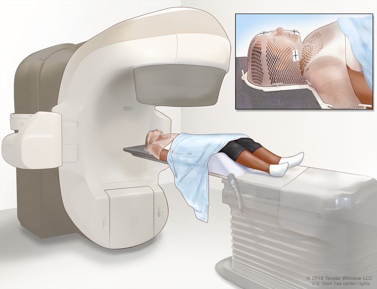
Oropharyngeal Cancer Treatment (PDQ®)–Patient Version

Oropharyngeal Cancer Treatment (PDQ®)–Patient Version

General Information About Oropharyngeal Cancer

Key Points

  • Oropharyngeal cancer is a type of head and neck cancer that starts in the tissues of the oropharynx.
  • Smoking or being infected with human papillomavirus (HPV) can increase the risk of oropharyngeal cancer.
  • Signs and symptoms of oropharyngeal cancer include a lump in the neck and a sore throat.
  • Tests that examine the mouth and throat are used to diagnose and stage oropharyngeal cancer.
  • Some people may decide to get a second opinion.
  • Certain factors affect prognosis (chance of recovery) and treatment options.

Oropharyngeal cancer is a type of head and neck cancer that starts in the tissues of the oropharynx.

The pharynx is a hollow tube in the neck about 5 inches long that is made up of three parts:

  • The nasopharynx is the upper part of the pharynx, located behind the nose. The nostrils are connected to the nasopharynx. Openings on each side of the nasopharynx lead to the ears.
  • The oropharynx is the middle part, located beneath the nasopharynx.
  • The hypopharynx is the lowermost part of the pharynx, opening up to both the trachea (windpipe) and esophagus.

When we breathe or swallow, the pharynx acts as a passageway for air to reach the lungs and food to reach the stomach. Oropharyngeal cancer commonly begins in the squamous cells that line the oropharynx.

EnlargeAnatomy of the pharynx; drawing shows the nasopharynx, oropharynx, and hypopharynx. Also shown are the nasal cavity, oral cavity, hyoid bone, larynx, esophagus, and trachea.
Anatomy of the pharynx. The pharynx is a hollow, muscular tube inside the neck that starts behind the nose and opens into the larynx and esophagus. The three parts of the pharynx are the nasopharynx, oropharynx, and hypopharynx.

The oropharynx includes the:

EnlargeParts of the oropharynx; drawing shows the soft palate, side and back wall of the throat, tonsil, and the back one-third of the tongue.
Parts of the oropharynx. The oropharynx includes the soft palate, the side and back walls of the throat, the tonsils, and the back one-third of the tongue.

Sometimes, a person can have more than one cancer at the same time in the oropharynx and mouth, nose, throat, voice box (larynx), windpipe (trachea), or esophagus.

Smoking or being infected with human papillomavirus (HPV) can increase the risk of oropharyngeal cancer.

Oropharyngeal cancer is caused by certain changes in how oropharyngeal cells function, especially how they grow and divide into new cells. There are many risk factors for oropharyngeal cancer, but many do not directly cause cancer. Instead, they increase the chance of DNA damage in cells that may lead to oropharyngeal cancer. Learn more about how cancer develops at What Is Cancer?

A risk factor is anything that increases a person’s chance of getting a disease. Some risk factors for oropharyngeal cancer, like tobacco use, can be changed. Risk factors also include things you cannot change, like your family history. Learning about risk factors for oropharyngeal cancer can help you make choices that might prevent or lower your risk of getting it.

The most common risk factors for oropharyngeal cancer include:

The number of cases of oropharyngeal cancers linked to HPV infection is increasing. Learn more about HPV and Cancer.

Signs and symptoms of oropharyngeal cancer include a lump in the neck and a sore throat.

Sometimes oropharyngeal cancer does not cause early signs and symptoms. When signs and symptoms occur, they may include:

  • a sore throat that does not go away
  • trouble swallowing
  • trouble opening the mouth fully
  • trouble moving the tongue
  • weight loss for no known reason
  • ear pain
  • a lump in the back of the mouth, throat, or neck
  • a white patch on the tongue or lining of the mouth that does not go away
  • coughing up blood

These problems may be caused by conditions other than oropharyngeal cancer. Check with your doctor if you have any of these problems to find out the cause and begin treatment, if needed.

Tests that examine the mouth and throat are used to diagnose and stage oropharyngeal cancer.

If you have symptoms that suggest oropharyngeal cancer, your doctor will need to find out if these are due to cancer or another problem. They will ask when the symptoms started and how often you have been having them. They will also ask about your personal and family health history and do a physical exam. Based on these results, the doctor may recommend other tests. If you are diagnosed with oropharyngeal cancer, the results of these tests will help you and your doctor plan treatment.

The following tests and procedures are used to diagnose and stage oropharyngeal cancer:

  • During a physical exam of the mouth and neck, the doctor or dentist looks at the mouth and neck, under the tongue, and down the throat with a small, long-handled mirror to check for abnormal areas.
  • A neurological exam uses a series of questions and tests to check the brain, spinal cord, and nerve function. The exam checks your mental status, coordination, and ability to walk normally, and how well the muscles, senses, and reflexes work. This may also be called a neuro exam or a neurologic exam.
  • PET-CT scan combines the pictures from a positron emission tomography (PET) scan and a computed tomography (CT) scan. The PET and CT scans are done at the same time with the same machine. The combined scans give more detailed pictures of areas inside the body than either scan gives by itself.
    • For the PET scan, a small amount of radioactive glucose (sugar) is injected into a vein. The scanner rotates around the body and makes a picture of where glucose is being used in the body. Because cancer cells often take up more glucose than normal cells, the pictures can be used to find cancer cells in the body.
    • For the CT scan, a series of detailed pictures of areas inside the body, such as the head, neck, chest, and lymph nodes, is taken from different angles. A dye is injected into a vein or swallowed to help the organs or tissues show up more clearly.
  • MRI (magnetic resonance imaging) uses a magnet, radio waves, and a computer to make a series of detailed pictures of areas inside the body. This procedure is also called nuclear magnetic resonance imaging (NMRI).
  • Biopsy is the removal of cells or tissues so they can be viewed under a microscope by a pathologist to check for signs of cancer. A fine-needle biopsy is usually done to remove a sample of tissue using a thin needle.

    The following procedures may be used to remove samples of cells or tissue:

    • Endoscopy is a procedure to look at organs and tissues inside the body to check for abnormal areas. An endoscope is inserted through an incision (cut) in the skin or opening in the body, such as the mouth or nose. An endoscope is a thin, tube-like instrument with a light and a lens for viewing. It may also have a tool to remove abnormal tissue or lymph node samples, which are checked under a microscope for signs of disease. The nose, throat, back of the tongue, esophagus, stomach, larynx, windpipe, and large airways will be checked. The type of endoscopy is named for the part of the body that is being examined. For example, pharyngoscopy is an exam to check the pharynx.
    • Laryngoscopy is a procedure in which the doctor checks the larynx (voice box) with a mirror or a laryngoscope to check for abnormal areas. A laryngoscope is a thin, tube-like instrument with a light and a lens for viewing the inside of the throat and voice box. It may also have a tool to remove tissue samples, which are checked under a microscope for signs of cancer.

    If cancer is found, the following test may be done to study the cancer cells:

    • HPV test (human papillomavirus test) is a laboratory test used to check the sample of tissue for certain types of HPV infection, such as HPV type 16. This test is done because oropharyngeal cancer can be caused by HPV infection. This is important because HPV-positive oropharyngeal cancer has a better prognosis and is treated differently than HPV-negative oropharyngeal cancer.

    Learn about the type of information that can be found in a pathologist’s report about the cells or tissue removed during a biopsy at Pathology Reports.

Some people may decide to get a second opinion.

You may want to get a second opinion to confirm your oropharyngeal cancer diagnosis and treatment plan. If you seek a second opinion, you will need to get medical test results and reports from the first doctor to share with the second doctor. The second doctor will review the pathology report, slides, and scans. They may agree with the first doctor, suggest changes or another treatment approach, or provide more information about your cancer.

Learn more about choosing a doctor and getting a second opinion at Finding Cancer Care. You can contact NCI’s Cancer Information Service via chat, email, or phone (both in English and Spanish) for help finding a doctor, hospital, or getting a second opinion. For questions you might want to ask at your appointments, visit Questions to Ask Your Doctor About Cancer.

Certain factors affect prognosis (chance of recovery) and treatment options.

The prognosis depends on:

  • whether the person has HPV infection of the oropharynx
  • whether the person has a history of smoking cigarettes for 10 or more pack years
  • the stage of the cancer
  • the number and size of lymph nodes with cancer

Oropharyngeal tumors related to HPV infection have a better prognosis and are less likely to recur than tumors not linked to HPV infection.

Treatment options depend on:

  • the stage of the cancer
  • keeping the person’s ability to speak and swallow as normal as possible
  • the person’s general health

People with oropharyngeal cancer have an increased risk of another cancer in the head or neck. This risk is increased if a person continues to smoke or drink alcohol after treatment.

For more information, visit Cigarette Smoking: Health Risks and How to Quit.

Stages of Oropharyngeal Cancer

Key Points

  • The following stages are used for HPV-positive oropharyngeal cancer:
    • Stage I (also called stage 1) oropharyngeal cancer
    • Stage II (also called stage 2) oropharyngeal cancer
    • Stage III (also called stage 3) oropharyngeal cancer
    • Stage IV (also called stage 4) oropharyngeal cancer
  • The following stages are used for HPV-negative oropharyngeal cancer:
    • Stage 0 (also called carcinoma in situ of the oropharynx)
    • Stage I (also called stage 1) oropharyngeal cancer
    • Stage II (also called stage 2) oropharyngeal cancer
    • Stage III (also called stage 3) oropharyngeal cancer
    • Stage IV (also called stage 4) oropharyngeal cancer
  • Oropharyngeal cancer can recur (come back) after it has been treated.

Cancer stage describes the extent of cancer in the body.

Cancer stage describes the extent of cancer in the body, such as the size of the tumor, whether it has spread, and how far it has spread from where it first formed. Knowing the cancer stage helps plan treatment. 

There are several staging systems for cancer that describe the extent of the cancer. Oropharyngeal cancer staging usually uses the TNM staging system. The cancer may be described by this staging system in your pathology report. Based on the TNM results, a stage (I, II, III, or IV, also written as 1, 2, 3, or 4) is assigned to the cancer. When talking to you about your diagnosis, your doctor may describe the cancer as one of these stages.

Learn more about Cancer Staging. 

The following stages are used for HPV-positive oropharyngeal cancer:

Stage I (also called stage 1) oropharyngeal cancer

In stage I, one of the following is true:

  • one or more lymph nodes with cancer that is HPV 16–positive are found but the place where the cancer began is not known. The lymph nodes with cancer are 6 centimeters or smaller, on one side of the neck; or
  • cancer is found in the oropharynx (throat) and the tumor is 4 centimeters or smaller. Cancer may have spread to one or more lymph nodes that are 6 centimeters or smaller, on the same side of the neck as the primary tumor.
EnlargeDrawing shows different sizes of a tumor in centimeters (cm) compared to the size of a pea (1 cm), a peanut (2 cm), a grape (3 cm), a walnut (4 cm), a lime (5 cm), an egg (6 cm), a peach (7 cm), and a grapefruit (10 cm). Also shown is a 10-cm ruler and a 4-inch ruler.
Tumor sizes are often measured in centimeters (cm) or inches. Common food items that can be used to show tumor size in cm include: a pea (1 cm), a peanut (2 cm), a grape (3 cm), a walnut (4 cm), a lime (5 cm or 2 inches), an egg (6 cm), a peach (7 cm), and a grapefruit (10 cm or 4 inches).

Stage II (also called stage 2) oropharyngeal cancer

In stage II, one of the following is true:

  • one or more lymph nodes with cancer that is HPV 16–positive are found but the place where the cancer began is not known. The lymph nodes with cancer are 6 centimeters or smaller, on one or both sides of the neck; or
  • the tumor is 4 centimeters or smaller. Cancer has spread to lymph nodes that are 6 centimeters or smaller, on the opposite side of the neck as the primary tumor or on both sides of the neck; or
  • the tumor is larger than 4 centimeters or cancer has spread to the top of the epiglottis (the flap that covers the trachea during swallowing). Cancer may have spread to one or more lymph nodes that are 6 centimeters or smaller, anywhere in the neck.

Stage III (also called stage 3) oropharyngeal cancer

In stage III, one of the following is true:

  • cancer has spread to the larynx (voice box), front part of the roof of the mouth, lower jaw, muscles that move the tongue, or to other parts of the head or neck. Cancer may have spread to lymph nodes in the neck; or
  • the tumor is any size and cancer may have spread to the larynx, front part of the roof of the mouth, lower jaw, muscles that move the tongue, or to other parts of the head or neck. Cancer has spread to one or more lymph nodes that are larger than 6 centimeters, anywhere in the neck.

Stage IV (also called stage 4) oropharyngeal cancer

In stage IV, cancer has spread to other parts of the body, such as the lung or bone.

Stage IV oropharyngeal cancer is also called metastatic oropharyngeal cancer. Metastatic cancer happens when cancer cells travel through the lymphatic system or blood and form tumors in other parts of the body. The metastatic tumor is the same type of cancer as the primary tumor. For example, if oropharyngeal cancer spreads to the lung, the cancer cells in the lung are actually oropharyngeal cancer cells. The disease is called metastatic oropharyngeal cancer, not lung cancer. Learn more in Metastatic Cancer: When Cancer Spreads.

The following stages are used for HPV-negative oropharyngeal cancer:

Stage 0 (also called carcinoma in situ of the oropharynx)

In stage 0, abnormal cells are found in the lining of the oropharynx (throat). These abnormal cells may become cancer and spread into nearby normal tissue.

Stage I (also called stage 1) oropharyngeal cancer

In stage I, cancer has formed. The tumor is 2 centimeters or smaller.

EnlargeDrawing shows different sizes of a tumor in centimeters (cm) compared to the size of a pea (1 cm), a peanut (2 cm), a grape (3 cm), a walnut (4 cm), a lime (5 cm), an egg (6 cm), a peach (7 cm), and a grapefruit (10 cm). Also shown is a 10-cm ruler and a 4-inch ruler.
Tumor sizes are often measured in centimeters (cm) or inches. Common food items that can be used to show tumor size in cm include: a pea (1 cm), a peanut (2 cm), a grape (3 cm), a walnut (4 cm), a lime (5 cm or 2 inches), an egg (6 cm), a peach (7 cm), and a grapefruit (10 cm or 4 inches).

Stage II (also called stage 2) oropharyngeal cancer

In stage II, the tumor is larger than 2 centimeters but not larger than 4 centimeters.

Stage III (also called stage 3) oropharyngeal cancer

In stage III, the cancer:

  • is either larger than 4 centimeters or has spread to the top of the epiglottis (the flap that covers the trachea during swallowing); or
  • is any size. Cancer has spread to one lymph node that is 3 centimeters or smaller, on the same side of the neck as the primary tumor.

Stage IV (also called stage 4) oropharyngeal cancer

Stage IV is divided into stages IVA, IVB, and IVC.

  • In stage IVA, cancer:
    • has spread to the larynx (voice box), front part of the roof of the mouth, lower jaw, or muscles that move the tongue. Cancer may have spread to one lymph node that is 3 centimeters or smaller, on the same side of the neck as the primary tumor; or
    • is any size and may have spread to the top of the epiglottis, larynx, front part of the roof of the mouth, lower jaw, or muscles that move the tongue. Cancer has spread to one of the following:
      • one lymph node that is larger than 3 centimeters but not larger than 6 centimeters, on the same side of the neck as the primary tumor; or
      • more than one lymph node that is 6 centimeters or smaller, anywhere in the neck.
  • In stage IVB, cancer:
    • has spread to the muscle that moves the lower jaw, the bone attached to the muscle that moves the lower jaw, the base of the skull, or to the area behind the nose or around the carotid artery. Cancer may have spread to lymph nodes in the neck; or
    • may be any size and may have spread to other parts of the head or neck. Cancer has spread to a lymph node that is larger than 6 centimeters or has spread through the outside covering of a lymph node into nearby connective tissue.
  • In stage IVC, cancer has spread to other parts of the body, such as the lung, liver, or bone.

Stage IV oropharyngeal cancer is also called metastatic oropharyngeal cancer. Metastatic cancer happens when cancer cells travel through the lymphatic system or blood and form tumors in other parts of the body. The metastatic tumor is the same type of cancer as the primary tumor. For example, if oropharyngeal cancer spreads to the lung, the cancer cells in the lung are actually oropharyngeal cancer cells. The disease is called metastatic oropharyngeal cancer, not lung cancer. Learn more in Metastatic Cancer: When Cancer Spreads.

Oropharyngeal cancer can recur (come back) after it has been treated.

Recurrent oropharyngeal cancer is cancer that has come back after it has been treated. If oropharyngeal cancer comes back, it may come back in the oropharynx, lymph nodes, or other parts of the body, such as the lungs, bone, or liver. Tests will help determine where in the body the cancer has returned. The type of treatment that you have for recurrent oropharyngeal cancer will depend on where it has come back.

Learn more in Recurrent Cancer: When Cancer Comes Back.

Treatment Option Overview

Key Points

  • There are different types of treatment for people with oropharyngeal cancer.
  • People with oropharyngeal cancer should have their treatment planned by a team of doctors with expertise in treating head and neck cancer.
  • The following types of treatment are used:
    • Surgery
    • Radiation therapy
    • Chemotherapy
    • Targeted therapy
    • Immunotherapy
  • New types of treatment are being tested in clinical trials.
  • Treatment for oropharyngeal cancer may cause side effects.
  • Follow-up care may be needed.

There are different types of treatment for people with oropharyngeal cancer.

Different types of treatments are available for oropharyngeal cancer. You and your cancer care team will work together to decide your treatment plan, which may include more than one type of treatment. Many factors will be considered, such as the stage of the cancer, your overall health, and your preferences. Your plan will include information about your cancer, the goals of treatment, your treatment options and the possible side effects, and the expected length of treatment. 

Talking with your cancer care team before treatment begins about what to expect will be helpful. You’ll want to learn what you need to do before treatment begins, how you’ll feel while going through it, and what kind of help you will need. Learn more at Questions to Ask Your Doctor About Your Treatment.   

People with oropharyngeal cancer should have their treatment planned by a team of doctors with expertise in treating head and neck cancer.

An oncologist, a doctor who specializes in treating people with cancer, oversees treatment for oropharyngeal cancer. Because the oropharynx helps in breathing, eating, and talking, you may need help adjusting to the side effects of the cancer and its treatment. The oncologist may refer you to other health care providers who are experts in treating head and neck cancer and also specialize in other areas of medicine. Other specialists may include:

The following types of treatment are used:

Surgery

Surgery to remove the tumor is a common treatment for all stages of oropharyngeal cancer. A surgeon may remove the cancer and some of the healthy tissue around the cancer. After the surgeon removes all the cancer that can be seen at the time of the surgery, some people may be given chemotherapy or radiation therapy after surgery to kill any cancer cells that are left. Treatment given after the surgery, to lower the risk that the cancer will come back, is called adjuvant therapy.

New types of surgery, including transoral robotic surgery, are being studied for the treatment of oropharyngeal cancer. Transoral robotic surgery may be used to remove cancer from hard-to-reach areas of the mouth and throat. Cameras attached to a robot give a 3-dimensional (3D) image that a surgeon can see. Using a computer, the surgeon guides very small tools at the ends of the robot arms to remove the cancer. This procedure may also be done using an endoscope.

Learn more about Surgery to Treat Cancer.

Radiation therapy

Radiation therapy uses high-energy x-rays or other types of radiation to kill cancer cells or keep them from growing by damaging their DNA. External radiation therapy uses a machine outside the body to send radiation toward the area of the body with cancer.

EnlargeExternal-beam radiation therapy of the head and neck; drawing shows a patient lying on a table under a machine that is used to aim high-energy radiation at the cancer. An inset shows a mesh mask that helps keep the patient's head and neck from moving during treatment. The mask has pieces of white tape with small ink marks on it. The ink marks are used to line up the radiation machine in the same position before each treatment.
External-beam radiation therapy of the head and neck. A machine is used to aim high-energy radiation at the cancer. The machine can rotate around the patient, delivering radiation from many different angles to provide highly conformal treatment. A mesh mask helps keep the patient’s head and neck from moving during treatment. Small ink marks are put on the mask. The ink marks are used to line up the radiation machine in the same position before each treatment.

Certain ways of giving radiation therapy can help keep radiation from damaging nearby healthy tissue. These types of radiation therapy include:

  • Intensity-modulated radiation therapy (IMRT): IMRT is a type of 3-dimensional (3-D) radiation therapy that uses a computer to make pictures of the size and shape of the tumor. Thin beams of radiation of different intensities (strengths) are aimed at the tumor from many angles.
  • Stereotactic body radiation therapy: Stereotactic body radiation therapy is a type of external radiation therapy. Special equipment is used to place the person in the same position for each radiation treatment. Once a day for several days, a radiation machine aims a larger than usual dose of radiation directly at the tumor. By having the person in the same position for each treatment, there is less damage to nearby healthy tissue. This procedure is also called stereotactic external-beam radiation therapy and stereotaxic radiation therapy.

In advanced oropharyngeal cancer, dividing the daily dose of radiation into smaller-dose treatments improves the way the tumor responds to treatment. This is called hyperfractionated radiation therapy.

Radiation therapy may work better in people who have stopped smoking before beginning treatment.

If the thyroid or pituitary gland are part of the radiation treatment area, the person has an increased risk of hypothyroidism (too little thyroid hormone). A blood test to check the thyroid hormone level in the body should be done before and after treatment.

Learn more about External Beam Radiation Therapy for Cancer and Radiation Therapy Side Effects.

Chemotherapy

Chemotherapy uses drugs to stop the growth of cancer cells, either by killing the cells or by stopping them from dividing. Chemotherapy for oropharyngeal cancer is usually systemic, meaning it is taken by mouth or injected into a vein or muscle. When given this way, the drugs enter the bloodstream and can reach cancer cells throughout the body.

Chemotherapy drugs used to treat oropharyngeal cancer include:

Combinations of these drugs may be used. Other chemotherapy drugs not listed here may also be used.

Chemotherapy may be combined with other types of treatment, such as radiation therapy.

To learn more about how chemotherapy works, how it is given, common side effects, and more, visit Chemotherapy to Treat Cancer and Chemotherapy and You: Support for People With Cancer.

Targeted therapy

Targeted therapy uses drugs or other substances to identify and attack specific cancer cells. Cetuximab is a type of targeted therapy used to treat recurrent and metastatic oropharyngeal cancer.

Learn more about Targeted Therapy to Treat Cancer. 

Immunotherapy

Immunotherapy is a treatment that uses a person’s immune system to fight cancer. Your doctor may suggest biomarker tests to help predict your response to certain immunotherapy drugs. Learn more about Biomarker Testing for Cancer Treatment.   

Pembrolizumab and nivolumab are types of immunotherapy used to treat metastatic or recurrent oropharyngeal cancer.

Learn more about Immunotherapy to Treat Cancer.

New types of treatment are being tested in clinical trials.

For some people, joining a clinical trial may be an option. There are different types of clinical trials for people with cancer. For example, a treatment trial tests new treatments or new ways of using current treatments. Supportive care and palliative care trials look at ways to improve quality of life, especially for those who have side effects from cancer and its treatment.

You can use the clinical trial search to find NCI-supported cancer clinical trials accepting participants. The search allows you to filter trials based on the type of cancer, your age, and where the trials are being done. Clinical trials supported by other organizations can be found on the ClinicalTrials.gov website.

Learn more about clinical trials, including how to find and join one, at Clinical Trials Information for Patients and Caregivers.

Treatment for oropharyngeal cancer may cause side effects.

For information about side effects caused by treatment for cancer, visit our Side Effects page.

Follow-up care may be needed.

As you go through treatment, you will have follow-up tests or check-ups. Some tests that were done to diagnose or stage the cancer may be repeated to see how well the treatment is working. Decisions about whether to continue, change, or stop treatment may be based on the results of these tests.

Some of the tests will continue to be done from time to time after treatment has ended. The results of these tests can show if your condition has changed or if the cancer has recurred (come back).

After treatment is complete, it is important to have head and neck exams to look for signs that the cancer has come back. Check-ups will be done every 6 to 12 weeks in the first year, every 3 months in the second year, every 3 to 4 months in the third year, and every 6 months thereafter.

Treatment of Stage I and Stage II Oropharyngeal Cancer

For information about the treatments listed below, see the Treatment Option Overview section.

Treatment of newly diagnosed stage I and stage II oropharyngeal cancer may include:

Use our clinical trial search to find NCI-supported cancer clinical trials that are accepting patients. You can search for trials based on the type of cancer, the age of the patient, and where the trials are being done. General information about clinical trials is also available.

Treatment of Stage III and Nonmetastatic Stage IV Oropharyngeal Cancer

For information about the treatments listed below, see the Treatment Option Overview section.

Treatment of newly diagnosed stage III oropharyngeal cancer and stage IV oropharyngeal cancer may include:

  • radiation therapy with or without chemotherapy given at the same time, for people with locally advanced cancer
  • radiation therapy alone for people who cannot have chemotherapy
  • chemotherapy given at the same time as radiation therapy
  • chemotherapy followed by radiation therapy given at the same time as more chemotherapy
  • a clinical trial of immunotherapy (nivolumab) with chemotherapy given at the same time as radiation therapy in people with cancer that has a higher risk of coming back
  • a clinical trial of radiation therapy with or without chemotherapy
  • a clinical trial of transoral surgery followed by standard- or low-dose radiation therapy with or without chemotherapy in people with HPV-positive oropharyngeal cancer

Use our clinical trial search to find NCI-supported cancer clinical trials that are accepting patients. You can search for trials based on the type of cancer, the age of the patient, and where the trials are being done. General information about clinical trials is also available.

Treatment of Metastatic Stage IV and Recurrent Oropharyngeal Cancer

For information about the treatments listed below, see the Treatment Option Overview section.

Treatment of oropharyngeal cancer that has metastasized or recurred in the oropharynx may include:

Use our clinical trial search to find NCI-supported cancer clinical trials that are accepting patients. You can search for trials based on the type of cancer, the age of the patient, and where the trials are being done. General information about clinical trials is also available.

To Learn More About Oropharyngeal Cancer

About This PDQ Summary

About PDQ

Physician Data Query (PDQ) is the National Cancer Institute’s (NCI’s) comprehensive cancer information database. The PDQ database contains summaries of the latest published information on cancer prevention, detection, genetics, treatment, supportive care, and complementary and alternative medicine. Most summaries come in two versions. The health professional versions have detailed information written in technical language. The patient versions are written in easy-to-understand, nontechnical language. Both versions have cancer information that is accurate and up to date and most versions are also available in Spanish.

PDQ is a service of the NCI. The NCI is part of the National Institutes of Health (NIH). NIH is the federal government’s center of biomedical research. The PDQ summaries are based on an independent review of the medical literature. They are not policy statements of the NCI or the NIH.

Purpose of This Summary

This PDQ cancer information summary has current information about the treatment of oropharyngeal cancer. It is meant to inform and help patients, families, and caregivers. It does not give formal guidelines or recommendations for making decisions about health care.

Reviewers and Updates

Editorial Boards write the PDQ cancer information summaries and keep them up to date. These Boards are made up of experts in cancer treatment and other specialties related to cancer. The summaries are reviewed regularly and changes are made when there is new information. The date on each summary (“Updated”) is the date of the most recent change.

The information in this patient summary was taken from the health professional version, which is reviewed regularly and updated as needed, by the PDQ Adult Treatment Editorial Board.

Clinical Trial Information

A clinical trial is a study to answer a scientific question, such as whether one treatment is better than another. Trials are based on past studies and what has been learned in the laboratory. Each trial answers certain scientific questions in order to find new and better ways to help cancer patients. During treatment clinical trials, information is collected about the effects of a new treatment and how well it works. If a clinical trial shows that a new treatment is better than one currently being used, the new treatment may become “standard.” Patients may want to think about taking part in a clinical trial. Some clinical trials are open only to patients who have not started treatment.

Clinical trials can be found online at NCI’s website. For more information, call the Cancer Information Service (CIS), NCI’s contact center, at 1-800-4-CANCER (1-800-422-6237).

Permission to Use This Summary

PDQ is a registered trademark. The content of PDQ documents can be used freely as text. It cannot be identified as an NCI PDQ cancer information summary unless the whole summary is shown and it is updated regularly. However, a user would be allowed to write a sentence such as “NCI’s PDQ cancer information summary about breast cancer prevention states the risks in the following way: [include excerpt from the summary].”

The best way to cite this PDQ summary is:

PDQ® Adult Treatment Editorial Board. PDQ Oropharyngeal Cancer Treatment. Bethesda, MD: National Cancer Institute. Updated <MM/DD/YYYY>. Available at: /types/head-and-neck/patient/adult/oropharyngeal-treatment-pdq. Accessed <MM/DD/YYYY>. [PMID: 26389310]

Images in this summary are used with permission of the author(s), artist, and/or publisher for use in the PDQ summaries only. If you want to use an image from a PDQ summary and you are not using the whole summary, you must get permission from the owner. It cannot be given by the National Cancer Institute. Information about using the images in this summary, along with many other images related to cancer can be found in Visuals Online. Visuals Online is a collection of more than 3,000 scientific images.

Disclaimer

The information in these summaries should not be used to make decisions about insurance reimbursement. More information on insurance coverage is available on Cancer.gov on the Managing Cancer Care page.

Contact Us

More information about contacting us or receiving help with the Cancer.gov website can be found on our Contact Us for Help page. Questions can also be submitted to Cancer.gov through the website’s E-mail Us.

Nasopharyngeal Cancer Treatment (PDQ®)–Patient Version

Nasopharyngeal Cancer Treatment (PDQ®)–Patient Version

General Information About Nasopharyngeal Cancer

Key Points

  • Nasopharyngeal cancer is a type of head and neck cancer that starts in the tissues of the nasopharynx.
  • Being exposed to the Epstein-Barr virus, your ancestry, and where you live can affect the risk of nasopharyngeal cancer.
  • Signs and symptoms of nasopharyngeal cancer include trouble breathing, speaking, or hearing.
  • Tests that examine the nose, throat, and nearby organs are used to diagnose and stage nasopharyngeal cancer.
  • Some people may decide to get a second opinion.
  • Certain factors affect prognosis (chance of recovery) and treatment options.

Nasopharyngeal cancer is a type of head and neck cancer that starts in the tissues of the nasopharynx.

The pharynx is a hollow tube in the neck about 5 inches long that is made up of three parts:

  • The nasopharynx is the upper part of the pharynx, located behind the nose. The nostrils are connected to the nasopharynx. Openings on each side of the nasopharynx lead to the ears.
  • The oropharynx is the middle part, located beneath the nasopharynx.
  • The hypopharynx is the lowermost part of the pharynx, opening up to both the trachea (windpipe) and esophagus.

When we breathe or swallow, the pharynx acts as a passageway for air to reach the lungs and food to reach the stomach. Nasopharyngeal cancer commonly begins in the squamous cells that line the nasopharynx.

EnlargeAnatomy of the pharynx; drawing shows the nasopharynx, oropharynx, and hypopharynx. Also shown are the nasal cavity, oral cavity, hyoid bone, larynx, esophagus, and trachea.
Anatomy of the pharynx. The pharynx is a hollow, muscular tube inside the neck that starts behind the nose and opens into the larynx and esophagus. The three parts of the pharynx are the nasopharynx, oropharynx, and hypopharynx.

Being exposed to the Epstein-Barr virus, your ancestry, and where you live can affect the risk of nasopharyngeal cancer.

Nasopharyngeal cancer is caused by certain changes in how nasopharyngeal cells function, especially how they grow and divide into new cells. There are many risk factors for nasopharyngeal cancer, but many do not directly cause cancer. Instead, they increase the chance of DNA damage in cells that may lead to nasopharyngeal cancer. Learn more about how cancer develops at What Is Cancer?

A risk factor is anything that increases a person’s chance of getting a disease. Some risk factors for nasopharyngeal cancer, like tobacco use, can be changed. Risk factors also include things you cannot change, like your family history. Learning about risk factors for nasopharyngeal cancer can help you make choices that might prevent or lower your risk of getting it.

Risk factors for nasopharyngeal cancer include:

  • being infected with Epstein-Barr virus (EBV)
  • living in or having ancestry in certain parts of Asia, North Africa, and the Arctic
  • having a family member with nasopharyngeal cancer
  • using tobacco or breathing in secondhand smoke
  • frequent and heavy alcohol use
  • having a diet high in salt-cured fish and meats because these foods may contain cancer-causing chemicals, such as nitrosamine

Nasopharyngeal cancer can occur at any age. In areas where the disease is not common, it is more likely to be diagnosed in people who are older than 50 years. In high-risk areas, younger people are more likely to be affected. Men tend to develop nasopharyngeal cancer more often than women. In rare cases, human papillomavirus (HPV), especially HPV type 16, has been linked to nasopharyngeal cancer. Learn about HPV and Cancer.

Learn more about Tobacco, including help with quitting.

Signs and symptoms of nasopharyngeal cancer include trouble breathing, speaking, or hearing.

The signs and symptoms of nasopharyngeal cancer can vary from person to person. Early signs and symptoms of nasopharyngeal cancer may include:

  • a lump in the neck
  • pain, pulsing, or ringing in the ear
  • trouble hearing
  • a sore throat
  • stuffy nose
  • nosebleeds

Signs and symptoms of advanced nasopharyngeal cancer (nasopharyngeal cancer that has spread to other parts of the body) may include symptoms of early-stage nasopharyngeal cancer and:

  • misalignment of the eyes (strabismus)
  • double vision
  • headaches
  • facial numbness
  • facial weakness

These problems may be caused by conditions other than nasopharyngeal cancer. Check with your doctor if you have any of these problems to find out the cause and begin treatment, if needed.

Tests that examine the nose, throat, and nearby organs are used to diagnose and stage nasopharyngeal cancer.

If you have symptoms that suggest nasopharyngeal cancer, your doctor will need to find out if these are due to cancer or another problem. They will ask when the symptoms started and how often you have been having them. They will also ask about your personal and family health history and do a physical exam. Based on these results, the doctor may recommend other tests. If you are diagnosed with nasopharyngeal cancer, the results of these tests will help you and your doctor plan treatment.

The following tests and procedures are used to diagnose and stage nasopharyngeal cancer:

  • Nasopharyngoscopy with biopsy is a procedure to examine the inside of the nose and back of the throat. The doctor inserts a nasopharyngoscope (a thin, flexible lighted tube) in the nose and advances it to the back of the throat to check for abnormal areas. The nasopharyngoscope may have a tool to remove a sample of cells or tissue (biopsy) so a pathologist can view it under a microscope to check for signs of cancer. Learn about the type of information that can be found in a pathologist’s report about the cells or tissue removed during a biopsy at Pathology Reports.
  • MRI (magnetic resonance imaging) uses a magnet, radio waves, and a computer to make a series of detailed pictures of areas inside the body. This procedure is also called nuclear magnetic resonance imaging (NMRI).
  • PET-CT scan combines pictures from a positron emission tomography (PET) scan and a computed tomography (CT) scan. The PET and CT scans are done at the same time on the same machine. The combined pictures make a more detailed picture than either test would make by itself.
    • For the PET scan, a small amount of radioactive glucose (sugar) is injected into a vein. The scanner rotates around the body and makes a picture of where glucose is being used in the body. Because cancer cells often take up more glucose than normal cells, the pictures can be used to find cancer cells in the body.
    • For the CT scan, a series of detailed x-ray pictures of areas inside the body is taken from different angles. A dye may be injected into a vein or swallowed to help the organs or tissues show up more clearly.
  • Epstein-Barr virus (EBV) test is a blood test to check for antibodies and DNA markers that are found in the blood of people who have been infected with EBV.
  • Human papillomavirus (HPV) test is a laboratory test used to check a sample of tissue for certain types of HPV infection. An HPV test is usually done if the EBV test is negative. This is because in rare cases, HPV can cause nasopharyngeal cancer.
  • A neurological exam uses a series of questions and tests to check brain, spinal cord, and nerve function. The exam checks your mental status, coordination, and ability to walk normally, and how well the muscles, senses, and reflexes work. This may also be called a neuro exam or a neurologic exam.
  • A hearing test checks your ability to hear soft and loud sounds and low- and high-pitched sounds. Each ear is checked separately. This test is done because nasopharyngeal cancer and its treatment can affect hearing. Hearing tests are usually done before, during, and after treatment.
  • Blood chemistry study is a laboratory test in which a blood sample is checked to measure the amounts of certain substances released into the blood by organs and tissues in the body. An unusual (higher or lower than normal) amount of a substance can be a sign of disease.
  • Complete blood count (CBC) is a laboratory test in which a sample of blood is drawn and checked for:
    • the number of red blood cells, white blood cells, and platelets
    • the amount of hemoglobin (the substance in the blood that carries oxygen) in the red blood cells
    • the amount of hematocrit (whole blood that is made up of red blood cells)
    EnlargeComplete blood count (CBC); left panel shows blood being drawn from a vein on the inside of the elbow using a tube attached to a syringe; right panel shows a laboratory test tube with blood cells separated into layers: plasma, white blood cells, platelets, and red blood cells.
    Complete blood count (CBC). Blood is collected by inserting a needle into a vein and allowing the blood to flow into a tube. The blood sample is sent to the laboratory and the red blood cells, white blood cells, and platelets are counted. The CBC is used to test for, diagnose, and monitor many different conditions.

Some people may decide to get a second opinion.

You may want to get a second opinion to confirm your nasopharyngeal cancer diagnosis and treatment plan. If you seek a second opinion, you will need to get medical test results and reports from the first doctor to share with the second doctor. The second doctor will review the pathology report, slides, and scans. They may agree with the first doctor, suggest changes or another treatment approach, or provide more information about your cancer.

Learn more about choosing a doctor and getting a second opinion at Finding Cancer Care. You can contact NCI’s Cancer Information Service via chat, email, or phone (both in English and Spanish) for help finding a doctor, hospital, or getting a second opinion. For questions you might want to ask at your appointments, visit Questions to Ask Your Doctor About Cancer.

Certain factors affect prognosis (chance of recovery) and treatment options.

The prognosis and treatment options depend on:

  • the size of the tumor
  • the stage of the cancer, including whether cancer has spread to one or more lymph nodes in the neck
  • whether there are high levels of EBV antibodies and EBV-DNA markers in the blood before and after treatment

Stages of Nasopharyngeal Cancer

Key Points

  • Cancer stage describes the extent of cancer in the body.
  • The following stages are used for nasopharyngeal cancer:
    • Stage 0 (also called carcinoma in situ of the nasopharynx)
    • Stage I (also called stage 1) nasopharyngeal cancer
    • Stage II (also called stage 2) nasopharyngeal cancer
    • Stage III (also called stage 3) nasopharyngeal cancer
    • Stage IV (also called stage 4) nasopharyngeal cancer
  • Nasopharyngeal cancer can recur (come back) after it has been treated.

Cancer stage describes the extent of cancer in the body.

Cancer stage describes the extent of cancer in the body, such as the size of the tumor, whether it has spread, and how far it has spread from where it first formed. Knowing the cancer stage helps plan treatment. 

There are several staging systems for cancer that describe the extent of the cancer. Nasopharyngeal cancer staging usually uses the TNM staging system. The cancer may be described by this staging system in your pathology report. Based on the TNM results, a stage (I, II, III, or IV, also written as 1, 2, 3, or 4) is assigned to the cancer. When talking to you about your diagnosis, your doctor may describe the cancer as one of these stages.

Learn more about Cancer Staging. 

The following stages are used for nasopharyngeal cancer:

Stage 0 (also called carcinoma in situ of the nasopharynx)

In stage 0, abnormal cells are found in the lining of the nasopharynx. These abnormal cells may become cancer and spread into nearby normal tissue.

Stage I (also called stage 1) nasopharyngeal cancer

In stage I, cancer has formed, and the cancer:

EnlargeDrawing shows different sizes of a tumor in centimeters (cm) compared to the size of a pea (1 cm), a peanut (2 cm), a grape (3 cm), a walnut (4 cm), a lime (5 cm), an egg (6 cm), a peach (7 cm), and a grapefruit (10 cm). Also shown is a 10-cm ruler and a 4-inch ruler.
Tumor sizes are often measured in centimeters (cm) or inches. Common food items that can be used to show tumor size in cm include: a pea (1 cm), a peanut (2 cm), a grape (3 cm), a walnut (4 cm), a lime (5 cm or 2 inches), an egg (6 cm), a peach (7 cm), and a grapefruit (10 cm or 4 inches).

Stage II (also called stage 2) nasopharyngeal cancer

In stage II, one of the following is true:

  • Cancer has spread to one or more lymph nodes on one side of the neck and/or to one or more lymph nodes on one or both sides of the back of the throat. The affected lymph nodes are 6 centimeters or smaller. Cancer is found:
    • in the nasopharynx only or has spread from the nasopharynx to the oropharynx and/or to the nasal cavity; or
    • only in the lymph nodes in the neck. The cancer cells in the lymph nodes are infected with Epstein-Barr virus (a virus linked to nasopharyngeal cancer). Cancer was not found in the nasopharynx.
  • Cancer has spread to the parapharyngeal space and/or nearby muscles. Cancer may have also spread to one or more lymph nodes on one side of the neck and/or to one or more lymph nodes on one or both sides of the back of the throat. The affected lymph nodes are 6 centimeters or smaller.

Stage III (also called stage 3) nasopharyngeal cancer

In stage III, one of the following is true:

  • Cancer has spread to one or more lymph nodes on both sides of the neck. The affected lymph nodes are 6 centimeters or smaller. Cancer is found:
  • Cancer has spread to the parapharyngeal space and/or nearby muscles. Cancer has also spread to one or more lymph nodes on both sides of the neck. The affected lymph nodes are 6 centimeters or smaller.
  • Cancer has spread to the bones at the bottom of the skull, the bones in the neck, jaw muscles, and/or the sinuses around the nose and eyes. Cancer may have also spread to one or more lymph nodes on one or both sides of the neck and/or the back of the throat. The affected lymph nodes are 6 centimeters or smaller.

Stage IV (also called stage 4) nasopharyngeal cancer

Stage IV is divided into stages IVA and IVB.

  • In stage IVA:
    • Cancer has spread to the brain, the cranial nerves, the hypopharynx, the salivary gland in the front of the ear, the bone around the eye, and/or the soft tissues of the jaw. Cancer may have also spread to one or more lymph nodes on one or both sides of the neck and/or the back of the throat. The affected lymph nodes are 6 centimeters or smaller; or
    • Cancer has spread to one or more lymph nodes on one or both sides of the neck. The affected lymph nodes are larger than 6 centimeters and/or are found in the lowest part of the neck.
  • In stage IVB: Cancer has spread beyond the lymph nodes in the neck to distant lymph nodes, such as those between the lungs, below the collarbone, or in the armpit or groin, or to other parts of the body, such as the lung, bone, or liver.

Stage IV nasopharyngeal cancer is also called metastatic nasopharyngeal cancer. Metastatic cancer happens when cancer cells travel through the lymphatic system or blood and form tumors in other parts of the body. The metastatic tumor is the same type of cancer as the primary tumor. For example, if nasopharyngeal cancer spreads to the lung, the cancer cells in the lung are actually nasopharyngeal cancer cells. The disease is called metastatic nasopharyngeal cancer, not lung cancer. Learn more in Metastatic Cancer: When Cancer Spreads.

Nasopharyngeal cancer can recur (come back) after it has been treated.

Recurrent nasopharyngeal cancer is cancer that has come back after it has been treated. If nasopharyngeal cancer comes back, it may come back in the nasopharynx, lymph nodes, or other parts of the body, such as the lungs, bone, or liver. Tests will help determine where in the body the cancer has returned. The type of treatment that you have for recurrent nasopharyngeal cancer will depend on where it has come back.

Learn more in Recurrent Cancer: When Cancer Comes Back.

Treatment Option Overview

Key Points

  • There are different types of treatment for people with nasopharyngeal cancer.
  • People with nasopharyngeal cancer should have their treatment planned by a team of doctors with expertise in treating head and neck cancer.
  • The following types of treatment are used:
    • Radiation therapy
    • Chemotherapy
    • Surgery
    • Immunotherapy
  • Treatment for nasopharyngeal cancer may cause side effects.
  • New types of treatment are being tested in clinical trials.
  • Follow-up care may be needed.

There are different types of treatment for people with nasopharyngeal cancer.

Different types of treatments are available for nasopharyngeal cancer. You and your cancer care team will work together to decide your treatment plan, which may include more than one type of treatment. Many factors will be considered, such as the stage of the cancer, your overall health, and your preferences. Your plan will include information about your cancer, the goals of treatment, your treatment options and the possible side effects, and the expected length of treatment. 

Talking with your cancer care team before treatment begins about what to expect will be helpful. You’ll want to learn what you need to do before treatment begins, how you’ll feel while going through it, and what kind of help you will need. Learn more at Questions to Ask Your Doctor About Your Treatment.   

People with nasopharyngeal cancer should have their treatment planned by a team of doctors with expertise in treating head and neck cancer.

An oncologist, a doctor who specializes in treating people with cancer, oversees treatment for nasopharyngeal cancer. Because the nasopharynx helps in breathing, eating, and talking, you may need help adjusting to the side effects of the cancer and its treatment. The oncologist may refer you to other health care providers who are experts in treating head and neck cancer and also specialize in other areas of medicine. Other specialists may include:

The following types of treatment are used:

Radiation therapy

Radiation therapy uses high-energy x-rays or other types of radiation to kill cancer cells or keep them from growing by damaging their DNA. The way radiation therapy is given depends on the type and stage of the cancer. External and internal radiation therapy are used to treat nasopharyngeal cancer.

  • External radiation therapy uses a machine outside the body to send radiation toward the area of the body with cancer.
    EnlargeExternal-beam radiation therapy of the head and neck; drawing shows a patient lying on a table under a machine that is used to aim high-energy radiation at the cancer. An inset shows a mesh mask that helps keep the patient's head and neck from moving during treatment. The mask has pieces of white tape with small ink marks on it. The ink marks are used to line up the radiation machine in the same position before each treatment.
    External-beam radiation therapy of the head and neck. A machine is used to aim high-energy radiation at the cancer. The machine can rotate around the patient, delivering radiation from many different angles to provide highly conformal treatment. A mesh mask helps keep the patient’s head and neck from moving during treatment. Small ink marks are put on the mask. The ink marks are used to line up the radiation machine in the same position before each treatment.

    Certain ways of giving radiation therapy can help keep radiation from damaging nearby healthy tissue. These include:

    • Intensity-modulated radiation therapy (IMRT): IMRT is a type of 3-dimensional (3-D) radiation therapy that uses a computer to make pictures of the size and shape of the tumor. Thin beams of radiation of different intensities (strengths) are aimed at the tumor from many angles. Compared to standard radiation therapy, intensity-modulated radiation therapy may be less likely to cause dry mouth. You will likely have treatment once a day, Monday through Friday, for about 6 to 7 weeks.
    • Stereotactic radiation therapy: Stereotactic radiation therapy also uses a computer to make detailed images of the tumor. Thin beams of radiation are aimed at the tumor from different angles. High-dose radiation is given in one to five sessions spread over several days. This procedure is also called stereotactic external-beam radiation and stereotaxic radiation therapy.

    External radiation therapy to the thyroid or the pituitary gland may change the way the thyroid gland works. A blood test to check the thyroid hormone level in the blood is done before and after therapy to make sure the thyroid gland is working properly. It is also important that a dentist check your teeth, gums, and mouth, and fix any existing problems before radiation therapy begins.

  • Internal radiation therapy (also called brachytherapy) uses a radioactive substance sealed in needles, seeds, wires, or catheters that are placed directly into or near the cancer. In some cases, it may be used with external radiation therapy to deliver an extra dose of radiation directly to the tumor. Learn more about Brachytherapy to Treat Cancer.

Chemotherapy

Chemotherapy (also called chemo) uses drugs to stop the growth of cancer cells, either by killing the cells or by stopping them from dividing. Chemotherapy for nasopharyngeal cancer is usually systemic, meaning it is taken by mouth or injected into a vein or muscle. When given this way, the drugs enter the bloodstream and can reach cancer cells throughout the body.

Chemotherapy drugs used to treat nasopharyngeal cancer include:

Combinations of these drugs may be used. Other chemotherapy drugs not listed here may also be used.

Chemotherapy may be combined with other types of treatment, such as radiation therapy.

To learn more about how chemotherapy works, how it is given, common side effects, and more, visit Chemotherapy to Treat Cancer and Chemotherapy and You: Support for People With Cancer.

Surgery

Surgery to remove the tumor is sometimes used for nasopharyngeal cancer that does not respond to radiation therapy. If cancer has spread to the lymph nodes, the doctor may remove lymph nodes and other tissues in the neck.

Learn more about Surgery to Treat Cancer.

Immunotherapy

Immunotherapy is a treatment that uses the patient’s immune system to fight cancer. Substances made by the body or made in a laboratory are used to boost, direct, or restore the body’s natural defenses against cancer.

  • PD-1 and PD-L1 inhibitor therapy: PD-1 is a protein on the surface of T cells that helps keep the body’s immune responses in check. PD-L1 is a protein found on some types of cancer cells. When PD-1 attaches to PD-L1, it stops the T cell from killing the cancer cell. PD-1 and PD-L1 inhibitors keep PD-1 and PD-L1 proteins from attaching to each other. This allows the T cells to kill cancer cells. Toripalimab is a type of PD-1 inhibitor used to treat metastatic or recurrent nasopharyngeal cancer.
EnlargeImmune checkpoint inhibitor; the panel on the left shows the binding of proteins PD-L1 (on the tumor cell) to PD-1 (on the T cell), which keeps T cells from killing tumor cells in the body. Also shown are a tumor cell antigen and T cell receptor. The panel on the right shows immune checkpoint inhibitors (anti-PD-L1 and anti-PD-1) blocking the binding of PD-L1 to PD-1, which allows the T cells to kill tumor cells.
Immune checkpoint inhibitor. Checkpoint proteins, such as PD-L1 on tumor cells and PD-1 on T cells, help keep immune responses in check. The binding of PD-L1 to PD-1 keeps T cells from killing tumor cells in the body (left panel). Blocking the binding of PD-L1 to PD-1 with an immune checkpoint inhibitor (anti-PD-L1 or anti-PD-1) allows the T cells to kill tumor cells (right panel).
Immunotherapy uses the body’s immune system to fight cancer. This animation explains one type of immunotherapy that uses immune checkpoint inhibitors to treat cancer.

Learn more about Immunotherapy to Treat Cancer.

Treatment for nasopharyngeal cancer may cause side effects.

For information about side effects caused by treatment for cancer, visit our Side Effects page.

Side effects from cancer treatment that begin after treatment and continue for months or years are called late effects. Late effects of nasopharyngeal cancer treatment may include:

  • chronic dry mouth
  • dental and oral complications
  • hearing loss
  • vision loss
  • difficulty swallowing
  • lockjaw
  • problems with the thyroid and pituitary gland
  • damage to nerves in the brain
  • changes in mood, feelings, thinking, learning, or memory

Some late effects may be treated or controlled. It is important to talk with your doctor about possible late effects caused by some treatments.

New types of treatment are being tested in clinical trials.

For some people, joining a clinical trial may be an option. There are different types of clinical trials for people with cancer. For example, a treatment trial tests new treatments or new ways of using current treatments. Supportive care and palliative care trials look at ways to improve quality of life, especially for those who have side effects from cancer and its treatment.

You can use the clinical trial search to find NCI-supported cancer clinical trials accepting participants. The search allows you to filter trials based on the type of cancer, your age, and where the trials are being done. Clinical trials supported by other organizations can be found on the ClinicalTrials.gov website.

Learn more about clinical trials, including how to find and join one, at Clinical Trials Information for Patients and Caregivers.

Follow-up care may be needed.

As you go through treatment, you will have follow-up tests or check-ups. Some tests that were done to diagnose or stage the cancer may be repeated to see how well the treatment is working. Decisions about whether to continue, change, or stop treatment may be based on the results of these tests.

Some of the tests will continue to be done from time to time after treatment has ended. The results of these tests can show if your condition has changed or if the cancer has recurred (come back).

After treatment is complete, it is important to have head and neck exams to look for signs that the cancer has come back.

Treatment of Stage I Nasopharyngeal Cancer

Learn more about these treatments in the Treatment Option Overview.

Treatment of stage I nasopharyngeal cancer is usually radiation therapy to the tumor and lymph nodes in the neck.

Use our clinical trial search to find NCI-supported cancer clinical trials that are accepting patients. You can search for trials based on the type of cancer, the age of the patient, and where the trials are being done. General information about clinical trials is also available.

Treatment of Stage II, III, and IVA Nasopharyngeal Cancer

Learn more about these treatments in the Treatment Option Overview.

Treatment of stage II, III, and IVA (nonmetastatic) nasopharyngeal cancer may include:

  • radiation therapy to the tumor and lymph nodes in the neck
  • chemotherapy given with radiation therapy, followed by more chemotherapy
  • chemotherapy followed by more chemotherapy given with radiation therapy
  • chemotherapy followed by radiation therapy (under study)
  • surgery to remove lymph nodes if they still contain cancer cells after initial treatment
  • chemotherapy

Use our clinical trial search to find NCI-supported cancer clinical trials that are accepting patients. You can search for trials based on the type of cancer, the age of the patient, and where the trials are being done. General information about clinical trials is also available.

Treatment of Stage IVB and Recurrent Nasopharyngeal Cancer

Learn more about these treatments in the Treatment Option Overview.

Treatment of stage IVB (metastatic) or recurrent nasopharyngeal cancer may include:

Use our clinical trial search to find NCI-supported cancer clinical trials that are accepting patients. You can search for trials based on the type of cancer, the age of the patient, and where the trials are being done. General information about clinical trials is also available.

To Learn More About Nasopharyngeal Cancer

About This PDQ Summary

About PDQ

Physician Data Query (PDQ) is the National Cancer Institute’s (NCI’s) comprehensive cancer information database. The PDQ database contains summaries of the latest published information on cancer prevention, detection, genetics, treatment, supportive care, and complementary and alternative medicine. Most summaries come in two versions. The health professional versions have detailed information written in technical language. The patient versions are written in easy-to-understand, nontechnical language. Both versions have cancer information that is accurate and up to date and most versions are also available in Spanish.

PDQ is a service of the NCI. The NCI is part of the National Institutes of Health (NIH). NIH is the federal government’s center of biomedical research. The PDQ summaries are based on an independent review of the medical literature. They are not policy statements of the NCI or the NIH.

Purpose of This Summary

This PDQ cancer information summary has current information about the treatment of adult nasopharyngeal cancer. It is meant to inform and help patients, families, and caregivers. It does not give formal guidelines or recommendations for making decisions about health care.

Reviewers and Updates

Editorial Boards write the PDQ cancer information summaries and keep them up to date. These Boards are made up of experts in cancer treatment and other specialties related to cancer. The summaries are reviewed regularly and changes are made when there is new information. The date on each summary (“Updated”) is the date of the most recent change.

The information in this patient summary was taken from the health professional version, which is reviewed regularly and updated as needed, by the PDQ Adult Treatment Editorial Board.

Clinical Trial Information

A clinical trial is a study to answer a scientific question, such as whether one treatment is better than another. Trials are based on past studies and what has been learned in the laboratory. Each trial answers certain scientific questions in order to find new and better ways to help cancer patients. During treatment clinical trials, information is collected about the effects of a new treatment and how well it works. If a clinical trial shows that a new treatment is better than one currently being used, the new treatment may become “standard.” Patients may want to think about taking part in a clinical trial. Some clinical trials are open only to patients who have not started treatment.

Clinical trials can be found online at NCI’s website. For more information, call the Cancer Information Service (CIS), NCI’s contact center, at 1-800-4-CANCER (1-800-422-6237).

Permission to Use This Summary

PDQ is a registered trademark. The content of PDQ documents can be used freely as text. It cannot be identified as an NCI PDQ cancer information summary unless the whole summary is shown and it is updated regularly. However, a user would be allowed to write a sentence such as “NCI’s PDQ cancer information summary about breast cancer prevention states the risks in the following way: [include excerpt from the summary].”

The best way to cite this PDQ summary is:

PDQ® Adult Treatment Editorial Board. PDQ Nasopharyngeal Cancer Treatment. Bethesda, MD: National Cancer Institute. Updated <MM/DD/YYYY>. Available at: /types/head-and-neck/patient/adult/nasopharyngeal-treatment-pdq. Accessed <MM/DD/YYYY>. [PMID: 26389409]

Images in this summary are used with permission of the author(s), artist, and/or publisher for use in the PDQ summaries only. If you want to use an image from a PDQ summary and you are not using the whole summary, you must get permission from the owner. It cannot be given by the National Cancer Institute. Information about using the images in this summary, along with many other images related to cancer can be found in Visuals Online. Visuals Online is a collection of more than 3,000 scientific images.

Disclaimer

The information in these summaries should not be used to make decisions about insurance reimbursement. More information on insurance coverage is available on Cancer.gov on the Managing Cancer Care page.

Contact Us

More information about contacting us or receiving help with the Cancer.gov website can be found on our Contact Us for Help page. Questions can also be submitted to Cancer.gov through the website’s E-mail Us.

Oral Cavity, Oropharyngeal, Hypopharyngeal, and Laryngeal Cancers Prevention (PDQ®)–Patient Version

Oral Cavity, Oropharyngeal, Hypopharyngeal, and Laryngeal Cancers Prevention (PDQ®)–Patient Version

What is prevention?

Cancer prevention is action taken to lower the chance of getting cancer. By preventing cancer, the number of new cases of cancer in a group or population is lowered. Hopefully, this will lower the number of deaths caused by cancer.

To prevent new cancers from starting, scientists look at risk factors and protective factors. Anything that increases your chance of developing cancer is called a cancer risk factor; anything that decreases your chance of developing cancer is called a cancer protective factor.

Some risk factors for cancer can be avoided, but many cannot. For example, both smoking and inheriting certain genes are risk factors for some types of cancer, but only smoking can be avoided. Regular exercise and a healthy diet may be protective factors for some types of cancer. Avoiding risk factors and increasing protective factors may lower your risk but it does not mean that you will not get cancer.

Different ways to prevent cancer are being studied, including:

  • Changing lifestyle or eating habits.
  • Avoiding things known to cause cancer.
  • Taking medicines to treat a precancerous condition or to keep cancer from starting.

For information about screening, diagnosis, and treatment of oral cavity, oropharyngeal, hypopharyngeal, and laryngeal cancer, see the following:

General Information About Oral Cavity, Oropharyngeal, Hypopharyngeal, and Laryngeal Cancers

Oral cavity, pharyngeal, and laryngeal cancers usually form in the squamous cells (thin, flat cells lining the inside of the oral cavity, pharynx, and larynx).

Oral cavity cancer forms in any of these tissues of the oral cavity:

  • The lips.
  • The front two thirds of the tongue.
  • The gingiva (gums).
  • The buccal mucosa (the lining of the inside of the cheeks).
  • The floor (bottom) of the mouth under the tongue.
  • The hard palate (the front, bony part of the roof of the mouth).
  • The retromolar trigone (the small area behind the wisdom teeth).
EnlargeAnatomy of the oral cavity; drawing shows the lip, hard palate, soft palate, retromolar trigone, front two-thirds of the tongue, gingiva, buccal mucosa, and floor of mouth. Also shown are the teeth, uvula, and tonsil.
Anatomy of the oral cavity. The oral cavity includes the lips, hard palate (the bony front portion of the roof of the mouth), soft palate (the muscular back portion of the roof of the mouth), retromolar trigone (the area behind the wisdom teeth), front two-thirds of the tongue, gingiva (gums), buccal mucosa (the inner lining of the lips and cheeks), and floor of the mouth under the tongue.

Pharyngeal cancer forms in any of these tissues of the pharynx (throat):

  • The nasopharynx (the upper part of the throat behind the nose).
  • The oropharynx, which includes the following tissues:
    • The middle part of the throat behind the mouth.
    • The back one third of the tongue.
    • The soft palate (the back of the roof of the mouth), including the uvula.
    • The side and back walls of the throat.
    • The tonsils.
  • The hypopharynx (the bottom part of the throat).

Cancers of the nasopharynx have causes, screening, risk factors, and treatment approaches which are distinct from other cancers of the pharynx. For more information, see Nasopharyngeal Cancer Treatment and Oral Cavity and Nasopharyngeal Cancers Screening.

EnlargeAnatomy of the pharynx; drawing shows the nasopharynx, oropharynx, and hypopharynx. Also shown are the nasal cavity, oral cavity, hyoid bone, larynx, esophagus, and trachea.
Anatomy of the pharynx. The pharynx is a hollow, muscular tube inside the neck that starts behind the nose and opens into the larynx and esophagus. The three parts of the pharynx are the nasopharynx, oropharynx, and hypopharynx.

Laryngeal cancer forms in any of these tissues of the larynx (voice box):

  • The supraglottis (the area above the vocal cords, including the epiglottis).
  • The vocal cords (two small bands of muscle within the larynx that vibrate to produce the voice).
  • The glottis (the middle part of the larynx, including the vocal cords).
  • The subglottis (the lowest part of the larynx, from just below the vocal cords to the top of the trachea).
EnlargeAnatomy of the larynx; drawing shows the epiglottis, supraglottis, glottis, subglottis, and vocal cords. Also shown are the tongue, trachea, and esophagus.
Anatomy of the larynx. The three parts of the larynx are the supraglottis (including the epiglottis), the glottis (including the vocal cords), and the subglottis.

Oral cavity cancer and oropharyngeal cancer: Men are more than twice as likely as women to have oral cavity cancer or oropharyngeal cancer and die from it.

Hypopharyngeal cancer: Hypopharyngeal cancer is rare. The number of new cases of hypopharyngeal cancer has slightly decreased over the past twenty years. The decrease in new cases is likely because of a decrease in cigarette smoking.

Nasopharyngeal cancer: Nasopharyngeal cancer is rare in the United States. It is more common in parts of Asia, the Arctic region, North Africa, and the Middle East.

Laryngeal cancer: Laryngeal cancer is less common than oral cavity and oropharyngeal cancers. The number of new cases of laryngeal cancer has slightly decreased over the past ten years. The decrease in new cases is likely because of a decrease in cigarette smoking.

Oral Cavity, Oropharyngeal, Hypopharyngeal, and Laryngeal Cancers Prevention

Key Points

  • Avoiding risk factors and increasing protective factors may help prevent cancer.
  • The following are risk factors for oral cavity, oropharyngeal, hypopharyngeal, and laryngeal cancers:
    • Tobacco use
    • Alcohol use
    • Tobacco and alcohol use
    • Betel quid chewing
    • Personal history of head and neck cancer
  • The following is a risk factor for oral cavity cancer and oropharyngeal cancer:
    • Human papillomavirus (HPV) infection
  • The following is a protective factor for oral cavity, oropharyngeal, hypopharyngeal, and laryngeal cancers:
    • Quitting smoking
  • It is not clear whether avoiding certain risk factors will decrease the risk of oral cavity, oropharyngeal, hypopharyngeal, and laryngeal cancers.
  • Cancer prevention clinical trials are used to study ways to prevent cancer.
  • New ways to prevent oral cavity, oropharyngeal, hypopharyngeal, and laryngeal cancers are being studied in clinical trials.

Avoiding risk factors and increasing protective factors may help prevent cancer.

Avoiding cancer risk factors may help prevent certain cancers. Risk factors include smoking, having overweight, and not getting enough exercise. Increasing protective factors such as quitting smoking and exercising may also help prevent some cancers. Talk to your doctor or other health care professional about how you might lower your risk of cancer.

Oral cavity, oropharyngeal, hypopharyngeal, and laryngeal cancers have common risk factors.

The following are risk factors for oral cavity, oropharyngeal, hypopharyngeal, and laryngeal cancers:

Tobacco use

Using tobacco is the most common cause of oral cavity, oropharyngeal, hypopharyngeal, and laryngeal cancers. The risk of these cancers is about 5 to 10 times higher for current smokers than for people who have never smoked, and is linked to how much and how long the person has smoked.

The use of all types of tobacco, including cigarettes, pipes, cigars, and smokeless tobacco (snuff and chewing tobacco) can cause cancers of the oral cavity, oropharynx, hypopharynx, and larynx.

Alcohol use

Using alcohol is also an important risk factor for oral cavity, oropharyngeal, hypopharyngeal, and laryngeal cancers.

The risk of oral cavity, oropharyngeal, hypopharyngeal, and laryngeal cancers increases with the number of alcoholic drinks consumed per day. The risk of these cancers is 2 to 6 times higher in people who have 2 or more alcoholic drinks per day compared with those who don’t drink alcohol.

Tobacco and alcohol use

The risk of oral cavity, oropharyngeal, hypopharyngeal, and laryngeal cancers is higher in people who use both tobacco and alcohol than it is in people who use only tobacco or only alcohol. The risk of oral cavity cancer and oropharyngeal cancer is about 5 to 14 times higher in people who both smoke and drink heavily than it is in people who never smoke cigarettes or consume alcohol.

Betel quid chewing

Chewing betel quid alone or with added tobacco has been shown to increase the risk of oral cavity, oropharyngeal, hypopharyngeal, and laryngeal cancers. Betel quid contains areca nut, which is a cancer-causing substance. The risk of these cancers increases with how long and how often betel quid is chewed. The risk is higher when chewing betel quid with tobacco than when chewing betel quid alone. Betel quid chewing is common in many countries in South Asia and Southeast Asia, including China and India.

Personal history of head and neck cancer

A personal history of head and neck cancer increases the risk of oral cavity, oropharyngeal, hypopharyngeal, and laryngeal cancers.

The following is a risk factor for oral cavity cancer and oropharyngeal cancer:

Human papillomavirus (HPV) infection

Being infected with certain types of human papillomavirus (HPV), especially HPV-16, increases the risk of oropharyngeal cancers. HPV infection may also increase the risk of some oral cavity cancers. HPV infection is spread mainly through sexual contact.

The risk of oropharyngeal cancer is about 15 times higher in people who have oral HPV-16 infection compared with people who do not have oral HPV-16 infection.

The following is a protective factor for oral cavity, oropharyngeal, hypopharyngeal, and laryngeal cancers:

Quitting smoking

Studies have shown that when people stop smoking cigarettes, their risk of oral cavity, oropharyngeal, hypopharyngeal, and laryngeal cancers decreases by one half (50%) within 5 to 9 years. Within 20 years of quitting, their risk of these cancers is the same as for a person who never smoked cigarettes.

It is not clear whether avoiding certain risk factors will decrease the risk of oral cavity, oropharyngeal, hypopharyngeal, and laryngeal cancers.

Some studies show that when people stop drinking alcohol, their risk of oral cavity and laryngeal cancers decreases within about 20 years.

Getting an HPV vaccination greatly lessens the risk of oral HPV infection. It is not yet known whether getting an HPV vaccination at any age will decrease the risk of oropharyngeal cancer from HPV infection. For information about the use of HPV vaccination to prevent cervical cancer, see Cervical Cancer Causes, Risk Factors, and Prevention.

Cancer prevention clinical trials are used to study ways to prevent cancer.

Cancer prevention clinical trials are used to study ways to lower the risk of certain types of cancer. Some cancer prevention trials are done with healthy people who have not had cancer but who have an increased risk for cancer. Other prevention trials are done with people who have had cancer and are trying to prevent another cancer of the same type or to lower their chance of developing a new type of cancer. Other trials are done with healthy volunteers who are not known to have any risk factors for cancer.

The purpose of some cancer prevention clinical trials is to find out whether actions people take can prevent cancer. These may include eating fruits and vegetables, exercising, quitting smoking, or taking certain medicines, vitamins, minerals, or food supplements.

New ways to prevent oral cavity, oropharyngeal, hypopharyngeal, and laryngeal cancers are being studied in clinical trials.

Information about clinical trials supported by NCI can be found on NCI’s clinical trials search webpage. Clinical trials supported by other organizations can be found on the ClinicalTrials.gov website.

About This PDQ Summary

About PDQ

Physician Data Query (PDQ) is the National Cancer Institute’s (NCI’s) comprehensive cancer information database. The PDQ database contains summaries of the latest published information on cancer prevention, detection, genetics, treatment, supportive care, and complementary and alternative medicine. Most summaries come in two versions. The health professional versions have detailed information written in technical language. The patient versions are written in easy-to-understand, nontechnical language. Both versions have cancer information that is accurate and up to date and most versions are also available in Spanish.

PDQ is a service of the NCI. The NCI is part of the National Institutes of Health (NIH). NIH is the federal government’s center of biomedical research. The PDQ summaries are based on an independent review of the medical literature. They are not policy statements of the NCI or the NIH.

Purpose of This Summary

This PDQ cancer information summary has current information about oral cavity, oropharyngeal, hypopharyngeal, and laryngeal cancers prevention. It is meant to inform and help patients, families, and caregivers. It does not give formal guidelines or recommendations for making decisions about health care.

Reviewers and Updates

Editorial Boards write the PDQ cancer information summaries and keep them up to date. These Boards are made up of experts in cancer treatment and other specialties related to cancer. The summaries are reviewed regularly and changes are made when there is new information. The date on each summary (“Updated”) is the date of the most recent change.

The information in this patient summary was taken from the health professional version, which is reviewed regularly and updated as needed, by the PDQ Screening and Prevention Editorial Board.

Clinical Trial Information

A clinical trial is a study to answer a scientific question, such as whether one treatment is better than another. Trials are based on past studies and what has been learned in the laboratory. Each trial answers certain scientific questions in order to find new and better ways to help cancer patients. During treatment clinical trials, information is collected about the effects of a new treatment and how well it works. If a clinical trial shows that a new treatment is better than one currently being used, the new treatment may become “standard.” Patients may want to think about taking part in a clinical trial. Some clinical trials are open only to patients who have not started treatment.

Clinical trials can be found online at NCI’s website. For more information, call the Cancer Information Service (CIS), NCI’s contact center, at 1-800-4-CANCER (1-800-422-6237).

Permission to Use This Summary

PDQ is a registered trademark. The content of PDQ documents can be used freely as text. It cannot be identified as an NCI PDQ cancer information summary unless the whole summary is shown and it is updated regularly. However, a user would be allowed to write a sentence such as “NCI’s PDQ cancer information summary about breast cancer prevention states the risks in the following way: [include excerpt from the summary].”

The best way to cite this PDQ summary is:

PDQ® Screening and Prevention Editorial Board. PDQ Oral Cavity, Oropharyngeal, Hypopharyngeal, and Laryngeal Cancers Prevention. Bethesda, MD: National Cancer Institute. Updated <MM/DD/YYYY>. Available at: /types/head-and-neck/patient/oral-prevention-pdq. Accessed <MM/DD/YYYY>. [PMID: 26389257]

Images in this summary are used with permission of the author(s), artist, and/or publisher for use in the PDQ summaries only. If you want to use an image from a PDQ summary and you are not using the whole summary, you must get permission from the owner. It cannot be given by the National Cancer Institute. Information about using the images in this summary, along with many other images related to cancer can be found in Visuals Online. Visuals Online is a collection of more than 3,000 scientific images.

Disclaimer

The information in these summaries should not be used to make decisions about insurance reimbursement. More information on insurance coverage is available on Cancer.gov on the Managing Cancer Care page.

Contact Us

More information about contacting us or receiving help with the Cancer.gov website can be found on our Contact Us for Help page. Questions can also be submitted to Cancer.gov through the website’s E-mail Us.

Nasopharyngeal Carcinoma Treatment (PDQ®)–Health Professional Version

Nasopharyngeal Carcinoma Treatment (PDQ®)–Health Professional Version

General Information About Nasopharyngeal Carcinoma

Tumors of many histologies can occur in the nasopharynx, but only nasopharyngeal carcinomas (also called NPC) are covered in this summary. The American Joint Committee on Cancer nasopharynx staging refers exclusively to the World Health Organization’s (WHO) classification of grades I, II, and III nasopharyngeal carcinoma.

Incidence and Mortality

Less than one person out of 100,000 is diagnosed with nasopharyngeal carcinoma in the world each year, with most cases found in southern China, Southeast Asia, the Arctic, and the Middle East/North Africa. The incidence is higher in males than in females.[13] WHO grade I nasopharyngeal carcinoma (keratinizing subtype) accounts for less than 20% of cases in the United States and WHO grades II and III represent the endemic form of nasopharyngeal carcinoma and are found mostly in Asia. Nonkeratinizing subtypes are associated with Epstein-Barr virus (EBV) infection and account for most cases.[4]

Anatomy

The nasopharynx has a cuboidal shape. The lateral walls are formed by the eustachian tube and the fossa of Rosenmuller. The roof, sloping downward from anterior to posterior, is bordered by the pharyngeal hypophysis, pharyngeal tonsil, and pharyngeal bursa with the base of the skull above. Anteriorly, the nasopharynx abuts the posterior choanae and nasal cavity, and the posterior boundary is formed by the muscles of the posterior pharyngeal wall. Inferiorly, the nasopharynx ends at an imaginary horizontal line formed by the upper surface of the soft palate and the posterior pharyngeal wall. Nasopharyngeal carcinoma originates from the epithelial cells that line the nasopharynx.

EnlargeAnatomy of the pharynx; drawing shows the nasopharynx, oropharynx, and hypopharynx. Also shown are the nasal cavity, oral cavity, hyoid bone, larynx, esophagus, and trachea.
Anatomy of the pharynx.

Risk Factors

Risk factors for nasopharyngeal carcinoma include:[48]

Risk factors for keratinizing squamous cell carcinoma (WHO grade I):

  • Heavy alcohol intake.
  • History of smoking.

Risk factors for nonkeratinizing carcinoma (WHO grades II and III):

  • Asian race.
  • EBV exposure.
  • Family history.

Clinical Features

Signs and symptoms at presentation include:

  • Headache caused by cranial nerve dysfunction (usually II–VI or IX–XII).
  • Diplopia.
  • Facial numbness.
  • Cervical adenopathy (present in approximately 75% of patients and often bilateral and posterior).
  • Nasal obstruction.
  • Epistaxis.
  • Diminished hearing.
  • Tinnitus.
  • Otitis media.
  • Sore throat.

In patients who present with cervical adenopathy alone, the finding of EBV genomic material in the tissue using polymerase chain reaction (PCR) is strong evidence of a nasopharyngeal primary tumor, and that area should be examined closely.[9]

Diagnostic Evaluation

Diagnostic tests and procedures

Diagnosis is made by biopsy of the nasopharyngeal mass. The following tests and procedures are used in the diagnosis of nasopharyngeal carcinoma:[10]

  • Careful visual examination by fiberoptic nasal endoscopic examination and/or examination under anesthesia.
  • Endoscopic biopsy.
  • Physical examination and health history. Documentation of the size and location of the tumor and cervical lymph nodes is noted.
  • Evaluation of cranial nerve function including neuro-ophthalmological evaluation and audiological evaluation.
  • Computed tomography (CT) scan and/or positron emission tomography (PET)-CT scan.
  • Magnetic resonance imaging (MRI) to evaluate skull base invasion.
  • Circulating cancer-derived EBV DNA in plasma.[11]
  • Human papillomavirus (HPV) type 16 blood test if EBV negative.

Any clinical or laboratory finding that suggests distant metastasis may prompt further evaluation of other sites. MRI is often more helpful than CT scans in assessing skull base involvement and in defining the extent of abnormalities detected.[10,12,13]

Circulating cancer-derived EBV DNA

EBV DNA in plasma samples in endemic populations may be useful in screening for early asymptomatic nasopharyngeal carcinoma. Circulating cancer-derived EBV DNA in plasma is an established tumor marker for nasopharyngeal carcinoma, with a sensitivity of 96% and a specificity of 93%.[1416] The presence of short EBV DNA fragments of fewer than 181 base pairs in the plasma of nasopharyngeal carcinoma patients suggests that EBV DNA molecules are released into the circulation by apoptosis of cancer cells rather than by active viral replication.[17]

Evidence (EBV DNA in plasma for screening and diagnosis of nasopharyngeal carcinoma):

  1. In a study of 20,174 participants in China, EBV DNA in plasma was used to screen for early nasopharyngeal carcinoma.[14]
    • Initially, 1,112 participants tested positive for EBV DNA in plasma.
    • Three hundred and nine participants (1.5% of all participants, and 27.8% of those who initially tested positive) had persistently detectable EBV DNA in plasma at baseline and follow-up.
    • Among the 309 participants, nasopharyngeal carcinoma was confirmed after nasal endoscopic examination, MRI, and biopsy in 34 participants (11.0%).

HPV

Differentiating HPV-related nasopharyngeal carcinoma requires identification of p16 immunohistochemical staining, in situ hybridization, and/or PCR similar to the method for differentiating HPV-related oropharyngeal cancer. Less than 10% of nonkeratinizing nasopharyngeal carcinomas are associated with HPV infection.[18,19]

Prognostic Factors

The following major prognostic factors adversely influence treatment outcome:[20]

  • WHO grade I.
  • A higher tumor (T) stage.
  • The presence of involved cervical lymph nodes (N).
  • High plasma/serum EBV DNA levels before and after treatment.[21,22]
  • Large tumor volume.[23][Level of evidence C1]

Follow-Up Testing and Late Effects

Follow-up testing for tumor recurrence includes:[24]

  • Routine periodic examination of the original tumor site and neck.
  • CT or PET-CT scan.
  • MRI scan.
  • Plasma/serum EBV DNA levels.

Patients should be monitored for the following potential late effects of treatment:[25,26]

  • Xerostomia.
  • Dental and oral complications.
  • Hearing loss.
  • Vision loss.
  • Dysphagia.
  • Trismus.
  • Thyroid and pituitary function.
  • Cranial neuropathies.
  • Cognitive impairment.

Although most recurrences occur within 5 years of diagnosis, relapse can be seen at longer intervals. The incidence of second primary malignancies after treatment is lower for nasopharyngeal carcinoma than for other head and neck cancer sites.[27]

Accumulating evidence has demonstrated a high incidence (>30%–40%) of hypothyroidism in patients who have received radiation therapy that delivered external-beam radiation therapy (EBRT) to the entire thyroid gland or to the pituitary gland. Thyroid-function testing of patients should be considered before therapy and as part of posttreatment follow-up.[28,29]

Careful dental and oral hygiene evaluation and therapy is particularly important before initiation of radiation treatment. Intensity-modulated radiation therapy (IMRT) results in a lower incidence of xerostomia and may provide a better quality of life than conventional three-dimensional or two-dimensional radiation therapy (2DRT).[30,31][Level of evidence A3]

Evidence (IMRT vs. 2DRT and incidence of xerostomia):

  1. A randomized prospective study assessed the incidence of xerostomia in patients with early-stage nasopharyngeal carcinoma treated with IMRT (n = 28) or 2DRT (n = 28).[32] Long-term toxicities were graded with the Radiation Therapy Oncology Group (RTOG) criteria.
    • The incidence of grade 2 xerostomia was 20% for patients who received IMRT and 90% for patients who received 2DRT (P = .001). There was no significant difference between the groups with the xerostomia questionnaire.
    • Patients who received IMRT had lower scores for dry mouth than patients who received 2DRT.
    • The overall survival rate was 82% in the IMRT group versus 54% in the 2DRT group.
    • The relapse-free survival rate was 70% in the IMRT group versus 54% in the 2DRT group.
    • More late complications were reported among patients in the 2DRT group.
  2. The phase II RTOG-0225 study tested the feasibility of IMRT in a multi-institutional setting.[33]
    • The rate of grade 2 xerostomia at 1 year from start of IMRT was 13.5%.
    • The rate of grades 3 and 4 xerostomia was minimal.
      • Only 2 of 68 patients were reported with grade 3 xerostomia.
      • None of the patients had grade 4 xerostomia.
References
  1. Petersson F: Nasopharyngeal carcinoma: a review. Semin Diagn Pathol 32 (1): 54-73, 2015. [PUBMED Abstract]
  2. Ferlay J, Soerjomataram I, Dikshit R, et al.: Cancer incidence and mortality worldwide: sources, methods and major patterns in GLOBOCAN 2012. Int J Cancer 136 (5): E359-86, 2015. [PUBMED Abstract]
  3. Chang ET, Adami HO: The enigmatic epidemiology of nasopharyngeal carcinoma. Cancer Epidemiol Biomarkers Prev 15 (10): 1765-77, 2006. [PUBMED Abstract]
  4. Chen YP, Chan ATC, Le QT, et al.: Nasopharyngeal carcinoma. Lancet 394 (10192): 64-80, 2019. [PUBMED Abstract]
  5. Chien YC, Chen JY, Liu MY, et al.: Serologic markers of Epstein-Barr virus infection and nasopharyngeal carcinoma in Taiwanese men. N Engl J Med 345 (26): 1877-82, 2001. [PUBMED Abstract]
  6. Chen L, Gallicchio L, Boyd-Lindsley K, et al.: Alcohol consumption and the risk of nasopharyngeal carcinoma: a systematic review. Nutr Cancer 61 (1): 1-15, 2009. [PUBMED Abstract]
  7. Okekpa SI, S M N Mydin RB, Mangantig E, et al.: Nasopharyngeal Carcinoma (NPC) Risk Factors: A Systematic Review and Meta-Analysis of the Association with Lifestyle, Diets, Socioeconomic and Sociodemographic in Asian Region. Asian Pac J Cancer Prev 20 (11): 3505-3514, 2019. [PUBMED Abstract]
  8. Xie SH, Yu IT, Tse LA, et al.: Tobacco smoking, family history, and the risk of nasopharyngeal carcinoma: a case-referent study in Hong Kong Chinese. Cancer Causes Control 26 (6): 913-21, 2015. [PUBMED Abstract]
  9. Feinmesser R, Miyazaki I, Cheung R, et al.: Diagnosis of nasopharyngeal carcinoma by DNA amplification of tissue obtained by fine-needle aspiration. N Engl J Med 326 (1): 17-21, 1992. [PUBMED Abstract]
  10. Cummings CW, Fredrickson JM, Harker LA, et al.: Otolaryngology – Head and Neck Surgery. Mosby-Year Book, Inc., 1998.
  11. Kim KY, Le QT, Yom SS, et al.: Clinical Utility of Epstein-Barr Virus DNA Testing in the Treatment of Nasopharyngeal Carcinoma Patients. Int J Radiat Oncol Biol Phys 98 (5): 996-1001, 2017. [PUBMED Abstract]
  12. Mendenhall WM, Werning JW, Pfister DG: Treatment of head and neck cancer. In: DeVita VT Jr, Lawrence TS, Rosenberg SA: Cancer: Principles and Practice of Oncology. 9th ed. Lippincott Williams & Wilkins, 2011, pp 729-80.
  13. Laramore GE, ed.: Radiation Therapy of Head and Neck Cancer. Springer-Verlag, 1989.
  14. Chan KCA, Woo JKS, King A, et al.: Analysis of Plasma Epstein-Barr Virus DNA to Screen for Nasopharyngeal Cancer. N Engl J Med 377 (6): 513-522, 2017. [PUBMED Abstract]
  15. Lo YM, Chan LY, Lo KW, et al.: Quantitative analysis of cell-free Epstein-Barr virus DNA in plasma of patients with nasopharyngeal carcinoma. Cancer Res 59 (6): 1188-91, 1999. [PUBMED Abstract]
  16. Leung SF, Zee B, Ma BB, et al.: Plasma Epstein-Barr viral deoxyribonucleic acid quantitation complements tumor-node-metastasis staging prognostication in nasopharyngeal carcinoma. J Clin Oncol 24 (34): 5414-8, 2006. [PUBMED Abstract]
  17. Chan KC, Zhang J, Chan AT, et al.: Molecular characterization of circulating EBV DNA in the plasma of nasopharyngeal carcinoma and lymphoma patients. Cancer Res 63 (9): 2028-32, 2003. [PUBMED Abstract]
  18. Huang WB, Chan JYW, Liu DL: Human papillomavirus and World Health Organization type III nasopharyngeal carcinoma: Multicenter study from an endemic area in Southern China. Cancer 124 (3): 530-536, 2018. [PUBMED Abstract]
  19. Robinson M, Suh YE, Paleri V, et al.: Oncogenic human papillomavirus-associated nasopharyngeal carcinoma: an observational study of correlation with ethnicity, histological subtype and outcome in a UK population. Infect Agent Cancer 8 (1): 30, 2013. [PUBMED Abstract]
  20. Sanguineti G, Geara FB, Garden AS, et al.: Carcinoma of the nasopharynx treated by radiotherapy alone: determinants of local and regional control. Int J Radiat Oncol Biol Phys 37 (5): 985-96, 1997. [PUBMED Abstract]
  21. Leung SF, Chan AT, Zee B, et al.: Pretherapy quantitative measurement of circulating Epstein-Barr virus DNA is predictive of posttherapy distant failure in patients with early-stage nasopharyngeal carcinoma of undifferentiated type. Cancer 98 (2): 288-91, 2003. [PUBMED Abstract]
  22. Chan AT, Lo YM, Zee B, et al.: Plasma Epstein-Barr virus DNA and residual disease after radiotherapy for undifferentiated nasopharyngeal carcinoma. J Natl Cancer Inst 94 (21): 1614-9, 2002. [PUBMED Abstract]
  23. Lee CC, Huang TT, Lee MS, et al.: Clinical application of tumor volume in advanced nasopharyngeal carcinoma to predict outcome. Radiat Oncol 5: 20, 2010. [PUBMED Abstract]
  24. Cooper JS, Fu K, Marks J, et al.: Late effects of radiation therapy in the head and neck region. Int J Radiat Oncol Biol Phys 31 (5): 1141-64, 1995. [PUBMED Abstract]
  25. McDowell L, Corry J, Ringash J, et al.: Quality of Life, Toxicity and Unmet Needs in Nasopharyngeal Cancer Survivors. Front Oncol 10: 930, 2020. [PUBMED Abstract]
  26. Fong R, Ward EC, Rumbach AF: Dysphagia after chemo-radiation for nasopharyngeal cancer: A scoping review. World J Otorhinolaryngol Head Neck Surg 6 (1): 10-24, 2020. [PUBMED Abstract]
  27. Cooper JS, Scott C, Marcial V, et al.: The relationship of nasopharyngeal carcinomas and second independent malignancies based on the Radiation Therapy Oncology Group experience. Cancer 67 (6): 1673-7, 1991. [PUBMED Abstract]
  28. Turner SL, Tiver KW, Boyages SC: Thyroid dysfunction following radiotherapy for head and neck cancer. Int J Radiat Oncol Biol Phys 31 (2): 279-83, 1995. [PUBMED Abstract]
  29. Constine LS: What else don’t we know about the late effects of radiation in patients treated for head and neck cancer? Int J Radiat Oncol Biol Phys 31 (2): 427-9, 1995. [PUBMED Abstract]
  30. Pow EH, Kwong DL, McMillan AS, et al.: Xerostomia and quality of life after intensity-modulated radiotherapy vs. conventional radiotherapy for early-stage nasopharyngeal carcinoma: initial report on a randomized controlled clinical trial. Int J Radiat Oncol Biol Phys 66 (4): 981-91, 2006. [PUBMED Abstract]
  31. Kam MK, Leung SF, Zee B, et al.: Prospective randomized study of intensity-modulated radiotherapy on salivary gland function in early-stage nasopharyngeal carcinoma patients. J Clin Oncol 25 (31): 4873-9, 2007. [PUBMED Abstract]
  32. Poon DMC, Kam MKM, Johnson D, et al.: Durability of the parotid-sparing effect of intensity-modulated radiotherapy (IMRT) in early stage nasopharyngeal carcinoma: A 15-year follow-up of a randomized prospective study of IMRT versus two-dimensional radiotherapy. Head Neck 43 (6): 1711-1720, 2021. [PUBMED Abstract]
  33. Lee N, Harris J, Garden AS, et al.: Intensity-modulated radiation therapy with or without chemotherapy for nasopharyngeal carcinoma: radiation therapy oncology group phase II trial 0225. J Clin Oncol 27 (22): 3684-90, 2009. [PUBMED Abstract]

Cellular Classification of Nasopharyngeal Carcinoma

The World Health Organization (WHO) definition of nasopharyngeal carcinoma is a “carcinoma arising in the nasopharyngeal mucosa that shows light microscopic or ultrastructural evidence of squamous differentiation.” The WHO classification for nasopharyngeal carcinoma has evolved over time, and the 2005 classification is the current version.[13] The three versions below are all used, and in particular, the undifferentiated carcinomas that carry the worst prognosis and the greatest sensitivity to chemoradiation are generally classified according to the 1978 definitions.[4]

1978 WHO classification:

  1. Squamous cell carcinoma.
  2. Nonkeratinizing squamous cell carcinoma.
  3. Undifferentiated carcinoma (most common subtype).

1991 WHO classification:

  1. Squamous cell carcinoma.
  2. Nonkeratinizing squamous cell carcinoma.
    • Differentiated nonkeratinizing carcinoma.
    • Undifferentiated carcinoma.

2005 WHO classification:

  1. Keratinizing squamous cell carcinoma.
  2. Nonkeratinizing carcinoma.
    • Differentiated nonkeratinizing carcinoma.
    • Undifferentiated carcinoma.
  3. Basaloid squamous cell carcinoma.

Previous subdivisions of nasopharyngeal carcinoma included lymphoepithelioma, which is now classified as WHO grade III and characterized by lymphoid infiltrate.[5]

References
  1. Thompson LD: Update on nasopharyngeal carcinoma. Head Neck Pathol 1 (1): 81-6, 2007. [PUBMED Abstract]
  2. Wang HY, Chang YL, To KF, et al.: A new prognostic histopathologic classification of nasopharyngeal carcinoma. Chin J Cancer 35: 41, 2016. [PUBMED Abstract]
  3. Stelow EB, Wenig BM: Update From The 4th Edition of the World Health Organization Classification of Head and Neck Tumours: Nasopharynx. Head Neck Pathol 11 (1): 16-22, 2017. [PUBMED Abstract]
  4. Shanmugaratnam K, Chan SH, de-Thé G, et al.: Histopathology of nasopharyngeal carcinoma: correlations with epidemiology, survival rates and other biological characteristics. Cancer 44 (3): 1029-44, 1979. [PUBMED Abstract]
  5. Shanmugaratnam K, Sobin L: Histological Typing of Upper Respiratory Tract Tumours. World Health Organization, 1978. International Histologic Classification of Tumours: No. 19.

Stage Information for Nasopharyngeal Carcinoma

Staging systems used for clinical staging are based on the best possible estimate of the extent of disease before treatment.[1,2]

Assessment of the primary tumor is made on the basis of inspection, palpation, and fiberoptic endoscopic evaluation. The tumor must be confirmed histologically, and any other pathologic data obtained on biopsy may be included. Evaluation of the function of the cranial nerves is important for tumors of the nasopharynx. Nodal drainage areas are examined by careful palpation and radiological evaluation. The retropharyngeal lymph nodes are the first echelon of drainage.[3,4]

Information from the following diagnostic imaging studies may be used in staging:

  • Magnetic resonance imaging provides additional information to computed tomography (CT) scanning in the evaluation of skull base invasion and intracranial spread.[5]
  • Positron emission tomography scans combined with CT are helpful in radiation treatment planning for targeted delineation of the primary tumor and aid in the detection of metastatic nodal involvement and metastatic spread, such as that found in lung or skeletal metastases in patients with advanced nasopharyngeal carcinoma.[6]

If the disease relapses, a complete reassessment must be done to select the appropriate additional therapy.

American Joint Committee on Cancer (AJCC) Stage Groupings and TNM Definitions

The AJCC has designated staging by TNM (tumor, node, metastasis) classification to define nasopharyngeal carcinoma.[7]

Table 1. Definition of TNM Stage 0a
Stage TNM Description
T = primary tumor; N = regional lymph node; M = distant metastasis.
aReprinted with permission from AJCC: Nasopharynx. In: Amin MB, Edge SB, Greene FL, et al., eds.: AJCC Cancer Staging Manual. 8th ed. New York, NY: Springer, 2017, pp 103–11.
0 Tis, N0, M0 Tis = Carcinoma in situ.
N0 = No regional lymph node metastasis.
M0 = No distant metastasis.
Table 2. Definition of TNM Stage Ia
Stage TNM Description
T = primary tumor; N = regional lymph node; M = distant metastasis.
aReprinted with permission from AJCC: Nasopharynx. In: Amin MB, Edge SB, Greene FL, et al., eds.: AJCC Cancer Staging Manual. 8th ed. New York, NY: Springer, 2017, pp 103–11.
I T1, N0, M0 T1 = Tumor confined to nasopharynx, or extension to oropharynx and/or nasal cavity without parapharyngeal involvement.
N0 = No regional lymph node metastasis.
M0 = No distant metastasis.
Table 3. Definition of TNM Stage IIa
Stage TNM Description
T = primary tumor; N = regional lymph node; M = distant metastasis; EBV = Epstein-Barr virus.
aReprinted with permission from AJCC: Nasopharynx. In: Amin MB, Edge SB, Greene FL, et al., eds.: AJCC Cancer Staging Manual. 8th ed. New York, NY: Springer, 2017, pp 103–11.
II T0, Tis, T1, N1, M0 T0 = No tumor identified, but EBV-positive cervical node(s) involvement.
Tis = Carcinoma in situ.
T1 = Tumor confined to nasopharynx, or extension to oropharynx and/or nasal cavity without parapharyngeal involvement.
N1 = Unilateral metastasis in cervical lymph node(s) and/or unilateral or bilateral metastasis in retropharyngeal lymph node(s), ≤6 cm in greatest dimension, above the caudal border of cricoid cartilage.
M0 = No distant metastasis.
T2, N0, M0 T2 = Tumor with extension to parapharyngeal space, and/or adjacent soft tissue involvement (medial pterygoid, lateral pterygoid, prevertebral muscles).
N0 = No regional lymph node metastasis.
M0 = No distant metastasis.
T2, N1, M0 T2 = Tumor with extension to parapharyngeal space, and/or adjacent soft tissue involvement (medial pterygoid, lateral pterygoid, prevertebral muscles).
N1 = Unilateral metastasis in cervical lymph node(s) and/or unilateral or bilateral metastasis in retropharyngeal lymph node(s), ≤6 cm in greatest dimension, above the caudal border of cricoid cartilage.
M0 = No distant metastasis.
Table 4. Definition of TNM Stage IIIa
Stage TNM Description
T = primary tumor; N = regional lymph node; M = distant metastasis; EBV = Epstein-Barr virus.
aReprinted with permission from AJCC: Nasopharynx. In: Amin MB, Edge SB, Greene FL, et al., eds.: AJCC Cancer Staging Manual. 8th ed. New York, NY: Springer, 2017, pp 103–11.
III T0, Tis, T1, N2, M0 T0 = No tumor identified, but EBV-positive cervical node(s) involvement.
Tis = Carcinoma in situ.
T1 = Tumor confined to nasopharynx, or extension to oropharynx and/or nasal cavity without parapharyngeal involvement.
N2 = Bilateral metastasis in cervical lymph node(s), ≤6 cm in greatest dimension, above the caudal border of cricoid cartilage.
M0 = No distant metastasis.
T2, N2, M0 T2 = Tumor with extension to parapharyngeal space, and/or adjacent soft tissue involvement (medial pterygoid, lateral pterygoid, prevertebral muscles).
N2 = Bilateral metastasis in cervical lymph node(s), ≤6 cm in greatest dimension, above the caudal border of cricoid cartilage.
M0 = No distant metastasis.
T3, N0, M0 T3 = Tumor with infiltration of bony structures at skull base, cervical vertebra, pterygoid structures, and/or paranasal sinuses.
N0 = No regional lymph node metastasis.
M0 = No distant metastasis.
T3, N1, M0 T3 = Tumor with infiltration of bony structures at skull base, cervical vertebra, pterygoid structures, and/or paranasal sinuses.
N1 = Unilateral metastasis in cervical lymph node(s) and/or unilateral or bilateral metastasis in retropharyngeal lymph node(s), ≤6 cm in greatest dimension, above the caudal border of cricoid cartilage.
M0 = No distant metastasis.
T3, N2, M0 T3 = Tumor with infiltration of bony structures at skull base, cervical vertebra, pterygoid structures, and/or paranasal sinuses.
N2 = Bilateral metastasis in cervical lymph node(s), ≤6 cm in greatest dimension, above the caudal border of cricoid cartilage.
M0 = No distant metastasis.
Table 5. Definition of TNM Stages IVA and IVBa
Stage TNM Description
T = primary tumor; N = regional lymph node; M = distant metastasis; EBV = Epstein-Barr virus.
aReprinted with permission from AJCC: Nasopharynx. In: Amin MB, Edge SB, Greene FL, et al., eds.: AJCC Cancer Staging Manual. 8th ed. New York, NY: Springer, 2017, pp 103–11.
IVA T4, N0, M0 T4 = Tumor with intracranial extension, involvement of cranial nerves, hypopharynx, orbit, parotid gland, and/or extensive soft tissue infiltration beyond the lateral surface of the lateral pterygoid muscle.
N0 = No regional lymph node metastasis.
M0 = No distant metastasis.
T4, N1, M0 T4 = Tumor with intracranial extension, involvement of cranial nerves, hypopharynx, orbit, parotid gland, and/or extensive soft tissue infiltration beyond the lateral surface of the lateral pterygoid muscle.
N1 = Unilateral metastasis in cervical lymph node(s) and/or unilateral or bilateral metastasis in retropharyngeal lymph node(s), ≤6 cm in greatest dimension, above the caudal border of cricoid cartilage.
M0 = No distant metastasis.
T4, N2, M0 T4 = Tumor with intracranial extension, involvement of cranial nerves, hypopharynx, orbit, parotid gland, and/or extensive soft tissue infiltration beyond the lateral surface of the lateral pterygoid muscle.
N2 = Bilateral metastasis in cervical lymph node(s), ≤6 cm in greatest dimension, above the caudal border of cricoid cartilage.
M0 = No distant metastasis.
Any T, N3, M0 TX = Primary tumor cannot be assessed.
T0 = No tumor identified, but EBV-positive cervical node(s) involvement.
Tis = Carcinoma in situ.
T1 = Tumor confined to nasopharynx, or extension to oropharynx and/or nasal cavity without parapharyngeal involvement.
T2 = Tumor with extension to parapharyngeal space, and/or adjacent soft tissue involvement (medial pterygoid, lateral pterygoid, prevertebral muscles).
T3 = Tumor with infiltration of bony structures at skull base, cervical vertebra, pterygoid structures, and/or paranasal sinuses.
T4 = Tumor with intracranial extension, involvement of cranial nerves, hypopharynx, orbit, parotid gland, and/or extensive soft tissue infiltration beyond the lateral surface of the lateral pterygoid muscle.
N3 = Unilateral or bilateral metastasis in cervical lymph node(s), >6 cm in greatest dimension, and/or extension below the caudal border of cricoid cartilage.
M0 = No distant metastasis.
IVB Any T, Any N, M1 Any T = See Stage IVA above.
NX = Regional lymph nodes cannot be assessed.
N0 = No regional lymph node metastasis.
N1 = Unilateral metastasis in cervical lymph node(s) and/or unilateral or bilateral metastasis in retropharyngeal lymph node(s), ≤6 cm in greatest dimension, above the caudal border of cricoid cartilage.
N2 = Bilateral metastasis in cervical lymph node(s), ≤6 cm in greatest dimension, above the caudal border of cricoid cartilage.
N3 = Unilateral or bilateral metastasis in cervical lymph node(s), >6 cm in greatest dimension, and/or extension below the caudal border of cricoid cartilage.
M1 = Distant metastasis.
References
  1. Teo PM, Leung SF, Yu P, et al.: A comparison of Ho’s, International Union Against Cancer, and American Joint Committee stage classifications for nasopharyngeal carcinoma. Cancer 67 (2): 434-9, 1991. [PUBMED Abstract]
  2. Lee AW, Foo W, Law SC, et al.: Staging of nasopharyngeal carcinoma: from Ho’s to the new UICC system. Int J Cancer 84 (2): 179-87, 1999. [PUBMED Abstract]
  3. Mendenhall WM, Werning JW, Pfister DG: Treatment of head and neck cancer. In: DeVita VT Jr, Lawrence TS, Rosenberg SA: Cancer: Principles and Practice of Oncology. 9th ed. Lippincott Williams & Wilkins, 2011, pp 729-80.
  4. Laramore GE, ed.: Radiation Therapy of Head and Neck Cancer. Springer-Verlag, 1989.
  5. Consensus conference. Magnetic resonance imaging. JAMA 259 (14): 2132-8, 1988. [PUBMED Abstract]
  6. Liu FY, Chang JT, Wang HM, et al.: [18F]fluorodeoxyglucose positron emission tomography is more sensitive than skeletal scintigraphy for detecting bone metastasis in endemic nasopharyngeal carcinoma at initial staging. J Clin Oncol 24 (4): 599-604, 2006. [PUBMED Abstract]
  7. Nasopharynx. In: Amin MB, Edge SB, Greene FL, et al., eds.: AJCC Cancer Staging Manual. 8th ed. Springer; 2017, pp. 103–11.

Treatment Option Overview for Nasopharyngeal Carcinoma

Table 6. Treatment Options for Nasopharyngeal Carcinoma
Stage Treatment Options
Stage I nasopharyngeal carcinoma Radiation therapy
Stages II, III, and IV nasopharyngeal carcinoma Radiation therapy
Concurrent chemoradiation
Neoadjuvant chemotherapy and concurrent chemoradiation
Concurrent chemoradiation and adjuvant chemotherapy
Neoadjuvant chemotherapy followed by radiation therapy alone
Surgery
Chemotherapy
Metastatic and recurrent nasopharyngeal carcinoma Radiation therapy
Surgery (for highly selected patients)
Chemotherapy/immunotherapy

Fluorouracil Dosing

The DPYD gene encodes an enzyme that catabolizes pyrimidines and fluoropyrimidines, like capecitabine and fluorouracil. An estimated 1% to 2% of the population has germline pathogenic variants in DPYD, which lead to reduced DPD protein function and an accumulation of pyrimidines and fluoropyrimidines in the body.[1,2] Patients with the DPYD*2A variant who receive fluoropyrimidines may experience severe, life-threatening toxicities that are sometimes fatal. Many other DPYD variants have been identified, with a range of clinical effects.[13] Fluoropyrimidine avoidance or a dose reduction of 50% may be recommended based on the patient’s DPYD genotype and number of functioning DPYD alleles.[46] DPYD genetic testing costs less than $200, but insurance coverage varies due to a lack of national guidelines.[7] In addition, testing may delay therapy by 2 weeks, which would not be advisable in urgent situations. This controversial issue requires further evaluation.[8]

References
  1. Sharma BB, Rai K, Blunt H, et al.: Pathogenic DPYD Variants and Treatment-Related Mortality in Patients Receiving Fluoropyrimidine Chemotherapy: A Systematic Review and Meta-Analysis. Oncologist 26 (12): 1008-1016, 2021. [PUBMED Abstract]
  2. Lam SW, Guchelaar HJ, Boven E: The role of pharmacogenetics in capecitabine efficacy and toxicity. Cancer Treat Rev 50: 9-22, 2016. [PUBMED Abstract]
  3. Shakeel F, Fang F, Kwon JW, et al.: Patients carrying DPYD variant alleles have increased risk of severe toxicity and related treatment modifications during fluoropyrimidine chemotherapy. Pharmacogenomics 22 (3): 145-155, 2021. [PUBMED Abstract]
  4. Amstutz U, Henricks LM, Offer SM, et al.: Clinical Pharmacogenetics Implementation Consortium (CPIC) Guideline for Dihydropyrimidine Dehydrogenase Genotype and Fluoropyrimidine Dosing: 2017 Update. Clin Pharmacol Ther 103 (2): 210-216, 2018. [PUBMED Abstract]
  5. Henricks LM, Lunenburg CATC, de Man FM, et al.: DPYD genotype-guided dose individualisation of fluoropyrimidine therapy in patients with cancer: a prospective safety analysis. Lancet Oncol 19 (11): 1459-1467, 2018. [PUBMED Abstract]
  6. Lau-Min KS, Varughese LA, Nelson MN, et al.: Preemptive pharmacogenetic testing to guide chemotherapy dosing in patients with gastrointestinal malignancies: a qualitative study of barriers to implementation. BMC Cancer 22 (1): 47, 2022. [PUBMED Abstract]
  7. Brooks GA, Tapp S, Daly AT, et al.: Cost-effectiveness of DPYD Genotyping Prior to Fluoropyrimidine-based Adjuvant Chemotherapy for Colon Cancer. Clin Colorectal Cancer 21 (3): e189-e195, 2022. [PUBMED Abstract]
  8. Baker SD, Bates SE, Brooks GA, et al.: DPYD Testing: Time to Put Patient Safety First. J Clin Oncol 41 (15): 2701-2705, 2023. [PUBMED Abstract]

Treatment of Stage I Nasopharyngeal Carcinoma

Treatment Options for Stage I Nasopharyngeal Carcinoma

Treatment options for stage I nasopharyngeal carcinoma include:

Radiation therapy

High-dose radiation therapy with chemotherapy is the initial treatment of nasopharyngeal carcinoma.[1] High-dose radiation therapy is given to the primary tumor site and prophylactic radiation therapy is given to the bilateral regional lymph nodes in the neck.[2] Radiation therapy dose and field margins are individually tailored to the location and size of the primary tumor and lymph nodes.[36]

Most tumors are exclusively treated with external-beam radiation therapy. For some patients, radiation therapy may be boosted with intracavitary or interstitial implants, or by the use of stereotactic radiosurgery when clinical expertise is available and the anatomy is suitable.[711]

Current Clinical Trials

Use our advanced clinical trial search to find NCI-supported cancer clinical trials that are now enrolling patients. The search can be narrowed by location of the trial, type of treatment, name of the drug, and other criteria. General information about clinical trials is also available.

References
  1. Baujat B, Audry H, Bourhis J, et al.: Chemotherapy in locally advanced nasopharyngeal carcinoma: an individual patient data meta-analysis of eight randomized trials and 1753 patients. Int J Radiat Oncol Biol Phys 64 (1): 47-56, 2006. [PUBMED Abstract]
  2. Xiao WW, Han F, Lu TX, et al.: Treatment outcomes after radiotherapy alone for patients with early-stage nasopharyngeal carcinoma. Int J Radiat Oncol Biol Phys 74 (4): 1070-6, 2009. [PUBMED Abstract]
  3. Perez CA, Devineni VR, Marcial-Vega V, et al.: Carcinoma of the nasopharynx: factors affecting prognosis. Int J Radiat Oncol Biol Phys 23 (2): 271-80, 1992. [PUBMED Abstract]
  4. Lee AW, Law SC, Foo W, et al.: Nasopharyngeal carcinoma: local control by megavoltage irradiation. Br J Radiol 66 (786): 528-36, 1993. [PUBMED Abstract]
  5. Geara FB, Sanguineti G, Tucker SL, et al.: Carcinoma of the nasopharynx treated by radiotherapy alone: determinants of distant metastasis and survival. Radiother Oncol 43 (1): 53-61, 1997. [PUBMED Abstract]
  6. Sanguineti G, Geara FB, Garden AS, et al.: Carcinoma of the nasopharynx treated by radiotherapy alone: determinants of local and regional control. Int J Radiat Oncol Biol Phys 37 (5): 985-96, 1997. [PUBMED Abstract]
  7. Mendenhall WM, Werning JW, Pfister DG: Treatment of head and neck cancer. In: DeVita VT Jr, Lawrence TS, Rosenberg SA: Cancer: Principles and Practice of Oncology. 9th ed. Lippincott Williams & Wilkins, 2011, pp 729-80.
  8. Itami J, Anzai Y, Nemoto K, et al.: Prognostic factors for local control in nasopharyngeal cancer (NPC): analysis by multivariate proportional hazard models. Radiother Oncol 21 (4): 233-9, 1991. [PUBMED Abstract]
  9. Levendag PC, Schmitz PI, Jansen PP, et al.: Fractionated high-dose-rate brachytherapy in primary carcinoma of the nasopharynx. J Clin Oncol 16 (6): 2213-20, 1998. [PUBMED Abstract]
  10. Teo PM, Leung SF, Lee WY, et al.: Intracavitary brachytherapy significantly enhances local control of early T-stage nasopharyngeal carcinoma: the existence of a dose-tumor-control relationship above conventional tumoricidal dose. Int J Radiat Oncol Biol Phys 46 (2): 445-58, 2000. [PUBMED Abstract]
  11. Le QT, Tate D, Koong A, et al.: Improved local control with stereotactic radiosurgical boost in patients with nasopharyngeal carcinoma. Int J Radiat Oncol Biol Phys 56 (4): 1046-54, 2003. [PUBMED Abstract]

Treatment of Stages II, III, and IV Nonmetastatic Nasopharyngeal Carcinoma

Treatment Options for Stages II, III, and IV Nonmetastatic Nasopharyngeal Carcinoma

Treatment options for stages II, III, and IV nonmetastatic nasopharyngeal carcinoma include:

Radiation therapy

High-dose radiation therapy with chemotherapy is the initial treatment of nasopharyngeal carcinoma.[2] High-dose radiation therapy is given to the primary tumor site and prophylactic radiation therapy is given to the bilateral regional lymph nodes in the neck.[3] Some studies have reported treatment using altered fractionation radiation therapy.[4,5] Radiation therapy dose and field margins are individually tailored to the location and size of the primary tumor and lymph nodes.[69]

Most tumors are exclusively treated with external-beam radiation therapy (EBRT). For some patients, radiation therapy may be boosted with intracavitary or interstitial implants, or by the use of stereotactic radiosurgery when clinical expertise is available and the anatomy is suitable.[1014]

Evidence (radiation therapy):

  1. A multicenter, noninferiority, phase III trial (NCT02633202) evaluated radiation therapy alone versus chemoradiation therapy for patients with low-risk, stage II, T3, N0, M0 (American Joint Committee on Cancer 7th edition staging) nasopharyngeal carcinoma. The study was performed in endemic China, where almost all cases of nasopharyngeal carcinoma are caused by the Epstein-Barr virus (EBV). In this trial, 341 patients were randomly assigned to receive either intensity-modulated radiation therapy (IMRT) alone (n = 172) or concurrent chemoradiation therapy (IMRT with cisplatin 100 mg/m2 every 3 weeks for three cycles [n = 169]). The primary end point was 3-year failure-free survival (FFS).[15][Level of evidence B1]
    • The 3-year FFS rate was 90.5% in the IMRT-alone group and 91.9% in the concurrent chemoradiation therapy group (difference, −1.4%; one-sided 95% confidence interval [CI], −7.4% to infinity; P for noninferiority < .001). There were no differences in rates of overall survival (OS), locoregional relapse, or distant metastasis between the two arms.
    • Patients in the IMRT-alone group experienced significantly lower grades 3 and 4 toxicity, including hematologic and nonhematologic toxicities (nausea, vomiting, anorexia, weight loss, mucositis). The IMRT-alone group had better quality-of-life scores during radiation therapy.
    • In addition to the trial being conducted in an area where almost all patients with nasopharyngeal carcinoma had cases caused by EBV, all patients had an EBV DNA cut-off of fewer than 4,000 copies/mL to be eligible for entry into the trial. Patients with stage II (T1–2, N1) tumors and nodal disease had to have a nodal size smaller than 3 cm, without extranodal extension, to be eligible for the trial.

    This trial shows that radiation therapy alone could be used for limited-stage disease if the EBV titers (which are not usually tested in the United States) show fewer than 4,000 copies/mL. Radiation therapy alone was not previously considered a standard of care, but based on these results, patients with lower-volume disease and a low EBV titer may consider radiation therapy alone.

Chemoradiation therapy

Studies and meta-analyses investigating chemoradiation combinations have been reported.[16][Level of evidence C1]; [2,1730] Overall, these results report increased survival when chemotherapy is added to radiation therapy.[31]

Evidence (neoadjuvant chemotherapy vs. chemoradiation therapy):

Data from phase III randomized trials support induction chemotherapy with gemcitabine plus cisplatin before concurrent chemoradiation therapy.[30,3234]

  1. A multicenter, randomized, controlled, phase III trial (NCT01872962) compared gemcitabine and cisplatin induction chemotherapy plus concurrent chemoradiation therapy with concurrent chemoradiation therapy alone. At a median follow-up of 42.7 months, the 3-year recurrence-free survival rate was 85.3% for patients in the induction chemotherapy group and 76.5% for patients in the standard therapy group (stratified hazard ratio [HR] recurrence or death, 0.51; 95% CI, 0.34–0.77; P = .001).[30][Level of evidence B1]

    In a multicenter phase III trial, patients were randomly assigned to receive either concurrent chemoradiation therapy alone (standard therapy, n = 238) or gemcitabine and cisplatin induction chemotherapy before concurrent chemoradiation therapy (n = 242). With a median follow-up of 69.8 months, patients in the induction chemotherapy group had a significantly higher 5-year OS rate (87.9%) than those in the standard therapy group (78.8%) (HR, 0.51; 95% CI, 0.34–0.78; P = .001). The risk of late toxicities was comparable (grade 3 or higher toxicity, 11.3% vs. 11.4%).[32][Level of evidence B1]

Evidence (chemoradiation therapy plus adjuvant chemotherapy):

  1. Chemoradiation therapy followed by adjuvant chemotherapy was used in the INT-0099 trial.[16][Level of evidence C1]
    • Patients with parapharyngeal extension were originally staged as T3 in the INT-0099 study and are now considered T2 in the current staging.
    • The control rate at 3 years was 91.7% in the radiation therapy group (median follow-up period, 34 months) and 100% in the chemoradiation and adjuvant chemotherapy group (median follow-up period, 44 months) (P =.10). The 3-year disease-free survival (DFS) rate was 91.7% in the radiation therapy group and 96.9% in the chemoradiation and adjuvant chemotherapy group (P =.66).

Evidence (combination chemotherapy plus radiation therapy vs. radiation therapy alone):

  1. Three randomized prospective trials compared combination chemotherapy (i.e., cisplatin, epirubicin, and bleomycin or cisplatin plus fluorouracil [5-FU] infusion) plus radiation therapy with radiation therapy alone.[17][Level of evidence A1]; [35,36][Level of evidence B1]
    • Although DFS was improved in the chemotherapy group, for both groups, improvement in OS was reported only from the Intergroup trial in which chemotherapy with cisplatin was given concurrently with radiation therapy.[17]

Evidence (chemoradiation therapy using carboplatin vs. cisplatin):

  1. A study of 1,355 patients compared radiation therapy given concurrently with carboplatin or cisplatin that was administered with a 96-hour infusion of 5-FU monthly for three cycles.[37][Level of evidence A1]
    • The 3-year DFS rate was 63.4% for patients in the cisplatin arm and 60.9% for patients in the carboplatin arm (HR, 0.70; 95% CI, 0.50–0.98; P = .961).
    • OS rates were 77% for patients in the cisplatin arm and 79% for patients in the carboplatin arm (HR, 0.83; 95% CI, 0.63–1.010; P = .988).
    • Toxicity to kidneys and red blood cell count was greater in patients in the cisplatin group.

Surgery

Neck dissection may be indicated for patients with persistent or recurrent lymph nodes if the primary tumor site is controlled.[10]

Chemotherapy

Clinical trials for patients with advanced tumors to evaluate the use of chemotherapy before radiation therapy, concurrent with radiation therapy, or as adjuvant therapy after radiation therapy should be considered.[3841]

Current Clinical Trials

Use our advanced clinical trial search to find NCI-supported cancer clinical trials that are now enrolling patients. The search can be narrowed by location of the trial, type of treatment, name of the drug, and other criteria. General information about clinical trials is also available.

References
  1. Wang Q, Xu G, Xia Y, et al.: Comparison of induction chemotherapy plus concurrent chemoradiotherapy and induction chemotherapy plus radiotherapy in locally advanced nasopharyngeal carcinoma. Oral Oncol 111: 104925, 2020. [PUBMED Abstract]
  2. Baujat B, Audry H, Bourhis J, et al.: Chemotherapy in locally advanced nasopharyngeal carcinoma: an individual patient data meta-analysis of eight randomized trials and 1753 patients. Int J Radiat Oncol Biol Phys 64 (1): 47-56, 2006. [PUBMED Abstract]
  3. Xiao WW, Han F, Lu TX, et al.: Treatment outcomes after radiotherapy alone for patients with early-stage nasopharyngeal carcinoma. Int J Radiat Oncol Biol Phys 74 (4): 1070-6, 2009. [PUBMED Abstract]
  4. Johnson CR, Schmidt-Ullrich RK, Wazer DE: Concomitant boost technique using accelerated superfractionated radiation therapy for advanced squamous cell carcinoma of the head and neck. Cancer 69 (11): 2749-54, 1992. [PUBMED Abstract]
  5. Chen CY, Han F, Zhao C, et al.: Treatment results and late complications of 556 patients with locally advanced nasopharyngeal carcinoma treated with radiotherapy alone. Br J Radiol 82 (978): 452-8, 2009. [PUBMED Abstract]
  6. Perez CA, Devineni VR, Marcial-Vega V, et al.: Carcinoma of the nasopharynx: factors affecting prognosis. Int J Radiat Oncol Biol Phys 23 (2): 271-80, 1992. [PUBMED Abstract]
  7. Lee AW, Law SC, Foo W, et al.: Nasopharyngeal carcinoma: local control by megavoltage irradiation. Br J Radiol 66 (786): 528-36, 1993. [PUBMED Abstract]
  8. Geara FB, Sanguineti G, Tucker SL, et al.: Carcinoma of the nasopharynx treated by radiotherapy alone: determinants of distant metastasis and survival. Radiother Oncol 43 (1): 53-61, 1997. [PUBMED Abstract]
  9. Sanguineti G, Geara FB, Garden AS, et al.: Carcinoma of the nasopharynx treated by radiotherapy alone: determinants of local and regional control. Int J Radiat Oncol Biol Phys 37 (5): 985-96, 1997. [PUBMED Abstract]
  10. Mendenhall WM, Werning JW, Pfister DG: Treatment of head and neck cancer. In: DeVita VT Jr, Lawrence TS, Rosenberg SA: Cancer: Principles and Practice of Oncology. 9th ed. Lippincott Williams & Wilkins, 2011, pp 729-80.
  11. Itami J, Anzai Y, Nemoto K, et al.: Prognostic factors for local control in nasopharyngeal cancer (NPC): analysis by multivariate proportional hazard models. Radiother Oncol 21 (4): 233-9, 1991. [PUBMED Abstract]
  12. Levendag PC, Schmitz PI, Jansen PP, et al.: Fractionated high-dose-rate brachytherapy in primary carcinoma of the nasopharynx. J Clin Oncol 16 (6): 2213-20, 1998. [PUBMED Abstract]
  13. Teo PM, Leung SF, Lee WY, et al.: Intracavitary brachytherapy significantly enhances local control of early T-stage nasopharyngeal carcinoma: the existence of a dose-tumor-control relationship above conventional tumoricidal dose. Int J Radiat Oncol Biol Phys 46 (2): 445-58, 2000. [PUBMED Abstract]
  14. Le QT, Tate D, Koong A, et al.: Improved local control with stereotactic radiosurgical boost in patients with nasopharyngeal carcinoma. Int J Radiat Oncol Biol Phys 56 (4): 1046-54, 2003. [PUBMED Abstract]
  15. Tang LL, Guo R, Zhang N, et al.: Effect of Radiotherapy Alone vs Radiotherapy With Concurrent Chemoradiotherapy on Survival Without Disease Relapse in Patients With Low-risk Nasopharyngeal Carcinoma: A Randomized Clinical Trial. JAMA 328 (8): 728-736, 2022. [PUBMED Abstract]
  16. Cheng SH, Tsai SY, Yen KL, et al.: Concomitant radiotherapy and chemotherapy for early-stage nasopharyngeal carcinoma. J Clin Oncol 18 (10): 2040-5, 2000. [PUBMED Abstract]
  17. Al-Sarraf M, LeBlanc M, Giri PG, et al.: Chemoradiotherapy versus radiotherapy in patients with advanced nasopharyngeal cancer: phase III randomized Intergroup study 0099. J Clin Oncol 16 (4): 1310-7, 1998. [PUBMED Abstract]
  18. Teo PM, Chan AT, Lee WY, et al.: Enhancement of local control in locally advanced node-positive nasopharyngeal carcinoma by adjunctive chemotherapy. Int J Radiat Oncol Biol Phys 43 (2): 261-71, 1999. [PUBMED Abstract]
  19. Chan AT, Teo PM, Ngan RK, et al.: Concurrent chemotherapy-radiotherapy compared with radiotherapy alone in locoregionally advanced nasopharyngeal carcinoma: progression-free survival analysis of a phase III randomized trial. J Clin Oncol 20 (8): 2038-44, 2002. [PUBMED Abstract]
  20. Huncharek M, Kupelnick B: Combined chemoradiation versus radiation therapy alone in locally advanced nasopharyngeal carcinoma: results of a meta-analysis of 1,528 patients from six randomized trials. Am J Clin Oncol 25 (3): 219-23, 2002. [PUBMED Abstract]
  21. Lin JC, Jan JS, Hsu CY, et al.: Phase III study of concurrent chemoradiotherapy versus radiotherapy alone for advanced nasopharyngeal carcinoma: positive effect on overall and progression-free survival. J Clin Oncol 21 (4): 631-7, 2003. [PUBMED Abstract]
  22. Chua DT, Ma J, Sham JS, et al.: Long-term survival after cisplatin-based induction chemotherapy and radiotherapy for nasopharyngeal carcinoma: a pooled data analysis of two phase III trials. J Clin Oncol 23 (6): 1118-24, 2005. [PUBMED Abstract]
  23. Wee J, Tan EH, Tai BC, et al.: Randomized trial of radiotherapy versus concurrent chemoradiotherapy followed by adjuvant chemotherapy in patients with American Joint Committee on Cancer/International Union against cancer stage III and IV nasopharyngeal cancer of the endemic variety. J Clin Oncol 23 (27): 6730-8, 2005. [PUBMED Abstract]
  24. Zhang L, Zhao C, Peng PJ, et al.: Phase III study comparing standard radiotherapy with or without weekly oxaliplatin in treatment of locoregionally advanced nasopharyngeal carcinoma: preliminary results. J Clin Oncol 23 (33): 8461-8, 2005. [PUBMED Abstract]
  25. Baujat B, Audry H, Bourhis J, et al.: Chemotherapy as an adjunct to radiotherapy in locally advanced nasopharyngeal carcinoma. Cochrane Database Syst Rev (4): CD004329, 2006. [PUBMED Abstract]
  26. Chen Y, Liu MZ, Liang SB, et al.: Preliminary results of a prospective randomized trial comparing concurrent chemoradiotherapy plus adjuvant chemotherapy with radiotherapy alone in patients with locoregionally advanced nasopharyngeal carcinoma in endemic regions of china. Int J Radiat Oncol Biol Phys 71 (5): 1356-64, 2008. [PUBMED Abstract]
  27. Lee AW, Tung SY, Chua DT, et al.: Randomized trial of radiotherapy plus concurrent-adjuvant chemotherapy vs radiotherapy alone for regionally advanced nasopharyngeal carcinoma. J Natl Cancer Inst 102 (15): 1188-98, 2010. [PUBMED Abstract]
  28. Lee AW, Tung SY, Chan AT, et al.: A randomized trial on addition of concurrent-adjuvant chemotherapy and/or accelerated fractionation for locally-advanced nasopharyngeal carcinoma. Radiother Oncol 98 (1): 15-22, 2011. [PUBMED Abstract]
  29. Lee AW, Tung SY, Ngan RK, et al.: Factors contributing to the efficacy of concurrent-adjuvant chemotherapy for locoregionally advanced nasopharyngeal carcinoma: combined analyses of NPC-9901 and NPC-9902 Trials. Eur J Cancer 47 (5): 656-66, 2011. [PUBMED Abstract]
  30. Zhang Y, Chen L, Hu GQ, et al.: Gemcitabine and Cisplatin Induction Chemotherapy in Nasopharyngeal Carcinoma. N Engl J Med 381 (12): 1124-1135, 2019. [PUBMED Abstract]
  31. Blanchard P, Lee AWM, Carmel A, et al.: Meta-analysis of chemotherapy in nasopharynx carcinoma (MAC-NPC): An update on 26 trials and 7080 patients. Clin Transl Radiat Oncol 32: 59-68, 2022. [PUBMED Abstract]
  32. Zhang Y, Chen L, Hu GQ, et al.: Final Overall Survival Analysis of Gemcitabine and Cisplatin Induction Chemotherapy in Nasopharyngeal Carcinoma: A Multicenter, Randomized Phase III Trial. J Clin Oncol 40 (22): 2420-2425, 2022. [PUBMED Abstract]
  33. Sun Y, Li WF, Chen NY, et al.: Induction chemotherapy plus concurrent chemoradiotherapy versus concurrent chemoradiotherapy alone in locoregionally advanced nasopharyngeal carcinoma: a phase 3, multicentre, randomised controlled trial. Lancet Oncol 17 (11): 1509-1520, 2016. [PUBMED Abstract]
  34. Yang Q, Cao SM, Guo L, et al.: Induction chemotherapy followed by concurrent chemoradiotherapy versus concurrent chemoradiotherapy alone in locoregionally advanced nasopharyngeal carcinoma: long-term results of a phase III multicentre randomised controlled trial. Eur J Cancer 119: 87-96, 2019. [PUBMED Abstract]
  35. Preliminary results of a randomized trial comparing neoadjuvant chemotherapy (cisplatin, epirubicin, bleomycin) plus radiotherapy vs. radiotherapy alone in stage IV(> or = N2, M0) undifferentiated nasopharyngeal carcinoma: a positive effect on progression-free survival. International Nasopharynx Cancer Study Group. VUMCA I trial. Int J Radiat Oncol Biol Phys 35 (3): 463-9, 1996. [PUBMED Abstract]
  36. Lee AW, Lau WH, Tung SY, et al.: Preliminary results of a randomized study on therapeutic gain by concurrent chemotherapy for regionally-advanced nasopharyngeal carcinoma: NPC-9901 Trial by the Hong Kong Nasopharyngeal Cancer Study Group. J Clin Oncol 23 (28): 6966-75, 2005. [PUBMED Abstract]
  37. Chitapanarux I, Lorvidhaya V, Kamnerdsupaphon P, et al.: Chemoradiation comparing cisplatin versus carboplatin in locally advanced nasopharyngeal cancer: randomised, non-inferiority, open trial. Eur J Cancer 43 (9): 1399-406, 2007. [PUBMED Abstract]
  38. Dimery IW, Peters LJ, Goepfert H, et al.: Effectiveness of combined induction chemotherapy and radiotherapy in advanced nasopharyngeal carcinoma. J Clin Oncol 11 (10): 1919-28, 1993. [PUBMED Abstract]
  39. Chan AT, Teo PM, Leung TW, et al.: A prospective randomized study of chemotherapy adjunctive to definitive radiotherapy in advanced nasopharyngeal carcinoma. Int J Radiat Oncol Biol Phys 33 (3): 569-77, 1995. [PUBMED Abstract]
  40. Merlano M, Benasso M, Corvò R, et al.: Five-year update of a randomized trial of alternating radiotherapy and chemotherapy compared with radiotherapy alone in treatment of unresectable squamous cell carcinoma of the head and neck. J Natl Cancer Inst 88 (9): 583-9, 1996. [PUBMED Abstract]
  41. Jeremic B, Shibamoto Y, Milicic B, et al.: Hyperfractionated radiation therapy with or without concurrent low-dose daily cisplatin in locally advanced squamous cell carcinoma of the head and neck: a prospective randomized trial. J Clin Oncol 18 (7): 1458-64, 2000. [PUBMED Abstract]

Treatment of Metastatic and Recurrent Nasopharyngeal Carcinoma

Treatment Options for Metastatic and Recurrent Nasopharyngeal Carcinoma

Treatment options for metastatic and recurrent nasopharyngeal carcinoma include:

  1. Radiation therapy.
  2. Surgery (for highly selected patients).
  3. Chemotherapy/immunotherapy.

Radiation therapy

High-dose radiation therapy with chemotherapy is the initial treatment of patients with nasopharyngeal carcinoma for the primary tumor site and the neck.[1] Selected patients with local recurrence may be retreated with moderate-dose external-beam radiation therapy (EBRT) using intensity-modulated radiation therapy, stereotactic radiation therapy, or intracavitary or interstitial radiation to the site of recurrence.[24]; [5][Level of evidence C2]; [69][Level of evidence C3] Radiation therapy dose and field margins are individually tailored to the location and size of the primary tumor and lymph nodes.[1013]

Most tumors are treated with EBRT exclusively. For some patients, radiation therapy may be boosted with intracavitary or interstitial implants or by the use of stereotactic radiosurgery when clinical expertise is available and the anatomy is suitable.[2,1417]

Surgery

In highly selected patients, surgical resection of locally recurrent lesions may be considered.

Chemotherapy/Immunotherapy

If a patient has metastatic disease or local recurrence that is no longer amenable to surgery or radiation therapy, chemotherapy or immunotherapy may be considered.[1820]

Evidence (chemotherapy/immunotherapy):

  1. The international, multicenter, randomized, double-blind, phase III JUPITOR-02 study (NCT03581786) was conducted in nasopharyngeal carcinoma–endemic regions, including mainland China, Taiwan, and Singapore. The trial randomly assigned 289 patients to receive either toripalimab, a humanized IgG4K monoclonal antibody against human programmed death 1 (PD-1), (240 mg/m2) or placebo in combination with gemcitabine and cisplatin for up to six cycles. Patients also received maintenance therapy with toripalimab or placebo until disease progression, intolerable toxicity, or completion of 2 years of treatment. The primary end point was progression-free survival (PFS) as assessed by a blinded independent central review. Secondary end points included objective response rate, overall survival (OS), PFS as assessed by an investigator, duration of response, and safety. The median survival follow-up was 36 months.[21]
    • The median OS was not reached in the toripalimab group (95% confidence interval [CI], 38.7 months–not estimable) and was 33.7 months (95% CI, 27.0–44.2) in the placebo group (hazard ratio [HR], 0.63; 95% CI, 0.45–0.89; P = .0083).[21][Level of evidence A1]
    • PFS was significantly longer in the toripalimab arm compared with the placebo arm, with a median PFS of 21.4 months in the toripalimab group versus 8.2 months in the placebo group (HR, 0.52; 95% CI, 0.37–0.73).
    • Grade 3 or higher immune-related adverse events occurred in 9.6% of patients in the toripalimab group. Treatment discontinuation occurred in 11.6% of patients in the toripalimab group and 4.9% of patients in the placebo group.
  2. A multicenter, randomized, open-label, phase III trial included patients with recurrent or metastatic nasopharyngeal carcinoma recruited from 22 hospitals in China. Patients were randomly assigned in a 1:1 ratio to receive either gemcitabine (1 g/m2 intravenously [IV] on days 1 and 8) and cisplatin (80 mg/m2 IV on day 1), or fluorouracil ([5-FU] 4 g/m2 in continuous IV infusion over 96 h) and cisplatin (80 mg/m2 IV on day 1) once every 3 weeks for a maximum of six cycles.[22] Of the 362 patients, 181 were assigned to the gemcitabine-plus-cisplatin group and 181 to the 5-FU-plus-cisplatin group.
    • The median follow-up time for PFS was 19.4 months (interquartile range [IQR], 12.1–35.6). The median PFS was 7.0 months (range, 4.4–10.9) in the gemcitabine group and 5.6 months (range, 3.0–7.0) in the 5-FU group (HR, 0.55; 95% CI, 0.44–0.68; P < .0001).[22][Level of evidence B1]
    • There were significant differences in the incidences of the following grade 3 or 4 treatment-related adverse events:
      • Leukopenia (52 [29%] in the gemcitabine group vs.15 [9%] in the 5-FU group; P < .0001).
      • Neutropenia (41 [23%] in the gemcitabine group vs. 23 [13%] in the 5-FU group; P = .0251).
      • Thrombocytopenia (24 [13%] in the gemcitabine group vs. 3 [2%] in the 5-FU group; P = .0007).
      • Mucosal inflammation (0 in the gemcitabine group vs. 25 [14%] in the 5-FU group; P < .0001).
    • Serious treatment-related adverse events occurred in seven patients (4%) in the gemcitabine group and ten patients (6%) in the 5-FU group.
    • Six patients (3%) in the gemcitabine group and 14 patients (8%) in the 5-FU group discontinued treatment because of drug-related adverse events.
    • No treatment-related deaths occurred in either group.
  3. POLARIS-02 (NCT02915432) was a phase II, open-label, multicenter, single-arm trial in China that enrolled 190 patients with recurrent or metastatic nasopharyngeal carcinoma. Patients received toripalimab (3 mg/kg) once every 2 weeks until disease progression or unacceptable toxicity. Patients had received prior platinum-based chemotherapy or had disease progression within 6 months of completion of platinum-based chemotherapy given as neoadjuvant, adjuvant, or definitive chemoradiation therapy for locally advanced disease. The primary end point was objective response rate. The secondary end points included safety, duration of response, PFS, and OS.[23]
    • The objective response rate was 20.5%, with a median duration of response of 12.8 months, a median PFS of 1.9 months, and a median OS of 17.4 months.[23][Level of evidence C3]

The U.S. Food and Drug Administration has approved toripalimab with cisplatin and gemcitabine as first-line treatment for patients with metastatic or recurrent locally advanced nasopharyngeal carcinoma. It is also approved as a single agent for adults with recurrent unresectable or metastatic nasopharyngeal carcinoma with disease progression during or after platinum-containing therapy.

Current Clinical Trials

Use our advanced clinical trial search to find NCI-supported cancer clinical trials that are now enrolling patients. The search can be narrowed by location of the trial, type of treatment, name of the drug, and other criteria. General information about clinical trials is also available.

References
  1. Baujat B, Audry H, Bourhis J, et al.: Chemotherapy in locally advanced nasopharyngeal carcinoma: an individual patient data meta-analysis of eight randomized trials and 1753 patients. Int J Radiat Oncol Biol Phys 64 (1): 47-56, 2006. [PUBMED Abstract]
  2. Mendenhall WM, Werning JW, Pfister DG: Treatment of head and neck cancer. In: DeVita VT Jr, Lawrence TS, Rosenberg SA: Cancer: Principles and Practice of Oncology. 9th ed. Lippincott Williams & Wilkins, 2011, pp 729-80.
  3. Vikram B, Strong EW, Shah JP, et al.: Intraoperative radiotherapy in patients with recurrent head and neck cancer. Am J Surg 150 (4): 485-7, 1985. [PUBMED Abstract]
  4. Koutcher L, Lee N, Zelefsky M, et al.: Reirradiation of locally recurrent nasopharynx cancer with external beam radiotherapy with or without brachytherapy. Int J Radiat Oncol Biol Phys 76 (1): 130-7, 2010. [PUBMED Abstract]
  5. Lu JJ, Shakespeare TP, Tan LK, et al.: Adjuvant fractionated high-dose-rate intracavitary brachytherapy after external beam radiotherapy in Tl and T2 nasopharyngeal carcinoma. Head Neck 26 (5): 389-95, 2004. [PUBMED Abstract]
  6. Tate DJ, Adler JR, Chang SD, et al.: Stereotactic radiosurgical boost following radiotherapy in primary nasopharyngeal carcinoma: impact on local control. Int J Radiat Oncol Biol Phys 45 (4): 915-21, 1999. [PUBMED Abstract]
  7. Chua DT, Sham JS, Kwong PW, et al.: Linear accelerator-based stereotactic radiosurgery for limited, locally persistent, and recurrent nasopharyngeal carcinoma: efficacy and complications. Int J Radiat Oncol Biol Phys 56 (1): 177-83, 2003. [PUBMED Abstract]
  8. Pai PC, Chuang CC, Wei KC, et al.: Stereotactic radiosurgery for locally recurrent nasopharyngeal carcinoma. Head Neck 24 (8): 748-53, 2002. [PUBMED Abstract]
  9. Xiao J, Xu G, Miao Y: Fractionated stereotactic radiosurgery for 50 patients with recurrent or residual nasopharyngeal carcinoma. Int J Radiat Oncol Biol Phys 51 (1): 164-70, 2001. [PUBMED Abstract]
  10. Perez CA, Devineni VR, Marcial-Vega V, et al.: Carcinoma of the nasopharynx: factors affecting prognosis. Int J Radiat Oncol Biol Phys 23 (2): 271-80, 1992. [PUBMED Abstract]
  11. Lee AW, Law SC, Foo W, et al.: Nasopharyngeal carcinoma: local control by megavoltage irradiation. Br J Radiol 66 (786): 528-36, 1993. [PUBMED Abstract]
  12. Geara FB, Sanguineti G, Tucker SL, et al.: Carcinoma of the nasopharynx treated by radiotherapy alone: determinants of distant metastasis and survival. Radiother Oncol 43 (1): 53-61, 1997. [PUBMED Abstract]
  13. Sanguineti G, Geara FB, Garden AS, et al.: Carcinoma of the nasopharynx treated by radiotherapy alone: determinants of local and regional control. Int J Radiat Oncol Biol Phys 37 (5): 985-96, 1997. [PUBMED Abstract]
  14. Itami J, Anzai Y, Nemoto K, et al.: Prognostic factors for local control in nasopharyngeal cancer (NPC): analysis by multivariate proportional hazard models. Radiother Oncol 21 (4): 233-9, 1991. [PUBMED Abstract]
  15. Levendag PC, Schmitz PI, Jansen PP, et al.: Fractionated high-dose-rate brachytherapy in primary carcinoma of the nasopharynx. J Clin Oncol 16 (6): 2213-20, 1998. [PUBMED Abstract]
  16. Teo PM, Leung SF, Lee WY, et al.: Intracavitary brachytherapy significantly enhances local control of early T-stage nasopharyngeal carcinoma: the existence of a dose-tumor-control relationship above conventional tumoricidal dose. Int J Radiat Oncol Biol Phys 46 (2): 445-58, 2000. [PUBMED Abstract]
  17. Le QT, Tate D, Koong A, et al.: Improved local control with stereotactic radiosurgical boost in patients with nasopharyngeal carcinoma. Int J Radiat Oncol Biol Phys 56 (4): 1046-54, 2003. [PUBMED Abstract]
  18. Al-Sarraf M: Head and neck cancer: chemotherapy concepts. Semin Oncol 15 (1): 70-85, 1988. [PUBMED Abstract]
  19. Jacobs C, Lyman G, Velez-García E, et al.: A phase III randomized study comparing cisplatin and fluorouracil as single agents and in combination for advanced squamous cell carcinoma of the head and neck. J Clin Oncol 10 (2): 257-63, 1992. [PUBMED Abstract]
  20. Foo KF, Tan EH, Leong SS, et al.: Gemcitabine in metastatic nasopharyngeal carcinoma of the undifferentiated type. Ann Oncol 13 (1): 150-6, 2002. [PUBMED Abstract]
  21. Mai HQ, Chen QY, Chen D, et al.: Toripalimab Plus Chemotherapy for Recurrent or Metastatic Nasopharyngeal Carcinoma: The JUPITER-02 Randomized Clinical Trial. JAMA 330 (20): 1961-1970, 2023. [PUBMED Abstract]
  22. Zhang L, Huang Y, Hong S, et al.: Gemcitabine plus cisplatin versus fluorouracil plus cisplatin in recurrent or metastatic nasopharyngeal carcinoma: a multicentre, randomised, open-label, phase 3 trial. Lancet 388 (10054): 1883-1892, 2016. [PUBMED Abstract]
  23. Wang FH, Wei XL, Feng J, et al.: Efficacy, Safety, and Correlative Biomarkers of Toripalimab in Previously Treated Recurrent or Metastatic Nasopharyngeal Carcinoma: A Phase II Clinical Trial (POLARIS-02). J Clin Oncol 39 (7): 704-712, 2021. [PUBMED Abstract]

Latest Updates to This Summary (05/14/2025)

The PDQ cancer information summaries are reviewed regularly and updated as new information becomes available. This section describes the latest changes made to this summary as of the date above.

Editorial changes were made to this summary.

This summary is written and maintained by the PDQ Adult Treatment Editorial Board, which is editorially independent of NCI. The summary reflects an independent review of the literature and does not represent a policy statement of NCI or NIH. More information about summary policies and the role of the PDQ Editorial Boards in maintaining the PDQ summaries can be found on the About This PDQ Summary and PDQ® Cancer Information for Health Professionals pages.

About This PDQ Summary

Purpose of This Summary

This PDQ cancer information summary for health professionals provides comprehensive, peer-reviewed, evidence-based information about the treatment of adult nasopharyngeal carcinoma. It is intended as a resource to inform and assist clinicians in the care of their patients. It does not provide formal guidelines or recommendations for making health care decisions.

Reviewers and Updates

This summary is reviewed regularly and updated as necessary by the PDQ Adult Treatment Editorial Board, which is editorially independent of the National Cancer Institute (NCI). The summary reflects an independent review of the literature and does not represent a policy statement of NCI or the National Institutes of Health (NIH).

Board members review recently published articles each month to determine whether an article should:

  • be discussed at a meeting,
  • be cited with text, or
  • replace or update an existing article that is already cited.

Changes to the summaries are made through a consensus process in which Board members evaluate the strength of the evidence in the published articles and determine how the article should be included in the summary.

The lead reviewers for Nasopharyngeal Carcinoma Treatment are:

  • Andrea Bonetti, MD (Pederzoli Hospital)
  • Minh Tam Truong, MD (Boston University Medical Center)

Any comments or questions about the summary content should be submitted to Cancer.gov through the NCI website’s Email Us. Do not contact the individual Board Members with questions or comments about the summaries. Board members will not respond to individual inquiries.

Levels of Evidence

Some of the reference citations in this summary are accompanied by a level-of-evidence designation. These designations are intended to help readers assess the strength of the evidence supporting the use of specific interventions or approaches. The PDQ Adult Treatment Editorial Board uses a formal evidence ranking system in developing its level-of-evidence designations.

Permission to Use This Summary

PDQ is a registered trademark. Although the content of PDQ documents can be used freely as text, it cannot be identified as an NCI PDQ cancer information summary unless it is presented in its entirety and is regularly updated. However, an author would be permitted to write a sentence such as “NCI’s PDQ cancer information summary about breast cancer prevention states the risks succinctly: [include excerpt from the summary].”

The preferred citation for this PDQ summary is:

PDQ® Adult Treatment Editorial Board. PDQ Nasopharyngeal Carcinoma Treatment. Bethesda, MD: National Cancer Institute. Updated <MM/DD/YYYY>. Available at: /types/head-and-neck/hp/adult/nasopharyngeal-treatment-pdq. Accessed <MM/DD/YYYY>. [PMID: 26389193]

Images in this summary are used with permission of the author(s), artist, and/or publisher for use within the PDQ summaries only. Permission to use images outside the context of PDQ information must be obtained from the owner(s) and cannot be granted by the National Cancer Institute. Information about using the illustrations in this summary, along with many other cancer-related images, is available in Visuals Online, a collection of over 2,000 scientific images.

Disclaimer

Based on the strength of the available evidence, treatment options may be described as either “standard” or “under clinical evaluation.” These classifications should not be used as a basis for insurance reimbursement determinations. More information on insurance coverage is available on Cancer.gov on the Managing Cancer Care page.

Contact Us

More information about contacting us or receiving help with the Cancer.gov website can be found on our Contact Us for Help page. Questions can also be submitted to Cancer.gov through the website’s Email Us.
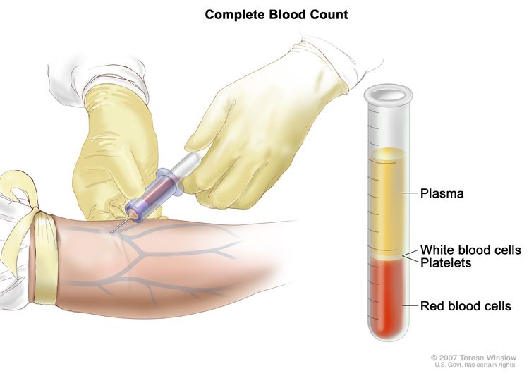
Oral Cavity and Nasopharyngeal Cancers Screening (PDQ®)–Patient Version

Oral Cavity and Nasopharyngeal Cancers Screening (PDQ®)–Patient Version

What Is Screening?

Screening is looking for cancer before a person has any symptoms. This can help find cancer at an early stage. When abnormal tissue or cancer is found early, it may be easier to treat. By the time symptoms appear, cancer may have begun to spread.

Scientists are trying to better understand which people are more likely to get certain types of cancer. They also study the things we do and the things around us to see if they cause cancer. This information helps doctors recommend who should be screened for cancer, which screening tests should be used, and how often the tests should be done.

It is important to remember that your doctor does not necessarily think you have cancer if he or she suggests a screening test. Screening tests are given when you have no cancer symptoms.

If a screening test result is abnormal, you may need to have more tests done to find out if you have cancer. These are called diagnostic tests.

General Information About Oral Cavity and Nasopharyngeal Cancers

Key Points

  • Oral cavity and nasopharyngeal cancers are diseases in which malignant (cancer) cells form in the mouth and throat.
  • The number of new cases of oral cavity and nasopharyngeal cancers and the number of deaths from these cancers vary by sex and geographic region.
  • Different factors increase or decrease the risk of oral cavity and nasopharyngeal cancers.

Oral cavity and nasopharyngeal cancers are diseases in which malignant (cancer) cells form in the mouth and throat.

Oral cavity cancer usually forms in the squamous cells (thin, flat cells lining the inside of the oral cavity).

Oral cavity cancer forms in any of these tissues of the oral cavity:

  • the lips
  • the front two thirds of the tongue
  • the gingiva (gums)
  • the buccal mucosa (the lining of the inside of the cheeks)
  • the floor (bottom) of the mouth under the tongue
  • the hard palate (the front of the roof of the mouth)
  • the retromolar trigone (the small area behind the wisdom teeth)
EnlargeAnatomy of the oral cavity; drawing shows the lip, hard palate, soft palate, retromolar trigone, front two-thirds of the tongue, gingiva, buccal mucosa, and floor of mouth. Also shown are the teeth, uvula, and tonsil.
Anatomy of the oral cavity. The oral cavity includes the lips, hard palate (the bony front portion of the roof of the mouth), soft palate (the muscular back portion of the roof of the mouth), retromolar trigone (the area behind the wisdom teeth), front two-thirds of the tongue, gingiva (gums), buccal mucosa (the inner lining of the lips and cheeks), and floor of the mouth under the tongue.

Pharyngeal cancer forms in the tissues of the pharynx (throat), including the nasopharynx, oropharynx, and hypopharynx. This summary covers nasopharyngeal cancer, which forms in the tissue of the nasopharynx (the upper part of the throat behind the nose).

EnlargeAnatomy of the pharynx; drawing shows the nasopharynx, oropharynx, and hypopharynx. Also shown are the nasal cavity, oral cavity, hyoid bone, larynx, esophagus, and trachea.
Anatomy of the pharynx. The pharynx is a hollow, muscular tube inside the neck that starts behind the nose and opens into the larynx and esophagus. The three parts of the pharynx are the nasopharynx, oropharynx, and hypopharynx.

Other PDQ summaries containing information related to oral cavity and nasopharyngeal cancers include:

The number of new cases of oral cavity and nasopharyngeal cancers and the number of deaths from these cancers vary by sex and geographic region.

From 2012 to 2021, the number of new cases of oral cavity cancer in the United States increased slightly each year.

Oral cavity cancer is more common in men than in women. Although oral cavity cancer may occur in adults of any age, it occurs most often in those aged 75 to 84 years.

France, Brazil, and parts of Asia have much higher rates of oral cavity cancer than most other countries.

Nasopharyngeal cancer is rare in the United States. Some indigenous populations in Southeast Asia, the Arctic, North Africa, and the Middle East have higher rates of nasopharyngeal cancers.

Different factors increase or decrease the risk of oral cavity and nasopharyngeal cancers.

Anything that increases your chance of getting a disease is called a risk factor. Anything that decreases your chance of getting a disease is called a protective factor.

Being infected with Epstein-Barr virus (EBV) increases the risk of nasopharyngeal cancer.

Other PDQ summaries containing information related to oral cavity and nasopharyngeal cancers include:

Oral Cavity and Nasopharyngeal Cancers Screening

Key Points

  • Tests are used to screen for different types of cancer when a person does not have symptoms.
  • There are no standard or routine screening tests for oral cavity and nasopharyngeal cancers.
  • Screening tests for oral cavity and nasopharyngeal cancers are being studied in clinical trials.

Tests are used to screen for different types of cancer when a person does not have symptoms.

Scientists study screening tests to find those with the fewest harms and most benefits. Cancer screening trials also are meant to show whether early detection (finding cancer before it causes symptoms) helps a person live longer or decreases a person’s chance of dying from the disease. For some types of cancer, the chance of recovery is better if the disease is found and treated at an early stage.

There are no standard or routine screening tests for oral cavity and nasopharyngeal cancers.

No studies have shown that screening for oral cavity cancer and nasopharyngeal cancer would lower the risk of dying from these diseases.

A dentist or medical doctor may check the oral cavity during a routine check-up. The exam will include looking for lesions, including areas of leukoplakia (an abnormal white patch of cells) and erythroplakia (an abnormal red patch of cells). Leukoplakia and erythroplakia lesions on the mucous membranes may become cancerous.

If lesions are seen in the mouth, the following procedures may be used to find abnormal tissue that might become oral cavity cancer:

  • Toluidine blue stain: A procedure in which lesions in the mouth are coated with a blue dye. Areas that stain darker are more likely to be cancer or become cancer.
  • Fluorescence staining: A procedure in which lesions in the mouth are viewed using a special light. After the patient uses a fluorescent mouth rinse, normal tissue looks different from abnormal tissue when seen under the light.
  • Exfoliative cytology: A procedure to collect cells from the oral cavity. A piece of cotton, a brush, or a small wooden stick is used to gently scrape cells from the lips, tongue, or mouth. The cells are viewed under a microscope to find out if they are abnormal.
  • Brush biopsy: The removal of cells using a brush that is designed to collect cells from all layers of a lesion. The cells are viewed under a microscope to find out if they are abnormal.

More than half of oral cancers have already spread to lymph nodes or other areas by the time they are found.

Epstein-Barr virus (EBV) has been linked to nasopharyngeal cancer. Screening for nasopharyngeal cancer using the EBV antibody test or EBV DNA test has been studied. These are laboratory tests used to check the blood for EBV antibodies or EBV DNA. If EBV antibodies or DNA are found in the blood, more tests may be done to check for nasopharyngeal cancer. No studies have shown that screening would decrease the risk of dying from this disease.

Screening tests for oral cavity and nasopharyngeal cancers are being studied in clinical trials.

Information about clinical trials supported by NCI can be found on NCI’s clinical trials search webpage. Clinical trials supported by other organizations can be found on the ClinicalTrials.gov website.

Risks of Oral Cavity and Nasopharyngeal Cancers Screening

Key Points

  • Screening tests have risks.
  • The risks of screening for oral cavity and nasopharyngeal cancers include:
    • Finding these cancers may not improve health or help a person live longer.
    • False-negative test results can occur.
    • False-positive test results can occur.

Screening tests have risks.

Decisions about screening tests can be difficult. Not all screening tests are helpful and most have risks. Before having any screening test, you may want to discuss the test with your doctor. It is important to know the risks of the test and whether it has been proven to reduce the risk of dying from cancer.

The risks of screening for oral cavity and nasopharyngeal cancers include:

Finding these cancers may not improve health or help a person live longer.

Some cancers never cause symptoms or become life-threatening, but if found by a screening test, the cancer may be treated. Finding these cancers is called overdiagnosis. It is not known if treatment of oral cavity cancer or nasopharyngeal cancer would help you live longer than if no treatment were given, and treatments for cancer, such as surgery and radiation therapy, may have serious side effects.

False-negative test results can occur.

Screening test results may appear to be normal even though oral cavity cancer or nasopharyngeal cancer is present. A person who receives a false-negative test result (one that shows there is no cancer when there really is) may delay seeking medical care even if there are symptoms.

False-positive test results can occur.

Screening test results may appear to be abnormal even though no cancer is present. A false-positive test result (one that shows there is cancer when there really isn’t) can cause anxiety and is usually followed by more tests and procedures (such as biopsy), which also have risks.

About This PDQ Summary

About PDQ

Physician Data Query (PDQ) is the National Cancer Institute’s (NCI’s) comprehensive cancer information database. The PDQ database contains summaries of the latest published information on cancer prevention, detection, genetics, treatment, supportive care, and complementary and alternative medicine. Most summaries come in two versions. The health professional versions have detailed information written in technical language. The patient versions are written in easy-to-understand, nontechnical language. Both versions have cancer information that is accurate and up to date and most versions are also available in Spanish.

PDQ is a service of the NCI. The NCI is part of the National Institutes of Health (NIH). NIH is the federal government’s center of biomedical research. The PDQ summaries are based on an independent review of the medical literature. They are not policy statements of the NCI or the NIH.

Purpose of This Summary

This PDQ cancer information summary has current information about oral cavity and nasopharyngeal cancers screening. It is meant to inform and help patients, families, and caregivers. It does not give formal guidelines or recommendations for making decisions about health care.

Reviewers and Updates

Editorial Boards write the PDQ cancer information summaries and keep them up to date. These Boards are made up of experts in cancer treatment and other specialties related to cancer. The summaries are reviewed regularly and changes are made when there is new information. The date on each summary (“Updated”) is the date of the most recent change.

The information in this patient summary was taken from the health professional version, which is reviewed regularly and updated as needed, by the PDQ Screening and Prevention Editorial Board.

Clinical Trial Information

A clinical trial is a study to answer a scientific question, such as whether one treatment is better than another. Trials are based on past studies and what has been learned in the laboratory. Each trial answers certain scientific questions in order to find new and better ways to help cancer patients. During treatment clinical trials, information is collected about the effects of a new treatment and how well it works. If a clinical trial shows that a new treatment is better than one currently being used, the new treatment may become “standard.” Patients may want to think about taking part in a clinical trial. Some clinical trials are open only to patients who have not started treatment.

Clinical trials can be found online at NCI’s website. For more information, call the Cancer Information Service (CIS), NCI’s contact center, at 1-800-4-CANCER (1-800-422-6237).

Permission to Use This Summary

PDQ is a registered trademark. The content of PDQ documents can be used freely as text. It cannot be identified as an NCI PDQ cancer information summary unless the whole summary is shown and it is updated regularly. However, a user would be allowed to write a sentence such as “NCI’s PDQ cancer information summary about breast cancer prevention states the risks in the following way: [include excerpt from the summary].”

The best way to cite this PDQ summary is:

PDQ® Screening and Prevention Editorial Board. PDQ Oral Cavity and Nasopharyngeal Cancers Screening. Bethesda, MD: National Cancer Institute. Updated <MM/DD/YYYY>. Available at: /types/head-and-neck/patient/oral-screening-pdq. Accessed <MM/DD/YYYY>. [PMID: 26389441]

Images in this summary are used with permission of the author(s), artist, and/or publisher for use in the PDQ summaries only. If you want to use an image from a PDQ summary and you are not using the whole summary, you must get permission from the owner. It cannot be given by the National Cancer Institute. Information about using the images in this summary, along with many other images related to cancer can be found in Visuals Online. Visuals Online is a collection of more than 3,000 scientific images.

Disclaimer

The information in these summaries should not be used to make decisions about insurance reimbursement. More information on insurance coverage is available on Cancer.gov on the Managing Cancer Care page.

Contact Us

More information about contacting us or receiving help with the Cancer.gov website can be found on our Contact Us for Help page. Questions can also be submitted to Cancer.gov through the website’s E-mail Us.

Lip and Oral Cavity Cancer Treatment (PDQ®)–Patient Version

Lip and Oral Cavity Cancer Treatment (PDQ®)–Patient Version

General Information About Lip and Oral Cavity Cancer

Key Points

  • Lip and oral cavity cancer is a disease that starts in lips or mouth.
  • Tobacco and alcohol use can affect the risk of lip and oral cavity cancer.
  • Signs of lip and oral cavity cancer include a sore or lump on the lips or in the mouth.
  • Tests that examine the mouth and throat are used to diagnose and stage lip and oral cavity cancer.
  • Some people decide to get a second opinion.
  • Certain factors affect prognosis (chance of recovery) and treatment options.

Lip and oral cavity cancer is a disease that starts in lips or mouth.

The oral cavity refers to the mouth. It includes:

  • the front two thirds of the tongue
  • the gingiva (gums)
  • the buccal mucosa (the lining of the inside of the cheeks)
  • the floor (bottom) of the mouth under the tongue
  • the hard palate (the roof of the mouth)
  • the retromolar trigone (the small area behind the wisdom teeth)
EnlargeAnatomy of the oral cavity; drawing shows the lip, hard palate, soft palate, retromolar trigone, front two-thirds of the tongue, gingiva, buccal mucosa, and floor of mouth. Also shown are the teeth, uvula, and tonsil.
Anatomy of the oral cavity. The oral cavity includes the lips, hard palate (the bony front portion of the roof of the mouth), soft palate (the muscular back portion of the roof of the mouth), retromolar trigone (the area behind the wisdom teeth), front two-thirds of the tongue, gingiva (gums), buccal mucosa (the inner lining of the lips and cheeks), and floor of the mouth under the tongue.

Most lip and oral cavity cancers start in squamous cells, the thin, flat cells lining the inside of the lips and oral cavity. Cancers that start in squamous cells are called squamous cell carcinomas. Cancer cells may spread into deeper tissue as the cancer grows. Squamous cell carcinoma usually develops in areas of leukoplakia (white patches of cells that do not rub off).

Lip and oral cavity cancer is a type of head and neck cancer.

Tobacco and alcohol use can affect the risk of lip and oral cavity cancer.

Lip and oral cavity cancer is caused by certain changes to the way lip and oral cavity cells function, especially how they grow and divide into new cells. There are many risk factors for lip and oral cavity cancer, but many do not directly cause cancer. Instead, they increase the chance of DNA damage in cells that may lead to lip and oral cavity cancer. Learn more about how cancer develops at What Is Cancer?

A risk factor is anything that increases the chance of getting a disease. Some risk factors for lip and oral cavity cancer, such as tobacco and alcohol use, can be changed. However, risk factors also include things people cannot change, like their genetics. Learning about risk factors for lip and oral cavity cancer can help you make changes that might lower your risk of getting it.

Risk factors for lip and oral cavity cancer include:

  • using tobacco products
  • heavy alcohol use
  • being exposed to natural sunlight or artificial sunlight (such as from tanning beds) over long periods of time
  • being male

Learn more about Tobacco, including help with quitting.

Having one or more of these risk factors does not mean that you will get lip and oral cavity cancer. Many people with risk factors never develop lip and oral cavity cancer, while others with no known risk factors do. Talk with your doctor if you think you may be at risk.

Signs of lip and oral cavity cancer include a sore or lump on the lips or in the mouth.

These and other signs and symptoms may be caused by lip and oral cavity cancer or by other conditions. Check with your doctor if you have any of the following:

  • a sore on the lip or in the mouth that does not heal
  • a lump or thickening on the lips or gums or in the mouth
  • a white or red patch on the gums, tongue, or lining of the mouth
  • bleeding, pain, or numbness in the lip or mouth
  • change in voice
  • loose teeth or dentures that no longer fit well
  • trouble chewing or swallowing or moving the tongue or jaw
  • swelling of jaw
  • sore throat or feeling that something is caught in the throat

Lip and oral cavity cancer may not have any symptoms and is sometimes found during a regular dental exam.

Tests that examine the mouth and throat are used to diagnose and stage lip and oral cavity cancer.

If you have symptoms that suggest lip and oral cavity cancer, your doctor will need to find out if these are due to cancer or another problem. They will ask when the symptoms started and how often you have been having them. They will also ask about your personal and family health history and do a physical exam. Based on these results, the doctor may recommend other tests. If you are diagnosed with lip and oral cavity cancer, the results of these tests will help you and your doctor plan treatment.

The following tests and procedures are used to diagnose and stage lip and oral cavity cancer:

  • Physical exam of the lips and oral cavity is an exam to check the lips and oral cavity for abnormal areas. The medical doctor or dentist will feel the entire inside of the mouth with a gloved finger and examine the oral cavity with a small long-handled mirror and lights. This will include checking the insides of the cheeks and lips; the gums; the roof and floor of the mouth; and the top, bottom, and sides of the tongue. The neck will be felt for swollen lymph nodes. A history of the patient’s health habits and past illnesses and medical and dental treatments will also be taken.
  • Endoscopy is a procedure to look at organs and tissues inside the body to check for abnormal areas. An endoscope is inserted through an incision (cut) in the skin or opening in the body, such as the mouth. An endoscope is a thin, tube-like instrument with a light and a lens for viewing. It may also have a tool to remove tissue or lymph node samples, which are checked under a microscope for signs of disease.
  • Biopsy is the removal of cells or tissues so they can be viewed under a microscope by a pathologist. If leukoplakia is found, cells taken from the patches are also checked under the microscope for signs of cancer.
  • Exfoliative cytology is a procedure to collect cells from the lip or oral cavity. A piece of cotton, a brush, or a small wooden stick is used to gently scrape cells from the lips, tongue, mouth, or throat. The cells are viewed under a microscope to find out if they are abnormal.
  • MRI (magnetic resonance imaging) uses a magnet, radio waves, and a computer to make a series of detailed pictures of areas inside the body. This procedure is also called nuclear magnetic resonance imaging (NMRI).
  • CT scan (CAT scan) uses a computer linked to an x-ray machine to make a series of detailed pictures of areas inside the body. The pictures are taken from different angles and are used to create 3-D views of tissues and organs. A dye may be injected into a vein or swallowed to help the organs or tissues show up more clearly. This procedure is also called computed tomography, computerized tomography, or computerized axial tomography.
    EnlargeComputed tomography (CT) scan of the head and neck; drawing shows a patient lying on a table that slides through the CT scanner, which takes x-ray pictures of the inside of the head and neck.
    Computed tomography (CT) scan of the head and neck. The patient lies on a table that slides through the CT scanner, which takes x-ray pictures of the inside of the head and neck.
  • Barium swallow is a series of x-rays of the esophagus and stomach. The patient drinks a liquid that contains barium (a silver-white metallic compound). The liquid coats the esophagus and x-rays are taken. This procedure is also called an upper GI series.
  • PET scan (positron emission tomography scan) uses a small amount of radioactive sugar (also called glucose) that is injected into a vein. Then a scanner rotates around the body to make detailed, computerized pictures of areas inside the body where the glucose is taken up. Because cancer cells often take up more glucose than normal cells, the pictures can be used to find cancer cells in the body.
  • Bone scan is a procedure to check if there are rapidly dividing cells, such as cancer cells, in the bone. A very small amount of radioactive material is injected into a vein and travels through the bloodstream. The radioactive material collects in the bones with cancer and is detected by a scanner.

Some people decide to get a second opinion.

You may want to get a second opinion to confirm your cancer diagnosis and treatment plan. If you seek a second opinion, you will need to get medical test results and reports from the first doctor to share with the second doctor. The second doctor will review the pathology report, slides, and scans. They may agree with the first doctor, suggest changes or another treatment approach, or provide more information about your cancer.

To learn more about choosing a doctor and getting a second opinion, see Finding Cancer Care. You can contact NCI’s Cancer Information Service via chat, email, or phone (both in English and Spanish) for help finding a doctor, hospital, or getting a second opinion. For questions you might want to ask at your appointments, see Questions to Ask Your Doctor About Cancer.

Certain factors affect prognosis (chance of recovery) and treatment options.

The prognosis depends on:

  • the cancer stage
  • where the tumor is in the lip or oral cavity
  • whether the cancer has spread to blood vessels

For patients who smoke, the chance of recovery is better if they stop smoking before beginning radiation therapy.

Treatment options depend on:

  • the stage of the cancer
  • the size of the tumor and where it is in the lip or oral cavity
  • whether the patient’s appearance and ability to talk and eat can stay the same
  • the patient’s age and general health

Patients who have had lip and oral cavity cancer have an increased risk of developing a second cancer in the head or neck. Frequent and careful follow-up is important. Clinical trials are studying the use of retinoid drugs to reduce the risk of a second head and neck cancer. Information about ongoing clinical trials is available from the NCI website.

Stages of Lip and Oral Cavity Cancer

Key Points

  • Cancer stage describes the extent of cancer in the body.
  • The following stages are used for lip and oral cavity cancer:
    • Stage 0 (carcinoma in situ)
    • Stage I (also called stage 1) lip and oral cavity cancer
    • Stage II (also called stage 2) lip and oral cavity cancer
    • Stage III (also called stage 3) lip and oral cavity cancer
    • Stage IV (also called stage 4) lip and oral cavity cancer
  • Lip and oral cavity cancer can recur (come back) after it has been treated.

Cancer stage describes the extent of cancer in the body.

Cancer stage describes the extent of cancer in the body, such as the size of the tumor, whether it has spread, and how far it has spread from where it first formed. Knowing the cancer stage helps plan treatment.

There are several staging systems for cancer that describe the extent of the cancer. Lip and oral cavity cancer staging usually uses the TNM staging system. The cancer may be described by this staging system in your pathology report. Based on the TNM results, a stage (I, II, III, or IV, also written as 1, 2, 3, or 4) is assigned to the cancer. When talking to you about your diagnosis, your doctor may describe the cancer as one of these stages.

Learn more about Cancer Staging.

The following stages are used for lip and oral cavity cancer:

Stage 0 (carcinoma in situ)

In stage 0, abnormal cells are found in the lining of the lips and oral cavity. These abnormal cells may become cancer and spread into nearby normal tissue. Stage 0 is also called carcinoma in situ.

EnlargeDrawing shows different sizes of a tumor in centimeters (cm) compared to the size of a pea (1 cm), a peanut (2 cm), a grape (3 cm), a walnut (4 cm), a lime (5 cm), an egg (6 cm), a peach (7 cm), and a grapefruit (10 cm). Also shown is a 10-cm ruler and a 4-inch ruler.
Tumor sizes are often measured in centimeters (cm) or inches. Common food items that can be used to show tumor size in cm include: a pea (1 cm), a peanut (2 cm), a grape (3 cm), a walnut (4 cm), a lime (5 cm or 2 inches), an egg (6 cm), a peach (7 cm), and a grapefruit (10 cm or 4 inches).

Stage I (also called stage 1) lip and oral cavity cancer

In stage I, cancer has formed. The tumor is 2 centimeters or smaller and the deepest point of tumor invasion is 5 millimeters or less.

EnlargeDrawing shows different sizes of common items in millimeters (mm): a sharp pencil point (1 mm), a new crayon point (2 mm), a pencil-top eraser (5 mm), a pea (10 mm), a peanut (20 mm), and a lime (50 mm). Also shown is a 2-centimeter (cm) ruler that shows 10 mm is equal to 1 cm.
Tumor sizes are often measured in millimeters (mm) or centimeters. Common items that can be used to show tumor size in mm include: a sharp pencil point (1 mm), a new crayon point (2 mm), a pencil-top eraser (5 mm), a pea (10 mm), a peanut (20 mm), and a lime (50 mm).

Stage II (also called stage 2) lip and oral cavity cancer

In stage II, the tumor:

  • is 2 centimeters or smaller and the deepest point of tumor invasion is greater than 5 millimeters; or
  • is larger than 2 centimeters but not larger than 4 centimeters and the deepest point of tumor invasion is 10 millimeters or less.

Stage III (also called stage 3) lip and oral cavity cancer

In stage III, the tumor:

  • is larger than 2 centimeters but not larger than 4 centimeters and the deepest point of tumor invasion is greater than 10 millimeters; or
  • is larger than 4 centimeters and the deepest point of tumor invasion is 10 millimeters or less; or
  • has spread to one lymph node that is 3 centimeters or smaller, on the same side of the neck as the primary tumor.

Stage IV (also called stage 4) lip and oral cavity cancer

Stage IV is divided into stages IVA, IVB, and IVC.

  • In stage IVA, the tumor:
    • is larger than 4 centimeters and the deepest point of tumor invasion is greater than 10 millimeters; or has spread to the outer surface of the upper or lower jawbone, into the maxillary sinus, or to the skin of the face. The cancer may have spread to one lymph node that is 3 centimeters or smaller, on the same side of the neck as the primary tumor; or
    • is any size or cancer has spread to the outer surface of the upper or lower jawbone, into the maxillary sinus, or to the skin of the face. Cancer has spread:
      • to one lymph node that is 3 centimeters or smaller, on the same side of the neck as the primary tumor, and cancer has spread through the outside covering of the lymph node into nearby connective tissue; or
      • to one lymph node that is larger than 3 centimeters but not larger than 6 centimeters, on the same side of the neck as the primary tumor; or
      • to multiple lymph nodes that are not larger than 6 centimeters, on the same side of the neck as the primary tumor; or
      • to multiple lymph nodes that are not larger than 6 centimeters, on the opposite side of the neck as the primary tumor or on both sides of the neck.
  • In stage IVB, the tumor:
    • has spread to one lymph node that is larger than 6 centimeters; or
    • has spread to one lymph node that is larger than 3 centimeters, on the same side of the neck as the primary tumor, and cancer has spread through the outside covering of the lymph node into nearby connective tissue; or
    • has spread to one lymph node of any size on the opposite side of the neck as the primary tumor, and cancer has spread through the outside covering of the lymph node into nearby connective tissue; or
    • has spread to multiple lymph nodes anywhere in the neck, and cancer has spread through the outside covering of any lymph node into nearby connective tissue; or
    • has spread further into the muscles or bones needed for chewing, or to the part of the sphenoid bone behind the upper jaw, and/or to the carotid artery near the base of the skull. Cancer may have also spread to one or more lymph nodes of any size, anywhere in the neck.
  • In stage IVC, the tumor:
    • has spread beyond the lip or oral cavity to other parts of the body, such as the lung, liver, or bone.

      Stage IVC lip and oral cavity cancer is also called metastatic lip and oral cavity cancer. Metastatic cancer happens when cancer cells travel through the lymphatic system or blood and form tumors in other parts of the body. The metastatic tumor is the same type of cancer as the primary tumor. For example, if lip and oral cavity cancer spreads to the lung, the cancer cells in the lung are actually lip and oral cavity cancer cells. The disease is called metastatic lip and oral cavity cancer, not lung cancer. Learn more in Metastatic Cancer: When Cancer Spreads.

Lip and oral cavity cancer can recur (come back) after it has been treated.

Recurrent lip and oral cavity cancer is cancer that has come back after it has been treated. If lip and oral cavity cancer comes back, it may come back in the lip, mouth, or other parts of the body. Tests will help determine where in the body the cancer has returned. The type of treatment that you have for recurrent lip and oral cavity cancer will depend on where it has come back.

Learn more in Recurrent Cancer: When Cancer Comes Back.

Treatment Option Overview

Key Points

  • There are different types of treatment for people with lip and oral cavity cancer.
  • People with lip and oral cavity cancer should have their treatment planned by a team of doctors who are expert in treating head and neck cancer.
  • The following types of treatment are used:
    • Surgery
    • Radiation therapy
    • Immunotherapy
  • New types of treatment are being tested in clinical trials.
  • Follow-up care may be needed.

There are different types of treatment for people with lip and oral cavity cancer.

Different types of treatments are available for lip and oral cavity cancer. You and your cancer care team will work together to decide your treatment plan, which may include more than one type of treatment. Many factors will be considered, such as the stage of the cancer, your overall health, and your preferences. Your plan will include information about your cancer, the goals of treatment, your treatment options and the possible side effects, and the expected length of treatment.

Talking with your cancer care team before treatment begins about what to expect will be helpful. You’ll want to learn what you need to do before treatment begins, how you’ll feel while going through it, and what kind of help you will need. Learn more at Questions to Ask Your Doctor About Treatment.

People with lip and oral cavity cancer should have their treatment planned by a team of doctors who are expert in treating head and neck cancer.

An oncologist, a doctor who specializes in treating people with cancer, oversees treatment for lip and oral cavity cancer. Because the lips and oral cavity are important for breathing, eating, and talking, you may need special help adjusting to the side effects of the cancer and its treatment. The oncologist may refer you to other health care providers who are experts in treating head and neck cancer and also specialize in other areas of medicine. Other specialists may include:

The following types of treatment are used:

Surgery

Surgery (removing the cancer in an operation) is a common treatment for all stages of lip and oral cavity cancer. Surgery may include:

  • Wide local excision is the removal of the cancer and some of the healthy tissue around it. If cancer has spread into bone, surgery may include removal of the involved bone tissue.
  • Neck dissection is the removal of lymph nodes and other tissues in the neck. This is done when cancer may have spread from the lip and oral cavity.
  • Plastic surgery is an operation that restores or improves the appearance of parts of the body. Dental implants, a skin graft, or other plastic surgery may be needed to repair parts of the mouth, throat, or neck after removal of large tumors.

After the doctor removes all the cancer that can be seen at the time of the surgery, some patients may be given chemotherapy or radiation therapy after surgery to kill any cancer cells that are left. Treatment given after the surgery, to lower the risk that the cancer will come back, is called adjuvant therapy.

Learn more about Surgery to Treat Cancer.

Radiation therapy

Radiation therapy uses high-energy x-rays or other types of radiation to kill cancer cells or keep them from growing by damaging their DNA. External and internal radiation therapy are used to treat lip and oral cavity cancer:

  • External radiation therapy uses a machine outside the body to send radiation toward the area of the body with cancer.
    EnlargeExternal-beam radiation therapy of the head and neck; drawing shows a patient lying on a table under a machine that is used to aim high-energy radiation at the cancer. An inset shows a mesh mask that helps keep the patient's head and neck from moving during treatment. The mask has pieces of white tape with small ink marks on it. The ink marks are used to line up the radiation machine in the same position before each treatment.
    External-beam radiation therapy of the head and neck. A machine is used to aim high-energy radiation at the cancer. The machine can rotate around the patient, delivering radiation from many different angles to provide highly conformal treatment. A mesh mask helps keep the patient’s head and neck from moving during treatment. Small ink marks are put on the mask. The ink marks are used to line up the radiation machine in the same position before each treatment.
  • Internal radiation therapy (also called brachytherapy) uses a radioactive substance sealed in needles, seeds, wires, or catheters that are placed directly into or near the cancer.

Radiation therapy may work better in patients who have stopped smoking before beginning treatment. It is also important for patients to have a dental exam before radiation therapy begins so that existing problems can be treated.

Learn more about Radiation Therapy to Treat Cancer and Radiation Therapy Side Effects.

Immunotherapy

Immunotherapy helps a person’s immune system fight cancer.  Your doctor may suggest biomarker tests to help predict your response to certain immunotherapy drugs. Learn more about Biomarker Testing for Cancer Treatment.

Immunotherapy drugs used to treat squamous cell carcinoma of the lip and oral cavity cancer that has come back or spread to other parts of the body include:

Learn more about Immunotherapy to Treat Cancer.

New types of treatment are being tested in clinical trials.

For some people, joining a clinical trial may be an option. There are different types of clinical trials for people with cancer. For example, a treatment trial tests new treatments or new ways of using existing treatments. Supportive care and palliative care trials look at ways to improve quality of life, especially for those who have side effects from cancer and its treatment.

You can use the clinical trial search to find NCI-supported cancer clinical trials that are accepting participants. This search allows you to filter trials based on the type of cancer, your age, and where the trials are being done. Clinical trials supported by other organizations can be found on the ClinicalTrials.gov website.

Learn the basics about clinical trials at Clinical Trials Information for Patients and Caregivers.

Follow-up care may be needed.

As you go through treatment, you will have follow-up tests or check-ups. Some tests that were done to diagnose or stage the cancer may be repeated to see how well the treatment is working. Decisions about whether to continue, change, or stop treatment may be based on the results of these tests.

Some of the tests will continue to be done from time to time after treatment has ended. The results of these tests can show if your condition has changed or if the cancer has recurred (come back).

Treatment of Stage I Lip and Oral Cavity Cancer

Learn about the treatments listed below in the Treatment Option Overview.

Treatment of stage I lip and oral cavity cancer depends on where cancer is found in the lip and oral cavity.

Lip

If cancer is in the lip, treatment may include:

Front of the tongue

If cancer is in the front of the tongue, treatment may include:

Buccal mucosa

If cancer is in the buccal mucosa (the lining of the inside of the cheeks), treatment may include:

Floor of the mouth

If cancer is in the floor (bottom) of the mouth, treatment may include:

Lower gingiva

If cancer is in the lower gingiva (gums), treatment may include:

Retromolar trigone

If cancer is in the retromolar trigone (the small area behind the wisdom teeth), treatment may include:

Upper gingiva or hard palate

If cancer is in the upper gingiva (gums) or the hard palate (the roof of the mouth), treatment is usually surgery (wide local excision) with or without radiation therapy.

Use our clinical trial search to find NCI-supported cancer clinical trials that are accepting patients. You can search for trials based on the type of cancer, the age of the patient, and where the trials are being done. General information about clinical trials is also available.

Treatment of Stage II Lip and Oral Cavity Cancer

Learn about the treatments listed below in the Treatment Option Overview.

Treatment of stage II lip and oral cavity cancer depends on where cancer is found in the lip and oral cavity.

Lip

If cancer is in the lip, treatment may include:

Front of the tongue

If cancer is in the front of the tongue, treatment may include:

Buccal mucosa

If cancer is in the buccal mucosa (the lining of the inside of the cheeks), treatment may include:

Floor of the mouth

If cancer is in the floor (bottom) of the mouth, treatment may include:

Lower gingiva

If cancer is in the lower gingiva (gums), treatment may include:

Retromolar trigone

If cancer is in the retromolar trigone (the small area behind the wisdom teeth), treatment may include:

Upper gingiva or hard palate

If cancer is in the upper gingiva (gums) or the hard palate (the roof of the mouth), treatment may include:

Use our clinical trial search to find NCI-supported cancer clinical trials that are accepting patients. You can search for trials based on the type of cancer, the age of the patient, and where the trials are being done. General information about clinical trials is also available.

Treatment of Stage III Lip and Oral Cavity Cancer

Learn about the treatments listed below in the Treatment Option Overview.

Treatment of stage III lip and oral cavity cancer depends on where cancer is found in the lip and oral cavity.

Lip

If cancer is in the lip, treatment may include surgery and external radiation therapy with or without internal radiation therapy.

Front of the tongue

If cancer is in the front of the tongue, treatment may include:

Buccal mucosa

If cancer is in the buccal mucosa (the lining of the inside of the cheeks), treatment may include:

Floor of the mouth

If cancer is in the floor (bottom) of the mouth, treatment may include:

Lower gingiva

If cancer is in the lower gingiva (gums), treatment may include surgery (wide local excision) with or without radiation therapy. Radiation may be given before or after surgery.

Retromolar trigone

If cancer is in the retromolar trigone (the small area behind the wisdom teeth), treatment may include surgery to remove the tumor, lymph nodes, and part of the jawbone, with or without radiation therapy.

Upper gingiva

If cancer is in the upper gingiva (gums), treatment may include:

Hard palate

If cancer is in the hard palate (the roof of the mouth), treatment may include:

Lymph nodes

For cancer that may have spread to lymph nodes, treatment may include radiation therapy and/or surgery (neck dissection).

Use our clinical trial search to find NCI-supported cancer clinical trials that are accepting patients. You can search for trials based on the type of cancer, the age of the patient, and where the trials are being done. General information about clinical trials is also available.

Treatment of Stage IV Nonmetastatic Lip and Oral Cavity Cancer

Learn about the treatments listed below in the Treatment Option Overview.

Treatment of stages IVA and IVB lip and oral cavity cancer depends on where cancer is found in the lip and oral cavity.

Lip

If cancer is in the lip, treatment may include surgery and external radiation therapy with or without internal radiation therapy.

Front of the tongue

If cancer is in the front of the tongue, treatment may include:

Buccal mucosa

If cancer is in the buccal mucosa (the lining of the inside of the cheeks), treatment may include surgery (wide local excision) and/or radiation therapy.

Floor of the mouth

If cancer is in the floor (bottom) of the mouth, treatment may include surgery before or after radiation therapy.

Lower gingiva

If cancer is in the lower gingiva (gums), treatment may include surgery and/or radiation therapy.

Retromolar trigone

If cancer is in the retromolar trigone (the small area behind the wisdom teeth), treatment may include surgery to remove the tumor, lymph nodes, and part of the jawbone, followed by radiation therapy.

Upper gingiva or hard palate

If cancer is in the upper gingiva (gums) or hard palate (the roof of the mouth), treatment may include surgery with radiation therapy.

Lymph nodes

For cancer that may have spread to lymph nodes, treatment may include radiation therapy and/or surgery (neck dissection).

Use our clinical trial search to find NCI-supported cancer clinical trials that are accepting patients. You can search for trials based on the type of cancer, the age of the patient, and where the trials are being done. General information about clinical trials is also available.

Treatment of Stage IV Metastatic and Recurrent Lip and Oral Cavity Cancer

Learn about the treatments listed below in the Treatment Option Overview.

Treatment of metastatic (IVC) and recurrent lip and oral cavity cancer may include:

Use our clinical trial search to find NCI-supported cancer clinical trials that are accepting patients. You can search for trials based on the type of cancer, the age of the patient, and where the trials are being done. General information about clinical trials is also available.

To Learn More About Lip and Oral Cavity Cancer

About This PDQ Summary

About PDQ

Physician Data Query (PDQ) is the National Cancer Institute’s (NCI’s) comprehensive cancer information database. The PDQ database contains summaries of the latest published information on cancer prevention, detection, genetics, treatment, supportive care, and complementary and alternative medicine. Most summaries come in two versions. The health professional versions have detailed information written in technical language. The patient versions are written in easy-to-understand, nontechnical language. Both versions have cancer information that is accurate and up to date and most versions are also available in Spanish.

PDQ is a service of the NCI. The NCI is part of the National Institutes of Health (NIH). NIH is the federal government’s center of biomedical research. The PDQ summaries are based on an independent review of the medical literature. They are not policy statements of the NCI or the NIH.

Purpose of This Summary

This PDQ cancer information summary has current information about the treatment of adult lip and oral cavity cancer. It is meant to inform and help patients, families, and caregivers. It does not give formal guidelines or recommendations for making decisions about health care.

Reviewers and Updates

Editorial Boards write the PDQ cancer information summaries and keep them up to date. These Boards are made up of experts in cancer treatment and other specialties related to cancer. The summaries are reviewed regularly and changes are made when there is new information. The date on each summary (“Updated”) is the date of the most recent change.

The information in this patient summary was taken from the health professional version, which is reviewed regularly and updated as needed, by the PDQ Adult Treatment Editorial Board.

Clinical Trial Information

A clinical trial is a study to answer a scientific question, such as whether one treatment is better than another. Trials are based on past studies and what has been learned in the laboratory. Each trial answers certain scientific questions in order to find new and better ways to help cancer patients. During treatment clinical trials, information is collected about the effects of a new treatment and how well it works. If a clinical trial shows that a new treatment is better than one currently being used, the new treatment may become “standard.” Patients may want to think about taking part in a clinical trial. Some clinical trials are open only to patients who have not started treatment.

Clinical trials can be found online at NCI’s website. For more information, call the Cancer Information Service (CIS), NCI’s contact center, at 1-800-4-CANCER (1-800-422-6237).

Permission to Use This Summary

PDQ is a registered trademark. The content of PDQ documents can be used freely as text. It cannot be identified as an NCI PDQ cancer information summary unless the whole summary is shown and it is updated regularly. However, a user would be allowed to write a sentence such as “NCI’s PDQ cancer information summary about breast cancer prevention states the risks in the following way: [include excerpt from the summary].”

The best way to cite this PDQ summary is:

PDQ® Adult Treatment Editorial Board. PDQ Lip and Oral Cavity Cancer Treatment. Bethesda, MD: National Cancer Institute. Updated <MM/DD/YYYY>. Available at: /types/head-and-neck/patient/adult/lip-mouth-treatment-pdq. Accessed <MM/DD/YYYY>. [PMID: 26389326]

Images in this summary are used with permission of the author(s), artist, and/or publisher for use in the PDQ summaries only. If you want to use an image from a PDQ summary and you are not using the whole summary, you must get permission from the owner. It cannot be given by the National Cancer Institute. Information about using the images in this summary, along with many other images related to cancer can be found in Visuals Online. Visuals Online is a collection of more than 3,000 scientific images.

Disclaimer

The information in these summaries should not be used to make decisions about insurance reimbursement. More information on insurance coverage is available on Cancer.gov on the Managing Cancer Care page.

Contact Us

More information about contacting us or receiving help with the Cancer.gov website can be found on our Contact Us for Help page. Questions can also be submitted to Cancer.gov through the website’s E-mail Us.

Lip and Oral Cavity Cancer Treatment (PDQ®)–Health Professional Version

Lip and Oral Cavity Cancer Treatment (PDQ®)–Health Professional Version

General Information About Lip and Oral Cavity Cancer

Anatomy

The oral cavity extends from the skin-vermilion junctions of the anterior lips to the junction of the hard and soft palates above and to the line of circumvallate papillae below and is divided into the following specific areas:

  • Lip.
  • Anterior two thirds of tongue.
  • Buccal mucosa.
  • Floor of mouth.
  • Lower gingiva.
  • Retromolar trigone.
  • Upper gingiva.
  • Hard palate.

Histopathology

The main routes of lymph node drainage are into the first station nodes (i.e., buccinator, jugulodigastric, submandibular, and submental). Sites close to the midline often drain bilaterally. Second station nodes include the parotid, jugular, and the upper and lower posterior cervical nodes.

Precancerous lesions of the oropharynx include leukoplakia, erythroplakia, and mixed erythroleukoplakia.[1] Leukoplakia, the most common of the three conditions, is defined by the World Health Organization as “a white patch or plaque that cannot be characterized clinically or pathologically as any other disease.”[2] The diagnosis of leukoplakia is one of exclusion; conditions such as candidiasis, lichen planus, leukoedema, and others must be ruled out before a diagnosis of leukoplakia can be made.[1]

The prevalence of leukoplakia in the United States is decreasing as a result of reduced tobacco consumption.[3] Although erythroplakia is not as common as leukoplakia, it is much more likely to be associated with dysplasia or carcinoma.[1,4]

Prognostic Factors

Early cancers (stage I and stage II) of the lip and oral cavity are highly curable by surgery or radiation therapy. The choice of treatment is dictated by the anticipated functional and cosmetic results of treatment and by the availability of a surgeon or radiation oncologist with the required expertise.[57] A positive surgical margin or a tumor depth of more than 5 mm significantly increases the risk of local recurrence.[8,9] The risk of occult nodal metastases increases based on depth of invasion of the primary tumor. Depth of invasion holds prognostic significance and was included in tumor staging definitions in the American Joint Committee on Cancer (AJCC) 8th edition staging classification.[10,11] Extranodal extension in a lymph node is a significant adverse prognostic factor and was incorporated into the 8th edition AJCC staging system.[12,13]

Advanced cancers (stage III and stage IV) of the lip and oral cavity represent a wide spectrum of challenges for the surgeon and radiation oncologist. Most patients with stage III or stage IV tumors are candidates for treatment by a combination of surgery and radiation therapy. The exception is patients with small T3 lesions and no regional lymph node and no distant metastases or who have no lymph nodes larger than 2 cm in diameter, for whom treatment by radiation therapy alone or surgery alone might be appropriate.[6] Furthermore, because local recurrence and/or distant metastases are common in this group of patients, clinical trials can be considered. Such trials evaluate the potential role of radiation modifiers or combination chemotherapy combined with surgery and/or radiation therapy.

Survival

Patients with head and neck cancers have an increased chance of developing a second primary tumor of the upper aerodigestive tract.[14,15] A study has shown that daily treatment with moderate doses of isotretinoin for 1 year can significantly reduce the incidence of second tumors. However, no survival advantage has been demonstrated, in part due to recurrence and death from the primary malignancy. An additional trial showed no benefit of retinyl palmitate or retinyl palmitate plus beta-carotene when compared with isotretinoin alone.[16][Level of evidence B1]

The cure rates of cancers of the lip and oral cavity depend on the stage and specific site. Most patients present with early cancers of the lip, which are highly curable by surgery or by radiation therapy with cure rates of 90% to 100%. Small cancers of the retromolar trigone, hard palate, and upper gingiva are highly curable by either radiation therapy or surgery with survival rates of as high as 100%. Local control rates as high as 90% can be achieved with either radiation therapy or surgery in small cancers of the anterior tongue, the floor of the mouth, and buccal mucosa.[17]

Moderately advanced and advanced cancers of the lip also can be controlled effectively by surgery, radiation therapy, or both. The choice of treatment is generally dictated by the anticipated functional and cosmetic results of the treatment. Moderately advanced lesions of the retromolar trigone without evidence of spread to cervical lymph nodes are usually curable and have shown local control rates as high as 90%. Such lesions of the hard palate, upper gingiva, and buccal mucosa have a local control rate of up to 80%. In the absence of clinical evidence of spread to cervical lymph nodes, moderately advanced lesions of the floor of the mouth and anterior tongue are generally curable, with survival rates of as high as 70% and 65%, respectively.[17,18]

References
  1. Neville BW, Day TA: Oral cancer and precancerous lesions. CA Cancer J Clin 52 (4): 195-215, 2002 Jul-Aug. [PUBMED Abstract]
  2. Kramer IR, Lucas RB, Pindborg JJ, et al.: Definition of leukoplakia and related lesions: an aid to studies on oral precancer. Oral Surg Oral Med Oral Pathol 46 (4): 518-39, 1978. [PUBMED Abstract]
  3. Scheifele C, Reichart PA, Dietrich T: Low prevalence of oral leukoplakia in a representative sample of the US population. Oral Oncol 39 (6): 619-25, 2003. [PUBMED Abstract]
  4. Shafer WG, Waldron CA: Erythroplakia of the oral cavity. Cancer 36 (3): 1021-8, 1975. [PUBMED Abstract]
  5. Cummings CW, Fredrickson JM, Harker LA, et al.: Otolaryngology – Head and Neck Surgery. Mosby-Year Book, Inc., 1998.
  6. Harrison LB, Sessions RB, Hong WK, eds.: Head and Neck Cancer: A Multidisciplinary Approach. 3rd ed. Lippincott, William & Wilkins, 2009.
  7. Wang CC, ed.: Radiation Therapy for Head and Neck Neoplasms. 3rd ed. Wiley-Liss, 1997.
  8. Jones KR, Lodge-Rigal RD, Reddick RL, et al.: Prognostic factors in the recurrence of stage I and II squamous cell cancer of the oral cavity. Arch Otolaryngol Head Neck Surg 118 (5): 483-5, 1992. [PUBMED Abstract]
  9. Po Wing Yuen A, Lam KY, Lam LK, et al.: Prognostic factors of clinically stage I and II oral tongue carcinoma-A comparative study of stage, thickness, shape, growth pattern, invasive front malignancy grading, Martinez-Gimeno score, and pathologic features. Head Neck 24 (6): 513-20, 2002. [PUBMED Abstract]
  10. Sparano A, Weinstein G, Chalian A, et al.: Multivariate predictors of occult neck metastasis in early oral tongue cancer. Otolaryngol Head Neck Surg 131 (4): 472-6, 2004. [PUBMED Abstract]
  11. D’Cruz AK, Vaish R, Kapre N, et al.: Elective versus Therapeutic Neck Dissection in Node-Negative Oral Cancer. N Engl J Med 373 (6): 521-9, 2015. [PUBMED Abstract]
  12. Cooper JS, Pajak TF, Forastiere AA, et al.: Postoperative concurrent radiotherapy and chemotherapy for high-risk squamous-cell carcinoma of the head and neck. N Engl J Med 350 (19): 1937-44, 2004. [PUBMED Abstract]
  13. Bernier J, Cooper JS, Pajak TF, et al.: Defining risk levels in locally advanced head and neck cancers: a comparative analysis of concurrent postoperative radiation plus chemotherapy trials of the EORTC (#22931) and RTOG (# 9501). Head Neck 27 (10): 843-50, 2005. [PUBMED Abstract]
  14. Day GL, Blot WJ: Second primary tumors in patients with oral cancer. Cancer 70 (1): 14-9, 1992. [PUBMED Abstract]
  15. van der Tol IG, de Visscher JG, Jovanovic A, et al.: Risk of second primary cancer following treatment of squamous cell carcinoma of the lower lip. Oral Oncol 35 (6): 571-4, 1999. [PUBMED Abstract]
  16. Papadimitrakopoulou VA, Lee JJ, William WN, et al.: Randomized trial of 13-cis retinoic acid compared with retinyl palmitate with or without beta-carotene in oral premalignancy. J Clin Oncol 27 (4): 599-604, 2009. [PUBMED Abstract]
  17. Wallner PE, Hanks GE, Kramer S, et al.: Patterns of Care Study. Analysis of outcome survey data-anterior two-thirds of tongue and floor of mouth. Am J Clin Oncol 9 (1): 50-7, 1986. [PUBMED Abstract]
  18. Takagi M, Kayano T, Yamamoto H, et al.: Causes of oral tongue cancer treatment failures. Analysis of autopsy cases. Cancer 69 (5): 1081-7, 1992. [PUBMED Abstract]

Cellular Classification of Lip and Oral Cavity Cancer

Most head and neck cancers are of squamous cell histology and may be preceded by various precancerous lesions. Minor salivary gland tumors are not uncommon in these sites. Specimens removed from the lesions may show the carcinomas to be noninvasive, in which case the term carcinoma in situ is applied. An invasive carcinoma will be well differentiated, moderately well differentiated, poorly differentiated, or undifferentiated.

Tumor grading is recommended using Broder classification (Tumor Grade [G]):

  • G1: well differentiated.
  • G2: moderately well differentiated.
  • G3: poorly differentiated.
  • G4: undifferentiated.[1]

No statistically significant correlation between degree of differentiation and the biological behavior of the cancer exists; however, vascular invasion is a negative prognostic factor.[2]

Because leukoplakia, erythroplakia, and mixed erythroleukoplakia are exclusively clinical terms that have no specific histopathologic connotations,[3] the term leukoplakia should be used solely as a clinically descriptive term to mean that the observer sees a white patch that does not rub off, the significance of which depends on histological findings. Leukoplakia can range from hyperkeratosis to an early invasive carcinoma, or it may represent a fungal infection, lichen planus, or other benign oral disease.

References
  1. Bansberg SF, Olsen KD, Gaffey TA: High-grade carcinoma of the oral cavity. Otolaryngol Head Neck Surg 100 (1): 41-8, 1989. [PUBMED Abstract]
  2. Close LG, Brown PM, Vuitch MF, et al.: Microvascular invasion and survival in cancer of the oral cavity and oropharynx. Arch Otolaryngol Head Neck Surg 115 (11): 1304-9, 1989. [PUBMED Abstract]
  3. Oral cavity and oropharynx. In: Rosai J, ed.: Rosai and Ackerman’s Surgical Pathology. Vol. 1. 10th ed. Mosby Elsevier, 2011, pp. 237-264.

Stage Information for Lip and Oral Cavity Cancer

The staging systems for lip and oral cavity cancer are all clinical staging and are based on the best possible estimate of the extent of disease before treatment. The assessment of the primary tumor is based on inspection and palpation when possible and by both indirect mirror examination and direct endoscopy when necessary. The tumor must be confirmed histologically, and any other pathological data obtained on biopsy may be included. The appropriate nodal drainage areas are examined by careful palpation. Information from diagnostic imaging studies may be used in staging. Magnetic resonance imaging offers an advantage over computed tomographic scans in the detection and localization of head and neck tumors and in the distinction of lymph nodes from blood vessels.[1] If a patient’s disease relapses, complete restaging must be done to select the appropriate additional therapy.[2,3]

American Joint Committee on Cancer (AJCC) Stage Groupings and TNM Definitions

The AJCC has designated staging by TNM (tumor, node, metastasis) classification to define lip and oral cavity cancer. The staging system reflects the whole oral cavity, which includes the mucosa of the lip but not the external (dry) lip.[4] The staging described below is used for patients who have not had a lymph node dissection of the neck.

Table 1. Definition of Primary Tumor (T)a
T Categoryb T Criteria
DOI = depth of invasion.
aReprinted with permission from AJCC: Oral cavity. In: Amin MB, Edge SB, Greene FL, et al., eds.: AJCC Cancer Staging Manual. 8th ed. New York, NY: Springer, 2017, pp. 79–94.
bClinical and pathological DOI are now used in conjunction with size to determine the T category.
cDOI is depth of invasion and not tumor thickness.
dSuperficial erosion of bone/tooth socket (alone) by a gingival primary is not sufficient to classify a tumor as T4.
TX Primary tumor cannot be assessed.
Tis Carcinoma in situ.
T1 Tumor ≤2 cm with DOIc ≤5 mm.
T2 Tumor ≤2 cm with DOIc >5 mm or tumor >2 cm and ≤4 cm with DOIc ≤10 mm.
T3 Tumor >2 cm and ≤4 cm with DOIc >10 mm or tumor >4 cm with DOIc ≤10 mm.
T4 Moderately advanced or very advanced local disease.
–T4ad Moderately advanced local disease. Tumor >4 cm with DOIc >10 mm or tumor invades adjacent structures only (e.g., through cortical bone of the mandible or maxilla or involves the maxillary sinus or skin of the face).
–T4b Very advanced local disease. Tumor invades masticator space, pterygoid plates, or skull base and/or encases the internal carotid artery.
Table 2. Definition of Regional Lymph Nodes – Pathological (pN)a
N Category N Criteria
ENE = extranodal extension.
aReprinted with permission from AJCC: Oral cavity. In: Amin MB, Edge SB, Greene FL, et al., eds.: AJCC Cancer Staging Manual. 8th ed. New York, NY: Springer, 2017, pp. 79–94.
Note: A designation of U or L may be used for any N category to indicate metastasis above the lower border of the cricoid (U) or below the lower border of the cricoid (L). Similarly, clinical and pathological ENE should be recorded as ENE(–) or ENE(+).
NX Regional lymph nodes cannot be assessed.
N0 No regional lymph node metastasis.
N1 Metastasis in a single ipsilateral lymph node, ≤3 cm in greatest dimension and ENE(–).
N2 Metastasis in a single ipsilateral lymph node ≤3 cm in greatest dimension and ENE(+) ; or >3 cm but ≤6 cm in greatest dimension and ENE(–); or metastases in multiple ipsilateral lymph nodes, none >6 cm in greatest dimension and ENE(–); or in bilateral or contralateral lymph node(s), none >6 cm in greatest dimension, and ENE(–).
–N2a Metastasis in a single ipsilateral node ≤3 cm in greatest dimension and ENE(+); or a single ipsilateral node >3 cm but ≤6 cm in greatest dimension and ENE(–).
–N2b Metastases in multiple ipsilateral nodes, none >6 cm in greatest dimension, and ENE(–).
–N2c Metastases in bilateral or contralateral lymph nodes, none >6 cm in greatest dimension, and ENE(–).
N3 Metastasis in a lymph node >6 cm in greatest dimension and ENE(–); or metastasis in a single ipsilateral node >3 cm in greatest dimension and ENE(+); or multiple ipsilateral, contralateral, or bilateral nodes, any with ENE(+); or a single contralateral node of any size and ENE(+).
–N3a Metastasis in a lymph node >6 cm in greatest dimension and ENE(–).
–N3b Metastasis in a single ipsilateral node >3 cm in greatest dimension and ENE(+); or multiple ipsilateral, contralateral, or bilateral nodes, any with ENE(+); or a single contralateral node of any size and ENE(+).
Table 3. Definition of Distant Metastasis (M)a
M Category M Criteria
aReprinted with permission from AJCC: Oral cavity. In: Amin MB, Edge SB, Greene FL, et al., eds.: AJCC Cancer Staging Manual. 8th ed. New York, NY: Springer, 2017, pp. 79–94.
M0 No distant metastasis.
M1 Distant metastasis.
Table 4. Definition of TNM Stage 0a
Stage TNM Description
T = primary tumor; N = regional lymph node; M = metastasis.
aReprinted with permission from AJCC: Oral cavity. In: Amin MB, Edge SB, Greene FL, et al., eds.: AJCC Cancer Staging Manual. 8th ed. New York, NY: Springer, 2017, pp. 79–94.
0 Tis, N0, M0 Tis = Carcinoma in situ.
N0 = No regional lymph node metastasis.
M0 = No distant metastasis.
Table 5. Definition of TNM Stage Ia
Stage TNM Description
T = primary tumor; N = regional lymph node; M = metastasis; DOI = depth of invasion.
aReprinted with permission from AJCC: Oral cavity. In: Amin MB, Edge SB, Greene FL, et al., eds.: AJCC Cancer Staging Manual. 8th ed. New York, NY: Springer, 2017, pp. 79–94.
bDOI is depth of invasion and not tumor thickness.
I T1, N0, M0 T1 = Tumor ≤2 cm with DOIb ≤5 mm.
N0 = No regional lymph node metastasis.
M0 = No distant metastasis.
Table 6. Definition of TNM Stage IIa
Stage TNM Description
T = primary tumor; N = regional lymph node; M = metastasis; DOI = depth of invasion.
aReprinted with permission from AJCC: Oral cavity. In: Amin MB, Edge SB, Greene FL, et al., eds.: AJCC Cancer Staging Manual. 8th ed. New York, NY: Springer, 2017, pp. 79–94.
bDOI is depth of invasion and not tumor thickness.
II T2, N0, M0 T2 = Tumor ≤2 cm with DOIb >5 mm or tumor >2 cm and ≤4 cm with DOIb ≤10 mm.
N0 = No regional lymph node metastasis.
M0 = No distant metastasis.
Table 7. Definitions of TNM Stage IIIa
Stage TNM Description
T = primary tumor; N = regional lymph node; M = metastasis; DOI = depth of invasion; ENE = extranodal extension.
aReprinted with permission from AJCC: Oral cavity. In: Amin MB, Edge SB, Greene FL, et al., eds.: AJCC Cancer Staging Manual. 8th ed. New York, NY: Springer, 2017, pp. 79–94.
bDOI is depth of invasion and not tumor thickness.
III T3, N0, M0 T3 = Tumor >2 cm and ≤4 cm with DOIb >10 mm or tumor >4 cm with DOIb ≤10 mm.
N0 = No regional lymph node metastasis.
M0 = No distant metastasis.
T1, T2, T3, N1, M0 T1, T2, T3 = see Table 1.
N1 = Metastasis in a single ipsilateral lymph node, ≤3 cm in greatest dimension and ENE(–).
M0 = No distant metastasis.
Table 8. Definitions of TNM Stage IVA, IVB, and IVCa
Stage TNM Description
T = primary tumor; N = regional lymph node; M = metastasis; DOI = depth of invasion; ENE = extranodal extension.
aReprinted with permission from AJCC: Oral cavity. In: Amin MB, Edge SB, Greene FL, et al., eds.: AJCC Cancer Staging Manual. 8th ed. New York, NY: Springer, 2017, pp. 79–94.
bSuperficial erosion of bone/tooth socket (alone) by a gingival primary is not sufficient to classify a tumor as T4.
cDOI is depth of invasion and not tumor thickness.
IVA T4a, N0, N1, M0 T4ab = Moderately advanced local disease. Tumor >4 cm with DOIc >10 mm or tumor invades adjacent structures only (e.g., through cortical bone of the mandible or maxilla or involves the maxillary sinus or skin of the face).
N0 = No regional lymph node metastasis.
N1 = Metastasis in a single ipsilateral lymph node, ≤3 cm in greatest dimension and ENE(–).
M0 = No distant metastasis.
T1, T2, T3, T4a, N2, M0 T1, T2, T3, T4a = see Table 1.
N2 = Metastasis in a single ipsilateral lymph node ≤3 cm in greatest dimension and ENE(+); or >3 cm but ≤6 cm in greatest dimension and ENE(–); or metastases in multiple ipsilateral lymph nodes, none >6 cm in greatest dimension and ENE(–); or in bilateral or contralateral lymph nodes, none >6 cm in greatest dimension, and ENE(–).
M0 = No distant metastasis.
IVB Any T, N3, M0 Any T = See Table 1.
N3 = Metastasis in a lymph node >6 cm in greatest dimension and ENE(–); or metastasis in a single ipsilateral node >3 cm in greatest dimension and ENE(+); or multiple ipsilateral, contralateral, or bilateral nodes, any with ENE(+); or in a single contralateral node of any size and ENE(+).
M0 = No distant metastasis.
T4b, Any N, M0 T4b = Very advanced local disease. Tumor invades masticator space, pterygoid plates, or skull base and/or encases the internal carotid artery.
Any N = See Table 2.
M0 = No distant metastasis.
IVC Any T, Any N, M1 Any T = See Table 1.
Any N = See Table 2.
M1 = Distant metastasis.
References
  1. Consensus conference. Magnetic resonance imaging. JAMA 259 (14): 2132-8, 1988. [PUBMED Abstract]
  2. Harrison LB, Sessions RB, Hong WK, eds.: Head and Neck Cancer: A Multidisciplinary Approach. 3rd ed. Lippincott, William & Wilkins, 2009.
  3. Wang CC, ed.: Radiation Therapy for Head and Neck Neoplasms. 3rd ed. Wiley-Liss, 1997.
  4. Oral cavity cancer. In: Amin MB, Edge SB, Greene FL, et al., eds.: AJCC Cancer Staging Manual. 8th ed. Springer; 2017, pp. 79–94.

Treatment Option Overview for Lip and Oral Cavity Cancer

The selection of treatment for lip and oral cavity cancer depends on the site and extent of the primary tumor and the status of the lymph nodes. Some options for treatment of this cancer include:[15]

  • Surgery alone.
  • Radiation therapy alone.
  • A combination of the surgery and radiation therapy.

For lesions of the oral cavity, surgery must adequately encompass all of the gross as well as the presumed microscopic extent of the disease. If regional nodes are positive, cervical node dissection is usually done in continuity. With modern approaches, the surgeon can successfully ablate large posterior oral cavity tumors and with reconstructive methods can achieve satisfactory functional results. Prosthodontic rehabilitation is important, particularly in early-stage cancers, to assure the best quality of life.

Radiation therapy for lip and oral cavity cancers can be given by external-beam radiation therapy (EBRT) or interstitial implantation alone, but for many sites the use of both modalities produces better control and functional results. Small superficial cancers can be very successfully treated by local implantation using any one of several radioactive sources, by intraoral cone radiation therapy, or by electrons. Larger lesions are frequently managed using EBRT to include the primary site and regional lymph nodes, even if they are not clinically involved. Supplementation with interstitial radiation sources may be necessary to achieve adequate doses to large primary tumors and/or bulky nodal metastases. A review of published clinical results of radical radiation therapy for head and neck cancer suggests a significant loss of local control with prolonged radiation therapy; therefore, lengthened standard treatment schedules should be avoided whenever possible.[6,7]

Early cancers (stage I and stage II) of the lip, floor of the mouth, and retromolar trigone are highly curable by surgery or radiation therapy. The choice of treatment is dictated by the anticipated functional and cosmetic results. Availability of a surgeon or radiation oncologist with the required expertise for the individual patient is also a factor in treatment choice.

Advanced cancers (stage III and stage IV) of the lip, floor of the mouth, and retromolar trigone represent a wide spectrum of challenges for the surgeon and radiation oncologists. Most patients with stage III or stage IV tumors are candidates for treatment with a combination of surgery and radiation therapy. The exceptions are patients with small T3 lesions and no regional lymph nodes, and no distant metastases or patients who have no lymph nodes larger than 2 cm in diameter, for whom treatment by radiation therapy alone or surgery alone might be appropriate. Because local recurrence and/or distant metastases are common in this group of patients, clinical trials that are evaluating the following should be considered:

  • The potential role of radiation modifiers to improve local control or decrease morbidity.
  • The role of combinations of chemotherapy with surgery and/or radiation therapy to improve local control and to decrease the frequency of distant metastases.

Early cancers of the buccal mucosa are equally curable by radiation therapy or adequate excision. Patient factors and local expertise influence the choice of treatment. Larger cancers require composite resection with reconstruction of the defect by pedicle flaps.

Early lesions (T1 and T2) of the anterior tongue may be managed by surgery or by radiation therapy alone. Both modalities produce 70% to 85% cure rates in patients with early lesions. Moderate excisions of tongue, even hemiglossectomy, can often result in little speech disability provided the wound closure is such that the tongue is not bound down. However, if the resection is more extensive, problems may include aspiration of liquids and solids, difficulty swallowing, and speech difficulties. Occasionally, patients with tumor of the tongue require almost total glossectomy. Large lesions generally require combined surgical and radiation treatment. The control rates for larger lesions are about 30% to 40%. According to clinical and radiological evidence of involvement, cancers of the lower gingiva that are exophytic and amenable to adequate local excision may be excised to include portions of bone. More advanced lesions require segmental bone resection, hemimandibulectomy, or maxillectomy, depending on the extent of the lesion and its location.

Early lesions of the upper gingiva or hard palate without bone involvement can be treated with equal effectiveness by surgery or radiation therapy alone. Advanced infiltrative and ulcerating lesions should be treated by a combination of radiation therapy and surgery. Most primary cancers of the hard palate are of minor salivary gland origin. Primary squamous cell carcinoma of the hard palate is uncommon, and these tumors generally represent invasion of squamous cell carcinoma arising on the upper gingiva, which is much more common. Management of squamous cell carcinoma of the upper gingiva and hard palate is usually considered together. Surgical treatment of cancer of the hard palate usually requires excision of underlying bone producing an opening into the antrum. This defect can be filled and covered with a dental prosthesis, which is a maneuver that restores satisfactory swallowing and speech.

Patients who smoke while receiving radiation therapy appear to have lower response rates and shorter survival durations than those who do not;[8] therefore, patients should be counseled to stop smoking before beginning radiation therapy. Dental status evaluation should be performed prior to therapy to prevent late sequelae.

Fluorouracil Dosing

The DPYD gene encodes an enzyme that catabolizes pyrimidines and fluoropyrimidines, like capecitabine and fluorouracil. An estimated 1% to 2% of the population has germline pathogenic variants in DPYD, which lead to reduced DPD protein function and an accumulation of pyrimidines and fluoropyrimidines in the body.[9,10] Patients with the DPYD*2A variant who receive fluoropyrimidines may experience severe, life-threatening toxicities that are sometimes fatal. Many other DPYD variants have been identified, with a range of clinical effects.[911] Fluoropyrimidine avoidance or a dose reduction of 50% may be recommended based on the patient’s DPYD genotype and number of functioning DPYD alleles.[1214] DPYD genetic testing costs less than $200, but insurance coverage varies due to a lack of national guidelines.[15] In addition, testing may delay therapy by 2 weeks, which would not be advisable in urgent situations. This controversial issue requires further evaluation.[16]

References
  1. Harrison LB, Sessions RB, Hong WK, eds.: Head and Neck Cancer: A Multidisciplinary Approach. 3rd ed. Lippincott, William & Wilkins, 2009.
  2. Wang CC, ed.: Radiation Therapy for Head and Neck Neoplasms. 3rd ed. Wiley-Liss, 1997.
  3. Myers EN, Suen MD, Myers J, eds.: Cancer of the Head and Neck. 4th ed. Saunders, 2003.
  4. Freund HR: Principles of Head and Neck Surgery. 2nd ed. Appleton-Century-Crofts, 1979.
  5. Lore JM: An Atlas of Head and Neck Surgery. 3rd ed. Saunders, 1988.
  6. Fowler JF, Lindstrom MJ: Loss of local control with prolongation in radiotherapy. Int J Radiat Oncol Biol Phys 23 (2): 457-67, 1992. [PUBMED Abstract]
  7. Langendijk JA, de Jong MA, Leemans ChR, et al.: Postoperative radiotherapy in squamous cell carcinoma of the oral cavity: the importance of the overall treatment time. Int J Radiat Oncol Biol Phys 57 (3): 693-700, 2003. [PUBMED Abstract]
  8. Browman GP, Wong G, Hodson I, et al.: Influence of cigarette smoking on the efficacy of radiation therapy in head and neck cancer. N Engl J Med 328 (3): 159-63, 1993. [PUBMED Abstract]
  9. Sharma BB, Rai K, Blunt H, et al.: Pathogenic DPYD Variants and Treatment-Related Mortality in Patients Receiving Fluoropyrimidine Chemotherapy: A Systematic Review and Meta-Analysis. Oncologist 26 (12): 1008-1016, 2021. [PUBMED Abstract]
  10. Lam SW, Guchelaar HJ, Boven E: The role of pharmacogenetics in capecitabine efficacy and toxicity. Cancer Treat Rev 50: 9-22, 2016. [PUBMED Abstract]
  11. Shakeel F, Fang F, Kwon JW, et al.: Patients carrying DPYD variant alleles have increased risk of severe toxicity and related treatment modifications during fluoropyrimidine chemotherapy. Pharmacogenomics 22 (3): 145-155, 2021. [PUBMED Abstract]
  12. Amstutz U, Henricks LM, Offer SM, et al.: Clinical Pharmacogenetics Implementation Consortium (CPIC) Guideline for Dihydropyrimidine Dehydrogenase Genotype and Fluoropyrimidine Dosing: 2017 Update. Clin Pharmacol Ther 103 (2): 210-216, 2018. [PUBMED Abstract]
  13. Henricks LM, Lunenburg CATC, de Man FM, et al.: DPYD genotype-guided dose individualisation of fluoropyrimidine therapy in patients with cancer: a prospective safety analysis. Lancet Oncol 19 (11): 1459-1467, 2018. [PUBMED Abstract]
  14. Lau-Min KS, Varughese LA, Nelson MN, et al.: Preemptive pharmacogenetic testing to guide chemotherapy dosing in patients with gastrointestinal malignancies: a qualitative study of barriers to implementation. BMC Cancer 22 (1): 47, 2022. [PUBMED Abstract]
  15. Brooks GA, Tapp S, Daly AT, et al.: Cost-effectiveness of DPYD Genotyping Prior to Fluoropyrimidine-based Adjuvant Chemotherapy for Colon Cancer. Clin Colorectal Cancer 21 (3): e189-e195, 2022. [PUBMED Abstract]
  16. Baker SD, Bates SE, Brooks GA, et al.: DPYD Testing: Time to Put Patient Safety First. J Clin Oncol 41 (15): 2701-2705, 2023. [PUBMED Abstract]

Treatment of Stage I Lip and Oral Cavity Cancer

Surgery and/or radiation therapy may be used, depending on the exact site.[1,2]

Treatment Options for Small Lesions of the Lip

Treatment options for stage I small lesions of the lip include:

  1. Surgery.
  2. Radiation therapy.

Surgery and radiation therapy produce similar cure rates, and the method of treatment is dictated by the anticipated cosmetic and functional results.

Treatment Options for Small Anterior Tongue Lesions

Treatment options for stage I small anterior tongue lesions include:

  1. Wide local excision is often used for small lesions that can be resected transorally.
  2. For patients with larger T1 lesions, the following standard treatments are used:
    1. Surgery.
    2. Radiation therapy.
    3. Interstitial implantation alone or with external-beam radiation therapy.
    4. Irradiation of the neck.

Treatment Options for Small Lesions of the Buccal Mucosa

Treatment options for stage I small lesions of the buccal mucosa include:

  1. Surgery alone for patients with lesions smaller than 1 cm in diameter, if the commissure is not involved.
  2. Radiation therapy, including brachytherapy, should be considered to treat lesions smaller than 1 cm in diameter, if the commissure is involved.
  3. Surgical excision with a split-thickness skin graft or radiation therapy is used to treat larger T1 lesions.

Treatment Options for Small Lesions of the Floor of the Mouth

Treatment options for stage I small lesions of the floor of the mouth include:

  1. Surgery for patients with T1 lesions.
  2. Radiation therapy is used to treat T1 lesions.
  3. Excision alone is generally adequate to treat lesions smaller than 0.5 cm if there is a margin of normal mucosa between the lesion and the gingiva.
  4. Surgery is often used if the lesion is attached to the periosteum.
  5. Radiation therapy is often used if the lesion encroaches on the tongue.

Treatment Options for Small Lesions of the Lower Gingiva

Treatment options for stage I small lesions of the lower gingiva include:

  1. Intraoral resection with or without a rim resection of bone and repair with a split-thickness skin graft are used to treat small lesions.
  2. Radiation therapy may be used for small lesions, but results are generally better after surgery alone.

Treatment Options for Small Tumors of the Retromolar Trigone

Treatment options for stage I small tumors of the retromolar trigone include:

  1. Limited resection of the mandible is performed for early lesions without detectable bone invasion.
  2. Radiation therapy may be used initially if limited resection is not feasible, with surgery reserved for radiation failure.

Treatment Options for Small Lesions of the Upper Gingiva and Hard Palate

Treatment options for stage I small lesions of the upper gingiva and hard palate include:

  1. Surgical resection is used to treat most small lesions.
  2. Postoperative radiation therapy may be used if appropriate.

Current Clinical Trials

Use our advanced clinical trial search to find NCI-supported cancer clinical trials that are now enrolling patients. The search can be narrowed by location of the trial, type of treatment, name of the drug, and other criteria. General information about clinical trials is also available.

References
  1. Harrison LB, Sessions RB, Hong WK, eds.: Head and Neck Cancer: A Multidisciplinary Approach. 3rd ed. Lippincott, William & Wilkins, 2009.
  2. Guerry TL, Silverman S, Dedo HH: Carbon dioxide laser resection of superficial oral carcinoma: indications, technique, and results. Ann Otol Rhinol Laryngol 95 (6 Pt 1): 547-55, 1986 Nov-Dec. [PUBMED Abstract]

Treatment of Stage II Lip and Oral Cavity Cancer

Surgery and/or radiation therapy may be used, depending on the exact site.[1]

Treatment Options for Small Lesions of the Lip

Treatment options for stage II small lesions of the lip include:

  1. Surgery is used for patients with smaller T2 lesions on the lower lip, if simple closure produces an acceptable cosmetic result.
  2. Radiation therapy, which may include external-beam and/or interstitial techniques, as appropriate, has the advantage of producing a relatively better functional and cosmetic result, with intact skin and muscle innervation, if a reconstructive surgical procedure is required.

Treatment Options for Small Anterior Tongue Lesions

Treatment options for stage II small anterior tongue lesions include:

  1. Radiation therapy is usually selected for patients with T2 lesions that have minimal infiltration to preserve speech and swallowing.[2]
  2. Surgery is reserved for patients for whom radiation treatment failed.[2]
  3. Neck dissection may be considered when primary brachytherapy is used.[2]
  4. Surgery, radiation therapy, or a combination of both are used for deeply infiltrative lesions.

Treatment Options for Small Lesions of the Buccal Mucosa

Treatment options for stage II small lesions of the buccal mucosa include:

  1. Radiation therapy is the usual treatment for patients with small T2 lesions (≤3 cm).
  2. Surgery, radiation therapy, or a combination of these are used, if indicated to treat large T2 lesions (>3 cm). Radiation therapy is often used if the lesion involves the commissure. Surgery is often used, if tumor invades the mandible or maxilla.

Treatment Options for Small Lesions of the Floor of the Mouth

Treatment options for stage II small lesions of the floor of the mouth include:

  1. Surgery is often used for patients with small T2 lesions (≤3 cm) if the lesion is attached to the periosteum.
  2. Radiation therapy is often used to treat patients with small T2 lesions (≤3 cm) if the lesion encroaches on the tongue.
  3. Surgery and radiation therapy are alternative methods of treatment for patients with large T2 lesions (>3 cm), the choice of which depends primarily on the expected extent of disability from surgery.
  4. External-beam radiation therapy with or without interstitial radiation therapy should be considered postoperatively for larger lesions.

Treatment Options for Small Lesions of the Lower Gingiva

Treatment options for stage II small lesions of the lower gingiva include:

  1. Intraoral resection with or without a rim resection of bone and repair with a split-thickness skin graft are used to treat patients with small lesions.
  2. Radiation therapy may be used to treat patients with small lesions, but results are generally better after surgery alone.

Treatment Options for Small Tumors of the Retromolar Trigone

Treatment options for stage II small tumors of the retromolar trigone include:

  1. Limited resection of the mandible is performed to treat patients with early lesions that are without detectable bone invasion.
  2. Radiation therapy may be used initially if limited resection is not feasible.
  3. Surgery is reserved for radiation failure.

Treatment Options for Small Lesions of the Upper Gingiva and Hard Palate

Treatment options for stage II small lesions of the upper gingiva and hard palate include:

  1. Surgical resection with postoperative radiation therapy, as appropriate, is used to treat most lesions. A small study showed that radiation therapy may be used effectively as the sole treatment modality.[3]

Current Clinical Trials

Use our advanced clinical trial search to find NCI-supported cancer clinical trials that are now enrolling patients. The search can be narrowed by location of the trial, type of treatment, name of the drug, and other criteria. General information about clinical trials is also available.

References
  1. Harrison LB, Sessions RB, Hong WK, eds.: Head and Neck Cancer: A Multidisciplinary Approach. 3rd ed. Lippincott, William & Wilkins, 2009.
  2. Pernot M, Malissard L, Aletti P, et al.: Iridium-192 brachytherapy in the management of 147 T2N0 oral tongue carcinomas treated with irradiation alone: comparison of two treatment techniques. Radiother Oncol 23 (4): 223-8, 1992. [PUBMED Abstract]
  3. Yorozu A, Sykes AJ, Slevin NJ: Carcinoma of the hard palate treated with radiotherapy: a retrospective review of 31 cases. Oral Oncol 37 (6): 493-7, 2001. [PUBMED Abstract]

Treatment of Stage III Lip and Oral Cavity Cancer

Surgery and/or radiation therapy are used, depending on the exact tumor site.[1,2] Neoadjuvant chemotherapy, as given in clinical trials, has been used to shrink tumors and render them more definitively treatable with either surgery or radiation. Neoadjuvant chemotherapy is given prior to the other modalities, as opposed to standard adjuvant chemotherapy, which is given after or during definitive therapy with radiation or after surgery. Many drug combinations have been used as neoadjuvant chemotherapy.[36] However, randomized, prospective trials have yet to demonstrate a benefit in either disease-free survival or overall survival for patients receiving neoadjuvant chemotherapy.[7]

Treatment Options for Moderately Advanced Lesions of the Lip

These lesions, including those involving bone, nerves, and lymph nodes, generally require a combination of surgery and radiation therapy.

Treatment options for stage III advanced lesions of the lip include:

  1. Surgery using a variety of approaches, the choice of which is dependent on the size and location of the lesion and the need for reconstruction.
  2. Radiation therapy using a variety of techniques, including external-beam radiation therapy (EBRT) with or without brachytherapy, the choice of which is dictated by the size and location of the lesion.
  3. Clinical trials for advanced tumors evaluating the use of chemotherapy preoperatively, before radiation therapy, as adjuvant therapy after surgery, or as part of combined modality therapy are appropriate.[36,810]
  4. Superfractionated radiation therapy (under clinical evaluation).[11]

Treatment Options for Moderately Advanced (Late T2, Small T3) Lesions of the Anterior Tongue

Treatment options for stage III moderately advanced (late T2, small T3) lesions of the anterior tongue include:

  1. EBRT with or without interstitial implant is used to treat minimally infiltrative lesions.
  2. Surgery with postoperative radiation therapy is used to treat deeply infiltrative lesions.[2]

Treatment Options for Moderately Advanced Lesions of the Buccal Mucosa

Treatment options for stage III advanced lesions of the buccal mucosa include:

  1. Radical surgical resection alone.
  2. Radiation therapy alone.
  3. Surgical resection plus radiation therapy, generally postoperative.
  4. Clinical trials for advanced tumors evaluating the use of chemotherapy preoperatively, before radiation therapy, as adjuvant therapy after surgery, or as part of combined modality therapy are appropriate.[36,810,12]

Treatment Options for Moderately Advanced Lesions of the Floor of the Mouth

Treatment options for stage III moderately advanced lesions of the floor of the mouth include:

  1. Surgery using rim resection plus neck dissection or partial mandibulectomy with neck dissection, as appropriate.
  2. Radiation therapy using EBRT alone or EBRT plus an interstitial implant.
  3. Clinical trials for advanced tumors evaluating the use of chemotherapy preoperatively, before radiation therapy, as adjuvant therapy after surgery, or as part of combined modality therapy are appropriate.[36,810,12]
  4. Clinical trials using novel radiation therapy fractionation schemas.[13]

Treatment Options for Moderately Advanced Lesions of the Lower Gingiva

Treatment options for stage III moderately advanced lesions of the lower gingiva include:

  1. Combined radiation therapy and radical resection or radical resection alone are used to treat extensive lesions with moderate bone destruction and/or nodal metastases. Radiation therapy may be administered either preoperatively or postoperatively.

Treatment Options for Moderately Advanced Lesions of the Retromolar Trigone

Treatment options for stage III advanced lesions of the retromolar trigone include:

  1. Surgical composite resection, which may be followed by postoperative radiation therapy.
  2. Clinical trials for advanced tumors evaluating the use of chemotherapy preoperatively, before radiation therapy, as adjuvant therapy after surgery, or as part of combined modality therapy are appropriate.[36,810,12]
  3. Clinical trials using novel radiation therapy fractionation schemas.[13]

Treatment Options for Moderately Advanced Lesions of the Upper Gingiva

Treatment options for stage III moderately advanced lesions of the upper gingiva include:

  1. Radiation therapy alone is used to treat superficial lesions with extensive involvement of the gingiva, hard palate, or soft palate.
  2. A combination of surgery and radiation therapy is used to treat deeply invasive lesions involving bone.

Treatment Options for Moderately Advanced Lesions of the Hard Palate

Treatment options for stage III moderately advanced lesions of the hard palate include:

  1. Radiation therapy alone is used to treat superficial lesions with extensive involvement of the gingiva, hard palate, or soft palate.
  2. A combination of surgery and radiation therapy or surgery alone is used to treat deeply invasive lesions involving bone.

Treatment Options Under Clinical Evaluation for All Stage III Lip and Oral Cavity Cancers

  1. Chemotherapy has been combined with radiation therapy in patients who have locally advanced disease that is surgically unresectable.[8,10,14,15]

    A meta-analysis of 63 randomized prospective trials published between 1965 and 1993 showed an 8% absolute survival advantage in the subset of patients who received concurrent chemotherapy and radiation therapy.[16][Level of evidence B4] Patients who received adjuvant or neoadjuvant chemotherapy had no survival advantage. Cost, quality of life, and morbidity data were not available. No standard regimen existed, and the trials were felt to be too heterogenous to provide definitive recommendations. The results of 18 ongoing trials may further clarify the role of concurrent chemotherapy and radiation therapy in the management of oral cavity cancer.

    The best chemotherapy to use and the appropriate way to integrate the two modalities is still unresolved.[17]

    Similar approaches in the patient with resectable disease, in whom resection would lead to a major functional deficit, are also being explored in randomized trials but cannot be recommended at this time as standard.

  2. Clinical trials of novel fractionation radiation therapy are under evaluation.[13]

Current Clinical Trials

Use our advanced clinical trial search to find NCI-supported cancer clinical trials that are now enrolling patients. The search can be narrowed by location of the trial, type of treatment, name of the drug, and other criteria. General information about clinical trials is also available.

References
  1. Harrison LB, Sessions RB, Hong WK, eds.: Head and Neck Cancer: A Multidisciplinary Approach. 3rd ed. Lippincott, William & Wilkins, 2009.
  2. Franceschi D, Gupta R, Spiro RH, et al.: Improved survival in the treatment of squamous carcinoma of the oral tongue. Am J Surg 166 (4): 360-5, 1993. [PUBMED Abstract]
  3. Ervin TJ, Clark JR, Weichselbaum RR, et al.: An analysis of induction and adjuvant chemotherapy in the multidisciplinary treatment of squamous-cell carcinoma of the head and neck. J Clin Oncol 5 (1): 10-20, 1987. [PUBMED Abstract]
  4. Al-Kourainy K, Kish J, Ensley J, et al.: Achievement of superior survival for histologically negative versus histologically positive clinically complete responders to cisplatin combination in patients with locally advanced head and neck cancer. Cancer 59 (2): 233-8, 1987. [PUBMED Abstract]
  5. Adjuvant chemotherapy for advanced head and neck squamous carcinoma. Final report of the Head and Neck Contracts Program. Cancer 60 (3): 301-11, 1987. [PUBMED Abstract]
  6. Ensley J, Crissman J, Kish J, et al.: The impact of conventional morphologic analysis on response rates and survival in patients with advanced head and neck cancers treated initially with cisplatin-containing combination chemotherapy. Cancer 57 (4): 711-7, 1986. [PUBMED Abstract]
  7. Mazeron JJ, Martin M, Brun B, et al.: Induction chemotherapy in head and neck cancer: results of a phase III trial. Head Neck 14 (2): 85-91, 1992 Mar-Apr. [PUBMED Abstract]
  8. Al-Sarraf M, Pajak TF, Marcial VA, et al.: Concurrent radiotherapy and chemotherapy with cisplatin in inoperable squamous cell carcinoma of the head and neck. An RTOG Study. Cancer 59 (2): 259-65, 1987. [PUBMED Abstract]
  9. Browman GP, Cripps C, Hodson DI, et al.: Placebo-controlled randomized trial of infusional fluorouracil during standard radiotherapy in locally advanced head and neck cancer. J Clin Oncol 12 (12): 2648-53, 1994. [PUBMED Abstract]
  10. Merlano M, Benasso M, Corvò R, et al.: Five-year update of a randomized trial of alternating radiotherapy and chemotherapy compared with radiotherapy alone in treatment of unresectable squamous cell carcinoma of the head and neck. J Natl Cancer Inst 88 (9): 583-9, 1996. [PUBMED Abstract]
  11. Johnson CR, Khandelwal SR, Schmidt-Ullrich RK, et al.: The influence of quantitative tumor volume measurements on local control in advanced head and neck cancer using concomitant boost accelerated superfractionated irradiation. Int J Radiat Oncol Biol Phys 32 (3): 635-41, 1995. [PUBMED Abstract]
  12. Licitra L, Grandi C, Guzzo M, et al.: Primary chemotherapy in resectable oral cavity squamous cell cancer: a randomized controlled trial. J Clin Oncol 21 (2): 327-33, 2003. [PUBMED Abstract]
  13. Stuschke M, Thames HD: Hyperfractionated radiotherapy of human tumors: overview of the randomized clinical trials. Int J Radiat Oncol Biol Phys 37 (2): 259-67, 1997. [PUBMED Abstract]
  14. Bachaud JM, David JM, Boussin G, et al.: Combined postoperative radiotherapy and weekly cisplatin infusion for locally advanced squamous cell carcinoma of the head and neck: preliminary report of a randomized trial. Int J Radiat Oncol Biol Phys 20 (2): 243-6, 1991. [PUBMED Abstract]
  15. Merlano M, Corvo R, Margarino G, et al.: Combined chemotherapy and radiation therapy in advanced inoperable squamous cell carcinoma of the head and neck. The final report of a randomized trial. Cancer 67 (4): 915-21, 1991. [PUBMED Abstract]
  16. Pignon JP, Bourhis J, Domenge C, et al.: Chemotherapy added to locoregional treatment for head and neck squamous-cell carcinoma: three meta-analyses of updated individual data. MACH-NC Collaborative Group. Meta-Analysis of Chemotherapy on Head and Neck Cancer. Lancet 355 (9208): 949-55, 2000. [PUBMED Abstract]
  17. Taylor SG, Murthy AK, Vannetzel JM, et al.: Randomized comparison of neoadjuvant cisplatin and fluorouracil infusion followed by radiation versus concomitant treatment in advanced head and neck cancer. J Clin Oncol 12 (2): 385-95, 1994. [PUBMED Abstract]

Treatment of Stage IV Lip and Oral Cavity Cancer

Randomized prospective trials have yet to demonstrate a benefit in either disease-free survival or overall survival for patients receiving neoadjuvant chemotherapy.[1] The use of isotretinoin daily for 1 year to prevent development of second upper aerodigestive tract primaries is under clinical evaluation.[2]

Treatment Options for Advanced Lesions of the Lip

These lesions, including those involving bone, nerves, and lymph nodes, generally require a combination of surgery and radiation therapy.

Treatment options for stage IV advanced lesions of the lip include:

  1. Surgery using a variety of approaches, the choice of which is dependent on the size and location of the lesion and the need for reconstruction. Treatment of both sides of the neck is indicated for selected patients.
  2. Radiation therapy using a variety of techniques, including external-beam radiation therapy (EBRT) with or without brachytherapy, the choice of which is dictated by the size and location of the lesion.
  3. Superfractionated radiation therapy (under clinical evaluation).[3]

Treatment Options for Advanced Lesions of the Anterior Tongue

Treatment options for stage IV advanced lesions of the anterior tongue include:

  1. Combined surgery (i.e., total glossectomy, sometimes requiring laryngectomy), possibly followed by postoperative radiation therapy, may be used to treat selected patients.[4]
  2. Palliative radiation therapy may be used to treat patients with very advanced lesions.

Treatment Options for Advanced Lesions of the Buccal Mucosa

Treatment options for stage IV advanced lesions of the buccal mucosa include:

  1. Radical surgical resection alone.
  2. Radiation therapy alone.
  3. Surgical resection plus radiation therapy, which is generally administered postoperatively.

Treatment Options for Advanced Lesions of the Floor of the Mouth

Treatment options for stage IV advanced lesions of the floor of the mouth include:

  1. A combination of surgery and radiation therapy, which is generally administered postoperatively, is often used.
  2. Preoperative radiation therapy is often used for fixed nodes (≥5 cm).

Treatment Options for Advanced Lesions of the Lower Gingiva

Treatment options for stage IV advanced lesions of the lower gingiva include:

  1. Surgery, radiation therapy, or a combination of both are poor controls for advanced tumors with extensive destruction of the mandible and with nodal metastases.

Treatment Options for Advanced Lesions of the Retromolar Trigone

Treatment options for stage IV advanced lesions of the retromolar trigone include:

  1. Surgical composite resection followed by postoperative radiation therapy.

Treatment Options for Advanced Lesions of the Upper Gingiva

Treatment options for stage IV advanced lesions of the upper gingiva include:

  1. Surgery in combination with radiation therapy is generally used to treat lesions that are extensive and infiltrating.

Treatment Options for Advanced Lesions of the Hard Palate

Treatment options for stage IV advanced lesions of the hard palate include:

  1. Surgery in combination with radiation therapy is generally used to treat lesions that are extensive and infiltrating.

Treatment Options Under Clinical Evaluation for All Stage IV Lip and Oral Cavity Cancers

  1. Chemotherapy has been combined with radiation therapy in patients who have locally advanced disease that is surgically unresectable.[58]

    A meta-analysis of 63 randomized prospective trials published between 1965 and 1993 showed an 8% absolute survival advantage in the subset of patients who received concurrent chemotherapy and radiation therapy.[9][Level of evidence B4] Patients who received adjuvant or neoadjuvant chemotherapy had no survival advantage. Cost, quality of life, and morbidity data were not available. No standard regimen existed, and the trials were felt to be too heterogenous to provide definitive recommendations. The results of 18 ongoing trials may further clarify the role of concurrent chemotherapy and radiation therapy in the management of oral cavity cancer.

    The best chemotherapy to use and the appropriate way to integrate the two modalities is still unresolved.[10]

    Similar approaches in the patient with resectable disease, in whom resection would lead to a major functional deficit, are also being explored in randomized trials but cannot be recommended at this time as standard.

  2. Clinical trials for advanced tumors evaluating the use of chemotherapy preoperatively, before radiation therapy, or as adjuvant therapy after surgery are appropriate.[5,1118]
  3. Clinical trials of novel fractionation radiation therapy are under evaluation.[19]

Current Clinical Trials

Use our advanced clinical trial search to find NCI-supported cancer clinical trials that are now enrolling patients. The search can be narrowed by location of the trial, type of treatment, name of the drug, and other criteria. General information about clinical trials is also available.

References
  1. Mazeron JJ, Martin M, Brun B, et al.: Induction chemotherapy in head and neck cancer: results of a phase III trial. Head Neck 14 (2): 85-91, 1992 Mar-Apr. [PUBMED Abstract]
  2. Hong WK, Lippman SM, Itri LM, et al.: Prevention of second primary tumors with isotretinoin in squamous-cell carcinoma of the head and neck. N Engl J Med 323 (12): 795-801, 1990. [PUBMED Abstract]
  3. Johnson CR, Khandelwal SR, Schmidt-Ullrich RK, et al.: The influence of quantitative tumor volume measurements on local control in advanced head and neck cancer using concomitant boost accelerated superfractionated irradiation. Int J Radiat Oncol Biol Phys 32 (3): 635-41, 1995. [PUBMED Abstract]
  4. Franceschi D, Gupta R, Spiro RH, et al.: Improved survival in the treatment of squamous carcinoma of the oral tongue. Am J Surg 166 (4): 360-5, 1993. [PUBMED Abstract]
  5. Al-Sarraf M, Pajak TF, Marcial VA, et al.: Concurrent radiotherapy and chemotherapy with cisplatin in inoperable squamous cell carcinoma of the head and neck. An RTOG Study. Cancer 59 (2): 259-65, 1987. [PUBMED Abstract]
  6. Bachaud JM, David JM, Boussin G, et al.: Combined postoperative radiotherapy and weekly cisplatin infusion for locally advanced squamous cell carcinoma of the head and neck: preliminary report of a randomized trial. Int J Radiat Oncol Biol Phys 20 (2): 243-6, 1991. [PUBMED Abstract]
  7. Merlano M, Corvo R, Margarino G, et al.: Combined chemotherapy and radiation therapy in advanced inoperable squamous cell carcinoma of the head and neck. The final report of a randomized trial. Cancer 67 (4): 915-21, 1991. [PUBMED Abstract]
  8. Merlano M, Benasso M, Corvò R, et al.: Five-year update of a randomized trial of alternating radiotherapy and chemotherapy compared with radiotherapy alone in treatment of unresectable squamous cell carcinoma of the head and neck. J Natl Cancer Inst 88 (9): 583-9, 1996. [PUBMED Abstract]
  9. Pignon JP, Bourhis J, Domenge C, et al.: Chemotherapy added to locoregional treatment for head and neck squamous-cell carcinoma: three meta-analyses of updated individual data. MACH-NC Collaborative Group. Meta-Analysis of Chemotherapy on Head and Neck Cancer. Lancet 355 (9208): 949-55, 2000. [PUBMED Abstract]
  10. Taylor SG, Murthy AK, Vannetzel JM, et al.: Randomized comparison of neoadjuvant cisplatin and fluorouracil infusion followed by radiation versus concomitant treatment in advanced head and neck cancer. J Clin Oncol 12 (2): 385-95, 1994. [PUBMED Abstract]
  11. Al-Kourainy K, Kish J, Ensley J, et al.: Achievement of superior survival for histologically negative versus histologically positive clinically complete responders to cisplatin combination in patients with locally advanced head and neck cancer. Cancer 59 (2): 233-8, 1987. [PUBMED Abstract]
  12. Adjuvant chemotherapy for advanced head and neck squamous carcinoma. Final report of the Head and Neck Contracts Program. Cancer 60 (3): 301-11, 1987. [PUBMED Abstract]
  13. Toohill RJ, Duncavage JA, Grossmam TW, et al.: The effects of delay in standard treatment due to induction chemotherapy in two randomized prospective studies. Laryngoscope 97 (4): 407-12, 1987. [PUBMED Abstract]
  14. Ensley J, Crissman J, Kish J, et al.: The impact of conventional morphologic analysis on response rates and survival in patients with advanced head and neck cancers treated initially with cisplatin-containing combination chemotherapy. Cancer 57 (4): 711-7, 1986. [PUBMED Abstract]
  15. Fu KK, Phillips TL, Silverberg IJ, et al.: Combined radiotherapy and chemotherapy with bleomycin and methotrexate for advanced inoperable head and neck cancer: update of a Northern California Oncology Group randomized trial. J Clin Oncol 5 (9): 1410-8, 1987. [PUBMED Abstract]
  16. Ryan RF, Krementz ET, Truesdale GL: Salvage of stage IV intraoral squamous cell carcinomas with preoperative 5-fluorouracil. Cancer 57 (4): 699-705, 1986. [PUBMED Abstract]
  17. Ervin TJ, Clark JR, Weichselbaum RR, et al.: An analysis of induction and adjuvant chemotherapy in the multidisciplinary treatment of squamous-cell carcinoma of the head and neck. J Clin Oncol 5 (1): 10-20, 1987. [PUBMED Abstract]
  18. Browman GP, Cripps C, Hodson DI, et al.: Placebo-controlled randomized trial of infusional fluorouracil during standard radiotherapy in locally advanced head and neck cancer. J Clin Oncol 12 (12): 2648-53, 1994. [PUBMED Abstract]
  19. Stuschke M, Thames HD: Hyperfractionated radiotherapy of human tumors: overview of the randomized clinical trials. Int J Radiat Oncol Biol Phys 37 (2): 259-67, 1997. [PUBMED Abstract]

Treatment Options for Management of Lymph Node Metastases

Patients with advanced lesions should have elective lymph node radiation therapy or node dissection. The risk of metastases to lymph nodes is increased by high-grade histology, large lesions, spread to involve the wet mucosa of the lip or the buccal mucosa in patients with recurrent disease, and invasion of muscle (i.e., orbicularis oris).[1]

Treatment options for management of lymph node metastases include:

  1. Radiation therapy alone or neck dissection:
    • N1 (0–2 cm).
    • N2b or N3; all nodes smaller than 2 cm. (A combined surgical and radiation therapy approach should also be considered.)
  2. Radiation therapy and neck dissection:
    • N1 (2–3 cm), N2a, N3.
  3. Surgery followed by radiation therapy, indications for which are as follows:
    • Multiple positive nodes.
    • Contralateral subclinical metastases.
    • Invasion of tumor through the capsule of the lymph node.
    • N2b or N3 (one or more nodes in each side of the neck, as appropriate, >2 cm).
  4. Radiation therapy prior to surgery:
    • Large fixed nodes.
References
  1. Harrison LB, Sessions RB, Hong WK, eds.: Head and Neck Cancer: A Multidisciplinary Approach. 3rd ed. Lippincott, William & Wilkins, 2009.

Treatment of Metastatic and Recurrent Lip and Oral Cavity Cancer

For lesions of the lip, anterior tongue, buccal mucosa, floor of the mouth, retromolar trigone, upper gingiva, and hard palate, treatment is dictated by the location and size of the recurrent lesion as well as prior treatment.[1,2]

Treatment Options for Metastatic and Recurrent Lip and Oral Cavity Cancer

Treatment options for metastatic and recurrent lip and oral cavity cancer include:

  1. Surgery is the preferred treatment if radiation therapy was used initially.[3]
  2. Surgery,[3] radiation therapy, or a combination of these treatments may be considered if surgery was used to treat the lesion initially.
  3. Immunotherapy.[412]
  4. Although chemotherapy has been shown to induce responses, no increase in survival has been demonstrated.[13]
  5. Clinical trials evaluating new chemotherapy drugs, chemotherapy and re-irradiation, or hyperthermia should be considered because both surgical salvage after primary radiation therapy and radiation therapy after primary surgery give poor results.[14,15]

Immunotherapy

Pembrolizumab

Pembrolizumab is a monoclonal antibody and an inhibitor of the programmed death-1 (PD-1) pathway. Studies have evaluated pembrolizumab in patients with incurable metastatic or recurrent head and neck squamous cell carcinoma (SCC).

Evidence (pembrolizumab as first-line therapy):

  1. KEYNOTE-048 (NCT02358031) was a nonblinded, randomized, phase III study of participants with untreated locally incurable metastatic or recurrent head and neck SCC that was performed at 200 sites in 37 countries.[6] A total of 882 patients were randomly assigned in a 1:1:1 ratio to receive pembrolizumab alone (n = 301), pembrolizumab plus a platinum and fluorouracil (5-FU) (pembrolizumab with chemotherapy) (n = 281), or cetuximab plus a platinum and 5-FU (cetuximab with chemotherapy) (n = 300). Investigators, patients, and representatives of the sponsor were masked to the programmed death-ligand 1 (PD-L1) combined positive score (CPS) results; PD-L1 positivity was not required for study entry. A total of 754 patients (85%) had a CPS of 1 or higher and 381 patients (43%) had a CPS of 20 or higher.

    The primary end points were overall survival (OS) and progression-free survival (PFS). Progression was defined as radiographically confirmed disease progression or death from any cause, whichever came first, in the intention-to-treat population.

    1. At the second interim analysis, pembrolizumab alone showed improved or noninferior OS compared with cetuximab with chemotherapy. The median OS results were reported as follows:[6][Level of evidence A1]
      • Among the population with a CPS of 20 or higher, the median OS was 14.9 months in patients who received pembrolizumab alone and 10.7 months in patients who received cetuximab with chemotherapy (hazard ratio [HR], 0.61; 95% confidence interval [CI], 0.45–0.83; P = .0007).
      • Among the population with a CPS of 1 or higher, the median OS was 12.3 months in patients who received pembrolizumab alone and 10.3 months in patients who received cetuximab with chemotherapy (HR, 0.78; 95% CI, 0.64–0.96; P = .0086).
      • Among the total population, patients who received pembrolizumab alone had noninferior OS (11.6 months) compared with patients who received cetuximab with chemotherapy (10.7 months) (HR, 0.85; 95% CI, 0.71–1.03; P = .0456).
    2. Pembrolizumab with chemotherapy showed improved OS versus cetuximab with chemotherapy. The OS results were reported as follows:
      • At the second interim analysis, among the total population, the median OS was 13.0 months in patients who received pembrolizumab with chemotherapy and 10.7 months in patients who received cetuximab with chemotherapy (HR, 0.77; 95% CI, 0.63–0.93; P = .0034).
      • At the final analysis, among the population with a CPS of 20 or higher, the median OS was 14.7 months in patients who received pembrolizumab with chemotherapy and 11.0 months in patients who received cetuximab with chemotherapy (HR, 0.60; 95% CI, 0.45–0.82; P = .0004).
      • At the final analysis, among the population with a CPS of 1 or higher, the median OS was 13.6 months in patients who received pembrolizumab with chemotherapy and 10.4 months in patients who received cetuximab with chemotherapy (HR, 0.65; 95% CI, 0.53–0.80; P < .0001).
    3. At the second interim analysis, neither pembrolizumab alone nor pembrolizumab with chemotherapy improved PFS.
    4. At the final analysis, grade 3 or higher all-cause adverse events occurred in 164 of 300 patients (55%) in the pembrolizumab-alone group, 235 of 276 patients (85%) who received pembrolizumab with chemotherapy, and 239 of 287 patients (83%) who received cetuximab with chemotherapy.
    5. Adverse events led to death in 25 patients (8%) in the pembrolizumab-alone group, 32 patients (12%) who received pembrolizumab with chemotherapy, and 28 patients (10%) who received cetuximab with chemotherapy.

Pembrolizumab plus a platinum and 5-FU is an appropriate first-line treatment for patients with metastatic or recurrent head and neck SCC. Pembrolizumab monotherapy is an appropriate first-line treatment for patients with PD-L1–positive metastatic or recurrent head and neck SCC. These results were confirmed at a longer median follow-up of 45 months (interquartile range, 41.0–49.2).[7]

Evidence (pembrolizumab after progression on platinum-based treatment):

  1. The phase III KEYNOTE-040 (NCT02252042) trial included patients with incurable metastatic or recurrent head and neck SCC who had received platinum-based treatment within 3 to 6 months.[4] Patients were randomly assigned to the pembrolizumab arm (200 mg every 3 weeks [247 patients]) or to the standard therapy arm of the investigator’s choice (methotrexate, docetaxel, or cetuximab [248 patients]). Patients received treatment until progression or toxicity. The maximum duration of pembrolizumab was 24 months. The primary end point was OS in the intention-to-treat population.
    • The median OS was 8.4 months in the pembrolizumab arm and 6.9 months in the standard therapy arm (HR, 0.80; 95% CI, 0.65–0.98; nominal P = .0161).[4][Level of evidence A1]
    • Pembrolizumab was associated with fewer grade 3 or higher adverse events (pembrolizumab, 13% vs. standard therapy, 36%). The most common treatment-related adverse events were hypothyroidism (13%) in the pembrolizumab arm and fatigue (18%) in the standard therapy arm.
    • In patients who received pembrolizumab, there were four treatment-related deaths resulting from large intestinal perforation, Stevens-Johnson syndrome, and unspecified malignant progression. Two treatment-related deaths in the standard therapy arm resulted from malignant progression and pneumonia.
    • The PD-L1 CPS was 1 or higher in 79% of the patients in the pembrolizumab arm and 77% of the patients in the standard therapy arm.
    • Compared with patients treated with standard therapy, a reduced HRdeath was noted for patients who received pembrolizumab and had PD-1 expression on their tumors or in the tumor microenvironment as noted by a PD-L1 CPS of 1 or higher (HR, 0.74; 95% CI, 0.58–0.93; nominal P = .0049) or a PD-L1 tumor proportion score of 50% or higher (HR, 0.53; 95% CI, 0.35–0.81; nominal P = .0014).
Nivolumab

Nivolumab is a fully human immunoglobulin G4 anti–PD-1 monoclonal antibody.

Evidence (nivolumab combined with ipilimumab in patients who have not previously received systemic therapy):

  1. The CheckMate 651 trial (NCT02741570) evaluated first-line nivolumab plus ipilimumab versus EXTREME (cetuximab, cisplatin/carboplatin, and 5-FU for up to six cycles followed by cetuximab maintenance) in patients with recurrent or metastatic head and neck SCC.[9] The primary end points were OS in all randomly assigned patients and patients with a PD-L1 CPS of 20 or higher. Secondary end points included OS in patients with a PD-L1 CPS of 1 or higher and PFS, objective response rate, and duration of response in all randomly assigned patients and patients with a PD-L1 CPS of 20 or higher.
    • Among all randomly assigned patients, there was no statistically significant difference in OS with nivolumab plus ipilimumab versus EXTREME (median OS, 13.9 vs. 13.5 months; HR, 0.95; 97.9% CI, 0.80–1.13; P = .4951). Among patients with a PD-L1 CPS of 20 or higher, there was also no statistically significant OS difference between the two treatments (median OS, 17.6 vs. 14.6 months; HR, 0.78; 97.51% CI, 0.59–1.03; P = .0469).[9][Level of evidence A1]
    • In patients with a CPS of 1 or higher, the median OS was 15.7 months for patients who received nivolumab plus ipilimumab versus 13.2 months for patients who received EXTREME (HR, 0.82; 95% CI, 0.69–0.97).
    • Among patients with a CPS of 20 or higher, the median PFS was 5.4 months for patients who received nivolumab plus ipilimumab and 7.0 months for patients who received EXTREME. The objective response rate was 34.1% for patients who received nivolumab plus ipilimumab and 36.0% for patients who received EXTREME.
    • Grade 3 or 4 treatment-related adverse events occurred in 28.2% of patients who received nivolumab plus ipilimumab and 70.7% of patients who received EXTREME.
    • CheckMate 651 did not meet its primary end points of OS in the randomly assigned or CPS of 20 or higher populations.

    The absence of a survival benefit for immune checkpoint inhibitors in this trial was an unexpected outcome, given the similarity of nivolumab to pembrolizumab in the studies of patients with cisplatin-refractory disease.[4,5] An editorial accompanying the CheckMate 651 trial analyzed some of the factors that may have contributed to a different result. The editorial suggested that survival in the control group, which was longer than that reported in prior studies, may have been impacted by the greater availability of second-line immunotherapy in the control group (46% in CheckMate 651 compared with 25% in the KEYNOTE-048 trial). The authors also suggested that the coadministration of ipilimumab detracted from the activity of nivolumab, as shown in the CheckMate 714 trial.[8]

  2. CheckMate 714 (NCT02823574), a double-blind phase II trial, evaluated the clinical benefit of first-line nivolumab plus ipilimumab versus nivolumab alone in 425 patients with recurrent or metastatic head and neck SCC.[10] Patients were randomly assigned in a 2:1 ratio to receive either nivolumab (3 mg/kg intravenously [IV] every 2 weeks) plus ipilimumab (1 mg/kg IV every 6 weeks) or nivolumab (3 mg/kg IV every 2 weeks) plus placebo. Treatment continued for up to 2 years or until disease progression, unacceptable toxic effects, or consent withdrawal. The primary end points were objective response rate and duration of response between treatment arms by blinded independent central review in the population with platinum-refractory recurrent or metastatic disease. These were patients who had recurrent disease less than 6 months after completion of platinum-based chemotherapy (adjuvant or neoadjuvant, or as part of multimodal treatment [chemotherapy, surgery, and/or radiation therapy]). Among the 241 patients (56.7%) with platinum-refractory disease, 159 were assigned to receive nivolumab plus ipilimumab and 82 were assigned to receive nivolumab alone. Among the 184 patients (43.3%) with platinum-eligible disease, 123 were assigned to receive nivolumab plus ipilimumab and 61 were assigned to receive nivolumab alone.[10][Level of evidence B3]
    • At primary database lock, the objective response rate in the population with platinum-refractory disease was 13.2% (95% CI, 8.4%–19.5%) with nivolumab plus ipilimumab and 18.3% (95% CI, 10.6%–28.4%) with nivolumab alone (odds ratio, 0.68; 95.5% CI, 0.33–1.43; P = .29).
    • The median duration of response was not reached (NR) in the nivolumab-plus-ipilimumab group (95% CI, 11.0 months–NR) and was 11.1 months (95% CI, 4.1–NR) in the nivolumab-alone group. In the population with platinum-eligible disease, the objective response rate was 20.3% (95% CI, 13.6%–28.5%) with nivolumab plus ipilimumab and 29.5% (95% CI, 18.5%–42.6%) with nivolumab alone.
    • Among the population with platinum-refractory disease, grade 3 or 4 treatment-related adverse events occurred in 25 of 158 patients (15.8%) who received nivolumab plus ipilimumab and in 12 of 82 patients (14.6%) who received nivolumab alone. Among the population with platinum-eligible disease, grade 3 or 4 treatment-related adverse events occurred in 30 of 122 patients (24.6%) who received nivolumab plus ipilimumab and in 8 of 61 patients (13.1%) who received nivolumab alone.
    • This trial did not meet its primary end point of objective response rate benefit with first-line nivolumab plus ipilimumab versus nivolumab alone in patients with platinum-refractory recurrent or metastatic head and neck SCC.

Evidence (nivolumab after progression on platinum-based treatment):

  1. A phase III open-label trial included 361 patients with recurrent SCC of the head and neck and disease progression within 6 months after platinum-based chemotherapy. Patients were randomly assigned in a 2:1 ratio to receive either nivolumab (at a dose of 3 mg/kg of body weight) every 2 weeks or standard single-agent systemic therapy (methotrexate, docetaxel, or cetuximab). The primary end point was OS.[5]
    • The median OS was 7.5 months (95% CI, 5.5–9.1) in the nivolumab group versus 5.1 months (95% CI, 4.0–6.0) in the standard therapy group. OS was statistically significantly longer with nivolumab than with standard therapy (HRdeath, 0.70; 97.73% CI, 0.51–0.96; P = .01). The estimated 1-year survival rate was approximately 19% higher in patients who received nivolumab (36.0%) than in those who received standard therapy (16.6%).[5][Level of evidence A1]
    • There was no statistically significant difference in median PFS between treatment groups. The 6-month PFS rate was 19.7% with nivolumab versus 9.9% with standard therapy.
    • The response rate was 13.3% in the nivolumab group versus 5.8% in the standard therapy group.
    • Grade 3 or 4 treatment-related adverse events occurred in 13.1% of the patients in the nivolumab group compared with 35.1% of the patients in the standard therapy group.
    • Quality-of-life outcomes—including physical, role, and social functioning and pain, sensory, and social contact problems—were stable in the nivolumab group but worse in the standard therapy group. These outcomes were assessed using the European Organisation for Research and Treatment of Cancer Quality of Life Questionnaire (QLQ) Core Module (QLQ-C30) and the Head and Neck Module (QLQ-H&N35).
    • In the subgroup of patients with a PD-L1 expression level of 1% or higher, the HRdeath among patients treated with nivolumab versus standard therapy was 0.55 (95% CI, 0.36–0.83). In the subgroup of patients with a PD-L1 expression level lower than 1%, the HR was 0.89 (95% CI, 0.54–1.45; P = .17 for interaction).
  2. A randomized, phase III, superiority study in India evaluated the dose of immune checkpoint inhibitors in the setting of palliative care for patients with advanced head and neck cancer. Low-dose IV nivolumab (20 mg every 3 weeks) was added to a triple metronomic chemotherapy regimen of oral methotrexate (9 mg/m2 once weekly), celecoxib (200 mg twice daily), and erlotinib (150 mg once daily). Notably, this nivolumab dose is less than 10% of the dose recommended by the U.S. Food and Drug Administration and the European Medicines Agency. A total of 151 patients were randomly assigned to receive either triple metronomic chemotherapy alone (n = 75) or triple metronomic chemotherapy with nivolumab (n = 76). The primary end point was 1-year OS.[11]
    • The addition of low-dose nivolumab to triple metronomic chemotherapy improved the 1-year OS rate from 16.3% (95% CI, 8.0%–27.4%) to 43.4% (95% CI, 30.8%–55.3%) (HR, 0.545; 95% CI, 0.362–0.820; P = .0036).[11][Level of evidence A1]
    • The median OS was 6.7 months (95% CI, 5.8–8.1) for patients who received triple metronomic chemotherapy alone and 10.1 months (95% CI, 7.4–12.6) for patients who received triple metronomic chemotherapy with nivolumab (P = .0052).
    • The rate of grade 3 or higher adverse events was 50% for patients who received triple metronomic chemotherapy alone and 46.1% for patients who received triple metronomic chemotherapy with nivolumab (P = .744).

    Although the control arm in this study cannot be considered standard care, lower doses of immunotherapy appeared to have some benefit in this setting.[12]

Current Clinical Trials

Use our advanced clinical trial search to find NCI-supported cancer clinical trials that are now enrolling patients. The search can be narrowed by location of the trial, type of treatment, name of the drug, and other criteria. General information about clinical trials is also available.

References
  1. Harrison LB, Sessions RB, Hong WK, eds.: Head and Neck Cancer: A Multidisciplinary Approach. 3rd ed. Lippincott, William & Wilkins, 2009.
  2. Vikram B, Strong EW, Shah JP, et al.: Intraoperative radiotherapy in patients with recurrent head and neck cancer. Am J Surg 150 (4): 485-7, 1985. [PUBMED Abstract]
  3. Wong LY, Wei WI, Lam LK, et al.: Salvage of recurrent head and neck squamous cell carcinoma after primary curative surgery. Head Neck 25 (11): 953-9, 2003. [PUBMED Abstract]
  4. Cohen EEW, Soulières D, Le Tourneau C, et al.: Pembrolizumab versus methotrexate, docetaxel, or cetuximab for recurrent or metastatic head-and-neck squamous cell carcinoma (KEYNOTE-040): a randomised, open-label, phase 3 study. Lancet 393 (10167): 156-167, 2019. [PUBMED Abstract]
  5. Ferris RL, Blumenschein G, Fayette J, et al.: Nivolumab for Recurrent Squamous-Cell Carcinoma of the Head and Neck. N Engl J Med 375 (19): 1856-1867, 2016. [PUBMED Abstract]
  6. Burtness B, Harrington KJ, Greil R, et al.: Pembrolizumab alone or with chemotherapy versus cetuximab with chemotherapy for recurrent or metastatic squamous cell carcinoma of the head and neck (KEYNOTE-048): a randomised, open-label, phase 3 study. Lancet 394 (10212): 1915-1928, 2019. [PUBMED Abstract]
  7. Harrington KJ, Burtness B, Greil R, et al.: Pembrolizumab With or Without Chemotherapy in Recurrent or Metastatic Head and Neck Squamous Cell Carcinoma: Updated Results of the Phase III KEYNOTE-048 Study. J Clin Oncol 41 (4): 790-802, 2023. [PUBMED Abstract]
  8. Burtness B: First-Line Nivolumab Plus Ipilimumab in Recurrent/Metastatic Head and Neck Cancer-What Happened? J Clin Oncol 41 (12): 2134-2137, 2023. [PUBMED Abstract]
  9. Haddad RI, Harrington K, Tahara M, et al.: Nivolumab Plus Ipilimumab Versus EXTREME Regimen as First-Line Treatment for Recurrent/Metastatic Squamous Cell Carcinoma of the Head and Neck: The Final Results of CheckMate 651. J Clin Oncol 41 (12): 2166-2180, 2023. [PUBMED Abstract]
  10. Harrington KJ, Ferris RL, Gillison M, et al.: Efficacy and Safety of Nivolumab Plus Ipilimumab vs Nivolumab Alone for Treatment of Recurrent or Metastatic Squamous Cell Carcinoma of the Head and Neck: The Phase 2 CheckMate 714 Randomized Clinical Trial. JAMA Oncol 9 (6): 779-789, 2023. [PUBMED Abstract]
  11. Patil VM, Noronha V, Menon N, et al.: Low-Dose Immunotherapy in Head and Neck Cancer: A Randomized Study. J Clin Oncol 41 (2): 222-232, 2023. [PUBMED Abstract]
  12. Mitchell AP, Goldstein DA: Cost Savings and Increased Access With Ultra-Low-Dose Immunotherapy. J Clin Oncol 41 (2): 170-172, 2023. [PUBMED Abstract]
  13. Jacobs C, Lyman G, Velez-García E, et al.: A phase III randomized study comparing cisplatin and fluorouracil as single agents and in combination for advanced squamous cell carcinoma of the head and neck. J Clin Oncol 10 (2): 257-63, 1992. [PUBMED Abstract]
  14. Hong WK, Bromer R: Chemotherapy in head and neck cancer. N Engl J Med 308 (2): 75-9, 1983. [PUBMED Abstract]
  15. Vokes EE, Athanasiadis I: Chemotherapy of squamous cell carcinoma of head and neck: the future is now. Ann Oncol 7 (1): 15-29, 1996. [PUBMED Abstract]

Latest Updates to This Summary (05/14/2025)

The PDQ cancer information summaries are reviewed regularly and updated as new information becomes available. This section describes the latest changes made to this summary as of the date above.

Editorial changes were made to this summary.

This summary is written and maintained by the PDQ Adult Treatment Editorial Board, which is editorially independent of NCI. The summary reflects an independent review of the literature and does not represent a policy statement of NCI or NIH. More information about summary policies and the role of the PDQ Editorial Boards in maintaining the PDQ summaries can be found on the About This PDQ Summary and PDQ® Cancer Information for Health Professionals pages.

About This PDQ Summary

Purpose of This Summary

This PDQ cancer information summary for health professionals provides comprehensive, peer-reviewed, evidence-based information about the treatment of adult lip and oral cavity cancer. It is intended as a resource to inform and assist clinicians in the care of their patients. It does not provide formal guidelines or recommendations for making health care decisions.

Reviewers and Updates

This summary is reviewed regularly and updated as necessary by the PDQ Adult Treatment Editorial Board, which is editorially independent of the National Cancer Institute (NCI). The summary reflects an independent review of the literature and does not represent a policy statement of NCI or the National Institutes of Health (NIH).

Board members review recently published articles each month to determine whether an article should:

  • be discussed at a meeting,
  • be cited with text, or
  • replace or update an existing article that is already cited.

Changes to the summaries are made through a consensus process in which Board members evaluate the strength of the evidence in the published articles and determine how the article should be included in the summary.

The lead reviewers for Lip and Oral Cavity Cancer Treatment are:

  • Andrea Bonetti, MD (Pederzoli Hospital)
  • Minh Tam Truong, MD (Boston University Medical Center)

Any comments or questions about the summary content should be submitted to Cancer.gov through the NCI website’s Email Us. Do not contact the individual Board Members with questions or comments about the summaries. Board members will not respond to individual inquiries.

Levels of Evidence

Some of the reference citations in this summary are accompanied by a level-of-evidence designation. These designations are intended to help readers assess the strength of the evidence supporting the use of specific interventions or approaches. The PDQ Adult Treatment Editorial Board uses a formal evidence ranking system in developing its level-of-evidence designations.

Permission to Use This Summary

PDQ is a registered trademark. Although the content of PDQ documents can be used freely as text, it cannot be identified as an NCI PDQ cancer information summary unless it is presented in its entirety and is regularly updated. However, an author would be permitted to write a sentence such as “NCI’s PDQ cancer information summary about breast cancer prevention states the risks succinctly: [include excerpt from the summary].”

The preferred citation for this PDQ summary is:

PDQ® Adult Treatment Editorial Board. PDQ Lip and Oral Cavity Cancer Treatment. Bethesda, MD: National Cancer Institute. Updated <MM/DD/YYYY>. Available at: /types/head-and-neck/hp/adult/lip-mouth-treatment-pdq. Accessed <MM/DD/YYYY>. [PMID: 26389262]

Images in this summary are used with permission of the author(s), artist, and/or publisher for use within the PDQ summaries only. Permission to use images outside the context of PDQ information must be obtained from the owner(s) and cannot be granted by the National Cancer Institute. Information about using the illustrations in this summary, along with many other cancer-related images, is available in Visuals Online, a collection of over 2,000 scientific images.

Disclaimer

Based on the strength of the available evidence, treatment options may be described as either “standard” or “under clinical evaluation.” These classifications should not be used as a basis for insurance reimbursement determinations. More information on insurance coverage is available on Cancer.gov on the Managing Cancer Care page.

Contact Us

More information about contacting us or receiving help with the Cancer.gov website can be found on our Contact Us for Help page. Questions can also be submitted to Cancer.gov through the website’s Email Us.