Thymoma and Thymic Carcinoma Treatment (PDQ®)–Health Professional Version

Thymoma and Thymic Carcinoma Treatment (PDQ®)–Health Professional Version

General Information About Thymoma and Thymic Carcinoma Treatment

Thymoma and thymic carcinoma, collectively termed thymic epithelial tumors (TETs), are relatively rare tumors arising from the thymus. Although infrequent, TETs are the most common tumors of the anterior mediastinum in adults. TETs, particularly thymomas, have unique biological properties and are associated with autoimmune paraneoplastic diseases. TETs have the lowest tumor mutational burden of all solid tumors in adults. All TETs have malignant potential and the ability to metastasize. The clinical behavior of TETs can vary from relatively indolent to aggressive, resulting in a range of clinical outcomes.

Surgery is the main treatment, especially for early-stage disease. Multimodality therapy, including chemotherapy and radiation therapy, is used to treat locally advanced disease, and systemic therapy alone is indicated for metastatic TETs.[1]

Incidence and Mortality

TETs are relatively rare tumors, representing about 0.2% to 1.5% of all malignancies.[2] The overall incidence of thymoma is 0.13 cases per 100,000 person-years, based on data from the National Cancer Institute Surveillance, Epidemiology, and End Results (SEER) Program.[3] Thymic carcinomas account for approximately 20% of all TETs.[4] The 5-year survival rate is 36% for patients with inoperable, locally advanced carcinoma and 24% for patients with metastatic thymoma and thymic carcinoma.[5]

Autoimmune Paraneoplastic Diseases Associated With Thymoma and Thymic Carcinomas

Autoimmune paraneoplastic diseases are associated with thymoma but rarely with thymic carcinomas.[69]

The occurrence of autoimmune paraneoplastic diseases in patients with thymoma is related to defective negative selection of autoreactive T cells. Decreased expression of AIRE, the autoimmune regulator gene, contributes to this process.[10] Thymoma-associated autoimmune paraneoplastic disease also involves an alteration in circulating T-cell subsets.[11,12] The primary T-cell abnormality may be related to the acquisition of the CD45RA+ phenotype on naive CD4+ T cells during terminal intratumorous thymopoiesis, followed by the export of these activated CD4+ T cells into the circulation.[13]

In addition to T-cell defects, B-cell lymphopenia and the presence of anticytokine antibodies have been observed in patients with thymoma-related immunodeficiency, resulting in an increased risk of developing opportunistic infection.[6,14,15]

The most common autoimmune paraneoplastic diseases associated with thymoma are myasthenia gravis, hypogammaglobulinemia, and autoimmune pure red cell aplasia.

  • Myasthenia gravis is the most common autoimmune paraneoplastic disease associated with thymoma. In reported series, approximately 30% to 65% of patients with thymoma have been diagnosed with myasthenia gravis.[7,16,17] Patients with thymoma-associated myasthenia gravis can produce autoantibodies to a variety of neuromuscular antigens, particularly the acetylcholine receptor and titin, a striated muscle antigen.[18,19]
  • Thymoma-associated hypogammaglobulinemia (Good syndrome) has a frequency of 5% to 20%, and thymoma-associated autoimmune pure red cell aplasia has a frequency of approximately 4%.[7,14]

A variety of other autoimmune paraneoplastic diseases can be associated with TETs and include virtually any organ system.[7,9]

Thymoma patients with myasthenia gravis or other autoimmune paraneoplastic diseases are typically diagnosed with early-stage disease and are more likely to undergo complete surgical resection than those who do not have autoimmune paraneoplastic diseases.[9,20] Thymectomy may not significantly improve the course of thymoma-associated autoimmune paraneoplastic disease in all cases.[21,22] The presence of autoimmune paraneoplastic disease also does not appear to be an independent prognostic factor in patients with TETs.[9]

Clinical Features

Most patients with thymoma or thymic carcinoma are asymptomatic at diagnosis.[23] About one-third of patients present with symptoms that arise either from the underlying tumor or from the presence of associated autoimmune paraneoplastic diseases. Typical clinical signs and symptoms include cough, dyspnea, chest pain, hoarseness of voice, phrenic nerve palsy, or signs suggestive of superior vena cava syndrome.[24]

Diagnostic and Staging Evaluation

TETs are differentiated from a number of nonthymic neoplasms that can present with mediastinal masses, including:[25,26]

  • Germ cell tumors.
  • Lymphomas.
  • Stromal tumors.
  • Metastatic tumors.
  • Lung cancer.

Nonneoplastic thymic conditions that can present with mediastinal masses include thymic hyperplasia and thymic cysts.

The following tests and procedures may be used to diagnose and stage thymoma and thymic carcinoma:

  • Physical examination and history.
  • Chest x-ray. Approximately 50% of thymomas are diagnosed when they are localized within the thymic capsule and do not infiltrate surrounding tissues.[23]
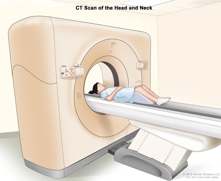
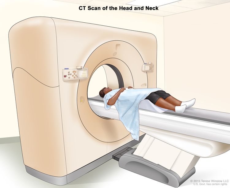
  • Computed tomography (CT) scan. CT with intravenous contrast is useful in the diagnosis and clinical staging of thymoma. CT is usually accurate in predicting tumor size, location, and invasion into vessels, the pericardium, and the lungs.[27,28]

    The appearance of the tumor on CT may indicate the histological tumor type.[25] In a retrospective study involving 53 patients who underwent thymectomy for TETs, CT indicated that smooth contours with a round shape were most suggestive of type A thymomas, and irregular contours were most suggestive of thymic carcinomas. Calcification was suggestive of type B thymomas. In this study, however, CT was found to be of limited value in differentiating type AB, B1, B2, and B3 thymomas.[29]

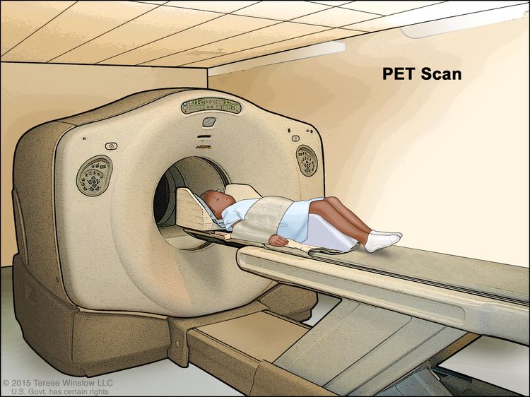
  • Positron emission tomography (PET) scan. Fluorine F 18-fludeoxyglucose (18F-FDG) PET and thallium single-photon emission CT have been reported in small series for diagnosis and evaluation of therapeutic outcomes in thymic carcinoma.[3033] Two small series reported that 18F-FDG uptake was related to the invasiveness of thymic carcinoma.[32,33] This raises the possibility of 18F-FDG PET use for diagnosis, treatment planning, and monitoring for recurrence. The impact of sensitivity and specificity on clinical therapeutic decisions is yet to be defined.
  • Magnetic resonance imaging (MRI). MRI can distinguish TETs from other malignant and benign mediastinal lesions. Chemical-shift MRI can help differentiate TETs from thymic hyperplasia and a normal thymus. Cardiac MRI is the preferred modality to evaluate for the presence of myocardial involvement. An MRI can help identify phrenic nerve involvement and is considered superior to CT for assessing chest wall invasion.[34]

Thymic carcinoma can metastasize to regional lymph nodes, bone, liver, or lungs. An evaluation for sites of metastases may be warranted.

Prognostic Factors and Prognosis

The World Health Organization (WHO) pathological classification of tumors of the thymus and stage correlate with prognosis.[25] The degree of invasion or tumor stage is generally thought to be a more important indicator of overall survival (OS).[27,35,36]

Thymoma

Histological classification of thymoma is not sufficient to distinguish biologically indolent thymomas from thymomas that exhibit aggressive clinical behavior. Although some thymoma histological types are more likely to be clinically aggressive, treatment outcome and the likelihood of recurrence appear to correlate more closely with the invasive/metastasizing properties of the tumor cells.[25,35] Therefore, some thymomas that appear to be relatively benign by histological criteria may behave very aggressively. Independent evaluations of both tumor invasiveness (using staging criteria) and tumor histology may be combined to predict the clinical behavior of a thymoma.

Both histological classification of thymomas and stage may have independent prognostic significance.[35,36] A few series have reported the prognostic value of the WHO classifications. Two large retrospective analyses, one with 100 thymoma cases and the other with 178 thymoma cases, showed that disease-free survival at 10 years varied (see Table 1).[37,38] In these series, stage and complete resection were significant independent prognostic factors. Another analysis reported on 273 thymoma patients who were treated over a 44-year period. See Table 1 for the 20-year survival rates.[35]

Table 1. Disease-Free Survival (DFS) of Patients With Thymoma by Histological Subtype
Study Histological Subtype
  A AB B1 B2 B3 C
a10-year DFS.
b20-year DFS.
[37] (N = 100)a 100% 100% 83% 83% 36% 28%
[38] (N = 178)a 95% 90% 85% 71% 40%  
[35] (N = 273)b 100% 87% 91% 59% 36%  

Thymic carcinoma

Thymic carcinomas are usually advanced when diagnosed.[39,40] Thymic carcinomas have a greater propensity for capsular invasion, metastases, and recurrence than thymomas. Patients with thymic carcinoma have worse survival than patients with thymoma (5-year survival rate, 30%–50%).[41] In a retrospective study of 40 patients with thymic carcinoma, the OS rates were 38% for 5 years and 28% for 10 years.[39] In another retrospective study evaluating 43 cases of thymic carcinoma, prognosis was found to depend solely on tumor invasion of the brachiocephalic artery.[40]

Follow-Up After Treatment of Thymoma

Thymoma has been associated with an increased risk of second malignancies. Because of this risk and because thymoma can recur after a long interval, lifelong surveillance should be considered.[22] The measurement of interferon-alpha and interleukin-2 antibodies is helpful in identifying patients with a thymoma recurrence.[42]

In a study of 849 cases between 1973 and 1998, there was an excess risk of subsequent non-Hodgkin lymphoma and soft tissue sarcomas following thymoma.[43] Risk of second malignancy does not appear to be related to thymectomy, radiation therapy, or a clinical history of myasthenia gravis.[22,43,44]

References
  1. Kelly RJ, Petrini I, Rajan A, et al.: Thymic malignancies: from clinical management to targeted therapies. J Clin Oncol 29 (36): 4820-7, 2011. [PUBMED Abstract]
  2. Fornasiero A, Daniele O, Ghiotto C, et al.: Chemotherapy of invasive thymoma. J Clin Oncol 8 (8): 1419-23, 1990. [PUBMED Abstract]
  3. Engels EA: Epidemiology of thymoma and associated malignancies. J Thorac Oncol 5 (10 Suppl 4): S260-5, 2010. [PUBMED Abstract]
  4. Carter BW, Benveniste MF, Madan R, et al.: IASLC/ITMIG Staging System and Lymph Node Map for Thymic Epithelial Neoplasms. Radiographics 37 (3): 758-776, 2017 May-Jun. [PUBMED Abstract]
  5. Kondo K, Monden Y: Therapy for thymic epithelial tumors: a clinical study of 1,320 patients from Japan. Ann Thorac Surg 76 (3): 878-84; discussion 884-5, 2003. [PUBMED Abstract]
  6. Levy Y, Afek A, Sherer Y, et al.: Malignant thymoma associated with autoimmune diseases: a retrospective study and review of the literature. Semin Arthritis Rheum 28 (2): 73-9, 1998. [PUBMED Abstract]
  7. Marx A, Willcox N, Leite MI, et al.: Thymoma and paraneoplastic myasthenia gravis. Autoimmunity 43 (5-6): 413-27, 2010. [PUBMED Abstract]
  8. Bernard C, Frih H, Pasquet F, et al.: Thymoma associated with autoimmune diseases: 85 cases and literature review. Autoimmun Rev 15 (1): 82-92, 2016. [PUBMED Abstract]
  9. Padda SK, Yao X, Antonicelli A, et al.: Paraneoplastic Syndromes and Thymic Malignancies: An Examination of the International Thymic Malignancy Interest Group Retrospective Database. J Thorac Oncol 13 (3): 436-446, 2018. [PUBMED Abstract]
  10. Kisand K, Lilic D, Casanova JL, et al.: Mucocutaneous candidiasis and autoimmunity against cytokines in APECED and thymoma patients: clinical and pathogenetic implications. Eur J Immunol 41 (6): 1517-27, 2011. [PUBMED Abstract]
  11. Hoffacker V, Schultz A, Tiesinga JJ, et al.: Thymomas alter the T-cell subset composition in the blood: a potential mechanism for thymoma-associated autoimmune disease. Blood 96 (12): 3872-9, 2000. [PUBMED Abstract]
  12. Buckley C, Douek D, Newsom-Davis J, et al.: Mature, long-lived CD4+ and CD8+ T cells are generated by the thymoma in myasthenia gravis. Ann Neurol 50 (1): 64-72, 2001. [PUBMED Abstract]
  13. Ströbel P, Helmreich M, Menioudakis G, et al.: Paraneoplastic myasthenia gravis correlates with generation of mature naive CD4(+) T cells in thymomas. Blood 100 (1): 159-66, 2002. [PUBMED Abstract]
  14. Martinez B, Browne SK: Good syndrome, bad problem. Front Oncol 4: 307, 2014. [PUBMED Abstract]
  15. Burbelo PD, Browne SK, Sampaio EP, et al.: Anti-cytokine autoantibodies are associated with opportunistic infection in patients with thymic neoplasia. Blood 116 (23): 4848-58, 2010. [PUBMED Abstract]
  16. Morgenthaler TI, Brown LR, Colby TV, et al.: Thymoma. Mayo Clin Proc 68 (11): 1110-23, 1993. [PUBMED Abstract]
  17. Souadjian JV, Enriquez P, Silverstein MN, et al.: The spectrum of diseases associated with thymoma. Coincidence or syndrome? Arch Intern Med 134 (2): 374-9, 1974. [PUBMED Abstract]
  18. Voltz RD, Albrich WC, Nägele A, et al.: Paraneoplastic myasthenia gravis: detection of anti-MGT30 (titin) antibodies predicts thymic epithelial tumor. Neurology 49 (5): 1454-7, 1997. [PUBMED Abstract]
  19. Gautel M, Lakey A, Barlow DP, et al.: Titin antibodies in myasthenia gravis: identification of a major immunogenic region of titin. Neurology 43 (8): 1581-5, 1993. [PUBMED Abstract]
  20. Kondo K, Monden Y: Thymoma and myasthenia gravis: a clinical study of 1,089 patients from Japan. Ann Thorac Surg 79 (1): 219-24, 2005. [PUBMED Abstract]
  21. Budde JM, Morris CD, Gal AA, et al.: Predictors of outcome in thymectomy for myasthenia gravis. Ann Thorac Surg 72 (1): 197-202, 2001. [PUBMED Abstract]
  22. Evoli A, Minisci C, Di Schino C, et al.: Thymoma in patients with MG: characteristics and long-term outcome. Neurology 59 (12): 1844-50, 2002. [PUBMED Abstract]
  23. Schmidt-Wolf IG, Rockstroh JK, Schüller H, et al.: Malignant thymoma: current status of classification and multimodality treatment. Ann Hematol 82 (2): 69-76, 2003. [PUBMED Abstract]
  24. Rajan A, Giaccone G: Treatment of advanced thymoma and thymic carcinoma. Curr Treat Options Oncol 9 (4-6): 277-87, 2008. [PUBMED Abstract]
  25. Rosai J: Histological Typing of Tumours of the Thymus. Springer-Verlag, 2nd ed., 1999.
  26. Strollo DC, Rosado-de-Christenson ML: Tumors of the thymus. J Thorac Imaging 14 (3): 152-71, 1999. [PUBMED Abstract]
  27. Sperling B, Marschall J, Kennedy R, et al.: Thymoma: a review of the clinical and pathological findings in 65 cases. Can J Surg 46 (1): 37-42, 2003. [PUBMED Abstract]
  28. Rendina EA, Venuta F, Ceroni L, et al.: Computed tomographic staging of anterior mediastinal neoplasms. Thorax 43 (6): 441-5, 1988. [PUBMED Abstract]
  29. Tomiyama N, Johkoh T, Mihara N, et al.: Using the World Health Organization Classification of thymic epithelial neoplasms to describe CT findings. AJR Am J Roentgenol 179 (4): 881-6, 2002. [PUBMED Abstract]
  30. Sasaki M, Kuwabara Y, Ichiya Y, et al.: Differential diagnosis of thymic tumors using a combination of 11C-methionine PET and FDG PET. J Nucl Med 40 (10): 1595-601, 1999. [PUBMED Abstract]
  31. Kageyama M, Seto H, Shimizu M, et al.: Thallium-201 single photon emission computed tomography in the evaluation of thymic carcinoma. Radiat Med 12 (5): 237-9, 1994 Sep-Oct. [PUBMED Abstract]
  32. Adams S, Baum RP, Hertel A, et al.: Metabolic (PET) and receptor (SPET) imaging of well- and less well-differentiated tumours: comparison with the expression of the Ki-67 antigen. Nucl Med Commun 19 (7): 641-7, 1998. [PUBMED Abstract]
  33. Kubota K, Yamada S, Kondo T, et al.: PET imaging of primary mediastinal tumours. Br J Cancer 73 (7): 882-6, 1996. [PUBMED Abstract]
  34. Carter BW, Lichtenberger JP, Benveniste MF: MR Imaging of Thymic Epithelial Neoplasms. Top Magn Reson Imaging 27 (2): 65-71, 2018. [PUBMED Abstract]
  35. Okumura M, Ohta M, Tateyama H, et al.: The World Health Organization histologic classification system reflects the oncologic behavior of thymoma: a clinical study of 273 patients. Cancer 94 (3): 624-32, 2002. [PUBMED Abstract]
  36. Chen G, Marx A, Wen-Hu C, et al.: New WHO histologic classification predicts prognosis of thymic epithelial tumors: a clinicopathologic study of 200 thymoma cases from China. Cancer 95 (2): 420-9, 2002. [PUBMED Abstract]
  37. Kondo K, Yoshizawa K, Tsuyuguchi M, et al.: WHO histologic classification is a prognostic indicator in thymoma. Ann Thorac Surg 77 (4): 1183-8, 2004. [PUBMED Abstract]
  38. Rena O, Papalia E, Maggi G, et al.: World Health Organization histologic classification: an independent prognostic factor in resected thymomas. Lung Cancer 50 (1): 59-66, 2005. [PUBMED Abstract]
  39. Ogawa K, Toita T, Uno T, et al.: Treatment and prognosis of thymic carcinoma: a retrospective analysis of 40 cases. Cancer 94 (12): 3115-9, 2002. [PUBMED Abstract]
  40. Blumberg D, Burt ME, Bains MS, et al.: Thymic carcinoma: current staging does not predict prognosis. J Thorac Cardiovasc Surg 115 (2): 303-8; discussion 308-9, 1998. [PUBMED Abstract]
  41. Eng TY, Fuller CD, Jagirdar J, et al.: Thymic carcinoma: state of the art review. Int J Radiat Oncol Biol Phys 59 (3): 654-64, 2004. [PUBMED Abstract]
  42. Buckley C, Newsom-Davis J, Willcox N, et al.: Do titin and cytokine antibodies in MG patients predict thymoma or thymoma recurrence? Neurology 57 (9): 1579-82, 2001. [PUBMED Abstract]
  43. Engels EA, Pfeiffer RM: Malignant thymoma in the United States: demographic patterns in incidence and associations with subsequent malignancies. Int J Cancer 105 (4): 546-51, 2003. [PUBMED Abstract]
  44. Pan CC, Chen PC, Wang LS, et al.: Thymoma is associated with an increased risk of second malignancy. Cancer 92 (9): 2406-11, 2001. [PUBMED Abstract]

Cellular Classification and Molecular Characteristics of Thymoma and Thymic Carcinomas

The histological classification of thymic epithelial tumors (TETs) is largely based on the third edition of the World Health Organization (WHO) classification of tumors of the lung, pleura, thymus, and heart, published in 2004. The fourth edition of the WHO classification, published in 2015, contains refined histological and immunohistochemical diagnostic criteria and is the most widely accepted cellular classification of TETs.[1,2] Thymomas arise from the thymic epithelium and consist of epithelial cells mixed with varying proportions of immature T cells. Thymic carcinomas are epithelial tumors with overt cytological atypia and without organotypic (i.e., thymus-like) features.

Thymoma

The epithelial component of thymomas exhibit no or minimal overt atypia and retain histological features specific to the normal thymus.[1] Immature nonneoplastic lymphocytes are present in variable numbers depending on the histological type of thymoma.

Table 2, Table 3, Table 4, Table 5, and Table 6 describe morphologic, molecular, and clinical characteristics of various subtypes of thymoma.

Table 2. Characteristics of Subtype A Thymoma
OS = overall survival.
Histological subtype percentage of all thymomas in study cited.[3,4] Approximately 4%–7%.
Myasthenia gravis association.[3] Approximately 17%.
Morphologic characteristics.[2] Composed of bland, spindle-shaped epithelial cells (at least focally) with a paucity or absence of immature (TdT+) T cells throughout the tumor.
Molecular characteristics.[5,6] Chromosome abnormalities, when present, may correlate with an aggressive clinical course and may include: chromosome 6q25 loss, chromosome 6p23 loss (FOXC1), C19MC overexpression, GTF2I variants, HRAS (G13V) variants, and miR-515 upregulation.
Prognosis and survival.[3,4] Excellent, with a ≥15-year OS rate of 100%.
Table 3. Characteristics of Subtype AB Thymoma
OS = overall survival.
Histological subtype percentage of all thymomas in study cited.[3,4] Approximately 28%–34%.
Myasthenia gravis association.[3] Approximately 16%.
Morphologic characteristics.[2] Composed of bland, spindle-shaped epithelial cells (at least focally), with an abundance of immature (TdT+) T cells focally or throughout the tumor.
Molecular characteristics.[5,6] Includes chromosome 6q25 loss, chromosome 6p23 loss (FOXC1), chromosome 7p15 loss, C19MC overexpression, and GTF2I variants.
Prognosis and survival.[3,4] Good, with a ≥15-year OS rate of approximately 90%.
Table 4. Characteristics of Subtype B1 Thymoma
OS = overall survival.
Histological subtype percentage of all thymomas in study cited.[3,4] Approximately 9%–20%.
Myasthenia gravis association.[3] Approximately 57%.
Morphologic characteristics.[2] Tumors exhibit thymus-like architecture and cytology including the abundance of immature T cells, areas of medullary differentiation (medullary islands), and a paucity of polygonal or dendritic epithelia cells without clustering (i.e., <3 contiguous epithelial cells).
Molecular characteristics.[5] Includes chromosome 1p, 2q, 3q, 6q losses.
Prognosis and survival.[3,4] Good, with a ≥20-year OS rate of approximately 90%.
Table 5. Characteristics of Subtype B2 Thymoma
OS = overall survival.
Histological subtype percentage of all thymomas in study cited.[3,4] Approximately 20%–36%.
Myasthenia gravis association.[3] Approximately 71%.
Morphologic characteristics.[2] Tumors consist of increased numbers of single or clustered polygonal or dendritic epithelial cells intermingled with abundant immature T cells.
Molecular characteristics.[5] Includes chromosome 6q25 loss, chromosome 6p23 loss (FOXC1), chromosome 1q gain, and KRAS (G12A) variants.
Prognosis and survival.[3] Worse than for thymoma types A, AB, and B1, with a 20-year OS rate (as defined by freedom from tumor death) of approximately 60%.
Table 6. Characteristics of Subtype B3 Thymoma
OS = overall survival.
Histological subtype percentage of all thymomas in study cited.[3,4] Approximately 10%–14%.
Myasthenia gravis association.[3] Approximately 46%.
Morphologic characteristics.[2] Predominantly composed of sheets of polygonal, slightly-to-moderately atypical epithelial cells, absent or rare intercellular bridges, and paucity or absence of intermingled TdT+ T cells.
Molecular characteristics.[5] Includes chromosome 6q25 loss, chromosome 6p23 loss (FOXC1), chromosome 11q4 loss, chromosome 1q gain, chromosomal translocation t(11;X), BCL2 copy number gains (18q21.33), MCL1 copy number gain, CDKN2A/B copy number losses (9p21.3), BCOR variants, and PHF15 variants.
Prognosis and survival.[3] A 20-year OS rate (as defined by freedom from tumor death) of approximately 40%.

Thymic Carcinoma

Thymic carcinoma is a TET that exhibits a definite cytological atypia and a set of histological features no longer specific to the thymus but similar to histological features observed in carcinomas of other organs. Unlike type A and B thymomas, thymic carcinomas lack immature lymphocytes. Any lymphocytes that are present are mature and usually admixed with plasma cells.[1]

The characteristics of thymic carcinoma subtypes are described in Table 7.

Table 7. Characteristics of Thymic Carcinoma Subtypesa
Subtype Characteristics
CEA = carcinoembryonic antigen; CK = cytokeratin; EMA = epithelial membrane antigen; PAS = periodic acid-Schiff; PLAP = placental alkaline phosphatase.
aAdapted from [7,8].
Squamous cell carcinoma (SCC) The most common subtype of thymic carcinoma, SCC exhibits clear-cut cytological atypia and resembles SCC arising in other organs. Not all cases have clear evidence of keratinization. SCC lacks immature T lymphocytes. CD5, CD70, CD117, FoxN1, and CD205 are expressed by most thymic SCCs.
Basaloid carcinoma Composed of compact lobules of tumor cells that exhibit peripheral palisading and an overall basophilic staining pattern caused by the high nucleocytoplasmic ratio. Basaloid carcinoma tends to originate from multilocular thymic cysts, expresses keratin and EMA, can express CD5 but does not express S-100 and neuroendocrine markers.
Lymphoepithelioma-like carcinoma Syncytial growth of undifferentiated carcinoma cells accompanied by a lymphoplasmacytic infiltration is like undifferentiated carcinoma of the respiratory tract. Lymphoepithelioma-like carcinoma may or may not be Epstein-Barr virus positive. Tumor cells are strongly positive for AE1-defined acidic CKs and negative for AE3-defined basic CKs. CK7 and CK20 are also negative. BCL-2 expression is common. CD5 is focally expressed or absent. Lymphoid cells are CD3+, CD5+, CD1a-, CD99-, and TdT-mature T cells. CD20+ B cells are present in small numbers in the stroma.
Sarcomatoid thymic carcinoma Part or all of the tumor resembles one of the types of soft tissue sarcoma. Sarcomatoid carcinoma includes spindle cell carcinoma (i.e., malignant transformation of type A thymoma), sarcomatoid transformation of preexisting thymic carcinoma, and true carcinosarcoma with heterologous component(s).
Clear cell thymic carcinoma Composed predominantly or exclusively of cells with optically clear cytoplasm. Tumor cells usually show strong cytoplasmic diastase-labile PAS positivity. Clear cell carcinomas are keratin positive. EMA is expressed in 20% of cases. CD5 expression is present in some cases. PLAP, vimentin, CEA, and S-100 are negative.
Mucoepidermoid thymic carcinoma Consists of squamous cells, mucus-producing cells, and cells of intermediate type and resembles mucoepidermoid carcinoma of other organs. Translocation of the MAML2 gene is present and can help distinguish this tumor from adenosquamous carcinomas and adenocarcinomas.
Papillary thymic adenocarcinoma Grows in a papillary fashion. Histology may be accompanied by psammoma body formation, which may result in a marked similarity with papillary carcinoma of the thyroid gland. Variable expression of Leu M1 and BerEP4 is observed. CEA and CD5 may also be positive. CD20, thyroglobulin, pulmonary surfactant apoprotein, and calretinin are absent.
Undifferentiated thymic carcinoma A rare type of thymic carcinoma that grows in a solid undifferentiated fashion but without exhibiting sarcomatoid (spindle cell or pleomorphic) features.
Carcinoma with t(15;19) translocation (NUT carcinoma) A rare, aggressive carcinoma of unknown histogenesis. The presence of undifferentiated, intermediate-sized, vigorously mitotic cells is characteristic. Pan-cytokeratin markers are expressed. Focal positivity of vimentin, EMA, and CEA is observed. CD30, CD45, PLAP, HMB45, S100, and neuroendocrine markers are negative. t(15;19)-translocation is observed with the generation of a BRD4::NUT fusion oncogene. Immunohistochemistry for NUT is highly sensitive and should be considered in any undifferentiated cancer, especially if focal squamous differentiation is seen.

Molecular Characteristics of Thymoma and Thymic Carcinomas

TETs have the lowest tumor mutational burden of all adult cancers. Multiplatform analyses have revealed four molecular subtypes that are associated with survival and WHO histological subtypes. Pathogenic variants in HRAS, NRAS, TP53, and GTF2I have been observed. Targetable variants are uncommon. Tumor overexpression of muscle autoantigens and increased aneuploidy have also been identified and provide a molecular link between thymoma and myasthenia gravis.[6]

References
  1. Travis WD, Brambilla E, Burke E, et al.: WHO Classification of Tumours of the Lung, Pleura, Thymus and Heart. 4th ed. International Agency for Research on Cancer, 2015.
  2. Marx A, Chan JK, Coindre JM, et al.: The 2015 World Health Organization Classification of Tumors of the Thymus: Continuity and Changes. J Thorac Oncol 10 (10): 1383-95, 2015. [PUBMED Abstract]
  3. Hirabayashi H, Fujii Y, Sakaguchi M, et al.: p16INK4, pRB, p53 and cyclin D1 expression and hypermethylation of CDKN2 gene in thymoma and thymic carcinoma. Int J Cancer 73 (5): 639-44, 1997. [PUBMED Abstract]
  4. Sasaki H, Kobayashi Y, Tanahashi M, et al.: Ets-1 gene expression in patients with thymoma. Jpn J Thorac Cardiovasc Surg 50 (12): 503-7, 2002. [PUBMED Abstract]
  5. Rajan A, Girard N, Marx A: State of the art of genetic alterations in thymic epithelial tumors. J Thorac Oncol 9 (9 Suppl 2): S131-6, 2014. [PUBMED Abstract]
  6. Radovich M, Pickering CR, Felau I, et al.: The Integrated Genomic Landscape of Thymic Epithelial Tumors. Cancer Cell 33 (2): 244-258.e10, 2018. [PUBMED Abstract]
  7. Travis W, Brambilla E, Müller-Hermelink H, et al., eds.: Pathology and Genetics of Tumours of the Lung, Pleura, and Thymus. IARC Press, 2004. World Health Organization Classification of Tumours.
  8. Marx A, Chan J, Coindre J-M, et al.: The 2015 WHO classification of tumors of the thymus: continuity and changes. J Thorac Oncol 10 (10): 1383–95, 2015.

Stage Information for Thymoma and Thymic Carcinoma

Evaluating the invasiveness of a thymoma involves the use of staging criteria that indicate the presence and degree of contiguous invasion, the presence of tumor implants, and lymph node or distant metastases regardless of histological type. The staging system, proposed by Masaoka in 1981 and modified by Koga in 1994, is most commonly used, with the modified system being recommended by the International Thymic Malignancies Interest Group (ITMIG) (see Table 8).[1,2] To establish consistency in the staging of thymic epithelial tumors (TETs), the American Joint Committee on Cancer (AJCC) and the Union for International Cancer Control (UICC) adopted a new TNM (tumor, node, metastasis) classification system developed by the International Association for the Study of Lung Cancer (IASLC) and ITMIG.[35]

Table 8. Masaoka-Koga Staging System for Thymoma, 1994a
Stage Description
a [2]
I Macroscopically, completely encapsulated; microscopically, no capsular invasion.
II Macroscopic invasion into surrounding fatty tissue or mediastinal pleura; microscopic invasion into capsule.
III Macroscopic invasion into neighboring organs (pericardium, lung, and great vessels).
IVa Pleural or pericardial dissemination.
IVb Lymphogenous or hematogenous metastases.

AJCC Stage Groupings and TNM Definitions

Table 9. Definition of TNM Stage Ia
Stage Tb,cNbM Description
T = primary tumor; N = regional lymph node; M = distant metastasis.
aAdapted from AJCC: Thymus. In: Amin MB, Edge SB, Greene FL, et al., eds.: AJCC Cancer Staging Manual. 8th ed. New York, NY: Springer, 2017, pp. 423–9.
The explanations for superscripts b and c are at the end of Table 12.
 
I T1a,b, N0, M0 T1 = Tumor encapsulated or extending into the mediastinal fat; may involve the mediastinal pleura.
–T1a = Tumor with no mediastinal pleura involvement.
–T1b = Tumor with direct invasion of mediastinal pleura.
N0 = No regional lymph node metastasis.
M0 = No pleural, pericardial, or distant metastasis.
Table 10. Definition of TNM Stage IIa
Stage Tb,cNbM Description
T = primary tumor; N = regional lymph node; M = distant metastasis.
aAdapted from AJCC: Thymus. In: Amin MB, Edge SB, Greene FL, et al., eds.: AJCC Cancer Staging Manual. 8th ed. New York, NY: Springer, 2017, pp. 423–9.
The explanations for superscripts b and c are at the end of Table 12.
 
II T2, N0, M0 T2 = Tumor with direct invasion of the pericardium (either partial or full thickness).
N0 = No regional lymph node metastasis.
M0 = No pleural, pericardial, or distant metastasis.
Table 11. Definition of TNM Stages IIIA and IIIBa
Stage Tb,cNbM Description
T = primary tumor; N = regional lymph node; M = distant metastasis.
aAdapted from AJCC: Thymus. In: Amin MB, Edge SB, Greene FL, et al., eds.: AJCC Cancer Staging Manual. 8th ed. New York, NY: Springer, 2017, pp. 423–9.
The explanations for superscripts b and c are at the end of Table 12.
 
IIIA T3, N0, M0 T3 = Tumor with direct invasion into any of the following: lung, brachiocephalic vein, superior vena cava, phrenic nerve, chest wall, or extrapericardial pulmonary artery or veins.
N0 = No regional lymph node metastasis.
M0 = No pleural, pericardial, or distant metastasis.
IIIB T4, N0, M0 T4 = Tumor with invasion into any of the following: aorta (ascending, arch, or descending), arch vessels, intrapericardial pulmonary artery, myocardium, trachea, esophagus.
N0 = No regional lymph node metastasis.
M0 = No pleural, pericardial, or distant metastasis.
Table 12. Definition of TNM Stages IVA and IVBa
Stage Tb,cNbM Description
T = primary tumor; N = regional lymph node; M = distant metastasis.
aAdapted from AJCC: Thymus. In: Amin MB, Edge SB, Greene FL, et al., eds.: AJCC Cancer Staging Manual. 8th ed. New York, NY: Springer, 2017, pp. 423–9.
bInvolvement must be microscopically confirmed in pathological staging, if possible.
cT categories are defined by levels of invasion; they reflect the highest degree of invasion regardless of how many other (lower-level) structures are invaded. T1, level 1 structures: thymus, anterior mediastinal fat, mediastinal pleura; T2, level 2 structures: pericardium; T3, level 3 structures: lung, brachiocephalic vein, superior vena cava, phrenic nerve, chest wall, hilar pulmonary vessels; T4, level 4 structures: aorta (ascending, arch, or descending), arch vessels, intrapericardial pulmonary artery, myocardium, trachea, esophagus.
IVA Any T, N1, M0 TX = Primary tumor cannot be assessed.
T0 = No evidence of primary tumor.
T1 = Tumor encapsulated or extending into the mediastinal fat; may involve the mediastinal pleura.
–T1a = Tumor with no mediastinal pleura involvement.
–T1b = Tumor with direct invasion of mediastinal pleura.
T2 = Tumor with direct invasion of the pericardium (either partial or full thickness).
T3 = Tumor with direct invasion into any of the following: lung, brachiocephalic vein, superior vena cava, phrenic nerve, chest wall, or extrapericardial pulmonary artery or veins.
T4 = Tumor with invasion into any of the following: aorta (ascending, arch, or descending), arch vessels, intrapericardial pulmonary artery, myocardium, trachea, esophagus.
N1 = Metastasis in anterior (perithymic) lymph nodes.
M0 = No pleural, pericardial, or distant metastasis.
Any T, N0,1, M1a Any T = See descriptions (stage IVA) in this table.
N0 = No regional lymph node metastasis.
N1 = Metastasis in anterior (perithymic) lymph nodes.
M1a = Separate pleural or pericardial nodule(s).
IVB Any T, N2, M0, M1a Any T = See descriptions (stage IVA) in this table.
N2 = Metastasis in deep intrathoracic or cervical lymph nodes.
M0 = No pleural, pericardial, or distant metastasis.
M1a = Separate pleural or pericardial nodule(s).
Any T, Any N, M1b Any T = See descriptions (stage IVA) in this table.
NX = Regional lymph nodes cannot be assessed.
N0 = No regional lymph node metastasis.
N1 = Metastasis in anterior (perithymic) lymph nodes.
N2 = Metastasis in deep intrathoracic or cervical lymph nodes.
M1b = Pulmonary intraparenchymal nodule or distant organ metastasis.

When the Masaoka staging system was applied to a series of 85 surgically treated patients with thymoma, its value in determining prognosis was confirmed, with 5-year survival rates of 96% for stage I disease, 86% for stage II disease, 69% for stage III disease, and 50% for stage IV disease.[1] In a large, retrospective study involving 273 patients with thymoma, 20-year survival rates (as defined by freedom from tumor death) according to the Masaoka staging system were reported to be 89% for stage I disease, 91% for stage II disease, 49% for stage III disease, and 0% for stage IV disease.[6]

The TNM staging system, applicable to thymoma and thymic carcinoma, is based on a large, global database of more than 10,000 subjects, as opposed to smaller series of fewer than 100 patients that were used to develop older staging systems. The TNM system also benefits from rigorous statistical analysis of a large pool of data and input from a multidisciplinary panel of experts. The rate of disease recurrence was 5% in patients with stage I disease, 18% for stage II disease, 32% for stage III disease, 59% for stage IVA disease, and 49% for stage IVB disease. The death rate was 7% in patients with stage I disease, 16% for stage II disease, 18% for stage III disease, 30% for stage IVA disease, and 33% for stage IVB disease.[5]

References
  1. Masaoka A, Monden Y, Nakahara K, et al.: Follow-up study of thymomas with special reference to their clinical stages. Cancer 48 (11): 2485-92, 1981. [PUBMED Abstract]
  2. Koga K, Matsuno Y, Noguchi M, et al.: A review of 79 thymomas: modification of staging system and reappraisal of conventional division into invasive and non-invasive thymoma. Pathol Int 44 (5): 359-67, 1994. [PUBMED Abstract]
  3. Thymus. In: Amin MB, Edge SB, Greene FL, et al., eds.: AJCC Cancer Staging Manual. 8th ed. Springer; 2017, pp. 423–9.
  4. Carter BW, Benveniste MF, Madan R, et al.: IASLC/ITMIG Staging System and Lymph Node Map for Thymic Epithelial Neoplasms. Radiographics 37 (3): 758-776, 2017 May-Jun. [PUBMED Abstract]
  5. Detterbeck FC, Stratton K, Giroux D, et al.: The IASLC/ITMIG Thymic Epithelial Tumors Staging Project: proposal for an evidence-based stage classification system for the forthcoming (8th) edition of the TNM classification of malignant tumors. J Thorac Oncol 9 (9 Suppl 2): S65-72, 2014. [PUBMED Abstract]
  6. Okumura M, Ohta M, Tateyama H, et al.: The World Health Organization histologic classification system reflects the oncologic behavior of thymoma: a clinical study of 273 patients. Cancer 94 (3): 624-32, 2002. [PUBMED Abstract]

Treatment Option Overview for Thymoma and Thymic Carcinoma

The primary treatment for patients with thymoma or thymic carcinoma is surgical resection with en bloc resection for invasive tumors, if possible.[13] Depending on tumor stage, multimodality treatment options—including the use of radiation therapy and chemotherapy with or without surgery—may be used.[4,5] The optimal strategy for induction therapy, which minimizes operative morbidity and mortality and optimizes resectability rates and ultimately survival, remains unknown. A review of the management of thymic epithelial tumors has been published.[1]

Table 13. Treatment Options for Thymoma and Thymic Carcinoma
Stage Treatment Options
Stage I and II thymoma Surgery
Surgery with or without postoperative radiation therapy
Stage III and IV thymoma (operable) Surgery followed by radiation therapy
Induction chemotherapy followed by surgery and radiation therapy
Stage III and IV thymoma (inoperable) Chemotherapy
Chemotherapy followed by radiation therapy
Chemotherapy followed by surgery (if operable) and radiation therapy
Thymic carcinoma (operable) Surgery (en bloc surgical resection) followed by postoperative radiation therapy with or without postoperative chemotherapy.
Thymic carcinoma (inoperable) Chemotherapy
Chemoradiation therapy
Chemotherapy followed by surgery (if operable) and radiation therapy
Recurrent thymoma and thymic carcinoma Chemotherapy
Biological therapies
Surgery or radiation therapy in carefully selected cases
Pembrolizumab (under clinical evaluation)

Capecitabine and Fluorouracil Dosing

The DPYD gene encodes an enzyme that catabolizes pyrimidines and fluoropyrimidines, like capecitabine and fluorouracil. An estimated 1% to 2% of the population has germline pathogenic variants in DPYD, which lead to reduced DPD protein function and an accumulation of pyrimidines and fluoropyrimidines in the body.[6,7] Patients with the DPYD*2A variant who receive fluoropyrimidines may experience severe, life-threatening toxicities that are sometimes fatal. Many other DPYD variants have been identified, with a range of clinical effects.[68] Fluoropyrimidine avoidance or a dose reduction of 50% may be recommended based on the patient’s DPYD genotype and number of functioning DPYD alleles.[911] DPYD genetic testing costs less than $200, but insurance coverage varies due to a lack of national guidelines.[12] In addition, testing may delay therapy by 2 weeks, which would not be advisable in urgent situations. This controversial issue requires further evaluation.[13]

References
  1. Girard N, Ruffini E, Marx A, et al.: Thymic epithelial tumours: ESMO Clinical Practice Guidelines for diagnosis, treatment and follow-up. Ann Oncol 26 (Suppl 5): v40-55, 2015. [PUBMED Abstract]
  2. Ruffini E, Filosso PL, Guerrera F, et al.: Optimal surgical approach to thymic malignancies: New trends challenging old dogmas. Lung Cancer 118: 161-170, 2018. [PUBMED Abstract]
  3. Cameron RB, Loehrer Sr, Marx A: Neoplasms of the mediastinum. In: DeVita VT Jr, Lawrence TS, Rosenberg SA, et al., eds.: DeVita, Hellman, and Rosenberg’s Cancer: Principles & Practice of Oncology. 11th ed. Wolters Kluwer, 2019, pp 700-12.
  4. Rimner A, Yao X, Huang J, et al.: Postoperative Radiation Therapy Is Associated with Longer Overall Survival in Completely Resected Stage II and III Thymoma-An Analysis of the International Thymic Malignancies Interest Group Retrospective Database. J Thorac Oncol 11 (10): 1785-92, 2016. [PUBMED Abstract]
  5. Rajan A, Giaccone G: Chemotherapy for thymic tumors: induction, consolidation, palliation. Thorac Surg Clin 21 (1): 107-14, viii, 2011. [PUBMED Abstract]
  6. Sharma BB, Rai K, Blunt H, et al.: Pathogenic DPYD Variants and Treatment-Related Mortality in Patients Receiving Fluoropyrimidine Chemotherapy: A Systematic Review and Meta-Analysis. Oncologist 26 (12): 1008-1016, 2021. [PUBMED Abstract]
  7. Lam SW, Guchelaar HJ, Boven E: The role of pharmacogenetics in capecitabine efficacy and toxicity. Cancer Treat Rev 50: 9-22, 2016. [PUBMED Abstract]
  8. Shakeel F, Fang F, Kwon JW, et al.: Patients carrying DPYD variant alleles have increased risk of severe toxicity and related treatment modifications during fluoropyrimidine chemotherapy. Pharmacogenomics 22 (3): 145-155, 2021. [PUBMED Abstract]
  9. Amstutz U, Henricks LM, Offer SM, et al.: Clinical Pharmacogenetics Implementation Consortium (CPIC) Guideline for Dihydropyrimidine Dehydrogenase Genotype and Fluoropyrimidine Dosing: 2017 Update. Clin Pharmacol Ther 103 (2): 210-216, 2018. [PUBMED Abstract]
  10. Henricks LM, Lunenburg CATC, de Man FM, et al.: DPYD genotype-guided dose individualisation of fluoropyrimidine therapy in patients with cancer: a prospective safety analysis. Lancet Oncol 19 (11): 1459-1467, 2018. [PUBMED Abstract]
  11. Lau-Min KS, Varughese LA, Nelson MN, et al.: Preemptive pharmacogenetic testing to guide chemotherapy dosing in patients with gastrointestinal malignancies: a qualitative study of barriers to implementation. BMC Cancer 22 (1): 47, 2022. [PUBMED Abstract]
  12. Brooks GA, Tapp S, Daly AT, et al.: Cost-effectiveness of DPYD Genotyping Prior to Fluoropyrimidine-based Adjuvant Chemotherapy for Colon Cancer. Clin Colorectal Cancer 21 (3): e189-e195, 2022. [PUBMED Abstract]
  13. Baker SD, Bates SE, Brooks GA, et al.: DPYD Testing: Time to Put Patient Safety First. J Clin Oncol 41 (15): 2701-2705, 2023. [PUBMED Abstract]

Treatment of Thymoma

For patients presenting with a mediastinal mass that is highly suspicious for an early-stage thymic epithelial tumor (TET) and is potentially completely resectable, surgical resection is the preferred initial treatment.[1] Under these circumstances, surgical resection serves as a diagnostic and therapeutic procedure. Complete resection of the tumor can be achieved in nearly all patients with stage I and stage II TETs.

Postoperative radiation therapy (PORT) is associated with survival benefit and is generally recommended for patients with stage II or stage III disease.[2] Patients with stage IVA disease are usually offered multimodality therapy consisting of induction chemotherapy followed by surgery (if the disease is considered resectable) and PORT.[36] Patients with stage IVB disease are treated with definitive chemotherapy.[79,9,10] Surgery and radiation therapy usually do not have a role as primary treatment modalities for advanced disease.

Stage I and Stage II Thymoma

Treatment options for stages I and II thymoma

Treatment options for stage I and stage II thymoma (operable disease) include:

  1. Surgery (stage I).
  2. Surgery with or without PORT (stage II).
Surgery (stage I)

Excellent long-term survival can be obtained after complete surgical excision for patients with a pathological stage I thymoma. There appears to be no benefit to adjuvant radiation therapy after complete resection of encapsulated noninvasive tumors.[1,11]

Surgery with or without PORT (stage II)

For patients with stage II thymomas with pathologically demonstrated capsular invasion, adjuvant radiation therapy after complete surgical excision has been considered a standard of care, despite the lack of prospective clinical trials.[12,13] Most studies use 40 Gy to 70 Gy with a standard fractionation scheme (1.8–2.0 Gy per fraction).

The role and risks of adjuvant radiation therapy for patients with completely resected stage II thymomas need further study. To avoid the potential morbidity and costs associated with thoracic radiation, PORT may be reserved for stage II patients when adjacent organs are within a few millimeters or involve the surgical margin (close or positive surgical margins), as determined by both pathological and intraoperative findings.

Evidence (surgery followed by PORT):

  1. Data were obtained from a clinical study of 1,320 Japanese patients.[14] Patients with stage I thymoma were treated with surgery only, and patients with stage II thymoma underwent surgery and additional radiation therapy.
    • Prophylactic mediastinal radiation therapy did not appear to prevent local recurrences effectively in patients with totally resected stage II thymoma.
  2. Some, but not all, retrospective clinical studies show improved local control and survival with the addition of PORT.[2,1417][Level of evidence C3]
  3. Other retrospective studies have found no outcome difference in patients treated with or without PORT after complete resection of the thymic tumor.[1821]

Current Clinical Trials

Use our advanced clinical trial search to find NCI-supported cancer clinical trials that are now enrolling patients. The search can be narrowed by location of the trial, type of treatment, name of the drug, and other criteria. General information about clinical trials is also available.

Stage III and Stage IV Thymoma

Treatment options for operable or potentially operable stages III and IV thymoma

Advances in imaging techniques have resulted in more accurate staging of TETs. However, on occasion, stage III thymoma may be difficult to identify before surgery, and invasion of adjacent mediastinal structures may be identified only at the time of surgery.

Surgical resection with curative intent should be considered for all patients deemed to have resectable stage III thymoma after the initial work-up. PORT is offered to all patients, regardless of surgical margin status, because it is associated with longer overall survival (OS).[2]

Combined-modality treatment consisting of induction chemotherapy followed by surgery and radiation therapy should be considered for all patients with unresectable stage III thymoma. The optimal strategy for induction therapy, which optimizes resectability rates and ultimately survival, is not defined. However, commonly used induction chemotherapy regimens include combinations of cisplatin, doxorubicin, and cyclophosphamide, or cisplatin and etoposide. Rates of response to induction chemotherapy ranged from 79% to 100%, with subsequent resectability rates of 36% to 69%.[37,2225]

Treatment options for operable or potentially operable stage III and stage IV thymoma include:

  1. Surgery followed by PORT.
  2. Induction chemotherapy followed by surgery and radiation therapy.

Evidence (treatment of stage III and IV operable or potentially operable thymoma):

  1. Data were obtained from a large clinical study of 1,320 Japanese patients.[14] Patients with stage III and stage IV thymoma underwent surgery and multimodality therapy with surgical resection followed by adjuvant therapy consisting of radiation therapy and/or chemotherapy.
    • The Masaoka clinical stage was found to correlate well with prognosis of thymoma and thymic carcinoma.
    • For patients with stage III or stage IV thymoma, the 5-year survival rates were 93% for patients treated with total resection, 64% for patients treated with subtotal resection, and 36% for patients whose disease was inoperable. These data highlight the prognostic significance of achieving complete surgical resection of the tumor.
    • Prophylactic mediastinal radiation therapy did not appear to prevent local recurrences effectively in patients with totally resected stage III thymoma.
    • Adjuvant therapy, including radiation and/or chemotherapy did not appear to improve the prognosis in patients with totally resected stage III or stage IV thymoma.
  2. In a large retrospective study, 1,334 patients diagnosed with malignant thymoma and treated between 1973 and 2005 were identified in a Surveillance, Epidemiology, and End Results (SEER) Program database.[25]
    • At a relatively short median follow-up of 65 months, radiation therapy did not appear to increase the risk of cardiac mortality or secondary malignancy.
    • In patients with stage III and stage IV disease (after excluding patients surviving less than 4 months to account for surgical mortality), the routine use of PORT did not appear to improve long-term survival.
  3. In a retrospective study, 476 patients with stage III thymoma who underwent surgical resection were identified using the SEER database. PORT was administered to 322 patients (67.6%).[26]
    • Patients who received PORT had a median OS of 127 months (95% confidence interval [CI], 100.9–153.1) compared with 105 months (95% CI, 76.9–133.1) in patients treated with surgery alone (P = .038).
    • Disease-specific survival was significantly improved in patients receiving PORT compared with patients undergoing surgery alone (P = .049).
  4. Different published series have reported long-term survival rates following induction chemotherapy and surgery, with or without radiation therapy and consolidation chemotherapy. The rates have ranged from 50% at 4 years to 77% at 7 years, with 10-year rates of 86% for stage III patients and 76% for stage IV patients.[5,22,23,27]
  5. Similar survival rates have been reported with preoperative radiation therapy without chemotherapy, particularly if great vessels are involved. Results showed a 5-year OS rate of 77% and a 10-year OS rate of 59%.[28,29]

Treatment options for inoperable stages III and IV thymoma

Treatment options for patients with inoperable stage III and stage IV thymoma include:

  1. Chemotherapy.
  2. Chemotherapy followed by radiation therapy.
  3. Chemotherapy followed by surgery (if operable) and radiation therapy.

The role of surgical debulking for patients with either stage III or stage IVA disease is controversial. Phase II data suggest that prolonged survival can be accomplished with chemotherapy and radiation therapy alone in many patients who present with locally advanced or even metastatic thymoma.[24] The value of surgery may be questioned if complete or, at the very least, near-complete extirpation cannot be accomplished.

Evidence (treatment of stage III and IV inoperable thymoma):

  1. An intergroup trial conducted in the United States reported a predicted 5-year OS rate of 52% in 26 patients who received the PAC chemotherapy regimen (cisplatin, doxorubicin, cyclophosphamide) followed by radiation therapy without surgery.[24]
  2. In a series of 30 patients with stage IV or locally progressive recurrent tumor after radiation therapy, the PAC regimen was given.[7][Level of evidence C3]
    • A 50% response rate was achieved, including three complete responses.
    • The median duration of response was 12 months.
    • The 5-year survival rate was 32%.
  3. The ADOC regimen (doxorubicin, cisplatin, vincristine, cyclophosphamide) was given to 37 patients.[8][Level of evidence C3]
    • A 92% response rate (34 of 37 patients) was achieved, including complete responses in 43% of patients.
  4. A study of combined chemotherapy with cisplatin and etoposide reported:[30][Level of evidence C3]
    • A 56% response rate (9 of 16 patients) was achieved.
    • There was a median response duration of 3.4 years and a median survival of 4.3 years.
  5. Patients with invasive thymoma or thymic carcinoma were treated with four cycles of etoposide, ifosfamide, and cisplatin (VIP) at 3-week intervals.[9][Level of evidence C3]
    • Nine of 28 evaluable patients had partial responses (32%; 95% CI, 16%–52%).
    • The median follow-up was 43 months (range, 12.8–52.3).
    • The median duration of response was 11.9 months (range, <1–26).
    • The median OS was 31.6 months.
    • The 1-year survival rate was 89% and the 2-year survival rate was 70%, based on Kaplan-Meier estimates.
    • These results appear to be inferior to other combinations.
  6. A phase II study evaluated the activity of a combination of carboplatin and paclitaxel in 46 patients with unresectable TETs, including 21 patients with unresectable thymoma.[10][Level of evidence C3]
    • Nine of 21 patients with thymoma had objective responses (42.9%; 90% CI, 24.5%–62.8%).
    • The median duration of response in patients with thymoma was 16.9 months (95% CI, 3.1–22.0).
    • The median progression-free survival for the thymoma cohort was 16.7 months (95% CI, 7.2–19.8); median OS was not reached after median follow-up of 59.4 months.
References
  1. Maggi G, Casadio C, Cavallo A, et al.: Thymoma: results of 241 operated cases. Ann Thorac Surg 51 (1): 152-6, 1991. [PUBMED Abstract]
  2. Rimner A, Yao X, Huang J, et al.: Postoperative Radiation Therapy Is Associated with Longer Overall Survival in Completely Resected Stage II and III Thymoma-An Analysis of the International Thymic Malignancies Interest Group Retrospective Database. J Thorac Oncol 11 (10): 1785-92, 2016. [PUBMED Abstract]
  3. Macchiarini P, Chella A, Ducci F, et al.: Neoadjuvant chemotherapy, surgery, and postoperative radiation therapy for invasive thymoma. Cancer 68 (4): 706-13, 1991. [PUBMED Abstract]
  4. Rea F, Sartori F, Loy M, et al.: Chemotherapy and operation for invasive thymoma. J Thorac Cardiovasc Surg 106 (3): 543-9, 1993. [PUBMED Abstract]
  5. Kim ES, Putnam JB, Komaki R, et al.: Phase II study of a multidisciplinary approach with induction chemotherapy, followed by surgical resection, radiation therapy, and consolidation chemotherapy for unresectable malignant thymomas: final report. Lung Cancer 44 (3): 369-79, 2004. [PUBMED Abstract]
  6. Yokoi K, Matsuguma H, Nakahara R, et al.: Multidisciplinary treatment for advanced invasive thymoma with cisplatin, doxorubicin, and methylprednisolone. J Thorac Oncol 2 (1): 73-8, 2007. [PUBMED Abstract]
  7. Loehrer PJ, Kim K, Aisner SC, et al.: Cisplatin plus doxorubicin plus cyclophosphamide in metastatic or recurrent thymoma: final results of an intergroup trial. The Eastern Cooperative Oncology Group, Southwest Oncology Group, and Southeastern Cancer Study Group. J Clin Oncol 12 (6): 1164-8, 1994. [PUBMED Abstract]
  8. Fornasiero A, Daniele O, Ghiotto C, et al.: Chemotherapy for invasive thymoma. A 13-year experience. Cancer 68 (1): 30-3, 1991. [PUBMED Abstract]
  9. Loehrer PJ, Jiroutek M, Aisner S, et al.: Combined etoposide, ifosfamide, and cisplatin in the treatment of patients with advanced thymoma and thymic carcinoma: an intergroup trial. Cancer 91 (11): 2010-5, 2001. [PUBMED Abstract]
  10. Lemma GL, Lee JW, Aisner SC, et al.: Phase II study of carboplatin and paclitaxel in advanced thymoma and thymic carcinoma. J Clin Oncol 29 (15): 2060-5, 2011. [PUBMED Abstract]
  11. Masaoka A, Monden Y, Nakahara K, et al.: Follow-up study of thymomas with special reference to their clinical stages. Cancer 48 (11): 2485-92, 1981. [PUBMED Abstract]
  12. Pollack A, Komaki R, Cox JD, et al.: Thymoma: treatment and prognosis. Int J Radiat Oncol Biol Phys 23 (5): 1037-43, 1992. [PUBMED Abstract]
  13. Ogawa K, Uno T, Toita T, et al.: Postoperative radiotherapy for patients with completely resected thymoma: a multi-institutional, retrospective review of 103 patients. Cancer 94 (5): 1405-13, 2002. [PUBMED Abstract]
  14. Kondo K, Monden Y: Therapy for thymic epithelial tumors: a clinical study of 1,320 patients from Japan. Ann Thorac Surg 76 (3): 878-84; discussion 884-5, 2003. [PUBMED Abstract]
  15. Ariaratnam LS, Kalnicki S, Mincer F, et al.: The management of malignant thymoma with radiation therapy. Int J Radiat Oncol Biol Phys 5 (1): 77-80, 1979. [PUBMED Abstract]
  16. Penn CR, Hope-Stone HF: The role of radiotherapy in the management of malignant thymoma. Br J Surg 59 (7): 533-9, 1972. [PUBMED Abstract]
  17. Curran WJ, Kornstein MJ, Brooks JJ, et al.: Invasive thymoma: the role of mediastinal irradiation following complete or incomplete surgical resection. J Clin Oncol 6 (11): 1722-7, 1988. [PUBMED Abstract]
  18. Mangi AA, Wright CD, Allan JS, et al.: Adjuvant radiation therapy for stage II thymoma. Ann Thorac Surg 74 (4): 1033-7, 2002. [PUBMED Abstract]
  19. Singhal S, Shrager JB, Rosenthal DI, et al.: Comparison of stages I-II thymoma treated by complete resection with or without adjuvant radiation. Ann Thorac Surg 76 (5): 1635-41; discussion 1641-2, 2003. [PUBMED Abstract]
  20. Thomas CR, Wright CD, Loehrer PJ: Thymoma: state of the art. J Clin Oncol 17 (7): 2280-9, 1999. [PUBMED Abstract]
  21. Berman AT, Litzky L, Livolsi V, et al.: Adjuvant radiotherapy for completely resected stage 2 thymoma. Cancer 117 (15): 3502-8, 2011. [PUBMED Abstract]
  22. Berruti A, Borasio P, Gerbino A, et al.: Primary chemotherapy with adriamycin, cisplatin, vincristine and cyclophosphamide in locally advanced thymomas: a single institution experience. Br J Cancer 81 (5): 841-5, 1999. [PUBMED Abstract]
  23. Shin DM, Walsh GL, Komaki R, et al.: A multidisciplinary approach to therapy for unresectable malignant thymoma. Ann Intern Med 129 (2): 100-4, 1998. [PUBMED Abstract]
  24. Loehrer PJ, Chen M, Kim K, et al.: Cisplatin, doxorubicin, and cyclophosphamide plus thoracic radiation therapy for limited-stage unresectable thymoma: an intergroup trial. J Clin Oncol 15 (9): 3093-9, 1997. [PUBMED Abstract]
  25. Fernandes AT, Shinohara ET, Guo M, et al.: The role of radiation therapy in malignant thymoma: a Surveillance, Epidemiology, and End Results database analysis. J Thorac Oncol 5 (9): 1454-60, 2010. [PUBMED Abstract]
  26. Weksler B, Shende M, Nason KS, et al.: The role of adjuvant radiation therapy for resected stage III thymoma: a population-based study. Ann Thorac Surg 93 (6): 1822-8; discussion 1828-9, 2012. [PUBMED Abstract]
  27. Lucchi M, Melfi F, Dini P, et al.: Neoadjuvant chemotherapy for stage III and IVA thymomas: a single-institution experience with a long follow-up. J Thorac Oncol 1 (4): 308-13, 2006. [PUBMED Abstract]
  28. Yagi K, Hirata T, Fukuse T, et al.: Surgical treatment for invasive thymoma, especially when the superior vena cava is invaded. Ann Thorac Surg 61 (2): 521-4, 1996. [PUBMED Abstract]
  29. Akaogi E, Ohara K, Mitsui K, et al.: Preoperative radiotherapy and surgery for advanced thymoma with invasion to the great vessels. J Surg Oncol 63 (1): 17-22, 1996. [PUBMED Abstract]
  30. Giaccone G, Ardizzoni A, Kirkpatrick A, et al.: Cisplatin and etoposide combination chemotherapy for locally advanced or metastatic thymoma. A phase II study of the European Organization for Research and Treatment of Cancer Lung Cancer Cooperative Group. J Clin Oncol 14 (3): 814-20, 1996. [PUBMED Abstract]

Treatment of Thymic Carcinoma

Thymic carcinoma is rare, and the optimal treatment is undefined. For patients with clearly resectable well-defined disease, surgical resection is often the initial therapeutic intervention. For patients with clinically borderline or frankly unresectable lesions, neoadjuvant (preoperative) chemotherapy, thoracic radiation therapy, or both have been given.[1] Patients presenting with locally advanced disease are carefully evaluated and undergo multimodality therapy. Patients with poor performance status and high associated operative risks are generally not candidates for these aggressive treatments. Patients with metastatic disease may respond to combination chemotherapy.

Treatment Options for Thymic Carcinoma

Treatment options for patients with operable thymic carcinoma include:[2]

  1. Surgery (en bloc surgical resection) followed by postoperative radiation therapy (PORT) with or without postoperative chemotherapy.

Treatment options for patients with inoperable thymic carcinoma (stage III and stage IV with vena caval obstruction, pleural involvement, pericardial implants, etc.) include:

  1. Chemotherapy.
  2. Chemoradiation therapy.
  3. Chemotherapy followed by surgery (if operable) and radiation therapy.

In most published studies, surgery has been followed by adjuvant radiation therapy.[3,4] A prescriptive dose range has yet to be identified. Most studies use 40 Gy to 70 Gy with a standard fractionation scheme (1.8–2.0 Gy per fraction).

Evidence (surgery followed by PORT with or without postoperative chemotherapy):

  1. In the largest series reported, data were obtained from a clinical study of 1,320 Japanese patients.[5] Patients with thymic carcinoma were treated with PORT or chemotherapy.
    • The 5-year survival rates were 67% for patients treated with total resection, 30% for patients treated with subtotal resection, and 24% for patients whose disease was inoperable.
    • Adjuvant therapy, including radiation or chemotherapy, did not appear to improve the prognosis in patients with thymic carcinoma.
  2. A multi-institutional retrospective outcome analysis of 186 patients with thymic carcinoma has been reported.[5]
    • The 5-year survival rates for patients with totally resected thymic carcinoma were 81.5% for patients treated with chemotherapy, 46.6% for patients treated with radiation therapy and chemotherapy, 73.6% for patients treated with radiation therapy alone, and 72.2% for patients who received no adjuvant therapy.
    • This study failed to detect a long-term survival benefit in patients treated with subtotal resection or any statistically significant survival augmentation from the addition of adjuvant radiation to surgical resection.
    • The authors stipulated that no definitive conclusions could be made regarding the role of adjuvant radiation therapy in thymic carcinoma because of sample size limitations.

The results of these studies call into question conventional thinking regarding the efficacy of an aggressive multimodality approach that includes debulking, radiation therapy, and cisplatin-based chemotherapy.[68] While other studies support the addition of adjuvant radiation therapy and chemotherapy, optimum treatment regimens are undetermined.

Chemotherapy is the primary treatment modality for patients with inoperable thymic carcinoma. Most regimens used are similar to those used to treat thymoma and include a platinum compound with or without an anthracycline (PAC [cisplatin, doxorubicin, cyclophosphamide], VIP [etoposide, ifosfamide, and cisplatin], ADOC [doxorubicin, cisplatin, vincristine, cyclophosphamide], cisplatin/etoposide, carboplatin/paclitaxel).[1,914]

Evidence (chemotherapy):

  1. A phase II study evaluated the combination of carboplatin and paclitaxel in 46 patients with unresectable thymic epithelial tumors, including 23 patients with unresectable thymic carcinoma.[14][Level of evidence C3]
    • Five of 23 patients with thymic carcinoma had objective responses (21.7%; 90% confidence interval [CI], 9.0%–40.4%).
    • The median duration of response in patients with thymic carcinoma was 4.5 months (95% CI, 3.4–9.9).
    • The median progression-free survival for the thymic carcinoma cohort was 5 months (95% CI, 3.0–8.3) and the median overall survival was 20 months (95% CI, 5.0–43.6) after a median follow-up of 59.4 months.
  2. VIP was used in a prospective North American Intergroup trial.[11]
    • Two of 8 patients with thymic carcinoma (25%) had a partial response.
    • The 1-year survival rate for patients with thymic carcinoma was 75% and the 2-year rate was 50%.

Current Clinical Trials

Use our advanced clinical trial search to find NCI-supported cancer clinical trials that are now enrolling patients. The search can be narrowed by location of the trial, type of treatment, name of the drug, and other criteria. General information about clinical trials is also available.

References
  1. Koizumi T, Takabayashi Y, Yamagishi S, et al.: Chemotherapy for advanced thymic carcinoma: clinical response to cisplatin, doxorubicin, vincristine, and cyclophosphamide (ADOC chemotherapy). Am J Clin Oncol 25 (3): 266-8, 2002. [PUBMED Abstract]
  2. Hsu HC, Huang EY, Wang CJ, et al.: Postoperative radiotherapy in thymic carcinoma: treatment results and prognostic factors. Int J Radiat Oncol Biol Phys 52 (3): 801-5, 2002. [PUBMED Abstract]
  3. Omasa M, Date H, Sozu T, et al.: Postoperative radiotherapy is effective for thymic carcinoma but not for thymoma in stage II and III thymic epithelial tumors: the Japanese Association for Research on the Thymus Database Study. Cancer 121 (7): 1008-16, 2015. [PUBMED Abstract]
  4. Ahmad U, Yao X, Detterbeck F, et al.: Thymic carcinoma outcomes and prognosis: results of an international analysis. J Thorac Cardiovasc Surg 149 (1): 95-100, 101.e1-2, 2015. [PUBMED Abstract]
  5. Kondo K, Monden Y: Therapy for thymic epithelial tumors: a clinical study of 1,320 patients from Japan. Ann Thorac Surg 76 (3): 878-84; discussion 884-5, 2003. [PUBMED Abstract]
  6. Ogawa K, Toita T, Uno T, et al.: Treatment and prognosis of thymic carcinoma: a retrospective analysis of 40 cases. Cancer 94 (12): 3115-9, 2002. [PUBMED Abstract]
  7. Greene MA, Malias MA: Aggressive multimodality treatment of invasive thymic carcinoma. J Thorac Cardiovasc Surg 125 (2): 434-6, 2003. [PUBMED Abstract]
  8. Lucchi M, Mussi A, Ambrogi M, et al.: Thymic carcinoma: a report of 13 cases. Eur J Surg Oncol 27 (7): 636-40, 2001. [PUBMED Abstract]
  9. Weide LG, Ulbright TM, Loehrer PJ, et al.: Thymic carcinoma. A distinct clinical entity responsive to chemotherapy. Cancer 71 (4): 1219-23, 1993. [PUBMED Abstract]
  10. Loehrer PJ, Kim K, Aisner SC, et al.: Cisplatin plus doxorubicin plus cyclophosphamide in metastatic or recurrent thymoma: final results of an intergroup trial. The Eastern Cooperative Oncology Group, Southwest Oncology Group, and Southeastern Cancer Study Group. J Clin Oncol 12 (6): 1164-8, 1994. [PUBMED Abstract]
  11. Loehrer PJ, Jiroutek M, Aisner S, et al.: Combined etoposide, ifosfamide, and cisplatin in the treatment of patients with advanced thymoma and thymic carcinoma: an intergroup trial. Cancer 91 (11): 2010-5, 2001. [PUBMED Abstract]
  12. Fornasiero A, Daniele O, Ghiotto C, et al.: Chemotherapy for invasive thymoma. A 13-year experience. Cancer 68 (1): 30-3, 1991. [PUBMED Abstract]
  13. Giaccone G, Ardizzoni A, Kirkpatrick A, et al.: Cisplatin and etoposide combination chemotherapy for locally advanced or metastatic thymoma. A phase II study of the European Organization for Research and Treatment of Cancer Lung Cancer Cooperative Group. J Clin Oncol 14 (3): 814-20, 1996. [PUBMED Abstract]
  14. Lemma GL, Lee JW, Aisner SC, et al.: Phase II study of carboplatin and paclitaxel in advanced thymoma and thymic carcinoma. J Clin Oncol 29 (15): 2060-5, 2011. [PUBMED Abstract]

Treatment of Recurrent Thymoma and Thymic Carcinoma

Treatment Options for Recurrent Thymoma and Thymic Carcinoma

Treatment options for recurrent thymoma and thymic carcinoma include:

  1. Chemotherapy.
  2. Biological therapies.
  3. Surgery or radiation therapy in carefully selected cases.
  4. Pembrolizumab (under clinical evaluation).

Chemotherapy

A number of studies have demonstrated that certain chemotherapy drugs can induce tumor responses as single-agent or combination therapy. These drugs include pemetrexed, gemcitabine, taxanes, capecitabine, or fluorouracil and etoposide. In general, higher response rates have been reported with combinations, however, no randomized trials have been conducted. In most cases of inoperable disease recurrence, single-agent systemic therapy is preferred. Combination chemotherapy can be considered for selected patients who have demonstrated a good response previously, have had a long recurrence-free interval and good performance status, and, in the case of anthracycline-containing regimen, have not received high cumulative doses previously, which can jeopardize safety, especially in relation to cardiac toxicity.[1]

Evidence (single-agent chemotherapy):

  1. A phase II trial of pemetrexed (500 mg/m2) was conducted in 27 patients with recurrent thymic epithelial tumors (TETs) (16 patients) and recurrent thymic carcinoma (11 patients).[2]
    • The objective response rate was 19.2% (95% confidence interval [CI], 6.3%–38.1%) (26.7% [95% CI, 7.8%–55.1%] in patients with thymoma and 9.1% [95% CI, 0.2%–41.3%] in patients with thymic carcinoma).
    • The median progression-free survival (PFS) was 10.6 months (12.1 months for patients with thymoma vs. 2.9 months for patients with thymic carcinoma).
    • The median overall survival (OS) was 28.7 months (46.4 months for patients with thymoma vs. 9.8 months for patients with thymic carcinoma).
    • The median duration of response was 4 months in patients with thymoma (range, 3.26–6.28 months) and 3.8 months in the one patient with thymic carcinoma who had a partial response.
  2. Six of 16 patients achieved objective responses to octreotide (1.5 mg every day subcutaneously) associated with prednisone (0.6 mg/kg every day orally for 3 months, 0.2 mg/kg every day orally during follow-up).[3]

Evidence (combination chemotherapy):

  1. Thirty patients (22 patients with recurrent thymoma and 8 patients with thymic carcinoma) were enrolled in a phase II trial and treated with capecitabine (650 mg/m2 twice daily on days 1–14) and gemcitabine (1,000 mg/m2 on days 1 and 8 every 3 weeks).[4]
    • Objective responses were observed in 9 of 22 patients (41%) with thymoma and 3 of 8 patients (38%) with thymic carcinoma.
    • After a median follow-up of 18 months (range, 15–22), the median PFS was 11 months (range, 6.5–16.5). The median PFS was 11 months for patients with thymoma and 6 months for patients with thymic carcinoma. The overall median PFS was also 11 months.
    • One-year and 2-year survival rates for the study population were 90% and 66%, respectively.

Biological therapies

Octreotide with or without prednisone may induce responses in patients with octreotide scan–positive thymoma. Objective responses have also been observed with sunitinib and everolimus in patients with recurrent TETs.

Octreotide with or without prednisone

Evidence (octreotide with or without prednisone):

  1. In one study, six of 16 patients achieved objective responses to octreotide (1.5 mg every day subcutaneously) associated with prednisone (0.6 mg/kg every day orally for 3 months, 0.2 mg/kg every day orally during follow-up).[3]
  2. In a study of octreotide with or without prednisone, two complete responses (5.3%) and ten partial responses (25%) were observed among 42 patients.[5]
Sunitinib

Evidence (sunitinib):

  1. Forty-one patients with recurrent TETs (25 thymic carcinoma, 16 thymoma) were enrolled in a phase II trial and treated with sunitinib at a dose of 50 mg per day administered in 6-week cycles (4 weeks on treatment followed by a 2-week break).[6][Level of evidence C3]
    • After a median follow-up of 17 months, 6 of 23 assessable patients with thymic carcinoma (26%; 90% CI, 12.1%–45.3%; 95% CI, 10.2%–48.4%) had an objective response, and 15 patients (65%; 95% CI, 42.7%–83.6%) achieved disease stabilization. Of 16 patients with thymoma, 1 patient (6%; 95% CI, 0.2%–30.2%) had a partial response, and 12 patients (75%; 47.6%–92.7%) had stable disease.
    • The median time to response in the thymic carcinoma cohort was 5.6 months (range, 2.7–13.8), and the median duration of response was 16.4 months (range, 1.4–16.4).
    • The median PFS was 7.2 months (95% CI, 3.4–15.2) for patients with thymic carcinoma and 8.5 months (2.8–11.3) for patients with thymoma.
    • The median OS was not reached for patients with thymic carcinoma and was 15.5 months (95% CI, 12.6–undefined) in patients with thymoma.
    • The estimated OS at 1 year was 78% (95% CI, 58.0%–90.4%) for patients with thymic carcinoma and 86% (60.9%–96.1%) for patients with thymoma.
Everolimus

Evidence (everolimus):

  1. A phase II study included 51 patients with recurrent TETs (32 with thymoma and 19 with thymic carcinoma). Patients received oral everolimus at a dose of 10 mg per day.[7][Level of evidence C3]
    • Objective responses were observed in 3 of 32 patients (9.4%) with thymoma and in 3 of 19 patients (15.8%) with thymic carcinoma.
    • The disease-control rate was 88% (thymoma: 93.8%; thymic carcinoma: 77.8%).
    • After a median follow-up of 25.7 months, the median PFS was 10.1 months (thymoma: 16.6 months; thymic carcinoma: 5.6 months).
    • The median OS was 25.7 months (thymoma: not reached; thymic carcinoma: 14.7 months).
Lenvatinib

Lenvatinib is an orally administered multikinase inhibitor that targets vascular endothelial growth factor receptors, platelet-derived growth factor receptor-alpha, fibroblast growth factor receptors, c-kit, and the RET proto-oncogene.

Evidence (lenvatinib):

  1. Forty-two patients with recurrent thymic carcinoma were enrolled in a phase II trial and treated with oral lenvatinib at a dose of 24 mg per day in 4-week cycles until disease progression or development of unacceptable adverse events. The primary end point was objective response rate, assessed by independent central review.[8][Level of evidence C3]
    • Objective responses were observed in 16 of 42 patients (38%; 90% CI, 25.6%–52.0%).
    • The disease-control rate was 95% (95% CI, 83.8%–99.4%).
    • After a median follow-up of 15.5 months, the median time to response was 2.0 months, the median duration of response was 11.6 months (95% CI, 5.8–18.0), the median PFS was 9.3 months (95% CI, 7.7–13.9), and the median OS was not reached (NR) (95% CI, 16.1–NR).
    • The most common treatment-related adverse events were hypertension (88%), palmar-plantar erythrodysesthesia (69%), thrombocytopenia (52%), and diarrhea (50%).
    • All patients required at least one dose reduction due to adverse events. Five dose reductions were permitted in this study (20 mg, 14 mg, 10 mg, 8 mg, 4 mg). There were no treatment-related deaths.
    • Predictive biomarker analysis was not performed as part of this clinical trial.

Surgery

Surgical resection may be repeated, particularly for local recurrences and, in some cases, pleural and pericardial implants. Patients with recurrent thymomas who undergo repeat resection of recurrent disease may have prolonged survival when complete resection is attained.[9] However, only a minority of patients may be candidates for resection.

Evidence (surgery):

  1. In a review of 395 patients who underwent resections for TETs, 67 had tumor recurrence and 22 underwent a repeat resection procedure.[10]
    • The 10-year survival rate was 70%.
  2. In another study, 30 of 266 patients initially treated by total resection of the tumor had a recurrence; in all 30 patients, surgical resection was attempted.[11] Complete resection of the recurrent tumor was obtained in ten cases.
    • The 5-year survival rate was 48%.
    • The 10-year survival rate was 24%.

Of note, patients in these series may have received chemotherapy and/or radiation therapy in addition to surgery.

Radiation therapy

Postoperative radiation therapy has been used for patients with incomplete resections and for selected patients after complete resections of recurrent thymomas.[9] Radiation therapy is also indicated for palliation of symptoms such as pain due to chest wall invasion, and superior vena cava syndrome.

Pembrolizumab

Pembrolizumab (an anti-programmed death ligand 1 antibody) has been evaluated in patients with recurrent TETs. Immune checkpoint inhibitor therapy is under clinical evaluation and should be used in the context of a clinical trial.

  1. Thirty-three patients with refractory or relapsed TETs (26 with thymic carcinoma, 7 with thymoma) were enrolled in a phase II trial of pembrolizumab.[12]
    • Objective responses were observed in 2 of 7 patients with thymoma (28.6%; 95% CI, 8.2%–64.1%) and in 5 of 26 patients with thymic carcinoma (19.2%; 95% CI, 8.5%–37.9%).
    • The median PFS was 6.1 months for both groups.
    • Grade 3 or greater immune-related adverse events were observed in 5 of 7 patients (71.4%) with thymoma and in 4 of 26 patients (15.4%) with thymic carcinoma, including hepatitis (12.1%), myocarditis (9.1%), and myasthenia gravis (6.1%).
  2. Forty-one patients with recurrent thymic carcinoma were enrolled in a single-arm phase II study of pembrolizumab.[13]
    • After a median follow-up of 20 months, the objective response rate was 22.5% (95% CI, 10.8%–38.5%).
    • The median duration of response was 22.4 months (95% CI, 12.3–34.7).
    • The median PFS was 4.2 months (95% CI, 2.9–10.3), and the median OS was 24.9 months (15.5–NR).
    • Severe immune-related adverse events were observed in six patients (15%), including two patients (5%) with myocarditis.

Current Clinical Trials

Use our advanced clinical trial search to find NCI-supported cancer clinical trials that are now enrolling patients. The search can be narrowed by location of the trial, type of treatment, name of the drug, and other criteria. General information about clinical trials is also available.

References
  1. Girard N, Ruffini E, Marx A, et al.: Thymic epithelial tumours: ESMO Clinical Practice Guidelines for diagnosis, treatment and follow-up. Ann Oncol 26 (Suppl 5): v40-55, 2015. [PUBMED Abstract]
  2. Gbolahan OB, Porter RF, Salter JT, et al.: A Phase II Study of Pemetrexed in Patients with Recurrent Thymoma and Thymic Carcinoma. J Thorac Oncol 13 (12): 1940-1948, 2018. [PUBMED Abstract]
  3. Palmieri G, Montella L, Martignetti A, et al.: Somatostatin analogs and prednisone in advanced refractory thymic tumors. Cancer 94 (5): 1414-20, 2002. [PUBMED Abstract]
  4. Palmieri G, Buonerba C, Ottaviano M, et al.: Capecitabine plus gemcitabine in thymic epithelial tumors: final analysis of a Phase II trial. Future Oncol 10 (14): 2141-7, 2014. [PUBMED Abstract]
  5. Loehrer PJ, Wang W, Johnson DH, et al.: Octreotide alone or with prednisone in patients with advanced thymoma and thymic carcinoma: an Eastern Cooperative Oncology Group phase II trial. J Clin Oncol 22 (2): 293-9, 2004. [PUBMED Abstract]
  6. Thomas A, Rajan A, Berman A, et al.: Sunitinib in patients with chemotherapy-refractory thymoma and thymic carcinoma: an open-label phase 2 trial. Lancet Oncol 16 (2): 177-86, 2015. [PUBMED Abstract]
  7. Zucali PA, De Pas T, Palmieri G, et al.: Phase II Study of Everolimus in Patients With Thymoma and Thymic Carcinoma Previously Treated With Cisplatin-Based Chemotherapy. J Clin Oncol 36 (4): 342-349, 2018. [PUBMED Abstract]
  8. Sato J, Satouchi M, Itoh S, et al.: Lenvatinib in patients with advanced or metastatic thymic carcinoma (REMORA): a multicentre, phase 2 trial. Lancet Oncol 21 (6): 843-850, 2020. [PUBMED Abstract]
  9. Urgesi A, Monetti U, Rossi G, et al.: Aggressive treatment of intrathoracic recurrences of thymoma. Radiother Oncol 24 (4): 221-5, 1992. [PUBMED Abstract]
  10. Okumura M, Shiono H, Inoue M, et al.: Outcome of surgical treatment for recurrent thymic epithelial tumors with reference to world health organization histologic classification system. J Surg Oncol 95 (1): 40-4, 2007. [PUBMED Abstract]
  11. Ruffini E, Mancuso M, Oliaro A, et al.: Recurrence of thymoma: analysis of clinicopathologic features, treatment, and outcome. J Thorac Cardiovasc Surg 113 (1): 55-63, 1997. [PUBMED Abstract]
  12. Cho J, Kim HS, Ku BM, et al.: Pembrolizumab for Patients With Refractory or Relapsed Thymic Epithelial Tumor: An Open-Label Phase II Trial. J Clin Oncol 37 (24): 2162-2170, 2019. [PUBMED Abstract]
  13. Giaccone G, Kim C, Thompson J, et al.: Pembrolizumab in patients with thymic carcinoma: a single-arm, single-centre, phase 2 study. Lancet Oncol 19 (3): 347-355, 2018. [PUBMED Abstract]

Latest Updates to This Summary (10/25/2024)

The PDQ cancer information summaries are reviewed regularly and updated as new information becomes available. This section describes the latest changes made to this summary as of the date above.

Editorial changes were made to this summary.

This summary is written and maintained by the PDQ Adult Treatment Editorial Board, which is editorially independent of NCI. The summary reflects an independent review of the literature and does not represent a policy statement of NCI or NIH. More information about summary policies and the role of the PDQ Editorial Boards in maintaining the PDQ summaries can be found on the About This PDQ Summary and PDQ® Cancer Information for Health Professionals pages.

About This PDQ Summary

Purpose of This Summary

This PDQ cancer information summary for health professionals provides comprehensive, peer-reviewed, evidence-based information about the treatment of adult thymoma and thymic carcinoma. It is intended as a resource to inform and assist clinicians in the care of their patients. It does not provide formal guidelines or recommendations for making health care decisions.

Reviewers and Updates

This summary is reviewed regularly and updated as necessary by the PDQ Adult Treatment Editorial Board, which is editorially independent of the National Cancer Institute (NCI). The summary reflects an independent review of the literature and does not represent a policy statement of NCI or the National Institutes of Health (NIH).

Board members review recently published articles each month to determine whether an article should:

  • be discussed at a meeting,
  • be cited with text, or
  • replace or update an existing article that is already cited.

Changes to the summaries are made through a consensus process in which Board members evaluate the strength of the evidence in the published articles and determine how the article should be included in the summary.

The lead reviewers for Thymoma and Thymic Carcinoma Treatment are:

  • Janet Dancey, MD, FRCPC (Ontario Institute for Cancer Research & NCIC Clinical Trials Group)
  • Meredith McAdams, MD (National Cancer Institute)
  • Arun Rajan, MD (National Cancer Institute)
  • Eva Szabo, MD (National Cancer Institute)

Any comments or questions about the summary content should be submitted to Cancer.gov through the NCI website’s Email Us. Do not contact the individual Board Members with questions or comments about the summaries. Board members will not respond to individual inquiries.

Levels of Evidence

Some of the reference citations in this summary are accompanied by a level-of-evidence designation. These designations are intended to help readers assess the strength of the evidence supporting the use of specific interventions or approaches. The PDQ Adult Treatment Editorial Board uses a formal evidence ranking system in developing its level-of-evidence designations.

Permission to Use This Summary

PDQ is a registered trademark. Although the content of PDQ documents can be used freely as text, it cannot be identified as an NCI PDQ cancer information summary unless it is presented in its entirety and is regularly updated. However, an author would be permitted to write a sentence such as “NCI’s PDQ cancer information summary about breast cancer prevention states the risks succinctly: [include excerpt from the summary].”

The preferred citation for this PDQ summary is:

PDQ® Adult Treatment Editorial Board. PDQ Thymoma and Thymic Carcinoma Treatment. Bethesda, MD: National Cancer Institute. Updated <MM/DD/YYYY>. Available at: /types/thymus-cancer/hp/thymoma-treatment-pdq. Accessed <MM/DD/YYYY>. [PMID: 26389476]

Images in this summary are used with permission of the author(s), artist, and/or publisher for use within the PDQ summaries only. Permission to use images outside the context of PDQ information must be obtained from the owner(s) and cannot be granted by the National Cancer Institute. Information about using the illustrations in this summary, along with many other cancer-related images, is available in Visuals Online, a collection of over 2,000 scientific images.

Disclaimer

Based on the strength of the available evidence, treatment options may be described as either “standard” or “under clinical evaluation.” These classifications should not be used as a basis for insurance reimbursement determinations. More information on insurance coverage is available on Cancer.gov on the Managing Cancer Care page.

Contact Us

More information about contacting us or receiving help with the Cancer.gov website can be found on our Contact Us for Help page. Questions can also be submitted to Cancer.gov through the website’s Email Us.
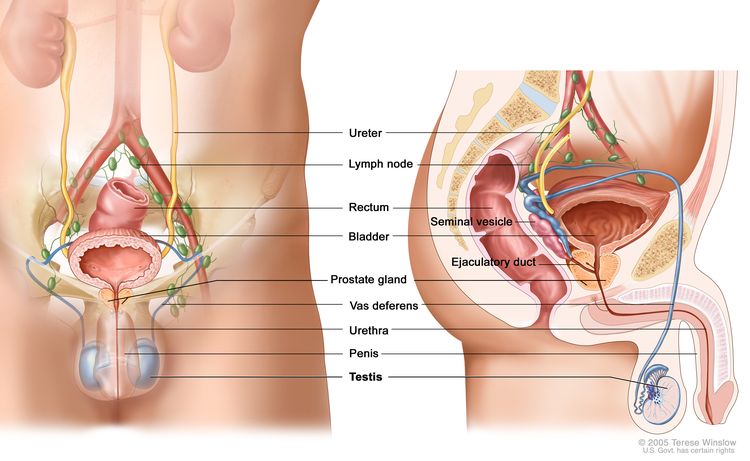
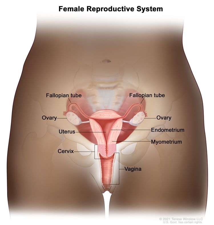
Testicular Cancer Screening (PDQ®)–Health Professional Version

Testicular Cancer Screening (PDQ®)–Health Professional Version

Overview

Note: The Overview section summarizes the published evidence on this topic. The rest of the summary describes the evidence in more detail.

Other PDQ summaries on Testicular Cancer Treatment and Levels of Evidence for Cancer Screening and Prevention Studies are also available.

Benefits

Based on fair evidence, screening for testicular cancer would not result in an appreciable decrease in mortality, in part because therapy at each stage is so effective.

Magnitude of Effect: Fair evidence of no reduction in mortality.

  • Study Design: Opinions of respected authorities based on clinical experience, descriptive studies, or reports of expert committees.
  • Internal Validity: Not applicable (N/A).
  • Consistency: N/A.
  • External Validity: N/A.

Harms

Based on fair evidence, screening for testicular cancer would result in unnecessary diagnostic procedures with attendant morbidity.

Magnitude of Effect: Fair evidence for rare but serious harms.

  • Study Design: Opinions of respected authorities based on clinical experience, descriptive studies, or reports of expert committees.
  • Internal Validity: N/A.
  • Consistency: N/A.
  • External Validity: N/A.

Incidence and Mortality

It is estimated that 9,720 new cases of testicular cancer will be diagnosed in men, and 600 men will die of this disease in the United States in 2025.[1] Testicular cancer is the most common malignancy in men aged 15 to 34 years.[2,3] It accounts for approximately 1% of all cancers in men. Worldwide, testicular cancer has more than doubled in the last 40 years. Incidence varies considerably in different geographical areas, being highest in Scandinavia and Switzerland; intermediate in the United States, Australia, and the United Kingdom; and lowest in Asia and Africa. It also varies according to ethnic groups, with a much higher rate among White men than Black men in the American population.[4] An annual increase of 3% is reported for White populations.[5] Despite the increase in observed incidence, there has been a dramatic decrease in mortality as a result of effective treatments.

Unlike most other cancers, testicular cancer is generally found in young men and has the highest incidence rate among men aged 25 to 34 years at approximately 15 cases per 100,000 men. Men in the surrounding age groups (20–24 years and 35–39 years) have a slightly lower incidence at approximately 12 cases per 100,000 men. The racial and ethnic groups with the highest incidence rates include non-Hispanic White men and non-Hispanic American Indian or Alaskan Native men, both at approximately 7 cases per 100,000 men. Testicular cancer is the most commonly diagnosed cancer among men aged 20 to 39 years.[6]

Approximately 66% of testicular cancers are localized, 19% are regional, and 12% are distant stage at diagnosis.[6] Although there has been no appreciable change in the stage distribution at diagnosis, advances in treatment have been associated with a 60% decrease in mortality. Most testicular cancers are curable even at advanced stages, and it would be impractical to document a further decrease in mortality associated with screening.

Germ cell tumors (GCTs) of the testis constitute 94% of testicular tumors and include five basic cell types:[7]

  • Seminoma.
  • Embryonal carcinoma.
  • Yolk sac tumor.
  • Teratoma.
  • Choriocarcinoma.

Sixty percent of GCTs are seminomas; the remainder are nonseminomatous GCTs. Almost half of all GCTs contain more than one of the five cell types.[7]

Three subtypes of pure seminomas have been described: classic, anaplastic, and spermatocytic. Classic seminoma accounts for 80% to 85% of all seminomas and occurs most commonly in men aged 30 to 50 years. Anaplastic seminoma accounts for 5% to 10% of all seminomas and has an age distribution similar to that of the typical subtype. A number of features suggest that anaplastic seminoma is a more aggressive and potentially more lethal variant of typical seminoma. These characteristics include greater mitotic activity, higher rate of local invasion, increased rate of metastatic spread, and higher rate of tumor marker (human chorionic gonadotropin [hCG] beta, or beta hCG) production. Spermatocytic seminoma accounts for 2% to 12% of all seminomas, and nearly half occur in men older than 50 years. The cells closely resemble different phases of maturing spermatogonia. The metastatic potential of this tumor is extremely low, and the prognosis is favorable.[8]

References
  1. American Cancer Society: Cancer Facts and Figures 2025. American Cancer Society, 2025. Available online. Last accessed January 16, 2025.
  2. McGlynn KA, Devesa SS, Sigurdson AJ, et al.: Trends in the incidence of testicular germ cell tumors in the United States. Cancer 97 (1): 63-70, 2003. [PUBMED Abstract]
  3. Garner MJ, Turner MC, Ghadirian P, et al.: Epidemiology of testicular cancer: an overview. Int J Cancer 116 (3): 331-9, 2005. [PUBMED Abstract]
  4. Huyghe E, Matsuda T, Thonneau P: Increasing incidence of testicular cancer worldwide: a review. J Urol 170 (1): 5-11, 2003. [PUBMED Abstract]
  5. Horwich A, Shipley J, Huddart R: Testicular germ-cell cancer. Lancet 367 (9512): 754-65, 2006. [PUBMED Abstract]
  6. Surveillance Research Program, National Cancer Institute: SEER*Explorer: An interactive website for SEER cancer statistics. Bethesda, MD: National Cancer Institute. Available online. Last accessed December 30, 2024.
  7. Dieckmann KP, Pichlmeier U: Clinical epidemiology of testicular germ cell tumors. World J Urol 22 (1): 2-14, 2004. [PUBMED Abstract]
  8. Richie JP, Steele GS: Neoplasms of the testis. In: Campbell MF, Wein AJ, Kavoussi LR: Campbell-Walsh Urology. 9th ed. Saunders Elsevier, 2007, pp 893-935.

Risk Factors

Testicular cancer is more than four times more common among White men than Black men,[1,2] with intermediate incidence rates for Hispanic, American Indian, and Asian men. High-risk groups exist. Males with cryptorchidism have 3 to 17 times the average risk. Approximately 7% to 10% of patients with testicular tumors have a history of cryptorchidism.[2,3] Although the association is established, the biological mechanism underlying the association remains uncertain; testicular cancer and cryptorchidism may share environmental and/or genetic risk factors; or, it is the ectopic position per se that is a postnatal risk factor for testicular cancer, or it is a combination of the two.[3] Orchiopexy may not prevent cancer in these children but allows clinical surveillance of patients with a previously impalpable gonad.

There is also an increased risk in males with gonadal dysgenesis and Klinefelter syndrome.[4] Men with a family history of testicular cancer may be at a higher risk of this disease.[5] A history of testicular cancer is associated with a higher risk of a contralateral tumor.[3,2] Although not consistently found, infertility, testicular atrophy, twinship, or abnormal semen parameters have been associated with a higher risk of testicular cancer, but the evidence is weak.[3,68]

There is a low cumulative risk of metachronous contralateral testicular cancer and a favorable overall survival of patients diagnosed with metachronous contralateral testicular cancer.[9] Future research is necessary to delineate the genetic and environmental risk factors for testicular cancer.[10]

Carcinoma In Situ

An additional risk factor for the development of testicular cancer is the presence of carcinoma in situ (CIS), also called intratubular germ cell neoplasia. Testicular CIS appears to develop from fetal gonocytes and is characterized histologically by seminiferous tubules containing only Sertoli cells and malignant-appearing germ cells.[11]

Early reports suggest that CIS is associated with the development of contralateral testicular cancer in 50% of patients at 5 years of follow-up.[12] CIS will be found in approximately 5% of contralateral testes (approximately the same rate as cryptorchid testes).[13]

There is controversy regarding the clinical significance and management of CIS of the testis.[2] Treatment options for CIS include observation, radiation therapy, chemotherapy, and orchiectomy. Although low-dose radiation therapy can preserve Leydig cell function and prevent germ cell tumors development, a conservative approach of observation may also be warranted. Individuals at high risk (e.g., cryptorchidism, atrophic testis, and intersex conditions) require close observation.

References
  1. Moul JW, Schanne FJ, Thompson IM, et al.: Testicular cancer in blacks. A multicenter experience. Cancer 73 (2): 388-93, 1994. [PUBMED Abstract]
  2. Richie JP, Steele GS: Neoplasms of the testis. In: Campbell MF, Wein AJ, Kavoussi LR: Campbell-Walsh Urology. 9th ed. Saunders Elsevier, 2007, pp 893-935.
  3. Dieckmann KP, Pichlmeier U: Clinical epidemiology of testicular germ cell tumors. World J Urol 22 (1): 2-14, 2004. [PUBMED Abstract]
  4. Henderson BE, Benton B, Jing J, et al.: Risk factors for cancer of the testis in young men. Int J Cancer 23 (5): 598-602, 1979. [PUBMED Abstract]
  5. Dieckmann KP, Pichlmeier U: The prevalence of familial testicular cancer: an analysis of two patient populations and a review of the literature. Cancer 80 (10): 1954-60, 1997. [PUBMED Abstract]
  6. Jacobsen R, Bostofte E, Engholm G, et al.: Risk of testicular cancer in men with abnormal semen characteristics: cohort study. BMJ 321 (7264): 789-92, 2000. [PUBMED Abstract]
  7. Walsh TJ, Croughan MS, Schembri M, et al.: Increased risk of testicular germ cell cancer among infertile men. Arch Intern Med 169 (4): 351-6, 2009. [PUBMED Abstract]
  8. Hotaling JM, Walsh TJ: Male infertility: a risk factor for testicular cancer. Nat Rev Urol 6 (10): 550-6, 2009. [PUBMED Abstract]
  9. Fosså SD, Chen J, Schonfeld SJ, et al.: Risk of contralateral testicular cancer: a population-based study of 29,515 U.S. men. J Natl Cancer Inst 97 (14): 1056-66, 2005. [PUBMED Abstract]
  10. Richiardi L, Pettersson A, Akre O: Genetic and environmental risk factors for testicular cancer. Int J Androl 30 (4): 230-40; discussion 240-1, 2007. [PUBMED Abstract]
  11. Olesen IA, Hoei-Hansen CE, Skakkebaek NE, et al.: Testicular carcinoma in situ in subfertile Danish men. Int J Androl 30 (4): 406-11; discussion 412, 2007. [PUBMED Abstract]
  12. Jørgensen N, Müller J, Giwercman A, et al.: Clinical and biological significance of carcinoma in situ of the testis. Cancer Surv 9 (2): 287-302, 1990. [PUBMED Abstract]
  13. Dieckmann KP, Loy V: Prevalence of contralateral testicular intraepithelial neoplasia in patients with testicular germ cell neoplasms. J Clin Oncol 14 (12): 3126-32, 1996. [PUBMED Abstract]

Evidence of Benefit Associated With Screening

The sensitivity, specificity, and positive predictive value of routine screening of asymptomatic men for testicular cancer are not known.[1,2] In a report of a single-center case series of men being evaluated for infertility, testicular symptoms, or erectile dysfunction, 1,320 men underwent testicular ultrasonography.[3] Focal lesions were found in 27 (2%) men, 17 of the lesions were palpable, and 10 were nonpalpable. Eighty percent of the lesions were ultimately shown to have benign histologies, for a positive predictive value of about 0.2. It is not clear if early discovery of the cancers resulted in clinical benefit, and the positive predictive value is likely to be lower in the target population of asymptomatic men in the screening setting.

Most testicular cancers are first detected by the patient, either unintentionally or by self-examination. Some are discovered by routine physical examination. However, no studies have been done to determine the effectiveness of testicular self-examination or clinical testicular examination in reducing mortality from testicular cancer. An updated systematic review performed on behalf of the U.S. Preventive Services Task Force, published in 2010, found no randomized trials, cohort studies, or case-control studies that examined benefits of testicular cancer screening (whether by physical examination, self-examination, or other screening tests) in an asymptomatic population.[2] Likewise, a systematic Cochrane Collaboration review found no randomized or quasi-randomized controlled trials that evaluated the effectiveness of screening by a health professional or patient self-examination.[4]

Screening would be very unlikely to decrease mortality substantially because therapy is so effective at virtually all stages of disease.[5] For more information, see Testicular Cancer Treatment. However, early detection may affect therapy. There is an increase in both the number of courses of chemotherapy and the extent of surgery required for treatment of advanced disease that results in higher morbidity. Patients diagnosed with localized disease require less treatment and have lower morbidity.[6]

References
  1. U.S. Preventive Services Task Force: Screening for testicular cancer: U.S. Preventive Services Task Force reaffirmation recommendation statement. Ann Intern Med 154 (7): 483-6, 2011. [PUBMED Abstract]
  2. Lin K, Sharangpani R: Screening for testicular cancer: an evidence review for the U.S. Preventive Services Task Force. Ann Intern Med 153 (6): 396-9, 2010. [PUBMED Abstract]
  3. Carmignani L, Gadda F, Gazzano G, et al.: High incidence of benign testicular neoplasms diagnosed by ultrasound. J Urol 170 (5): 1783-6, 2003. [PUBMED Abstract]
  4. Ilic D, Misso ML: Screening for testicular cancer. Cochrane Database Syst Rev (2): CD007853, 2011. [PUBMED Abstract]
  5. Feldman DR, Bosl GJ, Sheinfeld J, et al.: Medical treatment of advanced testicular cancer. JAMA 299 (6): 672-84, 2008. [PUBMED Abstract]
  6. Sagalowsky AI: Expectant management of stage A nonseminomatous testicular tumors. In: Ratiff TL, Catalona WJ, eds.: Genitourinary Cancer. Martinus Nijhoff, 1987, pp 225-237.

Evidence of Harm Associated With Screening

Harms of screening for testicular cancer are poorly quantified. They may include false positive tests [1] and resulting anxiety as well as subsequent unwarranted invasive diagnostic procedures. Two systematic reviews found no studies that provided a quantitative assessment of the harms of screening.[2,3]

References
  1. Carmignani L, Gadda F, Gazzano G, et al.: High incidence of benign testicular neoplasms diagnosed by ultrasound. J Urol 170 (5): 1783-6, 2003. [PUBMED Abstract]
  2. Lin K, Sharangpani R: Screening for testicular cancer: an evidence review for the U.S. Preventive Services Task Force. Ann Intern Med 153 (6): 396-9, 2010. [PUBMED Abstract]
  3. Ilic D, Misso ML: Screening for testicular cancer. Cochrane Database Syst Rev (2): CD007853, 2011. [PUBMED Abstract]

Latest Updates to This Summary (04/07/2025)

The PDQ cancer information summaries are reviewed regularly and updated as new information becomes available. This section describes the latest changes made to this summary as of the date above.

Incidence and Mortality

Updated statistics with estimated new cases and deaths for 2025 (cited American Cancer Society as reference 1).

This summary is written and maintained by the PDQ Screening and Prevention Editorial Board, which is editorially independent of NCI. The summary reflects an independent review of the literature and does not represent a policy statement of NCI or NIH. More information about summary policies and the role of the PDQ Editorial Boards in maintaining the PDQ summaries can be found on the About This PDQ Summary and PDQ® Cancer Information for Health Professionals pages.

About This PDQ Summary

Purpose of This Summary

This PDQ cancer information summary for health professionals provides comprehensive, peer-reviewed, evidence-based information about testicular cancer screening. It is intended as a resource to inform and assist clinicians in the care of their patients. It does not provide formal guidelines or recommendations for making health care decisions.

Reviewers and Updates

This summary is reviewed regularly and updated as necessary by the PDQ Screening and Prevention Editorial Board, which is editorially independent of the National Cancer Institute (NCI). The summary reflects an independent review of the literature and does not represent a policy statement of NCI or the National Institutes of Health (NIH).

Board members review recently published articles each month to determine whether an article should:

  • be discussed at a meeting,
  • be cited with text, or
  • replace or update an existing article that is already cited.

Changes to the summaries are made through a consensus process in which Board members evaluate the strength of the evidence in the published articles and determine how the article should be included in the summary.

Any comments or questions about the summary content should be submitted to Cancer.gov through the NCI website’s Email Us. Do not contact the individual Board Members with questions or comments about the summaries. Board members will not respond to individual inquiries.

Levels of Evidence

Some of the reference citations in this summary are accompanied by a level-of-evidence designation. These designations are intended to help readers assess the strength of the evidence supporting the use of specific interventions or approaches. The PDQ Screening and Prevention Editorial Board uses a formal evidence ranking system in developing its level-of-evidence designations.

Permission to Use This Summary

PDQ is a registered trademark. Although the content of PDQ documents can be used freely as text, it cannot be identified as an NCI PDQ cancer information summary unless it is presented in its entirety and is regularly updated. However, an author would be permitted to write a sentence such as “NCI’s PDQ cancer information summary about breast cancer prevention states the risks succinctly: [include excerpt from the summary].”

The preferred citation for this PDQ summary is:

PDQ® Screening and Prevention Editorial Board. PDQ Testicular Cancer Screening. Bethesda, MD: National Cancer Institute. Updated <MM/DD/YYYY>. Available at: /types/testicular/hp/testicular-screening-pdq. Accessed <MM/DD/YYYY>. [PMID: 26389404]

Images in this summary are used with permission of the author(s), artist, and/or publisher for use within the PDQ summaries only. Permission to use images outside the context of PDQ information must be obtained from the owner(s) and cannot be granted by the National Cancer Institute. Information about using the illustrations in this summary, along with many other cancer-related images, is available in Visuals Online, a collection of over 2,000 scientific images.

Disclaimer

The information in these summaries should not be used as a basis for insurance reimbursement determinations. More information on insurance coverage is available on Cancer.gov on the Managing Cancer Care page.

Contact Us

More information about contacting us or receiving help with the Cancer.gov website can be found on our Contact Us for Help page. Questions can also be submitted to Cancer.gov through the website’s Email Us.
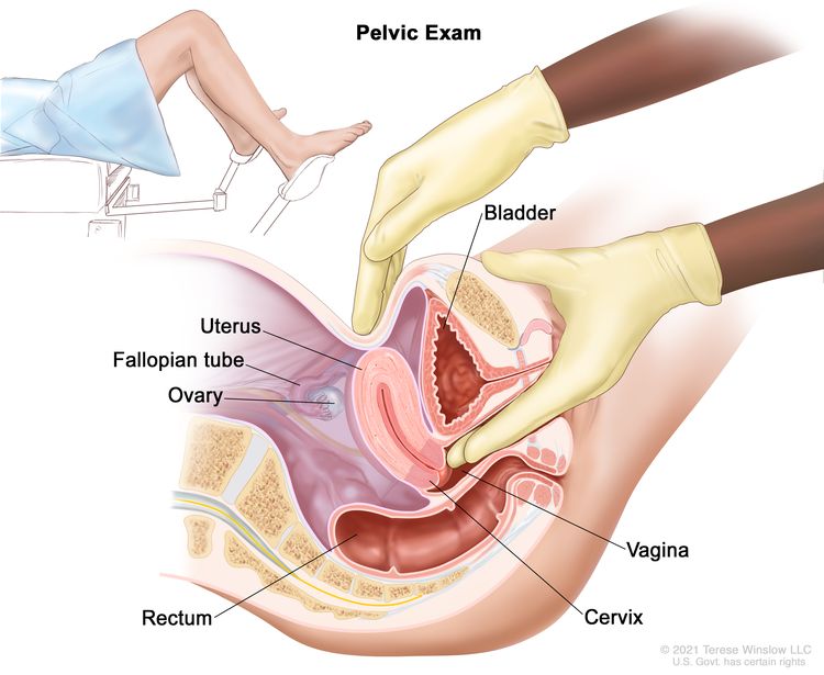
Testicular Cancer Screening (PDQ®)–Patient Version

Testicular Cancer Screening (PDQ®)–Patient Version

What Is Screening?

Screening is looking for cancer before a person has any symptoms. This can help find cancer at an early stage. When abnormal tissue or cancer is found early, it may be easier to treat. By the time symptoms appear, cancer may have begun to spread.

Scientists are trying to better understand which people are more likely to get certain types of cancer. They also study the things we do and the things around us to see if they cause cancer. This information helps doctors recommend who should be screened for cancer, which screening tests should be used, and how often the tests should be done.

It is important to remember that your doctor does not necessarily think you have cancer if he or she suggests a screening test. Screening tests are given when you have no cancer symptoms.

If a screening test result is abnormal, you may need to have more tests done to find out if you have cancer. These are called diagnostic tests.

General Information About Testicular Cancer

Key Points

  • Testicular cancer is a disease in which malignant (cancer) cells form in the tissues of one or both testicles.
  • Testicular cancer is the most commonly diagnosed cancer in men aged 20 to 39 years.
  • Testicular cancer can usually be cured.
  • A condition called cryptorchidism (an undescended testicle) is a risk factor for testicular cancer.

Testicular cancer is a disease in which malignant (cancer) cells form in the tissues of one or both testicles.

The testicles are 2 egg-shaped glands inside the scrotum (a sac of loose skin that lies directly below the penis). The testicles are held within the scrotum by the spermatic cord. The spermatic cord also contains the vas deferens and vessels and nerves of the testicles.

EnlargeAnatomy of the male reproductive and urinary systems; drawing shows front and side views of ureters, lymph nodes, rectum, bladder, prostate gland, vas deferens, urethra, penis, testicles, seminal vesicle, and ejaculatory duct.
Anatomy of the male reproductive and urinary systems, showing the testicles, prostate, bladder, and other organs.

The testicles are the male sex glands and make testosterone and sperm. Germ cells in the testicles make immature sperm. These sperm travel through a network of tubules (tiny tubes) and larger tubes into the epididymis (a long coiled tube next to the testicles). This is where the sperm cells mature and are stored.

Almost all testicular cancers start in the germ cells. The two main types of testicular germ cell tumors are seminomas and nonseminomas.

Learn more about testicular cancer at Testicular Cancer Treatment.

Testicular cancer is the most commonly diagnosed cancer in men aged 20 to 39 years.

Testicular cancer is the most common cancer in men between the ages of 20 to 39 years, with the highest rates between ages 25 to 34 years. The racial and ethnic groups with the highest rates of testicular cancer include non-Hispanic White men, non-Hispanic American Indian men, and Alaska Native men.

Testicular cancer can usually be cured.

Although the number of new cases of testicular cancer has doubled in the last 40 years, the number of deaths caused by testicular cancer has decreased greatly because of better treatments. Testicular cancer can usually be cured, even in late stages of the disease. Learn more about testicular cancer at Testicular Cancer Treatment.

A condition called cryptorchidism (an undescended testicle) is a risk factor for testicular cancer.

Anything that increases a person’s chance of getting a disease is called a risk factor. Not every person with one or more of these risk factors will develop testicular cancer, and it will also develop in some people who don’t have any known risk factors. Talk with your doctor if you think you may be at risk.

Risk factors for testicular cancer include:

Men who have cryptorchidism, a testicle that is not normal, or testicular carcinoma in situ have an increased risk of testicular cancer in one or both testicles and need to be followed closely.

Testicular Cancer Screening

Key Points

  • Tests are used to screen for different types of cancer when a person does not have symptoms.
  • There is no standard or routine screening test for testicular cancer.
  • Screening tests for testicular cancer are being studied in clinical trials.

Tests are used to screen for different types of cancer when a person does not have symptoms.

Scientists study screening tests to find those with the fewest harms and most benefits. Cancer screening trials also are meant to show whether early detection (finding cancer before it causes symptoms) helps a person live longer or decreases a person’s chance of dying from the disease. For some types of cancer, the chance of recovery is better if the disease is found and treated at an early stage.

There is no standard or routine screening test for testicular cancer.

There is no standard or routine screening test used for early detection of testicular cancer. Most often, testicular cancer is first found by men themselves, either by chance or during self-exam. Sometimes the cancer is found by a doctor during a routine physical exam.

No studies have been done to find out if testicular self-exams, regular exams by a doctor, or other screening tests in men with no symptoms would decrease the risk of dying from this disease. However, routine screening probably would not decrease the risk of dying from testicular cancer. This is partly because testicular cancer can usually be cured at any stage. Finding testicular cancer early may make it easier to treat. Patients who are diagnosed with testicular cancer that has not spread to other parts of the body may need less chemotherapy and surgery, resulting in fewer side effects.

If a lump is found in the testicle by the patient or during a routine physical exam, tests may be done to check for cancer. Some tests have risks and may cause anxiety.

Screening tests for testicular cancer are being studied in clinical trials.

Information about clinical trials supported by NCI can be found on NCI’s clinical trials search webpage. Clinical trials supported by other organizations can be found on the ClinicalTrials.gov website.

About This PDQ Summary

About PDQ

Physician Data Query (PDQ) is the National Cancer Institute’s (NCI’s) comprehensive cancer information database. The PDQ database contains summaries of the latest published information on cancer prevention, detection, genetics, treatment, supportive care, and complementary and alternative medicine. Most summaries come in two versions. The health professional versions have detailed information written in technical language. The patient versions are written in easy-to-understand, nontechnical language. Both versions have cancer information that is accurate and up to date and most versions are also available in Spanish.

PDQ is a service of the NCI. The NCI is part of the National Institutes of Health (NIH). NIH is the federal government’s center of biomedical research. The PDQ summaries are based on an independent review of the medical literature. They are not policy statements of the NCI or the NIH.

Purpose of This Summary

This PDQ cancer information summary has current information about testicular cancer screening. It is meant to inform and help patients, families, and caregivers. It does not give formal guidelines or recommendations for making decisions about health care.

Reviewers and Updates

Editorial Boards write the PDQ cancer information summaries and keep them up to date. These Boards are made up of experts in cancer treatment and other specialties related to cancer. The summaries are reviewed regularly and changes are made when there is new information. The date on each summary (“Updated”) is the date of the most recent change.

The information in this patient summary was taken from the health professional version, which is reviewed regularly and updated as needed, by the PDQ Screening and Prevention Editorial Board.

Clinical Trial Information

A clinical trial is a study to answer a scientific question, such as whether one treatment is better than another. Trials are based on past studies and what has been learned in the laboratory. Each trial answers certain scientific questions in order to find new and better ways to help cancer patients. During treatment clinical trials, information is collected about the effects of a new treatment and how well it works. If a clinical trial shows that a new treatment is better than one currently being used, the new treatment may become “standard.” Patients may want to think about taking part in a clinical trial. Some clinical trials are open only to patients who have not started treatment.

Clinical trials can be found online at NCI’s website. For more information, call the Cancer Information Service (CIS), NCI’s contact center, at 1-800-4-CANCER (1-800-422-6237).

Permission to Use This Summary

PDQ is a registered trademark. The content of PDQ documents can be used freely as text. It cannot be identified as an NCI PDQ cancer information summary unless the whole summary is shown and it is updated regularly. However, a user would be allowed to write a sentence such as “NCI’s PDQ cancer information summary about breast cancer prevention states the risks in the following way: [include excerpt from the summary].”

The best way to cite this PDQ summary is:

PDQ® Screening and Prevention Editorial Board. PDQ Testicular Cancer Screening. Bethesda, MD: National Cancer Institute. Updated <MM/DD/YYYY>. Available at: /types/testicular/patient/testicular-screening-pdq. Accessed <MM/DD/YYYY>. [PMID: 26389226]

Images in this summary are used with permission of the author(s), artist, and/or publisher for use in the PDQ summaries only. If you want to use an image from a PDQ summary and you are not using the whole summary, you must get permission from the owner. It cannot be given by the National Cancer Institute. Information about using the images in this summary, along with many other images related to cancer can be found in Visuals Online. Visuals Online is a collection of more than 3,000 scientific images.

Disclaimer

The information in these summaries should not be used to make decisions about insurance reimbursement. More information on insurance coverage is available on Cancer.gov on the Managing Cancer Care page.

Contact Us

More information about contacting us or receiving help with the Cancer.gov website can be found on our Contact Us for Help page. Questions can also be submitted to Cancer.gov through the website’s E-mail Us.

Ovarian Borderline Tumors Treatment (PDQ®)–Patient Version

Ovarian Borderline Tumors Treatment (PDQ®)–Patient Version

General Information About Ovarian Borderline Tumors

Key Points

  • Ovarian borderline tumor is a disease in which abnormal cells form in the tissue covering the ovary.
  • Signs and symptoms of ovarian borderline tumor include pain or swelling in the abdomen.
  • Tests that examine the ovaries are used to diagnose and stage ovarian borderline tumor.
  • Some people decide to get a second opinion.
  • Certain factors affect prognosis (chance of recovery) and treatment options.

Ovarian borderline tumor is a disease in which abnormal cells form in the tissue covering the ovary.

Ovarian borderline tumors have abnormal cells that may become cancer, but usually do not. This disease usually remains in the ovary. When disease is found in one ovary, the other ovary should also be checked carefully for signs of disease.

The ovaries are a pair of organs in the female reproductive system. They are in the pelvis, one on each side of the uterus (the hollow, pear-shaped organ where a fetus grows). Each ovary is about the size and shape of an almond. The ovaries make eggs and female hormones.

EnlargeAnatomy of the female reproductive system; drawing shows the uterus, myometrium (muscular outer layer of the uterus), endometrium (inner lining of the uterus), ovaries, fallopian tubes, cervix, and vagina.
Anatomy of the female reproductive system. The organs in the female reproductive system include the uterus, ovaries, fallopian tubes, cervix, and vagina. The uterus has a muscular outer layer called the myometrium and an inner lining called the endometrium.

Signs and symptoms of ovarian borderline tumor include pain or swelling in the abdomen.

Ovarian borderline tumor may not cause early signs or symptoms. If you do have signs or symptoms, they may include:

These signs and symptoms may be caused by other conditions. If they get worse or do not go away on their own, check with your doctor.

Tests that examine the ovaries are used to diagnose and stage ovarian borderline tumor.

In addition to asking about your personal and family health history and doing a physical exam, your doctor may perform the following tests and procedures:

  • Pelvic exam is an exam of the vagina, cervix, uterus, fallopian tubes, ovaries, and rectum. A speculum is inserted into the vagina and the doctor or nurse looks at the vagina and cervix for signs of disease. A Pap test of the cervix is usually done. The doctor or nurse also inserts one or two lubricated, gloved fingers of one hand into the vagina and places the other hand over the lower abdomen to feel the size, shape, and position of the uterus and ovaries. The doctor or nurse also inserts a lubricated, gloved finger into the rectum to feel for lumps or abnormal areas.
    EnlargePelvic exam; drawing shows a side view of the female reproductive anatomy during a pelvic exam. The uterus, left fallopian tube, left ovary, cervix, vagina, bladder, and rectum are shown. Two gloved fingers of one hand of the doctor or nurse are shown inserted into the vagina, while the other hand is shown pressing on the lower abdomen. The inset shows a woman covered by a drape on an exam table with her legs apart and her feet in stirrups.
    Pelvic exam. A doctor or nurse inserts one or two lubricated, gloved fingers of one hand into the vagina and presses on the lower abdomen with the other hand. This is done to feel the size, shape, and position of the uterus and ovaries. The vagina, cervix, fallopian tubes, and rectum are also checked.
  • Ultrasound exam is a procedure in which high-energy sound waves (ultrasound) are bounced off internal tissues or organs and make echoes. The echoes form a picture of body tissues called a sonogram. The picture can be printed to be looked at later.
    EnlargeAbdominal ultrasound; drawing shows a woman on an exam table during an abdominal ultrasound procedure. A diagnostic sonographer (a person trained to perform ultrasound procedures) is shown passing a transducer (a device that makes sound waves that bounce off tissues inside the body) over the surface of the patient’s abdomen. A computer screen shows a sonogram (computer picture).
    Abdominal ultrasound. An ultrasound transducer connected to a computer is passed over the surface of the abdomen. The ultrasound transducer bounces sound waves off internal organs and tissues to make echoes that form a sonogram (computer picture).

    Other patients may have a transvaginal ultrasound.

    EnlargeTransvaginal ultrasound; drawing shows a side view of the female reproductive anatomy during a transvaginal ultrasound procedure. An ultrasound probe (a device that makes sound waves that bounce off tissues inside the body) is shown inserted into the vagina. The bladder, uterus, right fallopian tube, and right ovary are also shown. The inset shows the diagnostic sonographer (a person trained to perform ultrasound procedures) examining a woman on a table, and a computer screen shows an image of the patient’s internal tissues.
    Transvaginal ultrasound. An ultrasound probe connected to a computer is inserted into the vagina and is gently moved to show different organs. The probe bounces sound waves off internal organs and tissues to make echoes that form a sonogram (computer picture).
  • CT scan (CAT scan) uses a computer linked to an x-ray machine to make a series of detailed pictures of areas inside the body. The pictures are taken from different angles and are used to create 3-D views of tissues and organs. A dye may be injected into a vein or swallowed to help the organs or tissues show up more clearly. This procedure is also called computed tomography, computerized tomography, or computerized axial tomography.
  • CA 125 assay is a laboratory test that measures the level of CA 125 in the blood. CA 125 is a substance released by cells into the bloodstream. An increased CA 125 level is sometimes a sign of cancer or other condition.
  • Chest x-ray is a type of radiation that can go through the body and make pictures of the organs and bones inside the chest.
  • Biopsy is the removal of cells or tissues so they can be viewed under a microscope by a pathologist to check for signs of cancer. The tissue is usually removed during surgery to remove the tumor.
  • Staging laparotomy is surgery to determine the extent of cancer within the abdomen. An incision (cut) is made in the wall of the abdomen to remove tissue so a pathologist can check it for signs of cancer.

Some people decide to get a second opinion.

You may want to get a second opinion to confirm your diagnosis and treatment plan. If you seek a second opinion, you will need to get medical test results and reports from the first doctor to share with the second doctor. The second doctor will review the pathology report, slides, and scans. They may agree with the first doctor, suggest changes or another treatment approach, or provide more information about your tumor.

Learn more about choosing a doctor and getting a second opinion at Finding Cancer Care. You can contact NCI’s Cancer Information Service via chat, email, or phone (both in English and Spanish) for help finding a doctor, hospital, or getting a second opinion. For questions you might want to ask at your appointments, visit Questions to Ask Your Doctor About Cancer.

Certain factors affect prognosis (chance of recovery) and treatment options.

The prognosis and treatment options depend on:

  • the stage of the disease (whether it affects part of the ovary, involves the whole ovary, or has spread to other places in the body)
  • what type of cells make up the tumor
  • the size of the tumor
  • the patient’s general health

Patients with ovarian borderline tumors have a good prognosis, especially when the tumor is found early.

Stages of Ovarian Borderline Tumors

Key Points

  • After ovarian borderline tumor has been diagnosed, tests are done to find out if abnormal cells have spread within the ovary or to other parts of the body.
  • The following stages are used for ovarian borderline tumor:
    • Stage I (also called stage 1) ovarian borderline tumor
    • Stage II (also called stage 2) ovarian borderline tumor
    • Stage III (also called stage 3) ovarian borderline tumor
    • Stage IV (also called stage 4) ovarian borderline tumor
  • Ovarian borderline tumors can recur (come back) after they have been treated.

After ovarian borderline tumor has been diagnosed, tests are done to find out if abnormal cells have spread within the ovary or to other parts of the body.

Cancer stage describes the extent of cancer in the body, such as the size of the tumor, whether it has spread, and how far it has spread from where it first formed. It is important to know the stage of the ovarian borderline tumors to plan the best treatment. Most people are diagnosed with stage I disease.

Borderline ovarian tumor staging usually uses the FIGO staging system. The tumor may be described by this staging system in your pathology report. Based on the FIGO results, a stage (I, II, III, or IV, also written as 1, 2, 3, or 4) is assigned to your tumor. When talking to you about your diagnosis, your doctor may describe the tumor as one of these stages. 

The following stages are used for ovarian borderline tumor:

Stage I (also called stage 1) ovarian borderline tumor

EnlargeThree-panel drawing of stage IA, stage IB, and stage IC; each panel shows the ovaries, fallopian tubes, uterus, cervix, and vagina. The first panel (stage IA) shows cancer inside one ovary. The second panel (stage IB) shows cancer inside both ovaries. The third panel (stage IC) shows cancer inside both ovaries and (a) the tumor in the ovary on the left has ruptured (broken open), (b) there is cancer on the surface of the ovary on the right, and (c) there are cancer cells in the pelvic peritoneal fluid (inset).
In stage IA, cancer is found inside a single ovary or fallopian tube. In stage IB, cancer is found inside both ovaries or fallopian tubes. In stage IC, cancer is found inside one or both ovaries or fallopian tubes and one of the following is true: (a) either the tumor or the capsule (outer covering) of the ovary has ruptured (broken open), or (b) cancer is also found on the surface of the ovary or fallopian tube, or (c) cancer cells are found in the pelvic peritoneal fluid.

In stage I, the tumor is found in one or both ovaries or fallopian tubes. Stage I is divided into stage IA, stage IB, and stage IC.

  • Stage IA: The tumor is found inside a single ovary or fallopian tube.
  • Stage IB: The tumor is found inside both ovaries or fallopian tubes.
  • Stage IC: The tumor is found inside one or both ovaries or fallopian tubes and one of the following is true:
    • tumor cells are found on the outside surface of one or both ovaries or fallopian tubes; or
    • the capsule (outer covering) of the ovary ruptured (broke open) before or during surgery; or
    • tumor cells are found in the fluid of the peritoneal cavity (the body cavity that contains most of the organs in the abdomen) or in washings of the peritoneum (tissue lining the peritoneal cavity).

Stage II (also called stage 2) ovarian borderline tumor

EnlargeThree-panel drawing of stage IIA, stage IIB, and stage II primary peritoneal cancer; the first panel (stage IIA) shows cancer inside both ovaries that has spread to the fallopian tube and uterus. Also shown are the cervix and vagina. The second panel (stage IIB) shows cancer inside both ovaries that has spread to the colon. The third panel (primary peritoneal cancer) shows cancer in the pelvic peritoneum.
In stage IIA, cancer is found in one or both ovaries or fallopian tubes and has spread to the uterus and/or the fallopian tubes and/or the ovaries. In stage IIB, cancer is found in one or both ovaries or fallopian tubes and has spread to organs in the peritoneal cavity, such as the colon. In primary peritoneal cancer, cancer is found in the pelvic peritoneum and has not spread there from another part of the body.

In stage II, the tumor is found in one or both ovaries or fallopian tubes and has spread into other areas of the pelvis, or primary peritoneal cancer is found within the pelvis. Stage II is divided into stage IIA and stage IIB.

  • Stage IIA: The tumor has spread from where it first formed to the uterus and/or the fallopian tubes and/or the ovaries.
  • Stage IIB: The tumor has spread from the ovary or fallopian tube to organs in the peritoneal cavity (the space that contains the abdominal organs).

Stage III (also called stage 3) ovarian borderline tumor

EnlargeDrawing shows different sizes of a tumor in centimeters (cm) compared to the size of a pea (1 cm), a peanut (2 cm), a grape (3 cm), a walnut (4 cm), a lime (5 cm), an egg (6 cm), a peach (7 cm), and a grapefruit (10 cm). Also shown is a 10-cm ruler and a 4-inch ruler.
Tumor sizes are often measured in centimeters (cm) or inches. Common food items that can be used to show tumor size in cm include: a pea (1 cm), a peanut (2 cm), a grape (3 cm), a walnut (4 cm), a lime (5 cm or 2 inches), an egg (6 cm), a peach (7 cm), and a grapefruit (10 cm or 4 inches).

In stage III, the tumor is found in one or both ovaries or fallopian tubes, or is primary peritoneal cancer, and has spread outside the pelvis to other parts of the abdomen and/or to nearby lymph nodes. Stage III is divided into stage IIIA, stage IIIB, and stage IIIC.

  • In stage IIIA, one of the following is true:
    • The tumor has spread to lymph nodes in the area outside or behind the peritoneum only; or
    • Tumor cells that can be seen only with a microscope have spread to the surface of the peritoneum outside the pelvis, such as the omentum (a fold of the peritoneum that surrounds the stomach and other organs in the abdomen). The tumor may have spread to nearby lymph nodes.
    EnlargeDrawing of stage IIIA shows cancer inside both ovaries that has spread to (a) lymph nodes behind the peritoneum and (b) the omentum. The small intestine, colon, fallopian tubes, uterus, and bladder are also shown.
    In stage IIIA, cancer is found in one or both ovaries or fallopian tubes and (a) cancer has spread to lymph nodes behind the peritoneum only, or (b) cancer cells that can be seen only with a microscope have spread to the surface of the peritoneum outside the pelvis, such as the omentum. Cancer may have also spread to nearby lymph nodes.
  • Stage IIIB: The tumor has spread to the peritoneum outside the pelvis, such as the omentum, and the tumor in the peritoneum is 2 centimeters or smaller. The tumor may have spread to lymph nodes behind the peritoneum.
    EnlargeDrawing of stage IIIB shows cancer inside both ovaries that has spread to the omentum. The cancer in the omentum is 2 centimeters or smaller. An inset shows 2 centimeters is about the size of a peanut. Also shown are the small intestine, colon, fallopian tubes, uterus, bladder, and lymph nodes behind the peritoneum.
    In stage IIIB, cancer is found in one or both ovaries or fallopian tubes and has spread to the peritoneum outside the pelvis, such as the omentum. The cancer in the omentum is 2 centimeters or smaller. Cancer may have also spread to lymph nodes behind the peritoneum.
  • Stage IIIC: The tumor has spread to the peritoneum outside the pelvis, such as the omentum, and the tumor in the peritoneum is larger than 2 centimeters. The tumor may have spread to lymph nodes behind the peritoneum or to the surface of the liver or spleen.
    EnlargeDrawing of stage IIIC shows cancer inside both ovaries that has spread to the omentum. The cancer in the omentum is larger than 2 centimeters. An inset shows 2 centimeters is about the size of a peanut. Also shown are the small intestine, colon, fallopian tubes, uterus, bladder, and lymph nodes behind the peritoneum.
    In stage IIIC, cancer is found in one or both ovaries or fallopian tubes and has spread to the peritoneum outside the pelvis, such as the omentum. The cancer in the omentum is larger than 2 centimeters. Cancer may have also spread to lymph nodes behind the peritoneum or to the surface of the liver or spleen (not shown).

Stage IV (also called stage 4) ovarian borderline tumor

In stage IV, tumor cells have spread beyond the abdomen to other parts of the body. Stage IV is divided into stage IVA and stage IVB.

EnlargeDrawing of stage IV shows other parts of the body where ovarian cancer may spread, including the lung, liver, and lymph nodes in the groin. An inset on the top shows extra fluid around the lung. An inset on the bottom shows cancer cells spreading through the blood and lymph system to another part of the body where metastatic cancer has formed.
In stage IV, cancer has spread beyond the abdomen to other parts of the body. In stage IVA, cancer cells are found in extra fluid that builds up around the lungs. In stage IVB, cancer has spread to organs and tissues outside the abdomen, including the lung, liver, and lymph nodes in the groin.

  • Stage IVA: Tumor cells are found in extra fluid that builds up around the lungs.
  • Stage IVB: The tumor has spread to organs and tissues outside the abdomen, including lymph nodes in the groin.

Ovarian borderline tumors can recur (come back) after they have been treated.

Recurrent ovarian borderline tumors are tumors that have come back after they have been treated. The tumors may come back in the other ovary or in other parts of the body. Tests will be done to help determine where the tumor has returned. The type of treatment for a recurrent ovarian borderline tumor will depend on where it has come back.

Treatment Option Overview

Key Points

  • There are different types of treatment for patients with ovarian borderline tumors.
  • The following types of treatment are used:
    • Surgery
    • Chemotherapy
  • New types of treatment are being tested in clinical trials.
  • Treatment for ovarian borderline tumors may cause side effects.
  • Follow-up care may be needed.

There are different types of treatment for patients with ovarian borderline tumors.

Different types of treatments are available for ovarian borderline tumors. You and your care team will work together to decide your treatment plan, which may include more than one type of treatment. Many factors will be considered, such as the stage of the tumor, your overall health, and your preferences. Your plan will include information about your tumor, the goals of treatment, your treatment options and the possible side effects, and the expected length of treatment. 

Talking with your care team before treatment begins about what to expect will be helpful. You’ll want to learn what you need to do before treatment begins, how you’ll feel while going through it, and what kind of help you will need. To learn more, visit Questions to Ask Your Doctor About Treatment. 

The following types of treatment are used:

Surgery

The type of surgery (removing the tumor in an operation) depends on the size and spread of the tumor and the patient’s plans for having children. Surgery may include:

  • Unilateral salpingo-oophorectomy is surgery to remove one ovary and one fallopian tube.
  • Bilateral salpingo-oophorectomy is surgery to remove both ovaries and both fallopian tubes.
  • Total hysterectomy and bilateral salpingo-oophorectomy is surgery to remove the uterus, cervix, and both ovaries and fallopian tubes. If the uterus and cervix are taken out through the vagina, the operation is called a vaginal hysterectomy. If the uterus and cervix are taken out through a large incision (cut) in the abdomen, the operation is called a total abdominal hysterectomy. If the uterus and cervix are taken out through a small incision (cut) in the abdomen using a laparoscope, the operation is called a total laparoscopic hysterectomy.
    EnlargeHysterectomy; drawing shows the female reproductive anatomy, including the ovaries, uterus, vagina, fallopian tubes, and cervix. Dotted lines show which organs and tissues are removed in a total hysterectomy, a total hysterectomy with salpingo-oophorectomy, and a radical hysterectomy. An inset shows the location of two possible incisions on the abdomen: a low transverse incision is just above the pubic area and a vertical incision is between the navel and the pubic area.
    Hysterectomy. The uterus is surgically removed with or without other organs or tissues. In a total hysterectomy, the uterus and cervix are removed. In a total hysterectomy with salpingo-oophorectomy, (a) the uterus plus one (unilateral) ovary and fallopian tube are removed; or (b) the uterus plus both (bilateral) ovaries and fallopian tubes are removed. In a radical hysterectomy, the uterus, cervix, both ovaries, both fallopian tubes, and nearby tissue are removed. These procedures are done using a low transverse incision or a vertical incision.
  • Partial oophorectomy is surgery to remove part of one ovary or part of both ovaries.
  • Omentectomy is surgery to remove the omentum (a tissue layer that lines the abdominal wall).

After the doctor removes all disease that can be seen at the time of the surgery, the patient may be given chemotherapy (also called chemo) after surgery to kill any tumor cells that are left. Treatment given after the surgery to lower the risk that the tumor will come back is called adjuvant therapy.

Chemotherapy

Chemotherapy uses drugs to stop the growth of cancer cells, either by killing the cells or by stopping them from dividing. Chemotherapy for ovarian borderline tumors is usually systemic, meaning it is injected into a vein or given by mouth. When given this way, the drugs enter the bloodstream to reach cancer cells throughout the body.  

Learn more about how chemotherapy works, how it is given, common side effects, and more at Chemotherapy to Treat Cancer and Chemotherapy and You: Support for People With Cancer.

New types of treatment are being tested in clinical trials.

For some people, joining a clinical trial may be an option. There are different types of clinical trials for people with cancer. For example, a treatment trial tests new treatments or new ways of using current treatments. Supportive care and palliative care trials look at ways to improve quality of life, especially for those who have side effects from cancer and its treatment.

You can use the clinical trial search to find NCI-supported cancer clinical trials accepting participants. The search allows you to filter trials based on the type of cancer, your age, and where the trials are being done. Clinical trials supported by other organizations can be found on the ClinicalTrials.gov website.

Learn more about clinical trials, including how to find and join one, at Clinical Trials Information for Patients and Caregivers.

Treatment for ovarian borderline tumors may cause side effects.

For information about side effects caused by treatment for cancer, visit our Side Effects page.

Follow-up care may be needed.

As you go through treatment, you will have follow-up tests or check-ups. Some tests that were done to diagnose or stage the cancer may be repeated to see how well the treatment is working. Decisions about whether to continue, change, or stop treatment may be based on the results of these tests.

Some of the tests will continue to be done from time to time after treatment has ended. The results of these tests can show if your condition has changed or if the cancer has recurred (come back).

Treatment of Early Stage Ovarian Borderline Tumors (Stages I and II)

Surgery is the standard treatment for early stage ovarian borderline tumors. The type of surgery usually depends on whether a patient plans to have children.

For patients who plan to have children, surgery is either:

To prevent recurrence of disease, most doctors recommend surgery to remove the remaining ovarian tissue when a patient no longer plans to have children.

For patients who do not plan to have children, treatment may be hysterectomy and bilateral salpingo-oophorectomy.

Learn more about these treatments in the Treatment Option Overview.

Use our clinical trial search to find NCI-supported cancer clinical trials that are accepting patients. You can search for trials based on the type of cancer, the age of the patient, and where the trials are being done. General information about clinical trials is also available.

Treatment of Advanced Stage Ovarian Borderline Tumors (Stages III and IV)

Treatment for advanced stage ovarian borderline tumors may be hysterectomy, bilateral salpingo-oophorectomy, and omentectomy. A lymph node dissection may also be done. Patients with advanced disease should undergo a total hysterectomy, bilateral salpingo-oophorectomy, omentectomy, node sampling, and aggressive cytoreductive surgery.

Learn more about these treatments in the Treatment Option Overview.

Use our clinical trial search to find NCI-supported cancer clinical trials that are accepting patients. You can search for trials based on the type of cancer, the age of the patient, and where the trials are being done. General information about clinical trials is also available.

Treatment of Recurrent Ovarian Borderline Tumors

Treatment for recurrent ovarian borderline tumors may include:

  • surgery to remove cancer that has spread in the abdominal cavity
  • surgery followed by chemotherapy

Learn more about these treatments in the Treatment Option Overview.

Use our clinical trial search to find NCI-supported cancer clinical trials that are accepting patients. You can search for trials based on the type of cancer, the age of the patient, and where the trials are being done. General information about clinical trials is also available.

To Learn More About Ovarian Borderline Tumors

About This PDQ Summary

About PDQ

Physician Data Query (PDQ) is the National Cancer Institute’s (NCI’s) comprehensive cancer information database. The PDQ database contains summaries of the latest published information on cancer prevention, detection, genetics, treatment, supportive care, and complementary and alternative medicine. Most summaries come in two versions. The health professional versions have detailed information written in technical language. The patient versions are written in easy-to-understand, nontechnical language. Both versions have cancer information that is accurate and up to date and most versions are also available in Spanish.

PDQ is a service of the NCI. The NCI is part of the National Institutes of Health (NIH). NIH is the federal government’s center of biomedical research. The PDQ summaries are based on an independent review of the medical literature. They are not policy statements of the NCI or the NIH.

Purpose of This Summary

This PDQ cancer information summary has current information about the treatment of ovarian borderline tumors. It is meant to inform and help patients, families, and caregivers. It does not give formal guidelines or recommendations for making decisions about health care.

Reviewers and Updates

Editorial Boards write the PDQ cancer information summaries and keep them up to date. These Boards are made up of experts in cancer treatment and other specialties related to cancer. The summaries are reviewed regularly and changes are made when there is new information. The date on each summary (“Updated”) is the date of the most recent change.

The information in this patient summary was taken from the health professional version, which is reviewed regularly and updated as needed, by the PDQ Adult Treatment Editorial Board.

Clinical Trial Information

A clinical trial is a study to answer a scientific question, such as whether one treatment is better than another. Trials are based on past studies and what has been learned in the laboratory. Each trial answers certain scientific questions in order to find new and better ways to help cancer patients. During treatment clinical trials, information is collected about the effects of a new treatment and how well it works. If a clinical trial shows that a new treatment is better than one currently being used, the new treatment may become “standard.” Patients may want to think about taking part in a clinical trial. Some clinical trials are open only to patients who have not started treatment.

Clinical trials can be found online at NCI’s website. For more information, call the Cancer Information Service (CIS), NCI’s contact center, at 1-800-4-CANCER (1-800-422-6237).

Permission to Use This Summary

PDQ is a registered trademark. The content of PDQ documents can be used freely as text. It cannot be identified as an NCI PDQ cancer information summary unless the whole summary is shown and it is updated regularly. However, a user would be allowed to write a sentence such as “NCI’s PDQ cancer information summary about breast cancer prevention states the risks in the following way: [include excerpt from the summary].”

The best way to cite this PDQ summary is:

PDQ® Adult Treatment Editorial Board. PDQ Ovarian Borderline Tumors Treatment. Bethesda, MD: National Cancer Institute. Updated <MM/DD/YYYY>. Available at: /types/ovarian/patient/ovarian-low-malignant-treatment-pdq. Accessed <MM/DD/YYYY>. [PMID: 26389247]

Images in this summary are used with permission of the author(s), artist, and/or publisher for use in the PDQ summaries only. If you want to use an image from a PDQ summary and you are not using the whole summary, you must get permission from the owner. It cannot be given by the National Cancer Institute. Information about using the images in this summary, along with many other images related to cancer can be found in Visuals Online. Visuals Online is a collection of more than 3,000 scientific images.

Disclaimer

The information in these summaries should not be used to make decisions about insurance reimbursement. More information on insurance coverage is available on Cancer.gov on the Managing Cancer Care page.

Contact Us

More information about contacting us or receiving help with the Cancer.gov website can be found on our Contact Us for Help page. Questions can also be submitted to Cancer.gov through the website’s E-mail Us.

Childhood Nasopharyngeal Cancer (PDQ®)–Patient Version

Childhood Nasopharyngeal Cancer (PDQ®)–Patient Version

What is childhood nasopharyngeal cancer?

Childhood nasopharyngeal cancer is a rare type of cancer that forms in the tissue lining the nasopharynx. The nasopharynx is the upper part of the throat behind the nose. The nostrils allow air into the nasopharynx. Air and food pass through the pharynx on the way to the trachea or esophagus.

Nasopharyngeal cancer is more common in children aged 10 to 19 years than in children younger than 10 years.

EnlargeAnatomy of the nasopharynx; drawing shows the three parts of the pharynx (throat): the nasopharynx, oropharynx, and hypopharynx. Also shown are the nasal cavity, oral cavity, larynx, esophagus, and trachea.
Anatomy of the nasopharynx. The nasopharynx is in the upper part of the pharynx (throat) behind the nose. The nostrils lead into the nasopharynx. An opening on each side of the nasopharynx leads into the ear.

Causes and risk factors for childhood nasopharyngeal cancer

Childhood nasopharyngeal cancer is caused by certain changes in the way the cells in the nasal cavity and top part of the throat function, especially how they grow and divide into new cells. Often, the exact cause of these changes is unknown. Learn more about how cancer develops at What Is Cancer?

A risk factor is anything that increases the chance of getting a disease. A known risk factor for childhood nasopharyngeal cancer is infection from the Epstein-Barr virus (EBV). Not every child with this risk factor will develop nasopharyngeal cancer. And it will develop in some children who don’t have a known risk factor. Talk with your child’s doctor if you think your child may be at risk.

Symptoms of childhood nasopharyngeal cancer

Children may not have symptoms of nasopharyngeal cancer until the tumor has grown bigger. It’s important to check with your child’s doctor if your child has:

  • a headache
  • a blocked or stuffy nose
  • nosebleeds
  • an earache
  • an ear infection
  • hearing loss
  • problems moving the jaw
  • trouble speaking
  • trouble seeing or a droopy eyelid
  • lumps in the neck that may be painful

These symptoms may be caused by problems other than nasopharyngeal cancer. The only way to know is for your child to see a doctor.

Tests to diagnose childhood nasopharyngeal cancer

If your child has symptoms that suggest nasopharyngeal cancer, the doctor will need to find out if these are due to cancer or another problem. The doctor will ask when the symptoms started and how often your child has been having them. They will also ask about your child’s personal and family medical history and do a physical exam. Depending on these results, the doctor may recommend other tests. If your child is diagnosed with nasopharyngeal cancer, the results of these tests will help plan treatment.

Diagnostic tests

The tests used to diagnose nasopharyngeal cancer may include:

Magnetic resonance imaging (MRI)

MRI uses a magnet, radio waves, and a computer to make a series of detailed pictures of areas of the body, such as the head and neck. This procedure is also called nuclear magnetic resonance imaging (NMRI).

Nasal endoscopy

Nasal endoscopy is a procedure to look at organs and tissues inside the body to check for abnormal areas. A flexible or rigid endoscope is inserted through the nose. An endoscope is a thin, tube-like instrument with a light and a lens for viewing. It may also have a tool to remove tissue samples, which are checked under a microscope by a pathologist for signs of disease.

Epstein-Barr virus (EBV) tests

Epstein-Barr virus (EBV) tests use a sample of blood to check for antibodies to the Epstein-Barr virus and DNA markers of the Epstein-Barr virus. These are found in the blood of patients who have been infected with EBV.

Tests to stage nasopharyngeal cancer

If your child is diagnosed with nasopharyngeal cancer, they will be referred to a pediatric oncologist. This is a doctor who specializes in staging and treating nasopharyngeal cancer and other cancers. They will recommend tests to determine the extent (stage) of cancer. Sometimes the cancer is only in the nasopharynx. Or, it may have spread to other parts of the body. The process of learning the extent of cancer in the body is called staging. It is important to know the stage of the nasopharyngeal cancer in order to plan the best treatment.

Most children with nasopharyngeal cancer are at an advanced stage at the time of diagnosis. Nasopharyngeal cancer spreads most often to the bone, lung, and liver.

For information about a specific stage of nasopharyngeal cancer, see Childhood Nasopharyngeal Cancer Stages.

The following procedures may be used to determine the nasopharyngeal cancer stage:

Neurological exam

A neurologic exam is a series of questions and tests done to check the brain, spinal cord, and nerve function. The exam checks a person’s mental status, coordination, and ability to walk normally, and how well the muscles, senses, and reflexes work. This may also be called a neuro exam.

Chest x-ray

Chest x-ray is a type of radiation that can go through the body and make pictures of the organs and bones inside the chest.

PET-CT scan

PET-CT scan combines the pictures from a positron emission tomography (PET) scan and a computed tomography (CT) scan. The PET and CT scans are done at the same time on the same machine. The combined scans make more detailed pictures than either test would make by itself.

  • For the PET scan, a small amount of radioactive sugar (also called radioactive glucose) is injected into a vein. The PET scanner rotates around the body and makes a picture of where sugar is being used in the body. Cancer cells show up brighter in the picture because they are more active and take up more sugar than normal cells do.
  • For the CT scan (CAT scan), a computer linked to an x-ray machine makes a series of detailed pictures of areas inside the body. The pictures are taken from different angles and are used to create 3-D views of tissues and organs. A dye may be injected into a vein or swallowed to help the organs or tissues show up more clearly. Often, a chest CT scan is done. This procedure is also called computed tomography, computerized tomography, or computerized axial tomography. Learn more about Computed Tomography (CT) Scans and Cancer.

Getting a second opinion

You may want to get a second opinion to confirm your child’s cancer diagnosis and treatment plan. If you seek a second opinion, you will need to get medical test results and reports from the first doctor to share with the second doctor. The second doctor will review the pathology report, slides, and scans. This doctor may agree with the first doctor, suggest changes to the treatment plan, or provide more information about your child’s cancer.

To learn more about choosing a doctor and getting a second opinion, visit Finding Cancer Care. You can contact NCI’s Cancer Information Service via chat, email, or phone (both in English and Spanish) for help finding a doctor or hospital that can provide a second opinion. For questions you might want to ask at your child’s appointments, visit Questions to Ask Your Doctor about Cancer.

Stages of childhood nasopharyngeal cancer

The cancer stage describes the extent of cancer in the body, such as the size of the tumor, whether it has spread, and how far it has spread from where it first formed. It is important to know the stage of nasopharyngeal cancer to plan the best treatment.

There are several staging systems for cancer. Nasopharyngeal cancer staging usually uses the TNM staging system. You may see your child’s cancer described by this staging system in the pathology report. Based on the TNM results, a stage (I, II, III, or IV, also written as 1, 2, 3, or 4) is assigned to your child’s cancer. When talking with you about your child’s cancer, your child’s doctor may describe it as one of these stages.

Learn more about Cancer Staging.

Learn about the tests and procedures used to determine the stage of nasopharyngeal cancer. 

Stage 0 nasopharyngeal cancer

In stage 0, abnormal cells are found in the lining of the nasopharynx. These abnormal cells may become cancer and spread into nearby normal tissue. Stage 0 is also called carcinoma in situ.

Stage I nasopharyngeal cancer

In stage I, nasopharyngeal cancer has formed and is found in the nasopharynx only or has spread from the nasopharynx to the oropharynx and/or nasal cavity.

EnlargeDrawing shows different sizes of a tumor in centimeters (cm) compared to the size of a pea (1 cm), a peanut (2 cm), a grape (3 cm), a walnut (4 cm), a lime (5 cm), an egg (6 cm), a peach (7 cm), and a grapefruit (10 cm). Also shown is a 10-cm ruler and a 4-inch ruler.
Tumor sizes are often measured in centimeters (cm) or inches. Common food items that can be used to show tumor size in cm include: a pea (1 cm), a peanut (2 cm), a grape (3 cm), a walnut (4 cm), a lime (5 cm or 2 inches), an egg (6 cm), a peach (7 cm), and a grapefruit (10 cm or 4 inches).

Stage II nasopharyngeal cancer

Stage II is based on the location of the cancer and whether it has spread. A child may have one of the following:

  • Cancer has spread to one or more lymph nodes on one side of the neck and/or to one or more lymph nodes on one or both sides of the back of the throat. The affected lymph nodes are 6 centimeters or smaller. Cancer is found:
    • in the nasopharynx only or has spread from the nasopharynx to the oropharynx and/or to the nasal cavity; or
    • only in the lymph nodes in the neck. The cancer cells in the lymph nodes are infected with Epstein-Barr virus (a virus linked to nasopharyngeal cancer). Cancer was not found in the nasopharynx.
  • Cancer has spread to the parapharyngeal space and/or nearby muscles. Cancer may have also spread to one or more lymph nodes on one side of the neck and/or to one or more lymph nodes on one or both sides of the back of the throat. The affected lymph nodes are 6 centimeters or smaller.

Stage III nasopharyngeal cancer

Stage III is based on the location of the cancer and whether it has spread. A child may have one of the following:

  • Cancer has spread to one or more lymph nodes on both sides of the neck. The affected lymph nodes are 6 centimeters or smaller. Cancer is found:
    • in the nasopharynx only or has spread from the nasopharynx to the oropharynx and/or to the nasal cavity; or
    • only in the lymph nodes in the neck. The cancer cells in the lymph nodes are infected with Epstein-Barr virus (a virus linked to nasopharyngeal cancer). Cancer was not found in the nasopharynx.
  • Cancer has spread to the parapharyngeal space and/or nearby muscles. Cancer has also spread to one or more lymph nodes on both sides of the neck. The affected lymph nodes are 6 centimeters or smaller.
  • Cancer has spread to the bones at the bottom of the skull, the bones in the neck, jaw muscles, and/or the sinuses around the nose and eyes. Cancer may have also spread to one or more lymph nodes on one or both sides of the neck and/or the back of the throat. The affected lymph nodes are 6 centimeters or smaller.

Stage IV nasopharyngeal cancer

Stage IV is subdivided based on whether the cancer has spread to other tissue.

  • In stage IVA:
    • Cancer has spread to the brain, the cranial nerves, the hypopharynx, the salivary gland in front of the ear, the bone around the eye, and/or the soft tissues of the jaw. Cancer may have also spread to one or more lymph nodes on one or both sides of the neck and/or the back of the throat. The affected lymph nodes are 6 centimeters or smaller; or
    • Cancer has spread to one or more lymph nodes on one or both sides of the neck. The affected lymph nodes are larger than 6 centimeters and/or are found in the lowest part of the neck.
  • In stage IVB: Cancer has spread beyond the lymph nodes in the neck to distant lymph nodes, such as those between the lungs, below the collarbone, or in the armpit or groin, or to other parts of the body, such as the lung, bone, or liver.

Stage IV nasopharyngeal cancer is also called metastatic nasopharyngeal cancer. Metastatic cancer happens when cancer cells travel through the lymphatic system or blood and form tumors in other parts of the body. The metastatic tumor is the same type of cancer as the primary tumor. For example, if nasopharyngeal cancer spreads to the lung, the cancer cells in the lung are actually nasopharyngeal cancer cells. The disease is called metastatic nasopharyngeal cancer, not lung cancer. Learn more in Metastatic Cancer: When Cancer Spreads. 

Recurrent nasopharyngeal cancer

Recurrent nasopharyngeal cancer is cancer that has come back after it has been treated. Nasopharyngeal cancer may come back in the nasopharynx, or in other parts of the body, such as the bone, lung, or liver. Tests will be done to help determine where the cancer has returned in the body, if it has spread, and how far. The type of treatment that your child will have for recurrent nasopharyngeal cancer will depend on where it has come back.  

Learn more in Recurrent Cancer: When Cancer Comes Back.

Types of treatment for childhood nasopharyngeal cancer

Who treats children with nasopharyngeal cancer?

A pediatric oncologist, a doctor who specializes in treating children with cancer, oversees treatment of nasopharyngeal cancer. The pediatric oncologist works with other health care providers who are experts in treating children with cancer and who specialize in certain areas of medicine. Other specialists may include:

Treatment options

There are different types of treatment for children and adolescents with nasopharyngeal cancer. You and your child’s care team will work together to decide treatment. Many factors will be considered, such as your child’s overall health and whether the cancer is newly diagnosed or has come back.

Your child’s treatment plan will include information about the cancer, the goals of treatment, treatment options, and the possible side effects. It will be helpful to talk with your child’s care team before treatment begins about what to expect. For help every step of the way, visit our booklet, Children with Cancer: A Guide for Parents.

The types of treatment your child might have include:

Chemotherapy

Chemotherapy (also called chemo) uses drugs to stop the growth of cancer cells. Chemotherapy either kills the cancer cells or stops them from dividing. Chemotherapy may be given alone or with other types of treatment, such as radiation therapy.

Chemotherapy for nasopharyngeal cancer is injected into a vein. When given this way, the drugs enter the bloodstream to reach cancer cells throughout the body. Chemotherapy used alone or in combination to treat nasopharyngeal cancer in children include:

Other chemotherapy not listed here may also be used. 

Learn more about how chemotherapy works, how it is given, common side effects, and more at Chemotherapy to Treat Cancer.

Radiation therapy

Radiation therapy uses high-energy x-rays or other types of radiation to kill cancer cells or keep them from growing. Nasopharyngeal cancer is treated with external beam radiation therapy. This type of radiation therapy uses a machine outside the body to send radiation toward the area of the body with cancer. Radiation therapy may be given alone or with other treatments, such as chemotherapy.

Learn more about External Beam Radiation Therapy for Cancer and Radiation Therapy Side Effects. 

Surgery

Surgery to remove the tumor is done if the tumor has not spread throughout the nasal cavity and throat at the time of diagnosis.

Immunotherapy

Immunotherapy uses a person’s immune system to fight cancer. Interferon may be used to treat nasopharyngeal cancer.

Learn more about Immunotherapy to Treat Cancer.

Clinical trials

For some children, joining a clinical trial may be an option. There are different types of clinical trials for childhood cancer. For example, a treatment trial tests new treatments or new ways of using current treatments. Supportive care and palliative care trials look at ways to improve quality of life, especially for those who have side effects from cancer and its treatment.

You can use the clinical trial search to find NCI-supported cancer clinical trials accepting participants. The search allows you to filter trials based on the type of cancer, your child’s age, and where the trials are being done. Clinical trials supported by other organizations can be found on the ClinicalTrials.gov website.

Learn more about clinical trials, including how to find and join one, at Clinical Trials Information for Patients and Caregivers.

Treatment of childhood nasopharyngeal cancer

Treatment of newly diagnosed nasopharyngeal cancer in children may include:

  • Chemotherapy followed by chemotherapy and radiation therapy given at the same time. Interferon may also be given.
  • Surgery to remove the tumor.

Treatment of refractory or recurrent nasopharyngeal cancer is chemotherapy.

Use our clinical trial search to find NCI-supported cancer clinical trials that are accepting patients. You can search for trials based on the type of cancer, the age of the patient, and where the trials are being done. General information about clinical trials is also available.

Prognostic factors for childhood nasopharyngeal cancer

If your child has been diagnosed with nasopharyngeal cancer, you likely have questions about how serious the cancer is and your child’s chances of survival. The likely outcome or course of a disease is called prognosis. The prognosis can be affected by whether the cancer has spread to other parts of the body at the time of diagnosis and whether the cancer has just been diagnosed or has recurred (come back). No two people are alike, and responses to treatment can vary greatly. Your child’s cancer care team is in the best position to talk with you about your child’s prognosis.

Side effects and late effects of treatment

Cancer treatments can cause side effects. Which side effects your child might have depends on the type of treatment they receive, the dose, and how their body reacts. Talk with your child’s treatment team about which side effects to look for and ways to manage them.

To learn more about side effects that begin during treatment for cancer, visit Side Effects.

Problems from cancer treatment that begin 6 months or later after treatment and continue for months or years are called late effects. Late effects of cancer treatment may include:

  • problems with the thyroid gland
  • decreased mouth function
  • hearing loss or chronic ear infections
  • dental cavities
  • chronic sinusitis
  • changes in mood, feelings, thinking, learning, or memory
  • second cancers (new types of cancer) or other problems

Some late effects may be treated or controlled. It is important to talk with your child’s doctors about the possible late effects caused by some treatments. Learn more about Late Effects of Treatment for Childhood Cancer.

Follow-up care

As your child goes through treatment, they will have follow-up tests or check-ups. Some tests that were done to diagnose or stage the cancer may be repeated to see how well the treatment is working. Decisions about whether to continue, change, or stop treatment may be based on the results of these tests.

Some of the tests will continue to be done from time to time after treatment has ended. The results of these tests can show if your child’s condition has changed or if the cancer has recurred (come back).

Coping with your child's cancer

When your child has cancer, every member of the family needs support. Taking care of yourself during this difficult time is important. Reach out to your child’s treatment team and to people in your family and community for support. To learn more, visit Support for Families: Childhood Cancer and the booklet Children with Cancer: A Guide for Parents. 

Related Resources

About This PDQ Summary

About PDQ

Physician Data Query (PDQ) is the National Cancer Institute’s (NCI’s) comprehensive cancer information database. The PDQ database contains summaries of the latest published information on cancer prevention, detection, genetics, treatment, supportive care, and complementary and alternative medicine. Most summaries come in two versions. The health professional versions have detailed information written in technical language. The patient versions are written in easy-to-understand, nontechnical language. Both versions have cancer information that is accurate and up to date and most versions are also available in Spanish.

PDQ is a service of the NCI. The NCI is part of the National Institutes of Health (NIH). NIH is the federal government’s center of biomedical research. The PDQ summaries are based on an independent review of the medical literature. They are not policy statements of the NCI or the NIH.

Purpose of This Summary

This PDQ cancer information summary has current information about the treatment of childhood nasopharyngeal cancer. It is meant to inform and help patients, families, and caregivers. It does not give formal guidelines or recommendations for making decisions about health care.

Reviewers and Updates

Editorial Boards write the PDQ cancer information summaries and keep them up to date. These Boards are made up of experts in cancer treatment and other specialties related to cancer. The summaries are reviewed regularly and changes are made when there is new information. The date on each summary (“Updated”) is the date of the most recent change.

The information in this patient summary was taken from the health professional version, which is reviewed regularly and updated as needed, by the PDQ Pediatric Treatment Editorial Board.

Clinical Trial Information

A clinical trial is a study to answer a scientific question, such as whether one treatment is better than another. Trials are based on past studies and what has been learned in the laboratory. Each trial answers certain scientific questions in order to find new and better ways to help cancer patients. During treatment clinical trials, information is collected about the effects of a new treatment and how well it works. If a clinical trial shows that a new treatment is better than one currently being used, the new treatment may become “standard.” Patients may want to think about taking part in a clinical trial. Some clinical trials are open only to patients who have not started treatment.

Clinical trials can be found online at NCI’s website. For more information, call the Cancer Information Service (CIS), NCI’s contact center, at 1-800-4-CANCER (1-800-422-6237).

Permission to Use This Summary

PDQ is a registered trademark. The content of PDQ documents can be used freely as text. It cannot be identified as an NCI PDQ cancer information summary unless the whole summary is shown and it is updated regularly. However, a user would be allowed to write a sentence such as “NCI’s PDQ cancer information summary about breast cancer prevention states the risks in the following way: [include excerpt from the summary].”

The best way to cite this PDQ summary is:

PDQ® Pediatric Treatment Editorial Board. PDQ Childhood Nasopharyngeal Cancer. Bethesda, MD: National Cancer Institute. Updated <MM/DD/YYYY>. Available at: /types/head-and-neck/patient/child/nasopharyngeal-treatment-pdq. Accessed <MM/DD/YYYY>.

Images in this summary are used with permission of the author(s), artist, and/or publisher for use in the PDQ summaries only. If you want to use an image from a PDQ summary and you are not using the whole summary, you must get permission from the owner. It cannot be given by the National Cancer Institute. Information about using the images in this summary, along with many other images related to cancer can be found in Visuals Online. Visuals Online is a collection of more than 3,000 scientific images.

Disclaimer

The information in these summaries should not be used to make decisions about insurance reimbursement. More information on insurance coverage is available on Cancer.gov on the Managing Cancer Care page.

Contact Us

More information about contacting us or receiving help with the Cancer.gov website can be found on our Contact Us for Help page. Questions can also be submitted to Cancer.gov through the website’s E-mail Us.

Childhood Laryngeal Tumors (PDQ®)–Patient Version

Childhood Laryngeal Tumors (PDQ®)–Patient Version

What are childhood laryngeal tumors?

Childhood laryngeal tumors form in the voice box, also called the larynx. These tumors may be benign (which means they are not cancer) or cancer. Most laryngeal tumors in children are not cancer and do not spread to other tissues. Both types of tumors need treatment.

The most common type of benign laryngeal tumor is papillomatosis of the larynx. In this condition, papillomas (benign tumors that look like warts) form in the lining of the larynx. These tumors may block the airway and cause trouble breathing. They often come back after treatment and, in rare cases, may turn into cancer in the larynx or the lung.

The larynx is a part of the throat, between the base of the tongue and the trachea (windpipe). The larynx contains the vocal cords, which vibrate and make sound when air is directed against them. The sound echoes through the pharynx, mouth, and nose to make a person’s voice.

There are three main parts of the larynx:

  • The supraglottis is the upper part of the larynx above the vocal cords, including the epiglottis.
  • The glottis is the middle part of the larynx where the vocal cords are located.
  • The subglottis is the lower part of the larynx between the vocal cords and the trachea.
EnlargeAnatomy of the larynx; drawing shows the epiglottis, supraglottis, vocal cord, glottis, and subglottis. Also shown are the tongue, trachea, and esophagus.
Anatomy of the larynx. The larynx is a part of the throat, between the base of the tongue and the trachea. The three main parts of the larynx are the supraglottis (including the epiglottis), the glottis (including the vocal cords), and the subglottis.

Causes of childhood laryngeal tumors

Laryngeal cancer in children is caused by certain changes to the way cells in the larynx function, especially how they grow and divide into new cells. Often, the exact cause of these cell changes is unknown. Learn more about how cancer develops at What Is Cancer?

Laryngeal papillomatosis is caused by infection with low-risk HPV (human papillomavirus), most often types 6 and 11. Children can get the HPV infection that causes laryngeal papillomatosis from an infected mother during birth. Most children with an HPV infection do not develop laryngeal papillomatosis. Talk with your child’s doctor if you think your child may be at risk.

Getting the HPV vaccine can help protect against HPV infection and reduce the risk of transmitting it during childbirth. Learn more about HPV Vaccines.

Symptoms of childhood laryngeal tumors

Children may not have symptoms of a laryngeal tumor until the tumor has grown bigger. It’s important to check with your child’s doctor if your child has:

  • hoarseness or a change in the voice
  • trouble or pain when swallowing
  • trouble breathing
  • a high-pitched sound with breathing
  • a lump in the neck or throat
  • a sore throat
  • a cough that does not go away

Infants and young children with these tumors may grow slowly and not eat well or meet developmental milestones such as sitting, walking, and talking in sentences.

These symptoms may be caused by problems other than a laryngeal tumor. The only way to know is to see your child’s doctor.

Tests to diagnose childhood laryngeal tumors

If your child has symptoms that suggest a laryngeal tumor, the doctor will need to find out if these are due to cancer or some other problem. The doctor will ask when the symptoms started and how often your child has been having them. They will also ask about your child’s personal and family medical history and do a physical exam. Depending on these results, they may recommend other tests. If your child is diagnosed with a laryngeal tumor, the results of these tests will help you and your child’s doctor plan treatment.

The tests used to diagnose laryngeal tumors may include:

Oral exam

In an oral exam, a doctor or dentist checks the mouth for abnormal areas. This exam may be done while under anesthesia. The doctor or dentist will feel the entire inside of the mouth with a gloved finger and examine the area with a small long-handled mirror and lights or a fiberoptic device.

Neck and chest x-ray

An x-ray is a type of radiation that can go through the body and make pictures of the inside of the body. A neck and chest x-ray makes pictures of the organs and bones inside the neck and chest.

Magnetic resonance imaging (MRI)

MRI uses a magnet, radio waves, and a computer to make a series of detailed pictures of areas in the body, such as the head and neck. This procedure is also called nuclear magnetic resonance imaging (NMRI).

CT scan

CT scan (CAT scan) uses a computer linked to an x-ray machine to make a series of detailed pictures of areas inside the body. The pictures are taken from different angles and are used to create 3-D views of tissues and organs. A dye may be injected into a vein or swallowed to help the organs or tissues show up more clearly. This procedure is also called computed tomography, computerized tomography, or computerized axial tomography. Learn more about Computed Tomography (CT) Scans and Cancer.

EnlargeComputed tomography (CT) scan of the head and neck; drawing shows a child lying on a table that slides through the CT scanner, which takes a series of detailed x-ray pictures of the inside of the head and neck.
Computed tomography (CT) scan of the head and neck. The child lies on a table that slides through the CT scanner, which takes a series of detailed x-ray pictures of the inside of the head and neck.

PET scan

PET scan (positron emission tomography scan) uses a small amount of radioactive sugar (also called radioactive glucose) that is injected into a vein. The PET scanner rotates around the body and makes pictures of where sugar is being used by the body. Cancer cells show up brighter in the pictures because they are more active and take up more sugar than normal cells do.

EnlargePositron emission tomography (PET) scan; drawing shows a child lying on table that slides through the PET scanner.
Positron emission tomography (PET) scan. The child lies on a table that slides through the PET scanner. The head rest and white strap help the child lie still. A small amount of radioactive glucose (sugar) is injected into the child’s vein, and a scanner makes a picture of where the glucose is being used in the body. Cancer cells show up brighter in the picture because they take up more glucose than normal cells do.

Ultrasound

Ultrasound uses high-energy sound waves (ultrasound) that bounce off internal tissues or organs and make echoes. The echoes form a picture of body tissues called a sonogram.

Barium swallow

Barium swallow is a series of x-rays of the esophagus and stomach. For this procedure, the patient drinks a liquid that contains barium (a silver-white metallic compound). The barium coats the esophagus and stomach which helps them show up more clearly in x-rays. This procedure is also called an upper GI series.

Biopsy

A biopsy is a procedure in which a sample of tissue is removed from the tumor so that a pathologist can view it under a microscope to check for signs of disease, such as cancer or laryngeal papillomatosis.

The sample of tissue may be removed during a procedure, such as:

  • Laryngoscopy is a procedure in which the doctor checks the larynx (voice box) with a mirror or a laryngoscope to check for abnormal areas. A laryngoscope is a thin, tube-like instrument with a light and a lens for viewing the inside of the throat and voice box. It may also have a tool to remove tissue samples, which are checked under a microscope for signs of disease.
  • Endoscopy is a procedure to look at organs and tissues inside the body, such as the throat, esophagus, and trachea to check for abnormal areas. An endoscope is a thin, tube-like instrument with a light and a lens for viewing that is inserted through an opening in the body, such as the mouth. A special tool on the endoscope may be used to remove samples of tissue.

Human papillomavirus (HPV) test

An HPV test checks tissue samples from the biopsy for certain types of HPV infection that may be linked to laryngeal papillomatosis.

Immunohistochemistry

Immunohistochemistry uses antibodies to check for certain antigens (markers) in a sample of a patient’s cells or tissue. The antibodies are usually linked to an enzyme or fluorescent dye. After the antibodies bind to a specific antigen in the tissue sample, the enzyme or dye is activated, and the antigen can then be seen under a microscope. This type of test is used to help diagnose cancer and help tell one type of cancer from another type.

Getting a second opinion

You may want to get a second opinion to confirm your child’s cancer diagnosis and treatment plan. If you seek a second opinion, you will need to get medical test results and reports from the first doctor to share with the second doctor. The second doctor will review the pathology report, slides, and scans. This doctor may agree with the first doctor, suggest changes to the treatment plan, or provide more information about your child’s cancer.

To learn more about choosing a doctor and getting a second opinion, visit Finding Cancer Care. You can contact NCI’s Cancer Information Service via chat, email, or phone (both in English and Spanish) for help finding a doctor or hospital that can provide a second opinion. For questions you might want to ask at your child’s appointments, visit Questions to Ask Your Doctor About Cancer.

Treatment of childhood laryngeal tumors

Who treats children with laryngeal tumors?

A pediatric oncologist, a doctor who specializes in treating children with cancer, oversees treatment of laryngeal tumors. The pediatric oncologist works with other health care providers who are experts in treating children with cancer and who specialize in certain areas of medicine. Other specialists may include:

There are different types of treatment for children with laryngeal tumors. You and your child’s care team will work together to decide on treatment. Many factors will be considered, such as your child’s overall health and whether the tumor is newly diagnosed or has come back.

Your child’s treatment plan will include information about the tumor, the goals of treatment, treatment options, and the possible side effects. It will be helpful to talk with your child’s care team before treatment begins about what to expect. For help every step of the way, see our booklet, Children with Cancer: A Guide for Parents.

Treatment of childhood laryngeal cancer

Laser surgery uses a laser beam (a narrow beam of intense light) to destroy the cancer cells.

For laryngeal cancer that is likely to spread, external radiation therapy is also used. This treatment uses high-energy x-rays or other types of radiation to kill cancer cells or keep them from growing. Learn more about External Beam Radiation Therapy for Cancer and Radiation Therapy Side Effects.

If the cancer comes back after treatment, your child’s doctor will talk with you about what to expect and possible next steps. There might be treatment options that may shrink the cancer or control its growth. If there are no treatments, your child can receive care to control symptoms from cancer so they can be as comfortable as possible.

Treatment of childhood laryngeal papillomatosis

Laser surgery, which uses a laser beam (a narrow beam of intense light) to destroy cancer cells, may be used to treat newly diagnosed papillomatosis or other benign tumors.

For laryngeal papillomatosis that comes back after being removed by laser surgery four times in one year, treatment may include:

  • Immunotherapy. Immunotherapy uses the patient’s immune system to fight disease.
  • Laser surgery combined with targeted therapy. Targeted therapy uses drugs or other substances to block the action of specific enzymes, proteins, or other molecules involved in the growth and spread of cancer cells.

Clinical trials

For some children, joining a clinical trial may be an option. There are different types of clinical trials for childhood cancer. For example, a treatment trial tests new treatments or new ways of using current treatments. Supportive care and palliative care trials look at ways to improve quality of life, especially for those who have side effects from cancer and its treatment.

You can use the clinical trial search to find NCI-supported cancer clinical trials accepting participants. The search allows you to filter trials based on the type of cancer, your child’s age, and where the trials are being done. Clinical trials supported by other organizations can be found on the ClinicalTrials.gov website.

Learn more about clinical trials, including how to find and join one, at Clinical Trials Information for Patients and Caregivers.

Follow-up care

As your child goes through treatment, they will have follow-up tests or check-ups. Some of the tests that were done to diagnose the cancer may be repeated to see how well the treatment is working. Decisions about whether to continue, change, or stop treatment may be based on the results of these tests.

Some of the tests will continue to be done from time to time after treatment has ended. The results of these tests can show if your child’s condition has changed or if the cancer has recurred (come back).

Coping with your child's cancer

When your child has cancer, every member of the family needs support. Taking care of yourself during this difficult time is important. Reach out to your child’s treatment team and to people in your family and community for support. To learn more, visit Support for Families: Childhood Cancer and the booklet Children with Cancer: A Guide for Parents.

Related Resources

About This PDQ Summary

About PDQ

Physician Data Query (PDQ) is the National Cancer Institute’s (NCI’s) comprehensive cancer information database. The PDQ database contains summaries of the latest published information on cancer prevention, detection, genetics, treatment, supportive care, and complementary and alternative medicine. Most summaries come in two versions. The health professional versions have detailed information written in technical language. The patient versions are written in easy-to-understand, nontechnical language. Both versions have cancer information that is accurate and up to date and most versions are also available in Spanish.

PDQ is a service of the NCI. The NCI is part of the National Institutes of Health (NIH). NIH is the federal government’s center of biomedical research. The PDQ summaries are based on an independent review of the medical literature. They are not policy statements of the NCI or the NIH.

Purpose of This Summary

This PDQ cancer information summary has current information about the treatment of childhood laryngeal cancer and papillomatosis. It is meant to inform and help patients, families, and caregivers. It does not give formal guidelines or recommendations for making decisions about health care.

Reviewers and Updates

Editorial Boards write the PDQ cancer information summaries and keep them up to date. These Boards are made up of experts in cancer treatment and other specialties related to cancer. The summaries are reviewed regularly and changes are made when there is new information. The date on each summary (“Updated”) is the date of the most recent change.

The information in this patient summary was taken from the health professional version, which is reviewed regularly and updated as needed, by the PDQ Pediatric Treatment Editorial Board.

Clinical Trial Information

A clinical trial is a study to answer a scientific question, such as whether one treatment is better than another. Trials are based on past studies and what has been learned in the laboratory. Each trial answers certain scientific questions in order to find new and better ways to help cancer patients. During treatment clinical trials, information is collected about the effects of a new treatment and how well it works. If a clinical trial shows that a new treatment is better than one currently being used, the new treatment may become “standard.” Patients may want to think about taking part in a clinical trial. Some clinical trials are open only to patients who have not started treatment.

Clinical trials can be found online at NCI’s website. For more information, call the Cancer Information Service (CIS), NCI’s contact center, at 1-800-4-CANCER (1-800-422-6237).

Permission to Use This Summary

PDQ is a registered trademark. The content of PDQ documents can be used freely as text. It cannot be identified as an NCI PDQ cancer information summary unless the whole summary is shown and it is updated regularly. However, a user would be allowed to write a sentence such as “NCI’s PDQ cancer information summary about breast cancer prevention states the risks in the following way: [include excerpt from the summary].”

The best way to cite this PDQ summary is:

PDQ® Pediatric Treatment Editorial Board. PDQ Childhood Laryngeal Tumors. Bethesda, MD: National Cancer Institute. Updated <MM/DD/YYYY>. Available at: /types/head-and-neck/patient/child/laryngeal-treatment-pdq. Accessed <MM/DD/YYYY>.

Images in this summary are used with permission of the author(s), artist, and/or publisher for use in the PDQ summaries only. If you want to use an image from a PDQ summary and you are not using the whole summary, you must get permission from the owner. It cannot be given by the National Cancer Institute. Information about using the images in this summary, along with many other images related to cancer can be found in Visuals Online. Visuals Online is a collection of more than 3,000 scientific images.

Disclaimer

The information in these summaries should not be used to make decisions about insurance reimbursement. More information on insurance coverage is available on Cancer.gov on the Managing Cancer Care page.

Contact Us

More information about contacting us or receiving help with the Cancer.gov website can be found on our Contact Us for Help page. Questions can also be submitted to Cancer.gov through the website’s E-mail Us.

Paranasal Sinus and Nasal Cavity Cancer Treatment (PDQ®)–Patient Version

Paranasal Sinus and Nasal Cavity Cancer Treatment (PDQ®)–Patient Version

General Information About Paranasal Sinus and Nasal Cavity
Cancer

Key Points

  • Paranasal sinus and nasal cavity cancer is a disease in which malignant (cancer) cells form in the tissues of the paranasal sinuses and nasal cavity.
  • Different types of cells in the paranasal sinus and nasal cavity may become malignant.
  • Being exposed to certain chemicals or dust in the workplace can increase the risk of paranasal sinus and nasal cavity cancer.
  • Signs and symptoms of paranasal sinus and nasal cavity cancer include sinus problems and nosebleeds.
  • Tests that examine the sinuses and nasal cavity are used to diagnose paranasal sinus and nasal cavity cancer.
  • Certain factors affect prognosis (chance of recovery) and treatment options.

Paranasal sinus and nasal cavity cancer is a disease in which malignant (cancer) cells form in the tissues of the paranasal sinuses and nasal cavity.

Paranasal sinuses

“Paranasal” means near the nose. The para sinuses are hollow, air-filled spaces in the bones around the nose. The sinuses are lined with cells that make mucus, which keeps the inside of the nose from drying out during breathing.

EnlargeAnatomy of the paranasal sinuses; drawing shows front and side views of the frontal sinus, ethmoid sinus, maxillary sinus, and sphenoid sinus. The nasal cavity and pharynx (throat) are also shown.
Anatomy of the paranasal sinuses (spaces between the bones around the nose).

There are several para sinuses named after the bones that surround them:

Nasal cavity

The nose opens into the nasal cavity, which is divided into two nasal passages. Air moves through these passages during breathing. The nasal cavity lies above the bone that forms the roof of the mouth and curves down at the back to join the throat. The area just inside the nostrils is called the nasal vestibule. A small area of special cells in the roof of each nasal passage sends signals to the brain to give the sense of smell.

Together the paranasal sinuses and the nasal cavity filter and warm the air, and make it moist before it goes into the lungs. The movement of air through the sinuses and other parts of the respiratory system help make sounds for talking.

Paranasal sinus and nasal cavity cancer is a type of head and neck cancer.

Different types of cells in the paranasal sinus and nasal cavity may become malignant.

The most common type of paranasal sinus and nasal cavity cancer is squamous cell carcinoma. This type of cancer forms in the thin, flat cells lining the inside of the paranasal sinuses and the nasal cavity.

Other types of paranasal sinus and nasal cavity cancer include:

Being exposed to certain chemicals or dust in the workplace can increase the risk of paranasal sinus and nasal cavity cancer.

Anything that increases a person’s chance of getting a disease is called a risk factor. Not every person with one or more of these risk factors will develop paranasal sinus and nasal cavity cancer, and it will develop in people who don’t have any known risk factors. Talk with your doctor if you think you may be at risk. Risk factors for paranasal sinus and nasal cavity cancer include:

  • Being exposed to certain workplace chemicals or dust, such as those found in the following jobs:
    • Furniture-making.
    • Sawmill work.
    • Woodworking (carpentry).
    • Shoemaking.
    • Metal-plating.
    • Flour mill or bakery work.
  • Being infected with human papillomavirus (HPV).
  • Being male and older than 40 years.
  • Smoking.

Signs and symptoms of paranasal sinus and nasal cavity cancer include sinus problems and nosebleeds.

These and other signs and symptoms may be caused by paranasal sinus and nasal cavity cancer or by other conditions. There may be no signs or symptoms in the early stages. Signs and symptoms may appear as the tumor grows. Check with your doctor if you have:

  • Blocked sinuses that do not clear, or sinus pressure.
  • Headaches or pain in the sinus areas.
  • A runny nose.
  • Nosebleeds.
  • A lump or sore inside the nose that does not heal.
  • A lump on the face or roof of the mouth.
  • Numbness or tingling in the face.
  • Swelling or other trouble with the eyes, such as double vision or the eyes pointing in different directions.
  • Pain in the upper teeth, loose teeth, or dentures that no longer fit well.
  • Pain or pressure in the ear.

Tests that examine the sinuses and nasal cavity are used to diagnose paranasal sinus and nasal cavity cancer.

In addition to asking about your personal and family health history and doing a physical exam, your doctor may perform the following tests and procedures:

  • Physical exam of the nose, face, and neck: An exam in which the doctor looks into the nose with a small, long-handled mirror to check for abnormal areas and checks the face and neck for lumps or swollen lymph nodes.
  • X-rays of the head and neck: An x-ray is a type of energy beam that can go through the body and onto film, making a picture of areas inside the body.
  • MRI (magnetic resonance imaging): A procedure that uses a magnet, radio waves, and a computer to make a series of detailed pictures of areas inside the body. This procedure is also called nuclear magnetic resonance imaging (NMRI).
  • CT scan (CAT scan): A procedure that makes a series of detailed pictures of areas inside the body, taken from different angles. The pictures are made by a computer linked to an x-ray machine. A dye may be injected into a vein or swallowed to help the organs or tissues show up more clearly. This procedure is also called computed tomography, computerized tomography, or computerized axial tomography.
  • Biopsy: The removal of cells or tissues so they can be viewed under a microscope by a pathologist to check for signs of cancer. There are three types of biopsy:
  • Nasoscopy: A procedure to look inside the nose for abnormal areas. A nasoscope is inserted into the nose. A nasoscope is a thin, tube-like instrument with a light and a lens for viewing. A special tool on the nasoscope may be used to remove samples of tissue. The tissues samples are viewed under a microscope by a pathologist to check for signs of cancer.
  • Laryngoscopy: A procedure in which the doctor checks the larynx (voice box) with a mirror or a laryngoscope to check for abnormal areas. A laryngoscope is a thin, tube-like instrument with a light and a lens for viewing the inside of the throat and voice box. It may also have a tool to remove tissue samples, which are checked under a microscope for signs of cancer.

Certain factors affect prognosis (chance of recovery) and treatment options.

The prognosis and treatment options depend on:

  • Where the tumor is in the paranasal sinus or nasal cavity and whether it has spread.
  • The size of the tumor.
  • The type of cancer.
  • The patient’s age and general health.
  • Whether the cancer has just been diagnosed or has recurred (come back).

Paranasal sinus and nasal cavity cancers often have spread by the time they are diagnosed and are hard to cure. After treatment, a lifetime of frequent and careful follow-up is important because there is an increased risk of developing a second kind of cancer in the head or neck.

Stages of Paranasal Sinus and Nasal Cavity Cancer

Key Points

  • After paranasal sinus and nasal cavity cancer has been diagnosed, tests are done to find out if cancer cells have spread within the paranasal sinuses and nasal cavity or to other parts of the body.
  • There are three ways that cancer spreads in the body.
  • Cancer may spread from where it began to other parts of the body.
  • There is no standard staging system for cancer of the sphenoid and frontal sinuses.
  • The following stages are used for maxillary sinus cancer:
    • Stage 0 (carcinoma in situ)
    • Stage I
    • Stage II
    • Stage III
    • Stage IV
  • The following stages are used for nasal cavity and ethmoid sinus cancer:
    • Stage 0 (carcinoma in situ)
    • Stage I
    • Stage II
    • Stage III
    • Stage IV
  • After surgery, the stage of the cancer may change and more treatment may be needed.
  • Paranasal sinus and nasal cavity cancer can recur (come back) after it has been treated.

After paranasal sinus and nasal cavity cancer has been diagnosed, tests are done to find out if cancer cells have spread within the paranasal sinuses and nasal cavity or to other parts of the body.

The process used to find out if cancer has spread within the paranasal sinuses and nasal cavity or to other parts of the body is called staging. The information gathered from the staging process determines the stage of the disease. It is important to know the stage in order to plan treatment. The following tests and procedures may be used in the staging process:

  • Endoscopy: A procedure to look at organs and tissues inside the body to check for abnormal areas. An endoscope is inserted through an opening in the body, such as the nose or mouth. An endoscope is a thin, tube-like instrument with a light and a lens for viewing. It may also have a tool to remove tissue or lymph node samples, which are checked under a microscope for signs of disease.
  • CT scan (CAT scan): A procedure that makes a series of detailed pictures of areas inside the body, taken from different angles. The pictures are made by a computer linked to an x-ray machine. A dye may be injected into a vein or swallowed to help the organs or tissues show up more clearly. This procedure is also called computed tomography, computerized tomography, or computerized axial tomography.
  • Chest x-ray: An x-ray of the organs and bones inside the chest. An x-ray is a type of energy beam that can go through the body and onto film, making a picture of areas inside the body.
  • MRI (magnetic resonance imaging) with gadolinium: A procedure that uses a magnet, radio waves, and a computer to make a series of detailed pictures of areas inside the body. Sometimes a substance called gadolinium is injected into a vein. The gadolinium collects around the cancer cells so they show up brighter in the picture. This procedure is also called nuclear magnetic resonance imaging (NMRI).
  • PET scan (positron emission tomography scan): A procedure to find malignant tumor cells in the body. A small amount of radioactive glucose (sugar) is injected into a vein. The PET scanner rotates around the body and makes a picture of where glucose is being used in the body. Malignant tumor cells show up brighter in the picture because they are more active and take up more glucose than normal cells do.
  • Bone scan: A procedure to check if there are rapidly dividing cells, such as cancer cells, in the bone. A very small amount of radioactive material is injected into a vein and travels through the bloodstream. The radioactive material collects in the bones with cancer and is detected by a scanner.

There are three ways that cancer spreads in the body.

Cancer can spread through tissue, the lymph system, and the blood:

  • Tissue. The cancer spreads from where it began by growing into nearby areas.
  • Lymph system. The cancer spreads from where it began by getting into the lymph system. The cancer travels through the lymph vessels to other parts of the body.
  • Blood. The cancer spreads from where it began by getting into the blood. The cancer travels through the blood vessels to other parts of the body.

Cancer may spread from where it began to other parts of the body.

When cancer spreads to another part of the body, it is called metastasis. Cancer cells break away from where they began (the primary tumor) and travel through the lymph system or blood.

  • Lymph system. The cancer gets into the lymph system, travels through the lymph vessels, and forms a tumor (metastatic tumor) in another part of the body.
  • Blood. The cancer gets into the blood, travels through the blood vessels, and forms a tumor (metastatic tumor) in another part of the body.

The metastatic tumor is the same type of cancer as the primary tumor. For example, if nasal cavity cancer spreads to the lung, the cancer cells in the lung are actually nasal cavity cancer cells. The disease is metastatic nasal cavity cancer, not lung cancer.

Many cancer deaths are caused when cancer moves from the original tumor and spreads to other tissues and organs. This is called metastatic cancer. This animation shows how cancer cells travel from the place in the body where they first formed to other parts of the body.

There is no standard staging system for cancer of the sphenoid and frontal sinuses.

The staging described below for the maxillary and ethmoid sinuses and the nasal cavity is only used for patients who have not had lymph nodes in the neck removed and checked for signs of cancer.

EnlargeDrawing shows different sizes of a tumor in centimeters (cm) compared to the size of a pea (1 cm), a peanut (2 cm), a grape (3 cm), a walnut (4 cm), a lime (5 cm), an egg (6 cm), a peach (7 cm), and a grapefruit (10 cm). Also shown is a 10-cm ruler and a 4-inch ruler.
Tumor sizes are often measured in centimeters (cm) or inches. Common food items that can be used to show tumor size in cm include: a pea (1 cm), a peanut (2 cm), a grape (3 cm), a walnut (4 cm), a lime (5 cm or 2 inches), an egg (6 cm), a peach (7 cm), and a grapefruit (10 cm or 4 inches).

The following stages are used for maxillary sinus cancer:

Stage 0 (carcinoma in situ)

In stage 0, abnormal cells are found in the mucous membranes lining the maxillary sinus. These abnormal cells may become cancer and spread into nearby normal tissue. Stage 0 is also called carcinoma in situ.

Stage I

In stage I, cancer has formed in the mucous membranes of the maxillary sinus.

Stage II

In stage II, cancer has spread to bone around the maxillary sinus, including the roof of the mouth and the nose, but not to bone at the back of the maxillary sinus or the part of the sphenoid bone behind the upper jaw.

Stage III

In stage III, cancer has spread to any of the following:

or

Cancer is found in the maxillary sinus and may have spread to any of the following:

  • The bones around the maxillary sinus, including the roof of the mouth and the nose.
  • The tissues under the skin.
  • The part of the eye socket near the nose or the bottom of the eye socket.
  • The area behind the cheek bone.
  • The ethmoid sinus.

Cancer has also spread to one lymph node on the same side of the neck as the cancer, and the lymph node is 3 centimeters or smaller.

Stage IV

Stage IV is divided into stages IVA, IVB, and IVC.

Stage IVA

In stage IVA, cancer has spread to any of the following:

Cancer may have also spread to one lymph node on the same side of the neck as the cancer, and the lymph node is 3 centimeters or smaller.

or

Cancer is found in the maxillary sinus and may have spread to any of the following:

  • The bones around the maxillary sinus, including the roof of the mouth and the nose.
  • The bone between the eyes.
  • The tissues under the skin.
  • The skin of the cheek.
  • The eye, the part of the eye socket near the nose, or the bottom of the eye socket.
  • The area behind the cheek bone.
  • The part of the sphenoid bone behind the upper jaw.
  • The area behind the upper jaw.
  • The ethmoid, sphenoid, or frontal sinuses.

Cancer has also spread to one of the following:

  • one lymph node on the same side of the neck as the cancer and the lymph node is larger than 3 centimeters but not larger than 6 centimeters; or
  • more than one lymph node on the same side of the neck as the cancer and the lymph nodes are not larger than 6 centimeters; or
  • lymph nodes on the opposite side of the neck as the cancer or on both sides of the neck, and the lymph nodes are not larger than 6 centimeters.

Stage IVB

In stage IVB, cancer has spread to any of the following:

  • The area behind the eye.
  • The brain.
  • The middle parts of the skull.
  • The nerves that begin in the brain and go to the face, neck, and other parts of the brain (cranial nerves).
  • The upper part of the throat behind the nose.
  • The base of the skull near the spinal cord.

Cancer may have also spread to one or more lymph nodes of any size, anywhere in the neck.

or

Cancer may be found anywhere in or near the maxillary sinus. Cancer has spread to a lymph node that is larger than 6 centimeters or has spread through the outside covering of a lymph node into nearby connective tissue.

Stage IVC

In stage IVC, cancer may be found anywhere in or near the maxillary sinus, may have spread to lymph nodes, and has spread to organs far away from the maxillary sinus, such as the lungs.

The following stages are used for nasal cavity and ethmoid sinus cancer:

Stage 0 (carcinoma in situ)

In stage 0, abnormal cells are found in the mucous membranes lining the nasal cavity or ethmoid sinus. These abnormal cells may become cancer and spread into nearby normal tissue. Stage 0 is also called carcinoma in situ.

Stage I

In stage I, cancer has formed and is found in only one area of either the nasal cavity or the ethmoid sinus and may have spread into bone.

Stage II

In stage II, cancer is found in two areas of either the nasal cavity or the ethmoid sinus that are near each other, or cancer has spread to an area next to the sinuses. Cancer may also have spread into bone.

Stage III

In stage III, cancer has spread to any of the following:

  • The part of the eye socket near the nose or the bottom of the eye socket.
  • The maxillary sinus.
  • The roof of the mouth.
  • The bone between the eyes.

or

Cancer is found in the nasal cavity or ethmoid sinus and may have spread to any of the following:

  • The part of the eye socket near the nose or the bottom of the eye socket.
  • The maxillary sinus.
  • The roof of the mouth.
  • The bone between the eyes.

Cancer has also spread to one lymph node on the same side of the neck as the cancer, and the lymph node is 3 centimeters or smaller.

Stage IV

Stage IV is divided into stages IVA, IVB, and IVC.

Stage IVA

In stage IVA, cancer has spread to any of the following:

Cancer may have also spread to one lymph node on the same side of the neck as the cancer, and the lymph node is 3 centimeters or smaller.

or

Cancer is found in the nasal cavity or ethmoid sinus and may have spread to any of the following:

  • The eye, the part of the eye socket near the nose, or the bottom of the eye socket.
  • The skin of the nose or cheek.
  • The front parts of the skull.
  • The part of the sphenoid bone behind the upper jaw.
  • The sphenoid or frontal sinuses.

Cancer has also spread to one of the following:

  • one lymph node on the same side of the neck as the cancer and the lymph node is larger than 3 centimeters but not larger than 6 centimeters; or
  • more than one lymph node on the same side of the neck as the cancer and the lymph nodes are not larger than 6 centimeters; or
  • lymph nodes on the opposite side of the neck as the cancer or on both sides of the neck, and the lymph nodes are not larger than 6 centimeters.

Stage IVB

In stage IVB, cancer has spread to any of the following:

  • The area behind the eye.
  • The brain.
  • The middle parts of the skull.
  • The nerves that begin in the brain and go to the face, neck, and other parts of the brain (cranial nerves).
  • The upper part of the throat behind the nose.
  • The base of the skull near the spinal cord.

Cancer may have also spread to one or more lymph nodes of any size, anywhere in the neck.

or

Cancer may be found anywhere in or near the nasal cavity and ethmoid sinus. Cancer has spread to a lymph node that is larger than 6 centimeters or has spread through the outside covering of a lymph node into nearby connective tissue.

Stage IVC

In stage IVC, cancer may be found anywhere in or near the nasal cavity and ethmoid sinus, may have spread to lymph nodes, and has spread to organs far away from the nasal cavity and ethmoid sinus, such as the lungs.

After surgery, the stage of the cancer may change and more treatment may be needed.

If the cancer is removed by surgery, a pathologist will examine a sample of the cancer tissue under a microscope. Sometimes, the pathologist’s review results in a change to the stage of the cancer and more treatment is needed after surgery.

Paranasal sinus and nasal cavity cancer can recur (come back) after it has been treated.

The cancer may come back in the paranasal sinuses and nasal cavity or in other parts of the body.

Treatment Option Overview

Key Points

  • There are different types of treatment for patients with paranasal sinus and nasal cavity cancer.
  • Patients with paranasal sinus and nasal cavity cancer should have their treatment planned by a team of doctors with expertise in treating head and neck cancer.
  • The following types of treatment are used:
    • Surgery
    • Radiation therapy
    • Chemotherapy
  • New types of treatment are being tested in clinical trials.
  • Treatment for paranasal sinus and nasal cavity cancer may cause side effects.
  • Patients may want to think about taking part in a clinical trial.
  • Patients can enter clinical trials before, during, or after starting their cancer treatment.
  • Follow-up tests may be needed.

There are different types of treatment for patients with paranasal sinus and nasal cavity cancer.

Different types of treatment are available for patients with paranasal sinus and nasal cavity cancer. Some treatments are standard (the currently used treatment), and some are being tested in clinical trials. A treatment clinical trial is a research study meant to help improve current treatments or obtain information on new treatments for patients with cancer. When clinical trials show that a new treatment is better than the standard treatment, the new treatment may become the standard treatment. Patients may want to think about taking part in a clinical trial. Some clinical trials are open only to patients who have not started treatment.

Patients with paranasal sinus and nasal cavity cancer should have their treatment planned by a team of doctors with expertise in treating head and neck cancer.

Treatment will be overseen by a medical oncologist, a doctor who specializes in treating people with cancer. The medical oncologist works with other doctors who are experts in treating patients with head and neck cancer and who specialize in certain areas of medicine and rehabilitation. Patients who have paranasal sinus and nasal cavity cancer may need special help adjusting to breathing problems or other side effects of the cancer and its treatment. If a large amount of tissue or bone around the paranasal sinuses or nasal cavity is taken out, plastic surgery may be done to repair or rebuild the area. The treatment team may include the following specialists:

The following types of treatment are used:

Surgery

Surgery (removing the cancer in an operation) is a common treatment for all stages of paranasal sinus and nasal cavity cancer. A doctor may remove the cancer and some of the healthy tissue and bone around the cancer. If the cancer has spread, the doctor may remove lymph nodes and other tissues in the neck.

After the doctor removes all the cancer that can be seen at the time of the surgery, some patients may be given chemotherapy or radiation therapy after surgery to kill any cancer cells that are left. Treatment given after the surgery, to lower the risk that the cancer will come back, is called adjuvant therapy.

Radiation therapy

Radiation therapy is a cancer treatment that uses high-energy x-rays or other types of radiation to kill cancer cells or keep them from growing. There are two types of radiation therapy:

  • External radiation therapy uses a machine outside the body to send radiation toward the area of the body with cancer. The total dose of radiation therapy is sometimes divided into several smaller, equal doses delivered over a period of several days. This is called fractionation.
    EnlargeExternal-beam radiation therapy of the head and neck; drawing shows a patient lying on a table under a machine that is used to aim high-energy radiation at the cancer. An inset shows a mesh mask that helps keep the patient's head and neck from moving during treatment. The mask has pieces of white tape with small ink marks on it. The ink marks are used to line up the radiation machine in the same position before each treatment.
    External-beam radiation therapy of the head and neck. A machine is used to aim high-energy radiation at the cancer. The machine can rotate around the patient, delivering radiation from many different angles to provide highly conformal treatment. A mesh mask helps keep the patient’s head and neck from moving during treatment. Small ink marks are put on the mask. The ink marks are used to line up the radiation machine in the same position before each treatment.
  • Internal radiation therapy uses a radioactive substance sealed in needles, seeds, wires, or catheters that are placed directly into or near the cancer.

The way the radiation therapy is given depends on the type and stage of the cancer being treated. External and internal radiation therapy are used to treat paranasal sinus and nasal cavity cancer.

External radiation therapy to the thyroid or the pituitary gland may change the way the thyroid gland works. The thyroid hormone levels in the blood may be tested before and after treatment.

Chemotherapy

Chemotherapy is a cancer treatment that uses drugs to stop the growth of cancer cells, either by killing the cells or by stopping them from dividing. When chemotherapy is taken by mouth or injected into a vein or muscle, the drugs enter the bloodstream and can reach cancer cells throughout the body (systemic chemotherapy). When chemotherapy is placed directly into the cerebrospinal fluid, an organ, or a body cavity such as the abdomen, the drugs mainly affect cancer cells in those areas (regional chemotherapy). Combination chemotherapy is treatment using more than one anticancer drug.

The way the chemotherapy is given depends on the type and stage of the cancer being treated.

For more information, see Drugs Approved for Head and Neck Cancer. (Paranasal sinus and nasal cavity cancer is a type of head and neck cancer.)

New types of treatment are being tested in clinical trials.

Information about clinical trials is available from the NCI website.

Treatment for paranasal sinus and nasal cavity cancer may cause side effects.

For information about side effects caused by treatment for cancer, visit our Side Effects page.

Patients may want to think about taking part in a clinical trial.

For some patients, taking part in a clinical trial may be the best treatment choice. Clinical trials are part of the cancer research process. Clinical trials are done to find out if new cancer treatments are safe and effective or better than the standard treatment.

Many of today’s standard treatments for cancer are based on earlier clinical trials. Patients who take part in a clinical trial may receive the standard treatment or be among the first to receive a new treatment.

Patients who take part in clinical trials also help improve the way cancer will be treated in the future. Even when clinical trials do not lead to effective new treatments, they often answer important questions and help move research forward.

Patients can enter clinical trials before, during, or after starting their cancer treatment.

Some clinical trials only include patients who have not yet received treatment. Other trials test treatments for patients whose cancer has not gotten better. There are also clinical trials that test new ways to stop cancer from recurring (coming back) or reduce the side effects of cancer treatment.

Clinical trials are taking place in many parts of the country. Information about clinical trials supported by NCI can be found on NCI’s clinical trials search webpage. Clinical trials supported by other organizations can be found on the ClinicalTrials.gov website.

Follow-up tests may be needed.

As you go through treatment, you will have follow-up tests or check-ups. Some tests that were done to diagnose or stage the cancer may be repeated to see how well the treatment is working. Decisions about whether to continue, change, or stop treatment may be based on the results of these tests.

Some of the tests will continue to be done from time to time after treatment has ended. The results of these tests can show if your condition has changed or if the cancer has recurred (come back).

Treatment of Stage I Paranasal Sinus and Nasal Cavity Cancer

For information about the treatments listed below, see the Treatment Option Overview section.

Treatment of stage I paranasal sinus and nasal cavity cancer depends on where cancer is found in the paranasal sinuses and nasal cavity:

Use our clinical trial search to find NCI-supported cancer clinical trials that are accepting patients. You can search for trials based on the type of cancer, the age of the patient, and where the trials are being done. General information about clinical trials is also available.

Treatment of Stage II Paranasal Sinus and Nasal Cavity Cancer

For information about the treatments listed below, see the Treatment Option Overview section.

Treatment of stage II paranasal sinus and nasal cavity cancer depends on where cancer is found in the paranasal sinuses and nasal cavity:

Use our clinical trial search to find NCI-supported cancer clinical trials that are accepting patients. You can search for trials based on the type of cancer, the age of the patient, and where the trials are being done. General information about clinical trials is also available.

Treatment of Stage III Paranasal Sinus and Nasal Cavity Cancer

For information about the treatments listed below, see the Treatment Option Overview section.

Treatment of stage III paranasal sinus and nasal cavity cancer depends on where cancer is found in the paranasal sinuses and nasal cavity.

If cancer is in the maxillary sinus, treatment may include:

If cancer is in the ethmoid sinus, treatment may include:

  • Surgery followed by radiation therapy.
  • A clinical trial of combination chemotherapy before surgery or radiation therapy.
  • A clinical trial of combination chemotherapy after surgery or other cancer treatment.

If cancer is in the sphenoid sinus, treatment is the same as for nasopharyngeal cancer, usually radiation therapy with or without chemotherapy. For more information, see Nasopharyngeal Cancer Treatment.

If cancer is in the nasal cavity, treatment may include:

  • Surgery and/or radiation therapy.
  • Chemotherapy and radiation therapy.
  • A clinical trial of combination chemotherapy before surgery or radiation therapy.
  • A clinical trial of combination chemotherapy after surgery or other cancer treatment.

For inverting papillomas, treatment is usually surgery with or without radiation therapy.

For melanomas and sarcomas, treatment may include:

  • Surgery.
  • Radiation therapy.
  • Surgery, radiation therapy, and chemotherapy.

For midline granulomas, treatment is usually radiation therapy.

If cancer is in the nasal vestibule, treatment may include:

Use our clinical trial search to find NCI-supported cancer clinical trials that are accepting patients. You can search for trials based on the type of cancer, the age of the patient, and where the trials are being done. General information about clinical trials is also available.

Treatment of Stage IV Paranasal Sinus and Nasal Cavity Cancer

For information about the treatments listed below, see the Treatment Option Overview section.

Treatment of stage IV paranasal sinus and nasal cavity cancer depends on where cancer is found in the paranasal sinuses and nasal cavity.

If cancer is in the maxillary sinus, treatment may include:

If cancer is in the ethmoid sinus, treatment may include:

  • Radiation therapy before or after surgery.
  • Chemotherapy and radiation therapy.
  • A clinical trial of chemotherapy before surgery or radiation therapy.
  • A clinical trial of chemotherapy after surgery or other cancer treatment.
  • A clinical trial of chemotherapy and radiation therapy.

If cancer is in the sphenoid sinus, treatment is the same as for nasopharyngeal cancer, usually radiation therapy with or without chemotherapy. For more information, see Nasopharyngeal Cancer Treatment.

If cancer is in the nasal cavity, treatment may include:

  • Surgery and/or radiation therapy.
  • Chemotherapy and radiation therapy.
  • A clinical trial of combination chemotherapy before surgery or radiation therapy.
  • A clinical trial of combination chemotherapy after surgery or other cancer treatment.

For inverting papillomas, treatment is usually surgery with or without radiation therapy.

For melanomas and sarcomas, treatment may include:

  • Surgery.
  • Radiation therapy.
  • Chemotherapy.

For midline granulomas, treatment is usually radiation therapy.

If cancer is in the nasal vestibule, treatment may include:

  • External radiation therapy and/or internal radiation therapy with or without surgery.
  • A clinical trial of chemotherapy before surgery or radiation therapy.
  • A clinical trial of chemotherapy after surgery or other cancer treatment.
  • A clinical trial of chemotherapy and radiation therapy.

Use our clinical trial search to find NCI-supported cancer clinical trials that are accepting patients. You can search for trials based on the type of cancer, the age of the patient, and where the trials are being done. General information about clinical trials is also available.

Treatment of Recurrent Paranasal Sinus and Nasal Cavity
Cancer

For information about the treatments listed below, see the Treatment Option Overview section.

Treatment of recurrent paranasal sinus and nasal cavity cancer depends on where cancer is found in the paranasal sinuses and nasal cavity.

If cancer is in the maxillary sinus, treatment may include:

If cancer is in the ethmoid sinus, treatment may include:

  • Surgery and/or radiation therapy.
  • Chemotherapy as palliative therapy to relieve symptoms and improve the quality of life.
  • A clinical trial of chemotherapy.

If cancer is in the sphenoid sinus, treatment is the same as for nasopharyngeal cancer and may include radiation therapy with or without chemotherapy. For more information, see Nasopharyngeal Cancer Treatment.

If cancer is in the nasal cavity, treatment may include:

  • Surgery and/or radiation therapy.
  • Chemotherapy as palliative therapy to relieve symptoms and improve the quality of life.
  • A clinical trial of chemotherapy.

For inverting papillomas, treatment is usually surgery with or without radiation therapy.

For melanomas and sarcomas, treatment may include:

  • Surgery.
  • Chemotherapy as palliative therapy to relieve symptoms and improve the quality of life.

For midline granulomas, treatment is usually radiation therapy.

If cancer is in the nasal vestibule, treatment may include:

  • Surgery and/or radiation therapy.
  • Chemotherapy as palliative therapy to relieve symptoms and improve the quality of life.
  • A clinical trial of chemotherapy.

Use our clinical trial search to find NCI-supported cancer clinical trials that are accepting patients. You can search for trials based on the type of cancer, the age of the patient, and where the trials are being done. General information about clinical trials is also available.

To Learn More About Paranasal Sinus and Nasal Cavity Cancer

About This PDQ Summary

About PDQ

Physician Data Query (PDQ) is the National Cancer Institute’s (NCI’s) comprehensive cancer information database. The PDQ database contains summaries of the latest published information on cancer prevention, detection, genetics, treatment, supportive care, and complementary and alternative medicine. Most summaries come in two versions. The health professional versions have detailed information written in technical language. The patient versions are written in easy-to-understand, nontechnical language. Both versions have cancer information that is accurate and up to date and most versions are also available in Spanish.

PDQ is a service of the NCI. The NCI is part of the National Institutes of Health (NIH). NIH is the federal government’s center of biomedical research. The PDQ summaries are based on an independent review of the medical literature. They are not policy statements of the NCI or the NIH.

Purpose of This Summary

This PDQ cancer information summary has current information about the treatment of paranasal sinus and nasal cavity cancer. It is meant to inform and help patients, families, and caregivers. It does not give formal guidelines or recommendations for making decisions about health care.

Reviewers and Updates

Editorial Boards write the PDQ cancer information summaries and keep them up to date. These Boards are made up of experts in cancer treatment and other specialties related to cancer. The summaries are reviewed regularly and changes are made when there is new information. The date on each summary (“Updated”) is the date of the most recent change.

The information in this patient summary was taken from the health professional version, which is reviewed regularly and updated as needed, by the PDQ Adult Treatment Editorial Board.

Clinical Trial Information

A clinical trial is a study to answer a scientific question, such as whether one treatment is better than another. Trials are based on past studies and what has been learned in the laboratory. Each trial answers certain scientific questions in order to find new and better ways to help cancer patients. During treatment clinical trials, information is collected about the effects of a new treatment and how well it works. If a clinical trial shows that a new treatment is better than one currently being used, the new treatment may become “standard.” Patients may want to think about taking part in a clinical trial. Some clinical trials are open only to patients who have not started treatment.

Clinical trials can be found online at NCI’s website. For more information, call the Cancer Information Service (CIS), NCI’s contact center, at 1-800-4-CANCER (1-800-422-6237).

Permission to Use This Summary

PDQ is a registered trademark. The content of PDQ documents can be used freely as text. It cannot be identified as an NCI PDQ cancer information summary unless the whole summary is shown and it is updated regularly. However, a user would be allowed to write a sentence such as “NCI’s PDQ cancer information summary about breast cancer prevention states the risks in the following way: [include excerpt from the summary].”

The best way to cite this PDQ summary is:

PDQ® Adult Treatment Editorial Board. PDQ Paranasal Sinus and Nasal Cavity Cancer Treatment. Bethesda, MD: National Cancer Institute. Updated <MM/DD/YYYY>. Available at: /types/head-and-neck/patient/adult/paranasal-sinus-treatment-pdq. Accessed <MM/DD/YYYY>. [PMID: 26389439]

Images in this summary are used with permission of the author(s), artist, and/or publisher for use in the PDQ summaries only. If you want to use an image from a PDQ summary and you are not using the whole summary, you must get permission from the owner. It cannot be given by the National Cancer Institute. Information about using the images in this summary, along with many other images related to cancer can be found in Visuals Online. Visuals Online is a collection of more than 3,000 scientific images.

Disclaimer

The information in these summaries should not be used to make decisions about insurance reimbursement. More information on insurance coverage is available on Cancer.gov on the Managing Cancer Care page.

Contact Us

More information about contacting us or receiving help with the Cancer.gov website can be found on our Contact Us for Help page. Questions can also be submitted to Cancer.gov through the website’s E-mail Us.

Salivary Gland Cancer Treatment (PDQ®)–Patient Version

Salivary Gland Cancer Treatment (PDQ®)–Patient Version

General Information About Salivary Gland Cancer

Key Points

  • Salivary gland cancer is a rare disease in which malignant (cancer) cells form in the tissues of the salivary glands.
  • Being exposed to certain types of radiation may increase the risk of salivary gland cancer.
  • Signs and symptoms of salivary gland cancer include a lump or trouble swallowing.
  • Tests that examine the head, neck, and the inside of the mouth are used to diagnose salivary gland cancer.
  • Certain factors affect prognosis (chance of recovery) and treatment options.

Salivary gland cancer is a rare disease in which malignant (cancer) cells form in the tissues of the salivary glands.

The salivary glands make saliva and release it into the mouth. Saliva has enzymes that help digest food and antibodies that help protect against infections of the mouth and throat. There are 3 pairs of major salivary glands:

  • Parotid glands: These are the largest salivary glands and are found in front of and just below each ear. Most major salivary gland tumors begin in this gland.
  • Sublingual glands: These glands are found under the tongue in the floor of the mouth.
  • Submandibular glands: These glands are found below the jawbone.
EnlargeAnatomy of the salivary glands; drawing shows a cross section of the head and the three main pairs of salivary glands. The parotid glands are in front of and just below each ear; the sublingual glands are under the tongue in the floor of the mouth; the submandibular glands are below each side of the jawbone. The tongue and lymph nodes are also shown.
Anatomy of the salivary glands. The three main pairs of salivary glands are the parotid glands, the sublingual glands, and the submandibular glands.

There are also hundreds of small (minor) salivary glands lining parts of the mouth, nose, and larynx that can be seen only with a microscope. Most small salivary gland tumors begin in the palate (roof of the mouth).

More than half of all salivary gland tumors are benign (not cancerous) and do not spread to other tissues.

Salivary gland cancer is a type of head and neck cancer.

Being exposed to certain types of radiation may increase the risk of salivary gland cancer.

Anything that increases a person’s chance of getting a disease is called a risk factor. Not every person with one or more of these risk factors will develop salivary gland cancer, and it can develop in people who don’t have any known risk factors. Talk with your doctor if you think you may be at risk. Although the cause of most salivary gland cancers is not known, risk factors include the following:

  • Older age.
  • Treatment with radiation therapy to the head and neck.
  • Being exposed to certain substances at work.

Signs and symptoms of salivary gland cancer include a lump or trouble swallowing.

Salivary gland cancer may not cause any symptoms. It may be found during a regular dental check-up or physical exam. Signs and symptoms may be caused by salivary gland cancer or by other conditions. Check with your doctor if you have any of the following symptoms that do not go away:

  • A lump (usually painless) in the area of the ear, cheek, jaw, lip, or inside the mouth.
  • Fluid draining from the ear.
  • Trouble swallowing or opening the mouth widely.
  • Numbness or weakness in the face.
  • Pain in the face that does not go away.

Tests that examine the head, neck, and the inside of the mouth are used to diagnose salivary gland cancer.

In addition to asking about your personal and family health history and doing a physical exam, your doctor may perform the following tests and procedures:

  • MRI (magnetic resonance imaging): A procedure that uses a magnet, radio waves, and a computer to make a series of detailed pictures of areas inside the body. This procedure is also called nuclear magnetic resonance imaging (NMRI).
  • CT scan (CAT scan): A procedure that makes a series of detailed pictures of areas inside the body, taken from different angles. The pictures are made by a computer linked to an x-ray machine. A dye may be injected into a vein or swallowed to help the organs or tissues show up more clearly. This procedure is also called computed tomography, computerized tomography, or computerized axial tomography.
    EnlargeComputed tomography (CT) scan of the head and neck; drawing shows a patient lying on a table that slides through the CT scanner, which takes x-ray pictures of the inside of the head and neck.
    Computed tomography (CT) scan of the head and neck. The patient lies on a table that slides through the CT scanner, which takes x-ray pictures of the inside of the head and neck.
  • PET scan (positron emission tomography scan): A procedure to find malignant tumor cells in the body. A small amount of radioactive glucose (sugar) is injected into a vein. The PET scanner rotates around the body and makes a picture of where glucose is being used in the body. Malignant tumor cells show up brighter in the picture because they are more active and take up more glucose than normal cells do.
  • Endoscopy: A procedure to look at organs and tissues inside the body to check for abnormal areas. For salivary gland cancer, an endoscope is inserted into the mouth to look at the mouth, throat, and larynx. An endoscope is a thin, tube-like instrument with a light and a lens for viewing.
  • Biopsy: The removal of cells or tissues so they can be viewed under a microscope by a pathologist to check for signs of cancer.
    • Fine needle aspiration (FNA) biopsy: The removal of tissue or fluid using a thin needle. An FNA is the most common type of biopsy used for salivary gland cancer.
    • Incisional biopsy: The removal of part of a lump or a sample of tissue that doesn’t look normal.
    • Surgery: If cancer cannot be diagnosed from the sample of tissue removed during an FNA biopsy or an incisional biopsy, the mass may be removed and checked for signs of cancer.

Because salivary gland cancer can be hard to diagnose, patients should ask to have the tissue samples checked by a pathologist who has experience in diagnosing salivary gland cancer.

Certain factors affect prognosis (chance of recovery) and treatment options.

The prognosis and treatment options depend on the following:

  • The stage of the cancer (especially the size of the tumor).
  • The type of salivary gland the cancer is in.
  • The type of cancer cells (how they look under a microscope).
  • The patient’s age and general health.

Stages of Salivary Gland Cancer

Key Points

  • After salivary gland cancer has been diagnosed, tests are done to find out if cancer cells have spread within the salivary gland or to other parts of the body.
  • There are three ways that cancer spreads in the body.
  • Cancer may spread from where it began to other parts of the body.
  • The following stages are used for salivary gland cancers that affect the parotid, submandibular, and sublingual glands:
    • Stage 0 (carcinoma in situ)
    • Stage I
    • Stage II
    • Stage III
    • Stage IV
  • Minor salivary glands are staged differently from the parotid, submandibular, and sublingual glands.
  • Salivary gland cancer can recur (come back) after it has been treated.

After salivary gland cancer has been diagnosed, tests are done to find out if cancer cells have spread within the salivary gland or to other parts of the body.

The process used to find out if cancer has spread within the salivary glands or to other parts of the body is called staging. The information gathered from the staging process determines the stage of the disease. It is important to know the stage in order to plan treatment. The following procedures may be used in the staging process:

  • MRI (magnetic resonance imaging): A procedure that uses a magnet, radio waves, and a computer to make a series of detailed pictures of areas inside the body. This procedure is also called nuclear magnetic resonance imaging (NMRI).
  • CT scan (CAT scan): A procedure that makes a series of detailed pictures of areas inside the body, taken from different angles. The pictures are made by a computer linked to an x-ray machine. A dye may be injected into a vein or swallowed to help the organs or tissues show up more clearly. This procedure is also called computed tomography, computerized tomography, or computerized axial tomography.
    EnlargeComputed tomography (CT) scan of the head and neck; drawing shows a patient lying on a table that slides through the CT scanner, which takes x-ray pictures of the inside of the head and neck.
    Computed tomography (CT) scan of the head and neck. The patient lies on a table that slides through the CT scanner, which takes x-ray pictures of the inside of the head and neck.

There are three ways that cancer spreads in the body.

Cancer can spread through tissue, the lymph system, and the blood:

  • Tissue. The cancer spreads from where it began by growing into nearby areas.
  • Lymph system. The cancer spreads from where it began by getting into the lymph system. The cancer travels through the lymph vessels to other parts of the body.
  • Blood. The cancer spreads from where it began by getting into the blood. The cancer travels through the blood vessels to other parts of the body.

Cancer may spread from where it began to other parts of the body.

When cancer spreads to another part of the body, it is called metastasis. Cancer cells break away from where they began (the primary tumor) and travel through the lymph system or blood.

  • Lymph system. The cancer gets into the lymph system, travels through the lymph vessels, and forms a tumor (metastatic tumor) in another part of the body.
  • Blood. The cancer gets into the blood, travels through the blood vessels, and forms a tumor (metastatic tumor) in another part of the body.

The metastatic tumor is the same type of cancer as the primary tumor. For example, if salivary gland cancer spreads to the lung, the cancer cells in the lung are actually salivary gland cancer cells. The disease is metastatic salivary gland cancer, not lung cancer.

Many cancer deaths are caused when cancer moves from the original tumor and spreads to other tissues and organs. This is called metastatic cancer. This animation shows how cancer cells travel from the place in the body where they first formed to other parts of the body.

The following stages are used for salivary gland cancers that affect the parotid, submandibular, and sublingual glands:

EnlargeDrawing shows different sizes of a tumor in centimeters (cm) compared to the size of a pea (1 cm), a peanut (2 cm), a grape (3 cm), a walnut (4 cm), a lime (5 cm), an egg (6 cm), a peach (7 cm), and a grapefruit (10 cm). Also shown is a 10-cm ruler and a 4-inch ruler.
Tumor sizes are often measured in centimeters (cm) or inches. Common food items that can be used to show tumor size in cm include: a pea (1 cm), a peanut (2 cm), a grape (3 cm), a walnut (4 cm), a lime (5 cm or 2 inches), an egg (6 cm), a peach (7 cm), and a grapefruit (10 cm or 4 inches).

Stage 0 (carcinoma in situ)

In stage 0, abnormal cells are found in the lining of the salivary ducts or the small sacs that make up the salivary gland. These abnormal cells may become cancer and spread into nearby normal tissue. Stage 0 is also called carcinoma in situ.

Stage I

In stage I, cancer has formed. The tumor is in the salivary gland only and is 2 centimeters or smaller.

Stage II

In stage II, the tumor is in the salivary gland only and is larger than 2 centimeters but not larger than 4 centimeters.

Stage III

In stage III, one of the following is true:

  • The tumor is larger than 4 centimeters, and/or cancer has spread to soft tissue around the salivary gland; or
  • The tumor is any size, and cancer may have spread to soft tissue around the salivary gland. Cancer has spread to one lymph node on the same side of the head or neck as the tumor. The lymph node is 3 centimeters or smaller, and cancer has not grown outside the lymph node.

Stage IV

Stage IV is divided into stages IVA, IVB, and IVC as follows:

  • Stage IVA:
    • Cancer has spread to the skin, jawbone, ear canal, and/or facial nerve. Cancer may have spread to one lymph node on the same side of the head or neck as the tumor. The lymph node is 3 centimeters or smaller, and cancer has not grown outside the lymph node; or
    • The tumor is any size, and cancer may have spread to soft tissue around the salivary gland or to the skin, jawbone, ear canal, and/or facial nerve. Cancer has spread:
      • to one lymph node on the same side of the head or neck as the tumor or on the side opposite the primary tumor; the lymph node is 3 centimeters or smaller, and cancer has grown outside the lymph node; or
      • to one lymph node on the same side of the head or neck as the tumor; the lymph node is larger than 3 centimeters but not larger than 6 centimeters, and cancer has not grown outside the lymph node; or
      • to more than one lymph node on the same side of the head or neck as the tumor; the lymph nodes are 6 centimeters or smaller, and cancer has not grown outside the lymph nodes; or
      • to lymph nodes on both sides of the head or neck or on the side opposite the primary tumor; the lymph nodes are 6 centimeters or smaller, and cancer has not grown outside the lymph nodes.
  • Stage IVB:
    • The tumor is any size, and cancer may have spread to soft tissue around the salivary gland or to the skin, jawbone, ear canal, and/or facial nerve. Cancer has spread:
      • to one lymph node larger than 6 centimeters, and cancer has not grown outside the lymph node; or
      • to one lymph node on the same side of the head or neck as the tumor; the lymph node is larger than 3 centimeters, and cancer has grown outside the lymph node; or
      • to more than one lymph node on the same side of the head or neck as the tumor, on the side opposite the primary tumor, or on both sides of the head or neck; cancer has grown outside any of the lymph nodes; or
      • to one lymph node of any size on the side of the head or neck opposite the primary tumor; cancer has grown outside the lymph node;

        or

    • Cancer has spread to the bottom of the skull and/or surrounds the carotid artery. Cancer may have spread to one or more lymph nodes of any size on either or both sides of the head or neck and may have grown outside the lymph nodes.
  • Stage IVC:
    • Cancer has spread to other parts of the body, such as the lungs.

Minor salivary glands are staged differently from the parotid, submandibular, and sublingual glands.

Minor salivary gland (small salivary glands lining parts of the mouth, nose, and larynx) cancers are staged according to where they were first formed, such as the oral cavity or sinuses.

Salivary gland cancer can recur (come back) after it has been treated.

The cancer may come back in the salivary glands or in other parts of the body.

Treatment Option Overview

Key Points

  • There are different types of treatment for patients with salivary gland cancer.
  • Patients with salivary gland cancer should have their treatment planned by a team of health care providers who are experts in treating head and neck cancer.
  • The following types of treatment are used:
    • Surgery
    • Radiation therapy
    • Chemotherapy
  • New types of treatment are being tested in clinical trials.
    • Radiosensitizers
  • Treatment for salivary gland cancer may cause side effects.
  • Patients may want to think about taking part in a clinical trial.
  • Patients can enter clinical trials before, during, or after starting their cancer treatment.
  • Follow-up tests may be needed.

There are different types of treatment for patients with salivary gland cancer.

Different types of treatment are available for patients with salivary gland cancer. Some treatments are standard (the currently used treatment), and some are being tested in clinical trials. A treatment clinical trial is a research study meant to help improve current treatments or obtain information on new treatments for patients with cancer. When clinical trials show that a new treatment is better than the standard treatment, the new treatment may become the standard treatment. Patients may want to think about taking part in a clinical trial. Some clinical trials are open only to patients who have not started treatment.

Patients with salivary gland cancer should have their treatment planned by a team of health care providers who are experts in treating head and neck cancer.

Treatment will be overseen by a medical oncologist, a doctor who specializes in treating cancer. Because the salivary glands help in eating and digesting food, patients may need special help adjusting to the side effects of the cancer and its treatment. The medical oncologist may refer you to other health care providers who have experience and expertise in treating patients with head and neck cancer and who specialize in certain areas of medicine. These may include the following specialists:

The following types of treatment are used:

Surgery

Surgery (removing the cancer in an operation) is a common treatment for salivary gland cancer. A doctor may remove the cancer and some of the healthy tissue around the cancer. In some cases, a lymphadenectomy (surgery in which lymph nodes are removed) will also be done.

After the doctor removes all the cancer that can be seen at the time of the surgery, some patients may be given radiation therapy after surgery to kill any cancer that are left. Treatment given after the surgery, to lower the risk that the cancer will come back, is called adjuvant therapy.

Radiation therapy

Radiation therapy is a cancer treatment that uses high-energy x-rays or other types of radiation to kill cancer cells or keep them from growing.

  • External radiation therapy uses a machine outside the body to send radiation toward the area of the body with cancer.
    EnlargeExternal-beam radiation therapy of the head and neck; drawing shows a patient lying on a table under a machine that is used to aim high-energy radiation at the cancer. An inset shows a mesh mask that helps keep the patient's head and neck from moving during treatment. The mask has pieces of white tape with small ink marks on it. The ink marks are used to line up the radiation machine in the same position before each treatment.
    External-beam radiation therapy of the head and neck. A machine is used to aim high-energy radiation at the cancer. The machine can rotate around the patient, delivering radiation from many different angles to provide highly conformal treatment. A mesh mask helps keep the patient’s head and neck from moving during treatment. Small ink marks are put on the mask. The ink marks are used to line up the radiation machine in the same position before each treatment.

    Special types of external radiation may be used to treat some salivary gland tumors. These include:

    • Fast-neutron radiation therapy: Fast-neutron radiation therapy is a type of high-energy external radiation therapy. A radiation therapy machine aims neutrons (tiny, invisible particles) at the cancer cells to kill them. Fast-neutron radiation therapy uses a higher-energy radiation than the x-ray type of radiation therapy. This allows the radiation therapy to be given in fewer treatments.
    • Photon-beam radiation therapy: Photon-beam radiation therapy is a type of external radiation therapy that reaches deep tumors with high-energy x-rays made by a machine called a linear accelerator. This can be delivered as hyperfractionated radiation therapy, in which the total dose of radiation is divided into small doses, and the treatments are given more than once a day.

External radiation therapy is used to treat salivary gland cancer and may also be used as palliative therapy to relieve symptoms and improve quality of life.

Chemotherapy

Chemotherapy is a cancer treatment that uses drugs to stop the growth of cancer cells, either by killing the cells or by stopping them from dividing. When chemotherapy is taken by mouth or injected into a vein or muscle, the drugs enter the bloodstream and can reach cancer cells throughout the body (systemic chemotherapy).

Learn more about Drugs Approved for Head and Neck Cancer. (Salivary gland cancer is a type of head and neck cancer.)

New types of treatment are being tested in clinical trials.

This summary section describes treatments that are being studied in clinical trials. It may not mention every new treatment being studied. Information about clinical trials is available from the NCI website.

Radiosensitizers

Radiosensitizers are drugs that make tumor cells more sensitive to radiation therapy. Combining radiation therapy with radiosensitizers may kill more tumor cells.

Treatment for salivary gland cancer may cause side effects.

For information about side effects caused by treatment for cancer, visit our Side Effects page.

Patients may want to think about taking part in a clinical trial.

For some patients, taking part in a clinical trial may be the best treatment choice. Clinical trials are part of the cancer research process. Clinical trials are done to find out if new cancer treatments are safe and effective or better than the standard treatment.

Many of today’s standard treatments for cancer are based on earlier clinical trials. Patients who take part in a clinical trial may receive the standard treatment or be among the first to receive a new treatment.

Patients who take part in clinical trials also help improve the way cancer will be treated in the future. Even when clinical trials do not lead to effective new treatments, they often answer important questions and help move research forward.

Patients can enter clinical trials before, during, or after starting their cancer treatment.

Some clinical trials only include patients who have not yet received treatment. Other trials test treatments for patients whose cancer has not gotten better. There are also clinical trials that test new ways to stop cancer from recurring (coming back) or reduce the side effects of cancer treatment.

Clinical trials are taking place in many parts of the country. Information about clinical trials supported by NCI can be found on NCI’s clinical trials search webpage. Clinical trials supported by other organizations can be found on the ClinicalTrials.gov website.

Follow-up tests may be needed.

As you go through treatment, you will have follow-up tests or check-ups. Some tests that were done to diagnose or stage the cancer may be repeated to see how well the treatment is working. Decisions about whether to continue, change, or stop treatment may be based on the results of these tests.

Some of the tests will continue to be done from time to time after treatment has ended. The results of these tests can show if your condition has changed or if the cancer has recurred (come back).

Treatment of Stage I Salivary Gland Cancer

For information about the treatments listed below, see the Treatment Option Overview section.

Treatment for stage I salivary gland cancer depends on whether the cancer is low-grade (slow growing) or high-grade (fast growing).

If the cancer is low-grade, treatment may include the following:

If the cancer is high-grade, treatment may include the following:

Use our clinical trial search to find NCI-supported cancer clinical trials that are accepting patients. You can search for trials based on the type of cancer, the age of the patient, and where the trials are being done. General information about clinical trials is also available.

Treatment of Stage II Salivary Gland Cancer

For information about the treatments listed below, see the Treatment Option Overview section.

Treatment for stage II salivary gland cancer depends on whether the cancer is low-grade (slow growing) or high-grade (fast growing).

If the cancer is low-grade, treatment may include the following:

If the cancer is high-grade, treatment may include the following:

Use our clinical trial search to find NCI-supported cancer clinical trials that are accepting patients. You can search for trials based on the type of cancer, the age of the patient, and where the trials are being done. General information about clinical trials is also available.

Treatment of Stage III Salivary Gland Cancer

For information about the treatments listed below, see the Treatment Option Overview section.

Treatment for stage III salivary gland cancer depends on whether the cancer is low-grade (slow growing) or high-grade (fast growing).

If the cancer is low-grade, treatment may include the following:

If the cancer is high-grade, treatment may include the following:

  • Surgery with or without lymphadenectomy. Radiation therapy may also be given after surgery.
  • Fast-neutron radiation therapy.
  • Radiation therapy as palliative therapy to relieve symptoms and improve quality of life.
  • A clinical trial of radiation therapy and/or radiosensitizers.
  • A clinical trial of chemotherapy.

Use our clinical trial search to find NCI-supported cancer clinical trials that are accepting patients. You can search for trials based on the type of cancer, the age of the patient, and where the trials are being done. General information about clinical trials is also available.

Treatment of Stages IVA, IVB, and IVC Salivary Gland Cancer

For information about the treatments listed below, see the Treatment Option Overview section.

Treatment of stage IVA, stage IVB, and stage IVC salivary gland cancer may include the following:

Use our clinical trial search to find NCI-supported cancer clinical trials that are accepting patients. You can search for trials based on the type of cancer, the age of the patient, and where the trials are being done. General information about clinical trials is also available.

Treatment of Recurrent Salivary Gland Cancer

For information about the treatments listed below, see the Treatment Option Overview section.

Treatment of recurrent salivary gland cancer may include the following:

Use our clinical trial search to find NCI-supported cancer clinical trials that are accepting patients. You can search for trials based on the type of cancer, the age of the patient, and where the trials are being done. General information about clinical trials is also available.

To Learn More About Salivary Gland Cancer

About This PDQ Summary

About PDQ

Physician Data Query (PDQ) is the National Cancer Institute’s (NCI’s) comprehensive cancer information database. The PDQ database contains summaries of the latest published information on cancer prevention, detection, genetics, treatment, supportive care, and complementary and alternative medicine. Most summaries come in two versions. The health professional versions have detailed information written in technical language. The patient versions are written in easy-to-understand, nontechnical language. Both versions have cancer information that is accurate and up to date and most versions are also available in Spanish.

PDQ is a service of the NCI. The NCI is part of the National Institutes of Health (NIH). NIH is the federal government’s center of biomedical research. The PDQ summaries are based on an independent review of the medical literature. They are not policy statements of the NCI or the NIH.

Purpose of This Summary

This PDQ cancer information summary has current information about the treatment of adult salivary gland cancer. It is meant to inform and help patients, families, and caregivers. It does not give formal guidelines or recommendations for making decisions about health care.

Reviewers and Updates

Editorial Boards write the PDQ cancer information summaries and keep them up to date. These Boards are made up of experts in cancer treatment and other specialties related to cancer. The summaries are reviewed regularly and changes are made when there is new information. The date on each summary (“Updated”) is the date of the most recent change.

The information in this patient summary was taken from the health professional version, which is reviewed regularly and updated as needed, by the PDQ Adult Treatment Editorial Board.

Clinical Trial Information

A clinical trial is a study to answer a scientific question, such as whether one treatment is better than another. Trials are based on past studies and what has been learned in the laboratory. Each trial answers certain scientific questions in order to find new and better ways to help cancer patients. During treatment clinical trials, information is collected about the effects of a new treatment and how well it works. If a clinical trial shows that a new treatment is better than one currently being used, the new treatment may become “standard.” Patients may want to think about taking part in a clinical trial. Some clinical trials are open only to patients who have not started treatment.

Clinical trials can be found online at NCI’s website. For more information, call the Cancer Information Service (CIS), NCI’s contact center, at 1-800-4-CANCER (1-800-422-6237).

Permission to Use This Summary

PDQ is a registered trademark. The content of PDQ documents can be used freely as text. It cannot be identified as an NCI PDQ cancer information summary unless the whole summary is shown and it is updated regularly. However, a user would be allowed to write a sentence such as “NCI’s PDQ cancer information summary about breast cancer prevention states the risks in the following way: [include excerpt from the summary].”

The best way to cite this PDQ summary is:

PDQ® Adult Treatment Editorial Board. PDQ Salivary Gland Cancer Treatment. Bethesda, MD: National Cancer Institute. Updated <MM/DD/YYYY>. Available at: /types/head-and-neck/patient/adult/salivary-gland-treatment-pdq. Accessed <MM/DD/YYYY>. [PMID: 26389192]

Images in this summary are used with permission of the author(s), artist, and/or publisher for use in the PDQ summaries only. If you want to use an image from a PDQ summary and you are not using the whole summary, you must get permission from the owner. It cannot be given by the National Cancer Institute. Information about using the images in this summary, along with many other images related to cancer can be found in Visuals Online. Visuals Online is a collection of more than 3,000 scientific images.

Disclaimer

The information in these summaries should not be used to make decisions about insurance reimbursement. More information on insurance coverage is available on Cancer.gov on the Managing Cancer Care page.

Contact Us

More information about contacting us or receiving help with the Cancer.gov website can be found on our Contact Us for Help page. Questions can also be submitted to Cancer.gov through the website’s E-mail Us.

Paranasal Sinus and Nasal Cavity Cancer Treatment (PDQ®)–Health Professional Version

Paranasal Sinus and Nasal Cavity Cancer Treatment (PDQ®)–Health Professional Version

General Information About Paranasal Sinus and Nasal Cavity Cancer

Incidence and Mortality

Most tumors of the paranasal sinuses present with advanced disease, and cure rates are generally poor (≤50%). Squamous cell carcinoma (SCC) is the most frequent type of malignant tumor in the nose and paranasal sinuses (70%–80%). Papillomas are distinct entities that may undergo malignant degeneration. The cancers grow within the bony confines of the sinuses and are often asymptomatic until they erode and invade adjacent structures.[13]

Nodal involvement is infrequent. Metastases from both the nasal cavity and paranasal sinuses may occur, and distant metastases are found in 20% to 40% of patients who do not respond to treatment. However, locoregional recurrence accounts for most cancer deaths because most patients die of direct extension into vital areas of the skull or of rapidly recurring local disease.

Cancers of the maxillary sinus are the most common of the paranasal sinus cancers. Tumors of the ethmoid sinuses, nasal vestibule, and nasal cavity are less common, and tumors of the sphenoid and frontal sinuses are rare.

Anatomy

The major lymphatic drainage route of the maxillary antrum is through the lateral and inferior collecting trunks to the first station submandibular, parotid, and jugulodigastric nodes and through the superoposterior trunk to retropharyngeal and jugular nodes.

Clinical Evaluation and Follow-Up

Pretreatment evaluation and staging, as well as the need for multidisciplinary planning of treatment, is very important. Generally, the first opportunity to treat patients with head and neck cancers is the most effective, although salvage surgery or salvage radiation therapy, as appropriate, may occasionally be successful.

Because most treatment failures occur within 2 years, patients must be monitored frequently and meticulously during this period. Lifetime follow-up is essential because nearly 33% of these patients develop second primary cancers in the aerodigestive tract.

Carcinogenesis and Risk Factors

Data indicate that various industrial exposures may be related to cancer of the paranasal sinus and nasal cavity. The risk of a second primary head and neck tumor is considerably increased.[4] A study has shown that a subgroup of paranasal sinus and nasal cavity SCCs are associated with human papilloma virus (HPV) infection and that HPV-positive patients may have a better prognosis than those who are HPV negative.[5]

References
  1. Mendenhall WM, Werning JW, Pfister DG: Treatment of head and neck cancer. In: DeVita VT Jr, Lawrence TS, Rosenberg SA: Cancer: Principles and Practice of Oncology. 9th ed. Lippincott Williams & Wilkins, 2011, pp 729-80.
  2. Laramore GE, ed.: Radiation Therapy of Head and Neck Cancer. Springer-Verlag, 1989.
  3. Thawley SE, Panje WR, Batsakis JG, et al., eds.: Comprehensive Management of Head and Neck Tumors. 2nd ed. WB Saunders, 1999.
  4. Johns ME, Kaplan MJ: Advances in the management of paranasal sinus tumors. In: Wolf GT, ed.: Head and Neck Oncology. Martinus Nijhoff Publishers, 1984, pp 27-52.
  5. Alos L, Moyano S, Nadal A, et al.: Human papillomaviruses are identified in a subgroup of sinonasal squamous cell carcinomas with favorable outcome. Cancer 115 (12): 2701-9, 2009. [PUBMED Abstract]

Cellular Classification of Paranasal Sinus and Nasal Cavity Cancer

The most common cell type for paranasal sinus and nasal cavity cancers is squamous cell carcinoma. Minor salivary gland tumors comprise 10% to 15% of these neoplasms. Malignant melanoma presents in less than 1% of neoplasms in this region. Some 5% of cases are malignant lymphomas.[1,2]

Esthesioneuroepithelioma, sometimes confused with undifferentiated carcinoma or undifferentiated lymphoma, arises from the olfactory nerves.[3]

Chondrosarcoma, osteosarcoma, Ewing sarcoma, and most soft tissue sarcomas have been reported for this region.

Inverting papilloma is considered a low-grade benign tumor with a tendency to recur and, in a small percentage of cases, to transform into a malignant tumor.

Midline granuloma, a progressively destructive condition, also involves this region.

References
  1. Mendenhall WM, Werning JW, Pfister DG: Treatment of head and neck cancer. In: DeVita VT Jr, Lawrence TS, Rosenberg SA: Cancer: Principles and Practice of Oncology. 9th ed. Lippincott Williams & Wilkins, 2011, pp 729-80.
  2. Goldenberg D, Golz A, Fradis M, et al.: Malignant tumors of the nose and paranasal sinuses: a retrospective review of 291 cases. Ear Nose Throat J 80 (4): 272-7, 2001. [PUBMED Abstract]
  3. Jethanamest D, Morris LG, Sikora AG, et al.: Esthesioneuroblastoma: a population-based analysis of survival and prognostic factors. Arch Otolaryngol Head Neck Surg 133 (3): 276-80, 2007. [PUBMED Abstract]

Stage Information for Paranasal Sinus and Nasal Cavity Cancer

The staging systems are clinical estimates of the extent of disease. The assessment of the tumor is based on inspection, palpation, and direct endoscopy when necessary. The tumor must be confirmed histologically, and any other pathological data obtained on biopsy may be included. The appropriate nodal drainage areas are examined by careful palpation. Computed tomographic and/or magnetic resonance imaging studies are generally required to adequately evaluate tumor extent before surgical resection or definitive radiation therapy. If a patient’s disease relapses, complete restaging must be done to select the appropriate additional therapy.[1,2]

American Joint Committee on Cancer (AJCC) Stage Groupings and TNM Definitions

Staging for nasal cavity and paranasal sinus carcinomas is not as well established as staging for other head and neck tumors. For cancer of the maxillary sinus, the nasal cavity, and the ethmoid sinus, the AJCC has designated staging by TNM (tumor, node, metastasis) classification. Lymphomas, sarcomas, and mucosal melanomas of the paranasal sinuses and nasal cavity are not staged using this system.[3] The staging described below is used only for patients who have not had a lymph node dissection of the neck.

Table 1. Definition of Primary Tumor (T)a
T Category Maxillary Sinus T Criteria Nasal Cavity and Ethmoid Sinus T Criteria
aReprinted with permission from AJCC: Nasal cavity and paranasal sinuses. In: Amin MB, Edge SB, Greene FL, et al., eds.: AJCC Cancer Staging Manual. 8th ed. New York, NY: Springer, 2017, pp 137–47.
TX Primary tumor cannot be assessed. Primary tumor cannot be assessed.
Tis Carcinoma in situ. Carcinoma in situ.
T1 Tumor limited to maxillary sinus mucosa with no erosion or destruction of bone. Tumor restricted to any one subsite, with or without bony invasion.
T2 Tumor causing bone erosion or destruction including extension into the hard palate and/or middle nasal meatus, except extension to posterior wall of maxillary sinus and pterygoid plates. Tumor invading two subsites in a single region or extending to involve an adjacent region within the nasoethmoidal complex, with or without bony invasion.
T3 Tumor invades any of the following: bone of the posterior wall of maxillary sinus, subcutaneous tissues, floor or medial wall of orbit, pterygoid fossa, ethmoid sinuses. Tumor extends to invade the medial wall or floor of the orbit, maxillary sinus, palate, or cribriform plate.
T4 Moderately advanced or very advanced local disease. Moderately advanced or very advanced local disease.
–T4a Moderately advanced local disease. Tumor invades anterior orbital contents, skin of cheek, pterygoid plates, infratemporal fossa, cribriform plate, sphenoid or frontal sinuses. Moderately advanced local disease. Tumor invades any of the following: anterior orbital contents, skin of nose or cheek, minimal extension to anterior cranial fossa, pterygoid plates, sphenoid or frontal sinuses.
–T4b Very advanced local disease. Tumor invades any of the following: orbital apex, dura, brain, middle cranial fossa, cranial nerves other than maxillary division of trigeminal nerve (V2), nasopharynx, or clivus. Very advanced local disease. Tumor invades any of the following: orbital apex, dura, brain, middle cranial fossa, cranial nerves other than V2, nasopharynx, or clivus.
Table 2. Definition of Regional Lymph Node (N)a
N Category Clinical Node (cN) Criteria
ENE = extranodal extension.
aReprinted with permission from AJCC: Nasal cavity and paranasal sinuses. In: Amin MB, Edge SB, Greene FL, et al., eds.: AJCC Cancer Staging Manual. 8th ed. New York, NY: Springer, 2017, pp 137–47.
Note: A designation of “U” or “L” may be used for any N category to indicate metastasis above the lower border of the cricoid (U) or below the lower border of the cricoid (L).
NX Regional lymph nodes cannot be assessed.
N0 No regional lymph node metastasis.
N1 Metastasis in a single ipsilateral lymph node, ≤3 cm in greatest dimension and ENE(‒).
N2 Metastasis in a single ipsilateral node >3 cm but ≤6 cm in greatest dimension and ENE(‒); or metastases in multiple ipsilateral lymph nodes, none >6 cm in greatest dimension and ENE(‒); or in bilateral or contralateral lymph nodes, none >6 cm in greatest dimension and ENE(‒).
–N2a Metastasis in a single ipsilateral node >3 cm but ≤6 cm in greatest dimension and ENE(‒).
–N2b Metastases in multiple ipsilateral nodes, none >6 cm in greatest dimension and ENE(‒).
–N2c Metastases in bilateral or contralateral lymph nodes, none >6 cm in greatest dimension and ENE(‒).
N3 Metastasis in a lymph node >6 cm in greatest dimension and ENE(‒); or metastasis in any node(s) with clinically overt ENE(+).
–N3a Metastasis in a lymph node >6 cm in greatest dimension and ENE(‒).
–N3b Metastasis in any node(s) with clinically overt ENE (ENEc).
Table 3. Definition of Distant Metastasis (M)a
M Category M Criteria
aReprinted with permission from AJCC: Nasal cavity and paranasal sinuses. In: Amin MB, Edge SB, Greene FL, et al., eds.: AJCC Cancer Staging Manual. 8th ed. New York, NY: Springer, 2017, pp 137–47.
M0 No distant metastasis (no pathologic M0; use clinical M to complete stage group).
M1 Distant metastasis.
Table 4. Definition of TNM Stage 0a
Stage TNM Description
T = primary tumor; N = regional lymph node; M = distant metastasis.
aReprinted with permission from AJCC: Nasal cavity and paranasal sinuses. In: Amin MB, Edge SB, Greene FL, et al., eds.: AJCC Cancer Staging Manual. 8th ed. New York, NY: Springer, 2017, pp 137–47.
0 Tis, N0, M0 Tis = See Table 1.
N0 = No regional lymph node metastasis.
M0 = No distant metastasis (no pathological M0; use clinical M to complete stage group).
Table 5. Definition of TNM Stage Ia
Stage TNM Description
T = primary tumor; N = regional lymph node; M = distant metastasis.
aReprinted with permission from AJCC: Nasal cavity and paranasal sinuses. In: Amin MB, Edge SB, Greene FL, et al., eds.: AJCC Cancer Staging Manual. 8th ed. New York, NY: Springer, 2017, pp 137–47.
I T1, N0, M0 T1 = See Table 1.
N0 = No regional lymph node metastasis.
M0 = No distant metastasis (no pathological M0; use clinical M to complete stage group).
Table 6. Definition of TNM Stage IIa
Stage TNM Description
T = primary tumor; N = regional lymph node; M = distant metastasis.
aReprinted with permission from AJCC: Nasal cavity and paranasal sinuses. In: Amin MB, Edge SB, Greene FL, et al., eds.: AJCC Cancer Staging Manual. 8th ed. New York, NY: Springer, 2017, pp 137–47.
II T2, N0, M0 T2 = See Table 1.
N0 = No regional lymph node metastasis.
M0 = No distant metastasis (no pathological M0; use clinical M to complete stage group).
Table 7. Definitions of TNM Stage IIIa
Stage TNM Description
T = primary tumor; N = regional lymph node; M = distant metastasis; ENE = extranodal extension.
aReprinted with permission from AJCC: Nasal cavity and paranasal sinuses. In: Amin MB, Edge SB, Greene FL, et al., eds.: AJCC Cancer Staging Manual. 8th ed. New York, NY: Springer, 2017, pp 137–47.
III T3, N0, M0 T3 = See Table 1.
N0 = No regional lymph node metastasis.
M0 = No distant metastasis (no pathological M0; use clinical M to complete stage group).
T1, T2, T3; N1, M0 T1, T2, T3 = See Table 1.
N1 = Metastasis in a single ipsilateral lymph node, ≤3 cm in greatest dimension and ENE(‒).
M0 = No distant metastasis (no pathological M0; use clinical M to complete stage group).
Table 8. Definitions of TNM Stage IVA, IVB, and IVCa
Stage TNM Description
T = primary tumor; N = regional lymph node; M = distant metastasis.
aReprinted with permission from AJCC: Nasal cavity and paranasal sinuses. In: Amin MB, Edge SB, Greene FL, et al., eds.: AJCC Cancer Staging Manual. 8th ed. New York, NY: Springer, 2017, pp 137–47.
IVA T4a; N0, N1; M0 T4a = See Table 1.
N0, N1 = See Table 2.
M0 = No distant metastasis (no pathological M0; use clinical M to complete stage group).
T1, T2, T3, T4a; N2, M0 T1, T2, T3, T4a = See Table 1.
N2 = See Table 2.
M0 = No distant metastasis (no pathological M0; use clinical M to complete stage group).
IVB Any T, N3, M0 Any T = See Table 1.
N3 = See Table 2.
M0 = No distant metastasis (no pathological M0; use clinical M to complete stage group).
T4b, Any N, M0 T4b = See Table 1.
Any N = See Table 2.
M0 = No distant metastasis (no pathological M0; use clinical M to complete stage group).
IVC Any T, Any N, M1 Any T = See Table 1.
Any N = See Table 2.
M1 = Distant metastasis.
References
  1. Mendenhall WM, Werning JW, Pfister DG: Treatment of head and neck cancer. In: DeVita VT Jr, Lawrence TS, Rosenberg SA: Cancer: Principles and Practice of Oncology. 9th ed. Lippincott Williams & Wilkins, 2011, pp 729-80.
  2. Laramore GE, ed.: Radiation Therapy of Head and Neck Cancer. Springer-Verlag, 1989.
  3. Nasal cavity and paranasal sinuses. In: Amin MB, Edge SB, Greene FL, et al., eds.: AJCC Cancer Staging Manual. 8th ed. Springer; 2017, pp 137-47.

Treatment Option Overview for Paranasal Sinus and Nasal Cavity Cancer

Except for patients with T1 mucosal carcinomas, the accepted method of treatment is a combination of radiation therapy and surgery. The incidence of lymph node metastases is generally low (approximately 20% of cases). Thus, routine radical neck dissection or elective neck radiation therapy is recommended only for patients presenting with positive nodes.

For patients with operable tumors, radical surgery is generally performed first to remove the bulk of the tumor and to establish drainage of the affected sinus(es). This is followed by postoperative radiation therapy. Some institutions continue to give a full dose of radiation therapy preoperatively for all patients with stage II and stage III tumors and operate 4 to 6 weeks later.[13] A review of published clinical results of radical radiation therapy for head and neck cancer suggested a significant loss of local control with prolonged radiation therapy; therefore, lengthening of standard treatment schedules should be avoided whenever possible.[4]

Surgery

Surgical exploration may be required to determine operability.

Relative contraindications to surgery include destruction of the base of the skull (i.e., anterior cranial fossa), cavernous sinus, or the pterygoid process; infiltration of the mucous membranes of the nasopharynx; or nonresectable lymph node metastases. Surgical approaches include fenestration with removal of the bulk tumor, which is usually followed by radiation therapy or block resection of the upper jaw. A combined craniofacial approach, including resection of the floor of the anterior cranial fossa, has been used with success in selected patients.[5] Removal of the eye is performed if the orbit is extensively invaded by cancer. Clinically positive nodes, if resectable, may be treated with radical neck dissection.

Radiation Therapy

Radiation therapy must be carried to high doses for any significant probability of permanent control. The treatment volume must include all of the maxillary antrum and involved hemiparanasal sinus and contiguous areas. The orbit and its contents are excluded except under unusual circumstances. Lymph nodes of the neck, when palpable, should be treated in conjunction with treatment of advanced carcinomas of the antrum. This may be unnecessary for early tumors.

Accumulating evidence has demonstrated a high incidence (>30%–40%) of hypothyroidism in patients who have received external-beam radiation therapy to the entire thyroid gland or to the pituitary gland. Thyroid function testing of patients should be considered prior to therapy and as part of posttreatment follow-up.[6,7]

Recurrent Disease

Patients with recurrent disease should consider chemotherapy clinical trials. Chemotherapy for recurrent squamous cell cancer of the head and neck has been shown to be efficacious as palliation and may improve a patient’s quality of life and length of survival. Various drug combinations, including cisplatin, fluorouracil, and methotrexate, are effective.[8,9]

Treatment of tumors of the paranasal sinuses and of the nasal cavity should be planned on an individual basis because of the complexity involved.

Fluorouracil dosing

The DPYD gene encodes an enzyme that catabolizes pyrimidines and fluoropyrimidines, like capecitabine and fluorouracil. An estimated 1% to 2% of the population has germline pathogenic variants in DPYD, which lead to reduced DPD protein function and an accumulation of pyrimidines and fluoropyrimidines in the body.[10,11] Patients with the DPYD*2A variant who receive fluoropyrimidines may experience severe, life-threatening toxicities that are sometimes fatal. Many other DPYD variants have been identified, with a range of clinical effects.[1012] Fluoropyrimidine avoidance or a dose reduction of 50% may be recommended based on the patient’s DPYD genotype and number of functioning DPYD alleles.[1315] DPYD genetic testing costs less than $200, but insurance coverage varies due to a lack of national guidelines.[16] In addition, testing may delay therapy by 2 weeks, which would not be advisable in urgent situations. This controversial issue requires further evaluation.[17]

References
  1. Mendenhall WM, Werning JW, Pfister DG: Treatment of head and neck cancer. In: DeVita VT Jr, Lawrence TS, Rosenberg SA: Cancer: Principles and Practice of Oncology. 9th ed. Lippincott Williams & Wilkins, 2011, pp 729-80.
  2. Laramore GE, ed.: Radiation Therapy of Head and Neck Cancer. Springer-Verlag, 1989.
  3. Thawley SE, Panje WR, Batsakis JG, et al., eds.: Comprehensive Management of Head and Neck Tumors. 2nd ed. WB Saunders, 1999.
  4. Fowler JF, Lindstrom MJ: Loss of local control with prolongation in radiotherapy. Int J Radiat Oncol Biol Phys 23 (2): 457-67, 1992. [PUBMED Abstract]
  5. Ganly I, Patel SG, Singh B, et al.: Craniofacial resection for malignant paranasal sinus tumors: Report of an International Collaborative Study. Head Neck 27 (7): 575-84, 2005. [PUBMED Abstract]
  6. Turner SL, Tiver KW, Boyages SC: Thyroid dysfunction following radiotherapy for head and neck cancer. Int J Radiat Oncol Biol Phys 31 (2): 279-83, 1995. [PUBMED Abstract]
  7. Constine LS: What else don’t we know about the late effects of radiation in patients treated for head and neck cancer? Int J Radiat Oncol Biol Phys 31 (2): 427-9, 1995. [PUBMED Abstract]
  8. Jacobs C, Lyman G, Velez-García E, et al.: A phase III randomized study comparing cisplatin and fluorouracil as single agents and in combination for advanced squamous cell carcinoma of the head and neck. J Clin Oncol 10 (2): 257-63, 1992. [PUBMED Abstract]
  9. Schornagel JH, Verweij J, de Mulder PH, et al.: Randomized phase III trial of edatrexate versus methotrexate in patients with metastatic and/or recurrent squamous cell carcinoma of the head and neck: a European Organization for Research and Treatment of Cancer Head and Neck Cancer Cooperative Group study. J Clin Oncol 13 (7): 1649-55, 1995. [PUBMED Abstract]
  10. Sharma BB, Rai K, Blunt H, et al.: Pathogenic DPYD Variants and Treatment-Related Mortality in Patients Receiving Fluoropyrimidine Chemotherapy: A Systematic Review and Meta-Analysis. Oncologist 26 (12): 1008-1016, 2021. [PUBMED Abstract]
  11. Lam SW, Guchelaar HJ, Boven E: The role of pharmacogenetics in capecitabine efficacy and toxicity. Cancer Treat Rev 50: 9-22, 2016. [PUBMED Abstract]
  12. Shakeel F, Fang F, Kwon JW, et al.: Patients carrying DPYD variant alleles have increased risk of severe toxicity and related treatment modifications during fluoropyrimidine chemotherapy. Pharmacogenomics 22 (3): 145-155, 2021. [PUBMED Abstract]
  13. Amstutz U, Henricks LM, Offer SM, et al.: Clinical Pharmacogenetics Implementation Consortium (CPIC) Guideline for Dihydropyrimidine Dehydrogenase Genotype and Fluoropyrimidine Dosing: 2017 Update. Clin Pharmacol Ther 103 (2): 210-216, 2018. [PUBMED Abstract]
  14. Henricks LM, Lunenburg CATC, de Man FM, et al.: DPYD genotype-guided dose individualisation of fluoropyrimidine therapy in patients with cancer: a prospective safety analysis. Lancet Oncol 19 (11): 1459-1467, 2018. [PUBMED Abstract]
  15. Lau-Min KS, Varughese LA, Nelson MN, et al.: Preemptive pharmacogenetic testing to guide chemotherapy dosing in patients with gastrointestinal malignancies: a qualitative study of barriers to implementation. BMC Cancer 22 (1): 47, 2022. [PUBMED Abstract]
  16. Brooks GA, Tapp S, Daly AT, et al.: Cost-effectiveness of DPYD Genotyping Prior to Fluoropyrimidine-based Adjuvant Chemotherapy for Colon Cancer. Clin Colorectal Cancer 21 (3): e189-e195, 2022. [PUBMED Abstract]
  17. Baker SD, Bates SE, Brooks GA, et al.: DPYD Testing: Time to Put Patient Safety First. J Clin Oncol 41 (15): 2701-2705, 2023. [PUBMED Abstract]

Treatment of Stage I Paranasal Sinus and Nasal Cavity Cancer

Stage I disease includes small lesions.

Maxillary Sinus Tumors

Maxillary sinus tumors are small lesions of the infrastructure.

Treatment options for stage I maxillary sinus tumors include the following:

  1. Surgical resection.
  2. Postoperative radiation therapy should be considered for close margins (particularly in tumors of the suprastructure).

Ethmoid Sinus Tumors

Ethmoid sinus tumors are usually extensive when diagnosed.[13]

Treatment options for stage I ethmoid sinus tumors include the following:

  1. External-beam radiation therapy alone is generally used for unresectable lesions.
  2. Well-localized lesions can be resected, but resection of the ethmoids, maxilla, and orbit, with consideration for a craniofacial approach, is generally required.
  3. If surgery can be done with good functional and cosmetic results, postoperative radiation therapy should be given even with clear surgical margins.

Sphenoid Sinus Tumors

Treatment options for stage I sphenoid sinus tumors include the following:

  1. Treatment is the same as for nasopharyngeal cancers, primarily radiation therapy. For more information, see the Treatment of Stage I Nasopharyngeal Carcinoma section in Nasopharyngeal Carcinoma Treatment.

Nasal Cavity Tumors

For nasal cavity tumors (squamous cell carcinomas), treatment preferences are either surgery or radiation therapy, which have equal cure rates.

Treatment options for stage I nasal cavity tumors include the following:

  1. Surgery for tumors of the septum.
  2. Radiation therapy for tumors of the lateral and superior walls.[4]
  3. Surgery plus radiation therapy for tumors of the septal and lateral walls.[5]

Inverting Papillomas

Treatment options for stage I inverting papillomas include the following:

  1. Surgical excision.
  2. Re-excision for surgery failures.
  3. Radical surgery may eventually be necessary.
  4. Radiation therapy has been used successfully for surgical failures.

Melanomas and Sarcomas

Treatment options for stage I melanomas and sarcomas include the following:

  1. Surgical excision if possible.
  2. Combined surgery, radiation therapy, and chemotherapy are recommended for rhabdomyosarcoma.

Midline Granulomas

Treatment options for stage I midline granulomas include the following:

  1. Radiation therapy to nasal cavity and paranasal sinuses.

Nasal Vestibule Tumors

Treatment options for stage I nasal vestibule tumors include the following:

  1. Surgery or radiation therapy may be performed. If lesions are extremely small, surgery is preferred, provided that no deformity is expected and a need for reconstruction is not anticipated. Radiation therapy is preferred for other small lesions.[6,7] Treatment of the ipsilateral neck should be considered.

Current Clinical Trials

Use our advanced clinical trial search to find NCI-supported cancer clinical trials that are now enrolling patients. The search can be narrowed by location of the trial, type of treatment, name of the drug, and other criteria. General information about clinical trials is also available.

References
  1. Kraus DH, Sterman BM, Levine HL, et al.: Factors influencing survival in ethmoid sinus cancer. Arch Otolaryngol Head Neck Surg 118 (4): 367-72, 1992. [PUBMED Abstract]
  2. Shah JP: Surgery of the anterior skull base for malignant tumors. Acta Otorhinolaryngol Belg 53 (3): 191-4, 1999. [PUBMED Abstract]
  3. Cantù G, Solero CL, Mariani L, et al.: Anterior craniofacial resection for malignant ethmoid tumors–a series of 91 patients. Head Neck 21 (3): 185-91, 1999. [PUBMED Abstract]
  4. Hawkins RB, Wynstra JH, Pilepich MV, et al.: Carcinoma of the nasal cavity–results of primary and adjuvant radiotherapy. Int J Radiat Oncol Biol Phys 15 (5): 1129-33, 1988. [PUBMED Abstract]
  5. Ang KK, Jiang GL, Frankenthaler RA, et al.: Carcinomas of the nasal cavity. Radiother Oncol 24 (3): 163-8, 1992. [PUBMED Abstract]
  6. Levendag PC, Pomp J: Radiation therapy of squamous cell carcinoma of the nasal vestibule. Int J Radiat Oncol Biol Phys 19 (6): 1363-7, 1990. [PUBMED Abstract]
  7. Wong CS, Cummings BJ: The place of radiation therapy in the treatment of squamous cell carcinoma of the nasal vestibule. A review. Acta Oncol 27 (3): 203-8, 1988. [PUBMED Abstract]

Treatment of Stage II Paranasal Sinus and Nasal Cavity Cancer

Stage II disease includes small and moderately advanced lesions.

Maxillary Sinus Tumors

Treatment options for stage II maxillary sinus tumors include the following:

  1. Surgical resection with high-dose preoperative or postoperative radiation therapy.

Ethmoid Sinus Tumors

Ethmoid sinus tumors are usually extensive when diagnosed.[13]

Treatment options for stage II ethmoid sinus tumors include the following:

  1. External-beam radiation therapy alone is generally used and produces better overall results than surgery.
  2. Well-localized lesions can be resected, but resection of the ethmoids, maxilla, and orbit, often with a combined neurosurgical sinus craniofacial approach, is generally required.
  3. If surgery can be done with good functional and cosmetic results, postoperative radiation therapy should be given, even when surgical margins are clear.

Sphenoid Sinus Tumors

Treatment options for stage II sphenoid sinus tumors include the following:

  1. Treatment is the same as for nasopharyngeal cancers, primarily radiation therapy. Concurrent chemotherapy and radiation therapy may be considered. For more information, see the Treatment of Stages II, III, and IV Nonmetastatic Nasopharyngeal Carcinoma section in Nasopharyngeal Carcinoma Treatment.

Nasal Cavity Tumors

For nasal cavity tumors (squamous cell carcinomas), treatment preferences are either surgery or radiation therapy, which have equal cure rates.[4]

Treatment options for stage II nasal cavity tumors include the following:

  1. Surgery or radiation therapy for tumors of the septum.
  2. Radiation therapy for tumors of the lateral and superior walls. Concurrent chemotherapy and radiation therapy may be considered.
  3. Surgery plus radiation therapy for tumors of the septal and lateral walls.[5]

Inverting Papillomas

Treatment options for stage II inverting papillomas include the following:

  1. Surgical excision.
  2. Re-excision for surgery failures.
  3. Radiation therapy for radical surgery failures may eventually be necessary.

Melanomas and Sarcomas

Treatment options for stage II melanomas and sarcomas include the following:

  1. Surgical excision if possible.
  2. Combined surgery, radiation therapy, and chemotherapy are recommended for rhabdomyosarcoma.

Midline Granulomas

Treatment options for stage II midline granulomas include the following:

  1. Radiation therapy to nasal cavity and paranasal sinuses.

Nasal Vestibule Tumors

Treatment options for stage II nasal vestibule tumors include the following:

  1. Surgery or radiation therapy may be performed. If tumors are extremely small, surgery is preferred, provided that no deformity is expected and a need for reconstruction is not anticipated. Radiation therapy is preferred for other small lesions.[6,7] Treatment of the neck should be considered.

Current Clinical Trials

Use our advanced clinical trial search to find NCI-supported cancer clinical trials that are now enrolling patients. The search can be narrowed by location of the trial, type of treatment, name of the drug, and other criteria. General information about clinical trials is also available.

References
  1. Kraus DH, Sterman BM, Levine HL, et al.: Factors influencing survival in ethmoid sinus cancer. Arch Otolaryngol Head Neck Surg 118 (4): 367-72, 1992. [PUBMED Abstract]
  2. Cantù G, Solero CL, Mariani L, et al.: Anterior craniofacial resection for malignant ethmoid tumors–a series of 91 patients. Head Neck 21 (3): 185-91, 1999. [PUBMED Abstract]
  3. Shah JP: Surgery of the anterior skull base for malignant tumors. Acta Otorhinolaryngol Belg 53 (3): 191-4, 1999. [PUBMED Abstract]
  4. Hawkins RB, Wynstra JH, Pilepich MV, et al.: Carcinoma of the nasal cavity–results of primary and adjuvant radiotherapy. Int J Radiat Oncol Biol Phys 15 (5): 1129-33, 1988. [PUBMED Abstract]
  5. Ang KK, Jiang GL, Frankenthaler RA, et al.: Carcinomas of the nasal cavity. Radiother Oncol 24 (3): 163-8, 1992. [PUBMED Abstract]
  6. Levendag PC, Pomp J: Radiation therapy of squamous cell carcinoma of the nasal vestibule. Int J Radiat Oncol Biol Phys 19 (6): 1363-7, 1990. [PUBMED Abstract]
  7. Wong CS, Cummings BJ: The place of radiation therapy in the treatment of squamous cell carcinoma of the nasal vestibule. A review. Acta Oncol 27 (3): 203-8, 1988. [PUBMED Abstract]

Treatment of Stage III Paranasal Sinus and Nasal Cavity Cancer

Stage III disease includes small and moderately advanced lesions.

Maxillary Sinus Tumors

Treatment options for stage III maxillary sinus tumors include the following:

  1. Surgical resection with high-dose preoperative or postoperative radiation therapy.
  2. Superfractionated preoperative or postoperative radiation therapy (under clinical evaluation).[1]

Ethmoid Sinus Tumors

Treatment options for stage III ethmoid sinus tumors include the following:

  1. Generally, a craniofacial resection in combination with postoperative radiation therapy.[24]
  2. Clinical trials using new drug combinations for advanced tumors should be considered to evaluate chemotherapy preoperatively or before radiation therapy. Adjuvant therapy after surgery or after combined-modality therapy should also be considered.

Sphenoid Sinus Tumors

Treatment options for stage III sphenoid sinus tumors include the following:

  1. Treatment is the same as for nasopharyngeal cancers, primarily radiation therapy. For more information, see the Treatment of Stages II, III, and IV Nonmetastatic Nasopharyngeal Carcinoma section in Nasopharyngeal Carcinoma Treatment.
  2. Concurrent chemotherapy and radiation therapy may be considered.

Nasal Cavity Tumors

Nasal cavity tumors are squamous cell carcinomas.

Treatment options for stage III nasal cavity tumors include the following:

  1. Surgery alone.
  2. Radiation therapy alone.[5] Concurrent chemotherapy and radiation therapy may be considered.
  3. Combined surgery and radiation therapy (postoperative radiation therapy is preferred).[5,6]
  4. Clinical trials using new drug combinations for advanced tumors should be considered to evaluate chemotherapy preoperatively or before radiation therapy. Adjuvant therapy after surgery or after combined-modality therapy should also be considered.

Inverting Papillomas

Treatment options for stage III inverting papillomas include the following:

  1. Surgical excision.
  2. Re-excision for surgery failures.
  3. Radiation therapy or radical surgery may eventually be necessary.

Melanomas and Sarcomas

Treatment options for stage III melanomas and sarcomas include the following:

  1. Surgical excision if possible; otherwise, consider radiation therapy.
  2. Combined surgery, radiation therapy, and chemotherapy are recommended for rhabdomyosarcoma.

Midline Granulomas

Treatment options for stage III midline granulomas include the following:

  1. Radiation therapy to nasal cavity and paranasal sinuses.

Nasal Vestibule Tumors

Treatment options for stage III nasal vestibule tumors include the following:

  1. Generally, radiation is preferred to minimize deformity.[7] External-beam (photons or electrons) and/or interstitial implantation can be used. Surgery is reserved for salvage.
  2. Clinical trials using new drug combinations for advanced tumors should be considered to evaluate chemotherapy preoperatively or before radiation therapy. Adjuvant therapy after surgery or after combined-modality therapy should also be considered.

Current Clinical Trials

Use our advanced clinical trial search to find NCI-supported cancer clinical trials that are now enrolling patients. The search can be narrowed by location of the trial, type of treatment, name of the drug, and other criteria. General information about clinical trials is also available.

References
  1. Johnson CR, Schmidt-Ullrich RK, Wazer DE: Concomitant boost technique using accelerated superfractionated radiation therapy for advanced squamous cell carcinoma of the head and neck. Cancer 69 (11): 2749-54, 1992. [PUBMED Abstract]
  2. Kraus DH, Sterman BM, Levine HL, et al.: Factors influencing survival in ethmoid sinus cancer. Arch Otolaryngol Head Neck Surg 118 (4): 367-72, 1992. [PUBMED Abstract]
  3. Cantù G, Solero CL, Mariani L, et al.: Anterior craniofacial resection for malignant ethmoid tumors–a series of 91 patients. Head Neck 21 (3): 185-91, 1999. [PUBMED Abstract]
  4. Shah JP: Surgery of the anterior skull base for malignant tumors. Acta Otorhinolaryngol Belg 53 (3): 191-4, 1999. [PUBMED Abstract]
  5. Hawkins RB, Wynstra JH, Pilepich MV, et al.: Carcinoma of the nasal cavity–results of primary and adjuvant radiotherapy. Int J Radiat Oncol Biol Phys 15 (5): 1129-33, 1988. [PUBMED Abstract]
  6. Ang KK, Jiang GL, Frankenthaler RA, et al.: Carcinomas of the nasal cavity. Radiother Oncol 24 (3): 163-8, 1992. [PUBMED Abstract]
  7. Wong CS, Cummings BJ: The place of radiation therapy in the treatment of squamous cell carcinoma of the nasal vestibule. A review. Acta Oncol 27 (3): 203-8, 1988. [PUBMED Abstract]

Treatment of Stage IV Paranasal Sinus and Nasal Cavity Cancer

Stage IV disease includes advanced lesions.

Maxillary Sinus Tumors

Treatment options for stage IV maxillary sinus tumors include the following:

  1. High-dose radiation therapy is used because extension to the base of the skull and nasopharynx is a potential, but not absolute, contraindication to surgery. If radiation therapy is to be used alone, localized drainage of the sinus(es) must be established before radiation therapy treatments are initiated.
  2. Superfractionated radiation therapy (under clinical evaluation).[1]
  3. Clinical trials for advanced tumors should be considered to evaluate chemotherapy preoperatively or before radiation therapy. Adjuvant therapy after surgery or after combined-modality therapy should also be considered.
  4. Concurrent chemotherapy and radiation therapy may be considered.

Ethmoid Sinus Tumors

Treatment options for stage IV ethmoid sinus tumors include the following:

  1. Generally, a craniofacial resection in combination with preoperative or postoperative radiation therapy is performed.[24]
  2. Concurrent chemotherapy and radiation therapy may be considered for patients with inoperable tumors.
  3. Clinical trials for advanced tumors should be considered to evaluate chemotherapy preoperatively or before radiation therapy. Adjuvant therapy after surgery or after combined-modality therapy should also be considered.

Sphenoid Sinus Tumors

Treatment options for stage IV sphenoid sinus tumors include the following:

  1. Treatment is the same as for nasopharyngeal cancers, primarily radiation therapy. For more information, see the Treatment of Stages II, III, and IV Nonmetastatic Nasopharyngeal Carcinoma and Treatment of Metastatic and Recurrent Nasopharyngeal Carcinoma sections in Nasopharyngeal Carcinoma Treatment.
  2. Concurrent chemotherapy and radiation therapy may be considered.

Nasal Cavity Tumors

Nasal cavity tumors are squamous cell carcinomas.

Treatment options for stage IV nasal cavity tumors include the following:

  1. Surgery alone.
  2. Radiation therapy alone.[5] Concurrent chemotherapy and radiation therapy may be considered.
  3. Combined surgery and radiation therapy (postoperative radiation therapy is preferred).[5]
  4. Clinical trials for advanced tumors should be considered to evaluate chemotherapy preoperatively or before radiation therapy. Adjuvant therapy after surgery or after combined-modality therapy should also be considered.

Inverting Papillomas

Treatment options for stage IV inverting papillomas include the following:

  1. Surgical excision.
  2. Re-excision for surgery failures.
  3. Radiation therapy or radical surgery may eventually be necessary.

Melanomas and Sarcomas

Treatment options for stage IV melanomas and sarcomas include the following:

  1. Surgical excision if possible.
  2. Appropriate radiation therapy and various chemotherapy agents should be considered.

Midline Granulomas

Treatment options for stage IV midline granulomas include the following:

  1. Radiation therapy to nasal cavity and paranasal sinuses.

Nasal Vestibule Tumors

Treatment options for stage IV nasal vestibule tumors include the following:

  1. Generally, radiation therapy is preferred to minimize deformity. External-beam (i.e., photons or electrons) and/or interstitial implantation can be used. Surgery is reserved for salvage. Treatment of the neck should be considered.
  2. Clinical trials for advanced tumors should be considered to evaluate chemotherapy preoperatively or before radiation therapy. Adjuvant therapy after surgery or after combined-modality therapy should also be considered.
  3. Concurrent chemotherapy and radiation therapy may be considered.

Neoadjuvant chemotherapy as used in clinical trials has been used to shrink tumors and to render them more definitively treatable with either surgery or radiation therapy. This chemotherapy is given before the other modalities; therefore, the designation of neoadjuvant is used to distinguish it from standard adjuvant therapy, which is given after or during definitive therapy with radiation or after surgery. Many drug combinations have been used in neoadjuvant chemotherapy.[68]

Current Clinical Trials

Use our advanced clinical trial search to find NCI-supported cancer clinical trials that are now enrolling patients. The search can be narrowed by location of the trial, type of treatment, name of the drug, and other criteria. General information about clinical trials is also available.

References
  1. Johnson CR, Schmidt-Ullrich RK, Wazer DE: Concomitant boost technique using accelerated superfractionated radiation therapy for advanced squamous cell carcinoma of the head and neck. Cancer 69 (11): 2749-54, 1992. [PUBMED Abstract]
  2. Kraus DH, Sterman BM, Levine HL, et al.: Factors influencing survival in ethmoid sinus cancer. Arch Otolaryngol Head Neck Surg 118 (4): 367-72, 1992. [PUBMED Abstract]
  3. Cantù G, Solero CL, Mariani L, et al.: Anterior craniofacial resection for malignant ethmoid tumors–a series of 91 patients. Head Neck 21 (3): 185-91, 1999. [PUBMED Abstract]
  4. Shah JP: Surgery of the anterior skull base for malignant tumors. Acta Otorhinolaryngol Belg 53 (3): 191-4, 1999. [PUBMED Abstract]
  5. Hawkins RB, Wynstra JH, Pilepich MV, et al.: Carcinoma of the nasal cavity–results of primary and adjuvant radiotherapy. Int J Radiat Oncol Biol Phys 15 (5): 1129-33, 1988. [PUBMED Abstract]
  6. Stupp R, Weichselbaum RR, Vokes EE: Combined modality therapy of head and neck cancer. Semin Oncol 21 (3): 349-58, 1994. [PUBMED Abstract]
  7. Al-Sarraf M: Head and neck cancer: chemotherapy concepts. Semin Oncol 15 (1): 70-85, 1988. [PUBMED Abstract]
  8. Dimery IW, Hong WK: Overview of combined modality therapies for head and neck cancer. J Natl Cancer Inst 85 (2): 95-111, 1993. [PUBMED Abstract]

Treatment of Recurrent Paranasal Sinus and Nasal Cavity Cancer

Chemotherapy for recurrent head and neck squamous cell cancer has shown promise. Chemotherapy may be indicated when there is recurrence in either distant or local disease after primary surgery or radiation therapy, and when there is residual disease after primary treatment.[1,2] Survival may be improved in those achieving a complete response to chemotherapy.[3] Combined-modality therapy with platinum and radiation therapy has been used in clinical trials such as UMCC-8810.[4]

Maxillary Sinus Tumors

Treatment options for recurrent maxillary sinus tumors include the following:

  1. After surgery, radiation therapy or craniofacial resection with postoperative radiation therapy.
  2. After radiation therapy, craniofacial resection if indicated.
  3. Chemotherapy should be considered for patients with disease that does not respond to other treatments.
  4. Clinical trials using chemotherapy should be considered.[5,6]

Ethmoid Sinus Tumors

Treatment options for recurrent ethmoid sinus tumors include the following:

  1. After limited surgery, craniofacial resection, radiation therapy, or both.[79]
  2. After radiation therapy, craniofacial resection.
  3. Chemotherapy should be considered for patients with disease that does not respond to other treatments.
  4. Clinical trials using chemotherapy should be considered.[5,6]

Sphenoid Sinus Tumors

Treatment options for recurrent sphenoid sinus tumors include the following:

  1. Treatment is the same as for nasopharyngeal cancers, primarily radiation therapy. For more information, see the Treatment of Metastatic and Recurrent Nasopharyngeal Carcinoma section in Nasopharyngeal Carcinoma Treatment.
  2. Chemotherapy should be considered for patients with disease that does not respond to other treatments.

Nasal Cavity Tumors

For nasal cavity tumors (squamous cell carcinomas), salvage is possible in approximately 25% of patients.

Treatment options for recurrent nasal cavity tumors include the following:

  1. For disease that does not respond to radiation therapy, craniofacial resection.
  2. For disease that does not respond to surgery, radiation therapy.
  3. Chemotherapy should be considered for patients with disease that does not respond to radiation therapy or surgery.
  4. Clinical trials using chemotherapy should be considered.[5,6]

Inverting Papillomas

Treatment options for recurrent inverting papillomas include the following:

  1. Surgical excision.
  2. Re-excision for surgery failures.
  3. Radical surgery or radiation therapy may eventually be necessary.

Melanomas and Sarcomas

Treatment options for recurrent melanomas and sarcomas include the following:

  1. Surgical excision if possible.
  2. Appropriate chemotherapy geared specifically to cell type. For more information, see the Treatment of Metastatic and Recurrent Nasopharyngeal Carcinoma section in Nasopharyngeal Carcinoma Treatment and the Treatment of Recurrent Major Salivary Gland Cancer section in Salivary Gland Cancer Treatment.

Midline Granulomas

Treatment options for recurrent midline granulomas include the following:

  1. Radiation therapy to nasal cavity and paranasal sinuses.

Nasal Vestibule Tumors

Treatment options for recurrent nasal vestibule tumors include the following:

  1. For disease that does not respond to radiation therapy, surgery.
  2. For disease that does not respond to surgery, radiation therapy or a combination of surgery and radiation therapy.
  3. Chemotherapy should be considered for patients with disease that does not respond to radiation therapy or surgery.
  4. Clinical trials using chemotherapy should be considered.[5,6]

Current Clinical Trials

Use our advanced clinical trial search to find NCI-supported cancer clinical trials that are now enrolling patients. The search can be narrowed by location of the trial, type of treatment, name of the drug, and other criteria. General information about clinical trials is also available.

References
  1. Kies MS, Levitan N, Hong WK: Chemotherapy of head and neck cancer. Otolaryngol Clin North Am 18 (3): 533-41, 1985. [PUBMED Abstract]
  2. LoRusso P, Tapazoglou E, Kish JA, et al.: Chemotherapy for paranasal sinus carcinoma. A 10-year experience at Wayne State University. Cancer 62 (1): 1-5, 1988. [PUBMED Abstract]
  3. Al-Kourainy K, Kish J, Ensley J, et al.: Achievement of superior survival for histologically negative versus histologically positive clinically complete responders to cisplatin combination in patients with locally advanced head and neck cancer. Cancer 59 (2): 233-8, 1987. [PUBMED Abstract]
  4. Al-Sarraf M, Pajak TF, Marcial VA, et al.: Concurrent radiotherapy and chemotherapy with cisplatin in inoperable squamous cell carcinoma of the head and neck. An RTOG Study. Cancer 59 (2): 259-65, 1987. [PUBMED Abstract]
  5. Brasnu D, Laccourreye O, Bassot V, et al.: Cisplatin-based neoadjuvant chemotherapy and combined resection for ethmoid sinus adenocarcinoma reaching and/or invading the skull base. Arch Otolaryngol Head Neck Surg 122 (7): 765-8, 1996. [PUBMED Abstract]
  6. Licitra L, Locati LD, Cavina R, et al.: Primary chemotherapy followed by anterior craniofacial resection and radiotherapy for paranasal cancer. Ann Oncol 14 (3): 367-72, 2003. [PUBMED Abstract]
  7. Kraus DH, Sterman BM, Levine HL, et al.: Factors influencing survival in ethmoid sinus cancer. Arch Otolaryngol Head Neck Surg 118 (4): 367-72, 1992. [PUBMED Abstract]
  8. Cantù G, Solero CL, Mariani L, et al.: Anterior craniofacial resection for malignant ethmoid tumors–a series of 91 patients. Head Neck 21 (3): 185-91, 1999. [PUBMED Abstract]
  9. Shah JP: Surgery of the anterior skull base for malignant tumors. Acta Otorhinolaryngol Belg 53 (3): 191-4, 1999. [PUBMED Abstract]

Latest Updates to This Summary (07/05/2024)

The PDQ cancer information summaries are reviewed regularly and updated as new information becomes available. This section describes the latest changes made to this summary as of the date above.

Treatment Option Overview for Paranasal Sinus and Nasal Cavity Cancer

Added Fluorouracil dosing as a new subsection.

This summary is written and maintained by the PDQ Adult Treatment Editorial Board, which is editorially independent of NCI. The summary reflects an independent review of the literature and does not represent a policy statement of NCI or NIH. More information about summary policies and the role of the PDQ Editorial Boards in maintaining the PDQ summaries can be found on the About This PDQ Summary and PDQ® Cancer Information for Health Professionals pages.

About This PDQ Summary

Purpose of This Summary

This PDQ cancer information summary for health professionals provides comprehensive, peer-reviewed, evidence-based information about the treatment of adult paranasal sinus and nasal cavity cancer. It is intended as a resource to inform and assist clinicians in the care of their patients. It does not provide formal guidelines or recommendations for making health care decisions.

Reviewers and Updates

This summary is reviewed regularly and updated as necessary by the PDQ Adult Treatment Editorial Board, which is editorially independent of the National Cancer Institute (NCI). The summary reflects an independent review of the literature and does not represent a policy statement of NCI or the National Institutes of Health (NIH).

Board members review recently published articles each month to determine whether an article should:

  • be discussed at a meeting,
  • be cited with text, or
  • replace or update an existing article that is already cited.

Changes to the summaries are made through a consensus process in which Board members evaluate the strength of the evidence in the published articles and determine how the article should be included in the summary.

The lead reviewers for Paranasal Sinus and Nasal Cavity Cancer Treatment are:

  • Andrea Bonetti, MD (Pederzoli Hospital)
  • Minh Tam Truong, MD (Boston University Medical Center)

Any comments or questions about the summary content should be submitted to Cancer.gov through the NCI website’s Email Us. Do not contact the individual Board Members with questions or comments about the summaries. Board members will not respond to individual inquiries.

Levels of Evidence

Some of the reference citations in this summary are accompanied by a level-of-evidence designation. These designations are intended to help readers assess the strength of the evidence supporting the use of specific interventions or approaches. The PDQ Adult Treatment Editorial Board uses a formal evidence ranking system in developing its level-of-evidence designations.

Permission to Use This Summary

PDQ is a registered trademark. Although the content of PDQ documents can be used freely as text, it cannot be identified as an NCI PDQ cancer information summary unless it is presented in its entirety and is regularly updated. However, an author would be permitted to write a sentence such as “NCI’s PDQ cancer information summary about breast cancer prevention states the risks succinctly: [include excerpt from the summary].”

The preferred citation for this PDQ summary is:

PDQ® Adult Treatment Editorial Board. PDQ Paranasal Sinus and Nasal Cavity Cancer Treatment. Bethesda, MD: National Cancer Institute. Updated <MM/DD/YYYY>. Available at: /types/head-and-neck/hp/adult/paranasal-sinus-treatment-pdq. Accessed <MM/DD/YYYY>. [PMID: 26389272]

Images in this summary are used with permission of the author(s), artist, and/or publisher for use within the PDQ summaries only. Permission to use images outside the context of PDQ information must be obtained from the owner(s) and cannot be granted by the National Cancer Institute. Information about using the illustrations in this summary, along with many other cancer-related images, is available in Visuals Online, a collection of over 2,000 scientific images.

Disclaimer

Based on the strength of the available evidence, treatment options may be described as either “standard” or “under clinical evaluation.” These classifications should not be used as a basis for insurance reimbursement determinations. More information on insurance coverage is available on Cancer.gov on the Managing Cancer Care page.

Contact Us

More information about contacting us or receiving help with the Cancer.gov website can be found on our Contact Us for Help page. Questions can also be submitted to Cancer.gov through the website’s Email Us.
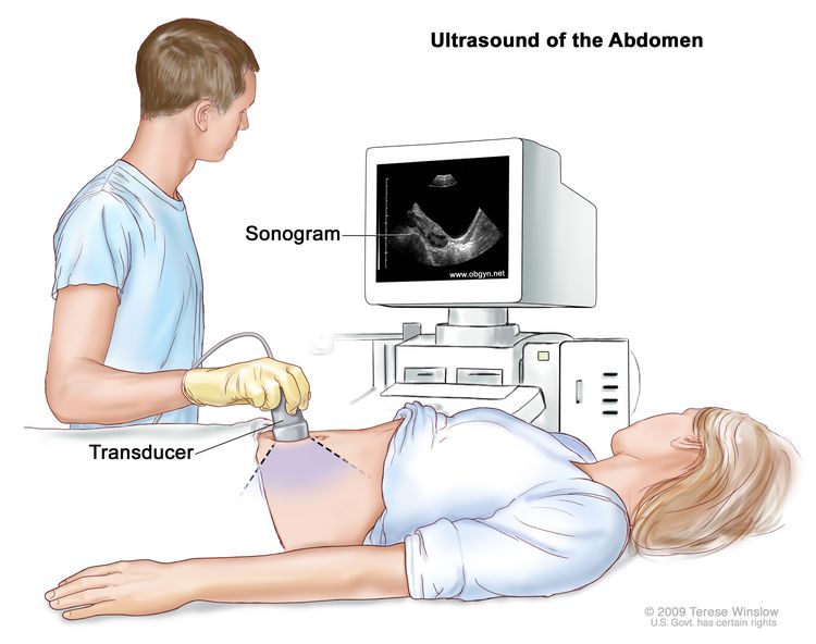
Salivary Gland Cancer Treatment (PDQ®)–Health Professional Version

Salivary Gland Cancer Treatment (PDQ®)–Health Professional Version

General Information About Salivary Gland Cancer

Incidence and Mortality

Salivary gland tumors are a morphologically and clinically diverse group of neoplasms, which may present significant diagnostic and management challenges. These tumors are rare. From 2018 to 2022, the age-adjusted incidence of salivary gland cancer in the United States was 1.3 cases per 100,000 people per year. The age-adjusted mortality rate from 2019 to 2023 was 0.3 deaths per 100,000 people per year.[1] Malignant salivary gland neoplasms account for more than 0.5% of all malignancies and approximately 3% to 5% of all head and neck cancers.[2,3] Most patients with malignant salivary gland tumors are in their sixth or seventh decade of life.[4,5]

Carcinogenesis and Risk Factors

Although exposure to ionizing radiation has been implicated as a cause of salivary gland cancer, the etiology of most salivary gland cancers cannot be determined.[3,4,6,7] Occupations associated with an increased risk for salivary gland cancers include rubber products manufacturing, asbestos mining, plumbing, and some types of woodworking.[4]

Anatomy

Tumors of the salivary glands comprise those in the major glands (e.g., parotid, submandibular, and sublingual) and the minor glands (e.g., oral mucosa, palate, uvula, floor of mouth, posterior tongue, retromolar area and peritonsillar area, pharynx, larynx, and paranasal sinuses).[3,8] Minor salivary gland lesions are most frequently seen in the oral cavity.[3]

Of salivary gland neoplasms, more than 50% are benign, and approximately 70% to 80% originate in the parotid gland.[2,3,9] The palate is the most common site of minor salivary gland tumors. The frequency of malignant lesions varies by site. Approximately 20% to 25% of parotid tumors, 35% to 40% of submandibular tumors, 50% of palate tumors, and more than 90% of sublingual gland tumors are malignant.[2,10]

Histopathology

Histologically, salivary gland tumors represent the most heterogenous group of tumors of any tissue in the body.[11] Although almost 40 histological types of epithelial tumors of the salivary glands exist, some are exceedingly rare and may be the subject of only a few case reports.[2,12] The most common benign major and minor salivary gland tumor is pleomorphic adenoma, which makes up about 50% of all salivary gland tumors and 65% of parotid gland tumors.[2] The most common malignant major and minor salivary gland tumor is mucoepidermoid carcinoma, which represents about 10% of all salivary gland neoplasms and approximately 35% of malignant salivary gland neoplasms.[2,13] This neoplasm occurs most often in the parotid gland.[3,13,14] For more information about this type and other histological types of salivary gland neoplasms, see the Cellular Classification of Salivary Gland Cancer section.

Clinical Presentation

Most patients with benign tumors of the major or minor salivary glands present with painless swelling of the parotid, submandibular, or sublingual glands. Neurological signs, such as numbness or weakness caused by nerve involvement, typically indicate a malignancy.[3] Facial nerve weakness associated with a parotid or submandibular tumor is an ominous sign. Persistent facial pain is highly suggestive of malignancy. Approximately 10% to 15% of malignant parotid neoplasms present with pain.[9,15] However, most parotid tumors, both benign and malignant, present as an asymptomatic mass in the gland.[3,9] For more information, see Cancer Pain.

Prognostic Factors

Early-stage, low-grade, malignant salivary gland tumors are usually curable by adequate surgical resection alone. The prognosis is more favorable when the tumor is in a major salivary gland. The parotid gland is most favorable followed by the submandibular gland. The least favorable primary sites are the sublingual and minor salivary glands. Large bulky tumors or high-grade tumors carry a poorer prognosis and may best be treated by surgical resection combined with postoperative radiation therapy.[16] The prognosis also depends on the following factors:[17,18]

  • Gland in which the tumor arises.
  • Histology.
  • Grade (i.e., degree of malignancy).
  • Extent of primary tumor (i.e., stage).
  • Whether the tumor involves the facial nerve, has fixation to the skin or deep structures, or has spread to lymph nodes or distant sites.

Follow-Up and Survivorship

Overall, clinical stage, particularly tumor size, may be the crucial factor that determines the outcome of salivary gland cancer and may be more important than histological grade.[19]

Treatment management

Perineural invasion can occur, particularly in high-grade adenoid cystic carcinoma, and should be specifically identified and treated.[20] Radiation therapy may increase the chance of local control and increase the survival of patients when adequate margins cannot be achieved.[21][Level of evidence C2] Unresectable or recurrent tumors may respond to chemotherapy.[2224] Fast neutron-beam radiation therapy or accelerated hyperfractionated photon-beam schedules have been effective in the treatment of inoperable, unresectable, and recurrent tumors.[2527]

Follow-up after treatment

Complications of surgical treatment for parotid neoplasms include facial nerve dysfunction and Frey syndrome (also known as gustatory flushing and sweating and auriculotemporal syndrome).[9] Frey syndrome has been successfully treated with injections of botulinum toxin A.[2830]

References
  1. Surveillance Research Program, National Cancer Institute: SEER*Explorer: An interactive website for SEER cancer statistics. Bethesda, MD: National Cancer Institute. Available online. Last accessed December 30, 2024.
  2. Speight PM, Barrett AW: Salivary gland tumours. Oral Dis 8 (5): 229-40, 2002. [PUBMED Abstract]
  3. Mendenhall WM, Werning JW, Pfister DG: Treatment of head and neck cancer. In: DeVita VT Jr, Lawrence TS, Rosenberg SA: Cancer: Principles and Practice of Oncology. 9th ed. Lippincott Williams & Wilkins, 2011, pp 729-80.
  4. Ellis GL, Auclair PL: Tumors of the Salivary Glands. Armed Forces Institute of Pathology, 1996. Atlas of Tumor Pathology, 3.
  5. Wahlberg P, Anderson H, Biörklund A, et al.: Carcinoma of the parotid and submandibular glands–a study of survival in 2465 patients. Oral Oncol 38 (7): 706-13, 2002. [PUBMED Abstract]
  6. Scanlon EF, Sener SF: Head and neck neoplasia following irradiation for benign conditions. Head Neck Surg 4 (2): 139-45, 1981 Nov-Dec. [PUBMED Abstract]
  7. van der Laan BF, Baris G, Gregor RT, et al.: Radiation-induced tumours of the head and neck. J Laryngol Otol 109 (4): 346-9, 1995. [PUBMED Abstract]
  8. Spiro RH, Thaler HT, Hicks WF, et al.: The importance of clinical staging of minor salivary gland carcinoma. Am J Surg 162 (4): 330-6, 1991. [PUBMED Abstract]
  9. Gooden E, Witterick IJ, Hacker D, et al.: Parotid gland tumours in 255 consecutive patients: Mount Sinai Hospital’s quality assurance review. J Otolaryngol 31 (6): 351-4, 2002. [PUBMED Abstract]
  10. Theriault C, Fitzpatrick PJ: Malignant parotid tumors. Prognostic factors and optimum treatment. Am J Clin Oncol 9 (6): 510-6, 1986. [PUBMED Abstract]
  11. Brandwein MS, Ferlito A, Bradley PJ, et al.: Diagnosis and classification of salivary neoplasms: pathologic challenges and relevance to clinical outcomes. Acta Otolaryngol 122 (7): 758-64, 2002. [PUBMED Abstract]
  12. Seifert G, Sobin LH: Histological Typing of Salivary Gland Tumours. 2nd ed. Springer-Verlag, 1991.
  13. Guzzo M, Andreola S, Sirizzotti G, et al.: Mucoepidermoid carcinoma of the salivary glands: clinicopathologic review of 108 patients treated at the National Cancer Institute of Milan. Ann Surg Oncol 9 (7): 688-95, 2002. [PUBMED Abstract]
  14. Goode RK, Auclair PL, Ellis GL: Mucoepidermoid carcinoma of the major salivary glands: clinical and histopathologic analysis of 234 cases with evaluation of grading criteria. Cancer 82 (7): 1217-24, 1998. [PUBMED Abstract]
  15. Spiro RH, Huvos AG, Strong EW: Cancer of the parotid gland. A clinicopathologic study of 288 primary cases. Am J Surg 130 (4): 452-9, 1975. [PUBMED Abstract]
  16. Parsons JT, Mendenhall WM, Stringer SP, et al.: Management of minor salivary gland carcinomas. Int J Radiat Oncol Biol Phys 35 (3): 443-54, 1996. [PUBMED Abstract]
  17. Vander Poorten VL, Balm AJ, Hilgers FJ, et al.: The development of a prognostic score for patients with parotid carcinoma. Cancer 85 (9): 2057-67, 1999. [PUBMED Abstract]
  18. Terhaard CH, Lubsen H, Van der Tweel I, et al.: Salivary gland carcinoma: independent prognostic factors for locoregional control, distant metastases, and overall survival: results of the Dutch head and neck oncology cooperative group. Head Neck 26 (8): 681-92; discussion 692-3, 2004. [PUBMED Abstract]
  19. Spiro RH: Factors affecting survival in salivary gland cancers. In: McGurk M, Renehan AG, eds.: Controversies in the Management of Salivary Gland Disease. Oxford University Press, 2001, pp 143-50.
  20. Gormley WB, Sekhar LN, Wright DC, et al.: Management and long-term outcome of adenoid cystic carcinoma with intracranial extension: a neurosurgical perspective. Neurosurgery 38 (6): 1105-12; discussion 1112-3, 1996. [PUBMED Abstract]
  21. Hosokawa Y, Shirato H, Kagei K, et al.: Role of radiotherapy for mucoepidermoid carcinoma of salivary gland. Oral Oncol 35 (1): 105-11, 1999. [PUBMED Abstract]
  22. Borthne A, Kjellevold K, Kaalhus O, et al.: Salivary gland malignant neoplasms: treatment and prognosis. Int J Radiat Oncol Biol Phys 12 (5): 747-54, 1986. [PUBMED Abstract]
  23. Spiro RH: Salivary neoplasms: overview of a 35-year experience with 2,807 patients. Head Neck Surg 8 (3): 177-84, 1986 Jan-Feb. [PUBMED Abstract]
  24. Licitra L, Cavina R, Grandi C, et al.: Cisplatin, doxorubicin and cyclophosphamide in advanced salivary gland carcinoma. A phase II trial of 22 patients. Ann Oncol 7 (6): 640-2, 1996. [PUBMED Abstract]
  25. Wang CC, Goodman M: Photon irradiation of unresectable carcinomas of salivary glands. Int J Radiat Oncol Biol Phys 21 (3): 569-76, 1991. [PUBMED Abstract]
  26. Buchholz TA, Laramore GE, Griffin BR, et al.: The role of fast neutron radiation therapy in the management of advanced salivary gland malignant neoplasms. Cancer 69 (11): 2779-88, 1992. [PUBMED Abstract]
  27. Krüll A, Schwarz R, Engenhart R, et al.: European results in neutron therapy of malignant salivary gland tumors. Bull Cancer Radiother 83 (Suppl): 125-9s, 1996. [PUBMED Abstract]
  28. Naumann M, Zellner M, Toyka KV, et al.: Treatment of gustatory sweating with botulinum toxin. Ann Neurol 42 (6): 973-5, 1997. [PUBMED Abstract]
  29. Arad-Cohen A, Blitzer A: Botulinum toxin treatment for symptomatic Frey’s syndrome. Otolaryngol Head Neck Surg 122 (2): 237-40, 2000. [PUBMED Abstract]
  30. von Lindern JJ, Niederhagen B, Bergé S, et al.: Frey syndrome: treatment with type A botulinum toxin. Cancer 89 (8): 1659-63, 2000. [PUBMED Abstract]

Cellular Classification of Salivary Gland Cancer

Salivary gland neoplasms are remarkable for their histological diversity. These neoplasms include benign and malignant tumors of epithelial, mesenchymal, and lymphoid origin. Salivary gland tumors pose a particular challenge to the surgical pathologist. Differentiating benign from malignant tumors may be difficult, primarily because of the complexity of the classification and the rarity of several entities, which may exhibit a broad spectrum of morphological diversity in individual lesions.[1] In some cases, hybrid lesions may be seen.[2] The key guiding principle to establish the malignant nature of a salivary gland tumor is the demonstration of an infiltrative margin.[1]

The following cellular classification scheme draws heavily from a scheme published by the Armed Forces Institute of Pathology (AFIP).[3] Malignant nonepithelial neoplasms are included in the scheme because these neoplasms comprise a significant proportion of salivary gland neoplasms seen in the clinical setting. For completeness, malignant secondary tumors are also included in the scheme.

Where AFIP statistics regarding the incidence, or relative frequency, of particular histopathologies are cited, some bias may exist because of the AFIP methods of case accrual as a pathology reference service. When possible, other sources are cited for incidence data. Notwithstanding the AFIP data, the incidence of a particular histopathology has been found to vary considerably depending upon the study cited. This variability in reporting may be partially caused by the rare incidence of many salivary gland neoplasms.

Epithelial Neoplasms

The clinician should be aware that several benign epithelial salivary gland neoplasms have malignant counterparts, which are discussed below:[3]

Histological grading of salivary gland carcinomas is important to determine the proper treatment approach, although it is not an independent indicator of the clinical course and must be considered in the context of the clinical stage. Clinical stage, particularly tumor size, may be the critical factor to determine the outcome of salivary gland cancer and may be more important than histological grade.[1] For example, stage I intermediate-grade or high-grade mucoepidermoid carcinomas can be successfully treated, whereas low-grade mucoepidermoid carcinomas that present as stage III disease may have a very aggressive clinical course.[4]

Grading is used primarily for mucoepidermoid carcinomas, adenocarcinomas, not otherwise specified (NOS), adenoid cystic carcinomas, and squamous cell carcinomas.[1,3] Various other salivary gland carcinomas can also be categorized according to histological grade as follows:[3,58]

Low grade

Low grade, intermediate grade, and high grade

Intermediate grade and high grade

High grade

*[Note: Some investigators consider mucoepidermoid carcinoma to be of only two grades: low grade and high grade.5]

Mucoepidermoid carcinoma

Mucoepidermoid carcinoma is a malignant epithelial tumor that is composed of various proportions of mucous, epidermoid (e.g., squamous), intermediate, columnar, and clear cells and often demonstrates prominent cystic growth. It is the most common malignant neoplasm observed in the major and minor salivary glands.[1,9] Mucoepidermoid carcinoma represents 29% to 34% of malignant tumors originating in both major and minor salivary glands.[3,5,10,11] In two large retrospective series, 84% to 93% of cases originated in the parotid gland.[12,13] With regard to malignant tumors of the minor salivary glands, mucoepidermoid carcinoma shows a strong predilection for the lower lip.[3,14] In an AFIP review of civilian cases, the mean age of patients was 47 years (range, 8–92 years).[3] Prior exposure to ionizing radiation appears to substantially increase the risk of developing malignant neoplasms of the major salivary glands, particularly mucoepidermoid carcinoma.[3,13]

Most patients are asymptomatic and present with solitary, painless masses. Symptoms include pain, drainage from the ipsilateral ear, dysphagia, trismus, and facial paralysis.[3] For more information, see Cancer Pain.

Microscopic grading of mucoepidermoid carcinoma is important to determine the prognosis.[1,12,15] Mucoepidermoid carcinomas are graded as low grade, intermediate grade, and high grade. Grading parameters with point values include the following:

  • Intracystic component (+2).
  • Neural invasion present (+2).
  • Necrosis present (+3).
  • Mitosis (≥4 per 10 high-power field [+3]).
  • Anaplasia present (+4).

Total point scores are 0 to 4 for low grade, 5 to 6 for intermediate grade, and 7 to 14 for high grade.

In a retrospective review of 243 cases of mucoepidermoid carcinoma of the major salivary glands, a statistically significant correlation was shown between this point-based grading system and outcome for parotid tumors but not for submandibular tumors.[12] Another retrospective study that used this histological grading system indicated that tumor grade correlated well with prognosis for mucoepidermoid carcinoma of the major salivary glands, excluding submandibular tumors, and minor salivary glands.[13] A modification of this grading system placed more emphasis on features of tumor invasion.[16] Nonetheless, though tumor grade may be useful, stage appears to be a better indicator of prognosis.[3,16]

Cytogenetically, mucoepidermoid carcinoma is characterized by a t(11;19)(q14–21;p12–13) translocation, which is occasionally the sole cytogenetic alteration.[1719] This translocation creates a novel MECT1::MAML2 gene fusion that disrupts a Notch signaling pathway.[20] Notch signaling plays a key role in the normal development of many tissues and cell types, through diverse effects on cellular differentiation, survival, and/or proliferation, and may be involved in a wide variety of human neoplasms.[21]

Rarely, mucoepidermoid carcinoma may originate within the jaws. This tumor type is known as central mucoepidermoid carcinoma.[3] The mandibular to maxillary predilection is approximately 3:1.[22]

Adenoid cystic carcinoma

Adenoid cystic carcinoma, formerly known as cylindroma, is a slow growing but aggressive neoplasm with a remarkable capacity for recurrence.[23] Morphologically, three growth patterns have been described: cribriform, or classic pattern; tubular; and solid, or basaloid pattern. The tumors are categorized according to the predominant pattern.[3,2325] The cribriform pattern shows epithelial cell nests that form cylindrical patterns. The lumina of these spaces contain periodic acid-Schiff (PAS)-positive mucopolysaccharide secretions. The tubular pattern reveals tubular structures that are lined by stratified cuboidal epithelium. The solid pattern shows solid groups of cuboidal cells. The cribriform pattern is the most common, and the solid pattern is the least common.[26] Solid adenoid cystic carcinoma is a high-grade lesion with reported recurrence rates of as much as 100%, compared with 50% to 80% for the tubular and cribriform variants.[25]

In a review of its case files, the AFIP found adenoid cystic carcinoma to be the fifth most common malignant epithelial tumor of the salivary glands after mucoepidermoid carcinomas; adenocarcinomas, NOS; acinic cell carcinomas; and PLGA.[3] Other series, however, reported adenoid cystic carcinoma to be the second most common malignant tumor, with an incidence or relative frequency of approximately 20%.[1] In the AFIP data, this neoplasm constitutes approximately 7.5% of all epithelial malignancies and 4% of all benign and malignant epithelial salivary gland tumors. The peak incidence for this tumor is reported to be in the fourth through sixth decades of life.[3]

This neoplasm typically develops as a slow-growing swelling in the preauricular or submandibular region. Pain and facial paralysis develop frequently during the course of the disease and are likely related to the associated high incidence of nerve invasion.[3] For more information, see Cancer Pain. Regardless of histological grade, adenoid cystic carcinomas, with their unusually slow biological growth, tend to have a protracted course and ultimately a poor outcome, with 10-year survival rates reported to be less than 50% for all grades.[1,27] These carcinomas typically show frequent recurrences and late distant metastases.[1,28] Clinical stage may be a better prognostic indicator than histological grade.[28,29] In a retrospective review of 92 cases, tumors larger than 4 cm were associated with an unfavorable clinical course in all cases.[30]

Adenocarcinomas
Acinic cell carcinoma

Acinic cell carcinoma, also known as acinic cell adenocarcinoma, is a malignant epithelial neoplasm in which the neoplastic cells express acinar differentiation. By conventional use, the term acinic cell carcinoma is defined by cytological differentiation toward serous acinar cells, as opposed to mucous acinar cells, whose characteristic feature is cytoplasmic PAS-positive zymogen-type secretory granules.[3] In AFIP data of salivary gland neoplasms, acinic cell carcinoma is the third most common salivary gland epithelial neoplasm after mucoepidermoid carcinoma and adenocarcinoma, NOS.[3] Acinic cell carcinoma accounted for 17% of primary malignant salivary gland tumors or about 6% of all salivary gland neoplasms. More than 80% of these tumors occur in the parotid gland, women were affected more than men, and the mean age was 44 years. Other studies have reported a relative frequency of acinic cell carcinoma from 0% to 19% of malignant salivary gland neoplasms.[3]

Clinically, patients typically present with a slowly enlarging mass in the parotid region. Pain is a symptom in more than 33% of patients. For acinic cell carcinoma, staging is likely a better predictor of outcome than histological grading.[3] In a retrospective review of 90 cases, poor prognostic features included pain or fixation; gross invasion; and microscopic features of desmoplasia, atypia, or increased mitotic activity. Neither morphological pattern nor cell composition was a predictive feature.[31] For more information, see Cancer Pain.

PLGA

PLGA is a malignant epithelial tumor that is essentially limited to occurrence in minor salivary gland sites and is characterized by bland, uniform nuclear features; diverse but characteristic architecture; infiltrative growth; and perineural infiltration.[3] In a series of 426 patients with minor salivary gland tumors, PLGA represented 11% of all tumors and 26% of those that were malignant.[32] In minor gland sites, PLGA is twice as frequent as adenoid cystic carcinoma, and among all benign and malignant salivary gland neoplasms, only pleomorphic adenoma and mucoepidermoid carcinoma are more common.[3] In the AFIP case files, more than 60% of tumors occurred in the mucosa of either the soft or hard palates, approximately 16% occurred in the buccal mucosa, and 12% occurred in the upper lip. The average age of patients is 59 years, with 70% of patients between the ages of 50 and 79 years.[3] The female-to-male ratio is about 2:1, a proportion greater than for malignant salivary gland tumors in general.[3,33]

PLGA typically presents as a firm, nontender swelling involving the mucosa of the hard and soft palates (i.e., it is often found at their junction), the cheek, or the upper lip. Discomfort, bleeding, telangiectasia, or ulceration of the overlying mucosa may occasionally occur.[3] This salivary gland neoplasm typically runs a moderately indolent course. In a study of 40 cases with long-term follow-up, the overall survival rate was 80% at 25 years.[34] Because of the unpredictable behavior of the tumor, some investigators consider the qualifying term, low grade, to be misleading and instead prefer the term polymorphous adenocarcinoma.[1]

Adenocarcinoma, NOS

Adenocarcinoma, NOS, is a salivary gland carcinoma that shows glandular or ductal differentiation but lacks the prominence of any of the morphological features that characterize the other, more specific carcinoma types. The diagnosis of adenocarcinoma, NOS, is essentially one of exclusion. In an AFIP review of cases, adenocarcinoma, NOS, was second only to mucoepidermoid carcinoma in frequency among malignant salivary gland neoplasms.[3] Other series have reported an incidence of 4% to 10%.[1] In AFIP files, the mean patient age was 58 years.[3] Approximately 40% and 60% of tumors occurred in the major and minor salivary glands, respectively. Among the major salivary gland tumors, 90% occurred in the parotid gland. Adenocarcinoma, NOS is graded in a similar way to extrasalivary lesions according to the degree of differentiation.[1] Tumor grades include low-grade, intermediate-grade, and high-grade categories.[3]

Patients with tumors in the major salivary glands typically present with solitary, painless masses.[35] Two retrospective studies indicate that survival is better for patients with tumors of the oral cavity than for those with tumors of the parotid and submandibular glands.[35,36] These studies differ regarding the prognostic significance of tumor grade.

Rare adenocarcinomas
Basal cell adenocarcinoma

Basal cell adenocarcinoma, also known as basaloid salivary carcinoma, carcinoma ex monomorphic adenoma, malignant basal cell adenoma, malignant basal cell tumor, or basal cell carcinoma, is an epithelial neoplasm that is cytologically similar to basal cell adenoma but is infiltrative and has a small potential for metastasis.[3] In AFIP case files spanning almost 11 years, basal cell carcinoma made up 1.6% of all salivary gland neoplasms and 2.9% of salivary gland malignancies.[3] Nearly 90% of tumors occurred in the parotid gland.[3,37] The average age of patients was 60 years.[3]

Similar to most salivary gland neoplasms, swelling is typically the only sign or symptom.[37] A sudden increase in size may occur in a few patients.[38] Basal cell carcinomas are low-grade carcinomas that are infiltrative, locally destructive, and tend to recur. The carcinomas occasionally metastasize. In a retrospective series that included 29 patients, there were recurrences in 7 patients and metastases in 3 patients.[37] In another retrospective review that included 72 patients, 37% of the patients experienced local recurrences.[38] The overall prognosis for patients with this tumor is good.[37,38]

Clear cell carcinoma

Clear cell carcinoma, also known as clear cell adenocarcinoma, is a very rare malignant epithelial neoplasm composed of a monomorphous population of cells that have optically clear cytoplasm with standard hematoxylin and eosin stains and lack features of other specific neoplasms. Because of inconsistencies in the methods of reporting salivary gland neoplasms, meaningful incidence rates for this tumor are difficult to derive from the literature.[3] Most cases involve the minor salivary glands.[1,3,3941] In the AFIP case files, the mean age of patients was approximately 58 years.[3]

In most patients, swelling is the only symptom. Clear cell adenocarcinoma is a low-grade neoplasm. As of 1996, the AFIP reported that no patient had died of this tumor.[3]

Cystadenocarcinoma

Cystadenocarcinoma is a rare malignant epithelial tumor characterized histologically by prominent cystic and, frequently, papillary growth but lacking features that characterize cystic variants of several more common salivary gland neoplasms. It is also known as malignant papillary cystadenoma, mucus-producing adenopapillary, or nonepidermoid, carcinoma; low-grade papillary adenocarcinoma of the palate; or papillary adenocarcinoma. Cystadenocarcinoma is the malignant counterpart of cystadenoma.[3]

In a review that included 57 patients, the AFIP found that men and women are affected equally. The average patient age was approximately 59 years. Approximately 65% of the tumors occurred in the major salivary glands, primarily in the parotid.[3] Most patients present with a slow-growing asymptomatic mass. Clinically, this neoplasm is rarely associated with pain or facial paralysis. Cystadenocarcinoma is considered to be a low-grade neoplasm.[3]

Sebaceous adenocarcinoma

Sebaceous adenocarcinoma is a rare malignant epithelial tumor composed of islands and sheets of cells that have morphologically atypical nuclei, an infiltrative growth pattern, and focal sebaceous differentiation. This is a very rare tumor, as few cases have been reported in the literature.[3] Almost all cases occur in the parotid gland.[3] The average age of patients is reported to be 69 years.[42]

An equal number of patients present with a painless, slow-growing, asymptomatic swelling or pain. A few experience facial paralysis.[3] Most sebaceous adenocarcinomas are probably intermediate-grade malignancies. The tumor recurs in about 33% of cases.[43,44]

Sebaceous lymphadenocarcinoma

Sebaceous lymphadenocarcinoma is an extremely rare malignant tumor that represents carcinomatous transformation of sebaceous lymphadenoma. The carcinoma element may be sebaceous adenocarcinoma or some other specific or nonspecific form of salivary gland cancer.[3] Only three cases have been reported in the literature.[43,45] The three cases occurred in or around the parotid gland. All patients were in their seventh decade of life. Two of the three patients were asymptomatic. One had tenderness on palpation. Case reports suggest that this is a low-grade malignancy with a good prognosis.[44,45]

Oncocytic carcinoma

Oncocytic carcinoma, also known as oncocytic adenocarcinoma, is a rare, predominantly oncocytic neoplasm whose malignant nature is reflected both by its abnormal morphological features and infiltrative growth. Oncocytic carcinoma represented less than 1% of almost 3,100 salivary gland tumors accessioned to the AFIP files during a 10-year period.[3] Most cases occurred in the parotid gland. The average age of patients in the AFIP series was 63 years.[3]

Approximately 33% of the patients usually develop parotid masses that cause pain or paralysis.[46] Oncocytic carcinoma is a high-grade carcinoma. Patients with tumors smaller than 2 cm have a better prognosis than patients with larger tumors.[6]

Salivary duct carcinoma

Salivary duct carcinoma, also known as salivary duct adenocarcinoma, is a rare, typically high-grade malignant epithelial neoplasm composed of structures that resemble expanded salivary gland ducts. A low-grade variant exists.[47] Incidence rates vary depending on the study cited.[3] In the AFIP files, salivary duct carcinomas represented only 0.2% of all epithelial salivary gland neoplasms. More than 85% of cases involved the parotid gland, and approximately 75% of patients were men. The peak incidence was reported to be in the seventh and eighth decades of life.[3]

Clinically, parotid swelling is the most common sign. Facial nerve dysfunction or paralysis occur in more than 25% of patients and may be the initial manifestation.[3] The high-grade variant of this neoplasm is one of the most aggressive types of salivary gland carcinoma and is typified by local invasion, lymphatic and hematogenous spread, and poor prognosis.[3,7] In a retrospective review of 104 cases, 33% of patients developed local recurrence, and 46% of patients developed distant metastasis.[48]

Mucinous adenocarcinoma

Mucinous adenocarcinoma is a rare malignant neoplasm characterized by large amounts of extracellular epithelial mucin that contains cords, nests, and solitary epithelial cells. The incidence is unknown. Limited data indicate that most, if not all, occur in the major salivary glands, with the submandibular gland as the predominant site.[3,49] These tumors may be associated with dull pain and tenderness.[3,49] This neoplasm may be considered low grade.[3]

Malignant mixed tumors

The classification of malignant mixed tumors includes three distinct clinicopathological entities: carcinoma ex pleomorphic adenoma, carcinosarcoma, and metastasizing mixed tumor. Carcinoma ex pleomorphic adenoma accounts for most cases, whereas carcinosarcoma, a true malignant mixed tumor, and metastasizing mixed tumor are extremely rare.[3]

Carcinoma ex pleomorphic adenoma

Carcinoma ex pleomorphic adenoma, also known as carcinoma ex mixed tumor, shows histological evidence of arising from or in a benign pleomorphic adenoma.[50] Diagnosis requires the identification of benign tumor in the tissue sample.[51] The incidence or relative frequency of this tumor varies considerably depending on the study cited.[1] A review of material at the AFIP showed that carcinoma ex pleomorphic adenoma made up 8.8% of all mixed tumors and 4.6% of all malignant salivary gland tumors. It is the sixth most common malignant salivary gland tumor after mucoepidermoid carcinoma; adenocarcinoma, NOS; acinic cell carcinoma; polymorphous low-grade adenocarcinoma; and adenoid cystic carcinoma.[3] The neoplasm occurs primarily in the major salivary glands.[52]

The most common clinical presentation is a painless mass.[3] Approximately 33% of patients may experience facial paralysis.[53] Depending on the series cited, survival rates vary significantly: 25% to 65% at 5 years, 24% to 50% at 10 years, 10% to 35% at 15 years, and 0% to 38% at 20 years.[3] In addition to tumor stage, histological grade and degree of invasion help to determine prognosis.[54]

Carcinosarcoma

Carcinosarcoma, also known as true malignant mixed tumor, is a rare malignant salivary gland neoplasm that contains both carcinoma and sarcoma components. Either or both components are expressed in metastatic foci. Some carcinosarcomas develop de novo, while others develop in association with benign mixed tumor. This neoplasm is rare; only eight cases exist in the AFIP case files.[3] At one facility, only 11 cases were recorded over a 32-year period.[8] Most of these tumors occur in the major salivary glands.

Swelling, pain, nerve palsy, and ulceration have been frequent clinical findings. Carcinosarcoma is an aggressive, high-grade malignancy. In the largest series reported, which consisted of 12 cases, the average survival period was 3.6 years.[8]

Metastasizing mixed tumor

Metastasizing mixed tumor is a very rare, histologically benign salivary gland neoplasm that inexplicably metastasizes. Often, a long interval occurs between the diagnosis of the primary tumor and the metastases. The histological features are within the spectrum of features that typify pleomorphic adenoma.[3] Most of these tumors occur in the major salivary glands. The primary neoplasm is typically a single, well-defined mass. Recurrences, which may be multiple, have occurred as many as 26 years after excision of the primary neoplasm.[55]

Rare carcinomas
Primary squamous cell carcinoma

Primary squamous cell carcinoma, also known as primary epidermoid carcinoma, is a malignant epithelial neoplasm of the major salivary glands that is composed of squamous (i.e., epidermoid) cells. Diagnosis requires the exclusion of primary disease located in some other head and neck site; indeed, most squamous cell carcinomas of the major salivary glands represent metastatic disease.[3] This diagnosis is not made in minor salivary glands because distinction from the more common mucosal squamous cell carcinoma is not possible.[3] Previous exposure to ionizing radiation appears to increase the risk of developing this neoplasm.[11,56,57] The median time between radiation therapy and diagnosis of the neoplasm is approximately 15.5 years.[11] The reported frequency of this tumor among all major salivary gland tumors has varied from 0.9% to 4.7%.[3,10] In AFIP major salivary gland accessions from 1985 to 1996, primary squamous cell carcinoma accounted for 2.7% of all tumors; 5.4% of malignant tumors; and 2.5% and 2.8%, respectively, of all parotid and submandibular tumors.[3] The average age in the AFIP registry was 64 years.[3] This neoplasm occurs in the parotid gland almost nine times more often than in the submandibular gland.[3,57] There is a strong male predilection.[3,11,5759] This tumor is graded in a similar way to extrasalivary lesions according to the degree of differentiation, namely, low grade, intermediate grade, and high grade.[1]

Most patients present with an asymptomatic mass in the parotid region. Other symptoms may include a painful mass and facial nerve palsy.[57] The prognosis for this neoplasm is poor. In a 30-year retrospective analysis of 50 cases of squamous cell carcinoma of the salivary glands, survival rates at 5 years and 10 years were 24% and 18%, respectively.[57]

Epithelial-myoepithelial carcinoma

Epithelial-myoepithelial carcinoma is an uncommon, low-grade epithelial neoplasm composed of variable proportions of ductal and large, clear-staining, differentiated myoepithelial cells. It is also known as adenomyoepithelioma, clear cell adenoma, tubular solid adenoma, monomorphic clear cell tumor, glycogen-rich adenoma, glycogen-rich adenocarcinoma, clear cell carcinoma, or salivary duct carcinoma. The tumor represents approximately 1% of all epithelial salivary gland neoplasms.[3,60] It is predominantly a tumor of the parotid gland. In the AFIP case files, the mean age of patients was about 60 years, and about 60% of the patients were female.[3]

Localized swelling is commonly the only symptom, but occasionally patients experience facial weakness or pain.[61,62] Overall, epithelial-myoepithelial carcinoma is a low-grade carcinoma that recurs frequently, has a tendency to metastasize to periparotid and cervical lymph nodes, and occasionally results in distant metastasis and death.[60,6264]

Anaplastic small cell carcinoma

Anaplastic small cell carcinoma of the salivary glands was first described in 1972.[65] Subsequent histochemical and electron microscopic studies have supported the neuroendocrine nature of this tumor.[66,67] Microscopically, the tumor cells have oval, hyperchromatic nuclei and a scant amount of cytoplasm and are organized in sheets, strands, and nests. The mitotic rate is high. Neuroendocrine carcinomas are more frequently found in the minor salivary glands. These patients have a better survival rate than patients with small cell carcinomas of the lung.[68] The undifferentiated counterpart of this neoplasm is the small cell undifferentiated carcinoma.

Undifferentiated carcinomas

Undifferentiated carcinomas of salivary glands are a group of uncommon malignant epithelial neoplasms that lack the specific light-microscopic morphological features of other types of salivary gland carcinomas. These carcinomas are histologically similar to undifferentiated carcinomas that arise in other organs and tissues. Accordingly, metastatic carcinoma is a primary concern in the differential diagnosis of these neoplasms.[3]

Small cell undifferentiated carcinoma

Small cell undifferentiated carcinoma, also known as extrapulmonary oat cell carcinoma, is a rare, primary malignant tumor. With conventional light microscopy, it is composed of undifferentiated cells and, with ultrastructural or immunohistochemical studies, does not demonstrate neuroendocrine differentiation. This is the undifferentiated counterpart of anaplastic small cell carcinoma. For more information, see the Anaplastic small cell carcinoma section.

In an AFIP review of case files, small cell carcinoma represented 1.8% of all major salivary gland malignancies; the mean age of patients was 56 years.[3] In 50% of the cases, patients present with an asymptomatic parotid mass of 3 months’ or less duration.[6870] This is a high-grade neoplasm. In a retrospective review of 12 cases, a tumor size of more than 4 cm was found to be the most important predictor of behavior. In another small retrospective series, estimated survival rates at 2 and 5 years were 70% and 46%, respectively.[68]

Large cell undifferentiated carcinoma

Large cell undifferentiated carcinoma is a tumor in which features of acinar, ductal, epidermoid, or myoepithelial differentiation are absent under light microscopy, though occasionally, poorly formed duct-like structures are found. This neoplasm accounts for approximately 1% of all epithelial salivary gland neoplasms.[3,53,71,72] Most of these tumors occur in the parotid gland.[70,72] In AFIP data, the peak incidence is in the seventh to eighth decades of life.[3]

Rapid growth of a parotid swelling is a common clinical presentation.[59] This is a high-grade neoplasm that frequently metastasizes and has a poor prognosis. Patients with neoplasms of 4 cm or larger may have a particularly poor outcome.[70,72]

Lymphoepithelial carcinoma

Lymphoepithelial carcinoma, also known as undifferentiated carcinoma with lymphoid stroma and carcinoma ex lymphoepithelial lesion, is an undifferentiated tumor that is associated with a dense lymphoid stroma. An exceptionally high incidence of this tumor is found in the Inuit population.[3,73] This neoplasm has been associated with Epstein-Barr virus infection.[74,75] Of the occurrences, 80% are in the parotid gland.[3]

In addition to the presence of a parotid or submandibular mass, pain is a frequent symptom, and facial nerve palsy occurs in as many as 20% of patients.[76] Of the patients, more than 40% have metastases to cervical lymph nodes at initial presentation, 20% develop local recurrences or lymph node metastases, and 20% develop distant metastases within 3 years following therapy.[73,7678] For more information, see Cancer Pain.

Myoepithelial carcinoma

Myoepithelioma carcinoma is a rare, malignant salivary gland neoplasm in which the tumor cells almost exclusively manifest myoepithelial differentiation. This neoplasm represents the malignant counterpart of benign myoepithelioma.[3] The largest series reported involved 25 cases.[79] Approximately 66% of the tumors occur in the parotid gland.[3,74] The mean age of patients is 55 years.[79]

Most patients present with the primary complaint of a painless mass.[79] This is an intermediate grade to high-grade carcinoma.[3,79] Histological grade does not appear to correlate well with clinical behavior. For instance, tumors with a low-grade histological appearance may behave aggressively.[79]

Adenosquamous carcinoma

Adenosquamous carcinoma is an extremely rare malignant neoplasm that simultaneously arises from surface mucosal epithelium and salivary gland ductal epithelium. The carcinoma shows histopathological features of both squamous cell carcinoma and adenocarcinoma. Only a handful of reports describe this tumor.[3]

In addition to swelling, adenosquamous carcinoma produces visible changes in the mucosa, including erythema, ulceration, and induration. Pain frequently accompanies ulceration. Limited data indicate that this is a highly aggressive neoplasm with a poor prognosis.[3]

Nonepithelial Neoplasms

Lymphomas and benign lymphoepithelial lesion

Lymphomas of the major salivary glands are characteristically of the non-Hodgkin type. In an AFIP review of case files, non-Hodgkin lymphoma accounted for 16.3% of all malignant tumors that occurred in the major salivary glands. Disease in the parotid gland accounted for about 80% of all cases.[3]

Patients with benign lymphoepithelial lesion (e.g., Mikulicz disease), which is a manifestation of the autoimmune disease Sjögren syndrome, are at an increased risk of developing non-Hodgkin lymphoma.[8084] Benign lymphoepithelial lesion is clinically characterized by diffuse and bilateral enlargement of the salivary and lacrimal glands.[23] Morphologically, a salivary gland lesion is composed of prominent myoepithelial islands surrounded by a lymphocytic infiltrate. Germinal centers are often present in the lymphocytic infiltrate.[23] Immunophenotypically and genotypically, the lymphocytic infiltrate is composed of B-lymphocytes and T-lymphocytes, which are polyclonal. In some instances, the B-cell lymphocytic infiltrate can undergo clonal expansion and evolve into frank non-Hodgkin lymphoma. Most of the non-Hodgkin lymphomas arising in a background of benign lymphoepithelial lesions are marginal zone lymphomas of mucosa-associated lymphoid tissue (MALT).[8184] MALT lymphomas of the salivary glands, like their counterparts in other anatomical sites, typically display relatively indolent clinical behavior.[3,85]

Primary non-MALT lymphomas of the salivary glands may also occur and appear to have a prognosis similar to those in patients who have histologically identical nodal lymphomas.[86,87] Unlike non-Hodgkin lymphoma, involvement of the major salivary glands by Hodgkin lymphoma is rare. Most tumors occur in the parotid gland.[3] The most common histological types encountered are the nodular sclerosing and lymphocyte-predominant variants.[88,89]

Mesenchymal neoplasms

Mesenchymal neoplasms account for 1.9% to 5% of all neoplasms that occur within the major salivary glands.[90,91] These cellular classifications pertain to major salivary gland tumors. Because the minor salivary glands are small and embedded within fibrous connective tissue, fat, and skeletal muscle, the origin of a mesenchymal neoplasm from stroma cannot be determined.[3] The types of benign mesenchymal salivary gland neoplasms include hemangiomas, lipomas, and lymphangiomas.

Malignant mesenchymal salivary gland neoplasms include malignant schwannomas, hemangiopericytomas, malignant fibrous histiocytomas, rhabdomyosarcomas, and fibrosarcomas, among others. In the major salivary glands, these neoplasms represent approximately 0.5% of all benign and malignant salivary gland tumors and approximately 1.5% of all malignant tumors.[90,92,93] It is important to establish a primary salivary gland origin for these tumors by excluding the possibilities of metastasis and direct extension from other sites. In addition, the diagnosis of salivary gland carcinosarcoma should be excluded.[3] Primary salivary gland sarcomas behave like their soft tissue counterparts, in which prognosis is related to sarcoma type, histological grade, tumor size, and stage.[93,94] For more information, see Soft Tissue Sarcoma Treatment. A comprehensive review of salivary gland mesenchymal neoplasms can be found elsewhere.[95]

Malignant Secondary Neoplasms

Malignant neoplasms whose origins lie outside the salivary glands may involve the major salivary glands by the following routes:[3]

  1. Direct invasion from cancers that lie adjacent to the salivary glands.
  2. Hematogenous metastases from distant primary tumors.
  3. Lymphatic metastases to lymph nodes within the salivary gland.

Direct invasion of nonsalivary gland tumors into the major salivary glands is principally from squamous cell and basal cell carcinomas of the overlying skin.

Approximately 80% of metastases to the major salivary glands may be from primary tumors elsewhere in the head and neck; the remaining 20% may be from infraclavicular sites.[96,97] The parotid gland is the site of 80% to 90% of the metastases, and the remainder involve the submandibular gland.[97,98] In a decade-long AFIP experience, metastatic tumors constituted approximately 10% of malignant neoplasms in the major salivary glands, exclusive of malignant lymphomas.[3] Most metastatic primary tumors to the major salivary glands are squamous cell carcinomas and melanomas from the head and neck that presumably reach the parotid gland via the lymphatic system. Infraclavicular primary tumors, such as those in the lung, kidney, and breast, reach the salivary glands by a hematogenous route.[9799] The peak incidence for metastatic tumors in the salivary glands is reported to be in the seventh decade of life.[3]

References
  1. Speight PM, Barrett AW: Salivary gland tumours. Oral Dis 8 (5): 229-40, 2002. [PUBMED Abstract]
  2. Seifert G, Donath K: Hybrid tumours of salivary glands. Definition and classification of five rare cases. Eur J Cancer B Oral Oncol 32B (4): 251-9, 1996. [PUBMED Abstract]
  3. Ellis GL, Auclair PL: Tumors of the Salivary Glands. Armed Forces Institute of Pathology, 1996. Atlas of Tumor Pathology, 3.
  4. Cheuk W, Chan JKC: Salivary gland tumors. In: Fletcher CDM, ed.: Diagnostic Histopathology of Tumors. 3rd ed. Churchill Livingstone, 2007, pp 239-326.
  5. Spiro RH, Huvos AG, Berk R, et al.: Mucoepidermoid carcinoma of salivary gland origin. A clinicopathologic study of 367 cases. Am J Surg 136 (4): 461-8, 1978. [PUBMED Abstract]
  6. Goode RK, Corio RL: Oncocytic adenocarcinoma of salivary glands. Oral Surg Oral Med Oral Pathol 65 (1): 61-6, 1988. [PUBMED Abstract]
  7. Guzzo M, Di Palma S, Grandi C, et al.: Salivary duct carcinoma: clinical characteristics and treatment strategies. Head Neck 19 (2): 126-33, 1997. [PUBMED Abstract]
  8. Stephen J, Batsakis JG, Luna MA, et al.: True malignant mixed tumors (carcinosarcoma) of salivary glands. Oral Surg Oral Med Oral Pathol 61 (6): 597-602, 1986. [PUBMED Abstract]
  9. Auclair PL, Ellis GL, Gnepp DR, et al.: Salivary gland neoplasms: general considerations. In: Ellis GL, Auclair PL, Gnepp DR, eds.: Surgical Pathology of the Salivary Glands. Saunders, 1991, pp 135-64.
  10. Eveson JW, Cawson RA: Salivary gland tumours. A review of 2410 cases with particular reference to histological types, site, age and sex distribution. J Pathol 146 (1): 51-8, 1985. [PUBMED Abstract]
  11. Spitz MR, Batsakis JG: Major salivary gland carcinoma. Descriptive epidemiology and survival of 498 patients. Arch Otolaryngol 110 (1): 45-9, 1984. [PUBMED Abstract]
  12. Goode RK, Auclair PL, Ellis GL: Mucoepidermoid carcinoma of the major salivary glands: clinical and histopathologic analysis of 234 cases with evaluation of grading criteria. Cancer 82 (7): 1217-24, 1998. [PUBMED Abstract]
  13. Guzzo M, Andreola S, Sirizzotti G, et al.: Mucoepidermoid carcinoma of the salivary glands: clinicopathologic review of 108 patients treated at the National Cancer Institute of Milan. Ann Surg Oncol 9 (7): 688-95, 2002. [PUBMED Abstract]
  14. Neville BW, Damm DD, Weir JC, et al.: Labial salivary gland tumors. Cancer 61 (10): 2113-6, 1988. [PUBMED Abstract]
  15. Auclair PL, Goode RK, Ellis GL: Mucoepidermoid carcinoma of intraoral salivary glands. Evaluation and application of grading criteria in 143 cases. Cancer 69 (8): 2021-30, 1992. [PUBMED Abstract]
  16. Brandwein MS, Ivanov K, Wallace DI, et al.: Mucoepidermoid carcinoma: a clinicopathologic study of 80 patients with special reference to histological grading. Am J Surg Pathol 25 (7): 835-45, 2001. [PUBMED Abstract]
  17. Nordkvist A, Gustafsson H, Juberg-Ode M, et al.: Recurrent rearrangements of 11q14-22 in mucoepidermoid carcinoma. Cancer Genet Cytogenet 74 (2): 77-83, 1994. [PUBMED Abstract]
  18. Horsman DE, Berean K, Durham JS: Translocation (11;19)(q21;p13.1) in mucoepidermoid carcinoma of salivary gland. Cancer Genet Cytogenet 80 (2): 165-6, 1995. [PUBMED Abstract]
  19. El-Naggar AK, Lovell M, Killary AM, et al.: A mucoepidermoid carcinoma of minor salivary gland with t(11;19)(q21;p13.1) as the only karyotypic abnormality. Cancer Genet Cytogenet 87 (1): 29-33, 1996. [PUBMED Abstract]
  20. Tonon G, Modi S, Wu L, et al.: t(11;19)(q21;p13) translocation in mucoepidermoid carcinoma creates a novel fusion product that disrupts a Notch signaling pathway. Nat Genet 33 (2): 208-13, 2003. [PUBMED Abstract]
  21. Allenspach EJ, Maillard I, Aster JC, et al.: Notch signaling in cancer. Cancer Biol Ther 1 (5): 466-76, 2002 Sep-Oct. [PUBMED Abstract]
  22. Brookstone MS, Huvos AG: Central salivary gland tumors of the maxilla and mandible: a clinicopathologic study of 11 cases with an analysis of the literature. J Oral Maxillofac Surg 50 (3): 229-36, 1992. [PUBMED Abstract]
  23. Major and minor salivary glands. In: Rosai J, ed.: Ackerman’s Surgical Pathology. 8th ed. Mosby, 1996, pp 815-56.
  24. Batsakis JG, Luna MA, el-Naggar A: Histopathologic grading of salivary gland neoplasms: III. Adenoid cystic carcinomas. Ann Otol Rhinol Laryngol 99 (12): 1007-9, 1990. [PUBMED Abstract]
  25. Tomich CE: Adenoid cystic carcinoma. In: Ellis GL, Auclair PL, Gnepp DR, eds.: Surgical Pathology of the Salivary Glands. Saunders, 1991, pp 333-49.
  26. Perzin KH, Gullane P, Clairmont AC: Adenoid cystic carcinomas arising in salivary glands: a correlation of histologic features and clinical course. Cancer 42 (1): 265-82, 1978. [PUBMED Abstract]
  27. Spiro RH: The controversial adenoid cystic carcinoma. Clinical considerations. In: McGurk M, Renehan AG, eds.: Controversies in the Management of Salivary Gland Disease. Oxford University Press, 2001, pp 207-11.
  28. Friedrich RE, Bleckmann V: Adenoid cystic carcinoma of salivary and lacrimal gland origin: localization, classification, clinical pathological correlation, treatment results and long-term follow-up control in 84 patients. Anticancer Res 23 (2A): 931-40, 2003 Mar-Apr. [PUBMED Abstract]
  29. Spiro RH, Huvos AG: Stage means more than grade in adenoid cystic carcinoma. Am J Surg 164 (6): 623-8, 1992. [PUBMED Abstract]
  30. Hamper K, Lazar F, Dietel M, et al.: Prognostic factors for adenoid cystic carcinoma of the head and neck: a retrospective evaluation of 96 cases. J Oral Pathol Med 19 (3): 101-7, 1990. [PUBMED Abstract]
  31. Lewis JE, Olsen KD, Weiland LH: Acinic cell carcinoma. Clinicopathologic review. Cancer 67 (1): 172-9, 1991. [PUBMED Abstract]
  32. Waldron CA, el-Mofty SK, Gnepp DR: Tumors of the intraoral minor salivary glands: a demographic and histologic study of 426 cases. Oral Surg Oral Med Oral Pathol 66 (3): 323-33, 1988. [PUBMED Abstract]
  33. Vincent SD, Hammond HL, Finkelstein MW: Clinical and therapeutic features of polymorphous low-grade adenocarcinoma. Oral Surg Oral Med Oral Pathol 77 (1): 41-7, 1994. [PUBMED Abstract]
  34. Evans HL, Luna MA: Polymorphous low-grade adenocarcinoma: a study of 40 cases with long-term follow up and an evaluation of the importance of papillary areas. Am J Surg Pathol 24 (10): 1319-28, 2000. [PUBMED Abstract]
  35. Spiro RH, Huvos AG, Strong EW: Adenocarcinoma of salivary origin. Clinicopathologic study of 204 patients. Am J Surg 144 (4): 423-31, 1982. [PUBMED Abstract]
  36. Matsuba HM, Mauney M, Simpson JR, et al.: Adenocarcinomas of major and minor salivary gland origin: a histopathologic review of treatment failure patterns. Laryngoscope 98 (7): 784-8, 1988. [PUBMED Abstract]
  37. Muller S, Barnes L: Basal cell adenocarcinoma of the salivary glands. Report of seven cases and review of the literature. Cancer 78 (12): 2471-7, 1996. [PUBMED Abstract]
  38. Ellis GL, Wiscovitch JG: Basal cell adenocarcinomas of the major salivary glands. Oral Surg Oral Med Oral Pathol 69 (4): 461-9, 1990. [PUBMED Abstract]
  39. Simpson RH, Sarsfield PT, Clarke T, et al.: Clear cell carcinoma of minor salivary glands. Histopathology 17 (5): 433-8, 1990. [PUBMED Abstract]
  40. Ogawa I, Nikai H, Takata T, et al.: Clear cell tumors of minor salivary gland origin. An immunohistochemical and ultrastructural analysis. Oral Surg Oral Med Oral Pathol 72 (2): 200-7, 1991. [PUBMED Abstract]
  41. Milchgrub S, Gnepp DR, Vuitch F, et al.: Hyalinizing clear cell carcinoma of salivary gland. Am J Surg Pathol 18 (1): 74-82, 1994. [PUBMED Abstract]
  42. Ellis GL, Auclair PL, Gnepp DR, et al.: Other malignant epithelial neoplasms. In: Ellis GL, Auclair PL, Gnepp DR, eds.: Surgical Pathology of the Salivary Glands. Saunders, 1991, pp 455-88.
  43. Gnepp DR: Sebaceous neoplasms of salivary gland origin: a review. Pathol Annu 18 Pt 1: 71-102, 1983. [PUBMED Abstract]
  44. Gnepp DR, Brannon R: Sebaceous neoplasms of salivary gland origin. Report of 21 cases. Cancer 53 (10): 2155-70, 1984. [PUBMED Abstract]
  45. Linhartová A: Sebaceous glands in salivary gland tissue. Arch Pathol 98 (5): 320-4, 1974. [PUBMED Abstract]
  46. Sugimoto T, Wakizono S, Uemura T, et al.: Malignant oncocytoma of the parotid gland: a case report with an immunohistochemical and ultrastructural study. J Laryngol Otol 107 (1): 69-74, 1993. [PUBMED Abstract]
  47. Delgado R, Klimstra D, Albores-Saavedra J: Low grade salivary duct carcinoma. A distinctive variant with a low grade histology and a predominant intraductal growth pattern. Cancer 78 (5): 958-67, 1996. [PUBMED Abstract]
  48. Barnes L, Rao U, Krause J, et al.: Salivary duct carcinoma. Part I. A clinicopathologic evaluation and DNA image analysis of 13 cases with review of the literature. Oral Surg Oral Med Oral Pathol 78 (1): 64-73, 1994. [PUBMED Abstract]
  49. Osaki T, Hirota J, Ohno A, et al.: Mucinous adenocarcinoma of the submandibular gland. Cancer 66 (8): 1796-801, 1990. [PUBMED Abstract]
  50. Röijer E, Nordkvist A, Ström AK, et al.: Translocation, deletion/amplification, and expression of HMGIC and MDM2 in a carcinoma ex pleomorphic adenoma. Am J Pathol 160 (2): 433-40, 2002. [PUBMED Abstract]
  51. LiVolsi VA, Perzin KH: Malignant mixed tumors arising in salivary glands. I. Carcinomas arising in benign mixed tumors: a clinicopathologic study. Cancer 39 (5): 2209-30, 1977. [PUBMED Abstract]
  52. Gnepp DR: Malignant mixed tumors of the salivary glands: a review. Pathol Annu 28 Pt 1: 279-328, 1993. [PUBMED Abstract]
  53. Seifert G: [Diseases of the Salivary Glands: Pathology, Diagnosis, Treatment, Facial Nerve Surgery]; translated by Philip M. Stell. Thieme, 1986.
  54. Brandwein MS, Ferlito A, Bradley PJ, et al.: Diagnosis and classification of salivary neoplasms: pathologic challenges and relevance to clinical outcomes. Acta Otolaryngol 122 (7): 758-64, 2002. [PUBMED Abstract]
  55. Wenig BM, Hitchcock CL, Ellis GL, et al.: Metastasizing mixed tumor of salivary glands. A clinicopathologic and flow cytometric analysis. Am J Surg Pathol 16 (9): 845-58, 1992. [PUBMED Abstract]
  56. Schneider AB, Favus MJ, Stachura ME, et al.: Salivary gland neoplasms as a late consequence of head and neck irradiation. Ann Intern Med 87 (2): 160-4, 1977. [PUBMED Abstract]
  57. Shemen LJ, Huvos AG, Spiro RH: Squamous cell carcinoma of salivary gland origin. Head Neck Surg 9 (4): 235-40, 1987 Mar-Apr. [PUBMED Abstract]
  58. Sterman BM, Kraus DH, Sebek BA, et al.: Primary squamous cell carcinoma of the parotid gland. Laryngoscope 100 (2 Pt 1): 146-8, 1990. [PUBMED Abstract]
  59. Gaughan RK, Olsen KD, Lewis JE: Primary squamous cell carcinoma of the parotid gland. Arch Otolaryngol Head Neck Surg 118 (8): 798-801, 1992. [PUBMED Abstract]
  60. Batsakis JG, el-Naggar AK, Luna MA: Epithelial-myoepithelial carcinoma of salivary glands. Ann Otol Rhinol Laryngol 101 (6): 540-2, 1992. [PUBMED Abstract]
  61. Daley TD, Wysocki GP, Smout MS, et al.: Epithelial-myoepithelial carcinoma of salivary glands. Oral Surg Oral Med Oral Pathol 57 (5): 512-9, 1984. [PUBMED Abstract]
  62. Collina G, Gale N, Visonà A, et al.: Epithelial-myoepithelial carcinoma of the parotid gland: a clinico-pathologic and immunohistochemical study of seven cases. Tumori 77 (3): 257-63, 1991. [PUBMED Abstract]
  63. Simpson RH, Clarke TJ, Sarsfield PT, et al.: Epithelial-myoepithelial carcinoma of salivary glands. J Clin Pathol 44 (5): 419-23, 1991. [PUBMED Abstract]
  64. Noel S, Brozna JP: Epithelial-myoepithelial carcinoma of salivary gland with metastasis to lung: report of a case and review of the literature. Head Neck 14 (5): 401-6, 1992 Sep-Oct. [PUBMED Abstract]
  65. Koss LG, Spiro RH, Hajdu S: Small cell (oat cell) carcinoma of minor salivary gland origin. Cancer 30 (3): 737-41, 1972. [PUBMED Abstract]
  66. Gnepp DR, Wick MR: Small cell carcinoma of the major salivary glands. An immunohistochemical study. Cancer 66 (1): 185-92, 1990. [PUBMED Abstract]
  67. Perez-Ordonez B, Caruana SM, Huvos AG, et al.: Small cell neuroendocrine carcinoma of the nasal cavity and paranasal sinuses. Hum Pathol 29 (8): 826-32, 1998. [PUBMED Abstract]
  68. Gnepp DR, Corio RL, Brannon RB: Small cell carcinoma of the major salivary glands. Cancer 58 (3): 705-14, 1986. [PUBMED Abstract]
  69. Scher RL, Feldman PS, Levine PA: Small-cell carcinoma of the parotid gland with neuroendocrine features. Arch Otolaryngol Head Neck Surg 114 (3): 319-21, 1988. [PUBMED Abstract]
  70. Hui KK, Luna MA, Batsakis JG, et al.: Undifferentiated carcinomas of the major salivary glands. Oral Surg Oral Med Oral Pathol 69 (1): 76-83, 1990. [PUBMED Abstract]
  71. Spiro RH: Salivary neoplasms: overview of a 35-year experience with 2,807 patients. Head Neck Surg 8 (3): 177-84, 1986 Jan-Feb. [PUBMED Abstract]
  72. Batsakis JG, Luna MA: Undifferentiated carcinomas of salivary glands. Ann Otol Rhinol Laryngol 100 (1): 82-4, 1991. [PUBMED Abstract]
  73. Bosch JD, Kudryk WH, Johnson GH: The malignant lymphoepithelial lesion of the salivary glands. J Otolaryngol 17 (4): 187-90, 1988. [PUBMED Abstract]
  74. Hamilton-Dutoit SJ, Therkildsen MH, Neilsen NH, et al.: Undifferentiated carcinoma of the salivary gland in Greenlandic Eskimos: demonstration of Epstein-Barr virus DNA by in situ nucleic acid hybridization. Hum Pathol 22 (8): 811-5, 1991. [PUBMED Abstract]
  75. Leung SY, Chung LP, Yuen ST, et al.: Lymphoepithelial carcinoma of the salivary gland: in situ detection of Epstein-Barr virus. J Clin Pathol 48 (11): 1022-7, 1995. [PUBMED Abstract]
  76. Borg MF, Benjamin CS, Morton RP, et al.: Malignant lympho-epithelial lesion of the salivary gland: a case report and review of the literature. Australas Radiol 37 (3): 288-91, 1993. [PUBMED Abstract]
  77. Saw D, Lau WH, Ho JH, et al.: Malignant lymphoepithelial lesion of the salivary gland. Hum Pathol 17 (9): 914-23, 1986. [PUBMED Abstract]
  78. Cleary KR, Batsakis JG: Undifferentiated carcinoma with lymphoid stroma of the major salivary glands. Ann Otol Rhinol Laryngol 99 (3 Pt 1): 236-8, 1990. [PUBMED Abstract]
  79. Savera AT, Sloman A, Huvos AG, et al.: Myoepithelial carcinoma of the salivary glands: a clinicopathologic study of 25 patients. Am J Surg Pathol 24 (6): 761-74, 2000. [PUBMED Abstract]
  80. Kassan SS, Thomas TL, Moutsopoulos HM, et al.: Increased risk of lymphoma in sicca syndrome. Ann Intern Med 89 (6): 888-92, 1978. [PUBMED Abstract]
  81. Abbondanzo SL: Extranodal marginal-zone B-cell lymphoma of the salivary gland. Ann Diagn Pathol 5 (4): 246-54, 2001. [PUBMED Abstract]
  82. Ihrler S, Baretton GB, Menauer F, et al.: Sjögren’s syndrome and MALT lymphomas of salivary glands: a DNA-cytometric and interphase-cytogenetic study. Mod Pathol 13 (1): 4-12, 2000. [PUBMED Abstract]
  83. Harris NL: Lymphoid proliferations of the salivary glands. Am J Clin Pathol 111 (1 Suppl 1): S94-103, 1999. [PUBMED Abstract]
  84. DiGiuseppe JA, Corio RL, Westra WH: Lymphoid infiltrates of the salivary glands: pathology, biology and clinical significance. Curr Opin Oncol 8 (3): 232-7, 1996. [PUBMED Abstract]
  85. Harris NL: Extranodal lymphoid infiltrates and mucosa-associated lymphoid tissue (MALT). A unifying concept. Am J Surg Pathol 15 (9): 879-84, 1991. [PUBMED Abstract]
  86. Burke JS: Waldeyer’s ring, sinonasal region, salivary gland, thyroid gland, central nervous system, and other extranodal lymphomas and lymphoid hyperplasias. In: Knowles DM, ed.: Neoplastic Hematopathology. Williams & Wilkins, 1992, pp 1047-79.
  87. Salhany KE, Pietra GG: Extranodal lymphoid disorders. Am J Clin Pathol 99 (4): 472-85, 1993. [PUBMED Abstract]
  88. Schmid U, Helbron D, Lennert K: Primary malignant lymphomas localized in salivary glands. Histopathology 6 (6): 673-87, 1982. [PUBMED Abstract]
  89. Gleeson MJ, Bennett MH, Cawson RA: Lymphomas of salivary glands. Cancer 58 (3): 699-704, 1986. [PUBMED Abstract]
  90. Seifert G, Oehne H: [Mesenchymal (non-epithelial) salivary gland tumors. Analysis of 167 tumor cases of the salivary gland register] Laryngol Rhinol Otol (Stuttg) 65 (9): 485-91, 1986. [PUBMED Abstract]
  91. Auclair PL, Ellis GL, Gnepp DR, et al.: Salivary gland neoplasms: general considerations. In: Ellis GL, Auclair PL, Gnepp DR, eds.: Surgical Pathology of the Salivary Glands. Saunders, 1991, pp 135-64.
  92. Auclair PL, Ellis GL: Nonlymphoid sarcomas of the major salivary glands. In: Ellis GL, Auclair PL, Gnepp DR, eds.: Surgical Pathology of the Salivary Glands. Saunders, 1991, pp 514-27.
  93. Luna MA, Tortoledo ME, Ordóñez NG, et al.: Primary sarcomas of the major salivary glands. Arch Otolaryngol Head Neck Surg 117 (3): 302-6, 1991. [PUBMED Abstract]
  94. Auclair PL, Langloss JM, Weiss SW, et al.: Sarcomas and sarcomatoid neoplasms of the major salivary gland regions. A clinicopathologic and immunohistochemical study of 67 cases and review of the literature. Cancer 58 (6): 1305-15, 1986. [PUBMED Abstract]
  95. Weiss SW, Goldblum JR: Enzinger and Weiss’s Soft Tissue Tumors. 4th ed. Mosby, 2001.
  96. Cantera JM, Hernandez AV: Bilateral parotid gland metastasis as the initial presentation of a small cell lung carcinoma. J Oral Maxillofac Surg 47 (11): 1199-201, 1989. [PUBMED Abstract]
  97. Gnepp DR: Metastatic disease to the major salivary glands. In: Ellis GL, Auclair PL, Gnepp DR, eds.: Surgical Pathology of the Salivary Glands. Saunders, 1991, pp 560-9.
  98. Seifert G, Hennings K, Caselitz J: Metastatic tumors to the parotid and submandibular glands–analysis and differential diagnosis of 108 cases. Pathol Res Pract 181 (6): 684-92, 1986. [PUBMED Abstract]
  99. Batsakis JG, Bautina E: Metastases to major salivary glands. Ann Otol Rhinol Laryngol 99 (6 Pt 1): 501-3, 1990. [PUBMED Abstract]

Stage Information for Salivary Gland Cancer

In general, tumors of the major salivary glands are staged according to size, extraparenchymal extension, lymph node involvement (in parotid tumors, whether or not the facial nerve is involved), and presence of metastases.[14] Tumors arising in the minor salivary glands are staged according to the anatomical site of origin (e.g., oral cavity and sinuses).

Clinical stage, particularly tumor size, may be the critical factor in determining the outcome of salivary gland cancer and may be more important than histological grade.[5,6] Diagnostic imaging studies may be used in staging. With excellent spatial resolution and superior soft tissue contrast, magnetic resonance imaging (MRI) offers advantages over computed tomographic scanning in the detection and localization of head and neck tumors. Overall, MRI is the preferred modality for evaluation of suspected neoplasms of the salivary glands.[7]

American Joint Committee on Cancer (AJCC) Stage Groupings and TNM Definitions

The AJCC has designated staging by TNM (tumor, node, metastasis) classification to define salivary gland cancer.[5]

Table 1. Definitions of TNM Stage 0a
Stage TNbM Description
T = primary tumor; N = regional lymph node; M = distant metastasis.
aReprinted with permission from AJCC: Major salivary glands (parotid, submandibular, and sublingual). In: Amin MB, Edge SB, Greene FL, et al., eds.: AJCC Cancer Staging Manual. 8th ed. New York, NY: Springer, 2017, pp. 95–101.
bA designation of U or L may be used for any N category to indicate metastasis above the lower border of the cricoid (U) or below the lower border of the cricoid (L). Similarly, clinical and pathological extranodal extension (ENE) should be recorded as ENE(–) or ENE(+).
0 Tis, N0, M0 Tis = Carcinoma in situ.
N0 = No regional lymph node metastasis.
M0 = No distant metastasis.
Table 2. Definitions of TNM Stage Ia
Stage TNbM Description
T = primary tumor; N = regional lymph node; M = distant metastasis.
aReprinted with permission from AJCC: Major salivary glands. In: Amin MB, Edge SB, Greene FL, et al., eds.: AJCC Cancer Staging Manual. 8th ed. New York, NY: Springer, 2017, pp. 95–101.
bA designation of U or L may be used for any N category to indicate metastasis above the lower border of the cricoid (U) or below the lower border of the cricoid (L). Similarly, clinical and pathological extranodal extension (ENE) should be recorded as ENE(–) or ENE(+).
cExtraparenchymal extension is clinical or macroscopic evidence of invasion of soft tissues. Microscopic evidence alone does not constitute extraparenchymal extension for classification purposes.
I T1, N0, M0 T1 = Tumor ≤2 cm in greatest dimension without extraparenchymal extension.c
N0 = No regional lymph node metastasis.
M0 = No distant metastasis.
Table 3. Definitions of TNM Stage IIa
Stage TNbM Description
T = primary tumor; N = regional lymph node; M = distant metastasis.
aReprinted with permission from AJCC: Major salivary glands. In: Amin MB, Edge SB, Greene FL, et al., eds.: AJCC Cancer Staging Manual. 8th ed. New York, NY: Springer, 2017, pp. 95–101.
bA designation of U or L may be used for any N category to indicate metastasis above the lower border of the cricoid (U) or below the lower border of the cricoid (L). Similarly, clinical and pathological extranodal extension (ENE) should be recorded as ENE(–) or ENE(+).
cExtraparenchymal extension is clinical or macroscopic evidence of invasion of soft tissues. Microscopic evidence alone does not constitute extraparenchymal extension for classification purposes.
II T2, N0, M0 T2 = Tumor >2 cm but ≤4 cm in greatest dimension without extraparenchymal extension.c
N0 = No regional lymph node metastasis.
M0 = No distant metastasis.
Table 4. Definitions of TNM Stage IIIa
Stage TNbM Description
T = primary tumor; N = regional lymph node; M = distant metastasis; ENE = extranodal extension.
aReprinted with permission from AJCC: Major salivary glands. In: Amin MB, Edge SB, Greene FL, et al., eds.: AJCC Cancer Staging Manual. 8th ed. New York, NY: Springer, 2017, pp. 95–101.
bA designation of U or L may be used for any N category to indicate metastasis above the lower border of the cricoid (U) or below the lower border of the cricoid (L). Similarly, clinical and pathological extranodal extension (ENE) should be recorded as ENE(–) or ENE(+).
cExtraparenchymal extension is clinical or macroscopic evidence of invasion of soft tissues. Microscopic evidence alone does not constitute extraparenchymal extension for classification purposes.
III T3, N0, M0 T3 = Tumor >4 cm and/or tumor having extraparenchymal extension.c
N0 = No regional lymph node metastasis.
M0 = No distant metastasis.
T0, T1, T2, T3, N1, M0 T0 = No evidence of primary tumor.
T1 = Tumor ≤2 cm in greatest dimension without extraparenchymal extension.c
T2 = Tumor >2 cm but ≤4 cm in greatest dimension without extraparenchymal extension.c
T3 = Tumor >4 cm and/or tumor having extraparenchymal extension.c
N1 = Metastasis in a single ipsilateral lymph node, ≤3 cm in greatest dimension and ENE(–).
M0 = No distant metastasis.
Table 5. Definitions of TNM Stages IVA, IVB, and IVCa
Stage TNbM Description
T = primary tumor; N = regional lymph node; M = distant metastasis; ENE = extranodal extension.
aReprinted with permission from AJCC: Major salivary glands. In: Amin MB, Edge SB, Greene FL, et al., eds.: AJCC Cancer Staging Manual. 8th ed. New York, NY: Springer, 2017, pp. 95–101.
bA designation of U or L may be used for any N category to indicate metastasis above the lower border of the cricoid (U) or below the lower border of the cricoid (L). Similarly, clinical and pathological ENE should be recorded as ENE(–) or ENE(+).
cExtraparenchymal extension is clinical or macroscopic evidence of invasion of soft tissues. Microscopic evidence alone does not constitute extraparenchymal extension for classification purposes.
IVA T4a, N0, N1, M0 T4a = Moderately advanced disease. Tumor invades skin, mandible, ear canal, and/or facial nerve.
N0 = No regional lymph node metastasis.
N1 = Metastasis in a single ipsilateral lymph node, ≤3 cm in greatest dimension and ENE(–).
M0 = No distant metastasis.
T0, T1, T2, T3, T4a, N2, M0 T0, T1, T2, T3, T4a = See descriptions below in this table, Stage IVB.
N2 = Metastasis in a single ipsilateral lymph node, ≤3 cm in greatest dimension and ENE(+); or >3 cm but ≤6 cm in greatest dimension and ENE(–); or metastases in multiple ipsilateral lymph nodes, none >6 cm in greatest dimension and ENE(–); or in bilateral or contralateral lymph nodes, none >6 cm in greatest dimension and ENE(–).
–N2a = Metastasis in a single ipsilateral or contralateral node ≤3 cm in greatest dimension and ENE(+) or a single ipsilateral node >3 cm but ≤6 cm in greatest dimension and ENE(–).
–N2b = Metastases in multiple ipsilateral nodes, none >6 cm in greatest dimension and ENE(–).
–N2c = Metastases in bilateral or contralateral lymph nodes, none >6 cm in greatest dimension and ENE(–).
M0 = No distant metastasis.
IVB Any T, N3, M0 TX = Primary tumor cannot be assessed.
T0 = No evidence of primary tumor.
Tis = Carcinoma in situ.
T1 = Tumor ≤2 cm in greatest dimension without extraparenchymal extension.c
T2 = Tumor >2 cm but ≤4 cm in greatest dimension without extraparenchymal extension.c
T3 = Tumor >4 cm and/or tumor having extraparenchymal extension.c
T4 = Moderately advanced or very advanced disease.
–T4a = Moderately advanced disease. Tumor invades skin, mandible, ear canal, and/or facial nerve.
–T4b = Very advanced disease. Tumor invades skull base and/or pterygoid plates and/or encases carotid artery.
N3 = Metastasis in a lymph node >6 cm in greatest dimension and ENE(–); or metastasis in any node(s) with clinically overt ENE(+).
–N3a = Metastasis in a lymph node >6 cm in greatest dimension and ENE(–).
–N3b = Metastasis in any node(s) with clinically overt ENE(+).
M0 = No distant metastasis.
T4b, Any N, M0 T4b = Very advanced disease. Tumor invades skull base and/or pterygoid plates and/or encases carotid artery.
NX = Regional lymph nodes cannot be assessed.
N0 = No regional lymph node metastasis.
N1 = Metastasis in a single ipsilateral lymph node, ≤3 cm in greatest dimension and ENE(–).
N2 = Metastasis in a single ipsilateral lymph node, ≤3 cm in greatest dimension and ENE(+); or >3 cm but ≤6 cm in greatest dimension and ENE(–); or metastases in multiple ipsilateral lymph nodes, none >6 cm in greatest dimension and ENE(–); or in bilateral or contralateral lymph nodes, none >6 cm in greatest dimension and ENE(–).
–N2a = Metastasis in a single ipsilateral or contralateral node ≤3 cm in greatest dimension and ENE(+) or a single ipsilateral node >3 cm but ≤6 cm in greatest dimension and ENE(–).
–N2b = Metastases in multiple ipsilateral nodes, none >6 cm in greatest dimension and ENE(–).
–N2c = Metastases in bilateral or contralateral lymph nodes, none >6 cm in greatest dimension and ENE(–).
N3 = Metastasis in a lymph node >6 cm in greatest dimension and ENE(–); or metastasis in any node(s) with clinically overt with ENE(+).
–N3a = Metastasis in a lymph node >6 cm in greatest dimension and ENE(–).
–N3b = Metastasis in any node(s) with clinically overt ENE(+).
M0 = No distant metastasis.
IVC Any T, Any N, M1 Any T = See descriptions above in this table, Stage IVB.
Any N = See descriptions above in this table, Stage IVB.
M1 = Distant metastasis.
References
  1. Spiro RH, Huvos AG, Strong EW: Cancer of the parotid gland. A clinicopathologic study of 288 primary cases. Am J Surg 130 (4): 452-9, 1975. [PUBMED Abstract]
  2. Fu KK, Leibel SA, Levine ML, et al.: Carcinoma of the major and minor salivary glands: analysis of treatment results and sites and causes of failures. Cancer 40 (6): 2882-90, 1977. [PUBMED Abstract]
  3. Levitt SH, McHugh RB, Gómez-Marin O, et al.: Clinical staging system for cancer of the salivary gland: a retrospective study. Cancer 47 (11): 2712-24, 1981. [PUBMED Abstract]
  4. Kuhel W, Goepfert H, Luna M, et al.: Adenoid cystic carcinoma of the palate. Arch Otolaryngol Head Neck Surg 118 (3): 243-7, 1992. [PUBMED Abstract]
  5. Major salivary glands. In: Amin MB, Edge SB, Greene FL, et al., eds.: AJCC Cancer Staging Manual. 8th ed. Springer; 2017, pp. 95–101.
  6. Spiro RH: Factors affecting survival in salivary gland cancers. In: McGurk M, Renehan AG, eds.: Controversies in the Management of Salivary Gland Disease. Oxford University Press, 2001, pp 143-50.
  7. Shah GV: MR imaging of salivary glands. Magn Reson Imaging Clin N Am 10 (4): 631-62, 2002. [PUBMED Abstract]

Treatment Option Overview for Salivary Gland Cancer

The minimum therapy for patients with low-grade malignancies of the superficial portion of the parotid gland is a superficial parotidectomy. For all other lesions, a total parotidectomy is often indicated. The facial nerve or its branches should be resected if involved by tumor; repair can be done simultaneously. Evidence suggests that postoperative radiation therapy augments surgical resection, particularly for the high-grade neoplasms, when margins are close or involved, when tumors are large, or when histological evidence of lymph node metastases is present.[18] Clinical trials in the United States and England indicated that fast neutron-beam radiation therapy improves disease-free survival and overall survival in patients with unresectable tumors or for patients with recurrent neoplasms.[912] The availability of facilities with fast neutron-beam radiation therapy is limited in the United States. Accelerated hyperfractionated photon-beam radiation therapy has also resulted in high rates of long-term local regional controls.[13,14] The use of chemotherapy for malignant salivary gland tumors remains under evaluation.[1519]

References
  1. Myers EN, Suen JY, eds.: Cancer of the Head and Neck. 3rd ed. Saunders, 1996.
  2. Freund HR: Principles of Head and Neck Surgery. 2nd ed. Appleton-Century-Crofts, 1979.
  3. Lore JM: An Atlas of Head and Neck Surgery. 3rd ed. Saunders, 1988.
  4. Million RR, Cassisi NJ, eds.: Management of Head and Neck Cancer: A Multidisciplinary Approach. Lippincott, 1994.
  5. Wang CC, ed.: Radiation Therapy for Head and Neck Neoplasms. 3rd ed. Wiley-Liss, 1997.
  6. Cummings CW, Fredrickson JM, Harker LA, et al.: Otolaryngology – Head and Neck Surgery. Mosby-Year Book, Inc., 1998.
  7. Garden AS, el-Naggar AK, Morrison WH, et al.: Postoperative radiotherapy for malignant tumors of the parotid gland. Int J Radiat Oncol Biol Phys 37 (1): 79-85, 1997. [PUBMED Abstract]
  8. Chen AM, Granchi PJ, Garcia J, et al.: Local-regional recurrence after surgery without postoperative irradiation for carcinomas of the major salivary glands: implications for adjuvant therapy. Int J Radiat Oncol Biol Phys 67 (4): 982-7, 2007. [PUBMED Abstract]
  9. Buchholz TA, Laramore GE, Griffin BR, et al.: The role of fast neutron radiation therapy in the management of advanced salivary gland malignant neoplasms. Cancer 69 (11): 2779-88, 1992. [PUBMED Abstract]
  10. Krüll A, Schwarz R, Engenhart R, et al.: European results in neutron therapy of malignant salivary gland tumors. Bull Cancer Radiother 83 (Suppl): 125-9s, 1996. [PUBMED Abstract]
  11. Douglas JG, Lee S, Laramore GE, et al.: Neutron radiotherapy for the treatment of locally advanced major salivary gland tumors. Head Neck 21 (3): 255-63, 1999. [PUBMED Abstract]
  12. Douglas JG, Laramore GE, Austin-Seymour M, et al.: Treatment of locally advanced adenoid cystic carcinoma of the head and neck with neutron radiotherapy. Int J Radiat Oncol Biol Phys 46 (3): 551-7, 2000. [PUBMED Abstract]
  13. Wang CC, Goodman M: Photon irradiation of unresectable carcinomas of salivary glands. Int J Radiat Oncol Biol Phys 21 (3): 569-76, 1991. [PUBMED Abstract]
  14. Douglas JG, Koh WJ, Austin-Seymour M, et al.: Treatment of salivary gland neoplasms with fast neutron radiotherapy. Arch Otolaryngol Head Neck Surg 129 (9): 944-8, 2003. [PUBMED Abstract]
  15. Kaplan MJ, Johns ME, Cantrell RW: Chemotherapy for salivary gland cancer. Otolaryngol Head Neck Surg 95 (2): 165-70, 1986. [PUBMED Abstract]
  16. Eisenberger MA: Supporting evidence for an active treatment program for advanced salivary gland carcinomas. Cancer Treat Rep 69 (3): 319-21, 1985. [PUBMED Abstract]
  17. Spiro RH: Salivary neoplasms: overview of a 35-year experience with 2,807 patients. Head Neck Surg 8 (3): 177-84, 1986 Jan-Feb. [PUBMED Abstract]
  18. Theriault C, Fitzpatrick PJ: Malignant parotid tumors. Prognostic factors and optimum treatment. Am J Clin Oncol 9 (6): 510-6, 1986. [PUBMED Abstract]
  19. Licitra L, Cavina R, Grandi C, et al.: Cisplatin, doxorubicin and cyclophosphamide in advanced salivary gland carcinoma. A phase II trial of 22 patients. Ann Oncol 7 (6): 640-2, 1996. [PUBMED Abstract]

Treatment of Stage I Major Salivary Gland Cancer

Treatment Options for Low-Grade Stage I Major Salivary Gland Tumors

Treatment options for low-grade stage I major salivary gland tumors include:

  1. Surgery alone.
  2. Postoperative radiation therapy should be considered when the resection margins are positive.

Low-grade stage I tumors of the salivary gland are curable with surgery alone.[13] Radiation therapy may be used for tumors for which resection involves a significant cosmetic or functional deficit or as an adjuvant to surgery when positive margins are present.[4] Neutron-beam therapy is effective in the treatment of patients with malignant salivary gland tumors who have a poor prognosis.[57]

Treatment Options for High-Grade Stage I Major Salivary Gland Tumors

Treatment options for high-grade stage I major salivary gland tumors include:

  1. Localized high-grade salivary gland tumors that are confined to the gland in which they arise may be cured by radical surgery alone.
  2. Postoperative radiation therapy may improve local control and increase survival rates for patients with high-grade tumors, positive surgical margins, or perineural invasion.[8][Level of evidence C2]; [911]
  3. Chemotherapy (under clinical evaluation).[12,13]

Clinical trials exploring newer methods of local control are appropriate.

High-grade stage I salivary gland tumors that are confined to the gland in which they arise may be cured by surgery alone. Adjuvant radiation therapy may be used, especially with the presence of positive margins.

Current Clinical Trials

Use our advanced clinical trial search to find NCI-supported cancer clinical trials that are now enrolling patients. The search can be narrowed by location of the trial, type of treatment, name of the drug, and other criteria. General information about clinical trials is also available.

References
  1. Byers RM, Jesse RH, Guillamondegui OM, et al.: Malignant tumors of the submaxillary gland. Am J Surg 126 (4): 458-63, 1973. [PUBMED Abstract]
  2. Mendenhall WM, Werning JW, Pfister DG: Treatment of head and neck cancer. In: DeVita VT Jr, Lawrence TS, Rosenberg SA: Cancer: Principles and Practice of Oncology. 9th ed. Lippincott Williams & Wilkins, 2011, pp 729-80.
  3. Woods JE, Chong GC, Beahrs OH: Experience with 1,360 primary parotid tumors. Am J Surg 130 (4): 460-2, 1975. [PUBMED Abstract]
  4. Guillamondegui OM, Byers RM, Luna MA, et al.: Aggressive surgery in treatment for parotid cancer: the role of adjunctive postoperative radiotherapy. Am J Roentgenol Radium Ther Nucl Med 123 (1): 49-54, 1975. [PUBMED Abstract]
  5. Krüll A, Schwarz R, Engenhart R, et al.: European results in neutron therapy of malignant salivary gland tumors. Bull Cancer Radiother 83 (Suppl): 125-9s, 1996. [PUBMED Abstract]
  6. Douglas JG, Lee S, Laramore GE, et al.: Neutron radiotherapy for the treatment of locally advanced major salivary gland tumors. Head Neck 21 (3): 255-63, 1999. [PUBMED Abstract]
  7. Douglas JG, Laramore GE, Austin-Seymour M, et al.: Treatment of locally advanced adenoid cystic carcinoma of the head and neck with neutron radiotherapy. Int J Radiat Oncol Biol Phys 46 (3): 551-7, 2000. [PUBMED Abstract]
  8. Hosokawa Y, Shirato H, Kagei K, et al.: Role of radiotherapy for mucoepidermoid carcinoma of salivary gland. Oral Oncol 35 (1): 105-11, 1999. [PUBMED Abstract]
  9. Garden AS, el-Naggar AK, Morrison WH, et al.: Postoperative radiotherapy for malignant tumors of the parotid gland. Int J Radiat Oncol Biol Phys 37 (1): 79-85, 1997. [PUBMED Abstract]
  10. Mendenhall WM, Morris CG, Amdur RJ, et al.: Radiotherapy alone or combined with surgery for salivary gland carcinoma. Cancer 103 (12): 2544-50, 2005. [PUBMED Abstract]
  11. Chen AM, Granchi PJ, Garcia J, et al.: Local-regional recurrence after surgery without postoperative irradiation for carcinomas of the major salivary glands: implications for adjuvant therapy. Int J Radiat Oncol Biol Phys 67 (4): 982-7, 2007. [PUBMED Abstract]
  12. Kaplan MJ, Johns ME, Cantrell RW: Chemotherapy for salivary gland cancer. Otolaryngol Head Neck Surg 95 (2): 165-70, 1986. [PUBMED Abstract]
  13. Eisenberger MA: Supporting evidence for an active treatment program for advanced salivary gland carcinomas. Cancer Treat Rep 69 (3): 319-21, 1985. [PUBMED Abstract]

Treatment of Stage II Major Salivary Gland Cancer

Treatment Options for Low-Grade Stage II Major Salivary Gland Tumors

Treatment options for low-grade stage II major salivary gland tumors include:

  1. Surgery alone or with postoperative radiation therapy, if indicated, is appropriate.[1,2]
  2. Chemotherapy should be considered in special circumstances, such as when radiation therapy or surgery is refused.

Low-grade stage II tumors of the salivary gland may be cured with surgery alone.[35] Radiation therapy may be used as primary treatment for tumors for in which resection involves a significant cosmetic or functional deficit or as an adjuvant to surgery when positive margins are present.[6]

Treatment Options for High-Grade Stage II Major Salivary Gland Tumors

Treatment options for high-grade stage II major salivary gland tumors include:

  1. Localized high-grade salivary gland tumors that are confined to the gland in which they arise may be cured by radical surgery alone.
  2. Postoperative radiation therapy may improve local control and increase survival rates for patients with high-grade tumors, positive surgical margins, or perineural invasion.[7][Level of evidence C2]; [810]
  3. Fast neutron-beam radiation therapy or accelerated hyperfractionated photon-beam schedules reportedly are more effective than conventional x-ray therapy in the treatment of patients with inoperable, unresectable, or recurrent malignant salivary gland tumors.[1114]
  4. Chemotherapy (under clinical evaluation).[15,16]

Clinical trials exploring ways to improve local control with radiation therapy and/or radiosensitizers are appropriate.

High-grade stage II salivary gland tumors that are confined to the gland in which they arise may be cured by surgery alone, although adjuvant radiation therapy may be used, especially if positive margins are present. Primary radiation therapy may be given for tumors that are inoperable, unresectable, or recurrent. Fast neutron-beam radiation therapy has been shown to improve disease-free survival and overall survival in this clinical situation.[11,13,14]

Current Clinical Trials

Use our advanced clinical trial search to find NCI-supported cancer clinical trials that are now enrolling patients. The search can be narrowed by location of the trial, type of treatment, name of the drug, and other criteria. General information about clinical trials is also available.

References
  1. Spiro RH: Salivary neoplasms: overview of a 35-year experience with 2,807 patients. Head Neck Surg 8 (3): 177-84, 1986 Jan-Feb. [PUBMED Abstract]
  2. Theriault C, Fitzpatrick PJ: Malignant parotid tumors. Prognostic factors and optimum treatment. Am J Clin Oncol 9 (6): 510-6, 1986. [PUBMED Abstract]
  3. Byers RM, Jesse RH, Guillamondegui OM, et al.: Malignant tumors of the submaxillary gland. Am J Surg 126 (4): 458-63, 1973. [PUBMED Abstract]
  4. Mendenhall WM, Werning JW, Pfister DG: Treatment of head and neck cancer. In: DeVita VT Jr, Lawrence TS, Rosenberg SA: Cancer: Principles and Practice of Oncology. 9th ed. Lippincott Williams & Wilkins, 2011, pp 729-80.
  5. Woods JE, Chong GC, Beahrs OH: Experience with 1,360 primary parotid tumors. Am J Surg 130 (4): 460-2, 1975. [PUBMED Abstract]
  6. Guillamondegui OM, Byers RM, Luna MA, et al.: Aggressive surgery in treatment for parotid cancer: the role of adjunctive postoperative radiotherapy. Am J Roentgenol Radium Ther Nucl Med 123 (1): 49-54, 1975. [PUBMED Abstract]
  7. Hosokawa Y, Shirato H, Kagei K, et al.: Role of radiotherapy for mucoepidermoid carcinoma of salivary gland. Oral Oncol 35 (1): 105-11, 1999. [PUBMED Abstract]
  8. Garden AS, el-Naggar AK, Morrison WH, et al.: Postoperative radiotherapy for malignant tumors of the parotid gland. Int J Radiat Oncol Biol Phys 37 (1): 79-85, 1997. [PUBMED Abstract]
  9. Mendenhall WM, Morris CG, Amdur RJ, et al.: Radiotherapy alone or combined with surgery for salivary gland carcinoma. Cancer 103 (12): 2544-50, 2005. [PUBMED Abstract]
  10. Chen AM, Granchi PJ, Garcia J, et al.: Local-regional recurrence after surgery without postoperative irradiation for carcinomas of the major salivary glands: implications for adjuvant therapy. Int J Radiat Oncol Biol Phys 67 (4): 982-7, 2007. [PUBMED Abstract]
  11. Krüll A, Schwarz R, Engenhart R, et al.: European results in neutron therapy of malignant salivary gland tumors. Bull Cancer Radiother 83 (Suppl): 125-9s, 1996. [PUBMED Abstract]
  12. Wang CC, Goodman M: Photon irradiation of unresectable carcinomas of salivary glands. Int J Radiat Oncol Biol Phys 21 (3): 569-76, 1991. [PUBMED Abstract]
  13. Douglas JG, Lee S, Laramore GE, et al.: Neutron radiotherapy for the treatment of locally advanced major salivary gland tumors. Head Neck 21 (3): 255-63, 1999. [PUBMED Abstract]
  14. Douglas JG, Laramore GE, Austin-Seymour M, et al.: Treatment of locally advanced adenoid cystic carcinoma of the head and neck with neutron radiotherapy. Int J Radiat Oncol Biol Phys 46 (3): 551-7, 2000. [PUBMED Abstract]
  15. Suen JY, Johns ME: Chemotherapy for salivary gland cancer. Laryngoscope 92 (3): 235-9, 1982. [PUBMED Abstract]
  16. Posner MR, Ervin TJ, Weichselbaum RR, et al.: Chemotherapy of advanced salivary gland neoplasms. Cancer 50 (11): 2261-4, 1982. [PUBMED Abstract]

Treatment of Stage III Major Salivary Gland Cancer

Treatment Options for Low-Grade Stage III Major Salivary Gland Tumors

Treatment options for low-grade stage III major salivary gland tumors include:

  1. Surgery alone or with postoperative radiation therapy, if indicated, is appropriate.
  2. Chemotherapy should be considered in special circumstances, such as when radiation or surgery is refused or when tumors are recurrent or nonresponsive.
  3. Fast neutron-beam radiation therapy (under clinical evaluation). Data on fast neutron-beam radiation therapy have indicated superior results when compared with conventional radiation therapy using x-rays.[1,2]
  4. Chemotherapy (under clinical evaluation).[3,4]

Patients with low-grade stage III tumors of the salivary gland may be cured with surgery alone.[57] Radiation therapy as primary treatment is not often required but may be used for tumors for which resection involves a significant cosmetic or functional deficit, or as an adjuvant to surgery when positive margins are present.[8] Patients with low-grade tumors that have spread to lymph nodes may be cured with resection of the primary tumor and the involved lymph nodes, with or without radiation therapy. Neutron-beam therapy is effective in the treatment of patients with tumors that have spread to local lymph nodes.

Treatment Options for High-Grade Stage III Major Salivary Gland Tumors

Treatment options for high-grade stage III major salivary gland tumors include:

  1. Patients with localized high-grade salivary gland tumors that are confined to the gland in which they arise may be cured by radical surgery alone.[9,10]
  2. Postoperative radiation therapy may improve local control and increase survival rates for patients with high-grade tumors, positive surgical margins, or perineural invasion.[11][Level of evidence C2]; [1214]
  3. Fast neutron-beam radiation therapy or accelerated hyperfractionated photon-beam schedules have been reported to be more effective than conventional x-ray therapy in the treatment of patients with inoperable, unresectable, or recurrent malignant salivary gland tumors.[1,1517]
  4. Radiation therapy, radiosensitizers, and chemotherapy (under clinical evaluation). Clinical trials are exploring ways to improve local control with these modalities.[24,18,19]

Patients with high-grade stage III salivary gland tumors that are confined to the gland in which they arise may be cured by surgery alone, although adjuvant postoperative radiation therapy may be used, especially if positive margins are present. Primary conventional x-ray radiation therapy may provide palliation for patients with unresectable tumors. Fast neutron beams, however, have been reported to improve disease-free survival and overall survival in this clinical situation.[1,16,17] Patients with tumors that have spread to regional lymph nodes should have a regional lymphadenectomy as part of the initial surgical procedure. Adjuvant radiation therapy for these tumors may reduce the local recurrence rate.

Current Clinical Trials

Use our advanced clinical trial search to find NCI-supported cancer clinical trials that are now enrolling patients. The search can be narrowed by location of the trial, type of treatment, name of the drug, and other criteria. General information about clinical trials is also available.

References
  1. Krüll A, Schwarz R, Engenhart R, et al.: European results in neutron therapy of malignant salivary gland tumors. Bull Cancer Radiother 83 (Suppl): 125-9s, 1996. [PUBMED Abstract]
  2. Catterall M, Errington RD: The implications of improved treatment of malignant salivary gland tumors by fast neutron radiotherapy. Int J Radiat Oncol Biol Phys 13 (9): 1313-8, 1987. [PUBMED Abstract]
  3. Kaplan MJ, Johns ME, Cantrell RW: Chemotherapy for salivary gland cancer. Otolaryngol Head Neck Surg 95 (2): 165-70, 1986. [PUBMED Abstract]
  4. Eisenberger MA: Supporting evidence for an active treatment program for advanced salivary gland carcinomas. Cancer Treat Rep 69 (3): 319-21, 1985. [PUBMED Abstract]
  5. Byers RM, Jesse RH, Guillamondegui OM, et al.: Malignant tumors of the submaxillary gland. Am J Surg 126 (4): 458-63, 1973. [PUBMED Abstract]
  6. Mendenhall WM, Werning JW, Pfister DG: Treatment of head and neck cancer. In: DeVita VT Jr, Lawrence TS, Rosenberg SA: Cancer: Principles and Practice of Oncology. 9th ed. Lippincott Williams & Wilkins, 2011, pp 729-80.
  7. Woods JE, Chong GC, Beahrs OH: Experience with 1,360 primary parotid tumors. Am J Surg 130 (4): 460-2, 1975. [PUBMED Abstract]
  8. Guillamondegui OM, Byers RM, Luna MA, et al.: Aggressive surgery in treatment for parotid cancer: the role of adjunctive postoperative radiotherapy. Am J Roentgenol Radium Ther Nucl Med 123 (1): 49-54, 1975. [PUBMED Abstract]
  9. Spiro RH: Salivary neoplasms: overview of a 35-year experience with 2,807 patients. Head Neck Surg 8 (3): 177-84, 1986 Jan-Feb. [PUBMED Abstract]
  10. Theriault C, Fitzpatrick PJ: Malignant parotid tumors. Prognostic factors and optimum treatment. Am J Clin Oncol 9 (6): 510-6, 1986. [PUBMED Abstract]
  11. Hosokawa Y, Shirato H, Kagei K, et al.: Role of radiotherapy for mucoepidermoid carcinoma of salivary gland. Oral Oncol 35 (1): 105-11, 1999. [PUBMED Abstract]
  12. Garden AS, el-Naggar AK, Morrison WH, et al.: Postoperative radiotherapy for malignant tumors of the parotid gland. Int J Radiat Oncol Biol Phys 37 (1): 79-85, 1997. [PUBMED Abstract]
  13. Mendenhall WM, Morris CG, Amdur RJ, et al.: Radiotherapy alone or combined with surgery for salivary gland carcinoma. Cancer 103 (12): 2544-50, 2005. [PUBMED Abstract]
  14. Chen AM, Granchi PJ, Garcia J, et al.: Local-regional recurrence after surgery without postoperative irradiation for carcinomas of the major salivary glands: implications for adjuvant therapy. Int J Radiat Oncol Biol Phys 67 (4): 982-7, 2007. [PUBMED Abstract]
  15. Wang CC, Goodman M: Photon irradiation of unresectable carcinomas of salivary glands. Int J Radiat Oncol Biol Phys 21 (3): 569-76, 1991. [PUBMED Abstract]
  16. Douglas JG, Lee S, Laramore GE, et al.: Neutron radiotherapy for the treatment of locally advanced major salivary gland tumors. Head Neck 21 (3): 255-63, 1999. [PUBMED Abstract]
  17. Douglas JG, Laramore GE, Austin-Seymour M, et al.: Treatment of locally advanced adenoid cystic carcinoma of the head and neck with neutron radiotherapy. Int J Radiat Oncol Biol Phys 46 (3): 551-7, 2000. [PUBMED Abstract]
  18. Suen JY, Johns ME: Chemotherapy for salivary gland cancer. Laryngoscope 92 (3): 235-9, 1982. [PUBMED Abstract]
  19. Posner MR, Ervin TJ, Weichselbaum RR, et al.: Chemotherapy of advanced salivary gland neoplasms. Cancer 50 (11): 2261-4, 1982. [PUBMED Abstract]

Treatment of Stage IV Major Salivary Gland Cancer

Treatment Options for Stage IV Major Salivary Gland Tumors

Standard therapy for patients with tumors that have spread to distant sites is not curative.

Treatment options for high-grade stage IV major salivary gland tumors include:

  1. Fast neutron-beam radiation therapy or accelerated hyperfractionated photon-beam schedules have been reported to be more effective than conventional x-ray therapy in the treatment of patients with inoperable, unresectable, or recurrent malignant salivary gland tumors.[15]
  2. Aggressive combinations of chemotherapy and radiation (under clinical evaluation).

Patients with stage IV salivary gland cancer and patients with any metastatic lesions should consider enrollment in clinical trials. Their cancer may be responsive to aggressive combinations of chemotherapy and radiation. Patients with any metastatic lesions could consider clinical trials. Chemotherapy using doxorubicin, cisplatin, cyclophosphamide, and fluorouracil as single agents or in various combinations is associated with modest response rates.[614]

Fluorouracil dosing

The DPYD gene encodes an enzyme that catabolizes pyrimidines and fluoropyrimidines, like capecitabine and fluorouracil. An estimated 1% to 2% of the population has germline pathogenic variants in DPYD, which lead to reduced DPD protein function and an accumulation of pyrimidines and fluoropyrimidines in the body.[15,16] Patients with the DPYD*2A variant who receive fluoropyrimidines may experience severe, life-threatening toxicities that are sometimes fatal. Many other DPYD variants have been identified, with a range of clinical effects.[1517] Fluoropyrimidine avoidance or a dose reduction of 50% may be recommended based on the patient’s DPYD genotype and number of functioning DPYD alleles.[1820] DPYD genetic testing costs less than $200, but insurance coverage varies due to a lack of national guidelines.[21] In addition, testing may delay therapy by 2 weeks, which would not be advisable in urgent situations. This controversial issue requires further evaluation.[22]

References
  1. Wang CC, Goodman M: Photon irradiation of unresectable carcinomas of salivary glands. Int J Radiat Oncol Biol Phys 21 (3): 569-76, 1991. [PUBMED Abstract]
  2. Laramore GE, Krall JM, Griffin TW, et al.: Neutron versus photon irradiation for unresectable salivary gland tumors: final report of an RTOG-MRC randomized clinical trial. Radiation Therapy Oncology Group. Medical Research Council. Int J Radiat Oncol Biol Phys 27 (2): 235-40, 1993. [PUBMED Abstract]
  3. Krüll A, Schwarz R, Engenhart R, et al.: European results in neutron therapy of malignant salivary gland tumors. Bull Cancer Radiother 83 (Suppl): 125-9s, 1996. [PUBMED Abstract]
  4. Douglas JG, Lee S, Laramore GE, et al.: Neutron radiotherapy for the treatment of locally advanced major salivary gland tumors. Head Neck 21 (3): 255-63, 1999. [PUBMED Abstract]
  5. Douglas JG, Laramore GE, Austin-Seymour M, et al.: Treatment of locally advanced adenoid cystic carcinoma of the head and neck with neutron radiotherapy. Int J Radiat Oncol Biol Phys 46 (3): 551-7, 2000. [PUBMED Abstract]
  6. Eisenberger MA: Supporting evidence for an active treatment program for advanced salivary gland carcinomas. Cancer Treat Rep 69 (3): 319-21, 1985. [PUBMED Abstract]
  7. Venook AP, Tseng A, Meyers FJ, et al.: Cisplatin, doxorubicin, and 5-fluorouracil chemotherapy for salivary gland malignancies: a pilot study of the Northern California Oncology Group. J Clin Oncol 5 (6): 951-5, 1987. [PUBMED Abstract]
  8. Rentschler R, Burgess MA, Byers R: Chemotherapy of malignant major salivary gland neoplasms: a 25-year review of M. D. Anderson Hospital experience. Cancer 40 (2): 619-24, 1977. [PUBMED Abstract]
  9. Posner MR, Ervin TJ, Weichselbaum RR, et al.: Chemotherapy of advanced salivary gland neoplasms. Cancer 50 (11): 2261-4, 1982. [PUBMED Abstract]
  10. Suen JY, Johns ME: Chemotherapy for salivary gland cancer. Laryngoscope 92 (3): 235-9, 1982. [PUBMED Abstract]
  11. Catterall M, Errington RD: The implications of improved treatment of malignant salivary gland tumors by fast neutron radiotherapy. Int J Radiat Oncol Biol Phys 13 (9): 1313-8, 1987. [PUBMED Abstract]
  12. Ono M, Watanabe A, Matsumoto Y, et al.: Methamphetamine modifies the photic entraining responses in the rodent suprachiasmatic nucleus via serotonin release. Neuroscience 72 (1): 213-24, 1996. [PUBMED Abstract]
  13. Saroja KR, Mansell J, Hendrickson FR, et al.: An update on malignant salivary gland tumors treated with neutrons at Fermilab. Int J Radiat Oncol Biol Phys 13 (9): 1319-25, 1987. [PUBMED Abstract]
  14. Licitra L, Cavina R, Grandi C, et al.: Cisplatin, doxorubicin and cyclophosphamide in advanced salivary gland carcinoma. A phase II trial of 22 patients. Ann Oncol 7 (6): 640-2, 1996. [PUBMED Abstract]
  15. Sharma BB, Rai K, Blunt H, et al.: Pathogenic DPYD Variants and Treatment-Related Mortality in Patients Receiving Fluoropyrimidine Chemotherapy: A Systematic Review and Meta-Analysis. Oncologist 26 (12): 1008-1016, 2021. [PUBMED Abstract]
  16. Lam SW, Guchelaar HJ, Boven E: The role of pharmacogenetics in capecitabine efficacy and toxicity. Cancer Treat Rev 50: 9-22, 2016. [PUBMED Abstract]
  17. Shakeel F, Fang F, Kwon JW, et al.: Patients carrying DPYD variant alleles have increased risk of severe toxicity and related treatment modifications during fluoropyrimidine chemotherapy. Pharmacogenomics 22 (3): 145-155, 2021. [PUBMED Abstract]
  18. Amstutz U, Henricks LM, Offer SM, et al.: Clinical Pharmacogenetics Implementation Consortium (CPIC) Guideline for Dihydropyrimidine Dehydrogenase Genotype and Fluoropyrimidine Dosing: 2017 Update. Clin Pharmacol Ther 103 (2): 210-216, 2018. [PUBMED Abstract]
  19. Henricks LM, Lunenburg CATC, de Man FM, et al.: DPYD genotype-guided dose individualisation of fluoropyrimidine therapy in patients with cancer: a prospective safety analysis. Lancet Oncol 19 (11): 1459-1467, 2018. [PUBMED Abstract]
  20. Lau-Min KS, Varughese LA, Nelson MN, et al.: Preemptive pharmacogenetic testing to guide chemotherapy dosing in patients with gastrointestinal malignancies: a qualitative study of barriers to implementation. BMC Cancer 22 (1): 47, 2022. [PUBMED Abstract]
  21. Brooks GA, Tapp S, Daly AT, et al.: Cost-effectiveness of DPYD Genotyping Prior to Fluoropyrimidine-based Adjuvant Chemotherapy for Colon Cancer. Clin Colorectal Cancer 21 (3): e189-e195, 2022. [PUBMED Abstract]
  22. Baker SD, Bates SE, Brooks GA, et al.: DPYD Testing: Time to Put Patient Safety First. J Clin Oncol 41 (15): 2701-2705, 2023. [PUBMED Abstract]

Treatment of Recurrent Major Salivary Gland Cancer

The prognosis for any patient with progressing or relapsing salivary gland cancer is poor, regardless of cell type or stage. Selecting further treatment depends on many factors, including the specific cancer, prior treatment, site of recurrence, and individual patient considerations. Fast neutron-beam radiation therapy is superior to conventional radiation therapy using x-rays and may be curative in selected patients with recurrent disease.[1]

Patients with inoperable, unresectable, or recurrent malignant salivary gland tumors treated with fast neutron-beam radiation therapy have better disease-free survival and overall survival than patients treated with conventional x-ray radiation therapy.[25] Clinical trials are appropriate and should be considered when possible.

Current Clinical Trials

Use our advanced clinical trial search to find NCI-supported cancer clinical trials that are now enrolling patients. The search can be narrowed by location of the trial, type of treatment, name of the drug, and other criteria. General information about clinical trials is also available.

References
  1. Laramore GE, Krall JM, Griffin TW, et al.: Neutron versus photon irradiation for unresectable salivary gland tumors: final report of an RTOG-MRC randomized clinical trial. Radiation Therapy Oncology Group. Medical Research Council. Int J Radiat Oncol Biol Phys 27 (2): 235-40, 1993. [PUBMED Abstract]
  2. Laramore GE: Fast neutron radiotherapy for inoperable salivary gland tumors: is it the treatment of choice? Int J Radiat Oncol Biol Phys 13 (9): 1421-3, 1987. [PUBMED Abstract]
  3. Saroja KR, Mansell J, Hendrickson FR, et al.: An update on malignant salivary gland tumors treated with neutrons at Fermilab. Int J Radiat Oncol Biol Phys 13 (9): 1319-25, 1987. [PUBMED Abstract]
  4. Buchholz TA, Laramore GE, Griffin BR, et al.: The role of fast neutron radiation therapy in the management of advanced salivary gland malignant neoplasms. Cancer 69 (11): 2779-88, 1992. [PUBMED Abstract]
  5. Krüll A, Schwarz R, Engenhart R, et al.: European results in neutron therapy of malignant salivary gland tumors. Bull Cancer Radiother 83 (Suppl): 125-9s, 1996. [PUBMED Abstract]

Latest Updates to This Summary (05/13/2025)

The PDQ cancer information summaries are reviewed regularly and updated as new information becomes available. This section describes the latest changes made to this summary as of the date above.

General Information About Salivary Gland Cancer

Revised text about the incidence and mortality rates of salivary gland cancer in the United States (cited National Cancer Institute as reference 1).

This summary is written and maintained by the PDQ Adult Treatment Editorial Board, which is editorially independent of NCI. The summary reflects an independent review of the literature and does not represent a policy statement of NCI or NIH. More information about summary policies and the role of the PDQ Editorial Boards in maintaining the PDQ summaries can be found on the About This PDQ Summary and PDQ® Cancer Information for Health Professionals pages.

About This PDQ Summary

Purpose of This Summary

This PDQ cancer information summary for health professionals provides comprehensive, peer-reviewed, evidence-based information about the treatment of adult salivary gland cancer. It is intended as a resource to inform and assist clinicians in the care of their patients. It does not provide formal guidelines or recommendations for making health care decisions.

Reviewers and Updates

This summary is reviewed regularly and updated as necessary by the PDQ Adult Treatment Editorial Board, which is editorially independent of the National Cancer Institute (NCI). The summary reflects an independent review of the literature and does not represent a policy statement of NCI or the National Institutes of Health (NIH).

Board members review recently published articles each month to determine whether an article should:

  • be discussed at a meeting,
  • be cited with text, or
  • replace or update an existing article that is already cited.

Changes to the summaries are made through a consensus process in which Board members evaluate the strength of the evidence in the published articles and determine how the article should be included in the summary.

The lead reviewers for Salivary Gland Cancer Treatment are:

  • Andrea Bonetti, MD (Pederzoli Hospital)
  • Minh Tam Truong, MD (Boston University Medical Center)

Any comments or questions about the summary content should be submitted to Cancer.gov through the NCI website’s Email Us. Do not contact the individual Board Members with questions or comments about the summaries. Board members will not respond to individual inquiries.
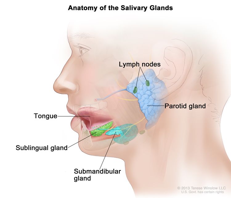
Levels of Evidence

Some of the reference citations in this summary are accompanied by a level-of-evidence designation. These designations are intended to help readers assess the strength of the evidence supporting the use of specific interventions or approaches. The PDQ Adult Treatment Editorial Board uses a formal evidence ranking system in developing its level-of-evidence designations.

Permission to Use This Summary

PDQ is a registered trademark. Although the content of PDQ documents can be used freely as text, it cannot be identified as an NCI PDQ cancer information summary unless it is presented in its entirety and is regularly updated. However, an author would be permitted to write a sentence such as “NCI’s PDQ cancer information summary about breast cancer prevention states the risks succinctly: [include excerpt from the summary].”

The preferred citation for this PDQ summary is:

PDQ® Adult Treatment Editorial Board. PDQ Salivary Gland Cancer Treatment. Bethesda, MD: National Cancer Institute. Updated <MM/DD/YYYY>. Available at: /types/head-and-neck/hp/adult/salivary-gland-treatment-pdq. Accessed <MM/DD/YYYY>. [PMID: 26389389]

Images in this summary are used with permission of the author(s), artist, and/or publisher for use within the PDQ summaries only. Permission to use images outside the context of PDQ information must be obtained from the owner(s) and cannot be granted by the National Cancer Institute. Information about using the illustrations in this summary, along with many other cancer-related images, is available in Visuals Online, a collection of over 2,000 scientific images.

Disclaimer

Based on the strength of the available evidence, treatment options may be described as either “standard” or “under clinical evaluation.” These classifications should not be used as a basis for insurance reimbursement determinations. More information on insurance coverage is available on Cancer.gov on the Managing Cancer Care page.

Contact Us

More information about contacting us or receiving help with the Cancer.gov website can be found on our Contact Us for Help page. Questions can also be submitted to Cancer.gov through the website’s Email Us.