Advances in Leukemia Research

Human cells with acute myelocytic leukemia as seen through a microscope

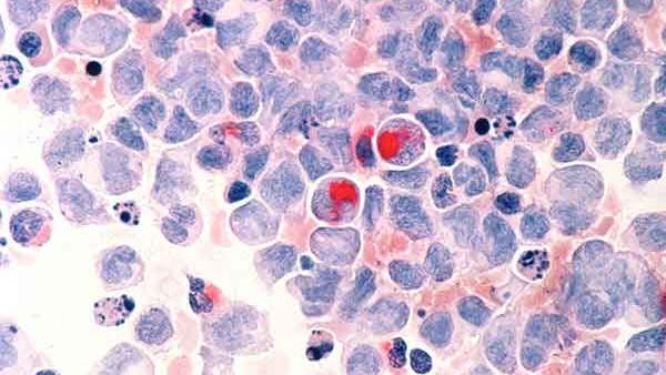
Human cells with acute myelocytic leukemia.

Credit: National Cancer Institute

NCI-funded researchers are working to advance our understanding of how to treat leukemia. With progress in both targeted therapies and immunotherapies, leukemia treatment has the potential to become more effective and less toxic.

This page highlights some of the latest research in leukemia, including clinical advances that may soon translate into improved care, NCI-supported programs that are fueling progress, and research findings from recent studies.

Leukemia Treatment for Adults

The mainstays of leukemia treatment for adults have been chemotherapy, radiation therapy, and stem cell transplantation. Over the last two decades, targeted therapies have also become part of the standard of care for some types of leukemia. These treatments target proteins that control how cancer cells grow, divide, and spread. Different types of leukemia require different combinations of therapies.  For a complete list of all currently approved drugs, see Drugs Approved for Leukemia.

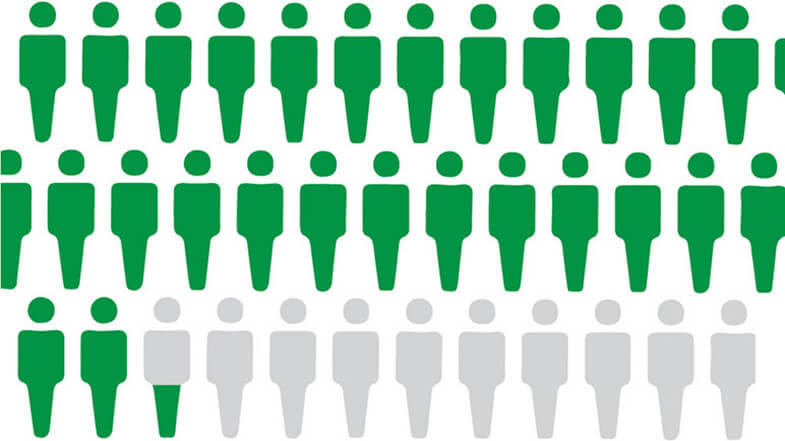
Although much progress has been made against some types of leukemia, others still have relatively poor rates of survival. And, as the population ages, there is a greater need for treatment regimens that are more effective and less toxic than standard chemotherapy.

Acute Lymphoblastic Leukemia (ALL) Treatment

Adult acute lymphoblastic leukemia (ALL) is a type of cancer in which the bone marrow makes too many lymphocytes (a type of white blood cell). It usually gets worse quickly and needs rapid treatment. Some recent research includes:

Combining less-toxic therapies

The intensive chemotherapy treatments used for ALL have serious side effects that many older patients cannot tolerate. Targeted therapies may have fewer side effects than chemotherapy. Clinical trials are now testing whether combinations of these types of therapies can be used instead of chemotherapy for older patients with a form of ALL called B-cell ALL.

Immunotherapy

Immunotherapies are treatments that help the body’s immune system fight cancer more effectively. Immunotherapy strategies being used or tested in ALL include:

CAR T-cell therapy

CAR T-cell therapy is a type of treatment in which a patient’s own immune cells are genetically modified to treat their cancer.

CAR T cell therapies are now being explored for other uses in ALL. For example, scientists hope that it will be possible to use CAR T-cell therapy to delay—or even replace—stem-cell transplantation in older, frailer patients.

Bispecific T-cell engagers

Another immunotherapy being tested in ALL is bispecific T-cell engagers (BiTEs). These drugs attach to immune cells and cancer cells, enabling the immune cells to easily find and destroy the cancer cell by bringing them closer together.

Once such BiTE, called blinatumomab (Blincyto), was recently shown to improve survival for people with ALL who are in remission after chemotherapy, even when there is no trace of their disease. In 2024, FDA approved blinatumomab for adult and pediatric patients one month and older with a specific type of B-cell precursor ALL. The approval is for use as part of consolidation chemotherapy, which is treatment that is given after cancer has disappeared following initial therapy.

Improving treatment for adolescents and young adults (AYAs)

An intensive treatment regimen developed for children with ALL has been found to also improve outcomes for newly diagnosed AYA patients. The pediatric regimen more than doubled the median length of time people lived without their cancer returning compared with an adult treatment regimen. Further studies are now testing the addition of targeted therapies to the combination.

Acute Myeloid Leukemia (AML) Treatment

Acute myeloid leukemia (AML) is the most common type of acute leukemia in adults. It can cause a buildup of abnormal red blood cells, white blood cells, or platelets.

AML tends to be aggressive and is harder to treat than ALL. However, AML cells sometimes have gene changes that cause the tumors to grow but can be targeted with new drugs. Researchers are starting to look at whether genomic sequencing of tumor cells can help doctors choose the best treatment (such as chemotherapy, targeted therapy, stem-cell transplant, or a combination of therapies) for each patient. Scientists are also testing other ways to treat AML.

Targeted therapies

Targeted therapies recently approved to treat AML with certain gene changes include Enasidenib (Idhifa)Olutasidenib (Rezlidhia)Ivosidenib (Tibsovo)Venetoclax (Venclexta)Gemtuzumab ozogamicin (Mylotarg)Midostaurin (Rydapt)Gilteritinib (Xospata)Glasdegib (Daurismo), and Quizartinib (Vanflyta)

An NCI-supported precision medicine study called MyeloMATCH is now enrolling people with newly diagnosed AML or a related but less aggressive cancer called myelodysplastic syndrome (MDS). Participants will undergo genomic testing of blood and bone marrow samples to see if they have specific genetic alterations that can be matched to corresponding targeted therapies.

Other ways to treat AML

  • Testing newer targeted therapies. Researchers continue to develop new drugs to shut down proteins that some leukemias need to grow. For example, new drugs called menin inhibitors stop cancer-promoting genes from being expressed. 
  • Studying ways to target AML cells indirectly. These include testing ways to make cancer cells more vulnerable to new and existing treatments.
  • Targeting AML and related conditions. MDS can eventually progress to AML. Researchers are testing HDAC inhibitors and other drugs that alter how genes are switched on and off in both MDS and AML.
  • Reducing side effects. Some older adults cannot tolerate the intensive treatments most commonly used for AML. Studies have recently found that several drug combinations can help older people with AML live longer while avoiding many serious side effects. New treatments to relieve symptoms of MDS have also been developed.
  • Immunotherapy. CAR T cells and BiTEs are being tested in people with AML.

Chronic Myelogenous Leukemia (CML) Treatment

Chronic myelogenous leukemia (CML) is a type of cancer in which the bone marrow makes too many granulocytes (a type of white blood cell). These granulocytes are abnormal and can build up in the blood and bone marrow so there is less room for healthy white blood cells, red blood cells, and platelets. CML usually gets worse slowly over time.

Blocking an abnormal protein

Most people with CML have a specific chromosome alteration called the Philadelphia chromosome, which produces an abnormal protein that drives the growth of leukemia cells. Targeted therapies that block this abnormal protein—imatinib (Gleevec), nilotinib (Tasigna), dasatinib (Sprycel), and ponatinib (Iclusig)—have radically changed the outlook for people with CML, who now have close to a normal life expectancy.

Testing new combination therapies

Some people with CML continue to have detectable cancer cells in their body even after long-term treatment with drugs that target the protein produced by the Philadelphia chromosome. NCI-supported trials are testing whether the addition of immunotherapy or other targeted therapies to these drugs can reduce the number of CML cells in such patients.

Looking at whether patients can stop taking therapy

Researchers have found that some drugs that target the protein produced by the Philadelphia chromosome can be safely stopped in some CML patients rather than taken for life. These patients must undergo regular testing to ensure the disease has not come back.

Chronic Lymphocytic Leukemia (CLL) Treatment

Like ALL, chronic lymphocytic leukemia (CLL) is a type of cancer in which the bone marrow makes too many lymphocytes (a type of white blood cell). But unlike ALL, CLL is slow growing and worsens over time.

Targeted therapy

Ibrutinib (Imbruvica). The targeted therapy ibrutinib (Imbruvica) was the first non-chemotherapy drug approved to treat CLL. It shuts down a signaling pathway called the B-cell receptor signaling pathway, which is commonly overactive in CLL cells. Depending on people’s age, ibrutinib may be given in combination with another targeted drug, rituximab (Rituxan).

Clinical trials have shown that ibrutinib benefits both younger and older patients with CLL.

Venetoclax (Venclexta) and obinutuzumab (Gazyva). In 2019, the Food and Drug Administration (FDA) approved the second chemotherapy-free initial treatment regimen for CLL, containing the targeted therapies venetoclax (Venclexta) and obinutuzumab (Gazyva).

Other combinations of these drugs plus ibrutinib are now being used or tested for CLL, including

An ongoing trial at NCI is also testing whether giving the combination of venetoclax and obinutuzumab to some people with CLL before symptoms develop can help them live longer overall.

Zanubrutinib (Brukinsa). In early 2023, the FDA approved a drug that works in a similar manner to ibrutinib, called zanubrutinib (Brukinsa), for people with CLL. A large study showed that zanubrutinib alone has fewer side effects and is more effective than ibrutinib for people whose leukemia has returned after initial treatment. More research is now needed to understand how to best combine zanubrutinib with other newer therapies, such as venetoclax.

CAR T-cell therapy

CAR T-cell therapy is also being tested in adults with CLL. Researchers would like to know if using this type of immunotherapy early in the course of treatment would be more effective than waiting until the cancer recurs.

Hairy Cell Leukemia (HCL) Treatment

Hairy cell leukemia (HCL) is a type of cancer in which the bone marrow makes too many lymphocytes (a type of white blood cell). The disease is called hairy cell leukemia because the abnormal lymphocytes look “hairy” when viewed under a microscope. This rare type of leukemia gets worse slowly, or sometimes does not get worse at all.

Combinations of drugs

Researchers are studying combinations of drugs to treat HCL. For example, in a recent small study, a combination of two targeted therapies—vemurafenib (Zelboraf) and rituximab (Rituxan)led to long-lasting remissions for most participants with HCL that had come back after previous treatments. More drug combinations are currently being tested in clinical trials.

Leukemia Treatment for Children

For the two most common types of leukemia, AML and ALL, standard leukemia treatments for children have been chemotherapy, radiation therapy, and stem-cell transplant. Despite great improvements in survival for children with many types of leukemia, some treatments don’t always work. Also, some children later experience a relapse of their disease. Others live with the side effects of chemotherapy and radiation therapy for the rest of their lives, highlighting the need for less toxic treatments.

Now researchers are focusing on targeted drugs and immunotherapies for the treatment of leukemia in children. Newer chemotherapy drugs are also being tested.

Targeted Therapies

Targeted therapies that have been approved or are being studied for children with leukemia include:

More possible targets for the treatment of childhood cancers are discovered every year, and many new drugs that could potentially be used to treat cancers that have these targets are being tested through the Pediatric Preclinical In Vivo Testing Consortium (PIVOT).

Immunotherapy

CAR T-cell therapy has recently generated great excitement for the treatment of children with relapsed ALL. One CAR T-cell therapy, tisagenlecleucel (Kymriah), was approved in 2017 for some children with relapsed ALL.

Researchers continue to address remaining challenges about the use of CAR T-cell therapy in children with leukemia:

  • Sometimes, leukemia can become resistant to tisagenlecleucel. Researchers in NCI’s Pediatric Oncology Branch have developed CAR T cells that target leukemia cells in a different way. An ongoing clinical trial is testing whether the combination of these two types of CAR T cells can provide longer-lasting remissions.
  • CAR T cells are currently only approved for use in leukemia that has relapsed or proved resistant to standard treatment. A clinical trial from the Children’s Oncology Group (COG) is now testing tisagenlecleucel as part of first-line therapy in children with ALL at high risk of relapse.
  • More research is needed to understand which children who receive CAR T cells are at high risk of developing resistance to treatment. Researchers also plan to test whether strategies such as combining CAR T-cell therapy with other immunotherapies may help prevent resistance from developing. 
  • Other research, both in NCI’s Pediatric Oncology Branch and at other institutions, is focused on creating CAR T-cell therapies that work for children with other types of childhood leukemia, such as AML. Several clinical trials of these treatments, including one led by NCI researchers, are now under way.

Two other drugs that use the body’s immune system to fight cancer have shown promise for children with leukemia:

  • A drug called blinatumomab (Blincyto) is a type of immunotherapy called a bispecific T-cell engager (BiTE). These drugs attach to immune cells and cancer cells, enabling the immune cells to easily find and destroy the cancer cell by bringing them closer together.  Blinatumomab has been approved by the FDA for children with ALL who have relapsed after initial treatment.
  • A drug called inotuzumab ozogamicin (Besponsa) is being tested in children with relapsed B-cell ALL. This drug consists of an antibody that can bind to cancer cells linked to a drug that can kill those cells. An NCI-supported trial is also testing the drug as part of treatment for newly diagnosed ALL in children and adolescents at higher risk of relapse.

Chemotherapy

In addition to targeted therapies and immunotherapies, researchers are also working to develop new chemotherapy drugs for leukemia and find better ways to use existing drugs. In 2018, a large clinical trial showed that adding the drug nelarabine (Arranon) to standard chemotherapy improves survival for children and young adults newly diagnosed with T-cell ALL.

Other drugs are being tested that may make standard chemotherapy drugs more effective. These drugs include venetoclax, which has been approved for older adults with some types of leukemia and is now being tested in children.

Survivorship

Children’s developing brains and bodies can be particularly sensitive to the harmful effects of cancer treatment. Because many children treated for cancer go on to live long lives, they may be dealing with these late effects for decades to come.

The NCI-funded Childhood Cancer Survivor Study, ongoing since 1994, tracks the long-term harmful effects of treatments for childhood cancer and studies ways to minimize these effects. NCI also funds research into addressing ways to help cancer survivors cope with and manage health issues stemming from cancer treatment, as well as into altering existing treatment regimens to make them less toxic in the long term.

For example, one study found that, in children with ALL, radiation therapy to prevent the cancer from returning in the brain is likely unnecessary. The study found that radiation can even be omitted for children at the highest risk of the cancer coming back, reducing the risk of future problems with thinking and memory, hormone dysfunction, and other side effects of radiation to the brain.

Preventing and Treating Graft Versus Host Disease

Many people with leukemia—both adults and children—have a stem-cell transplant as part of their treatment. If the new stem cells come from a donor, the immune cells they produce may be able to attack any cancer cells that remain in the body.

But sometimes, immune cells produced by donor stem cells attack healthy tissues of the body instead. This condition, called graft versus host disease (GVHD), can affect nearly every organ and can cause many painful and debilitating symptoms. 

In recent years, several drugs have been approved by the FDA for the treatment of GVHD, including:

Researchers are also testing ways to prevent GVHD from developing in the first place. For example, a recent study found that removing certain immune cells from donated stem cells before they are transplanted may reduce the risk of chronic GVHD without any apparent increase in the likelihood of relapse.

NCI-Supported Research Programs

Many NCI-funded researchers working at the NIH campus and across the United States and the world are seeking ways to address leukemia more effectively. Some research is basic, exploring questions as diverse as the biological underpinnings of cancer. And some is more clinical, seeking to translate this basic information into improving patient outcomes. The programs listed below are a small sampling of NCI’s research efforts in leukemia.

NCI’s Leukemia Specialized Programs of Research Excellence (SPORE) promotes collaborative, interdisciplinary research. SPORE grants involve both basic and clinical/applied scientists working together. They support the efficient movement of basic scientific findings into clinical settings, as well as studies to determine the biological basis for observations made in individuals with cancer or in populations at risk for cancer.

The Targeting Fusion Oncoproteins in Childhood Cancers (TFCC) Network is forming a collaborative team of investigators to advance the understanding of how fusion proteins contribute to pediatric cancers, and how they might be targeted with new treatments. Fusion proteins, which can occur when parts of different chromosomal regions are joined, may drive the development of many cancers in children.

NCI has also formed partnerships with the pharmaceutical industry, academic institutions, and individual investigators for the early clinical evaluation of innovative cancer therapies. The Experimental Therapeutics Clinical Trials Network (ETCTN) was created to evaluate these therapies using a coordinated, collaborative approach to early-phase clinical trials.

The Pediatric Early-Phase Clinical Trials Network was established to help identify and develop effective new drugs for children and adolescents with cancer. The network’s focus is on phase I and early phase II trials, as well as pilot studies of novel drugs and treatment regimens to determine their tolerability.

NCI’s Pediatric Preclinical In Vivo Testing Consortium (PIVOT) develops mouse models to allow early, rapid testing of new drugs for pediatric cancers, including leukemia. The models are all derived from tissue samples taken from patients’ tumors. The consortium partners both with commercial drug companies and with drug development efforts at universities and cancer centers.

The NCI-supported Children’s Oncology Group develops and conducts both clinical trials of initial treatments and clinical trials for after cancer relapse for children and adolescents with ALL, AML, and CML.

Researchers in NCI’s Division of Cancer Epidemiology and Genetics (DCEG) investigate novel, molecular biomarkers for leukemia, as well as clarify relationships of established risk factors. Studies include those looking at environmental and workplace exposure, families with multiple leukemia cases, and inherited bone marrow failure syndromes to name a few.

Clinical Trials

NCI funds and oversees both early- and late-phase clinical trials to develop new treatments and improve patient care. Search NCI-Supported Clinical Trials to find leukemia-related trials now accepting patients. 

Leukemia Research Results

Lymphoma—Health Professional Version

Lymphoma—Health Professional Version

Causes & Prevention

NCI does not have PDQ evidence-based information about prevention of lymphoma.

Genetics

PDQ Genetics Information for Health Professionals

Screening

NCI does not have PDQ evidence-based information about screening for lymphoma.

Supportive & Palliative Care

We offer evidence-based supportive and palliative care information for health professionals on the assessment and management of cancer-related symptoms and conditions.

Cancer Pain Nausea and Vomiting Nutrition in Cancer Care Transition to End-of-Life Care Last Days of Life View all Supportive and Palliative Care Summaries

Childhood Acute Myeloid Leukemia/Other Myeloid Malignancies Treatment (PDQ®)–Patient Version

Childhood Acute Myeloid Leukemia/Other Myeloid Malignancies Treatment (PDQ®)–Patient Version

General Information About Childhood Acute Myeloid Leukemia and Other Myeloid Malignancies

Key Points

  • Childhood acute myeloid leukemia (AML) is a type of cancer in which the bone marrow makes a large number of abnormal blood cells.
  • Leukemia and other diseases of the blood and bone marrow may affect red blood cells, white blood cells, and platelets.
  • Other myeloid diseases can affect the blood and bone marrow.
    • Transient abnormal myelopoiesis (TAM)
    • Acute promyelocytic leukemia (APL)
    • Juvenile myelomonocytic leukemia (JMML)
    • Chronic myelogenous leukemia (CML)
    • Myelodysplastic syndromes (MDS)
  • AML or MDS may occur after treatment with certain chemotherapy drugs and/or radiation therapy.
  • The risk factors for childhood acute myeloid leukemia and other myeloid malignancies are similar.
  • Signs and symptoms of childhood acute myeloid leukemia and other myeloid malignancies include fever, feeling tired, and easy bleeding or bruising.
  • Tests that examine the blood and bone marrow are used to diagnose and find any spread of childhood acute myeloid leukemia and other myeloid malignancies.
  • Certain factors affect prognosis (chance of recovery) and treatment options.

Childhood acute myeloid leukemia (AML) is a type of cancer in which the bone marrow makes a large number of abnormal blood cells.

Childhood acute myeloid leukemia (AML) is a cancer of the blood and bone marrow. AML is also called acute myelogenous leukemia and acute nonlymphocytic leukemia. Cancers that are acute usually get worse quickly if they are not treated. Cancers that are chronic usually get worse slowly.

EnlargeAnatomy of the bone; drawing shows spongy bone, red marrow, and yellow marrow. A cross section of the bone shows compact bone and blood vessels in the bone marrow. Also shown are red blood cells, white blood cells, platelets, and a blood stem cell.
Anatomy of the bone. The bone is made up of compact bone, spongy bone, and bone marrow. Compact bone makes up the outer layer of the bone. Spongy bone is found mostly at the ends of bones and contains red marrow. Bone marrow is found in the center of most bones and has many blood vessels. There are two types of bone marrow: red and yellow. Red marrow contains blood stem cells that can become red blood cells, white blood cells, or platelets. Yellow marrow is made mostly of fat.

This summary is about the treatment of childhood AML, transient abnormal myelopoiesis, childhood acute promyelocytic leukemia, juvenile myelomonocytic leukemia, childhood chronic myelogenous leukemia, and childhood myelodysplastic syndromes. For information about the treatment of childhood acute lymphoblastic leukemia, see Childhood Acute Lymphoblastic Leukemia Treatment.

Leukemia and other diseases of the blood and bone marrow may affect red blood cells, white blood cells, and platelets.

In healthy children, the bone marrow makes blood stem cells (immature cells) that become mature blood cells over time. A blood stem cell may become a myeloid stem cell or a lymphoid stem cell. A lymphoid stem cell becomes a type of white blood cell.

A myeloid stem cell becomes one of three types of mature blood cells:

EnlargeBlood cell development; drawing shows the steps a blood stem cell goes through to become a red blood cell, platelet, or white blood cell. A myeloid stem cell becomes a red blood cell, a platelet, or a myeloblast, which then becomes a granulocyte (the types of granulocytes are eosinophils, basophils, and neutrophils). A lymphoid stem cell becomes a lymphoblast and then becomes a B-lymphocyte, T-lymphocyte, or natural killer cell.
Blood cell development. A blood stem cell goes through several steps to become a red blood cell, platelet, or white blood cell.

In AML, the myeloid stem cells usually become a type of immature white blood cell called myeloblasts (or myeloid blasts). The myeloblasts, or leukemia cells, in AML are abnormal and do not become healthy white blood cells. The leukemia cells can build up in the blood and bone marrow so there is less room for healthy white blood cells, red blood cells, and platelets. This may lead to infection, anemia, or easy bleeding.

The leukemia cells can spread outside the blood to other parts of the body, including the central nervous system (brain and spinal cord), skin, and gums. Sometimes leukemia cells form a solid tumor called a myeloid sarcoma. Myeloid sarcoma is also called granulocytic sarcoma or chloroma.

Other myeloid diseases can affect the blood and bone marrow.

Transient abnormal myelopoiesis (TAM)

TAM is a disorder of the bone marrow that can develop in newborns who have Down syndrome. TAM usually goes away on its own within the first 3 months of life. Infants who have TAM have an increased chance of developing AML before the age of 3 years. TAM is also called transient myeloproliferative disorder or transient leukemia.

Acute promyelocytic leukemia (APL)

APL is a subtype of AML. In APL, some genes on chromosome 15 switch places with some genes on chromosome 17 and an abnormal gene called PML-RARA is made. The PML-RARA gene sends a message that stops promyelocytes (a type of white blood cell) from maturing. The promyelocytes (leukemia cells) can build up in the blood and bone marrow so there is less room for healthy white blood cells, red blood cells, and platelets. Problems with severe bleeding and blood clots may also occur. This is a serious health problem that needs treatment as soon as possible.

Juvenile myelomonocytic leukemia (JMML)

JMML is a rare childhood cancer that is most common in children around the age of 2 years and is more common in boys. In JMML, too many myeloid blood stem cells become myelocytes and monocytes (two types of white blood cells). Some of these myeloid blood stem cells never become mature white blood cells. These immature cells, called blasts, are unable to do their usual work. Over time, the myelocytes, monocytes, and blasts crowd out the healthy white blood cells, red blood cells, and platelets in the bone marrow. When this happens, infection, anemia, or easy bleeding may occur.

Chronic myelogenous leukemia (CML)

CML often begins in an early myeloid blood cell when a certain gene change occurs. A section of genes, that includes the ABL gene, on chromosome 9 changes place with a section of genes on chromosome 22, which has the BCR gene. This makes a very short chromosome 22 (called the Philadelphia chromosome) and a very long chromosome 9. An abnormal BCR-ABL gene is formed on chromosome 22. The BCR-ABL gene tells the blood cells to make too much of a protein called tyrosine kinase. Tyrosine kinase causes too many abnormal white blood cells (leukemia cells) to be made in the bone marrow. The leukemia cells can build up in the blood and bone marrow so there is less room for healthy white blood cells, red blood cells, and platelets. This can lead to infection, anemia, or easy bleeding. CML is rare in children.

EnlargePhiladelphia chromosome; three-panel drawing shows a piece of chromosome 9 and a piece of chromosome 22 breaking off and trading places, creating a changed chromosome 22 called the Philadelphia chromosome. In the left panel, the drawing shows a normal chromosome 9 with the ABL1 gene and a normal chromosome 22 with the BCR gene. In the center panel, the drawing shows part of the ABL1 gene breaking off from chromosome 9 and a piece of chromosome 22 breaking off, below the BCR gene. In the right panel, the drawing shows chromosome 9 with the piece from chromosome 22 attached. It also shows a shortened version of chromosome 22 with the piece from chromosome 9 containing part of the ABL1 gene attached. The ABL1 gene joins to the BCR gene on chromosome 22 to form the BCR::ABL1 fusion gene. The changed chromosome 22 with the BCR::ABL1 fusion gene on it is called the Philadelphia chromosome.
The Philadelphia (Ph) chromosome is an abnormal chromosome that is made when pieces of chromosomes 9 and 22 break off and trade places. The ABL1 gene from chromosome 9 joins to the BCR gene on chromosome 22 to form the BCR::ABL1 fusion gene. The changed chromosome 22 with the fusion gene on it is called the Ph chromosome.

Myelodysplastic syndromes (MDS)

MDS occur less often in children than in adults. In MDS, the bone marrow makes too few red blood cells, white blood cells, and platelets. These blood cells may not mature and enter the blood. The type of MDS depends on the type of blood cell that is affected.

The treatment for MDS depends on how low the numbers of red blood cells, white blood cells, or platelets are. Over time, MDS may become AML.

AML or MDS may occur after treatment with certain chemotherapy drugs and/or radiation therapy.

Cancer treatment with certain chemotherapy drugs and/or radiation therapy may cause therapy-related AML (t-AML) or therapy-related MDS (t-MDS). The risk of these therapy-related myeloid diseases depends on the total dose of the chemotherapy drugs used and the radiation dose and treatment field. Some patients also have an inherited risk for t-AML and t-MDS. These therapy-related diseases usually occur within 7 years after treatment, but are rare in children.

The risk factors for childhood acute myeloid leukemia and other myeloid malignancies are similar.

Anything that increases your risk of getting a disease is called a risk factor. Having a risk factor does not mean that you will get cancer; not having risk factors doesn’t mean that you will not get cancer. Talk with your child’s doctor if you think your child may be at risk. These and other factors may increase the risk of childhood AML, APL, JMML, CML, and MDS:

Signs and symptoms of childhood acute myeloid leukemia and other myeloid malignancies include fever, feeling tired, and easy bleeding or bruising.

These and other signs and symptoms may be caused by childhood AML, APL, JMML, CML, or MDS or by other conditions. Check with a doctor if your child has any of the following:

  • Fever with or without an infection.
  • Drenching night sweats.
  • Shortness of breath.
  • Weakness, feeling tired, or looking pale.
  • Easy bruising or bleeding.
  • Petechiae (flat, pinpoint spots under the skin caused by bleeding).
  • Bone or joint pain.
  • Pain or feeling of fullness below the ribs.
  • Painless lumps in the neck, underarm, stomach, groin, or other parts of the body. In childhood AML, these lumps, called leukemia cutis, may be blue or purple.
  • Painless lumps that are sometimes around the eyes. These lumps, called myeloid sarcomas, are sometimes seen in childhood AML and may be blue-green.
  • An eczema-like skin rash.
  • Loss of appetite or weight loss.
  • Headache, trouble seeing, or confusion.

The signs and symptoms of TAM may include the following:

  • Swelling all over the body.
  • Shortness of breath.
  • Trouble breathing.
  • Increased heart rate.
  • Weakness, feeling tired, or looking pale.
  • Easy bleeding or bruising.
  • Petechiae (flat, pinpoint spots under the skin caused by bleeding).
  • Pain below the ribs.
  • Skin rash.
  • Jaundice (yellowing of the skin and whites of the eyes).

Sometimes TAM does not cause any symptoms at all and is diagnosed after a routine blood test.

Tests that examine the blood and bone marrow are used to diagnose and find any spread of childhood acute myeloid leukemia and other myeloid malignancies.

The following tests and procedures may be used:

  • Physical exam and health history: An exam of the body to check general signs of health, including checking for signs of disease, such as lumps or anything else that seems unusual. A history of the patient’s health habits and past illnesses and treatments will also be taken.
  • Complete blood count (CBC) with differential: A procedure in which a sample of blood is drawn and checked for the following:
    • The number of red blood cells and platelets.
    • The number and type of white blood cells.
    • The portion of the blood sample made up of red blood cells.
    • The amount of hemoglobin (the protein that carries oxygen) in the red blood cells.
    EnlargeComplete blood count (CBC); left panel shows blood being drawn from a vein on the inside of the elbow using a tube attached to a syringe; right panel shows a laboratory test tube with blood cells separated into layers: plasma, white blood cells, platelets, and red blood cells.
    Complete blood count (CBC). Blood is collected by inserting a needle into a vein and allowing the blood to flow into a tube. The blood sample is sent to the laboratory and the red blood cells, white blood cells, and platelets are counted. The CBC is used to test for, diagnose, and monitor many different conditions.
  • Chest x-ray: An x-ray of the organs and bones inside the chest. An x-ray is a type of energy beam that can go through the body and onto film, making a picture of areas inside the body.
  • Biopsy: The removal of cells or tissues so they can be viewed under a microscope by a pathologist to check for signs of cancer. Biopsies that may be done include the following:
    • Bone marrow aspiration and biopsy: The removal of bone marrow and a small piece of bone by inserting a hollow needle into the hipbone or breastbone.
      EnlargeBone marrow aspiration and biopsy; drawing shows a child lying face down on a table and a bone marrow needle being inserted into the right hip bone. An inset shows the bone marrow needle being inserted through the skin into the bone marrow of the hip bone.
      Bone marrow aspiration and biopsy. After a small area of skin is numbed, a bone marrow needle is inserted into the child’s hip bone. Samples of blood, bone, and bone marrow are removed for examination under a microscope.
    • Tumor biopsy for AML: The removal of cells or tissues from a lump in the testicles, ovaries, or skin using a needle. This may be done if the doctor suspects the leukemia cells may have formed a solid tumor called a myeloid sarcoma.
  • Immunophenotyping: A laboratory test that uses antibodies to identify cancer cells based on the types of antigens or markers on the surface of the cells. This test is used to help diagnose specific types of leukemia.
  • Cytogenetic analysis: A laboratory test in which the chromosomes of cells in a sample of blood or bone marrow are counted and checked for any changes, such as broken, missing, rearranged, or extra chromosomes. Changes in certain chromosomes may be a sign of cancer. Cytogenetic analysis is used to help diagnose cancer, plan treatment, or find out how well treatment is working.

    The following test is a type of cytogenetic analysis:

    • FISH (fluorescence in situ hybridization): A laboratory test used to look at and count genes or chromosomes in cells and tissues. Pieces of DNA that contain fluorescent dyes are made in the laboratory and added to a sample of a patient’s cells or tissues. When these dyed pieces of DNA attach to certain genes or areas of chromosomes in the sample, they light up when viewed under a fluorescent microscope. The FISH test is used to help diagnose cancer and help plan treatment.
  • Molecular testing: A laboratory test to check for certain genes, proteins, or other molecules in a sample of tissue, blood, or bone marrow. Molecular tests also check for certain changes in a gene or chromosome that may cause or affect the chance of developing AML. A molecular test may be used to help plan treatment, find out how well treatment is working, or make a prognosis.
  • Lumbar puncture: A procedure used to collect a sample of cerebrospinal fluid (CSF) from the spinal column. This is done by placing a needle between two bones in the spine and into the CSF around the spinal cord and removing a sample of the fluid. The sample of CSF is checked under a microscope for signs that leukemia cells have spread to the brain and spinal cord. This procedure is also called an LP or spinal tap.
    EnlargeLumbar puncture; drawing shows a patient lying in a curled position on a table and a spinal needle (a long, thin needle) being inserted into the lower back. Inset shows a close-up of the spinal needle inserted into the cerebrospinal fluid (CSF) in the lower part of the spinal column.
    Lumbar puncture. A patient lies in a curled position on a table. After a small area on the lower back is numbed, a spinal needle (a long, thin needle) is inserted into the lower part of the spinal column to remove cerebrospinal fluid (CSF, shown in blue). The fluid may be sent to a laboratory for testing.

Certain factors affect prognosis (chance of recovery) and treatment options.

The prognosis and treatment options for childhood acute myeloid leukemia (AML) depend on the following:

  • The age of the child when the cancer is diagnosed.
  • The race or ethnic group of the child.
  • Whether the child is greatly overweight.
  • Number of white blood cells in the blood at diagnosis.
  • Whether the AML occurred after previous cancer treatment.
  • The subtype of AML.
  • Whether there are certain chromosome or gene changes in the leukemia cells.
  • Whether the child has Down syndrome. Most children with AML and Down syndrome can be cured of their leukemia when diagnosed before age 4 years.
  • Whether the leukemia is in the central nervous system (brain and spinal cord).
  • How quickly the leukemia responds to treatment.
  • Whether the AML is newly diagnosed (untreated) or has recurred after treatment.
  • The length of time since treatment ended, for AML that has recurred.

The prognosis and treatment options for childhood acute promyelocytic leukemia (APL) depends on the following:

  • Number of white blood cells in the blood at diagnosis.
  • Whether there are certain chromosome or gene changes in the leukemia cells.
  • Whether the APL is newly diagnosed (untreated) or has recurred after treatment.

The prognosis and treatment options for juvenile myelomonocytic leukemia (JMML) depend on the following:

  • The age of the child when the cancer is diagnosed.
  • The type of gene affected and the number of genes that have changes.
  • How many platelets are in the blood after treatment.
  • How much hemoglobin is in the blood after treatment.
  • Whether the JMML is newly diagnosed (untreated) or has recurred after treatment.

The prognosis and treatment options for childhood chronic myelogenous leukemia (CML) depend on the following:

  • How long it has been since the patient was diagnosed.
  • How many blast cells are in the blood.
  • Whether and how fully the blast cells disappear from the blood and bone marrow after therapy has started.
  • Whether the CML is newly diagnosed (untreated) or has recurred after treatment.

The prognosis and treatment options for myelodysplastic syndromes (MDS) depend on the following:

  • Whether the MDS was caused by previous cancer treatment.
  • How low the numbers of red blood cells, white blood cells, or platelets are.
  • Whether the MDS is newly diagnosed (untreated) or has recurred after treatment.

Stages of Childhood Acute Myeloid Leukemia and Other Myeloid Malignancies

Key Points

  • There is no standard staging system for childhood acute myeloid leukemia and other myelogenous malignancies.
  • Sometimes childhood acute myeloid leukemia and other myeloid malignancies do not respond to treatment or come back after treatment.

There is no standard staging system for childhood acute myeloid leukemia and other myelogenous malignancies.

The extent or spread of cancer is usually described as stages. Instead of stages, treatment is based on one or more of the following:

Newly diagnosed childhood AML

Newly diagnosed childhood AML is cancer that has not been treated except to relieve signs and symptoms such as fever, bleeding, or pain, and has one of the following:

  • More than 20% of the cells in the bone marrow are blasts (leukemia cells).

    or

  • Less than 20% of the cells in the bone marrow are blasts and there is a certain change in the chromosome.

Childhood AML in remission

In childhood AML in remission, the disease has been treated and the following are found:

  • The complete blood count is almost normal.
  • Less than 5% of the cells in the bone marrow are blasts (leukemia cells).
  • There are no signs or symptoms of leukemia in the brain, spinal cord, or other parts of the body.

Sometimes childhood acute myeloid leukemia and other myeloid malignancies do not respond to treatment or come back after treatment.

Refractory leukemia is cancer that does not respond to treatment.

Recurrent leukemia is cancer that has recurred (come back) after it has been treated. The cancer may come back in the blood and bone marrow or in other parts of the body, such as the central nervous system (brain and spinal cord).

Treatment Option Overview

Key Points

  • There are different types of treatment for children with acute myeloid leukemia and other myeloid malignancies.
  • Treatment is planned by a team of health care providers who are experts in treating childhood leukemia and other diseases of the blood.
  • The treatment of childhood AML and other myeloid malignancies usually has two phases.
  • Seven types of standard treatment may be used for childhood AML and other myeloid malignancies.
    • Chemotherapy
    • Radiation therapy
    • Stem cell transplant
    • Targeted therapy
    • Other drug therapy
    • Watchful waiting
    • Supportive care
  • New types of treatment are being tested in clinical trials.
  • Treatment for childhood acute myeloid leukemia and other myeloid malignancies may cause side effects.
  • Patients may want to think about taking part in a clinical trial.
  • Patients can enter clinical trials before, during, or after starting their cancer treatment.
  • Follow-up tests may be needed.

There are different types of treatment for children with acute myeloid leukemia and other myeloid malignancies.

Different types of treatment are available for children with acute myeloid leukemia (AML), transient abnormal myelopoiesis (TAM), acute promyelocytic leukemia (APL), juvenile myelomonocytic leukemia (JMML), chronic myelogenous leukemia (CML), and myelodysplastic syndromes (MDS). Some treatments are standard (the currently used treatment), and some are being tested in clinical trials. A treatment clinical trial is a research study meant to help improve current treatments or obtain information on new treatments for patients with cancer. When clinical trials show that a new treatment is better than the standard treatment, the new treatment may become the standard treatment.

Because AML and other myeloid disorders are rare in children, taking part in a clinical trial should be considered. Some clinical trials are open only to patients who have not yet started treatment.

Treatment is planned by a team of health care providers who are experts in treating childhood leukemia and other diseases of the blood.

Treatment will be overseen by a pediatric oncologist, a doctor who specializes in treating children with cancer. The pediatric oncologist works with other health care providers who are experts in treating children with leukemia and who specialize in certain areas of medicine. These may include the following specialists:

The treatment of childhood AML and other myeloid malignancies usually has two phases.

The treatment of childhood AML is done in phases:

Treatment called central nervous system (CNS) prophylaxis therapy may be given during the induction phase of therapy. Because standard doses of chemotherapy may not reach leukemia cells in the CNS (brain and spinal cord), the leukemia cells are able to hide in the CNS. Intrathecal chemotherapy is able to reach leukemia cells in the CNS. It is given to kill the leukemia cells and lessen the chance the leukemia will recur (come back).

The treatment of childhood APL includes a third phase called maintenance. The goal of maintenance is to kill any remaining leukemia cells that may regrow and cause a relapse. Often the cancer treatments are given in lower doses than those used during the remission induction and consolidation/intensification phases.

Seven types of standard treatment may be used for childhood AML and other myeloid malignancies.

Chemotherapy

Chemotherapy is a cancer treatment that uses drugs to stop the growth of cancer cells, either by killing the cells or by stopping them from dividing. When chemotherapy is taken by mouth or injected into a vein or muscle, the drugs enter the bloodstream and can reach cancer cells throughout the body (systemic chemotherapy). When chemotherapy is placed directly into the cerebrospinal fluid (intrathecal chemotherapy), an organ, or a body cavity such as the abdomen, the drugs mainly affect cancer cells in those areas (regional chemotherapy). Combination chemotherapy is treatment using more than one chemotherapy drug.

The way the chemotherapy is given depends on the type of cancer being treated. In AML, chemotherapy given by mouth, vein, or into the cerebrospinal fluid is used.

In AML, the leukemia cells may spread to the brain and/or spinal cord. Chemotherapy given by mouth or vein to treat AML may not cross the blood-brain barrier to get into the fluid that surrounds the brain and spinal cord. Instead, chemotherapy is injected into the fluid-filled space to kill leukemia cells that may have spread there (intrathecal chemotherapy).

EnlargeIntrathecal chemotherapy; drawing shows the cerebrospinal fluid (CSF) in the brain and spinal cord, and an Ommaya reservoir (a dome-shaped container that is placed under the scalp during surgery; it holds the drugs as they flow through a small tube into the brain). Top section shows a syringe and needle injecting anticancer drugs into the Ommaya reservoir. Bottom section shows a syringe and needle injecting anticancer drugs directly into the cerebrospinal fluid in the lower part of the spinal column.
Intrathecal chemotherapy. Anticancer drugs are injected into the intrathecal space, which is the space that holds the cerebrospinal fluid (CSF, shown in blue). There are two different ways to do this. One way, shown in the top part of the figure, is to inject the drugs into an Ommaya reservoir (a dome-shaped container that is placed under the scalp during surgery; it holds the drugs as they flow through a small tube into the brain). The other way, shown in the bottom part of the figure, is to inject the drugs directly into the CSF in the lower part of the spinal column, after a small area on the lower back is numbed.

See Drugs Approved for Acute Myeloid Leukemia for more information.

Radiation therapy

Radiation therapy is a cancer treatment that uses high-energy x-rays or other types of radiation to kill cancer cells or keep them from growing. External radiation therapy uses a machine outside the body to send radiation toward the area of the body with cancer.

In childhood AML, external radiation therapy may be used to treat a myeloid sarcoma that does not respond to chemotherapy.

Stem cell transplant

Chemotherapy is given to kill cancer cells or other abnormal blood cells. Healthy cells, including blood-forming cells, are also destroyed by the cancer treatment. Stem cell transplant is a treatment to replace the blood-forming cells. Stem cells (immature blood cells) are removed from the blood or bone marrow of the patient or a donor and are frozen and stored. After the patient completes chemotherapy, the stored stem cells are thawed and given to the patient through an infusion. These reinfused stem cells grow into (and restore) the body’s blood cells.

EnlargeDonor stem cell transplant; (Panel 1): Drawing of stem cells being collected from a donor's bloodstream using an apheresis machine. Blood is removed from a vein in the donor's arm and flows through the machine where the stem cells are removed. The rest of the blood is then returned to the donor through a vein in their other arm. (Panel 2): Drawing of a health care provider giving a patient an infusion of chemotherapy through a catheter in the patient's chest. The chemotherapy is given to kill cancer cells and prepare the patient's body for the donor stem cells. (Panel 3): Drawing of a patient receiving an infusion of the donor stem cells through a catheter in the chest.
Donor stem cell transplant. (Step 1): Four to five days before donor stem cell collection, the donor receives a medicine to increase the number of stem cells circulating through their bloodstream (not shown). The blood-forming stem cells are then collected from the donor through a large vein in their arm. The blood flows through an apheresis machine that removes the stem cells. The rest of the blood is returned to the donor through a vein in their other arm. (Step 2): The patient receives chemotherapy to kill cancer cells and prepare their body for the donor stem cells. The patient may also receive radiation therapy (not shown). (Step 3): The patient receives an infusion of the donor stem cells.

Targeted therapy

Targeted therapy is a type of treatment that uses drugs or other substances to identify and attack specific cancer cells. Targeted therapies usually cause less harm to normal cells than chemotherapy or radiation therapy do. Types of targeted therapy include the following:

  • Tyrosine kinase inhibitor therapy: This treatment blocks the enzyme, tyrosine kinase, that causes stem cells to become more white blood cells (blasts) than the body needs. Tyrosine kinase inhibitors may be used with chemotherapy drugs as adjuvant therapy (treatment given after the initial treatment, to lower the risk that the cancer will come back).
  • Monoclonal antibodies: Monoclonal antibodies are immune system proteins made in the laboratory to treat many diseases, including cancer. As a cancer treatment, these antibodies can attach to a specific target on cancer cells or other cells that may help cancer cells grow. The antibodies are able to then kill the cancer cells, block their growth, or keep them from spreading. Monoclonal antibodies are given by infusion. They may be used alone or to carry drugs, toxins, or radioactive material directly to cancer cells.
    • Gemtuzumab ozogamicin is a type of monoclonal antibody that is attached to a chemotherapy drug. It is used in the treatment of AML.
How do monoclonal antibodies work to treat cancer? This video shows how monoclonal antibodies, such as trastuzumab, pembrolizumab, and rituximab, block molecules cancer cells need to grow, flag cancer cells for destruction by the body’s immune system, or deliver harmful substances to cancer cells.

See Drugs Approved for Leukemia for more information.

Other drug therapy

Lenalidomide may be used to lessen the need for transfusions in patients who have myelodysplastic syndromes caused by a specific chromosome change.

Arsenic trioxide and tretinoin are drugs that kill certain types of leukemia cells, stop the leukemia cells from dividing, or help the leukemia cells mature into white blood cells. These drugs are used in the treatment of acute promyelocytic leukemia.

See Drugs Approved for Acute Myeloid Leukemia for more information.

Watchful waiting

Watchful waiting is closely monitoring a patient’s condition without giving any treatment until signs or symptoms appear or change. It is sometimes used to treat transient abnormal myelopoiesis (TAM).

Supportive care

Supportive care is given to lessen the problems caused by the disease or its treatment. All patients with leukemia receive supportive care treatments. Supportive care may include the following:

  • Transfusion therapy: A way of giving red blood cells or platelets to replace blood cells destroyed by disease or cancer treatment. The blood may be donated from another person or it may have been taken from the patient earlier and stored until needed.
  • Antifungal agents: Drugs, such as Caspofungin or fluconazole, used to prevent or treat infections caused by a fungus (a type of microorganism). This is important in the care of patients with AML.
  • Drug therapy, such as antibiotics.
  • Leukapheresis: A procedure in which a special machine is used to remove white blood cells from the blood. Blood is taken from the patient and put through a blood cell separator where the white blood cells are removed. The rest of the blood is then returned to the patient’s bloodstream. Leukapheresis is used to treat patients with very high white blood cell counts.

New types of treatment are being tested in clinical trials.

This summary section describes treatments that are being studied in clinical trials. It may not mention every new treatment being studied. Information about clinical trials is available from the NCI website.

Treatment for childhood acute myeloid leukemia and other myeloid malignancies may cause side effects.

To learn more about side effects that begin during treatment for cancer, visit Side Effects.

Regular follow-up exams are very important. Side effects from cancer treatment that begin after treatment and continue for months or years are called late effects. Late effects of cancer treatment may include the following:

  • Physical problems that affect the following:
  • Changes in mood, feelings, thinking, learning, or memory.
  • Second cancers (new types of cancer), such as breast cancer.

Some late effects may be treated or controlled. It is important that parents of children who are treated for AML or other blood diseases talk with their child’s doctors about the effects cancer treatment can have on their child. (See the PDQ summary on Late Effects of Treatment for Childhood Cancer for more information).

Patients may want to think about taking part in a clinical trial.

For some patients, taking part in a clinical trial may be the best treatment choice. Clinical trials are part of the cancer research process. Clinical trials are done to find out if new cancer treatments are safe and effective or better than the standard treatment.

Many of today’s standard treatments for cancer are based on earlier clinical trials. Patients who take part in a clinical trial may receive the standard treatment or be among the first to receive a new treatment.

Patients who take part in clinical trials also help improve the way cancer will be treated in the future. Even when clinical trials do not lead to effective new treatments, they often answer important questions and help move research forward.

Patients can enter clinical trials before, during, or after starting their cancer treatment.

Some clinical trials only include patients who have not yet received treatment. Other trials test treatments for patients whose cancer has not gotten better. There are also clinical trials that test new ways to stop cancer from recurring (coming back) or reduce the side effects of cancer treatment.

Clinical trials are taking place in many parts of the country. Information about clinical trials supported by NCI can be found on NCI’s clinical trials search webpage. Clinical trials supported by other organizations can be found on the ClinicalTrials.gov website.

Follow-up tests may be needed.

As your child goes through treatment, they will have follow-up tests or check-ups. Some tests that were done to diagnose or stage the cancer may be repeated to see how well the treatment is working. Decisions about whether to continue, change, or stop treatment may be based on the results of these tests.

Some of the tests will continue to be done from time to time after treatment has ended. The results of these tests can show if your child’s condition has changed or if the cancer has recurred (come back).

Treatment of Childhood Acute Myeloid Leukemia

For information about the treatments listed below, see the Treatment Option Overview section.

Treatment of newly diagnosed childhood acute myeloid leukemia (AML) during the induction phase may include the following:

Treatment of childhood AML during the remission phase (consolidation/intensification therapy) depends on the subtype of AML and may include the following:

Treatment of refractory childhood AML may include the following:

  • Chemotherapy.
  • Targeted therapy with a monoclonal antibody (gemtuzumab ozogamicin).
  • Radiation therapy to treat a myeloid sarcoma that does not completely respond to chemotherapy.

Treatment of recurrent childhood AML may include the following:

  • Combination chemotherapy.
  • Targeted therapy with a monoclonal antibody (gemtuzumab ozogamicin).
  • Radiation therapy to treat locally recurring myeloid sarcoma.
  • A clinical trial of targeted therapy (midostaurin, sorafenib, or gilteritinib).
  • Combination chemotherapy and stem cell transplant, for patients who have had a second complete remission.
  • A second stem cell transplant, for patients whose disease came back after the first stem cell transplant.

Treatment of Transient Abnormal Myelopoiesis or Children with Down Syndrome and AML

For information about the treatments listed below, see the Treatment Option Overview section.

Transient abnormal myelopoiesis (TAM) usually goes away on its own. For newly diagnosed TAM that does not go away on its own or causes other health problems, treatment may include the following:

Treatment of newly diagnosed acute myeloid leukemia (AML) in children aged 4 years or younger who have Down syndrome may include the following:

Treatment of newly diagnosed AML in children older than 4 years who have Down syndrome may be the same as treatment for children without Down syndrome.

Treatment of Childhood Acute Promyelocytic Leukemia

For information about the treatments listed below, see the Treatment Option Overview section.

Treatment of newly diagnosed childhood acute promyelocytic leukemia (APL) may include the following:

Treatment of childhood APL during the remission phase (consolidation/intensification therapy) may include the following:

  • Tretinoin with arsenic trioxide.
  • Tretinoin plus chemotherapy with or without arsenic trioxide.

Treatment of childhood APL during the remission phase (maintenance therapy) may include the following:

Treatment of recurrent childhood APL may include the following:

Treatment of Juvenile Myelomonocytic Leukemia

For information about the treatments listed below, see the Treatment Option Overview section.

Treatment of newly diagnosed juvenile myelomonocytic leukemia (JMML) may include the following:

Treatment of refractory or recurrent childhood JMML may include the following:

Treatment of Childhood Chronic Myelogenous Leukemia

For information about the treatments listed below, see the Treatment Option Overview section.

Treatment of newly diagnosed chronic myelogenous leukemia (CML) may include the following:

Treatment of refractory or recurrent childhood CML may include the following:

Treatment of Childhood Myelodysplastic Syndromes

For information about the treatments listed below, see the Treatment Option Overview section.

Treatment of newly diagnosed childhood myelodysplastic syndromes (MDS) may include the following:

If the MDS becomes acute myeloid leukemia (AML), treatment will be the same as treatment for newly diagnosed AML.

To Learn More About Childhood Acute Myeloid Leukemia and Other Myeloid Malignancies

About This PDQ Summary

About PDQ

Physician Data Query (PDQ) is the National Cancer Institute’s (NCI’s) comprehensive cancer information database. The PDQ database contains summaries of the latest published information on cancer prevention, detection, genetics, treatment, supportive care, and complementary and alternative medicine. Most summaries come in two versions. The health professional versions have detailed information written in technical language. The patient versions are written in easy-to-understand, nontechnical language. Both versions have cancer information that is accurate and up to date and most versions are also available in Spanish.

PDQ is a service of the NCI. The NCI is part of the National Institutes of Health (NIH). NIH is the federal government’s center of biomedical research. The PDQ summaries are based on an independent review of the medical literature. They are not policy statements of the NCI or the NIH.

Purpose of This Summary

This PDQ cancer information summary has current information about the treatment of childhood acute myeloid leukemia and other myeloid malignancies. It is meant to inform and help patients, families, and caregivers. It does not give formal guidelines or recommendations for making decisions about health care.

Reviewers and Updates

Editorial Boards write the PDQ cancer information summaries and keep them up to date. These Boards are made up of experts in cancer treatment and other specialties related to cancer. The summaries are reviewed regularly and changes are made when there is new information. The date on each summary (“Updated”) is the date of the most recent change.

The information in this patient summary was taken from the health professional version, which is reviewed regularly and updated as needed, by the PDQ Pediatric Treatment Editorial Board.

Clinical Trial Information

A clinical trial is a study to answer a scientific question, such as whether one treatment is better than another. Trials are based on past studies and what has been learned in the laboratory. Each trial answers certain scientific questions in order to find new and better ways to help cancer patients. During treatment clinical trials, information is collected about the effects of a new treatment and how well it works. If a clinical trial shows that a new treatment is better than one currently being used, the new treatment may become “standard.” Patients may want to think about taking part in a clinical trial. Some clinical trials are open only to patients who have not started treatment.

Clinical trials can be found online at NCI’s website. For more information, call the Cancer Information Service (CIS), NCI’s contact center, at 1-800-4-CANCER (1-800-422-6237).

Permission to Use This Summary

PDQ is a registered trademark. The content of PDQ documents can be used freely as text. It cannot be identified as an NCI PDQ cancer information summary unless the whole summary is shown and it is updated regularly. However, a user would be allowed to write a sentence such as “NCI’s PDQ cancer information summary about breast cancer prevention states the risks in the following way: [include excerpt from the summary].”

The best way to cite this PDQ summary is:

PDQ® Pediatric Treatment Editorial Board. PDQ Childhood Acute Myeloid Leukemia/Other Myeloid Malignancies Treatment. Bethesda, MD: National Cancer Institute. Updated <MM/DD/YYYY>. Available at: /types/leukemia/patient/child-aml-treatment-pdq. Accessed <MM/DD/YYYY>. [PMID: 26389303]

Images in this summary are used with permission of the author(s), artist, and/or publisher for use in the PDQ summaries only. If you want to use an image from a PDQ summary and you are not using the whole summary, you must get permission from the owner. It cannot be given by the National Cancer Institute. Information about using the images in this summary, along with many other images related to cancer can be found in Visuals Online. Visuals Online is a collection of more than 3,000 scientific images.

Disclaimer

The information in these summaries should not be used to make decisions about insurance reimbursement. More information on insurance coverage is available on Cancer.gov on the Managing Cancer Care page.

Contact Us

More information about contacting us or receiving help with the Cancer.gov website can be found on our Contact Us for Help page. Questions can also be submitted to Cancer.gov through the website’s E-mail Us.

Hairy Cell Leukemia Treatment (PDQ®)–Patient Version

Hairy Cell Leukemia Treatment (PDQ®)–Patient Version

General Information About Hairy Cell Leukemia

Key Points

  • Hairy cell leukemia is a type of cancer in which the bone marrow makes too many lymphocytes (a type of white blood cell).
  • Leukemia may affect red blood cells, white blood cells, and platelets.
  • Signs and symptoms of hairy cell leukemia include infections, tiredness, and pain below the ribs.
  • Tests that examine the blood and bone marrow are used to diagnose hairy cell leukemia.
  • Certain factors affect treatment options and prognosis (chance of recovery).

Hairy cell leukemia is a type of cancer in which the bone marrow makes too many lymphocytes (a type of white blood cell).

Hairy cell leukemia is a cancer of the blood and bone marrow. This rare type of leukemia gets worse slowly or does not get worse at all. The disease is called hairy cell leukemia because the leukemia cells look “hairy” when viewed under a microscope.

EnlargeAnatomy of the bone; drawing shows spongy bone, red marrow, and yellow marrow. A cross section of the bone shows compact bone and blood vessels in the bone marrow. Also shown are red blood cells, white blood cells, platelets, and a blood stem cell.
Anatomy of the bone. The bone is made up of compact bone, spongy bone, and bone marrow. Compact bone makes up the outer layer of the bone. Spongy bone is found mostly at the ends of bones and contains red marrow. Bone marrow is found in the center of most bones and has many blood vessels. There are two types of bone marrow: red and yellow. Red marrow contains blood stem cells that can become red blood cells, white blood cells, or platelets. Yellow marrow is made mostly of fat.

Leukemia may affect red blood cells, white blood cells, and platelets.

Normally, the bone marrow makes blood stem cells (immature cells) that become mature blood cells over time. A blood stem cell may become a myeloid stem cell or a lymphoid stem cell.

A myeloid stem cell becomes one of three types of mature blood cells:

A lymphoid stem cell becomes a lymphoblast cell and then one of three types of lymphocytes (white blood cells):

EnlargeBlood cell development; drawing shows the steps a blood stem cell goes through to become a red blood cell, platelet, or white blood cell. A myeloid stem cell becomes a red blood cell, a platelet, or a myeloblast, which then becomes a granulocyte (the types of granulocytes are eosinophils, basophils, and neutrophils). A lymphoid stem cell becomes a lymphoblast and then becomes a B-lymphocyte, T-lymphocyte, or natural killer cell.
Blood cell development. A blood stem cell goes through several steps to become a red blood cell, platelet, or white blood cell.

In hairy cell leukemia, too many blood stem cells become lymphocytes. These lymphocytes are abnormal and do not become healthy white blood cells. The abnormal lymphocytes are also called leukemia cells. The leukemia cells can build up in the blood and bone marrow so there is less room for healthy white blood cells, red blood cells, and platelets. This may cause infection, anemia, and easy bleeding. Some of the leukemia cells may collect in the spleen and cause it to swell.

Signs and symptoms of hairy cell leukemia include infections, tiredness, and pain below the ribs.

These and other signs and symptoms may be caused by hairy cell leukemia or by other conditions. Check with your doctor if you have:

  • Weakness or fatigue (feeling tired).
  • Frequent infections.
  • Easy bruising or bleeding.
  • Shortness of breath.
  • Pain or a feeling of fullness below the ribs.
  • Painless lumps in the neck, underarm, stomach, or groin.

Tests that examine the blood and bone marrow are used to diagnose hairy cell leukemia.

In addition to asking about your personal and family health history and doing a physical exam, your doctor may perform the following tests and procedures:

  • Complete blood count (CBC): A procedure in which a sample of blood is drawn and checked for:
    • The number of red blood cells, white blood cells, and platelets.
    • The amount of hemoglobin (the protein that carries oxygen) in the red blood cells.
    • The portion of the sample made up of red blood cells.
    EnlargeComplete blood count (CBC); left panel shows blood being drawn from a vein on the inside of the elbow using a tube attached to a syringe; right panel shows a laboratory test tube with blood cells separated into layers: plasma, white blood cells, platelets, and red blood cells.
    Complete blood count (CBC). Blood is collected by inserting a needle into a vein and allowing the blood to flow into a tube. The blood sample is sent to the laboratory and the red blood cells, white blood cells, and platelets are counted. The CBC is used to test for, diagnose, and monitor many different conditions.
  • Peripheral blood smear: A procedure in which a sample of blood is checked for cells that look “hairy,” the number and kinds of white blood cells, the number of platelets, and changes in the shape of blood cells.
  • Blood chemistry studies: A procedure in which a blood sample is checked to measure the amounts of certain substances released into the blood by organs and tissues in the body. An unusual (higher or lower than normal) amount of a substance can be a sign of disease.
  • Flow cytometry: A laboratory test that measures the number of cells in a sample, the percentage of live cells in a sample, and certain characteristics of the cells, such as size, shape, and the presence of tumor (or other) markers on the cell surface. The cells from a sample of a patient’s blood, bone marrow, or other tissue are stained with a fluorescent dye, placed in a fluid, and then passed one at a time through a beam of light. The test results are based on how the cells that were stained with the fluorescent dye react to the beam of light. This test is used to help diagnose and manage certain types of cancers, such as leukemia and lymphoma.
  • Bone marrow aspiration and biopsy: The removal of bone marrow, blood, and a small piece of bone by inserting a hollow needle into the hipbone or breastbone. A pathologist views the bone marrow, blood, and bone under a microscope to look for signs of cancer.
    EnlargeBone marrow aspiration and biopsy; drawing shows a patient lying face down on a table and a bone marrow needle being inserted into the hip bone. An inset shows a close up of the needle being inserted through the skin and hip bone into the bone marrow.
    Bone marrow aspiration and biopsy. After a small area of skin is numbed, a long, hollow needle is inserted through the patient’s skin and hip bone into the bone marrow. A sample of bone marrow and a small piece of bone are removed for examination under a microscope.
  • Immunophenotyping: A laboratory test that uses antibodies to identify cancer cells based on the types of antigens or markers on the surface of the cells. This test is used to help diagnose specific types of leukemia.
  • Cytogenetic analysis: A laboratory test in which the chromosomes of cells in a sample of blood or bone marrow are counted and checked for any changes, such as broken, missing, rearranged, or extra chromosomes. Changes in certain chromosomes may be a sign of cancer. Cytogenetic analysis is used to help diagnose cancer, plan treatment, or find out how well treatment is working.
  • BRAF gene testing: A laboratory test in which a sample of blood or tissue is tested for certain changes in the BRAF gene. A BRAF gene mutation is often found in patients with hairy cell leukemia.
  • CT scan (CAT scan): A procedure that makes a series of detailed pictures of areas inside the body, taken from different angles. The pictures are made by a computer linked to an x-ray machine. A dye may be injected into a vein or swallowed to help the organs or tissues show up more clearly. This procedure is also called computed tomography, computerized tomography, or computerized axial tomography. A CT scan of the abdomen may be done to check for swollen lymph nodes or a swollen spleen.

Certain factors affect treatment options and prognosis (chance of recovery).

The treatment options depend on:

  • The number of hairy (leukemia) cells and healthy blood cells in the blood and bone marrow.
  • Whether the spleen is swollen.
  • Whether there are signs or symptoms of leukemia, such as infection.
  • Whether the leukemia has recurred (come back) after previous treatment.

The prognosis depends on:

  • Whether the hairy cell leukemia does not grow or grows so slowly it does not need treatment.
  • Whether the hairy cell leukemia responds to treatment.

Treatment often results in a long-lasting remission (a period during which some or all of the signs and symptoms of the leukemia are gone). If the leukemia returns after it has been in remission, re-treatment often causes another remission.

Stages of Hairy Cell Leukemia

Key Points

  • There is no standard staging system for hairy cell leukemia.
  • Sometimes hairy cell leukemia does not respond to treatment or comes back after treatment.

There is no standard staging system for hairy cell leukemia.

The process used to find out if cancer has spread to other parts of the body is called staging. There is no standard staging system for hairy cell leukemia.

In untreated hairy cell leukemia, some or all of the following conditions occur:

Sometimes hairy cell leukemia does not respond to treatment or comes back after treatment.

Refractory hairy cell leukemia is cancer that does not respond to treatment.

Recurrent hairy cell leukemia is cancer that has recurred (come back) after it has been treated.

Treatment Option Overview

Key Points

  • There are different types of treatment for patients with hairy cell leukemia.
  • The following types of treatment are used:
    • Watchful waiting
    • Chemotherapy
    • Targeted therapy
    • Surgery
  • New types of treatment are being tested in clinical trials.
  • Treatment for hairy cell leukemia may cause side effects.
  • Patients may want to think about taking part in a clinical trial.
  • Patients can enter clinical trials before, during, or after starting their cancer treatment.
  • Follow-up care may be needed.

There are different types of treatment for patients with hairy cell leukemia.

Different types of treatment are available for patients with hairy cell leukemia. Some treatments are standard (the currently used treatment), and some are being tested in clinical trials. A treatment clinical trial is a research study meant to help improve current treatments or obtain information on new treatments for patients with cancer. When clinical trials show that a new treatment is better than the standard treatment, the new treatment may become the standard treatment. Patients may want to think about taking part in a clinical trial. Some clinical trials are open only to patients who have not started treatment.

The following types of treatment are used:

Watchful waiting

Watchful waiting is closely monitoring a patient’s condition, without giving any treatment until signs or symptoms appear or change.

Chemotherapy

Chemotherapy is a cancer treatment that uses drugs to stop the growth of cancer cells, either by killing the cells or by stopping them from dividing. When chemotherapy is taken by mouth or injected into a vein or muscle, the drugs enter the bloodstream and can reach cancer cells throughout the body (systemic chemotherapy). The way the chemotherapy is given depends on the type and stage of the cancer being treated. Cladribine and pentostatin are anticancer drugs commonly used to treat hairy cell leukemia. These drugs may increase the risk of other types of cancer, especially Hodgkin lymphoma and non-Hodgkin lymphoma. Long-term follow-up for second cancers is very important. Bendamustine is another anticancer drug that is being studied to treat hairy cell leukemia.

Learn more about Drugs Approved for Hairy Cell Leukemia.

Targeted therapy

Targeted therapy is a type of treatment that uses drugs or other substances to identify and attack specific cancer cells.

  • Monoclonal antibody therapy: Monoclonal antibodies are immune system proteins made in the laboratory to treat many diseases, including cancer. As a cancer treatment, these antibodies can attach to a specific target on cancer cells or other cells that may help cancer cells grow. The antibodies are able to then kill the cancer cells, block their growth, or keep them from spreading. Monoclonal antibodies are given by infusion. They may be used alone or to carry drugs, toxins, or radioactive material directly to cancer cells.

    Rituximab is a type of monoclonal antibody that may be used to treat certain patients with hairy cell leukemia.

How do monoclonal antibodies work to treat cancer? This video shows how monoclonal antibodies, such as trastuzumab, pembrolizumab, and rituximab, block molecules cancer cells need to grow, flag cancer cells for destruction by the body’s immune system, or deliver harmful substances to cancer cells.

Other types of targeted therapies are being studied in the treatment of hairy cell leukemia, including:

  • Vemurafenib, dabrafenib, and trametinib are types of kinase inhibitors that block certain proteins made by the mutated BRAF gene, which may help keep cancer cells from growing.
  • Ibrutinib is a type of targeted therapy that blocks a protein called Bruton’s tyrosine kinase (BTK), which may help keep cancer cells from growing.

Learn more about Drugs Approved for Hairy Cell Leukemia.

Surgery

Splenectomy is a surgical procedure to remove the spleen.

New types of treatment are being tested in clinical trials.

Information about clinical trials is available from the NCI website.

Treatment for hairy cell leukemia may cause side effects.

For information about side effects caused by treatment for cancer, visit our Side Effects page.

Patients may want to think about taking part in a clinical trial.

For some patients, taking part in a clinical trial may be the best treatment choice. Clinical trials are part of the cancer research process. Clinical trials are done to find out if new cancer treatments are safe and effective or better than the standard treatment.

Many of today’s standard treatments for cancer are based on earlier clinical trials. Patients who take part in a clinical trial may receive the standard treatment or be among the first to receive a new treatment.

Patients who take part in clinical trials also help improve the way cancer will be treated in the future. Even when clinical trials do not lead to effective new treatments, they often answer important questions and help move research forward.

Patients can enter clinical trials before, during, or after starting their cancer treatment.

Some clinical trials only include patients who have not yet received treatment. Other trials test treatments for patients whose cancer has not gotten better. There are also clinical trials that test new ways to stop cancer from recurring (coming back) or reduce the side effects of cancer treatment.

Clinical trials are taking place in many parts of the country. Information about clinical trials supported by NCI can be found on NCI’s clinical trials search webpage. Clinical trials supported by other organizations can be found on the ClinicalTrials.gov website.

Follow-up care may be needed.

As you go through treatment, you will have follow-up tests or check-ups. Some tests that were done to diagnose or stage the cancer may be repeated to see how well the treatment is working. Decisions about whether to continue, change, or stop treatment may be based on the results of these tests.

Some of the tests will continue to be done from time to time after treatment has ended. The results of these tests can show if your condition has changed or if the cancer has recurred (come back).

Treatment of Hairy Cell Leukemia

Treatment of hairy cell leukemia may include:

Learn more about these treatments in the Treatment Option Overview

You and your doctor will discuss what treatment plan is best for you.

Use our clinical trial search to find NCI-supported cancer clinical trials that are accepting patients. You can search for trials based on the type of cancer, the age of the patient, and where the trials are being done. General information about clinical trials is also available.

To Learn More About Hairy Cell Leukemia

About This PDQ Summary

About PDQ

Physician Data Query (PDQ) is the National Cancer Institute’s (NCI’s) comprehensive cancer information database. The PDQ database contains summaries of the latest published information on cancer prevention, detection, genetics, treatment, supportive care, and complementary and alternative medicine. Most summaries come in two versions. The health professional versions have detailed information written in technical language. The patient versions are written in easy-to-understand, nontechnical language. Both versions have cancer information that is accurate and up to date and most versions are also available in Spanish.

PDQ is a service of the NCI. The NCI is part of the National Institutes of Health (NIH). NIH is the federal government’s center of biomedical research. The PDQ summaries are based on an independent review of the medical literature. They are not policy statements of the NCI or the NIH.

Purpose of This Summary

This PDQ cancer information summary has current information about the treatment of hairy cell leukemia. It is meant to inform and help patients, families, and caregivers. It does not give formal guidelines or recommendations for making decisions about health care.

Reviewers and Updates

Editorial Boards write the PDQ cancer information summaries and keep them up to date. These Boards are made up of experts in cancer treatment and other specialties related to cancer. The summaries are reviewed regularly and changes are made when there is new information. The date on each summary (“Updated”) is the date of the most recent change.

The information in this patient summary was taken from the health professional version, which is reviewed regularly and updated as needed, by the PDQ Adult Treatment Editorial Board.

Clinical Trial Information

A clinical trial is a study to answer a scientific question, such as whether one treatment is better than another. Trials are based on past studies and what has been learned in the laboratory. Each trial answers certain scientific questions in order to find new and better ways to help cancer patients. During treatment clinical trials, information is collected about the effects of a new treatment and how well it works. If a clinical trial shows that a new treatment is better than one currently being used, the new treatment may become “standard.” Patients may want to think about taking part in a clinical trial. Some clinical trials are open only to patients who have not started treatment.

Clinical trials can be found online at NCI’s website. For more information, call the Cancer Information Service (CIS), NCI’s contact center, at 1-800-4-CANCER (1-800-422-6237).

Permission to Use This Summary

PDQ is a registered trademark. The content of PDQ documents can be used freely as text. It cannot be identified as an NCI PDQ cancer information summary unless the whole summary is shown and it is updated regularly. However, a user would be allowed to write a sentence such as “NCI’s PDQ cancer information summary about breast cancer prevention states the risks in the following way: [include excerpt from the summary].”

The best way to cite this PDQ summary is:

PDQ® Adult Treatment Editorial Board. PDQ Hairy Cell Leukemia Treatment. Bethesda, MD: National Cancer Institute. Updated <MM/DD/YYYY>. Available at: /types/leukemia/patient/hairy-cell-treatment-pdq. Accessed <MM/DD/YYYY>. [PMID: 26389248]

Images in this summary are used with permission of the author(s), artist, and/or publisher for use in the PDQ summaries only. If you want to use an image from a PDQ summary and you are not using the whole summary, you must get permission from the owner. It cannot be given by the National Cancer Institute. Information about using the images in this summary, along with many other images related to cancer can be found in Visuals Online. Visuals Online is a collection of more than 3,000 scientific images.

Disclaimer

The information in these summaries should not be used to make decisions about insurance reimbursement. More information on insurance coverage is available on Cancer.gov on the Managing Cancer Care page.

Contact Us

More information about contacting us or receiving help with the Cancer.gov website can be found on our Contact Us for Help page. Questions can also be submitted to Cancer.gov through the website’s E-mail Us.

Chronic Myelogenous Leukemia Treatment (PDQ®)–Patient Version

Chronic Myelogenous Leukemia Treatment (PDQ®)–Patient Version

General Information About Chronic Myelogenous Leukemia

Key Points

  • Chronic myelogenous leukemia is a disease in which the bone marrow makes too many white blood cells.
  • Leukemia may affect red blood cells, white blood cells, and platelets.
  • Signs and symptoms of chronic myelogenous leukemia include weight loss and tiredness.
  • Most people with CML have a gene mutation (change) called the Philadelphia chromosome.
  • Tests that examine the blood and bone marrow are used to diagnose chronic myelogenous leukemia.
  • Certain factors affect prognosis (chance of recovery) and treatment options.

Chronic myelogenous leukemia is a disease in which the bone marrow makes too many white blood cells.

Chronic myelogenous leukemia (also called CML or chronic granulocytic leukemia) is a slowly progressing blood and bone marrow disease that usually occurs during or after middle age, and rarely occurs in children.

EnlargeAnatomy of the bone; drawing shows spongy bone, red marrow, and yellow marrow. A cross section of the bone shows compact bone and blood vessels in the bone marrow. Also shown are red blood cells, white blood cells, platelets, and a blood stem cell.
Anatomy of the bone. The bone is made up of compact bone, spongy bone, and bone marrow. Compact bone makes up the outer layer of the bone. Spongy bone is found mostly at the ends of bones and contains red marrow. Bone marrow is found in the center of most bones and has many blood vessels. There are two types of bone marrow: red and yellow. Red marrow contains blood stem cells that can become red blood cells, white blood cells, or platelets. Yellow marrow is made mostly of fat.

Leukemia may affect red blood cells, white blood cells, and platelets.

Normally, the bone marrow makes blood stem cells (immature cells) that become mature blood cells over time. A blood stem cell may become a myeloid stem cell or a lymphoid stem cell. A lymphoid stem cell becomes a white blood cell.

A myeloid stem cell becomes one of three types of mature blood cells:

EnlargeBlood cell development; drawing shows the steps a blood stem cell goes through to become a red blood cell, platelet, or white blood cell. A myeloid stem cell becomes a red blood cell, a platelet, or a myeloblast, which then becomes a granulocyte (the types of granulocytes are eosinophils, basophils, and neutrophils). A lymphoid stem cell becomes a lymphoblast and then becomes a B-lymphocyte, T-lymphocyte, or natural killer cell.
Blood cell development. A blood stem cell goes through several steps to become a red blood cell, platelet, or white blood cell.

In CML, too many blood stem cells become a type of white blood cell called granulocytes. These granulocytes are abnormal and do not become healthy white blood cells. They are also called leukemia cells. The leukemia cells can build up in the blood and bone marrow so there is less room for healthy white blood cells, red blood cells, and platelets. When this happens, infection, anemia, or easy bleeding may occur.

This summary is about chronic myelogenous leukemia. See the following PDQ summaries for more information about leukemia:

Signs and symptoms of chronic myelogenous leukemia include weight loss and tiredness.

These and other signs and symptoms may be caused by CML or by other conditions. Check with your doctor if you have any of the following:

  • Feeling very tired.
  • Weight loss for no known reason.
  • Drenching night sweats.
  • Fever.
  • Pain or a feeling of fullness below the ribs on the left side.

Sometimes CML does not cause any symptoms at all.

Most people with CML have a gene mutation (change) called the Philadelphia chromosome.

Every cell in the body contains DNA (genetic material) that determines how the cell looks and acts. DNA is contained inside chromosomes. In CML, part of the DNA from one chromosome moves to another chromosome. This change is called the “Philadelphia chromosome.” It results in the bone marrow making a protein, called tyrosine kinase, that causes too many stem cells to become white blood cells (granulocytes or blasts).

The Philadelphia chromosome is not passed from parent to child.

EnlargePhiladelphia chromosome; three-panel drawing shows a piece of chromosome 9 and a piece of chromosome 22 breaking off and trading places, creating a changed chromosome 22 called the Philadelphia chromosome. In the left panel, the drawing shows a normal chromosome 9 with the ABL1 gene and a normal chromosome 22 with the BCR gene. In the center panel, the drawing shows part of the ABL1 gene breaking off from chromosome 9 and a piece of chromosome 22 breaking off, below the BCR gene. In the right panel, the drawing shows chromosome 9 with the piece from chromosome 22 attached. It also shows a shortened version of chromosome 22 with the piece from chromosome 9 containing part of the ABL1 gene attached. The ABL1 gene joins to the BCR gene on chromosome 22 to form the BCR::ABL1 fusion gene. The changed chromosome 22 with the BCR::ABL1 fusion gene on it is called the Philadelphia chromosome.
The Philadelphia (Ph) chromosome is an abnormal chromosome that is made when pieces of chromosomes 9 and 22 break off and trade places. The ABL1 gene from chromosome 9 joins to the BCR gene on chromosome 22 to form the BCR::ABL1 fusion gene. The changed chromosome 22 with the fusion gene on it is called the Ph chromosome.

Tests that examine the blood and bone marrow are used to diagnose chronic myelogenous leukemia.

The following tests and procedures may be used:

  • Physical exam and health history: An exam of the body to check general signs of health, including checking for signs of disease such as an enlarged spleen. A history of the patient’s health habits and past illnesses and treatments will also be taken.
  • Complete blood count (CBC) with differential: A procedure in which a sample of blood is drawn and checked for the following:
    • The number of red blood cells and platelets.
    • The number and type of white blood cells.
    • The amount of hemoglobin (the protein that carries oxygen) in the red blood cells.
    • The portion of the blood sample made up of red blood cells.
    EnlargeComplete blood count (CBC); left panel shows blood being drawn from a vein on the inside of the elbow using a tube attached to a syringe; right panel shows a laboratory test tube with blood cells separated into layers: plasma, white blood cells, platelets, and red blood cells.
    Complete blood count (CBC). Blood is collected by inserting a needle into a vein and allowing the blood to flow into a tube. The blood sample is sent to the laboratory and the red blood cells, white blood cells, and platelets are counted. The CBC is used to test for, diagnose, and monitor many different conditions.
  • Blood chemistry studies: A procedure in which a blood sample is checked to measure the amounts of certain substances released into the blood by organs and tissues in the body. An unusual (higher or lower than normal) amount of a substance can be a sign of disease.
  • Bone marrow aspiration and biopsy: The removal of bone marrow, blood, and a small piece of bone by inserting a needle into the hipbone or breastbone. A pathologist views the bone marrow, blood, and bone under a microscope to look for abnormal cells.
    EnlargeBone marrow aspiration and biopsy; drawing shows a patient lying face down on a table and a bone marrow needle being inserted into the hip bone. An inset shows a close up of the needle being inserted through the skin and hip bone into the bone marrow.
    Bone marrow aspiration and biopsy. After a small area of skin is numbed, a long, hollow needle is inserted through the patient’s skin and hip bone into the bone marrow. A sample of bone marrow and a small piece of bone are removed for examination under a microscope.

    One of the following tests may be done on the samples of blood or bone marrow tissue that are removed:

    • Cytogenetic analysis: A laboratory test in which the chromosomes of cells in a sample of blood or bone marrow are counted and checked for any changes, such as broken, missing, rearranged, or extra chromosomes. Changes in certain chromosomes, such as the Philadelphia chromosome, may be a sign of cancer. Cytogenetic analysis is used to help diagnose cancer, plan treatment, or find out how well treatment is working.
    • FISH (fluorescence in situ hybridization): A laboratory test used to look at and count genes or chromosomes in cells and tissues. Pieces of DNA that contain fluorescent dyes are made in the laboratory and added to a sample of a patient’s cells or tissues. When these dyed pieces of DNA attach to certain genes or areas of chromosomes in the sample, they light up when viewed under a fluorescent microscope. The FISH test is used to help diagnose cancer and help plan treatment.
    • Reverse transcription–polymerase chain reaction test (RT–PCR): A laboratory test in which the amount of a genetic substance called mRNA made by a specific gene is measured. An enzyme called reverse transcriptase is used to convert a specific piece of RNA into a matching piece of DNA, which can be amplified (made in large numbers) by another enzyme called DNA polymerase. The amplified DNA copies help tell whether a specific mRNA is being made by a gene. RT-PCR can be used to check the activation of certain genes that may indicate the presence of cancer cells. This test may be used to look for certain changes in a gene or chromosome, which may help diagnose cancer.

Certain factors affect prognosis (chance of recovery) and treatment options.

The prognosis and treatment options depend on the following:

  • The patient’s age.
  • The phase of CML.
  • The amount of blasts in the blood or bone marrow.
  • The patient’s general health.

Stages of Chronic Myelogenous Leukemia

Key Points

  • After chronic myelogenous leukemia has been diagnosed, tests are done to find out if the cancer has spread.
  • Chronic myelogenous leukemia has 3 phases.
    • Chronic phase
    • Accelerated phase
    • Blastic phase
  • Chronic myelogenous leukemia can relapse (return) after it has been treated.

After chronic myelogenous leukemia has been diagnosed, tests are done to find out if the cancer has spread.

The extent or spread of cancer is usually described as stages. In chronic myelogenous leukemia (CML), the disease is classified by phase: chronic phase, accelerated phase, or blastic phase. It is important to know the phase in order to plan treatment. The information from tests and procedures done to diagnose chronic myelogenous leukemia is also used to plan treatment.

Chronic myelogenous leukemia has 3 phases.

As the amount of blast cells increases in the blood and bone marrow, there is less room for healthy white blood cells, red blood cells, and platelets. This may result in infections, anemia, and easy bleeding, as well as bone pain and pain or a feeling of fullness below the ribs on the left side. The number of blast cells in the blood and bone marrow and the severity of signs or symptoms determine the phase of the disease.

Chronic phase

In chronic phase CML, fewer than 10% of the cells in the blood and bone marrow are blast cells.

Accelerated phase

In accelerated phase CML, 10% to 19% of the cells in the blood and bone marrow are blast cells.

Blastic phase

In blastic phase CML, 20% or more of the cells in the blood or bone marrow are blast cells. When tiredness, fever, and an enlarged spleen occur during the blastic phase, it is called blast crisis.

Chronic myelogenous leukemia can relapse (return) after it has been treated.

In relapsed CML, the number of blast cells increases after a remission.

Treatment Option Overview

Key Points

  • There are different types of treatment for patients with chronic myelogenous leukemia.
  • Six types of standard treatment are used:
    • Targeted therapy
    • Chemotherapy
    • Immunotherapy
    • High-dose chemotherapy with stem cell transplant
    • Donor lymphocyte infusion (DLI)
    • Surgery
  • New types of treatment are being tested in clinical trials.
  • Treatment for chronic myelogenous leukemia may cause side effects.
  • Patients may want to think about taking part in a clinical trial.
  • Patients can enter clinical trials before, during, or after starting their cancer treatment.
  • Follow-up tests may be needed.

There are different types of treatment for patients with chronic myelogenous leukemia.

Different types of treatment are available for patients with chronic myelogenous leukemia (CML). Some treatments are standard (the currently used treatment), and some are being tested in clinical trials. A treatment clinical trial is a research study meant to help improve current treatments or obtain information about new treatments for patients with cancer. When clinical trials show that a new treatment is better than the standard treatment, the new treatment may become the standard treatment. Patients may want to think about taking part in a clinical trial. Some clinical trials are open only to patients who have not started treatment.

Six types of standard treatment are used:

Targeted therapy

Targeted therapy is a type of treatment that uses drugs or other substances to identify and attack specific cancer cells.

See Drugs Approved for Chronic Myelogenous Leukemia for more information.

Chemotherapy

Chemotherapy is a cancer treatment that uses drugs to stop the growth of cancer cells, either by killing the cells or by stopping them from dividing. When chemotherapy is taken by mouth or injected into a vein or muscle, the drugs enter the bloodstream and can reach cancer cells throughout the body (systemic chemotherapy).

See Drugs Approved for Chronic Myelogenous Leukemia for more information.

Immunotherapy

Immunotherapy is a treatment that uses the patient’s immune system to fight cancer. Substances made by the body or made in a laboratory are used to boost, direct, or restore the body’s natural defenses against cancer.

  • Interferon: Interferon affects the division of cancer cells and can slow tumor growth.

See Drugs Approved for Chronic Myelogenous Leukemia for more information.

High-dose chemotherapy with stem cell transplant

High doses of chemotherapy are given to kill cancer cells. Healthy cells, including blood-forming cells, are also destroyed by the cancer treatment. Stem cell transplant is a treatment to replace the blood-forming cells. Stem cells (immature blood cells) are removed from the blood or bone marrow of the patient or a donor and are frozen and stored. After the patient completes chemotherapy, the stored stem cells are thawed and given back to the patient through an infusion. These reinfused stem cells grow into (and restore) the body’s blood cells.

See Drugs Approved for Chronic Myelogenous Leukemia for more information.

EnlargeDonor stem cell transplant; (Panel 1): Drawing of stem cells being collected from a donor's bloodstream using an apheresis machine. Blood is removed from a vein in the donor's arm and flows through the machine where the stem cells are removed. The rest of the blood is then returned to the donor through a vein in their other arm. (Panel 2): Drawing of a health care provider giving a patient an infusion of chemotherapy through a catheter in the patient's chest. The chemotherapy is given to kill cancer cells and prepare the patient's body for the donor stem cells. (Panel 3): Drawing of a patient receiving an infusion of the donor stem cells through a catheter in the chest.
Donor stem cell transplant. (Step 1): Four to five days before donor stem cell collection, the donor receives a medicine to increase the number of stem cells circulating through their bloodstream (not shown). The blood-forming stem cells are then collected from the donor through a large vein in their arm. The blood flows through an apheresis machine that removes the stem cells. The rest of the blood is returned to the donor through a vein in their other arm. (Step 2): The patient receives chemotherapy to kill cancer cells and prepare their body for the donor stem cells. The patient may also receive radiation therapy (not shown). (Step 3): The patient receives an infusion of the donor stem cells.

Donor lymphocyte infusion (DLI)

Donor lymphocyte infusion (DLI) is a cancer treatment that may be used after stem cell transplant. Lymphocytes (a type of white blood cell) from the stem cell transplant donor are removed from the donor’s blood and may be frozen for storage. The donor’s lymphocytes are thawed if they were frozen and then given to the patient through one or more infusions. The lymphocytes see the patient’s cancer cells as not belonging to the body and attack them.

Surgery

Splenectomy is surgery to remove the spleen.

New types of treatment are being tested in clinical trials.

Information about clinical trials is available from the NCI website.

Treatment for chronic myelogenous leukemia may cause side effects.

For information about side effects caused by treatment for cancer, visit our Side Effects page.

Patients may want to think about taking part in a clinical trial.

For some patients, taking part in a clinical trial may be the best treatment choice. Clinical trials are part of the cancer research process. Clinical trials are done to find out if new cancer treatments are safe and effective or better than the standard treatment.

Many of today’s standard treatments for cancer are based on earlier clinical trials. Patients who take part in a clinical trial may receive the standard treatment or be among the first to receive a new treatment.

Patients who take part in clinical trials also help improve the way cancer will be treated in the future. Even when clinical trials do not lead to effective new treatments, they often answer important questions and help move research forward.

Patients can enter clinical trials before, during, or after starting their cancer treatment.

Some clinical trials only include patients who have not yet received treatment. Other trials test treatments for patients whose cancer has not gotten better. There are also clinical trials that test new ways to stop cancer from recurring (coming back) or reduce the side effects of cancer treatment.

Clinical trials are taking place in many parts of the country. Information about clinical trials supported by NCI can be found on NCI’s clinical trials search webpage. Clinical trials supported by other organizations can be found on the ClinicalTrials.gov website.

Follow-up tests may be needed.

As you go through treatment, you will have follow-up tests or check-ups. Some tests that were done to diagnose or stage the cancer may be repeated to see how well the treatment is working. Decisions about whether to continue, change, or stop treatment may be based on the results of these tests.

Some of the tests will continue to be done from time to time after treatment has ended. The results of these tests can show if your condition has changed or if the cancer has recurred (come back).

Treatment of Chronic Phase Chronic Myelogenous Leukemia

For information about the treatments listed below, see the Treatment Option Overview section.

Treatment of chronic phase chronic myelogenous leukemia may include the following:

Use our clinical trial search to find NCI-supported cancer clinical trials that are accepting patients. You can search for trials based on the type of cancer, the age of the patient, and where the trials are being done. General information about clinical trials is also available.

Treatment of Accelerated Phase Chronic Myelogenous Leukemia

For information about the treatments listed below, see the Treatment Option Overview section.

Treatment of accelerated phase chronic myelogenous leukemia may include the following:

Use our clinical trial search to find NCI-supported cancer clinical trials that are accepting patients. You can search for trials based on the type of cancer, the age of the patient, and where the trials are being done. General information about clinical trials is also available.

Treatment of Blastic Phase Chronic Myelogenous Leukemia

For information about the treatments listed below, see the Treatment Option Overview section.

Treatment of blastic phase chronic myelogenous leukemia may include the following:

Use our clinical trial search to find NCI-supported cancer clinical trials that are accepting patients. You can search for trials based on the type of cancer, the age of the patient, and where the trials are being done. General information about clinical trials is also available.

Treatment of Relapsed Chronic Myelogenous Leukemia

For information about the treatments listed below, see the Treatment Option Overview section.

In relapsed CML, the number of blast cells increases after a remission. Treatment of relapsed chronic myelogenous leukemia may include the following:

Use our clinical trial search to find NCI-supported cancer clinical trials that are accepting patients. You can search for trials based on the type of cancer, the age of the patient, and where the trials are being done. General information about clinical trials is also available.

To Learn More About Chronic Myelogenous Leukemia

About This PDQ Summary

About PDQ

Physician Data Query (PDQ) is the National Cancer Institute’s (NCI’s) comprehensive cancer information database. The PDQ database contains summaries of the latest published information on cancer prevention, detection, genetics, treatment, supportive care, and complementary and alternative medicine. Most summaries come in two versions. The health professional versions have detailed information written in technical language. The patient versions are written in easy-to-understand, nontechnical language. Both versions have cancer information that is accurate and up to date and most versions are also available in Spanish.

PDQ is a service of the NCI. The NCI is part of the National Institutes of Health (NIH). NIH is the federal government’s center of biomedical research. The PDQ summaries are based on an independent review of the medical literature. They are not policy statements of the NCI or the NIH.

Purpose of This Summary

This PDQ cancer information summary has current information about the treatment of chronic myelogenous leukemia. It is meant to inform and help patients, families, and caregivers. It does not give formal guidelines or recommendations for making decisions about health care.

Reviewers and Updates

Editorial Boards write the PDQ cancer information summaries and keep them up to date. These Boards are made up of experts in cancer treatment and other specialties related to cancer. The summaries are reviewed regularly and changes are made when there is new information. The date on each summary (“Updated”) is the date of the most recent change.

The information in this patient summary was taken from the health professional version, which is reviewed regularly and updated as needed, by the PDQ Adult Treatment Editorial Board.

Clinical Trial Information

A clinical trial is a study to answer a scientific question, such as whether one treatment is better than another. Trials are based on past studies and what has been learned in the laboratory. Each trial answers certain scientific questions in order to find new and better ways to help cancer patients. During treatment clinical trials, information is collected about the effects of a new treatment and how well it works. If a clinical trial shows that a new treatment is better than one currently being used, the new treatment may become “standard.” Patients may want to think about taking part in a clinical trial. Some clinical trials are open only to patients who have not started treatment.

Clinical trials can be found online at NCI’s website. For more information, call the Cancer Information Service (CIS), NCI’s contact center, at 1-800-4-CANCER (1-800-422-6237).

Permission to Use This Summary

PDQ is a registered trademark. The content of PDQ documents can be used freely as text. It cannot be identified as an NCI PDQ cancer information summary unless the whole summary is shown and it is updated regularly. However, a user would be allowed to write a sentence such as “NCI’s PDQ cancer information summary about breast cancer prevention states the risks in the following way: [include excerpt from the summary].”

The best way to cite this PDQ summary is:

PDQ® Adult Treatment Editorial Board. PDQ Chronic Myelogenous Leukemia Treatment. Bethesda, MD: National Cancer Institute. Updated <MM/DD/YYYY>. Available at: /types/leukemia/patient/cml-treatment-pdq. Accessed <MM/DD/YYYY>. [PMID: 26389183]

Images in this summary are used with permission of the author(s), artist, and/or publisher for use in the PDQ summaries only. If you want to use an image from a PDQ summary and you are not using the whole summary, you must get permission from the owner. It cannot be given by the National Cancer Institute. Information about using the images in this summary, along with many other images related to cancer can be found in Visuals Online. Visuals Online is a collection of more than 3,000 scientific images.

Disclaimer

The information in these summaries should not be used to make decisions about insurance reimbursement. More information on insurance coverage is available on Cancer.gov on the Managing Cancer Care page.

Contact Us

More information about contacting us or receiving help with the Cancer.gov website can be found on our Contact Us for Help page. Questions can also be submitted to Cancer.gov through the website’s E-mail Us.

Chronic Lymphocytic Leukemia Treatment (PDQ®)–Patient Version

Chronic Lymphocytic Leukemia Treatment (PDQ®)–Patient Version

General Information About Chronic Lymphocytic Leukemia

Key Points

  • Chronic lymphocytic leukemia (CLL) is a type of cancer in which the bone marrow makes too many lymphocytes (a type of white blood cell).
  • Leukemia may affect red blood cells, white blood cells, and platelets.
  • Signs and symptoms of CLL include swollen lymph nodes and feeling tired.
  • Tests that examine the blood are used to diagnose CLL.
  • Certain factors affect prognosis (chance of recovery) and treatment options.

Chronic lymphocytic leukemia (CLL) is a type of cancer in which the bone marrow makes too many lymphocytes (a type of white blood cell).

CLL is a cancer of the blood and bone marrow that usually gets worse slowly. CLL is one of the most common types of leukemia in adults. It often occurs during or after middle age; it rarely occurs in children.

EnlargeAnatomy of the bone; drawing shows spongy bone, red marrow, and yellow marrow. A cross section of the bone shows compact bone and blood vessels in the bone marrow. Also shown are red blood cells, white blood cells, platelets, and a blood stem cell.
Anatomy of the bone. The bone is made up of compact bone, spongy bone, and bone marrow. Compact bone makes up the outer layer of the bone. Spongy bone is found mostly at the ends of bones and contains red marrow. Bone marrow is found in the center of most bones and has many blood vessels. There are two types of bone marrow: red and yellow. Red marrow contains blood stem cells that can become red blood cells, white blood cells, or platelets. Yellow marrow is made mostly of fat.

Leukemia may affect red blood cells, white blood cells, and platelets.

Normally, the bone marrow makes blood stem cells (immature cells) that become mature blood cells over time. A blood stem cell may become a myeloid stem cell or a lymphoid stem cell.

A myeloid stem cell becomes one of three types of mature blood cells:

A lymphoid stem cell becomes a lymphoblast cell and then one of three types of lymphocytes (white blood cells):

EnlargeBlood cell development; drawing shows the steps a blood stem cell goes through to become a red blood cell, platelet, or white blood cell. A myeloid stem cell becomes a red blood cell, a platelet, or a myeloblast, which then becomes a granulocyte (the types of granulocytes are eosinophils, basophils, and neutrophils). A lymphoid stem cell becomes a lymphoblast and then becomes a B-lymphocyte, T-lymphocyte, or natural killer cell.
Blood cell development. A blood stem cell goes through several steps to become a red blood cell, platelet, or white blood cell.

In CLL, too many blood stem cells become abnormal lymphocytes. The abnormal lymphocytes may also be called leukemia cells. These leukemia cells are not able to fight infection very well. Also, as the number of leukemia cells increases in the blood and bone marrow, there is less room for healthy white blood cells, red blood cells, and platelets. This may lead to infection, anemia, and easy bleeding.

Signs and symptoms of CLL include swollen lymph nodes and feeling tired.

In the beginning, CLL does not cause any signs or symptoms and may be found during a routine blood test. Later, signs and symptoms may occur. Check with your doctor if you have:

  • Painless swelling of the lymph nodes in the neck, underarm, stomach, or groin.
  • Weakness or feeling tired.
  • Pain or a feeling of fullness below the ribs.
  • Fever and infection.
  • Easy bruising or bleeding.
  • Petechiae (flat, pinpoint, dark-red spots under the skin caused by bleeding).
  • Weight loss for no known reason.
  • Drenching night sweats.

Tests that examine the blood are used to diagnose CLL.

In addition to asking about your personal and family health history and doing a physical exam, your doctor may perform the following tests and procedures:

  • Complete blood count (CBC) with differential: A procedure in which a sample of blood is drawn and checked for:
    • The number of red blood cells and platelets.
    • The number and type of white blood cells.
    • The amount of hemoglobin (the protein that carries oxygen) in the red blood cells.
    • The portion of the blood sample made up of red blood cells.
    EnlargeComplete blood count (CBC); left panel shows blood being drawn from a vein on the inside of the elbow using a tube attached to a syringe; right panel shows a laboratory test tube with blood cells separated into layers: plasma, white blood cells, platelets, and red blood cells.
    Complete blood count (CBC). Blood is collected by inserting a needle into a vein and allowing the blood to flow into a tube. The blood sample is sent to the laboratory and the red blood cells, white blood cells, and platelets are counted. The CBC is used to test for, diagnose, and monitor many different conditions.
  • Blood chemistry studies: A procedure in which a blood sample is checked to measure the amounts of certain substances released into the blood by organs and tissues in the body. An unusual (higher or lower than normal) amount of a substance can be a sign of disease.
  • Lactate dehydrogenase testing: A laboratory test for one of a group of enzymes found in the blood and other body tissues and involved in energy production in cells. An increased amount of lactate dehydrogenase in the blood may be a sign of tissue damage and some types of cancer or other diseases.
  • Beta-2-microglobulin testing: A laboratory test for beta-2-microglobulin, a small protein normally found on the surface of many cells, including lymphocytes, and in small amounts in the blood and urine. An increased amount in the blood or urine may be a sign of certain diseases, including some types of cancer, such as multiple myeloma or lymphoma.
  • Flow cytometry: A laboratory test that measures the number of cells in a sample, the percentage of live cells in a sample, and certain characteristics of the cells, such as size, shape, and the presence of tumor (or other) markers on the cell surface. The cells from a sample of a patient’s blood, bone marrow, or other tissue are stained with a fluorescent dye, placed in a fluid, and then passed one at a time through a beam of light. The test results are based on how the cells that were stained with the fluorescent dye react to the beam of light. This test is used to help diagnose and manage certain types of cancers, such as leukemia and lymphoma.
  • FISH (fluorescence in situ hybridization): A laboratory test used to look at and count genes or chromosomes in cells and tissues. Pieces of DNA that contain fluorescent dyes are made in the laboratory and added to a sample of a patient’s cells or tissues. When these dyed pieces of DNA attach to certain genes or areas of chromosomes in the sample, they light up when viewed under a fluorescent microscope. The FISH test is used to help diagnose cancer and help plan treatment.
  • Genetic testing: A laboratory test in which cells or tissue are analyzed to look for changes in the TP53 or IGH gene. These changes may be helpful to determine the patient’s prognosis.
  • Serum immunoglobulin testing: A laboratory test that measures specific types of immunoglobulins (antibodies) in the blood. This may help diagnose cancer or find out how well treatment is working or if cancer has come back.
  • Hepatitis B virus and hepatitis C virus testing: A test to check for hepatitis B or hepatitis C virus in the blood. Infection with one of these viruses causes hepatitis (inflammation of the liver).
  • HIV testing: A test to check for HIV infection. HIV is the virus that causes AIDS. The most common type of HIV test is called the HIV antibody test, which checks for antibodies against HIV in a sample of blood, urine, or fluid from the mouth.

Certain factors affect prognosis (chance of recovery) and treatment options.

The prognosis depends on:

  • Whether there are certain gene changes, such as in TP53 or IGH.
  • Whether lymphocytes have spread throughout the bone marrow.
  • Whether the red blood cell and platelet counts are low.
  • Whether the white blood cell count is increasing quickly.
  • The stage of the cancer.
  • The results of certain blood tests, such as the beta-2 microglobulin and lactate dehydrogenase tests.
  • The patient’s age and general health.
  • How quickly and how low the leukemia cell count drops during treatment.
  • Whether the CLL gets better with treatment or has recurred (come back).
  • Whether the CLL progresses to lymphoma or becomes prolymphocytic leukemia.
  • Whether the patient gets another type of cancer after being diagnosed with CLL.

Treatment options depend on:

  • The red blood cell, white blood cell, and platelet blood counts.
  • Whether the liver, spleen, or lymph nodes are larger than normal.
  • The age and health of the patient at the time of diagnosis.
  • Whether there are signs or symptoms, such as fever, chills, or weight loss.
  • The response to initial treatment.
  • Whether the CLL has recurred (come back).

Stage Information for Chronic Lymphocytic Leukemia

Key Points

  • After chronic lymphocytic leukemia (CLL) has been diagnosed, tests are done to find out whether the cancer has spread.
  • The following stages are used for CLL:
    • Stage 0
    • Stage I
    • Stage II
    • Stage III
    • Stage IV
  • CLL is described as asymptomatic, symptomatic or progressive, recurrent, or refractory.

After chronic lymphocytic leukemia (CLL) has been diagnosed, tests are done to find out whether the cancer has spread.

Staging is the process used to find out how far the cancer has spread. In CLL, the leukemia cells may spread from the blood and bone marrow to other parts of the body, such as the lymph nodes, liver, and spleen. It is important to know whether the leukemia cells have spread in order to plan the best treatment.

The following tests may be used to find out how far the cancer has spread:

  • Chest x-ray: An x-ray of the organs and bones inside the chest. An x-ray is a type of energy beam that can go through the body and onto film, making a picture of areas inside the body, such as the lymph nodes.
  • CT scan (CAT scan): A procedure that makes a series of detailed pictures of areas inside the body, such as the neck, chest, abdomen, pelvis, and lymph nodes, taken from different angles. The pictures are made by a computer linked to an x-ray machine. A dye may be injected into a vein or swallowed to help the organs or tissues show up more clearly. This test is used in patients with many swollen lymph nodes throughout the body. This procedure is also called computed tomography, computerized tomography, or computerized axial tomography. If a PET-CT scan is not available, a CT scan alone may be done.
  • PET-CT scan: A procedure that combines the pictures from a positron emission tomography (PET) scan and a computed tomography (CT) scan. The PET and CT scans are done at the same time on the same machine. The pictures from both scans are combined to make a more detailed picture than either test would make by itself. A PET scan is a procedure to find malignant tumor cells in the body. A small amount of radioactive glucose (sugar) is injected into a vein. The PET scanner rotates around the body and makes a picture of where glucose is being used in the body. Malignant tumor cells show up brighter in the picture because they are more active and take up more glucose than normal cells do. This test is done in patients with fever, drenching night sweats, weight loss, or fast-growing lymph nodes to check whether CLL has become an aggressive form of lymphoma.

The following stages are used for CLL:

Stage 0

In stage 0 CLL, there are too many lymphocytes in the blood, but there are no other signs or symptoms of leukemia. Stage 0 CLL is indolent (slow-growing).

Stage I

In stage I CLL, there are too many lymphocytes in the blood, and the lymph nodes are larger than normal.

Stage II

In stage II CLL, there are too many lymphocytes in the blood, the liver or spleen is larger than normal, and the lymph nodes may be larger than normal.

Stage III

In stage III CLL, there are too many lymphocytes in the blood, and there are too few red blood cells. The lymph nodes, liver, or spleen may be larger than normal.

Stage IV

In stage IV CLL, there are too many lymphocytes in the blood and too few platelets. The lymph nodes, liver, or spleen may be larger than normal, or there may be too few red blood cells.

CLL is described as asymptomatic, symptomatic or progressive, recurrent, or refractory.

  • Asymptomatic CLL: The leukemia causes no or few symptoms.
  • Symptomatic or progressive CLL: The leukemia has caused significant changes to blood counts or other serious symptoms.
  • Recurrent CLL: The leukemia has recurred (come back) after a period of time in which the cancer could not be detected.
  • Refractory CLL: The leukemia does not get better with treatment.

Treatment Option Overview

Key Points

  • There are different types of treatment for patients with chronic lymphocytic leukemia (CLL).
  • The following types of treatment are used:
    • Watchful waiting
    • Targeted therapy
    • Chemotherapy
    • Radiation therapy
    • Immunotherapy
    • Chemotherapy with bone marrow or peripheral blood stem cell transplant
  • New types of treatment are being tested in clinical trials.
  • Treatment for chronic lymphocytic leukemia may cause side effects.
  • Patients may want to think about taking part in a clinical trial.
  • Patients can enter clinical trials before, during, or after starting their cancer treatment.
  • Follow-up care may be needed.

There are different types of treatment for patients with chronic lymphocytic leukemia (CLL).

Different types of treatment are available for patients with CLL. Some treatments are standard (the currently used treatment), and some are being tested in clinical trials. A treatment clinical trial is a research study meant to help improve current treatments or obtain information on new treatments for patients with cancer. When clinical trials show that a new treatment is better than the standard treatment, the new treatment may become the standard treatment. Patients may want to think about taking part in a clinical trial. Some clinical trials are open only to patients who have not started treatment.

The following types of treatment are used:

Watchful waiting

Watchful waiting is closely monitoring a patient’s condition without giving any treatment until signs or symptoms appear or change. This is also called observation. Watchful waiting is used to treat asymptomatic and symptomatic or progressive CLL.

Targeted therapy

Targeted therapy is a type of treatment that uses drugs or other substances to identify and attack specific cancer cells. Different types of targeted therapy are used to treat CLL:

  • Tyrosine kinase inhibitor (TKI) therapy: This treatment blocks the enzyme, tyrosine kinase, that causes stem cells to develop into more white blood cells than the body needs. Ibrutinib, acalabrutinib, zanubrutinib, and duvelisib are TKIs used to treat symptomatic or progressive, recurrent, or refractory CLL.
  • BCL2 inhibitor therapy: This treatment blocks a protein called BCL2, which is found on some leukemia cells. This may kill leukemia cells and make them more sensitive to other anticancer drugs. Venetoclax is a type of BCL2 therapy used to treat symptomatic or progressive, recurrent, or refractory CLL.
  • Monoclonal antibody therapy: Monoclonal antibodies are immune system proteins made in the laboratory to treat many diseases, including cancer. As a cancer treatment, these antibodies can attach to a specific target on cancer cells or other cells that may help cancer cells grow. The antibodies are able to then kill the cancer cells, block their growth, or keep them from spreading. Monoclonal antibodies are given by infusion. They may be used alone or to carry drugs, toxins, or radioactive material directly to cancer cells. Rituximab, ofatumumab, obinutuzumab, and alemtuzumab alone and in combination with chemotherapy are used to treat symptomatic or progressive, recurrent, or refractory CLL.
    How do monoclonal antibodies work to treat cancer? This video shows how monoclonal antibodies, such as trastuzumab, pembrolizumab, and rituximab, block molecules cancer cells need to grow, flag cancer cells for destruction by the body’s immune system, or deliver harmful substances to cancer cells.

For more information, see Drugs Approved for Chronic Lymphocytic Leukemia.

Chemotherapy

Chemotherapy is a cancer treatment that uses drugs to stop the growth of cancer cells, either by killing the cells or by stopping them from dividing. When chemotherapy is taken by mouth or injected into a vein or muscle, the drugs enter the bloodstream and can reach cancer cells throughout the body (systemic chemotherapy). Combination chemotherapy is treatment using more than one anticancer drug.

For more information, see Drugs Approved for Chronic Lymphocytic Leukemia.

Radiation therapy

Radiation therapy is a cancer treatment that uses high-energy x-rays or other types of radiation to kill cancer cells or keep them from growing. External radiation therapy uses a machine outside the body to send radiation toward the area of the body with cancer, such as a group of lymph nodes or the spleen. This treatment may be used to reduce pain related to a swollen spleen or lymph nodes.

Immunotherapy

Immunotherapy is a treatment that uses the patient’s immune system to fight cancer. Substances made by the body or made in a laboratory are used to boost, direct, or restore the body’s natural defenses against cancer.

  • Immunomodulating agent: Lenalidomide stimulates T cells to kill leukemia cells. It may be used alone or with rituximab in patients with symptomatic or progressive, recurrent, or refractory CLL.
  • CAR T-cell therapy: This treatment changes the patient’s T cells (a type of immune system cell) so they will attack certain proteins on the surface of cancer cells. T cells are taken from the patient and special receptors are added to their surface in the laboratory. The changed cells are called chimeric antigen receptor (CAR) T cells. The CAR T cells are grown in the laboratory and given to the patient by infusion. The CAR T cells multiply in the patient’s blood and attack cancer cells. CAR T-cell therapy is being studied in the treatment of recurrent or refractory CLL.
    EnlargeCAR T-cell therapy; drawing of blood being removed from a vein in a patient’s arm to get T cells. Also shown is a special receptor called a chimeric antigen receptor (CAR) being made in the laboratory; the gene for CAR is inserted into the T cells and then millions of CAR T cells are grown. Drawing also shows the CAR T cells being given to the patient by infusion and binding to antigens on the cancer cells and killing them.
    CAR T-cell therapy. A type of treatment in which a patient’s T cells (a type of immune cell) are changed in the laboratory so they will bind to cancer cells and kill them. Blood from a vein in the patient’s arm flows through a tube to an apheresis machine (not shown), which removes the white blood cells, including the T cells, and sends the rest of the blood back to the patient. Then, the gene for a special receptor called a chimeric antigen receptor (CAR) is inserted into the T cells in the laboratory. Millions of the CAR T cells are grown in the laboratory and then given to the patient by infusion. The CAR T cells are able to bind to an antigen on the cancer cells and kill them.

Chemotherapy with bone marrow or peripheral blood stem cell transplant

Chemotherapy is given to kill cancer cells. Healthy cells, including blood-forming cells, are destroyed by the cancer treatment. A bone marrow or peripheral blood stem cell transplant are treatments to replace the blood-forming cells. Stem cells (immature blood cells) are removed from the blood or bone marrow of the patient or a donor and are frozen and stored. After the patient completes chemotherapy, the stored stem cells are thawed and given back to the patient through an infusion. These reinfused stem cells grow into (and restore) the body’s blood cells.

New types of treatment are being tested in clinical trials.

Information about clinical trials is available from the NCI website.

Treatment for chronic lymphocytic leukemia may cause side effects.

For information about side effects caused by treatment for cancer, visit our Side Effects page.

Patients may want to think about taking part in a clinical trial.

For some patients, taking part in a clinical trial may be the best treatment choice. Clinical trials are part of the cancer research process. Clinical trials are done to find out if new cancer treatments are safe and effective or better than the standard treatment.

Many of today’s standard treatments for cancer are based on earlier clinical trials. Patients who take part in a clinical trial may receive the standard treatment or be among the first to receive a new treatment.

Patients who take part in clinical trials also help improve the way cancer will be treated in the future. Even when clinical trials do not lead to effective new treatments, they often answer important questions and help move research forward.

Patients can enter clinical trials before, during, or after starting their cancer treatment.

Some clinical trials only include patients who have not yet received treatment. Other trials test treatments for patients whose cancer has not gotten better. There are also clinical trials that test new ways to stop cancer from recurring (coming back) or reduce the side effects of cancer treatment.

Clinical trials are taking place in many parts of the country. Information about clinical trials supported by NCI can be found on NCI’s clinical trials search webpage. Clinical trials supported by other organizations can be found on the ClinicalTrials.gov website.

Follow-up care may be needed.

As you go through treatment, you will have follow-up tests or check-ups. Some tests that were done to diagnose or stage the cancer may be repeated to see how well the treatment is working. Decisions about whether to continue, change, or stop treatment may be based on the results of these tests.

Some of the tests will continue to be done from time to time after treatment has ended. The results of these tests can show if your condition has changed or if the cancer has recurred (come back).

Treatment of Asymptomatic Chronic Lymphocytic Leukemia

For information about the treatment listed below, see the Treatment Option Overview section.

The treatment of asymptomatic chronic lymphocytic leukemia (CLL) may include watchful waiting.

Treatment of Symptomatic or Progressive Chronic Lymphocytic Leukemia

For information about the treatments listed below, see the Treatment Option Overview section.

The treatment of symptomatic or progressive chronic lymphocytic leukemia (CLL) may include the following:

All of these treatments may be used for patients being treated for the first time and those who have been treated before. Because these treatments have not been compared in studies, it is not possible to know if one treatment is better than another. The choice of treatment is made based on test results, the patient’s age and general health, and the desire to minimize short-term and long-term side effects.

Treatment of Recurrent or Refractory Chronic Lymphocytic Leukemia

For information about the treatments listed below, see the Treatment Option Overview section.

The treatment of recurrent or refractory chronic lymphocytic leukemia (CLL) may include therapies and clinical trials.

To Learn More About Chronic Lymphocytic Leukemia

About This PDQ Summary

About PDQ

Physician Data Query (PDQ) is the National Cancer Institute’s (NCI’s) comprehensive cancer information database. The PDQ database contains summaries of the latest published information on cancer prevention, detection, genetics, treatment, supportive care, and complementary and alternative medicine. Most summaries come in two versions. The health professional versions have detailed information written in technical language. The patient versions are written in easy-to-understand, nontechnical language. Both versions have cancer information that is accurate and up to date and most versions are also available in Spanish.

PDQ is a service of the NCI. The NCI is part of the National Institutes of Health (NIH). NIH is the federal government’s center of biomedical research. The PDQ summaries are based on an independent review of the medical literature. They are not policy statements of the NCI or the NIH.

Purpose of This Summary

This PDQ cancer information summary has current information about the treatment of chronic lymphocytic leukemia. It is meant to inform and help patients, families, and caregivers. It does not give formal guidelines or recommendations for making decisions about health care.

Reviewers and Updates

Editorial Boards write the PDQ cancer information summaries and keep them up to date. These Boards are made up of experts in cancer treatment and other specialties related to cancer. The summaries are reviewed regularly and changes are made when there is new information. The date on each summary (“Updated”) is the date of the most recent change.

The information in this patient summary was taken from the health professional version, which is reviewed regularly and updated as needed, by the PDQ Adult Treatment Editorial Board.

Clinical Trial Information

A clinical trial is a study to answer a scientific question, such as whether one treatment is better than another. Trials are based on past studies and what has been learned in the laboratory. Each trial answers certain scientific questions in order to find new and better ways to help cancer patients. During treatment clinical trials, information is collected about the effects of a new treatment and how well it works. If a clinical trial shows that a new treatment is better than one currently being used, the new treatment may become “standard.” Patients may want to think about taking part in a clinical trial. Some clinical trials are open only to patients who have not started treatment.

Clinical trials can be found online at NCI’s website. For more information, call the Cancer Information Service (CIS), NCI’s contact center, at 1-800-4-CANCER (1-800-422-6237).

Permission to Use This Summary

PDQ is a registered trademark. The content of PDQ documents can be used freely as text. It cannot be identified as an NCI PDQ cancer information summary unless the whole summary is shown and it is updated regularly. However, a user would be allowed to write a sentence such as “NCI’s PDQ cancer information summary about breast cancer prevention states the risks in the following way: [include excerpt from the summary].”

The best way to cite this PDQ summary is:

PDQ® Adult Treatment Editorial Board. PDQ Chronic Lymphocytic Leukemia Treatment. Bethesda, MD: National Cancer Institute. Updated <MM/DD/YYYY>. Available at: /types/leukemia/patient/cll-treatment-pdq. Accessed <MM/DD/YYYY>. [PMID: 26389485]

Images in this summary are used with permission of the author(s), artist, and/or publisher for use in the PDQ summaries only. If you want to use an image from a PDQ summary and you are not using the whole summary, you must get permission from the owner. It cannot be given by the National Cancer Institute. Information about using the images in this summary, along with many other images related to cancer can be found in Visuals Online. Visuals Online is a collection of more than 3,000 scientific images.

Disclaimer

The information in these summaries should not be used to make decisions about insurance reimbursement. More information on insurance coverage is available on Cancer.gov on the Managing Cancer Care page.

Contact Us

More information about contacting us or receiving help with the Cancer.gov website can be found on our Contact Us for Help page. Questions can also be submitted to Cancer.gov through the website’s E-mail Us.

Leukemia—Patient Version

Leukemia—Patient Version

Overview

Leukemia is a broad term for cancers of the blood cells. The type of leukemia depends on the type of blood cell that becomes cancer and whether it grows quickly or slowly. Leukemia occurs most often in adults older than 55, but it is also the most common cancer in children younger than 15. Explore the links on this page to learn more about the types of leukemia plus treatment, statistics, research, and clinical trials.

Causes & Prevention

NCI does not have PDQ evidence-based information about prevention of leukemia.

Screening

NCI does not have PDQ evidence-based information about screening for leukemia.

Coping with Cancer

The information in this section is meant to help you cope with the many issues and concerns that occur when you have cancer.

Emotions and Cancer Adjusting to Cancer Support for Caregivers Survivorship Advanced Cancer Managing Cancer Care

Leukemia—Health Professional Version

Leukemia—Health Professional Version

Causes & Prevention

NCI does not have PDQ evidence-based information about prevention of leukemia.

Screening

NCI does not have PDQ evidence-based information about screening for leukemia.

Supportive & Palliative Care

We offer evidence-based supportive and palliative care information for health professionals on the assessment and management of cancer-related symptoms and conditions.

Cancer Pain Nausea and Vomiting Nutrition in Cancer Care Transition to End-of-Life Care Last Days of Life View all Supportive and Palliative Care Summaries

Acute Myeloid Leukemia Treatment (PDQ®)–Patient Version

Acute Myeloid Leukemia Treatment (PDQ®)–Patient Version

General Information About Acute Myeloid Leukemia

Key Points

  • Adult acute myeloid leukemia (AML) is a type of cancer in which the bone marrow makes a large number of abnormal blood cells.
  • Leukemia may affect red blood cells, white blood cells, and platelets.
  • There are different subtypes of AML.
  • Smoking, previous chemotherapy treatment, and exposure to radiation may increase the risk of AML.
  • Signs and symptoms of AML include fever, feeling tired, and easy bruising or bleeding.
  • Tests that examine the blood and bone marrow are used to diagnose AML.
  • After AML has been diagnosed, tests are done to find out if the cancer has spread to other parts of the body.
  • Some people decide to get a second opinion.
  • Certain factors affect prognosis (chance of recovery) and treatment options.

Adult acute myeloid leukemia (AML) is a type of cancer in which the bone marrow makes a large number of abnormal blood cells.

AML is a cancer of the blood and bone marrow. It is the most common type of acute leukemia in adults. This type of cancer usually gets worse quickly if it is not treated. AML is also called acute myelogenous leukemia and acute nonlymphocytic leukemia.

EnlargeAnatomy of the bone; drawing shows spongy bone, red marrow, and yellow marrow. A cross section of the bone shows compact bone and blood vessels in the bone marrow. Also shown are red blood cells, white blood cells, platelets, and a blood stem cell.
Anatomy of the bone. The bone is made up of compact bone, spongy bone, and bone marrow. Compact bone makes up the outer layer of the bone. Spongy bone is found mostly at the ends of bones and contains red marrow. Bone marrow is found in the center of most bones and has many blood vessels. There are two types of bone marrow: red and yellow. Red marrow contains blood stem cells that can become red blood cells, white blood cells, or platelets. Yellow marrow is made mostly of fat.

Leukemia may affect red blood cells, white blood cells, and platelets.

The bone marrow makes blood stem cells (immature cells) that become mature blood cells over time. A blood stem cell may become a myeloid stem cell or a lymphoid stem cell.

Myeloid stem cells go through several stages of development in the bone marrow before fully maturing into:

EnlargeBlood cell development; drawing shows the steps a blood stem cell goes through to become a red blood cell, platelet, or white blood cell. Drawing shows a myeloid stem cell becoming a red blood cell, platelet, or myeloblast, which then becomes a white blood cell. Drawing also shows a lymphoid stem cell becoming a lymphoblast and then one of several different types of white blood cells.
Blood cell development. A blood stem cell goes through several steps to become a red blood cell, platelet, or white blood cell.

In AML, there is an increase in the number of immature white blood cells called myeloblasts (or myeloid blasts). The myeloblasts in AML are abnormal and do not become healthy white blood cells. As the number of these cells, also called leukemia cells, increases in the blood and bone marrow, there is less room for healthy platelets, red blood cells, and other white blood cells. This may lead to easy bleeding, anemia, and infection.

The leukemia cells can spread outside the blood to other parts of the body, including the central nervous system (CNS; brain and spinal cord), skin, and gums. Sometimes leukemia cells form a solid tumor called a myeloid sarcoma. Myeloid sarcoma is also called extramedullary myeloid tumor, granulocytic sarcoma, or chloroma.

There are different subtypes of AML.

Most AML subtypes are based on how mature (developed) the cancer cells are at the time of diagnosis, and how different they are from normal cells.

Acute promyelocytic leukemia (APL) is a subtype of AML. This leukemia occurs when genes on chromosome 15 switch places with some genes on chromosome 17, and an abnormal gene called PML::RARA is made. The PML::RARA gene sends a message that stops promyelocytes (a type of white blood cell) from maturing. Problems with severe bleeding and blood clots may occur. This is a serious health problem that needs treatment as soon as possible. APL usually occurs in middle-aged adults.

Smoking, previous chemotherapy treatment, and exposure to radiation may increase the risk of AML.

AML is caused by certain changes to the way blood stem cells function, especially how they grow and divide into new cells. A risk factor is anything that increases the chance of getting a disease. Some risk factors for AML, like smoking, can be changed. However, risk factors also include things people cannot change, like their genetics, getting older, and their health history.

There are many risk factors for AML, but many do not directly cause cancer. Instead, they increase the chance of DNA damage in cells that may lead to AML. Learn more about how cancer develops at What Is Cancer?

Having one or more of these risk factors does not mean that you will get AML. Many people with risk factors never develop AML, while others with no known risk factors do.

Possible risk factors for AML include:

Talk with your doctor if you think you may be at risk.

Signs and symptoms of AML include fever, feeling tired, and easy bruising or bleeding.

The early signs and symptoms of AML may be like those caused by the flu or other common diseases. Check with your doctor if you have:

  • weakness or feeling tired
  • fever
  • infection
  • paleness or loss of normal skin color
  • bleeding

Less common signs or symptoms may be caused by clusters of leukemia cells in the CNS or testicles, or a tumor of myeloid cells called a chloroma.

Symptoms of acute leukemia often develop between 4 and 6 weeks before diagnosis.

Tests that examine the blood and bone marrow are used to diagnose AML.

In addition to asking about your personal and family health history and doing a physical exam, your doctor may perform the following tests and procedures:

  • Complete blood count (CBC) checks a sample of blood for:
    • the number of red blood cells and platelets
    • the number and type of white blood cells
    • the amount of hemoglobin (the protein that carries oxygen) in the red blood cells
    • the amount of hematocrit (whole blood that is made up of red blood cells)
    EnlargeComplete blood count (CBC); left panel shows blood being drawn from a vein on the inside of the elbow using a tube attached to a syringe; right panel shows a laboratory test tube with blood cells separated into layers: plasma, white blood cells, platelets, and red blood cells.
    Complete blood count (CBC). Blood is collected by inserting a needle into a vein and allowing the blood to flow into a tube. The blood sample is sent to the laboratory and the red blood cells, white blood cells, and platelets are counted. The CBC is used to test for, diagnose, and monitor many different conditions.
  • Peripheral blood smear checks a sample of blood for blast cells, the number and kinds of white blood cells, the number of platelets, and changes in the shape of blood cells.
  • Flow cytometry measures the number of cells in a sample, the percentage of live cells in a sample, and certain characteristics of the cells, such as size, shape, and the presence of tumor (or other) markers on the cell surface. The cells from a sample of a patient’s blood, bone marrow, or other tissue are stained with a fluorescent dye, placed in a fluid, and then passed one at a time through a beam of light. The test results are based on how the cells that were stained with the fluorescent dye react to the beam of light. This test is used to help diagnose and manage certain types of cancers, such as leukemia and lymphoma.
  • Bone marrow aspiration and biopsy is the removal of bone marrow, blood, and a small piece of bone by inserting a hollow needle into the hipbone or breastbone. A pathologist views the bone marrow, blood, and bone under a microscope to look for signs of cancer.
    EnlargeBone marrow aspiration and biopsy; drawing shows a patient lying face down on a table and a bone marrow needle being inserted into the hip bone. An inset shows a close up of the needle being inserted through the skin and hip bone into the bone marrow.
    Bone marrow aspiration and biopsy. After a small area of skin is numbed, a long, hollow needle is inserted through the patient’s skin and hip bone into the bone marrow. A sample of bone marrow and a small piece of bone are removed for examination under a microscope.
  • Tumor biopsy is the removal of cells or tissues from a lump using a needle. This may be done if the doctor suspects the leukemia cells may have formed a solid tumor called a myeloid sarcoma (also called a chloroma).
  • Cytogenetic analysis checks the chromosomes of cells in a blood or bone marrow sample for changes, such as broken, missing, rearranged, or extra chromosomes. Changes in certain chromosomes may be a sign of cancer. Cytogenetic analysis is used to help diagnose cancer, plan treatment, or find out how well treatment is working. Other tests, such as fluorescence in situ hybridization (FISH), may also be done to look for certain changes in the chromosomes.
  • Molecular testing checks for certain genes, proteins, or other molecules in a sample of blood or bone marrow. Molecular tests also check for certain changes in a gene or chromosome that may cause or affect the chance of developing AML. A molecular test may be used to help plan treatment, find out how well treatment is working, or make a prognosis.
  • Immunophenotyping uses antibodies to identify cancer cells based on the types of antigens or markers on the surface of the cells. This test is used to help diagnose specific types of leukemia. For example, a cytochemistry study may test the cells in a sample of tissue using chemicals (dyes) to look for certain changes in the sample. A chemical may cause a color change in one type of leukemia cell but not in another type of leukemia cell.
  • Reverse transcription–polymerase chain reaction test (RT-PCR) measures the amount of a genetic substance called mRNA made by a specific gene. An enzyme called reverse transcriptase is used to convert a specific piece of RNA into a matching piece of DNA, which can be amplified (made in large numbers) by another enzyme called DNA polymerase. The amplified DNA copies help tell whether a specific mRNA is being made by a gene. RT-PCR can be used to check the activation of certain genes that may indicate the presence of cancer cells. This test may be used to look for certain changes in a gene or chromosome, which may help diagnose cancer. This test is used to diagnose certain types of AML including acute promyelocytic leukemia (APL).

After AML has been diagnosed, tests are done to find out if the cancer has spread to other parts of the body.

The following tests and procedures may be used to determine if the leukemia has spread outside the blood and bone marrow:

  • Lumbar puncture is a procedure used to collect a sample of cerebrospinal fluid (CSF) from the spinal column. This is done by placing a needle between two bones in the spine and into the lining around the spinal cord to remove a sample of CSF. The sample of CSF is checked under a microscope for signs that leukemia cells have spread to the brain and spinal cord. This procedure is also called an LP or spinal tap.
    EnlargeLumbar puncture; drawing shows a patient lying in a curled position on a table and a spinal needle (a long, thin needle) being inserted into the lower back. Inset shows a close-up of the spinal needle inserted into the cerebrospinal fluid (CSF) in the lower part of the spinal column.
    Lumbar puncture. A patient lies in a curled position on a table. After a small area on the lower back is numbed, a spinal needle (a long, thin needle) is inserted into the lower part of the spinal column to remove cerebrospinal fluid (CSF, shown in blue). The fluid may be sent to a laboratory for testing.
  • CT scan (CAT scan) uses a computer linked to an x-ray machine to make a series of detailed pictures of areas inside the body, such as the abdomen. The pictures are taken from different angles and are used to create 3-D views of tissues and organs. A dye may be injected into a vein or swallowed to help the organs or tissues show up more clearly. This procedure is also called computed tomography, computerized tomography, or computerized axial tomography. Learn more about Computed Tomography (CT) Scans and Cancer.

Some people decide to get a second opinion.

You may want to get a second opinion to confirm your AML diagnosis and treatment plan. If you seek a second opinion, you will need to get medical test results and reports from the first doctor to share with the second doctor. The second doctor will review the pathology report, slides, and scans. They may agree with the first doctor, suggest changes or another treatment approach, or provide more information about your cancer.

To learn more about choosing a doctor and getting a second opinion, visit Finding Cancer Care. You can contact NCI’s Cancer Information Service via chat, email, or phone (both in English and Spanish) for help finding a doctor, hospital, or getting a second opinion. For questions you might want to ask at your appointments, visit Questions to Ask Your Doctor about Cancer.

Certain factors affect prognosis (chance of recovery) and treatment options.

The prognosis and treatment options for AML depend on:

  • the person’s age
  • whether the leukemia has spread to the CNS
  • whether the patient has a systemic infection at the time of diagnosis
  • whether the patient has a very high white blood cell count at the time of diagnosis
  • the subtype of AML
  • whether the patient received chemotherapy or radiation therapy in the past to treat a different cancer
  • whether there is a history of a blood disorder such as myelodysplastic syndrome
  • whether the cancer has been treated before or recurred (come back)

It is important to treat AML right away. Older age at diagnosis may be linked to lower remission rates and more complications.

Stages of Acute Myeloid Leukemia

Key Points

  • There is no standard staging system for acute myeloid leukemia (AML).

There is no standard staging system for acute myeloid leukemia (AML).

The extent or spread of cancer is usually described as stages. Instead of stages, AML treatment is based on one or more of the following:

Newly diagnosed (untreated) AML

In untreated AML, the disease is newly diagnosed. It has not been treated except to relieve signs and symptoms such as fever, bleeding, or pain, and the following are true:

  • The complete blood count (CBC) is abnormal.
  • At least 20% of the cells in the bone marrow are blasts (leukemia cells) or there are certain gene changes.
  • There are signs or symptoms of leukemia.

AML in remission

In AML in remission, the disease has been treated and the following are true:

  • The CBC is normal.
  • Less than 5% of the cells in the bone marrow are blasts (leukemia cells).
  • There are no signs or symptoms of leukemia in the brain and spinal cord or elsewhere in the body.

Refractory or recurrent AML

After treatment with chemotherapy, some people with newly diagnosed AML will not go into remission. This is called refractory cancer. In contrast, recurrent AML is cancer that has recurred (come back) after remission. The AML may come back in the blood or bone marrow.

Learn more in Recurrent Cancer: When Cancer Comes Back.

Treatment Option Overview

Key Points

  • There are different types of treatment for people with acute myeloid leukemia (AML).
  • The treatment of AML usually has two phases.
  • Patients receive supportive care for side effects of treatment.
  • The following types of treatment are used:
    • Chemotherapy
    • Radiation therapy
    • Chemotherapy with stem cell transplant
    • Targeted therapy
    • Other drug therapy
  • New types of treatment are being tested in clinical trials.
  • Treatment for acute myeloid leukemia may cause side effects.
  • Follow-up care may be needed.

There are different types of treatment for people with acute myeloid leukemia (AML).

Different types of treatments are available for people with AML. You and your cancer care team will work together to decide your treatment plan, which may include more than one type of treatment. Many factors will be considered, such as the phase of the cancer, your overall health, and your preferences. Your plan will include information about your cancer, the goals of treatment, your treatment options and the possible side effects, and the expected length of treatment.

Talking with your cancer care team before treatment begins about what to expect will be helpful. You’ll want to learn what you need to do before treatment begins, how you’ll feel while going through it, and what kind of help you will need. Learn more at Questions to Ask Your Doctor about Treatment.

The treatment of AML usually has two phases.

There are two treatment phases of AML:

  • Remission induction therapy is the first phase of treatment. The goal is to kill the leukemia cells in the blood and bone marrow. This puts the leukemia into remission.
  • Consolidation therapy is the second phase of treatment. It begins after the leukemia is in remission. The goal of consolidation therapy is to kill any remaining leukemia cells that may not be active but could begin to regrow and cause a relapse. This phase is also called remission continuation therapy.

Patients receive supportive care for side effects of treatment.

Patients must be closely monitored during treatment of AML. Myelosuppression, a condition which results in fewer red blood cells, white blood cells, and platelets, is a side effect of both AML and treatment with chemotherapy. Supportive care during remission induction therapy may include:

The following types of treatment are used:

Chemotherapy

Chemotherapy (also called chemo) uses drugs to stop the growth of cancer cells, either by killing the cells or by stopping them from dividing. The way the chemotherapy is given depends on the subtype of AML being treated and whether the leukemia cells have spread to the central nervous system (CNS; brain and spinal cord).

Systemic chemotherapy is when chemotherapy drugs are taken by mouth or injected into a vein or muscle. When given this way, the drugs enter the bloodstream and can reach cancer cells throughout the body.

Systemic chemotherapy drugs used to treat AML include:

Combinations of these drugs may be used. Other chemotherapy drugs not listed here may also be used.

Intrathecal chemotherapy may be used to treat AML that has spread to the CNS (brain and spinal cord). Intrathecal chemotherapy is a method of placing chemotherapy directly into the cerebrospinal fluid, which is the fluid that surrounds the brain and spinal cord. This approach is used because the blood-brain barrier, a protective layer around the brain, can prevent chemotherapy drugs given by mouth or into a vein from reaching the CNS.

Cytarabine and methotrexate are two chemotherapy drugs given as intrathecal chemotherapy to treat AML. These drugs can also be given systemically.

EnlargeIntrathecal chemotherapy; drawing shows the cerebrospinal fluid (CSF) in the brain and spinal cord, and an Ommaya reservoir (a dome-shaped container that is placed under the scalp during surgery; it holds the drugs as they flow through a small tube into the brain). Top section shows a syringe and needle injecting anticancer drugs into the Ommaya reservoir. Bottom section shows a syringe and needle injecting anticancer drugs directly into the cerebrospinal fluid in the lower part of the spinal column.
Intrathecal chemotherapy. Anticancer drugs are injected into the intrathecal space, which is the space that holds the cerebrospinal fluid (CSF, shown in blue). There are two different ways to do this. One way, shown in the top part of the figure, is to inject the drugs into an Ommaya reservoir (a dome-shaped container that is placed under the scalp during surgery; it holds the drugs as they flow through a small tube into the brain). The other way, shown in the bottom part of the figure, is to inject the drugs directly into the CSF in the lower part of the spinal column, after a small area on the lower back is numbed.

Learn more about how chemotherapy works, how it is given, common side effects, and more at Chemotherapy to Treat Cancer and Chemotherapy and You: Support for People With Cancer.

Radiation therapy

Radiation therapy uses high-energy x-rays or other types of radiation to kill cancer cells or keep them from growing. AML is sometimes treated with external radiation therapy. This type of radiation therapy uses a machine outside the body to send radiation toward the area of the body with cancer. Total-body irradiation sends radiation toward the whole body. It is a type of external radiation that may be used to prepare the body for a stem cell transplant when the leukemia has recurred.

Learn more about External Beam Radiation Therapy for Cancer and Radiation Therapy Side Effects.

Chemotherapy with stem cell transplant

High doses of chemotherapy are given to kill cancer cells. Healthy cells, including blood-forming cells, are also destroyed by the cancer treatment. Stem cell transplant is a treatment to replace the blood-forming cells. Stem cells (immature blood cells) are removed from the blood or bone marrow of the patient or a donor and are frozen and stored. After the patient completes chemotherapy and/or total-body irradiation, the stored stem cells are thawed and given back to the patient through an infusion. These reinfused stem cells grow into (and restore) the body’s blood cells.

EnlargeDonor stem cell transplant; (Panel 1): Drawing of stem cells being collected from a donor's bloodstream using an apheresis machine. Blood is removed from a vein in the donor's arm and flows through the machine where the stem cells are removed. The rest of the blood is then returned to the donor through a vein in their other arm. (Panel 2): Drawing of a health care provider giving a patient an infusion of chemotherapy through a catheter in the patient's chest. The chemotherapy is given to kill cancer cells and prepare the patient's body for the donor stem cells. (Panel 3): Drawing of a patient receiving an infusion of the donor stem cells through a catheter in the chest.
Donor stem cell transplant. (Step 1): Four to five days before donor stem cell collection, the donor receives a medicine to increase the number of stem cells circulating through their bloodstream (not shown). The blood-forming stem cells are then collected from the donor through a large vein in their arm. The blood flows through an apheresis machine that removes the stem cells. The rest of the blood is returned to the donor through a vein in their other arm. (Step 2): The patient receives chemotherapy to kill cancer cells and prepare their body for the donor stem cells. The patient may also receive radiation therapy (not shown). (Step 3): The patient receives an infusion of the donor stem cells.

Learn more about Stem Cell Transplants in Cancer Treatment.

Targeted therapy

Targeted therapy uses drugs or other substances to identify and attack specific cancer cells. Your doctor may suggest biomarker tests to help predict your response to certain targeted therapy drugs. Learn more about Biomarker Testing for Cancer Treatment.

Targeted therapies used to treat AML include:

Less-intensive targeted therapies in people who are unable or unwilling to receive other treatments include:

Learn more about Targeted Therapy to Treat Cancer.

Other drug therapy

Arsenic trioxide and all-trans retinoic acid (ATRA) are anticancer drugs that kill leukemia cells, stop the leukemia cells from dividing, or help the leukemia cells mature into white blood cells. These drugs are used in the treatment of a subtype of AML called acute promyelocytic leukemia.

New types of treatment are being tested in clinical trials.

For some people, joining a clinical trial may be an option. There are different types of clinical trials for people with cancer. For example, a treatment trial tests new treatments or new ways of using current treatments. Supportive care and palliative care trials look at ways to improve quality of life, especially for those who have side effects from cancer and its treatment.

You can use the clinical trial search to find NCI-supported cancer clinical trials accepting participants. The search allows you to filter trials based on the type of cancer, your age, and where the trials are being done. Clinical trials supported by other organizations can be found on the ClinicalTrials.gov website.

Learn more about clinical trials, including how to find and join one, at Clinical Trials Information for Patients and Caregivers.

Treatment for acute myeloid leukemia may cause side effects.

For information about side effects caused by treatment for cancer, visit our Side Effects page.

Follow-up care may be needed.

As you go through treatment, you will have follow-up tests or check-ups. Some tests that were done to diagnose or stage the cancer may be repeated to see how well the treatment is working. Decisions about whether to continue, change, or stop treatment may be based on the results of these tests.

Some of the tests will continue to be done from time to time after treatment has ended. The results of these tests can show if your condition has changed or if the cancer has recurred (come back).

Treatment of Untreated Acute Myeloid Leukemia

Standard treatment of untreated acute myeloid leukemia (AML) during the remission induction phase depends on the subtype of AML and may include:

For older adults or people who are unable or unwilling to receive intensive chemotherapy, the following may be continued as long as the person benefits or until toxic effects occur:

Learn more about these treatments in the Treatment Option Overview.

Use our clinical trial search to find NCI-supported cancer clinical trials that are accepting patients. You can search for trials based on the type of cancer, the age of the patient, and where the trials are being done. General information about clinical trials is also available.

Treatment of Acute Myeloid Leukemia in Remission

Treatment of acute myeloid leukemia (AML) during the remission phase depends on the subtype of AML and may include:

Learn more about these treatments in Treatment Option Overview.

Use our clinical trial search to find NCI-supported cancer clinical trials that are accepting patients. You can search for trials based on the type of cancer, the age of the patient, and where the trials are being done. General information about clinical trials is also available.

Treatment of Refractory or Recurrent Acute Myeloid Leukemia

There is no standard treatment for refractory or recurrent acute myeloid leukemia (AML). Treatment depends on the subtype of AML and may include:

Learn more about these treatments in the Treatment Option Overview.

Use our clinical trial search to find NCI-supported cancer clinical trials that are accepting patients. You can search for trials based on the type of cancer, the age of the patient, and where the trials are being done. General information about clinical trials is also available.

Treatment of Acute Promyelocytic Leukemia

Treatment of newly diagnosed acute promyelocytic leukemia (APL) may include:

Learn more about these treatments in the Treatment Option Overview.

Use our clinical trial search to find NCI-supported cancer clinical trials that are accepting patients. You can search for trials based on the type of cancer, the age of the patient, and where the trials are being done. General information about clinical trials is also available.

Treatment of Recurrent Acute Promyelocytic Leukemia

Treatment of recurrent acute promyelocytic leukemia (APL) may include:

Learn more about these treatments in the Treatment Option Overview.

To Learn More About Leukemia

About This PDQ Summary

About PDQ

Physician Data Query (PDQ) is the National Cancer Institute’s (NCI’s) comprehensive cancer information database. The PDQ database contains summaries of the latest published information on cancer prevention, detection, genetics, treatment, supportive care, and complementary and alternative medicine. Most summaries come in two versions. The health professional versions have detailed information written in technical language. The patient versions are written in easy-to-understand, nontechnical language. Both versions have cancer information that is accurate and up to date and most versions are also available in Spanish.

PDQ is a service of the NCI. The NCI is part of the National Institutes of Health (NIH). NIH is the federal government’s center of biomedical research. The PDQ summaries are based on an independent review of the medical literature. They are not policy statements of the NCI or the NIH.

Purpose of This Summary

This PDQ cancer information summary has current information about the treatment of adult acute myeloid leukemia. It is meant to inform and help patients, families, and caregivers. It does not give formal guidelines or recommendations for making decisions about health care.

Reviewers and Updates

Editorial Boards write the PDQ cancer information summaries and keep them up to date. These Boards are made up of experts in cancer treatment and other specialties related to cancer. The summaries are reviewed regularly and changes are made when there is new information. The date on each summary (“Updated”) is the date of the most recent change.

The information in this patient summary was taken from the health professional version, which is reviewed regularly and updated as needed, by the PDQ Adult Treatment Editorial Board.

Clinical Trial Information

A clinical trial is a study to answer a scientific question, such as whether one treatment is better than another. Trials are based on past studies and what has been learned in the laboratory. Each trial answers certain scientific questions in order to find new and better ways to help cancer patients. During treatment clinical trials, information is collected about the effects of a new treatment and how well it works. If a clinical trial shows that a new treatment is better than one currently being used, the new treatment may become “standard.” Patients may want to think about taking part in a clinical trial. Some clinical trials are open only to patients who have not started treatment.

Clinical trials can be found online at NCI’s website. For more information, call the Cancer Information Service (CIS), NCI’s contact center, at 1-800-4-CANCER (1-800-422-6237).

Permission to Use This Summary

PDQ is a registered trademark. The content of PDQ documents can be used freely as text. It cannot be identified as an NCI PDQ cancer information summary unless the whole summary is shown and it is updated regularly. However, a user would be allowed to write a sentence such as “NCI’s PDQ cancer information summary about breast cancer prevention states the risks in the following way: [include excerpt from the summary].”

The best way to cite this PDQ summary is:

PDQ® Adult Treatment Editorial Board. PDQ Acute Myeloid Leukemia Treatment. Bethesda, MD: National Cancer Institute. Updated <MM/DD/YYYY>. Available at: /types/leukemia/patient/adult-aml-treatment-pdq. Accessed <MM/DD/YYYY>. [PMID: 26389377]

Images in this summary are used with permission of the author(s), artist, and/or publisher for use in the PDQ summaries only. If you want to use an image from a PDQ summary and you are not using the whole summary, you must get permission from the owner. It cannot be given by the National Cancer Institute. Information about using the images in this summary, along with many other images related to cancer can be found in Visuals Online. Visuals Online is a collection of more than 3,000 scientific images.

Disclaimer

The information in these summaries should not be used to make decisions about insurance reimbursement. More information on insurance coverage is available on Cancer.gov on the Managing Cancer Care page.

Contact Us

More information about contacting us or receiving help with the Cancer.gov website can be found on our Contact Us for Help page. Questions can also be submitted to Cancer.gov through the website’s E-mail Us.

Acute Lymphoblastic Leukemia Treatment (PDQ®)–Health Professional Version

Acute Lymphoblastic Leukemia Treatment (PDQ®)–Health Professional Version

General Information About Acute Lymphoblastic Leukemia (ALL)

ALL (also called acute lymphocytic leukemia) is an aggressive type of leukemia characterized by the presence of too many lymphoblasts or lymphocytes in the bone marrow and peripheral blood. It can spread to the lymph nodes, spleen, liver, central nervous system (CNS), testicles, and other organs. Without treatment, ALL usually progresses quickly.

Signs and symptoms of ALL may include:

  • Weakness or fatigue.
  • Fever or night sweats.
  • Bruises or bleeds easily (i.e., bleeding gums, purplish patches in the skin, or petechiae [flat, pinpoint spots under the skin]).
  • Shortness of breath.
  • Unexpected weight loss or anorexia.
  • Pain in the bones or joints.
  • Swollen lymph nodes, particularly lymph nodes in the neck, armpit, or groin, which are usually painless.
  • Swelling or discomfort in the abdomen.
  • Frequent infections.

ALL occurs in both children and adults. It is the most common type of cancer in children, and treatment results in a good chance for a cure. For adults, the prognosis is not as optimistic. This summary discusses ALL in adults. For more information, see Childhood Acute Lymphoblastic Leukemia Treatment.

Incidence and Mortality

Estimated new cases and deaths from ALL in the United States in 2025:[1]

  • New cases: 6,100.
  • Deaths: 1,400.

Anatomy

ALL presumably arises from malignant transformation of B- or T-cell progenitor cells.[2] It is more commonly seen in children but can occur at any age. The disease is characterized by the accumulation of lymphoblasts in the marrow or in various extramedullary sites, frequently accompanied by suppression of normal hematopoiesis. B- and T-cell lymphoblastic leukemia cells express surface antigens that parallel their respective lineage developments. Precursor B-cell ALL cells typically express CD10, CD19, and CD34 on their surface, along with nuclear terminal deoxynucleotide transferase (TdT), while precursor T-cell ALL cells commonly express CD2, CD3, CD7, CD34, and TdT.

EnlargeBlood cell development; drawing shows the steps a blood stem cell goes through to become a red blood cell, platelet, or white blood cell. A myeloid stem cell becomes a red blood cell, a platelet, or a myeloblast, which then becomes a granulocyte (the types of granulocytes are eosinophils, basophils, and neutrophils). A lymphoid stem cell becomes a lymphoblast and then becomes a B-lymphocyte, T-lymphocyte, or natural killer cell.
Blood cell development. A blood stem cell goes through several steps to become a red blood cell, platelet, or white blood cell.

Molecular Genetics

Some patients presenting with acute leukemia may have a cytogenetic abnormality that is cytogenetically indistinguishable from the Philadelphia chromosome (Ph).[3] The Ph occurs in only 1% to 2% of patients with acute myeloid leukemia (AML), but it occurs in about 20% of adults and a small percentage of children with ALL.[4] In most children and in more than one-half of adults with Ph-positive ALL, the molecular abnormality is different from that in Ph-positive chronic myeloid leukemia (CML).

Many patients who have molecular evidence of the BCR::ABL1 fusion gene, which characterizes the Ph, have no evidence of the abnormal chromosome by cytogenetics. The BCR::ABL1 fusion gene may be detectable only by fluorescence in situ hybridization (FISH) or reverse transcription-polymerase chain reaction (RT-PCR) because many patients have a different fusion protein from the one found in CML (p190 vs. p210). These tests should be performed, whenever possible, in patients with ALL, especially in those with B-cell lineage disease.

L3 ALL is associated with a variety of translocations that involve the MYC proto-oncogene and the immunoglobulin gene locus t(2;8), t(8;12), and t(8;22).

Diagnosis

Patients with ALL may present with a variety of hematologic abnormalities ranging from pancytopenia to hyperleukocytosis. In addition to a history and physical examination, the initial workup should include:

  • Complete blood count with differential.
  • A chemistry panel (including uric acid, creatinine, blood urea nitrogen, potassium, phosphate, calcium, bilirubin, and hepatic transaminases).
  • Fibrinogen and tests of coagulation as a screen for disseminated intravascular coagulation.
  • A careful screen for evidence of active infection.

A bone marrow biopsy and aspirate are routinely performed even in T-cell ALL to determine the extent of marrow involvement. Malignant cells should be sent for conventional cytogenetic studies, as detection of the Ph t(9;22), MYC gene rearrangements (in Burkitt leukemia), and KMT2A gene rearrangements add important prognostic information. Flow cytometry should be performed to characterize expression of lineage-defining antigens and determine the specific ALL subtype. In addition, for B-cell disease, the malignant cells should be analyzed using RT-PCR and FISH for evidence of the BCR::ABL1 fusion gene. This last point is of utmost importance, as timely diagnosis of Ph ALL will significantly change the therapeutic approach.

Diagnostic confusion with AML, hairy cell leukemia, and malignant lymphoma is not uncommon. Proper diagnosis is crucial because of the difference in prognosis and treatment of ALL and AML. Immunophenotypic analysis is essential because leukemias that do not express myeloperoxidase include M0 AML, M7 AML, and ALL.

The examination of bone marrow aspirates and/or biopsy specimens should be done by an experienced oncologist, hematologist, hematopathologist, or general pathologist who is capable of interpreting conventional and specially stained specimens.

Prognosis and Survival

Factors associated with prognosis in patients with ALL include:

  • Age: Age, a significant factor in childhood ALL and AML, may be an important prognostic factor in adult ALL. In one study, the overall prognosis was better in patients younger than 25 years; another study found a better prognosis in patients younger than 35 years. These findings may, in part, be related to the increased incidence of the Ph in older patients with ALL, a subgroup associated with poor prognosis.[5,6]
  • CNS involvement: As in childhood ALL, adult patients with ALL are at risk of developing CNS involvement during the course of their disease. This is particularly true for patients with L3 (Burkitt) morphology.[7] This complication influences both treatment and prognosis.
  • Cellular morphology: Patients with L3 morphology showed improved outcomes, as evidenced in a completed Cancer and Leukemia Group B study (CLB-9251 [NCT00002494]), when treated according to specific treatment algorithms.[8,9] This study found that L3 leukemia can be cured with aggressive, rapidly cycling, lymphoma-like chemotherapy regimens.[8,10,11]
  • Chromosomal abnormalities: Chromosomal abnormalities, including aneuploidy and translocations, have been described and may correlate with prognosis.[12] In particular, patients with Ph-positive t(9;22) ALL have a poor prognosis and represent more than 30% of adult cases. Patients with leukemia and a BCR::ABL1 fusion gene who do not demonstrate the classical Ph carry a poor prognosis similar to those who are Ph positive. Patients with Ph-positive ALL are rarely cured with chemotherapy, although long-term survival is now routinely reported when such patients are treated with combinations of chemotherapy and BCR::ABL1 tyrosine kinase inhibitors.

    Two other chromosomal abnormalities associated with a poor prognosis are t(4;11), which is characterized by rearrangements of the KMT2A gene and may be rearranged despite normal cytogenetics, and t(9;22). In addition to t(4;11) and t(9;22), compared with patients with a normal karyotype, patients with deletion of chromosome 7 or trisomy 8 have been reported to have a lower probability of survival at 5 years.[13] In a multivariate analysis, karyotype was the most important predictor of disease-free survival.[13][Level of evidence C2]

Late Effects of Treatment for ALL

Long-term follow-up of 30 patients with ALL in remission for at least 10 years has demonstrated ten cases of secondary malignancies. Of 31 long-term female survivors of ALL or AML younger than 40 years, 26 resumed normal menstruation following completion of therapy. Among 36 live offspring of survivors, two congenital problems occurred.[14]

References
  1. American Cancer Society: Cancer Facts and Figures 2025. American Cancer Society, 2025. Available online. Last accessed January 16, 2025.
  2. Pui CH, Jeha S: New therapeutic strategies for the treatment of acute lymphoblastic leukaemia. Nat Rev Drug Discov 6 (2): 149-65, 2007. [PUBMED Abstract]
  3. Peterson LC, Bloomfield CD, Brunning RD: Blast crisis as an initial or terminal manifestation of chronic myeloid leukemia: a study of 28 patients. Am J Med 60(2): 209-220, 1976.
  4. Secker-Walker LM, Cooke HM, Browett PJ, et al.: Variable Philadelphia breakpoints and potential lineage restriction of bcr rearrangement in acute lymphoblastic leukemia. Blood 72 (2): 784-91, 1988. [PUBMED Abstract]
  5. Gaynor J, Chapman D, Little C, et al.: A cause-specific hazard rate analysis of prognostic factors among 199 adults with acute lymphoblastic leukemia: the Memorial Hospital experience since 1969. J Clin Oncol 6 (6): 1014-30, 1988. [PUBMED Abstract]
  6. Hoelzer D, Thiel E, Löffler H, et al.: Prognostic factors in a multicenter study for treatment of acute lymphoblastic leukemia in adults. Blood 71 (1): 123-31, 1988. [PUBMED Abstract]
  7. Kantarjian HM, Walters RS, Smith TL, et al.: Identification of risk groups for development of central nervous system leukemia in adults with acute lymphocytic leukemia. Blood 72 (5): 1784-9, 1988. [PUBMED Abstract]
  8. Lee EJ, Petroni GR, Schiffer CA, et al.: Brief-duration high-intensity chemotherapy for patients with small noncleaved-cell lymphoma or FAB L3 acute lymphocytic leukemia: results of cancer and leukemia group B study 9251. J Clin Oncol 19 (20): 4014-22, 2001. [PUBMED Abstract]
  9. Hoelzer D, Ludwig WD, Thiel E, et al.: Improved outcome in adult B-cell acute lymphoblastic leukemia. Blood 87 (2): 495-508, 1996. [PUBMED Abstract]
  10. Fenaux P, Lai JL, Miaux O, et al.: Burkitt cell acute leukaemia (L3 ALL) in adults: a report of 18 cases. Br J Haematol 71 (3): 371-6, 1989. [PUBMED Abstract]
  11. Reiter A, Schrappe M, Ludwig WD, et al.: Favorable outcome of B-cell acute lymphoblastic leukemia in childhood: a report of three consecutive studies of the BFM group. Blood 80 (10): 2471-8, 1992. [PUBMED Abstract]
  12. Chromosomal abnormalities and their clinical significance in acute lymphoblastic leukemia. Third International Workshop on Chromosomes in Leukemia. Cancer Res 43 (2): 868-73, 1983. [PUBMED Abstract]
  13. Wetzler M, Dodge RK, Mrózek K, et al.: Prospective karyotype analysis in adult acute lymphoblastic leukemia: the cancer and leukemia Group B experience. Blood 93 (11): 3983-93, 1999. [PUBMED Abstract]
  14. Micallef IN, Rohatiner AZ, Carter M, et al.: Long-term outcome of patients surviving for more than ten years following treatment for acute leukaemia. Br J Haematol 113 (2): 443-5, 2001. [PUBMED Abstract]

Cellular Classification of ALL

The following leukemic cell characteristics are important:

  • Morphological features.
  • Cytogenetic characteristics.
  • Immunologic cell surface and biochemical markers.
  • Cytochemistry.

In adults, French-American-British (FAB) L1 morphology (more mature-appearing lymphoblasts) is present in fewer than 50% of patients, and L2 morphology (more immature and pleomorphic) predominates.[1] L3 (Burkitt) acute lymphoblastic leukemia (ALL) is much less common than the other two FAB subtypes. It is characterized by blasts with cytoplasmic vacuolizations and surface expression of immunoglobulin, and the bone marrow often has an appearance described as a starry sky owing to the presence of numerous apoptotic cells. L3 ALL is associated with a variety of translocations that involve the MYC proto-oncogene and the immunoglobulin gene locus t(2;8), t(8;12), and t(8;22).

Some patients presenting with acute leukemia may have a cytogenetic abnormality that is morphologically indistinguishable from the Philadelphia chromosome (Ph).[2] The Ph occurs in only 1% to 2% of patients with acute myeloid leukemia (AML), but it occurs in about 20% of adults and a small percentage of children with ALL.[3] In most children and in more than one-half of adults with Ph-positive ALL, the molecular abnormality is different from that in Ph-positive chronic myeloid leukemia (CML).

Many patients who have molecular evidence of the BCR::ABL1 fusion gene, which characterizes the Ph, have no evidence of the abnormal chromosome by cytogenetics. The BCR::ABL1 fusion gene may be detectable only by pulsed-field gel electrophoresis or reverse transcription-polymerase chain reaction because many patients have a different fusion protein from the one found in CML (p190 vs. p210).

Using heteroantisera and monoclonal antibodies, ALL cells can be divided into several subtypes (see Table 1).[1,46]

Table 1. Frequency of Acute Lymphoblastic Leukemia (ALL) Cell Subtypes
Cell Subtype Approximate Frequency
Early B-cell lineage 80%
T cells 10%–15%
B cells with surface immunoglobulins <5%

About 95% of all types of ALL (except Burkitt, which usually has an L3 morphology by the FAB classification) have elevated terminal deoxynucleotidyl transferase (TdT) expression. This elevation is extremely useful in diagnosis; if concentrations of the enzyme are not elevated, the diagnosis of ALL is suspect. However, 20% of cases of AML may express TdT; therefore, its usefulness as a lineage marker is limited. Because Burkitt leukemias are managed according to different treatment algorithms, it is important to specifically identify these cases prospectively by their L3 morphology, absence of TdT, and expression of surface immunoglobulin. Patients with Burkitt leukemias will typically have one of the following three chromosomal translocations:

  • t(8;14).
  • t(2;8).
  • t(8;22).
References
  1. Brearley RL, Johnson SA, Lister TA: Acute lymphoblastic leukaemia in adults: clinicopathological correlations with the French-American-British (FAB) co-operative group classification. Eur J Cancer 15 (6): 909-14, 1979. [PUBMED Abstract]
  2. Peterson LC, Bloomfield CD, Brunning RD: Blast crisis as an initial or terminal manifestation of chronic myeloid leukemia: a study of 28 patients. Am J Med 60(2): 209-220, 1976.
  3. Secker-Walker LM, Cooke HM, Browett PJ, et al.: Variable Philadelphia breakpoints and potential lineage restriction of bcr rearrangement in acute lymphoblastic leukemia. Blood 72 (2): 784-91, 1988. [PUBMED Abstract]
  4. Hoelzer D, Thiel E, Löffler H, et al.: Prognostic factors in a multicenter study for treatment of acute lymphoblastic leukemia in adults. Blood 71 (1): 123-31, 1988. [PUBMED Abstract]
  5. Sobol RE, Royston I, LeBien TW, et al.: Adult acute lymphoblastic leukemia phenotypes defined by monoclonal antibodies. Blood 65 (3): 730-5, 1985. [PUBMED Abstract]
  6. Foon KA, Billing RJ, Terasaki PI, et al.: Immunologic classification of acute lymphoblastic leukemia. Implications for normal lymphoid differentiation. Blood 56 (6): 1120-6, 1980. [PUBMED Abstract]

Stage Information for ALL

There is no distinct staging system for acute lymphoblastic leukemia (ALL). This disease is classified as untreated, in remission, or recurrent.

Untreated ALL

For a newly diagnosed patient with no prior treatment, untreated ALL is defined by:

  • Abnormal white blood cell count and differential.
  • Abnormal hematocrit/hemoglobin and platelet counts.
  • Abnormal bone marrow with more than 5% blasts.
  • Signs and symptoms of the disease.

ALL in Remission

A patient who has received remission-induction treatment of ALL is in remission if all of the following criteria are met:

  • Bone marrow is normocellular with no more than 5% blasts.
  • There are no signs or symptoms of the disease.
  • There are no signs or symptoms of central nervous system leukemia or other extramedullary infiltration.
  • All of the following laboratory values are within the reference ranges:
    • White blood cell count and differential.
    • Hematocrit/hemoglobin level.
    • Platelet count.

Treatment Option Overview for ALL

Successful treatment of acute lymphoblastic leukemia (ALL) consists of the control of bone marrow and systemic disease and the treatment (or prevention) of sanctuary-site disease, particularly the central nervous system (CNS).[1,2] The cornerstone of this strategy includes systemically administered combination chemotherapy with CNS preventive therapy. CNS prophylaxis is achieved with chemotherapy (intrathecal and/or high-dose systemic therapy) and, in some cases, cranial radiation therapy.

Treatment is divided into three phases:

  • Remission induction.
  • CNS prophylaxis.
  • Consolidation (also called remission continuation or maintenance).

The average length of treatment for ALL ranges from 1.5 to 3 years in the effort to eradicate the leukemic cell population. Younger adults with ALL may be eligible for selected clinical trials for childhood ALL. For more information, see the Adolescents and Young Adults With ALL section in Childhood Acute Lymphoblastic Leukemia Treatment.

Entry into a clinical trial is highly desirable to ensure adequate patient treatment and maximal information retrieval from the treatment of this highly responsive, but usually fatal, disease.

Table 2. Treatment Options for Acute Lymphoblastic Leukemia (ALL)
Disease Status Treatment Options
BMT = bone marrow transplant; CNS = central nervous system.
Untreated ALL Remission induction therapy
CNS prophylaxis therapy
ALL in remission Consolidation therapy
CNS prophylaxis therapy
Recurrent ALL Reinduction chemotherapy followed by allogeneic BMT
Blinatumomab followed by allogeneic BMT
Inotuzumab ozogamicin followed by allogeneic BMT
Palliative radiation therapy
Dasatinib
Revumenib
References
  1. Clarkson BD, Gee T, Arlin ZA, et al.: Current status of treatment of acute leukemia in adults: an overview of the Memorial experience and review of literature. Crit Rev Oncol Hematol 4 (3): 221-48, 1986. [PUBMED Abstract]
  2. Hoelzer D, Gale RP: Acute lymphoblastic leukemia in adults: recent progress, future directions. Semin Hematol 24 (1): 27-39, 1987. [PUBMED Abstract]

Treatment of Untreated ALL

Treatment Options for Untreated ALL

Treatment options for untreated acute lymphoblastic leukemia (ALL) include:

  1. Remission induction therapy, including:
    • Combination chemotherapy.
    • Imatinib mesylate (for patients with Philadelphia chromosome [Ph]–positive ALL).
    • Imatinib mesylate combined with combination chemotherapy (for patients with Ph-positive ALL).
    • Supportive care.
  2. Central nervous system (CNS) prophylaxis therapy, including:
    • Cranial radiation therapy plus intrathecal (IT) methotrexate.
    • High-dose systemic methotrexate and IT methotrexate without cranial radiation therapy.
    • IT chemotherapy alone.

Remission induction therapy

Sixty percent to 80% of adults with ALL usually achieve a complete remission after appropriate induction therapy. Appropriate initial treatment, usually consisting of a regimen that includes the combination of vincristine, prednisone, and an anthracycline, with or without asparaginase, results in a complete response rate of up to 80%. In patients with Ph-positive ALL, the remission rate is generally greater than 90% when standard induction regimens are combined with BCR::ABL1 tyrosine kinase inhibitors. In the largest study published to date of Ph-positive ALL patients, 1,913 adult patients with ALL had a 5-year overall survival (OS) rate of 39%.[1]

Patients who experience a relapse after remission usually die within 1 year, even if a second complete remission is achieved. If there are appropriate available donors and if the patient is younger than 55 years, bone marrow transplant may be considered.[2] Transplant centers performing five or fewer transplants annually usually have poorer results than larger centers.[3] If allogeneic transplant is considered, it is recommended to avoid transfusions with blood products from a potential donor.[410]

Combination chemotherapy

Most current induction regimens for patients with adult ALL include combination chemotherapy with prednisone, vincristine, and an anthracycline. Some regimens, including those used in a Cancer and Leukemia Group B (CALGB) study (CLB-8811), also add other drugs, such as asparaginase or cyclophosphamide. Current multiagent induction regimens result in complete response rates that range from 60% to 90%.[1,4,5,11,12]

Imatinib mesylate

Imatinib mesylate is often incorporated into the therapeutic plan for patients with Ph-positive ALL. Imatinib mesylate, an orally available inhibitor of the BCR::ABL1 tyrosine kinase, has shown clinical activity as a single agent in Ph-positive ALL.[13,14][Level of evidence C3] More commonly, particularly in younger patients, imatinib is incorporated into combination chemotherapy regimens. There are several published single-arm studies in which the complete response rate and survival rate are compared with historical controls.

Evidence (imatinib mesylate):

Several studies have suggested that the addition of imatinib to conventional combination chemotherapy induction regimens results in complete response rates, event-free survival rates, and OS rates that are higher than those in historical controls.[1517] At the present time, no conclusions can be drawn regarding the optimal imatinib dose or schedule.

  1. In a study of imatinib combined with chemotherapy from the Northern Italy Leukemia Group, patients with newly diagnosed, untreated Ph-positive ALL were treated with an induction regimen containing idarubicin, vincristine, prednisone, and L-asparaginase.[18] After accrual of an initial cohort, the study was modified to include the use of imatinib (600 mg qd from days 15 to 21). In consolidation, patients received imatinib (600 mg qd for 7 days) beginning 3 days before the start of each course of chemotherapy.
    • For all patients who achieved remission, the intent was to proceed to allogeneic transplant when and if an HLA-matched donor could be identified. Patients lacking a donor received an autologous transplant. After completion of chemotherapy and transplant, all patients were to receive maintenance imatinib for as long as tolerated. After 20 patients had accrued to the imatinib arm, L-asparaginase was omitted from the induction regimen from both arms because of toxicity.
    • Outcomes for the first cohort of 35 patients who did not receive imatinib were compared with those of the subsequent cohort of 59 imatinib-treated patients. For patients treated with imatinib, the OS probability was 38% at 5 years (median, 3.1 years) versus 23% in the imatinib-free group (median, 1.1 year; P = .009).[18][Level of evidence C1]
    • The drawbacks of this nonrandomized study were the small sample size (94 total patients) and the change in the treatment regimen (omission of L-asparaginase) midway through the study. However, the results suggested that inclusion of imatinib into a relatively standard chemotherapy regimen for newly diagnosed adult patients with Ph-positive ALL may provide a significant survival advantage.
  2. In another study, ten patients with Ph-positive ALL and ten patients with chronic myeloid leukemia in lymphoid blast crisis were treated with doses of imatinib ranging from 300 mg to 1,000 mg per day.[13]
    • Of these 20 patients, four had complete hematologic remission and ten had marrow responses.
    • Responses were short lived, with most of these patients experiencing disease relapse at a median of 58 days after the start of therapy.
  3. In another study, 48 patients with Ph-positive ALL were treated with 400 mg to 800 mg of imatinib per day.[14]
    • The overall response rate was 60%, with 9 out of 48 patients (19%) achieving a complete remission.
    • The responses again were short, with a median duration of 2.2 months.

In each of these studies, common toxicities were nausea and liver enzyme abnormalities, which necessitated interruption and/or dose reduction of imatinib.[13,14] Subsequent allogeneic transplant does not appear to be adversely affected by the addition of imatinib to the treatment regimen. For more information, see Nausea and Vomiting Related to Cancer Treatment.

Imatinib is generally incorporated into the treatment of patients with Ph-positive ALL because of the responses observed in monotherapy trials. If a suitable donor is available, allogeneic bone marrow transplant should be considered because remissions are generally short with conventional ALL chemotherapy clinical trials.

Supportive care

Since myelosuppression is an anticipated consequence of both leukemia and its treatment with chemotherapy, patients must be closely monitored during remission induction treatment. Facilities must be available for hematologic support and for the treatment of infectious complications.

Supportive care during remission induction treatment should routinely include red blood cell and platelet transfusions, when appropriate.[19,20]

Evidence (supportive care):

  1. Randomized clinical trials have shown similar outcomes for patients who received prophylactic platelet transfusions at a level of 10,000/mm3 rather than at a level of 20,000/mm3.[21]
  2. The incidence of platelet alloimmunization was similar among groups randomly assigned to receive one of the following from random donors:[22]
    • Pooled platelet concentrates.
    • Filtered, pooled platelet concentrates.
    • Ultraviolet B-irradiated, pooled platelet concentrates.
    • Filtered platelets obtained by apheresis.

Empiric broad-spectrum antimicrobial therapy is an absolute necessity for febrile patients who are profoundly neutropenic.[23,24] Careful instruction in personal hygiene and dental care and in recognizing early signs of infection are appropriate for all patients. Elaborate isolation facilities, including filtered air, sterile food, and gut flora sterilization, are not routinely indicated but may benefit patients undergoing transplants.[25,26]

Rapid marrow ablation with consequent earlier marrow regeneration decreases morbidity and mortality. White blood cell transfusions can be beneficial in selected patients with aplastic marrow and serious infections that are not responding to antibiotics.[27] Prophylactic oral antibiotics may be appropriate in patients with expected prolonged, profound granulocytopenia (<100/mm3 for 2 weeks), although further studies are necessary.[28] Serial surveillance cultures may be helpful in detecting the presence or acquisition of resistant organisms in these patients.

As suggested in a CALGB study (CLB-9111), the use of myeloid growth factors during remission-induction therapy appears to decrease the time to hematopoietic reconstitution.[29,30]

CNS prophylaxis therapy

The early institution of CNS prophylaxis is critical to achieve control of sanctuary disease.

Special Considerations for B-Cell and T-Cell ALL

Two additional subtypes of ALL require special consideration. B-cell ALL, which expresses surface immunoglobulin and cytogenetic abnormalities such as t(8;14), t(2;8), and t(8;22), is not usually cured with typical ALL regimens. Aggressive, brief-duration, high-intensity regimens, including those previously used in CLB-9251 (NCT00002494), that are similar to those used in aggressive non-Hodgkin lymphoma have shown high response rates and cure rates (75% complete response rate; 40% failure-free survival rate).[3133] Similarly, T-cell ALL, including lymphoblastic lymphoma, has shown high cure rates when treated with cyclophosphamide-containing regimens.[4]

Whenever possible, patients with B-cell or T-cell ALL should enroll in clinical trials designed to improve the outcomes in these subsets.

Current Clinical Trials

Use our advanced clinical trial search to find NCI-supported cancer clinical trials that are now enrolling patients. The search can be narrowed by location of the trial, type of treatment, name of the drug, and other criteria. General information about clinical trials is also available.

References
  1. Goldstone AH, Richards SM, Lazarus HM, et al.: In adults with standard-risk acute lymphoblastic leukemia, the greatest benefit is achieved from a matched sibling allogeneic transplantation in first complete remission, and an autologous transplantation is less effective than conventional consolidation/maintenance chemotherapy in all patients: final results of the International ALL Trial (MRC UKALL XII/ECOG E2993). Blood 111 (4): 1827-33, 2008. [PUBMED Abstract]
  2. Bortin MM, Horowitz MM, Gale RP, et al.: Changing trends in allogeneic bone marrow transplantation for leukemia in the 1980s. JAMA 268 (5): 607-12, 1992. [PUBMED Abstract]
  3. Horowitz MM, Przepiorka D, Champlin RE, et al.: Should HLA-identical sibling bone marrow transplants for leukemia be restricted to large centers? Blood 79 (10): 2771-4, 1992. [PUBMED Abstract]
  4. Larson RA, Dodge RK, Burns CP, et al.: A five-drug remission induction regimen with intensive consolidation for adults with acute lymphoblastic leukemia: cancer and leukemia group B study 8811. Blood 85 (8): 2025-37, 1995. [PUBMED Abstract]
  5. Linker CA, Levitt LJ, O’Donnell M, et al.: Treatment of adult acute lymphoblastic leukemia with intensive cyclical chemotherapy: a follow-up report. Blood 78 (11): 2814-22, 1991. [PUBMED Abstract]
  6. Barrett AJ, Horowitz MM, Gale RP, et al.: Marrow transplantation for acute lymphoblastic leukemia: factors affecting relapse and survival. Blood 74 (2): 862-71, 1989. [PUBMED Abstract]
  7. Dinsmore R, Kirkpatrick D, Flomenberg N, et al.: Allogeneic bone marrow transplantation for patients with acute lymphoblastic leukemia. Blood 62 (2): 381-8, 1983. [PUBMED Abstract]
  8. Jacobs AD, Gale RP: Recent advances in the biology and treatment of acute lymphoblastic leukemia in adults. N Engl J Med 311 (19): 1219-31, 1984. [PUBMED Abstract]
  9. Doney K, Buckner CD, Kopecky KJ, et al.: Marrow transplantation for patients with acute lymphoblastic leukemia in first marrow remission. Bone Marrow Transplant 2 (4): 355-63, 1987. [PUBMED Abstract]
  10. Vernant JP, Marit G, Maraninchi D, et al.: Allogeneic bone marrow transplantation in adults with acute lymphoblastic leukemia in first complete remission. J Clin Oncol 6 (2): 227-31, 1988. [PUBMED Abstract]
  11. Hoelzer D, Thiel E, Löffler H, et al.: Prognostic factors in a multicenter study for treatment of acute lymphoblastic leukemia in adults. Blood 71 (1): 123-31, 1988. [PUBMED Abstract]
  12. Kantarjian H, Thomas D, O’Brien S, et al.: Long-term follow-up results of hyperfractionated cyclophosphamide, vincristine, doxorubicin, and dexamethasone (Hyper-CVAD), a dose-intensive regimen, in adult acute lymphocytic leukemia. Cancer 101 (12): 2788-801, 2004. [PUBMED Abstract]
  13. Druker BJ, Sawyers CL, Kantarjian H, et al.: Activity of a specific inhibitor of the BCR-ABL tyrosine kinase in the blast crisis of chronic myeloid leukemia and acute lymphoblastic leukemia with the Philadelphia chromosome. N Engl J Med 344 (14): 1038-42, 2001. [PUBMED Abstract]
  14. Ottmann OG, Druker BJ, Sawyers CL, et al.: A phase 2 study of imatinib in patients with relapsed or refractory Philadelphia chromosome-positive acute lymphoid leukemias. Blood 100 (6): 1965-71, 2002. [PUBMED Abstract]
  15. Thomas DA, Faderl S, Cortes J, et al.: Treatment of Philadelphia chromosome-positive acute lymphocytic leukemia with hyper-CVAD and imatinib mesylate. Blood 103 (12): 4396-407, 2004. [PUBMED Abstract]
  16. Yanada M, Takeuchi J, Sugiura I, et al.: High complete remission rate and promising outcome by combination of imatinib and chemotherapy for newly diagnosed BCR-ABL-positive acute lymphoblastic leukemia: a phase II study by the Japan Adult Leukemia Study Group. J Clin Oncol 24 (3): 460-6, 2006. [PUBMED Abstract]
  17. Wassmann B, Pfeifer H, Goekbuget N, et al.: Alternating versus concurrent schedules of imatinib and chemotherapy as front-line therapy for Philadelphia-positive acute lymphoblastic leukemia (Ph+ ALL). Blood 108 (5): 1469-77, 2006. [PUBMED Abstract]
  18. Bassan R, Rossi G, Pogliani EM, et al.: Chemotherapy-phased imatinib pulses improve long-term outcome of adult patients with Philadelphia chromosome-positive acute lymphoblastic leukemia: Northern Italy Leukemia Group protocol 09/00. J Clin Oncol 28 (22): 3644-52, 2010. [PUBMED Abstract]
  19. Slichter SJ: Controversies in platelet transfusion therapy. Annu Rev Med 31: 509-40, 1980. [PUBMED Abstract]
  20. Murphy MF, Metcalfe P, Thomas H, et al.: Use of leucocyte-poor blood components and HLA-matched-platelet donors to prevent HLA alloimmunization. Br J Haematol 62 (3): 529-34, 1986. [PUBMED Abstract]
  21. Rebulla P, Finazzi G, Marangoni F, et al.: The threshold for prophylactic platelet transfusions in adults with acute myeloid leukemia. Gruppo Italiano Malattie Ematologiche Maligne dell’Adulto. N Engl J Med 337 (26): 1870-5, 1997. [PUBMED Abstract]
  22. Leukocyte reduction and ultraviolet B irradiation of platelets to prevent alloimmunization and refractoriness to platelet transfusions. The Trial to Reduce Alloimmunization to Platelets Study Group. N Engl J Med 337 (26): 1861-9, 1997. [PUBMED Abstract]
  23. Hughes WT, Armstrong D, Bodey GP, et al.: From the Infectious Diseases Society of America. Guidelines for the use of antimicrobial agents in neutropenic patients with unexplained fever. J Infect Dis 161 (3): 381-96, 1990. [PUBMED Abstract]
  24. Rubin M, Hathorn JW, Pizzo PA: Controversies in the management of febrile neutropenic cancer patients. Cancer Invest 6 (2): 167-84, 1988. [PUBMED Abstract]
  25. Armstrong D: Symposium on infectious complications of neoplastic disease (Part II). Protected environments are discomforting and expensive and do not offer meaningful protection. Am J Med 76 (4): 685-9, 1984. [PUBMED Abstract]
  26. Sherertz RJ, Belani A, Kramer BS, et al.: Impact of air filtration on nosocomial Aspergillus infections. Unique risk of bone marrow transplant recipients. Am J Med 83 (4): 709-18, 1987. [PUBMED Abstract]
  27. Schiffer CA: Granulocyte transfusions: an overlooked therapeutic modality. Transfus Med Rev 4 (1): 2-7, 1990. [PUBMED Abstract]
  28. Wade JC, Schimpff SC, Hargadon MT, et al.: A comparison of trimethoprim-sulfamethoxazole plus nystatin with gentamicin plus nystatin in the prevention of infections in acute leukemia. N Engl J Med 304 (18): 1057-62, 1981. [PUBMED Abstract]
  29. Scherrer R, Geissler K, Kyrle PA, et al.: Granulocyte colony-stimulating factor (G-CSF) as an adjunct to induction chemotherapy of adult acute lymphoblastic leukemia (ALL). Ann Hematol 66 (6): 283-9, 1993. [PUBMED Abstract]
  30. Larson RA, Dodge RK, Linker CA, et al.: A randomized controlled trial of filgrastim during remission induction and consolidation chemotherapy for adults with acute lymphoblastic leukemia: CALGB study 9111. Blood 92 (5): 1556-64, 1998. [PUBMED Abstract]
  31. Hoelzer D, Ludwig WD, Thiel E, et al.: Improved outcome in adult B-cell acute lymphoblastic leukemia. Blood 87 (2): 495-508, 1996. [PUBMED Abstract]
  32. Lee EJ, Petroni GR, Schiffer CA, et al.: Brief-duration high-intensity chemotherapy for patients with small noncleaved-cell lymphoma or FAB L3 acute lymphocytic leukemia: results of cancer and leukemia group B study 9251. J Clin Oncol 19 (20): 4014-22, 2001. [PUBMED Abstract]
  33. Thomas DA, Cortes J, O’Brien S, et al.: Hyper-CVAD program in Burkitt’s-type adult acute lymphoblastic leukemia. J Clin Oncol 17 (8): 2461-70, 1999. [PUBMED Abstract]

Treatment of ALL in Remission

Treatment Options for ALL in Remission

Treatment options for acute lymphoblastic leukemia (ALL) in remission include:

  1. Consolidation therapy, including:
    • Chemotherapy.
    • Ongoing treatment with a BCR::ABL1 tyrosine kinase inhibitor, such as imatinib, nilotinib, or dasatinib.
    • Autologous or allogeneic bone marrow transplant (BMT).
  2. Central nervous system (CNS) prophylaxis therapy, including:
    • Cranial radiation therapy plus intrathecal (IT) methotrexate.
    • High-dose systemic methotrexate and IT methotrexate without cranial radiation therapy.
    • IT chemotherapy alone.

Consolidation therapy

Current approaches to consolidation therapy for ALL include short-term, relatively intensive chemotherapy followed by:

  • Longer-term therapy at lower doses (maintenance therapy).
  • Allogeneic BMT.

Because the optimal consolidation therapy for patients with ALL is still unclear, patients should consider participating in clinical trials. For more information, see the Treatment of Diffuse Small Noncleaved-Cell/Burkitt Lymphoma section in Aggressive B-Cell Non-Hodgkin Lymphoma Treatment.

Evidence (chemotherapy):

  1. Several trials, including studies from the Cancer and Leukemia Group B (CLB-8811) and the completed European Cooperative Oncology Group (ECOG-2993 [NCT00002514]), of aggressive consolidation chemotherapy for ALL have confirmed a long-term disease-free survival (DFS) rate of approximately 40%.[17]
    • In two series,[4,5] especially good prognoses were found for patients with T-cell lineage ALL, with DFS rates of 50% to 70% for patients receiving consolidation therapy.
    • These series represent a significant improvement in DFS rates over previous, less intensive chemotherapeutic approaches.
  2. In contrast, poor cure rates were demonstrated in patients with Philadelphia chromosome (Ph)–positive ALL, B-cell lineage ALL with an L3 phenotype (surface immunoglobulin positive), and B-cell lineage ALL characterized by t(4;11).

Administration of the newer dose-intensive schedules can be difficult and should be performed by physicians experienced in these regimens at centers equipped to deal with potential complications. In studies that eliminated continuation or maintenance chemotherapy, patients had outcomes inferior to those of patients in studies with extended treatment durations.[8,9] Imatinib has been incorporated into maintenance regimens in patients with Ph-positive ALL.[1012]

Evidence (allogeneic and autologous BMT):

Allogeneic BMT results in the lowest incidence of leukemic relapse, even when compared with a BMT from an identical twin (syngeneic BMT). This finding has led to the concept of an immunologic graft-versus-leukemia effect similar to graft-versus-host disease (GVHD). The improvement in DFS in patients undergoing allogeneic BMT as primary consolidation therapy is offset, in part, by the increased morbidity and mortality from GVHD, veno-occlusive disease of the liver, and interstitial pneumonitis.[13]

  1. The results of a series of retrospective and prospective studies published between 1987 and 1994 suggest that allogeneic BMT or autologous BMT as consolidation therapy offer no survival advantage over intensive chemotherapy, except perhaps for patients with high-risk or Ph-positive ALL.[1417] This was confirmed in the ECOG-2993 study (NCT01505699).[7]
    • The use of allogeneic BMT as primary consolidation therapy is limited by both the need for an HLA-matched sibling donor and the increased mortality from allogeneic BMT in patients in their fifth or sixth decade.
    • The mortality from allogeneic BMT using an HLA-matched sibling donor in these studies ranged from 20% to 40%.
  2. Following on the results of earlier studies, the International ALL Trial (ECOG-2993) was launched to examine the role of transplant as consolidation therapy for ALL more definitively. Patients were accrued from 1993 to 2006.[7] Patients with Ph-negative ALL between the ages of 15 years and 59 years received identical multiagent induction therapy resembling previously published regimens.[13] Patients in remission were then eligible for HLA typing; patients with a fully matched sibling donor underwent allogeneic BMT as consolidation therapy. Patients without a donor were randomly assigned to receive either an autologous BMT or maintenance chemotherapy. The primary outcome measured was overall survival (OS). Event-free survival, relapse rate, and nonrelapse mortality were secondary outcomes. A total of 1,929 patients were registered and stratified according to age, white blood cell (WBC) count, and time to remission. High-risk patients were defined as those having a high WBC count at presentation or those older than 35 years.
    1. Ninety percent of patients in this study achieved remission after induction therapy. Of these patients, 443 had an HLA-identical sibling, 310 of whom underwent an allogeneic BMT. For the 456 patients in remission who were eligible for transplant but lacked a donor, 227 received chemotherapy alone, while 229 underwent an autologous BMT.
    2. By donor-to-no-donor analysis, standard-risk ALL patients with an HLA-identical sibling had a 5-year OS rate of 53%, compared with 45% for patients lacking a donor (P = .01).
    3. In a subgroup analysis, the advantage for patients with standard-risk ALL who had donors remained significant (OS rate, 62% vs. 52%; P = .02).
      • For patients with high-risk disease (older than 35 years or high WBC count), the difference in OS was 41% versus 35% (donor vs. no donor) but was not significant (P = .2).
      • Relapse rates were significantly lower (P < .00005) for both standard- and high-risk patients with HLA-matched donors.
    4. In contrast to allogeneic BMT, autologous BMT was less effective than maintenance chemotherapy as consolidation treatment (5-year OS rate, 46% for chemotherapy vs. 37% for autologous BMT; P = .03).
    5. The results of this trial suggest the existence of a graft-versus-leukemia effect for adult Ph-negative ALL and support the use of sibling donor allogeneic BMT as the consolidation therapy providing the greatest chance for long-term survival for patients with standard-risk adult ALL in first remission.[7][Level of evidence B4]
    6. The results also suggest that in the absence of a sibling donor, maintenance chemotherapy is preferable to autologous BMT as consolidation therapy.[7][Level of evidence B4]

The use of matched unrelated donors for allogeneic BMT is currently under evaluation. However, because of its current high treatment-related morbidity and mortality, allogeneic BMT is reserved for patients in second remission or beyond. The dose of total-body radiation therapy administered is associated with the incidence of acute and chronic GVHD and may be an independent predictor of leukemia-free survival.[18][Level of evidence C1]

Evidence (B-cell ALL):

Aggressive cyclophosphamide-based regimens similar to those used in aggressive non-Hodgkin lymphoma have shown improved outcome of prolonged DFS for patients with B-cell ALL (L3 morphology, surface immunoglobulin positive).[19]

  1. One group of investigators retrospectively reviewed three sequential cooperative group trials from Germany and found the following results:[19]
    • A marked improvement in survival, from zero survivors in a 1981 study that used standard pediatric therapy and lasted 2.5 years, to a 50% survival rate in two subsequent trials that used rapidly alternating lymphoma-like chemotherapy and were completed within 6 months.

CNS prophylaxis therapy

The early institution of CNS prophylaxis is critical to achieve control of sanctuary disease. Some authors have suggested that there is a subgroup of patients at low risk of CNS relapse for whom CNS prophylaxis may not be necessary. However, this concept has not been tested prospectively.[20]

Aggressive CNS prophylaxis remains a prominent component of treatment.[19] This report, which requires confirmation in other cooperative group settings, is encouraging for patients with L3 ALL. Patients with surface immunoglobulin and L1 or L2 morphology did not benefit from this regimen. Similarly, patients with L3 morphology and immunophenotype, but unusual cytogenetic features, were not cured with this approach. A WBC count of less than 50,000 per microliter predicted improved leukemia-free survival in a univariate analysis.

Current Clinical Trials

Use our advanced clinical trial search to find NCI-supported cancer clinical trials that are now enrolling patients. The search can be narrowed by location of the trial, type of treatment, name of the drug, and other criteria. General information about clinical trials is also available.

References
  1. Gaynor J, Chapman D, Little C, et al.: A cause-specific hazard rate analysis of prognostic factors among 199 adults with acute lymphoblastic leukemia: the Memorial Hospital experience since 1969. J Clin Oncol 6 (6): 1014-30, 1988. [PUBMED Abstract]
  2. Hoelzer D, Thiel E, Löffler H, et al.: Prognostic factors in a multicenter study for treatment of acute lymphoblastic leukemia in adults. Blood 71 (1): 123-31, 1988. [PUBMED Abstract]
  3. Linker CA, Levitt LJ, O’Donnell M, et al.: Treatment of adult acute lymphoblastic leukemia with intensive cyclical chemotherapy: a follow-up report. Blood 78 (11): 2814-22, 1991. [PUBMED Abstract]
  4. Zhang MJ, Hoelzer D, Horowitz MM, et al.: Long-term follow-up of adults with acute lymphoblastic leukemia in first remission treated with chemotherapy or bone marrow transplantation. The Acute Lymphoblastic Leukemia Working Committee. Ann Intern Med 123 (6): 428-31, 1995. [PUBMED Abstract]
  5. Larson RA, Dodge RK, Burns CP, et al.: A five-drug remission induction regimen with intensive consolidation for adults with acute lymphoblastic leukemia: cancer and leukemia group B study 8811. Blood 85 (8): 2025-37, 1995. [PUBMED Abstract]
  6. Kantarjian H, Thomas D, O’Brien S, et al.: Long-term follow-up results of hyperfractionated cyclophosphamide, vincristine, doxorubicin, and dexamethasone (Hyper-CVAD), a dose-intensive regimen, in adult acute lymphocytic leukemia. Cancer 101 (12): 2788-801, 2004. [PUBMED Abstract]
  7. Goldstone AH, Richards SM, Lazarus HM, et al.: In adults with standard-risk acute lymphoblastic leukemia, the greatest benefit is achieved from a matched sibling allogeneic transplantation in first complete remission, and an autologous transplantation is less effective than conventional consolidation/maintenance chemotherapy in all patients: final results of the International ALL Trial (MRC UKALL XII/ECOG E2993). Blood 111 (4): 1827-33, 2008. [PUBMED Abstract]
  8. Cuttner J, Mick R, Budman DR, et al.: Phase III trial of brief intensive treatment of adult acute lymphocytic leukemia comparing daunorubicin and mitoxantrone: a CALGB Study. Leukemia 5 (5): 425-31, 1991. [PUBMED Abstract]
  9. Dekker AW, van’t Veer MB, Sizoo W, et al.: Intensive postremission chemotherapy without maintenance therapy in adults with acute lymphoblastic leukemia. Dutch Hemato-Oncology Research Group. J Clin Oncol 15 (2): 476-82, 1997. [PUBMED Abstract]
  10. Thomas DA, Faderl S, Cortes J, et al.: Treatment of Philadelphia chromosome-positive acute lymphocytic leukemia with hyper-CVAD and imatinib mesylate. Blood 103 (12): 4396-407, 2004. [PUBMED Abstract]
  11. Yanada M, Takeuchi J, Sugiura I, et al.: High complete remission rate and promising outcome by combination of imatinib and chemotherapy for newly diagnosed BCR-ABL-positive acute lymphoblastic leukemia: a phase II study by the Japan Adult Leukemia Study Group. J Clin Oncol 24 (3): 460-6, 2006. [PUBMED Abstract]
  12. Wassmann B, Pfeifer H, Goekbuget N, et al.: Alternating versus concurrent schedules of imatinib and chemotherapy as front-line therapy for Philadelphia-positive acute lymphoblastic leukemia (Ph+ ALL). Blood 108 (5): 1469-77, 2006. [PUBMED Abstract]
  13. Finiewicz KJ, Larson RA: Dose-intensive therapy for adult acute lymphoblastic leukemia. Semin Oncol 26 (1): 6-20, 1999. [PUBMED Abstract]
  14. Horowitz MM, Messerer D, Hoelzer D, et al.: Chemotherapy compared with bone marrow transplantation for adults with acute lymphoblastic leukemia in first remission. Ann Intern Med 115 (1): 13-8, 1991. [PUBMED Abstract]
  15. Sebban C, Lepage E, Vernant JP, et al.: Allogeneic bone marrow transplantation in adult acute lymphoblastic leukemia in first complete remission: a comparative study. French Group of Therapy of Adult Acute Lymphoblastic Leukemia. J Clin Oncol 12 (12): 2580-7, 1994. [PUBMED Abstract]
  16. Forman SJ, O’Donnell MR, Nademanee AP, et al.: Bone marrow transplantation for patients with Philadelphia chromosome-positive acute lymphoblastic leukemia. Blood 70 (2): 587-8, 1987. [PUBMED Abstract]
  17. Fière D, Lepage E, Sebban C, et al.: Adult acute lymphoblastic leukemia: a multicentric randomized trial testing bone marrow transplantation as postremission therapy. The French Group on Therapy for Adult Acute Lymphoblastic Leukemia. J Clin Oncol 11 (10): 1990-2001, 1993. [PUBMED Abstract]
  18. Corvò R, Paoli G, Barra S, et al.: Total body irradiation correlates with chronic graft versus host disease and affects prognosis of patients with acute lymphoblastic leukemia receiving an HLA identical allogeneic bone marrow transplant. Int J Radiat Oncol Biol Phys 43 (3): 497-503, 1999. [PUBMED Abstract]
  19. Hoelzer D, Ludwig WD, Thiel E, et al.: Improved outcome in adult B-cell acute lymphoblastic leukemia. Blood 87 (2): 495-508, 1996. [PUBMED Abstract]
  20. Kantarjian HM, Walters RS, Smith TL, et al.: Identification of risk groups for development of central nervous system leukemia in adults with acute lymphocytic leukemia. Blood 72 (5): 1784-9, 1988. [PUBMED Abstract]

Treatment of Recurrent ALL

Treatment Options for Recurrent ALL

Treatment options for recurrent acute lymphoblastic leukemia (ALL) include:

  1. Reinduction chemotherapy followed by allogeneic bone marrow transplant (BMT).
  2. Blinatumomab followed by allogeneic BMT.
  3. Inotuzumab ozogamicin followed by allogeneic BMT.
  4. Palliative radiation therapy (for patients with symptomatic recurrence).
  5. Dasatinib (for patients with Philadelphia chromosome [Ph]–positive ALL).
  6. Revumenib.
  7. Patients who do not have an HLA-matched donor are excellent candidates for enrollment in clinical trials that are studying:[17]
    • Autologous transplant.
    • Immunomodulation.
    • Chimeric antigen receptor (CAR) T-cell therapy.[8]
    • Novel chemotherapeutic or biological agents.

Reinduction chemotherapy followed by allogeneic BMT

Patients with ALL who experience a relapse after chemotherapy and maintenance therapy are unlikely to be cured by further chemotherapy alone. These patients should be considered for reinduction chemotherapy followed by allogeneic BMT.

Blinatumomab followed by allogeneic BMT

Blinatumomab is a bispecific antibody targeting CD19 and CD3. The U.S. Food and Drug Administration (FDA) has approved blinatumomab for use in patients with relapsed or refractory B-cell ALL.

Evidence (blinatumomab):

  1. A randomized phase III study of blinatumomab versus one of four standard reinduction regimens was conducted in patients with primary refractory disease, which was refractory to salvage, with a first relapse lasting fewer than 12 months, a second or greater relapse, or any relapse after allogeneic transplant.[9] The four regimens included the following: fludarabine, high-dose cytosine arabinoside, and granulocyte colony-stimulating factor with or without anthracycline; a high-dose cytosine arabinoside–based regimen; a high-dose methotrexate-based regimen; or a clofarabine-based regimen.
    • Remission rates were 43.9% for the blinatumomab-treated group versus 24.6% in the standard-treatment group (odds ratio, 2.40; 95% confidence interval [CI], 1.51–3.80).
    • Overall survival (OS) was superior in the blinatumomab-treated group (7.7 months vs. 4.0 months in the standard-treatment group), with a hazard ratio (HR) of .71 (95% CI, 0.55–0.93), favoring blinatumomab.
    • Adverse events occurred at similar rates in both groups, and the only unique side effect of blinatumomab was cytokine-release syndrome, which was seen in 4.9% of patients.

Blinatumomab should be considered as an option for reinduction therapy for patients with primary refractory disease, which is refractory to salvage, with a first relapse lasting fewer than 12 months, a second or greater relapse, or any relapse after allogeneic transplant.[9][Level of evidence A1]

Inotuzumab ozogamicin followed by allogeneic BMT

Inotuzumab ozogamicin is an antibody-drug conjugate targeting CD22, which contains a conjugated toxin, calicheamicin. The FDA has approved inotuzumab ozogamicin for use in patients with relapsed or refractory B-cell ALL with CD22 expression.

Evidence (inotuzumab ozogamicin):

  1. A randomized phase III study compared inotuzumab ozogamicin with one of three standard reinduction regimens. The trial enrolled 218 patients, aged 18 years or older, who had relapsed or refractory disease and were to receive their first or second salvage regimen.[10] The three standard regimens consisted of fludarabine, cytarabine, and granulocyte colony-stimulating factor (FLAG), cytarabine and mitoxantrone, or high-dose cytarabine.
    • Complete remission or complete remission with incomplete count recovery rates were 80.7% (95% CI, 72.1%–87.7%) in the inotuzumab group versus 29.4% (95% CI, 21.0%–38.8%) in the standard-treatment group (P < .001).
    • Progression-free survival was superior in the inotuzumab-treated group (5.0 months vs. 1.8 months in the standard-treatment group) with an HR of 0.45 (97.5 CI, 0.34–0.61; P < .001).
    • Duration of remission was short in both groups; duration in the inotuzumab group was 4.6 months (95% CI, 3.9–5.4) and duration in the standard chemotherapy group was 3.1 months (95% CI, 1.4–4.9).
    • Of the 48 patients in the inotuzumab arm who proceeded to transplant after therapy, 10 developed veno-occlusive disease. Of the 20 patients who proceeded to transplant after remission on the standard chemotherapy arm, 1 developed veno-occlusive disease.
    • OS was not statistically prolonged in the inotuzumab group (7.7 months in the inotuzumab group vs. 6.7 months in the standard-treatment group) (HR, 0.77; 97.5% CI, 0.58–1.03; P = .04) because of a failure to meet a study-prespecified boundary of P = .0208.
    • Grade 3 or higher adverse events were more common in the inotuzumab group. Veno-occlusive disease of the liver occurred in 11% of patients in the inotuzumab group and 1% of patients in the standard-treatment group.

Inotuzumab ozogamicin may be an option for reinduction for patients with relapsed or refractory CD22-positive ALL.[10][Level of evidence B1]

Palliative radiation therapy

Low-dose palliative radiation therapy may be considered in patients with symptomatic recurrence either within or outside the central nervous system.[11]

Dasatinib

Patients with Ph-positive ALL are often taking imatinib at the time of relapse and thus have imatinib-resistant disease. Dasatinib is a novel tyrosine kinase inhibitor with efficacy against several different imatinib-resistant BCR::ABL1 fusion gene variants. Dasatinib has been approved for use in patients with Ph-positive ALL who are resistant to, or intolerant of, imatinib. The approval was based on a series of trials involving patients with chronic myeloid leukemia, one of which included small numbers of patients with lymphoid blast crisis or Ph-positive ALL.

Evidence (dasatinib):

  1. In one study, ten patients were treated with dose-escalated dasatinib.[12] Seven of these patients had a complete hematologic response (<5% marrow blasts with normal peripheral blood cell counts), three of whom had a complete cytogenetic response.
    • The common toxicities were reversible myelosuppression (89%) and pleural effusions (21%).
    • Virtually all of these patients relapsed within 6 months of the start of treatment with dasatinib.

Revumenib

Revumenib is an oral menin inhibitor that is approved by the FDA for the treatment of relapsed or refractory acute leukemia with a KMT2A translocation in adult and pediatric patients aged 1 year and older.

  1. In the phase I/II AUGMENT-101 study (NCT04065399), 104 patients with relapsed or refractory acute leukemia with a KMT2A rearrangement received revumenib. Patients had to have a corrected QT interval (QTc) using Fridericia’s formula of less than 450 milliseconds at baseline.[13]
    • The overall rate of complete remission (CR) plus CRh (complete remission with partial hematologic recovery) was 21.2% (95% CI, 13.8%–30.3%). The median duration of CR plus CRh was 6.4 months (95% CI, 2.7–not reached).[13][Level of evidence C3]
    • Of the 83 patients who were dependent on red blood cell and/or platelet transfusions, 14% became independent of transfusions during a 56-day postbaseline period.
    • Revumenib has a boxed warning for differentiation syndrome, which was observed in 29% of patients. Grade 3 or 4 differentiation syndrome was observed in 13% of patients, and one patient died (0.7%).
    • Grade 3 or higher QTc prolongation was observed in 12% of patients.
    • Because revumenib is metabolized by CYP3A4, the dose must be reduced in patients receiving strong CYP3A4 inhibitors. The standard dose for patients who weigh more than 40 kg is 270 mg orally twice daily, with a dose reduction to 160 mg orally twice daily for patients receiving a strong CYP3A4 inhibitor. Furthermore, the dose is adjusted according to body surface area in patients who weigh less than 40 kg.

Current Clinical Trials

Use our advanced clinical trial search to find NCI-supported cancer clinical trials that are now enrolling patients. The search can be narrowed by location of the trial, type of treatment, name of the drug, and other criteria. General information about clinical trials is also available.

References
  1. Herzig RH, Bortin MM, Barrett AJ, et al.: Bone-marrow transplantation in high-risk acute lymphoblastic leukaemia in first and second remission. Lancet 1 (8536): 786-9, 1987. [PUBMED Abstract]
  2. Thomas ED, Sanders JE, Flournoy N, et al.: Marrow transplantation for patients with acute lymphoblastic leukemia: a long-term follow-up. Blood 62 (5): 1139-41, 1983. [PUBMED Abstract]
  3. Barrett AJ, Horowitz MM, Gale RP, et al.: Marrow transplantation for acute lymphoblastic leukemia: factors affecting relapse and survival. Blood 74 (2): 862-71, 1989. [PUBMED Abstract]
  4. Dinsmore R, Kirkpatrick D, Flomenberg N, et al.: Allogeneic bone marrow transplantation for patients with acute lymphoblastic leukemia. Blood 62 (2): 381-8, 1983. [PUBMED Abstract]
  5. Sallan SE, Niemeyer CM, Billett AL, et al.: Autologous bone marrow transplantation for acute lymphoblastic leukemia. J Clin Oncol 7 (11): 1594-601, 1989. [PUBMED Abstract]
  6. Paciucci PA, Keaveney C, Cuttner J, et al.: Mitoxantrone, vincristine, and prednisone in adults with relapsed or primarily refractory acute lymphocytic leukemia and terminal deoxynucleotidyl transferase positive blastic phase chronic myelocytic leukemia. Cancer Res 47 (19): 5234-7, 1987. [PUBMED Abstract]
  7. Biggs JC, Horowitz MM, Gale RP, et al.: Bone marrow transplants may cure patients with acute leukemia never achieving remission with chemotherapy. Blood 80 (4): 1090-3, 1992. [PUBMED Abstract]
  8. Maude SL, Frey N, Shaw PA, et al.: Chimeric antigen receptor T cells for sustained remissions in leukemia. N Engl J Med 371 (16): 1507-17, 2014. [PUBMED Abstract]
  9. Kantarjian H, Stein A, Gökbuget N, et al.: Blinatumomab versus Chemotherapy for Advanced Acute Lymphoblastic Leukemia. N Engl J Med 376 (9): 836-847, 2017. [PUBMED Abstract]
  10. Kantarjian HM, DeAngelo DJ, Stelljes M, et al.: Inotuzumab Ozogamicin versus Standard Therapy for Acute Lymphoblastic Leukemia. N Engl J Med 375 (8): 740-53, 2016. [PUBMED Abstract]
  11. Gray JR, Wallner KE: Reversal of cranial nerve dysfunction with radiation therapy in adults with lymphoma and leukemia. Int J Radiat Oncol Biol Phys 19 (2): 439-44, 1990. [PUBMED Abstract]
  12. Talpaz M, Shah NP, Kantarjian H, et al.: Dasatinib in imatinib-resistant Philadelphia chromosome-positive leukemias. N Engl J Med 354 (24): 2531-41, 2006. [PUBMED Abstract]
  13. Issa GC, Aldoss I, Thirman MJ, et al.: Menin Inhibition With Revumenib for KMT2A-Rearranged Relapsed or Refractory Acute Leukemia (AUGMENT-101). J Clin Oncol 43 (1): 75-84, 2025. [PUBMED Abstract]

Latest Updates to This Summary (03/17/2025)

The PDQ cancer information summaries are reviewed regularly and updated as new information becomes available. This section describes the latest changes made to this summary as of the date above.

Treatment Option Overview for Acute Lymphoblastic Leukemia (ALL)

Revised Table 2, Treatment Options for Acute Lymphoblastic Leukemia (ALL), to include revumenib as a treatment option for recurrent ALL.

Treatment of Recurrent ALL

Revised the list of treatment options for recurrent ALL to include revumenib.

Added Revumenib as a new subsection.

This summary is written and maintained by the PDQ Adult Treatment Editorial Board, which is editorially independent of NCI. The summary reflects an independent review of the literature and does not represent a policy statement of NCI or NIH. More information about summary policies and the role of the PDQ Editorial Boards in maintaining the PDQ summaries can be found on the About This PDQ Summary and PDQ® Cancer Information for Health Professionals pages.

About This PDQ Summary

Purpose of This Summary

This PDQ cancer information summary for health professionals provides comprehensive, peer-reviewed, evidence-based information about the treatment of acute lymphoblastic leukemia. It is intended as a resource to inform and assist clinicians in the care of their patients. It does not provide formal guidelines or recommendations for making health care decisions.

Reviewers and Updates

This summary is reviewed regularly and updated as necessary by the PDQ Adult Treatment Editorial Board, which is editorially independent of the National Cancer Institute (NCI). The summary reflects an independent review of the literature and does not represent a policy statement of NCI or the National Institutes of Health (NIH).

Board members review recently published articles each month to determine whether an article should:

  • be discussed at a meeting,
  • be cited with text, or
  • replace or update an existing article that is already cited.

Changes to the summaries are made through a consensus process in which Board members evaluate the strength of the evidence in the published articles and determine how the article should be included in the summary.

The lead reviewer for Acute Lymphoblastic Leukemia Treatment is:

  • Aaron Gerds, MD (Cleveland Clinic Taussig Cancer Institute)

Any comments or questions about the summary content should be submitted to Cancer.gov through the NCI website’s Email Us. Do not contact the individual Board Members with questions or comments about the summaries. Board members will not respond to individual inquiries.

Levels of Evidence

Some of the reference citations in this summary are accompanied by a level-of-evidence designation. These designations are intended to help readers assess the strength of the evidence supporting the use of specific interventions or approaches. The PDQ Adult Treatment Editorial Board uses a formal evidence ranking system in developing its level-of-evidence designations.

Permission to Use This Summary

PDQ is a registered trademark. Although the content of PDQ documents can be used freely as text, it cannot be identified as an NCI PDQ cancer information summary unless it is presented in its entirety and is regularly updated. However, an author would be permitted to write a sentence such as “NCI’s PDQ cancer information summary about breast cancer prevention states the risks succinctly: [include excerpt from the summary].”

The preferred citation for this PDQ summary is:

PDQ® Adult Treatment Editorial Board. PDQ Acute Lymphoblastic Leukemia Treatment. Bethesda, MD: National Cancer Institute. Updated <MM/DD/YYYY>. Available at: /types/leukemia/hp/adult-all-treatment-pdq. Accessed <MM/DD/YYYY>. [PMID: 26389171]

Images in this summary are used with permission of the author(s), artist, and/or publisher for use within the PDQ summaries only. Permission to use images outside the context of PDQ information must be obtained from the owner(s) and cannot be granted by the National Cancer Institute. Information about using the illustrations in this summary, along with many other cancer-related images, is available in Visuals Online, a collection of over 2,000 scientific images.

Disclaimer

Based on the strength of the available evidence, treatment options may be described as either “standard” or “under clinical evaluation.” These classifications should not be used as a basis for insurance reimbursement determinations. More information on insurance coverage is available on Cancer.gov on the Managing Cancer Care page.

Contact Us

More information about contacting us or receiving help with the Cancer.gov website can be found on our Contact Us for Help page. Questions can also be submitted to Cancer.gov through the website’s Email Us.