To collect stem cells for a stem cell transplant, the donor is connected to an apheresis machine. After the machine collects blood stem cells from the donor, it returns the rest of the blood to their body.
Credit: Terese Winslow
Stem cell transplants are procedures that restore blood stem cells in people who have had theirs destroyed by the high doses of chemotherapy or radiation therapy that are used to treat certain cancers, blood disorders, and autoimmune disorders. Blood-forming stem cells are vital because they grow into different types of blood cells. The main types of blood cells are:
white blood cells, which are part of your immune system and help your body fight infection
red blood cells, which carry oxygen throughout your body
platelets, which help the blood clot and prevent bleeding
Types of cancer treated with stem cell transplants
Stem cell transplants for other types of cancer are being studied in clinical trials, which are research studies involving people. To find a study that may be an option for you, see Find a Clinical Trial.
How stem cell transplants work against cancer
Stem cell transplants do not usually work against cancer directly. Instead, they restore your body’s ability to produce new blood cells after treatment with the very high doses of chemotherapy and maybe other treatments, such as radiation therapy, that are used to destroy cancer cells.
But in leukemia, the stem cell transplant may work against cancer directly. This happens because of an effect called graft-versus-tumor or graft-versus-leukemia, which can occur after transplants that use stem cells from a donor. This effect occurs when white blood cells from your donor (the graft) attack any cancer cells that remain in your body (the tumor or leukemia cells). This effect improves the chances of success of the transplant.
Types of stem cell transplants
In a stem cell transplant, you receive healthy blood-forming stem cells through a needle in your vein. Most of the blood-forming stem cells that are used in transplants come from the bloodstream. When stem cells come from the blood, the transplant may be called a peripheral blood stem cell transplant, or PBSCT. But blood stem cells can also come from the bone marrow or umbilical cord, which is blood collected when a baby is born. When the stem cells come from the bone marrow, the procedure may be called a bone marrow transplant, or BMT. When they come from cord blood, the procedure may be called a cord blood transplant.
Once they enter your bloodstream, the stem cells travel to the bone marrow, where they take the place of the cells that were destroyed by treatment. Transplants can be:
autologous, which means the stem cells come from you, the person with cancer
allogeneic, which means the stem cells come from someone else. The donor may be a blood relative or someone who is not related, if the cells are a close enough match to yours
syngeneic, which means the stem cells come from your identical twin
There are benefits and risks to both autologous and allogeneic stem cell transplants. With autologous transplants, the transplanted cells will match. But there is a small risk that cancer cells will be transplanted.
With allogeneic transplants, it is important that the cells match closely enough that your immune system won’t see the transplanted blood stem cells as foreign and destroy them.
Mini-transplants are a type of allogeneic transplant that use lower doses of cancer treatment than a regular transplant. They do not kill all your blood-forming stem cells, but they still kill some of the cancer cells. This type of allogeneic transplant can prevent rejection of the donor’s stem cells by suppressing your immune system.
Tandem transplants are a type of autologous transplant. During a tandem transplant, you receive a round of high-dose chemotherapy followed by a stem cell transplant. Then after many weeks or months, you have another round of high-dose chemotherapy followed by another stem cell transplant.
Whether a stem cell transplant is right for you and which type you might have depends on many factors, such as:
the type of cancer you have
how advanced your cancer is
if you can use your own stem cells
if matching donor stem cells are available
if there are other treatments that are likely to work for your cancer
if you can tolerate high doses of chemotherapy
if you have other serious health problems
other treatments you’ve had in the past
Your doctor will carefully weigh these issues with the risks and benefits of each type of stem cell transplant and discuss them with you.
How blood-forming stem cells are matched
To decide if the stem cells from a donor are a match for you, they will be tested for their HLAs (which stands for human leukocyte antigens). HLAs are sets of proteins, or markers, that you have on most cells in your body. Each person has a different set of HLAs. The more HLAs that you and the donor have in common, the better the chance that your body will accept the donor’s stem cells.
Most often, the best match for an allogeneic stem cell transplant is a brother or sister.
If you have an allogeneic transplant, you might develop a serious problem called graft-versus-host disease. Graft-versus-host disease can occur when white blood cells from your donor (the graft) see cells in your body (the host) as foreign and attack them. This problem can cause damage to your skin, liver, intestines, and many other organs.
Graft-versus-host disease can be acute or chronic. Acute graft-versus-host disease occurs within the first 3 months after transplant. Chronic graft-versus-host disease occurs 3 months after a transplant or later.
Graft-versus-host disease can be treated with steroids or other drugs that suppress your immune system.
There are a few ways that the risk of graft-versus-host disease can be reduced.
The closer your donor’s stem cells match yours, the less likely you are to have graft-versus-host disease.
Your doctor may give you drugs to suppress your immune system.
Donated stem cells can be treated to remove the white blood cells (called T cells) that cause graft-versus-host disease. This process is called T-cell depletion.
How much stem cell transplants cost
Stem cells transplants are complicated procedures that are very expensive. They require long hospital stays at special treatment centers and require the services of many health care providers. If you do not live nearby, you will need to stay in a hotel or apartment when you are not in the hospital. If you have no problems, you can go home 100 days after you’ve received the donor stem cells. But you will need to be closely followed by a doctor who has experience in taking care of people who have had a stem cell transplant.
Transplants can cause serious side effects that can be expensive to manage.
If you need to travel for treatment, you might have extra costs for transportation, housing, and childcare.
Most insurance plans cover some of the costs of transplants for certain types of cancer. Talk with your health plan about which services it will pay for. The business office of your treatment center may help you understand all the costs involved.
When you need an allogeneic stem cell transplant, you will need to go to a hospital that has a specialized transplant center. The National Marrow Donor Program® maintains a list of transplant centers in the United States.
How long it takes to have a stem cell transplant
A stem cell transplant can take a few months to complete. The process begins with treatment with high doses of chemotherapy and maybe radiation therapy. This treatment goes on for a week or two. Once you have finished, you will have a few days to rest.
Next, you will receive the blood stem cells. The day you receive your stem cells is often called “day zero.” The stem cells will be given to you through an intravenous (IV) catheter. This process is like receiving a blood transfusion. It takes 1 to 5 hours to receive all the stem cells.
After receiving the stem cells, you begin the recovery phase. During this time, doctors will follow the progress of the new blood cells by checking your blood counts often. As the new stem cells produce blood cells, your blood counts will go up.
Even after your blood counts return to normal, it takes much longer for your immune system to fully recover—several months for autologous transplants, and 1 to 2 years for allogeneic or syngeneic transplants.
How you may feel after a stem cell transplant
Stem cell transplants affect people in different ways. How you feel depends on:
the type of transplant that you have
the doses of treatment you have before the transplant
how you respond to the high-dose treatments
your type of cancer
how advanced your cancer is
how healthy you were before the transplant
Since people respond to stem cell transplants in different ways, your doctor or nurses cannot know for sure how the procedure will make you feel.
Whether or not you can work during a stem cell transplant may depend on the type of job you have. The process of a stem cell transplant, with the high-dose treatments, the transplant, and recovery, can take many months. You will be in and out of the hospital during this time. Even when you are not in the hospital, sometimes you will need to stay near it, rather than staying in your own home.
You will be more tired and your ability to concentrate on work may be affected. You will be visiting the hospital two or three times a week after discharge. You may need to spend a few hours in the hospital for blood or platelet transfusions or replacing minerals in your body.
So, if your job allows, you may want to arrange to work remotely part-time. Many employers are required by law to change your work schedule to meet your needs during cancer treatment. Talk with your employer about ways to adjust your work during treatment. You can learn more about these laws by talking with a social worker.
For more information about working with cancer and your legal rights, see Going Back to Work.
Childhood acute lymphoblastic leukemia (also called ALL or acute lymphocytic leukemia) is a cancer of the blood and bone marrow. This is the most common type of cancer in children. It accounts for about 25% of all childhood cancers in the United States and occurs most often in children aged 1 to 4 years.
EnlargeAnatomy of the bone. The bone is made up of compact bone, spongy bone, and bone marrow. Compact bone makes up the outer layer of the bone. Spongy bone is found mostly at the ends of bones and contains red marrow. Bone marrow is found in the center of most bones and has many blood vessels. There are two types of bone marrow: red and yellow. Red marrow contains blood stem cells that can become red blood cells, white blood cells, or platelets. Yellow marrow is made mostly of fat.
The bone marrow makes blood stem cells (immature cells) that become mature blood cells over time. A blood stem cell may become a myeloidstem cell or a lymphoid stem cell.
A myeloid stem cell becomes one of three types of blood cells:
red blood cells that carry oxygen and other substances to all the tissues in the body
granulocytes and other types of white blood cells that help the body’s immune system respond to infection, allergens, and inflammation
platelets that help stop bleeding by forming clots
A lymphoid stem cell becomes a lymphoblast cell and then one of three types of lymphocytes (white blood cells):
EnlargeBlood cell development. A blood stem cell goes through several steps to become a red blood cell, platelet, or white blood cell.
ALL occurs because too many stem cells become lymphoblasts that do not mature into B lymphocytes or T lymphocytes. These cells are also called leukemia cells. Leukemia cells are not able to fight infection very well. Also, as the number of leukemia cells increases in the blood and bone marrow, there is less room for healthy red blood cells, platelets, and white blood cells. This may lead to anemia, easy bleeding, and infection.
ALL usually worsens quickly if it is not treated.
Causes and risk factors for childhood acute lymphoblastic leukemia
Childhood ALL is caused by changes to how the blood stem cells function, especially how they grow and divide into new cells. The exact cause of these cell changes is often unknown. Learn more about how cancer develops at What Is Cancer?
A risk factor is anything that increases the chance of getting a disease. Not every child with one or more of these risk factors will develop ALL. And it will develop in some children who don’t have a known risk factor. Risk factors may be genetic or due to other causes.
constitutional mismatch repair deficiency (mutations in certain genes that stop DNA from repairing itself, which leads to the growth of cancers at an early age)
Talk with your child’s doctor if you think your child may be at risk.
Genetic counseling for children with acute lymphoblastic leukemia
It is not always clear from the family medical history whether a child with ALL has an inherited condition that increased their risk. Genetic counseling can assess the likelihood that your child’s cancer is inherited and whether genetic testing is needed. Genetic counselors and other specially trained health professionals can discuss your child’s diagnosis and family medical history to help you understand:
the options for testing for changes in the TP53 gene or other genes
the risk of other cancers for your child
the risk of ALL and other cancers for your child’s siblings
the risks and benefits of learning genetic information
Genetic counselors can also help you cope with your child’s genetic test results, including how to discuss the results with family members. They can advise you about whether other members of your family should receive genetic testing.
Symptoms of childhood acute lymphoblastic leukemia
Symptoms of childhood ALL are caused by not having enough red blood cells and platelets and by having too many white blood cells that don’t work well. It’s important to check with your child’s doctor if your child has:
fever
easy bruising or bleeding
flat, pinpoint, dark-red spots under the skin caused by bleeding (petechiae)
frequent infections or infections that do not go away
These symptoms may be caused by problems other than ALL. The only way to know is for your child to see a doctor.
Tests to diagnose childhood acute lymphoblastic leukemia
If your child has symptoms that suggest ALL, the doctor will need to find out if these are due to cancer or another problem. The doctor will ask when the symptoms started and how often your child has been having them. They will also ask about your child’s personal and family medical history and do a physical exam. Depending on these results, they may recommend other diagnostic tests. If your child is diagnosed with ALL, the results of these tests will help plan treatment.
The tests used to diagnose childhood acute lymphoblastic leukemia may include:
Complete blood count (CBC)
A CBC checks a sample of blood for:
the number of red blood cells and platelets
the number and type of white blood cells
the amount of hemoglobin (the protein that carries oxygen) in the red blood cells
the amount of hematocrit (whole blood that is made up of red blood cells)
EnlargeComplete blood count (CBC). Blood is collected by inserting a needle into a vein and allowing the blood to flow into a tube. The blood sample is sent to the laboratory and the red blood cells, white blood cells, and platelets are counted. The CBC is used to test for, diagnose, and monitor many different conditions.
Blood chemistry study
Blood chemistry study uses a blood sample to measure the amounts of certain substances released into the blood by organs and tissues in the body. An unusual amount of a substance can be a sign of disease.
Bone marrow aspiration and biopsy
The removal of bone marrow and a small piece of bone by inserting a hollow needle into the hipbone or breastbone. A pathologist views the bone marrow and bone under a microscope to look for cancer.
EnlargeBone marrow aspiration and biopsy. After a small area of skin is numbed, a bone marrow needle is inserted into the child’s hip bone. Samples of blood, bone, and bone marrow are removed for examination under a microscope.
Tests done on blood or the bone marrow tissue that is removed include:
Genetic tests: Many leukemia cells have abnormalities in their genes which can be found by different types of genetic tests. An example of a genetic test that is commonly used is cytogenetic analysis, in which the chromosomes in a sample of blood or bone marrow are counted and checked for any changes, such as broken, missing, rearranged, or extra chromosomes. Genetic testing of blood and bone marrow samples is used to help diagnose cancer, plan treatment, or find out how well treatment is working.
Immunophenotyping: A laboratory test that uses antibodies to identify cancer cells based on the types of antigens or markers on the surface of the cells. This test is used to help diagnose specific types of leukemia. For example, the cancer cells are checked to see if they are B lymphocytes or T lymphocytes.
Lumbar puncture
Lumbar puncture is a procedure used to collect a sample of cerebrospinal fluid (CSF) from the spinal column (also called spine) to check for leukemia cells. This is done by placing a needle between two bones in the spine and into the lining around the spinal cord to remove a sample of CSF. The sample of CSF is checked under a microscope for signs that leukemia cells have spread to the brain and spinal cord. This procedure is also called an LP or spinal tap.
EnlargeLumbar puncture. A patient lies in a curled position on a table. After a small area on the lower back is numbed, a spinal needle (a long, thin needle) is inserted into the lower part of the spinal column to remove cerebrospinal fluid (CSF, shown in blue). The fluid may be sent to a laboratory for testing.
This procedure is done after leukemia is diagnosed to find out if leukemia cells have spread to the brain and spinal cord. Intrathecal chemotherapy is given after the sample of fluid is removed to treat any leukemia cells that may have spread to the brain and spinal cord.
Chest x-ray
Chest x-ray is a type of radiation that can go through the body and make pictures of the organs and bones inside the chest. The chest x-ray is done to see if leukemia cells have formed a lump in the middle of the chest.
Getting a second opinion
You may want to get a second opinion to confirm your child’s cancer diagnosis and treatment plan. If you seek a second opinion, you will need to get medical test results and reports from the first doctor to share with the second doctor. The second doctor will review the genetic test results, pathology report, slides, and scans. This doctor may agree with the first doctor, suggest changes to the treatment plan, or provide more information about your child’s tumor.
To learn more about choosing a doctor and getting a second opinion, visit Finding Cancer Care. You can contact NCI’s Cancer Information Service via chat, email, or phone (both in English and Spanish) for help finding a doctor or hospital that can provide a second opinion. For questions you may want to ask at your child’s appointments, visit Questions to Ask Your Doctor About Cancer.
Risk groups for childhood acute lymphoblastic leukemia
In childhood ALL, risk groups are used to plan treatment. The three risk groups in childhood ALL include:
Standard risk: Children aged 1 to younger than 10 years who have a white blood cell count less than 50,000 per microliter of blood at the time of diagnosis.
High risk: Children 10 years and older and/or children who have a white blood cell count of 50,000 per microliter of blood or more at the time of diagnosis.
Very high risk: Children younger than age 1, children with certain changes in the genes, children who have a slow improvement from initial treatment, and children who have signs of leukemia after the first 4 weeks of treatment.
whether the leukemia cell count drops quickly and how low it drops after initial treatment
whether leukemia cells are found in the cerebrospinal fluid or the testes at the time of diagnosis
having Down syndrome
receiving steroids before cancer treatment
It is important to know the risk group in order to plan treatment. Children with high-risk or very high-risk ALL usually receive more anticancer drugs and/or higher doses of anticancer drugs than children with standard-risk ALL.
Types of treatment for childhood acute lymphoblastic leukemia
Who treats children with acute lymphoblastic leukemia?
A pediatric oncologist, a doctor who specializes in treating children with cancer, oversees treatment of ALL. The pediatric oncologist works with other pediatric health care providers who are experts in treating children with leukemia and who specialize in certain areas of medicine. Other specialists may include:
Treatment phases of childhood acute lymphoblastic leukemia
The treatment of childhood ALL is done in three phases:
Remission induction is the first phase of treatment. The goal is to kill the leukemia cells in the blood and bone marrow. This puts the leukemia into remission.
Consolidation/intensification is the second phase of treatment. It begins once the leukemia is in remission. The goal of consolidation/intensification therapy is to kill any leukemia cells that remain in the body and may cause a relapse.
Maintenance is the third phase of treatment. The goal is to kill any remaining leukemia cells that may regrow and cause a relapse. Often the cancer treatments are given in lower doses than those used during the remission induction and consolidation/intensification phases. This is also called the continuation therapy phase.
Taking medicine as ordered by the doctor during maintenance therapy decreases the chance the cancer will come back.
Treatment options depend on:
whether the leukemia cells began from B lymphocytes or T lymphocytes
if your child has standard-risk, high-risk, or very high-risk ALL
your child’s age at the time of diagnosis
whether there are certain changes in the chromosomes of lymphocytes, such as the Philadelphia chromosome
whether your child was treated with dexamethasone or prednisone before the start of remission induction therapy
how quickly and how low the leukemia cell count drops during treatment
if leukemia was found in the central nervous system
Types of treatment your child might have include:
Chemotherapy
Chemotherapy (also called chemo) uses drugs to stop the growth of cancer cells. Chemotherapy either kills the cells or stops them from dividing. Chemotherapy may be given alone or with other types of treatment.
For ALL, chemotherapy may be given a few ways. Chemotherapy that is taken by mouth or injected into a vein enters the bloodstream and reaches cancer cells throughout the body. Chemotherapy that is placed directly into the cerebrospinal fluid (intrathecal chemotherapy) mainly affects cancer cells in this area.
The way the chemotherapy is given depends on your child’s risk group. Children with high-risk ALL receive more anticancer drugs and higher doses of anticancer drugs than children with standard-risk ALL. Intrathecal chemotherapy is used to treat childhood ALL that has spread, or may spread, to the brain and spinal cord.
Chemotherapy drugs used alone or in combination to treat ALL in children include:
Learn more about how chemotherapy works, how it is given, common side effects, and more at Chemotherapy to Treat Cancer.
Radiation therapy
Radiation therapy uses high-energy x-rays or other types of radiation to kill cancer cells or keep them from growing. Childhood ALL is treated with external beam radiation therapy. This type of therapy uses a machine outside the body to send radiation toward the area of the body with cancer. Radiation therapy may be given alone or with other treatments.
External radiation therapy may be used to treat childhood ALL that has spread, or may spread, to the brain, spinal cord, or testicles. It may also be used to prepare the bone marrow for a stem cell transplant.
High doses of chemotherapy are given to kill cancer cells. Total-body irradiation is usually given with chemotherapy, but it is sometimes not given to infants and very young children. These treatments destroy healthy cells, including blood-forming cells. Stem cell transplant is a treatment to replace the blood-forming cells. Stem cells (immature blood cells) are removed from the blood or bone marrow of a donor and are frozen and stored. After the patient completes chemotherapy and radiation therapy, the stored stem cells are thawed and given to the patient through an infusion. These stem cells grow into (and restore) the body’s blood cells. The donor stem cells may also find and kill any cancer cells left in the body.
Stem cell transplant is rarely used as initial treatment for children and adolescents with ALL. It is used more often as part of treatment for ALL that relapses (comes back after treatment).
EnlargeStem cell transplant. (Step 1): Blood is taken from a vein in the arm of the donor. The blood flows through a machine that removes the stem cells. Then the blood is returned to the donor through a vein in the other arm. (Step 2): The patient receives chemotherapy to kill blood-forming cells. The patient may receive radiation therapy (not shown). (Step 3): The patient receives stem cells through a catheter placed into a blood vessel in the chest.
Immunotherapy
Immunotherapy is a treatment that uses the patient’s immune system to fight cancer. Substances made by the body or made in a laboratory are used to boost, direct, or restore the body’s natural defenses against cancer. Examples of immunotherapy used to treat childhood ALL include blinatumomab, rituximab, and CAR T-cell therapy.
Blinatumomab: Blinatumomab works by bringing healthy T cells (immune cells that help kill cancer cells) and leukemia cells close together so the T cells can more effectively kill the leukemia. It does this by binding to a protein called CD3 on healthy T cells and a protein called CD19 on B cells (the immune cells that are cancerous in acute lymphoblastic leukemia). Blinatumomab is a type of targeted therapy drug called a bispecific T-cell engager (BiTE).
CAR T-cell therapy: This treatment changes the patient’s T-cells (a type of immune system cell) so they will attack certain proteins on the surface of cancer cells. T cells are taken from the patient and special receptors are added to their surface in the laboratory. The changed cells are called chimeric antigen receptor (CAR) T cells. The CAR T cells are grown in the laboratory and given to the patient by infusion. The CAR T cells multiply in the patient’s blood and attack cancer cells. CAR T-cell therapy is being studied in the treatment of childhood ALL that has relapsed (come back) a second time. EnlargeCAR T-cell therapy. A type of treatment in which a patient’s T cells (a type of immune cell) are changed in the laboratory so they will bind to cancer cells and kill them. Blood from a vein in the patient’s arm flows through a tube to an apheresis machine (not shown), which removes the white blood cells, including the T cells, and sends the rest of the blood back to the patient. Then, the gene for a special receptor called a chimeric antigen receptor (CAR) is inserted into the T cells in the laboratory. Millions of the CAR T cells are grown in the laboratory and then given to the patient by infusion. The CAR T cells are able to bind to an antigen on the cancer cells and kill them.
Targeted therapy uses drugs or other substances to block the action of specific enzymes, proteins, or other molecules involved in the growth and spread of cancer cells. Often, targeted therapies are only used in specific ALL subtypes in which the specific target of the drug is present. Targeted therapy used or studied to treat childhood ALL includes:
For some children, joining a clinical trial may be an option. There are different types of clinical trials for childhood cancer. For example, a treatment trial tests new treatments or new ways of using current treatments. Supportive care and palliative care trials look at ways to improve quality of life, especially for those who have side effects from cancer and its treatment.
You can use the clinical trial search to find NCI-supported cancer clinical trials accepting participants. The search allows you to filter trials based on the type of cancer, your child’s age, and where the trials are being done. Clinical trials supported by other organizations can be found on the ClinicalTrials.gov website.
Treatment options for childhood acute lymphoblastic leukemia
There are different types of treatment for children and adolescents with acute lymphoblastic leukemia (ALL). You and your child’s care team will work together to decide treatment. Many factors will be considered, such as your child’s age and overall health, and whether the tumor is newly diagnosed or has come back.
Your child’s treatment plan will include information about the cancer, the goals of treatment, treatment options, and the possible side effects. It will be helpful to talk with your child’s care team before treatment begins about what to expect. For help every step of the way, visit our booklet, Children with Cancer: A Guide for Parents.
Treatment of standard-risk childhood acute lymphoblastic leukemia
For children with a poor response to treatment who are in remission after remission induction therapy, a stem cell transplant using stem cells from a donor may be done.
For children with a poor response to treatment who are not in remission after remission induction therapy, further treatment is usually the same treatment given to children with high-risk ALL.
Throughout treatment, it’s important that your child take all medicines ordered by the doctor. Not taking the medicines as directed increases the chance the cancer will come back.
Treatment of high-risk childhood acute lymphoblastic leukemia
The treatment of newly diagnosed high-risk childhood acute lymphoblastic leukemia (ALL) during the remission induction, consolidation/intensification, and maintenance phases always includes combination chemotherapy. Children in the high-risk ALL group are given more anticancer drugs and higher doses of anticancer drugs, especially during the consolidation/intensification phase, than children in the standard-risk group.
Throughout treatment, it’s important that your child take all medicines ordered by the doctor. Not taking the medicines as directed increases the chance the cancer will come back.
Treatment of very high-risk childhood acute lymphoblastic leukemia
Treatment of newly diagnosed very high-risk childhood acute lymphoblastic leukemia (ALL) during the remission induction, consolidation/intensification, and maintenance phases always includes combination chemotherapy. Children in the very high-risk ALL group are given more anticancer drugs than children in the high-risk group. It is not clear whether a stem cell transplant during first remission will help the child live longer.
Throughout treatment, it’s important that your child take all medicines ordered by the doctor. Not taking the medicines as directed increases the chance the cancer will come back.
Treatment of childhood acute lymphoblastic leukemia in the brain and spinal cord or testicles
Chemotherapy to kill leukemia cells or prevent them from spreading to the brain and spinal cord (central nervous system; CNS) is called CNS-directed therapy. Standard doses of chemotherapy may not cross the blood-brain barrier to get into the fluid that surrounds the brain and spinal cord. Therefore, leukemia cells are able to hide in the CNS. Systemic chemotherapy given in high doses or intrathecal chemotherapy (into the cerebrospinal fluid) is able to reach leukemia cells in the CNS. Sometimes external radiation therapy to the brain is also given.
EnlargeIntrathecal chemotherapy. Anticancer drugs are injected into the intrathecal space, which is the space that holds the cerebrospinal fluid (CSF, shown in blue). There are two different ways to do this. One way, shown in the top part of the figure, is to inject the drugs into an Ommaya reservoir (a dome-shaped container that is placed under the scalp during surgery; it holds the drugs as they flow through a small tube into the brain). The other way, shown in the bottom part of the figure, is to inject the drugs directly into the CSF in the lower part of the spinal column, after a small area on the lower back is numbed.
These treatments are given in addition to treatment that is used to kill leukemia cells in the rest of the body. All children with ALL receive CNS-directed therapy as part of induction therapy and consolidation/intensification therapy and sometimes during maintenance therapy.
If the leukemia cells spread to the testicles, treatment includes high doses of systemic chemotherapy and sometimes radiation therapy.
Throughout treatment, it’s important that your child take all medicines ordered by the doctor. Not taking the medicines as directed increases the chance the cancer will come back.
Treatment of T-cell childhood acute lymphoblastic leukemia
Treatment of newly diagnosed T-cell childhood acute lymphoblastic leukemia (T-ALL) during the remission induction, consolidation/intensification, and maintenance phases always includes combination chemotherapy. Children with T-ALL are given more anticancer drugs and higher doses of anticancer drugs than children in the newly diagnosed standard-risk group.
Throughout treatment, it’s important that your child take all medicine ordered by the doctor. Not taking the medicines as directed increases the chance the cancer will come back.
Treatment of infants with acute lymphoblastic leukemia
Acute lymphoblastic leukemia (ALL) diagnosed in infancy is uncommon. Infants with ALL usually have more symptoms and need more medical support when they are diagnosed. They have a higher risk of relapse than older children.
Treatment of infants with newly diagnosed ALL during the remission induction, consolidation/intensification, and maintenance phases always includes combination chemotherapy. Infants with ALL are given different anticancer drugs and higher doses of anticancer drugs than children 1 year and older in the standard-risk group. It is not clear whether a stem cell transplant during first remission will help your child live longer.
Throughout treatment, it’s important that you give your child all medicines ordered by the doctor. Not giving the medicines as directed increases the chance the cancer will come back.
Treatment of adolescents and young adults with acute lymphoblastic leukemia
Adolescents and young adults are usually considered to have high-risk acute lymphoblastic leukemia (ALL).
Treatment of newly diagnosed ALL in adolescents and young adults during the remission induction, consolidation/intensification, and maintenance phases always includes combination chemotherapy. Adolescents and young adults with ALL are given more anticancer drugs and higher doses of anticancer drugs than children in the standard-risk group.
Throughout treatment, it’s important that your child take all medicines ordered by the doctor. Not taking the medicines as directed increases the chance the cancer will come back.
Treatment of children with Down syndrome and acute lymphoblastic leukemia
Treatment of newly diagnosed acute lymphoblastic leukemia (ALL) in children, adolescents, and young adults with Down syndrome during the remission induction, consolidation/intensification, and maintenance phases always includes combination chemotherapy. Children with Down syndrome and ALL are treated based on their risk group. Children with Down syndrome and ALL may experience more side effects from treatment than other children. Sometimes children with Down syndrome may receive lower doses of anticancer drugs to lower the risk of side effects from treatment.
Throughout treatment, it’s important that your child take all medicines ordered by the doctor. Not taking the medicines as directed increases the chance the cancer will come back.
Treatment of childhood Philadelphia chromosome–positive acute lymphoblastic leukemia
Philadelphia chromosome–positive acute lymphoblastic leukemia (ALL) is uncommon in young children. It occurs more often in adolescence and with increasing age.
Treatment of newly diagnosed Philadelphia chromosome–positive childhood ALL during the remission induction, consolidation/intensification, and maintenance phases always includes combination chemotherapy. Treatment also includes targeted therapy (imatinib mesylate or dasatinib) with or without a stem cell transplant using stem cells from a donor.
Throughout treatment, it’s important that your child take all medicines ordered by the doctor. Not taking the medicines as directed increases the chance the cancer will come back.
Treatment of relapsed or refractory childhood acute lymphoblastic leukemia
Refractory childhood acute lymphoblastic leukemia (ALL) is cancer that does not respond to initial treatment.
Relapsed childhood ALL is cancer that has come back after it has been treated. The leukemia may come back in the blood and bone marrow, brain, spinal cord, testicles, or other parts of the body.
Treatment of relapsed childhood acute lymphoblastic leukemia (ALL) that comes back in the bone marrow may include:
stem cell transplant for cancer that has relapsed in the brain and/or spinal cord, especially if relapse occurs soon after initial diagnosis
combination chemotherapy and radiation therapy for cancer that comes back in the testicles only
Prognostic factors for childhood acute lymphoblastic leukemia
If your child has been diagnosed with ALL, you likely have questions about how serious the cancer is and your child’s survival. The likely outcome or course of a disease is called prognosis.
The prognosis depends on:
how quickly and how low the leukemia cell count drops after the first month of treatment
the number of white blood cells in the blood at the time of diagnosis
whether the leukemia cells began from B lymphocytes or T lymphocytes
whether there are certain changes in the chromosomes or genes of the leukemia cells
whether your child has Down syndrome
whether leukemia cells are found in the cerebrospinal fluid at diagnosis
your child’s weight at the time of diagnosis and during treatment
For leukemia that comes back after treatment, your child’s prognosis depends partly on:
how long it is between the time of diagnosis and when the leukemia comes back
whether the leukemia comes back in the bone marrow or in other parts of the body
your child’s age at relapse
your child’s risk group at initial diagnosis
your child’s initial response to treatment for the relapsed leukemia
whether the leukemia cells began from B lymphocytes or T lymphocytes
whether there are certain changes in the chromosomes or genes in the leukemia cells
No two people are alike, and responses to treatment can vary greatly. Your child’s cancer care team is in the best position to talk with you about your child’s prognosis.
Side effects and late effects of treatment
Cancer treatments can cause side effects. Which side effects your child might have depends on the type of treatment they receive, the dose, and how their body reacts. Talk with your child’s treatment team about which side effects to look for and ways to manage them.
Changes in mood, feelings, thinking, learning, or memory. Children younger than 4 years who have received radiation therapy to the brain have a higher risk of these effects.
Some late effects may be treated or controlled. It is important to talk with your child’s doctors about the possible late effects caused by some treatments. Learn more about Late Effects of Treatment for Childhood Cancer.
Follow-up care
As your child goes through treatment, they will have follow-up tests or check-ups. Some of the tests that were done to diagnose the cancer may be repeated to see how well the treatment is working. Decisions about whether to continue, change, or stop treatment may be based on the results of these tests.
Some of the tests will continue to be done from time to time after treatment has ended. The results of these tests can show if your child’s condition has changed or if the cancer has recurred (come back).
Bone marrow aspiration and biopsy are done during initial phases of treatment to see how well the treatment is working. Bone marrow aspiration and biopsy are typically not done after treatment has ended, unless there is concern that the leukemia might have come back.
When your child has cancer, every member of the family needs support. Taking care of yourself during this difficult time is important. Reach out to your child’s treatment team and to people in your family and community for support. To learn more, visit Support for Families: Childhood Cancer and the booklet Children with Cancer: A Guide for Parents.
Related resources
For more childhood cancer information and other general cancer resources, visit:
Physician Data Query (PDQ) is the National Cancer Institute’s (NCI’s) comprehensive cancer information database. The PDQ database contains summaries of the latest published information on cancer prevention, detection, genetics, treatment, supportive care, and complementary and alternative medicine. Most summaries come in two versions. The health professional versions have detailed information written in technical language. The patient versions are written in easy-to-understand, nontechnical language. Both versions have cancer information that is accurate and up to date and most versions are also available in Spanish.
PDQ is a service of the NCI. The NCI is part of the National Institutes of Health (NIH). NIH is the federal government’s center of biomedical research. The PDQ summaries are based on an independent review of the medical literature. They are not policy statements of the NCI or the NIH.
Purpose of This Summary
This PDQ cancer information summary has current information about the treatment of childhood acute lymphoblastic leukemia. It is meant to inform and help patients, families, and caregivers. It does not give formal guidelines or recommendations for making decisions about health care.
Reviewers and Updates
Editorial Boards write the PDQ cancer information summaries and keep them up to date. These Boards are made up of experts in cancer treatment and other specialties related to cancer. The summaries are reviewed regularly and changes are made when there is new information. The date on each summary (“Updated”) is the date of the most recent change.
The information in this patient summary was taken from the health professional version, which is reviewed regularly and updated as needed, by the PDQ Pediatric Treatment Editorial Board.
Clinical Trial Information
A clinical trial is a study to answer a scientific question, such as whether one treatment is better than another. Trials are based on past studies and what has been learned in the laboratory. Each trial answers certain scientific questions in order to find new and better ways to help cancer patients. During treatment clinical trials, information is collected about the effects of a new treatment and how well it works. If a clinical trial shows that a new treatment is better than one currently being used, the new treatment may become “standard.” Patients may want to think about taking part in a clinical trial. Some clinical trials are open only to patients who have not started treatment.
Clinical trials can be found online at NCI’s website. For more information, call the Cancer Information Service (CIS), NCI’s contact center, at 1-800-4-CANCER (1-800-422-6237).
Permission to Use This Summary
PDQ is a registered trademark. The content of PDQ documents can be used freely as text. It cannot be identified as an NCI PDQ cancer information summary unless the whole summary is shown and it is updated regularly. However, a user would be allowed to write a sentence such as “NCI’s PDQ cancer information summary about breast cancer prevention states the risks in the following way: [include excerpt from the summary].”
The best way to cite this PDQ summary is:
PDQ® Pediatric Treatment Editorial Board. PDQ Childhood Acute Lymphoblastic Leukemia. Bethesda, MD: National Cancer Institute. Updated <MM/DD/YYYY>. Available at: /types/leukemia/patient/child-all-treatment-pdq. Accessed <MM/DD/YYYY>. [PMID: 26389385]
Images in this summary are used with permission of the author(s), artist, and/or publisher for use in the PDQ summaries only. If you want to use an image from a PDQ summary and you are not using the whole summary, you must get permission from the owner. It cannot be given by the National Cancer Institute. Information about using the images in this summary, along with many other images related to cancer can be found in Visuals Online. Visuals Online is a collection of more than 3,000 scientific images.
Disclaimer
The information in these summaries should not be used to make decisions about insurance reimbursement. More information on insurance coverage is available on Cancer.gov on the Managing Cancer Care page.
Contact Us
More information about contacting us or receiving help with the Cancer.gov website can be found on our Contact Us for Help page. Questions can also be submitted to Cancer.gov through the website’s E-mail Us.
Cancer in children and adolescents is rare, although the overall incidence of childhood cancer, including ALL, has slowly increased since 1975.[1] Dramatic improvements in survival have been achieved in children and adolescents with cancer.[1–3] Between 1975 and 2020, childhood cancer mortality decreased by more than 50%, although cancer remains the leading cause of death by disease past infancy among children in the United States.[1,2,4,5] For ALL, the 5-year survival rate increased over the same time, from 60% to approximately 90% for children younger than 15 years, and from 28% to more than 75% for adolescents aged 15 to 19 years.[2,3,6] Childhood and adolescent cancer survivors require close monitoring because cancer therapy side effects may persist or develop months or years after treatment. For specific information about the incidence, type, and monitoring of late effects in childhood and adolescent cancer survivors, see Late Effects of Treatment for Childhood Cancer.
Incidence
ALL, the most common cancer diagnosed in children, represents approximately 25% of cancer diagnoses among children younger than 15 years.[7] In the United States, ALL occurs at an annual rate of approximately 40 cases per 1 million people aged 0 to 14 years and approximately 20 cases per 1 million people aged 15 to 19 years.[3] Approximately 3,100 children and adolescents younger than 20 years are diagnosed with ALL each year in the United States.[8] Since 1975, there has been a gradual increase in the incidence of ALL.[2,9]
A sharp peak in ALL incidence is observed among children aged 1 to 4 years (76.3 cases per 1 million per year), with rates decreasing to 23.8 cases per 1 million by age 10 years.[3] The incidence of ALL among children aged 1 to 4 years is approximately fourfold greater than that for infants and for children aged 10 years and older.[3]
The incidence of ALL appears to be highest in American Indian or Alaska Native children and adolescents (43.9 cases per 1 million) and Hispanic children and adolescents (46.8 cases per 1 million).[3,10] The incidence is substantially higher in White children than in Black children, with a twofold higher incidence of ALL from age 1 to 4 years in White children than in Black children.[3]
Anatomy
Childhood ALL originates in the T and B lymphoblasts in tissues with hematopoietic progenitor cells, such as the bone marrow and thymus (see Figure 1).
EnlargeFigure 1. Blood cell development. Different blood and immune cell lineages, including T and B lymphocytes, differentiate from a common blood stem cell.
Marrow involvement of acute leukemia as seen by light microscopy is defined as follows:
M1: Fewer than 5% blast cells.
M2: 5% to 25% blast cells.
M3: Greater than 25% blast cells.
Almost all patients with ALL present with an M3 marrow.
Morphology
In the past, ALL lymphoblasts were classified using the French-American-British (FAB) criteria as having L1, L2, or L3 morphology.[11] However, it is no longer used because of the lack of independent prognostic significance and the subjective nature of this classification system.
Most cases of ALL that show L3 morphology express surface immunoglobulin (Ig) and have a MYC gene translocation identical to those seen in Burkitt lymphoma (i.e., t(8;14)(q24;q32), t(2;8)) that join MYC to one of the Ig genes. Patients with this specific rare form of leukemia (mature B-cell or Burkitt leukemia) should be treated according to protocols for Burkitt lymphoma. For more information about the treatment of mature B-cell lymphoma/leukemia and Burkitt lymphoma/leukemia, see Childhood Non-Hodgkin Lymphoma Treatment. Rarely, blasts with L1/L2 (not L3) morphology will express surface Ig.[12] These patients should be treated in the same way as patients with B-ALL.[12]
Risk Factors for Developing ALL
The primary accepted risk factors for ALL and associated genes (when relevant) include the following:
Prenatal exposure to x-rays.
Postnatal exposure to high doses of radiation (e.g., therapeutic radiation previously used for conditions such as tinea capitis and thymus enlargement).
Previous treatment with chemotherapy.
Genetic conditions that include the following:
Down syndrome. For more information, see the Down syndrome section.
Carriers of a constitutional Robertsonian translocation that involves chromosomes 15 and 21 and carriers of constitutional ring chromosome 21 are specifically and highly predisposed to developing intrachromosomal amplification of chromosome 21 (iAMP21) ALL.[24,25]
Down syndrome
Children with Down syndrome have an increased risk of developing both ALL and AML,[26–28] with a cumulative risk of developing leukemia of approximately 2.1% by age 5 years and 2.7% by age 30 years.[26,28] These rates represent a 20- to 30-fold increased risk of ALL and over 100-fold increased risk of AML for children with Down syndrome.[27,28]
A genome-wide association study found that four susceptibility loci associated with B-ALL in the non-Down syndrome population (IKZF1, CDKN2A, ARID5B, and GATA3) were also associated with susceptibility to ALL in children with Down syndrome.[29] CDKN2A risk allele penetrance appeared to be higher for children with Down syndrome.
Approximately one-half to two-thirds of cases of acute leukemia in children with Down syndrome are ALL, and about 2% to 3% of childhood ALL cases occur in children with Down syndrome.[30–33] ALL in children with Down syndrome has an age distribution similar to that of ALL in children without Down syndrome, with a median age of 3 to 4 years.[30,31] In contrast, nearly all cases of AML in children with Down syndrome occur before the age of 4 years (median age, 1 year).[34]
Patients with ALL and Down syndrome have a lower incidence of both favorable (ETV6::RUNX1 fusion and hyperdiploidy [51–65 chromosomes]) and unfavorable (BCR::ABL1 or KMT2A::AFF1 fusions and hypodiploidy [<44 chromosomes]) genomic alterations and a near absence of T-cell phenotype.[30–32,34,35]
Approximately 50% to 60% of cases of ALL in children with Down syndrome have genomic alterations affecting CRLF2 that generally result in overexpression of the protein produced by this gene, which dimerizes with the interleukin-7 receptor alpha to form the receptor for the cytokine thymic stromal lymphopoietin.[36–38] The P2RY8::CRLF2 fusion occurs much more commonly than the IGH::CRLF2 fusion in children with Down syndrome, particularly in those of younger age.[38,39] CRLF2 genomic alterations are observed at a much lower frequency (<10%) in children with B-ALL who do not have Down syndrome; when they do occur, they are more often associated with the BCR::ABL1-like subtype.[38,40,41] In one retrospective study, the frequency of CRLF2 rearrangements was nine times higher in children with Down syndrome and ALL than in children with ALL but without Down syndrome (54.2% vs. 6.0%). In that study, only 25% of the cases with CRLF2 rearrangements and Down syndrome were classified as BCR::ABL1-like, compared with 54% of cases with CRLF2 rearrangements without Down syndrome.[42]
Based on the relatively small number of published series, it does not appear that genomic CRLF2 aberrations in patients with Down syndrome and ALL have prognostic relevance.[35,37] However, among patients with Down syndrome and CRLF2 rearrangements, those with the BCR::ABL1 signature appear to have a worse prognosis than those who do not have the BCR::ABL1 fusion.[42]
Approximately 20% to 30% of ALL cases arising in children with Down syndrome have somatically acquired JAK1 or JAK2 variants,[36,37,42–45] which are strongly associated with the presence of CRLF2 rearrangements.[36–38,42] JAK variants are uncommon among younger children with ALL who do not have Down syndrome but are observed more frequently in older children and adolescents with high-risk B-ALL, particularly in those with the BCR::ABL1-like subtype.[46] Preliminary evidence suggests no correlation between JAK2 variant status and 5-year event-free survival (EFS) in children with Down syndrome and ALL.[37,44]
IKZF1 gene deletions, observed in 20% to 35% of patients with Down syndrome and ALL, have been associated with a significantly worse outcome in this group of patients.[37,47,48]
Approximately 10% of patients with Down syndrome and ALL have genomic alterations leading to overexpression or abnormal activation of the CEBPD, CEBPA, and CEBPE genes.[42] Of the CEBP-activated cases with ALL and Down syndrome, approximately 40% also have FLT3 single nucleotide variants or insertions/deletions, compared with 4.1% in cases with Down syndrome and other ALL subtypes.
Low- and high-penetrance inherited genetic variants
Genetic predisposition to ALL can be divided into several broad categories, as follows:
Association with genetic syndromes. Increased risk can be associated with the genetic syndromes listed above in which ALL is observed, although it is not the primary manifestation of the condition.
Common alleles. Another category for genetic predisposition includes common alleles with relatively small effect sizes that are identified by genome-wide association studies. Genome-wide association studies have identified a number of germline (inherited) genetic polymorphisms that are associated with the development of childhood ALL.[23] For example, the risk alleles of ARID5B are associated with the development of hyperdiploid (51–65 chromosomes) B-ALL. ARID5B is a gene that encodes a transcriptional factor important in embryonic development, cell type–specific gene expression, and cell growth regulation.[49,50] Other genes with polymorphisms associated with increased risk of ALL include GATA3,[51] IKZF1,[49,50,52] CDKN2A,[53] CDKN2B,[52,53] CEBPE,[49] PIP4K2A,[51,54] and TP63.[55]
Genetic risk factors for T-ALL share some overlap with the genetic risk factors for B-ALL, but unique risk factors also exist. A genome-wide association study identified a risk allele near USP7 that was associated with an increased risk of developing T-ALL (odds ratio, 1.44) but not B-ALL. The risk allele was shown to be associated with reduced USP7 transcription, which is consistent with the finding that somatic loss-of-function variants in USP7 are observed in patients with T-ALL. USP7 germline and somatic variants are generally mutually exclusive and are most commonly observed in T-ALL patients with TAL1 alterations.[56]
Genetic risk factors that have similar impact for developing both B-ALL and T-ALL include CDKN2A, CDKN2B, and 8q24.21 (cis distal enhancer region variants for MYC).[56]
Rare germline variants with high penetrance. Germline variants that cause pathogenic changes in genes associated with ALL and that are observed in kindreds with familial ALL (i.e., large effect sizes) comprise another category of genetic predisposition to ALL. Many of the genes associated with ALL risk play key roles in B-cell development (e.g., PAX5, ETV6, and IKZF1).[57]
PAX5. A germline pathogenic variant in PAX5 that substitutes serine for glycine at amino acid 183 and that reduces PAX5 activity has been identified in several families that experienced multiple cases of ALL.[58,59]
ETV6. Several germline ETV6 pathogenic variants that lead to loss of ETV6 function have been identified in kindreds affected by both thrombocytopenia and ALL.[60–64] Sequencing of ETV6 in remission (i.e., germline) specimens identified variants that were potentially related to ALL in approximately 1% of children with ALL that were evaluated.[60] Most of the germline pathogenic variants (approximately 75%) were shown to be deleterious for ETV6 function, and 70% of cases with a deleterious germline ETV6 pathogenic variant had a hyperdiploid karyotype. The remaining cases with a deleterious variant had diploid ALL, with a transcriptional profile similar to that of cases with ETV6::RUNX1 fusion–positive ALL.[64]
TP53. Germline TP53 pathogenic variants are associated with an increased risk of ALL.[65] A study of 3,801 children with ALL observed that 26 patients (0.7%) had a germline TP53 pathogenic variant, with an associated odds ratio of 5.2 for ALL development.[65] Compared with ALL in children with TP53 wild-type status or TP53 variants of unknown significance, ALL in children with germline TP53 pathogenic variants was associated with older age at diagnosis (15.5 years vs. 7.3 years), hypodiploidy (65% vs. 1%), inferior EFS and overall survival, and a higher risk of second cancers.
IKZF1. Germline IKZF1 pathogenic variants were identified in a kindred with familial ALL and in 43 of 4,963 (0.9%) children with sporadic ALL. Most (22 of 28) IKZF1 variants were shown to adversely affect IKZF1 gene function.[66] Germline pathogenic variants in IKZF1 have been identified in hereditary hypogammaglobulinemia. In one series, 2 of 29 affected patients developed B-ALL during childhood.[67]
Prenatal origin of childhood ALL
Development of ALL is a multistep process in most cases, with more than one genomic alteration required for frank leukemia to develop. In at least some cases of childhood ALL, the initial genomic alteration occurs in utero. Evidence to support this comes from the observation that the immunoglobulin or T-cell receptor antigen rearrangements that are unique to each patient’s leukemia cells can be detected in blood samples obtained at birth.[68,69] Similarly, in ALL characterized by specific chromosomal abnormalities, some patients have blood cells that carry at least one leukemic genomic abnormality at the time of birth, with additional cooperative genomic changes acquired postnatally.[68–70] Genomic studies of identical twins with concordant leukemia further support the prenatal origin of some leukemias.[68,71]
Evidence also exists that some children who never develop ALL are born with rare blood cells carrying a genomic alteration associated with ALL. Initial studies focused on the ETV6::RUNX1 translocation and used reverse transcriptase–polymerase chain reaction (PCR) to identify RNA transcripts indicating the presence of the gene fusion. For example, in one study, 1% of neonatal blood spots (Guthrie cards) tested positive for the ETV6::RUNX1 translocation.[72] While subsequent reports generally confirmed the presence of the ETV6::RUNX1 translocation at birth in some children, rates and extent of positivity varied widely.
To more definitively address this question, a highly sensitive and specific DNA-based approach (genomic inverse PCR for exploration of ligated breakpoints) was applied to DNA from 1,000 cord blood specimens and found that 5% of specimens had the ETV6::RUNX1 translocation.[73] When the same method was applied to 340 cord blood specimens to detect the TCF3::PBX1 fusion, two cord specimens (0.6%) were positive for its presence.[74] For both ETV6::RUNX1 and TCF3::PBX1, the percentage of cord blood specimens positive for one of the translocations far exceeds the percentage of children who will develop either type of ALL (<0.01%).
Clinical Presentation
The typical and atypical symptoms and clinical findings of childhood ALL have been published.[75–77]
Diagnosis
The evaluation needed to definitively diagnose childhood ALL has been published.[75–79]
Overall Prognosis
Among children with ALL, approximately 98% attain remission. Approximately 85% of patients aged 1 to 18 years with newly diagnosed ALL treated on current regimens are expected to be long-term event-free survivors, with more than 90% of patients alive at 5 years.[80–83] In one study of patients with newly diagnosed ALL, relapses were rare (occurring in fewer than 1% of patients) by 6 to 7 years after diagnosis.[84] In addition, the excess risk of death associated with the leukemia diagnosis had decreased such that the mortality rate of the surviving patients at 6 to 7 years after diagnosis was similar to that of the general population.
Despite the treatment advances in childhood ALL, numerous important biological and therapeutic questions remain to be answered before the goal of curing every child with ALL with the least associated toxicity can be achieved. The systematic investigation of these issues requires large clinical trials, and the opportunity to participate in these trials is offered to most patients and families.
Clinical trials for children and adolescents with ALL are generally designed to compare therapy that is currently accepted as standard with investigational regimens that seek to improve cure rates and/or decrease toxicity. In certain trials in which the cure rate for the patient group is very high, therapy reduction questions may be asked. Much of the progress made in identifying curative therapies for childhood ALL and other childhood cancers has been achieved through investigator-driven discovery and tested in carefully randomized, controlled, multi-institutional clinical trials. Information about ongoing clinical trials is available from the NCI website.
Current Clinical Trials
Use our advanced clinical trial search to find NCI-supported cancer clinical trials that are now enrolling patients. The search can be narrowed by location of the trial, type of treatment, name of the drug, and other criteria. General information about clinical trials is also available.
References
Smith MA, Altekruse SF, Adamson PC, et al.: Declining childhood and adolescent cancer mortality. Cancer 120 (16): 2497-506, 2014. [PUBMED Abstract]
Surveillance Research Program, National Cancer Institute: SEER*Explorer: An interactive website for SEER cancer statistics. Bethesda, MD: National Cancer Institute. Available online. Last accessed December 30, 2024.
National Cancer Institute: NCCR*Explorer: An interactive website for NCCR cancer statistics. Bethesda, MD: National Cancer Institute. Available online. Last accessed February 25, 2025.
Childhood cancer. In: Howlader N, Noone AM, Krapcho M, et al., eds.: SEER Cancer Statistics Review, 1975-2010. National Cancer Institute, 2013, Section 28. Also available online. Last accessed August 21, 2023.
Childhood cancer by the ICCC. In: Howlader N, Noone AM, Krapcho M, et al., eds.: SEER Cancer Statistics Review, 1975-2010. National Cancer Institute, 2013, Section 29. Also available online. Last accessed August 21, 2023.
Howlader N, Noone AM, Krapcho M: SEER Cancer Statistics Review (CSR) 1975-2013. Bethesda, Md: National Cancer Institute, 2015. Available online. Last accessed June 04, 2021.
Surveillance, Epidemiology, and End Results Program: SEER Cancer Stat Facts: Childhood Leukemia (Ages 0–19). Bethesda, Md: National Cancer Institute, DCCPS, Surveillance Research Program. Available online. Last accessed September 7, 2022.
Special section: cancer in children and adolescents. In: American Cancer Society: Cancer Facts and Figures 2014. American Cancer Society, 2014, pp 25-42. Available online. Last accessed June 04, 2021.
Shah A, Coleman MP: Increasing incidence of childhood leukaemia: a controversy re-examined. Br J Cancer 97 (7): 1009-12, 2007. [PUBMED Abstract]
Barrington-Trimis JL, Cockburn M, Metayer C, et al.: Rising rates of acute lymphoblastic leukemia in Hispanic children: trends in incidence from 1992 to 2011. Blood 125 (19): 3033-4, 2015. [PUBMED Abstract]
Bennett JM, Catovsky D, Daniel MT, et al.: The morphological classification of acute lymphoblastic leukaemia: concordance among observers and clinical correlations. Br J Haematol 47 (4): 553-61, 1981. [PUBMED Abstract]
Koehler M, Behm FG, Shuster J, et al.: Transitional pre-B-cell acute lymphoblastic leukemia of childhood is associated with favorable prognostic clinical features and an excellent outcome: a Pediatric Oncology Group study. Leukemia 7 (12): 2064-8, 1993. [PUBMED Abstract]
Stiller CA, Chessells JM, Fitchett M: Neurofibromatosis and childhood leukaemia/lymphoma: a population-based UKCCSG study. Br J Cancer 70 (5): 969-72, 1994. [PUBMED Abstract]
Passarge E: Bloom’s syndrome: the German experience. Ann Genet 34 (3-4): 179-97, 1991. [PUBMED Abstract]
Alter BP: Cancer in Fanconi anemia, 1927-2001. Cancer 97 (2): 425-40, 2003. [PUBMED Abstract]
Taylor AM, Metcalfe JA, Thick J, et al.: Leukemia and lymphoma in ataxia telangiectasia. Blood 87 (2): 423-38, 1996. [PUBMED Abstract]
Elitzur S, Shiloh R, Loeffen JLC, et al.: ATM germ line pathogenic variants affect outcomes in children with ataxia-telangiectasia and hematological malignancies. Blood 144 (11): 1193-1205, 2024. [PUBMED Abstract]
Holmfeldt L, Wei L, Diaz-Flores E, et al.: The genomic landscape of hypodiploid acute lymphoblastic leukemia. Nat Genet 45 (3): 242-52, 2013. [PUBMED Abstract]
Powell BC, Jiang L, Muzny DM, et al.: Identification of TP53 as an acute lymphocytic leukemia susceptibility gene through exome sequencing. Pediatr Blood Cancer 60 (6): E1-3, 2013. [PUBMED Abstract]
Hof J, Krentz S, van Schewick C, et al.: Mutations and deletions of the TP53 gene predict nonresponse to treatment and poor outcome in first relapse of childhood acute lymphoblastic leukemia. J Clin Oncol 29 (23): 3185-93, 2011. [PUBMED Abstract]
Ilencikova D, Sejnova D, Jindrova J, et al.: High-grade brain tumors in siblings with biallelic MSH6 mutations. Pediatr Blood Cancer 57 (6): 1067-70, 2011. [PUBMED Abstract]
Ripperger T, Schlegelberger B: Acute lymphoblastic leukemia and lymphoma in the context of constitutional mismatch repair deficiency syndrome. Eur J Med Genet 59 (3): 133-42, 2016. [PUBMED Abstract]
Li Y, Schwab C, Ryan SL, et al.: Constitutional and somatic rearrangement of chromosome 21 in acute lymphoblastic leukaemia. Nature 508 (7494): 98-102, 2014. [PUBMED Abstract]
Harrison CJ, Moorman AV, Schwab C, et al.: An international study of intrachromosomal amplification of chromosome 21 (iAMP21): cytogenetic characterization and outcome. Leukemia 28 (5): 1015-21, 2014. [PUBMED Abstract]
Hasle H: Pattern of malignant disorders in individuals with Down’s syndrome. Lancet Oncol 2 (7): 429-36, 2001. [PUBMED Abstract]
Lupo PJ, Schraw JM, Desrosiers TA, et al.: Association Between Birth Defects and Cancer Risk Among Children and Adolescents in a Population-Based Assessment of 10 Million Live Births. JAMA Oncol 5 (8): 1150-1158, 2019. [PUBMED Abstract]
Marlow EC, Ducore J, Kwan ML, et al.: Leukemia Risk in a Cohort of 3.9 Million Children with and without Down Syndrome. J Pediatr 234: 172-180.e3, 2021. [PUBMED Abstract]
Brown AL, de Smith AJ, Gant VU, et al.: Inherited genetic susceptibility to acute lymphoblastic leukemia in Down syndrome. Blood 134 (15): 1227-1237, 2019. [PUBMED Abstract]
Zeller B, Gustafsson G, Forestier E, et al.: Acute leukaemia in children with Down syndrome: a population-based Nordic study. Br J Haematol 128 (6): 797-804, 2005. [PUBMED Abstract]
Arico M, Ziino O, Valsecchi MG, et al.: Acute lymphoblastic leukemia and Down syndrome: presenting features and treatment outcome in the experience of the Italian Association of Pediatric Hematology and Oncology (AIEOP). Cancer 113 (3): 515-21, 2008. [PUBMED Abstract]
Maloney KW, Carroll WL, Carroll AJ, et al.: Down syndrome childhood acute lymphoblastic leukemia has a unique spectrum of sentinel cytogenetic lesions that influences treatment outcome: a report from the Children’s Oncology Group. Blood 116 (7): 1045-50, 2010. [PUBMED Abstract]
de Graaf G, Buckley F, Skotko BG: Estimation of the number of people with Down syndrome in the United States. Genet Med 19 (4): 439-447, 2017. [PUBMED Abstract]
Chessells JM, Harrison G, Richards SM, et al.: Down’s syndrome and acute lymphoblastic leukaemia: clinical features and response to treatment. Arch Dis Child 85 (4): 321-5, 2001. [PUBMED Abstract]
Buitenkamp TD, Izraeli S, Zimmermann M, et al.: Acute lymphoblastic leukemia in children with Down syndrome: a retrospective analysis from the Ponte di Legno study group. Blood 123 (1): 70-7, 2014. [PUBMED Abstract]
Hertzberg L, Vendramini E, Ganmore I, et al.: Down syndrome acute lymphoblastic leukemia, a highly heterogeneous disease in which aberrant expression of CRLF2 is associated with mutated JAK2: a report from the International BFM Study Group. Blood 115 (5): 1006-17, 2010. [PUBMED Abstract]
Buitenkamp TD, Pieters R, Gallimore NE, et al.: Outcome in children with Down’s syndrome and acute lymphoblastic leukemia: role of IKZF1 deletions and CRLF2 aberrations. Leukemia 26 (10): 2204-11, 2012. [PUBMED Abstract]
Mullighan CG, Collins-Underwood JR, Phillips LA, et al.: Rearrangement of CRLF2 in B-progenitor- and Down syndrome-associated acute lymphoblastic leukemia. Nat Genet 41 (11): 1243-6, 2009. [PUBMED Abstract]
Russell LJ, Jones L, Enshaei A, et al.: Characterisation of the genomic landscape of CRLF2-rearranged acute lymphoblastic leukemia. Genes Chromosomes Cancer 56 (5): 363-372, 2017. [PUBMED Abstract]
Harvey RC, Mullighan CG, Chen IM, et al.: Rearrangement of CRLF2 is associated with mutation of JAK kinases, alteration of IKZF1, Hispanic/Latino ethnicity, and a poor outcome in pediatric B-progenitor acute lymphoblastic leukemia. Blood 115 (26): 5312-21, 2010. [PUBMED Abstract]
Schwab CJ, Chilton L, Morrison H, et al.: Genes commonly deleted in childhood B-cell precursor acute lymphoblastic leukemia: association with cytogenetics and clinical features. Haematologica 98 (7): 1081-8, 2013. [PUBMED Abstract]
Li Z, Chang TC, Junco JJ, et al.: Genomic landscape of Down syndrome-associated acute lymphoblastic leukemia. Blood 142 (2): 172-184, 2023. [PUBMED Abstract]
Bercovich D, Ganmore I, Scott LM, et al.: Mutations of JAK2 in acute lymphoblastic leukaemias associated with Down’s syndrome. Lancet 372 (9648): 1484-92, 2008. [PUBMED Abstract]
Gaikwad A, Rye CL, Devidas M, et al.: Prevalence and clinical correlates of JAK2 mutations in Down syndrome acute lymphoblastic leukaemia. Br J Haematol 144 (6): 930-2, 2009. [PUBMED Abstract]
Kearney L, Gonzalez De Castro D, Yeung J, et al.: Specific JAK2 mutation (JAK2R683) and multiple gene deletions in Down syndrome acute lymphoblastic leukemia. Blood 113 (3): 646-8, 2009. [PUBMED Abstract]
Mullighan CG, Zhang J, Harvey RC, et al.: JAK mutations in high-risk childhood acute lymphoblastic leukemia. Proc Natl Acad Sci U S A 106 (23): 9414-8, 2009. [PUBMED Abstract]
Hanada I, Terui K, Ikeda F, et al.: Gene alterations involving the CRLF2-JAK pathway and recurrent gene deletions in Down syndrome-associated acute lymphoblastic leukemia in Japan. Genes Chromosomes Cancer 53 (11): 902-10, 2014. [PUBMED Abstract]
Michels N, Boer JM, Enshaei A, et al.: Minimal residual disease, long-term outcome, and IKZF1 deletions in children and adolescents with Down syndrome and acute lymphocytic leukaemia: a matched cohort study. Lancet Haematol 8 (10): e700-e710, 2021. [PUBMED Abstract]
Papaemmanuil E, Hosking FJ, Vijayakrishnan J, et al.: Loci on 7p12.2, 10q21.2 and 14q11.2 are associated with risk of childhood acute lymphoblastic leukemia. Nat Genet 41 (9): 1006-10, 2009. [PUBMED Abstract]
Treviño LR, Yang W, French D, et al.: Germline genomic variants associated with childhood acute lymphoblastic leukemia. Nat Genet 41 (9): 1001-5, 2009. [PUBMED Abstract]
Migliorini G, Fiege B, Hosking FJ, et al.: Variation at 10p12.2 and 10p14 influences risk of childhood B-cell acute lymphoblastic leukemia and phenotype. Blood 122 (19): 3298-307, 2013. [PUBMED Abstract]
Hungate EA, Vora SR, Gamazon ER, et al.: A variant at 9p21.3 functionally implicates CDKN2B in paediatric B-cell precursor acute lymphoblastic leukaemia aetiology. Nat Commun 7: 10635, 2016. [PUBMED Abstract]
Sherborne AL, Hosking FJ, Prasad RB, et al.: Variation in CDKN2A at 9p21.3 influences childhood acute lymphoblastic leukemia risk. Nat Genet 42 (6): 492-4, 2010. [PUBMED Abstract]
Xu H, Yang W, Perez-Andreu V, et al.: Novel susceptibility variants at 10p12.31-12.2 for childhood acute lymphoblastic leukemia in ethnically diverse populations. J Natl Cancer Inst 105 (10): 733-42, 2013. [PUBMED Abstract]
Ellinghaus E, Stanulla M, Richter G, et al.: Identification of germline susceptibility loci in ETV6-RUNX1-rearranged childhood acute lymphoblastic leukemia. Leukemia 26 (5): 902-9, 2012. [PUBMED Abstract]
Qian M, Zhao X, Devidas M, et al.: Genome-Wide Association Study of Susceptibility Loci for T-Cell Acute Lymphoblastic Leukemia in Children. J Natl Cancer Inst 111 (12): 1350-1357, 2019. [PUBMED Abstract]
Somasundaram R, Prasad MA, Ungerbäck J, et al.: Transcription factor networks in B-cell differentiation link development to acute lymphoid leukemia. Blood 126 (2): 144-52, 2015. [PUBMED Abstract]
Shah S, Schrader KA, Waanders E, et al.: A recurrent germline PAX5 mutation confers susceptibility to pre-B cell acute lymphoblastic leukemia. Nat Genet 45 (10): 1226-31, 2013. [PUBMED Abstract]
Auer F, Rüschendorf F, Gombert M, et al.: Inherited susceptibility to pre B-ALL caused by germline transmission of PAX5 c.547G>A. Leukemia 28 (5): 1136-8, 2014. [PUBMED Abstract]
Zhang MY, Churpek JE, Keel SB, et al.: Germline ETV6 mutations in familial thrombocytopenia and hematologic malignancy. Nat Genet 47 (2): 180-5, 2015. [PUBMED Abstract]
Topka S, Vijai J, Walsh MF, et al.: Germline ETV6 Mutations Confer Susceptibility to Acute Lymphoblastic Leukemia and Thrombocytopenia. PLoS Genet 11 (6): e1005262, 2015. [PUBMED Abstract]
Noetzli L, Lo RW, Lee-Sherick AB, et al.: Germline mutations in ETV6 are associated with thrombocytopenia, red cell macrocytosis and predisposition to lymphoblastic leukemia. Nat Genet 47 (5): 535-8, 2015. [PUBMED Abstract]
Rampersaud E, Ziegler DS, Iacobucci I, et al.: Germline deletion of ETV6 in familial acute lymphoblastic leukemia. Blood Adv 3 (7): 1039-1046, 2019. [PUBMED Abstract]
Nishii R, Baskin-Doerfler R, Yang W, et al.: Molecular basis of ETV6-mediated predisposition to childhood acute lymphoblastic leukemia. Blood 137 (3): 364-373, 2021. [PUBMED Abstract]
Qian M, Cao X, Devidas M, et al.: TP53 Germline Variations Influence the Predisposition and Prognosis of B-Cell Acute Lymphoblastic Leukemia in Children. J Clin Oncol 36 (6): 591-599, 2018. [PUBMED Abstract]
Churchman ML, Qian M, Te Kronnie G, et al.: Germline Genetic IKZF1 Variation and Predisposition to Childhood Acute Lymphoblastic Leukemia. Cancer Cell 33 (5): 937-948.e8, 2018. [PUBMED Abstract]
Kuehn HS, Boisson B, Cunningham-Rundles C, et al.: Loss of B Cells in Patients with Heterozygous Mutations in IKAROS. N Engl J Med 374 (11): 1032-1043, 2016. [PUBMED Abstract]
Greaves MF, Wiemels J: Origins of chromosome translocations in childhood leukaemia. Nat Rev Cancer 3 (9): 639-49, 2003. [PUBMED Abstract]
Taub JW, Konrad MA, Ge Y, et al.: High frequency of leukemic clones in newborn screening blood samples of children with B-precursor acute lymphoblastic leukemia. Blood 99 (8): 2992-6, 2002. [PUBMED Abstract]
Bateman CM, Colman SM, Chaplin T, et al.: Acquisition of genome-wide copy number alterations in monozygotic twins with acute lymphoblastic leukemia. Blood 115 (17): 3553-8, 2010. [PUBMED Abstract]
Greaves MF, Maia AT, Wiemels JL, et al.: Leukemia in twins: lessons in natural history. Blood 102 (7): 2321-33, 2003. [PUBMED Abstract]
Mori H, Colman SM, Xiao Z, et al.: Chromosome translocations and covert leukemic clones are generated during normal fetal development. Proc Natl Acad Sci U S A 99 (12): 8242-7, 2002. [PUBMED Abstract]
Schäfer D, Olsen M, Lähnemann D, et al.: Five percent of healthy newborns have an ETV6-RUNX1 fusion as revealed by DNA-based GIPFEL screening. Blood 131 (7): 821-826, 2018. [PUBMED Abstract]
Hein D, Dreisig K, Metzler M, et al.: The preleukemic TCF3-PBX1 gene fusion can be generated in utero and is present in ≈0.6% of healthy newborns. Blood 134 (16): 1355-1358, 2019. [PUBMED Abstract]
Gramatges MM, O’Brien MM, Rabin KR: Acute lymphoblastic leukemia. In: Blaney SM, Helman LJ, Adamson PC, eds.: Pizzo and Poplack’s Pediatric Oncology. 8th ed. Wolters Kluwer, 2020, pp 419-53.
Chessells JM; haemostasis and thrombosis task force, British committee for standards in haematology: Pitfalls in the diagnosis of childhood leukaemia. Br J Haematol 114 (3): 506-11, 2001. [PUBMED Abstract]
Onciu M: Acute lymphoblastic leukemia. Hematol Oncol Clin North Am 23 (4): 655-74, 2009. [PUBMED Abstract]
Margolskee E, Waith Wertheim GB, Harvey RC: Pathology and molecular diagnosis of leukemias and lymphomas. In: Blaney SM, Helman LJ, Adamson PC, eds.: Pizzo and Poplack’s Pediatric Oncology. 8th ed. Wolters Kluwer, 2020, pp 117-30.
Cheng J, Klairmont MM, Choi JK: Peripheral blood flow cytometry for the diagnosis of pediatric acute leukemia: Highly reliable with rare exceptions. Pediatr Blood Cancer 66 (1): e27453, 2019. [PUBMED Abstract]
Möricke A, Zimmermann M, Valsecchi MG, et al.: Dexamethasone vs prednisone in induction treatment of pediatric ALL: results of the randomized trial AIEOP-BFM ALL 2000. Blood 127 (17): 2101-12, 2016. [PUBMED Abstract]
Vora A, Goulden N, Wade R, et al.: Treatment reduction for children and young adults with low-risk acute lymphoblastic leukaemia defined by minimal residual disease (UKALL 2003): a randomised controlled trial. Lancet Oncol 14 (3): 199-209, 2013. [PUBMED Abstract]
Place AE, Stevenson KE, Vrooman LM, et al.: Intravenous pegylated asparaginase versus intramuscular native Escherichia coli L-asparaginase in newly diagnosed childhood acute lymphoblastic leukaemia (DFCI 05-001): a randomised, open-label phase 3 trial. Lancet Oncol 16 (16): 1677-90, 2015. [PUBMED Abstract]
Pieters R, de Groot-Kruseman H, Van der Velden V, et al.: Successful Therapy Reduction and Intensification for Childhood Acute Lymphoblastic Leukemia Based on Minimal Residual Disease Monitoring: Study ALL10 From the Dutch Childhood Oncology Group. J Clin Oncol 34 (22): 2591-601, 2016. [PUBMED Abstract]
Moorman AV, Antony G, Wade R, et al.: Time to Cure for Childhood and Young Adult Acute Lymphoblastic Leukemia Is Independent of Early Risk Factors: Long-Term Follow-Up of the UKALL2003 Trial. J Clin Oncol 40 (36): 4228-4239, 2022. [PUBMED Abstract]
World Health Organization (WHO) Classification System for Childhood ALL
The 5th edition of the WHO Classification of Haematolymphoid Tumours lists the following entities for acute lymphoid leukemias:[1]
WHO 5th Edition Classification of B-Cell Lymphoblastic Leukemias/Lymphomas
B-lymphoblastic leukemia/lymphoma, not otherwise specified (NOS).
B-lymphoblastic leukemia/lymphoma with high hyperdiploidy.
B-lymphoblastic leukemia/lymphoma with iAMP21.
B-lymphoblastic leukemia/lymphoma with BCR::ABL1 fusion.
B-lymphoblastic leukemia/lymphoma with BCR::ABL1-like features.
B-lymphoblastic leukemia/lymphoma with KMT2A rearrangement.
B-lymphoblastic leukemia/lymphoma with ETV6::RUNX1 fusion.
B-lymphoblastic leukemia/lymphoma with ETV6::RUNX1-like features.
B-lymphoblastic leukemia/lymphoma with TCF3::PBX1 fusion.
B-lymphoblastic leukemia/lymphoma with IGH::IL3 fusion.
B-lymphoblastic leukemia/lymphoma with TCF3::HLF fusion.
B-lymphoblastic leukemia/lymphoma with other defined genetic abnormalities.
The category of B-ALL with other defined genetic abnormalities includes potential novel entities, including B-ALL with DUX4, MEF2D, ZNF384 or NUTM1 rearrangements; B-ALL with IG::MYC fusions; and B-ALL with PAX5alt or PAX5 p.P80R (NP_057953.1) abnormalities.
WHO 5th Edition Classification of T-Lymphoblastic Leukemia/Lymphoma
T-lymphoblastic leukemia/lymphoma, NOS.
Early T-precursor lymphoblastic leukemia/lymphoma.
2016 WHO Classification of Acute Leukemias of Ambiguous Lineage
For acute leukemias of ambiguous lineage, the group of acute leukemias that have characteristics of both acute myeloid leukemia (AML) and acute lymphoblastic leukemia (ALL), the WHO classification system is summarized in Table 1.[2,3] The criteria for lineage assignment for a diagnosis of mixed phenotype acute leukemia (MPAL) are provided in Table 2.[4]
Table 1. Acute Leukemias of Ambiguous Lineage According to the World Health Organization Classification of Tumors of Hematopoietic and Lymphoid Tissuesa
Condition
Definition
MPAL = mixed phenotype acute leukemia; NOS = not otherwise specified.
aAdapted from Béné MC: Biphenotypic, bilineal, ambiguous or mixed lineage: strange leukemias! Haematologica 94 (7): 891-3, 2009.[2] Obtained from Haematologica/the Hematology Journal website http://www.haematologica.org.
Acute undifferentiated leukemia
Acute leukemia that does not express any marker considered specific for either lymphoid or myeloid lineage
MPAL with BCR::ABL1 (t(9;22)(q34;q11.2))
Acute leukemia meeting the diagnostic criteria for mixed phenotype acute leukemia in which the blasts also have the (9;22) translocation or the BCR::ABL1 rearrangement
MPAL with KMT2A rearranged (t(v;11q23))
Acute leukemia meeting the diagnostic criteria for mixed phenotype acute leukemia in which the blasts also have a translocation involving the KMT2A gene
MPAL, B/myeloid, NOS (B/M MPAL)
Acute leukemia meeting the diagnostic criteria for assignment to both B and myeloid lineage, in which the blasts lack genetic abnormalities involving BCR::ABL1 or KMT2A
MPAL, T/myeloid, NOS (T/M MPAL)
Acute leukemia meeting the diagnostic criteria for assignment to both T and myeloid lineage, in which the blasts lack genetic abnormalities involving BCR::ABL1 or KMT2A
MPAL, B/myeloid, NOS—rare types
Acute leukemia meeting the diagnostic criteria for assignment to both B and T lineage
Table 2. Lineage Assignment Criteria for Mixed Phenotype Acute Leukemia According to the 2016 Revision to the World Health Organization Classification of Myeloid Neoplasms and Acute Leukemiaa
bStrong defined as equal to or brighter than the normal B or T cells in the sample.
Myeloid lineage
Myeloperoxidase (flow cytometry, immunohistochemistry, or cytochemistry); or monocytic differentiation (at least two of the following: nonspecific esterase cytochemistry, CD11c, CD14, CD64, lysozyme)
T lineage
Strongb cytoplasmic CD3 (with antibodies to CD3 epsilon chain); or surface CD3
B lineage
Strongb CD19 with at least one of the following strongly expressed: CD79a, cytoplasmic CD22, or CD10; or weak CD19 with at least two of the following strongly expressed: CD79a, cytoplasmic CD22, or CD10
Leukemias of mixed phenotype may be seen in various presentations, including the following:
Bilineal leukemias in which there are two distinct populations of cells, usually one lymphoid and one myeloid.
Biphenotypic leukemias in which individual blast cells display features of both lymphoid and myeloid lineage.
Biphenotypic cases represent most of the mixed phenotype leukemias.[5] Patients with B-myeloid biphenotypic leukemias lacking the ETV6::RUNX1 fusion have lower rates of complete remission (CR) and significantly worse event-free survival (EFS) rates compared with patients with B-ALL.[5] Cases of MPAL (B/myeloid) that have ZNF384 gene fusions have been reported,[6,7] and a genomic evaluation of MPAL found that ZNF384 gene fusions were present in approximately one-half of B/myeloid cases.[8]
Some studies suggest that patients with biphenotypic leukemia may fare better with a lymphoid, as opposed to a myeloid, treatment regimen.[9–12]; [13][Level of evidence C1] A large retrospective study from the international Berlin-Frankfurt-Münster group demonstrated that initial therapy with an ALL-type regimen was associated with a superior outcome compared with AML-type or combined ALL/AML regimens, particularly in cases with CD19 positivity or other lymphoid antigen expression. In this study, hematopoietic stem cell transplant in first CR was not beneficial, with the possible exception of cases with morphological evidence of persistent marrow disease (≥5% blasts) after the first month of treatment.[12]
For more information about key clinical and biological characteristics, as well as the prognostic significance for these entities, see the Cytogenetics/Genomics of Childhood ALL section.
References
Alaggio R, Amador C, Anagnostopoulos I, et al.: The 5th edition of the World Health Organization Classification of Haematolymphoid Tumours: Lymphoid Neoplasms. Leukemia 36 (7): 1720-1748, 2022. [PUBMED Abstract]
Borowitz MJ, Béné MC, Harris NL: Acute leukaemias of ambiguous lineage. In: Swerdlow SH, Campo E, Harris NL, et al., eds.: WHO Classification of Tumours of Haematopoietic and Lymphoid Tissues. 4th ed. International Agency for Research on Cancer, 2008, pp 150-5.
Arber DA, Orazi A, Hasserjian R, et al.: The 2016 revision to the World Health Organization classification of myeloid neoplasms and acute leukemia. Blood 127 (20): 2391-405, 2016. [PUBMED Abstract]
Gerr H, Zimmermann M, Schrappe M, et al.: Acute leukaemias of ambiguous lineage in children: characterization, prognosis and therapy recommendations. Br J Haematol 149 (1): 84-92, 2010. [PUBMED Abstract]
Shago M, Abla O, Hitzler J, et al.: Frequency and outcome of pediatric acute lymphoblastic leukemia with ZNF384 gene rearrangements including a novel translocation resulting in an ARID1B/ZNF384 gene fusion. Pediatr Blood Cancer 63 (11): 1915-21, 2016. [PUBMED Abstract]
Yao L, Cen J, Pan J, et al.: TAF15-ZNF384 fusion gene in childhood mixed phenotype acute leukemia. Cancer Genet 211: 1-4, 2017. [PUBMED Abstract]
Alexander TB, Gu Z, Iacobucci I, et al.: The genetic basis and cell of origin of mixed phenotype acute leukaemia. Nature 562 (7727): 373-379, 2018. [PUBMED Abstract]
Rubnitz JE, Onciu M, Pounds S, et al.: Acute mixed lineage leukemia in children: the experience of St Jude Children’s Research Hospital. Blood 113 (21): 5083-9, 2009. [PUBMED Abstract]
Al-Seraihy AS, Owaidah TM, Ayas M, et al.: Clinical characteristics and outcome of children with biphenotypic acute leukemia. Haematologica 94 (12): 1682-90, 2009. [PUBMED Abstract]
Matutes E, Pickl WF, Van’t Veer M, et al.: Mixed-phenotype acute leukemia: clinical and laboratory features and outcome in 100 patients defined according to the WHO 2008 classification. Blood 117 (11): 3163-71, 2011. [PUBMED Abstract]
Hrusak O, de Haas V, Stancikova J, et al.: International cooperative study identifies treatment strategy in childhood ambiguous lineage leukemia. Blood 132 (3): 264-276, 2018. [PUBMED Abstract]
Orgel E, Alexander TB, Wood BL, et al.: Mixed-phenotype acute leukemia: A cohort and consensus research strategy from the Children’s Oncology Group Acute Leukemia of Ambiguous Lineage Task Force. Cancer 126 (3): 593-601, 2020. [PUBMED Abstract]
Cytogenetics/Genomics of Childhood ALL
Genomics of childhood ALL
The genomics of childhood acute lymphoblastic leukemia (ALL) has been extensively investigated, and multiple distinctive subtypes have been defined on the basis of cytogenetic and molecular characterizations, each with its own pattern of clinical and prognostic characteristics.[1,2] The discussion of the genomics of childhood ALL below is divided into three sections: the genomic alterations associated with B-ALL, followed by the genomic alterations associated with T-ALL and mixed phenotype acute leukemia (MPAL). Figures 2, 3, and 5 illustrate the distribution of B-ALL (stratified by National Cancer Institute [NCI] standard- and high-risk B-ALL) and T-ALL cases by cytogenetic/molecular subtypes.[1]
Throughout this section, the percentages of genomic subtypes from among all B-ALL and T-ALL cases are derived primarily from a report describing the genomic characterization of patients treated on several Children’s Oncology Group (COG) and St. Jude Children’s Research Hospital (SJCRH) clinical trials. Percentages by subtype are presented for NCI standard-risk and NCI high-risk patients with B-ALL (up to age 18 years).[1]
B-ALL cytogenetics/genomics
B-ALL is typified by genomic alterations that include: 1) gene fusions that lead to aberrant activity of transcription factors, 2) chromosomal gains and losses (e.g., hyperdiploidy or hypodiploidy), and 3) alterations leading to activation of tyrosine kinase genes.[1] Figures 2 and 3 illustrate the distribution of NCI standard-risk and high-risk B-ALL cases by 23 cytogenetic/molecular subtypes.[1] The two most common subtypes (hyperdiploid and ETV6::RUNX1 fusion) together account for approximately 60% of NCI standard-risk B-ALL cases, but only approximately 25% of NCI high-risk cases. Most other subtypes are much less common, with most occurring at frequencies less than 2% to 3% of B-ALL cases. The molecular and clinical characteristics of some of the subtypes are discussed below.
EnlargeFigure 2. Genomic subtypes and frequencies of NCI standard-risk B-ALL. The figure represents data from 1,126 children diagnosed with NCI standard-risk B-ALL (aged 1–9 years and WBC <50,000/µL) and enrolled in St. Jude Children’s Research Hospital or Children’s Oncology Group clinical trials. Adapted from Supplemental Table 2 of Brady SW, Roberts KG, Gu Z, et al.: The genomic landscape of pediatric acute lymphoblastic leukemia. Nature Genetics 54: 1376-1389, 2022.EnlargeFigure 3. Genomic subtypes and frequencies of NCI high-risk B-ALL. The figure represents data from 1,084 children diagnosed with NCI high-risk B-ALL (aged 1–18 years and WBC >50,000/µL) and enrolled in St. Jude Children’s Research Hospital or Children’s Oncology Group clinical trials. Adapted from Supplemental Table 2 of Brady SW, Roberts KG, Gu Z, et al.: The genomic landscape of pediatric acute lymphoblastic leukemia. Nature Genetics 54: 1376-1389, 2022.
The genomic landscape of B-ALL is characterized by a range of genomic alterations that disrupt normal B-cell development and, in some cases, by variants in genes that provide a proliferation signal (e.g., activating variants in RAS family genes or variants/translocations leading to kinase pathway signaling). Genomic alterations leading to blockage of B-cell development include translocations (e.g., TCF3::PBX1 and ETV6::RUNX1 fusions), single nucleotide variants (e.g., IKZF1 and PAX5), and intragenic/intergenic deletions (e.g., IKZF1, PAX5, EBF, and ERG).[3]
The genomic alterations in B-ALL tend not to occur at random, but rather to cluster within subtypes that can be delineated by biological characteristics such as their gene expression profiles. Cases with recurring chromosomal translocations (e.g., TCF3::PBX1 and ETV6::RUNX1 fusions and KMT2A-rearranged ALL) have distinctive biological features and illustrate this point, as do the examples below of specific genomic alterations within unique biological subtypes:
IKZF1 deletions and variants are most commonly observed within cases of BCR::ABL1 ALL and BCR::ABL1-like ALL.[4,5]
Intragenic ERG deletions occur within a distinctive subtype characterized by gene rearrangements involving DUX4.[6,7]
TP53 variants, often germline and pathogenic, occur at high frequency in patients with low hypodiploid ALL with 32 to 39 chromosomes.[8] TP53 variants are uncommon in other patients with B-ALL.
Activating single nucleotide variants in kinase genes are uncommon in high-risk B-ALL. JAK genes are the primary kinases that are found to be altered. These variants are generally observed in patients with BCR::ABL1-like ALL who have CRLF2 abnormalities, although JAK2 variants are also observed in approximately 25% of children with Down syndrome and ALL, occurring exclusively in cases with CRLF2 gene rearrangements.[5,9–11] Several kinase genes and cytokine receptor genes are activated by translocations, as described below in the discussion of BCR::ABL1 ALL and BCR::ABL1-like ALL. FLT3 variants occur in a minority of cases (approximately 10%) of hyperdiploid ALL and KMT2A-rearranged ALL, and are rare in other subtypes.[12]
Understanding of the genomics of B-ALL at relapse is less advanced than the understanding of ALL genomics at diagnosis. Childhood ALL is often polyclonal at diagnosis and under the selective influence of therapy, some clones may be extinguished and new clones with distinctive genomic profiles may arise.[13] However, molecular subtype–defining lesions such as translocations and aneuploidy are almost always retained at relapse.[1,13] Of particular importance are new variants that arise at relapse that may be selected by specific components of therapy. As an example, variants in NT5C2 are not found at diagnosis, whereas specific variants in NT5C2 were observed in 7 of 44 (16%) and 9 of 20 (45%) cases of B-ALL with early relapse that were evaluated for this variant in two studies.[13,14] NT5C2 variants are uncommon in patients with late relapse, and they appear to induce resistance to mercaptopurine and thioguanine.[14] Another gene that is found altered only at relapse is PRSP1, a gene involved in purine biosynthesis.[15] Variants were observed in 13.0% of a Chinese cohort and 2.7% of a German cohort, and were observed in patients with on-treatment relapses. The PRSP1 variants observed in relapsed cases induce resistance to thiopurines in leukemia cell lines. CREBBP variants are also enriched at relapse and appear to be associated with increased resistance to glucocorticoids.[13,16] With increased understanding of the genomics of relapse, it may be possible to tailor upfront therapy to avoid relapse or detect resistance-inducing variants early and intervene before a frank relapse.
Several recurrent chromosomal abnormalities have been shown to have prognostic significance, especially in B-ALL. Some chromosomal alterations are associated with more favorable outcomes, such as favorable trisomies (51–65 chromosomes) and the ETV6::RUNX1 fusion.[17][Level of evidence B4] Other alterations historically have been associated with a poorer prognosis, including the BCR::ABL1 fusion (Philadelphia chromosome–positive [Ph+]; t(9;22)(q34;q11.2)), rearrangements of the KMT2A gene, hypodiploidy, and intrachromosomal amplification of the RUNX1 gene (iAMP21).[18]
In recognition of the clinical significance of many of these genomic alterations, the 5th edition revision of the World Health Organization Classification of Haematolymphoid Tumours lists the following entities for B-ALL:[19]
B-lymphoblastic leukemia/lymphoma, NOS.
B-lymphoblastic leukemia/lymphoma with high hyperdiploidy.
B-lymphoblastic leukemia/lymphoma with hypodiploidy.
B-lymphoblastic leukemia/lymphoma with iAMP21.
B-lymphoblastic leukemia/lymphoma with BCR::ABL1 fusion.
B-lymphoblastic leukemia/lymphoma with BCR::ABL1-like features.
B-lymphoblastic leukemia/lymphoma with KMT2A rearrangement.
B-lymphoblastic leukemia/lymphoma with ETV6::RUNX1 fusion.
B-lymphoblastic leukemia/lymphoma with ETV6::RUNX1-like features.
B-lymphoblastic leukemia/lymphoma with TCF3::PBX1 fusion.
B-lymphoblastic leukemia/lymphoma with IGH::IL3 fusion.
B-lymphoblastic leukemia/lymphoma with TCF3::HLF fusion.
B-lymphoblastic leukemia/lymphoma with other defined genetic abnormalities.
The category of B-ALL with other defined genetic abnormalities includes potential novel entities, including B-ALL with DUX4, MEF2D, ZNF384 or NUTM1 rearrangements; B-ALL with IG::MYC fusions; and B-ALL with PAX5alt or PAX5 p.P80R (NP_057953.1) abnormalities.
These and other chromosomal and genomic abnormalities for childhood ALL are described below.
Chromosome number.
High hyperdiploidy (51–65 chromosomes).
High hyperdiploidy, defined as 51 to 65 chromosomes per cell or a DNA index greater than 1.16, occurs in approximately 33% of NCI standard-risk and 14% of NCI high-risk pediatric B-ALL cases.[1,20] Hyperdiploidy can be evaluated by measuring the DNA content of cells (DNA index) or by karyotyping. In cases with a normal karyotype or in which standard cytogenetic analysis was unsuccessful, interphase fluorescence in situ hybridization (FISH) may detect hidden hyperdiploidy.
High hyperdiploidy generally occurs in cases with clinically favorable prognostic factors (patients aged 1 to <10 years with a low white blood cell [WBC] count) and is an independent favorable prognostic factor.[20–22] Within the hyperdiploid range of 51 to 65 chromosomes, patients with higher modal numbers (58–66) appeared to have a better prognosis in one study.[22] Hyperdiploid leukemia cells are particularly susceptible to undergoing apoptosis and accumulate higher levels of methotrexate and its active polyglutamate metabolites,[23] which may explain the favorable outcome commonly observed in these cases.
While the overall outcome of patients with high hyperdiploidy is considered to be favorable, factors such as age, WBC count, specific trisomies, and early response to treatment have been shown to modify its prognostic significance.[24–26]
Multiple reports have described the prognostic significance of specific chromosome trisomies among children with hyperdiploid B-ALL.
A study combining experience from the Children’s Cancer Group and the Pediatric Oncology Group (POG) found that patients with trisomies of chromosomes 4, 10, and 17 (triple trisomies) have a particularly favorable outcome.[27]; [17][Level of evidence B4]
A report using POG data found that NCI standard-risk patients with trisomies of 4 and 10, without regard to chromosome 17 status, have an excellent prognosis.[28] COG protocols currently use double trisomies of chromosomes 4 and 10 to define favorable hyperdiploidy.
A retrospective analysis evaluated patients treated on two consecutive UKALL trials to identify and validate a profile to predict outcome in high hyperdiploid B-ALL. The investigators defined a good-risk group (approximately 80% of high hyperdiploidy patients) that was associated with a more favorable prognosis. Good-risk patients had either trisomies of both chromosomes 17 and 18 or trisomy of one of these two chromosomes along with absence of trisomies of chromosomes 5 and 20. All other patients were defined as poor risk and had a less favorable outcome. End-induction MRD and copy number alterations (such as IKZF1 deletion) were prognostically significant within each hyperdiploid risk group.[29]
Chromosomal translocations may be seen with high hyperdiploidy, and in those cases, patients are more appropriately risk-classified on the basis of the prognostic significance of the translocation. For instance, in one study, 8% of patients with the BCR::ABL1 fusion also had high hyperdiploidy,[30] and the outcome of these patients (treated without tyrosine kinase inhibitors) was inferior to that observed in non-BCR::ABL1 high hyperdiploid patients.
Certain patients with hyperdiploid ALL may have a hypodiploid clone that has doubled (masked hypodiploidy).[31] Molecular technologies, such as single nucleotide polymorphism microarrays to detect widespread loss of heterozygosity, can be used to identify patients with masked hypodiploidy.[31] These cases may be interpretable based on the pattern of gains and losses of specific chromosomes (hyperdiploidy with two and four copies of chromosomes rather than three copies). These patients have an unfavorable outcome, similar to those with hypodiploidy.[32]
Near triploidy (68–80 chromosomes) and near tetraploidy (>80 chromosomes) are much less common and appear to be biologically distinct from high hyperdiploidy.[33] Unlike high hyperdiploidy, a high proportion of near tetraploid cases harbor a cryptic ETV6::RUNX1 fusion.[33–35] Near triploidy and tetraploidy were previously thought to be associated with an unfavorable prognosis, but later studies suggest that this may not be the case.[33,35]
The genomic landscape of hyperdiploid ALL is characterized by variants in genes of the receptor tyrosine kinase (RTK)/RAS pathway in approximately one-half of cases. Genes encoding histone modifiers are also present in a recurring manner in a minority of cases. Analysis of variant profiles demonstrates that chromosomal gains are early events in the pathogenesis of hyperdiploid ALL and may occur in utero, while variants in RTK/RAS pathway genes are late events in leukemogenesis and are often subclonal.[1,36]
Hypodiploidy (<44 chromosomes).
B-ALL cases with fewer than the normal number of chromosomes have been subdivided in various ways, with one report stratifying on the basis of modal chromosome number into the following four groups:[32]
Near-haploid: 24 to 29 chromosomes (n = 46).
Low-hypodiploid: 33 to 39 chromosomes (n = 26).
High-hypodiploid: 40 to 43 chromosomes (n = 13).
Near-diploid: 44 chromosomes (n = 54).
Near-haploid cases represent approximately 2% of NCI standard-risk and 2% of NCI high-risk pediatric B-ALL.[1]
Low-hypodiploid cases represent approximately 0.5% of NCI standard-risk and 2.6% of NCI high-risk pediatric B-ALL cases.[1]
Most patients with hypodiploidy are in the near-haploid and low-hypodiploid groups, and both of these groups have an elevated risk of treatment failure compared with nonhypodiploid cases.[32,37] Patients with fewer than 44 chromosomes have a worse outcome than do patients with 44 or 45 chromosomes in their leukemic cells.[32] Several studies have shown that patients with high minimal residual disease (MRD) (≥0.01%) after induction do very poorly, with 5-year event-free survival (EFS) rates ranging from 25% to 47%. Although hypodiploid patients with low MRD after induction fare better (5-year EFS rates, 64%–75%), their outcomes are still inferior to most children with other types of ALL.[38–40]
The recurring genomic alterations of near-haploid and low-hypodiploid ALL appear to be distinctive from each other and from other types of ALL.[8] In near-haploid ALL, alterations targeting RTK signaling, RAS signaling, and IKZF3 are common.[41] In low-hypodiploid ALL, genetic alterations involving TP53, RB1, and IKZF2 are common. Importantly, the TP53 alterations observed in low-hypodiploid ALL are also present in nontumor cells in approximately 40% of cases, suggesting that these variants are germline pathogenic and that low-hypodiploid ALL represents, in some cases, a manifestation of Li-Fraumeni syndrome.[8] Approximately two-thirds of patients with ALL and germline TP53 pathogenic variants have hypodiploid ALL.[42]
Chromosomal translocations and gains/deletions of chromosomal segments.
ETV6::RUNX1 fusion (t(12;21)(p13.2;q22.1)).
Fusion of the ETV6 gene on chromosome 12 to the RUNX1 gene on chromosome 21 is present in approximately 27% of NCI standard-risk and 10% of NCI high-risk pediatric B-ALL cases.[1,34]
The ETV6::RUNX1 fusion produces a cryptic translocation that is detected by methods such as FISH, rather than conventional cytogenetics, and it occurs most commonly in children aged 2 to 9 years.[43,44] Hispanic children with ALL have a lower incidence of ETV6::RUNX1 fusions than do White children.[45]
Reports generally indicate favorable EFS and overall survival (OS) rates in children with the ETV6::RUNX1 fusion; however, the prognostic impact of this genetic feature is modified by the following factors:[26,46–50]; [17][Level of evidence B4]
Early response to treatment.
NCI risk category (age and WBC count at diagnosis).
Treatment regimen.
In one study of the treatment of newly diagnosed children with ALL, multivariate analysis of prognostic factors found age and leukocyte count, but not ETV6::RUNX1 fusion status, to be independent prognostic factors.[46] However, another large trial only enrolled patients classified as having favorable-risk B-ALL, with low-risk clinical features, either trisomies of 4, 10, and 17 or ETV6::RUNX1 fusion, and end induction MRD less than 0.01%. Patients had a 5-year continuous complete remission rate of 93.7% and a 6-year OS rate of 98.2% for patients with ETV6::RUNX1.[17] It does not appear that the presence of secondary cytogenetic abnormalities, such as deletion of ETV6 (12p) or CDKN2A/B (9p), impacts the outcome of patients with the ETV6::RUNX1 fusion.[50,51]
There is a higher frequency of late relapses in patients with ETV6::RUNX1 fusions compared with other relapsed B-ALL patients.[46,52] Patients with the ETV6::RUNX1 fusion who relapse seem to have a better outcome than other relapse patients,[53] with an especially favorable prognosis for patients who relapse more than 36 months from diagnosis.[54] Some relapses in patients with ETV6::RUNX1 fusions may represent a new independent second hit in a persistent preleukemic clone (with the first hit being the ETV6::RUNX1 translocation).[55,56]
BCR::ABL1 fusion (t(9;22)(q34.1;q11.2); Ph+).
The BCR::ABL1 fusion leads to production of a BCR::ABL1 fusion protein with tyrosine kinase activity (see Figure 4).[1] The BCR::ABL1 fusion occurs in approximately 2% of NCI standard-risk and 5% of NCI high-risk pediatric B-ALL cases.[1] The BCR::ABL1 fusion is also the leukemogenic driver for chronic myeloid leukemia (CML). The most common BCR breakpoint in CML is different from the most common BCR breakpoint in ALL. The breakpoint that typifies CML produces a larger fusion protein (termed p210) than the breakpoint most commonly observed for ALL (termed p190, a smaller fusion protein).
EnlargeFigure 4. The Philadelphia chromosome is a translocation between the ABL1 oncogene (on the long arm of chromosome 9) and the BCR gene (on the long arm of chromosome 22), resulting in the fusion gene BCR::ABL1. BCR::ABL1 encodes an oncogenic protein with tyrosine kinase activity.
Ph+ ALL is more common in older children with B-ALL and high WBC counts, with the incidence of the BCR::ABL1 fusions increasing to about 25% in young adults with ALL.
Historically, the BCR::ABL1 fusion was associated with an extremely poor prognosis (especially in those who presented with a high WBC count or had a slow early response to initial therapy), and its presence had been considered an indication for allogeneic hematopoietic stem cell transplant (HSCT) in patients in first remission.[30,57–59] Inhibitors of the BCR::ABL1 tyrosine kinase, such as imatinib mesylate, are effective in patients with BCR::ABL1 ALL.[60] A study by the Children’s Oncology Group (COG), which used intensive chemotherapy and concurrent imatinib mesylate given daily, demonstrated a 5-year EFS rate of 70% (± 12%), which was superior to the EFS rate of historical controls in the pre-tyrosine kinase inhibitor (imatinib mesylate) era. This result eliminated the recommendation of HSCT for patients with a good early response to chemotherapy using a tyrosine kinase inhibitor.[61,62]
The International Consensus Classification of acute lymphoblastic leukemia/lymphoma from 2022 divides BCR::ABL1–positive B-ALL into two subtypes: cases with lymphoid-only involvement and cases with multilineage involvement.[63] These subtypes differ in the timing of their transformation event. A multipotent progenitor serves as the target cell of origin for BCR::ABL1–positive B-ALL with multilineage involvement, and a later progenitor is the target cell of origin for BCR::ABL1–positive B-ALL with lymphoid-only involvement.
BCR::ABL1–positive B-ALL with lymphoid-only involvement is the predominate subtype. Only a minority of cases in children and adults have multilineage involvement (estimated at 15%–30%).[64]
BCR::ABL1–positive B-ALL cases with lymphoid-only involvement and cases with multilineage involvement have similar clinical presentations and immunophenotypes. In addition, both subtypes commonly have the p190 fusion protein.[64,65]
One way of distinguishing between patients with lymphoid-only and multilineage involvement is to detect the BCR::ABL1 fusion in normal non-ALL B cells, T cells, and myeloid cells.[65]
A second way of distinguishing between patients with lymphoid-only and multilineage involvement is to detect quantitative differences in MRD levels (typically 1 log) using measures that quantify BCR::ABL1 DNA or RNA, compared with measures based on flow cytometry, real-time quantitative polymerase chain reaction (PCR), or next-generation sequencing (NGS) quantitation of leukemia-specific immunoglobulin (IG) or T-cell receptor (TCR) rearrangements.[64–66]
For patients with lymphoid-only BCR::ABL1–positive B-ALL, MRD estimates for these methods will be correlated with each other.
For patients with multilineage involvement BCR::ABL1–positive B-ALL, posttreatment MRD estimates based on detection of BCR::ABL1 DNA or RNA will often be higher than estimates based on flow cytometry or quantitation of leukemia-specific IG/TCR rearrangements.
For patients with BCR::ABL1–positive B-ALL and multilineage involvement, levels of BCR::ABL1 transcripts and DNA may remain stable over time despite continued treatment with chemotherapy and tyrosine kinase inhibitors. In these situations, the persisting BCR::ABL1 DNA or RNA likely represents evidence of a residual preleukemic clone and not leukemia cells. Therefore, the term MRD is a misnomer.
A corollary of the difference in MRD detection by methods based on BCR::ABL1 DNA or RNA detection versus MRD detection based on flow cytometry or IG/TCR rearrangements is that the latter methods provide more reliable prognostication.[64,66,67] For example, the presence of MRD by BCR::ABL1 DNA or RNA detection in the absence of MRD detection by IG/TCR rearrangements does not confer inferior prognosis.
Based on the limited numbers of patients studied to date, prognosis appears similar in both adults and children with lymphoid-only versus multilineage involvement BCR::ABL1–positive B-ALL.[64,66]
There are case reports of patients with multilineage involvement BCR::ABL1–positive B-ALL who relapse years from their initial diagnosis. In addition, their relapsed leukemia has the same BCR::ABL1 breakpoint as their initial leukemia, but it has a different IG/TCR rearrangement.[66] These case reports suggest that patients with multilineage BCR::ABL1–positive B-ALL are at risk of a second leukemogenic event, leading to a second BCR::ABL1 leukemia.
There is no evidence that a specific monitoring schedule or prolonged treatment with a tyrosine kinase inhibitor provides clinical benefit for patients with multilineage involvement BCR::ABL1–positive B-ALL who have maintained presence of BCR::ABL1 transcripts or DNA at the completion of a standard-duration course of leukemia therapy.
KMT2A-rearranged ALL (t(v;11q23.3)).
Rearrangements involving the KMT2A gene with more than 100 translocation partner genes result in the production of fusion oncoproteins. KMT2A gene rearrangements occur in up to 80% of infants with ALL. Beyond infancy, approximately 1% of NCI standard-risk and 4% of NCI high-risk pediatric B-ALL cases have KMT2A rearrangements.[1]
These rearrangements are generally associated with an increased risk of treatment failure, particularly in infants.[68–71] The KMT2A::AFF1 fusion (t(4;11)(q21;q23)) is the most common rearrangement involving the KMT2A gene in children with ALL and occurs in approximately 1% to 2% of childhood ALL.[69,72]
Patients with KMT2A::AFF1 fusions are usually infants with high WBC counts. These patients are more likely than other children with ALL to have central nervous system (CNS) disease and to have a poor response to initial therapy.[73] While both infants and adults with the KMT2A::AFF1 fusion are at high risk of treatment failure, children with the KMT2A::AFF1 fusion appear to have a better outcome.[68,69,74] Irrespective of the type of KMT2A gene rearrangement, infants with KMT2A-rearranged ALL have much worse event-free survival rates than non-infant pediatric patients with KMT2A-rearranged ALL.[68,69,74]
Whole-genome sequencing has determined that cases of infant ALL with KMT2A gene rearrangements have frequent subclonal NRAS or KRAS variants and few additional genomic alterations, none of which have clear clinical significance.[12,75] Deletion of the KMT2A gene has not been associated with an adverse prognosis.[76]
Of interest, the KMT2A::MLLT1 fusion (t(11;19)(q23;p13.3)) occurs in approximately 1% of ALL cases and occurs in both early B-lineage ALL and T-ALL.[77] Outcome for infants with the KMT2A::MLLT1 fusion is poor, but outcome appears relatively favorable in older children with T-ALL and the KMT2A::MLLT1 fusion.[77]
TCF3::PBX1 fusion (t(1;19)(q23;p13.3)) and TCF3::HLF fusion (t(17;19)(q22;p13)).
Fusion of the TCF3 gene on chromosome 19 to the PBX1 gene on chromosome 1 is present in approximately 4% of NCI standard-risk and 5% of NCI high-risk pediatric B-ALL cases.[1,78,79] The TCF3::PBX1 fusion may occur as either a balanced translocation or as an unbalanced translocation and is the primary recurring genomic alteration of the pre-B–ALL immunophenotype (cytoplasmic immunoglobulin positive).[80] Black children are relatively more likely than White children to have pre-B–ALL with the TCF3::PBX1 fusion.[81]
The TCF3::PBX1 fusion had been associated with inferior outcome in the context of antimetabolite-based therapy,[82] but the adverse prognostic significance was largely negated by more aggressive multiagent therapies.[79,83] More specifically, in a trial conducted by St. Jude Children’s Research Hospital (SJCRH) in which all patients were treated without cranial radiation, patients with the TCF3::PBX1 fusion had an overall outcome comparable to children lacking this translocation, but with a higher risk of CNS relapse and a lower rate of bone marrow relapse, suggesting that more intensive CNS therapy may be needed for these patients.[84,85]
The TCF3::HLF fusion occurs in less than 1% of pediatric ALL cases. ALL with the TCF3::HLF fusion is associated with disseminated intravascular coagulation and hypercalcemia at diagnosis. Outcome is very poor for children with the TCF3::HLF fusion, with a literature review noting mortality for 20 of 21 cases reported.[86] In addition to the TCF3::HLF fusion, the genomic landscape of this ALL subtype was characterized by deletions in genes involved in B-cell development (PAX5, BTG1, and VPREB1) and by variants in RAS pathway genes (NRAS, KRAS, and PTPN11).[80]
DUX4-rearranged ALL with frequent ERG deletions.
Approximately 3% of NCI standard-risk and 6% of NCI high-risk pediatric B-ALL patients have a rearrangement involving DUX4 that leads to its overexpression.[1,6,7] East Asian ancestry was linked to an increased prevalence of DUX4-rearranged ALL (favorable).[87] The most common rearrangement produces IGH::DUX4 fusions, with ERG::DUX4 fusions also observed.[88] DUX4-rearranged cases show a distinctive gene expression pattern that was initially identified as being associated with focal deletions in ERG,[88–91] and one-half to more than two-thirds of these cases have focal intragenic deletions involving ERG that are not observed in other ALL subtypes.[6,88] ERG deletions often appear to be clonal, but using sensitive detection methodology, it appears that most cases are polyclonal.[88] IKZF1 alterations are observed in 20% to 40% of DUX4-rearranged ALL.[6,7]
ERG deletion connotes an excellent prognosis, with OS rates exceeding 90%. Even when the IZKF1 deletion is present, prognosis remains highly favorable.[89–92] While patients with DUX4-rearranged ALL have an overall favorable prognosis, there is uncertainty as to whether this applies to both ERG-deleted and ERG-intact cases. In a study of 50 patients with DUX4-rearranged ALL, patients with an ERG deletion detected by genomic PCR (n = 33) had a more favorable EFS rate of approximately 90% than did patients with intact ERG (n = 17), with an EFS rate of approximately 70%.[90]
MEF2D-rearranged ALL.
Gene fusions involving MEF2D, a transcription factor that is expressed during B-cell development, are observed in approximately 0.3% of NCI standard-risk and 3% of NCI high-risk pediatric B-ALL cases.[1,93,94]
Although multiple fusion partners may occur, most cases involve BCL9, which is located on chromosome 1q21, as is MEF2D.[93,95] The interstitial deletion producing the MEF2D::BCL9 fusion is too small to be detected by conventional cytogenetic methods. Cases with MEF2D gene fusions show a distinctive gene expression profile, except for rare cases with MEF2D::CSFR1 that have a BCR::ABL1-like gene expression profile.[93,96]
The median age at diagnosis for cases of MEF2D-rearranged ALL in studies that included both adult and pediatric patients was 12 to 14 years.[93,94] For 22 children with MEF2D-rearranged ALL enrolled in a high-risk ALL clinical trial, the 5-year EFS rate was 72% (standard error, ± 10%), which was inferior to that for other patients.[93]
ZNF384-rearranged ALL.
ZNF384 is a transcription factor that is rearranged in approximately 0.3% of NCI standard-risk and 2.7% of NCI high-risk pediatric B-ALL cases.[1,93,97,98]
East Asian ancestry was associated with an increased prevalence of ZNF384.[87] Multiple fusion partners for ZNF384 have been reported, including ARID1B, CREBBP, EP300, SMARCA2, TAF15, and TCF3. Regardless of the fusion partner, ZNF384-rearranged ALL cases show a distinctive gene expression profile.[93,97,98] ZNF384 rearrangement does not appear to confer independent prognostic significance.[93,97,98] However, within the subset of patients with ZNF384 rearrangements, patients with EP300::ZNF384 fusions have lower relapse rates than patients with other ZNF384 fusion partners.[99] The immunophenotype of B-ALL with ZNF384 rearrangement is characterized by weak or negative CD10 expression, with expression of CD13 and/or CD33 commonly observed.[97,98] Cases of mixed phenotype acute leukemia (MPAL) (B/myeloid) that have ZNF384 gene fusions have been reported,[100,101] and a genomic evaluation of MPAL found that ZNF384 gene fusions were present in approximately one-half of B/myeloid cases.[102]
NUTM1-rearranged B-ALL.
NUTM1-rearranged B-ALL is most commonly observed in infants, representing 3% to 5% of overall cases of B-ALL in this age group and approximately 20% of infant B-ALL cases lacking the KMT2A rearrangement.[103] The frequency of NUTM1 rearrangement is lower in children after infancy (<1% of cases).[1,103]
The NUTM1 gene is located on chromosome 15q14, and some cases of B-ALL with NUTM1 rearrangements show chromosome 15q aberrations, but other cases are cryptic and have no cytogenetic abnormalities.[104] RNA sequencing, as well as break-apart FISH, can be used to detect the presence of the NUTM1 rearrangement.[103]
The NUTM1 rearrangement appears to be associated with a favorable outcome.[103,105] Among 35 infants with NUTM1-rearranged B-ALL who were treated on Interfant protocols, all patients achieved remission and no relapses were observed.[103] For the 32 children older than 12 months with NUTM1-rearranged B-ALL, the 4-year EFS and OS rates were 92% and 100%, respectively.
IGH::IL3 fusion (t(5;14)(q31.1;q32.3)).
This entity is included in the 2016 revision of the World Health Organization (WHO) classification of tumors of the hematopoietic and lymphoid tissues.[106] The finding of t(5;14)(q31.1;q32.3) in patients with ALL and hypereosinophilia in the 1980s was followed by the identification of the IGH::IL3 fusion as the underlying genetic basis for the condition.[107,108] The joining of the IGH locus to the promoter region of the IL3 gene leads to dysregulation of IL3 expression.[109] Cytogenetic abnormalities in children with ALL and eosinophilia are variable, with only a subset resulting from the IGH::IL3 fusion.[110]
The number of cases of IGH::IL3 ALL described in the published literature is too small to assess the prognostic significance of the IGH::IL3 fusion. Diagnosis of cases of IGH::IL3 ALL may be delayed because the ALL clone in the bone marrow may be small, and because it can present with hypereosinophilia in the absence of cytopenias and circulating blasts.[106]
Intrachromosomal amplification of chromosome 21 (iAMP21).
iAMP21 occurs in approximately 5% of NCI standard-risk and 7% of NCI high-risk pediatric B-ALL cases.[1] iAMP21 is generally diagnosed using FISH and is defined by the presence of greater than or equal to five RUNX1 signals per cell (or ≥3 extra copies of RUNX1 on a single abnormal chromosome).[106] iAMP21 can also be identified by chromosomal microarray analysis. Uncommonly, iAMP21 with an atypical genomic pattern (e.g., amplification of the genomic region but with less than 5 RUNX1 signals or having at least 5 RUNX1 signals with some located apart from the abnormal iAMP21-chromosome) is identified by microarray but not RUNX1 FISH.[111] The prognostic significance of iAMP21 defined only by microarray has not been characterized.
iAMP21 is associated with older age (median, approximately 10 years), presenting WBC count of less than 50 × 109/L, a slight female preponderance, and high end-induction MRD.[112–114] Analysis of variant signatures indicates that gene amplifications in iAMP21 occur later in leukemogenesis, which is in contrast to those of hyperdiploid ALL that can arise early in life and even in utero.[1]
The United Kingdom Acute Lymphoblastic Leukaemia (UKALL) clinical trials group initially reported that the presence of iAMP21 conferred a poor prognosis in patients treated in the MRC ALL 97/99 trial (5-year EFS rate, 29%).[18] In their subsequent trial (UKALL2003 [NCT00222612]), patients with iAMP21 were assigned to a more intensive chemotherapy regimen and had a markedly better outcome (5-year EFS rate, 78%).[113] Similarly, the COG has reported that iAMP21 was associated with a significantly inferior outcome in NCI standard-risk patients (4-year EFS rate, 73% for iAMP21 vs. 92% in others), but not in NCI high-risk patients (4-year EFS rate, 73% vs. 80%).[112] On multivariate analysis, iAMP21 was an independent predictor of inferior outcome only in NCI standard-risk patients.[112] The results of the UKALL2003 and COG studies suggest that treatment of iAMP21 patients with high-risk chemotherapy regimens abrogates its adverse prognostic significance and obviates the need for HSCT in first remission.[114]
PAX5 alterations.
Gene expression analysis identified two distinctive ALL subsets with PAX5 genomic alterations, called PAX5alt and PAX5 p.P80R (NP_057953.1).[115] The alterations in the PAX5alt subtype included rearrangements, sequence variants, and focal intragenic amplifications.
PAX5alt. PAX5 rearrangements have been reported to represent approximately 3% of NCI standard-risk and 11% of NCI high-risk pediatric B-ALL cases.[1] More than 20 partner genes for PAX5 have been described,[115] with PAX5::ETV6, the primary genomic alteration in dic(9;12)(p13;p13),[116] being the most common gene fusion.[115]
Intragenic amplification of PAX5 was identified in approximately 1% of B-ALL cases, and it was usually detected in cases lacking known leukemia-driver genomic alterations.[117] Cases with PAX5 amplification show male predominance (66%), with most (55%) having NCI high-risk status. For a cohort of patients with PAX5 amplification diagnosed between 1993 and 2015, the 5-year EFS rate was 49% (95% confidence interval [CI], 36%–61%), and the OS rate was 67% (95% CI, 54%–77%), suggesting a relatively poor prognosis for patients with this B-ALL subtype.
PAX5 p.P80R (NP_057953.1). PAX5 with a p.P80R variant shows a gene expression profile distinctive from that of other cases with PAX5 alterations.[115] Cases with PAX5 p.P80R represent approximately 0.3% of NCI standard-risk and 1.8% of NCI high-risk pediatric B-ALL.[1] PAX5 p.P80R B-ALL appears to occur more frequently in the adolescent and young adult (AYA) and adult populations (3.1% and 4.2%, respectively).[115]
Outcome for the pediatric patients with PAX5 p.P80R and PAX5alt treated in a COG clinical trial appears to be intermediate (5-year EFS rate, approximately 75%).[115] PAX5alt rearrangements have also been detected in infant patients with ALL, with a reported outcome similar to KMT2A-rearranged infant ALL.[105]
BCR::ABL1-like (Ph-like).
BCR::ABL1-negative patients with a gene expression profile similar to BCR::ABL1-positive patients have been referred to as Ph-like,[118–120] and are now referred to as BCR::ABL1-like.[19] This occurs in 10% to 20% of pediatric B-ALL patients, increasing in frequency with age, and has been associated with an IKZF1 deletion or variant.[1,9,118,119,121,122]
Retrospective analyses have indicated that patients with BCR::ABL1-like ALL have a poor prognosis.[5,118] In one series, the 5-year EFS rate for NCI high-risk children and adolescents with BCR::ABL1-like ALL was 58% and 41%, respectively.[5] While it is more frequent in older and higher-risk patients, the BCR::ABL1-like subtype has also been identified in NCI standard-risk patients. In a COG study, 13.6% of 1,023 NCI standard-risk B-ALL patients were found to have BCR::ABL1-like ALL; these patients had an inferior EFS rate compared with non–BCR::ABL1-like standard-risk patients (82% vs. 91%), although no difference in OS rate (93% vs. 96%) was noted.[123] In one study of 40 BCR::ABL1-like patients, the adverse prognostic significance of this subtype appeared to be abrogated when patients were treated with risk-directed therapy on the basis of MRD levels.[124]
The hallmark of BCR::ABL1-like ALL is activated kinase signaling, with approximately 35% to 50% containing CRLF2 genomic alterations [1,120,125] and half of those cases containing concomitant JAK variants.[126]
Many of the remaining cases of BCR::ABL1-like ALL have been noted to have a series of translocations involving tyrosine-kinase encoding ABL-class fusion genes, including ABL1, ABL2, CSF1R, and PDGFRB.[5,121,127] Fusion proteins from these gene combinations have been noted in some cases to be transformative and have responded to tyrosine kinase inhibitors both in vitro and in vivo,[121,128] suggesting potential therapeutic strategies for these patients. Preclinical drug sensitivity assays have suggested that sensitivity to different tyrosine kinase inhibitors (TKIs) may vary by the specific ABL-class gene involved in the fusion. In one study of ex vivo TKI sensitivity, samples from patients with PDGFRB fusions were sensitive to imatinib. However, these samples were less sensitive to dasatinib and bosutinib than samples from patients with ABL1 fusions (including BCR::ABL1).[128] Clinical studies have not yet confirmed the differing responses to various TKIs by type of ABL-class fusion.
BCR::ABL1-like ALL cases with non-CRLF2 genomic alterations represent approximately 3% of NCI standard-risk and 8% of NCI high-risk pediatric B-ALL cases.[1] In a retrospective study of 122 pediatric patients (aged 1–18 years) with ABL-class fusions (all treated without tyrosine kinase inhibitors), the 5-year EFS rate was 59%, and the OS rate was 76%.[129]
Approximately 9% of BCR::ABL1-like ALL cases result from rearrangements that lead to overexpression of a truncated erythropoietin receptor (EPOR).[130] The C-terminal region of the receptor that is lost is the region that is altered in primary familial congenital polycythemia and that controls stability of the EPOR. The portion of the EPOR remaining is sufficient for JAK-STAT activation and for driving leukemia development. Single nucleotide variants in kinase genes, aside from those in JAK1 and JAK2, are uncommon in patients with BCR::ABL1-like ALL.[9]
CRLF2. Genomic alterations in CRLF2, a cytokine receptor gene located on the pseudoautosomal regions of the sex chromosomes, have been identified in 5% to 10% of cases of B-ALL. These alterations represent approximately 50% of cases of BCR::ABL1-like ALL.[131–133] The chromosomal abnormalities that commonly lead to CRLF2 overexpression include translocations of the IGH locus (chromosome 14) to CRLF2 and interstitial deletions in pseudoautosomal regions of the sex chromosomes, resulting in a P2RY8::CRLF2 fusion.[9,125,131,132] These two genomic alterations are associated with distinctive clinical and biological characteristics.
BCR::ABL1-like B-ALL with CRLF2 genomic alterations is observed in approximately 2% of NCI standard-risk and 5% of NCI high-risk pediatric B-ALL cases.[1]
ALL with genomic alterations in CRLF2 occurs at a higher incidence in children with Hispanic or Latino genetic ancestry [125,134,135] and American Indian genetic ancestry.[87] In a study of 205 children with high-risk B-ALL, 18 of 51 (35.3%) Hispanic or Latino patients had CRLF2 rearrangements, compared with 11 of 154 (7.1%) cases of other declared ethnicity.[125] In a second study, the frequency of IGH::CRLF2 fusions was increased in Hispanic or Latino children compared with non-Hispanic or non-Latino children with B-ALL (13.2% vs. 3.6%).[134,135] In this study, the percentage of B-ALL with P2RY8::CRLF2 fusions was approximately 6% and was not affected by ethnicity.
The P2RY8::CRLF2 fusion is observed in 70% to 75% of pediatric patients with CRLF2 genomic alterations, and it occurs in younger patients (median age, approximately 4 years vs. 14 years for patients with IGH::CRLF2).[136,137] P2RY8::CRLF2 occurs not infrequently with established chromosomal abnormalities (e.g., hyperdiploidy, iAMP21, dic(9;20)), while IGH::CRLF2 is generally mutually exclusive with known cytogenetic subgroups. CRLF2 genomic alterations are observed in approximately 60% of patients with Down syndrome and ALL, with P2RY8::CRLF2 fusions being more common than IGH::CRLF2 (approximately 80%–85% vs. 15%–20%).[132,136]
IGH::CRLF2 and P2RY8::CRLF2 commonly occur as an early event in B-ALL development and show clonal prevalence.[138] However, in some cases they appear to be a late event and show subclonal prevalence.[138] Loss of the CRLF2 genomic abnormality in some cases at relapse confirms the subclonal nature of the alteration in these cases.[136,139]
CRLF2 abnormalities are strongly associated with the presence of IKZF1 deletions. Deletions of IKZF1 are more common in cases with IGH::CRLF2 fusions than in cases with P2RY8::CRLF2 fusions.[137] Hispanic and Latino children have a higher frequency of CRLF2 rearrangements with IKZF1 deletions than non-Hispanic children.[135]
Other recurring genomic alterations found in association with CRLF2 alterations include deletions in genes associated with B-cell differentiation (e.g., PAX5, BTG1, EBF1, etc.) and cell cycle control (CDKN2A), as well as genomic alterations activating JAK-STAT pathway signaling (e.g., IL7R and JAK variants).[5,125,126,132,140]
Although the results of several retrospective studies suggest that CRLF2 abnormalities may have adverse prognostic significance in univariate analyses, most do not find this abnormality to be an independent predictor of outcome.[125,131,132,141,142] For example, in a large European study, increased expression of CRLF2 was not associated with unfavorable outcome in multivariate analysis, while IKZF1 deletion and BCR::ABL1-like expression signatures were associated with unfavorable outcome.[122] Controversy exists about whether the prognostic significance of CRLF2 abnormalities should be analyzed on the basis of CRLF2 overexpression or on the presence of CRLF2 genomic alterations.[141,142]
IKZF1 deletions.
IKZF1 deletions, including deletions of the entire gene and deletions of specific exons, are present in approximately 15% of B-ALL cases. Less commonly, IKZF1 can be inactivated by deleterious single nucleotide variants.[119]
Cases with IKZF1 deletions tend to occur in older children, have a higher WBC count at diagnosis, and are therefore more common in NCI high-risk patients than in NCI standard-risk patients.[3,119,140,143,144] A high proportion of BCR::ABL1-positive cases have a deletion of IKZF1,[4,140] and ALL arising in children with Down syndrome appears to have elevated rates of IKZF1 deletions.[145] IKZF1 deletions are also common in cases with CRLF2 genomic alterations and in BCR::ABL1-like ALL cases.[89,118,140] IKZF1 deletions also occur more commonly in Hispanic children. In one study from a single cancer center, IKZF1 deletions were observed in 29% of Hispanic children, compared with 11% of non-Hispanic children (P = .001).[135]
Multiple reports have documented the adverse prognostic significance of an IKZF1 deletion, and most studies have reported that this deletion is an independent predictor of poor outcome in multivariate analyses.[89,118,119,122,140,146–153]; [154][Level of evidence B4] However, the prognostic significance of IKZF1 may not apply equally across ALL biological subtypes, as illustrated by the apparent lack of prognostic significance in patients with ERG deletions.[89–91] Similarly, the prognostic significance of the IKZF1 deletion also appeared to be minimized in a cohort of COG patients with DUX4-rearranged ALL and with ERG transcriptional dysregulation that frequently occurred by ERG deletion.[7] The Associazione Italiana di Ematologia e Oncologia Pediatrica–Berlin-Frankfurt-Münster group reported that IKZF1 deletions were significant adverse prognostic factors only in B-ALL patients with high end-induction MRD and in whom co-occurrence of deletions of CDKN2A, CDKN2B, PAX5, or PAR1 (in the absence of ERG deletion) were identified.[155] This combination of IKZF1 deletion with accompanying deletion of select other genes is termed IKZF1PLUS.[155] In a single-center study, the IKZF1PLUS profile was more commonly observed in Hispanic children than in non-Hispanic children (20% vs. 5%, P = .001).[135]
The poor prognosis associated with IKZF1 alterations appears to be enhanced by the concomitant finding of deletion of 22q11.22. In a study of 1,310 patients with B-ALL, approximately one-half of the patients with IKZF1 alterations also had deletion of 22q11.22. The 5-year EFS rate was 43.3% for those with both abnormalities, compared with 68.5% for patients with IKZF1 alterations and wild-type 22q11.22 (P < .001).[156]
There are few published results of changing therapy on the basis of IKZF1 gene status. The Malaysia-Singapore group published results of two consecutive trials. In the first trial (MS2003), IKZF1 status was not considered in risk stratification, while in the subsequent trial (MS2010), IKZF1-deleted patients were excluded from the standard-risk group. Thus, more IKZF1-deleted patients in the MS2010 trial received intensified therapy. Patients with IKZF1-deleted ALL had improved outcomes in MS2010 compared with patients in MS2003, but interpretation of this observation is limited by other changes in risk stratification and therapeutic differences between the two trials.[157][Level of evidence B4]
In the Dutch ALL11 study, patients with IKZF1 deletions had maintenance therapy extended by 1 year, with the goal of improving outcomes.[158] The landmark analysis demonstrated an almost threefold reduction in relapse rate and an improvement in the 2-year EFS rate (from 74.4% to 91.2%), compared with historical controls.
MYC-rearranged ALL (8q24).
MYC gene rearrangements are a rare but recurrent finding in pediatric patients with B-ALL. Patients with rearrangements of the MYC gene and the IGH2, IGK, and IGL genes at 14q32, 2p12, and 22q11.2, respectively, have been reported.[159–161] The lymphoblasts typically exhibit a precursor B-cell immunophenotype, with a French-American-British (FAB) L2 or L3 morphology, with no expression of surface immunoglobulin and kappa or lambda light chains. Concurrent MYC gene rearrangements have been observed along with additional cytogenetic rearrangements such as IGH::BCL2 or KMT2A.[161] Patients reported in the literature have been variably treated with ALL therapy or with mature B leukemia/lymphoma treatment protocols, and the optimal treatment for this patient group remains uncertain.[161]
Genomics of ALL in children with Down syndrome
The largest study that examined the genomic landscape of ALL arising in children with Down syndrome included 295 patients enrolled in COG clinical trials.[11]
Almost all cases of ALL in children with Down syndrome are B-ALL. T-ALL is uncommon.
The common recurring genomic alterations found in non-Down syndrome ALL (e.g., high hyperdiploidy and ETV6::RUNX1) occur much less often in children with Down syndrome and ALL. Other alterations occur more often in children with Down syndrome and ALL.
Fifty percent to 60% of children with Down syndrome and ALL have CRLF2 rearrangements involving either IGH or P2RY8, with most cases (85%) involving P2RY8.
Approximately one-half of CRLF2-rearranged cases have JAK2 variants, which are not seen in children with Down syndrome and ALL who do not have CRLF2 rearrangements.
IKZF1 alterations occur in approximately 30% of cases with CRLF2 rearrangements but in only approximately 10% of cases without CRLF2 rearrangements.
Twenty-five percent of CRLF2-rearranged cases in patients with Down syndrome are classified by gene expression as BCR::ABL1-like, compared with 54% of CRLF2-rearranged non-Down syndrome ALL cases.
Overall, patients with CRLF2-rearranged ALL and Down syndrome have an intermediate prognosis. However, patients with a BCR::ABL1-like gene expression signature have worse outcomes than those without a BCR::ABL1-like gene expression signature and CRLF2 rearrangements (EFS rates, 39.5% ± 8.1% vs. 82% ± 4.4%; OS rates, 70.3% ± 8.7% vs. 86.9% ± 4.8%).
The IGH::IGF2BP1 gene fusion occurs in approximately 3% of patients with Down syndrome. This gene fusion is rare in patients with ALL who do not have Down syndrome. In one retrospective analysis, this fusion was associated with a relatively favorable outcome (EFS rate, 87.5% ± 11.7%).
C/EBP altered (C/EBPalt) B-ALL, which is characterized by aberrant activation of C/EBP family genes, is also markedly enriched in children with Down syndrome (10.5% of Down syndrome ALL vs. 0.1% of non-Down syndrome B-ALL).
Rearrangements of CEBPD are the most common C/EBPalt lesion, occurring in 7.5% of Down syndrome ALL cases. The fusion partner for more than 80% of CEBPD rearrangements is IGH. Less common fusion partners include MME, TPM4, 9p13.2, and 6q25.3.
Another 4% to 5% of Down syndrome ALL is characterized by alterations in other C/EBP family members, such as CEBPA and CEBPE.
C/EBPalt cases commonly harbor concomitant variants of FLT3, KDM6A, and SETD2.
C/EBPalt was associated with high rates of MRD-negative remission at the end of induction therapy (87.1%) and an intermediate outcome (10-year EFS rate, 73.9% ± 9.9%; 10-year OS rate, 76.7% ± 12.8%).
T-ALL cytogenetics/genomics
T-ALL is characterized by genomic alterations leading to activation of transcriptional programs related to T-cell development and by a high frequency of cases (approximately 60%) with variants in NOTCH1 and/or FBXW7 that result in activation of the NOTCH1 pathway.[162] Cytogenetic abnormalities common in B-ALL (e.g., hyperdiploidy, 51–65 chromosomes) are rare in T-ALL.[163,164]
In Figure 5 below, pediatric T-ALL cases are divided into 10 molecular subtypes based on their RNA expression and gene variant status. These cases were derived from patients enrolled in SJCRH and COG clinical trials.[1] Each subtype is associated with dysregulation of specific genes involved in T-cell development. Within a subtype, multiple mechanisms may drive expression of the dysregulated gene. For example, for the largest subtype, TAL1, overexpression of TAL1 can result from the STIL::TAL1 fusion and a noncoding insertion variant upstream of the TAL1 locus that creates a MYB-binding site.[162,165] As another example, within the HOXA group, overexpression of HOXA9 can result from multiple gene fusions, including KMT2A rearrangements, MLLT10 rearrangements, and SET::NUP214 fusions.[1,162,166] In contrast to the molecular subtypes of B-ALL, the molecular subtypes of T-ALL are not used to define treatment interventions based on their prognostic significance or therapeutic implications.
EnlargeFigure 5. Genomic subtypes of T-ALL. The figure represents data from 466 children, adolescents, and young adults diagnosed with T-ALL and enrolled in St. Jude Children’s Research Hospital or Children’s Oncology Group clinical trials. Adapted from Brady SW, Roberts KG, Gu Z, et al.: The genomic landscape of pediatric acute lymphoblastic leukemia. Nature Genetics 54: 1376-1389, 2022.
Notch pathway signaling.
Notch pathway signaling is commonly activated by NOTCH1 and FBXW7 gene variants in T-ALL, and these are the most commonly altered genes in pediatric T-ALL.[162,167] NOTCH1-activating gene variants occur in approximately 50% to 60% of T-ALL cases, and FBXW7-inactivating gene variants occur in approximately 15% of cases. Approximately 60% of T-ALL cases have Notch pathway activation by variants in at least one of these genes.[168,169]
The prognostic significance of NOTCH1 and FBXW7 variants may be modulated by genomic alterations in RAS and PTEN. The French Acute Lymphoblastic Leukaemia Study Group (FRALLE) and the Group for Research on Adult Acute Lymphoblastic Leukemia reported that patients having altered NOTCH1 or FBXW7 and wild-type PTEN and RAS constituted a favorable-risk group (i.e., low-risk group), while patients with PTEN or RAS variants, regardless of NOTCH1 and FBXW7 status, have a significantly higher risk of treatment failure (i.e., high-risk group).[170,171] In the FRALLE study, the 5-year disease-free survival rate was 88% for the genetic low-risk group of patients and 60% for the genetic high-risk group of patients.[170] However, using the same criteria to define the genetic risk group, the Dana-Farber Cancer Institute consortium was unable to replicate these results. They reported a 5-year EFS rate of 86% for genetic low-risk patients and 79% for the genetic high-risk patients, a difference that was not statistically significant (P = .26).[169]
Chromosomal translocations.
Multiple chromosomal translocations have been identified in T-ALL that lead to deregulated expression of the target genes. These chromosome rearrangements fuse genes encoding transcription factors (e.g., TAL1, TAL2, LMO1, LMO2, LYL1, TLX1, TLX3, NKX2-I, HOXA, and MYB) to one of the T-cell receptor loci (or to other genes) and result in deregulated expression of these transcription factors in leukemia cells.[162,163,172–176] These translocations are often not apparent by examining a standard karyotype, but can be identified using more sensitive screening techniques, including FISH or PCR.[163] Variants in a noncoding region near the TAL1 gene that produce a super-enhancer upstream of TAL1 represent nontranslocation genomic alterations that can also activate TAL1 transcription to induce T-ALL.[165]
Translocations resulting in chimeric fusion proteins are also observed in T-ALL.[170]
A NUP214::ABL1 fusion has been noted in 4% to 6% of T-ALL cases and is observed in both adults and children, with a male predominance.[177–179] The fusion is cytogenetically cryptic and is seen in FISH on amplified episomes or, more rarely, as a small homogeneous staining region.[179] T-ALL may also uncommonly show ABL1 fusion proteins with other gene partners (e.g., ETV6, BCR, and EML1).[179] ABL tyrosine kinase inhibitors, such as imatinib or dasatinib, may demonstrate therapeutic benefits in this T-ALL subtype,[177,178,180] although clinical experience with this strategy is very limited.[181–183]
Gene fusions involving SPI1 (encoding the transcription factor PU.1) were reported in 4% of Japanese children with T-ALL.[184] Fusion partners included STMN1 and TCF7. T-ALL cases with SPI1 fusions had a particularly poor prognosis; six of seven affected individuals died within 3 years of diagnosis of early relapse.
BCL11B is a zinc finger transcription factor that plays a dual role as a transcription activator and repressor. It is known to play a critical role in T-cell differentiation. In T-ALL, the BCL11B gene is involved in a t(5;14)(q35;q32) translocation where a distal BCL11B enhancer drives aberrant expression of TLX3 (or NKX2-5).[185] In the process of donating its enhancer, one allele of BCL11B is inactivated. However, the resulting haploinsufficient state itself may also play a role in tumor pathogenesis. The role of BCL11B as a tumor suppressor gene is supported by the finding that about 16% of patients have T-ALL that harbors deletions or missense variants.[162,186] As described in the sections for early T-cell precursor (ETP) and T/myeloid mixed phenotype acute leukemia (T/M MPAL), BCL11B may also be leukemogenic through overexpression.
Other recurring gene fusions in T-ALL patients include those involving MLLT10, KMT2A, NUP214, and NUP98.[162,166]
Ploidy.
Recurrent abnormalities in chromosome number are much less common in T-ALL than in B-ALL. One study included 2,250 pediatric patients with T-ALL who were treated in Associazione Italiana di Ematologia e Oncologia Pediatrica/Berlin-Frankfurt-Münster protocols. The study found that near tetraploidy (DNA index, 1.79–2.28 or 81–103 chromosomes), observed in 1.4% of patients, was associated with favorable disease features and outcomes.[187]
Early T-cell precursor (ETP) ALL cytogenetics/genomics
Detailed molecular characterization of ETP ALL showed this entity to be highly heterogeneous at the molecular level, with no single gene affected by variant or copy number alteration in more than one-third of cases.[188] Compared with other T-ALL cases, the ETP group had a lower rate of NOTCH1 variants and significantly higher frequencies of alterations in genes regulating cytokine receptors and RAS signaling, hematopoietic development, and histone modification. The transcriptional profile of ETP ALL shows similarities to that of normal hematopoietic stem cells and myeloid leukemia stem cells.[188]
Studies have found that the absence of biallelic deletion of the TCR-gamma locus (ABD), as detected by comparative genomic hybridization and/or quantitative DNA-PCR, was associated with early treatment failure in patients with T-ALL.[189,190] ABD is characteristic of early thymic precursor cells, and many of the T-ALL patients with ABD have an immunophenotype consistent with the diagnosis of ETP phenotype.
Allele-specific, generally high expression of BCL11B plays an oncogenic role in a subset of cases identified as ETP ALL (7 of 58 in one study) as well as in up to 30% to 40% of lineage ambiguous leukemia T/M mixed phenotype acute leukemia (T/M MPAL).[191,192] The dysregulated expression of BCL11B can occur by multiple mechanisms.
One such alteration is t(2;14)(q22;q32), which produces an in-frame ZEB2::BCL11B fusion gene.
Other structural variants leading to allele-specific deregulated BCL11B expression include structural variants that juxtapose regulatory sequences of active genes (e.g., ARID1B [chromosome 6], BENC [chromosome 7], and CDK6 [chromosome 7]) upstream or downstream of the BCL11B locus leading to aberrant expression in a process called enhancer hijacking.
Finally, in about 20% of cases with deregulated BCL11B expression, a translocation cannot be identified. In many such cases, amplification of a downstream enhancer, BCL11B enhancer tandem amplification (BETA), leads to BCL11B promoter driven transcription.
There is a high prevalence of FLT3 alterations and JAK/STAT activation in acute leukemias driven by genomic alterations leading to BCL11B expression.[191]
For acute leukemias of ambiguous lineage, the WHO classification system is summarized in Table 3.[193,194] The criteria for lineage assignment for a diagnosis of MPAL are provided in Table 4.[106]
Table 3. Acute Leukemias of Ambiguous Lineage According to the World Health Organization Classification of Tumors of Hematopoietic and Lymphoid Tissuesa
Condition
Definition
MPAL = mixed phenotype acute leukemia; NOS = not otherwise specified.
aAdapted from Béné MC: Biphenotypic, bilineal, ambiguous or mixed lineage: strange leukemias! Haematologica 94 (7): 891-3, 2009.[193] Obtained from Haematologica/the Hematology Journal website http://www.haematologica.org.
Acute undifferentiated leukemia
Acute leukemia that does not express any marker considered specific for either lymphoid or myeloid lineage
MPAL with BCR::ABL1 (t(9;22)(q34;q11.2))
Acute leukemia meeting the diagnostic criteria for MPAL in which the blasts also have the (9;22) translocation or the BCR::ABL1 rearrangement
MPAL with KMT2A (t(v;11q23))
Acute leukemia meeting the diagnostic criteria for MPAL in which the blasts also have a translocation involving the KMT2A gene
MPAL, B/myeloid, NOS (B/M MPAL)
Acute leukemia meeting the diagnostic criteria for assignment to both B and myeloid lineage, in which the blasts lack genetic abnormalities involving BCR::ABL1 or KMT2A
MPAL, T/myeloid, NOS (T/M MPAL)
Acute leukemia meeting the diagnostic criteria for assignment to both T and myeloid lineage, in which the blasts lack genetic abnormalities involving BCR::ABL1 or KMT2A
MPAL, B/myeloid, NOS—rare types
Acute leukemia meeting the diagnostic criteria for assignment to both B and T lineage
Table 4. Lineage Assignment Criteria for Mixed Phenotype Acute Leukemia According to the 2016 Revision to the World Health Organization Classification of Myeloid Neoplasms and Acute Leukemiaa
bStrong defined as equal to or brighter than the normal B or T cells in the sample.
Myeloid lineage
Myeloperoxidase (flow cytometry, immunohistochemistry, or cytochemistry); or monocytic differentiation (at least two of the following: nonspecific esterase cytochemistry, CD11c, CD14, CD64, lysozyme)
T lineage
Strongb cytoplasmic CD3 (with antibodies to CD3 epsilon chain); or surface CD3
B lineage
Strongb CD19 with at least one of the following strongly expressed: CD79a, cytoplasmic CD22, or CD10; or weak CD19 with at least two of the following strongly expressed: CD79a, cytoplasmic CD22, or CD10
The classification system for MPAL includes two entities that are defined by their primary molecular alteration: MPAL with BCR::ABL1 translocation and MPAL with KMT2A rearrangement. The genomic alterations associated with the MPAL, B/myeloid, NOS (B/M MPAL) and MPAL, T/myeloid, NOS (T/M MPAL) entities are distinctive, as described below:
B/M MPAL.
Among 115 MPAL cases for which genomic characterization was performed, 35 (30%) were B/M MPAL. There were an additional 16 MPAL cases (14%) with KMT2A rearrangements, 15 of whom showed a B/myeloid immunophenotype.
Approximately one-half of B/M MPAL cases had rearrangements of ZNF384 with recurrent fusion partners, including TCF3 and EP300. These cases had gene expression profiles indistinguishable from B-ALL cases with ZNF384 rearrangements.[102]
Approximately two-thirds of B/M MPAL cases had RAS pathway alterations, with NRAS and PTPN11 being the most commonly altered genes.[102]
Genes encoding epigenetic regulators (e.g., MLLT3, KDM6A, EP300, and CREBBP) are altered in approximately two-thirds of B/M MPAL cases.[102]
T/M MPAL.
Among 115 MPAL cases for which genomic characterization was performed, 49 (43%) were T/M MPAL.[102] The genomic features of the T/M MPAL cases shared commonalities with those of ETP ALL, suggesting that T/M MPAL and ETP ALL are similar entities along the spectrum of immature leukemias.
Compared with T-ALL, T/M MPAL showed a lower rate of alterations in the core T-ALL transcription factors (TAL1, TAL2, TLX1, TLX3, LMO1, LMO2, NKX2-1, HOXA10, and LYL1) (63% vs. 16%, respectively).[102] A similar lower rate was also observed for ETP ALL.
CDKN2A, CDKN2B, and NOTCH1 variants, which are present in approximately two-thirds of T-ALL cases, were much less common in T/M MPAL cases. By contrast, WT1 variants occurred in approximately 40% of T/M MPAL, but in less than 10% of T-ALL cases.[102]
One-third of T/M MPAL cases have genomic alterations associated with BCL11B that lead to allele-specific, generally high expression of BCL11B.[191,192]
One such alteration is t(2;14)(q22;q32), which produces an in-frame ZEB2::BCL11B fusion gene that leads to deregulated expression of BCL11B.
Other alterations leading to allele-specific deregulated BCL11B expression include structural variants that juxtapose regulatory sequences of active genes (e.g., ARID1B [chromosome 6], BENC [chromosome 7], and CDK6 [chromosome 7]) upstream or downstream of the BCL11B locus in a process called enhancer hijacking.
Finally, a translocation cannot be identified in about 20% of cases with deregulated BCL11B overexpression. In such cases, amplification of a downstream enhancer, BCL11B enhancer tandem amplification (BETA), leads to BCL11B promoter driven transcription.
There is a high prevalence of FLT3 alterations and JAK/STAT activation in acute leukemias driven by genomic alterations leading to BCL11B overexpression.
RAS and JAK-STAT pathway variants were common in the T/M MPAL and ETP ALL cases, while the PI3K signaling pathway is more commonly altered in T-ALL.[102] For T/M MPAL, the most commonly altered signaling pathway gene was FLT3 (43% of cases). FLT3 variants tended to be mutually exclusive with RAS pathway variants.
Genes encoding epigenetic regulators (e.g., EZH2 and PHF6) were altered in approximately two-thirds of T/M MPAL cases.[102]
Gene polymorphisms in drug metabolic pathways
Several polymorphisms of genes involved in the metabolism of chemotherapeutic agents have been reported to have prognostic significance in childhood ALL.[195–197]
TPMT.
Patients with variant phenotypes of TPMT (a gene involved in the metabolism of thiopurines such as mercaptopurine) appear to have more favorable outcomes,[198] although such patients may also be at higher risk of developing significant treatment-related toxicities, including myelosuppression, infection, and second malignancies.[199,200] Patients with homozygosity for TPMT variants associated with low enzymatic activity tolerate only very low doses of mercaptopurine (approximately 10% of the standard dose) and are treated with reduced doses of mercaptopurine to avoid excessive toxicity. Patients who are heterozygous for this variant enzyme gene generally tolerate mercaptopurine without serious toxicity, but they do require more frequent dose reductions for hematologic toxicity than do patients who are homozygous for the normal allele.[201,202]
NUDT15.
Germline pathogenic variants in NUDT15 that reduce or abolish activity of this enzyme also lead to diminished tolerance to thiopurines.[201,203] The NUDT15 variants are most common in East Asian and Hispanic patients, and they are rare in European and African patients. Patients homozygous for the risk variants tolerate only very low doses of mercaptopurine, while patients heterozygous for the risk alleles tolerate lower doses than do patients homozygous for the wild-type allele (approximately 25% dose reduction on average), but there is broad overlap in tolerated doses between the two groups.[201,204]
CEP72.
Gene polymorphisms may also affect the expression of proteins that play central roles in the cellular effects of anticancer drugs. As an example, patients who are homozygous for a polymorphism in the promoter region of CEP72 (a centrosomal protein involved in microtubule formation) are at increased risk of vincristine neurotoxicity.[205]
Single nucleotide polymorphisms.
Genome-wide polymorphism analysis has identified specific single nucleotide polymorphisms associated with high end-induction MRD and risk of relapse. Polymorphisms of interleukin-15, as well as genes associated with the metabolism of etoposide and methotrexate, were significantly associated with treatment response in two large cohorts of ALL patients treated on SJCRH and COG protocols.[206] Polymorphic variants involving the reduced folate carrier and methotrexate metabolism have been linked to toxicity and outcome.[207,208] While these associations suggest that individual variations in drug metabolism can affect outcome, few studies have attempted to adjust for these variations. It is unknown whether individualized dose modification on the basis of these findings will improve outcomes.
References
Brady SW, Roberts KG, Gu Z, et al.: The genomic landscape of pediatric acute lymphoblastic leukemia. Nat Genet 54 (9): 1376-1389, 2022. [PUBMED Abstract]
Liu Y, Klein J, Bajpai R, et al.: Etiology of oncogenic fusions in 5,190 childhood cancers and its clinical and therapeutic implication. Nat Commun 14 (1): 1739, 2023. [PUBMED Abstract]
Mullighan CG, Goorha S, Radtke I, et al.: Genome-wide analysis of genetic alterations in acute lymphoblastic leukaemia. Nature 446 (7137): 758-64, 2007. [PUBMED Abstract]
Mullighan CG, Miller CB, Radtke I, et al.: BCR-ABL1 lymphoblastic leukaemia is characterized by the deletion of Ikaros. Nature 453 (7191): 110-4, 2008. [PUBMED Abstract]
Roberts KG, Li Y, Payne-Turner D, et al.: Targetable kinase-activating lesions in Ph-like acute lymphoblastic leukemia. N Engl J Med 371 (11): 1005-15, 2014. [PUBMED Abstract]
Lilljebjörn H, Henningsson R, Hyrenius-Wittsten A, et al.: Identification of ETV6-RUNX1-like and DUX4-rearranged subtypes in paediatric B-cell precursor acute lymphoblastic leukaemia. Nat Commun 7: 11790, 2016. [PUBMED Abstract]
Zhang J, McCastlain K, Yoshihara H, et al.: Deregulation of DUX4 and ERG in acute lymphoblastic leukemia. Nat Genet 48 (12): 1481-1489, 2016. [PUBMED Abstract]
Holmfeldt L, Wei L, Diaz-Flores E, et al.: The genomic landscape of hypodiploid acute lymphoblastic leukemia. Nat Genet 45 (3): 242-52, 2013. [PUBMED Abstract]
Loh ML, Zhang J, Harvey RC, et al.: Tyrosine kinome sequencing of pediatric acute lymphoblastic leukemia: a report from the Children’s Oncology Group TARGET Project. Blood 121 (3): 485-8, 2013. [PUBMED Abstract]
Bercovich D, Ganmore I, Scott LM, et al.: Mutations of JAK2 in acute lymphoblastic leukaemias associated with Down’s syndrome. Lancet 372 (9648): 1484-92, 2008. [PUBMED Abstract]
Li Z, Chang TC, Junco JJ, et al.: Genomic landscape of Down syndrome-associated acute lymphoblastic leukemia. Blood 142 (2): 172-184, 2023. [PUBMED Abstract]
Andersson AK, Ma J, Wang J, et al.: The landscape of somatic mutations in infant MLL-rearranged acute lymphoblastic leukemias. Nat Genet 47 (4): 330-7, 2015. [PUBMED Abstract]
Ma X, Edmonson M, Yergeau D, et al.: Rise and fall of subclones from diagnosis to relapse in pediatric B-acute lymphoblastic leukaemia. Nat Commun 6: 6604, 2015. [PUBMED Abstract]
Meyer JA, Wang J, Hogan LE, et al.: Relapse-specific mutations in NT5C2 in childhood acute lymphoblastic leukemia. Nat Genet 45 (3): 290-4, 2013. [PUBMED Abstract]
Li B, Li H, Bai Y, et al.: Negative feedback-defective PRPS1 mutants drive thiopurine resistance in relapsed childhood ALL. Nat Med 21 (6): 563-71, 2015. [PUBMED Abstract]
Mattano LA, Devidas M, Maloney KW, et al.: Favorable Trisomies and ETV6-RUNX1 Predict Cure in Low-Risk B-Cell Acute Lymphoblastic Leukemia: Results From Children’s Oncology Group Trial AALL0331. J Clin Oncol 39 (14): 1540-1552, 2021. [PUBMED Abstract]
Moorman AV, Ensor HM, Richards SM, et al.: Prognostic effect of chromosomal abnormalities in childhood B-cell precursor acute lymphoblastic leukaemia: results from the UK Medical Research Council ALL97/99 randomised trial. Lancet Oncol 11 (5): 429-38, 2010. [PUBMED Abstract]
Alaggio R, Amador C, Anagnostopoulos I, et al.: The 5th edition of the World Health Organization Classification of Haematolymphoid Tumours: Lymphoid Neoplasms. Leukemia 36 (7): 1720-1748, 2022. [PUBMED Abstract]
Paulsson K, Johansson B: High hyperdiploid childhood acute lymphoblastic leukemia. Genes Chromosomes Cancer 48 (8): 637-60, 2009. [PUBMED Abstract]
Aricò M, Valsecchi MG, Rizzari C, et al.: Long-term results of the AIEOP-ALL-95 Trial for Childhood Acute Lymphoblastic Leukemia: insight on the prognostic value of DNA index in the framework of Berlin-Frankfurt-Muenster based chemotherapy. J Clin Oncol 26 (2): 283-9, 2008. [PUBMED Abstract]
Dastugue N, Suciu S, Plat G, et al.: Hyperdiploidy with 58-66 chromosomes in childhood B-acute lymphoblastic leukemia is highly curable: 58951 CLG-EORTC results. Blood 121 (13): 2415-23, 2013. [PUBMED Abstract]
Synold TW, Relling MV, Boyett JM, et al.: Blast cell methotrexate-polyglutamate accumulation in vivo differs by lineage, ploidy, and methotrexate dose in acute lymphoblastic leukemia. J Clin Invest 94 (5): 1996-2001, 1994. [PUBMED Abstract]
Moorman AV, Richards SM, Martineau M, et al.: Outcome heterogeneity in childhood high-hyperdiploid acute lymphoblastic leukemia. Blood 102 (8): 2756-62, 2003. [PUBMED Abstract]
Chilton L, Buck G, Harrison CJ, et al.: High hyperdiploidy among adolescents and adults with acute lymphoblastic leukaemia (ALL): cytogenetic features, clinical characteristics and outcome. Leukemia 28 (7): 1511-8, 2014. [PUBMED Abstract]
Purvis K, Zhou Y, Karol SE, et al.: Outcomes in patients with ETV6::RUNX1 or high-hyperdiploid B-ALL treated in the St. Jude Total Therapy XV/XVI studies. Blood 145 (2): 190-201, 2025. [PUBMED Abstract]
Sutcliffe MJ, Shuster JJ, Sather HN, et al.: High concordance from independent studies by the Children’s Cancer Group (CCG) and Pediatric Oncology Group (POG) associating favorable prognosis with combined trisomies 4, 10, and 17 in children with NCI Standard-Risk B-precursor Acute Lymphoblastic Leukemia: a Children’s Oncology Group (COG) initiative. Leukemia 19 (5): 734-40, 2005. [PUBMED Abstract]
Harris MB, Shuster JJ, Carroll A, et al.: Trisomy of leukemic cell chromosomes 4 and 10 identifies children with B-progenitor cell acute lymphoblastic leukemia with a very low risk of treatment failure: a Pediatric Oncology Group study. Blood 79 (12): 3316-24, 1992. [PUBMED Abstract]
Enshaei A, Vora A, Harrison CJ, et al.: Defining low-risk high hyperdiploidy in patients with paediatric acute lymphoblastic leukaemia: a retrospective analysis of data from the UKALL97/99 and UKALL2003 clinical trials. Lancet Haematol 8 (11): e828-e839, 2021. [PUBMED Abstract]
Heerema NA, Harbott J, Galimberti S, et al.: Secondary cytogenetic aberrations in childhood Philadelphia chromosome positive acute lymphoblastic leukemia are nonrandom and may be associated with outcome. Leukemia 18 (4): 693-702, 2004. [PUBMED Abstract]
Carroll AJ, Shago M, Mikhail FM, et al.: Masked hypodiploidy: Hypodiploid acute lymphoblastic leukemia (ALL) mimicking hyperdiploid ALL in children: A report from the Children’s Oncology Group. Cancer Genet 238: 62-68, 2019. [PUBMED Abstract]
Nachman JB, Heerema NA, Sather H, et al.: Outcome of treatment in children with hypodiploid acute lymphoblastic leukemia. Blood 110 (4): 1112-5, 2007. [PUBMED Abstract]
Raimondi SC, Zhou Y, Shurtleff SA, et al.: Near-triploidy and near-tetraploidy in childhood acute lymphoblastic leukemia: association with B-lineage blast cells carrying the ETV6-RUNX1 fusion, T-lineage immunophenotype, and favorable outcome. Cancer Genet Cytogenet 169 (1): 50-7, 2006. [PUBMED Abstract]
Attarbaschi A, Mann G, König M, et al.: Incidence and relevance of secondary chromosome abnormalities in childhood TEL/AML1+ acute lymphoblastic leukemia: an interphase FISH analysis. Leukemia 18 (10): 1611-6, 2004. [PUBMED Abstract]
Lemez P, Attarbaschi A, Béné MC, et al.: Childhood near-tetraploid acute lymphoblastic leukemia: an EGIL study on 36 cases. Eur J Haematol 85 (4): 300-8, 2010. [PUBMED Abstract]
Paulsson K, Lilljebjörn H, Biloglav A, et al.: The genomic landscape of high hyperdiploid childhood acute lymphoblastic leukemia. Nat Genet 47 (6): 672-6, 2015. [PUBMED Abstract]
Harrison CJ, Moorman AV, Broadfield ZJ, et al.: Three distinct subgroups of hypodiploidy in acute lymphoblastic leukaemia. Br J Haematol 125 (5): 552-9, 2004. [PUBMED Abstract]
Mullighan CG, Jeha S, Pei D, et al.: Outcome of children with hypodiploid ALL treated with risk-directed therapy based on MRD levels. Blood 126 (26): 2896-9, 2015. [PUBMED Abstract]
Pui CH, Rebora P, Schrappe M, et al.: Outcome of Children With Hypodiploid Acute Lymphoblastic Leukemia: A Retrospective Multinational Study. J Clin Oncol 37 (10): 770-779, 2019. [PUBMED Abstract]
McNeer JL, Devidas M, Dai Y, et al.: Hematopoietic Stem-Cell Transplantation Does Not Improve the Poor Outcome of Children With Hypodiploid Acute Lymphoblastic Leukemia: A Report From Children’s Oncology Group. J Clin Oncol 37 (10): 780-789, 2019. [PUBMED Abstract]
Irving J, Matheson E, Minto L, et al.: Ras pathway mutations are prevalent in relapsed childhood acute lymphoblastic leukemia and confer sensitivity to MEK inhibition. Blood 124 (23): 3420-30, 2014. [PUBMED Abstract]
Qian M, Cao X, Devidas M, et al.: TP53 Germline Variations Influence the Predisposition and Prognosis of B-Cell Acute Lymphoblastic Leukemia in Children. J Clin Oncol 36 (6): 591-599, 2018. [PUBMED Abstract]
Rubnitz JE, Wichlan D, Devidas M, et al.: Prospective analysis of TEL gene rearrangements in childhood acute lymphoblastic leukemia: a Children’s Oncology Group study. J Clin Oncol 26 (13): 2186-91, 2008. [PUBMED Abstract]
Kanerva J, Saarinen-Pihkala UM, Niini T, et al.: Favorable outcome in 20-year follow-up of children with very-low-risk ALL and minimal standard therapy, with special reference to TEL-AML1 fusion. Pediatr Blood Cancer 42 (1): 30-5, 2004. [PUBMED Abstract]
Aldrich MC, Zhang L, Wiemels JL, et al.: Cytogenetics of Hispanic and White children with acute lymphoblastic leukemia in California. Cancer Epidemiol Biomarkers Prev 15 (3): 578-81, 2006. [PUBMED Abstract]
Loh ML, Goldwasser MA, Silverman LB, et al.: Prospective analysis of TEL/AML1-positive patients treated on Dana-Farber Cancer Institute Consortium Protocol 95-01. Blood 107 (11): 4508-13, 2006. [PUBMED Abstract]
Borowitz MJ, Devidas M, Hunger SP, et al.: Clinical significance of minimal residual disease in childhood acute lymphoblastic leukemia and its relationship to other prognostic factors: a Children’s Oncology Group study. Blood 111 (12): 5477-85, 2008. [PUBMED Abstract]
Madzo J, Zuna J, Muzíková K, et al.: Slower molecular response to treatment predicts poor outcome in patients with TEL/AML1 positive acute lymphoblastic leukemia: prospective real-time quantitative reverse transcriptase-polymerase chain reaction study. Cancer 97 (1): 105-13, 2003. [PUBMED Abstract]
Enshaei A, Schwab CJ, Konn ZJ, et al.: Long-term follow-up of ETV6-RUNX1 ALL reveals that NCI risk, rather than secondary genetic abnormalities, is the key risk factor. Leukemia 27 (11): 2256-9, 2013. [PUBMED Abstract]
Barbany G, Andersen MK, Autio K, et al.: Additional aberrations of the ETV6 and RUNX1 genes have no prognostic impact in 229 t(12;21)(p13;q22)-positive B-cell precursor acute lymphoblastic leukaemias treated according to the NOPHO-ALL-2000 protocol. Leuk Res 36 (7): 936-8, 2012. [PUBMED Abstract]
Forestier E, Heyman M, Andersen MK, et al.: Outcome of ETV6/RUNX1-positive childhood acute lymphoblastic leukaemia in the NOPHO-ALL-1992 protocol: frequent late relapses but good overall survival. Br J Haematol 140 (6): 665-72, 2008. [PUBMED Abstract]
Seeger K, Stackelberg AV, Taube T, et al.: Relapse of TEL-AML1–positive acute lymphoblastic leukemia in childhood: a matched-pair analysis. J Clin Oncol 19 (13): 3188-93, 2001. [PUBMED Abstract]
Gandemer V, Chevret S, Petit A, et al.: Excellent prognosis of late relapses of ETV6/RUNX1-positive childhood acute lymphoblastic leukemia: lessons from the FRALLE 93 protocol. Haematologica 97 (11): 1743-50, 2012. [PUBMED Abstract]
Zuna J, Ford AM, Peham M, et al.: TEL deletion analysis supports a novel view of relapse in childhood acute lymphoblastic leukemia. Clin Cancer Res 10 (16): 5355-60, 2004. [PUBMED Abstract]
van Delft FW, Horsley S, Colman S, et al.: Clonal origins of relapse in ETV6-RUNX1 acute lymphoblastic leukemia. Blood 117 (23): 6247-54, 2011. [PUBMED Abstract]
Aricò M, Schrappe M, Hunger SP, et al.: Clinical outcome of children with newly diagnosed Philadelphia chromosome-positive acute lymphoblastic leukemia treated between 1995 and 2005. J Clin Oncol 28 (31): 4755-61, 2010. [PUBMED Abstract]
Schrappe M, Aricò M, Harbott J, et al.: Philadelphia chromosome-positive (Ph+) childhood acute lymphoblastic leukemia: good initial steroid response allows early prediction of a favorable treatment outcome. Blood 92 (8): 2730-41, 1998. [PUBMED Abstract]
Ribeiro RC, Broniscer A, Rivera GK, et al.: Philadelphia chromosome-positive acute lymphoblastic leukemia in children: durable responses to chemotherapy associated with low initial white blood cell counts. Leukemia 11 (9): 1493-6, 1997. [PUBMED Abstract]
Biondi A, Schrappe M, De Lorenzo P, et al.: Imatinib after induction for treatment of children and adolescents with Philadelphia-chromosome-positive acute lymphoblastic leukaemia (EsPhALL): a randomised, open-label, intergroup study. Lancet Oncol 13 (9): 936-45, 2012. [PUBMED Abstract]
Schultz KR, Bowman WP, Aledo A, et al.: Improved early event-free survival with imatinib in Philadelphia chromosome-positive acute lymphoblastic leukemia: a children’s oncology group study. J Clin Oncol 27 (31): 5175-81, 2009. [PUBMED Abstract]
Schultz KR, Carroll A, Heerema NA, et al.: Long-term follow-up of imatinib in pediatric Philadelphia chromosome-positive acute lymphoblastic leukemia: Children’s Oncology Group study AALL0031. Leukemia 28 (7): 1467-71, 2014. [PUBMED Abstract]
Short NJ, Jabbour E, Macaron W, et al.: Ultrasensitive NGS MRD assessment in Ph+ ALL: Prognostic impact and correlation with RT-PCR for BCR::ABL1. Am J Hematol 98 (8): 1196-1203, 2023. [PUBMED Abstract]
Hovorkova L, Zaliova M, Venn NC, et al.: Monitoring of childhood ALL using BCR-ABL1 genomic breakpoints identifies a subgroup with CML-like biology. Blood 129 (20): 2771-2781, 2017. [PUBMED Abstract]
Zuna J, Hovorkova L, Krotka J, et al.: Minimal residual disease in BCR::ABL1-positive acute lymphoblastic leukemia: different significance in typical ALL and in CML-like disease. Leukemia 36 (12): 2793-2801, 2022. [PUBMED Abstract]
Hunger SP, Tran TH, Saha V, et al.: Dasatinib with intensive chemotherapy in de novo paediatric Philadelphia chromosome-positive acute lymphoblastic leukaemia (CA180-372/COG AALL1122): a single-arm, multicentre, phase 2 trial. Lancet Haematol 10 (7): e510-e520, 2023. [PUBMED Abstract]
Pui CH, Chessells JM, Camitta B, et al.: Clinical heterogeneity in childhood acute lymphoblastic leukemia with 11q23 rearrangements. Leukemia 17 (4): 700-6, 2003. [PUBMED Abstract]
Johansson B, Moorman AV, Haas OA, et al.: Hematologic malignancies with t(4;11)(q21;q23)–a cytogenetic, morphologic, immunophenotypic and clinical study of 183 cases. European 11q23 Workshop participants. Leukemia 12 (5): 779-87, 1998. [PUBMED Abstract]
Raimondi SC, Peiper SC, Kitchingman GR, et al.: Childhood acute lymphoblastic leukemia with chromosomal breakpoints at 11q23. Blood 73 (6): 1627-34, 1989. [PUBMED Abstract]
Harrison CJ, Moorman AV, Barber KE, et al.: Interphase molecular cytogenetic screening for chromosomal abnormalities of prognostic significance in childhood acute lymphoblastic leukaemia: a UK Cancer Cytogenetics Group Study. Br J Haematol 129 (4): 520-30, 2005. [PUBMED Abstract]
Pui CH, Pei D, Campana D, et al.: A revised definition for cure of childhood acute lymphoblastic leukemia. Leukemia 28 (12): 2336-43, 2014. [PUBMED Abstract]
Pieters R, Schrappe M, De Lorenzo P, et al.: A treatment protocol for infants younger than 1 year with acute lymphoblastic leukaemia (Interfant-99): an observational study and a multicentre randomised trial. Lancet 370 (9583): 240-50, 2007. [PUBMED Abstract]
Attarbaschi A, Möricke A, Harrison CJ, et al.: Outcomes of Childhood Noninfant Acute Lymphoblastic Leukemia With 11q23/KMT2A Rearrangements in a Modern Therapy Era: A Retrospective International Study. J Clin Oncol 41 (7): 1404-1422, 2023. [PUBMED Abstract]
Isobe T, Takagi M, Sato-Otsubo A, et al.: Multi-omics analysis defines highly refractory RAS burdened immature subgroup of infant acute lymphoblastic leukemia. Nat Commun 13 (1): 4501, 2022. [PUBMED Abstract]
Pui CH, Gaynon PS, Boyett JM, et al.: Outcome of treatment in childhood acute lymphoblastic leukaemia with rearrangements of the 11q23 chromosomal region. Lancet 359 (9321): 1909-15, 2002. [PUBMED Abstract]
Rubnitz JE, Camitta BM, Mahmoud H, et al.: Childhood acute lymphoblastic leukemia with the MLL-ENL fusion and t(11;19)(q23;p13.3) translocation. J Clin Oncol 17 (1): 191-6, 1999. [PUBMED Abstract]
Hunger SP: Chromosomal translocations involving the E2A gene in acute lymphoblastic leukemia: clinical features and molecular pathogenesis. Blood 87 (4): 1211-24, 1996. [PUBMED Abstract]
Uckun FM, Sensel MG, Sather HN, et al.: Clinical significance of translocation t(1;19) in childhood acute lymphoblastic leukemia in the context of contemporary therapies: a report from the Children’s Cancer Group. J Clin Oncol 16 (2): 527-35, 1998. [PUBMED Abstract]
Fischer U, Forster M, Rinaldi A, et al.: Genomics and drug profiling of fatal TCF3-HLF-positive acute lymphoblastic leukemia identifies recurrent mutation patterns and therapeutic options. Nat Genet 47 (9): 1020-9, 2015. [PUBMED Abstract]
Pui CH, Sandlund JT, Pei D, et al.: Results of therapy for acute lymphoblastic leukemia in black and white children. JAMA 290 (15): 2001-7, 2003. [PUBMED Abstract]
Crist WM, Carroll AJ, Shuster JJ, et al.: Poor prognosis of children with pre-B acute lymphoblastic leukemia is associated with the t(1;19)(q23;p13): a Pediatric Oncology Group study. Blood 76 (1): 117-22, 1990. [PUBMED Abstract]
Andersen MK, Autio K, Barbany G, et al.: Paediatric B-cell precursor acute lymphoblastic leukaemia with t(1;19)(q23;p13): clinical and cytogenetic characteristics of 47 cases from the Nordic countries treated according to NOPHO protocols. Br J Haematol 155 (2): 235-43, 2011. [PUBMED Abstract]
Pui CH, Campana D, Pei D, et al.: Treating childhood acute lymphoblastic leukemia without cranial irradiation. N Engl J Med 360 (26): 2730-41, 2009. [PUBMED Abstract]
Jeha S, Pei D, Raimondi SC, et al.: Increased risk for CNS relapse in pre-B cell leukemia with the t(1;19)/TCF3-PBX1. Leukemia 23 (8): 1406-9, 2009. [PUBMED Abstract]
Minson KA, Prasad P, Vear S, et al.: t(17;19) in Children with Acute Lymphocytic Leukemia: A Report of 3 Cases and a Review of the Literature. Case Rep Hematol 2013: 563291, 2013. [PUBMED Abstract]
Lee SHR, Antillon-Klussmann F, Pei D, et al.: Association of Genetic Ancestry With the Molecular Subtypes and Prognosis of Childhood Acute Lymphoblastic Leukemia. JAMA Oncol 8 (3): 354-363, 2022. [PUBMED Abstract]
Zaliova M, Potuckova E, Hovorkova L, et al.: ERG deletions in childhood acute lymphoblastic leukemia with DUX4 rearrangements are mostly polyclonal, prognostically relevant and their detection rate strongly depends on screening method sensitivity. Haematologica 104 (7): 1407-1416, 2019. [PUBMED Abstract]
Harvey RC, Mullighan CG, Wang X, et al.: Identification of novel cluster groups in pediatric high-risk B-precursor acute lymphoblastic leukemia with gene expression profiling: correlation with genome-wide DNA copy number alterations, clinical characteristics, and outcome. Blood 116 (23): 4874-84, 2010. [PUBMED Abstract]
Zaliova M, Zimmermannova O, Dörge P, et al.: ERG deletion is associated with CD2 and attenuates the negative impact of IKZF1 deletion in childhood acute lymphoblastic leukemia. Leukemia 28 (1): 182-5, 2014. [PUBMED Abstract]
Clappier E, Auclerc MF, Rapion J, et al.: An intragenic ERG deletion is a marker of an oncogenic subtype of B-cell precursor acute lymphoblastic leukemia with a favorable outcome despite frequent IKZF1 deletions. Leukemia 28 (1): 70-7, 2014. [PUBMED Abstract]
Li Z, Lee SHR, Chin WHN, et al.: Distinct clinical characteristics of DUX4- and PAX5-altered childhood B-lymphoblastic leukemia. Blood Adv 5 (23): 5226-5238, 2021. [PUBMED Abstract]
Gu Z, Churchman M, Roberts K, et al.: Genomic analyses identify recurrent MEF2D fusions in acute lymphoblastic leukaemia. Nat Commun 7: 13331, 2016. [PUBMED Abstract]
Liu YF, Wang BY, Zhang WN, et al.: Genomic Profiling of Adult and Pediatric B-cell Acute Lymphoblastic Leukemia. EBioMedicine 8: 173-83, 2016. [PUBMED Abstract]
Suzuki K, Okuno Y, Kawashima N, et al.: MEF2D-BCL9 Fusion Gene Is Associated With High-Risk Acute B-Cell Precursor Lymphoblastic Leukemia in Adolescents. J Clin Oncol 34 (28): 3451-9, 2016. [PUBMED Abstract]
Lilljebjörn H, Ågerstam H, Orsmark-Pietras C, et al.: RNA-seq identifies clinically relevant fusion genes in leukemia including a novel MEF2D/CSF1R fusion responsive to imatinib. Leukemia 28 (4): 977-9, 2014. [PUBMED Abstract]
Hirabayashi S, Ohki K, Nakabayashi K, et al.: ZNF384-related fusion genes define a subgroup of childhood B-cell precursor acute lymphoblastic leukemia with a characteristic immunotype. Haematologica 102 (1): 118-129, 2017. [PUBMED Abstract]
Qian M, Zhang H, Kham SK, et al.: Whole-transcriptome sequencing identifies a distinct subtype of acute lymphoblastic leukemia with predominant genomic abnormalities of EP300 and CREBBP. Genome Res 27 (2): 185-195, 2017. [PUBMED Abstract]
Hirabayashi S, Butler ER, Ohki K, et al.: Clinical characteristics and outcomes of B-ALL with ZNF384 rearrangements: a retrospective analysis by the Ponte di Legno Childhood ALL Working Group. Leukemia 35 (11): 3272-3277, 2021. [PUBMED Abstract]
Shago M, Abla O, Hitzler J, et al.: Frequency and outcome of pediatric acute lymphoblastic leukemia with ZNF384 gene rearrangements including a novel translocation resulting in an ARID1B/ZNF384 gene fusion. Pediatr Blood Cancer 63 (11): 1915-21, 2016. [PUBMED Abstract]
Yao L, Cen J, Pan J, et al.: TAF15-ZNF384 fusion gene in childhood mixed phenotype acute leukemia. Cancer Genet 211: 1-4, 2017. [PUBMED Abstract]
Alexander TB, Gu Z, Iacobucci I, et al.: The genetic basis and cell of origin of mixed phenotype acute leukaemia. Nature 562 (7727): 373-379, 2018. [PUBMED Abstract]
Boer JM, Valsecchi MG, Hormann FM, et al.: Favorable outcome of NUTM1-rearranged infant and pediatric B cell precursor acute lymphoblastic leukemia in a collaborative international study. Leukemia 35 (10): 2978-2982, 2021. [PUBMED Abstract]
De Lorenzo P, Moorman AV, Pieters R, et al.: Cytogenetics and outcome of infants with acute lymphoblastic leukemia and absence of MLL rearrangements. Leukemia 28 (2): 428-30, 2014. [PUBMED Abstract]
Fazio G, Bardini M, De Lorenzo P, et al.: Recurrent genetic fusions redefine MLL germ line acute lymphoblastic leukemia in infants. Blood 137 (14): 1980-1984, 2021. [PUBMED Abstract]
Arber DA, Orazi A, Hasserjian R, et al.: The 2016 revision to the World Health Organization classification of myeloid neoplasms and acute leukemia. Blood 127 (20): 2391-405, 2016. [PUBMED Abstract]
Hogan TF, Koss W, Murgo AJ, et al.: Acute lymphoblastic leukemia with chromosomal 5;14 translocation and hypereosinophilia: case report and literature review. J Clin Oncol 5 (3): 382-90, 1987. [PUBMED Abstract]
Grimaldi JC, Meeker TC: The t(5;14) chromosomal translocation in a case of acute lymphocytic leukemia joins the interleukin-3 gene to the immunoglobulin heavy chain gene. Blood 73 (8): 2081-5, 1989. [PUBMED Abstract]
Meeker TC, Hardy D, Willman C, et al.: Activation of the interleukin-3 gene by chromosome translocation in acute lymphocytic leukemia with eosinophilia. Blood 76 (2): 285-9, 1990. [PUBMED Abstract]
Sutton R, Lonergan M, Tapp H, et al.: Two cases of hypereosinophilia and high-risk acute lymphoblastic leukemia. Leukemia 22 (7): 1463-5, 2008. [PUBMED Abstract]
Koleilat A, Smadbeck JB, Zepeda-Mendoza CJ, et al.: Characterization of unusual iAMP21 B-lymphoblastic leukemia (iAMP21-ALL) from the Mayo Clinic and Children’s Oncology Group. Genes Chromosomes Cancer 61 (12): 710-719, 2022. [PUBMED Abstract]
Heerema NA, Carroll AJ, Devidas M, et al.: Intrachromosomal amplification of chromosome 21 is associated with inferior outcomes in children with acute lymphoblastic leukemia treated in contemporary standard-risk children’s oncology group studies: a report from the children’s oncology group. J Clin Oncol 31 (27): 3397-402, 2013. [PUBMED Abstract]
Moorman AV, Robinson H, Schwab C, et al.: Risk-directed treatment intensification significantly reduces the risk of relapse among children and adolescents with acute lymphoblastic leukemia and intrachromosomal amplification of chromosome 21: a comparison of the MRC ALL97/99 and UKALL2003 trials. J Clin Oncol 31 (27): 3389-96, 2013. [PUBMED Abstract]
Harrison CJ, Moorman AV, Schwab C, et al.: An international study of intrachromosomal amplification of chromosome 21 (iAMP21): cytogenetic characterization and outcome. Leukemia 28 (5): 1015-21, 2014. [PUBMED Abstract]
Gu Z, Churchman ML, Roberts KG, et al.: PAX5-driven subtypes of B-progenitor acute lymphoblastic leukemia. Nat Genet 51 (2): 296-307, 2019. [PUBMED Abstract]
Strehl S, König M, Dworzak MN, et al.: PAX5/ETV6 fusion defines cytogenetic entity dic(9;12)(p13;p13). Leukemia 17 (6): 1121-3, 2003. [PUBMED Abstract]
Schwab C, Nebral K, Chilton L, et al.: Intragenic amplification of PAX5: a novel subgroup in B-cell precursor acute lymphoblastic leukemia? Blood Adv 1 (19): 1473-7, 2017.
Den Boer ML, van Slegtenhorst M, De Menezes RX, et al.: A subtype of childhood acute lymphoblastic leukaemia with poor treatment outcome: a genome-wide classification study. Lancet Oncol 10 (2): 125-34, 2009. [PUBMED Abstract]
Mullighan CG, Su X, Zhang J, et al.: Deletion of IKZF1 and prognosis in acute lymphoblastic leukemia. N Engl J Med 360 (5): 470-80, 2009. [PUBMED Abstract]
Reshmi SC, Harvey RC, Roberts KG, et al.: Targetable kinase gene fusions in high-risk B-ALL: a study from the Children’s Oncology Group. Blood 129 (25): 3352-3361, 2017. [PUBMED Abstract]
Roberts KG, Morin RD, Zhang J, et al.: Genetic alterations activating kinase and cytokine receptor signaling in high-risk acute lymphoblastic leukemia. Cancer Cell 22 (2): 153-66, 2012. [PUBMED Abstract]
van der Veer A, Waanders E, Pieters R, et al.: Independent prognostic value of BCR-ABL1-like signature and IKZF1 deletion, but not high CRLF2 expression, in children with B-cell precursor ALL. Blood 122 (15): 2622-9, 2013. [PUBMED Abstract]
Roberts KG, Reshmi SC, Harvey RC, et al.: Genomic and outcome analyses of Ph-like ALL in NCI standard-risk patients: a report from the Children’s Oncology Group. Blood 132 (8): 815-824, 2018. [PUBMED Abstract]
Roberts KG, Pei D, Campana D, et al.: Outcomes of children with BCR-ABL1–like acute lymphoblastic leukemia treated with risk-directed therapy based on the levels of minimal residual disease. J Clin Oncol 32 (27): 3012-20, 2014. [PUBMED Abstract]
Harvey RC, Mullighan CG, Chen IM, et al.: Rearrangement of CRLF2 is associated with mutation of JAK kinases, alteration of IKZF1, Hispanic/Latino ethnicity, and a poor outcome in pediatric B-progenitor acute lymphoblastic leukemia. Blood 115 (26): 5312-21, 2010. [PUBMED Abstract]
Mullighan CG, Collins-Underwood JR, Phillips LA, et al.: Rearrangement of CRLF2 in B-progenitor- and Down syndrome-associated acute lymphoblastic leukemia. Nat Genet 41 (11): 1243-6, 2009. [PUBMED Abstract]
Schwab C, Roberts K, Boer JM, et al.: SSBP2-CSF1R is a recurrent fusion in B-lineage acute lymphoblastic leukemia with diverse genetic presentation and variable outcome. Blood 137 (13): 1835-1838, 2021. [PUBMED Abstract]
van Outersterp I, Tasian SK, Reichert CEJ, et al.: Tyrosine kinase inhibitor response of ABL-class acute lymphoblastic leukemia: the role of kinase type and SH3 domain. Blood 143 (21): 2178-2189, 2024. [PUBMED Abstract]
den Boer ML, Cario G, Moorman AV, et al.: Outcomes of paediatric patients with B-cell acute lymphocytic leukaemia with ABL-class fusion in the pre-tyrosine-kinase inhibitor era: a multicentre, retrospective, cohort study. Lancet Haematol 8 (1): e55-e66, 2021. [PUBMED Abstract]
Iacobucci I, Li Y, Roberts KG, et al.: Truncating Erythropoietin Receptor Rearrangements in Acute Lymphoblastic Leukemia. Cancer Cell 29 (2): 186-200, 2016. [PUBMED Abstract]
Cario G, Zimmermann M, Romey R, et al.: Presence of the P2RY8-CRLF2 rearrangement is associated with a poor prognosis in non-high-risk precursor B-cell acute lymphoblastic leukemia in children treated according to the ALL-BFM 2000 protocol. Blood 115 (26): 5393-7, 2010. [PUBMED Abstract]
Ensor HM, Schwab C, Russell LJ, et al.: Demographic, clinical, and outcome features of children with acute lymphoblastic leukemia and CRLF2 deregulation: results from the MRC ALL97 clinical trial. Blood 117 (7): 2129-36, 2011. [PUBMED Abstract]
Schmäh J, Fedders B, Panzer-Grümayer R, et al.: Molecular characterization of acute lymphoblastic leukemia with high CRLF2 gene expression in childhood. Pediatr Blood Cancer 64 (10): , 2017. [PUBMED Abstract]
Raca G, Abdel-Azim H, Yue F, et al.: Increased Incidence of IKZF1 deletions and IGH-CRLF2 translocations in B-ALL of Hispanic/Latino children-a novel health disparity. Leukemia 35 (8): 2399-2402, 2021. [PUBMED Abstract]
Kovach AE, Wengyn M, Vu MH, et al.: IKZF1PLUS alterations contribute to outcome disparities in Hispanic/Latino children with B-lymphoblastic leukemia. Pediatr Blood Cancer 71 (7): e30996, 2024. [PUBMED Abstract]
Vesely C, Frech C, Eckert C, et al.: Genomic and transcriptional landscape of P2RY8-CRLF2-positive childhood acute lymphoblastic leukemia. Leukemia 31 (7): 1491-1501, 2017. [PUBMED Abstract]
Russell LJ, Jones L, Enshaei A, et al.: Characterisation of the genomic landscape of CRLF2-rearranged acute lymphoblastic leukemia. Genes Chromosomes Cancer 56 (5): 363-372, 2017. [PUBMED Abstract]
Potter N, Jones L, Blair H, et al.: Single-cell analysis identifies CRLF2 rearrangements as both early and late events in Down syndrome and non-Down syndrome acute lymphoblastic leukaemia. Leukemia 33 (4): 893-904, 2019. [PUBMED Abstract]
Morak M, Attarbaschi A, Fischer S, et al.: Small sizes and indolent evolutionary dynamics challenge the potential role of P2RY8-CRLF2-harboring clones as main relapse-driving force in childhood ALL. Blood 120 (26): 5134-42, 2012. [PUBMED Abstract]
Schwab CJ, Chilton L, Morrison H, et al.: Genes commonly deleted in childhood B-cell precursor acute lymphoblastic leukemia: association with cytogenetics and clinical features. Haematologica 98 (7): 1081-8, 2013. [PUBMED Abstract]
Chen IM, Harvey RC, Mullighan CG, et al.: Outcome modeling with CRLF2, IKZF1, JAK, and minimal residual disease in pediatric acute lymphoblastic leukemia: a Children’s Oncology Group study. Blood 119 (15): 3512-22, 2012. [PUBMED Abstract]
Palmi C, Vendramini E, Silvestri D, et al.: Poor prognosis for P2RY8-CRLF2 fusion but not for CRLF2 over-expression in children with intermediate risk B-cell precursor acute lymphoblastic leukemia. Leukemia 26 (10): 2245-53, 2012. [PUBMED Abstract]
Clappier E, Grardel N, Bakkus M, et al.: IKZF1 deletion is an independent prognostic marker in childhood B-cell precursor acute lymphoblastic leukemia, and distinguishes patients benefiting from pulses during maintenance therapy: results of the EORTC Children’s Leukemia Group study 58951. Leukemia 29 (11): 2154-61, 2015. [PUBMED Abstract]
Srinivasan S, Ramanathan S, Kumar S, et al.: Prevalence and prognostic significance of IKZF1 deletion in paediatric acute lymphoblastic leukemia: A systematic review and meta-analysis. Ann Hematol 102 (8): 2165-2179, 2023. [PUBMED Abstract]
Buitenkamp TD, Pieters R, Gallimore NE, et al.: Outcome in children with Down’s syndrome and acute lymphoblastic leukemia: role of IKZF1 deletions and CRLF2 aberrations. Leukemia 26 (10): 2204-11, 2012. [PUBMED Abstract]
Krentz S, Hof J, Mendioroz A, et al.: Prognostic value of genetic alterations in children with first bone marrow relapse of childhood B-cell precursor acute lymphoblastic leukemia. Leukemia 27 (2): 295-304, 2013. [PUBMED Abstract]
Feng J, Tang Y: Prognostic significance of IKZF1 alteration status in pediatric B-lineage acute lymphoblastic leukemia: a meta-analysis. Leuk Lymphoma 54 (4): 889-91, 2013. [PUBMED Abstract]
Dörge P, Meissner B, Zimmermann M, et al.: IKZF1 deletion is an independent predictor of outcome in pediatric acute lymphoblastic leukemia treated according to the ALL-BFM 2000 protocol. Haematologica 98 (3): 428-32, 2013. [PUBMED Abstract]
Olsson L, Castor A, Behrendtz M, et al.: Deletions of IKZF1 and SPRED1 are associated with poor prognosis in a population-based series of pediatric B-cell precursor acute lymphoblastic leukemia diagnosed between 1992 and 2011. Leukemia 28 (2): 302-10, 2014. [PUBMED Abstract]
Boer JM, van der Veer A, Rizopoulos D, et al.: Prognostic value of rare IKZF1 deletion in childhood B-cell precursor acute lymphoblastic leukemia: an international collaborative study. Leukemia 30 (1): 32-8, 2016. [PUBMED Abstract]
Tran TH, Harris MH, Nguyen JV, et al.: Prognostic impact of kinase-activating fusions and IKZF1 deletions in pediatric high-risk B-lineage acute lymphoblastic leukemia. Blood Adv 2 (5): 529-533, 2018. [PUBMED Abstract]
Vrooman LM, Blonquist TM, Harris MH, et al.: Refining risk classification in childhood B acute lymphoblastic leukemia: results of DFCI ALL Consortium Protocol 05-001. Blood Adv 2 (12): 1449-1458, 2018. [PUBMED Abstract]
Öfverholm I, Rezayee F, Heyman M, et al.: The prognostic impact of IKZF1 deletions and UKALL genetic classifiers in paediatric B-cell precursor acute lymphoblastic leukaemia treated according to NOPHO 2008 protocols. Br J Haematol 202 (2): 384-392, 2023. [PUBMED Abstract]
van der Veer A, Zaliova M, Mottadelli F, et al.: IKZF1 status as a prognostic feature in BCR-ABL1-positive childhood ALL. Blood 123 (11): 1691-8, 2014. [PUBMED Abstract]
Stanulla M, Dagdan E, Zaliova M, et al.: IKZF1plus Defines a New Minimal Residual Disease-Dependent Very-Poor Prognostic Profile in Pediatric B-Cell Precursor Acute Lymphoblastic Leukemia. J Clin Oncol 36 (12): 1240-1249, 2018. [PUBMED Abstract]
Mangum DS, Meyer JA, Mason CC, et al.: Association of Combined Focal 22q11.22 Deletion and IKZF1 Alterations With Outcomes in Childhood Acute Lymphoblastic Leukemia. JAMA Oncol 7 (10): 1521-1528, 2021. [PUBMED Abstract]
Yeoh AEJ, Lu Y, Chin WHN, et al.: Intensifying Treatment of Childhood B-Lymphoblastic Leukemia With IKZF1 Deletion Reduces Relapse and Improves Overall Survival: Results of Malaysia-Singapore ALL 2010 Study. J Clin Oncol 36 (26): 2726-2735, 2018. [PUBMED Abstract]
Pieters R, de Groot-Kruseman H, Fiocco M, et al.: Improved Outcome for ALL by Prolonging Therapy for IKZF1 Deletion and Decreasing Therapy for Other Risk Groups. J Clin Oncol 41 (25): 4130-4142, 2023. [PUBMED Abstract]
Herbrueggen H, Mueller S, Rohde J, et al.: Treatment and outcome of IG-MYC+ neoplasms with precursor B-cell phenotype in childhood and adolescence. Leukemia 34 (3): 942-946, 2020. [PUBMED Abstract]
Sakaguchi K, Imamura T, Ishimaru S, et al.: Nationwide study of pediatric B-cell precursor acute lymphoblastic leukemia with chromosome 8q24/MYC rearrangement in Japan. Pediatr Blood Cancer 67 (7): e28341, 2020. [PUBMED Abstract]
Bomken S, Enshaei A, Schwalbe EC, et al.: Molecular characterization and clinical outcome of B-cell precursor acute lymphoblastic leukemia with IG-MYC rearrangement. Haematologica 108 (3): 717-731, 2023. [PUBMED Abstract]
Liu Y, Easton J, Shao Y, et al.: The genomic landscape of pediatric and young adult T-lineage acute lymphoblastic leukemia. Nat Genet 49 (8): 1211-1218, 2017. [PUBMED Abstract]
Armstrong SA, Look AT: Molecular genetics of acute lymphoblastic leukemia. J Clin Oncol 23 (26): 6306-15, 2005. [PUBMED Abstract]
Karrman K, Forestier E, Heyman M, et al.: Clinical and cytogenetic features of a population-based consecutive series of 285 pediatric T-cell acute lymphoblastic leukemias: rare T-cell receptor gene rearrangements are associated with poor outcome. Genes Chromosomes Cancer 48 (9): 795-805, 2009. [PUBMED Abstract]
Mansour MR, Abraham BJ, Anders L, et al.: Oncogene regulation. An oncogenic super-enhancer formed through somatic mutation of a noncoding intergenic element. Science 346 (6215): 1373-7, 2014. [PUBMED Abstract]
Steimlé T, Dourthe ME, Alcantara M, et al.: Clinico-biological features of T-cell acute lymphoblastic leukemia with fusion proteins. Blood Cancer J 12 (1): 14, 2022. [PUBMED Abstract]
Weng AP, Ferrando AA, Lee W, et al.: Activating mutations of NOTCH1 in human T cell acute lymphoblastic leukemia. Science 306 (5694): 269-71, 2004. [PUBMED Abstract]
Gallo Llorente L, Luther H, Schneppenheim R, et al.: Identification of novel NOTCH1 mutations: increasing our knowledge of the NOTCH signaling pathway. Pediatr Blood Cancer 61 (5): 788-96, 2014. [PUBMED Abstract]
Burns MA, Place AE, Stevenson KE, et al.: Identification of prognostic factors in childhood T-cell acute lymphoblastic leukemia: Results from DFCI ALL Consortium Protocols 05-001 and 11-001. Pediatr Blood Cancer 68 (1): e28719, 2021. [PUBMED Abstract]
Petit A, Trinquand A, Chevret S, et al.: Oncogenetic mutations combined with MRD improve outcome prediction in pediatric T-cell acute lymphoblastic leukemia. Blood 131 (3): 289-300, 2018. [PUBMED Abstract]
Trinquand A, Tanguy-Schmidt A, Ben Abdelali R, et al.: Toward a NOTCH1/FBXW7/RAS/PTEN-based oncogenetic risk classification of adult T-cell acute lymphoblastic leukemia: a Group for Research in Adult Acute Lymphoblastic Leukemia study. J Clin Oncol 31 (34): 4333-42, 2013. [PUBMED Abstract]
Bergeron J, Clappier E, Radford I, et al.: Prognostic and oncogenic relevance of TLX1/HOX11 expression level in T-ALLs. Blood 110 (7): 2324-30, 2007. [PUBMED Abstract]
van Grotel M, Meijerink JP, Beverloo HB, et al.: The outcome of molecular-cytogenetic subgroups in pediatric T-cell acute lymphoblastic leukemia: a retrospective study of patients treated according to DCOG or COALL protocols. Haematologica 91 (9): 1212-21, 2006. [PUBMED Abstract]
Cavé H, Suciu S, Preudhomme C, et al.: Clinical significance of HOX11L2 expression linked to t(5;14)(q35;q32), of HOX11 expression, and of SIL-TAL fusion in childhood T-cell malignancies: results of EORTC studies 58881 and 58951. Blood 103 (2): 442-50, 2004. [PUBMED Abstract]
Baak U, Gökbuget N, Orawa H, et al.: Thymic adult T-cell acute lymphoblastic leukemia stratified in standard- and high-risk group by aberrant HOX11L2 expression: experience of the German multicenter ALL study group. Leukemia 22 (6): 1154-60, 2008. [PUBMED Abstract]
Ferrando AA, Neuberg DS, Dodge RK, et al.: Prognostic importance of TLX1 (HOX11) oncogene expression in adults with T-cell acute lymphoblastic leukaemia. Lancet 363 (9408): 535-6, 2004. [PUBMED Abstract]
Burmeister T, Gökbuget N, Reinhardt R, et al.: NUP214-ABL1 in adult T-ALL: the GMALL study group experience. Blood 108 (10): 3556-9, 2006. [PUBMED Abstract]
Graux C, Stevens-Kroef M, Lafage M, et al.: Heterogeneous patterns of amplification of the NUP214-ABL1 fusion gene in T-cell acute lymphoblastic leukemia. Leukemia 23 (1): 125-33, 2009. [PUBMED Abstract]
Hagemeijer A, Graux C: ABL1 rearrangements in T-cell acute lymphoblastic leukemia. Genes Chromosomes Cancer 49 (4): 299-308, 2010. [PUBMED Abstract]
Quintás-Cardama A, Tong W, Manshouri T, et al.: Activity of tyrosine kinase inhibitors against human NUP214-ABL1-positive T cell malignancies. Leukemia 22 (6): 1117-24, 2008. [PUBMED Abstract]
Clarke S, O’Reilly J, Romeo G, et al.: NUP214-ABL1 positive T-cell acute lymphoblastic leukemia patient shows an initial favorable response to imatinib therapy post relapse. Leuk Res 35 (7): e131-3, 2011. [PUBMED Abstract]
Deenik W, Beverloo HB, van der Poel-van de Luytgaarde SC, et al.: Rapid complete cytogenetic remission after upfront dasatinib monotherapy in a patient with a NUP214-ABL1-positive T-cell acute lymphoblastic leukemia. Leukemia 23 (3): 627-9, 2009. [PUBMED Abstract]
Crombet O, Lastrapes K, Zieske A, et al.: Complete morphologic and molecular remission after introduction of dasatinib in the treatment of a pediatric patient with t-cell acute lymphoblastic leukemia and ABL1 amplification. Pediatr Blood Cancer 59 (2): 333-4, 2012. [PUBMED Abstract]
Seki M, Kimura S, Isobe T, et al.: Recurrent SPI1 (PU.1) fusions in high-risk pediatric T cell acute lymphoblastic leukemia. Nat Genet 49 (8): 1274-1281, 2017. [PUBMED Abstract]
Nagel S, Scherr M, Kel A, et al.: Activation of TLX3 and NKX2-5 in t(5;14)(q35;q32) T-cell acute lymphoblastic leukemia by remote 3′-BCL11B enhancers and coregulation by PU.1 and HMGA1. Cancer Res 67 (4): 1461-71, 2007. [PUBMED Abstract]
Gutierrez A, Kentsis A, Sanda T, et al.: The BCL11B tumor suppressor is mutated across the major molecular subtypes of T-cell acute lymphoblastic leukemia. Blood 118 (15): 4169-73, 2011. [PUBMED Abstract]
Ceppi F, Gotti G, Möricke A, et al.: Near-tetraploid T-cell acute lymphoblastic leukaemia in childhood: Results of the AIEOP-BFM ALL studies. Eur J Cancer 175: 120-124, 2022. [PUBMED Abstract]
Zhang J, Ding L, Holmfeldt L, et al.: The genetic basis of early T-cell precursor acute lymphoblastic leukaemia. Nature 481 (7380): 157-63, 2012. [PUBMED Abstract]
Gutierrez A, Dahlberg SE, Neuberg DS, et al.: Absence of biallelic TCRgamma deletion predicts early treatment failure in pediatric T-cell acute lymphoblastic leukemia. J Clin Oncol 28 (24): 3816-23, 2010. [PUBMED Abstract]
Yang YL, Hsiao CC, Chen HY, et al.: Absence of biallelic TCRγ deletion predicts induction failure and poorer outcomes in childhood T-cell acute lymphoblastic leukemia. Pediatr Blood Cancer 58 (6): 846-51, 2012. [PUBMED Abstract]
Montefiori LE, Bendig S, Gu Z, et al.: Enhancer Hijacking Drives Oncogenic BCL11B Expression in Lineage-Ambiguous Stem Cell Leukemia. Cancer Discov 11 (11): 2846-2867, 2021. [PUBMED Abstract]
Di Giacomo D, La Starza R, Gorello P, et al.: 14q32 rearrangements deregulating BCL11B mark a distinct subgroup of T-lymphoid and myeloid immature acute leukemia. Blood 138 (9): 773-784, 2021. [PUBMED Abstract]
Borowitz MJ, Béné MC, Harris NL, et al.: Acute leukaemias of ambiguous lineage. In: Swerdlow SH, Campo E, Harris NL, et al., eds.: WHO Classification of Tumours of Haematopoietic and Lymphoid Tissues. 4th rev. ed. International Agency for Research on Cancer, 2017, pp 179-87.
Davies SM, Bhatia S, Ross JA, et al.: Glutathione S-transferase genotypes, genetic susceptibility, and outcome of therapy in childhood acute lymphoblastic leukemia. Blood 100 (1): 67-71, 2002. [PUBMED Abstract]
Krajinovic M, Costea I, Chiasson S: Polymorphism of the thymidylate synthase gene and outcome of acute lymphoblastic leukaemia. Lancet 359 (9311): 1033-4, 2002. [PUBMED Abstract]
Krajinovic M, Lemieux-Blanchard E, Chiasson S, et al.: Role of polymorphisms in MTHFR and MTHFD1 genes in the outcome of childhood acute lymphoblastic leukemia. Pharmacogenomics J 4 (1): 66-72, 2004. [PUBMED Abstract]
Schmiegelow K, Forestier E, Kristinsson J, et al.: Thiopurine methyltransferase activity is related to the risk of relapse of childhood acute lymphoblastic leukemia: results from the NOPHO ALL-92 study. Leukemia 23 (3): 557-64, 2009. [PUBMED Abstract]
Relling MV, Hancock ML, Boyett JM, et al.: Prognostic importance of 6-mercaptopurine dose intensity in acute lymphoblastic leukemia. Blood 93 (9): 2817-23, 1999. [PUBMED Abstract]
Stanulla M, Schaeffeler E, Flohr T, et al.: Thiopurine methyltransferase (TPMT) genotype and early treatment response to mercaptopurine in childhood acute lymphoblastic leukemia. JAMA 293 (12): 1485-9, 2005. [PUBMED Abstract]
Yang JJ, Landier W, Yang W, et al.: Inherited NUDT15 variant is a genetic determinant of mercaptopurine intolerance in children with acute lymphoblastic leukemia. J Clin Oncol 33 (11): 1235-42, 2015. [PUBMED Abstract]
Relling MV, Hancock ML, Rivera GK, et al.: Mercaptopurine therapy intolerance and heterozygosity at the thiopurine S-methyltransferase gene locus. J Natl Cancer Inst 91 (23): 2001-8, 1999. [PUBMED Abstract]
Moriyama T, Nishii R, Perez-Andreu V, et al.: NUDT15 polymorphisms alter thiopurine metabolism and hematopoietic toxicity. Nat Genet 48 (4): 367-73, 2016. [PUBMED Abstract]
Tanaka Y, Kato M, Hasegawa D, et al.: Susceptibility to 6-MP toxicity conferred by a NUDT15 variant in Japanese children with acute lymphoblastic leukaemia. Br J Haematol 171 (1): 109-15, 2015. [PUBMED Abstract]
Diouf B, Crews KR, Lew G, et al.: Association of an inherited genetic variant with vincristine-related peripheral neuropathy in children with acute lymphoblastic leukemia. JAMA 313 (8): 815-23, 2015. [PUBMED Abstract]
Yang JJ, Cheng C, Yang W, et al.: Genome-wide interrogation of germline genetic variation associated with treatment response in childhood acute lymphoblastic leukemia. JAMA 301 (4): 393-403, 2009. [PUBMED Abstract]
Gregers J, Christensen IJ, Dalhoff K, et al.: The association of reduced folate carrier 80G>A polymorphism to outcome in childhood acute lymphoblastic leukemia interacts with chromosome 21 copy number. Blood 115 (23): 4671-7, 2010. [PUBMED Abstract]
Radtke S, Zolk O, Renner B, et al.: Germline genetic variations in methotrexate candidate genes are associated with pharmacokinetics, toxicity, and outcome in childhood acute lymphoblastic leukemia. Blood 121 (26): 5145-53, 2013. [PUBMED Abstract]
Risk-Based Treatment Assignment
Introduction to Risk-Based Treatment
Children with acute lymphoblastic leukemia (ALL) are usually treated according to risk groups defined by both clinical and laboratory features. The intensity of treatment required for cure varies substantially among subsets of children with ALL. Risk-based treatment assignment is used in children with ALL so that patients with favorable clinical and biological features who are likely to have a very good outcome with modest therapy can be spared more intensive and toxic treatment, while a more aggressive, potentially more toxic therapeutic approach is reserved for patients with a lower probability of long-term survival.[1,2]
Certain ALL study groups, such as the Children’s Oncology Group (COG), use a more- or less-intensive induction regimen based on a subset of pretreatment factors, while other groups give a similar induction regimen to all patients.
Factors used by the COG to determine the intensity of induction include the following:
Immunophenotype.
The presence or absence of extramedullary disease.
Steroid pretreatment.
The presence or absence of Down syndrome.
The National Cancer Institute (NCI) risk group classification.
The NCI risk group classification for B-ALL stratifies risk according to age and white blood cell (WBC) count, as follows:[3]
Standard risk: WBC count less than 50,000/μL and age 1 to younger than 10 years.
High risk: WBC count 50,000/μL or greater and/or age 10 years or older.
All study groups modify the intensity of postinduction therapy on the basis of a variety of prognostic factors, including NCI risk group, immunophenotype, early response determinations, and cytogenetics and genomic alterations.[4] Detection of the BCR::ABL1 fusion (i.e., BCR::ABL1-positive ALL) leads to immediate changes in induction therapy, including the addition of a tyrosine kinase inhibitor, such as imatinib or dasatinib.[5]
Risk-based treatment assignment requires the availability of prognostic factors that reliably predict outcome. For children with ALL, a number of factors have demonstrated prognostic value, some of which are described below.[6] Factors affecting prognosis are grouped into the following three categories:
As in any discussion of prognostic factors, the relative order of significance and the interrelationship of the variables are often treatment dependent and require multivariate analysis to determine which factors operate independently as prognostic variables. Because prognostic factors are treatment dependent, improvements in therapy may diminish or abrogate the significance of any of these prognostic factors.
A subset of the prognostic and clinical factors discussed below is used for the initial stratification of children with ALL for treatment assignment. For brief descriptions of the prognostic groupings currently applied in ongoing clinical trials in the United States, see the Prognostic (risk) groups under clinical evaluation section.
Age at diagnosis has strong prognostic significance in patients with B-ALL, reflecting the different underlying biology of ALL in different age groups.[7] Age at diagnosis is not prognostically relevant in T-ALL.[8]
Infants (younger than 1 year).
Infants with ALL have a particularly high risk of treatment failure. Treatment failure is most common in the following groups:
Infants younger than 6 months (with an even poorer prognosis for those aged ≤90 days).[9–13]
Infants with extremely high presenting leukocyte counts (>200,000–300,000 × 109/L).[10]
Infants with a poor response to a prednisone prophase.[10]
Up to 80% of infants with ALL have a translocation of 11q23 with numerous chromosome partners generating a KMT2A gene rearrangement.[10,12,14,15] The most common rearrangement is KMT2A::AFF1 (t(4;11)(q21;q23)), but KMT2A rearrangements with many other translocation partners are observed. Infants with leukemia and KMT2A rearrangements typically have very high WBC counts and an increased incidence of CNS involvement. Event-free survival (EFS) and overall survival (OS) rates are poor. The 5-year EFS and OS rates are 35% to 40% for infants with KMT2A-rearranged ALL.[10–12]
The frequency of KMT2A gene rearrangements is extremely high in infants younger than 6 months. From 6 months to 1 year, the incidence of KMT2A rearrangements decreases but remains significantly higher than that observed in older children.[10,16] Blasts from infants with KMT2A rearrangements are often CD10 negative and express high levels of FLT3.[10,11,15,17] Conversely, infants whose leukemic cells show a germline KMT2A gene configuration frequently present with CD10-positive precursor-B immunophenotype. These infants have a significantly better outcome than do infants with ALL characterized by KMT2A rearrangements.[10,11,15,18]
Black infants with ALL are significantly less likely to have KMT2A rearrangements than White infants.[16]
A comparison of the landscape of somatic variants in infants and older children with KMT2A-rearranged ALL revealed significant differences between the two groups. This result suggests distinctive age-related biological behaviors for KMT2A-rearranged ALL that may relate to the significantly poorer outcome for infants.[19,20]
For more information about infants with ALL, see the Infants With ALL section.
Young children (aged 1 to <10 years).
Young children (aged 1 to <10 years) with B-ALL have a better disease-free survival (DFS) rate than older children, adolescents, and infants.[3,7,21–23] The improved prognosis in young children is at least partly explained by the more frequent occurrence of favorable cytogenetic features in the leukemic blasts, including hyperdiploidy with 51 to 65 chromosomes and/or favorable chromosome trisomies, or the ETV6::RUNX1 fusion (t(12;21)(p13;q22), previously known as the TEL::AML1 translocation).[7,24,25]
Adolescents and young adults (aged ≥10 years).
In general, the outcome of patients with B-ALL aged 10 years and older is inferior to that of patients aged 1 to younger than 10 years.[26] Patients aged 10 to 15 years fare better than those who are aged 16 to 21 years at diagnosis who were treated with pediatric regimens.[8] However, the outcome for older adolescents has improved significantly over time.[27–29] Five-year survival rates for adolescents aged 15 to 19 years increased from 36% (1975–1984) to 78% (2011–2017).[30–33]
Multiple retrospective studies have established that adolescents aged 16 to 21 years have a better outcome when treated on pediatric versus adult protocols.[34–36] For more information about adolescents with ALL, see the Postinduction Treatment for Specific ALL Subgroups section.
WBC count at diagnosis
A WBC count of 50,000/µL is generally used as an operational cut point between better and poorer prognosis,[3] although the relationship between WBC count and prognosis is a continuous function rather than a step function. Patients with B-ALL and high WBC counts at diagnosis have an increased risk of treatment failure compared with patients with low initial WBC counts.[37]
The median WBC count at diagnosis is much higher for T-ALL (>50,000/µL) than for B-ALL (<10,000/µL), and there is no consistent effect of WBC count at diagnosis on prognosis for T-ALL.[37–46]
CNS involvement at diagnosis
The presence or absence of CNS leukemia at diagnosis has prognostic significance in both patients with B-ALL and T-ALL. Patients who have a nontraumatic diagnostic lumbar puncture may be placed into one of three categories according to the number of WBC/µL and the presence or absence of blasts on cytospin as follows:
CNS1: Cerebrospinal fluid (CSF) that is cytospin negative for blasts regardless of WBC count.
CNS2: CSF with fewer than 5 WBC/µL and cytospin positive for blasts.
CNS3 (CNS disease): CSF with 5 or more WBC/µL and cytospin positive for blasts or clinical signs of CNS leukemia (i.e., facial nerve palsy, brain/eye involvement, or hypothalamic syndrome).
Children with B-ALL or T-ALL who present with CNS3 disease at diagnosis are at a higher risk of treatment failure (both within the CNS and systemically) than patients who are classified as CNS1 or CNS2.[47–49] The prognostic implication of CNS2 status at diagnosis may differ between patients with B-ALL and T-ALL. Some studies have reported increased risk of CNS relapse and/or inferior EFS in patients with B-ALL and CNS2 status at diagnosis, compared with patients with CNS1 status,[50,51] while other studies have not.[47,52–54] In an analysis of 2,164 patients with T-ALL treated in two consecutive COG trials, there was no difference in EFS, DFS, or cumulative incidence of relapse between patients with CNS1 and CNS2 status.[49]
A traumatic lumbar puncture (≥10 erythrocytes/µL) that includes blasts at diagnosis has also been associated with increased risk of CNS relapse and overall poorer outcome in some studies,[47,53,55] but not others.[51,52,56] Patients with CNS2, CNS3, or traumatic lumbar puncture have a higher frequency of unfavorable prognostic characteristics than those with CNS1, including significantly higher WBC counts at diagnosis, older age at diagnosis, an increased frequency of the T-ALL phenotype, and KMT2A gene rearrangements.[47,52,53]
Most clinical trial groups have approached the treatment of CNS2 and traumatic lumbar puncture patients by using more intensive therapy, primarily additional doses of intrathecal therapy during induction.[47,57,58]; [52][Level of evidence B4]; [59][Level of evidence A1]
To determine whether a patient with a traumatic lumbar puncture (with blasts) should be treated as CNS3, the COG uses an algorithm relating the WBC and red blood cell counts in the spinal fluid and the peripheral blood.[60]
Testicular involvement at diagnosis
Overt testicular involvement at the time of diagnosis occurs in approximately 2% of males,[61,62] with a higher frequency in patients with T-ALL than in patients with B-ALL.[62]
In early ALL trials, testicular involvement at diagnosis was an adverse prognostic factor. With more aggressive initial therapy, however, it does not appear to have prognostic significance.[61,62] For example, a European Organization for Research and Treatment of Cancer trial (EORTC-58881) reported no adverse prognostic significance for overt testicular involvement at diagnosis.[62]
The role of radiation therapy for testicular involvement is unclear. A study from St. Jude Children’s Research Hospital (SJCRH) suggests that a good outcome can be achieved with aggressive conventional chemotherapy without radiation.[61] The COG has also adopted this strategy for boys with testicular involvement that resolves completely by the end of induction therapy. The COG considers patients with testicular involvement to be high risk regardless of other presenting features, but most other large clinical trial groups in the United States and Europe do not consider testicular disease to be a high-risk feature.
Down syndrome (trisomy 21)
Outcomes in children with Down syndrome and ALL have often been somewhat inferior to outcomes in children without Down syndrome.[63–67] However, in some studies, patients with Down syndrome appeared to fare as well as those without Down syndrome.[68,69] The lower EFS and OS of children with Down syndrome appear to be related to increased frequency of treatment-related mortality, as well as higher rates of induction failure and relapse.[63–66,70,71] The inferior anti-leukemic outcome may be due, in part, to the decreased prevalence of favorable biological features such as ETV6::RUNX1 or hyperdiploidy (51–65 chromosomes) with trisomies of chromosomes 4 and 10 in Down syndrome ALL patients.[70–72]
In a large retrospective study that included 653 patients with Down syndrome and ALL, patients with Down syndrome had a lower complete remission (CR) rate (97% vs. 99%, P < .001), higher cumulative incidence of relapse (26% vs. 15%, P < .001) and higher treatment-related mortality (7% vs. < 1%, P < .001) compared with patients without Down syndrome.[71] Among the patients with Down syndrome, age younger than 6 years, WBC count of less than 10,000/µL, and the presence of the ETV6::RUNX1 fusion (observed in 8% of patients) were independent predictors of favorable EFS.
In a report from the COG, among patients with B-ALL who lacked KMT2A rearrangements, BCR::ABL1, ETV6::RUNX1, and hyperdiploidy with trisomies of chromosomes 4 and 10, the EFS and OS rates were similar in children with and without Down syndrome.[70]
Certain genomic abnormalities, such as IKZF1 deletions, CRLF2 aberrations, and JAK variants, are seen more frequently in ALL arising in children with Down syndrome than in those without Down syndrome.[73–77] Studies of children with Down syndrome and ALL suggest that the presence of IKZF1 deletions (but not CRLF2 aberrations or JAK variants) is associated with an inferior prognosis.[71,77,78]
A retrospective analysis included 130 patients with CRLF2-rearranged ALL and Down syndrome. Patients with the BCR::ABL1-like signature (25% of the CRLF2-rearranged cases) had an inferior outcome compared with those who lacked the BCR::ABL1-like signature (EFS rates, 39.5% ± 8.1% vs. 82.0% ± 4.4%; hazard ratio [HR], 5.27).[72]
The IGH::IGF2BP1 gene fusion occurs in approximately 3% of patients with Down syndrome. This fusion is rare in patients with ALL who do not have Down syndrome. In one retrospective analysis, the fusion was associated with a relatively favorable outcome (EFS rate, 87.5% ± 1.7%).[72]
Sex
In some studies, the prognosis for girls with ALL is slightly better than it is for boys with ALL.[79–81] One reason is the occurrence of testicular relapses among boys, but boys also appear to be at increased risk of bone marrow and CNS relapse for reasons that are not well understood.[79–81] While some reports describe outcomes for boys as closely approaching those of girls,[23,57,82] larger clinical trial experiences and national data continue to show somewhat lower survival rates for boys.[22,33,83,84]
Race and ethnicity
Over the last several decades in the United States, survival rates in Black and Hispanic children with ALL have been somewhat lower than those in White children with ALL.[85–88] One study included more than 18,031 patients with B-ALL and 1,892 patients with T-ALL who were aged 0 to 30 years and treated between 2004 and 2019 in COG clinical trials. The race- and ethnicity-based outcome disparities noted in older studies persisted with more contemporary therapy. Race- and ethnicity-based outcome disparities were observed for patients with B-ALL but not for patients with T-ALL. The study also noted a wider disparity in OS versus EFS for patients with B-ALL, suggesting that disparities might be greater in the setting of relapsed disease versus newly diagnosed disease.[89] Multivariable analysis adjusting for disease prognosticators (e.g., age and WBC count, cytogenetic risk group, CNS status) and insurance status substantially attenuated the increased risk of inferior EFS for Hispanic patients. However, the same adjustments did not attenuate the inferior EFS for non-Hispanic Black children.[89]
The following factors associated with race and ethnicity influence survival:
ALL subtype. The reason for better outcomes in White and Asian children than in Black and Hispanic children is at least partially explained by the different spectrum of ALL subtypes. For example, Black children have a higher relative incidence of T-ALL, lower rates of favorable genetic subtypes of B-ALL, and higher rates of the TFC3::PBX1 (t(1;19)) translocation. Hispanic and Latino children have a lower prevalence of the favorable ETV6::RUNX1 fusion gene.[90]
Hispanic and Latino children have a higher frequency of CRLF2 rearrangements and IKZF1 deletions.[90–92] They also have a higher frequency of the IKZF1PLUS profile (IKZF1 deletion plus deletion of CDKN2A, CDKN2B, PAX5, or PAR1 [in the absence of ERG deletion]).[90]
Treatment adherence. Differences in outcome may also be related to treatment adherence, as illustrated by a study of adherence to oral mercaptopurine (6-MP) in maintenance therapy. In the first report from the study, there was an increased risk of relapse in Hispanic children compared with non-Hispanic White children, depending on the level of adherence, even when adjusting for other known variables. However, even with adherence rates of 90% or more, Hispanic children continued to demonstrate increased rates of relapse.[93] In the second report from the study, adherence rates were shown to be significantly lower in Asian American and African American patients than in non-Hispanic White patients. A greater percentage of patients in these ethnic groups had adherence rates of less than 90%, which was associated with a 3.9-fold increased risk of relapse.[94]
Ancestry-related genomic variations. Ancestry-related genomic variations may also contribute to racial and ethnic disparities in both the incidence and outcome of ALL.[95] For example, the differential presence of specific host polymorphisms in different racial and ethnic groups may contribute to outcome disparities, as illustrated by the occurrence of single nucleotide polymorphisms in the ARID5B gene that occur more frequently among Hispanic patients and are linked to both ALL susceptibility and to relapse hazard.[96] In a genome-wide association study (GWAS), the GATA3 variant, rs3824662, was associated with an increased risk of developing BCR::ABL1-like (Ph-like) ALL. Patients with this variant were at increased risk of high minimal residual disease (MRD) at end-induction and at greater risk of relapse. The rs3824662 risk allele is associated with Native American genetic ancestry. The risk allele frequency was 52% in Guatemalan patients, 40% in U.S. Hispanic patients, and 14% in patients of European descent.[97]
Weight at diagnosis and during treatment
Studies of the impact of obesity on the outcome of ALL have had variable results. In most of these studies, obesity is defined as weight above the 95th percentile for age and height.
Two studies showed obesity to be an independent prognostic factor only in patients older than 10 years or in patients with intermediate-risk or high-risk disease.[101,102][Level of evidence C2]
The COG reported on the impact of obesity on outcome in 2,008 children, 14% of whom were obese, who were enrolled on a high-risk ALL trial (CCG-1961 [NCT00002812]).[103][Level of evidence B4] Obesity was found to be an independent variable for inferior outcome compared with nonobese patients (5-year EFS rates, 64% vs. 74%; P = .002). However, obese patients at diagnosis who then lost weight during the premaintenance period of treatment had outcomes similar to patients with normal weight at diagnosis.
In a retrospective study of patients treated at a single institution, obesity at diagnosis was linked to an increased risk of having MRD at the end of induction and an inferior EFS.[104][Level of evidence C2]
In a different retrospective study of 373 patients treated at a single institution, body mass index (BMI) at diagnosis was not associated with MRD at days 19 and 46, cumulative incidence of relapse, or EFS. OS was lower in patients with a high BMI, primarily resulting from treatment-related mortality and inferior salvage after relapse.[105][Level of evidence C1]
In one study, obesity at diagnosis was associated with increased toxicity and truncated administration of asparaginase, especially in older children and adolescents.[106]
In a study of 388 patients aged 15 to 50 years who were treated with Dana-Farber Cancer Institute (DFCI) ALL consortium regimens, greater BMI was associated with higher rates of relapse and nonrelapse mortality, as well as inferior OS. Higher BMI was associated with increased rates of hepatotoxicity and hyperglycemia. The deleterious effect of elevated BMI was more pronounced in older patients. Among patients aged 15 to 29 years at diagnosis (n = 254), the 4-year OS rate was 73% for those with high BMI, compared with 83% for those with BMI in the reference range (P = .09).[107]
In a study of 762 pediatric patients with ALL (aged 2–17 years), the Dutch Childhood Oncology Group found that those who were underweight at diagnosis (defined as BMI standard deviation score < -1.8; 8% of the population) had an almost twofold higher risk of relapse compared with patients who were not underweight (after adjusting for risk group and age), although this did not result in a difference in EFS or OS. Patients with a decrease in BMI during the first 32 weeks of treatment had similar rates of relapse as other patients, but had significantly worse OS, primarily because of poorer salvage rates after relapse.[108]
Leukemic characteristics
Leukemic cell characteristics affecting prognosis include the following:
The 2016 revision to the World Health Organization (WHO) classification of myeloid neoplasms and acute leukemia classifies ALL as either B-lymphoblastic leukemia or T-lymphoblastic leukemia, with further subdivisions based on molecular characteristics.[109,110] For more information, see the Diagnosis section.
Either B- or T-lymphoblastic leukemia can coexpress myeloid antigens. These cases need to be distinguished from leukemia of ambiguous lineage.
B-ALL (WHO B-lymphoblastic leukemia).
Before 2008, the WHO classified B-lymphoblastic leukemia as precursor B-lymphoblastic leukemia, and this terminology is still frequently used in the literature of childhood ALL to distinguish it from mature B-cell ALL. Mature B-cell ALL is now termed Burkitt leukemia and requires different treatment than has been given for B-ALL (precursor B-cell ALL).
B-ALL, defined by the expression of CD19, HLA-DR, cytoplasmic CD79a, and other B-cell–associated antigens, accounts for 80% to 85% of childhood ALL. Approximately 90% of B-ALL cases express the CD10 surface antigen (formerly known as common ALL antigen). Absence of CD10 is often associated with KMT2A rearrangements, particularly t(4;11)(q21;q23), and a poor outcome.[10,111] It is not clear whether CD10 negativity has any independent prognostic significance in the absence of a KMT2A gene rearrangement.[112]
The major immunophenotypic subtypes of B-ALL are as follows:
Common B-ALL (CD10 positive and no surface or cytoplasmic immunoglobulin [Ig]).
Approximately three-quarters of patients with B-ALL have the common precursor B-cell immunophenotype and have the best prognosis. Patients with favorable cytogenetics almost always show a common precursor B-cell immunophenotype.
Pro-B ALL (CD10 negative and no surface or cytoplasmic Ig).
Approximately 5% of patients have the pro-B immunophenotype. Pro-B is the most common immunophenotype seen in infants and is often associated with KMT2A gene rearrangements.
Pre-B ALL (presence of cytoplasmic Ig).
The leukemic cells of patients with pre-B ALL contain cytoplasmic Ig, and 25% of patients with pre-B ALL have the t(1;19)(q21;p13) translocation with the TCF3::PBX1 fusion.[113,114]
Approximately 3% of patients have transitional pre-B ALL with expression of surface Ig heavy chain in the absence of Ig light chain expression, MYC gene involvement, and L3 morphology. Patients with this phenotype respond well to therapy used for B-ALL.[115]
Mature B-ALL (Burkitt lymphoma/leukemia).
Approximately 2% of patients present with mature B-cell leukemia (surface Ig expression, generally with French-American-British criteria L3 morphology and an 8q24 translocation involving MYC), also called Burkitt leukemia. The treatment for mature B-cell ALL is based on therapy for non-Hodgkin lymphoma and is completely different from the treatment for B-ALL. Rare cases of mature B-cell leukemia that lack surface Ig but have L3 morphology with MYC gene translocations should also be treated as mature B-cell leukemia.[115] For more information about the treatment of children with mature B-cell lymphoma/leukemia and Burkitt lymphoma/leukemia, see Childhood Non-Hodgkin Lymphoma Treatment.
A small number of cases of IG::MYC-translocated leukemias with precursor B-cell immunophenotype (e.g., absence of CD20 expression and surface Ig expression) have been reported.[116] These cases presented in both children and adults. Like Burkitt lymphoma/leukemia, they had a male predominance and most patients showed L3 morphology. The cases lacked variants in genes recurrently altered in Burkitt lymphoma (e.g., ID3, CCND3, or MYC), whereas variants in RAS genes (frequently altered in B-ALL) were common. The clinical significance and optimal therapy of IG::MYC–translocated leukemias with precursor B-cell phenotype and molecular characteristics requires further study.
T-ALL.
T-ALL is defined by expression of the T-cell–associated antigens (cytoplasmic CD3, with CD7 plus CD2 or CD5) on leukemic blasts. T-ALL is frequently associated with a constellation of clinical features, including the following:[21,39,82]
Male sex.
Older age.
Leukocytosis.
Mediastinal mass.
While not true historically, with appropriately intensive therapy, children with T-ALL now have an outcome approaching that of children with B-ALL.[21,39,42,43,82,117]
There are few commonly accepted prognostic factors for patients with T-ALL. Conflicting data exist regarding the prognostic significance of presenting leukocyte counts in T-ALL.[38–45,118] The presence or absence of a mediastinal mass at diagnosis has no prognostic significance. In patients with a mediastinal mass, the rate of regression of the mass lacks prognostic significance.[119]
Early T-cell precursor (ETP) ALL.
ETP ALL, a distinct subset of childhood T-ALL, was initially defined by identifying T-ALL cases with gene expression profiles highly related to expression profiles for normal early T-cell precursors.[120] The subset of T-ALL cases identified by these analyses represented 13% of all cases, and are characterized by a distinctive immunophenotype (CD1a and CD8 negativity, with weak expression of CD5 and coexpression of stem cell or myeloid markers). Another subgroup of T-ALL, called near-ETP ALL, has a similar immunophenotype as ETP ALL, except with strong CD5 expression. This subtype represents approximately 15% of cases.[46]
Initial reports describing ETP ALL suggested that this subset of patients has a poorer prognosis than other patients with T-ALL.[120–122] In addition, studies have reported that patients with ETP and near-ETP ALL have a slower early response and higher frequency of induction failure.[45,46] However, despite higher rates of end-induction MRD and induction failure in these patients, the ETP and near-ETP subtypes do not appear to be independent predictors of inferior EFS or OS.[46,123] For instance, in a study from the U.K. Medical Research Council, the ETP ALL subgroup of patients had nonsignificantly inferior 5-year EFS rates compared with non-ETP patients (76% vs. 84%).[123] Similarly, in the COG AALL0434 [NCT00408005] trial, neither ETP nor near-ETP status had a statistically significant impact on EFS on multivariable analysis.[46] Based on these results, most ALL treatment groups do not change patient treatment based on ETP status.
Myeloid antigen expression.
Up to one-third of childhood ALL patients have leukemia cells that express myeloid-associated surface antigens. Myeloid-associated antigen expression appears to be associated with specific ALL subgroups, notably those with KMT2A rearrangements, ETV6::RUNX1, and BCR::ABL1.[124–126] Patients with B-ALL who have gene rearrangements involving ZNF384 also commonly show myeloid antigen expression.[127,128] No independent adverse prognostic significance exists for myeloid-surface antigen expression.[124,125]
For information about B-ALL and T-ALL cytogenetics/genomics and gene polymorphisms in drug metabolic pathways, see the Cytogenetics/Genomics of Childhood ALL section.
Response to initial treatment
The rapidity with which leukemia cells are eliminated after initiation of treatment and the level of residual disease at the end of induction are associated with long-term outcome. Because treatment response is influenced by the drug sensitivity of leukemic cells and host pharmacodynamics and pharmacogenomics,[129] early response has strong prognostic significance. Various ways of evaluating the leukemia cell response to treatment have been used, including the following:
MRD determination in bone marrow at the end of induction (EOI) and end of consolidation (EOC)
Morphological assessment of residual leukemia in blood or bone marrow is often difficult and is relatively insensitive. Traditionally, a cutoff of 5% blasts in the bone marrow (detected by light microscopy) has been used to determine remission status. This corresponds to a level of 1 in 20 malignant cells. To detect lower levels of leukemic cells in either blood or marrow, specialized techniques are required. Such techniques include polymerase chain reaction (PCR) assays, which determine unique Ig/T-cell receptor gene rearrangements and fusion transcripts produced by chromosome translocations, or flow cytometric assays, which detect leukemia-specific immunophenotypes. With these techniques, detection of as few as 1 leukemia cell in 100,000 normal cells is possible, and MRD at the level of 1 in 10,000 cells (1 × 10⁻⁴ or 0.01%) can be detected routinely.[130] Newer techniques involving high-throughput sequencing (HTS) of Ig/T-cell receptor gene rearrangements can increase sensitivity of MRD detection to 1 in 1 million cells (1 × 10⁻⁶ or 0.0001%).[131]
Multiple studies have demonstrated that EOI MRD is an important, independent predictor of outcome in children and adolescents with B-lineage ALL.[132–134] MRD response discriminates outcome in subsets of patients defined by age, leukocyte count, and cytogenetic abnormalities.[135] In general, patients with higher levels of EOI MRD have a poorer prognosis than do those with lower or undetectable levels.[130,132–134] However, the absolute risk of relapse associated with specific MRD levels varies by genetic subtype. For example, at any given level of detectable EOI MRD, patients with favorable cytogenetics, such as ETV6::RUNX1 or high hyperdiploidy, have a lower absolute risk of subsequent relapse than do other patients, while patients with high-risk cytogenetics have a higher absolute risk of subsequent relapse than do other patients.[136] This observation may have important implications when MRD is used to develop risk classification plans.
EOI MRD is used by almost all groups as a factor in determining the intensity of postinduction treatment. Patients found to have higher EOI MRD levels (typically >0.1% to 0.01%) are allocated to more intensive therapies.[130,133,137]; [138][Level of evidence B4]
A study of 619 children with ALL compared the prognostic utility of MRD by flow cytometry with the more sensitive HTS assay. Using an EOI MRD cut point level of 0.01%, HTS identified approximately 30% more cases as positive (i.e., >0.01%). Patients identified as positive by HTS but negative by flow cytometry had an intermediate prognosis, compared with patients categorized as either positive or negative by both methods. Patients meeting the criteria for standard-risk ALL with undetectable MRD by HTS had an especially good prognosis (5-year EFS rate, 98.1%).[131]
MRD levels obtained 10 to 12 weeks after the start of treatment (EOC) have also been shown to be prognostically important. Patients with high levels of EOC MRD have a significantly inferior EFS compared with other patients.[134,135,139] In one study by the Dutch Children’s Oncology Group, patients with low but detectable EOC MRD (<0.05%, assessed by Ig/TR PCR assay) fared as well as those with nondetectable MRD at this time point, if their subsequent MRD assessments were negative. However, these patients did poorly if their subsequent MRD assessments remained detectable by the PCR assay.[140]
B-ALL. For patients with B-ALL, evaluating MRD at two time points (EOI and EOC) can identify the following three prognostically distinct patient subsets:[135]
Low or undetectable EOI MRD: Best prognosis.
High EOI MRD but low or negative EOC MRD: Intermediate prognosis.
High EOC MRD (week 10–12 of therapy): Worst prognosis. The prognostic impact of EOC MRD is modulated by NCI risk criteria. NCI high-risk patients with high EOC MRD have DFS rates lower than NCI standard-risk patients who have similar MRD levels at this time point.[139]
T-ALL. There are fewer studies documenting the prognostic significance of MRD in patients with T-ALL. The United Kingdom Acute Lymphoblastic Leukaemia (UKALL) group reported that T-ALL patients with nondetectable EOI MRD had excellent outcomes, while those with very high MRD levels (>5%) at EOI had a poor prognosis. However, for all other T-ALL patients, an association between EOI MRD level and relapse risk was not found.[136] The DFCI ALL consortium also reported that T-ALL patients with very low EOI MRD (<10-4) had a very favorable outcome.[45] In the COG AALL0434 trial, high EOI MRD (>0.1%) was an independent predictor of inferior EFS and OS for patients with T-ALL on multivariable analysis.[46] In this trial, high EOI MRD was associated with inferior EFS in patients with non-ETP and near-ETP T-ALL, but not in those meeting the immunophenotypic definition of ETP.[46]
Another study also indicated that MRD at a later time point may be more prognostically significant in T-ALL.[141] In the Associazione Italiana di Ematologia e Oncologia Pediatrica (AIEOP) ALL-BFM-2000 (NCT00430118) trial, MRD status at day 78 (week 12) was the most important predictor for relapse in patients with T-ALL.[141] Patients with detectable MRD at EOI who had negative MRD by day 78 generally had a favorable prognosis, similar to that of patients who achieved MRD-negativity at the earlier EOI time point.[141] In the COG AALL0434 trial, EOC MRD was evaluated in patients with T-ALL who had very high EOI MRD (>1%). High EOC MRD was associated with a markedly inferior outcome.[46] The COG AALL1231 study confirmed the prognostic significance of EOC marrow MRD for patients with T-ALL.[142]
MRD measurements, in conjunction with other presenting features, have also been used to identify subsets of patients with an extremely low risk of relapse. The COG reported a very favorable prognosis (5-year EFS rate, 97% ± 1%) for patients with precursor B-cell phenotype, NCI standard risk age/leukocyte count, CNS1 status, and favorable cytogenetic abnormalities (either high hyperdiploidy with favorable trisomies or the ETV6::RUNX1 fusion) who had less than 0.01% MRD levels at both day 8 (from peripheral blood) and EOI (from bone marrow).[133] The excellent outcomes in patients with low MRD at the EOI were sustained for more than 10 years from diagnosis.[143]
Modifying therapy on the basis of MRD determination has been shown to improve outcome.
The UKALL2003 (NCT00222612) study demonstrated that reduction of therapy (i.e., one rather than two courses of delayed intensification) did not adversely impact outcome in non-high–risk patients with favorable EOI MRD.[22][Level of evidence B1] In a randomized controlled trial, the UKALL2003 study also demonstrated improved EFS for standard-risk and intermediate-risk patients who received augmented therapy if EOI MRD was greater than 0.01% (5-year EFS rates, 89.6% for augmented therapy vs. 82.8% for standard therapy).[144]
The Dutch ALL10 trial stratified patients into the following three risk groups on the basis of MRD after the first month of treatment and after the second cycle of chemotherapy:[145][Level of evidence B4]
Standard risk (low MRD after the first month of treatment).
Moderate risk (high MRD after the first month of treatment, low MRD after the second cycle of chemotherapy).
High risk (high MRD after the second cycle of chemotherapy).
Compared with previous trials conducted by the same group, therapy was less intensive for standard-risk patients but more intensive for moderate-risk and high-risk patients. The overall 5-year EFS rate (87%) and OS rate (92%) were superior to the previous Dutch studies.
In the DFCI ALL Consortium 05-001 trial, B-ALL patients with high EOI MRD (defined as ≥1 × 10⁻³) were classified as very high risk regardless of other presenting characteristics. These patients received an intensified cytotoxic chemotherapy backbone. The 5-year DFS rate for these patients was 77%, significantly better than outcome for such patients on previous trials, when EOI MRD was not used to stratify therapy.[8]
Day 7 and day 14 bone marrow responses
Patients who have a rapid reduction in leukemia cells to less than 5% in their bone marrow within 7 or 14 days after the initiation of multiagent chemotherapy have a more favorable prognosis than patients who have slower clearance of leukemia cells from the bone marrow.[146] MRD assessments at the EOI therapy have generally replaced day 7 and day 14 morphological assessments as response to therapy prognostic indicators because the latter lose their prognostic significance in multivariate analysis once MRD is included in the analyses.[133,147]
Peripheral blood response to steroid prophase
Patients with a reduction in peripheral blast count to less than 1,000/µL after a 7-day induction prophase with prednisone and one dose of intrathecal methotrexate (a good prednisone response) have a more favorable prognosis than patients whose peripheral blast counts remain above 1,000/µL (a poor prednisone response).[21] Poor prednisone response is observed in fewer than 10% of patients.[21,148] Treatment stratification for protocols of the Berlin-Frankfurt-Münster (BFM) clinical trials group historically were partially based on early response to the 7-day prednisone prophase (administered immediately before the initiation of multiagent remission induction). The current trial being conducted by that group still uses prednisone response to risk-stratify patients with T-ALL but not B-ALL.
Peripheral blood response to multiagent induction therapy
Patients with persistent circulating leukemia cells at 7 to 10 days after the initiation of multiagent chemotherapy are at increased risk of relapse, compared with patients who have clearance of peripheral blasts within 1 week of therapy initiation.[149] Rate of clearance of peripheral blasts has been found to be of prognostic significance in both T-cell and B-lineage ALL.[149]
Peripheral blood MRD before EOI (day 8, day 15)
MRD measured in peripheral blood obtained 1 week after the initiation of multiagent induction chemotherapy has also been evaluated as an early response-to-therapy prognostic factor.
In a COG study involving nearly 2,000 children with ALL, the presence of MRD in the peripheral blood at day 8 was associated with adverse prognosis. Increasing MRD levels were associated with a progressively poorer outcome.[133]
In multivariate analysis, EOI MRD was the most powerful prognostic factor, but day 8 peripheral blood MRD maintained its prognostic significance, as did NCI risk group and the presence of favorable trisomies. A smaller study assessed the prognostic significance of peripheral blood MRD at day 15 after 1 week of a steroid prophase and 1 week of multiagent induction therapy.[150] This study also observed multivariate significance for peripheral blood MRD levels after 1 week of multiagent induction therapy.
Both studies identified a group of patients who achieved low MRD levels after 1 week of multiagent induction therapy who had a low rate of subsequent treatment failure.
Persistent leukemia at the EOI (induction failure)
Nearly all children with ALL achieve complete morphological remission by the end of the first month of treatment. The presence of greater than 5% lymphoblasts by morphological assessment at the EOI phase is observed in 1% to 2% of children with ALL.[22,23,151–153]
Features associated with a higher risk of induction failure include the following:[153–155]
T-cell phenotype, especially the ETP phenotype.[46]
Higher WBC at diagnosis for patients with B-ALL.
Older age.
Unfavorable biology.
KMT2A rearrangement.
BCR::ABL1 rearrangement (before the use of tyrosine kinase inhibitors).
Rearrangement of PDGFRB (most commonly EBF1::PDGFRB), commonly associated with the BCR::ABL1-like subtype.[153,156] These patients represent less than 1% of B-ALL cases in children but account for as much as 10% of induction failure cases.[153] In one retrospective study, 43 of 49 patients (88%) with PDGFRB fusions had EOI MRD levels greater than 1%.[157]
In a large retrospective study, the OS rate of patients with induction failure was only 32%.[151] However, there was significant clinical and biological heterogeneity. A relatively favorable outcome was observed in patients with B-ALL between the ages of 1 and 5 years without adverse cytogenetics (KMT2A rearrangement or BCR::ABL1). This group had a 10-year survival rate exceeding 50%, and hematopoietic stem cell transplant (HSCT) in first remission was not associated with a survival advantage compared with chemotherapy alone for this subset. Patients with the poorest outcomes (10-year survival rate, <20%) included those who were aged 14 to 18 years, or who had the BCR::ABL1 fusion or KMT2A rearrangement. B-ALL patients younger than 6 years and T-ALL patients (regardless of age) appeared to have better outcomes if treated with allogeneic HSCT after achieving CR than those who received further treatment with chemotherapy alone.[151] However, in the COG AALL0434 (NCT00408005) study, an advantage for HSCT in first CR for T-ALL patients with induction failure (defined as M3 marrow at EOI) was not observed. In this study, T-ALL patients were assigned to receive nelarabine during several postinduction treatment phases and high-dose methotrexate during the first interim maintenance phase. The 5-year EFS rate of these patients was 53.1%, with no significant difference between those who proceeded to HSCT in first CR (n = 20) and those who did not (n = 23) (P = .42).[158]
Flow cytometry versus morphology
MRD is now being integrated with morphological assessment into the response to induction therapy, on the basis of studies that showed that patients with MRD levels above 5%, despite morphological CR, had outcomes similar to patients with morphological induction failure.
In the UKALL2003 (NCT00222612) study, 59 of 3,113 patients (1.9%) had morphological induction failure.[153]
The 5-year EFS rate was 51%, and the OS rate was 58%.
2.3% of patients had a morphological remission but had MRD of ≥5% measured by real-time quantitative IgH–T-cell receptor PCR. This group had a 5-year EFS rate of 47%, similar to those with morphological induction failure.
The authors suggested that using both morphological and MRD criteria to define induction failure would more precisely identify patients with poor outcomes.
A study of 9,350 patients enrolled on COG clinical trials between 2004 and 2014 compared characteristics of patients and their outcomes categorized by morphology (M1 vs. M2/M3) and MRD status assessed by flow cytometry (<5% vs. ≥5%). Morphological remission (M1 status) was achieved for 98.6% of B-ALL patients and 93.8% of T-ALL patients at the EOI.[159]
Morphology and MRD were concordant in 97.4% of children. However, only 87.3% of T-ALL patients were M1 with MRD of <5%, while 97.8% of B-ALL patients were in concordant remission.
Approximately 20% of patients (40 of 202) with M2/M3 morphology had MRD of <5%. B-ALL patients with M2/M3 morphology but MRD of <5% had a 5-year OS rate of 72.7%, which was inferior to that of patients concordantly in remission (5-year OS rate, 93.8%) but superior to that of patients with M3 marrow (5-year OS rate, 43.4%).
Among B-ALL and T-ALL patients with M1 marrow, 0.9% of B-ALL patients and 6.9% of T-ALL patients had MRD of ≥5%. Their outcome was compared with that of patients with M1 marrow and MRD of <5% and are shown in Table 5 below.
Table 5 shows that for children with B-ALL with M1 marrow and MRD of ≥5%, the 5-year EFS rate was significantly inferior to that of children concordantly in remission (59.1% vs. 87.1%) but was superior to that of children concordantly not in remission (M2 with MRD ≥5%: 5-year EFS rate, 39.1%).
The impact on EFS for MRD of ≥5% for children with B-ALL in morphological remission was driven by NCI high-risk patients, as there was no significant difference in EFS between NCI standard-risk patients in morphological remission with or without MRD of ≥5%.
Inferior EFS rates were also observed for patients with T-ALL with M1 marrow and MRD of ≥5% compared with those in concordant remission (87.6% vs. 80.3%). However, outcome for T-ALL patients not in remission (whether by morphology or MRD or both) was superior to that of comparable patients with B-ALL.
Factors predictive of discordant MRD (≥5%) for patients in morphological remission at EOI included age 10 years and older, WBC count at presentation of 50,000/µL or higher, neutral or unfavorable cytogenetics, and ETP ALL (for patients with T-ALL).
Table 5. 5-Year Survival Outcomes Among Patients With Concordant in Remission, Discordant, and Concordant Not in Remission End-of-Induction Bone Marrow MRD Levelsa
Outcome
M1/MRD <5%
P valueb
M1/MRD ≥5%
P valuec
M2/MRD ≥5%
HR = high risk; MRD = minimal residual disease; SR = standard risk.
For decades, clinical trial groups studying childhood ALL have used risk classification schemes to assign patients to therapeutic regimens on the basis of their estimated risk of treatment failure. Initial risk classification systems used clinical factors such as age and presenting WBC count. Response-to-therapy measures were subsequently added, with some groups using early morphological bone marrow response (e.g., at day 8 or day 15) and with other groups using response of circulating leukemia cells to single-agent prednisone. Contemporary risk classification systems continue to use clinical factors such as age and presenting WBC count and incorporate cytogenetics and genomic lesions of leukemia cells at diagnosis (e.g., favorable and unfavorable translocations) and response to therapy based on detection of MRD at EOI (and in some cases at later time points).[141] The risk classification systems of the COG and the BFM groups are briefly described below.
Children’s Oncology Group (COG) risk groups
In COG protocols, children with ALL are initially stratified into treatment groups (with varying degrees of risk of treatment failure) on the basis of a subset of prognostic factors, including the following:
Age.
WBC count at diagnosis.
Immunophenotype.
Cytogenetics/genomic alterations.
Presence of extramedullary disease.
Down syndrome.
Steroid pretreatment.
EFS rates exceed 85% in children meeting good-risk criteria (aged 1 to <10 years, WBC count <50,000/μL, and precursor B-cell immunophenotype). In children meeting high-risk criteria, EFS rates are approximately 75%.[4,57,148,160,161] Additional factors, including cytogenetic abnormalities and measures of early response to therapy (e.g., MRD levels in peripheral blood on day 8 and in bone marrow samples at the EOI), considered in conjunction with presenting age, WBC count, immunophenotype, the presence of extramedullary disease, and steroid pretreatment can identify patient groups for postinduction therapy with expected EFS rates ranging from less than 40% to more than 95%.[4,133]
Patients who are at very high risk of treatment failure include the following:[162–165]
Infants with KMT2A rearrangements.
Patients with hypodiploidy (<44 chromosomes).
Patients with initial induction failure.
Berlin-Frankfurt-Münster (BFM) risk groups
Since 2000, risk stratification on BFM protocols has been based on treatment response criteria, as well as biology. Treatment response is assessed primarily via MRD measurements at two time points, EOI (time point 1, week 5) and end of the IB phase (similar to COG consolidation phase) at week 12 (time point 2). High MRD at both time points is defined as higher than 5 × 10⁻⁴.
The BFM defines 3 risk groups based on early response:[135]
Standard risk: Patients who have negative MRD at both time points.
Intermediate risk: Patients who have high MRD at time point 1 and negative MRD at time point 2.
High risk: Patients with high MRD at time point 2. Patients with T-ALL with a poor response to the prednisone prophase are also considered high risk, regardless of subsequent MRD.
Biological factors used to stratify patients as high risk (regardless of MRD at either time point) include KMT2A::AFF1, TCF3::HLF, and hypodiploidy (<45 chromosomes). Patients with IKZF1-plus status (IKZF1 deletions that co-occurred with deletions in CDKN2A, CDKN2B, PAX5, or PAR1 in the absence of ERG deletion) [166] are considered high risk if they have high MRD at EOI, regardless of EOC MRD. Age, presenting leukocyte count, and CNS status at diagnosis do not factor into the current risk classification schema.
Prognostic (risk) groups under clinical evaluation
COG AALL1731 (NCT03914625) standard-risk and AALL1732 (NCT03959085) high-risk clinical trials: The COG classifies patients into six risk groups for patients with B-ALL (standard-risk favorable, standard-risk average, standard-risk high, high-risk favorable, high risk, and very high risk) on the basis of the following:
Age and presenting leukocyte count (using NCI risk-group criteria).[3]
NCI standard (low) risk: Includes children aged 1 year to <10 years with WBC <50,000/µL at the time of diagnosis.
NCI high risk: Includes children aged ≥10 years and/or children who have WBC ≥50,000/µL at the time of diagnosis.
Extramedullary disease (presence or absence of CNS and/or testicular leukemia).
CNS1: Absence of blasts on CSF cytospin preparation, regardless of the number of WBCs.
CNS2: Presence of <5 WBC/μL in CSF and cytospin positive for blasts; or traumatic LP, ≥5 WBC/μL, cytospin positive for blasts but negative by Steinherz/Bleyer algorithm.
CNS3 is divided and defined as follows:
CNS3a: <10 RBC/μL; ≥5 WBC/μL and cytospin positive for blasts.
CNS3b: ≥10 RBC/μL; ≥5 WBC/μL and positive by Steinherz/Bleyer algorithm.
CNS3c: Clinical signs of CNS leukemia (such as facial nerve palsy, brain/eye involvement or hypothalamic syndrome).
Genomic alterations in leukemia cells.
Day 8 peripheral blood MRD.
Day 29 bone marrow morphological response and MRD.
EOC MRD.
Steroid pretreatment.
Morphological assessment of early response in the bone marrow is no longer performed on days 8 and 15 of induction as part of risk stratification. Patients with T-cell phenotype are treated on separate trials and are not risk classified in this way.
For patients with B-ALL, the definitions of favorable, unfavorable, and neutral cytogenetics are as follows:
Favorable cytogenetic features include the following:
Hyperdiploidy with double trisomies of chromosomes 4 and 10 (double trisomy); or
ETV6::RUNX1 fusion.
Unfavorable cytogenetic features include the following:
Hypodiploidy (<44 chromosomes or DNA index <0.81).
KMT2A rearrangements.
t(17;19)(q21-q22;p13.3) or resultant TCF3::HLF fusion transcript.
Intrachromosomal amplification of chromosome 21 (iAMP21); and
BCR::ABL1 (Ph+ or t(9;22)(q34;q11)). Patients with BCR::ABL1 ALL are treated on a separate clinical trial.
Neutral cytogenetics: Lacking favorable and unfavorable cytogenetic features.
NCI standard-risk patients are divided into a highly favorable group (standard-risk favorable; 5-year DFS rate, >95%), a group with favorable outcome (standard-risk average; 5-year DFS rate, 90%–95%), and a group with a 5-year DFS rate below 90% (standard-risk high). Patients classified as standard-risk high receive postinduction backbone chemotherapy as per high-risk B-ALL regimens with intensified consolidation, interim maintenance, and reinduction therapy. Criteria for these three groups are provided in Table 6, Table 7, and Table 8 below.
Table 6. Standard-Risk Favorable B-ALL (Non-Down Syndrome and Down Syndrome)
NCI Risk Group
CNS Stage
Steroid Pretreatmenta
Favorable Genetics (ETV6::RUNX1 or DT)
PB MRD Day 8
BM MRD Day 29
BM = bone marrow; CNS = central nervous system; DT = double trisomy; MRD = minimal residual disease; NCI = National Cancer Institute; PB = peripheral blood; SR = standard risk.
aWithin one month prior to diagnosis.
SR
1, 2
None
Yes
<1%
<0.01%
Table 7. Standard-Risk Average B-ALL (Non-Down Syndrome and Down Syndrome)
NCI Risk Group
CNS Stage
ETV6::RUNX1
DT
Neutral Cytogenetics
PB MRD Day 8
BM MRD Day 29
BM = bone marrow; CNS = central nervous system; DT = double trisomy; MRD = minimal residual disease; NCI = National Cancer Institute; PB = peripheral blood; SR = standard risk.
SR
1, 2
Yes to either
No
≥1%
<0.01%
SR
1, 2
No
Yes
No
Any
≥0.01 to <0.1%
SR
1
No
No
Yes
Any
<0.01%
Table 8. Standard-Risk High B-ALL
NCI Risk Group
CNS Stage
ETV6::RUNX1
DT
Neutral Cytogenetics
Unfavorable Cytogenetics
PB MRD Day 8
BM MRD Day 29
BM = bone marrow; CNS = central nervous system; DT = double trisomy; MRD = minimal residual disease; NCI = National Cancer Institute; PB = peripheral blood; SR = standard risk.
SR
1, 2
Yes
No
No
No
Any
≥0.01%
SR
1, 2
No
Yes
No
No
Any
≥0.1%
SR
1
No
No
Yes
No
Any
≥0.01%
SR
2
No
No
Yes
No
Any
Any
SR
1, 2
No
No
No
Yes
Any
Any
High-risk favorable B-ALL is defined by the characteristics in Table 9. These patients have an EFS rate higher than 90% on past COG clinical trials for high-risk patients.
Table 9. Characteristics of High-Risk Favorable B-ALL Patients
NCI Risk Group
Age (y)
CNS Status
Testicular Leukemia
Steroid Pretreatment
Favorable Genetics (ETV6::RUNX1 or DT)
Bone marrow MRD EOI
HR
<10
1
None
≤24 hoursa
Yes
<0.01%
CNS = central nervous system; DT = double trisomy; EOI = end of induction; HR = high risk; MRD = minimal residual disease; NCI = National Cancer Institute.
aWithin two weeks of diagnosis.
High-risk B-ALL is defined by the characteristics in Table 10. NCI standard-risk patients are elevated to high-risk status based on steroid pretreatment and CNS and/or testicular involvement.
Table 10. Characteristics of High-Risk B-ALL Patients
NCI Risk Group
Age (y)
CNS and/or Testicular Leukemia
Steroid Pretreatment
Cytogenetics
Bone marrow MRD EOI
Bone marrow MRD EOC
CNS = central nervous system; EOC = end of consolidation; EOI = end of induction; HR = high risk; MRD = minimal residual disease; N/A = not applicable; NCI = National Cancer Institute; SR = standard risk.
aCNS3.
bPhiladelphia chromosome–positive (Ph+) ALL is excluded.
cOnly subjects with EOI bone marrow MRD ≥0.01% will have a bone marrow MRD assessment at EOC.
dWithin 2 weeks of diagnosis.
eCNS2 or CNS3.
SR
<10
Yesa
Any
Anyb
Any
<1%c
SR
<10
No
>24 hoursd
Anyb
Any
<1%c
HR
≥10
Any
Any
Anyb
<0.01%
N/A
HR
<10
Yese
Any
Anyb
<0.01%
N/A
HR
<10
No
>24 hoursd
Anyb
<0.01%
N/A
HR
<10
No
≤24 hoursd
Neutral/unfavorableb
<0.01%
N/A
HR
Any
Any
Any
Anyb
≥0.01%
<0.01%
NCI high-risk patients with EOC marrow MRD ≥0.01% are classified as very high risk and are eligible for a chimeric antigen receptor (CAR) T-cell clinical trial in first remission (NCT03792633).
Patients with B-ALL and Down syndrome are classified into risk groups similar to other children, but Down syndrome patients classified as high risk receive a treatment regimen that is modified to reduce toxicity.
Current Clinical Trials
Use our advanced clinical trial search to find NCI-supported cancer clinical trials that are now enrolling patients. The search can be narrowed by location of the trial, type of treatment, name of the drug, and other criteria. General information about clinical trials is also available.
References
Hunger SP, Loh ML, Whitlock JA, et al.: Children’s Oncology Group’s 2013 blueprint for research: acute lymphoblastic leukemia. Pediatr Blood Cancer 60 (6): 957-63, 2013. [PUBMED Abstract]
Hunger SP, Mullighan CG: Acute Lymphoblastic Leukemia in Children. N Engl J Med 373 (16): 1541-52, 2015. [PUBMED Abstract]
Smith M, Arthur D, Camitta B, et al.: Uniform approach to risk classification and treatment assignment for children with acute lymphoblastic leukemia. J Clin Oncol 14 (1): 18-24, 1996. [PUBMED Abstract]
Schultz KR, Pullen DJ, Sather HN, et al.: Risk- and response-based classification of childhood B-precursor acute lymphoblastic leukemia: a combined analysis of prognostic markers from the Pediatric Oncology Group (POG) and Children’s Cancer Group (CCG). Blood 109 (3): 926-35, 2007. [PUBMED Abstract]
Jeha S, Coustan-Smith E, Pei D, et al.: Impact of tyrosine kinase inhibitors on minimal residual disease and outcome in childhood Philadelphia chromosome-positive acute lymphoblastic leukemia. Cancer 120 (10): 1514-9, 2014. [PUBMED Abstract]
Möricke A, Zimmermann M, Reiter A, et al.: Prognostic impact of age in children and adolescents with acute lymphoblastic leukemia: data from the trials ALL-BFM 86, 90, and 95. Klin Padiatr 217 (6): 310-20, 2005 Nov-Dec. [PUBMED Abstract]
Vrooman LM, Blonquist TM, Harris MH, et al.: Refining risk classification in childhood B acute lymphoblastic leukemia: results of DFCI ALL Consortium Protocol 05-001. Blood Adv 2 (12): 1449-1458, 2018. [PUBMED Abstract]
Reaman GH, Sposto R, Sensel MG, et al.: Treatment outcome and prognostic factors for infants with acute lymphoblastic leukemia treated on two consecutive trials of the Children’s Cancer Group. J Clin Oncol 17 (2): 445-55, 1999. [PUBMED Abstract]
Pieters R, Schrappe M, De Lorenzo P, et al.: A treatment protocol for infants younger than 1 year with acute lymphoblastic leukaemia (Interfant-99): an observational study and a multicentre randomised trial. Lancet 370 (9583): 240-50, 2007. [PUBMED Abstract]
Hilden JM, Dinndorf PA, Meerbaum SO, et al.: Analysis of prognostic factors of acute lymphoblastic leukemia in infants: report on CCG 1953 from the Children’s Oncology Group. Blood 108 (2): 441-51, 2006. [PUBMED Abstract]
Dreyer ZE, Hilden JM, Jones TL, et al.: Intensified chemotherapy without SCT in infant ALL: results from COG P9407 (Cohort 3). Pediatr Blood Cancer 62 (3): 419-26, 2015. [PUBMED Abstract]
Chessells JM, Harrison CJ, Watson SL, et al.: Treatment of infants with lymphoblastic leukaemia: results of the UK Infant Protocols 1987-1999. Br J Haematol 117 (2): 306-14, 2002. [PUBMED Abstract]
Isoyama K, Eguchi M, Hibi S, et al.: Risk-directed treatment of infant acute lymphoblastic leukaemia based on early assessment of MLL gene status: results of the Japan Infant Leukaemia Study (MLL96). Br J Haematol 118 (4): 999-1010, 2002. [PUBMED Abstract]
Nagayama J, Tomizawa D, Koh K, et al.: Infants with acute lymphoblastic leukemia and a germline MLL gene are highly curable with use of chemotherapy alone: results from the Japan Infant Leukemia Study Group. Blood 107 (12): 4663-5, 2006. [PUBMED Abstract]
Sam TN, Kersey JH, Linabery AM, et al.: MLL gene rearrangements in infant leukemia vary with age at diagnosis and selected demographic factors: a Children’s Oncology Group (COG) study. Pediatr Blood Cancer 58 (6): 836-9, 2012. [PUBMED Abstract]
Stam RW, Schneider P, de Lorenzo P, et al.: Prognostic significance of high-level FLT3 expression in MLL-rearranged infant acute lymphoblastic leukemia. Blood 110 (7): 2774-5, 2007. [PUBMED Abstract]
De Lorenzo P, Moorman AV, Pieters R, et al.: Cytogenetics and outcome of infants with acute lymphoblastic leukemia and absence of MLL rearrangements. Leukemia 28 (2): 428-30, 2014. [PUBMED Abstract]
Kang H, Wilson CS, Harvey RC, et al.: Gene expression profiles predictive of outcome and age in infant acute lymphoblastic leukemia: a Children’s Oncology Group study. Blood 119 (8): 1872-81, 2012. [PUBMED Abstract]
Andersson AK, Ma J, Wang J, et al.: The landscape of somatic mutations in infant MLL-rearranged acute lymphoblastic leukemias. Nat Genet 47 (4): 330-7, 2015. [PUBMED Abstract]
Schrappe M, Reiter A, Ludwig WD, et al.: Improved outcome in childhood acute lymphoblastic leukemia despite reduced use of anthracyclines and cranial radiotherapy: results of trial ALL-BFM 90. German-Austrian-Swiss ALL-BFM Study Group. Blood 95 (11): 3310-22, 2000. [PUBMED Abstract]
Vora A, Goulden N, Wade R, et al.: Treatment reduction for children and young adults with low-risk acute lymphoblastic leukaemia defined by minimal residual disease (UKALL 2003): a randomised controlled trial. Lancet Oncol 14 (3): 199-209, 2013. [PUBMED Abstract]
Place AE, Stevenson KE, Vrooman LM, et al.: Intravenous pegylated asparaginase versus intramuscular native Escherichia coli L-asparaginase in newly diagnosed childhood acute lymphoblastic leukaemia (DFCI 05-001): a randomised, open-label phase 3 trial. Lancet Oncol 16 (16): 1677-90, 2015. [PUBMED Abstract]
Forestier E, Schmiegelow K; on behalf of the Nordic Society of Paediatric Haematology and Oncology NOPHO: The incidence peaks of the childhood acute leukemias reflect specific cytogenetic aberrations. J Pediatr Hematol Oncol 28 (8): 486-95, 2006. [PUBMED Abstract]
Dastugue N, Suciu S, Plat G, et al.: Hyperdiploidy with 58-66 chromosomes in childhood B-acute lymphoblastic leukemia is highly curable: 58951 CLG-EORTC results. Blood 121 (13): 2415-23, 2013. [PUBMED Abstract]
Burke MJ, Devidas M, Chen Z, et al.: Outcomes in adolescent and young adult patients (16 to 30 years) compared to younger patients treated for high-risk B-lymphoblastic leukemia: report from Children’s Oncology Group Study AALL0232. Leukemia 36 (3): 648-655, 2022. [PUBMED Abstract]
Nachman JB, La MK, Hunger SP, et al.: Young adults with acute lymphoblastic leukemia have an excellent outcome with chemotherapy alone and benefit from intensive postinduction treatment: a report from the children’s oncology group. J Clin Oncol 27 (31): 5189-94, 2009. [PUBMED Abstract]
Pulte D, Gondos A, Brenner H: Improvement in survival in younger patients with acute lymphoblastic leukemia from the 1980s to the early 21st century. Blood 113 (7): 1408-11, 2009. [PUBMED Abstract]
Pui CH, Pei D, Campana D, et al.: Improved prognosis for older adolescents with acute lymphoblastic leukemia. J Clin Oncol 29 (4): 386-91, 2011. [PUBMED Abstract]
Childhood cancer. In: Howlader N, Noone AM, Krapcho M, et al., eds.: SEER Cancer Statistics Review, 1975-2010. National Cancer Institute, 2013, Section 28. Also available online. Last accessed August 21, 2023.
Childhood cancer by the ICCC. In: Howlader N, Noone AM, Krapcho M, et al., eds.: SEER Cancer Statistics Review, 1975-2010. National Cancer Institute, 2013, Section 29. Also available online. Last accessed August 21, 2023.
Smith MA, Ries LA, Gurney JG, et al.: Leukemia. In: Ries LA, Smith MA, Gurney JG, et al., eds.: Cancer incidence and survival among children and adolescents: United States SEER Program 1975-1995. National Cancer Institute, SEER Program, 1999. NIH Pub.No. 99-4649, pp 17-34. Also available online. Last accessed August 11, 2022.
National Cancer Institute: NCCR*Explorer: An interactive website for NCCR cancer statistics. Bethesda, MD: National Cancer Institute. Available online. Last accessed February 25, 2025.
de Bont JM, Holt B, Dekker AW, et al.: Significant difference in outcome for adolescents with acute lymphoblastic leukemia treated on pediatric vs adult protocols in the Netherlands. Leukemia 18 (12): 2032-5, 2004. [PUBMED Abstract]
Boissel N, Auclerc MF, Lhéritier V, et al.: Should adolescents with acute lymphoblastic leukemia be treated as old children or young adults? Comparison of the French FRALLE-93 and LALA-94 trials. J Clin Oncol 21 (5): 774-80, 2003. [PUBMED Abstract]
Stock W, La M, Sanford B, et al.: What determines the outcomes for adolescents and young adults with acute lymphoblastic leukemia treated on cooperative group protocols? A comparison of Children’s Cancer Group and Cancer and Leukemia Group B studies. Blood 112 (5): 1646-54, 2008. [PUBMED Abstract]
Hastings C, Gaynon PS, Nachman JB, et al.: Increased post-induction intensification improves outcome in children and adolescents with a markedly elevated white blood cell count (≥200 × 10(9) /l) with T cell acute lymphoblastic leukaemia but not B cell disease: a report from the Children’s Oncology Group. Br J Haematol 168 (4): 533-46, 2015. [PUBMED Abstract]
Pullen J, Shuster JJ, Link M, et al.: Significance of commonly used prognostic factors differs for children with T cell acute lymphocytic leukemia (ALL), as compared to those with B-precursor ALL. A Pediatric Oncology Group (POG) study. Leukemia 13 (11): 1696-707, 1999. [PUBMED Abstract]
Goldberg JM, Silverman LB, Levy DE, et al.: Childhood T-cell acute lymphoblastic leukemia: the Dana-Farber Cancer Institute acute lymphoblastic leukemia consortium experience. J Clin Oncol 21 (19): 3616-22, 2003. [PUBMED Abstract]
Silverman LB, Stevenson KE, O’Brien JE, et al.: Long-term results of Dana-Farber Cancer Institute ALL Consortium protocols for children with newly diagnosed acute lymphoblastic leukemia (1985-2000). Leukemia 24 (2): 320-34, 2010. [PUBMED Abstract]
Pui CH, Pei D, Sandlund JT, et al.: Long-term results of St Jude Total Therapy Studies 11, 12, 13A, 13B, and 14 for childhood acute lymphoblastic leukemia. Leukemia 24 (2): 371-82, 2010. [PUBMED Abstract]
Gaynon PS, Angiolillo AL, Carroll WL, et al.: Long-term results of the children’s cancer group studies for childhood acute lymphoblastic leukemia 1983-2002: a Children’s Oncology Group Report. Leukemia 24 (2): 285-97, 2010. [PUBMED Abstract]
Möricke A, Zimmermann M, Reiter A, et al.: Long-term results of five consecutive trials in childhood acute lymphoblastic leukemia performed by the ALL-BFM study group from 1981 to 2000. Leukemia 24 (2): 265-84, 2010. [PUBMED Abstract]
Vaitkevičienė G, Forestier E, Hellebostad M, et al.: High white blood cell count at diagnosis of childhood acute lymphoblastic leukaemia: biological background and prognostic impact. Results from the NOPHO ALL-92 and ALL-2000 studies. Eur J Haematol 86 (1): 38-46, 2011. [PUBMED Abstract]
Burns MA, Place AE, Stevenson KE, et al.: Identification of prognostic factors in childhood T-cell acute lymphoblastic leukemia: Results from DFCI ALL Consortium Protocols 05-001 and 11-001. Pediatr Blood Cancer 68 (1): e28719, 2021. [PUBMED Abstract]
Wood BL, Devidas M, Summers RJ, et al.: Prognostic significance of ETP phenotype and minimal residual disease in T-ALL: a Children’s Oncology Group study. Blood 142 (24): 2069-2078, 2023. [PUBMED Abstract]
Bürger B, Zimmermann M, Mann G, et al.: Diagnostic cerebrospinal fluid examination in children with acute lymphoblastic leukemia: significance of low leukocyte counts with blasts or traumatic lumbar puncture. J Clin Oncol 21 (2): 184-8, 2003. [PUBMED Abstract]
Vora A, Andreano A, Pui CH, et al.: Influence of Cranial Radiotherapy on Outcome in Children With Acute Lymphoblastic Leukemia Treated With Contemporary Therapy. J Clin Oncol 34 (9): 919-26, 2016. [PUBMED Abstract]
Gossai NP, Devidas M, Chen Z, et al.: Central nervous system status is prognostic in T-cell acute lymphoblastic leukemia: a Children’s Oncology Group report. Blood 141 (15): 1802-1811, 2023. [PUBMED Abstract]
Mahmoud HH, Rivera GK, Hancock ML, et al.: Low leukocyte counts with blast cells in cerebrospinal fluid of children with newly diagnosed acute lymphoblastic leukemia. N Engl J Med 329 (5): 314-9, 1993. [PUBMED Abstract]
Winick N, Devidas M, Chen S, et al.: Impact of Initial CSF Findings on Outcome Among Patients With National Cancer Institute Standard- and High-Risk B-Cell Acute Lymphoblastic Leukemia: A Report From the Children’s Oncology Group. J Clin Oncol 35 (22): 2527-2534, 2017. [PUBMED Abstract]
Sirvent N, Suciu S, Rialland X, et al.: Prognostic significance of the initial cerebro-spinal fluid (CSF) involvement of children with acute lymphoblastic leukaemia (ALL) treated without cranial irradiation: results of European Organization for Research and Treatment of Cancer (EORTC) Children Leukemia Group study 58881. Eur J Cancer 47 (2): 239-47, 2011. [PUBMED Abstract]
te Loo DM, Kamps WA, van der Does-van den Berg A, et al.: Prognostic significance of blasts in the cerebrospinal fluid without pleiocytosis or a traumatic lumbar puncture in children with acute lymphoblastic leukemia: experience of the Dutch Childhood Oncology Group. J Clin Oncol 24 (15): 2332-6, 2006. [PUBMED Abstract]
Gilchrist GS, Tubergen DG, Sather HN, et al.: Low numbers of CSF blasts at diagnosis do not predict for the development of CNS leukemia in children with intermediate-risk acute lymphoblastic leukemia: a Childrens Cancer Group report. J Clin Oncol 12 (12): 2594-600, 1994. [PUBMED Abstract]
Gajjar A, Harrison PL, Sandlund JT, et al.: Traumatic lumbar puncture at diagnosis adversely affects outcome in childhood acute lymphoblastic leukemia. Blood 96 (10): 3381-4, 2000. [PUBMED Abstract]
Matloub Y, Bostrom BC, Hunger SP, et al.: Escalating intravenous methotrexate improves event-free survival in children with standard-risk acute lymphoblastic leukemia: a report from the Children’s Oncology Group. Blood 118 (2): 243-51, 2011. [PUBMED Abstract]
Pui CH, Campana D, Pei D, et al.: Treating childhood acute lymphoblastic leukemia without cranial irradiation. N Engl J Med 360 (26): 2730-41, 2009. [PUBMED Abstract]
Levinsen M, Taskinen M, Abrahamsson J, et al.: Clinical features and early treatment response of central nervous system involvement in childhood acute lymphoblastic leukemia. Pediatr Blood Cancer 61 (8): 1416-21, 2014. [PUBMED Abstract]
Jeha S, Pei D, Choi J, et al.: Improved CNS Control of Childhood Acute Lymphoblastic Leukemia Without Cranial Irradiation: St Jude Total Therapy Study 16. J Clin Oncol 37 (35): 3377-3391, 2019. [PUBMED Abstract]
Cherlow JM, Sather H, Steinherz P, et al.: Craniospinal irradiation for acute lymphoblastic leukemia with central nervous system disease at diagnosis: a report from the Children’s Cancer Group. Int J Radiat Oncol Biol Phys 36 (1): 19-27, 1996. [PUBMED Abstract]
Hijiya N, Liu W, Sandlund JT, et al.: Overt testicular disease at diagnosis of childhood acute lymphoblastic leukemia: lack of therapeutic role of local irradiation. Leukemia 19 (8): 1399-403, 2005. [PUBMED Abstract]
Sirvent N, Suciu S, Bertrand Y, et al.: Overt testicular disease (OTD) at diagnosis is not associated with a poor prognosis in childhood acute lymphoblastic leukemia: results of the EORTC CLG Study 58881. Pediatr Blood Cancer 49 (3): 344-8, 2007. [PUBMED Abstract]
Bassal M, La MK, Whitlock JA, et al.: Lymphoblast biology and outcome among children with Down syndrome and ALL treated on CCG-1952. Pediatr Blood Cancer 44 (1): 21-8, 2005. [PUBMED Abstract]
Zeller B, Gustafsson G, Forestier E, et al.: Acute leukaemia in children with Down syndrome: a population-based Nordic study. Br J Haematol 128 (6): 797-804, 2005. [PUBMED Abstract]
Whitlock JA, Sather HN, Gaynon P, et al.: Clinical characteristics and outcome of children with Down syndrome and acute lymphoblastic leukemia: a Children’s Cancer Group study. Blood 106 (13): 4043-9, 2005. [PUBMED Abstract]
Arico M, Ziino O, Valsecchi MG, et al.: Acute lymphoblastic leukemia and Down syndrome: presenting features and treatment outcome in the experience of the Italian Association of Pediatric Hematology and Oncology (AIEOP). Cancer 113 (3): 515-21, 2008. [PUBMED Abstract]
Lundin C, Forestier E, Klarskov Andersen M, et al.: Clinical and genetic features of pediatric acute lymphoblastic leukemia in Down syndrome in the Nordic countries. J Hematol Oncol 7 (1): 32, 2014. [PUBMED Abstract]
Athale UH, Puligandla M, Stevenson KE, et al.: Outcome of children and adolescents with Down syndrome treated on Dana-Farber Cancer Institute Acute Lymphoblastic Leukemia Consortium protocols 00-001 and 05-001. Pediatr Blood Cancer 65 (10): e27256, 2018. [PUBMED Abstract]
Matloub Y, Rabin KR, Ji L, et al.: Excellent long-term survival of children with Down syndrome and standard-risk ALL: a report from the Children’s Oncology Group. Blood Adv 3 (11): 1647-1656, 2019. [PUBMED Abstract]
Maloney KW, Carroll WL, Carroll AJ, et al.: Down syndrome childhood acute lymphoblastic leukemia has a unique spectrum of sentinel cytogenetic lesions that influences treatment outcome: a report from the Children’s Oncology Group. Blood 116 (7): 1045-50, 2010. [PUBMED Abstract]
Buitenkamp TD, Izraeli S, Zimmermann M, et al.: Acute lymphoblastic leukemia in children with Down syndrome: a retrospective analysis from the Ponte di Legno study group. Blood 123 (1): 70-7, 2014. [PUBMED Abstract]
Li Z, Chang TC, Junco JJ, et al.: Genomic landscape of Down syndrome-associated acute lymphoblastic leukemia. Blood 142 (2): 172-184, 2023. [PUBMED Abstract]
Mullighan CG, Collins-Underwood JR, Phillips LA, et al.: Rearrangement of CRLF2 in B-progenitor- and Down syndrome-associated acute lymphoblastic leukemia. Nat Genet 41 (11): 1243-6, 2009. [PUBMED Abstract]
Bercovich D, Ganmore I, Scott LM, et al.: Mutations of JAK2 in acute lymphoblastic leukaemias associated with Down’s syndrome. Lancet 372 (9648): 1484-92, 2008. [PUBMED Abstract]
Gaikwad A, Rye CL, Devidas M, et al.: Prevalence and clinical correlates of JAK2 mutations in Down syndrome acute lymphoblastic leukaemia. Br J Haematol 144 (6): 930-2, 2009. [PUBMED Abstract]
Kearney L, Gonzalez De Castro D, Yeung J, et al.: Specific JAK2 mutation (JAK2R683) and multiple gene deletions in Down syndrome acute lymphoblastic leukemia. Blood 113 (3): 646-8, 2009. [PUBMED Abstract]
Buitenkamp TD, Pieters R, Gallimore NE, et al.: Outcome in children with Down’s syndrome and acute lymphoblastic leukemia: role of IKZF1 deletions and CRLF2 aberrations. Leukemia 26 (10): 2204-11, 2012. [PUBMED Abstract]
Hanada I, Terui K, Ikeda F, et al.: Gene alterations involving the CRLF2-JAK pathway and recurrent gene deletions in Down syndrome-associated acute lymphoblastic leukemia in Japan. Genes Chromosomes Cancer 53 (11): 902-10, 2014. [PUBMED Abstract]
Pui CH, Boyett JM, Relling MV, et al.: Sex differences in prognosis for children with acute lymphoblastic leukemia. J Clin Oncol 17 (3): 818-24, 1999. [PUBMED Abstract]
Shuster JJ, Wacker P, Pullen J, et al.: Prognostic significance of sex in childhood B-precursor acute lymphoblastic leukemia: a Pediatric Oncology Group Study. J Clin Oncol 16 (8): 2854-63, 1998. [PUBMED Abstract]
Chessells JM, Richards SM, Bailey CC, et al.: Gender and treatment outcome in childhood lymphoblastic leukaemia: report from the MRC UKALL trials. Br J Haematol 89 (2): 364-72, 1995. [PUBMED Abstract]
Silverman LB, Gelber RD, Dalton VK, et al.: Improved outcome for children with acute lymphoblastic leukemia: results of Dana-Farber Consortium Protocol 91-01. Blood 97 (5): 1211-8, 2001. [PUBMED Abstract]
Hunger SP, Lu X, Devidas M, et al.: Improved survival for children and adolescents with acute lymphoblastic leukemia between 1990 and 2005: a report from the children’s oncology group. J Clin Oncol 30 (14): 1663-9, 2012. [PUBMED Abstract]
Gupta S, Teachey DT, Chen Z, et al.: Sex-based disparities in outcome in pediatric acute lymphoblastic leukemia: a Children’s Oncology Group report. Cancer 128 (9): 1863-1870, 2022. [PUBMED Abstract]
Bhatia S: Influence of race and socioeconomic status on outcome of children treated for childhood acute lymphoblastic leukemia. Curr Opin Pediatr 16 (1): 9-14, 2004. [PUBMED Abstract]
Kadan-Lottick NS, Ness KK, Bhatia S, et al.: Survival variability by race and ethnicity in childhood acute lymphoblastic leukemia. JAMA 290 (15): 2008-14, 2003. [PUBMED Abstract]
Tai EW, Ward KC, Bonaventure A, et al.: Survival among children diagnosed with acute lymphoblastic leukemia in the United States, by race and age, 2001 to 2009: Findings from the CONCORD-2 study. Cancer 123 (Suppl 24): 5178-5189, 2017. [PUBMED Abstract]
Kahn JM, Cole PD, Blonquist TM, et al.: An investigation of toxicities and survival in Hispanic children and adolescents with ALL: Results from the Dana-Farber Cancer Institute ALL Consortium protocol 05-001. Pediatr Blood Cancer 65 (3): , 2018. [PUBMED Abstract]
Gupta S, Dai Y, Chen Z, et al.: Racial and ethnic disparities in childhood and young adult acute lymphocytic leukaemia: secondary analyses of eight Children’s Oncology Group cohort trials. Lancet Haematol 10 (2): e129-e141, 2023. [PUBMED Abstract]
Kovach AE, Wengyn M, Vu MH, et al.: IKZF1PLUS alterations contribute to outcome disparities in Hispanic/Latino children with B-lymphoblastic leukemia. Pediatr Blood Cancer 71 (7): e30996, 2024. [PUBMED Abstract]
Lee SHR, Antillon-Klussmann F, Pei D, et al.: Association of Genetic Ancestry With the Molecular Subtypes and Prognosis of Childhood Acute Lymphoblastic Leukemia. JAMA Oncol 8 (3): 354-363, 2022. [PUBMED Abstract]
Harvey RC, Mullighan CG, Chen IM, et al.: Rearrangement of CRLF2 is associated with mutation of JAK kinases, alteration of IKZF1, Hispanic/Latino ethnicity, and a poor outcome in pediatric B-progenitor acute lymphoblastic leukemia. Blood 115 (26): 5312-21, 2010. [PUBMED Abstract]
Bhatia S, Landier W, Shangguan M, et al.: Nonadherence to oral mercaptopurine and risk of relapse in Hispanic and non-Hispanic white children with acute lymphoblastic leukemia: a report from the children’s oncology group. J Clin Oncol 30 (17): 2094-101, 2012. [PUBMED Abstract]
Bhatia S, Landier W, Hageman L, et al.: 6MP adherence in a multiracial cohort of children with acute lymphoblastic leukemia: a Children’s Oncology Group study. Blood 124 (15): 2345-53, 2014. [PUBMED Abstract]
Yang JJ, Cheng C, Devidas M, et al.: Ancestry and pharmacogenomics of relapse in acute lymphoblastic leukemia. Nat Genet 43 (3): 237-41, 2011. [PUBMED Abstract]
Xu H, Cheng C, Devidas M, et al.: ARID5B genetic polymorphisms contribute to racial disparities in the incidence and treatment outcome of childhood acute lymphoblastic leukemia. J Clin Oncol 30 (7): 751-7, 2012. [PUBMED Abstract]
Perez-Andreu V, Roberts KG, Harvey RC, et al.: Inherited GATA3 variants are associated with Ph-like childhood acute lymphoblastic leukemia and risk of relapse. Nat Genet 45 (12): 1494-8, 2013. [PUBMED Abstract]
Aldhafiri FK, McColl JH, Reilly JJ: Prognostic significance of being overweight and obese at diagnosis in children with acute lymphoblastic leukemia. J Pediatr Hematol Oncol 36 (3): 234-6, 2014. [PUBMED Abstract]
Baillargeon J, Langevin AM, Lewis M, et al.: Obesity and survival in a cohort of predominantly Hispanic children with acute lymphoblastic leukemia. J Pediatr Hematol Oncol 28 (9): 575-8, 2006. [PUBMED Abstract]
Hijiya N, Panetta JC, Zhou Y, et al.: Body mass index does not influence pharmacokinetics or outcome of treatment in children with acute lymphoblastic leukemia. Blood 108 (13): 3997-4002, 2006. [PUBMED Abstract]
Butturini AM, Dorey FJ, Lange BJ, et al.: Obesity and outcome in pediatric acute lymphoblastic leukemia. J Clin Oncol 25 (15): 2063-9, 2007. [PUBMED Abstract]
Gelelete CB, Pereira SH, Azevedo AM, et al.: Overweight as a prognostic factor in children with acute lymphoblastic leukemia. Obesity (Silver Spring) 19 (9): 1908-11, 2011. [PUBMED Abstract]
Orgel E, Sposto R, Malvar J, et al.: Impact on survival and toxicity by duration of weight extremes during treatment for pediatric acute lymphoblastic leukemia: A report from the Children’s Oncology Group. J Clin Oncol 32 (13): 1331-7, 2014. [PUBMED Abstract]
Orgel E, Tucci J, Alhushki W, et al.: Obesity is associated with residual leukemia following induction therapy for childhood B-precursor acute lymphoblastic leukemia. Blood 124 (26): 3932-8, 2014. [PUBMED Abstract]
Eissa HM, Zhou Y, Panetta JC, et al.: The effect of body mass index at diagnosis on clinical outcome in children with newly diagnosed acute lymphoblastic leukemia. Blood Cancer J 7 (2): e531, 2017. [PUBMED Abstract]
Egnell C, Heyman M, Jónsson ÓG, et al.: Obesity as a predictor of treatment-related toxicity in children with acute lymphoblastic leukaemia. Br J Haematol 196 (5): 1239-1247, 2022. [PUBMED Abstract]
Shimony S, Flamand Y, Valtis YK, et al.: Effect of BMI on toxicities and survival among adolescents and young adults treated on DFCI Consortium ALL trials. Blood Adv 7 (18): 5234-5245, 2023. [PUBMED Abstract]
den Hoed MA, Pluijm SM, de Groot-Kruseman HA, et al.: The negative impact of being underweight and weight loss on survival of children with acute lymphoblastic leukemia. Haematologica 100 (1): 62-9, 2015. [PUBMED Abstract]
Swerdlow SH, Campo E, Harris NL, et al., eds.: WHO Classification of Tumours of Haematopoietic and Lymphoid Tissues. 4th rev. ed. International Agency for Research on Cancer, 2017.
Arber DA, Orazi A, Hasserjian R, et al.: The 2016 revision to the World Health Organization classification of myeloid neoplasms and acute leukemia. Blood 127 (20): 2391-405, 2016. [PUBMED Abstract]
Pui CH, Chessells JM, Camitta B, et al.: Clinical heterogeneity in childhood acute lymphoblastic leukemia with 11q23 rearrangements. Leukemia 17 (4): 700-6, 2003. [PUBMED Abstract]
Möricke A, Ratei R, Ludwig WD, et al.: Prognostic factors in CD10 negative precursor b-cell acute lymphoblastic leukemia in children: data from three consecutive trials ALL-BFM 86, 90, and 95. [Abstract] Blood 104 (11): A-1957, 540a, 2004.
Hunger SP: Chromosomal translocations involving the E2A gene in acute lymphoblastic leukemia: clinical features and molecular pathogenesis. Blood 87 (4): 1211-24, 1996. [PUBMED Abstract]
Uckun FM, Sensel MG, Sather HN, et al.: Clinical significance of translocation t(1;19) in childhood acute lymphoblastic leukemia in the context of contemporary therapies: a report from the Children’s Cancer Group. J Clin Oncol 16 (2): 527-35, 1998. [PUBMED Abstract]
Koehler M, Behm FG, Shuster J, et al.: Transitional pre-B-cell acute lymphoblastic leukemia of childhood is associated with favorable prognostic clinical features and an excellent outcome: a Pediatric Oncology Group study. Leukemia 7 (12): 2064-8, 1993. [PUBMED Abstract]
Wagener R, López C, Kleinheinz K, et al.: IG-MYC+ neoplasms with precursor B-cell phenotype are molecularly distinct from Burkitt lymphomas. Blood 132 (21): 2280-2285, 2018. [PUBMED Abstract]
Winter SS, Dunsmore KP, Devidas M, et al.: Improved Survival for Children and Young Adults With T-Lineage Acute Lymphoblastic Leukemia: Results From the Children’s Oncology Group AALL0434 Methotrexate Randomization. J Clin Oncol 36 (29): 2926-2934, 2018. [PUBMED Abstract]
Slack JL, Arthur DC, Lawrence D, et al.: Secondary cytogenetic changes in acute promyelocytic leukemia–prognostic importance in patients treated with chemotherapy alone and association with the intron 3 breakpoint of the PML gene: a Cancer and Leukemia Group B study. J Clin Oncol 15 (5): 1786-95, 1997. [PUBMED Abstract]
Attarbaschi A, Mann G, Dworzak M, et al.: Mediastinal mass in childhood T-cell acute lymphoblastic leukemia: significance and therapy response. Med Pediatr Oncol 39 (6): 558-65, 2002. [PUBMED Abstract]
Coustan-Smith E, Mullighan CG, Onciu M, et al.: Early T-cell precursor leukaemia: a subtype of very high-risk acute lymphoblastic leukaemia. Lancet Oncol 10 (2): 147-56, 2009. [PUBMED Abstract]
Ma M, Wang X, Tang J, et al.: Early T-cell precursor leukemia: a subtype of high risk childhood acute lymphoblastic leukemia. Front Med 6 (4): 416-20, 2012. [PUBMED Abstract]
Inukai T, Kiyokawa N, Campana D, et al.: Clinical significance of early T-cell precursor acute lymphoblastic leukaemia: results of the Tokyo Children’s Cancer Study Group Study L99-15. Br J Haematol 156 (3): 358-65, 2012. [PUBMED Abstract]
Patrick K, Wade R, Goulden N, et al.: Outcome for children and young people with Early T-cell precursor acute lymphoblastic leukaemia treated on a contemporary protocol, UKALL 2003. Br J Haematol 166 (3): 421-4, 2014. [PUBMED Abstract]
Pui CH, Rubnitz JE, Hancock ML, et al.: Reappraisal of the clinical and biologic significance of myeloid-associated antigen expression in childhood acute lymphoblastic leukemia. J Clin Oncol 16 (12): 3768-73, 1998. [PUBMED Abstract]
Uckun FM, Sather HN, Gaynon PS, et al.: Clinical features and treatment outcome of children with myeloid antigen positive acute lymphoblastic leukemia: a report from the Children’s Cancer Group. Blood 90 (1): 28-35, 1997. [PUBMED Abstract]
Corrente F, Bellesi S, Metafuni E, et al.: Role of flow-cytometric immunophenotyping in prediction of BCR/ABL1 gene rearrangement in adult B-cell acute lymphoblastic leukemia. Cytometry B Clin Cytom 94 (3): 468-476, 2018. [PUBMED Abstract]
Hirabayashi S, Ohki K, Nakabayashi K, et al.: ZNF384-related fusion genes define a subgroup of childhood B-cell precursor acute lymphoblastic leukemia with a characteristic immunotype. Haematologica 102 (1): 118-129, 2017. [PUBMED Abstract]
Qian M, Zhang H, Kham SK, et al.: Whole-transcriptome sequencing identifies a distinct subtype of acute lymphoblastic leukemia with predominant genomic abnormalities of EP300 and CREBBP. Genome Res 27 (2): 185-195, 2017. [PUBMED Abstract]
Relling MV, Dervieux T: Pharmacogenetics and cancer therapy. Nat Rev Cancer 1 (2): 99-108, 2001. [PUBMED Abstract]
van Dongen JJ, Seriu T, Panzer-Grümayer ER, et al.: Prognostic value of minimal residual disease in acute lymphoblastic leukaemia in childhood. Lancet 352 (9142): 1731-8, 1998. [PUBMED Abstract]
Wood B, Wu D, Crossley B, et al.: Measurable residual disease detection by high-throughput sequencing improves risk stratification for pediatric B-ALL. Blood 131 (12): 1350-1359, 2018. [PUBMED Abstract]
Zhou J, Goldwasser MA, Li A, et al.: Quantitative analysis of minimal residual disease predicts relapse in children with B-lineage acute lymphoblastic leukemia in DFCI ALL Consortium Protocol 95-01. Blood 110 (5): 1607-11, 2007. [PUBMED Abstract]
Borowitz MJ, Devidas M, Hunger SP, et al.: Clinical significance of minimal residual disease in childhood acute lymphoblastic leukemia and its relationship to other prognostic factors: a Children’s Oncology Group study. Blood 111 (12): 5477-85, 2008. [PUBMED Abstract]
Borowitz MJ, Wood BL, Devidas M, et al.: Prognostic significance of minimal residual disease in high risk B-ALL: a report from Children’s Oncology Group study AALL0232. Blood 126 (8): 964-71, 2015. [PUBMED Abstract]
Conter V, Bartram CR, Valsecchi MG, et al.: Molecular response to treatment redefines all prognostic factors in children and adolescents with B-cell precursor acute lymphoblastic leukemia: results in 3184 patients of the AIEOP-BFM ALL 2000 study. Blood 115 (16): 3206-14, 2010. [PUBMED Abstract]
O’Connor D, Enshaei A, Bartram J, et al.: Genotype-Specific Minimal Residual Disease Interpretation Improves Stratification in Pediatric Acute Lymphoblastic Leukemia. J Clin Oncol 36 (1): 34-43, 2018. [PUBMED Abstract]
Basso G, Veltroni M, Valsecchi MG, et al.: Risk of relapse of childhood acute lymphoblastic leukemia is predicted by flow cytometric measurement of residual disease on day 15 bone marrow. J Clin Oncol 27 (31): 5168-74, 2009. [PUBMED Abstract]
Pui CH, Pei D, Coustan-Smith E, et al.: Clinical utility of sequential minimal residual disease measurements in the context of risk-based therapy in childhood acute lymphoblastic leukaemia: a prospective study. Lancet Oncol 16 (4): 465-74, 2015. [PUBMED Abstract]
Rau RE, Dai Y, Devidas M, et al.: Prognostic impact of minimal residual disease at the end of consolidation in NCI standard-risk B-lymphoblastic leukemia: A report from the Children’s Oncology Group. Pediatr Blood Cancer 68 (4): e28929, 2021. [PUBMED Abstract]
Stutterheim J, van der Waarden R, de Groot-Kruseman HA, et al.: Are measurable residual disease results after consolidation therapy useful in children with acute lymphoblastic leukemia? Leukemia 38 (11): 2376-2381, 2024. [PUBMED Abstract]
Schrappe M, Valsecchi MG, Bartram CR, et al.: Late MRD response determines relapse risk overall and in subsets of childhood T-cell ALL: results of the AIEOP-BFM-ALL 2000 study. Blood 118 (8): 2077-84, 2011. [PUBMED Abstract]
Teachey DT, Devidas M, Wood BL, et al.: Children’s Oncology Group Trial AALL1231: A Phase III Clinical Trial Testing Bortezomib in Newly Diagnosed T-Cell Acute Lymphoblastic Leukemia and Lymphoma. J Clin Oncol 40 (19): 2106-2118, 2022. [PUBMED Abstract]
Bartram J, Wade R, Vora A, et al.: Excellent outcome of minimal residual disease-defined low-risk patients is sustained with more than 10 years follow-up: results of UK paediatric acute lymphoblastic leukaemia trials 1997-2003. Arch Dis Child 101 (5): 449-54, 2016. [PUBMED Abstract]
Vora A, Goulden N, Mitchell C, et al.: Augmented post-remission therapy for a minimal residual disease-defined high-risk subgroup of children and young people with clinical standard-risk and intermediate-risk acute lymphoblastic leukaemia (UKALL 2003): a randomised controlled trial. Lancet Oncol 15 (8): 809-18, 2014. [PUBMED Abstract]
Pieters R, de Groot-Kruseman H, Van der Velden V, et al.: Successful Therapy Reduction and Intensification for Childhood Acute Lymphoblastic Leukemia Based on Minimal Residual Disease Monitoring: Study ALL10 From the Dutch Childhood Oncology Group. J Clin Oncol 34 (22): 2591-601, 2016. [PUBMED Abstract]
Gaynon PS, Desai AA, Bostrom BC, et al.: Early response to therapy and outcome in childhood acute lymphoblastic leukemia: a review. Cancer 80 (9): 1717-26, 1997. [PUBMED Abstract]
Borowitz MJ, Wood BL, Devidas M, et al.: Assessment of end induction minimal residual disease (MRD) in childhood B precursor acute lymphoblastic leukemia (ALL) to eliminate the need for day 14 marrow examination: A Children’s Oncology Group study. [Abstract] J Clin Oncol 31 (Suppl 15): A-10001, 2013. Also available online. Last accessed June 04, 2021.
Möricke A, Reiter A, Zimmermann M, et al.: Risk-adjusted therapy of acute lymphoblastic leukemia can decrease treatment burden and improve survival: treatment results of 2169 unselected pediatric and adolescent patients enrolled in the trial ALL-BFM 95. Blood 111 (9): 4477-89, 2008. [PUBMED Abstract]
Griffin TC, Shuster JJ, Buchanan GR, et al.: Slow disappearance of peripheral blood blasts is an adverse prognostic factor in childhood T cell acute lymphoblastic leukemia: a Pediatric Oncology Group study. Leukemia 14 (5): 792-5, 2000. [PUBMED Abstract]
Volejnikova J, Mejstrikova E, Valova T, et al.: Minimal residual disease in peripheral blood at day 15 identifies a subgroup of childhood B-cell precursor acute lymphoblastic leukemia with superior prognosis. Haematologica 96 (12): 1815-21, 2011. [PUBMED Abstract]
Schrappe M, Hunger SP, Pui CH, et al.: Outcomes after induction failure in childhood acute lymphoblastic leukemia. N Engl J Med 366 (15): 1371-81, 2012. [PUBMED Abstract]
Möricke A, Zimmermann M, Valsecchi MG, et al.: Dexamethasone vs prednisone in induction treatment of pediatric ALL: results of the randomized trial AIEOP-BFM ALL 2000. Blood 127 (17): 2101-12, 2016. [PUBMED Abstract]
O’Connor D, Moorman AV, Wade R, et al.: Use of Minimal Residual Disease Assessment to Redefine Induction Failure in Pediatric Acute Lymphoblastic Leukemia. J Clin Oncol 35 (6): 660-667, 2017. [PUBMED Abstract]
Silverman LB, Gelber RD, Young ML, et al.: Induction failure in acute lymphoblastic leukemia of childhood. Cancer 85 (6): 1395-404, 1999. [PUBMED Abstract]
Oudot C, Auclerc MF, Levy V, et al.: Prognostic factors for leukemic induction failure in children with acute lymphoblastic leukemia and outcome after salvage therapy: the FRALLE 93 study. J Clin Oncol 26 (9): 1496-503, 2008. [PUBMED Abstract]
Schwab C, Ryan SL, Chilton L, et al.: EBF1-PDGFRB fusion in pediatric B-cell precursor acute lymphoblastic leukemia (BCP-ALL): genetic profile and clinical implications. Blood 127 (18): 2214-8, 2016. [PUBMED Abstract]
den Boer ML, Cario G, Moorman AV, et al.: Outcomes of paediatric patients with B-cell acute lymphocytic leukaemia with ABL-class fusion in the pre-tyrosine-kinase inhibitor era: a multicentre, retrospective, cohort study. Lancet Haematol 8 (1): e55-e66, 2021. [PUBMED Abstract]
Dunsmore KP, Winter SS, Devidas M, et al.: Children’s Oncology Group AALL0434: A Phase III Randomized Clinical Trial Testing Nelarabine in Newly Diagnosed T-Cell Acute Lymphoblastic Leukemia. J Clin Oncol 38 (28): 3282-3293, 2020. [PUBMED Abstract]
Gupta S, Devidas M, Loh ML, et al.: Flow-cytometric vs. -morphologic assessment of remission in childhood acute lymphoblastic leukemia: a report from the Children’s Oncology Group (COG). Leukemia 32 (6): 1370-1379, 2018. [PUBMED Abstract]
Moghrabi A, Levy DE, Asselin B, et al.: Results of the Dana-Farber Cancer Institute ALL Consortium Protocol 95-01 for children with acute lymphoblastic leukemia. Blood 109 (3): 896-904, 2007. [PUBMED Abstract]
Veerman AJ, Kamps WA, van den Berg H, et al.: Dexamethasone-based therapy for childhood acute lymphoblastic leukaemia: results of the prospective Dutch Childhood Oncology Group (DCOG) protocol ALL-9 (1997-2004). Lancet Oncol 10 (10): 957-66, 2009. [PUBMED Abstract]
Kosaka Y, Koh K, Kinukawa N, et al.: Infant acute lymphoblastic leukemia with MLL gene rearrangements: outcome following intensive chemotherapy and hematopoietic stem cell transplantation. Blood 104 (12): 3527-34, 2004. [PUBMED Abstract]
Balduzzi A, Valsecchi MG, Uderzo C, et al.: Chemotherapy versus allogeneic transplantation for very-high-risk childhood acute lymphoblastic leukaemia in first complete remission: comparison by genetic randomisation in an international prospective study. Lancet 366 (9486): 635-42, 2005 Aug 20-26. [PUBMED Abstract]
Schrauder A, Reiter A, Gadner H, et al.: Superiority of allogeneic hematopoietic stem-cell transplantation compared with chemotherapy alone in high-risk childhood T-cell acute lymphoblastic leukemia: results from ALL-BFM 90 and 95. J Clin Oncol 24 (36): 5742-9, 2006. [PUBMED Abstract]
Ribera JM, Ortega JJ, Oriol A, et al.: Comparison of intensive chemotherapy, allogeneic, or autologous stem-cell transplantation as postremission treatment for children with very high risk acute lymphoblastic leukemia: PETHEMA ALL-93 Trial. J Clin Oncol 25 (1): 16-24, 2007. [PUBMED Abstract]
Stanulla M, Dagdan E, Zaliova M, et al.: IKZF1plus Defines a New Minimal Residual Disease-Dependent Very-Poor Prognostic Profile in Pediatric B-Cell Precursor Acute Lymphoblastic Leukemia. J Clin Oncol 36 (12): 1240-1249, 2018. [PUBMED Abstract]
Treatment Option Overview for Childhood ALL
Phases of Therapy
Treatment for children with acute lymphoblastic leukemia (ALL) is typically divided into the following phases:
Historically, certain extramedullary sites have been considered sanctuary sites (i.e., anatomic spaces that are poorly penetrated by many of the orally and intravenously administered chemotherapy agents typically used to treat ALL). The two most important sanctuary sites in childhood ALL are the central nervous system (CNS) and the testes. Successful treatment of ALL requires therapy that effectively addresses clinical or subclinical involvement of leukemia in these extramedullary sanctuary sites.
Central nervous system (CNS)
At diagnosis, approximately 3% of patients have CNS3 disease (defined as cerebrospinal fluid specimen with ≥5 white blood cells/μL with lymphoblasts and/or the presence of cranial nerve palsies). However, unless specific therapy is directed toward the CNS, most children will eventually develop overt CNS leukemia whether or not lymphoblasts were detected in the spinal fluid at initial diagnosis. CNS-directed treatments include intrathecal chemotherapy, CNS-directed systemic chemotherapy, and cranial radiation. Some or all of these treatments are included in current regimens for ALL. For more information, see the CNS-Directed Therapy for Childhood ALL section.
Testes
Overt testicular involvement at the time of diagnosis occurs in approximately 2% of males. In early ALL trials, testicular involvement at diagnosis was an adverse prognostic factor. With more aggressive initial therapy, however, the prognostic significance of initial testicular involvement is unclear.[1,2] The role of radiation therapy for testicular involvement is also unclear. A study from St. Jude Children’s Research Hospital suggests that a good outcome can be achieved with aggressive conventional chemotherapy without radiation.[1] The Children’s Oncology Group has also adopted this strategy for boys with testicular involvement that resolves completely during induction chemotherapy.
References
Hijiya N, Liu W, Sandlund JT, et al.: Overt testicular disease at diagnosis of childhood acute lymphoblastic leukemia: lack of therapeutic role of local irradiation. Leukemia 19 (8): 1399-403, 2005. [PUBMED Abstract]
Sirvent N, Suciu S, Bertrand Y, et al.: Overt testicular disease (OTD) at diagnosis is not associated with a poor prognosis in childhood acute lymphoblastic leukemia: results of the EORTC CLG Study 58881. Pediatr Blood Cancer 49 (3): 344-8, 2007. [PUBMED Abstract]
Special Considerations for the Treatment of Children With ALL
The treatment of children and adolescents with acute lymphoblastic leukemia (ALL) entails complicated risk assignment, extensive therapies, and intensive supportive care (e.g., transfusions; management of infectious complications; and emotional, financial, and developmental support). Because of these factors, the evaluation and treatment of these patients are best coordinated by a multidisciplinary team in cancer centers or hospitals with all of the necessary pediatric supportive care facilities.[1] This multidisciplinary team approach incorporates the skills of the following pediatric specialists and others to ensure that children receive treatment, supportive care, and rehabilitation to achieve optimal survival and quality of life:
Primary care physicians.
Pediatric medical oncologists and hematologists.
Pediatric surgeons.
Pathologists.
Pediatric radiation oncologists.
Pediatric intensivists.
Rehabilitation specialists.
Pediatric oncology nurses.
Social workers.
Child-life professionals.
Psychologists.
Nutritionists.
For specific information about supportive care for children and adolescents with cancer, see the summaries on Supportive and Palliative Care.
The American Academy of Pediatrics has outlined guidelines for pediatric cancer centers and their role in the treatment of children and adolescents with cancer.[1] Treatment of childhood ALL typically involves chemotherapy given for 2 to 3 years. Because myelosuppression and generalized immunosuppression are anticipated consequences of leukemia and chemotherapy treatment, adequate facilities must be immediately available for both hematological support and treatment of infections and other complications throughout all phases of therapy. Approximately 1% to 3% of patients die during the remission induction phase, and another 1% to 3% die after having achieved complete remission from treatment-related complications.[2–6] It is important that the clinical centers and the specialists directing the patient’s care maintain contact with the referring physician in the community. Strong lines of communication optimize any urgent or interim care required when the child is at home.
Clinical trials are generally available for children with ALL, with specific protocols designed for children at standard (low) risk of treatment failure and for children at higher risk of treatment failure. Clinical trials for children with ALL are generally designed to compare standard therapy for a particular risk group with a potentially better treatment approach that may improve survival and/or diminish toxicities associated with the standard treatment regimen. Other types of clinical trials test novel therapies when there is no standard therapy for a cancer diagnosis. Many of the therapeutic innovations that produced increased survival rates in children with ALL were achieved through clinical trials, and it is appropriate for children and adolescents with ALL to be offered participation in a clinical trial. Information about ongoing clinical trials is available from the NCI website.
Risk-based treatment assignment is an important therapeutic strategy for children with ALL. This approach allows children who historically have a very good outcome to be treated with less intensive therapy and to be spared more toxic treatments, while children with a historically lower probability of long-term survival receive more intensive therapy that may increase their chance of cure. For more information about clinical and laboratory features that have shown prognostic value, see the Risk-Based Treatment Assignment section.
References
American Academy of Pediatrics: Standards for pediatric cancer centers. Pediatrics 134 (2): 410-4, 2014. Also available online. Last accessed February 25, 2025.
Rubnitz JE, Lensing S, Zhou Y, et al.: Death during induction therapy and first remission of acute leukemia in childhood: the St. Jude experience. Cancer 101 (7): 1677-84, 2004. [PUBMED Abstract]
Christensen MS, Heyman M, Möttönen M, et al.: Treatment-related death in childhood acute lymphoblastic leukaemia in the Nordic countries: 1992-2001. Br J Haematol 131 (1): 50-8, 2005. [PUBMED Abstract]
Vrooman LM, Stevenson KE, Supko JG, et al.: Postinduction dexamethasone and individualized dosing of Escherichia Coli L-asparaginase each improve outcome of children and adolescents with newly diagnosed acute lymphoblastic leukemia: results from a randomized study–Dana-Farber Cancer Institute ALL Consortium Protocol 00-01. J Clin Oncol 31 (9): 1202-10, 2013. [PUBMED Abstract]
Lund B, Åsberg A, Heyman M, et al.: Risk factors for treatment related mortality in childhood acute lymphoblastic leukaemia. Pediatr Blood Cancer 56 (4): 551-9, 2011. [PUBMED Abstract]
Alvarez EM, Malogolowkin M, Li Q, et al.: Decreased Early Mortality in Young Adult Patients With Acute Lymphoblastic Leukemia Treated at Specialized Cancer Centers in California. J Oncol Pract 15 (4): e316-e327, 2019. [PUBMED Abstract]
Treatment of Newly Diagnosed Childhood ALL
Standard Induction Treatment Options for Newly Diagnosed ALL
Standard treatment options for newly diagnosed childhood acute lymphoblastic leukemia (ALL) include the following:
Chemotherapy.
Remission induction chemotherapy
The goal of the first phase of therapy (remission induction) is to induce a complete remission (CR). This induction phase typically lasts 4 weeks. Overall, approximately 98% of patients with newly diagnosed B-ALL achieve CR by the end of this phase, with somewhat lower rates in infants and in noninfant patients with T-ALL or high presenting leukocyte counts.[1–5]
Induction chemotherapy typically consists of the following drugs, with or without an anthracycline (either doxorubicin or daunorubicin):
Vincristine.
Corticosteroid (either prednisone or dexamethasone).
Asparaginase.
Intrathecal chemotherapy.
The Children’s Oncology Group (COG) protocols administer a three-drug induction (vincristine, corticosteroid, and pegaspargase) to National Cancer Institute (NCI) standard-risk B-ALL patients and a four-drug induction (vincristine, corticosteroid, and pegaspargase plus anthracycline) to NCI high-risk B-ALL and all T-ALL patients. Other groups use a four-drug induction for all patients.[1–3]
Corticosteroid therapy
Many current regimens use dexamethasone instead of prednisone during remission induction and later phases of therapy, although controversy exists as to whether dexamethasone benefits all subsets of patients. Some trials also suggest that dexamethasone during induction may be associated with more toxicity than prednisone, including higher rates of infection, myopathy, and behavioral changes.[1,6–8] The COG reported that dexamethasone during induction was associated with a higher risk of osteonecrosis in older children (aged >10 years),[8] although this finding has not been confirmed in other randomized studies.[1,7]
Evidence (dexamethasone vs. prednisone during induction):
The Children’s Cancer Group conducted a randomized trial that compared dexamethasone and prednisone in standard-risk B-ALL patients receiving a three-drug induction without an anthracycline.[6]
Dexamethasone was associated with a superior event-free survival (EFS).
Dexamethasone was associated with a higher frequency of reversible steroid myopathy and hyperglycemia. No significant differences in rates of infection during induction were observed between the two randomized arms.
Another randomized trial that included both standard-risk and high-risk patients was conducted by the United Kingdom Medical Research Council (MRC).[7]
The trial demonstrated that dexamethasone was associated with a more favorable outcome than prednisolone in all patient subgroups.
Patients who received dexamethasone had a significantly lower incidence of both central nervous system (CNS) and non-CNS relapses than did patients who received prednisolone.
Dexamethasone was associated with a higher incidence of steroid-associated behavioral problems and myopathy, but an excess risk of osteonecrosis was not observed. There was no difference in induction death rates between the randomized groups.
The Associazione Italiana di Ematologia e Oncologia Pediatrica (AIEOP) ALL-BFM-2000 (NCT00430118) trial randomly assigned 3,720 patients to receive either dexamethasone (10 mg/m2/d) or prednisone (60 mg/m2/d) during multiagent remission induction (including an anthracycline for all patients) after a 7-day prednisone prophase.[9]
Dexamethasone was associated with higher incidence of life-threatening events (primarily infections), resulting in a significantly higher induction death rate (2.5% for dexamethasone vs. 0.9% for prednisone; P = .00013).
There was no difference in rates of osteonecrosis between the randomized groups.
The 5-year cumulative incidence of relapse was significantly lower with dexamethasone (11% vs. 16%; P < .0001), resulting in superior 5-year EFS rates (84% for dexamethasone vs. 81% for prednisone, P = .024) despite the increased induction death rate.
No difference in overall survival (OS) was observed based on steroid randomization, although the study was not sufficiently powered to detect small differences in OS.
In a predefined subgroup analysis, a survival benefit was observed with dexamethasone treatment in patients with T-ALL and a good response to the prednisone prophase (5-year OS rates, 91% with dexamethasone vs. 83% with prednisone, P = .036).
The COG conducted a randomized trial of dexamethasone and prednisone in NCI high-risk B-ALL patients.[8] Patients were randomly assigned to receive 14 days of dexamethasone or 28 days of prednisone during a four-drug induction (with an anthracycline). This trial also included a randomized comparison of high-dose and escalating-dose methotrexate during the interim maintenance phase.
Dexamethasone was associated with a higher rate of infection, but there was no difference in the induction death rate when comparing dexamethasone and prednisone.
For patients who were younger than 10 years at diagnosis, there was a significant interaction between the corticosteroid and methotrexate randomizations. However, the best outcome for this group of patients was observed in those who received both dexamethasone during induction and high-dose methotrexate during interim maintenance.
The corticosteroid randomization was closed early for patients aged 10 years or older at diagnosis because of excessive rates of osteonecrosis in patients randomly assigned to dexamethasone. However, there was no EFS benefit associated with dexamethasone in these older patients (5-year EFS rates of 73.1% with dexamethasone and 73.9% with prednisone; P = .78)
The ratio of dexamethasone to prednisone dose used may influence outcome. Studies in which the dexamethasone to prednisone ratio was 1:5 to 1:7 have shown a better result for dexamethasone, while studies that used a 1:10 ratio have shown similar outcomes.[10]
Asparaginase
Several forms of asparaginase have been used in the treatment of children with ALL, including the following:
Native Escherichia coli (E. coli) L-asparaginase (unavailable in the United States, but still available in other countries).
Pegaspargase (PEG-asparaginase)
Pegaspargase is a form of L-asparaginase in which the E. coli–derived enzyme is modified by the covalent attachment of polyethylene glycol. It is commonly used during both induction and postinduction phases of treatment in newly diagnosed patients treated in Western Europe. Pegaspargase is not available in the United States, but it is still available in other countries.
Pegaspargase may be given either intramuscularly (IM) or intravenously (IV).[11] Pharmacokinetics and toxicity profiles are similar for IM and IV pegaspargase administration.[11] There is no evidence that IV administration of pegaspargase is more toxic than IM administration.[11–13]
Pegaspargase has a much longer serum half-life than native E. coli L-asparaginase, producing prolonged asparagine depletion after a single injection.[14]
Serum asparaginase enzyme activity levels of more than 0.1 IU/mL have been associated with serum asparagine depletion. Studies have shown that a single dose of pegaspargase given either IM or IV as part of multiagent induction results in serum enzyme activity of more than 0.1 IU/mL in nearly all patients for at least 2 to 3 weeks.[11,12,15,16] In one study of 54 NCI high-risk patients conducted by the COG, plasma asparaginase activity as low as 0.02 IU/mL was associated with serum asparagine depletion. Using that cutoff value, it was estimated that 96% of patients maintained the therapeutic effect (plasma asparagine depletion) for 22 to 29 days after a single pegaspargase dose of 2,500 IU/m2.[17] In one randomized study, higher doses of pegaspargase (3,500 IU/m2) did not improve outcome when compared with standard doses (2,500 IU/m2).[18][Level of evidence A1]
In another study, doses of pegaspargase were reduced in an attempt to decrease toxicity.[19] While lower doses were successful in maintaining appropriate asparaginase levels of more than 0.1 IU/mL, the frequency of asparaginase-related toxicities was similar to the frequency of toxicities reported in previous studies that used higher doses of pegaspargase. This study did not report on the impact of lower doses of pegaspargase on EFS.
Evidence (use of pegaspargase versus native E. coli L-asparaginase):
A randomized comparison of IV pegaspargase versus IM native E. coli asparaginase was conducted. Each agent was administered for a 30-week period after the achievement of CR.[13][Level of evidence A3]
Serum asparaginase activity (SAA) levels were significantly higher with IV pegaspargase and exceeded goal therapeutic levels (>0.1 IU/mL) in nearly all patients throughout the 30-week period.
There was no significant difference in EFS and OS between the randomized arms.
There was no difference in rates of asparaginase-related toxicities, including hypersensitivity, pancreatitis, and thromboembolic complications.
Similar outcome and similar rates of asparaginase-related toxicities were observed for both groups of patients.
IV pegaspargase was associated with less treatment-related anxiety, as assessed by patient and parent surveys.
Another randomized trial of patients with standard-risk ALL assigned patients to receive either pegaspargase or native E. coli asparaginase during induction and in each of two delayed intensification courses.[15]
A single dose of pegaspargase given in conjunction with vincristine and prednisone during induction therapy appeared to have similar activity and toxicity as nine doses of IM E. coli L-asparaginase (3 times a week for 3 weeks).[15]
The use of pegaspargase was associated with more rapid blast clearance and a lower incidence of neutralizing antibodies.
Patients with an allergic reaction to pegaspargase are typically switched to Erwinia L-asparaginase. A COG analysis investigated the deleterious effect on disease-free survival (DFS) of early discontinuation of treatment with pegaspargase in patients with high-risk B-ALL. The study found that the adverse effect on outcome could be reversed with the use of Erwinia L-asparaginase to complete the planned course of asparaginase therapy.[20][Level of evidence C2] Measurement of SAA levels after a mild or questionable reaction to pegaspargase may help to differentiate patients for whom the switch to Erwinia is indicated (because of inadequate SAA) versus those for whom a change in preparation may not be necessary.[21,22]
Evidence (adverse prognostic impact of early discontinuation of pegaspargase or silent inactivation of asparaginase):
Several studies have identified a subset of patients who experience silent inactivation of asparaginase, which is defined as the absence of therapeutic SAA levels without overt allergy.[23,24]
In a trial conducted by the Dana-Farber Cancer Institute (DFCI) Consortium, 12% of patients who were initially treated with native E.coli L-asparaginase demonstrated silent inactivation. These patients had a superior EFS if their asparaginase preparation was changed.[24]
Patients who were treated with pegaspargase appear to have lower levels of silent inactivation (<10%).[13,23,25]
Determination of the optimal frequency of pharmacokinetic monitoring for pegaspargase-treated patients, and whether such screening impacts outcome, awaits further investigation.
A report from the COG included 8,196 patients with newly diagnosed B-ALL who were enrolled between 2004 to 2011.[20][Level of evidence C2]
The cumulative incidence of pegaspargase discontinuation (because of toxicity) was 12.2% in NCI standard-risk patients and 25.4% in NCI high-risk patients.
In multivariable analysis, NCI high-risk patients who discontinued pegaspargase early had inferior DFS (hazard ratio [HR], 1.5; P = .002) than did those who received all prescribed doses. For NCI standard-risk patients, there was no impact of pegaspargase discontinuation on DFS, except in patients with slow-early response who received intensified postinduction therapy (HR, 1.7; P = .03).
NCI high-risk patients who discontinued pegaspargase but then switched to Erwinia asparaginase and received all subsequent intended doses, did not have an increased risk of relapse (HR, 1.1; P = .69).
An analysis of 1,115 non–high-risk ALL patients from the Nordic Society of Pediatric Hematology and Oncology (NOPHO) ALL2008 protocol reported the following:[25]
255 patients received a truncated asparaginase course because of toxicity, and 46 patients had evidence of silent inactivation on therapeutic drug monitoring.
The 7-year cumulative incidence of relapse was 11.1% in the 301 patients who received a truncated asparaginase course, compared with 6.7% in the remaining 814 patients who received the planned courses (HR, 1.73; P = .03).
In a Cox model, suboptimal asparaginase treatment (because of either truncated pegaspargase or silent inactivation) was significantly associated with a higher relapse risk (HR, 1.69; P = 0.03).
In an attempt to decrease hypersensitivity reactions to pegaspargase, the Dutch Childhood Oncology Group-ALL11 protocol randomly assigned patients to receive either continuous or noncontinuous dosing after induction therapy. The occurrence of inactivating hypersensitivity reactions was seven times lower and antibody levels were significantly lower in the continuous-dosing arm. There was no difference in total number of asparaginase toxicities or the 5-year incidences of relapse, death, or disease-free survival between the treatment arms.[26]
Calaspargase pegol
Calaspargase pegol is another formulation of pegylated asparaginase that is also available for the treatment of children and adolescents with ALL.[27] This formulation is similar in structure to pegaspargase, except with a different linker between the L-asparaginase enzyme and the PEG moiety, resulting in a longer half-life.[28,29]
Evidence (calaspargase pegol vs. pegaspargase):
In a COG study, 165 patients with high-risk B-ALL were randomly assigned to receive either calaspargase pegol or pegaspargase during the induction phase of ALL therapy.[28]
The mean half-life of calaspargase pegol was approximately 2.5 times longer than pegaspargase.
The total systemic exposure to calaspargase pegol was greater than for pegaspargase.
Twenty-five days after a dose of calaspargase pegol, 95% of patients maintained an asparaginase level higher than 0.1 IU/mL, compared with 28% of patients who received pegaspargase.
Evidence of end-induction minimal residual disease (MRD) negativity was similar between the two drugs (74% and 72%).
The toxicity profile of the two drugs was similar.
In a DFCI trial of calaspargase pegol in patients with newly diagnosed ALL, all patients received one dose of either calaspargase pegol or pegaspargase as part of induction therapy. After induction, 230 patients were randomly assigned to receive either calaspargase pegol every 3 weeks (10 doses) or pegaspargase every 2 weeks (15 doses).[29]
At day 25 after the induction dose, 88% of patients who received calaspargase pegol had an asparaginase level higher than 0.1 IU/mL, compared with 17% of patients who received pegaspargase.
There was no difference in end-of-induction MRD.
There was no difference in the frequency of toxicities (37%).
The 5-year EFS rates were similar for calaspargase pegol and pegaspargase (88.1% vs. 84.9%).
Calaspargase pegol has only been approved for use in the United States for patients younger than 22 years.
Erwinia L-asparaginase is typically used in patients who have experienced an allergy to native E. coli or pegaspargase.
The half-life of Erwinia L-asparaginase (0.65 days) is much shorter than that of native E. coli (1.2 days) or pegaspargase (5.7 days).[14] If Erwinia L-asparaginase is used, the shorter half-life of the Erwinia preparation requires more frequent administration to achieve adequate asparagine depletion.
Evidence (increased dose frequency of Erwinia L-asparaginase needed to achieve goal therapeutic effect):
A COG trial demonstrated that IM Erwinia L-asparaginase given three times a week to patients with an allergy to pegaspargase leads to therapeutic serum asparaginase enzyme activity levels (defined as a level ≥0.1 IU/mL).[30]
On this trial, 96% of children achieved a level of 0.1 IU/mL or more at 2 days after a dose of Erwinia L-asparaginase and 85% did so at 3 days after a dose.
A trial of IV Erwinia L-asparaginase given on a Monday-Wednesday-Friday schedule to patients with an allergy to pegaspargase demonstrated therapeutic serum asparaginase enzyme activity (defined as ≥0.1 IU/mL) in 83% of patients 48 hours after a dose but in only 43% of patients 72 hours after a dose.[31]
If IV Erwinia is given on a Monday-Wednesday-Friday schedule, the authors suggest that 72-hour nadir enzyme activity levels be monitored to ensure therapeutic levels.
A recombinant form of Erwinia L-asparaginase, asparaginase erwinia chrysanthemi (recombinant)-rywn, was studied in a phase II/III COG trial. When it was given on a Monday (25 mg/m2), Wednesday (25 mg/m2), and Friday (50 mg/m2) schedule for six doses, the proportion of patients who achieved asparaginase levels of 0.1 IU/mL or greater was 90% at 72 hours (44 of 49 patients) and 96% at 48 hours (47 of 49 patients). The safety profile was comparable with other forms of asparaginase.[32] In 2022, the U.S. Food and Drug Administration approved asparaginase erwinia chrysanthemi (recombinant)-rywn for IM use in children and adults with ALL on the Monday, Wednesday, and Friday schedule used in the COG trial.
Anthracycline use during induction
The COG protocols administer a three-drug induction (vincristine, corticosteroid, and pegaspargase) to NCI standard-risk B-ALL patients and a four-drug induction (vincristine, corticosteroid, and pegaspargase plus an anthracycline) to NCI high-risk B-ALL and all T-ALL patients. Other groups use a four-drug induction for all patients.[1–3]
In induction regimens that include an anthracycline, either daunorubicin or doxorubicin are typically used. In a randomized trial comparing the two agents during induction, there were no differences in early response measures, including reduction in peripheral blood blast counts during the first week of therapy, day 15 marrow morphology, and end-induction MRD levels.[33][Level of evidence B3]
Response to remission induction chemotherapy
More than 95% of children with newly diagnosed ALL will achieve a CR within the first 4 weeks of treatment. Of those who fail to achieve CR within the first 4 weeks, approximately one-half will experience a toxic death during the induction phase (usually caused by infection) and the other half will have resistant disease (persistent morphological leukemia).[34–36]; [37][Level of evidence C1]
Remission is classically defined as an end-induction bone marrow examination by routine microscopic cytomorphology with fewer than 5% lymphoblasts at the end of induction (M1). The Ponte de Legno consortium includes approximately 15 large national and international cooperative groups devoted to the study and treatment of childhood ALL. This group published a consensus definition of complete remission, as follows:[38]
Achievement of MRD levels of less than 1% and/or M1 cytomorphology.
MRD is the gold standard and takes precedence over cytomorphology.
MRD is determined by either flow cytometry or polymerase chain reaction techniques.
Resolution of extramedullary disease, assessed no earlier than the end of induction.
Most patients with persistence of morphologically detectable leukemia at the end of the 4-week induction phase have a poor prognosis and may benefit from an allogeneic hematopoietic stem cell transplant (HSCT) once CR is achieved.[4,39,40] In a retrospective study of 1,041 patients with persistent disease after induction therapy (induction failure) who were treated between 1985 and 2000, the 10-year OS rate was 32%.[41] A trend for superior outcome with allogeneic HSCT compared with chemotherapy alone was observed in patients with T-cell phenotype (any age) and B-ALL patients older than 6 years. B-ALL patients who were aged 1 to 5 years at diagnosis and did not have any adverse cytogenetic abnormalities (KMT2A rearrangement, BCR::ABL1) had a relatively favorable prognosis, without any advantage in outcome with the utilization of HSCT compared with chemotherapy alone.[41]
A follow-up retrospective study reported the outcomes of 325 children and adolescents with T-ALL and initial induction failure who were treated between 2000 and 2018.[42] The 10-year OS rate was 54.7%, which was significantly better than the rates of patients in historical cohorts who were treated between 1985 and 2000 (10-year OS rate, 27.6%). Complete remission was eventually achieved in 93% of patients with T-ALL and initial induction failure. Of the patients who achieved complete remission, 72% underwent HSCT. Adjusting for time to transplant, the 10-year OS rate was 66.2% for these patients, compared with 50.8% for those who did not undergo transplants.
The incorporation of nelarabine may be of value for patients with T-ALL and have induction failure. The COG AALL0434 (NCT00408005) study included 43 patients with more than 25% blasts in an end-induction bone marrow aspirate. Of these patients, 23 patients were nonrandomly assigned to therapy that included high-dose methotrexate and nelarabine as part of a multidrug regimen, and 20 patients underwent allogeneic transplant. The 5-year EFS rate was 53.1% (± 9.4%) for the patients who received high-dose methotrexate and nelarabine. There was no difference in outcome for these two groups (HR, 0.66; 95% CI, 0.24–1.83; P = .423).[43]
For patients who achieve CR, measures of the rapidity of blast clearance and MRD determinations have important prognostic significance, particularly the following:
The percentage of morphologically detectable marrow blasts at 7 and 14 days after starting multiagent remission induction therapy has been correlated with relapse risk,[44] and has been used in the past by the COG to risk-stratify patients. However, in multivariate analyses, when end-induction MRD is included, these early marrow findings lose their prognostic significance.[45,46]
End-induction levels of submicroscopic MRD, assessed by multiparameter flow cytometry, polymerase chain reaction, or next-generation sequencing assays strongly correlates with long-term outcome.[45,47–50] Intensification of postinduction therapy for patients with high levels of end-induction MRD is a common component of most ALL treatment regimens. In a randomized trial conducted by the United Kingdom Acute Lymphoblastic Leukaemia (UKALL) group, augmented postinduction therapy was shown to improve outcome for standard-risk and intermediate-risk patients with high end-induction MRD.[51]
MRD levels earlier in induction (e.g., days 8 and 15) and at later postinduction time points (e.g., week 12 after starting therapy) have also been shown to have prognostic significance in both B-ALL and T-ALL.[45,46,49,52–55]
Nearly all patients with a positive end-of-induction MRD will become MRD negative at the end of 4 to 8 weeks of consolidation therapy. In a COG study, patients with high-risk B-ALL who had a positive end-of-induction MRD but a negative end-of-consolidation MRD had a significantly improved DFS compared with patients who were MRD positive at end of consolidation (5-year DFS rate, 79.5% vs. 39.5%).[46]
For specific information about CNS therapy to prevent CNS relapse in children with newly diagnosed ALL, see the CNS-Directed Therapy for Childhood ALL section.
Standard Postinduction Treatment Options for Childhood ALL
Standard treatment options for consolidation/intensification and maintenance therapy (postinduction therapy) include the following:
Chemotherapy.
CNS-directed therapy is provided during premaintenance chemotherapy by all groups. Some protocols (COG, St. Jude Children’s Research Hospital [SJCRH], and DFCI) provide ongoing intrathecal chemotherapy during maintenance, while others (Berlin-Frankfurt-Münster [BFM]) do not. For specific information about CNS therapy to prevent CNS relapse in children with acute lymphoblastic leukemia (ALL) who are receiving postinduction therapy, see the CNS-Directed Therapy for Childhood ALL section.
Consolidation/intensification therapy
Once CR has been achieved, systemic treatment in conjunction with CNS-directed therapy follows. The intensity of the postinduction chemotherapy varies considerably depending on risk group assignment, but all patients receive some form of intensification after the achievement of CR and before beginning maintenance therapy.
The most commonly used intensification schema is the BFM backbone. This therapeutic backbone, first introduced by the BFM clinical trials group, includes the following:[1]
An initial consolidation (referred to as induction IB) immediately after the initial induction phase. This phase includes intrathecal therapy, cyclophosphamide, low-dose cytarabine, and mercaptopurine.
An interim maintenance phase, which includes intrathecal therapy and four doses of high-dose methotrexate (typically 5 g/m2) with leucovorin rescue.
Reinduction (or delayed intensification), which typically includes agents and schedules similar to those used during the induction and initial consolidation phases.
Maintenance, typically consisting of daily mercaptopurine (6-MP), weekly low-dose methotrexate, and sometimes, intermittent administration of vincristine and a corticosteroid, as well as continued intrathecal therapy.
This backbone has been adopted by many groups, including the COG. Variation of this backbone includes the following:
Intensification for higher-risk patients by including additional doses of vincristine and pegaspargase, as well as repeated interim maintenance and delayed intensification phases.[56,57]
The use of escalating doses of methotrexate (starting at a dose of 100 mg/m2) without leucovorin rescue instead of or in addition to high-dose methotrexate during interim maintenance phases.
Elimination or truncation of some of the phases for lower-risk patients to minimize acute and long-term toxicity.
Other clinical trial groups utilize a different therapeutic backbone during postinduction treatment phases, as follows:
DFCI: The DFCI ALL Consortium protocols include 30 weeks of pegaspargase therapy beginning at week 7 of therapy, given in conjunction with maintenance regimen (vincristine/dexamethasone pulses, weekly low-dose methotrexate, daily mercaptopurine).[3] These protocols also do not include a delayed intensification phase, but high-risk patients receive additional doses of doxorubicin (instead of low-dose methotrexate) during the first six months of postinduction therapy.
NOPHO: The NOPHO also emphasizes the use of pegaspargase during consolidation and intensification. In the ALL2008 trial, all patients received five doses of pegaspargase given every other week after induction. Patients then received an additional ten doses at 2-week intervals or three doses at 6-week intervals. Both regimens produced equally excellent survival rates, with reduced toxicity in the three-dose regimen.[58]
SJCRH: SJCRH follows a BFM backbone but augments the reinduction and maintenance phases for some patients by including intensified dosing of pegaspargase, frequent vincristine/corticosteroid pulses, and rotating drug pairs during maintenance (mercaptopurine/methotrexate, cyclophosphamide/cytarabine, dexamethasone/vincristine).[59]
Standard-risk ALL
In children with low- and standard-risk B-ALL, there has been an attempt to limit exposure to drugs such as anthracyclines and alkylating agents that may be associated with an increased risk of late toxic effects.[60–62] The COG regimen for standard-risk B-ALL postinduction therapy can be delivered in the outpatient setting and has multiple favorable characteristics, including low-intensity 4-week consolidation, limited anthracycline (75 mg/m2) and alkylator exposure (1 gm/m2), only two doses of pegaspargase, and interim maintenance phases consisting of escalating doses of methotrexate (without leucovorin rescue) rather than high-dose IV methotrexate.[63][Level of evidence B4]
Favorable outcomes for standard-risk patients with B-ALL were also reported in trials that used a limited number of courses of intermediate-dose or high-dose methotrexate as consolidation followed by maintenance therapy (without a reinduction phase).[61,64,65] More specifically, a subset of patients with standard-risk B-ALL with favorable cytogenetics, no evidence of CNS or testicular disease at diagnosis, and rapid achievement of low levels of MRD, have been treated with exposure to no or low doses of anthracyclines and alkylating agents. The 5-year DFS rate was almost 99%, and the OS rate was 100%.[66] The DFCI ALL Consortium study used multiple doses of pegaspargase (30 weeks) as consolidation, without postinduction exposure to alkylating agents or anthracyclines.[67,68]
However, the prognostic impact of end-induction and/or consolidation MRD has influenced the treatment of patients originally diagnosed as NCI standard risk. Multiple studies have demonstrated that higher levels of end-induction MRD are associated with poorer prognosis.[45,47,48,69,70] Augmenting therapy has been shown to improve the outcome in standard-risk patients with elevated MRD levels at the end of induction.[51] Patients with NCI standard-risk B-ALL with high-risk features (including increased end-of-induction MRD levels as well as CNS2 status at diagnosis, and/or unfavorable genetics) are treated with more intensified therapy. For more information, see the Prognostic (risk) groups under clinical evaluation section.
Evidence (intensification for standard-risk B-ALL):
Clinical trials conducted in the 1980s and early 1990s demonstrated that the use of a delayed intensification phase improved outcome for children with standard-risk ALL treated with regimens using a BFM backbone.[71–73] The delayed intensification phase on such regimens, including those of the COG, consists of an 8-week phase of reinduction (including dexamethasone and an anthracycline) and reconsolidation containing cyclophosphamide, cytarabine, and 6-thioguanine given approximately 4 to 6 months after remission is achieved.[35,71,74]
The former Children’s Cancer Group (CCG) study (CCG-1991/COG-1991) for standard-risk ALL used dexamethasone in a three-drug induction phase and tested the utility of a second delayed intensification phase. This study also compared escalating IV methotrexate (without leucovorin rescue) in conjunction with vincristine versus a standard maintenance combination with oral methotrexate given during two interim maintenance phases.[75][Level of evidence B1]
A second delayed intensification phase provided no benefit in patients who were rapid early responders (M1 or M2 marrow by day 14 of induction).
Escalating IV methotrexate during the interim maintenance phases, compared with oral methotrexate during these phases, produced a significant improvement in EFS, which was because of a decreased incidence of isolated extramedullary relapses, particularly those involving the CNS.
Successful therapies for patients with standard-risk ALL that have decreased the use of drugs associated with long-term toxicities have focused on children with B-ALL, not T-ALL.[61,62,64,65,67,68] The COG, Dutch Children’s Oncology Group (DCOG), DFCI, NOPHO, and other large cooperative groups have excluded patients with T-cell ALL from low and standard-risk therapies. Patients with NCI standard-risk features but a T-cell immunophenotype had an inferior EFS and OS compared with patients with NCI standard-risk B-ALL treated on the same regimens on CCG1952 and CCG1991.[76]
The COG AALL0331 (NCT00103285) study stratified intensity of therapy for NCI standard-risk patients on the basis of biology and early response. Rapid early response was defined as less than 5% bone marrow blasts by day 15 based on local morphological interpretation and an M1 bone marrow with MRD levels of less than 0.1% at day 29. Standard-risk low patients were those with favorable biology (ETV6::RUNX1 or high hyperdiploidy with triple trisomy), CNS1 status, and a rapid early response. Standard-risk average patients were those lacking favorable or unfavorable biology who also had a rapid early response. Standard-risk high patients were those with slow early response and/or CNS3 status, or KMT2A-rearranged patients with rapid early response. All patients received a three-drug prednisone-based induction (no anthracycline). Standard-risk average patients were randomly assigned to either intensified consolidation (augmented BFM) or standard consolidation. Standard-risk high patients were nonrandomly assigned to the full augmented BFM therapy used for NCI high-risk patients, including two delayed intensification phases.[77]
The 6-year EFS rate for all patients was 89%, and the OS rate was 96%.
For standard-risk low patients, this study evaluated the addition of four doses of pegaspargase (added in consolidation and interim maintenance phases) to standard therapy, which included two doses of pegaspargase (administered in induction and delayed intensification phases). Standard-risk low patients had highly favorable outcomes (6-year DFS and OS rates were 94.7% ± 0.6% and 98.7% ± 0.3%, respectively). The augmentation of standard-risk low therapy with additional pegaspargase did not improve outcomes.[63][Level of evidence B4]
For standard-risk average patients, the augmented consolidation regimen did not improve rates of continuous complete remission (CCR) or OS. The 6-year rates of CCR and OS for the standard-risk average cohort were 88% to 89% and 95% to 96%, respectively.
Standard-risk average patients with end-induction MRD levels of 0.01% to <0.1% had an inferior outcome compared with those with MRD levels of <0.01% (6-year CCR rates, 77% vs. 91%, respectively). Augmented consolidation was not associated with a better outcome in standard-risk average patients with higher levels of MRD.
The standard-risk high cohort achieved a relatively favorable 6-year CCR rate of 86% and an OS rate of 93%.
In a randomized study conducted in the United Kingdom, children and young adults with ALL who lacked high-risk features (including adverse cytogenetics, and/or M3 marrow morphology at day 8 or day 15 of induction) were risk-stratified on the basis of MRD level at the end of induction (week 4) and at week 11 of therapy. Patients with undetectable MRD at week 4 (or with low MRD at week 4 and undetectable by week 11) were considered low risk, and were eligible to be randomly assigned to therapy with either one or two delayed intensification phases.[78][Level of evidence B1]
There was no significant difference in EFS between patients who received one and those who received two delayed intensification phases.
There was no significant difference in treatment-related deaths between the two arms; however, the second delayed intensification phase was associated with grade 3 or 4 toxic events in 17% of the 261 patients randomly assigned to that arm, and one patient experienced a treatment-related death during that phase.
In the AIEOP ALL-BFM-2000 (NCT00430118) trial, standard-risk patients (defined as those with undetectable MRD at days 33 and 78 and absence of high-risk cytogenetics) were randomly assigned to receive treatment with a single delayed-intensification phase of either standard intensity or reduced intensity (shorter duration, with reduced total dosages of dexamethasone, vincristine, doxorubicin, and cyclophosphamide).[79]
Reduced-intensity delayed intensification was associated with an inferior 8-year DFS rate (89% vs. 92%, P = .04), resulting from an increased risk of relapse.
In a subset analysis, for patients with the ETV6::RUNX1 fusion, no difference in outcome between the two treatment arms was observed (8-year DFS rate, approximately 94% for both arms).
The Malaysia-Singapore ALL MS2010 trial for patients with favorable-risk B-ALL evaluated a deintensified, modified BFM regimen. This regimen omitted anthracyclines, had fewer doses of high-dose methotrexate, and fewer doses of low-dose cytarabine.[80]
The long-term EFS in this trial (6-year EFS rate, 96.5%) was noninferior, compared with the predecessor trial conducted by the same group.
This regimen was also less toxic, with significantly decreased rates of bacteremia and septic shock/intensive care unit admissions.
Patients who are standard or intermediate risk at diagnosis, but have high levels of end-induction MRD, have been shown to have a poorer prognosis and should be treated as high-risk patients. The UKALL2003 (NCT00222612) trial used augmented postinduction therapy (extra doses of pegaspargase and vincristine and an escalated-dose of IV methotrexate without leucovorin rescue) to treat standard- or intermediate-risk patients with high levels of end-induction MRD.[51][Level of evidence B1]
Augmented postinduction therapy resulted in an increased EFS that was comparable to that of patients with low levels of end-induction MRD.
High-risk ALL
In high-risk patients, a number of different approaches have been used with comparable efficacy.[67,81]; [74][Level of evidence B4] Treatment for high-risk patients is generally more intensive than that for standard-risk patients and typically includes higher cumulative doses of multiple agents, including anthracyclines and/or alkylating agents. Higher doses of these agents increase the risk of both short-term and long-term toxicities, and many clinical trials have focused on reducing the side effects of these intensified regimens.
Evidence (intensification for high-risk ALL):
The former CCG developed an augmented BFM treatment regimen that included a second interim maintenance and delayed intensification phase. This regimen featured repeated courses of escalating-dose IV methotrexate (without leucovorin rescue) given with vincristine and pegaspargase during interim maintenance and additional vincristine and pegaspargase pulses during initial consolidation and delayed intensification. In the CCG-1882 trial, NCI high-risk patients with slow early response (M3 marrow on day 7 of induction) were randomly assigned to receive either standard- or augmented-BFM therapy.[56]
The augmented-therapy regimen in the CCG-1882 trial produced significantly better EFS and OS rates (75% and 78%) than did the standard CCG modified-BFM therapy (55% and 66.7%).
There was a significantly higher incidence of osteonecrosis in patients older than 10 years who received the augmented therapy (which included two 21-day postinduction dexamethasone courses), compared with those who were treated on the standard arm (one 21-day postinduction dexamethasone course).[82]
In an Italian study, investigators showed that two applications of delayed intensification therapy (protocol II) significantly improved outcome for patients with a poor response to a prednisone prophase.[83]
The CCG-1961 study used a 2 × 2 factorial design to compare both standard- versus augmented-intensity therapies and therapies of standard duration (one interim maintenance and delayed intensification phase) versus increased duration (two interim maintenance and delayed intensification phases) among NCI high-risk patients with a rapid early response. This trial also tested whether continuous versus alternate-week dexamethasone during delayed intensification phases affected rates of osteonecrosis.
Augmented therapy was associated with an improvement in EFS. There was no EFS benefit associated with the administration of the second interim maintenance and delayed intensification phases.[57,84][Level of evidence A1]
The cumulative incidence of osteonecrosis at 5 years was 9.9% for patients aged 10 to 15 years and 20.0% for patients aged 16 to 21 years, compared with 1.0% for patients aged 1 to 9 years (P = .0001). For patients aged 10 to 21 years, alternate-week dosing of dexamethasone during delayed intensification phases was associated with a significantly lower cumulative incidence of osteonecrosis, compared with continuous dosing (8.7% vs. 17.0%, P = .0005).[85][Level of evidence A3]
In the UKALL2003 (NCT00222612) trial, patients with high end-induction MRD (>0.01%) and/or high-risk cytogenetics were randomly assigned to receive either a standard-intensity or an augmented BFM chemotherapy backbone.[86]
The 10-year EFS rate was 87.1% for patients assigned to the augmented chemotherapy backbone, compared with 82.1% for those assigned the standard-intensity chemotherapy backbone (P = .09).
Patients with high-risk cytogenetics had a significantly lower risk of relapse when treated with augmented therapy (10-year relapse rate, 22.1% vs. 52.4% with standard-intensity therapy; P = .016).
In the COG AALL0232 (NCT00075725) study (2004–2011), patients with high-risk B-ALL received an augmented BFM backbone with one interim maintenance and delayed intensification phase. Only patients with end-induction MRD greater than 0.1% or M2/M3 marrow at day 15 received two interim maintenance/delayed intensification phases. Patients were randomly assigned to receive either high-dose methotrexate or escalating dose IV methotrexate (Capizzi methotrexate) plus pegaspargase during the interim maintenance phase (the first phase only for those receiving two of these phases).[8,46]
The methotrexate randomization was terminated early when planned interim monitoring indicated that high-dose methotrexate was associated with superior outcome. The 5-year EFS rate of patients randomly assigned to high-dose methotrexate was 79.6%, compared with 75% for those randomly assigned to the Capizzi methotrexate arm. High-dose methotrexate was also associated with a superior 5-year OS (P = .025).[8]
Patients with MRD less than 0.01% at end of induction had a 5-year EFS rate of 87%, compared with 74% for those with MRD 0.01% to 0.1%. Those with MRD levels greater than 0.1% fared worse.[46]
High-dose methotrexate was associated with a superior EFS rate in patients with end-induction MRD greater than 0.01% (high-dose methotrexate, 68%; Capizzi methotrexate, 58%; P = .008).[46]
Because treatment for high-risk ALL involves more intensive therapy, leading to a higher risk of acute and long-term toxicities, a number of clinical trials have tested interventions to prevent side effects without adversely impacting EFS. Interventions that have been investigated include the use of the cardioprotectant dexrazoxane to prevent anthracycline-related cardiac toxic effects and alternative scheduling of corticosteroids to reduce the risk of osteonecrosis.
Evidence (cardioprotective effect of dexrazoxane):
In a DFCI ALL Consortium trial, children with high-risk ALL were randomly assigned to receive doxorubicin alone (30 mg/m2/dose to a cumulative dose of 300 mg/m2) or with dexrazoxane during the induction and intensification phases of multiagent chemotherapy.[87,88]
The use of the cardioprotectant dexrazoxane before doxorubicin resulted in better left ventricular fractional shortening and improved end-systolic dimension Z-scores without any adverse effect on EFS or increase in second malignancy risk, compared with the use of doxorubicin alone 5 years posttreatment.
A greater long-term protective effect was noted in girls than in boys.
On the POG-9404 trial, patients with T-ALL were randomly assigned to receive dexrazoxane or not before each dose of doxorubicin (cumulative dose 360 mg/m2).[89]
There was no difference in EFS between patients with T-ALL who were treated with dexrazoxane and patients who were not treated with dexrazoxane (cumulative doxorubicin dose, 360 mg/m2).
Three years after initial diagnosis, left ventricular shortening fraction and left ventricular wall thickness were both significantly worse in patients who received doxorubicin alone than in patients who received dexrazoxane, indicating that dexrazoxane was cardioprotective. The frequency of grade 3 and 4 toxicities that occurred during therapy was similar between the randomized groups, and there was no difference in cumulative incidence of second malignant neoplasms.
Evidence (reducing risk of osteonecrosis):
In the CCG-1961 study, alternate-week dosing of dexamethasone during delayed intensification was studied with the goal of reducing the frequency of osteonecrosis.[85] Patients with high-risk B-ALL and a rapid early morphological response to induction therapy were randomly assigned to receive either one or two delayed intensification phases. Patients randomly assigned to one delayed intensification phase received daily dosing of dexamethasone (21 consecutive days), while those randomly assigned to two delayed intensification phases received alternate-week dosing of dexamethasone (days 0–6 and 14–21) during each delayed intensification phase.
For patients aged 10 years or older at diagnosis, those who received two delayed intensification phases (alternate-week dosing of dexamethasone) had a significantly lower risk of symptomatic osteonecrosis (5-year cumulative incidence of 8.7%, compared with 17% for patients receiving one delayed intensification phase with continuous dexamethasone dosing; P = .001).
The greatest impact was seen in females aged 16 to 21 years, who showed the highest incidence of osteonecrosis with standard therapy containing continuous dexamethasone; the incidence of osteonecrosis with alternative-week dexamethasone was 5.6%, compared with 57.6% for those receiving continuous dosing.
For more information, see the Osteonecrosis section.
Very high-risk ALL
Approximately 10% to 20% of patients with ALL are classified as very high risk, including the following:[74,90]
Infants younger than 1 year, especially if there is a KMT2A gene rearrangement present. For more information about infants with ALL, see the Infants With ALL section.
Patients with adverse cytogenetic abnormalities, including BCR::ABL1, TCF3::HLF, KMT2A gene rearrangements, and low hypodiploidy (<44 chromosomes).
Patients who achieve CR but have a slow early response to initial therapy, including those with a high absolute blast count after a 7-day steroid prophase, and patients with high MRD levels at the end of induction (week 4) or later time points (e.g., week 12).
Patients who have morphologically persistent disease after the first 4 weeks of therapy (induction failure), even if they later achieve CR.
Patients with very high-risk features have been treated with multiple cycles of intensive chemotherapy during the consolidation phase (usually in addition to the typical BFM backbone intensification phases). These additional cycles often include agents not typically used in frontline ALL regimens for standard-risk and high-risk patients, such as high-dose cytarabine, ifosfamide, and etoposide.[74] However, even with this intensified approach, reported long-term EFS rates range from 30% to 50% for some of these very high-risk subsets.[39,74] The DCOG reported the outcomes of 107 patients with very high-risk features who were treated with three to six intensive chemotherapy blocks in two consecutive trials. Sixty of these patients received an allogeneic HSCT in first CR. The 5-year EFS rate was 73%, and the OS rate was 79% for all patients. With this intensified treatment approach, the cumulative incidence of treatment-related mortality was 12.3%, which was similar to the cumulative incidence of relapse, at 13%.[91]
On some clinical trials, very high-risk patients have also been considered candidates for allogeneic HSCT in first CR.[39,92–94] However, there are limited data regarding the outcome of very high-risk patients treated with allogeneic HSCT in first CR. Controversy exists regarding which subpopulations could potentially benefit from HSCT.
Evidence (allogeneic HSCT in first remission for very high-risk patients):
In a European cooperative group study conducted between 1995 and 2000, very high-risk patients were defined as one of the following: morphologically persistent disease after a four-drug induction, BCR::ABL1 or KMT2A::AFF1 fusions, or poor response to prednisone prophase in patients with either T-cell phenotype or presenting white blood cells (WBC) >100,000/μL. These patients were assigned to receive either an allogeneic HSCT in first CR (based on the availability of a human lymphocyte antigen–matched related donor) or intensive chemotherapy.[39]
Using an intent-to-treat analysis, patients assigned to allogeneic HSCT (on the basis of donor availability) had a superior 5-year DFS rate compared with patients assigned to intensive chemotherapy (57% ± 7% for transplant vs. 41% ± 3% for chemotherapy, P = .02).
There was no significant difference in OS rates (56% ± 6% for transplant vs. 50% ± 3% for chemotherapy; P = .12).
For patients with T-ALL and a poor response to prednisone prophase, both DFS and OS rates were significantly better with allogeneic HSCT.[92]
In a large retrospective series of patients with initial induction failure, the 10-year OS rate for patients with persistent leukemia was 32%.[41]
A trend for superior outcome with allogeneic HSCT, compared with chemotherapy alone, was observed in patients with T-cell phenotype (any age) and with B-ALL who were older than 6 years.
Patients with B-ALL who were aged 1 to 5 years at diagnosis and did not have any adverse cytogenetic abnormalities (KMT2A rearrangement, BCR::ABL1) had a relatively favorable prognosis, without any advantage in outcome with the utilization of HSCT compared with chemotherapy alone.
The AIEOP ALL-BFM-2000 (NCT00430118) study (2000–2006) classified patients as high risk if they met any of the following criteria: poor response to prednisone prophase, failure to achieve CR at the end of the first month of treatment, high MRD levels after induction IB (day 78 of therapy), and KMT2A::AFF1 fusion. These patients were allocated to allogeneic HSCT in first CR per protocol on the basis of donor availability and investigator preference.[95][Level of evidence B4]
The overall 5-year EFS rate of patients meeting high-risk criteria was 58.9%.
The 5-year EFS rate was 74% for patients whose only high-risk feature was prednisone-poor response. There was no significant difference in DFS (P = .31) or OS (P = .91) when comparing HSCT and chemotherapy for patients with poor prednisone response in whom HSCT was allowed per protocol (those with T-ALL and/or WBC ≥100,000/mm3).
All other high-risk patients (i.e., those with initial induction failure, high day 78 MRD and/or KMT2A::AFF1 fusion) had EFS rates less than 50%. For these patients, there was no statistically significant difference in DFS between those who received HSCT (n = 66) and those who received chemotherapy only (n = 88), after adjusting for waiting time to HSCT (5.7 months).
On the NOPHO ALL2008 (NCT00819351) protocol, patients were allocated to HSCT in first CR if they had MRD levels of 5% or greater at the end of induction or MRD levels of 0.1% or greater at end of consolidation. All patients allocated to HSCT received at least three blocks of intensive chemotherapy before HSCT to reduce levels of MRD.[96]
In the intent-to-treat analysis of 69 patients who met HSCT criteria (10 of whom did not undergo HSCT), the 5-year DFS rate was 78%.
Comparing the patients in this cohort who did and did not receive HSCT, receipt of HSCT was not significantly associated with survival (HR, 1.4; P = .69).
For patients who underwent HSCT, superior outcomes (better DFS and lower cumulative incidence of relapse) were observed in patients who had nondetectable MRD before HSCT.
In the DCOG ALL-10 and ALL-11 trials, patients with very high-risk features received an intensified treatment regimen that included three high-dose chemotherapy blocks after consolidation. After the three blocks, 60 patients received an allogeneic HSCT, and 22 patients continued with a chemotherapy-only approach, which included three additional high-dose blocks. In these trials, very high-risk disease was defined by any of the following features: morphologically detectable disease at end of induction, high end-consolidation MRD (time point 2), t(4;11), or poor response to a prednisone prophase.[91]
The 5-year EFS rate was 72.8% for all patients.
In a landmark analysis of EFS from the end of the third high-dose chemotherapy block, no difference was observed in the outcomes of patients who received HSCT versus those who received chemotherapy only.
Two retrospective analyses investigated the role of HSCT in first CR for patients with hypodiploid ALL. The studies showed no clear evidence that HSCT improved outcomes when 1) transplanting all patients with hypodiploid ALL, or 2) transplanting hypodiploid patients deemed at high risk on the basis of high MRD after induction. The studies did not examine the strategy of HSCT for persistent MRD after consolidation, nor did they analyze the status of MRD at the time of HSCT.
In a study of 306 hypodiploid patients from 16 ALL cooperative groups treated between 1997 and 2013, a subgroup of 228 patients (42 who underwent HSCT) with 44 or fewer chromosomes who achieved remission were analyzed.[97][Level of evidence C2]
Favorable prognostic factors included a chromosome number of 44 (compared with 43 or fewer), MRD less than 0.01% after induction, and treatment on an MRD-stratified protocol that intensified therapy for patients with higher MRD after induction.
After correction for median time to transplant, patients with low MRD who underwent HSCT had a DFS rate of 73.6%, compared with a DFS rate of 70% for those treated with chemotherapy alone (P = .81). Patients with higher MRD after induction who underwent HSCT had a DFS rate of 55.9%, compared with a DFS rate of 40.3% for those treated with chemotherapy (P = .29).
The COG published an analysis of 113 evaluable patients with hypodiploid ALL who were treated between 2003 and 2011; 61 of those patients underwent HSCT in first CR.[98][Level of evidence C1]
The 5-year EFS rate was 57.4% for patients who underwent HSCT and 47.8% for patients in the chemotherapy cohorts (P = .49). The OS rate was 66.2% for patients who underwent HSCT and 53.8% for patients in the chemotherapy cohorts (P = .34).
Patients with high MRD after induction (≥0.01%) had a very poor EFS rate of 26.7% at 5 years, with no difference between the patients who received HSCT and the patients who received chemotherapy.
Maintenance therapy
Backbone of maintenance therapy
The backbone of maintenance therapy in most protocols includes daily oral mercaptopurine and weekly oral or parenteral methotrexate. On many protocols, intrathecal chemotherapy for CNS sanctuary therapy is continued during maintenance therapy. Also, vincristine/steroid pulses during maintenance are used by some groups but not others (see below). It is imperative to carefully monitor children on maintenance therapy for both drug-related toxicity and for compliance with the oral chemotherapy agents used during maintenance therapy.[99] A protocol conducted by the COG suggested there are significant differences in compliance with oral mercaptopurine regimens among various racial and socioeconomic groups and that level of adherence impacts relapse risk.[99,100]
In the past, clinical practice generally called for the administration of oral mercaptopurine in the evening, on the basis of evidence from older studies that this practice may improve EFS.[101] However, in a study conducted by the NOPHO group, in which details of oral intake were prospectively captured, timing of mercaptopurine administration (nighttime vs. other times of day) was not of prognostic significance.[102] In a COG study, taking mercaptopurine at varying times of day rather than consistently at nighttime was associated with higher rates of nonadherence. However, among adherent patients (i.e., those who took >95% of prescribed doses), there was no association between timing of mercaptopurine ingestion and relapse risk.[103]
Some patients may develop severe hematologic toxicity when receiving conventional dosages of mercaptopurine because of an inherited deficiency (homozygous mutant) of thiopurine S-methyltransferase, an enzyme that inactivates mercaptopurine.[104,105] These patients are able to tolerate mercaptopurine only in much lower dosages than those conventionally used.[104,105] Patients who are heterozygous for the variant generally tolerate mercaptopurine without serious toxicity, but they do require more frequent dose reductions for hematologic toxicity than do patients who are homozygous for the normal allele.[104] Polymorphisms of the NUDT15 gene, observed most frequently in East Asian and Hispanic patients, have also been linked to extreme sensitivity to the myelosuppressive effects of mercaptopurine.[106–108]
Evidence (maintenance therapy):
A meta-analysis of randomized trials compared thiopurines and found the following:
Thioguanine did not improve the overall EFS, although particular subgroups may benefit from its use.[109]
The use of continuous thioguanine instead of mercaptopurine during the maintenance phase is associated with an increased risk of hepatic complications, including veno-occlusive disease (sinusoidal obstruction syndrome) and portal hypertension.[110–114]
Because of the increased toxicity of thioguanine, mercaptopurine remains the standard drug of choice.
In the COG AALL0932 (NCT01190930) trial, NCI standard-risk patients with average-risk features were randomly assigned to receive weekly oral methotrexate during maintenance at one of two starting doses: 20 mg/m2 (standard) or 40 mg/m2 (investigational).[115][Level of evidence A1]
There was no significant difference in 5-year DFS from the start of maintenance therapy between the two treatment arms (5-year DFS rate, 95.1% for patients who received the standard dose vs. 94.2% for patients who received the investigational dose; P = .92), indicating no advantage for the higher dose of oral methotrexate.
An intensified maintenance regimen, consisting of rotating pairs of agents, including cyclophosphamide and epipodophyllotoxins along with more standard maintenance agents, has been evaluated in several clinical trials conducted by SJCRH and other groups.[2]
The intensified maintenance with rotating pairs of agents was associated with more episodes of febrile neutropenia [116] and a higher risk of secondary acute myelogenous leukemia,[117,118] especially when epipodophyllotoxins were included.[116]
On the basis of these findings, SJCRH modified the agents used in the rotating pair schedule during the maintenance phase. On the Total XV study, standard-risk and high-risk patients received three rotating pairs (mercaptopurine plus methotrexate, cyclophosphamide plus cytarabine, and dexamethasone plus vincristine) throughout this treatment phase. Low-risk patients received more standard maintenance (without cyclophosphamide and cytarabine).[59]
A randomized study from Argentina demonstrated no benefit from this intensified approach compared with a more standard maintenance regimen for patients who receive induction and consolidation phases based on a BFM backbone.[116]
Vincristine/corticosteroid pulses
Pulses of vincristine and corticosteroid are often added to the standard maintenance backbone, although the benefit of these pulses within the context of contemporary multiagent chemotherapy regimens remains controversial.
Evidence (vincristine/corticosteroid pulses):
A CCG randomized trial conducted in the 1980s demonstrated improved outcome in patients who received monthly vincristine/prednisone pulses.[119]
A meta-analysis combining data from six clinical trials from the same treatment era showed an EFS advantage for vincristine/prednisone pulses.[120,121] However, overall EFS from these trials was lower than is observed with more contemporary regimens.
A systematic review of the impact of vincristine plus steroid pulses from more recent clinical trials raised the question of whether such pulses are of value in current ALL treatment, which includes more intensive early therapy and risk stratification incorporating early response (MRD) and biological factors.[121]
In a multicenter randomized trial in children with intermediate-risk ALL being treated on a BFM regimen, there was no benefit associated with the addition of six pulses of vincristine/dexamethasone during the continuation phase, although the pulses were administered less frequently than in other trials in which a benefit had been demonstrated.[122]
A small multicenter trial of average-risk patients demonstrated superior EFS in patients receiving vincristine plus corticosteroid pulses. In this study, there was no difference in outcome based on type of steroid (prednisone vs. dexamethasone).[123][Level of evidence A1]
In the COG AALL0932 (NCT01190930) trial, standard-risk patients were randomly assigned during maintenance to receive vincristine/dexamethasone pulses every 4 weeks or every 12 weeks.[115][Level of evidence A1]
For the randomly assigned standard-risk patients, the 5-year DFS rate from the start of maintenance was 94.6%. There was no significant difference between the every-4-week group and the every-12-week group.
The Chinese Children’s Cancer Group conducted a randomized noninferiority trial to determine whether vincristine/dexamethasone pulses could be omitted during the second year of maintenance therapy. One year after the initiation of therapy, 5,054 patients with non-BCR::ABL1 fusion–positive ALL (B-ALL and T-ALL, aged 0–18 years) were randomly assigned to receive either vincristine/dexamethasone pulses every 8 weeks (seven pulses total) or no pulses during the second year of maintenance chemotherapy. Noninferiority was defined by calculating the one-sided 95% upper confidence bound of the difference in EFS probability between arms to ensure that an EFS decrement of 5% or more was ruled out.[124]
For low-risk patients (NCI standard-risk B-ALL with high hyperdiploidy or ETV6::RUNX1 and low end-induction MRD), the EFS difference between arms met the protocol definition of noninferiority, indicating that omission of vincristine/dexamethasone pulses during the second year of maintenance did not result in a decrement of EFS that was greater than 5%.
For intermediate-risk and high-risk patients, the difference in 5-year EFS between arms did not meet the protocol definition of noninferiority (the 95% upper confidence bound for the difference was 0.055, which exceeded the preset noninferiority margin of 0.05); therefore, it could not be concluded that vincristine/dexamethasone pulses could be omitted in these patients without resulting in an EFS decrement exceeding 5%.
The Japanese Childhood Cancer Group ALL-B12 trial included 1,107 patients with standard-risk B-ALL (i.e., NCI standard risk, good response to prednisone, day 15 marrow M1/2, end-of-induction marrow M1 without high-risk features [CNS3, KMT2A rearranged, TCF3::HLF fusion, TCF4::HLF fusion, hypodiploid]). Patients were randomly assigned to receive maintenance therapy, either with or without every 4-week vincristine/dexamethasone pulses.[125]
There was no difference in EFS or adverse events between the two treatment groups.
A systematic review and meta-analysis evaluated the effect of reducing vincristine/steroid pulses on EFS, OS, and toxicity in patients with B-ALL. Twenty-five publications that included more than 12,000 patients were examined.[126]
This study demonstrated that the benefit of these pulses noted in historical trials was not seen in contemporary trials.
However, there was an increased risk of grade 3+ nonhepatic toxicity in the high-pulse frequency group.
The authors concluded that decreasing or removing pulses likely does not affect survival or risk of relapse, but it is associated with reduced toxicity.
For regimens that include vincristine/steroid pulses, a number of studies have addressed which steroid (dexamethasone or prednisone) should be used. From these studies, it appears that dexamethasone is associated with superior EFS, but also may lead to a greater frequency of steroid-associated complications, including bone toxicity and infections, especially in older children and adolescents.[6,7,24,71,127] Compared with prednisone, dexamethasone has also been associated with a higher frequency of behavioral problems.[7] In a randomized study of 50 patients aged 3 to 16 years who received maintenance chemotherapy, concurrent administration of hydrocortisone (at physiological dosing) during dexamethasone pulses reduced the frequency of behavioral difficulties, emotional lability, and sleep disturbances.[128]
Evidence (dexamethasone vs. prednisone):
In a CCG study, dexamethasone was compared with prednisone during the induction and maintenance phases for children aged 1 to younger than 10 years with lower-risk ALL.[6,71]
Patients randomly assigned to receive dexamethasone had significantly fewer CNS relapses and a significantly better EFS rate.
In a MRC United Kingdom Acute Lymphoblastic Leukaemia (UKALL) trial, dexamethasone was compared with prednisolone during the induction and maintenance phases in both standard-risk and high-risk patients.[7]
The EFS and incidence of both CNS and non-CNS relapses improved with the use of dexamethasone.
Dexamethasone was associated with an increased risk of steroid-associated toxicities, including behavioral problems, myopathy, and osteopenia.
In a DFCI ALL Consortium trial, patients were randomly assigned to receive either dexamethasone or prednisone during all postinduction treatment phases.[24]
Dexamethasone was associated with a superior EFS, but also with a higher frequency of infections (primarily episodes of bacteremia) and, in patients aged 10 years or older, an increased incidence of osteonecrosis and fracture.
Childhood Cancer Genomics (PDQ®)–Health Professional Version
General Information About Childhood Cancer Genomics
Research teams from around the world have made remarkable progress in the past decade in elucidating the genomic landscape of most types of childhood cancer. A decade ago it was possible to hope that targetable oncogenes, such as activated tyrosine kinases, might be identified in a high percentage of childhood cancers. However, it is now clear that the genomic landscape of childhood cancer is highly varied, and in many cases is quite distinctive from that of the common adult cancers.
There are examples of genomic lesions that have provided immediate therapeutic direction, including the following:
NPM::ALK fusion genes associated with anaplastic large cell lymphoma cases.
ALK single nucleotide variants associated with a subset of neuroblastoma cases.
BRAF and other kinase genomic alterations associated with subsets of pediatric glioma cases.
Hedgehog pathway variants associated with a subset of medulloblastoma cases.
ABL family genes activated by translocation in a subset of acute lymphoblastic leukemia (ALL) cases.
For some cancers, the genomic findings have been highly illuminating in the identification of genomically defined subsets of patients within histologies that have distinctive biological features and distinctive clinical characteristics (particularly in terms of prognosis). In some instances, identification of these subtypes has resulted in early clinical translation as exemplified by the WNT subgroup of medulloblastoma. Because of its excellent outcome, the WNT subgroup will be studied separately in future medulloblastoma clinical trials so that reductions in therapy can be evaluated with the goal of maintaining favorable outcome while reducing long-term morbidity. However, the prognostic significance of the recurring genomic lesions for some other cancers remains to be defined.
A key finding from genomic studies is the extent to which the molecular characteristics of childhood cancers correlate with their tissue (cell) of origin. As with most adult cancers, variants in childhood cancers do not arise at random, but rather are linked in specific constellations to disease categories. A few examples include the following:
The presence of H3.3 and H3.1 K27M variants almost exclusively among pediatric midline high-grade gliomas.
The loss of SMARCB1 in rhabdoid tumors.
The presence of RELA translocations in supratentorial ependymomas.
The presence of specific fusion proteins in different pediatric sarcomas.
Another theme across multiple childhood cancers is the contribution of variants of genes involved in normal development of the tissue of origin of the cancer and the contribution of genes involved in epigenomic regulation.
Structural variations play an important role for many childhood cancers. Translocations resulting in oncogenic fusion genes or overexpression of oncogenes play a central role, particularly for the leukemias and sarcomas. However, for other childhood cancers that are primarily characterized by structural variations, functional fusion genes are not produced. Mechanisms by which these recurring structural variations have oncogenic effects have been identified for osteosarcoma (translocations confined to the first intron of TP53) and medulloblastoma (structural variants juxtapose GFI1 or GFI1B coding sequences proximal to active enhancer elements leading to transcriptional activation [enhancer hijacking]).[1,2] However, the oncogenic mechanisms of action for recurring structural variations of other childhood cancers (e.g., the segmental chromosomal alterations in neuroblastoma) need to be elucidated.
Understanding of the contribution of germline variants to childhood cancer etiology is being advanced by the application of whole-genome and exome sequencing to cohorts of children with cancer. Estimates for rates of germline pathogenic variants approaching 10% have emerged from studies applying these sequencing methods to childhood cancer cohorts.[3–5] In some cases, the germline pathogenic variants are clearly contributory to the patient’s cancer (e.g., TP53 variants arising in the context of Li-Fraumeni syndrome), whereas in other cases, the contribution of the germline variant to the patient’s cancer is less clear (e.g., variants in adult cancer predisposition genes such as BRCA1 and BRCA2 that have an undefined role in childhood cancer predisposition).[4,5] The frequency of germline variants differs by tumor type (e.g., lower for neuroblastoma and higher for osteosarcoma),[5] and many of the identified germline variants fit into known predisposition syndromes (e.g., DICER1 for pleuropulmonary blastoma, SMARCB1 and SMARCA4 for rhabdoid tumor and small cell ovarian cancer, TP53 for adrenocortical carcinoma and Li-Fraumeni syndrome cancers, RB1 for retinoblastoma, etc.). The germline contribution to the development of specific cancers is discussed in the disease-specific sections that follow.
Each section of this document is meant to provide readers with a brief summary of current knowledge about the genomic landscape of specific childhood cancers, an understanding that is critical in considering how to apply precision medicine concepts to childhood cancers.
References
Northcott PA, Lee C, Zichner T, et al.: Enhancer hijacking activates GFI1 family oncogenes in medulloblastoma. Nature 511 (7510): 428-34, 2014. [PUBMED Abstract]
Chen X, Bahrami A, Pappo A, et al.: Recurrent somatic structural variations contribute to tumorigenesis in pediatric osteosarcoma. Cell Rep 7 (1): 104-12, 2014. [PUBMED Abstract]
Mody RJ, Wu YM, Lonigro RJ, et al.: Integrative Clinical Sequencing in the Management of Refractory or Relapsed Cancer in Youth. JAMA 314 (9): 913-25, 2015. [PUBMED Abstract]
Parsons DW, Roy A, Yang Y, et al.: Diagnostic Yield of Clinical Tumor and Germline Whole-Exome Sequencing for Children With Solid Tumors. JAMA Oncol 2 (5): 616-624, 2016. [PUBMED Abstract]
Zhang J, Walsh MF, Wu G, et al.: Germline Mutations in Predisposition Genes in Pediatric Cancer. N Engl J Med 373 (24): 2336-46, 2015. [PUBMED Abstract]
Leukemias
Acute Lymphoblastic Leukemia (ALL)
Genomics of childhood ALL
The genomics of childhood acute lymphoblastic leukemia (ALL) has been extensively investigated, and multiple distinctive subtypes have been defined on the basis of cytogenetic and molecular characterizations, each with its own pattern of clinical and prognostic characteristics.[1,2] The discussion of the genomics of childhood ALL below is divided into three sections: the genomic alterations associated with B-ALL, followed by the genomic alterations associated with T-ALL and mixed phenotype acute leukemia (MPAL). Figures 1, 2, and 4 illustrate the distribution of B-ALL (stratified by National Cancer Institute [NCI] standard- and high-risk B-ALL) and T-ALL cases by cytogenetic/molecular subtypes.[1]
Throughout this section, the percentages of genomic subtypes from among all B-ALL and T-ALL cases are derived primarily from a report describing the genomic characterization of patients treated on several Children’s Oncology Group (COG) and St. Jude Children’s Research Hospital (SJCRH) clinical trials. Percentages by subtype are presented for NCI standard-risk and NCI high-risk patients with B-ALL (up to age 18 years).[1]
B-ALL cytogenetics/genomics
B-ALL is typified by genomic alterations that include: 1) gene fusions that lead to aberrant activity of transcription factors, 2) chromosomal gains and losses (e.g., hyperdiploidy or hypodiploidy), and 3) alterations leading to activation of tyrosine kinase genes.[1] Figures 1 and 2 illustrate the distribution of NCI standard-risk and high-risk B-ALL cases by 23 cytogenetic/molecular subtypes.[1] The two most common subtypes (hyperdiploid and ETV6::RUNX1 fusion) together account for approximately 60% of NCI standard-risk B-ALL cases, but only approximately 25% of NCI high-risk cases. Most other subtypes are much less common, with most occurring at frequencies less than 2% to 3% of B-ALL cases. The molecular and clinical characteristics of some of the subtypes are discussed below.
EnlargeFigure 1. Genomic subtypes and frequencies of NCI standard-risk B-ALL. The figure represents data from 1,126 children diagnosed with NCI standard-risk B-ALL (aged 1–9 years and WBC <50,000/µL) and enrolled in St. Jude Children’s Research Hospital or Children’s Oncology Group clinical trials. Adapted from Supplemental Table 2 of Brady SW, Roberts KG, Gu Z, et al.: The genomic landscape of pediatric acute lymphoblastic leukemia. Nature Genetics 54: 1376-1389, 2022.EnlargeFigure 2. Genomic subtypes and frequencies of NCI high-risk B-ALL. The figure represents data from 1,084 children diagnosed with NCI high-risk B-ALL (aged 1–18 years and WBC >50,000/µL) and enrolled in St. Jude Children’s Research Hospital or Children’s Oncology Group clinical trials. Adapted from Supplemental Table 2 of Brady SW, Roberts KG, Gu Z, et al.: The genomic landscape of pediatric acute lymphoblastic leukemia. Nature Genetics 54: 1376-1389, 2022.
The genomic landscape of B-ALL is characterized by a range of genomic alterations that disrupt normal B-cell development and, in some cases, by variants in genes that provide a proliferation signal (e.g., activating variants in RAS family genes or variants/translocations leading to kinase pathway signaling). Genomic alterations leading to blockage of B-cell development include translocations (e.g., TCF3::PBX1 and ETV6::RUNX1 fusions), single nucleotide variants (e.g., IKZF1 and PAX5), and intragenic/intergenic deletions (e.g., IKZF1, PAX5, EBF, and ERG).[3]
The genomic alterations in B-ALL tend not to occur at random, but rather to cluster within subtypes that can be delineated by biological characteristics such as their gene expression profiles. Cases with recurring chromosomal translocations (e.g., TCF3::PBX1 and ETV6::RUNX1 fusions and KMT2A-rearranged ALL) have distinctive biological features and illustrate this point, as do the examples below of specific genomic alterations within unique biological subtypes:
IKZF1 deletions and variants are most commonly observed within cases of BCR::ABL1 ALL and BCR::ABL1-like ALL.[4,5]
Intragenic ERG deletions occur within a distinctive subtype characterized by gene rearrangements involving DUX4.[6,7]
TP53 variants, often germline and pathogenic, occur at high frequency in patients with low hypodiploid ALL with 32 to 39 chromosomes.[8] TP53 variants are uncommon in other patients with B-ALL.
Activating single nucleotide variants in kinase genes are uncommon in high-risk B-ALL. JAK genes are the primary kinases that are found to be altered. These variants are generally observed in patients with BCR::ABL1-like ALL who have CRLF2 abnormalities, although JAK2 variants are also observed in approximately 25% of children with Down syndrome and ALL, occurring exclusively in cases with CRLF2 gene rearrangements.[5,9–11] Several kinase genes and cytokine receptor genes are activated by translocations, as described below in the discussion of BCR::ABL1 ALL and BCR::ABL1-like ALL. FLT3 variants occur in a minority of cases (approximately 10%) of hyperdiploid ALL and KMT2A-rearranged ALL, and are rare in other subtypes.[12]
Understanding of the genomics of B-ALL at relapse is less advanced than the understanding of ALL genomics at diagnosis. Childhood ALL is often polyclonal at diagnosis and under the selective influence of therapy, some clones may be extinguished and new clones with distinctive genomic profiles may arise.[13] However, molecular subtype–defining lesions such as translocations and aneuploidy are almost always retained at relapse.[1,13] Of particular importance are new variants that arise at relapse that may be selected by specific components of therapy. As an example, variants in NT5C2 are not found at diagnosis, whereas specific variants in NT5C2 were observed in 7 of 44 (16%) and 9 of 20 (45%) cases of B-ALL with early relapse that were evaluated for this variant in two studies.[13,14] NT5C2 variants are uncommon in patients with late relapse, and they appear to induce resistance to mercaptopurine and thioguanine.[14] Another gene that is found altered only at relapse is PRSP1, a gene involved in purine biosynthesis.[15] Variants were observed in 13.0% of a Chinese cohort and 2.7% of a German cohort, and were observed in patients with on-treatment relapses. The PRSP1 variants observed in relapsed cases induce resistance to thiopurines in leukemia cell lines. CREBBP variants are also enriched at relapse and appear to be associated with increased resistance to glucocorticoids.[13,16] With increased understanding of the genomics of relapse, it may be possible to tailor upfront therapy to avoid relapse or detect resistance-inducing variants early and intervene before a frank relapse.
Several recurrent chromosomal abnormalities have been shown to have prognostic significance, especially in B-ALL. Some chromosomal alterations are associated with more favorable outcomes, such as favorable trisomies (51–65 chromosomes) and the ETV6::RUNX1 fusion.[17][Level of evidence B4] Other alterations historically have been associated with a poorer prognosis, including the BCR::ABL1 fusion (Philadelphia chromosome–positive [Ph+]; t(9;22)(q34;q11.2)), rearrangements of the KMT2A gene, hypodiploidy, and intrachromosomal amplification of the RUNX1 gene (iAMP21).[18]
In recognition of the clinical significance of many of these genomic alterations, the 5th edition revision of the World Health Organization Classification of Haematolymphoid Tumours lists the following entities for B-ALL:[19]
B-lymphoblastic leukemia/lymphoma, NOS.
B-lymphoblastic leukemia/lymphoma with high hyperdiploidy.
B-lymphoblastic leukemia/lymphoma with hypodiploidy.
B-lymphoblastic leukemia/lymphoma with iAMP21.
B-lymphoblastic leukemia/lymphoma with BCR::ABL1 fusion.
B-lymphoblastic leukemia/lymphoma with BCR::ABL1-like features.
B-lymphoblastic leukemia/lymphoma with KMT2A rearrangement.
B-lymphoblastic leukemia/lymphoma with ETV6::RUNX1 fusion.
B-lymphoblastic leukemia/lymphoma with ETV6::RUNX1-like features.
B-lymphoblastic leukemia/lymphoma with TCF3::PBX1 fusion.
B-lymphoblastic leukemia/lymphoma with IGH::IL3 fusion.
B-lymphoblastic leukemia/lymphoma with TCF3::HLF fusion.
B-lymphoblastic leukemia/lymphoma with other defined genetic abnormalities.
The category of B-ALL with other defined genetic abnormalities includes potential novel entities, including B-ALL with DUX4, MEF2D, ZNF384 or NUTM1 rearrangements; B-ALL with IG::MYC fusions; and B-ALL with PAX5alt or PAX5 p.P80R (NP_057953.1) abnormalities.
These and other chromosomal and genomic abnormalities for childhood ALL are described below.
Chromosome number.
High hyperdiploidy (51–65 chromosomes).
High hyperdiploidy, defined as 51 to 65 chromosomes per cell or a DNA index greater than 1.16, occurs in approximately 33% of NCI standard-risk and 14% of NCI high-risk pediatric B-ALL cases.[1,20] Hyperdiploidy can be evaluated by measuring the DNA content of cells (DNA index) or by karyotyping. In cases with a normal karyotype or in which standard cytogenetic analysis was unsuccessful, interphase fluorescence in situ hybridization (FISH) may detect hidden hyperdiploidy.
High hyperdiploidy generally occurs in cases with clinically favorable prognostic factors (patients aged 1 to <10 years with a low white blood cell [WBC] count) and is an independent favorable prognostic factor.[20–22] Within the hyperdiploid range of 51 to 65 chromosomes, patients with higher modal numbers (58–66) appeared to have a better prognosis in one study.[22] Hyperdiploid leukemia cells are particularly susceptible to undergoing apoptosis and accumulate higher levels of methotrexate and its active polyglutamate metabolites,[23] which may explain the favorable outcome commonly observed in these cases.
While the overall outcome of patients with high hyperdiploidy is considered to be favorable, factors such as age, WBC count, specific trisomies, and early response to treatment have been shown to modify its prognostic significance.[24–26]
Multiple reports have described the prognostic significance of specific chromosome trisomies among children with hyperdiploid B-ALL.
A study combining experience from the Children’s Cancer Group and the Pediatric Oncology Group (POG) found that patients with trisomies of chromosomes 4, 10, and 17 (triple trisomies) have a particularly favorable outcome.[27]; [17][Level of evidence B4]
A report using POG data found that NCI standard-risk patients with trisomies of 4 and 10, without regard to chromosome 17 status, have an excellent prognosis.[28] COG protocols currently use double trisomies of chromosomes 4 and 10 to define favorable hyperdiploidy.
A retrospective analysis evaluated patients treated on two consecutive UKALL trials to identify and validate a profile to predict outcome in high hyperdiploid B-ALL. The investigators defined a good-risk group (approximately 80% of high hyperdiploidy patients) that was associated with a more favorable prognosis. Good-risk patients had either trisomies of both chromosomes 17 and 18 or trisomy of one of these two chromosomes along with absence of trisomies of chromosomes 5 and 20. All other patients were defined as poor risk and had a less favorable outcome. End-induction MRD and copy number alterations (such as IKZF1 deletion) were prognostically significant within each hyperdiploid risk group.[29]
Chromosomal translocations may be seen with high hyperdiploidy, and in those cases, patients are more appropriately risk-classified on the basis of the prognostic significance of the translocation. For instance, in one study, 8% of patients with the BCR::ABL1 fusion also had high hyperdiploidy,[30] and the outcome of these patients (treated without tyrosine kinase inhibitors) was inferior to that observed in non-BCR::ABL1 high hyperdiploid patients.
Certain patients with hyperdiploid ALL may have a hypodiploid clone that has doubled (masked hypodiploidy).[31] Molecular technologies, such as single nucleotide polymorphism microarrays to detect widespread loss of heterozygosity, can be used to identify patients with masked hypodiploidy.[31] These cases may be interpretable based on the pattern of gains and losses of specific chromosomes (hyperdiploidy with two and four copies of chromosomes rather than three copies). These patients have an unfavorable outcome, similar to those with hypodiploidy.[32]
Near triploidy (68–80 chromosomes) and near tetraploidy (>80 chromosomes) are much less common and appear to be biologically distinct from high hyperdiploidy.[33] Unlike high hyperdiploidy, a high proportion of near tetraploid cases harbor a cryptic ETV6::RUNX1 fusion.[33–35] Near triploidy and tetraploidy were previously thought to be associated with an unfavorable prognosis, but later studies suggest that this may not be the case.[33,35]
The genomic landscape of hyperdiploid ALL is characterized by variants in genes of the receptor tyrosine kinase (RTK)/RAS pathway in approximately one-half of cases. Genes encoding histone modifiers are also present in a recurring manner in a minority of cases. Analysis of variant profiles demonstrates that chromosomal gains are early events in the pathogenesis of hyperdiploid ALL and may occur in utero, while variants in RTK/RAS pathway genes are late events in leukemogenesis and are often subclonal.[1,36]
Hypodiploidy (<44 chromosomes).
B-ALL cases with fewer than the normal number of chromosomes have been subdivided in various ways, with one report stratifying on the basis of modal chromosome number into the following four groups:[32]
Near-haploid: 24 to 29 chromosomes (n = 46).
Low-hypodiploid: 33 to 39 chromosomes (n = 26).
High-hypodiploid: 40 to 43 chromosomes (n = 13).
Near-diploid: 44 chromosomes (n = 54).
Near-haploid cases represent approximately 2% of NCI standard-risk and 2% of NCI high-risk pediatric B-ALL.[1]
Low-hypodiploid cases represent approximately 0.5% of NCI standard-risk and 2.6% of NCI high-risk pediatric B-ALL cases.[1]
Most patients with hypodiploidy are in the near-haploid and low-hypodiploid groups, and both of these groups have an elevated risk of treatment failure compared with nonhypodiploid cases.[32,37] Patients with fewer than 44 chromosomes have a worse outcome than do patients with 44 or 45 chromosomes in their leukemic cells.[32] Several studies have shown that patients with high minimal residual disease (MRD) (≥0.01%) after induction do very poorly, with 5-year event-free survival (EFS) rates ranging from 25% to 47%. Although hypodiploid patients with low MRD after induction fare better (5-year EFS rates, 64%–75%), their outcomes are still inferior to most children with other types of ALL.[38–40]
The recurring genomic alterations of near-haploid and low-hypodiploid ALL appear to be distinctive from each other and from other types of ALL.[8] In near-haploid ALL, alterations targeting RTK signaling, RAS signaling, and IKZF3 are common.[41] In low-hypodiploid ALL, genetic alterations involving TP53, RB1, and IKZF2 are common. Importantly, the TP53 alterations observed in low-hypodiploid ALL are also present in nontumor cells in approximately 40% of cases, suggesting that these variants are germline pathogenic and that low-hypodiploid ALL represents, in some cases, a manifestation of Li-Fraumeni syndrome.[8] Approximately two-thirds of patients with ALL and germline TP53 pathogenic variants have hypodiploid ALL.[42]
Chromosomal translocations and gains/deletions of chromosomal segments.
ETV6::RUNX1 fusion (t(12;21)(p13.2;q22.1)).
Fusion of the ETV6 gene on chromosome 12 to the RUNX1 gene on chromosome 21 is present in approximately 27% of NCI standard-risk and 10% of NCI high-risk pediatric B-ALL cases.[1,34]
The ETV6::RUNX1 fusion produces a cryptic translocation that is detected by methods such as FISH, rather than conventional cytogenetics, and it occurs most commonly in children aged 2 to 9 years.[43,44] Hispanic children with ALL have a lower incidence of ETV6::RUNX1 fusions than do White children.[45]
Reports generally indicate favorable EFS and overall survival (OS) rates in children with the ETV6::RUNX1 fusion; however, the prognostic impact of this genetic feature is modified by the following factors:[26,46–50]; [17][Level of evidence B4]
Early response to treatment.
NCI risk category (age and WBC count at diagnosis).
Treatment regimen.
In one study of the treatment of newly diagnosed children with ALL, multivariate analysis of prognostic factors found age and leukocyte count, but not ETV6::RUNX1 fusion status, to be independent prognostic factors.[46] However, another large trial only enrolled patients classified as having favorable-risk B-ALL, with low-risk clinical features, either trisomies of 4, 10, and 17 or ETV6::RUNX1 fusion, and end induction MRD less than 0.01%. Patients had a 5-year continuous complete remission rate of 93.7% and a 6-year OS rate of 98.2% for patients with ETV6::RUNX1.[17] It does not appear that the presence of secondary cytogenetic abnormalities, such as deletion of ETV6 (12p) or CDKN2A/B (9p), impacts the outcome of patients with the ETV6::RUNX1 fusion.[50,51]
There is a higher frequency of late relapses in patients with ETV6::RUNX1 fusions compared with other relapsed B-ALL patients.[46,52] Patients with the ETV6::RUNX1 fusion who relapse seem to have a better outcome than other relapse patients,[53] with an especially favorable prognosis for patients who relapse more than 36 months from diagnosis.[54] Some relapses in patients with ETV6::RUNX1 fusions may represent a new independent second hit in a persistent preleukemic clone (with the first hit being the ETV6::RUNX1 translocation).[55,56]
BCR::ABL1 fusion (t(9;22)(q34.1;q11.2); Ph+).
The BCR::ABL1 fusion leads to production of a BCR::ABL1 fusion protein with tyrosine kinase activity (see Figure 3).[1] The BCR::ABL1 fusion occurs in approximately 2% of NCI standard-risk and 5% of NCI high-risk pediatric B-ALL cases.[1] The BCR::ABL1 fusion is also the leukemogenic driver for chronic myeloid leukemia (CML). The most common BCR breakpoint in CML is different from the most common BCR breakpoint in ALL. The breakpoint that typifies CML produces a larger fusion protein (termed p210) than the breakpoint most commonly observed for ALL (termed p190, a smaller fusion protein).
EnlargeFigure 3. The Philadelphia chromosome is a translocation between the ABL1 oncogene (on the long arm of chromosome 9) and the BCR gene (on the long arm of chromosome 22), resulting in the fusion gene BCR::ABL1. BCR::ABL1 encodes an oncogenic protein with tyrosine kinase activity.
Ph+ ALL is more common in older children with B-ALL and high WBC counts, with the incidence of the BCR::ABL1 fusions increasing to about 25% in young adults with ALL.
Historically, the BCR::ABL1 fusion was associated with an extremely poor prognosis (especially in those who presented with a high WBC count or had a slow early response to initial therapy), and its presence had been considered an indication for allogeneic hematopoietic stem cell transplant (HSCT) in patients in first remission.[30,57–59] Inhibitors of the BCR::ABL1 tyrosine kinase, such as imatinib mesylate, are effective in patients with BCR::ABL1 ALL.[60] A study by the Children’s Oncology Group (COG), which used intensive chemotherapy and concurrent imatinib mesylate given daily, demonstrated a 5-year EFS rate of 70% (± 12%), which was superior to the EFS rate of historical controls in the pre-tyrosine kinase inhibitor (imatinib mesylate) era. This result eliminated the recommendation of HSCT for patients with a good early response to chemotherapy using a tyrosine kinase inhibitor.[61,62]
The International Consensus Classification of acute lymphoblastic leukemia/lymphoma from 2022 divides BCR::ABL1–positive B-ALL into two subtypes: cases with lymphoid-only involvement and cases with multilineage involvement.[63] These subtypes differ in the timing of their transformation event. A multipotent progenitor serves as the target cell of origin for BCR::ABL1–positive B-ALL with multilineage involvement, and a later progenitor is the target cell of origin for BCR::ABL1–positive B-ALL with lymphoid-only involvement.
BCR::ABL1–positive B-ALL with lymphoid-only involvement is the predominate subtype. Only a minority of cases in children and adults have multilineage involvement (estimated at 15%–30%).[64]
BCR::ABL1–positive B-ALL cases with lymphoid-only involvement and cases with multilineage involvement have similar clinical presentations and immunophenotypes. In addition, both subtypes commonly have the p190 fusion protein.[64,65]
One way of distinguishing between patients with lymphoid-only and multilineage involvement is to detect the BCR::ABL1 fusion in normal non-ALL B cells, T cells, and myeloid cells.[65]
A second way of distinguishing between patients with lymphoid-only and multilineage involvement is to detect quantitative differences in MRD levels (typically 1 log) using measures that quantify BCR::ABL1 DNA or RNA, compared with measures based on flow cytometry, real-time quantitative polymerase chain reaction (PCR), or next-generation sequencing (NGS) quantitation of leukemia-specific immunoglobulin (IG) or T-cell receptor (TCR) rearrangements.[64–66]
For patients with lymphoid-only BCR::ABL1–positive B-ALL, MRD estimates for these methods will be correlated with each other.
For patients with multilineage involvement BCR::ABL1–positive B-ALL, posttreatment MRD estimates based on detection of BCR::ABL1 DNA or RNA will often be higher than estimates based on flow cytometry or quantitation of leukemia-specific IG/TCR rearrangements.
For patients with BCR::ABL1–positive B-ALL and multilineage involvement, levels of BCR::ABL1 transcripts and DNA may remain stable over time despite continued treatment with chemotherapy and tyrosine kinase inhibitors. In these situations, the persisting BCR::ABL1 DNA or RNA likely represents evidence of a residual preleukemic clone and not leukemia cells. Therefore, the term MRD is a misnomer.
A corollary of the difference in MRD detection by methods based on BCR::ABL1 DNA or RNA detection versus MRD detection based on flow cytometry or IG/TCR rearrangements is that the latter methods provide more reliable prognostication.[64,66,67] For example, the presence of MRD by BCR::ABL1 DNA or RNA detection in the absence of MRD detection by IG/TCR rearrangements does not confer inferior prognosis.
Based on the limited numbers of patients studied to date, prognosis appears similar in both adults and children with lymphoid-only versus multilineage involvement BCR::ABL1–positive B-ALL.[64,66]
There are case reports of patients with multilineage involvement BCR::ABL1–positive B-ALL who relapse years from their initial diagnosis. In addition, their relapsed leukemia has the same BCR::ABL1 breakpoint as their initial leukemia, but it has a different IG/TCR rearrangement.[66] These case reports suggest that patients with multilineage BCR::ABL1–positive B-ALL are at risk of a second leukemogenic event, leading to a second BCR::ABL1 leukemia.
There is no evidence that a specific monitoring schedule or prolonged treatment with a tyrosine kinase inhibitor provides clinical benefit for patients with multilineage involvement BCR::ABL1–positive B-ALL who have maintained presence of BCR::ABL1 transcripts or DNA at the completion of a standard-duration course of leukemia therapy.
KMT2A-rearranged ALL (t(v;11q23.3)).
Rearrangements involving the KMT2A gene with more than 100 translocation partner genes result in the production of fusion oncoproteins. KMT2A gene rearrangements occur in up to 80% of infants with ALL. Beyond infancy, approximately 1% of NCI standard-risk and 4% of NCI high-risk pediatric B-ALL cases have KMT2A rearrangements.[1]
These rearrangements are generally associated with an increased risk of treatment failure, particularly in infants.[68–71] The KMT2A::AFF1 fusion (t(4;11)(q21;q23)) is the most common rearrangement involving the KMT2A gene in children with ALL and occurs in approximately 1% to 2% of childhood ALL.[69,72]
Patients with KMT2A::AFF1 fusions are usually infants with high WBC counts. These patients are more likely than other children with ALL to have central nervous system (CNS) disease and to have a poor response to initial therapy.[73] While both infants and adults with the KMT2A::AFF1 fusion are at high risk of treatment failure, children with the KMT2A::AFF1 fusion appear to have a better outcome.[68,69,74] Irrespective of the type of KMT2A gene rearrangement, infants with KMT2A-rearranged ALL have much worse event-free survival rates than non-infant pediatric patients with KMT2A-rearranged ALL.[68,69,74]
Whole-genome sequencing has determined that cases of infant ALL with KMT2A gene rearrangements have frequent subclonal NRAS or KRAS variants and few additional genomic alterations, none of which have clear clinical significance.[12,75] Deletion of the KMT2A gene has not been associated with an adverse prognosis.[76]
Of interest, the KMT2A::MLLT1 fusion (t(11;19)(q23;p13.3)) occurs in approximately 1% of ALL cases and occurs in both early B-lineage ALL and T-ALL.[77] Outcome for infants with the KMT2A::MLLT1 fusion is poor, but outcome appears relatively favorable in older children with T-ALL and the KMT2A::MLLT1 fusion.[77]
TCF3::PBX1 fusion (t(1;19)(q23;p13.3)) and TCF3::HLF fusion (t(17;19)(q22;p13)).
Fusion of the TCF3 gene on chromosome 19 to the PBX1 gene on chromosome 1 is present in approximately 4% of NCI standard-risk and 5% of NCI high-risk pediatric B-ALL cases.[1,78,79] The TCF3::PBX1 fusion may occur as either a balanced translocation or as an unbalanced translocation and is the primary recurring genomic alteration of the pre-B–ALL immunophenotype (cytoplasmic immunoglobulin positive).[80] Black children are relatively more likely than White children to have pre-B–ALL with the TCF3::PBX1 fusion.[81]
The TCF3::PBX1 fusion had been associated with inferior outcome in the context of antimetabolite-based therapy,[82] but the adverse prognostic significance was largely negated by more aggressive multiagent therapies.[79,83] More specifically, in a trial conducted by St. Jude Children’s Research Hospital (SJCRH) in which all patients were treated without cranial radiation, patients with the TCF3::PBX1 fusion had an overall outcome comparable to children lacking this translocation, but with a higher risk of CNS relapse and a lower rate of bone marrow relapse, suggesting that more intensive CNS therapy may be needed for these patients.[84,85]
The TCF3::HLF fusion occurs in less than 1% of pediatric ALL cases. ALL with the TCF3::HLF fusion is associated with disseminated intravascular coagulation and hypercalcemia at diagnosis. Outcome is very poor for children with the TCF3::HLF fusion, with a literature review noting mortality for 20 of 21 cases reported.[86] In addition to the TCF3::HLF fusion, the genomic landscape of this ALL subtype was characterized by deletions in genes involved in B-cell development (PAX5, BTG1, and VPREB1) and by variants in RAS pathway genes (NRAS, KRAS, and PTPN11).[80]
DUX4-rearranged ALL with frequent ERG deletions.
Approximately 3% of NCI standard-risk and 6% of NCI high-risk pediatric B-ALL patients have a rearrangement involving DUX4 that leads to its overexpression.[1,6,7] East Asian ancestry was linked to an increased prevalence of DUX4-rearranged ALL (favorable).[87] The most common rearrangement produces IGH::DUX4 fusions, with ERG::DUX4 fusions also observed.[88] DUX4-rearranged cases show a distinctive gene expression pattern that was initially identified as being associated with focal deletions in ERG,[88–91] and one-half to more than two-thirds of these cases have focal intragenic deletions involving ERG that are not observed in other ALL subtypes.[6,88] ERG deletions often appear to be clonal, but using sensitive detection methodology, it appears that most cases are polyclonal.[88] IKZF1 alterations are observed in 20% to 40% of DUX4-rearranged ALL.[6,7]
ERG deletion connotes an excellent prognosis, with OS rates exceeding 90%. Even when the IZKF1 deletion is present, prognosis remains highly favorable.[89–92] While patients with DUX4-rearranged ALL have an overall favorable prognosis, there is uncertainty as to whether this applies to both ERG-deleted and ERG-intact cases. In a study of 50 patients with DUX4-rearranged ALL, patients with an ERG deletion detected by genomic PCR (n = 33) had a more favorable EFS rate of approximately 90% than did patients with intact ERG (n = 17), with an EFS rate of approximately 70%.[90]
MEF2D-rearranged ALL.
Gene fusions involving MEF2D, a transcription factor that is expressed during B-cell development, are observed in approximately 0.3% of NCI standard-risk and 3% of NCI high-risk pediatric B-ALL cases.[1,93,94]
Although multiple fusion partners may occur, most cases involve BCL9, which is located on chromosome 1q21, as is MEF2D.[93,95] The interstitial deletion producing the MEF2D::BCL9 fusion is too small to be detected by conventional cytogenetic methods. Cases with MEF2D gene fusions show a distinctive gene expression profile, except for rare cases with MEF2D::CSFR1 that have a BCR::ABL1-like gene expression profile.[93,96]
The median age at diagnosis for cases of MEF2D-rearranged ALL in studies that included both adult and pediatric patients was 12 to 14 years.[93,94] For 22 children with MEF2D-rearranged ALL enrolled in a high-risk ALL clinical trial, the 5-year EFS rate was 72% (standard error, ± 10%), which was inferior to that for other patients.[93]
ZNF384-rearranged ALL.
ZNF384 is a transcription factor that is rearranged in approximately 0.3% of NCI standard-risk and 2.7% of NCI high-risk pediatric B-ALL cases.[1,93,97,98]
East Asian ancestry was associated with an increased prevalence of ZNF384.[87] Multiple fusion partners for ZNF384 have been reported, including ARID1B, CREBBP, EP300, SMARCA2, TAF15, and TCF3. Regardless of the fusion partner, ZNF384-rearranged ALL cases show a distinctive gene expression profile.[93,97,98] ZNF384 rearrangement does not appear to confer independent prognostic significance.[93,97,98] However, within the subset of patients with ZNF384 rearrangements, patients with EP300::ZNF384 fusions have lower relapse rates than patients with other ZNF384 fusion partners.[99] The immunophenotype of B-ALL with ZNF384 rearrangement is characterized by weak or negative CD10 expression, with expression of CD13 and/or CD33 commonly observed.[97,98] Cases of mixed phenotype acute leukemia (MPAL) (B/myeloid) that have ZNF384 gene fusions have been reported,[100,101] and a genomic evaluation of MPAL found that ZNF384 gene fusions were present in approximately one-half of B/myeloid cases.[102]
NUTM1-rearranged B-ALL.
NUTM1-rearranged B-ALL is most commonly observed in infants, representing 3% to 5% of overall cases of B-ALL in this age group and approximately 20% of infant B-ALL cases lacking the KMT2A rearrangement.[103] The frequency of NUTM1 rearrangement is lower in children after infancy (<1% of cases).[1,103]
The NUTM1 gene is located on chromosome 15q14, and some cases of B-ALL with NUTM1 rearrangements show chromosome 15q aberrations, but other cases are cryptic and have no cytogenetic abnormalities.[104] RNA sequencing, as well as break-apart FISH, can be used to detect the presence of the NUTM1 rearrangement.[103]
The NUTM1 rearrangement appears to be associated with a favorable outcome.[103,105] Among 35 infants with NUTM1-rearranged B-ALL who were treated on Interfant protocols, all patients achieved remission and no relapses were observed.[103] For the 32 children older than 12 months with NUTM1-rearranged B-ALL, the 4-year EFS and OS rates were 92% and 100%, respectively.
IGH::IL3 fusion (t(5;14)(q31.1;q32.3)).
This entity is included in the 2016 revision of the World Health Organization (WHO) classification of tumors of the hematopoietic and lymphoid tissues.[106] The finding of t(5;14)(q31.1;q32.3) in patients with ALL and hypereosinophilia in the 1980s was followed by the identification of the IGH::IL3 fusion as the underlying genetic basis for the condition.[107,108] The joining of the IGH locus to the promoter region of the IL3 gene leads to dysregulation of IL3 expression.[109] Cytogenetic abnormalities in children with ALL and eosinophilia are variable, with only a subset resulting from the IGH::IL3 fusion.[110]
The number of cases of IGH::IL3 ALL described in the published literature is too small to assess the prognostic significance of the IGH::IL3 fusion. Diagnosis of cases of IGH::IL3 ALL may be delayed because the ALL clone in the bone marrow may be small, and because it can present with hypereosinophilia in the absence of cytopenias and circulating blasts.[106]
Intrachromosomal amplification of chromosome 21 (iAMP21).
iAMP21 occurs in approximately 5% of NCI standard-risk and 7% of NCI high-risk pediatric B-ALL cases.[1] iAMP21 is generally diagnosed using FISH and is defined by the presence of greater than or equal to five RUNX1 signals per cell (or ≥3 extra copies of RUNX1 on a single abnormal chromosome).[106] iAMP21 can also be identified by chromosomal microarray analysis. Uncommonly, iAMP21 with an atypical genomic pattern (e.g., amplification of the genomic region but with less than 5 RUNX1 signals or having at least 5 RUNX1 signals with some located apart from the abnormal iAMP21-chromosome) is identified by microarray but not RUNX1 FISH.[111] The prognostic significance of iAMP21 defined only by microarray has not been characterized.
iAMP21 is associated with older age (median, approximately 10 years), presenting WBC count of less than 50 × 109/L, a slight female preponderance, and high end-induction MRD.[112–114] Analysis of variant signatures indicates that gene amplifications in iAMP21 occur later in leukemogenesis, which is in contrast to those of hyperdiploid ALL that can arise early in life and even in utero.[1]
The United Kingdom Acute Lymphoblastic Leukaemia (UKALL) clinical trials group initially reported that the presence of iAMP21 conferred a poor prognosis in patients treated in the MRC ALL 97/99 trial (5-year EFS rate, 29%).[18] In their subsequent trial (UKALL2003 [NCT00222612]), patients with iAMP21 were assigned to a more intensive chemotherapy regimen and had a markedly better outcome (5-year EFS rate, 78%).[113] Similarly, the COG has reported that iAMP21 was associated with a significantly inferior outcome in NCI standard-risk patients (4-year EFS rate, 73% for iAMP21 vs. 92% in others), but not in NCI high-risk patients (4-year EFS rate, 73% vs. 80%).[112] On multivariate analysis, iAMP21 was an independent predictor of inferior outcome only in NCI standard-risk patients.[112] The results of the UKALL2003 and COG studies suggest that treatment of iAMP21 patients with high-risk chemotherapy regimens abrogates its adverse prognostic significance and obviates the need for HSCT in first remission.[114]
PAX5 alterations.
Gene expression analysis identified two distinctive ALL subsets with PAX5 genomic alterations, called PAX5alt and PAX5 p.P80R (NP_057953.1).[115] The alterations in the PAX5alt subtype included rearrangements, sequence variants, and focal intragenic amplifications.
PAX5alt. PAX5 rearrangements have been reported to represent approximately 3% of NCI standard-risk and 11% of NCI high-risk pediatric B-ALL cases.[1] More than 20 partner genes for PAX5 have been described,[115] with PAX5::ETV6, the primary genomic alteration in dic(9;12)(p13;p13),[116] being the most common gene fusion.[115]
Intragenic amplification of PAX5 was identified in approximately 1% of B-ALL cases, and it was usually detected in cases lacking known leukemia-driver genomic alterations.[117] Cases with PAX5 amplification show male predominance (66%), with most (55%) having NCI high-risk status. For a cohort of patients with PAX5 amplification diagnosed between 1993 and 2015, the 5-year EFS rate was 49% (95% confidence interval [CI], 36%–61%), and the OS rate was 67% (95% CI, 54%–77%), suggesting a relatively poor prognosis for patients with this B-ALL subtype.
PAX5 p.P80R (NP_057953.1). PAX5 with a p.P80R variant shows a gene expression profile distinctive from that of other cases with PAX5 alterations.[115] Cases with PAX5 p.P80R represent approximately 0.3% of NCI standard-risk and 1.8% of NCI high-risk pediatric B-ALL.[1] PAX5 p.P80R B-ALL appears to occur more frequently in the adolescent and young adult (AYA) and adult populations (3.1% and 4.2%, respectively).[115]
Outcome for the pediatric patients with PAX5 p.P80R and PAX5alt treated in a COG clinical trial appears to be intermediate (5-year EFS rate, approximately 75%).[115] PAX5alt rearrangements have also been detected in infant patients with ALL, with a reported outcome similar to KMT2A-rearranged infant ALL.[105]
BCR::ABL1-like (Ph-like).
BCR::ABL1-negative patients with a gene expression profile similar to BCR::ABL1-positive patients have been referred to as Ph-like,[118–120] and are now referred to as BCR::ABL1-like.[19] This occurs in 10% to 20% of pediatric B-ALL patients, increasing in frequency with age, and has been associated with an IKZF1 deletion or variant.[1,9,118,119,121,122]
Retrospective analyses have indicated that patients with BCR::ABL1-like ALL have a poor prognosis.[5,118] In one series, the 5-year EFS rate for NCI high-risk children and adolescents with BCR::ABL1-like ALL was 58% and 41%, respectively.[5] While it is more frequent in older and higher-risk patients, the BCR::ABL1-like subtype has also been identified in NCI standard-risk patients. In a COG study, 13.6% of 1,023 NCI standard-risk B-ALL patients were found to have BCR::ABL1-like ALL; these patients had an inferior EFS rate compared with non–BCR::ABL1-like standard-risk patients (82% vs. 91%), although no difference in OS rate (93% vs. 96%) was noted.[123] In one study of 40 BCR::ABL1-like patients, the adverse prognostic significance of this subtype appeared to be abrogated when patients were treated with risk-directed therapy on the basis of MRD levels.[124]
The hallmark of BCR::ABL1-like ALL is activated kinase signaling, with approximately 35% to 50% containing CRLF2 genomic alterations [1,120,125] and half of those cases containing concomitant JAK variants.[126]
Many of the remaining cases of BCR::ABL1-like ALL have been noted to have a series of translocations involving tyrosine-kinase encoding ABL-class fusion genes, including ABL1, ABL2, CSF1R, and PDGFRB.[5,121,127] Fusion proteins from these gene combinations have been noted in some cases to be transformative and have responded to tyrosine kinase inhibitors both in vitro and in vivo,[121,128] suggesting potential therapeutic strategies for these patients. Preclinical drug sensitivity assays have suggested that sensitivity to different tyrosine kinase inhibitors (TKIs) may vary by the specific ABL-class gene involved in the fusion. In one study of ex vivo TKI sensitivity, samples from patients with PDGFRB fusions were sensitive to imatinib. However, these samples were less sensitive to dasatinib and bosutinib than samples from patients with ABL1 fusions (including BCR::ABL1).[128] Clinical studies have not yet confirmed the differing responses to various TKIs by type of ABL-class fusion.
BCR::ABL1-like ALL cases with non-CRLF2 genomic alterations represent approximately 3% of NCI standard-risk and 8% of NCI high-risk pediatric B-ALL cases.[1] In a retrospective study of 122 pediatric patients (aged 1–18 years) with ABL-class fusions (all treated without tyrosine kinase inhibitors), the 5-year EFS rate was 59%, and the OS rate was 76%.[129]
Approximately 9% of BCR::ABL1-like ALL cases result from rearrangements that lead to overexpression of a truncated erythropoietin receptor (EPOR).[130] The C-terminal region of the receptor that is lost is the region that is altered in primary familial congenital polycythemia and that controls stability of the EPOR. The portion of the EPOR remaining is sufficient for JAK-STAT activation and for driving leukemia development. Single nucleotide variants in kinase genes, aside from those in JAK1 and JAK2, are uncommon in patients with BCR::ABL1-like ALL.[9]
CRLF2. Genomic alterations in CRLF2, a cytokine receptor gene located on the pseudoautosomal regions of the sex chromosomes, have been identified in 5% to 10% of cases of B-ALL. These alterations represent approximately 50% of cases of BCR::ABL1-like ALL.[131–133] The chromosomal abnormalities that commonly lead to CRLF2 overexpression include translocations of the IGH locus (chromosome 14) to CRLF2 and interstitial deletions in pseudoautosomal regions of the sex chromosomes, resulting in a P2RY8::CRLF2 fusion.[9,125,131,132] These two genomic alterations are associated with distinctive clinical and biological characteristics.
BCR::ABL1-like B-ALL with CRLF2 genomic alterations is observed in approximately 2% of NCI standard-risk and 5% of NCI high-risk pediatric B-ALL cases.[1]
ALL with genomic alterations in CRLF2 occurs at a higher incidence in children with Hispanic or Latino genetic ancestry [125,134,135] and American Indian genetic ancestry.[87] In a study of 205 children with high-risk B-ALL, 18 of 51 (35.3%) Hispanic or Latino patients had CRLF2 rearrangements, compared with 11 of 154 (7.1%) cases of other declared ethnicity.[125] In a second study, the frequency of IGH::CRLF2 fusions was increased in Hispanic or Latino children compared with non-Hispanic or non-Latino children with B-ALL (13.2% vs. 3.6%).[134,135] In this study, the percentage of B-ALL with P2RY8::CRLF2 fusions was approximately 6% and was not affected by ethnicity.
The P2RY8::CRLF2 fusion is observed in 70% to 75% of pediatric patients with CRLF2 genomic alterations, and it occurs in younger patients (median age, approximately 4 years vs. 14 years for patients with IGH::CRLF2).[136,137] P2RY8::CRLF2 occurs not infrequently with established chromosomal abnormalities (e.g., hyperdiploidy, iAMP21, dic(9;20)), while IGH::CRLF2 is generally mutually exclusive with known cytogenetic subgroups. CRLF2 genomic alterations are observed in approximately 60% of patients with Down syndrome and ALL, with P2RY8::CRLF2 fusions being more common than IGH::CRLF2 (approximately 80%–85% vs. 15%–20%).[132,136]
IGH::CRLF2 and P2RY8::CRLF2 commonly occur as an early event in B-ALL development and show clonal prevalence.[138] However, in some cases they appear to be a late event and show subclonal prevalence.[138] Loss of the CRLF2 genomic abnormality in some cases at relapse confirms the subclonal nature of the alteration in these cases.[136,139]
CRLF2 abnormalities are strongly associated with the presence of IKZF1 deletions. Deletions of IKZF1 are more common in cases with IGH::CRLF2 fusions than in cases with P2RY8::CRLF2 fusions.[137] Hispanic and Latino children have a higher frequency of CRLF2 rearrangements with IKZF1 deletions than non-Hispanic children.[135]
Other recurring genomic alterations found in association with CRLF2 alterations include deletions in genes associated with B-cell differentiation (e.g., PAX5, BTG1, EBF1, etc.) and cell cycle control (CDKN2A), as well as genomic alterations activating JAK-STAT pathway signaling (e.g., IL7R and JAK variants).[5,125,126,132,140]
Although the results of several retrospective studies suggest that CRLF2 abnormalities may have adverse prognostic significance in univariate analyses, most do not find this abnormality to be an independent predictor of outcome.[125,131,132,141,142] For example, in a large European study, increased expression of CRLF2 was not associated with unfavorable outcome in multivariate analysis, while IKZF1 deletion and BCR::ABL1-like expression signatures were associated with unfavorable outcome.[122] Controversy exists about whether the prognostic significance of CRLF2 abnormalities should be analyzed on the basis of CRLF2 overexpression or on the presence of CRLF2 genomic alterations.[141,142]
IKZF1 deletions.
IKZF1 deletions, including deletions of the entire gene and deletions of specific exons, are present in approximately 15% of B-ALL cases. Less commonly, IKZF1 can be inactivated by deleterious single nucleotide variants.[119]
Cases with IKZF1 deletions tend to occur in older children, have a higher WBC count at diagnosis, and are therefore more common in NCI high-risk patients than in NCI standard-risk patients.[3,119,140,143,144] A high proportion of BCR::ABL1-positive cases have a deletion of IKZF1,[4,140] and ALL arising in children with Down syndrome appears to have elevated rates of IKZF1 deletions.[145] IKZF1 deletions are also common in cases with CRLF2 genomic alterations and in BCR::ABL1-like ALL cases.[89,118,140] IKZF1 deletions also occur more commonly in Hispanic children. In one study from a single cancer center, IKZF1 deletions were observed in 29% of Hispanic children, compared with 11% of non-Hispanic children (P = .001).[135]
Multiple reports have documented the adverse prognostic significance of an IKZF1 deletion, and most studies have reported that this deletion is an independent predictor of poor outcome in multivariate analyses.[89,118,119,122,140,146–153]; [154][Level of evidence B4] However, the prognostic significance of IKZF1 may not apply equally across ALL biological subtypes, as illustrated by the apparent lack of prognostic significance in patients with ERG deletions.[89–91] Similarly, the prognostic significance of the IKZF1 deletion also appeared to be minimized in a cohort of COG patients with DUX4-rearranged ALL and with ERG transcriptional dysregulation that frequently occurred by ERG deletion.[7] The Associazione Italiana di Ematologia e Oncologia Pediatrica–Berlin-Frankfurt-Münster group reported that IKZF1 deletions were significant adverse prognostic factors only in B-ALL patients with high end-induction MRD and in whom co-occurrence of deletions of CDKN2A, CDKN2B, PAX5, or PAR1 (in the absence of ERG deletion) were identified.[155] This combination of IKZF1 deletion with accompanying deletion of select other genes is termed IKZF1PLUS.[155] In a single-center study, the IKZF1PLUS profile was more commonly observed in Hispanic children than in non-Hispanic children (20% vs. 5%, P = .001).[135]
The poor prognosis associated with IKZF1 alterations appears to be enhanced by the concomitant finding of deletion of 22q11.22. In a study of 1,310 patients with B-ALL, approximately one-half of the patients with IKZF1 alterations also had deletion of 22q11.22. The 5-year EFS rate was 43.3% for those with both abnormalities, compared with 68.5% for patients with IKZF1 alterations and wild-type 22q11.22 (P < .001).[156]
There are few published results of changing therapy on the basis of IKZF1 gene status. The Malaysia-Singapore group published results of two consecutive trials. In the first trial (MS2003), IKZF1 status was not considered in risk stratification, while in the subsequent trial (MS2010), IKZF1-deleted patients were excluded from the standard-risk group. Thus, more IKZF1-deleted patients in the MS2010 trial received intensified therapy. Patients with IKZF1-deleted ALL had improved outcomes in MS2010 compared with patients in MS2003, but interpretation of this observation is limited by other changes in risk stratification and therapeutic differences between the two trials.[157][Level of evidence B4]
In the Dutch ALL11 study, patients with IKZF1 deletions had maintenance therapy extended by 1 year, with the goal of improving outcomes.[158] The landmark analysis demonstrated an almost threefold reduction in relapse rate and an improvement in the 2-year EFS rate (from 74.4% to 91.2%), compared with historical controls.
MYC-rearranged ALL (8q24).
MYC gene rearrangements are a rare but recurrent finding in pediatric patients with B-ALL. Patients with rearrangements of the MYC gene and the IGH2, IGK, and IGL genes at 14q32, 2p12, and 22q11.2, respectively, have been reported.[159–161] The lymphoblasts typically exhibit a precursor B-cell immunophenotype, with a French-American-British (FAB) L2 or L3 morphology, with no expression of surface immunoglobulin and kappa or lambda light chains. Concurrent MYC gene rearrangements have been observed along with additional cytogenetic rearrangements such as IGH::BCL2 or KMT2A.[161] Patients reported in the literature have been variably treated with ALL therapy or with mature B leukemia/lymphoma treatment protocols, and the optimal treatment for this patient group remains uncertain.[161]
Genomics of ALL in children with Down syndrome
The largest study that examined the genomic landscape of ALL arising in children with Down syndrome included 295 patients enrolled in COG clinical trials.[11]
Almost all cases of ALL in children with Down syndrome are B-ALL. T-ALL is uncommon.
The common recurring genomic alterations found in non-Down syndrome ALL (e.g., high hyperdiploidy and ETV6::RUNX1) occur much less often in children with Down syndrome and ALL. Other alterations occur more often in children with Down syndrome and ALL.
Fifty percent to 60% of children with Down syndrome and ALL have CRLF2 rearrangements involving either IGH or P2RY8, with most cases (85%) involving P2RY8.
Approximately one-half of CRLF2-rearranged cases have JAK2 variants, which are not seen in children with Down syndrome and ALL who do not have CRLF2 rearrangements.
IKZF1 alterations occur in approximately 30% of cases with CRLF2 rearrangements but in only approximately 10% of cases without CRLF2 rearrangements.
Twenty-five percent of CRLF2-rearranged cases in patients with Down syndrome are classified by gene expression as BCR::ABL1-like, compared with 54% of CRLF2-rearranged non-Down syndrome ALL cases.
Overall, patients with CRLF2-rearranged ALL and Down syndrome have an intermediate prognosis. However, patients with a BCR::ABL1-like gene expression signature have worse outcomes than those without a BCR::ABL1-like gene expression signature and CRLF2 rearrangements (EFS rates, 39.5% ± 8.1% vs. 82% ± 4.4%; OS rates, 70.3% ± 8.7% vs. 86.9% ± 4.8%).
The IGH::IGF2BP1 gene fusion occurs in approximately 3% of patients with Down syndrome. This gene fusion is rare in patients with ALL who do not have Down syndrome. In one retrospective analysis, this fusion was associated with a relatively favorable outcome (EFS rate, 87.5% ± 11.7%).
C/EBP altered (C/EBPalt) B-ALL, which is characterized by aberrant activation of C/EBP family genes, is also markedly enriched in children with Down syndrome (10.5% of Down syndrome ALL vs. 0.1% of non-Down syndrome B-ALL).
Rearrangements of CEBPD are the most common C/EBPalt lesion, occurring in 7.5% of Down syndrome ALL cases. The fusion partner for more than 80% of CEBPD rearrangements is IGH. Less common fusion partners include MME, TPM4, 9p13.2, and 6q25.3.
Another 4% to 5% of Down syndrome ALL is characterized by alterations in other C/EBP family members, such as CEBPA and CEBPE.
C/EBPalt cases commonly harbor concomitant variants of FLT3, KDM6A, and SETD2.
C/EBPalt was associated with high rates of MRD-negative remission at the end of induction therapy (87.1%) and an intermediate outcome (10-year EFS rate, 73.9% ± 9.9%; 10-year OS rate, 76.7% ± 12.8%).
T-ALL cytogenetics/genomics
T-ALL is characterized by genomic alterations leading to activation of transcriptional programs related to T-cell development and by a high frequency of cases (approximately 60%) with variants in NOTCH1 and/or FBXW7 that result in activation of the NOTCH1 pathway.[162] Cytogenetic abnormalities common in B-ALL (e.g., hyperdiploidy, 51–65 chromosomes) are rare in T-ALL.[163,164]
In Figure 4 below, pediatric T-ALL cases are divided into 10 molecular subtypes based on their RNA expression and gene variant status. These cases were derived from patients enrolled in SJCRH and COG clinical trials.[1] Each subtype is associated with dysregulation of specific genes involved in T-cell development. Within a subtype, multiple mechanisms may drive expression of the dysregulated gene. For example, for the largest subtype, TAL1, overexpression of TAL1 can result from the STIL::TAL1 fusion and a noncoding insertion variant upstream of the TAL1 locus that creates a MYB-binding site.[162,165] As another example, within the HOXA group, overexpression of HOXA9 can result from multiple gene fusions, including KMT2A rearrangements, MLLT10 rearrangements, and SET::NUP214 fusions.[1,162,166] In contrast to the molecular subtypes of B-ALL, the molecular subtypes of T-ALL are not used to define treatment interventions based on their prognostic significance or therapeutic implications.
EnlargeFigure 4. Genomic subtypes of T-ALL. The figure represents data from 466 children, adolescents, and young adults diagnosed with T-ALL and enrolled in St. Jude Children’s Research Hospital or Children’s Oncology Group clinical trials. Adapted from Brady SW, Roberts KG, Gu Z, et al.: The genomic landscape of pediatric acute lymphoblastic leukemia. Nature Genetics 54: 1376-1389, 2022.
Notch pathway signaling.
Notch pathway signaling is commonly activated by NOTCH1 and FBXW7 gene variants in T-ALL, and these are the most commonly altered genes in pediatric T-ALL.[162,167] NOTCH1-activating gene variants occur in approximately 50% to 60% of T-ALL cases, and FBXW7-inactivating gene variants occur in approximately 15% of cases. Approximately 60% of T-ALL cases have Notch pathway activation by variants in at least one of these genes.[168,169]
The prognostic significance of NOTCH1 and FBXW7 variants may be modulated by genomic alterations in RAS and PTEN. The French Acute Lymphoblastic Leukaemia Study Group (FRALLE) and the Group for Research on Adult Acute Lymphoblastic Leukemia reported that patients having altered NOTCH1 or FBXW7 and wild-type PTEN and RAS constituted a favorable-risk group (i.e., low-risk group), while patients with PTEN or RAS variants, regardless of NOTCH1 and FBXW7 status, have a significantly higher risk of treatment failure (i.e., high-risk group).[170,171] In the FRALLE study, the 5-year disease-free survival rate was 88% for the genetic low-risk group of patients and 60% for the genetic high-risk group of patients.[170] However, using the same criteria to define the genetic risk group, the Dana-Farber Cancer Institute consortium was unable to replicate these results. They reported a 5-year EFS rate of 86% for genetic low-risk patients and 79% for the genetic high-risk patients, a difference that was not statistically significant (P = .26).[169]
Chromosomal translocations.
Multiple chromosomal translocations have been identified in T-ALL that lead to deregulated expression of the target genes. These chromosome rearrangements fuse genes encoding transcription factors (e.g., TAL1, TAL2, LMO1, LMO2, LYL1, TLX1, TLX3, NKX2-I, HOXA, and MYB) to one of the T-cell receptor loci (or to other genes) and result in deregulated expression of these transcription factors in leukemia cells.[162,163,172–176] These translocations are often not apparent by examining a standard karyotype, but can be identified using more sensitive screening techniques, including FISH or PCR.[163] Variants in a noncoding region near the TAL1 gene that produce a super-enhancer upstream of TAL1 represent nontranslocation genomic alterations that can also activate TAL1 transcription to induce T-ALL.[165]
Translocations resulting in chimeric fusion proteins are also observed in T-ALL.[170]
A NUP214::ABL1 fusion has been noted in 4% to 6% of T-ALL cases and is observed in both adults and children, with a male predominance.[177–179] The fusion is cytogenetically cryptic and is seen in FISH on amplified episomes or, more rarely, as a small homogeneous staining region.[179] T-ALL may also uncommonly show ABL1 fusion proteins with other gene partners (e.g., ETV6, BCR, and EML1).[179] ABL tyrosine kinase inhibitors, such as imatinib or dasatinib, may demonstrate therapeutic benefits in this T-ALL subtype,[177,178,180] although clinical experience with this strategy is very limited.[181–183]
Gene fusions involving SPI1 (encoding the transcription factor PU.1) were reported in 4% of Japanese children with T-ALL.[184] Fusion partners included STMN1 and TCF7. T-ALL cases with SPI1 fusions had a particularly poor prognosis; six of seven affected individuals died within 3 years of diagnosis of early relapse.
BCL11B is a zinc finger transcription factor that plays a dual role as a transcription activator and repressor. It is known to play a critical role in T-cell differentiation. In T-ALL, the BCL11B gene is involved in a t(5;14)(q35;q32) translocation where a distal BCL11B enhancer drives aberrant expression of TLX3 (or NKX2-5).[185] In the process of donating its enhancer, one allele of BCL11B is inactivated. However, the resulting haploinsufficient state itself may also play a role in tumor pathogenesis. The role of BCL11B as a tumor suppressor gene is supported by the finding that about 16% of patients have T-ALL that harbors deletions or missense variants.[162,186] As described in the sections for early T-cell precursor (ETP) and T/myeloid mixed phenotype acute leukemia (T/M MPAL), BCL11B may also be leukemogenic through overexpression.
Other recurring gene fusions in T-ALL patients include those involving MLLT10, KMT2A, NUP214, and NUP98.[162,166]
Ploidy.
Recurrent abnormalities in chromosome number are much less common in T-ALL than in B-ALL. One study included 2,250 pediatric patients with T-ALL who were treated in Associazione Italiana di Ematologia e Oncologia Pediatrica/Berlin-Frankfurt-Münster protocols. The study found that near tetraploidy (DNA index, 1.79–2.28 or 81–103 chromosomes), observed in 1.4% of patients, was associated with favorable disease features and outcomes.[187]
Early T-cell precursor (ETP) ALL cytogenetics/genomics
Detailed molecular characterization of ETP ALL showed this entity to be highly heterogeneous at the molecular level, with no single gene affected by variant or copy number alteration in more than one-third of cases.[188] Compared with other T-ALL cases, the ETP group had a lower rate of NOTCH1 variants and significantly higher frequencies of alterations in genes regulating cytokine receptors and RAS signaling, hematopoietic development, and histone modification. The transcriptional profile of ETP ALL shows similarities to that of normal hematopoietic stem cells and myeloid leukemia stem cells.[188]
Studies have found that the absence of biallelic deletion of the TCR-gamma locus (ABD), as detected by comparative genomic hybridization and/or quantitative DNA-PCR, was associated with early treatment failure in patients with T-ALL.[189,190] ABD is characteristic of early thymic precursor cells, and many of the T-ALL patients with ABD have an immunophenotype consistent with the diagnosis of ETP phenotype.
Allele-specific, generally high expression of BCL11B plays an oncogenic role in a subset of cases identified as ETP ALL (7 of 58 in one study) as well as in up to 30% to 40% of lineage ambiguous leukemia T/M mixed phenotype acute leukemia (T/M MPAL).[191,192] The dysregulated expression of BCL11B can occur by multiple mechanisms.
One such alteration is t(2;14)(q22;q32), which produces an in-frame ZEB2::BCL11B fusion gene.
Other structural variants leading to allele-specific deregulated BCL11B expression include structural variants that juxtapose regulatory sequences of active genes (e.g., ARID1B [chromosome 6], BENC [chromosome 7], and CDK6 [chromosome 7]) upstream or downstream of the BCL11B locus leading to aberrant expression in a process called enhancer hijacking.
Finally, in about 20% of cases with deregulated BCL11B expression, a translocation cannot be identified. In many such cases, amplification of a downstream enhancer, BCL11B enhancer tandem amplification (BETA), leads to BCL11B promoter driven transcription.
There is a high prevalence of FLT3 alterations and JAK/STAT activation in acute leukemias driven by genomic alterations leading to BCL11B expression.[191]
For acute leukemias of ambiguous lineage, the WHO classification system is summarized in Table 1.[193,194] The criteria for lineage assignment for a diagnosis of MPAL are provided in Table 2.[106]
Table 1. Acute Leukemias of Ambiguous Lineage According to the World Health Organization Classification of Tumors of Hematopoietic and Lymphoid Tissuesa
Condition
Definition
MPAL = mixed phenotype acute leukemia; NOS = not otherwise specified.
aAdapted from Béné MC: Biphenotypic, bilineal, ambiguous or mixed lineage: strange leukemias! Haematologica 94 (7): 891-3, 2009.[193] Obtained from Haematologica/the Hematology Journal website http://www.haematologica.org.
Acute undifferentiated leukemia
Acute leukemia that does not express any marker considered specific for either lymphoid or myeloid lineage
MPAL with BCR::ABL1 (t(9;22)(q34;q11.2))
Acute leukemia meeting the diagnostic criteria for MPAL in which the blasts also have the (9;22) translocation or the BCR::ABL1 rearrangement
MPAL with KMT2A (t(v;11q23))
Acute leukemia meeting the diagnostic criteria for MPAL in which the blasts also have a translocation involving the KMT2A gene
MPAL, B/myeloid, NOS (B/M MPAL)
Acute leukemia meeting the diagnostic criteria for assignment to both B and myeloid lineage, in which the blasts lack genetic abnormalities involving BCR::ABL1 or KMT2A
MPAL, T/myeloid, NOS (T/M MPAL)
Acute leukemia meeting the diagnostic criteria for assignment to both T and myeloid lineage, in which the blasts lack genetic abnormalities involving BCR::ABL1 or KMT2A
MPAL, B/myeloid, NOS—rare types
Acute leukemia meeting the diagnostic criteria for assignment to both B and T lineage
Table 2. Lineage Assignment Criteria for Mixed Phenotype Acute Leukemia According to the 2016 Revision to the World Health Organization Classification of Myeloid Neoplasms and Acute Leukemiaa
bStrong defined as equal to or brighter than the normal B or T cells in the sample.
Myeloid lineage
Myeloperoxidase (flow cytometry, immunohistochemistry, or cytochemistry); or monocytic differentiation (at least two of the following: nonspecific esterase cytochemistry, CD11c, CD14, CD64, lysozyme)
T lineage
Strongb cytoplasmic CD3 (with antibodies to CD3 epsilon chain); or surface CD3
B lineage
Strongb CD19 with at least one of the following strongly expressed: CD79a, cytoplasmic CD22, or CD10; or weak CD19 with at least two of the following strongly expressed: CD79a, cytoplasmic CD22, or CD10
The classification system for MPAL includes two entities that are defined by their primary molecular alteration: MPAL with BCR::ABL1 translocation and MPAL with KMT2A rearrangement. The genomic alterations associated with the MPAL, B/myeloid, NOS (B/M MPAL) and MPAL, T/myeloid, NOS (T/M MPAL) entities are distinctive, as described below:
B/M MPAL.
Among 115 MPAL cases for which genomic characterization was performed, 35 (30%) were B/M MPAL. There were an additional 16 MPAL cases (14%) with KMT2A rearrangements, 15 of whom showed a B/myeloid immunophenotype.
Approximately one-half of B/M MPAL cases had rearrangements of ZNF384 with recurrent fusion partners, including TCF3 and EP300. These cases had gene expression profiles indistinguishable from B-ALL cases with ZNF384 rearrangements.[102]
Approximately two-thirds of B/M MPAL cases had RAS pathway alterations, with NRAS and PTPN11 being the most commonly altered genes.[102]
Genes encoding epigenetic regulators (e.g., MLLT3, KDM6A, EP300, and CREBBP) are altered in approximately two-thirds of B/M MPAL cases.[102]
T/M MPAL.
Among 115 MPAL cases for which genomic characterization was performed, 49 (43%) were T/M MPAL.[102] The genomic features of the T/M MPAL cases shared commonalities with those of ETP ALL, suggesting that T/M MPAL and ETP ALL are similar entities along the spectrum of immature leukemias.
Compared with T-ALL, T/M MPAL showed a lower rate of alterations in the core T-ALL transcription factors (TAL1, TAL2, TLX1, TLX3, LMO1, LMO2, NKX2-1, HOXA10, and LYL1) (63% vs. 16%, respectively).[102] A similar lower rate was also observed for ETP ALL.
CDKN2A, CDKN2B, and NOTCH1 variants, which are present in approximately two-thirds of T-ALL cases, were much less common in T/M MPAL cases. By contrast, WT1 variants occurred in approximately 40% of T/M MPAL, but in less than 10% of T-ALL cases.[102]
One-third of T/M MPAL cases have genomic alterations associated with BCL11B that lead to allele-specific, generally high expression of BCL11B.[191,192]
One such alteration is t(2;14)(q22;q32), which produces an in-frame ZEB2::BCL11B fusion gene that leads to deregulated expression of BCL11B.
Other alterations leading to allele-specific deregulated BCL11B expression include structural variants that juxtapose regulatory sequences of active genes (e.g., ARID1B [chromosome 6], BENC [chromosome 7], and CDK6 [chromosome 7]) upstream or downstream of the BCL11B locus in a process called enhancer hijacking.
Finally, a translocation cannot be identified in about 20% of cases with deregulated BCL11B overexpression. In such cases, amplification of a downstream enhancer, BCL11B enhancer tandem amplification (BETA), leads to BCL11B promoter driven transcription.
There is a high prevalence of FLT3 alterations and JAK/STAT activation in acute leukemias driven by genomic alterations leading to BCL11B overexpression.
RAS and JAK-STAT pathway variants were common in the T/M MPAL and ETP ALL cases, while the PI3K signaling pathway is more commonly altered in T-ALL.[102] For T/M MPAL, the most commonly altered signaling pathway gene was FLT3 (43% of cases). FLT3 variants tended to be mutually exclusive with RAS pathway variants.
Genes encoding epigenetic regulators (e.g., EZH2 and PHF6) were altered in approximately two-thirds of T/M MPAL cases.[102]
Gene polymorphisms in drug metabolic pathways
Several polymorphisms of genes involved in the metabolism of chemotherapeutic agents have been reported to have prognostic significance in childhood ALL.[195–197]
TPMT.
Patients with variant phenotypes of TPMT (a gene involved in the metabolism of thiopurines such as mercaptopurine) appear to have more favorable outcomes,[198] although such patients may also be at higher risk of developing significant treatment-related toxicities, including myelosuppression, infection, and second malignancies.[199,200] Patients with homozygosity for TPMT variants associated with low enzymatic activity tolerate only very low doses of mercaptopurine (approximately 10% of the standard dose) and are treated with reduced doses of mercaptopurine to avoid excessive toxicity. Patients who are heterozygous for this variant enzyme gene generally tolerate mercaptopurine without serious toxicity, but they do require more frequent dose reductions for hematologic toxicity than do patients who are homozygous for the normal allele.[201,202]
NUDT15.
Germline pathogenic variants in NUDT15 that reduce or abolish activity of this enzyme also lead to diminished tolerance to thiopurines.[201,203] The NUDT15 variants are most common in East Asian and Hispanic patients, and they are rare in European and African patients. Patients homozygous for the risk variants tolerate only very low doses of mercaptopurine, while patients heterozygous for the risk alleles tolerate lower doses than do patients homozygous for the wild-type allele (approximately 25% dose reduction on average), but there is broad overlap in tolerated doses between the two groups.[201,204]
CEP72.
Gene polymorphisms may also affect the expression of proteins that play central roles in the cellular effects of anticancer drugs. As an example, patients who are homozygous for a polymorphism in the promoter region of CEP72 (a centrosomal protein involved in microtubule formation) are at increased risk of vincristine neurotoxicity.[205]
Single nucleotide polymorphisms.
Genome-wide polymorphism analysis has identified specific single nucleotide polymorphisms associated with high end-induction MRD and risk of relapse. Polymorphisms of interleukin-15, as well as genes associated with the metabolism of etoposide and methotrexate, were significantly associated with treatment response in two large cohorts of ALL patients treated on SJCRH and COG protocols.[206] Polymorphic variants involving the reduced folate carrier and methotrexate metabolism have been linked to toxicity and outcome.[207,208] While these associations suggest that individual variations in drug metabolism can affect outcome, few studies have attempted to adjust for these variations. It is unknown whether individualized dose modification on the basis of these findings will improve outcomes.
Genetic analysis of leukemia blast cells (using both conventional cytogenetic methods and molecular methods) is performed on children with AML because both chromosomal and molecular abnormalities are important diagnostic and prognostic markers.[209–213] Clonal chromosomal abnormalities are identified in the blasts of about 75% of children with AML and are useful in defining subtypes with both prognostic and therapeutic significance. Detection of molecular abnormalities can also aid in risk stratification and treatment allocation. For example, variants of NPM and CEBPA are associated with favorable outcomes, while certain variants of FLT3 portend a high risk of relapse. Identifying the latter variants may allow for targeted therapy.[214–217]
Comprehensive molecular profiling of pediatric and adult AML has shown that AML is a disease demonstrating both commonalities and differences across the age spectrum.[218,219]
Pediatric AML, in contrast to AML in adults, is typically a disease of recurring chromosomal alterations. For a list of common gene fusions and other recurring genomic alterations, see Table 3.[2,213,218] Within the pediatric age range, certain gene fusions occur primarily in children younger than 5 years (e.g., NUP98, KMT2A, and CBFA2T3::GLIS2 gene fusions), while others occur primarily in children aged 5 years and older (e.g., RUNX1::RUNX1T1, CBFB::MYH11, and PML::RARA gene fusions).
In general, pediatric patients with AML have low rates of variants. Most cases show less than one somatic change in protein-coding regions per megabase.[219] This variant rate is somewhat lower than that observed in adult AML and is much lower than the variant rate for cancers that respond to checkpoint inhibitors (e.g., melanoma).[219]
The pattern of gene variants differs between pediatric and adult AML cases. For example, IDH1, IDH2, TP53, RUNX1, and DNMT3A variants are more common in adult AML than in pediatric AML, while NRAS and WT1 variants are significantly more common in pediatric AML.[218–220]
The genomic landscape of pediatric AML cases can change from diagnosis to relapse, with variants detectable at diagnosis dropping out at relapse and, conversely, with new variants appearing at relapse. In a study of 20 cases for which sequencing data were available at diagnosis and relapse, a key finding was that the variant allele frequency at diagnosis strongly correlated with persistence of variants at relapse.[221] Approximately 90% of the diagnostic variants with variant allele frequency greater than 0.4 persisted to relapse, compared with only 28% with variant allele frequency less than 0.2 (P < .001). This observation is consistent with previous results showing that presence of a variant in the FLT3 gene resulting from internal tandem duplications (ITD) predicted for poor prognosis only when there was a high FLT3 ITD allelic ratio.
The 5th edition (2022) of the World Health Organization (WHO) Classification of Hematolymphoid Tumors, as well as the Inaugural WHO Classification of Pediatric Tumors, emphasize a multilayered approach to AML classification. These classifications consider multiple clinico-pathological parameters and seek a genetic basis for disease classification wherever possible.[222,223] These karyotypic abnormalities and other genomic alterations are used to define specific pediatric AML entities and are outlined in Table 3.[222,223]
In addition to the cytogenetic/molecular abnormalities that aid AML diagnosis, as defined by the WHO, there are additional entities that, while not disease-defining, have prognostic significance in pediatric AML. All prognostic abnormalities, both those defined by the WHO and these additional abnormalities, have been clustered according to favorable or unfavorable prognosis, as defined by contemporary Children’s Oncology Group (COG) clinical trials. These entities are summarized below. After these entities are described, information about additional cytogenetic/molecular and phenotypic features associated with pediatric AML will be described. However, these additional features may not, at present, be used to aid in risk stratification and treatment.
While the t(15;17) fusion that results in the PML::RARA gene product is defined as a pediatric AML risk-defining lesion, given its association with acute promyelocytic leukemia, it is discussed in Childhood Acute Promyelocytic Leukemia.
Table 3. Pediatric Acute Myeloid Leukemia (AML) With Recurrent Gene Alterations Included in the WHO Classification of Pediatric Tumorsa
AML with inv(16)(p13.1q22) or t(16;16)(p13.1;q22); CBFB::MYH11
4%–9%
APL with t(15;17)(q24.1;q21.2); PML::RARA
6%–11%
AML with KMT2A rearrangement
25%
AML with t(6;9)(p23;q34.1); DEK::NUP214
1.7%
AML with inv(3)(q21q26)/t(3;3)(q21;q26); GATA2, RPN1::MECOM
<1%
AML with ETV6 fusion
0.8%
AML with t(8;16)(p11.2;p13.3); KAT6A::CREBBP
0.5%
AML with t(1;22)(p13.3;q13.1); RBM15::MRTFA (MKL1)
0.8%
AML with CBFA2T3::GLIS2 (inv(16)(p13q24))b
3%
AML with NUP98 fusionb
10%
AML with t(16;21)(p11;q22); FUS::ERG
0.3%–0.5%
AML with NPM1 variant
8%
AML with variants in the bZIP domain of CEBPA
5%
Specific recurring cytogenetic and molecular abnormalities are briefly described below. The abnormalities are listed by those in clinical use that identify patients with favorable or unfavorable prognosis, followed by other abnormalities. The nomenclature of the 5th edition of the WHO classification is incorporated for disease entities where relevant.
Abnormalities associated with a favorable prognosis
Cytogenetic/molecular abnormalities associated with a favorable prognosis include the following:
Core-binding factor (CBF) AML includes cases with RUNX1::RUNX1T1 and CBFB::MYH11 gene fusions.
AML with RUNX1::RUNX1T1 gene fusions (t(8;21)(q22;q22.1)). In leukemias with t(8;21), the RUNX1 gene on chromosome 21 is fused with the RUNX1T1 gene on chromosome 8. The t(8;21) translocation is associated with the FAB M2 subtype and with granulocytic sarcomas. Adults with t(8;21) have a more favorable prognosis than do adults with other types of AML.[209] The t(8;21) translocation occurs in approximately 12% of children with AML [210,211,224] and is associated with a more favorable outcome than AML characterized by normal or complex karyotypes.[209,225–227] Overall, the translocation is associated with 5-year overall survival (OS) rates of 74% to 90%.[210,211,224]
AML with CBFB::MYH11 gene fusions (inv(16)(p13.1;q22) or t(16;16)(p13.1;q22)). In leukemias with inv(16), the CBFB gene at chromosome band 16q22 is fused with the MYH11 gene at chromosome band 16p13. The inv(16) translocation is associated with the FAB M4Eo subtype and confers a favorable prognosis for both adults and children with AML.[209,225–227] Inv(16) occurs in 7% to 9% of children with AML, for whom the 5-year OS rate is approximately 85%.[210,211]
Cases with CBFB::MYH11 or RUNX1::RUNX1T1 fusions have distinctive secondary variants, with CBFB::MYH11 secondary variants primarily restricted to genes that activate receptor tyrosine kinase signaling (NRAS, FLT3, and KIT).[228,229] The prognostic significance of activating KIT variants in adults with CBF AML has been studied with conflicting results. A meta-analysis found that KIT variants appear to increase the risk of relapse without an impact on OS for adults with AML and RUNX1::RUNX1T1 fusions.[230] The prognostic significance of KIT variants in pediatric CBF AML remains unclear. Some studies have found no impact of KIT variants on outcomes,[231–233] although, in some instances, the treatment used was heterogenous, potentially confounding the analysis. Other studies have reported a higher risk of treatment failure when KIT variants are present.[234–239] An analysis of a subset of pediatric patients treated with a uniform chemotherapy backbone on the COG AAML0531 study demonstrated that the subset of patients with KIT exon 17 variants had inferior outcomes, compared with patients with CBF AML who did not have the variant. However, treatment with gemtuzumab ozogamicin abrogated this negative prognostic impact.[238] While there was a trend toward inferior outcomes for patients with CBF AML with co-occurring KIT exon 8 abnormalities, this finding was not statistically significant. A second study of 46 patients who were treated uniformly found that KIT exon 17 variants only had prognostic significance in AML with RUNX1::RUNX1T1 fusions but not CBFB::MYH11 fusions.[239]
While KIT variants are seen in both CBF AML subsets, other secondary variants tend to cluster with one of the two fusions. For example, patients with RUNX1::RUNX1T1 fusions also have frequent variants in genes regulating chromatin conformation (e.g., ASXL1 and ASXL2) (40% of cases) and genes encoding members of the cohesin complex (20% of cases). Variants in ASXL1 and ASXL2 and variants in members of the cohesin complex are rare in cases with leukemia and CBFB::MYH11 fusions.[228,229] Despite this correlation, a study of 204 adults with AML and RUNX1::RUNX1T1 fusions found that ASXL2 variants (present in 17% of cases) and ASXL1 or ASXL2 variants (present in 25% of cases) lacked prognostic significance.[240] Similar results, albeit with smaller numbers, were reported for children with the same abnormalities.[241]
AML with NPM1 variant. NPM1 is a protein that has been linked to ribosomal protein assembly and transport, as well as being a molecular chaperone involved in preventing protein aggregation in the nucleolus. Immunohistochemical methods can be used to accurately identify patients with NPM1 variants by the demonstration of cytoplasmic localization of NPM. Variants in the NPM1 protein that diminish its nuclear localization are primarily associated with a subset of AML with a normal karyotype, absence of CD34 expression, and an improved prognosis in the absence of FLT3 ITD variants in adults and younger adults.[242–247]
Studies of children with AML suggest a lower rate of occurrence of NPM1 variants in children compared with adults with normal cytogenetics. NPM1 variants occur in approximately 8% of pediatric patients with AML and are uncommon in children younger than 2 years.[214,215,248,249] NPM1 variants are associated with a favorable prognosis in patients with AML characterized by a normal karyotype.[214,215,249] For the pediatric population, conflicting reports have been published regarding the prognostic significance of an NPM1 variant when a FLT3 ITD variant is also present. One study reported that an NPM1 variant did not completely abrogate the poor prognosis associated with having a FLT3 ITD variant,[214,250] but other studies showed no impact of a FLT3 ITD variant on the favorable prognosis associated with an NPM1 variant.[215,219,249]
In a comprehensive analysis of serial COG trials, outcomes of patients with an NPM1 variant and co-occurring FLT3 ITD variants were favorable and comparable to those of patients with an NPM1 variant who did not have co-occurring FLT3 ITD variants. The event-free survival (EFS) and OS rates ranged from 70% to 75% for both groups.[251] A significant number of patients analyzed had an NPM1 variant and received an HSCT in earlier clinical trials, leading to speculation that their outcomes may be comparable because of the favorable impact of HSCT on patients with AML who have NPM1 and FLT3 ITD variants. The COG AAML1831 (NCT04293562) trial will determine if patients with NPM1 and FLT3 ITD variants who are MRD negative after induction 1 can avoid an HSCT and still have excellent outcomes comparable to those of patients with NPM1 variants who do not have FLT3 ITD abnormalities.
AML with CEBPA variants. Variants in the CEBPA gene occur in a subset of children and adults with cytogenetically normal AML.[252,253] In adults younger than 60 years, approximately 15% of cytogenetically normal AML cases have variants in CEBPA.[246] Outcomes for adults with AML with CEBPA variants appear to be relatively favorable and similar to that of patients with CBF leukemias.[246,254] Initial studies in adults with AML demonstrated that CEBPA double-variant, but not single-variant, abnormalities were independently associated with a favorable prognosis,[255–260] leading to the WHO 2016 revision that required biallelic variants for the disease definition.[106] However, a study of over 4,700 adults with AML found that patients with single CEBPA variants in the bZIP C-terminal domain have clinical characteristics and favorable outcomes similar to those of patients with double-variant CEBPA AML.[260]
CEBPA variants occur in approximately 5% of children with AML and have been preferentially found in the cytogenetically normal subtype of AML with FAB M1 or M2.
Patients with double CEBPA variants or with single CEBPA bZIP variants have a median age of presentation of 12 to 13 years and have gene expression profiles that are highly related to each other.[253]
Approximately 80% of pediatric patients have double-variant alleles (i.e., cases with both a CEBPA TAD domain and a CEBPA bZIP domain variant), which is predictive of significantly improved survival, similar to the effect observed in adult studies.[253,261]
In a study of nearly 3,000 children with AML, both patients with CEBPA double variants and those with only a bZIP domain variant were observed to have a favorable prognosis, compared with patients with wild-type CEBPA.[253]
Given these findings in pediatric AML with CEBPA variants, the presence of a bZIP variant alone confers a favorable prognosis. Importantly, however, there is a small subset of patients with AML and CEBPA variants who have less-favorable outcomes. Specifically, CSF3R variants occur in 10% to 15% of patients with AML and CEBPA variants. CSF3R variants appear to be associated with an increased risk of relapse, but without an impact on OS.[253,262] At present, the occurrence of this secondary variant does not result in stratification to more intensified therapy in pediatric patients with AML.
While not common, a small percentage of children with AML and CEBPA variants may have an underlying germline variant. In newly diagnosed patients with double-variant CEBPA AML, germline screening should be considered in addition to usual family history queries because 5% to 10% of these patients have a germline CEBPA abnormality that confers an increased malignancy risk.[252,263] For more information, see CEBPA-Associated Familial Acute Myeloid Leukemia.
Cytogenetic abnormality associated with a variable prognosis: KMT2A (MLL) gene rearrangements
The 5th edition (2022) of the WHO Classification of Hematolymphoid Tumors includes a diagnostic category of AML with KMT2A rearrangements. Specific translocation partners are not listed because there are more than 80 KMT2A fusion partners.[223]
KMT2A gene rearrangements occur in approximately 20% of children with AML.[210,211] These cases, including most AMLs secondary to epipodophyllotoxin exposure,[264] are generally associated with monocytic differentiation (FAB M4 and M5). KMT2A rearrangements are also reported in approximately 10% of FAB M7 (AMKL) patients.[265,266]
The median age for 11q23/KMT2A-rearranged cases in children is approximately 2 years, and most translocation subgroups have a median age at presentation of younger than 5 years.[267] However, significantly older median ages are seen at presentation of pediatric cases with t(6;11)(q27;q23) (12 years) and t(11;17)(q23;q21) (9 years).[267]
Outcomes for patients with de novo AML and KMT2A gene rearrangements are generally similar to or slightly worse than the outcomes observed in other patients with AML.[209,210,267–269] As the KMT2A gene can participate in translocations with many different fusion partners, the specific fusion partner appears to influence prognosis. This finding was demonstrated by two large international retrospective studies [270] of KMT2A-rearranged AML and another COG analysis that studied outcomes of patients with KMT2A rearrangements who were treated more uniformly within the context of the AAML0531 trial.[267,269]
The most common translocation, representing approximately 50% of KMT2A-rearranged cases in the pediatric AML population, is t(9;11)(p22;q23), in which the KMT2A gene is fused with the MLLT3 gene.[267,269] The overall prognosis of these patients has been debated. Single clinical trial groups have variably described a more favorable prognosis for these patients, but two large international retrospective studies and the COG AAML0531 experience suggested their outcomes were less favorable.[209,210,267,269] This fusion, which is associated with an intermediate prognosis, is not currently classified as high risk, at least within the COG, unless minimal residual disease (MRD) remains at the end of induction 1.
KMT2A-rearranged AML subgroups that are associated with poor outcomes include the following:
Cases with the t(10;11) translocation are a group at high risk of relapse in bone marrow and the CNS.[209,211] Some cases with the t(10;11) translocation have fusion of the KMT2A gene with the MLLT10 gene at 10p12, while others have fusion of KMT2A with ABI1 at 10p11.2. An international retrospective study found that these cases, which present at a median age of approximately 1 to 3 years, have a 5-year EFS rate of 17% to 30%.[267,269]
Patients with t(6;11)(q27;q23) (KMT2A::AFDN) have poor outcomes, with 5-year EFS rates of 11% to 15%.[269]
Patients with t(4;11)(q21;q23) (KMT2A::AFF1) often present with hyperleukocytosis and also have poor outcomes, with 5-year EFS rates of 0% to 29%.[267,269]
Patients with t(11;19)(q23;p13.3) (KMT2A::MLLT1) have poor outcomes, with a 5-year EFS rate of 14%.[269]
Based on the data above, the International Berlin-Frankfurt-Münster (iBFM) study group analyzed data regarding outcomes in patients with KMT2A rearrangements enrolled in BFM, COG, and other European cooperative group studies.[270] In keeping with earlier papers,[270] this study classified patients with 6q27 (KMT2A::AFDN, i.e., MLLT4), 4q21 (KMT2A::AFF1, i.e., MLL::MLLT2), 10p12.3 (KMT2A::MLLT10), 10p12.1 (KMT2A::ABI1), and 19p13.3 (KMT2A::MLLT1, i.e., MLL::ENL) as high risk, while all others were considered in the non–high-risk group.[267,269,270] Using this classification, the 5-year EFS rates for patients with non–high-risk, KMT2A-rearranged AML is 54%, compared with 30.3% for patients with high-risk disease.[270] MRD assessment after induction 2 imparts further prognostic significance within the iBFM analysis. In a large study, the presence of additional cytogenetic aberrations appeared to have variable prognostic impact.[271] However, given the heterogenous treatment of this study cohort, it is not clear whether this is an independent predictor of outcome, particularly when patients received gemtuzumab ozogamicin, which has therapeutic benefits in KMT2A-rearranged AML.[269]
With this distinction, non–high-risk KMT2A fusions are, in most cooperative groups, upstaged to high-risk if MRD is noted after induction treatment.[269]
When examining outcomes for patients with KMT2A rearrangements, both overall and within the context of high-risk and non–high-risk fusions, treatment with gemtuzumab ozogamicin appeared to abrogate the negative prognostic impact of the variant. Specifically, the EFS rate for patients with KMT2A-rearranged AML was superior with gemtuzumab ozogamicin treatment than without this treatment (48% vs. 29%; P = .003) and comparable with the outcomes observed in patients without KMT2A rearrangements.[269]
Cytogenetic/molecular abnormalities associated with an unfavorable prognosis
Genetic abnormalities associated with an unfavorable prognosis are described below. Some of these are disease-defining alterations that are initiating events and maintained throughout a patient’s disease course. Other entities described below are secondary alterations (e.g., FLT3 alterations). Although these secondary alterations do not induce disease on their own, they are able to promote the cell growth and survival of leukemias that are driven by primary genetic alterations.
AML with GATA2 or MECOM abnormalities (inv(3)(q21.3;q26.2)/t(3;3)(q21.3;q26.2) or t(3;21)(26.2;q22)). MECOM at chromosome 3q26 codes for two proteins, EVI1 and MDS1::EVI1, both of which are transcription regulators. The inv(3) and t(3;3) abnormalities lead to overexpression of EVI1 and to reduced expression of GATA2.[272,273] These abnormalities are associated with poor prognosis in adults with AML [209,274,275] but are rare in children (<1% of pediatric AML cases).[210,226,276]
Abnormalities involving MECOM can also be detected in some AML cases with other 3q abnormalities (e.g., t(3;21)(26.2;q22)). The RUNX1::MECOM fusion is also associated with poor prognosis.[277,278]
AML with NPM1::MLF1 (t(3;5)(q25;q34)) gene fusions. This fusion results in a chimeric protein that includes virtually the entire MLF1 gene. This gene does not usually have a function in normal hematopoiesis, but in this context, it is hypothesized to result in ectopic expression of the protein. While incredibly rare in pediatrics (less than 0.5% of cases, most of which occur in adolescence),[279] it is generally associated with poor prognosis.[280]
AML with DEK::NUP214 (t(6;9)(p23;q34.1)) gene fusions. t(6;9) leads to the formation of a leukemia-associated fusion protein DEK::NUP214.[281,282] This subgroup of AML has been associated with a poor prognosis in adults with AML,[281,283,284] and occurs infrequently in children (less than 1% of AML cases). The median age of children with AML and DEK::NUP214 fusions is 10 to 11 years, and approximately 40% of pediatric patients have FLT3 ITD.[285,286]
t(6;9) AML appears to be associated with a high risk of treatment failure in children, particularly for those not proceeding to allogeneic HSCT.[210,282,285,286]
AML with KAT6A::CREBBP (t(8;16)(p11.2;p13.3)) gene fusions (if 90 days or older at diagnosis). The t(8;16) translocation fuses the KAT6A gene on chromosome 8p11 to CREBBP on chromosome 16p13. It is associated with poor outcomes in adults, although its prognostic significance in pediatrics is less clear.[219,287] Although this translocation rarely occurs in children, in an iBFM AML study of 62 children, this translocation was associated with younger age at diagnosis (median, 1.2 years), FAB M4/M5 phenotype, erythrophagocytosis, leukemia cutis, and disseminated intravascular coagulation.[288] A substantial proportion of infants diagnosed with t(8;16) AML in the first month of life show spontaneous remission, although AML recurrence may occur months to years later.[288–291] These observations suggest that a watch-and-wait approach could be considered in cases of t(8;16) AML diagnosed in the neonatal period if close long-term monitoring can be ensured.[288] For older children, the prognosis is less favorable, and the typical recommendation is to proceed to HSCT once remission is achieved.
AML with FUS::ERG (t(16;21)(p11;q22)) gene fusions. In leukemias with t(16;21)(p11;q22), the FUS gene is joined with the ERG gene, producing a distinctive AML subtype with a gene expression profile that clusters separately from other cytogenetic subgroups.[292] This fusion is rare in pediatrics and represents 0.3% to 0.5% of pediatric AML cases. In a cohort of 31 patients with AML and FUS::ERG fusions, outcomes were poor, with a 4-year EFS rate of 7% and a cumulative incidence of relapse rate of 74%.[292]
AML with CBFA2T3::GLIS2 gene fusions.CBFA2T3::GLIS2 is a fusion resulting from a cryptic chromosome 16 inversion (inv(16)(p13.3;q24.3)).[293–297] It occurs commonly in non–Down syndrome AMKL, representing 16% to 27% of pediatric AMKL and presents at a median age of 1 year.[266,295,298–300] Leukemia cells with CBFA2T3::GLIS2 fusions have a distinctive immunophenotype (initially reported as the RAM phenotype),[301,302] with high CD56, dim or negative expression of CD45 and CD38, and a lack of HLA-DR expression. This fusion is a very high-risk lesion associated with poor clinical outcomes.[266,293,297–300]
In a study of approximately 2,000 children with AML, the CBFA2T3::GLIS2 fusion was identified in 39 cases (1.9%), with a median age at presentation of 1.5 years. All cases observed in children were younger than 3 years.[303] Approximately one-half of cases had M7 megakaryoblastic morphology, and 29% of patients were Black or African American (exceeding the 12.8% frequency in patients lacking the fusion). Children with the fusion were found to be MRD positive after induction 1 in 80% of cases. In an analysis of outcomes from serial COG trials of 37 identified patients, OS at 5 years from study entry was 22.0% for patients with CBFA2T3::GLIS2 fusions versus 63.0% for fusion-negative patients (n = 1,724). Even worse outcomes were demonstrated when the subset of patients with CBFA2T3::GLIS2 AMKL were compared with patients with AMKL without the abnormality. Analysis from the COG AAML0531 and AAML1031 trials revealed OS rates of 43% (± 37%) and 10% (± 19%), respectively, among children with AMKL and this fusion.[300] As CBFA2T3::GLIS2 leukemias express high levels of cell surface FOLR1, a targetable surface antigen by immunotherapeutic approaches, the roles of such agents are planned for study in this high-risk population.[304,305]
AML with NUP98 gene fusions.NUP98 has been reported to form leukemogenic gene fusions with more than 20 different partners. A significant proportion of cases are associated with non–Down syndrome AMKL, although approximately 50% are seen outside of that morphologic subtype.[306,307] The two most common gene fusions in pediatric AML are NUP98::NSD1 and NUP98::KDM5A. In one report, the former fusion was observed in approximately 15% of cytogenetically normal pediatric AML cases, and the latter fusion was observed in approximately 10% of pediatric AMKL cases (see below).[266,295,300,308] AML cases with either NUP98 gene fusion show high expression of HOXA and HOXB genes, indicative of a stem cell phenotype.[282,295] Some of the less common fusions entail HOX genes.[307]
The NUP98::NSD1 gene fusion, which is often cytogenetically cryptic, results from the fusion of NUP98 (chromosome 11p15) with NSD1 (chromosome 5q35).[282,308–310] This alteration occurs in approximately 4% to 7% of pediatric AML cases.[106,282,308,311,312] It is the most common NUP98 fusion seen. This disease phenotype is characterized by the following:
The highest frequency of NUP98::NSD1 fusions in the pediatric population is observed in children aged 5 to 9 years (approximately 8%), with a lower frequency in younger children (approximately 2% in children younger than 2 years).
Patients with NUP98::NSD1 fusions present with a high white blood cell (WBC) count (median, 147 × 109/L in one study).[308,309] Most patients with AML and NUP98::NSD1 fusions do not show cytogenetic aberrations.[282,308] There is a slight male predominance for patients with this fusion (64.5% vs. 32.2%).[307]
A high percentage of patients with NUP98::NSD1 fusions (74%–90%) have co-occurring FLT3 ITD AML.[308,309,311]
In one of a series of COG studies, 108 children with NUP98::NSD1 fusions demonstrated lower rates of complete remission (CR) (38%, P < .001) and higher rates of MRD (73%, P < .001), compared with a cohort of patients without NUP98 fusions. Patients with NUP98::NSD1 fusions also had inferior EFS rates (17% vs. 47%; P < .001) and OS rates (36% vs. 64%; P < .001), compared with the reference cohort.[307] In another study that included children (n = 38) and adults (n = 7) with AML and NUP98::NSD1 fusions, presence of both NUP98::NSD1 fusions and FLT3 ITD independently predicted poor prognosis. Patients with both lesions had a low CR rate (approximately 30%) and a low 3-year EFS rate (approximately 15%).[309]
In a study of children with refractory AML, NUP98 was overrepresented compared with a cohort who did achieve remission (21% [6 of 28 patients] vs. <4%).[313]
A cytogenetically cryptic translocation, t(11;12)(p15;p13), results in the NUP98::KDM5A gene fusion.[314] Approximately 2% of all pediatric AML patients have NUP98::KDM5A fusions, and these cases tend to present at a young age (median age, 3 years).[315] Additional clinical characteristics are as follows:
Cases with NUP98::KDM5A fusions tend to be AMKL (34%), followed by FAB M5 (21%), and FAB M6 (17%) histologies.[315] NUP98::KDM5A fusions are observed in approximately 10% of pediatric AMKL cases,[266,298] and patients with this fusion tend to present with lower WBC counts than patients with NUP98::NSD1 fusions.
Other genetic aberrations associated with pediatric AML, including FLT3 variants, are uncommon in patients with NUP98::KDM5A fusions.[315]
Prognosis for children with NUP98::KDM5A fusions is inferior to that of other children with AML (5-year EFS rate, 29.6% ± 14.6%; OS rate, 34.1% ± 16.1%) in one series.[315] Another study that included 32 patients with NUP98::KDM5A fusions demonstrated similar CR rates to the reference population but inferior OS (30%, P < .001) and EFS rates (25%; P = .01).[307]
AML with 12p13.2 rearrangements (ETV6 and any partner gene). The ETS family of genes encode transcription factors responsible for cellular growth and development. The ETV6 gene encodes a transcription factor that serves as a tumor suppressor gene and is the most frequent ETS family rearranged partner in pediatric AML. The cryptic translocation t(7;12)(q36;p13) encodes ETV6::MNX1, the most frequent ETV6-rearranged fusion partner, which occurs in approximately 1% of pediatric AML cases (enriched in infants). It is associated with poor clinical outcomes.[316] It is also strongly associated with trisomy 19.[316] The transcription may be cryptic by conventional karyotyping and, in some cases, may be confirmed only by fluorescence in situ hybridization (FISH).[317,318] This alteration occurs virtually exclusively in children younger than 2 years, with a median age of diagnosis of 6 months.[316] It appears to be associated with a high risk of treatment failure.[210,211,249,317,319,320] A literature review of 17 cases showed a 3-year EFS rate of 24% and OS rate of 42%.[219,316,321]
AML with 12p deletion to include 12p13.2 (loss of ETV6).ETV6 deletions are exceedingly rare in pediatric AML. In one pediatric series, 4 of 259 patients (1.5%) had an ETV6 deletion.[321,322] This abnormality is enriched in adult patients with chromosome 7 abnormalities and in patients with TP53 variants.[323] However, in a second pediatric series, there was a reported correlation with CBF AML.[322] According to this latter series, relapse risk rates for patients with and without deletions in ETV6 were 63% and 45%, respectively (P = .3), with corresponding disease-free survival (DFS) rates of 32% and 53%, respectively (P = .2). However, there was a high prevalence of CBF AML in patients with ETV6 deletions. In the context of CBF AML, the deletion was associated with adverse outcomes. Patients with CBF AML, with and without ETV6 deletions, had EFS rates of 0% and 63%, respectively (P = .002). Of the patients with CBF AML who achieved an initial CR, those with an ETV6 deletion had a risk of relapse rate of 88%, compared with 38% for those without the deletion (P = .08). The corresponding DFS rates were 0% for patients with an ETV6 deletion, compared with 61% for those without the deletion (P = .009).[322]
Chromosome 5 and 7 abnormalities. Chromosomal abnormalities associated with poor prognosis in adults with AML include those involving chromosome 5 (del(5q)) and chromosome 7 (monosomy 7).[209,274,324] These cytogenetic subgroups represent approximately 2% and 4% of pediatric AML cases, respectively, and are also associated with poor prognosis in children.[210,274,324–327] Chromosome 5 and 7 abnormalities appear to lack prognostic significance in AML patients with Down syndrome who are aged 4 years and younger.[328]
Increasing data show that the presence of monosomy 7 is associated with a higher risk of a patient having germline GATA2, SAMD9 or SAMD9L pathogenic variants. Cases associated with an underlying RUNX1-altered familial platelet disorder, telomere biology disorder, and germline ERCC6L2 pathogenic variants have also been reported.[329] Germline testing should be considered when monosomy 7 disease is identified.
In the past, patients with del(7q) were also considered to be at high risk of treatment failure, and data from adults with AML support a poor prognosis for both del(7q) and monosomy 7.[212] However, outcome for children with del(7q), but not monosomy 7, appears comparable to that of other children with AML.[211,326] The presence of del(7q) does not abrogate the prognostic significance of favorable cytogenetic characteristics (e.g., inv(16) and t(8;21)).[209,326]
AML with 10p12.3 rearrangements (MLLT10::any partner gene).MLLT10 frequently forms fusions with partners other than KMT2A, and these fusions are also associated with a poor prognosis. A retrospective review of 2,226 children enrolled in serial COG trials identified 23 children with non-KMT2A::MLLT10 fusions. Nearly one-half of patients (13 of 23) had MLLT10::PICALM fusions, and the EFS rate of this heterogenous group was 12.7%.[330] Another study focused on the prognostic impact of the MLLT10::PICALM fusion, which results in aberrant hematopoiesis and loss of chromatin-mediated gene regulation. Within this specific subset, the 20 pediatric patients with MLLT10::PICALM fusions had a poor prognosis. The 5-year EFS rate was 22%, and the OS rate was 26%.[331]
FLT3 variants. Presence of a FLT3 ITD variant appears to be associated with poor prognosis in adults with AML,[332] particularly when both alleles are altered or there is a high ratio of the variant allele to the normal allele.[333] FLT3 ITD variants also convey a poor prognosis in children with AML.[217,250,334–336] The frequency of FLT3 ITD variants in children is lower than that observed in adults, especially for children younger than 10 years, for whom 5% to 10% of cases have the variant (compared with approximately 30% in adults).[335,336]
The prevalence of FLT3 ITD is increased in certain genomic subtypes of pediatric AML, including cases with the NUP98::NSD1 gene fusion, 80% to 90% of which have a co-occurring FLT3 ITD.[308,309]
The prognostic significance of FLT3 ITD is modified by the presence of other recurring genomic alterations.[308,309] For patients who have FLT3 ITD, the presence of either WT1 variants or NUP98::NSD1 fusions is associated with poorer outcomes (EFS rates below 25%) than for patients who have FLT3 ITD without these alterations.[219] Conversely, a co-occurring cryptic DEK::NUP214 fusion may be more favorable, particularly with the addition of a FLT3 inhibitor to standard front-line chemotherapy. When FLT3 ITD is accompanied by NPM1 variants, the outcome is relatively favorable and is similar to that of pediatric AML cases without FLT3 ITD.[219] The latter subset is the one scenario in which the presence of the FLT3 ITD variant does not necessarily upstage a patient to high risk, based on the favorable outcomes seen with the co-occurring variants.[219]
Activating single nucleotide variants of FLT3 have also been identified in both adults and children with AML, although the clinical significance of these variants is not clearly defined. Some of these single nucleotide variants appear to be specific to pediatric patients.[219]
RAM phenotype. The RAM phenotype is characterized by high-intensity CD56 expression, dim-to-negative expression of CD45 and CD38, and a lack of HLA-DR expression. These patients tend to be younger, with a median age of 1.6 years in the initially reported series. This phenotype is enriched in patients with non-Down syndrome–related AMKL.[337] Clinically, patients with the RAM phenotype have inferior outcomes. In the initial series, patients in the RAM cohort had a 3-year EFS rate of 16%, compared with 51% for patients in the non-RAM cohort (P < .001). Patients in the RAM cohort also had inferior survival compared with patients with high CD56 expression, who lacked other phenotypic features of the RAM phenotype. OS was also inferior compared with the patients without the RAM phenotype (26% vs. 69%, P = .001). In a subanalysis, the OS of the patients in the RAM cohort was also markedly worse than patients in the CD56-positive (non-RAM) cohort (26% vs. 66%, P < .001) and the CD56-negative cohort (26% vs. 70%, P < .001).[337] Many, but not all, patients with a RAM phenotype have evidence of a CBFA2T3::GLIS2 fusion that, in itself, confers very high-risk disease. In a published series, approximately 60% of patients with the RAM phenotype at diagnosis were subsequently found to have this cryptic fusion that also confers higher-risk disease.[337]
Additional cytogenetic/molecular abnormalities that may have prognostic significance
This section includes cytogenetic/molecular abnormalities that are seen at diagnosis and do not impact disease risk stratification but may have prognostic significance.
AML with RUNX1::CBFA2T3 (t(16;21)(q24;q22)) gene fusions. In leukemias with t(16;21)(q24;q22), the RUNX1 gene is fused with the CBFA2T3 gene, and the gene expression profile is closely related to that of AML cases with t(8;21) and RUNX1::RUNX1T1 fusions.[292] Patients present at a median age of 7 years. This cancer is rare, representing approximately 0.1% to 0.3% of pediatric AML cases. Among 23 patients with RUNX1::CBFA2T3 fusions, five presented with secondary AML, including two patients who had a primary diagnosis of Ewing sarcoma. Outcomes were favorable for the cohort of 23 patients, with a 4-year EFS rate of 77% and a cumulative incidence of relapse rate of 0%.[292]
RAS variants. Although variants in RAS have been identified in 20% to 25% of patients with AML, the prognostic significance of these variants has not been clearly shown.[249,338] Variants in NRAS are more commonly observed than variants in KRAS in pediatric AML cases.[249,339] RAS variants occur with similar frequency for all Type II alteration subtypes, with the exception of APL, for which RAS variants are seldom observed.[249]
AML with RBM15::MRTFA gene fusions. The t(1;22)(p13;q13) translocation that produces RBM15::MRTFA fusions (also known as RBM15::MKL1) is uncommon (<1% of pediatric AML) and is restricted to AMKL.[210,299,340–343] Studies have found that t(1;22)(p13;q13) is observed in 10% to 20% of children with AMKL who have evaluable cytogenetics or molecular genetics.[265,266,298,300] Most AMKL cases with t(1;22) occur in infants, with the median age at presentation (4–7 months) being younger than for other children with AMKL.[265,295,300,344] Cases with detectable RBM15::MKL1 fusion transcripts in the absence of t(1;22) have also been reported because these young patients usually have hypoplastic bone marrow.[341]
An international collaborative retrospective study of 51 t(1;22) cases reported that patients with this abnormality had a 5-year EFS rate of 54.5% and an OS rate of 58.2%, similar to the rates for other children with AMKL.[265] In another international retrospective analysis of 153 cases with non–Down syndrome AMKL who had samples available for molecular analysis, the 4-year EFS rate for patients with t(1;22) was 59% and the OS rate was 70%, significantly better than for AMKL patients with other specific genetic abnormalities (CBFA2T3::GUS2 fusions, NUP98::KDM5A fusions, KMT2A rearrangements, monosomy 7).[298] Similar outcomes were seen in the COG AAML0531 and AAML1031 phase III trials (5-year OS rates, 86% ± 26% [n = 7] and 54% ± 14% [n = 14] for AAML0531 and AAML1031, respectively).[300]
HOX rearrangements. Cases with a gene fusion involving a HOX cluster gene represented 15% of pediatric AMKL in one report.[266] This report observed that these patients appear to have a relatively favorable prognosis, although the small number of cases studied limits confidence in this assessment.
GATA1 variants.GATA1-truncating variants in non–Down syndrome AMKL arise in young children (median age, 1–2 years) and are associated with amplification of the RCAN1 gene on chromosome 21.[266] These patients represented approximately 10% of non–Down syndrome AMKL and appeared to have a favorable outcome if there were no prognostically unfavorable fusion genes also present, although the number of patients studied was small (n = 8).[266]
Hypodiploidy. Hypodiploidy is defined as a modal chromosome number of less than or equal to 45. This occurs rarely in pediatric patients with AML. In a retrospective cohort analysis, the iBFM AML study group aimed to characterize hypodiploidy in pediatric patients with AML. The study excluded several patient groups, including patients with APL, Down syndrome, or loss of chromosome 7.[345] Their observations included the following:
Hypodiploidy was observed in 1.3% of children with AML. Approximately 80% of patients had a modal chromosome number of 45, and the remaining 20% of patients had a modal chromosome number of either 43 or 44.
Most patients (>80%) with a modal chromosome number of 43 or 44 also met the criteria for complex karyotype. In this study, a complex karyotype was defined as at least three independent chromosomal abnormalities, regardless of whether these were structural abnormalities or defects in chromosome number, and an absence of recurrent aberrations as defined by the WHO.
Patients with a modal chromosome number of 43 or 44 had decreased EFS rates and OS rates when compared with patients who had 45 chromosomes (EFS rate, 21% vs. 37%; P = .07; OS rate, 33% vs. 56%; P = .1).
UBTF tandem duplication.UBTF is located at chromosome 17q21.31, and it codes for a nucleolar protein that interacts with ribosomal DNA to mediate RNA polymerase 1 ribosomal RNA transcription.[346]
UBTF tandem duplication (UBTF-TD) is mutually exclusive with other leukemia driver genomic alterations. Like other leukemogenic drivers, it is maintained at relapse.
UBTF genomic alterations involving heterozygous somatic variants resulting in in-frame tandem duplication of UBTF exon 13 are observed in approximately 4% of pediatric AML cases.
UBTF-TD AML in the pediatric population primarily occurs during adolescence (median age, 12–14 years). It is also observed in adults younger than 60 years, but it is uncommon among AML in older adult patients.
FLT3 ITD is common in cases of AML with UBTF-TD. Approximately two-thirds of cases have FLT3 ITD. In addition, approximately 40% of cases with UBTF-TD AML have WT1 variants.
In the AAML1031 clinical trial, EFS and OS rates for patients with UBTF-TD were 30% and 44%, respectively. These values were lower than those for non–UBTF-TD patients enrolled in AAML1031 (45% and 64%, respectively). Outcome for patients with UBTF-TD was similar to that for patients with KMT2A rearrangements.
In the AAML1031 trial, co-occurrence of UBTF-TD with either FLT3 ITD or WT1 variants was associated with an inferior prognosis, compared with patients with UBTF-TD alone.
AML with CBFB::GDXY insertions.CBFB encodes the CBFB protein that is part of the multiprotein, core-binding transcription factor complex, which master regulates a gene expression program critical for hematopoiesis. CBFB is recurrently fused with MYH11 in inv(16)/t(16;16) AML.[347]
In-frame insertions in exon 3 of CBFB have been identified in about 0.4% of pediatric AML cases at diagnosis. All described insertions lead to replacement of aspartic acid at position 87 (D87) with either glycine, aspartic acid, serin, and tyrosine (GDSY) or glycine, aspartic acid, threonine, and tyrosine (GDTY).
CBFB::GDXY insertions are associated with a gene expression profile overlapping with CBFB::MYH11–expressing AML, with the exception of increased expression of stem cell genes such as HOXA cluster genes and MEIS1.
CBFB::GDXY insertions frequently co-occur with FLT3 tyrosine kinase domain (TKD) and BCOR1 variants, but lack KIT variants, which are frequently found in CBFB::MYH11 AML.
CBFB::GDXY insertions appear to be enriched among adolescents and young adults.
The impact of CBFB::GDXY insertions on patient outcomes are unclear due to a paucity of data. However, early analysis suggests that these patients may not have the same favorable outcome as patients with CBFB::MYH11 fusions.
RUNX1 variants. AML with RUNX1 variants was a provisional entity in the 2016 WHO classification. In the 5th edition of the WHO classification, it falls into the category of AML with other defined genetic alterations.[223] This subtype of AML is more common in adults than in children. In adults, the RUNX1 variant is associated with a high risk of treatment failure. A meta-analysis of outcomes for adult patients with RUNX1 variants also demonstrated high-risk disease, although this significance was lost in the context of intermediate-risk cytogenetics.[348]
In a study of children with AML, RUNX1 variants were observed in 11 of 503 patients (approximately 2%). Six of 11 patients with AML and RUNX1 variants failed to achieve remission, and their 5-year EFS rate was 9%, suggesting that the RUNX1 variant confers a poor prognosis in both children and adults.[349] However, a second study in which 23 children were found to have RUNX1 variants among 488 children with AML found no significant impact of RUNX1 variants on response or outcome. Additionally, analysis identified that children with RUNX1 variants were more frequently male, adolescents, and had a greater incidence of co-occurring FLT3 ITD and other variants. However, in each of these groups, univariable and multivariable analyses found no survival differences based on the presence of RUNX1 variants.[350] Genetic variants of RUNX1 result in a familial platelet disorder with associated myeloid malignancy (FPD-MM).[223]
WT1 variants. WT1, a zinc-finger protein regulating gene transcription, is altered in approximately 10% of cytogenetically normal cases of AML in adults.[351–354] The WT1 variant has been shown in some,[351,352,354] but not all, studies [353] to be an independent predictor of worse DFS, EFS, and OS in adult patients.
In children with AML, WT1 variants are observed in approximately 10% of cases.[355,356] Cases with WT1 variants are enriched among children with normal cytogenetics and FLT3 ITD but are less common among children younger than 3 years.[355,356] AML cases with NUP98::NSD1 fusions are enriched for both FLT3 ITD and WT1 variants.[308] In univariate analyses, WT1 variants are predictive of poorer outcome in pediatric patients, but the independent prognostic significance of WT1 variant status is unclear because of its strong association with FLT3 ITD and its association with NUP98::NSD1 fusions.[308,355,356] The largest study of WT1 variants in children with AML observed that children with WT1 variants in the absence of FLT3 ITD had outcomes similar to that of children without WT1 variants, while children with both WT1 variants and FLT3 ITD had survival rates less than 20%.[355]
In a study of children with refractory AML, WT1 was overrepresented, compared with a cohort who did achieve remission (54% [15 of 28 patients] vs. 15%).[313]
DNMT3A variants. Variants of the DNMT3A gene have been identified in approximately 20% of adult patients with AML. These variants are uncommon in patients with favorable cytogenetics but occur in one-third of adult patients with intermediate-risk cytogenetics.[357] Variants in this gene are independently associated with poor outcome.[357–359] DNMT3A variants are virtually absent in children.[360]
IDH1 and IDH2 variants. Variants in IDH1 and IDH2, which code for isocitrate dehydrogenase, occur in approximately 20% of adults with AML,[220,361–365] and they are enriched in patients with NPM1 variants.[362,363,366] The specific variants that occur in IDH1 and IDH2 create a novel enzymatic activity that promotes conversion of alpha-ketoglutarate to 2-hydroxyglutarate.[367,368] This novel activity appears to induce a DNA hypermethylation phenotype similar to that observed in AML cases with loss-of-function variants in TET2.[366]
Variants in IDH1 and IDH2 are rare in pediatric AML, occurring in 0% to 4% of cases.[220,360,369–373] There is no indication of a negative prognostic effect for IDH1 and IDH2 variants in children with AML.[220,369]
CSF3R variants.CSF3R is the gene encoding the granulocyte colony-stimulating factor (G-CSF) receptor, and activating variants in CSF3R are observed in 2% to 3% of pediatric AML cases.[374] These variants lead to enhanced signaling through the G-CSF receptor. They are primarily observed in AML with either CEBPA variants or with CBF abnormalities (RUNX1::RUNX1T1 and CBFB::MYH11 fusions).[374] In a study of 2,150 pediatric patients with AML, 35 patients (1.6%) were found to have CSF3R variants; 30 (89%) of these cases were in patients with either RUNX1::RUNX1T1 fusions (n = 18) or with CEBPA variants (n = 12).[262] Risk of relapse was significantly higher for patients with co-occurring CSF3R and CEBPA variants, compared with patients with RUNX1::RUNX1T1 fusions and CSF3R variants.[262] Although relapse rates are higher in patients with AML who have co-occurring CSF3R and CEBPA variants, OS is not adversely impacted, reflecting a high salvage rate with reinduction therapy and HSCT.[253]
Activating variants in CSF3R are also observed in patients with severe congenital neutropenia. These variants are not the cause of severe congenital neutropenia, but rather arise as somatic variants and can represent an early step in the pathway to AML.[375] In one study of patients with severe congenital neutropenia, 34% of patients who had not developed a myeloid malignancy had CSF3R variants detectable in peripheral blood neutrophils and mononuclear cells, while 78% of patients who had developed a myeloid malignancy showed CSF3R variants.[375] A study of 31 patients with severe congenital neutropenia who developed AML or MDS observed CSF3R variants in approximately 80% of patients. The study also observed a high frequency of RUNX1 variants (approximately 60%), suggesting cooperation between CSF3R and RUNX1 variants for leukemia development within the context of severe congenital neutropenia.[376]
The characteristic chromosomal abnormality associated with acute promyelocytic leukemia (APL) is t(15;17)(q22;q21). This translocation involves a breakpoint that includes the retinoic acid receptor and leads to production of the PML::RARA fusion protein.[377] Other more complex chromosomal rearrangements may also lead to a PML::RARA fusion and result in APL.
Patients with a suspected diagnosis of APL can have their diagnosis confirmed by detection of the PML::RARA fusion protein through fluorescence in situ hybridization (FISH), reverse transcriptase–polymerase chain reaction (RT-PCR), or conventional cytogenetics. Quantitative RT-PCR allows identification of the three common transcript variants and is used for monitoring response on treatment and early detection of molecular relapse.[378] In addition, an immunofluorescence method using an anti-PML monoclonal antibody can rapidly establish the presence of the PML::RARA fusion protein based on the characteristic distribution pattern of PML that occurs in the presence of the fusion protein.[379–381]
Uncommon molecular variants of APL produce fusion proteins that join distinctive gene partners (e.g., PLZF, NPM, STAT5B, and NuMA) to RARA.[382,383] Recognition of these rare variants is important because they differ in their sensitivities to tretinoin and arsenic trioxide.[384]
PLZF::RARA fusion gene variant. The PLZF::RARA variant, characterized by t(11;17)(q23;q21), represents about 0.8% of APL, expresses surface CD56, and has very fine granules, compared with t(15;17) APL.[385–387] APL with the PLZF::RARA fusion gene has been associated with a poor prognosis and usually does not respond to tretinoin or arsenic trioxide.[384–387]
NPM::RARA or NuMA::RARA fusion gene variants. The rare APL variants with NPM::RARA (t(5;17)(q35;q21)) or NuMA::RARA (t(11;17)(q13;q21)) translocations may still be responsive to tretinoin.[384,388–391]
PML::RARA fusion gene variant. There are rare case reports of patients with PML::RARA fusion–negative APL. One such APL is the torque teno mini virus (TTMV) subtype.[346,392,393] This is a newly described entity in which the TTMV genome is integrated into intron 2 of the human RARA gene, resulting in a TTMV::RARA gene fusion. The clinical and morphological features of this APL subtype are similar to those of PML::RARA fusion–positive APL.
FLT3 Variants
FLT3 variants (either internal tandem duplication or tyrosine kinase domain variants) are observed in 40% to 50% of APL cases. The presence of FLT3 variants is correlated with higher white blood cell counts and the microgranular variant (M3v) subtype.[394–398] The FLT3 variant has previously been associated with an increased risk of induction death and, in some reports, an increased risk of treatment failure.[394–400] Given the extremely high cure rates for children with APL who were treated with tretinoin and arsenic trioxide, FLT3 variants are not associated with inferior outcomes.[401]
The cytogenetic abnormality required for diagnosis of CML is the Philadelphia chromosome (Ph), which represents a translocation of chromosomes 9 and 22 (t(9;22)), resulting in a BCR::ABL1 fusion protein.[402]
Additional chromosomal abnormalities have been found in studies of adults with CML in the TKI era. These studies have illustrated a number of adverse prognostic variants, including those identified as high risk in the chronic phase.[403,404]
The genomic landscape of JMML is characterized by variants in one of five genes of the RAS pathway: NF1, NRAS, KRAS, PTPN11, and CBL.[405–407] In a series of 118 consecutively diagnosed JMML cases with RAS pathway–activating variants, PTPN11 was the most commonly altered gene, accounting for 51% of cases (19% germline and 32% somatic) (see Figure 5).[405] Patients with NRAS variants accounted for 19% of cases, and patients with KRAS variants accounted for 15% of cases. NF1 variants accounted for 8% of cases, and CBL variants accounted for 11% of cases. Although variants among these five genes are generally mutually exclusive, 4% to 17% of cases have variants in two of these RAS pathway genes,[405–407] a finding that is associated with poorer prognosis.[405,407]
The variant rate in JMML leukemia cells is very low, but additional variants beyond those of the five RAS pathway genes described above are observed.[405–407] Secondary genomic alterations are observed for genes of the transcriptional repressor complex PRC2 (e.g., ASXL1 was altered in 7%–8% of cases). Some genes associated with myeloproliferative neoplasms in adults are also altered at low rates in JMML (e.g., SETBP1 was altered in 6%–9% of cases).[405–408] JAK3 variants are also observed in a small percentage (4%–12%) of JMML cases.[405–408] Cases with germline PTPN11 and germline CBL variants showed low rates of additional variants (see Figure 5).[405] The presence of variants beyond disease-defining RAS pathway variants is associated with an inferior prognosis.[405,406]
A report describing the genomic landscape of JMML found that 16 of 150 patients (11%) lacked canonical RAS pathway variants. Among these 16 patients, 3 were observed to have in-frame fusions involving receptor tyrosine kinases (DCTN1::ALK, RANBP2::ALK, and TBL1XR1::ROS1 gene fusions). These patients all had monosomy 7 and were aged 56 months or older. One patient with an ALK gene fusion was treated with crizotinib plus conventional chemotherapy and achieved a complete molecular remission and proceeded to allogeneic bone marrow transplant.[407]
EnlargeFigure 5. Alteration profiles in individual JMML cases. Germline and somatically acquired alterations with recurring hits in the RAS pathway and PRC2 network are shown for 118 patients with JMML who underwent detailed genetic analysis. Blast excess was defined as a blast count ≥10% but <20% of nucleated cells in the bone marrow at diagnosis. Blast crisis was defined as a blast count ≥20% of nucleated cells in the bone marrow. NS, Noonan syndrome. Reprinted by permission from Macmillan Publishers Ltd: Nature Genetics (Caye A, Strullu M, Guidez F, et al.: Juvenile myelomonocytic leukemia displays mutations in components of the RAS pathway and the PRC2 network. Nat Genet 47 [11]: 1334-40, 2015), copyright (2015).
Genomic and Molecular Prognostic Factors
Several genomic factors affect the prognosis of patients with JMML, including the following:
Number of non–RAS pathway variants. A predictor of prognosis for children with JMML is the number of variants beyond the disease-defining RAS pathway variants.[405,406]
One study observed that zero or one somatic alteration (pathogenic variant or monosomy 7) was identified in 64 patients (65.3%) at diagnosis, whereas two or more alterations were identified in 34 patients (34.7%).[406] In multivariate analysis, variant number (2 or more vs. 0 or 1) maintained significance as a predictor of inferior event-free survival (EFS) and overall survival (OS). A higher proportion of patients diagnosed with two or more alterations were older and male, and these patients also demonstrated a higher rate of monosomy 7 or somatic NF1 variants.[406]
Another study observed that approximately 60% of patients had one or more additional variants beyond their disease-defining RAS pathway variant. These patients had an inferior OS compared with patients who had no additional variants (3-year OS rate, 61% vs. 85%, respectively).[405]
A third study observed a trend for an inferior OS for patients with two or more variants compared with patients with zero or one variant.[407]
RAS pathway double variants. Although variants in the five canonical RAS pathway genes associated with JMML (NF1, NRAS, KRAS, PTPN11, and CBL) are generally mutually exclusive, 4% to 17% of cases have variants in two of these RAS pathway genes.[405,406] This finding has been associated with a poorer prognosis.[405,406]
Two RAS pathway variants were identified in 11% of JMML patients in one report, and these patients had a significantly inferior EFS rate (14%) compared with patients who had a single RAS pathway variant (62%). Patients with Noonan syndrome were excluded from the analyses.[406]
Similar findings for RAS pathway variants were reported in a second study. This study observed that patients with RAS pathway double variants (15 of 96 patients) had lower survival rates than did patients with either no additional variants or with additional variants beyond the RAS pathway variant.[405]
DNA methylation profile.
One study applied DNA methylation profiling to a discovery cohort of 39 patients with JMML and to a validation cohort of 40 patients. Distinctive subsets of JMML with either high, intermediate, or low methylation levels were observed in both cohorts. Patients with the lowest methylation levels had the highest survival rates, and all but 1 of 15 patients experienced spontaneous resolution in the low methylation cohort. High methylation status was associated with lower EFS rates.[409]
Another study applied DNA methylation profiling to a cohort of 106 patients with JMML. The study observed one subgroup of patients with a hypermethylation profile and one subgroup of patients with a hypomethylation profile. Patients in the hypermethylation group had a significantly lower OS rate than did patients in the hypomethylation group (5-year OS rate, 46% vs. 73%, respectively). Patients in the hypermethylation group also had a significantly poorer 5-year transplant-free survival rate than did patients in the hypomethylation group (2.2%; 95% CI, 0.2%–10.1% vs. 41.2%; 95% CI, 27.1%–54.8%). Hypermethylation status was associated with two or more variants, higher fetal hemoglobin levels, older age, and lower platelet count at diagnosis. All patients with Noonan syndrome were in the hypomethylation group.[407]
A study examined 33 patients with JMML who had CBL variants. The study identified 31 patients with low methylation and 2 patients with intermediate methylation. Both of the children with intermediate methylation relapsed after undergoing HSCT. Because treatment, which included observation only, varied among the 31 patients with low methylation, the impact of the methylation profile on therapeutic decisions and outcomes could not be fully assessed. However, the methylation status was not prognostic of spontaneous resolution.[410]
LIN28B overexpression.LIN28B overexpression, which is present in approximately one-half of children with JMML, identifies a biologically distinctive subset of JMML. LIN28B is an RNA-binding protein that regulates stem cell renewal.[411]
LIN28B overexpression was positively correlated with high blood fetal hemoglobin level and age (both of which are associated with poor prognosis), and it was negatively correlated with presence of monosomy 7 (also associated with inferior prognosis). Although LIN28B overexpression identifies a subset of patients with increased risk of treatment failure, it was not found to be an independent prognostic factor when other factors such as age and monosomy 7 status are considered.[411]
Another study also observed a subset of JMML patients with elevated LIN28B expression. The study identified LIN28B as the gene for which expression was most strongly associated with hypermethylation status.[407]
Molecular features of myelodysplastic neoplasms (MDS)
Compared with MDS in adults, pediatric MDS is associated with a distinctive constellation of genetic alterations. In adults, MDS often evolves from clonal hematopoiesis and is characterized by variants in TET2, DNMT3A, DDX41 and TP53. In contrast, variants in these genes are rare in pediatric MDS, while variants in GATA2, SAMD9, SAMD9L, ETV6, SETBP1, ASXL1, and RAS/MAPK pathway genes are observed in subsets of children with MDS.[412,413]
A report of the genomic landscape of pediatric MDS described the results of whole-exome sequencing for 32 pediatric patients with primary MDS and targeted sequencing for another 14 cases.[412] These 46 cases were equally divided between childhood MDS with low blasts (cMDS-LB) (previously called refractory cytopenia of childhood) and childhood MDS with increased blasts (cMDS-IB) (previously called MDS with excess blasts [MDS-EB)]). The results from the report include the following:
Variants in RAS/MAPK pathway genes were observed in 43% of primary MDS cases, with variants most commonly involving the PTPN11 and NRAS genes. However, variants were also observed in other RAS/MAPK pathway genes (e.g., BRAF [non–BRAF V600E], CBL, and KRAS). RAS/MAPK variants were more common in patients with cMDS-IB (65%) than in patients with cMDS-LB (17%).
Germline pathogenic variants in SAMD9 (n = 4) or SAMD9L (n = 4) were observed in 17% of patients with primary MDS, with seven of eight variants occurring in patients with cMDS-LB. These cases all showed loss of material on chromosome 7. Approximately 40% of patients with deletions of part or all of chromosome 7 had germline SAMD9 or SAMD9L variants.
GATA2 pathogenic variants were observed in three cases (7%), and all cases were confirmed or presumed to be germline.
Deletions involving chromosome 7 were the most common copy number alteration and were observed in 41% of cases. Loss of part or all of chromosome 7 was most commonly observed in SAMD9 and SAMD9L cases (100%) and in cMDS-IB patients with a RAS/MAPK variant (71%).
In more than 1 of the 46 cases, other genes were altered (SETBP1, ETV6, and TP53).
A second report described the application of a targeted sequencing panel of 105 genes to 50 pediatric patients with MDS (cMDS-LB = 31 and cMDS-IB = 19) and was enriched for cases with monosomy 7 (48%).[412,413] SAMD9 and SAMD9L were not included in the gene panel. The second report described the following results:
Germline GATA2 pathogenic variants were observed in 30% of patients, and germline RUNX1 pathogenic variants were observed in 6% of patients.
Somatic variants were observed in 34% of patients and were more common in patients with cMDS-IB than in patients with cMDS-LB (68% vs. 13%).
The most commonly altered gene was SETBP1 (18%). Less commonly altered genes included ASXL1, RUNX1, and RAS/MAPK pathway genes (PTPN11, NRAS, KRAS, NF1). Twelve percent of cases had variants in RAS/MAPK pathway genes.
Patients with germline GATA2 pathogenic variants, in addition to MDS, show a wide range of hematopoietic and immune defects as well as nonhematopoietic manifestations.[414] The former defects include monocytopenia with susceptibility to atypical mycobacterial infection and DCML deficiency (loss of dendritic cells, monocytes, and B and natural killer lymphoid cells). The resulting immunodeficiency leads to increased susceptibility to warts, severe viral infections, mycobacterial infections, fungal infections, and human papillomavirus–related cancers. The nonhematopoietic manifestations include deafness and lymphedema.
Germline GATA2 pathogenic variants were studied in 426 pediatric patients with primary MDS and 82 cases with secondary MDS who were enrolled in consecutive studies of the European Working Group of MDS in Childhood (EWOG-MDS).[415] The study had the following results:
Germline GATA2 pathogenic variants were identified in 7% of pediatric patients with primary MDS. While the median age of patients presenting with GATA2 variants was 12.3 years in the EWOG-MDS pediatric population, most cases of germline GATA2-related myeloid neoplasms occur during adulthood.[416]
GATA2 variants were more common in patients with cMDS-IB (15%) than in patients with cMDS-LB (4%).
Among patients with GATA2 variants, 46% presented with cMDS-IB, and 70% showed monosomy 7.
Familial MDS/acute myeloid leukemia (AML) was identified in 12 of 53 patients with GATA2 variants for whom detailed family histories were available.
Nonhematologic phenotypes of GATA2 deficiency were present in 51% of patients with MDS who had GATA2 variants and included deafness (9%), lymphedema/hydrocele (23%), and immunodeficiency (39%).
SAMD9 and SAMD9L germline pathogenic variants are both associated with pediatric MDS cases in which there is an additional loss of all or part of chromosome 7.[417,418]
In 2016, SAMD9 was identified as the cause of the MIRAGE syndrome (myelodysplasia, infection, restriction of growth, adrenal hypoplasia, genital phenotypes, and enteropathy), which is associated with early-onset MDS with monosomy 7.[419] Subsequently, variants in SAMD9L were identified in patients with ataxia pancytopenia syndrome (ATXPC; OMIM 159550). SAMD9 and SAMD9L variants were also identified as the cause of the myelodysplasia and leukemia syndrome with monosomy 7 (MLSM7; OMIM 252270),[420] a syndrome first identified in phenotypically normal siblings who developed MDS or AML associated with monosomy 7 during childhood.[421]
Causative variants in both SAMD9 and SAMD9L are gain-of-function variants and enhance the growth-suppressing activity of SAMD9 and SAMD9L.[419,421]
Both SAMD9 and SAMD9L are located at chromosome 7q21.2. Cases of MDS in patients with SAMD9 or SAMD9L variants often show monosomy 7, with the remaining chromosome 7 having wild-type SAMD9 and SAMD9L. This results in the loss of the enhanced growth-suppressing activity of the altered gene.
Phenotypically normal patients with SAMD9 or SAMD9L variants and monosomy 7 may progress to develop MDS or AML or, alternatively, may show loss of their monosomy 7 with a return of normal hematopoiesis.[421] The former outcome is associated with the acquisition of variants in genes associated with MDS/AML (e.g., ETV6 or SETBP1). The latter outcome is associated with genetic alterations (e.g., revertant variants or copy-neutral loss of heterozygosity with retention of the wild-type allele) that result in normalization of SAMD9 or SAMD9L activity. These observations suggest that monitoring patients with SAMD9– or SAMD9L-related monosomy 7, using clinical sequencing for acquired somatic variants in genes associated with progression to AML, may identify those at high risk of leukemic transformation. Such patients may benefit most from hematopoietic stem cell transplant.[421]
The presence of an isolated monosomy 7 is the most common cytogenetic abnormality, although it does not appear to portend a poor prognosis, compared with its presence in overt AML. However, the presence of monosomy 7 in combination with other cytogenetic abnormalities is associated with a poor prognosis.[422,423] The relatively common abnormalities of -Y, 20q-, and 5q- in adults with MDS are rare in childhood MDS. The presence of cytogenetic abnormalities that are found in AML (t(8;21)(q22;q22.1), inv(16)(p13.1;q22) or t(16;16)(p13.1;q22), and APL with PML::RARA gene fusions) defines disease that should be treated as AML and not MDS, regardless of blast percentage. The World Health Organization (WHO) notes that whether this should also apply to other recurring genetic abnormalities remains controversial.[424]
TAM blasts most commonly have megakaryoblastic differentiation characteristics and distinctive variants involving the GATA1 gene in the presence of trisomy 21.[425,426] TAM may occur in phenotypically normal infants with genetic mosaicism in the bone marrow for trisomy 21. While TAM is generally not characterized by cytogenetic abnormalities other than trisomy 21, the presence of additional cytogenetic findings may predict an increased risk of developing subsequent AML.[427]
GATA1 variants are present in most, if not all, children with Down syndrome who have either TAM or acute megakaryoblastic leukemia (AMKL).[425,428–430] GATA1 is a transcription factor that is required for normal development of erythroid cells, megakaryocytes, eosinophils, and mast cells. X-linked GATA1 variants result in the absence of the full-length GATA1 protein, leaving only the normally minor variant, a truncated GATA1s transcription factor that has decreased activity.[425,426] This confers increased sensitivity to cytarabine by down-regulating cytidine deaminase expression, possibly explaining the superior outcome of children with Down syndrome and M7 AML when treated with cytarabine-containing regimens.[431]
A 2024 analysis screened 143 TAM samples for additional somatic variants in the abnormal cells. With the exception of rare STAG2 variants, the study found no additional abnormalities beyond the typical GATA1 abnormality.[432]
Approximately 20% of infants with TAM and Down syndrome eventually develop AML. Most of these cases are diagnosed within the first 3 years of life.[426,427]
Brady SW, Roberts KG, Gu Z, et al.: The genomic landscape of pediatric acute lymphoblastic leukemia. Nat Genet 54 (9): 1376-1389, 2022. [PUBMED Abstract]
Liu Y, Klein J, Bajpai R, et al.: Etiology of oncogenic fusions in 5,190 childhood cancers and its clinical and therapeutic implication. Nat Commun 14 (1): 1739, 2023. [PUBMED Abstract]
Mullighan CG, Goorha S, Radtke I, et al.: Genome-wide analysis of genetic alterations in acute lymphoblastic leukaemia. Nature 446 (7137): 758-64, 2007. [PUBMED Abstract]
Mullighan CG, Miller CB, Radtke I, et al.: BCR-ABL1 lymphoblastic leukaemia is characterized by the deletion of Ikaros. Nature 453 (7191): 110-4, 2008. [PUBMED Abstract]
Roberts KG, Li Y, Payne-Turner D, et al.: Targetable kinase-activating lesions in Ph-like acute lymphoblastic leukemia. N Engl J Med 371 (11): 1005-15, 2014. [PUBMED Abstract]
Lilljebjörn H, Henningsson R, Hyrenius-Wittsten A, et al.: Identification of ETV6-RUNX1-like and DUX4-rearranged subtypes in paediatric B-cell precursor acute lymphoblastic leukaemia. Nat Commun 7: 11790, 2016. [PUBMED Abstract]
Zhang J, McCastlain K, Yoshihara H, et al.: Deregulation of DUX4 and ERG in acute lymphoblastic leukemia. Nat Genet 48 (12): 1481-1489, 2016. [PUBMED Abstract]
Holmfeldt L, Wei L, Diaz-Flores E, et al.: The genomic landscape of hypodiploid acute lymphoblastic leukemia. Nat Genet 45 (3): 242-52, 2013. [PUBMED Abstract]
Loh ML, Zhang J, Harvey RC, et al.: Tyrosine kinome sequencing of pediatric acute lymphoblastic leukemia: a report from the Children’s Oncology Group TARGET Project. Blood 121 (3): 485-8, 2013. [PUBMED Abstract]
Bercovich D, Ganmore I, Scott LM, et al.: Mutations of JAK2 in acute lymphoblastic leukaemias associated with Down’s syndrome. Lancet 372 (9648): 1484-92, 2008. [PUBMED Abstract]
Li Z, Chang TC, Junco JJ, et al.: Genomic landscape of Down syndrome-associated acute lymphoblastic leukemia. Blood 142 (2): 172-184, 2023. [PUBMED Abstract]
Andersson AK, Ma J, Wang J, et al.: The landscape of somatic mutations in infant MLL-rearranged acute lymphoblastic leukemias. Nat Genet 47 (4): 330-7, 2015. [PUBMED Abstract]
Ma X, Edmonson M, Yergeau D, et al.: Rise and fall of subclones from diagnosis to relapse in pediatric B-acute lymphoblastic leukaemia. Nat Commun 6: 6604, 2015. [PUBMED Abstract]
Meyer JA, Wang J, Hogan LE, et al.: Relapse-specific mutations in NT5C2 in childhood acute lymphoblastic leukemia. Nat Genet 45 (3): 290-4, 2013. [PUBMED Abstract]
Li B, Li H, Bai Y, et al.: Negative feedback-defective PRPS1 mutants drive thiopurine resistance in relapsed childhood ALL. Nat Med 21 (6): 563-71, 2015. [PUBMED Abstract]
Mattano LA, Devidas M, Maloney KW, et al.: Favorable Trisomies and ETV6-RUNX1 Predict Cure in Low-Risk B-Cell Acute Lymphoblastic Leukemia: Results From Children’s Oncology Group Trial AALL0331. J Clin Oncol 39 (14): 1540-1552, 2021. [PUBMED Abstract]
Moorman AV, Ensor HM, Richards SM, et al.: Prognostic effect of chromosomal abnormalities in childhood B-cell precursor acute lymphoblastic leukaemia: results from the UK Medical Research Council ALL97/99 randomised trial. Lancet Oncol 11 (5): 429-38, 2010. [PUBMED Abstract]
Alaggio R, Amador C, Anagnostopoulos I, et al.: The 5th edition of the World Health Organization Classification of Haematolymphoid Tumours: Lymphoid Neoplasms. Leukemia 36 (7): 1720-1748, 2022. [PUBMED Abstract]
Paulsson K, Johansson B: High hyperdiploid childhood acute lymphoblastic leukemia. Genes Chromosomes Cancer 48 (8): 637-60, 2009. [PUBMED Abstract]
Aricò M, Valsecchi MG, Rizzari C, et al.: Long-term results of the AIEOP-ALL-95 Trial for Childhood Acute Lymphoblastic Leukemia: insight on the prognostic value of DNA index in the framework of Berlin-Frankfurt-Muenster based chemotherapy. J Clin Oncol 26 (2): 283-9, 2008. [PUBMED Abstract]
Dastugue N, Suciu S, Plat G, et al.: Hyperdiploidy with 58-66 chromosomes in childhood B-acute lymphoblastic leukemia is highly curable: 58951 CLG-EORTC results. Blood 121 (13): 2415-23, 2013. [PUBMED Abstract]
Synold TW, Relling MV, Boyett JM, et al.: Blast cell methotrexate-polyglutamate accumulation in vivo differs by lineage, ploidy, and methotrexate dose in acute lymphoblastic leukemia. J Clin Invest 94 (5): 1996-2001, 1994. [PUBMED Abstract]
Moorman AV, Richards SM, Martineau M, et al.: Outcome heterogeneity in childhood high-hyperdiploid acute lymphoblastic leukemia. Blood 102 (8): 2756-62, 2003. [PUBMED Abstract]
Chilton L, Buck G, Harrison CJ, et al.: High hyperdiploidy among adolescents and adults with acute lymphoblastic leukaemia (ALL): cytogenetic features, clinical characteristics and outcome. Leukemia 28 (7): 1511-8, 2014. [PUBMED Abstract]
Purvis K, Zhou Y, Karol SE, et al.: Outcomes in patients with ETV6::RUNX1 or high-hyperdiploid B-ALL treated in the St. Jude Total Therapy XV/XVI studies. Blood 145 (2): 190-201, 2025. [PUBMED Abstract]
Sutcliffe MJ, Shuster JJ, Sather HN, et al.: High concordance from independent studies by the Children’s Cancer Group (CCG) and Pediatric Oncology Group (POG) associating favorable prognosis with combined trisomies 4, 10, and 17 in children with NCI Standard-Risk B-precursor Acute Lymphoblastic Leukemia: a Children’s Oncology Group (COG) initiative. Leukemia 19 (5): 734-40, 2005. [PUBMED Abstract]
Harris MB, Shuster JJ, Carroll A, et al.: Trisomy of leukemic cell chromosomes 4 and 10 identifies children with B-progenitor cell acute lymphoblastic leukemia with a very low risk of treatment failure: a Pediatric Oncology Group study. Blood 79 (12): 3316-24, 1992. [PUBMED Abstract]
Enshaei A, Vora A, Harrison CJ, et al.: Defining low-risk high hyperdiploidy in patients with paediatric acute lymphoblastic leukaemia: a retrospective analysis of data from the UKALL97/99 and UKALL2003 clinical trials. Lancet Haematol 8 (11): e828-e839, 2021. [PUBMED Abstract]
Heerema NA, Harbott J, Galimberti S, et al.: Secondary cytogenetic aberrations in childhood Philadelphia chromosome positive acute lymphoblastic leukemia are nonrandom and may be associated with outcome. Leukemia 18 (4): 693-702, 2004. [PUBMED Abstract]
Carroll AJ, Shago M, Mikhail FM, et al.: Masked hypodiploidy: Hypodiploid acute lymphoblastic leukemia (ALL) mimicking hyperdiploid ALL in children: A report from the Children’s Oncology Group. Cancer Genet 238: 62-68, 2019. [PUBMED Abstract]
Nachman JB, Heerema NA, Sather H, et al.: Outcome of treatment in children with hypodiploid acute lymphoblastic leukemia. Blood 110 (4): 1112-5, 2007. [PUBMED Abstract]
Raimondi SC, Zhou Y, Shurtleff SA, et al.: Near-triploidy and near-tetraploidy in childhood acute lymphoblastic leukemia: association with B-lineage blast cells carrying the ETV6-RUNX1 fusion, T-lineage immunophenotype, and favorable outcome. Cancer Genet Cytogenet 169 (1): 50-7, 2006. [PUBMED Abstract]
Attarbaschi A, Mann G, König M, et al.: Incidence and relevance of secondary chromosome abnormalities in childhood TEL/AML1+ acute lymphoblastic leukemia: an interphase FISH analysis. Leukemia 18 (10): 1611-6, 2004. [PUBMED Abstract]
Lemez P, Attarbaschi A, Béné MC, et al.: Childhood near-tetraploid acute lymphoblastic leukemia: an EGIL study on 36 cases. Eur J Haematol 85 (4): 300-8, 2010. [PUBMED Abstract]
Paulsson K, Lilljebjörn H, Biloglav A, et al.: The genomic landscape of high hyperdiploid childhood acute lymphoblastic leukemia. Nat Genet 47 (6): 672-6, 2015. [PUBMED Abstract]
Harrison CJ, Moorman AV, Broadfield ZJ, et al.: Three distinct subgroups of hypodiploidy in acute lymphoblastic leukaemia. Br J Haematol 125 (5): 552-9, 2004. [PUBMED Abstract]
Mullighan CG, Jeha S, Pei D, et al.: Outcome of children with hypodiploid ALL treated with risk-directed therapy based on MRD levels. Blood 126 (26): 2896-9, 2015. [PUBMED Abstract]
Pui CH, Rebora P, Schrappe M, et al.: Outcome of Children With Hypodiploid Acute Lymphoblastic Leukemia: A Retrospective Multinational Study. J Clin Oncol 37 (10): 770-779, 2019. [PUBMED Abstract]
McNeer JL, Devidas M, Dai Y, et al.: Hematopoietic Stem-Cell Transplantation Does Not Improve the Poor Outcome of Children With Hypodiploid Acute Lymphoblastic Leukemia: A Report From Children’s Oncology Group. J Clin Oncol 37 (10): 780-789, 2019. [PUBMED Abstract]
Irving J, Matheson E, Minto L, et al.: Ras pathway mutations are prevalent in relapsed childhood acute lymphoblastic leukemia and confer sensitivity to MEK inhibition. Blood 124 (23): 3420-30, 2014. [PUBMED Abstract]
Qian M, Cao X, Devidas M, et al.: TP53 Germline Variations Influence the Predisposition and Prognosis of B-Cell Acute Lymphoblastic Leukemia in Children. J Clin Oncol 36 (6): 591-599, 2018. [PUBMED Abstract]
Rubnitz JE, Wichlan D, Devidas M, et al.: Prospective analysis of TEL gene rearrangements in childhood acute lymphoblastic leukemia: a Children’s Oncology Group study. J Clin Oncol 26 (13): 2186-91, 2008. [PUBMED Abstract]
Kanerva J, Saarinen-Pihkala UM, Niini T, et al.: Favorable outcome in 20-year follow-up of children with very-low-risk ALL and minimal standard therapy, with special reference to TEL-AML1 fusion. Pediatr Blood Cancer 42 (1): 30-5, 2004. [PUBMED Abstract]
Aldrich MC, Zhang L, Wiemels JL, et al.: Cytogenetics of Hispanic and White children with acute lymphoblastic leukemia in California. Cancer Epidemiol Biomarkers Prev 15 (3): 578-81, 2006. [PUBMED Abstract]
Loh ML, Goldwasser MA, Silverman LB, et al.: Prospective analysis of TEL/AML1-positive patients treated on Dana-Farber Cancer Institute Consortium Protocol 95-01. Blood 107 (11): 4508-13, 2006. [PUBMED Abstract]
Borowitz MJ, Devidas M, Hunger SP, et al.: Clinical significance of minimal residual disease in childhood acute lymphoblastic leukemia and its relationship to other prognostic factors: a Children’s Oncology Group study. Blood 111 (12): 5477-85, 2008. [PUBMED Abstract]
Madzo J, Zuna J, Muzíková K, et al.: Slower molecular response to treatment predicts poor outcome in patients with TEL/AML1 positive acute lymphoblastic leukemia: prospective real-time quantitative reverse transcriptase-polymerase chain reaction study. Cancer 97 (1): 105-13, 2003. [PUBMED Abstract]
Enshaei A, Schwab CJ, Konn ZJ, et al.: Long-term follow-up of ETV6-RUNX1 ALL reveals that NCI risk, rather than secondary genetic abnormalities, is the key risk factor. Leukemia 27 (11): 2256-9, 2013. [PUBMED Abstract]
Barbany G, Andersen MK, Autio K, et al.: Additional aberrations of the ETV6 and RUNX1 genes have no prognostic impact in 229 t(12;21)(p13;q22)-positive B-cell precursor acute lymphoblastic leukaemias treated according to the NOPHO-ALL-2000 protocol. Leuk Res 36 (7): 936-8, 2012. [PUBMED Abstract]
Forestier E, Heyman M, Andersen MK, et al.: Outcome of ETV6/RUNX1-positive childhood acute lymphoblastic leukaemia in the NOPHO-ALL-1992 protocol: frequent late relapses but good overall survival. Br J Haematol 140 (6): 665-72, 2008. [PUBMED Abstract]
Seeger K, Stackelberg AV, Taube T, et al.: Relapse of TEL-AML1–positive acute lymphoblastic leukemia in childhood: a matched-pair analysis. J Clin Oncol 19 (13): 3188-93, 2001. [PUBMED Abstract]
Gandemer V, Chevret S, Petit A, et al.: Excellent prognosis of late relapses of ETV6/RUNX1-positive childhood acute lymphoblastic leukemia: lessons from the FRALLE 93 protocol. Haematologica 97 (11): 1743-50, 2012. [PUBMED Abstract]
Zuna J, Ford AM, Peham M, et al.: TEL deletion analysis supports a novel view of relapse in childhood acute lymphoblastic leukemia. Clin Cancer Res 10 (16): 5355-60, 2004. [PUBMED Abstract]
van Delft FW, Horsley S, Colman S, et al.: Clonal origins of relapse in ETV6-RUNX1 acute lymphoblastic leukemia. Blood 117 (23): 6247-54, 2011. [PUBMED Abstract]
Aricò M, Schrappe M, Hunger SP, et al.: Clinical outcome of children with newly diagnosed Philadelphia chromosome-positive acute lymphoblastic leukemia treated between 1995 and 2005. J Clin Oncol 28 (31): 4755-61, 2010. [PUBMED Abstract]
Schrappe M, Aricò M, Harbott J, et al.: Philadelphia chromosome-positive (Ph+) childhood acute lymphoblastic leukemia: good initial steroid response allows early prediction of a favorable treatment outcome. Blood 92 (8): 2730-41, 1998. [PUBMED Abstract]
Ribeiro RC, Broniscer A, Rivera GK, et al.: Philadelphia chromosome-positive acute lymphoblastic leukemia in children: durable responses to chemotherapy associated with low initial white blood cell counts. Leukemia 11 (9): 1493-6, 1997. [PUBMED Abstract]
Biondi A, Schrappe M, De Lorenzo P, et al.: Imatinib after induction for treatment of children and adolescents with Philadelphia-chromosome-positive acute lymphoblastic leukaemia (EsPhALL): a randomised, open-label, intergroup study. Lancet Oncol 13 (9): 936-45, 2012. [PUBMED Abstract]
Schultz KR, Bowman WP, Aledo A, et al.: Improved early event-free survival with imatinib in Philadelphia chromosome-positive acute lymphoblastic leukemia: a children’s oncology group study. J Clin Oncol 27 (31): 5175-81, 2009. [PUBMED Abstract]
Schultz KR, Carroll A, Heerema NA, et al.: Long-term follow-up of imatinib in pediatric Philadelphia chromosome-positive acute lymphoblastic leukemia: Children’s Oncology Group study AALL0031. Leukemia 28 (7): 1467-71, 2014. [PUBMED Abstract]
Short NJ, Jabbour E, Macaron W, et al.: Ultrasensitive NGS MRD assessment in Ph+ ALL: Prognostic impact and correlation with RT-PCR for BCR::ABL1. Am J Hematol 98 (8): 1196-1203, 2023. [PUBMED Abstract]
Hovorkova L, Zaliova M, Venn NC, et al.: Monitoring of childhood ALL using BCR-ABL1 genomic breakpoints identifies a subgroup with CML-like biology. Blood 129 (20): 2771-2781, 2017. [PUBMED Abstract]
Zuna J, Hovorkova L, Krotka J, et al.: Minimal residual disease in BCR::ABL1-positive acute lymphoblastic leukemia: different significance in typical ALL and in CML-like disease. Leukemia 36 (12): 2793-2801, 2022. [PUBMED Abstract]
Hunger SP, Tran TH, Saha V, et al.: Dasatinib with intensive chemotherapy in de novo paediatric Philadelphia chromosome-positive acute lymphoblastic leukaemia (CA180-372/COG AALL1122): a single-arm, multicentre, phase 2 trial. Lancet Haematol 10 (7): e510-e520, 2023. [PUBMED Abstract]
Pui CH, Chessells JM, Camitta B, et al.: Clinical heterogeneity in childhood acute lymphoblastic leukemia with 11q23 rearrangements. Leukemia 17 (4): 700-6, 2003. [PUBMED Abstract]
Johansson B, Moorman AV, Haas OA, et al.: Hematologic malignancies with t(4;11)(q21;q23)–a cytogenetic, morphologic, immunophenotypic and clinical study of 183 cases. European 11q23 Workshop participants. Leukemia 12 (5): 779-87, 1998. [PUBMED Abstract]
Raimondi SC, Peiper SC, Kitchingman GR, et al.: Childhood acute lymphoblastic leukemia with chromosomal breakpoints at 11q23. Blood 73 (6): 1627-34, 1989. [PUBMED Abstract]
Harrison CJ, Moorman AV, Barber KE, et al.: Interphase molecular cytogenetic screening for chromosomal abnormalities of prognostic significance in childhood acute lymphoblastic leukaemia: a UK Cancer Cytogenetics Group Study. Br J Haematol 129 (4): 520-30, 2005. [PUBMED Abstract]
Pui CH, Pei D, Campana D, et al.: A revised definition for cure of childhood acute lymphoblastic leukemia. Leukemia 28 (12): 2336-43, 2014. [PUBMED Abstract]
Pieters R, Schrappe M, De Lorenzo P, et al.: A treatment protocol for infants younger than 1 year with acute lymphoblastic leukaemia (Interfant-99): an observational study and a multicentre randomised trial. Lancet 370 (9583): 240-50, 2007. [PUBMED Abstract]
Attarbaschi A, Möricke A, Harrison CJ, et al.: Outcomes of Childhood Noninfant Acute Lymphoblastic Leukemia With 11q23/KMT2A Rearrangements in a Modern Therapy Era: A Retrospective International Study. J Clin Oncol 41 (7): 1404-1422, 2023. [PUBMED Abstract]
Isobe T, Takagi M, Sato-Otsubo A, et al.: Multi-omics analysis defines highly refractory RAS burdened immature subgroup of infant acute lymphoblastic leukemia. Nat Commun 13 (1): 4501, 2022. [PUBMED Abstract]
Pui CH, Gaynon PS, Boyett JM, et al.: Outcome of treatment in childhood acute lymphoblastic leukaemia with rearrangements of the 11q23 chromosomal region. Lancet 359 (9321): 1909-15, 2002. [PUBMED Abstract]
Rubnitz JE, Camitta BM, Mahmoud H, et al.: Childhood acute lymphoblastic leukemia with the MLL-ENL fusion and t(11;19)(q23;p13.3) translocation. J Clin Oncol 17 (1): 191-6, 1999. [PUBMED Abstract]
Hunger SP: Chromosomal translocations involving the E2A gene in acute lymphoblastic leukemia: clinical features and molecular pathogenesis. Blood 87 (4): 1211-24, 1996. [PUBMED Abstract]
Uckun FM, Sensel MG, Sather HN, et al.: Clinical significance of translocation t(1;19) in childhood acute lymphoblastic leukemia in the context of contemporary therapies: a report from the Children’s Cancer Group. J Clin Oncol 16 (2): 527-35, 1998. [PUBMED Abstract]
Fischer U, Forster M, Rinaldi A, et al.: Genomics and drug profiling of fatal TCF3-HLF-positive acute lymphoblastic leukemia identifies recurrent mutation patterns and therapeutic options. Nat Genet 47 (9): 1020-9, 2015. [PUBMED Abstract]
Pui CH, Sandlund JT, Pei D, et al.: Results of therapy for acute lymphoblastic leukemia in black and white children. JAMA 290 (15): 2001-7, 2003. [PUBMED Abstract]
Crist WM, Carroll AJ, Shuster JJ, et al.: Poor prognosis of children with pre-B acute lymphoblastic leukemia is associated with the t(1;19)(q23;p13): a Pediatric Oncology Group study. Blood 76 (1): 117-22, 1990. [PUBMED Abstract]
Andersen MK, Autio K, Barbany G, et al.: Paediatric B-cell precursor acute lymphoblastic leukaemia with t(1;19)(q23;p13): clinical and cytogenetic characteristics of 47 cases from the Nordic countries treated according to NOPHO protocols. Br J Haematol 155 (2): 235-43, 2011. [PUBMED Abstract]
Pui CH, Campana D, Pei D, et al.: Treating childhood acute lymphoblastic leukemia without cranial irradiation. N Engl J Med 360 (26): 2730-41, 2009. [PUBMED Abstract]
Jeha S, Pei D, Raimondi SC, et al.: Increased risk for CNS relapse in pre-B cell leukemia with the t(1;19)/TCF3-PBX1. Leukemia 23 (8): 1406-9, 2009. [PUBMED Abstract]
Minson KA, Prasad P, Vear S, et al.: t(17;19) in Children with Acute Lymphocytic Leukemia: A Report of 3 Cases and a Review of the Literature. Case Rep Hematol 2013: 563291, 2013. [PUBMED Abstract]
Lee SHR, Antillon-Klussmann F, Pei D, et al.: Association of Genetic Ancestry With the Molecular Subtypes and Prognosis of Childhood Acute Lymphoblastic Leukemia. JAMA Oncol 8 (3): 354-363, 2022. [PUBMED Abstract]
Zaliova M, Potuckova E, Hovorkova L, et al.: ERG deletions in childhood acute lymphoblastic leukemia with DUX4 rearrangements are mostly polyclonal, prognostically relevant and their detection rate strongly depends on screening method sensitivity. Haematologica 104 (7): 1407-1416, 2019. [PUBMED Abstract]
Harvey RC, Mullighan CG, Wang X, et al.: Identification of novel cluster groups in pediatric high-risk B-precursor acute lymphoblastic leukemia with gene expression profiling: correlation with genome-wide DNA copy number alterations, clinical characteristics, and outcome. Blood 116 (23): 4874-84, 2010. [PUBMED Abstract]
Zaliova M, Zimmermannova O, Dörge P, et al.: ERG deletion is associated with CD2 and attenuates the negative impact of IKZF1 deletion in childhood acute lymphoblastic leukemia. Leukemia 28 (1): 182-5, 2014. [PUBMED Abstract]
Clappier E, Auclerc MF, Rapion J, et al.: An intragenic ERG deletion is a marker of an oncogenic subtype of B-cell precursor acute lymphoblastic leukemia with a favorable outcome despite frequent IKZF1 deletions. Leukemia 28 (1): 70-7, 2014. [PUBMED Abstract]
Li Z, Lee SHR, Chin WHN, et al.: Distinct clinical characteristics of DUX4- and PAX5-altered childhood B-lymphoblastic leukemia. Blood Adv 5 (23): 5226-5238, 2021. [PUBMED Abstract]
Gu Z, Churchman M, Roberts K, et al.: Genomic analyses identify recurrent MEF2D fusions in acute lymphoblastic leukaemia. Nat Commun 7: 13331, 2016. [PUBMED Abstract]
Liu YF, Wang BY, Zhang WN, et al.: Genomic Profiling of Adult and Pediatric B-cell Acute Lymphoblastic Leukemia. EBioMedicine 8: 173-83, 2016. [PUBMED Abstract]
Suzuki K, Okuno Y, Kawashima N, et al.: MEF2D-BCL9 Fusion Gene Is Associated With High-Risk Acute B-Cell Precursor Lymphoblastic Leukemia in Adolescents. J Clin Oncol 34 (28): 3451-9, 2016. [PUBMED Abstract]
Lilljebjörn H, Ågerstam H, Orsmark-Pietras C, et al.: RNA-seq identifies clinically relevant fusion genes in leukemia including a novel MEF2D/CSF1R fusion responsive to imatinib. Leukemia 28 (4): 977-9, 2014. [PUBMED Abstract]
Hirabayashi S, Ohki K, Nakabayashi K, et al.: ZNF384-related fusion genes define a subgroup of childhood B-cell precursor acute lymphoblastic leukemia with a characteristic immunotype. Haematologica 102 (1): 118-129, 2017. [PUBMED Abstract]
Qian M, Zhang H, Kham SK, et al.: Whole-transcriptome sequencing identifies a distinct subtype of acute lymphoblastic leukemia with predominant genomic abnormalities of EP300 and CREBBP. Genome Res 27 (2): 185-195, 2017. [PUBMED Abstract]
Hirabayashi S, Butler ER, Ohki K, et al.: Clinical characteristics and outcomes of B-ALL with ZNF384 rearrangements: a retrospective analysis by the Ponte di Legno Childhood ALL Working Group. Leukemia 35 (11): 3272-3277, 2021. [PUBMED Abstract]
Shago M, Abla O, Hitzler J, et al.: Frequency and outcome of pediatric acute lymphoblastic leukemia with ZNF384 gene rearrangements including a novel translocation resulting in an ARID1B/ZNF384 gene fusion. Pediatr Blood Cancer 63 (11): 1915-21, 2016. [PUBMED Abstract]
Yao L, Cen J, Pan J, et al.: TAF15-ZNF384 fusion gene in childhood mixed phenotype acute leukemia. Cancer Genet 211: 1-4, 2017. [PUBMED Abstract]
Alexander TB, Gu Z, Iacobucci I, et al.: The genetic basis and cell of origin of mixed phenotype acute leukaemia. Nature 562 (7727): 373-379, 2018. [PUBMED Abstract]
Boer JM, Valsecchi MG, Hormann FM, et al.: Favorable outcome of NUTM1-rearranged infant and pediatric B cell precursor acute lymphoblastic leukemia in a collaborative international study. Leukemia 35 (10): 2978-2982, 2021. [PUBMED Abstract]
De Lorenzo P, Moorman AV, Pieters R, et al.: Cytogenetics and outcome of infants with acute lymphoblastic leukemia and absence of MLL rearrangements. Leukemia 28 (2): 428-30, 2014. [PUBMED Abstract]
Fazio G, Bardini M, De Lorenzo P, et al.: Recurrent genetic fusions redefine MLL germ line acute lymphoblastic leukemia in infants. Blood 137 (14): 1980-1984, 2021. [PUBMED Abstract]
Arber DA, Orazi A, Hasserjian R, et al.: The 2016 revision to the World Health Organization classification of myeloid neoplasms and acute leukemia. Blood 127 (20): 2391-405, 2016. [PUBMED Abstract]
Hogan TF, Koss W, Murgo AJ, et al.: Acute lymphoblastic leukemia with chromosomal 5;14 translocation and hypereosinophilia: case report and literature review. J Clin Oncol 5 (3): 382-90, 1987. [PUBMED Abstract]
Grimaldi JC, Meeker TC: The t(5;14) chromosomal translocation in a case of acute lymphocytic leukemia joins the interleukin-3 gene to the immunoglobulin heavy chain gene. Blood 73 (8): 2081-5, 1989. [PUBMED Abstract]
Meeker TC, Hardy D, Willman C, et al.: Activation of the interleukin-3 gene by chromosome translocation in acute lymphocytic leukemia with eosinophilia. Blood 76 (2): 285-9, 1990. [PUBMED Abstract]
Sutton R, Lonergan M, Tapp H, et al.: Two cases of hypereosinophilia and high-risk acute lymphoblastic leukemia. Leukemia 22 (7): 1463-5, 2008. [PUBMED Abstract]
Koleilat A, Smadbeck JB, Zepeda-Mendoza CJ, et al.: Characterization of unusual iAMP21 B-lymphoblastic leukemia (iAMP21-ALL) from the Mayo Clinic and Children’s Oncology Group. Genes Chromosomes Cancer 61 (12): 710-719, 2022. [PUBMED Abstract]
Heerema NA, Carroll AJ, Devidas M, et al.: Intrachromosomal amplification of chromosome 21 is associated with inferior outcomes in children with acute lymphoblastic leukemia treated in contemporary standard-risk children’s oncology group studies: a report from the children’s oncology group. J Clin Oncol 31 (27): 3397-402, 2013. [PUBMED Abstract]
Moorman AV, Robinson H, Schwab C, et al.: Risk-directed treatment intensification significantly reduces the risk of relapse among children and adolescents with acute lymphoblastic leukemia and intrachromosomal amplification of chromosome 21: a comparison of the MRC ALL97/99 and UKALL2003 trials. J Clin Oncol 31 (27): 3389-96, 2013. [PUBMED Abstract]
Harrison CJ, Moorman AV, Schwab C, et al.: An international study of intrachromosomal amplification of chromosome 21 (iAMP21): cytogenetic characterization and outcome. Leukemia 28 (5): 1015-21, 2014. [PUBMED Abstract]
Gu Z, Churchman ML, Roberts KG, et al.: PAX5-driven subtypes of B-progenitor acute lymphoblastic leukemia. Nat Genet 51 (2): 296-307, 2019. [PUBMED Abstract]
Strehl S, König M, Dworzak MN, et al.: PAX5/ETV6 fusion defines cytogenetic entity dic(9;12)(p13;p13). Leukemia 17 (6): 1121-3, 2003. [PUBMED Abstract]
Schwab C, Nebral K, Chilton L, et al.: Intragenic amplification of PAX5: a novel subgroup in B-cell precursor acute lymphoblastic leukemia? Blood Adv 1 (19): 1473-7, 2017.
Den Boer ML, van Slegtenhorst M, De Menezes RX, et al.: A subtype of childhood acute lymphoblastic leukaemia with poor treatment outcome: a genome-wide classification study. Lancet Oncol 10 (2): 125-34, 2009. [PUBMED Abstract]
Mullighan CG, Su X, Zhang J, et al.: Deletion of IKZF1 and prognosis in acute lymphoblastic leukemia. N Engl J Med 360 (5): 470-80, 2009. [PUBMED Abstract]
Reshmi SC, Harvey RC, Roberts KG, et al.: Targetable kinase gene fusions in high-risk B-ALL: a study from the Children’s Oncology Group. Blood 129 (25): 3352-3361, 2017. [PUBMED Abstract]
Roberts KG, Morin RD, Zhang J, et al.: Genetic alterations activating kinase and cytokine receptor signaling in high-risk acute lymphoblastic leukemia. Cancer Cell 22 (2): 153-66, 2012. [PUBMED Abstract]
van der Veer A, Waanders E, Pieters R, et al.: Independent prognostic value of BCR-ABL1-like signature and IKZF1 deletion, but not high CRLF2 expression, in children with B-cell precursor ALL. Blood 122 (15): 2622-9, 2013. [PUBMED Abstract]
Roberts KG, Reshmi SC, Harvey RC, et al.: Genomic and outcome analyses of Ph-like ALL in NCI standard-risk patients: a report from the Children’s Oncology Group. Blood 132 (8): 815-824, 2018. [PUBMED Abstract]
Roberts KG, Pei D, Campana D, et al.: Outcomes of children with BCR-ABL1–like acute lymphoblastic leukemia treated with risk-directed therapy based on the levels of minimal residual disease. J Clin Oncol 32 (27): 3012-20, 2014. [PUBMED Abstract]
Harvey RC, Mullighan CG, Chen IM, et al.: Rearrangement of CRLF2 is associated with mutation of JAK kinases, alteration of IKZF1, Hispanic/Latino ethnicity, and a poor outcome in pediatric B-progenitor acute lymphoblastic leukemia. Blood 115 (26): 5312-21, 2010. [PUBMED Abstract]
Mullighan CG, Collins-Underwood JR, Phillips LA, et al.: Rearrangement of CRLF2 in B-progenitor- and Down syndrome-associated acute lymphoblastic leukemia. Nat Genet 41 (11): 1243-6, 2009. [PUBMED Abstract]
Schwab C, Roberts K, Boer JM, et al.: SSBP2-CSF1R is a recurrent fusion in B-lineage acute lymphoblastic leukemia with diverse genetic presentation and variable outcome. Blood 137 (13): 1835-1838, 2021. [PUBMED Abstract]
van Outersterp I, Tasian SK, Reichert CEJ, et al.: Tyrosine kinase inhibitor response of ABL-class acute lymphoblastic leukemia: the role of kinase type and SH3 domain. Blood 143 (21): 2178-2189, 2024. [PUBMED Abstract]
den Boer ML, Cario G, Moorman AV, et al.: Outcomes of paediatric patients with B-cell acute lymphocytic leukaemia with ABL-class fusion in the pre-tyrosine-kinase inhibitor era: a multicentre, retrospective, cohort study. Lancet Haematol 8 (1): e55-e66, 2021. [PUBMED Abstract]
Iacobucci I, Li Y, Roberts KG, et al.: Truncating Erythropoietin Receptor Rearrangements in Acute Lymphoblastic Leukemia. Cancer Cell 29 (2): 186-200, 2016. [PUBMED Abstract]
Cario G, Zimmermann M, Romey R, et al.: Presence of the P2RY8-CRLF2 rearrangement is associated with a poor prognosis in non-high-risk precursor B-cell acute lymphoblastic leukemia in children treated according to the ALL-BFM 2000 protocol. Blood 115 (26): 5393-7, 2010. [PUBMED Abstract]
Ensor HM, Schwab C, Russell LJ, et al.: Demographic, clinical, and outcome features of children with acute lymphoblastic leukemia and CRLF2 deregulation: results from the MRC ALL97 clinical trial. Blood 117 (7): 2129-36, 2011. [PUBMED Abstract]
Schmäh J, Fedders B, Panzer-Grümayer R, et al.: Molecular characterization of acute lymphoblastic leukemia with high CRLF2 gene expression in childhood. Pediatr Blood Cancer 64 (10): , 2017. [PUBMED Abstract]
Raca G, Abdel-Azim H, Yue F, et al.: Increased Incidence of IKZF1 deletions and IGH-CRLF2 translocations in B-ALL of Hispanic/Latino children-a novel health disparity. Leukemia 35 (8): 2399-2402, 2021. [PUBMED Abstract]
Kovach AE, Wengyn M, Vu MH, et al.: IKZF1PLUS alterations contribute to outcome disparities in Hispanic/Latino children with B-lymphoblastic leukemia. Pediatr Blood Cancer 71 (7): e30996, 2024. [PUBMED Abstract]
Vesely C, Frech C, Eckert C, et al.: Genomic and transcriptional landscape of P2RY8-CRLF2-positive childhood acute lymphoblastic leukemia. Leukemia 31 (7): 1491-1501, 2017. [PUBMED Abstract]
Russell LJ, Jones L, Enshaei A, et al.: Characterisation of the genomic landscape of CRLF2-rearranged acute lymphoblastic leukemia. Genes Chromosomes Cancer 56 (5): 363-372, 2017. [PUBMED Abstract]
Potter N, Jones L, Blair H, et al.: Single-cell analysis identifies CRLF2 rearrangements as both early and late events in Down syndrome and non-Down syndrome acute lymphoblastic leukaemia. Leukemia 33 (4): 893-904, 2019. [PUBMED Abstract]
Morak M, Attarbaschi A, Fischer S, et al.: Small sizes and indolent evolutionary dynamics challenge the potential role of P2RY8-CRLF2-harboring clones as main relapse-driving force in childhood ALL. Blood 120 (26): 5134-42, 2012. [PUBMED Abstract]
Schwab CJ, Chilton L, Morrison H, et al.: Genes commonly deleted in childhood B-cell precursor acute lymphoblastic leukemia: association with cytogenetics and clinical features. Haematologica 98 (7): 1081-8, 2013. [PUBMED Abstract]
Chen IM, Harvey RC, Mullighan CG, et al.: Outcome modeling with CRLF2, IKZF1, JAK, and minimal residual disease in pediatric acute lymphoblastic leukemia: a Children’s Oncology Group study. Blood 119 (15): 3512-22, 2012. [PUBMED Abstract]
Palmi C, Vendramini E, Silvestri D, et al.: Poor prognosis for P2RY8-CRLF2 fusion but not for CRLF2 over-expression in children with intermediate risk B-cell precursor acute lymphoblastic leukemia. Leukemia 26 (10): 2245-53, 2012. [PUBMED Abstract]
Clappier E, Grardel N, Bakkus M, et al.: IKZF1 deletion is an independent prognostic marker in childhood B-cell precursor acute lymphoblastic leukemia, and distinguishes patients benefiting from pulses during maintenance therapy: results of the EORTC Children’s Leukemia Group study 58951. Leukemia 29 (11): 2154-61, 2015. [PUBMED Abstract]
Srinivasan S, Ramanathan S, Kumar S, et al.: Prevalence and prognostic significance of IKZF1 deletion in paediatric acute lymphoblastic leukemia: A systematic review and meta-analysis. Ann Hematol 102 (8): 2165-2179, 2023. [PUBMED Abstract]
Buitenkamp TD, Pieters R, Gallimore NE, et al.: Outcome in children with Down’s syndrome and acute lymphoblastic leukemia: role of IKZF1 deletions and CRLF2 aberrations. Leukemia 26 (10): 2204-11, 2012. [PUBMED Abstract]
Krentz S, Hof J, Mendioroz A, et al.: Prognostic value of genetic alterations in children with first bone marrow relapse of childhood B-cell precursor acute lymphoblastic leukemia. Leukemia 27 (2): 295-304, 2013. [PUBMED Abstract]
Feng J, Tang Y: Prognostic significance of IKZF1 alteration status in pediatric B-lineage acute lymphoblastic leukemia: a meta-analysis. Leuk Lymphoma 54 (4): 889-91, 2013. [PUBMED Abstract]
Dörge P, Meissner B, Zimmermann M, et al.: IKZF1 deletion is an independent predictor of outcome in pediatric acute lymphoblastic leukemia treated according to the ALL-BFM 2000 protocol. Haematologica 98 (3): 428-32, 2013. [PUBMED Abstract]
Olsson L, Castor A, Behrendtz M, et al.: Deletions of IKZF1 and SPRED1 are associated with poor prognosis in a population-based series of pediatric B-cell precursor acute lymphoblastic leukemia diagnosed between 1992 and 2011. Leukemia 28 (2): 302-10, 2014. [PUBMED Abstract]
Boer JM, van der Veer A, Rizopoulos D, et al.: Prognostic value of rare IKZF1 deletion in childhood B-cell precursor acute lymphoblastic leukemia: an international collaborative study. Leukemia 30 (1): 32-8, 2016. [PUBMED Abstract]
Tran TH, Harris MH, Nguyen JV, et al.: Prognostic impact of kinase-activating fusions and IKZF1 deletions in pediatric high-risk B-lineage acute lymphoblastic leukemia. Blood Adv 2 (5): 529-533, 2018. [PUBMED Abstract]
Vrooman LM, Blonquist TM, Harris MH, et al.: Refining risk classification in childhood B acute lymphoblastic leukemia: results of DFCI ALL Consortium Protocol 05-001. Blood Adv 2 (12): 1449-1458, 2018. [PUBMED Abstract]
Öfverholm I, Rezayee F, Heyman M, et al.: The prognostic impact of IKZF1 deletions and UKALL genetic classifiers in paediatric B-cell precursor acute lymphoblastic leukaemia treated according to NOPHO 2008 protocols. Br J Haematol 202 (2): 384-392, 2023. [PUBMED Abstract]
van der Veer A, Zaliova M, Mottadelli F, et al.: IKZF1 status as a prognostic feature in BCR-ABL1-positive childhood ALL. Blood 123 (11): 1691-8, 2014. [PUBMED Abstract]
Stanulla M, Dagdan E, Zaliova M, et al.: IKZF1plus Defines a New Minimal Residual Disease-Dependent Very-Poor Prognostic Profile in Pediatric B-Cell Precursor Acute Lymphoblastic Leukemia. J Clin Oncol 36 (12): 1240-1249, 2018. [PUBMED Abstract]
Mangum DS, Meyer JA, Mason CC, et al.: Association of Combined Focal 22q11.22 Deletion and IKZF1 Alterations With Outcomes in Childhood Acute Lymphoblastic Leukemia. JAMA Oncol 7 (10): 1521-1528, 2021. [PUBMED Abstract]
Yeoh AEJ, Lu Y, Chin WHN, et al.: Intensifying Treatment of Childhood B-Lymphoblastic Leukemia With IKZF1 Deletion Reduces Relapse and Improves Overall Survival: Results of Malaysia-Singapore ALL 2010 Study. J Clin Oncol 36 (26): 2726-2735, 2018. [PUBMED Abstract]
Pieters R, de Groot-Kruseman H, Fiocco M, et al.: Improved Outcome for ALL by Prolonging Therapy for IKZF1 Deletion and Decreasing Therapy for Other Risk Groups. J Clin Oncol 41 (25): 4130-4142, 2023. [PUBMED Abstract]
Herbrueggen H, Mueller S, Rohde J, et al.: Treatment and outcome of IG-MYC+ neoplasms with precursor B-cell phenotype in childhood and adolescence. Leukemia 34 (3): 942-946, 2020. [PUBMED Abstract]
Sakaguchi K, Imamura T, Ishimaru S, et al.: Nationwide study of pediatric B-cell precursor acute lymphoblastic leukemia with chromosome 8q24/MYC rearrangement in Japan. Pediatr Blood Cancer 67 (7): e28341, 2020. [PUBMED Abstract]
Bomken S, Enshaei A, Schwalbe EC, et al.: Molecular characterization and clinical outcome of B-cell precursor acute lymphoblastic leukemia with IG-MYC rearrangement. Haematologica 108 (3): 717-731, 2023. [PUBMED Abstract]
Liu Y, Easton J, Shao Y, et al.: The genomic landscape of pediatric and young adult T-lineage acute lymphoblastic leukemia. Nat Genet 49 (8): 1211-1218, 2017. [PUBMED Abstract]
Armstrong SA, Look AT: Molecular genetics of acute lymphoblastic leukemia. J Clin Oncol 23 (26): 6306-15, 2005. [PUBMED Abstract]
Karrman K, Forestier E, Heyman M, et al.: Clinical and cytogenetic features of a population-based consecutive series of 285 pediatric T-cell acute lymphoblastic leukemias: rare T-cell receptor gene rearrangements are associated with poor outcome. Genes Chromosomes Cancer 48 (9): 795-805, 2009. [PUBMED Abstract]
Mansour MR, Abraham BJ, Anders L, et al.: Oncogene regulation. An oncogenic super-enhancer formed through somatic mutation of a noncoding intergenic element. Science 346 (6215): 1373-7, 2014. [PUBMED Abstract]
Steimlé T, Dourthe ME, Alcantara M, et al.: Clinico-biological features of T-cell acute lymphoblastic leukemia with fusion proteins. Blood Cancer J 12 (1): 14, 2022. [PUBMED Abstract]
Weng AP, Ferrando AA, Lee W, et al.: Activating mutations of NOTCH1 in human T cell acute lymphoblastic leukemia. Science 306 (5694): 269-71, 2004. [PUBMED Abstract]
Gallo Llorente L, Luther H, Schneppenheim R, et al.: Identification of novel NOTCH1 mutations: increasing our knowledge of the NOTCH signaling pathway. Pediatr Blood Cancer 61 (5): 788-96, 2014. [PUBMED Abstract]
Burns MA, Place AE, Stevenson KE, et al.: Identification of prognostic factors in childhood T-cell acute lymphoblastic leukemia: Results from DFCI ALL Consortium Protocols 05-001 and 11-001. Pediatr Blood Cancer 68 (1): e28719, 2021. [PUBMED Abstract]
Petit A, Trinquand A, Chevret S, et al.: Oncogenetic mutations combined with MRD improve outcome prediction in pediatric T-cell acute lymphoblastic leukemia. Blood 131 (3): 289-300, 2018. [PUBMED Abstract]
Trinquand A, Tanguy-Schmidt A, Ben Abdelali R, et al.: Toward a NOTCH1/FBXW7/RAS/PTEN-based oncogenetic risk classification of adult T-cell acute lymphoblastic leukemia: a Group for Research in Adult Acute Lymphoblastic Leukemia study. J Clin Oncol 31 (34): 4333-42, 2013. [PUBMED Abstract]
Bergeron J, Clappier E, Radford I, et al.: Prognostic and oncogenic relevance of TLX1/HOX11 expression level in T-ALLs. Blood 110 (7): 2324-30, 2007. [PUBMED Abstract]
van Grotel M, Meijerink JP, Beverloo HB, et al.: The outcome of molecular-cytogenetic subgroups in pediatric T-cell acute lymphoblastic leukemia: a retrospective study of patients treated according to DCOG or COALL protocols. Haematologica 91 (9): 1212-21, 2006. [PUBMED Abstract]
Cavé H, Suciu S, Preudhomme C, et al.: Clinical significance of HOX11L2 expression linked to t(5;14)(q35;q32), of HOX11 expression, and of SIL-TAL fusion in childhood T-cell malignancies: results of EORTC studies 58881 and 58951. Blood 103 (2): 442-50, 2004. [PUBMED Abstract]
Baak U, Gökbuget N, Orawa H, et al.: Thymic adult T-cell acute lymphoblastic leukemia stratified in standard- and high-risk group by aberrant HOX11L2 expression: experience of the German multicenter ALL study group. Leukemia 22 (6): 1154-60, 2008. [PUBMED Abstract]
Ferrando AA, Neuberg DS, Dodge RK, et al.: Prognostic importance of TLX1 (HOX11) oncogene expression in adults with T-cell acute lymphoblastic leukaemia. Lancet 363 (9408): 535-6, 2004. [PUBMED Abstract]
Burmeister T, Gökbuget N, Reinhardt R, et al.: NUP214-ABL1 in adult T-ALL: the GMALL study group experience. Blood 108 (10): 3556-9, 2006. [PUBMED Abstract]
Graux C, Stevens-Kroef M, Lafage M, et al.: Heterogeneous patterns of amplification of the NUP214-ABL1 fusion gene in T-cell acute lymphoblastic leukemia. Leukemia 23 (1): 125-33, 2009. [PUBMED Abstract]
Hagemeijer A, Graux C: ABL1 rearrangements in T-cell acute lymphoblastic leukemia. Genes Chromosomes Cancer 49 (4): 299-308, 2010. [PUBMED Abstract]
Quintás-Cardama A, Tong W, Manshouri T, et al.: Activity of tyrosine kinase inhibitors against human NUP214-ABL1-positive T cell malignancies. Leukemia 22 (6): 1117-24, 2008. [PUBMED Abstract]
Clarke S, O’Reilly J, Romeo G, et al.: NUP214-ABL1 positive T-cell acute lymphoblastic leukemia patient shows an initial favorable response to imatinib therapy post relapse. Leuk Res 35 (7): e131-3, 2011. [PUBMED Abstract]
Deenik W, Beverloo HB, van der Poel-van de Luytgaarde SC, et al.: Rapid complete cytogenetic remission after upfront dasatinib monotherapy in a patient with a NUP214-ABL1-positive T-cell acute lymphoblastic leukemia. Leukemia 23 (3): 627-9, 2009. [PUBMED Abstract]
Crombet O, Lastrapes K, Zieske A, et al.: Complete morphologic and molecular remission after introduction of dasatinib in the treatment of a pediatric patient with t-cell acute lymphoblastic leukemia and ABL1 amplification. Pediatr Blood Cancer 59 (2): 333-4, 2012. [PUBMED Abstract]
Seki M, Kimura S, Isobe T, et al.: Recurrent SPI1 (PU.1) fusions in high-risk pediatric T cell acute lymphoblastic leukemia. Nat Genet 49 (8): 1274-1281, 2017. [PUBMED Abstract]
Nagel S, Scherr M, Kel A, et al.: Activation of TLX3 and NKX2-5 in t(5;14)(q35;q32) T-cell acute lymphoblastic leukemia by remote 3′-BCL11B enhancers and coregulation by PU.1 and HMGA1. Cancer Res 67 (4): 1461-71, 2007. [PUBMED Abstract]
Gutierrez A, Kentsis A, Sanda T, et al.: The BCL11B tumor suppressor is mutated across the major molecular subtypes of T-cell acute lymphoblastic leukemia. Blood 118 (15): 4169-73, 2011. [PUBMED Abstract]
Ceppi F, Gotti G, Möricke A, et al.: Near-tetraploid T-cell acute lymphoblastic leukaemia in childhood: Results of the AIEOP-BFM ALL studies. Eur J Cancer 175: 120-124, 2022. [PUBMED Abstract]
Zhang J, Ding L, Holmfeldt L, et al.: The genetic basis of early T-cell precursor acute lymphoblastic leukaemia. Nature 481 (7380): 157-63, 2012. [PUBMED Abstract]
Gutierrez A, Dahlberg SE, Neuberg DS, et al.: Absence of biallelic TCRgamma deletion predicts early treatment failure in pediatric T-cell acute lymphoblastic leukemia. J Clin Oncol 28 (24): 3816-23, 2010. [PUBMED Abstract]
Yang YL, Hsiao CC, Chen HY, et al.: Absence of biallelic TCRγ deletion predicts induction failure and poorer outcomes in childhood T-cell acute lymphoblastic leukemia. Pediatr Blood Cancer 58 (6): 846-51, 2012. [PUBMED Abstract]
Montefiori LE, Bendig S, Gu Z, et al.: Enhancer Hijacking Drives Oncogenic BCL11B Expression in Lineage-Ambiguous Stem Cell Leukemia. Cancer Discov 11 (11): 2846-2867, 2021. [PUBMED Abstract]
Di Giacomo D, La Starza R, Gorello P, et al.: 14q32 rearrangements deregulating BCL11B mark a distinct subgroup of T-lymphoid and myeloid immature acute leukemia. Blood 138 (9): 773-784, 2021. [PUBMED Abstract]
Borowitz MJ, Béné MC, Harris NL, et al.: Acute leukaemias of ambiguous lineage. In: Swerdlow SH, Campo E, Harris NL, et al., eds.: WHO Classification of Tumours of Haematopoietic and Lymphoid Tissues. 4th rev. ed. International Agency for Research on Cancer, 2017, pp 179-87.
Davies SM, Bhatia S, Ross JA, et al.: Glutathione S-transferase genotypes, genetic susceptibility, and outcome of therapy in childhood acute lymphoblastic leukemia. Blood 100 (1): 67-71, 2002. [PUBMED Abstract]
Krajinovic M, Costea I, Chiasson S: Polymorphism of the thymidylate synthase gene and outcome of acute lymphoblastic leukaemia. Lancet 359 (9311): 1033-4, 2002. [PUBMED Abstract]
Krajinovic M, Lemieux-Blanchard E, Chiasson S, et al.: Role of polymorphisms in MTHFR and MTHFD1 genes in the outcome of childhood acute lymphoblastic leukemia. Pharmacogenomics J 4 (1): 66-72, 2004. [PUBMED Abstract]
Schmiegelow K, Forestier E, Kristinsson J, et al.: Thiopurine methyltransferase activity is related to the risk of relapse of childhood acute lymphoblastic leukemia: results from the NOPHO ALL-92 study. Leukemia 23 (3): 557-64, 2009. [PUBMED Abstract]
Relling MV, Hancock ML, Boyett JM, et al.: Prognostic importance of 6-mercaptopurine dose intensity in acute lymphoblastic leukemia. Blood 93 (9): 2817-23, 1999. [PUBMED Abstract]
Stanulla M, Schaeffeler E, Flohr T, et al.: Thiopurine methyltransferase (TPMT) genotype and early treatment response to mercaptopurine in childhood acute lymphoblastic leukemia. JAMA 293 (12): 1485-9, 2005. [PUBMED Abstract]
Yang JJ, Landier W, Yang W, et al.: Inherited NUDT15 variant is a genetic determinant of mercaptopurine intolerance in children with acute lymphoblastic leukemia. J Clin Oncol 33 (11): 1235-42, 2015. [PUBMED Abstract]
Relling MV, Hancock ML, Rivera GK, et al.: Mercaptopurine therapy intolerance and heterozygosity at the thiopurine S-methyltransferase gene locus. J Natl Cancer Inst 91 (23): 2001-8, 1999. [PUBMED Abstract]
Moriyama T, Nishii R, Perez-Andreu V, et al.: NUDT15 polymorphisms alter thiopurine metabolism and hematopoietic toxicity. Nat Genet 48 (4): 367-73, 2016. [PUBMED Abstract]
Tanaka Y, Kato M, Hasegawa D, et al.: Susceptibility to 6-MP toxicity conferred by a NUDT15 variant in Japanese children with acute lymphoblastic leukaemia. Br J Haematol 171 (1): 109-15, 2015. [PUBMED Abstract]
Diouf B, Crews KR, Lew G, et al.: Association of an inherited genetic variant with vincristine-related peripheral neuropathy in children with acute lymphoblastic leukemia. JAMA 313 (8): 815-23, 2015. [PUBMED Abstract]
Yang JJ, Cheng C, Yang W, et al.: Genome-wide interrogation of germline genetic variation associated with treatment response in childhood acute lymphoblastic leukemia. JAMA 301 (4): 393-403, 2009. [PUBMED Abstract]
Gregers J, Christensen IJ, Dalhoff K, et al.: The association of reduced folate carrier 80G>A polymorphism to outcome in childhood acute lymphoblastic leukemia interacts with chromosome 21 copy number. Blood 115 (23): 4671-7, 2010. [PUBMED Abstract]
Radtke S, Zolk O, Renner B, et al.: Germline genetic variations in methotrexate candidate genes are associated with pharmacokinetics, toxicity, and outcome in childhood acute lymphoblastic leukemia. Blood 121 (26): 5145-53, 2013. [PUBMED Abstract]
Grimwade D, Walker H, Oliver F, et al.: The importance of diagnostic cytogenetics on outcome in AML: analysis of 1,612 patients entered into the MRC AML 10 trial. The Medical Research Council Adult and Children’s Leukaemia Working Parties. Blood 92 (7): 2322-33, 1998. [PUBMED Abstract]
Harrison CJ, Hills RK, Moorman AV, et al.: Cytogenetics of childhood acute myeloid leukemia: United Kingdom Medical Research Council Treatment trials AML 10 and 12. J Clin Oncol 28 (16): 2674-81, 2010. [PUBMED Abstract]
von Neuhoff C, Reinhardt D, Sander A, et al.: Prognostic impact of specific chromosomal aberrations in a large group of pediatric patients with acute myeloid leukemia treated uniformly according to trial AML-BFM 98. J Clin Oncol 28 (16): 2682-9, 2010. [PUBMED Abstract]
Grimwade D, Hills RK, Moorman AV, et al.: Refinement of cytogenetic classification in acute myeloid leukemia: determination of prognostic significance of rare recurring chromosomal abnormalities among 5876 younger adult patients treated in the United Kingdom Medical Research Council trials. Blood 116 (3): 354-65, 2010. [PUBMED Abstract]
Creutzig U, van den Heuvel-Eibrink MM, Gibson B, et al.: Diagnosis and management of acute myeloid leukemia in children and adolescents: recommendations from an international expert panel. Blood 120 (16): 3187-205, 2012. [PUBMED Abstract]
Brown P, McIntyre E, Rau R, et al.: The incidence and clinical significance of nucleophosmin mutations in childhood AML. Blood 110 (3): 979-85, 2007. [PUBMED Abstract]
Hollink IH, Zwaan CM, Zimmermann M, et al.: Favorable prognostic impact of NPM1 gene mutations in childhood acute myeloid leukemia, with emphasis on cytogenetically normal AML. Leukemia 23 (2): 262-70, 2009. [PUBMED Abstract]
Ho PA, Alonzo TA, Gerbing RB, et al.: Prevalence and prognostic implications of CEBPA mutations in pediatric acute myeloid leukemia (AML): a report from the Children’s Oncology Group. Blood 113 (26): 6558-66, 2009. [PUBMED Abstract]
Meshinchi S, Alonzo TA, Stirewalt DL, et al.: Clinical implications of FLT3 mutations in pediatric AML. Blood 108 (12): 3654-61, 2006. [PUBMED Abstract]
Tarlock K, Meshinchi S: Pediatric acute myeloid leukemia: biology and therapeutic implications of genomic variants. Pediatr Clin North Am 62 (1): 75-93, 2015. [PUBMED Abstract]
Bolouri H, Farrar JE, Triche T, et al.: The molecular landscape of pediatric acute myeloid leukemia reveals recurrent structural alterations and age-specific mutational interactions. Nat Med 24 (1): 103-112, 2018. [PUBMED Abstract]
Zarnegar-Lumley S, Alonzo TA, Gerbing RB, et al.: Characteristics and prognostic impact of IDH mutations in AML: a COG, SWOG, and ECOG analysis. Blood Adv 7 (19): 5941-5953, 2023. [PUBMED Abstract]
Farrar JE, Schuback HL, Ries RE, et al.: Genomic Profiling of Pediatric Acute Myeloid Leukemia Reveals a Changing Mutational Landscape from Disease Diagnosis to Relapse. Cancer Res 76 (8): 2197-205, 2016. [PUBMED Abstract]
Pfister SM, Reyes-Múgica M, Chan JKC, et al.: A Summary of the Inaugural WHO Classification of Pediatric Tumors: Transitioning from the Optical into the Molecular Era. Cancer Discov 12 (2): 331-355, 2022. [PUBMED Abstract]
Khoury JD, Solary E, Abla O, et al.: The 5th edition of the World Health Organization Classification of Haematolymphoid Tumours: Myeloid and Histiocytic/Dendritic Neoplasms. Leukemia 36 (7): 1703-1719, 2022. [PUBMED Abstract]
Klein K, Kaspers G, Harrison CJ, et al.: Clinical Impact of Additional Cytogenetic Aberrations, cKIT and RAS Mutations, and Treatment Elements in Pediatric t(8;21)-AML: Results From an International Retrospective Study by the International Berlin-Frankfurt-Münster Study Group. J Clin Oncol 33 (36): 4247-58, 2015. [PUBMED Abstract]
Creutzig U, Zimmermann M, Ritter J, et al.: Definition of a standard-risk group in children with AML. Br J Haematol 104 (3): 630-9, 1999. [PUBMED Abstract]
Raimondi SC, Chang MN, Ravindranath Y, et al.: Chromosomal abnormalities in 478 children with acute myeloid leukemia: clinical characteristics and treatment outcome in a cooperative pediatric oncology group study-POG 8821. Blood 94 (11): 3707-16, 1999. [PUBMED Abstract]
Lie SO, Abrahamsson J, Clausen N, et al.: Treatment stratification based on initial in vivo response in acute myeloid leukaemia in children without Down’s syndrome: results of NOPHO-AML trials. Br J Haematol 122 (2): 217-25, 2003. [PUBMED Abstract]
Duployez N, Marceau-Renaut A, Boissel N, et al.: Comprehensive mutational profiling of core binding factor acute myeloid leukemia. Blood 127 (20): 2451-9, 2016. [PUBMED Abstract]
Faber ZJ, Chen X, Gedman AL, et al.: The genomic landscape of core-binding factor acute myeloid leukemias. Nat Genet 48 (12): 1551-1556, 2016. [PUBMED Abstract]
Chen W, Xie H, Wang H, et al.: Prognostic Significance of KIT Mutations in Core-Binding Factor Acute Myeloid Leukemia: A Systematic Review and Meta-Analysis. PLoS One 11 (1): e0146614, 2016. [PUBMED Abstract]
Shih LY, Liang DC, Huang CF, et al.: Cooperating mutations of receptor tyrosine kinases and Ras genes in childhood core-binding factor acute myeloid leukemia and a comparative analysis on paired diagnosis and relapse samples. Leukemia 22 (2): 303-7, 2008. [PUBMED Abstract]
Goemans BF, Zwaan CM, Miller M, et al.: Mutations in KIT and RAS are frequent events in pediatric core-binding factor acute myeloid leukemia. Leukemia 19 (9): 1536-42, 2005. [PUBMED Abstract]
Pollard JA, Alonzo TA, Gerbing RB, et al.: Prevalence and prognostic significance of KIT mutations in pediatric patients with core binding factor AML enrolled on serial pediatric cooperative trials for de novo AML. Blood 115 (12): 2372-9, 2010. [PUBMED Abstract]
Shimada A, Taki T, Tabuchi K, et al.: KIT mutations, and not FLT3 internal tandem duplication, are strongly associated with a poor prognosis in pediatric acute myeloid leukemia with t(8;21): a study of the Japanese Childhood AML Cooperative Study Group. Blood 107 (5): 1806-9, 2006. [PUBMED Abstract]
Manara E, Bisio V, Masetti R, et al.: Core-binding factor acute myeloid leukemia in pediatric patients enrolled in the AIEOP AML 2002/01 trial: screening and prognostic impact of c-KIT mutations. Leukemia 28 (5): 1132-4, 2014. [PUBMED Abstract]
Chen X, Dou H, Wang X, et al.: KIT mutations correlate with adverse survival in children with core-binding factor acute myeloid leukemia. Leuk Lymphoma 59 (4): 829-836, 2018. [PUBMED Abstract]
Tokumasu M, Murata C, Shimada A, et al.: Adverse prognostic impact of KIT mutations in childhood CBF-AML: the results of the Japanese Pediatric Leukemia/Lymphoma Study Group AML-05 trial. Leukemia 29 (12): 2438-41, 2015. [PUBMED Abstract]
Tarlock K, Alonzo TA, Wang YC, et al.: Functional Properties of KIT Mutations Are Associated with Differential Clinical Outcomes and Response to Targeted Therapeutics in CBF Acute Myeloid Leukemia. Clin Cancer Res 25 (16): 5038-5048, 2019. [PUBMED Abstract]
Srinivasan S, Dhamne C, Patkar N, et al.: KIT exon 17 mutations are predictive of inferior outcome in pediatric acute myeloid leukemia with RUNX1::RUNX1T1. Pediatr Blood Cancer 71 (2): e30791, 2024. [PUBMED Abstract]
Jahn N, Agrawal M, Bullinger L, et al.: Incidence and prognostic impact of ASXL2 mutations in adult acute myeloid leukemia patients with t(8;21)(q22;q22): a study of the German-Austrian AML Study Group. Leukemia 31 (4): 1012-1015, 2017. [PUBMED Abstract]
Yamato G, Shiba N, Yoshida K, et al.: ASXL2 mutations are frequently found in pediatric AML patients with t(8;21)/ RUNX1-RUNX1T1 and associated with a better prognosis. Genes Chromosomes Cancer 56 (5): 382-393, 2017. [PUBMED Abstract]
Döhner K, Schlenk RF, Habdank M, et al.: Mutant nucleophosmin (NPM1) predicts favorable prognosis in younger adults with acute myeloid leukemia and normal cytogenetics: interaction with other gene mutations. Blood 106 (12): 3740-6, 2005. [PUBMED Abstract]
Verhaak RG, Goudswaard CS, van Putten W, et al.: Mutations in nucleophosmin (NPM1) in acute myeloid leukemia (AML): association with other gene abnormalities and previously established gene expression signatures and their favorable prognostic significance. Blood 106 (12): 3747-54, 2005. [PUBMED Abstract]
Schnittger S, Schoch C, Kern W, et al.: Nucleophosmin gene mutations are predictors of favorable prognosis in acute myelogenous leukemia with a normal karyotype. Blood 106 (12): 3733-9, 2005. [PUBMED Abstract]
Falini B, Mecucci C, Tiacci E, et al.: Cytoplasmic nucleophosmin in acute myelogenous leukemia with a normal karyotype. N Engl J Med 352 (3): 254-66, 2005. [PUBMED Abstract]
Schlenk RF, Döhner K, Krauter J, et al.: Mutations and treatment outcome in cytogenetically normal acute myeloid leukemia. N Engl J Med 358 (18): 1909-18, 2008. [PUBMED Abstract]
Gale RE, Green C, Allen C, et al.: The impact of FLT3 internal tandem duplication mutant level, number, size, and interaction with NPM1 mutations in a large cohort of young adult patients with acute myeloid leukemia. Blood 111 (5): 2776-84, 2008. [PUBMED Abstract]
Cazzaniga G, Dell’Oro MG, Mecucci C, et al.: Nucleophosmin mutations in childhood acute myelogenous leukemia with normal karyotype. Blood 106 (4): 1419-22, 2005. [PUBMED Abstract]
Balgobind BV, Hollink IH, Arentsen-Peters ST, et al.: Integrative analysis of type-I and type-II aberrations underscores the genetic heterogeneity of pediatric acute myeloid leukemia. Haematologica 96 (10): 1478-87, 2011. [PUBMED Abstract]
Staffas A, Kanduri M, Hovland R, et al.: Presence of FLT3-ITD and high BAALC expression are independent prognostic markers in childhood acute myeloid leukemia. Blood 118 (22): 5905-13, 2011. [PUBMED Abstract]
Tarlock K, Gerbing RB, Ries RE, et al.: Prognostic impact of cooccurring mutations in FLT3-ITD pediatric acute myeloid leukemia. Blood Adv 8 (9): 2094-2103, 2024. [PUBMED Abstract]
Tawana K, Wang J, Renneville A, et al.: Disease evolution and outcomes in familial AML with germline CEBPA mutations. Blood 126 (10): 1214-23, 2015. [PUBMED Abstract]
Tarlock K, Lamble AJ, Wang YC, et al.: CEBPA-bZip mutations are associated with favorable prognosis in de novo AML: a report from the Children’s Oncology Group. Blood 138 (13): 1137-1147, 2021. [PUBMED Abstract]
Marcucci G, Maharry K, Radmacher MD, et al.: Prognostic significance of, and gene and microRNA expression signatures associated with, CEBPA mutations in cytogenetically normal acute myeloid leukemia with high-risk molecular features: a Cancer and Leukemia Group B Study. J Clin Oncol 26 (31): 5078-87, 2008. [PUBMED Abstract]
Wouters BJ, Löwenberg B, Erpelinck-Verschueren CA, et al.: Double CEBPA mutations, but not single CEBPA mutations, define a subgroup of acute myeloid leukemia with a distinctive gene expression profile that is uniquely associated with a favorable outcome. Blood 113 (13): 3088-91, 2009. [PUBMED Abstract]
Dufour A, Schneider F, Metzeler KH, et al.: Acute myeloid leukemia with biallelic CEBPA gene mutations and normal karyotype represents a distinct genetic entity associated with a favorable clinical outcome. J Clin Oncol 28 (4): 570-7, 2010. [PUBMED Abstract]
Taskesen E, Bullinger L, Corbacioglu A, et al.: Prognostic impact, concurrent genetic mutations, and gene expression features of AML with CEBPA mutations in a cohort of 1182 cytogenetically normal AML patients: further evidence for CEBPA double mutant AML as a distinctive disease entity. Blood 117 (8): 2469-75, 2011. [PUBMED Abstract]
Fasan A, Haferlach C, Alpermann T, et al.: The role of different genetic subtypes of CEBPA mutated AML. Leukemia 28 (4): 794-803, 2014. [PUBMED Abstract]
Wakita S, Sakaguchi M, Oh I, et al.: Prognostic impact of CEBPA bZIP domain mutation in acute myeloid leukemia. Blood Adv 6 (1): 238-247, 2022. [PUBMED Abstract]
Taube F, Georgi JA, Kramer M, et al.: CEBPA mutations in 4708 patients with acute myeloid leukemia: differential impact of bZIP and TAD mutations on outcome. Blood 139 (1): 87-103, 2022. [PUBMED Abstract]
Hollink IH, van den Heuvel-Eibrink MM, Arentsen-Peters ST, et al.: Characterization of CEBPA mutations and promoter hypermethylation in pediatric acute myeloid leukemia. Haematologica 96 (3): 384-92, 2011. [PUBMED Abstract]
Tarlock K, Alonzo T, Wang YC, et al.: Prognostic impact of CSF3R mutations in favorable risk childhood acute myeloid leukemia. Blood 135 (18): 1603-1606, 2020. [PUBMED Abstract]
Pui CH, Relling MV, Rivera GK, et al.: Epipodophyllotoxin-related acute myeloid leukemia: a study of 35 cases. Leukemia 9 (12): 1990-6, 1995. [PUBMED Abstract]
Inaba H, Zhou Y, Abla O, et al.: Heterogeneous cytogenetic subgroups and outcomes in childhood acute megakaryoblastic leukemia: a retrospective international study. Blood 126 (13): 1575-84, 2015. [PUBMED Abstract]
de Rooij JD, Branstetter C, Ma J, et al.: Pediatric non-Down syndrome acute megakaryoblastic leukemia is characterized by distinct genomic subsets with varying outcomes. Nat Genet 49 (3): 451-456, 2017. [PUBMED Abstract]
Balgobind BV, Raimondi SC, Harbott J, et al.: Novel prognostic subgroups in childhood 11q23/MLL-rearranged acute myeloid leukemia: results of an international retrospective study. Blood 114 (12): 2489-96, 2009. [PUBMED Abstract]
Swansbury GJ, Slater R, Bain BJ, et al.: Hematological malignancies with t(9;11)(p21-22;q23)–a laboratory and clinical study of 125 cases. European 11q23 Workshop participants. Leukemia 12 (5): 792-800, 1998. [PUBMED Abstract]
Pollard JA, Guest E, Alonzo TA, et al.: Gemtuzumab Ozogamicin Improves Event-Free Survival and Reduces Relapse in Pediatric KMT2A-Rearranged AML: Results From the Phase III Children’s Oncology Group Trial AAML0531. J Clin Oncol 39 (28): 3149-3160, 2021. [PUBMED Abstract]
van Weelderen RE, Klein K, Harrison CJ, et al.: Measurable Residual Disease and Fusion Partner Independently Predict Survival and Relapse Risk in Childhood KMT2A-Rearranged Acute Myeloid Leukemia: A Study by the International Berlin-Frankfurt-Münster Study Group. J Clin Oncol 41 (16): 2963-2974, 2023. [PUBMED Abstract]
van Weelderen RE, Harrison CJ, Klein K, et al.: Optimized cytogenetic risk-group stratification of KMT2A-rearranged pediatric acute myeloid leukemia. Blood Adv 8 (12): 3200-3213, 2024. [PUBMED Abstract]
Gröschel S, Sanders MA, Hoogenboezem R, et al.: A single oncogenic enhancer rearrangement causes concomitant EVI1 and GATA2 deregulation in leukemia. Cell 157 (2): 369-81, 2014. [PUBMED Abstract]
Yamazaki H, Suzuki M, Otsuki A, et al.: A remote GATA2 hematopoietic enhancer drives leukemogenesis in inv(3)(q21;q26) by activating EVI1 expression. Cancer Cell 25 (4): 415-27, 2014. [PUBMED Abstract]
Lugthart S, Gröschel S, Beverloo HB, et al.: Clinical, molecular, and prognostic significance of WHO type inv(3)(q21q26.2)/t(3;3)(q21;q26.2) and various other 3q abnormalities in acute myeloid leukemia. J Clin Oncol 28 (24): 3890-8, 2010. [PUBMED Abstract]
Balgobind BV, Lugthart S, Hollink IH, et al.: EVI1 overexpression in distinct subtypes of pediatric acute myeloid leukemia. Leukemia 24 (5): 942-9, 2010. [PUBMED Abstract]
Elsherif M, Hammad M, Hafez H, et al.: MECOM gene overexpression in pediatric patients with acute myeloid leukemia. Acta Oncol 61 (4): 516-522, 2022. [PUBMED Abstract]
Baldazzi C, Luatti S, Zuffa E, et al.: Complex chromosomal rearrangements leading to MECOM overexpression are recurrent in myeloid malignancies with various 3q abnormalities. Genes Chromosomes Cancer 55 (4): 375-88, 2016. [PUBMED Abstract]
Lim G, Choi JR, Kim MJ, et al.: Detection of t(3;5) and NPM1/MLF1 rearrangement in an elderly patient with acute myeloid leukemia: clinical and laboratory study with review of the literature. Cancer Genet Cytogenet 199 (2): 101-9, 2010. [PUBMED Abstract]
Dumézy F, Renneville A, Mayeur-Rousse C, et al.: Acute myeloid leukemia with translocation t(3;5): new molecular insights. Haematologica 98 (4): e52-4, 2013. [PUBMED Abstract]
Ageberg M, Drott K, Olofsson T, et al.: Identification of a novel and myeloid specific role of the leukemia-associated fusion protein DEK-NUP214 leading to increased protein synthesis. Genes Chromosomes Cancer 47 (4): 276-87, 2008. [PUBMED Abstract]
Shiba N, Ichikawa H, Taki T, et al.: NUP98-NSD1 gene fusion and its related gene expression signature are strongly associated with a poor prognosis in pediatric acute myeloid leukemia. Genes Chromosomes Cancer 52 (7): 683-93, 2013. [PUBMED Abstract]
Slovak ML, Gundacker H, Bloomfield CD, et al.: A retrospective study of 69 patients with t(6;9)(p23;q34) AML emphasizes the need for a prospective, multicenter initiative for rare ‘poor prognosis’ myeloid malignancies. Leukemia 20 (7): 1295-7, 2006. [PUBMED Abstract]
Alsabeh R, Brynes RK, Slovak ML, et al.: Acute myeloid leukemia with t(6;9) (p23;q34): association with myelodysplasia, basophilia, and initial CD34 negative immunophenotype. Am J Clin Pathol 107 (4): 430-7, 1997. [PUBMED Abstract]
Sandahl JD, Coenen EA, Forestier E, et al.: t(6;9)(p22;q34)/DEK-NUP214-rearranged pediatric myeloid leukemia: an international study of 62 patients. Haematologica 99 (5): 865-72, 2014. [PUBMED Abstract]
Tarlock K, Alonzo TA, Moraleda PP, et al.: Acute myeloid leukaemia (AML) with t(6;9)(p23;q34) is associated with poor outcome in childhood AML regardless of FLT3-ITD status: a report from the Children’s Oncology Group. Br J Haematol 166 (2): 254-9, 2014. [PUBMED Abstract]
Lamble AJ, Hagiwara K, Gerbing RB, et al.: CREBBP alterations are associated with a poor prognosis in de novo AML. Blood 141 (17): 2156-2159, 2023. [PUBMED Abstract]
Coenen EA, Zwaan CM, Reinhardt D, et al.: Pediatric acute myeloid leukemia with t(8;16)(p11;p13), a distinct clinical and biological entity: a collaborative study by the International-Berlin-Frankfurt-Munster AML-study group. Blood 122 (15): 2704-13, 2013. [PUBMED Abstract]
Wong KF, Yuen HL, Siu LL, et al.: t(8;16)(p11;p13) predisposes to a transient but potentially recurring neonatal leukemia. Hum Pathol 39 (11): 1702-7, 2008. [PUBMED Abstract]
Wu X, Sulavik D, Roulston D, et al.: Spontaneous remission of congenital acute myeloid leukemia with t(8;16)(p11;13). Pediatr Blood Cancer 56 (2): 331-2, 2011. [PUBMED Abstract]
Terui K, Sato T, Sasaki S, et al.: Two novel variants of MOZ-CBP fusion transcripts in spontaneously remitted infant leukemia with t(1;16;8)(p13;p13;p11), a new variant of t(8;16)(p11;p13). Haematologica 93 (10): 1591-3, 2008. [PUBMED Abstract]
Noort S, Zimmermann M, Reinhardt D, et al.: Prognostic impact of t(16;21)(p11;q22) and t(16;21)(q24;q22) in pediatric AML: a retrospective study by the I-BFM Study Group. Blood 132 (15): 1584-1592, 2018. [PUBMED Abstract]
Gruber TA, Larson Gedman A, Zhang J, et al.: An Inv(16)(p13.3q24.3)-encoded CBFA2T3-GLIS2 fusion protein defines an aggressive subtype of pediatric acute megakaryoblastic leukemia. Cancer Cell 22 (5): 683-97, 2012. [PUBMED Abstract]
Thiollier C, Lopez CK, Gerby B, et al.: Characterization of novel genomic alterations and therapeutic approaches using acute megakaryoblastic leukemia xenograft models. J Exp Med 209 (11): 2017-31, 2012. [PUBMED Abstract]
de Rooij JD, Hollink IH, Arentsen-Peters ST, et al.: NUP98/JARID1A is a novel recurrent abnormality in pediatric acute megakaryoblastic leukemia with a distinct HOX gene expression pattern. Leukemia 27 (12): 2280-8, 2013. [PUBMED Abstract]
Masetti R, Pigazzi M, Togni M, et al.: CBFA2T3-GLIS2 fusion transcript is a novel common feature in pediatric, cytogenetically normal AML, not restricted to FAB M7 subtype. Blood 121 (17): 3469-72, 2013. [PUBMED Abstract]
Masetti R, Rondelli R, Fagioli F, et al.: Infants with acute myeloid leukemia treated according to the Associazione Italiana di Ematologia e Oncologia Pediatrica 2002/01 protocol have an outcome comparable to that of older children. Haematologica 99 (8): e127-9, 2014. [PUBMED Abstract]
de Rooij JD, Masetti R, van den Heuvel-Eibrink MM, et al.: Recurrent abnormalities can be used for risk group stratification in pediatric AMKL: a retrospective intergroup study. Blood 127 (26): 3424-30, 2016. [PUBMED Abstract]
Hara Y, Shiba N, Ohki K, et al.: Prognostic impact of specific molecular profiles in pediatric acute megakaryoblastic leukemia in non-Down syndrome. Genes Chromosomes Cancer 56 (5): 394-404, 2017. [PUBMED Abstract]
Chisholm KM, Smith J, Heerema-McKenney AE, et al.: Pathologic, cytogenetic, and molecular features of acute myeloid leukemia with megakaryocytic differentiation: A report from the Children’s Oncology Group. Pediatr Blood Cancer 70 (5): e30251, 2023. [PUBMED Abstract]
Eidenschink Brodersen L, Alonzo TA, Menssen AJ, et al.: A recurrent immunophenotype at diagnosis independently identifies high-risk pediatric acute myeloid leukemia: a report from Children’s Oncology Group. Leukemia 30 (10): 2077-2080, 2016. [PUBMED Abstract]
Pardo LM, Voigt AP, Alonzo TA, et al.: Deciphering the Significance of CD56 Expression in Pediatric Acute Myeloid Leukemia: A Report from the Children’s Oncology Group. Cytometry B Clin Cytom 98 (1): 52-56, 2020. [PUBMED Abstract]
Smith JL, Ries RE, Hylkema T, et al.: Comprehensive Transcriptome Profiling of Cryptic CBFA2T3-GLIS2 Fusion-Positive AML Defines Novel Therapeutic Options: A COG and TARGET Pediatric AML Study. Clin Cancer Res 26 (3): 726-737, 2020. [PUBMED Abstract]
Tang T, Le Q, Castro S, et al.: Targeting FOLR1 in high-risk CBF2AT3-GLIS2 pediatric AML with STRO-002 FOLR1-antibody-drug conjugate. Blood Adv 6 (22): 5933-5937, 2022. [PUBMED Abstract]
Le Q, Hadland B, Smith JL, et al.: CBFA2T3-GLIS2 model of pediatric acute megakaryoblastic leukemia identifies FOLR1 as a CAR T cell target. J Clin Invest 132 (22): , 2022. [PUBMED Abstract]
Takeda A, Yaseen NR: Nucleoporins and nucleocytoplasmic transport in hematologic malignancies. Semin Cancer Biol 27: 3-10, 2014. [PUBMED Abstract]
Bertrums EJM, Smith JL, Harmon L, et al.: Comprehensive molecular and clinical characterization of NUP98 fusions in pediatric acute myeloid leukemia. Haematologica 108 (8): 2044-2058, 2023. [PUBMED Abstract]
Hollink IH, van den Heuvel-Eibrink MM, Arentsen-Peters ST, et al.: NUP98/NSD1 characterizes a novel poor prognostic group in acute myeloid leukemia with a distinct HOX gene expression pattern. Blood 118 (13): 3645-56, 2011. [PUBMED Abstract]
Ostronoff F, Othus M, Gerbing RB, et al.: NUP98/NSD1 and FLT3/ITD coexpression is more prevalent in younger AML patients and leads to induction failure: a COG and SWOG report. Blood 124 (15): 2400-7, 2014. [PUBMED Abstract]
Panarello C, Rosanda C, Morerio C: Cryptic translocation t(5;11)(q35;p15.5) with involvement of the NSD1 and NUP98 genes without 5q deletion in childhood acute myeloid leukemia. Genes Chromosomes Cancer 35 (3): 277-81, 2002. [PUBMED Abstract]
Struski S, Lagarde S, Bories P, et al.: NUP98 is rearranged in 3.8% of pediatric AML forming a clinical and molecular homogenous group with a poor prognosis. Leukemia 31 (3): 565-572, 2017. [PUBMED Abstract]
Cerveira N, Correia C, Dória S, et al.: Frequency of NUP98-NSD1 fusion transcript in childhood acute myeloid leukaemia. Leukemia 17 (11): 2244-7, 2003. [PUBMED Abstract]
McNeer NA, Philip J, Geiger H, et al.: Genetic mechanisms of primary chemotherapy resistance in pediatric acute myeloid leukemia. Leukemia 33 (8): 1934-1943, 2019. [PUBMED Abstract]
van Zutven LJ, Onen E, Velthuizen SC, et al.: Identification of NUP98 abnormalities in acute leukemia: JARID1A (12p13) as a new partner gene. Genes Chromosomes Cancer 45 (5): 437-46, 2006. [PUBMED Abstract]
Noort S, Wander P, Alonzo TA, et al.: The clinical and biological characteristics of NUP98-KDM5A in pediatric acute myeloid leukemia. Haematologica 106 (2): 630-634, 2021. [PUBMED Abstract]
Espersen ADL, Noren-Nyström U, Abrahamsson J, et al.: Acute myeloid leukemia (AML) with t(7;12)(q36;p13) is associated with infancy and trisomy 19: Data from Nordic Society for Pediatric Hematology and Oncology (NOPHO-AML) and review of the literature. Genes Chromosomes Cancer 57 (7): 359-365, 2018. [PUBMED Abstract]
von Bergh AR, van Drunen E, van Wering ER, et al.: High incidence of t(7;12)(q36;p13) in infant AML but not in infant ALL, with a dismal outcome and ectopic expression of HLXB9. Genes Chromosomes Cancer 45 (8): 731-9, 2006. [PUBMED Abstract]
Tosi S, Harbott J, Teigler-Schlegel A, et al.: t(7;12)(q36;p13), a new recurrent translocation involving ETV6 in infant leukemia. Genes Chromosomes Cancer 29 (4): 325-32, 2000. [PUBMED Abstract]
Slater RM, von Drunen E, Kroes WG, et al.: t(7;12)(q36;p13) and t(7;12)(q32;p13)–translocations involving ETV6 in children 18 months of age or younger with myeloid disorders. Leukemia 15 (6): 915-20, 2001. [PUBMED Abstract]
Park J, Kim M, Lim J, et al.: Three-way complex translocations in infant acute myeloid leukemia with t(7;12)(q36;p13): the incidence and correlation of a HLXB9 overexpression. Cancer Genet Cytogenet 191 (2): 102-5, 2009. [PUBMED Abstract]
de Rooij J, Beuling E, Fornerod M, et al.: ETV6 Aberrations Are a Recurrent Event in Pediatric Acute Myeloid Leukemia with Poor Clinical Outcome. [Abstract] Blood 124 (21): 1012, 2014. Also available online. Last accessed January 25, 2024.
Helton HL, Ries RE, Alonzo TA, et al.: Clinically Significant Mutations, Deletions and Translocations Involving ETV6 Identified by Whole Genome and Whole Exome Sequencing; Report From NCI/COG Target AML Initiative. [Abstract] Blood 120 (21): 125, 2012. Also available online. Last accessed January 25, 2024.
Papadopoulou V, Schoumans J, Scarpelli I, et al.: Description of an Institutional Cohort of Myeloid Neoplasms Carrying ETV6-Locus Deletions or ETV6 Rearrangements. Acta Haematol 146 (5): 401-407, 2023. [PUBMED Abstract]
Johnston DL, Alonzo TA, Gerbing RB, et al.: Outcome of pediatric patients with acute myeloid leukemia (AML) and -5/5q- abnormalities from five pediatric AML treatment protocols: a report from the Children’s Oncology Group. Pediatr Blood Cancer 60 (12): 2073-8, 2013. [PUBMED Abstract]
Stevens RF, Hann IM, Wheatley K, et al.: Marked improvements in outcome with chemotherapy alone in paediatric acute myeloid leukemia: results of the United Kingdom Medical Research Council’s 10th AML trial. MRC Childhood Leukaemia Working Party. Br J Haematol 101 (1): 130-40, 1998. [PUBMED Abstract]
Hasle H, Alonzo TA, Auvrignon A, et al.: Monosomy 7 and deletion 7q in children and adolescents with acute myeloid leukemia: an international retrospective study. Blood 109 (11): 4641-7, 2007. [PUBMED Abstract]
Rasche M, von Neuhoff C, Dworzak M, et al.: Genotype-outcome correlations in pediatric AML: the impact of a monosomal karyotype in trial AML-BFM 2004. Leukemia 31 (12): 2807-2814, 2017. [PUBMED Abstract]
Blink M, Zimmermann M, von Neuhoff C, et al.: Normal karyotype is a poor prognostic factor in myeloid leukemia of Down syndrome: a retrospective, international study. Haematologica 99 (2): 299-307, 2014. [PUBMED Abstract]
Wlodarski MW, Sahoo SS, Niemeyer CM: Monosomy 7 in Pediatric Myelodysplastic Syndromes. Hematol Oncol Clin North Am 32 (4): 729-743, 2018. [PUBMED Abstract]
Abla O, Ries RE, Triche T, et al.: Structural variants involving MLLT10 fusion are associated with adverse outcomes in pediatric acute myeloid leukemia. Blood Adv 8 (8): 2005-2017, 2024. [PUBMED Abstract]
Mark C, Meshinchi S, Joyce B, et al.: Treatment outcomes of childhood PICALM::MLLT10 acute leukaemias. Br J Haematol 204 (2): 576-584, 2024. [PUBMED Abstract]
Schnittger S, Schoch C, Dugas M, et al.: Analysis of FLT3 length mutations in 1003 patients with acute myeloid leukemia: correlation to cytogenetics, FAB subtype, and prognosis in the AMLCG study and usefulness as a marker for the detection of minimal residual disease. Blood 100 (1): 59-66, 2002. [PUBMED Abstract]
Thiede C, Steudel C, Mohr B, et al.: Analysis of FLT3-activating mutations in 979 patients with acute myelogenous leukemia: association with FAB subtypes and identification of subgroups with poor prognosis. Blood 99 (12): 4326-35, 2002. [PUBMED Abstract]
Iwai T, Yokota S, Nakao M, et al.: Internal tandem duplication of the FLT3 gene and clinical evaluation in childhood acute myeloid leukemia. The Children’s Cancer and Leukemia Study Group, Japan. Leukemia 13 (1): 38-43, 1999. [PUBMED Abstract]
Meshinchi S, Stirewalt DL, Alonzo TA, et al.: Activating mutations of RTK/ras signal transduction pathway in pediatric acute myeloid leukemia. Blood 102 (4): 1474-9, 2003. [PUBMED Abstract]
Zwaan CM, Meshinchi S, Radich JP, et al.: FLT3 internal tandem duplication in 234 children with acute myeloid leukemia: prognostic significance and relation to cellular drug resistance. Blood 102 (7): 2387-94, 2003. [PUBMED Abstract]
Voigt AP, Brodersen LE, Alonzo TA, et al.: Phenotype in combination with genotype improves outcome prediction in acute myeloid leukemia: a report from Children’s Oncology Group protocol AAML0531. Haematologica 102 (12): 2058-2068, 2017. [PUBMED Abstract]
Berman JN, Gerbing RB, Alonzo TA, et al.: Prevalence and clinical implications of NRAS mutations in childhood AML: a report from the Children’s Oncology Group. Leukemia 25 (6): 1039-42, 2011. [PUBMED Abstract]
Kühn MW, Radtke I, Bullinger L, et al.: High-resolution genomic profiling of adult and pediatric core-binding factor acute myeloid leukemia reveals new recurrent genomic alterations. Blood 119 (10): e67-75, 2012. [PUBMED Abstract]
Lion T, Haas OA: Acute megakaryocytic leukemia with the t(1;22)(p13;q13). Leuk Lymphoma 11 (1-2): 15-20, 1993. [PUBMED Abstract]
Duchayne E, Fenneteau O, Pages MP, et al.: Acute megakaryoblastic leukaemia: a national clinical and biological study of 53 adult and childhood cases by the Groupe Français d’Hématologie Cellulaire (GFHC). Leuk Lymphoma 44 (1): 49-58, 2003. [PUBMED Abstract]
Ma Z, Morris SW, Valentine V, et al.: Fusion of two novel genes, RBM15 and MKL1, in the t(1;22)(p13;q13) of acute megakaryoblastic leukemia. Nat Genet 28 (3): 220-1, 2001. [PUBMED Abstract]
Mercher T, Coniat MB, Monni R, et al.: Involvement of a human gene related to the Drosophila spen gene in the recurrent t(1;22) translocation of acute megakaryocytic leukemia. Proc Natl Acad Sci U S A 98 (10): 5776-9, 2001. [PUBMED Abstract]
Bernstein J, Dastugue N, Haas OA, et al.: Nineteen cases of the t(1;22)(p13;q13) acute megakaryblastic leukaemia of infants/children and a review of 39 cases: report from a t(1;22) study group. Leukemia 14 (1): 216-8, 2000. [PUBMED Abstract]
Hammer ASB, Juul-Dam KL, Sandahl JD, et al.: Hypodiploidy has unfavorable impact on survival in pediatric acute myeloid leukemia: an I-BFM Study Group collaboration. Blood Adv 7 (6): 1045-1055, 2023. [PUBMED Abstract]
Umeda M, Ma J, Huang BJ, et al.: Integrated Genomic Analysis Identifies UBTF Tandem Duplications as a Recurrent Lesion in Pediatric Acute Myeloid Leukemia. Blood Cancer Discov 3 (3): 194-207, 2022. [PUBMED Abstract]
Ryland GL, Umeda M, Holmfeldt L, et al.: Description of a novel subtype of acute myeloid leukemia defined by recurrent CBFB insertions. Blood 141 (7): 800-805, 2023. [PUBMED Abstract]
Rungjirajittranon T, Siriwannangkul T, Kungwankiattichai S, et al.: Clinical Outcomes of Acute Myeloid Leukemia Patients Harboring the RUNX1 Mutation: Is It Still an Unfavorable Prognosis? A Cohort Study and Meta-Analysis. Cancers (Basel) 14 (21): , 2022. [PUBMED Abstract]
Yamato G, Shiba N, Yoshida K, et al.: RUNX1 mutations in pediatric acute myeloid leukemia are associated with distinct genetic features and an inferior prognosis. Blood 131 (20): 2266-2270, 2018. [PUBMED Abstract]
Sendker S, Awada A, Domagalla S, et al.: RUNX1 mutation has no prognostic significance in paediatric AML: a retrospective study of the AML-BFM study group. Leukemia 37 (7): 1435-1443, 2023. [PUBMED Abstract]
Paschka P, Marcucci G, Ruppert AS, et al.: Wilms’ tumor 1 gene mutations independently predict poor outcome in adults with cytogenetically normal acute myeloid leukemia: a cancer and leukemia group B study. J Clin Oncol 26 (28): 4595-602, 2008. [PUBMED Abstract]
Virappane P, Gale R, Hills R, et al.: Mutation of the Wilms’ tumor 1 gene is a poor prognostic factor associated with chemotherapy resistance in normal karyotype acute myeloid leukemia: the United Kingdom Medical Research Council Adult Leukaemia Working Party. J Clin Oncol 26 (33): 5429-35, 2008. [PUBMED Abstract]
Gaidzik VI, Schlenk RF, Moschny S, et al.: Prognostic impact of WT1 mutations in cytogenetically normal acute myeloid leukemia: a study of the German-Austrian AML Study Group. Blood 113 (19): 4505-11, 2009. [PUBMED Abstract]
Renneville A, Boissel N, Zurawski V, et al.: Wilms tumor 1 gene mutations are associated with a higher risk of recurrence in young adults with acute myeloid leukemia: a study from the Acute Leukemia French Association. Cancer 115 (16): 3719-27, 2009. [PUBMED Abstract]
Ho PA, Zeng R, Alonzo TA, et al.: Prevalence and prognostic implications of WT1 mutations in pediatric acute myeloid leukemia (AML): a report from the Children’s Oncology Group. Blood 116 (5): 702-10, 2010. [PUBMED Abstract]
Hollink IH, van den Heuvel-Eibrink MM, Zimmermann M, et al.: Clinical relevance of Wilms tumor 1 gene mutations in childhood acute myeloid leukemia. Blood 113 (23): 5951-60, 2009. [PUBMED Abstract]
Ley TJ, Ding L, Walter MJ, et al.: DNMT3A mutations in acute myeloid leukemia. N Engl J Med 363 (25): 2424-33, 2010. [PUBMED Abstract]
Yan XJ, Xu J, Gu ZH, et al.: Exome sequencing identifies somatic mutations of DNA methyltransferase gene DNMT3A in acute monocytic leukemia. Nat Genet 43 (4): 309-15, 2011. [PUBMED Abstract]
Thol F, Damm F, Lüdeking A, et al.: Incidence and prognostic influence of DNMT3A mutations in acute myeloid leukemia. J Clin Oncol 29 (21): 2889-96, 2011. [PUBMED Abstract]
Ho PA, Kutny MA, Alonzo TA, et al.: Leukemic mutations in the methylation-associated genes DNMT3A and IDH2 are rare events in pediatric AML: a report from the Children’s Oncology Group. Pediatr Blood Cancer 57 (2): 204-9, 2011. [PUBMED Abstract]
Green CL, Evans CM, Hills RK, et al.: The prognostic significance of IDH1 mutations in younger adult patients with acute myeloid leukemia is dependent on FLT3/ITD status. Blood 116 (15): 2779-82, 2010. [PUBMED Abstract]
Paschka P, Schlenk RF, Gaidzik VI, et al.: IDH1 and IDH2 mutations are frequent genetic alterations in acute myeloid leukemia and confer adverse prognosis in cytogenetically normal acute myeloid leukemia with NPM1 mutation without FLT3 internal tandem duplication. J Clin Oncol 28 (22): 3636-43, 2010. [PUBMED Abstract]
Abbas S, Lugthart S, Kavelaars FG, et al.: Acquired mutations in the genes encoding IDH1 and IDH2 both are recurrent aberrations in acute myeloid leukemia: prevalence and prognostic value. Blood 116 (12): 2122-6, 2010. [PUBMED Abstract]
Marcucci G, Maharry K, Wu YZ, et al.: IDH1 and IDH2 gene mutations identify novel molecular subsets within de novo cytogenetically normal acute myeloid leukemia: a Cancer and Leukemia Group B study. J Clin Oncol 28 (14): 2348-55, 2010. [PUBMED Abstract]
Wagner K, Damm F, Göhring G, et al.: Impact of IDH1 R132 mutations and an IDH1 single nucleotide polymorphism in cytogenetically normal acute myeloid leukemia: SNP rs11554137 is an adverse prognostic factor. J Clin Oncol 28 (14): 2356-64, 2010. [PUBMED Abstract]
Figueroa ME, Abdel-Wahab O, Lu C, et al.: Leukemic IDH1 and IDH2 mutations result in a hypermethylation phenotype, disrupt TET2 function, and impair hematopoietic differentiation. Cancer Cell 18 (6): 553-67, 2010. [PUBMED Abstract]
Ward PS, Patel J, Wise DR, et al.: The common feature of leukemia-associated IDH1 and IDH2 mutations is a neomorphic enzyme activity converting alpha-ketoglutarate to 2-hydroxyglutarate. Cancer Cell 17 (3): 225-34, 2010. [PUBMED Abstract]
Dang L, White DW, Gross S, et al.: Cancer-associated IDH1 mutations produce 2-hydroxyglutarate. Nature 462 (7274): 739-44, 2009. [PUBMED Abstract]
Damm F, Thol F, Hollink I, et al.: Prevalence and prognostic value of IDH1 and IDH2 mutations in childhood AML: a study of the AML-BFM and DCOG study groups. Leukemia 25 (11): 1704-10, 2011. [PUBMED Abstract]
Oki K, Takita J, Hiwatari M, et al.: IDH1 and IDH2 mutations are rare in pediatric myeloid malignancies. Leukemia 25 (2): 382-4, 2011. [PUBMED Abstract]
Pigazzi M, Ferrari G, Masetti R, et al.: Low prevalence of IDH1 gene mutation in childhood AML in Italy. Leukemia 25 (1): 173-4, 2011. [PUBMED Abstract]
Ho PA, Alonzo TA, Kopecky KJ, et al.: Molecular alterations of the IDH1 gene in AML: a Children’s Oncology Group and Southwest Oncology Group study. Leukemia 24 (5): 909-13, 2010. [PUBMED Abstract]
Andersson AK, Miller DW, Lynch JA, et al.: IDH1 and IDH2 mutations in pediatric acute leukemia. Leukemia 25 (10): 1570-7, 2011. [PUBMED Abstract]
Maxson JE, Ries RE, Wang YC, et al.: CSF3R mutations have a high degree of overlap with CEBPA mutations in pediatric AML. Blood 127 (24): 3094-8, 2016. [PUBMED Abstract]
Germeshausen M, Kratz CP, Ballmaier M, et al.: RAS and CSF3R mutations in severe congenital neutropenia. Blood 114 (16): 3504-5, 2009. [PUBMED Abstract]
Skokowa J, Steinemann D, Katsman-Kuipers JE, et al.: Cooperativity of RUNX1 and CSF3R mutations in severe congenital neutropenia: a unique pathway in myeloid leukemogenesis. Blood 123 (14): 2229-37, 2014. [PUBMED Abstract]
Melnick A, Licht JD: Deconstructing a disease: RARalpha, its fusion partners, and their roles in the pathogenesis of acute promyelocytic leukemia. Blood 93 (10): 3167-215, 1999. [PUBMED Abstract]
Sanz MA, Grimwade D, Tallman MS, et al.: Management of acute promyelocytic leukemia: recommendations from an expert panel on behalf of the European LeukemiaNet. Blood 113 (9): 1875-91, 2009. [PUBMED Abstract]
Falini B, Flenghi L, Fagioli M, et al.: Immunocytochemical diagnosis of acute promyelocytic leukemia (M3) with the monoclonal antibody PG-M3 (anti-PML). Blood 90 (10): 4046-53, 1997. [PUBMED Abstract]
Gomis F, Sanz J, Sempere A, et al.: Immunofluorescent analysis with the anti-PML monoclonal antibody PG-M3 for rapid and accurate genetic diagnosis of acute promyelocytic leukemia. Ann Hematol 83 (11): 687-90, 2004. [PUBMED Abstract]
Dimov ND, Medeiros LJ, Kantarjian HM, et al.: Rapid and reliable confirmation of acute promyelocytic leukemia by immunofluorescence staining with an antipromyelocytic leukemia antibody: the M. D. Anderson Cancer Center experience of 349 patients. Cancer 116 (2): 369-76, 2010. [PUBMED Abstract]
Zelent A, Guidez F, Melnick A, et al.: Translocations of the RARalpha gene in acute promyelocytic leukemia. Oncogene 20 (49): 7186-203, 2001. [PUBMED Abstract]
Yan W, Zhang G: Molecular Characteristics and Clinical Significance of 12 Fusion Genes in Acute Promyelocytic Leukemia: A Systematic Review. Acta Haematol 136 (1): 1-15, 2016. [PUBMED Abstract]
Rego EM, Ruggero D, Tribioli C, et al.: Leukemia with distinct phenotypes in transgenic mice expressing PML/RAR alpha, PLZF/RAR alpha or NPM/RAR alpha. Oncogene 25 (13): 1974-9, 2006. [PUBMED Abstract]
Licht JD, Chomienne C, Goy A, et al.: Clinical and molecular characterization of a rare syndrome of acute promyelocytic leukemia associated with translocation (11;17). Blood 85 (4): 1083-94, 1995. [PUBMED Abstract]
Guidez F, Ivins S, Zhu J, et al.: Reduced retinoic acid-sensitivities of nuclear receptor corepressor binding to PML- and PLZF-RARalpha underlie molecular pathogenesis and treatment of acute promyelocytic leukemia. Blood 91 (8): 2634-42, 1998. [PUBMED Abstract]
Grimwade D, Biondi A, Mozziconacci MJ, et al.: Characterization of acute promyelocytic leukemia cases lacking the classic t(15;17): results of the European Working Party. Groupe Français de Cytogénétique Hématologique, Groupe de Français d’Hematologie Cellulaire, UK Cancer Cytogenetics Group and BIOMED 1 European Community-Concerted Action “Molecular Cytogenetic Diagnosis in Haematological Malignancies”. Blood 96 (4): 1297-308, 2000. [PUBMED Abstract]
Sukhai MA, Wu X, Xuan Y, et al.: Myeloid leukemia with promyelocytic features in transgenic mice expressing hCG-NuMA-RARalpha. Oncogene 23 (3): 665-78, 2004. [PUBMED Abstract]
Wells RA, Catzavelos C, Kamel-Reid S: Fusion of retinoic acid receptor alpha to NuMA, the nuclear mitotic apparatus protein, by a variant translocation in acute promyelocytic leukaemia. Nat Genet 17 (1): 109-13, 1997. [PUBMED Abstract]
Wells RA, Hummel JL, De Koven A, et al.: A new variant translocation in acute promyelocytic leukaemia: molecular characterization and clinical correlation. Leukemia 10 (4): 735-40, 1996. [PUBMED Abstract]
Chen X, Wang F, Zhou X, et al.: Torque teno mini virus driven childhood acute promyelocytic leukemia: The third case report and sequence analysis. Front Oncol 12: 1074913, 2022. [PUBMED Abstract]
Sala-Torra O, Beppu LW, Abukar FA, et al.: TTMV-RARA fusion as a recurrent cause of AML with APL characteristics. Blood Adv 6 (12): 3590-3592, 2022. [PUBMED Abstract]
Callens C, Chevret S, Cayuela JM, et al.: Prognostic implication of FLT3 and Ras gene mutations in patients with acute promyelocytic leukemia (APL): a retrospective study from the European APL Group. Leukemia 19 (7): 1153-60, 2005. [PUBMED Abstract]
Gale RE, Hills R, Pizzey AR, et al.: Relationship between FLT3 mutation status, biologic characteristics, and response to targeted therapy in acute promyelocytic leukemia. Blood 106 (12): 3768-76, 2005. [PUBMED Abstract]
Arrigoni P, Beretta C, Silvestri D, et al.: FLT3 internal tandem duplication in childhood acute myeloid leukaemia: association with hyperleucocytosis in acute promyelocytic leukaemia. Br J Haematol 120 (1): 89-92, 2003. [PUBMED Abstract]
Noguera NI, Breccia M, Divona M, et al.: Alterations of the FLT3 gene in acute promyelocytic leukemia: association with diagnostic characteristics and analysis of clinical outcome in patients treated with the Italian AIDA protocol. Leukemia 16 (11): 2185-9, 2002. [PUBMED Abstract]
Tallman MS, Kim HT, Montesinos P, et al.: Does microgranular variant morphology of acute promyelocytic leukemia independently predict a less favorable outcome compared with classical M3 APL? A joint study of the North American Intergroup and the PETHEMA Group. Blood 116 (25): 5650-9, 2010. [PUBMED Abstract]
Iland HJ, Bradstock K, Supple SG, et al.: All-trans-retinoic acid, idarubicin, and IV arsenic trioxide as initial therapy in acute promyelocytic leukemia (APML4). Blood 120 (8): 1570-80; quiz 1752, 2012. [PUBMED Abstract]
Kutny MA, Moser BK, Laumann K, et al.: FLT3 mutation status is a predictor of early death in pediatric acute promyelocytic leukemia: a report from the Children’s Oncology Group. Pediatr Blood Cancer 59 (4): 662-7, 2012. [PUBMED Abstract]
Kutny MA, Alonzo TA, Abla O, et al.: Assessment of Arsenic Trioxide and All-trans Retinoic Acid for the Treatment of Pediatric Acute Promyelocytic Leukemia: A Report From the Children’s Oncology Group AAML1331 Trial. JAMA Oncol 8 (1): 79-87, 2022. [PUBMED Abstract]
Quintás-Cardama A, Cortes J: Molecular biology of bcr-abl1-positive chronic myeloid leukemia. Blood 113 (8): 1619-30, 2009. [PUBMED Abstract]
Loghavi S, Kanagal-Shamanna R, Khoury JD, et al.: Fifth Edition of the World Health Classification of Tumors of the Hematopoietic and Lymphoid Tissue: Myeloid Neoplasms. Mod Pathol 37 (2): 100397, 2024. [PUBMED Abstract]
Wang W, Cortes JE, Tang G, et al.: Risk stratification of chromosomal abnormalities in chronic myelogenous leukemia in the era of tyrosine kinase inhibitor therapy. Blood 127 (22): 2742-50, 2016. [PUBMED Abstract]
Caye A, Strullu M, Guidez F, et al.: Juvenile myelomonocytic leukemia displays mutations in components of the RAS pathway and the PRC2 network. Nat Genet 47 (11): 1334-40, 2015. [PUBMED Abstract]
Stieglitz E, Taylor-Weiner AN, Chang TY, et al.: The genomic landscape of juvenile myelomonocytic leukemia. Nat Genet 47 (11): 1326-33, 2015. [PUBMED Abstract]
Murakami N, Okuno Y, Yoshida K, et al.: Integrated molecular profiling of juvenile myelomonocytic leukemia. Blood 131 (14): 1576-1586, 2018. [PUBMED Abstract]
Sakaguchi H, Okuno Y, Muramatsu H, et al.: Exome sequencing identifies secondary mutations of SETBP1 and JAK3 in juvenile myelomonocytic leukemia. Nat Genet 45 (8): 937-41, 2013. [PUBMED Abstract]
Stieglitz E, Mazor T, Olshen AB, et al.: Genome-wide DNA methylation is predictive of outcome in juvenile myelomonocytic leukemia. Nat Commun 8 (1): 2127, 2017. [PUBMED Abstract]
Hecht A, Meyer JA, Behnert A, et al.: Molecular and phenotypic diversity of CBL-mutated juvenile myelomonocytic leukemia. Haematologica 107 (1): 178-186, 2022. [PUBMED Abstract]
Helsmoortel HH, Bresolin S, Lammens T, et al.: LIN28B overexpression defines a novel fetal-like subgroup of juvenile myelomonocytic leukemia. Blood 127 (9): 1163-72, 2016. [PUBMED Abstract]
Schwartz JR, Ma J, Lamprecht T, et al.: The genomic landscape of pediatric myelodysplastic syndromes. Nat Commun 8 (1): 1557, 2017. [PUBMED Abstract]
Pastor V, Hirabayashi S, Karow A, et al.: Mutational landscape in children with myelodysplastic syndromes is distinct from adults: specific somatic drivers and novel germline variants. Leukemia 31 (3): 759-762, 2017. [PUBMED Abstract]
Collin M, Dickinson R, Bigley V: Haematopoietic and immune defects associated with GATA2 mutation. Br J Haematol 169 (2): 173-87, 2015. [PUBMED Abstract]
Wlodarski MW, Hirabayashi S, Pastor V, et al.: Prevalence, clinical characteristics, and prognosis of GATA2-related myelodysplastic syndromes in children and adolescents. Blood 127 (11): 1387-97; quiz 1518, 2016. [PUBMED Abstract]
Wlodarski MW, Collin M, Horwitz MS: GATA2 deficiency and related myeloid neoplasms. Semin Hematol 54 (2): 81-86, 2017. [PUBMED Abstract]
Davidsson J, Puschmann A, Tedgård U, et al.: SAMD9 and SAMD9L in inherited predisposition to ataxia, pancytopenia, and myeloid malignancies. Leukemia 32 (5): 1106-1115, 2018. [PUBMED Abstract]
Schwartz JR, Wang S, Ma J, et al.: Germline SAMD9 mutation in siblings with monosomy 7 and myelodysplastic syndrome. Leukemia 31 (8): 1827-1830, 2017. [PUBMED Abstract]
Narumi S, Amano N, Ishii T, et al.: SAMD9 mutations cause a novel multisystem disorder, MIRAGE syndrome, and are associated with loss of chromosome 7. Nat Genet 48 (7): 792-7, 2016. [PUBMED Abstract]
Chen DH, Below JE, Shimamura A, et al.: Ataxia-Pancytopenia Syndrome Is Caused by Missense Mutations in SAMD9L. Am J Hum Genet 98 (6): 1146-1158, 2016. [PUBMED Abstract]
Wong JC, Bryant V, Lamprecht T, et al.: Germline SAMD9 and SAMD9L mutations are associated with extensive genetic evolution and diverse hematologic outcomes. JCI Insight 3 (14): , 2018. [PUBMED Abstract]
Göhring G, Michalova K, Beverloo HB, et al.: Complex karyotype newly defined: the strongest prognostic factor in advanced childhood myelodysplastic syndrome. Blood 116 (19): 3766-9, 2010. [PUBMED Abstract]
Haase D, Germing U, Schanz J, et al.: New insights into the prognostic impact of the karyotype in MDS and correlation with subtypes: evidence from a core dataset of 2124 patients. Blood 110 (13): 4385-95, 2007. [PUBMED Abstract]
Arber DA, Vardiman JW, Brunning RD: Acute myeloid leukaemia with recurrent genetic abnormalities. In: Swerdlow SH, Campo E, Harris NL, et al., eds.: WHO Classification of Tumours of Haematopoietic and Lymphoid Tissues. 4th ed. International Agency for Research on Cancer, 2008, pp 110-23.
Hitzler JK, Cheung J, Li Y, et al.: GATA1 mutations in transient leukemia and acute megakaryoblastic leukemia of Down syndrome. Blood 101 (11): 4301-4, 2003. [PUBMED Abstract]
Mundschau G, Gurbuxani S, Gamis AS, et al.: Mutagenesis of GATA1 is an initiating event in Down syndrome leukemogenesis. Blood 101 (11): 4298-300, 2003. [PUBMED Abstract]
Massey GV, Zipursky A, Chang MN, et al.: A prospective study of the natural history of transient leukemia (TL) in neonates with Down syndrome (DS): Children’s Oncology Group (COG) study POG-9481. Blood 107 (12): 4606-13, 2006. [PUBMED Abstract]
Groet J, McElwaine S, Spinelli M, et al.: Acquired mutations in GATA1 in neonates with Down’s syndrome with transient myeloid disorder. Lancet 361 (9369): 1617-20, 2003. [PUBMED Abstract]
Rainis L, Bercovich D, Strehl S, et al.: Mutations in exon 2 of GATA1 are early events in megakaryocytic malignancies associated with trisomy 21. Blood 102 (3): 981-6, 2003. [PUBMED Abstract]
Wechsler J, Greene M, McDevitt MA, et al.: Acquired mutations in GATA1 in the megakaryoblastic leukemia of Down syndrome. Nat Genet 32 (1): 148-52, 2002. [PUBMED Abstract]
Ge Y, Stout ML, Tatman DA, et al.: GATA1, cytidine deaminase, and the high cure rate of Down syndrome children with acute megakaryocytic leukemia. J Natl Cancer Inst 97 (3): 226-31, 2005. [PUBMED Abstract]
Sato T, Yoshida K, Toki T, et al.: Landscape of driver mutations and their clinical effects on Down syndrome-related myeloid neoplasms. Blood 143 (25): 2627-2643, 2024. [PUBMED Abstract]
Non-Hodgkin Lymphoma
Mature B-cell Lymphoma
The mature B-cell lymphomas include Burkitt lymphoma, diffuse large B-cell lymphoma, and primary mediastinal B-cell lymphoma.
Burkitt lymphoma
Genomics of Burkitt lymphoma
The malignant cells of Burkitt lymphoma show a mature B-cell phenotype and are negative for the enzyme terminal deoxynucleotidyl transferase. These malignant cells usually express surface immunoglobulin (Ig), most bearing a clonal surface IgM with either kappa or lambda light chains. A variety of additional B-cell markers (e.g., CD19, CD20, CD22) are usually present, and most childhood Burkitt lymphomas express CD10.[1]
Burkitt lymphoma expresses a characteristic chromosomal translocation, usually t(8;14) and more rarely t(8;22) or t(2;8). Each of these translocations juxtaposes the MYC oncogene and the immunoglobulin locus (IG, mostly the IGH locus) regulatory elements, resulting in the inappropriate expression of MYC, a gene involved in cellular proliferation.[2,3] The presence of one of the variant translocations t(2;8) or t(8;22) does not appear to affect response or outcome.[4,5]
Mapping of IGH-translocation breakpoints demonstrated that IG::MYC translocations in sporadic Burkitt lymphoma most commonly occur through aberrant class-switch recombination and less commonly through somatic hypervariant. Translocations resulting from aberrant variable, diversity, and joining (VDJ) gene segment recombinations are rare.[6] These findings are consistent with a germinal center derivation of Burkitt lymphoma.
While MYC translocations are present in all Burkitt lymphoma, cooperating genomic alterations appear to be required for lymphoma development. Some of the more commonly observed recurring variants that have been identified in Burkitt lymphoma in pediatric and adult cases are listed below. The clinical significance of these variants for pediatric Burkitt lymphoma remains to be elucidated.
Activating variants in the transcription factor TCF3 and inactivating variants in its negative regulator ID3 are observed in approximately 70% of Burkitt lymphoma cases.[6–10]
TP53 variants are observed in one-third to one-half of cases.[7,9]
CCND3 variants are commonly observed in sporadic Burkitt lymphoma (approximately 40% of cases) but are rare in endemic Burkitt lymphoma.[7,9]
Mutually exclusive variants in SMARCA4 and ARID1A,[6] components of the SWItch/Sucrose Non-Fermentable (SWI/SNF) complex, are observed in more than one-half of pediatric Burkitt lymphoma cases.[5]
Variants in MYC itself are observed in approximately one-half of Burkitt lymphoma cases and appear to enhance tumorigenesis, in part, by increasing MYC stability.[6,7,11]
Variants and altered DNA methylation result in dysregulation of sphingosine-1-phosphate signaling in a subset of Burkitt lymphoma. Genes contributing to this include RHOA, which is altered in approximately 10% of cases, and, less commonly, GNA13, GNA11, and GNA12.[5,7,8]
A study that compared the genomic landscape of endemic Burkitt lymphoma with the genomics of sporadic Burkitt lymphoma found the expected high rate of Epstein-Barr virus (EBV) positivity in endemic cases, with much lower rates in sporadic cases. There was general similarity between the patterns of variants for endemic and sporadic cases and for EBV-positive and EBV-negative cases. However, EBV-positive cases showed significantly lower variant rates for selected genes/pathways, including SMARCA4, CCND3, TP53, and apoptosis.[5]
Cytogenetic evidence of MYC rearrangement is the gold standard for diagnosis of Burkitt lymphoma. For cases in which cytogenetic analysis is not available, the World Health Organization (WHO) has recommended that the Burkitt-like diagnosis be reserved for lymphoma resembling Burkitt lymphoma or with more pleomorphism, large cells, and a proliferation fraction (i.e., MIB-1 or Ki-67 immunostaining) of 99% or greater.[1] BCL2 staining by immunohistochemistry is variable. The absence of a translocation involving the BCL2 gene does not preclude the diagnosis of Burkitt lymphoma and has no clinical implications.[12]
Genomics of Burkitt-like lymphoma/high-grade B-cell lymphoma with 11q aberrations
Burkitt-like lymphoma with 11q aberration was added as a provisional entity in the 2017 revised WHO Classification of Tumours of Haematopoietic and Lymphoid Tissues.[13] In the 5th edition of the WHO classification, this entity was renamed high-grade B-cell lymphoma with 11q aberrations.[14] In this entity, MYC rearrangement is absent, and the characteristic chromosome 11q finding (detected cytogenetically and/or with copy-number DNA arrays) is 11q23.2-q23.3 gain/amplification and 11q24.1-qter loss.[15,16]
In a study of 102 lymphomas that morphologically resembled Burkitt lymphoma, diffuse large B-cell lymphoma, and high-grade B-cell lymphoma, unclassifiable, 13 cases (13%) lacked a MYC rearrangement but were positive for 11q proximal gain and telomeric loss by fluorescence in situ hybridization.[17]
Most patients with high-grade B-cell lymphoma with 11q aberrations present in the adolescent and young adult age range with localized nodal disease.[16,17] Head and neck involvement is the most common presentation, although presentation in other nodal areas, as well as in the abdomen, can occur.
Cases show a very high proliferative index and can show a focal starry sky pattern.[16,17]
Outcomes appear highly favorable in the small number of cases identified.[16,17]
The variant landscape of high-grade B-cell lymphoma with 11q aberrations is distinct from that of Burkitt lymphoma. Variants commonly observed in Burkitt lymphoma (e.g., ID3, TCF3, and CCND3) are uncommon in high-grade B-cell lymphoma with 11q aberrations.[15] Conversely, variants in GNA13 appear to be common (up to 50%) in patients with high-grade B-cell lymphoma with 11q aberrations and are less common in patients with Burkitt lymphoma.
Gene expression profiling of diffuse large B-cell lymphoma in adults has defined molecular subtypes. These subtypes are based on the suspected cell of origin, including germinal center B cell (GCB), activated B cell (ABC), and 10% to 15% of cases that remain unclassifiable. Current comprehensive molecular profiling of diffuse large B-cell lymphoma in adults has led to the proposal of additional subclassification beyond the cell of origin. This additional subclassification is based on genetic variants and copy number variations.[18,19] Diffuse large B-cell lymphoma in children and adolescents differs biologically from diffuse large B-cell lymphoma in adults in the following ways:
Most pediatric diffuse large B-cell lymphoma cases have a germinal center B-cell phenotype, as assessed by immunohistochemical analysis of selected proteins found in normal germinal center B cells, such as the BCL6 gene product and CD10.[4,20–22] The age at which the favorable germinal center subtype changes to the less favorable nongerminal center subtype was shown to be a continuous variable.[23]
Pediatric diffuse large B-cell lymphoma rarely demonstrates the t(14;18) translocation involving the IGH gene and the BCL2 gene that is seen in adults.[20]
As many as 30% of patients younger than 14 years with diffuse large B-cell lymphoma will have a gene signature similar to Burkitt lymphoma.[24,25]
In contrast to adult diffuse large B-cell lymphoma, pediatric cases show a high frequency of abnormalities at the MYC locus (chromosome 8q24), with approximately one-third of pediatric cases showing MYC rearrangement and approximately one-half of the nonrearranged cases showing MYC gain or amplification.[25,26]
A large-scale retrospective study assessed the spectrum of MYC-rearranged B-cell lymphomas and the fluorescence in situ hybridization (FISH) results for MYC, BCL2, and BCL6 rearrangements and MYC immunoglobulin (IG) rearrangement partners in pediatric (n = 129) and young adult patients (n = 129). Most MYC-rearranged B-cell lymphomas in pediatrics (89%) and young adults (66%) were Burkitt lymphomas. Double-hit cytogenetics (MYC-rearranged with BCL2-rearranged or BCL6-rearranged high-grade B-cell lymphoma) was rare in the pediatric population (2%). Double-hit, high-grade B-cell lymphoma increased with age and was identified in 13% of young adult cases. Most double-hit, high-grade B-cell lymphomas had MYC and BCL6 rearrangements, while BCL2 rearrangements were rare in both groups (1%). MYC rearrangement without an IG partner was more common in the young adult group (12%) than in the pediatric group (2%; P = .001). The pediatric-to-young adult transition is characterized by decreasing frequency of Burkitt lymphoma and increasing genetic heterogeneity of MYC-rearranged B-cell lymphoma and the emergence of double-hit B-cell lymphoma with MYC and BCL6 rearrangements. The investigators concluded that FISH analysis to evaluate MYC, BCL2, and BCL6 rearrangements and MYC IG rearrangement partners is warranted in young adults with B-cell lymphoma.[27]
One report included 31 pediatric patients with diffuse large B-cell lymphoma, NOS. Most patients (n = 21) showed a germinal center phenotype, and the genomic alterations resembled those of adult germinal center B-cell diffuse large B-cell lymphoma (GCB-DLBCL) (e.g., SOCS1 and KMT2D variants). Among this group of patients, MYC rearrangements were detected in 3 patients, and 5 of 25 cases were EBV positive (4 with the activated B-cell phenotype).[22]
Large B-cell lymphoma with IRF4 rearrangement (LBCL-IRF4) is a distinct entity in the 5th edition of the WHO classification of lymphoid neoplasms.[28]
LBCL-IRF4 cases have a translocation that juxtaposes the IRF4 oncogene next to one of the IG loci.
In one report, diffuse large B-cell lymphoma cases with an IRF4 translocation were significantly more frequent in children than in adults with diffuse large B-cell lymphoma or follicular lymphoma (15% vs. 2%). One study of 32 pediatric cases of diffuse large B-cell lymphoma or follicular lymphoma found 2 (6%) with IRF4 translocations.[29] A second study of 34 cases of pediatric follicular lymphoma or diffuse large B-cell lymphoma found 7 cases (21%) with IRF translocations. Most of these cases occurred in the adolescent age range.[17]
LBCL-IRF4 cases are primarily germinal center–derived B-cell lymphomas. They commonly present with nodal involvement of the head and neck (particularly the Waldeyer ring) and less commonly in the gastrointestinal tract.[17,22,30–32]
LBCL-IRF4 shows strong IRF4 expression. In a study of 17 cases, the most frequently altered genes were CARD11 (35%) and CCND3 (24%).
LBCL-IRF4 appears to be a low stage at diagnosis and is associated with a favorable prognosis compared with diffuse large B-cell lymphoma cases lacking this abnormality.[17,22,30]
High-grade B-cell lymphoma, NOS, is defined as a clinically aggressive B-cell lymphoma that lacks MYC plus BCL2 and/or BCL6 rearrangements. In addition, this entity does not meet criteria for diffuse large B-cell lymphoma, NOS, or Burkitt lymphoma.[33]
High-grade B-cell lymphoma, NOS, is a biologically heterogeneous disease. In a study of eight cases of pediatric high-grade B-cell lymphoma, NOS, four had variant profiles similar to that of Burkitt lymphoma (e.g., MYC rearrangements and variants in CCND3, ID3, and DDX3X).[22] The remaining cases lacked MYC rearrangements and had variant profiles closer to GCB-DLBCL (e.g., TNFRSF14, CARD11 and EZH2 variants), and lacked MYC translocations.
Primary mediastinal B-cell lymphoma was previously considered a subtype of diffuse large B-cell lymphoma, but is now a separate entity in the World Health Organization (WHO) classification.[14] These tumors arise in the mediastinum from thymic B cells and show a diffuse large cell proliferation with sclerosis that compartmentalizes neoplastic cells.
Primary mediastinal B-cell lymphoma can be very difficult to distinguish morphologically from the following types of lymphoma:
Diffuse large B-cell lymphoma: Cell surface markers in primary mediastinal B-cell lymphoma are similar to the ones seen in diffuse large B-cell lymphoma (i.e., CD19, CD20, CD22, CD79a, and PAX-5). However, primary mediastinal B-cell lymphoma may display cytoplasmic immunoglobulins, and CD30 expression is commonly present.[34]
Hodgkin lymphoma: Primary mediastinal B-cell lymphoma may be difficult to distinguish from Hodgkin lymphoma clinically and morphologically, especially with small mediastinal biopsies because of extensive sclerosis and necrosis.
Primary mediastinal B-cell lymphoma has distinctive gene expression and variant profiles compared with diffuse large B-cell lymphoma. However, its gene expression and variant profiles have features similar to those seen in Hodgkin lymphoma.[35–37] Primary mediastinal B-cell lymphoma is also associated with a distinctive constellation of chromosomal aberrations compared with other NHL subtypes. Because primary mediastinal B-cell lymphoma is primarily a cancer of adolescents and young adults, the genomic findings are presented without regard to age.
Multiple genomic alterations contribute to immune evasion in primary mediastinal B-cell lymphoma:
Structural rearrangements and copy number gains at chromosome 9p24 are common in primary mediastinal B-cell lymphoma. This region encodes the immune checkpoint genes CD274 (PDL1) and PDCD1LG2. The genomic alterations lead to increased expression of these checkpoint proteins.[37–41] Structural rearrangements are also observed in other genes involved in immune evasion (CTIIA, DOCK8, and CD83).[42]
Genomic alterations in CIITA, which is the master transcriptional regulator of major histocompatibility complex (MHC) class II expression, are common in primary mediastinal B-cell lymphoma. These alterations lead to loss of MHC class II expression.[37,41,43]
Approximately 50% of primary mediastinal B-cell lymphoma cases show variants or focal copy number losses in B2M, the gene that encodes beta-2-microglobulin (the invariant chain of the MHC class I). These alterations lead to reduced expression of MHC class I.[37,41]
Genomic alterations involving genes of the JAK-STAT pathway are observed in most cases of primary mediastinal B-cell lymphoma.[44]
STAT6 is altered in approximately 40% of primary mediastinal B-cell lymphoma cases.[37,41]
The chromosome 9p region that shows gains and amplification in primary mediastinal B-cell lymphoma encodes JAK2, which activates the STAT pathway.[45,46]
SOCS1, a negative regulator of JAK-STAT signaling, is inactivated in approximately 50% to 60% of primary mediastinal B-cell lymphoma cases by either variant or gene deletion.[37,41,47,48]
The IL4R gene shows activating variants in approximately 20% to 30% of primary mediastinal B-cell lymphoma cases. IL4R activation leads to increased JAK-STAT pathway activity.[37,41,44]
Genomic alterations leading to NF-ĸB activation are also common in primary mediastinal B-cell lymphoma. These include copy number gains and amplifications at 2p16.1, a region that encodes BCL11A and REL.[37,41,45,46] Genes encoding negative regulators of NF-kB signaling (e.g., TNFAIP3 and NFKBIE) show inactivating variants in primary mediastinal B-cell lymphoma.[37,41]
Other genes that are altered in primary mediastinal B-cell lymphoma include ZNF217, XPO1, and EZH2.[37,41]
Lymphoblastic lymphomas are usually positive for terminal deoxynucleotidyl transferase. More than 75% of cases have a T-cell immunophenotype and the remaining cases have a precursor B-cell phenotype.[49]
As opposed to pediatric T-cell acute lymphoblastic leukemia (T-ALL), the molecular biology and chromosomal abnormalities of pediatric lymphoblastic lymphoma are not as well characterized. Many genomic alterations that occur in T-ALL also occur in T-cell lymphoblastic lymphoma. Examples include the following:
NOTCH1 and FBXW7 variants (which also induce NOTCH pathway signaling) are common in T-ALL.[50] In T-cell lymphoblastic lymphoma, NOTCH1 variants are observed in approximately 60% to 65% of cases, and FBXW7 variants are observed in approximately 15% to 25% of cases.[51–54] T-cell lymphoblastic lymphomas with NOTCH1 gene fusions, which have gene expression signatures that are different from cases with NOTCH1 gene variants, are discussed below.
CDKN2A at chromosome 9p21 is commonly altered in both T-ALL and in T-cell lymphoblastic lymphoma, with approximately three-fourths of each showing deletions of this gene locus.[50,54]
Loss of heterozygosity at chromosome 6q is observed in approximately 15% of T-ALL cases.[54]
PTEN variants are observed in approximately 15% of T-ALL cases and in a comparable percentage of T-cell lymphoblastic lymphoma cases.[50,53,54]
KMT2D variants are observed in approximately 10% of T-cell lymphoblastic lymphoma cases.[54] Other genes associated with epigenetics that are altered in T-ALL include PHF6 and KMT2C.
For the genomic alterations described above, NOTCH1 and FBXW7 variants may confer a more favorable prognosis for patients with T-cell lymphoblastic lymphoma. In contrast, loss of heterozygosity at chromosome 6q, PTEN variants, and KMT2D variants may be associated with an inferior prognosis.[51–55] For example, one study noted that the presence of a KMT2D and/or PTEN variant was associated with a high risk of relapse in patients with wild-type NOTCH1 or FBXW7, but these variants were not associated with an increased risk of relapse in patients with variants in NOTCH1 or FBXW7.[54] Studies with larger numbers of patients are needed to better define the critical genomic determinants of outcome for patients with T-cell lymphoblastic lymphoma.
A distinctive genomic subtype of T-cell lymphoblastic lymphoma is characterized by gene fusions involving NOTCH1. TRB is the most common fusion partner. This subtype is absent, or extremely rare, in T-ALL.
Among 192 pediatric patients with T-cell lymphoblastic lymphoma, 12 cases (6.3%) had TRB::NOTCH1 gene fusions. These fusions were not identified in the 167 cases of T-ALL. Features of the 12 patients with TRB::NOTCH1 fusions included the following:[56]
All 12 patients with TRB::NOTCH1 fusions were older than 10 years.
Patients with TRB::NOTCH1 gene fusions rarely had additional variants in NOTCH1. However, patients without this fusion commonly had NOTCH1 variants (about 60%).
The cumulative incidence of relapse was 67% in patients with TRB::NOTCH1 fusions, compared with less than 20% in patients with T-cell lymphoblastic lymphoma who did not have the fusion.
A second study identified NOTCH1 gene fusions in 6 of 29 (21%) pediatric patients with T-cell lymphoblastic lymphoma. The specific gene fusions were miR142::NOTCH1 (n = 2), TRBJ::NOTCH1 (n = 3), and IKZF2::NOTCH1 (n = 1).[57]
Only one of six patients with a fusion was younger than 10 years. The ages of patients ranged from 8 to 17 years.
Five of six patients with NOTCH1 fusions experienced an event. Four patients had disease relapse during therapy, and one patient developed a therapy-related AML.
CCL17 (TARC) levels, which are commonly increased at diagnosis for patients with Hodgkin lymphoma, were markedly elevated in all patients with T-cell lymphoblastic lymphoma with NOTCH1 gene fusions, but they were not elevated in patients without NOTCH1 gene fusions. CCL17 (TARC) levels decreased when remission was achieved and then increased again at disease relapse.
There have been few studies of the genomic characteristics of B-cell lymphoblastic lymphoma. One report described copy number alterations for pediatric B-cell lymphoblastic lymphoma cases. The study noted that some gene deletions that are common in B-ALL (e.g., CDKN2A, IKZF1, and PAX5) appeared to occur with appreciable frequency in B-cell lymphoblastic lymphoma.[58]
The morphology and immunophenotype of B-cell lymphoblastic lymphoma are known to overlap with those of B-ALL, but few studies have examined the genomic landscape of B-cell lymphoblastic lymphoma, partially due to the lack of sufficient material for genomic analysis.[58] One study has better evaluated the genomic alterations associated with pediatric B-cell lymphoblastic lymphoma.[59] The study analyzed 97 cases of B-cell lymphoblastic lymphoma using a combination of targeted DNA, whole-exome, and RNA sequencing. Overall, the results showed remarkable similarities in the variant and transcriptional landscape between B-cell lymphoblastic lymphoma and B-ALL.
Clonal immunoglobulin and T-cell receptor gene rearrangements were detected in 89% and 79%, respectively, of the B-cell lymphoblastic lymphoma cases. Most clonal rearrangements were unproductive or nonfunctional, reflecting an early stage in B-cell development, which is consistent with the model that B-cell lymphoblastic lymphoma and B-ALL share the same cell of origin.
The variant landscape and focal deletions of B-cell lymphoblastic lymphoma show great overlap with those of B-ALL. The most common variants and deletions involved in B-cell lymphoblastic lymphoma were CDKN2A or CDKN2B (21%), NRAS (13%), IKZF1 (12%), and KMT2D (12%). RAS pathway variants were equally represented between B-cell lymphoblastic lymphoma and B-ALL, while variants in genes controlling B-cell development and cell cycle control were more common in B-ALL. Genes encoding epigenetic regulators (e.g., KMT2D, EP300, ARID1A, and ATF7IP) were more frequently altered in B-cell lymphoblastic lymphoma.
High hypodiploidy was seen in 29% of B-cell lymphoblastic lymphoma cases (similar to B-ALL), while the ETV6::RUNX1 gene fusion was detected in 13% of B-cell lymphoblastic lymphoma cases, a frequency somewhat lower than that reported for B-ALL (25%).
B-ALL high-risk groups (intrachromosomal amplification of the RUNX1 gene [iAMP21], ABL-class fusions, Philadelphia chromosome-like, KMT2A-rearranged/like, near haploid, and low haploid) were detected in 24% of B-cell lymphoblastic lymphoma cases. There was no association between stage and risk group. While the cumulative incidence of relapse was greater for patients in the high-risk group than for those in the non-high–risk group, the difference did not reach statistical significance.
While mature T cell is the predominant immunophenotype of anaplastic large cell lymphoma, null-cell disease (i.e., no T-cell, B-cell, or natural killer-cell surface antigen expression) does occur. The World Health Organization (WHO) classifies anaplastic large cell lymphoma as a subtype of peripheral T-cell lymphoma.[14,60]
All anaplastic large cell lymphoma cases are CD30-positive. More than 90% of pediatric anaplastic large cell lymphoma cases have a chromosomal rearrangement involving the ALK gene. About 85% of these chromosomal rearrangements will be t(2;5)(p23;q35), leading to the expression of the NPM::ALK fusion protein. The other 15% of cases are composed of variant ALK translocations.[61] The anti-ALK immunohistochemical staining pattern is quite specific for the type of ALK translocation. Cytoplasm and nuclear ALK staining is associated with NPM::ALK fusion proteins, whereas cytoplasmic staining of ALK is only associated with the variant ALK translocations, as shown in Table 4.[62]
Table 4. Variant ALK Translocation and Associated Partner Chromosome Location and Frequencya
In adults, ALK-positive anaplastic large cell lymphoma is viewed differently from other peripheral T-cell lymphomas because prognosis tends to be superior.[63] Also, adult patients with ALK-negative anaplastic large cell lymphoma have an inferior outcome compared with patients who have ALK-positive disease.[64] In children, however, this difference in outcome between ALK-positive and ALK-negative disease has not been demonstrated. In addition, no correlation has been found between outcome and the specific ALK-translocation type.[65–67]
One European series included 375 children and adolescents with systemic ALK-positive anaplastic large cell lymphoma. The presence of a small cell or lymphohistiocytic component was observed in 32% of patients, and it was significantly associated with a high risk of failure in the multivariate analysis, controlling for clinical characteristics (hazard ratio, 2.0; P = .002).[66] The prognostic implication of the small cell variant of anaplastic large cell lymphoma was also shown in the COG-ANHL0131 (NCT00059839) study, despite using a different chemotherapy backbone.[67]
Pediatric-type follicular lymphoma and nodal marginal zone lymphoma are rare indolent B-cell lymphomas that are clinically and molecularly distinct from these tumor types in adults.
The pediatric types lack BCL2 and IRF4 rearrangements, resulting in IRF4 expression.[68]
BCL6 and MYC rearrangements are also not present in pediatric-type follicular lymphoma.[68]
TNFSFR14 variants are common in pediatric-type follicular lymphoma. These variants appear to occur with similar frequency in adult follicular lymphoma.[69,70]
MAP2K1 variants, which are uncommon in adults, are observed in as many as 43% of pediatric-type follicular lymphoma cases. Other genes (e.g., MAPK1 and RRAS) have been found to be altered in cases without MAP2K1 variants. This finding suggests that the MAP kinase pathway is important in the pathogenesis of pediatric-type follicular lymphoma.[71,72]
IRF8 variants, KMT2C variants, and abnormalities in chromosome 1p have also been observed in pediatric-type follicular lymphoma.[30,69,73,74]
Leoncini L, Raphael M, Stein H, et al.: Burkitt lymphoma. In: Swerdlow SH, Campo E, Harris NL, et al., eds.: WHO Classification of Tumours of Haematopoietic and Lymphoid Tissues. 4th rev. ed. International Agency for Research on Cancer, 2017, pp 330-4.
Perkins SL, Lones MA, Davenport V, et al.: B-Cell non-Hodgkin’s lymphoma in children and adolescents: surface antigen expression and clinical implications for future targeted bioimmune therapy: a children’s cancer group report. Clin Adv Hematol Oncol 1 (5): 314-7, 2003. [PUBMED Abstract]
Miles RR, Cairo MS, Satwani P, et al.: Immunophenotypic identification of possible therapeutic targets in paediatric non-Hodgkin lymphomas: a children’s oncology group report. Br J Haematol 138 (4): 506-12, 2007. [PUBMED Abstract]
Gualco G, Weiss LM, Harrington WJ, et al.: Nodal diffuse large B-cell lymphomas in children and adolescents: immunohistochemical expression patterns and c-MYC translocation in relation to clinical outcome. Am J Surg Pathol 33 (12): 1815-22, 2009. [PUBMED Abstract]
Grande BM, Gerhard DS, Jiang A, et al.: Genome-wide discovery of somatic coding and noncoding mutations in pediatric endemic and sporadic Burkitt lymphoma. Blood 133 (12): 1313-1324, 2019. [PUBMED Abstract]
López C, Kleinheinz K, Aukema SM, et al.: Genomic and transcriptomic changes complement each other in the pathogenesis of sporadic Burkitt lymphoma. Nat Commun 10 (1): 1459, 2019. [PUBMED Abstract]
Schmitz R, Young RM, Ceribelli M, et al.: Burkitt lymphoma pathogenesis and therapeutic targets from structural and functional genomics. Nature 490 (7418): 116-20, 2012. [PUBMED Abstract]
Richter J, Schlesner M, Hoffmann S, et al.: Recurrent mutation of the ID3 gene in Burkitt lymphoma identified by integrated genome, exome and transcriptome sequencing. Nat Genet 44 (12): 1316-20, 2012. [PUBMED Abstract]
Havelange V, Pepermans X, Ameye G, et al.: Genetic differences between paediatric and adult Burkitt lymphomas. Br J Haematol 173 (1): 137-44, 2016. [PUBMED Abstract]
Rohde M, Bonn BR, Zimmermann M, et al.: Relevance of ID3-TCF3-CCND3 pathway mutations in pediatric aggressive B-cell lymphoma treated according to the non-Hodgkin Lymphoma Berlin-Frankfurt-Münster protocols. Haematologica 102 (6): 1091-1098, 2017. [PUBMED Abstract]
Chakraborty AA, Scuoppo C, Dey S, et al.: A common functional consequence of tumor-derived mutations within c-MYC. Oncogene 34 (18): 2406-9, 2015. [PUBMED Abstract]
Masqué-Soler N, Szczepanowski M, Kohler CW, et al.: Clinical and pathological features of Burkitt lymphoma showing expression of BCL2–an analysis including gene expression in formalin-fixed paraffin-embedded tissue. Br J Haematol 171 (4): 501-8, 2015. [PUBMED Abstract]
Kluin PM, Harris NL, Stein H: B-cell lymphoma, unclassifiable, with features intermediate between diffuse large B-cell lymphoma and Burkitt lymphoma. In: Swerdlow SH, Campo E, Harris NL, et al., eds.: WHO Classification of Tumours of Haematopoietic and Lymphoid Tissues. 4th rev. ed. International Agency for Research on Cancer, 2017, pp 314-6.
Alaggio R, Amador C, Anagnostopoulos I, et al.: The 5th edition of the World Health Organization Classification of Haematolymphoid Tumours: Lymphoid Neoplasms. Leukemia 36 (7): 1720-1748, 2022. [PUBMED Abstract]
Wagener R, Seufert J, Raimondi F, et al.: The mutational landscape of Burkitt-like lymphoma with 11q aberration is distinct from that of Burkitt lymphoma. Blood 133 (9): 962-966, 2019. [PUBMED Abstract]
Gonzalez-Farre B, Ramis-Zaldivar JE, Salmeron-Villalobos J, et al.: Burkitt-like lymphoma with 11q aberration: a germinal center-derived lymphoma genetically unrelated to Burkitt lymphoma. Haematologica 104 (9): 1822-1829, 2019. [PUBMED Abstract]
Au-Yeung RKH, Arias Padilla L, Zimmermann M, et al.: Experience with provisional WHO-entities large B-cell lymphoma with IRF4-rearrangement and Burkitt-like lymphoma with 11q aberration in paediatric patients of the NHL-BFM group. Br J Haematol 190 (5): 753-763, 2020. [PUBMED Abstract]
Chapuy B, Stewart C, Dunford AJ, et al.: Molecular subtypes of diffuse large B cell lymphoma are associated with distinct pathogenic mechanisms and outcomes. Nat Med 24 (5): 679-690, 2018. [PUBMED Abstract]
Schmitz R, Wright GW, Huang DW, et al.: Genetics and Pathogenesis of Diffuse Large B-Cell Lymphoma. N Engl J Med 378 (15): 1396-1407, 2018. [PUBMED Abstract]
Oschlies I, Klapper W, Zimmermann M, et al.: Diffuse large B-cell lymphoma in pediatric patients belongs predominantly to the germinal-center type B-cell lymphomas: a clinicopathologic analysis of cases included in the German BFM (Berlin-Frankfurt-Munster) Multicenter Trial. Blood 107 (10): 4047-52, 2006. [PUBMED Abstract]
Miles RR, Raphael M, McCarthy K, et al.: Pediatric diffuse large B-cell lymphoma demonstrates a high proliferation index, frequent c-Myc protein expression, and a high incidence of germinal center subtype: Report of the French-American-British (FAB) international study group. Pediatr Blood Cancer 51 (3): 369-74, 2008. [PUBMED Abstract]
Ramis-Zaldivar JE, Gonzalez-Farré B, Balagué O, et al.: Distinct molecular profile of IRF4-rearranged large B-cell lymphoma. Blood 135 (4): 274-286, 2020. [PUBMED Abstract]
Klapper W, Kreuz M, Kohler CW, et al.: Patient age at diagnosis is associated with the molecular characteristics of diffuse large B-cell lymphoma. Blood 119 (8): 1882-7, 2012. [PUBMED Abstract]
Klapper W, Szczepanowski M, Burkhardt B, et al.: Molecular profiling of pediatric mature B-cell lymphoma treated in population-based prospective clinical trials. Blood 112 (4): 1374-81, 2008. [PUBMED Abstract]
Deffenbacher KE, Iqbal J, Sanger W, et al.: Molecular distinctions between pediatric and adult mature B-cell non-Hodgkin lymphomas identified through genomic profiling. Blood 119 (16): 3757-66, 2012. [PUBMED Abstract]
Poirel HA, Cairo MS, Heerema NA, et al.: Specific cytogenetic abnormalities are associated with a significantly inferior outcome in children and adolescents with mature B-cell non-Hodgkin’s lymphoma: results of the FAB/LMB 96 international study. Leukemia 23 (2): 323-31, 2009. [PUBMED Abstract]
Gagnon MF, Bruehl FK, Sill DR, et al.: Cytogenetic and pathologic characterization of MYC-rearranged B-cell lymphomas in pediatric and young adult patients. J Hematop 17 (2): 51-61, 2024. [PUBMED Abstract]
Pittaluga S, Harris NL, Siebert R, et al.: Large B-cell lymphoma with IRF4 rearrangement. In: Swerdlow SH, Campo E, Harris NL, et al., eds.: WHO Classification of Tumours of Haematopoietic and Lymphoid Tissues. 4th rev. ed. International Agency for Research on Cancer, 2017, pp 280-1.
Chisholm KM, Mohlman J, Liew M, et al.: IRF4 translocation status in pediatric follicular and diffuse large B-cell lymphoma patients enrolled in Children’s Oncology Group trials. Pediatr Blood Cancer 66 (8): e27770, 2019. [PUBMED Abstract]
Salaverria I, Philipp C, Oschlies I, et al.: Translocations activating IRF4 identify a subtype of germinal center-derived B-cell lymphoma affecting predominantly children and young adults. Blood 118 (1): 139-47, 2011. [PUBMED Abstract]
Liu Q, Salaverria I, Pittaluga S, et al.: Follicular lymphomas in children and young adults: a comparison of the pediatric variant with usual follicular lymphoma. Am J Surg Pathol 37 (3): 333-43, 2013. [PUBMED Abstract]
Jiang XN, Yu F, Xue T, et al.: IRF4 rearrangement may predict favorable prognosis in children and young adults with primary head and neck large B-cell lymphoma. Cancer Med 12 (9): 10684-10693, 2023. [PUBMED Abstract]
Kluin PM, Harris NL, Stein H, et al.: High-grade B-cell lymphoma. In: Swerdlow SH, Campo E, Harris NL, et al., eds.: WHO Classification of Tumours of Haematopoietic and Lymphoid Tissues. 4th rev. ed. International Agency for Research on Cancer, 2017, pp 335-41.
Jaffe ES, Harris NL, Stein H, et al.: Introduction and overview of the classification of the lymphoid neoplasms. In: Swerdlow SH, Campo E, Harris NL, et al., eds.: WHO Classification of Tumours of Haematopoietic and Lymphoid Tissues. 4th ed. International Agency for Research on Cancer, 2008, pp 157-66.
Rosenwald A, Wright G, Leroy K, et al.: Molecular diagnosis of primary mediastinal B cell lymphoma identifies a clinically favorable subgroup of diffuse large B cell lymphoma related to Hodgkin lymphoma. J Exp Med 198 (6): 851-62, 2003. [PUBMED Abstract]
Savage KJ, Monti S, Kutok JL, et al.: The molecular signature of mediastinal large B-cell lymphoma differs from that of other diffuse large B-cell lymphomas and shares features with classical Hodgkin lymphoma. Blood 102 (12): 3871-9, 2003. [PUBMED Abstract]
Mottok A, Hung SS, Chavez EA, et al.: Integrative genomic analysis identifies key pathogenic mechanisms in primary mediastinal large B-cell lymphoma. Blood 134 (10): 802-813, 2019. [PUBMED Abstract]
Green MR, Monti S, Rodig SJ, et al.: Integrative analysis reveals selective 9p24.1 amplification, increased PD-1 ligand expression, and further induction via JAK2 in nodular sclerosing Hodgkin lymphoma and primary mediastinal large B-cell lymphoma. Blood 116 (17): 3268-77, 2010. [PUBMED Abstract]
Twa DD, Chan FC, Ben-Neriah S, et al.: Genomic rearrangements involving programmed death ligands are recurrent in primary mediastinal large B-cell lymphoma. Blood 123 (13): 2062-5, 2014. [PUBMED Abstract]
Chong LC, Twa DD, Mottok A, et al.: Comprehensive characterization of programmed death ligand structural rearrangements in B-cell non-Hodgkin lymphomas. Blood 128 (9): 1206-13, 2016. [PUBMED Abstract]
Chapuy B, Stewart C, Dunford AJ, et al.: Genomic analyses of PMBL reveal new drivers and mechanisms of sensitivity to PD-1 blockade. Blood 134 (26): 2369-2382, 2019. [PUBMED Abstract]
Noerenberg D, Briest F, Hennch C, et al.: Genetic Characterization of Primary Mediastinal B-Cell Lymphoma: Pathogenesis and Patient Outcomes. J Clin Oncol 42 (4): 452-466, 2024. [PUBMED Abstract]
Mottok A, Woolcock B, Chan FC, et al.: Genomic Alterations in CIITA Are Frequent in Primary Mediastinal Large B Cell Lymphoma and Are Associated with Diminished MHC Class II Expression. Cell Rep 13 (7): 1418-1431, 2015. [PUBMED Abstract]
Viganò E, Gunawardana J, Mottok A, et al.: Somatic IL4R mutations in primary mediastinal large B-cell lymphoma lead to constitutive JAK-STAT signaling activation. Blood 131 (18): 2036-2046, 2018. [PUBMED Abstract]
Bea S, Zettl A, Wright G, et al.: Diffuse large B-cell lymphoma subgroups have distinct genetic profiles that influence tumor biology and improve gene-expression-based survival prediction. Blood 106 (9): 3183-90, 2005. [PUBMED Abstract]
Oschlies I, Burkhardt B, Salaverria I, et al.: Clinical, pathological and genetic features of primary mediastinal large B-cell lymphomas and mediastinal gray zone lymphomas in children. Haematologica 96 (2): 262-8, 2011. [PUBMED Abstract]
Melzner I, Bucur AJ, Brüderlein S, et al.: Biallelic mutation of SOCS-1 impairs JAK2 degradation and sustains phospho-JAK2 action in the MedB-1 mediastinal lymphoma line. Blood 105 (6): 2535-42, 2005. [PUBMED Abstract]
Mestre C, Rubio-Moscardo F, Rosenwald A, et al.: Homozygous deletion of SOCS1 in primary mediastinal B-cell lymphoma detected by CGH to BAC microarrays. Leukemia 19 (6): 1082-4, 2005. [PUBMED Abstract]
Neth O, Seidemann K, Jansen P, et al.: Precursor B-cell lymphoblastic lymphoma in childhood and adolescence: clinical features, treatment, and results in trials NHL-BFM 86 and 90. Med Pediatr Oncol 35 (1): 20-7, 2000. [PUBMED Abstract]
Liu Y, Easton J, Shao Y, et al.: The genomic landscape of pediatric and young adult T-lineage acute lymphoblastic leukemia. Nat Genet 49 (8): 1211-1218, 2017. [PUBMED Abstract]
Bonn BR, Rohde M, Zimmermann M, et al.: Incidence and prognostic relevance of genetic variations in T-cell lymphoblastic lymphoma in childhood and adolescence. Blood 121 (16): 3153-60, 2013. [PUBMED Abstract]
Burkhardt B, Moericke A, Klapper W, et al.: Pediatric precursor T lymphoblastic leukemia and lymphoblastic lymphoma: Differences in the common regions with loss of heterozygosity at chromosome 6q and their prognostic impact. Leuk Lymphoma 49 (3): 451-61, 2008. [PUBMED Abstract]
Balbach ST, Makarova O, Bonn BR, et al.: Proposal of a genetic classifier for risk group stratification in pediatric T-cell lymphoblastic lymphoma reveals differences from adult T-cell lymphoblastic leukemia. Leukemia 30 (4): 970-3, 2016. [PUBMED Abstract]
Khanam T, Sandmann S, Seggewiss J, et al.: Integrative genomic analysis of pediatric T-cell lymphoblastic lymphoma reveals candidates of clinical significance. Blood 137 (17): 2347-2359, 2021. [PUBMED Abstract]
Callens C, Baleydier F, Lengline E, et al.: Clinical impact of NOTCH1 and/or FBXW7 mutations, FLASH deletion, and TCR status in pediatric T-cell lymphoblastic lymphoma. J Clin Oncol 30 (16): 1966-73, 2012. [PUBMED Abstract]
Te Vrugt M, Wessolowski J, Randau G, et al.: Pediatric T-cell lymphoblastic lymphomas but not leukemias harbor TRB::NOTCH1 fusions with unfavorable outcome. Blood 144 (13): 1412-1417, 2024. [PUBMED Abstract]
Kroeze E, Kleisman MM, Kester LA, et al.: NOTCH1 fusions in pediatric T-cell lymphoblastic lymphoma: A high-risk subgroup with CCL17 (TARC) levels as diagnostic biomarker. Hemasphere 8 (7): e117, 2024. [PUBMED Abstract]
Meyer JA, Zhou D, Mason CC, et al.: Genomic characterization of pediatric B-lymphoblastic lymphoma and B-lymphoblastic leukemia using formalin-fixed tissues. Pediatr Blood Cancer 64 (7): , 2017. [PUBMED Abstract]
Kroeze E, Iaccarino I, Kleisman MM, et al.: Mutational and transcriptional landscape of pediatric B-cell precursor lymphoblastic lymphoma. Blood 144 (1): 74-83, 2024. [PUBMED Abstract]
Swerdlow SH, Campo E, Pileri SA, et al.: The 2016 revision of the World Health Organization classification of lymphoid neoplasms. Blood 127 (20): 2375-90, 2016. [PUBMED Abstract]
Tsuyama N, Sakamoto K, Sakata S, et al.: Anaplastic large cell lymphoma: pathology, genetics, and clinical aspects. J Clin Exp Hematop 57 (3): 120-142, 2017. [PUBMED Abstract]
Savage KJ, Harris NL, Vose JM, et al.: ALK- anaplastic large-cell lymphoma is clinically and immunophenotypically different from both ALK+ ALCL and peripheral T-cell lymphoma, not otherwise specified: report from the International Peripheral T-Cell Lymphoma Project. Blood 111 (12): 5496-504, 2008. [PUBMED Abstract]
Vose J, Armitage J, Weisenburger D, et al.: International peripheral T-cell and natural killer/T-cell lymphoma study: pathology findings and clinical outcomes. J Clin Oncol 26 (25): 4124-30, 2008. [PUBMED Abstract]
Stein H, Foss HD, Dürkop H, et al.: CD30(+) anaplastic large cell lymphoma: a review of its histopathologic, genetic, and clinical features. Blood 96 (12): 3681-95, 2000. [PUBMED Abstract]
Lamant L, McCarthy K, d’Amore E, et al.: Prognostic impact of morphologic and phenotypic features of childhood ALK-positive anaplastic large-cell lymphoma: results of the ALCL99 study. J Clin Oncol 29 (35): 4669-76, 2011. [PUBMED Abstract]
Alexander S, Kraveka JM, Weitzman S, et al.: Advanced stage anaplastic large cell lymphoma in children and adolescents: results of ANHL0131, a randomized phase III trial of APO versus a modified regimen with vinblastine: a report from the children’s oncology group. Pediatr Blood Cancer 61 (12): 2236-42, 2014. [PUBMED Abstract]
Jaffe ES, Harris NL, Siebert R: Paediatric-type follicular lymphoma. In: Swerdlow SH, Campo E, Harris NL, et al., eds.: WHO Classification of Tumours of Haematopoietic and Lymphoid Tissues. 4th rev. ed. International Agency for Research on Cancer, 2017, pp 278-9.
Launay E, Pangault C, Bertrand P, et al.: High rate of TNFRSF14 gene alterations related to 1p36 region in de novo follicular lymphoma and impact on prognosis. Leukemia 26 (3): 559-62, 2012. [PUBMED Abstract]
Schmidt J, Gong S, Marafioti T, et al.: Genome-wide analysis of pediatric-type follicular lymphoma reveals low genetic complexity and recurrent alterations of TNFRSF14 gene. Blood 128 (8): 1101-11, 2016. [PUBMED Abstract]
Louissaint A, Schafernak KT, Geyer JT, et al.: Pediatric-type nodal follicular lymphoma: a biologically distinct lymphoma with frequent MAPK pathway mutations. Blood 128 (8): 1093-100, 2016. [PUBMED Abstract]
Schmidt J, Ramis-Zaldivar JE, Nadeu F, et al.: Mutations of MAP2K1 are frequent in pediatric-type follicular lymphoma and result in ERK pathway activation. Blood 130 (3): 323-327, 2017. [PUBMED Abstract]
Ozawa MG, Bhaduri A, Chisholm KM, et al.: A study of the mutational landscape of pediatric-type follicular lymphoma and pediatric nodal marginal zone lymphoma. Mod Pathol 29 (10): 1212-20, 2016. [PUBMED Abstract]
Lim S, Lim KY, Koh J, et al.: Pediatric-Type Indolent B-Cell Lymphomas With Overlapping Clinical, Pathologic, and Genetic Features. Am J Surg Pathol 46 (10): 1397-1406, 2022. [PUBMED Abstract]
Hodgkin Lymphoma
Genomics of Classical Hodgkin Lymphoma
Classical Hodgkin lymphoma has a molecular profile that differs from that of non-Hodgkin lymphomas. The exception is primary mediastinal B-cell lymphoma, which shares many genomic and cytogenetic characteristics with Hodgkin lymphoma.[1,2] Characterization of genomic alterations for Hodgkin lymphoma is challenging because malignant Hodgkin and Reed-Sternberg (HRS) cells make up only a small percentage of the overall tumor mass. Because of this finding, special methods, such as microdissection of HRS cells or flow cytometry cell sorting, are required before applying molecular analysis methods.[2–5] Hodgkin lymphoma genomic alterations can also be assessed using special sequencing methods applied to circulating cell-free DNA (cfDNA) in peripheral blood of patients with Hodgkin lymphoma.[6,7]
The genomic alterations observed in Hodgkin lymphoma fall into several categories, including immune evasion alterations, JAK-STAT pathway alterations, alterations leading to nuclear factor kappa-light-chain-enhancer of activated B cells (NF-kappaB) activation, and others:
Multiple genomic alterations contribute to immune evasion in Hodgkin lymphoma.
Copy number gain or amplification at chromosome 9p24 is observed in most cases of Hodgkin lymphoma.[8,9] This region encodes the immune checkpoint genes CD274 (encoding PD-L1) and PDCD1LG2 (encoding PD-L2). These genomic alterations lead to increased expression of these checkpoint proteins.[8,9]
Gene fusions involving CIITA, which is the master transcriptional regulator of major histocompatibility complex (MHC) class II expression, were reported in 15% of Hodgkin lymphoma cases.[10] Similar alterations are found in primary mediastinal B-cell lymphoma, and they lead to decreased CIITA protein expression and loss of MHC class II expression.[10,11]
Beta-2-microglobulin (the invariant chain of the MHC class I) frequently shows decreased/absent expression in HRS cells, with accompanying decreased MHC class I expression.[12] Inactivating variants in B2M, the gene that encodes beta-2-microglobulin, are common in Hodgkin lymphoma and lead to reduced expression of MHC class I.[2,4] Inactivating variants in B2M occur more frequently in Epstein-Barr virus (EBV)-negative Hodgkin lymphoma than in EBV-positive Hodgkin lymphoma,[2] which explains the higher rates of beta-2 microglobulin and MHC class I expression for EBV-positive Hodgkin lymphoma, compared with EBV-negative Hodgkin lymphoma.[12]
Genomic alterations involving genes in the JAK-STAT pathway are observed in most cases of Hodgkin lymphoma.[3] Genes in the JAK-STAT pathway for which genomic alterations are reported include:
SOCS1, a negative regulator of JAK-STAT signaling, is inactivated by variants in 60% to 70% of Hodgkin lymphoma cases.[3] In a study of pediatric Hodgkin lymphoma using cfDNA collected before treatment, SOCS1 was the most frequently altered gene, with variants in 60% of all cases and approximately 80% of cases in which genomic alterations were detected in cfDNA.[13]
Activating STAT6 variants occurring at hot spots in the DNA-binding domain are observed in approximately 30% of Hodgkin lymphoma cases.[2,3]
The chromosome 9p region that contains CD274 and PDCD1LG2, which shows gains and amplifications in Hodgkin lymphoma, also contains JAK2.[2,3,14] Chromosome 9p gain/amplification is thought to further augment JAK-STAT pathway signaling.[14]
Inactivating variants in PTPN1, a phosphatase that inhibits JAK-STAT pathway signaling, were observed in approximately 20% of Hodgkin lymphoma cases.[2,15]
Variants in other genes affecting JAK-STAT pathway signaling have also been reported, including JAK1, STAT3, STAT5B, and CSF2RB.[2,3]
Genomic alterations leading to NF-kappaB activation are also common in Hodgkin lymphoma.
The REL gene at chromosome 2p16.1 shows genomic gain or amplification in approximately one-third of Hodgkin lymphoma cases.[2,16]
EBV-positive Hodgkin lymphoma expresses the EBV latent membrane protein 1 (LMP1) at the cell surface. This protein acts like a constitutively activated receptor of the TNF receptor family to cause activation of the NF-kappaB pathway.[17]
Inactivating variants in genes that inhibit NF-kappaB pathway signaling, including TNFAIP3, NFKBIA, and NFKBIE, are common in Hodgkin lymphoma. Inactivation of the gene products for these genes leads to NF-kappaB pathway activation. TNFAIP3 is the most commonly altered inhibitor of NF-kappaB pathway signaling, and loss of function alterations occur by either variants or by focal 6q23.3 or arm-level 6q loss.[2,18] TNFAIP3 genomic alterations are much more common in EBV-negative Hodgkin lymphoma than in EBV-positive Hodgkin lymphoma, suggesting that LMP1 expression in EBV-positive Hodgkin lymphoma obviates the need for TNFAIP3 loss of function.[2,18]
Other genes with variants in Hodgkin lymphoma include XPO1, RBM38, ACTB, ARID1A, and GNA13.[2,3,6]
An evaluation of a large cohort of both pediatric and adult patients (N = 366) with classical Hodgkin lymphoma profiled by ctDNA revealed two molecular clusters based on variant profiles. The H1 cluster is characterized by younger age, higher mutational burden, and variants in NF-kappaB and JAK/STAT signaling. The H2 cluster is distributed more evenly across age groups, has a lower mutational burden, and more frequent somatic copy number alterations.[7]
Hodgkin lymphoma is derived from a B-cell progenitor, and HRS cells generally do not express B-cell surface antigens. HRS cells do have immunoglobulin (Ig) heavy and light chain V gene rearrangements typical of B cells.[19,20] Although Ig genes have undergone rearrangements in HRS cells, the rearrangements are nonproductive and B-cell receptor is not expressed.
Genomics of Nodular Lymphocyte-Predominant Hodgkin Lymphoma (NLPHL)
The lymphocyte-predominant (LP) cells of NLPHL have distinctive genomic characteristics compared with the HRS cells of Hodgkin lymphoma. As with Hodgkin lymphoma, genomic characterization is complicated by the low percentage of malignant cells within a tumor mass.
LP cells express B-cell antigens (e.g., CD19, CD20, CD22, and CD79A) and B-cell transcription factors (e.g., OCT2 and BOB1).[21,22]
The expression of Bcl-6 and the presence of somatic hypervariants in the variable region of rearranged Ig heavy chain genes point to a germinal center derivation for LP cells.[23,24]
IgD expression connotes a distinct type of NLPHL that is associated with a very high male-to-female ratio (>10:1).[25,26] An evaluation of the antigenic specificity of the B-cell receptor in cases of IgD-positive NLPHL found that in 7 of 8 cases (6 of 8 patients aged ≤18 years), the B-cell receptor recognized the DNA-directed RNA polymerase (RpoC) from Moraxella catarrhalis.[27] High-titer, light-chain-restricted anti-RpoC IgG1 serum-antibodies were observed in these patients. In addition, MID/hag is a superantigen expressed by M. catarrhalis that binds to the Fc domain of IgD and activates IgD-positive B cells. These observations support a role for M. catarrhalis in the development and maintenance of IgD-positive NLPHL.
Genomic analysis of NLPHL is limited to a small number of patients using gene panels to evaluate microdissected specimens containing LP cells. Genes with recurring variants include SOCS1 (an inhibitor of JAK-STAT pathway signaling), DUSP2 (a dual specificity phosphatase that is a negative regulator of the MAP kinase pathway), JUNB (a transcription factor in the activator protein-1 family), and SGK1 (a serine-threonine kinase).[28–30]
Mottok A, Hung SS, Chavez EA, et al.: Integrative genomic analysis identifies key pathogenic mechanisms in primary mediastinal large B-cell lymphoma. Blood 134 (10): 802-813, 2019. [PUBMED Abstract]
Wienand K, Chapuy B, Stewart C, et al.: Genomic analyses of flow-sorted Hodgkin Reed-Sternberg cells reveal complementary mechanisms of immune evasion. Blood Adv 3 (23): 4065-4080, 2019. [PUBMED Abstract]
Tiacci E, Ladewig E, Schiavoni G, et al.: Pervasive mutations of JAK-STAT pathway genes in classical Hodgkin lymphoma. Blood 131 (22): 2454-2465, 2018. [PUBMED Abstract]
Reichel J, Chadburn A, Rubinstein PG, et al.: Flow sorting and exome sequencing reveal the oncogenome of primary Hodgkin and Reed-Sternberg cells. Blood 125 (7): 1061-72, 2015. [PUBMED Abstract]
Maura F, Ziccheddu B, Xiang JZ, et al.: Molecular Evolution of Classic Hodgkin Lymphoma Revealed Through Whole-Genome Sequencing of Hodgkin and Reed Sternberg Cells. Blood Cancer Discov 4 (3): 208-227, 2023. [PUBMED Abstract]
Spina V, Bruscaggin A, Cuccaro A, et al.: Circulating tumor DNA reveals genetics, clonal evolution, and residual disease in classical Hodgkin lymphoma. Blood 131 (22): 2413-2425, 2018. [PUBMED Abstract]
Alig SK, Shahrokh Esfahani M, Garofalo A, et al.: Distinct Hodgkin lymphoma subtypes defined by noninvasive genomic profiling. Nature 625 (7996): 778-787, 2024. [PUBMED Abstract]
Roemer MG, Advani RH, Ligon AH, et al.: PD-L1 and PD-L2 Genetic Alterations Define Classical Hodgkin Lymphoma and Predict Outcome. J Clin Oncol 34 (23): 2690-7, 2016. [PUBMED Abstract]
Roemer MGM, Redd RA, Cader FZ, et al.: Major Histocompatibility Complex Class II and Programmed Death Ligand 1 Expression Predict Outcome After Programmed Death 1 Blockade in Classic Hodgkin Lymphoma. J Clin Oncol 36 (10): 942-950, 2018. [PUBMED Abstract]
Steidl C, Shah SP, Woolcock BW, et al.: MHC class II transactivator CIITA is a recurrent gene fusion partner in lymphoid cancers. Nature 471 (7338): 377-81, 2011. [PUBMED Abstract]
Mottok A, Woolcock B, Chan FC, et al.: Genomic Alterations in CIITA Are Frequent in Primary Mediastinal Large B Cell Lymphoma and Are Associated with Diminished MHC Class II Expression. Cell Rep 13 (7): 1418-1431, 2015. [PUBMED Abstract]
Roemer MG, Advani RH, Redd RA, et al.: Classical Hodgkin Lymphoma with Reduced β2M/MHC Class I Expression Is Associated with Inferior Outcome Independent of 9p24.1 Status. Cancer Immunol Res 4 (11): 910-916, 2016. [PUBMED Abstract]
Desch AK, Hartung K, Botzen A, et al.: Genotyping circulating tumor DNA of pediatric Hodgkin lymphoma. Leukemia 34 (1): 151-166, 2020. [PUBMED Abstract]
Green MR, Monti S, Rodig SJ, et al.: Integrative analysis reveals selective 9p24.1 amplification, increased PD-1 ligand expression, and further induction via JAK2 in nodular sclerosing Hodgkin lymphoma and primary mediastinal large B-cell lymphoma. Blood 116 (17): 3268-77, 2010. [PUBMED Abstract]
Gunawardana J, Chan FC, Telenius A, et al.: Recurrent somatic mutations of PTPN1 in primary mediastinal B cell lymphoma and Hodgkin lymphoma. Nat Genet 46 (4): 329-35, 2014. [PUBMED Abstract]
Steidl C, Telenius A, Shah SP, et al.: Genome-wide copy number analysis of Hodgkin Reed-Sternberg cells identifies recurrent imbalances with correlations to treatment outcome. Blood 116 (3): 418-27, 2010. [PUBMED Abstract]
Gires O, Zimber-Strobl U, Gonnella R, et al.: Latent membrane protein 1 of Epstein-Barr virus mimics a constitutively active receptor molecule. EMBO J 16 (20): 6131-40, 1997. [PUBMED Abstract]
Schmitz R, Hansmann ML, Bohle V, et al.: TNFAIP3 (A20) is a tumor suppressor gene in Hodgkin lymphoma and primary mediastinal B cell lymphoma. J Exp Med 206 (5): 981-9, 2009. [PUBMED Abstract]
Küppers R, Rajewsky K, Zhao M, et al.: Hodgkin disease: Hodgkin and Reed-Sternberg cells picked from histological sections show clonal immunoglobulin gene rearrangements and appear to be derived from B cells at various stages of development. Proc Natl Acad Sci U S A 91 (23): 10962-6, 1994. [PUBMED Abstract]
Kanzler H, Küppers R, Helmes S, et al.: Hodgkin and Reed-Sternberg-like cells in B-cell chronic lymphocytic leukemia represent the outgrowth of single germinal-center B-cell-derived clones: potential precursors of Hodgkin and Reed-Sternberg cells in Hodgkin’s disease. Blood 95 (3): 1023-31, 2000. [PUBMED Abstract]
Shankar A, Daw S: Nodular lymphocyte predominant Hodgkin lymphoma in children and adolescents–a comprehensive review of biology, clinical course and treatment options. Br J Haematol 159 (3): 288-98, 2012. [PUBMED Abstract]
Stein H, Marafioti T, Foss HD, et al.: Down-regulation of BOB.1/OBF.1 and Oct2 in classical Hodgkin disease but not in lymphocyte predominant Hodgkin disease correlates with immunoglobulin transcription. Blood 97 (2): 496-501, 2001. [PUBMED Abstract]
Braeuninger A, Küppers R, Strickler JG, et al.: Hodgkin and Reed-Sternberg cells in lymphocyte predominant Hodgkin disease represent clonal populations of germinal center-derived tumor B cells. Proc Natl Acad Sci U S A 94 (17): 9337-42, 1997. [PUBMED Abstract]
Falini B, Bigerna B, Pasqualucci L, et al.: Distinctive expression pattern of the BCL-6 protein in nodular lymphocyte predominance Hodgkin’s disease. Blood 87 (2): 465-71, 1996. [PUBMED Abstract]
Huppmann AR, Nicolae A, Slack GW, et al.: EBV may be expressed in the LP cells of nodular lymphocyte-predominant Hodgkin lymphoma (NLPHL) in both children and adults. Am J Surg Pathol 38 (3): 316-24, 2014. [PUBMED Abstract]
Prakash S, Fountaine T, Raffeld M, et al.: IgD positive L&H cells identify a unique subset of nodular lymphocyte predominant Hodgkin lymphoma. Am J Surg Pathol 30 (5): 585-92, 2006. [PUBMED Abstract]
Thurner L, Hartmann S, Neumann F, et al.: Role of Specific B-Cell Receptor Antigens in Lymphomagenesis. Front Oncol 10: 604685, 2020. [PUBMED Abstract]
Hartmann S, Schuhmacher B, Rausch T, et al.: Highly recurrent mutations of SGK1, DUSP2 and JUNB in nodular lymphocyte predominant Hodgkin lymphoma. Leukemia 30 (4): 844-53, 2016. [PUBMED Abstract]
Mottok A, Renné C, Willenbrock K, et al.: Somatic hypermutation of SOCS1 in lymphocyte-predominant Hodgkin lymphoma is accompanied by high JAK2 expression and activation of STAT6. Blood 110 (9): 3387-90, 2007. [PUBMED Abstract]
Schuhmacher B, Bein J, Rausch T, et al.: JUNB, DUSP2, SGK1, SOCS1 and CREBBP are frequently mutated in T-cell/histiocyte-rich large B-cell lymphoma. Haematologica 104 (2): 330-337, 2019. [PUBMED Abstract]
Central Nervous System Tumors
Central nervous system (CNS) tumors include gliomas (including astrocytomas), glioneuronal tumors, neuronal tumors, CNS atypical teratoid/rhabdoid tumors, medulloblastomas, nonmedulloblastoma embryonal tumors, pineal tumors, and ependymomas.
The terminology of the 2021 World Health Organization (WHO) Classification of Tumors of the Central Nervous System is used below. The 2021 WHO CNS classification advances the role of molecular diagnostics in CNS tumor classification, and it includes multiple major changes from the previous 2016 WHO classification.[1]
Astrocytomas, Other Gliomas, and Glioneuronal/Neuronal Tumors
This category includes, among other diagnoses, pediatric-type diffuse low-grade gliomas, pediatric-type diffuse high-grade gliomas, circumscribed astrocytic gliomas, glioneuronal tumors, and neuronal tumors.
For pediatric-type diffuse gliomas, rearrangements in the MYB family of transcription factors (MYB and MYBL1) are the most commonly reported genomic alteration in low-grade tumors.[2–4] Other alterations observed include FGFR1 alterations (primarily duplications involving the tyrosine kinase domain),[3,4] BRAF alterations, NF1 variants, and RAS family variants.[2,3] IDH1 variants, which are the most common genomic alteration in adult-type diffuse astrocytomas, are uncommon in children with diffuse astrocytomas and, when present, are observed almost exclusively in older adolescents.[2,5]
The diffuse midline glioma, H3 K27M-altered, category includes tumors previously classified as diffuse intrinsic pontine glioma (DIPG). Most of the data is derived from experience with DIPG. This category also includes gliomas with the H3 K27M variant arising in midline structures such as the thalamus.
Selected cancer susceptibility syndromes associated with pediatric glioma
Neurofibromatosis type 1 (NF1)
Children with NF1 have an increased propensity to develop low-grade gliomas, especially in the optic pathway. Up to 20% of patients with NF1 will develop an optic pathway glioma. Most children with NF1-associated optic nerve gliomas are asymptomatic and/or have nonprogressive symptoms and do not require antitumor treatment. Screening magnetic resonance imaging (MRI) in asymptomatic patients with NF1 is usually not indicated, although some investigators perform baseline MRI for young children who cannot undergo detailed ophthalmologic examinations.[6]
The diagnosis is often based on compatible clinical findings and imaging features. Histological confirmation is rarely needed at the time of diagnosis. When biopsies are performed, these tumors are predominantly pilocytic astrocytomas.[7]
Indications for treatment vary and are often based on the goal of preserving vision.
Very rarely, patients with NF1 develop high-grade gliomas. Sometimes, this tumor is the result of a transformation of a lower-grade tumor.[8]
Tuberous sclerosis
Patients with tuberous sclerosis have a predilection for developing subependymal giant cell astrocytoma (SEGA). Variants in either TSC1 or TSC2 cause constitutive activation of the mammalian target of rapamycin complex 1 (mTORC1) signaling pathway, leading to increases in proliferation. SEGAs are responsive to molecularly targeted approaches with mTORC1 pathway inhibitors.[9][Level of evidence C2] Patients with tuberous sclerosis are also at risk of developing cortical tubers and subependymal nodules.
Molecular features and recurrent genomic alterations
Recurrent genomic alterations resulting in constitutive activation of the mitogen-activated protein kinase (MAPK) pathway, most commonly involving the BRAF gene, represent the primary (and often sole) oncogenic driver in the vast majority of pediatric low-grade gliomas, including pilocytic/pilomyxoid astrocytomas, gangliogliomas, and others.[7] As a result, most of these tumors are amenable to molecular targeted therapies.
More complex tumor genomes are characteristic of pediatric diffuse high-grade gliomas. These complex genomes include recurrent genomic alterations in the H3 histone encoding genes (e.g., H3F3A, HIST1H3B), DNA damage repair pathways (e.g., TP53, PPM1D, ATM, MDM2), chromatin modifiers (e.g., ATRX, BCOR, SETD2), cell cycle pathways (e.g., CDKN2A, CDKN2B, RB1), and/or oncogene amplifications (PDGFR, VEGFR2, KIT, MYC, MYCN).[10] For most of these tumors, existing conventional and molecular targeted therapies have limited efficacy.
A rare subset of pediatric high-grade gliomas arising in patients with inheritable biallelic mismatch repair deficiency (bMMRD) is characterized by an extraordinarily high mutational burden. Correctly identifying these patients at the time of diagnosis is critical because of intrinsic resistance to temozolomide and responsiveness to treatment with immune checkpoint inhibitors.[11][Level of evidence C3]; [12]
BRAF::KIAA1549
BRAF activation in pilocytic astrocytoma occurs most commonly through a BRAF::KIAA1549 gene fusion, resulting in a fusion protein that lacks the BRAF autoregulatory domain.[13] This fusion is seen in most infratentorial and midline pilocytic astrocytomas but is present at lower frequency in supratentorial (hemispheric) tumors.[7]
Presence of the BRAF::KIAA1549 fusion is associated with improved clinical outcome (progression-free survival [PFS] and overall survival [OS]) in patients with pilocytic astrocytoma.[14]; [15][Level of evidence C1] Progression to high-grade gliomas is very rare for pediatric gliomas with the BRAF::KIAA1549 fusion.[15]
BRAF variants
Activating single nucleotide variants in BRAF, most commonly BRAF V600E, are present in a subset of pediatric gliomas and glioneuronal tumors across a wide spectrum of histologies, including pleomorphic xanthoastrocytoma, pilocytic astrocytoma, ganglioglioma, desmoplastic infantile ganglioglioma/astrocytoma, and others.[7] Some low-grade, infiltrative, pediatric gliomas with an alteration in a MAPK pathway gene, including BRAF, and often resembling diffuse low-grade astrocytoma or oligodendroglioma histologically, are now classified as diffuse low-grade glioma, MAPK pathway altered.[1,16]
Retrospective clinical studies have shown the following:
In a retrospective series of more than 400 children with low-grade gliomas, 17% of tumors had BRAF V600E variants. The 10-year PFS rate was 27% for patients with BRAF V600E variants, compared with 60% for patients whose tumors did not harbor that variant. Additional factors associated with this poor prognosis included subtotal resection and CDKN2A deletion.[17][Level of evidence C2] Even in patients who underwent a gross-total resection, recurrence was noted in one-third, suggesting that BRAF V600E tumors have a more invasive phenotype than do other low-grade glioma variants.
In a similar analysis, children with diencephalic low-grade astrocytomas with a BRAF V600E variant had a 5-year PFS rate of 22%, compared with a PFS rate of 52% in children with wild-type BRAF.[18][Level of evidence C2]
The frequency of the BRAF V600E variant was significantly higher in pediatric low-grade gliomas that transformed to high-grade gliomas (8 of 18 patients) than was the frequency of the variant in tumors that did not transform to high-grade gliomas (10 of 167 cases).[15]
NF1 variants
Somatic alterations in NF1 are seen most frequently in children with NF1 and are associated with germline alterations in the tumor suppressor NF1. Loss of heterozygosity for NF1 represents the most common somatic alteration in these patients followed by inactivating variants in the second NF1 allele, and consistent with a second hit required for tumorigenesis. While most NF1 patients with low-grade gliomas have an excellent long-term prognosis, secondary transformation into high-grade glioma may occur in a small subset. Genomically, transformation is associated with the acquisition of additional oncogenic drivers, such as loss of function alterations in CDKN2A, CDKN2B and/or ATRX. Primary high-grade gliomas may also occur in patients with NF1 but are exceedingly rare. Genomic alterations involving the MAPK signaling pathway other than NF1 are very uncommon in gliomas occurring in children with NF1.[8]
ALK, NTRK1, NTRK2, NTRK3, or ROS1 gene fusions
High-grade gliomas with distinctive molecular characteristics arise in infants, typically in those diagnosed during the first year of life.[19–21] These tumors are characterized by recurrent oncogenic gene fusions involving ALK, NTRK1, NTRK2, NTRK3, or ROS1 as the primary and, typically, sole oncogenic driver. Infants with this type of glioma, now classified as infant-type hemispheric glioma, have a much better prognosis compared with older children with high-grade gliomas. Remarkably, these tumors may evolve from high-grade to low-grade histology over time, and it remains unclear how much this phenomenon is a consequence of natural disease history versus treatment-induced changes.[19]
ROS1 gene fusions have also been reported in gliomas occurring in older children and adults. A retrospective meta-analysis that included 40 children older than 1 year revealed that ROS1 gene fusions occurred in diverse glioma histologies, including diffuse high-grade and low-grade gliomas and glioneuronal tumors.[21] Similar to ROS1-altered cases occurring in infants, tumor variants in other known driver genes were rare. However, tumor copy number alterations were more frequent in older children than infants.
Other genomic alterations
As an alternative to BRAF activation or NF1 loss, other primary oncogenic driver alterations along the MAPK signaling pathway have been observed in pilocytic astrocytomas and other pediatric-type gliomas. These include oncogenic variants and/or fusions involving FGFR1, FGFR2, PTPN11, RAF1,NTRK2, and others.[4,7,22]
Low-grade gliomas with rearrangements in the MYB family of transcription factors [2,3,7] have been classified as a separate entity: diffuse astrocytoma, MYB– or MYBL1-altered, WHO grade 1.[1] Prognosis is generally favorable for patients with these tumors, particularly when a gross-total resection or near-total resection is obtained at the time of surgery.[23]
Angiocentric gliomas
Angiocentric gliomas typically arise in children and young adults as cerebral tumors presenting with seizures.[24]
Two reports in 2016 identified MYB gene alterations as being present in almost all cases diagnosed as angiocentric glioma, with QKI being the primary fusion partner in cases where fusion-partner testing was possible.[4,25] While angiocentric gliomas most commonly occur supratentorially, brain stem angiocentric gliomas with MYB::QKI fusions have also been reported.[26,27]
Astroblastomas, MN1-altered
Astroblastomas are defined histologically as glial neoplasms composed of GFAP-positive cells and contain astroblastic pseudorosettes that often demonstrate sclerosis. Astroblastomas are diagnosed primarily in childhood through young adulthood.[24]
The following studies have described genomic alterations associated with astroblastoma:
A report describing a molecular classification of CNS primitive neuroectodermal tumors (PNETs) identified an entity called CNS high-grade neuroepithelial tumor with MN1 alteration (CNS HGNET-MN1) that was characterized by gene fusions involving MN1.[28] Most tumors with a histological diagnosis of astroblastoma (16 of 23) belonged to this molecularly defined entity.
A report of 27 histologically defined astroblastomas found that 10 cases had MN1 rearrangements, 7 cases had BRAF rearrangements, and 2 cases had RELA rearrangements.[29] Methylation array analysis showed that the cases with MN1 rearrangements clustered with CNS HGNET-MN1, the BRAF-altered cases clustered with pleomorphic xanthoastrocytomas, and the RELA cases clustered with ependymomas.
Genomic evaluation of eight cases of astroblastoma identified four with MN1 alterations. Of the remaining four cases, two had genomic alterations consistent with high-grade glioma and two cases could not be classified based on their molecular characteristics.[30]
One study described eight cases of astroblastoma. All five cases that underwent fluorescence in situ hybridization analysis showed MN1 rearrangements.[31]
These reports suggest that the histological diagnosis of astroblastoma encompasses a heterogeneous group of genomically defined entities. Astroblastomas with MN1 fusions represent a distinctive subset of histologically diagnosed cases.[32]
IDH1 and IDH2 variants
IDH1– and IDH2-altered tumors occur in the pediatric population as low-grade gliomas (WHO Grade 2), high-grade gliomas (WHO Grade 3 and 4), and oligodendrogliomas with codeletion of 1p and 19q. For more information about IDH1– and IDH2-altered gliomas, see the IDH1 and IDH2 variants section in the Molecular features of pediatric-type high-grade gliomas section.
Molecular features of pediatric-type high-grade gliomas
Pediatric high-grade gliomas are biologically distinct from those arising in adults.[5,33–35]
Subgroups identified using DNA methylation patterns
Pediatric-type high-grade gliomas can be separated into distinct subgroups based o
Childhood kidney tumors are diseases in which malignant (cancer) cells form in the tissues of the kidney.
There are many types of childhood kidney tumors.
Wilms Tumor
Renal Cell Cancer (RCC)
Rhabdoid Tumor of the Kidney
Clear Cell Sarcoma of the Kidney
Congenital Mesoblastic Nephroma
Ewing Sarcoma of the Kidney
Primary Renal Myoepithelial Carcinoma
Cystic Partially Differentiated Nephroblastoma
Multilocular Cystic Nephroma
Primary Renal Synovial Sarcoma
Anaplastic Sarcoma of the Kidney
Nephroblastomatosis is not cancer but may become Wilms tumor.
Having certain genetic syndromes, other conditions, or environmental exposures can increase the risk of Wilms tumor.
Tests are used to screen for Wilms tumor.
Having certain conditions may increase the risk of renal cell cancer.
Treatment for Wilms tumor and other childhood kidney tumors may include genetic counseling.
Signs of Wilms tumor and other childhood kidney tumors include a lump in the abdomen and blood in the urine.
Tests that examine the kidney and the blood are used to diagnose Wilms tumor and other childhood kidney tumors.
Certain factors affect prognosis (chance of recovery) and treatment options.
Childhood kidney tumors are diseases in which malignant (cancer) cells form in the tissues of the kidney.
There are two kidneys, one on each side of the spine, above the waist. Tiny tubules in the kidneys filter and clean the blood. They take out waste products and make urine. The urine passes from each kidney through a long tube called a ureter into the bladder. The bladder holds the urine until it passes through the urethra and leaves the body.
EnlargeAnatomy of the urinary system showing the kidneys, ureters, bladder, and urethra. The inside of the left kidney shows the renal pelvis. An inset shows the renal tubules and urine. Also shown is the spine and adrenal glands. Urine is made in the renal tubules and collects in the renal pelvis of each kidney. The urine flows from the kidneys through the ureters to the bladder. The urine is stored in the bladder until it leaves the body through the urethra.
There are many types of childhood kidney tumors.
Wilms Tumor
In Wilms tumor, one or more tumors may be found in one or both kidneys. Wilms tumor may spread to the lungs, liver, bone, brain, or nearby lymph nodes. In children and adolescents younger than 15 years old, most kidney cancers are Wilms tumors.
Renal Cell Cancer (RCC)
Renal cell cancer is rare in children and adolescents younger than 15 years old. It is much more common in adolescents between 15 and 19 years old. Children and adolescents are more likely to be diagnosed with a large renal cell tumor or cancer that has spread. Renal cell cancers may spread to the lungs, liver, bone, or lymph nodes. Renal cell cancer may also be called renal cell carcinoma.
Rhabdoid Tumor of the Kidney
Rhabdoid tumor of the kidney is a type of kidney cancer that occurs mostly in infants and young children. It is often advanced at the time of diagnosis. Rhabdoid tumor of the kidney grows and spreads quickly, often to the lungs or brain.
Children with a certain change in the SMARCB1gene can also have tumors grow in the kidney, brain, or soft tissues. These children are checked regularly to see if a rhabdoid tumor has formed in the kidney or the brain:
Clear cell sarcoma of the kidney is an uncommon kidney cancer that may spread to the bone, lungs, brain, liver, or soft tissue. It occurs most often before age 3 years. It may recur (come back) up to 14 years after treatment, often in the brain or lung.
Congenital Mesoblastic Nephroma
Congenital mesoblastic nephroma is a tumor of the kidney that is often diagnosed during the first year of life or before birth. It is the most common kidney tumor found in infants younger than 6 months old and is found more often in males than in females. It can usually be cured.
Ewing Sarcoma of the Kidney
Ewing sarcoma (previously called neuroepithelial tumor) of the kidney is rare and usually occurs in young adults. This cancer grows and spreads to other parts of the body quickly.
Primary Renal Myoepithelial Carcinoma
Primary renal myoepithelial carcinoma is a rare type of cancer that usually affects soft tissues, but sometimes forms in the internal organs (such as the kidney). This type of cancer grows and spreads quickly.
Cystic Partially Differentiated Nephroblastoma
Cystic partially differentiated nephroblastoma is a very rare type of Wilms tumor made up of cysts.
Multilocular Cystic Nephroma
Multilocular cystic nephromas are benign tumors made up of cysts and are most common in infants, young children, and adult women. These tumors can occur in one or both kidneys.
Primary renal synovial sarcoma is a cyst-like tumor of the kidney and is most common in young adults. These tumors grow and spread quickly.
Anaplastic Sarcoma of the Kidney
Anaplasticsarcoma of the kidney is a rare tumor that is most common in children or adolescents younger than 15 years of age. Anaplastic sarcoma of the kidney often spreads to the lungs, liver, or bones. Imaging tests that check the lungs for cysts or solid tumors may be done. Since anaplastic sarcoma may be an inherited condition, genetic counseling and genetic testing may be considered.
Nephroblastomatosis is not cancer but may become Wilms tumor.
Sometimes, after the kidneys form in the fetus, abnormal groups of kidney cells remain in one or both kidneys. In nephroblastomatosis (diffuse hyperplastic perilobar nephroblastomatosis), these abnormal groups of cells may grow in many places inside the kidney or make a thick layer around the kidney. When these groups of abnormal cells are found in a kidney after it was removed for Wilms tumor, the child has an increased risk of Wilms tumor in the other kidney. Frequent follow-up testing is important at least every 3 months, for at least 7 years after the child is diagnosed or treated.
Having certain genetic syndromes, other conditions, or environmental exposures can increase the risk of Wilms tumor.
Anything that increases the risk of getting a disease is called a risk factor. Having a risk factor does not mean that you will get cancer; not having risk factors doesn’t mean that you will not get cancer. Talk to your child’s doctor if you think your child may be at risk.
Wilms tumor may be part of a geneticsyndrome that affects growth or development. A genetic syndrome is a set of signs and symptoms or conditions that occur together and is caused by certain changes in the genes. Certain conditions or environmental exposures can also increase a child’s risk of developing Wilms tumor. The following have been linked to Wilms tumor:
Beckwith-Wiedemann syndrome (abnormally large growth of one or more body parts, large tongue, umbilical hernia at birth, and abnormal genitourinary system).
Screening tests are done in children with an increased risk of Wilms tumor. These tests may help find cancer early and decrease the chance of dying from cancer.
In general, children with an increased risk of Wilms tumor should be screened for Wilms tumor every 3 months until they are at least 8 years old. An ultrasound test of the abdomen is usually used for screening. Small Wilms tumors may be found and removed before symptoms occur.
Children with Beckwith-Wiedemann syndrome or hemihyperplasia are also screened for liver and adrenal tumors that are linked to these genetic syndromes. A test to check the alpha-fetoprotein (AFP) level in the blood and an ultrasound of the abdomen are done until the child is 4 years old. An ultrasound of the kidneys is done between the ages of 4 and 7 years old. A physical exam by a specialist (geneticist or pediatric oncologist) is done two times each year. In children with certain gene changes, a different schedule for ultrasound of the abdomen may be used.
Children with aniridia and a certain gene change are screened for Wilms tumor every 3 months until they are 8 years old. An ultrasound test of the abdomen is used for screening.
Some children develop Wilms tumor in both kidneys. These often appear when Wilms tumor is first diagnosed, but Wilms tumor may also occur in the second kidney after the child is successfully treated for Wilms tumor in one kidney. Children with an increased risk of a second Wilms tumor in the other kidney should be screened for Wilms tumor every 3 months for up to 8 years. An ultrasound test of the abdomen may be used for screening.
Having certain conditions may increase the risk of renal cell cancer.
Renal cell cancer may be related to the following conditions:
Von Hippel-Lindau disease (an inherited condition that causes abnormal growth of blood vessels). Children with Von Hippel-Lindau disease should be checked yearly for renal cell cancer with an ultrasound of the abdomen or an MRI (magnetic resonance imaging) beginning at age 8 to 11 years.
Tuberous sclerosis (an inherited disease marked by noncancerous fatty cysts in the kidney).
Familial renal cell cancer (an inherited condition that occurs when certain changes in the genes that cause kidney cancer are passed down from the parent to the child).
Renal medullary cancer (a rare kidney cancer that grows and spreads quickly).
Treatment for Wilms tumor and other childhood kidney tumors may include genetic counseling.
Genetic counseling (a discussion with a trained professional about genetic diseases and whether genetic testing is needed) may be done if the child has one of the following syndromes or conditions:
Signs of Wilms tumor and other childhood kidney tumors include a lump in the abdomen and blood in the urine.
Sometimes childhood kidney tumors do not cause signs and symptoms and the parent finds a mass in the abdomen by chance or the mass is found during a well-child health check-up. These and other signs and symptoms may be caused by kidney tumors or by other conditions. Check with your child’s doctor if your child has any of the following:
A lump, swelling, or pain in the abdomen.
Blood in the urine.
High blood pressure (headache, feeling very tired, chest pain, or trouble seeing or breathing).
Tests that examine the kidney and the blood are used to diagnose Wilms tumor and other childhood kidney tumors.
The following tests and procedures may be used:
Physical exam and health history: An exam of the body to check general signs of health, including checking for signs of disease, such as lumps or anything else that seems unusual. A history of the patient’s health habits and past illnesses and treatments will also be taken.
Complete blood count (CBC): A procedure in which a sample of blood is drawn and checked for the following:
The portion of the blood sample made up of red blood cells.
Blood chemistry studies: A procedure in which a blood sample is checked to measure the amounts of certain substances released into the blood by organs and tissues in the body. An unusual (higher or lower than normal) amount of a substance can be a sign of disease. This test is done to check how well the liver and kidneys are working.
Renal function test: A procedure in which blood or urine samples are checked to measure the amounts of certain substances released into the blood or urine by the kidneys. A higher or lower than normal amount of a substance can be a sign that the kidneys are not working as they should.
Urinalysis: A test to check the color of urine and its contents, such as sugar, protein, blood, and bacteria.
Ultrasound exam: A procedure in which high-energy sound waves (ultrasound) are bounced off internal tissues or organs and make echoes. The echoes form a picture of body tissues called a sonogram. An ultrasound of the abdomen is done to diagnose a kidney tumor. EnlargeAbdominal ultrasound. An ultrasound transducer connected to a computer is pressed against the skin of the abdomen. The transducer bounces sound waves off internal organs and tissues to make echoes that form a sonogram (computer picture).
CT scan (CAT scan): A procedure that makes a series of detailed pictures of areas inside the body, such as the chest, abdomen, and pelvis, taken from different angles. The pictures are made by a computer linked to an x-ray machine. A dye is injected into a vein or swallowed to help the organs or tissues show up more clearly. This procedure is also called computed tomography, computerized tomography, or computerized axial tomography. EnlargeComputed tomography (CT) scan. The child lies on a table that slides through the CT scanner, which takes a series of detailed x-ray pictures of areas inside the body.
MRI (magnetic resonance imaging) with gadolinium: A procedure that uses a magnet, radio waves, and a computer to make a series of detailed pictures of areas inside the body, such as the abdomen. A substance called gadolinium is injected into a vein. The gadolinium collects around the cancer cells so they show up brighter in the picture. This procedure is also called nuclear magnetic resonance imaging (NMRI). EnlargeMagnetic resonance imaging (MRI) scan. The child lies on a table that slides into the MRI machine, which takes a series of detailed pictures of areas inside the body. The positioning of the child on the table depends on the part of the body being imaged.
X-ray: An x-ray is a type of energy beam that can go through the body and onto film, making a picture of areas inside the body, such as the chest and abdomen.
PET-CT scan: A procedure that combines the pictures from a positron emission tomography (PET) scan and a computed tomography (CT) scan. The PET and CT scans are done at the same time on the same machine. The pictures from both scans are combined to make a more detailed picture than either test would make by itself. A PET scan is a procedure to find malignant tumor cells in the body. A small amount of radioactiveglucose (sugar) is injected into a vein. The PET scanner rotates around the body and makes a picture of where glucose is being used in the body. Malignant tumor cells show up brighter in the picture because they are more active and take up more glucose than normal cells do.
Biopsy: The removal of cells or tissues so they can be viewed under a microscope by a pathologist to check for signs of cancer. The decision of whether to do a biopsy is based on the following:
The size of the tumor.
The stage of the cancer. If the tumor appears to be resectable or stage I or stage II Wilms tumor, a biopsy is not done to avoid tumor cells being spread during the procedure.
A biopsy may be done before any treatment is given, after chemotherapy to shrink the tumor, or after surgery to remove the tumor.
Certain factors affect prognosis (chance of recovery) and treatment options.
The prognosis and treatment options for Wilms tumor depend on the following:
How different the tumor cells are from normal kidney cells when looked at under a microscope.
The stage of the cancer.
The type of tumor.
The age of the child.
Whether the tumor can be completely removed by surgery.
Whether there are certain changes in chromosomes or genes.
Whether the cancer has just been diagnosed or has recurred (come back).
The prognosis for renal cell cancer depends on the following:
The stage of the cancer.
Whether the cancer has spread to the lymph nodes.
The prognosis for rhabdoid tumor of the kidney depends on the following:
The age of the child at the time of diagnosis.
The stage of the cancer.
Whether the cancer has spread to the brain or spinal cord.
The prognosis for clear cell sarcoma of the kidney depends on the following:
The age of the child at the time of diagnosis.
The stage of the cancer.
Stages of Wilms Tumor
Key Points
Wilms tumors are staged during surgery and with imaging tests.
There are three ways that cancer spreads in the body.
Cancer may spread from where it began to other parts of the body.
In addition to the stages, Wilms tumors are described by their histology.
The following stages are used for both favorable histology and anaplastic Wilms tumors:
Stage I
Stage II
Stage III
Stage IV
Stage V (bilateral)
Sometimes Wilms tumor and other childhood kidney tumors come back after treatment.
Wilms tumors are staged during surgery and with imaging tests.
The process used to find out if cancer has spread outside of the kidney to other parts of the body is called staging. The information gathered from the staging process determines the stage of the disease. The results of the tests and procedures done to diagnose and stage Wilms tumor are used to help make decisions about treatment.
There is no staging for the other types of childhood kidney tumors. The treatment of these tumors depends on the tumor type.
The following tests may be done to see if cancer has spread to other places in the body:
Liver function test: A procedure in which a blood sample is checked to measure the amounts of certain substances released into the blood by the liver. A higher than normal amount of a substance can be a sign that the liver is not working as it should.
X-ray of the chest and bones: An x-ray is a type of energy beam that can go through the body and onto film, making a picture of areas inside the body, such as the chest.
CT scan (CAT scan): A procedure that makes a series of detailed pictures of areas inside the body, such as the abdomen, pelvis, chest, and brain, taken from different angles. The pictures are made by a computer linked to an x-ray machine. A dye is injected into a vein or swallowed to help the organs or tissues show up more clearly. This procedure is also called computed tomography, computerized tomography, or computerized axial tomography.
PET-CT scan: A procedure that combines the pictures from a positron emission tomography (PET) scan and a computed tomography (CT) scan. The PET and CT scans are done at the same time on the same machine. The pictures from both scans are combined to make a more detailed picture than either test would make by itself. A PET scan is a procedure to find malignanttumor cells in the body. A small amount of radioactiveglucose (sugar) is injected into a vein. The PET scanner rotates around the body and makes a picture of where glucose is being used in the body. Malignant tumor cells show up brighter in the picture because they are more active and take up more glucose than normal cells do.
MRI (magnetic resonance imaging): A procedure that uses a magnet, radio waves, and a computer to make a series of detailed pictures of areas inside the body, such as the abdomen, pelvis, and brain. This procedure is also called nuclear magnetic resonance imaging (NMRI).
Bone scan: A procedure to check if there are rapidly dividing cells, such as cancer cells, in the bone. A very small amount of radioactive material is injected into a vein and travels through the bloodstream. The radioactive material collects in the bones with cancer and is detected by a scanner. EnlargeBone scan. A small amount of radioactive material is injected into the child’s vein and travels through the blood. The radioactive material collects in the bones. As the child lies on a table that slides under the scanner, the radioactive material is detected and images are made on a computer screen.
Ultrasound exam: A procedure in which high-energy sound waves (ultrasound) are bounced off internal tissues or organs and make echoes. The echoes form a picture of body tissues called a sonogram. An ultrasound of the major heart vessels is done to stage Wilms tumor.
Cystoscopy: A procedure to look inside the bladder and urethra to check for abnormal areas. A cystoscope is inserted through the urethra into the bladder. A cystoscope is a thin, tube-like instrument with a light and a lens for viewing. It may also have a tool to remove tissue samples, which are checked under a microscope for signs of cancer.
There are three ways that cancer spreads in the body.
Tissue. The cancer spreads from where it began by growing into nearby areas.
Lymph system. The cancer spreads from where it began by getting into the lymph system. The cancer travels through the lymph vessels to other parts of the body.
Blood. The cancer spreads from where it began by getting into the blood. The cancer travels through the blood vessels to other parts of the body.
Cancer may spread from where it began to other parts of the body.
When cancer spreads to another part of the body, it is called metastasis. Cancer cells break away from where they began (the primary tumor) and travel through the lymph system or blood.
Lymph system. The cancer gets into the lymph system, travels through the lymph vessels, and forms a tumor (metastatic tumor) in another part of the body.
Blood. The cancer gets into the blood, travels through the blood vessels, and forms a tumor (metastatic tumor) in another part of the body.
The metastatic tumor is the same type of cancer as the primary tumor. For example, if Wilms tumor spreads to the lung, the cancer cells in the lung are actually Wilms tumor cells. The disease is metastatic Wilms tumor, not lung cancer.
Many cancer deaths are caused when cancer moves from the original tumor and spreads to other tissues and organs. This is called metastatic cancer. This animation shows how cancer cells travel from the place in the body where they first formed to other parts of the body.
In addition to the stages, Wilms tumors are described by their histology.
The histology (how the cells look under a microscope) of the tumor affects the prognosis and the treatment of Wilms tumor. The histology may be favorable or anaplastic (unfavorable). Tumors with a favorable histology have a better prognosis and respond better to chemotherapy than anaplastic tumors. Tumor cells that are anaplastic divide quickly and under a microscope do not look like the type of cells they came from. Anaplastic tumors are harder to treat with chemotherapy than other Wilms tumors at the same stage.
The following stages are used for both favorable histology and anaplastic Wilms tumors:
Stage I
In stage I, the tumor was completely removed by surgery and all of the following are true:
No cancer cells were found at the edges of the area where the tumor was removed.
The outer layer of the kidney did not break open.
The tumor did not break open.
A biopsy was not done before the tumor was removed.
Stage II
In stage II, the tumor was completely removed by surgery and no cancercells were found at the edges of the area where the cancer was removed. Cancer has not spread to lymph nodes. Before the tumor was removed, one of the following was true:
Cancer had spread to the renal sinus (the part of the kidney where it joins the ureter).
Cancer had spread to blood vessels outside the area of the kidney where urine is made, such as the renal sinus.
In stage V (bilateral) Wilms tumor, cancercells are found in both kidneys when the cancer is first diagnosed. The cancer in each kidney is staged separately as stage I, II, III, or IV.
Sometimes Wilms tumor and other childhood kidney tumors come back after treatment.
Childhood Wilms tumor may recur (come back) in the lungs, abdomen, liver, or other places in the body.
There are different types of treatment for patients with Wilms tumor and other childhood kidney tumors.
Children with Wilms tumor or other childhood kidney tumors should have their treatment planned by a team of health care providers who are experts in treating cancer in children.
Six types of treatment are used:
Surgery
Radiation therapy
Chemotherapy
Immunotherapy
High-dose chemotherapy with stem cell rescue
Targeted therapy
New types of treatment are being tested in clinical trials.
Treatment for Wilms tumor and other childhood kidney tumors may cause side effects.
Patients may want to think about taking part in a clinical trial.
Patients can enter clinical trials before, during, or after starting their cancer treatment.
Follow-up tests may be needed.
There are different types of treatment for patients with Wilms tumor and other childhood kidney tumors.
Different types of treatment are available for children with Wilms and other childhood kidneytumors. Some treatments are standard (the currently used treatment), and some are being tested in clinical trials. A treatment clinical trial is a research study meant to help improve current treatments or obtain information on new treatments for patients with cancer. When clinical trials show that a new treatment is better than the standard treatment, the new treatment may become the standard treatment.
Because cancer in children is rare, taking part in a clinical trial should be considered. Some clinical trials are open only to patients who have not started treatment.
Children with Wilms tumor or other childhood kidney tumors should have their treatment planned by a team of health care providers who are experts in treating cancer in children.
Your child’s treatment will be overseen by a pediatric oncologist, a doctor who specializes in treating children with cancer. The pediatric oncologist works with other pediatrichealth care providers who are experts in treating children with Wilms tumor or other childhood kidney tumors and who specialize in certain areas of medicine. These may include the following specialists:
Two types of surgery are used to treat kidney tumors:
Nephrectomy: Wilms tumor and other childhood kidney tumors are usually treated with nephrectomy (surgery to remove the whole kidney). Nearby lymph nodes may also be removed and checked for signs of cancer. Sometimes a kidney transplant (surgery to remove the kidney and replace it with a kidney from a donor) is done when the cancer is in both kidneys and the kidneys are not working well.
Partial nephrectomy: If cancer is found in both kidneys or is likely to spread to both kidneys, surgery may include a partial nephrectomy (removal of the cancer in the kidney and a small amount of normal tissue around it). Partial nephrectomy is done to keep as much of the kidney working as possible. A partial nephrectomy is also called renal-sparing surgery.
After the doctor removes all the cancer that can be seen at the time of the surgery, some patients may be given chemotherapy or radiation therapy after surgery to kill any cancer cells that are left. Treatment given after the surgery, to lower the risk that the cancer will come back, is called adjuvant therapy. Sometimes, a second-look surgery is done to see if cancer remains after chemotherapy or radiation therapy.
Sometimes the tumor cannot be removed by surgery for one of the following reasons:
The patient has trouble breathing because cancer has spread to the lungs.
In this case, a biopsy is done first. Then chemotherapy is given to reduce the size of the tumor before surgery, in order to save as much healthy tissue as possible and lessen problems after surgery. This is called neoadjuvant chemotherapy. Radiation therapy is given after surgery.
Radiation therapy
Radiation therapy is a cancer treatment that uses high-energy x-rays or other types of radiation to kill cancer cells or keep them from growing. External radiation therapy uses a machine outside the body to send radiation toward the area of the body with cancer.
External radiation therapy is used to treat Wilms tumor and other childhood kidney tumors.
Chemotherapy
Chemotherapy is a cancer treatment that uses drugs to stop the growth of cancer cells, either by killing the cells or by stopping them from dividing. When chemotherapy is taken by mouth or injected into a vein or muscle, the drugs enter the bloodstream and can reach cancer cells throughout the body (systemic chemotherapy). Combination chemotherapy is treatment using two or more anticancer drugs.
Systemic chemotherapy is used to treat Wilms tumor and other childhood kidney tumors.
Sometimes chemotherapy is given to reduce the size of the tumor before surgery, in order to save as much healthy tissue as possible and lessen problems after surgery. This is called neoadjuvant chemotherapy.
Immunotherapy is a treatment that uses the patient’s immune system to fight cancer. Substances made by the body or made in a laboratory are used to boost, direct, or restore the body’s natural defenses against cancer. This cancer treatment is a type of biologic therapy.
Interferon and interleukin-2 (IL-2) are types of immunotherapy used to treat childhood renal cell cancer. Interferon may slow tumor growth and may help kill the cancer cells. IL-2 boosts the growth and activity of many immune cells, especially lymphocytes (a type of white blood cell). Lymphocytes can attack and kill cancer cells.
High-dose chemotherapy with stem cell rescue
High doses of chemotherapy are given to kill cancer cells. Healthy cells, including blood-forming cells, are also destroyed by the cancer treatment. Stem cell rescue is a treatment to replace the blood-forming cells. Stem cells (immature blood cells) are removed from the blood or bone marrow of the patient and are frozen and stored. After the patient completes chemotherapy, the stored stem cells are thawed and given back to the patient through an infusion. These stem cells grow into (and restore) the body’s blood cells.
High-dose chemotherapy with stem cell rescue may be used to treat rhabdoid tumor of the kidney or recurrent Wilms tumor.
Targeted therapy
Targeted therapy is a type of treatment that uses drugs or other substances to identify and attack specific cancer cells. Targeted therapies usually cause less harm to normal cells than chemotherapy or radiation therapy do. Targeted therapy used to treat childhood kidney tumors may include the following:
Tyrosine kinase inhibitors: This targeted therapy blocks signals that cancer cells need to grow and divide.
Larotrectinib and entrectinib may be used to treat congenital mesoblastic nephroma with a certain gene change that cannot be removed by surgery, has spread to other parts of the body, or has continued to grow during treatment. This combination is also being studied to treat congenital mesoblastic nephroma that has come back after treatment.
Axitinib is being studied in combination with a monoclonal antibody (nivolumab) to treat renal cell carcinoma that cannot be removed by surgery or has spread to other parts of the body.
Monoclonal antibodytherapy: Monoclonal antibodies are immune systemproteins made in the laboratory to treat many diseases, including cancer. As a cancer treatment, these antibodies can attach to a specific target on cancer cells or other cells that may help cancer cells grow. The antibodies are able to then kill the cancer cells, block their growth, or keep them from spreading. Monoclonal antibodies are given by infusion. They may be used alone or to carry drugs, toxins, or radioactive material directly to cancer cells. Nivolumab or a combination of nivolumab and a tyrosine kinase inhibitor (axitinib) are being studied to treat renal cell carcinoma that cannot be removed by surgery or has spread to other parts of the body.
How do monoclonal antibodies work to treat cancer? This video shows how monoclonal antibodies, such as trastuzumab, pembrolizumab, and rituximab, block molecules cancer cells need to grow, flag cancer cells for destruction by the body’s immune system, or deliver harmful substances to cancer cells.
Other targeted therapies are being studied for the treatment of childhood kidney tumors that have recurred (come back).
New types of treatment are being tested in clinical trials.
Information about clinical trials is available from the NCI website.
Treatment for Wilms tumor and other childhood kidney tumors may cause side effects.
Side effects from cancer treatment that begin after treatment and continue for months or years are called late effects. Late effects of cancer treatment may include the following:
Physical problems, such as problems with the heart, the kidney, or during pregnancy.
Some late effects may be treated or controlled. It is important to talk with your child’s doctors about the effects cancer treatment can have on your child. (See the PDQ summary about Late Effects of Treatment for Childhood Cancer for more information).
Clinical trials are being done to find out if lower doses of chemotherapy and radiation can be used to lessen the late effects of treatment without changing how well the treatment works.
Monitoring for late effects involving the kidneys in patients with Wilms tumor and related conditions includes the following:
Children with WAGR syndrome are monitored throughout their lives because they are at increased risk of developing hypertension and kidney disease.
Children with Wilms tumor and abnormalgenitourinary system are monitored because they are at increased risk of late kidney failure.
Patients with Wilms tumor and aniridia without abnormal genitourinary system are at lower risk but are monitored for kidney disease or kidney failure.
Patients may want to think about taking part in a clinical trial.
For some patients, taking part in a clinical trial may be the best treatment choice. Clinical trials are part of the cancer research process. Clinical trials are done to find out if new cancer treatments are safe and effective or better than the standard treatment.
Many of today’s standard treatments for cancer are based on earlier clinical trials. Patients who take part in a clinical trial may receive the standard treatment or be among the first to receive a new treatment.
Patients who take part in clinical trials also help improve the way cancer will be treated in the future. Even when clinical trials do not lead to effective new treatments, they often answer important questions and help move research forward.
Patients can enter clinical trials before, during, or after starting their cancer treatment.
Some clinical trials only include patients who have not yet received treatment. Other trials test treatments for patients whose cancer has not gotten better. There are also clinical trials that test new ways to stop cancer from recurring (coming back) or reduce the side effects of cancer treatment.
Clinical trials are taking place in many parts of the country. Information about clinical trials supported by NCI can be found on NCI’s clinical trials search webpage. Clinical trials supported by other organizations can be found on the ClinicalTrials.gov website.
Follow-up tests may be needed.
As your child goes through treatment, they will have follow-up tests or check-ups. Some tests that were done to diagnose or stage the cancer may be repeated to see how well the treatment is working. Decisions about whether to continue, change, or stop treatment may be based on the results of these tests.
Some of the tests will continue to be done from time to time after treatment has ended. The results of these tests can show if your child’s condition has changed or if the cancer has recurred (come back).
Treatment of stage I anaplastic Wilms tumor may include:
Nephrectomy with removal of lymph nodes, followed by combination chemotherapy and radiation therapy to the flank area (either side of the body between the ribs and hipbone).
Use our clinical trial search to find NCI-supported cancer clinical trials that are accepting patients. You can search for trials based on the type of cancer, the age of the patient, and where the trials are being done. General information about clinical trials is also available.
Treatment of stage II anaplastic Wilms tumor may include:
Nephrectomy with removal of lymph nodes, followed by radiation therapy to the abdomen and combination chemotherapy.
Use our clinical trial search to find NCI-supported cancer clinical trials that are accepting patients. You can search for trials based on the type of cancer, the age of the patient, and where the trials are being done. General information about clinical trials is also available.
Treatment of stage III anaplastic Wilms tumor may include:
Nephrectomy with removal of lymph nodes, followed by radiation therapy to the abdomen and combination chemotherapy.
Combination chemotherapy, followed by nephrectomy with removal of lymph nodes, followed by radiation therapy to the abdomen.
Use our clinical trial search to find NCI-supported cancer clinical trials that are accepting patients. You can search for trials based on the type of cancer, the age of the patient, and where the trials are being done. General information about clinical trials is also available.
Treatment of stage IV anaplastic Wilms tumor may include:
Nephrectomy with removal of lymph nodes, followed by radiation therapy to the abdomen and combination chemotherapy. If cancer has spread to other parts of the body, patients will also receive radiation therapy to those areas.
Combination chemotherapy given before nephrectomy with removal of lymph nodes, followed by radiation therapy to the abdomen. If cancer has spread to other parts of the body, patients will also receive radiation therapy to those areas.
Use our clinical trial search to find NCI-supported cancer clinical trials that are accepting patients. You can search for trials based on the type of cancer, the age of the patient, and where the trials are being done. General information about clinical trials is also available.
Stage V Wilms Tumor and patients at high risk of developing bilateral Wilms tumor
A biopsy of the kidneys is followed by combination chemotherapy to shrink the tumor. A second surgery is done to remove as much of the cancer as possible. This may be followed by more chemotherapy and/or radiation therapy if cancer remains after surgery.
If a kidney transplant is needed because of kidney problems, it is usually delayed until 1 to 2 years after treatment is completed and there are no signs of cancer.
Use our clinical trial search to find NCI-supported cancer clinical trials that are accepting patients. You can search for trials based on the type of cancer, the age of the patient, and where the trials are being done. General information about clinical trials is also available.
Treatment Options for Other Childhood Kidney Tumors
A clinical trial of targeted therapy with monoclonal antibodytherapy (nivolumab) or a combination of nivolumab with a tyrosine kinase inhibitor (axitinib) for cancer that has a certain gene change and cannot be removed by surgery or has spread to other parts of the body.
Use our clinical trial search to find NCI-supported cancer clinical trials that are accepting patients. You can search for trials based on the type of cancer, the age of the patient, and where the trials are being done. General information about clinical trials is also available.
Use our clinical trial search to find NCI-supported cancer clinical trials that are accepting patients. You can search for trials based on the type of cancer, the age of the patient, and where the trials are being done. General information about clinical trials is also available.
Use our clinical trial search to find NCI-supported cancer clinical trials that are accepting patients. You can search for trials based on the type of cancer, the age of the patient, and where the trials are being done. General information about clinical trials is also available.
Use our clinical trial search to find NCI-supported cancer clinical trials that are accepting patients. You can search for trials based on the type of cancer, the age of the patient, and where the trials are being done. General information about clinical trials is also available.
It may also be treated in the same way that Ewing sarcoma is treated. See the PDQ summary about Ewing Sarcoma Treatment for more information.
Use our clinical trial search to find NCI-supported cancer clinical trials that are accepting patients. You can search for trials based on the type of cancer, the age of the patient, and where the trials are being done. General information about clinical trials is also available.
A clinical trial that checks a sample of the patient’s tumor for certain gene changes. The type of targeted therapy that will be given to the patient depends on the type of gene change.
A clinical trial of combination chemotherapy for patients with relapsed favorable histology Wilms tumor.
A clinical trial that checks a sample of the patient’s tumor for certain gene changes. The type of targeted therapy that will be given to the patient depends on the type of gene change.
Combination chemotherapy, surgery to remove the tumor (if possible), with or without radiation therapy.
A clinical trial that checks a sample of the patient’s tumor for certain gene changes. The type of targeted therapy that will be given to the patient depends on the type of gene change.
A clinical trial that checks a sample of the patient’s tumor for certain gene changes. The type of targeted therapy that will be given to the patient depends on the type of gene change.
Treatment of other recurrent childhood kidney tumors is usually within a clinical trial.
Use our clinical trial search to find NCI-supported cancer clinical trials that are accepting patients. You can search for trials based on the type of cancer, the age of the patient, and where the trials are being done. General information about clinical trials is also available.
To Learn More About Wilms Tumor and Other Childhood Kidney Tumors
For more information from the National Cancer Institute about Wilms tumor and other childhood kidney tumors, see the following:
Physician Data Query (PDQ) is the National Cancer Institute’s (NCI’s) comprehensive cancer information database. The PDQ database contains summaries of the latest published information on cancer prevention, detection, genetics, treatment, supportive care, and complementary and alternative medicine. Most summaries come in two versions. The health professional versions have detailed information written in technical language. The patient versions are written in easy-to-understand, nontechnical language. Both versions have cancer information that is accurate and up to date and most versions are also available in Spanish.
PDQ is a service of the NCI. The NCI is part of the National Institutes of Health (NIH). NIH is the federal government’s center of biomedical research. The PDQ summaries are based on an independent review of the medical literature. They are not policy statements of the NCI or the NIH.
Purpose of This Summary
This PDQ cancer information summary has current information about the treatment of Wilms tumor and other childhood kidney tumors. It is meant to inform and help patients, families, and caregivers. It does not give formal guidelines or recommendations for making decisions about health care.
Reviewers and Updates
Editorial Boards write the PDQ cancer information summaries and keep them up to date. These Boards are made up of experts in cancer treatment and other specialties related to cancer. The summaries are reviewed regularly and changes are made when there is new information. The date on each summary (“Updated”) is the date of the most recent change.
The information in this patient summary was taken from the health professional version, which is reviewed regularly and updated as needed, by the PDQ Pediatric Treatment Editorial Board.
Clinical Trial Information
A clinical trial is a study to answer a scientific question, such as whether one treatment is better than another. Trials are based on past studies and what has been learned in the laboratory. Each trial answers certain scientific questions in order to find new and better ways to help cancer patients. During treatment clinical trials, information is collected about the effects of a new treatment and how well it works. If a clinical trial shows that a new treatment is better than one currently being used, the new treatment may become “standard.” Patients may want to think about taking part in a clinical trial. Some clinical trials are open only to patients who have not started treatment.
Clinical trials can be found online at NCI’s website. For more information, call the Cancer Information Service (CIS), NCI’s contact center, at 1-800-4-CANCER (1-800-422-6237).
Permission to Use This Summary
PDQ is a registered trademark. The content of PDQ documents can be used freely as text. It cannot be identified as an NCI PDQ cancer information summary unless the whole summary is shown and it is updated regularly. However, a user would be allowed to write a sentence such as “NCI’s PDQ cancer information summary about breast cancer prevention states the risks in the following way: [include excerpt from the summary].”
The best way to cite this PDQ summary is:
PDQ® Pediatric Treatment Editorial Board. PDQ Wilms Tumor and Other Childhood Kidney Tumors Treatment. Bethesda, MD: National Cancer Institute. Updated <MM/DD/YYYY>. Available at: /types/kidney/patient/wilms-treatment-pdq. Accessed <MM/DD/YYYY>. [PMID: 26389390]
Images in this summary are used with permission of the author(s), artist, and/or publisher for use in the PDQ summaries only. If you want to use an image from a PDQ summary and you are not using the whole summary, you must get permission from the owner. It cannot be given by the National Cancer Institute. Information about using the images in this summary, along with many other images related to cancer can be found in Visuals Online. Visuals Online is a collection of more than 3,000 scientific images.
Disclaimer
The information in these summaries should not be used to make decisions about insurance reimbursement. More information on insurance coverage is available on Cancer.gov on the Managing Cancer Care page.
Contact Us
More information about contacting us or receiving help with the Cancer.gov website can be found on our Contact Us for Help page. Questions can also be submitted to Cancer.gov through the website’s E-mail Us.
Endometrial cancer occurs in postmenopausal women, with an average age at diagnosis of 60 years. Estrogen, both endogenous and exogenous, is associated with endometrial proliferation, hyperplasia, and cancer. Thus, risk factors include endometrial hyperplasia, reproductive factors (nulliparity, early menarche and late menopause), polycystic ovary syndrome, postmenopausal estrogen therapy, obesity with adult weight gain, and tamoxifen use. Women with Lynch syndrome have an increased risk of endometrial cancer, as do women who have a first-degree relative with endometrial cancer.
Overview
Note: The Overview section summarizes the published evidence on this topic. The rest of the summary describes the evidence in more detail.
Other PDQ summaries with information related to endometrial cancer prevention include the following:
Factors With Adequate Evidence for an Increased Risk of Endometrial Cancer
Endometrial hyperplasia
Based on solid evidence, endometrial hyperplasia is associated with concurrent [1] or subsequent development of cancer, an association first recognized in 1932.[2]
Magnitude of Effect: Women with hyperplasia and atypia have a 40% risk of concurrent cancer.[3]
Study Design: Prospective cohort series.
Internal Validity: Good.
Consistency: Good.
External Validity: Good.
Hormone therapy (HT) with estrogen: Unopposed estrogen
Based on solid evidence, unopposed estrogen is associated with an increased risk of endometrial cancer. This excess risk can be eliminated by adding continuous progestin to estrogen therapy, but this combination is associated with an increased risk of breast cancer.[4–7] For more information, see Breast Cancer Prevention.
Magnitude of Effect: The associated risk of endometrial cancer in women who use unopposed estrogen for 5 or more years is at least twofold higher than in women who do not use the hormone. The risk increases with prolonged use of unopposed estrogen.
Study Design: Randomized controlled trials, cohort, and case-control studies.
Internal Validity: Good.
Consistency: Good.
External Validity: Good.
Selective estrogen receptor modulators (SERMs)
Based on solid evidence, more than 2 years of tamoxifen use is associated with an increased risk of endometrial cancer.[8] The use of a similar SERM, raloxifene, is not associated with an increased risk.[9,10]
Magnitude of Effect: Women taking tamoxifen for more than 2 years have a 2.3-fold to 7.5-fold relative risk (RR) of endometrial cancer, including an increased risk of uterine serous carcinoma and carcinosarcoma.[11]
Study Design: Multiple randomized controlled trials.
Internal Validity: Good.
Consistency: Good.
External Validity: Good.
Obesity
Based on solid evidence, being overweight or having obesity, and adult weight gain are associated with an increased risk of endometrial cancer.[12]
Magnitude of Effect: The risk of endometrial cancer increases 1.5-fold per 5 kg/m2 change in body mass.[12]
Study Design: Multiple randomized controlled trials.
Internal Validity: Good.
Consistency: Good.
External Validity: Good.
Genetic predisposition
Based on solid evidence, women with certain inherited conditions, with highly penetrant genes, and with a family history of endometrial cancer in a first-degree relative have an increased risk of developing endometrial cancer.
Magnitude of Effect: The risk of developing endometrial cancer increased by 1.82-fold (95% confidence interval [CI], 1.65–1.98) and was associated with a history of endometrial cancer in a first-degree relative. The absolute risk of endometrial cancer among women with BRCA1 or BRCA2 variants was 3%.
Study Design: Case controls, cohort studies.
Internal Validity: Good.
Consistency: Good.
External Validity: Good.
Factors With Adequate Evidence for a Decreased Risk of Endometrial Cancer
Pregnancy and lactation
Based on solid evidence, increased parity and duration of lactation are associated with a decreased risk of endometrial cancer.[13]
Magnitude of Effect: Parous women have a 35% decreased risk of endometrial cancer (hazard ratio [HR], 0.65; 95% CI, 0.54–0.77) compared with nulliparous women. Duration of breastfeeding has also been associated with a decreased risk, with a 23% risk reduction noted for women who breastfeed longer than 18 months. The risk reduction was attenuated when adjusted for parity.[14,15]
Study Design: Prospective cohort study, case-control studies.
Internal Validity: Good.
Consistency: Good.
External Validity: Good.
Hormonal contraceptives: Benefits
Based on solid evidence, at least 1-year use of oral contraceptives containing estrogen and progesterone decreases endometrial cancer risk, proportionate to duration of use.[16] The lower risk may persist for more than 30 years after the last use of oral contraceptives.[16,17]
Study Design: Case-control studies and cohort studies.
Internal Validity: Good.
Consistency: Good.
External Validity: Good.
Magnitude of Effect: Use of oral contraceptives for 5 years was associated with an RR reduction of 24% (risk ratio, 0.76; 95% CI, 0.73–0.78) and persisted for more than 30 years. Ten years of use was associated with an absolute reduction in risk before age 75 years from 2.3 per 100 women to 1.3 per 100 women.
Hormonal contraceptives: Harms
Based on solid evidence, current use of combined oral contraceptives is associated with an increased risk of blood clots,[18] stroke, and myocardial infarction,[19] especially among women who smoke cigarettes and who are older than 35 years.
Magnitude of Effect: Use of oral contraceptives was associated with an absolute increased risk of blood clots of approximately 1 case per 4,465 person-years (95% CI, 4,095–4,797 person-years). Use of oral contraceptives was associated with an increased RR of stroke or myocardial infarction of 60% (risk ratio, 1.6; 95% CI, 1.3–1.9).
Study Design: Cohort studies, nested case-control studies, case-control studies.
Internal Validity: Good.
Consistency: Good.
External Validity: Good.
Weight loss: Benefits
The evidence is insufficient to conclude whether weight loss is associated with a decreased incidence of endometrial cancer. Based on one study, self-reported intentional weight loss during three age periods was not associated with a decrease in endometrial cancer incidence.[20] Bariatric surgery is associated with a decreased risk of developing endometrial cancer.[21–23] After bariatric surgery, other obesity-related health conditions, such as diabetes and metabolic syndrome are also often improved or resolved.
Magnitude of Effect: RR of endometrial cancer for women who intentionally lost at least 20 pounds was 0.93 (95% CI, 0.6–1.44). The incidence rate of endometrial cancer per 1,000 person-years was 1.1 in those who underwent bariatric surgery compared with 2 in the control group of patients with obesity who received usual care (HR, 0.56; 95% CI, 0.35–0.89).
Study Design: Prospective and retrospective cohort studies.
Internal Validity: Good.
Consistency: Fair.
External Validity: Good.
Weight loss: Harms
A variety of procedures are included under the umbrella of bariatric surgery. Bariatric surgery is associated with a potential for short-term surgical complications, and possible medium and long-term risks. Immediate surgical complications may include infections, venous thromboembolism, respiratory or cardiac complications, anastomotic leak, marginal ulcers, stenosis or obstruction, or rarely, death.[24,25] Dumping syndrome and metabolic and nutritional derangements from malabsorption may also occur.[26]
Physical activity: Benefits
Based on solid evidence, increased physical exercise is associated with a decreased risk of endometrial cancer.[27,28]
Magnitude of Effect: Regular exercise may be associated with a 38% to 46% relative decrease in risk. However, a trend in risk reduction with increasing exercise duration or intensity has not been shown.
Study Design: Multiple cohort and case-control studies.
Internal Validity: Good.
Consistency: Fair.
External Validity: Good.
Smoking: Benefits
Based on solid evidence, cigarette smoking is associated with a decreased risk of endometrial cancer.[29]
Magnitude of Effect: Smokers have a reduced risk of endometrial cancer of approximately 20% among prospective studies (RR, 0.81; 95% CI, 0.74–0.88) and case-control studies (odds ratio, 0.72; 95% CI, 0.66–0.79).[29]
Study Design: Prospective cohort and case-control studies.
Internal Validity: Good.
Consistency: Good.
External Validity: Good.
Smoking: Harms
Based on solid evidence, cigarette smoking is associated with cardiovascular disease and cancers of the head and neck, lung, bladder, and pancreas. Cigarette smokers have a decreased life expectancy; they live at least 10 fewer years than nonsmokers.[30]
Intervention With Inadequate Evidence of an Association With Endometrial Cancer
Fruits, vegetables, and vitamins
There is adequate evidence of no association between endometrial cancer and diet or vitamin intake.[31–35]
Study Design: Cohort and case-control studies.
Internal Validity: Good.
Consistency: Good.
External Validity: Good.
Hair products, including dyes, bleach, highlights, straighteners, and permanents
There is insufficient evidence of an association between hair products and endometrial cancer. One retrospective analysis of the Sister Study addressed a possible association between these hair products and uterine cancers, including endometrial cancers.[36]
Study Design: Cohort.
Internal Validity: Poor.
Consistency: No other studies at this time.
External Validity: Poor.
References
Widra EA, Dunton CJ, McHugh M, et al.: Endometrial hyperplasia and the risk of carcinoma. Int J Gynecol Cancer 5 (3): 233-235, 1995. [PUBMED Abstract]
Taylor HC: Endometrial hyperplasia and carcinoma of the body of the uterus. Am J Obstet Gynecol 23 (3): 309-32, 1932.
Trimble CL, Kauderer J, Zaino R, et al.: Concurrent endometrial carcinoma in women with a biopsy diagnosis of atypical endometrial hyperplasia: a Gynecologic Oncology Group study. Cancer 106 (4): 812-9, 2006. [PUBMED Abstract]
Beral V, Bull D, Reeves G, et al.: Endometrial cancer and hormone-replacement therapy in the Million Women Study. Lancet 365 (9470): 1543-51, 2005 Apr 30-May 6. [PUBMED Abstract]
Anderson GL, Limacher M, Assaf AR, et al.: Effects of conjugated equine estrogen in postmenopausal women with hysterectomy: the Women’s Health Initiative randomized controlled trial. JAMA 291 (14): 1701-12, 2004. [PUBMED Abstract]
Furness S, Roberts H, Marjoribanks J, et al.: Hormone therapy in postmenopausal women and risk of endometrial hyperplasia. Cochrane Database Syst Rev (2): CD000402, 2009. [PUBMED Abstract]
Grady D, Gebretsadik T, Kerlikowske K, et al.: Hormone replacement therapy and endometrial cancer risk: a meta-analysis. Obstet Gynecol 85 (2): 304-13, 1995. [PUBMED Abstract]
Fisher B, Costantino JP, Redmond CK, et al.: Endometrial cancer in tamoxifen-treated breast cancer patients: findings from the National Surgical Adjuvant Breast and Bowel Project (NSABP) B-14. J Natl Cancer Inst 86 (7): 527-37, 1994. [PUBMED Abstract]
Cummings SR, Eckert S, Krueger KA, et al.: The effect of raloxifene on risk of breast cancer in postmenopausal women: results from the MORE randomized trial. Multiple Outcomes of Raloxifene Evaluation. JAMA 281 (23): 2189-97, 1999. [PUBMED Abstract]
DeMichele A, Troxel AB, Berlin JA, et al.: Impact of raloxifene or tamoxifen use on endometrial cancer risk: a population-based case-control study. J Clin Oncol 26 (25): 4151-9, 2008. [PUBMED Abstract]
Brinton LA, Felix AS, McMeekin DS, et al.: Etiologic heterogeneity in endometrial cancer: evidence from a Gynecologic Oncology Group trial. Gynecol Oncol 129 (2): 277-84, 2013. [PUBMED Abstract]
Aune D, Navarro Rosenblatt DA, Chan DS, et al.: Anthropometric factors and endometrial cancer risk: a systematic review and dose-response meta-analysis of prospective studies. Ann Oncol 26 (8): 1635-48, 2015. [PUBMED Abstract]
Newcomb PA, Trentham-Dietz A: Breast feeding practices in relation to endometrial cancer risk, USA. Cancer Causes Control 11 (7): 663-7, 2000. [PUBMED Abstract]
Dossus L, Allen N, Kaaks R, et al.: Reproductive risk factors and endometrial cancer: the European Prospective Investigation into Cancer and Nutrition. Int J Cancer 127 (2): 442-51, 2010. [PUBMED Abstract]
Karageorgi S, Hankinson SE, Kraft P, et al.: Reproductive factors and postmenopausal hormone use in relation to endometrial cancer risk in the Nurses’ Health Study cohort 1976-2004. Int J Cancer 126 (1): 208-16, 2010. [PUBMED Abstract]
Collaborative Group on Epidemiological Studies on Endometrial Cancer: Endometrial cancer and oral contraceptives: an individual participant meta-analysis of 27 276 women with endometrial cancer from 36 epidemiological studies. Lancet Oncol 16 (9): 1061-70, 2015. [PUBMED Abstract]
Iversen L, Sivasubramaniam S, Lee AJ, et al.: Lifetime cancer risk and combined oral contraceptives: the Royal College of General Practitioners’ Oral Contraception Study. Am J Obstet Gynecol 216 (6): 580.e1-580.e9, 2017. [PUBMED Abstract]
de Bastos M, Stegeman BH, Rosendaal FR, et al.: Combined oral contraceptives: venous thrombosis. Cochrane Database Syst Rev (3): CD010813, 2014. [PUBMED Abstract]
Roach RE, Helmerhorst FM, Lijfering WM, et al.: Combined oral contraceptives: the risk of myocardial infarction and ischemic stroke. Cochrane Database Syst Rev (8): CD011054, 2015. [PUBMED Abstract]
Parker ED, Folsom AR: Intentional weight loss and incidence of obesity-related cancers: the Iowa Women’s Health Study. Int J Obes Relat Metab Disord 27 (12): 1447-52, 2003. [PUBMED Abstract]
Anveden Å, Taube M, Peltonen M, et al.: Long-term incidence of female-specific cancer after bariatric surgery or usual care in the Swedish Obese Subjects Study. Gynecol Oncol 145 (2): 224-229, 2017. [PUBMED Abstract]
Schauer DP, Feigelson HS, Koebnick C, et al.: Bariatric Surgery and the Risk of Cancer in a Large Multisite Cohort. Ann Surg 269 (1): 95-101, 2019. [PUBMED Abstract]
Aminian A, Wilson R, Al-Kurd A, et al.: Association of Bariatric Surgery With Cancer Risk and Mortality in Adults With Obesity. JAMA 327 (24): 2423-2433, 2022. [PUBMED Abstract]
Flum DR, Belle SH, King WC, et al.: Perioperative safety in the longitudinal assessment of bariatric surgery. N Engl J Med 361 (5): 445-54, 2009. [PUBMED Abstract]
Sanyal AJ, Sugerman HJ, Kellum JM, et al.: Stomal complications of gastric bypass: incidence and outcome of therapy. Am J Gastroenterol 87 (9): 1165-9, 1992. [PUBMED Abstract]
van Beek AP, Emous M, Laville M, et al.: Dumping syndrome after esophageal, gastric or bariatric surgery: pathophysiology, diagnosis, and management. Obes Rev 18 (1): 68-85, 2017. [PUBMED Abstract]
Moradi T, Weiderpass E, Signorello LB, et al.: Physical activity and postmenopausal endometrial cancer risk (Sweden). Cancer Causes Control 11 (9): 829-37, 2000. [PUBMED Abstract]
Schouten LJ, Goldbohm RA, van den Brandt PA: Anthropometry, physical activity, and endometrial cancer risk: results from the Netherlands Cohort Study. J Natl Cancer Inst 96 (21): 1635-8, 2004. [PUBMED Abstract]
Zhou B, Yang L, Sun Q, et al.: Cigarette smoking and the risk of endometrial cancer: a meta-analysis. Am J Med 121 (6): 501-508.e3, 2008. [PUBMED Abstract]
Centers for Disease Control and Prevention: Smoking and Tobacco Use. Atlanta, Ga: Centers for Disease Control and Prevention, Office on Smoking and Health, 2015. Available Online. Last accessed December 18, 2023.
International Agency for Research On Cancer: IARC Handbooks of Cancer Prevention. Volume 8: Fruit and Vegetables. International Agency for Research On Cancer, 2003.
Bandera EV, Kushi LH, Gifkins DM, et al.: WCRF Systematic Literature Review: The Association Between Food, Nutrition, and Physical Activity and the Risk of Endometrial Cancer and Underlying Mechanisms. World Cancer Research Fund, American Institute for Cancer Research, 2006.
Horn-Ross PL, John EM, Canchola AJ, et al.: Phytoestrogen intake and endometrial cancer risk. J Natl Cancer Inst 95 (15): 1158-64, 2003. [PUBMED Abstract]
Xu WH, Zheng W, Xiang YB, et al.: Soya food intake and risk of endometrial cancer among Chinese women in Shanghai: population based case-control study. BMJ 328 (7451): 1285, 2004. [PUBMED Abstract]
Zeleniuch-Jacquotte A, Gallicchio L, Hartmuller V, et al.: Circulating 25-hydroxyvitamin D and risk of endometrial cancer: Cohort Consortium Vitamin D Pooling Project of Rarer Cancers. Am J Epidemiol 172 (1): 36-46, 2010. [PUBMED Abstract]
Chang CJ, O’Brien KM, Keil AP, et al.: Use of Straighteners and Other Hair Products and Incident Uterine Cancer. J Natl Cancer Inst 114 (12): 1636-1645, 2022. [PUBMED Abstract]
Incidence and Mortality
Endometrial cancer is the most common invasive gynecologic cancer in U.S. women, with an estimated 69,120 new cases expected to occur in 2025.[1] This disease primarily affects postmenopausal women at an average age of 60 years at diagnosis.[2] In the United States, it is estimated that approximately 13,860 women will die of endometrial cancer in 2025. Over the past decade, incidence rates of endometrial cancer increased by 0.6% per year in White women and by 2% to 3% per year in women of all other racial and ethnic groups. Between 2013 and 2022, death rates for endometrial cancer increased by 1.5% per year.[1] Higher mortality from endometrial cancer in African American women compared with White women is only partly attributable to lower socioeconomic issues that impair access to care.[3,4]
Compared with the incidence in White American women, endometrial cancer incidence is lower in Japanese American (relative risk [RR], 0.6; 95% confidence interval [CI], 0.46–0.83) and Latina American women (RR, 0.63; 95% CI, 0.46–0.87), but not in African American (RR, 0.76; 95% CI, 0.53–1.08) or native Hawaiian women (RR, 0.92; 95% CI, 0.58–1.46).[5] When corrected for hysterectomy prevalence among U.S. women, the incidence of all histological subtypes of endometrial cancer has increased approximately 2% per year between 2000 and 2015 among non-Hispanic Black, non-Hispanic Asian or Pacific Islander, and Hispanic women, but has remained stable among non-Hispanic White women.[6] The incidence of endometrioid endometrial cancer has increased 1% to 2% among all racial and ethnic subgroups except non-Hispanic White women, where it has also remained stable across the same period. The greatest increase in incidence across all racial and ethnic subgroups has been seen in nonendometrioid histological subtypes, with an approximate 2% increase in non-Hispanic White women, 3% increase in non-Hispanic Black women, and 4% increase in non-Hispanic Asian or Pacific Islanders and Hispanic women. These findings challenge the supposition that the increase in endometrial cancer incidence was caused by the obesity epidemic, which would have been expected to increase the incidence of the endometrioid subtype more than the nonendometrioid subtypes.[6]
Endometrial cancer risk is associated with endogenous and exogenous factors associated with estrogen effects.[7–9] Thus, risk factors for endometrial cancer include reproductive factors such as nulliparity, early menarche, and late menopause, as well as obesity with adult weight gain, polycystic ovary syndrome, postmenopausal estrogen use, and tamoxifen use.
Women with Lynch syndrome have a lifetime risk of endometrial cancer of up to 60%.[10] For additional information about inherited risk, see Genetics of Breast and Gynecologic Cancers.
References
American Cancer Society: Cancer Facts and Figures 2025. American Cancer Society, 2025. Available online. Last accessed January 16, 2025.
American Cancer Society: Detailed Guide: Endometrial Cancer: What are the Risk Factors for Endometrial Cancer? Atlanta, Ga: American Cancer Society, 2005. Available online. Last accessed April 8, 2025.
Madison T, Schottenfeld D, James SA, et al.: Endometrial cancer: socioeconomic status and racial/ethnic differences in stage at diagnosis, treatment, and survival. Am J Public Health 94 (12): 2104-11, 2004. [PUBMED Abstract]
Long B, Liu FW, Bristow RE: Disparities in uterine cancer epidemiology, treatment, and survival among African Americans in the United States. Gynecol Oncol 130 (3): 652-9, 2013. [PUBMED Abstract]
Setiawan VW, Pike MC, Kolonel LN, et al.: Racial/ethnic differences in endometrial cancer risk: the multiethnic cohort study. Am J Epidemiol 165 (3): 262-70, 2007. [PUBMED Abstract]
Clarke MA, Devesa SS, Harvey SV, et al.: Hysterectomy-Corrected Uterine Corpus Cancer Incidence Trends and Differences in Relative Survival Reveal Racial Disparities and Rising Rates of Nonendometrioid Cancers. J Clin Oncol 37 (22): 1895-1908, 2019. [PUBMED Abstract]
Zeleniuch-Jacquotte A, Akhmedkhanov A, Kato I, et al.: Postmenopausal endogenous oestrogens and risk of endometrial cancer: results of a prospective study. Br J Cancer 84 (7): 975-81, 2001. [PUBMED Abstract]
Lukanova A, Lundin E, Micheli A, et al.: Circulating levels of sex steroid hormones and risk of endometrial cancer in postmenopausal women. Int J Cancer 108 (3): 425-32, 2004. [PUBMED Abstract]
Brown SB, Hankinson SE: Endogenous estrogens and the risk of breast, endometrial, and ovarian cancers. Steroids 99 (Pt A): 8-10, 2015. [PUBMED Abstract]
Watson P, Vasen HF, Mecklin JP, et al.: The risk of endometrial cancer in hereditary nonpolyposis colorectal cancer. Am J Med 96 (6): 516-20, 1994. [PUBMED Abstract]
Factors With Adequate Evidence for an Increased Risk of Endometrial Cancer
Endogenous Hyperplasia
Reproductive factors resulting in increased duration of exposure to endogenous estrogen, such as early menarche, nulliparity, and late menopause, are associated with an increased risk of endometrial cancer. Early menarche when compared with late menarche has been associated with a 39% relative increased risk of endometrial cancer among participants in the European Prospective Investigation into Cancer and Nutrition.[1] In the same study, late menopause and nulliparity were associated with a 2.2-fold and 1.6-fold increased risk, respectively. Other factors associated with increased risk, such as obesity and polycystic ovary syndrome, may also be related to increased estrogen exposure.[2] Polycystic ovary syndrome has been associated with a threefold increased risk of endometrial cancer in a meta-analysis.[3]
Hormone Therapy (HT) With Estrogen: Unopposed Estrogen
An association between postmenopausal estrogen replacement therapy and endometrial cancer was reported in 1975 [4] and confirmed soon after.[5,6] In these three studies, the overall risk ratio ranged from 4.5 to 8.0. Further studies documented an association with duration of use (10-fold to 30-fold with 5 years or more of use),[7–10] and a persistent effect lasting more than 10 years after 1 year of use.[11] When these findings were publicized, prescriptions for estrogen declined sharply, followed rapidly by a drop in endometrial cancer incidence.[12]
Postmenopausal estrogen was long recognized to be associated with the risk of endometrial hyperplasia, often a precursor of endometrial cancer.[13] In addition, progestational agents were known to be effective in the treatment of uterine neoplasms.[14–16] Consequently, combined estrogen-progesterone postmenopausal hormone therapy (HT) avoids the endometrial cancer risk associated with unopposed estrogen and actually reduces the risk by 35%.[17] Tibolone, a synthetic steroid with estrogenic, progestogenic, and androgenic properties, has been associated with an increased incidence rate ratio of endometrial cancer of 3.56 (95% confidence interval [CI], 3.08–4.69) for current users compared with never-users. Tibolone is approved for use to manage menopausal symptoms or to prevent osteoporosis in many countries. However, it is not approved for use in Canada or the United States. Other combined therapy with estrogen and progestin may also increase the risk of breast cancer, so the risks and benefits must be considered.[18,19] The Women’s Health Initiative (WHI) study was a randomized trial that compared combination estrogen and progestin therapy with no hormone replacement. The absolute excess risk of breast cancer attributable to estrogen/progestin use was 8 more invasive breast cancers per 10,000 person-years.[19]
Selective Estrogen Receptor Modulators (SERMs)
Tamoxifen and raloxifene are SERMs, drugs that have divergent estrogen agonist and antagonist effects in different target organs. The association between endometrial cancer and tamoxifen was first recognized in 1985 when three cases of endometrial cancer were described in women who had been treated with tamoxifen for breast cancer.[20] Since then, confirmation of the association has been provided by randomized clinical trials using tamoxifen for breast cancer treatment and prevention [21–24] and by case-control, observational, and laboratory studies.
The National Surgical Adjuvant Breast and Bowel Project, Breast Cancer Prevention Trial P-1 Study in women at high risk of invasive breast cancer demonstrated that tamoxifen decreased breast cancer incidence by 49% but confirmed an increased incidence of endometrial cancer. The annual rate was 2.2 cases per 1,000 women for those who received tamoxifen versus 0.68 cases per 1,000 women for those who received placebo. Significantly increased risks were restricted to women aged 50 years or older at study entry. Of the 53 invasive cancers associated with tamoxifen use, 52 were stage I.[25] Tamoxifen use has also been shown to be associated with high-risk histological subtypes, with an odds ratio (OR) of 3.2 for uterine serous carcinoma and 5.4 for uterine carcinosarcoma; however, the absolute risk of these rare histological subtypes remains low.[26]
Raloxifene is a second-generation SERM approved for prophylaxis against postmenopausal osteoporosis. Unlike tamoxifen, it does not have an estrogenic effect on the uterus. The Multiple Outcomes of Raloxifene randomized trial, after 40 months of follow-up, showed that raloxifene reduced the risk of estrogen receptor–positive breast cancer, without increasing endometrial cancer (relative risk [RR], 0.8; 95% CI, 0.2–2.7).[27] A population-based case-control study included 547 women with endometrial cancer and 1,410 controls. The study reported a reduction in risk of endometrial cancer with raloxifene use (OR, 0.50; 95% CI, 0.29–0.85) and confirmed an increased risk associated with tamoxifen use.[28]
Obesity
Elevated body mass index (BMI), obesity, and weight gain are associated with an increased risk of endometrial cancer. One of the possible mechanisms for the observed association is an increased level of serum estrone in women with obesity as a result of aromatization of androstenedione in adipose tissue, which increases the production of estrogen.[29] Alternatively, obesity has been associated with a reduction in levels of sex hormone-binding globulin (SHBG), which may protect against endometrial cancer by decreasing bioavailable estrogen.[30] Obesity has been associated with several factors known to increase the risk of endometrial cancer, including upper-body or central adiposity, polycystic ovary syndrome, and physical inactivity.[31,32]
Body weight is a modifiable risk factor, which accounts for a substantial proportion of endometrial cases worldwide. A study conducted among European countries estimated that between 26% and 47% of endometrial cancer cases can be attributed to overweight and obesity. The same group conducted a meta-analysis of 12 studies (5 cohort and 7 case-control), which examined the relationship between obesity and endometrial cancer. Eleven of the 12 studies concluded that there is a positive association between endometrial cancer and excess weight.[33]
RRs associated with obesity range from 2 to 10. Some studies show that upper-body and central weight confer a higher risk than peripheral body weight, even after consideration of BMI.[34–36] However, other studies have failed to confirm such an association. Several studies have observed a stronger association between endometrial cancer and obesity near the time of diagnosis compared with obesity earlier in life.[37–40] An increased risk is observed across all measures of adiposity, such as BMI, waist circumference, waist-to-hip ratio, and weight gain.[41]
A meta-analysis of prospective studies observed an RR of 1.39 (95% CI, 1.29–1.49) among nonusers and 1.09 (95% CI, 1.02–1.16) among HT users for each 5 kg increase in adult weight gain.[42] Another meta-analysis also observed a stronger association between BMI and the risk of endometrial cancer in never-users of HT than in ever-users of HT.[43]
A meta-analysis examined the association between metabolic syndrome and endometrial cancer risk. The study observed an increased risk associated with metabolic syndrome (RR, 1.89; 95% CI, 1.34–2.67) and with each component of the syndrome (BMI and/or waist circumference, blood pressure, and triglyceride levels), except low high-density lipoprotein cholesterol.[44] In a meta-analysis of studies of the association between diabetes and cancer, endometrial cancer was associated with a hazard ratio (HR) of approximately 2.[45] However, data from the WHI suggest that the association between diabetes and endometrial cancer is largely mediated through the risk of obesity.[46]
Genetic Predisposition
Women with inherited conditions such as Lynch syndrome, Cowden syndrome, and polycystic ovary syndrome have an increased risk of endometrial cancer. For more information, see Genetics of Breast and Gynecologic Cancers and Genetics of Colorectal Cancer. However, in addition to inherited syndromes with highly penetrant genes (including BRCA1 and BRCA2), having a family history of endometrial cancer in a first-degree relative also is associated with an increased risk of cancer.[47] A meta-analysis, including case-control and cohort studies, observed an increased risk of 1.82 (95% CI, 1.65–1.98) associated with a history of endometrial cancer in a first-degree relative, with an estimated cumulative absolute risk of about 3% (95% CI, 2.8%–3.4%).[47] A Dutch multicenter cohort study of women with germline BRCA1 and BRCA2 pathogenic variants concluded that the absolute risk of endometrial cancer was approximately 3%.[48]
This familial risk may result from inherited genetic predisposition and other common factors that exist in families, such as shared culture or learned behaviors.
References
Dossus L, Allen N, Kaaks R, et al.: Reproductive risk factors and endometrial cancer: the European Prospective Investigation into Cancer and Nutrition. Int J Cancer 127 (2): 442-51, 2010. [PUBMED Abstract]
Brown SB, Hankinson SE: Endogenous estrogens and the risk of breast, endometrial, and ovarian cancers. Steroids 99 (Pt A): 8-10, 2015. [PUBMED Abstract]
Haoula Z, Salman M, Atiomo W: Evaluating the association between endometrial cancer and polycystic ovary syndrome. Hum Reprod 27 (5): 1327-31, 2012. [PUBMED Abstract]
Smith DC, Prentice R, Thompson DJ, et al.: Association of exogenous estrogen and endometrial carcinoma. N Engl J Med 293 (23): 1164-7, 1975. [PUBMED Abstract]
Mack TM, Pike MC, Henderson BE, et al.: Estrogens and endometrial cancer in a retirement community. N Engl J Med 294 (23): 1262-7, 1976. [PUBMED Abstract]
Ziel HK, Finkle WD: Increased risk of endometrial carcinoma among users of conjugated estrogens. N Engl J Med 293 (23): 1167-70, 1975. [PUBMED Abstract]
Walker AM, Jick H: Cancer of the corpus uteri: increasing incidence in the United States, 1970–1975. Am J Epidemiol 110 (1): 47-51, 1979. [PUBMED Abstract]
McDonald TW, Annegers JF, O’Fallon WM, et al.: Exogenous estrogen and endometrial carcinoma: case-control and incidence study. Am J Obstet Gynecol 127 (6): 572-80, 1977. [PUBMED Abstract]
Antunes CM, Strolley PD, Rosenshein NB, et al.: Endometrial cancer and estrogen use. Report of a large case-control study. N Engl J Med 300 (1): 9-13, 1979. [PUBMED Abstract]
Shapiro S, Kelly JP, Rosenberg L, et al.: Risk of localized and widespread endometrial cancer in relation to recent and discontinued use of conjugated estrogens. N Engl J Med 313 (16): 969-72, 1985. [PUBMED Abstract]
Austin DF, Roe KM: The decreasing incidence of endometrial cancer: public health implications. Am J Public Health 72 (1): 65-8, 1982. [PUBMED Abstract]
Gusberg SB: Precursors of corpus carcinoma estrogens and adenomatous hyperplasia. Am J Obstet Gynecol 54(6): 905-927, 1947.
Bonte J: Medroxyprogesterone in the management of primary and recurrent or metastatic uterine adenocarcinoma. Acta Obstet Gynecol Scand Suppl 19: 21-4, 1972. [PUBMED Abstract]
KISTNER RW: Histological effects of progestins on hyperplasia and carcinoma in situ of the endometrium. Cancer 12: 1106-22, 1959 Nov-Dec. [PUBMED Abstract]
Chlebowski RT, Anderson GL, Sarto GE, et al.: Continuous Combined Estrogen Plus Progestin and Endometrial Cancer: The Women’s Health Initiative Randomized Trial. J Natl Cancer Inst 108 (3): , 2016. [PUBMED Abstract]
Løkkegaard ECL, Mørch LS: Tibolone and risk of gynecological hormone sensitive cancer. Int J Cancer 142 (12): 2435-2440, 2018. [PUBMED Abstract]
Simin J, Tamimi R, Lagergren J, et al.: Menopausal hormone therapy and cancer risk: An overestimated risk? Eur J Cancer 84: 60-68, 2017. [PUBMED Abstract]
Killackey MA, Hakes TB, Pierce VK: Endometrial adenocarcinoma in breast cancer patients receiving antiestrogens. Cancer Treat Rep 69 (2): 237-8, 1985. [PUBMED Abstract]
Fornander T, Rutqvist LE, Cedermark B, et al.: Adjuvant tamoxifen in early breast cancer: occurrence of new primary cancers. Lancet 1 (8630): 117-20, 1989. [PUBMED Abstract]
Rutqvist LE, Mattsson A: Cardiac and thromboembolic morbidity among postmenopausal women with early-stage breast cancer in a randomized trial of adjuvant tamoxifen. The Stockholm Breast Cancer Study Group. J Natl Cancer Inst 85 (17): 1398-406, 1993. [PUBMED Abstract]
Andersson M, Storm HH, Mouridsen HT: Incidence of new primary cancers after adjuvant tamoxifen therapy and radiotherapy for early breast cancer. J Natl Cancer Inst 83 (14): 1013-7, 1991. [PUBMED Abstract]
Fisher B, Costantino JP, Redmond CK, et al.: Endometrial cancer in tamoxifen-treated breast cancer patients: findings from the National Surgical Adjuvant Breast and Bowel Project (NSABP) B-14. J Natl Cancer Inst 86 (7): 527-37, 1994. [PUBMED Abstract]
Fisher B, Costantino JP, Wickerham DL, et al.: Tamoxifen for the prevention of breast cancer: current status of the National Surgical Adjuvant Breast and Bowel Project P-1 study. J Natl Cancer Inst 97 (22): 1652-62, 2005. [PUBMED Abstract]
Brinton LA, Felix AS, McMeekin DS, et al.: Etiologic heterogeneity in endometrial cancer: evidence from a Gynecologic Oncology Group trial. Gynecol Oncol 129 (2): 277-84, 2013. [PUBMED Abstract]
Cummings SR, Eckert S, Krueger KA, et al.: The effect of raloxifene on risk of breast cancer in postmenopausal women: results from the MORE randomized trial. Multiple Outcomes of Raloxifene Evaluation. JAMA 281 (23): 2189-97, 1999. [PUBMED Abstract]
DeMichele A, Troxel AB, Berlin JA, et al.: Impact of raloxifene or tamoxifen use on endometrial cancer risk: a population-based case-control study. J Clin Oncol 26 (25): 4151-9, 2008. [PUBMED Abstract]
Enriori CL, Reforzo-Membrives J: Peripheral aromatization as a risk factor for breast and endometrial cancer in postmenopausal women: a review. Gynecol Oncol 17 (1): 1-21, 1984. [PUBMED Abstract]
Davidson BJ, Gambone JC, Lagasse LD, et al.: Free estradiol in postmenopausal women with and without endometrial cancer. J Clin Endocrinol Metab 52 (3): 404-8, 1981. [PUBMED Abstract]
Troisi R, Potischman N, Hoover RN, et al.: Insulin and endometrial cancer. Am J Epidemiol 146 (6): 476-82, 1997. [PUBMED Abstract]
Barry JA, Azizia MM, Hardiman PJ: Risk of endometrial, ovarian and breast cancer in women with polycystic ovary syndrome: a systematic review and meta-analysis. Hum Reprod Update 20 (5): 748-58, 2014 Sep-Oct. [PUBMED Abstract]
Bergström A, Pisani P, Tenet V, et al.: Overweight as an avoidable cause of cancer in Europe. Int J Cancer 91 (3): 421-30, 2001. [PUBMED Abstract]
Swanson CA, Potischman N, Wilbanks GD, et al.: Relation of endometrial cancer risk to past and contemporary body size and body fat distribution. Cancer Epidemiol Biomarkers Prev 2 (4): 321-7, 1993 Jul-Aug. [PUBMED Abstract]
Elliott EA, Matanoski GM, Rosenshein NB, et al.: Body fat patterning in women with endometrial cancer. Gynecol Oncol 39 (3): 253-8, 1990. [PUBMED Abstract]
Schapira DV, Kumar NB, Lyman GH, et al.: Upper-body fat distribution and endometrial cancer risk. JAMA 266 (13): 1808-11, 1991. [PUBMED Abstract]
Olson SH, Trevisan M, Marshall JR, et al.: Body mass index, weight gain, and risk of endometrial cancer. Nutr Cancer 23 (2): 141-9, 1995. [PUBMED Abstract]
Weiderpass E, Persson I, Adami HO, et al.: Body size in different periods of life, diabetes mellitus, hypertension, and risk of postmenopausal endometrial cancer (Sweden). Cancer Causes Control 11 (2): 185-92, 2000. [PUBMED Abstract]
Le Marchand L, Wilkens LR, Mi MP: Early-age body size, adult weight gain and endometrial cancer risk. Int J Cancer 48 (6): 807-11, 1991. [PUBMED Abstract]
Shu XO, Brinton LA, Zheng W, et al.: Relation of obesity and body fat distribution to endometrial cancer in Shanghai, China. Cancer Res 52 (14): 3865-70, 1992. [PUBMED Abstract]
Aune D, Navarro Rosenblatt DA, Chan DS, et al.: Anthropometric factors and endometrial cancer risk: a systematic review and dose-response meta-analysis of prospective studies. Ann Oncol 26 (8): 1635-48, 2015. [PUBMED Abstract]
Keum N, Ju W, Lee DH, et al.: Leisure-time physical activity and endometrial cancer risk: dose-response meta-analysis of epidemiological studies. Int J Cancer 135 (3): 682-94, 2014. [PUBMED Abstract]
Crosbie EJ, Zwahlen M, Kitchener HC, et al.: Body mass index, hormone replacement therapy, and endometrial cancer risk: a meta-analysis. Cancer Epidemiol Biomarkers Prev 19 (12): 3119-30, 2010. [PUBMED Abstract]
Esposito K, Chiodini P, Capuano A, et al.: Metabolic syndrome and endometrial cancer: a meta-analysis. Endocrine 45 (1): 28-36, 2014. [PUBMED Abstract]
Friberg E, Orsini N, Mantzoros CS, et al.: Diabetes mellitus and risk of endometrial cancer: a meta-analysis. Diabetologia 50 (7): 1365-74, 2007. [PUBMED Abstract]
Luo J, Beresford S, Chen C, et al.: Association between diabetes, diabetes treatment and risk of developing endometrial cancer. Br J Cancer 111 (7): 1432-9, 2014. [PUBMED Abstract]
Win AK, Reece JC, Ryan S: Family history and risk of endometrial cancer: a systematic review and meta-analysis. Obstet Gynecol 125 (1): 89-98, 2015. [PUBMED Abstract]
de Jonge MM, de Kroon CD, Jenner DJ, et al.: Endometrial Cancer Risk in Women With Germline BRCA1 or BRCA2 Mutations: Multicenter Cohort Study. J Natl Cancer Inst 113 (9): 1203-1211, 2021. [PUBMED Abstract]
Factors With Adequate Evidence for a Decreased Risk of Endometrial Cancer
Pregnancy and Lactation
Decreased risk of endometrial cancer is associated with parity and lactation, perhaps by inhibiting ovulation. The European Prospective Investigation into Cancer and Nutrition observed a decreased risk associated with parity compared with nulliparous women (hazard ratio [HR], 0.65; 95% confidence interval [CI], 0.54–0.77). The study also observed a trend of decreased risk with increasing number of full-term pregnancies (P < .0001).[1] A pooled analysis of 11 cohort and 19 case-control studies evaluated associations between specific pregnancy outcomes and endometrial cancer.[2] The risk reduction associated with pregnancy was greatest for the first full-term birth (odds ratio [OR], 0.78; 95% CI, 0.72–0.84). Each additional full-term pregnancy was associated with an additional approximately 15% risk reduction up to eight full-term pregnancies (OR, 0.20; 95% CI, 0.14–0.28). Multiple gestation pregnancies did not appear to provide additional benefit beyond that of a singleton birth. Incomplete pregnancies were also associated with a decreased risk of endometrial cancer, with a 7% reduction per episode and an OR of 0.89 (95% CI, 0.84–0.95) for the first incomplete pregnancy. When evaluated by type of incomplete pregnancy, the associated risk reductions for spontaneous abortions (OR, 0.83; 95% CI, 0.76–0.90) and induced abortions (OR, 0.89; 95% CI, 0.79–1.01) were identified.
Data was pooled from 17 studies that participated in the Epidemiology of Endometrial Cancer Consortium. After adjusting for age, parity, use of oral contraception and duration of use, body mass index (BMI), and education level, parous women who reported breast feeding had an 11% reduction in risk of endometrial cancer (pooled OR, 0.89; 95% CI, 0.81–0.98).[3] The risk reduction associated with increasing total duration of breastfeeding was not linear. The greatest reduction in risk occurred after a total duration of breastfeeding of greater than 36 months (adjusted pooled OR, 0.67; 95% CI, 0.53–0.83). However, for individual episodes of breastfeeding, breastfeeding one child beyond 3 months was associated with a 5% reduction in risk (adjusted pooled OR, 0.95; 95% CI, 0.91–0.99).[3]
Hormonal Contraceptives
Oral contraceptives were first approved by the U.S. Food and Drug Administration in 1960. For many years, they were the mainstay of hormonal contraception. More recently, hormonal contraception has expanded to include combination transdermal patches or vaginal rings, injections, and progestogen-releasing long-acting reversible contraceptives, including single-rod implants and intrauterine systems (IUS).[4]
Oral contraceptive usage confers a long-term reduction in the risk of endometrial cancer. A large population-based study from the United Kingdom prospectively collected information on combined oral contraception use for 46,022 women and followed them for 44 years. In this study, after adjusting for age, parity, smoking, and social class, ever-users of combined estrogen/progesterone oral contraceptive pills had an incidence rate ratio of 0.66 (95% CI, 0.48–0.89) compared with never-users.[5] The benefit of oral contraceptive pill use is associated with duration of use, with increasing benefit reported for women who were obese, current smokers, and those who rarely exercise and who used oral contraceptives for 10 years or more.[6] A meta-analysis combined data from 36 epidemiological studies that included 27,276 women. The study observed a risk reduction of 0.76 (95% CI, 0.73–0.78) for every 5 years of oral contraceptive use. The lower risk persisted for more than 30 years after the last use of oral contraceptives.[5,7] Among women from highly developed countries, 10 years of oral contraceptive use was associated with an absolute risk reduction of endometrial cancer before age 75 years from 2.3 to 1.3 cases per 100 women.[7]
Data suggest that use of levonorgestrel-releasing intrauterine systems (LNG-IUS) is associated with a statistically significant reduction in the risk of developing endometrial cancer. Use of LNG-IUS is an effective treatment for endometrial hyperplasia, which in some cases is a precursor to endometrial cancer and early-stage low-risk endometrial cancer.[8–10] A population-based prospective cohort study in Norway evaluated a cohort of 104,380 women, which included 9,146 women who identified as ever-users of LNG-IUS. The incidence rate of endometrial cancer per 100,000 person-years was 13.9 for ever-users of LNG-IUS (95% CI, 7.8–23.0), compared with 70.0 (95% CI, 65.4–74.9) for never-users. After adjusting for age and menopausal status at the start of follow-up, BMI, physical activity level, use of oral contraceptive pills, and parity, the relative risk (RR) of endometrial cancer was 0.22 (95% CI, 0.13–0.40) for ever-users of LNG-IUS.[11] In an observational nationwide cohort study from Finland, women using LNG-IUS for treatment of menorrhagia from 1994 to 2007 were identified from administrative registers and linked with the Finnish Cancer Registry.[12] In this study, 93,843 users of LNG-IUS were followed for 855,324 women-years at risk. The standardized incidence ratio for endometrial cancer after at least one purchase of LNG-IUS was 0.46 (95% CI, 0.33–0.64; 37 observed cases compared with 80 expected cases). Although not statistically different, the standardized incidence ratio decreased further in women who had purchased two LNG-IUS (0.25; 95% CI, 0.05–0.73; 3 observed cases compared with 12 expected cases). These data represent an attempt to demonstrate a dose effect, as LNG-IUS are considered effective for 5 years.
Weight Loss
While it is known that obesity is associated with increased endometrial cancer risk, only one study examines the potential benefit of intentional weight loss. In the Iowa Women’s Health Study (IWHS) of 21,707 postmenopausal women,[13] participants completed a self-report questionnaire about intentional weight loss between ages 18 and 39 years, between ages 40 and 54 years, and after age 55 years. Multivariate models adjusting for age, BMI, and BMI2 found no association between endometrial cancer incidence and intentional weight loss of at least 20 pounds (RR, 0.93; 95% CI, 0.60–1.44). However, one study included 36,793 women from the Women’s Health Initiative (WHI) cohort [14] whose weight was measured at baseline and at 3-year follow-up and was combined with self-reported intentionality of weight loss. The analysis showed an association between intentional weight loss of 10 pounds or more and lower endometrial cancer incidence (multivariable-adjusted RR, 0.61; 95% CI, 0.40–0.92).
Both of these analyses share substantial limitations. Missing covariate data resulted in excluding nearly 25% of participants from each study, and only small percentages of the remaining participants (17% IWHS/8% WHI) were classified into the intentional weight loss category. This resulted in very low numbers of endometrial cancer cases driving the analyses. Both studies used self-report to characterize intentionality of weight loss, which can lead to potential misclassification, although the retrospective nature of the questioning in the IWHS makes the problem more acute in that analysis. Both analyses also adjusted for self-reported physical activity and smoking status, among other covariates. With such small numbers of cases and the potential for residual confounding, the contradictory results of these two analyses suggest that there is scant evidence to conclude that nonsurgical weight loss is protective for endometrial cancer.
Bariatric surgery is associated with more sustained weight loss compared with nonsurgical intentional weight loss.[15] Evidence suggests an association between bariatric surgery and a decreased risk of endometrial cancer.[16–18] In a prospective cohort study from Sweden, 1,420 women with obesity who underwent bariatric surgery and 1,447 matched controls who underwent conventional obesity treatment were followed for a median of 18.1 years.[16] Mean weight loss after bariatric surgery was 21 kilograms at 10 years, compared with almost no change in weight in the usual care cohort. In this study, bariatric surgery was associated with a reduced risk of endometrial cancer (HR, 0.56; 95% CI, 0.35–0.89). Of note, this was not a prespecified study end point or powered to evaluate incidence of cancer. A retrospective cohort study through the Kaiser Permanente health system evaluated 22,198 individuals who had bariatric surgery and 66,427 nonsurgical individuals who were matched on sex, age, study site, BMI, and Elixhauser comorbidity index.[17] More than 80% of the cohort was female. After a mean follow-up time of 3.5 years, there was a 50% reduction in the incidence of endometrial cancer (HR, 0.50; 95% CI, 0.37–0.67) in the cohort who underwent bariatric surgery. A systematic review, which included five observational studies with a control group, reported a decrease in the odds of developing endometrial cancer after bariatric surgery (OR, 0.32; 95% CI, 0.16–0.63).[18] A large retrospective cohort study from England, which followed patients for a median of 3 years in the surgery group and 2.5 years in the no-surgery group, did not find an association with a decreased risk of endometrial cancer after bariatric surgery compared with the control group of patients with obesity.[19] The association of bariatric surgery and decrease in the incidence of endometrial cancer may be caused by secondary effects of weight loss. In one study, women who underwent bariatric surgery had a 35% decrease in blood estradiol levels 1 year after surgery.[20] Bariatric surgery has also been associated with a return to regular menstrual cycles in a high proportion of women with previous menstrual irregularities.[21] The Surgical Procedures and Long-term Effectiveness in Neoplastic Disease Incidence and Death (SPLENDID) study was a retrospective, observational, matched cohort study of patients with obesity who underwent contemporary bariatric surgery (i.e., Roux-en-Y gastric bypass or sleeve gastrectomy) or received usual care (no bariatric surgery).[22] Median follow-up was 6.1 years. The primary end point was the first occurrence of 1 of 13 predefined obesity-associated cancers. Secondary end points included incidence of all types of cancer and cancer-related mortality.[22] In the adjusted Cox models that evaluated incidence of individual cancer types, only endometrial cancer remained significant (adjusted HR, 0.47; 95% CI, 0.27–0.83). There were limitations to this study, which included selection bias and different rates of cancer screening behaviors between the study arms, stemming from the observational nature of the study. In addition, there was the low number of incident cancers and a limited follow-up time.
Physical Activity
A meta-analysis combined data from prospective studies of recreational activity (nine studies) and occupational activity (five studies) to determine whether activity is associated with endometrial cancer.[23] The highest category of recreational activity was associated with an RR of endometrial cancer of 0.73 (95% CI, 0.58–0.93), compared with lowest category. The RR of endometrial cancer for the highest category of occupational physical activity, based on job classification, was 0.75 (95% CI, 0.68–0.83), compared with the lowest category. Further investigation using the metabolic equivalent of task (MET) and combining data from case-control and cohort studies revealed a decrease in endometrial cancer risk with activity up to 50 MET-hours per week (up to 15 hours/week).[24]
Smoking
Ever-smokers who smoked at least 20 cigarettes per day have a decreased risk of endometrial cancer, with greatest risk reductions seen in postmenopausal women and in current smokers. This effect has been seen in observational cohort, prospective cohort, and case-control studies and was summarized in a meta-analysis.[25,26] However, such a decrease does not begin to compensate for the many well-documented harms of smoking. These harms are most evident in the increased risk of cardiovascular diseases and other cancers, to the extent that smokers have at least a 10-year decrease in overall life expectancy, compared with nonsmokers.[27]
In contrast, Mendelian randomization analyses have not shown a causal relationship between smoking and decreased endometrial cancer risk in the U.K. Biobank and European Prospective Investigation into Cancer and Nutrition (EPIC) patient cohorts, questioning the strength of this association that was seen in the study’s observational analyses.[26]
References
Dossus L, Allen N, Kaaks R, et al.: Reproductive risk factors and endometrial cancer: the European Prospective Investigation into Cancer and Nutrition. Int J Cancer 127 (2): 442-51, 2010. [PUBMED Abstract]
Jordan SJ, Na R, Weiderpass E, et al.: Pregnancy outcomes and risk of endometrial cancer: A pooled analysis of individual participant data in the Epidemiology of Endometrial Cancer Consortium. Int J Cancer 148 (9): 2068-2078, 2021. [PUBMED Abstract]
Jordan SJ, Na R, Johnatty SE, et al.: Breastfeeding and Endometrial Cancer Risk: An Analysis From the Epidemiology of Endometrial Cancer Consortium. Obstet Gynecol 129 (6): 1059-1067, 2017. [PUBMED Abstract]
Regidor PA: Clinical relevance in present day hormonal contraception. Horm Mol Biol Clin Investig 37 (1): , 2018. [PUBMED Abstract]
Iversen L, Sivasubramaniam S, Lee AJ, et al.: Lifetime cancer risk and combined oral contraceptives: the Royal College of General Practitioners’ Oral Contraception Study. Am J Obstet Gynecol 216 (6): 580.e1-580.e9, 2017. [PUBMED Abstract]
Michels KA, Pfeiffer RM, Brinton LA, et al.: Modification of the Associations Between Duration of Oral Contraceptive Use and Ovarian, Endometrial, Breast, and Colorectal Cancers. JAMA Oncol 4 (4): 516-521, 2018. [PUBMED Abstract]
Collaborative Group on Epidemiological Studies on Endometrial Cancer: Endometrial cancer and oral contraceptives: an individual participant meta-analysis of 27 276 women with endometrial cancer from 36 epidemiological studies. Lancet Oncol 16 (9): 1061-70, 2015. [PUBMED Abstract]
Orbo A, Vereide A, Arnes M, et al.: Levonorgestrel-impregnated intrauterine device as treatment for endometrial hyperplasia: a national multicentre randomised trial. BJOG 121 (4): 477-86, 2014. [PUBMED Abstract]
Kim MK, Seong SJ, Kim JW, et al.: Management of Endometrial Hyperplasia With a Levonorgestrel-Releasing Intrauterine System: A Korean Gynecologic-Oncology Group Study. Int J Gynecol Cancer 26 (4): 711-5, 2016. [PUBMED Abstract]
Pal N, Broaddus RR, Urbauer DL, et al.: Treatment of Low-Risk Endometrial Cancer and Complex Atypical Hyperplasia With the Levonorgestrel-Releasing Intrauterine Device. Obstet Gynecol 131 (1): 109-116, 2018. [PUBMED Abstract]
Jareid M, Thalabard JC, Aarflot M, et al.: Levonorgestrel-releasing intrauterine system use is associated with a decreased risk of ovarian and endometrial cancer, without increased risk of breast cancer. Results from the NOWAC Study. Gynecol Oncol 149 (1): 127-132, 2018. [PUBMED Abstract]
Soini T, Hurskainen R, Grénman S, et al.: Cancer risk in women using the levonorgestrel-releasing intrauterine system in Finland. Obstet Gynecol 124 (2 Pt 1): 292-9, 2014. [PUBMED Abstract]
Parker ED, Folsom AR: Intentional weight loss and incidence of obesity-related cancers: the Iowa Women’s Health Study. Int J Obes Relat Metab Disord 27 (12): 1447-52, 2003. [PUBMED Abstract]
Luo J, Chlebowski RT, Hendryx M, et al.: Intentional Weight Loss and Endometrial Cancer Risk. J Clin Oncol 35 (11): 1189-1193, 2017. [PUBMED Abstract]
Sjöström L, Gummesson A, Sjöström CD, et al.: Effects of bariatric surgery on cancer incidence in obese patients in Sweden (Swedish Obese Subjects Study): a prospective, controlled intervention trial. Lancet Oncol 10 (7): 653-62, 2009. [PUBMED Abstract]
Anveden Å, Taube M, Peltonen M, et al.: Long-term incidence of female-specific cancer after bariatric surgery or usual care in the Swedish Obese Subjects Study. Gynecol Oncol 145 (2): 224-229, 2017. [PUBMED Abstract]
Schauer DP, Feigelson HS, Koebnick C, et al.: Bariatric Surgery and the Risk of Cancer in a Large Multisite Cohort. Ann Surg 269 (1): 95-101, 2019. [PUBMED Abstract]
Winder AA, Kularatna M, MacCormick AD: Does Bariatric Surgery Affect the Incidence of Endometrial Cancer Development? A Systematic Review. Obes Surg 28 (5): 1433-1440, 2018. [PUBMED Abstract]
Aravani A, Downing A, Thomas JD, et al.: Obesity surgery and risk of colorectal and other obesity-related cancers: An English population-based cohort study. Cancer Epidemiol 53: 99-104, 2018. [PUBMED Abstract]
Sarwer DB, Spitzer JC, Wadden TA, et al.: Changes in sexual functioning and sex hormone levels in women following bariatric surgery. JAMA Surg 149 (1): 26-33, 2014. [PUBMED Abstract]
Butterworth J, Deguara J, Borg CM: Bariatric Surgery, Polycystic Ovary Syndrome, and Infertility. J Obes 2016: 1871594, 2016. [PUBMED Abstract]
Aminian A, Wilson R, Al-Kurd A, et al.: Association of Bariatric Surgery With Cancer Risk and Mortality in Adults With Obesity. JAMA 327 (24): 2423-2433, 2022. [PUBMED Abstract]
Moore SC, Gierach GL, Schatzkin A, et al.: Physical activity, sedentary behaviours, and the prevention of endometrial cancer. Br J Cancer 103 (7): 933-8, 2010. [PUBMED Abstract]
Keum N, Ju W, Lee DH, et al.: Leisure-time physical activity and endometrial cancer risk: dose-response meta-analysis of epidemiological studies. Int J Cancer 135 (3): 682-94, 2014. [PUBMED Abstract]
Zhou B, Yang L, Sun Q, et al.: Cigarette smoking and the risk of endometrial cancer: a meta-analysis. Am J Med 121 (6): 501-508.e3, 2008. [PUBMED Abstract]
Dimou N, Omiyale W, Biessy C, et al.: Cigarette Smoking and Endometrial Cancer Risk: Observational and Mendelian Randomization Analyses. Cancer Epidemiol Biomarkers Prev 31 (9): 1839-1848, 2022. [PUBMED Abstract]
Centers for Disease Control and Prevention: Smoking and Tobacco Use. Atlanta, Ga: Centers for Disease Control and Prevention, Office on Smoking and Health, 2015. Available Online. Last accessed December 18, 2023.
Interventions With Inadequate Evidence of an Association With Endometrial Cancer
Fruits, Vegetables, and Vitamins
Studies have not found an association between endometrial cancer and diet, phytoestrogens, soy, and vitamin D.[1–6] Multivitamin use has little or no influence on the risk of common cancers, including endometrial cancer, or on total mortality in postmenopausal women.[7]
Hair Products, Including Dyes, Bleach, Highlights, Straighteners, and Permanents
One retrospective analysis of the Sister Study addressed a possible association between these hair products and uterine cancers, including endometrial cancers. A limitation to this study was a lack of properly adjusted analysis for multiple comparisons, thus making the significance of the findings hard to interpret.[8]
References
International Agency for Research On Cancer: IARC Handbooks of Cancer Prevention. Volume 8: Fruit and Vegetables. International Agency for Research On Cancer, 2003.
Bandera EV, Kushi LH, Gifkins DM, et al.: WCRF Systematic Literature Review: The Association Between Food, Nutrition, and Physical Activity and the Risk of Endometrial Cancer and Underlying Mechanisms. World Cancer Research Fund, American Institute for Cancer Research, 2006.
Horn-Ross PL, John EM, Canchola AJ, et al.: Phytoestrogen intake and endometrial cancer risk. J Natl Cancer Inst 95 (15): 1158-64, 2003. [PUBMED Abstract]
Xu WH, Zheng W, Xiang YB, et al.: Soya food intake and risk of endometrial cancer among Chinese women in Shanghai: population based case-control study. BMJ 328 (7451): 1285, 2004. [PUBMED Abstract]
Zeleniuch-Jacquotte A, Gallicchio L, Hartmuller V, et al.: Circulating 25-hydroxyvitamin D and risk of endometrial cancer: Cohort Consortium Vitamin D Pooling Project of Rarer Cancers. Am J Epidemiol 172 (1): 36-46, 2010. [PUBMED Abstract]
Liu JJ, Bertrand KA, Karageorgi S, et al.: Prospective analysis of vitamin D and endometrial cancer risk. Ann Oncol 24 (3): 687-92, 2013. [PUBMED Abstract]
Neuhouser ML, Wassertheil-Smoller S, Thomson C, et al.: Multivitamin use and risk of cancer and cardiovascular disease in the Women’s Health Initiative cohorts. Arch Intern Med 169 (3): 294-304, 2009. [PUBMED Abstract]
Chang CJ, O’Brien KM, Keil AP, et al.: Use of Straighteners and Other Hair Products and Incident Uterine Cancer. J Natl Cancer Inst 114 (12): 1636-1645, 2022. [PUBMED Abstract]
Latest Updates to This Summary (04/10/2025)
The PDQ cancer information summaries are reviewed regularly and updated as new information becomes available. This section describes the latest changes made to this summary as of the date above.
Updated statistics with estimated new cases and deaths for 2025 (cited American Cancer Society as reference 1). Also revised text to state that between 2013 and 2022, death rates for endometrial cancer increased by 1.5% per year.
This summary is written and maintained by the PDQ Screening and Prevention Editorial Board, which is editorially independent of NCI. The summary reflects an independent review of the literature and does not represent a policy statement of NCI or NIH. More information about summary policies and the role of the PDQ Editorial Boards in maintaining the PDQ summaries can be found on the About This PDQ Summary and PDQ® Cancer Information for Health Professionals pages.
About This PDQ Summary
Purpose of This Summary
This PDQ cancer information summary for health professionals provides comprehensive, peer-reviewed, evidence-based information about endometrial cancer prevention. It is intended as a resource to inform and assist clinicians in the care of their patients. It does not provide formal guidelines or recommendations for making health care decisions.
Reviewers and Updates
This summary is reviewed regularly and updated as necessary by the PDQ Screening and Prevention Editorial Board, which is editorially independent of the National Cancer Institute (NCI). The summary reflects an independent review of the literature and does not represent a policy statement of NCI or the National Institutes of Health (NIH).
Board members review recently published articles each month to determine whether an article should:
be discussed at a meeting,
be cited with text, or
replace or update an existing article that is already cited.
Changes to the summaries are made through a consensus process in which Board members evaluate the strength of the evidence in the published articles and determine how the article should be included in the summary.
Any comments or questions about the summary content should be submitted to Cancer.gov through the NCI website’s Email Us. Do not contact the individual Board Members with questions or comments about the summaries. Board members will not respond to individual inquiries.
Levels of Evidence
Some of the reference citations in this summary are accompanied by a level-of-evidence designation. These designations are intended to help readers assess the strength of the evidence supporting the use of specific interventions or approaches. The PDQ Screening and Prevention Editorial Board uses a formal evidence ranking system in developing its level-of-evidence designations.
Permission to Use This Summary
PDQ is a registered trademark. Although the content of PDQ documents can be used freely as text, it cannot be identified as an NCI PDQ cancer information summary unless it is presented in its entirety and is regularly updated. However, an author would be permitted to write a sentence such as “NCI’s PDQ cancer information summary about breast cancer prevention states the risks succinctly: [include excerpt from the summary].”
The preferred citation for this PDQ summary is:
PDQ® Screening and Prevention Editorial Board. PDQ Endometrial Cancer Prevention. Bethesda, MD: National Cancer Institute. Updated <MM/DD/YYYY>. Available at: /types/uterine/hp/endometrial-prevention-pdq. Accessed <MM/DD/YYYY>. [PMID: 26389477]
Images in this summary are used with permission of the author(s), artist, and/or publisher for use within the PDQ summaries only. Permission to use images outside the context of PDQ information must be obtained from the owner(s) and cannot be granted by the National Cancer Institute. Information about using the illustrations in this summary, along with many other cancer-related images, is available in Visuals Online, a collection of over 2,000 scientific images.
Disclaimer
The information in these summaries should not be used as a basis for insurance reimbursement determinations. More information on insurance coverage is available on Cancer.gov on the Managing Cancer Care page.
Contact Us
More information about contacting us or receiving help with the Cancer.gov website can be found on our Contact Us for Help page. Questions can also be submitted to Cancer.gov through the website’s Email Us.
We offer evidence-based supportive and palliative care information for health professionals on the assessment and management of cancer-related symptoms and conditions.
Researchers are testing certain targeted therapies for some types of endometrial cancer.
Credit: iStock
NCI-funded researchers are working to advance our understanding of how to prevent, detect, and treat endometrial cancer, which is a type of uterine cancer. The other type, uterine sarcoma, is much less common and can be more aggressive and harder to treat.
There are two main subtypes of endometrial cancers: endometrioid and non-endometrioid. Both occur in the inner lining of the uterus, but they look different under a microscope.
Endometrioid tumors are more common (they make up 75% to 80% of uterine cancers), are typically diagnosed at an early stage, and may have a favorable prognosis.
Non-endometrioid tumors (including serous, clear cell, carcinosarcoma, and other, rarer types of endometrial cancer) are often more aggressive and have a poor prognosis.
This page highlights some of the latest research in endometrial cancer, NCI-supported programs that are fueling progress, and research findings from recent studies.
Early Detection of Endometrial Cancer
There is no standard screening test for endometrial cancer. Researchers are exploring a variety of ways to detect endometrial cancer before symptoms develop. This includes studying genetic risk factors that increase the risk of endometrial and other cancers.
Abnormal bleeding: Early-stage endometrial cancer and even atypical hyperplasia of the endometrium (which is not cancer but can become cancer) can cause vaginal bleeding in postmenopausal women. Although bleeding can have many causes, research shows that most postmenopausal women with endometrial cancer had abnormal vaginal bleeding before diagnosis. This confirms the value of follow-up testing in women who have this symptom.
New biomarkers: Scientists are looking at potential biomarkers to further improve diagnosis of early endometrial cancer. A biomarker is a molecule found in blood or other tissues that is a sign of a condition or disease. Research has shown that it’s possible to detect endometrial cancer biomarkers from minimally invasive, lower genital tract samples.
In the DETECT Study, for example, researchers from NCI’s Division of Cancer Epidemiology and Genetics (DCEG) are studying ways to detect endometrial cancer in samples collected using vaginal tampons. Scientists are comparing biomarkers in both tissue and tampon samples collected from women who are having a hysterectomy for endometrial cancer, and from women having a hysterectomy for an unrelated benign condition. Researchers hope to find biomarkers that may eventually lead to noninvasive early detection approaches. This study is also designed to reach a racially diverse group of women.
Researchers funded by NCI’s Early Detection Research Network (EDRN), a network of institutions developing biomarkers to detect cancer in its early stages, designed a test called PapSEEK that analyzes cells from the lining of the uterus. In a research study, the test identified cancer-related DNA alterations in most women with known endometrial cancer, but also in a few women without the disease.
More studies of PapSEEK are needed before the test will be ready for use in patient care.
Familial genetic risk: Lynch syndrome is an inherited DNA repair disorder in which people have a higher-than-normal risk of developing certain cancers, including endometrial cancer, colon cancer, and, less frequently, ovarian cancer. About 5% of endometrial cancers are caused by Lynch syndrome. It is recommended that all women diagnosed with endometrial cancer be tested for this disorder. This will aid in treatment decisions and also help with prevention and screening of other cancers in the patient and their blood relatives.
Advances in Endometrial Cancer Treatment
Surgery is the standard treatment for early-stage endometrial cancer. Additional treatment, depending on the stage of disease and other factors, may include radiation with or without chemotherapy, hormone therapy, immunotherapy, and some targeted therapies. Several new treatments for advanced disease have become available. (For a complete list of all currently approved drugs, see Drugs Approved for Endometrial Cancer.)
Doctors are now using these subtypes to help choose the best treatments for certain patients with endometrial cancer. Molecular analysis of endometrial cancers is now recommended for all newly diagnosed patients and can be used to guide treatment decisions in selected subtypes. This includes intensifying treatment where needed, or reducing the intensity of treatment if it’s shown to be safe and equally effective.
Immunotherapy
Immunotherapies help the immune system to better fight cancer. Immune checkpoint inhibitors, a type of immunotherapy, have shown promise in treating certain forms of endometrial cancer.
These drugs are especially useful in tumors that have defects in a specific DNA repair process, called mismatch repair. Tumors with mismatch repair deficiency (dMMR) develop a large number of DNA mutations, a condition called high microsatellite instability (MSI-H). Such tumors are particularly vulnerable to treatment with immunotherapy alone or immunotherapy in combination with other therapies.
Endometrial cancers that develop in people with Lynch syndrome are dMMR/MSI-H. In addition, around one-third of people with endometrial cancer that is not due to an inherited defect in DNA repair also have dMMR/MSI-H cancers.
The immune checkpoint inhibitorpembrolizumab (Keytruda) has been approved for treating patients with advanced endometrial cancer that is dMMR or MSI-H, cannot be removed surgically, and has gotten worse after other treatments. A different immune checkpoint inhibitor, dostarlimab, is also used for advanced endometrial cancer that is dMMR and is not responding to chemotherapy.
The chemotherapy/dostarlimab combination was approved for use in patients with dMMR cancers. It is expected that the NCI-supported trial of chemotherapy/pembrolizumab will be approved for dMMR patients. The pembrolizumab study suggests there may also be benefit of the combination for patients who do not have dMMR cancers, but conclusions are pending.
Other advances include:
An NCI-supported study is testing whether combining the drugs nivolumab and ipilimumab is better than nivolumab alone in shrinking tumors in patients with recurrent endometrial carcinoma that has progressed after earlier treatment with an immune checkpoint inhibitor. The combination of these two drugs has been found to shrink or stabilize other dMMR cancers, but this has not been studied in endometrial cancer until now.
Targeted therapies are drugs or other substances that interfere with specific molecules, or targets, to block the growth and spread of cancer with less harm to normal cells.
Several targeted therapies are being studied for treating advanced endometrial cancer. Some examples include:
Radiation therapy and cisplatin: An NCI randomized phase 2 trialis comparing the combination of radiation therapy and cisplatin with radiation therapy alone in treating patients with endometrial cancer that has come back. The trial is now closed and researchers are analyzing the results.
Unlike most other cancers in the United States, endometrial cancer has increased in both incidence and death rates in recent years. These changes reflect increases in aggressive (non-endometrioid) subtypes of uterine cancer, with rates of endometrioid subtypes having remained fairly stable.
The reasons for the increases in non-endometrioid subtypes and the disparities across groups are not clear, but NCI-funded studies are seeking to understand their origin. For example:
In addition to studying biomarkers in tampon specimens, the aforementioned DETECT study has expanded their aims to investigate possible sources of these disparities, such as differences in risk factors, in molecular markers and in care delays.
As part of NCI’s Cancer Moonshot Program, researchers at Ohio State University will examine the genomics of 350 Black and 350 white women with higher risk endometrial cancers. Scientists hope to get a better understanding of the underlying biology of these tumors in order to better personalize treatment.
The Social Interventions for Support During Treatment for Patients with Endometrial Cancer (SISTER Study) will compare whether weekly support groups led by peer supporters, 1-on-1 peer support check-ins, or enhanced usual care work better to support Black patients with endometrial cancer during treatment. Researchers hope to see if social interventions can provide support and improve the well-being and quality of life of patients with endometrial cancer.
The Carolina Endometrial Cancer Study seeks to address this gap by analyzing endometrial tumors to identify genetic details and guide treatment strategies. Women from across the state of North Carolina are being recruited, with a goal of half the participants being Black.
NCI-Supported Research Programs
Many NCI-funded researchers at the NIH campus, and across the United States and the world, are seeking ways to address uterine cancer more effectively. Some research is basic, exploring questions as diverse as the biological underpinnings of cancer and the social factors that affect cancer risk. And some is more clinical, seeking to translate this basic information into improving patient outcomes.
The Endometrial Specialized Programs of Research Excellence (SPOREs) promotes collaborative translational cancer research. This group works to improve prevention and treatment approaches, along with molecular diagnostics, in the clinical setting to help patients with endometrial cancer.
The NCI-funded Colon Cancer Family Registry has established an international cohort of thousands of colorectal cancer patients, their relatives, and other individuals at increased risk of colorectal and other cancers, including endometrial cancer. More than 10,000 families from the United States, Canada, Australia, and New Zealand have been registered. The database includes more than 2,000 individuals with Lynch syndrome from 781 families.
The Epidemiology of Endometrial Cancer Consortium (E2C2) is an NCI-supported consortium studying the causes and origins of this cancer through collaboration among investigators. The goal of E2C2 is to combine data across studies to better understand endometrial cancer.
Clinical Trials for Uterine Cancer
NCI funds and oversees both early- and late-phase clinical trials to develop new treatments and improve patient care. Trials are available for the treatment of both endometrial cancer and uterine sarcoma.
Endometrial Cancer Research Results
The following are some of our latest news articles on endometrial cancer research:
Hormones are substances that function as chemical messengers in the body. They affect the actions of cells and tissues at various locations in the body, often reaching their targets through the bloodstream.
The hormones estrogen and progesterone are produced by the ovaries in premenopausal women and by some other tissues, including fat and skin, in both premenopausal and postmenopausal women and in men. Estrogen promotes the development and maintenance of female sex characteristics and the growth of long bones. Progesterone plays a role in the menstrual cycle and pregnancy.
Estrogen and progesterone also promote the growth of some breast cancers, which are called hormone-sensitive (or hormone-dependent) breast cancers. Hormone-sensitive breast cancer cells contain proteins called hormone receptors (estrogen receptors, or ERs, and progesterone receptors, or PRs) that become activated when hormones bind to them. The activated receptors cause changes in the expression of specific genes, which can stimulate cell growth.
To determine whether breast cancer cells contain hormone receptors, doctors test samples of tumor tissue that have been removed by surgery. If the tumor cells contain estrogen receptors, the cancer is called estrogen receptor positive (ER positive), estrogen sensitive, or estrogen responsive. Similarly, if the tumor cells contain progesterone receptors, the cancer is called progesterone receptor positive (PR or PgR positive). Breast tumors that contain estrogen and/or progesterone receptors are sometimes called hormone receptor positive (HR positive). Most ER-positive breast cancers are also PR positive.
Breast cancers that lack ERs are called ER negative, and if they lack both ER and PR they may be called HR negative.
Approximately 67%–80% of breast cancers in women are ER positive (1, 2). Approximately 90% of breast cancers in men are ER positive and approximately 80% are PR positive (3).
What is hormone therapy?
Hormone therapy (also called hormonal therapy, hormone treatment, or endocrine therapy) slows or stops the growth of hormone-sensitive tumors by blocking the body’s ability to produce hormones or by interfering with effects of hormones on breast cancer cells. Tumors that are hormone insensitive do not have hormone receptors and do not respond to hormone therapy.
Hormone therapy for breast cancer should not be confused with menopausal hormone therapy (MHT)—treatment with estrogen alone or in combination with progesterone to help relieve symptoms of menopause. These two types of therapy produce opposite effects: hormone therapy for breast cancer blocks the growth of HR-positive breast cancer, whereas MHT can stimulate the growth of HR-positive breast cancer. For this reason, when a woman taking MHT is diagnosed with HR-positive breast cancer she is usually asked to stop that therapy.
What types of hormone therapy are used for breast cancer?
Several strategies are used to treat hormone-sensitive breast cancer:
Blocking ovarian function: Because the ovaries are the main source of estrogen in premenopausal women, estrogen levels in these women can be reduced by eliminating or suppressing ovarian function. Blocking ovarian function is called ovarian ablation.
Ovarian ablation can be done surgically in an operation to remove the ovaries (called oophorectomy) or by treatment with radiation. This type of ovarian ablation is usually permanent.
Estrogen and progesterone production in premenopausal women. Drawing shows that in premenopausal women, estrogen and progesterone production by the ovaries is regulated by luteinizing hormone (LH) and luteinizing hormone-releasing hormone (LHRH). The hypothalamus releases LHRH, which then causes the pituitary gland to make and secrete LH and follicle-stimulating hormone (FSH). LH and FSH cause the ovaries to make estrogen and progesterone, which act on the endometrium (inner lining of the uterus). (When estrogen and progesterone production reaches a certain level during the menstrual cycle, these hormones act on the hypothalamus and pituitary to turn off production of LHRH, LH, and FSH.)
Blocking estrogen production: Drugs called aromatase inhibitors are used to block the activity of an enzyme called aromatase, which the body uses to make estrogen in the ovaries and in other tissues. Aromatase inhibitors are used primarily in postmenopausal women because the ovaries in premenopausal women produce too much aromatase for the inhibitors to block effectively. However, these drugs can be used in premenopausal women if they are given together with a drug that suppresses ovarian function.
Examples of aromatase inhibitors approved by the FDA are anastrozole (Arimidex) and letrozole (Femara), both of which temporarily inactivate aromatase, and exemestane (Aromasin), which permanently inactivates aromatase.
Blocking estrogen’s effects: Several types of drugs interfere with estrogen’s ability to stimulate the growth of breast cancer cells:
Because they bind to estrogen receptors, SERMs can potentially not only block estrogen activity (by preventing estrogen from binding to its receptor) but also mimic the effects of estrogen, depending on where they are expressed in the body. For example, tamoxifen blocks the effects of estrogen in breast tissue but acts like estrogen in the uterus and bone.
Other antiestrogen drugs, such as fulvestrant (Faslodex), work in a somewhat different way to block estrogen’s effects. Like SERMs, fulvestrant binds to the estrogen receptor and functions as an estrogen blocker. However, unlike SERMs, fulvestrant does not mimic estrogen. For this reason, it is called a pure antiestrogen. In addition, when fulvestrant binds to the estrogen receptor, the receptor is targeted for destruction.
How is hormone therapy used to treat breast cancer?
There are three main ways that hormone therapy is used to treat hormone-sensitive breast cancer:
Research has shown that women who receive at least 5 years of adjuvant therapy with tamoxifen after having surgery for early-stage ER-positive breast cancer have reduced risks of breast cancer recurrence, including a new breast cancer in the other breast, and reduced risk of death at 15 years (4).
Until recently, most women who received adjuvant hormone therapy to reduce the chance of a breast cancer recurrence took tamoxifen every day for 5 years. However, with the introduction of newer hormone therapies (i.e., the aromatase inhibitors), some of which have been compared with tamoxifen in clinical trials, additional approaches to hormone therapy have become common (5–7).
For example, some women may take an aromatase inhibitor, instead of tamoxifen, every day for 5 years. Other women may receive additional treatment with an aromatase inhibitor after 5 years of tamoxifen. Finally, some women may switch to an aromatase inhibitor after 2 or 3 years of tamoxifen, for a total of 5 or more years of hormone therapy. Research has shown that for postmenopausal women who have been treated for early-stage breast cancer, adjuvant therapy with an aromatase inhibitor reduces the risk of recurrence and improves overall survival compared with adjuvant tamoxifen (8).
Some premenopausal women with early-stage ER-positive breast cancer may have ovarian suppression plus an aromatase inhibitor, which was found to have higher rates of freedom from recurrence than ovarian suppression plus tamoxifen or tamoxifen alone (9).
Men with early-stage ER-positive breast cancer who receive adjuvant therapy are usually treated first with tamoxifen. Those treated with an aromatase inhibitor usually also take a GnRH agonist.
Decisions about the type and duration of adjuvant hormone therapy are complicated and must be made on an individual basis in consultation with an oncologist.
Treatment of advanced or metastatic breast cancer: Several types of hormone therapy are approved to treat metastatic or recurrent hormone-sensitive breast cancer. Hormone therapy is also a treatment option for ER-positive breast cancer that has come back in the breast, chest wall, or nearby lymph nodes after treatment (also called a locoregional recurrence).
Two SERMs, tamoxifen and toremifene, are approved to treat metastatic breast cancer. The antiestrogen fulvestrant is approved for postmenopausal women with metastatic ER-positive breast cancer that has spread after treatment with other antiestrogens (10). Fulvestrant is also approved for postmenopausal women with HR-positive, HER2-negativelocally advanced or metastatic breast cancer who have not previously been treated with hormone therapy (11). In addition, it may be used in premenopausal women who have had ovarian ablation.
The aromatase inhibitors anastrozole and letrozole are approved to be given to postmenopausal women as initial therapy for metastatic or locally advanced hormone-sensitive breast cancer (12, 13). Both of these drugs and the aromatase inhibitor exemestane are also approved to treat postmenopausal women with advanced breast cancer whose disease has worsened after treatment with tamoxifen (14). Men with advanced breast cancer who are treated with an aromatase inhibitor also receive a GnRH agonist.
Some women with advanced breast cancer are treated with a combination of hormone therapy and one of several targeted therapies:
Palbociclib (Ibrance), is approved for use in combination with letrozole as initial therapy for the treatment of HR-positive, HER2-negative advanced or metastatic breast cancer in postmenopausal women. Palbociclib inhibits two cyclin-dependent kinases (CDK4 and CDK6) that appear to promote the growth of HR-positive breast cancer cells (15).
Palbociclib is also approved to be used in combination with fulvestrant for the treatment of postmenopausal women with HR-positive, HER2-negative advanced or metastatic breast cancer whose cancer has gotten worse after treatment with another hormone therapy (16).
Abemaciclib (Verzenio), another CDK4 and CDK6 inhibitor, is approved to be used in combination with fulvestrant for postmenopausal women with HR-positive, HER2-negative advanced or metastatic breast cancer whose disease has progressed after treatment with hormone therapy (17).
Abemaciclib is also approved to be used alone for women and men with HR-positive, HER2-negative advanced or metastatic breast cancer whose disease got worse after treatment with hormone therapy and previous chemotherapy given for metastatic disease (18).
Abemaciclib is also approved to be used with an aromatase inhibitor as first-line hormone therapy in postmenopausal women with HR-positive, HER2-negative advanced or metastatic breast cancer.
Ribociclib (Kisqali), another CDK4/6 inhibitor, is approved to be used in combination with an aromatase inhibitor in postmenopausal women with HR-positive, HER2-negative advanced or metastatic breast cancer that has not been treated with hormone therapy (19, 20).
Ribociclib is also approved to be used in combination with fulvestrant in postmenopausal women with HR-positive, HER2-negative advanced or metastatic breast cancer who have not been treated with hormone therapy or whose disease got worse during treatment with hormone therapy (21).
Lapatinib (Tykerb) is approved to be used in combination with letrozole to treat HR-positive, HER2-positive metastatic breast cancer in postmenopausal women for whom hormone therapy is indicated. It is a small-molecule inhibitor of the HER2 and EGFR tyrosine kinases.
Alpelisib (Piqray) is approved to treat breast cancer that is HR positive and HER2 negative and has a mutation in the PIK3CA gene. It is used with fulvestrant to treat postmenopausal women, and men, whose breast cancer is advanced or metastatic and has gotten worse during or after treatment with hormone therapy (22).
Some women with advanced breast cancer that is HER2 and HR positive may receive hormone therapy plus trastuzumab with or without pertuzumab (23).
Neoadjuvant treatment of breast cancer: The use of hormone therapy to treat breast cancer to reduce tumor size before surgery (neoadjuvant therapy) has been studied in clinical trials (24). These trials have shown that neoadjuvant hormone therapy—in particular, with aromatase inhibitors—can be effective in reducing the size of breast tumors in postmenopausal women, but it is not yet clear how effective it is in premenopausal women.
Hormone therapy is sometimes used for the neoadjuvant treatment of HR-positive breast cancer in postmenopausal women who cannot tolerate chemotherapy or when surgery needs to be delayed.
Can hormone therapy be used to prevent breast cancer?
Yes. Most breast cancers are ER positive, and clinical trials have tested whether hormone therapy can be used to prevent breast cancer in women who are at increased risk of developing the disease.
A large NCI-sponsored randomized clinical trial called the Breast Cancer Prevention Trial found that tamoxifen, taken for 5 years, reduces the risk of developing invasive breast cancer by about 50% in postmenopausal women who were at increased risk (25). Long-term follow-up of another randomized trial, the International Breast Cancer Intervention Study I, found that 5 years of tamoxifen treatment reduces the incidence of breast cancer for at least 20 years (26). A subsequent large randomized trial, the Study of Tamoxifen and Raloxifene, which was also sponsored by NCI, found that 5 years of raloxifene (a SERM) reduces breast cancer risk in such women by about 38% (27).
As a result of these trials, both tamoxifen and raloxifene have been approved by the FDA to reduce the risk of developing breast cancer in women at high risk of the disease. Tamoxifen is approved for this use regardless of menopausal status. Raloxifene is approved for use only in postmenopausal women.
Two aromatase inhibitors—exemestane and anastrozole—have also been found to reduce the risk of breast cancer in postmenopausal women at increased risk of the disease. After 3 years of follow-up in a randomized trial, women who took exemestane were 65% less likely than those who took a placebo to develop breast cancer (28). After 7 years of follow-up in another randomized trial, women who took anastrozole were 50% less likely than those who took a placebo to develop breast cancer (29). Both exemestane and anastrozole are approved by the FDA for treatment of women with ER-positive breast cancer. Although both are also used for breast cancer prevention, neither is approved for that indication specifically.
What are the side effects of hormone therapy?
The side effects of hormone therapy depend largely on the specific drug or the type of treatment (7). The benefits and harms of taking hormone therapy should be carefully weighed for each person. A common switching strategy used for adjuvant therapy, in which patients take tamoxifen for 2 or 3 years, followed by an aromatase inhibitor for 2 or 3 years, may yield the best balance of benefits and harms of these two types of hormone therapy (30).
pain, including bone pain, back pain, musculoskeletal pain, joint pain, and in the extremities
headache
hot flashes
breathing problems, including painful breathing, shortness of breath, and cough
loss of appetite
Can other drugs interfere with hormone therapy?
Certain drugs, including several commonly prescribed antidepressants (those in the category called selective serotonin reuptake inhibitors, or SSRIs), inhibit an enzyme called CYP2D6. This enzyme plays a critical role in the body’s use of tamoxifen because CYP2D6 metabolizes, or breaks down, tamoxifen into molecules, or metabolites, that are much more active than tamoxifen itself.
The possibility that SSRIs might, by inhibiting CYP2D6, slow the metabolism of tamoxifen and reduce its effectiveness is a concern given that as many as one-fourth of breast cancer patients experience clinical depression and may be treated with SSRIs. In addition, SSRIs are sometimes used to treat hot flashes caused by hormone therapy.
Many experts suggest that patients who are taking antidepressants along with tamoxifen should discuss treatment options with their doctors, such as switching from an SSRI that is a potent inhibitor of CYP2D6, such as paroxetine hydrochloride (Paxil), to one that is a weaker inhibitor, such as sertraline (Zoloft) or citalopram (Celexa), or to an antidepressant that does not inhibit CYP2D6, such as venlafaxine (Effexor) (31). Or doctors may suggest that their postmenopausal patients take an aromatase inhibitor instead of tamoxifen.
Other medications that inhibit CYP2D6 include the following:
quinidine, which is used to treat abnormal heart rhythms
Cancer prevention is action taken to lower the chance of getting cancer. By preventing cancer, the number of new cases of cancer in a group or population is lowered. Hopefully, this will lower the number of deaths caused by cancer.
To prevent new cancers from starting, scientists look at risk factors and protective factors. Anything that increases your chance of developing cancer is called a cancer risk factor; anything that decreases your chance of developing cancer is called a cancer protective factor.
Some risk factors for cancer can be avoided, but many cannot. For example, both smoking and inheriting certain genes are risk factors for some types of cancer, but only smoking can be avoided. Regular exercise and a healthy diet may be protective factors for some types of cancer. Avoiding risk factors and increasing protective factors may lower your risk but it does not mean that you will not get cancer.
Different ways to prevent cancer are being studied.
General Information About Endometrial Cancer
Key Points
Endometrial cancer is a disease in which malignant (cancer) cells form in the tissues of the endometrium.
Endometrial cancer is most common in postmenopausal women.
Endometrial cancer is a disease in which malignant (cancer) cells form in the tissues of the endometrium.
The endometrium is the innermost lining of the uterus. The uterus is a hollow, muscular organ in a woman’s pelvis. The uterus is where a fetus grows. In most nonpregnant women, the uterus is about 3 inches long.
EnlargeAnatomy of the female reproductive system. The organs in the female reproductive system include the uterus, ovaries, fallopian tubes, cervix, and vagina. The uterus has a muscular outer layer called the myometrium and an inner lining called the endometrium.
Endometrial cancer is most common in postmenopausal women.
Endometrial cancer occurs most often in postmenopausal women, with 60 being the average age at diagnosis.
From 2012 to 2021, the number of new cases of endometrial cancer increased slightly in White women and by 2% to 3% each year in women of all other racial and ethnic groups. From 2013 to 2022, the number of deaths from endometrial cancer increased by just under 2% each year.
Endometrial Cancer Prevention
Key Points
Avoiding risk factors and increasing protective factors may help prevent cancer.
The following risk factors increase the risk of endometrial cancer:
Endometrial hyperplasia
Estrogen
Tamoxifen
Obesity, weight gain, metabolic syndrome, and diabetes
Genetic factors
The following protective factors decrease the risk of endometrial cancer:
Pregnancy and breast-feeding
Hormonal contraceptives
Weight loss
Physical activity
It is not known if the following factors affect the risk of endometrial cancer:
Fruits, vegetables, and vitamins
Hair products, including dyes, bleach, highlights, straighteners, and permanents
Cancer prevention clinical trials are used to study ways to prevent cancer.
New ways to prevent endometrial cancer are being studied in clinical trials.
Avoiding risk factors and increasing protective factors may help prevent cancer.
Avoiding cancerrisk factors may help prevent certain cancers. Risk factors include smoking, having overweight, and not getting enough exercise. Increasing protective factors such as quitting smoking and exercising may also help prevent some cancers. Talk to your doctor or other health care professional about how you might lower your risk of cancer.
The following risk factors increase the risk of endometrial cancer:
Estrogen is a hormone made by the body. It helps the body develop and maintain female sex characteristics. Estrogen can affect the growth of some cancers, including endometrial cancer.
A woman’s risk of developing endometrial cancer is increased by being exposed to estrogen in the following ways:
Estrogen-only hormone therapy: Estrogen may be given to replace the estrogen no longer produced by the ovaries in postmenopausal women or women whose ovaries have been removed. This is called hormone therapy (HT). The use of HT that contains only estrogen increases the risk of endometrial cancer, and the risk grows higher the longer the estrogen is used. For this reason, estrogen therapy alone is usually prescribed only for women who do not have a uterus.
When estrogen is combined with progestin (another hormone), it is called combination estrogen-progestin therapy. For postmenopausal women, taking estrogen in combination with progestin does not increase the risk of endometrial cancer, but it does increase the risk of breast cancer. For more information, visit Breast Cancer Prevention.
Early menstruation: Beginning to have menstrual periods at an early age increases the number of years the body is exposed to estrogen and increases a woman’s risk of endometrial cancer.
Late menopause: Women who reach menopause at an older age are exposed to estrogen for a longer time and have an increased risk of endometrial cancer.
Never being pregnant: Because estrogen levels are lower during pregnancy, women who have never been pregnant are exposed to estrogen for a longer time than women who have been pregnant. This increases the risk of endometrial cancer.
Tamoxifen
Tamoxifen is one of a group of drugs called selective estrogen receptor modulators, or SERMs. Tamoxifen acts like estrogen on some tissues in the body, such as the uterus, but blocks the effects of estrogen on other tissues, such as the breast. Tamoxifen is used to prevent breast cancer in women who are at high risk for the disease. However, using tamoxifen for more than 2 years increases the risk of endometrial cancer. This risk is greater in postmenopausal women.
Raloxifene is a SERM that is used to prevent bone weakness in postmenopausal women. However, it does not have estrogen-like effects on the uterus and has not been shown to increase the risk of endometrial cancer.
Obesity, weight gain, metabolic syndrome, and diabetes
Having obesity or gaining weight as an adult increases the risk of endometrial cancer. Obesity is related to other risk factors such as high estrogen levels, having extra fat around the waist, polycystic ovary syndrome, and lack of physical activity.
Having metabolic syndrome increases the risk of endometrial cancer. Metabolic syndrome is a condition that includes extra fat around the waist, high blood sugar, high blood pressure, and high levels of triglycerides (a type of fat) in the blood.
Having type 2 diabetes may increase the risk of endometrial cancer.
Genetic factors
Based on solid evidence, women with certain genetic conditions have an increased risk of developing endometrial cancer.
Lynch syndrome is an inherited disorder caused by changes in certain genes. Women who have Lynch syndrome have a much higher risk of developing endometrial cancer than women who do not have Lynch syndrome.
Polycystic ovary syndrome (a disorder of the hormones made by the ovaries) and Cowden syndrome are inherited conditions that are linked to an increased risk of endometrial cancer.
Women with a family history of endometrial cancer in a first-degree relative (mother, sister, or daughter) are also at increased risk of endometrial cancer.
The following protective factors decrease the risk of endometrial cancer:
Pregnancy and breast-feeding
Estrogen levels are lower during pregnancy and when breast-feeding. The risk of endometrial cancer is lower in women who have had children. Breast-feeding also decreases the risk of endometrial cancer.
Hormonal contraceptives
Taking hormonal contraceptives (birth control pills) that combine estrogen and progestin (combined oral contraceptives) decreases the risk of endometrial cancer. The protective effect of this type of birth control increases with the length of time they are used and can last for many years after oral contraceptive use has been stopped.
While taking birth control pills, women have a higher risk of blood clots, stroke, and heart attack, especially women who smoke and are older than 35 years.
New data suggest that other hormonal contraceptives, such as birth control devices that are inserted into a woman’s uterus, may also decrease the risk of endometrial cancer.
Weight loss
It is not known if losing weight decreases the risk of endometrial cancer. However, having bariatric surgery (a surgery that changes how your digestive system works so you will lose weight) decreases the risk of endometrial cancer. After bariatric surgery, other obesity-related conditions, such as diabetes and metabolic syndrome often improve or go away.
Having bariatric surgery also includes risks, such as infection, blood clots, breathing or heart problems, and digestive issues.
Physical activity
Physical activity (exercise) may lower the risk of endometrial cancer. This includes any physical activity you do at your job or at home.
It is not known if the following factors affect the risk of endometrial cancer:
Fruits, vegetables, and vitamins
A diet that includes, fruits, vegetables, phytoestrogen, soy, and vitamin D has not been found to affect the risk of endometrial cancer.
Taking multivitamins has little or no effect on the risk of common cancers, including endometrial cancer.
Hair products, including dyes, bleach, highlights, straighteners, and permanents
There is not enough evidence to show a link between hair products and endometrial cancer. One retrospective study discussed a possible link between certain hair products and uterine cancers, including endometrial cancers.
Cancer prevention clinical trials are used to study ways to prevent cancer.
Cancer prevention clinical trials are used to study ways to lower the risk of developing certain types of cancer. Some cancer prevention trials include healthy people who may or may not have an increased risk of cancer. Other prevention trials include people who have had cancer and are trying to prevent recurrence or a second cancer.
The purpose of some cancer prevention clinical trials is to find out whether actions people take can prevent cancer. These may include eating fruits and vegetables, exercising, quitting smoking, or taking certain medicines, vitamins, minerals, or food supplements.
New ways to prevent endometrial cancer are being studied in clinical trials.
Physician Data Query (PDQ) is the National Cancer Institute’s (NCI’s) comprehensive cancer information database. The PDQ database contains summaries of the latest published information on cancer prevention, detection, genetics, treatment, supportive care, and complementary and alternative medicine. Most summaries come in two versions. The health professional versions have detailed information written in technical language. The patient versions are written in easy-to-understand, nontechnical language. Both versions have cancer information that is accurate and up to date and most versions are also available in Spanish.
PDQ is a service of the NCI. The NCI is part of the National Institutes of Health (NIH). NIH is the federal government’s center of biomedical research. The PDQ summaries are based on an independent review of the medical literature. They are not policy statements of the NCI or the NIH.
Purpose of This Summary
This PDQ cancer information summary has current information about endometrial cancer prevention. It is meant to inform and help patients, families, and caregivers. It does not give formal guidelines or recommendations for making decisions about health care.
Reviewers and Updates
Editorial Boards write the PDQ cancer information summaries and keep them up to date. These Boards are made up of experts in cancer treatment and other specialties related to cancer. The summaries are reviewed regularly and changes are made when there is new information. The date on each summary (“Updated”) is the date of the most recent change.
The information in this patient summary was taken from the health professional version, which is reviewed regularly and updated as needed, by the PDQ Screening and Prevention Editorial Board.
Clinical Trial Information
A clinical trial is a study to answer a scientific question, such as whether one treatment is better than another. Trials are based on past studies and what has been learned in the laboratory. Each trial answers certain scientific questions in order to find new and better ways to help cancer patients. During treatment clinical trials, information is collected about the effects of a new treatment and how well it works. If a clinical trial shows that a new treatment is better than one currently being used, the new treatment may become “standard.” Patients may want to think about taking part in a clinical trial. Some clinical trials are open only to patients who have not started treatment.
Clinical trials can be found online at NCI’s website. For more information, call the Cancer Information Service (CIS), NCI’s contact center, at 1-800-4-CANCER (1-800-422-6237).
Permission to Use This Summary
PDQ is a registered trademark. The content of PDQ documents can be used freely as text. It cannot be identified as an NCI PDQ cancer information summary unless the whole summary is shown and it is updated regularly. However, a user would be allowed to write a sentence such as “NCI’s PDQ cancer information summary about breast cancer prevention states the risks in the following way: [include excerpt from the summary].”
The best way to cite this PDQ summary is:
PDQ® Screening and Prevention Editorial Board. PDQ Endometrial Cancer Prevention. Bethesda, MD: National Cancer Institute. Updated <MM/DD/YYYY>. Available at: /types/uterine/patient/endometrial-prevention-pdq. Accessed <MM/DD/YYYY>. [PMID: 26389201]
Images in this summary are used with permission of the author(s), artist, and/or publisher for use in the PDQ summaries only. If you want to use an image from a PDQ summary and you are not using the whole summary, you must get permission from the owner. It cannot be given by the National Cancer Institute. Information about using the images in this summary, along with many other images related to cancer can be found in Visuals Online. Visuals Online is a collection of more than 3,000 scientific images.
Disclaimer
The information in these summaries should not be used to make decisions about insurance reimbursement. More information on insurance coverage is available on Cancer.gov on the Managing Cancer Care page.
Contact Us
More information about contacting us or receiving help with the Cancer.gov website can be found on our Contact Us for Help page. Questions can also be submitted to Cancer.gov through the website’s E-mail Us.
Leaving ChemoPrescribe
You are now leaving ChemoPrescribe website.
To return to the website, click on the back arrow on your browser.

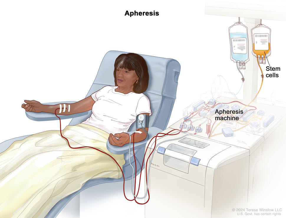


. A lymphoid stem cell becomes a lymphoblast and then becomes a B-lymphocyte, T-lymphocyte, or natural killer cell.")
; left panel shows blood being drawn from a vein on the inside of the elbow using a tube attached to a syringe; right panel shows a laboratory test tube with blood cells separated into layers: plasma, white blood cells, platelets, and red blood cells.")

 being inserted into the lower back. Inset shows a close-up of the spinal needle inserted into the cerebrospinal fluid (CSF) in the lower part of the spinal column.")
: Drawing of stem cells being collected from a donor's bloodstream using an apheresis machine. Blood is removed from a vein in the donor's arm and flows through the machine where the stem cells are removed. The rest of the blood is then returned to the donor through a vein in their other arm. (Panel 2): Drawing of a health care provider giving a patient an infusion of chemotherapy through a catheter in the patient's chest. The chemotherapy is given to kill cancer cells and prepare the patient's body for the donor stem cells. (Panel 3): Drawing of a patient receiving an infusion of the donor stem cells through a catheter in the chest.")
 being made in the laboratory; the gene for CAR is inserted into the T cells and then millions of CAR T cells are grown. Drawing also shows the CAR T cells being given to the patient by infusion and binding to antigens on the cancer cells and killing them.")
 in the brain and spinal cord, and an Ommaya reservoir (a dome-shaped container that is placed under the scalp during surgery; it holds the drugs as they flow through a small tube into the brain). Top section shows a syringe and needle injecting anticancer drugs into the Ommaya reservoir. Bottom section shows a syringe and needle injecting anticancer drugs directly into the cerebrospinal fluid in the lower part of the spinal column.")


 against the skin of the abdomen. A computer screen shows a sonogram (picture).")
 scan; drawing shows a child lying on a table that slides through the CT scanner, which takes a series of detailed x-ray pictures of areas inside the body.")
 scan; drawing shows a child lying on a table that slides into the MRI machine, which takes a series of detailed pictures of areas inside the body.")

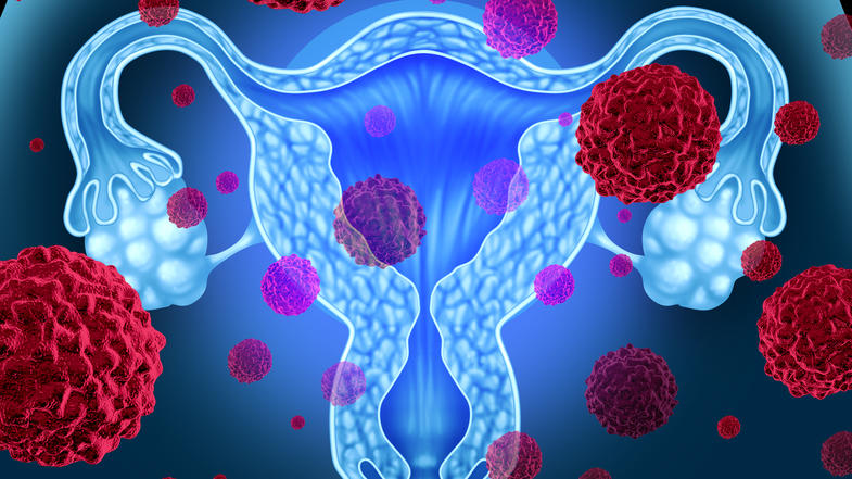
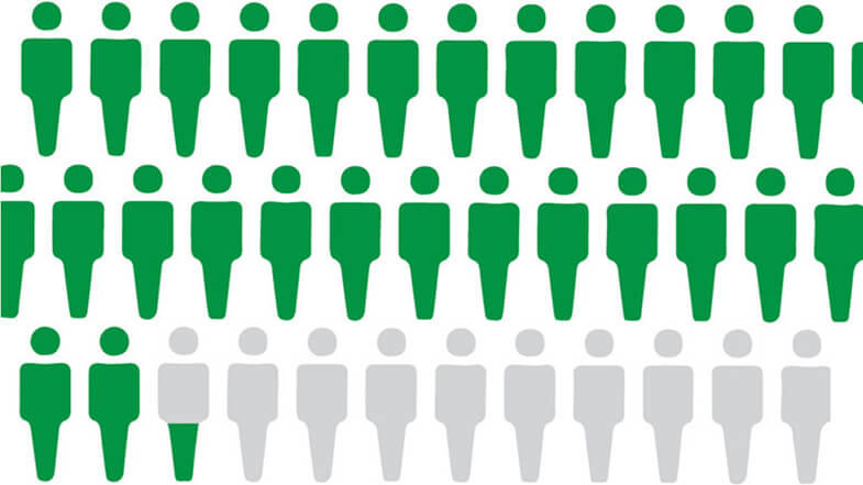
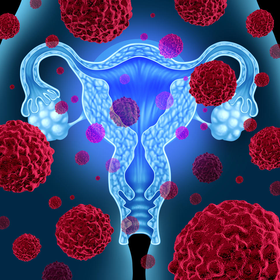

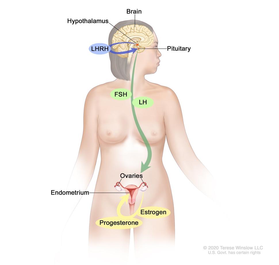
, endometrium (inner lining of the uterus), ovaries, fallopian tubes, cervix, and vagina.")