Childhood Hepatocellular Carcinoma

Childhood Hepatocellular Carcinoma

Childhood hepatocellular carcinoma is a rare type of cancer that forms in liver cells called hepatocytes. Hepatocytes are the most common cells of the liver, and they carry out most of the functions of the liver. 

The liver is one of the largest organs in the body. It has two lobes and fills the upper right side of the abdomen inside the rib cage. Three of the many important functions of the liver are:

  • to make bile to help digest fats from food
  • to store glycogen (sugar), which the body uses for energy
  • to filter harmful substances from the blood so they can be passed from the body in stools and urine
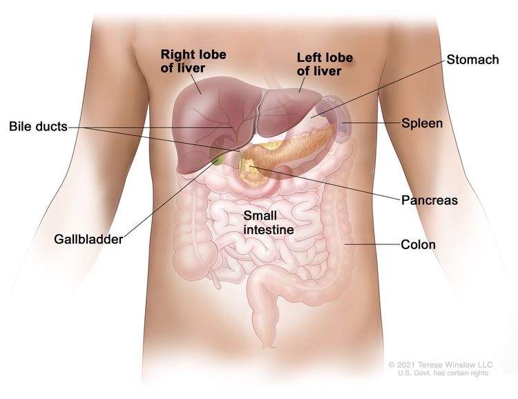
EnlargeAnatomy of the liver; drawing shows the right and left lobes of the liver. Also shown are the bile ducts, gallbladder, stomach, spleen, pancreas, small intestine, and colon.
Anatomy of the liver. The liver is in the upper abdomen near the stomach, intestines, gallbladder, and pancreas. The liver has a right lobe and a left lobe. Each lobe is divided into two sections (not shown).

Childhood hepatocellular carcinoma usually affects older children and adolescents. It is more common in areas of Asia that have higher rates of hepatitis B virus infection than in the United States. 

Hepatocellular carcinoma is the most common type of liver cancer in adults. Risk factors, staging, and treatment for children are different than for adults. Learn more about hepatocellular carcinoma in adults at What Is Liver Cancer?

Causes and risk factors for childhood hepatocellular carcinoma

Childhood hepatocellular carcinoma is caused by certain changes in the way liver cells function, especially how they grow and divide into new cells. Often, the exact cause of these cell changes is unknown. Learn more about how cancer develops at What Is Cancer?

A risk factor is anything that increases the chance of getting a disease. Not every child with one or more of these risk factors will develop hepatocellular carcinoma. And it will develop in some children who don’t have a known risk factor. 

The following syndromes or conditions are risk factors for childhood hepatocellular carcinoma: 

Hepatocellular carcinoma may develop in children with no underlying liver disease.

Talk with your child’s doctor if you think your child may be at risk.  

Symptoms of childhood hepatocellular carcinoma

Children may not have symptoms of hepatocellular carcinoma until the tumor has grown bigger. It’s important to check with your child’s doctor if your child has: 

  • a lump in the abdomen
  • swelling in the abdomen 
  • abdominal pain
  • weight loss for no known reason 
  • loss of appetite 
  • nausea and vomiting 

These symptoms may be caused by problems other than hepatocellular carcinoma. The only way to know is to see your child’s doctor.

Tests to diagnose childhood hepatocellular carcinoma

If your child has symptoms that suggest hepatocellular carcinoma, the doctor will need to find out if these are due to cancer or to another problem. The doctor will ask when the symptoms started and how often your child has been having them. They will also ask about your child’s personal and family medical history and do a physical exam. Based on these results, the doctor may recommend other tests. If your child is diagnosed with hepatocellular carcinoma, the results of these tests will also help you and your child’s doctor plan treatment.

The tests used to diagnose hepatocellular carcinoma in children may include:

Serum tumor marker test

Serum tumor marker tests measure the amounts of certain substances released into the blood by organs, tissues, or tumor cells in the body. Certain substances are linked to specific types of cancer when found in increased levels in the blood. These are called tumor markers. The blood of children who have liver cancer may have increased amounts of a hormone called beta-human chorionic gonadotropin (beta-hCG) or a protein called alpha-fetoprotein (AFP). Other cancers, benign liver tumors, and certain noncancer conditions, including cirrhosis and hepatitis, can also increase AFP levels.

Complete blood count (CBC)

A CBC checks a sample of blood for:

Liver function tests

Liver function tests measure the amounts of certain substances released into the blood by the liver. A higher-than-normal amount of a substance can be a sign of liver damage or cancer. 

Blood chemistry studies

Blood chemistry studies measure the amounts of certain substances, such as bilirubin or lactate dehydrogenase (LDH), released into the blood by organs and tissues in the body. An unusual amount of a substance can be a sign of disease.  

Hepatitis panel

A hepatitis panel checks for antigens or antibodies in the blood to see if there is or has been a hepatitis infection. 

Magnetic resonance imaging (MRI) with gadolinium

MRI uses a magnet, radio waves, and a computer to make a series of detailed pictures of areas inside the liver. A substance called gadolinium is injected into a vein. The gadolinium collects around the cancer cells, so they show up brighter in the picture. This procedure is also called nuclear MRI.
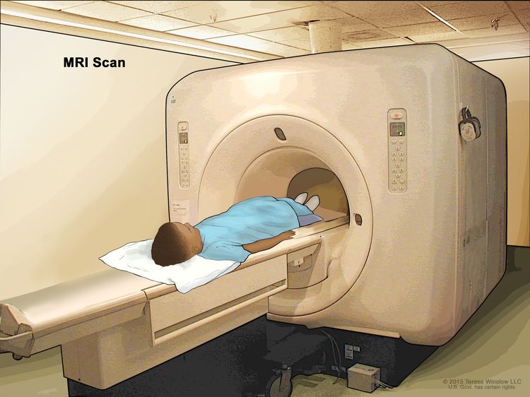
EnlargeMagnetic resonance imaging (MRI) scan; drawing shows a child lying on a table that slides into the MRI machine, which takes a series of detailed pictures of areas inside the body.
Magnetic resonance imaging (MRI) scan. The child lies on a table that slides into the MRI machine, which takes a series of detailed pictures of areas inside the body. The positioning of the child on the table depends on the part of the body being imaged.

CT scan (CAT scan)

A CT scan uses a computer linked to an x-ray machine to make a series of detailed pictures of areas inside the body, taken from different angles. A dye may be injected into a vein or swallowed to help the organs or tissues show up more clearly. This procedure is also called computed tomography, computerized tomography, or computerized axial tomography.

A CT scan of the chest and abdomen is usually done to help diagnose childhood liver cancer.

Learn more about Computed Tomography (CT) Scans and Cancer.
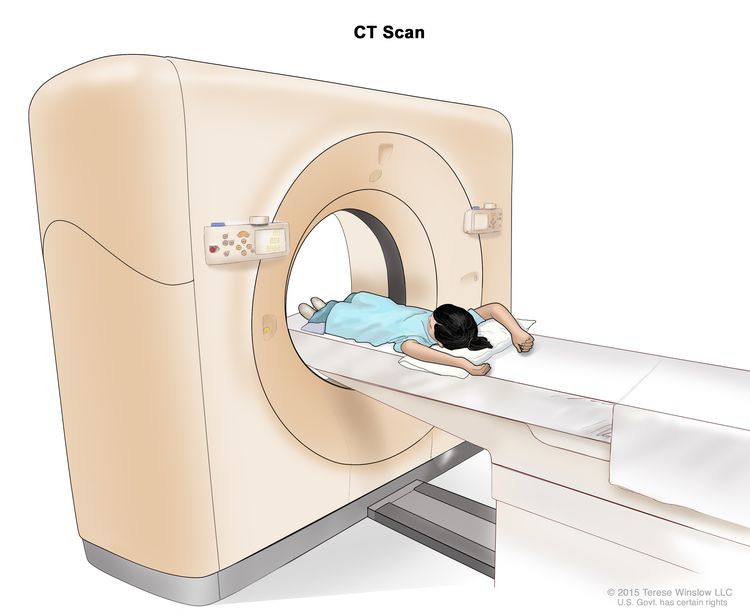
EnlargeComputed tomography (CT) scan; drawing shows a child lying on a table that slides through the CT scanner, which takes a series of detailed x-ray pictures of areas inside the body.
Computed tomography (CT) scan. The child lies on a table that slides through the CT scanner, which takes a series of detailed x-ray pictures of areas inside the body.

Ultrasound exam

An ultrasound exam uses high-energy sound waves (ultrasound) that bounce off internal tissues or organs and make echoes. The echoes form a picture of body tissues called a sonogram. An ultrasound exam of the abdomen to check the large blood vessels is usually done to help diagnose childhood liver cancer. 

EnlargeAbdominal ultrasound; drawing shows a child lying on an exam table during an abdominal ultrasound procedure. A technician is shown pressing a transducer (a device that makes sound waves that bounce off tissues inside the body) against the skin of the abdomen. A computer screen shows a sonogram (picture).
Abdominal ultrasound. An ultrasound transducer connected to a computer is pressed against the skin of the abdomen. The transducer bounces sound waves off internal organs and tissues to make echoes that form a sonogram (computer picture).

Chest x-ray

An x-ray is a type of high-energy radiation that can go through the body onto film, making a picture of areas inside the body. A chest x-ray is one that makes pictures of the lungs.

Biopsy

Biopsy is a procedure in which a sample of tissue is removed from the tumor so that a pathologist can view it under a microscope to check for cancer. The doctor may remove as much tumor as safely possible during the same biopsy procedure.

Immunohistochemistry

Immunohistochemistry uses antibodies to check for certain antigens (markers) in a sample of a patient’s tissue. The antibodies are usually linked to an enzyme or a fluorescent dye. After the antibodies bind to a specific antigen in the tissue sample, the enzyme or dye is activated, and the antigen can then be seen under a microscope. This type of test is used to help diagnose cancer and to help tell one type of cancer from another type.

Getting a second opinion

You may want to get a second opinion to confirm your child’s diagnosis and treatment plan. If you seek a second opinion, you will need to get medical test results and reports from the first doctor to share with the second doctor. The second doctor will review the pathology report, slides, and scans before giving a recommendation. This doctor may agree with the first doctor, suggest changes to the treatment plan, or provide more information about your child’s cancer.

To learn more about choosing a doctor and getting a second opinion, visit Finding Cancer Care. You can contact NCI’s Cancer Information Service via chat, email, or phone (both in English and Spanish) for help finding a doctor or hospital that can provide a second opinion. For questions you might want to ask at your child’s appointments, visit Questions to Ask Your Doctor. 

Prognostic factors for childhood hepatocellular carcinoma

If your child has been diagnosed with hepatocellular carcinoma, you likely have questions about how serious the cancer is and your child’s chances of survival. The likely outcome or course of a disease is called prognosis.

The prognosis for childhood hepatocellular carcinoma depends on:

  • whether the cancer has spread throughout the liver
  • whether the cancer can be removed completely by surgery 
  • whether the cancer has spread to other places in the body, such as the lungs 
  • how the cancer responds to chemotherapy 
  • whether your child has hepatitis B virus infection 
  • whether the cancer has just been diagnosed or has come back after treatment 

For childhood hepatocellular carcinoma that recurs (comes back) after initial treatment, the prognosis depends on:

  • where in the body the tumor recurred 
  • the type of treatment used to treat the initial cancer 

No two people are alike, and responses to treatment can vary greatly. Your child’s cancer care team is in the best position to talk with you about your child’s prognosis.

Stages of childhood hepatocellular carcinoma

The cancer stage describes the extent of cancer in the body, especially whether the cancer has spread from where it first formed to other parts of the body.

In childhood hepatocellular carcinoma, the PRETEXT and POSTTEXT groups are used to plan treatment. The results of the tests and procedures done to detect, diagnose, and find out whether the cancer has spread are used to determine the PRETEXT and POSTTEXT groups. 

Two grouping systems are used for childhood hepatocellular carcinoma to decide whether the tumor can be removed by surgery:

  • The PRETEXT group describes the tumor before the patient has any treatment. 
  • The POSTTEXT group describes the tumor after the patient has had treatment such as neoadjuvant chemotherapy. 

The liver is divided into four sections for the two grouping systems. The PRETEXT and POSTTEXT groups depend on which sections of the liver have cancer. There are four PRETEXT and POSTTEXT groups.

PRETEXT and POSTTEXT group I  

EnlargeLiver PRETEXT and POSTTEXT I; drawing shows two livers. Dotted lines divide each liver into four vertical sections of about the same size. In the first liver, cancer is shown in the section on the far left. In the second liver, cancer is shown in the section on the far right.
Liver PRETEXT and POSTTEXT I. Cancer is found in one section of the liver. Three sections of the liver that are next to each other do not have cancer in them.

In group I, the cancer is found in one section of the liver. Three sections of the liver that are next to each other do not have cancer in them. 

PRETEXT and POSTTEXT group II  

EnlargeLiver PRETEXT and POSTTEXT II; drawing shows five livers. Dotted lines divide each liver into four vertical sections that are about the same size. In the first liver, cancer is shown in the two sections on the left. In the second liver, cancer is shown in the two sections on the right. In the third liver, cancer is shown in the far left and far right sections. In the fourth liver, cancer is shown in the second section from the left. In the fifth liver, cancer is shown in the second section from the right.
Liver PRETEXT and POSTTEXT II. Cancer is found in one or two sections of the liver. Two sections of the liver that are next to each other do not have cancer in them.

In group II, cancer is found in one or two sections of the liver. Two sections of the liver that are next to each other do not have cancer in them. 

PRETEXT and POSTTEXT group III 

EnlargeLiver PRETEXT and POSTTEXT III; drawing shows seven livers. Dotted lines divide each liver into four vertical sections that are about the same size. In the first liver, cancer is shown in three sections on the left. In the second liver, cancer is shown in the two sections on the left and the section on the far right. In the third liver, cancer is shown in the section on the far left and the two sections on the right. In the fourth liver, cancer is shown in three sections on the right. In the fifth liver, cancer is shown in the two middle sections. In the sixth liver, cancer is shown in the section on the far left and in the second section from the right. In the seventh liver, cancer is shown in the section on the far right and in the second section from the left.
Liver PRETEXT and POSTTEXT III. Cancer is found in three sections of the liver and one section does not have cancer in it, or cancer is found in two sections of the liver and two sections that are not next to each other do not have cancer in them.

In group III, one of the following is true: 

  • Cancer is found in three sections of the liver, and one section does not have cancer. 
  • Cancer is found in two sections of the liver, and two sections that are not next to each other do not have cancer in them. 

PRETEXT and POSTTEXT group IV

EnlargeLiver PRETEXT and POSTTEXT IV; drawing shows two livers. Dotted lines divide each liver into four vertical sections that are about the same size. In the first liver, cancer is shown across all four sections. In the second liver, cancer is shown in the two sections on the left and spots of cancer are shown in the two sections on the right.
Liver PRETEXT and POSTTEXT IV. Cancer is found in all four sections of the liver.

In group IV, cancer is found in all four sections of the liver. 

Progressive and recurrent childhood hepatocellular carcinoma

Childhood hepatocellular carcinoma can be a progressive disease or refractory disease. Progressive hepatocellular carcinoma is cancer that continues to grow, spread, or worsen. Refractory hepatocellular carcinoma is cancer that no longer responds to treatment.

Recurrent hepatocellular carcinoma is cancer that has recurred (come back) after it has been treated. The cancer may come back in the liver or in other parts of the body. Tests will be done to help determine where the cancer has returned in the body, if it has spread, and how far. The type of treatment that your child will have for recurrent hepatocellular carcinoma will depend on how far it has spread.

Learn more at Recurrent Cancer: When Cancer Comes Back.

Types of treatment for childhood hepatocellular carcinoma

There are different types of treatment for children and adolescents with hepatocellular carcinoma. You and your child’s care team will work together to decide treatment. Many factors will be considered, such as your child’s overall health and whether the cancer is newly diagnosed or has come back.

A pediatric oncologist, a doctor who specializes in treating children with cancer, will oversee treatment of hepatocellular carcinoma. The pediatric oncologist works with other health care providers who are experts in treating children with hepatocellular carcinoma and who specialize in certain areas of medicine. It is especially important to have a pediatric surgeon with experience in liver surgery who can send patients to a liver transplant program if needed. Other specialists may include:

Your child’s treatment plan will include information about the cancer, the goals of treatment, treatment options, and the possible side effects. It will be helpful to talk with your child’s care team before treatment begins about what to expect. For help every step of the way, visit our booklet, Children with Cancer: A Guide for Parents.

Surgery 

When possible, the cancer is removed by surgery. The types of surgery that may be done are:

  • Partial hepatectomy is surgery to remove the part of the liver where cancer is found. The part removed may be a wedge of tissue, an entire lobe, or a larger part of the liver, along with a small amount of normal tissue around it.  
  • Liver transplant is the removal of the entire liver by surgery, followed by a transplant of a healthy liver from a donor. A liver transplant may be possible when cancer has not spread beyond the liver, and a donated liver can be found. If the patient has to wait for a donated liver, other treatment is given as needed. 
  • Resection of metastases is surgery to remove cancer that has spread outside of the liver, such as to nearby tissues, the lungs, or the brain.

The type of surgery that can be done depends on:

  • the PRETEXT group and POSTTEXT group 
  • the size of the primary tumor 
  • whether there is more than one tumor in the liver 
  • whether the cancer has spread to nearby large blood vessels 
  • the level of AFP in the blood 
  • whether the tumor can be shrunk by chemotherapy so that it can be removed by surgery
  • whether a liver transplant is needed 

Chemotherapy is sometimes given before surgery to shrink the tumor and make it easier to remove. This is called neoadjuvant therapy. 

After the doctor removes all the cancer that can be seen at the time of the surgery, some patients may be given chemotherapy or radiation therapy to kill any cancer cells that are left. Treatment given after the surgery to lower the risk that the cancer will come back is called adjuvant therapy. 

Chemotherapy 

Chemotherapy (also called chemo) uses drugs to stop the growth of cancer cells. Chemotherapy either kills the cancer cells or stops them from dividing. Chemotherapy may be given alone or with other types of treatment, such as radiation therapy.

There are two main ways to give chemotherapy to treat childhood hepatocellular carcinoma:

  • Systemic chemotherapy is chemotherapy that is injected into a vein or muscle. When given this way, the drugs enter the bloodstream and can reach cancer cells throughout the body.
  • Regional chemotherapy is chemotherapy that is placed directly into the cerebrospinal fluid, an organ, or a body cavity such as the abdomen. When given this way, the drugs mainly affect cancer cells in those areas.

    Chemoembolization of the hepatic artery (the main artery that supplies blood to the liver) is a type of regional chemotherapy used to treat childhood hepatocellular carcinoma that cannot be removed by surgery. The anticancer drug is injected into the hepatic artery through a catheter (thin tube). The drug is mixed with a substance that blocks the artery, cutting off blood flow to the tumor. Most of the anticancer drug is trapped near the tumor, and only a small amount of the drug reaches other parts of the body. The blockage may be temporary or permanent, depending on the substance used to block the artery. The tumor is prevented from getting the oxygen and nutrients it needs to grow. The liver continues to receive blood from the hepatic portal vein, which carries blood from the stomach and intestine to the liver. This procedure is also called transarterial chemoembolization, or TACE.

Chemotherapy drugs used alone or in combination to treat childhood hepatocellular carcinoma include:

Other chemotherapy drugs not listed here may also be used.

Learn more about how chemotherapy works, how it is given, common side effects, and more at Chemotherapy to Treat Cancer.

Radiation therapy 

Radiation therapy uses high-energy x-rays or other types of radiation to kill cancer cells or keep them from growing. Hepatocellular carcinoma in children may be treated with internal radiation therapy. Internal radiation therapy uses a radioactive substance sealed in needles, seeds, wires, or catheters that are placed directly into or near the cancer.

  • Radioembolization is a type of internal radiation therapy used to treat childhood hepatocellular carcinoma. A very small amount of a radioactive substance is attached to tiny beads that are injected into the hepatic artery (the main artery that supplies blood to the liver) through a thin tube called a catheter. The beads are mixed with a substance that blocks the artery, cutting off blood flow to the tumor. Most of the radiation is trapped near the tumor to kill the cancer cells. This is done to shrink the tumor or to relieve symptoms and improve quality of life for children with hepatocellular carcinoma. 

Learn more about radiation therapy and its side effects at Radiation Therapy to Treat Cancer and Radiation Therapy Side Effects.

Antiviral treatment 

Hepatocellular carcinoma that is linked to the hepatitis B virus may be treated with antiviral drugs.  

Ablation therapy

Ablation therapy removes or destroys tissue. Radiofrequency ablation is a type of ablation therapy that may be used to treat hepatocellular carcinoma. During radiofrequency ablation, special needles are inserted directly through the skin or through an incision in the abdomen to reach the tumor. High-energy radio waves heat the needles and tumor which kills cancer cells.

Clinical trials  

For some children, joining a clinical trial may be an option. There are different types of clinical trials for childhood cancer. For example, a treatment trial tests new treatments or new ways of using current treatments. Supportive care and palliative care trials look at ways to improve quality of life, especially for those who have side effects from cancer and its treatment.

You can use the clinical trial search to find NCI-supported cancer clinical trials accepting participants. The search allows you to filter trials based on the type of cancer, your child’s age, and where the trials are being done. Clinical trials supported by other organizations can be found on the ClinicalTrials.gov website.

Learn more about clinical trials, including how to find and join one, at Clinical Trials Information for Patients and Caregivers.

Treatment of newly diagnosed childhood hepatocellular carcinoma

Treatment options for newly diagnosed hepatocellular carcinoma that can be removed by surgery at the time of diagnosis may include:

  • surgery to remove the tumor, followed by chemotherapy
  • combination chemotherapy, followed by surgery to remove the tumor
  • surgery alone to remove the tumor

Treatment options for newly diagnosed hepatocellular carcinoma that cannot be removed by surgery and has not spread to other parts of the body at the time of diagnosis may include:

  • chemotherapy to shrink the tumor, followed by surgery to completely remove the tumor 
  • chemotherapy to shrink the tumor 

If surgery to completely remove the tumor is not possible, further treatment may include:

  • liver transplant 
  • chemoembolization or radioembolization of the hepatic artery to shrink the tumor, followed by surgery to remove the tumor or liver transplant 
  • chemoembolization or radioembolization of the hepatic artery alone 
  • radioembolization of the hepatic artery as palliative therapy to relieve symptoms and improve the quality of life 

Treatment for newly diagnosed hepatocellular carcinoma that has spread to other parts of the body at the time of diagnosis may include:

  • Combination chemotherapy to shrink the tumor, followed by surgery to remove as much of the tumor as possible from the liver and other places where cancer has spread. Studies have shown that this treatment may not work well, but some patients may benefit.

Treatment options for newly diagnosed hepatocellular carcinoma related to hepatitis B virus infection may include:

  • surgery to remove the tumor, followed by antiviral drugs that treat infection caused by the hepatitis B virus

Treatment of progressive or recurrent childhood hepatocellular carcinoma

Treatment of progressive or recurrent hepatocellular carcinoma may include:

  • chemoembolization of the hepatic artery to shrink the tumor before liver transplant
  • radiofrequency ablation
  • liver transplant

Side effects and late effects of treatment

Cancer treatments can cause side effects. Which side effects your child might have depends on the type of treatment they receive, the dose, and how their body reacts. Talk with your child’s treatment team about which side effects to look for and ways to manage them.

For information about side effects that begin during treatment for cancer, visit Side Effects.

Problems from cancer treatment that begin 6 months or later after treatment and continue for months or years are called late effects. Late effects of cancer treatment may include:

  • physical problems that affect liver function or hearing 
  • changes in mood, feelings, thinking, learning, or memory  
  • second cancers (new types of cancer)

Some late effects may be treated or controlled. It is important to talk with your child’s doctors about the long-term effects cancer treatment can have on your child. Learn more about Late Effects of Treatment for Childhood Cancer.  

Follow-up care

As your child goes through treatment, they will have follow-up tests or check-ups. Some of the tests that were done to diagnose the cancer or to find out the treatment group may be repeated to see how well the treatment is working. Decisions about whether to continue, change, or stop treatment may be based on the results of these tests. 

Some of the tests will continue to be done from time to time after treatment has ended. The results of these tests can show if your child’s condition has changed or if the cancer has recurred (come back). To learn more about follow-up tests, visit Tests to diagnose childhood hepatocellular carcinoma.

Coping with your child's cancer

When your child has cancer, every member of the family needs support. Taking care of yourself during this difficult time is important. Reach out to your child’s treatment team and to people in your family and community for support. To learn more, visit Support for Families: Childhood Cancer and the booklet Children with Cancer: A Guide for Parents.  

Hepatoblastoma

Hepatoblastoma

Hepatoblastoma is a cancer that forms in the tissues of the liver. It is the most common type of childhood liver cancer and usually affects children younger than 3 years of age.

The liver is one of the largest organs in the body. It has two lobes and fills the upper right side of the abdomen inside the rib cage. Three of the many important functions of the liver are:

  • to make bile to help digest fats from food
  • to store glycogen (sugar), which the body uses for energy
  • to filter harmful substances from the blood so they can be passed from the body in stools and urine
EnlargeAnatomy of the liver; drawing shows the right and left lobes of the liver. Also shown are the bile ducts, gallbladder, stomach, spleen, pancreas, small intestine, and colon.
Anatomy of the liver. The liver is in the upper abdomen near the stomach, intestines, gallbladder, and pancreas. The liver has a right lobe and a left lobe. Each lobe is divided into two sections (not shown).

In hepatoblastoma, the histology (how the cancer cells look under a microscope) affects the way the cancer is treated. The histology for hepatoblastoma includes:

Causes and risk factors for hepatoblastoma

Hepatoblastoma is caused by certain changes in the way the liver cells function, especially how they grow and divide into new cells. Often, the exact cause of these changes is unknown. Learn more about how cancer develops at What Is Cancer?

A risk factor is anything that increases the chance of getting a disease. Not every child with one or more of these risk factors will develop hepatoblastoma. And it will develop in some children who don’t have a known risk factor.

The following syndromes or conditions are risk factors for hepatoblastoma:

Talk with your child’s doctor if you think your child may be at risk.

Monitoring children at risk of hepatoblastoma

Children with Beckwith-Wiedemann syndrome, hemihyperplasia, Simpson-Golabi-Behmel syndrome, and trisomy 18 may have tests done to check for cancer before any symptoms appear. These tests may help find cancer early and improve your child’s chance of survival.

Children at risk of developing hepatoblastoma have an abdominal ultrasound exam every 3 months from birth (or diagnosis of a risk factor) until they are 4 years old. They also have blood tests to check the level of alpha-fetoprotein (AFP).

Symptoms of hepatoblastoma

Children may not have symptoms of hepatoblastoma until the tumor has grown bigger. It’s important to check with your child’s doctor if your child has:

  • a lump in the abdomen
  • swelling in the abdomen
  • pain in the abdomen
  • weight loss for no known reason
  • loss of appetite
  • nausea and vomiting

These symptoms may be caused by problems other than hepatoblastoma. The only way to know is for your child to see a doctor.

Tests to diagnose hepatoblastoma

If your child has symptoms that suggest hepatoblastoma, the doctor will need to find out if these are due to cancer or another problem. The doctor will ask when the symptoms started and how often your child has been having them. They will also ask about your child’s personal and family medical history and do a physical exam. Based on these results, the doctor may recommend other tests. If your child is diagnosed with hepatoblastoma, the results of these tests will also help you and your child’s doctor plan treatment.

The tests used to diagnose hepatoblastoma may include:

Serum tumor marker test

Serum tumor marker tests measure the amounts of certain substances released into the blood by organs, tissues, or tumor cells in the body. Certain substances are linked to specific types of cancer when found in increased levels in the blood. These are called tumor markers. The blood of children who have liver cancer may have increased amounts of a hormone called beta-human chorionic gonadotropin (beta-hCG) or a protein called AFP. Other cancers, benign liver tumors, and certain noncancer conditions, including cirrhosis and hepatitis, can also increase AFP levels.

SMARCB1 gene testing

SMARCB1 gene testing is a laboratory test in which a sample of blood or tissue is tested for certain changes in the SMARCB1 gene.

Complete blood count (CBC)

A CBC checks a sample of blood for:

Liver function tests

Liver function tests measure the amounts of certain substances released into the blood by the liver. A higher-than-normal amount of a substance can be a sign of liver damage or cancer.

Blood chemistry studies

Blood chemistry studies measure the amounts of certain substances, such as bilirubin or lactate dehydrogenase (LDH), released into the blood by organs and tissues in the body. An unusual amount of a substance can be a sign of disease.

Magnetic resonance imaging (MRI) with gadolinium

MRI uses a magnet, radio waves, and a computer to make a series of detailed pictures of areas inside the liver. A substance called gadolinium is injected into a vein. The gadolinium collects around the cancer cells so they show up brighter in the picture. This procedure is also called nuclear MRI.

EnlargeMagnetic resonance imaging (MRI) scan; drawing shows a child lying on a table that slides into the MRI machine, which takes a series of detailed pictures of areas inside the body.
Magnetic resonance imaging (MRI) scan. The child lies on a table that slides into the MRI machine, which takes a series of detailed pictures of areas inside the body. The positioning of the child on the table depends on the part of the body being imaged.

CT scan (CAT scan)

A CT scan uses a computer linked to an x-ray machine to make a series of detailed pictures of areas inside the body, taken from different angles. A dye may be injected into a vein or swallowed to help the organs or tissues show up more clearly. This procedure is also called computed tomography, computerized tomography, or computerized axial tomography.

A CT scan of the chest and abdomen is usually done to help diagnose childhood liver cancer.

Learn more about Computed Tomography (CT) Scans and Cancer.

EnlargeComputed tomography (CT) scan; drawing shows a child lying on a table that slides through the CT scanner, which takes a series of detailed x-ray pictures of areas inside the body.
Computed tomography (CT) scan. The child lies on a table that slides through the CT scanner, which takes a series of detailed x-ray pictures of areas inside the body.

Ultrasound exam

An ultrasound exam is a procedure in which high-energy sound waves (ultrasound) are bounced off internal tissues or organs and make echoes. The echoes form a picture of body tissues called a sonogram. An ultrasound exam of the abdomen to check the large blood vessels is usually done to help diagnose childhood liver cancer.
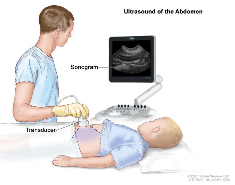
EnlargeAbdominal ultrasound; drawing shows a child lying on an exam table during an abdominal ultrasound procedure. A technician is shown pressing a transducer (a device that makes sound waves that bounce off tissues inside the body) against the skin of the abdomen. A computer screen shows a sonogram (picture).
Abdominal ultrasound. An ultrasound transducer connected to a computer is pressed against the skin of the abdomen. The transducer bounces sound waves off internal organs and tissues to make echoes that form a sonogram (computer picture).

Chest x-ray

An x-ray is a type of high-energy radiation that can go through the body and make pictures. A chest x-ray is one that makes pictures of the lungs.

Biopsy

Biopsy is a procedure in which a sample of tissue is removed from the tumor so that a pathologist can view it under a microscope to check for cancer. The doctor may remove as much tumor as safely possible during the same biopsy procedure.

Immunohistochemistry

Immunohistochemistry uses antibodies to check for certain antigens (markers) in a sample of a patient’s tissue. The antibodies are usually linked to an enzyme or a fluorescent dye. After the antibodies bind to a specific antigen in the tissue sample, the enzyme or dye is activated, and the antigen can then be seen under a microscope. This type of test is used to check for a certain gene mutation, to help diagnose cancer, and to help tell one type of cancer from another type of cancer. This test may be used to look for changes in the INI1 gene.

Getting a second opinion

You may want to get a second opinion to confirm your child’s diagnosis and treatment plan. If you seek a second opinion, you will need to get medical test results and reports from the first doctor to share with the second doctor. The second doctor will review the pathology report, slides, and scans before giving a recommendation. They may agree with the first doctor, suggest changes to the treatment plan, or provide more information about your child’s cancer.

To learn more about choosing a doctor and getting a second opinion, visit Finding Cancer Care. You can contact NCI’s Cancer Information Service via chat, email, or phone (both in English and Spanish) for help finding a doctor or hospital that can provide a second opinion. For questions you might want to ask at your appointments, visit Questions to Ask Your Doctor. 

Prognostic factors for hepatoblastoma

If your child has been diagnosed with hepatoblastoma, you likely have questions about how serious the cancer is and your child’s chances of survival. The likely outcome or course of a disease is called prognosis.

The prognosis for hepatoblastoma depends on:

  • whether the cancer has spread throughout the liver
  • the size of the tumor
  • whether the type of hepatoblastoma is well-differentiated fetal (pure fetal) or small cell undifferentiated histology
  • whether the cancer has spread to other places in the body, such as the diaphragm, lungs, or certain large blood vessels
  • whether there is more than one tumor in the liver
  • whether the outer covering around the tumor has broken open
  • how the cancer responds to chemotherapy
  • whether the cancer can be removed completely by surgery
  • whether your child can have a liver transplant
  • whether the AFP blood levels go down after treatment
  • your child’s age
  • whether the cancer has just been diagnosed or has come back after treatment

For hepatoblastoma that comes back after initial treatment, the prognosis depends on:

  • where in the body the tumor recurred
  • the type of treatment used to treat the initial cancer

Hepatoblastoma may be cured if the tumor is small and can be completely removed by surgery.

No two people are alike, and responses to treatment can vary greatly. Your child’s cancer care team is in the best position to talk with you about your child’s prognosis.

Stages of hepatoblastoma

Cancer stage describes the extent of cancer in the body, especially whether the cancer has spread from where it first formed to other parts of the body.

In hepatoblastoma, the PRETEXT and POSTTEXT groups are used instead of stage to plan treatment. The results of the tests and procedures done to detect, diagnose, and find out whether the cancer has spread are used to determine the PRETEXT and POSTTEXT groups.

Two grouping systems are used for hepatoblastoma to decide whether the tumor can be removed by surgery:

  • The PRETEXT group describes the tumor before the patient has any treatment.
  • The POSTTEXT group describes the tumor after the patient has had treatment such as neoadjuvant chemotherapy.

The liver is divided into four sections for the two grouping systems. The PRETEXT and POSTTEXT groups depend on which sections of the liver have cancer. There are four PRETEXT and POSTTEXT groups.

PRETEXT and POSTTEXT group I
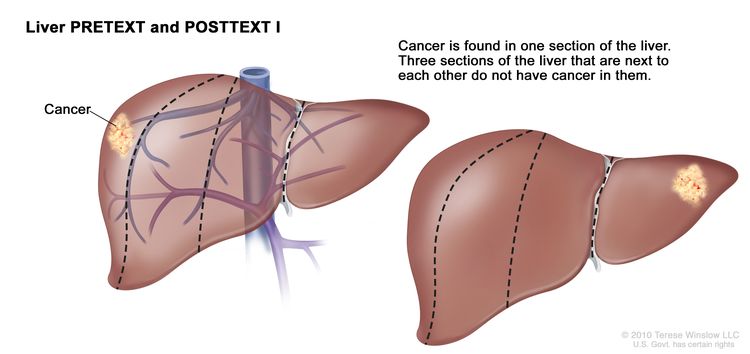
EnlargeLiver PRETEXT and POSTTEXT I; drawing shows two livers. Dotted lines divide each liver into four vertical sections of about the same size. In the first liver, cancer is shown in the section on the far left. In the second liver, cancer is shown in the section on the far right.
Liver PRETEXT and POSTTEXT I. Cancer is found in one section of the liver. Three sections of the liver that are next to each other do not have cancer in them.

In group I, the cancer is found in one section of the liver. Three sections of the liver that are next to each other do not have cancer in them.

PRETEXT and POSTTEXT group II
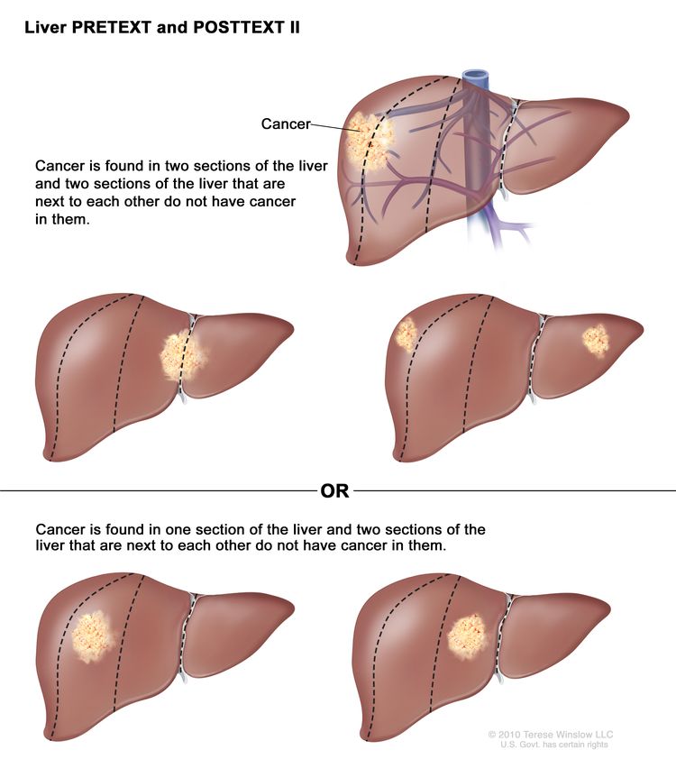
EnlargeLiver PRETEXT and POSTTEXT II; drawing shows five livers. Dotted lines divide each liver into four vertical sections that are about the same size. In the first liver, cancer is shown in the two sections on the left. In the second liver, cancer is shown in the two sections on the right. In the third liver, cancer is shown in the far left and far right sections. In the fourth liver, cancer is shown in the second section from the left. In the fifth liver, cancer is shown in the second section from the right.
Liver PRETEXT and POSTTEXT II. Cancer is found in one or two sections of the liver. Two sections of the liver that are next to each other do not have cancer in them.

In group II, cancer is found in one or two sections of the liver. Two sections of the liver that are next to each other do not have cancer in them.

PRETEXT and POSTTEXT group III
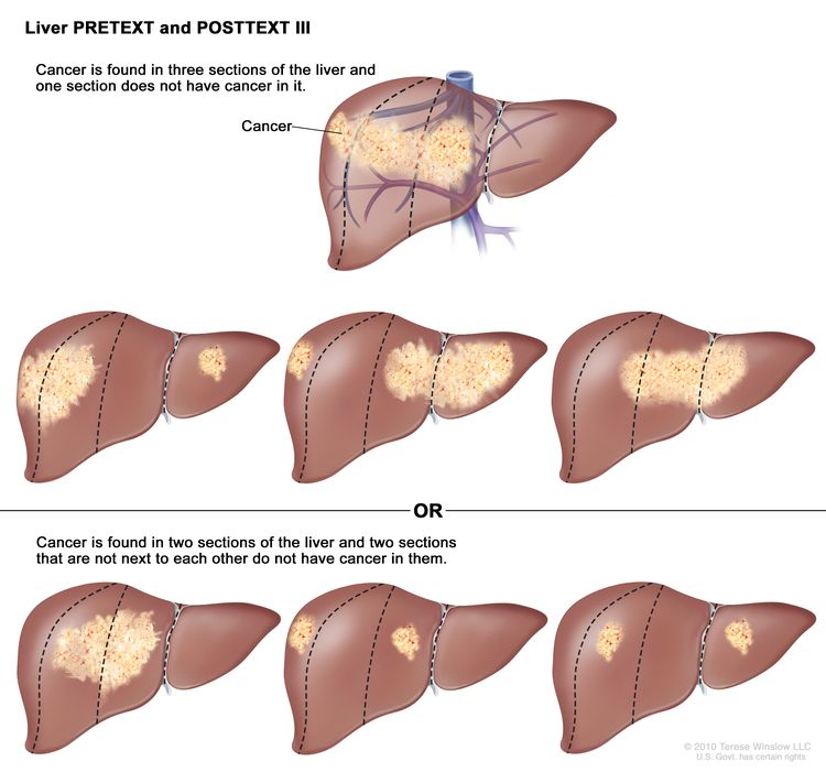
EnlargeLiver PRETEXT and POSTTEXT III; drawing shows seven livers. Dotted lines divide each liver into four vertical sections that are about the same size. In the first liver, cancer is shown in three sections on the left. In the second liver, cancer is shown in the two sections on the left and the section on the far right. In the third liver, cancer is shown in the section on the far left and the two sections on the right. In the fourth liver, cancer is shown in three sections on the right. In the fifth liver, cancer is shown in the two middle sections. In the sixth liver, cancer is shown in the section on the far left and in the second section from the right. In the seventh liver, cancer is shown in the section on the far right and in the second section from the left.
Liver PRETEXT and POSTTEXT III. Cancer is found in three sections of the liver and one section does not have cancer in it, or cancer is found in two sections of the liver and two sections that are not next to each other do not have cancer in them.

In group III, one of the following is true:

  • Cancer is found in three sections of the liver, and one section does not have cancer.
  • Cancer is found in two sections of the liver, and two sections that are not next to each other do not have cancer in them.

PRETEXT and POSTTEXT group IV
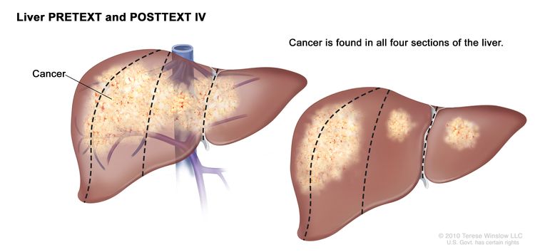
EnlargeLiver PRETEXT and POSTTEXT IV; drawing shows two livers. Dotted lines divide each liver into four vertical sections that are about the same size. In the first liver, cancer is shown across all four sections. In the second liver, cancer is shown in the two sections on the left and spots of cancer are shown in the two sections on the right.
Liver PRETEXT and POSTTEXT IV. Cancer is found in all four sections of the liver.

In group IV, cancer is found in all four sections of the liver.

Progressive and recurrent hepatoblastoma

Hepatoblastoma can be a progressive or refractory disease. Progressive hepatoblastoma is cancer that continues to grow, spread, or worsen. Refractory hepatoblastoma is cancer that no longer responds to treatment.

Recurrent hepatoblastoma is cancer that has recurred (come back) after it has been treated. The cancer may come back in the liver or as metastatic tumors in other parts of the body. Tests will be done to help determine where the cancer has returned in the body, if it has spread, and how far. The type of treatment that your child will have for recurrent hepatoblastoma will depend on how far it has spread.

Learn more at Recurrent Cancer: When Cancer Comes Back.

Types of treatment for hepatoblastoma

There are different types of treatment for children and adolescents with hepatoblastoma. You and your child’s care team will work together to decide treatment. Many factors will be considered, such as your child’s overall health and whether the cancer is newly diagnosed or has come back.

A pediatric oncologist, a doctor who specializes in treating children with cancer, oversees treatment of hepatoblastoma. The pediatric oncologist works with other health care providers who are experts in treating children with hepatoblastoma and who specialize in certain areas of medicine. It is especially important to have a pediatric surgeon with experience in liver surgery who can send patients to a liver transplant program if needed.

Other specialists may include:

Your child’s treatment plan will include information about the cancer, the goals of treatment, treatment options, and the possible side effects. It will be helpful to talk with your child’s care team before treatment begins about what to expect. For help every step of the way, visit our booklet, Children with Cancer: A Guide for Parents.

Surgery

When possible, the cancer is removed by surgery. The types of surgery that may be done are:

  • Partial hepatectomy is surgery to remove the part of the liver where cancer is found. The part removed may be a wedge of tissue, an entire lobe, or a larger part of the liver, along with a small amount of normal tissue around it.
  • Liver transplant is the removal of the entire liver by surgery, followed by a transplant of a healthy liver from a donor. A liver transplant may be possible when cancer has not spread beyond the liver, and a donated liver can be found. If the patient has to wait for a donated liver, other treatment is given as needed.
  • Resection of metastases is surgery to remove cancer that has spread outside of the liver, such as to nearby tissues, the lungs, or the brain.

The type of surgery that can be done depends on:

  • the PRETEXT group and POSTTEXT group
  • the size of the primary tumor
  • whether there is more than one tumor in the liver
  • whether the cancer has spread to nearby large blood vessels
  • the level of AFP in the blood
  • whether the tumor can be shrunk by chemotherapy, so that it can be removed by surgery
  • whether a liver transplant is needed

Chemotherapy is sometimes given before surgery to shrink the tumor and make it easier to remove. This is called neoadjuvant therapy.

After the doctor removes all the cancer that can be seen at the time of the surgery, some patients may be given chemotherapy or radiation therapy to kill any cancer cells that are left. Treatment given after the surgery to lower the risk that the cancer will come back is called adjuvant therapy.

Watchful waiting

Watchful waiting is closely monitoring a patient’s condition without giving any treatment until signs or symptoms appear or change. In hepatoblastoma, this treatment is only used for small well-differentiated fetal (pure fetal) histology tumors that have been completely removed by surgery.

Chemotherapy

Chemotherapy (also called chemo) uses drugs to stop the growth of cancer cells. Chemotherapy either kills the cancer cells or stops them from dividing. Chemotherapy may be given alone or with other types of treatment, such as radiation therapy.

There are two ways to give chemotherapy to treat hepatoblastoma:

  • Systemic chemotherapy is chemotherapy that is injected into a vein. When given this way, the drugs enter the bloodstream and can reach cancer cells throughout the body.
  • Regional chemotherapy is chemotherapy that is placed directly into the cerebrospinal fluid, an organ, or a body cavity such as the abdomen. When given this way, the drugs mainly affect cancer cells in those areas.

    Chemoembolization of the hepatic artery (the main artery that supplies blood to the liver) is a type of regional chemotherapy used to treat hepatoblastoma that cannot be removed by surgery. The anticancer drug is injected into the hepatic artery through a catheter (thin tube). The drug is mixed with a substance that blocks the artery, cutting off blood flow to the tumor. Most of the anticancer drug is trapped near the tumor, and only a small amount of the drug reaches other parts of the body. The blockage may be temporary or permanent, depending on the substance used to block the artery. The tumor is prevented from getting the oxygen and nutrients it needs to grow. The liver continues to receive blood from the hepatic portal vein, which carries blood from the stomach and intestine to the liver. This procedure is also called transarterial chemoembolization, or TACE.

Chemotherapy drugs used alone or in combination to treat hepatoblastoma include:

Other chemotherapy drugs not listed here may also be used.

Learn more about how chemotherapy works, how it is given, common side effects, and more at Chemotherapy to Treat Cancer.

Radiation therapy

Radiation therapy uses high-energy x-rays or other types of radiation to kill cancer cells or keep them from growing. The way the radiation therapy is given depends on the type of cancer being treated and the PRETEXT or POSTTEXT group.

Hepatoblastoma may be treated with external beam radiation therapy or internal radiation therapy:

  • External radiation therapy uses a machine outside the body to send radiation toward the area of the body with cancer. External radiation therapy is used to treat hepatoblastoma that cannot be removed by surgery or has spread to other parts of the body.
  • Internal radiation therapy uses a radioactive substance sealed in needles, seeds, wires, or catheters that are placed directly into or near the cancer.

    Radioembolization is a type of internal radiation therapy used to treat hepatoblastoma. A very small amount of a radioactive substance is attached to tiny beads that are injected into the hepatic artery (the main artery that supplies blood to the liver) through a thin tube called a catheter. The beads are mixed with a substance that blocks the artery, cutting off blood flow to the tumor. Most of the radiation is trapped near the tumor to kill the cancer cells. This is done to shrink the tumor or to relieve symptoms and improve quality of life for children with hepatoblastoma.

Learn more about radiation therapy and its side effects at External Beam Radiation Therapy for Cancer and Radiation Therapy Side Effects.

Radiofrequency ablation therapy

Radiofrequency ablation uses needles inserted directly through the skin or through an incision in the abdomen to reach the tumor. High-energy radio waves heat the needles and tumor, which kills cancer cells. Radiofrequency ablation is being used to treat recurrent hepatoblastoma.

Clinical trials

For some children, joining a clinical trial may be an option. There are different types of clinical trials for childhood cancer. For example, a treatment trial tests new treatments or new ways of using current treatments. Supportive care and palliative care trials look at ways to improve quality of life, especially for those who have side effects from cancer and its treatment.

You can use the clinical trial search to find NCI-supported cancer clinical trials accepting participants. The search allows you to filter trials based on the type of cancer, your child’s age, and where the trials are being done. Clinical trials supported by other organizations can be found on the ClinicalTrials.gov website.

Learn more about clinical trials, including how to find and join one, at Clinical Trials Information for Patients and Caregivers.

Treatment of newly diagnosed hepatoblastoma

Treatment of newly diagnosed hepatoblastoma that can be removed by surgery at the time of diagnosis may include:

  • surgery to remove the tumor, followed by combination chemotherapy, for hepatoblastoma that is mixed epithelial and fetal histology (not well-differentiated fetal histology) or aggressive chemotherapy for small cell undifferentiated histology
  • surgery to remove the tumor, followed by watchful waiting or chemotherapy, for hepatoblastoma with well-differentiated fetal histology

Treatment of newly diagnosed hepatoblastoma that cannot be removed by surgery or is not removed at the time of diagnosis may include:

  • combination chemotherapy to shrink the tumor, followed by surgery to remove the tumor
  • combination chemotherapy, followed by a liver transplant
  • chemoembolization or radioembolization of the hepatic artery to shrink the tumor, followed by surgery to remove the tumor

For newly diagnosed hepatoblastoma that has spread to other parts of the body at the time of diagnosis, combination chemotherapy is given to shrink the tumors in the liver and cancer that has spread to other parts of the body. After chemotherapy, imaging tests are done to check whether the tumors can be removed by surgery.

Treatment may include:

  • if the tumors in the liver and other parts of the body (usually nodules in the lung) can be removed, surgery will be done to remove the tumors, followed by chemotherapy to kill any cancer cells that may remain
  • if the tumor in other parts of the body cannot be removed, or a liver transplant is not possible, chemotherapy, chemoembolization or radioembolization of the hepatic artery, or radiation therapy may be given
  • if the tumor in other parts of the body cannot be removed, or the patient does not want surgery, radiofrequency ablation may be given

Treatment of progressive or recurrent hepatoblastoma

Treatment of progressive or recurrent hepatoblastoma may include:

  • surgery to remove isolated (single and separate) metastatic tumors with or without chemotherapy
  • radiofrequency ablation
  • combination chemotherapy
  • liver transplant

Side effects and late effects of treatment

Cancer treatments can cause side effects. Which side effects your child might have depends on the type of treatment they receive, the dose, and how their body reacts. Talk with your child’s treatment team about which side effects to look for and ways to manage them.

To learn more about side effects that begin during treatment for cancer, visit Side Effects.

Problems from cancer treatment that begin 6 months or later after treatment and continue for months or years are called late effects. Late effects of treatment may include:

  • physical problems that affect liver function or hearing
  • changes in mood, feelings, thinking, learning, or memory
  • second cancers (new types of cancer)

Some late effects may be treated or controlled. It is important to talk with your child’s doctors about the long-term effects cancer treatment can have on your child. Learn more about Late Effects of Treatment for Childhood Cancer.

Follow-up care

As your child goes through treatment, they will have follow-up tests or check-ups. Some of the tests that were done to diagnose the cancer or to find out the treatment group may be repeated to see how well the treatment is working. Decisions about whether to continue, change, or stop treatment may be based on the results of these tests.

Some of the tests will continue to be done from time to time after treatment has ended. The results of these tests can show if your child’s condition has changed, or if the cancer has recurred (come back).

To learn more about follow-up tests, visit Tests to diagnose hepatoblastoma.

Coping with your child's cancer

When a child has cancer, every member of the family needs support. Taking care of yourself during this difficult time is important. Reach out to your child’s treatment team and to people in your family and community for support. To learn more, visit Support for Families: Childhood Cancer and the booklet Children with Cancer: A Guide for Parents.  
Childhood Liver Cancer

Liver cancer is rare in children. There are several different types of childhood liver cancer. The most common type is hepatoblastoma.

The other less common types of childhood liver cancer are hepatocellular carcinoma, undifferentiated embryonal sarcoma of the liver, infantile choriocarcinoma of the liver, and fibrolamellar carcinoma.

Certain diseases and conditions can increase the risk of childhood liver cancer.

Explore the links on this page to learn more about the different types of liver cancer that can occur in children and adolescents, including causes and risk factors, how the cancer is found, and how it is treated.

Liver and Bile Duct Cancer

Cancer that starts in the liver is called primary liver cancer. The most common type of primary liver cancer in adults is hepatocellular carcinoma (HCC). This type of liver cancer is the third leading cause of cancer-related deaths worldwide.

Primary liver cancer can occur in both adults and children. However, treatment for children is different than treatment for adults.

Bile duct cancer, also called cholangiocarcinoma, is cancer that starts in the bile ducts, which are tubes that connect the liver and gallbladder to the small intestine. Cancer that forms in the bile ducts inside the liver is called intrahepatic bile duct cancer. If it forms in the bile ducts outside of the liver it is called extrahepatic bile duct cancer.

Explore the links on this page to learn more about the different types of liver and bile duct cancer (cholangiocarcinoma), including causes and risk factors, how the cancer is found, and how it is treated.

Adrenocortical Carcinoma Treatment by Stage

Stage is an important factor in deciding the best treatment for adrenocortical carcinoma. Other factors, such as your preferences and overall health, are also important.

For some people, taking part in a clinical trial may be an option. Clinical trials of new cancer drugs or treatment combinations may be available. Learn more about clinical trials, including how to find and join a trial, at Cancer Clinical Trial Information for Patients and Caregivers.  

Learn more about the treatments listed below at Adrenocortical Carcinoma Treatment.

Treatment of stage I adrenocortical carcinoma

Treatment of stage I adrenocortical carcinoma may include:

Treatment of stage II adrenocortical carcinoma

Treatment of stage II adrenocortical carcinoma may include:

  • Surgery (adrenalectomy). Nearby lymph nodes may also be removed if they are larger than normal.
  • A clinical trial of a new treatment.

Treatment of stage III adrenocortical carcinoma

Treatment of stage III adrenocortical carcinoma may include:

  • Surgery (adrenalectomy). Nearby lymph nodes may also be removed if they are larger than normal.
  • A clinical trial of radiation therapy.
  • A clinical trial of a new treatment.

Treatment of stage IV adrenocortical carcinoma

Treatment of stage IV adrenocortical carcinoma may include the following as palliative therapy to relieve symptoms and improve the quality of life:

Treatment of recurrent adrenocortical carcinoma

Treatment of recurrent adrenocortical carcinoma may include the following as palliative therapy to relieve symptoms and improve the quality of life:

  • Surgery.
  • Radiation therapy.
  • A clinical trial of chemotherapy or immunotherapy. 

Adrenocortical Carcinoma Treatment

Different types of treatments are available for patients with adrenocortical carcinoma. Some treatments are standard (the currently used treatment), and some are being tested in clinical trials. A treatment clinical trial is a research study meant to help improve current treatments or obtain information on new treatments for patients with cancer. When clinical trials show that a new treatment is better than the standard treatment, the new treatment may become the standard treatment. Patients may want to think about taking part in a clinical trial. Some clinical trials are open only to patients who have not started treatment.

Treatment options for adrenocortical carcinoma

The following types of treatment are used:

Surgery

Surgery to remove the adrenal gland (adrenalectomy) is often used to treat adrenocortical carcinoma. Sometimes surgery is done to remove the nearby lymph nodes and other tissue where the cancer has spread.

Radiation therapy

Radiation therapy is a cancer treatment that uses high-energy x-rays or other types of radiation to kill cancer cells or keep them from growing. There are two types of radiation therapy:

The way the radiation therapy is given depends on the type and stage of the cancer being treated.

Chemotherapy

Chemotherapy is a cancer treatment that uses drugs to stop the growth of cancer cells, either by killing the cells or by stopping them from dividing. When chemotherapy is taken by mouth or injected into a vein or muscle, the drugs enter the bloodstream and can reach cancer cells throughout the body (systemic chemotherapy). When chemotherapy is placed directly into the cerebrospinal fluid, an organ, or a body cavity such as the abdomen, the drugs mainly affect cancer cells in those areas (regional chemotherapy). Combination chemotherapy is treatment using more than one anticancer drug. The way the chemotherapy is given depends on the type and stage of the cancer being treated.

New treatments being tested in clinical trials

This summary section describes treatments that are being studied in clinical trials. It may not mention every new treatment being studied. Information about clinical trials is available from the NCI website.

Immunotherapy

Immunotherapy is a treatment that uses the patient’s immune system to fight cancer. Substances made by the body or made in a laboratory are used to boost, direct, or restore the body’s natural defenses against cancer.

Targeted therapy

Targeted therapy is a type of treatment that uses drugs or other substances to identify and attack specific cancer cells. 

Side effects of treatment

Treatment for adrenocortical carcinoma may cause side effects. For information about side effects caused by treatment for cancer, visit our Side Effects page.

Clinical trials

For some patients, taking part in a clinical trial may be the best treatment choice. Clinical trials are part of the cancer research process. Clinical trials are done to find out if new cancer treatments are safe and effective or better than the standard treatment.

Many of today’s standard treatments for cancer are based on earlier clinical trials. Patients who take part in a clinical trial may receive the standard treatment or be among the first to receive a new treatment.

Patients who take part in clinical trials also help improve the way cancer will be treated in the future. Even when clinical trials do not lead to effective new treatments, they often answer important questions and help move research forward.

Patients can enter clinical trials before, during, or after starting their cancer treatment

Some clinical trials only include patients who have not yet received treatment. Other trials test treatments for patients whose cancer has not gotten better. There are also clinical trials that test new ways to stop cancer from recurring (coming back) or reduce the side effects of cancer treatment.

Clinical trials are taking place in many parts of the country. Find clinical trials for adrenocortical carcinoma at Treatment Clinical Trials for Adrenal Cortex Cancer. Clinical trials supported by other organizations can be found on the ClinicalTrials.gov website. 

Follow-up care

As you go through treatment, you will have follow-up tests or check-ups. Some tests that were done to diagnose or stage the cancer may be repeated to see how well the treatment is working. Decisions about whether to continue, change, or stop treatment may be based on the results of these tests.

Some of the tests will continue to be done from time to time after treatment has ended. The results of these tests can show if your condition has changed or if the cancer has recurred (come back).

Adrenocortical Carcinoma Diagnosis & Prognosis

The tests and procedures used to diagnose adrenocortical carcinoma depend on the patient’s signs and symptoms. In addition to asking about your personal and family health history and doing a physical exam, your doctor may perform the following tests and procedures:

  • Twenty-four-hour urine test: A test in which urine is collected for 24 hours to measure the amounts of cortisol or 17-ketosteroids. A higher-than-normal amount of these in the urine may be a sign of disease in the adrenal cortex.
  • Low-dose dexamethasone suppression test: A test in which one or more small doses of dexamethasone are given. The level of cortisol is checked from a sample of blood or from urine that is collected for three days. This test is done to check if the adrenal gland is making too much cortisol.
  • High-dose dexamethasone suppression test: A test in which one or more high doses of dexamethasone are given. The level of cortisol is checked from a sample of blood or from urine that is collected for three days. This test is done to check if the adrenal gland is making too much cortisol or if the pituitary gland is telling the adrenal glands to make too much cortisol.
  • Blood chemistry study: A procedure in which a blood sample is checked to measure the amounts of certain substances, such as potassium or sodium, released into the blood by organs and tissues in the body. An unusual (higher or lower than normal) amount of a substance can be a sign of disease.
  • Blood hormone studies: A procedure in which a blood sample is checked to measure the amounts of certain hormones released into the blood by organs and tissues in the body. An unusual (higher or lower than normal) amount of a substance can be a sign of disease in the organ or tissue that makes it.
  • CT scan (CAT scan): A procedure that makes a series of detailed pictures of areas inside the body, taken from different angles. The pictures are made by a computer linked to an x-ray machine. A dye may be injected into a vein or swallowed to help the organs or tissues show up more clearly. This procedure is also called computed tomography, computerized tomography, or computerized axial tomography.
  • MRI (magnetic resonance imaging): A procedure that uses a magnet, radio waves, and a computer to make a series of detailed pictures of areas inside the body. This procedure is also called nuclear magnetic resonance imaging (NMRI). An MRI of the abdomen is done to diagnose adrenocortical carcinoma.
  • Adrenal angiography: A procedure to look at the arteries and the flow of blood near the adrenal glands. A contrast dye is injected into the adrenal arteries. As the dye moves through the arteries, a series of x-rays are taken to see if any arteries are blocked.
  • Adrenal venography: A procedure to look at the adrenal veins and the flow of blood near the adrenal glands. A contrast dye is injected into an adrenal vein. As the contrast dye moves through the veins, a series of x-rays are taken to see if any veins are blocked. A catheter (very thin tube) may be inserted into the vein to take a blood sample, which is checked for abnormal hormone levels.
  • PET scan (positron emission tomography scan): A procedure to find malignant tumor cells in the body. A small amount of radioactive glucose (sugar) is injected into a vein. The PET scanner rotates around the body and makes a picture of where glucose is being used in the body. Malignant tumor cells show up brighter in the picture because they are more active and take up more glucose than normal cells do.
  • MIBG scan: A very small amount of radioactive material called MIBG is injected into a vein and travels through the bloodstream. Adrenal gland cells take up the radioactive material and are detected by a device that measures radiation. This scan is done to tell the difference between adrenocortical carcinoma and pheochromocytoma.
  • Biopsy: The removal of cells or tissues so they can be viewed under a microscope by a pathologist to check for signs of cancer. The sample may be taken using a thin needle, called a fine-needle aspiration (FNA) biopsy, or a wider needle, called a core biopsy.

Adrenocortical carcinoma prognosis

Certain factors affect the prognosis (chance of recovery) and treatment options. The prognosis and treatment options depend on:

  • The stage of the cancer (the size of the tumor and whether it is in the adrenal gland only or has spread to other places in the body).
  • Whether the tumor can be completely removed in surgery.
  • Whether the cancer has been treated in the past.
  • The patient’s general health.
  • The grade of tumor cells (how different they look from normal cells under a microscope).

Adrenocortical carcinoma may be cured if treated at an early stage. 

Adrenocortical Carcinoma Stages

After adrenocortical carcinoma has been diagnosed, tests are done to find out if cancer cells have spread within the adrenal gland or to other parts of the body.

The process used to find out if cancer has spread within the adrenal gland or to other parts of the body is called staging. The information gathered from the staging process determines the stage of the disease. It is important to know the stage in order to plan treatment.

Tests to stage adrenocortical carcinoma

The following tests and procedures may be used in the staging process:

  • CT scan (CAT scan): A procedure that makes a series of detailed pictures of areas inside the body, such as the abdomen or chest, taken from different angles. The pictures are made by a computer linked to an x-ray machine. A dye may be injected into a vein or swallowed to help the organs or tissues show up more clearly. This procedure is also called computed tomography, computerized tomography, or computerized axial tomography.
  • MRI (magnetic resonance imaging) with gadolinium: A procedure that uses a magnet, radio waves, and a computer to make a series of detailed pictures of areas inside the body, such as the abdomen. A substance called gadolinium may be injected into a vein. The gadolinium collects around the cancer cells so they show up brighter in the picture. This procedure is also called nuclear magnetic resonance imaging (NMRI).
  • PET scan (positron emission tomography scan): A procedure to find malignant tumor cells in the body. A small amount of radioactive glucose (sugar) is injected into a vein. The PET scanner rotates around the body and makes a picture of where glucose is being used in the body. Malignant tumor cells show up brighter in the picture because they are more active and take up more glucose than normal cells do.
  • Ultrasound exam: A procedure in which high-energy sound waves (ultrasound) are bounced off internal tissues or organs, such as the vena cava, and make echoes. The echoes form a picture of body tissues called a sonogram.
  • Adrenalectomy: A procedure to remove the affected adrenal gland. A tissue sample is viewed under a microscope by a pathologist to check for signs of cancer.

How cancer spreads in the body

Cancer can spread through tissue, the lymph system, and the blood:

  • Tissue. The cancer spreads from where it began by growing into nearby areas.
  • Lymph system. The cancer spreads from where it began by getting into the lymph system. The cancer travels through the lymph vessels to other parts of the body.
  • Blood. The cancer spreads from where it began by getting into the blood. The cancer travels through the blood vessels to other parts of the body.

Cancer may spread from where it began to other parts of the body

When cancer spreads to another part of the body, it is called metastasis. Cancer cells break away from where they began (the primary tumor) and travel through the lymph system or blood.

  • Lymph system. The cancer gets into the lymph system, travels through the lymph vessels, and forms a tumor (metastatic tumor) in another part of the body.
  • Blood. The cancer gets into the blood, travels through the blood vessels, and forms a tumor (metastatic tumor) in another part of the body.

The metastatic tumor is the same type of cancer as the primary tumor. For example, if adrenocortical carcinoma spreads to the lung, the cancer cells in the lung are actually adrenocortical carcinoma cells. The disease is metastatic adrenocortical carcinoma, not lung cancer.

Metastasis: How Cancer Spreads

During metastasis, cancer cells spread from the place in the body where they first formed to other parts of the body.

Stage I

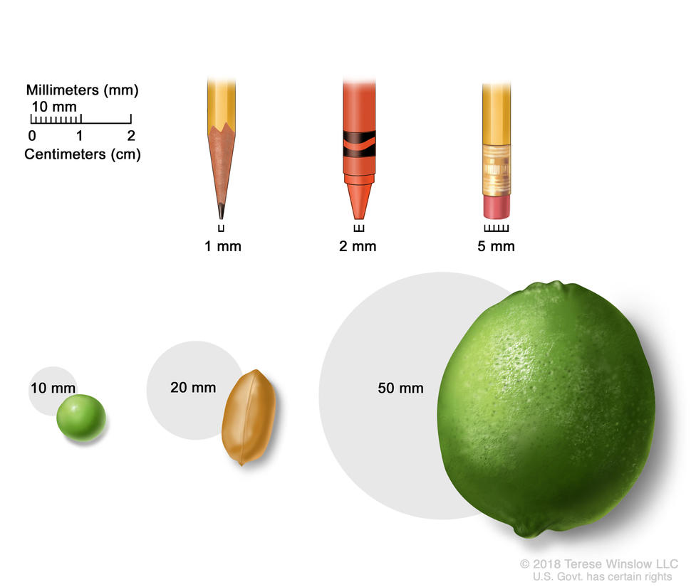
Drawing shows different sizes of common items in millimeters (mm): a sharp pencil point (1 mm), a new crayon point (2 mm), a pencil-top eraser (5 mm), a pea (10 mm), a peanut (20 mm), and a lime (50 mm). Also shown is a 2-centimeter (cm) ruler that shows 10 mm is equal to 1 cm.

Tumor size is often measured in millimeters (mm) or centimeters. Common items that can be used to show tumor size in mm include: a sharp pencil point (1 mm), a new crayon point (2 mm), a pencil-top eraser (5 mm), a pea (10 mm), a peanut (20 mm), and a lime (50 mm).

Credit: © Terese Winslow

In stage I adrenocortical carcinoma, the tumor is 5 centimeters or smaller and is found in the adrenal gland only.

Stage II

In stage II adrenocortical carcinoma, the tumor is larger than 5 centimeters and is found in the adrenal gland only.

Stage III

In stage III adrenocortical carcinoma, the tumor is any size and has spread:

Stage IV

In stage IV adrenocortical carcinoma, the tumor is any size, may have spread to nearby lymph nodes, and has spread to other parts of the body, such as the lung, bone, or peritoneum.

Recurrent adrenocortical carcinoma

Adrenocortical carcinoma can recur (come back) after it has been treated. The cancer may come back in the adrenal cortex or in other parts of the body.

What Is Adrenocortical Carcinoma?

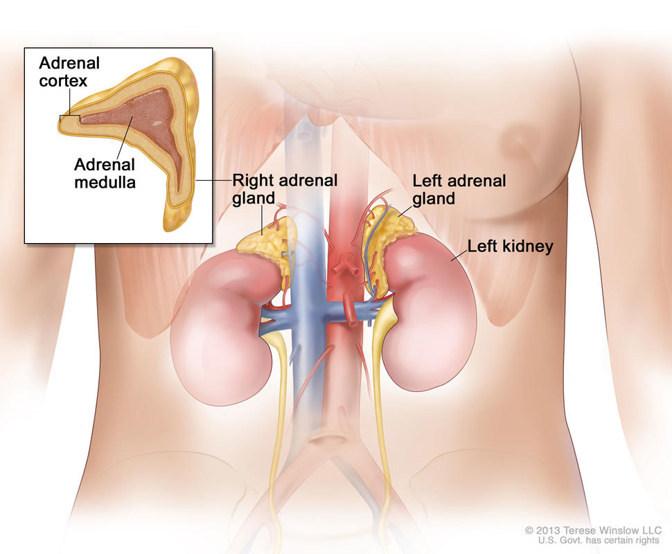
An illustration of the adrenal gland system, labeling the left and right adrenal glands and the left kidney. The right adrenal gland has a line pointing to an inset that shows the adrenal cortex and adrenal medulla.

Anatomy of the adrenal gland. There are two adrenal glands, one on top of each kidney.

Credit: © Terese Winslow

Adrenocortical carcinoma is a rare disease in which malignant (cancer) cells form in the outer layer of the adrenal gland.

There are two adrenal glands. The adrenal glands are small and shaped like a triangle. One adrenal gland sits on top of each kidney. Each adrenal gland has two parts. The outer layer of the adrenal gland is the adrenal cortex. The center of the adrenal gland is the adrenal medulla.

The adrenal cortex makes important hormones that:

  • Balance the water and salt in the body.
  • Help keep blood pressure normal.
  • Help control the body’s use of protein, fat, and carbohydrates.
  • Cause the body to have masculine or feminine characteristics.

Adrenocortical carcinoma is also called cancer of the adrenal cortex. A tumor of the adrenal cortex may be functioning (makes more hormones than normal) or nonfunctioning (does not make more hormones than normal). Most adrenocortical tumors are functioning. The hormones made by functioning tumors may cause certain signs or symptoms of disease.

The adrenal medulla makes hormones that help the body react to stress. Cancer that forms in the adrenal medulla is called pheochromocytoma and is not discussed in this summary. Learn more about Pheochromocytoma and Paraganglioma.

Adrenocortical carcinoma and pheochromocytoma can occur in both adults and children. Treatment for children, however, is different than treatment for adults. Learn more about Childhood Adrenocortical Carcinoma and Childhood Pheochromocytoma and Paraganglioma Treatment.

Risk factors for adrenocortical carcinoma

Having certain genetic conditions increases the risk of adrenocortical carcinoma. Anything that increases a person’s risk of getting a disease is called a risk factor. Not every person with one or more of these risk factors will develop adrenocortical carcinoma, and it will develop in some people who don’t have any known risk factors. Talk with your doctor if you think you may be at risk.

Risk factors for adrenocortical carcinoma include having the following hereditary diseases: