Pediatric Allogeneic Hematopoietic Stem Cell Transplant (PDQ®)–Health Professional Version

Pediatric Allogeneic Hematopoietic Stem Cell Transplant (PDQ®)–Health Professional Version

Improved Outcomes After Allogeneic Hematopoietic Stem Cell Transplant (HSCT)

During the past two decades, significant advances have led to improved outcomes after allogeneic HSCT.[13] The most significant improvements in survival occurred in unrelated and alternative donor procedures.[46] Possible explanations for these improvements in survival include improved patient selection, better supportive care, refined treatment regimens, improved approaches specific to stem cell sources, better intensive care unit experience, and better HLA typing. The sections below focus on modifiable aspects of HSCT, including the optimization of HLA typing and selection of stem cell sources.

References
  1. Hahn T, McCarthy PL, Hassebroek A, et al.: Significant improvement in survival after allogeneic hematopoietic cell transplantation during a period of significantly increased use, older recipient age, and use of unrelated donors. J Clin Oncol 31 (19): 2437-49, 2013. [PUBMED Abstract]
  2. Horan JT, Logan BR, Agovi-Johnson MA, et al.: Reducing the risk for transplantation-related mortality after allogeneic hematopoietic cell transplantation: how much progress has been made? J Clin Oncol 29 (7): 805-13, 2011. [PUBMED Abstract]
  3. Wood WA, Lee SJ, Brazauskas R, et al.: Survival improvements in adolescents and young adults after myeloablative allogeneic transplantation for acute lymphoblastic leukemia. Biol Blood Marrow Transplant 20 (6): 829-36, 2014. [PUBMED Abstract]
  4. MacMillan ML, Davies SM, Nelson GO, et al.: Twenty years of unrelated donor bone marrow transplantation for pediatric acute leukemia facilitated by the National Marrow Donor Program. Biol Blood Marrow Transplant 14 (9 Suppl): 16-22, 2008. [PUBMED Abstract]
  5. Harvey J, Green A, Cornish J, et al.: Improved survival in matched unrelated donor transplant for childhood ALL since the introduction of high-resolution matching at HLA class I and II. Bone Marrow Transplant 47 (10): 1294-300, 2012. [PUBMED Abstract]
  6. Majhail NS, Chitphakdithai P, Logan B, et al.: Significant improvement in survival after unrelated donor hematopoietic cell transplantation in the recent era. Biol Blood Marrow Transplant 21 (1): 142-50, 2015. [PUBMED Abstract]

Allogeneic Hematopoietic Stem Cell Transplant (HSCT) Indications for Hematologic Malignancies

Indications for HSCT vary over time as risk classifications for a given malignancy change and the efficacy of primary therapy improves. It is best to include specific indications in the context of complete therapy for any given disease. With this in mind, links to sections in specific summaries that cover the most common pediatric allogeneic HSCT indications are provided below.

  1. Acute lymphoblastic leukemia (ALL).
  2. Acute myeloid leukemia (AML).
  3. Myelodysplastic neoplasms (MDS).
  4. Juvenile myelomonocytic leukemia (JMML).
    • For more information, see the Treatment of JMML section in Juvenile Myelomonocytic Leukemia Treatment.
  5. Chronic myeloid leukemia (CML).

HLA Matching and Hematopoietic Stem Cell Sources

HLA Overview

Appropriate matching between donor and recipient HLA in the major histocompatibility complex located on chromosome 6 is essential to successful allogeneic hematopoietic stem cell transplant (HSCT) (see Figure 1, Table 1, and Table 2).

EnlargeHuman lymphocyte antigen (HLA) complex; drawing shows the long and short arms of human chromosome 6 with amplification of the HLA region, including the class I A, B, and C alleles, and the class II DP, DQ, and DR alleles.
Figure 1. HLA Complex. Human chromosome 6 with amplification of the HLA region. The locations of specific HLA loci for the class I B, C, and A alleles and the class II DP, DQ, and DR alleles are shown.

HLA class I (A, B, C, etc.) and class II (DRB1, DRB3, DRB4, DRB5, DQB1, DPB1, etc.) alleles are highly polymorphic. Therefore, finding appropriately matched unrelated donors is a challenge for some patients, especially those of certain racial groups (e.g., patients with African, Hispanic, Asian, or Pacific-Islander ancestry).[1,2] Full siblings of cancer patients have a 25% chance of being HLA matched.

Early serological techniques of HLA assessment defined a number of HLA antigens, but more precise DNA methodologies have shown HLA allele-level mismatches in up to 40% of serological HLA antigen matches. These differences are clinically relevant because the use of donors with allele-level mismatches affects survival and rates of graft-versus-host disease (GVHD) to a degree similar to that in patients with antigen-level mismatches.[3] Because of this, DNA-based allele-level HLA typing is standard when unrelated donors are being chosen.

The National Marrow Donor Program has published guidelines for HLA matching. The term for allele-level matching used in their guidelines is antigen recognition domain, which refers to the fact that the allele-level similarities used to define the specific HLA type are associated with areas directly used for antigen recognition. Polymorphisms of the HLA proteins outside of these areas are not involved in the function of these molecules. Therefore, they are often not assessed as part of HLA testing and unlikely to contribute to HLA mismatch.[4]

Table 1. Level of HLA Typing Currently Used for Different Hematopoietic Stem Cell Sourcesa,b,c
  Class I Antigens Class II Antigens
BM = bone marrow; N/A = not applicable; PBSCs = peripheral blood stem cells.
aHLA antigen: A serologically defined, low-resolution method of defining an HLA protein. Differs from allele-level typing at least 40% of the time. Designated by the first two numbers (i.e., for HLA B 35:01, the antigen is HLA B 35).
bHLA allele: A higher-resolution method of defining unique HLA proteins by typing their gene through sequencing or other DNA-based methods that detect unique differences. Designated by at least four numbers (i.e., for HLA B 35:01, 35 is the antigen and 01 is the allele).
cConsensus recommendations for HLA typing, including extended class II typing of mismatched donors, have been published by the National Cancer Institute/National Heart, Lung, and Blood Institute–sponsored Blood and Marrow Transplant Clinical Trials Network.[5]
dSiblings need confirmation that they have fully matched haplotypes with no crossovers in the A to DRB1 region. If parental typing is performed and haplotypes are established, antigen-level typing of class I is adequate. With no parental haplotypes, allele-level typing of eight alleles is recommended.
eParents, cousins, or other family members, with a phenotypic match or near-complete HLA match.
Stem Cell Source HLA A HLA B HLA C HLA DRB1 HLA DQB1; HLA DPB1; HLA DRB3,4,5
Matched siblingd BM/PBSCs Antigen or allele Antigen or allele Optional Allele N/A
Mismatched sibling/other related-donore BM/PBSCs Allele Allele Allele Allele Recommended
Unrelated-donor BM/PBSCs Allele Allele Allele Allele Recommended
Unrelated-donor cord blood Antigen (allele recommended) Antigen (allele recommended) Allele recommended Allele N/A
Table 2. Definitions of the Numbers Describing HLA Antigens and Alleles Matching
If These HLA Antigens and Alleles Match: Then the Donor Is Considered to be This Type of Match:
A, B, and DRB1 6/6
A, B, C, and DRB1 8/8
A, B, C, DRB1, and DQB1 10/10
A, B, C, DRB1, DQB1, and DPB1 12/12

HLA Matching Considerations for Sibling and Related Donors

The most commonly used related donor is a sibling from the same parents who, at a minimum, is HLA matched for HLA A, HLA B, and HLA DRB1 at the antigen level. Given the distance between HLA A and HLA DRB1 on chromosome 6, there is approximately a 1% possibility of a crossover event occurring in a possible sibling match. Because a crossover event could involve the HLA C antigen and because parents may share HLA antigens that actually differ at the allele level, many centers perform allele-level typing of possible sibling donors at all of the key HLA antigens (HLA A, B, C, and DRB1). Any related donor that is not a full sibling should have full HLA typing because similar haplotypes from different parents could differ at the allele level.

Although single-antigen mismatched related donors (5/6 antigen matched) were used interchangeably with matched sibling donors in some studies, a large Center for International Blood and Marrow Transplant Research (CIBMTR) study in pediatric HSCT recipients showed that the use of 5/6 antigen-matched related donors resulted in rates of GVHD and overall survival (OS) equivalent to rates in 8/8-allele-level-matched unrelated donors and slightly inferior survival than in fully matched siblings.[6] Any siblings with single mismatches should have extended typing to ensure that if the mismatch is caused by a crossover, it only occurs with one antigen. If clinicians choose siblings with multiple antigen mismatches as donors, haploidentical approaches may be warranted.

HLA Matching Considerations for Unrelated Donors

Optimal outcomes are achieved in unrelated allogeneic bone marrow transplant when the pairs of antigens at HLA A, B, C, and DRB1 are matched between the donor and the recipient at the allele level (termed an 8/8 match) (see Table 2).[7] A single antigen/allele mismatch at any of these antigens (7/8 match) lowers the probability of survival between 5% and 10%, with a similar increase in the amount of significant (grades III–IV) acute GVHD.[7] Of these four antigen pairs, different reports have shown HLA A, C, and DRB1 mismatches to potentially be more highly associated with mortality than the other antigens,[3,7,8] but the differences in outcome are small and inconsistent, making it very difficult to conclude that one can pick a more favorable mismatch by choosing one type of antigen mismatch over another. Many study groups are attempting to define specific antigens or pairs of antigens that are associated with either good or poor outcomes. For example, a specific HLA C mismatch (HLA-C*03:03/03:04) produces outcomes similar to a match. Therefore, selection of this mismatch is desirable in an otherwise matched donor/pair combination.[9]

It is well understood that class II antigen DRB1 mismatches increase GVHD incidence and worsen survival.[8] Subsequent data have also shown that multiple mismatches of DQB1, DPB1, and DRB3,4,5 lead to worse outcomes in the setting of less-than-8/8 matches.[10] DPB1 mismatches have been extensively studied and classified as permissive or nonpermissive on the basis of T-cell epitope matching. Patients with 10/10 matches and nonpermissive DPB1 mismatches have more transplant-related mortality but have survival rates similar to those with DPB1 matches or permissive matches. Those with 9/10 matches who have nonpermissive DPB1 mismatches have worse survival than those with permissive mismatches or DPB1 matches.[1113]

With these findings in mind, a 7/8- or 8/8-matched unrelated donor can be used routinely. However, outcomes may be further improved with the following:

  • Extended typing of DQB1, DPB1, and DRB3,4,5.[4,1113]
  • Extended HLA testing to select appropriate donors in the context of HLA-sensitized patients to avoid the potential risk of graft failure.[14,15] HLA sensitization is detected by testing for the presence of specific anti-HLA antibodies and avoiding donors who have any HLA antigens associated with the antibodies present in the recipient.
  • Use of younger donors.[5]
  • Matching cytomegalovirus (CMV)-positive recipients with CMV-positive donors and matching CMV-negative recipients with CMV-negative donors.[16]
  • Use of blood type–compatible unrelated donors.[5]

EnlargeChart showing HLA allele duplication and type of match between donor and recipient: an allele match (0201 and 0401 for both donor and recipient); a mismatch (0201 for both donor and recipient and 0201 for donor, 0401 for recipient) shown by an arrow pointing in a direction that promotes GVHD (GVH-O); a mismatch (0201 for both donor and recipient and 0401 for donor, 0201 for recipient) shown by an arrow pointing in a direction that promotes rejection (R-O); and a bidirectional mismatch (0201 for donor, 0301 for recipient, and 0401 for both donor and recipient) shown by arrows pointing in two directions, a direction that promotes rejection (R-O) and a direction that promotes GVHD (GVH-O).
Figure 2. HLA allele duplication in a donor or recipient results in a half match and a mismatch that will either occur in a direction that promotes GVHD (GVH-O) or a direction that promotes rejection (R-O).

If a donor or recipient has a duplication of one of their HLA alleles, they will have a half match and a mismatch only in one direction. Figure 2 illustrates that these mismatches will occur in either a direction that promotes GVHD (GVH-O, donor cells can detect a mismatch in a recipient which could cause GVHD) or a direction that promotes rejection (R-O, recipient cells can detect a mismatch in a donor that could lead to rejection). When 8/8-matched unrelated donors are compared with 7/8 donors mismatched in the GVH-O direction, 7/8 mismatched in the R-O direction, or 7/8 mismatched in both directions, the mismatch in the R-O direction leads to rates of grades III and IV acute GVHD similar to rates in the 8/8 matched and better than in the other two combinations. The 7/8 mismatched in only the R-O direction is preferred over GVH-O and bidirectional mismatches.[17] It is important to note that this observation in unrelated donors differs from observations in cord blood recipients, outlined below.

HLA Matching and Cell Dose Considerations for Unrelated Cord Blood HSCT

Another commonly used hematopoietic stem cell source is unrelated umbilical cord blood, which is harvested from donor placentas moments after birth. The cord blood is processed, HLA typed, cryopreserved, and banked.

Unrelated cord blood transplant has been successful with less-stringent HLA matching requirements compared with standard related or unrelated donors, probably because of limited antigen exposure experienced in utero and different immunological composition. Cord blood matching has traditionally been performed at an intermediate level for HLA A and B and at an allele level (high resolution) for DRB1. However, as outlined below, more extended typing can be helpful.

Although better outcomes occur when 6/6 or 5/6 HLA-matched units are used,[18] successful HSCT has occurred even with 4/6 or less HLA-matched units. In a large CIBMTR/Eurocord study, better matching at the allele level using eight antigens (matching for HLA A, B, C, and DRB1) resulted in less transplant-related mortality and improved survival. Best outcome was noted with 8/8 allele matching versus 4/8 to 7/8 matches, with poor survival in patients with five or more allele mismatches. Patients receiving 8/8-matched cord blood did not require higher cell doses for better outcomes. However, patients with one to three allele mismatches had less transplant-related mortality with total nucleated cell counts higher than 3 × 10⁷/kg, and those with four allele mismatches required a total nucleated cell count higher than 5 × 10⁷/kg to decrease transplant-related mortality.[19] This observation was noted to be especially important in cord blood transplants for nonmalignant disorders, where any mismatching below 7/8 alleles led to inferior survival.[20] Many centers will type additional alleles and use the best match possible, but the impact of DQB1, DPB1, and DRB3,4,5 mismatches has not been studied in detail.

As in unrelated peripheral blood stem cells (PBSCs) or bone marrow donors, extended HLA testing can support the selection of appropriate cord blood units in HLA-sensitized patients to avoid the potential risk of graft failure.[21,22] Evidence also suggests that selecting a mismatched cord blood unit, where the mismatch involves a noninherited maternal antigen, may improve survival.[23,24]

As with unrelated donors, individuals can occasionally have duplicate HLA alleles (e.g., the HLA A allele is 01:01 on both chromosomes). When this occurs in a donor product and the allele is matched to one of the recipient alleles, the recipient immune response will see the donor allele as matched (matched, in the rejection direction), but the donor immune response will see a mismatch in the recipient (mismatched in the GVHD direction). This variation of partial mismatching has been shown to be important in cord blood transplant outcomes. Mismatches that are only in the GVHD direction (i.e., GVH-O) lead to lower transplant-related mortality and overall mortality than those with rejection direction only (i.e., R-O) mismatches.[25] R-O mismatches have outcomes similar to those of bidirectional mismatches.[26] Although these studies suggest that using unidirectional mismatching as a criteria for cord blood selection may be beneficial, a Eurocord–European Society for Blood and Marrow Transplantation analysis disputes the value of this type of mismatching.[27]

Two aspects of umbilical cord blood HSCT have made the practice more widely applicable. First, because a successful procedure can occur with multiple HLA mismatches, more than 95% of patients from a wide variety of ethnicities are able to find at least a 4/6-matched cord blood unit.[1,28] Second, as mentioned above, adequate cell dose (minimum 2.5–3 × 10⁷ total nucleated cells/kg and 1.5 × 10⁵ CD34+ cells/kg) has been shown to be associated with improved survival.[29,30] Total nucleated cells are generally used to judge units because techniques to measure CD34-positive doses have not been standardized. Because even large single umbilical cord blood units are only able to supply these minimum doses to recipients weighing up to 40 kg to 50 kg, early umbilical cord blood HSCT focused mainly on smaller children. Later studies showed that barriers of this smaller size could be overcome by using two umbilical cord blood units if each of the units is at least a 4/6 HLA match with the recipient. Because two cord blood units provide higher cell doses, umbilical cord blood transplant is now used widely for larger children and adults.[31]

If a single unit provides an adequate cell dose, there may be disadvantages to adding a second unit.[32][Level of evidence A1] Two randomized trials showed that in children who had adequately sized single units, the addition of a second unit did not alter relapse, transplant-related mortality, or survival rates, but was associated with higher rates of extensive chronic GVHD.[32,33]

Investigators have shown that by using combinations of cytokines and other compounds to expand cord blood before infusion, engraftment of cord blood cells can occur more rapidly than after standard approaches.[3437] Although some studies that used multiple units or split units showed that expanded units will engraft early and then give way to nonexpanded units for long-term reconstitution,[38] other studies are showing persistence of expanded cells, implying preservation of stem cells through the expansion process.[36,37] A number of these approaches are under investigation. The U.S. Food and Drug Administration (FDA) approved an approach that uses a single unit split into two fractions, expanding one in culture with nicotinamide and infusing the second fraction without manipulation (omidubicel). One randomized trial compared omidubicel with standard cord blood transplant. Patients who received omidubicel had faster neutrophil and platelet engraftment, fewer bacterial and fungal infections, and fewer hospital days in the first 3 months after HSCT.[39] Notably, there was no difference in survival and GVHD outcomes.

Haploidentical HSCT

Early HSCT studies demonstrated progressively higher percentages of patients experiencing severe GVHD and lower survival rates as the number of donor/recipient HLA mismatches increased.[40] Other studies showed that even with very high numbers of donors in unrelated-donor registries, patients with rare HLA haplotypes and patients with certain ethnic backgrounds (e.g., patients with African, Hispanic, Asian, or Pacific-Islander ancestry) have a low chance of achieving desired levels of HLA matching (7/8 or 8/8 match at the allele level).[2]

To allow access to HSCT for patients without fully HLA-matched donor options, investigators have developed techniques allowing the use of siblings, parents, or other relatives who share only a single haplotype of the HLA complex with the patient and are thus half matches. Most approaches developed to date rely on T-cell depletion of the product before infusion into the patient. The main challenge associated with this approach is intense immune suppression with delayed immune recovery, which can result in lethal infections,[41] increased risk of Epstein-Barr virus (EBV)–associated lymphoproliferative disorder, and high rates of relapse.[42] This led to inferior survival compared with matched-donor HSCTs in the past and resulted in the procedure being used mainly at larger academic centers with a specific research focus on studying and developing this approach.

Improvements in haploidentical approaches, however, have resulted in better outcomes, with many groups reporting survival similar to that of unrelated marrow or cord blood approaches.[4346] These improvements include the following:

  • Newer techniques of T-cell depletion and add-back of specific cell populations (e.g., CD3 or alpha-beta CD3/CD19-negative selection) have decreased transplant-related mortality.[47]; [45,46,48,49][Level of evidence C2]
  • Reduced toxicity preparative regimens have led to decreased transplant-related mortality.[45,50]
  • Better supportive care has decreased the chance of morbidity from infection or EBV-associated lymphoproliferative disorder.[51]
  • Patient-donor combinations that have specific killer immunoglobulin-like receptor mismatches have shown decreased likelihood of relapse in some studies. For more information, see the Role of killer immunoglobulin-like receptor (KIR) mismatching in HSCT section.
  • Certain techniques, such as using combinations of granulocyte colony-stimulating factor–primed bone marrow and PBSCs with posttransplant antibody–based T-cell depletion [52] or post-HSCT cyclophosphamide (chemotherapeutic T-cell depletion),[45,46,53]; [54][Level of evidence C1] have made these procedures more accessible because they do not use the expensive and complicated processing necessary for traditional T-cell depletion.

Reported survival rates using many different types of haploidentical approaches range from 25% to 80%, depending on the technique and the risk of the patient undergoing the procedure.[42,43,52,53]; [54][Level of evidence C1] Retrospective trials in adults have shown similar outcomes after haploidentical-donor transplants compared with matched-unrelated donor or cord blood transplants.[55,56] One prospective randomized trial in adults with hematologic malignancies that used reduced-intensity regimens showed similar progression-free survival, but lower relapse rates and better OS using haploidentical donors.[57] Pediatric trials using haploidentical donors have shown better outcomes with myeloablative preparative regimens, and survival is comparable to nonhaploidentical approaches.[45,46,49,58] One prospective trial in pediatric patients showed that disease-free survival (DFS) was superior using haploidentical approaches compared with mismatched unrelated-donor approaches. DFS rates in patients treated with haploidentical approaches were similar to those in patients treated with other stem cell sources.[45]

Patients who have been sensitized to HLA antigens and develop antibodies to HLA alleles that are present among the mismatched alleles of their haploidentical donor are at increased risk of rejection of their haploidentical graft. Clinicians should choose donors with HLA types against whom the recipient does not have an antibody present, if possible. Guidelines on how to best approach this issue have been published.[59]

Comparison of Stem Cell Products

Currently, the following three stem cell products are used from both related and unrelated donors:

  • Bone marrow.
  • PBSCs.
  • Cord blood.

Bone marrow or PBSCs, including partially HLA-matched (half or more antigens [haploidentical]) related bone marrow or PBSCs, can be used after in vitro or in vivo T-cell depletion, and these products behave differently from other stem cell products. A comparison of stem cell products is presented in Table 3.

Table 3. Comparison of Hematopoietic Stem Cell Products
  PBSCs BM Cord Blood T-cell–Depleted BM/PBSCs Haploidentical T-cell–Depleted BM/PBSCs
BM = bone marrow; EBV-LPD = Epstein-Barr virus–associated lymphoproliferative disorder; GVHD = graft-versus-host disease; HSCT = hematopoietic stem cell transplant; PBSCs = peripheral blood stem cells.
aAssuming no development of GVHD. If patients develop GVHD, immune reconstitution is delayed until resolution of the GVHD and discontinuation of immune suppression.
bIf a haploidentical donor is used, longer times to immune reconstitution may occur.
T-cell content High Moderate Low Very low Very low
CD34+ content Moderate–high Moderate Low (but higher potency) Moderate–high Moderate–high
Time to neutrophil recovery Rapid: median, 16 d (11–29 d) [60] Moderate: median, 21 d (12–35 d) [60] Slower: median, 23 d (11–133 d) [33] Rapid: median, 16 d (9–40 d) [61] Rapid: median, 13 d (10–20 d) [62]
Early post-HSCT risk of infections, EBV-LPD Low–moderate Moderate High Very high Very high
Risk of graft rejection Low Low–moderate Moderate–high Moderate–high Moderate–high
Time to immune reconstitutiona Rapid (6–12 mo) Moderate (6–18 mo) Slow (6–24 mo) Slow (6–24 mo) Slow (9–24 mo)b
Risk of acute GVHD Moderate Moderate Moderate Low Low
Risk of chronic GVHD High Moderate Low Low Low

The main differences between the products are the numbers of T cells and CD34-positive progenitor cells present. Very high levels of T cells are present in PBSCs, intermediate numbers in bone marrow, and low numbers in cord blood and T-cell–depleted products. Patients receiving T-cell–depleted products or cord blood generally have slower hematopoietic recovery, increased risk of infection, late immune reconstitution, higher risks of nonengraftment, and increased risk of EBV-associated lymphoproliferative disorder. This is countered by lower rates of GVHD and an ability to offer transplant to patients for whom full HLA matching is not available. Higher doses of T cells and other cells in PBSCs result in rapid neutrophil recovery and immune reconstitution but also increase rates of chronic GVHD.

Only a few studies have directly compared outcomes of different stem cell sources/products in pediatric patients.

Evidence (comparison of outcomes of stem cell sources/products in children):

  1. A retrospective registry study of pediatric patients who underwent HSCT for acute leukemia compared those who received related-donor bone marrow with those who received related-donor PBSCs.[63]
    • Although the bone marrow and PBSC recipient cohorts differed some in their risk profiles, after statistical correction, increased risk of GVHD and transplant-related mortality associated with PBSCs led to poorer survival in the PBSC group.
  2. A retrospective study of Japanese children with acute leukemia compared 90 children who received PBSCs with 571 children who received bone marrow.[64]
    • The study confirmed higher transplant-related mortality caused by GVHD and inferior survival among the children who received PBSCs.
  3. A large Blood and Marrow Transplant Clinical Trials Network trial for patients requiring unrelated donors included a number of pediatric patients. Patients were randomly assigned to receive either bone marrow or PBSCs. This trial demonstrated the following:[65]
    • OS was identical using either source, but rates of chronic GVHD were significantly higher in the PBSC arm, with a small increase in rejection in the bone marrow arm.
    • Rejections were rare in pediatric patients.
    • There was an insufficient number of patients to draw specific conclusions about rejection risk in children who received bone marrow.

These reports, combined with a lack of prospective studies comparing bone marrow and PBSCs, have led most pediatric transplant protocols to prefer bone marrow over PBSCs from related donors. This approach is further supported by a meta-analysis that included additional retrospective trials.[66]

Published studies comparing unrelated cord blood and bone marrow have been retrospective, with weaknesses inherent in such analyses.

Evidence (comparison of unrelated cord blood versus bone marrow outcomes):

  1. In one study, pediatric patients with acute lymphoblastic leukemia (ALL) who underwent HSCT and received 8/8 HLA-matched unrelated-donor bone marrow were compared with those who received unrelated cord blood.[18]
    • The analysis showed that the best survival occurred in recipients of 6/6 HLA-matched cord blood.
    • Survival after 8/8 HLA-matched unrelated bone marrow was slightly worse and was statistically identical to survival for patients receiving 5/6 and 4/6 HLA-matched cord blood units.
  2. In another study from a single center consisting of mostly adult patients with acute myeloid leukemia (AML), myelodysplastic neoplasms (MDS), and ALL, outcomes for cord blood recipients were compared with outcomes for recipients of matched and mismatched unrelated-donor bone marrow/PBSCs.[67]
    • Better survival because of less relapse was noted in cord blood recipients. This result was mainly due to superior survival in patients with minimal residual disease (MRD) present just before transplant.
    • No difference was seen in relapse or survival between patients with pre-HSCT MRD and patients without pre-HSCT MRD if they received cord blood.
    • These results are controversial because they contradict many other studies that showed that the presence of pre-HSCT MRD in cord blood recipients led to increased relapse and inferior survival rates.[6871]
  3. The CIBMTR compared outcomes of children with low-risk and intermediate-risk ALL and AML who underwent transplant between 2000 and 2014 using alternative donors (non–HLA-matched related or unrelated), including 7/8 HLA-matched bone marrow (n = 172) and 4/6 or greater HLA-matched umbilical cord blood (n = 1,613).[72]
    • In multivariate analysis, patients who received 7/8 HLA-matched bone marrow versus umbilical cord blood had similar GVHD-free, relapse-free survival (hazard ratio [HR], 1.12; 95% confidence interval [CI], 0.87–1.45; P = .39), chronic GVHD-free, relapse-free survival (HR, 1.06; 95% CI, 0.82–1.38; P = .66), and OS (HR, 1.07; 95% CI, 0.80–1.44; P = .66).
    • Relapse may have been higher in the 7/8 HLA-matched bone marrow group (HR, 1.44; 95% CI, 1.03–2.02; P = .03; the publication called this a trend as they chose a cutoff value of 0.01% to control for multiple comparisons).
    • The patients in the 7/8 HLA-matched bone marrow group had a significantly higher risk of grades III to IV acute GVHD (HR, 1.70; 95% CI, 1.16–2.48; P = .006) and chronic GVHD (HR, 6.17; 95% CI, 2.2–17.33; P = .0006) than did the patients in the umbilical cord blood group.

On the basis of these studies, most transplant centers consider matched sibling bone marrow to be the preferred stem cell source/product. If a sibling donor is not available, fully matched unrelated-donor bone marrow, HLA-matched (4/6 to 6/6 or 6/8 to 8/8) cord blood from a single unit with an adequate cell dose, or a haploidentical HSCT lead to similar survival rates.[49][Level of evidence C2] For more information about the prevention of acute GVHD, see the Prevention and treatment of acute GVHD section in Complications, Graft-Versus-Host Disease, and Late Effects After Pediatric Hematopoietic Stem Cell Transplant.

Other Donor Characteristics Associated With Outcome

HLA matching has consistently been the most important factor associated with improved survival in allogeneic HSCT, but a number of other donor characteristics have been shown to affect key outcomes. Higher cell dose from the donor has also been shown to be important when related, unrelated, or haploidentical bone marrow or PBSC donors are used.[73,74] For more information, see the HLA Matching and Cell Dose Considerations for Unrelated Cord Blood HSCT section. The effects of donor age, blood type, CMV status, sex, and parity of female donors have also been studied.

Ideally, after HLA matching, transplant centers should select donors based on the following characteristics:

  • Donor age. The youngest donor available is generally preferred (when considering pediatric donors, the size and ability to obtain an adequate cell dose comes into consideration).[75,76]
  • CMV status of the recipient. CMV-negative donor matched to CMV-negative recipient and CMV-positive donor matched to CMV-positive recipient are preferred.[77]
  • Donor blood type. Matching of blood type between donor and recipient is preferred, although not required. If only blood type–mismatched donors are available, a minor mismatch is preferred over a major mismatch.[7880]
  • Donor sex and parity of female donors. Male or nonparous female donors are preferred over parous female donors.[76,81]

It is rare for a donor-recipient pair to fit perfectly into this algorithm, and determining which of these characteristics should be chosen over others has been controversial.

Evidence (donor-recipient characteristics):

  1. A CIBMTR study examined 6,349 patients who underwent transplant for hematological malignancies from 1988 to 2006. The study tested the effect of donor characteristics while adjusting for disease risk and other key transplant characteristics. The data from this study showed the following:[76]
    • In addition to HLA mismatching, older donor age and major or minor ABO blood type mismatching increased overall mortality.
    • Parous female graft recipients experienced lower rates of relapse.
    • Recipients of younger donor grafts had lower rates of acute GVHD.
    • Recipients of parous female grafts had higher rates of chronic GVHD.
    • Recipient CMV status was more important than donor CMV status (recipients who are CMV positive are at higher risk of mortality independent of the donor CMV status), although a CMV-negative donor to a CMV-negative recipient combination improves survival.
  2. A cohort of 4,690 patients who underwent transplant between 2007 and 2011 was tested by a multivariate analysis for independent predictors of survival in an EBMT study. The study demonstrated the following:[82]
    • Older donor age was confirmed to be independently associated with worse OS; every 10 years of donor age increased the risk of mortality by 5.5%.
    • HLA matching continued to have the most important effect on survival; ABO mismatching was not confirmed to have a continuing effect.
  3. A study of over 10,000 matched donor-recipient pairs attempted to define a hierarchy that could prioritize the non-HLA characteristics (donor age, sex, blood type, CMV status, etc.) that have been described to affect outcomes.[83]
    • Although the study was unable to create a hierarchical algorithm of modifiable factors, it showed that, by far, younger donor age is the most important factor. The study found a decrease in OS of 3% for every 10-year increment of increased donor age.

Thus, after HLA matching, donor age is likely the most important factor to optimize. Of note, if the recipient is CMV negative, finding a CMV-negative donor is also a high priority.

Several studies have attempted to identify characteristics of the best donors for haploidentical procedures. As with conventional bone marrow transplant, use of younger donors appears to be beneficial, but data regarding donor sex are inconclusive. Studies involving intense T-cell depletion have noted better outcomes using maternal donors,[84] but studies using posttransplant cyclophosphamide or intense immune suppression seem to favor male donors.[85,86] Further study is needed to clarify this important issue.

One large comparison of haploidentical donors showed an effect of ABO incompatibility on engraftment (risk of rejection doubling from 6% to 12%, ABO match vs. ABO major mismatch), and patients receiving bidirectionally mismatched donors had a 2.4-fold increase in grades II to IV acute GVHD.[87] As with nonhaploidentical donors, significant improvement of outcomes was noted when younger donors were used for haploidentical procedures compared with older donors, with an HR of 1.13 for each decade of life that the donor is older.[88]

References
  1. Barker JN, Byam CE, Kernan NA, et al.: Availability of cord blood extends allogeneic hematopoietic stem cell transplant access to racial and ethnic minorities. Biol Blood Marrow Transplant 16 (11): 1541-8, 2010. [PUBMED Abstract]
  2. Gragert L, Eapen M, Williams E, et al.: HLA match likelihoods for hematopoietic stem-cell grafts in the U.S. registry. N Engl J Med 371 (4): 339-48, 2014. [PUBMED Abstract]
  3. Woolfrey A, Klein JP, Haagenson M, et al.: HLA-C antigen mismatch is associated with worse outcome in unrelated donor peripheral blood stem cell transplantation. Biol Blood Marrow Transplant 17 (6): 885-92, 2011. [PUBMED Abstract]
  4. Dehn J, Spellman S, Hurley CK, et al.: Selection of unrelated donors and cord blood units for hematopoietic cell transplantation: guidelines from the NMDP/CIBMTR. Blood 134 (12): 924-934, 2019. [PUBMED Abstract]
  5. Howard CA, Fernandez-Vina MA, Appelbaum FR, et al.: Recommendations for donor human leukocyte antigen assessment and matching for allogeneic stem cell transplantation: consensus opinion of the Blood and Marrow Transplant Clinical Trials Network (BMT CTN). Biol Blood Marrow Transplant 21 (1): 4-7, 2015. [PUBMED Abstract]
  6. Shaw PJ, Kan F, Woo Ahn K, et al.: Outcomes of pediatric bone marrow transplantation for leukemia and myelodysplasia using matched sibling, mismatched related, or matched unrelated donors. Blood 116 (19): 4007-15, 2010. [PUBMED Abstract]
  7. Flomenberg N, Baxter-Lowe LA, Confer D, et al.: Impact of HLA class I and class II high-resolution matching on outcomes of unrelated donor bone marrow transplantation: HLA-C mismatching is associated with a strong adverse effect on transplantation outcome. Blood 104 (7): 1923-30, 2004. [PUBMED Abstract]
  8. Petersdorf EW, Kollman C, Hurley CK, et al.: Effect of HLA class II gene disparity on clinical outcome in unrelated donor hematopoietic cell transplantation for chronic myeloid leukemia: the US National Marrow Donor Program Experience. Blood 98 (10): 2922-9, 2001. [PUBMED Abstract]
  9. Fernandez-Viña MA, Wang T, Lee SJ, et al.: Identification of a permissible HLA mismatch in hematopoietic stem cell transplantation. Blood 123 (8): 1270-8, 2014. [PUBMED Abstract]
  10. Fernández-Viña MA, Klein JP, Haagenson M, et al.: Multiple mismatches at the low expression HLA loci DP, DQ, and DRB3/4/5 associate with adverse outcomes in hematopoietic stem cell transplantation. Blood 121 (22): 4603-10, 2013. [PUBMED Abstract]
  11. Fleischhauer K, Shaw BE, Gooley T, et al.: Effect of T-cell-epitope matching at HLA-DPB1 in recipients of unrelated-donor haemopoietic-cell transplantation: a retrospective study. Lancet Oncol 13 (4): 366-74, 2012. [PUBMED Abstract]
  12. Crocchiolo R, Zino E, Vago L, et al.: Nonpermissive HLA-DPB1 disparity is a significant independent risk factor for mortality after unrelated hematopoietic stem cell transplantation. Blood 114 (7): 1437-44, 2009. [PUBMED Abstract]
  13. Pidala J, Lee SJ, Ahn KW, et al.: Nonpermissive HLA-DPB1 mismatch increases mortality after myeloablative unrelated allogeneic hematopoietic cell transplantation. Blood 124 (16): 2596-606, 2014. [PUBMED Abstract]
  14. Spellman S, Bray R, Rosen-Bronson S, et al.: The detection of donor-directed, HLA-specific alloantibodies in recipients of unrelated hematopoietic cell transplantation is predictive of graft failure. Blood 115 (13): 2704-8, 2010. [PUBMED Abstract]
  15. Ciurea SO, Thall PF, Wang X, et al.: Donor-specific anti-HLA Abs and graft failure in matched unrelated donor hematopoietic stem cell transplantation. Blood 118 (22): 5957-64, 2011. [PUBMED Abstract]
  16. Shaw BE, Mayor NP, Szydlo RM, et al.: Recipient/donor HLA and CMV matching in recipients of T-cell-depleted unrelated donor haematopoietic cell transplants. Bone Marrow Transplant 52 (5): 717-725, 2017. [PUBMED Abstract]
  17. Hurley CK, Woolfrey A, Wang T, et al.: The impact of HLA unidirectional mismatches on the outcome of myeloablative hematopoietic stem cell transplantation with unrelated donors. Blood 121 (23): 4800-6, 2013. [PUBMED Abstract]
  18. Eapen M, Rubinstein P, Zhang MJ, et al.: Outcomes of transplantation of unrelated donor umbilical cord blood and bone marrow in children with acute leukaemia: a comparison study. Lancet 369 (9577): 1947-54, 2007. [PUBMED Abstract]
  19. Eapen M, Klein JP, Ruggeri A, et al.: Impact of allele-level HLA matching on outcomes after myeloablative single unit umbilical cord blood transplantation for hematologic malignancy. Blood 123 (1): 133-40, 2014. [PUBMED Abstract]
  20. Eapen M, Wang T, Veys PA, et al.: Allele-level HLA matching for umbilical cord blood transplantation for non-malignant diseases in children: a retrospective analysis. Lancet Haematol 4 (7): e325-e333, 2017. [PUBMED Abstract]
  21. Takanashi M, Atsuta Y, Fujiwara K, et al.: The impact of anti-HLA antibodies on unrelated cord blood transplantations. Blood 116 (15): 2839-46, 2010. [PUBMED Abstract]
  22. Cutler C, Kim HT, Sun L, et al.: Donor-specific anti-HLA antibodies predict outcome in double umbilical cord blood transplantation. Blood 118 (25): 6691-7, 2011. [PUBMED Abstract]
  23. Rocha V, Spellman S, Zhang MJ, et al.: Effect of HLA-matching recipients to donor noninherited maternal antigens on outcomes after mismatched umbilical cord blood transplantation for hematologic malignancy. Biol Blood Marrow Transplant 18 (12): 1890-6, 2012. [PUBMED Abstract]
  24. van Rood JJ, Stevens CE, Smits J, et al.: Reexposure of cord blood to noninherited maternal HLA antigens improves transplant outcome in hematological malignancies. Proc Natl Acad Sci U S A 106 (47): 19952-7, 2009. [PUBMED Abstract]
  25. Kanda J, Atsuta Y, Wake A, et al.: Impact of the direction of HLA mismatch on transplantation outcomes in single unrelated cord blood transplantation. Biol Blood Marrow Transplant 19 (2): 247-54, 2013. [PUBMED Abstract]
  26. Stevens CE, Carrier C, Carpenter C, et al.: HLA mismatch direction in cord blood transplantation: impact on outcome and implications for cord blood unit selection. Blood 118 (14): 3969-78, 2011. [PUBMED Abstract]
  27. Cunha R, Loiseau P, Ruggeri A, et al.: Impact of HLA mismatch direction on outcomes after umbilical cord blood transplantation for hematological malignant disorders: a retrospective Eurocord-EBMT analysis. Bone Marrow Transplant 49 (1): 24-9, 2014. [PUBMED Abstract]
  28. Barker JN, Rocha V, Scaradavou A: Optimizing unrelated donor cord blood transplantation. Biol Blood Marrow Transplant 15 (1 Suppl): 154-61, 2009. [PUBMED Abstract]
  29. Wagner JE, Barker JN, DeFor TE, et al.: Transplantation of unrelated donor umbilical cord blood in 102 patients with malignant and nonmalignant diseases: influence of CD34 cell dose and HLA disparity on treatment-related mortality and survival. Blood 100 (5): 1611-8, 2002. [PUBMED Abstract]
  30. Rubinstein P, Carrier C, Scaradavou A, et al.: Outcomes among 562 recipients of placental-blood transplants from unrelated donors. N Engl J Med 339 (22): 1565-77, 1998. [PUBMED Abstract]
  31. Barker JN, Weisdorf DJ, DeFor TE, et al.: Transplantation of 2 partially HLA-matched umbilical cord blood units to enhance engraftment in adults with hematologic malignancy. Blood 105 (3): 1343-7, 2005. [PUBMED Abstract]
  32. Michel G, Galambrun C, Sirvent A, et al.: Single- vs double-unit cord blood transplantation for children and young adults with acute leukemia or myelodysplastic syndrome. Blood 127 (26): 3450-7, 2016. [PUBMED Abstract]
  33. Wagner JE, Eapen M, Carter S, et al.: One-unit versus two-unit cord-blood transplantation for hematologic cancers. N Engl J Med 371 (18): 1685-94, 2014. [PUBMED Abstract]
  34. Stiff PJ, Montesinos P, Peled T, et al.: Cohort-Controlled Comparison of Umbilical Cord Blood Transplantation Using Carlecortemcel-L, a Single Progenitor-Enriched Cord Blood, to Double Cord Blood Unit Transplantation. Biol Blood Marrow Transplant 24 (7): 1463-1470, 2018. [PUBMED Abstract]
  35. Anand S, Thomas S, Hyslop T, et al.: Transplantation of Ex Vivo Expanded Umbilical Cord Blood (NiCord) Decreases Early Infection and Hospitalization. Biol Blood Marrow Transplant 23 (7): 1151-1157, 2017. [PUBMED Abstract]
  36. Wagner JE, Brunstein CG, Boitano AE, et al.: Phase I/II Trial of StemRegenin-1 Expanded Umbilical Cord Blood Hematopoietic Stem Cells Supports Testing as a Stand-Alone Graft. Cell Stem Cell 18 (1): 144-55, 2016. [PUBMED Abstract]
  37. Horwitz ME, Wease S, Blackwell B, et al.: Phase I/II Study of Stem-Cell Transplantation Using a Single Cord Blood Unit Expanded Ex Vivo With Nicotinamide. J Clin Oncol 37 (5): 367-374, 2019. [PUBMED Abstract]
  38. Delaney C, Heimfeld S, Brashem-Stein C, et al.: Notch-mediated expansion of human cord blood progenitor cells capable of rapid myeloid reconstitution. Nat Med 16 (2): 232-6, 2010. [PUBMED Abstract]
  39. Horwitz ME, Stiff PJ, Cutler C, et al.: Omidubicel vs standard myeloablative umbilical cord blood transplantation: results of a phase 3 randomized study. Blood 138 (16): 1429-1440, 2021. [PUBMED Abstract]
  40. Beatty PG, Clift RA, Mickelson EM, et al.: Marrow transplantation from related donors other than HLA-identical siblings. N Engl J Med 313 (13): 765-71, 1985. [PUBMED Abstract]
  41. Aversa F, Tabilio A, Velardi A, et al.: Treatment of high-risk acute leukemia with T-cell-depleted stem cells from related donors with one fully mismatched HLA haplotype. N Engl J Med 339 (17): 1186-93, 1998. [PUBMED Abstract]
  42. Barrett J, Gluckman E, Handgretinger R, et al.: Point-counterpoint: haploidentical family donors versus cord blood transplantation. Biol Blood Marrow Transplant 17 (1 Suppl): S89-93, 2011. [PUBMED Abstract]
  43. Leung W, Campana D, Yang J, et al.: High success rate of hematopoietic cell transplantation regardless of donor source in children with very high-risk leukemia. Blood 118 (2): 223-30, 2011. [PUBMED Abstract]
  44. González-Vicent M, Molina B, Andión M, et al.: Allogeneic hematopoietic transplantation using haploidentical donor vs. unrelated cord blood donor in pediatric patients: a single-center retrospective study. Eur J Haematol 87 (1): 46-53, 2011. [PUBMED Abstract]
  45. Pulsipher MA, Ahn KW, Bunin NJ, et al.: KIR-favorable TCR-αβ/CD19-depleted haploidentical HCT in children with ALL/AML/MDS: primary analysis of the PTCTC ONC1401 trial. Blood 140 (24): 2556-2572, 2022. [PUBMED Abstract]
  46. Merli P, Algeri M, Galaverna F, et al.: TCRαβ/CD19 cell-depleted HLA-haploidentical transplantation to treat pediatric acute leukemia: updated final analysis. Blood 143 (3): 279-289, 2024. [PUBMED Abstract]
  47. Handgretinger R, Chen X, Pfeiffer M, et al.: Feasibility and outcome of reduced-intensity conditioning in haploidentical transplantation. Ann N Y Acad Sci 1106: 279-89, 2007. [PUBMED Abstract]
  48. Locatelli F, Merli P, Pagliara D, et al.: Outcome of children with acute leukemia given HLA-haploidentical HSCT after αβ T-cell and B-cell depletion. Blood 130 (5): 677-685, 2017. [PUBMED Abstract]
  49. Bertaina A, Zecca M, Buldini B, et al.: Unrelated donor vs HLA-haploidentical α/β T-cell- and B-cell-depleted HSCT in children with acute leukemia. Blood 132 (24): 2594-2607, 2018. [PUBMED Abstract]
  50. Bethge WA, Faul C, Bornhäuser M, et al.: Haploidentical allogeneic hematopoietic cell transplantation in adults using CD3/CD19 depletion and reduced intensity conditioning: an update. Blood Cells Mol Dis 40 (1): 13-9, 2008. [PUBMED Abstract]
  51. Leen AM, Christin A, Myers GD, et al.: Cytotoxic T lymphocyte therapy with donor T cells prevents and treats adenovirus and Epstein-Barr virus infections after haploidentical and matched unrelated stem cell transplantation. Blood 114 (19): 4283-92, 2009. [PUBMED Abstract]
  52. Huang XJ, Liu DH, Liu KY, et al.: Haploidentical hematopoietic stem cell transplantation without in vitro T-cell depletion for the treatment of hematological malignancies. Bone Marrow Transplant 38 (4): 291-7, 2006. [PUBMED Abstract]
  53. Luznik L, Fuchs EJ: High-dose, post-transplantation cyclophosphamide to promote graft-host tolerance after allogeneic hematopoietic stem cell transplantation. Immunol Res 47 (1-3): 65-77, 2010. [PUBMED Abstract]
  54. Berger M, Lanino E, Cesaro S, et al.: Feasibility and Outcome of Haploidentical Hematopoietic Stem Cell Transplantation with Post-Transplant High-Dose Cyclophosphamide for Children and Adolescents with Hematologic Malignancies: An AIEOP-GITMO Retrospective Multicenter Study. Biol Blood Marrow Transplant 22 (5): 902-9, 2016. [PUBMED Abstract]
  55. Baker M, Wang H, Rowley SD, et al.: Comparative Outcomes after Haploidentical or Unrelated Donor Bone Marrow or Blood Stem Cell Transplantation in Adult Patients with Hematological Malignancies. Biol Blood Marrow Transplant 22 (11): 2047-2055, 2016. [PUBMED Abstract]
  56. Rashidi A, Slade M, DiPersio JF, et al.: Post-transplant high-dose cyclophosphamide after HLA-matched vs haploidentical hematopoietic cell transplantation for AML. Bone Marrow Transplant 51 (12): 1561-1564, 2016. [PUBMED Abstract]
  57. Fuchs EJ, O’Donnell PV, Eapen M, et al.: Double unrelated umbilical cord blood vs HLA-haploidentical bone marrow transplantation: the BMT CTN 1101 trial. Blood 137 (3): 420-428, 2021. [PUBMED Abstract]
  58. Symons HJ, Zahurak M, Cao Y, et al.: Myeloablative haploidentical BMT with posttransplant cyclophosphamide for hematologic malignancies in children and adults. Blood Adv 4 (16): 3913-3925, 2020. [PUBMED Abstract]
  59. Ciurea SO, Cao K, Fernandez-Vina M, et al.: The European Society for Blood and Marrow Transplantation (EBMT) Consensus Guidelines for the Detection and Treatment of Donor-specific Anti-HLA Antibodies (DSA) in Haploidentical Hematopoietic Cell Transplantation. Bone Marrow Transplant 53 (5): 521-534, 2018. [PUBMED Abstract]
  60. Bensinger WI, Martin PJ, Storer B, et al.: Transplantation of bone marrow as compared with peripheral-blood cells from HLA-identical relatives in patients with hematologic cancers. N Engl J Med 344 (3): 175-81, 2001. [PUBMED Abstract]
  61. Rocha V, Cornish J, Sievers EL, et al.: Comparison of outcomes of unrelated bone marrow and umbilical cord blood transplants in children with acute leukemia. Blood 97 (10): 2962-71, 2001. [PUBMED Abstract]
  62. Bertaina A, Merli P, Rutella S, et al.: HLA-haploidentical stem cell transplantation after removal of αβ+ T and B cells in children with nonmalignant disorders. Blood 124 (5): 822-6, 2014. [PUBMED Abstract]
  63. Eapen M, Horowitz MM, Klein JP, et al.: Higher mortality after allogeneic peripheral-blood transplantation compared with bone marrow in children and adolescents: the Histocompatibility and Alternate Stem Cell Source Working Committee of the International Bone Marrow Transplant Registry. J Clin Oncol 22 (24): 4872-80, 2004. [PUBMED Abstract]
  64. Shinzato A, Tabuchi K, Atsuta Y, et al.: PBSCT is associated with poorer survival and increased chronic GvHD than BMT in Japanese paediatric patients with acute leukaemia and an HLA-matched sibling donor. Pediatr Blood Cancer 60 (9): 1513-9, 2013. [PUBMED Abstract]
  65. Anasetti C, Logan BR, Lee SJ, et al.: Peripheral-blood stem cells versus bone marrow from unrelated donors. N Engl J Med 367 (16): 1487-96, 2012. [PUBMED Abstract]
  66. Shimosato Y, Tanoshima R, Tsujimoto SI, et al.: Allogeneic Bone Marrow Transplantation versus Peripheral Blood Stem Cell Transplantation for Hematologic Malignancies in Children: A Systematic Review and Meta-Analysis. Biol Blood Marrow Transplant 26 (1): 88-93, 2020. [PUBMED Abstract]
  67. Milano F, Gooley T, Wood B, et al.: Cord-Blood Transplantation in Patients with Minimal Residual Disease. N Engl J Med 375 (10): 944-53, 2016. [PUBMED Abstract]
  68. Ruggeri A, Michel G, Dalle JH, et al.: Impact of pretransplant minimal residual disease after cord blood transplantation for childhood acute lymphoblastic leukemia in remission: an Eurocord, PDWP-EBMT analysis. Leukemia 26 (12): 2455-61, 2012. [PUBMED Abstract]
  69. Bachanova V, Burke MJ, Yohe S, et al.: Unrelated cord blood transplantation in adult and pediatric acute lymphoblastic leukemia: effect of minimal residual disease on relapse and survival. Biol Blood Marrow Transplant 18 (6): 963-8, 2012. [PUBMED Abstract]
  70. Sutton R, Shaw PJ, Venn NC, et al.: Persistent MRD before and after allogeneic BMT predicts relapse in children with acute lymphoblastic leukaemia. Br J Haematol 168 (3): 395-404, 2015. [PUBMED Abstract]
  71. Sanchez-Garcia J, Serrano J, Serrano-Lopez J, et al.: Quantification of minimal residual disease levels by flow cytometry at time of transplant predicts outcome after myeloablative allogeneic transplantation in ALL. Bone Marrow Transplant 48 (3): 396-402, 2013. [PUBMED Abstract]
  72. Mehta RS, Holtan SG, Wang T, et al.: GRFS and CRFS in alternative donor hematopoietic cell transplantation for pediatric patients with acute leukemia. Blood Adv 3 (9): 1441-1449, 2019. [PUBMED Abstract]
  73. Pulsipher MA, Chitphakdithai P, Logan BR, et al.: Donor, recipient, and transplant characteristics as risk factors after unrelated donor PBSC transplantation: beneficial effects of higher CD34+ cell dose. Blood 114 (13): 2606-16, 2009. [PUBMED Abstract]
  74. Aversa F, Terenzi A, Tabilio A, et al.: Full haplotype-mismatched hematopoietic stem-cell transplantation: a phase II study in patients with acute leukemia at high risk of relapse. J Clin Oncol 23 (15): 3447-54, 2005. [PUBMED Abstract]
  75. Kollman C, Howe CW, Anasetti C, et al.: Donor characteristics as risk factors in recipients after transplantation of bone marrow from unrelated donors: the effect of donor age. Blood 98 (7): 2043-51, 2001. [PUBMED Abstract]
  76. Kollman C, Spellman SR, Zhang MJ, et al.: The effect of donor characteristics on survival after unrelated donor transplantation for hematologic malignancy. Blood 127 (2): 260-7, 2016. [PUBMED Abstract]
  77. Boeckh M, Nichols WG: The impact of cytomegalovirus serostatus of donor and recipient before hematopoietic stem cell transplantation in the era of antiviral prophylaxis and preemptive therapy. Blood 103 (6): 2003-8, 2004. [PUBMED Abstract]
  78. Seebach JD, Stussi G, Passweg JR, et al.: ABO blood group barrier in allogeneic bone marrow transplantation revisited. Biol Blood Marrow Transplant 11 (12): 1006-13, 2005. [PUBMED Abstract]
  79. Logan AC, Wang Z, Alimoghaddam K, et al.: ABO mismatch is associated with increased nonrelapse mortality after allogeneic hematopoietic cell transplantation. Biol Blood Marrow Transplant 21 (4): 746-54, 2015. [PUBMED Abstract]
  80. Stussi G, Muntwyler J, Passweg JR, et al.: Consequences of ABO incompatibility in allogeneic hematopoietic stem cell transplantation. Bone Marrow Transplant 30 (2): 87-93, 2002. [PUBMED Abstract]
  81. Loren AW, Bunin GR, Boudreau C, et al.: Impact of donor and recipient sex and parity on outcomes of HLA-identical sibling allogeneic hematopoietic stem cell transplantation. Biol Blood Marrow Transplant 12 (7): 758-69, 2006. [PUBMED Abstract]
  82. Canaani J, Savani BN, Labopin M, et al.: ABO incompatibility in mismatched unrelated donor allogeneic hematopoietic cell transplantation for acute myeloid leukemia: A report from the acute leukemia working party of the EBMT. Am J Hematol 92 (8): 789-796, 2017. [PUBMED Abstract]
  83. Shaw BE, Logan BR, Spellman SR, et al.: Development of an Unrelated Donor Selection Score Predictive of Survival after HCT: Donor Age Matters Most. Biol Blood Marrow Transplant 24 (5): 1049-1056, 2018. [PUBMED Abstract]
  84. Stern M, Ruggeri L, Mancusi A, et al.: Survival after T cell-depleted haploidentical stem cell transplantation is improved using the mother as donor. Blood 112 (7): 2990-5, 2008. [PUBMED Abstract]
  85. Ciurea SO, Champlin RE: Donor selection in T cell-replete haploidentical hematopoietic stem cell transplantation: knowns, unknowns, and controversies. Biol Blood Marrow Transplant 19 (2): 180-4, 2013. [PUBMED Abstract]
  86. Wang Y, Chang YJ, Xu LP, et al.: Who is the best donor for a related HLA haplotype-mismatched transplant? Blood 124 (6): 843-50, 2014. [PUBMED Abstract]
  87. Canaani J, Savani BN, Labopin M, et al.: Impact of ABO incompatibility on patients’ outcome after haploidentical hematopoietic stem cell transplantation for acute myeloid leukemia – a report from the Acute Leukemia Working Party of the EBMT. Haematologica 102 (6): 1066-1074, 2017. [PUBMED Abstract]
  88. DeZern AE, Franklin C, Tsai HL, et al.: Relationship of donor age and relationship to outcomes of haploidentical transplantation with posttransplant cyclophosphamide. Blood Adv 5 (5): 1360-1368, 2021. [PUBMED Abstract]

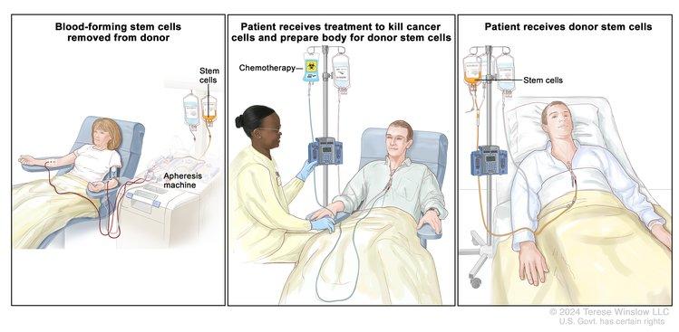
Allogeneic Hematopoietic Stem Cell Transplant (HSCT) Preparative Regimens

In the days just before infusion of the stem cell product (bone marrow, peripheral blood stem cells [PBSCs], or cord blood), HSCT recipients receive chemotherapy/immunotherapy, sometimes combined with radiation therapy. This is called a preparative regimen, and the original intent of this treatment was to:

  • Create bone marrow space in the recipient for the donor cells to engraft.
  • Suppress the immune system or eliminate the recipient T cells to minimize risk of rejection.
  • Intensely treat cancer (if present) with high doses of active agents to overcome therapy resistance.

With the recognition that donor T cells can facilitate engraftment and kill tumors through graft-versus-leukemia (GVL) effects (obviating the need to create bone marrow space and intensely treat cancer), reduced-intensity or minimal-intensity HSCT approaches focusing on immune suppression rather than myeloablation have been developed. The resulting lower toxicity associated with these regimens has led to lower rates of transplant-related mortality and expanded eligibility for allogeneic HSCT to older individuals and younger patients with pre-HSCT comorbidities that put them at risk of severe toxicity after standard HSCT approaches.[1]

Existing preparative regimens vary tremendously in the amount of immunosuppression and myelosuppression they cause, with the lowest-intensity regimens relying heavily on a strong graft-versus-tumor (GVT) effect (see Figure 3).

EnlargeChart showing selected preparative regimens frequently used in pediatric HCT categorized by current definitions as non-myeloablative, reduced-intensity, or myeloablative.
Figure 3. Selected preparative regimens frequently used in pediatric HSCT categorized by current definitions as nonmyeloablative, reduced intensity, or myeloablative. Although FLU plus treosulfan and FLU plus busulfan (full dose) are considered myeloablative approaches, these and similar approaches are called reduced-toxicity regimens.

Although these regimens lead to varying degrees of myelosuppression and immune suppression, they have been grouped clinically into the following three major categories (see Figure 4):[2]

  • Myeloablative: Intense approaches that cause irreversible pancytopenia that requires stem cell rescue for restoration of hematopoiesis.
  • Nonmyeloablative: Regimens that cause minimal cytopenias and do not require stem cell support.
  • Reduced-intensity conditioning: Regimens that are of intermediate intensity and do not meet the definitions of nonmyeloablative or myeloablative regimens.

EnlargeFigure 3; chart shows classification of conditioning regimens based on duration of pancytopenia and requirement for stem cell support; chart shows myeloablative regimens, nonmyeloablative regimens, and reduced intensity regimens.
Figure 4. Classification of conditioning regimens in 3 categories, based on duration of pancytopenia and requirement for stem cell support. Myeloablative regimens (MA) produce irreversible pancytopenia and require stem cell support. Nonmyeloablative regimens (NMA) produce minimal cytopenia and would not require stem cell support. Reduced-intensity regimens (RIC) are regimens which cannot be classified as MA nor NMA. Reprinted from Biology of Blood and Marrow Transplantation, Volume 15 (Issue 12), Andrea Bacigalupo, Karen Ballen, Doug Rizzo, Sergio Giralt, Hillard Lazarus, Vincent Ho, Jane Apperley, Shimon Slavin, Marcelo Pasquini, Brenda M. Sandmaier, John Barrett, Didier Blaise, Robert Lowski, Mary Horowitz, Defining the Intensity of Conditioning Regimens: Working Definitions, Pages 1628-1633, Copyright 2009, with permission from Elsevier.

For years, retrospective studies showed similar outcomes using reduced-intensity and myeloablative approaches.[3,4] However, a Blood and Marrow Transplant Clinical Trials Network trial of adult patients with acute myeloid leukemia (AML) and myelodysplastic neoplasms (MDS) who were randomly assigned to receive either myeloablative or reduced-intensity HSCT approaches demonstrated the importance of regimen intensity.[5]

  • At 18 months, relapse was markedly higher in the reduced-intensity cohort (48% vs. 13.5%, P < .001).
  • Although treatment-related mortality was higher in the myeloablative arm (16% vs. 4%, P = .002), relapse-free survival rates were superior (69% vs. 47%, P < .01) and overall survival (OS) rates were higher in the myeloablative arm (76% vs. 68%), with a nonsignificant P value of .07.

With this in mind, the use of reduced-intensity conditioning and nonmyeloablative regimens is well established in older adults who cannot tolerate more intense myeloablative approaches,[68] but these approaches have been studied in a limited number of younger patients with malignancies.[913] A large Pediatric Blood and Marrow Transplant Consortium study identified patients at high risk of transplant-related mortality with myeloablative regimens (e.g., history of previous myeloablative transplant, severe organ system dysfunction, or active, invasive fungal infection) and successfully treated these patients with a reduced-intensity regimen.[14] Transplant-related mortality was low in this high-risk group, and long-term survival occurred in most patients with minimal or no detectable disease at the time of transplant. Because the risks of relapse are higher with these approaches, their use in pediatric cancer is currently limited to patients ineligible for myeloablative regimens and is most likely to be successful when patients have achieved minimal residual disease (MRD)–negative remissions.[14]

In pediatric HSCT, there has been an effort to develop preparative regimens that are myeloablative but do not have the severe toxicities associated with traditional, highly intense myeloablative approaches such as full-dose total-body irradiation, busulfan and cyclophosphamide, or busulfan, cyclophosphamide, and melphalan. These less-intense regimens are called reduced toxicity and include approaches such as full-dose busulfan and fludarabine or treosulfan and fludarabine. These approaches have been especially useful in transplant for nonmalignant disorders that require full chimerism,[15] but they have often shown similar outcomes when used for patients with malignancies.[16]

Establishing Donor Chimerism

Intense myeloablative approaches almost invariably result in hematopoiesis derived from donor cells upon count recovery after the transplant. The introduction of reduced-toxicity, reduced-intensity, and nonmyeloablative conditioning into HSCT practice has resulted in a slower pace of transition to donor hematopoiesis (gradually increasing from partial to full donor hematopoiesis over months) that sometimes remains partially long-term. DNA-based techniques have been established to differentiate donor from recipient hematopoiesis, applying the word chimerism to describe whether all or part of hematopoiesis after HSCT is from the donor or recipient.

There are several implications regarding the pace and extent of donor chimerism achieved by an HSCT recipient. For patients receiving reduced-intensity conditioning or nonmyeloablative regimens, rapid progression to full donor chimerism is associated with lower relapse rates but more graft-versus-host disease (GVHD).[17] The delayed pace of obtaining full donor chimerism after reduced-intensity regimens has led to late-onset acute GVHD, occurring as late as 6 to 7 months after HSCT (acute GVHD generally occurs within 100 days after myeloablative approaches).[18] A portion of patients achieve stable mixed chimerism of both donor and recipient. Mixed chimerism is associated with more relapse after HSCT for malignancies and less GVHD. However, this condition is often advantageous for nonmalignant HSCT, where usually only a percentage of normal hematopoiesis is needed to correct the underlying disorder and GVHD is not beneficial.[19] Finally, serially measured decreasing donor chimerism, especially T-cell–specific chimerism, has been associated with increased risk of rejection.[20]

Because of the implications of persistent recipient chimerism, most transplant programs test for chimerism shortly after engraftment and continue testing regularly until stable full donor hematopoiesis has been achieved. Investigators have defined two approaches to treat the increased risks of relapse and rejection associated with increasing recipient chimerism: rapid withdrawal of immune suppression and donor lymphocyte infusions (DLI). These approaches are frequently used to address this issue, and they have been shown to decrease relapse risk and stop rejection in some cases.[2123] The timing of immune suppression and dose tapers and approaches to administration of DLI to increase or stabilize donor chimerism vary between stem cell sources. There is also a wide institutional variability, with some institutions proactively following chimerism and often intervening, and others having a more limited approach to interventions. For more information, see the Using donor lymphocyte infusions (DLI) or early withdrawal of immune suppression to enhance GVL section.

References
  1. Deeg HJ, Sandmaier BM: Who is fit for allogeneic transplantation? Blood 116 (23): 4762-70, 2010. [PUBMED Abstract]
  2. Bacigalupo A, Ballen K, Rizzo D, et al.: Defining the intensity of conditioning regimens: working definitions. Biol Blood Marrow Transplant 15 (12): 1628-33, 2009. [PUBMED Abstract]
  3. Luger SM, Ringdén O, Zhang MJ, et al.: Similar outcomes using myeloablative vs reduced-intensity allogeneic transplant preparative regimens for AML or MDS. Bone Marrow Transplant 47 (2): 203-11, 2012. [PUBMED Abstract]
  4. Pulsipher MA, Chitphakdithai P, Logan BR, et al.: Donor, recipient, and transplant characteristics as risk factors after unrelated donor PBSC transplantation: beneficial effects of higher CD34+ cell dose. Blood 114 (13): 2606-16, 2009. [PUBMED Abstract]
  5. Scott BL, Pasquini MC, Logan BR, et al.: Myeloablative Versus Reduced-Intensity Hematopoietic Cell Transplantation for Acute Myeloid Leukemia and Myelodysplastic Syndromes. J Clin Oncol 35 (11): 1154-1161, 2017. [PUBMED Abstract]
  6. Giralt S, Estey E, Albitar M, et al.: Engraftment of allogeneic hematopoietic progenitor cells with purine analog-containing chemotherapy: harnessing graft-versus-leukemia without myeloablative therapy. Blood 89 (12): 4531-6, 1997. [PUBMED Abstract]
  7. Slavin S, Nagler A, Naparstek E, et al.: Nonmyeloablative stem cell transplantation and cell therapy as an alternative to conventional bone marrow transplantation with lethal cytoreduction for the treatment of malignant and nonmalignant hematologic diseases. Blood 91 (3): 756-63, 1998. [PUBMED Abstract]
  8. Storb R, Yu C, Sandmaier BM, et al.: Mixed hematopoietic chimerism after marrow allografts. Transplantation in the ambulatory care setting. Ann N Y Acad Sci 872: 372-5; discussion 375-6, 1999. [PUBMED Abstract]
  9. Bradley MB, Satwani P, Baldinger L, et al.: Reduced intensity allogeneic umbilical cord blood transplantation in children and adolescent recipients with malignant and non-malignant diseases. Bone Marrow Transplant 40 (7): 621-31, 2007. [PUBMED Abstract]
  10. Del Toro G, Satwani P, Harrison L, et al.: A pilot study of reduced intensity conditioning and allogeneic stem cell transplantation from unrelated cord blood and matched family donors in children and adolescent recipients. Bone Marrow Transplant 33 (6): 613-22, 2004. [PUBMED Abstract]
  11. Gómez-Almaguer D, Ruiz-Argüelles GJ, Tarín-Arzaga Ldel C, et al.: Reduced-intensity stem cell transplantation in children and adolescents: the Mexican experience. Biol Blood Marrow Transplant 9 (3): 157-61, 2003. [PUBMED Abstract]
  12. Pulsipher MA, Woolfrey A: Nonmyeloablative transplantation in children. Current status and future prospects. Hematol Oncol Clin North Am 15 (5): 809-34, vii-viii, 2001. [PUBMED Abstract]
  13. Roman E, Cooney E, Harrison L, et al.: Preliminary results of the safety of immunotherapy with gemtuzumab ozogamicin following reduced intensity allogeneic stem cell transplant in children with CD33+ acute myeloid leukemia. Clin Cancer Res 11 (19 Pt 2): 7164s-7170s, 2005. [PUBMED Abstract]
  14. Pulsipher MA, Boucher KM, Wall D, et al.: Reduced-intensity allogeneic transplantation in pediatric patients ineligible for myeloablative therapy: results of the Pediatric Blood and Marrow Transplant Consortium Study ONC0313. Blood 114 (7): 1429-36, 2009. [PUBMED Abstract]
  15. Cseh A, Galimard JE, de la Fuente J, et al.: Busulfan-fludarabine- or treosulfan-fludarabine-based conditioning before allogeneic HSCT from matched sibling donors in paediatric patients with sickle cell disease: A study on behalf of the EBMT Paediatric Diseases and Inborn Errors Working Parties. Br J Haematol 204 (1): e1-e5, 2024. [PUBMED Abstract]
  16. Pulsipher MA, Ahn KW, Bunin NJ, et al.: KIR-favorable TCR-αβ/CD19-depleted haploidentical HCT in children with ALL/AML/MDS: primary analysis of the PTCTC ONC1401 trial. Blood 140 (24): 2556-2572, 2022. [PUBMED Abstract]
  17. Baron F, Baker JE, Storb R, et al.: Kinetics of engraftment in patients with hematologic malignancies given allogeneic hematopoietic cell transplantation after nonmyeloablative conditioning. Blood 104 (8): 2254-62, 2004. [PUBMED Abstract]
  18. Vigorito AC, Campregher PV, Storer BE, et al.: Evaluation of NIH consensus criteria for classification of late acute and chronic GVHD. Blood 114 (3): 702-8, 2009. [PUBMED Abstract]
  19. Marsh RA, Vaughn G, Kim MO, et al.: Reduced-intensity conditioning significantly improves survival of patients with hemophagocytic lymphohistiocytosis undergoing allogeneic hematopoietic cell transplantation. Blood 116 (26): 5824-31, 2010. [PUBMED Abstract]
  20. McSweeney PA, Niederwieser D, Shizuru JA, et al.: Hematopoietic cell transplantation in older patients with hematologic malignancies: replacing high-dose cytotoxic therapy with graft-versus-tumor effects. Blood 97 (11): 3390-400, 2001. [PUBMED Abstract]
  21. Bader P, Kreyenberg H, Hoelle W, et al.: Increasing mixed chimerism is an important prognostic factor for unfavorable outcome in children with acute lymphoblastic leukemia after allogeneic stem-cell transplantation: possible role for pre-emptive immunotherapy? J Clin Oncol 22 (9): 1696-705, 2004. [PUBMED Abstract]
  22. Horn B, Soni S, Khan S, et al.: Feasibility study of preemptive withdrawal of immunosuppression based on chimerism testing in children undergoing myeloablative allogeneic transplantation for hematologic malignancies. Bone Marrow Transplant 43 (6): 469-76, 2009. [PUBMED Abstract]
  23. Haines HL, Bleesing JJ, Davies SM, et al.: Outcomes of donor lymphocyte infusion for treatment of mixed donor chimerism after a reduced-intensity preparative regimen for pediatric patients with nonmalignant diseases. Biol Blood Marrow Transplant 21 (2): 288-92, 2015. [PUBMED Abstract]

Immunotherapeutic Effects of Allogeneic Hematopoietic Stem Cell Transplant (HSCT)

Graft-Versus-Leukemia (GVL) Effect

Early studies in HSCT focused on the delivery of intense myeloablative preparative regimens followed by rescue of the hematopoietic system with either an autologous or allogeneic bone marrow transplant. Investigators quickly showed that allogeneic approaches led to a decreased risk of relapse caused by an immunotherapeutic reaction of the new bone marrow graft against tumor antigens. This phenomenon came to be termed the GVL or graft-versus-tumor (GVT) effect and has been associated with mismatches to both major and minor HLA antigens.

The GVL effect is challenging to use therapeutically because of a strong association between GVL and clinical graft-versus-host disease (GVHD). For standard approaches to HSCT, the highest survival rates have been associated with mild or moderate GVHD (grades I to II in acute myeloid leukemia [AML] and grades I to III in acute lymphoblastic leukemia [ALL]), compared with patients who have no GVHD and experience more relapse or patients with severe GVHD who experience more transplant-related mortality.[13]; [4][Level of evidence C2]

Understanding when GVL occurs and how to use GVL optimally is challenging. One method of study compares rates of relapse and survival between patients undergoing myeloablative HSCT with either autologous or allogeneic donors for a given disease.

  • Leukemia and myelodysplastic neoplasms (MDS): A clear advantage has been noted when allogeneic approaches are used for ALL, AML, chronic myelogenous leukemia (CML), and MDS. For ALL and AML specifically, autologous HSCT approaches for most high-risk patient groups have shown results similar to those obtained with chemotherapy, while allogeneic approaches produced superior results and are therefore useful for chemoresistant or relapsed patients.[5,6]
  • Hodgkin lymphoma (HL) and non-Hodgkin lymphoma (NHL): Patients with HL or NHL generally fare better with autologous approaches, although there may be a role for allogeneic approaches in relapsed lymphoblastic lymphoma, lymphoma that is poorly responsive to chemotherapy, or lymphoma that has relapsed after autologous HSCT.[7]

Further insights into the therapeutic benefit of GVL/GVT for given diseases have come from the use of reduced-intensity preparative regimens. This approach to transplant relies on GVL because, in most cases, the intensity of the preparative regimen is not sufficient for cure. Although studies have shown benefit for patients pursuing this approach when they are ineligible for standard transplant,[8] this approach has not been used for most children with cancer who require HSCT because pediatric cancer patients can generally undergo myeloablative approaches safely. For more information, see the Allogeneic HSCT Preparative Regimens section.

Using donor lymphocyte infusions (DLI) or early withdrawal of immune suppression to enhance GVL

GVL can be achieved therapeutically through the infusion of cells after transplant that either specifically or nonspecifically target the tumor. The most common approach is the use of DLI. This approach relies on the persistence of donor T-cell engraftment after transplant to prevent rejection of donor lymphocytes infused to induce GVL.

Therapeutic DLI results in potent responses in patients with CML who relapse after transplant (60%–80% enter into long-term remission),[9] but responses in patients with other diseases (such as AML and ALL) have been less potent, with long-term survival rates of only 20% to 30%.[10] DLI works poorly in patients with acute leukemia who relapse early and who have high levels of active disease. Late relapse (>6 months after transplant) and the treatment of patients into complete remission with chemotherapy before DLI have been associated with improved outcomes.[11] Infusions of donor lymphocytes modified to enhance GVL or other donor cells (e.g., natural killer [NK] cells) have also been studied but have yet to be generally adopted.

Another method of delivering GVL therapeutically is the rapid withdrawal of immune suppression after HSCT. Some studies have scheduled more rapid immune suppression tapers based on donor type (related donors are tapered more quickly than are unrelated donors because of less GVHD risk), and others have used sensitive measures of either low levels of persistent recipient cells (recipient chimerism) or minimal residual disease to assess the risk of relapse and trigger rapid taper of immune suppression.

A combination of early withdrawal of immune suppression after HSCT with DLI to prevent relapse in patients at high risk of relapse because of persistent/progressive recipient chimerism has been tested in patients who underwent transplant for both ALL and AML.[12][Level of evidence B4]; [13][Level of evidence C2]

  • ALL: One study found increasing recipient chimerism in 46 of 101 patients with ALL. Thirty-one of those patients had withdrawal of immune suppression, and a portion went on to receive DLI if GVHD did not occur. This group had a survival rate of 37%, compared with 0% in the 15 patients who did not undergo this approach (P < .001).[14]
  • AML: About 20% of patients with AML experienced mixed chimerism after HSCT and were identified as high risk. Of these patients, 54% survived if they underwent withdrawal of immune suppression with or without DLI. There were no survivors among those who did not receive this therapy.[15]

Other immunological and cell therapy approaches under evaluation

Role of killer immunoglobulin-like receptor (KIR) mismatching in HSCT

Donor-derived NK cells in the post-HSCT setting have been shown to promote the following:[1618]

  • Engraftment.
  • Decreased GVHD.
  • Fewer relapses of hematological malignancies.
  • Improved survival.

NK-cell function is modulated by interactions with a number of receptor families, including activating and inhibiting KIR. The KIR effect in the allogeneic HSCT setting hinges on the expression of specific inhibitory KIR on donor-derived NK cells and either the presence or absence of their matching HLA class I molecules (KIR ligands) on recipient leukemic and normal cells. Normally, the presence of specific KIR ligands interacting with paired inhibitory KIR molecules prevents NK cell attacks on healthy cells. In the allogeneic transplant setting, recipient leukemia cells genetically differ from donor NK cells, and they may not have the appropriate inhibitory KIR ligand. Mismatch of ligand and receptor allows NK-cell–based killing of recipient leukemia cells to proceed for certain donor-recipient genetic combinations.

The original observation of decreased relapse with certain KIR-ligand combinations was made in the setting of T-cell–depleted haploidentical transplant and was strongest after HSCT for AML.[17,19] However, some haploidentical transplant studies have not shown this effect.[20] Along with decreasing relapse, these studies have suggested a decrease in GVHD with appropriate KIR-ligand combinations. Many subsequent studies did not detect survival effects for KIR-incompatible HSCT using standard transplant methods,[2124] which has led to the conclusion that T-cell depletion may be necessary to remove other forms of inhibitory cellular interactions.

Decreased relapse and better survival have been noted with donor/recipient KIR-ligand incompatibility after cord blood HSCT, a relatively T-cell–depleted procedure.[25,26] In contrast to this notion, one study demonstrated that some KIR mismatching combinations (activating receptor KIR2DS1 with the HLA C1 ligand) can lead to decreased relapse after AML HSCT without T-cell depletion.[27] The role of KIR incompatibility in sibling donor HSCT and in diseases other than AML is controversial, but in pediatrics, at least two groups have found better outcomes with specific types of KIR mismatching in ALL.[2830]

A current challenge associated with studies of KIR is that several different approaches have been used to determine what is KIR incompatible and what are the most favorable combinations of KIR molecules in donor-recipient pairs.[19,31,32] Activating KIR molecules have also been shown to contribute to the effect.[33] The standardization of KIR classification and prospective studies should help clarify the utility and importance of this approach. Because a limited number of centers perform haploidentical HSCT and the results of other approaches to HSCT are preliminary, most transplant programs do not use KIR mismatching as part of their strategy for choosing a donor. Full HLA matching is considered most important for outcome, with considerations of KIR mismatching or choosing donors with favorable KIR activation profiles remaining secondary.

NK-cell transplant

With a low risk of GVHD and demonstrated efficacy in decreasing relapse in posthaploidentical HSCT settings, NK-cell infusions as a method of treating high-risk patients and consolidating patients in remission have been studied:

Evidence (NK-cell transplant outcomes):

  1. A University of Minnesota research group compared approaches with different NK-cell populations.[34]
    • The study initially failed to demonstrate efficacy with autologous NK cells, but it found that intense immunoablative therapy followed by purified haploidentical NK cells and interleukin-2 (IL-2) maintenance led to remission in 5 of 19 high-risk patients with AML.
  2. Researchers at St. Jude Children’s Research Hospital treated ten intermediate-risk patients with AML who had completed chemotherapy and were in remission. The patients received lower-dose immunosuppression followed by haploidentical NK-cell infusions and IL-2 for consolidation.[35]
    • Expansion of NK cells was noted in all nine of the KIR-incompatible donor-recipient pairs.
    • All ten children remained in remission at 2 years.
    • A follow-up phase II study is under way, as are many investigations into NK-cell therapy for a number of cancer types.

    Although early survival rates are high in this high-risk AML cohort, multicenter confirmatory studies will be necessary to establish the efficacy of these types of NK-cell approaches.

  3. Other investigators have used expanded/activated NK cells before and after HSCT.[36] One approach that included the culturing of haploidentical NK cells with membrane-bound IL-21 showed marked expansion and high activity. These cells were then infused just before haploidentical HSCT, followed by additional infusions on day 7 and 28 after HSCT.[36]
References
  1. Yeshurun M, Weisdorf D, Rowe JM, et al.: The impact of the graft-versus-leukemia effect on survival in acute lymphoblastic leukemia. Blood Adv 3 (4): 670-680, 2019. [PUBMED Abstract]
  2. Pulsipher MA, Langholz B, Wall DA, et al.: The addition of sirolimus to tacrolimus/methotrexate GVHD prophylaxis in children with ALL: a phase 3 Children’s Oncology Group/Pediatric Blood and Marrow Transplant Consortium trial. Blood 123 (13): 2017-25, 2014. [PUBMED Abstract]
  3. Neudorf S, Sanders J, Kobrinsky N, et al.: Allogeneic bone marrow transplantation for children with acute myelocytic leukemia in first remission demonstrates a role for graft versus leukemia in the maintenance of disease-free survival. Blood 103 (10): 3655-61, 2004. [PUBMED Abstract]
  4. Boyiadzis M, Arora M, Klein JP, et al.: Impact of Chronic Graft-versus-Host Disease on Late Relapse and Survival on 7,489 Patients after Myeloablative Allogeneic Hematopoietic Cell Transplantation for Leukemia. Clin Cancer Res 21 (9): 2020-8, 2015. [PUBMED Abstract]
  5. Woods WG, Neudorf S, Gold S, et al.: A comparison of allogeneic bone marrow transplantation, autologous bone marrow transplantation, and aggressive chemotherapy in children with acute myeloid leukemia in remission. Blood 97 (1): 56-62, 2001. [PUBMED Abstract]
  6. Ribera JM, Ortega JJ, Oriol A, et al.: Comparison of intensive chemotherapy, allogeneic, or autologous stem-cell transplantation as postremission treatment for children with very high risk acute lymphoblastic leukemia: PETHEMA ALL-93 Trial. J Clin Oncol 25 (1): 16-24, 2007. [PUBMED Abstract]
  7. Gross TG, Hale GA, He W, et al.: Hematopoietic stem cell transplantation for refractory or recurrent non-Hodgkin lymphoma in children and adolescents. Biol Blood Marrow Transplant 16 (2): 223-30, 2010. [PUBMED Abstract]
  8. Pulsipher MA, Boucher KM, Wall D, et al.: Reduced-intensity allogeneic transplantation in pediatric patients ineligible for myeloablative therapy: results of the Pediatric Blood and Marrow Transplant Consortium Study ONC0313. Blood 114 (7): 1429-36, 2009. [PUBMED Abstract]
  9. Porter DL, Collins RH, Shpilberg O, et al.: Long-term follow-up of patients who achieved complete remission after donor leukocyte infusions. Biol Blood Marrow Transplant 5 (4): 253-61, 1999. [PUBMED Abstract]
  10. Levine JE, Barrett AJ, Zhang MJ, et al.: Donor leukocyte infusions to treat hematologic malignancy relapse following allo-SCT in a pediatric population. Bone Marrow Transplant 42 (3): 201-5, 2008. [PUBMED Abstract]
  11. Warlick ED, DeFor T, Blazar BR, et al.: Successful remission rates and survival after lymphodepleting chemotherapy and donor lymphocyte infusion for relapsed hematologic malignancies postallogeneic hematopoietic cell transplantation. Biol Blood Marrow Transplant 18 (3): 480-6, 2012. [PUBMED Abstract]
  12. Horn B, Petrovic A, Wahlstrom J, et al.: Chimerism-based pre-emptive immunotherapy with fast withdrawal of immunosuppression and donor lymphocyte infusions after allogeneic stem cell transplantation for pediatric hematologic malignancies. Biol Blood Marrow Transplant 21 (4): 729-37, 2015. [PUBMED Abstract]
  13. Horn B, Wahlstrom JT, Melton A, et al.: Early mixed chimerism-based preemptive immunotherapy in children undergoing allogeneic hematopoietic stem cell transplantation for acute leukemia. Pediatr Blood Cancer 64 (8): , 2017. [PUBMED Abstract]
  14. Bader P, Kreyenberg H, Hoelle W, et al.: Increasing mixed chimerism is an important prognostic factor for unfavorable outcome in children with acute lymphoblastic leukemia after allogeneic stem-cell transplantation: possible role for pre-emptive immunotherapy? J Clin Oncol 22 (9): 1696-705, 2004. [PUBMED Abstract]
  15. Rettinger E, Willasch AM, Kreyenberg H, et al.: Preemptive immunotherapy in childhood acute myeloid leukemia for patients showing evidence of mixed chimerism after allogeneic stem cell transplantation. Blood 118 (20): 5681-8, 2011. [PUBMED Abstract]
  16. Ruggeri L, Capanni M, Urbani E, et al.: Effectiveness of donor natural killer cell alloreactivity in mismatched hematopoietic transplants. Science 295 (5562): 2097-100, 2002. [PUBMED Abstract]
  17. Giebel S, Locatelli F, Lamparelli T, et al.: Survival advantage with KIR ligand incompatibility in hematopoietic stem cell transplantation from unrelated donors. Blood 102 (3): 814-9, 2003. [PUBMED Abstract]
  18. Bari R, Rujkijyanont P, Sullivan E, et al.: Effect of donor KIR2DL1 allelic polymorphism on the outcome of pediatric allogeneic hematopoietic stem-cell transplantation. J Clin Oncol 31 (30): 3782-90, 2013. [PUBMED Abstract]
  19. Ruggeri L, Mancusi A, Capanni M, et al.: Donor natural killer cell allorecognition of missing self in haploidentical hematopoietic transplantation for acute myeloid leukemia: challenging its predictive value. Blood 110 (1): 433-40, 2007. [PUBMED Abstract]
  20. Merli P, Algeri M, Galaverna F, et al.: TCRαβ/CD19 cell-depleted HLA-haploidentical transplantation to treat pediatric acute leukemia: updated final analysis. Blood 143 (3): 279-289, 2024. [PUBMED Abstract]
  21. Davies SM, Ruggieri L, DeFor T, et al.: Evaluation of KIR ligand incompatibility in mismatched unrelated donor hematopoietic transplants. Killer immunoglobulin-like receptor. Blood 100 (10): 3825-7, 2002. [PUBMED Abstract]
  22. Farag SS, Bacigalupo A, Eapen M, et al.: The effect of KIR ligand incompatibility on the outcome of unrelated donor transplantation: a report from the center for international blood and marrow transplant research, the European blood and marrow transplant registry, and the Dutch registry. Biol Blood Marrow Transplant 12 (8): 876-84, 2006. [PUBMED Abstract]
  23. Davies SM, Iannone R, Alonzo TA, et al.: A Phase 2 Trial of KIR-Mismatched Unrelated Donor Transplantation Using in Vivo T Cell Depletion with Antithymocyte Globulin in Acute Myelogenous Leukemia: Children’s Oncology Group AAML05P1 Study. Biol Blood Marrow Transplant 26 (4): 712-717, 2020. [PUBMED Abstract]
  24. Verneris MR, Miller JS, Hsu KC, et al.: Investigation of donor KIR content and matching in children undergoing hematopoietic cell transplantation for acute leukemia. Blood Adv 4 (7): 1350-1356, 2020. [PUBMED Abstract]
  25. Cooley S, Trachtenberg E, Bergemann TL, et al.: Donors with group B KIR haplotypes improve relapse-free survival after unrelated hematopoietic cell transplantation for acute myelogenous leukemia. Blood 113 (3): 726-32, 2009. [PUBMED Abstract]
  26. Willemze R, Rodrigues CA, Labopin M, et al.: KIR-ligand incompatibility in the graft-versus-host direction improves outcomes after umbilical cord blood transplantation for acute leukemia. Leukemia 23 (3): 492-500, 2009. [PUBMED Abstract]
  27. Venstrom JM, Pittari G, Gooley TA, et al.: HLA-C-dependent prevention of leukemia relapse by donor activating KIR2DS1. N Engl J Med 367 (9): 805-16, 2012. [PUBMED Abstract]
  28. Leung W: Use of NK cell activity in cure by transplant. Br J Haematol 155 (1): 14-29, 2011. [PUBMED Abstract]
  29. Leung W, Campana D, Yang J, et al.: High success rate of hematopoietic cell transplantation regardless of donor source in children with very high-risk leukemia. Blood 118 (2): 223-30, 2011. [PUBMED Abstract]
  30. Oevermann L, Michaelis SU, Mezger M, et al.: KIR B haplotype donors confer a reduced risk for relapse after haploidentical transplantation in children with ALL. Blood 124 (17): 2744-7, 2014. [PUBMED Abstract]
  31. Leung W, Iyengar R, Triplett B, et al.: Comparison of killer Ig-like receptor genotyping and phenotyping for selection of allogeneic blood stem cell donors. J Immunol 174 (10): 6540-5, 2005. [PUBMED Abstract]
  32. Pulsipher MA, Ahn KW, Bunin NJ, et al.: KIR-favorable TCR-αβ/CD19-depleted haploidentical HCT in children with ALL/AML/MDS: primary analysis of the PTCTC ONC1401 trial. Blood 140 (24): 2556-2572, 2022. [PUBMED Abstract]
  33. Cooley S, Weisdorf DJ, Guethlein LA, et al.: Donor selection for natural killer cell receptor genes leads to superior survival after unrelated transplantation for acute myelogenous leukemia. Blood 116 (14): 2411-9, 2010. [PUBMED Abstract]
  34. Miller JS, Soignier Y, Panoskaltsis-Mortari A, et al.: Successful adoptive transfer and in vivo expansion of human haploidentical NK cells in patients with cancer. Blood 105 (8): 3051-7, 2005. [PUBMED Abstract]
  35. Rubnitz JE, Inaba H, Ribeiro RC, et al.: NKAML: a pilot study to determine the safety and feasibility of haploidentical natural killer cell transplantation in childhood acute myeloid leukemia. J Clin Oncol 28 (6): 955-9, 2010. [PUBMED Abstract]
  36. Ciurea SO, Schafer JR, Bassett R, et al.: Phase 1 clinical trial using mbIL21 ex vivo-expanded donor-derived NK cells after haploidentical transplantation. Blood 130 (16): 1857-1868, 2017. [PUBMED Abstract]

Treatment Options Under Clinical Evaluation

Information about National Cancer Institute (NCI)–supported clinical trials can be found on the NCI website. For information about clinical trials sponsored by other organizations, see the ClinicalTrials.gov website.

The following is an example of a national and/or institutional clinical trial that is currently being conducted:

  • ASCT2031 (NCT05457556) (Mismatched Related Donor [Haplo] Versus Matched Unrelated Donor [MUD] Hematopoietic Stem Cell Transplant [HSCT] for Children, Adolescents, and Young Adults with Acute Leukemia or Myelodysplastic Neoplasms [MDS]): This is a phase III randomized controlled trial that will compare outcomes between haplo HSCT and MUD HSCT for patients with acute leukemias or MDS. Patients with both available donor sources (MUD and haplo) will be randomly assigned to either Arm A (haplo HSCT) or Arm B (MUD HSCT). The randomization will be stratified by patient age, complete remission status, and disease type. Patients who do not have both an available MUD and haplo donor, but do have a haplo donor, will be assigned to Arm C and receive a nonrandomized haplo HSCT. Arm C is expected to enroll a higher fraction of racial and ethnic minority patients because they lack MUDs more often than White patients. This third arm will allow direct comparisons of haplo HSCT outcomes between racial and ethnic minority patients and White patients.

    The clinically relevant end points, severe graft-versus-host disease (GVHD), and disease-free survival (DFS) in haplo HSCT versus MUD HSCT will be compared as co-primary objectives. The hypothesis is that the incidence of severe GVHD will be less frequent and DFS will be noninferior for haplo HSCT than MUD HSCT.

Latest Updates to This Summary (06/13/2024)

The PDQ cancer information summaries are reviewed regularly and updated as new information becomes available. This section describes the latest changes made to this summary as of the date above.

This summary was comprehensively reviewed.

This summary is written and maintained by the PDQ Pediatric Treatment Editorial Board, which is editorially independent of NCI. The summary reflects an independent review of the literature and does not represent a policy statement of NCI or NIH. More information about summary policies and the role of the PDQ Editorial Boards in maintaining the PDQ summaries can be found on the About This PDQ Summary and PDQ® Cancer Information for Health Professionals pages.

About This PDQ Summary

Purpose of This Summary

This PDQ cancer information summary for health professionals provides comprehensive, peer-reviewed, evidence-based information about the use of allogeneic hematopoietic stem cell transplant in treating pediatric cancer. It is intended as a resource to inform and assist clinicians in the care of their patients. It does not provide formal guidelines or recommendations for making health care decisions.

Reviewers and Updates

This summary is reviewed regularly and updated as necessary by the PDQ Pediatric Treatment Editorial Board, which is editorially independent of the National Cancer Institute (NCI). The summary reflects an independent review of the literature and does not represent a policy statement of NCI or the National Institutes of Health (NIH).

Board members review recently published articles each month to determine whether an article should:

  • be discussed at a meeting,
  • be cited with text, or
  • replace or update an existing article that is already cited.

Changes to the summaries are made through a consensus process in which Board members evaluate the strength of the evidence in the published articles and determine how the article should be included in the summary.

The lead reviewers for Pediatric Allogeneic Hematopoietic Stem Cell Transplant are:

  • Thomas G. Gross, MD, PhD (National Cancer Institute)
  • Michael A. Pulsipher, MD (Huntsman Cancer Institute at University of Utah)

Any comments or questions about the summary content should be submitted to Cancer.gov through the NCI website’s Email Us. Do not contact the individual Board Members with questions or comments about the summaries. Board members will not respond to individual inquiries.

Levels of Evidence

Some of the reference citations in this summary are accompanied by a level-of-evidence designation. These designations are intended to help readers assess the strength of the evidence supporting the use of specific interventions or approaches. The PDQ Pediatric Treatment Editorial Board uses a formal evidence ranking system in developing its level-of-evidence designations.

Permission to Use This Summary

PDQ is a registered trademark. Although the content of PDQ documents can be used freely as text, it cannot be identified as an NCI PDQ cancer information summary unless it is presented in its entirety and is regularly updated. However, an author would be permitted to write a sentence such as “NCI’s PDQ cancer information summary about breast cancer prevention states the risks succinctly: [include excerpt from the summary].”

The preferred citation for this PDQ summary is:

PDQ® Pediatric Treatment Editorial Board. PDQ Pediatric Allogeneic Hematopoietic Stem Cell Transplant. Bethesda, MD: National Cancer Institute. Updated <MM/DD/YYYY>. Available at: /types/childhood-cancers/hp-stem-cell-transplant/allogeneic. Accessed <MM/DD/YYYY>. [PMID: 35133766]

Images in this summary are used with permission of the author(s), artist, and/or publisher for use within the PDQ summaries only. Permission to use images outside the context of PDQ information must be obtained from the owner(s) and cannot be granted by the National Cancer Institute. Information about using the illustrations in this summary, along with many other cancer-related images, is available in Visuals Online, a collection of over 2,000 scientific images.

Disclaimer

Based on the strength of the available evidence, treatment options may be described as either “standard” or “under clinical evaluation.” These classifications should not be used as a basis for insurance reimbursement determinations. More information on insurance coverage is available on Cancer.gov on the Managing Cancer Care page.

Contact Us

More information about contacting us or receiving help with the Cancer.gov website can be found on our Contact Us for Help page. Questions can also be submitted to Cancer.gov through the website’s Email Us.

Pediatric Hematopoietic Stem Cell Transplant and Cellular Therapy for Cancer (PDQ®)–Health Professional Version

Pediatric Hematopoietic Stem Cell Transplant and Cellular Therapy for Cancer (PDQ®)–Health Professional Version

General Information About Hematopoietic Stem Cell Transplant (HSCT)

Rationale for HSCT

Blood and marrow transplant, or HSCT, is a procedure that involves infusion of hematopoietic stem cells (along with hematopoietic progenitor cells) to reconstitute the hematopoietic system of a patient. The infusion of hematopoietic stem cells generally follows a preparative regimen consisting of agents designed to do the following:

  • Create marrow space.
  • Suppress the patient’s immune system to prevent rejection.
  • Eradicate malignant cells in patients with cancer.

HSCT is currently used in the:

  • Treatment of malignancies,
  • Replacement or modulation of an absent or poorly functioning hematopoietic or immune system, or for the
  • Treatment of certain genetic diseases. In these cases, insufficient expression of the affected gene product can be partially or completely overcome by circulating hematopoietic stem cells transplanted from a donor with normal gene expression.

This summary focuses on the use of HSCT in the treatment of childhood malignancies.

Autologous Versus Allogeneic HSCT

The two major HSCT approaches currently in use are the following:

  • Autologous (using the patient’s own hematopoietic stem cells).
  • Allogeneic (using related- or unrelated-donor hematopoietic stem cells).

An autologous transplant treats cancer by exposing patients to high-dose therapy with the intent of overcoming chemotherapy resistance in tumor cells, followed by infusion of the patient’s previously stored hematopoietic stem cells. The transplant can be performed in a single procedure or tandem sequential procedures.

Allogeneic transplant approaches to cancer treatment also may involve high-dose therapy, but because of immunologic differences between the donor and recipient, an additional graft-versus-tumor or graft-versus-leukemia treatment effect can occur. Although autologous approaches are associated with less short-term mortality, many malignancies are resistant to even high doses of chemotherapy and/or involve the bone marrow. Therefore, patients may require allogeneic approaches for optimal outcomes.

Determining When HSCT Is Indicated: Comparison of HSCT and Chemotherapy Outcomes

Because the outcomes using chemotherapy and HSCT treatments have been changing over time, these approaches should be compared regularly to continually redefine optimal therapy for a given patient. For some diseases, randomized trials or intent-to-treat trials using an HLA-matched sibling donor have established the benefit of HSCT by direct comparison.[1,2] However, for very high-risk patients, such as those with early relapse of acute lymphoblastic leukemia, randomized trials have not been feasible because of investigator bias.[3,4]

In general, HSCT typically benefits only children at high risk of relapse with standard chemotherapy approaches. Accordingly, treatment schemas that accurately identify these high-risk patients and offer HSCT if appropriate allogeneic donors are available are the preferred approach for many diseases.[5] Less well-established, higher-risk approaches to HSCT, such as haploidentical transplant, are sometimes reserved for only the very highest-risk patients. However, these higher-risk approaches are becoming safer and more efficacious and are increasingly used interchangeably with fully matched allogeneic approaches.[69] For more information, see the Haploidentical HSCT section in Pediatric Allogeneic Hematopoietic Stem Cell Transplant.

When comparisons of similar patients treated with HSCT or chemotherapy are made in the setting where randomized or intent-to-treat studies are not feasible, the following issues should be considered:

  1. Remission/disease status: Comparisons of HSCT and chemotherapy should include only patients who obtain remission, preferably after similar approaches to salvage therapy, because patients who fail to obtain remission have poor outcomes with any therapy.[10]

    To account for time-to-transplant bias, the chemotherapy comparator arm should include only patients who maintained remission until the median time to HSCT. The HSCT comparator arm should also include only patients who achieved the initial remission mentioned above and maintained that remission until the time of HSCT.[10]

    High-risk and intermediate-risk patient groups should not be combined because benefit or lack of benefit of HSCT in the high-risk group can be masked by different levels of benefit in the intermediate-risk group.[10]

  2. Therapy approaches used for comparison: Comparisons should be made with the best or most used chemotherapy/immunotherapy or HSCT approaches used during the time frame under study.
  3. HSCT approach: HSCT approaches that are very high risk or have documented lower rates of survival should not be combined for analysis with standard-risk HSCT approaches.
  4. Criteria for relapse: Risk factors for relapse should be carefully defined, and analysis should be based on the most current knowledge of risk.
  5. Selection bias: Attempts should be made to understand and eliminate or correct for selection bias. Examples include the following:
    • Higher-risk patients preferentially undergoing HSCT (i.e., patients who take several rounds to achieve remission or have disease relapse after obtaining remission and go back into a subsequent remission before HSCT).
    • Sicker patients deferred from HSCT because of comorbidities.
    • Related to the time-to-transplant bias noted above, patients who undergo HSCT after relapse or recurrence are a subset of all patients with a disease recurrence and will be selected from those who are able to obtain a remission and remain healthy enough to undergo HSCT.
    • Patient or parent refusal.
    • Lack of or inability to obtain insurance approval for HSCT.
    • Lack of access to HSCT because of distance or inability to travel.

Physician bias, for or against HSCT, is difficult to control for or detect. The effects of access to HSCT and therapeutic bias on outcomes of pediatric malignancies for which HSCT may be indicated have been poorly studied.

For more information about pediatric HSCT, see the following summaries:

References
  1. Matthay KK, Villablanca JG, Seeger RC, et al.: Treatment of high-risk neuroblastoma with intensive chemotherapy, radiotherapy, autologous bone marrow transplantation, and 13-cis-retinoic acid. Children’s Cancer Group. N Engl J Med 341 (16): 1165-73, 1999. [PUBMED Abstract]
  2. Woods WG, Neudorf S, Gold S, et al.: A comparison of allogeneic bone marrow transplantation, autologous bone marrow transplantation, and aggressive chemotherapy in children with acute myeloid leukemia in remission. Blood 97 (1): 56-62, 2001. [PUBMED Abstract]
  3. Lawson SE, Harrison G, Richards S, et al.: The UK experience in treating relapsed childhood acute lymphoblastic leukaemia: a report on the medical research council UKALLR1 study. Br J Haematol 108 (3): 531-43, 2000. [PUBMED Abstract]
  4. Gaynon PS, Harris RE, Altman AJ, et al.: Bone marrow transplantation versus prolonged intensive chemotherapy for children with acute lymphoblastic leukemia and an initial bone marrow relapse within 12 months of the completion of primary therapy: Children’s Oncology Group study CCG-1941. J Clin Oncol 24 (19): 3150-6, 2006. [PUBMED Abstract]
  5. Merli P, Algeri M, Del Bufalo F, et al.: Hematopoietic Stem Cell Transplantation in Pediatric Acute Lymphoblastic Leukemia. Curr Hematol Malig Rep 14 (2): 94-105, 2019. [PUBMED Abstract]
  6. Bertaina A, Merli P, Rutella S, et al.: HLA-haploidentical stem cell transplantation after removal of αβ+ T and B cells in children with nonmalignant disorders. Blood 124 (5): 822-6, 2014. [PUBMED Abstract]
  7. Handgretinger R, Chen X, Pfeiffer M, et al.: Feasibility and outcome of reduced-intensity conditioning in haploidentical transplantation. Ann N Y Acad Sci 1106: 279-89, 2007. [PUBMED Abstract]
  8. Huang XJ, Liu DH, Liu KY, et al.: Haploidentical hematopoietic stem cell transplantation without in vitro T-cell depletion for the treatment of hematological malignancies. Bone Marrow Transplant 38 (4): 291-7, 2006. [PUBMED Abstract]
  9. Luznik L, Fuchs EJ: High-dose, post-transplantation cyclophosphamide to promote graft-host tolerance after allogeneic hematopoietic stem cell transplantation. Immunol Res 47 (1-3): 65-77, 2010. [PUBMED Abstract]
  10. Pulsipher MA, Peters C, Pui CH: High-risk pediatric acute lymphoblastic leukemia: to transplant or not to transplant? Biol Blood Marrow Transplant 17 (1 Suppl): S137-48, 2011. [PUBMED Abstract]

Current Clinical Trials

Use our advanced clinical trial search to find NCI-supported cancer clinical trials that are now enrolling patients. The search can be narrowed by location of the trial, type of treatment, name of the drug, and other criteria. General information about clinical trials is also available.

Latest Updates to This Summary (06/13/2024)

The PDQ cancer information summaries are reviewed regularly and updated as new information becomes available. This section describes the latest changes made to this summary as of the date above.

This summary was comprehensively reviewed.

This summary is written and maintained by the PDQ Pediatric Treatment Editorial Board, which is editorially independent of NCI. The summary reflects an independent review of the literature and does not represent a policy statement of NCI or NIH. More information about summary policies and the role of the PDQ Editorial Boards in maintaining the PDQ summaries can be found on the About This PDQ Summary and PDQ® Cancer Information for Health Professionals pages.

About This PDQ Summary

Purpose of This Summary

This PDQ cancer information summary for health professionals provides comprehensive, peer-reviewed, evidence-based information about the use of hematopoietic stem cell transplant and cellular therapy in treating pediatric cancer. It is intended as a resource to inform and assist clinicians in the care of their patients. It does not provide formal guidelines or recommendations for making health care decisions.

Reviewers and Updates

This summary is reviewed regularly and updated as necessary by the PDQ Pediatric Treatment Editorial Board, which is editorially independent of the National Cancer Institute (NCI). The summary reflects an independent review of the literature and does not represent a policy statement of NCI or the National Institutes of Health (NIH).

Board members review recently published articles each month to determine whether an article should:

  • be discussed at a meeting,
  • be cited with text, or
  • replace or update an existing article that is already cited.

Changes to the summaries are made through a consensus process in which Board members evaluate the strength of the evidence in the published articles and determine how the article should be included in the summary.

The lead reviewers for Pediatric Hematopoietic Stem Cell Transplant and Cellular Therapy for Cancer are:

  • Thomas G. Gross, MD, PhD (National Cancer Institute)
  • Michael A. Pulsipher, MD (Huntsman Cancer Institute at University of Utah)

Any comments or questions about the summary content should be submitted to Cancer.gov through the NCI website’s Email Us. Do not contact the individual Board Members with questions or comments about the summaries. Board members will not respond to individual inquiries.

Levels of Evidence

Some of the reference citations in this summary are accompanied by a level-of-evidence designation. These designations are intended to help readers assess the strength of the evidence supporting the use of specific interventions or approaches. The PDQ Pediatric Treatment Editorial Board uses a formal evidence ranking system in developing its level-of-evidence designations.

Permission to Use This Summary

PDQ is a registered trademark. Although the content of PDQ documents can be used freely as text, it cannot be identified as an NCI PDQ cancer information summary unless it is presented in its entirety and is regularly updated. However, an author would be permitted to write a sentence such as “NCI’s PDQ cancer information summary about breast cancer prevention states the risks succinctly: [include excerpt from the summary].”

The preferred citation for this PDQ summary is:

PDQ® Pediatric Treatment Editorial Board. PDQ Pediatric Hematopoietic Stem Cell Transplant and Cellular Therapy for Cancer. Bethesda, MD: National Cancer Institute. Updated <MM/DD/YYYY>. Available at: /types/childhood-cancers/hp-stem-cell-transplant. Accessed <MM/DD/YYYY>. [PMID: 26389503]

Images in this summary are used with permission of the author(s), artist, and/or publisher for use within the PDQ summaries only. Permission to use images outside the context of PDQ information must be obtained from the owner(s) and cannot be granted by the National Cancer Institute. Information about using the illustrations in this summary, along with many other cancer-related images, is available in Visuals Online, a collection of over 2,000 scientific images.

Disclaimer

Based on the strength of the available evidence, treatment options may be described as either “standard” or “under clinical evaluation.” These classifications should not be used as a basis for insurance reimbursement determinations. More information on insurance coverage is available on Cancer.gov on the Managing Cancer Care page.

Contact Us

More information about contacting us or receiving help with the Cancer.gov website can be found on our Contact Us for Help page. Questions can also be submitted to Cancer.gov through the website’s Email Us.

Pediatric Autologous Hematopoietic Stem Cell Transplant (PDQ®)–Health Professional Version

Pediatric Autologous Hematopoietic Stem Cell Transplant (PDQ®)–Health Professional Version

Collection and Storage of Autologous Hematopoietic Stem Cells

Autologous hematopoietic stem cell transplant (HSCT) procedures require collection of growth-factor–mobilized peripheral blood stem cells (PBSCs) from patients using leukapheresis. Bone marrow can be used for autologous transplants, but PBSCs lead to quicker blood count recovery, resulting in less transplant-related toxicity.

Patients being considered for autologous HSCT are generally given chemotherapy to determine tumor responsiveness and minimize the risk of tumor contamination in their bone marrow. After a number of rounds of chemotherapy, patients undergo the leukapheresis procedure, either as their blood counts recover from chemotherapy or during a break between chemotherapy treatments. Growth factors such as granulocyte colony-stimulating factor are used to increase the number of circulating stem and progenitor cells (CD34+ cells). Collection centers monitor the number of CD34-positive cells in the patient and product each day to determine the best time to begin collection and when collection is complete. Patients with low numbers of CD34-positive cells before collection can often have their cells successfully collected using alternative mobilization approaches (e.g., addition of plerixafor).[1] The collected PBSCs are cryopreserved for later use. After completion of an intensive preparative regimen using high-dose chemotherapy, which varies according to the tumor type, the PBSCs are given to the patient at the time of transplant.

References
  1. Patel B, Pearson H, Zacharoulis S: Mobilisation of haematopoietic stem cells in paediatric patients, prior to autologous transplantation following administration of plerixafor and G-CSF. Pediatr Blood Cancer 62 (8): 1477-80, 2015. [PUBMED Abstract]

General Indications and Considerations for Autologous Procedures

Autologous Hematopoietic Stem Cell Transplant (HSCT) Indications for Solid Tumors and Lymphomas

In pediatrics, the most common autologous HSCT indications are for the treatment of some solid tumors and lymphomas.

Autologous transplants have also been used to reset the immune system in patients with severe autoimmune disorders and to enable engraftment of genetically modified autologous hematopoietic stem cell progenitors to correct or ameliorate inherited disorders (e.g., immunodeficiencies, metabolic disorders, and hemoglobinopathies). These indications are not covered in this summary.

Indications for HSCT vary over time as risk classifications for a given malignancy change and the efficacy of primary therapy improves. It is best to include specific indications in the context of complete therapy for any given disease.

With this in mind, links to sections in specific summaries that cover the most common pediatric autologous HSCT indications are provided below.

  1. Neuroblastoma.
  2. Brain tumors. Indications for young patients to reduce or eliminate cranial radiation therapy; indications for responsive tumors at relapse.
  3. Germ cell tumors (GCTs) (intracranial and extracranial).
  4. Retinoblastoma.
  5. Hodgkin lymphoma.
  6. Non-Hodgkin lymphoma.

For autologous transplants to result in cure of malignancies, the following must apply:

  • A dose-intensified chemotherapy regimen (with or without radiation therapy) with hematopoietic stem cell support is used to achieve a significantly higher cell kill than could be achieved without the use of hematopoietic stem cell support. This approach may include increased tumor kill in areas where standard-dose chemotherapy has less penetration (central nervous system).
  • Meaningful percentages of cure or long-term remission from the disease must occur without significant nonhematopoietic toxicities that would otherwise limit the therapeutic benefit achieved.

The tumor-specific activity and intensity of agents used for autologous regimens have been shown to be important in improving survival.

The contamination of the collected stem cell product by persistent tumor cells is a concern with autologous approaches. Although techniques have been developed to remove or purge tumor cells from products, studies to date have shown no benefit to tumor purging.[1]

References
  1. Kreissman SG, Seeger RC, Matthay KK, et al.: Purged versus non-purged peripheral blood stem-cell transplantation for high-risk neuroblastoma (COG A3973): a randomised phase 3 trial. Lancet Oncol 14 (10): 999-1008, 2013. [PUBMED Abstract]

Latest Updates to This Summary (06/13/2024)

The PDQ cancer information summaries are reviewed regularly and updated as new information becomes available. This section describes the latest changes made to this summary as of the date above.

This summary was comprehensively reviewed.

This summary is written and maintained by the PDQ Pediatric Treatment Editorial Board, which is editorially independent of NCI. The summary reflects an independent review of the literature and does not represent a policy statement of NCI or NIH. More information about summary policies and the role of the PDQ Editorial Boards in maintaining the PDQ summaries can be found on the About This PDQ Summary and PDQ® Cancer Information for Health Professionals pages.

About This PDQ Summary

Purpose of This Summary

This PDQ cancer information summary for health professionals provides comprehensive, peer-reviewed, evidence-based information about the use of autologous hematopoietic stem cell transplant in treating pediatric cancer. It is intended as a resource to inform and assist clinicians in the care of their patients. It does not provide formal guidelines or recommendations for making health care decisions.

Reviewers and Updates

This summary is reviewed regularly and updated as necessary by the PDQ Pediatric Treatment Editorial Board, which is editorially independent of the National Cancer Institute (NCI). The summary reflects an independent review of the literature and does not represent a policy statement of NCI or the National Institutes of Health (NIH).

Board members review recently published articles each month to determine whether an article should:

  • be discussed at a meeting,
  • be cited with text, or
  • replace or update an existing article that is already cited.

Changes to the summaries are made through a consensus process in which Board members evaluate the strength of the evidence in the published articles and determine how the article should be included in the summary.

The lead reviewers for Pediatric Autologous Hematopoietic Stem Cell Transplant are:

  • Thomas G. Gross, MD, PhD (National Cancer Institute)
  • Michael A. Pulsipher, MD (Huntsman Cancer Institute at University of Utah)

Any comments or questions about the summary content should be submitted to Cancer.gov through the NCI website’s Email Us. Do not contact the individual Board Members with questions or comments about the summaries. Board members will not respond to individual inquiries.

Levels of Evidence

Some of the reference citations in this summary are accompanied by a level-of-evidence designation. These designations are intended to help readers assess the strength of the evidence supporting the use of specific interventions or approaches. The PDQ Pediatric Treatment Editorial Board uses a formal evidence ranking system in developing its level-of-evidence designations.

Permission to Use This Summary

PDQ is a registered trademark. Although the content of PDQ documents can be used freely as text, it cannot be identified as an NCI PDQ cancer information summary unless it is presented in its entirety and is regularly updated. However, an author would be permitted to write a sentence such as “NCI’s PDQ cancer information summary about breast cancer prevention states the risks succinctly: [include excerpt from the summary].”

The preferred citation for this PDQ summary is:

PDQ® Pediatric Treatment Editorial Board. PDQ Pediatric Autologous Hematopoietic Stem Cell Transplant. Bethesda, MD: National Cancer Institute. Updated <MM/DD/YYYY>. Available at: /types/childhood-cancers/hp-stem-cell-transplant/autologous. Accessed <MM/DD/YYYY>. [PMID: 35133767]

Images in this summary are used with permission of the author(s), artist, and/or publisher for use within the PDQ summaries only. Permission to use images outside the context of PDQ information must be obtained from the owner(s) and cannot be granted by the National Cancer Institute. Information about using the illustrations in this summary, along with many other cancer-related images, is available in Visuals Online, a collection of over 2,000 scientific images.

Disclaimer

Based on the strength of the available evidence, treatment options may be described as either “standard” or “under clinical evaluation.” These classifications should not be used as a basis for insurance reimbursement determinations. More information on insurance coverage is available on Cancer.gov on the Managing Cancer Care page.

Contact Us

More information about contacting us or receiving help with the Cancer.gov website can be found on our Contact Us for Help page. Questions can also be submitted to Cancer.gov through the website’s Email Us.

Hodgkin Lymphoma Treatment (PDQ®)–Patient Version

Hodgkin Lymphoma Treatment (PDQ®)–Patient Version

General Information About Hodgkin Lymphoma

Key Points

  • Hodgkin lymphoma is a disease in which malignant (cancer) cells form in the lymph system.
  • The two main types of Hodgkin lymphoma are classic and nodular lymphocyte-predominant.
  • Being in early or late adulthood, being male, past Epstein-Barr infection, and a family history of Hodgkin lymphoma can increase the risk of Hodgkin lymphoma.
  • Signs and symptoms of Hodgkin lymphoma include swollen lymph nodes, fever, drenching night sweats, weight loss, and fatigue.
  • Tests that examine the lymph system and other parts of the body are used to help diagnose and stage Hodgkin lymphoma.
  • Certain factors affect prognosis (chance of recovery) and treatment options.

Hodgkin lymphoma is a disease in which malignant (cancer) cells form in the lymph system.

Hodgkin lymphoma is a type of cancer that develops in the lymph system. The lymph system is part of the immune system. It helps protect the body from infection and disease.

The lymph system is made up of:

  • Lymph: Colorless, watery fluid that travels through the lymph vessels and carries T and B lymphocytes. Lymphocytes are a type of white blood cell.
  • Lymph vessels: A network of thin tubes that collect lymph from different parts of the body and return it to the bloodstream.
  • Lymph nodes: Small, bean-shaped structures that filter lymph and store white blood cells that help fight infection and disease. Lymph nodes are found along a network of lymph vessels throughout the body. Groups of lymph nodes are found in the mediastinum (the area between the lungs), neck, underarm, abdomen, pelvis, and groin. Hodgkin lymphoma most commonly forms in the lymph nodes above the diaphragm and often in the lymph nodes in the mediastinum.
  • Spleen: An organ that makes lymphocytes, stores red blood cells and lymphocytes, filters the blood, and destroys old blood cells. The spleen is on the left side of the abdomen near the stomach.
  • Thymus: An organ in which T lymphocytes mature and multiply. The thymus is in the chest behind the breastbone.
  • Bone marrow: The soft, spongy tissue in the center of certain bones, such as the hip bone and breastbone. White blood cells, red blood cells, and platelets are made in the bone marrow.
  • Tonsils: Two small masses of lymph tissue at the back of the throat. There is one tonsil on each side of the throat. Hodgkin lymphoma rarely forms in the tonsils.
EnlargeLymphatic system; drawing shows the lymph vessels and lymph organs, including the lymph nodes, tonsils, thymus, spleen, and bone marrow. Also shown is the small intestine (one site of mucosal-associated lymphoid tissue). There are also two pullouts: one showing a close up of the inside structure of a lymph node and the attached artery, vein, and lymph vessels with arrows showing how the lymph (clear, watery fluid) moves into and out of the lymph node, and another showing a close up of bone marrow with blood cells.
The lymph system is part of the body’s immune system and is made up of tissues and organs that help protect the body from infection and disease. These include the tonsils, adenoids (not shown), thymus, spleen, bone marrow, lymph vessels, and lymph nodes. Lymph tissue is also found in many other parts of the body, including the small intestine.

Lymph tissue is also found in other parts of the body, such as the lining of the digestive tract, bronchus, and skin.

There are two general types of lymphoma: Hodgkin lymphoma and non-Hodgkin lymphoma. This summary is about the treatment of Hodgkin lymphoma in adults, including during pregnancy.

The two main types of Hodgkin lymphoma are classic and nodular lymphocyte-predominant.

Most Hodgkin lymphomas are the classic type. When a sample of lymph node tissue is looked at under a microscope, Hodgkin lymphoma cancer cells, called Reed-Sternberg cells, may be seen. The classic type is broken down into the following four subtypes:

  • Nodular sclerosing Hodgkin lymphoma.
  • Mixed cellularity Hodgkin lymphoma.
  • Lymphocyte-depleted Hodgkin lymphoma.
  • Lymphocyte-rich classic Hodgkin lymphoma.

Nodular lymphocyte-predominant Hodgkin lymphoma (NLPHL) is rare and tends to grow slower than classic Hodgkin lymphoma. NLPHL often presents as a swollen lymph node in the neck, chest, armpit, or groin. Most people do not have any other signs or symptoms of cancer at diagnosis. Treatment is often different from classic Hodgkin lymphoma.

Being in early or late adulthood, being male, past Epstein-Barr infection, and a family history of Hodgkin lymphoma can increase the risk of Hodgkin lymphoma.

Anything that increases a person’s chance of getting a disease is called a risk factor. Not every person with one or more of these risk factors will develop Hodgkin lymphoma, and it can develop in people who don’t have any known risk factors. Talk with your doctor if you think you may be at risk. Risk factors for Hodgkin lymphoma include:

  • Age. Hodgkin lymphoma is most common in early adulthood (age 20–39 years) and in late adulthood (age 65 years and older).
  • Being male. The risk of Hodgkin lymphoma is slightly higher in males than in females.
  • Past Epstein-Barr virus infection. Having an infection with the Epstein-Barr virus in the teenage years or early childhood increases the risk of Hodgkin lymphoma.
  • A family history of Hodgkin lymphoma. Having a parent, brother, or sister with Hodgkin lymphoma increases the risk of developing Hodgkin lymphoma.

Signs and symptoms of Hodgkin lymphoma include swollen lymph nodes, fever, drenching night sweats, weight loss, and fatigue.

These and other signs and symptoms may be caused by Hodgkin lymphoma or by other conditions. Check with your doctor if you have any of the following symptoms that do not go away:

  • Painless, swollen lymph nodes in the neck, underarm, or groin.
  • Fever for no known reason.
  • Drenching night sweats.
  • Weight loss for no known reason in the past 6 months.
  • Pruritus (itchy skin), especially after bathing or drinking alcohol.
  • Feeling very tired.

Tests that examine the lymph system and other parts of the body are used to help diagnose and stage Hodgkin lymphoma.

In addition to asking about your personal and family health history and doing a physical exam, your doctor may perform the following tests and procedures:

  • Complete blood count (CBC): A procedure in which a sample of blood is drawn and checked for:
    • The number of red blood cells, white blood cells, and platelets.
    • The amount of hemoglobin (the protein that carries oxygen) in the red blood cells.
    • The portion of the sample made up of red blood cells.
    EnlargeComplete blood count (CBC); left panel shows blood being drawn from a vein on the inside of the elbow using a tube attached to a syringe; right panel shows a laboratory test tube with blood cells separated into layers: plasma, white blood cells, platelets, and red blood cells.
    Complete blood count (CBC). Blood is collected by inserting a needle into a vein and allowing the blood to flow into a tube. The blood sample is sent to the laboratory and the red blood cells, white blood cells, and platelets are counted. The CBC is used to test for, diagnose, and monitor many different conditions.
  • Blood chemistry studies: A procedure in which a blood sample is checked to measure the amounts of certain substances released into the blood by organs and tissues in the body. An unusual (higher or lower than normal) amount of a substance can be a sign of disease.
  • LDH test: A procedure in which a blood sample is checked to measure the amount of lactic dehydrogenase (LDH). An increased amount of LDH in the blood may be a sign of tissue damage, lymphoma, or other diseases.
  • Hepatitis B and hepatitis C test: A procedure in which a sample of blood is checked to measure the amounts of hepatitis B virus-specific antigens and/or antibodies and the amounts of hepatitis C virus-specific antibodies. These antigens or antibodies are called markers. Different markers or combinations of markers are used to determine whether a patient has a hepatitis B or C infection, has had a prior infection or vaccination, or is susceptible to infection. Knowing whether a patient has hepatitis B or C may help plan treatment.
  • HIV test: A test to measure the level of HIV antibodies in a sample of blood. Antibodies are made by the body when it is invaded by a foreign substance. A high level of HIV antibodies may mean the body has been infected with HIV. Knowing whether a patient has HIV may help plan treatment.
  • Sedimentation rate: A procedure in which a sample of blood is drawn and checked for the rate at which the red blood cells settle to the bottom of the test tube. The sedimentation rate is a measure of how much inflammation is in the body. A higher than normal sedimentation rate may be a sign of lymphoma or another condition. Also called erythrocyte sedimentation rate, sed rate, or ESR.
  • PET-CT scan: A procedure that combines the pictures from a positron emission tomography (PET) scan and a computed tomography (CT) scan. The PET and CT scans are done at the same time on the same machine. The pictures from both scans are combined to make a more detailed picture than either test would make by itself. A PET-CT scan may be used to help diagnose disease, such as cancer, determine stage, plan treatment, or find out how well treatment is working. A PET-magnetic resonance imaging (MRI) scan may be done in place of a PET-CT scan and uses a lower dose of radiation.
    • CT scan (CAT scan): A procedure that makes a series of detailed pictures of areas inside the body, such as the neck, chest, abdomen, pelvis, and lymph nodes, taken from different angles. The pictures are made by a computer linked to an x-ray machine. A dye may be injected into a vein or swallowed to help the organs or tissues show up more clearly. This procedure is also called computed tomography, computerized tomography, or computerized axial tomography. If a PET-CT scan is not available, a CT scan alone may be done.
    • PET scan (positron emission tomography scan): A PET scan is a procedure to find cancer cells in the body. A small amount of radioactive glucose (sugar) is injected into a vein. The PET scanner rotates around the body and makes a picture of where glucose is being used in the body. Cancer cells show up brighter in the picture because they are more active and take up more glucose than normal cells do.
  • Lymph node biopsy: The removal of all or part of a lymph node. A pathologist views the tissue under a microscope to look for cancer cells called Reed-Sternberg cells. Reed-Sternberg cells are common in classic Hodgkin lymphoma.
    EnlargeReed-Sternberg cell; photograph shows normal lymphocytes compared with a Reed-Sternberg cell.
    Reed-Sternberg cell. Reed-Sternberg cells are large, abnormal lymphocytes (a type of white blood cell) that may contain more than one nucleus. These cells are found in people with Hodgkin lymphoma. Reed-Sternberg cells are also called Hodgkin and Reed-Sternberg cells.

    One of the following types of biopsies may be done:

    Other areas of the body, such as the liver, lung, bone, bone marrow, and brain, may also have a sample of tissue removed and checked by a pathologist for signs of cancer.

    The following test may be done on tissue that was removed:

    • Immunophenotyping: A laboratory test that uses antibodies to identify cancer cells based on the types of antigens or markers on the surface of the cells. This test is used to help diagnose specific types of lymphoma.

For pregnant women with Hodgkin lymphoma, imaging tests that protect the fetus from the harms of radiation are used. These include:

  • MRI (magnetic resonance imaging): A procedure that uses a magnet, radio waves, and a computer to make a series of detailed pictures of areas inside the body. This procedure is also called nuclear magnetic resonance imaging (NMRI). In women who are pregnant, contrast dye is not used during the procedure.
  • Ultrasound exam: A procedure in which high-energy sound waves (ultrasound) are bounced off internal tissues or organs and make echoes. The echoes form a picture of body tissues called a sonogram.

Certain factors affect prognosis (chance of recovery) and treatment options.

The prognosis and treatment options depend on:

  • The patient’s signs and symptoms, including whether or not they have B symptoms (fever for no known reason, weight loss for no known reason, or drenching night sweats).
  • The stage of the cancer (the size of the cancer tumors and whether the cancer has spread to the abdomen or more than one group of lymph nodes).
  • The type of Hodgkin lymphoma.
  • Blood test results.
  • The patient’s age, sex, and general health.
  • Whether the cancer is newly diagnosed, continues to grow during treatment, or has come back after treatment.

For Hodgkin lymphoma during pregnancy, treatment options also depend on:

  • The wishes of the patient.
  • The age of the fetus.

Hodgkin lymphoma can usually be cured if found and treated early.

Stages of Hodgkin Lymphoma

Key Points

  • After Hodgkin lymphoma has been diagnosed, tests are done to find out if cancer cells have spread within the lymph system or to other parts of the body.
  • There are three ways that cancer spreads in the body.
  • The following stages are used for Hodgkin lymphoma:
    • Stage I
    • Stage II
    • Stage III
    • Stage IV
  • Hodgkin lymphoma may be grouped for treatment as follows:
    • Early Favorable
    • Early Unfavorable
    • Advanced
  • Hodgkin lymphoma can recur (come back) after it has been treated.

After Hodgkin lymphoma has been diagnosed, tests are done to find out if cancer cells have spread within the lymph system or to other parts of the body.

The process used to find out if cancer has spread within the lymph system or to other parts of the body is called staging. The information gathered from the staging process determines the stage of the disease. It is important to know the stage to plan treatment. The results of the tests and procedures done to diagnose and stage Hodgkin lymphoma are used to help make decisions about treatment.

There are three ways that cancer spreads in the body.

Cancer can spread through tissue, the lymph system, and the blood:

  • Tissue. The cancer spreads from where it began by growing into nearby areas.
  • Lymph system. The cancer spreads from where it began by getting into the lymph system. The cancer travels through the lymph vessels to other parts of the body.
  • Blood. The cancer spreads from where it began by getting into the blood. The cancer travels through the blood vessels to other parts of the body.

The following stages are used for Hodgkin lymphoma:

Stage I

EnlargeStage I adult lymphoma; drawing shows cancer in one lymph node group and in the spleen. Also shown are the Waldeyer’s ring and the thymus. An inset shows a lymph node with a lymph vessel, an artery, and a vein. Cancer cells are shown in the lymph node.
Stage I adult lymphoma. Cancer is found in one or more lymph nodes in a group of lymph nodes or, in rare cases, cancer is found in the Waldeyer’s ring, thymus, or spleen. In stage IE (not shown), cancer has spread to one area outside the lymph system.

Stage I Hodgkin lymphoma is divided into stages I and IE.

Stage II

Stage II Hodgkin lymphoma is divided into stages II and IIE.

  • In stage II, cancer is found in two or more groups of lymph nodes that are either above the diaphragm or below the diaphragm.
    EnlargeStage II adult lymphoma; drawing shows cancer in two lymph node groups above the diaphragm and below the diaphragm. An inset shows a lymph node with a lymph vessel, an artery, and a vein. Cancer cells are shown in the lymph node.
    Stage II adult lymphoma. Cancer is found in two or more groups of lymph nodes that are either above the diaphragm or below the diaphragm.
  • In stage IIE, cancer has spread from a group of lymph nodes to a nearby area that is outside the lymph system. Cancer may have spread to other lymph node groups on the same side of the diaphragm.
    EnlargeStage IIE adult lymphoma; drawing shows cancer that has spread from a group of lymph nodes to a nearby area. Also shown is a lung and the diaphragm. An inset shows a lymph node with a lymph vessel, an artery, and a vein. Cancer cells are shown in the lymph node.
    Stage IIE adult lymphoma. Cancer has spread from a group of lymph nodes to a nearby area that is outside the lymph system. Cancer may have spread to other lymph node groups on the same side of the diaphragm.

In stage II, the term bulky disease refers to a larger tumor mass. The size of the tumor mass that is referred to as bulky disease varies based on the type of lymphoma.

Stage III

EnlargeStage III adult lymphoma; drawing shows the right and left sides of the body. The right side of the body shows cancer in a group of lymph nodes above the diaphragm and below the diaphragm. The left side of the body shows cancer in a group of lymph nodes above the diaphragm and cancer in the spleen.
Stage III adult lymphoma. Cancer is found in groups of lymph nodes both above and below the diaphragm; or in a group of lymph nodes above the diaphragm and in the spleen.

In stage III Hodgkin lymphoma, cancer is found:

Stage IV

EnlargeStage IV adult lymphoma; drawing shows four panels: (a) the top left panel shows cancer in the liver; (b) the top right panel shows cancer in the left lung and in two groups of lymph nodes below the diaphragm; (c) the bottom left panel shows cancer in the left lung and in a group of lymph nodes above the diaphragm and below the diaphragm; and (d) the bottom right panel shows cancer in both lungs, the liver, and the bone marrow (pullout). Also shown is primary cancer in the lymph nodes and a pullout of the brain with cerebrospinal fluid (in blue).
Stage IV adult lymphoma. Cancer (a) has spread throughout one or more organs outside the lymph system; or (b) is found in two or more groups of lymph nodes that are either above the diaphragm or below the diaphragm and in one organ that is outside the lymph system and not near the affected lymph nodes; or (c) is found in groups of lymph nodes above the diaphragm and below the diaphragm and in any organ that is outside the lymph system; or (d) is found in the liver, bone marrow, more than one place in the lung, or cerebrospinal fluid (CSF). The cancer has not spread directly into the liver, bone marrow, lung, or CSF from nearby lymph nodes.

In stage IV Hodgkin lymphoma, cancer:

  • has spread throughout one or more organs outside the lymph system; or
  • is found in two or more groups of lymph nodes that are either above the diaphragm or below the diaphragm and in one organ that is outside the lymph system and not near the affected lymph nodes; or
  • is found in groups of lymph nodes both above and below the diaphragm and in any organ that is outside the lymph system; or
  • is found in the liver, bone marrow, more than one place in the lung, or cerebrospinal fluid (CSF). The cancer has not spread directly into the liver, bone marrow, lung, or CSF from nearby lymph nodes.

Hodgkin lymphoma may be grouped for treatment as follows:

Early Favorable

Early favorable Hodgkin lymphoma is stage I or stage II, without risk factors that increase the chance that the cancer will come back after it is treated.

Early Unfavorable

Early unfavorable Hodgkin lymphoma is stage I or stage II with one or more of the following risk factors that increase the chance that the cancer will come back after it is treated:

  • Having a tumor in the chest that is larger than 1/3 of the width of the chest or is at least 10 centimeters.
  • Having cancer in an organ other than the lymph nodes.
  • Having a high sedimentation rate (in a sample of blood, the red blood cells settle to the bottom of the test tube more quickly than normal).
  • Having three or more lymph nodes with cancer.
  • Having B symptoms (fever for no known reason, weight loss for no known reason, or drenching night sweats).

Advanced

Advanced Hodgkin lymphoma is stage III or stage IV. Advanced favorable Hodgkin lymphoma means that the patient has 0–3 of the risk factors below. Advanced unfavorable Hodgkin lymphoma means that the patient has 4 or more of the risk factors below. The more risk factors a patient has, the more likely it is that the cancer will come back after it is treated:

  • Having a low blood albumin level (below 4).
  • Having a low hemoglobin level (below 10.5).
  • Being male.
  • Being aged 45 years or older.
  • Having stage IV disease.
  • Having a high white blood cell count (15,000 or higher).
  • Having a low lymphocyte count (below 600 or less than 8% of the white blood cell count).

Hodgkin lymphoma can recur (come back) after it has been treated.

The cancer may come back in the lymph system or in other parts of the body.

Treatment Option Overview

Key Points

  • There are different types of treatment for patients with Hodgkin lymphoma.
  • Patients with Hodgkin lymphoma should have their treatment planned by a team of health care providers with expertise in treating lymphomas.
  • Treatment for Hodgkin lymphoma may cause side effects.
  • The following types of treatment are used:
    • Chemotherapy
    • Radiation therapy
    • Targeted therapy
    • Immunotherapy
    • Chemotherapy with stem cell transplant
  • For patients with nodular lymphocyte–predominant Hodgkin lymphoma (NLPHL), treatment options also include:
    • Watchful waiting
    • Active surveillance
  • For pregnant patients with Hodgkin lymphoma, treatment options also include:
    • Watchful waiting
    • Steroid therapy
  • New types of treatment are being tested in clinical trials.
  • Patients may want to think about taking part in a clinical trial.
  • Patients can enter clinical trials before, during, or after starting their cancer treatment.
  • Follow-up care may be needed.

There are different types of treatment for patients with Hodgkin lymphoma.

Different types of treatment are available for patients with Hodgkin lymphoma. Some treatments are standard (currently used treatment), and some are being tested in clinical trials. A treatment clinical trial is a research study meant to help improve current treatments or obtain information on new treatments for patients with cancer. When clinical trials show that a new treatment is better than the standard treatment, the new treatment may become the standard treatment. Patients may want to think about taking part in a clinical trial. Some clinical trials are open only to patients who have not started treatment.

For pregnant women with Hodgkin lymphoma, treatment is carefully chosen to protect the fetus. Treatment decisions are based on the mother’s wishes, the stage of the Hodgkin lymphoma, and the trimester of the pregnancy. The treatment plan may change as the signs and symptoms, cancer, and pregnancy change. Choosing the most appropriate cancer treatment is a decision that ideally involves the patient, family, and health care team.

Patients with Hodgkin lymphoma should have their treatment planned by a team of health care providers with expertise in treating lymphomas.

Treatment will be overseen by a medical oncologist, a doctor who specializes in treating cancer. The medical oncologist may refer you to other health care providers who have experience and expertise in treating Hodgkin lymphoma and who specialize in certain areas of medicine. These may include the following specialists:

Treatment for Hodgkin lymphoma may cause side effects.

To learn more about side effects that begin during treatment for cancer, visit Side Effects.

Side effects from cancer treatment that begin after treatment and continue for months or years are called late effects. Treatment with chemotherapy and/or radiation therapy for Hodgkin lymphoma may increase the risk of second cancers and other health problems for many months or years after treatment. These late effects depend on the type of treatment and the patient’s age when treated, and may include:

Regular follow-up by doctors who are experts in finding and treating late effects is important for the long-term health of patients treated for Hodgkin lymphoma.

The following types of treatment are used:

Chemotherapy

Chemotherapy is a cancer treatment that uses one or more drugs to stop the growth of cancer cells, either by killing the cells or by stopping them from dividing. Cancer treatment using more than one chemotherapy drug is called combination chemotherapy. When chemotherapy is taken by mouth or injected into a vein or muscle, the drugs enter the bloodstream and can reach cancer cells throughout the body (systemic chemotherapy).

When a pregnant woman is treated with chemotherapy for Hodgkin lymphoma, it isn’t possible to protect the fetus from being exposed to the chemotherapy. Some chemotherapy regimens may cause birth defects if given in the first trimester. Vinblastine is an anticancer drug that has not been linked with birth defects when given in the second or third trimester of pregnancy.

For more information, see Drugs Approved for Hodgkin Lymphoma.

Radiation therapy

Radiation therapy is a cancer treatment that uses high-energy x-rays or other types of radiation to kill cancer cells or keep them from growing. External radiation therapy uses a machine outside the body to send radiation toward the area of the body with cancer. Sometimes total-body irradiation is given before a stem cell transplant.

Proton beam radiation therapy is a type of high-energy, external radiation therapy that uses streams of protons (tiny particles with a positive charge) to kill tumor cells. This type of treatment can lower the amount of radiation damage to healthy tissue near a tumor such as the heart or breast.

External radiation therapy is used to treat Hodgkin lymphoma and may also be used as palliative therapy to relieve symptoms and improve quality of life.

For a pregnant woman with Hodgkin lymphoma, radiation therapy should be postponed until after delivery, if possible, to avoid any risk of radiation exposure during fetal development. If treatment is needed right away, the woman may decide to continue the pregnancy and receive radiation therapy. A lead shield is used to cover the pregnant woman’s abdomen to help protect the fetus from radiation as much as possible.

Targeted therapy

Targeted therapy is a type of treatment that uses drugs or other substances to identify and attack specific cancer cells.

  • Monoclonal antibodies: Monoclonal antibodies are immune system proteins made in the laboratory to treat many diseases, including cancer. As a cancer treatment, these antibodies can attach to a specific target on cancer cells or other cells that may help cancer cells grow. The antibodies are able to then kill the cancer cells, block their growth, or keep them from spreading. Monoclonal antibodies are given by infusion. They may be used alone or to carry drugs, toxins, or radioactive material directly to cancer cells. Brentuximab vedotin and rituximab are monoclonal antibodies used to treat Hodgkin lymphoma.
    How do monoclonal antibodies work to treat cancer? This video shows how monoclonal antibodies, such as trastuzumab, pembrolizumab, and rituximab, block molecules cancer cells need to grow, flag cancer cells for destruction by the body’s immune system, or deliver harmful substances to cancer cells.

For more information, see Drugs Approved for Hodgkin Lymphoma.

Immunotherapy

Immunotherapy is a treatment that uses the patient’s immune system to fight cancer. Substances made by the body or made in a laboratory are used to boost, direct, or restore the body’s natural defenses against cancer.

  • PD-1 and PD-L1 inhibitor therapy: PD-1 is a protein on the surface of T cells that helps keep the body’s immune responses in check. PD-L1 is a protein found on some types of cancer cells. When PD-1 attaches to PD-L1, it stops the T cell from killing the cancer cell. PD-1 and PD-L1 inhibitors keep PD-1 and PD-L1 proteins from attaching to each other. This allows the T cells to kill cancer cells. Pembrolizumab and nivolumab are types of PD-1 inhibitors used to treat Hodgkin lymphoma that has recurred (come back).
Immunotherapy uses the body’s immune system to fight cancer. This animation explains one type of immunotherapy that uses immune checkpoint inhibitors to treat cancer.
EnlargeImmune checkpoint inhibitor; the panel on the left shows the binding of proteins PD-L1 (on the tumor cell) to PD-1 (on the T cell), which keeps T cells from killing tumor cells in the body. Also shown are a tumor cell antigen and T cell receptor. The panel on the right shows immune checkpoint inhibitors (anti-PD-L1 and anti-PD-1) blocking the binding of PD-L1 to PD-1, which allows the T cells to kill tumor cells.
Immune checkpoint inhibitor. Checkpoint proteins, such as PD-L1 on tumor cells and PD-1 on T cells, help keep immune responses in check. The binding of PD-L1 to PD-1 keeps T cells from killing tumor cells in the body (left panel). Blocking the binding of PD-L1 to PD-1 with an immune checkpoint inhibitor (anti-PD-L1 or anti-PD-1) allows the T cells to kill tumor cells (right panel).

For more information, see Drugs Approved for Hodgkin Lymphoma.

Chemotherapy with stem cell transplant

High doses of chemotherapy are given to kill cancer cells. Healthy cells, including blood-forming cells, are also destroyed by the cancer treatment. Stem cell transplant is a treatment to replace the blood-forming cells. Stem cells (immature blood cells) are removed from the blood or bone marrow of the patient or a donor and are frozen and stored. After the patient completes chemotherapy and radiation therapy, the stored stem cells are thawed and given back to the patient through an infusion. These reinfused stem cells grow into (and restore) the body’s blood cells.

For patients with nodular lymphocyte–predominant Hodgkin lymphoma (NLPHL), treatment options also include:

Watchful waiting

Watchful waiting is closely monitoring a patient’s condition without giving any treatment until signs or symptoms appear or change.

Active surveillance

Active surveillance is a treatment plan that involves closely watching a patient’s condition but not giving any treatment unless there are changes in test results that show the condition is getting worse. During active surveillance, certain exams and tests are done on a regular schedule.

For pregnant patients with Hodgkin lymphoma, treatment options also include:

Watchful waiting

Watchful waiting is closely monitoring a patient’s without giving any treatment unless signs or symptoms appear or change. Labor may be induced when the fetus is 32 to 36 weeks so that the mother can begin treatment.

Steroid therapy

Steroids are hormones made naturally in the body by the adrenal glands and by reproductive organs. Some types of steroids are made in a laboratory. Certain steroid drugs have been found to help chemotherapy work better and help stop the growth of cancer cells. When an early delivery is likely, steroids can also help the lungs of the fetus develop faster than normal. This gives babies who are born early a better chance of survival.

For more information, see Drugs Approved for Hodgkin Lymphoma.

New types of treatment are being tested in clinical trials.

Information about clinical trials is available from the NCI website.

Patients may want to think about taking part in a clinical trial.

For some patients, taking part in a clinical trial may be the best treatment choice. Clinical trials are part of the cancer research process. Clinical trials are done to find out if new cancer treatments are safe and effective or better than the standard treatment.

Many of today’s standard treatments for cancer are based on earlier clinical trials. Patients who take part in a clinical trial may receive the standard treatment or be among the first to receive a new treatment.

Patients who take part in clinical trials also help improve the way cancer will be treated in the future. Even when clinical trials do not lead to effective new treatments, they often answer important questions and help move research forward.

Patients can enter clinical trials before, during, or after starting their cancer treatment.

Some clinical trials only include patients who have not yet received treatment. Other trials test treatments for patients whose cancer has not gotten better. There are also clinical trials that test new ways to stop cancer from recurring (coming back) or reduce the side effects of cancer treatment.

Clinical trials are taking place in many parts of the country. Information about clinical trials supported by NCI can be found on NCI’s clinical trials search webpage. Clinical trials supported by other organizations can be found on the ClinicalTrials.gov website.

Follow-up care may be needed.

As you go through treatment, you will have follow-up tests or check-ups. Some tests that were done to diagnose or stage the cancer may be repeated to see how well the treatment is working. Decisions about whether to continue, change, or stop treatment may be based on the results of these tests.

Some of the tests will continue to be done from time to time after treatment has ended. The results of these tests can show if your condition has changed or if the cancer has recurred (come back).

Treatment of Early Favorable Classic Hodgkin Lymphoma

For information about the treatments listed below, see the Treatment Option Overview section.

Treatment of early favorable classic Hodgkin lymphoma in adults may include:

Use our clinical trial search to find NCI-supported cancer clinical trials that are accepting patients. You can search for trials based on the type of cancer, the age of the patient, and where the trials are being done. General information about clinical trials is also available.

Treatment of Early Unfavorable Classic Hodgkin Lymphoma

For information about the treatments listed below, see the Treatment Option Overview section.

Treatment of early unfavorable classic Hodgkin lymphoma in adults may include:

Use our clinical trial search to find NCI-supported cancer clinical trials that are accepting patients. You can search for trials based on the type of cancer, the age of the patient, and where the trials are being done. General information about clinical trials is also available.

Treatment of Advanced Classic Hodgkin Lymphoma

For information about the treatments listed below, see the Treatment Option Overview section.

Treatment of advanced classic Hodgkin lymphoma in adults may include:

Use our clinical trial search to find NCI-supported cancer clinical trials that are accepting patients. You can search for trials based on the type of cancer, the age of the patient, and where the trials are being done. General information about clinical trials is also available.

Treatment of Recurrent Classic Hodgkin Lymphoma

For information about the treatments listed below, see the Treatment Option Overview section.

Treatment of recurrent classic Hodgkin lymphoma in adults may include:

Use our clinical trial search to find NCI-supported cancer clinical trials that are accepting patients. You can search for trials based on the type of cancer, the age of the patient, and where the trials are being done. General information about clinical trials is also available.

Treatment of Nodular Lymphocyte–Predominant Hodgkin Lymphoma (NLPHL)

For information about the treatments listed below, see the Treatment Option Overview section.

Treatment of NLPHL in adults may include:

Treatment of Hodgkin Lymphoma During Pregnancy

For information about the treatments listed below, see the Treatment Option Overview section.

Hodgkin Lymphoma During the First Trimester of Pregnancy

When Hodgkin lymphoma is diagnosed in the first trimester of pregnancy, it does not necessarily mean that the woman will be advised to end the pregnancy. Each woman’s treatment will depend on the stage of the lymphoma, how fast it is growing, and her wishes. Treatment of Hodgkin lymphoma during the first trimester of pregnancy may include:

Hodgkin Lymphoma During the Second or Third Trimester of Pregnancy

When Hodgkin lymphoma is diagnosed in the second half of pregnancy, most women can delay treatment until after delivery. Treatment of Hodgkin lymphoma during the second or third trimester of pregnancy may include:

To Learn More About Hodgkin Lymphoma

About This PDQ Summary

About PDQ

Physician Data Query (PDQ) is the National Cancer Institute’s (NCI’s) comprehensive cancer information database. The PDQ database contains summaries of the latest published information on cancer prevention, detection, genetics, treatment, supportive care, and complementary and alternative medicine. Most summaries come in two versions. The health professional versions have detailed information written in technical language. The patient versions are written in easy-to-understand, nontechnical language. Both versions have cancer information that is accurate and up to date and most versions are also available in Spanish.

PDQ is a service of the NCI. The NCI is part of the National Institutes of Health (NIH). NIH is the federal government’s center of biomedical research. The PDQ summaries are based on an independent review of the medical literature. They are not policy statements of the NCI or the NIH.

Purpose of This Summary

This PDQ cancer information summary has current information about the treatment of adult Hodgkin lymphoma. It is meant to inform and help patients, families, and caregivers. It does not give formal guidelines or recommendations for making decisions about health care.

Reviewers and Updates

Editorial Boards write the PDQ cancer information summaries and keep them up to date. These Boards are made up of experts in cancer treatment and other specialties related to cancer. The summaries are reviewed regularly and changes are made when there is new information. The date on each summary (“Updated”) is the date of the most recent change.

The information in this patient summary was taken from the health professional version, which is reviewed regularly and updated as needed, by the PDQ Adult Treatment Editorial Board.

Clinical Trial Information

A clinical trial is a study to answer a scientific question, such as whether one treatment is better than another. Trials are based on past studies and what has been learned in the laboratory. Each trial answers certain scientific questions in order to find new and better ways to help cancer patients. During treatment clinical trials, information is collected about the effects of a new treatment and how well it works. If a clinical trial shows that a new treatment is better than one currently being used, the new treatment may become “standard.” Patients may want to think about taking part in a clinical trial. Some clinical trials are open only to patients who have not started treatment.

Clinical trials can be found online at NCI’s website. For more information, call the Cancer Information Service (CIS), NCI’s contact center, at 1-800-4-CANCER (1-800-422-6237).

Permission to Use This Summary

PDQ is a registered trademark. The content of PDQ documents can be used freely as text. It cannot be identified as an NCI PDQ cancer information summary unless the whole summary is shown and it is updated regularly. However, a user would be allowed to write a sentence such as “NCI’s PDQ cancer information summary about breast cancer prevention states the risks in the following way: [include excerpt from the summary].”

The best way to cite this PDQ summary is:

PDQ® Adult Treatment Editorial Board. PDQ Hodgkin Lymphoma Treatment. Bethesda, MD: National Cancer Institute. Updated <MM/DD/YYYY>. Available at: /types/lymphoma/patient/adult-hodgkin-treatment-pdq. Accessed <MM/DD/YYYY>. [PMID: 26389245]

Images in this summary are used with permission of the author(s), artist, and/or publisher for use in the PDQ summaries only. If you want to use an image from a PDQ summary and you are not using the whole summary, you must get permission from the owner. It cannot be given by the National Cancer Institute. Information about using the images in this summary, along with many other images related to cancer can be found in Visuals Online. Visuals Online is a collection of more than 3,000 scientific images.

Disclaimer

The information in these summaries should not be used to make decisions about insurance reimbursement. More information on insurance coverage is available on Cancer.gov on the Managing Cancer Care page.

Contact Us

More information about contacting us or receiving help with the Cancer.gov website can be found on our Contact Us for Help page. Questions can also be submitted to Cancer.gov through the website’s E-mail Us.

Childhood Hodgkin Lymphoma Treatment (PDQ®)–Health Professional Version

Childhood Hodgkin Lymphoma Treatment (PDQ®)–Health Professional Version

General Information About Childhood Hodgkin Lymphoma

Dramatic improvements in survival have been achieved for children and adolescents with cancer.[1] Between 2013 and 2019, the 5-year overall survival rate was 98% for patients younger than 20 years with Hodgkin lymphoma.[2]

Overview of Childhood Hodgkin Lymphoma

Childhood Hodgkin lymphoma is one of the few pediatric malignancies that shares aspects of its biology and natural history with an adult cancer. When initial treatment approaches for children were modeled after those used for adults, substantial morbidities resulted from unacceptably high radiation doses. As a result, strategies using chemotherapy and lower-dose radiation were developed. Presently, treatment approaches for pediatric and adult patients are merging, focusing on improving outcomes while reducing late effects in both populations.

Approximately 90% to 95% of children and adolescents with Hodgkin lymphoma can be cured, prompting increased attention to therapy that lessens long-term morbidity. Contemporary treatment programs use a risk-based and response-adapted approach in which patients receive multiagent chemotherapy, with or without low-dose involved-field or involved-site radiation therapy. Prognostic factors used to determine chemotherapy intensity include cancer stage, presence or absence of B symptoms (fever, weight loss, and night sweats), bulky disease, extranodal involvement, and/or erythrocyte sedimentation rate.

Epidemiology

Hodgkin lymphoma accounts for 6.5% of childhood cancers. In the United States, the incidence of Hodgkin lymphoma is age related and is highest among adolescents aged 15 to 19 years (31.2 cases per 1 million per year). Children aged 10 to 14 years, 5 to 9 years, and 0 to 4 years have approximately threefold, tenfold, and 30-fold lower rates of Hodgkin lymphoma, respectively, than do older adolescents.[2] In low-income countries, the incidence rate is similar in young adults but much higher in children.[3]

Hodgkin lymphoma has the following unique epidemiological features:

  • Bimodal age distribution. Bimodal age distribution differs geographically and ethnically. In industrialized countries, the early peak occurs in the middle-to-late 20s and the second peak after age 50 years. In low-income countries, the early peak occurs before adolescence.[4]
  • Male-to-female ratio. This ratio varies markedly by age. In children younger than 10 years, the incidence of Hodgkin lymphoma is threefold higher in males than in females. In children aged 10 to 14 years, the incidence is approximately 1.2-fold higher in males than in females. In adolescents aged 15 to 19 years, the incidence is similar for males and females.[2]
  • Age cohorts. Hodgkin lymphoma can be segregated into the following three age cohorts because of the variation in etiologies and histological subtypes (see Table 1):
    • Children: More males than females are affected in the youngest age cohort, especially in children younger than 10 years.

      Individuals aged 14 years and younger have a higher prevalence of the non-classical nodular lymphocyte-predominant disease (NLPHL) and Epstein-Barr virus (EBV)–associated mixed-cellularity disease. EBV-associated Hodgkin lymphoma increases in prevalence in association with larger family size and lower socioeconomic status.[4]

      Early exposure to common infections in early childhood appears to decrease the risk of Hodgkin lymphoma, most likely by maturation of cellular immunity.[5,6]

    • Adolescents and young adults: Hodgkin lymphoma in individuals aged 15 to 34 years is associated with a higher socioeconomic status in industrialized countries, increased sibship size, and earlier birth order.[7] The lower risk of Hodgkin lymphoma observed in young adults with multiple older, but not younger, siblings, is consistent with the hypothesis that early exposure to viral infection (which the siblings bring home from school, for example) may play a role in the pathogenesis of the disease.[5]

      Nodular-sclerosing Hodgkin lymphoma is the most common subtype, followed by mixed cellularity.

    • Older adults: Hodgkin lymphoma also occurs in individuals aged 55 to 74 years, who have a higher risk of lymphocyte-depleted Hodgkin lymphoma. The treatment of older adults is not discussed in this summary. For more information, see Hodgkin Lymphoma Treatment.
  • Family history. A family history of Hodgkin lymphoma in siblings or parents has been associated with an increased risk of this disease.[8,9] In a population-based study that evaluated risk of familial classical Hodgkin lymphoma (i.e., not including NLPHL) by relationship, histology, age, and sex, the cumulative risk of Hodgkin lymphoma was 0.6%, a 3.3-fold increased risk compared with the general population.[10] The risk in siblings was significantly higher than the risk in parents and/or offspring. The risk in sisters was higher than the risk in brothers or siblings of opposite sex. The lifetime risk of Hodgkin lymphoma was higher when first-degree relatives were diagnosed before age 30 years.
  • Genetic susceptibility. A study of twins affected by Hodgkin lymphoma showed that monozygotic twins, but not dizygotic twins, have a greatly increased risk of Hodgkin lymphoma (standardized incidence ratio of approximately 100). This finding supports the idea that genetic susceptibility underlies Hodgkin lymphoma.[11] A meta-analysis of genome-wide association studies identified 18 risk loci for Hodgkin lymphoma, further validating the major role of genetic susceptibility. Genes putatively associated with the risk loci affected three general biological processes: germinal center reaction, T-cell differentiation and function, and constitutive nuclear factor kappa-light-chain-enhancer of activated B cells activation.[12]

    A comprehensive whole genome sequencing effort was conducted in 234 individuals with and without Hodgkin lymphoma, selected from 36 pedigrees that had two or more affected first-degree relatives.[13] Using linkage and a tiered variant prioritization algorithm, 44 Hodgkin lymphoma pathogenic risk variants were identified (33 coding variants and 11 noncoding variants). A recurrent coding variant was seen in KDR, and a 5’ untranslated region variant was seen in KLHDC8B—both of which have previously been identified. Two new noncoding variants were seen in PAX5 (intron 5) and GATA3 (intron 3). In addition, multiple unrelated families harbored novel loss of function variants in POLR1E and stop-gain variants in IRF7 and EEF2KMT. These findings validated previous studies and identified additional germline pathogenic variants associated with an increased risk of Hodgkin lymphoma.

Table 1. Epidemiology of Hodgkin Lymphoma (HL) Across the Age Spectruma
Variables Childhood HL AYA HL Adult HL Older Adult HL
Age Range ≤14 y 15–34 y ≥35 y ≥55 y
Prevalence of HL 10%–12% 50% 35%
Sex (Male-to-Female Ratio) 2–3:1 1:1–1.3:1 1.2:1–1:1.1
Histology:  
  Nodular sclerosing 40%–45% 65%–80% 35%–40%
  Mixed cellularity 30%–45% 10%–25% 35%–50%
  NLPHL 8%–20% 2%–8% 7%–10%
EBV Associated 27%–54% 20%–25% 34%–40% 50%–56%
Advanced Stage 30%–35% 40% 55%
B Symptoms 25% 30%–40% 50%
Relative Survival: Rates at 5 Years 94% (age <20 y) 90% (age <50 y) 65% (age >50 y)
AYA = adolescent and young adult; EBV = Epstein-Barr virus; NLPHL = nodular lymphocyte-predominant Hodgkin lymphoma.
aAdapted from Punnett et al.[14]

EBV and Hodgkin lymphoma

EBV has been implicated in the etiology of some cases of Hodgkin lymphoma. Some patients with Hodgkin lymphoma have high EBV titers, suggesting that a previous EBV infection may precede the development of Hodgkin lymphoma. EBV genetic material can be detected in Hodgkin and Reed-Sternberg (HRS) cells from some patients with Hodgkin lymphoma, most commonly in those with mixed-cellularity disease.[15] In children and adolescents with intermediate-risk Hodgkin lymphoma, EBV DNA in cell-free blood correlated with the presence of EBV in the tumor. EBV DNA found in cell-free blood 8 days after the initiation of therapy predicted an inferior event-free survival (EFS).[15]

The incidence of EBV-associated Hodgkin lymphoma also shows the following distinct epidemiological features:

  • Histology. EBV positivity is most commonly observed in tumors with mixed-cellularity histology and is almost never seen in patients with lymphocyte-predominant histology (i.e., NLPHL).[16,17]
  • Age. EBV positivity is more common in children younger than 10 years than in adolescents and young adults.[16,17]
  • Low-income countries. The incidence of EBV tumor cell positivity for Hodgkin lymphoma in low-income countries ranges from 15% to 25% in adolescents and young adults.[1618] A high incidence of mixed-cellularity histology in childhood Hodgkin lymphoma is seen in low-income countries, and these cases are generally EBV positive (approximately 80%).[19]

EBV serologic status is not a prognostic factor for failure-free survival in young adult patients with Hodgkin lymphoma,[1618,20] but plasma EBV DNA has been associated with an inferior outcome in adults.[21] However, children with intermediate-risk disease with higher levels of EBV DNA at diagnosis have better outcomes.[15] This also correlates with better outcomes for patients with mixed-cellularity disease treated with dose-dense chemotherapy (doxorubicin, bleomycin, vincristine, etoposide, prednisone, and cyclophosphamide [ABVE-PC]). Patients with a previous history of serologically confirmed infectious mononucleosis have a fourfold increased risk of developing EBV-positive Hodgkin lymphoma. These patients are not at increased risk of developing EBV-negative Hodgkin lymphoma.[22]

Immunodeficiency and Hodgkin lymphoma

Individuals with immunodeficiency have an increased risk of Hodgkin lymphoma,[23] although the risk of non-Hodgkin lymphoma is even higher.

Characteristics of Hodgkin lymphoma presenting in the context of immunodeficiency are as follows:

  • Hodgkin lymphoma usually occurs at a younger age and with histologies other than nodular sclerosing in patients with primary immunodeficiencies.[23]
  • The risk of Hodgkin lymphoma increases as much as 50-fold over the general population in patients with autoimmune lymphoproliferative syndrome (ALPS).[24]
  • Although it is not an AIDS-defining malignancy, the incidence of Hodgkin lymphoma appears to be higher in HIV-infected individuals, including children.[25,26]
  • Recipients of solid organ transplants who take chronic immunosuppressive medications have a higher risk of Hodgkin lymphoma than the general population.[27]
  • Hodgkin lymphoma is the second most common cancer type in children who have undergone a solid organ transplant.[28]

Clinical Presentation

The following presenting features of Hodgkin lymphoma result from direct or indirect effects of nodal or extranodal involvement and/or constitutional symptoms related to cytokine release from HRS cells and cell signaling within the tumor microenvironment:[29]

  • Approximately 80% of patients present with painless adenopathy, most commonly involving the supraclavicular or cervical area.
  • Mediastinal disease, which may be asymptomatic, is present in about 75% of adolescents and young adults with Hodgkin lymphoma, compared with only about 35% of young children with Hodgkin lymphoma. This difference reflects the greater prevalence of mixed-cellularity and lymphocyte-predominant (i.e., NLPHL) histology versus nodular-sclerosing histology in this age cohort.
  • Nonspecific constitutional symptoms including fatigue, anorexia, weight loss, pruritus, night sweats, and fever occur in approximately 25% of patients.[30,31]
  • Three specific constitutional symptoms (B symptoms) that have been correlated with prognosis are commonly used to assign risk in clinical trials. These symptoms include unexplained fever (temperature above 38.0°C orally), unexplained weight loss (10% of body weight within the 6 months preceding diagnosis), and drenching night sweats.[32]
  • Female patients with large mediastinal masses and B symptoms are most likely to present with pericardial effusions.[33][Level of evidence C1]

Approximately 15% to 20% of patients have noncontiguous extranodal involvement (stage IV). The most common sites of extranodal involvement are the lungs, liver, bones, and bone marrow.[30,31] A review of 4,995 patients from two European studies and one U.S. study found 45 patients with Hodgkin lymphoma who had extra-axial central nervous system involvement.[34]

Prognostic Factors

As the treatment of Hodgkin lymphoma improved, factors associated with outcome became more difficult to identify. However, several factors continue to influence the success and choice of therapy. These factors are interrelated in the sense that disease stage, bulk, and biological aggressiveness are frequently collinear.

Pretreatment factors

Pretreatment factors associated with an adverse outcome include the following:

  • Advanced stage of disease.[3537]
  • Presence of B symptoms.[30,31,35]
  • Presence of bulky disease.[30,35]
  • Presence of a pericardial effusion.[33][Level of evidence C1]
  • Presence of a pleural effusion.[38][Level of evidence B4]
  • Elevated erythrocyte sedimentation rate.[39]
  • Leukocytosis (white blood cell count of 11,500/mm3 or higher).[40]
  • Anemia (hemoglobin lower than 11.0 g/dL).[40]
  • Hypoalbuminemia.[35]
  • Male sex.[31,40]

Prognostic factors identified in select multi-institutional studies include the following:

  • In the Gesellschaft für Pädiatrische Onkologie und Hämatologie (GPOH)-95 study, B symptoms, histology, and male sex were adverse prognostic factors for EFS on multivariate analysis.[31]
  • In 320 children with clinically staged Hodgkin lymphoma treated in the Stanford-St. Jude-Dana Farber Cancer Institute consortium, male sex; stage IIB, IIIB, or IV disease; white blood cell count of 11,500/mm3 or higher; and hemoglobin lower than 11.0 g/dL were significant prognostic factors for inferior disease-free survival and overall survival (OS). Prognosis was also associated with the number of adverse factors.[40]
  • In the CCG-5942 study, the combination of B symptoms and bulky disease was associated with an inferior outcome.[30]
  • Factors associated with adverse outcome, many of which are collinear, were evaluated by multivariable analysis in the Children’s Oncology Group (COG) AHOD0031 (NCT00025259) trial for 1,734 children with intermediate-risk Hodgkin lymphoma. The most robust predictors of outcome in this homogeneously treated cohort were stage IV disease, fever, a large mediastinal mass, and low albumin (<3.4 g/dL). The Childhood Hodgkin International Prognostic Score (CHIPS), highly predictive of EFS, was derived by giving a point for each adverse factor.[35] However, CHIPS requires further prospective validation.
  • Pleural effusions have been shown to be an adverse prognostic finding in patients treated for low-stage Hodgkin lymphoma.[38][Level of evidence B4] The risk of relapse was 25% in patients with an effusion, compared with less than 15% in patients without an effusion. Patients with effusions were more often older (15 years vs. 14 years) and had nodular-sclerosing histology.
  • A single-institution study showed that Black patients had a higher relapse rate than White patients, but OS was similar.[41] A COG analysis showed no difference in EFS by race or ethnicity. However, compared with non-Hispanic White children, Hispanic and non-Hispanic Black children had an inferior OS because of an increased postrelapse mortality rate.[42][Level of evidence A1]

Response to initial chemotherapy

The rapidity of response to initial cycles of chemotherapy also appears to be prognostically important.[4345] Response evaluation in previous generations of trials relied on computed tomography and gallium uptake; positron emission tomography (PET) scanning is now routinely used to assess early response in pediatric Hodgkin lymphoma.[46] Fluorine F 18-fludeoxyglucose PET avidity after two cycles of chemotherapy (PET2) for Hodgkin lymphoma in adults has been shown to predict treatment failure and progression-free survival.[4749] Reduction in PET avidity after one cycle of chemotherapy was associated with a favorable EFS outcome in children with limited-stage classical Hodgkin lymphoma.[39] Additional studies in children are ongoing to assess the role of early PET-based response in modifying therapy and predicting outcome.

Prognostic factors will continue to change because of risk stratification and choice of therapy, with parameters such as disease stage, bulk, systemic symptomatology, and early response to chemotherapy used to stratify therapeutic assignment.

References
  1. Smith MA, Altekruse SF, Adamson PC, et al.: Declining childhood and adolescent cancer mortality. Cancer 120 (16): 2497-506, 2014. [PUBMED Abstract]
  2. National Cancer Institute: NCCR*Explorer: An interactive website for NCCR cancer statistics. Bethesda, MD: National Cancer Institute. Available online. Last accessed February 25, 2025.
  3. Macfarlane GJ, Evstifeeva T, Boyle P, et al.: International patterns in the occurrence of Hodgkin’s disease in children and young adult males. Int J Cancer 61 (2): 165-9, 1995. [PUBMED Abstract]
  4. Grufferman S, Delzell E: Epidemiology of Hodgkin’s disease. Epidemiol Rev 6: 76-106, 1984. [PUBMED Abstract]
  5. Chang ET, Montgomery SM, Richiardi L, et al.: Number of siblings and risk of Hodgkin’s lymphoma. Cancer Epidemiol Biomarkers Prev 13 (7): 1236-43, 2004. [PUBMED Abstract]
  6. Rudant J, Orsi L, Monnereau A, et al.: Childhood Hodgkin’s lymphoma, non-Hodgkin’s lymphoma and factors related to the immune system: the Escale Study (SFCE). Int J Cancer 129 (9): 2236-47, 2011. [PUBMED Abstract]
  7. Westergaard T, Melbye M, Pedersen JB, et al.: Birth order, sibship size and risk of Hodgkin’s disease in children and young adults: a population-based study of 31 million person-years. Int J Cancer 72 (6): 977-81, 1997. [PUBMED Abstract]
  8. Crump C, Sundquist K, Sieh W, et al.: Perinatal and family risk factors for Hodgkin lymphoma in childhood through young adulthood. Am J Epidemiol 176 (12): 1147-58, 2012. [PUBMED Abstract]
  9. Linabery AM, Erhardt EB, Richardson MR, et al.: Family history of cancer and risk of pediatric and adolescent Hodgkin lymphoma: A Children’s Oncology Group study. Int J Cancer 137 (9): 2163-74, 2015. [PUBMED Abstract]
  10. Kharazmi E, Fallah M, Pukkala E, et al.: Risk of familial classical Hodgkin lymphoma by relationship, histology, age, and sex: a joint study from five Nordic countries. Blood 126 (17): 1990-5, 2015. [PUBMED Abstract]
  11. Mack TM, Cozen W, Shibata DK, et al.: Concordance for Hodgkin’s disease in identical twins suggesting genetic susceptibility to the young-adult form of the disease. N Engl J Med 332 (7): 413-8, 1995. [PUBMED Abstract]
  12. Sud A, Thomsen H, Orlando G, et al.: Genome-wide association study implicates immune dysfunction in the development of Hodgkin lymphoma. Blood 132 (19): 2040-2052, 2018. [PUBMED Abstract]
  13. Flerlage JE, Myers JR, Maciaszek JL, et al.: Discovery of novel predisposing coding and noncoding variants in familial Hodgkin lymphoma. Blood 141 (11): 1293-1307, 2023. [PUBMED Abstract]
  14. Punnett A, Tsang RW, Hodgson DC: Hodgkin lymphoma across the age spectrum: epidemiology, therapy, and late effects. Semin Radiat Oncol 20 (1): 30-44, 2010. [PUBMED Abstract]
  15. Welch JJG, Schwartz CL, Higman M, et al.: Epstein-Barr virus DNA in serum as an early prognostic marker in children and adolescents with Hodgkin lymphoma. Blood Adv 1 (11): 681-684, 2017. [PUBMED Abstract]
  16. Claviez A, Tiemann M, Lüders H, et al.: Impact of latent Epstein-Barr virus infection on outcome in children and adolescents with Hodgkin’s lymphoma. J Clin Oncol 23 (18): 4048-56, 2005. [PUBMED Abstract]
  17. Lee JH, Kim Y, Choi JW, et al.: Prevalence and prognostic significance of Epstein-Barr virus infection in classical Hodgkin’s lymphoma: a meta-analysis. Arch Med Res 45 (5): 417-31, 2014. [PUBMED Abstract]
  18. Jarrett RF, Stark GL, White J, et al.: Impact of tumor Epstein-Barr virus status on presenting features and outcome in age-defined subgroups of patients with classic Hodgkin lymphoma: a population-based study. Blood 106 (7): 2444-51, 2005. [PUBMED Abstract]
  19. Chabay PA, Barros MH, Hassan R, et al.: Pediatric Hodgkin lymphoma in 2 South American series: a distinctive epidemiologic pattern and lack of association of Epstein-Barr virus with clinical outcome. J Pediatr Hematol Oncol 30 (4): 285-91, 2008. [PUBMED Abstract]
  20. Herling M, Rassidakis GZ, Vassilakopoulos TP, et al.: Impact of LMP-1 expression on clinical outcome in age-defined subgroups of patients with classical Hodgkin lymphoma. Blood 107 (3): 1240; author reply 1241, 2006. [PUBMED Abstract]
  21. Kanakry JA, Li H, Gellert LL, et al.: Plasma Epstein-Barr virus DNA predicts outcome in advanced Hodgkin lymphoma: correlative analysis from a large North American cooperative group trial. Blood 121 (18): 3547-53, 2013. [PUBMED Abstract]
  22. Hjalgrim H, Askling J, Rostgaard K, et al.: Characteristics of Hodgkin’s lymphoma after infectious mononucleosis. N Engl J Med 349 (14): 1324-32, 2003. [PUBMED Abstract]
  23. Robison LL, Stoker V, Frizzera G, et al.: Hodgkin’s disease in pediatric patients with naturally occurring immunodeficiency. Am J Pediatr Hematol Oncol 9 (2): 189-92, 1987. [PUBMED Abstract]
  24. Straus SE, Jaffe ES, Puck JM, et al.: The development of lymphomas in families with autoimmune lymphoproliferative syndrome with germline Fas mutations and defective lymphocyte apoptosis. Blood 98 (1): 194-200, 2001. [PUBMED Abstract]
  25. Biggar RJ, Jaffe ES, Goedert JJ, et al.: Hodgkin lymphoma and immunodeficiency in persons with HIV/AIDS. Blood 108 (12): 3786-91, 2006. [PUBMED Abstract]
  26. Biggar RJ, Frisch M, Goedert JJ: Risk of cancer in children with AIDS. AIDS-Cancer Match Registry Study Group. JAMA 284 (2): 205-9, 2000. [PUBMED Abstract]
  27. Knight JS, Tsodikov A, Cibrik DM, et al.: Lymphoma after solid organ transplantation: risk, response to therapy, and survival at a transplantation center. J Clin Oncol 27 (20): 3354-62, 2009. [PUBMED Abstract]
  28. Yanik EL, Smith JM, Shiels MS, et al.: Cancer Risk After Pediatric Solid Organ Transplantation. Pediatrics 139 (5): , 2017. [PUBMED Abstract]
  29. Steidl C, Connors JM, Gascoyne RD: Molecular pathogenesis of Hodgkin’s lymphoma: increasing evidence of the importance of the microenvironment. J Clin Oncol 29 (14): 1812-26, 2011. [PUBMED Abstract]
  30. Nachman JB, Sposto R, Herzog P, et al.: Randomized comparison of low-dose involved-field radiotherapy and no radiotherapy for children with Hodgkin’s disease who achieve a complete response to chemotherapy. J Clin Oncol 20 (18): 3765-71, 2002. [PUBMED Abstract]
  31. Rühl U, Albrecht M, Dieckmann K, et al.: Response-adapted radiotherapy in the treatment of pediatric Hodgkin’s disease: an interim report at 5 years of the German GPOH-HD 95 trial. Int J Radiat Oncol Biol Phys 51 (5): 1209-18, 2001. [PUBMED Abstract]
  32. Gobbi PG, Cavalli C, Gendarini A, et al.: Reevaluation of prognostic significance of symptoms in Hodgkin’s disease. Cancer 56 (12): 2874-80, 1985. [PUBMED Abstract]
  33. Marks LJ, McCarten KM, Pei Q, et al.: Pericardial effusion in Hodgkin lymphoma: a report from the Children’s Oncology Group AHOD0031 protocol. Blood 132 (11): 1208-1211, 2018. [PUBMED Abstract]
  34. Pabari R, McCarten K, Flerlage J, et al.: Hodgkin lymphoma involving the extra-axial CNS: an AHOD1331, PHL-C1, and PHL-C2 report from the COG and EuroNet-PHL. Blood Adv 8 (18): 4856-4865, 2024. [PUBMED Abstract]
  35. Schwartz CL, Chen L, McCarten K, et al.: Childhood Hodgkin International Prognostic Score (CHIPS) Predicts event-free survival in Hodgkin Lymphoma: A Report from the Children’s Oncology Group. Pediatr Blood Cancer 64 (4): , 2017. [PUBMED Abstract]
  36. Milgrom SA, Kim J, Chirindel A, et al.: Prognostic value of baseline metabolic tumor volume in children and adolescents with intermediate-risk Hodgkin lymphoma treated with chemo-radiation therapy: FDG-PET parameter analysis in a subgroup from COG AHOD0031. Pediatr Blood Cancer 68 (9): e29212, 2021. [PUBMED Abstract]
  37. Gallamini A, Filippi A, Camus V, et al.: Toward a paradigm shift in prognostication and treatment of early-stage Hodgkin lymphoma. Br J Haematol 205 (3): 823-832, 2024. [PUBMED Abstract]
  38. McCarten KM, Metzger ML, Drachtman RA, et al.: Significance of pleural effusion at diagnosis in pediatric Hodgkin lymphoma: a report from Children’s Oncology Group protocol AHOD0031. Pediatr Radiol 48 (12): 1736-1744, 2018. [PUBMED Abstract]
  39. Keller FG, Castellino SM, Chen L, et al.: Results of the AHOD0431 trial of response adapted therapy and a salvage strategy for limited stage, classical Hodgkin lymphoma: A report from the Children’s Oncology Group. Cancer 124 (15): 3210-3219, 2018. [PUBMED Abstract]
  40. Smith RS, Chen Q, Hudson MM, et al.: Prognostic factors for children with Hodgkin’s disease treated with combined-modality therapy. J Clin Oncol 21 (10): 2026-33, 2003. [PUBMED Abstract]
  41. Metzger ML, Castellino SM, Hudson MM, et al.: Effect of race on the outcome of pediatric patients with Hodgkin’s lymphoma. J Clin Oncol 26 (8): 1282-8, 2008. [PUBMED Abstract]
  42. Kahn JM, Kelly KM, Pei Q, et al.: Survival by Race and Ethnicity in Pediatric and Adolescent Patients With Hodgkin Lymphoma: A Children’s Oncology Group Study. J Clin Oncol 37 (32): 3009-3017, 2019. [PUBMED Abstract]
  43. Weiner MA, Leventhal B, Brecher ML, et al.: Randomized study of intensive MOPP-ABVD with or without low-dose total-nodal radiation therapy in the treatment of stages IIB, IIIA2, IIIB, and IV Hodgkin’s disease in pediatric patients: a Pediatric Oncology Group study. J Clin Oncol 15 (8): 2769-79, 1997. [PUBMED Abstract]
  44. Landman-Parker J, Pacquement H, Leblanc T, et al.: Localized childhood Hodgkin’s disease: response-adapted chemotherapy with etoposide, bleomycin, vinblastine, and prednisone before low-dose radiation therapy-results of the French Society of Pediatric Oncology Study MDH90. J Clin Oncol 18 (7): 1500-7, 2000. [PUBMED Abstract]
  45. Friedman DL, Chen L, Wolden S, et al.: Dose-intensive response-based chemotherapy and radiation therapy for children and adolescents with newly diagnosed intermediate-risk hodgkin lymphoma: a report from the Children’s Oncology Group Study AHOD0031. J Clin Oncol 32 (32): 3651-8, 2014. [PUBMED Abstract]
  46. Ilivitzki A, Radan L, Ben-Arush M, et al.: Early interim FDG PET/CT prediction of treatment response and prognosis in pediatric Hodgkin disease-added value of low-dose CT. Pediatr Radiol 43 (1): 86-92, 2013. [PUBMED Abstract]
  47. Hutchings M, Loft A, Hansen M, et al.: FDG-PET after two cycles of chemotherapy predicts treatment failure and progression-free survival in Hodgkin lymphoma. Blood 107 (1): 52-9, 2006. [PUBMED Abstract]
  48. Gallamini A, Hutchings M, Rigacci L, et al.: Early interim 2-[18F]fluoro-2-deoxy-D-glucose positron emission tomography is prognostically superior to international prognostic score in advanced-stage Hodgkin’s lymphoma: a report from a joint Italian-Danish study. J Clin Oncol 25 (24): 3746-52, 2007. [PUBMED Abstract]
  49. Dann EJ, Bar-Shalom R, Tamir A, et al.: Risk-adapted BEACOPP regimen can reduce the cumulative dose of chemotherapy for standard and high-risk Hodgkin lymphoma with no impairment of outcome. Blood 109 (3): 905-9, 2007. [PUBMED Abstract]

Cellular Classification and Biological Correlates of Childhood Hodgkin Lymphoma

Hodgkin lymphoma is characterized by a variable number of characteristic multinucleated giant cells (Hodgkin and Reed-Sternberg [HRS] cells) or large mononuclear cell variants (lymphocytic and histiocytic cells). These cells are in a background of inflammatory cells consisting of small lymphocytes, histiocytes, epithelioid histiocytes, neutrophils, eosinophils, plasma cells, and fibroblasts. The inflammatory cells are present in different proportions depending on the histological subtype. It has been conclusively shown that HRS cells and/or lymphocytic and histiocytic cells represent a clonal population. Almost all cases of Hodgkin lymphoma arise from germinal center B cells.[13]

The histological features and clinical symptoms of Hodgkin lymphoma have been attributed to the numerous cytokines, chemokines, and products of the tumor necrosis factor receptors family secreted by the HRS cells and cell signaling within the tumor microenvironment.[46]

The hallmark of Hodgkin lymphoma is the HRS cell and its variants,[7] which have the following features:

  • The HRS cell is a binucleated or multinucleated giant cell with a bilobed nucleus and two large nucleoli that give a characteristic owl’s eye appearance.[7]
  • The malignant HRS cell comprises only about 1% of the abundant reactive cellular infiltrate of lymphocytes, macrophages, granulocytes, and eosinophils in involved specimens.[7]
  • HRS cells almost always express CD30. They express CD15 in about 70% of patients and CD20 in 6% to 10% of patients. As opposed to other cells of hematologic origin, HRS cells do not express CD45, CD19, or CD79A, which are typically expressed in other B-cell lymphomas.[810]
  • In nodular lymphocyte-predominant Hodgkin lymphoma (NLPHL), the malignant cells equivalent to HRS cells are lymphocyte-predominant (LP) cells, previously named lymphocyte and histiocytic (L&H) cells and sometimes referred to as popcorn cells. They are usually mononuclear, with a markedly convoluted and lobated nucleus (hence popcorn cells). LP cells do not express CD30, but they do express CD20 and other B-cell surface antigens. This evidence shows that NLPHL is biologically distinct from other subtypes of Hodgkin lymphoma and, therefore, not considered to be classical Hodgkin lymphoma.

Hodgkin lymphoma can be divided into the following two broad pathological classes:[11,12]

Classical Hodgkin Lymphoma (cHL)

cHL is divided into four subtypes, which are defined according to the number of HRS cells, characteristics of the inflammatory milieu, and the presence or absence of fibrosis.[3]

Characteristics of the four histological subtypes of cHL include the following:

  • Nodular-sclerosing (NS) Hodgkin lymphoma. This histology accounts for approximately 80% of Hodgkin lymphoma cases in older children and adolescents but only 55% of cases in younger children in the United States.[13]

    This subtype is distinguished by the presence of collagenous bands that divide the lymph node into nodules, which often contain an HRS cell variant called the lacunar cell. Transforming growth factor-beta (TGF-beta) may be responsible for the fibrosis in this subtype.

    A study of over 600 patients with NS Hodgkin lymphoma from three university hospitals in the United States showed that two haplotypes in the HLA class II region correlated with a 70% increased risk of developing NS Hodgkin lymphoma.[14] Another haplotype was associated with a 60% decreased risk of developing Hodgkin lymphoma. These haplotypes are thought to be associated with atypical immune responses that predispose patients to Hodgkin lymphoma.

  • Mixed-cellularity (MC) Hodgkin lymphoma. This subtype is more common in young children than in adolescents and adults, accounting for approximately 20% of cases in children younger than 10 years, but only approximately 9% of cases in older children and adolescents aged 10 to 19 years in the United States.[13] A high percentage of MC Hodgkin lymphoma cases are Epstein-Barr virus positive.[15]

    HRS cells are frequent in a background of abundant normal reactive cells (lymphocytes, plasma cells, eosinophils, and histiocytes). Interleukin-5 may be responsible for the eosinophilia in MC Hodgkin lymphoma. This subtype can be difficult to distinguish from non-Hodgkin lymphoma.

  • Lymphocyte-rich Hodgkin lymphoma. This subtype may have a nodular appearance, but immunophenotypical analysis shows a distinction between this form of Hodgkin lymphoma and NLPHL.[16] Lymphocyte-rich classical Hodgkin lymphoma cells express CD15 and CD30.
  • Lymphocyte-depleted Hodgkin lymphoma. This subtype is rare in children. It is common in adult patients with HIV and older adults.

    This subtype is characterized by numerous large, bizarre malignant cells, many HRS cells, and few lymphocytes. Diffuse fibrosis and necrosis are common. Many cases previously diagnosed as lymphocyte-depleted Hodgkin lymphoma are now recognized as diffuse large B-cell lymphoma, anaplastic large cell lymphoma, or NS classical Hodgkin lymphoma with lymphocyte depletion.[17]

Nodular Lymphocyte-Predominant Hodgkin Lymphoma (NLPHL)

The frequency of NLPHL in the pediatric population ranges from 5% to 10% in different studies, with a higher frequency in children younger than 10 years than in children aged 10 to 19 years.[13] This type of Hodgkin lymphoma is most common in males younger than 18 years.[18,19]

Characteristics of NLPHL include the following:

  • Patients generally present with localized, nonbulky, peripheral lymphadenopathy that rarely involves the mediastinum.[18,19] Less than 10% of patients have systemic B symptoms, although some patients with involved lymph nodes, especially cervical, may experience discomfort.[20]
  • NLPHL is characterized by molecular and immunophenotypical evidence of B-lineage differentiation with the following distinctive features:
    • Large cells with multilobed nuclei, termed LP cells (previously referred to as L&H cells and sometimes referred to as popcorn cells), as opposed to HRS cells of cHL, express pan–B-cell antigens such as CD19, CD20, CD22, and CD79A. They are negative for CD15 and may or may not express CD30.[21] They also express the B-cell transcription factors OCT2 and BOB1.[22]
    • Reliable discrimination from non-Hodgkin lymphoma (i.e., diffuse large B-cell lymphoma, primary mediastinal B-cell lymphoma, and gray zone lymphoma) is problematic in diffuse subtypes with lymphocytic and histiocytic cells set against a diffuse background of reactive T cells.[23]
    • NLPHL can be difficult to distinguish from progressive transformation of germinal centers and/or T-cell–rich B-cell lymphoma.[24]
    • Histological variants may impact event-free survival (EFS).[25]
    • Immunoglobulin (Ig) D expression connotes a distinct type of NLPHL that is associated with a very high male-to-female ratio (>10:1).[26,27] In one study, 87 of the 124 pediatric cases (70%) versus 32 of the 84 adult (>18 years) cases (38%) tested expressed IgD in LP cells (P < .0001). The median age of the IgD-positive patients was 14 years.[26] In a second study, the median age of IgD-positive patients was 21 years, compared with a median age of 44 years for the IgD-negative patients.[27] The IgD-positive patients were more likely to present with cervical node involvement (58%) than were the IgD-negative patients (18%). IgD expression was not associated with EFS.
  • Pediatric patients (aged <20 years) have better outcomes than adult patients, even when controlling for other prognostic factors.[19] Chemotherapy and/or radiation therapy produce excellent long-term progression-free survival and overall survival in patients with NLPHL. However, radiation therapy alone should not be considered for prepubescent patients because the evidence-based doses necessary for tumor control are associated with musculoskeletal impairment. When radiation is administered with chemotherapy, lower radiation doses are effective. Late recurrences have been reported up to 10 years after initial therapy.[20,28,29]; [30][Level of evidence B4]
  • Deaths of individuals with NLPHL are more frequently related to treatment complications and/or the development of subsequent neoplasms (including non-Hodgkin lymphoma) than refractory disease. This finding underscores the importance of judicious use of chemotherapy and radiation therapy at initial presentation and after recurrent disease.[28,29]
References
  1. Bräuninger A, Schmitz R, Bechtel D, et al.: Molecular biology of Hodgkin’s and Reed/Sternberg cells in Hodgkin’s lymphoma. Int J Cancer 118 (8): 1853-61, 2006. [PUBMED Abstract]
  2. Mathas S: The pathogenesis of classical Hodgkin’s lymphoma: a model for B-cell plasticity. Hematol Oncol Clin North Am 21 (5): 787-804, 2007. [PUBMED Abstract]
  3. Pizzi M, Tazzoli S, Carraro E, et al.: Histology of pediatric classic Hodgkin lymphoma: From diagnosis to prognostic stratification. Pediatr Blood Cancer 67 (5): e28230, 2020. [PUBMED Abstract]
  4. Re D, Küppers R, Diehl V: Molecular pathogenesis of Hodgkin’s lymphoma. J Clin Oncol 23 (26): 6379-86, 2005. [PUBMED Abstract]
  5. Steidl C, Connors JM, Gascoyne RD: Molecular pathogenesis of Hodgkin’s lymphoma: increasing evidence of the importance of the microenvironment. J Clin Oncol 29 (14): 1812-26, 2011. [PUBMED Abstract]
  6. Diefenbach C, Steidl C: New strategies in Hodgkin lymphoma: better risk profiling and novel treatments. Clin Cancer Res 19 (11): 2797-803, 2013. [PUBMED Abstract]
  7. Küppers R, Schwering I, Bräuninger A, et al.: Biology of Hodgkin’s lymphoma. Ann Oncol 13 (Suppl 1): 11-8, 2002. [PUBMED Abstract]
  8. Portlock CS, Donnelly GB, Qin J, et al.: Adverse prognostic significance of CD20 positive Reed-Sternberg cells in classical Hodgkin’s disease. Br J Haematol 125 (6): 701-8, 2004. [PUBMED Abstract]
  9. von Wasielewski R, Mengel M, Fischer R, et al.: Classical Hodgkin’s disease. Clinical impact of the immunophenotype. Am J Pathol 151 (4): 1123-30, 1997. [PUBMED Abstract]
  10. Tzankov A, Zimpfer A, Pehrs AC, et al.: Expression of B-cell markers in classical Hodgkin lymphoma: a tissue microarray analysis of 330 cases. Mod Pathol 16 (11): 1141-7, 2003. [PUBMED Abstract]
  11. Pileri SA, Ascani S, Leoncini L, et al.: Hodgkin’s lymphoma: the pathologist’s viewpoint. J Clin Pathol 55 (3): 162-76, 2002. [PUBMED Abstract]
  12. Harris NL: Hodgkin’s lymphomas: classification, diagnosis, and grading. Semin Hematol 36 (3): 220-32, 1999. [PUBMED Abstract]
  13. Bazzeh F, Rihani R, Howard S, et al.: Comparing adult and pediatric Hodgkin lymphoma in the Surveillance, Epidemiology and End Results Program, 1988-2005: an analysis of 21 734 cases. Leuk Lymphoma 51 (12): 2198-207, 2010. [PUBMED Abstract]
  14. Cozen W, Li D, Best T, et al.: A genome-wide meta-analysis of nodular sclerosing Hodgkin lymphoma identifies risk loci at 6p21.32. Blood 119 (2): 469-75, 2012. [PUBMED Abstract]
  15. Lee JH, Kim Y, Choi JW, et al.: Prevalence and prognostic significance of Epstein-Barr virus infection in classical Hodgkin’s lymphoma: a meta-analysis. Arch Med Res 45 (5): 417-31, 2014. [PUBMED Abstract]
  16. Anagnostopoulos I, Hansmann ML, Franssila K, et al.: European Task Force on Lymphoma project on lymphocyte predominance Hodgkin disease: histologic and immunohistologic analysis of submitted cases reveals 2 types of Hodgkin disease with a nodular growth pattern and abundant lymphocytes. Blood 96 (5): 1889-99, 2000. [PUBMED Abstract]
  17. Slack GW, Ferry JA, Hasserjian RP, et al.: Lymphocyte depleted Hodgkin lymphoma: an evaluation with immunophenotyping and genetic analysis. Leuk Lymphoma 50 (6): 937-43, 2009. [PUBMED Abstract]
  18. Hall GW, Katzilakis N, Pinkerton CR, et al.: Outcome of children with nodular lymphocyte predominant Hodgkin lymphoma – a Children’s Cancer and Leukaemia Group report. Br J Haematol 138 (6): 761-8, 2007. [PUBMED Abstract]
  19. Gerber NK, Atoria CL, Elkin EB, et al.: Characteristics and outcomes of patients with nodular lymphocyte-predominant Hodgkin lymphoma versus those with classical Hodgkin lymphoma: a population-based analysis. Int J Radiat Oncol Biol Phys 92 (1): 76-83, 2015. [PUBMED Abstract]
  20. Marks LJ, Pei Q, Bush R, et al.: Outcomes in intermediate-risk pediatric lymphocyte-predominant Hodgkin lymphoma: A report from the Children’s Oncology Group. Pediatr Blood Cancer 65 (12): e27375, 2018. [PUBMED Abstract]
  21. Shankar A, Daw S: Nodular lymphocyte predominant Hodgkin lymphoma in children and adolescents–a comprehensive review of biology, clinical course and treatment options. Br J Haematol 159 (3): 288-98, 2012. [PUBMED Abstract]
  22. Stein H, Marafioti T, Foss HD, et al.: Down-regulation of BOB.1/OBF.1 and Oct2 in classical Hodgkin disease but not in lymphocyte predominant Hodgkin disease correlates with immunoglobulin transcription. Blood 97 (2): 496-501, 2001. [PUBMED Abstract]
  23. Boudová L, Torlakovic E, Delabie J, et al.: Nodular lymphocyte-predominant Hodgkin lymphoma with nodules resembling T-cell/histiocyte-rich B-cell lymphoma: differential diagnosis between nodular lymphocyte-predominant Hodgkin lymphoma and T-cell/histiocyte-rich B-cell lymphoma. Blood 102 (10): 3753-8, 2003. [PUBMED Abstract]
  24. Kraus MD, Haley J: Lymphocyte predominance Hodgkin’s disease: the use of bcl-6 and CD57 in diagnosis and differential diagnosis. Am J Surg Pathol 24 (8): 1068-78, 2000. [PUBMED Abstract]
  25. Untanu RV, Back J, Appel B, et al.: Variant histology, IgD and CD30 expression in low-risk pediatric nodular lymphocyte predominant Hodgkin lymphoma: A report from the Children’s Oncology Group. Pediatr Blood Cancer 65 (1): , 2018. [PUBMED Abstract]
  26. Huppmann AR, Nicolae A, Slack GW, et al.: EBV may be expressed in the LP cells of nodular lymphocyte-predominant Hodgkin lymphoma (NLPHL) in both children and adults. Am J Surg Pathol 38 (3): 316-24, 2014. [PUBMED Abstract]
  27. Prakash S, Fountaine T, Raffeld M, et al.: IgD positive L&H cells identify a unique subset of nodular lymphocyte predominant Hodgkin lymphoma. Am J Surg Pathol 30 (5): 585-92, 2006. [PUBMED Abstract]
  28. Chen RC, Chin MS, Ng AK, et al.: Early-stage, lymphocyte-predominant Hodgkin’s lymphoma: patient outcomes from a large, single-institution series with long follow-up. J Clin Oncol 28 (1): 136-41, 2010. [PUBMED Abstract]
  29. Jackson C, Sirohi B, Cunningham D, et al.: Lymphocyte-predominant Hodgkin lymphoma–clinical features and treatment outcomes from a 30-year experience. Ann Oncol 21 (10): 2061-8, 2010. [PUBMED Abstract]
  30. Appel BE, Chen L, Buxton AB, et al.: Minimal Treatment of Low-Risk, Pediatric Lymphocyte-Predominant Hodgkin Lymphoma: A Report From the Children’s Oncology Group. J Clin Oncol 34 (20): 2372-9, 2016. [PUBMED Abstract]

Genomics of Hodgkin Lymphoma

Genomics of Classical Hodgkin Lymphoma

Classical Hodgkin lymphoma has a molecular profile that differs from that of non-Hodgkin lymphomas. The exception is primary mediastinal B-cell lymphoma, which shares many genomic and cytogenetic characteristics with Hodgkin lymphoma.[1,2] Characterization of genomic alterations for Hodgkin lymphoma is challenging because malignant Hodgkin and Reed-Sternberg (HRS) cells make up only a small percentage of the overall tumor mass. Because of this finding, special methods, such as microdissection of HRS cells or flow cytometry cell sorting, are required before applying molecular analysis methods.[25] Hodgkin lymphoma genomic alterations can also be assessed using special sequencing methods applied to circulating cell-free DNA (cfDNA) in peripheral blood of patients with Hodgkin lymphoma.[6,7]

The genomic alterations observed in Hodgkin lymphoma fall into several categories, including immune evasion alterations, JAK-STAT pathway alterations, alterations leading to nuclear factor kappa-light-chain-enhancer of activated B cells (NF-kappaB) activation, and others:

  • Multiple genomic alterations contribute to immune evasion in Hodgkin lymphoma.
    • Copy number gain or amplification at chromosome 9p24 is observed in most cases of Hodgkin lymphoma.[8,9] This region encodes the immune checkpoint genes CD274 (encoding PD-L1) and PDCD1LG2 (encoding PD-L2). These genomic alterations lead to increased expression of these checkpoint proteins.[8,9]
    • Gene fusions involving CIITA, which is the master transcriptional regulator of major histocompatibility complex (MHC) class II expression, were reported in 15% of Hodgkin lymphoma cases.[10] Similar alterations are found in primary mediastinal B-cell lymphoma, and they lead to decreased CIITA protein expression and loss of MHC class II expression.[10,11]
    • Beta-2-microglobulin (the invariant chain of the MHC class I) frequently shows decreased/absent expression in HRS cells, with accompanying decreased MHC class I expression.[12] Inactivating variants in B2M, the gene that encodes beta-2-microglobulin, are common in Hodgkin lymphoma and lead to reduced expression of MHC class I.[2,4] Inactivating variants in B2M occur more frequently in Epstein-Barr virus (EBV)-negative Hodgkin lymphoma than in EBV-positive Hodgkin lymphoma,[2] which explains the higher rates of beta-2 microglobulin and MHC class I expression for EBV-positive Hodgkin lymphoma, compared with EBV-negative Hodgkin lymphoma.[12]
  • Genomic alterations involving genes in the JAK-STAT pathway are observed in most cases of Hodgkin lymphoma.[3] Genes in the JAK-STAT pathway for which genomic alterations are reported include:
    • SOCS1, a negative regulator of JAK-STAT signaling, is inactivated by variants in 60% to 70% of Hodgkin lymphoma cases.[3] In a study of pediatric Hodgkin lymphoma using cfDNA collected before treatment, SOCS1 was the most frequently altered gene, with variants in 60% of all cases and approximately 80% of cases in which genomic alterations were detected in cfDNA.[13]
    • Activating STAT6 variants occurring at hot spots in the DNA-binding domain are observed in approximately 30% of Hodgkin lymphoma cases.[2,3]
    • The chromosome 9p region that contains CD274 and PDCD1LG2, which shows gains and amplifications in Hodgkin lymphoma, also contains JAK2.[2,3,14] Chromosome 9p gain/amplification is thought to further augment JAK-STAT pathway signaling.[14]
    • Inactivating variants in PTPN1, a phosphatase that inhibits JAK-STAT pathway signaling, were observed in approximately 20% of Hodgkin lymphoma cases.[2,15]
    • Variants in other genes affecting JAK-STAT pathway signaling have also been reported, including JAK1, STAT3, STAT5B, and CSF2RB.[2,3]
  • Genomic alterations leading to NF-kappaB activation are also common in Hodgkin lymphoma.
    • The REL gene at chromosome 2p16.1 shows genomic gain or amplification in approximately one-third of Hodgkin lymphoma cases.[2,16]
    • EBV-positive Hodgkin lymphoma expresses the EBV latent membrane protein 1 (LMP1) at the cell surface. This protein acts like a constitutively activated receptor of the TNF receptor family to cause activation of the NF-kappaB pathway.[17]
    • Inactivating variants in genes that inhibit NF-kappaB pathway signaling, including TNFAIP3, NFKBIA, and NFKBIE, are common in Hodgkin lymphoma. Inactivation of the gene products for these genes leads to NF-kappaB pathway activation. TNFAIP3 is the most commonly altered inhibitor of NF-kappaB pathway signaling, and loss of function alterations occur by either variants or by focal 6q23.3 or arm-level 6q loss.[2,18] TNFAIP3 genomic alterations are much more common in EBV-negative Hodgkin lymphoma than in EBV-positive Hodgkin lymphoma, suggesting that LMP1 expression in EBV-positive Hodgkin lymphoma obviates the need for TNFAIP3 loss of function.[2,18]
  • Other genes with variants in Hodgkin lymphoma include XPO1, RBM38, ACTB, ARID1A, and GNA13.[2,3,6]
  • An evaluation of a large cohort of both pediatric and adult patients (N = 366) with classical Hodgkin lymphoma profiled by ctDNA revealed two molecular clusters based on variant profiles. The H1 cluster is characterized by younger age, higher mutational burden, and variants in NF-kappaB and JAK/STAT signaling. The H2 cluster is distributed more evenly across age groups, has a lower mutational burden, and more frequent somatic copy number alterations.[7]
  • Hodgkin lymphoma is derived from a B-cell progenitor, and HRS cells generally do not express B-cell surface antigens. HRS cells do have immunoglobulin (Ig) heavy and light chain V gene rearrangements typical of B cells.[19,20] Although Ig genes have undergone rearrangements in HRS cells, the rearrangements are nonproductive and B-cell receptor is not expressed.

Genomics of Nodular Lymphocyte-Predominant Hodgkin Lymphoma (NLPHL)

The lymphocyte-predominant (LP) cells of NLPHL have distinctive genomic characteristics compared with the HRS cells of Hodgkin lymphoma. As with Hodgkin lymphoma, genomic characterization is complicated by the low percentage of malignant cells within a tumor mass.

  • LP cells express B-cell antigens (e.g., CD19, CD20, CD22, and CD79A) and B-cell transcription factors (e.g., OCT2 and BOB1).[21,22]
  • The expression of Bcl-6 and the presence of somatic hypervariants in the variable region of rearranged Ig heavy chain genes point to a germinal center derivation for LP cells.[23,24]
  • IgD expression connotes a distinct type of NLPHL that is associated with a very high male-to-female ratio (>10:1).[25,26] An evaluation of the antigenic specificity of the B-cell receptor in cases of IgD-positive NLPHL found that in 7 of 8 cases (6 of 8 patients aged ≤18 years), the B-cell receptor recognized the DNA-directed RNA polymerase (RpoC) from Moraxella catarrhalis.[27] High-titer, light-chain-restricted anti-RpoC IgG1 serum-antibodies were observed in these patients. In addition, MID/hag is a superantigen expressed by M. catarrhalis that binds to the Fc domain of IgD and activates IgD-positive B cells. These observations support a role for M. catarrhalis in the development and maintenance of IgD-positive NLPHL.
  • Genomic analysis of NLPHL is limited to a small number of patients using gene panels to evaluate microdissected specimens containing LP cells. Genes with recurring variants include SOCS1 (an inhibitor of JAK-STAT pathway signaling), DUSP2 (a dual specificity phosphatase that is a negative regulator of the MAP kinase pathway), JUNB (a transcription factor in the activator protein-1 family), and SGK1 (a serine-threonine kinase).[2830]
References
  1. Mottok A, Hung SS, Chavez EA, et al.: Integrative genomic analysis identifies key pathogenic mechanisms in primary mediastinal large B-cell lymphoma. Blood 134 (10): 802-813, 2019. [PUBMED Abstract]
  2. Wienand K, Chapuy B, Stewart C, et al.: Genomic analyses of flow-sorted Hodgkin Reed-Sternberg cells reveal complementary mechanisms of immune evasion. Blood Adv 3 (23): 4065-4080, 2019. [PUBMED Abstract]
  3. Tiacci E, Ladewig E, Schiavoni G, et al.: Pervasive mutations of JAK-STAT pathway genes in classical Hodgkin lymphoma. Blood 131 (22): 2454-2465, 2018. [PUBMED Abstract]
  4. Reichel J, Chadburn A, Rubinstein PG, et al.: Flow sorting and exome sequencing reveal the oncogenome of primary Hodgkin and Reed-Sternberg cells. Blood 125 (7): 1061-72, 2015. [PUBMED Abstract]
  5. Maura F, Ziccheddu B, Xiang JZ, et al.: Molecular Evolution of Classic Hodgkin Lymphoma Revealed Through Whole-Genome Sequencing of Hodgkin and Reed Sternberg Cells. Blood Cancer Discov 4 (3): 208-227, 2023. [PUBMED Abstract]
  6. Spina V, Bruscaggin A, Cuccaro A, et al.: Circulating tumor DNA reveals genetics, clonal evolution, and residual disease in classical Hodgkin lymphoma. Blood 131 (22): 2413-2425, 2018. [PUBMED Abstract]
  7. Alig SK, Shahrokh Esfahani M, Garofalo A, et al.: Distinct Hodgkin lymphoma subtypes defined by noninvasive genomic profiling. Nature 625 (7996): 778-787, 2024. [PUBMED Abstract]
  8. Roemer MG, Advani RH, Ligon AH, et al.: PD-L1 and PD-L2 Genetic Alterations Define Classical Hodgkin Lymphoma and Predict Outcome. J Clin Oncol 34 (23): 2690-7, 2016. [PUBMED Abstract]
  9. Roemer MGM, Redd RA, Cader FZ, et al.: Major Histocompatibility Complex Class II and Programmed Death Ligand 1 Expression Predict Outcome After Programmed Death 1 Blockade in Classic Hodgkin Lymphoma. J Clin Oncol 36 (10): 942-950, 2018. [PUBMED Abstract]
  10. Steidl C, Shah SP, Woolcock BW, et al.: MHC class II transactivator CIITA is a recurrent gene fusion partner in lymphoid cancers. Nature 471 (7338): 377-81, 2011. [PUBMED Abstract]
  11. Mottok A, Woolcock B, Chan FC, et al.: Genomic Alterations in CIITA Are Frequent in Primary Mediastinal Large B Cell Lymphoma and Are Associated with Diminished MHC Class II Expression. Cell Rep 13 (7): 1418-1431, 2015. [PUBMED Abstract]
  12. Roemer MG, Advani RH, Redd RA, et al.: Classical Hodgkin Lymphoma with Reduced β2M/MHC Class I Expression Is Associated with Inferior Outcome Independent of 9p24.1 Status. Cancer Immunol Res 4 (11): 910-916, 2016. [PUBMED Abstract]
  13. Desch AK, Hartung K, Botzen A, et al.: Genotyping circulating tumor DNA of pediatric Hodgkin lymphoma. Leukemia 34 (1): 151-166, 2020. [PUBMED Abstract]
  14. Green MR, Monti S, Rodig SJ, et al.: Integrative analysis reveals selective 9p24.1 amplification, increased PD-1 ligand expression, and further induction via JAK2 in nodular sclerosing Hodgkin lymphoma and primary mediastinal large B-cell lymphoma. Blood 116 (17): 3268-77, 2010. [PUBMED Abstract]
  15. Gunawardana J, Chan FC, Telenius A, et al.: Recurrent somatic mutations of PTPN1 in primary mediastinal B cell lymphoma and Hodgkin lymphoma. Nat Genet 46 (4): 329-35, 2014. [PUBMED Abstract]
  16. Steidl C, Telenius A, Shah SP, et al.: Genome-wide copy number analysis of Hodgkin Reed-Sternberg cells identifies recurrent imbalances with correlations to treatment outcome. Blood 116 (3): 418-27, 2010. [PUBMED Abstract]
  17. Gires O, Zimber-Strobl U, Gonnella R, et al.: Latent membrane protein 1 of Epstein-Barr virus mimics a constitutively active receptor molecule. EMBO J 16 (20): 6131-40, 1997. [PUBMED Abstract]
  18. Schmitz R, Hansmann ML, Bohle V, et al.: TNFAIP3 (A20) is a tumor suppressor gene in Hodgkin lymphoma and primary mediastinal B cell lymphoma. J Exp Med 206 (5): 981-9, 2009. [PUBMED Abstract]
  19. Küppers R, Rajewsky K, Zhao M, et al.: Hodgkin disease: Hodgkin and Reed-Sternberg cells picked from histological sections show clonal immunoglobulin gene rearrangements and appear to be derived from B cells at various stages of development. Proc Natl Acad Sci U S A 91 (23): 10962-6, 1994. [PUBMED Abstract]
  20. Kanzler H, Küppers R, Helmes S, et al.: Hodgkin and Reed-Sternberg-like cells in B-cell chronic lymphocytic leukemia represent the outgrowth of single germinal-center B-cell-derived clones: potential precursors of Hodgkin and Reed-Sternberg cells in Hodgkin’s disease. Blood 95 (3): 1023-31, 2000. [PUBMED Abstract]
  21. Shankar A, Daw S: Nodular lymphocyte predominant Hodgkin lymphoma in children and adolescents–a comprehensive review of biology, clinical course and treatment options. Br J Haematol 159 (3): 288-98, 2012. [PUBMED Abstract]
  22. Stein H, Marafioti T, Foss HD, et al.: Down-regulation of BOB.1/OBF.1 and Oct2 in classical Hodgkin disease but not in lymphocyte predominant Hodgkin disease correlates with immunoglobulin transcription. Blood 97 (2): 496-501, 2001. [PUBMED Abstract]
  23. Braeuninger A, Küppers R, Strickler JG, et al.: Hodgkin and Reed-Sternberg cells in lymphocyte predominant Hodgkin disease represent clonal populations of germinal center-derived tumor B cells. Proc Natl Acad Sci U S A 94 (17): 9337-42, 1997. [PUBMED Abstract]
  24. Falini B, Bigerna B, Pasqualucci L, et al.: Distinctive expression pattern of the BCL-6 protein in nodular lymphocyte predominance Hodgkin’s disease. Blood 87 (2): 465-71, 1996. [PUBMED Abstract]
  25. Huppmann AR, Nicolae A, Slack GW, et al.: EBV may be expressed in the LP cells of nodular lymphocyte-predominant Hodgkin lymphoma (NLPHL) in both children and adults. Am J Surg Pathol 38 (3): 316-24, 2014. [PUBMED Abstract]
  26. Prakash S, Fountaine T, Raffeld M, et al.: IgD positive L&H cells identify a unique subset of nodular lymphocyte predominant Hodgkin lymphoma. Am J Surg Pathol 30 (5): 585-92, 2006. [PUBMED Abstract]
  27. Thurner L, Hartmann S, Neumann F, et al.: Role of Specific B-Cell Receptor Antigens in Lymphomagenesis. Front Oncol 10: 604685, 2020. [PUBMED Abstract]
  28. Hartmann S, Schuhmacher B, Rausch T, et al.: Highly recurrent mutations of SGK1, DUSP2 and JUNB in nodular lymphocyte predominant Hodgkin lymphoma. Leukemia 30 (4): 844-53, 2016. [PUBMED Abstract]
  29. Mottok A, Renné C, Willenbrock K, et al.: Somatic hypermutation of SOCS1 in lymphocyte-predominant Hodgkin lymphoma is accompanied by high JAK2 expression and activation of STAT6. Blood 110 (9): 3387-90, 2007. [PUBMED Abstract]
  30. Schuhmacher B, Bein J, Rausch T, et al.: JUNB, DUSP2, SGK1, SOCS1 and CREBBP are frequently mutated in T-cell/histiocyte-rich large B-cell lymphoma. Haematologica 104 (2): 330-337, 2019. [PUBMED Abstract]

Diagnosis and Staging Information for Childhood Hodgkin Lymphoma

Staging and evaluation of disease status is undertaken at diagnosis, early in the course of chemotherapy, and at the end of chemotherapy.

Diagnostic and Staging Evaluation

The diagnostic and staging evaluation is critical for the selection of treatment. Initial evaluation of the child with Hodgkin lymphoma includes the following:

  • History of systemic symptoms.
  • Physical examination.
  • Laboratory studies, including complete blood count, chemistry panel with albumin, and erythrocyte sedimentation rate.
  • Anatomical imaging, including chest x-ray and computed tomography (CT) or magnetic resonance imaging (MRI) of the neck, chest, abdomen, and pelvis. MRI has the advantage of limiting radiation exposure.[1,2]
  • Functional imaging, including positron emission tomography (PET)-CT or PET-MRI.[2]

Systemic symptoms

The following three constitutional symptoms (B symptoms) correlate with prognosis and are used in assignment of stage:

  • Unexplained fever with temperatures above 38.0°C orally.
  • Unexplained weight loss of 10% within the 6 months preceding diagnosis.
  • Drenching night sweats.

Additional Hodgkin-associated constitutional symptoms that lack prognostic significance include the following:

  • Pruritus.
  • Alcohol-induced nodal pain.

Physical examination

  • All node-bearing areas, including the Waldeyer ring, should be assessed by careful physical examination.
  • Enlarged nodes should be measured to establish a baseline for evaluation of therapy response.

Laboratory studies

  • Hematological and chemical blood parameters (e.g., albumin) show nonspecific changes that may correlate with disease extent.
  • Abnormalities of peripheral blood counts may include neutrophilic leukocytosis, lymphopenia, eosinophilia, and monocytosis.
  • Acute-phase reactants such as the erythrocyte sedimentation rate and C-reactive protein, if abnormal at diagnosis, may be useful in follow-up evaluation.[3]

Anatomical imaging

Anatomical information from CT or MRI is complemented by PET functional imaging, which is sensitive in determining initial sites of involvement, particularly in sites too small to be considered clearly involved by CT or MRI criteria. Collaboration across international groups to harmonize definitions is ongoing.[2,4] Metabolic tumor volume calculations may enhance the prognostic utility of PET scans.[5]

Definition of bulky disease

Historically, the presence of bulky disease, especially mediastinal bulk, predicted an increased risk of local failure and resulted in the incorporation of bulk as an important factor in treatment assignment. The definition of bulk has varied across pediatric protocols and evolved over time with advances in diagnostic imaging technology.[4]

The criteria for bulky mediastinal and nonmediastinal disease are as follows:

  • Mediastinal. In North American protocols, the posteroanterior chest radiograph remains important because the criterion for bulky mediastinal lymphadenopathy is defined by the ratio of the diameter of the mediastinal lymph node mass to the maximal diameter of the rib cage on an upright chest radiograph, usually at the level of the diaphragm. A ratio of 33% or higher is considered bulky. In contrast, the EuroNet-Pediatric Hodgkin Lymphoma Group defines mediastinal bulk by the volume of the largest contiguous lymph node mass being 200 mL or more on CT.[6]

    These two definitions differ from the published consensus guidelines from the International Conference on Malignant Lymphomas Imaging Group (Lugano), which defines bulk as a mass 10 cm or larger seen unidimensionally on CT.[6]

  • Nonmediastinal. The criteria for bulky peripheral, nonmediastinal lymphadenopathy have also varied over the years in cooperative group study protocols, and this disease characteristic has not been consistently used for treatment stratification. In contemporary U.S. protocols, bulky peripheral lymphadenopathy is defined as greater than 6 cm, with aggregates measured transversely or cranial-caudal. In EuroNet protocols, peripheral adenopathy is again defined as a volume of 200 mL or more, which is generally larger than a 6-cm unidimensional mass.
Criteria for lymphomatous involvement by CT or MRI

Defining strict CT or MRI size criteria for lymphomatous nodal involvement is complicated by several factors, such as size overlap between what proves to be benign reactive hyperplasia versus malignant lymphadenopathy, the implication of nodal clusters, and obliquity of node orientation to the scan plane. Additional difficulties more specific to children include greater variability of normal nodal size and the frequent occurrence of reactive hyperplasia.

General concepts to consider for defining lymphomatous involvement by CT or MRI include the following:

  • Contiguous nodal clustering or matting is highly suggestive of lymphomatous involvement.
  • Any focal mass lesion large enough to characterize in a visceral organ is considered lymphomatous involvement unless the imaging characteristics indicate an alternative etiology.
  • Criteria for nodal involvement may vary by cooperative group or protocol.[4]
    • Children’s Oncology Group (COG) and EuroNet protocols consider lymph nodes abnormal if the long axis is greater than 2 cm, regardless of the short axis and PET avidity. Lymph nodes with a long axis measuring between 1 cm and 2 cm are only considered abnormal if they are part of a conglomerate of nodes and are fluorine F 18-fludeoxyglucose (18F-FDG) PET positive.
    • In the Gesellschaft für Pädiatrische Onkologie und Hämatologie (GPOH) GPOH-HD-2002 study, nodal involvement was defined as node size greater than 2 cm in largest diameter. The node was not involved if it was less than 1 cm and was considered questionable if it was between 1 cm and 2 cm. The decision on involvement was then made based on additional clinical evidence.[7]
    • In an analysis of 47,828 imaging measurements from 2,983 adult and pediatric patients with lymphoma enrolled in ten multicenter clinical trials, a single dimension measurement of 15 mm or more constituted involvement.[8]

Functional imaging

The recommended functional imaging procedure for initial staging is PET, using the radioactive glucose analogue 18F-FDG.[2,9,10] 18F-FDG PET identifies areas of increased metabolic activity, specifically anaerobic glycolysis. PET-CT, which integrates functional and anatomical tumor characteristics, is often used for staging and monitoring of pediatric patients with Hodgkin lymphoma. Residual or persistent 18F-FDG avidity has been correlated with poor prognosis and the need for additional therapy in posttreatment evaluation.[1113]; [14][Level of evidence B4] Whole-body MRI, with diffusion-weighted imaging, compares favorably to PET-CT for staging of pediatric Hodgkin lymphoma.[15]

Newer factors to consider for using PET for prognostication include metabolic tumor volume, tumor dissemination on PET (Dmax), and total lesion surface.[5,16]

General concepts to consider for defining lymphomatous involvement by 18F-FDG PET include the following:

  • Concordance between PET and CT data is generally high for nodal regions but may be significantly lower for extranodal sites. In one study analyzing pediatric patients with Hodgkin lymphoma, assessment of initial staging comparing PET and CT data demonstrated concordance of approximately 86% overall. Concordance rates were significantly lower for the spleen, lung nodules, bone, and pleural and pericardial effusions.[17] A meta-analysis of nine clinical studies showed that PET-CT achieved high sensitivity (96.9%) and high specificity (99.7%) in detecting bone marrow involvement in newly diagnosed patients with Hodgkin lymphoma. Focal involvement was highly predictive of bone marrow involvement.[18,19]
  • Integration of data acquired from PET scans can lead to changes in staging.[6,20]
  • Staging criteria using PET and CT scan information is protocol dependent. Generally, areas of PET positivity that do not correspond to an anatomical lesion by clinical examination or CT scan size criteria should be disregarded in staging, with the possible exception of focally PET-positive bone marrow findings.
  • A suspected anatomical lesion that is PET negative should not be considered involved unless proven by biopsy.

18F-FDG PET has limitations in the pediatric setting. Tracer avidity may be seen in a variety of nonmalignant conditions, including thymic rebound commonly observed after completion of lymphoma therapy. 18F-FDG avidity in normal tissues, such as brown fat in the neck, may confound interpretation of the presence of nodal involvement by lymphoma.[9]

Visual PET criteria are scored according to uptake involved by lymphoma from the Deauville 5-point scale, from 1 to 5, as described in Table 2. Calculation of metabolic tumor volume is an evolving approach that may enhance the prognostic utility of PET scans.[5] The COG and EuroNet definitions of PET response of lymph nodes or nodal masses are described in Table 3.

Table 2. Deauville Score Criteria
Deauville Score (Visual Score) Criteria
1 No uptake.
2 Uptake ≤ mediastinal blood pool.
3 Uptake > mediastinal blood pool and ≤ normal liver.
4 Moderately increased uptake > normal liver.
5 Markedly increased uptake > normal liver.
Table 3. Children’s Oncology Group and EuroNet Definition of PET Response of Lymph Node or Nodal Masses
Timing of 18F-FDG PET 18F-FDG PET Avidity
18F-FDG = fluorine F 18-fludeoxyglucose; PET = positron emission tomography.
Baseline PET (PET 0) response visual threshold uses mediastinal blood pool as the reference activity: 18F-FDG PET positive is defined as visual score 3, 4, 5.
18F-FDG PET negative is defined as visual score 1, 2.
Interim postcycle 2 PET (PET 2) response visual threshold uses normal liver as the reference activity: 18F-FDG PET positive is defined as visual score 4, 5.
18F-FDG PET negative is defined as visual score 1, 2, 3.
End of chemotherapy PET (PET 4 or 5) response visual threshold also uses mediastinal blood pool as the reference activity: 18F-FDG PET positive is defined as visual score 3, 4, 5.
18F-FDG PET negative is defined as visual score 1, 2.

Establishing the Diagnosis of Hodgkin Lymphoma

After a careful physiological and radiographic evaluation of the patient, the least invasive procedure should be used to establish the diagnosis of lymphoma. However, this should not be interpreted to mean that a needle biopsy is the optimal methodology. Small fragments of lymphoma tissue are often inadequate for diagnosis, resulting in the need for second procedures that delay the diagnosis.

If possible, the diagnosis should be established by biopsy of one or more peripheral lymph nodes. The likelihood of obtaining sufficient tissue should be carefully considered when selecting a biopsy procedure. Other issues to consider include the following:

  • Type of biopsy procedure.
    • Aspiration cytology alone is not recommended because of the lack of stromal tissue, the small number of cells present in the specimen, and the difficulty of classifying Hodgkin lymphoma into one of the subtypes.
    • An image-guided biopsy may be used to obtain diagnostic tissue from intra-thoracic or intra-abdominal lymph nodes. Based on the involved sites of disease, alternative procedures to consider may include thoracoscopy, mediastinoscopy, and laparoscopy. Thoracotomy or laparotomy is rarely needed to access diagnostic tissue.
    • A meta-analysis of nine clinical studies including both pediatric and adult patients showed that PET-CT achieved high sensitivity (96.9%) and high specificity (99.7%) in detecting bone marrow involvement in newly diagnosed patients with Hodgkin lymphoma.[18] Based on these studies, a consensus group no longer recommends bone marrow biopsy in the initial evaluation of adults with Hodgkin lymphoma, with PET-CT being used instead to identify bone marrow involvement.[6] For more information, see the Stage Information for HL section in Hodgkin Lymphoma Treatment.
    • Because bone marrow involvement is relatively rare in pediatric patients with Hodgkin lymphoma, bilateral bone marrow biopsy might be considered only in patients with advanced disease (stage III or stage IV) and/or B symptoms.[21]
  • Procedure-related complications.
    • Patients with large mediastinal masses are at risk of cardiac or respiratory arrest during general anesthesia or heavy sedation.[22] After careful planning with the anesthesiologist, peripheral lymph node biopsy or image-guided core-needle biopsy of mediastinal lymph nodes may be feasible using light sedation and local anesthesia before proceeding to more invasive procedures.
    • If airway compromise precludes a diagnostic operative procedure, preoperative treatment with steroids or low-dose, localized radiation therapy should be considered, although the latter can be technically difficult if the patient cannot recline. Since preoperative treatment may affect the ability to obtain an accurate tissue diagnosis, a diagnostic biopsy should be obtained as soon as the risks associated with general anesthesia or heavy sedation are alleviated.

Lugano Staging Classification for Hodgkin Lymphoma

Stage is determined by anatomical evidence of disease using CT or MRI scanning in conjunction with functional imaging. The American Joint Committee on Cancer (AJCC) has adopted the Lugano classification to evaluate and stage lymphoma (see Table 4).[23] The Lugano classification system replaces the Ann Arbor classification system, which was adopted in 1971 at the Ann Arbor Conference,[24] with some modifications 18 years later from the Cotswolds meeting.[25] Staging is independent of the imaging modality used.

Table 4. Lugano Classification Applicable for Pediatric Hodgkin Lymphomasa
Stage Description
Note: Hodgkin lymphoma uses A or B designation with stage group.
aAdapted from AJCC: Pediatric Hodgkin and non-Hodgkin lymphomas. In: Amin MB, Edge SB, Greene FL, et al., eds.: AJCC Cancer Staging Manual. 8th ed. New York, NY: Springer, 2017, pp. 959–65.[23,26]
bStage II bulky may be considered either early or advanced stage based on lymphoma histology and prognostic factors.
cThe definition of disease bulk varies according to lymphoma histology. In the Lugano classification, bulk in Hodgkin lymphoma is defined as a mass greater than one third of the thoracic diameter on CT of the chest or a mass >10 cm.
Limited stage
I Involvement of a single lymphatic site (i.e., nodal region, Waldeyer’s ring, thymus, or spleen).
IE Single extralymphatic site in the absence of nodal involvement (rare in Hodgkin lymphoma).
II Involvement of two or more lymph node regions on the same side of the diaphragm.
IIE Contiguous extralymphatic extension from a nodal site with or without involvement of other lymph node regions on the same side of the diaphragm.
II bulkyb Stage II with disease bulk.c
Advanced stage
III Involvement of lymph node regions on both sides of the diaphragm; or nodes above the diaphragm with spleen involvement.
IV Diffuse or disseminated involvement of one or more extralymphatic organs, with or without associated lymph node involvement; or noncontiguous extralymphatic organ involvement in conjunction with nodal stage II disease or any extralymphatic organ involvement in nodal stage III disease. Stage IV includes any involvement of the bone marrow, liver, or lungs (other than by direct extension in stage IIE disease).
 
Designations applicable to any stage
A No symptoms.
B Fever (temperature >38.0ºC), drenching night sweats, unexplained loss of >10% of body weight within the preceding 6 months.
E Involvement of a single extranodal site that is contiguous or proximal to the known nodal site.
S Splenic involvement.

Extralymphatic disease resulting from direct extension of an involved lymph node region is designated E. Extralymphatic disease can cause confusion in staging. For example, the designation E is not appropriate for cases of widespread disease or diffuse extralymphatic disease (e.g., large pleural effusion that is cytologically positive for Hodgkin lymphoma), which should be considered stage IV. If pathological proof of noncontiguous involvement of one or more extralymphatic sites has been documented, the symbol for the site of involvement, followed by a plus sign (+), is listed.

Current practice is to assign a clinical stage based on findings of the clinical evaluation. However, pathological confirmation of noncontiguous extralymphatic involvement is strongly suggested for assignment to stage IV.

Risk Stratification

After the diagnostic and staging evaluation data are acquired, patients are further classified into risk groups for treatment planning. The classification of patients into low-, intermediate-, or high-risk categories varies considerably among the pediatric research groups, and often even between different studies conducted by the same group, as summarized in Table 5.[27]

Table 5. Differences in Risk Stratification Between Pediatric Hodgkin Lymphoma Study Groups and Protocolsa
Study Group Risk Group (Protocol) Stage I Stage II Stage III Stage IV
COG = Children’s Oncology Group; EuroNet-PHL = European Network for Pediatric Hodgkin Lymphoma; TG = treatment group; TL = treatment level.
aAdapted from Mauz-Körholz et al.[27]
bEuroNet-PHL-C1 was amended in 2012: Low-risk (TG1) patients with an erythrocyte sedimentation rate of ≥30 mm/hour and/or bulk of ≥200 mL were treated in TG2 (intermediate risk).
COG Low (AHOD0431) IA IIA    
Intermediate (AHOD0031) IA with extranodal or bulky disease; IB IIA with extranodal or bulky disease; IIB IIIA IVA
High (AHOD0831)     IIIB IVB
EuroNet-PHL-C1b Low (TG1) IA; IB IIA    
Intermediate (TG2) IA or IB with extranodal disease or risk factors IIA with extranodal disease or risk factors; IIB IIIA  
High (TG3)   IIB with extranodal disease IIIA with extranodal disease; IIIB IVA; IVB
EuroNet-PHL-C2 Low (TL1) IA; IB IIA    
Intermediate (TL2) IA or IB with extranodal disease or risk factors IIA with extranodal disease or risk factors; IIB IIIA  
High (TL3)   IIB with extranodal disease IIIA with extranodal disease; IIIB IVA; IVB
Pediatric Hodgkin Consortium Low (HOD99/HOD08) IA IIA with fewer than 3 nodal sites    
Intermediate (HOD05) IA with extranodal disease or mediastinal bulk; IB IIA with extranodal disease or mediastinal bulk IIIA  
High (HOD99/HLHR13)   IIB IIIB IVA; IVB

The COG has collaborated with adult cancer cooperative groups for the treatment of patients with Hodgkin lymphoma. In these trials, risk stratification is similar to that of adult patients (i.e., early stage [stage I/II] and advanced stage [stage III/IV]).

Although all major research groups classify patients according to clinical criteria, such as stage and presence of B symptoms, extranodal involvement, or bulky disease, comparison of outcomes across trials is further complicated because of differences in how these individual criteria are defined.[4]

Response Assessment

Risk classification may be further refined by assessing response after initial cycles of chemotherapy or at the completion of chemotherapy.

Interim response assessment

The interim response to initial therapy, which may be assessed based on volume reduction of disease, functional imaging status, or both, is an important prognostic variable in both early- and advanced-stage pediatric Hodgkin lymphoma.[28,29]; [14][Level of evidence B4]

Definitions for interim response are variable and protocol specific but can range from 2-dimensional reductions in size of greater than 50% to the achievement of a complete response, with 2-dimensional reductions in tumor size of greater than 75% or 80% or a volume reduction of greater than 95% by anatomical imaging or resolution of 18F-FDG PET avidity.[7,30,31]

The rapidity of response to early therapy has been used in risk stratification to titrate therapy in an effort to augment therapy in higher-risk patients or to reduce therapy in rapidly responding patients, which might, in turn, reduce the risk of late effects while maintaining efficacy.[28,29,31,32]

The significance of new pulmonary lesions found on CT scan at the time of interim analysis was evaluated in a retrospective study of 1,300 patients enrolled in the EuroNet-PHL-C1 trial. New nodules were common (119 patients; 9.2%) and most (97%) were smaller than 10 mm. These nodules occurred regardless of initial lung involvement or whether a patient had a relapse. Of the 119 patients with new lung lesions, 17 (14%) subsequently had a relapse or progression. Of these patients, 11 patients had relapse staging imaging available for central review. In all 11 patients, the new lesions seen at interim analysis had all resolved on relapse staging. New lung lesions occurred in 102 patients (7.8%) without subsequent relapse. The authors concluded that most new nodules at interim staging are likely not malignant and require no further action.[33]

Trials using interim response to titrate therapy

Several studies have evaluated the use of interim response to titrate additional therapy:

  1. The Pediatric Oncology Group used a response-based therapy approach consisting of dose-dense doxorubicin, bleomycin, vincristine, etoposide, prednisone, and cyclophosphamide (ABVE-PC) for intermediate-stage and advanced-stage patients, in combination with 21 Gy involved-field radiation therapy (IFRT).[31]
    • The dose-dense approach permitted reduction in chemotherapy exposure in 63% of patients who achieved a rapid early response on CT imaging after three ABVE-PC cycles.
    • The 5-year event-free survival (EFS) rates were comparable for rapid early responders (86%; treated with three cycles of ABVE-PC) and slow early responders (83%; treated with five cycles of ABVE-PC). All patients received 21 Gy of regional radiation therapy.
  2. The Children’s Cancer Group (CCG) (CCG-59704) evaluated response-adapted therapy featuring four cycles of the dose-intensive bleomycin, etoposide, doxorubicin, cyclophosphamide, vincristine, procarbazine, prednisone (BEACOPP) regimen, followed by a sex-tailored consolidation, for pediatric patients with stages IIB, IIIB with bulky disease, and IV Hodgkin lymphoma.[32]
    1. For rapid early responding girls, an additional four courses of cyclophosphamide, vincristine, procarbazine, prednisone/doxorubicin, bleomycin, vinblastine (COPP/ABV) without IFRT was given in an effort to reduce breast cancer risk.
    2. Rapid early responding boys received two cycles of ABVD followed by IFRT.
    3. Slow early responders received four additional courses of BEACOPP and IFRT.
      • Rapid early response (defined by resolution of B symptoms and >70% reduction in tumor volume) was achieved by 74% of patients after four BEACOPP cycles. The 5-year EFS rate among the cohort was 94% (median follow-up, 6.3 years).
  3. The COG AHOD0031 (NCT00025259), AHOD0831 (NCT01026220), and AHOD0431 (NCT00302003) trials also used interim response to titrate therapy. The AHOD0031 trial was designed to evaluate this paradigm of care by randomly assigning patients to receive either standard or response-based therapy. For more information, see the Evolution of North American cooperative and consortium trial results section.
The use of interim PET to titrate therapy

The EuroNet Hodgkin lymphoma trials use a similar early response assessment definition of PET positivity, which is a Deauville score of greater than 3 after two cycles of vincristine (Oncovin), etoposide, prednisone, and doxorubicin (Adriamycin) (OEPA).[34]

  1. GPOH studies use stringent criteria for treatment group 1 (TG1) patients that include at least 95% reduction in tumor volume or less than 2 mL residual volume on CT. Patients achieving these metrics will have radiation therapy omitted. Treatment group 2 (TG2) and treatment group 3 (TG3) patients received radiation therapy despite their potential morphological complete response (see Table 5).[7]
  2. The COG AHOD1331 (NCT02166463) initial therapeutics clinical trial for patients with high-risk Hodgkin lymphoma uses 18F-FDG PET assessment, graded by a 5-point visual scale or Deauville criteria after two chemotherapy cycles, to define a rapid early-responding lesion for which radiation will be omitted. A mass of any size is permitted for a complete response designation if the PET is negative. The results of using the latter criteria are not yet available, so it may not be considered standard of care.

End of chemotherapy response assessment

Restaging is carried out after all initial chemotherapy is completed. It may be used to determine the need for consolidative radiation therapy. Key concepts to consider include the following:

  • Defining complete response. The definition of complete response may vary by cooperative group or protocol.
    • The International Working Group (IWG) defined complete response for adults with Hodgkin lymphoma in terms of complete metabolic response as assessed by 18F-FDG PET, even when a persistent mass is present.[35] These criteria were endorsed in the Lugano classification, with the recommendation for a 5-point scale to assess response.[6,36] COG protocols have adopted this approach for defining complete response.
    • Previous studies have varied in the use of findings from the clinical examination, anatomical imaging, and functional imaging to assess response. Although complete response can be defined as absence of disease by clinical examination and/or imaging studies, complete response in Hodgkin lymphoma trials is often defined by a greater than 80% reduction of disease and a change from initial positivity to negativity on functional imaging.[37] This definition is necessary in Hodgkin lymphoma because fibrotic residual disease is common, particularly in the mediastinum. In some studies, such patients are designated as having an unconfirmed complete response.
  • Timing of PET scanning after completing therapy. Timing of PET scanning after completing therapy is an important issue.
    • For patients treated with chemotherapy alone, PET scanning is ideally performed a minimum of 3 weeks after the completion of therapy, while patients whose last treatment modality was radiation therapy should not undergo PET scanning until 8 to 12 weeks postradiation.[35]
  • Screening frequency and overscreening.
    • A COG study evaluated surveillance CT and detection of relapse in intermediate-stage and advanced-stage Hodgkin lymphoma. Most relapses occurred within the first year after therapy and were detected based on symptoms, laboratory, or physical findings. The method of detection of late relapse, whether by imaging or clinical change, did not affect overall survival. Routine use of CT at the intervals used in this study did not improve outcome.[38] Other investigations have supported the concept of reduced frequency of imaging.[39,40]
    • Caution should be used in diagnosing relapsed or refractory disease based solely on anatomical and functional imaging because false-positive results are not uncommon.[4143] Consequently, pathological confirmation of refractory or recurrent disease is recommended before modification of therapeutic plans.
References
  1. Afaq A, Fraioli F, Sidhu H, et al.: Comparison of PET/MRI With PET/CT in the Evaluation of Disease Status in Lymphoma. Clin Nucl Med 42 (1): e1-e7, 2017. [PUBMED Abstract]
  2. Mhlanga J, Alazraki A, Cho SY, et al.: Imaging recommendations in pediatric lymphoma: A COG Diagnostic Imaging Committee/SPR Oncology Committee White Paper. Pediatr Blood Cancer 70 (Suppl 4): e29968, 2023. [PUBMED Abstract]
  3. Haase R, Vilser C, Mauz-Körholz C, et al.: Evaluation of the prognostic meaning of C-reactive protein (CRP) in children and adolescents with classical Hodgkin’s lymphoma (HL). Klin Padiatr 224 (6): 377-81, 2012. [PUBMED Abstract]
  4. Flerlage JE, Kelly KM, Beishuizen A, et al.: Staging Evaluation and Response Criteria Harmonization (SEARCH) for Childhood, Adolescent and Young Adult Hodgkin Lymphoma (CAYAHL): Methodology statement. Pediatr Blood Cancer 64 (7): , 2017. [PUBMED Abstract]
  5. Milgrom SA, Kim J, Chirindel A, et al.: Prognostic value of baseline metabolic tumor volume in children and adolescents with intermediate-risk Hodgkin lymphoma treated with chemo-radiation therapy: FDG-PET parameter analysis in a subgroup from COG AHOD0031. Pediatr Blood Cancer 68 (9): e29212, 2021. [PUBMED Abstract]
  6. Cheson BD, Fisher RI, Barrington SF, et al.: Recommendations for initial evaluation, staging, and response assessment of Hodgkin and non-Hodgkin lymphoma: the Lugano classification. J Clin Oncol 32 (27): 3059-68, 2014. [PUBMED Abstract]
  7. Mauz-Körholz C, Hasenclever D, Dörffel W, et al.: Procarbazine-free OEPA-COPDAC chemotherapy in boys and standard OPPA-COPP in girls have comparable effectiveness in pediatric Hodgkin’s lymphoma: the GPOH-HD-2002 study. J Clin Oncol 28 (23): 3680-6, 2010. [PUBMED Abstract]
  8. Younes A, Hilden P, Coiffier B, et al.: International Working Group consensus response evaluation criteria in lymphoma (RECIL 2017). Ann Oncol 28 (7): 1436-1447, 2017. [PUBMED Abstract]
  9. Hudson MM, Krasin MJ, Kaste SC: PET imaging in pediatric Hodgkin’s lymphoma. Pediatr Radiol 34 (3): 190-8, 2004. [PUBMED Abstract]
  10. Hernandez-Pampaloni M, Takalkar A, Yu JQ, et al.: F-18 FDG-PET imaging and correlation with CT in staging and follow-up of pediatric lymphomas. Pediatr Radiol 36 (6): 524-31, 2006. [PUBMED Abstract]
  11. Hutchings M, Loft A, Hansen M, et al.: FDG-PET after two cycles of chemotherapy predicts treatment failure and progression-free survival in Hodgkin lymphoma. Blood 107 (1): 52-9, 2006. [PUBMED Abstract]
  12. Lopci E, Burnelli R, Guerra L, et al.: Postchemotherapy PET evaluation correlates with patient outcome in paediatric Hodgkin’s disease. Eur J Nucl Med Mol Imaging 38 (9): 1620-7, 2011. [PUBMED Abstract]
  13. Sucak GT, Özkurt ZN, Suyani E, et al.: Early post-transplantation positron emission tomography in patients with Hodgkin lymphoma is an independent prognostic factor with an impact on overall survival. Ann Hematol 90 (11): 1329-36, 2011. [PUBMED Abstract]
  14. Lopci E, Mascarin M, Piccardo A, et al.: FDG PET in response evaluation of bulky masses in paediatric Hodgkin’s lymphoma (HL) patients enrolled in the Italian AIEOP-LH2004 trial. Eur J Nucl Med Mol Imaging 46 (1): 97-106, 2019. [PUBMED Abstract]
  15. Spijkers S, Littooij AS, Kwee TC, et al.: Whole-body MRI versus an FDG-PET/CT-based reference standard for staging of paediatric Hodgkin lymphoma: a prospective multicentre study. Eur Radiol 31 (3): 1494-1504, 2021. [PUBMED Abstract]
  16. Gallamini A, Filippi A, Camus V, et al.: Toward a paradigm shift in prognostication and treatment of early-stage Hodgkin lymphoma. Br J Haematol 205 (3): 823-832, 2024. [PUBMED Abstract]
  17. Robertson VL, Anderson CS, Keller FG, et al.: Role of FDG-PET in the definition of involved-field radiation therapy and management for pediatric Hodgkin’s lymphoma. Int J Radiat Oncol Biol Phys 80 (2): 324-32, 2011. [PUBMED Abstract]
  18. Adams HJ, Kwee TC, de Keizer B, et al.: Systematic review and meta-analysis on the diagnostic performance of FDG-PET/CT in detecting bone marrow involvement in newly diagnosed Hodgkin lymphoma: is bone marrow biopsy still necessary? Ann Oncol 25 (5): 921-7, 2014. [PUBMED Abstract]
  19. Cistaro A, Cassalia L, Ferrara C, et al.: Italian Multicenter Study on Accuracy of 18F-FDG PET/CT in Assessing Bone Marrow Involvement in Pediatric Hodgkin Lymphoma. Clin Lymphoma Myeloma Leuk 18 (6): e267-e273, 2018. [PUBMED Abstract]
  20. Cheng G, Servaes S, Zhuang H: Value of (18)F-fluoro-2-deoxy-D-glucose positron emission tomography/computed tomography scan versus diagnostic contrast computed tomography in initial staging of pediatric patients with lymphoma. Leuk Lymphoma 54 (4): 737-42, 2013. [PUBMED Abstract]
  21. Simpson CD, Gao J, Fernandez CV, et al.: Routine bone marrow examination in the initial evaluation of paediatric Hodgkin lymphoma: the Canadian perspective. Br J Haematol 141 (6): 820-6, 2008. [PUBMED Abstract]
  22. Anghelescu DL, Burgoyne LL, Liu T, et al.: Clinical and diagnostic imaging findings predict anesthetic complications in children presenting with malignant mediastinal masses. Paediatr Anaesth 17 (11): 1090-8, 2007. [PUBMED Abstract]
  23. Link MP, Jaffe ES, Leonard JP: Pediatric Hodgkin and non-Hodgkin lymphomas. In: Amin MB, Edge SB, Greene FL, et al., eds.: AJCC Cancer Staging Manual. 8th ed. Springer; 2017, pp 959-65.
  24. Carbone PP, Kaplan HS, Musshoff K, et al.: Report of the Committee on Hodgkin’s Disease Staging Classification. Cancer Res 31 (11): 1860-1, 1971. [PUBMED Abstract]
  25. Lister TA, Crowther D, Sutcliffe SB, et al.: Report of a committee convened to discuss the evaluation and staging of patients with Hodgkin’s disease: Cotswolds meeting. J Clin Oncol 7 (11): 1630-6, 1989. [PUBMED Abstract]
  26. Amin MB, Edge SB, Greene FL, et al., eds.: AJCC Cancer Staging Manual. 8th ed. Springer; 2017.
  27. Mauz-Körholz C, Metzger ML, Kelly KM, et al.: Pediatric Hodgkin Lymphoma. J Clin Oncol 33 (27): 2975-85, 2015. [PUBMED Abstract]
  28. Keller FG, Castellino SM, Chen L, et al.: Results of the AHOD0431 trial of response adapted therapy and a salvage strategy for limited stage, classical Hodgkin lymphoma: A report from the Children’s Oncology Group. Cancer 124 (15): 3210-3219, 2018. [PUBMED Abstract]
  29. Friedman DL, Chen L, Wolden S, et al.: Dose-intensive response-based chemotherapy and radiation therapy for children and adolescents with newly diagnosed intermediate-risk hodgkin lymphoma: a report from the Children’s Oncology Group Study AHOD0031. J Clin Oncol 32 (32): 3651-8, 2014. [PUBMED Abstract]
  30. Keller FG, Nachman J, Constine L: A phase III study for the treatment of children and adolescents with newly diagnosed low risk Hodgkin lymphoma (HL). [Abstract] Blood 116 (21): A-767, 2010.
  31. Schwartz CL, Constine LS, Villaluna D, et al.: A risk-adapted, response-based approach using ABVE-PC for children and adolescents with intermediate- and high-risk Hodgkin lymphoma: the results of P9425. Blood 114 (10): 2051-9, 2009. [PUBMED Abstract]
  32. Kelly KM, Sposto R, Hutchinson R, et al.: BEACOPP chemotherapy is a highly effective regimen in children and adolescents with high-risk Hodgkin lymphoma: a report from the Children’s Oncology Group. Blood 117 (9): 2596-603, 2011. [PUBMED Abstract]
  33. Stoevesandt D, Ludwig C, Mauz-Körholz C, et al.: Pulmonary lesions in early response assessment in pediatric Hodgkin lymphoma: prevalence and possible implications for initial staging. Pediatr Radiol 54 (5): 725-736, 2024. [PUBMED Abstract]
  34. Hasenclever D, Kurch L, Mauz-Körholz C, et al.: qPET – a quantitative extension of the Deauville scale to assess response in interim FDG-PET scans in lymphoma. Eur J Nucl Med Mol Imaging 41 (7): 1301-8, 2014. [PUBMED Abstract]
  35. Cheson BD, Pfistner B, Juweid ME, et al.: Revised response criteria for malignant lymphoma. J Clin Oncol 25 (5): 579-86, 2007. [PUBMED Abstract]
  36. Barrington SF, Mikhaeel NG, Kostakoglu L, et al.: Role of imaging in the staging and response assessment of lymphoma: consensus of the International Conference on Malignant Lymphomas Imaging Working Group. J Clin Oncol 32 (27): 3048-58, 2014. [PUBMED Abstract]
  37. Molnar Z, Simon Z, Borbenyi Z, et al.: Prognostic value of FDG-PET in Hodgkin lymphoma for posttreatment evaluation. Long term follow-up results. Neoplasma 57 (4): 349-54, 2010. [PUBMED Abstract]
  38. Voss SD, Chen L, Constine LS, et al.: Surveillance computed tomography imaging and detection of relapse in intermediate- and advanced-stage pediatric Hodgkin’s lymphoma: a report from the Children’s Oncology Group. J Clin Oncol 30 (21): 2635-40, 2012. [PUBMED Abstract]
  39. Hartridge-Lambert SK, Schöder H, Lim RC, et al.: ABVD alone and a PET scan complete remission negates the need for radiologic surveillance in early-stage, nonbulky Hodgkin lymphoma. Cancer 119 (6): 1203-9, 2013. [PUBMED Abstract]
  40. Friedmann AM, Wolfson JA, Hudson MM, et al.: Relapse after treatment of pediatric Hodgkin lymphoma: outcome and role of surveillance after end of therapy. Pediatr Blood Cancer 60 (9): 1458-63, 2013. [PUBMED Abstract]
  41. Nasr A, Stulberg J, Weitzman S, et al.: Assessment of residual posttreatment masses in Hodgkin’s disease and the need for biopsy in children. J Pediatr Surg 41 (5): 972-4, 2006. [PUBMED Abstract]
  42. Meany HJ, Gidvani VK, Minniti CP: Utility of PET scans to predict disease relapse in pediatric patients with Hodgkin lymphoma. Pediatr Blood Cancer 48 (4): 399-402, 2007. [PUBMED Abstract]
  43. Picardi M, De Renzo A, Pane F, et al.: Randomized comparison of consolidation radiation versus observation in bulky Hodgkin’s lymphoma with post-chemotherapy negative positron emission tomography scans. Leuk Lymphoma 48 (9): 1721-7, 2007. [PUBMED Abstract]

Special Considerations for the Treatment of Children With Cancer

Cancer in children and adolescents is rare, although the overall incidence has slowly increased since 1975.[1] Children and adolescents with cancer should be referred to medical centers that have a multidisciplinary team of cancer specialists with experience treating the cancers that occur during childhood and adolescence. This multidisciplinary team approach incorporates the skills of the following pediatric specialists and others to ensure that children receive treatment, supportive care, and rehabilitation to achieve optimal survival and quality of life:

  • Primary care physicians.
  • Pediatric surgeons.
  • Transplant surgeons.
  • Pathologists.
  • Pediatric radiation oncologists.
  • Pediatric medical oncologists and hematologists.
  • Ophthalmologists.
  • Rehabilitation specialists.
  • Pediatric oncology nurses.
  • Social workers.
  • Child-life professionals.
  • Psychologists.
  • Nutritionists.

For specific information about supportive care for children and adolescents with cancer, see the summaries on Supportive and Palliative Care.

The American Academy of Pediatrics has outlined guidelines for pediatric cancer centers and their role in the treatment of children and adolescents with cancer.[2] At these centers, clinical trials are available for most types of cancer that occur in children and adolescents, and the opportunity to participate is offered to most patients and their families. Clinical trials for children and adolescents diagnosed with cancer are generally designed to compare potentially better therapy with current standard therapy. Other types of clinical trials test novel therapies when there is no standard therapy for a cancer diagnosis. Most of the progress in identifying curative therapies for childhood cancers has been achieved through clinical trials. Information about ongoing clinical trials is available from the NCI website.

References
  1. Smith MA, Seibel NL, Altekruse SF, et al.: Outcomes for children and adolescents with cancer: challenges for the twenty-first century. J Clin Oncol 28 (15): 2625-34, 2010. [PUBMED Abstract]
  2. American Academy of Pediatrics: Standards for pediatric cancer centers. Pediatrics 134 (2): 410-4, 2014. Also available online. Last accessed February 25, 2025.

Treatment of Newly Diagnosed Children and Adolescents With Hodgkin Lymphoma

History of Treatment for Hodgkin Lymphoma

Children and adolescents with Hodgkin lymphoma have achieved long-term survival rates after treatment with radiation therapy, multiagent chemotherapy, and combined-modality therapy. In select cases of localized nodular lymphocyte-predominant Hodgkin lymphoma (NLPHL), complete surgical resection may be curative and obviate the need for cytotoxic therapy.

Treatment options for children and adolescents with Hodgkin lymphoma include the following:

  1. Radiation therapy as a single modality.
    • Recognition of the excess adverse effects of high-dose radiation therapy on musculoskeletal development in children motivated investigations of multiagent chemotherapy alone or with lower radiation doses (15–25.5 Gy) and reduced treatment volumes (involved sites). It also led clinicians to abandon the use of radiation as a single modality except in select situations.[13]
    • Radiation therapy alone may rarely be considered for adolescents and young adults with NLPHL.[4]
    • Recognition of the excess risk of cardiovascular disease and subsequent neoplasms in adult survivors who were treated for Hodgkin lymphoma during childhood led to the restriction of radiation therapy in contemporary trials and the reduction in volume and dose when used.[5,6]
  2. Multiagent chemotherapy.
    • The establishment of the non–cross-resistant combinations of mechlorethamine, vincristine (Oncovin), procarbazine, and prednisone (MOPP) developed in the 1960s and doxorubicin (Adriamycin), bleomycin, vinblastine, and dacarbazine (ABVD) developed in the 1970s made long-term survival possible for patients with advanced and unfavorable (e.g., bulky, symptomatic) Hodgkin lymphoma.[7,8]

      MOPP-related sequelae include a dose-related risk of infertility and subsequent myelodysplasia and leukemia.[2,9] The use of MOPP-derivative regimens substituting less leukemogenic and gonadotoxic alkylating agents (e.g., cyclophosphamide) for mechlorethamine or restricting cumulative alkylating agent dose exposure reduces this risk.[10] However, COPP-based regimens (substituting cyclophosphamide for mechlorethamine) are not commonly used in contemporary treatment protocols because of the restricted availability of procarbazine in many parts of the world.

      ABVD-related sequelae include a dose-related risk of cardiopulmonary toxicity related to doxorubicin and bleomycin.[1113] The cumulative dose of these agents has been proactively restricted in pediatric patients to reduce this risk.

      In an effort to reduce chemotherapy-related toxicity, hybrid regimens alternating MOPP and ABVD or derivative therapy were developed. They use lower total cumulative doses of alkylators, doxorubicin, and bleomycin.[14,15]

      With the use of a cardioprotectant and replacing bleomycin with other agents, ABVD-based regimens are being used more in pediatric patients.[16]

    • Etoposide has been incorporated into treatment regimens as an effective alternative to alkylating agents in an effort to reduce gonadal toxicity and enhance antineoplastic activity.[17]

      Etoposide-related sequelae include an increased risk of subsequent myelodysplasia and leukemia that appears to be rare when etoposide is used in restricted doses in pediatric Hodgkin lymphoma regimens.[18,19]

    • Pediatric trials have used procarbazine-free standard backbone regimens, such as doxorubicin, bleomycin, vincristine, etoposide, prednisone, and cyclophosphamide (ABVE-PC) in North America [20,21] and vincristine, etoposide, prednisone, doxorubicin; cyclophosphamide, vincristine, prednisone, dacarbazine (OEPA-COPDAC) in Europe.[22] Both of these regimens represent dose-dense therapies that use six drugs to maximize intensity without exceeding thresholds of toxicity.
  3. Multiagent chemotherapy alone versus combined-modality therapy.
    • Treatment with non–cross-resistant chemotherapy alone offers advantages in low-income countries lacking radiation facilities and trained personnel, as well as diagnostic imaging modalities needed for clinical staging. This treatment option also avoids the potential long-term growth inhibition, organ dysfunction, and solid tumor induction associated with radiation.
    • Chemotherapy-alone treatment protocols usually prescribe higher cumulative doses of alkylating agent and anthracycline chemotherapy, which may produce acute- and late-treatment morbidity from myelosuppression, cardiac toxic effects, gonadal injury, and subsequent leukemia. However, more recent trials are designed to significantly reduce these risks, especially in those with chemotherapy-responsive disease.[20]
    • In general, the use of combined chemotherapy and low-dose involved-site radiation therapy (LD-ISRT) broadens the spectrum of potential toxicities, while reducing the severity of individual drug-related or radiation-related toxicities. The results of prospective and controlled randomized trials indicate that combined-modality therapy, compared with chemotherapy alone, produces a superior event-free survival (EFS). However, because of effective second-line therapy, overall survival (OS) has not differed among the groups studied.[23,24]

Contemporary Treatment of Hodgkin Lymphoma

Contemporary treatment of pediatric patients with Hodgkin lymphoma uses a risk-adapted and response-based paradigm that assigns the length and intensity of therapy based on disease-related factors such as stage, number of involved nodal regions, tumor bulk, the presence of B symptoms, and early response to chemotherapy by functional and anatomical imaging. Age, sex, and histological subtype may also be considered in treatment planning.

Treatment options for childhood Hodgkin lymphoma include the following:

Risk designation

Risk designation depends on favorable and unfavorable clinical features, as follows:

  • Favorable clinical features include localized nodal involvement in the absence of B symptoms and bulky disease. Risk factors considered in other studies include the number of involved nodal regions, presence of hilar adenopathy, size of peripheral lymphadenopathy, and extranodal extension.[25]
  • Unfavorable clinical features include the presence of B symptoms, bulky mediastinal or peripheral lymphadenopathy, extranodal extension of disease, and advanced (stages IIIB–IV) disease.[25] In most clinical trials, bulky mediastinal lymphadenopathy is designated when the ratio of the maximum measurement of mediastinal lymphadenopathy to intrathoracic cavity on an upright chest radiograph equals or exceeds 33%. Notably, the definition of bulk is trial specific. For more information, see the Definition of bulky disease section.

    Pleural effusions have been shown to be an adverse prognostic finding in patients treated for low-stage Hodgkin lymphoma.[26][Level of evidence B4] The risk of relapse was 25% in patients with an effusion, compared with less than 15% in patients without an effusion. Patients with effusions were more often older (15 years vs. 14 years) and had nodular-sclerosing histology.

    Localized disease (stages I, II, and IIIA) with unfavorable features may be treated similarly to advanced-stage disease in some treatment protocols or treated with therapy of intermediate intensity.[25]

Inconsistency in risk categorization across studies often makes comparison of study outcomes challenging.

Risk-adapted treatment paradigms

No single treatment approach is ideal for all pediatric and young adult patients because of differences in age-related developmental status and sex-related sensitivity to chemotherapy toxicity.

  • The general treatment strategy for children and adolescents with Hodgkin lymphoma is chemotherapy, with or without radiation.
    • The rapidity and degree of response may determine the number of cycles and intensity of chemotherapy as well as the radiation dose and volume. The primary exception to this strategy is in patients with NLPHL, when surgical resection has been advocated for stage I disease with a single resectable node in the United States [27] and for any resectable disease in Europe.[28]
    • Sex-based regimens were designed because male patients are vulnerable to gonadal toxicity from alkylating-agent chemotherapy, and female patients have a substantial risk of breast cancer after chest irradiation. In addition, males may experience a higher risk of cardiovascular disease after chest irradiation, which suggests limiting radiation exposure in males.[29]

Ongoing trials for patients with favorable disease are evaluating the effectiveness of treatment with fewer cycles of combination chemotherapy alone that limit doses of anthracyclines, alkylating agents, and radiation therapy. Contemporary trials for patients with intermediate/unfavorable disease are testing whether chemotherapy and radiation therapy can be limited in patients who achieve a rapid early response to dose-intensive chemotherapy regimens. Trials have and are also testing the efficacy of regimens integrating novel, potentially less-toxic agents such as brentuximab vedotin and immune modulating therapies such as checkpoint inhibitors.[30]

Histology-based therapy

Nodular lymphocyte-predominant Hodgkin lymphoma (NLPHL)

The use of combination chemotherapy and/or radiation therapy can produce excellent long-term progression-free survival (PFS) and OS in patients with NLPHL.[27,31,32] Late recurrences have been reported and are typically responsive to re-treatment. Because deaths observed among individuals with this histological subtype are frequently related to complications from cytotoxic therapy or transformation to non-Hodgkin lymphoma, risk-adapted treatment assignment is particularly important for limiting exposure to agents with established dose-related toxicities.[31,32]

Histological subtype may direct therapy in patients with stage I, completely resected NLPHL, whose initial treatment may be surgery alone.[27]

Evidence (surgery alone for localized NLPHL):

  1. Although treatment of adult patients with NLPHL has traditionally involved high-dose radiation alone, treatment of children originally involved chemotherapy plus LD-ISRT. Standard of care in pediatric NLPHL at present is unclear but may include chemotherapy alone or, for limited disease, complete resection of isolated nodal disease without chemotherapy. Surgical resection of localized disease produces a prolonged disease-free survival in a substantial proportion of patients, obviating the need for immediate cytotoxic therapy.[27,28,33,34] Even if cytotoxic therapy is required, the possibility of avoiding chemotherapy and radiation in prepubertal children is advantageous.
  2. Results from a single-arm Children’s Oncology Group (COG) trial support the strategy of observation after surgical resection of a single node and treatment with limited chemotherapy for children with favorable stage IA or IIA NLPHL.[27][Level of evidence B1] To date, there is no evidence that this approach increases the risk of transformation to non-Hodgkin lymphoma.
    • A total of 178 patients were treated with surgical resection alone for single-node disease (n = 52), chemotherapy alone after complete response (CR) to three cycles of doxorubicin, vincristine, prednisone, and cyclophosphamide (AV-PC) (n = 115), or chemotherapy with low-dose involved-field radiation therapy (LD-IFRT) (21 Gy) after incomplete response to AV-PC chemotherapy (n = 11). The 5-year EFS rate was 85.5%, and the OS rate was 100%.
    • The 5-year EFS rate was 77% for patients observed after total resection and 88.8% for patients treated with AV-PC chemotherapy.

Advanced-stage NLPHL is very rare. There is no consensus regarding the optimal treatment for this disease, although outcomes for patients are excellent when they are treated according to standard regimens for intermediate-risk or high-risk Hodgkin lymphoma.

Evidence (chemotherapy for NLPHL with unfavorable characteristics):

  1. In a retrospective review of 41 patients with advanced-stage NLPHL, many different chemotherapy regimens were used; some included rituximab.[35][Level of evidence C1]
    • The OS rate was 98%, with the only death resulting from a subsequent neoplasm.
  2. In a retrospective analysis, 97 intermediate-risk patients with NLPHL were treated in COG study AHOD0031 (NCT00025259).[36]
    • These patients demonstrated a higher CR rate than patients with classical histology. The 5-year EFS rate was marginally superior in patients with NLPHL (91.2%) than in patients with classical Hodgkin lymphoma (83.2%).
    • Most patients treated with four cycles of the ABVE-PC regimen achieved a rapid early response with a CR status and demonstrated excellent EFS and OS without IFRT. This finding suggests that the dose-dense, response-based protocol therapy designed for patients with classical Hodgkin lymphoma may have been more intensive than necessary for patients with NLPHL.

Retrospective case series report on responses with rituximab alone [37] or in combination with cyclophosphamide, doxorubicin, vincristine, and prednisone (R-CHOP) [38] in adults with NLPHL. However, pediatric data have not been reported.

A summary of treatment approaches for NLPHL can be found in Table 10. Both children and adults have a favorable outcome, particularly when the disease is localized (stage I), as it is for most patients.[27,28,33,39] In patients with NLPHL, transformation to aggressive large B-cell lymphoma rarely occurs. When it does, it substantially increases the risk of mortality.[40] In adults with NLPHL, a variant immunoarchitectural pattern has been associated with a higher risk of progression to aggressive lymphoma and more advanced disease.[41] Among long-term survivors of NLPHL, death is more likely to result from treatment-related toxicity (both acute and long-term) than from lymphoma.[42,43]

Mixed-cellularity Hodgkin lymphoma

In addition to variable responses by histology for NLPHL, differences by mixed-cellularity histology have also been observed. COG investigators reported a 4-year EFS rate of 95.2% for children with stage I or stage II mixed-cellularity histology treated with minimal AV-PC therapy (and only rarely requiring radiation therapy). This EFS rate was significantly better than the 75.8% EFS rate for patients who had nodular-sclerosing histology (P = .008).[44]

Radiation Therapy

As previously mentioned, most newly diagnosed children are treated with risk-adapted chemotherapy, either alone or in combination with consolidative radiation therapy. Radiation therapy volumes can vary and have protocol-specific definitions, but they generally encompass lymph node sites initially involved at the time of diagnosis, without extensive inclusion of uninvolved regions, or positron emission tomography (PET)-avid sites at either interim or end-of-therapy assessment. Radiation therapy field reductions are made to account for tumor regression with chemotherapy.[45]

One study investigated the effects of central review of the interim fluorine F 18-fludeoxyglucose (18F-FDG) PET–computed tomography (CT) scan response (iPET) assessment on treatment allocation in the risk-based, response-adapted COG AHOD1331 (NCT02166463) study for pediatric patients with high-risk Hodgkin lymphoma. The study evaluated the results of 573 patients after two cycles of chemotherapy. There was good agreement between central and institutional iPET analysis, with a concordance rate of 89.7% (514 of 573). Of 126 patients who were considered iPET positive by institutional review, 30% were found to be iPET negative by central review. Thus, these patients could avoid being treated with radiation therapy. Conversely, of 447 patients who were considered iPET negative by institutional review, 4.7% were considered positive by central review, which led to these patients receiving radiation therapy.[46]

Radiation volume

With advancements in systemic therapy, radiation therapy field definitions have become increasingly restricted. Radiation therapy is no longer needed to sterilize all disease. Advances in radiological imaging allow for a more precise radiation target definition. With effective chemotherapy and contemporary treatments using lower radiation doses (<21 Gy) and reduced volumes (ISRT), contralateral uninvolved sites are not irradiated.

General trends in radiation treatment volume are summarized as follows:

  • Historical regional radiation therapy fields (e.g., mantle, subtotal, or total nodal) have been replaced by involved-nodal radiation therapy (INRT) or ISRT. In select situations, such as adolescents and young adults treated with radiation alone for NLPHL, IFRT is used.
  • INRT defines the treatment volume using the prechemotherapy PET-CT scan that is obtained with the patient in a position similar to the position to be used at the time of radiation therapy. This volume is later contoured onto the postchemotherapy-planning CT scan. The final treatment volume only includes the initially involved nodes with a margin, typically 2 cm.[4749] The subsequent EuroNet-PHL-C2 trial employs INRT.
  • ISRT, used in contemporary COG trials, is used when optimal prechemotherapy imaging (PET-CT in a position similar to the position to be used at the time of radiation therapy) is not available to the radiation oncologist. Because the delineation of the area of involvement is less precise, a somewhat larger treatment volume is contoured than for INRT, typically at least 2 cm around the nodes where the lymphoma was located before chemotherapy was given. The exact volume will depend on the individual case scenario.[45] There are several situations in which this definition is further modified, such as when inappropriately large volumes of sensitive normal tissues might be exposed.[50]
  • Modified involved-field radiation therapy is the term used in the EuroNet-PHL-C1 trial to describe treatment volumes that contain the involved lymph node(s) as seen before chemotherapy plus radiation planning margins of 1 cm to 2 cm, depending on the area of involvement. These volumes are comparable to ISRT fields, although the development preceded the widespread availability of CT-based planning.

Breast-sparing radiation therapy plans using proton therapy are under evaluation to determine whether there is a statistically significant reduction in dose.[51] Ongoing studies seek to determine whether doses to other critical organs, such as the heart and lungs, can be reduced with proton therapy, without compromising survival outcomes.[52][Level of evidence C1] Long-term results are pending.

ISRT or INRT treatment planning

Radiation therapy planning that uses CT scans obtained during the simulation procedure is a requirement for contemporary INRT or ISRT. Fusion of staging imaging (CT or PET-CT) with the planning CT dataset can facilitate delineation of the treatment volume. Radiation therapy planning scans that encompass the full extent of organs at risk (e.g., lungs) are important so that normal tissue exposures can be calculated accurately.

Definitions that are important in planning radiation therapy include the following:

  1. Prechemotherapy or presurgery gross tumor volume (GTV): Imaging abnormalities of nodal or non-nodal tissues at initially involved sites.
  2. Postchemotherapy GTV: Imaging abnormalities at initially involved sites that remain abnormal after chemotherapy.
  3. Postchemotherapy clinical target volume (CTV): Abnormal tissues originally involved with lymphoma but taking into account the reduction in the axial (transverse) diameter that has occurred with chemotherapy. This delineation requires consideration of the expected routes of disease spread and the quality of pretreatment imaging.
  4. Internal target volume (ITV): Encompasses the CTV, with an added margin to account for variation in shape and motion within the patient (e.g., breathing).
  5. Planning target volume (PTV): Encompasses the ITV or CTV and accounts for variation in daily setup for radiation; generally, 0.5 cm to 1 cm.
  6. Boost radiation therapy: Some protocols, such as the EuroNet-PHL-C1 protocol, give additional radiation therapy (a boost) to sites with a poor response and/or bulky residual disease after initial chemotherapy. These volumes were determined after completion of all chemotherapy. This approach is sometimes used for patients with residual areas of PET avidity after chemotherapy.
  7. Organ at risk determination and dose constraints: Because of the importance of long-term tissue injury after radiation, the dose to normal tissues is kept as low as reasonably achievable while adequately treating the PTV. Some specific organ radiation dose tolerances guide these decisions, and these organs are considered organs at risk.

The treatment volume for unfavorable or advanced disease is somewhat variable and often protocol-specific. Large-volume radiation therapy may compromise organ function and limit the intensity of second-line therapy if relapse occurs. In patients with intermediate or advanced disease, who often have multifocal/extranodal disease, the current standard of therapy includes postchemotherapy ISRT that limits radiation exposure to large portions of the body.[45,50] For example, in the AHOD0031 trial, radiation therapy was given to involved sites at diagnosis,[20] but in the AHOD1331 trial, it was given to bulky mediastinal disease and to slow responding disease sites (based on interim PET scan).[53] There is emerging evidence for omitting radiation therapy entirely in patients who have a complete, PET-based response. Thus, in the S1826 trial, radiation therapy was given only to patients with residual, metabolically active posttherapy sites as defined on PET.[30]

Radiation dose

The dose of radiation also varies and is often protocol specific.

General considerations regarding radiation dose include the following:

  • Doses of 15 Gy to 25 Gy are typically used, with modifications based on patient age, the presence of bulky or residual (postchemotherapy) disease, and normal tissue concerns. Contemporary studies (Euronet-PHL-C1 and C2, AHOD1331, AHOD1721, and S1826) also allow for consideration of dose augmentation to 30 Gy to 36 Gy to residual PET-avid (Deauville score of 4 and, rarely, 5) sites after chemotherapy. This is because of the continued relapses in involved sites even after combined-modality therapy.[20,30,54]
  • Some protocols have prescribed a boost of 5 Gy to 10 Gy in regions with suboptimal response to chemotherapy.[55] This approach has not been formally evaluated to quantitate the risk-benefit relationship, and it clearly increases the risk of radiation-associated late effects on heart, lungs, and breast tissues.

Technical considerations

Technical considerations for the use of radiation therapy to treat Hodgkin lymphoma include the following:

  • A linear accelerator with a beam energy of 6 mV is desirable because of its penetration, well-defined edge, and homogeneity throughout an irregular treatment field.
  • Three-dimensional conformal radiation therapy (3-D CRT) or intensity-modulated radiation therapy (IMRT) are standard techniques in the treatment of lymphoma. Appropriate CT-based, image-guided treatment planning and delivery are standard, preferably with fusion of staging CT and PET imaging with radiation therapy planning CT datasets to delineate the target volumes.[45]
  • Data are accumulating regarding the efficacy of IMRT and the decrease in median dose to normal surrounding tissues. Some uncertainty exists about the potential for increased late effects from IMRT, particularly subsequent neoplasms, because a larger area of the body receives a low dose compared with conventional techniques (although the mean dose to a volume may be decreased).
  • Proton therapy is being investigated and may further decrease the mean dose to the surrounding normal tissue compared with IMRT or 3-D CRT, without increasing the volume of normal tissue receiving lower-dose radiation.[56]
  • Individualized immobilization devices are preferable for young children to ensure accuracy and reproducibility.
  • Attempts should be made to exclude or position breast tissue under the lung/axillary shielding.
  • When the decision is made to include some or all of a critical organ (such as liver, kidney, or heart) in the radiation field, then normal tissue constraints are critical, depending on the chemotherapy used and patient age.
  • Whole-lung irradiation (~10 Gy), with partial transmission blocks or intensity modulation, was historically a consideration in the setting of overt pulmonary nodules that had not achieved a CR.[20,21,55] However, it may be used in exceptional situations.

Role of LD-ISRT in childhood and adolescent Hodgkin lymphoma

Because all children and adolescents with Hodgkin lymphoma receive chemotherapy, an important question is whether patients who achieve a rapid early response or a CR to chemotherapy require radiation therapy. Conversely, the judicious use of LD-ISRT may permit a reduction in the intensity or duration of chemotherapy below toxicity thresholds that would not be possible if single-modality chemotherapy was used, thus decreasing overall acute and late toxicities.

The treatment approach for pediatric Hodgkin lymphoma should focus on maximizing disease control and minimizing risks of late toxicity associated with both radiation therapy and chemotherapy. Key points to consider regarding the role of radiation include the following:

  • The use of LD-IFRT or ISRT in children with Hodgkin lymphoma may permit reduction in duration or intensity of chemotherapy and, as a result, dose-related toxicity of anthracyclines, alkylating agents, and bleomycin. This treatment may preserve cardiopulmonary and gonadal function and reduce the risk of subsequent leukemia.
  • Radiation has been used as an adjunct to multiagent chemotherapy in clinical trials for low-, intermediate-, and high-risk pediatric Hodgkin lymphoma. The goal is to reduce risk of relapse in initially involved sites that do not show sufficient early or end-of-therapy responses to treatment, with the intent of preventing toxicity associated with second-line therapy.

    Compared with chemotherapy alone, adjuvant radiation has, in most studies, produced a superior EFS for children with intermediate-risk and high-risk Hodgkin lymphoma who achieve a CR to multiagent chemotherapy. But it does not clearly improve OS because of the success of second-line therapy.[24]

    However, the intermediate-risk Hodgkin lymphoma study (AHOD0031 [NCT00025259]) did not show a benefit for IFRT in patients who achieved a rapid CR to chemotherapy (defined as >60% reduction in 2-dimensional tumor burden after two cycles and metabolic remission and >80% reduction after four cycles). The 4-year EFS rate was 87.9% for patients with rapid responses who were randomly assigned to IFRT versus 84.3% (P = .11) for patients with rapid responses who were not assigned to IFRT. The OS rate was 98.8% in both groups.[20] In a subset analysis of patients with anemia and bulky limited-stage disease, the EFS rate was 89.3% for patients with rapid early responses or complete remissions who received IFRT, compared with 77.9% for patients who did not receive IFRT (P = .019).[57][Level of evidence B1]

    Adjuvant radiation therapy may be associated with an increased risk of late effects or mortality.[58]

  • Radiation consolidation may facilitate local disease control in individuals with refractory or recurrent disease, especially in those who have limited or bulky sites of disease progression/recurrence or persistent disease that does not completely respond to chemotherapy.[59,60]
  • The radiation dose to breast, heart, thyroid, and lung tissue received by patients in contemporary COG trials is 55% to 85% lower than the dose received by survivors analyzed in the Childhood Cancer Survivors Study (CCSS). This finding should be considered when estimating the risk of late toxicity associated with modern radiation therapy.[61] However, a Stanford report identified a significant risk of breast cancer in children with Hodgkin lymphoma despite being treated with low-dose radiation therapy. The regimen used from 1970 to 1990 prescribed IFRT of 15 Gy to 25.5 Gy. At a median follow-up of 20.6 years, 18 of 110 children treated with radiation therapy in this dose range developed one or more subsequent malignant neoplasms, including 6 patients who developed breast carcinomas.[62]

Finally, an inherent assumption is made in a trial comparing chemotherapy alone versus chemotherapy and radiation that the effect of radiation on EFS will be uniform across all patient subgroups. However, it is not clear how histology, presence of bulky disease, presence of B symptoms, or other variables affect the efficacy of postchemotherapy radiation.

Chemotherapy

Many chemotherapy combinations have been used to effectively treat pediatric patients with Hodgkin lymphoma. Many of the agents in original MOPP and ABVD regimens continue to be used. Etoposide has been incorporated into some pediatric treatment regimens as an effective alternative to alkylating agents, in an effort to reduce gonadal toxicity and enhance antineoplastic activity. Current treatment approaches for pediatric patients with Hodgkin lymphoma use procarbazine-free standard backbone regimens, such as ABVE-PC in North America [20,21] and OEPA-COPDAC in Europe.[22] Both of these regimens represent dose-dense therapies that use six drugs to maximize intensity without exceeding thresholds of toxicity. In North America, pediatric patients with Hodgkin lymphoma are treated with ABVD-based regimens. However, bleomycin has been replaced by other agents (i.e., brentuximab vedotin or nivolumab), and the cardioprotectant dexrazoxane has been used to reduce the risk of late effects.

Combination chemotherapy regimens used in trials are summarized in Table 6.

Table 6. Chemotherapy Regimens for Children and Adolescents With Hodgkin Lymphoma
Name Drugs Dosage Route Days
IV = intravenous; PO = oral.
aABVE-PC modifications during the P9425 study included reducing bleomycin to 5 units/m2 on day 0 and administering prednisone on days 0 to 7 (instead of days 0–9). In subsequent studies, doxorubicin dose was reduced to 25 mg/m2 in all trials, and for high-risk Hodgkin lymphoma, use of cyclophosphamide was increased to 600 mg/m2 on days 1 and 2.
COPDAC [22] Cyclophosphamide 600 mg/m2 IV 1, 8
Vincristine (Oncovin) 1.4 mg/m2 IV 1, 8
Prednisone 40 mg/m2 PO 1–15
Dacarbazine 250 mg/m2 IV 1–3
CAPDAC [63] Brentuximab vedotin substituted for vincristine in COPDAC 1.2 mg/kg IV 1, 8
OEPA [22] Vincristine (Oncovin) 1.5 mg/m2 IV 1, 8, 15
Etoposide 125 mg/m2 IV 3–6
Prednisone 60 mg/m2 PO 1–15
Doxorubicin (Adriamycin) 40 mg/m2 IV 1, 15
AEPA [63] Brentuximab vedotin substituted for vincristine in OEPA 1.2 mg/kg IV 1, 8, 15
ABVD [8] Doxorubicin (Adriamycin) 25 mg/m2 IV 1, 15
Bleomycin 10 units/m2 IV 1, 15
Vinblastine 6 mg/m2 IV 1, 15
Dacarbazine 375 mg/m2 IV 1, 15
N-AVD [30] Doxorubicin (Adriamycin) 25 mg/m2 IV 1, 15
Vinblastine 6 mg/m2 IV 1, 15
Dacarbazine 375 mg/m2 IV 1, 15
Nivolumab Age 12–17 y: 3 mg/kg (240 mg maximum); age 18 y or older: 240 mg IV 1, 15
ABVE-PCa [21] Doxorubicin (Adriamycin) 30 mg/m2 IV 0, 1
Bleomycin 10 units/m2 IV 0, 7
Vincristine (Oncovin) 1.4 mg/m2 (maximum dose, 2.8 mg/m2) IV 0, 7
Etoposide 75 mg/m2 IV 0–4
Prednisone 40 mg/m2 PO 0–9
Cyclophosphamide 800 mg/m2 IV 0
Bv-AVE-PC (bleomycin omitted and brentuximab vedotin added to the ABVE-PC regimen) [53] Brentuximab vedotin 1.8 mg/kg IV 1
Vincristine 1.4 mg/m2 (maximum dose, 2.8 mg/m2) IV 8
BEACOPP [64] Bleomycin 10 units/m2 IV 7
Etoposide 200 mg/m2 IV 0–2
Doxorubicin (Adriamycin) 35 mg/m2 IV 0
Cyclophosphamide 1,200 mg/m2 IV 1, 8
Vincristine (Oncovin) 2 mg/m2 IV 7
Prednisone 40 mg/m2 PO 0–13
Procarbazine 100 mg/m2 PO 0–6
CVP [65] Cyclophosphamide 500 mg/m2 IV 1
Vinblastine 6 mg/m2 IV 1, 8
Prednisolone 40 mg/m2 PO 1–8
AV-PC [27,44] Doxorubicin (Adriamycin) 25 mg/m2 IV 1, 2
Vincristine 1.4 mg/m2 (maximum dose, 2.8 mg/m2) IV 1, 8
Prednisone 20 mg/m2 PO 1–7
Cyclophosphamide 600 mg/m2 IV 1, 2

Evolution of North American cooperative and consortium trial results

A series of North American trials have evaluated response-based and risk-adapted therapy.

Evidence (response-based and risk-adapted therapy):

  1. The Pediatric Oncology Group organized two trials featuring response-based, risk-adapted therapy with ABVE [66] for patients with favorable low-stage disease and dose-dense ABVE-PC for patients with unfavorable advanced-stage disease in combination with 21 Gy IFRT.[21]
    • Children and adolescents with low-risk Hodgkin lymphoma (stages I, IIA, IIIA1) treated with IFRT (25.5 Gy) after achieving CR to two cycles of doxorubicin, bleomycin, vincristine, and etoposide (DBVE) had outcomes comparable to those not in CR after two cycles of DBVE who were then treated with a total of four cycles of DBVE and IFRT (25.5 Gy). This response-dependent approach permitted reduction in chemotherapy exposure in 45% of patients.[66]
    • A dose-dense, early response–based treatment approach with ABVE-PC permitted reduction in chemotherapy exposure in 63% of patients who achieved a rapid early response after three ABVE-PC cycles.[21][Level of evidence B1]
    • The 5-year EFS rate was comparable for patients with rapid early responses (86%) and slow early responses (83%) who were treated with three and five cycles of ABVE-PC, respectively, followed by radiation therapy (21 Gy). Patients who received dexrazoxane had more hematological and pulmonary toxicity.[21]
    • Although etoposide is associated with an increased risk of therapy-related acute myeloid leukemia with 11q23 abnormalities, the risk is very low in those treated with ABVE or ABVE-PC without dexrazoxane.[18,67]
  2. A large COG study (COG-59704) evaluated response-adapted therapy featuring four cycles of a dose-intensive regimen of bleomycin, etoposide, doxorubicin, cyclophosphamide, vincristine, procarbazine, prednisone (BEACOPP), followed by a sex-tailored consolidation for pediatric patients with stages IIB, IIIB with bulky disease, and IV Hodgkin lymphoma.[64][Level of evidence B4] For girls with rapid early responses, an additional four courses of COPP/ABV (without IFRT) were given. Boys with rapid early responses received two cycles of ABVD followed by IFRT. Patients with slow early responses received four additional courses of BEACOPP and IFRT. Eliminating IFRT from the girls’ therapy was intended to reduce the risk of breast cancer. Key findings include the following:[64]
    • Rapid early response (defined by resolution of B symptoms and >70% reduction in tumor volume) was achieved by 74% of patients after four cycles of BEACOPP.
    • The 5-year EFS rate was 94%, with a median follow-up time of 6.3 years.
    • Early intensification followed by less-intense response-based therapy resulted in high EFS.

    However, infectious complications during therapy and the long-term risks of infertility and subsequent neoplasms undermine this approach as an optimal treatment, particularly in light of newer and safer strategies.

  3. The Stanford, St. Jude Children’s Research Hospital, and Boston Consortium administered a series of risk-adapted trials over the last 20 years. Key findings include the following:
    • Nonalkylating-agent chemotherapy (e.g., methotrexate or etoposide) instead of alkylating-agent chemotherapy results in an inferior EFS among patients with unfavorable clinical presentations.[68,69]
    • The combination of vinblastine, doxorubicin, methotrexate, and prednisone (VAMP) is an effective regimen (10-year EFS rate, 89%) for children and adolescents with favorable-risk disease (low-stage NLPHL and classical Hodgkin lymphoma without B symptoms or bulky disease) when used in combination with response-based LD-IFRT (15–25.5 Gy).[70]
    • Patients with favorable-risk Hodgkin lymphoma treated with four cycles of VAMP chemotherapy alone who achieved an early CR had a comparable 5-year EFS rate to those treated with four cycles of VAMP chemotherapy plus 25.5 Gy IFRT (89% vs. 88%).[71]
  4. The COG AHOD0031 (NCT00025259) study enrolled 1,712 patients in a randomized controlled trial to evaluate the role of early chemotherapy response in tailoring subsequent therapy in pediatric intermediate-risk Hodgkin lymphoma. Intermediate-risk Hodgkin lymphoma was defined as Ann Arbor stages IB, IAE, IIB, IIAE, IIIA, IVA with or without bulky disease, and IA or IIA with bulky disease. All patients received two cycles of ABVE-PC followed by response evaluation.[20]
    1. Patients with rapid early responses (defined by CT imaging after two cycles) received two additional ABVE-PC cycles, followed by CR evaluation.
      • Patients with rapid early responses with CR at the end of chemotherapy (based on CT imaging and negative PET or gallium scans) were randomly assigned to receive either IFRT or no additional therapy.
      • Patients with rapid early responses with less than a CR were nonrandomly assigned to IFRT.
    2. Patients with slow early responses were randomly assigned to receive two additional ABVE-PC cycles with or without two cycles of dexamethasone, etoposide, cisplatin, and cytarabine (DECA). All patients with slow early responses were assigned to receive IFRT.

    Key 4-year OS and EFS outcomes from this trial include the following:

    • Early response was an important prognostic factor. The overall EFS rate was 85.0% and significantly higher (P < .001) for patients with rapid early responses (86.9%) than for patients with slow early responses (77.4%).
    • The OS rate was 97.8% and significantly higher (P < .001) for patients with rapid early responses (98.5%) than for patients with slow early responses (95.3%).
    • Approximately 45% of patients had rapid early responses and achieved CR by the end of chemotherapy. For this population, the EFS rate did not differ significantly (P = .11) among those who were randomly assigned to IFRT (87.9%) versus no IFRT (84.3%). The OS rate was 98.8% (95% confidence interval [CI], 96.8%–99.5%) for those receiving IFRT and 98.8% (95% CI, 96.9%–99.6%) for those receiving chemotherapy alone.
    • Despite achieving rapid early response or CR, stage I or stage II patients with bulky mediastinal adenopathy and anemia had significantly better EFS when randomly assigned to IFRT after four cycles of ABVE-PC.[57]
    • Approximately 20% of patients had slow early responses. For this population, the EFS rate did not differ significantly (P = .11) among those who were randomly assigned to DECA (79.3%) versus no DECA (75.2%).
    • Study results confirm the prognostic significance of early chemotherapy response and support the safety of no IFRT, based on rapid early response with CR by the end of chemotherapy.

    An analysis of patterns of failure among patients whose disease relapsed while enrolled in the AHOD0031 (NCT00025259) study demonstrated that first relapses occurred more often within the previously irradiated field and within initially involved sites of disease, including both bulky and nonbulky sites.[54]

  5. The COG AHOD0431 (NCT00302003) study used a response-directed treatment strategy for children and adolescents with stage I and stage IIA, nonbulky disease. Chemotherapy sensitivity was assessed by 18F-FDG PET response after one and three cycles of AV-PC chemotherapy. LD-IFRT (21 Gy) was administered only to patients who did not achieve a complete remission after chemotherapy. The protocol also incorporated a standardized salvage regimen (vinorelbine and ifosfamide plus dexamethasone, etoposide, cisplatin, and cytarabine) for low-risk recurrences (defined as stage I/II, nonbulky disease, regardless of time to relapse) after treatment with chemotherapy alone.[44]
    • At 4 years, the OS rate was 99.6%, with 49.0% in remission after treatment with minimal chemotherapy alone and 88.8% in remission without receiving high-dose chemotherapy with stem cell rescue or more than 21 Gy of IFRT.[44]
    • Factors predicting favorable EFS after a limited chemotherapy response-based approach included mixed-cellularity histology, low erythrocyte sedimentation rate, and negative 18F-FDG PET after one cycle.[44]
    • Extended follow-up of this trial confirmed a significantly higher rate of relapse among patients with a slow early response by PET after one cycle, which was mitigated by adding 21 Gy of IFRT.[72][Level of evidence B4]
      • For patients with rapid early responses, the 10-year PFS rate was 96.6% with IFRT and 84.1% without IFRT (P = .10).
      • For patients with slow early responses, the 10-year PFS rate was 80.9% with IFRT and 64% without IFRT (P =.03).
      • Among the 90 patients with rapid early responses who did not receive IFRT, all 14 relapses included an initial disease site.
      • Among the 45 patients with slow early responses who did not receive IFRT, 14 of the 16 relapses occurred in the initial disease site.
      • This 3-year study was amended during the second year. All patients with equivocal or positive PET findings after one cycle were treated with IFRT, even if they achieved a CR after three cycles.
  6. In the COG AHOD1331 (NCT02166463) phase III study, 587 eligible patients with high-risk Hodgkin lymphoma were randomly assigned to receive ABVE-PC or Bv-AVE-PC, a regimen that incorporates brentuximab vedotin, omits bleomycin, and reduces vincristine to one dose per treatment course (see Table 6). Patients aged 2 to 21 years with stage IIB with bulk, stage IIIB, and stage IV Hodgkin lymphoma were eligible.[53]
    • After a median follow-up period of 42.1 months, the 3-year EFS rates were 92.1% (95% CI, 88.4%–94.7%) for patients who received Bv-AVE-PC and 82.5% (95% CI, 77.4%–86.5%; P = .0002) for patients who received ABVE-PC.
    • The cumulative incidence of relapse was significantly lower for patients who received Bv-AVE-PC (7.5%; 95% CI, 4.9%–10.9%) than for patients who received ABVE-PC (17.1%; 95% CI, 12.9%–21.8%).
    • Radiation therapy was administered to patients with slow-responding lesions confirmed by PET2 imaging (defined as a five-point scale score >3) and to patients with any large mediastinal masses. The percentage of patients who received radiation therapy was similar between the two arms of the study (52.7% for Bv-AVE-PC and 55.7% for ABVE-PC).
    • Rates of febrile neutropenia, infection complications, and neuropathy were similar between the two arms of the study.
    • In the group of patients who received the novel agent brentuximab vedotin, health-related quality of life improved over the course of initial therapy, earlier, and to a greater extent.[73]
    • The U.S. Food and Drug Administration approved brentuximab vedotin in combination with doxorubicin, vincristine, etoposide, prednisone, and cyclophosphamide for pediatric patients aged 2 years and older with previously untreated high-risk classical Hodgkin lymphoma.
  7. The S1826 (NCT03907488) phase III study included both adolescents (aged ≥12 years) and adults. The study evaluated six cycles of doxorubicin (Adriamycin), vinblastine, and dacarbazine (AVD) with either brentuximab vedotin or nivolumab (see Table 6). A total of 970 enrolled patients with newly diagnosed stage III or stage IV Hodgkin lymphoma were randomly assigned to receive either brentuximab vedotin-AVD or nivolumab-AVD. Granulocyte colony-stimulating factor was required for patients who received brentuximab vedotin-AVD and optional for patients who received nivolumab-AVD. The cardioprotectant dexrazoxane was allowed for all patients, per the investigator’s choice. Radiation therapy for pediatric patients was based on the end-of-treatment imaging evaluation after completion of six cycles of systemic therapy. The use of the AVD backbone with either agent was to reduce or avoid radiation therapy and reduce the use of alkylating agents.[30]
    • Patients aged 12 to 17 years accounted for approximately 24% of the study’s total accrual.
    • About 63% of patients had stage IV disease, 68% had B symptoms, and 29% had bulky disease. There were no differences between the brentuximab-AVD group and the nivolumab-AVD group.
    • Results for this study were released early at a planned interim analysis because the primary PFS end point crossed the protocol-specified monitoring boundary. However, patients continued to be monitored until a median of 2 years was achieved.
    • The 2-year PFS rate favored nivolumab-AVD over brentuximab vedotin-AVD (92% vs. 83%, respectively) after a median follow-up of 2.1 years. The hazard ratio (HR) was 0.45 (95% CI, 0.30–0.65).
    • Less than 1% of patients (n = 7) in either arm received consolidative radiation therapy.
    • The nivolumab-AVD regimen was tolerated better than the brentuximab-AVD regimen. In this study, 9.4% of patients discontinued nivolumab, while 22.2% of patients discontinued brentuximab. Neuropathy was more frequent in the brentuximab arm. Hypothyroidism and hyperthyroidism were more common in the nivolumab arm (7% and 3%, respectively), but other autoimmune conditions were not seen more frequently in the nivolumab arm.
    • Three age groups were analyzed (12–17 years, 18–60 years, and >60 years). In all age groups, the 2-year PFS rates were significantly increased with the nivolumab-AVD regimen compared with the brentuximab-AVD regimen.
      • Aged 12–17 years: 95% versus 83%.
      • Aged 18–60 years: 92% versus 86%.
      • Aged older than 60 years: 88% versus 65%.
    • The authors suggested that nivolumab-AVD will likely be a new standard therapy for patients with advanced-stage Hodgkin lymphoma.

Evolution of European multicenter trial results

European investigators have conducted a series of risk-adapted trials evaluating sex-based treatments featuring multiagent chemotherapy with vincristine, prednisone, procarbazine, and doxorubicin (OPPA)/COPP and IFRT.

Key findings from these trials include the following:

  1. Substitution of cyclophosphamide for mechlorethamine in the MOPP combination results in a low risk of subsequent myelodysplasia/leukemia.[10]
  2. Omission of procarbazine from the OPPA combination and substitution of methotrexate for procarbazine in the COPP combination (OPA/COMP) results in a substantially inferior EFS.[74]
  3. Substitution of etoposide for procarbazine in the OPPA combination (OEPA) in boys produces comparable EFS to that of girls treated with OPPA and is associated with hormonal parameters, suggesting lower risk of gonadal toxicity.[75]
  4. Omission of radiation for patients completely responding (defined as complete resolution or only minor residuals in all previously involved regions using clinical examination and anatomical imaging) to risk-based and sex-based OEPA or OPPA/COPP chemotherapy results in a significantly lower EFS in intermediate-risk and high-risk patients than in irradiated patients (79% vs. 91%), but no difference among nonirradiated and irradiated patients assigned to the favorable-risk group.[24]
  5. Substitution of dacarbazine for procarbazine (OEPA-COPDAC) in boys produces comparable results to standard OPPA-COPP in girls when used in combination with IFRT for intermediate-risk and high-risk patients.[22][Level of evidence B4]
  6. A large, multinational, randomized trial (EuroNet-PHL-C1) investigated whether radiation therapy could be omitted in children (aged <18 years) with intermediate- and advanced-stage classical Hodgkin lymphoma who achieved a morphological and adequate metabolic response to early chemotherapy with OEPA. The trial also studied whether modified consolidation with COPDAC (substituting dacarbazine for procarbazine in COPP) reduced gonadotoxicity.[76][Level of evidence B1]
    • At a median follow-up of 66.5 months, the 5-year EFS rate was 90.1% (95% CI, 87.5%–92.7%) for patients who responded adequately to early chemotherapy with OEPA followed by COPP or COPDAC.
    • In the analysis according to protocol treatment, the 5-year EFS rate was 89.9% (95% CI, 87.1%–92.8%) for individuals randomly assigned to COPP (n = 444) versus 86.1% (95% CI, 82.9%–89.4%) for those randomly assigned to COPDAC (n = 448). Similar results were observed in the intent-to-treat analysis.
    • In a subgroup analysis (unplanned), the 5-year EFS rate among those with adequate early response to OEPA was 91.9% (95% CI, 88.1%–95.9%) with COPP and 82.9% (77.2%–89.0%) with COPDAC, but there was no difference in OS.
    • A posttreatment semen analysis included 45 men at the 40-month follow-up. COPP appeared to be more gonadotoxic (19 of 23 men were azoospermic) than COPDAC (0 of 22 men were azoospermic). Biomarker analyses that included follicle-stimulating hormone (FSH) and inhibin B also suggested higher prevalence of gonadotoxicity after COPP than COPDAC. Similarly, based on biomarker analyses limited to 113 women, FSH was significantly increased in 55 women who were randomly assigned to receive COPP, compared with 58 women who were randomly assigned to receive COPDAC.
  7. Another EuroNet-PHL-C1 trial investigated whether radiation therapy can be omitted in patients with adequate morphological and metabolic responses to OEPA.[77]
    • Among 738 patients with early-stage disease (median follow-up period, 63.3 months), 714 patients were assigned to and received therapy in treatment group 1.
    • Among the 713 patients in the intention-to-treat group, 440 had adequate responses to two cycles of OEPA and did not receive radiation therapy. The 5-year EFS rate was 86.5% (95% CI, 83.3%–89.8%).
    • The 5-year EFS rate was 88.6% (95% CI, 84.8%–92.5%) for the 273 patients with adequate responses to chemotherapy who also received radiation therapy.
    • The study findings suggested that radiation therapy can be omitted in patients with early-stage classical Hodgkin lymphoma who have had adequate responses to OEPA chemotherapy.
  8. An open-label, single-arm, multicenter trial (NCT01920932) evaluated two cycles of AEPA (brentuximab vedotin substituted for vincristine in the OEPA regimen) and four cycles of CAPDAC (brentuximab vedotin substituted for vincristine in the COPDAC regimen) in 77 patients aged 18 years or younger with stage IIB, IIIB, or IV classical Hodgkin lymphoma. Residual node radiation therapy (25.5 Gy) was given at the end of all chemotherapy and only to nodal sites that did not achieve a CR at the early-response assessment after two cycles of therapy.[63][Level of evidence B4]
    • The 3-year EFS rate (median follow-up, 3.4 years) was 97.4%, and the OS rate was 98.7%.
    • The AEPA and CAPDAC regimens were well tolerated and allowed for omission of radiation therapy in 35% of the treated patients.
    • Only 4% of patients experienced grade 3 neuropathy.
    • Compared with historical controls, residual node radiation volumes in patients requiring radiation were very small, sparing healthy surrounding tissue.

Accepted Risk-Adapted Treatment Strategies

Contemporary trials for pediatric Hodgkin lymphoma involve a risk-adapted, response-based treatment approach that titrates the length and intensity of chemotherapy and dose of radiation based on disease-related factors, including stage, number of involved nodal regions, tumor bulk, the presence of B symptoms, and early response to chemotherapy as determined by functional imaging. In addition, vulnerability related to age and sex is also considered in treatment planning.

Classical Hodgkin lymphoma, low-risk disease

Table 7 summarizes the results of treatment approaches used for pediatric patients with low-risk Hodgkin lymphoma.

Table 7. Treatment Approaches for Pediatric Patients With Low-Risk Hodgkin Lymphoma
Chemotherapy (No. of Cycles)a Radiation (Gy) Stage No. of Patients Event-Free Survival Rate (No. of Years of Follow-up) Survival Rate (No. of Years of Follow-up)
CS = clinical stage; IFRT = involved-field radiation therapy; N/A = not applicable; No. = number.
aFor more information about the chemotherapy regimens, see Table 6.
bIncluded patients with nodular lymphocyte-predominant Hodgkin lymphoma.
OEPA (2) [24] IFRT (20–35) I, IIA 281 94% (5) N/A
None 113 97% (5)
ABVD [78] IFRT (21–35) I–IV 209 85% (5) 97% (5)
ABVE (2-4)b [66] IFRT (25.5) IA, IIA, IIIA1, without bulky disease 51 91% (6) 98% (6)
AV-PC [44] None IA, IIA, without bulky disease 278 79.9% (4) 99.6% (4)
Response-based IFRT (21)

Classical Hodgkin lymphoma, intermediate-risk disease

Table 8 summarizes the results of treatment approaches used for pediatric patients with intermediate-risk Hodgkin lymphoma.

Table 8. Treatment Approaches for Pediatric Patients With Intermediate-Risk Hodgkin Lymphoma
Chemotherapy (No. of Cycles)a Radiation (Gy) Stage No. of Patients Event-Free Survival Rate (No. of Years of Follow-up) Survival Rate (No. of Years of Follow-up)
CR = complete response; CS = clinical stage; E = extralymphatic; IFRT = involved-field radiation therapy; N/A = not applicable; RER = rapid early response; SER = slow early response.
aFor more information about the chemotherapy regimens, see Table 6.
OEPA (2) + COPDAC (2) [22] IFRT (20–35) IE, IIB, IIEA, IIIA 139 88.3% (5) 98.5% (5)
ABVE-PC (3–5) [21] IFRT (21) IIA/IIIA, if bulky disease 53 84% (5) 95% (5)
ABVE-PC: RER/CR [20] IFRT (21) IB, IAE, IIB, IIAE, IIA, IVA, IA, IIA + bulky disease 380 87.9% (4) 98.8% (4)
ABVE-PC: RER/CR [20] None IB, IAE, IIB, IIAE, IIA, IVA, IA, IIA + bulky disease 382 84.3% (4) 98.8% (4)
ABVE-PC: SER: +DECA [20] IFRT (21) IB, IAE, IIB, IIAE, IIA, IVA, IA, IIA + bulky disease 153 79.3% (4) 96.5% (4)
ABVE-PC: SER: -DECA [20] IFRT (21)   151 75.2% (4) 94.3% (4)

Classical Hodgkin lymphoma, high-risk disease

Table 9 summarizes the results of treatment approaches used for pediatric patients with high-risk Hodgkin lymphoma.

Table 9. Treatment Approaches for Pediatric Patients With High-Risk Hodgkin Lymphoma
Chemotherapy (No. of Cycles)a Radiation (Gy) Stage No. of Patients Event-Free Survival Rate (No. of Years of Follow-up) Survival Rate (No. of Years of Follow-up)
E = extralymphatic; IFRT = involved-field radiation therapy; ISRT = involved-site radiation therapy; N/A = not applicable; No. = number; PFS = progression-free survival.
aFor more information about the chemotherapy regimens, see Table 6.
OEPA (2) + COPDAC (4) [22] IFRT (20–35) IIEB, IIIEA/B, IIIB, IVA/B 239 86.9% (5) 94.9% (5)
ABVE-PC (3-5) [21,79] IFRT (21) IIB, IIIB, IV 163 85% (5) 95% (5)
AEPA (2); CAPDAC (4) [63] Individual residual nodal (25.5) IIB, IIIB, IV 77 97.4% (3) 98.7% (3)
Bv-AVE-PC (5) [53] ISRT IIB + Bulk, IIIB, IV 587 92.1% (3) 99.3% (3)
N-AVD (6) [30] None III, IV 487 total (118 aged 12–17 y) PFS: 92% (2) 99% (2)
Treatment options under clinical evaluation

Information about National Cancer Institute (NCI)–supported clinical trials can be found on the NCI website. For information about clinical trials sponsored by other organizations, see the ClinicalTrials.gov website.

The following is an example of a national and/or institutional clinical trial that is currently being conducted:

  • AHOD2131 (NCT05675410) (A Study to Compare Standard Therapy to Treat Hodgkin Lymphoma to the Use of Two Drugs, Brentuximab Vedotin and Nivolumab): This trial compares immunotherapy (brentuximab vedotin and nivolumab) with standard treatment alone for patients with stage I and stage II classic Hodgkin lymphoma.

NLPHL

Table 10 summarizes the results of treatment approaches used for pediatric patients with NLPHL, some of which feature surgery alone for completely resected disease and limited cycles of chemotherapy with or without LD-IFRT. Because of the relative rarity of this subtype, most trials are limited by small cohort numbers and nonrandom allocation of treatment.

Table 10. Treatment Approaches for Pediatric Patients With Nodular Lymphocyte-Predominant Hodgkin Lymphoma
Chemotherapy (No. of Cycles)a Radiation (Gy) No. of Patients Event-Free Survival Rate (No. of Years of Follow-up) Survival Rate (No. of Years of Follow-up)
IFRT = involved-field radiation therapy; N/A = not applicable; No. = number.
aFor more information about the chemotherapy regimens, see Table 6.
bSingle lymph node surgically resected.
cAll involved lymph nodes surgically resected.
CVP (3) [65] None 55 74% (5) 100% (5)
Noneb [27] Noneb 52 77% (5) 100% (5)
AV-PC [27] None 124 85.5% (5) 100% (5)
IFRT (21) 11
Nonec [28] None 51 67% (2) 100% (2)

Treatment of Adolescents and Young Adults With Hodgkin Lymphoma

The treatment approach for adolescents and young adults with Hodgkin lymphoma may vary based on community referral patterns and age restrictions at pediatric cancer centers. The optimal approach is debatable.

In patients with intermediate-risk or high-risk disease, the standard of care in adult oncology practices typically involves at least six cycles of ABVD chemotherapy that delivers a cumulative anthracycline dose of 300 mg/m2.[80,81] For more information, see Hodgkin Lymphoma Treatment. In late-health outcome studies of pediatric cancer survivors, the risk of anthracycline cardiomyopathy has been shown to exponentially increase after exposure to cumulative anthracycline doses of 250 to 300 mg/m2.[82,83] Subsequent need for mediastinal radiation can further enhance the risk of several late cardiac events.[84] In an effort to optimize disease control and preserve both cardiac and gonadal function, pediatric regimens for low-risk disease most often feature a restricted number of cycles of ABVD derivative combinations. For those with intermediate-risk and high-risk disease, alkylating agents and etoposide are integrated into anthracycline-containing regimens.

No prospective studies of efficacy or toxicity in adolescent or young adults treated with pediatric versus adult regimens have been reported; however, some secondary analyses have been conducted.[85]

  1. A retrospective review documented the outcomes of patients aged 17 to 22 years treated in the Eastern Cooperative Oncology Group (ECOG) trials E2496 (NCT00003389) or Stanford V versus the COG trial AHOD0031 (NCT00025259).[86][Level of evidence C2]
    • The 5-year failure-free survival (FFS) rates were 68% for patients in the ECOG trial and 81% for patients in the COG trial, with OS rates of 89% and 97%, respectively.
    • Limitations of this study include differences in the study populations. More adolescents and young adults aged 17 to 22 years in the E2496 study had stage III or IV disease and B symptoms, whereas more adolescents and young adults aged 17 to 22 years in the AHOD0031 study had bulky disease and received radiation (although with smaller doses than those in E2496). Some of these differences were addressed using a propensity score analysis that confirmed inferior FFS for adolescents and young adults in the E2496 trial than those in the AHOD0031 trial. The study was also not a prospective randomized trial.
  2. A comprehensive review of differences in outcomes between adolescent and young adult patients treated in pediatric versus adult trials was published.[87] In a retrospective analysis, adolescents (aged ≥15 years) who were treated in risk- and response-adapted Children’s Oncology Group Hodgkin lymphoma trials had worse EFS and OS rates than children (aged <15 years). These trials included AHOD0431 (NCT00302003) for low-risk patients, AHOD0031 [NCT00025259] for intermediate-risk patients, and AHOD0831 (NCT01026220) for high-risk patients.[88]
    • After a median follow-up of 7.4 years, the unadjusted 5-year EFS rates were 80% for older patients and 86% for younger patients (HR, 1.38).
    • The unadjusted 5-year OS rates were 96% for older patients and 99% for younger patients (HR, 2.50). In multivariable modeling, older patients were more likely to die than younger patients (HR, 3.08).
    • Outcomes varied by histology for older patients. Older patients with non-mixed cellularity histology experienced a significantly increased risk of having an event, compared with younger patients with the same histology (HR, 1.32). Older patients with mixed cellularity had significantly worse unadjusted 5-year EFS rates (77%) than younger patients (94%) (HR, 2.93 unadjusted). This result remained significant after multivariable modeling (HR, 3.72).

The optimal approach for adolescents and young adults with Hodgkin lymphoma is complicated by critical but understudied variables. Factors such as tumor biology, disease control, supportive care needs, and long-term toxicities in adolescents and young adults with Hodgkin lymphoma require further research.

Adolescent and young adult patients with Hodgkin lymphoma should consider participating in a clinical trial. Information about ongoing clinical trials is available from the NCI website.

Current Clinical Trials

Use our advanced clinical trial search to find NCI-supported cancer clinical trials that are now enrolling patients. The search can be narrowed by location of the trial, type of treatment, name of the drug, and other criteria. General information about clinical trials is also available.

References
  1. Donaldson SS, Kaplan HS: Complications of treatment of Hodgkin’s disease in children. Cancer Treat Rep 66 (4): 977-89, 1982. [PUBMED Abstract]
  2. Donaldson SS, Link MP: Combined modality treatment with low-dose radiation and MOPP chemotherapy for children with Hodgkin’s disease. J Clin Oncol 5 (5): 742-9, 1987. [PUBMED Abstract]
  3. Jenkin D, Chan H, Freedman M, et al.: Hodgkin’s disease in children: treatment results with MOPP and low-dose, extended-field irradiation. Cancer Treat Rep 66 (4): 949-59, 1982. [PUBMED Abstract]
  4. Binkley MS, Flerlage JE, Savage KJ, et al.: International Prognostic Score for Nodular Lymphocyte-Predominant Hodgkin Lymphoma. J Clin Oncol 42 (19): 2271-2280, 2024. [PUBMED Abstract]
  5. Bhatia S, Robison LL, Oberlin O, et al.: Breast cancer and other second neoplasms after childhood Hodgkin’s disease. N Engl J Med 334 (12): 745-51, 1996. [PUBMED Abstract]
  6. Hancock SL, Donaldson SS, Hoppe RT: Cardiac disease following treatment of Hodgkin’s disease in children and adolescents. J Clin Oncol 11 (7): 1208-15, 1993. [PUBMED Abstract]
  7. Devita VT, Serpick AA, Carbone PP: Combination chemotherapy in the treatment of advanced Hodgkin’s disease. Ann Intern Med 73 (6): 881-95, 1970. [PUBMED Abstract]
  8. Bonadonna G, Santoro A: ABVD chemotherapy in the treatment of Hodgkin’s disease. Cancer Treat Rev 9 (1): 21-35, 1982. [PUBMED Abstract]
  9. Ortin TT, Shostak CA, Donaldson SS: Gonadal status and reproductive function following treatment for Hodgkin’s disease in childhood: the Stanford experience. Int J Radiat Oncol Biol Phys 19 (4): 873-80, 1990. [PUBMED Abstract]
  10. Schellong G, Riepenhausen M, Creutzig U, et al.: Low risk of secondary leukemias after chemotherapy without mechlorethamine in childhood Hodgkin’s disease. German-Austrian Pediatric Hodgkin’s Disease Group. J Clin Oncol 15 (6): 2247-53, 1997. [PUBMED Abstract]
  11. Mefferd JM, Donaldson SS, Link MP: Pediatric Hodgkin’s disease: pulmonary, cardiac, and thyroid function following combined modality therapy. Int J Radiat Oncol Biol Phys 16 (3): 679-85, 1989. [PUBMED Abstract]
  12. Fryer CJ, Hutchinson RJ, Krailo M, et al.: Efficacy and toxicity of 12 courses of ABVD chemotherapy followed by low-dose regional radiation in advanced Hodgkin’s disease in children: a report from the Children’s Cancer Study Group. J Clin Oncol 8 (12): 1971-80, 1990. [PUBMED Abstract]
  13. Weiner MA, Leventhal BG, Marcus R, et al.: Intensive chemotherapy and low-dose radiotherapy for the treatment of advanced-stage Hodgkin’s disease in pediatric patients: a Pediatric Oncology Group study. J Clin Oncol 9 (9): 1591-8, 1991. [PUBMED Abstract]
  14. Chow LM, Nathan PC, Hodgson DC, et al.: Survival and late effects in children with Hodgkin’s lymphoma treated with MOPP/ABV and low-dose, extended-field irradiation. J Clin Oncol 24 (36): 5735-41, 2006. [PUBMED Abstract]
  15. Nachman JB, Sposto R, Herzog P, et al.: Randomized comparison of low-dose involved-field radiotherapy and no radiotherapy for children with Hodgkin’s disease who achieve a complete response to chemotherapy. J Clin Oncol 20 (18): 3765-71, 2002. [PUBMED Abstract]
  16. Lo AC, Liu A, Liu Q, et al.: Late Cardiac Toxic Effects Associated With Treatment Protocols for Hodgkin Lymphoma in Children. JAMA Netw Open 7 (1): e2351062, 2024. [PUBMED Abstract]
  17. Gerres L, Brämswig JH, Schlegel W, et al.: The effects of etoposide on testicular function in boys treated for Hodgkin’s disease. Cancer 83 (10): 2217-22, 1998. [PUBMED Abstract]
  18. Smith MA, Rubinstein L, Anderson JR, et al.: Secondary leukemia or myelodysplastic syndrome after treatment with epipodophyllotoxins. J Clin Oncol 17 (2): 569-77, 1999. [PUBMED Abstract]
  19. Giulino-Roth L, Pei Q, Buxton A, et al.: Subsequent malignant neoplasms among children with Hodgkin lymphoma: a report from the Children’s Oncology Group. Blood 137 (11): 1449-1456, 2021. [PUBMED Abstract]
  20. Friedman DL, Chen L, Wolden S, et al.: Dose-intensive response-based chemotherapy and radiation therapy for children and adolescents with newly diagnosed intermediate-risk hodgkin lymphoma: a report from the Children’s Oncology Group Study AHOD0031. J Clin Oncol 32 (32): 3651-8, 2014. [PUBMED Abstract]
  21. Schwartz CL, Constine LS, Villaluna D, et al.: A risk-adapted, response-based approach using ABVE-PC for children and adolescents with intermediate- and high-risk Hodgkin lymphoma: the results of P9425. Blood 114 (10): 2051-9, 2009. [PUBMED Abstract]
  22. Mauz-Körholz C, Hasenclever D, Dörffel W, et al.: Procarbazine-free OEPA-COPDAC chemotherapy in boys and standard OPPA-COPP in girls have comparable effectiveness in pediatric Hodgkin’s lymphoma: the GPOH-HD-2002 study. J Clin Oncol 28 (23): 3680-6, 2010. [PUBMED Abstract]
  23. Wolden SL, Chen L, Kelly KM, et al.: Long-term results of CCG 5942: a randomized comparison of chemotherapy with and without radiotherapy for children with Hodgkin’s lymphoma–a report from the Children’s Oncology Group. J Clin Oncol 30 (26): 3174-80, 2012. [PUBMED Abstract]
  24. Dörffel W, Lüders H, Rühl U, et al.: Preliminary results of the multicenter trial GPOH-HD 95 for the treatment of Hodgkin’s disease in children and adolescents: analysis and outlook. Klin Padiatr 215 (3): 139-45, 2003 May-Jun. [PUBMED Abstract]
  25. Kelly KM: Management of children with high-risk Hodgkin lymphoma. Br J Haematol 157 (1): 3-13, 2012. [PUBMED Abstract]
  26. McCarten KM, Metzger ML, Drachtman RA, et al.: Significance of pleural effusion at diagnosis in pediatric Hodgkin lymphoma: a report from Children’s Oncology Group protocol AHOD0031. Pediatr Radiol 48 (12): 1736-1744, 2018. [PUBMED Abstract]
  27. Appel BE, Chen L, Buxton AB, et al.: Minimal Treatment of Low-Risk, Pediatric Lymphocyte-Predominant Hodgkin Lymphoma: A Report From the Children’s Oncology Group. J Clin Oncol 34 (20): 2372-9, 2016. [PUBMED Abstract]
  28. Mauz-Körholz C, Gorde-Grosjean S, Hasenclever D, et al.: Resection alone in 58 children with limited stage, lymphocyte-predominant Hodgkin lymphoma-experience from the European network group on pediatric Hodgkin lymphoma. Cancer 110 (1): 179-85, 2007. [PUBMED Abstract]
  29. Castellino SM, Geiger AM, Mertens AC, et al.: Morbidity and mortality in long-term survivors of Hodgkin lymphoma: a report from the Childhood Cancer Survivor Study. Blood 117 (6): 1806-16, 2011. [PUBMED Abstract]
  30. Herrera AF, LeBlanc M, Castellino SM, et al.: Nivolumab+AVD in Advanced-Stage Classic Hodgkin’s Lymphoma. N Engl J Med 391 (15): 1379-1389, 2024. [PUBMED Abstract]
  31. Chen RC, Chin MS, Ng AK, et al.: Early-stage, lymphocyte-predominant Hodgkin’s lymphoma: patient outcomes from a large, single-institution series with long follow-up. J Clin Oncol 28 (1): 136-41, 2010. [PUBMED Abstract]
  32. Jackson C, Sirohi B, Cunningham D, et al.: Lymphocyte-predominant Hodgkin lymphoma–clinical features and treatment outcomes from a 30-year experience. Ann Oncol 21 (10): 2061-8, 2010. [PUBMED Abstract]
  33. Pellegrino B, Terrier-Lacombe MJ, Oberlin O, et al.: Lymphocyte-predominant Hodgkin’s lymphoma in children: therapeutic abstention after initial lymph node resection–a Study of the French Society of Pediatric Oncology. J Clin Oncol 21 (15): 2948-52, 2003. [PUBMED Abstract]
  34. Shankar A, Daw S: Nodular lymphocyte predominant Hodgkin lymphoma in children and adolescents–a comprehensive review of biology, clinical course and treatment options. Br J Haematol 159 (3): 288-98, 2012. [PUBMED Abstract]
  35. Shankar AG, Roques G, Kirkwood AA, et al.: Advanced stage nodular lymphocyte predominant Hodgkin lymphoma in children and adolescents: clinical characteristics and treatment outcome – a report from the SFCE & CCLG groups. Br J Haematol 177 (1): 106-115, 2017. [PUBMED Abstract]
  36. Marks LJ, Pei Q, Bush R, et al.: Outcomes in intermediate-risk pediatric lymphocyte-predominant Hodgkin lymphoma: A report from the Children’s Oncology Group. Pediatr Blood Cancer 65 (12): e27375, 2018. [PUBMED Abstract]
  37. Eichenauer DA, Plütschow A, Fuchs M, et al.: Long-Term Course of Patients With Stage IA Nodular Lymphocyte-Predominant Hodgkin Lymphoma: A Report From the German Hodgkin Study Group. J Clin Oncol 33 (26): 2857-62, 2015. [PUBMED Abstract]
  38. Fanale MA, Cheah CY, Rich A, et al.: Encouraging activity for R-CHOP in advanced stage nodular lymphocyte-predominant Hodgkin lymphoma. Blood 130 (4): 472-477, 2017. [PUBMED Abstract]
  39. Nogová L, Reineke T, Brillant C, et al.: Lymphocyte-predominant and classical Hodgkin’s lymphoma: a comprehensive analysis from the German Hodgkin Study Group. J Clin Oncol 26 (3): 434-9, 2008. [PUBMED Abstract]
  40. Kalashnikov I, Tanskanen T, Pitkäniemi J, et al.: Transformation and outcome of nodular lymphocyte predominant Hodgkin lymphoma: a Finnish Nationwide population-based study. Blood Cancer J 11 (12): 203, 2021. [PUBMED Abstract]
  41. Binkley MS, Rauf MS, Milgrom SA, et al.: Stage I-II nodular lymphocyte-predominant Hodgkin lymphoma: a multi-institutional study of adult patients by ILROG. Blood 135 (26): 2365-2374, 2020. [PUBMED Abstract]
  42. Diehl V, Sextro M, Franklin J, et al.: Clinical presentation, course, and prognostic factors in lymphocyte-predominant Hodgkin’s disease and lymphocyte-rich classical Hodgkin’s disease: report from the European Task Force on Lymphoma Project on Lymphocyte-Predominant Hodgkin’s Disease. J Clin Oncol 17 (3): 776-83, 1999. [PUBMED Abstract]
  43. Sandoval C, Venkateswaran L, Billups C, et al.: Lymphocyte-predominant Hodgkin disease in children. J Pediatr Hematol Oncol 24 (4): 269-73, 2002. [PUBMED Abstract]
  44. Keller FG, Castellino SM, Chen L, et al.: Results of the AHOD0431 trial of response adapted therapy and a salvage strategy for limited stage, classical Hodgkin lymphoma: A report from the Children’s Oncology Group. Cancer 124 (15): 3210-3219, 2018. [PUBMED Abstract]
  45. Hodgson DC, Dieckmann K, Terezakis S, et al.: Implementation of contemporary radiation therapy planning concepts for pediatric Hodgkin lymphoma: Guidelines from the International Lymphoma Radiation Oncology Group. Pract Radiat Oncol 5 (2): 85-92, 2015 Mar-Apr. [PUBMED Abstract]
  46. Hoppe BS, McCarten KM, Pei Q, et al.: Importance of Central Imaging Review in a Pediatric Hodgkin Lymphoma Trial Using Positron Emission Tomography Response Adapted Radiation Therapy. Int J Radiat Oncol Biol Phys 116 (5): 1025-1030, 2023. [PUBMED Abstract]
  47. Girinsky T, van der Maazen R, Specht L, et al.: Involved-node radiotherapy (INRT) in patients with early Hodgkin lymphoma: concepts and guidelines. Radiother Oncol 79 (3): 270-7, 2006. [PUBMED Abstract]
  48. Campbell BA, Voss N, Pickles T, et al.: Involved-nodal radiation therapy as a component of combination therapy for limited-stage Hodgkin’s lymphoma: a question of field size. J Clin Oncol 26 (32): 5170-4, 2008. [PUBMED Abstract]
  49. Maraldo MV, Aznar MC, Vogelius IR, et al.: Involved node radiation therapy: an effective alternative in early-stage hodgkin lymphoma. Int J Radiat Oncol Biol Phys 85 (4): 1057-65, 2013. [PUBMED Abstract]
  50. Wirth A, Mikhaeel NG, Aleman BMP, et al.: Involved Site Radiation Therapy in Adult Lymphomas: An Overview of International Lymphoma Radiation Oncology Group Guidelines. Int J Radiat Oncol Biol Phys 107 (5): 909-933, 2020. [PUBMED Abstract]
  51. Andolino DL, Hoene T, Xiao L, et al.: Dosimetric comparison of involved-field three-dimensional conformal photon radiotherapy and breast-sparing proton therapy for the treatment of Hodgkin’s lymphoma in female pediatric patients. Int J Radiat Oncol Biol Phys 81 (4): e667-71, 2011. [PUBMED Abstract]
  52. Tringale KR, Modlin LA, Sine K, et al.: Vital organ sparing with proton therapy for pediatric Hodgkin lymphoma: Toxicity and outcomes in 50 patients. Radiother Oncol 168: 46-52, 2022. [PUBMED Abstract]
  53. Castellino SM, Pei Q, Parsons SK, et al.: Brentuximab Vedotin with Chemotherapy in Pediatric High-Risk Hodgkin’s Lymphoma. N Engl J Med 387 (18): 1649-1660, 2022. [PUBMED Abstract]
  54. Dharmarajan KV, Friedman DL, Schwartz CL, et al.: Patterns of relapse from a phase 3 Study of response-based therapy for intermediate-risk Hodgkin lymphoma (AHOD0031): a report from the Children’s Oncology Group. Int J Radiat Oncol Biol Phys 92 (1): 60-6, 2015. [PUBMED Abstract]
  55. Rühl U, Albrecht M, Dieckmann K, et al.: Response-adapted radiotherapy in the treatment of pediatric Hodgkin’s disease: an interim report at 5 years of the German GPOH-HD 95 trial. Int J Radiat Oncol Biol Phys 51 (5): 1209-18, 2001. [PUBMED Abstract]
  56. Hoppe BS, Flampouri S, Su Z, et al.: Effective dose reduction to cardiac structures using protons compared with 3DCRT and IMRT in mediastinal Hodgkin lymphoma. Int J Radiat Oncol Biol Phys 84 (2): 449-55, 2012. [PUBMED Abstract]
  57. Charpentier AM, Friedman DL, Wolden S, et al.: Predictive Factor Analysis of Response-Adapted Radiation Therapy for Chemotherapy-Sensitive Pediatric Hodgkin Lymphoma: Analysis of the Children’s Oncology Group AHOD 0031 Trial. Int J Radiat Oncol Biol Phys 96 (5): 943-950, 2016. [PUBMED Abstract]
  58. Yeh JM, Diller L: Pediatric Hodgkin lymphoma: trade-offs between short- and long-term mortality risks. Blood 120 (11): 2195-202, 2012. [PUBMED Abstract]
  59. Constine LS, Yahalom J, Ng AK, et al.: The Role of Radiation Therapy in Patients With Relapsed or Refractory Hodgkin Lymphoma: Guidelines From the International Lymphoma Radiation Oncology Group. Int J Radiat Oncol Biol Phys 100 (5): 1100-1118, 2018. [PUBMED Abstract]
  60. Daw S, Hasenclever D, Mascarin M, et al.: Risk and Response Adapted Treatment Guidelines for Managing First Relapsed and Refractory Classical Hodgkin Lymphoma in Children and Young People. Recommendations from the EuroNet Pediatric Hodgkin Lymphoma Group. Hemasphere 4 (1): e329, 2020. [PUBMED Abstract]
  61. Zhou R, Ng A, Constine LS, et al.: A Comparative Evaluation of Normal Tissue Doses for Patients Receiving Radiation Therapy for Hodgkin Lymphoma on the Childhood Cancer Survivor Study and Recent Children’s Oncology Group Trials. Int J Radiat Oncol Biol Phys 95 (2): 707-11, 2016. [PUBMED Abstract]
  62. O’Brien MM, Donaldson SS, Balise RR, et al.: Second malignant neoplasms in survivors of pediatric Hodgkin’s lymphoma treated with low-dose radiation and chemotherapy. J Clin Oncol 28 (7): 1232-9, 2010. [PUBMED Abstract]
  63. Metzger ML, Link MP, Billett AL, et al.: Excellent Outcome for Pediatric Patients With High-Risk Hodgkin Lymphoma Treated With Brentuximab Vedotin and Risk-Adapted Residual Node Radiation. J Clin Oncol 39 (20): 2276-2283, 2021. [PUBMED Abstract]
  64. Kelly KM, Sposto R, Hutchinson R, et al.: BEACOPP chemotherapy is a highly effective regimen in children and adolescents with high-risk Hodgkin lymphoma: a report from the Children’s Oncology Group. Blood 117 (9): 2596-603, 2011. [PUBMED Abstract]
  65. Shankar A, Hall GW, Gorde-Grosjean S, et al.: Treatment outcome after low intensity chemotherapy [CVP] in children and adolescents with early stage nodular lymphocyte predominant Hodgkin’s lymphoma – an Anglo-French collaborative report. Eur J Cancer 48 (11): 1700-6, 2012. [PUBMED Abstract]
  66. Tebbi CK, Mendenhall NP, London WB, et al.: Response-dependent and reduced treatment in lower risk Hodgkin lymphoma in children and adolescents, results of P9426: a report from the Children’s Oncology Group. Pediatr Blood Cancer 59 (7): 1259-65, 2012. [PUBMED Abstract]
  67. Tebbi CK, London WB, Friedman D, et al.: Dexrazoxane-associated risk for acute myeloid leukemia/myelodysplastic syndrome and other secondary malignancies in pediatric Hodgkin’s disease. J Clin Oncol 25 (5): 493-500, 2007. [PUBMED Abstract]
  68. Friedmann AM, Hudson MM, Weinstein HJ, et al.: Treatment of unfavorable childhood Hodgkin’s disease with VEPA and low-dose, involved-field radiation. J Clin Oncol 20 (14): 3088-94, 2002. [PUBMED Abstract]
  69. Hudson MM, Krasin M, Link MP, et al.: Risk-adapted, combined-modality therapy with VAMP/COP and response-based, involved-field radiation for unfavorable pediatric Hodgkin’s disease. J Clin Oncol 22 (22): 4541-50, 2004. [PUBMED Abstract]
  70. Donaldson SS, Link MP, Weinstein HJ, et al.: Final results of a prospective clinical trial with VAMP and low-dose involved-field radiation for children with low-risk Hodgkin’s disease. J Clin Oncol 25 (3): 332-7, 2007. [PUBMED Abstract]
  71. Metzger ML, Weinstein HJ, Hudson MM, et al.: Association between radiotherapy vs no radiotherapy based on early response to VAMP chemotherapy and survival among children with favorable-risk Hodgkin lymphoma. JAMA 307 (24): 2609-16, 2012. [PUBMED Abstract]
  72. Parekh A, Keller FG, McCarten KM, et al.: Targeted radiotherapy for early-stage, low-risk pediatric Hodgkin lymphoma slow early responders: a COG AHOD0431 analysis. Blood 140 (10): 1086-1093, 2022. [PUBMED Abstract]
  73. Williams AM, Rodday AM, Pei Q, et al.: Longitudinal Health-Related Quality of Life Among Patients With High-Risk Pediatric Hodgkin Lymphoma Treated on the Children’s Oncology Group AHOD 1331 Study. J Clin Oncol 42 (28): 3330-3338, 2024. [PUBMED Abstract]
  74. Schellong G: The balance between cure and late effects in childhood Hodgkin’s lymphoma: the experience of the German-Austrian Study-Group since 1978. German-Austrian Pediatric Hodgkin’s Disease Study Group. Ann Oncol 7 (Suppl 4): 67-72, 1996. [PUBMED Abstract]
  75. Schellong G, Pötter R, Brämswig J, et al.: High cure rates and reduced long-term toxicity in pediatric Hodgkin’s disease: the German-Austrian multicenter trial DAL-HD-90. The German-Austrian Pediatric Hodgkin’s Disease Study Group. J Clin Oncol 17 (12): 3736-44, 1999. [PUBMED Abstract]
  76. Mauz-Körholz C, Landman-Parker J, Balwierz W, et al.: Response-adapted omission of radiotherapy and comparison of consolidation chemotherapy in children and adolescents with intermediate-stage and advanced-stage classical Hodgkin lymphoma (EuroNet-PHL-C1): a titration study with an open-label, embedded, multinational, non-inferiority, randomised controlled trial. Lancet Oncol 23 (1): 125-137, 2022. [PUBMED Abstract]
  77. Mauz-Körholz C, Landman-Parker J, Fernández-Teijeiro A, et al.: Response-adapted omission of radiotherapy in children and adolescents with early-stage classical Hodgkin lymphoma and an adequate response to vincristine, etoposide, prednisone, and doxorubicin (EuroNet-PHL-C1): a titration study. Lancet Oncol 24 (3): 252-261, 2023. [PUBMED Abstract]
  78. Marr KC, Connors JM, Savage KJ, et al.: ABVD chemotherapy with reduced radiation therapy rates in children, adolescents and young adults with all stages of Hodgkin lymphoma. Ann Oncol 28 (4): 849-854, 2017. [PUBMED Abstract]
  79. Kelly KM, Cole PD, Pei Q, et al.: Response-adapted therapy for the treatment of children with newly diagnosed high risk Hodgkin lymphoma (AHOD0831): a report from the Children’s Oncology Group. Br J Haematol 187 (1): 39-48, 2019. [PUBMED Abstract]
  80. Viviani S, Zinzani PL, Rambaldi A, et al.: ABVD versus BEACOPP for Hodgkin’s lymphoma when high-dose salvage is planned. N Engl J Med 365 (3): 203-12, 2011. [PUBMED Abstract]
  81. Chisesi T, Bellei M, Luminari S, et al.: Long-term follow-up analysis of HD9601 trial comparing ABVD versus Stanford V versus MOPP/EBV/CAD in patients with newly diagnosed advanced-stage Hodgkin’s lymphoma: a study from the Intergruppo Italiano Linfomi. J Clin Oncol 29 (32): 4227-33, 2011. [PUBMED Abstract]
  82. van der Pal HJ, van Dalen EC, van Delden E, et al.: High risk of symptomatic cardiac events in childhood cancer survivors. J Clin Oncol 30 (13): 1429-37, 2012. [PUBMED Abstract]
  83. Blanco JG, Sun CL, Landier W, et al.: Anthracycline-related cardiomyopathy after childhood cancer: role of polymorphisms in carbonyl reductase genes–a report from the Children’s Oncology Group. J Clin Oncol 30 (13): 1415-21, 2012. [PUBMED Abstract]
  84. Bhakta N, Liu Q, Yeo F, et al.: Cumulative burden of cardiovascular morbidity in paediatric, adolescent, and young adult survivors of Hodgkin’s lymphoma: an analysis from the St Jude Lifetime Cohort Study. Lancet Oncol 17 (9): 1325-34, 2016. [PUBMED Abstract]
  85. Kahn JM, Kelly KM: Adolescent and young adult Hodgkin lymphoma: Raising the bar through collaborative science and multidisciplinary care. Pediatr Blood Cancer 65 (7): e27033, 2018. [PUBMED Abstract]
  86. Henderson TO, Parsons SK, Wroblewski KE, et al.: Outcomes in adolescents and young adults with Hodgkin lymphoma treated on US cooperative group protocols: An adult intergroup (E2496) and Children’s Oncology Group (COG AHOD0031) comparative analysis. Cancer 124 (1): 136-144, 2018. [PUBMED Abstract]
  87. Flerlage JE, Metzger ML, Bhakta N: The management of Hodgkin lymphoma in adolescents and young adults: burden of disease or burden of choice? Blood 132 (4): 376-384, 2018. [PUBMED Abstract]
  88. Kahn JM, Pei Q, Friedman DL, et al.: Survival by age in paediatric and adolescent patients with Hodgkin lymphoma: a retrospective pooled analysis of children’s oncology group trials. Lancet Haematol 9 (1): e49-e57, 2022. [PUBMED Abstract]

Treatment of Primary Refractory or Recurrent Hodgkin Lymphoma in Children and Adolescents

Because children and adolescents with Hodgkin lymphoma have excellent responses to frontline therapy, second-line (salvage) therapy has only been evaluated in a limited capacity. Because primary therapy fails in relatively few patients, no uniform second-line treatment strategy exists for this population.[1]

Adverse prognostic factors after relapse include the following:[2][Level of evidence C1]

  • The presence of B symptoms (fever, weight loss, and night sweats) and extranodal disease.[3]
  • Early relapse (occurring 3–12 months from the end of therapy).[4,5]
  • Inadequate response to initial second-line therapy.[5]

Children with localized favorable relapses (≥12 months after completing therapy) whose original therapy involved reduced cycles of risk-adapted chemotherapy alone or chemotherapy with low-dose, small-volume radiation therapy (consolidation therapy) have a high likelihood of achieving long-term survival after treatment with more intensive conventional chemotherapy.[6,7]

Treatment options for children and adolescents with refractory or recurrent Hodgkin lymphoma include the following:

Chemotherapy and Targeted Therapy

Chemotherapy is the recommended second-line therapy. The choice of specific agents, dose intensity, and number of cycles is determined by the initial therapy, disease characteristics at progression/relapse, and response to second-line therapy.

Agents used alone or in combination regimens in the treatment of refractory or recurrent pediatric Hodgkin lymphoma include the following:

  • Ifosfamide, carboplatin, and etoposide (ICE).[8]
  • Ifosfamide and vinorelbine, with or without bortezomib.[9][Level of evidence B4]; [10][Level of evidence C3]
  • Ifosfamide, gemcitabine, and vinorelbine.[11][Level of evidence C1]
  • Vinorelbine and gemcitabine.[12]; [13][Level of evidence C2]
  • Vinorelbine, gemcitabine, and dexamethasone.[14][Level of evidence C1]
  • Etoposide, prednisolone, ifosfamide, and cisplatin (EPIC).[15]
  • Cytosine arabinoside, cisplatin, and etoposide (APE).[16]
  • High-dose methotrexate, ifosfamide, etoposide, and dexamethasone (MIED).[17]
  • Etoposide, methylprednisolone, high-dose cytarabine, and cisplatin (ESHAP).[18]
  • Dexamethasone, high-dose cytarabine, and cisplatin (DHAP).[19]
  • Rituximab (for patients with CD20-positive disease) alone or in combination with second-line chemotherapy.[20]
  • Brentuximab vedotin.

    Brentuximab vedotin has been evaluated in adults with Hodgkin lymphoma. The U.S. Food and Drug Administration (FDA) indications for brentuximab vedotin in adult patients are as follows: (1) classical Hodgkin lymphoma after failure of autologous HSCT or after failure of at least two previous multiagent chemotherapy regimens in patients who are not autologous HSCT candidates, and (2) classical Hodgkin lymphoma at high risk of relapse or progression, as postautologous HSCT consolidation therapy. For more information, see the Treatment of Recurrent Classic HL section in Hodgkin Lymphoma Treatment.

    1. A phase II trial in 102 adults with Hodgkin lymphoma whose disease relapsed after autologous HSCT showed the following:[2124]
      • A complete remission rate of 34% and a partial remission rate of 40% was observed.[2123]
      • Patients who achieved a complete remission (n = 34) had a 3-year progression-free survival (PFS) rate of 58% and a 3-year overall survival (OS) rate of 73%, with only 6 of 34 patients proceeding to allogeneic HSCT while in remission.
      • Further follow-up demonstrated a 5-year OS rate of 41% and a PFS rate of 22%. However, patients who achieved a complete remission (38%) had a 5-year OS rate of 64% and a PFS rate of 52%.[24][Level of evidence B4]
    2. The number of pediatric patients treated with brentuximab vedotin is not sufficient to determine whether they respond differently than adult patients. Clearance and volume of brentuximab vedotin significantly correlates with weight (P < .001), and its area under the curve and C max are lower in children than in adults with weekly dosing.[25]
    3. The Children’s Oncology Group phase I/II AHOD1221 (NCT01780662) study investigated treatment with brentuximab vedotin and gemcitabine in 46 children and young adults with primary refractory Hodgkin lymphoma or early relapse.[26]
      • The recommended phase II dose of brentuximab vedotin was 1.8 mg/kg.
      • Twenty-four of 42 patients (57%; 95% confidence interval [CI], 41%–72%) treated at this dose level experienced a complete response within the first four cycles. Four of 13 patients (31%) with partial response or stable disease had all target lesions with Deauville scores of 3 or less after cycle four. By modern response criteria, these are also complete responses, increasing the complete response to 28 of 42 patients (67%; 95% CI, 51%–80%).
      • Compared with alternate second-line regimens, brentuximab vedotin with gemcitabine offers the advantage of avoiding agents, such as anthracyclines, alkylators, or epipodophyllotoxins, that are associated with late treatment-related sequelae.
    4. Several small retrospective studies have evaluated the outcomes of pediatric and young adult patients with refractory or relapsed Hodgkin lymphoma treated with brentuximab vedotin and bendamustine. Overall results demonstrate tolerability, response, and the potential for this combination to serve as a bridge treatment to HSCT.[27,28][Level of evidence C1]
      1. One study evaluated the outcomes of 32 patients (median age, 16 years) who received up to six cycles of treatment with brentuximab vedotin (1.8 mg/kg) on day 1 and bendamustine (90–120 mg/m2) on days 2 and 3.[27]
        • At the end of treatment, the overall response rate was 81%.
        • The 3-year OS rate was 78.1%, and the 3-year PFS rate was 67%.
      2. A multicenter study from four academic centers evaluated 29 patients (median age, 16 years) who received a median of three cycles of brentuximab vedotin (1.8 mg/kg) on day 1 and bendamustine (90 mg/m2) on days 1 and 2 of 3-week cycles.[28]
        • Nineteen patients (66%) achieved a complete metabolic response, and 23 patients (79%) achieved an objective response.
        • The 3-year posttreatment event-free survival rate was 65%, and the OS rate was 89%.

    There are ongoing trials to determine the toxicity and efficacy of combining brentuximab vedotin with chemotherapy.

Checkpoint Inhibitor Therapy

Treatments that block the interaction between programmed death-1 (PD-1) and its ligands have shown high levels of activity in adults with Hodgkin lymphoma.

Evidence (nivolumab):

  1. The anti–PD-1 antibody nivolumab induced objective responses in 20 of 23 adult patients (87%) with relapsed Hodgkin lymphoma.[29]
  2. In a phase I/II study of children with refractory malignancies, single-agent nivolumab was tolerable and showed antitumor activity.[30][Level of evidence C3]
    • Among ten children with Hodgkin lymphoma, there was one complete response, two partial responses, and five cases of stable disease.
  3. Twenty-eight patients, aged 5 to 30 years, with low-risk relapsed Hodgkin lymphoma were treated with four cycles of nivolumab and brentuximab vedotin. Patients who had a complete metabolic response to this therapy received an additional two cycles of nivolumab and brentuximab vedotin. Patients who had an inadequate response received intensification therapy with two cycles of brentuximab vedotin and bendamustine. Complete metabolic response (Deauville score ≤3) was assessed after the initial four cycles of treatment or after intensification therapy. Those who achieved a complete metabolic response at any time received involved-field radiation therapy after six total cycles of immunotherapy.[31]
    • After four cycles of nivolumab and brentuximab vedotin, 23 of 28 patients (82%) achieved a complete metabolic response.
    • Twenty-six of the 28 patients (93%) achieved a complete metabolic response at some time before radiation therapy.
    • At 31.9 months of follow-up, the 3-year EFS rate was 87%, and the PFS rate was 95%.
    • The safety profile was consistent with that of each agent used.
  4. In a phase II study, pediatric and young adult patients (70% were <18 years) with standard-risk relapsed or refractory Hodgkin lymphoma were treated with nivolumab and brentuximab vedotin.[32]
    • After four induction cycles of nivolumab plus brentuximab vedotin, 59% of patients (23 of 43) achieved a complete metabolic response.
    • Patients without a complete metabolic response also received intensification therapy with brentuximab vedotin and bendamustine before undergoing autologous HSCT. After intensification therapy and before consolidation therapy, 94% of patients achieved a complete metabolic response.

The FDA approved nivolumab for adult patients with classical Hodgkin lymphoma who have relapsed or progressed after autologous HSCT and brentuximab vedotin or three or more lines of systemic therapy that included autologous HSCT.[29,33]

Evidence (pembrolizumab):

  1. The anti–PD-1 antibody pembrolizumab produced an objective response rate of 65% in 31 heavily pretreated adult patients with Hodgkin lymphoma whose disease relapsed after receiving brentuximab vedotin.[34] For more information, see the Treatment of Recurrent Classic HL section in Hodgkin Lymphoma Treatment.
  2. A phase II study of 210 adult patients (median age, 35 years; range, 18–76 years) with refractory/relapsed classical Hodgkin lymphoma who were treated with pembrolizumab reported the following:[35][Level of evidence C3]
    • An overall response rate of 69% (95% CI, 62.3%–75.2%), with a complete response rate of 22.4% (95% CI, 6.9%–28.6%).
  3. In a multicenter, nonrandomized, open-label, single-arm phase I/II study, 15 pediatric patients with relapsed or refractory Hodgkin lymphoma were treated with pembrolizumab at a dose of 2 mg/kg every 3 weeks.[36][Level of evidence C1]
    • Two patients achieved complete responses, and seven patients achieved partial responses, for an overall objective response rate of 60% (95% CI, 32.2%–83.7%).
    • Adverse events were documented in 97% of the 154 patients enrolled in the study; most were grades 1 to 2 toxicities.
    • Grades 3 to 5 events, seen in 45% of the cases, consisted mostly of anemia and lymphopenia.
    • Treatment interruptions were most commonly caused by transaminitis, hypertension, pleural effusion, and pneumonitis.
    • Two deaths were attributed to drug administration (one resulting from pulmonary edema and the other from pleural effusion and pneumonitis).

The FDA approved pembrolizumab for use in patients with refractory disease or relapse after three or more lines of therapy.

Trials are ongoing to determine the toxicity and efficacy of combining and/or comparing brentuximab vedotin and nivolumab with chemotherapy in pediatric patients with Hodgkin lymphoma.

Chemotherapy Followed by Autologous HSCT

Myeloablative chemotherapy with autologous HSCT is the recommended approach for patients who develop refractory disease during therapy or relapsed disease within 1 year after completing therapy.[8,3739]; [40,41][Level of evidence C1] This approach is also recommended for patients who have recurrent, extensive disease after the first year of completing therapy or for those with recurrent disease after initial therapy that included intensive (alkylating agents and anthracyclines) multiagent chemotherapy and radiation therapy.

  • The EuroNet-PHL-R1 study enrolled 118 patients in a prospective nonrandomized study to investigate whether presalvage risk factors and fluorine F 18-fludeoxyglucose (18F-FDG) positron emission tomography (PET) response to reinduction chemotherapy could help determine whether chemotherapy alone or chemotherapy with autologous HSCT was needed in pediatric patients with refractory/relapsed Hodgkin lymphoma. All patients were given two cycles of reinduction therapy consisting of ifosfamide, etoposide, prednisolone (IEP) and adriamycin, bleomycin, vinblastine, dacarbazine (ABVD), followed by consolidation with either radiation therapy alone in patients with low-risk disease or high-dose chemotherapy (HDC)/autologous HSCT, with or without radiation therapy for patients with high-risk disease. There were three risk groups:[42]
    • R1: Patients with relapse longer than 12 months after completion of initial therapy, who had early-stage disease and received two cycles of chemotherapy.
    • R3: Patients with progression during or up to 3 months after completion of initial therapy.
    • R2: Patients with all other relapses.

    Patients in the R1 subgroup were defined as low risk, were not studied with 18F-FDG PET, and proceeded to radiation therapy. Patients in the R3 subgroup were defined as high risk and all received HDC/HSCT. Patients in the R2 subgroup were defined as low or high risk based on 18F-FDG PET response. If the PET scan was negative with 50% tumor-volume reduction, the patient was defined as low risk and proceeded to radiation therapy. Those with an inadequate 18F-FDG PET response (Deauville score >3) received HDC/HSCT with radiation therapy.

    For all 118 patients, the 5-year PFS rate was 71.3%, and the OS rate was 82.7%. For the 41 patients in the low-risk group, the PFS rate was 89.7%, and the OS rate was 97.4%. For the 18 patients in the R2 low-risk group who received HDC/HSCT off protocol, the PFS rate was 88.9%, and the OS rate was 100%. For the 59 patients with high-risk disease, the PFS rate was 53.3%, and the OS rate was 66.5%.

    This trial showed that 18F-FDG PET response-guided therapy in pediatric patients with relapsed/refractory Hodgkin lymphoma can identify patients who may have excellent outcomes without undergoing HSCT.

  • Autologous HSCT has been preferred for patients with relapsed Hodgkin lymphoma because of the historically high transplant-related mortality (TRM) associated with allogeneic transplant.[43] After autologous HSCT, the projected survival rate is 45% to 70%, and the PFS rate is 30% to 89%.[23,40,44,45]; [46,47][Level of evidence C1]
  • Brentuximab vedotin as maintenance therapy, given for 1 year after autologous HSCT in adult patients with high risk of relapse or progression, demonstrated improved PFS in a randomized, placebo-controlled, phase III trial.[48]
  • Brentuximab vedotin as consolidation therapy (after autologous HSCT) was evaluated in 67 pediatric patients with relapsed or refractory Hodgkin lymphoma. The median follow-up was 37 months, and the 3-year PFS rate was 85%. About 69% of these patients (46 of 67) received brentuximab vedotin at any point during the pre-HSCT salvage treatment, for either upfront therapy or reinduction therapy.[49]
  • A multicenter, open-label, dose-escalation, phase I/II study evaluated the safety, maximum tolerated dose, and pharmacokinetics of brentuximab vedotin. The study identified a recommended phase II dose in 36 pediatric patients with relapsed or refractory classical Hodgkin lymphoma (n = 19) and anaplastic large cell lymphoma (n = 17). Toxicity was manageable (33% of patients had transient, limited-severity peripheral neuropathy), the maximum tolerated dose was not reached, and pediatric pharmacokinetics were similar to that of adults. The recommended phase II dose of brentuximab vedotin, 1.8 mg/m2, was administered for up to 16 cycles (median, 10 cycles) in the phase II arm. In this arm, 47% of Hodgkin lymphoma participants achieved an overall response (33% complete response, 13% partial response), which provided a bridge to HSCT for some patients.[50][Level of evidence C1]
  • The most commonly used preparative regimens for peripheral blood HSCT are either carmustine (BCNU), etoposide, cytarabine, melphalan (BEAM) or cyclophosphamide, carmustine, etoposide (CBV).[38,44,45,47]; [40,41][Level of evidence C1] Carmustine may produce significant pulmonary toxicity.[47]
  • Other noncarmustine-containing preparative regimens have been used, including high-dose busulfan, etoposide, and cyclophosphamide [51] and lomustine, cytarabine, cyclophosphamide, and etoposide (LACE).[52][Level of evidence C1]

Adverse prognostic features for outcome after autologous HSCT include extranodal disease at relapse, bulky mediastinal mass at time of transplant, advanced stage at relapse, primary refractory disease, poor response to chemotherapy, and a positive positron emission tomography (PET) scan before autologous HSCT.[2,44,45,47,53,54]

For more information about transplant, see Pediatric Autologous Hematopoietic Stem Cell Transplant and Pediatric Hematopoietic Stem Cell Transplant and Cellular Therapy for Cancer.

Chemotherapy Followed by Allogeneic HSCT

For patients who do not improve after autologous HSCT and patients with chemoresistant disease, allogeneic HSCT has been used with encouraging results.[15,43,55] Investigations of reduced-intensity allogeneic transplant that typically use fludarabine or low-dose total body irradiation to provide a nontoxic immunosuppression have demonstrated acceptable rates of TRM.[5659]

For more information about transplant, see Pediatric Allogeneic Hematopoietic Stem Cell Transplant and Pediatric Hematopoietic Stem Cell Transplant and Cellular Therapy for Cancer.

Involved-Site Radiation Therapy (ISRT)

ISRT to sites of recurrent disease may enhance local control if these sites have not been previously irradiated. ISRT is generally administered after high-dose chemotherapy and stem cell rescue.[60] For patients who are not responsive to salvage therapy, ISRT may be considered before HSCT.[61,62] Consolidative ISRT is particularly appropriate in the following situations:[1]

  • Low-risk patients whose PET scans are negative after standard-dose salvage chemotherapy.
  • Select standard-risk and/or high-risk patients who are treated with high-dose chemotherapy and HSCT.

Response Rates for Primary Refractory Hodgkin Lymphoma

Salvage rates for patients with primary refractory Hodgkin lymphoma are poor even with autologous HSCT and radiation. However, some studies have reported that intensification of therapy followed by HSCT consolidation can achieve long-term survival.

Evidence (response to treatment of primary refractory Hodgkin lymphoma):

  1. In one large series, the 5-year OS rate was 49% for patients with primary refractory Hodgkin lymphoma after receiving aggressive second-line therapy (high-dose chemoradiation therapy) and autologous HSCT.[63]
  2. In a Gesellschaft für Pädiatrische Onkologie und Hämatologie (GPOH) study, patients with primary refractory Hodgkin lymphoma (progressive disease on therapy or relapse within 3 months from the end of therapy) had 10-year event-free survival (EFS) and OS rates of 41% and 51%, respectively.[4]
  3. In a study of 53 adolescent patients (like those who participated in the GPOH study), EFS and OS rates were similar.[64] Chemosensitivity to standard-dose, second-line chemotherapy predicted better survival (OS rate, 66%), and tumors that remained refractory to chemotherapy did poorly (OS rate, 17%).[65]
  4. Another group reported a PFS rate of 80% post-HSCT for chemosensitive patients, compared with 0% for those with chemoresistant disease.[40]

Second Relapse After Initial Treatment With Autologous HSCT

In a phase II study, patients (median age, 26.5 years) who had relapsed or refractory disease after autologous HSCT received brentuximab vedotin, with an objective response rate of 73% and a complete remission rate of 34%. Patients who achieved a complete remission (n = 34) had a 3-year PFS rate of 58% and a 3-year OS rate of 73%. Only 6 of 34 patients proceeded to allogeneic HSCT while in remission.[23][Level of evidence B4]

Treatment Options Under Clinical Evaluation

Information about National Cancer Institute (NCI)–supported clinical trials can be found on the NCI website. For information about clinical trials sponsored by other organizations, see the ClinicalTrials.gov website.

Anti-CD30 chimeric antigen receptor (CAR) T-cell therapy clinical trials

Preliminary data on CAR T cells targeting CD30 have been published. In a phase I/II trial of 41 adults with multiply relapsed or refractory Hodgkin lymphoma, CD30 CAR T cells were administered after lymphoreduction with bendamustine alone, bendamustine and fludarabine, or cyclophosphamide and fludarabine.[66] Treated patients had a median of seven previous lines of therapy, including brentuximab vedotin, checkpoint inhibitors, and autologous and allogeneic HSCTs. The overall response rate was 72% for the 32 patients with active disease who received fludarabine-based lymphodepletion. For all evaluable patients, the 1-year PFS rate was 36%, and the OS rate was 94%. The CD30 CAR T-cell therapy was well tolerated.

A number of clinical trials of anti-CD30 CAR T-cell therapy for patients with relapsed Hodgkin lymphoma are listed on ClinicalTrials.gov. The following is an example of a national and/or institutional clinical trial that is currently enrolling patients younger than 18 years:

  • RELY-30 (NCT02917083) (CD30 CAR T Cells With or Without Cyclophosphamide and Fludarabine in Treating Participants With Relapsed or Refractory CD30-Positive Lymphoma): Patients aged 12 years and older with relapsed or refractory Hodgkin lymphoma will receive CD30 CAR T-cell therapy after chemotherapy or autologous transplant in this phase I study.

Other clinical trials

Anti–PD-1 antibodies being studied in children with Hodgkin lymphoma include the following:

Current Clinical Trials

Use our advanced clinical trial search to find NCI-supported cancer clinical trials that are now enrolling patients. The search can be narrowed by location of the trial, type of treatment, name of the drug, and other criteria. General information about clinical trials is also available.

References
  1. Daw S, Hasenclever D, Mascarin M, et al.: Risk and Response Adapted Treatment Guidelines for Managing First Relapsed and Refractory Classical Hodgkin Lymphoma in Children and Young People. Recommendations from the EuroNet Pediatric Hodgkin Lymphoma Group. Hemasphere 4 (1): e329, 2020. [PUBMED Abstract]
  2. Metzger ML, Hudson MM, Krasin MJ, et al.: Initial response to salvage therapy determines prognosis in relapsed pediatric Hodgkin lymphoma patients. Cancer 116 (18): 4376-84, 2010. [PUBMED Abstract]
  3. Moskowitz CH, Nimer SD, Zelenetz AD, et al.: A 2-step comprehensive high-dose chemoradiotherapy second-line program for relapsed and refractory Hodgkin disease: analysis by intent to treat and development of a prognostic model. Blood 97 (3): 616-23, 2001. [PUBMED Abstract]
  4. Schellong G, Dörffel W, Claviez A, et al.: Salvage therapy of progressive and recurrent Hodgkin’s disease: results from a multicenter study of the pediatric DAL/GPOH-HD study group. J Clin Oncol 23 (25): 6181-9, 2005. [PUBMED Abstract]
  5. Gorde-Grosjean S, Oberlin O, Leblanc T, et al.: Outcome of children and adolescents with recurrent/refractory classical Hodgkin lymphoma, a study from the Société Française de Lutte contre le Cancer des Enfants et des Adolescents (SFCE). Br J Haematol 158 (5): 649-56, 2012. [PUBMED Abstract]
  6. Nachman JB, Sposto R, Herzog P, et al.: Randomized comparison of low-dose involved-field radiotherapy and no radiotherapy for children with Hodgkin’s disease who achieve a complete response to chemotherapy. J Clin Oncol 20 (18): 3765-71, 2002. [PUBMED Abstract]
  7. Rühl U, Albrecht M, Dieckmann K, et al.: Response-adapted radiotherapy in the treatment of pediatric Hodgkin’s disease: an interim report at 5 years of the German GPOH-HD 95 trial. Int J Radiat Oncol Biol Phys 51 (5): 1209-18, 2001. [PUBMED Abstract]
  8. Cairo MS, Shen V, Krailo MD, et al.: Prospective randomized trial between two doses of granulocyte colony-stimulating factor after ifosfamide, carboplatin, and etoposide in children with recurrent or refractory solid tumors: a children’s cancer group report. J Pediatr Hematol Oncol 23 (1): 30-8, 2001. [PUBMED Abstract]
  9. Horton TM, Drachtman RA, Chen L, et al.: A phase 2 study of bortezomib in combination with ifosfamide/vinorelbine in paediatric patients and young adults with refractory/recurrent Hodgkin lymphoma: a Children’s Oncology Group study. Br J Haematol 170 (1): 118-22, 2015. [PUBMED Abstract]
  10. Trippett TM, Schwartz CL, Guillerman RP, et al.: Ifosfamide and vinorelbine is an effective reinduction regimen in children with refractory/relapsed Hodgkin lymphoma, AHOD00P1: a children’s oncology group report. Pediatr Blood Cancer 62 (1): 60-4, 2015. [PUBMED Abstract]
  11. Marr K, Ronsley R, Nadel H, et al.: Ifosfamide, gemcitabine, and vinorelbine is an effective salvage regimen with excellent stem cell mobilization in relapsed or refractory pediatric Hodgkin lymphoma. Pediatr Blood Cancer 67 (4): e28167, 2020. [PUBMED Abstract]
  12. Cole PD, Schwartz CL, Drachtman RA, et al.: Phase II study of weekly gemcitabine and vinorelbine for children with recurrent or refractory Hodgkin’s disease: a children’s oncology group report. J Clin Oncol 27 (9): 1456-61, 2009. [PUBMED Abstract]
  13. Jan MRU, Saeed H, Abubakar M, et al.: Response rates, long term outcomes and toxicity profile of gemcitabine and vinorelbine based outpatient chemotherapy regimen in primary progressive and relapsed childhood Hodgkin lymphoma. Leuk Lymphoma 61 (14): 3369-3377, 2020. [PUBMED Abstract]
  14. Ganesan P, Mehra N, Joel A, et al.: Gemcitabine, vinorelbine and dexamethasone: A safe and effective regimen for treatment of relapsed/refractory hodgkin’s lymphoma. Leuk Res 84: 106188, 2019. [PUBMED Abstract]
  15. Shankar A, Hayward J, Kirkwood A, et al.: Treatment outcome in children and adolescents with relapsed Hodgkin lymphoma–results of the UK HD3 relapse treatment strategy. Br J Haematol 165 (4): 534-44, 2014. [PUBMED Abstract]
  16. Wimmer RS, Chauvenet AR, London WB, et al.: APE chemotherapy for children with relapsed Hodgkin disease: a Pediatric Oncology Group trial. Pediatr Blood Cancer 46 (3): 320-4, 2006. [PUBMED Abstract]
  17. Sandlund JT, Pui CH, Mahmoud H, et al.: Efficacy of high-dose methotrexate, ifosfamide, etoposide and dexamethasone salvage therapy for recurrent or refractory childhood malignant lymphoma. Ann Oncol 22 (2): 468-71, 2011. [PUBMED Abstract]
  18. Aparicio J, Segura A, Garcerá S, et al.: ESHAP is an active regimen for relapsing Hodgkin’s disease. Ann Oncol 10 (5): 593-5, 1999. [PUBMED Abstract]
  19. Velasquez WS, Cabanillas F, Salvador P, et al.: Effective salvage therapy for lymphoma with cisplatin in combination with high-dose Ara-C and dexamethasone (DHAP). Blood 71 (1): 117-22, 1988. [PUBMED Abstract]
  20. Schulz H, Rehwald U, Morschhauser F, et al.: Rituximab in relapsed lymphocyte-predominant Hodgkin lymphoma: long-term results of a phase 2 trial by the German Hodgkin Lymphoma Study Group (GHSG). Blood 111 (1): 109-11, 2008. [PUBMED Abstract]
  21. Seattle Genetics, Inc: ADCETRIS (Brentuximab Vedotin): Prescribing Information. Bothell, Wa: Seattle Genetics, 2012. Available online. Last accessed February 14, 2023.
  22. Younes A, Gopal AK, Smith SE, et al.: Results of a pivotal phase II study of brentuximab vedotin for patients with relapsed or refractory Hodgkin’s lymphoma. J Clin Oncol 30 (18): 2183-9, 2012. [PUBMED Abstract]
  23. Gopal AK, Chen R, Smith SE, et al.: Durable remissions in a pivotal phase 2 study of brentuximab vedotin in relapsed or refractory Hodgkin lymphoma. Blood 125 (8): 1236-43, 2015. [PUBMED Abstract]
  24. Chen R, Gopal AK, Smith SE, et al.: Five-year survival and durability results of brentuximab vedotin in patients with relapsed or refractory Hodgkin lymphoma. Blood 128 (12): 1562-6, 2016. [PUBMED Abstract]
  25. Flerlage JE, Metzger ML, Wu J, et al.: Pharmacokinetics, immunogenicity, and safety of weekly dosing of brentuximab vedotin in pediatric patients with Hodgkin lymphoma. Cancer Chemother Pharmacol 78 (6): 1217-1223, 2016. [PUBMED Abstract]
  26. Cole PD, McCarten KM, Pei Q, et al.: Brentuximab vedotin with gemcitabine for paediatric and young adult patients with relapsed or refractory Hodgkin’s lymphoma (AHOD1221): a Children’s Oncology Group, multicentre single-arm, phase 1-2 trial. Lancet Oncol 19 (9): 1229-1238, 2018. [PUBMED Abstract]
  27. Vinti L, Pagliara D, Buffardi S, et al.: Brentuximab vedotin in combination with bendamustine in pediatric patients or young adults with relapsed or refractory Hodgkin lymphoma. Pediatr Blood Cancer 69 (4): e29557, 2022. [PUBMED Abstract]
  28. Forlenza CJ, Gulati N, Mauguen A, et al.: Combination brentuximab vedotin and bendamustine for pediatric patients with relapsed/refractory Hodgkin lymphoma. Blood Adv 5 (24): 5519-5524, 2021. [PUBMED Abstract]
  29. Ansell SM, Lesokhin AM, Borrello I, et al.: PD-1 blockade with nivolumab in relapsed or refractory Hodgkin’s lymphoma. N Engl J Med 372 (4): 311-9, 2015. [PUBMED Abstract]
  30. Davis KL, Fox E, Merchant MS, et al.: Nivolumab in children and young adults with relapsed or refractory solid tumours or lymphoma (ADVL1412): a multicentre, open-label, single-arm, phase 1-2 trial. Lancet Oncol 21 (4): 541-550, 2020. [PUBMED Abstract]
  31. Daw S, Cole PD, Hoppe BS, et al.: Transplant-Free Approach in Relapsed Hodgkin Lymphoma in Children, Adolescents, and Young Adults: A Nonrandomized Clinical Trial. JAMA Oncol 11 (3): 249-257, 2025. [PUBMED Abstract]
  32. Harker-Murray P, Mauz-Körholz C, Leblanc T, et al.: Nivolumab and brentuximab vedotin with or without bendamustine for R/R Hodgkin lymphoma in children, adolescents, and young adults. Blood 141 (17): 2075-2084, 2023. [PUBMED Abstract]
  33. Younes A, Santoro A, Shipp M, et al.: Nivolumab for classical Hodgkin’s lymphoma after failure of both autologous stem-cell transplantation and brentuximab vedotin: a multicentre, multicohort, single-arm phase 2 trial. Lancet Oncol 17 (9): 1283-94, 2016. [PUBMED Abstract]
  34. Armand P, Shipp MA, Ribrag V, et al.: Programmed Death-1 Blockade With Pembrolizumab in Patients With Classical Hodgkin Lymphoma After Brentuximab Vedotin Failure. J Clin Oncol 34 (31): 3733-3739, 2016. [PUBMED Abstract]
  35. Chen R, Zinzani PL, Fanale MA, et al.: Phase II Study of the Efficacy and Safety of Pembrolizumab for Relapsed/Refractory Classic Hodgkin Lymphoma. J Clin Oncol 35 (19): 2125-2132, 2017. [PUBMED Abstract]
  36. Geoerger B, Kang HJ, Yalon-Oren M, et al.: Pembrolizumab in paediatric patients with advanced melanoma or a PD-L1-positive, advanced, relapsed, or refractory solid tumour or lymphoma (KEYNOTE-051): interim analysis of an open-label, single-arm, phase 1-2 trial. Lancet Oncol 21 (1): 121-133, 2020. [PUBMED Abstract]
  37. Rancea M, Monsef I, von Tresckow B, et al.: High-dose chemotherapy followed by autologous stem cell transplantation for patients with relapsed/refractory Hodgkin lymphoma. Cochrane Database Syst Rev 6: CD009411, 2013. [PUBMED Abstract]
  38. Baker KS, Gordon BG, Gross TG, et al.: Autologous hematopoietic stem-cell transplantation for relapsed or refractory Hodgkin’s disease in children and adolescents. J Clin Oncol 17 (3): 825-31, 1999. [PUBMED Abstract]
  39. Akhtar S, Rauf SM, Elhassan TA, et al.: Outcome analysis of high-dose chemotherapy and autologous stem cell transplantation in adolescent and young adults with relapsed or refractory Hodgkin lymphoma. Ann Hematol 95 (9): 1521-35, 2016. [PUBMED Abstract]
  40. Shafer JA, Heslop HE, Brenner MK, et al.: Outcome of hematopoietic stem cell transplant as salvage therapy for Hodgkin’s lymphoma in adolescents and young adults at a single institution. Leuk Lymphoma 51 (4): 664-70, 2010. [PUBMED Abstract]
  41. Claviez A, Sureda A, Schmitz N: Haematopoietic SCT for children and adolescents with relapsed and refractory Hodgkin’s lymphoma. Bone Marrow Transplant 42 (Suppl 2): S16-24, 2008. [PUBMED Abstract]
  42. Daw S, Claviez A, Kurch L, et al.: Transplant and Nontransplant Salvage Therapy in Pediatric Relapsed or Refractory Hodgkin Lymphoma: The EuroNet-PHL-R1 Phase 3 Nonrandomized Clinical Trial. JAMA Oncol 11 (3): 258-267, 2025. [PUBMED Abstract]
  43. Peniket AJ, Ruiz de Elvira MC, Taghipour G, et al.: An EBMT registry matched study of allogeneic stem cell transplants for lymphoma: allogeneic transplantation is associated with a lower relapse rate but a higher procedure-related mortality rate than autologous transplantation. Bone Marrow Transplant 31 (8): 667-78, 2003. [PUBMED Abstract]
  44. Lieskovsky YE, Donaldson SS, Torres MA, et al.: High-dose therapy and autologous hematopoietic stem-cell transplantation for recurrent or refractory pediatric Hodgkin’s disease: results and prognostic indices. J Clin Oncol 22 (22): 4532-40, 2004. [PUBMED Abstract]
  45. Akhtar S, Abdelsalam M, El Weshi A, et al.: High-dose chemotherapy and autologous stem cell transplantation for Hodgkin’s lymphoma in the kingdom of Saudi Arabia: King Faisal specialist hospital and research center experience. Bone Marrow Transplant 42 (Suppl 1): S37-S40, 2008. [PUBMED Abstract]
  46. Talleur AC, Flerlage JE, Shook DR, et al.: Autologous hematopoietic cell transplantation for the treatment of relapsed/refractory pediatric, adolescent, and young adult Hodgkin lymphoma: a single institutional experience. Bone Marrow Transplant 55 (7): 1357-1366, 2020. [PUBMED Abstract]
  47. Harris RE, Termuhlen AM, Smith LM, et al.: Autologous peripheral blood stem cell transplantation in children with refractory or relapsed lymphoma: results of Children’s Oncology Group study A5962. Biol Blood Marrow Transplant 17 (2): 249-58, 2011. [PUBMED Abstract]
  48. Moskowitz CH, Nademanee A, Masszi T, et al.: Brentuximab vedotin as consolidation therapy after autologous stem-cell transplantation in patients with Hodgkin’s lymphoma at risk of relapse or progression (AETHERA): a randomised, double-blind, placebo-controlled, phase 3 trial. Lancet 385 (9980): 1853-62, 2015. [PUBMED Abstract]
  49. Forlenza CJ, Rosenzweig J, Mauguen A, et al.: Brentuximab vedotin after autologous transplantation in pediatric patients with relapsed/refractory Hodgkin lymphoma. Blood Adv 7 (13): 3225-3231, 2023. [PUBMED Abstract]
  50. Locatelli F, Mauz-Koerholz C, Neville K, et al.: Brentuximab vedotin for paediatric relapsed or refractory Hodgkin’s lymphoma and anaplastic large-cell lymphoma: a multicentre, open-label, phase 1/2 study. Lancet Haematol 5 (10): e450-e461, 2018. [PUBMED Abstract]
  51. Wadehra N, Farag S, Bolwell B, et al.: Long-term outcome of Hodgkin disease patients following high-dose busulfan, etoposide, cyclophosphamide, and autologous stem cell transplantation. Biol Blood Marrow Transplant 12 (12): 1343-9, 2006. [PUBMED Abstract]
  52. Gupta A, Gokarn A, Rajamanickam D, et al.: Lomustine, cytarabine, cyclophosphamide, etoposide – An effective conditioning regimen in autologous hematopoietic stem cell transplant for primary refractory or relapsed lymphoma: Analysis of toxicity, long-term outcome, and prognostic factors. J Cancer Res Ther 14 (5): 926-933, 2018 Jul-Sep. [PUBMED Abstract]
  53. Jabbour E, Hosing C, Ayers G, et al.: Pretransplant positive positron emission tomography/gallium scans predict poor outcome in patients with recurrent/refractory Hodgkin lymphoma. Cancer 109 (12): 2481-9, 2007. [PUBMED Abstract]
  54. Satwani P, Ahn KW, Carreras J, et al.: A prognostic model predicting autologous transplantation outcomes in children, adolescents and young adults with Hodgkin lymphoma. Bone Marrow Transplant 50 (11): 1416-23, 2015. [PUBMED Abstract]
  55. Cooney JP, Stiff PJ, Toor AA, et al.: BEAM allogeneic transplantation for patients with Hodgkin’s disease who relapse after autologous transplantation is safe and effective. Biol Blood Marrow Transplant 9 (3): 177-82, 2003. [PUBMED Abstract]
  56. Carella AM, Cavaliere M, Lerma E, et al.: Autografting followed by nonmyeloablative immunosuppressive chemotherapy and allogeneic peripheral-blood hematopoietic stem-cell transplantation as treatment of resistant Hodgkin’s disease and non-Hodgkin’s lymphoma. J Clin Oncol 18 (23): 3918-24, 2000. [PUBMED Abstract]
  57. Robinson SP, Goldstone AH, Mackinnon S, et al.: Chemoresistant or aggressive lymphoma predicts for a poor outcome following reduced-intensity allogeneic progenitor cell transplantation: an analysis from the Lymphoma Working Party of the European Group for Blood and Bone Marrow Transplantation. Blood 100 (13): 4310-6, 2002. [PUBMED Abstract]
  58. Devetten MP, Hari PN, Carreras J, et al.: Unrelated donor reduced-intensity allogeneic hematopoietic stem cell transplantation for relapsed and refractory Hodgkin lymphoma. Biol Blood Marrow Transplant 15 (1): 109-17, 2009. [PUBMED Abstract]
  59. Robinson SP, Sureda A, Canals C, et al.: Reduced intensity conditioning allogeneic stem cell transplantation for Hodgkin’s lymphoma: identification of prognostic factors predicting outcome. Haematologica 94 (2): 230-8, 2009. [PUBMED Abstract]
  60. Wadhwa P, Shina DC, Schenkein D, et al.: Should involved-field radiation therapy be used as an adjunct to lymphoma autotransplantation? Bone Marrow Transplant 29 (3): 183-9, 2002. [PUBMED Abstract]
  61. Constine LS, Yahalom J, Ng AK, et al.: The Role of Radiation Therapy in Patients With Relapsed or Refractory Hodgkin Lymphoma: Guidelines From the International Lymphoma Radiation Oncology Group. Int J Radiat Oncol Biol Phys 100 (5): 1100-1118, 2018. [PUBMED Abstract]
  62. Tinkle CL, Williams NL, Wu H, et al.: Treatment patterns and disease outcomes for pediatric patients with refractory or recurrent Hodgkin lymphoma treated with curative-intent salvage radiotherapy. Radiother Oncol 134: 89-95, 2019. [PUBMED Abstract]
  63. Morabito F, Stelitano C, Luminari S, et al.: The role of high-dose therapy and autologous stem cell transplantation in patients with primary refractory Hodgkin’s lymphoma: a report from the Gruppo Italiano per lo Studio dei Linfomi (GISL). Bone Marrow Transplant 37 (3): 283-8, 2006. [PUBMED Abstract]
  64. Akhtar S, El Weshi A, Rahal M, et al.: High-dose chemotherapy and autologous stem cell transplant in adolescent patients with relapsed or refractory Hodgkin’s lymphoma. Bone Marrow Transplant 45 (3): 476-82, 2010. [PUBMED Abstract]
  65. Moskowitz CH, Kewalramani T, Nimer SD, et al.: Effectiveness of high dose chemoradiotherapy and autologous stem cell transplantation for patients with biopsy-proven primary refractory Hodgkin’s disease. Br J Haematol 124 (5): 645-52, 2004. [PUBMED Abstract]
  66. Ramos CA, Grover NS, Beaven AW, et al.: Anti-CD30 CAR-T Cell Therapy in Relapsed and Refractory Hodgkin Lymphoma. J Clin Oncol 38 (32): 3794-3804, 2020. [PUBMED Abstract]

Late Effects From Childhood and Adolescent Hodgkin Lymphoma Therapy

Childhood and adolescent survivors of Hodgkin lymphoma may be at risk of developing numerous late complications of treatment related to radiation, specific chemotherapeutic exposures, and surgical staging.[1,2] Adverse treatment effects may impact the following:

In the past 30 to 40 years, pediatric Hodgkin lymphoma therapy has changed dramatically to limit exposure to radiation and chemotherapeutic agents, such as anthracyclines, alkylating agents, and bleomycin. When counseling individual patients about the risk of specific treatment complications, the era of treatment should be considered.

In this regard, Childhood Cancer Survivor Study (CCSS) investigators determined the incidence of serious health conditions among 2,996 five-year survivors of pediatric Hodgkin lymphoma (mean age, 35.8 years), compared outcomes by treatment era and strategies, and estimated risks associated with contemporary therapy.[3]

  • The cumulative incidence of any grade 3 to 5 conditions by age 35 years was 31.4%. Females were twice as likely as males to experience these conditions (hazard ratio, 2.1).
  • The decade-specific risk of grade 3 to 5 conditions declined by 20% from the 1970s to the 1990s (P trend = .002).
  • Compared with survivors who were treated with chest radiation therapy of 35 Gy or higher in combination with an anthracycline or alkylating agent, patients who received contemporary regimens for low- or intermediate-risk disease had an estimated 40% reduction in risk of grade 3 to 5 conditions (HR, 0.6).
  • The risk of grade 3 to 5 conditions in survivors who had a recurrence or underwent hematopoietic stem cell transplant (HSCT) was substantially elevated and similar to that of survivors treated with high-dose, extended-field radiation therapy.

Table 11 summarizes late health effects observed in Hodgkin lymphoma survivors, followed by a limited discussion of common late effects. For a full discussion of this topic, see Late Effects of Treatment for Childhood Cancer.

Table 11. Treatment Complications Observed in Hodgkin Lymphoma Survivors
Health Effects Predisposing Therapy Clinical Manifestations
Reproductive Alkylating agent chemotherapy Hypogonadism
Gonadal irradiation Infertility
Thyroid Radiation impacting thyroid gland Hypothyroidism
Hyperthyroidism
Thyroid nodules
Cardiovascular Radiation impacting cardiovascular structures Subclinical left ventricular dysfunction
Cardiomyopathy
Pericarditis
Heart valve dysfunction
Conduction disorder
Coronary, carotid, subclavian vascular disease
Myocardial infarction
Stroke
Anthracycline chemotherapy Subclinical left ventricular dysfunction
Cardiomyopathy
Congestive heart failure
Subsequent neoplasms or disease Alkylating agent chemotherapy Myelodysplasia/acute myeloid leukemia
Epipodophyllotoxins Myelodysplasia/acute myeloid leukemia
Radiation Solid benign and malignant neoplasms
Anthracycline chemotherapy Breast cancer
Oral or dental Any chemotherapy in a patient who has not developed permanent dentition Dental maldevelopment (tooth or root agenesis, microdontia, root thinning and shortening, enamel dysplasia)
Radiation impacting oral cavity and salivary glands Salivary gland dysfunction
Xerostomia
Accelerated dental decay
Periodontal disease
Pulmonary Radiation impacting the lungs Subclinical pulmonary dysfunction
Bleomycin Pulmonary fibrosis
Musculoskeletal Radiation of musculoskeletal tissues in any patient who is not skeletally mature Growth impairment
Glucocorticosteroids Bone mineral density deficit (osteoporosis)
Multiple sclerotic bone lesions
Osteonecrosis [4]
Immune Splenectomy Overwhelming post-splenectomy sepsis

Male Gonadal Toxicity

Important concepts related to male gonadal toxicity include the following:

  • Gonadal irradiation and alkylating agent chemotherapy may produce testicular Leydig cell or germ cell dysfunction, with risk related to cumulative dose of both modalities.
  • Hypoandrogenism associated with Leydig cell dysfunction may manifest as lack of sexual development; small, atrophic testicles; and sexual dysfunction. Hypoandrogenism also increases the risk of osteoporosis and metabolic disorders associated with chronic disease.[5,6]
  • Testicular Leydig cells are relatively resistant to treatment toxicity compared with testicular germ cells. Survivors who are azoospermic after gonadal toxic therapy may maintain adequate testosterone production.[79]
  • Infertility caused by azoospermia is the most common manifestation of gonadal toxicity. Some pubertal male patients will have impaired spermatogenesis before they begin therapy.[10,11]
  • The prepubertal testicle is likely equally or slightly less sensitive to chemotherapy compared with the pubertal testicle. Pubertal status is not protective of chemotherapy-associated gonadal toxicity.[8,9]
  • Chemotherapy regimens that do not include alkylating agents are not associated with male infertility. These regimens include doxorubicin (Adriamycin), bleomycin, vinblastine, dacarbazine (ABVD); doxorubicin (Adriamycin), bleomycin, vincristine, etoposide (ABVE); vincristine (Oncovin), etoposide, prednisone, doxorubicin Adriamycin (OEPA); or vincristine, doxorubicin (Adriamycin), methotrexate, prednisone (VAMP).
  • Prednisone and cyclophosphamide (ABVE-PC) and cyclophosphamide, vincristine, prednisone, dacarbazine (OEPA-COPDAC) are titrated to limit alkylating agent dose to below the usual threshold associated with male sterility. Investigations evaluating germ cell function in relation to single alkylating agent exposure suggest that the incidence of permanent azoospermia will be low if the cyclophosphamide dose is less than 7.5 g/m2.[9,12]
  • Chemotherapy regimens that include more than one alkylating agent, usually procarbazine in conjunction with cyclophosphamide (i.e., cyclophosphamide, vincristine [Oncovin], prednisone, procarbazine [COPP]), chlorambucil, or nitrogen mustard (MOPP), confer a high risk of permanent azoospermia if treatment exceeds three cycles.[13,14]

For more information, see the Testis section in Late Effects of Treatment for Childhood Cancer.

Female Gonadal Toxicity

Ovarian hormone production is linked to the maturation of primordial follicles. Depletion of follicles by alkylating agent chemotherapy can potentially affect both fertility and ovarian hormone production. Because of their greater complement of primordial follicles, the ovaries of young and adolescent girls are less sensitive to the effects of alkylating agents than the ovaries of older women. In general, girls maintain ovarian function at higher cumulative alkylating agent doses, compared with the germ cell function maintained in boys.

Important concepts related to female gonadal toxicity include the following:

  • Most females treated with contemporary risk-adapted therapy will have menarche (if prepubertal at treatment) or regain normal menses (if pubertal at treatment) unless pelvic radiation therapy is given without oophoropexy. Current regimens used in pediatric oncology are tailored to minimize the risk of ovarian failure. Data presented below related to pediatric treatment before 1987 [15,16] or adult trials in Europe (European Organisation for Research and Treatment of Cancer H1–H9 trials) [17] are not likely reflective of the expected reproductive outcomes in the current era.
  • Ovarian transposition to a lateral or medial region from the planned radiation volume may preserve ovarian function in young and adolescent girls who require pelvic radiation therapy for lymphoma.[18] Ovarian transposition did not appear to modify risk of premature ovarian insufficiency in a cohort of 49 long-term survivors of Hodgkin lymphoma enrolled in the St. Jude Lifetime Cohort Study who were treated with gonadotoxic therapy and underwent ovarian transposition before pelvic radiation therapy.[19]
  • The risk of acute ovarian failure and premature menopause is substantial if treatment includes combined-modality therapy with alkylating agent chemotherapy and abdominal or pelvic radiation or dose-intensive alkylating agents for myeloablative conditioning before HSCT.[15,16] The risk of ovarian failure after treatment with contemporary regimens using lower cumulative doses of cyclophosphamide without procarbazine is anticipated to be lower.
  • In the CCSS, investigators observed that Hodgkin lymphoma survivors were among the highest risk groups for acute ovarian failure and early menopause. In this cohort, the cumulative incidence of nonsurgical premature menopause among survivors treated with alkylating agents and abdominal or pelvic radiation approached 30%.[15,16] These patients were treated before 1986, usually with substantially higher doses of alkylating agents than are used in current regimens in the Children’s Oncology Group (COG), EuroNet, or other consortiums.
  • A German study demonstrated that parenthood for female survivors of Hodgkin lymphoma was similar to that of the general population, although parenthood was lower for survivors who received pelvic radiation therapy.[20]

For more information, see the Ovary section in Late Effects of Treatment for Childhood Cancer.

Thyroid Abnormalities

Abnormalities of the thyroid gland, including hypothyroidism, hyperthyroidism, and thyroid neoplasms, occur at a higher rate among survivors of Hodgkin lymphoma than in the general population.

  • Hypothyroidism. Risk factors for hypothyroidism include increasing dose of radiation, female sex, and older age at diagnosis.[2123] CCSS investigators reported a 20-year actuarial risk of 30% of developing hypothyroidism in Hodgkin lymphoma survivors treated with 35 Gy to 44.99 Gy of radiation and 50% for subjects whose thyroid received 45 Gy or more of radiation.

    Hypothyroidism develops most often in the first 5 years after treatment, but new cases have emerged more than 20 years after the cancer diagnosis.[22]

  • Hyperthyroidism. Hyperthyroidism has been observed after treatment for Hodgkin lymphoma, with a clinical picture similar to that of Graves’ disease.[24] Higher radiation dose has been associated with greater risk of hyperthyroidism.[22]
  • Subsequent neoplasms. Thyroid neoplasms, both benign and malignant, have been reported with increased frequency after neck irradiation. The incidence of nodules varies substantially across studies (2%–65%) depending on the length of follow-up and detection methods used.[2123]

    The relative risk (RR) of thyroid cancer is higher among Hodgkin lymphoma survivors (approximately 18-fold for the CCSS Hodgkin lymphoma cohort compared with the general population).[23] Risk factors for the development of thyroid nodules in Hodgkin lymphoma survivors reported by CCSS include time since diagnosis of more than 10 years (RR, 4.8; 95% confidence interval [CI], 3.0–7.8), female sex (RR, 4.0; 95% CI, 2.5–6.7), and radiation dose to thyroid higher than 25 Gy (RR, 2.9; 95% CI, 1.4–6.9).[23] The absolute risk of thyroid cancer is relatively low, with approximately 1% of the CCSS Hodgkin cohort developing thyroid cancer, with a median follow-up of approximately 15 years.[23]

    A single-institution Hodgkin lymphoma survivor cohort that included both adult and pediatric cases showed a cumulative incidence of thyroid cancer at 10 years from diagnosis of 0.26%, increasing to approximately 3% at 30 years from diagnosis. In this cohort, age younger than 20 years at Hodgkin lymphoma diagnosis and female sex were significantly associated with thyroid cancer.[25]

For more information, see the Thyroid Gland section in Late Effects of Treatment for Childhood Cancer summary.

Cardiac Toxicity

Hodgkin lymphoma survivors exposed to doxorubicin or thoracic radiation therapy are at risk of long-term cardiac toxicity. The effects of thoracic radiation therapy are difficult to separate from those of anthracyclines because few children undergo thoracic radiation therapy without the use of anthracyclines. The pathogenesis of injury differs, however, with radiation primarily affecting the fine vasculature of the heart and anthracyclines directly damaging myocytes.[2628]

Survivors of childhood Hodgkin lymphoma older than 50 years experience more than two times the number of chronic cardiovascular conditions and nearly five times the number of more severe (grades 3–5) cardiovascular conditions compared with community controls. Also, survivors have one severe, life-threatening, or fatal cardiovascular condition, on average.[29]

Cardiac mortality is higher for survivors of adolescent Hodgkin lymphoma than for survivors of young adult Hodgkin lymphoma. This finding was demonstrated in the Teenage and Young Adult Cancer Survivor Study cohort, with standardized mortality ratios (SMR) of 10.4 (95% CI, 8.1–13.3) for those diagnosed at age 15 and 19 years, compared with an SMR of 2.8 (95% CI, 2.3–3.4) for those diagnosed at age 35 to 39 years.[30]

Applying a model to predict late cardiac toxic effects of therapy, patients with intermediate- and high-risk Hodgkin lymphoma who were treated in four consecutive COG trials between 2002 and 2020 were assessed for risk of grade 3 to grade 5 cardiac disease at 30 years after completion of therapy. Over this time period, the percentage of patients who received mediastinal radiation therapy decreased from 50% to less than 1%, which led to lower cardiac radiation exposure. Anthracycline doses increased from 200 mg/m2 to 300 mg/m2. However, use of the cardioprotectant dexrazoxane increased from 0% to 80%. The results demonstrated the predicted risk of grade 3 to grade 5 cardiac disease at 30 years will decrease from 10% to 6%, which would be highly statistically significant. The 6% incidence of cardiac disease is similar to the predicted 5% incidence for the general population, which questions the necessity of current long-term cardiac monitoring guidelines.[31]

Radiation-associated cardiovascular toxicity

  • Late effects of radiation to the heart may include the following:[3234]
    • Delayed pericarditis.
    • Pancarditis, including pericardial and myocardial fibrosis, with or without endocardial fibroelastosis.
    • Cardiomyopathy.
    • Coronary artery disease.[27,33]
    • Functional valve injury.[27,35]
    • Conduction defects.

    The risks to the heart are related to the amount of radiation delivered to different depths of the heart, volume and specific areas of the heart irradiated, total and fractional irradiation dose, age at exposure, and latency period.

  • Modern radiation techniques allow a reduction in the volume of cardiac tissue incidentally exposed to higher radiation doses. This reduction should lower the risk of adverse cardiac events.
  • Austrian-German investigators evaluated the development of cardiac disease (via patient self-report supplemented by physician report) in a cohort of 1,132 pediatric Hodgkin lymphoma survivors monitored for a median of 20 years. The 25-year cumulative incidence of heart disease increased with higher mediastinal radiation doses: 3% (unirradiated), 5% (20 Gy), 6% (25 Gy), 10% (30 Gy), and 21% (36 Gy). Valve defects were most common, followed by coronary artery disease, cardiomyopathy, rhythm disorders, and pericardial abnormalities.[35]
  • In a study of adult survivors of Hodgkin lymphoma, vigorous exercise lowered the risk of cardiovascular events, independent of the treatment received.[36]
  • Emerging data, not confined to patients with Hodgkin lymphoma but inclusive of other pediatric malignancies, suggest that a lower mean heart dose of 10 Gy to 15 Gy should be a goal in contemporary treatment protocols.[28]

Anthracycline-related cardiac toxicity

  • Late complications related to anthracycline injury may include subclinical left ventricular dysfunction, cardiomyopathy, and congestive heart failure.[27]
  • Increased risk of doxorubicin-related cardiomyopathy is associated with female sex, treatment with cumulative doses of 250 mg/m2 or higher, younger age at time of exposure, and increased time from exposure.[37]
  • Prevention or amelioration of anthracycline-induced cardiomyopathy is important because anthracyclines are required in cancer therapy in more than one-half of children with newly diagnosed cancer.[38,39]
  • Dexrazoxane (a bisdioxopiperazine compound that readily enters cells and is subsequently hydrolyzed to form a chelating agent) has been shown to prevent heart damage in adults and children treated with anthracyclines.[40] Studies suggest that dexrazoxane is safe and does not interfere with chemotherapeutic efficacy. Dexrazoxane has been associated with increased hematologic toxicity and typhlitis in children with Hodgkin lymphoma receiving ABVE-PC chemotherapy.[41]
  • A number of trials have studied the risk of subsequent neoplasms following dexrazoxane administration, and none has found a significant association with subsequent neoplasms.[42,43] However, one study found a borderline statistical increase in subsequent neoplasms in patients randomly assigned to receive dexrazoxane. This increase was attributed to the administration of three topoisomerase inhibitors (doxorubicin, etoposide, and dexrazoxane) within 2 to 3 hours of each other.[44]
  • Studies of cancer survivors treated with anthracyclines have not demonstrated the benefit of enalapril in preventing progressive cardiac toxicity.[45,46]

For more information, see the Late Effects of the Cardiovascular System section in Late Effects of Treatment for Childhood Cancer.

Subsequent Neoplasms

Series evaluating the incidence of subsequent neoplasms in survivors of childhood and adolescent Hodgkin lymphoma have been published.[4754]; [55][Level of evidence C1] Many of the patients included in these series received high-dose radiation therapy and high-dose alkylating agent chemotherapy regimens, which are no longer used.

  • Subsequent neoplasms comprise two distinct groups:[56,57]
    • Myelodysplasia and acute myeloid leukemia (AML) related to chemotherapy.

      Subsequent hematological malignancy is related to the use of alkylating agents, anthracyclines, and etoposide and exhibit a brief latency period (<10 years from the primary cancer).[58] This excess risk is largely related to cases of myelodysplasia and subsequent AML.

      A single-study experience suggests that there could be an increase in malignancies when multiple topoisomerase inhibitors are administered in close proximity.[44]

      Clinical trials using dexrazoxane in childhood leukemia have not observed an excess risk of subsequent neoplasms.[44,59,60]

      Chemotherapy-related myelodysplasia and AML are less prevalent after contemporary therapy because of the restriction of cumulative alkylating agent doses.[61,62]

      Among 1,711 intermediate-risk Hodgkin lymphoma survivors treated with response-adapted therapy in the COG AHOD0031 (NCT00025259) trial (median follow-up, 7.3 years), the 10-year cumulative incidence of subsequent malignancy was 1.3%, and the cumulative incidence of secondary myelodysplastic syndrome or AML was 0.2%. Of the three cases of secondary AML, the median time to onset was 2 years (range, 1.8–2.7 years).[63]

    • Solid neoplasms that are predominately related to radiation.

      Solid neoplasms most often involve the skin, breast, thyroid, gastrointestinal tract, lung, and head and neck, with risk increasing with radiation dose.[52,54,64]; [55][Level of evidence C1] The risk of a solid subsequent neoplasm escalates with the passage of time after diagnosis of Hodgkin lymphoma, with a latency of 20 years or more. For more information about subsequent thyroid neoplasms, see the Thyroid Abnormalities section.

      Breast cancer is the most common therapy-related, solid, subsequent neoplasm after treatment of Hodgkin lymphoma:

      • The absolute excess risk of breast cancer ranges from 18.6 to 79 per 10,000 person-years, and the cumulative incidence ranges from 12% to 26%, with onset 25 to 30 years after radiation exposure.[51,6567]
      • High risk of breast cancer has been found to increase as early as 8 years after radiation exposure, is rare before age 25 years, and continues to increase with time from exposure. Importantly, breast cancer in female childhood cancer survivors typically develops at least 25 years earlier than in the general population and often years before the ages recommended for population-based screening.[51]
      • The cumulative incidence of breast cancer by age 40 to 45 years ranges from 13% to 20%, compared with 1% for women in the general population.[51,65,67,68] This risk is similar to that observed for women with a BRCA gene variant, for whom the cumulative incidence of breast cancer ranges from 10% to 19% by age 40 years.[69]

      Breast cancer risk after radiation therapy:

      • The risk of breast cancer in female survivors of Hodgkin lymphoma is directly related to the dose of radiation therapy received over a range from 4 Gy to 40 Gy.[70] Female patients treated with both radiation therapy and alkylating agent chemotherapy have a lower RR of developing breast cancer than women receiving radiation therapy alone.[52,71]
      • CCSS investigators also demonstrated that breast cancer risk associated with breast irradiation was sharply reduced among women who received 5 Gy or more to the ovaries.[72] The protective effect of alkylating chemotherapy and ovarian radiation is believed to be mediated through induction of premature menopause, suggesting that hormone stimulation contributes to the development of radiation-induced breast cancer.[73]

      Breast cancer risk after chemotherapy (includes survivors of Hodgkin lymphoma and other childhood, adolescent, and young adult malignancies):

      • Several cohort studies have demonstrated a dose-related increased risk of breast cancer among female survivors of childhood, adolescent, and young adult cancer treated with anthracycline chemotherapy.[7477]
      • St. Jude Lifetime Cohort investigators observed that treatment with anthracycline doses of 250 mg/m2 or higher was associated with increased breast cancer risk in survivors who did not have pathogenic (or likely pathogenic) cancer-predisposing variants and who did not receive chest radiation therapy.[77]
      • CCSS investigators reported an additive interaction between anthracyclines and chest radiation therapy, as the risk associated with this combination was higher than the sum of the individual risks.[75]
      • Evidence for risk of breast cancer after treatment with higher doses of alkylating agents without chest radiation therapy has been conflicting, with one study reporting an increased risk and others not observing this effect.[74,75,78]
      • The evidence is inconsistent about risks and dose thresholds for breast cancer after treatment with chemotherapy in women who did not receive chest radiation. Shared decision-making is recommended for planning breast cancer surveillance.[79]

Hereditary syndromes, other than high-risk breast cancer syndromes, and pathogenic variants may modify the effect of radiation exposure on breast cancer risk after childhood cancer.[80,81]

A study of women survivors who received chest radiation for Hodgkin lymphoma showed that one of the most important factors in obtaining breast cancer screenings per guidelines was recommendation from their treating physician.[82] Standard guidelines for routine breast screening are available. The COG guidelines recommend annual screening with magnetic resonance imaging and mammography for women beginning 8 years after treatment or at age 25 years, whichever is later.[82]

For more information, see the Subsequent Neoplasms section in Late Effects of Treatment for Childhood Cancer.

References
  1. Ng AK: Current survivorship recommendations for patients with Hodgkin lymphoma: focus on late effects. Blood 124 (23): 3373-9, 2014. [PUBMED Abstract]
  2. Dörffel W, Riepenhausen M, Lüders H, et al.: Late Effects Following Treatment of Hodgkin Lymphoma During Childhood and Adolescence. Results of the Hodgkin Lymphoma Late Effects Research Project. Klin Padiatr 228 (6-07): 286-293, 2016. [PUBMED Abstract]
  3. Oeffinger KC, Stratton KL, Hudson MM, et al.: Impact of Risk-Adapted Therapy for Pediatric Hodgkin Lymphoma on Risk of Long-Term Morbidity: A Report From the Childhood Cancer Survivor Study. J Clin Oncol 39 (20): 2266-2275, 2021. [PUBMED Abstract]
  4. Giertz M, Aarnivala H, Wilk Michelsen S, et al.: Symptomatic osteonecrosis in children treated for Hodgkin lymphoma: A population-based study in Sweden, Finland, and Denmark. Pediatr Blood Cancer 71 (11): e31250, 2024. [PUBMED Abstract]
  5. Greenfield DM, Walters SJ, Coleman RE, et al.: Prevalence and consequences of androgen deficiency in young male cancer survivors in a controlled cross-sectional study. J Clin Endocrinol Metab 92 (9): 3476-82, 2007. [PUBMED Abstract]
  6. Howell SJ, Radford JA, Adams JE, et al.: The impact of mild Leydig cell dysfunction following cytotoxic chemotherapy on bone mineral density (BMD) and body composition. Clin Endocrinol (Oxf) 52 (5): 609-16, 2000. [PUBMED Abstract]
  7. Rowley MJ, Leach DR, Warner GA, et al.: Effect of graded doses of ionizing radiation on the human testis. Radiat Res 59 (3): 665-78, 1974. [PUBMED Abstract]
  8. Howell SJ, Shalet SM: Effect of cancer therapy on pituitary-testicular axis. Int J Androl 25 (5): 269-76, 2002. [PUBMED Abstract]
  9. Kenney LB, Laufer MR, Grant FD, et al.: High risk of infertility and long term gonadal damage in males treated with high dose cyclophosphamide for sarcoma during childhood. Cancer 91 (3): 613-21, 2001. [PUBMED Abstract]
  10. Fitoussi, Eghbali H, Tchen N, et al.: Semen analysis and cryoconservation before treatment in Hodgkin’s disease. Ann Oncol 11 (6): 679-84, 2000. [PUBMED Abstract]
  11. Viviani S, Ragni G, Santoro A, et al.: Testicular dysfunction in Hodgkin’s disease before and after treatment. Eur J Cancer 27 (11): 1389-92, 1991. [PUBMED Abstract]
  12. Meistrich ML, Wilson G, Brown BW, et al.: Impact of cyclophosphamide on long-term reduction in sperm count in men treated with combination chemotherapy for Ewing and soft tissue sarcomas. Cancer 70 (11): 2703-12, 1992. [PUBMED Abstract]
  13. Hobbie WL, Ginsberg JP, Ogle SK, et al.: Fertility in males treated for Hodgkins disease with COPP/ABV hybrid. Pediatr Blood Cancer 44 (2): 193-6, 2005. [PUBMED Abstract]
  14. da Cunha MF, Meistrich ML, Fuller LM, et al.: Recovery of spermatogenesis after treatment for Hodgkin’s disease: limiting dose of MOPP chemotherapy. J Clin Oncol 2 (6): 571-7, 1984. [PUBMED Abstract]
  15. Chemaitilly W, Mertens AC, Mitby P, et al.: Acute ovarian failure in the childhood cancer survivor study. J Clin Endocrinol Metab 91 (5): 1723-8, 2006. [PUBMED Abstract]
  16. Sklar CA, Mertens AC, Mitby P, et al.: Premature menopause in survivors of childhood cancer: a report from the childhood cancer survivor study. J Natl Cancer Inst 98 (13): 890-6, 2006. [PUBMED Abstract]
  17. van der Kaaij MA, Heutte N, Meijnders P, et al.: Premature ovarian failure and fertility in long-term survivors of Hodgkin’s lymphoma: a European Organisation for Research and Treatment of Cancer Lymphoma Group and Groupe d’Etude des Lymphomes de l’Adulte Cohort Study. J Clin Oncol 30 (3): 291-9, 2012. [PUBMED Abstract]
  18. Thibaud E, Ramirez M, Brauner R, et al.: Preservation of ovarian function by ovarian transposition performed before pelvic irradiation during childhood. J Pediatr 121 (6): 880-4, 1992. [PUBMED Abstract]
  19. Fernandez-Pineda I, Davidoff AM, Lu L, et al.: Impact of ovarian transposition before pelvic irradiation on ovarian function among long-term survivors of childhood Hodgkin lymphoma: A report from the St. Jude Lifetime Cohort Study. Pediatr Blood Cancer 65 (9): e27232, 2018. [PUBMED Abstract]
  20. Brämswig JH, Riepenhausen M, Schellong G: Parenthood in adult female survivors treated for Hodgkin’s lymphoma during childhood and adolescence: a prospective, longitudinal study. Lancet Oncol 16 (6): 667-75, 2015. [PUBMED Abstract]
  21. Constine LS, Donaldson SS, McDougall IR, et al.: Thyroid dysfunction after radiotherapy in children with Hodgkin’s disease. Cancer 53 (4): 878-83, 1984. [PUBMED Abstract]
  22. Hancock SL, Cox RS, McDougall IR: Thyroid diseases after treatment of Hodgkin’s disease. N Engl J Med 325 (9): 599-605, 1991. [PUBMED Abstract]
  23. Sklar C, Whitton J, Mertens A, et al.: Abnormalities of the thyroid in survivors of Hodgkin’s disease: data from the Childhood Cancer Survivor Study. J Clin Endocrinol Metab 85 (9): 3227-32, 2000. [PUBMED Abstract]
  24. Loeffler JS, Tarbell NJ, Garber JR, et al.: The development of Graves’ disease following radiation therapy in Hodgkin’s disease. Int J Radiat Oncol Biol Phys 14 (1): 175-8, 1988. [PUBMED Abstract]
  25. Michaelson EM, Chen YH, Silver B, et al.: Thyroid malignancies in survivors of Hodgkin lymphoma. Int J Radiat Oncol Biol Phys 88 (3): 636-41, 2014. [PUBMED Abstract]
  26. Adams MJ, Lipshultz SE: Pathophysiology of anthracycline- and radiation-associated cardiomyopathies: implications for screening and prevention. Pediatr Blood Cancer 44 (7): 600-6, 2005. [PUBMED Abstract]
  27. van Nimwegen FA, Schaapveld M, Janus CP, et al.: Cardiovascular disease after Hodgkin lymphoma treatment: 40-year disease risk. JAMA Intern Med 175 (6): 1007-17, 2015. [PUBMED Abstract]
  28. Bates JE, Howell RM, Liu Q, et al.: Therapy-Related Cardiac Risk in Childhood Cancer Survivors: An Analysis of the Childhood Cancer Survivor Study. J Clin Oncol 37 (13): 1090-1101, 2019. [PUBMED Abstract]
  29. Bhakta N, Liu Q, Yeo F, et al.: Cumulative burden of cardiovascular morbidity in paediatric, adolescent, and young adult survivors of Hodgkin’s lymphoma: an analysis from the St Jude Lifetime Cohort Study. Lancet Oncol 17 (9): 1325-34, 2016. [PUBMED Abstract]
  30. Henson KE, Reulen RC, Winter DL, et al.: Cardiac Mortality Among 200 000 Five-Year Survivors of Cancer Diagnosed at 15 to 39 Years of Age: The Teenage and Young Adult Cancer Survivor Study. Circulation 134 (20): 1519-1531, 2016. [PUBMED Abstract]
  31. Lo AC, Liu A, Liu Q, et al.: Late Cardiac Toxic Effects Associated With Treatment Protocols for Hodgkin Lymphoma in Children. JAMA Netw Open 7 (1): e2351062, 2024. [PUBMED Abstract]
  32. Adams MJ, Lipshultz SE, Schwartz C, et al.: Radiation-associated cardiovascular disease: manifestations and management. Semin Radiat Oncol 13 (3): 346-56, 2003. [PUBMED Abstract]
  33. Küpeli S, Hazirolan T, Varan A, et al.: Evaluation of coronary artery disease by computed tomography angiography in patients treated for childhood Hodgkin’s lymphoma. J Clin Oncol 28 (6): 1025-30, 2010. [PUBMED Abstract]
  34. Levis M, Filippi AR, Fiandra C, et al.: Inclusion of heart substructures in the optimization process of volumetric modulated arc therapy techniques may reduce the risk of heart disease in Hodgkin’s lymphoma patients. Radiother Oncol 138: 52-58, 2019. [PUBMED Abstract]
  35. Schellong G, Riepenhausen M, Bruch C, et al.: Late valvular and other cardiac diseases after different doses of mediastinal radiotherapy for Hodgkin disease in children and adolescents: report from the longitudinal GPOH follow-up project of the German-Austrian DAL-HD studies. Pediatr Blood Cancer 55 (6): 1145-52, 2010. [PUBMED Abstract]
  36. Jones LW, Liu Q, Armstrong GT, et al.: Exercise and risk of major cardiovascular events in adult survivors of childhood hodgkin lymphoma: a report from the childhood cancer survivor study. J Clin Oncol 32 (32): 3643-50, 2014. [PUBMED Abstract]
  37. Armenian SH, Hudson MM, Mulder RL, et al.: Recommendations for cardiomyopathy surveillance for survivors of childhood cancer: a report from the International Late Effects of Childhood Cancer Guideline Harmonization Group. Lancet Oncol 16 (3): e123-36, 2015. [PUBMED Abstract]
  38. van Dalen EC, van der Pal HJ, Kok WE, et al.: Clinical heart failure in a cohort of children treated with anthracyclines: a long-term follow-up study. Eur J Cancer 42 (18): 3191-8, 2006. [PUBMED Abstract]
  39. Krischer JP, Epstein S, Cuthbertson DD, et al.: Clinical cardiotoxicity following anthracycline treatment for childhood cancer: the Pediatric Oncology Group experience. J Clin Oncol 15 (4): 1544-52, 1997. [PUBMED Abstract]
  40. van Dalen EC, Caron HN, Dickinson HO, et al.: Cardioprotective interventions for cancer patients receiving anthracyclines. Cochrane Database Syst Rev (2): CD003917, 2008. [PUBMED Abstract]
  41. Schwartz CL, Constine LS, Villaluna D, et al.: A risk-adapted, response-based approach using ABVE-PC for children and adolescents with intermediate- and high-risk Hodgkin lymphoma: the results of P9425. Blood 114 (10): 2051-9, 2009. [PUBMED Abstract]
  42. Vrooman LM, Neuberg DS, Stevenson KE, et al.: The low incidence of secondary acute myelogenous leukaemia in children and adolescents treated with dexrazoxane for acute lymphoblastic leukaemia: a report from the Dana-Farber Cancer Institute ALL Consortium. Eur J Cancer 47 (9): 1373-9, 2011. [PUBMED Abstract]
  43. Chow EJ, Asselin BL, Schwartz CL, et al.: Late Mortality After Dexrazoxane Treatment: A Report From the Children’s Oncology Group. J Clin Oncol 33 (24): 2639-45, 2015. [PUBMED Abstract]
  44. Tebbi CK, London WB, Friedman D, et al.: Dexrazoxane-associated risk for acute myeloid leukemia/myelodysplastic syndrome and other secondary malignancies in pediatric Hodgkin’s disease. J Clin Oncol 25 (5): 493-500, 2007. [PUBMED Abstract]
  45. Silber JH, Cnaan A, Clark BJ, et al.: Enalapril to prevent cardiac function decline in long-term survivors of pediatric cancer exposed to anthracyclines. J Clin Oncol 22 (5): 820-8, 2004. [PUBMED Abstract]
  46. Lipshultz SE, Lipsitz SR, Sallan SE, et al.: Long-term enalapril therapy for left ventricular dysfunction in doxorubicin-treated survivors of childhood cancer. J Clin Oncol 20 (23): 4517-22, 2002. [PUBMED Abstract]
  47. van Leeuwen FE, Klokman WJ, Veer MB, et al.: Long-term risk of second malignancy in survivors of Hodgkin’s disease treated during adolescence or young adulthood. J Clin Oncol 18 (3): 487-97, 2000. [PUBMED Abstract]
  48. Green DM, Hyland A, Barcos MP, et al.: Second malignant neoplasms after treatment for Hodgkin’s disease in childhood or adolescence. J Clin Oncol 18 (7): 1492-9, 2000. [PUBMED Abstract]
  49. Metayer C, Lynch CF, Clarke EA, et al.: Second cancers among long-term survivors of Hodgkin’s disease diagnosed in childhood and adolescence. J Clin Oncol 18 (12): 2435-43, 2000. [PUBMED Abstract]
  50. Sankila R, Garwicz S, Olsen JH, et al.: Risk of subsequent malignant neoplasms among 1,641 Hodgkin’s disease patients diagnosed in childhood and adolescence: a population-based cohort study in the five Nordic countries. Association of the Nordic Cancer Registries and the Nordic Society of Pediatric Hematology and Oncology. J Clin Oncol 14 (5): 1442-6, 1996. [PUBMED Abstract]
  51. Bhatia S, Yasui Y, Robison LL, et al.: High risk of subsequent neoplasms continues with extended follow-up of childhood Hodgkin’s disease: report from the Late Effects Study Group. J Clin Oncol 21 (23): 4386-94, 2003. [PUBMED Abstract]
  52. Constine LS, Tarbell N, Hudson MM, et al.: Subsequent malignancies in children treated for Hodgkin’s disease: associations with gender and radiation dose. Int J Radiat Oncol Biol Phys 72 (1): 24-33, 2008. [PUBMED Abstract]
  53. Swerdlow AJ, Higgins CD, Smith P, et al.: Second cancer risk after chemotherapy for Hodgkin’s lymphoma: a collaborative British cohort study. J Clin Oncol 29 (31): 4096-104, 2011. [PUBMED Abstract]
  54. Chowdhry AK, McHugh C, Fung C, et al.: Second primary head and neck cancer after Hodgkin lymphoma: a population-based study of 44,879 survivors of Hodgkin lymphoma. Cancer 121 (9): 1436-45, 2015. [PUBMED Abstract]
  55. Holmqvist AS, Chen Y, Berano Teh J, et al.: Risk of solid subsequent malignant neoplasms after childhood Hodgkin lymphoma-Identification of high-risk populations to guide surveillance: A report from the Late Effects Study Group. Cancer 125 (8): 1373-1383, 2019. [PUBMED Abstract]
  56. Reulen RC, Frobisher C, Winter DL, et al.: Long-term risks of subsequent primary neoplasms among survivors of childhood cancer. JAMA 305 (22): 2311-9, 2011. [PUBMED Abstract]
  57. Friedman DL, Whitton J, Leisenring W, et al.: Subsequent neoplasms in 5-year survivors of childhood cancer: the Childhood Cancer Survivor Study. J Natl Cancer Inst 102 (14): 1083-95, 2010. [PUBMED Abstract]
  58. Le Deley MC, Leblanc T, Shamsaldin A, et al.: Risk of secondary leukemia after a solid tumor in childhood according to the dose of epipodophyllotoxins and anthracyclines: a case-control study by the Société Française d’Oncologie Pédiatrique. J Clin Oncol 21 (6): 1074-81, 2003. [PUBMED Abstract]
  59. Lipshultz SE, Scully RE, Lipsitz SR, et al.: Assessment of dexrazoxane as a cardioprotectant in doxorubicin-treated children with high-risk acute lymphoblastic leukaemia: long-term follow-up of a prospective, randomised, multicentre trial. Lancet Oncol 11 (10): 950-61, 2010. [PUBMED Abstract]
  60. Barry EV, Vrooman LM, Dahlberg SE, et al.: Absence of secondary malignant neoplasms in children with high-risk acute lymphoblastic leukemia treated with dexrazoxane. J Clin Oncol 26 (7): 1106-11, 2008. [PUBMED Abstract]
  61. Koontz MZ, Horning SJ, Balise R, et al.: Risk of therapy-related secondary leukemia in Hodgkin lymphoma: the Stanford University experience over three generations of clinical trials. J Clin Oncol 31 (5): 592-8, 2013. [PUBMED Abstract]
  62. Schellong G, Riepenhausen M, Creutzig U, et al.: Low risk of secondary leukemias after chemotherapy without mechlorethamine in childhood Hodgkin’s disease. German-Austrian Pediatric Hodgkin’s Disease Group. J Clin Oncol 15 (6): 2247-53, 1997. [PUBMED Abstract]
  63. Giulino-Roth L, Pei Q, Buxton A, et al.: Subsequent malignant neoplasms among children with Hodgkin lymphoma: a report from the Children’s Oncology Group. Blood 137 (11): 1449-1456, 2021. [PUBMED Abstract]
  64. Daniëls LA, Krol AD, Schaapveld M, et al.: Long-term risk of secondary skin cancers after radiation therapy for Hodgkin’s lymphoma. Radiother Oncol 109 (1): 140-5, 2013. [PUBMED Abstract]
  65. Kenney LB, Yasui Y, Inskip PD, et al.: Breast cancer after childhood cancer: a report from the Childhood Cancer Survivor Study. Ann Intern Med 141 (8): 590-7, 2004. [PUBMED Abstract]
  66. Ng AK, Bernardo MV, Weller E, et al.: Second malignancy after Hodgkin disease treated with radiation therapy with or without chemotherapy: long-term risks and risk factors. Blood 100 (6): 1989-96, 2002. [PUBMED Abstract]
  67. Taylor AJ, Winter DL, Stiller CA, et al.: Risk of breast cancer in female survivors of childhood Hodgkin’s disease in Britain: a population-based study. Int J Cancer 120 (2): 384-91, 2007. [PUBMED Abstract]
  68. Henderson TO, Amsterdam A, Bhatia S, et al.: Systematic review: surveillance for breast cancer in women treated with chest radiation for childhood, adolescent, or young adult cancer. Ann Intern Med 152 (7): 444-55; W144-54, 2010. [PUBMED Abstract]
  69. Easton DF, Ford D, Bishop DT: Breast and ovarian cancer incidence in BRCA1-mutation carriers. Breast Cancer Linkage Consortium. Am J Hum Genet 56 (1): 265-71, 1995. [PUBMED Abstract]
  70. Alm El-Din MA, Hughes KS, Finkelstein DM, et al.: Breast cancer after treatment of Hodgkin’s lymphoma: risk factors that really matter. Int J Radiat Oncol Biol Phys 73 (1): 69-74, 2009. [PUBMED Abstract]
  71. Travis LB, Hill DA, Dores GM, et al.: Breast cancer following radiotherapy and chemotherapy among young women with Hodgkin disease. JAMA 290 (4): 465-75, 2003. [PUBMED Abstract]
  72. Inskip PD, Robison LL, Stovall M, et al.: Radiation dose and breast cancer risk in the childhood cancer survivor study. J Clin Oncol 27 (24): 3901-7, 2009. [PUBMED Abstract]
  73. De Bruin ML, Sparidans J, van’t Veer MB, et al.: Breast cancer risk in female survivors of Hodgkin’s lymphoma: lower risk after smaller radiation volumes. J Clin Oncol 27 (26): 4239-46, 2009. [PUBMED Abstract]
  74. Teepen JC, van Leeuwen FE, Tissing WJ, et al.: Long-Term Risk of Subsequent Malignant Neoplasms After Treatment of Childhood Cancer in the DCOG LATER Study Cohort: Role of Chemotherapy. J Clin Oncol 35 (20): 2288-2298, 2017. [PUBMED Abstract]
  75. Veiga LH, Curtis RE, Morton LM, et al.: Association of Breast Cancer Risk After Childhood Cancer With Radiation Dose to the Breast and Anthracycline Use: A Report From the Childhood Cancer Survivor Study. JAMA Pediatr 173 (12): 1171-1179, 2019. [PUBMED Abstract]
  76. Turcotte LM, Liu Q, Yasui Y, et al.: Chemotherapy and Risk of Subsequent Malignant Neoplasms in the Childhood Cancer Survivor Study Cohort. J Clin Oncol 37 (34): 3310-3319, 2019. [PUBMED Abstract]
  77. Ehrhardt MJ, Howell CR, Hale K, et al.: Subsequent Breast Cancer in Female Childhood Cancer Survivors in the St Jude Lifetime Cohort Study (SJLIFE). J Clin Oncol 37 (19): 1647-1656, 2019. [PUBMED Abstract]
  78. Henderson TO, Moskowitz CS, Chou JF, et al.: Breast Cancer Risk in Childhood Cancer Survivors Without a History of Chest Radiotherapy: A Report From the Childhood Cancer Survivor Study. J Clin Oncol 34 (9): 910-8, 2016. [PUBMED Abstract]
  79. Mulder RL, Hudson MM, Bhatia S, et al.: Updated Breast Cancer Surveillance Recommendations for Female Survivors of Childhood, Adolescent, and Young Adult Cancer From the International Guideline Harmonization Group. J Clin Oncol 38 (35): 4194-4207, 2020. [PUBMED Abstract]
  80. Morton LM, Sampson JN, Armstrong GT, et al.: Genome-Wide Association Study to Identify Susceptibility Loci That Modify Radiation-Related Risk for Breast Cancer After Childhood Cancer. J Natl Cancer Inst 109 (11): , 2017. [PUBMED Abstract]
  81. Wang Z, Wilson CL, Easton J, et al.: Genetic Risk for Subsequent Neoplasms Among Long-Term Survivors of Childhood Cancer. J Clin Oncol 36 (20): 2078-2087, 2018. [PUBMED Abstract]
  82. Oeffinger KC, Ford JS, Moskowitz CS, et al.: Breast cancer surveillance practices among women previously treated with chest radiation for a childhood cancer. JAMA 301 (4): 404-14, 2009. [PUBMED Abstract]

Latest Updates to This Summary (04/16/2025)

The PDQ cancer information summaries are reviewed regularly and updated as new information becomes available. This section describes the latest changes made to this summary as of the date above.

General Information About Childhood Hodgkin Lymphoma

Added text to state that a review of 4,995 patients from two European studies and one U.S. study found 45 patients with Hodgkin lymphoma who had extra-axial central nervous system involvement (cited Pabari et al. as reference 34).

Treatment of Newly Diagnosed Children and Adolescents With Hodgkin Lymphoma

Added Herrera et al. as reference 30.

Added text to state that in the group of patients who received the novel agent brentuximab vedotin in the Children’s Oncology Group AHOD1331 trial, health-related quality of life improved over the course of initial therapy, earlier, and to a greater extent (cited Williams et al. as reference 73).

Revised text about the S1826 phase III study, including the clinical presentation, follow-up, side effects, and outcomes of the patients who received doxorubicin, vinblastine, and dacarbazine (AVD) with either brentuximab vedotin or nivolumab.

Revised Table 9 to update the number of patients and outcome results for those who received the nivolumab-AVD regimen.

Added AHOD2131 as a new clinical trial that is currently being conducted for patients with newly diagnosed Hodgkin lymphoma.

Treatment of Primary Refractory or Recurrent Hodgkin Lymphoma in Children and Adolescents

Added text about the results of a study that included 28 patients aged 5 to 30 years with low-risk relapsed Hodgkin lymphoma who were treated with four cycles of nivolumab and brentuximab vedotin. Additional therapy was based on response to this therapy (cited Daw, Cole et al. as reference 31).

Added text about the results of the EuroNet-PHL-R1 study, which investigated whether presalvage risk factors and fluorine F 18-fludeoxyglucose positron emission tomography response to reinduction chemotherapy could help determine whether chemotherapy alone or chemotherapy with autologous hematopoietic stem cell transplant was needed in pediatric patients with refractory/relapsed Hodgkin lymphoma (cited Daw, Claviez et al. as reference 42).

Late Effects From Childhood and Adolescent Hodgkin Lymphoma Therapy

Revised Table 11 to include osteonecrosis as a musculoskeletal complication observed after glucocorticosteroid treatment in survivors of Hodgkin lymphoma (cited Giertz et al. as reference 4).

This summary is written and maintained by the PDQ Pediatric Treatment Editorial Board, which is editorially independent of NCI. The summary reflects an independent review of the literature and does not represent a policy statement of NCI or NIH. More information about summary policies and the role of the PDQ Editorial Boards in maintaining the PDQ summaries can be found on the About This PDQ Summary and PDQ® Cancer Information for Health Professionals pages.

About This PDQ Summary

Purpose of This Summary

This PDQ cancer information summary for health professionals provides comprehensive, peer-reviewed, evidence-based information about the treatment of childhood Hodgkin lymphoma. It is intended as a resource to inform and assist clinicians in the care of their patients. It does not provide formal guidelines or recommendations for making health care decisions.

Reviewers and Updates

This summary is reviewed regularly and updated as necessary by the PDQ Pediatric Treatment Editorial Board, which is editorially independent of the National Cancer Institute (NCI). The summary reflects an independent review of the literature and does not represent a policy statement of NCI or the National Institutes of Health (NIH).

Board members review recently published articles each month to determine whether an article should:

  • be discussed at a meeting,
  • be cited with text, or
  • replace or update an existing article that is already cited.

Changes to the summaries are made through a consensus process in which Board members evaluate the strength of the evidence in the published articles and determine how the article should be included in the summary.

The lead reviewers for Childhood Hodgkin Lymphoma Treatment are:

  • Louis S. Constine, MD (James P. Wilmot Cancer Center at University of Rochester Medical Center)
  • Alan Scott Gamis, MD, MPH (Children’s Mercy Hospital)
  • Thomas G. Gross, MD, PhD (National Cancer Institute)
  • Kenneth L. McClain, MD, PhD (Texas Children’s Cancer Center and Hematology Service at Texas Children’s Hospital)
  • Arthur Kim Ritchey, MD (Children’s Hospital of Pittsburgh of UPMC)
  • Lisa Giulino Roth, MD (Weil Cornell Medical College)
  • Malcolm A. Smith, MD, PhD (National Cancer Institute)

Any comments or questions about the summary content should be submitted to Cancer.gov through the NCI website’s Email Us. Do not contact the individual Board Members with questions or comments about the summaries. Board members will not respond to individual inquiries.

Levels of Evidence

Some of the reference citations in this summary are accompanied by a level-of-evidence designation. These designations are intended to help readers assess the strength of the evidence supporting the use of specific interventions or approaches. The PDQ Pediatric Treatment Editorial Board uses a formal evidence ranking system in developing its level-of-evidence designations.

Permission to Use This Summary

PDQ is a registered trademark. Although the content of PDQ documents can be used freely as text, it cannot be identified as an NCI PDQ cancer information summary unless it is presented in its entirety and is regularly updated. However, an author would be permitted to write a sentence such as “NCI’s PDQ cancer information summary about breast cancer prevention states the risks succinctly: [include excerpt from the summary].”

The preferred citation for this PDQ summary is:

PDQ® Pediatric Treatment Editorial Board. PDQ Childhood Hodgkin Lymphoma Treatment. Bethesda, MD: National Cancer Institute. Updated <MM/DD/YYYY>. Available at: /types/lymphoma/hp/child-hodgkin-treatment-pdq. Accessed <MM/DD/YYYY>. [PMID: 26389170]

Images in this summary are used with permission of the author(s), artist, and/or publisher for use within the PDQ summaries only. Permission to use images outside the context of PDQ information must be obtained from the owner(s) and cannot be granted by the National Cancer Institute. Information about using the illustrations in this summary, along with many other cancer-related images, is available in Visuals Online, a collection of over 2,000 scientific images.

Disclaimer

Based on the strength of the available evidence, treatment options may be described as either “standard” or “under clinical evaluation.” These classifications should not be used as a basis for insurance reimbursement determinations. More information on insurance coverage is available on Cancer.gov on the Managing Cancer Care page.

Contact Us

More information about contacting us or receiving help with the Cancer.gov website can be found on our Contact Us for Help page. Questions can also be submitted to Cancer.gov through the website’s Email Us.

Hodgkin Lymphoma Treatment (PDQ®)–Health Professional Version

Hodgkin Lymphoma Treatment (PDQ®)–Health Professional Version

General Information About Hodgkin Lymphoma (HL)

Incidence and Mortality

Estimated new cases and deaths from HL in the United States in 2025:[1]

  • New cases: 8,720.
  • Deaths: 1,150.

Up to 90% of all newly diagnosed patients with HL can be cured with combination chemotherapy and/or radiation therapy.[2]

Anatomy

EnlargeLymphatic system; drawing shows the lymph vessels and lymph organs, including the lymph nodes, tonsils, thymus, spleen, and bone marrow. Also shown is the small intestine (one site of mucosal-associated lymphoid tissue). There are also two pullouts: one showing a close up of the inside structure of a lymph node and the attached artery, vein, and lymph vessels with arrows showing how the lymph (clear, watery fluid) moves into and out of the lymph node, and another showing a close up of bone marrow with blood cells.
The lymph system is part of the body’s immune system and is made up of tissues and organs that help protect the body from infection and disease. These include the tonsils, adenoids (not shown), thymus, spleen, bone marrow, lymph vessels, and lymph nodes. Lymph tissue is also found in many other parts of the body, including the small intestine.

HL most frequently presents in lymph node groups above the diaphragm and/or in mediastinal lymph nodes. Involvement of Waldeyer’s ring or tonsillar lymph glands is rarely seen.

Risk Factors

Risk factors for HL include:

  • Being in early adulthood (aged 20–39 years) (most often) or late adulthood (aged 65 years and older) (less often).
  • Being male.
  • Having a previous infection with the Epstein-Barr virus in the teenage years or early childhood.
  • Having a first-degree relative with HL.

Clinical Features

These and other signs and symptoms may be caused by HL or by other conditions:

  • Painless, swollen lymph nodes in the neck, axilla, or inguinal area.
  • Fever defined as 38ºC or higher.
  • Drenching and recurrent night sweats.
  • Weight loss of 10% or more of baseline weight in the previous 6 months.
  • Pruritus, especially after bathing or after ingesting alcohol.
  • Fatigue.

Treatment of HL should relieve these symptoms within days. For more information, see Hot Flashes and Night Sweats, Pruritus, and Fatigue.

Diagnostic Evaluation

Diagnostic evaluation of patients with lymphoma may include:

  1. Biopsy (preferably excisional), with interpretation by a qualified pathologist.
  2. History, with special attention given to the presence and duration of fever, night sweats, and unexplained weight loss of 10% or more of body weight in the previous 6 months.
  3. Physical examination.
  4. Laboratory tests.
    • Complete blood cell count and platelet count.
    • Erythrocyte sedimentation rate.
    • Chemistry panel (electrolytes, blood urea nitrogen, creatinine, calcium, aspartate transaminase, alanine aminotransferase, bilirubin, and alkaline phosphatase) plus lactate dehydrogenase, uric acid, and phosphorus.
  5. Radiographic examination.
    • Computed tomography (CT) of the neck, chest, abdomen, and pelvis; or metabolic imaging (fluorine F 18-fludeoxyglucose positron emission tomography [PET]) with PET-CT. PET-magnetic resonance imaging scans may be equivalent to PET-CT in obtaining staging information at 25% of the radiation dose.[3]
  6. HIV testing.
  7. Hepatitis B and hepatitis C serology.

All stages of HL can be subclassified into A and B categories: B for those with defined general symptoms (described below) and A for those without B symptoms. The B designation is given to patients with any of the following symptoms:

  • Unexplained weight loss (more than 10% of body weight in the 6 months before diagnosis).
  • Unexplained fever with temperatures above 38°C.
  • Drenching and recurrent night sweats.

The most significant B symptoms are fevers and weight loss. Night sweats alone do not confer an adverse prognosis.

Prognostic Factors

The prognosis for a given patient depends on several factors. The most important factors include:[1,4,5]

  • Presence or absence of systemic B symptoms.
  • Stage of disease.
  • Presence of large masses.
  • Quality and suitability of the treatment administered.

Other important factors are:[1,4,5]

  • Age.
  • Sex.
  • Erythrocyte sedimentation rate.
  • Hematocrit.
  • Extent of abdominal involvement.
  • Absolute number of nodal sites of involvement.

The best predictor of treatment failure is a PET-CT scan obtained after two cycles of chemotherapy (PET2 scan).[6,7] For limited-stage disease, there are frequent false-positive tests because the relapse risk is low (low-positive predictive value). For advanced-stage disease, up to 15% of patients have a relapse despite a negative PET2 scan (lowering the negative predictive value).[6,7] Combining biomarkers with PET-CT scanning responses or calculating metabolic tumor volume with PET-CT scanning are methods under evaluation to improve prognostic predictions.[6,811]

Follow-Up

Recommendations for posttreatment follow-up are not evidence based, but a variety of opinions have been published for high-risk patients who present with advanced-stage disease and for patients who achieve less-than-complete remission by PET-CT scans at the end of therapy.[1215] For patients at high risk of relapse, conventional CT scans are used to avoid increased false-positive test results and increased radiation exposure of serial PET-CT scans.[16]

For patients with negative findings from a PET-CT scan at the end of therapy, routine scans are not advised because of the very low risk of recurrence.[17] Opportunistic scanning is applied when patients present with suspicious symptoms, physical findings, or laboratory test results. The 5-year risk of relapse from diagnosis is 5.6% for patients remaining event-free for 2 years after induction therapy.[18]

Among 6,840 patients enrolled in German Hodgkin Study Group (GHSG) trials, with a median follow-up of 10.3 years, 141 patients had a relapse after 5 years, compared with 466 patients who had a relapse within 5 years. Treatment-related adverse effects and late relapses may occur beyond 20 years of follow-up.[19]

Adverse Long-Term Effects of Therapy

Patients who complete therapy for HL are at risk of developing long-term side effects, ranging from direct damage to organ function or the immune system to second malignancies. For the first 15 years after treatment, HL is the main cause of death. By 15 to 20 years after therapy, the cumulative mortality from a second malignancy, cardiovascular disease, or pulmonary fibrosis exceeds the cumulative mortality from HL.[2023] This risk of developing a second malignancy is even higher for individuals with a family history of cancer.[24]

Compared with the general population, long-term survivors of HL have a significantly lower life expectancy.[25] A multicenter cohort study of 4,919 patients treated between 1965 and 2000 and before age 51 years had a median follow-up of 20.2 years. Patients with HL had an absolute excess mortality (AEM) of 123 excess deaths per 10,000 person-years. This risk (standardized mortality ratio, 5.2; 95% confidence interval [CI], 4.2–6.5; AEM, 619) was maintained for 40-year survivors.[25] For example, at age 54 years, the cumulative mortality of 20.0% for HL survivors was commensurate with that of a 71-year-old person from the general population. While mortality from HL dropped precipitously from 1965 to 2000, solid tumor mortality did not change over that time.[25]

Second malignancies

Recommendations for screening for secondary malignancies or follow-up of long-term survivors are consensus based and not derived from randomized trials.[26]

Solid tumors

An increase in second solid tumors has also been observed, especially mesothelioma and cancers of the lung, breast, thyroid, bone/soft tissue, stomach, esophagus, colon and rectum, uterine cervix, and head and neck.[2734] These tumors occur primarily after radiation therapy or with combined-modality treatment (especially when involving mechlorethamine or procarbazine), and approximately 75% occur within radiation ports. The risk of developing a second solid tumor (cumulative incidence of a second cancer) increases with time after treatment.

  • At 15-years of follow-up, the risk is approximately 13%.[30]
  • At 20-years of follow-up, the risk is approximately 17%.[35]
  • At 25-years of follow-up, the risk is approximately 22%.[27,36]
  • At 40-years of follow-up, the risk is approximately 48%.[37]

In a cohort of 18,862 5-year survivors from 13 population-based registries, the younger patients had elevated risks for breast, colon, and rectal cancers for 10 to 25 years before the ages when routine screening is recommended in the general population.[29] Even with involved-field doses of 15 Gy to 25 Gy, sarcomas, breast cancers, and thyroid cancers occurred with similar incidence in young patients, compared with those receiving higher-dose radiation.[35]

Lung cancer and breast cancer are among the most-common second solid tumors that develop after therapy for HL.

  • Lung cancer. Lung cancer is seen with increased frequency, even after chemotherapy alone, and the risk of this cancer increases with cigarette smoking.[3841] In a retrospective Surveillance, Epidemiology, and End Results (SEER) Program analysis, stage-specific survival was decreased by 30% to 60% in HL survivors, compared with patients with de novo non-small cell lung cancer.[42]
  • Breast cancer. Breast cancer is seen with increased frequency after radiation therapy or combined-modality therapy.[27,28,4345] The risk appears greatest for females treated with radiation before age 30 years, especially for girls close to menarche.[46] The incidence of breast cancer increases substantially after 15 years of posttherapy follow-up.[27,47,48] In a cohort of 1,964 female 5-year HL survivors treated between 1975 and 2008, doxorubicin also increased breast cancer risk independent of age at first treatment or prior chest radiation therapy.[49] Survivors who received more than 200 mg/m2 of doxorubicin had a 1.5-fold increased risk (95% CI, 1.08–2.10) versus survivors who did not receive doxorubicin.

    In two case-control studies of 479 patients who developed breast cancer after therapy for HL, cumulative absolute risks for developing breast cancer were calculated as a function of radiation therapy dose and the use of chemotherapy.[50,51] With a 30-year to 40-year follow-up, cumulative absolute risks of breast cancer with exposure to radiation range from 8.5% to 39.6%, depending on age at diagnosis. These cohort studies show a continued increase in cumulative excess risk of breast cancer beyond 20 years of follow-up.[50,51]

    In a nested case-control study and subsequent cohort study, patients who received both chemotherapy and radiation therapy had a statistically significant lower risk of developing breast cancer than did those treated with radiation therapy alone.[43,52] Reaching early menopause with fewer than 10 years of intact ovarian function appeared to account for the reduction in risk among patients who received combined-modality therapy.[52] Reduction of radiation volume also decreased the risk of breast cancer after HL.[52]

Late effects of autologous stem cell transplant for failure of induction chemotherapy include second malignancies, hypothyroidism, hypogonadism, herpes zoster, depression, and cardiac disease.[53]

Hematologic cancers
  • Acute myelogenous leukemia (AML). Acute nonlymphocytic leukemia may occur in patients treated with combined-modality therapy or with combination chemotherapy alone, especially with increased exposure to alkylating agents.[30,54]
    • At 10 years after therapy with regimens containing MOPP (mechlorethamine, vincristine, procarbazine, and prednisone), the risk of AML is approximately 3%, with the peak incidence occurring 5 to 9 years after therapy.[30,54] The risk of acute leukemia at 10 years after therapy with ABVD (doxorubicin, bleomycin, vinblastine, and dacarbazine) appears to be less than 1%.[55]
    • A population-based study of more than 35,000 survivors during a 30-year time span identified 217 patients who developed AML. The excess absolute risk (EAR) was significantly higher for older patients (>35 years at diagnosis) than for younger survivors (EAR, 9.9 vs. 4.2 per 10,000 patient-years, P < .001).[56]

Other adverse long-term effects

Treatment of HL also affects the endocrine, cardiac, pulmonary, skeletal, and immune systems. Chronic fatigue can be a debilitating symptom for some long-term survivors.[57] A retrospective survey of 20,007 patients with early- and advanced-stage classical HL treated between 2000 and 2016 (i.e., the era in which ABVD became the preferred frontline chemotherapy regimen) showed 1,321 deaths not attributable to lymphoma (39% of total deaths). Heart disease (estimated EAR: 6.6 per 10,000 patient-years, standardized mortality ratio, 1.7 for early-stage disease and 15.1 per 10,000 patient-years, standardized mortality ratio, 2.1 for advanced-stage disease) and infection (estimated EAR: 3.1 per 10,000 patient-years, standardized mortality ratio, 2.2 for early-stage disease and 10.6 per 10,000 patient-years, standardized mortality ratio, 3.9 for advanced-stage disease) were the leading causes of death, especially in patients older than 60 years.[58]

Infertility. A toxic effect that is primarily related to chemotherapy is infertility, usually after regimens containing MOPP or BEACOPP (bleomycin, etoposide, doxorubicin, cyclophosphamide, vincristine, procarbazine, and prednisone).[5961] After six to eight cycles of BEACOPP, most men had testosterone levels within reference range; however, 82% of women younger than 30 years recovered menses (mostly within 12 months), but only 45% of women older than 30 years recovered menses.[62] ABVD appears to spare long-term testicular and ovarian function.[60,63,64] Increasing age and alkylator-based regimens are the two major factors increasing the risk of premature ovarian insufficiency.[62,65,66] A prospective evaluation of gonadal function embedded in the randomized Response-Adapted Therapy in Advanced Hodgkin Lymphoma (RATHL) study for patients with newly diagnosed advanced-stage HL found good recovery of anti-Müllerian hormone concentration and reduction in follicle-stimulating hormone after ABVD or AVD (doxorubicin, vinblastine, dacarbazine), but less recovery after BEACOPP and for women older than 35 years.[65] A PET scan-adapted treatment regimen to reduce the use of BEACOPP also resulted in less infertility and gonadal dysfunction.[67] While cryopreservation of oocytes or sperm remains the first choice for preservation of fertility, luteinizing hormone-releasing hormone agonists can be tried in this setting, although efficacy for patients with HL has not been confirmed as has been confirmed for patients with breast cancer.[68] A national Danish registry of 793 HL survivors showed that patients who did not have a relapse had similar parenthood rates to the general population, but assistive reproduction methods were required more often for HL survivors (male, 21.6% vs. 6.3%; female, 13.6% vs. 5.5%; P ≤ .001 for both comparisons).[69]

Hypothyroidism. Hypothyroidism is a late complication primarily related to radiation therapy.[7072] Long-term survivors who receive radiation therapy to the neck are followed up with annual thyroid-stimulating hormone testing.

Cardiac disease. A late complication primarily related to radiation therapy is cardiac disease, the risk of which may persist for over 30 years after the first treatment.[70,7381] The EAR of fatal cardiovascular disease ranges from 11.9 to 48.9 per 10,000 patient-years and is mostly attributable to fatal myocardial infarction (MI).[7375,77] A retrospective survey of over 6,000 patients with HL treated in trials between 1964 and 2004 found that cardiac exposure to radiation and use of doxorubicin were significant predictors of ischemic heart disease, congestive heart failure, arrhythmias, and vascular disease.[79] In a cohort of 7,033 patients with HL, MI mortality risk persisted for 25 years after first treatment with supradiaphragmatic radiation therapy (dependent on the details of treatment planning), doxorubicin, or vincristine.[77,78] A nested case-control study of 2,617 5-year survivors of HL diagnosed before age 51 years and treated between 1965 and 1995 found that the 25-year risk of moderate to severe heart failure increased for patients receiving anthracyclines. The risk ranged from 11.2% for patients exposed to 0 Gy to 15 Gy radiation up to 32.9% for patients exposed to radiation equal or greater than 21 Gy.[82] The use of subcranial blocking did not reduce the incidence of fatal MI in a retrospective review, perhaps because of the exposure of the proximal coronary arteries to radiation.[74] Compared with a general matched population, HL patients treated with mediastinal radiation were at increased risk of complications, especially during cardiac surgery.[83] Risk prediction models rely on the dose of mediastinal radiation, smoking history, male sex, and anthracycline exposure to define the patients at highest risk.[81] These risk prediction models found that mediastinal radiation therapy combined with doxorubicin exposure conferred the highest risk, followed by mediastinal radiation therapy alone.[81]

In the U.K. RAPID trial, performed between 2003 and 2010, 183 patients with early-stage HL were PET-negative but still received involved-field radiation therapy (IFRT) (20 Gy) after receiving ABVD.[80] The average predicted 30-year cardiovascular mortality was 5.02%, which included 3.52% expected in the general population, 0.94% EAR from the doxorubicin, and 0.56% from the IFRT. Since 2010, radiation therapy techniques have advanced by using smaller target volumes, lower-dose IFRT (20 Gy), deep inspiration breath holding, intensity-modulated radiation therapy, and proton beam therapy.[80] These techniques will need further evaluation to better assess cardiovascular risks from radiation therapy.

Pulmonary impairment. Impairment of pulmonary function may occur as a result of mantle-field radiation therapy; this impairment is not usually clinically evident, and recovery in pulmonary testing often occurs after 2 to 3 years.[84] Pulmonary toxic effects from bleomycin as used in ABVD are seen in patients older than 40 years.[85]

Bone necrosis. Avascular necrosis of bone has been observed in patients treated with chemotherapy and is most likely related to corticosteroid therapy.[86]

Bacterial sepsis. Bacterial sepsis may occur rarely after splenectomy performed during staging laparotomy for HL;[87] it is much more common in children than in adults.

Fatigue. Fatigue is a commonly reported symptom among patients who have completed chemotherapy and radiation therapy. In a case-control study design, most HL survivors reported significant fatigue lasting for more than 6 months after therapy, compared with age-matched controls. Quality-of-life questionnaires given to 5,306 patients on GHSG trials showed that 20% of patients complained of severe fatigue 5 years after therapy, and those patients had significantly increased problems with employment and financial stability.[8890] For more information, see Fatigue.

Neurocognitive impairment. After a median of 23 years from diagnosis, 1,760 HL survivors treated in childhood were compared with 3,180 siblings. Significantly higher rates of memory loss (8.1% vs. 5.7%; P < .05), anxiety (7.0% vs. 5.4%; P < .05), unemployment (9.6% vs. 4.4%; P < .05), depression (9.1% vs. 7.0%; P < .05), and impaired physical quality of life (11.2% vs. 3.0%; P < .05) were reported.[91] Lower risks were associated with survivors who adhered to exercise guidelines and did not smoke, but the design of this study did not allow a cause-and-effect conclusion.

References
  1. American Cancer Society: Cancer Facts and Figures 2025. American Cancer Society, 2025. Available online. Last accessed January 16, 2025.
  2. Brice P, de Kerviler E, Friedberg JW: Classical Hodgkin lymphoma. Lancet 398 (10310): 1518-1527, 2021. [PUBMED Abstract]
  3. Picardi M, Cavaliere C, Della Pepa R, et al.: PET/MRI for staging patients with Hodgkin lymphoma: equivalent results with PET/CT in a prospective trial. Ann Hematol 100 (6): 1525-1535, 2021. [PUBMED Abstract]
  4. Cosset JM, Henry-Amar M, Meerwaldt JH, et al.: The EORTC trials for limited stage Hodgkin’s disease. The EORTC Lymphoma Cooperative Group. Eur J Cancer 28A (11): 1847-50, 1992. [PUBMED Abstract]
  5. Evens AM, Helenowski I, Ramsdale E, et al.: A retrospective multicenter analysis of elderly Hodgkin lymphoma: outcomes and prognostic factors in the modern era. Blood 119 (3): 692-5, 2012. [PUBMED Abstract]
  6. Agostinelli C, Gallamini A, Stracqualursi L, et al.: The combined role of biomarkers and interim PET scan in prediction of treatment outcome in classical Hodgkin’s lymphoma: a retrospective, European, multicentre cohort study. Lancet Haematol 3 (10): e467-e479, 2016. [PUBMED Abstract]
  7. Gallamini A, Rossi A, Patti C, et al.: Interim PET-adapted chemotherapy in advanced Hodgkin lymphoma: results of the second interim analysis of the Italian GITIL/FIL DH0607 trial. [Abstract] Hematol Oncol 33 (Suppl 1): A-118, 100-180, 2015.
  8. Spina V, Bruscaggin A, Cuccaro A, et al.: Circulating tumor DNA reveals genetics, clonal evolution, and residual disease in classical Hodgkin lymphoma. Blood 131 (22): 2413-2425, 2018. [PUBMED Abstract]
  9. Cottereau AS, Versari A, Loft A, et al.: Prognostic value of baseline metabolic tumor volume in early-stage Hodgkin lymphoma in the standard arm of the H10 trial. Blood 131 (13): 1456-1463, 2018. [PUBMED Abstract]
  10. Akhtari M, Milgrom SA, Pinnix CC, et al.: Reclassifying patients with early-stage Hodgkin lymphoma based on functional radiographic markers at presentation. Blood 131 (1): 84-94, 2018. [PUBMED Abstract]
  11. Moskowitz AJ, Schöder H, Gavane S, et al.: Prognostic significance of baseline metabolic tumor volume in relapsed and refractory Hodgkin lymphoma. Blood 130 (20): 2196-2203, 2017. [PUBMED Abstract]
  12. Hoppe RT, Advani RH, Ai WZ, et al.: Hodgkin lymphoma, version 2.2012 featured updates to the NCCN guidelines. J Natl Compr Canc Netw 10 (5): 589-97, 2012. [PUBMED Abstract]
  13. Ng A, Constine LS, Advani R, et al.: ACR Appropriateness Criteria: follow-up of Hodgkin’s lymphoma. Curr Probl Cancer 34 (3): 211-27, 2010 May-Jun. [PUBMED Abstract]
  14. Armitage JO: Who benefits from surveillance imaging? J Clin Oncol 30 (21): 2579-80, 2012. [PUBMED Abstract]
  15. Picardi M, Pugliese N, Cirillo M, et al.: Advanced-stage Hodgkin lymphoma: US/chest radiography for detection of relapse in patients in first complete remission–a randomized trial of routine surveillance imaging procedures. Radiology 272 (1): 262-74, 2014. [PUBMED Abstract]
  16. El-Galaly TC, Mylam KJ, Brown P, et al.: Positron emission tomography/computed tomography surveillance in patients with Hodgkin lymphoma in first remission has a low positive predictive value and high costs. Haematologica 97 (6): 931-6, 2012. [PUBMED Abstract]
  17. Hartridge-Lambert SK, Schöder H, Lim RC, et al.: ABVD alone and a PET scan complete remission negates the need for radiologic surveillance in early-stage, nonbulky Hodgkin lymphoma. Cancer 119 (6): 1203-9, 2013. [PUBMED Abstract]
  18. Hapgood G, Zheng Y, Sehn LH, et al.: Evaluation of the Risk of Relapse in Classical Hodgkin Lymphoma at Event-Free Survival Time Points and Survival Comparison With the General Population in British Columbia. J Clin Oncol 34 (21): 2493-500, 2016. [PUBMED Abstract]
  19. Bröckelmann PJ, Goergen H, Kohnhorst C, et al.: Late Relapse of Classical Hodgkin Lymphoma: An Analysis of the German Hodgkin Study Group HD7 to HD12 Trials. J Clin Oncol 35 (13): 1444-1450, 2017. [PUBMED Abstract]
  20. Mauch PM, Kalish LA, Marcus KC, et al.: Long-Term Survival in Hodgkin’s Disease Cancer J Sci Am 1 (1): 33-42, 1995. [PUBMED Abstract]
  21. Aisenberg AC: Problems in Hodgkin’s disease management. Blood 93 (3): 761-79, 1999. [PUBMED Abstract]
  22. Longo DL, Armitage JO: Controversies in the treatment of early-stage Hodgkin’s lymphoma. N Engl J Med 372 (17): 1667-9, 2015. [PUBMED Abstract]
  23. Aleman BM, van den Belt-Dusebout AW, Klokman WJ, et al.: Long-term cause-specific mortality of patients treated for Hodgkin’s disease. J Clin Oncol 21 (18): 3431-9, 2003. [PUBMED Abstract]
  24. Sud A, Thomsen H, Sundquist K, et al.: Risk of Second Cancer in Hodgkin Lymphoma Survivors and Influence of Family History. J Clin Oncol 35 (14): 1584-1590, 2017. [PUBMED Abstract]
  25. de Vries S, Schaapveld M, Janus CPM, et al.: Long-Term Cause-Specific Mortality in Hodgkin Lymphoma Patients. J Natl Cancer Inst 113 (6): 760-769, 2021. [PUBMED Abstract]
  26. Ng AK: Current survivorship recommendations for patients with Hodgkin lymphoma: focus on late effects. Blood 124 (23): 3373-9, 2014. [PUBMED Abstract]
  27. Dores GM, Metayer C, Curtis RE, et al.: Second malignant neoplasms among long-term survivors of Hodgkin’s disease: a population-based evaluation over 25 years. J Clin Oncol 20 (16): 3484-94, 2002. [PUBMED Abstract]
  28. Franklin J, Pluetschow A, Paus M, et al.: Second malignancy risk associated with treatment of Hodgkin’s lymphoma: meta-analysis of the randomised trials. Ann Oncol 17 (12): 1749-60, 2006. [PUBMED Abstract]
  29. Hodgson DC, Gilbert ES, Dores GM, et al.: Long-term solid cancer risk among 5-year survivors of Hodgkin’s lymphoma. J Clin Oncol 25 (12): 1489-97, 2007. [PUBMED Abstract]
  30. Swerdlow AJ, Higgins CD, Smith P, et al.: Second cancer risk after chemotherapy for Hodgkin’s lymphoma: a collaborative British cohort study. J Clin Oncol 29 (31): 4096-104, 2011. [PUBMED Abstract]
  31. Chowdhry AK, McHugh C, Fung C, et al.: Second primary head and neck cancer after Hodgkin lymphoma: a population-based study of 44,879 survivors of Hodgkin lymphoma. Cancer 121 (9): 1436-45, 2015. [PUBMED Abstract]
  32. Dores GM, Curtis RE, van Leeuwen FE, et al.: Pancreatic cancer risk after treatment of Hodgkin lymphoma. Ann Oncol 25 (10): 2073-9, 2014. [PUBMED Abstract]
  33. Rigter LS, Spaander MCW, Aleman BMP, et al.: High prevalence of advanced colorectal neoplasia and serrated polyposis syndrome in Hodgkin lymphoma survivors. Cancer 125 (6): 990-999, 2019. [PUBMED Abstract]
  34. Geurts YM, Shakir R, Ntentas G, et al.: Association of Radiation and Procarbazine Dose With Risk of Colorectal Cancer Among Survivors of Hodgkin Lymphoma. JAMA Oncol 9 (4): 481-489, 2023. [PUBMED Abstract]
  35. O’Brien MM, Donaldson SS, Balise RR, et al.: Second malignant neoplasms in survivors of pediatric Hodgkin’s lymphoma treated with low-dose radiation and chemotherapy. J Clin Oncol 28 (7): 1232-9, 2010. [PUBMED Abstract]
  36. Bonadonna G, Viviani S, Bonfante V, et al.: Survival in Hodgkin’s disease patients–report of 25 years of experience at the Milan Cancer Institute. Eur J Cancer 41 (7): 998-1006, 2005. [PUBMED Abstract]
  37. Schaapveld M, Aleman BM, van Eggermond AM, et al.: Second Cancer Risk Up to 40 Years after Treatment for Hodgkin’s Lymphoma. N Engl J Med 373 (26): 2499-511, 2015. [PUBMED Abstract]
  38. van Leeuwen FE, Klokman WJ, Stovall M, et al.: Roles of radiotherapy and smoking in lung cancer following Hodgkin’s disease. J Natl Cancer Inst 87 (20): 1530-7, 1995. [PUBMED Abstract]
  39. Swerdlow AJ, Schoemaker MJ, Allerton R, et al.: Lung cancer after Hodgkin’s disease: a nested case-control study of the relation to treatment. J Clin Oncol 19 (6): 1610-8, 2001. [PUBMED Abstract]
  40. Travis LB, Gospodarowicz M, Curtis RE, et al.: Lung cancer following chemotherapy and radiotherapy for Hodgkin’s disease. J Natl Cancer Inst 94 (3): 182-92, 2002. [PUBMED Abstract]
  41. Lorigan P, Radford J, Howell A, et al.: Lung cancer after treatment for Hodgkin’s lymphoma: a systematic review. Lancet Oncol 6 (10): 773-9, 2005. [PUBMED Abstract]
  42. Milano MT, Li H, Constine LS, et al.: Survival after second primary lung cancer: a population-based study of 187 Hodgkin lymphoma patients. Cancer 117 (24): 5538-47, 2011. [PUBMED Abstract]
  43. van Leeuwen FE, Klokman WJ, Stovall M, et al.: Roles of radiation dose, chemotherapy, and hormonal factors in breast cancer following Hodgkin’s disease. J Natl Cancer Inst 95 (13): 971-80, 2003. [PUBMED Abstract]
  44. Wahner-Roedler DL, Nelson DF, Croghan IT, et al.: Risk of breast cancer and breast cancer characteristics in women treated with supradiaphragmatic radiation for Hodgkin lymphoma: Mayo Clinic experience. Mayo Clin Proc 78 (6): 708-15, 2003. [PUBMED Abstract]
  45. Travis LB, Hill DA, Dores GM, et al.: Breast cancer following radiotherapy and chemotherapy among young women with Hodgkin disease. JAMA 290 (4): 465-75, 2003. [PUBMED Abstract]
  46. Moskowitz CS, Ronckers CM, Chou JF, et al.: Development and Validation of a Breast Cancer Risk Prediction Model for Childhood Cancer Survivors Treated With Chest Radiation: A Report From the Childhood Cancer Survivor Study and the Dutch Hodgkin Late Effects and LATER Cohorts. J Clin Oncol 39 (27): 3012-3021, 2021. [PUBMED Abstract]
  47. Alm El-Din MA, Hughes KS, Finkelstein DM, et al.: Breast cancer after treatment of Hodgkin’s lymphoma: risk factors that really matter. Int J Radiat Oncol Biol Phys 73 (1): 69-74, 2009. [PUBMED Abstract]
  48. Cooke R, Jones ME, Cunningham D, et al.: Breast cancer risk following Hodgkin lymphoma radiotherapy in relation to menstrual and reproductive factors. Br J Cancer 108 (11): 2399-406, 2013. [PUBMED Abstract]
  49. Neppelenbroek SIM, Geurts YM, Aleman BMP, et al.: Doxorubicin Exposure and Breast Cancer Risk in Survivors of Adolescent and Adult Hodgkin Lymphoma. J Clin Oncol 42 (16): 1903-1913, 2024. [PUBMED Abstract]
  50. Travis LB, Hill D, Dores GM, et al.: Cumulative absolute breast cancer risk for young women treated for Hodgkin lymphoma. J Natl Cancer Inst 97 (19): 1428-37, 2005. [PUBMED Abstract]
  51. Swerdlow AJ, Cooke R, Bates A, et al.: Breast cancer risk after supradiaphragmatic radiotherapy for Hodgkin’s lymphoma in England and Wales: a National Cohort Study. J Clin Oncol 30 (22): 2745-52, 2012. [PUBMED Abstract]
  52. De Bruin ML, Sparidans J, van’t Veer MB, et al.: Breast cancer risk in female survivors of Hodgkin’s lymphoma: lower risk after smaller radiation volumes. J Clin Oncol 27 (26): 4239-46, 2009. [PUBMED Abstract]
  53. Lavoie JC, Connors JM, Phillips GL, et al.: High-dose chemotherapy and autologous stem cell transplantation for primary refractory or relapsed Hodgkin lymphoma: long-term outcome in the first 100 patients treated in Vancouver. Blood 106 (4): 1473-8, 2005. [PUBMED Abstract]
  54. Koontz MZ, Horning SJ, Balise R, et al.: Risk of therapy-related secondary leukemia in Hodgkin lymphoma: the Stanford University experience over three generations of clinical trials. J Clin Oncol 31 (5): 592-8, 2013. [PUBMED Abstract]
  55. Valagussa P, Santoro A, Fossati-Bellani F, et al.: Second acute leukemia and other malignancies following treatment for Hodgkin’s disease. J Clin Oncol 4 (6): 830-7, 1986. [PUBMED Abstract]
  56. Schonfeld SJ, Gilbert ES, Dores GM, et al.: Acute myeloid leukemia following Hodgkin lymphoma: a population-based study of 35,511 patients. J Natl Cancer Inst 98 (3): 215-8, 2006. [PUBMED Abstract]
  57. Kreissl S, Müller H, Goergen H, et al.: Health-Related Quality of Life in Patients With Hodgkin Lymphoma: A Longitudinal Analysis of the German Hodgkin Study Group. J Clin Oncol 38 (25): 2839-2848, 2020. [PUBMED Abstract]
  58. Dores GM, Curtis RE, Dalal NH, et al.: Cause-Specific Mortality Following Initial Chemotherapy in a Population-Based Cohort of Patients With Classical Hodgkin Lymphoma, 2000-2016. J Clin Oncol 38 (35): 4149-4162, 2020. [PUBMED Abstract]
  59. Behringer K, Breuer K, Reineke T, et al.: Secondary amenorrhea after Hodgkin’s lymphoma is influenced by age at treatment, stage of disease, chemotherapy regimen, and the use of oral contraceptives during therapy: a report from the German Hodgkin’s Lymphoma Study Group. J Clin Oncol 23 (30): 7555-64, 2005. [PUBMED Abstract]
  60. van der Kaaij MA, Heutte N, Le Stang N, et al.: Gonadal function in males after chemotherapy for early-stage Hodgkin’s lymphoma treated in four subsequent trials by the European Organisation for Research and Treatment of Cancer: EORTC Lymphoma Group and the Groupe d’Etude des Lymphomes de l’Adulte. J Clin Oncol 25 (19): 2825-32, 2007. [PUBMED Abstract]
  61. Scholz M, Engert A, Franklin J, et al.: Impact of first- and second-line treatment for Hodgkin’s lymphoma on the incidence of AML/MDS and NHL–experience of the German Hodgkin’s Lymphoma Study Group analyzed by a parametric model of carcinogenesis. Ann Oncol 22 (3): 681-8, 2011. [PUBMED Abstract]
  62. Behringer K, Mueller H, Goergen H, et al.: Gonadal function and fertility in survivors after Hodgkin lymphoma treatment within the German Hodgkin Study Group HD13 to HD15 trials. J Clin Oncol 31 (2): 231-9, 2013. [PUBMED Abstract]
  63. Viviani S, Santoro A, Ragni G, et al.: Pre- and post-treatment testicular dysfunction in Hodgkin’s disease (HD). [Abstract] Proceedings of the American Society of Clinical Oncology 7: A-877, 227, 1988.
  64. van der Kaaij MA, Heutte N, Meijnders P, et al.: Premature ovarian failure and fertility in long-term survivors of Hodgkin’s lymphoma: a European Organisation for Research and Treatment of Cancer Lymphoma Group and Groupe d’Etude des Lymphomes de l’Adulte Cohort Study. J Clin Oncol 30 (3): 291-9, 2012. [PUBMED Abstract]
  65. Anderson RA, Remedios R, Kirkwood AA, et al.: Determinants of ovarian function after response-adapted therapy in patients with advanced Hodgkin’s lymphoma (RATHL): a secondary analysis of a randomised phase 3 trial. Lancet Oncol 19 (10): 1328-1337, 2018. [PUBMED Abstract]
  66. Weibull CE, Johansson ALV, Eloranta S, et al.: Contemporarily Treated Patients With Hodgkin Lymphoma Have Childbearing Potential in Line With Matched Comparators. J Clin Oncol 36 (26): 2718-2725, 2018. [PUBMED Abstract]
  67. Demeestere I, Racape J, Dechene J, et al.: Gonadal Function Recovery in Patients With Advanced Hodgkin Lymphoma Treated With a PET-Adapted Regimen: Prospective Analysis of a Randomized Phase III Trial (AHL2011). J Clin Oncol 39 (29): 3251-3260, 2021. [PUBMED Abstract]
  68. Lambertini M, Demeestere I: Another step towards improving oncofertility counselling of young women with Hodgkin’s lymphoma. Lancet Oncol 19 (10): 1264-1266, 2018. [PUBMED Abstract]
  69. Øvlisen AK, Jakobsen LH, Eloranta S, et al.: Parenthood Rates and Use of Assisted Reproductive Techniques in Younger Hodgkin Lymphoma Survivors: A Danish Population-Based Study. J Clin Oncol 39 (31): 3463-3472, 2021. [PUBMED Abstract]
  70. Tarbell NJ, Thompson L, Mauch P: Thoracic irradiation in Hodgkin’s disease: disease control and long-term complications. Int J Radiat Oncol Biol Phys 18 (2): 275-81, 1990. [PUBMED Abstract]
  71. Hancock SL, Cox RS, McDougall IR: Thyroid diseases after treatment of Hodgkin’s disease. N Engl J Med 325 (9): 599-605, 1991. [PUBMED Abstract]
  72. Cella L, Conson M, Caterino M, et al.: Thyroid V30 predicts radiation-induced hypothyroidism in patients treated with sequential chemo-radiotherapy for Hodgkin’s lymphoma. Int J Radiat Oncol Biol Phys 82 (5): 1802-8, 2012. [PUBMED Abstract]
  73. Reinders JG, Heijmen BJ, Olofsen-van Acht MJ, et al.: Ischemic heart disease after mantlefield irradiation for Hodgkin’s disease in long-term follow-up. Radiother Oncol 51 (1): 35-42, 1999. [PUBMED Abstract]
  74. Hancock SL, Tucker MA, Hoppe RT: Factors affecting late mortality from heart disease after treatment of Hodgkin’s disease. JAMA 270 (16): 1949-55, 1993. [PUBMED Abstract]
  75. Heidenreich PA, Schnittger I, Strauss HW, et al.: Screening for coronary artery disease after mediastinal irradiation for Hodgkin’s disease. J Clin Oncol 25 (1): 43-9, 2007. [PUBMED Abstract]
  76. Dabaja B, Cox JD, Buchholz TA: Radiation therapy can still be used safely in combined modality approaches in patients with Hodgkin’s lymphoma. J Clin Oncol 25 (1): 3-5, 2007. [PUBMED Abstract]
  77. Swerdlow AJ, Higgins CD, Smith P, et al.: Myocardial infarction mortality risk after treatment for Hodgkin disease: a collaborative British cohort study. J Natl Cancer Inst 99 (3): 206-14, 2007. [PUBMED Abstract]
  78. van Nimwegen FA, Schaapveld M, Cutter DJ, et al.: Radiation Dose-Response Relationship for Risk of Coronary Heart Disease in Survivors of Hodgkin Lymphoma. J Clin Oncol 34 (3): 235-43, 2016. [PUBMED Abstract]
  79. Maraldo MV, Giusti F, Vogelius IR, et al.: Cardiovascular disease after treatment for Hodgkin’s lymphoma: an analysis of nine collaborative EORTC-LYSA trials. Lancet Haematol 2 (11): e492-502, 2015. [PUBMED Abstract]
  80. Cutter DJ, Ramroth J, Diez P, et al.: Predicted Risks of Cardiovascular Disease Following Chemotherapy and Radiotherapy in the UK NCRI RAPID Trial of Positron Emission Tomography-Directed Therapy for Early-Stage Hodgkin Lymphoma. J Clin Oncol 39 (32): 3591-3601, 2021. [PUBMED Abstract]
  81. de Vries S, Haaksma ML, Jóźwiak K, et al.: Development and Validation of Risk Prediction Models for Coronary Heart Disease and Heart Failure After Treatment for Hodgkin Lymphoma. J Clin Oncol 41 (1): 86-95, 2023. [PUBMED Abstract]
  82. van Nimwegen FA, Ntentas G, Darby SC, et al.: Risk of heart failure in survivors of Hodgkin lymphoma: effects of cardiac exposure to radiation and anthracyclines. Blood 129 (16): 2257-2265, 2017. [PUBMED Abstract]
  83. Galper SL, Yu JB, Mauch PM, et al.: Clinically significant cardiac disease in patients with Hodgkin lymphoma treated with mediastinal irradiation. Blood 117 (2): 412-8, 2011. [PUBMED Abstract]
  84. Horning SJ, Adhikari A, Rizk N, et al.: Effect of treatment for Hodgkin’s disease on pulmonary function: results of a prospective study. J Clin Oncol 12 (2): 297-305, 1994. [PUBMED Abstract]
  85. Martin WG, Ristow KM, Habermann TM, et al.: Bleomycin pulmonary toxicity has a negative impact on the outcome of patients with Hodgkin’s lymphoma. J Clin Oncol 23 (30): 7614-20, 2005. [PUBMED Abstract]
  86. Prosnitz LR, Lawson JP, Friedlaender GE, et al.: Avascular necrosis of bone in Hodgkin’s disease patients treated with combined modality therapy. Cancer 47 (12): 2793-7, 1981. [PUBMED Abstract]
  87. Schimpff SC, O’Connell MJ, Greene WH, et al.: Infections in 92 splenectomized patients with Hodgkin’s disease. A clinical review. Am J Med 59 (5): 695-701, 1975. [PUBMED Abstract]
  88. Behringer K, Goergen H, Müller H, et al.: Cancer-Related Fatigue in Patients With and Survivors of Hodgkin Lymphoma: The Impact on Treatment Outcome and Social Reintegration. J Clin Oncol 34 (36): 4329-4337, 2016. [PUBMED Abstract]
  89. Loge JH, Abrahamsen AF, Ekeberg O, et al.: Hodgkin’s disease survivors more fatigued than the general population. J Clin Oncol 17 (1): 253-61, 1999. [PUBMED Abstract]
  90. Kreissl S, Mueller H, Goergen H, et al.: Cancer-related fatigue in patients with and survivors of Hodgkin’s lymphoma: a longitudinal study of the German Hodgkin Study Group. Lancet Oncol 17 (10): 1453-1462, 2016. [PUBMED Abstract]
  91. Williams AM, Mirzaei Salehabadi S, Xing M, et al.: Modifiable risk factors for neurocognitive and psychosocial problems after Hodgkin lymphoma. Blood 139 (20): 3073-3086, 2022. [PUBMED Abstract]

Cellular Classification of HL

Pathologists currently use the World Health Organization (WHO) modification of the Revised European-American Lymphoma (REAL) classification for the histological classification of Hodgkin lymphoma (HL).[1,2]

WHO Modification of the REAL Classification

  • Classic HL.
    • Nodular sclerosis HL.
    • Mixed-cellularity HL.
    • Lymphocyte-depleted HL. Among 10,019 patients who underwent central expert pathology review for the German Hodgkin Study Group, 84 patients (<1%) were identified as having lymphocyte-depleted classic HL.[3] These patients presented more frequently with advanced-stage HL and B symptoms.
    • Lymphocyte-rich classic HL.
  • Nodular lymphocyte–predominant HL (NLPHL). NLPHL is a clinicopathological entity of B-cell origin that is distinct from classic HL.[4,5]

    The typical immunophenotype for classic HL is CD15+, CD20-, CD30+, CD45-, while the profile for lymphocyte-predominant disease is CD15-, CD20+, CD30-, CD45+.

References
  1. Lukes RJ, Craver LF, Hall TC, et al.: Report of the Nomenclature Committee. Cancer Res 26 (1): 1311, 1966.
  2. Harris NL: Hodgkin’s lymphomas: classification, diagnosis, and grading. Semin Hematol 36 (3): 220-32, 1999. [PUBMED Abstract]
  3. Klimm B, Franklin J, Stein H, et al.: Lymphocyte-depleted classical Hodgkin’s lymphoma: a comprehensive analysis from the German Hodgkin study group. J Clin Oncol 29 (29): 3914-20, 2011. [PUBMED Abstract]
  4. Eichenauer DA, Plütschow A, Fuchs M, et al.: Long-Term Follow-Up of Patients With Nodular Lymphocyte-Predominant Hodgkin Lymphoma Treated in the HD7 to HD15 Trials: A Report From the German Hodgkin Study Group. J Clin Oncol 38 (7): 698-705, 2020. [PUBMED Abstract]
  5. Bartlett NL: Treatment of Nodular Lymphocyte Hodgkin Lymphoma: The Goldilocks Principle. J Clin Oncol 38 (7): 662-668, 2020. [PUBMED Abstract]

Stage Information for HL

Clinical staging for patients with Hodgkin lymphoma (HL) includes:

  • Physical examination and history.
  • Laboratory studies (including sedimentation rate).
  • Thoracic and abdominal/pelvic computerized tomographic (CT) scans with or without positron emission tomography (PET).[1] PET scans combined with CT scans have become the standard imaging for clinical staging.[2]

Staging laparotomy is no longer recommended and should be considered only when the results will allow substantially less treatment. Staging laparotomy should not be done in patients who require chemotherapy. If the laparotomy is required for treatment decisions, the risks of potential morbidity should be considered.[36]

Bone marrow involvement occurs in 5% of patients and is more prevalent in the context of constitutional B symptoms and anemia, leukopenia, or thrombocytopenia. In a retrospective review and meta-analysis of 955 patients in nine studies, fewer than 2% of patients with positive bone marrow biopsy results had only stage I or stage II disease on PET-CT scans.[7] Omission of the bone marrow biopsy for PET-CT–designated early-stage patients did not change treatment selection.[7] In addition, focal skeletal bone lesions on PET-CT predicted bone marrow involvement with a 96.9% (95% confidence interval [CI], 93.0%–99.08%) sensitivity and 99.7% (95% CI, 98.9%–100%) specificity.[7] For these reasons, PET-CT has replaced bone marrow biopsy in the clinical staging of newly diagnosed HL.

Massive mediastinal disease has been defined by the Cotswolds meeting as a thoracic ratio of maximum transverse mass diameter of 33% or more of the internal transverse thoracic diameter measured at the T5/6 intervertebral disc level on chest radiography.[1] Some investigators have designated a lymph node mass measuring 10 cm or more in greatest dimension as massive disease.[8] Other investigators use a measurement of the maximum width of the mediastinal mass divided by the maximum intrathoracic diameter.[9]

Staging Subclassification System

Lugano Classification

The American Joint Committee on Cancer (AJCC) has adopted the Lugano classification to evaluate and stage lymphoma.[10] The Lugano classification system replaces the Ann Arbor classification system, which was adopted in 1971 at the Ann Arbor Conference,[11] with some modifications 18 years later from the Cotswolds meeting.[1]

Table 1. Lugano Classification for Hodgkin and Non-Hodgkin Lymphomaa
Stage Stage Description Illustration
CSF = cerebrospinal fluid; CT = computed tomography; DLBCL = diffuse large B-cell lymphoma; NHL = non-Hodgkin lymphoma.
aHodgkin and Non-Hodgkin Lymphomas. In: Amin MB, Edge SB, Greene FL, et al., eds.: AJCC Cancer Staging Manual. 8th ed. New York, NY: Springer, 2017, pp. 937–58.
bStage II bulky may be considered either early or advanced stage based on lymphoma histology and prognostic factors.
cThe definition of disease bulk varies according to lymphoma histology. In the Lugano classification, bulk ln Hodgkin lymphoma is defined as a mass greater than one-third of the thoracic diameter on CT of the chest or a mass >10 cm. For NHL, the recommended definitions of bulk vary by lymphoma histology. In follicular lymphoma, 6 cm has been suggested based on the Follicular Lymphoma International Prognostic Index-2 and its validation. In DLBCL, cutoffs ranging from 5 cm to 10 cm have been used, although 10 cm is recommended.
Limited stage
I Involvement of a single lymphatic site (i.e., nodal region, Waldeyer’s ring, thymus, or spleen).
EnlargeStage I adult lymphoma; drawing shows cancer in one lymph node group and in the spleen. Also shown are the Waldeyer’s ring and the thymus. An inset shows a lymph node with a lymph vessel, an artery, and a vein. Cancer cells are shown in the lymph node.
IE Single extralymphatic site in the absence of nodal involvement (rare in Hodgkin lymphoma).  
II Involvement of two or more lymph node regions on the same side of the diaphragm.
EnlargeStage II adult lymphoma; drawing shows cancer in two lymph node groups above the diaphragm and below the diaphragm. An inset shows a lymph node with a lymph vessel, an artery, and a vein. Cancer cells are shown in the lymph node.
IIE Contiguous extralymphatic extension from a nodal site with or without involvement of other lymph node regions on the same side of the diaphragm.
EnlargeStage IIE adult lymphoma; drawing shows cancer that has spread from a group of lymph nodes to a nearby area. Also shown is a lung and the diaphragm. An inset shows a lymph node with a lymph vessel, an artery, and a vein. Cancer cells are shown in the lymph node.
II bulkyb Stage II with disease bulk.c  
Advanced stage
III Involvement of lymph node regions on both sides of the diaphragm; nodes above the diaphragm with spleen involvement.
EnlargeStage III adult lymphoma; drawing shows the right and left sides of the body. The right side of the body shows cancer in a group of lymph nodes above the diaphragm and below the diaphragm. The left side of the body shows cancer in a group of lymph nodes above the diaphragm and cancer in the spleen.
IV Diffuse or disseminated involvement of one or more extralymphatic organs, with or without associated lymph node involvement; or noncontiguous extralymphatic organ involvement in conjunction with nodal stage II disease; or any extralymphatic organ involvement in nodal stage III disease. Stage IV includes any involvement of the CSF, bone marrow, liver, or multiple lung lesions (other than by direct extension in stage IIE disease).
EnlargeStage IV adult lymphoma; drawing shows four panels: (a) the top left panel shows cancer in the liver; (b) the top right panel shows cancer in the left lung and in two groups of lymph nodes below the diaphragm; (c) the bottom left panel shows cancer in the left lung and in a group of lymph nodes above the diaphragm and below the diaphragm; and (d) the bottom right panel shows cancer in both lungs, the liver, and the bone marrow (pullout). Also shown is primary cancer in the lymph nodes and a pullout of the brain with cerebrospinal fluid (in blue).
Note: Hodgkin lymphoma uses A or B designation with stage group. A/B is no longer used in NHL.

The E designation is used when well-localized extranodal lymphoid malignancies arise in or extend to tissues beyond, but near, the major lymphatic aggregates. Stage IV refers to disease that is diffusely spread throughout an extranodal site, such as the liver. If pathological proof of involvement of one or more extralymphatic sites has been documented, the symbol for the site of involvement, followed by a plus sign (+), is listed.

Table 2. Notations for Identifying Sites
N = nodes H = liver L = lung M = bone marrow
S = spleen P = pleura O = bone D = skin

Prognostic Groups

Many investigators and many new clinical trials employ a clinical staging system that divides patients into three major groups that are also useful for the clinician:[12]

  • Early favorable.
  • Early unfavorable.
  • Advanced.

The group assignment depends on:

  • Whether the patient has early or advanced disease.
  • The type and number of adverse prognostic factors present.

Early-stage adverse prognostic factors:

  • Large mediastinal mass (>33% of the thoracic width on chest x-ray, ≥10 cm on CT scan).
  • Extranodal involvement.
  • Elevated erythrocyte sedimentation rate (>30 mm/h for B stage [symptoms], >50 mm/h for A stage [symptoms]).
  • Involvement of three or more lymph node areas.
  • Presence of B symptoms.

Early favorable group: Clinical stage I or II without any of the adverse prognostic factors listed above.

Early unfavorable group: Clinical stage I or II with one or more of the adverse prognostic factors listed above.

Advanced-stage adverse prognostic factors:

For patients with advanced-stage HL, the International Prognostic Factors Project on Advanced Hodgkin’s Disease developed the International Prognostic Index with a score that is based on the following seven adverse prognostic factors:[13]

  • Albumin level lower than 40 g/L.
  • Hemoglobin level lower than 105 g/L.
  • Male sex.
  • Age 45 years or older.
  • Stage IV disease.
  • White blood cell (WBC) count of 15 × 109/L or higher.
  • Absolute lymphocytic count lower than 0.6 × 10⁹/L or lymphocyte count higher than 8% of the total WBC count.

Advanced group: Clinical stage III or IV with up to three of the adverse risk factors listed above. Patients with advanced disease have a 60% to 80% rate of freedom from progression of disease at 5 years from treatment with first-line chemotherapy.[13][Level of evidence C2] An updated clinical prediction model uses continuous variables listed for the International Prognostic Index above, with an online calculator available.[14]

References
  1. Lister TA, Crowther D, Sutcliffe SB, et al.: Report of a committee convened to discuss the evaluation and staging of patients with Hodgkin’s disease: Cotswolds meeting. J Clin Oncol 7 (11): 1630-6, 1989. [PUBMED Abstract]
  2. Barrington SF, Kirkwood AA, Franceschetto A, et al.: PET-CT for staging and early response: results from the Response-Adapted Therapy in Advanced Hodgkin Lymphoma study. Blood 127 (12): 1531-8, 2016. [PUBMED Abstract]
  3. Urba WJ, Longo DL: Hodgkin’s disease. N Engl J Med 326 (10): 678-87, 1992. [PUBMED Abstract]
  4. Sombeck MD, Mendenhall NP, Kaude JV, et al.: Correlation of lymphangiography, computed tomography, and laparotomy in the staging of Hodgkin’s disease. Int J Radiat Oncol Biol Phys 25 (3): 425-9, 1993. [PUBMED Abstract]
  5. Mauch P, Larson D, Osteen R, et al.: Prognostic factors for positive surgical staging in patients with Hodgkin’s disease. J Clin Oncol 8 (2): 257-65, 1990. [PUBMED Abstract]
  6. Dietrich PY, Henry-Amar M, Cosset JM, et al.: Second primary cancers in patients continuously disease-free from Hodgkin’s disease: a protective role for the spleen? Blood 84 (4): 1209-15, 1994. [PUBMED Abstract]
  7. Adams HJ, Kwee TC, de Keizer B, et al.: Systematic review and meta-analysis on the diagnostic performance of FDG-PET/CT in detecting bone marrow involvement in newly diagnosed Hodgkin lymphoma: is bone marrow biopsy still necessary? Ann Oncol 25 (5): 921-7, 2014. [PUBMED Abstract]
  8. Bradley AJ, Carrington BM, Lawrance JA, et al.: Assessment and significance of mediastinal bulk in Hodgkin’s disease: comparison between computed tomography and chest radiography. J Clin Oncol 17 (8): 2493-8, 1999. [PUBMED Abstract]
  9. Mauch P, Goodman R, Hellman S: The significance of mediastinal involvement in early stage Hodgkin’s disease. Cancer 42 (3): 1039-45, 1978. [PUBMED Abstract]
  10. Hodgkin and non-Hodgkin lymphoma. In: Amin MB, Edge SB, Greene FL, et al., eds.: AJCC Cancer Staging Manual. 8th ed. Springer; 2017, pp. 937–58.
  11. Carbone PP, Kaplan HS, Musshoff K, et al.: Report of the Committee on Hodgkin’s Disease Staging Classification. Cancer Res 31 (11): 1860-1, 1971. [PUBMED Abstract]
  12. Jost LM, Stahel RA; ESMO Guidelines Task Force: ESMO Minimum Clinical Recommendations for diagnosis, treatment and follow-up of Hodgkin’s disease. Ann Oncol 16 (Suppl 1): i54-5, 2005. [PUBMED Abstract]
  13. Hasenclever D, Diehl V: A prognostic score for advanced Hodgkin’s disease. International Prognostic Factors Project on Advanced Hodgkin’s Disease. N Engl J Med 339 (21): 1506-14, 1998. [PUBMED Abstract]
  14. Rodday AM, Parsons SK, Upshaw JN, et al.: The Advanced-Stage Hodgkin Lymphoma International Prognostic Index: Development and Validation of a Clinical Prediction Model From the HoLISTIC Consortium. J Clin Oncol 41 (11): 2076-2086, 2023. [PUBMED Abstract]

Treatment Option Overview for HL

After initial clinical staging for Hodgkin lymphoma (HL), patients with early favorable disease or early unfavorable disease are treated with ABVD (doxorubicin, bleomycin, vinblastine, and dacarbazine) chemotherapy with or without involved-field or nodal radiation.

Patients with advanced-stage disease are primarily treated with chemotherapy alone, although subsequent radiation therapy may be applied for initial bulky disease (≥10 cm mediastinal mass) or for residual adenopathy (>2.5 cm) with positive findings after a postchemotherapy positron emission tomography (PET) scan.[1] Treatment regimen preferences and application, as well as relative risks, differ regionally.

Patients with HL who are older than 60 years may have more treatment-related morbidity and mortality; maintaining the dose intensity of standard chemotherapy may be difficult.[2,3] Other therapies have been proposed for older patients with lower tolerance for conventional regimens, but no randomized trials have been conducted with these regimens.[4] Twenty-seven previously untreated patients older than 60 years, judged by the investigator to be in poor condition and unable to undergo chemotherapy, received brentuximab vedotin. A 92% overall response rate and 73% complete remission rate were reported.[5][Level of evidence C3] Brentuximab vedotin has been combined with dacarbazine [6] or sequentially with AVD (doxorubicin, vinblastine, dacarbazine) [7], reporting acceptable toxicities in an older population. A retrospective review of 287 patients aged 60 years or older with early-stage favorable HL in two German Hodgkin Study Group (GHSG) trials (HD10 and HD13) showed increased bleomycin-induced lung toxicity with more than two cycles of exposure to bleomycin.[8]

Table 3. Treatment Options for Hodgkin Lymphoma
Prognostic Group Treatment Options
HL = Hodgkin lymphoma; NLPHL = nodular lymphocyte-predominant Hodgkin lymphoma.
Early favorable classic HL Chemotherapy with or without radiation therapy
Early unfavorable classic HL Chemotherapy with or without radiation therapy
Advanced classic HL Chemotherapy with or without nivolumab or brentuximab vedotin
Recurrent classic HL Pembrolizumab or nivolumab (alone or with chemotherapy)
Brentuximab vedotin
Brentuximab vedotin plus nivolumab
Chemotherapy with stem cell transplant
Combination chemotherapy
Radiation therapy
NLPHL Watchful waiting/active surveillance
Radiation therapy
Chemotherapy
Rituximab

Chemotherapy

Table 4 describes the chemotherapy regimens used in the treatment of HL.

Table 4. Chemotherapy Regimens Used to Treat Hodgkin Lymphoma
Combination Name Drugs Included Prognostic Group
HL = Hodgkin lymphoma.
ABVD Doxorubicin, bleomycin, vinblastine, and dacarbazine Early favorable classic HL
Early unfavorable classic HL
Advanced classic HL
BEACOPP Bleomycin, etoposide, doxorubicin, cyclophosphamide, vincristine, procarbazine, and prednisone Early unfavorable classic HL
Advanced classic HL
GVD Gemcitabine, vinorelbine, and liposomal doxorubicin Recurrent classic HL
ICE Ifosfamide, carboplatin, and etoposide Recurrent classic HL
MOPP Mechlorethamine, vincristine, procarbazine, and prednisone Advanced classic HL

Radiation Therapy

Radiation therapy alone is almost never used to treat patients newly diagnosed with early favorable classic HL.[9] In HL, the appropriate dose of radiation alone is 20 Gy to 30 Gy to clinically uninvolved sites and 30 Gy to 36 Gy to regions of initial nodal involvement.[911] When mediastinal radiation will encompass the left side of the heart or will increase breast cancer risk in young female patients, proton therapy may be considered to reduce the radiation dose to organs at risk.[12] When used as a single modality, radiation therapy is delivered to the neck, chest, and axilla (mantle field) and then to an abdominal field to treat para-aortic nodes and the spleen (splenic pedicle). In some patients, pelvic nodes are treated with a third field. The three fields constitute total nodal radiation therapy. In some cases, the pelvic and para-aortic nodes are treated in a single field called an inverted Y.[911]

References
  1. Engert A, Haverkamp H, Kobe C, et al.: Reduced-intensity chemotherapy and PET-guided radiotherapy in patients with advanced stage Hodgkin’s lymphoma (HD15 trial): a randomised, open-label, phase 3 non-inferiority trial. Lancet 379 (9828): 1791-9, 2012. [PUBMED Abstract]
  2. Böll B, Görgen H, Fuchs M, et al.: ABVD in older patients with early-stage Hodgkin lymphoma treated within the German Hodgkin Study Group HD10 and HD11 trials. J Clin Oncol 31 (12): 1522-9, 2013. [PUBMED Abstract]
  3. Evens AM, Hong F: How can outcomes be improved for older patients with Hodgkin lymphoma? J Clin Oncol 31 (12): 1502-5, 2013. [PUBMED Abstract]
  4. Kolstad A, Nome O, Delabie J, et al.: Standard CHOP-21 as first line therapy for elderly patients with Hodgkin’s lymphoma. Leuk Lymphoma 48 (3): 570-6, 2007. [PUBMED Abstract]
  5. Forero-Torres A, Holkova B, Goldschmidt J, et al.: Phase 2 study of frontline brentuximab vedotin monotherapy in Hodgkin lymphoma patients aged 60 years and older. Blood 126 (26): 2798-804, 2015. [PUBMED Abstract]
  6. Friedberg JW, Forero-Torres A, Bordoni RE, et al.: Frontline brentuximab vedotin in combination with dacarbazine or bendamustine in patients aged ≥60 years with HL. Blood 130 (26): 2829-2837, 2017. [PUBMED Abstract]
  7. Evens AM, Advani RH, Helenowski IB, et al.: Multicenter Phase II Study of Sequential Brentuximab Vedotin and Doxorubicin, Vinblastine, and Dacarbazine Chemotherapy for Older Patients With Untreated Classical Hodgkin Lymphoma. J Clin Oncol 36 (30): 3015-3022, 2018. [PUBMED Abstract]
  8. Böll B, Goergen H, Behringer K, et al.: Bleomycin in older early-stage favorable Hodgkin lymphoma patients: analysis of the German Hodgkin Study Group (GHSG) HD10 and HD13 trials. Blood 127 (18): 2189-92, 2016. [PUBMED Abstract]
  9. Herst J, Crump M, Baldassarre FG, et al.: Management of Early-stage Hodgkin Lymphoma: A Practice Guideline. Clin Oncol (R Coll Radiol) 29 (1): e5-e12, 2017. [PUBMED Abstract]
  10. Dühmke E, Franklin J, Pfreundschuh M, et al.: Low-dose radiation is sufficient for the noninvolved extended-field treatment in favorable early-stage Hodgkin’s disease: long-term results of a randomized trial of radiotherapy alone. J Clin Oncol 19 (11): 2905-14, 2001. [PUBMED Abstract]
  11. Mendenhall NP, Rodrigue LL, Moore-Higgs GJ, et al.: The optimal dose of radiation in Hodgkin’s disease: an analysis of clinical and treatment factors affecting in-field disease control. Int J Radiat Oncol Biol Phys 44 (3): 551-61, 1999. [PUBMED Abstract]
  12. Dabaja BS, Hoppe BS, Plastaras JP, et al.: Proton therapy for adults with mediastinal lymphomas: the International Lymphoma Radiation Oncology Group guidelines. Blood 132 (16): 1635-1646, 2018. [PUBMED Abstract]

Treatment of Early Favorable Classic HL

Patients are designated as having early favorable classic Hodgkin lymphoma (HL) when they have clinical stage I or stage II disease and none of the following adverse prognostic factors:

  • B symptoms (unexplained fever ≥38°C, soaking night sweats, unexplained weight loss ≥10% within 6 months).
  • Extranodal disease.
  • Bulky disease (≥10 cm or >33% of the chest diameter on chest x-ray).
  • Three or more sites of nodal involvement.
  • Sedimentation rate of 50 mm/h or higher.

Treatment Options for Early Favorable Classic HL

Treatment options for early favorable classic HL include:

Chemotherapy with or without radiation therapy

Treatment options include:

  • ABVD (doxorubicin, bleomycin, vinblastine, and dacarbazine) for three to six cycles.[1]
  • ABVD for two to four cycles plus involved-field radiation therapy (IFRT) (20 Gy or 30 Gy).
  • Radiation therapy alone in certain circumstances (such as for older adults with absolute contraindications for using chemotherapy).[2,3]

Historically, radiation therapy alone was the primary treatment for patients with early favorable classic HL, often after confirmatory negative staging laparotomy.

The late mortality from solid tumors (especially in the lung, breast, gastrointestinal tract, and connective tissue) and cardiovascular disease makes radiation therapy a less-attractive option for the best-risk patients, who have the highest probability of cure and long-term survival.[48] Clinical trials have focused on regimens with chemotherapy and IFRT or with chemotherapy alone.[1]

Evidence (chemotherapy and/or radiation therapy):

For patients with early favorable classic HL, the following four trials established ABVD alone for four cycles or ABVD for two cycles plus 20 Gy of IFRT.

  1. A randomized, prospective trial from the National Cancer Institute of Canada involving 123 patients with early favorable classic HL compared ABVD for four to six cycles with subtotal nodal radiation.[9][Level of evidence A1]
    • With a median follow-up of 11.3 years, no difference was observed in event-free survival rates (89% vs. 86%; P = .64) or in overall survival rates (OS) (98% vs. 98%; P = .95).
  2. A randomized study from the Milan Cancer Institute of patients with clinical early-stage HL compared 4 months of ABVD followed by IFRT with 4 months of ABVD followed by extended-field radiation therapy (EFRT).[10][Level of evidence B1]
    • The results showed similar OS and freedom from progression of disease with a 10-year median follow-up, but the study had inadequate statistical power to determine noninferiority of IFRT versus EFRT.
  3. In the HD10 trial, the German Hodgkin Study Group (GHSG) randomly assigned 1,190 patients with early favorable HL to receive one of the following:[11,12][Level of evidence A1]
    • Two cycles of ABVD plus 30 Gy of IFRT.
    • Two cycles of ABVD plus 20 Gy of IFRT.
    • Four cycles of ABVD plus 30 Gy of IFRT.
    • Four cycles of ABVD plus 20 Gy of IFRT.

    The following results were observed for the trial:

    • With an 8.2-year median follow-up, no differences were observed (hazard ratio [HR], 1.0; 95% confidence interval [CI], 0.6–1.5) in 10-year progression-free survival (PFS) rates (87%) or OS rates (94%) for all four groups.
  4. A follow-up study by the GHSG (HD13 trial) compared modified versions of ABVD with elimination of dacarbazine, bleomycin, or both in combination with 30 Gy of radiation therapy in 1,502 patients with early favorable HL.[13]
    • After 5 years, freedom from treatment failure was significantly worse when dacarbazine, bleomycin, or both were omitted.
    • This trial suggests that ABVD remains the standard chemotherapy regimen.

Other trials have investigated the role of positron emission tomography (PET) scans for early favorable HL.

  1. Three prospective randomized trials (EORTC/LYSA/FIL H10 [NCT00433433][14,15]; RAPID [NCT00943423][16,17]; and GHSG HD16 [NCT00736320][18]) of 2,889 patients with early-stage disease investigated the use of PET‒computed tomography (CT) scans to modify therapy.[1418]
    • Among patients with early favorable HL who had negative PET-CT scan results (Deauville score of 1 or 2) after two or three cycles of ABVD, radiation therapy could be omitted with no significant loss of OS in all three trials.[14,16,18][Level of evidence B1]

      However, two of the trials showed an increased risk of relapse when radiation therapy was omitted. In the GHSG HD16 trial, for the 628 patients with PET2-negative disease (PET after two cycles of ABVD), the 5-year PFS rate was 93.4% (95% CI, 90.4%–96.5%) with combined modality therapy and 86.1% (95% CI, 81.4%–90.0%) with ABVD alone (HR, 1.78; 95% CI, 1.02–3.12).[18] A subsequent analysis of the GHSG HD16 trial showed that most of the recurrences occurred in the proposed radiation field.[15] In the EORTC/LYSA/FIL H10 trial, the 10-year PFS rate was 98.8% with three cycles of ABVD plus radiation therapy and 85.4% with four cycles of ABVD without radiation therapy (HR, 13.2; 95% CI, 3.1–55.8; P < .001).[17]

      In summary, this 7% to 13% difference in PFS without a difference in OS can be seen either as a mandate to combine radiation therapy with ABVD to avoid recurrences or as a rationale to give four or more cycles of ABVD when omitting radiation therapy.

    • ABVD was given for three cycles (six doses) in the RAPID study,[16] for four cycles (eight doses) in the EORTC/LYSA/FIL H10 study,[14] and for two cycles (four doses) in the GHSG HD16 study [18] when applied without radiation therapy.
    • None of the studies randomly assigned therapy for positive results from an interim PET-CT scan (Deauville score of 3, 4, or 5) after two or three cycles of ABVD because this occurred in only 15% to 25% of the patients studied. One of the studies (RAPID) added an extra cycle of ABVD and IFRT to 30 Gy,[16] another study (EORTC H10F) switched to BEACOPP (bleomycin, etoposide, doxorubicin, cyclophosphamide, vincristine, procarbazine, and prednisone)–escalated therapy for two cycles plus involved nodal radiation therapy to 30 Gy,[14] and the other study (GHSG HD16) added IFRT to 30 Gy.[18]

      In the RAPID study (NCT00943423), patients with postchemotherapy PET-CT Deauville scores of 5 (uptake ≥3 times maximum liver uptake) had inferior 5-year PFS rates (61.9%; 95% CI, 41.1%–82.7%) and 5-year OS rates (85.2%; 95% CI, 69.7%–100%) (P = .002) when compared with patients with Deauville scores of 1 to 4 (P < .001).[19]

Older patients with early favorable HL have also been studied.

  1. In 287 patients older than 60 years or with early favorable disease, a retrospective review of pulmonary toxicity in the HD10 and HD13 trials showed the following:[20]
    • Two cycles of ABVD plus IFRT (137 patients): 2% pulmonary toxicity.
    • Two cycles of AVD (omitting bleomycin) plus IFRT (82 patients): 2% pulmonary toxicity.
    • Four cycles of ABVD plus IFRT (68 patients): 10% pulmonary toxicity.

For older patients (>60 years) with early favorable disease, when more than two cycles of ABVD are required, bleomycin may be omitted to avoid pulmonary toxicity.

Summary of early favorable classic HL:

  • ABVD alone for three to four cycles is recommended for patients with early favorable classical HL when the interim PET-CT scan results are negative after two or three cycles of chemotherapy.[21] These patients are also unlikely to ever have a relapse, so routine CT scans are not recommended in follow-up.
  • With positive interim PET-CT scan results, extra cycles of ABVD and involved nodal radiation therapy are recommended.
  • A combined-modality approach with two cycles of ABVD and 20 Gy of IFRT can also be used for patients with early favorable classic HL.[21] In this situation, a PET-CT scan to assess response after completion of therapy would suffice.

Current Clinical Trials

Use our advanced clinical trial search to find NCI-supported cancer clinical trials that are now enrolling patients. The search can be narrowed by location of the trial, type of treatment, name of the drug, and other criteria. General information about clinical trials is also available.

References
  1. Canellos GP, Abramson JS, Fisher DC, et al.: Treatment of favorable, limited-stage Hodgkin’s lymphoma with chemotherapy without consolidation by radiation therapy. J Clin Oncol 28 (9): 1611-5, 2010. [PUBMED Abstract]
  2. Landgren O, Axdorph U, Fears TR, et al.: A population-based cohort study on early-stage Hodgkin lymphoma treated with radiotherapy alone: with special reference to older patients. Ann Oncol 17 (8): 1290-5, 2006. [PUBMED Abstract]
  3. Backstrand KH, Ng AK, Takvorian RW, et al.: Results of a prospective trial of mantle irradiation alone for selected patients with early-stage Hodgkin’s disease. J Clin Oncol 19 (3): 736-41, 2001. [PUBMED Abstract]
  4. Dores GM, Metayer C, Curtis RE, et al.: Second malignant neoplasms among long-term survivors of Hodgkin’s disease: a population-based evaluation over 25 years. J Clin Oncol 20 (16): 3484-94, 2002. [PUBMED Abstract]
  5. Reinders JG, Heijmen BJ, Olofsen-van Acht MJ, et al.: Ischemic heart disease after mantlefield irradiation for Hodgkin’s disease in long-term follow-up. Radiother Oncol 51 (1): 35-42, 1999. [PUBMED Abstract]
  6. Longo DL: Radiation therapy in Hodgkin disease: why risk a Pyrrhic victory? J Natl Cancer Inst 97 (19): 1394-5, 2005. [PUBMED Abstract]
  7. Swerdlow AJ, Higgins CD, Smith P, et al.: Myocardial infarction mortality risk after treatment for Hodgkin disease: a collaborative British cohort study. J Natl Cancer Inst 99 (3): 206-14, 2007. [PUBMED Abstract]
  8. Engert A, Franklin J, Eich HT, et al.: Two cycles of doxorubicin, bleomycin, vinblastine, and dacarbazine plus extended-field radiotherapy is superior to radiotherapy alone in early favorable Hodgkin’s lymphoma: final results of the GHSG HD7 trial. J Clin Oncol 25 (23): 3495-502, 2007. [PUBMED Abstract]
  9. Meyer RM, Gospodarowicz MK, Connors JM, et al.: ABVD alone versus radiation-based therapy in limited-stage Hodgkin’s lymphoma. N Engl J Med 366 (5): 399-408, 2012. [PUBMED Abstract]
  10. Bonadonna G, Bonfante V, Viviani S, et al.: ABVD plus subtotal nodal versus involved-field radiotherapy in early-stage Hodgkin’s disease: long-term results. J Clin Oncol 22 (14): 2835-41, 2004. [PUBMED Abstract]
  11. Engert A, Plütschow A, Eich HT, et al.: Reduced treatment intensity in patients with early-stage Hodgkin’s lymphoma. N Engl J Med 363 (7): 640-52, 2010. [PUBMED Abstract]
  12. Sasse S, Bröckelmann PJ, Goergen H, et al.: Long-Term Follow-Up of Contemporary Treatment in Early-Stage Hodgkin Lymphoma: Updated Analyses of the German Hodgkin Study Group HD7, HD8, HD10, and HD11 Trials. J Clin Oncol 35 (18): 1999-2007, 2017. [PUBMED Abstract]
  13. Behringer K, Goergen H, Hitz F, et al.: Omission of dacarbazine or bleomycin, or both, from the ABVD regimen in treatment of early-stage favourable Hodgkin’s lymphoma (GHSG HD13): an open-label, randomised, non-inferiority trial. Lancet 385 (9976): 1418-27, 2015. [PUBMED Abstract]
  14. Raemaekers JM, André MP, Federico M, et al.: Omitting radiotherapy in early positron emission tomography-negative stage I/II Hodgkin lymphoma is associated with an increased risk of early relapse: Clinical results of the preplanned interim analysis of the randomized EORTC/LYSA/FIL H10 trial. J Clin Oncol 32 (12): 1188-94, 2014. [PUBMED Abstract]
  15. Baues C, Goergen H, Fuchs M, et al.: Involved-Field Radiation Therapy Prevents Recurrences in the Early Stages of Hodgkin Lymphoma in PET-Negative Patients After ABVD Chemotherapy: Relapse Analysis of GHSG Phase 3 HD16 Trial. Int J Radiat Oncol Biol Phys 111 (4): 900-906, 2021. [PUBMED Abstract]
  16. Radford J, Illidge T, Counsell N, et al.: Results of a trial of PET-directed therapy for early-stage Hodgkin’s lymphoma. N Engl J Med 372 (17): 1598-607, 2015. [PUBMED Abstract]
  17. Federico M, Fortpied C, Stepanishyna Y, et al.: Long-Term Follow-Up of the Response-Adapted Intergroup EORTC/LYSA/FIL H10 Trial for Localized Hodgkin Lymphoma. J Clin Oncol 42 (1): 19-25, 2024. [PUBMED Abstract]
  18. Fuchs M, Goergen H, Kobe C, et al.: Positron Emission Tomography-Guided Treatment in Early-Stage Favorable Hodgkin Lymphoma: Final Results of the International, Randomized Phase III HD16 Trial by the German Hodgkin Study Group. J Clin Oncol 37 (31): 2835-2845, 2019. [PUBMED Abstract]
  19. Barrington SF, Phillips EH, Counsell N, et al.: Positron Emission Tomography Score Has Greater Prognostic Significance Than Pretreatment Risk Stratification in Early-Stage Hodgkin Lymphoma in the UK RAPID Study. J Clin Oncol 37 (20): 1732-1741, 2019. [PUBMED Abstract]
  20. Böll B, Goergen H, Behringer K, et al.: Bleomycin in older early-stage favorable Hodgkin lymphoma patients: analysis of the German Hodgkin Study Group (GHSG) HD10 and HD13 trials. Blood 127 (18): 2189-92, 2016. [PUBMED Abstract]
  21. Bröckelmann PJ, Sasse S, Engert A: Balancing risk and benefit in early-stage classical Hodgkin lymphoma. Blood 131 (15): 1666-1678, 2018. [PUBMED Abstract]

Treatment of Early Unfavorable Classic HL

Patients are designated as having early unfavorable classic Hodgkin lymphoma (HL) when they have clinical stage I or stage II disease and one or more of the following risk factors:

  • B symptoms (unexplained fever ≥38°C, soaking night sweats, unexplained weight loss ≥10% within 6 months).
  • Extranodal disease.
  • Bulky disease (≥10 cm or >33% of the chest diameter on chest x-ray).
  • Three or more sites of nodal involvement.
  • Sedimentation rate of 50 mm/h or higher.

A retrospective review found that infradiaphragmatic early-stage disease appears to have an inferior outcome compared with the more frequent (>90%) supradiaphragmatic disease, with a decrement in overall survival (OS) rates of 6% (91.5% vs. 97.6%; P < .001).[1][Level of evidence C2]

Treatment Options for Early Unfavorable Classic HL

Treatment options for early unfavorable classic HL include:

Chemotherapy with or without radiation therapy

Treatment options include:[2,3]

  • Four cycles of ABVD (doxorubicin, bleomycin, vinblastine, and dacarbazine) plus involved-field radiation therapy (IFRT) (20 Gy–30 Gy).[47]
  • Six cycles of ABVD.[2,3]

See Table 4 for a description of the chemotherapy regimens used to treat HL.

Evidence (chemotherapy and radiation therapy):

  1. A randomized, prospective trial from the National Cancer Institute of Canada (NCIC) involving 276 patients with early unfavorable HL compared ABVD for four to six cycles with ABVD for two cycles plus extended-field radiation therapy (EFRT).[2][Level of evidence A1]
    • With a median follow-up of 11.3 years, the freedom from progression score favored combined-modality therapy (86% vs. 94%; P = .006), but the OS rate was better for ABVD alone (92% vs. 81%; P = .04).
    • The trend toward a worse survival for the combined-modality arm was attributed to excess secondary malignancies and cardiovascular deaths. In this trial, the EFRT used higher doses and significantly larger exposure to body sites than are employed in current practice.
    • This trial established that six cycles of ABVD can be used alone and that long-term complications from radiation therapy can negate differences for progression-free survival (PFS).
  2. In the HD11 trial, the German Hodgkin Study Group (GHSG) randomly assigned 1,395 patients with early unfavorable HL to receive one of the following:
    • Four cycles of ABVD plus 30 Gy of IFRT.
    • Four cycles of ABVD plus 20 Gy of IFRT.
    • Four cycles of BEACOPP (bleomycin, etoposide, doxorubicin, cyclophosphamide, vincristine, procarbazine, and prednisone) plus 30 Gy of IFRT.
    • Four cycles of BEACOPP plus 20 Gy of IFRT.

    The following results were observed:

    • With an 8.8-year median follow-up, no differences were observed in OS rates (93%–96%) for all four groups.[8][Level of evidence A1]
    • In the study arms using 30 Gy of IFRT, there was no difference in freedom from treatment failure between BEACOPP and ABVD (P = .65), but a significant difference against ABVD was seen for PFS when 20 Gy of IFRT was used (10-year PFS rate, 84% vs. 76%; hazard ratio (HR), 1.5; 95% confidence interval [CI], 1.0–2.1).[5][Level of evidence B1]
    • In this trial, four cycles of ABVD plus 30 Gy of IFRT established this regimen as the preferred approach (or BEACOPP with 20 Gy of IFRT).
  3. In the HD14 trial, the GHSG randomly assigned 1,528 patients with early unfavorable HL to receive either four cycles of ABVD plus 30 Gy of IFRT or two cycles of escalated BEACOPP followed by two cycles of ABVD plus 30 Gy of IFRT.[6][Level of evidence A1]
    • With a median follow-up of 43 months, no difference was observed in OS.
    • In this trial, four cycles of ABVD plus 30 Gy of IFRT established this regimen as the preferred approach.
  4. In the H9-U trial, the European Organisation for Research and Treatment of Cancer–Groupe d’Étude des Lymphomes de l’Adulte (EORTC/GELA) randomly assigned 808 patients with early unfavorable disease (including 40% with bulky disease) to receive one of the following:[7][Level of evidence A1]:
    • Six cycles of ABVD plus 36 Gy of IFRT.
    • Four cycles of ABVD plus 36 Gy of IFRT.
    • Four cycles of BEACOPP plus 36 Gy of IFRT.

    The following results were observed:

    • With a median follow-up of 64 months, no differences were observed (event-free survival rates, 89%–92%; P = .38; or OS rates, 91%–96%; P = .89).
    • Based on toxicities, four cycles of ABVD plus IFRT was established as the preferred regimen.
  5. A multicenter nonrandomized study in 117 patients (most of whom had bulky disease) showed that four cycles of BV-AVD (brentuximab vedotin + doxorubicin, vinblastine, and dacarbazine) with or without involved-site radiation therapy is well-tolerated and effective.[9]
    • With a median follow-up of 3.8 years, the overall 2-year PFS was 94% (95% CI, 89.7%–98.3%). The 2-year OS was 99.1% (97.3%– 100.0%).[9][Level of evidence C2]
    • This pilot study requires confirmation, but the results may be reassuring when using the regimen for patients who cannot take bleomycin or need to limit anthracycline exposure.

Could the radiation therapy be omitted to minimize late morbidity and mortality from secondary solid tumors and from cardiovascular disease?[3]

  • The NCIC study addressed this question in patients with early unfavorable HL. Although four to six cycles of ABVD alone had improved OS compared with a combined-modality approach, the use of EFRT in the combined-modality arm is excessive by current standards, and late effects will be magnified with these larger fields.[2] In addition, chemotherapy alone was 8% worse in freedom from disease progression compared with the combined-modality approach. An indirect comparison for using ABVD alone is that the 94% OS rate reported for patients with early unfavorable HL in the NCIC study [2] at 11 years is equivalent to the survival reported at 11 years in the GHSG’s HD6 (NCT00002561), HD10 (NCT01399931), and HD11 (NCT0264953) trials using combined-modality therapy.[10] In addition, for the HD6 and HD10 trials, between the reports at 55 months and subsequently at 133 months, cardiovascular events doubled and solid tumor events tripled.[10]
  • A retrospective analysis of 215 patients treated with ABVD and more contemporary radiation therapy (20 Gy–30 Gy, limited field) was compared with a cohort of 860 individuals matched for age, sex, geographical region, and major medical diseases.[11] Excess morbidity was still seen in terms of second malignancies, cardiovascular disease, and respiratory disease (HR, 1.5–7.6), but at a lower rate than in reports using regimens and doses from earlier decades.[11]

A Cochrane meta-analysis of 1,245 patients in five randomized clinical trials suggested improved survival for combined-modality therapy versus chemotherapy alone (HR, 0.40; 95% CI, 0.27–0.61).[12] However, the five randomized trials that were analyzed had inadequate follow-up to account for the late toxicities and increased mortality seen with radiation therapy after 10 years.

Other trials have investigated the role of positron emission tomography‒computed tomography (PET-CT) scans for patients with early unfavorable HL.

  1. A randomized prospective trial (EORTC HIOU) of 1,196 patients with early unfavorable HL investigated the use of PET-CT scans to modify therapy after two cycles of therapy.[13]
    1. Among the 815 patients with negative PET-CT findings (Deauville score of 1 or 2) after two cycles of ABVD, the patients randomly assigned to receive six cycles of ABVD had inferior PFS rates compared with patients who received four cycles of ABVD plus involved nodal radiation therapy (94.7% vs. 99.2%; P = .026), but no difference in OS.[Level of evidence B1]
    2. The use of ABVD for six cycles is acceptable in the absence of radiation therapy for patients with early unfavorable classic HL who have negative PET-CT results after two cycles of ABVD, if one can accept a 5% rate of increased relapse, with no decrement in OS after salvage therapy.
    3. In a follow-up report from this trial, 381 patients with positive PET-CT results (Deauville score of 3, 4, or 5) after two cycles of ABVD were randomly assigned to receive four cycles of ABVD plus 30 Gy of involved nodal radiation therapy versus two cycles of ABVD followed by two cycles of escalated BEACOPP plus 30 Gy of involved nodal radiation therapy.[14][Level of evidence A1]
      • The 5-year PFS rate was 91% in the BEACOPP arm compared with 77% in the ABVD arm (P = .002).
      • The 5-year OS rate was 96% in the BEACOPP arm compared with 89% in the ABVD arm (P = .02).

    This trial supports adding escalated BEACOPP to ABVD for patients with early unfavorable classic HL who have positive PET-CT results after two cycles.

  2. A randomized prospective trial (GHSG HD17 [NCT01356680]) of 1,100 patients with early-stage unfavorable HL evaluated whether radiation therapy can be omitted in patients with a complete metabolic response (CMR) on PET-CT scan after two cycles of escalated BEACOPP and two cycles of regular-dose BEACOPP (2 + 2 regimen). Patients were randomly assigned to receive combined-modality therapy (n = 548) or PET4-guided therapy (n = 552). Combined-modality therapy included both the 2 + 2 regimen and involved-field radiation therapy. PET4-guided therapy included the 2 + 2 regimen for all patients and involved-node radiation therapy for the patients with a positive PET4 scan (n = 160). A total of 333 patients in the PET4-guided therapy group were PET4-negative and received chemotherapy alone.[15]
    • With a median follow-up of 46.2 months, the 5-year PFS rate was 97.3% (95% CI, 94.5%–98.7%) for patients who received combined-modality therapy and 95.1% (95% CI, 92.0%–97.0%) for patients who received PET4-guided therapy (HR, 0.523; 95% CI, 0.23–1.21). The between-group difference was 2.2% (95% CI, -0.9% to 5.3%) and excluded the noninferiority margin of 8%.[15][Level of evidence B1]
    • In the subgroup of PET4-negative patients who received chemotherapy alone, the difference in 5-year PFS was 1.7% (95% CI, -1.8% to 5.3%).
    • Omitting radiation therapy for patients in CMR after four cycles of BEACOPP-based chemotherapy did not significantly impair PFS.
  3. A prospective phase II trial included 94 patients with early-stage (I/II) bulky disease (defined as mass >10 cm or >⅓ maximum intrathoracic diameter on chest x-ray). Patients received ABVD for two cycles, followed by interim PET (PET2) scan. PET-negative patients (78% of the total) were defined as Deauville 1, 2, or 3 and received two more cycles of ABVD. PET2-positive patients (Deauville 4 or 5, 22% of the total) received four cycles of escalated BEACOPP, followed by 30.6 Gy of IFRT.[16]
    • With a median follow-up of 60 months, the 3-year PFS rate was 93.1% in PET2-negative patients and 89.7% in PET2-positive patients. The 3-year OS rate was 98.6% in PET2-negative patients and 94.4% in PET2-positive patients.[16][Level of evidence C3]

To summarize:

  • Most of the trials support using four cycles of ABVD plus 30 Gy of IFRT or involved nodal radiation therapy.[17]
  • ABVD alone for six cycles is a reasonable alternative despite a 5% to 6% decrement in PFS because the long-term toxicities of adding radiation therapy will affect OS, which is the most important patient outcome.[17]
  • For patients with a positive PET-CT (usually Deauville 4 or 5) after two cycles of ABVD, adding brentuximab vedotin while eliminating bleomycin can be considered. BEACOPP or clinical trials investigating the addition of brentuximab vedotin or checkpoint inhibitors in this setting would be indicated.
  • Radiation therapy may be omitted in patients with a negative PET-CT (Deauville 1) after two to four cycles of chemotherapy.

Patients with bulky disease (≥10 cm) or massive mediastinal involvement were excluded from most of the trials. On the basis of historical comparisons to chemotherapy or radiation therapy alone, these patients receive combined-modality therapy.[1820][Level of evidence C2] A retrospective review published in a preliminary abstract reported on 194 patients with bulky disease who had PET-CT scans at the completion of chemotherapy; 112 of them had negative PET results (Deauville score of 1 or 2).[21] The observed 86% OS rate at 5 years suggests that radiation therapy can be excluded for patients with massive mediastinal disease who have negative PET-CT scan results after six cycles of therapy.[21][Level of evidence C2]

Current Clinical Trials

Use our advanced clinical trial search to find NCI-supported cancer clinical trials that are now enrolling patients. The search can be narrowed by location of the trial, type of treatment, name of the drug, and other criteria. General information about clinical trials is also available.

References
  1. Sasse S, Goergen H, Plütschow A, et al.: Outcome of Patients With Early-Stage Infradiaphragmatic Hodgkin Lymphoma: A Comprehensive Analysis From the German Hodgkin Study Group. J Clin Oncol 36 (25): 2603-2611, 2018. [PUBMED Abstract]
  2. Meyer RM, Gospodarowicz MK, Connors JM, et al.: ABVD alone versus radiation-based therapy in limited-stage Hodgkin’s lymphoma. N Engl J Med 366 (5): 399-408, 2012. [PUBMED Abstract]
  3. Canellos GP, Abramson JS, Fisher DC, et al.: Treatment of favorable, limited-stage Hodgkin’s lymphoma with chemotherapy without consolidation by radiation therapy. J Clin Oncol 28 (9): 1611-5, 2010. [PUBMED Abstract]
  4. Gunther JR, Fanale MA, Reddy JP, et al.: Treatment of Early-Stage Unfavorable Hodgkin Lymphoma: Efficacy and Toxicity of 4 Versus 6 Cycles of ABVD Chemotherapy With Radiation. Int J Radiat Oncol Biol Phys 96 (1): 110-8, 2016. [PUBMED Abstract]
  5. Eich HT, Diehl V, Görgen H, et al.: Intensified chemotherapy and dose-reduced involved-field radiotherapy in patients with early unfavorable Hodgkin’s lymphoma: final analysis of the German Hodgkin Study Group HD11 trial. J Clin Oncol 28 (27): 4199-206, 2010. [PUBMED Abstract]
  6. von Tresckow B, Plütschow A, Fuchs M, et al.: Dose-intensification in early unfavorable Hodgkin’s lymphoma: final analysis of the German hodgkin study group HD14 trial. J Clin Oncol 30 (9): 907-13, 2012. [PUBMED Abstract]
  7. Fermé C, Thomas J, Brice P, et al.: ABVD or BEACOPPbaseline along with involved-field radiotherapy in early-stage Hodgkin Lymphoma with risk factors: Results of the European Organisation for Research and Treatment of Cancer (EORTC)-Groupe d’Étude des Lymphomes de l’Adulte (GELA) H9-U intergroup randomised trial. Eur J Cancer 81: 45-55, 2017. [PUBMED Abstract]
  8. Sasse S, Bröckelmann PJ, Goergen H, et al.: Long-Term Follow-Up of Contemporary Treatment in Early-Stage Hodgkin Lymphoma: Updated Analyses of the German Hodgkin Study Group HD7, HD8, HD10, and HD11 Trials. J Clin Oncol 35 (18): 1999-2007, 2017. [PUBMED Abstract]
  9. Kumar A, Casulo C, Advani RH, et al.: Brentuximab Vedotin Combined With Chemotherapy in Patients With Newly Diagnosed Early-Stage, Unfavorable-Risk Hodgkin Lymphoma. J Clin Oncol 39 (20): 2257-2265, 2021. [PUBMED Abstract]
  10. Meyer RM, Hoppe RT: Point/counterpoint: early-stage Hodgkin lymphoma and the role of radiation therapy. Blood 120 (23): 4488-95, 2012. [PUBMED Abstract]
  11. Lagerlöf I, Fohlin H, Enblad G, et al.: Limited, But Not Eliminated, Excess Long-Term Morbidity in Stage I-IIA Hodgkin Lymphoma Treated With Doxorubicin, Bleomycin, Vinblastine, and Dacarbazine and Limited-Field Radiotherapy. J Clin Oncol 40 (13): 1487-1496, 2022. [PUBMED Abstract]
  12. Herbst C, Rehan FA, Skoetz N, et al.: Chemotherapy alone versus chemotherapy plus radiotherapy for early stage Hodgkin lymphoma. Cochrane Database Syst Rev (2): CD007110, 2011. [PUBMED Abstract]
  13. Raemaekers JM, André MP, Federico M, et al.: Omitting radiotherapy in early positron emission tomography-negative stage I/II Hodgkin lymphoma is associated with an increased risk of early relapse: Clinical results of the preplanned interim analysis of the randomized EORTC/LYSA/FIL H10 trial. J Clin Oncol 32 (12): 1188-94, 2014. [PUBMED Abstract]
  14. André MPE, Girinsky T, Federico M, et al.: Early Positron Emission Tomography Response-Adapted Treatment in Stage I and II Hodgkin Lymphoma: Final Results of the Randomized EORTC/LYSA/FIL H10 Trial. J Clin Oncol 35 (16): 1786-1794, 2017. [PUBMED Abstract]
  15. Borchmann P, Plütschow A, Kobe C, et al.: PET-guided omission of radiotherapy in early-stage unfavourable Hodgkin lymphoma (GHSG HD17): a multicentre, open-label, randomised, phase 3 trial. Lancet Oncol 22 (2): 223-234, 2021. [PUBMED Abstract]
  16. LaCasce AS, Dockter T, Ruppert AS, et al.: Positron Emission Tomography-Adapted Therapy in Bulky Stage I/II Classic Hodgkin Lymphoma: CALGB 50801 (Alliance). J Clin Oncol 41 (5): 1023-1034, 2023. [PUBMED Abstract]
  17. Bröckelmann PJ, Sasse S, Engert A: Balancing risk and benefit in early-stage classical Hodgkin lymphoma. Blood 131 (15): 1666-1678, 2018. [PUBMED Abstract]
  18. Longo DL, Glatstein E, Duffey PL, et al.: Alternating MOPP and ABVD chemotherapy plus mantle-field radiation therapy in patients with massive mediastinal Hodgkin’s disease. J Clin Oncol 15 (11): 3338-46, 1997. [PUBMED Abstract]
  19. Horning SJ, Hoppe RT, Breslin S, et al.: Stanford V and radiotherapy for locally extensive and advanced Hodgkin’s disease: mature results of a prospective clinical trial. J Clin Oncol 20 (3): 630-7, 2002. [PUBMED Abstract]
  20. Advani RH, Hong F, Fisher RI, et al.: Randomized Phase III Trial Comparing ABVD Plus Radiotherapy With the Stanford V Regimen in Patients With Stages I or II Locally Extensive, Bulky Mediastinal Hodgkin Lymphoma: A Subset Analysis of the North American Intergroup E2496 Trial. J Clin Oncol 33 (17): 1936-42, 2015. [PUBMED Abstract]
  21. Savage KJ: Advanced stage classical Hodgkin lymphoma patients with a negative PET-scan following treatment with ABVD have excellent outcomes without the need for consolidative radiotherapy regardless of disease bulk at presentation. [Abstract] Blood 126 (23): 579, 2015.

Treatment of Advanced Classic HL

The following adverse prognostic factors for advanced classic Hodgkin lymphoma (HL) have been combined into the International Prognostic Score (IPS) for advanced-stage HL:[1]

  • Albumin level lower than 40 g/L.
  • Hemoglobin level lower than 105 g/L.
  • Male sex.
  • Aged 45 years or older.
  • Stage IV disease.
  • White blood cell (WBC) count of 15 × 109/L or higher.
  • Absolute lymphocyte count lower than 0.6 × 10⁹/L or a lymphocyte count higher than 8% of the total WBC count.
Table 5. Risk Factors and Survival Rates for Patients With Advanced Classic Hodgkin Lymphoma
No. of Risk Factors 5-Year FFP (%) 5-Year OS (%)
FFP = freedom from progression; No. = number; OS = overall survival.
0 88 98
1 84 97
2 80 92
3 74 91
4 67 88
≥5 62 73

Even the highest-risk patients in this index have a 5-year freedom from progression rate above 60% and a 5-year overall survival (OS) rate above 70%.[1]

Treatment Options for Advanced Classic HL

Treatment options for advanced classic HL include:

  1. Chemotherapy with or without immunotherapy or an antibody-drug conjugate.
    • N-AVD (nivolumab [a checkpoint inhibitor] plus doxorubicin, vinblastine, and dacarbazine).
    • BV-AVD (brentuximab vedotin [an antibody-drug conjugate directed against CD30] plus doxorubicin, vinblastine, and dacarbazine).
    • ABVD (doxorubicin, bleomycin, vinblastine, and dacarbazine).

Chemotherapy with or without nivolumab or brentuximab vedotin

The chemotherapy regimens N-AVD and BV-AVD are given for six cycles. These regimens have replaced ABVD, the previous standard regimen for three decades.[2,3] The ABVD regimen remains a viable option in cost-conscious settings.

See Table 4 for a description of the chemotherapy regimens used to treat HL.

Evidence (chemotherapy with or without nivolumab or brentuximab vedotin):

  1. A randomized prospective trial (NCT03907488) enrolled 994 patients with previously untreated advanced-stage HL. The study compared BV-AVD with N-AVD.[4]
    • With a median follow-up of 2.1 years, the 2-year progression-free survival (PFS) rate favored N-AVD over BV-AVD at 92% (95% confidence interval [CI], 89%–94%) versus 83% (95% CI, 79%–86%) (hazard ratio [HR], 0.45; 95% CI, 0.30–0.65; P = .001).[4][Level of evidence B1]
    • Treatment discontinuation due to side effects was twice as likely for patients who received BV-AVD (22% vs. 11%), mainly because of peripheral sensory neuropathy. Grade 2 or higher sensory peripheral neuropathy occurred in 32% of patients who received BV-AVD and 3% of patients who received N-AVD.
    • A preplanned analysis, published in abstract form, included 97 patients aged 60 years or older. With a median follow-up of 12.1 months, the 1-year PFS favored N-AVD over BV-AVD at 93% versus 64% (HR, 0.35; 95% CI, 0.12–1.02; P = .022).[5] The BV-AVD regimen had substantially worse side effects including septicemia, peripheral sensory neuropathy, nausea, diarrhea, anorexia, and weight loss, compared with rash and hypothyroidism for N-AVD.[5]
    • Based on the substantial PFS advantage across age, stage, and IPS score subgroups, as well as the reduced toxicity from avoiding bleomycin or brentuximab vedotin, N-AVD has become the treatment of choice for patients with stages III and IV HL.
  2. A randomized prospective trial (NCT01712490) included 1,334 patients with previously untreated advanced-stage HL. The study compared ABVD with BV-AVD.[6]
    • With a median follow-up of 73 months, the 6-year OS rate was 93.9% (95% CI, 91.6%–95.5%) for patients who received BV-AVD and 89.4% (95% CI, 86.6%–91.7%) for patients who received ABVD (HR, 0.59; 95% CI, 0.40–0.88; P = .009).[6][Level of evidence A1]
    • With a median follow-up of 73 months, the 6-year PFS rate was 82.3% (95% CI, 79.1%–85.0%) for patients who received BV-AVD and 74.5% (95% CI, 70.8%–77.7%) for patients who received ABVD (HR, 0.68; 95% CI, 0.53–0.86; P = .002).[6]
    • Among patients who received BV-AVD, there was significantly more grade 3 or 4 peripheral neuropathy (67% vs. 43%); however, there was more than 80% partial or complete recovery, with a median time to resolution of 16 weeks for BV-AVD and 10 weeks for ABVD. Pulmonary toxicity led to 11 deaths in the ABVD arm.
    • Although fertility was not directly assessed, pregnancies and live births subsequently occurred in both arms of the trial for female patients and female partners of the male patients.
    • BV-AVD can cost 50 times more than ABVD (in 2018).[7]
    • BV-AVD is a new standard of care for patients with advanced-stage classic HL.
  3. In multiple prospective trials and a meta-analysis, ABVD therapy for 6 to 8 months for patients with newly diagnosed advanced HL, showed equivalent OS when compared with other regimens (i.e., BEACOPP [bleomycin, etoposide, doxorubicin, cyclophosphamide, vincristine, procarbazine, and prednisone], escalated BEACOPP, Stanford V [doxorubicin, vinblastine, mechlorethamine, etoposide, vincristine, bleomycin, and prednisone], and MOPP-ABV [mechlorethamine, vincristine, procarbazine prednisone/doxorubicin, bleomycin, and vinblastine]).[815][Level of evidence A1]

Multiple studies have addressed the role of radiation therapy consolidation after induction chemotherapy for advanced-stage HL.

  1. Three prospective randomized trials did not show a benefit in OS from the addition of consolidative radiation therapy to chemotherapy for patients with advanced-stage disease.[1618][Level of evidence A1]
  2. In a meta-analysis of 1,740 patients treated in 14 different trials, no improvement was observed in 10-year OS for patients with advanced-stage HL who received combined-modality therapy compared with chemotherapy alone.[19][Level of evidence C1]
  3. No survival advantage is known for the use of radiation consolidation for patients with massive mediastinal disease and advanced-stage disease.[20]

    A randomized prospective trial with a median follow-up of 5.9 years included 320 patients with advanced-stage HL and a large nodal mass (≥5 cm). Patients were randomly assigned to receive radiation therapy or no further treatment after six cycles of ABVD. For patients with a complete metabolic response on positron emission tomography (PET)–computed tomography (CT) after six cycles of ABVD, there was no difference in the 6-year PFS rate for patients who received radiation therapy (91%; 95% CI, 84%–99%) versus patients who received no further treatment (95%; 95% CI, 89%–100%, P = .62).[21][Level of evidence B1]

  4. The German Hodgkin Lymphoma Study Group HD15 trial showed that a negative PET scan after induction therapy with BEACOPP (escalated or every 14 days) for advanced-stage HL was highly predictive for a good outcome, even with the omission of consolidative radiation therapy (negative predictive value for PET was 94% [95% CI, 91%–97%]).[22] In the German Hodgkin Study Group HD18 trial (NCT00515554), PET scan negativity after two cycles (PET2) of escalated BEACOPP allowed reduction to four cycles of therapy instead of six or eight cycles because of the equivalent 5-year PFS (90.8% vs. 92.2%; difference 1.4%; 95% CI, -2.7 to 5.4).[23][Level of evidence B1] The HD18 trial established a Deauville score of 4 or 5 as PET2 positive based on a 3-year OS.[24]

Other trials have investigated the role of PET scans in patients with advanced classic HL.

  1. A randomized prospective trial of 1,214 patients with advanced-stage HL (RATHL [NCT00678327]) investigated the use of PET-CT scans after two cycles of ABVD to modify therapy.[25,26] Patients with negative findings from a PET-CT scan (Deauville score of 1, 2, or 3) were randomly assigned to receive four more cycles of ABVD versus four cycles of AVD (doxorubicin, vinblastine, and dacarbazine).
    1. With a median follow-up of 7.3 years for the 937 patients with negative PET-CT results, there was no difference in the 7-year OS rate (93.2%; 95% CI, 90.2%–95.3% for ABVD vs. 93.5%; 95% CI, 90.5%–95.5% for AVD).[26][Level of evidence A1]
    2. The absolute difference in the 3-year PFS rate (ABVD minus AVD) was 1.3% (95% CI, -3.0% to 4.7%), which falls within the predefined noninferiority margin. This meant that there was no PFS advantage for continuing bleomycin for PET-negative patients on the interim scan.
    3. However, pulmonary toxicity was worse in the continued ABVD arm, with significantly more grade 3 or 4 respiratory events and worsened long-term diffusing capacity of the lung for carbon monoxide levels persisting beyond 1 year.
    4. This study concluded that bleomycin may be omitted after the second cycle of ABVD if findings from the PET-CT scan are negative (Deauville score of 1, 2, or 3).
    5. The patients with positive PET-CT scan results (Deauville score of 4 or 5) after two cycles of ABVD received BEACOPP.
      • With a median follow-up of 41 months for the 172 patients with positive PET-CT results, the 3-year PFS rate was 67.5% and the OS rate was 87.8%
      • This trial did not establish that switching to BEACOPP was superior to remaining on ABVD.
  2. In a nonrandomized trial (SWOG S0816 [NCT00822120]), 336 patients with advanced HL received two cycles of ABVD and then were evaluated by PET scan.[27] PET2–negative patients (Deauville score of 1 to 3) completed four more cycles of ABVD, while the 60 PET2–positive patients (18% of total) were switched to escalated BEACOPP.
    • With a median follow-up of 5.9 years, the 5-year PFS rate for the PET2–positive patients was 66% (95% CI, 52%–76%).[27][Level of evidence C3]

Older patients with advanced-stage HL have also been studied.

  1. In a multicenter phase II study, 48 patients older than 60 years, of whom 81% had advanced-stage disease, received brentuximab vedotin for two consecutive doses, followed by six cycles of AVD, followed by four more doses of brentuximab vedotin.[28]
    • The 2-year event-free survival rate was 80%, PFS rate was 84%, and OS rate was 93%.[28][Level of evidence C3]
    • Grade 3 or 4 toxicity was experienced by 42% of patients.

Summary of advanced-stage classic HL:

  • For patients with advanced-stage HL, six cycles of N-AVD is now the standard approach. In situations where immunotherapy might be contraindicated (such as active vasculitis or inflammatory colitis), BV-AVD is a good option.
  • When the financial toxicity of N-AVD or BV-AVD precludes their use (such as in a nation with constrained health care options), ABVD is still a reasonable and cost-effective approach. For patients with negative PET-CT scan results after the second cycle of ABVD, bleomycin may be omitted from the chemotherapy regimen with little loss of efficacy and improvement in tolerability.

Current Clinical Trials

Use our advanced clinical trial search to find NCI-supported cancer clinical trials that are now enrolling patients. The search can be narrowed by location of the trial, type of treatment, name of the drug, and other criteria. General information about clinical trials is also available.

References
  1. Moccia AA, Donaldson J, Chhanabhai M, et al.: International Prognostic Score in advanced-stage Hodgkin’s lymphoma: altered utility in the modern era. J Clin Oncol 30 (27): 3383-8, 2012. [PUBMED Abstract]
  2. Connors JM, Jurczak W, Straus DJ, et al.: Brentuximab Vedotin with Chemotherapy for Stage III or IV Hodgkin’s Lymphoma. N Engl J Med 378 (4): 331-344, 2018. [PUBMED Abstract]
  3. Straus DJ, Długosz-Danecka M, Alekseev S, et al.: Brentuximab vedotin with chemotherapy for stage III/IV classical Hodgkin lymphoma: 3-year update of the ECHELON-1 study. Blood 135 (10): 735-742, 2020. [PUBMED Abstract]
  4. Herrera AF, LeBlanc M, Castellino SM, et al.: Nivolumab+AVD in Advanced-Stage Classic Hodgkin’s Lymphoma. N Engl J Med 391 (15): 1379-1389, 2024. [PUBMED Abstract]
  5. Rutherford SC, Li H, Herrera AF, et al.: Nivolumab-AVD is better tolerated and improves progression-free survival compared to Bv-AVD in older patients (aged ≥60 years) with advanced stage Hodgkin lymphoma enrolled on SWOG S1826. [Abstract] Blood 142 (Suppl 1): A-624, 181, 2023.
  6. Ansell SM, Radford J, Connors JM, et al.: Overall Survival with Brentuximab Vedotin in Stage III or IV Hodgkin’s Lymphoma. N Engl J Med 387 (4): 310-320, 2022. [PUBMED Abstract]
  7. Huntington SF, von Keudell G, Davidoff AJ, et al.: Cost-Effectiveness Analysis of Brentuximab Vedotin With Chemotherapy in Newly Diagnosed Stage III and IV Hodgkin Lymphoma. J Clin Oncol : JCO1800122, 2018. [PUBMED Abstract]
  8. Canellos GP, Niedzwiecki D: Long-term follow-up of Hodgkin’s disease trial. N Engl J Med 346 (18): 1417-8, 2002. [PUBMED Abstract]
  9. Duggan DB, Petroni GR, Johnson JL, et al.: Randomized comparison of ABVD and MOPP/ABV hybrid for the treatment of advanced Hodgkin’s disease: report of an intergroup trial. J Clin Oncol 21 (4): 607-14, 2003. [PUBMED Abstract]
  10. Federico M, Luminari S, Iannitto E, et al.: ABVD compared with BEACOPP compared with CEC for the initial treatment of patients with advanced Hodgkin’s lymphoma: results from the HD2000 Gruppo Italiano per lo Studio dei Linfomi Trial. J Clin Oncol 27 (5): 805-11, 2009. [PUBMED Abstract]
  11. Viviani S, Zinzani PL, Rambaldi A, et al.: ABVD versus BEACOPP for Hodgkin’s lymphoma when high-dose salvage is planned. N Engl J Med 365 (3): 203-12, 2011. [PUBMED Abstract]
  12. Bauer K, Skoetz N, Monsef I, et al.: Comparison of chemotherapy including escalated BEACOPP versus chemotherapy including ABVD for patients with early unfavourable or advanced stage Hodgkin lymphoma. Cochrane Database Syst Rev (8): CD007941, 2011. [PUBMED Abstract]
  13. Chisesi T, Bellei M, Luminari S, et al.: Long-term follow-up analysis of HD9601 trial comparing ABVD versus Stanford V versus MOPP/EBV/CAD in patients with newly diagnosed advanced-stage Hodgkin’s lymphoma: a study from the Intergruppo Italiano Linfomi. J Clin Oncol 29 (32): 4227-33, 2011. [PUBMED Abstract]
  14. Carde P, Karrasch M, Fortpied C, et al.: Eight Cycles of ABVD Versus Four Cycles of BEACOPPescalated Plus Four Cycles of BEACOPPbaseline in Stage III to IV, International Prognostic Score ≥ 3, High-Risk Hodgkin Lymphoma: First Results of the Phase III EORTC 20012 Intergroup Trial. J Clin Oncol 34 (17): 2028-36, 2016. [PUBMED Abstract]
  15. Mounier N, Brice P, Bologna S, et al.: ABVD (8 cycles) versus BEACOPP (4 escalated cycles ≥ 4 baseline): final results in stage III-IV low-risk Hodgkin lymphoma (IPS 0-2) of the LYSA H34 randomized trial. Ann Oncol 25 (8): 1622-8, 2014. [PUBMED Abstract]
  16. Fabian CJ, Mansfield CM, Dahlberg S, et al.: Low-dose involved field radiation after chemotherapy in advanced Hodgkin disease. A Southwest Oncology Group randomized study. Ann Intern Med 120 (11): 903-12, 1994. [PUBMED Abstract]
  17. Aleman BM, Raemaekers JM, Tirelli U, et al.: Involved-field radiotherapy for advanced Hodgkin’s lymphoma. N Engl J Med 348 (24): 2396-406, 2003. [PUBMED Abstract]
  18. Fermé C, Mounier N, Casasnovas O, et al.: Long-term results and competing risk analysis of the H89 trial in patients with advanced-stage Hodgkin lymphoma: a study by the Groupe d’Etude des Lymphomes de l’Adulte (GELA). Blood 107 (12): 4636-42, 2006. [PUBMED Abstract]
  19. Loeffler M, Brosteanu O, Hasenclever D, et al.: Meta-analysis of chemotherapy versus combined modality treatment trials in Hodgkin’s disease. International Database on Hodgkin’s Disease Overview Study Group. J Clin Oncol 16 (3): 818-29, 1998. [PUBMED Abstract]
  20. Brice P, Colin P, Berger F, et al.: Advanced Hodgkin disease with large mediastinal involvement can be treated with eight cycles of chemotherapy alone after a major response to six cycles of chemotherapy: a study of 82 patients from the Groupes d’Etudes des Lymphomes de l’Adulte H89 trial. Cancer 92 (3): 453-9, 2001. [PUBMED Abstract]
  21. Gallamini A, Rossi A, Patti C, et al.: Consolidation Radiotherapy Could Be Safely Omitted in Advanced Hodgkin Lymphoma With Large Nodal Mass in Complete Metabolic Response After ABVD: Final Analysis of the Randomized GITIL/FIL HD0607 Trial. J Clin Oncol 38 (33): 3905-3913, 2020. [PUBMED Abstract]
  22. Kobe C, Dietlein M, Franklin J, et al.: Positron emission tomography has a high negative predictive value for progression or early relapse for patients with residual disease after first-line chemotherapy in advanced-stage Hodgkin lymphoma. Blood 112 (10): 3989-94, 2008. [PUBMED Abstract]
  23. Borchmann P, Goergen H, Kobe C, et al.: PET-guided treatment in patients with advanced-stage Hodgkin’s lymphoma (HD18): final results of an open-label, international, randomised phase 3 trial by the German Hodgkin Study Group. Lancet 390 (10114): 2790-2802, 2018. [PUBMED Abstract]
  24. Kobe C, Goergen H, Baues C, et al.: Outcome-based interpretation of early interim PET in advanced-stage Hodgkin lymphoma. Blood 132 (21): 2273-2279, 2018. [PUBMED Abstract]
  25. Johnson P, Federico M, Kirkwood A, et al.: Adapted Treatment Guided by Interim PET-CT Scan in Advanced Hodgkin’s Lymphoma. N Engl J Med 374 (25): 2419-29, 2016. [PUBMED Abstract]
  26. Luminari S, Fossa A, Trotman J, et al.: Long-Term Follow-Up of the Response-Adjusted Therapy for Advanced Hodgkin Lymphoma Trial. J Clin Oncol 42 (1): 13-18, 2024. [PUBMED Abstract]
  27. Stephens DM, Li H, Schöder H, et al.: Five-year follow-up of SWOG S0816: limitations and values of a PET-adapted approach with stage III/IV Hodgkin lymphoma. Blood 134 (15): 1238-1246, 2019. [PUBMED Abstract]
  28. Evens AM, Advani RH, Helenowski IB, et al.: Multicenter Phase II Study of Sequential Brentuximab Vedotin and Doxorubicin, Vinblastine, and Dacarbazine Chemotherapy for Older Patients With Untreated Classical Hodgkin Lymphoma. J Clin Oncol 36 (30): 3015-3022, 2018. [PUBMED Abstract]

Treatment of Recurrent Classic HL

More than one-half of all patients with recurrent Hodgkin lymphoma (HL) can achieve long-term disease-free survival (DFS), or even cure, using reinduction therapy followed by stem cell/bone marrow transplant consolidation.[1] In this regard, the disease follows a 75% rule: 75% of patients attain a clinical complete remission with salvage therapy reinduction, and then 75% of patients who undergo autologous stem cell transplant (SCT) are free of disease at 4 years. Poor prognostic factors include:[24]

  • Primary refractory disease (worst prognosis).
  • Relapse less than 12 months after initial therapy. Among patients who initially present with early-stage favorable disease that relapses, more than 75% have a relapse more than 12 months after diagnosis.[5]
  • Inability to attain a clinical complete remission after reinduction (i.e., positron emission tomography‒computed tomography [PET-CT] scan results are positive with a Deauville score of 4 or 5 followed by subsequent progression in the size and/or sites of disease).
  • B symptoms at relapse.
  • Extranodal disease at relapse.
  • More than two previous salvage regimens received.

Treatment Options for Recurrent Classic HL

Treatment options for recurrent classic HL include:

Pembrolizumab or nivolumab (alone or with chemotherapy)

The anti-programmed cell death-1 (PD-1) monoclonal antibodies pembrolizumab and nivolumab are immune checkpoint inhibitors.

Evidence: (pembrolizumab):

  1. In a phase II trial of 37 patients with relapsed or refractory disease, patients received three cycles of pembrolizumab with two cycles of ICE chemotherapy (ifosfamide, carboplatin, and etoposide) every 21 days prior to autologous SCT.[6][Level of evidence C3]
    • The complete response rate was 86.5% (95% confidence interval [CI], 71.2%–95.5%), and the overall response rate was 97.3%. There was no impairment in stem cell mobilization.
  2. A phase II trial included 39 patients with transplant-eligible relapsed or refractory disease. Patients received pembrolizumab with GVD chemotherapy (gemcitabine, vinorelbine, and liposomal doxorubicin).[7]
    • With a median follow-up of 13.5 months, the overall response rate was 100%, and the complete response rate was 95%.[7][Level of evidence C3]
    • Thirty-six patients (35%) proceeded to autologous SCT consolidation.
  3. A prospective randomized trial included 304 patients with relapsed or refractory disease who were ineligible for or had a relapse after autologous SCT. Patients were assigned to receive either pembrolizumab or brentuximab vedotin.[8]
    • With a median follow-up of 25.7 months, the median progression-free survival (PFS) for patients who received pembrolizumab was 13.2 months (95% CI, 10.9–19.4) versus 8.3 months (95% CI, 5.7–8.8) for patients who received brentuximab vedotin (hazard ratio [HR], 0.65; 95% CI, 0.48–0.88; P = .0027).[8][Level of evidence B1]
    • Serious treatment-related adverse events occurred in 16% of patients who received pembrolizumab and 11% of patients who received brentuximab vedotin.
  4. Studies of patients with relapsed HL treated with pembrolizumab reported the following:[9,10][Level of evidence C3]
    • The overall response rate was 64% to 74%, with a complete response rate of 22.4% (95% CI, 6.9%–28.6%).
    • Pembrolizumab was well tolerated by patients and can be used to achieve a clinical complete remission before autologous or allogeneic SCT.
    • The U.S. Food and Drug Administration (FDA) approved pembrolizumab for use in cases of refractory disease or relapse after three or more lines of therapy.

Evidence (nivolumab alone or nivolumab plus ICE):

  1. Studies of patients with relapsed HL treated with nivolumab reported the following:[1113][Level of evidence C3]
    • The overall response rate was 65% to 87% and the complete response rate was 16% to 28%, with response durations usually exceeding 1 year for patients with heavily pretreated, relapsed disease.
    • Nivolumab was well tolerated by patients and can be used to achieve a clinical complete remission before autologous or allogeneic SCT.
    • The FDA approved nivolumab for use after both relapse from SCT and previous exposure to brentuximab vedotin. Nivolumab is also approved if the patient has received three different previous treatments, including SCT.
  2. In a phase II trial, nivolumab was given for 3 months. Patients who achieved a complete response proceeded to autologous SCT, while patients with disease in partial response or less received NICE (nivolumab, ifosfamide, carboplatin, and etoposide).[14]
    • Nivolumab induction was given to 34 patients, and 9 patients needed NICE because the complete response rate was 71% for nivolumab. After all therapy, the overall response rate was 93%, and the complete response rate was 91%. The 2-year PFS rate was 72%, and the 2-year OS rate was 95%.[14][Level of evidence C3]

Brentuximab vedotin

Brentuximab vedotin is an antibody-drug conjugate directed against CD30.[1517] CD30 is a target for therapy because it is expressed on malignant Reed-Sternberg cells of HL but has limited expression on normal cells. Brentuximab vedotin is well tolerated by patients and can be used to achieve a clinical complete response before autologous or allogeneic SCT.

Evidence (brentuximab vedotin):

  1. In multiple trials for patients with relapsed disease, including one trial performed after allogeneic SCT, the following results were observed:
    • For patients with relapsing disease, response rates were approximately 75%. Complete remission rates were approximately 50% and median PFS was 4 to 8 months.[1519][Level of evidence C3]
  2. Twenty-seven previously untreated patients older than 60 years, judged by the investigator to be in poor condition and unable to undergo chemotherapy, received brentuximab vedotin.[20]
  3. Retreatment with brentuximab vedotin was successful in patients with relapsed disease, with a response rate of 60%.[21][Level of evidence C3]
  4. For 329 patients at high risk of residual HL after SCT, the double-blind AETHERA trial (NCT01100502) evaluated brentuximab vedotin versus placebo.[22,23]
    • With a median follow-up of 5.0 years, the 5-year PFS rate for brentuximab vedotin was 59% (95% CI, 51%–66%) versus 41% (95% CI, 33%–49%) for placebo (HR, 0.521; 95% CI, 0.379–0.717).[22,23][Level of evidence B1]
    • The 16-month treatment duration after transplant was not achieved by most patients because they developed progressive peripheral neuropathy, which was partially reversible after discontinuation of brentuximab vedotin.
  5. In two phase I/II studies, 120 patients with relapsed or refractory HL received brentuximab vedotin and bendamustine.[24]
    • After two cycles, the objective response rates were 93% and 78%, and the complete remission rates were 74% and 32%.[24,25][Level of evidence C3]

Brentuximab vedotin plus nivolumab

Evidence (brentuximab vedotin plus nivolumab):

  1. In a phase II trial, 91 patients with relapsed or refractory HL received brentuximab vedotin and nivolumab.[26] Prior brentuximab vedotin therapy was allowed if the patient was not resistant or intolerant to the drug.
    • With a median follow-up of 34.3 months, the overall response rate was 85%, and the complete response rate was 67%. The 3-year PFS rate was 77% (95% CI, 65%–86%) for all patients and 91% (95% CI, 79%–96%) for those who received autologous SCT. The 3-year OS rate was 93% (95% CI, 85%–97%).[26][Level of evidence C3]
    • In this trial, 16% of patients had adverse events that required treatment with steroids.
  2. In a phase I/II study of 59 patients with relapsed or refractory HL, the combination of nivolumab and brentuximab vedotin was well tolerated (<10% of patients required systemic steroids).[27][Level of evidence C3]
    • With a median follow-up of 28.9 months, the 18-month PFS rate was 94% (95% CI, 84%–98%).
    • Adverse events included peripheral neuropathy (53%), neutropenia (42%), and immune-related events requiring corticosteroids (29%).[27]

Chemotherapy with stem cell transplant

Patients whose HL relapses after initial combination chemotherapy can undergo reinduction with the same or another chemotherapy regimen followed by high-dose chemotherapy and autologous bone marrow or peripheral stem cell or allogeneic bone marrow rescue.[1,2831] This therapy has resulted in 3- to 4-year DFS rates of up to 50%. Patients who are responsive to reinduction therapy may have a better prognosis after subsequent autologous SCT; in one analysis, the 3-year event-free survival (EFS) rate was 80% with negative PET-CT scan results and 29% with positive PET-CT scan results.[32]

Patients who do not respond to induction chemotherapy (about 20%‒25% of all presenting patients) have survival rates lower than 10% at 8 years.[3] For these patients, high-dose chemotherapy and autologous bone marrow or peripheral stem cell or allogeneic bone marrow rescue [28,29,3335] have resulted in 5-year DFS rates of around 25% to 30%, but selection bias clearly influences these numbers.[28,29,34,36,37]

In a retrospective review of 105 patients, those older than 60 years fared better with a combination of chemotherapy and salvage radiation therapy than with the use of intensified transplant consolidation.[38][Level of evidence C3]

The use of HLA-matched sibling marrow (allogeneic transplant) results in lower relapse rates, but the benefit may be offset by increased toxic effects.[28,39,40] Reduced-intensity conditioning for allogeneic SCT is also under clinical evaluation.[4143]

Evidence (chemotherapy with SCT):

  1. A randomized trial compared aggressive conventional chemotherapy versus high-dose chemotherapy with autologous hematopoietic SCT for relapsed chemosensitive HL.[44][Level of evidence B1]
    • This trial showed improvement in freedom from treatment failure at 3 years for the transplant arm (55%) versus the chemotherapy-alone arm (34%).[44]
    • No difference was observed in overall survival (OS).
  2. A Cochrane meta-analysis concluded that autologous SCT after reinduction chemotherapy improves relapse-free survival by 20% to 30% over chemotherapy alone, but without an OS benefit.[45][Level of evidence B1]
  3. In three retrospective reviews of patients who underwent autologous bone marrow transplant (BMT) for relapsed or refractory disease, a comparison was made between those who received involved-field radiation therapy (IFRT) for residual masses after high-dose therapy and those who received no further treatment.[4648]
    • Those who received IFRT had decreased local disease recurrence.
    • Normalization of fluorine F 18-fludeoxyglucose PET-CT scans after reinduction therapy predicted a much better outcome after SCT, with an EFS rate of 80% versus 29% in one phase II trial.[32][Level of evidence C2]

After completion of autologous SCT for recurrent HL, 329 patients were randomly assigned to receive brentuximab vedotin or placebo in a double-blind trial (AETHERA [NCT01100502]).[22,23]

  • With a median follow-up of 5.0 years, the 5-year PFS rate for brentuximab vedotin was 59% (95% CI, 51%–66%) versus 41% (95% CI, 33%–49%) for placebo (HR, 0.521; 95% CI, 0.379–0.717).[22,23][Level of evidence B1]
  • The 16-month treatment duration after transplant was not achieved by most patients because they developed progressive peripheral neuropathy, which was partially reversible after discontinuation of brentuximab vedotin.
  • It is unclear whether the results of this trial are applicable when brentuximab vedotin is employed before transplant, such as during reinduction after relapse or during initial therapy (presently under clinical evaluation).

A phase II trial reported a response rate higher than 50% for patients with relapsing disease after autologous BMT.[49][Level of evidence C3] For patients with recurrent disease after autologous BMT, weekly vinblastine therapy has provided palliation with minimal toxic effects.[50][Level of evidence C3]

Combination chemotherapy

For patients who experience a relapse after initial combination chemotherapy, prognosis is determined more by the duration of the first remission than by the specific induction or salvage combination chemotherapy regimen. Patients whose initial remission after chemotherapy was longer than 1 year (late relapse) have long-term survival rates of 22% to 71% with salvage chemotherapy.[24,5153] Patients whose initial remission after chemotherapy was shorter than 1 year (early relapse) do much worse and have long-term survival rates of 11% to 46%.[2,3,54]

It is rare to see a patient who received only radiation therapy for initial treatment, but patients who experience a relapse after initial wide-field, high-dose radiation therapy have a good prognosis. Combination chemotherapy results in 10-year DFS rates of 57% to 81% and OS rates of 57% to 89%.[2,5557]

Radiation therapy

For the small subgroup of patients with only limited nodal recurrence following initial chemotherapy, radiation therapy with or without additional chemotherapy may provide long-term survival for about 50% of these highly selected patients.[58,59]

Summary for sequencing therapies for recurrent classic HL

  • Patients whose disease recurs who have not received brentuximab vedotin or a checkpoint inhibitor should consider the combination of nivolumab and brentuximab vedotin.[26,60]
  • The combination of pembrolizumab plus ICE chemotherapy,[6] nivolumab plus ICE chemotherapy,[14] or pembrolizumab plus GVD chemotherapy [7] is an effective induction therapy prior to autologous SCT.
  • Consider allogeneic SCT for patients with primary refractory disease who achieved partial response or complete remission on salvage therapy.
  • Checkpoint inhibitors alone are useful palliative agents for older patients or patients with comorbidities that preclude SCT.

Current Clinical Trials

Use our advanced clinical trial search to find NCI-supported cancer clinical trials that are now enrolling patients. The search can be narrowed by location of the trial, type of treatment, name of the drug, and other criteria. General information about clinical trials is also available.

References
  1. Holmberg L, Maloney DG: The role of autologous and allogeneic hematopoietic stem cell transplantation for Hodgkin lymphoma. J Natl Compr Canc Netw 9 (9): 1060-71, 2011. [PUBMED Abstract]
  2. Josting A, Franklin J, May M, et al.: New prognostic score based on treatment outcome of patients with relapsed Hodgkin’s lymphoma registered in the database of the German Hodgkin’s lymphoma study group. J Clin Oncol 20 (1): 221-30, 2002. [PUBMED Abstract]
  3. Bonfante V, Santoro A, Viviani S, et al.: Outcome of patients with Hodgkin’s disease failing after primary MOPP-ABVD. J Clin Oncol 15 (2): 528-34, 1997. [PUBMED Abstract]
  4. Garcia-Carbonero R, Paz-Ares L, Arcediano A, et al.: Favorable prognosis after late relapse of Hodgkin’s disease. Cancer 83 (3): 560-5, 1998. [PUBMED Abstract]
  5. Bröckelmann PJ, Müller H, Guhl T, et al.: Relapse After Early-Stage, Favorable Hodgkin Lymphoma: Disease Characteristics and Outcomes With Conventional or High-Dose Chemotherapy. J Clin Oncol 39 (2): 107-115, 2021. [PUBMED Abstract]
  6. Bryan LJ, Casulo C, Allen PB, et al.: Pembrolizumab Added to Ifosfamide, Carboplatin, and Etoposide Chemotherapy for Relapsed or Refractory Classic Hodgkin Lymphoma: A Multi-institutional Phase 2 Investigator-Initiated Nonrandomized Clinical Trial. JAMA Oncol 9 (5): 683-691, 2023. [PUBMED Abstract]
  7. Moskowitz AJ, Shah G, Schöder H, et al.: Phase II Trial of Pembrolizumab Plus Gemcitabine, Vinorelbine, and Liposomal Doxorubicin as Second-Line Therapy for Relapsed or Refractory Classical Hodgkin Lymphoma. J Clin Oncol 39 (28): 3109-3117, 2021. [PUBMED Abstract]
  8. Kuruvilla J, Ramchandren R, Santoro A, et al.: Pembrolizumab versus brentuximab vedotin in relapsed or refractory classical Hodgkin lymphoma (KEYNOTE-204): an interim analysis of a multicentre, randomised, open-label, phase 3 study. Lancet Oncol 22 (4): 512-524, 2021. [PUBMED Abstract]
  9. Armand P, Shipp MA, Ribrag V, et al.: Programmed Death-1 Blockade With Pembrolizumab in Patients With Classical Hodgkin Lymphoma After Brentuximab Vedotin Failure. J Clin Oncol 34 (31): 3733-3739, 2016. [PUBMED Abstract]
  10. Chen R, Zinzani PL, Lee HJ, et al.: Pembrolizumab in relapsed or refractory Hodgkin lymphoma: 2-year follow-up of KEYNOTE-087. Blood 134 (14): 1144-1153, 2019. [PUBMED Abstract]
  11. Ansell SM, Lesokhin AM, Borrello I, et al.: PD-1 blockade with nivolumab in relapsed or refractory Hodgkin’s lymphoma. N Engl J Med 372 (4): 311-9, 2015. [PUBMED Abstract]
  12. Younes A, Santoro A, Shipp M, et al.: Nivolumab for classical Hodgkin’s lymphoma after failure of both autologous stem-cell transplantation and brentuximab vedotin: a multicentre, multicohort, single-arm phase 2 trial. Lancet Oncol 17 (9): 1283-94, 2016. [PUBMED Abstract]
  13. Armand P, Engert A, Younes A, et al.: Nivolumab for Relapsed/Refractory Classic Hodgkin Lymphoma After Failure of Autologous Hematopoietic Cell Transplantation: Extended Follow-Up of the Multicohort Single-Arm Phase II CheckMate 205 Trial. J Clin Oncol 36 (14): 1428-1439, 2018. [PUBMED Abstract]
  14. Mei MG, Lee HJ, Palmer JM, et al.: Response-adapted anti-PD-1-based salvage therapy for Hodgkin lymphoma with nivolumab alone or in combination with ICE. Blood 139 (25): 3605-3616, 2022. [PUBMED Abstract]
  15. Gopal AK, Ramchandren R, O’Connor OA, et al.: Safety and efficacy of brentuximab vedotin for Hodgkin lymphoma recurring after allogeneic stem cell transplantation. Blood 120 (3): 560-8, 2012. [PUBMED Abstract]
  16. Younes A, Bartlett NL, Leonard JP, et al.: Brentuximab vedotin (SGN-35) for relapsed CD30-positive lymphomas. N Engl J Med 363 (19): 1812-21, 2010. [PUBMED Abstract]
  17. Younes A, Gopal AK, Smith SE, et al.: Results of a pivotal phase II study of brentuximab vedotin for patients with relapsed or refractory Hodgkin’s lymphoma. J Clin Oncol 30 (18): 2183-9, 2012. [PUBMED Abstract]
  18. Chen R, Palmer JM, Thomas SH, et al.: Brentuximab vedotin enables successful reduced-intensity allogeneic hematopoietic cell transplantation in patients with relapsed or refractory Hodgkin lymphoma. Blood 119 (26): 6379-81, 2012. [PUBMED Abstract]
  19. Bazarbachi A, Boumendil A, Finel H, et al.: Brentuximab vedotin for recurrent Hodgkin lymphoma after allogeneic hematopoietic stem cell transplantation: A report from the EBMT Lymphoma Working Party. Cancer 125 (1): 90-98, 2019. [PUBMED Abstract]
  20. Forero-Torres A, Holkova B, Goldschmidt J, et al.: Phase 2 study of frontline brentuximab vedotin monotherapy in Hodgkin lymphoma patients aged 60 years and older. Blood 126 (26): 2798-804, 2015. [PUBMED Abstract]
  21. Bartlett NL, Chen R, Fanale MA, et al.: Retreatment with brentuximab vedotin in patients with CD30-positive hematologic malignancies. J Hematol Oncol 7: 24, 2014. [PUBMED Abstract]
  22. Moskowitz CH, Nademanee A, Masszi T, et al.: Brentuximab vedotin as consolidation therapy after autologous stem-cell transplantation in patients with Hodgkin’s lymphoma at risk of relapse or progression (AETHERA): a randomised, double-blind, placebo-controlled, phase 3 trial. Lancet 385 (9980): 1853-62, 2015. [PUBMED Abstract]
  23. Moskowitz CH, Walewski J, Nademanee A, et al.: Five-year PFS from the AETHERA trial of brentuximab vedotin for Hodgkin lymphoma at high risk of progression or relapse. Blood 132 (25): 2639-2642, 2018. [PUBMED Abstract]
  24. LaCasce AS, Bociek RG, Sawas A, et al.: Brentuximab vedotin plus bendamustine: a highly active first salvage regimen for relapsed or refractory Hodgkin lymphoma. Blood 132 (1): 40-48, 2018. [PUBMED Abstract]
  25. O’Connor OA, Lue JK, Sawas A, et al.: Brentuximab vedotin plus bendamustine in relapsed or refractory Hodgkin’s lymphoma: an international, multicentre, single-arm, phase 1-2 trial. Lancet Oncol 19 (2): 257-266, 2018. [PUBMED Abstract]
  26. Advani RH, Moskowitz AJ, Bartlett NL, et al.: Brentuximab vedotin in combination with nivolumab in relapsed or refractory Hodgkin lymphoma: 3-year study results. Blood 138 (6): 427-438, 2021. [PUBMED Abstract]
  27. Herrera AF, Chen L, Nieto Y, et al.: Brentuximab vedotin plus nivolumab after autologous haematopoietic stem-cell transplantation for adult patients with high-risk classic Hodgkin lymphoma: a multicentre, phase 2 trial. Lancet Haematol 10 (1): e14-e23, 2023. [PUBMED Abstract]
  28. Akpek G, Ambinder RF, Piantadosi S, et al.: Long-term results of blood and marrow transplantation for Hodgkin’s lymphoma. J Clin Oncol 19 (23): 4314-21, 2001. [PUBMED Abstract]
  29. Tarella C, Cuttica A, Vitolo U, et al.: High-dose sequential chemotherapy and peripheral blood progenitor cell autografting in patients with refractory and/or recurrent Hodgkin lymphoma: a multicenter study of the intergruppo Italiano Linfomi showing prolonged disease free survival in patients treated at first recurrence. Cancer 97 (11): 2748-59, 2003. [PUBMED Abstract]
  30. Shah GL, Moskowitz CH: Transplant strategies in relapsed/refractory Hodgkin lymphoma. Blood 131 (15): 1689-1697, 2018. [PUBMED Abstract]
  31. Martínez C, Gayoso J, Canals C, et al.: Post-Transplantation Cyclophosphamide-Based Haploidentical Transplantation as Alternative to Matched Sibling or Unrelated Donor Transplantation for Hodgkin Lymphoma: A Registry Study of the Lymphoma Working Party of the European Society for Blood and Marrow Transplantation. J Clin Oncol 35 (30): 3425-3432, 2017. [PUBMED Abstract]
  32. Moskowitz CH, Matasar MJ, Zelenetz AD, et al.: Normalization of pre-ASCT, FDG-PET imaging with second-line, non-cross-resistant, chemotherapy programs improves event-free survival in patients with Hodgkin lymphoma. Blood 119 (7): 1665-70, 2012. [PUBMED Abstract]
  33. Fermé C, Mounier N, Diviné M, et al.: Intensive salvage therapy with high-dose chemotherapy for patients with advanced Hodgkin’s disease in relapse or failure after initial chemotherapy: results of the Groupe d’Etudes des Lymphomes de l’Adulte H89 Trial. J Clin Oncol 20 (2): 467-75, 2002. [PUBMED Abstract]
  34. Gopal AK, Metcalfe TL, Gooley TA, et al.: High-dose therapy and autologous stem cell transplantation for chemoresistant Hodgkin lymphoma: the Seattle experience. Cancer 113 (6): 1344-50, 2008. [PUBMED Abstract]
  35. Morschhauser F, Brice P, Fermé C, et al.: Risk-adapted salvage treatment with single or tandem autologous stem-cell transplantation for first relapse/refractory Hodgkin’s lymphoma: results of the prospective multicenter H96 trial by the GELA/SFGM study group. J Clin Oncol 26 (36): 5980-7, 2008. [PUBMED Abstract]
  36. Nademanee A, O’Donnell MR, Snyder DS, et al.: High-dose chemotherapy with or without total body irradiation followed by autologous bone marrow and/or peripheral blood stem cell transplantation for patients with relapsed and refractory Hodgkin’s disease: results in 85 patients with analysis of prognostic factors. Blood 85 (5): 1381-90, 1995. [PUBMED Abstract]
  37. Horning SJ, Chao NJ, Negrin RS, et al.: High-dose therapy and autologous hematopoietic progenitor cell transplantation for recurrent or refractory Hodgkin’s disease: analysis of the Stanford University results and prognostic indices. Blood 89 (3): 801-13, 1997. [PUBMED Abstract]
  38. Böll B, Goergen H, Arndt N, et al.: Relapsed hodgkin lymphoma in older patients: a comprehensive analysis from the German hodgkin study group. J Clin Oncol 31 (35): 4431-7, 2013. [PUBMED Abstract]
  39. Milpied N, Fielding AK, Pearce RM, et al.: Allogeneic bone marrow transplant is not better than autologous transplant for patients with relapsed Hodgkin’s disease. European Group for Blood and Bone Marrow Transplantation. J Clin Oncol 14 (4): 1291-6, 1996. [PUBMED Abstract]
  40. Gajewski JL, Phillips GL, Sobocinski KA, et al.: Bone marrow transplants from HLA-identical siblings in advanced Hodgkin’s disease. J Clin Oncol 14 (2): 572-8, 1996. [PUBMED Abstract]
  41. Kuruvilla J, Pintilie M, Stewart D, et al.: Outcomes of reduced-intensity conditioning allo-SCT for Hodgkin’s lymphoma: a national review by the Canadian Blood and Marrow Transplant Group. Bone Marrow Transplant 45 (7): 1253-5, 2010. [PUBMED Abstract]
  42. Peggs KS, Kayani I, Edwards N, et al.: Donor lymphocyte infusions modulate relapse risk in mixed chimeras and induce durable salvage in relapsed patients after T-cell-depleted allogeneic transplantation for Hodgkin’s lymphoma. J Clin Oncol 29 (8): 971-8, 2011. [PUBMED Abstract]
  43. Genadieva-Stavrik S, Boumendil A, Dreger P, et al.: Myeloablative versus reduced intensity allogeneic stem cell transplantation for relapsed/refractory Hodgkin’s lymphoma in recent years: a retrospective analysis of the Lymphoma Working Party of the European Group for Blood and Marrow Transplantation. Ann Oncol 27 (12): 2251-2257, 2016. [PUBMED Abstract]
  44. Schmitz N, Pfistner B, Sextro M, et al.: Aggressive conventional chemotherapy compared with high-dose chemotherapy with autologous haemopoietic stem-cell transplantation for relapsed chemosensitive Hodgkin’s disease: a randomised trial. Lancet 359 (9323): 2065-71, 2002. [PUBMED Abstract]
  45. Rancea M, Monsef I, von Tresckow B, et al.: High-dose chemotherapy followed by autologous stem cell transplantation for patients with relapsed/refractory Hodgkin lymphoma. Cochrane Database Syst Rev 6: CD009411, 2013. [PUBMED Abstract]
  46. Mundt AJ, Sibley G, Williams S, et al.: Patterns of failure following high-dose chemotherapy and autologous bone marrow transplantation with involved field radiotherapy for relapsed/refractory Hodgkin’s disease. Int J Radiat Oncol Biol Phys 33 (2): 261-70, 1995. [PUBMED Abstract]
  47. Poen JC, Hoppe RT, Horning SJ: High-dose therapy and autologous bone marrow transplantation for relapsed/refractory Hodgkin’s disease: the impact of involved field radiotherapy on patterns of failure and survival. Int J Radiat Oncol Biol Phys 36 (1): 3-12, 1996. [PUBMED Abstract]
  48. Milgrom SA, Jauhari S, Plastaras JP, et al.: A multi-institutional analysis of peritransplantation radiotherapy in patients with relapsed/refractory Hodgkin lymphoma undergoing autologous stem cell transplantation. Cancer 123 (8): 1363-1371, 2017. [PUBMED Abstract]
  49. Moskowitz AJ, Hamlin PA, Perales MA, et al.: Phase II study of bendamustine in relapsed and refractory Hodgkin lymphoma. J Clin Oncol 31 (4): 456-60, 2013. [PUBMED Abstract]
  50. Little R, Wittes RE, Longo DL, et al.: Vinblastine for recurrent Hodgkin’s disease following autologous bone marrow transplant. J Clin Oncol 16 (2): 584-8, 1998. [PUBMED Abstract]
  51. Harker WG, Kushlan P, Rosenberg SA: Combination chemotherapy for advanced Hodgkin’s disease after failure of MOPP: ABVD and B-CAVe. Ann Intern Med 101 (4): 440-6, 1984. [PUBMED Abstract]
  52. Tourani JM, Levy R, Colonna P, et al.: High-dose salvage chemotherapy without bone marrow transplantation for adult patients with refractory Hodgkin’s disease. J Clin Oncol 10 (7): 1086-94, 1992. [PUBMED Abstract]
  53. Canellos GP, Petroni GR, Barcos M, et al.: Etoposide, vinblastine, and doxorubicin: an active regimen for the treatment of Hodgkin’s disease in relapse following MOPP. Cancer and Leukemia Group B. J Clin Oncol 13 (8): 2005-11, 1995. [PUBMED Abstract]
  54. Longo DL, Duffey PL, Young RC, et al.: Conventional-dose salvage combination chemotherapy in patients relapsing with Hodgkin’s disease after combination chemotherapy: the low probability for cure. J Clin Oncol 10 (2): 210-8, 1992. [PUBMED Abstract]
  55. Ng AK, Li S, Neuberg D, et al.: Comparison of MOPP versus ABVD as salvage therapy in patients who relapse after radiation therapy alone for Hodgkin’s disease. Ann Oncol 15 (2): 270-5, 2004. [PUBMED Abstract]
  56. Specht L, Horwich A, Ashley S: Salvage of relapse of patients with Hodgkin’s disease in clinical stages I or II who were staged with laparotomy and initially treated with radiotherapy alone. A report from the international database on Hodgkin’s disease. Int J Radiat Oncol Biol Phys 30 (4): 805-11, 1994. [PUBMED Abstract]
  57. Horwich A, Specht L, Ashley S: Survival analysis of patients with clinical stages I or II Hodgkin’s disease who have relapsed after initial treatment with radiotherapy alone. Eur J Cancer 33 (6): 848-53, 1997. [PUBMED Abstract]
  58. Uematsu M, Tarbell NJ, Silver B, et al.: Wide-field radiation therapy with or without chemotherapy for patients with Hodgkin disease in relapse after initial combination chemotherapy. Cancer 72 (1): 207-12, 1993. [PUBMED Abstract]
  59. Josting A, Nogová L, Franklin J, et al.: Salvage radiotherapy in patients with relapsed and refractory Hodgkin’s lymphoma: a retrospective analysis from the German Hodgkin Lymphoma Study Group. J Clin Oncol 23 (7): 1522-9, 2005. [PUBMED Abstract]
  60. Herrera AF, Moskowitz AJ, Bartlett NL, et al.: Interim results of brentuximab vedotin in combination with nivolumab in patients with relapsed or refractory Hodgkin lymphoma. Blood 131 (11): 1183-1194, 2018. [PUBMED Abstract]

Treatment of Nodular Lymphocyte–Predominant HL (NLPHL)

Immunophenotypic differences distinguish NLPHL (CD15-, CD20+, CD30-) from lymphocyte-rich classic Hodgkin lymphoma (HL) (CD15+, CD20-, CD30+).[1,2] The largest retrospective report of 426 cases showed no significant difference in clinical response or outcome to standard therapies for these two subgroups when patients present with early-stage disease (stage I or II).[3][Level of evidence C1]

Patients with NLPHL have earlier-stage disease and longer survival than those with classic HL.[4,5] NLPHL is usually diagnosed in asymptomatic younger patients with cervical or inguinal lymph nodes; this usually occurs without mediastinal involvement. Unlike patients with classic HL, bulky disease, B symptoms, and contiguous spread are uncommon in patients with NLPHL.[6,7] An international prognostic score identified age 45 years or older, stage III or IV disease, hemoglobin less than 10.5 g/dL, and splenic involvement as poor prognostic factors for NLPHL.[8]

Treatment Options for NLPHL

Treatment options for NLPHL include:

Watchful waiting/active surveillance

Because of the favorable prognosis for NLPHL and the potential long-term side effects of therapy, studies have evaluated watchful waiting or active surveillance for patients with asymptomatic, low tumor burden disease.[9] In a retrospective comparison, 37 such patients managed with active surveillance had a 5-year progression-free survival (PFS) rate of 77%, versus 85% for patients receiving active treatment.[10][Level of evidence C3]

Radiation therapy

Limited-field radiation therapy is the most-common treatment approach for patients with early-stage disease. This histology is rare, but this approach is based on retrospective analysis spanning several decades.[5,1115]

Patients with nonbulky lymphocyte–predominant disease presenting in unilateral high neck (above the thyroid notch) or epitrochlear locations require only involved-field radiation therapy (IFRT) after clinical staging.[16] A retrospective report of 426 cases of lymphocyte-predominant HL (including the nodular lymphocyte–predominant and lymphocyte-rich classic subtypes) showed that more patients died of acute and long-term treatment-related toxicity than of recurrent HL.[3][Level of evidence C1] Limitation of radiation dose and radiation fields and avoidance of leukemogenic chemotherapeutic agents, along with watchful waiting policies, should be investigated for these subgroups.[15,17]

Chemotherapy

For patients with early-stage NLPHL, ABVD (doxorubicin, bleomycin, vinblastine, and dacarbazine) for two to three cycles has been combined with IFRT on the basis of anecdotal single-arm trials.[5,18]

For patients with advanced-stage NLPHL, chemotherapy regimens designed for patients with non-Hodgkin lymphomas, such as R-CHOP (rituximab, cyclophosphamide, doxorubicin, vincristine, and prednisone) or R-CVP (rituximab, cyclophosphamide, vincristine, and prednisone), may be preferred, based on two retrospective reviews and a phase II study.[7,1921][Level of evidence C3]

Rituximab

In a phase II trial of 39 patients with previously untreated and relapsed NLPHL, most of whom had advanced-stage disease, treatment with rituximab yielded a 100% response rate. With a median follow-up of 9.8 years, the median PFS was 3.0 years for patients who received rituximab induction only and 5.6 years for patients who received rituximab induction plus rituximab maintenance.[22][Level of evidence C2] With induction only, 9 of 23 patients had disease relapse with an aggressive B-cell lymphoma.

Follow-Up

Despite a usually favorable prognosis, there is a tendency for histological transformation of NLPHL to diffuse large B-cell lymphoma or T-cell–rich large B-cell lymphoma in approximately 10% of patients by 10 years.[6,22,23] This propensity of NLPHL to transform to aggressive B-cell lymphoma underscores the importance of long-term follow-up and rebiopsy at relapse.[22,24]

With a median follow-up of 7 to 8 years, more patients died of treatment-related toxic effects (acute and long-term) than of recurrent HL. Limitation of radiation dose and fields and avoidance of leukemogenic chemotherapeutic agents, along with watchful waiting policies, should be investigated for these subgroups.[5,17,25]

The treatment approach for relapsing disease is similar to that for recurrent follicular lymphoma. Based on age and performance status, some patients receive sequential therapies and watchful waiting, and some patients receive aggressive salvage chemoimmunotherapy (like R-ICE [rituximab, ifosfamide, carboplatin, and etoposide]) followed by stem cell transplant.[7,26,27]

Current Clinical Trials

Use our advanced clinical trial search to find NCI-supported cancer clinical trials that are now enrolling patients. The search can be narrowed by location of the trial, type of treatment, name of the drug, and other criteria. General information about clinical trials is also available.

References
  1. Harris NL: Hodgkin’s lymphomas: classification, diagnosis, and grading. Semin Hematol 36 (3): 220-32, 1999. [PUBMED Abstract]
  2. Shimabukuro-Vornhagen A, Haverkamp H, Engert A, et al.: Lymphocyte-rich classical Hodgkin’s lymphoma: clinical presentation and treatment outcome in 100 patients treated within German Hodgkin’s Study Group trials. J Clin Oncol 23 (24): 5739-45, 2005. [PUBMED Abstract]
  3. Diehl V, Sextro M, Franklin J, et al.: Clinical presentation, course, and prognostic factors in lymphocyte-predominant Hodgkin’s disease and lymphocyte-rich classical Hodgkin’s disease: report from the European Task Force on Lymphoma Project on Lymphocyte-Predominant Hodgkin’s Disease. J Clin Oncol 17 (3): 776-83, 1999. [PUBMED Abstract]
  4. Nogová L, Reineke T, Brillant C, et al.: Lymphocyte-predominant and classical Hodgkin’s lymphoma: a comprehensive analysis from the German Hodgkin Study Group. J Clin Oncol 26 (3): 434-9, 2008. [PUBMED Abstract]
  5. Eichenauer DA, Plütschow A, Fuchs M, et al.: Long-Term Course of Patients With Stage IA Nodular Lymphocyte-Predominant Hodgkin Lymphoma: A Report From the German Hodgkin Study Group. J Clin Oncol 33 (26): 2857-62, 2015. [PUBMED Abstract]
  6. Eichenauer DA, Plütschow A, Fuchs M, et al.: Long-Term Follow-Up of Patients With Nodular Lymphocyte-Predominant Hodgkin Lymphoma Treated in the HD7 to HD15 Trials: A Report From the German Hodgkin Study Group. J Clin Oncol 38 (7): 698-705, 2020. [PUBMED Abstract]
  7. Bartlett NL: Treatment of Nodular Lymphocyte Hodgkin Lymphoma: The Goldilocks Principle. J Clin Oncol 38 (7): 662-668, 2020. [PUBMED Abstract]
  8. Binkley MS, Flerlage JE, Savage KJ, et al.: International Prognostic Score for Nodular Lymphocyte-Predominant Hodgkin Lymphoma. J Clin Oncol 42 (19): 2271-2280, 2024. [PUBMED Abstract]
  9. Moskowitz AJ: NLP Hodgkin lymphoma: can we get away with less? Blood 135 (26): 2329-2330, 2020. [PUBMED Abstract]
  10. Borchmann S, Joffe E, Moskowitz CH, et al.: Active surveillance for nodular lymphocyte-predominant Hodgkin lymphoma. Blood 133 (20): 2121-2129, 2019. [PUBMED Abstract]
  11. Chen RC, Chin MS, Ng AK, et al.: Early-stage, lymphocyte-predominant Hodgkin’s lymphoma: patient outcomes from a large, single-institution series with long follow-up. J Clin Oncol 28 (1): 136-41, 2010. [PUBMED Abstract]
  12. Nogová L, Reineke T, Eich HT, et al.: Extended field radiotherapy, combined modality treatment or involved field radiotherapy for patients with stage IA lymphocyte-predominant Hodgkin’s lymphoma: a retrospective analysis from the German Hodgkin Study Group (GHSG). Ann Oncol 16 (10): 1683-7, 2005. [PUBMED Abstract]
  13. Wilder RB, Schlembach PJ, Jones D, et al.: European Organization for Research and Treatment of Cancer and Groupe d’Etude des Lymphomes de l’Adulte very favorable and favorable, lymphocyte-predominant Hodgkin disease. Cancer 94 (6): 1731-8, 2002. [PUBMED Abstract]
  14. Eichenauer DA, Engert A: How I treat nodular lymphocyte-predominant Hodgkin lymphoma. Blood 136 (26): 2987-2993, 2020. [PUBMED Abstract]
  15. Binkley MS, Rauf MS, Milgrom SA, et al.: Stage I-II nodular lymphocyte-predominant Hodgkin lymphoma: a multi-institutional study of adult patients by ILROG. Blood 135 (26): 2365-2374, 2020. [PUBMED Abstract]
  16. Russell KJ, Hoppe RT, Colby TV, et al.: Lymphocyte predominant Hodgkin’s disease: clinical presentation and results of treatment. Radiother Oncol 1 (3): 197-205, 1984. [PUBMED Abstract]
  17. Aster JC: Lymphocyte-predominant Hodgkin’s disease: how little therapy is enough? J Clin Oncol 17 (3): 744-6, 1999. [PUBMED Abstract]
  18. Savage KJ, Skinnider B, Al-Mansour M, et al.: Treating limited-stage nodular lymphocyte predominant Hodgkin lymphoma similarly to classical Hodgkin lymphoma with ABVD may improve outcome. Blood 118 (17): 4585-90, 2011. [PUBMED Abstract]
  19. Canellos GP, Mauch P: What is the appropriate systemic chemotherapy for lymphocyte-predominant Hodgkin’s lymphoma? J Clin Oncol 28 (1): e8, 2010. [PUBMED Abstract]
  20. Xing KH, Connors JM, Lai A, et al.: Advanced-stage nodular lymphocyte predominant Hodgkin lymphoma compared with classical Hodgkin lymphoma: a matched pair outcome analysis. Blood 123 (23): 3567-73, 2014. [PUBMED Abstract]
  21. Fanale MA, Cheah CY, Rich A, et al.: Encouraging activity for R-CHOP in advanced stage nodular lymphocyte-predominant Hodgkin lymphoma. Blood 130 (4): 472-477, 2017. [PUBMED Abstract]
  22. Advani RH, Horning SJ, Hoppe RT, et al.: Mature results of a phase II study of rituximab therapy for nodular lymphocyte-predominant Hodgkin lymphoma. J Clin Oncol 32 (9): 912-8, 2014. [PUBMED Abstract]
  23. Al-Mansour M, Connors JM, Gascoyne RD, et al.: Transformation to aggressive lymphoma in nodular lymphocyte-predominant Hodgkin’s lymphoma. J Clin Oncol 28 (5): 793-9, 2010. [PUBMED Abstract]
  24. Kenderian SS, Habermann TM, Macon WR, et al.: Large B-cell transformation in nodular lymphocyte-predominant Hodgkin lymphoma: 40-year experience from a single institution. Blood 127 (16): 1960-6, 2016. [PUBMED Abstract]
  25. Pellegrino B, Terrier-Lacombe MJ, Oberlin O, et al.: Lymphocyte-predominant Hodgkin’s lymphoma in children: therapeutic abstention after initial lymph node resection–a Study of the French Society of Pediatric Oncology. J Clin Oncol 21 (15): 2948-52, 2003. [PUBMED Abstract]
  26. Eichenauer DA, Plütschow A, Schröder L, et al.: Relapsed and refractory nodular lymphocyte-predominant Hodgkin lymphoma: an analysis from the German Hodgkin Study Group. Blood 132 (14): 1519-1525, 2018. [PUBMED Abstract]
  27. Akhtar S, Montoto S, Boumendil A, et al.: High dose chemotherapy and autologous stem cell transplantation in nodular lymphocyte-predominant Hodgkin lymphoma: A retrospective study by the European society for blood and marrow transplantation-lymphoma working party. Am J Hematol 93 (1): 40-46, 2018. [PUBMED Abstract]

Latest Updates to This Summary (02/12/2025)

The PDQ cancer information summaries are reviewed regularly and updated as new information becomes available. This section describes the latest changes made to this summary as of the date above.

General Information About Hodgkin Lymphoma

Updated statistics with estimated new cases and deaths for 2025 (cited American Cancer Society as reference 1). 

Treatment Option Overview for Hodgkin Lymphoma (HL)

Revised Table 3, Treatment Options for Hodgkin Lymphoma, to list chemotherapy with or without nivolumab or brentuximab vedotin as a treatment option for advanced classic HL.

Revised Table 4, Chemotherapy Regimens Used to Treat Hodgkin Lymphoma, to list advanced classic HL as a prognostic group that is treated with the ABVD (doxorubicin, bleomycin, vinblastine, and dacarbazine) regimen.

Treatment of Advanced Classic HL

Revised text to list chemotherapy plus immunotherapy or an antibody-drug conjugate as a treatment option for advanced classic HL.

Revised text about the results of a randomized prospective trial of 994 patients with previously untreated advanced-stage HL that compared BV-AVD (brentuximab vedotin plus doxorubicin, vinblastine, and dacarbazine) with N-AVD (nivolumab plus doxorubicin, vinblastine, and dacarbazine) (cited Herrera et al. as reference 4 and level of evidence B1).

Added text to state that, for patients with advanced-stage HL, six cycles of N-AVD is now the standard approach. In situations where immunotherapy might be contraindicated (such as active vasculitis or inflammatory colitis), BV-AVD is a good option. When the financial toxicity of N-AVD or BV-AVD precludes their use (such as in a nation with constrained health care options), ABVD is still a reasonable and cost-effective approach.

This summary is written and maintained by the PDQ Adult Treatment Editorial Board, which is editorially independent of NCI. The summary reflects an independent review of the literature and does not represent a policy statement of NCI or NIH. More information about summary policies and the role of the PDQ Editorial Boards in maintaining the PDQ summaries can be found on the About This PDQ Summary and PDQ® Cancer Information for Health Professionals pages.

About This PDQ Summary

Purpose of This Summary

This PDQ cancer information summary for health professionals provides comprehensive, peer-reviewed, evidence-based information about the treatment of adult Hodgkin lymphoma. It is intended as a resource to inform and assist clinicians in the care of their patients. It does not provide formal guidelines or recommendations for making health care decisions.

Reviewers and Updates

This summary is reviewed regularly and updated as necessary by the PDQ Adult Treatment Editorial Board, which is editorially independent of the National Cancer Institute (NCI). The summary reflects an independent review of the literature and does not represent a policy statement of NCI or the National Institutes of Health (NIH).

Board members review recently published articles each month to determine whether an article should:

  • be discussed at a meeting,
  • be cited with text, or
  • replace or update an existing article that is already cited.

Changes to the summaries are made through a consensus process in which Board members evaluate the strength of the evidence in the published articles and determine how the article should be included in the summary.

The lead reviewer for Hodgkin Lymphoma Treatment is:

  • Eric J. Seifter, MD (Johns Hopkins University)

Any comments or questions about the summary content should be submitted to Cancer.gov through the NCI website’s Email Us. Do not contact the individual Board Members with questions or comments about the summaries. Board members will not respond to individual inquiries.

Levels of Evidence

Some of the reference citations in this summary are accompanied by a level-of-evidence designation. These designations are intended to help readers assess the strength of the evidence supporting the use of specific interventions or approaches. The PDQ Adult Treatment Editorial Board uses a formal evidence ranking system in developing its level-of-evidence designations.

Permission to Use This Summary

PDQ is a registered trademark. Although the content of PDQ documents can be used freely as text, it cannot be identified as an NCI PDQ cancer information summary unless it is presented in its entirety and is regularly updated. However, an author would be permitted to write a sentence such as “NCI’s PDQ cancer information summary about breast cancer prevention states the risks succinctly: [include excerpt from the summary].”

The preferred citation for this PDQ summary is:

PDQ® Adult Treatment Editorial Board. PDQ Hodgkin Lymphoma Treatment. Bethesda, MD: National Cancer Institute. Updated <MM/DD/YYYY>. Available at: /types/lymphoma/hp/adult-hodgkin-treatment-pdq. Accessed <MM/DD/YYYY>. [PMID: 26389473]

Images in this summary are used with permission of the author(s), artist, and/or publisher for use within the PDQ summaries only. Permission to use images outside the context of PDQ information must be obtained from the owner(s) and cannot be granted by the National Cancer Institute. Information about using the illustrations in this summary, along with many other cancer-related images, is available in Visuals Online, a collection of over 2,000 scientific images.

Disclaimer

Based on the strength of the available evidence, treatment options may be described as either “standard” or “under clinical evaluation.” These classifications should not be used as a basis for insurance reimbursement determinations. More information on insurance coverage is available on Cancer.gov on the Managing Cancer Care page.

Contact Us

More information about contacting us or receiving help with the Cancer.gov website can be found on our Contact Us for Help page. Questions can also be submitted to Cancer.gov through the website’s Email Us.

Childhood Hodgkin Lymphoma Treatment (PDQ®)–Patient Version

Childhood Hodgkin Lymphoma Treatment (PDQ®)–Patient Version

General Information About Childhood Hodgkin Lymphoma

Key Points

  • Childhood Hodgkin lymphoma is a cancer that develops in the lymph system.
  • The two main types of childhood Hodgkin lymphoma are classic and nodular lymphocyte-predominant.
  • Epstein-Barr virus infection and a family history of Hodgkin lymphoma can increase the risk of childhood Hodgkin lymphoma.
  • Signs of childhood Hodgkin lymphoma include swollen lymph nodes, fever, drenching night sweats, and weight loss.
  • Tests that examine the lymph system and other parts of the body are used to diagnose and stage childhood Hodgkin lymphoma.
  • You may want to get a second opinion.
  • Certain factors affect prognosis (chance of recovery) and treatment options.

Childhood Hodgkin lymphoma is a cancer that develops in the lymph system.

The lymph system is part of the immune system. It helps protect the body from infection and disease.

The lymph system is made up of:

  • Lymph: Colorless, watery fluid that travels through the lymph vessels and transports T and B lymphocytes. Lymphocytes are a type of white blood cell.
  • Lymph vessels: A network of thin tubes that collect lymph from different parts of the body and return it to the bloodstream.
  • Lymph nodes: Small, bean-shaped structures that filter lymph and store white blood cells that help fight infection and disease. Lymph nodes are found along a network of lymph vessels throughout the body. Groups of lymph nodes are found in the neck, underarm, mediastinum (the area between the lungs), abdomen, pelvis, and groin. Hodgkin lymphoma most commonly forms in the lymph nodes above the diaphragm.
  • Spleen: An organ that makes lymphocytes, stores red blood cells and lymphocytes, filters the blood, and destroys old blood cells. The spleen is on the left side of the abdomen near the stomach.
  • Thymus: An organ in which T lymphocytes mature and multiply. The thymus is in the chest behind the breastbone.
  • Bone marrow: The soft, spongy tissue in the center of certain bones, such as the hip bone and breastbone. White blood cells, red blood cells, and platelets are made in the bone marrow.
  • Tonsils: Two small masses of lymph tissue at the back of the throat. There is one tonsil on each side of the throat.
EnlargeLymph system; drawing shows the tonsils, thymus, spleen, bone marrow, lymph vessels, and lymph nodes. One inset shows the inside structure of a lymph node and the attached lymph vessels with arrows showing how the lymph (clear, watery fluid) moves into and out of the lymph node. Another inset shows a close up of bone marrow with blood cells.
The lymph system is part of the body’s immune system and is made up of tissues and organs that help protect the body from infection and disease. These include the tonsils, thymus, spleen, bone marrow, lymph vessels, and lymph nodes. Lymph (clear, watery fluid) and lymphocytes (white blood cells) travel through the lymph vessels and into the lymph nodes where the lymphocytes destroy harmful substances. The lymph enters the bloodstream through a large vein near the heart.

Bits of lymph tissue are also found in other parts of the body such as the lining of the gastrointestinal tract, bronchus, and skin.

There are two general types of lymphoma: Hodgkin lymphoma and non-Hodgkin lymphoma. This summary is about the treatment of childhood Hodgkin lymphoma.

Hodgkin lymphoma occurs most often in adolescents 15 to 19 years of age. The treatment for children and adolescents is different than treatment for adults.

Other PDQ summaries with information related to lymphoma include:

The two main types of childhood Hodgkin lymphoma are classic and nodular lymphocyte-predominant.

  • Classic Hodgkin lymphoma is the most common type of Hodgkin lymphoma. It occurs most often in adolescents. When a sample of lymph node tissue is looked at under a microscope, Hodgkin lymphoma cancer cells, called Reed-Sternberg cells, may be seen.
    EnlargeReed-Sternberg cell; photograph shows normal lymphocytes compared with a Reed-Sternberg cell.
    Reed-Sternberg cell. Reed-Sternberg cells are large, abnormal lymphocytes (a type of white blood cell) that may contain more than one nucleus. These cells are found in people with Hodgkin lymphoma. Reed-Sternberg cells are also called Hodgkin and Reed-Sternberg cells.

    Classic Hodgkin lymphoma is divided into four subtypes, based on how the cancer cells look under a microscope:

    • Nodular-sclerosing Hodgkin lymphoma occurs most often in older children and adolescents. It is common to have a chest mass at diagnosis.
    • Mixed cellularity Hodgkin lymphoma most often occurs in children younger than 10 years of age. It is linked to a history of Epstein-Barr virus (EBV) infection and often occurs in the lymph nodes of the neck.
    • Lymphocyte-rich Hodgkin lymphoma is rare in children. When a sample of lymph node tissue is looked at under a microscope, there are Reed-Sternberg cells and many normal lymphocytes and other blood cells.
    • Lymphocyte-depleted Hodgkin lymphoma is rare in children and occurs most often in older adults and adults with HIV. When a sample of lymph node tissue is looked at under a microscope, there are many large, oddly shaped cancer cells and few normal lymphocytes and other blood cells.
  • Nodular lymphocyte-predominant Hodgkin lymphoma is less common than classic Hodgkin lymphoma. It most often occurs in children younger than 10 years of age. Nodular lymphocyte-predominant Hodgkin lymphoma often occurs as a swollen lymph node in the neck, underarm, or groin. Most children do not have any other signs or symptoms of cancer at diagnosis. When a sample of lymph node tissue is looked at under a microscope, the cancer cells are shaped like popcorn.

Epstein-Barr virus infection and a family history of Hodgkin lymphoma can increase the risk of childhood Hodgkin lymphoma.

Anything that increases a person’s chance of getting a disease is called a risk factor. Not every person with one or more of these risk factors will develop childhood Hodgkin lymphoma, and it can develop in some children who don’t have any known risk factors. Talk with your child’s doctor if you think your child may be at risk.

Risk factors for childhood Hodgkin lymphoma include:

  • being infected with the Epstein-Barr virus (EBV)
  • being infected with HIV
  • having certain diseases of the immune system, such as autoimmune lymphoproliferative syndrome
  • having a weakened immune system after an organ transplant or from medicine given after a transplant to stop the organ from being rejected by the body
  • having a parent, brother, or sister with a personal history of Hodgkin lymphoma

Inherited changes in genes may increase the risk of childhood Hodgkin lymphoma.

Being exposed to common infections in early childhood may decrease the risk of Hodgkin lymphoma in children.

Signs of childhood Hodgkin lymphoma include swollen lymph nodes, fever, drenching night sweats, and weight loss.

The signs and symptoms of Hodgkin lymphoma depend on where the cancer forms in the body and the size of the cancer. It’s important to check with your child’s doctor if your child has:

  • painless, swollen lymph nodes near the collarbone or in the neck, chest, underarm, or groin
  • fever for no known reason
  • weight loss for no known reason
  • drenching night sweats
  • fatigue
  • anorexia
  • itchy skin
  • coughing
  • trouble breathing, especially when lying down
  • pain in the lymph nodes after drinking alcohol

Fever for no known reason, weight loss for no known reason, or drenching night sweats are called B symptoms. B symptoms are an important part of staging Hodgkin lymphoma and understanding the patient’s chance of recovery.

These symptoms may be caused by problems other than childhood Hodgkin lymphoma. The only way to know is to see your child’s doctor. The doctor will ask you when the symptoms started and how often your child has been having them as a first step in making a diagnosis.

Tests that examine the lymph system and other parts of the body are used to diagnose and stage childhood Hodgkin lymphoma.

If your child has symptoms that suggest Hodgkin lymphoma, the doctor will need to find out if these are due to cancer or to another problem. They will ask about your child’s personal and family health history and do a physical exam. The doctor may recommend diagnostic tests to find out if your child has Hodgkin lymphoma. The results of these tests will also help you and your child’s doctor plan treatment.

The tests used to diagnose Hodgkin lymphoma may include:

  • Complete blood count (CBC): A procedure in which a sample of blood is drawn and checked for:
    • The number of red blood cells, white blood cells, and platelets.
    • The amount of hemoglobin (the protein that carries oxygen) in the red blood cells.
    • The portion of the blood sample made up of red blood cells.
      EnlargeComplete blood count (CBC); left panel shows blood being drawn from a vein on the inside of the elbow using a tube attached to a syringe; right panel shows a laboratory test tube with blood cells separated into layers: plasma, white blood cells, platelets, and red blood cells.
      Complete blood count (CBC). Blood is collected by inserting a needle into a vein and allowing the blood to flow into a tube. The blood sample is sent to the laboratory and the red blood cells, white blood cells, and platelets are counted. The CBC is used to test for, diagnose, and monitor many different conditions.
  • Blood chemistry studies: A procedure in which a blood sample is checked to measure the amounts of certain substances released into the blood, including albumin, by organs and tissues in the body. An unusual (higher or lower than normal) amount of a substance can be a sign of disease.
  • C-reactive protein test: A test in which a blood sample is checked to measure the amount of c-reactive protein in the blood. C-reactive protein is made by the liver and sent to the bloodstream in response to inflammation. A higher-than-normal amount of c-reactive protein in the blood may be a sign of disease.
  • Sedimentation rate: A procedure in which a sample of blood is drawn and checked for the rate at which the red blood cells settle to the bottom of the test tube. The sedimentation rate is a measure of how much inflammation is in the body. A higher-than-normal sedimentation rate may be a sign of lymphoma. Also called erythrocyte sedimentation rate, sed rate, or ESR.
  • CT scan (CAT scan): A procedure that makes a series of detailed pictures of areas inside the body, such as the neck, chest, abdomen, or pelvis, taken from different angles. The pictures are made by a computer linked to an x-ray machine. A dye may be injected into a vein or swallowed to help the organs or tissues show up more clearly. This procedure is also called computed tomography, computerized tomography, or computerized axial tomography. Learn more about Computed Tomography (CT) Scans and Cancer.
    EnlargeComputed tomography (CT) scan; drawing shows a child lying on a table that slides through the CT scanner, which takes a series of detailed x-ray pictures of areas inside the body.
    Computed tomography (CT) scan. The child lies on a table that slides through the CT scanner, which takes a series of detailed x-ray pictures of areas inside the body.
  • PET scan (positron emission tomography scan): A procedure to find malignant tumor cells in the body. A small amount of radioactive glucose (sugar) is injected into a vein. The PET scanner rotates around the body and makes a picture of where glucose is being used in the body. Malignant tumor cells show up brighter in the picture because they are more active and take up more glucose than normal cells do.
    EnlargePositron emission tomography (PET) scan; drawing shows a child lying on table that slides through the PET scanner.
    Positron emission tomography (PET) scan. The child lies on a table that slides through the PET scanner. The head rest and white strap help the child lie still. A small amount of radioactive glucose (sugar) is injected into the child’s vein, and a scanner makes a picture of where the glucose is being used in the body. Cancer cells show up brighter in the picture because they take up more glucose than normal cells do.
  • MRI (magnetic resonance imaging): A procedure that uses a magnet, radio waves, and a computer to make a series of detailed pictures of areas inside the body, such as the lymph nodes. This procedure is also called nuclear magnetic resonance imaging (NMRI).
    EnlargeMagnetic resonance imaging (MRI) scan; drawing shows a child lying on a table that slides into the MRI machine, which takes a series of detailed pictures of areas inside the body.
    Magnetic resonance imaging (MRI) scan. The child lies on a table that slides into the MRI machine, which takes a series of detailed pictures of areas inside the body. The positioning of the child on the table depends on the part of the body being imaged.
  • PET-CT scan: A procedure that combines the pictures from a positron emission tomography (PET) scan and a computed tomography (CT) scan. The PET and CT scans are done at the same time with the same machine. The combined scans give more detailed pictures of areas inside the body than either scan gives by itself.
  • PET-MRI scan: A procedure that combines the pictures from a positron emission tomography (PET) scan and a magnetic resonance imaging (MRI) scan. A PET scan uses a radioactive tracer to highlight abnormal areas in the body. An MRI uses radio waves and a powerful magnet to take detailed pictures of tissues in the body. For a PET-MRI, the scans are done at the same time with the same machine. The combined scans give more detailed pictures of areas inside the body than either scan gives by itself. The overall amount of radiation a person is exposed to is also reduced. A PET-MRI may be used to help diagnose disease, such as cancer, plan treatment, or find out how well treatment is working.
  • Chest x-ray: An x-ray of the organs and bones inside the chest. An x-ray is a type of energy beam that can go through the body and onto film, making a picture of areas inside the body.
  • Bone marrow aspiration and biopsy: The removal of bone marrow and a small piece of bone by inserting a hollow needle into the hipbone or breastbone. A pathologist views the bone marrow and bone under a microscope to look for abnormal cells. Bone marrow aspiration and biopsy is done for patients with advanced disease and/or B symptoms.
    EnlargeBone marrow aspiration and biopsy; drawing shows a child lying face down on a table and a bone marrow needle being inserted into the right hip bone. An inset shows the bone marrow needle being inserted through the skin into the bone marrow of the hip bone.
    Bone marrow aspiration and biopsy. After a small area of skin is numbed, a bone marrow needle is inserted into the child’s hip bone. Samples of blood, bone, and bone marrow are removed for examination under a microscope.
  • Lymph node biopsy: The removal of all or part of one or more lymph nodes. The lymph node may be removed during an image-guided CT scan or a thoracoscopy, mediastinoscopy, or laparoscopy. One of the following types of biopsies may be done:
    • Excisional biopsy: The removal of an entire lymph node.
    • Incisional biopsy: The removal of part of a lymph node.
    • Core biopsy: The removal of tissue from a lymph node using a wide needle.

    A pathologist views the lymph node tissue under a microscope to check for cancer cells called Reed-Sternberg cells. Reed-Sternberg cells are common in classic Hodgkin lymphoma.

    The following test may be done on tissue that was removed:

    • Immunophenotyping: A laboratory test that uses antibodies to identify cancer cells based on the types of antigens or markers on the surface of the cells. This test is used to help diagnose specific types of lymphoma.

You may want to get a second opinion.

You may want to get a second opinion to confirm your child’s diagnosis and treatment plan. If you seek a second opinion, you will need to get important medical test results from the first doctor to share with the second doctor. The second doctor will review the pathology report, slides, and scans before giving a recommendation. They may agree with the first doctor, suggest changes or another approach, or provide more information about your child’s cancer.

To learn more about choosing a doctor and getting a second opinion, visit Finding Cancer Care. You can contact NCI’s Cancer Information Service via chat, email, or phone (both in English and Spanish) for help finding a doctor or hospital that can provide a second opinion. For questions you might want to ask at your appointments, visit Questions to Ask Your Doctor About Cancer.

Certain factors affect prognosis (chance of recovery) and treatment options.

If your child has been diagnosed with Hodgkin lymphoma, you may have questions about how serious the cancer is and your child’s chances of survival. The likely outcome or course of a disease is called prognosis. The prognosis depends on:

  • the stage of the cancer (the size of the cancer, including whether the cancer is a larger tumor mass called bulky disease, and whether the cancer has spread below the diaphragm or to more than one group of lymph nodes)
  • whether there are B symptoms (fever for no known reason, weight loss for no known reason, or drenching night sweats) at diagnosis
  • the type of Hodgkin lymphoma
  • having more than the usual number of white blood cells or anemia at the time of diagnosis
  • whether there is fluid around the heart or lungs at diagnosis
  • the sedimentation rate or the albumin level in the blood
  • the child’s sex
  • whether the cancer is newly diagnosed or has recurred (come back)

The treatment options also depend on:

  • whether there is a low, medium, or high risk the cancer will come back after treatment
  • the child’s age
  • the risk of long-term side effects

Most children and adolescents with newly diagnosed Hodgkin lymphoma can be cured.

Your child’s cancer care team is in the best position to talk with you about your child’s prognosis.

Stages of Childhood Hodgkin Lymphoma

Key Points

  • After childhood Hodgkin lymphoma has been diagnosed, tests are done to find out if cancer cells have spread within the lymph system or to other parts of the body.
  • There are three ways that cancer spreads in the body.
  • The following stages are used for childhood Hodgkin lymphoma:
    • Stage I
    • Stage II
    • Stage III
    • Stage IV
  • In addition to the stage number, the letters A, B, E, or S may be noted.
  • Childhood Hodgkin lymphoma is treated according to risk groups.
  • Sometimes childhood Hodgkin lymphoma does not respond to treatment or comes back after treatment.

After childhood Hodgkin lymphoma has been diagnosed, tests are done to find out if cancer cells have spread within the lymph system or to other parts of the body.

The process used to find out if cancer has spread is called staging. The information gathered from the staging process determines the stage of the disease. The results of the tests and procedures done to diagnose and stage Hodgkin lymphoma are used to help make decisions about treatment.

There are three ways that cancer spreads in the body.

Cancer can spread through tissue, the lymph system, and the blood:

  • Tissue. The cancer spreads from where it began by growing into nearby areas.
  • Lymph system. The cancer spreads from where it began by getting into the lymph system. The cancer travels through the lymph vessels to other parts of the body.
  • Blood. The cancer spreads from where it began by getting into the blood. The cancer travels through the blood vessels to other parts of the body.

The following stages are used for childhood Hodgkin lymphoma:

Stage I

EnlargeStage I childhood Hodgkin lymphoma; drawing shows cancer in one lymph node group above the diaphragm and in the spleen. Also shown are the Waldeyer’s ring and the thymus. An inset shows a lymph node with a lymph vessel, an artery, and a vein. Cancer cells are shown inside the lymph node.
Stage I childhood Hodgkin lymphoma. Cancer is found in one or more lymph nodes in a group of lymph nodes or, in rare cases, cancer is found in the Waldeyer’s ring, thymus, or spleen. In stage IE (not shown), cancer has spread to one area outside the lymph system.

Stage I is divided into stage I and stage IE.

Stage II

Stage II is divided into stage II and stage IIE.

  • Stage II: Cancer is found in two or more lymph node groups either above or below the diaphragm (the thin muscle below the lungs that helps breathing and separates the chest from the abdomen).
    EnlargeStage II childhood Hodgkin lymphoma; drawing shows cancer in two lymph node groups above the diaphragm and below the diaphragm. An inset shows a lymph node with a lymph vessel, an artery, and a vein. Cancer cells are shown inside the lymph node.
    Stage II childhood Hodgkin lymphoma. Cancer is found in two or more lymph node groups that are either above the diaphragm or below the diaphragm.
  • Stage IIE: Cancer has spread from a group of lymph nodes to a nearby organ that is outside the lymph system. Cancer may have spread to other lymph node groups on the same side of the diaphragm.
    EnlargeStage IIE childhood Hodgkin lymphoma; drawing shows cancer in a lymph node group above the diaphragm and cancer that has spread to a nearby area (the left lung). An inset shows a lymph node with a lymph vessel, an artery, and a vein. Cancer cells are shown inside the lymph node.
    Stage IIE childhood Hodgkin lymphoma. Cancer has spread from a group of lymph nodes to a nearby organ or area that is outside the lymph system.

Stage III

In stage III, the cancer is found:

  • in lymph node groups above and below the diaphragm (the thin muscle below the lungs that helps breathing and separates the chest from the abdomen); or
  • in lymph node groups above the diaphragm and in the spleen.
    EnlargeStage III childhood Hodgkin lymphoma; drawing shows (a) cancer in lymph node groups above and below the diaphragm. Also shown is (b) cancer in a lymph node group above the diaphragm and cancer in the spleen. The lungs are also shown. An inset shows a close-up of a lymph node with a lymph vessel, an artery, and a vein. Cancer cells are shown inside the lymph node.
    Stage III childhood Hodgkin lymphoma. Cancer is found (a) in lymph node groups above and below the diaphragm; or (b) in lymph node groups above the diaphragm and in the spleen.

Stage IV

In stage IV, the cancer:

  • has spread throughout one or more organs outside the lymph system and may be in lymph nodes near those organs; or
  • is found in two or more groups of lymph nodes that are on the same side of the diaphragm (the thin muscle below the lungs that helps breathing and separates the chest from the abdomen) and in an organ that is outside the lymph system and not near the affected lymph nodes; or
  • is found in groups of lymph nodes on both side of the diaphragm and in any organ that is outside the lymph system; or
  • has spread to the lungs, liver, or bone marrow from areas far away.
    EnlargeStage IV childhood Hodgkin lymphoma; drawing shows four panels: (a) the top left panel shows cancer in the liver; (b) the top right panel shows cancer in the left lung and in two groups of lymph nodes below the diaphragm; (c) the bottom left panel shows cancer in the left lung and in a group of lymph nodes above the diaphragm and below the diaphragm; and (d) the bottom right panel shows cancer in both lungs and the liver. There is also a pullout showing cancer in the bone marrow.
    Stage IV childhood Hodgkin lymphoma. Cancer (a) has spread throughout one or more organs outside the lymph system, such as the liver; or (b) is found in two or more groups of lymph nodes that are on the same side of the diaphragm and in an organ that is outside the lymph system, such as the lung, and not near the affected lymph nodes; or (c) is found in groups of lymph nodes on both sides of the diaphragm and in any organ that is outside the lymph system, such as the lung; or (d) has spread to the lungs, liver, or bone marrow from areas far away.

In addition to the stage number, the letters A, B, E, or S may be noted.

The letters A, B, E, or S may be used to further describe the stage of childhood Hodgkin lymphoma.

  • A: The patient does not have B symptoms (fever, weight loss, or drenching night sweats).
  • B: The patient has B symptoms.
  • E: Cancer is found in an organ or tissue that is not part of the lymph system but which may be next to an area of the lymph system affected by the cancer.
  • S: Cancer is found in the spleen.

Childhood Hodgkin lymphoma is treated according to risk groups.

Untreated childhood Hodgkin lymphoma is divided into risk groups based on the stage, size of the tumor, and whether the patient has B symptoms (fever, weight loss, or drenching night sweats). The risk group describes the likelihood that Hodgkin lymphoma will not respond to treatment or recur (come back) after treatment. It is used to plan initial treatment.

Low-risk Hodgkin lymphoma requires fewer cycles of treatment, fewer anticancer drugs, and lower doses of anticancer drugs than high-risk lymphoma.

Sometimes childhood Hodgkin lymphoma does not respond to treatment or comes back after treatment.

Primary refractory Hodgkin lymphoma is cancer that does not respond to initial treatment.

Recurrent Hodgkin lymphoma is cancer that has recurred (come back) after it has been treated. The lymphoma may come back in the lymph system or in other parts of the body, such as the lungs, liver, bones, or bone marrow.

Treatment Option Overview

Key Points

  • There are different types of treatment for children with Hodgkin lymphoma.
  • Children with Hodgkin lymphoma should have their treatment planned by a team of health care providers who are experts in treating childhood cancer.
  • The following types of treatment may be used:
    • Chemotherapy
    • Radiation therapy
    • Targeted therapy
    • Immunotherapy
    • Surgery
    • High-dose chemotherapy with stem cell transplant
  • Clinical trials
    • Proton beam radiation therapy
  • Treatment for childhood Hodgkin lymphoma causes side effects and late effects.
  • Patients may want to think about taking part in a clinical trial.
  • Patients can enter clinical trials before, during, or after starting their cancer treatment.
  • Follow-up care may be needed.

There are different types of treatment for children with Hodgkin lymphoma.

There are different types of treatment for children with Hodgkin lymphoma. You and your child’s care team will work together to decide treatment. Many factors will be considered, such as your child’s overall health, and whether the tumor is newly diagnosed or has come back.

Children with Hodgkin lymphoma should have their treatment planned by a team of health care providers who are experts in treating childhood cancer.

A pediatric oncologist, a doctor who specializes in treating children with cancer, oversees treatment of childhood Hodgkin lymphoma. The pediatric oncologist works with other pediatric health care providers who are experts in treating children with Hodgkin lymphoma and who specialize in certain areas of medicine. Other specialists may include:

The treatment of Hodgkin lymphoma in adolescents and young adults may be different than the treatment for children. Some adolescents and young adults are treated with an adult treatment regimen.

The following types of treatment may be used:

Chemotherapy

Chemotherapy is a cancer treatment that uses one or more drugs to stop the growth of cancer cells, either by killing the cells or by stopping them from dividing. Cancer treatment using more than one chemotherapy drug is called combination chemotherapy. When chemotherapy is taken by mouth or injected into a vein or muscle, the drugs enter the bloodstream and can reach cancer cells throughout the body (systemic chemotherapy).

The way the chemotherapy is given depends on the risk group. For example, children with low-risk Hodgkin lymphoma receive fewer cycles of treatment, fewer anticancer drugs, and lower doses of anticancer drugs than children with high-risk lymphoma.

For more information, visit Drugs Approved for Hodgkin Lymphoma.

Radiation therapy

Radiation therapy is a cancer treatment that uses high-energy x-rays or other types of radiation to kill cancer cells or keep them from growing. External radiation therapy uses a machine outside the body to send radiation toward the area of the body with cancer.

Certain ways of giving radiation therapy can help keep radiation from damaging nearby healthy tissue. These types of external radiation therapy include:

Radiation therapy may be given, based on the child’s risk group and chemotherapy regimen. The radiation is given only to the lymph nodes or other areas with cancer.

Targeted therapy

Targeted therapy is a type of treatment that uses drugs or other substances to identify and attack specific cancer cells. Types of targeted therapy include:

  • Monoclonal antibody therapy: Monoclonal antibodies are immune system proteins made in the laboratory to treat many diseases, including cancer. As a cancer treatment, these antibodies can attach to a specific target on cancer cells or other cells that may help cancer cells grow. The antibodies are able to then kill the cancer cells, block their growth, or keep them from spreading. Monoclonal antibodies are given by infusion. They may be used alone or to carry drugs, toxins, or radioactive material directly to cancer cells.

    Rituximab or brentuximab may be used to treat high-risk, refractory, or recurrent childhood Hodgkin lymphoma.

    How do monoclonal antibodies work to treat cancer? This video shows how monoclonal antibodies, such as trastuzumab, pembrolizumab, and rituximab, block molecules cancer cells need to grow, flag cancer cells for destruction by the body’s immune system, or deliver harmful substances to cancer cells.
  • Proteasome inhibitor therapy blocks the action of proteasomes in cancer cells. Proteasomes remove proteins no longer needed by the cell. When the proteasomes are blocked, the proteins build up in the cell and may cause the cancer cell to die.

    Bortezomib is a proteasome inhibitor used to treat refractory or recurrent childhood Hodgkin lymphoma.

Learn more about Targeted Therapy to Treat Cancer.

Immunotherapy

Immunotherapy uses the patient’s immune system to fight cancer. Substances made by the body or made in a laboratory are used to boost, direct, or restore the body’s natural defenses against cancer. Types of immunotherapy include:

  • Immune checkpoint inhibitor therapy: Some types of immune cells, such as T cells, and some cancer cells have certain proteins, called checkpoint proteins, on their surface that keep immune responses in check. When cancer cells have large amounts of these proteins, they will not be attacked and killed by T cells. Immune checkpoint inhibitors block these proteins, and the ability of T cells to kill cancer cells is increased. The following is a type of immune checkpoint inhibitor therapy:
    • PD-1 and PD-L1 inhibitor therapy: PD-1 is a protein on the surface of T cells that helps keep the body’s immune responses in check. PD-L1 is a protein found on some types of cancer cells. When PD-1 attaches to PD-L1, it stops the T cell from killing the cancer cell. PD-1 and PD-L1 inhibitors keep PD-1 and PD-L1 proteins from attaching to each other. This allows the T cells to kill cancer cells.

      Pembrolizumab and nivolumab are types of PD-1 inhibitors that may be used in the treatment of childhood Hodgkin lymphoma that has come back after treatment.

    EnlargeImmune checkpoint inhibitor; the panel on the left shows the binding of proteins PD-L1 (on the tumor cell) to PD-1 (on the T cell), which keeps T cells from killing tumor cells in the body. Also shown are a tumor cell antigen and T cell receptor. The panel on the right shows immune checkpoint inhibitors (anti-PD-L1 and anti-PD-1) blocking the binding of PD-L1 to PD-1, which allows the T cells to kill tumor cells.
    Immune checkpoint inhibitor. Checkpoint proteins, such as PD-L1 on tumor cells and PD-1 on T cells, help keep immune responses in check. The binding of PD-L1 to PD-1 keeps T cells from killing tumor cells in the body (left panel). Blocking the binding of PD-L1 to PD-1 with an immune checkpoint inhibitor (anti-PD-L1 or anti-PD-1) allows the T cells to kill tumor cells (right panel).
    Immunotherapy uses the body’s immune system to fight cancer. This animation explains one type of immunotherapy that uses immune checkpoint inhibitors to treat cancer.

Surgery

Surgery may be done to remove as much of the tumor as possible for localized nodular lymphocyte-predominant Hodgkin lymphoma in children.

High-dose chemotherapy with stem cell transplant

High doses of chemotherapy are given to kill cancer cells. Healthy cells, including blood-forming cells, are also destroyed by the cancer treatment. Stem cell transplant is a treatment to replace the blood-forming cells. Stem cells (immature blood cells) are removed from the blood or bone marrow of the patient or a donor and are frozen and stored. After the patient completes chemotherapy, the stored stem cells are thawed and given to the patient through an infusion. These reinfused stem cells grow into (and restore) the body’s blood cells.

For more information, visit Drugs Approved for Hodgkin Lymphoma.

Clinical trials

A treatment clinical trial is a research study meant to help improve current treatments or obtain information on new treatments for patients with cancer. Because cancer in children is rare, taking part in a clinical trial should be considered.

Use our clinical trial search to find NCI-supported cancer clinical trials that are accepting patients. You can search for trials based on the type of cancer, the age of the patient, and where the trials are being done. Some clinical trials are open only to patients who have not started treatment. Clinical trials supported by other organizations can be found on the ClinicalTrials.gov website.

Learn more at Clinical Trials Information for Patients and Caregivers.

Proton beam radiation therapy

Proton beam therapy is a type of high-energy, external radiation therapy that uses streams of protons (small, positively-charged particles of matter) to make radiation. This type of radiation therapy may help lessen the damage to healthy tissue near the tumor, such as the breast, heart, and lungs.

Treatment for childhood Hodgkin lymphoma causes side effects and late effects.

To learn more about side effects that begin during treatment for cancer, visit Side Effects.

Side effects from cancer treatment that begin after treatment and continue for months or years are called late effects. Because late effects affect health and development, regular follow-up exams are important.

Late effects of cancer treatment may include:

For female survivors of Hodgkin lymphoma, there is an increased risk of breast cancer. This risk depends on the amount of radiation the breast received during treatment and the chemotherapy regimen used. The risk of breast cancer is decreased if radiation to the ovaries was also given.

Doctors may recommend that female survivors who received radiation therapy to the breast have a mammogram and MRI once a year starting 8 years after treatment or at age 25 years, whichever is later. They may also suggest that female survivors do a breast self-exam every month beginning at puberty and have a breast exam done by a health professional every year beginning at puberty until age 25 years. The breast exams done by a health professional will increase to every 6 months at age 25 years.

For male survivors who received radiation therapy to the chest, there may be a higher risk of cardiovascular disease. Limiting radiation therapy to the chest is suggested if possible.

Dexrazoxane is a drug that can reduce the risk of long-term heart damage in Hodgkin lymphoma survivors. The drug is usually taken alongside chemotherapy and other treatments.

Some late effects may be treated or controlled. It is important to talk with your child’s doctors about the possible late effects caused by some treatments. Learn more about Late Effects of Treatment for Childhood Cancer.

Patients may want to think about taking part in a clinical trial.

For some patients, taking part in a clinical trial may be the best treatment choice. Clinical trials are part of the cancer research process. Clinical trials are done to find out if new cancer treatments are safe and effective or better than the standard treatment.

Many of today’s standard treatments for cancer are based on earlier clinical trials. Patients who take part in a clinical trial may receive the standard treatment or be among the first to receive a new treatment.

Patients who take part in clinical trials also help improve the way cancer will be treated in the future. Even when clinical trials do not lead to effective new treatments, they often answer important questions and help move research forward.

Patients can enter clinical trials before, during, or after starting their cancer treatment.

Some clinical trials only include patients who have not yet received treatment. Other trials test treatments for patients whose cancer has not gotten better. There are also clinical trials that test new ways to stop cancer from recurring (coming back) or reduce the side effects of cancer treatment.

Clinical trials are taking place in many parts of the country. Information about clinical trials supported by NCI can be found on NCI’s clinical trials search webpage. Clinical trials supported by other organizations can be found on the ClinicalTrials.gov website.

Follow-up care may be needed.

As your child goes through treatment, they will have follow-up tests or check-ups. Some tests that were done to diagnose or stage the cancer may be repeated to see how well the treatment is working. Decisions about whether to continue, change, or stop treatment may be based on the results of these tests.

Some of the tests will continue to be done from time to time after treatment has ended. The results of these tests can show if your child’s condition has changed or if the cancer has recurred (come back).

For patients who receive chemotherapy alone, a PET scan may be done 3 weeks or more after treatment ends. For patients who receive radiation therapy last, a PET scan should not be done until 8 to 12 weeks after treatment ends.

Treatment of Low-Risk Classic Childhood Hodgkin Lymphoma

For information about the treatments listed below, visit the Treatment Option Overview section.

Treatment of newly diagnosed low-risk classic Hodgkin lymphoma in children may include:

Use our clinical trial search to find NCI-supported cancer clinical trials that are accepting patients. You can search for trials based on the type of cancer, the age of the patient, and where the trials are being done. General information about clinical trials is also available.

Treatment of Intermediate-Risk Classic Childhood Hodgkin Lymphoma

For information about the treatments listed below, visit the Treatment Option Overview section.

Treatment of newly diagnosed intermediate-risk classic Hodgkin lymphoma in children may include:

Use our clinical trial search to find NCI-supported cancer clinical trials that are accepting patients. You can search for trials based on the type of cancer, the age of the patient, and where the trials are being done. General information about clinical trials is also available.

Treatment of High-Risk Classic Childhood Hodgkin Lymphoma

For information about the treatments listed below, visit the Treatment Option Overview section.

Treatment of newly diagnosed high-risk classic Hodgkin lymphoma in children may include:

Use our clinical trial search to find NCI-supported cancer clinical trials that are accepting patients. You can search for trials based on the type of cancer, the age of the patient, and where the trials are being done. General information about clinical trials is also available.

Treatment of Nodular Lymphocyte-Predominant Childhood Hodgkin Lymphoma

For information about the treatments listed below, visit the Treatment Option Overview section.

Treatment of newly diagnosed nodular lymphocyte-predominant Hodgkin lymphoma in children may include:

Use our clinical trial search to find NCI-supported cancer clinical trials that are accepting patients. You can search for trials based on the type of cancer, the age of the patient, and where the trials are being done. General information about clinical trials is also available.

Treatment of Primary Refractory or Recurrent Hodgkin Lymphoma in Children and Adolescents

For information about the treatments listed below, visit the Treatment Option Overview section.

In children and adolescents, treatment of primary refractory Hodgkin lymphoma (cancer that does not respond to initial treatment) or recurrent Hodgkin lymphoma (cancer that came back after treatment) may include:

Use our clinical trial search to find NCI-supported cancer clinical trials that are accepting patients. You can search for trials based on the type of cancer, the age of the patient, and where the trials are being done. General information about clinical trials is also available.

Coping With Your Child's Cancer

When your child has cancer, every member of the family needs support. Taking care of yourself during this difficult time is important. Reach out to your child’s treatment team and to people in your family and community for support. To learn more, visit Support for Families: Childhood Cancer and Children with Cancer: A Guide for Parents.

To Learn More About Childhood Hodgkin Lymphoma

About This PDQ Summary

About PDQ

Physician Data Query (PDQ) is the National Cancer Institute’s (NCI’s) comprehensive cancer information database. The PDQ database contains summaries of the latest published information on cancer prevention, detection, genetics, treatment, supportive care, and complementary and alternative medicine. Most summaries come in two versions. The health professional versions have detailed information written in technical language. The patient versions are written in easy-to-understand, nontechnical language. Both versions have cancer information that is accurate and up to date and most versions are also available in Spanish.

PDQ is a service of the NCI. The NCI is part of the National Institutes of Health (NIH). NIH is the federal government’s center of biomedical research. The PDQ summaries are based on an independent review of the medical literature. They are not policy statements of the NCI or the NIH.

Purpose of This Summary

This PDQ cancer information summary has current information about the treatment of childhood Hodgkin lymphoma. It is meant to inform and help patients, families, and caregivers. It does not give formal guidelines or recommendations for making decisions about health care.

Reviewers and Updates

Editorial Boards write the PDQ cancer information summaries and keep them up to date. These Boards are made up of experts in cancer treatment and other specialties related to cancer. The summaries are reviewed regularly and changes are made when there is new information. The date on each summary (“Updated”) is the date of the most recent change.

The information in this patient summary was taken from the health professional version, which is reviewed regularly and updated as needed, by the PDQ Pediatric Treatment Editorial Board.

Clinical Trial Information

A clinical trial is a study to answer a scientific question, such as whether one treatment is better than another. Trials are based on past studies and what has been learned in the laboratory. Each trial answers certain scientific questions in order to find new and better ways to help cancer patients. During treatment clinical trials, information is collected about the effects of a new treatment and how well it works. If a clinical trial shows that a new treatment is better than one currently being used, the new treatment may become “standard.” Patients may want to think about taking part in a clinical trial. Some clinical trials are open only to patients who have not started treatment.

Clinical trials can be found online at NCI’s website. For more information, call the Cancer Information Service (CIS), NCI’s contact center, at 1-800-4-CANCER (1-800-422-6237).

Permission to Use This Summary

PDQ is a registered trademark. The content of PDQ documents can be used freely as text. It cannot be identified as an NCI PDQ cancer information summary unless the whole summary is shown and it is updated regularly. However, a user would be allowed to write a sentence such as “NCI’s PDQ cancer information summary about breast cancer prevention states the risks in the following way: [include excerpt from the summary].”

The best way to cite this PDQ summary is:

PDQ® Pediatric Treatment Editorial Board. PDQ Childhood Hodgkin Lymphoma Treatment. Bethesda, MD: National Cancer Institute. Updated <MM/DD/YYYY>. Available at: /types/lymphoma/patient/child-hodgkin-treatment-pdq. Accessed <MM/DD/YYYY>. [PMID: 26389224]

Images in this summary are used with permission of the author(s), artist, and/or publisher for use in the PDQ summaries only. If you want to use an image from a PDQ summary and you are not using the whole summary, you must get permission from the owner. It cannot be given by the National Cancer Institute. Information about using the images in this summary, along with many other images related to cancer can be found in Visuals Online. Visuals Online is a collection of more than 3,000 scientific images.

Disclaimer

The information in these summaries should not be used to make decisions about insurance reimbursement. More information on insurance coverage is available on Cancer.gov on the Managing Cancer Care page.

Contact Us

More information about contacting us or receiving help with the Cancer.gov website can be found on our Contact Us for Help page. Questions can also be submitted to Cancer.gov through the website’s E-mail Us.

Childhood Non-Hodgkin Lymphoma Treatment (PDQ®)–Patient Version

Childhood Non-Hodgkin Lymphoma Treatment (PDQ®)–Patient Version

General Information About Childhood Non-Hodgkin Lymphoma

Key Points

  • Childhood non-Hodgkin lymphoma is a cancer that forms in the lymph system, which is a part of the body’s immune system.
  • There are three major types of childhood non-Hodgkin lymphoma.
    • Aggressive mature B-cell non-Hodgkin lymphoma
    • Lymphoblastic lymphoma
    • Anaplastic large cell lymphoma
  • Some types of non-Hodgkin lymphoma are rare in children.
  • Having a weakened immune system increases the risk of NHL in children.
  • Symptoms of childhood non-Hodgkin lymphoma include breathing problems and swollen lymph nodes.
  • Tests that examine the body and lymph system are used to diagnose and stage childhood non-Hodgkin lymphoma.
  • You may want to get a second opinion to confirm your child’s cancer diagnosis.
  • Certain factors affect prognosis (chance of recovery) and treatment options.

Childhood non-Hodgkin lymphoma is a cancer that forms in the lymph system, which is a part of the body’s immune system.

The immune system helps protect the body from infection and disease.

The lymph system is made up of:

  • Lymph: Colorless, watery fluid that travels through the lymph vessels and carries lymphocytes (white blood cells).
  • Lymph vessels: A network of thin tubes that collect lymph from different parts of the body and return it to the bloodstream.
  • Lymph nodes: Small, bean-shaped structures that filter lymph and store white blood cells that help fight infection and disease. Lymph nodes are found along a network of lymph vessels throughout the body. Groups of lymph nodes are found in the neck, underarm, mediastinum (the area between the lungs), abdomen, pelvis, and groin.
  • Spleen: An organ that makes lymphocytes, stores red blood cells and lymphocytes, filters the blood, and destroys old blood cells. The spleen is on the left side of the abdomen near the stomach.
  • Thymus: An organ in which T lymphocytes mature and multiply. The thymus is in the chest behind the breastbone.
  • Tonsils: Two small masses of lymph tissue at the back of the throat. There is one tonsil on each side of the throat.
  • Bone marrow: The soft, spongy tissue in the center of certain bones, such as the hip bone and breastbone. White blood cells, red blood cells, and platelets are made in the bone marrow.
EnlargeLymph system; drawing shows the tonsils, thymus, spleen, bone marrow, lymph vessels, and lymph nodes. One inset shows the inside structure of a lymph node and the attached lymph vessels with arrows showing how the lymph (clear, watery fluid) moves into and out of the lymph node. Another inset shows a close up of bone marrow with blood cells.
The lymph system is part of the body’s immune system and is made up of tissues and organs that help protect the body from infection and disease. These include the tonsils, thymus, spleen, bone marrow, lymph vessels, and lymph nodes. Lymph (clear, watery fluid) and lymphocytes (white blood cells) travel through the lymph vessels and into the lymph nodes where the lymphocytes destroy harmful substances. The lymph enters the bloodstream through a large vein near the heart.

Lymph tissue is also found in other parts of the body such as the stomach, thyroid gland, brain, and skin.

Non-Hodgkin lymphoma can begin in B lymphocytes, T lymphocytes, or natural killer cells.

There are two general types of lymphomas: Hodgkin lymphoma and non-Hodgkin lymphoma. This summary is about the treatment of childhood non-Hodgkin lymphoma. Learn more about Childhood Hodgkin Lymphoma Treatment.

Treatment of non-Hodgkin lymphoma is different for children and adults. For information about treatment of adults, see:

There are three major types of childhood non-Hodgkin lymphoma.

The type of lymphoma is determined by how the cells look under a microscope. The three major types of childhood non-Hodgkin lymphoma are:

Aggressive mature B-cell non-Hodgkin lymphoma

Aggressive mature B-cell non-Hodgkin lymphomas include:

  • Burkitt lymphoma: Burkitt lymphoma is an aggressive (fast-growing) cancer that develops from B lymphocytes and is most common in children, adolescents, and young adults. It may form in the abdomen, Waldeyer’s ring, testicles, bone, bone marrow, skin, or central nervous system (CNS).

    Burkitt lymphoma has been linked to infection with the Epstein-Barr virus (EBV). Burkitt lymphoma is more common in White people than in Hispanic people. Burkitt lymphoma is diagnosed when a sample of tissue is checked and a certain change to the MYC gene is found.

  • Diffuse large B-cell lymphoma: Diffuse large B-cell lymphoma is the most common type of non-Hodgkin lymphoma. It is a type of B-cell non-Hodgkin lymphoma that grows quickly in the lymph nodes. The spleen, liver, bone marrow, or other organs are also often affected. Diffuse large B-cell lymphoma occurs more often in adolescents than in children.
  • Primary mediastinal B-cell lymphoma: A type of lymphoma that develops from B cells in the mediastinum (the area between the lungs). It may spread to nearby organs including the lungs and the sac around the heart. It may also spread to lymph nodes and distant organs including the kidneys. Primary mediastinal B-cell lymphoma occurs more often in older adolescents than in children.

Lymphoblastic lymphoma

Lymphoblastic lymphoma is a type of lymphoma that mainly affects T-cell lymphocytes. It usually forms in the mediastinum (the area between the lungs). This causes trouble breathing, wheezing, trouble swallowing, or swelling of the head and neck. It may spread to lymph nodes, bone, bone marrow, skin, the CNS, abdominal organs, and other areas. Lymphoblastic lymphoma is a lot like acute lymphoblastic leukemia (ALL).

Anaplastic large cell lymphoma

Anaplastic large cell lymphoma is a type of lymphoma that mainly affects T-cell lymphocytes. It usually forms in the lymph nodes, skin, or bone, and sometimes forms in the gastrointestinal tract, lung, tissue that covers the lungs, and muscle. Patients with anaplastic large cell lymphoma have a receptor, called CD30, on the surface of their T cells. In many children, anaplastic large cell lymphoma is marked by changes in the ALK gene that makes a protein called anaplastic lymphoma kinase. A pathologist checks for these cell and gene changes to help diagnose anaplastic large cell lymphoma.

Some types of non-Hodgkin lymphoma are rare in children.

Some types of childhood non-Hodgkin lymphoma are less common. These include:

  • Pediatric-type follicular lymphoma: In children, follicular lymphoma occurs most often in males. It is more likely to be found in one area and does not spread to other places in the body. It usually forms in the tonsils and lymph nodes in the neck, but may also form in the testicles, kidney, gastrointestinal tract, and parotid gland.
  • Marginal zone lymphoma: Marginal zone lymphoma is a type of lymphoma that tends to grow and spread slowly and is usually found at an early stage. It may be found in the lymph nodes or in areas outside the lymph nodes. Marginal zone lymphoma found outside the lymph nodes in children is called mucosa-associated lymphoid tissue (MALT) lymphoma. MALT may be linked to Helicobacter pylori infection of the gastrointestinal tract and Chlamydophila psittaci infection of the conjunctival membrane which lines the eye. Marginal zone lymphoma is rare in children and adults.
  • Primary central nervous system (CNS) lymphoma: Primary CNS lymphoma is extremely rare in children. Most primary CNS lymphoma is diffuse large B-cell lymphoma.
  • Peripheral T-cell lymphoma: Peripheral T-cell lymphoma is an aggressive (fast-growing) non-Hodgkin lymphoma that begins in mature T lymphocytes. Other types of peripheral T-cell lymphoma include mature T-cell/natural killer-cell lymphoma, extranodal NK/T-cell lymphoma, and gamma-delta hepatosplenic T-cell lymphoma. Peripheral T-cell lymphoma is rare in children.
  • Cutaneous T-cell lymphoma: Cutaneous T-cell lymphoma begins in the skin and can cause the skin to thicken or form a tumor. It is very rare in children but is more common in adolescents and young adults. There are different types of cutaneous T-cell lymphoma, such as cutaneous anaplastic large cell lymphoma, subcutaneous panniculitis-like T-cell lymphoma, gamma-delta T-cell lymphoma, mycosis fungoides, and lymphomatoid papulosis. Mycosis fungoides rarely occurs in children and adolescents.

Having a weakened immune system increases the risk of NHL in children.

A risk factor is anything that increases the chance of getting a disease. Not every child with one or more of these risk factors will develop NHL. And it will develop in some children who don’t have a known risk factor.

Some immune system problems may increase the risk of childhood NHL. These immune system problems include:

If lymphoma or lymphoproliferative disease is linked to a weakened immune system from certain inherited diseases, HIV infection, a transplant, or medicines given after a transplant, the condition is called lymphoproliferative disease associated with immunodeficiency. The different types of lymphoproliferative disease associated with immunodeficiency include:

Talk with your child’s doctor if you think your child may be at risk.

Symptoms of childhood non-Hodgkin lymphoma include breathing problems and swollen lymph nodes.

The symptoms of childhood non-Hodgkin lymphoma depend on the where the cancer forms in the body. It’s important to check with your child’s doctor if your child has:

  • trouble breathing
  • wheezing
  • coughing
  • high-pitched breathing sounds
  • swelling of the head, neck, upper body, or arms
  • trouble swallowing
  • painless swelling of the lymph nodes in the neck, underarm, stomach, or groin
  • painless lump or swelling in a testicle
  • fever for no known reason
  • weight loss for no known reason
  • drenching night sweats
  • pain or swelling in the abdomen
  • weakness, numbness, or pain in the back or legs
  • a change in bowel or bladder control
  • bone or joint pain
  • fatigue

These symptoms may be caused by problems other than non-Hodgkin lymphoma. The only way to know is to see your child’s doctor.

Tests that examine the body and lymph system are used to diagnose and stage childhood non-Hodgkin lymphoma.

If your child has symptoms that suggest non-Hodgkin lymphoma, the doctor will need to find out if these are due to cancer or another problem. The doctor will ask when the symptoms started and how often your child has been having them. They will also ask about your child’s personal and family health history and do a physical exam. Depending on these results, they may recommend other tests. If your child is diagnosed with non-Hodgkin lymphoma, the results of the tests will help plan treatment.

The tests used to diagnose and stage non-Hodgkin lymphoma may include:

  • Blood chemistry studies use a blood sample to measure the amounts of certain substances released into the blood by organs and tissues in the body, including electrolytes, lactate dehydrogenase (LDH), uric acid, blood urea nitrogen (BUN), creatinine, and liver function values. An unusual amount of a substance can be a sign of disease.
  • Liver function tests measure the amounts of certain substances released into the blood by the liver. A higher-than-normal amount of a substance can be a sign of cancer.
  • CT scan (CAT scan) uses a computer linked to an x-ray machine to make a series of detailed pictures of areas inside the body, such as the neck, chest, abdomen, and pelvis. The pictures are taken from different angles and are used to create 3-D views of tissues and organs. A dye may be injected into a vein or swallowed to help the organs or tissues show up more clearly. This procedure is also called computed tomography, computerized tomography, or computerized axial tomography. Learn more about Computed Tomography (CT) Scans and Cancer.
    EnlargeComputed tomography (CT) scan; drawing shows a child lying on a table that slides through the CT scanner, which takes a series of detailed x-ray pictures of areas inside the body.
    Computed tomography (CT) scan. The child lies on a table that slides through the CT scanner, which takes a series of detailed x-ray pictures of areas inside the body.
  • PET scan (positron emission tomography scan) uses a small amount of radioactive sugar (also called radioactive glucose) that is injected into a vein. The PET scanner rotates around the body and makes pictures of areas inside the body where the sugar is being used by the body. Cancer cells show up brighter in the pictures because they are more active and take up more sugar than normal cells do. Sometimes a PET scan and a CT scan are done at the same time.
    EnlargePositron emission tomography (PET) scan; drawing shows a child lying on table that slides through the PET scanner.
    Positron emission tomography (PET) scan. The child lies on a table that slides through the PET scanner. The head rest and white strap help the child lie still. A small amount of radioactive glucose (sugar) is injected into the child’s vein, and a scanner makes a picture of where the glucose is being used in the body. Cancer cells show up brighter in the picture because they take up more glucose than normal cells do.
  • Magnetic resonance imaging (MRI) uses a magnet, radio waves, and a computer to make a series of detailed pictures of areas inside the body. This procedure is also called nuclear magnetic resonance imaging (NMRI).
    EnlargeMagnetic resonance imaging (MRI) scan; drawing shows a child lying on a table that slides into the MRI machine, which takes a series of detailed pictures of areas inside the body.
    Magnetic resonance imaging (MRI) scan. The child lies on a table that slides into the MRI machine, which takes a series of detailed pictures of areas inside the body. The positioning of the child on the table depends on the part of the body being imaged.
  • Lumbar puncture is a procedure used to collect a sample of cerebrospinal fluid (CSF) from the spinal column. This is done by placing a needle between two bones in the spine and into the lining around the spinal cord to remove a sample of CSF. The sample of CSF is checked under a microscope for cancer.
    EnlargeLumbar puncture; drawing shows a patient lying in a curled position on a table and a spinal needle (a long, thin needle) being inserted into the lower back. Inset shows a close-up of the spinal needle inserted into the cerebrospinal fluid (CSF) in the lower part of the spinal column.
    Lumbar puncture. A patient lies in a curled position on a table. After a small area on the lower back is numbed, a spinal needle (a long, thin needle) is inserted into the lower part of the spinal column to remove cerebrospinal fluid (CSF, shown in blue). The fluid may be sent to a laboratory for testing.
  • Chest x-ray is a type of radiation that can go through the body and make pictures of the organs and bones inside the chest.
  • Ultrasound uses high-energy sound waves (ultrasound) that bounce off internal tissues or organs and make echoes. The echoes form a picture of body tissues called a sonogram.
    EnlargeAbdominal ultrasound; drawing shows a child lying on an exam table during an abdominal ultrasound procedure. A technician is shown pressing a transducer (a device that makes sound waves that bounce off tissues inside the body) against the skin of the abdomen. A computer screen shows a sonogram (picture).
    Abdominal ultrasound. An ultrasound transducer connected to a computer is pressed against the skin of the abdomen. The transducer bounces sound waves off internal organs and tissues to make echoes that form a sonogram (computer picture).
  • Biopsy is the removal of a sample of cells or tissue from the tumor so that pathologist can view it under a microscope to check for cancer.

    One of the following types of biopsies may be done:

The procedure used to remove the sample of tissue depends on where the tumor is in the body:

  • Bone marrow aspiration and biopsy removes bone marrow and a piece of bone by inserting a hollow needle into the hip bone or breastbone. A pathologist views the bone marrow and bone under a microscope to look for cancer.
    EnlargeBone marrow aspiration and biopsy; drawing shows a child lying face down on a table and a bone marrow needle being inserted into the right hip bone. An inset shows the bone marrow needle being inserted through the skin into the bone marrow of the hip bone.
    Bone marrow aspiration and biopsy. After a small area of skin is numbed, a bone marrow needle is inserted into the child’s hip bone. Samples of blood, bone, and bone marrow are removed for examination under a microscope.
  • Mediastinoscopy is a surgical procedure to look at the organs, tissues, and lymph nodes between the lungs for abnormal areas. A cut (incision) is made at the top of the breastbone and a mediastinoscope is inserted into the chest. A mediastinoscope is a thin, tube-like instrument with a light and a lens for viewing. It also has a tool to remove tissue or lymph node samples, which are checked under a microscope for cancer.
  • Anterior mediastinotomy is a surgical procedure to look at the organs and tissues between the lungs and between the breastbone and heart for abnormal areas. A cut (incision) is made next to the breastbone and a mediastinoscope is inserted into the chest. A mediastinoscope is a thin, tube-like instrument with a light and a lens for viewing. It also has a tool to remove tissue or lymph node samples, which are checked under a microscope for cancer. This is also called the Chamberlain procedure.
  • Thoracentesis is the removal of fluid from the space between the lining of the chest and the lung, using a needle. A pathologist views the fluid under a microscope to look for cancer cells.

If cancer is found, the following tests may be done to study the cancer cells:

  • Immunohistochemistry uses antibodies to check for certain antigens (markers) in a sample of a patient’s cells or tissue. The antibodies are usually linked to an enzyme or a fluorescent dye. After the antibodies bind to a specific antigen in the tissue sample, the enzyme or dye is activated, and the antigen can then be seen under a microscope. This type of test is used to help diagnose cancer and to help tell one type of cancer from another type.
  • Flow cytometry measures the number of cells in a sample, the percentage of live cells in a sample, and certain characteristics of the cells, such as size, shape, and the presence of tumor (or other) markers on the cell surface. The cells from a sample of a patient’s blood, bone marrow, or other tissue are stained with a fluorescent dye, placed in a fluid, and then passed one at a time through a beam of light. The test results are based on how the cells that were stained with the fluorescent dye react to the beam of light. This test is used to help diagnose and manage certain types of cancers, such as leukemia and lymphoma.
  • Cytogenetic analysis checks the chromosomes of cells in a sample of bone marrow, blood, tumor, or other tissue for broken, missing, rearranged, or extra chromosomes. Changes in certain chromosomes may be a sign of cancer. Cytogenetic analysis is used to help diagnose cancer, plan treatment, or find out how well treatment is working.
  • FISH (fluorescence in situ hybridization) looks at and counts genes or chromosomes in cells and tissues. Pieces of DNA that contain fluorescent dyes are made in the laboratory and added to a sample of a patient’s cells or tissues. When these dyed pieces of DNA attach to certain genes or areas of chromosomes in the sample, they light up when viewed under a fluorescent microscope. The FISH test is used to help diagnose cancer and help plan treatment.
  • Immunophenotyping uses antibodies to identify cancer cells based on the types of antigens or markers on the surface of the cells. This test is used to help diagnose specific types of lymphoma.

You may want to get a second opinion to confirm your child’s cancer diagnosis.

You may want to get a second opinion to confirm your child’s diagnosis and treatment plan. If you seek a second opinion, you will need to get medical test results and reports from the first doctor to share with the second doctor. The second doctor will review the pathology report, slides, and scans. This doctor may agree with the first doctor, suggest changes to the treatment plan, or provide more information about your child’s cancer.

To learn more about choosing a doctor and getting a second opinion, visit Finding Cancer Care. You can contact NCI’s Cancer Information Service via chat, email, or phone (both in English and Spanish) for help finding a doctor or hospital that can provide a second opinion. For questions you might want to ask at your child’s appointments, visit Questions to Ask Your Doctor About Cancer.

Certain factors affect prognosis (chance of recovery) and treatment options.

If your child has been diagnosed with non-Hodgkin lymphoma, you likely have questions about how serious the cancer is and your child’s chances of survival. The likely outcome or course of a disease is called prognosis.

The prognosis depends on:

  • the type of lymphoma
  • where the cancer is in the body at diagnosis
  • the stage of the cancer
  • whether there are certain changes in the chromosomes
  • the type of initial treatment
  • whether the lymphoma responded to initial treatment
  • your child’s age and general health

No two people are alike, and responses to treatment can vary greatly. Your child’s cancer care team is in the best position to talk with you about your child’s prognosis.

Stages of Childhood Non-Hodgkin Lymphoma

Key Points

  • After childhood non-Hodgkin lymphoma has been diagnosed, tests are done to find out if cancer cells have spread within the lymph system or to other parts of the body.
  • The following stages are used for childhood non-Hodgkin lymphoma:
    • Stage I
    • Stage II
    • Stage III
    • Stage IV
  • Sometimes childhood non-Hodgkin lymphoma does not respond to treatment or recurs (comes back) after treatment.

After childhood non-Hodgkin lymphoma has been diagnosed, tests are done to find out if cancer cells have spread within the lymph system or to other parts of the body.

Cancer stage describes the extent of cancer in the body, such as the size of the tumor, whether it has spread, and how far it has spread from where it first formed. It’s important to know the stage of non-Hodgkin lymphoma to plan the best treatment.

For a description of the tests and procedures used to diagnose non-Hodgkin lymphoma, see General Information.

The following stages are used for childhood non-Hodgkin lymphoma:

Stage I

EnlargeStage I childhood non-Hodgkin lymphoma; drawing shows cancer in one group of lymph nodes. An inset shows a lymph node with a lymph vessel, an artery, and a vein. Lymphoma cells containing cancer are shown in the lymph node.
Stage I childhood non-Hodgkin lymphoma. Cancer is found in one group of lymph nodes or one area outside the lymph nodes, but no cancer is found in the abdomen or mediastinum (area between the lungs).

In stage I childhood non-Hodgkin lymphoma, cancer is found:

  • in one group of lymph nodes; or
  • in one area outside the lymph nodes.

No cancer is found in the abdomen or mediastinum (area between the lungs).

Stage II

EnlargeStage II childhood non-Hodgkin lymphoma; drawing shows cancer in lymph node groups above and below the diaphragm, in the liver, and in the appendix. The colon and small intestine are also shown. An inset shows a lymph node with a lymph vessel, an artery, and a vein. Lymphoma cells containing cancer are shown in the lymph node.
Stage II childhood non-Hodgkin lymphoma. Cancer is found in one area outside the lymph nodes and in nearby lymph nodes (a); or in two or more areas above (b) or below (c) the diaphragm; or cancer started in the stomach, appendix, or intestines (d) and can be removed by surgery.

In stage II childhood non-Hodgkin lymphoma, cancer is found:

  • in one area outside the lymph nodes and in nearby lymph nodes; or
  • in two or more areas either above or below the diaphragm, and may have spread to nearby lymph nodes; or
  • to have started in the stomach or intestines and can be completely removed by surgery. Cancer may have spread to certain nearby lymph nodes.

Stage III

EnlargeStage III childhood non-Hodgkin lymphoma; drawing shows cancer in lymph node groups above and below the diaphragm, in the chest, and throughout the abdomen in the liver, spleen, small intestines, and appendix. The colon is also shown. An inset shows a lymph node with a lymph vessel, an artery, and a vein. Lymphoma cells containing cancer are shown in the lymph node.
Stage III childhood non-Hodgkin lymphoma. Cancer is found in at least one area above and below the diaphragm (a); or cancer started in the chest (b); or cancer started in the abdomen and spread throughout the abdomen (c); or in the area around the spine (not shown).

In stage III childhood non-Hodgkin lymphoma, cancer is found:

  • in at least one area above the diaphragm and in at least one area below the diaphragm; or
  • to have started in the chest; or
  • to have started in the abdomen and spread throughout the abdomen and cannot be completely removed by surgery; or
  • in the area around the spine.

Stage IV

EnlargeStage IV childhood non-Hodgkin lymphoma; drawing shows the brain, spinal cord, and cerebrospinal fluid in and around the brain and spinal cord. An inset shows cancer in the bone marrow.
Stage IV childhood non-Hodgkin lymphoma. Cancer is found in the bone marrow, brain, or cerebrospinal fluid (CSF). Cancer may also be found in other parts of the body.

In stage IV childhood non-Hodgkin lymphoma, cancer is found in the bone marrow, brain, or cerebrospinal fluid. Cancer may also be found in other parts of the body.

Sometimes childhood non-Hodgkin lymphoma does not respond to treatment or recurs (comes back) after treatment.

Refractory non-Hodgkin lymphoma is cancer that does not respond to initial treatment.

Recurrent non-Hodgkin lymphoma is cancer that has come back after treatment. It may come back in the lymph system or in other parts of the body.

Treatment Option Overview

Key Points

  • There are different types of treatment for children with non-Hodgkin lymphoma.
  • Children with non-Hodgkin lymphoma should have their treatment planned by a team of doctors who are experts in treating childhood cancer.
  • The following types of treatment may be used:
    • Surgery
    • Chemotherapy
    • Radiation therapy
    • Stem cell transplant
    • Targeted therapy
    • Other drug therapy
    • Phototherapy
    • Watchful waiting
    • Immunotherapy
  • Treatment for childhood non-Hodgkin lymphoma may cause side effects.
  • Patients may want to think about taking part in a clinical trial.
  • Follow-up care may be needed.

There are different types of treatment for children with non-Hodgkin lymphoma.

You and your child’s care team will work together to decide treatment. Many factors will be considered, such as where the cancer is located, the type of non-Hodgkin lymphoma, whether the cancer is newly diagnosed or has come back, and your child’s age and overall health.

Your child’s treatment plan will include information about the tumor, the goals of treatment, treatment options, and possible side effects. It will be helpful for you to talk with your child’s care team before treatment begins about what to expect. For help every step of the way, visit our booklet, Children with Cancer: A Guide for Parents.

Children with non-Hodgkin lymphoma should have their treatment planned by a team of doctors who are experts in treating childhood cancer.

A pediatric oncologist, a doctor who specializes in treating children with cancer, oversees treatment of non-Hodgkin lymphoma. The pediatric oncologist works with other health care providers who are experts in treating children with cancer and also specialize in certain areas of medicine. Other specialists may include:

The following types of treatment may be used:

Surgery

Surgery may be done to remove as much of the tumor as possible for some types of childhood non-Hodgkin lymphoma. After the doctor removes all the cancer that can be seen at the time of surgery, patients may be given chemotherapy to kill any cancer cells that are left. Treatment given after the surgery, to lower the risk that the cancer will come back, is called adjuvant therapy.

Chemotherapy

Chemotherapy (also called chemo) uses drugs to stop the growth of cancer cells. Chemotherapy either kills the cells or stops them from dividing. Chemotherapy may be given alone or with other types of treatment.

Chemotherapy for childhood non-Hodgkin lymphoma is taken by mouth or injected into a vein. When given this way, the drugs enter the bloodstream and can reach cancer cells throughout the body. Chemotherapy for childhood non-Hodgkin lymphoma is also placed directly into the cerebrospinal fluid (intrathecal chemotherapy), an organ, or a body cavity such as the abdomen. When given this way, the drugs mainly affect cancer cells in those areas.

Intrathecal chemotherapy may be used to treat childhood non-Hodgkin lymphoma that has spread, or may spread, to the brain. When used to lessen the chance cancer will spread to the brain, it is called CNS prophylaxis. Intrathecal chemotherapy is given in addition to chemotherapy by mouth or vein. Higher than usual doses of chemotherapy may also be used as CNS prophylaxis.

EnlargeIntrathecal chemotherapy; drawing shows the cerebrospinal fluid (CSF) in the brain and spinal cord, and an Ommaya reservoir (a dome-shaped container that is placed under the scalp during surgery; it holds the drugs as they flow through a small tube into the brain). Top section shows a syringe and needle injecting anticancer drugs into the Ommaya reservoir. Bottom section shows a syringe and needle injecting anticancer drugs directly into the cerebrospinal fluid in the lower part of the spinal column.
Intrathecal chemotherapy. Anticancer drugs are injected into the intrathecal space, which is the space that holds the cerebrospinal fluid (CSF, shown in blue). There are two different ways to do this. One way, shown in the top part of the figure, is to inject the drugs into an Ommaya reservoir (a dome-shaped container that is placed under the scalp during surgery; it holds the drugs as they flow through a small tube into the brain). The other way, shown in the bottom part of the figure, is to inject the drugs directly into the CSF in the lower part of the spinal column, after a small area on the lower back is numbed.

Chemotherapy drugs used alone or in combination to treat non-Hodgkin lymphoma in children include:

Other chemotherapy drugs not listed here may also be used.

The way the chemotherapy is given depends on the type and stage of the cancer being treated.

Learn more about Chemotherapy to Treat Cancer.

Radiation therapy

Radiation therapy uses high-energy x-rays or other types of radiation to kill cancer cells or keep them from growing. External radiation therapy uses a machine outside the body to send radiation toward the area of the body with cancer. External radiation therapy may be used to treat childhood non-Hodgkin lymphoma that has spread, or may spread, to the brain and spinal cord. It may also be used to treat cutaneous T-cell lymphoma (mycosis fungoides).

Learn more about External Beam Radiation Therapy for Cancer and Radiation Therapy Side Effects.

Stem cell transplant

High doses of chemotherapy are given to kill cancer cells. This treatment destroys healthy cells, including blood-forming cells. Stem cell transplant is a treatment to replace the blood-forming cells. Stem cells (immature blood cells) are removed from the blood or bone marrow of the patient or a donor and are frozen and stored. After the patient completes chemotherapy, the stored stem cells are thawed and given to the patient through an infusion. These reinfused stem cells grow into (and restore) the body’s blood cells. The donor stem cells may also find and kill any cancer cells left in the body.

EnlargeDonor stem cell transplant; (Panel 1): Drawing of stem cells being collected from a donor's bloodstream using an apheresis machine. Blood is removed from a vein in the donor's arm and flows through the machine where the stem cells are removed. The rest of the blood is then returned to the donor through a vein in their other arm. (Panel 2): Drawing of a health care provider giving a patient an infusion of chemotherapy through a catheter in the patient's chest. The chemotherapy is given to kill cancer cells and prepare the patient's body for the donor stem cells. (Panel 3): Drawing of a patient receiving an infusion of the donor stem cells through a catheter in the chest.
Donor stem cell transplant. (Step 1): Four to five days before donor stem cell collection, the donor receives a medicine to increase the number of stem cells circulating through their bloodstream (not shown). The blood-forming stem cells are then collected from the donor through a large vein in their arm. The blood flows through an apheresis machine that removes the stem cells. The rest of the blood is returned to the donor through a vein in their other arm. (Step 2): The patient receives chemotherapy to kill cancer cells and prepare their body for the donor stem cells. The patient may also receive radiation therapy (not shown). (Step 3): The patient receives an infusion of the donor stem cells.

Targeted therapy

Targeted therapy uses drugs or other substances to block the action of specific enzymes, proteins, or other molecules involved in the growth and spread of cancer cells. Types of targeted therapy used to treat non-Hodgkin lymphoma in children include:

Learn more about Targeted Therapy to Treat Cancer.

Other drug therapy

Retinoids are drugs related to vitamin A. Retinoid therapy with bexarotene is used to treat several types of cutaneous T-cell lymphoma.

Steroids are hormones made naturally in the body. They can also be made in a laboratory and used as drugs. Steroid therapy that is applied to the skin is used to treat cutaneous T-cell lymphoma. Dexamethasone and prednisone are steroids used with other drugs to treat certain types of lymphoma.

Antibiotics are drugs used to treat infections and cancer caused by bacteria or other microorganisms. Antibiotics have been used to treat mucosa-associated lymphoid tissue (MALT) lymphoma. Treatment with antibiotic therapy has been mostly studied in adults.

Phototherapy

Phototherapy uses a drug and a certain type of laser light to kill cancer cells. A drug that is not active until it is exposed to light is injected into a vein. The drug collects more in cancer cells than in normal cells. For cancer in the skin, laser light is shined onto the skin and the drug becomes active and kills the cancer cells. Phototherapy is used in the treatment of cutaneous T-cell lymphoma.

Watchful waiting

Watchful waiting is closely monitoring a patient’s condition without giving any treatment until signs or symptoms appear or change.

Immunotherapy

Immunotherapy helps a person’s immune system fight cancer. Types of immunotherapy include:

  • CAR T-cell therapy changes the patient’s T cells (a type of immune system cell) so they will attack certain proteins on the surface of cancer cells. T cells are taken from the patient and special receptors are added to their surface in the laboratory. The changed cells are called chimeric antigen receptor (CAR) T cells. The CAR T cells are grown in the laboratory and given to the patient by infusion. The CAR T cells multiply in the patient’s blood and attack cancer cells. CAR T-cell therapy is being studied in the treatment of Burkitt lymphoma and diffuse large B-cell lymphoma that has not responded to treatment or has recurred (come back).
    EnlargeCAR T-cell therapy; drawing of blood being removed from a vein in a patient’s arm to get T cells. Also shown is a special receptor called a chimeric antigen receptor (CAR) being made in the laboratory; the gene for CAR is inserted into the T cells and then millions of CAR T cells are grown. Drawing also shows the CAR T cells being given to the patient by infusion and binding to antigens on the cancer cells and killing them.
    CAR T-cell therapy. A type of treatment in which a patient’s T cells (a type of immune cell) are changed in the laboratory so they will bind to cancer cells and kill them. Blood from a vein in the patient’s arm flows through a tube to an apheresis machine (not shown), which removes the white blood cells, including the T cells, and sends the rest of the blood back to the patient. Then, the gene for a special receptor called a chimeric antigen receptor (CAR) is inserted into the T cells in the laboratory. Millions of the CAR T cells are grown in the laboratory and then given to the patient by infusion. The CAR T cells are able to bind to an antigen on the cancer cells and kill them.
  • Cyclosporine A is used in combination with steroids to treat subcutaneous panniculitic T-cell lymphoma.

Learn more about Immunotherapy to Treat Cancer.

Treatment for childhood non-Hodgkin lymphoma may cause side effects.

Cancer treatments can cause side effects. Which side effects your child might have depends on the type of treatment they receive, the dose, and how their body reacts. Talk with your child’s treatment team about which side effects to look for and ways to manage them.

To learn more about side effects that begin during treatment for cancer, visit Side Effects.

Problems from cancer treatment that begin 6 months or later after treatment and continue for months or years are called late effects. Late effects of cancer treatment may include:

Some late effects may be treated or controlled. It is important to talk with your child’s doctors about the effects cancer treatment can have on your child. Learn more about Late Effects of Treatment for Childhood Cancer.

Patients may want to think about taking part in a clinical trial.

For some children, joining a clinical trial may be an option. There are different types of clinical trials for childhood cancer. For example, a treatment trial tests new treatments or new ways of using current treatments. Supportive care and palliative care trials look at ways to improve quality of life, especially for those who have side effects from cancer and its treatment.

You can use the clinical trial search to find NCI-supported cancer clinical trials accepting participants. The search allows you to filter trials based on the type of cancer, your child’s age, and where the trials are being done. Clinical trials supported by other organizations can be found on the ClinicalTrials.gov website.

Learn more about clinical trials, including how to find and join one, at Clinical Trials Information for Patients and Caregivers.

Follow-up care may be needed.

As your child goes through treatment, they will have follow-up tests or check-ups. Some tests that were done to diagnose or stage the cancer may be repeated to see how well the treatment is working. Decisions about whether to continue, change, or stop treatment may be based on the results of these tests.

Some of the tests will continue to be done from time to time after treatment has ended. The results of these tests can show if your child’s condition has changed or if the cancer has recurred (come back).

Treatment Options for Childhood Non-Hodgkin Lymphoma

Burkitt lymphoma

Treatment of Burkitt lymphoma

For information about the treatments listed below, see Treatment Option Overview.

Treatment of newly diagnosed Burkitt lymphoma may include:

Treatment of recurrent or refractory Burkitt lymphoma

For information about the treatments listed below, see Treatment Option Overview.

Treatment of recurrent or refractory Burkitt lymphoma may include:

Use our clinical trial search to find NCI-supported cancer clinical trials that are accepting patients. You can search for trials based on the type of cancer, the age of the patient, and where the trials are being done. General information about clinical trials is also available.

Diffuse large B-cell lymphoma

Treatment of diffuse large B-cell lymphoma

For information about the treatments listed below, see Treatment Option Overview.

Treatment of newly diagnosed diffuse large B-cell lymphoma may include:

Treatment of recurrent or refractory diffuse large B-cell lymphoma

For information about the treatments listed below, see Treatment Option Overview.

Treatment of recurrent or refractory diffuse large B-cell lymphoma may include:

Use our clinical trial search to find NCI-supported cancer clinical trials that are accepting patients. You can search for trials based on the type of cancer, the age of the patient, and where the trials are being done. General information about clinical trials is also available.

Primary Mediastinal B-cell Lymphoma

Treatment of primary mediastinal B-cell lymphoma

For information about the treatments listed below, see Treatment Option Overview.

Treatment of newly diagnosed primary mediastinal B-cell lymphoma may include combination chemotherapy, prednisone, targeted therapy (rituximab), and sometimes radiation therapy.

Treatment of recurrent or refractory primary mediastinal B-cell lymphoma

For information about the treatments listed below, see Treatment Option Overview.

Treatment of recurrent or refractory primary mediastinal B-cell lymphoma may include targeted therapy (pembrolizumab).

Use our clinical trial search to find NCI-supported cancer clinical trials that are accepting patients. You can search for trials based on the type of cancer, the age of the patient, and where the trials are being done. General information about clinical trials is also available.

Lymphoblastic Lymphoma

Treatment of lymphoblastic lymphoma

For information about the treatments listed below, see Treatment Option Overview.

Lymphoblastic lymphoma may be classified as the same disease as acute lymphoblastic leukemia (ALL). Treatment of lymphoblastic lymphoma may include:

Treatment of recurrent or refractory lymphoblastic lymphoma

For information about the treatments listed below, see Treatment Option Overview.

Treatment of recurrent or refractory  lymphoblastic lymphoma may include:

Use our clinical trial search to find NCI-supported cancer clinical trials that are accepting patients. You can search for trials based on the type of cancer, the age of the patient, and where the trials are being done. General information about clinical trials is also available.

Anaplastic Large Cell Lymphoma

Treatment of anaplastic large cell lymphoma

For information about the treatments listed below, see Treatment Option Overview.

Treatment of anaplastic large cell lymphoma may include:

Treatment of recurrent or refractory anaplastic large cell lymphoma

For information about the treatments listed below, see Treatment Option Overview.

Treatment of recurrent or refractory anaplastic large cell lymphoma may include:

Use our clinical trial search to find NCI-supported cancer clinical trials that are accepting patients. You can search for trials based on the type of cancer, the age of the patient, and where the trials are being done. General information about clinical trials is also available.

Lymphoproliferative Disease Associated With Immunodeficiency in Children

Treatment of lymphoproliferative disease associated with primary immunodeficiency

For information about the treatments listed below, see Treatment Option Overview.

Treatment of lymphoproliferative disease in children and adolescents with weakened immune systems may include:

Treatment of non-Hodgkin lymphoma associated with DNA repair defect syndromes

For information about the treatments listed below, see Treatment Option Overview.

Treatment of non-Hodgkin lymphoma associated with DNA repair defect syndromes in children may include chemotherapy.

Treatment of HIV-associated non-Hodgkin lymphoma

For information about the treatments listed below, see Treatment Option Overview.

Treatment with highly active antiretroviral therapy or HAART (a combination of antiretroviral drugs) lowers the risk of non-Hodgkin lymphoma in patients infected with HIV.

Treatment of HIV-related non-Hodgkin lymphoma (NHL) in children may include chemotherapy with or without targeted therapy (rituximab).

Treatment of post-transplant lymphoproliferative disease

For information about the treatments listed below, see Treatment Option Overview.

Treatment of post-transplant lymphoproliferative disease may include:

Rare NHL Occurring in Children

Treatment of pediatric-type follicular lymphoma

For information about the treatments listed below, see Treatment Option Overview.

Treatment of follicular lymphoma in children may include:

For children whose cancer has certain changes in the genes, treatment is similar to that given to adults with follicular lymphoma. For information about the treatment of follicular lymphoma in adults, visit Non-Hodgkin Lymphoma.

Treatment of marginal zone lymphoma

For information about the treatments listed below, see Treatment Option Overview.

Treatment of marginal zone lymphoma (including mucosa-associated lymphoid tissue [MALT] lymphoma) in children may include:

Treatment of primary CNS lymphoma

For information about the treatments listed below, see Treatment Option Overview.

Treatment of primary CNS lymphoma in children may include chemotherapy and targeted therapy (rituximab).

Treatment of peripheral T-cell lymphoma

For information about the treatments listed below, see Treatment Option Overview.

Treatment of peripheral T-cell lymphoma in children may include:

Treatment of cutaneous T-cell lymphoma

For information about the treatments listed below, see Treatment Option Overview.

Treatment of subcutaneous panniculitis-like T-cell lymphoma in children may include:

Treatment of cutaneous anaplastic large cell lymphoma may include surgery, radiation therapy, or both.

Treatment of mycosis fungoides

For information about the treatments listed below, see Treatment Option Overview.

In children, treatment of mycosis fungoides may include:

Treatment of lymphomatoid papulosis

For information about the treatments listed below, see Treatment Option Overview.

In children, treatment of lymphomatoid papulosis may include:

  • watchful waiting
  • steroids applied to the skin
  • phototherapy (light therapy using ultraviolet B radiation)
  • low doses of chemotherapy (methotrexate)

To Learn More About Childhood Non-Hodgkin Lymphoma

About This PDQ Summary

About PDQ

Physician Data Query (PDQ) is the National Cancer Institute’s (NCI’s) comprehensive cancer information database. The PDQ database contains summaries of the latest published information on cancer prevention, detection, genetics, treatment, supportive care, and complementary and alternative medicine. Most summaries come in two versions. The health professional versions have detailed information written in technical language. The patient versions are written in easy-to-understand, nontechnical language. Both versions have cancer information that is accurate and up to date and most versions are also available in Spanish.

PDQ is a service of the NCI. The NCI is part of the National Institutes of Health (NIH). NIH is the federal government’s center of biomedical research. The PDQ summaries are based on an independent review of the medical literature. They are not policy statements of the NCI or the NIH.

Purpose of This Summary

This PDQ cancer information summary has current information about the treatment of childhood non-Hodgkin lymphoma. It is meant to inform and help patients, families, and caregivers. It does not give formal guidelines or recommendations for making decisions about health care.

Reviewers and Updates

Editorial Boards write the PDQ cancer information summaries and keep them up to date. These Boards are made up of experts in cancer treatment and other specialties related to cancer. The summaries are reviewed regularly and changes are made when there is new information. The date on each summary (“Updated”) is the date of the most recent change.

The information in this patient summary was taken from the health professional version, which is reviewed regularly and updated as needed, by the PDQ Pediatric Treatment Editorial Board.

Clinical Trial Information

A clinical trial is a study to answer a scientific question, such as whether one treatment is better than another. Trials are based on past studies and what has been learned in the laboratory. Each trial answers certain scientific questions in order to find new and better ways to help cancer patients. During treatment clinical trials, information is collected about the effects of a new treatment and how well it works. If a clinical trial shows that a new treatment is better than one currently being used, the new treatment may become “standard.” Patients may want to think about taking part in a clinical trial. Some clinical trials are open only to patients who have not started treatment.

Clinical trials can be found online at NCI’s website. For more information, call the Cancer Information Service (CIS), NCI’s contact center, at 1-800-4-CANCER (1-800-422-6237).

Permission to Use This Summary

PDQ is a registered trademark. The content of PDQ documents can be used freely as text. It cannot be identified as an NCI PDQ cancer information summary unless the whole summary is shown and it is updated regularly. However, a user would be allowed to write a sentence such as “NCI’s PDQ cancer information summary about breast cancer prevention states the risks in the following way: [include excerpt from the summary].”

The best way to cite this PDQ summary is:

PDQ® Pediatric Treatment Editorial Board. PDQ Childhood Non-Hodgkin Lymphoma Treatment. Bethesda, MD: National Cancer Institute. Updated <MM/DD/YYYY>. Available at: /types/lymphoma/patient/child-nhl-treatment-pdq. Accessed <MM/DD/YYYY>. [PMID: 26389294]

Images in this summary are used with permission of the author(s), artist, and/or publisher for use in the PDQ summaries only. If you want to use an image from a PDQ summary and you are not using the whole summary, you must get permission from the owner. It cannot be given by the National Cancer Institute. Information about using the images in this summary, along with many other images related to cancer can be found in Visuals Online. Visuals Online is a collection of more than 3,000 scientific images.

Disclaimer

The information in these summaries should not be used to make decisions about insurance reimbursement. More information on insurance coverage is available on Cancer.gov on the Managing Cancer Care page.

Contact Us

More information about contacting us or receiving help with the Cancer.gov website can be found on our Contact Us for Help page. Questions can also be submitted to Cancer.gov through the website’s E-mail Us.

Childhood Non-Hodgkin Lymphoma Treatment (PDQ®)–Health Professional Version

Childhood Non-Hodgkin Lymphoma Treatment (PDQ®)–Health Professional Version

General Information About Childhood Non-Hodgkin Lymphoma (NHL)

Dramatic improvements in survival have been achieved for children and adolescents with cancer.[1] Between 1975 and 2020, childhood cancer mortality decreased by more than 50%.[13] Between 2013 and 2019, the 5-year relative survival rate was 90% for children and adolescents younger than 20 years with NHL.[3] In 2020, there were an estimated 30,500 survivors of childhood and adolescent NHL in the United States.[4]

Childhood and adolescent cancer survivors require close monitoring because cancer therapy side effects may persist or develop months or years after treatment. For information about the incidence, type, and monitoring of late effects in childhood and adolescent cancer survivors, see Late Effects of Treatment for Childhood Cancer.

On the basis of immunophenotype, molecular biology, and clinical response to treatment, most NHL cases occurring in childhood and adolescence fall into three categories:

  1. Aggressive mature B-cell NHL (Burkitt lymphoma, diffuse large B-cell lymphoma, and primary mediastinal B-cell lymphoma).
  2. Lymphoblastic lymphoma.
  3. Anaplastic large cell lymphoma.

Other rare types of pediatric NHL include the following:

Incidence

Lymphoma (Hodgkin lymphoma and NHL) is the third most common childhood malignancy, and NHL accounts for approximately 7% of cancers in children younger than 20 years in the United States.[3]

The following factors affect the incidence of NHL in children and adolescents:[2,3]

  • Geographic location: In the United States, about 1,200 new cases of NHL are diagnosed each year in children and adolescents younger than 20 years.[5] The incidence is approximately 13 cases per 1 million people per year.[2,3]

    In sub-Saharan Africa, the incidence of Epstein-Barr virus (EBV)–induced Burkitt lymphoma is tenfold to twentyfold higher than the incidence in the United States, resulting in a much higher incidence of NHL.[6]

  • Race: The incidence of NHL is higher in White people than in Black people. Burkitt lymphoma is more common in non-Hispanic White people (3.2 cases/million person-years) than in Hispanic White people (2 cases/million person-years).[7]
  • Age: Although there is no sharp age peak, childhood NHL occurs most commonly in the second decade of life.[2,3] NHL occurs infrequently in children younger than 3 years [2,3] and is very rare in infants (1% in Berlin-Frankfurt-Münster [BFM] trials from 1986 to 2002).[8]

    The incidence of NHL is increasing overall because of a slight increase in the incidence for patients aged 15 to 19 years. Conversely, the incidence of NHL in children younger than 15 years has remained constant over the past several decades.[3]

  • Sex: Childhood NHL is more common in males than in females, with the exception of primary mediastinal B-cell lymphoma, in which the incidence is almost the same in males and females.[9,10] According to data from the National Childhood Cancer Registry from 2016 to 2020, the incidence of Burkitt lymphoma was 2.4 cases per 1 million children and adolescents younger than 20 years. Males have a higher incidence of Burkitt lymphoma than females (3.8 cases vs. 0.9 cases per 1 million).[3] The incidence of diffuse large B-cell lymphoma increases with age in both males and females. The incidence of lymphoblastic lymphoma remains relatively constant across ages for both males and females.[9]

The incidence and age distribution of histological types of NHL according to sex is described in Table 1.

Table 1. Incidence and Age Distribution of Specific Types of NHLa
  Incidence of NHL per Million Person-Years
  Males Females
ALCL = anaplastic large cell lymphoma; DLBCL = diffuse large B-cell lymphoma; NHL = non-Hodgkin lymphoma.
aAdapted from Percy et al.[9]
bIndolent and aggressive histologies (more commonly seen in adult patients) are mostly found in older adolescents.
Age (y) <5 5–9 10–14 15–19 <5 5–9 10–14 15–19
Burkitt 3.2 6 6.1 2.8 0.8 1.1 0.8 1.2
Lymphoblastic 1.6 2.2 2.8 2.2 0.9 1.0 0.7 0.9
DLBCL 0.5 1.2 2.5 6.1 0.6 0.7 1.4 4.9
Other (mostly ALCL) 2.3 3.3 4.3 7.8b 1.5 1.6 2.8 3.4b

Risk Factors

Relatively little data on the epidemiology of childhood NHL have been published. However, known risk factors include the following:

  • EBV: EBV is associated with most cases of NHL occurring in the immunodeficient population.[9] Almost all endemic Burkitt lymphoma/leukemia in Africa is associated with EBV. However, approximately 15% of cases in Europe and the United States will have EBV detectable in the tumor tissue.[11]
  • Immunodeficiency: Immunodeficiency, both congenital and acquired (HIV or posttransplant immunodeficiency), increases the risk of NHL.[9] U.S. transplant and cancer registries show that posttransplant lymphoproliferative disease (PTLD) accounts for about 3% of all pediatric NHL diagnoses; 65% of PTLDs are diffuse large B-cell lymphoma histology, and 9% are Burkitt histology.[12]
  • DNA repair syndromes: The incidence of NHL is increased in patients with DNA repair syndromes, including ataxia-telangiectasia, Nijmegen breakage syndrome, and constitutional mismatch repair deficiency.[13] The distribution of NHL subtypes differs among the DNA repair syndromes, as follows:[13,14]
    • For patients with ataxia-telangiectasia, mature B-cell NHL accounts for most of the NHL cases.
    • For patients with Nijmegen breakage syndrome, mature B-cell NHL is the most common lymphoma. However, T-cell lymphoblastic lymphoma and peripheral T-cell lymphoma are each observed in approximately 20% of cases.
    • For patients with constitutional mismatch repair deficiency, most cases are T-cell lymphoblastic lymphoma.
  • Previous neoplasm: NHL presenting as a subsequent neoplasm is rare in pediatrics. A retrospective review of the German Childhood Cancer Registry identified 2,968 children who were newly diagnosed with cancer, 11 (0.3%) of whom were later diagnosed with NHL as a subsequent neoplasm before age 19 years.[15] In this small cohort, outcomes were similar to those of patients with de novo NHL who were treated with standard therapy.[15]

Anatomy

Unlike adults with NHL who present most often with nodal disease, children typically have extranodal disease involving the mediastinum, abdomen, and/or head and neck, as well as the bone marrow or CNS. In high-income countries, Burkitt lymphoma occurs in the abdomen in approximately 60% of cases, with 15% to 20% of cases arising in the head and neck.[16,17] This high incidence of extranodal disease substantiates the use of the Murphy staging system for pediatric NHL, instead of the Ann Arbor staging system.

Diagnostic Evaluation

The following tests and procedures are used to diagnose childhood NHL:

  • History and physical examination.
  • Pathological examination of tumor cells.
    • Immunophenotyping by immunohistochemistry and/or flow cytometry.
    • Cytogenetics and/or fluorescence in situ hybridization (FISH).
  • Bone marrow biopsy and aspiration.
  • Lumbar puncture with cerebrospinal fluid (CSF) cytology.
  • Total-body imaging (e.g., computed tomography scan, positron emission tomography, and magnetic resonance imaging).
  • Measurement of serum electrolytes, lactate dehydrogenase (LDH), uric acid, blood urea nitrogen (BUN), and creatinine.
  • Liver function tests.

Prognosis and Prognostic Factors for Childhood NHL

In high-income countries and with current treatments, more than 80% of children and adolescents with NHL survive at least 5 years, although outcome depends on a number of factors, including clinical stage and histology.[18]

Prognostic factors for childhood NHL include the following:

For more information about the tumor biology and genomic alterations associated with each type of NHL, some of which are being evaluated as potential prognostic biomarkers, see the following sections:

Response to therapy

Regardless of histology, pediatric patients with NHL that is refractory to first-line therapy have a very poor prognosis,[1923] with the exception of patients with anaplastic large cell lymphoma.[19,24] As opposed to other hematologic malignancies, it has been difficult to demonstrate the prognostic value of early response to therapy in pediatric NHL.

  • Burkitt lymphoma: Historically, response to the initial prophase treatment was considered an important predictive factor. Poor responders (i.e., <20% resolution of disease) had an event-free survival (EFS) rate of 30%.[25,26] However, poor response to initial treatment was not found to be an adverse prognostic factor in the Inter B-NHL Ritux 2010 (NCT01516580) trial, in which patients were treated with more effective therapy.[27]
  • Lymphoblastic lymphoma: The presence of a residual mediastinal mass at day 33 or at the end of induction was not found to be associated with decreased survival in the BFM 90-95 studies. However, all patients with less than 70% reduction at the end of induction had therapy intensified.[28]

International pediatric NHL response criteria have been proposed but require prospective evaluation. The clinical utility of these new criteria are under investigation.[29]

In contrast to the prognostic value of minimal residual disease (MRD) in patients with acute leukemia, the prognostic value of MRD after therapy is initiated remains uncertain and requires further investigation in pediatric patients with NHL.

  • Burkitt lymphoma: One study suggested an inferior outcome for patients with Burkitt lymphoma who had detectable MRD after induction chemotherapy.[30] However, other studies found that detectable MRD at the end of induction was not prognostic, possibly because of the low number of relapses in patients with disease detected in the blood or bone marrow at diagnosis.[31,32]
  • Anaplastic large cell lymphoma: A retrospective analysis of a collaborative European study showed that after induction, MRD-negative patients had a relapse risk of approximately 20% and an overall survival (OS) rate of approximately 90%. By contrast, MRD-positive patients had a relapse risk of 81% and an OS rate of 65% (P < .001). The presence of MRD is significantly associated with uncommon histological subtypes containing small cell and/or lymphohistiocytic components.[33][Level of evidence B4]

Stage at diagnosis/minimal disseminated disease (MDD)

In general, patients with low-stage disease (i.e., single extra-abdominal/extrathoracic tumor or totally resected intra-abdominal tumor) have an excellent prognosis (5-year survival rate of approximately 90%), regardless of histology.[25,28,3436] Apart from this finding, the outcome by clinical stage, using appropriate therapy on the basis of risk stratification, does not differ significantly.

A surrogate for tumor burden, specifically elevated levels of LDH, has been shown to be prognostic in many studies.[25,34,37]

Patients with morphologically involved bone marrow with more than 5% lymphoma cells are considered to have stage IV disease. MDD is generally defined as submicroscopic bone marrow involvement that is present at diagnosis. MDD is generally detected by sensitive methods such as flow cytometry or reverse transcription–polymerase chain reaction (RT-PCR).

  • Burkitt lymphoma: The role of MDD remains to be defined. One study suggested that MDD is predictive of outcome,[38,39] while another study did not.[31]
  • T-cell lymphoblastic lymphoma: A Children’s Oncology Group (COG) study (A5971 [NCT00004228]) demonstrated a 2-year EFS rate of 91% for patients who had an MDD level by flow cytometry lower than 1% (n = 73), compared with 68% if the MDD level exceeded 1% (n = 26), and 52% if the MDD was 5% and higher (n = 9).[40]

    An Associazione Italiana Ematologia Oncologia Pediatrica (AIEOP) study used an MDD cutoff level of 3% by flow cytometry. The study observed a 5-year EFS rate of 60% for patients with MDD greater than 3% versus 83% for the remaining patients (P = .04).[41]

    The largest experience with MDD for T-cell lymphoblastic lymphoma (n = 273) is from the COG AALL0434 (NCT00408005) study, in which MDD had no prognostic impact. Patients with bone marrow MDD levels of less than 1% had an EFS rate of 82.4% compared with 89.5% for those with MDD levels of 1% or higher (P = .3084).[42]

  • Anaplastic large cell lymphoma: Multiple studies have found that the presence of MDD using molecular diagnostic methods to detect the NPM::ALK gene transcript is associated with increased risk of treatment failure.[33][Level of evidence B4]; [4348] MDD is commonly quantified by normalizing the number of NPM::ALK transcripts to 104 copies of ABL1, with 10 normalized copy number (NCN) NPM::ALK transcripts being the most common cut point to compare the prognostic impact of MDD.[4345] NPM::ALK transcript levels in blood and bone marrow are comparable.[43,46] Examples of studies with MDD results include the following:
    • Among 74 patients treated on the NHL-BFM-95 and ALCL99 (NCT00006455) trials, 16 patients with more than 10 NCNs NPM::ALK in bone marrow had a cumulative incidence of relapse of 71% (± 14%) compared with 18% (± 6%) for the 59 patients with 10 or fewer NCNs.[43] The presence of MDD was significantly higher in patients with stages III to IV disease and in patients with small cell and other uncommon histologies.[43]
    • For a cohort of 420 patients treated on the ALCL99 trial, MDD results by qualitative PCR were available for 162 patients. MDD was positive in either bone marrow or blood for 54% of patients.[46] The 10-year progression-free survival (PFS) rate was 83% for patients with negative MDD compared with 62% for patients with positive MDD. In multivariate analysis, MDD and histological subtype (small cell/lymphohistiocytic) were the two factors significantly associated with inferior outcome.
    • The ANHL12P1 (NCT01979536) study evaluated the addition of either brentuximab vedotin or crizotinib to ALCL99 trial chemotherapy. The study confirmed the poor prognosis associated with MDD (defined as >10 NCNs) in the peripheral blood at diagnosis. In each arm, approximately 40% of patients had MDD, and their 2-year EFS rate was about 60%. The remaining patients without detectable MDD had a 2-year EFS rate of approximately 85%.[47,48]
    • The prognostic significance of MDD is modified by MRD positivity after one course of chemotherapy. In a study of 180 patients, the presence of MDD was associated with a cumulative incidence of relapse of 46%, compared with a cumulative incidence of relapse of 15% for patients with no bone marrow involvement.[33][Level of evidence B4] Among 26 MDD-positive/MRD-positive patients, the cumulative incidence of relapse was significantly higher (81% ± 8%) than in the 26 MDD-positive/MRD-negative patients (31% ± 9%) and the 77 MDD-negative patients (15% ± 5%) (P < .001).
    • Digital PCR methods have been applied to evaluating MDD for patients with anaplastic large cell lymphoma to facilitate harmonization between laboratories and across studies. In a study of 91 patients, NPM::ALK MDD levels by digital PCR correlated well with estimates by quantitative PCR.[44] The 3-year EFS rate was 33% (± 11%) for the 18 patients with more than 10 NCN NPM::ALK transcripts by digital PCR, compared with 79% (± 5%) for the 73 patients with 10 or fewer NCN NPM::ALK transcripts (P < .0001).

    The presence of MDD is significantly associated with uncommon histological subtypes containing small cell and/or lymphohistiocytic components.[43]

Sites of disease at diagnosis

In pediatric NHL, some sites of disease appear to have prognostic value, including the following:

  • Bone marrow and CNS: Bone marrow and CNS involvement at diagnosis usually requires more intensive therapy.[27,34,49,50] However, with appropriate risk-stratified therapy, patients with bone marrow and/or CNS involvement can achieve similar outcomes to patients without bone marrow and/or CNS involvement.[27]
  • Head and neck: For patients with mature B-cell NHL, OS is comparable to that observed for patients with primary tumors at other sites. Head and neck primary tumors are associated with higher rates of disseminated and CNS disease and lower rates of LDH levels that were more than twofold higher than the upper limit of normal. Childhood NHL of the head and neck site was not associated with inferior OS.[17]
  • Mediastinum: Mediastinal involvement in children and adolescents with nonlymphoblastic NHL results in an inferior outcome.[18,25,34,37] In children and young adults with primary mediastinal B-cell lymphoma, series have reported 3-year EFS rates of 50% to 70%.[34,37,51] However, studies using the dose-adjusted (DA)–EPOCH protocol (etoposide, prednisone, vincristine, and doxorubicin) with rituximab have reported EFS rates higher than 80%.[52,53]
  • Viscera: For anaplastic large cell lymphoma, a retrospective study by the European Intergroup for Childhood NHL (EICNHL) found a high-risk group of patients defined by involvement of mediastinum, skin, or viscera.[50] In a subsequent study analysis from EICNHL using biological risk factors, the clinical risk features were not found to be significant.[54] In the CCG-5941 (NCT00002590) study for patients with anaplastic large cell lymphoma, these clinical risk factors could not be confirmed. Only bone marrow involvement predicted inferior PFS.[55][Level of evidence B4]
  • Bone: Although previously thought to be a poor prognostic site, patients with NHL arising in bone have an excellent prognosis, regardless of histology.[56,57]
  • Skin: The prognostic implication of skin involvement is limited to anaplastic large cell lymphoma and depends on whether the disease is localized to skin. Patients with ALK-negative, skin-limited anaplastic large cell lymphoma appear to have an excellent prognosis. However, studies from EICNHL and the COG have demonstrated that skin involvement in systemic anaplastic large cell lymphoma does not appear to have positive prognostic value.[54,55]
  • Testicle: Testicular involvement does not affect prognosis.[28,58]
  • Spinal cord: In a review of the BFM database (1990–2020), 1.2% of children with NHL presented with symptoms of spinal cord compression. These cases were comprised of Burkitt lymphoma (49%), B-cell lymphoblastic lymphoma (21%), diffuse large B-cell lymphoma (19%), anaplastic large cell lymphoma (5%), and T-cell lymphoblastic lymphoma (2%). The 5-year EFS and OS rates of patients with spinal cord compression did not differ from those of patients without spinal cord compression at diagnosis. Approximately one-third of long-term survivors had persistent neurological symptoms.[59]

Age

NHL in infants is rare (1% in BFM trials from 1986 to 2002).[8] In this retrospective review, the outcome for infants was inferior compared with the outcome for older patients with NHL.[8]

Adolescents have also been reported to have outcomes inferior to those of younger children.[16,18,60] This adverse effect of age appears to be most pronounced for adolescents with diffuse large B-cell lymphoma and, to a lesser degree, T-cell lymphoblastic lymphoma.[18,60] Conversely, for patients with Burkitt lymphoma, adolescent age (≥15 years) was not an independent risk factor for inferior outcome.[27,37] Adolescents with mature B-cell lymphoma who are treated using pediatric protocols have a superior outcome compared with those treated with adult regimens (EFS rates, 88% vs. 66%).[61][Level of evidence C2]

Immune response to tumor

An immune response against the ALK protein (i.e., anti-ALK antibody titer) may correlate with lower clinical stage and predicted relapse risk but not OS.[62] A study by the EICNHL, which combined the level of anti-ALK antibody with MDD, demonstrated that patients with newly diagnosed anaplastic large cell lymphoma could be stratified into three risk groups, with the following PFS rates:[54]

  • 28% for the high-risk group (MDD positive and antibody titer ≤1/750).
  • 68% for the intermediate-risk group (all remaining patients).
  • 93% for the low-risk group (MDD negative and antibody titer >1/750) (P < .0001).

In a cohort of Japanese patients with anaplastic large cell lymphoma who were treated on the ALCL99 (NCT00006455) study, comparable results were obtained for a three-category risk classification algorithm.[45] For a cohort of 180 patients with anaplastic large cell lymphoma who were treated on several European studies, low anti-ALK antibody titer retained prognostic significance in a multivariate analysis, along with MDD, MRD, and uncommon histology (small cell and others).[33][Level of evidence B4]

References
  1. Smith MA, Altekruse SF, Adamson PC, et al.: Declining childhood and adolescent cancer mortality. Cancer 120 (16): 2497-506, 2014. [PUBMED Abstract]
  2. Surveillance Research Program, National Cancer Institute: SEER*Explorer: An interactive website for SEER cancer statistics. Bethesda, MD: National Cancer Institute. Available online. Last accessed December 30, 2024.
  3. National Cancer Institute: NCCR*Explorer: An interactive website for NCCR cancer statistics. Bethesda, MD: National Cancer Institute. Available online. Last accessed February 25, 2025.
  4. Ehrhardt MJ, Hochberg J, Robison LL: Childhood and adolescent non-Hodgkin lymphoma survivorship: the Childhood Cancer Survivor Study (CCSS) and St. Jude Lifetime (SJLIFE) cohorts. [Abstract] Br J Haematol 182 (Suppl 1): A-1, 5, 2018. Also available online. Last accessed December 22, 2023.
  5. Surveillance, Epidemiology, and End Results Program: SEER Cancer Stat Facts: Non-Hodgkin Lymphoma. Bethesda, Md: National Cancer Institute, DCCPS, Surveillance Research Program. Available online. Last accessed December 16, 2024.
  6. Aka P, Kawira E, Masalu N, et al.: Incidence and trends in Burkitt lymphoma in northern Tanzania from 2000 to 2009. Pediatr Blood Cancer 59 (7): 1234-8, 2012. [PUBMED Abstract]
  7. Mbulaiteye SM, Biggar RJ, Bhatia K, et al.: Sporadic childhood Burkitt lymphoma incidence in the United States during 1992-2005. Pediatr Blood Cancer 53 (3): 366-70, 2009. [PUBMED Abstract]
  8. Mann G, Attarbaschi A, Burkhardt B, et al.: Clinical characteristics and treatment outcome of infants with non-Hodgkin lymphoma. Br J Haematol 139 (3): 443-9, 2007. [PUBMED Abstract]
  9. Percy CL, Smith MA, Linet M, et al.: Lymphomas and reticuloendothelial neoplasms. In: Ries LA, Smith MA, Gurney JG, et al., eds.: Cancer incidence and survival among children and adolescents: United States SEER Program 1975-1995. National Cancer Institute, SEER Program, 1999. NIH Pub.No. 99-4649, pp 35-50. Also available online. Last accessed December 22, 2023.
  10. Swerdlow SH, Campo E, Pileri SA, et al.: The 2016 revision of the World Health Organization classification of lymphoid neoplasms. Blood 127 (20): 2375-90, 2016. [PUBMED Abstract]
  11. Grande BM, Gerhard DS, Jiang A, et al.: Genome-wide discovery of somatic coding and noncoding mutations in pediatric endemic and sporadic Burkitt lymphoma. Blood 133 (12): 1313-1324, 2019. [PUBMED Abstract]
  12. Yanik EL, Shiels MS, Smith JM, et al.: Contribution of solid organ transplant recipients to the pediatric non-hodgkin lymphoma burden in the United States. Cancer 123 (23): 4663-4671, 2017. [PUBMED Abstract]
  13. Attarbaschi A, Carraro E, Abla O, et al.: Non-Hodgkin lymphoma and pre-existing conditions: spectrum, clinical characteristics and outcome in 213 children and adolescents. Haematologica 101 (12): 1581-1591, 2016. [PUBMED Abstract]
  14. Rigaud C, Forster VJ, Al-Tarrah H, et al.: Comprehensive analysis of constitutional mismatch repair deficiency-associated non-Hodgkin lymphomas in a global cohort. Pediatr Blood Cancer 71 (12): e31302, 2024. [PUBMED Abstract]
  15. Landmann E, Oschlies I, Zimmermann M, et al.: Secondary non-Hodgkin lymphoma (NHL) in children and adolescents after childhood cancer other than NHL. Br J Haematol 143 (3): 387-94, 2008. [PUBMED Abstract]
  16. Patte C, Auperin A, Michon J, et al.: The Société Française d’Oncologie Pédiatrique LMB89 protocol: highly effective multiagent chemotherapy tailored to the tumor burden and initial response in 561 unselected children with B-cell lymphomas and L3 leukemia. Blood 97 (11): 3370-9, 2001. [PUBMED Abstract]
  17. Lervat C, Auperin A, Patte C, et al.: Head and neck presentations of B-NHL and B-AL in children/adolescents: experience of the LMB89 study. Pediatr Blood Cancer 61 (3): 473-8, 2014. [PUBMED Abstract]
  18. Burkhardt B, Zimmermann M, Oschlies I, et al.: The impact of age and gender on biology, clinical features and treatment outcome of non-Hodgkin lymphoma in childhood and adolescence. Br J Haematol 131 (1): 39-49, 2005. [PUBMED Abstract]
  19. Attarbaschi A, Dworzak M, Steiner M, et al.: Outcome of children with primary resistant or relapsed non-Hodgkin lymphoma and mature B-cell leukemia after intensive first-line treatment: a population-based analysis of the Austrian Cooperative Study Group. Pediatr Blood Cancer 44 (1): 70-6, 2005. [PUBMED Abstract]
  20. Woessmann W, Zimmermann M, Meinhardt A, et al.: Progressive or relapsed Burkitt lymphoma or leukemia in children and adolescents after BFM-type first-line therapy. Blood 135 (14): 1124-1132, 2020. [PUBMED Abstract]
  21. Jourdain A, Auperin A, Minard-Colin V, et al.: Outcome of and prognostic factors for relapse in children and adolescents with mature B-cell lymphoma and leukemia treated in three consecutive prospective “Lymphomes Malins B” protocols. A Société Française des Cancers de l’Enfant study. Haematologica 100 (6): 810-7, 2015. [PUBMED Abstract]
  22. Cairo M, Auperin A, Perkins SL, et al.: Overall survival of children and adolescents with mature B cell non-Hodgkin lymphoma who had refractory or relapsed disease during or after treatment with FAB/LMB 96: A report from the FAB/LMB 96 study group. Br J Haematol 182 (6): 859-869, 2018. [PUBMED Abstract]
  23. Burkhardt B, Reiter A, Landmann E, et al.: Poor outcome for children and adolescents with progressive disease or relapse of lymphoblastic lymphoma: a report from the berlin-frankfurt-muenster group. J Clin Oncol 27 (20): 3363-9, 2009. [PUBMED Abstract]
  24. Knörr F, Brugières L, Pillon M, et al.: Stem Cell Transplantation and Vinblastine Monotherapy for Relapsed Pediatric Anaplastic Large Cell Lymphoma: Results of the International, Prospective ALCL-Relapse Trial. J Clin Oncol 38 (34): 3999-4009, 2020. [PUBMED Abstract]
  25. Patte C, Auperin A, Gerrard M, et al.: Results of the randomized international FAB/LMB96 trial for intermediate risk B-cell non-Hodgkin lymphoma in children and adolescents: it is possible to reduce treatment for the early responding patients. Blood 109 (7): 2773-80, 2007. [PUBMED Abstract]
  26. Cairo MS, Gerrard M, Sposto R, et al.: Results of a randomized international study of high-risk central nervous system B non-Hodgkin lymphoma and B acute lymphoblastic leukemia in children and adolescents. Blood 109 (7): 2736-43, 2007. [PUBMED Abstract]
  27. Minard-Colin V, Aupérin A, Pillon M, et al.: Rituximab for High-Risk, Mature B-Cell Non-Hodgkin’s Lymphoma in Children. N Engl J Med 382 (23): 2207-2219, 2020. [PUBMED Abstract]
  28. Reiter A, Schrappe M, Ludwig WD, et al.: Intensive ALL-type therapy without local radiotherapy provides a 90% event-free survival for children with T-cell lymphoblastic lymphoma: a BFM group report. Blood 95 (2): 416-21, 2000. [PUBMED Abstract]
  29. Sandlund JT, Guillerman RP, Perkins SL, et al.: International Pediatric Non-Hodgkin Lymphoma Response Criteria. J Clin Oncol 33 (18): 2106-11, 2015. [PUBMED Abstract]
  30. Mussolin L, Pillon M, Conter V, et al.: Prognostic role of minimal residual disease in mature B-cell acute lymphoblastic leukemia of childhood. J Clin Oncol 25 (33): 5254-61, 2007. [PUBMED Abstract]
  31. Shiramizu B, Goldman S, Kusao I, et al.: Minimal disease assessment in the treatment of children and adolescents with intermediate-risk (Stage III/IV) B-cell non-Hodgkin lymphoma: a children’s oncology group report. Br J Haematol 153 (6): 758-63, 2011. [PUBMED Abstract]
  32. Shiramizu B, Goldman S, Smith L, et al.: Impact of persistent minimal residual disease post-consolidation therapy in children and adolescents with advanced Burkitt leukaemia: a Children’s Oncology Group Pilot Study Report. Br J Haematol 170 (3): 367-71, 2015. [PUBMED Abstract]
  33. Damm-Welk C, Mussolin L, Zimmermann M, et al.: Early assessment of minimal residual disease identifies patients at very high relapse risk in NPM-ALK-positive anaplastic large-cell lymphoma. Blood 123 (3): 334-7, 2014. [PUBMED Abstract]
  34. Woessmann W, Seidemann K, Mann G, et al.: The impact of the methotrexate administration schedule and dose in the treatment of children and adolescents with B-cell neoplasms: a report of the BFM Group Study NHL-BFM95. Blood 105 (3): 948-58, 2005. [PUBMED Abstract]
  35. Gerrard M, Cairo MS, Weston C, et al.: Excellent survival following two courses of COPAD chemotherapy in children and adolescents with resected localized B-cell non-Hodgkin’s lymphoma: results of the FAB/LMB 96 international study. Br J Haematol 141 (6): 840-7, 2008. [PUBMED Abstract]
  36. Seidemann K, Tiemann M, Schrappe M, et al.: Short-pulse B-non-Hodgkin lymphoma-type chemotherapy is efficacious treatment for pediatric anaplastic large cell lymphoma: a report of the Berlin-Frankfurt-Münster Group Trial NHL-BFM 90. Blood 97 (12): 3699-706, 2001. [PUBMED Abstract]
  37. Cairo MS, Sposto R, Gerrard M, et al.: Advanced stage, increased lactate dehydrogenase, and primary site, but not adolescent age (≥ 15 years), are associated with an increased risk of treatment failure in children and adolescents with mature B-cell non-Hodgkin’s lymphoma: results of the FAB LMB 96 study. J Clin Oncol 30 (4): 387-93, 2012. [PUBMED Abstract]
  38. Mussolin L, Pillon M, d’Amore ES, et al.: Minimal disseminated disease in high-risk Burkitt’s lymphoma identifies patients with different prognosis. J Clin Oncol 29 (13): 1779-84, 2011. [PUBMED Abstract]
  39. Pillon M, Mussolin L, Carraro E, et al.: Detection of prognostic factors in children and adolescents with Burkitt and Diffuse Large B-Cell Lymphoma treated with the AIEOP LNH-97 protocol. Br J Haematol 175 (3): 467-475, 2016. [PUBMED Abstract]
  40. Coustan-Smith E, Sandlund JT, Perkins SL, et al.: Minimal disseminated disease in childhood T-cell lymphoblastic lymphoma: a report from the children’s oncology group. J Clin Oncol 27 (21): 3533-9, 2009. [PUBMED Abstract]
  41. Mussolin L, Buldini B, Lovisa F, et al.: Detection and role of minimal disseminated disease in children with lymphoblastic lymphoma: The AIEOP experience. Pediatr Blood Cancer 62 (11): 1906-13, 2015. [PUBMED Abstract]
  42. Hayashi RJ, Winter SS, Dunsmore KP, et al.: Successful Outcomes of Newly Diagnosed T Lymphoblastic Lymphoma: Results From Children’s Oncology Group AALL0434. J Clin Oncol 38 (26): 3062-3070, 2020. [PUBMED Abstract]
  43. Damm-Welk C, Busch K, Burkhardt B, et al.: Prognostic significance of circulating tumor cells in bone marrow or peripheral blood as detected by qualitative and quantitative PCR in pediatric NPM-ALK-positive anaplastic large-cell lymphoma. Blood 110 (2): 670-7, 2007. [PUBMED Abstract]
  44. Damm-Welk C, Kutscher N, Zimmermann M, et al.: Quantification of minimal disseminated disease by quantitative polymerase chain reaction and digital polymerase chain reaction for NPM-ALK as a prognostic factor in children with anaplastic large cell lymphoma. Haematologica 105 (8): 2141-2149, 2020. [PUBMED Abstract]
  45. Iijima-Yamashita Y, Mori T, Nakazawa A, et al.: Prognostic impact of minimal disseminated disease and immune response to NPM-ALK in Japanese children with ALK-positive anaplastic large cell lymphoma. Int J Hematol 107 (2): 244-250, 2018. [PUBMED Abstract]
  46. Mussolin L, Le Deley MC, Carraro E, et al.: Prognostic Factors in Childhood Anaplastic Large Cell Lymphoma: Long Term Results of the International ALCL99 Trial. Cancers (Basel) 12 (10): , 2020. [PUBMED Abstract]
  47. Lowe EJ, Reilly AF, Lim MS, et al.: Crizotinib in Combination With Chemotherapy for Pediatric Patients With ALK+ Anaplastic Large-Cell Lymphoma: The Results of Children’s Oncology Group Trial ANHL12P1. J Clin Oncol 41 (11): 2043-2053, 2023. [PUBMED Abstract]
  48. Lowe EJ, Reilly AF, Lim MS, et al.: Brentuximab vedotin in combination with chemotherapy for pediatric patients with ALK+ ALCL: results of COG trial ANHL12P1. Blood 137 (26): 3595-3603, 2021. [PUBMED Abstract]
  49. Williams D, Mori T, Reiter A, et al.: Central nervous system involvement in anaplastic large cell lymphoma in childhood: results from a multicentre European and Japanese study. Pediatr Blood Cancer 60 (10): E118-21, 2013. [PUBMED Abstract]
  50. Le Deley MC, Reiter A, Williams D, et al.: Prognostic factors in childhood anaplastic large cell lymphoma: results of a large European intergroup study. Blood 111 (3): 1560-6, 2008. [PUBMED Abstract]
  51. Gerrard M, Waxman IM, Sposto R, et al.: Outcome and pathologic classification of children and adolescents with mediastinal large B-cell lymphoma treated with FAB/LMB96 mature B-NHL therapy. Blood 121 (2): 278-85, 2013. [PUBMED Abstract]
  52. Dunleavy K, Pittaluga S, Maeda LS, et al.: Dose-adjusted EPOCH-rituximab therapy in primary mediastinal B-cell lymphoma. N Engl J Med 368 (15): 1408-16, 2013. [PUBMED Abstract]
  53. Giulino-Roth L, O’Donohue T, Chen Z, et al.: Outcomes of adults and children with primary mediastinal B-cell lymphoma treated with dose-adjusted EPOCH-R. Br J Haematol 179 (5): 739-747, 2017. [PUBMED Abstract]
  54. Mussolin L, Damm-Welk C, Pillon M, et al.: Use of minimal disseminated disease and immunity to NPM-ALK antigen to stratify ALK-positive ALCL patients with different prognosis. Leukemia 27 (2): 416-22, 2013. [PUBMED Abstract]
  55. Lowe EJ, Sposto R, Perkins SL, et al.: Intensive chemotherapy for systemic anaplastic large cell lymphoma in children and adolescents: final results of Children’s Cancer Group Study 5941. Pediatr Blood Cancer 52 (3): 335-9, 2009. [PUBMED Abstract]
  56. Lones MA, Perkins SL, Sposto R, et al.: Non-Hodgkin’s lymphoma arising in bone in children and adolescents is associated with an excellent outcome: a Children’s Cancer Group report. J Clin Oncol 20 (9): 2293-301, 2002. [PUBMED Abstract]
  57. Zhao XF, Young KH, Frank D, et al.: Pediatric primary bone lymphoma-diffuse large B-cell lymphoma: morphologic and immunohistochemical characteristics of 10 cases. Am J Clin Pathol 127 (1): 47-54, 2007. [PUBMED Abstract]
  58. Dalle JH, Mechinaud F, Michon J, et al.: Testicular disease in childhood B-cell non-Hodgkin’s lymphoma: the French Society of Pediatric Oncology experience. J Clin Oncol 19 (9): 2397-403, 2001. [PUBMED Abstract]
  59. Riquelme A, Werner J, Zimmermann M, et al.: Non-Hodgkin lymphoma presenting with spinal cord compression: A population-based analysis of the NHL-BFM study group. Pediatr Blood Cancer 71 (9): e31182, 2024. [PUBMED Abstract]
  60. Burkhardt B, Oschlies I, Klapper W, et al.: Non-Hodgkin’s lymphoma in adolescents: experiences in 378 adolescent NHL patients treated according to pediatric NHL-BFM protocols. Leukemia 25 (1): 153-60, 2011. [PUBMED Abstract]
  61. Gupta S, Alexander S, Pole JD, et al.: Superior outcomes with paediatric protocols in adolescents and young adults with aggressive B-cell non-Hodgkin lymphoma. Br J Haematol 196 (3): 743-752, 2022. [PUBMED Abstract]
  62. Ait-Tahar K, Damm-Welk C, Burkhardt B, et al.: Correlation of the autoantibody response to the ALK oncoantigen in pediatric anaplastic lymphoma kinase-positive anaplastic large cell lymphoma with tumor dissemination and relapse risk. Blood 115 (16): 3314-9, 2010. [PUBMED Abstract]

Histopathologic and Molecular Classification of Childhood NHL

In children, non-Hodgkin lymphoma (NHL) is distinct from the more common forms of lymphoma observed in adults. While lymphomas in adults are more commonly low or intermediate grade, almost all NHL that occurs in children is high grade.[1,2] The World Health Organization (WHO) classifies NHL according to the following features:[2,3]

  • Phenotype (i.e., B-lineage, T-lineage, or natural killer [NK] cell lineage).
  • Cell differentiation (i.e., precursor vs. mature).

On the basis of the WHO classification, most NHL cases in childhood and adolescence fall into the following three categories:

  1. Aggressive mature B-cell NHL: The most common types are Burkitt lymphoma, diffuse large B-cell lymphoma, and primary mediastinal B-cell lymphoma. Less common entities included in the WHO classification that occur in children include high-grade B-cell lymphoma with 11q aberrations, high-grade B-cell lymphoma–not otherwise specified, and large B-cell lymphoma with IRF4 rearrangement.[3]

    Compared with treatments for adults, aggressive Burkitt regimens in pediatrics have been used to treat patients with both Burkitt lymphoma and large B-cell histologies, resulting in no difference in outcome based on histology.[48] The exception is for patients with primary mediastinal B-cell lymphoma, who have had inferior outcomes with these regimens.[47,9]

    Historically, for patients with pediatric Burkitt lymphoma, secondary cytogenetic abnormalities, other than MYC rearrangement, have been associated with an inferior outcome,[10,11] and cytogenetic abnormalities involving gain of 7q or deletion of 13q may be associated with an inferior outcome on the FAB/LMB-96 chemotherapy protocol.[11,12] For pediatric patients with diffuse large B-cell lymphoma and chromosomal rearrangement at MYC (8q24), outcomes may be worse.[11]

    Results from the Inter-B-NHL Ritux 2010 (NCT01516580) phase III trial showed that the addition of rituximab to chemotherapy for patients with aggressive mature B-cell NHL improved event-free survival (EFS) rates, from 82% to 94%. The small number of treatment failures, resulting from a high EFS rate, make it challenging to confirm these previously identified candidate prognostic biomarkers.[13]

    Large B-cell lymphoma with IRF4 rearrangement is included in the 5th edition of the WHO Classification of Hematolymphoid Tumors.[3,14] Large B-cell lymphoma with IRF4 cases have a translocation that juxtaposes the IRF4 oncogene next to one of the immunoglobulin loci and has been associated with a favorable prognosis compared with diffuse large B-cell lymphoma cases lacking this finding.[15,16]

    For more information about the tumor biology and genomic alterations, see the sections on Tumor biology (Genomics of Burkitt lymphoma), Tumor biology (Genomics of diffuse large B-cell lymphoma), and Tumor biology (Genomics of primary mediastinal B-cell lymphoma).

  2. Lymphoblastic lymphoma: This is primarily precursor T-cell lymphoma and, less frequently, precursor B-cell lymphoma.

    For more information about the tumor biology and genomic alterations, see the Tumor Biology (Genomics of lymphoblastic lymphoma) section.

  3. Anaplastic large cell lymphoma: Anaplastic large cell lymphoma is classified as a mature peripheral T-cell lymphoma. The null-cell variant of anaplastic large cell lymphoma is considered to be the same disease in which the cells have lost most of the T-cell antigens.

    In adults, patients with ALK-negative disease have an inferior outcome. However, in children, the difference in outcome between patients with ALK-positive and ALK-negative disease has not been demonstrated.[1719] In a series of 375 children and adolescents with systemic ALK-positive anaplastic large cell lymphoma enrolled on the ALCL99 (NCT00006455) study, the presence of a small cell or lymphohistiocytic component was observed in 32% of patients. This finding was significantly associated with a high risk of failure in the multivariate analysis, controlling for clinical characteristics.[20] With longer follow-up, presence of the small cell/lymphohistiocytic pattern maintained its prognostic significance on multivariate analysis.[21]

    In the COG-ANHL0131 (NCT00059839) study, despite a different chemotherapy backbone, patients with the small cell variant of anaplastic large cell lymphoma, as well as other histological variants, had a significantly increased risk of failure.[19]

    For more information about the tumor biology and genomic alterations, see the Tumor Biology (Genomics of anaplastic large cell lymphoma) section.

WHO Classification for NHL

The WHO classification is the most widely used NHL classification and is shown in Table 2, with immunophenotype and common clinical and molecular findings in pediatric NHL.[13]

Table 2. Major Histopathological Categories of Non-Hodgkin Lymphoma in Children and Adolescentsa
WHO Classification Immunophenotype Clinical Presentation Chromosome Abnormalities Genes Affected
CNS = central nervous system; TdT = terminal deoxynucleotidyl transferase; WHO = World Health Organization; + = positive.
aAdapted from Percy et al.[1]
Burkitt lymphoma Mature B cell Intra-abdominal (sporadic), head and neck (non-jaw, sporadic), jaw (endemic), bone marrow, CNS t(8;14)(q24;q32), t(2;8)(p11;q24), t(8;22)(q24;q11) MYC, TCF3, ID3, CCND3, TP53
High-grade B-cell lymphoma with 11q aberrations Mature B cell Nodal 11q alteration, no MYC rearrangement  
Large B-cell lymphoma with IRF4 rearrangement Mature B cell Nodal (typically head and neck) Cryptic IRF1 rearrangement with IGH locus IRF4
Diffuse large B-cell lymphoma Mature B cell Nodal, abdominal, bone, primary CNS (when associated with immunodeficiency), mediastinal No consistent cytogenetic abnormality identified  
Primary mediastinal (thymic) large B-cell lymphoma Mature B cell, often CD30+ Mediastinal, but may also have other nodal or extranodal disease (i.e., abdominal, often kidney) 9p and 2p gains CIITA, TNFAIP3, SOCS1, PTPN11, STAT6
ALK-positive large B-cell lymphoma   Generalized lymphadenopathy, bone marrow in 25% t(2;5)(p23;q35); less common variant translocations involving ALK ALK, NPM
T-cell lymphoblastic leukemia/lymphoma T lymphoblasts (TdT, CD2, CD3, CD7, CD4, CD8) Mediastinal mass, bone marrow    
B-cell lymphoblastic leukemia/lymphoma B lymphoblasts (CD19, CD79a, CD22, CD10, TdT) Skin, soft tissue, bone, lymph nodes, bone marrow    
Pediatric-type follicular lymphoma Mature B cell Nodal (typically head and neck)   TNFRSF14, MAP2K1
Pediatric nodal marginal zone lymphoma Mature B cell Nodal (typically head and neck)    

Other types of lymphoma, such as the nonanaplastic large cell peripheral T-cell lymphomas (including T/NK lymphomas), cutaneous lymphomas, and indolent B-cell lymphomas (e.g., follicular lymphoma and marginal zone lymphoma), are more commonly seen in adults and rarely occur in children. The WHO classification has designated pediatric-type follicular lymphoma and pediatric nodal marginal zone lymphoma as distinct entities from the counterparts observed in adults.[3]

For more information about the treatment of NHL in adult patients, see the following summaries:

References
  1. Percy CL, Smith MA, Linet M, et al.: Lymphomas and reticuloendothelial neoplasms. In: Ries LA, Smith MA, Gurney JG, et al., eds.: Cancer incidence and survival among children and adolescents: United States SEER Program 1975-1995. National Cancer Institute, SEER Program, 1999. NIH Pub.No. 99-4649, pp 35-50. Also available online. Last accessed December 22, 2023.
  2. Swerdlow SH, Campo E, Pileri SA, et al.: The 2016 revision of the World Health Organization classification of lymphoid neoplasms. Blood 127 (20): 2375-90, 2016. [PUBMED Abstract]
  3. Alaggio R, Amador C, Anagnostopoulos I, et al.: The 5th edition of the World Health Organization Classification of Haematolymphoid Tumours: Lymphoid Neoplasms. Leukemia 36 (7): 1720-1748, 2022. [PUBMED Abstract]
  4. Burkhardt B, Zimmermann M, Oschlies I, et al.: The impact of age and gender on biology, clinical features and treatment outcome of non-Hodgkin lymphoma in childhood and adolescence. Br J Haematol 131 (1): 39-49, 2005. [PUBMED Abstract]
  5. Cairo MS, Sposto R, Gerrard M, et al.: Advanced stage, increased lactate dehydrogenase, and primary site, but not adolescent age (≥ 15 years), are associated with an increased risk of treatment failure in children and adolescents with mature B-cell non-Hodgkin’s lymphoma: results of the FAB LMB 96 study. J Clin Oncol 30 (4): 387-93, 2012. [PUBMED Abstract]
  6. Patte C, Auperin A, Gerrard M, et al.: Results of the randomized international FAB/LMB96 trial for intermediate risk B-cell non-Hodgkin lymphoma in children and adolescents: it is possible to reduce treatment for the early responding patients. Blood 109 (7): 2773-80, 2007. [PUBMED Abstract]
  7. Woessmann W, Seidemann K, Mann G, et al.: The impact of the methotrexate administration schedule and dose in the treatment of children and adolescents with B-cell neoplasms: a report of the BFM Group Study NHL-BFM95. Blood 105 (3): 948-58, 2005. [PUBMED Abstract]
  8. Gerrard M, Cairo MS, Weston C, et al.: Excellent survival following two courses of COPAD chemotherapy in children and adolescents with resected localized B-cell non-Hodgkin’s lymphoma: results of the FAB/LMB 96 international study. Br J Haematol 141 (6): 840-7, 2008. [PUBMED Abstract]
  9. Gerrard M, Waxman IM, Sposto R, et al.: Outcome and pathologic classification of children and adolescents with mediastinal large B-cell lymphoma treated with FAB/LMB96 mature B-NHL therapy. Blood 121 (2): 278-85, 2013. [PUBMED Abstract]
  10. Onciu M, Schlette E, Zhou Y, et al.: Secondary chromosomal abnormalities predict outcome in pediatric and adult high-stage Burkitt lymphoma. Cancer 107 (5): 1084-92, 2006. [PUBMED Abstract]
  11. Poirel HA, Cairo MS, Heerema NA, et al.: Specific cytogenetic abnormalities are associated with a significantly inferior outcome in children and adolescents with mature B-cell non-Hodgkin’s lymphoma: results of the FAB/LMB 96 international study. Leukemia 23 (2): 323-31, 2009. [PUBMED Abstract]
  12. Nelson M, Perkins SL, Dave BJ, et al.: An increased frequency of 13q deletions detected by fluorescence in situ hybridization and its impact on survival in children and adolescents with Burkitt lymphoma: results from the Children’s Oncology Group study CCG-5961. Br J Haematol 148 (4): 600-10, 2010. [PUBMED Abstract]
  13. Minard-Colin V, Aupérin A, Pillon M, et al.: Rituximab for High-Risk, Mature B-Cell Non-Hodgkin’s Lymphoma in Children. N Engl J Med 382 (23): 2207-2219, 2020. [PUBMED Abstract]
  14. Swerdlow SH, Campo E, Harris NL, et al., eds.: WHO Classification of Tumours of Haematopoietic and Lymphoid Tissues. 4th rev. ed. International Agency for Research on Cancer, 2017.
  15. Salaverria I, Philipp C, Oschlies I, et al.: Translocations activating IRF4 identify a subtype of germinal center-derived B-cell lymphoma affecting predominantly children and young adults. Blood 118 (1): 139-47, 2011. [PUBMED Abstract]
  16. Au-Yeung RKH, Arias Padilla L, Zimmermann M, et al.: Experience with provisional WHO-entities large B-cell lymphoma with IRF4-rearrangement and Burkitt-like lymphoma with 11q aberration in paediatric patients of the NHL-BFM group. Br J Haematol 190 (5): 753-763, 2020. [PUBMED Abstract]
  17. Stein H, Foss HD, Dürkop H, et al.: CD30(+) anaplastic large cell lymphoma: a review of its histopathologic, genetic, and clinical features. Blood 96 (12): 3681-95, 2000. [PUBMED Abstract]
  18. Brugières L, Le Deley MC, Rosolen A, et al.: Impact of the methotrexate administration dose on the need for intrathecal treatment in children and adolescents with anaplastic large-cell lymphoma: results of a randomized trial of the EICNHL Group. J Clin Oncol 27 (6): 897-903, 2009. [PUBMED Abstract]
  19. Alexander S, Kraveka JM, Weitzman S, et al.: Advanced stage anaplastic large cell lymphoma in children and adolescents: results of ANHL0131, a randomized phase III trial of APO versus a modified regimen with vinblastine: a report from the children’s oncology group. Pediatr Blood Cancer 61 (12): 2236-42, 2014. [PUBMED Abstract]
  20. Lamant L, McCarthy K, d’Amore E, et al.: Prognostic impact of morphologic and phenotypic features of childhood ALK-positive anaplastic large-cell lymphoma: results of the ALCL99 study. J Clin Oncol 29 (35): 4669-76, 2011. [PUBMED Abstract]
  21. Mussolin L, Le Deley MC, Carraro E, et al.: Prognostic Factors in Childhood Anaplastic Large Cell Lymphoma: Long Term Results of the International ALCL99 Trial. Cancers (Basel) 12 (10): , 2020. [PUBMED Abstract]

Stage Information for Childhood NHL

The Ann Arbor staging system is used for all lymphomas in adults and for Hodgkin lymphoma in pediatrics. However, the Ann Arbor staging system has less prognostic value in pediatric non-Hodgkin lymphoma (NHL), primarily because of the high incidence of extranodal disease. Therefore, the most widely used staging schema for childhood NHL is that of the St. Jude Children’s Research Hospital (Murphy Staging).[1] Another staging system defines bone marrow and central nervous system (CNS) involvement using modern techniques to document the presence of malignant cells. However, the basic definitions of bone marrow and CNS disease are essentially the same. The clinical utility of this staging system is under investigation.[2]

Role of Radiographic Imaging in Childhood NHL

Radiographic imaging is essential in the staging of patients with NHL. Ultrasonography may be the preferred method for assessment of an abdominal mass, but computed tomography (CT) scan and magnetic resonance imaging (MRI) have been used for staging.

The role of functional imaging in pediatric NHL is evolving and still being refined. Gallium scans have been replaced by fluorine F 18-fludeoxyglucose positron emission tomography (PET) scanning, which is now routinely performed at many centers.[3] A review of the revised International Workshop Criteria comparing CT imaging alone or CT together with PET imaging demonstrated that the combination of CT and PET imaging was more accurate than CT imaging alone.[4,5]

While the International Working Group (formerly called the International Harmonization Project for PET) response criteria have been attempted in adults, the prognostic value of PET scanning for staging pediatric NHL remains under investigation.[3,6,7] Data support that PET identifies more abnormalities than does CT scanning,[8] but it is unclear whether this should be used to upstage pediatric patients and change therapy. The International Working Group has updated their response criteria for malignant lymphoma to include PET, immunohistochemistry, and flow cytometry data.[5,9]

St. Jude Children’s Research Hospital (Murphy) Staging

Stage I childhood NHL

In stage I childhood NHL, a single tumor or nodal area is involved, excluding the abdomen and mediastinum.

Stage II childhood NHL

In stage II childhood NHL, disease extent is limited to a single tumor with regional node involvement, two or more tumors or nodal areas involved on one side of the diaphragm, or a primary gastrointestinal tract tumor (completely resected) with or without regional node involvement.

Stage III childhood NHL

In stage III childhood NHL, tumors or involved lymph node areas occur on both sides of the diaphragm. Stage III NHL also includes any primary intrathoracic (mediastinal, pleural, or thymic) disease, extensive primary intra-abdominal disease, or any paraspinal or epidural tumors.

Stage IV childhood NHL

In stage IV childhood NHL, tumors involve the bone marrow and/or CNS, regardless of other sites of involvement.

Bone marrow involvement has been defined as 5% or more malignant cells in an otherwise normal bone marrow, with normal peripheral blood counts and smears. Patients with lymphoblastic lymphoma who have more than 25% malignant cells in the bone marrow are usually considered to have leukemia and may be appropriately treated on leukemia clinical trials.

CNS disease in lymphoblastic lymphoma is defined by criteria similar to that used for acute lymphocytic leukemia (i.e., white blood cell count of at least 5/μL and malignant cells in the cerebrospinal fluid [CSF]). For other types of NHL, the definition of CNS disease is any malignant cell present in the CSF regardless of cell count. The Berlin-Frankfurt-Münster group analyzed the prevalence of CNS involvement in more than 2,300 pediatric patients with NHL. Overall, CNS involvement was diagnosed in 6% of patients. CNS involvement (percentage of patients) according to NHL subtype was as follows:[10]

  • Burkitt lymphoma: 8.8%.
  • Precursor B-cell lymphoblastic lymphoma: 5.4%.
  • T-cell lymphoblastic lymphoma: 3.2%.
  • Anaplastic large cell lymphoma: 3.3%.
  • Diffuse large B-cell lymphoma: 2.6%.
  • Primary mediastinal large B-cell lymphoma: 0%.
References
  1. Murphy SB, Fairclough DL, Hutchison RE, et al.: Non-Hodgkin’s lymphomas of childhood: an analysis of the histology, staging, and response to treatment of 338 cases at a single institution. J Clin Oncol 7 (2): 186-93, 1989. [PUBMED Abstract]
  2. Rosolen A, Perkins SL, Pinkerton CR, et al.: Revised International Pediatric Non-Hodgkin Lymphoma Staging System. J Clin Oncol 33 (18): 2112-8, 2015. [PUBMED Abstract]
  3. Juweid ME, Stroobants S, Hoekstra OS, et al.: Use of positron emission tomography for response assessment of lymphoma: consensus of the Imaging Subcommittee of International Harmonization Project in Lymphoma. J Clin Oncol 25 (5): 571-8, 2007. [PUBMED Abstract]
  4. Brepoels L, Stroobants S, De Wever W, et al.: Hodgkin lymphoma: Response assessment by revised International Workshop Criteria. Leuk Lymphoma 48 (8): 1539-47, 2007. [PUBMED Abstract]
  5. Cheson BD, Pfistner B, Juweid ME, et al.: Revised response criteria for malignant lymphoma. J Clin Oncol 25 (5): 579-86, 2007. [PUBMED Abstract]
  6. Cheson BD: The International Harmonization Project for response criteria in lymphoma clinical trials. Hematol Oncol Clin North Am 21 (5): 841-54, 2007. [PUBMED Abstract]
  7. Bakhshi S, Radhakrishnan V, Sharma P, et al.: Pediatric nonlymphoblastic non-Hodgkin lymphoma: baseline, interim, and posttreatment PET/CT versus contrast-enhanced CT for evaluation–a prospective study. Radiology 262 (3): 956-68, 2012. [PUBMED Abstract]
  8. Cheng G, Servaes S, Zhuang H: Value of (18)F-fluoro-2-deoxy-D-glucose positron emission tomography/computed tomography scan versus diagnostic contrast computed tomography in initial staging of pediatric patients with lymphoma. Leuk Lymphoma 54 (4): 737-42, 2013. [PUBMED Abstract]
  9. Cheson BD, Fisher RI, Barrington SF, et al.: Recommendations for initial evaluation, staging, and response assessment of Hodgkin and non-Hodgkin lymphoma: the Lugano classification. J Clin Oncol 32 (27): 3059-68, 2014. [PUBMED Abstract]
  10. Salzburg J, Burkhardt B, Zimmermann M, et al.: Prevalence, clinical pattern, and outcome of CNS involvement in childhood and adolescent non-Hodgkin’s lymphoma differ by non-Hodgkin’s lymphoma subtype: a Berlin-Frankfurt-Munster Group Report. J Clin Oncol 25 (25): 3915-22, 2007. [PUBMED Abstract]

Treatment Option Overview for Childhood NHL

Many of the advancements in childhood cancer survival have been made by using combinations of known and/or new agents to improve the best available, accepted therapy. Clinical trials in pediatrics are designed to compare potentially better therapy with currently accepted standard therapy. This comparison may be done in a randomized study of two treatment arms or by evaluating a single new treatment and comparing the results with those previously obtained with standard therapy.

All children with non-Hodgkin lymphoma (NHL) should consider enrolling in a clinical trial. Treatment planning by a multidisciplinary team of cancer specialists with experience treating tumors of childhood is strongly recommended to determine, coordinate, and implement treatment to achieve optimal survival. Children with NHL should be referred for treatment by a multidisciplinary team of pediatric oncologists at an institution with experience in treating pediatric cancers. Information about ongoing National Cancer Institute (NCI)–supported clinical trials is available from the NCI website.

NHL in children is generally considered to be widely disseminated at diagnosis, even when the tumor is apparently localized. As a result, combination chemotherapy is recommended for most patients.[1] Exceptions to this treatment strategy include the following:

In contrast to the treatment of adults with NHL, the use of radiation therapy is limited in children with NHL. Study results include the following:

  • Early studies demonstrated that the routine use of radiation had no benefit for patients with low-stage (I or II) NHL.[2]
  • Studies have demonstrated that prophylactic central nervous system (CNS) radiation can be omitted in patients with pediatric NHL.[36]
  • For patients with anaplastic large cell lymphoma and B-cell NHL who present with CNS disease, radiation can also be eliminated.[5,6]

Radiation therapy may have a role in treating patients who have not had a complete response to chemotherapy. Data to support limiting the use of radiation therapy in the treatment of pediatric NHL come from the Childhood Cancer Survivor Study.[7] This analysis demonstrated that radiation exposure was a significant risk factor for subsequent neoplasms and death in long-term survivors.

The treatment of NHL in childhood and adolescence has historically been based on the histological subtype of the disease. A study by the Children’s Cancer Group demonstrated that the outcomes for patients with lymphoblastic lymphoma were superior with longer acute lymphoblastic leukemia–like therapy, while patients with nonlymphoblastic NHL (Burkitt lymphoma) had superior outcomes with short, intensive, pulsed therapy. The outcomes for patients with large cell lymphoma were similar with either approach.[8]

Outcomes for children and adolescents with recurrent NHL remain very poor, with the exception of patients with anaplastic large cell lymphoma.[914] Patients or families who desire additional disease-directed therapy should consider entering trials of novel therapeutic approaches. Regardless of whether a decision is made to pursue disease-directed therapy at the time of progression, palliative care remains a central focus of management. This ensures that quality of life is maximized while attempting to reduce symptoms and stress related to terminal illness.

Table 3 describes the treatment options for newly diagnosed and recurrent childhood NHL.

Table 3. Treatment Options for Childhood Non-Hodgkin Lymphoma (NHL)
Treatment Group Treatment Options
CAR = chimeric antigen receptor; CNS = central nervous system; EBV = Epstein-Barr virus; HSCT = hematopoietic stem cell transplant; MALT = mucosa-associated lymphoid tissue; PTLD = posttransplant lymphoproliferative disease.
Mature B-cell NHL:
  Burkitt lymphoma Newly diagnosed Surgery (for stage I and II only)
Chemotherapy with or without rituximab
Recurrent or refractory Chemotherapy with or without rituximab
Allogeneic or autologous HSCT
CAR T-cell therapy
Bispecific antibody therapy
  Diffuse large B-cell lymphoma Newly diagnosed Surgery (for stage I and II only)
Chemotherapy with or without rituximab
Recurrent or refractory Chemotherapy with or without rituximab
Allogeneic or autologous HSCT
CAR T-cell therapy
  Primary mediastinal B-cell lymphoma Chemotherapy and rituximab
Radiation therapy
Lymphoblastic lymphoma Newly diagnosed Chemotherapy
Cranial radiation therapy for overt CNS disease only
Recurrent or refractory Nelarabine or nelarabine-containing chemotherapy regimens
Chemotherapy
Bortezomib with chemotherapy
Allogeneic HSCT
Anaplastic large cell lymphoma Newly diagnosed Surgery followed by chemotherapy (for stage I)
Chemotherapy
Recurrent or refractory Chemotherapy, brentuximab, and/or ALK inhibitors (e.g., crizotinib or alectinib)
Allogeneic or autologous HSCT
Lymphoproliferative disease associated with immunodeficiency:
  Lymphoproliferative disease associated with primary immunodeficiency Chemotherapy with or without rituximab
Allogeneic HSCT
  NHL associated with DNA repair defect syndromes Chemotherapy
  HIV-associated NHL Chemotherapy with or without rituximab
  PTLD Surgery and reduction of immunosuppressive therapy, if possible
Rituximab alone
Standard or slightly modified chemotherapy with or without rituximab
Low-dose chemotherapy with or without rituximab
Rare NHL:
  Pediatric-type follicular lymphoma Surgery only
Chemotherapy with or without rituximab
  Marginal zone lymphoma Surgery only
Radiation therapy
Rituximab with or without chemotherapy
Antibiotic therapy, for MALT lymphoma
  Primary CNS lymphoma Chemotherapy and rituximab
Radiation therapy
  Peripheral T-cell lymphoma Chemotherapy
Radiation therapy
Allogeneic or autologous HSCT
  Cutaneous T-cell lymphoma No standard treatments have been established
  Mycosis fungoides No standard treatments have been established
  Lymphomatoid papulosis No standard treatments have been established

Medical Emergencies

The most common potentially life-threatening clinical situations, seen in patients with lymphoblastic lymphoma and Burkitt lymphoma, are the following:

Mediastinal masses

Patients with large mediastinal masses are at risk of tracheal compression, superior vena caval compression, large pleural and pericardial effusions, and right and left ventricular outflow compression. Thus, cardiac or respiratory arrest is a significant risk, particularly if the patient is placed in a supine position for procedures such as computed tomography (CT) or echocardiography scans.[15] Most of these procedures can be performed with patients on their side or prone.

Because of the risk of complications from general anesthesia or heavy sedation, a careful physiological and radiographic evaluation of the patient should be completed, and the least invasive procedure should be used to establish the diagnosis of lymphoma.[16,17] The following procedures may be used:

  • Bone marrow aspirate and biopsy.
  • Thoracentesis. If a pleural or pericardial effusion is present, a cytological diagnosis is frequently possible using thoracentesis, with confirmation of the diagnosis and cell lineage by flow cytometry.
  • Lymph node biopsy. In children who present with peripheral adenopathy, a lymph node biopsy performed under local anesthesia and with the patient in an upright position may be possible.[18]

In situations when the above procedures do not yield a diagnosis, the use of a CT-guided core-needle biopsy should be considered. This procedure can frequently be performed using light sedation and local anesthesia before more invasive procedures are undertaken. Care should be taken to keep patients out of a supine position. Mediastinoscopy, anterior mediastinotomy, or thoracoscopy are the procedures of choice when other diagnostic modalities fail to establish the diagnosis. A formal thoracotomy is rarely, if ever, indicated for the diagnosis or treatment of childhood lymphoma.

Occasionally, it will not be possible to perform a diagnostic operative procedure because of the risk of complications from general anesthesia or heavy sedation. In these situations, preoperative treatment with steroids or, less commonly, localized radiation therapy should be considered. Because preoperative treatment may affect the ability to obtain an accurate tissue diagnosis, a diagnostic biopsy should be done as soon as the risk of complications from general anesthesia or heavy sedation is reduced.

Tumor lysis syndrome

Tumor lysis syndrome results from rapid breakdown of malignant cells, causing several metabolic abnormalities, most notably hyperuricemia, hyperkalemia, and hyperphosphatemia. Patients may present with tumor lysis syndrome before the start of therapy.

Hyperhydration and allopurinol or rasburicase (urate oxidase) are essential components of therapy in all patients, except those with the most limited disease.[1924] In patients with G6PD deficiency, rasburicase may cause hemolysis or methemoglobinuria and should be avoided. An initial prephase consisting of low-dose cyclophosphamide and vincristine does not obviate the need for allopurinol or rasburicase and hydration.

Hyperuricemia and tumor lysis syndrome, particularly when associated with ureteral obstruction, frequently result in life-threatening complications.

Tumor Surveillance

Although the use of positron emission tomography (PET) to assess rapidity of response to therapy appears to have prognostic value in Hodgkin lymphoma and some types of NHL observed in adult patients, it remains under investigation in pediatric NHL. To date, there are insufficient data for pediatric NHL to support a finding that early response to therapy assessed by PET has prognostic value.

Diagnosing relapsed disease solely based on imaging requires caution because false-positive results are common.[2528] Data also demonstrate that PET scanning can produce false-negative results.[29] A study of young adults with primary mediastinal B-cell lymphoma demonstrated that 9 of 12 patients who had residual mediastinal masses at the end of therapy had positive PET scans. Seven of these nine patients had the masses resected, but no viable tumor was found.[30] Before changes in therapy are undertaken based on residual masses noted by imaging, even if the PET scan is positive, a biopsy to prove residual disease is warranted.[28]

References
  1. Sandlund JT, Downing JR, Crist WM: Non-Hodgkin’s lymphoma in childhood. N Engl J Med 334 (19): 1238-48, 1996. [PUBMED Abstract]
  2. Link MP, Shuster JJ, Donaldson SS, et al.: Treatment of children and young adults with early-stage non-Hodgkin’s lymphoma. N Engl J Med 337 (18): 1259-66, 1997. [PUBMED Abstract]
  3. Burkhardt B, Woessmann W, Zimmermann M, et al.: Impact of cranial radiotherapy on central nervous system prophylaxis in children and adolescents with central nervous system-negative stage III or IV lymphoblastic lymphoma. J Clin Oncol 24 (3): 491-9, 2006. [PUBMED Abstract]
  4. Sandlund JT, Pui CH, Zhou Y, et al.: Effective treatment of advanced-stage childhood lymphoblastic lymphoma without prophylactic cranial irradiation: results of St Jude NHL13 study. Leukemia 23 (6): 1127-30, 2009. [PUBMED Abstract]
  5. Seidemann K, Tiemann M, Schrappe M, et al.: Short-pulse B-non-Hodgkin lymphoma-type chemotherapy is efficacious treatment for pediatric anaplastic large cell lymphoma: a report of the Berlin-Frankfurt-Münster Group Trial NHL-BFM 90. Blood 97 (12): 3699-706, 2001. [PUBMED Abstract]
  6. Cairo MS, Gerrard M, Sposto R, et al.: Results of a randomized international study of high-risk central nervous system B non-Hodgkin lymphoma and B acute lymphoblastic leukemia in children and adolescents. Blood 109 (7): 2736-43, 2007. [PUBMED Abstract]
  7. Bluhm EC, Ronckers C, Hayashi RJ, et al.: Cause-specific mortality and second cancer incidence after non-Hodgkin lymphoma: a report from the Childhood Cancer Survivor Study. Blood 111 (8): 4014-21, 2008. [PUBMED Abstract]
  8. Anderson JR, Jenkin RD, Wilson JF, et al.: Long-term follow-up of patients treated with COMP or LSA2L2 therapy for childhood non-Hodgkin’s lymphoma: a report of CCG-551 from the Childrens Cancer Group. J Clin Oncol 11 (6): 1024-32, 1993. [PUBMED Abstract]
  9. Brugières L, Pacquement H, Le Deley MC, et al.: Single-drug vinblastine as salvage treatment for refractory or relapsed anaplastic large-cell lymphoma: a report from the French Society of Pediatric Oncology. J Clin Oncol 27 (30): 5056-61, 2009. [PUBMED Abstract]
  10. Mori T, Takimoto T, Katano N, et al.: Recurrent childhood anaplastic large cell lymphoma: a retrospective analysis of registered cases in Japan. Br J Haematol 132 (5): 594-7, 2006. [PUBMED Abstract]
  11. Woessmann W, Zimmermann M, Lenhard M, et al.: Relapsed or refractory anaplastic large-cell lymphoma in children and adolescents after Berlin-Frankfurt-Muenster (BFM)-type first-line therapy: a BFM-group study. J Clin Oncol 29 (22): 3065-71, 2011. [PUBMED Abstract]
  12. Mossé YP, Lim MS, Voss SD, et al.: Safety and activity of crizotinib for paediatric patients with refractory solid tumours or anaplastic large-cell lymphoma: a Children’s Oncology Group phase 1 consortium study. Lancet Oncol 14 (6): 472-80, 2013. [PUBMED Abstract]
  13. Pro B, Advani R, Brice P, et al.: Brentuximab vedotin (SGN-35) in patients with relapsed or refractory systemic anaplastic large-cell lymphoma: results of a phase II study. J Clin Oncol 30 (18): 2190-6, 2012. [PUBMED Abstract]
  14. Knörr F, Brugières L, Pillon M, et al.: Stem Cell Transplantation and Vinblastine Monotherapy for Relapsed Pediatric Anaplastic Large Cell Lymphoma: Results of the International, Prospective ALCL-Relapse Trial. J Clin Oncol 38 (34): 3999-4009, 2020. [PUBMED Abstract]
  15. Azizkhan RG, Dudgeon DL, Buck JR, et al.: Life-threatening airway obstruction as a complication to the management of mediastinal masses in children. J Pediatr Surg 20 (6): 816-22, 1985. [PUBMED Abstract]
  16. King DR, Patrick LE, Ginn-Pease ME, et al.: Pulmonary function is compromised in children with mediastinal lymphoma. J Pediatr Surg 32 (2): 294-9; discussion 299-300, 1997. [PUBMED Abstract]
  17. Shamberger RC, Holzman RS, Griscom NT, et al.: Prospective evaluation by computed tomography and pulmonary function tests of children with mediastinal masses. Surgery 118 (3): 468-71, 1995. [PUBMED Abstract]
  18. Prakash UB, Abel MD, Hubmayr RD: Mediastinal mass and tracheal obstruction during general anesthesia. Mayo Clin Proc 63 (10): 1004-11, 1988. [PUBMED Abstract]
  19. Pui CH, Mahmoud HH, Wiley JM, et al.: Recombinant urate oxidase for the prophylaxis or treatment of hyperuricemia in patients With leukemia or lymphoma. J Clin Oncol 19 (3): 697-704, 2001. [PUBMED Abstract]
  20. Goldman SC, Holcenberg JS, Finklestein JZ, et al.: A randomized comparison between rasburicase and allopurinol in children with lymphoma or leukemia at high risk for tumor lysis. Blood 97 (10): 2998-3003, 2001. [PUBMED Abstract]
  21. Cairo MS, Bishop M: Tumour lysis syndrome: new therapeutic strategies and classification. Br J Haematol 127 (1): 3-11, 2004. [PUBMED Abstract]
  22. Cairo MS, Coiffier B, Reiter A, et al.: Recommendations for the evaluation of risk and prophylaxis of tumour lysis syndrome (TLS) in adults and children with malignant diseases: an expert TLS panel consensus. Br J Haematol 149 (4): 578-86, 2010. [PUBMED Abstract]
  23. Galardy PJ, Hochberg J, Perkins SL, et al.: Rasburicase in the prevention of laboratory/clinical tumour lysis syndrome in children with advanced mature B-NHL: a Children’s Oncology Group Report. Br J Haematol 163 (3): 365-72, 2013. [PUBMED Abstract]
  24. Coiffier B, Altman A, Pui CH, et al.: Guidelines for the management of pediatric and adult tumor lysis syndrome: an evidence-based review. J Clin Oncol 26 (16): 2767-78, 2008. [PUBMED Abstract]
  25. Rhodes MM, Delbeke D, Whitlock JA, et al.: Utility of FDG-PET/CT in follow-up of children treated for Hodgkin and non-Hodgkin lymphoma. J Pediatr Hematol Oncol 28 (5): 300-6, 2006. [PUBMED Abstract]
  26. Nakatani K, Nakamoto Y, Watanabe K, et al.: Roles and limitations of FDG PET in pediatric non-Hodgkin lymphoma. Clin Nucl Med 37 (7): 656-62, 2012. [PUBMED Abstract]
  27. Ulaner GA, Lilienstein J, Gönen M, et al.: False-Positive [18F]fluorodeoxyglucose-avid lymph nodes on positron emission tomography-computed tomography after allogeneic but not autologous stem-cell transplantation in patients with lymphoma. J Clin Oncol 32 (1): 51-6, 2014. [PUBMED Abstract]
  28. Bhojwani D, McCarville MB, Choi JK, et al.: The role of FDG-PET/CT in the evaluation of residual disease in paediatric non-Hodgkin lymphoma. Br J Haematol 168 (6): 845-53, 2015. [PUBMED Abstract]
  29. Picardi M, De Renzo A, Pane F, et al.: Randomized comparison of consolidation radiation versus observation in bulky Hodgkin’s lymphoma with post-chemotherapy negative positron emission tomography scans. Leuk Lymphoma 48 (9): 1721-7, 2007. [PUBMED Abstract]
  30. Dunleavy K, Pittaluga S, Maeda LS, et al.: Dose-adjusted EPOCH-rituximab therapy in primary mediastinal B-cell lymphoma. N Engl J Med 368 (15): 1408-16, 2013. [PUBMED Abstract]

Special Considerations for the Treatment of Children With Cancer

Cancer in children and adolescents is rare, although the overall incidence has slowly increased since 1975.[1] Children and adolescents with cancer should be referred to medical centers that have a multidisciplinary team of cancer specialists with experience treating the cancers that occur during childhood and adolescence. This multidisciplinary team approach incorporates the skills of the following pediatric specialists and others to ensure that children receive treatment, supportive care, and rehabilitation to achieve optimal survival and quality of life:

  • Primary care physicians.
  • Pediatric surgeons.
  • Transplant surgeons.
  • Pathologists.
  • Pediatric radiation oncologists.
  • Pediatric medical oncologists and hematologists.
  • Ophthalmologists.
  • Rehabilitation specialists.
  • Pediatric oncology nurses.
  • Social workers.
  • Child-life professionals.
  • Psychologists.
  • Nutritionists.

For specific information about supportive care for children and adolescents with cancer, see the summaries on Supportive and Palliative Care.

The American Academy of Pediatrics has outlined guidelines for pediatric cancer centers and their role in the treatment of children and adolescents with cancer.[2] At these centers, clinical trials are available for most types of cancer that occur in children and adolescents, and the opportunity to participate is offered to most patients and their families. Clinical trials for children and adolescents diagnosed with cancer are generally designed to compare potentially better therapy with current standard therapy. Other types of clinical trials test novel therapies when there is no standard therapy for a cancer diagnosis. Most of the progress in identifying curative therapies for childhood cancers has been achieved through clinical trials. Information about ongoing clinical trials is available from the NCI website.

References
  1. Smith MA, Seibel NL, Altekruse SF, et al.: Outcomes for children and adolescents with cancer: challenges for the twenty-first century. J Clin Oncol 28 (15): 2625-34, 2010. [PUBMED Abstract]
  2. American Academy of Pediatrics: Standards for pediatric cancer centers. Pediatrics 134 (2): 410-4, 2014. Also available online. Last accessed February 25, 2025.

Aggressive Mature B-Cell NHL

Burkitt Lymphoma

Incidence

In the United States, Burkitt lymphoma accounts for about 40% of childhood non-Hodgkin lymphoma (NHL) cases and exhibits a consistent, aggressive clinical behavior.[1] The overall incidence of Burkitt lymphoma in the United States is 2.4 cases per 1 million person-years and is higher among boys than girls (3.8 vs. 0.9).[2,3] For more information about the incidence of Burkitt lymphoma by age and sex distribution, see Table 1.

Clinical presentation

The most common primary sites of disease are the abdomen and the lymphatic tissue of Waldeyer ring.[4] Other sites of involvement include testes, bone, skin, bone marrow, and central nervous system (CNS). While lung involvement does not tend to occur, pleural and peritoneal spread are seen.[4]

Tumor biology

Genomics of Burkitt lymphoma

The malignant cells of Burkitt lymphoma show a mature B-cell phenotype and are negative for the enzyme terminal deoxynucleotidyl transferase. These malignant cells usually express surface immunoglobulin (Ig), most bearing a clonal surface IgM with either kappa or lambda light chains. A variety of additional B-cell markers (e.g., CD19, CD20, CD22) are usually present, and most childhood Burkitt lymphomas express CD10.[1]

Burkitt lymphoma expresses a characteristic chromosomal translocation, usually t(8;14) and more rarely t(8;22) or t(2;8). Each of these translocations juxtaposes the MYC oncogene and the immunoglobulin locus (IG, mostly the IGH locus) regulatory elements, resulting in the inappropriate expression of MYC, a gene involved in cellular proliferation.[5,6] The presence of one of the variant translocations t(2;8) or t(8;22) does not appear to affect response or outcome.[7,8]

Mapping of IGH-translocation breakpoints demonstrated that IG::MYC translocations in sporadic Burkitt lymphoma most commonly occur through aberrant class-switch recombination and less commonly through somatic hypervariant. Translocations resulting from aberrant variable, diversity, and joining (VDJ) gene segment recombinations are rare.[9] These findings are consistent with a germinal center derivation of Burkitt lymphoma.

While MYC translocations are present in all Burkitt lymphoma, cooperating genomic alterations appear to be required for lymphoma development. Some of the more commonly observed recurring variants that have been identified in Burkitt lymphoma in pediatric and adult cases are listed below. The clinical significance of these variants for pediatric Burkitt lymphoma remains to be elucidated.

  • Activating variants in the transcription factor TCF3 and inactivating variants in its negative regulator ID3 are observed in approximately 70% of Burkitt lymphoma cases.[913]
  • TP53 variants are observed in one-third to one-half of cases.[10,12]
  • CCND3 variants are commonly observed in sporadic Burkitt lymphoma (approximately 40% of cases) but are rare in endemic Burkitt lymphoma.[10,12]
  • Mutually exclusive variants in SMARCA4 and ARID1A,[9] components of the SWItch/Sucrose Non-Fermentable (SWI/SNF) complex, are observed in more than one-half of pediatric Burkitt lymphoma cases.[8]
  • Variants in MYC itself are observed in approximately one-half of Burkitt lymphoma cases and appear to enhance tumorigenesis, in part, by increasing MYC stability.[9,10,14]
  • Variants and altered DNA methylation result in dysregulation of sphingosine-1-phosphate signaling in a subset of Burkitt lymphoma. Genes contributing to this include RHOA, which is altered in approximately 10% of cases, and, less commonly, GNA13, GNA11, and GNA12.[8,10,11]

A study that compared the genomic landscape of endemic Burkitt lymphoma with the genomics of sporadic Burkitt lymphoma found the expected high rate of Epstein-Barr virus (EBV) positivity in endemic cases, with much lower rates in sporadic cases. There was general similarity between the patterns of variants for endemic and sporadic cases and for EBV-positive and EBV-negative cases. However, EBV-positive cases showed significantly lower variant rates for selected genes/pathways, including SMARCA4, CCND3, TP53, and apoptosis.[8]

Cytogenetic evidence of MYC rearrangement is the gold standard for diagnosis of Burkitt lymphoma. For cases in which cytogenetic analysis is not available, the World Health Organization (WHO) has recommended that the Burkitt-like diagnosis be reserved for lymphoma resembling Burkitt lymphoma or with more pleomorphism, large cells, and a proliferation fraction (i.e., MIB-1 or Ki-67 immunostaining) of 99% or greater.[1] BCL2 staining by immunohistochemistry is variable. The absence of a translocation involving the BCL2 gene does not preclude the diagnosis of Burkitt lymphoma and has no clinical implications.[15]

Genomics of Burkitt-like lymphoma/high-grade B-cell lymphoma with 11q aberrations

Burkitt-like lymphoma with 11q aberration was added as a provisional entity in the 2017 revised WHO Classification of Tumours of Haematopoietic and Lymphoid Tissues.[16] In the 5th edition of the WHO classification, this entity was renamed high-grade B-cell lymphoma with 11q aberrations.[17] In this entity, MYC rearrangement is absent, and the characteristic chromosome 11q finding (detected cytogenetically and/or with copy-number DNA arrays) is 11q23.2-q23.3 gain/amplification and 11q24.1-qter loss.[18,19]

  • In a study of 102 lymphomas that morphologically resembled Burkitt lymphoma, diffuse large B-cell lymphoma, and high-grade B-cell lymphoma, unclassifiable, 13 cases (13%) lacked a MYC rearrangement but were positive for 11q proximal gain and telomeric loss by fluorescence in situ hybridization.[20]
  • Most patients with high-grade B-cell lymphoma with 11q aberrations present in the adolescent and young adult age range with localized nodal disease.[19,20] Head and neck involvement is the most common presentation, although presentation in other nodal areas, as well as in the abdomen, can occur.
  • Cases show a very high proliferative index and can show a focal starry sky pattern.[19,20]
  • Outcomes appear highly favorable in the small number of cases identified.[19,20]
  • The variant landscape of high-grade B-cell lymphoma with 11q aberrations is distinct from that of Burkitt lymphoma. Variants commonly observed in Burkitt lymphoma (e.g., ID3, TCF3, and CCND3) are uncommon in high-grade B-cell lymphoma with 11q aberrations.[18] Conversely, variants in GNA13 appear to be common (up to 50%) in patients with high-grade B-cell lymphoma with 11q aberrations and are less common in patients with Burkitt lymphoma.

Prognostic factors

For information about prognostic factors for Burkitt lymphoma, see the Prognosis and Prognostic Factors for Childhood NHL section.

Standard treatment options for Burkitt lymphoma

The treatment of Burkitt lymphoma is the same as treatment for diffuse large B-cell lymphoma, and the following discussion is pertinent to both types of childhood NHL.

Unlike mature B-lineage NHL seen in adult patients, there is no difference in outcome based on histology in pediatric patients (Burkitt lymphoma or diffuse large B-cell lymphoma). Pediatric Burkitt lymphoma and diffuse large B-cell lymphoma are clinically very aggressive, and patients are treated with very intensive regimens.[2126]

Tumor lysis syndrome is often present at diagnosis or after initiation of treatment. This emergent clinical situation should be anticipated and addressed before treatment is started. For more information, see the Tumor lysis syndrome section.

Current treatment strategies are based on risk stratification, as described in Table 4. Involvement of the bone marrow may lead to confusion about whether the patient has lymphoma or leukemia. Traditionally, patients with more than 25% marrow blasts are classified as having mature B-cell leukemia, and those with fewer than 25% marrow blasts are classified as having lymphoma. It is not clear whether these arbitrary definitions are biologically distinct, but there is no question that patients with leukemic involvement should be treated with protocols designed for Burkitt lymphoma.[21,23,26]

Table 4. FAB/LMB and BFM Staging Schemas for B-cell NHL
  Stratum Disease Manifestation
ALL = acute lymphoblastic leukemia; BFM = Berlin-Frankfurt-Münster; CNS= central nervous system; FAB = French-American-British; LDH = lactate dehydrogenase; LMB = Lymphome Malin de Burkitt; NHL = non-Hodgkin lymphoma.
aBased on results of the FAB/LMB-96 study, a serum LDH level more than twice the upper limit of normal has been used to define a group B high-risk group in the international B-cell NHL study ANHL1131 (NCT01516567).[22]
COG-C5961 (FAB/LMB-96);[22,23,27] COG-ANHL1131 (Inter-B-NHL Ritux 2010) [26] A Completely resected stage I and abdominal stage II
Ba Multiple extra-abdominal sites
Nonresected stage I and II, III, IV (marrow <25% blasts, no CNS disease); epidural masses (stage III Murphy staging) are treated as group B unless there is evidence of dural invasion
C Mature B-cell ALL (>25% blasts in marrow) and/or CNS disease
 
BFM Group [28] R1 Completely resected stage I and abdominal stage II
R2 Nonresected stage I or II and stage III with LDH <500 IU/L
R3 Stage III with LDH 500–999 IU/L
Stage IV, B-ALL (>25% blasts), no CNS disease, and LDH <1,000 IU/L
R4 Stage III, IV, B-cell ALL with LDH >1,000 IU/L
Any CNS disease

The following studies have contributed to the development of current treatment regimens for pediatric patients with Burkitt lymphoma and diffuse large B-cell lymphoma.

Evidence (chemotherapy):

  1. Berlin-Frankfurt-Münster (BFM) studies
    1. Localized disease (R1 and R2 groups): The BFM group has treated risk group R1 patients with two cycles of multiagent chemotherapy (GER-GPOH-NHL-BFM-90 and GER-GPOH-NHL-BFM-95).[21,28] R2 patients received a cytoreductive phase followed by five cycles of chemotherapy.[21,28]
      • Event-free survival (EFS) rates with best therapy in the NHL-BFM-95 study were higher than 95% for R1 and R2 group patients.[21]
    2. Advanced/disseminated disease (R3 and R4 groups): In the NHL-BFM-95 study, the EFS rate was 93% with best therapy.[21]
      • Inferior outcome was observed for patients with CNS disease at presentation (3-year EFS rate, 70%).[28]
  2. French Society of Pediatric Oncology Lymphome Malin de Burkitt (LMB) and French-American British (FAB) studies
    1. Localized disease (group A): Patients who received two cycles of multiagent chemotherapy, without intrathecal chemotherapy or rituximab, had excellent outcomes (COG-C5961 [FAB/LMB-96]).[27][Level of evidence B4]
    2. Advanced disease (group B):
      • The 3-year EFS rate was 90% for stage III patients and 86% for stage IV (CNS-negative and nonleukemic) patients.
      • Patients with a lactate dehydrogenase (LDH) level more than twice the upper limit of normal had an EFS rate of 86% compared with 96% in those with lower LDH levels.
    3. Disseminated disease (group C):
      • Patients with leukemic disease only, and no CNS disease, had a 3-year EFS rate of 90%, while patients with CNS disease at presentation had a 3-year EFS rate of 70%.
      • Patients with combined marrow and CNS disease at diagnosis had an EFS rate of only 61%.
      • This study identified the response to prophase reduction as the most significant prognostic factor. Patients with poorly responding disease (i.e., <20% resolution of disease) had an EFS rate of 30%.

Both the BFM and FAB/LMB studies demonstrated that omission of craniospinal irradiation, even in patients presenting with CNS disease, does not affect outcome (COG-C5961 [FAB/LMB-96] and NHL-BFM-90 [GER-GPOH-NHL-BFM-90]).[2123,28]

Evidence (rituximab):

  1. In a phase II study performed by the BFM group, single-agent rituximab showed activity in pediatric patients with Burkitt lymphoma.[29][Level of evidence B4]
  2. A Children’s Oncology Group (COG) pilot study (COG-ANHL01P1) added rituximab to baseline chemotherapy with FAB/LMB-96 therapy in patients with stage III and stage IV B-cell NHL.[3032]; [24][Level of evidence C1]
    • Compared with chemotherapy-only protocols, toxicity was similar with the addition of rituximab, despite a trend toward higher peak levels of rituximab in younger patients.
  3. An international randomized phase III trial (COG-ANHL1131) evaluated the benefit of adding rituximab to standard therapy for Group B patients with high levels of LDH and Group C patients. The study was closed early because of the superior outcomes observed for the patients who received rituximab.[26] This study led to the European Medicines Agency and U.S. Food and Drug Administration approval of rituximab for the treatment of pediatric patients with B-cell lymphoma.
    • For patients in the rituximab arm, the EFS rate was 94% for this high-risk group of patients (stage III with elevated LDH and stage IV), compared with 82% for patients who received standard therapy (hazard ratio, 0.32; 95% confidence interval [CI], 0.15–0.66; one-sided P = .00096).
    • Refractory disease or relapse/progression was observed in 15% of patients who received standard therapy, compared with 3% of patients who received rituximab.
    • Toxic mortality occurred in 2% of patients in each arm.
    • For patients who received rituximab, there was no difference in outcome based on age, histology (diffuse large B-cell lymphoma vs. Burkitt lymphoma), stage, and response to low-dose cyclophosphamide, vincristine, and prednisone (COP regimen).
    • A prespecified, secondary aim of the study was to evaluate the immune effects of rituximab therapy in pediatric patients after the completion of intensive therapy.[33] Patients in the rituximab group were significantly more likely to have low IgG, IgA, and IgM serum concentrations 1 month after the end of therapy than patients in the chemotherapy-only group. Low IgG levels persisted for 1 year after the start of therapy for patients who received rituximab. No fatal infections were observed in the follow-up period. However, a small number of patients who had all received rituximab had severe infections.

Standard treatment options for Burkitt lymphoma and diffuse large B-cell lymphoma are described in Table 5.

Table 5. Standard Treatment Options for Burkitt Lymphoma and Diffuse Large B-cell Lymphoma
Trial Stratum Disease Manifestations Treatment
ALL = acute lymphoblastic leukemia; BFM = Berlin-Frankfurt-Münster; CNS = central nervous system; COG = Children’s Oncology Group; FAB = French-American-British; LDH = lactate dehydrogenase; LMB = Lymphome Malin de Burkitt; NHL = non-Hodgkin lymphoma; POG = Pediatric Oncology Group.
COG-C5961 (FAB/LMB-96) [22,27] COG-ANHL01 [31,32]; [24][Level of evidence C1] COG-ANHL1131 (Inter-B-NHL Ritux 2010) [26] A Completely resected stage I and abdominal stage II Two cycles of chemotherapy [27]
B Multiple extra-abdominal sites Prephase + four cycles of chemotherapy (reduced-intensity arm) [22,34]
Nonresected stage I and II, III (normal LDH)
Stage III (elevated LDH), marrow <25% blasts, no stage IV CNS disease Prephase + four cycles of chemotherapy (reduced-intensity arm) + six doses of rituximab [26]
C Mature B-cell ALL (>25% blasts in marrow) and/or stage IV CNS disease Prephase + six cycles of chemotherapy (full-intensity arm) and only two maintenance cycles + six doses of rituximab [26]
 
GER-GPOH-NHL-BFM-95 [21] R1 Completely resected stage I and abdominal stage II Two cycles of chemotherapy
R2 Nonresected stage I/II and stage III with LDH <500 IU/L Prephase + four cycles of chemotherapy (4-hour methotrexate infusion)

Treatment options for recurrent or refractory Burkitt lymphoma

There is no standard treatment option for patients with recurrent or progressive disease. For patients with recurrent or refractory aggressive mature B-cell NHL, survival rates range between 10% and 50%. In the largest series, the survival rate was about 20%.[23,35,36]; [37][Level of evidence C1] Three large retrospective multivariable analyses identified the following prognostic factors:

  • For improved survival:
    • Duration of complete remission of more than 6 months.[38,39]
    • Normal LDH levels at initial diagnosis.[38,39]
    • One site of disease at relapse.[38]
    • No failure in bone marrow.[39]
    • Diffuse large B-cell lymphoma histology.[38]
    • Complete remission before hematopoietic stem cell transplant (HSCT).[37][Level of evidence C1]
    • Reinduction with intensive continuous therapy before HSCT.[37][Level of evidence C1]
  • For inferior survival:

Treatment options for recurrent or refractory Burkitt lymphoma and diffuse large B-cell lymphoma include the following:

  1. R-ICE regimen (rituximab plus ifosfamide, carboplatin, and etoposide [ICE]).[40]
  2. CYVE regimen (high-dose cytarabine and etoposide) for relapsed group A and group B disease.[38]
  3. Allogeneic or autologous HSCT.[4143]
  4. Bispecific antibody (anti-CD20, anti-CD3) therapy.[44]
  5. Chimeric antigen receptor (CAR) T-cell therapy.[45]
  6. R-VICI regimen (rituximab, vincristine, idarubicin, ifosfamide, carboplatin, and dexamethasone).[37][Level of evidence C1]

Chemoresistance makes remission difficult to achieve.

Evidence (treatment of recurrent or refractory Burkitt lymphoma):

  1. A study from the United Kingdom for children with relapsed or refractory mature B-cell NHL and B-cell acute lymphoblastic leukemia reported the following:[46]
    • The most favorable outcomes occurred in patients who received rituximab and an autologous HSCT.
    • However, the study could not distinguish whether this relationship reflected that children who survived were those who remained well enough to tolerate chemotherapy and rituximab, achieved a response, and were eligible for transplant.
  2. In a COG study of 20 patients with relapsed/refractory B-cell NHL (Burkitt lymphoma [n = 14] and diffuse large B-cell lymphoma) who were treated with R-ICE, the following was observed:[40][Level of evidence C1]
    • A complete remission/partial remission rate of 60%.
  3. The Japanese Pediatric Leukemia/Lymphoma Study Group performed a phase II study using R-ICE to treat 28 pediatric patients with relapsed/refractory B-cell NHL.[47]
    • The investigators observed a complete and partial response rate of 70%.
  4. A retrospective review of patients with relapsed disease treated in the LMB-89, LMB-96, and LMB-2001 trials were analyzed. Group A and group B patients received the CYVE regimen as initial salvage therapy, and group C patients received ICE with or without rituximab.[38]
    • The complete and partial remission rate was 64%; 2 of 3 group A patients responded, 19 of 29 group B patients responded, and 3 of 5 group C patients responded.
  5. A retrospective review of patients with relapsed Burkitt lymphoma treated initially with BFM therapy noted that patients who were treated before the year 2000 had significantly inferior outcomes compared with those who were treated after 2000. The 5-year overall survival (OS) rates were 11% for those treated before the year 2000, compared with 27% for those treated after 2000. The 75 patients treated after 2000 were analyzed further, with the following results:[37][Level of evidence C1]
    • Of the 75 patients, 29 (41%) had leukemic involvement.
    • Median time to relapse was 0.4 years after diagnosis. More than one-third of patients relapsed during initial treatment.
    • Reinduction therapy was variable; 65% of patients proceeded to either autologous or allogeneic HSCT.
    • Reinduction with intensive continuous chemotherapy with R-VICI therapy before HSCT improved survival (4-year OS rate, 67%).
    • Initial disease risk category was prognostic: Low-risk patients (R1/R2) had improved survival (4-year OS rate, 50%), compared with high-risk patients (R3/R4; 4-year OS rate, 21%).
    • First-line therapy was also prognostic: 25 of 28 patients with progression during first-line therapy died, and 9 of 10 patients who were treated with front-line rituximab died.
    • Complete remission before HSCT was prognostic (4-year OS rate, 63%).
  6. There are limited data for CAR T-cell therapy in pediatric patients with Burkitt lymphoma and diffuse large B-cell lymphoma. A single-center experience included 23 children with relapsed or refractory Burkitt lymphoma who were treated sequentially with three CAR T cells (CD19, CD22, and CD20). Patients were followed every 2 weeks, and they received subsequent CAR T cells if CAR T cells were not detectable in the blood or there was evidence of disease progression.[45]
    • The 18-month complete response and PFS rates were 78%.
    • These results may be due to short (1 week) CAR T-cell manufacture time and/or sequential antigen targeting.
    • The sustained complete response rates were 39% (9 of 23) for CD19 CAR T cells, 38% (5 of 13) for CD22 CAR T cells, and 50% (3 of 6) for CD20 CAR T cells.
    • The median follow-up was only 17 months.

If remission can be achieved, high-dose therapy plus HSCT remains the best option for survival. Patients not in remission at the time of transplant fare significantly worse.[38,41,46,4850] The very poor outcome of patients whose disease is refractory to salvage chemotherapy suggests that a nonexperimental transplant option should not be pursued in these patients.[41,49,50] If a complete remission was reported, survival ranges between 30% to 75%, albeit all series have a small number of patients (i.e., fewer than 40).[41,46,47,49,51] The benefit of autologous versus allogeneic HSCT remains unclear.[36,41,51,52]; [48][Level of evidence B4]; [53][Level of evidence C2]

For more information about transplant, see Pediatric Autologous Hematopoietic Stem Cell Transplant, Pediatric Allogeneic Hematopoietic Stem Cell Transplant, and Pediatric Hematopoietic Stem Cell Transplant and Cellular Therapy for Cancer.

Evidence (HSCT therapy):

  1. An analysis of data from the Center for International Blood and Marrow Transplant Research demonstrated the following:[41]
    • No difference in outcome using either autologous or allogeneic donor stem cell sources.
    • The 2-year EFS rates were 50% for patients with diffuse large B-cell lymphoma and 30% for patients with Burkitt lymphoma who survived to undergo a transplant.
    • Some graft-versus-lymphoma effect has been implied by the lower relapse rate in the allogeneic HSCT patients. However, that effect was balanced by the higher treatment-related mortality.
  2. A small, single-center, prospective study used autologous transplant followed by reduced-intensity allogeneic HSCT to treat patients with relapsed NHL.[42]
    • The study reported an EFS rate of 60%.

Treatment options under clinical evaluation for Burkitt lymphoma

Information about National Cancer Institute (NCI)–supported clinical trials can be found on the NCI website. For information about clinical trials sponsored by other organizations, see the ClinicalTrials.gov website.

Current Clinical Trials

Use our advanced clinical trial search to find NCI-supported cancer clinical trials that are now enrolling patients. The search can be narrowed by location of the trial, type of treatment, name of the drug, and other criteria. General information about clinical trials is also available.

Diffuse Large B-Cell Lymphoma

Primary mediastinal B-cell lymphoma, previously considered a subtype of diffuse large B-cell lymphoma, is now a separate entity in the WHO classification. For more information, see the Primary Mediastinal B-cell Lymphoma section.

Incidence

Diffuse large B-cell lymphoma is an aggressive mature B-cell neoplasm that represents 10% to 20% of pediatric NHL cases.[54,55] Diffuse large B-cell lymphoma occurs more frequently during the second decade of life than during the first decade.[55,56] For more information on the incidence of diffuse large B-cell lymphoma by age and sex distribution, see Table 1.

Clinical presentation

Pediatric diffuse large B-cell lymphoma may present in a manner clinically similar to that of Burkitt lymphoma, although more often it is localized, and less often it involves the bone marrow or CNS.[54,56] For more information, see the Clinical presentation section in the Burkitt lymphoma section.

Tumor biology

Genomics of diffuse large B-cell lymphoma

Gene expression profiling of diffuse large B-cell lymphoma in adults has defined molecular subtypes. These subtypes are based on the suspected cell of origin, including germinal center B cell (GCB), activated B cell (ABC), and 10% to 15% of cases that remain unclassifiable. Current comprehensive molecular profiling of diffuse large B-cell lymphoma in adults has led to the proposal of additional subclassification beyond the cell of origin. This additional subclassification is based on genetic variants and copy number variations.[57,58] Diffuse large B-cell lymphoma in children and adolescents differs biologically from diffuse large B-cell lymphoma in adults in the following ways:

  • Most pediatric diffuse large B-cell lymphoma cases have a germinal center B-cell phenotype, as assessed by immunohistochemical analysis of selected proteins found in normal germinal center B cells, such as the BCL6 gene product and CD10.[7,5961] The age at which the favorable germinal center subtype changes to the less favorable nongerminal center subtype was shown to be a continuous variable.[62]
  • Pediatric diffuse large B-cell lymphoma rarely demonstrates the t(14;18) translocation involving the IGH gene and the BCL2 gene that is seen in adults.[59]
  • As many as 30% of patients younger than 14 years with diffuse large B-cell lymphoma will have a gene signature similar to Burkitt lymphoma.[63,64]
  • In contrast to adult diffuse large B-cell lymphoma, pediatric cases show a high frequency of abnormalities at the MYC locus (chromosome 8q24), with approximately one-third of pediatric cases showing MYC rearrangement and approximately one-half of the nonrearranged cases showing MYC gain or amplification.[64,65]
  • A large-scale retrospective study assessed the spectrum of MYC-rearranged B-cell lymphomas and the fluorescence in situ hybridization (FISH) results for MYC, BCL2, and BCL6 rearrangements and MYC immunoglobulin (IG) rearrangement partners in pediatric (n = 129) and young adult patients (n = 129). Most MYC-rearranged B-cell lymphomas in pediatrics (89%) and young adults (66%) were Burkitt lymphomas. Double-hit cytogenetics (MYC-rearranged with BCL2-rearranged or BCL6-rearranged high-grade B-cell lymphoma) was rare in the pediatric population (2%). Double-hit, high-grade B-cell lymphoma increased with age and was identified in 13% of young adult cases. Most double-hit, high-grade B-cell lymphomas had MYC and BCL6 rearrangements, while BCL2 rearrangements were rare in both groups (1%). MYC rearrangement without an IG partner was more common in the young adult group (12%) than in the pediatric group (2%; P = .001). The pediatric-to-young adult transition is characterized by decreasing frequency of Burkitt lymphoma and increasing genetic heterogeneity of MYC-rearranged B-cell lymphoma and the emergence of double-hit B-cell lymphoma with MYC and BCL6 rearrangements. The investigators concluded that FISH analysis to evaluate MYC, BCL2, and BCL6 rearrangements and MYC IG rearrangement partners is warranted in young adults with B-cell lymphoma.[66]
  • One report included 31 pediatric patients with diffuse large B-cell lymphoma, NOS. Most patients (n = 21) showed a germinal center phenotype, and the genomic alterations resembled those of adult germinal center B-cell diffuse large B-cell lymphoma (GCB-DLBCL) (e.g., SOCS1 and KMT2D variants). Among this group of patients, MYC rearrangements were detected in 3 patients, and 5 of 25 cases were EBV positive (4 with the activated B-cell phenotype).[61]

Large B-cell lymphoma with IRF4 rearrangement (LBCL-IRF4) is a distinct entity in the 5th edition of the WHO classification of lymphoid neoplasms.[67]

  • LBCL-IRF4 cases have a translocation that juxtaposes the IRF4 oncogene next to one of the IG loci.
  • In one report, diffuse large B-cell lymphoma cases with an IRF4 translocation were significantly more frequent in children than in adults with diffuse large B-cell lymphoma or follicular lymphoma (15% vs. 2%). One study of 32 pediatric cases of diffuse large B-cell lymphoma or follicular lymphoma found 2 (6%) with IRF4 translocations.[68] A second study of 34 cases of pediatric follicular lymphoma or diffuse large B-cell lymphoma found 7 cases (21%) with IRF translocations. Most of these cases occurred in the adolescent age range.[20]
  • LBCL-IRF4 cases are primarily germinal center–derived B-cell lymphomas. They commonly present with nodal involvement of the head and neck (particularly the Waldeyer ring) and less commonly in the gastrointestinal tract.[20,61,6971]
  • LBCL-IRF4 shows strong IRF4 expression. In a study of 17 cases, the most frequently altered genes were CARD11 (35%) and CCND3 (24%).
  • LBCL-IRF4 appears to be a low stage at diagnosis and is associated with a favorable prognosis compared with diffuse large B-cell lymphoma cases lacking this abnormality.[20,61,69]

High-grade B-cell lymphoma, NOS, is defined as a clinically aggressive B-cell lymphoma that lacks MYC plus BCL2 and/or BCL6 rearrangements. In addition, this entity does not meet criteria for diffuse large B-cell lymphoma, NOS, or Burkitt lymphoma.[72]

  • High-grade B-cell lymphoma, NOS, is a biologically heterogeneous disease. In a study of eight cases of pediatric high-grade B-cell lymphoma, NOS, four had variant profiles similar to that of Burkitt lymphoma (e.g., MYC rearrangements and variants in CCND3, ID3, and DDX3X).[61] The remaining cases lacked MYC rearrangements and had variant profiles closer to GCB-DLBCL (e.g., TNFRSF14, CARD11 and EZH2 variants), and lacked MYC translocations.

Prognostic factors

For information about prognostic factors for diffuse large B-cell lymphoma, see the Prognosis and Prognostic Factors for Childhood NHL section.

Treatment options for diffuse large B-cell lymphoma

As with Burkitt lymphoma, current treatment strategies are based on risk stratification, as described in Table 5. The treatment of diffuse large B-cell lymphoma is the same as the treatment of Burkitt lymphoma. For information about the treatment of diffuse large B-cell lymphoma, see the Standard treatment options for Burkitt lymphoma section.

Radiation therapy can be considered for patients who are unresponsive to salvage therapies or as a consolidation therapy for select patients.[73]

Treatment options for recurrent or refractory diffuse large B-cell lymphoma

The treatment of recurrent diffuse large B-cell lymphoma is the same as the treatment of recurrent Burkitt lymphoma. For more information, see the Treatment options for recurrent or refractory Burkitt lymphoma section.

Treatment options under clinical evaluation for diffuse large B-cell lymphoma

Information about NCI-supported clinical trials can be found on the NCI website. For information about clinical trials sponsored by other organizations, see the ClinicalTrials.gov website.

Current Clinical Trials

Use our advanced clinical trial search to find NCI-supported cancer clinical trials that are now enrolling patients. The search can be narrowed by location of the trial, type of treatment, name of the drug, and other criteria. General information about clinical trials is also available.

Primary Mediastinal B-Cell Lymphoma

Incidence

In the pediatric population, primary mediastinal B-cell lymphoma is predominantly seen in older adolescents, accounting for 1% to 2% of all pediatric NHL cases.[56,7476]

Clinical presentation

As the name suggests, primary mediastinal B-cell lymphoma occurs in the mediastinum. The tumor can be locally invasive (e.g., pericardial and lung extension), and it can be associated with superior vena cava syndrome. The tumor can disseminate outside the thoracic cavity with nodal and extranodal involvement, with predilection to the kidneys. However, CNS and marrow involvement are exceedingly rare.[77]

Tumor biology

Genomics of primary mediastinal B-cell lymphoma

Primary mediastinal B-cell lymphoma was previously considered a subtype of diffuse large B-cell lymphoma, but is now a separate entity in the World Health Organization (WHO) classification.[17] These tumors arise in the mediastinum from thymic B cells and show a diffuse large cell proliferation with sclerosis that compartmentalizes neoplastic cells.

Primary mediastinal B-cell lymphoma can be very difficult to distinguish morphologically from the following types of lymphoma:

  • Diffuse large B-cell lymphoma: Cell surface markers in primary mediastinal B-cell lymphoma are similar to the ones seen in diffuse large B-cell lymphoma (i.e., CD19, CD20, CD22, CD79a, and PAX-5). However, primary mediastinal B-cell lymphoma may display cytoplasmic immunoglobulins, and CD30 expression is commonly present.[78]
  • Hodgkin lymphoma: Primary mediastinal B-cell lymphoma may be difficult to distinguish from Hodgkin lymphoma clinically and morphologically, especially with small mediastinal biopsies because of extensive sclerosis and necrosis.

Primary mediastinal B-cell lymphoma has distinctive gene expression and variant profiles compared with diffuse large B-cell lymphoma. However, its gene expression and variant profiles have features similar to those seen in Hodgkin lymphoma.[7981] Primary mediastinal B-cell lymphoma is also associated with a distinctive constellation of chromosomal aberrations compared with other NHL subtypes. Because primary mediastinal B-cell lymphoma is primarily a cancer of adolescents and young adults, the genomic findings are presented without regard to age.

  • Multiple genomic alterations contribute to immune evasion in primary mediastinal B-cell lymphoma:
    • Structural rearrangements and copy number gains at chromosome 9p24 are common in primary mediastinal B-cell lymphoma. This region encodes the immune checkpoint genes CD274 (PDL1) and PDCD1LG2. The genomic alterations lead to increased expression of these checkpoint proteins.[8185] Structural rearrangements are also observed in other genes involved in immune evasion (CTIIA, DOCK8, and CD83).[86]
    • Genomic alterations in CIITA, which is the master transcriptional regulator of major histocompatibility complex (MHC) class II expression, are common in primary mediastinal B-cell lymphoma. These alterations lead to loss of MHC class II expression.[81,85,87]
    • Approximately 50% of primary mediastinal B-cell lymphoma cases show variants or focal copy number losses in B2M, the gene that encodes beta-2-microglobulin (the invariant chain of the MHC class I). These alterations lead to reduced expression of MHC class I.[81,85]
  • Genomic alterations involving genes of the JAK-STAT pathway are observed in most cases of primary mediastinal B-cell lymphoma.[88]
    • STAT6 is altered in approximately 40% of primary mediastinal B-cell lymphoma cases.[81,85]
    • The chromosome 9p region that shows gains and amplification in primary mediastinal B-cell lymphoma encodes JAK2, which activates the STAT pathway.[75,76]
    • SOCS1, a negative regulator of JAK-STAT signaling, is inactivated in approximately 50% to 60% of primary mediastinal B-cell lymphoma cases by either variant or gene deletion.[81,85,89,90]
    • The IL4R gene shows activating variants in approximately 20% to 30% of primary mediastinal B-cell lymphoma cases. IL4R activation leads to increased JAK-STAT pathway activity.[81,85,88]
  • Genomic alterations leading to NF-ĸB activation are also common in primary mediastinal B-cell lymphoma. These include copy number gains and amplifications at 2p16.1, a region that encodes BCL11A and REL.[75,76,81,85] Genes encoding negative regulators of NF-kB signaling (e.g., TNFAIP3 and NFKBIE) show inactivating variants in primary mediastinal B-cell lymphoma.[81,85]
  • Other genes that are altered in primary mediastinal B-cell lymphoma include ZNF217, XPO1, and EZH2.[81,85]

Prognostic factors

For information on prognostic factors for primary mediastinal B-cell lymphoma, see the Prognosis and Prognostic Factors for Childhood NHL section.

There are limited studies to evaluate prognostic factors in children with primary mediastinal B-cell lymphoma.

  • Among series of adults with primary mediastinal B-cell lymphoma, high International Prognostic Index (IPI) score, elevated LDH, and extranodal disease are associated with adverse outcomes.[91]
  • In adults, the CD58 variant is associated with adverse prognosis among patients treated with less-intensive regimens (i.e., rituximab with doxorubicin, cyclophosphamide, vincristine, and prednisone [R-CHOP]). However, this association is not observed in patients treated with more intense regimens such as dose-adjusted etoposide, doxorubicin, cyclophosphamide, vincristine, prednisone, and rituximab (DA-EPOCH-R). DUSP2 variants are associated with favorable outcomes among patients treated with low- or high-intensity regimens.[86]

Treatment options for primary mediastinal B-cell lymphoma

Treatment options for primary mediastinal B-cell lymphoma include the following:

  1. Chemotherapy and rituximab.
    • DA-EPOCH-R.
    • Lymphome Malin de Burkitt (LMB)–based chemotherapy plus rituximab.
  2. Radiation therapy.
Chemotherapy and rituximab

Pediatric and adolescent patients with stage III primary mediastinal large B-cell lymphoma fared significantly worse on the FAB/LMB-96 (NCT00002757) study, with a 5-year EFS rate of 66%, compared with 85% for adolescents with nonmediastinal diffuse large B-cell lymphoma.[92][Level of evidence B4] Similarly, in the NHL-BFM-95 trial, patients with primary mediastinal B-cell lymphoma had an EFS rate of 50% at 3 years.[21] However, a study of young adults treated with DA-EPOCH-R showed excellent disease-free survival rates.[93]

Evidence (DA-EPOCH-R):

  1. A single-arm study in young adults used the DA-EPOCH-R regimen (usually six cycles) with filgrastim and no radiation therapy.[93][Level of evidence B4]
    • The 5-year EFS rate was 93%, and the OS rate was 97%.
    • At short-term follow-up, there was no evidence of cardiac toxicity, despite a high cumulative dose of doxorubicin for those who received most of the anthracycline-dose escalations.
    • An important finding in this study was the prognostic value of end-of-therapy imaging. Nine of 12 patients who had residual mediastinal masses at the end of therapy had positive positron emission tomography scans. Seven of these nine patients had the masses resected, but no viable tumor was found.
    • A concern for using this regimen is the significantly higher cumulative doses of alkylating agents and anthracyclines administered than those used in previous regimens.
  2. A multicenter retrospective study of 38 pediatric patients (aged <21 years) and 118 adult patients treated with DA-EPOCH-R observed the following:[94]
    • Pediatric patients had a 3-year EFS rate of 81% and a 3-year OS rate of 91%. These results were not significantly different from the results observed in adults.
  3. A prospective international study included 46 patients (aged <18 years) who were treated with DA-EPOCH-R. The study demonstrated the following results:[95]
    • The 4-year EFS rate was 70%, and the 3-year OS rate was 85%.
    • These outcomes were lower than those in other studies that treated patients with DA-EPOCH-R and were not statistically different from the results of the FAB/LMB-96 (NCT00002757) trial, when a Burkitt lymphoma therapy was used.
    • The ability to dose escalate and the adverse events were similar to what has been reported in adult patients.

Evidence (LMB-based chemotherapy plus rituximab):

  1. The French prospective LMB2001 study reported the outcomes of patients with primary mediastinal B-cell lymphoma who were treated with LMB-based chemotherapy without radiation therapy. There were 773 patients with B-cell NHL, including 42 patients with primary mediastinal B-cell lymphoma, treated between 2001 and 2012. In 2008, the investigators recommended treating all patients with primary mediastinal B-cell lymphoma with rituximab on day 1 of each course. Additionally, patients with bulky mediastinal adenopathy (>10 cm) and/or high LDH serum level (>2N on the Institution upper limit value), and/or lomboaortic nodes were assigned to Group C1 therapy. After 2010, patients with primary mediastinal B-cell lymphoma were treated with a hybrid Group B/C therapy. In total, 21 of 42 patients received rituximab. Nineteen patients were treated with Group B therapy, 18 with Group C therapy, and 5 with Group B/C therapy. The median follow-up was 7.1 years for the entire cohort, 10.6 years for patients who did not receive rituximab, and 6.4 years for patients treated with rituximab.[96]
    • The 5-year EFS rate was 88.1% (95% CI, 75%–94.8%) for the whole cohort.
      • The 5-year EFS rate was 81% (95% CI, 60%–92.3%) for patients who did not receive rituximab.
      • The 5-year EFS rate was 95.2% (95% CI, 77.3%–99.2%) for patients who received rituximab.
    • The 5-year OS rate was 95.2% for the whole cohort.
      • The 5-year OS rate was 90.5% for patients who did not receive rituximab.
      • The 5-year OS rate was 100% for patients who received rituximab.
Radiation therapy

Primary mediastinal B-cell lymphoma in adults is currently and primarily treated with a combination of chemotherapy and the monoclonal antibody rituximab (chemoimmunotherapy). Following chemoimmunotherapy, adult patients receive radiation therapy if they have a residual abnormality that is concerning for active tumor. Although most patients with primary mediastinal B-cell lymphoma demonstrate residual tissue abnormalities at the end of chemoimmunotherapy, this does not definitively indicate active tumor. Positron emission tomography–computed tomography (PET-CT) scans are useful to differentiate active tumor from fibrotic tissue in patients treated for mediastinal lymphoma.

Although lymphoma is responsive to radiation therapy, the role of radiation therapy has not been clearly determined in the up-front setting of primary mediastinal B-cell lymphoma. The results from a prospective randomized trial in adults with primary mediastinal B-cell lymphoma who were treated with R-CHOP with or without radiation therapy demonstrated that patients assigned to radiation therapy had a superior EFS, with no differences in PFS and OS.[97] The role of radiation therapy is less clear in the setting of more dose-intensive regimens that contain rituximab, such as DA-EPOCH-R.

Pediatric data are limited on the use of radiation therapy in the initial management of primary mediastinal B-cell lymphoma. Prospective pediatric studies that did not include radiation therapy have been conducted. In a retrospective analysis on the use of DA-EPOCH-R, radiation therapy was only administered in a small subset of pediatric patients (4 of 36 patients), highlighting the limited use of radiation therapy among pediatric patients treated outside of clinical trials.[94] The management of primary mediastinal B-cell lymphoma in pediatric patients, as with other childhood cancers, requires considering the efficacy and the long-term toxicity of the treatment. In particular, the potential for cardiac and pulmonary toxicities and secondary malignancies must be considered. More intensive chemotherapy regimens may allow for omitting radiation therapy but may also increase cardiac toxicity.

Treatment options for recurrent or refractory primary mediastinal B-cell lymphoma

The U.S. Food and Drug Administration granted accelerated approval of pembrolizumab for the treatment of adult and pediatric patients with refractory primary mediastinal large B-cell lymphoma or who have relapsed after two or more previous lines of therapy. The approval was based on data from 53 patients (median age, 33 years; range, 20–61 years). The overall response rate was 41%, which included 12% complete responses and 29% partial responses.[98]

Treatment options under clinical evaluation for primary mediastinal B-cell lymphoma

Information about NCI-supported clinical trials can be found on the NCI website. For information about clinical trials sponsored by other organizations, see the ClinicalTrials.gov website.

The following is an example of a national and/or institutional clinical trial that is currently being conducted:

  • ANHL1931 (NCT04759586) (Nivolumab in Combination With Chemo-Immunotherapy for the Treatment of Newly Diagnosed Primary Mediastinal B-Cell Lymphoma): This phase III trial compares the effects of nivolumab combined with chemo-immunotherapy versus chemo-immunotherapy alone in treating patients with newly diagnosed primary mediastinal B-cell lymphoma. Patients treated with R-CHOP or who have biopsy-proven, end-of-therapy disease are permitted to undergo consolidative radiation therapy.
References
  1. Leoncini L, Raphael M, Stein H, et al.: Burkitt lymphoma. In: Swerdlow SH, Campo E, Harris NL, et al., eds.: WHO Classification of Tumours of Haematopoietic and Lymphoid Tissues. 4th rev. ed. International Agency for Research on Cancer, 2017, pp 330-4.
  2. Mbulaiteye SM, Biggar RJ, Bhatia K, et al.: Sporadic childhood Burkitt lymphoma incidence in the United States during 1992-2005. Pediatr Blood Cancer 53 (3): 366-70, 2009. [PUBMED Abstract]
  3. National Cancer Institute: NCCR*Explorer: An interactive website for NCCR cancer statistics. Bethesda, MD: National Cancer Institute. Available online. Last accessed February 25, 2025.
  4. Patte C, Auperin A, Michon J, et al.: The Société Française d’Oncologie Pédiatrique LMB89 protocol: highly effective multiagent chemotherapy tailored to the tumor burden and initial response in 561 unselected children with B-cell lymphomas and L3 leukemia. Blood 97 (11): 3370-9, 2001. [PUBMED Abstract]
  5. Perkins SL, Lones MA, Davenport V, et al.: B-Cell non-Hodgkin’s lymphoma in children and adolescents: surface antigen expression and clinical implications for future targeted bioimmune therapy: a children’s cancer group report. Clin Adv Hematol Oncol 1 (5): 314-7, 2003. [PUBMED Abstract]
  6. Miles RR, Cairo MS, Satwani P, et al.: Immunophenotypic identification of possible therapeutic targets in paediatric non-Hodgkin lymphomas: a children’s oncology group report. Br J Haematol 138 (4): 506-12, 2007. [PUBMED Abstract]
  7. Gualco G, Weiss LM, Harrington WJ, et al.: Nodal diffuse large B-cell lymphomas in children and adolescents: immunohistochemical expression patterns and c-MYC translocation in relation to clinical outcome. Am J Surg Pathol 33 (12): 1815-22, 2009. [PUBMED Abstract]
  8. Grande BM, Gerhard DS, Jiang A, et al.: Genome-wide discovery of somatic coding and noncoding mutations in pediatric endemic and sporadic Burkitt lymphoma. Blood 133 (12): 1313-1324, 2019. [PUBMED Abstract]
  9. López C, Kleinheinz K, Aukema SM, et al.: Genomic and transcriptomic changes complement each other in the pathogenesis of sporadic Burkitt lymphoma. Nat Commun 10 (1): 1459, 2019. [PUBMED Abstract]
  10. Schmitz R, Young RM, Ceribelli M, et al.: Burkitt lymphoma pathogenesis and therapeutic targets from structural and functional genomics. Nature 490 (7418): 116-20, 2012. [PUBMED Abstract]
  11. Richter J, Schlesner M, Hoffmann S, et al.: Recurrent mutation of the ID3 gene in Burkitt lymphoma identified by integrated genome, exome and transcriptome sequencing. Nat Genet 44 (12): 1316-20, 2012. [PUBMED Abstract]
  12. Havelange V, Pepermans X, Ameye G, et al.: Genetic differences between paediatric and adult Burkitt lymphomas. Br J Haematol 173 (1): 137-44, 2016. [PUBMED Abstract]
  13. Rohde M, Bonn BR, Zimmermann M, et al.: Relevance of ID3-TCF3-CCND3 pathway mutations in pediatric aggressive B-cell lymphoma treated according to the non-Hodgkin Lymphoma Berlin-Frankfurt-Münster protocols. Haematologica 102 (6): 1091-1098, 2017. [PUBMED Abstract]
  14. Chakraborty AA, Scuoppo C, Dey S, et al.: A common functional consequence of tumor-derived mutations within c-MYC. Oncogene 34 (18): 2406-9, 2015. [PUBMED Abstract]
  15. Masqué-Soler N, Szczepanowski M, Kohler CW, et al.: Clinical and pathological features of Burkitt lymphoma showing expression of BCL2–an analysis including gene expression in formalin-fixed paraffin-embedded tissue. Br J Haematol 171 (4): 501-8, 2015. [PUBMED Abstract]
  16. Kluin PM, Harris NL, Stein H: B-cell lymphoma, unclassifiable, with features intermediate between diffuse large B-cell lymphoma and Burkitt lymphoma. In: Swerdlow SH, Campo E, Harris NL, et al., eds.: WHO Classification of Tumours of Haematopoietic and Lymphoid Tissues. 4th rev. ed. International Agency for Research on Cancer, 2017, pp 314-6.
  17. Alaggio R, Amador C, Anagnostopoulos I, et al.: The 5th edition of the World Health Organization Classification of Haematolymphoid Tumours: Lymphoid Neoplasms. Leukemia 36 (7): 1720-1748, 2022. [PUBMED Abstract]
  18. Wagener R, Seufert J, Raimondi F, et al.: The mutational landscape of Burkitt-like lymphoma with 11q aberration is distinct from that of Burkitt lymphoma. Blood 133 (9): 962-966, 2019. [PUBMED Abstract]
  19. Gonzalez-Farre B, Ramis-Zaldivar JE, Salmeron-Villalobos J, et al.: Burkitt-like lymphoma with 11q aberration: a germinal center-derived lymphoma genetically unrelated to Burkitt lymphoma. Haematologica 104 (9): 1822-1829, 2019. [PUBMED Abstract]
  20. Au-Yeung RKH, Arias Padilla L, Zimmermann M, et al.: Experience with provisional WHO-entities large B-cell lymphoma with IRF4-rearrangement and Burkitt-like lymphoma with 11q aberration in paediatric patients of the NHL-BFM group. Br J Haematol 190 (5): 753-763, 2020. [PUBMED Abstract]
  21. Woessmann W, Seidemann K, Mann G, et al.: The impact of the methotrexate administration schedule and dose in the treatment of children and adolescents with B-cell neoplasms: a report of the BFM Group Study NHL-BFM95. Blood 105 (3): 948-58, 2005. [PUBMED Abstract]
  22. Patte C, Auperin A, Gerrard M, et al.: Results of the randomized international FAB/LMB96 trial for intermediate risk B-cell non-Hodgkin lymphoma in children and adolescents: it is possible to reduce treatment for the early responding patients. Blood 109 (7): 2773-80, 2007. [PUBMED Abstract]
  23. Cairo MS, Gerrard M, Sposto R, et al.: Results of a randomized international study of high-risk central nervous system B non-Hodgkin lymphoma and B acute lymphoblastic leukemia in children and adolescents. Blood 109 (7): 2736-43, 2007. [PUBMED Abstract]
  24. Goldman S, Smith L, Anderson JR, et al.: Rituximab and FAB/LMB 96 chemotherapy in children with Stage III/IV B-cell non-Hodgkin lymphoma: a Children’s Oncology Group report. Leukemia 27 (5): 1174-7, 2013. [PUBMED Abstract]
  25. Cairo MS, Sposto R, Gerrard M, et al.: Advanced stage, increased lactate dehydrogenase, and primary site, but not adolescent age (≥ 15 years), are associated with an increased risk of treatment failure in children and adolescents with mature B-cell non-Hodgkin’s lymphoma: results of the FAB LMB 96 study. J Clin Oncol 30 (4): 387-93, 2012. [PUBMED Abstract]
  26. Minard-Colin V, Aupérin A, Pillon M, et al.: Rituximab for High-Risk, Mature B-Cell Non-Hodgkin’s Lymphoma in Children. N Engl J Med 382 (23): 2207-2219, 2020. [PUBMED Abstract]
  27. Gerrard M, Cairo MS, Weston C, et al.: Excellent survival following two courses of COPAD chemotherapy in children and adolescents with resected localized B-cell non-Hodgkin’s lymphoma: results of the FAB/LMB 96 international study. Br J Haematol 141 (6): 840-7, 2008. [PUBMED Abstract]
  28. Reiter A, Schrappe M, Tiemann M, et al.: Improved treatment results in childhood B-cell neoplasms with tailored intensification of therapy: A report of the Berlin-Frankfurt-Münster Group Trial NHL-BFM 90. Blood 94 (10): 3294-306, 1999. [PUBMED Abstract]
  29. Meinhardt A, Burkhardt B, Zimmermann M, et al.: Phase II window study on rituximab in newly diagnosed pediatric mature B-cell non-Hodgkin’s lymphoma and Burkitt leukemia. J Clin Oncol 28 (19): 3115-21, 2010. [PUBMED Abstract]
  30. Barth MJ, Goldman S, Smith L, et al.: Rituximab pharmacokinetics in children and adolescents with de novo intermediate and advanced mature B-cell lymphoma/leukaemia: a Children’s Oncology Group report. Br J Haematol 162 (5): 678-83, 2013. [PUBMED Abstract]
  31. Goldman S, Smith L, Galardy P, et al.: Rituximab with chemotherapy in children and adolescents with central nervous system and/or bone marrow-positive Burkitt lymphoma/leukaemia: a Children’s Oncology Group Report. Br J Haematol 167 (3): 394-401, 2014. [PUBMED Abstract]
  32. Frazer JK, Li KJ, Galardy PJ, et al.: Excellent outcomes in children and adolescents with CNS+ Burkitt lymphoma or other mature B-NHL using only intrathecal and systemic chemoimmunotherapy: results from FAB/LMB96 and COG ANHL01P1. Br J Haematol 185 (2): 374-377, 2019. [PUBMED Abstract]
  33. Alexander S, Aupérin A, Bomken S, et al.: Effect of rituximab on immune status in children with mature B-cell non-Hodgkin lymphoma: a prespecified secondary analysis of the Inter-B-NHL Ritux 2010 trial. Lancet Haematol 10 (6): e445-e457, 2023. [PUBMED Abstract]
  34. Goldman S, Barth M, Shiramizu B, et al.: A dose substitution of anthracycline intensity with dose-dense rituximab in children and adolescents with good-risk mature B-cell lymphoma. Leukemia 35 (10): 2994-2997, 2021. [PUBMED Abstract]
  35. Atra A, Gerrard M, Hobson R, et al.: Outcome of relapsed or refractory childhood B-cell acute lymphoblastic leukaemia and B-cell non-Hodgkin’s lymphoma treated with the UKCCSG 9003/9002 protocols. Br J Haematol 112 (4): 965-8, 2001. [PUBMED Abstract]
  36. Attarbaschi A, Dworzak M, Steiner M, et al.: Outcome of children with primary resistant or relapsed non-Hodgkin lymphoma and mature B-cell leukemia after intensive first-line treatment: a population-based analysis of the Austrian Cooperative Study Group. Pediatr Blood Cancer 44 (1): 70-6, 2005. [PUBMED Abstract]
  37. Woessmann W, Zimmermann M, Meinhardt A, et al.: Progressive or relapsed Burkitt lymphoma or leukemia in children and adolescents after BFM-type first-line therapy. Blood 135 (14): 1124-1132, 2020. [PUBMED Abstract]
  38. Jourdain A, Auperin A, Minard-Colin V, et al.: Outcome of and prognostic factors for relapse in children and adolescents with mature B-cell lymphoma and leukemia treated in three consecutive prospective “Lymphomes Malins B” protocols. A Société Française des Cancers de l’Enfant study. Haematologica 100 (6): 810-7, 2015. [PUBMED Abstract]
  39. Cairo M, Auperin A, Perkins SL, et al.: Overall survival of children and adolescents with mature B cell non-Hodgkin lymphoma who had refractory or relapsed disease during or after treatment with FAB/LMB 96: A report from the FAB/LMB 96 study group. Br J Haematol 182 (6): 859-869, 2018. [PUBMED Abstract]
  40. Griffin TC, Weitzman S, Weinstein H, et al.: A study of rituximab and ifosfamide, carboplatin, and etoposide chemotherapy in children with recurrent/refractory B-cell (CD20+) non-Hodgkin lymphoma and mature B-cell acute lymphoblastic leukemia: a report from the Children’s Oncology Group. Pediatr Blood Cancer 52 (2): 177-81, 2009. [PUBMED Abstract]
  41. Gross TG, Hale GA, He W, et al.: Hematopoietic stem cell transplantation for refractory or recurrent non-Hodgkin lymphoma in children and adolescents. Biol Blood Marrow Transplant 16 (2): 223-30, 2010. [PUBMED Abstract]
  42. Satwani P, Jin Z, Martin PL, et al.: Sequential myeloablative autologous stem cell transplantation and reduced intensity allogeneic hematopoietic cell transplantation is safe and feasible in children, adolescents and young adults with poor-risk refractory or recurrent Hodgkin and non-Hodgkin lymphoma. Leukemia 29 (2): 448-55, 2015. [PUBMED Abstract]
  43. Naik S, Martinez CA, Omer B, et al.: Allogeneic hematopoietic stem cell transplant for relapsed and refractory non-Hodgkin lymphoma in pediatric patients. Blood Adv 3 (18): 2689-2695, 2019. [PUBMED Abstract]
  44. Schuster FR, Stanglmaier M, Woessmann W, et al.: Immunotherapy with the trifunctional anti-CD20 x anti-CD3 antibody FBTA05 (Lymphomun) in paediatric high-risk patients with recurrent CD20-positive B cell malignancies. Br J Haematol 169 (1): 90-102, 2015. [PUBMED Abstract]
  45. Liu Y, Deng B, Hu B, et al.: Sequential different B-cell antigen-targeted CAR T-cell therapy for pediatric refractory/relapsed Burkitt lymphoma. Blood Adv 6 (3): 717-730, 2022. [PUBMED Abstract]
  46. Anoop P, Sankpal S, Stiller C, et al.: Outcome of childhood relapsed or refractory mature B-cell non-Hodgkin lymphoma and acute lymphoblastic leukemia. Leuk Lymphoma 53 (10): 1882-8, 2012. [PUBMED Abstract]
  47. Osumi T, Mori T, Fujita N, et al.: Relapsed/refractory pediatric B-cell non-Hodgkin lymphoma treated with rituximab combination therapy: A report from the Japanese Pediatric Leukemia/Lymphoma Study Group. Pediatr Blood Cancer 63 (10): 1794-9, 2016. [PUBMED Abstract]
  48. Harris RE, Termuhlen AM, Smith LM, et al.: Autologous peripheral blood stem cell transplantation in children with refractory or relapsed lymphoma: results of Children’s Oncology Group study A5962. Biol Blood Marrow Transplant 17 (2): 249-58, 2011. [PUBMED Abstract]
  49. Rigaud C, Auperin A, Jourdain A, et al.: Outcome of relapse in children and adolescents with B-cell non-Hodgkin lymphoma and mature acute leukemia: A report from the French LMB study. Pediatr Blood Cancer 66 (9): e27873, 2019. [PUBMED Abstract]
  50. Fujita N, Mori T, Mitsui T, et al.: The role of hematopoietic stem cell transplantation with relapsed or primary refractory childhood B-cell non-Hodgkin lymphoma and mature B-cell leukemia: a retrospective analysis of enrolled cases in Japan. Pediatr Blood Cancer 51 (2): 188-92, 2008. [PUBMED Abstract]
  51. Ladenstein R, Pearce R, Hartmann O, et al.: High-dose chemotherapy with autologous bone marrow rescue in children with poor-risk Burkitt’s lymphoma: a report from the European Lymphoma Bone Marrow Transplantation Registry. Blood 90 (8): 2921-30, 1997. [PUBMED Abstract]
  52. Sandlund JT, Bowman L, Heslop HE, et al.: Intensive chemotherapy with hematopoietic stem-cell support for children with recurrent or refractory NHL. Cytotherapy 4 (3): 253-8, 2002. [PUBMED Abstract]
  53. Andion M, Molina B, Gonzalez-Vicent M, et al.: High-dose busulfan and cyclophosphamide as a conditioning regimen for autologous peripheral blood stem cell transplantation in childhood non-Hodgkin lymphoma patients: a long-term follow-up study. J Pediatr Hematol Oncol 33 (3): e89-91, 2011. [PUBMED Abstract]
  54. Reiter A, Klapper W: Recent advances in the understanding and management of diffuse large B-cell lymphoma in children. Br J Haematol 142 (3): 329-47, 2008. [PUBMED Abstract]
  55. Percy CL, Smith MA, Linet M, et al.: Lymphomas and reticuloendothelial neoplasms. In: Ries LA, Smith MA, Gurney JG, et al., eds.: Cancer incidence and survival among children and adolescents: United States SEER Program 1975-1995. National Cancer Institute, SEER Program, 1999. NIH Pub.No. 99-4649, pp 35-50. Also available online. Last accessed December 22, 2023.
  56. Burkhardt B, Zimmermann M, Oschlies I, et al.: The impact of age and gender on biology, clinical features and treatment outcome of non-Hodgkin lymphoma in childhood and adolescence. Br J Haematol 131 (1): 39-49, 2005. [PUBMED Abstract]
  57. Chapuy B, Stewart C, Dunford AJ, et al.: Molecular subtypes of diffuse large B cell lymphoma are associated with distinct pathogenic mechanisms and outcomes. Nat Med 24 (5): 679-690, 2018. [PUBMED Abstract]
  58. Schmitz R, Wright GW, Huang DW, et al.: Genetics and Pathogenesis of Diffuse Large B-Cell Lymphoma. N Engl J Med 378 (15): 1396-1407, 2018. [PUBMED Abstract]
  59. Oschlies I, Klapper W, Zimmermann M, et al.: Diffuse large B-cell lymphoma in pediatric patients belongs predominantly to the germinal-center type B-cell lymphomas: a clinicopathologic analysis of cases included in the German BFM (Berlin-Frankfurt-Munster) Multicenter Trial. Blood 107 (10): 4047-52, 2006. [PUBMED Abstract]
  60. Miles RR, Raphael M, McCarthy K, et al.: Pediatric diffuse large B-cell lymphoma demonstrates a high proliferation index, frequent c-Myc protein expression, and a high incidence of germinal center subtype: Report of the French-American-British (FAB) international study group. Pediatr Blood Cancer 51 (3): 369-74, 2008. [PUBMED Abstract]
  61. Ramis-Zaldivar JE, Gonzalez-Farré B, Balagué O, et al.: Distinct molecular profile of IRF4-rearranged large B-cell lymphoma. Blood 135 (4): 274-286, 2020. [PUBMED Abstract]
  62. Klapper W, Kreuz M, Kohler CW, et al.: Patient age at diagnosis is associated with the molecular characteristics of diffuse large B-cell lymphoma. Blood 119 (8): 1882-7, 2012. [PUBMED Abstract]
  63. Klapper W, Szczepanowski M, Burkhardt B, et al.: Molecular profiling of pediatric mature B-cell lymphoma treated in population-based prospective clinical trials. Blood 112 (4): 1374-81, 2008. [PUBMED Abstract]
  64. Deffenbacher KE, Iqbal J, Sanger W, et al.: Molecular distinctions between pediatric and adult mature B-cell non-Hodgkin lymphomas identified through genomic profiling. Blood 119 (16): 3757-66, 2012. [PUBMED Abstract]
  65. Poirel HA, Cairo MS, Heerema NA, et al.: Specific cytogenetic abnormalities are associated with a significantly inferior outcome in children and adolescents with mature B-cell non-Hodgkin’s lymphoma: results of the FAB/LMB 96 international study. Leukemia 23 (2): 323-31, 2009. [PUBMED Abstract]
  66. Gagnon MF, Bruehl FK, Sill DR, et al.: Cytogenetic and pathologic characterization of MYC-rearranged B-cell lymphomas in pediatric and young adult patients. J Hematop 17 (2): 51-61, 2024. [PUBMED Abstract]
  67. Pittaluga S, Harris NL, Siebert R, et al.: Large B-cell lymphoma with IRF4 rearrangement. In: Swerdlow SH, Campo E, Harris NL, et al., eds.: WHO Classification of Tumours of Haematopoietic and Lymphoid Tissues. 4th rev. ed. International Agency for Research on Cancer, 2017, pp 280-1.
  68. Chisholm KM, Mohlman J, Liew M, et al.: IRF4 translocation status in pediatric follicular and diffuse large B-cell lymphoma patients enrolled in Children’s Oncology Group trials. Pediatr Blood Cancer 66 (8): e27770, 2019. [PUBMED Abstract]
  69. Salaverria I, Philipp C, Oschlies I, et al.: Translocations activating IRF4 identify a subtype of germinal center-derived B-cell lymphoma affecting predominantly children and young adults. Blood 118 (1): 139-47, 2011. [PUBMED Abstract]
  70. Liu Q, Salaverria I, Pittaluga S, et al.: Follicular lymphomas in children and young adults: a comparison of the pediatric variant with usual follicular lymphoma. Am J Surg Pathol 37 (3): 333-43, 2013. [PUBMED Abstract]
  71. Jiang XN, Yu F, Xue T, et al.: IRF4 rearrangement may predict favorable prognosis in children and young adults with primary head and neck large B-cell lymphoma. Cancer Med 12 (9): 10684-10693, 2023. [PUBMED Abstract]
  72. Kluin PM, Harris NL, Stein H, et al.: High-grade B-cell lymphoma. In: Swerdlow SH, Campo E, Harris NL, et al., eds.: WHO Classification of Tumours of Haematopoietic and Lymphoid Tissues. 4th rev. ed. International Agency for Research on Cancer, 2017, pp 335-41.
  73. Saifi O, Hoppe BS: Radiation Therapy in Diffuse Large B-Cell Lymphoma: A Little Boost Gets You Over the Finish Line. Oncology (Williston Park) 36 (12): 722-723, 2022. [PUBMED Abstract]
  74. Seidemann K, Tiemann M, Lauterbach I, et al.: Primary mediastinal large B-cell lymphoma with sclerosis in pediatric and adolescent patients: treatment and results from three therapeutic studies of the Berlin-Frankfurt-Münster Group. J Clin Oncol 21 (9): 1782-9, 2003. [PUBMED Abstract]
  75. Bea S, Zettl A, Wright G, et al.: Diffuse large B-cell lymphoma subgroups have distinct genetic profiles that influence tumor biology and improve gene-expression-based survival prediction. Blood 106 (9): 3183-90, 2005. [PUBMED Abstract]
  76. Oschlies I, Burkhardt B, Salaverria I, et al.: Clinical, pathological and genetic features of primary mediastinal large B-cell lymphomas and mediastinal gray zone lymphomas in children. Haematologica 96 (2): 262-8, 2011. [PUBMED Abstract]
  77. Jaffe ES, Harris NL, Stein H, et al.: Primary mediastinal (thymic) large B-cell lymphoma. In: Swerdlow SH, Campo E, Harris NL, et al., eds.: WHO Classification of Tumours of Haematopoietic and Lymphoid Tissues. 4th rev. ed. International Agency for Research on Cancer, 2017, pp 314-6.
  78. Jaffe ES, Harris NL, Stein H, et al.: Introduction and overview of the classification of the lymphoid neoplasms. In: Swerdlow SH, Campo E, Harris NL, et al., eds.: WHO Classification of Tumours of Haematopoietic and Lymphoid Tissues. 4th ed. International Agency for Research on Cancer, 2008, pp 157-66.
  79. Rosenwald A, Wright G, Leroy K, et al.: Molecular diagnosis of primary mediastinal B cell lymphoma identifies a clinically favorable subgroup of diffuse large B cell lymphoma related to Hodgkin lymphoma. J Exp Med 198 (6): 851-62, 2003. [PUBMED Abstract]
  80. Savage KJ, Monti S, Kutok JL, et al.: The molecular signature of mediastinal large B-cell lymphoma differs from that of other diffuse large B-cell lymphomas and shares features with classical Hodgkin lymphoma. Blood 102 (12): 3871-9, 2003. [PUBMED Abstract]
  81. Mottok A, Hung SS, Chavez EA, et al.: Integrative genomic analysis identifies key pathogenic mechanisms in primary mediastinal large B-cell lymphoma. Blood 134 (10): 802-813, 2019. [PUBMED Abstract]
  82. Green MR, Monti S, Rodig SJ, et al.: Integrative analysis reveals selective 9p24.1 amplification, increased PD-1 ligand expression, and further induction via JAK2 in nodular sclerosing Hodgkin lymphoma and primary mediastinal large B-cell lymphoma. Blood 116 (17): 3268-77, 2010. [PUBMED Abstract]
  83. Twa DD, Chan FC, Ben-Neriah S, et al.: Genomic rearrangements involving programmed death ligands are recurrent in primary mediastinal large B-cell lymphoma. Blood 123 (13): 2062-5, 2014. [PUBMED Abstract]
  84. Chong LC, Twa DD, Mottok A, et al.: Comprehensive characterization of programmed death ligand structural rearrangements in B-cell non-Hodgkin lymphomas. Blood 128 (9): 1206-13, 2016. [PUBMED Abstract]
  85. Chapuy B, Stewart C, Dunford AJ, et al.: Genomic analyses of PMBL reveal new drivers and mechanisms of sensitivity to PD-1 blockade. Blood 134 (26): 2369-2382, 2019. [PUBMED Abstract]
  86. Noerenberg D, Briest F, Hennch C, et al.: Genetic Characterization of Primary Mediastinal B-Cell Lymphoma: Pathogenesis and Patient Outcomes. J Clin Oncol 42 (4): 452-466, 2024. [PUBMED Abstract]
  87. Mottok A, Woolcock B, Chan FC, et al.: Genomic Alterations in CIITA Are Frequent in Primary Mediastinal Large B Cell Lymphoma and Are Associated with Diminished MHC Class II Expression. Cell Rep 13 (7): 1418-1431, 2015. [PUBMED Abstract]
  88. Viganò E, Gunawardana J, Mottok A, et al.: Somatic IL4R mutations in primary mediastinal large B-cell lymphoma lead to constitutive JAK-STAT signaling activation. Blood 131 (18): 2036-2046, 2018. [PUBMED Abstract]
  89. Melzner I, Bucur AJ, Brüderlein S, et al.: Biallelic mutation of SOCS-1 impairs JAK2 degradation and sustains phospho-JAK2 action in the MedB-1 mediastinal lymphoma line. Blood 105 (6): 2535-42, 2005. [PUBMED Abstract]
  90. Mestre C, Rubio-Moscardo F, Rosenwald A, et al.: Homozygous deletion of SOCS1 in primary mediastinal B-cell lymphoma detected by CGH to BAC microarrays. Leukemia 19 (6): 1082-4, 2005. [PUBMED Abstract]
  91. Hang H, Zhou H, Ma L: Prognostic factors and clinical survival outcome in patients with primary mediastinal diffuse large B-cell lymphoma in rituximab era: A population-based study. Medicine (Baltimore) 103 (8): e37238, 2024. [PUBMED Abstract]
  92. Gerrard M, Waxman IM, Sposto R, et al.: Outcome and pathologic classification of children and adolescents with mediastinal large B-cell lymphoma treated with FAB/LMB96 mature B-NHL therapy. Blood 121 (2): 278-85, 2013. [PUBMED Abstract]
  93. Dunleavy K, Pittaluga S, Maeda LS, et al.: Dose-adjusted EPOCH-rituximab therapy in primary mediastinal B-cell lymphoma. N Engl J Med 368 (15): 1408-16, 2013. [PUBMED Abstract]
  94. Giulino-Roth L, O’Donohue T, Chen Z, et al.: Outcomes of adults and children with primary mediastinal B-cell lymphoma treated with dose-adjusted EPOCH-R. Br J Haematol 179 (5): 739-747, 2017. [PUBMED Abstract]
  95. Burke GAA, Minard-Colin V, Aupérin A, et al.: Dose-Adjusted Etoposide, Doxorubicin, and Cyclophosphamide With Vincristine and Prednisone Plus Rituximab Therapy in Children and Adolescents With Primary Mediastinal B-Cell Lymphoma: A Multicenter Phase II Trial. J Clin Oncol 39 (33): 3716-3724, 2021. [PUBMED Abstract]
  96. Dourthe ME, Phulpin A, Auperin A, et al.: Rituximab in addition to LMB-based chemotherapy regimen in children and adolescents with primary mediastinal large B-cell lymphoma: results of the French LMB2001 prospective study. Haematologica 107 (9): 2173-2182, 2022. [PUBMED Abstract]
  97. Held G, Thurner L, Poeschel V, et al.: Radiation and Dose-densification of R-CHOP in Primary Mediastinal B-cell Lymphoma: Subgroup Analysis of the UNFOLDER Trial. Hemasphere 7 (7): e917, 2023. [PUBMED Abstract]
  98. Zinzani PL, Ribrag V, Moskowitz CH, et al.: Safety and tolerability of pembrolizumab in patients with relapsed/refractory primary mediastinal large B-cell lymphoma. Blood 130 (3): 267-270, 2017. [PUBMED Abstract]

Lymphoblastic Lymphoma

Incidence

Lymphoblastic lymphoma comprises approximately 20% of childhood non-Hodgkin lymphoma (NHL) cases.[1,2] For more information about the incidence of lymphoblastic lymphoma by age and sex distribution, see Table 1.

Clinical Presentation

As many as 75% of patients with T-cell lymphoblastic lymphoma will present with an anterior mediastinal mass, which may manifest as dyspnea, wheezing, stridor, dysphagia, or swelling of the head and neck.

Pleural and/or pericardial effusions may be present. Involvement of lymph nodes, usually above the diaphragm, may be a prominent feature. There may also be involvement of bone, skin, bone marrow, central nervous system (CNS), abdominal organs (but rarely bowel), and, occasionally, other sites such as lymphoid tissue of Waldeyer ring, testes, or subcutaneous tissue. Abdominal involvement is less common in T-cell lymphoblastic lymphoma than in Burkitt lymphoma.

Involvement of the bone marrow may lead to confusion about whether the patient has lymphoma with bone marrow involvement or leukemia with extramedullary disease. Traditionally, patients with more than 25% marrow blasts are considered to have T-cell acute lymphoblastic leukemia (T-ALL), and those with fewer than 25% marrow blasts are considered to have stage IV T-cell lymphoblastic lymphoma. The World Health Organization (WHO) classifies lymphoblastic lymphoma as the same disease as ALL.[3] The debate centers on whether they truly represent the same disease.[4] It is not yet clear whether these arbitrary definitions are biologically distinct or relevant for treatment design.

B-cell lymphoblastic lymphoma is more often localized nodal disease, but it can present with extranodal disease (e.g., isolated testicular or cutaneous disease).[5,6]

Tumor Biology

Genomics of lymphoblastic lymphoma

Lymphoblastic lymphomas are usually positive for terminal deoxynucleotidyl transferase. More than 75% of cases have a T-cell immunophenotype and the remaining cases have a precursor B-cell phenotype.[5]

As opposed to pediatric T-cell acute lymphoblastic leukemia (T-ALL), the molecular biology and chromosomal abnormalities of pediatric lymphoblastic lymphoma are not as well characterized. Many genomic alterations that occur in T-ALL also occur in T-cell lymphoblastic lymphoma. Examples include the following:

  • NOTCH1 and FBXW7 variants (which also induce NOTCH pathway signaling) are common in T-ALL.[7] In T-cell lymphoblastic lymphoma, NOTCH1 variants are observed in approximately 60% to 65% of cases, and FBXW7 variants are observed in approximately 15% to 25% of cases.[811] T-cell lymphoblastic lymphomas with NOTCH1 gene fusions, which have gene expression signatures that are different from cases with NOTCH1 gene variants, are discussed below.
  • CDKN2A at chromosome 9p21 is commonly altered in both T-ALL and in T-cell lymphoblastic lymphoma, with approximately three-fourths of each showing deletions of this gene locus.[7,11]
  • Loss of heterozygosity at chromosome 6q is observed in approximately 15% of T-ALL cases.[11]
  • PTEN variants are observed in approximately 15% of T-ALL cases and in a comparable percentage of T-cell lymphoblastic lymphoma cases.[7,10,11]
  • KMT2D variants are observed in approximately 10% of T-cell lymphoblastic lymphoma cases.[11] Other genes associated with epigenetics that are altered in T-ALL include PHF6 and KMT2C.

For the genomic alterations described above, NOTCH1 and FBXW7 variants may confer a more favorable prognosis for patients with T-cell lymphoblastic lymphoma. In contrast, loss of heterozygosity at chromosome 6q, PTEN variants, and KMT2D variants may be associated with an inferior prognosis.[812] For example, one study noted that the presence of a KMT2D and/or PTEN variant was associated with a high risk of relapse in patients with wild-type NOTCH1 or FBXW7, but these variants were not associated with an increased risk of relapse in patients with variants in NOTCH1 or FBXW7.[11] Studies with larger numbers of patients are needed to better define the critical genomic determinants of outcome for patients with T-cell lymphoblastic lymphoma.

A distinctive genomic subtype of T-cell lymphoblastic lymphoma is characterized by gene fusions involving NOTCH1. TRB is the most common fusion partner. This subtype is absent, or extremely rare, in T-ALL.

Among 192 pediatric patients with T-cell lymphoblastic lymphoma, 12 cases (6.3%) had TRB::NOTCH1 gene fusions. These fusions were not identified in the 167 cases of T-ALL. Features of the 12 patients with TRB::NOTCH1 fusions included the following:[13]

  • All 12 patients with TRB::NOTCH1 fusions were older than 10 years.
  • Patients with TRB::NOTCH1 gene fusions rarely had additional variants in NOTCH1. However, patients without this fusion commonly had NOTCH1 variants (about 60%).
  • The cumulative incidence of relapse was 67% in patients with TRB::NOTCH1 fusions, compared with less than 20% in patients with T-cell lymphoblastic lymphoma who did not have the fusion.

A second study identified NOTCH1 gene fusions in 6 of 29 (21%) pediatric patients with T-cell lymphoblastic lymphoma. The specific gene fusions were miR142::NOTCH1 (n = 2), TRBJ::NOTCH1 (n = 3), and IKZF2::NOTCH1 (n = 1).[14]

  • Only one of six patients with a fusion was younger than 10 years. The ages of patients ranged from 8 to 17 years.
  • Five of six patients with NOTCH1 fusions experienced an event. Four patients had disease relapse during therapy, and one patient developed a therapy-related AML.
  • CCL17 (TARC) levels, which are commonly increased at diagnosis for patients with Hodgkin lymphoma, were markedly elevated in all patients with T-cell lymphoblastic lymphoma with NOTCH1 gene fusions, but they were not elevated in patients without NOTCH1 gene fusions. CCL17 (TARC) levels decreased when remission was achieved and then increased again at disease relapse.

There have been few studies of the genomic characteristics of B-cell lymphoblastic lymphoma. One report described copy number alterations for pediatric B-cell lymphoblastic lymphoma cases. The study noted that some gene deletions that are common in B-ALL (e.g., CDKN2A, IKZF1, and PAX5) appeared to occur with appreciable frequency in B-cell lymphoblastic lymphoma.[4]

The morphology and immunophenotype of B-cell lymphoblastic lymphoma are known to overlap with those of B-ALL, but few studies have examined the genomic landscape of B-cell lymphoblastic lymphoma, partially due to the lack of sufficient material for genomic analysis.[4] One study has better evaluated the genomic alterations associated with pediatric B-cell lymphoblastic lymphoma.[15] The study analyzed 97 cases of B-cell lymphoblastic lymphoma using a combination of targeted DNA, whole-exome, and RNA sequencing. Overall, the results showed remarkable similarities in the variant and transcriptional landscape between B-cell lymphoblastic lymphoma and B-ALL.

  • Clonal immunoglobulin and T-cell receptor gene rearrangements were detected in 89% and 79%, respectively, of the B-cell lymphoblastic lymphoma cases. Most clonal rearrangements were unproductive or nonfunctional, reflecting an early stage in B-cell development, which is consistent with the model that B-cell lymphoblastic lymphoma and B-ALL share the same cell of origin.
  • The variant landscape and focal deletions of B-cell lymphoblastic lymphoma show great overlap with those of B-ALL. The most common variants and deletions involved in B-cell lymphoblastic lymphoma were CDKN2A or CDKN2B (21%), NRAS (13%), IKZF1 (12%), and KMT2D (12%). RAS pathway variants were equally represented between B-cell lymphoblastic lymphoma and B-ALL, while variants in genes controlling B-cell development and cell cycle control were more common in B-ALL. Genes encoding epigenetic regulators (e.g., KMT2D, EP300, ARID1A, and ATF7IP) were more frequently altered in B-cell lymphoblastic lymphoma.
  • High hypodiploidy was seen in 29% of B-cell lymphoblastic lymphoma cases (similar to B-ALL), while the ETV6::RUNX1 gene fusion was detected in 13% of B-cell lymphoblastic lymphoma cases, a frequency somewhat lower than that reported for B-ALL (25%).
  • B-ALL high-risk groups (intrachromosomal amplification of the RUNX1 gene [iAMP21], ABL-class fusions, Philadelphia chromosome-like, KMT2A-rearranged/like, near haploid, and low haploid) were detected in 24% of B-cell lymphoblastic lymphoma cases. There was no association between stage and risk group. While the cumulative incidence of relapse was greater for patients in the high-risk group than for those in the non-high–risk group, the difference did not reach statistical significance.

Prognostic Factors

For information about prognostic factors for lymphoblastic lymphoma, see the Prognosis and Prognostic Factors for Childhood NHL section.

Standard Treatment Options for Lymphoblastic Lymphoma

Low-stage (stage I or stage II) lymphoblastic lymphoma is primarily a B-cell disease. Treatment with short, pulsed chemotherapy (i.e., doxorubicin, cyclophosphamide, vincristine, and prednisone [CHOP]), followed by 6 months of maintenance therapy, produces a disease-free survival (DFS) rate of about 60% and an overall survival (OS) rate exceeding 90%.[16,17] However, the use of an ALL treatment approach, consisting of induction, consolidation, and maintenance therapy for a total of 24 months, has produced DFS rates higher than 90% in children with low-stage lymphoblastic lymphoma.[6,18,19]

Patients with high-stage (stage III or stage IV) lymphoblastic lymphoma, most often T-cell disease, have DFS rates higher than 80%.[1820] Mediastinal involvement is common, but radiation therapy is not necessary for patients with mediastinal masses, except in the emergency treatment of symptomatic superior vena cava obstruction or airway obstruction. In these cases, either corticosteroid therapy or low-dose radiation therapy is usually given. For more information, see the Mediastinal masses section.

The following studies have contributed to the development of current treatment regimens for pediatric patients with lymphoblastic lymphoma.

The Pediatric Oncology Group conducted a trial to test the effectiveness of high-dose methotrexate in the treatment of patients with T-ALL and T-cell lymphoblastic lymphoma. In the lymphoma patients (n = 66), high-dose methotrexate did not demonstrate a benefit, with a 5-year event-free survival (EFS) rate of 88%.[21][Level of evidence A1] Of note, all of these patients received prophylactic cranial radiation therapy, even though other studies have shown that it is not required for patients with T-cell lymphoblastic lymphoma.[19,20] In this study, the benefit of adding the cardioprotectant dexrazoxane was tested in a randomized fashion. The addition of dexrazoxane did not affect patient outcomes, and it provided cardioprotective benefits, as demonstrated by echocardiographic and laboratory assessments.[22][Level of evidence B4]

In the NHL-BFM-90 study, the 5-year DFS rate was 90%, and there was no difference in outcome between patients with stage III and stage IV disease.[18] Patients with precursor B-cell lymphoblastic lymphoma appeared to have similar results using the same therapy.[2] All patients received prophylactic cranial radiation therapy. In the NHL-BFM-95 study, the amount of daunorubicin and asparaginase in induction was reduced and patients did not receive prophylactic cranial radiation therapy.[19] The DFS rate in this study was similar to the rate in the NHL-BFM-90 study. However, the EFS rate was lower, at 82%, because of a higher incidence of subsequent neoplasms.[19] A single-center study reported that patients treated for lymphoblastic lymphoma had a higher incidence of subsequent neoplasms than did patients treated for other pediatric NHL.[23] However, studies from the Children’s Oncology Group (COG) and the Childhood Cancer Survivor Study Group did not support this finding.[20,2426]

Evidence for chemotherapy (low-stage treatment regimens for lymphoblastic lymphoma):

  1. COG-A5971 (NCT00004228): Stage I or stage II patients (arm A0; localized disease) received a modified Children’s Cancer Group (CCG) BFM regimen and a reduced number of intrathecal treatments during the maintenance phase.[6]
    • In 56 patients, the 5-year EFS rate was 90%, and the OS rate was 96%.

Evidence for chemotherapy (high-stage treatment regimens for lymphoblastic lymphoma):

  1. GER-GPOH-NHL-BFM-95: Only CNS-positive patients received CNS radiation therapy. The treatment duration for patients with T-cell and B-cell precursor lymphoblastic lymphoma was 24 months.[18,19]
    • The 5-year DFS rate was 88%, and the OS rate was 85%.
  2. COG-A5971 (NCT00004228): This trial evaluated two strategies for CNS prophylaxis, without the use of CNS irradiation, for patients with stage III and stage IV lymphoblastic lymphoma. Patients were randomly assigned to receive either high-dose methotrexate during the interim maintenance phase (BFM-95) or intrathecal chemotherapy throughout the maintenance phase (CCG-BFM).[20][Level of evidence A1]
    • First randomization:
      • Arm A1 (disseminated disease, no CNS disease): Modified CCG-BFM regimen without intensification. No high-dose methotrexate was administered during the interim maintenance phase, but intrathecal therapy was administered throughout the maintenance phase.
      • Arm B1 (disseminated disease, no CNS disease): GER-GPOH-NHL-BFM-95 regimen without intensification and without intrathecal therapy during the maintenance phase.
    • Second randomization:
      • Arm A2 (disseminated disease, no CNS disease): Modified CCG-BFM regimen (arm A1) with intensified induction and delayed intensification.
      • Arm B2 (disseminated disease, no CNS disease): GER-GPOH-NHL-BFM-95 regimen (arm B1) with intensified induction and delayed intensification. Patients with CNS disease were nonrandomly treated on arm B2 with the addition of radiation therapy.

    Equivalent outcomes were observed for patients treated on arms A1, B1, A2, and B2. The 5-year EFS rates were 81%, 80%, 84%, and 80%, respectively. The OS rates were 84%, 88%, 85%, and 85%, respectively. Patients with CNS disease at diagnosis had a 5-year EFS rate of 63% and an OS rate of 81%.

  3. COG AALL0434 (NCT00408005): In this trial, patients with stages II to IV T-cell lymphoblastic lymphoma received COG-augmented, BFM-backbone therapy with Capizzi methotrexate and intrathecal chemotherapy through the maintenance phase. No patients with CNS disease received cranial radiation therapy because patients with CNS3 disease were not eligible. Patients with less than 1% minimal disseminated disease (MDD) in their bone marrow at diagnosis, assessed by flow cytometry, were nonrandomly assigned to treatment without nelarabine. Patients with greater than 1% MDD were randomly assigned to receive treatment with or without nelarabine.[26]
    • The overall 4-year DFS rate was 85%, and the OS rate was 89%.
    • There was no difference in DFS observed between standard-risk and high-risk patients. Disease stage and MDD status at diagnosis also did not demonstrate differences in EFS.
    • Although nelarabine was beneficial in patients with T-ALL, there was no statistical difference in outcome for patients with NHL who did or did not receive nelarabine. This result may be explained by small numbers of randomized NHL patients or lower CNS relapse rates in NHL patients (i.e., 1.4%).
  4. COG AALL1231 (NCT02112916): Patients who were newly diagnosed with T-cell lymphoblastic lymphoma were treated with the AALL0434 backbone, with some treatment modifications. Dexamethasone was given instead of prednisone in the induction and maintenance phases, and two additional pegaspargase doses were given during the induction and delayed intensification phases. Patients were randomly assigned to receive treatment with or without bortezomib (four doses in induction and four doses in delayed intensification).[27]
    • There was an increased rate of toxic deaths in the AALL1231 trial (4%), compared with the AALL0434 trial (2%).
    • Patients who received bortezomib had statistically significantly better 4-year EFS rates than those who did not receive bortezomib (EFS rates, 86% vs. 76%; P = .04). However, patients in the bortezomib arm had EFS and OS rates similar to patients in both arms of the AALL0434 trial, who were treated with and without nelarabine.
    • The poorer-than-expected outcome for patients treated without bortezomib cannot be explained solely by an increased toxic death rate. Despite more intensive therapy in the AALL1231 trial than in the AALL0434 trial, more relapses were observed in AALL1231.

Treatment Options for Recurrent or Refractory Lymphoblastic Lymphoma

For patients with recurrent or refractory lymphoblastic lymphoma, survival rates range from 10% to 40%.[24,28]; [29][Level of evidence B4]; [30,31][Level of evidence C1] As in patients with Burkitt lymphoma, chemoresistant disease is common.

There are no standard treatment options for patients with recurrent or refractory disease.

Treatment options for recurrent or refractory lymphoblastic lymphoma include the following:

  1. Nelarabine or nelarabine-containing chemotherapy regimens (nelarabine, cyclophosphamide, and etoposide).[3235]
  2. ICE regimen (ifosfamide, carboplatin, and etoposide).[36]
  3. Bortezomib with block 1 (four-drug induction), block 2 (cyclophosphamide, etoposide, and high-dose methotrexate), and block 3 (high-dose cytarabine and PEG-asparaginase).[37]
  4. Daratumumab with chemotherapy.[38]
  5. Allogeneic hematopoietic stem cell transplant (HSCT).[39,40]

Evidence (treatment of recurrent or refractory lymphoblastic lymphoma):

  1. A COG phase II study of nelarabine (compound 506U78) as a single agent demonstrated a response rate of 40%.[32]
  2. A phase IV multicenter study of patients with recurrent or refractory T-cell leukemia/lymphoma (n = 28, 11 lymphoma) were treated with single-agent nelarabine.[33]
    • A complete response rate of 36% was observed.
  3. Three small series have treated patients with recurrent or refractory T-cell leukemia/lymphoma using nelarabine, cyclophosphamide, and etoposide.[34,41,42]
    1. One study treated 27 patients.[34]
      • A partial/complete response rate of 85% was observed.
      • However, 13% of patients developed greater than grade 3 neurotoxicity, and three patients died of neurotoxicity.
      • Of the four lymphoma patients, one patient achieved a partial remission, but all patients eventually had disease progression.
    2. The second study treated seven patients.[41]
      • The partial/complete response rate was 100%.
      • Of the two lymphoma patients, both achieved partial responses but later progressed.
    3. The third study treated ten patients.[42]
      • Forty-four percent of the patients had any response (complete response, complete response with incomplete platelet recovery, or partial response), and the complete response rate was 33%.
      • Two of ten patients had severe peripheral neuropathy.
  4. On the AALL07P1 (NCT00873093) trial, ten patients with T-cell lymphoblastic lymphoma in first relapse were treated with bortezomib added to a four-drug induction regimen.[37]
    • Seven patients had a response; one patient had a complete response, two patients had unconfirmed complete responses, and four patients had partial responses.
  5. An international collaboration studied the use of daratumumab plus two cycles of chemotherapy in ten patients with relapsed or refractory T-cell lymphoblastic lymphoma.[38]
    • The overall response rate was 50%, with a 40% complete response rate.
    • The addition of daratumumab did not significantly increase the toxicity of the chemotherapy, but most patients experienced infusion-related toxicity (70%; grades 1 and 2).
  6. A BFM study showed an OS rate of 14% for patients relapsing after BFM front-line therapy. All patients who survived had undergone an allogeneic HSCT.[31]
  7. A Center for International Blood and Marrow Transplant Research analysis demonstrated that the EFS rate was significantly worse when an autologous (4%) versus allogeneic (40%) donor stem cell source was used. All treatment failures resulted from progressive disease.[39]

Treatment Options Under Clinical Evaluation for Lymphoblastic Lymphoma

Information about National Cancer Institute (NCI)–supported clinical trials can be found on the NCI website. For information about clinical trials sponsored by other organizations, see the ClinicalTrials.gov website.

Current Clinical Trials

Use our advanced clinical trial search to find NCI-supported cancer clinical trials that are now enrolling patients. The search can be narrowed by location of the trial, type of treatment, name of the drug, and other criteria. General information about clinical trials is also available.

References
  1. Percy CL, Smith MA, Linet M, et al.: Lymphomas and reticuloendothelial neoplasms. In: Ries LA, Smith MA, Gurney JG, et al., eds.: Cancer incidence and survival among children and adolescents: United States SEER Program 1975-1995. National Cancer Institute, SEER Program, 1999. NIH Pub.No. 99-4649, pp 35-50. Also available online. Last accessed December 22, 2023.
  2. Burkhardt B, Zimmermann M, Oschlies I, et al.: The impact of age and gender on biology, clinical features and treatment outcome of non-Hodgkin lymphoma in childhood and adolescence. Br J Haematol 131 (1): 39-49, 2005. [PUBMED Abstract]
  3. Swerdlow SH, Campo E, Pileri SA, et al.: The 2016 revision of the World Health Organization classification of lymphoid neoplasms. Blood 127 (20): 2375-90, 2016. [PUBMED Abstract]
  4. Meyer JA, Zhou D, Mason CC, et al.: Genomic characterization of pediatric B-lymphoblastic lymphoma and B-lymphoblastic leukemia using formalin-fixed tissues. Pediatr Blood Cancer 64 (7): , 2017. [PUBMED Abstract]
  5. Neth O, Seidemann K, Jansen P, et al.: Precursor B-cell lymphoblastic lymphoma in childhood and adolescence: clinical features, treatment, and results in trials NHL-BFM 86 and 90. Med Pediatr Oncol 35 (1): 20-7, 2000. [PUBMED Abstract]
  6. Termuhlen AM, Smith LM, Perkins SL, et al.: Outcome of newly diagnosed children and adolescents with localized lymphoblastic lymphoma treated on Children’s Oncology Group trial A5971: a report from the Children’s Oncology Group. Pediatr Blood Cancer 59 (7): 1229-33, 2012. [PUBMED Abstract]
  7. Liu Y, Easton J, Shao Y, et al.: The genomic landscape of pediatric and young adult T-lineage acute lymphoblastic leukemia. Nat Genet 49 (8): 1211-1218, 2017. [PUBMED Abstract]
  8. Bonn BR, Rohde M, Zimmermann M, et al.: Incidence and prognostic relevance of genetic variations in T-cell lymphoblastic lymphoma in childhood and adolescence. Blood 121 (16): 3153-60, 2013. [PUBMED Abstract]
  9. Burkhardt B, Moericke A, Klapper W, et al.: Pediatric precursor T lymphoblastic leukemia and lymphoblastic lymphoma: Differences in the common regions with loss of heterozygosity at chromosome 6q and their prognostic impact. Leuk Lymphoma 49 (3): 451-61, 2008. [PUBMED Abstract]
  10. Balbach ST, Makarova O, Bonn BR, et al.: Proposal of a genetic classifier for risk group stratification in pediatric T-cell lymphoblastic lymphoma reveals differences from adult T-cell lymphoblastic leukemia. Leukemia 30 (4): 970-3, 2016. [PUBMED Abstract]
  11. Khanam T, Sandmann S, Seggewiss J, et al.: Integrative genomic analysis of pediatric T-cell lymphoblastic lymphoma reveals candidates of clinical significance. Blood 137 (17): 2347-2359, 2021. [PUBMED Abstract]
  12. Callens C, Baleydier F, Lengline E, et al.: Clinical impact of NOTCH1 and/or FBXW7 mutations, FLASH deletion, and TCR status in pediatric T-cell lymphoblastic lymphoma. J Clin Oncol 30 (16): 1966-73, 2012. [PUBMED Abstract]
  13. Te Vrugt M, Wessolowski J, Randau G, et al.: Pediatric T-cell lymphoblastic lymphomas but not leukemias harbor TRB::NOTCH1 fusions with unfavorable outcome. Blood 144 (13): 1412-1417, 2024. [PUBMED Abstract]
  14. Kroeze E, Kleisman MM, Kester LA, et al.: NOTCH1 fusions in pediatric T-cell lymphoblastic lymphoma: A high-risk subgroup with CCL17 (TARC) levels as diagnostic biomarker. Hemasphere 8 (7): e117, 2024. [PUBMED Abstract]
  15. Kroeze E, Iaccarino I, Kleisman MM, et al.: Mutational and transcriptional landscape of pediatric B-cell precursor lymphoblastic lymphoma. Blood 144 (1): 74-83, 2024. [PUBMED Abstract]
  16. Anderson JR, Jenkin RD, Wilson JF, et al.: Long-term follow-up of patients treated with COMP or LSA2L2 therapy for childhood non-Hodgkin’s lymphoma: a report of CCG-551 from the Childrens Cancer Group. J Clin Oncol 11 (6): 1024-32, 1993. [PUBMED Abstract]
  17. Link MP, Shuster JJ, Donaldson SS, et al.: Treatment of children and young adults with early-stage non-Hodgkin’s lymphoma. N Engl J Med 337 (18): 1259-66, 1997. [PUBMED Abstract]
  18. Reiter A, Schrappe M, Ludwig WD, et al.: Intensive ALL-type therapy without local radiotherapy provides a 90% event-free survival for children with T-cell lymphoblastic lymphoma: a BFM group report. Blood 95 (2): 416-21, 2000. [PUBMED Abstract]
  19. Burkhardt B, Woessmann W, Zimmermann M, et al.: Impact of cranial radiotherapy on central nervous system prophylaxis in children and adolescents with central nervous system-negative stage III or IV lymphoblastic lymphoma. J Clin Oncol 24 (3): 491-9, 2006. [PUBMED Abstract]
  20. Termuhlen AM, Smith LM, Perkins SL, et al.: Disseminated lymphoblastic lymphoma in children and adolescents: results of the COG A5971 trial: a report from the Children’s Oncology Group. Br J Haematol 162 (6): 792-801, 2013. [PUBMED Abstract]
  21. Asselin BL, Devidas M, Wang C, et al.: Effectiveness of high-dose methotrexate in T-cell lymphoblastic leukemia and advanced-stage lymphoblastic lymphoma: a randomized study by the Children’s Oncology Group (POG 9404). Blood 118 (4): 874-83, 2011. [PUBMED Abstract]
  22. Asselin BL, Devidas M, Chen L, et al.: Cardioprotection and Safety of Dexrazoxane in Patients Treated for Newly Diagnosed T-Cell Acute Lymphoblastic Leukemia or Advanced-Stage Lymphoblastic Non-Hodgkin Lymphoma: A Report of the Children’s Oncology Group Randomized Trial Pediatric Oncology Group 9404. J Clin Oncol 34 (8): 854-62, 2016. [PUBMED Abstract]
  23. Leung W, Sandlund JT, Hudson MM, et al.: Second malignancy after treatment of childhood non-Hodgkin lymphoma. Cancer 92 (7): 1959-66, 2001. [PUBMED Abstract]
  24. Abromowitch M, Sposto R, Perkins S, et al.: Shortened intensified multi-agent chemotherapy and non-cross resistant maintenance therapy for advanced lymphoblastic lymphoma in children and adolescents: report from the Children’s Oncology Group. Br J Haematol 143 (2): 261-7, 2008. [PUBMED Abstract]
  25. Bluhm EC, Ronckers C, Hayashi RJ, et al.: Cause-specific mortality and second cancer incidence after non-Hodgkin lymphoma: a report from the Childhood Cancer Survivor Study. Blood 111 (8): 4014-21, 2008. [PUBMED Abstract]
  26. Hayashi RJ, Winter SS, Dunsmore KP, et al.: Successful Outcomes of Newly Diagnosed T Lymphoblastic Lymphoma: Results From Children’s Oncology Group AALL0434. J Clin Oncol 38 (26): 3062-3070, 2020. [PUBMED Abstract]
  27. Teachey DT, Devidas M, Wood BL, et al.: Children’s Oncology Group Trial AALL1231: A Phase III Clinical Trial Testing Bortezomib in Newly Diagnosed T-Cell Acute Lymphoblastic Leukemia and Lymphoma. J Clin Oncol 40 (19): 2106-2118, 2022. [PUBMED Abstract]
  28. Attarbaschi A, Dworzak M, Steiner M, et al.: Outcome of children with primary resistant or relapsed non-Hodgkin lymphoma and mature B-cell leukemia after intensive first-line treatment: a population-based analysis of the Austrian Cooperative Study Group. Pediatr Blood Cancer 44 (1): 70-6, 2005. [PUBMED Abstract]
  29. Michaux K, Bergeron C, Gandemer V, et al.: Relapsed or Refractory Lymphoblastic Lymphoma in Children: Results and Analysis of 23 Patients in the EORTC 58951 and the LMT96 Protocols. Pediatr Blood Cancer 63 (7): 1214-21, 2016. [PUBMED Abstract]
  30. Mitsui T, Mori T, Fujita N, et al.: Retrospective analysis of relapsed or primary refractory childhood lymphoblastic lymphoma in Japan. Pediatr Blood Cancer 52 (5): 591-5, 2009. [PUBMED Abstract]
  31. Burkhardt B, Reiter A, Landmann E, et al.: Poor outcome for children and adolescents with progressive disease or relapse of lymphoblastic lymphoma: a report from the berlin-frankfurt-muenster group. J Clin Oncol 27 (20): 3363-9, 2009. [PUBMED Abstract]
  32. Berg SL, Blaney SM, Devidas M, et al.: Phase II study of nelarabine (compound 506U78) in children and young adults with refractory T-cell malignancies: a report from the Children’s Oncology Group. J Clin Oncol 23 (15): 3376-82, 2005. [PUBMED Abstract]
  33. Zwaan CM, Kowalczyk J, Schmitt C, et al.: Safety and efficacy of nelarabine in children and young adults with relapsed or refractory T-lineage acute lymphoblastic leukaemia or T-lineage lymphoblastic lymphoma: results of a phase 4 study. Br J Haematol 179 (2): 284-293, 2017. [PUBMED Abstract]
  34. Kuhlen M, Bleckmann K, Möricke A, et al.: Neurotoxic side effects in children with refractory or relapsed T-cell malignancies treated with nelarabine based therapy. Br J Haematol 179 (2): 272-283, 2017. [PUBMED Abstract]
  35. Yanagi M, Mori M, Honda M, et al.: Nelarabine-containing salvage therapy and conditioning regimen in transplants for pediatric T-cell acute lymphoblastic leukemia and lymphoma. Int J Hematol 119 (3): 327-333, 2024. [PUBMED Abstract]
  36. Kung FH, Harris MB, Krischer JP: Ifosfamide/carboplatin/etoposide (ICE), an effective salvaging therapy for recurrent malignant non-Hodgkin lymphoma of childhood: a Pediatric Oncology Group phase II study. Med Pediatr Oncol 32 (3): 225-6, 1999. [PUBMED Abstract]
  37. Horton TM, Whitlock JA, Lu X, et al.: Bortezomib reinduction chemotherapy in high-risk ALL in first relapse: a report from the Children’s Oncology Group. Br J Haematol 186 (2): 274-285, 2019. [PUBMED Abstract]
  38. Bhatla T, Hogan LE, Teachey DT, et al.: Daratumumab in pediatric relapsed/refractory acute lymphoblastic leukemia or lymphoblastic lymphoma: the DELPHINUS study. Blood 144 (21): 2237-2247, 2024. [PUBMED Abstract]
  39. Gross TG, Hale GA, He W, et al.: Hematopoietic stem cell transplantation for refractory or recurrent non-Hodgkin lymphoma in children and adolescents. Biol Blood Marrow Transplant 16 (2): 223-30, 2010. [PUBMED Abstract]
  40. Naik S, Martinez CA, Omer B, et al.: Allogeneic hematopoietic stem cell transplant for relapsed and refractory non-Hodgkin lymphoma in pediatric patients. Blood Adv 3 (18): 2689-2695, 2019. [PUBMED Abstract]
  41. Commander LA, Seif AE, Insogna IG, et al.: Salvage therapy with nelarabine, etoposide, and cyclophosphamide in relapsed/refractory paediatric T-cell lymphoblastic leukaemia and lymphoma. Br J Haematol 150 (3): 345-51, 2010. [PUBMED Abstract]
  42. Whitlock JA, Malvar J, Dalla-Pozza L, et al.: Nelarabine, etoposide, and cyclophosphamide in relapsed pediatric T-acute lymphoblastic leukemia and T-lymphoblastic lymphoma (study T2008-002 NECTAR). Pediatr Blood Cancer 69 (11): e29901, 2022. [PUBMED Abstract]

Anaplastic Large Cell Lymphoma

Incidence

Anaplastic large cell lymphoma accounts for approximately 10% of childhood non-Hodgkin lymphoma (NHL) cases.[1] For more information about the incidence of anaplastic large cell lymphoma by age and sex distribution, see Table 1.

Clinical Presentation

Clinically, systemic anaplastic large cell lymphoma has a broad range of presentations. These include involvement of lymph nodes and a variety of extranodal sites, particularly skin and bone and, less often, gastrointestinal tract, lung, pleura, and muscle. Involvement of the central nervous system (CNS) and bone marrow is uncommon.

Anaplastic large cell lymphoma is often associated with systemic symptoms (e.g., fever, weight loss) and a prolonged waxing and waning course, making diagnosis difficult and often delayed. Patients with anaplastic large cell lymphoma may present with signs and symptoms consistent with hemophagocytic lymphohistiocytosis.[2]

There is a subgroup of patients with anaplastic large cell lymphoma who have leukemic peripheral blood involvement. These patients usually exhibit significant respiratory distress with diffuse lung infiltrates or pleural effusions and have hepatosplenomegaly.[3,4]

Tumor Biology

Genomics of anaplastic large cell lymphoma

While mature T cell is the predominant immunophenotype of anaplastic large cell lymphoma, null-cell disease (i.e., no T-cell, B-cell, or natural killer-cell surface antigen expression) does occur. The World Health Organization (WHO) classifies anaplastic large cell lymphoma as a subtype of peripheral T-cell lymphoma.[5,6]

All anaplastic large cell lymphoma cases are CD30-positive. More than 90% of pediatric anaplastic large cell lymphoma cases have a chromosomal rearrangement involving the ALK gene. About 85% of these chromosomal rearrangements will be t(2;5)(p23;q35), leading to the expression of the NPM::ALK fusion protein. The other 15% of cases are composed of variant ALK translocations.[7] The anti-ALK immunohistochemical staining pattern is quite specific for the type of ALK translocation. Cytoplasm and nuclear ALK staining is associated with NPM::ALK fusion proteins, whereas cytoplasmic staining of ALK is only associated with the variant ALK translocations, as shown in Table 6.[8]

Table 6. Variant ALK Translocation and Associated Partner Chromosome Location and Frequencya
Gene Fusion Partner Chromosome Location Frequency of Gene Fusion
aAdapted from Tsuyama et al.[8]
NPM::ALK 5q36.1 Approximately 80%
TPM3::ALK 1p23 Approximately 15%
ALO17::ALK 17q25.3 Rare
ATIC::ALK 2q35 Rare
CLTC::ALK 17q23 Rare
MSN::ALK Xp11.1 Rare
MYH9::ALK 22q13.1 Rare
TFG::ALK 3q12.2 Rare
TPM4::ALK 19p13 Rare
TRAF1::ALK 9q33.2 Rare

In adults, ALK-positive anaplastic large cell lymphoma is viewed differently from other peripheral T-cell lymphomas because prognosis tends to be superior.[9] Also, adult patients with ALK-negative anaplastic large cell lymphoma have an inferior outcome compared with patients who have ALK-positive disease.[10] In children, however, this difference in outcome between ALK-positive and ALK-negative disease has not been demonstrated. In addition, no correlation has been found between outcome and the specific ALK-translocation type.[1113]

One European series included 375 children and adolescents with systemic ALK-positive anaplastic large cell lymphoma. The presence of a small cell or lymphohistiocytic component was observed in 32% of patients, and it was significantly associated with a high risk of failure in the multivariate analysis, controlling for clinical characteristics (hazard ratio, 2.0; P = .002).[12] The prognostic implication of the small cell variant of anaplastic large cell lymphoma was also shown in the COG-ANHL0131 (NCT00059839) study, despite using a different chemotherapy backbone.[13]

Prognostic Factors

For information on prognostic factors for anaplastic large cell lymphoma, see the Prognosis and Prognostic Factors for Childhood NHL section.

Standard Treatment Options for Anaplastic Large Cell Lymphoma

Children and adolescents with high-stage (stage III or IV) anaplastic large cell lymphoma have a disease-free survival rate of approximately 60% to 75%.[1419]

It is unclear which treatment strategy is best for patients with anaplastic large cell lymphoma. Current data do not suggest superiority of one treatment regimen over another for these standard treatment options.

Commonly used treatment regimens include the following:

  1. POG-8314/POG-8719/POG 9219: Three cycles of chemotherapy (no radiation or maintenance therapy) for stage I and stage II disease.[20]
  2. GER-GPOH-NHL-BFM-90: Prephase plus three cycles of chemotherapy (only for completely resected disease).[15]
  3. APO regimen: Doxorubicin, prednisone, and vincristine.[16] This regimen can be administered in the outpatient setting. The duration of therapy is 52 weeks, and the cumulative dose of doxorubicin is 300 mg/m2. No alkylator therapy is given.
  4. FRE-IGR-ALCL99: Dexamethasone, cyclophosphamide, ifosfamide, etoposide, doxorubicin, intravenous (IV) methotrexate (3 g/m2 in one study arm), cytarabine, prednisolone, and vinblastine.[21] This regimen usually requires hospitalization for administration. The total duration of therapy is 5 months, and the cumulative dose of doxorubicin is 150 mg/m2.

Evidence (treatment of anaplastic large cell lymphoma):

  1. The POG-9219 study for patients with low-stage lymphoma used three cycles of doxorubicin, cyclophosphamide, vincristine, and prednisone (CHOP).[20]
    • The 5-year event-free survival (EFS) rate was 88% for patients with large cell lymphoma (anaplastic large cell lymphoma and diffuse large B-cell lymphoma).
  2. The FRE-IGR-ALCL99 trial used three cycles of chemotherapy after cytoreductive prophase for patients with stage I, completely resected disease. The therapy for patients without complete resection was the same as the therapy for patients with disseminated disease.[22][Level of evidence B4]
    • Only 6 of 36 patients with stage I disease had complete resections. No treatment failures were reported for these 6 patients.
    • The 3-year EFS (77%) and overall survival (OS) (97%) rates for patients without complete resections were not statistically different from the outcomes for patients with higher-stage disease.
  3. The German Berlin-Frankfurt-Münster (BFM) group used six cycles of intensive pulsed therapy, similar to their B-cell NHL therapy (GER-GPOH-NHL-BFM-90 [NHL-BFM-90]).[15,23,24]; [21][Level of evidence A1] Building on these results, the European Intergroup for Childhood NHL group conducted the FRE-IGR-ALCL99 study (based on the GER-GPOH-NHL-BFM-90 regimen).
    • First, this randomized study demonstrated that methotrexate 1 g/m2 infused over 24 hours plus intrathecal methotrexate and methotrexate 3 g/m2 infused over 3 hours without intrathecal methotrexate yielded similar outcomes.[23][Level of evidence A3] However, methotrexate 3 g/m2 over 3 hours had less toxicity than methotrexate 1 g/m2 over 24 hours.[23]; [21][Level of evidence B1]
    • Second, patients in the FRE-IGR-ALCL99 trial were randomly assigned to receive either limited vinblastine or prolonged (1 year) vinblastine exposure. Patients who received the vinblastine-plus-chemotherapy regimen had a better EFS rate in the first year after therapy (91%) than did those who did not receive vinblastine (74%). However, after 2 years of follow-up, the EFS rate was 73% for both groups.[24][Level of evidence B1] This suggests that the longer therapy in the vinblastine group delayed, but did not prevent, relapse.
  4. The COG-ANHL0131 (NCT00059839) trial showed that the addition of vinblastine to the doxorubicin, prednisone, and vincristine (APO) regimen increased toxicity, but did not improve the survival of patients with anaplastic large cell lymphoma.[13]
  5. The earlier Pediatric Oncology Group (POG) trial (POG-9317) demonstrated no benefit of adding methotrexate and high-dose cytarabine to 52 weeks of the APO regimen.[16]
  6. The Italian Association of Pediatric Hematology/Oncology group used a leukemia-like regimen for 24 months in the LNH-92 trial. The results of this study were similar to those of studies that used other regimens, although the duration of first remission was prolonged by the longer therapy.[17]
  7. The CCG-5941 study tested an approach similar to that used in the LNH-92 trial, with more intensive induction and consolidation with maintenance for a total duration of therapy of 1 year. Similar outcomes and similar significant increase in hematologic toxicity were observed.[18][Level of evidence B4]
  8. One arm of the COG ANHL12P1 (NCT01979536) study added brentuximab vedotin to the ALCL99 trial chemotherapy backbone, and 68 patients were enrolled.[25]
    • Patients who received the brentuximab vedotin–containing regimen had a 2-year EFS rate of 79% (95% confidence interval [CI], 67%–87%) and an OS rate of 97% (95% CI, 88%–99%). Patients who received the ALCL99 trial therapy without brentuximab vedotin had an estimated 5-year progression-free survival (PFS) rate of approximately 70%.
    • All events occurred after completion of therapy, with median time from diagnosis to relapse of 7.5 months (range, 5.5–22.0 months).
    • The addition of brentuximab vedotin to ALCL99 trial chemotherapy produced toxicity similar to what was observed in the ALCL99 trial.
    • This study confirmed the poor prognosis associated with minimal disseminated disease (MDD) in the peripheral blood at diagnosis. Patients with MDD (n = 22; 37%) had a 5-year EFS rate of 57.8%, compared with patients without MDD (n = 37; 63%) who had a 5-year EFS rate of 84.9%.
  9. The second arm of the COG ANHL12P1 study added crizotinib to the ALCL99 trial chemotherapy backbone, and 66 patients were enrolled.[26]
    • Patients who received the crizotinib-containing regimen had a 2-year EFS rate of 76.8% (95% CI, 68.5%–88.1%) and a 2-year OS rate of 95.2% (95% CI, 85.7%–98.4%). Patients who received ALCL99 trial therapy without crizotinib had an estimated 5-year PFS rate of approximately 70%.
    • All events occurred after completion of therapy, with median time from diagnosis to relapse of 7.4 months (range, 4.2–28.9 months).
    • The addition of crizotinib produced an unexpectedly high rate of thromboembolic events. Eleven of the first 41 patients (26.8%; 95% CI, 14.2%–42.9%) experienced grade 2 or higher thromboembolic adverse events. Among 25 patients enrolled after instituting mandatory prophylactic anticoagulation, 2 patients (8%) experienced grade 2 or higher thromboembolic events. Given the high rate of thromboembolic events, the authors recommend not using crizotinib with ALCL99 trial chemotherapy.
    • Patients with MDD in the peripheral blood at diagnosis (n = 20) had a 5-year EFS rate of 58.1%, while patients without MDD (n = 37) had a 5-year EFS rate of 85.6%.

CNS involvement in patients with anaplastic large cell lymphoma is rare at diagnosis. In an international study of systemic childhood anaplastic large cell lymphoma, 12 of 463 patients (2.6%) had CNS involvement, 3 of whom had isolated CNS disease (primary CNS lymphoma). For the CNS-positive group who received multiagent chemotherapy, including high-dose methotrexate, cytarabine, and intrathecal treatment, the EFS rate was 50% (95% CI, 25%–75%), and the OS rate was 74% (95% CI, 45%–91%) at a median follow-up of 4.1 years. The role of cranial radiation therapy has been difficult to assess.[27]

Treatment Options for Recurrent or Refractory Anaplastic Large Cell Lymphoma

Unlike mature B-cell or lymphoblastic lymphoma, the survival rates for patients with recurrent or refractory anaplastic large cell lymphoma are 40% to 80%.[2832]

There is no standard approach for the treatment of recurrent or refractory anaplastic large cell lymphoma.

Treatment options for recurrent or refractory anaplastic large cell lymphoma include the following:

  1. ICE regimen (ifosfamide, carboplatin, and etoposide).[33]
  2. Vinblastine.[34]
  3. Brentuximab vedotin.[35]; [36][Level of evidence B4]
  4. Crizotinib [37] and other ALK inhibitors (e.g., alectinib and ceritinib).[38,39]
  5. Allogeneic or autologous hematopoietic stem cell transplant (HSCT).[4042]

Although remissions can be achieved with single-agent therapy (e.g., vinblastine, brentuximab vedotin, or crizotinib), CNS progressions after therapy have been observed in patients with recurrent anaplastic large cell lymphoma. A large retrospective review of a study database found that the incidence of CNS involvement at relapse is about 4%. The median time to relapse with CNS involvement was 8 months for 26 patients. The 3-year OS rate after relapse was about 50%.[43]

Chemotherapy, followed by autologous or allogeneic HSCT, if remission can be achieved, has been used in this setting.[29,30,40,41,44]

Evidence (chemotherapy and targeted therapy):

  1. Vinblastine is active as a single agent in patients with recurrent or refractory anaplastic large cell lymphoma.
    1. In one study, patients with recurrent or refractory anaplastic large cell lymphoma were treated with vinblastine alone, and the following was observed:[34][Level of evidence C1]
      • Vinblastine induced complete remission in 25 of 30 evaluable patients (83%).
      • Nine of these 25 patients remained in complete remission, with a median follow-up of 7 years from the end of treatment.
  2. ALK kinase inhibitors are highly active in patients with recurrent anaplastic large cell lymphoma that express the NPM::ALK fusion protein.
    1. Crizotinib, a kinase inhibitor that blocks the activity of the NPM::ALK fusion protein, has been evaluated in children and adults with relapsed/refractory anaplastic large cell lymphoma.[45]
      • Of 26 patients with anaplastic large cell lymphoma who were treated with crizotinib on a pediatric phase I study with a phase II extension, 21 patients achieved complete responses.[37,46][Level of evidence B4]
      • Although complete responses are common, the duration of therapy remains unclear.[47][Level of evidence C2]
      • The most common adverse event was neutropenia.[46]
    2. Alectinib is a second-generation ALK inhibitor that showed superiority over crizotinib in phase III studies for patients with ALK variants and non-small cell lung cancer.[48]
      • In a study of ten patients with relapsed/refractory anaplastic large cell lymphoma, eight achieved objective responses, with six complete responses. Five patients achieved durable remissions (397–925 days at the time of the report) to alectinib as a single agent.[38]
    3. Ceritinib, an orally administered, second-generation ALK inhibitor, has been shown to be safe and effective in pediatric patients with ALK-positive tumors whose cancer relapsed. In a phase I dose-finding study, eight patients with anaplastic large cell lymphoma were evaluable for response.[39]
      • Twenty-five percent of the patients achieved a complete response, and 50% achieved a partial response.
  3. Brentuximab vedotin has been evaluated in adults with anaplastic large cell lymphoma.
    1. In a phase II study of adults and adolescents with CD30-positive cancers, patients received a dose of 1.8 mg/kg of brentuximab vedotin every 3 weeks for approximately 1 year. The median age of patients was 52 years (range, 14–76 years). Sixteen of 58 patients (28%) had ALK-positive anaplastic large cell lymphoma, and 42 of 58 patients (72%) had ALK-negative anaplastic large cell lymphoma.[35]
      • Complete remission rates of approximately 55% to 60% and partial remission rates of 29% were observed.[35]
      • For the 38 patients who achieved a complete remission (28 ALK-negative patients, 10 ALK-positive patients), the 5-year PFS rate was 79%, and the OS rate was 57%. PFS was similar for ALK-positive and ALK-negative patients.[49]
      • Sixteen patients (11 ALK-negative patients, 5 ALK-positive patients) remained in remission without the start of new therapy other than consolidative HSCT at 5 or more years from the end of treatment with brentuximab vedotin. Of the five ALK-positive patients who remained in remission, four received an allogeneic HSCT, and one received no therapy other than brentuximab vedotin.[49]
  4. Brentuximab vedotin has been evaluated in children with recurrent or refractory anaplastic large cell lymphoma.
    1. In a phase II study, 17 patients (median age, 11 years) were treated with brentuximab vedotin at a dose of 1.8 mg/kg every 21 days. Twelve patients were ALK positive and five were ALK negative. After treatment with brentuximab vedotin, nine patients then received either autologous or allogeneic transplant.[36][Level of evidence B4]
      • The overall response rate was 53% (9 of 17 patients), with a complete response rate of 41% (7 of 17 patients), a partial response rate of 12% (2 of 17 patients), a stable disease rate of 29% (5 of 17 patients), and a progressive disease rate of 18% (3 of 17 patients).
      • The median EFS was 4.8 months.
      • The OS rate was 93.3% at 24 months.

Evidence (autologous vs. allogeneic HSCT):

  1. A retrospective study of patients with relapsed or refractory anaplastic large cell lymphoma who received BFM-type first-line therapy, reinduction chemotherapy, followed by autologous HSCT reported the following:[30][Level of evidence B4]
    • A 5-year EFS rate of 59% and an OS rate of 77%. However, the outcomes were poor for patients with bone marrow or CNS involvement, relapse during first-line therapy, or CD3-positive anaplastic large cell lymphoma. These patients may benefit from allogeneic transplant.
  2. In a European prospective study of patients with relapsed or refractory anaplastic large cell lymphoma, primary refractory patients received aggressive multiagent chemotherapy and underwent allogeneic HSCT. Relapsed patients with CD3-positive disease received less-intense multiagent chemotherapy and allogeneic HSCT (recommended therapy) or autologous HSCT if no donors were available. Relapsed patients with CD3-negative disease (<1 year from diagnosis) received less-aggressive chemotherapy and autologous HSCT. Relapsed patients with CD3-negative disease (>1 year from diagnosis) received single-agent vinblastine for 2 years.[31] The study demonstrated the following results:
    • For the entire cohort, the 5-year EFS rate was approximately 50%, and the OS rate was 77%.
    • For patients with refractory disease or early relapse, reinduction with multiagent chemotherapy followed by allogeneic HSCT achieved the best results (i.e., long-term EFS rate, 65%).
    • Patients who received autologous HSCT fared much worse (i.e., long-term EFS rate, 25%).
    • Patients who had a late relapse (>1 year from diagnosis) were treated with vinblastine for 2 years and achieved a 5-year EFS rate of 81%.
  3. Several additional studies suggest that allogeneic HSCT may result in better outcomes for patients with refractory/relapsed anaplastic large cell lymphoma.[40,42,44,50]

Treatment Options Under Clinical Evaluation for Anaplastic Large Cell Lymphoma

Information about National Cancer Institute (NCI)–supported clinical trials can be found on the NCI website. For information about clinical trials sponsored by other organizations, see the ClinicalTrials.gov website.

Current Clinical Trials

Use our advanced clinical trial search to find NCI-supported cancer clinical trials that are now enrolling patients. The search can be narrowed by location of the trial, type of treatment, name of the drug, and other criteria. General information about clinical trials is also available.

References
  1. Burkhardt B, Zimmermann M, Oschlies I, et al.: The impact of age and gender on biology, clinical features and treatment outcome of non-Hodgkin lymphoma in childhood and adolescence. Br J Haematol 131 (1): 39-49, 2005. [PUBMED Abstract]
  2. Sevilla DW, Choi JK, Gong JZ: Mediastinal adenopathy, lung infiltrates, and hemophagocytosis: unusual manifestation of pediatric anaplastic large cell lymphoma: report of two cases. Am J Clin Pathol 127 (3): 458-64, 2007. [PUBMED Abstract]
  3. Onciu M, Behm FG, Raimondi SC, et al.: ALK-positive anaplastic large cell lymphoma with leukemic peripheral blood involvement is a clinicopathologic entity with an unfavorable prognosis. Report of three cases and review of the literature. Am J Clin Pathol 120 (4): 617-25, 2003. [PUBMED Abstract]
  4. Grewal JS, Smith LB, Winegarden JD, et al.: Highly aggressive ALK-positive anaplastic large cell lymphoma with a leukemic phase and multi-organ involvement: a report of three cases and a review of the literature. Ann Hematol 86 (7): 499-508, 2007. [PUBMED Abstract]
  5. Swerdlow SH, Campo E, Pileri SA, et al.: The 2016 revision of the World Health Organization classification of lymphoid neoplasms. Blood 127 (20): 2375-90, 2016. [PUBMED Abstract]
  6. Alaggio R, Amador C, Anagnostopoulos I, et al.: The 5th edition of the World Health Organization Classification of Haematolymphoid Tumours: Lymphoid Neoplasms. Leukemia 36 (7): 1720-1748, 2022. [PUBMED Abstract]
  7. Duyster J, Bai RY, Morris SW: Translocations involving anaplastic lymphoma kinase (ALK). Oncogene 20 (40): 5623-37, 2001. [PUBMED Abstract]
  8. Tsuyama N, Sakamoto K, Sakata S, et al.: Anaplastic large cell lymphoma: pathology, genetics, and clinical aspects. J Clin Exp Hematop 57 (3): 120-142, 2017. [PUBMED Abstract]
  9. Savage KJ, Harris NL, Vose JM, et al.: ALK- anaplastic large-cell lymphoma is clinically and immunophenotypically different from both ALK+ ALCL and peripheral T-cell lymphoma, not otherwise specified: report from the International Peripheral T-Cell Lymphoma Project. Blood 111 (12): 5496-504, 2008. [PUBMED Abstract]
  10. Vose J, Armitage J, Weisenburger D, et al.: International peripheral T-cell and natural killer/T-cell lymphoma study: pathology findings and clinical outcomes. J Clin Oncol 26 (25): 4124-30, 2008. [PUBMED Abstract]
  11. Stein H, Foss HD, Dürkop H, et al.: CD30(+) anaplastic large cell lymphoma: a review of its histopathologic, genetic, and clinical features. Blood 96 (12): 3681-95, 2000. [PUBMED Abstract]
  12. Lamant L, McCarthy K, d’Amore E, et al.: Prognostic impact of morphologic and phenotypic features of childhood ALK-positive anaplastic large-cell lymphoma: results of the ALCL99 study. J Clin Oncol 29 (35): 4669-76, 2011. [PUBMED Abstract]
  13. Alexander S, Kraveka JM, Weitzman S, et al.: Advanced stage anaplastic large cell lymphoma in children and adolescents: results of ANHL0131, a randomized phase III trial of APO versus a modified regimen with vinblastine: a report from the children’s oncology group. Pediatr Blood Cancer 61 (12): 2236-42, 2014. [PUBMED Abstract]
  14. Brugières L, Deley MC, Pacquement H, et al.: CD30(+) anaplastic large-cell lymphoma in children: analysis of 82 patients enrolled in two consecutive studies of the French Society of Pediatric Oncology. Blood 92 (10): 3591-8, 1998. [PUBMED Abstract]
  15. Seidemann K, Tiemann M, Schrappe M, et al.: Short-pulse B-non-Hodgkin lymphoma-type chemotherapy is efficacious treatment for pediatric anaplastic large cell lymphoma: a report of the Berlin-Frankfurt-Münster Group Trial NHL-BFM 90. Blood 97 (12): 3699-706, 2001. [PUBMED Abstract]
  16. Laver JH, Kraveka JM, Hutchison RE, et al.: Advanced-stage large-cell lymphoma in children and adolescents: results of a randomized trial incorporating intermediate-dose methotrexate and high-dose cytarabine in the maintenance phase of the APO regimen: a Pediatric Oncology Group phase III trial. J Clin Oncol 23 (3): 541-7, 2005. [PUBMED Abstract]
  17. Rosolen A, Pillon M, Garaventa A, et al.: Anaplastic large cell lymphoma treated with a leukemia-like therapy: report of the Italian Association of Pediatric Hematology and Oncology (AIEOP) LNH-92 protocol. Cancer 104 (10): 2133-40, 2005. [PUBMED Abstract]
  18. Lowe EJ, Sposto R, Perkins SL, et al.: Intensive chemotherapy for systemic anaplastic large cell lymphoma in children and adolescents: final results of Children’s Cancer Group Study 5941. Pediatr Blood Cancer 52 (3): 335-9, 2009. [PUBMED Abstract]
  19. Pillon M, Gregucci F, Lombardi A, et al.: Results of AIEOP LNH-97 protocol for the treatment of anaplastic large cell lymphoma of childhood. Pediatr Blood Cancer 59 (5): 828-33, 2012. [PUBMED Abstract]
  20. Link MP, Shuster JJ, Donaldson SS, et al.: Treatment of children and young adults with early-stage non-Hodgkin’s lymphoma. N Engl J Med 337 (18): 1259-66, 1997. [PUBMED Abstract]
  21. Brugières L, Le Deley MC, Rosolen A, et al.: Impact of the methotrexate administration dose on the need for intrathecal treatment in children and adolescents with anaplastic large-cell lymphoma: results of a randomized trial of the EICNHL Group. J Clin Oncol 27 (6): 897-903, 2009. [PUBMED Abstract]
  22. Attarbaschi A, Mann G, Rosolen A, et al.: Limited stage I disease is not necessarily indicative of an excellent prognosis in childhood anaplastic large cell lymphoma. Blood 117 (21): 5616-9, 2011. [PUBMED Abstract]
  23. Wrobel G, Mauguen A, Rosolen A, et al.: Safety assessment of intensive induction therapy in childhood anaplastic large cell lymphoma: report of the ALCL99 randomised trial. Pediatr Blood Cancer 56 (7): 1071-7, 2011. [PUBMED Abstract]
  24. Le Deley MC, Rosolen A, Williams DM, et al.: Vinblastine in children and adolescents with high-risk anaplastic large-cell lymphoma: results of the randomized ALCL99-vinblastine trial. J Clin Oncol 28 (25): 3987-93, 2010. [PUBMED Abstract]
  25. Lowe EJ, Reilly AF, Lim MS, et al.: Brentuximab vedotin in combination with chemotherapy for pediatric patients with ALK+ ALCL: results of COG trial ANHL12P1. Blood 137 (26): 3595-3603, 2021. [PUBMED Abstract]
  26. Lowe EJ, Reilly AF, Lim MS, et al.: Crizotinib in Combination With Chemotherapy for Pediatric Patients With ALK+ Anaplastic Large-Cell Lymphoma: The Results of Children’s Oncology Group Trial ANHL12P1. J Clin Oncol 41 (11): 2043-2053, 2023. [PUBMED Abstract]
  27. Williams D, Mori T, Reiter A, et al.: Central nervous system involvement in anaplastic large cell lymphoma in childhood: results from a multicentre European and Japanese study. Pediatr Blood Cancer 60 (10): E118-21, 2013. [PUBMED Abstract]
  28. Attarbaschi A, Dworzak M, Steiner M, et al.: Outcome of children with primary resistant or relapsed non-Hodgkin lymphoma and mature B-cell leukemia after intensive first-line treatment: a population-based analysis of the Austrian Cooperative Study Group. Pediatr Blood Cancer 44 (1): 70-6, 2005. [PUBMED Abstract]
  29. Mori T, Takimoto T, Katano N, et al.: Recurrent childhood anaplastic large cell lymphoma: a retrospective analysis of registered cases in Japan. Br J Haematol 132 (5): 594-7, 2006. [PUBMED Abstract]
  30. Woessmann W, Zimmermann M, Lenhard M, et al.: Relapsed or refractory anaplastic large-cell lymphoma in children and adolescents after Berlin-Frankfurt-Muenster (BFM)-type first-line therapy: a BFM-group study. J Clin Oncol 29 (22): 3065-71, 2011. [PUBMED Abstract]
  31. Knörr F, Brugières L, Pillon M, et al.: Stem Cell Transplantation and Vinblastine Monotherapy for Relapsed Pediatric Anaplastic Large Cell Lymphoma: Results of the International, Prospective ALCL-Relapse Trial. J Clin Oncol 38 (34): 3999-4009, 2020. [PUBMED Abstract]
  32. Pereira V, Barthoulot M, Aladjidi N, et al.: Outcome of childhood ALK-positive anaplastic large cell lymphoma relapses: Real-life experience of the French Society of Pediatric Oncology (SFCE) cohort of 75 French children. Pediatr Blood Cancer 72 (1): e31397, 2025. [PUBMED Abstract]
  33. Kung FH, Harris MB, Krischer JP: Ifosfamide/carboplatin/etoposide (ICE), an effective salvaging therapy for recurrent malignant non-Hodgkin lymphoma of childhood: a Pediatric Oncology Group phase II study. Med Pediatr Oncol 32 (3): 225-6, 1999. [PUBMED Abstract]
  34. Brugières L, Pacquement H, Le Deley MC, et al.: Single-drug vinblastine as salvage treatment for refractory or relapsed anaplastic large-cell lymphoma: a report from the French Society of Pediatric Oncology. J Clin Oncol 27 (30): 5056-61, 2009. [PUBMED Abstract]
  35. Pro B, Advani R, Brice P, et al.: Brentuximab vedotin (SGN-35) in patients with relapsed or refractory systemic anaplastic large-cell lymphoma: results of a phase II study. J Clin Oncol 30 (18): 2190-6, 2012. [PUBMED Abstract]
  36. Locatelli F, Mauz-Koerholz C, Neville K, et al.: Brentuximab vedotin for paediatric relapsed or refractory Hodgkin’s lymphoma and anaplastic large-cell lymphoma: a multicentre, open-label, phase 1/2 study. Lancet Haematol 5 (10): e450-e461, 2018. [PUBMED Abstract]
  37. Mossé YP, Lim MS, Voss SD, et al.: Safety and activity of crizotinib for paediatric patients with refractory solid tumours or anaplastic large-cell lymphoma: a Children’s Oncology Group phase 1 consortium study. Lancet Oncol 14 (6): 472-80, 2013. [PUBMED Abstract]
  38. Fukano R, Mori T, Sekimizu M, et al.: Alectinib for relapsed or refractory anaplastic lymphoma kinase-positive anaplastic large cell lymphoma: An open-label phase II trial. Cancer Sci 111 (12): 4540-4547, 2020. [PUBMED Abstract]
  39. Fischer M, Moreno L, Ziegler DS, et al.: Ceritinib in paediatric patients with anaplastic lymphoma kinase-positive malignancies: an open-label, multicentre, phase 1, dose-escalation and dose-expansion study. Lancet Oncol 22 (12): 1764-1776, 2021. [PUBMED Abstract]
  40. Gross TG, Hale GA, He W, et al.: Hematopoietic stem cell transplantation for refractory or recurrent non-Hodgkin lymphoma in children and adolescents. Biol Blood Marrow Transplant 16 (2): 223-30, 2010. [PUBMED Abstract]
  41. Strullu M, Thomas C, Le Deley MC, et al.: Hematopoietic stem cell transplantation in relapsed ALK+ anaplastic large cell lymphoma in children and adolescents: a study on behalf of the SFCE and SFGM-TC. Bone Marrow Transplant 50 (6): 795-801, 2015. [PUBMED Abstract]
  42. Naik S, Martinez CA, Omer B, et al.: Allogeneic hematopoietic stem cell transplant for relapsed and refractory non-Hodgkin lymphoma in pediatric patients. Blood Adv 3 (18): 2689-2695, 2019. [PUBMED Abstract]
  43. Del Baldo G, Abbas R, Woessmann W, et al.: Neuro-meningeal relapse in anaplastic large-cell lymphoma: incidence, risk factors and prognosis – a report from the European intergroup for childhood non-Hodgkin lymphoma. Br J Haematol 192 (6): 1039-1048, 2021. [PUBMED Abstract]
  44. Woessmann W, Peters C, Lenhard M, et al.: Allogeneic haematopoietic stem cell transplantation in relapsed or refractory anaplastic large cell lymphoma of children and adolescents–a Berlin-Frankfurt-Münster group report. Br J Haematol 133 (2): 176-82, 2006. [PUBMED Abstract]
  45. Gambacorti-Passerini C, Messa C, Pogliani EM: Crizotinib in anaplastic large-cell lymphoma. N Engl J Med 364 (8): 775-6, 2011. [PUBMED Abstract]
  46. Mossé YP, Voss SD, Lim MS, et al.: Targeting ALK With Crizotinib in Pediatric Anaplastic Large Cell Lymphoma and Inflammatory Myofibroblastic Tumor: A Children’s Oncology Group Study. J Clin Oncol 35 (28): 3215-3221, 2017. [PUBMED Abstract]
  47. Gambacorti-Passerini C, Mussolin L, Brugieres L: Abrupt Relapse of ALK-Positive Lymphoma after Discontinuation of Crizotinib. N Engl J Med 374 (1): 95-6, 2016. [PUBMED Abstract]
  48. Peters S, Camidge DR, Shaw AT, et al.: Alectinib versus Crizotinib in Untreated ALK-Positive Non-Small-Cell Lung Cancer. N Engl J Med 377 (9): 829-838, 2017. [PUBMED Abstract]
  49. Pro B, Advani R, Brice P, et al.: Five-year results of brentuximab vedotin in patients with relapsed or refractory systemic anaplastic large cell lymphoma. Blood 130 (25): 2709-2717, 2017. [PUBMED Abstract]
  50. Fukano R, Mori T, Kobayashi R, et al.: Haematopoietic stem cell transplantation for relapsed or refractory anaplastic large cell lymphoma: a study of children and adolescents in Japan. Br J Haematol 168 (4): 557-63, 2015. [PUBMED Abstract]

Lymphoproliferative Disease Associated With Immunodeficiency in Children

Incidence

The incidence of lymphoproliferative disease or lymphoma is 100-fold higher in immunocompromised children than in the general population. The causes of such immune deficiencies include the following:

  • A genetically inherited defect (primary immunodeficiency).
  • Secondary to HIV infection.
  • Iatrogenic disorders after transplant (solid organ transplant or allogeneic hematopoietic stem cell transplant [HSCT]). Epstein-Barr virus (EBV) is associated with most of these tumors, but some tumors are not associated with any infectious agent.
  • Iatrogenic disorders from chemotherapy.

Clinical Presentation

Non-Hodgkin lymphoma (NHL) associated with immunodeficiency is usually aggressive. Most cases occur in extralymphatic sites and have a higher incidence of primary central nervous system (CNS) involvement.[14]

Lymphoproliferative Disease Associated With Primary Immunodeficiency

Lymphoproliferative disease observed in primary immunodeficiency usually shows an aggressive mature B-cell phenotype and large cell histology.[2] Mature T-cell lymphoma and anaplastic large cell lymphoma have been observed.[2] Children with primary immunodeficiency and NHL are more likely to have high-stage (stage III or stage IV) disease and present with symptoms related to extranodal disease, particularly in the gastrointestinal tract and CNS.[2]

Treatment options for lymphoproliferative disease associated with primary immunodeficiency

Treatment options for lymphoproliferative disease associated with primary immunodeficiency include the following:

  1. Chemotherapy with or without rituximab.
  2. Allogeneic hematopoietic stem cell transplant (HSCT).

Patients with primary immunodeficiency can achieve complete and durable remissions with standard chemotherapy regimens for NHL, although toxicity is increased.[2]; [5][Level of evidence C1] Recurrences in these patients are common and may not represent the same clonal disease.[6] Immunologic correction through allogeneic HSCT is often required to prevent recurrences.

NHL Associated With DNA Repair Defect Syndromes

The incidence of NHL is increased in patients with DNA repair syndromes, including ataxia-telangiectasia, Nijmegen breakage syndrome, and constitutional mismatch repair deficiency. Aggressive mature B-cell NHL accounts for most NHL seen in patients with ataxia-telangiectasia (84%) and Nijmegen breakage syndrome (46%), while T-cell lymphoblastic lymphoma (81%) is observed in patients with constitutional mismatch repair deficiency.[5]

Treatment options for NHL associated with DNA repair defect syndromes

Patients with DNA repair defects are particularly difficult to treat.[7,8] Overall survival (OS) rates at 5 and 10 years are poor, at 40% to 60%.[5,9]

Treatment options for NHL associated with DNA repair defect syndromes include the following:

  1. Chemotherapy.
    • Cytotoxic agents produce much more toxicity and greatly increase the risk of subsequent neoplasms in these patients. One review reported that dose reduction of chemotherapeutic drugs was effective and reduced toxic effects, but did not prevent subsequent neoplasms (10-year incidence rate, 25%).[9]

HIV-Associated NHL

NHL in children with HIV often presents with fever, weight loss, and symptoms related to extranodal disease, such as abdominal pain or CNS symptoms.[1] Most childhood HIV-related NHL is of mature B-cell phenotype but with a spectrum, including primary effusion lymphoma, primary CNS lymphoma, mucosa-associated lymphoid tissue (MALT), Burkitt lymphoma, and diffuse large B-cell lymphoma.[10,11]

HIV-associated NHL can be broadly grouped into the following three subcategories:

  1. Systemic (nodal and extranodal). Approximately 80% of all NHL in HIV patients is considered to be systemic.[1]
  2. Primary CNS lymphoma.
  3. Body cavity–based lymphoma, also referred to as primary effusion lymphoma. Primary effusion lymphoma, a unique lymphomatous effusion associated with human herpesvirus 8 (HHV-8) or Kaposi sarcoma herpesvirus infection, is primarily observed in adults infected with HIV but has been reported in HIV-infected children.[12]

Highly active antiretroviral therapy has decreased the incidence of NHL in HIV-positive individuals, particularly for primary CNS lymphoma cases.[13,14]

Treatment options for HIV-associated NHL

Treatment options for HIV-associated NHL include the following:

  1. Chemotherapy with or without rituximab.

In the era of highly active antiretroviral therapy, children with HIV and NHL are treated with standard chemotherapy regimens for NHL. However, the prevention (using prophylaxis) and early detection of infection is warranted.[1,13,14] Although the number of pediatric patients with HIV-associated NHL is too small to perform meaningful clinical trials, studies of adult patients support the addition of rituximab to standard treatment regimens.[15] Treatment of recurrent disease is based on histology using standard approaches.

Posttransplant Lymphoproliferative Disease (PTLD)

PTLD represents a spectrum of clinically and morphologically heterogeneous lymphoid proliferations. Essentially all PTLDs after HSCT are associated with EBV, but EBV-negative PTLD can be seen after solid organ transplant.[3] While most PTLDs are of B-cell phenotype, approximately 10% are mature (peripheral) T-cell lymphomas.[16] The B-cell stimulation by EBV may result in multiple clones of proliferating B cells. Both polymorphic and monomorphic histologies may be present in a patient, even within the same lesion of PTLD.[17] Thus, histology of a single biopsied site may not be representative of the entire disease process.

The World Health Organization (WHO) has classified PTLD into the following three subtypes:[16]

  • Early lesion: Early lesions show germinal center expansion, but tissue architecture remains normal.
  • Polymorphic PTLD: Presence of infiltrating T cells, disruption of nodal architecture, and necrosis distinguish polymorphic PTLD from early lesions.
  • Monomorphic PTLD: Histologies observed in patients with the monomorphic subtype are like those observed in NHL, with diffuse large B-cell lymphoma being the most common histology, followed by Burkitt lymphoma, myeloma, plasmacytoma, and Hodgkin-like PTLD rarely occur in patients with this subtype. T-cell PTLD is seen in about 10% of PTLD cases, may be EBV positive or EBV negative, and is usually of the mature T-cell subtype.[16]

EBV lymphoproliferative disease posttransplant may manifest as isolated hepatitis, lymphoid interstitial pneumonitis, meningoencephalitis, or an infectious mononucleosis-like syndrome. The definition of PTLD is frequently limited to lymphomatous lesions (low stage or high stage), which are often extranodal (frequently in the allograft).[3] PTLD may less commonly present as a rapidly progressive, high-stage disease that clinically resembles septic shock, and these patients have a poor prognosis. However, the use of rituximab and low-dose chemotherapy may improve outcomes in these patients.[18,19] U.S. transplant and cancer registries show that PTLD accounts for about 3% of all pediatric NHL diagnoses; 65% of PTLDs have diffuse large B-cell lymphoma histology, and 9% of PTLDs have Burkitt histology.[20]

Genomics of PTLD

PTLD represents a broad spectrum of disorders. The variant profile was evaluated in 31 pediatric patients with PTLDs, including 7 PTLD cases with Burkitt lymphoma histology (PTLD-BL) and 24 PTLD cases with diffuse large B-cell lymphoma histology (PTLD-DLBC).[21] While both groups were generally EBV positive, PTLD-BL cases expressed an EBV latency type 1 pattern and had variants in MYC, ID3, DDXC3, ARID1A, or CCND3, resembling Burkitt lymphoma in immunocompetent children. In contrast, the PTLD-DLBC cases were more heterogenous and appeared to be a molecularly distinct group. In general, pediatric PTLD-DLBC cases were genetically less complex than cases of adult PTLD-DLBC and diffuse large B-cell lymphoma in immunocompetent pediatric patients.

Treatment options for PTLD

Treatment options for PTLD include the following:

  1. For localized resectable disease, surgical resection and, if possible, reduction of immunosuppressive therapy.
  2. Rituximab therapy alone.[22]
  3. Standard or slightly modified lymphoma-specific chemotherapy regimens for the specific histology, with or without rituximab for B-cell PTLD.[2326]
  4. For EBV-positive B-cell PTLD, low-dose chemotherapy with or without rituximab.[19]; [27][Level of evidence C2]

First-line therapy for patients with PTLD is to reduce immunosuppressive therapy as much as possible.[27,28] However, this may not be possible because of the increased risk of organ rejection or graft-versus-host disease (GVHD).

Rituximab, an anti-CD20 antibody, has been used in the posttransplant setting. Rituximab as a single agent to treat PTLD after organ transplant has demonstrated efficacy in adult patients, but data are lacking in pediatric patients.

Evidence (rituximab):

  1. A study of 144 children and adults who developed post-HSCT PTLD reported the following:[22][Level of evidence C1]
    • Approximately 70% of the patients who received rituximab survived.
    • Survival was also associated with reduction of immunosuppression.
    • Older age, extranodal disease, and acute GVHD were predictors of poor outcome.

For more information, see the Polymorphic Posttransplant Lymphoproliferative Disorder section in Aggressive B-Cell Non-Hodgkin Lymphoma Treatment.

Low-intensity chemotherapy has been effective in patients with EBV-positive, CD20-positive B-lineage PTLD.[19,29] An event-free survival (EFS) rate of 67% was demonstrated in a Children’s Oncology Group study using rituximab plus cyclophosphamide and prednisone in children with PTLD after solid organ transplant in whom immune suppression was reduced.[19][Level of evidence B4]

Some studies suggest that modified conventional lymphoma therapy is effective for patients who have PTLD with MYC translocations and Burkitt lymphoma histology.[24,25][Level of evidence C2] A multicenter retrospective review summarized the treatments and outcomes of 35 patients with PTLD-BL. Fluorescence in situ hybridization (FISH) detected the MYC translocation in 95% of cases. Treatments ranged from rituximab only to FAB/LMB therapy. The 3-year EFS and OS rates for all patients were 66.2% and 88.0%, respectively. The most commonly used therapy was a low-dose chemotherapy approach that is similar to the COG regimen (cyclophosphamide, prednisone, and rituximab [CPR]; n = 13). Using this approach, the EFS rate was 52.7%, and the OS rate was 84.6%.[26]

Patients with T-cell or Hodgkin-like PTLD are usually treated with standard lymphoma-specific chemotherapy regimens.[3033]

Antirejection therapy is usually decreased or discontinued when chemotherapy is given to avoid excessive toxicity. There are no data to guide the re-initiation of immunosuppressive therapy after chemotherapy treatment. There is little evidence of benefit for chemotherapy after HSCT.

Adoptive immunotherapy with either donor lymphocytes or ex vivo–generated EBV-specific cytotoxic T lymphocytes (EBV-CTLs) has been effective in treating patients with PTLD after blood or bone marrow transplant.[34,35] To make this approach more broadly applicable, banks of off-the-shelf, third-party, allogeneic EBV-CTLs derived from healthy donors have been developed.[36,37] EBV-CTLs were evaluated in 46 patients with PTLD that had either progressed during rituximab treatment, not fully responded to rituximab treatment, or had recurred after a previous response. The following results were observed:[38]

  • The lymphomas were monomorphic diffuse large B-cell lymphomas in 24 of 33 patients who underwent HSCT and in 8 of 13 patients who underwent solid organ transplants.
  • EBV-CTLs were selected for having at least one HLA allele shared between the donor EBV-CTLs and the lymphoma.
  • The objective response rate after one cycle of EBV-CTLs was 39% (9 complete responses [CRs] and 9 partial responses [PRs] among 46 patients). With additional cycles of therapy, 29 of 45 evaluable patients (64%) achieved CRs or sustained PRs.
  • Three of five recipients with progressive disease after their first cycle of EBV-CTLs achieved CRs or durable PRs after receiving EBV-CTLs from a different donor.
  • Of 11 patients with CNS involvement, 5 achieved CRs and 4 achieved durable PRs.
  • Factors associated with favorable response included previous treatment with rituximab only (i.e., no prior chemotherapy or radiation therapy), absence of extranodal disease, and more extensive in vivo expansion of the EBV-CTLs.

Treatment options under clinical evaluation for PTLD

Information about National Cancer Institute (NCI)–supported clinical trials can be found on the NCI website. For information about clinical trials sponsored by other organizations, see the ClinicalTrials.gov website.

The following is an example of a national and/or institutional clinical trial that is currently being conducted:

  • NCT03394365 (Tabelecleucel for Solid Organ or Allogeneic HSCT Participants With EBV-Positive PTLD After Failure of Rituximab or Rituximab and Chemotherapy [ALLELE]): The purpose of this study is to determine the clinical benefit and characterize the safety profile of tabelecleucel for the treatment of EBV-positive PTLD in the setting of: (1) solid organ transplant after failure of rituximab and rituximab plus chemotherapy; or (2) allogeneic HSCT after failure of rituximab.

Immunodeficiency Associated With Acute Lymphoblastic Leukemia (ALL) Therapy

An international collaboration identified 95 cases of lymphoid neoplasms after the diagnosis of ALL.[39] Of these cases, 52 were characteristic of EBV-associated lymphoproliferative disease in the setting of immunodeficiency. These 52 cases were analyzed, along with 14 additional cases identified from the literature (n = 66). All cases occurred in the maintenance phase or within 6 months of completing maintenance (median, 14 months into maintenance therapy). Treatment strategies varied, but two-thirds of the patients were event-free survivors at 5 years.

References
  1. McClain KL, Joshi VV, Murphy SB: Cancers in children with HIV infection. Hematol Oncol Clin North Am 10 (5): 1189-201, 1996. [PUBMED Abstract]
  2. Seidemann K, Tiemann M, Henze G, et al.: Therapy for non-Hodgkin lymphoma in children with primary immunodeficiency: analysis of 19 patients from the BFM trials. Med Pediatr Oncol 33 (6): 536-44, 1999. [PUBMED Abstract]
  3. Loren AW, Porter DL, Stadtmauer EA, et al.: Post-transplant lymphoproliferative disorder: a review. Bone Marrow Transplant 31 (3): 145-55, 2003. [PUBMED Abstract]
  4. Jaffe ES, van Krieken JH, Onciu M, et al.: Lymphoproliferative diseases associated with primary immune disorders. In: Swerdlow SH, Campo E, Harris NL, et al., eds.: WHO Classification of Tumours of Haematopoietic and Lymphoid Tissues. 4th rev. ed. International Agency for Research on Cancer, 2017, pp 444-61.
  5. Attarbaschi A, Carraro E, Abla O, et al.: Non-Hodgkin lymphoma and pre-existing conditions: spectrum, clinical characteristics and outcome in 213 children and adolescents. Haematologica 101 (12): 1581-1591, 2016. [PUBMED Abstract]
  6. Hoffmann T, Heilmann C, Madsen HO, et al.: Matched unrelated allogeneic bone marrow transplantation for recurrent malignant lymphoma in a patient with X-linked lymphoproliferative disease (XLP). Bone Marrow Transplant 22 (6): 603-4, 1998. [PUBMED Abstract]
  7. Sandoval C, Swift M: Treatment of lymphoid malignancies in patients with ataxia-telangiectasia. Med Pediatr Oncol 31 (6): 491-7, 1998. [PUBMED Abstract]
  8. Dembowska-Baginska B, Perek D, Brozyna A, et al.: Non-Hodgkin lymphoma (NHL) in children with Nijmegen Breakage syndrome (NBS). Pediatr Blood Cancer 52 (2): 186-90, 2009. [PUBMED Abstract]
  9. Bienemann K, Burkhardt B, Modlich S, et al.: Promising therapy results for lymphoid malignancies in children with chromosomal breakage syndromes (Ataxia teleangiectasia or Nijmegen-breakage syndrome): a retrospective survey. Br J Haematol 155 (4): 468-76, 2011. [PUBMED Abstract]
  10. Ohno Y, Kosaka T, Muraoka I, et al.: Remission of primary low-grade gastric lymphomas of the mucosa-associated lymphoid tissue type in immunocompromised pediatric patients. World J Gastroenterol 12 (16): 2625-8, 2006. [PUBMED Abstract]
  11. Fedorova A, Mlyavaya T, Alexeichik A, et al.: Successful treatment of the HIV-associated Burkitt lymphoma in a three-year-old child. Pediatr Blood Cancer 47 (1): 92-3, 2006. [PUBMED Abstract]
  12. Jaffe ES: Primary body cavity-based AIDS-related lymphomas. Evolution of a new disease entity. Am J Clin Pathol 105 (2): 141-3, 1996. [PUBMED Abstract]
  13. Kirk O, Pedersen C, Cozzi-Lepri A, et al.: Non-Hodgkin lymphoma in HIV-infected patients in the era of highly active antiretroviral therapy. Blood 98 (12): 3406-12, 2001. [PUBMED Abstract]
  14. Godot C, Patte C, Blanche S, et al.: Characteristics and prognosis of B-cell lymphoma in HIV-infected children in the HAART era. J Pediatr Hematol Oncol 34 (7): e282-8, 2012. [PUBMED Abstract]
  15. Besson C, Lancar R, Prevot S, et al.: Outcomes for HIV-associated diffuse large B-cell lymphoma in the modern combined antiretroviral therapy era. AIDS 31 (18): 2493-2501, 2017. [PUBMED Abstract]
  16. Swerdlow SH, Webber SA, Chadburn A: Post-transplant lymphoproliferative disorders. In: Swerdlow SH, Campo E, Harris NL, et al., eds.: WHO Classification of Tumours of Haematopoietic and Lymphoid Tissues. 4th ed. International Agency for Research on Cancer, 2008, pp 343-9.
  17. Chadburn A, Cesarman E, Liu YF, et al.: Molecular genetic analysis demonstrates that multiple posttransplantation lymphoproliferative disorders occurring in one anatomic site in a single patient represent distinct primary lymphoid neoplasms. Cancer 75 (11): 2747-56, 1995. [PUBMED Abstract]
  18. Collins MH, Montone KT, Leahey AM, et al.: Autopsy pathology of pediatric posttransplant lymphoproliferative disorder. Pediatrics 107 (6): E89, 2001. [PUBMED Abstract]
  19. Gross TG, Orjuela MA, Perkins SL, et al.: Low-dose chemotherapy and rituximab for posttransplant lymphoproliferative disease (PTLD): a Children’s Oncology Group Report. Am J Transplant 12 (11): 3069-75, 2012. [PUBMED Abstract]
  20. Yanik EL, Shiels MS, Smith JM, et al.: Contribution of solid organ transplant recipients to the pediatric non-hodgkin lymphoma burden in the United States. Cancer 123 (23): 4663-4671, 2017. [PUBMED Abstract]
  21. Salmerón-Villalobos J, Castrejón-de-Anta N, Guerra-García P, et al.: Decoding the molecular heterogeneity of pediatric monomorphic post-solid organ transplant lymphoproliferative disorders. Blood 142 (5): 434-445, 2023. [PUBMED Abstract]
  22. Styczynski J, Gil L, Tridello G, et al.: Response to rituximab-based therapy and risk factor analysis in Epstein Barr Virus-related lymphoproliferative disorder after hematopoietic stem cell transplant in children and adults: a study from the Infectious Diseases Working Party of the European Group for Blood and Marrow Transplantation. Clin Infect Dis 57 (6): 794-802, 2013. [PUBMED Abstract]
  23. Hayashi RJ, Kraus MD, Patel AL, et al.: Posttransplant lymphoproliferative disease in children: correlation of histology to clinical behavior. J Pediatr Hematol Oncol 23 (1): 14-8, 2001. [PUBMED Abstract]
  24. Picarsic J, Jaffe R, Mazariegos G, et al.: Post-transplant Burkitt lymphoma is a more aggressive and distinct form of post-transplant lymphoproliferative disorder. Cancer 117 (19): 4540-50, 2011. [PUBMED Abstract]
  25. Windebank K, Walwyn T, Kirk R, et al.: Post cardiac transplantation lymphoproliferative disorder presenting as t(8;14) Burkitt leukaemia/lymphoma treated with low intensity chemotherapy and rituximab. Pediatr Blood Cancer 53 (3): 392-6, 2009. [PUBMED Abstract]
  26. Afify Z, Orjuela-Grimm M, Smith CM, et al.: Burkitt lymphoma after solid-organ transplant: Treatment and outcomes in the paediatric PTLD collaborative. Br J Haematol 200 (3): 297-305, 2023. [PUBMED Abstract]
  27. Gross TG, Bucuvalas JC, Park JR, et al.: Low-dose chemotherapy for Epstein-Barr virus-positive post-transplantation lymphoproliferative disease in children after solid organ transplantation. J Clin Oncol 23 (27): 6481-8, 2005. [PUBMED Abstract]
  28. Green M, Michaels MG, Webber SA, et al.: The management of Epstein-Barr virus associated post-transplant lymphoproliferative disorders in pediatric solid-organ transplant recipients. Pediatr Transplant 3 (4): 271-81, 1999. [PUBMED Abstract]
  29. Twist CJ, Hiniker SM, Gratzinger D, et al.: Treatment and outcomes in classic Hodgkin lymphoma post-transplant lymphoproliferative disorder in children. Pediatr Blood Cancer 66 (8): e27803, 2019. [PUBMED Abstract]
  30. Yang F, Li Y, Braylan R, et al.: Pediatric T-cell post-transplant lymphoproliferative disorder after solid organ transplantation. Pediatr Blood Cancer 50 (2): 415-8, 2008. [PUBMED Abstract]
  31. Williams KM, Higman MA, Chen AR, et al.: Successful treatment of a child with late-onset T-cell post-transplant lymphoproliferative disorder/lymphoma. Pediatr Blood Cancer 50 (3): 667-70, 2008. [PUBMED Abstract]
  32. Dharnidharka VR, Douglas VK, Hunger SP, et al.: Hodgkin’s lymphoma after post-transplant lymphoproliferative disease in a renal transplant recipient. Pediatr Transplant 8 (1): 87-90, 2004. [PUBMED Abstract]
  33. Goyal RK, McEvoy L, Wilson DB: Hodgkin disease after renal transplantation in childhood. J Pediatr Hematol Oncol 18 (4): 392-5, 1996. [PUBMED Abstract]
  34. Papadopoulos EB, Ladanyi M, Emanuel D, et al.: Infusions of donor leukocytes to treat Epstein-Barr virus-associated lymphoproliferative disorders after allogeneic bone marrow transplantation. N Engl J Med 330 (17): 1185-91, 1994. [PUBMED Abstract]
  35. Rooney CM, Smith CA, Ng CY, et al.: Infusion of cytotoxic T cells for the prevention and treatment of Epstein-Barr virus-induced lymphoma in allogeneic transplant recipients. Blood 92 (5): 1549-55, 1998. [PUBMED Abstract]
  36. Haque T, Wilkie GM, Taylor C, et al.: Treatment of Epstein-Barr-virus-positive post-transplantation lymphoproliferative disease with partly HLA-matched allogeneic cytotoxic T cells. Lancet 360 (9331): 436-42, 2002. [PUBMED Abstract]
  37. Barker JN, Doubrovina E, Sauter C, et al.: Successful treatment of EBV-associated posttransplantation lymphoma after cord blood transplantation using third-party EBV-specific cytotoxic T lymphocytes. Blood 116 (23): 5045-9, 2010. [PUBMED Abstract]
  38. Prockop S, Doubrovina E, Suser S, et al.: Off-the-shelf EBV-specific T cell immunotherapy for rituximab-refractory EBV-associated lymphoma following transplantation. J Clin Invest 130 (2): 733-747, 2020. [PUBMED Abstract]
  39. Elitzur S, Vora A, Burkhardt B, et al.: EBV-driven lymphoid neoplasms associated with pediatric ALL maintenance therapy. Blood 141 (7): 743-755, 2023. [PUBMED Abstract]

Rare NHL Occurring in Children

Low-grade or intermediate-grade mature B-cell lymphomas—such as small lymphocytic lymphoma, mucosa-associated lymphoid tissue (MALT) lymphoma, mantle cell lymphoma, myeloma, or follicular cell lymphoma—are rarely seen in children. The World Health Organization (WHO) classification has identified pediatric-type follicular lymphoma and pediatric nodal marginal zone lymphoma as entities separate from their adult counterparts.[1]

The Children’s Oncology Group (COG) opened a registry study (COG-ANHL04B1) to learn more about the clinical and pathological features of these rare types of pediatric non-Hodgkin lymphoma (NHL). This study banks tissue for pathobiology studies and collects limited data on clinical presentation and outcome of therapy.[2]

Pediatric Gray Zone Lymphoma

Gray zone lymphomas represent a hybrid malignancy, with an unclassifiable B-cell lymphoma and classical Hodgkin lymphoma, which may present together in an initial biopsy or sequentially as a relapse.[3]

A retrospective case series study assessed the clinical characteristics and outcomes of six patients with gray zone lymphomas from Austria. The three male and three female patients ranged in age from 15 to 17 years. Two of the six patients had B symptoms and high lactate dehydrogenase (LDH) levels. All patients had mediastinal masses, and five of six patients had positive cervical/supraclavicular lymph nodes. Extranodal involvement of the pleura and lung was common. Initial therapy with B-cell NHL treatments in five patients led to a complete response (CR) in one patient and progressive disease and death in one patient. The other three patients relapsed with primarily classical Hodgkin lymphoma histology and required treatment with salvage therapy. All of these patients survived after high-dose therapies and hematopoietic stem cell transplants (HSCT). One patient who initially received Hodgkin lymphoma therapy achieved a CR and survived.[4]

Pediatric-Type Follicular Lymphoma

Pediatric-type follicular lymphoma is a disease that genetically and clinically differs from its adult counterpart and is recognized by the WHO classification as a separate entity from follicular lymphoma observed commonly in adults.[5] The genetic hallmark of adult follicular lymphoma is t(14;18)(q32;q21) involving BCL2. However, this translocation must be excluded to diagnose pediatric-type follicular lymphoma.[58]

Pediatric-type follicular lymphoma predominantly occurs in males, is associated with a high proliferation rate, and is more likely to be localized disease.[6,9,10] In pediatric-type follicular lymphoma, a high-grade component (i.e., grade 3 with high proliferative index such as Ki-67 expression of >30%) resembling diffuse large B-cell lymphoma can frequently be detected at initial diagnosis but does not indicate a more aggressive clinical course in children. Unlike follicular lymphoma in adults, pediatric-type follicular lymphoma does not transform to diffuse large B-cell lymphoma.[5,6,8,10,11] Limited-stage disease is observed with pediatric-type follicular lymphoma. Cervical lymph nodes and tonsils are common sites, but disease has also occurred in extranodal sites such as the testis, kidney, gastrointestinal tract, and parotid gland.[68,1114]

Tumor biology

Genomics of pediatric-type follicular lymphoma

Pediatric-type follicular lymphoma and nodal marginal zone lymphoma are rare indolent B-cell lymphomas that are clinically and molecularly distinct from these tumor types in adults.

  • The pediatric types lack BCL2 and IRF4 rearrangements, resulting in IRF4 expression.[15]
  • BCL6 and MYC rearrangements are also not present in pediatric-type follicular lymphoma.[15]
  • TNFSFR14 variants are common in pediatric-type follicular lymphoma. These variants appear to occur with similar frequency in adult follicular lymphoma.[10,16]
  • MAP2K1 variants, which are uncommon in adults, are observed in as many as 43% of pediatric-type follicular lymphoma cases. Other genes (e.g., MAPK1 and RRAS) have been found to be altered in cases without MAP2K1 variants. This finding suggests that the MAP kinase pathway is important in the pathogenesis of pediatric-type follicular lymphoma.[17,18]
  • IRF8 variants, KMT2C variants, and abnormalities in chromosome 1p have also been observed in pediatric-type follicular lymphoma.[16,1921]

Treatment options for pediatric-type follicular lymphoma

Pediatric-type follicular lymphoma is rare in children, with only case reports and small case series to guide therapy. The outcomes of patients with pediatric-type follicular lymphoma are excellent, with an event-free survival (EFS) rate of about 95%.[6,811,13] Unlike in adult follicular lymphoma, the clinical course in pediatric patients is not dominated by relapses.[6,8,11,12]

Treatment options for pediatric-type follicular lymphoma include the following:

  1. Surgery only.
  2. Multiagent chemotherapy with or without rituximab.

Studies suggest that for children with stage I disease who had a complete resection, a watch-and-wait approach without chemotherapy may be indicated. Patients with higher-stage disease also have a favorable outcome with low-intensity and intermediate-intensity chemotherapy, with an EFS rate of 94% and an overall survival (OS) rate of 100% (2-year median follow-up).[2,6,9,10] Although the number of pediatric patients with pediatric follicular-type lymphoma is too small to perform meaningful clinical trials, studies of adult patients with follicular lymphoma support the addition of rituximab to standard treatment regimens.

For patients with BCL2-rearranged tumors, treatment similar to that of adult patients with follicular lymphoma is administered.

For more information, see the Follicular Lymphoma (Grades 1–3a) section in Indolent B-Cell Non-Hodgkin Lymphoma Treatment.

Marginal Zone Lymphoma (Including MALT Lymphoma)

Marginal zone lymphoma is a type of indolent lymphoma that is rare in pediatric patients. Marginal zone lymphoma can present as nodal or extranodal disease and almost always as low-stage (stage I or stage II) disease. It is unclear whether the marginal zone lymphoma that is observed in pediatric patients is clinicopathologically different from the disease that is observed in adults. Most extranodal marginal zone lymphoma in pediatrics presents as MALT lymphoma and may be associated with Helicobacter pylori (gastrointestinal) or Chlamydophila psittaci (conjunctival), previously called Chlamydia psittaci.[22,23]

Treatment options for marginal zone lymphoma (including MALT lymphoma)

Treatment options for marginal zone lymphoma (including MALT lymphoma) include the following:

  1. Surgery only.
  2. Radiation therapy.
  3. Rituximab with or without chemotherapy.
  4. Antibiotic therapy, for MALT lymphoma.

Most pediatric patients with marginal zone lymphomas require no more than local therapy involving curative surgery and/or radiation therapy.[22,24] Treatment of patients with MALT lymphoma of the gastric mucosa may also include antibiotic therapy, which is considered standard treatment in adults. A series of five patients with H. pylori-associated MALT lymphoma received triple therapy with amoxicillin, nitroimidazole or a macrolide, and a proton pump inhibitor for 14 days. All patients achieved H. pylori eradication and had complete resolution of histological findings at the last follow-up.[25]

Evidence (treatment of marginal zone lymphoma):

  1. In the largest retrospective study of pediatric patients (aged 18 years or younger) with marginal zone lymphoma (N = 66), the following was reported:[26][Level of evidence C1]
    1. The overall 5-year EFS rate was 70%.
    2. The OS rate was 98%.
    3. Patients primarily fell into the following two WHO-defined groups:
      • Nodal (32%): Nearly all patients were male, with localized primary tumors in the head and neck. The treatment for all patients was resection (complete or incomplete) followed by observation. The EFS rate was 94%, and the OS rate was 100%.
      • Extranodal (67%): 57% of patients were male, and 27% of patients had a preexisting condition, which was immune compromising in most patients. The treatment options included chemotherapy, radiation, rituximab, resection, and observation. The EFS rate was 64%, and the OS rate was 97%. The only two deaths resulted from treatment-related complications of HSCT. Both patients had an underlying immunodeficiency. Of note, 9 of 12 patients with extranodal marginal zone lymphoma who were managed with resection only remained in a first continuous complete remission with no further therapy. The other 3 patients who relapsed had their disease successfully salvaged.

Although the number of pediatric patients with MALT lymphoma is too small to perform meaningful clinical trials, studies of adult patients support the use of rituximab with or without chemotherapy. For more information, see the Marginal Zone Lymphoma section in Indolent B-Cell Non-Hodgkin Lymphoma Treatment.

Intralesional interferon-alpha for conjunctival MALT lymphoma has been studied in trials.[27]

Primary Central Nervous System (CNS) Lymphoma

Other types of NHL that are rare in adults and are exceedingly rare in pediatric patients include primary CNS lymphomas. Because of the small numbers of patients, it is difficult to ascertain whether the disease observed in children is the same as the disease observed in adults.

Reports suggest that the outcome of pediatric patients with primary CNS lymphoma (OS rate, 70%–80%) may be superior to that of adults with primary CNS lymphoma.[2831]

Most children have diffuse large B-cell lymphoma, although other histologies have been observed.

Treatment options for primary CNS lymphoma

Treatment options for primary CNS lymphoma include the following:

  1. Chemotherapy and rituximab (for mature B-cell disease).
  2. Radiation therapy.

Therapy with high-dose intravenous methotrexate and cytosine arabinoside is the most successful, and intrathecal chemotherapy may be needed only when malignant cells are present in the cerebrospinal fluid.[32]

There are case reports describing the administration of repeated doses of intraventricular rituximab in patients with refractory primary CNS lymphoma, with excellent results reported.[33,34] This apparently good outcome needs to be confirmed, and similar results have not been observed in adults. It is generally believed that rituximab does not cross the blood-brain barrier.

Among patients who have a partial response (PR) to induction therapy, and particularly those who are not eligible for transplant, reduced-dose whole-brain radiation therapy with a boost to residual disease may be a viable treatment approach that merits further investigation.[35,36]

For more information about treatment options for non–AIDS-related primary CNS lymphoma, see Primary CNS Lymphoma Treatment.

Peripheral T-Cell Lymphoma

Peripheral T-cell lymphoma, excluding anaplastic large cell lymphoma, is rare in children.

Mature T-cell/natural killer (NK)–cell lymphoma or peripheral T-cell lymphoma has a postthymic phenotype (e.g., terminal deoxynucleotidyl transferase negative), usually expresses CD4 or CD8, and has rearrangement of T-cell receptor genes, either alpha-beta and/or gamma-delta chains. The most common phenotype observed in children is peripheral T-cell lymphoma, not otherwise specified (NOS), although angioimmunoblastic lymphoma, enteropathy-associated lymphoma (associated with celiac disease), subcutaneous panniculitis-like lymphoma, angiocentric lymphoma, and extranodal NK/T-cell peripheral T-cell lymphoma have been reported.[3741]

Extranodal NK/T-cell lymphoma is a rare subtype of NHL, constitutes between 0.2% and 1.6% of newly diagnosed cases of NHL in children and adolescents, and is closely associated with the Epstein-Barr virus (EBV).[42] The incidence varies by region. The incidence is between 3% and 10% in Asian countries and 1% in western countries.[43] The common primary tumor sites are the nasal cavity and paranasal sinuses.[44] A standard treatment for pediatric patients has not been established. A series of 34 patients were treated with chemotherapy with or without asparaginase. At a median follow-up of 54 months, patients with lower-stage (I/II) disease had 5-year EFS and OS rates of 66.2% and 94.7%, respectively, compared with 26.0% and 42.3% for patients with stage III/IV disease. For all patients, there was no statistically significant difference in outcomes between patients who received asparaginase-containing regimens and those who did not. All patients with stage I/II disease received radiation therapy, whereas only 4 of 13 patients with higher-stage disease received radiation therapy. The 5-year EFS rate was 66.7% for stage III/IV patients who received hematopoietic stem cell transplant (HSCT) and 11.1% for patients who did not receive HSCT (P = .054).[45][Level of evidence C1]

Although very rare, gamma-delta hepatosplenic T-cell lymphoma may be seen in children.[40] This tumor has also been associated with children and adolescents who have Crohn disease and have been treated with immunosuppressive therapy. This lymphoma has been fatal in all cases.[46]

Treatment options for peripheral T-cell lymphoma

Optimal therapy for peripheral T-cell lymphoma is unclear for both pediatric and adult patients.

Treatment options for peripheral T-cell lymphoma include the following:

  1. Chemotherapy.
  2. Radiation therapy.
  3. Allogeneic or autologous HSCT.[47]

There have been four retrospective analyses of treatment and outcome for pediatric patients with peripheral T-cell lymphoma.

Evidence (treatment of peripheral T-cell lymphoma):

  1. The United Kingdom Children’s Cancer Study Group (UKCCSG) examined 25 children diagnosed with peripheral T-cell lymphoma over a 20-year period and reported the following:[37]
    • A 5-year survival rate of approximately 50%.
    • The UKCCSG also observed that the use of acute lymphoblastic leukemia–like therapy, instead of NHL therapy, produced a superior outcome.
  2. The COG reported on 20 patients older than 8 years who were treated on Pediatric Oncology Group NHL trials.[38]
    • Eight of ten patients with low-stage disease achieved long-term disease-free survival, compared with only four of ten patients with high-stage disease.
  3. In a study of Japanese children with peripheral T-cell lymphoma (N = 21), treatment included chemotherapy (n = 18), radiation therapy (n = 2), and autologous (n = 2) and allogeneic (n = 9) HSCT.[48]
    • The 5-year OS rate was 85.2%.
  4. The Berlin-Frankfurt-Münster study group reported 38 cases of peripheral T-cell lymphoma acquired over a 26-year period.[40][Level of evidence C2]
    • Patients with peripheral T-cell lymphoma, NOS (n = 18), most with advanced disease (stage III [n = 10] and stage IV [n = 5]), were usually treated with anaplastic large cell lymphoma protocols and had a 10-year EFS rate of 61%.
    • Patients with NK/T-cell lymphoma (n = 9) fared poorly, with a 10-year EFS rate of 17%.
    • This series also included five patients with hepatosplenic T-cell lymphoma and five patients with subcutaneous panniculitis-like T-cell lymphoma.

For more information about the treatment of adults, see Peripheral T-Cell Non-Hodgkin Lymphoma Treatment.

Cutaneous T-Cell Lymphoma/Mycosis Fungoides

Cutaneous T-cell lymphoma

General information about cutaneous T-cell lymphoma

Primary cutaneous lymphomas, including primary cutaneous CD30-positive T-cell lymphoproliferative disorders, are very rare in pediatric patients (between 0.1 and 0.3 cases per 1 million person-years), but the incidence increases in adolescents and young adults. All histologies of NHL have been observed to involve the skin. More than 80% of cutaneous lymphomas are the T-cell or NK-cell phenotype.[49,50]

The two most common cutaneous T-cell lymphomas are mycosis fungoides and lymphomatoid papulosis.

Subcutaneous panniculitic T-cell lymphomas (SPTCL) are very rare lymphomas with panniculitis-like infiltration of subcutaneous tissue by cytotoxic T-cells. SPTCL account for less than 1% of all peripheral T-cell lymphomas.[5153] SPTCL can be observed with malignant T cells, expressing alpha-beta chain T-cell receptor or gamma-delta T-cell receptor rearrangements.

In adults, the gamma-delta subtype of SPTCL is associated with a more aggressive course and a worse prognosis than the alpha-beta subtype of SPTCL.[54] Morbidity and mortality are frequently related to the development of hemophagocytic syndrome, which was reported in one series of adults to occur in 17% of patients with alpha-beta SPTCL and in 45% of patients with gamma-delta SPTCL. The 5-year OS rate is 82% for patients with alpha-beta SPTCL and 11% for patients with gamma-delta SPTCL.[54]

SPTCL is heterogeneous in the pediatric age group and does not necessarily follow the course observed in adults. In a retrospective series of 18 children (median age, 11.1 years; range, 0.52–14.7 years, with 3 children aged <1 year), most presented with single or multiple subcutaneous nodules or patchy skin lesions on the limbs and/or trunk. Most of the patients also had fever, asthenia, and weight loss. Four out of five patients screened were positive for the HAVCR2 gene variant in the T-cell immunoglobulin domain and mucin domain 3 (TIM-3) lineages.[55][Level of evidence C3] Seven cases were associated with hemophagocytic syndrome, similar to 7 of 11 pediatric cases in another series.[56]; [55][Level of evidence C3]

The diagnosis of primary cutaneous anaplastic large cell lymphoma can be difficult to distinguish pathologically from more benign diseases such as lymphomatoid papulosis.[57] Primary cutaneous lymphomas are now thought to represent a spectrum of disorders, distinguished by clinical presentation.

Treatment options for cutaneous T-cell lymphoma

Because of the rarity of cutaneous T-cell lymphoma, no standard treatments have been established. Management and treatment of patients with cutaneous T-cell lymphoma should be individualized and, in some cases, watchful waiting may be appropriate. Treatment may only be necessary if hemophagocytic syndrome develops.[58]

There is no standard treatment regimen for SPTCL. Spontaneous remissions have been observed, particularly in younger children. Older children, however, may have a course of disease that is complicated by hemophagocytic syndrome. First-line treatment consists of either chemotherapy or immunomodulatory drugs. Chemotherapy was the mainstay of treatment before 2019. Immunomodulatory therapy or observational follow-up became the mainstay of treatment after this time. Immunomodulatory agents include steroids combined with cyclosporine A or ruxolitinib. In a series of 18 patients, the CR rate was 71.4% for patients treated with these immunomodulatory agents.[55][Level of evidence C3]

An oral retinoid (bexarotene) has been reported to be active against SPTCL in a series of 15 patients from three institutions.[59] In a series of 11 pediatric patients, aggressive polychemotherapy was used in all patients. Nine of 11 patients sustained clinical remission, with a median follow-up of 3.5 years.[56] Additional treatment options include high-dose steroids, bexarotene, denileukin diftitox, multiagent chemotherapy, and HSCT.[53,5863]

Primary cutaneous anaplastic large cell lymphoma usually does not express ALK and may be treated successfully with surgical resection and/or local radiation therapy without systemic chemotherapy.[64] There are reports of surgery alone also being curative for patients with ALK-positive cutaneous anaplastic large cell lymphoma, but extensive staging and vigilant follow-up is required.[65,66]

Mycosis fungoides

General information about mycosis fungoides

Mycosis fungoides is rarely reported in children and adolescents,[6770] accounting for about 0.5% to 7% of all cases. In a systematic review of 571 children and adolescents with mycosis fungoides, the mean age of diagnosis was 12.2 years, and the mean age at onset was 8.6 years.

Compared with adults, pediatric patients are diagnosed with an earlier stage of mycosis fungoides and have a higher rate of atypical presentations, specifically the hypopigmented variant.[69] One of the largest series of pediatric patients with mycosis fungoides (n = 71; diagnosed aged <18 years) was followed for a mean of 9.2 years (range, 1–24 years).[69]

  • Sixty-nine of 71 patients had early-stage disease. The mean age of symptom onset was 8 years, and the mean age at diagnosis was 11 years. There was a mean diagnostic delay of 3 years.
  • The most common presentation was hypopigmented lesions (55%), followed by folliculotropic lesions (42%) and classical mycosis fungoides (39%), alone or in combination.
  • The head and neck region was more frequently involved in early-stage folliculotropic mycosis fungoides (43%), compared with nonfolliculotropic mycosis fungoides (12%) (P = .004). Pruritus was more common in folliculotropic mycosis fungoides than in nonfolliculotropic mycosis fungoides.
  • CD4 predominance was seen in 73% of patients with early-stage folliculotropic mycosis fungoides, whereas CD8 predominance was seen in 49% of patients with nonfolliculotropic mycosis fungoides.
  • Bacterial infection was rare in pediatric patients, in contrast to the frequency in adults.

Factors associated with worse overall 10-year survival were delay in establishing the correct diagnosis, granulomatous slack skin, granulomatous mycosis fungoides, history of organ transplant, and stage 2 disease at the time of diagnosis.[71][Level of evidence C3] For information about the treatment of adults, see Mycosis Fungoides and Other Cutaneous T-Cell Lymphomas Treatment.

Treatment options for mycosis fungoides

Mycosis fungoides in pediatric patients may respond to various therapies, including topical steroids, retinoids, radiation therapy, or phototherapy (e.g., narrowband UVB treatment [NBUVB]), but remission may not be durable.[7275] In a retrospective series of 71 pediatric patients with mycosis fungoides, the overall response rate (CR + partial remission) was 88%. However, CR was achieved in only 40% of patients initially.[69][Level of evidence C3]

  • NBUVB monotherapy was the most commonly administered treatment to pediatric patients with early-stage nonfolliculotropic mycosis fungoides. The CR rate was 63% for these patients. In contrast, pediatric patients with early-stage folliculotropic mycosis fungoides had a CR rate of 29% with NBUVB (P = .04).
  • UVA-based phototherapy, such as systemic psoralen plus UVA (PUVA), bath PUVA, or UVA combined with NBUVB, were the most commonly administered treatments to patients with folliculotropic mycosis fungoides, with CR rates of 60% versus 81% for patients with nonfolliculotropic mycosis fungoides (P = .17).
  • During a mean follow-up of 9.2 years, four of the patients with early-stage disease (6%) experienced progression of their disease stage. Two of these patients (both with folliculotropic mycosis fungoides) progressed to advanced-stage disease.

Lymphomatoid papulosis

General information about lymphomatoid papulosis

Lymphomatoid papulosis accounts for 16% to 47% of all pediatric cutaneous lymphoproliferative disorders.[76]

In a retrospective international study, 87 children and adolescents diagnosed from 1998 to 2012 were characterized.[77] The mean age of onset was 7.0 years, and the mean time from the appearance of the first cutaneous lesion to diagnosis was 1.3 years (range, 0–14 years). Erythematous papules or papulonodules were the most frequent presentation. The main histological subtype was type A in 55% of the cases. Monoclonal T-cell receptor rearrangement was found in 77% of the biopsies.[77]

  • The OS rate was 100% (most patients followed for 5 years, with eight patients followed for 15 years).
  • Similar to adults with lymphomatoid papulosis, associated hematological malignancies occurred in 10% of pediatric patients (7 of 73 patients). These included four patients with mycosis fungoides, one with primary cutaneous anaplastic large cell lymphoma, one with systemic anaplastic large cell lymphoma, and one with myeloid leukemia. The malignancy can occur before, concomitantly, or after the occurrence of lymphomatoid papulosis.
  • Because of the risk of associated hematological malignancies, long-term follow-up for at least 15 years should be considered.
Treatment options for lymphomatoid papulosis

No treatment regimen has proven effective in altering the lymphoid papulosis disease course or preventing associated neoplasms.

  • A wait-and-see strategy is often chosen and considered an appropriate first-line approach.[77]
  • Patients with a few lesions can be treated with moderate-to-high potency topical or intralesional corticosteroids.[77]
  • Phototherapy (NBUVB) can be used for widespread disease.[77]
  • Low-dose methotrexate has been used for those with extensive or scarring lesions. Aggressive chemotherapy is not recommended.[77]
  • Other agents such as brentuximab vedotin have been used. In a series of 12 adult patients (youngest age, 27 years) with refractory lymphomatoid papulosis, all patients responded, including 7 who had CRs.[78] One patient with relapsed disease was re-treated and remained in a PR for more than 23 months.[78] In a phase II study of brentuximab vedotin that included nine adult patients with lymphomatoid papulosis, there were five CRs and four PRs.[79]
References
  1. Alaggio R, Amador C, Anagnostopoulos I, et al.: The 5th edition of the World Health Organization Classification of Haematolymphoid Tumours: Lymphoid Neoplasms. Leukemia 36 (7): 1720-1748, 2022. [PUBMED Abstract]
  2. O’Suoji C, Welch JJ, Perkins SL, et al.: Rare Pediatric Non-Hodgkin Lymphomas: A Report From Children’s Oncology Group Study ANHL 04B1. Pediatr Blood Cancer 63 (5): 794-800, 2016. [PUBMED Abstract]
  3. Swerdlow SH, Campo E, Harris NL, et al.: B-cell lymphoma, unclassifiable, with features intermediate between diffuse large B-cell lymphoma and classic Hodgkin Lymphoma. In: Swerdlow SH, Campo E, Harris NL, et al., eds.: WHO Classification of Tumours of Haematopoietic and Lymphoid Tissues. 4th rev. ed. International Agency for Research on Cancer, 2017, pp 342-4.
  4. Perwein T, Lackner H, Ebetsberger-Dachs G, et al.: Management of children and adolescents with gray zone lymphoma: A case series. Pediatr Blood Cancer 67 (5): e28206, 2020. [PUBMED Abstract]
  5. Swerdlow SH, Campo E, Pileri SA, et al.: The 2016 revision of the World Health Organization classification of lymphoid neoplasms. Blood 127 (20): 2375-90, 2016. [PUBMED Abstract]
  6. Louissaint A, Ackerman AM, Dias-Santagata D, et al.: Pediatric-type nodal follicular lymphoma: an indolent clonal proliferation in children and adults with high proliferation index and no BCL2 rearrangement. Blood 120 (12): 2395-404, 2012. [PUBMED Abstract]
  7. Liu Q, Salaverria I, Pittaluga S, et al.: Follicular lymphomas in children and young adults: a comparison of the pediatric variant with usual follicular lymphoma. Am J Surg Pathol 37 (3): 333-43, 2013. [PUBMED Abstract]
  8. Lorsbach RB, Shay-Seymore D, Moore J, et al.: Clinicopathologic analysis of follicular lymphoma occurring in children. Blood 99 (6): 1959-64, 2002. [PUBMED Abstract]
  9. Attarbaschi A, Beishuizen A, Mann G, et al.: Children and adolescents with follicular lymphoma have an excellent prognosis with either limited chemotherapy or with a “Watch and wait” strategy after complete resection. Ann Hematol 92 (11): 1537-41, 2013. [PUBMED Abstract]
  10. Schmidt J, Gong S, Marafioti T, et al.: Genome-wide analysis of pediatric-type follicular lymphoma reveals low genetic complexity and recurrent alterations of TNFRSF14 gene. Blood 128 (8): 1101-11, 2016. [PUBMED Abstract]
  11. Oschlies I, Salaverria I, Mahn F, et al.: Pediatric follicular lymphoma–a clinico-pathological study of a population-based series of patients treated within the Non-Hodgkin’s Lymphoma–Berlin-Frankfurt-Munster (NHL-BFM) multicenter trials. Haematologica 95 (2): 253-9, 2010. [PUBMED Abstract]
  12. Lones MA, Raphael M, McCarthy K, et al.: Primary follicular lymphoma of the testis in children and adolescents. J Pediatr Hematol Oncol 34 (1): 68-71, 2012. [PUBMED Abstract]
  13. Agrawal R, Wang J: Pediatric follicular lymphoma: a rare clinicopathologic entity. Arch Pathol Lab Med 133 (1): 142-6, 2009. [PUBMED Abstract]
  14. Salaverria I, Weigert O, Quintanilla-Martinez L: The clinical and molecular taxonomy of t(14;18)-negative follicular lymphomas. Blood Adv 7 (18): 5258-5271, 2023. [PUBMED Abstract]
  15. Jaffe ES, Harris NL, Siebert R: Paediatric-type follicular lymphoma. In: Swerdlow SH, Campo E, Harris NL, et al., eds.: WHO Classification of Tumours of Haematopoietic and Lymphoid Tissues. 4th rev. ed. International Agency for Research on Cancer, 2017, pp 278-9.
  16. Launay E, Pangault C, Bertrand P, et al.: High rate of TNFRSF14 gene alterations related to 1p36 region in de novo follicular lymphoma and impact on prognosis. Leukemia 26 (3): 559-62, 2012. [PUBMED Abstract]
  17. Louissaint A, Schafernak KT, Geyer JT, et al.: Pediatric-type nodal follicular lymphoma: a biologically distinct lymphoma with frequent MAPK pathway mutations. Blood 128 (8): 1093-100, 2016. [PUBMED Abstract]
  18. Schmidt J, Ramis-Zaldivar JE, Nadeu F, et al.: Mutations of MAP2K1 are frequent in pediatric-type follicular lymphoma and result in ERK pathway activation. Blood 130 (3): 323-327, 2017. [PUBMED Abstract]
  19. Salaverria I, Philipp C, Oschlies I, et al.: Translocations activating IRF4 identify a subtype of germinal center-derived B-cell lymphoma affecting predominantly children and young adults. Blood 118 (1): 139-47, 2011. [PUBMED Abstract]
  20. Ozawa MG, Bhaduri A, Chisholm KM, et al.: A study of the mutational landscape of pediatric-type follicular lymphoma and pediatric nodal marginal zone lymphoma. Mod Pathol 29 (10): 1212-20, 2016. [PUBMED Abstract]
  21. Lim S, Lim KY, Koh J, et al.: Pediatric-Type Indolent B-Cell Lymphomas With Overlapping Clinical, Pathologic, and Genetic Features. Am J Surg Pathol 46 (10): 1397-1406, 2022. [PUBMED Abstract]
  22. Claviez A, Meyer U, Dominick C, et al.: MALT lymphoma in children: a report from the NHL-BFM Study Group. Pediatr Blood Cancer 47 (2): 210-4, 2006. [PUBMED Abstract]
  23. Stefanovic A, Lossos IS: Extranodal marginal zone lymphoma of the ocular adnexa. Blood 114 (3): 501-10, 2009. [PUBMED Abstract]
  24. Kempf W, Kazakov DV, Buechner SA, et al.: Primary cutaneous marginal zone lymphoma in children: a report of 3 cases and review of the literature. Am J Dermatopathol 36 (8): 661-6, 2014. [PUBMED Abstract]
  25. Melnik P, Weintraub I, Shamaly H, et al.: Helicobacter pylori-Associated Extranodal Marginal Zone Lymphoma of Mucosa-Associated Tissue in Children: A Multicenter Case Series. Helicobacter 29 (4): e13113, 2024. [PUBMED Abstract]
  26. Ronceray L, Abla O, Barzilai-Birenboim S, et al.: Children and adolescents with marginal zone lymphoma have an excellent prognosis with limited chemotherapy or a watch-and-wait strategy after complete resection. Pediatr Blood Cancer 65 (4): , 2018. [PUBMED Abstract]
  27. Holds J, Buchanan A, Hanson R: Intralesional interferon-α for the treatment of bilateral conjunctival mucosa-associated lymphoid tissue lymphoma. Pediatr Blood Cancer 59 (1): 176-8, 2012. [PUBMED Abstract]
  28. Abla O, Sandlund JT, Sung L, et al.: A case series of pediatric primary central nervous system lymphoma: favorable outcome without cranial irradiation. Pediatr Blood Cancer 47 (7): 880-5, 2006. [PUBMED Abstract]
  29. Shah AC, Kelly DR, Nabors LB, et al.: Treatment of primary CNS lymphoma with high-dose methotrexate in immunocompetent pediatric patients. Pediatr Blood Cancer 55 (6): 1227-30, 2010. [PUBMED Abstract]
  30. Yoon JH, Kang HJ, Kim H, et al.: Successful treatment of primary central nervous system lymphoma without irradiation in children: single center experience. J Korean Med Sci 27 (11): 1378-84, 2012. [PUBMED Abstract]
  31. Thorer H, Zimmermann M, Makarova O, et al.: Primary central nervous system lymphoma in children and adolescents: low relapse rate after treatment according to Non-Hodgkin-Lymphoma Berlin-Frankfurt-Münster protocols for systemic lymphoma. Haematologica 99 (11): e238-41, 2014. [PUBMED Abstract]
  32. Abla O, Weitzman S, Blay JY, et al.: Primary CNS lymphoma in children and adolescents: a descriptive analysis from the International Primary CNS Lymphoma Collaborative Group (IPCG). Clin Cancer Res 17 (2): 346-52, 2011. [PUBMED Abstract]
  33. Akyuz C, Aydin GB, Cila A, et al.: Successful use of intraventricular and intravenous rituximab therapy for refractory primary CNS lymphoma in a child. Leuk Lymphoma 48 (6): 1253-5, 2007. [PUBMED Abstract]
  34. Wada-Shimosato Y, Ikeda J, Tsujimoto SI, et al.: Intraventricular Rituximab in Pediatric CD20-positive Refractory Primary Central Nervous System Lymphoma. J Pediatr Hematol Oncol 41 (7): 571-573, 2019. [PUBMED Abstract]
  35. Sheu T, Milgrom SA, Andraos TY, et al.: Response-adapted radiation therapy for newly diagnosed primary diffuse large B-cell lymphoma of the CNS treated with methotrexate-based systemic therapy. Adv Radiat Oncol 3 (4): 639-646, 2018 Oct-Dec. [PUBMED Abstract]
  36. Fox CP, Phillips EH, Smith J, et al.: Guidelines for the diagnosis and management of primary central nervous system diffuse large B-cell lymphoma. Br J Haematol 184 (3): 348-363, 2019. [PUBMED Abstract]
  37. Windsor R, Stiller C, Webb D: Peripheral T-cell lymphoma in childhood: population-based experience in the United Kingdom over 20 years. Pediatr Blood Cancer 50 (4): 784-7, 2008. [PUBMED Abstract]
  38. Hutchison RE, Laver JH, Chang M, et al.: Non-anaplastic peripheral t-cell lymphoma in childhood and adolescence: a Children’s Oncology Group study. Pediatr Blood Cancer 51 (1): 29-33, 2008. [PUBMED Abstract]
  39. Wang ZY, Li YX, Wang WH, et al.: Primary radiotherapy showed favorable outcome in treating extranodal nasal-type NK/T-cell lymphoma in children and adolescents. Blood 114 (23): 4771-6, 2009. [PUBMED Abstract]
  40. Kontny U, Oschlies I, Woessmann W, et al.: Non-anaplastic peripheral T-cell lymphoma in children and adolescents–a retrospective analysis of the NHL-BFM study group. Br J Haematol 168 (6): 835-44, 2015. [PUBMED Abstract]
  41. Maciejka-Kemblowska L, Chaber R, Wrobel G, et al.: Clinical features and treatment outcomes of peripheral T-cell lymphoma in children. A current data report from Polish Pediatric Leukemia/Lymphoma Study Group (PPLLSG). Adv Med Sci 61 (2): 311-316, 2016. [PUBMED Abstract]
  42. Hue SS, Oon ML, Wang S, et al.: Epstein-Barr virus-associated T- and NK-cell lymphoproliferative diseases: an update and diagnostic approach. Pathology 52 (1): 111-127, 2020. [PUBMED Abstract]
  43. Chihara D, Ito H, Matsuda T, et al.: Differences in incidence and trends of haematological malignancies in Japan and the United States. Br J Haematol 164 (4): 536-45, 2014. [PUBMED Abstract]
  44. Xiong J, Zhao W: What we should know about natural killer/T-cell lymphomas. Hematol Oncol 37 (Suppl 1): 75-81, 2019. [PUBMED Abstract]
  45. Zhen Z, Huang H, Lin T, et al.: Comparison of chemotherapy with or without asparaginase for extranodal nasal NK/T-cell lymphoma in children and adolescents. Pediatr Blood Cancer 68 (5): e28901, 2021. [PUBMED Abstract]
  46. Rosh JR, Gross T, Mamula P, et al.: Hepatosplenic T-cell lymphoma in adolescents and young adults with Crohn’s disease: a cautionary tale? Inflamm Bowel Dis 13 (8): 1024-30, 2007. [PUBMED Abstract]
  47. Moser O, Ngoya M, Galimard JE, et al.: Hematopoietic stem cell transplantation for pediatric patients with non-anaplastic peripheral T-cell lymphoma. An EBMT pediatric diseases working party study. Bone Marrow Transplant 59 (5): 604-614, 2024. [PUBMED Abstract]
  48. Kobayashi R, Yamato K, Tanaka F, et al.: Retrospective analysis of non-anaplastic peripheral T-cell lymphoma in pediatric patients in Japan. Pediatr Blood Cancer 54 (2): 212-5, 2010. [PUBMED Abstract]
  49. Senerchia AA, Ribeiro KB, Rodriguez-Galindo C: Trends in incidence of primary cutaneous malignancies in children, adolescents, and young adults: a population-based study. Pediatr Blood Cancer 61 (2): 211-6, 2014. [PUBMED Abstract]
  50. Criscione VD, Weinstock MA: Incidence of cutaneous T-cell lymphoma in the United States, 1973-2002. Arch Dermatol 143 (7): 854-9, 2007. [PUBMED Abstract]
  51. Weisenburger DD, Savage KJ, Harris NL, et al.: Peripheral T-cell lymphoma, not otherwise specified: a report of 340 cases from the International Peripheral T-cell Lymphoma Project. Blood 117 (12): 3402-8, 2011. [PUBMED Abstract]
  52. Gallardo F, Pujol RM: Subcutaneous panniculitic-like T-cell lymphoma and other primary cutaneous lymphomas with prominent subcutaneous tissue involvement. Dermatol Clin 26 (4): 529-40, viii, 2008. [PUBMED Abstract]
  53. Mellgren K, Attarbaschi A, Abla O, et al.: Non-anaplastic peripheral T cell lymphoma in children and adolescents-an international review of 143 cases. Ann Hematol 95 (8): 1295-305, 2016. [PUBMED Abstract]
  54. Willemze R, Jansen PM, Cerroni L, et al.: Subcutaneous panniculitis-like T-cell lymphoma: definition, classification, and prognostic factors: an EORTC Cutaneous Lymphoma Group Study of 83 cases. Blood 111 (2): 838-45, 2008. [PUBMED Abstract]
  55. Duan Y, Gao H, Zhou C, et al.: A retrospective study of 18 children with subcutaneous panniculitis-like T-cell lymphoma: multidrug combination chemotherapy or immunomodulatory therapy? Orphanet J Rare Dis 17 (1): 432, 2022. [PUBMED Abstract]
  56. Oschlies I, Simonitsch-Klupp I, Maldyk J, et al.: Subcutaneous panniculitis-like T-cell lymphoma in children: a detailed clinicopathological description of 11 multifocal cases with a high frequency of haemophagocytic syndrome. Br J Dermatol 172 (3): 793-7, 2015. [PUBMED Abstract]
  57. Kumar S, Pittaluga S, Raffeld M, et al.: Primary cutaneous CD30-positive anaplastic large cell lymphoma in childhood: report of 4 cases and review of the literature. Pediatr Dev Pathol 8 (1): 52-60, 2005 Jan-Feb. [PUBMED Abstract]
  58. Johnston EE, LeBlanc RE, Kim J, et al.: Subcutaneous panniculitis-like T-cell lymphoma: Pediatric case series demonstrating heterogeneous presentation and option for watchful waiting. Pediatr Blood Cancer 62 (11): 2025-8, 2015. [PUBMED Abstract]
  59. Mehta N, Wayne AS, Kim YH, et al.: Bexarotene is active against subcutaneous panniculitis-like T-cell lymphoma in adult and pediatric populations. Clin Lymphoma Myeloma Leuk 12 (1): 20-5, 2012. [PUBMED Abstract]
  60. McGinnis KS, Shapiro M, Junkins-Hopkins JM, et al.: Denileukin diftitox for the treatment of panniculitic lymphoma. Arch Dermatol 138 (6): 740-2, 2002. [PUBMED Abstract]
  61. Rojnuckarin P, Nakorn TN, Assanasen T, et al.: Cyclosporin in subcutaneous panniculitis-like T-cell lymphoma. Leuk Lymphoma 48 (3): 560-3, 2007. [PUBMED Abstract]
  62. Gibson JF, Alpdogan O, Subtil A, et al.: Hematopoietic stem cell transplantation for primary cutaneous γδ T-cell lymphoma and refractory subcutaneous panniculitis-like T-cell lymphoma. J Am Acad Dermatol 72 (6): 1010-5.e5, 2015. [PUBMED Abstract]
  63. Chen CC, Teng CL, Yeh SP: Relapsed and refractory subcutaneous panniculitis-like T-cell lymphoma with excellent response to cyclosporine: a case report and literature review. Ann Hematol 95 (5): 837-40, 2016. [PUBMED Abstract]
  64. Kempf W, Pfaltz K, Vermeer MH, et al.: EORTC, ISCL, and USCLC consensus recommendations for the treatment of primary cutaneous CD30-positive lymphoproliferative disorders: lymphomatoid papulosis and primary cutaneous anaplastic large-cell lymphoma. Blood 118 (15): 4024-35, 2011. [PUBMED Abstract]
  65. Hinshaw M, Trowers AB, Kodish E, et al.: Three children with CD30 cutaneous anaplastic large cell lymphomas bearing the t(2;5)(p23;q35) translocation. Pediatr Dermatol 21 (3): 212-7, 2004 May-Jun. [PUBMED Abstract]
  66. Oschlies I, Lisfeld J, Lamant L, et al.: ALK-positive anaplastic large cell lymphoma limited to the skin: clinical, histopathological and molecular analysis of 6 pediatric cases. A report from the ALCL99 study. Haematologica 98 (1): 50-6, 2013. [PUBMED Abstract]
  67. Kim ST, Sim HJ, Jeon YS, et al.: Clinicopathological features and T-cell receptor gene rearrangement findings of mycosis fungoides in patients younger than age 20 years. J Dermatol 36 (7): 392-402, 2009. [PUBMED Abstract]
  68. Castano E, Glick S, Wolgast L, et al.: Hypopigmented mycosis fungoides in childhood and adolescence: a long-term retrospective study. J Cutan Pathol 40 (11): 924-34, 2013. [PUBMED Abstract]
  69. Reiter O, Amitay-Laish I, Oren-Shabtai M, et al.: Paediatric mycosis fungoides – characteristics, management and outcomes with particular focus on the folliculotropic variant. J Eur Acad Dermatol Venereol 36 (5): 671-679, 2022. [PUBMED Abstract]
  70. Welfringer-Morin A, Barroil M, Fraitag S, et al.: Clinical Features, Histological Characteristics, and Disease Outcomes of Mycosis Fungoides in Children and Adolescents: A Nationwide Multicentre Cohort of 46 Patients. Dermatology 239 (1): 132-139, 2023. [PUBMED Abstract]
  71. Jung JM, Lim DJ, Won CH, et al.: Mycosis Fungoides in Children and Adolescents: A Systematic Review. JAMA Dermatol 157 (4): 431-438, 2021. [PUBMED Abstract]
  72. Boulos S, Vaid R, Aladily TN, et al.: Clinical presentation, immunopathology, and treatment of juvenile-onset mycosis fungoides: a case series of 34 patients. J Am Acad Dermatol 71 (6): 1117-26, 2014. [PUBMED Abstract]
  73. Koh MJ, Chong WS: Narrow-band ultraviolet B phototherapy for mycosis fungoides in children. Clin Exp Dermatol 39 (4): 474-8, 2014. [PUBMED Abstract]
  74. Laws PM, Shear NH, Pope E: Childhood mycosis fungoides: experience of 28 patients and response to phototherapy. Pediatr Dermatol 31 (4): 459-64, 2014 Jul-Aug. [PUBMED Abstract]
  75. Heng YK, Koh MJ, Giam YC, et al.: Pediatric mycosis fungoides in Singapore: a series of 46 children. Pediatr Dermatol 31 (4): 477-82, 2014 Jul-Aug. [PUBMED Abstract]
  76. Ceppi F, Pope E, Ngan B, et al.: Primary Cutaneous Lymphomas in Children and Adolescents. Pediatr Blood Cancer 63 (11): 1886-94, 2016. [PUBMED Abstract]
  77. Blanchard M, Morren MA, Busschots AM, et al.: Paediatric-onset lymphomatoid papulosis: results of a multicentre retrospective cohort study on behalf of the EORTC Cutaneous Lymphoma Tumours Group (CLTG). Br J Dermatol 191 (2): 233-242, 2024. [PUBMED Abstract]
  78. Lewis DJ, Talpur R, Huen AO, et al.: Brentuximab Vedotin for Patients With Refractory Lymphomatoid Papulosis: An Analysis of Phase 2 Results. JAMA Dermatol 153 (12): 1302-1306, 2017. [PUBMED Abstract]
  79. Duvic M, Tetzlaff MT, Gangar P, et al.: Results of a Phase II Trial of Brentuximab Vedotin for CD30+ Cutaneous T-Cell Lymphoma and Lymphomatoid Papulosis. J Clin Oncol 33 (32): 3759-65, 2015. [PUBMED Abstract]

Latest Updates to This Summary (04/17/2025)

The PDQ cancer information summaries are reviewed regularly and updated as new information becomes available. This section describes the latest changes made to this summary as of the date above.

General Information About Childhood Non-Hodgkin Lymphoma (NHL)

Added Rigaud et al. as reference 14.

Treatment Option Overview for Childhood NHL

Revised Table 3 to include treatment options for lymphomatoid papulosis.

Lymphoblastic Lymphoma

Added daratumumab with chemotherapy as a treatment option for recurrent or refractory lymphoblastic lymphoma (cited Bhatla et al. as reference 38).

Added text about the results of an international collaboration that studied the use of daratumumab plus two cycles of chemotherapy in ten patients with relapsed or refractory T-cell lymphoblastic lymphoma.

Anaplastic Large Cell Lymphoma

Revised text to state that unlike mature B-cell or lymphoblastic lymphoma, the survival rates for patients with recurrent or refractory anaplastic large cell lymphoma are 40% to 80% (cited Pereira et al. as reference 32).

Rare NHL Occurring in Children

Added text to state that a series of five patients with Helicobacter pylori–associated mucosa-associated lymphoid tissue lymphoma received triple therapy with amoxicillin, nitroimidazole or a macrolide, and a proton pump inhibitor for 14 days. All patients achieved H. pylori eradication and had complete resolution of histological findings at the last follow-up (cited Melnik et al. as reference 25).

Revised text to state that primary cutaneous lymphomas, including primary cutaneous CD30-positive T-cell lymphoproliferative disorders, are very rare in pediatric patients, but the incidence increases in adolescents and young adults (cited Criscione et al. as reference 50).

Added text to state that the two most common cutaneous T-cell lymphomas are mycosis fungoides and lymphomatoid papulosis.

Added Lymphomatoid papulosis as a new subsection.

This summary is written and maintained by the PDQ Pediatric Treatment Editorial Board, which is editorially independent of NCI. The summary reflects an independent review of the literature and does not represent a policy statement of NCI or NIH. More information about summary policies and the role of the PDQ Editorial Boards in maintaining the PDQ summaries can be found on the About This PDQ Summary and PDQ® Cancer Information for Health Professionals pages.

About This PDQ Summary

Purpose of This Summary

This PDQ cancer information summary for health professionals provides comprehensive, peer-reviewed, evidence-based information about the treatment of childhood non-Hodgkin lymphoma. It is intended as a resource to inform and assist clinicians in the care of their patients. It does not provide formal guidelines or recommendations for making health care decisions.

Reviewers and Updates

This summary is reviewed regularly and updated as necessary by the PDQ Pediatric Treatment Editorial Board, which is editorially independent of the National Cancer Institute (NCI). The summary reflects an independent review of the literature and does not represent a policy statement of NCI or the National Institutes of Health (NIH).

Board members review recently published articles each month to determine whether an article should:

  • be discussed at a meeting,
  • be cited with text, or
  • replace or update an existing article that is already cited.

Changes to the summaries are made through a consensus process in which Board members evaluate the strength of the evidence in the published articles and determine how the article should be included in the summary.

The lead reviewers for Childhood Non-Hodgkin Lymphoma Treatment are:

  • William L. Carroll, MD (Laura and Isaac Perlmutter Cancer Center at NYU Langone)
  • Louis S. Constine, MD (James P. Wilmot Cancer Center at University of Rochester Medical Center)
  • Alan Scott Gamis, MD, MPH (Children’s Mercy Hospital)
  • Thomas G. Gross, MD, PhD (National Cancer Institute)
  • Kenneth L. McClain, MD, PhD (Texas Children’s Cancer Center and Hematology Service at Texas Children’s Hospital)
  • Arthur Kim Ritchey, MD (Children’s Hospital of Pittsburgh of UPMC)
  • Lisa Giulino Roth, MD (Weil Cornell Medical College)
  • Nita Louise Seibel, MD (National Cancer Institute)
  • Malcolm A. Smith, MD, PhD (National Cancer Institute)

Any comments or questions about the summary content should be submitted to Cancer.gov through the NCI website’s Email Us. Do not contact the individual Board Members with questions or comments about the summaries. Board members will not respond to individual inquiries.

Levels of Evidence

Some of the reference citations in this summary are accompanied by a level-of-evidence designation. These designations are intended to help readers assess the strength of the evidence supporting the use of specific interventions or approaches. The PDQ Pediatric Treatment Editorial Board uses a formal evidence ranking system in developing its level-of-evidence designations.

Permission to Use This Summary

PDQ is a registered trademark. Although the content of PDQ documents can be used freely as text, it cannot be identified as an NCI PDQ cancer information summary unless it is presented in its entirety and is regularly updated. However, an author would be permitted to write a sentence such as “NCI’s PDQ cancer information summary about breast cancer prevention states the risks succinctly: [include excerpt from the summary].”

The preferred citation for this PDQ summary is:

PDQ® Pediatric Treatment Editorial Board. PDQ Childhood Non-Hodgkin Lymphoma Treatment. Bethesda, MD: National Cancer Institute. Updated <MM/DD/YYYY>. Available at: /types/lymphoma/hp/child-nhl-treatment-pdq. Accessed <MM/DD/YYYY>. [PMID: 26389181]

Images in this summary are used with permission of the author(s), artist, and/or publisher for use within the PDQ summaries only. Permission to use images outside the context of PDQ information must be obtained from the owner(s) and cannot be granted by the National Cancer Institute. Information about using the illustrations in this summary, along with many other cancer-related images, is available in Visuals Online, a collection of over 2,000 scientific images.

Disclaimer

Based on the strength of the available evidence, treatment options may be described as either “standard” or “under clinical evaluation.” These classifications should not be used as a basis for insurance reimbursement determinations. More information on insurance coverage is available on Cancer.gov on the Managing Cancer Care page.

Contact Us

More information about contacting us or receiving help with the Cancer.gov website can be found on our Contact Us for Help page. Questions can also be submitted to Cancer.gov through the website’s Email Us.

Donating Blood Stem Cells for Stem Cell Transplants

Donating Blood Stem Cells for Stem Cell Transplants

Drawing of a man holding up a huge heart, which is connected to a huge bag of blood, which leads to a woman holding a drop of blood.

Donations of blood stem cells are used in stem cell transplants, which help people recover from treatments with high doses of cancer treatment that destroy their own stem cells. 

Credit: iStock

Blood-forming stem cells are immature cells that grow into the three different types of blood cells that we all have in our bodies. These blood cells are white blood cells, red blood cells, and platelets

Blood stem cells are used in stem cell transplants, which help people recover from treatments with high doses of chemotherapy and radiation that destroy their own blood-forming stem cells.  A blood stem cell transplant is a medical procedure used to treat patients with certain life-threatening diseases.

The blood stem cells used in transplants can come from yourself or a donor. If the stem cell comes from you, the transplant is called autologous. If they come from a donor, the transplant is called allogeneic. Whoever the donor is, the cells need to match yours as closely as possible. Rarely, stem cells may come from an identical twin, known as a syngeneic transplant. 

How are blood stem cells collected for a stem cell transplant? 

Blood stem cells used in a stem cell transplant can be collected from the bone marrow or blood. Donor blood stem cells can also come from the umbilical cord of a newborn baby. 

If you are having an autologous stem cell transplant, your stem cells will be collected before you start treatment.  

Stem cell transplants help restore blood-forming stem cells in people who have had theirs destroyed by certain cancer treatments.

Credit: © Terese Winslow

Collecting blood stem cells from bone marrow

Bone marrow is the liquid center of the bone and is rich in blood stem cells. Cells from the bone marrow are collected with a thick needle that is inserted through your skin and into your hip bone. For this procedure, you will need general anesthesia, which puts you to sleep. 

It takes an hour to 90 minutes to harvest enough stem cells for a transplant.

Stem cells from bone marrow can be frozen and preserved for many years.

Collecting stem cells from blood

Stem cells from the blood are collected from your bloodstream through a central venous catheter or a large vein in your arm. During this procedure, the blood flows through a machine that removes the stem cells. Then it returns your blood back to you. It takes 4 to 6 hours to complete this process, which is called apheresis or leukapheresis. Once the stem cells are collected, they can be frozen until you are ready for them. You may need to have a second collection if enough cells are not collected on the first day.

Four to five days before your stem cells are collected, you will receive medicine to increase the number of stem cells flowing through your bloodstream. 

Collecting stem cells from an umbilical cord

To donate umbilical cord blood, parents must contact a cord blood bank before the baby is born to arrange for the collection of the blood. 

After the baby is born and the umbilical cord has been cut, blood is retrieved from the umbilical cord and placenta. The blood is then processed and frozen for future use. 

About cord blood banks

Cord blood banks may be public or commercial. Public cord blood banks accept donations of cord blood. They store the cord blood until it is a match for someone who needs a stem cell transplant. Commercial cord blood banks charge money to store the cord blood for the family of the baby in case it is needed later for the baby or another family member.

Only a small amount of blood can be retrieved from the umbilical cord and placenta, so the collected stem cells are most often used for children or small adults.

What are the risks of donating blood stem cells?

Risks of donating bone marrow

The most serious risk of donating bone marrow involves the use of anesthesia during the procedure. General anesthesia is safe but is the type of anesthesia most likely to cause problems. The most common problems are nausea, vomiting, chills, and confusion. If you do have problems like this, you should be better in a day or two. You might also have a sore throat caused by a breathing tube.  

Though rare, there is also a risk of a very serious allergic reaction to the anesthesia. Other side effects include reduced blood pressure, headache, and pain at the site of where the needle was inserted..

The area where the bone marrow was taken out may feel stiff or sore for a few days. 

You may feel weak and tired until your body replaces the bone marrow that you donated. Some people are back to normal within a few days. Others may take several weeks to fully recover their strength.

Risks of donating stem cells from the blood

Apheresis might cause a small amount of discomfort. During apheresis, you may feel lightheaded and have chills, numbness around the lips, and cramping in the hands. 

The medicine that you receive to increase the number of stem cells in the bloodstream may cause flu- like symptoms, bone and muscle aches, headaches, fatigue, nausea, vomiting, and difficulty sleeping. These side effects should stop 2 to 3 days after the last dose. You can take acetaminophen (such as Tylenol®) for these symptoms and report to your doctor if they get worse.

Risks of donating umbilical cord blood

Donating cord blood involves no risks to the mother or the baby.

What are the costs of donating blood stem cells?

All medical costs for stem cell donation are covered by the nonprofit Be The Match® program, or by the patient or patient’s health plan. Travel expenses and other costs are also covered. The only costs to you, the donor, might be time taken off from work.

You can donate your baby’s umbilical cord blood to public cord blood banks at no charge. But commercial cord blood banks charge to store the cord blood for your family’s private use.

How to become a blood stem cell donor

The National Marrow Donor Program, a nonprofit organization, manages the Be The Match® Registry, which helps connect patients with matching donors.