Childhood Chronic Myeloid Leukemia Treatment (PDQ®)–Health Professional Version

Childhood Chronic Myeloid Leukemia Treatment (PDQ®)–Health Professional Version

Incidence and Clinical Presentation

Chronic myeloid leukemia (CML) results from the BCR::ABL1 translocation. CML is primarily an adult disease but represents the most common of the chronic myeloproliferative disorders in children. CML accounts for approximately 13% to 20% of all childhood myeloid leukemias and 2% of all childhood leukemias.[14] Although it has been reported in very young children, most patients are aged 6 years and older. CML most commonly occurs in older adolescents.

CML is a clonal panmyelopathy that involves all hematopoietic cell lineages. CML is characterized by a marked leukocytosis and is often associated with thrombocytosis, sometimes with abnormal platelet function. Bone marrow aspiration or biopsy reveals hypercellularity with relatively normal granulocytic maturation and no significant increase in leukemic blasts. Although reduced leukocyte alkaline phosphatase activity is seen in patients with CML, this is not a specific finding.

CML historically was divided into the following three clinical phases:

  • Chronic phase. Chronic phase, which lasts for approximately 3 years if untreated, usually presents with symptoms secondary to hyperleukocytosis such as weakness, fever, night sweats, bone pain, respiratory distress, priapism, left upper quadrant pain (splenomegaly), and, rarely, hearing loss and visual disturbances.
  • Accelerated phase. This phase is now omitted in the 5th edition of the World Health Organization (WHO) Classification of Hematolymphoid Tumors.[5,6] It was previously defined by progressive splenomegaly, thrombocytopenia, and increased percentage of peripheral and bone marrow blasts, along with accumulation of karyotypic abnormalities in addition to the Philadelphia (Ph) chromosome. However, in the era of using tyrosine kinase inhibitors (TKIs), this phase is less prognostically relevant.
  • Blast crisis phase. Blast crisis is notable for the bone marrow showing greater than 20% blasts or chloromatous lesions or the presence of increased lymphoblasts (even if <10%) in peripheral blood or bone marrow. The clinical picture of CML is indistinguishable from acute leukemia. Approximately two-thirds of blast crisis is myeloid, and the remainder is lymphoid, usually of B lineage. Patients in blast crisis will die within a few months.[7]

The 5th edition of the WHO classification now divides clinical presentation into either chronic phase or blast phase and eliminates the accelerated phase. This change was partially due to the impact of TKIs on the disease course, which has reduced the proportion of patients who develop progression. Also, the 5th edition of the WHO classification identifies certain chronic phase characteristics as high risk for disease progression and TKI resistance.[6] These characteristics, present at diagnosis or during TKI therapy, include the following:

  • High-risk features of chronic-phase CML at diagnosis include the following:
    • High European Treatment and Outcome Study (EUTOS) long-term survival (ELTS) score.
    • Ten percent to 19% myeloid blasts in the peripheral blood or bone marrow. Presence of lymphoblasts in the peripheral blood or bone marrow (even if <10%) is indicative of blast crisis–phase disease.
    • Basophils of 20% or higher in the peripheral blood.
    • Additional chromosomal aberrations in Ph chromosome–positive (Ph+) cells (3q26.2 rearrangements, monosomy 7, isochromosome 17q, and/or complex karyotypes). Other aberrations include trisomy 8, 11q23 rearrangements, trisomy 19, trisomy 21, additional Ph+ in Ph+ cells, although the evidence of association with disease progression is less clear.
    • Clusters of small megakaryocytes associated with significant bone marrow fibrosis (MF2-3).
  • High-risk features of chronic-phase CML during treatment with TKIs include the following:
    • Failure to achieve a complete hematologic response to the first TKI.
    • Development of hematologic, cytogenetic, or molecular indications of resistance to two sequential TKIs.
    • Development of new additional chromosomal abnormalities and/or occurrence of compound variants (≥2 variants in the same BCR::ABL1 molecule) in the BCR::ABL1 fusion gene during TKI therapy.
References
  1. Ries LA, Smith MA, Gurney JG, et al., eds.: Cancer incidence and survival among children and adolescents: United States SEER Program 1975-1995. National Cancer Institute, SEER Program, 1999. NIH Pub.No. 99-4649. Also available online. Last accessed December 22, 2023.
  2. Surveillance Research Program, National Cancer Institute: SEER*Explorer: An interactive website for SEER cancer statistics. Bethesda, MD: National Cancer Institute. Available online. Last accessed December 30, 2024.
  3. National Cancer Institute: NCCR*Explorer: An interactive website for NCCR cancer statistics. Bethesda, MD: National Cancer Institute. Available online. Last accessed February 25, 2025.
  4. Mattano L Jr, Nachman J, Ross J, et al.: Leukemias. In: Bleyer A, O’Leary M, Barr R, et al., eds.: Cancer Epidemiology in Older Adolescents and Young Adults 15 to 29 Years of Age, Including SEER Incidence and Survival: 1975-2000. National Cancer Institute, 2006. NIH Pub. No. 06-5767., pp 39-52.
  5. Khoury JD, Solary E, Abla O, et al.: The 5th edition of the World Health Organization Classification of Haematolymphoid Tumours: Myeloid and Histiocytic/Dendritic Neoplasms. Leukemia 36 (7): 1703-1719, 2022. [PUBMED Abstract]
  6. Loghavi S, Kanagal-Shamanna R, Khoury JD, et al.: Fifth Edition of the World Health Classification of Tumors of the Hematopoietic and Lymphoid Tissue: Myeloid Neoplasms. Mod Pathol 37 (2): 100397, 2024. [PUBMED Abstract]
  7. O’Dwyer ME, Mauro MJ, Kurilik G, et al.: The impact of clonal evolution on response to imatinib mesylate (STI571) in accelerated phase CML. Blood 100 (5): 1628-33, 2002. [PUBMED Abstract]

Cytogenetics of CML

Genomics of CML

The cytogenetic abnormality required for diagnosis of CML is the Philadelphia chromosome (Ph), which represents a translocation of chromosomes 9 and 22 (t(9;22)), resulting in a BCR::ABL1 fusion protein.[1]

Additional chromosomal abnormalities have been found in studies of adults with CML in the TKI era. These studies have illustrated a number of adverse prognostic variants, including those identified as high risk in the chronic phase.[2,3]

References
  1. Quintás-Cardama A, Cortes J: Molecular biology of bcr-abl1-positive chronic myeloid leukemia. Blood 113 (8): 1619-30, 2009. [PUBMED Abstract]
  2. Loghavi S, Kanagal-Shamanna R, Khoury JD, et al.: Fifth Edition of the World Health Classification of Tumors of the Hematopoietic and Lymphoid Tissue: Myeloid Neoplasms. Mod Pathol 37 (2): 100397, 2024. [PUBMED Abstract]
  3. Wang W, Cortes JE, Tang G, et al.: Risk stratification of chromosomal abnormalities in chronic myelogenous leukemia in the era of tyrosine kinase inhibitor therapy. Blood 127 (22): 2742-50, 2016. [PUBMED Abstract]

Special Considerations for the Treatment of Children With Cancer

Cancer in children and adolescents is rare, although the overall incidence has slowly increased since 1975.[1] Children and adolescents with cancer should be referred to medical centers that have a multidisciplinary team of cancer specialists with experience treating the cancers that occur during childhood and adolescence.[2] This multidisciplinary team approach incorporates the skills of the following pediatric specialists and others to ensure that children receive treatment, supportive care, and rehabilitation to achieve optimal survival and quality of life:

  • Primary care physicians.
  • Pediatric surgeons.
  • Pathologists.
  • Pediatric radiation oncologists.
  • Pediatric medical oncologists and hematologists.
  • Rehabilitation specialists.
  • Pediatric oncology nurses.
  • Social workers.
  • Child-life professionals.
  • Psychologists.
  • Nutritionists.

For specific information about supportive care for children and adolescents with cancer, see the summaries on Supportive and Palliative Care.

The American Academy of Pediatrics has outlined guidelines for pediatric cancer centers and their role in the treatment of children and adolescents with cancer.[3] At these centers, clinical trials are available for most types of cancer that occur in children and adolescents, and the opportunity to participate is offered to most patients and their families. Clinical trials for children and adolescents diagnosed with cancer are generally designed to compare potentially better therapy with current standard therapy. Other types of clinical trials test novel therapies when there is no standard therapy for a cancer diagnosis. Most of the progress in identifying curative therapies for childhood cancers has been achieved through clinical trials. Information about ongoing clinical trials is available from the NCI website.

References
  1. Smith MA, Seibel NL, Altekruse SF, et al.: Outcomes for children and adolescents with cancer: challenges for the twenty-first century. J Clin Oncol 28 (15): 2625-34, 2010. [PUBMED Abstract]
  2. Wolfson J, Sun CL, Wyatt L, et al.: Adolescents and Young Adults with Acute Lymphoblastic Leukemia and Acute Myeloid Leukemia: Impact of Care at Specialized Cancer Centers on Survival Outcome. Cancer Epidemiol Biomarkers Prev 26 (3): 312-320, 2017. [PUBMED Abstract]
  3. American Academy of Pediatrics: Standards for pediatric cancer centers. Pediatrics 134 (2): 410-4, 2014. Also available online. Last accessed February 25, 2025.

Historical (Pre–Tyrosine Kinase Inhibitor) Therapy for Childhood CML

Before the tyrosine kinase inhibitor (TKI) era, allogeneic hematopoietic stem cell transplant (HSCT) was the primary treatment for children with chronic myeloid leukemia (CML). Published reports from this period described survival rates of 70% to 80% when an HLA–matched-family donor (MFD) was used in the treatment of children in early chronic phase. Lower survival rates were reported when HLA–matched-unrelated donors were used.[13]

Relapse rates were low (less than 20%) when transplant was performed in the chronic phase.[1,2] The primary cause of death was treatment-related mortality, which was increased with HLA–matched-unrelated donors compared with HLA-MFDs in most reports.[1,2] High-resolution DNA matching for HLA alleles appeared to reduce rates of treatment-related mortality, leading to improved outcome for HSCT using unrelated donors.[4]

Compared with transplant in the chronic phase, transplant in the accelerated phase or blast crisis and in the second chronic phase resulted in significantly reduced survival.[13] The use of T-lymphocyte depletion to avoid graft-versus-host disease resulted in a higher relapse rate and decreased overall survival,[5] supporting the contribution of a graft-versus-leukemia effect to favorable outcome after allogeneic HSCT.

The introduction of the TKI imatinib as a therapeutic drug targeted at inhibiting the BCR::ABL1 fusion kinase revolutionized the treatment of patients with CML, for both children and adults.[6] Most data on the use of TKIs for CML are from adult clinical trials. For more information, see Chronic Myeloid Leukemia Treatment. The more limited experience in children is described below.

References
  1. Millot F, Esperou H, Bordigoni P, et al.: Allogeneic bone marrow transplantation for chronic myeloid leukemia in childhood: a report from the Société Française de Greffe de Moelle et de Thérapie Cellulaire (SFGM-TC). Bone Marrow Transplant 32 (10): 993-9, 2003. [PUBMED Abstract]
  2. Cwynarski K, Roberts IA, Iacobelli S, et al.: Stem cell transplantation for chronic myeloid leukemia in children. Blood 102 (4): 1224-31, 2003. [PUBMED Abstract]
  3. Weisdorf DJ, Anasetti C, Antin JH, et al.: Allogeneic bone marrow transplantation for chronic myelogenous leukemia: comparative analysis of unrelated versus matched sibling donor transplantation. Blood 99 (6): 1971-7, 2002. [PUBMED Abstract]
  4. Lee SJ, Klein J, Haagenson M, et al.: High-resolution donor-recipient HLA matching contributes to the success of unrelated donor marrow transplantation. Blood 110 (13): 4576-83, 2007. [PUBMED Abstract]
  5. Horowitz MM, Gale RP, Sondel PM, et al.: Graft-versus-leukemia reactions after bone marrow transplantation. Blood 75 (3): 555-62, 1990. [PUBMED Abstract]
  6. Druker BJ: Translation of the Philadelphia chromosome into therapy for CML. Blood 112 (13): 4808-17, 2008. [PUBMED Abstract]

Treatment of Childhood CML

Treatment options for children with chronic myeloid leukemia (CML) may include the following:

  1. Tyrosine kinase inhibitor (TKI) therapy.

TKI Therapy

An increasing number of targeted agents are now approved for use in adults with CML. The use of these agents in pediatric patients is slow because there are not many studies that include children. Table 1 and the following narratives describe findings from specific trials where pediatric data are available.

Table 1. Targeted Therapies and Outcomes Reported in Pediatric Clinical Trials
Target and Agents Prospective Pediatric Outcomes Reference
  CHR CCyR MMR PFS  
ATP = adenosine triphosphate; CHR = complete hematologic response; CCyR = complete cytogenetic response; MMR = major molecular response; PFS = progression-free survival.
aAt 36 months.
bAt 12 months.
cAt 48 months.
dFor available active clinical trials using this agent, see the Treatment Options Under Clinical Evaluation section.
BCR::ABL1 kinase domain ATP-binding pocket:          
  Imatinib 260 mg/m2 98%a 61% 31%b 98% Giona et al.[1]
  Imatinib 340 mg/m2   91.5% 66.6%b   Giona et al.[1]
  Dasatinib 60–72 mg/m2   92%b 52%b 93%c Gore et al.[2]
  Nilotinib 230 mg/m2     64%b   Hijiya et al.[3]
  Bosutinib 400 mg/m2 Phase I study only       Brivio et al.[4]
  Ponatinibd Anecdotal data only        
BCR::ABL1 kinase domain myristoyl-binding pocket:          
  Asciminibd No prospective pediatric data        

Imatinib

Imatinib has shown a high level of activity in children with CML that is comparable with the activity observed in adults.[1,58] As a result of this high level of activity, it is common to initiate imatinib treatment in children with CML rather than proceeding immediately to allogeneic stem cell transplant.[9] The pharmacokinetics of imatinib in children appear consistent with previous results in adults.[10]

Doses of imatinib used in phase II trials for children with CML have ranged from 260 mg/m2 to 340 mg/m2, which provide comparable drug exposures as the adult flat-doses of 400 mg to 600 mg.[1,7,8]

Evidence (imatinib in children):

  1. In a prospective trial, 44 pediatric patients with newly diagnosed CML were treated with imatinib (260 mg/day).[1]
    • The progression-free survival (PFS) rate was 98% at 36 months.
    • A complete hematologic response was achieved in 98% of the patients.
    • The rate of complete cytogenetic response was 61%, and the rate of major molecular response was 31% at 12 months. These are similar to the rates seen in adult patients with chronic-phase CML who were treated with imatinib.
  2. In an Italian study, 47 pediatric patients with chronic-phase CML were treated with 340 mg/m2 per day of imatinib.[1]
    • Complete cytogenetic response was achieved in 91.5% of patients at a median time of 6 months.
    • The rate of major molecular response (MMR) at 12 months was 66.6%.
    • Thus, it appears that starting with the higher dose of 340 mg/m2 has superior efficacy and is typically tolerable, with dose adjustment as needed for toxicity.[1,8]

Early molecular responses, such as the polymerase chain reaction (PCR)–based minimal residual disease (MRD) measurement at 3 months of therapy showing 10% BCR::ABL1 fusion transcripts, have been reported to be associated with improved PFS, similar to early molecular response data in adults.[11] The European LeukemiaNet (ELN) has defined optimal molecular milestones of BCR::ABL1 transcript levels to be 10% or less at 3 months, 1% or less at 6 months, and 0.1% or less at 12 months or more of therapy.[12]

The monitoring parameters described for adults with CML are reasonable to use in children. Monitoring occurs every 3 months until MMR is achieved and confirmed every 3 to 6 months thereafter. For more information, see Chronic Myeloid Leukemia Treatment.

Imatinib is generally well tolerated in children. Adverse effects are generally mild to moderate and reversible with treatment discontinuation or dose reduction.[7,8] Growth delay occurs in most prepubertal children who receive imatinib.[13] Children who receive imatinib and experience growth impairment may show some catch-up growth during their pubertal growth spurts, but they are at risk of having lower-than-expected adult height, as most patients do not achieve midparental height.[13,14]

Dasatinib

Dasatinib is a TKI that is approved by the U.S. Food and Drug Administration (FDA) for the treatment of children with CML.

Evidence (dasatinib in children):

  1. A phase I trial of dasatinib in children showed that drug disposition, tolerability, and efficacy of this agent was similar to that observed in adults.[15,16]
  2. A phase II trial of dasatinib, which included 84 children with newly diagnosed CML in chronic phase, used a dose of 60 mg/m2 (tablets) or 72 mg/m2 (oral solution) given to patients once daily.[2]
    • Complete cytogenetic response and MMR (≥3-log reduction or ≤0.1% on the International Scale [IS]) were achieved in 92% and 52% of patients, respectively, after 12 months of therapy.
    • The 4-year PFS rate was 93%.
    • Dasatinib was well tolerated, with very few grade 3 or grade 4 adverse events. No pleural or pericardial effusions or pulmonary complications were observed.

Nilotinib

Nilotinib is a TKI that is approved by the FDA for the treatment of children with CML.

Evidence (nilotinib in children):

The FDA approved nilotinib in March 2018 for the treatment of children with CML based on two sponsored trials.[3,17]

  1. An initial study (NCT01077544 [CAMN107A2120]) of 11 patients evaluated pharmacokinetic, safety, and preliminary efficacy data.
  2. A second study (NCT01844765 [CAMN107A2203; AAML1321]) of 58 patients evaluated efficacy and safety.[3]

    Data from both studies were combined for a pooled-data analysis of 69 patients, which included 25 patients with newly diagnosed CML and 44 patients with resistant or intolerant CML. Both studies used a dose of 230 mg/m2 given twice daily (rounded to the nearest 50 mg; maximum single dose, 400 mg).[3,17]

    • In the phase II trial, 64% of patients with newly diagnosed CML achieved an MMR at 1 year.
    • The tolerability of nilotinib in children was similar to that observed in adults. Primary side effects affecting more than 30% of children included headache, fever, and hyperbilirubinemia.
    • Prolongation of QTc interval (defined in this trial as an increase of >30 msec over baseline) is a recognized side effect of nilotinib, and it was observed in 25% of children in these trials. The investigators recommended obtaining an electrocardiogram at baseline, 1 week, periodically afterward, and after dose adjustments.

Other TKIs

Most data on the use of TKIs for CML is from adult clinical trials. A safe pediatric dose has not yet been established for ponatinib.

Bosutinib is a TKI that targets the BCR::ABL1 gene fusion. The FDA approved bosutinib for the treatment of all phases of CML in adults who show intolerance to or whose disease shows resistance to previous therapy with another TKI.

The pediatric recommended phase II dose of bosutinib was determined in a phase I study that included 30 screened children, 28 of whom received treatment. For children previously resistant or intolerant to other TKIs, the dose was 400 mg/m2 with food once daily (maximum dose, 600 mg). For children with newly diagnosed CML, the dose was 300 mg/m2 with food once daily (maximum dose, 500 mg).[4]

  • The most prevalent adverse event (all grades) was diarrhea, which occurred in 93% of the patients, 11% of whom had grade 3 or higher severity. In some cases, the diarrhea persisted for over 1 year of treatment.
  • Additional adverse events of all grades (although most were grades 1–2) that occurred in over 50% of the children included nausea, vomiting, and abdominal pain.
  • Fifteen children discontinued use of the agent (7 were intolerant and 8 had an inadequate response).
  • Responses to bosutinib were considered similar to other TKIs.

Ponatinib is a BCR::ABL1 fusion transcript inhibitor that is effective against the T315I variant.[18] Ponatinib induced objective responses in approximately 70% of heavily pretreated adults with chronic-phase CML. Responses were observed regardless of the baseline BCR::ABL1 kinase domain variant.[19] The use of ponatinib has been complicated by the high rate of vascular occlusion observed in patients receiving the agent. Arterial and venous thrombosis and occlusions (including myocardial infarction and stroke) occurred in more than 20% of treated patients.[20] Ponatinib is being prospectively studied in the pediatric population.

Asciminib is an allosteric inhibitor of the myristoyl-binding pocket, whereas the previously described TKI agents target the adenosine triphosphate (ATP)–binding pocket. Asciminib was initially used to treat adults with CML that had developed resistance to the ATP-binding pocket agents.[21] Asciminib was effective in this setting, and the FDA approved it to treat CML with the T315I variant.[22] Subsequently, asciminib was approved to treat adults with newly diagnosed CML because MMR, as well as time to MMR, was significantly better with asciminib than with imatinib, and it trended similarly compared with second-generation TKIs. Safety profiles were also better with asciminib.[23] To date, there are no prospectively reported pediatric data for asciminib.

Discontinuation of TKI Therapy

Discontinuation of TKI treatment is an accepted strategy for adults with CML who meet strict criteria related to their duration of treatment and response to treatment. Guidelines for discontinuation of TKIs have been developed by both the ELN and the U.S.-based National Comprehensive Cancer Network (NCCN).[12,24] Key elements for both guidelines include the following:

  • TKI therapy for a minimum duration of 4 to 5 years for ELN and 3 years for NCCN.
  • A minimum duration of deep molecular response (DMR or MR4) (BCR::ABL1 protein transcript level ≤0.01% IS) of 2 years for both ELN and NCCN.

These guidelines specify close monitoring of BCR::ABL1 transcript levels after TKI discontinuation. Loss of MMR (or MR3) (BCR::ABL1 transcript level ≤0.1% IS) is generally used as the trigger for reinitiation of TKI therapy.

Loss of MMR is most likely to occur within the first 6 months of TKI discontinuation. Loss of MMR occurs much less frequently more than 1 year after TKI discontinuation. A meta-analysis included 3,105 adult patients who initiated a first attempt at TKI discontinuation. The study found that the probability of molecular recurrence was 35% after 0 to 6 months, 8% after 6 to 12 months, 3% after 12 to 18 months, and 3% after 18 to 24 months.[25] These results indicated that approximately 50% of adult patients maintained their molecular responses 2 years after TKI discontinuation. Relapses can occur when TKIs have been discontinued for more than 2 years, but these recurrences appear to be infrequent (<2%). Unfavorable outcomes were uncommon when relapses occurred. In addition, 90% of patients reacquired deep molecular remission after TKI reinitiation.

There are limited data regarding TKI discontinuation in children with CML. This limited experience is explained, in part, by the low incidence of CML in children. In addition, few children with CML who are treated with TKIs meet the criteria for TKI discontinuation. For example, among patients enrolled on the International Chronic Myeloid Leukemia Pediatric Study (I-CML-Ped [NCT01281735]), only 9% of children with CML who were treated with TKIs met the criteria for TKI discontinuation.[26] Other reports have also supported this trend.[27,28] Although the small number of children studied is a limitation, it appears that the outcome for TKI discontinuation in children with CML is similar to that of adults. Two of the larger pediatric studies that discuss this topic are summarized below:

  • The Japan Pediatric Leukemia and Lymphoma Study Group (JPLSG) reported on 22 children with CML who met their criteria for TKI discontinuation, which was similar to the NCCN’s TKI discontinuation criteria.[28] The median age at CML diagnosis was 9 years, and the median age at TKI discontinuation was 16 years. The median duration of TKI therapy exceeded 8 years, and the median duration of MR4 before TKI discontinuation exceeded 4 years. Eleven of 22 children experienced loss of MMR at a median of 90 days after TKI discontinuation. All of these children subsequently regained MR4 after TKI resumption. The treatment-free remission rate at 12 months was 50%, and no relapses were observed beyond 4 months of TKI discontinuation.

    TKI withdrawal syndrome is observed in approximately 20% to 30% of adults when TKI therapy is discontinued.[29] The syndrome includes musculoskeletal pain that typically develops within 2 months of TKI discontinuation and continues for several months. The JPLSG study did not observe musculoskeletal pain in children after TKI discontinuation.

  • The International Registry of Childhood Chronic Myeloid Leukemia reported on 18 patients with CML who were younger than 18 years at diagnosis. These patients discontinued imatinib after meeting the criteria for TKI discontinuation (i.e., in chronic phase with a sustained DMR to imatinib [MR4; BCR::ABL1 transcript level ≤0.01% IS]) for at least 2 years.[26]

    Among the 18 children who stopped taking imatinib, 9 (50%) eventually resumed treatment.[26] Seven of these nine patients experienced loss of MMR (BCR::ABL1 transcript level ≤0.1% IS). Six of the seven patients regained MR4 within a median of approximately 5 months after TKI reinitiation. The remaining patient achieved MMR after TKI reinitiation. Two additional patients who had a one-log increase in BCR::ABL1 transcript levels, but did not meet the criteria for loss of MMR, were restarted on imatinib by their physicians. For the other nine patients who remained in treatment-free remission, the median follow-up period after imatinib discontinuation was 50 months. TKI withdrawal syndrome was not reported in any patients discontinuing imatinib.

Treatment Options Under Clinical Evaluation

Information about National Cancer Institute (NCI)–supported clinical trials can be found on the NCI website. For information about clinical trials sponsored by other organizations, see the ClinicalTrials.gov website.

References
  1. Giona F, Putti MC, Micalizzi C, et al.: Long-term results of high-dose imatinib in children and adolescents with chronic myeloid leukaemia in chronic phase: the Italian experience. Br J Haematol 170 (3): 398-407, 2015. [PUBMED Abstract]
  2. Gore L, Kearns PR, de Martino ML, et al.: Dasatinib in Pediatric Patients With Chronic Myeloid Leukemia in Chronic Phase: Results From a Phase II Trial. J Clin Oncol 36 (13): 1330-1338, 2018. [PUBMED Abstract]
  3. Hijiya N, Maschan A, Rizzari C, et al.: Phase 2 study of nilotinib in pediatric patients with Philadelphia chromosome-positive chronic myeloid leukemia. Blood 134 (23): 2036-2045, 2019. [PUBMED Abstract]
  4. Brivio E, Pennesi E, Willemse ME, et al.: Bosutinib in Resistant and Intolerant Pediatric Patients With Chronic Phase Chronic Myeloid Leukemia: Results From the Phase I Part of Study ITCC054/COG AAML1921. J Clin Oncol 42 (7): 821-831, 2024. [PUBMED Abstract]
  5. Champagne MA, Capdeville R, Krailo M, et al.: Imatinib mesylate (STI571) for treatment of children with Philadelphia chromosome-positive leukemia: results from a Children’s Oncology Group phase 1 study. Blood 104 (9): 2655-60, 2004. [PUBMED Abstract]
  6. Millot F, Guilhot J, Nelken B, et al.: Imatinib mesylate is effective in children with chronic myelogenous leukemia in late chronic and advanced phase and in relapse after stem cell transplantation. Leukemia 20 (2): 187-92, 2006. [PUBMED Abstract]
  7. Millot F, Baruchel A, Guilhot J, et al.: Imatinib is effective in children with previously untreated chronic myelogenous leukemia in early chronic phase: results of the French national phase IV trial. J Clin Oncol 29 (20): 2827-32, 2011. [PUBMED Abstract]
  8. Champagne MA, Fu CH, Chang M, et al.: Higher dose imatinib for children with de novo chronic phase chronic myelogenous leukemia: a report from the Children’s Oncology Group. Pediatr Blood Cancer 57 (1): 56-62, 2011. [PUBMED Abstract]
  9. Andolina JR, Neudorf SM, Corey SJ: How I treat childhood CML. Blood 119 (8): 1821-30, 2012. [PUBMED Abstract]
  10. Menon-Andersen D, Mondick JT, Jayaraman B, et al.: Population pharmacokinetics of imatinib mesylate and its metabolite in children and young adults. Cancer Chemother Pharmacol 63 (2): 229-38, 2009. [PUBMED Abstract]
  11. Millot F, Guilhot J, Baruchel A, et al.: Impact of early molecular response in children with chronic myeloid leukemia treated in the French Glivec phase 4 study. Blood 124 (15): 2408-10, 2014. [PUBMED Abstract]
  12. Hochhaus A, Baccarani M, Silver RT, et al.: European LeukemiaNet 2020 recommendations for treating chronic myeloid leukemia. Leukemia 34 (4): 966-984, 2020. [PUBMED Abstract]
  13. Shima H, Tokuyama M, Tanizawa A, et al.: Distinct impact of imatinib on growth at prepubertal and pubertal ages of children with chronic myeloid leukemia. J Pediatr 159 (4): 676-81, 2011. [PUBMED Abstract]
  14. Millot F, Guilhot J, Baruchel A, et al.: Growth deceleration in children treated with imatinib for chronic myeloid leukaemia. Eur J Cancer 50 (18): 3206-11, 2014. [PUBMED Abstract]
  15. Aplenc R, Blaney SM, Strauss LC, et al.: Pediatric phase I trial and pharmacokinetic study of dasatinib: a report from the children’s oncology group phase I consortium. J Clin Oncol 29 (7): 839-44, 2011. [PUBMED Abstract]
  16. Zwaan CM, Rizzari C, Mechinaud F, et al.: Dasatinib in children and adolescents with relapsed or refractory leukemia: results of the CA180-018 phase I dose-escalation study of the Innovative Therapies for Children with Cancer Consortium. J Clin Oncol 31 (19): 2460-8, 2013. [PUBMED Abstract]
  17. Novartis Pharmaceuticals Corporation: TASIGNA (nilotinib): Prescribing Information. East Hanover, NJ: Novartis, 2018. Available online. Last accessed April 7, 2022.
  18. O’Hare T, Shakespeare WC, Zhu X, et al.: AP24534, a pan-BCR-ABL inhibitor for chronic myeloid leukemia, potently inhibits the T315I mutant and overcomes mutation-based resistance. Cancer Cell 16 (5): 401-12, 2009. [PUBMED Abstract]
  19. Cortes JE, Kim DW, Pinilla-Ibarz J, et al.: A phase 2 trial of ponatinib in Philadelphia chromosome-positive leukemias. N Engl J Med 369 (19): 1783-96, 2013. [PUBMED Abstract]
  20. Prasad V, Mailankody S: The accelerated approval of oncologic drugs: lessons from ponatinib. JAMA 311 (4): 353-4, 2014 Jan 22-29. [PUBMED Abstract]
  21. Hughes TP, Mauro MJ, Cortes JE, et al.: Asciminib in Chronic Myeloid Leukemia after ABL Kinase Inhibitor Failure. N Engl J Med 381 (24): 2315-2326, 2019. [PUBMED Abstract]
  22. Réa D, Mauro MJ, Boquimpani C, et al.: A phase 3, open-label, randomized study of asciminib, a STAMP inhibitor, vs bosutinib in CML after 2 or more prior TKIs. Blood 138 (21): 2031-2041, 2021. [PUBMED Abstract]
  23. Hochhaus A, Wang J, Kim DW, et al.: Asciminib in Newly Diagnosed Chronic Myeloid Leukemia. N Engl J Med 391 (10): 885-898, 2024. [PUBMED Abstract]
  24. National Comprehensive Cancer Network: NCCN Guidelines for Patients: Chronic Myeloid Leukemia, 2021. Plymouth Meeting, PA: National Comprehensive Cancer Network, 2021. Available online with free subscription. Last accessed August 29, 2022.
  25. Dulucq S, Astrugue C, Etienne G, et al.: Risk of molecular recurrence after tyrosine kinase inhibitor discontinuation in chronic myeloid leukaemia patients: a systematic review of literature with a meta-analysis of studies over the last ten years. Br J Haematol 189 (3): 452-468, 2020. [PUBMED Abstract]
  26. Millot F, Suttorp M, Ragot S, et al.: Discontinuation of Imatinib in Children with Chronic Myeloid Leukemia: A Study from the International Registry of Childhood CML. Cancers (Basel) 13 (16): , 2021. [PUBMED Abstract]
  27. de Bruijn CMA, Millot F, Suttorp M, et al.: Discontinuation of imatinib in children with chronic myeloid leukaemia in sustained deep molecular remission: results of the STOP IMAPED study. Br J Haematol 185 (4): 718-724, 2019. [PUBMED Abstract]
  28. Shima H, Kada A, Tanizawa A, et al.: Discontinuation of tyrosine kinase inhibitors in pediatric chronic myeloid leukemia. Pediatr Blood Cancer 69 (8): e29699, 2022. [PUBMED Abstract]
  29. Berger MG, Pereira B, Rousselot P, et al.: Longer treatment duration and history of osteoarticular symptoms predispose to tyrosine kinase inhibitor withdrawal syndrome. Br J Haematol 187 (3): 337-346, 2019. [PUBMED Abstract]

Treatment of Recurrent or Refractory Childhood CML

Treatment options for children with recurrent or refractory chronic myeloid leukemia (CML) may include the following:

Alternative TKI Therapy

In children who develop a hematologic or cytogenetic relapse during treatment with imatinib or who have an inadequate initial response to their initial TKI agents, determination of BCR::ABL1 kinase domain variant status should be considered to help guide subsequent therapy. Depending on the patient’s variant status, alternative TKIs such as dasatinib, nilotinib, or bosutinib can be considered on the basis of the adult and pediatric experience with these agents.[16]

Evidence (dasatinib in children with resistant or intolerant CML):

  1. In a study of 14 children with resistant or intolerant CML, the following results were observed:[6]
    • 76% of patients were in complete cytogenetic remission, and 41% of patients had a major molecular response (MMR) after 12 months of dasatinib therapy.
    • The progression-free survival (PFS) rate was 78% at 48 months.

Evidence (nilotinib in children with resistant or intolerant CML):

  1. In a study of 44 children with CML who were resistant or intolerant to imatinib or dasatinib, the following results were observed:[7]
    • 40.7% of patients achieved an MMR after 12 months of nilotinib therapy.
    • After a median of 11.3 months, no patients had experienced disease progression.

Dasatinib and nilotinib are active against many BCR::ABL1 variants that confer resistance to imatinib, although the agents are ineffective in patients with the T315I variant. In the presence of the T315I variant, which is resistant to all U.S. Food and Drug Administration (FDA)–approved TKIs, an allogeneic HSCT should be considered. Ponatinib, the BCR::ABL1 inhibitor effective against the T315I variant in adults, has not been prospectively studied in the pediatric population.

Allogeneic HSCT

The question of whether a pediatric patient with CML should receive an allogeneic HSCT when multiple TKIs are available remains unanswered. However, reports suggest that PFS does not improve when using HSCT, compared with the sustained use of imatinib.[8] The potential advantages and disadvantages need to be discussed with the patient and family. While HSCT is currently the only known definitive curative therapy for CML, patients discontinuing treatment with TKIs after sustained molecular remissions, who remained in molecular remission, have been reported.[9]

Treatment Options Under Clinical Evaluation

Information about National Cancer Institute (NCI)–supported clinical trials can be found on the NCI website. For information about clinical trials sponsored by other organizations, see the ClinicalTrials.gov website.

The following are examples of national and/or institutional clinical trials that are currently being conducted:

  • NCT04925479 (Study to Determine the Dose and Safety of Asciminib in Pediatric Patients With Chronic Myeloid Leukemia): This study aims to determine the dose and safety profile of asciminib in pediatric patients who were previously treated with one or more TKIs.
  • NCT03934372 (An Open-Label, Single-Arm, Phase I/II Study Evaluating the Safety and Efficacy of Ponatinib for the Treatment of Recurrent or Refractory Leukemias, Lymphomas, or Solid Tumors in Pediatric Participants): This study will evaluate the safety, tolerability, pharmacokinetics, and efficacy of ponatinib in children aged 1 year to younger than 18 years.
References
  1. Hochhaus A, Baccarani M, Deininger M, et al.: Dasatinib induces durable cytogenetic responses in patients with chronic myelogenous leukemia in chronic phase with resistance or intolerance to imatinib. Leukemia 22 (6): 1200-6, 2008. [PUBMED Abstract]
  2. le Coutre P, Ottmann OG, Giles F, et al.: Nilotinib (formerly AMN107), a highly selective BCR-ABL tyrosine kinase inhibitor, is active in patients with imatinib-resistant or -intolerant accelerated-phase chronic myelogenous leukemia. Blood 111 (4): 1834-9, 2008. [PUBMED Abstract]
  3. Kantarjian H, O’Brien S, Talpaz M, et al.: Outcome of patients with Philadelphia chromosome-positive chronic myelogenous leukemia post-imatinib mesylate failure. Cancer 109 (8): 1556-60, 2007. [PUBMED Abstract]
  4. Kantarjian H, Shah NP, Hochhaus A, et al.: Dasatinib versus imatinib in newly diagnosed chronic-phase chronic myeloid leukemia. N Engl J Med 362 (24): 2260-70, 2010. [PUBMED Abstract]
  5. Saglio G, Kim DW, Issaragrisil S, et al.: Nilotinib versus imatinib for newly diagnosed chronic myeloid leukemia. N Engl J Med 362 (24): 2251-9, 2010. [PUBMED Abstract]
  6. Gore L, Kearns PR, de Martino ML, et al.: Dasatinib in Pediatric Patients With Chronic Myeloid Leukemia in Chronic Phase: Results From a Phase II Trial. J Clin Oncol 36 (13): 1330-1338, 2018. [PUBMED Abstract]
  7. Novartis Pharmaceuticals Corporation: TASIGNA (nilotinib): Prescribing Information. East Hanover, NJ: Novartis, 2018. Available online. Last accessed April 7, 2022.
  8. Giona F, Putti MC, Micalizzi C, et al.: Long-term results of high-dose imatinib in children and adolescents with chronic myeloid leukaemia in chronic phase: the Italian experience. Br J Haematol 170 (3): 398-407, 2015. [PUBMED Abstract]
  9. Ross DM, Branford S, Seymour JF, et al.: Safety and efficacy of imatinib cessation for CML patients with stable undetectable minimal residual disease: results from the TWISTER study. Blood 122 (4): 515-22, 2013. [PUBMED Abstract]

Latest Updates to This Summary (12/10/2024)

The PDQ cancer information summaries are reviewed regularly and updated as new information becomes available. This section describes the latest changes made to this summary as of the date above.

Treatment of Childhood Chronic Myeloid Leukemia (CML)

Added text to state that asciminib is an allosteric inhibitor of the myristoyl-binding pocket, whereas the previously described tyrosine kinase inhibitor (TKI) agents target the adenosine triphosphate (ATP)–binding pocket. Asciminib was initially used to treat adults with CML that had developed resistance to the ATP-binding pocket agents (cited Hughes et al. as reference 21). Asciminib was effective in this setting, and the U.S. Food and Drug Administration approved it to treat CML with the T315I variant (cited Réa et al. as reference 22). Subsequently, asciminib was approved to treat adults with newly diagnosed CML because major molecular response (MMR), as well as time to MMR, was significantly better with asciminib than with imatinib, and it trended similarly compared with second-generation TKIs. Safety profiles were also better with asciminib (cited Hochhaus et al. as reference 23). To date, there are no prospectively reported pediatric data for asciminib.

This summary is written and maintained by the PDQ Pediatric Treatment Editorial Board, which is editorially independent of NCI. The summary reflects an independent review of the literature and does not represent a policy statement of NCI or NIH. More information about summary policies and the role of the PDQ Editorial Boards in maintaining the PDQ summaries can be found on the About This PDQ Summary and PDQ® Cancer Information for Health Professionals pages.

About This PDQ Summary

Purpose of This Summary

This PDQ cancer information summary for health professionals provides comprehensive, peer-reviewed, evidence-based information about the treatment of childhood chronic myeloid leukemia. It is intended as a resource to inform and assist clinicians in the care of their patients. It does not provide formal guidelines or recommendations for making health care decisions.

Reviewers and Updates

This summary is reviewed regularly and updated as necessary by the PDQ Pediatric Treatment Editorial Board, which is editorially independent of the National Cancer Institute (NCI). The summary reflects an independent review of the literature and does not represent a policy statement of NCI or the National Institutes of Health (NIH).

Board members review recently published articles each month to determine whether an article should:

  • be discussed at a meeting,
  • be cited with text, or
  • replace or update an existing article that is already cited.

Changes to the summaries are made through a consensus process in which Board members evaluate the strength of the evidence in the published articles and determine how the article should be included in the summary.

The lead reviewers for Childhood Chronic Myeloid Leukemia Treatment are:

  • Alan Scott Gamis, MD, MPH (Children’s Mercy Hospital)
  • Karen J. Marcus, MD, FACR (Dana-Farber of Boston Children’s Cancer Center and Blood Disorders Harvard Medical School)
  • Jessica Pollard, MD (Dana-Farber/Boston Children’s Cancer and Blood Disorders Center)
  • Michael A. Pulsipher, MD (Huntsman Cancer Institute at University of Utah)
  • Rachel E. Rau, MD (University of Washington School of Medicine, Seatle Children’s)
  • Lewis B. Silverman, MD (Dana-Farber Cancer Institute/Boston Children’s Hospital)
  • Malcolm A. Smith, MD, PhD (National Cancer Institute)
  • Sarah K. Tasian, MD (Children’s Hospital of Philadelphia)

Any comments or questions about the summary content should be submitted to Cancer.gov through the NCI website’s Email Us. Do not contact the individual Board Members with questions or comments about the summaries. Board members will not respond to individual inquiries.

Levels of Evidence

Some of the reference citations in this summary are accompanied by a level-of-evidence designation. These designations are intended to help readers assess the strength of the evidence supporting the use of specific interventions or approaches. The PDQ Pediatric Treatment Editorial Board uses a formal evidence ranking system in developing its level-of-evidence designations.

Permission to Use This Summary

PDQ is a registered trademark. Although the content of PDQ documents can be used freely as text, it cannot be identified as an NCI PDQ cancer information summary unless it is presented in its entirety and is regularly updated. However, an author would be permitted to write a sentence such as “NCI’s PDQ cancer information summary about breast cancer prevention states the risks succinctly: [include excerpt from the summary].”

The preferred citation for this PDQ summary is:

PDQ® Pediatric Treatment Editorial Board. PDQ Childhood Chronic Myeloid Leukemia Treatment. Bethesda, MD: National Cancer Institute. Updated <MM/DD/YYYY>. Available at: /types/leukemia/hp/child-aml-treatment-pdq/childhood-cml-treatment-pdq. Accessed <MM/DD/YYYY>. [PMID: 38630977]

Images in this summary are used with permission of the author(s), artist, and/or publisher for use within the PDQ summaries only. Permission to use images outside the context of PDQ information must be obtained from the owner(s) and cannot be granted by the National Cancer Institute. Information about using the illustrations in this summary, along with many other cancer-related images, is available in Visuals Online, a collection of over 2,000 scientific images.

Disclaimer

Based on the strength of the available evidence, treatment options may be described as either “standard” or “under clinical evaluation.” These classifications should not be used as a basis for insurance reimbursement determinations. More information on insurance coverage is available on Cancer.gov on the Managing Cancer Care page.

Contact Us

More information about contacting us or receiving help with the Cancer.gov website can be found on our Contact Us for Help page. Questions can also be submitted to Cancer.gov through the website’s Email Us.

Childhood Myelodysplastic Neoplasms Treatment (PDQ®)–Health Professional Version

Childhood Myelodysplastic Neoplasms Treatment (PDQ®)–Health Professional Version

General Information About Childhood Myelodysplastic Neoplasms (MDS)

The myelodysplastic neoplasms (MDS) and myeloproliferative neoplasms (MPN) represent between 5% and 10% of all myeloid malignancies in children. They are a heterogeneous group of disorders. MDS usually presents with cytopenias and is characterized by ineffective hematopoiesis and increased cell death. MPN presents with increased peripheral white blood cell, red blood cell, or platelet counts, and it is associated with increased progenitor cell proliferation and survival. Because both types of syndromes represent disorders of very primitive, multipotential hematopoietic stem cells, curative therapeutic approaches nearly always require allogeneic hematopoietic stem cell transplant.

For information about therapy-related MDS, see the Therapy-Related AML and Therapy-Related Myelodysplastic Neoplasms section in Childhood Acute Myeloid Leukemia Treatment.

For information about MDS associated with GATA1 variants in children with Down syndrome who are aged 4 years or younger, see Childhood Myeloid Proliferations Associated with Down Syndrome Treatment.

For information about MPN, see Childhood Chronic Myeloid Leukemia Treatment and Juvenile Myelomonocytic Leukemia Treatment.

Clinical Presentation

Patients with myelodysplastic neoplasms (MDS) often present with signs of cytopenias, including pallor, infection, or bruising.

The bone marrow is usually characterized by hypercellularity and dysplastic changes of 10% or more in one or more precursor lineages. Clonal evolution can eventually lead to the development of acute myeloid leukemia (AML). The percentage of abnormal blasts is less than 20%, and they lack common AML recurrent cytogenetic abnormalities (e.g., t(8;21), inv(16), t(15;17), or KMT2A translocations).

The less common hypocellular MDS can be distinguished from aplastic anemia in part by its marked dysplasia, clonal nature, and higher percentage of CD34-positive precursors.[1,2]

References
  1. Kasahara S, Hara T, Itoh H, et al.: Hypoplastic myelodysplastic syndromes can be distinguished from acquired aplastic anaemia by bone marrow stem cell expression of the tumour necrosis factor receptor. Br J Haematol 118 (1): 181-8, 2002. [PUBMED Abstract]
  2. Orazi A: Histopathology in the diagnosis and classification of acute myeloid leukemia, myelodysplastic syndromes, and myelodysplastic/myeloproliferative diseases. Pathobiology 74 (2): 97-114, 2007. [PUBMED Abstract]

Risk Factors

Patients with the following germline variants or inherited disorders have a significantly increased risk of developing myelodysplastic neoplasms (MDS):

  • Fanconi anemia: Caused by germline pathogenic variants in DNA repair genes.
  • Telomere biology disorders (e.g., dyskeratosis congenita): Resulting from variants in genes that regulate telomere length. Genes altered in dyskeratosis congenita include ACD, CTC1, DKC1, NHP2, NOP10, PARN, RTEL1, TERC, TERT, TINF2, and WRAP53.
  • Shwachman-Diamond syndrome, Diamond-Blackfan anemia, and other bone marrow failure syndromes: Resulting from variants in genes encoding ribosome-associated proteins.[1,2] GATA1 variants have been linked to Diamond-Blackfan anemia and MDS predisposition.[3]
  • Severe congenital neutropenia: Caused by variants in the gene encoding elastase. The 15-year cumulative risk of MDS in patients with severe congenital neutropenia, also known as Kostmann syndrome, has been estimated to be 15%, with an annual risk of MDS/acute myeloid leukemia (AML) of 2% to 3%. It is unclear how variants affecting this protein and how the chronic exposure of granulocyte colony-stimulating factor (G-CSF) contribute to the development of MDS.[4]
  • Trisomy 21 syndrome: GATA1 variants are nearly always present in the transient leukemia associated with Trisomy 21 and MDS in children younger than 3 years with Down syndrome.[5]
  • Congenital amegakaryocytic thrombocytopenia (CAMT): Inherited variants in the RUNX1 or CEPBA genes are associated with CAMT.[6,7] Variants in the MPL gene are the underlying genetic cause of CAMT. The risk of developing MDS/AML in patients with CAMT is less than 10%.[8]
  • GATA2 variants: Germline pathogenic variants in GATA2 have been reported in patients with MDS/AML in conjunction with monocytopenia, B cell and natural killer cell deficiency, pulmonary alveolar proteinosis, and susceptibility to opportunistic infections.[9,10] For more information, see GATA2 Deficiency Syndrome.
  • RUNX1 or CEPBA variants: Inherited variants in the RUNX1 or CEPBA genes are associated with familial MDS/AML.[6,7] For more information, see RUNX1-Familial Platelet Disorder and CEBPA-Associated Familial Acute Myeloid Leukemia.
  • SAMD9 and SAMD9L variants: Inherited variants in SAMD9 and SAMD9L are associated with familial MDS.[1116]

A retrospective analysis was performed on genomic DNA from peripheral blood mononuclear cell samples from patients undergoing hematopoietic stem cell transplant for MDS and aplastic anemia. The analysis used a capture assay to target variants known to predispose individuals to bone marrow failure and MDS. Among the 46 children aged 18 years and younger with MDS, 10 patients (22%) harbored constitutional variants in hematologic predisposition genes (5 GATA2, 1 each of MPL, RTEL1, SBDS, TINF2, and TP53). Only two of these patients were clinically suspected of having genetic variants before their transplants. Children in this study had a higher incidence of genetic variants (22%) than adults aged 18 to 40 years (8%).[17]

References
  1. Alter BP, Giri N, Savage SA, et al.: Malignancies and survival patterns in the National Cancer Institute inherited bone marrow failure syndromes cohort study. Br J Haematol 150 (2): 179-88, 2010. [PUBMED Abstract]
  2. Rosenberg PS, Huang Y, Alter BP: Individualized risks of first adverse events in patients with Fanconi anemia. Blood 104 (2): 350-5, 2004. [PUBMED Abstract]
  3. Ludwig LS, Gazda HT, Eng JC, et al.: Altered translation of GATA1 in Diamond-Blackfan anemia. Nat Med 20 (7): 748-53, 2014. [PUBMED Abstract]
  4. Rosenberg PS, Zeidler C, Bolyard AA, et al.: Stable long-term risk of leukaemia in patients with severe congenital neutropenia maintained on G-CSF therapy. Br J Haematol 150 (2): 196-9, 2010. [PUBMED Abstract]
  5. Wechsler J, Greene M, McDevitt MA, et al.: Acquired mutations in GATA1 in the megakaryoblastic leukemia of Down syndrome. Nat Genet 32 (1): 148-52, 2002. [PUBMED Abstract]
  6. Liew E, Owen C: Familial myelodysplastic syndromes: a review of the literature. Haematologica 96 (10): 1536-42, 2011. [PUBMED Abstract]
  7. Owen C, Barnett M, Fitzgibbon J: Familial myelodysplasia and acute myeloid leukaemia–a review. Br J Haematol 140 (2): 123-32, 2008. [PUBMED Abstract]
  8. Ghauri RI, Naveed M, Mannan J: Congenital amegakaryocytic thrombocytopenic purpura (CAMT). J Coll Physicians Surg Pak 24 (4): 285-7, 2014. [PUBMED Abstract]
  9. Auer PL, Teumer A, Schick U, et al.: Rare and low-frequency coding variants in CXCR2 and other genes are associated with hematological traits. Nat Genet 46 (6): 629-34, 2014. [PUBMED Abstract]
  10. Vinh DC, Patel SY, Uzel G, et al.: Autosomal dominant and sporadic monocytopenia with susceptibility to mycobacteria, fungi, papillomaviruses, and myelodysplasia. Blood 115 (8): 1519-29, 2010. [PUBMED Abstract]
  11. Schwartz JR, Ma J, Lamprecht T, et al.: The genomic landscape of pediatric myelodysplastic syndromes. Nat Commun 8 (1): 1557, 2017. [PUBMED Abstract]
  12. Schwartz JR, Wang S, Ma J, et al.: Germline SAMD9 mutation in siblings with monosomy 7 and myelodysplastic syndrome. Leukemia 31 (8): 1827-1830, 2017. [PUBMED Abstract]
  13. Davidsson J, Puschmann A, Tedgård U, et al.: SAMD9 and SAMD9L in inherited predisposition to ataxia, pancytopenia, and myeloid malignancies. Leukemia 32 (5): 1106-1115, 2018. [PUBMED Abstract]
  14. Narumi S, Amano N, Ishii T, et al.: SAMD9 mutations cause a novel multisystem disorder, MIRAGE syndrome, and are associated with loss of chromosome 7. Nat Genet 48 (7): 792-7, 2016. [PUBMED Abstract]
  15. Chen DH, Below JE, Shimamura A, et al.: Ataxia-Pancytopenia Syndrome Is Caused by Missense Mutations in SAMD9L. Am J Hum Genet 98 (6): 1146-1158, 2016. [PUBMED Abstract]
  16. Wong JC, Bryant V, Lamprecht T, et al.: Germline SAMD9 and SAMD9L mutations are associated with extensive genetic evolution and diverse hematologic outcomes. JCI Insight 3 (14): , 2018. [PUBMED Abstract]
  17. Keel SB, Scott A, Sanchez-Bonilla M, et al.: Genetic features of myelodysplastic syndrome and aplastic anemia in pediatric and young adult patients. Haematologica 101 (11): 1343-1350, 2016. [PUBMED Abstract]

Molecular Abnormalities

Molecular features of myelodysplastic neoplasms (MDS)

Compared with MDS in adults, pediatric MDS is associated with a distinctive constellation of genetic alterations. In adults, MDS often evolves from clonal hematopoiesis and is characterized by variants in TET2, DNMT3A, DDX41 and TP53. In contrast, variants in these genes are rare in pediatric MDS, while variants in GATA2, SAMD9, SAMD9L, ETV6, SETBP1, ASXL1, and RAS/MAPK pathway genes are observed in subsets of children with MDS.[1,2]

A report of the genomic landscape of pediatric MDS described the results of whole-exome sequencing for 32 pediatric patients with primary MDS and targeted sequencing for another 14 cases.[1] These 46 cases were equally divided between childhood MDS with low blasts (cMDS-LB) (previously called refractory cytopenia of childhood) and childhood MDS with increased blasts (cMDS-IB) (previously called MDS with excess blasts [MDS-EB)]). The results from the report include the following:

  • Variants in RAS/MAPK pathway genes were observed in 43% of primary MDS cases, with variants most commonly involving the PTPN11 and NRAS genes. However, variants were also observed in other RAS/MAPK pathway genes (e.g., BRAF [non–BRAF V600E], CBL, and KRAS). RAS/MAPK variants were more common in patients with cMDS-IB (65%) than in patients with cMDS-LB (17%).
  • Germline pathogenic variants in SAMD9 (n = 4) or SAMD9L (n = 4) were observed in 17% of patients with primary MDS, with seven of eight variants occurring in patients with cMDS-LB. These cases all showed loss of material on chromosome 7. Approximately 40% of patients with deletions of part or all of chromosome 7 had germline SAMD9 or SAMD9L variants.
  • GATA2 pathogenic variants were observed in three cases (7%), and all cases were confirmed or presumed to be germline.
  • Deletions involving chromosome 7 were the most common copy number alteration and were observed in 41% of cases. Loss of part or all of chromosome 7 was most commonly observed in SAMD9 and SAMD9L cases (100%) and in cMDS-IB patients with a RAS/MAPK variant (71%).
  • In more than 1 of the 46 cases, other genes were altered (SETBP1, ETV6, and TP53).

A second report described the application of a targeted sequencing panel of 105 genes to 50 pediatric patients with MDS (cMDS-LB = 31 and cMDS-IB = 19) and was enriched for cases with monosomy 7 (48%).[1,2] SAMD9 and SAMD9L were not included in the gene panel. The second report described the following results:

  • Germline GATA2 pathogenic variants were observed in 30% of patients, and germline RUNX1 pathogenic variants were observed in 6% of patients.
  • Somatic variants were observed in 34% of patients and were more common in patients with cMDS-IB than in patients with cMDS-LB (68% vs. 13%).
  • The most commonly altered gene was SETBP1 (18%). Less commonly altered genes included ASXL1, RUNX1, and RAS/MAPK pathway genes (PTPN11, NRAS, KRAS, NF1). Twelve percent of cases had variants in RAS/MAPK pathway genes.

Patients with germline GATA2 pathogenic variants, in addition to MDS, show a wide range of hematopoietic and immune defects as well as nonhematopoietic manifestations.[3] The former defects include monocytopenia with susceptibility to atypical mycobacterial infection and DCML deficiency (loss of dendritic cells, monocytes, and B and natural killer lymphoid cells). The resulting immunodeficiency leads to increased susceptibility to warts, severe viral infections, mycobacterial infections, fungal infections, and human papillomavirus–related cancers. The nonhematopoietic manifestations include deafness and lymphedema.

Germline GATA2 pathogenic variants were studied in 426 pediatric patients with primary MDS and 82 cases with secondary MDS who were enrolled in consecutive studies of the European Working Group of MDS in Childhood (EWOG-MDS).[4] The study had the following results:

  • Germline GATA2 pathogenic variants were identified in 7% of pediatric patients with primary MDS. While the median age of patients presenting with GATA2 variants was 12.3 years in the EWOG-MDS pediatric population, most cases of germline GATA2-related myeloid neoplasms occur during adulthood.[5]
  • GATA2 variants were more common in patients with cMDS-IB (15%) than in patients with cMDS-LB (4%).
  • Among patients with GATA2 variants, 46% presented with cMDS-IB, and 70% showed monosomy 7.
  • Familial MDS/acute myeloid leukemia (AML) was identified in 12 of 53 patients with GATA2 variants for whom detailed family histories were available.
  • Nonhematologic phenotypes of GATA2 deficiency were present in 51% of patients with MDS who had GATA2 variants and included deafness (9%), lymphedema/hydrocele (23%), and immunodeficiency (39%).

SAMD9 and SAMD9L germline pathogenic variants are both associated with pediatric MDS cases in which there is an additional loss of all or part of chromosome 7.[6,7]

In 2016, SAMD9 was identified as the cause of the MIRAGE syndrome (myelodysplasia, infection, restriction of growth, adrenal hypoplasia, genital phenotypes, and enteropathy), which is associated with early-onset MDS with monosomy 7.[8] Subsequently, variants in SAMD9L were identified in patients with ataxia pancytopenia syndrome (ATXPC; OMIM 159550). SAMD9 and SAMD9L variants were also identified as the cause of the myelodysplasia and leukemia syndrome with monosomy 7 (MLSM7; OMIM 252270),[9] a syndrome first identified in phenotypically normal siblings who developed MDS or AML associated with monosomy 7 during childhood.[10]

  • Causative variants in both SAMD9 and SAMD9L are gain-of-function variants and enhance the growth-suppressing activity of SAMD9 and SAMD9L.[8,10]
  • Both SAMD9 and SAMD9L are located at chromosome 7q21.2. Cases of MDS in patients with SAMD9 or SAMD9L variants often show monosomy 7, with the remaining chromosome 7 having wild-type SAMD9 and SAMD9L. This results in the loss of the enhanced growth-suppressing activity of the altered gene.
  • Phenotypically normal patients with SAMD9 or SAMD9L variants and monosomy 7 may progress to develop MDS or AML or, alternatively, may show loss of their monosomy 7 with a return of normal hematopoiesis.[10] The former outcome is associated with the acquisition of variants in genes associated with MDS/AML (e.g., ETV6 or SETBP1). The latter outcome is associated with genetic alterations (e.g., revertant variants or copy-neutral loss of heterozygosity with retention of the wild-type allele) that result in normalization of SAMD9 or SAMD9L activity. These observations suggest that monitoring patients with SAMD9– or SAMD9L-related monosomy 7, using clinical sequencing for acquired somatic variants in genes associated with progression to AML, may identify those at high risk of leukemic transformation. Such patients may benefit most from hematopoietic stem cell transplant.[10]

The presence of an isolated monosomy 7 is the most common cytogenetic abnormality, although it does not appear to portend a poor prognosis, compared with its presence in overt AML. However, the presence of monosomy 7 in combination with other cytogenetic abnormalities is associated with a poor prognosis.[11,12] The relatively common abnormalities of -Y, 20q-, and 5q- in adults with MDS are rare in childhood MDS. The presence of cytogenetic abnormalities that are found in AML (t(8;21)(q22;q22.1), inv(16)(p13.1;q22) or t(16;16)(p13.1;q22), and APL with PML::RARA gene fusions) defines disease that should be treated as AML and not MDS, regardless of blast percentage. The World Health Organization (WHO) notes that whether this should also apply to other recurring genetic abnormalities remains controversial.[13]

References
  1. Schwartz JR, Ma J, Lamprecht T, et al.: The genomic landscape of pediatric myelodysplastic syndromes. Nat Commun 8 (1): 1557, 2017. [PUBMED Abstract]
  2. Pastor V, Hirabayashi S, Karow A, et al.: Mutational landscape in children with myelodysplastic syndromes is distinct from adults: specific somatic drivers and novel germline variants. Leukemia 31 (3): 759-762, 2017. [PUBMED Abstract]
  3. Collin M, Dickinson R, Bigley V: Haematopoietic and immune defects associated with GATA2 mutation. Br J Haematol 169 (2): 173-87, 2015. [PUBMED Abstract]
  4. Wlodarski MW, Hirabayashi S, Pastor V, et al.: Prevalence, clinical characteristics, and prognosis of GATA2-related myelodysplastic syndromes in children and adolescents. Blood 127 (11): 1387-97; quiz 1518, 2016. [PUBMED Abstract]
  5. Wlodarski MW, Collin M, Horwitz MS: GATA2 deficiency and related myeloid neoplasms. Semin Hematol 54 (2): 81-86, 2017. [PUBMED Abstract]
  6. Davidsson J, Puschmann A, Tedgård U, et al.: SAMD9 and SAMD9L in inherited predisposition to ataxia, pancytopenia, and myeloid malignancies. Leukemia 32 (5): 1106-1115, 2018. [PUBMED Abstract]
  7. Schwartz JR, Wang S, Ma J, et al.: Germline SAMD9 mutation in siblings with monosomy 7 and myelodysplastic syndrome. Leukemia 31 (8): 1827-1830, 2017. [PUBMED Abstract]
  8. Narumi S, Amano N, Ishii T, et al.: SAMD9 mutations cause a novel multisystem disorder, MIRAGE syndrome, and are associated with loss of chromosome 7. Nat Genet 48 (7): 792-7, 2016. [PUBMED Abstract]
  9. Chen DH, Below JE, Shimamura A, et al.: Ataxia-Pancytopenia Syndrome Is Caused by Missense Mutations in SAMD9L. Am J Hum Genet 98 (6): 1146-1158, 2016. [PUBMED Abstract]
  10. Wong JC, Bryant V, Lamprecht T, et al.: Germline SAMD9 and SAMD9L mutations are associated with extensive genetic evolution and diverse hematologic outcomes. JCI Insight 3 (14): , 2018. [PUBMED Abstract]
  11. Göhring G, Michalova K, Beverloo HB, et al.: Complex karyotype newly defined: the strongest prognostic factor in advanced childhood myelodysplastic syndrome. Blood 116 (19): 3766-9, 2010. [PUBMED Abstract]
  12. Haase D, Germing U, Schanz J, et al.: New insights into the prognostic impact of the karyotype in MDS and correlation with subtypes: evidence from a core dataset of 2124 patients. Blood 110 (13): 4385-95, 2007. [PUBMED Abstract]
  13. Arber DA, Vardiman JW, Brunning RD: Acute myeloid leukaemia with recurrent genetic abnormalities. In: Swerdlow SH, Campo E, Harris NL, et al., eds.: WHO Classification of Tumours of Haematopoietic and Lymphoid Tissues. 4th ed. International Agency for Research on Cancer, 2008, pp 110-23.

World Health Organization (WHO) Classification of Bone Marrow and Peripheral Blood Findings for MDS

Pediatric myelodysplastic neoplasms (MDS) can be grouped into several general categories, each with distinctive clinical and biological characteristics, as follows:[1]

  • MDS arising from an inherited bone marrow failure syndrome, such as Fanconi anemia, severe congenital neutropenia, and Shwachman-Diamond syndrome, or a germline predisposition syndrome that confers higher risk of myeloid malignancy.
  • MDS arising from severe aplastic anemia.
  • Secondary MDS arising from cytotoxic exposures, such as high-dose alkylating chemotherapy.

Primary MDS includes cases of MDS beyond those listed above, acknowledging that some of the cases characterized as primary MDS are also associated with predisposition syndromes.

Distinguishing MDS from similar-appearing, reactive causes of dysplasia and/or cytopenias can be difficult. In general, the finding of ≥10% dysplasia in a cell lineage is a diagnostic criterion for MDS. However, the 2016 WHO guidelines caution that reactive etiologies, rather than clonal ones, may have ≥10% dysplasia and should be excluded, especially when dysplasia is subtle and/or restricted to a single lineage.[2]

The French-American-British (FAB) classification of MDS was not completely applicable to children.[3,4] Traditionally, MDS classification systems have been divided into several distinct categories based on the presence of the following:[47]

  • Myelodysplasia.
  • Types of cytopenia.
  • Specific chromosomal abnormalities.
  • Percentage of myeloblasts.

A modified classification schema for MDS and myeloproliferative disorders (MPDs) that included subsections on pediatric MDS and MPD was initially proposed in 2003 [8] and then published by the WHO in 2008.[9] A 2016 revision to the WHO classification removed focus on the specific lineage (anemia, thrombocytopenia, or neutropenia) and distinguished cases with dysplasia in single versus multiple lineages.

The 5th edition of the WHO Classification of Hematolymphoid Tumors includes a separate category for childhood MDS because MDS in children (aged <18 years) are biologically distinct from those in adults.[10,11] The WHO classification and defining features of MDS are summarized in Table 1.[12]

Table 1. World Health Organization Classification and Defining Features of Myelodysplastic Neoplasms (MDS)a
Classification Blasts Cytogenetics Variants
MDS with defining genetic abnormalities:      
MDS with low blasts and isolated 5q deletion (MDS-5q) <5% BM and <2% PB 5q deletion alone, or with 1 other abnormality other than monosomy 7 or 7q deletion  
MDS with low blasts and SF3B1 variantb (MDS-SF3B1) Absence of 5q deletion, monosomy 7, or complex karyotype SF3B1
MDS with biallelic TP53 inactivation (MDS-biTP53) <20% BM and PB Usually complex Two or more TP53 variants, or 1 TP53 variant with evidence of TP53 copy number loss or cnLOH
MDS, morphologically defined:      
MDS with low blasts (MDS-LB) <5% BM and <2% PB    
MDS, hypoplasticc (MDS-h)    
MDS with increased blasts (MDS-IB):      
             MDS-IB1 5%–9% BM or 2%–4% PB    
               MDS-IB2 10%–19% BM or 5%–19% PB or Auer rods    
           MDS with fibrosis (MDS-f) 5%–19% BM; 2%–19% PB    
Childhood MDS (cMDS):      
cMDS with low blasts (cMDS-LB):      
  cMDS-LB, hypoplastic <5% BM and <2% PB    
  cMDS-LB, not otherwise specified    
cMDS with increased blasts (cMDS-IB) 5%–19% BM and 2%–19% PB    
BM = bone marrow; cnLOH = copy neutral loss of heterozygosity; PB = peripheral blood.
aCredit: Adapted from Khoury, J.D., Solary, E., Abla, O. et al. The 5th edition of the World Health Organization Classification of Haematolymphoid Tumours: Myeloid and Histiocytic/Dendritic Neoplasms. Leukemia 36, 1703–1719 (2022). https://doi.org/10.1038/s41375-022-01613-1.[12] This is an open access article distributed under the terms of the Creative Commons CC BY license, which permits unrestricted use, distribution, and reproduction in any medium, provided the original work is properly cited.
bDetection of ≥15% ring sideroblasts may substitute for SF3B1 variant. Acceptable related terminology: MDS with low blasts and ring sideroblasts.
cBy definition, ≤25% bone marrow cellularity, age adjusted.
  • Childhood MDS with low blasts (cMDS-LB) includes cases with less than 5% blasts in the bone marrow and less than 2% blasts in the peripheral blood. This group replaces the prior category of refractory cytopenia of childhood. Patients with cMDS-LB can be further categorized as hypocellular (80% of childhood cases; defined as <25% cellularity, adjusted for age) or not otherwise specified (20% of childhood cases).[12,13]
  • As in the 2016 WHO guidelines, reactive etiologies, rather than clonal ones, may have more than 10% dysplasia and should be excluded. Childhood MDS with increased blasts (cMDS-IB) includes the patients with 5% to 19% blasts in the bone marrow or 2% to 19% blasts in the peripheral blood. When children present with dysplasia and blast count <20% but genetic testing reveals recurrent cytogenetic abnormalities that are usually associated with acute myeloid leukemia (AML), a diagnosis of AML is made, and patients are treated accordingly.

A third classification system, the International Consensus Classification (ICC) of Myeloid Neoplasms and Acute Leukemias, has been published and is primarily used as a tool for clinical trial development instead of clinical use. It further incorporates the growing number of discovered germline predisposition syndromes in children with myeloid neoplasms. For more information, see the sections on Risk Factors and Molecular Abnormalities.[14,15]

The International Prognostic Scoring System (IPSS) is used to determine the risk of progression to AML and the outcome in adult patients with MDS. When this system was applied to children with MDS or juvenile myelomonocytic leukemia (JMML), only a blast count of less than 5% and a platelet count of more than 100 × 109/L were associated with a better survival in MDS, and a platelet count of more than 40 × 109/L predicted a better outcome in JMML.[16] These results suggest that MDS and JMML in children may be significantly different disorders than adult-type MDS.

The median survival for children with high-risk MDS remains substantially better than for adults, and the presence of monosomy 7 in children has not had the same adverse prognostic impact as in adults with MDS.[17] However, the presence of monosomy 7 in combination with other cytogenetic abnormalities is associated with a poor prognosis.[18,19] In one retrospective analysis, only the revised IPSS (R-IPSS) very poor–risk subgroup, defined as having complex cytogenetics (i.e., >3 abnormalities), was found to have a significant adverse prognostic impact on overall survival and relapse risk after transplant.[20] The relatively common abnormalities of -Y, 20q-, and 5q- in adults with MDS are rare in childhood MDS. Patients with recurrent cytogenetic abnormalities that are found in AML should be treated for AML and not MDS, regardless of blast percentage.

The R-IPSS prognostic groups and associated cytogenetic abnormalities include the following:[20]

  • Very good prognostic group: -Y; del(11q).
  • Good prognostic group: Normal; del(5q); del(20q); del(12p); double including del(5q).
  • Intermediate prognostic group: del(7q); +8; i(17q); +19; any other single or double independent clones.
  • Poor prognostic group: -7; inv(3)/t(3q)/del(3q); double including -7/del(7q); complex: 3 abnormalities.
  • Very poor prognostic group: Complex: >3 abnormalities.

The IPSS can help to distinguish low-risk from high-risk MDS. However, its utility in children with MDS is more limited than in adults because many characteristics differ between children and adults.[16,21]

Genomic characterization of pediatric primary MDS has identified specific subsets defined by alterations in selected genes. For example, germline pathogenic variants in either GATA2,[22] SAMD9, or SAMD9L [10,23,24] are especially common in children with deletions of all or part of chromosome 7. Spontaneous remission of MDS in young children with SAMD9 or SAMD9L variants led to the discovery that somatic genetic rescue can lead to phenotypic correction.[25] Genomic characterization has also shown that primary MDS in children differs from adult MDS at the molecular level.[10,11] For more information about MDS, see the Molecular Abnormalities section.

References
  1. Wlodarski MW, Sahoo SS, Niemeyer CM: Monosomy 7 in Pediatric Myelodysplastic Syndromes. Hematol Oncol Clin North Am 32 (4): 729-743, 2018. [PUBMED Abstract]
  2. Arber DA, Orazi A, Hasserjian R, et al.: The 2016 revision to the World Health Organization classification of myeloid neoplasms and acute leukemia. Blood 127 (20): 2391-405, 2016. [PUBMED Abstract]
  3. Bennett JM, Catovsky D, Daniel MT, et al.: Proposals for the classification of the myelodysplastic syndromes. Br J Haematol 51 (2): 189-99, 1982. [PUBMED Abstract]
  4. Mandel K, Dror Y, Poon A, et al.: A practical, comprehensive classification for pediatric myelodysplastic syndromes: the CCC system. J Pediatr Hematol Oncol 24 (7): 596-605, 2002. [PUBMED Abstract]
  5. Bennett JM: World Health Organization classification of the acute leukemias and myelodysplastic syndrome. Int J Hematol 72 (2): 131-3, 2000. [PUBMED Abstract]
  6. Head DR: Proposed changes in the definitions of acute myeloid leukemia and myelodysplastic syndrome: are they helpful? Curr Opin Oncol 14 (1): 19-23, 2002. [PUBMED Abstract]
  7. Nösslinger T, Reisner R, Koller E, et al.: Myelodysplastic syndromes, from French-American-British to World Health Organization: comparison of classifications on 431 unselected patients from a single institution. Blood 98 (10): 2935-41, 2001. [PUBMED Abstract]
  8. Hasle H, Niemeyer CM, Chessells JM, et al.: A pediatric approach to the WHO classification of myelodysplastic and myeloproliferative diseases. Leukemia 17 (2): 277-82, 2003. [PUBMED Abstract]
  9. Brunning RD, Porwit A, Orazi A, et al.: Myelodysplastic syndromes/neoplasms overview. In: Swerdlow SH, Campo E, Harris NL, et al., eds.: WHO Classification of Tumours of Haematopoietic and Lymphoid Tissues. 4th ed. International Agency for Research on Cancer, 2008, pp 88-93.
  10. Schwartz JR, Ma J, Lamprecht T, et al.: The genomic landscape of pediatric myelodysplastic syndromes. Nat Commun 8 (1): 1557, 2017. [PUBMED Abstract]
  11. Pastor V, Hirabayashi S, Karow A, et al.: Mutational landscape in children with myelodysplastic syndromes is distinct from adults: specific somatic drivers and novel germline variants. Leukemia 31 (3): 759-762, 2017. [PUBMED Abstract]
  12. Khoury JD, Solary E, Abla O, et al.: The 5th edition of the World Health Organization Classification of Haematolymphoid Tumours: Myeloid and Histiocytic/Dendritic Neoplasms. Leukemia 36 (7): 1703-1719, 2022. [PUBMED Abstract]
  13. Chisholm KM, Bohling SD: Childhood Myelodysplastic Syndrome. Clin Lab Med 43 (4): 639-655, 2023. [PUBMED Abstract]
  14. Arber DA, Orazi A, Hasserjian RP, et al.: International Consensus Classification of Myeloid Neoplasms and Acute Leukemias: integrating morphologic, clinical, and genomic data. Blood 140 (11): 1200-1228, 2022. [PUBMED Abstract]
  15. Rudelius M, Weinberg OK, Niemeyer CM, et al.: The International Consensus Classification (ICC) of hematologic neoplasms with germline predisposition, pediatric myelodysplastic syndrome, and juvenile myelomonocytic leukemia. Virchows Arch 482 (1): 113-130, 2023. [PUBMED Abstract]
  16. Hasle H, Baumann I, Bergsträsser E, et al.: The International Prognostic Scoring System (IPSS) for childhood myelodysplastic syndrome (MDS) and juvenile myelomonocytic leukemia (JMML). Leukemia 18 (12): 2008-14, 2004. [PUBMED Abstract]
  17. Hasle H, Niemeyer CM: Advances in the prognostication and management of advanced MDS in children. Br J Haematol 154 (2): 185-95, 2011. [PUBMED Abstract]
  18. Göhring G, Michalova K, Beverloo HB, et al.: Complex karyotype newly defined: the strongest prognostic factor in advanced childhood myelodysplastic syndrome. Blood 116 (19): 3766-9, 2010. [PUBMED Abstract]
  19. Haase D, Germing U, Schanz J, et al.: New insights into the prognostic impact of the karyotype in MDS and correlation with subtypes: evidence from a core dataset of 2124 patients. Blood 110 (13): 4385-95, 2007. [PUBMED Abstract]
  20. Yamamoto S, Kato M, Watanabe K, et al.: Prognostic value of the revised International Prognostic Scoring System five-group cytogenetic abnormality classification for the outcome prediction of hematopoietic stem cell transplantation in pediatric myelodysplastic syndrome. Bone Marrow Transplant 56 (12): 3016-3023, 2021. [PUBMED Abstract]
  21. Cutler CS, Lee SJ, Greenberg P, et al.: A decision analysis of allogeneic bone marrow transplantation for the myelodysplastic syndromes: delayed transplantation for low-risk myelodysplasia is associated with improved outcome. Blood 104 (2): 579-85, 2004. [PUBMED Abstract]
  22. Wlodarski MW, Hirabayashi S, Pastor V, et al.: Prevalence, clinical characteristics, and prognosis of GATA2-related myelodysplastic syndromes in children and adolescents. Blood 127 (11): 1387-97; quiz 1518, 2016. [PUBMED Abstract]
  23. Narumi S, Amano N, Ishii T, et al.: SAMD9 mutations cause a novel multisystem disorder, MIRAGE syndrome, and are associated with loss of chromosome 7. Nat Genet 48 (7): 792-7, 2016. [PUBMED Abstract]
  24. Davidsson J, Puschmann A, Tedgård U, et al.: SAMD9 and SAMD9L in inherited predisposition to ataxia, pancytopenia, and myeloid malignancies. Leukemia 32 (5): 1106-1115, 2018. [PUBMED Abstract]
  25. Sahoo SS, Pastor VB, Goodings C, et al.: Clinical evolution, genetic landscape and trajectories of clonal hematopoiesis in SAMD9/SAMD9L syndromes. Nat Med 27 (10): 1806-1817, 2021. [PUBMED Abstract]

Special Considerations for the Treatment of Children With Cancer

Cancer in children and adolescents is rare, although the overall incidence has slowly increased since 1975.[1] Children and adolescents with cancer should be referred to medical centers that have a multidisciplinary team of cancer specialists with experience treating the cancers that occur during childhood and adolescence.[2] This multidisciplinary team approach incorporates the skills of the following pediatric specialists and others to ensure that children receive treatment, supportive care, and rehabilitation to achieve optimal survival and quality of life:

  • Primary care physicians.
  • Pediatric surgeons.
  • Pathologists.
  • Pediatric radiation oncologists.
  • Pediatric medical oncologists and hematologists.
  • Rehabilitation specialists.
  • Pediatric oncology nurses.
  • Social workers.
  • Child-life professionals.
  • Psychologists.
  • Nutritionists.

For specific information about supportive care for children and adolescents with cancer, see the summaries on Supportive and Palliative Care.

The American Academy of Pediatrics has outlined guidelines for pediatric cancer centers and their role in the treatment of children and adolescents with cancer.[3] At these centers, clinical trials are available for most types of cancer that occur in children and adolescents, and the opportunity to participate is offered to most patients and their families. Clinical trials for children and adolescents diagnosed with cancer are generally designed to compare potentially better therapy with current standard therapy. Other types of clinical trials test novel therapies when there is no standard therapy for a cancer diagnosis. Most of the progress in identifying curative therapies for childhood cancers has been achieved through clinical trials. Information about ongoing clinical trials is available from the NCI website.

References
  1. Smith MA, Seibel NL, Altekruse SF, et al.: Outcomes for children and adolescents with cancer: challenges for the twenty-first century. J Clin Oncol 28 (15): 2625-34, 2010. [PUBMED Abstract]
  2. Wolfson J, Sun CL, Wyatt L, et al.: Adolescents and Young Adults with Acute Lymphoblastic Leukemia and Acute Myeloid Leukemia: Impact of Care at Specialized Cancer Centers on Survival Outcome. Cancer Epidemiol Biomarkers Prev 26 (3): 312-320, 2017. [PUBMED Abstract]
  3. American Academy of Pediatrics: Standards for pediatric cancer centers. Pediatrics 134 (2): 410-4, 2014. Also available online. Last accessed February 25, 2025.

Treatment of Childhood MDS

Treatment options for children with myelodysplastic neoplasms (MDS) include the following:

HSCT

MDS and associated disorders usually involve a primitive hematopoietic stem cell. Thus, allogeneic HSCT is considered the optimal approach to treatment for pediatric patients with MDS. Although matched sibling donor transplant is preferred, similar survival has been noted with well-matched, unrelated cord blood and haploidentical approaches.[15]

Because survival after HSCT is improved in children with early forms of MDS (refractory anemia), transplant before progression to late MDS or acute myeloid leukemia (AML) should be considered. HSCT should especially be considered when transfusions or other treatments are required, as is usually the case in patients with severe symptomatic cytopenias.[4,6] The 8-year disease-free survival (DFS) rates for children with various stages of MDS has been reported to be 65% for those treated with HLA matched donor transplants and 40% for those treated with mismatched unrelated donor transplants.[6][Level of evidence C2] A 3-year DFS rate of 50% was reported with the use of unrelated cord blood donor transplants for children with MDS when the transplants were done after the year 2001.[7][Level of evidence C2]

When making treatment decisions, certain data should be considered, including the question of whether chemotherapy should be used in high-risk MDS. For example, survival rates as high as 80% have been reported for patients with early-stage MDS who proceeded to transplant within a few months of diagnosis. Additionally, early transplant and no pretransplant chemotherapy have been associated with improved survival in children with MDS.[8][Level of evidence C1]; [9] A retrospective analysis suggested that azacitidine and venetoclax may have a role in the cytoreduction of disease before HSCT in children with MDS. To date, reports of patients with advanced MDS who received venetoclax-based therapy are anecdotal.[10] While results differ in published series, this regimen might prove to be an effective bridge to HSCT. Azacitidine and venetoclax are being prospectively studied as treatment options for children with MDS.

DFS rates have been estimated to be 50% to 70% for pediatric patients with advanced MDS using myeloablative transplant preparative regimens.[4,6,1113] While nonmyeloablative preparative transplant regimens are being tested in patients with MDS and AML, such regimens are still investigational for children with these disorders. However, these regimens may be reasonable in the setting of a clinical trial or when a patient’s organ function is compromised in such a way that a myeloablative regimen would be intolerable.[1417]; [18][Level of evidence C1]

Evidence (HSCT):

  1. The Children’s Cancer Group 2891 trial accrued patients between 1989 and 1995, including children with MDS.[11] There were 77 patients enrolled, including patients with refractory anemia (n = 2), refractory anemia with excess blasts (RAEB) (n = 33), refractory anemia with excess blasts in transformation (RAEB-T) (n = 26), or AML with antecedent MDS (n = 16). Patients were randomly assigned to receive either standard or intensively timed induction. Subsequently, patients were allocated to allogeneic HSCT if there was a suitable family donor or randomly assigned to either autologous HSCT or chemotherapy.
    • Patients with refractory anemia or RAEB had a lower remission rate (45%). Patients with RAEB-T (69%) or AML with a history of MDS (81%) had similar remission rates compared with those with de novo AML (77%).
    • The 6-year survival rates were lower for those with refractory anemia or RAEB (28%) and RAEB-T (30%).
    • Patients with AML and antecedent MDS had a similar outcome to those with de novo AML (survival rates, 50% vs. 45%, respectively).
    • Allogeneic HSCT appeared to improve survival (P = .08).
  2. Based on the results of the EWOG-MDS 98 study, HSCT was verified as an important therapeutic approach necessary to achieve prolonged survival. For many patients, HSCT is the sole therapy received.[19] Children with RAEB (n = 53), RAEB-T (n = 29), and myelodysplasia-related AML (n = 15) were treated with an HSCT from various sources (related and unrelated) using the preparative regimen of busulfan, cyclophosphamide, and melphalan. Among this group, 73 were treated without the use of intensive therapy before the HSCT preparative regimen.
    • Children with a diagnosis of RAEB and RAEB-T had equivalent event-free survival (EFS) rates of 63% (95% confidence interval [CI], 49%–77%) and 64% (95% CI, 46%–82%), respectively.
    • For those with a morphological marrow blast percentage before HSCT of less than 5%, 5% to 20%, or 20% or higher, the EFS rates were 62% (95% CI, 41%–83%), 65% (95% CI, 50%–80%) and 45% (95% CI, 23%–67%), respectively.
    • In the entire cohort (n = 97), patients who received low-dose therapy or no therapy before the preparative regimen (n = 73) had similar EFS rates compared with those who received prior intensive chemotherapy (58% [95% CI, 46%–70%] vs. 62% [95% CI, 42%–82%]).
    • The outcomes of patients who received unrelated donor cells were like the outcomes of patients who received matched-family donor cells.
  3. A single-institution retrospective analysis reported on 37 consecutive children with various types of MDS who underwent HSCT using various donor types. Some patients were treated with pre-HSCT chemotherapy (n = 7).[8]
    • In multivariate analysis, improved DFS was associated with avoiding pre-HSCT chemotherapy (relative risk [RR], 0.30; P = .03) and a shorter interval (<140 days) between diagnosis and HSCT (RR, 0.27; P = .02).
    • In the 16 children who did not receive pre-HSCT chemotherapy and underwent transplant fewer than 140 days from diagnosis, the 3-year overall survival (OS) and DFS rates were both 80% (95% CI, 51%–93%).

When analyzing these results, it is important to consider that the subtype RAEB-T is likely to represent patients with overt AML, while refractory anemia and RAEB represents MDS. The World Health Organization classification has omitted the category of RAEB-T, concluding that it is essentially AML.

Because MDS in children is often associated with inherited predisposition syndromes, reports of transplant in small numbers of patients with these disorders have been documented. For example, in patients with Fanconi anemia and AML or advanced MDS, the 5-year OS rate has been reported to be 33% to 55%.[20,21][Level of evidence C1]

While some patients with inherited predisposition syndromes require significant modification of their transplant approaches because of excess toxicity (e.g., Fanconi anemia), other syndromes have no detectable excessive toxicity associated with the transplant process. Inherited GATA2 deficiency is a good example of the latter. One study compared HSCT outcomes of 65 children with GATA2 germline pathogenic variants and MDS with the outcomes of 404 children with MDS and wild-type germline GATA2. Rates of DFS, relapse, and nonrelapse mortality were similar in the two populations.[22]

Second transplants have also been used in pediatric patients with MDS/myeloproliferative disorders who experience relapse or graft failure. The 3-year OS rates were 33% for those who underwent a second transplant after relapse and 57% for those who underwent a second transplant after initial graft failure.[23][Level of evidence C1]

For patients with clinically significant cytopenias, supportive care that includes transfusions and prophylactic antibiotics are considered the standard of care. The use of hematopoietic growth factors can improve the hematopoietic status, but there are concerns that such treatment could accelerate conversion to AML.[24]

Other Therapies

In general, the primary aim for children with newly diagnosed MDS is to rule out AML-associated somatic variants, which would indicate the need to treat according to AML guidelines. Thereafter, the objective should be to provide supportive care while looking for an appropriate donor for HSCT. During this time, close monitoring for the emergence of AML is imperative.[25] Therapies used in adult MDS have not been shown to be beneficial in childhood MDS, likely owing to differences in underlying variant etiologies.

Other therapies for MDS that have been studied and may be applicable include the following:

  • Agents such as lenalidomide, an analogue of thalidomide, have been tested based on findings that demonstrated increased activity in the bone marrow of patients with MDS. Lenalidomide has shown the most efficacy in patients with 5q- syndrome, especially those with thrombocytosis. The U.S. Food and Drug Administration approved lenalidomide for use in adults with this finding.[26]
References
  1. Uberti JP, Agovi MA, Tarima S, et al.: Comparative analysis of BU and CY versus CY and TBI in full intensity unrelated marrow donor transplantation for AML, CML and myelodysplasia. Bone Marrow Transplant 46 (1): 34-43, 2011. [PUBMED Abstract]
  2. Nemecek ER, Guthrie KA, Sorror ML, et al.: Conditioning with treosulfan and fludarabine followed by allogeneic hematopoietic cell transplantation for high-risk hematologic malignancies. Biol Blood Marrow Transplant 17 (3): 341-50, 2011. [PUBMED Abstract]
  3. Shaw PJ, Kan F, Woo Ahn K, et al.: Outcomes of pediatric bone marrow transplantation for leukemia and myelodysplasia using matched sibling, mismatched related, or matched unrelated donors. Blood 116 (19): 4007-15, 2010. [PUBMED Abstract]
  4. Parikh SH, Mendizabal A, Martin PL, et al.: Unrelated donor umbilical cord blood transplantation in pediatric myelodysplastic syndrome: a single-center experience. Biol Blood Marrow Transplant 15 (8): 948-55, 2009. [PUBMED Abstract]
  5. Locatelli F, Merli P, Pagliara D, et al.: Outcome of children with acute leukemia given HLA-haploidentical HSCT after αβ T-cell and B-cell depletion. Blood 130 (5): 677-685, 2017. [PUBMED Abstract]
  6. Woodard P, Carpenter PA, Davies SM, et al.: Unrelated donor bone marrow transplantation for myelodysplastic syndrome in children. Biol Blood Marrow Transplant 17 (5): 723-8, 2011. [PUBMED Abstract]
  7. Madureira AB, Eapen M, Locatelli F, et al.: Analysis of risk factors influencing outcome in children with myelodysplastic syndrome after unrelated cord blood transplantation. Leukemia 25 (3): 449-54, 2011. [PUBMED Abstract]
  8. Smith AR, Christiansen EC, Wagner JE, et al.: Early hematopoietic stem cell transplant is associated with favorable outcomes in children with MDS. Pediatr Blood Cancer 60 (4): 705-10, 2013. [PUBMED Abstract]
  9. Wachter F, Hebert K, Pikman Y, et al.: Impact of cytoreduction and remission status on hematopoietic cell transplantation outcomes in pediatric myelodysplastic syndrome and related disorders. Pediatr Blood Cancer : e30530, 2023. [PUBMED Abstract]
  10. Masetti R, Baccelli F, Leardini D, et al.: Venetoclax: a new player in the treatment of children with high-risk myeloid malignancies? Blood Adv 8 (13): 3583-3595, 2024. [PUBMED Abstract]
  11. Woods WG, Barnard DR, Alonzo TA, et al.: Prospective study of 90 children requiring treatment for juvenile myelomonocytic leukemia or myelodysplastic syndrome: a report from the Children’s Cancer Group. J Clin Oncol 20 (2): 434-40, 2002. [PUBMED Abstract]
  12. Andolina JR, Kletzel M, Tse WT, et al.: Allogeneic hematopoetic stem cell transplantation in pediatric myelodysplastic syndromes: improved outcomes for de novo disease. Pediatr Transplant 15 (3): 334-43, 2011. [PUBMED Abstract]
  13. Al-Seraihy A, Ayas M, Al-Nounou R, et al.: Outcome of allogeneic stem cell transplantation with a conditioning regimen of busulfan, cyclophosphamide and low-dose etoposide for children with myelodysplastic syndrome. Hematol Oncol Stem Cell Ther 4 (3): 121-5, 2011. [PUBMED Abstract]
  14. Champlin R: Hematopoietic stem cell transplantation for treatment of myleodysplastic syndromes. Biol Blood Marrow Transplant 17 (1 Suppl): S6-8, 2011. [PUBMED Abstract]
  15. Nelson RP, Yu M, Schwartz JE, et al.: Long-term disease-free survival after nonmyeloablative cyclophosphamide/fludarabine conditioning and related/unrelated allotransplantation for acute myeloid leukemia/myelodysplasia. Bone Marrow Transplant 45 (8): 1300-8, 2010. [PUBMED Abstract]
  16. Warlick ED: Optimizing stem cell transplantation in myelodysplastic syndromes: unresolved questions. Curr Opin Oncol 22 (2): 150-4, 2010. [PUBMED Abstract]
  17. Pulsipher MA, Boucher KM, Wall D, et al.: Reduced-intensity allogeneic transplantation in pediatric patients ineligible for myeloablative therapy: results of the Pediatric Blood and Marrow Transplant Consortium Study ONC0313. Blood 114 (7): 1429-36, 2009. [PUBMED Abstract]
  18. Gao L, Gao L, Gong Y, et al.: Reduced-intensity conditioning therapy with fludarabine, idarubicin, busulfan and cytarabine for allogeneic hematopoietic stem cell transplantation in acute myeloid leukemia and myelodysplastic syndrome. Leuk Res 37 (11): 1482-7, 2013. [PUBMED Abstract]
  19. Strahm B, Nöllke P, Zecca M, et al.: Hematopoietic stem cell transplantation for advanced myelodysplastic syndrome in children: results of the EWOG-MDS 98 study. Leukemia 25 (3): 455-62, 2011. [PUBMED Abstract]
  20. Mitchell R, Wagner JE, Hirsch B, et al.: Haematopoietic cell transplantation for acute leukaemia and advanced myelodysplastic syndrome in Fanconi anaemia. Br J Haematol 164 (3): 384-95, 2014. [PUBMED Abstract]
  21. Ayas M, Saber W, Davies SM, et al.: Allogeneic hematopoietic cell transplantation for fanconi anemia in patients with pretransplantation cytogenetic abnormalities, myelodysplastic syndrome, or acute leukemia. J Clin Oncol 31 (13): 1669-76, 2013. [PUBMED Abstract]
  22. Bortnick R, Wlodarski M, de Haas V, et al.: Hematopoietic stem cell transplantation in children and adolescents with GATA2-related myelodysplastic syndrome. Bone Marrow Transplant 56 (11): 2732-2741, 2021. [PUBMED Abstract]
  23. Kato M, Yoshida N, Inagaki J, et al.: Salvage allogeneic stem cell transplantation in patients with pediatric myelodysplastic syndrome and myeloproliferative neoplasms. Pediatr Blood Cancer 61 (10): 1860-6, 2014. [PUBMED Abstract]
  24. Zwierzina H, Suciu S, Loeffler-Ragg J, et al.: Low-dose cytosine arabinoside (LD-AraC) vs LD-AraC plus granulocyte/macrophage colony stimulating factor vs LD-AraC plus Interleukin-3 for myelodysplastic syndrome patients with a high risk of developing acute leukemia: final results of a randomized phase III study (06903) of the EORTC Leukemia Cooperative Group. Leukemia 19 (11): 1929-33, 2005. [PUBMED Abstract]
  25. Locatelli F, Strahm B: How I treat myelodysplastic syndromes of childhood. Blood 131 (13): 1406-1414, 2018. [PUBMED Abstract]
  26. Yazji S, Giles FJ, Tsimberidou AM, et al.: Antithymocyte globulin (ATG)-based therapy in patients with myelodysplastic syndromes. Leukemia 17 (11): 2101-6, 2003. [PUBMED Abstract]

Treatment Options Under Clinical Evaluation

Information about National Cancer Institute (NCI)–supported clinical trials can be found on the NCI website. For information about clinical trials sponsored by other organizations, see the ClinicalTrials.gov website.

Latest Updates to This Summary (12/05/2024)

The PDQ cancer information summaries are reviewed regularly and updated as new information becomes available. This section describes the latest changes made to this summary as of the date above.

Treatment of Childhood Myelodysplastic Neoplasms (MDS)

Added Wachter et al. as reference 9. Added text to state that a retrospective analysis suggested that azacitidine and venetoclax may have a role in the cytoreduction of disease before hematopoietic stem cell transplant (HSCT) in children with MDS. To date, reports of patients with advanced MDS who received venetoclax-based therapy are anecdotal (cited Masetti et al. as reference 10). While results differ in published series, this regimen might prove to be an effective bridge to HSCT. Azacitidine and venetoclax are being prospectively studied as treatment options for children with MDS.

This summary is written and maintained by the PDQ Pediatric Treatment Editorial Board, which is editorially independent of NCI. The summary reflects an independent review of the literature and does not represent a policy statement of NCI or NIH. More information about summary policies and the role of the PDQ Editorial Boards in maintaining the PDQ summaries can be found on the About This PDQ Summary and PDQ® Cancer Information for Health Professionals pages.

About This PDQ Summary

Purpose of This Summary

This PDQ cancer information summary for health professionals provides comprehensive, peer-reviewed, evidence-based information about the treatment of childhood myelodysplastic neoplasms. It is intended as a resource to inform and assist clinicians in the care of their patients. It does not provide formal guidelines or recommendations for making health care decisions.

Reviewers and Updates

This summary is reviewed regularly and updated as necessary by the PDQ Pediatric Treatment Editorial Board, which is editorially independent of the National Cancer Institute (NCI). The summary reflects an independent review of the literature and does not represent a policy statement of NCI or the National Institutes of Health (NIH).

Board members review recently published articles each month to determine whether an article should:

  • be discussed at a meeting,
  • be cited with text, or
  • replace or update an existing article that is already cited.

Changes to the summaries are made through a consensus process in which Board members evaluate the strength of the evidence in the published articles and determine how the article should be included in the summary.

The lead reviewers for Childhood Myelodysplastic Neoplasms Treatment are:

  • Alan Scott Gamis, MD, MPH (Children’s Mercy Hospital)
  • Karen J. Marcus, MD, FACR (Dana-Farber of Boston Children’s Cancer Center and Blood Disorders Harvard Medical School)
  • Jessica Pollard, MD (Dana-Farber/Boston Children’s Cancer and Blood Disorders Center)
  • Michael A. Pulsipher, MD (Huntsman Cancer Institute at University of Utah)
  • Rachel E. Rau, MD (University of Washington School of Medicine, Seatle Children’s)
  • Lewis B. Silverman, MD (Dana-Farber Cancer Institute/Boston Children’s Hospital)
  • Malcolm A. Smith, MD, PhD (National Cancer Institute)
  • Sarah K. Tasian, MD (Children’s Hospital of Philadelphia)

Any comments or questions about the summary content should be submitted to Cancer.gov through the NCI website’s Email Us. Do not contact the individual Board Members with questions or comments about the summaries. Board members will not respond to individual inquiries.

Levels of Evidence

Some of the reference citations in this summary are accompanied by a level-of-evidence designation. These designations are intended to help readers assess the strength of the evidence supporting the use of specific interventions or approaches. The PDQ Pediatric Treatment Editorial Board uses a formal evidence ranking system in developing its level-of-evidence designations.

Permission to Use This Summary

PDQ is a registered trademark. Although the content of PDQ documents can be used freely as text, it cannot be identified as an NCI PDQ cancer information summary unless it is presented in its entirety and is regularly updated. However, an author would be permitted to write a sentence such as “NCI’s PDQ cancer information summary about breast cancer prevention states the risks succinctly: [include excerpt from the summary].”

The preferred citation for this PDQ summary is:

PDQ® Pediatric Treatment Editorial Board. PDQ Childhood Myelodysplastic Neoplasms Treatment. Bethesda, MD: National Cancer Institute. Updated <MM/DD/YYYY>. Available at: /types/leukemia/hp/child-aml-treatment-pdq/myeloid-dysplastic-neoplasms-treatment-pdq. Accessed <MM/DD/YYYY>. [PMID: 38630971]

Images in this summary are used with permission of the author(s), artist, and/or publisher for use within the PDQ summaries only. Permission to use images outside the context of PDQ information must be obtained from the owner(s) and cannot be granted by the National Cancer Institute. Information about using the illustrations in this summary, along with many other cancer-related images, is available in Visuals Online, a collection of over 2,000 scientific images.

Disclaimer

Based on the strength of the available evidence, treatment options may be described as either “standard” or “under clinical evaluation.” These classifications should not be used as a basis for insurance reimbursement determinations. More information on insurance coverage is available on Cancer.gov on the Managing Cancer Care page.

Contact Us

More information about contacting us or receiving help with the Cancer.gov website can be found on our Contact Us for Help page. Questions can also be submitted to Cancer.gov through the website’s Email Us.
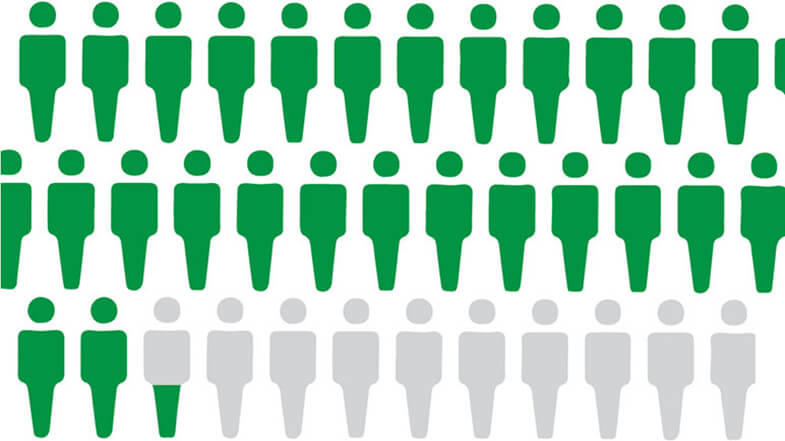
Childhood Myeloid Proliferations Associated With Down Syndrome Treatment (PDQ®)–Health Professional Version

Childhood Myeloid Proliferations Associated With Down Syndrome Treatment (PDQ®)–Health Professional Version

General Information About Childhood Myeloid Proliferations Associated With Down Syndrome

Myeloid leukemias that arise in children with Down syndrome, particularly in patients younger than 4 years, are a distinct subset of acute myeloid leukemia (AML) characterized by the co-existence of trisomy 21 and GATA1 variants within the leukemic blasts that are often, but not always, megakaryoblastic.

This distinct leukemia is further subdivided into two types:[1]

  • Transient abnormal myelopoiesis (TAM): A transient newborn and young-infant version, which spontaneously remits over time.
  • Myeloid leukemia of Down syndrome (MLDS): An unremitting but chemosensitive version that appears later, between the ages of 90 days and 3 years.

It is important to recognize the possibility of these versions in both children with Down syndrome phenotypes and in those who have mosaic trisomy 21, which can be solely present in the leukemic blasts. If possible, newborns with apparent AML should not begin therapy until genetic testing results have been returned.[2]

In older children with megakaryocytic AML, it is important to rule out the presence of co-existing trisomy 21 and GATA1 variants. These children may be successfully treated with the lower-intensity chemotherapy regimens that are used for children with myeloid leukemia associated with Down syndrome.[3]

References
  1. Lange B: The management of neoplastic disorders of haematopoiesis in children with Down’s syndrome. Br J Haematol 110 (3): 512-24, 2000. [PUBMED Abstract]
  2. Gamis AS, Smith FO: Transient myeloproliferative disorder in children with Down syndrome: clarity to this enigmatic disorder. Br J Haematol 159 (3): 277-87, 2012. [PUBMED Abstract]
  3. de Rooij JD, Branstetter C, Ma J, et al.: Pediatric non-Down syndrome acute megakaryoblastic leukemia is characterized by distinct genomic subsets with varying outcomes. Nat Genet 49 (3): 451-456, 2017. [PUBMED Abstract]

Special Considerations for the Treatment of Children With Cancer

Cancer in children and adolescents is rare, although the overall incidence has slowly increased since 1975.[1] Children and adolescents with cancer should be referred to medical centers that have a multidisciplinary team of cancer specialists with experience treating the cancers that occur during childhood and adolescence.[2] This multidisciplinary team approach incorporates the skills of the following pediatric specialists and others to ensure that children receive treatment, supportive care, and rehabilitation to achieve optimal survival and quality of life:

  • Primary care physicians.
  • Pediatric surgeons.
  • Pathologists.
  • Pediatric radiation oncologists.
  • Pediatric medical oncologists and hematologists.
  • Rehabilitation specialists.
  • Pediatric oncology nurses.
  • Social workers.
  • Child-life professionals.
  • Psychologists.
  • Nutritionists.

For specific information about supportive care for children and adolescents with cancer, see the summaries on Supportive and Palliative Care.

The American Academy of Pediatrics has outlined guidelines for pediatric cancer centers and their role in the treatment of children and adolescents with cancer.[3] At these centers, clinical trials are available for most types of cancer that occur in children and adolescents, and the opportunity to participate is offered to most patients and their families. Clinical trials for children and adolescents diagnosed with cancer are generally designed to compare potentially better therapy with current standard therapy. Other types of clinical trials test novel therapies when there is no standard therapy for a cancer diagnosis. Most of the progress in identifying curative therapies for childhood cancers has been achieved through clinical trials. Information about ongoing clinical trials is available from the NCI website.

References
  1. Smith MA, Seibel NL, Altekruse SF, et al.: Outcomes for children and adolescents with cancer: challenges for the twenty-first century. J Clin Oncol 28 (15): 2625-34, 2010. [PUBMED Abstract]
  2. Wolfson J, Sun CL, Wyatt L, et al.: Adolescents and Young Adults with Acute Lymphoblastic Leukemia and Acute Myeloid Leukemia: Impact of Care at Specialized Cancer Centers on Survival Outcome. Cancer Epidemiol Biomarkers Prev 26 (3): 312-320, 2017. [PUBMED Abstract]
  3. American Academy of Pediatrics: Standards for pediatric cancer centers. Pediatrics 134 (2): 410-4, 2014. Also available online. Last accessed February 25, 2025.

Transient Abnormal Myelopoiesis (TAM) Associated With Down Syndrome

Incidence

Approximately 10% of neonates with Down syndrome develop TAM (also termed transient myeloproliferative disorder [TMD]).[1] This disorder mimics congenital AML but typically improves spontaneously within the first 3 months of life (median, 49 days). However, TAM has been reported to remit as late as 20 months.[2] The late remissions likely reflect a persistent hepatomegaly from TAM-associated hepatic fibrosis rather than active disease.[3]

Clinical Presentation and Risk Groups

Although TAM is usually a self-resolving condition, it can be associated with significant morbidity and may be fatal in 10% to 17% of affected infants.[26] When TAM is detected, it is either in a proliferative, worsening phase or it has already converted to a resolving, improving phase. Observation over time is needed to determine which phase is present. Infants with progressive organomegaly, visceral effusions, preterm delivery (less than 37 weeks of gestation), bleeding diatheses, failure of spontaneous remission, laboratory evidence of progressive liver dysfunction (elevated direct bilirubin), renal failure, and very high white blood cell (WBC) count are at particularly high risk of early mortality.[3,4,6] In one report, death occurred in 21% of these patients with high-risk TAM, although only 10% were attributable to TAM. The remaining deaths were caused by coexisting conditions known to be more prominent in neonates with Down syndrome.[3]

The following three risk groups have been identified on the basis of the diagnostic clinical findings of hepatomegaly with or without life-threatening symptoms:[3]

  • Low risk. Includes those without hepatomegaly or life-threatening symptoms (38% of patients and an overall survival [OS] rate of 92% ± 8%).
  • Intermediate risk. Includes those with hepatomegaly alone (40% of patients and an OS rate of 77% ± 12%).
  • High risk. Includes those with hepatomegaly and life-threatening symptoms (21% of patients and an OS rate of 51% ± 19%).

Molecular Features

Genomics of TAM

TAM blasts most commonly have megakaryoblastic differentiation characteristics and distinctive variants involving the GATA1 gene in the presence of trisomy 21.[7,8] TAM may occur in phenotypically normal infants with genetic mosaicism in the bone marrow for trisomy 21. While TAM is generally not characterized by cytogenetic abnormalities other than trisomy 21, the presence of additional cytogenetic findings may predict an increased risk of developing subsequent AML.[4]

GATA1 variants are present in most, if not all, children with Down syndrome who have either TAM or acute megakaryoblastic leukemia (AMKL).[7,911] GATA1 is a transcription factor that is required for normal development of erythroid cells, megakaryocytes, eosinophils, and mast cells. X-linked GATA1 variants result in the absence of the full-length GATA1 protein, leaving only the normally minor variant, a truncated GATA1s transcription factor that has decreased activity.[7,8] This confers increased sensitivity to cytarabine by down-regulating cytidine deaminase expression, possibly explaining the superior outcome of children with Down syndrome and M7 AML when treated with cytarabine-containing regimens.[12]

A 2024 analysis screened 143 TAM samples for additional somatic variants in the abnormal cells. With the exception of rare STAG2 variants, the study found no additional abnormalities beyond the typical GATA1 abnormality.[13]

Approximately 20% of infants with TAM and Down syndrome eventually develop AML. Most of these cases are diagnosed within the first 3 years of life.[4,8]

Treatment of TAM

While observation is appropriate for most infants with TAM, therapeutic intervention is warranted in patients with apparent severe hydrops or organ failure. Because TAM eventually spontaneously remits, treatment is short in duration and primarily aimed at the reduction of leukemic burden and resolution of immediate symptoms. Several treatment approaches have been used, including the following:

  • Exchange transfusion.
  • Leukapheresis.
  • Low-dose cytarabine. Of these approaches, only cytarabine has been shown to consistently reduce TAM complications and related mortality.[3,6]; [14][Level of evidence B4] Cytarabine dosing has ranged from 0.4 to 1.5 mg/kg per dose given intravenously (IV) or subcutaneously (SC) once to twice daily for 4 to 12 days.[6] This dosing schedule has produced similar efficacies and less toxicity than higher doses given in continuous 5-day infusions, which led to prolonged severe neutropenia.[3] A prospective trial examined the use of low-dose cytarabine (1.5 mg/kg per day IV or SC for 7 days) to treat symptomatic patients. This trial reported a significant reduction in early death using this regimen, compared with similar patients in the historical control group (12% ± 5% vs. 33% ± 7%, respectively; P = .02).[14][Level of evidence B4]

Risk Factors for the Development of AML After Resolution of TAM

Subsequent development of myeloid leukemia of Down syndrome (MLDS) is seen in 10% to 30% of children with TAM. It has been reported at a mean age of 16 months (range, 1–30 months).[2,3,15] While TAM is generally not characterized by cytogenetic abnormalities other than trisomy 21, the presence of additional cytogenetic findings may connote an increased risk of developing subsequent MLDS.[4] An additional risk factor reported in two studies is the late resolution of TAM, measured by either time to complete resolution of signs of TAM (defined as resolution beyond the median, 47 days from diagnosis) or by persistence of minimal residual disease (MRD) in the peripheral blood at week 12 of follow-up.[3]; [14][Level of evidence B4]

The use of cytarabine for TAM symptoms or persistent MRD in TAM has failed to show a reduction in later MLDS, as reported in large observational cohort studies.[3,6] In a prospective single-arm trial designed to assess whether cytarabine treatment for TAM could prevent the development of later MLDS, no benefit was found when compared with historical controls (19% ± 4% vs. 22% ± 4%, respectively; P = .88).[14][Level of evidence B4]

References
  1. Gamis AS, Smith FO: Transient myeloproliferative disorder in children with Down syndrome: clarity to this enigmatic disorder. Br J Haematol 159 (3): 277-87, 2012. [PUBMED Abstract]
  2. Homans AC, Verissimo AM, Vlacha V: Transient abnormal myelopoiesis of infancy associated with trisomy 21. Am J Pediatr Hematol Oncol 15 (4): 392-9, 1993. [PUBMED Abstract]
  3. Gamis AS, Alonzo TA, Gerbing RB, et al.: Natural history of transient myeloproliferative disorder clinically diagnosed in Down syndrome neonates: a report from the Children’s Oncology Group Study A2971. Blood 118 (26): 6752-9; quiz 6996, 2011. [PUBMED Abstract]
  4. Massey GV, Zipursky A, Chang MN, et al.: A prospective study of the natural history of transient leukemia (TL) in neonates with Down syndrome (DS): Children’s Oncology Group (COG) study POG-9481. Blood 107 (12): 4606-13, 2006. [PUBMED Abstract]
  5. Muramatsu H, Kato K, Watanabe N, et al.: Risk factors for early death in neonates with Down syndrome and transient leukaemia. Br J Haematol 142 (4): 610-5, 2008. [PUBMED Abstract]
  6. Klusmann JH, Creutzig U, Zimmermann M, et al.: Treatment and prognostic impact of transient leukemia in neonates with Down syndrome. Blood 111 (6): 2991-8, 2008. [PUBMED Abstract]
  7. Hitzler JK, Cheung J, Li Y, et al.: GATA1 mutations in transient leukemia and acute megakaryoblastic leukemia of Down syndrome. Blood 101 (11): 4301-4, 2003. [PUBMED Abstract]
  8. Mundschau G, Gurbuxani S, Gamis AS, et al.: Mutagenesis of GATA1 is an initiating event in Down syndrome leukemogenesis. Blood 101 (11): 4298-300, 2003. [PUBMED Abstract]
  9. Groet J, McElwaine S, Spinelli M, et al.: Acquired mutations in GATA1 in neonates with Down’s syndrome with transient myeloid disorder. Lancet 361 (9369): 1617-20, 2003. [PUBMED Abstract]
  10. Rainis L, Bercovich D, Strehl S, et al.: Mutations in exon 2 of GATA1 are early events in megakaryocytic malignancies associated with trisomy 21. Blood 102 (3): 981-6, 2003. [PUBMED Abstract]
  11. Wechsler J, Greene M, McDevitt MA, et al.: Acquired mutations in GATA1 in the megakaryoblastic leukemia of Down syndrome. Nat Genet 32 (1): 148-52, 2002. [PUBMED Abstract]
  12. Ge Y, Stout ML, Tatman DA, et al.: GATA1, cytidine deaminase, and the high cure rate of Down syndrome children with acute megakaryocytic leukemia. J Natl Cancer Inst 97 (3): 226-31, 2005. [PUBMED Abstract]
  13. Sato T, Yoshida K, Toki T, et al.: Landscape of driver mutations and their clinical effects on Down syndrome-related myeloid neoplasms. Blood 143 (25): 2627-2643, 2024. [PUBMED Abstract]
  14. Flasinski M, Scheibke K, Zimmermann M, et al.: Low-dose cytarabine to prevent myeloid leukemia in children with Down syndrome: TMD Prevention 2007 study. Blood Adv 2 (13): 1532-1540, 2018. [PUBMED Abstract]
  15. Ravindranath Y, Abella E, Krischer JP, et al.: Acute myeloid leukemia (AML) in Down’s syndrome is highly responsive to chemotherapy: experience on Pediatric Oncology Group AML Study 8498. Blood 80 (9): 2210-4, 1992. [PUBMED Abstract]

Myeloid Leukemia of Down Syndrome (MLDS)

General Information

Children with Down syndrome have a 10-fold to 45-fold increased risk of leukemia when compared with children without Down syndrome.[1] However, the ratio of acute lymphoblastic leukemia to acute myeloid leukemia (AML) is typical for childhood acute leukemia. The exception is during the first 3 years of life, when AML, particularly the megakaryoblastic subtype, predominates and exhibits a distinctive biology characterized by GATA1 variants and increased sensitivity to cytarabine.[27] Importantly, these risks appear to be similar whether a child has phenotypic characteristics of Down syndrome or whether a child has only genetic bone marrow mosaicism.[8]

Prognosis of Children With MLDS

Outcome is generally favorable for children with Down syndrome who develop AML. This is called myeloid leukemia of Down syndrome (MLDS) in the World Health Organization (WHO) classification.[911] For more information, see the sections on General Information About Childhood Myeloid Malignancies and World Health Organization (WHO) Classification System for Childhood AML in Childhood Acute Myeloid Leukemia Treatment.

Prognostic factors for children with MLDS include the following:

  • Age. The prognosis is particularly good (event-free survival [EFS] rates exceeding 85%) in children aged 4 years or younger at diagnosis. This age group accounts for the vast majority of patients with MLDS.[1013] Children with MLDS who are older than 4 years have a significantly worse prognosis. These patients should undergo the therapy that is used in children with AML without Down syndrome, unless a GATA1 variant is found.[14]
  • White blood cell (WBC) count. A large international Berlin-Frankfurt-Münster (BFM) retrospective study of 451 children with MLDS (aged >6 months and <5 years) observed a 7-year EFS rate of 78% and a 7-year overall survival (OS) rate of 79%. In multivariate analyses, WBC count (≥20 × 109/L) and age (>3 years) were independent predictors of lower EFS. The 7-year EFS rate for the older population (>3 years) and for the higher WBC-count population still exceeded 60%.[15]
  • AML karyotype. The presence of trisomy 8 has been shown to adversely impact prognosis.[13] In another study, complex karyotypes (≥3 independent abnormalities) were associated with an increased cumulative incidence of relapse (CIR) rate at 2 years (30.8% compared with 7.5% in patients without complex karyotypes; P = .001).[16]
  • Minimal residual disease (MRD). MRD at the end of induction 1 was found to be a strong prognostic factor.[11,17] This finding was consistent with the BFM finding that early response correlated with improved OS.[13] However, a negative MRD status at the end of induction 1 did not identify a favorable-risk group of patients who could receive reduced chemotherapy.[16]

Approximately 29% to 47% of patients with Down syndrome present with myelodysplastic neoplasms (MDS) (<20% blasts) but their outcomes are similar to those with AML.[10,11,13]

Treatment of Newly Diagnosed Childhood MLDS

Appropriate therapy for younger children (aged ≤4 years) with MLDS is less intensive than current standard childhood AML therapy. Hematopoietic stem cell transplant is not indicated in first remission.[4,914,18,19]

Treatment options for newly diagnosed children with MLDS include the following:

  1. Chemotherapy.

Evidence (chemotherapy):

  1. In a Children’s Oncology Group (COG) trial for newly diagnosed children with MLDS (AAML0431 [NCT00369317]), 204 children received a regimen that substituted high-dose cytarabine for the second of four induction cycles (thereby reducing cumulative anthracycline exposure from 320 mg to 240 mg), moving this cycle from intensification where it was used in the previous COG A2971 (NCT00003593) trial.[10,11] Intrathecal doses were reduced from seven to two total injections, and intensification included two cycles of cytarabine/etoposide.
    • When compared with the previous trial, these changes resulted in an overall improvement of approximately 10%.
    • The EFS rate was 89.9%, and the OS rate was 93%.
    • Relapse occurred in 14 patients and there were two treatment-related deaths, both related to pneumonia, neither of which occurred during induction 2.
    • No patient had central nervous system (CNS) involvement in this trial or the preceding COG A2971 trial.[10]
    • The only prognostic factor identified was MRD using flow cytometry on day 28 of induction 1. Among those who were MRD negative (≤0.01%), the disease-free survival (DFS) rate was 92.7%. In the 14.4% of patients who were MRD positive, the DFS rate was 76.2% (P = .011).
  2. In the COG AAML1531 (NCT00369317) trial for children with newly diagnosed MLDS, removing the high-dose cytarabine cycle in those with standard-risk MLDS was unsuccessful.[16]
    • The interim analysis found that patients who did not receive high-dose cytarabine had a lower 2-year EFS rate of 85.6%, compared with the 2-year EFS rate of 93.5% for patients in the AAML0431 trial (P = .0002).
  3. In a joint trial (ML-DS 2006) from the BFM, Dutch Childhood Oncology Group (DCOG), and Nordic Society of Pediatric Hematology and Oncology (NOPHO), 170 children with Down syndrome were enrolled. This trial focused on reducing therapy by eliminating etoposide during consolidation, reducing the number of intrathecal doses from 11 to 4, and the elimination of maintenance from the reduced-therapy Down syndrome arm of AML-BFM 98.[13] As in the COG trials, no patient had CNS disease at diagnosis.
    • Outcomes were no worse despite reduction in chemotherapy. The OS rate was 89% (± 3%), and the EFS rate was 87% (± 3%), similar to that observed in AML-BFM 98 (OS rate, 90% ± 4% [P = NS]; EFS rate, 89% ± 4% [P = NS]). The CIR rate was 6% in both trials.
    • Nine patients relapsed, and seven of those patients died.
    • Patients with a good early response (<5% blasts by morphology before induction cycle 2, n = 123 [72%]) had better outcomes (OS rate, 92% ± 3% vs. 57% ± 16%, P < .0001; EFS rate, 88% ± 3% vs. 58% ± 16%, P = .0008; and CIR rate, 3% ± 2% vs. 27% ± 18%, P = .003).
    • Less toxicity was seen in this trial, and treatment-related mortality remained low (2.9% vs. 5%, P = .276).

    The following two prognostic factors were identified:[13]

    • Trisomy 8 was an adverse factor (n = 37; OS rate, 77% vs. 95%, P = .07; EFS rate, 73% ± 8% vs. 91% ± 4%, P = .018; CIR rate, 16% ± 7% vs. 3% ± 2%, P = .02).
    • This was confirmed in multivariate analysis, where lack of good early response and trisomy 8 maintained their adverse impact on relapse, with relative risks of 8.55 (95% confidence interval [CI], 1.96–37.29; P = .004) and 4.36 (95% CI, 1.24–15.39; P = .022), respectively.
  4. A 2024 analysis included a cohort of Japanese patients with MLDS (n = 204) who were treated with uniform chemotherapy. Patients underwent extensive somatic testing to further define variants most commonly seen with this diagnosis. Somatic variants in 26 genes known to be driver genes in MLDS were identified again. These included variants in cohesin and cohesin-related proteins (43.6%), epigenetic regulators (39.2%), tyrosine kinases (25.5%), and genes important in the RAS pathway (11.8%). An additional 16 novel genes were also described. Of these, variants in two transcription factors (IRX1: 16.2%; ZBTB7A: 13.2%) were found, and functional studies confirmed their role as tumor suppressor genes that impacted signaling through MYC/E2F pathways. Structural variants were also observed. RUNX1 partial tandem duplications were seen in 13.7% of patients, which may result in partial loss of function of the gene. This causes upregulation expression through the addition of an extra promoter, which results in isoform disequilibrium, with RUNX1A bias versus RUNX1. Recent studies have shown that this disequilibrium contributes to MLDS pathogenesis. Variants of RUNX1, IRX1, and ZBTB7A also activate MYC/E2F genes, suggesting that targeting this pathway may provide therapeutic benefit.[20]
    • Four somatic alterations were associated with inferior outcomes in this study cohort.
      • Of 177 patients with comprehensive somatic testing, CDKN2A deletions and variants in ZBTB7A, TP53, and JAK2 were all associated with inferior outcomes.
      • If patients had at least one of these variants, EFS and OS rates were significantly lower than those of patients who lacked any four of these abnormalities (3-year EFS rates, 66.6% vs. 94.2%; P for unadjusted Cox regression < .001; 3-year OS rates, 69.0% vs. 95.6%; P for unadjusted Cox regression < .001).
      • In multivariable analysis, all of these abnormalities were associated with inferior outcomes.
      • Validation of these findings in additional cohorts is needed. However, these findings may help identify patients with higher-risk MLDS in the future.

Children with mosaicism for trisomy 21 are treated similarly to those children with clinically evident Down syndrome.[8,10,21] Children with MLDS who are older than 4 years have a significantly worse prognosis.[14] Although an optimal treatment for these children has not been defined, they are usually treated with AML regimens designed for children without Down syndrome.

Treatment options under clinical evaluation

Information about National Cancer Institute (NCI)–supported clinical trials can be found on the NCI website. For information about clinical trials sponsored by other organizations, see the ClinicalTrials.gov website.

Treatment of Relapsed or Refractory Childhood MLDS

A small number of trials address outcomes in children with MLDS who relapse after initial therapy or who have refractory MLDS. In three prospective trials of children with newly diagnosed MLDS, outcomes were poor for those who relapsed (4 of 11, 2 of 9, and 2 of 12 patients who relapsed survived).[9,13,16] Thus, these children are treated similarly to children without Down syndrome, with an intensive reinduction chemotherapy regimen. If a remission is achieved, therapy is followed by an allogeneic hematopoietic stem cell transplant (HSCT).

Treatment options for children with refractory or relapsed MLDS include the following:

  1. Chemotherapy, which may be followed by an allogeneic HSCT.

Evidence (treatment of children with refractory or relapsed MLDS):

Four analyses have specifically examined children with relapsed or refractory MLDS.[2225]

  1. The Japanese Pediatric Leukemia/Lymphoma Study Group reported the outcomes of 29 patients with relapsed (n = 26) or refractory (n = 3) MLDS. As expected with Down syndrome, the children in this cohort were very young (median age, 2 years); relapses were almost all early (median, 8.6 months; 80% <12 months from diagnosis); and 89% had M7 French-American-British classification.[22][Level of evidence C1]
    • In contrast to the excellent outcomes achieved after initial therapy, only 50% of the children attained a second remission, and the 3-year OS rate was 26%. Attainment of second remission was more successful the later the relapse occurred after completing initial therapies.
    • Approximately one-half of the children underwent allogeneic transplant, and no advantage was noted for transplant compared with chemotherapy. However, the number of patients was small.
  2. A Center for International Blood and Marrow Transplant Research study of children with MLDS who underwent allogeneic HSCT reported the following results:[23][Level of evidence C1]
    • A similarly poor outcome, with a 3-year OS rate of 19%.
    • The main cause of failure after transplant was relapse, which exceeded 60%. Survival was significantly worse for patients who relapsed early.
    • The transplant-related mortality was approximately 20%.
  3. A Japanese registry study reported better survival after transplant of children with MLDS using reduced-intensity conditioning regimens compared with myeloablative approaches. However, the number of patients was very small (n = 5), and the efficacy of reduced-intensity approaches in children with MLDS requires further study.[24][Level of evidence C2]
  4. The largest study to date was conducted by a consortium of pediatric cooperative groups and select North American institutions. The study retrospectively evaluated children with MLDS to determine their outcomes and prognostic factors for survival after relapse or refractory disease.[25]
    • The most common site of relapse was bone marrow (61 of 62 patients), and no CNS relapses were reported.
    • Median time to relapse was 6.8 months, and 82% of relapses occurred within 12 months of initial diagnosis.
    • Time to relapse, use of HSCT, and attainment of second complete remission (CR) before transplant were prognostically significant.
    • For the entire cohort, the OS rate was 22.1%, the EFS rate was 20.9%, and the cumulative relapse rate was 79.1%.
    • The median time from relapse or refractory disease to time of death was 5.1 months (0.4–41 months).
    • The 3-year OS rate was 46% for those who achieved remission (45% of patients).
    • HSCT was performed in 29 patients. Undergoing HSCT in second CR was critically important, with 6 of 19 patients relapsing after HSCT if initially in second CR, compared with 9 of 10 patients relapsing if they went to transplant when not in second CR. Among the 29 HSCT recipients, the 3-year OS rate was 39.8%, and the EFS rate was 36.7%.
    • Only 3 of 33 patients who received chemotherapy alone ultimately survived (3-year OS and EFS rates, 6.4%).
References
  1. Marlow EC, Ducore J, Kwan ML, et al.: Leukemia Risk in a Cohort of 3.9 Million Children with and without Down Syndrome. J Pediatr 234: 172-180.e3, 2021. [PUBMED Abstract]
  2. Ravindranath Y: Down syndrome and leukemia: new insights into the epidemiology, pathogenesis, and treatment. Pediatr Blood Cancer 44 (1): 1-7, 2005. [PUBMED Abstract]
  3. Ross JA, Spector LG, Robison LL, et al.: Epidemiology of leukemia in children with Down syndrome. Pediatr Blood Cancer 44 (1): 8-12, 2005. [PUBMED Abstract]
  4. Gamis AS: Acute myeloid leukemia and Down syndrome evolution of modern therapy–state of the art review. Pediatr Blood Cancer 44 (1): 13-20, 2005. [PUBMED Abstract]
  5. Taub JW, Ge Y: Down syndrome, drug metabolism and chromosome 21. Pediatr Blood Cancer 44 (1): 33-9, 2005. [PUBMED Abstract]
  6. Crispino JD: GATA1 mutations in Down syndrome: implications for biology and diagnosis of children with transient myeloproliferative disorder and acute megakaryoblastic leukemia. Pediatr Blood Cancer 44 (1): 40-4, 2005. [PUBMED Abstract]
  7. Ge Y, Stout ML, Tatman DA, et al.: GATA1, cytidine deaminase, and the high cure rate of Down syndrome children with acute megakaryocytic leukemia. J Natl Cancer Inst 97 (3): 226-31, 2005. [PUBMED Abstract]
  8. Kudo K, Hama A, Kojima S, et al.: Mosaic Down syndrome-associated acute myeloid leukemia does not require high-dose cytarabine treatment for induction and consolidation therapy. Int J Hematol 91 (4): 630-5, 2010. [PUBMED Abstract]
  9. Lange BJ, Kobrinsky N, Barnard DR, et al.: Distinctive demography, biology, and outcome of acute myeloid leukemia and myelodysplastic syndrome in children with Down syndrome: Children’s Cancer Group Studies 2861 and 2891. Blood 91 (2): 608-15, 1998. [PUBMED Abstract]
  10. Sorrell AD, Alonzo TA, Hilden JM, et al.: Favorable survival maintained in children who have myeloid leukemia associated with Down syndrome using reduced-dose chemotherapy on Children’s Oncology Group trial A2971: a report from the Children’s Oncology Group. Cancer 118 (19): 4806-14, 2012. [PUBMED Abstract]
  11. Taub JW, Berman JN, Hitzler JK, et al.: Improved outcomes for myeloid leukemia of Down syndrome: a report from the Children’s Oncology Group AAML0431 trial. Blood 129 (25): 3304-3313, 2017. [PUBMED Abstract]
  12. Creutzig U, Reinhardt D, Diekamp S, et al.: AML patients with Down syndrome have a high cure rate with AML-BFM therapy with reduced dose intensity. Leukemia 19 (8): 1355-60, 2005. [PUBMED Abstract]
  13. Uffmann M, Rasche M, Zimmermann M, et al.: Therapy reduction in patients with Down syndrome and myeloid leukemia: the international ML-DS 2006 trial. Blood 129 (25): 3314-3321, 2017. [PUBMED Abstract]
  14. Gamis AS, Woods WG, Alonzo TA, et al.: Increased age at diagnosis has a significantly negative effect on outcome in children with Down syndrome and acute myeloid leukemia: a report from the Children’s Cancer Group Study 2891. J Clin Oncol 21 (18): 3415-22, 2003. [PUBMED Abstract]
  15. Blink M, Zimmermann M, von Neuhoff C, et al.: Normal karyotype is a poor prognostic factor in myeloid leukemia of Down syndrome: a retrospective, international study. Haematologica 99 (2): 299-307, 2014. [PUBMED Abstract]
  16. Hitzler J, Alonzo T, Gerbing R, et al.: High-dose AraC is essential for the treatment of ML-DS independent of postinduction MRD: results of the COG AAML1531 trial. Blood 138 (23): 2337-2346, 2021. [PUBMED Abstract]
  17. Taga T, Tanaka S, Hasegawa D, et al.: Post-induction MRD by FCM and GATA1-PCR are significant prognostic factors for myeloid leukemia of Down syndrome. Leukemia 35 (9): 2508-2516, 2021. [PUBMED Abstract]
  18. Ravindranath Y, Abella E, Krischer JP, et al.: Acute myeloid leukemia (AML) in Down’s syndrome is highly responsive to chemotherapy: experience on Pediatric Oncology Group AML Study 8498. Blood 80 (9): 2210-4, 1992. [PUBMED Abstract]
  19. Taga T, Shimomura Y, Horikoshi Y, et al.: Continuous and high-dose cytarabine combined chemotherapy in children with down syndrome and acute myeloid leukemia: Report from the Japanese children’s cancer and leukemia study group (JCCLSG) AML 9805 down study. Pediatr Blood Cancer 57 (1): 36-40, 2011. [PUBMED Abstract]
  20. Sato T, Yoshida K, Toki T, et al.: Landscape of driver mutations and their clinical effects on Down syndrome-related myeloid neoplasms. Blood 143 (25): 2627-2643, 2024. [PUBMED Abstract]
  21. Gamis AS, Alonzo TA, Gerbing RB, et al.: Natural history of transient myeloproliferative disorder clinically diagnosed in Down syndrome neonates: a report from the Children’s Oncology Group Study A2971. Blood 118 (26): 6752-9; quiz 6996, 2011. [PUBMED Abstract]
  22. Taga T, Saito AM, Kudo K, et al.: Clinical characteristics and outcome of refractory/relapsed myeloid leukemia in children with Down syndrome. Blood 120 (9): 1810-5, 2012. [PUBMED Abstract]
  23. Hitzler JK, He W, Doyle J, et al.: Outcome of transplantation for acute myelogenous leukemia in children with Down syndrome. Biol Blood Marrow Transplant 19 (6): 893-7, 2013. [PUBMED Abstract]
  24. Muramatsu H, Sakaguchi H, Taga T, et al.: Reduced intensity conditioning in allogeneic stem cell transplantation for AML with Down syndrome. Pediatr Blood Cancer 61 (5): 925-7, 2014. [PUBMED Abstract]
  25. Raghuram N, Hasegawa D, Nakashima K, et al.: Survival outcomes of children with relapsed or refractory myeloid leukemia associated with Down syndrome. Blood Adv 7 (21): 6532-6539, 2023. [PUBMED Abstract]

Latest Updates to This Summary (09/16/2024)

The PDQ cancer information summaries are reviewed regularly and updated as new information becomes available. This section describes the latest changes made to this summary as of the date above.

Transient Abnormal Myelopoiesis (TAM) Associated With Down Syndrome

Added text to state that a 2024 analysis screened 143 TAM samples for additional somatic variants in the abnormal cells. With the exception of rare STAG2 variants, the study found no additional abnormalities beyond the typical GATA1 abnormality (cited Sato et al. as reference 13).

Myeloid Leukemia of Down Syndrome (MLDS)

Added text about the results of a 2024 analysis that included a cohort of Japanese patients with MLDS who were treated with uniform chemotherapy. Patients underwent extensive somatic testing to further define variants most commonly seen with this diagnosis (cited Sato et al. as reference 20).

This summary is written and maintained by the PDQ Pediatric Treatment Editorial Board, which is editorially independent of NCI. The summary reflects an independent review of the literature and does not represent a policy statement of NCI or NIH. More information about summary policies and the role of the PDQ Editorial Boards in maintaining the PDQ summaries can be found on the About This PDQ Summary and PDQ® Cancer Information for Health Professionals pages.

About This PDQ Summary

Purpose of This Summary

This PDQ cancer information summary for health professionals provides comprehensive, peer-reviewed, evidence-based information about the treatment of childhood myeloid proliferations associated with Down syndrome. It is intended as a resource to inform and assist clinicians in the care of their patients. It does not provide formal guidelines or recommendations for making health care decisions.

Reviewers and Updates

This summary is reviewed regularly and updated as necessary by the PDQ Pediatric Treatment Editorial Board, which is editorially independent of the National Cancer Institute (NCI). The summary reflects an independent review of the literature and does not represent a policy statement of NCI or the National Institutes of Health (NIH).

Board members review recently published articles each month to determine whether an article should:

  • be discussed at a meeting,
  • be cited with text, or
  • replace or update an existing article that is already cited.

Changes to the summaries are made through a consensus process in which Board members evaluate the strength of the evidence in the published articles and determine how the article should be included in the summary.

The lead reviewers for Childhood Myeloid Proliferations Associated With Down Syndrome Treatment are:

  • Alan Scott Gamis, MD, MPH (Children’s Mercy Hospital)
  • Karen J. Marcus, MD, FACR (Dana-Farber of Boston Children’s Cancer Center and Blood Disorders Harvard Medical School)
  • Jessica Pollard, MD (Dana-Farber/Boston Children’s Cancer and Blood Disorders Center)
  • Michael A. Pulsipher, MD (Huntsman Cancer Institute at University of Utah)
  • Rachel E. Rau, MD (University of Washington School of Medicine, Seatle Children’s)
  • Lewis B. Silverman, MD (Dana-Farber Cancer Institute/Boston Children’s Hospital)
  • Malcolm A. Smith, MD, PhD (National Cancer Institute)
  • Sarah K. Tasian, MD (Children’s Hospital of Philadelphia)

Any comments or questions about the summary content should be submitted to Cancer.gov through the NCI website’s Email Us. Do not contact the individual Board Members with questions or comments about the summaries. Board members will not respond to individual inquiries.

Levels of Evidence

Some of the reference citations in this summary are accompanied by a level-of-evidence designation. These designations are intended to help readers assess the strength of the evidence supporting the use of specific interventions or approaches. The PDQ Pediatric Treatment Editorial Board uses a formal evidence ranking system in developing its level-of-evidence designations.

Permission to Use This Summary

PDQ is a registered trademark. Although the content of PDQ documents can be used freely as text, it cannot be identified as an NCI PDQ cancer information summary unless it is presented in its entirety and is regularly updated. However, an author would be permitted to write a sentence such as “NCI’s PDQ cancer information summary about breast cancer prevention states the risks succinctly: [include excerpt from the summary].”

The preferred citation for this PDQ summary is:

PDQ® Pediatric Treatment Editorial Board. PDQ Childhood Myeloid Proliferations Associated With Down Syndrome Treatment. Bethesda, MD: National Cancer Institute. Updated <MM/DD/YYYY>. Available at: /types/leukemia/hp/child-aml-treatment-pdq/myeloid-proliferations-down-syndrome-treatment-pdq. Accessed <MM/DD/YYYY>. [PMID: 38630975]

Images in this summary are used with permission of the author(s), artist, and/or publisher for use within the PDQ summaries only. Permission to use images outside the context of PDQ information must be obtained from the owner(s) and cannot be granted by the National Cancer Institute. Information about using the illustrations in this summary, along with many other cancer-related images, is available in Visuals Online, a collection of over 2,000 scientific images.

Disclaimer

Based on the strength of the available evidence, treatment options may be described as either “standard” or “under clinical evaluation.” These classifications should not be used as a basis for insurance reimbursement determinations. More information on insurance coverage is available on Cancer.gov on the Managing Cancer Care page.

Contact Us

More information about contacting us or receiving help with the Cancer.gov website can be found on our Contact Us for Help page. Questions can also be submitted to Cancer.gov through the website’s Email Us.
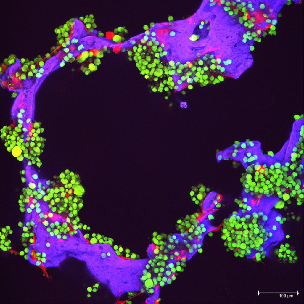
Childhood Acute Myeloid Leukemia Treatment (PDQ®)–Health Professional Version

Childhood Acute Myeloid Leukemia Treatment (PDQ®)–Health Professional Version

General Information About Childhood Myeloid Malignancies

Approximately 20% of childhood leukemias are of myeloid origin and represent a spectrum of hematopoietic malignancies.[1] Most myeloid leukemias in children are acute; the remainder include chronic and/or subacute myeloproliferative disorders, such as chronic myeloid leukemia and juvenile myelomonocytic leukemia. Myelodysplastic neoplasms (MDS) occur much less frequently in children than in adults and almost invariably represent clonal, preleukemic conditions that often evolve from congenital marrow failure syndromes, such as Fanconi anemia and Shwachman-Diamond syndrome.

The general characteristics of myeloid leukemias and other myeloid malignancies are described below:

  • Acute myeloid leukemia (AML). AML is a clonal disorder caused by malignant transformation of a bone marrow–derived, self-renewing stem cell or progenitors, leading to accumulation of immature, nonfunctional myeloid cells. These events lead to increased accumulation of these malignant cells in the bone marrow and other organs. To be called acute, the bone marrow usually must have greater than 20% immature leukemic blasts, with some exceptions. For more information, see the sections on Treatment Option Overview for Childhood AML and Treatment of Childhood AML.
  • Myeloid leukemias of Down syndrome.
    • Transient abnormal myelopoiesis (TAM). TAM is also called transient myeloproliferative disorder or transient leukemia. The TAM observed in infants with Down syndrome represents a clonal expansion of myeloblasts with GATA1 variants in the setting of a coexisting trisomy 21 that can be difficult to distinguish from AML. Most importantly, TAM spontaneously regresses within the first 3 months of life in most cases. TAM occurs in 4% to 10% of infants with Down syndrome.[24]
    • Myeloid leukemia of Down syndrome (MLDS). MLDS is defined by the presence of myeloblasts with GATA1 variants in the setting of a coexisting trisomy 21 occurring in children older than 3 months. It is distinct from myeloid leukemias in children without trisomy 21 and GATA1 variants. Treatment with chemotherapy results in overall excellent survival. Less-intense therapeutic regimens are used and can reduce morbidity in these children with Down syndrome who experience greater toxicity than children without Down syndrome. However, children with Down syndrome who are older than 4 years most often have AML similar to children without Down syndrome (i.e., without the GATA1 variant). These patients require the more intensive chemotherapeutic regimens used in children without Down syndrome.

    For more information about TAM and MLDS, see Childhood Myeloid Proliferations Associated With Down Syndrome Treatment.

  • Myelodysplastic neoplasms (MDS). MDS in children, identified when the marrow blast proportion is less than 20%, represents a heterogeneous group of disorders characterized by ineffective hematopoiesis, impaired maturation of myeloid progenitors with dysplastic morphological features, and cytopenias. Although the underlying cause of MDS in children is unclear, there is often an association with marrow failure syndromes or germline conditions that predispose to myeloid malignancy/dysfunction. Most patients with MDS may have hypercellular bone marrows without increased numbers of leukemic blasts. However, some patients may present with hypocellular bone marrow, making the distinction between severe aplastic anemia and MDS difficult.[5,6]

    The presence of a karyotype abnormality in a hypocellular marrow is consistent with MDS, and transformation to AML should be expected. Patients with MDS are typically referred for stem cell transplant before transformation to AML.

    If a patient with MDS has a common defining genetic variant that is seen in AML, the clinician should be aware that, despite the relatively low proportion of blasts, the child should be treated similarly to those with blast proportions of 20% or more.

    In children with Down syndrome younger than 4 years, the finding of MDS likely represents an early presentation of typical AML, and patients should be treated with regimens used for AML in Down syndrome.

    For more information, see Childhood Myelodysplastic Neoplasms Treatment.

  • Juvenile myelomonocytic leukemia (JMML). JMML represents the most common myeloproliferative neoplasm observed in young children. JMML occurs at a median age of 1.8 years.

    JMML characteristically presents with hepatosplenomegaly, lymphadenopathy, fever, and skin rash, along with an elevated white blood cell (WBC) count and increased circulating monocytes.[7] In addition, patients often have elevated hemoglobin F, hypersensitivity of the leukemic cells to granulocyte-macrophage colony-stimulating factor (GM-CSF), monosomy 7, and leukemia cell variants in a gene involved in RAS pathway signaling (e.g., NF1, KRAS, NRAS, PTPN11, or CBL).[79]

    For more information, see Juvenile Myelomonocytic Leukemia Treatment.

  • Chronic myeloid leukemia (CML). CML is primarily an adult disease but represents the most common of the chronic myeloproliferative disorders in childhood, accounting for approximately 10% of childhood myeloid leukemias.[10] Although CML has been reported in very young children, most patients are aged 6 years and older.

    CML is a clonal panmyelopathy that involves all hematopoietic cell lineages. While the WBC count can be extremely elevated, the bone marrow does not show increased numbers of leukemic blasts during the chronic phase of this disease. CML is caused by the presence of the Philadelphia chromosome, a translocation between chromosomes 9 and 22 (i.e., t(9;22)) resulting in fusion of the BCR and ABL1 genes.

    For more information, see Childhood Chronic Myeloid Leukemia Treatment.

    Other chronic myeloproliferative neoplasms, such as polycythemia vera, primary myelofibrosis, and essential thrombocytosis, are extremely rare in children.

  • Acute promyelocytic leukemia (APL). APL is a distinct subtype of AML and occurs in about 7% of children with AML.[10,11] Several factors that make APL unique include the following:
    • Clinical presentation of universal coagulopathy (disseminated intravascular coagulation) and unique morphological characteristics (French-American-British [FAB] M3 or its variants).
    • Unique molecular etiology as a result of the involvement of the RARA oncogene.
    • Unique sensitivity to the differentiating agent tretinoin and to the proapoptotic agent arsenic trioxide.[12]

    For more information, see Childhood Acute Promyelocytic Leukemia Treatment.

References
  1. National Cancer Institute: NCCR*Explorer: An interactive website for NCCR cancer statistics. Bethesda, MD: National Cancer Institute. Available online. Last accessed February 25, 2025.
  2. Roberts I, Alford K, Hall G, et al.: GATA1-mutant clones are frequent and often unsuspected in babies with Down syndrome: identification of a population at risk of leukemia. Blood 122 (24): 3908-17, 2013. [PUBMED Abstract]
  3. Zipursky A: Transient leukaemia–a benign form of leukaemia in newborn infants with trisomy 21. Br J Haematol 120 (6): 930-8, 2003. [PUBMED Abstract]
  4. Gamis AS, Smith FO: Transient myeloproliferative disorder in children with Down syndrome: clarity to this enigmatic disorder. Br J Haematol 159 (3): 277-87, 2012. [PUBMED Abstract]
  5. Hasle H, Niemeyer CM: Advances in the prognostication and management of advanced MDS in children. Br J Haematol 154 (2): 185-95, 2011. [PUBMED Abstract]
  6. Schwartz JR, Ma J, Lamprecht T, et al.: The genomic landscape of pediatric myelodysplastic syndromes. Nat Commun 8 (1): 1557, 2017. [PUBMED Abstract]
  7. Niemeyer CM, Arico M, Basso G, et al.: Chronic myelomonocytic leukemia in childhood: a retrospective analysis of 110 cases. European Working Group on Myelodysplastic Syndromes in Childhood (EWOG-MDS) Blood 89 (10): 3534-43, 1997. [PUBMED Abstract]
  8. Loh ML: Recent advances in the pathogenesis and treatment of juvenile myelomonocytic leukaemia. Br J Haematol 152 (6): 677-87, 2011. [PUBMED Abstract]
  9. Stieglitz E, Taylor-Weiner AN, Chang TY, et al.: The genomic landscape of juvenile myelomonocytic leukemia. Nat Genet 47 (11): 1326-33, 2015. [PUBMED Abstract]
  10. Smith MA, Ries LA, Gurney JG, et al.: Leukemia. In: Ries LA, Smith MA, Gurney JG, et al., eds.: Cancer incidence and survival among children and adolescents: United States SEER Program 1975-1995. National Cancer Institute, SEER Program, 1999. NIH Pub.No. 99-4649, pp 17-34. Also available online. Last accessed August 11, 2022.
  11. von Neuhoff C, Reinhardt D, Sander A, et al.: Prognostic impact of specific chromosomal aberrations in a large group of pediatric patients with acute myeloid leukemia treated uniformly according to trial AML-BFM 98. J Clin Oncol 28 (16): 2682-9, 2010. [PUBMED Abstract]
  12. Melnick A, Licht JD: Deconstructing a disease: RARalpha, its fusion partners, and their roles in the pathogenesis of acute promyelocytic leukemia. Blood 93 (10): 3167-215, 1999. [PUBMED Abstract]

Inherited and Acquired Conditions Associated With AML and Other Myeloid Malignancies

Risk Factors for Acute Myeloid Leukemia (AML) and Other Myeloid Malignancies

Genetic abnormalities (cancer predisposition syndromes) are associated with the development of AML and other myeloid malignancies. These inherited/familial syndromes are recognized as a unique category in the 5th edition of the World Health Organization (WHO) Classification of Hematolymphoid Tumors. There are also several acquired conditions that increase the risk of developing AML and other myeloid malignancies (categorized below). These inherited and acquired conditions can induce leukemogenesis through mechanisms that include chromosomal imbalances or instabilities, defects in DNA repair, altered cytokine receptor or signal transduction pathway activation, and altered protein synthesis.[13]

Inherited syndromes

  • Chromosomal imbalances:
    • Down syndrome.
    • Familial monosomy 7.
  • Chromosomal instability syndromes:
    • Fanconi anemia.
    • Dyskeratosis congenita.
    • Bloom syndrome.
  • Syndromes of growth and cell survival signaling pathway defects:
    • Neurofibromatosis type 1 (particularly JMML development).
    • Noonan syndrome (particularly JMML development).
    • Severe congenital neutropenia (Kostmann syndrome, HAX1, C6PC3, CSF3R, VPS45, JAGN1, GFI1, CXCR4, and WAS variants) and cyclic neutropenia (ELANE variants).
    • Shwachman-Diamond syndrome.
    • Diamond-Blackfan anemia.
    • Congenital amegakaryocytic thrombocytopenia (MPL variants).
    • CBL germline syndrome (particularly in JMML).
    • Li-Fraumeni syndrome (TP53 variants).
  • Inherited thrombocytopenia and platelet disorders with germline predisposition to myeloid neoplasia (RUNX1, ANKRD26, and ETV6 variants).
  • GATA2 deficiency (GATA2 variants).

Nonsyndromic genetic susceptibility to AML and other myeloid malignancies is also being studied. For example, homozygosity for a specific IKZF1 polymorphism has been associated with an increased risk of AML.[46]

The 5th edition of the WHO classification system has categorized the myeloid neoplasms with germline predisposition as follows:[3]

  • Myeloid neoplasms with germline predisposition without a preexisting platelet disorder or organ dysfunction.[3]
    • Germline CEBPA pathogenic or likely pathogenic variant (CEBPA-associated familial AML).
    • Germline DDX41 pathogenic or likely pathogenic variant.
    • Germline TP53 pathogenic or likely pathogenic variant (Li-Fraumeni syndrome).
  • Myeloid neoplasms with germline predisposition and preexisting platelet disorders.[3]
    • Germline RUNX1 pathogenic or likely pathogenic variant (familial platelet disorder with associated myeloid malignancy, FPD-MM).
    • Germline ANKRD26 pathogenic or likely pathogenic variant (thrombocytopenia 2).
    • Germline ETV6 pathogenic or likely pathogenic variant (thrombocytopenia 5).
  • Myeloid neoplasms with germline predisposition and potential organ dysfunction.[3]
    • Germline GATA2 pathogenic or likely pathogenic variant (GATA2 deficiency).
    • Bone marrow failure syndromes.
      • Severe congenital neutropenia (SCN).
      • Shwachman-Diamond syndrome (SDS).
      • Fanconi anemia (FA).
    • Telomere biology disorders.
    • RASopathies (neurofibromatosis type 1, Noonan syndrome, and Noonan syndrome–like disorders).
    • Down syndrome.
    • Germline SAMD9 pathogenic or likely pathogenic variant (MIRAGE syndrome).
    • Germline SAMD9L pathogenic or likely pathogenic variant (SAMD9L-related ataxia pancytopenia syndrome).
    • Biallelic germline BLM pathogenic or likely pathogenic variant (Bloom syndrome).

There is a high concordance rate of leukemia in identical twins. However, this finding is not believed to be related to genetic risk, but rather to shared circulation and the inability of one twin to reject leukemic cells from the other twin during fetal development.[79] There is an estimated twofold to fourfold increased risk of developing leukemia for the fraternal twin of a pediatric leukemia patient up to about age 6 years, after which the risk is not significantly greater than that of the general population.[10,11]

References
  1. Puumala SE, Ross JA, Aplenc R, et al.: Epidemiology of childhood acute myeloid leukemia. Pediatr Blood Cancer 60 (5): 728-33, 2013. [PUBMED Abstract]
  2. West AH, Godley LA, Churpek JE: Familial myelodysplastic syndrome/acute leukemia syndromes: a review and utility for translational investigations. Ann N Y Acad Sci 1310: 111-8, 2014. [PUBMED Abstract]
  3. Khoury JD, Solary E, Abla O, et al.: The 5th edition of the World Health Organization Classification of Haematolymphoid Tumours: Myeloid and Histiocytic/Dendritic Neoplasms. Leukemia 36 (7): 1703-1719, 2022. [PUBMED Abstract]
  4. Ross JA, Linabery AM, Blommer CN, et al.: Genetic variants modify susceptibility to leukemia in infants: a Children’s Oncology Group report. Pediatr Blood Cancer 60 (1): 31-4, 2013. [PUBMED Abstract]
  5. de Rooij JD, Beuling E, van den Heuvel-Eibrink MM, et al.: Recurrent deletions of IKZF1 in pediatric acute myeloid leukemia. Haematologica 100 (9): 1151-9, 2015. [PUBMED Abstract]
  6. Zhang X, Huang A, Liu L, et al.: The clinical impact of IKZF1 mutation in acute myeloid leukemia. Exp Hematol Oncol 12 (1): 33, 2023. [PUBMED Abstract]
  7. Zuelzer WW, Cox DE: Genetic aspects of leukemia. Semin Hematol 6 (3): 228-49, 1969. [PUBMED Abstract]
  8. Miller RW: Persons with exceptionally high risk of leukemia. Cancer Res 27 (12): 2420-3, 1967. [PUBMED Abstract]
  9. Inskip PD, Harvey EB, Boice JD, et al.: Incidence of childhood cancer in twins. Cancer Causes Control 2 (5): 315-24, 1991. [PUBMED Abstract]
  10. Kurita S, Kamei Y, Ota K: Genetic studies on familial leukemia. Cancer 34 (4): 1098-101, 1974. [PUBMED Abstract]
  11. Greaves M: Pre-natal origins of childhood leukemia. Rev Clin Exp Hematol 7 (3): 233-45, 2003. [PUBMED Abstract]

Classification of Pediatric Myeloid Malignancies

Over the past 40 years, myeloid malignancies have been categorized using several classification systems that have built upon ever-improving methods of diagnosis. Initially, the French-American-British (FAB) classification system was created primarily based on morphologically distinct subgroups that were defined histochemically and, eventually, immunologically. The World Health Organization’s (WHO) classification system for acute myeloid leukemia (AML) was developed after the FAB system, and it is the primary system used now. The WHO classification was initially and primarily based on cytogenetics and morphology, and it now also uses molecular genetics. It has gone through several iterations, with the latest publication in 2022 (5th edition of the WHO Classification of Hematolymphoid Tumours: Myeloid and Histiocytic/Dendritic Neoplasms). A third classification system, the International Consensus Classification (ICC) of Myeloid Neoplasms and Acute Leukemias, has been published and is primarily used as a tool for clinical trial development instead of clinical use.

French-American-British (FAB) Classification System for Childhood AML

The first comprehensive morphological-histochemical classification system for AML was developed by the FAB Cooperative Group.[15] This classification system, which has been replaced by the WHO system, categorized AML into major subtypes primarily on the basis of morphology and immunohistochemical detection of lineage markers.

The major subtypes of AML include the following:

  • M0: Acute myeloblastic leukemia without differentiation.[6,7] M0 AML, also referred to as minimally differentiated AML, does not express myeloperoxidase (MPO) at the light microscopy level but may show characteristic granules by electron microscopy. M0 AML can be defined by expression of cluster determinant (CD) markers such as CD13, CD33, and CD117 (c-KIT) in the absence of lymphoid differentiation.
  • M1: Acute myeloblastic leukemia with minimal differentiation but with the expression of MPO that is detected by immunohistochemistry or flow cytometry.
  • M2: Acute myeloblastic leukemia with differentiation.
  • M3: Acute promyelocytic leukemia (APL) hypergranular type. For more information, see Childhood Acute Promyelocytic Leukemia Treatment.
  • M3v: APL, microgranular variant. Cytoplasm of promyelocytes demonstrates a fine granularity, and nuclei are often folded. M3v has the same clinical, cytogenetic, and therapeutic implications as FAB M3.
  • M4: Acute myelomonocytic leukemia (AMML).
  • M4Eo: AMML with eosinophilia (abnormal eosinophils with dysplastic basophilic granules).
  • M5: Acute monocytic leukemia (AMoL).
    • M5a: AMoL without differentiation (monoblastic).
    • M5b: AMoL with differentiation.
  • M6: Acute erythroid leukemia (AEL).
    • M6a: Erythroleukemia.
    • M6b: Pure erythroid leukemia (myeloblast component not apparent).
    • M6c: Presence of myeloblasts and proerythroblasts.
  • M7: Acute megakaryocytic leukemia (AMKL).

Other extremely rare subtypes of AML include acute eosinophilic leukemia and acute basophilic leukemia.

Although the FAB classification was superseded by the WHO classification described below, it remains relevant as the basis of the WHO’s subcategory of AML, defined by differentiation. AML, defined by differentiation, is used for patients whose AML does not meet the criteria for classification within all the current and newly discovered cytogenetic-specific, molecular-specific, and myelodysplastic neoplasms (MDS) or treatment-related AML categories.

World Health Organization (WHO) Classification System for Childhood AML

In 2001, the WHO proposed a new classification system that incorporated diagnostic cytogenetic information and that more reliably correlated with outcome. In this classification, patients with t(8;21), inv(16), t(15;17), or KMT2A (MLL) translocations, which collectively made up nearly half of childhood AML cases, were classified as AML with recurrent cytogenetic abnormalities. This classification system also decreased the required bone marrow percentage of leukemic blasts for the diagnosis of AML from 30% to 20%. An additional clarification was made so that patients with recurrent cytogenetic abnormalities did not need to meet the minimum blast requirement to be considered an AML patient.[810]

In 2008, the WHO expanded the number of cytogenetic abnormalities linked to AML classification and, for the first time, included specific gene variants (CEBPA and NPM) in its classification system.[11]

In 2016, and again in 2022, the WHO classification underwent revisions to incorporate the expanding knowledge of leukemia biomarkers, which are important to the diagnosis, prognosis, and treatment of leukemia.[12,13] With emerging technologies aimed at genetic, epigenetic, proteomic, and immunophenotypic classification, AML classification will continue to evolve and provide informative prognostic and biological guidelines to clinicians and researchers.

2022 WHO classification of hematolymphoid tumors (5th edition)

  • AML with defining genetic abnormalities:
    • Acute promyelocytic leukemia with PML::RARA fusion.
    • Acute myeloid leukemia with RUNX1::RUNX1T1 fusion.
    • Acute myeloid leukemia with CBFB::MYH11 fusion.
    • Acute myeloid leukemia with DEK::NUP214 fusion.
    • Acute myeloid leukemia with RBM15::MRTFA fusion.
    • Acute myeloid leukemia with BCR::ABL1 fusion.
    • Acute myeloid leukemia with KMT2A rearrangement.
    • Acute myeloid leukemia with MECOM rearrangement.
    • Acute myeloid leukemia with NUP98 rearrangement.
    • Acute myeloid leukemia with NPM1 variant.
    • Acute myeloid leukemia with CEBPA variant.
    • Acute myeloid leukemia, myelodysplasia-related.
    • Acute myeloid leukemia with other defined genetic alterations.
  • AML, defined by differentiation:
    • Acute myeloid leukemia with minimal differentiation.
    • Acute myeloid leukemia without maturation.
    • Acute myeloid leukemia with maturation.
    • Acute basophilic leukemia.
    • Acute myelomonocytic leukemia.
    • Acute monoblastic/monocytic leukemia.
    • Pure erythroid leukemia.
    • Acute megakaryoblastic leukemia.

The inaugural WHO Classification of Pediatric Tumors was also published in 2022. It focuses on a multilayered approach to AML classification, encompassing multiple clinico-pathological parameters and seeking a genetic basis for disease classification wherever possible.[13,14] The recurrent translocations and other genomic alterations that are used to define specific pediatric AML entities in the pediatric WHO classification are listed in Table 1.

Table 1. Pediatric Acute Myeloid Leukemia (AML) With Recurrent Gene Alterations Included in the WHO Classification of Pediatric Tumorsa
Diagnostic Category Approximate Prevalence in Pediatric AML
aAdapted from Pfister et al.[14]
bCryptic chromosomal translocation.
AML with t(8;21)(q22;q22); RUNX1::RUNX1T1 13%–14%
AML with inv(16)(p13.1q22) or t(16;16)(p13.1;q22); CBFB::MYH11 4%–9%
APL with t(15;17)(q24.1;q21.2); PML::RARA 6%–11%
AML with KMT2A-rearrangement 25%
AML with t(6;9)(p23;q34.1); DEK::NUP214 1.7%
AML with inv(3)(q21q26)/t(3;3)(q21;q26); GATA2, RPN1::MECOM <1%
AML with ETV6 fusion 0.8%
AML with t(8;16)(p11.2;p13.3); KAT6A::CREBBP 0.5%
AML with t(1;22)(p13.3;q13.1); RBM15::MRTFA (MKL1) 0.8%
AML with CBFA2T3::GLIS2 (inv(16)(p13q24))b 3%
AML with NUP98 fusionb 10%
AML with t(16;21)(p11;q22); FUS::ERG 0.3%–0.5%
AML with NPM1 variants 8%
AML with variants in the bZIP domain of CEBPA 5%
Histochemical evaluation

It is critical to distinguish AML from acute lymphoblastic leukemia (ALL) because the treatment for children with AML differs significantly from that for ALL. Special histochemical stains performed on bone marrow specimens of children with acute leukemia can be helpful to confirm their diagnosis. The stains most commonly used and variably positive in AML include myeloperoxidase, nonspecific esterases, and Sudan Black B, whereas periodic acid-Schiff is usually positive in ALL, M6 AML (AEL), and, occasionally, M4 and M5 FAB subtypes. In most cases, the pattern with these histochemical stains will distinguish AML from ALL. However, histochemical stains have been mostly replaced by flow cytometric immunophenotyping for diagnostic purposes.

Immunophenotypic evaluation

The use of monoclonal antibodies via flow cytometry to determine cell-surface antigens of AML cells is now the primary tool used to diagnose AML. Various lineage-specific monoclonal antibodies that detect antigens on AML cells should be used at the time of initial diagnostic workup, along with a battery of lineage-specific T-lymphocyte and B-lymphocyte markers to help distinguish AML from ALL and acute leukemias of ambiguous lineage. The expression of various CD proteins that are relatively lineage-specific for AML include CD33, CD13, CD14, CDw41 (or platelet antiglycoprotein IIb/IIIa), CD15, CD11B, CD36, and antiglycophorin A.

Lineage-associated B-lymphocytic antigens CD10, CD19, CD20, CD22, and CD24 may be present in 10% to 20% of AML cases, but monoclonal surface immunoglobulin and cytoplasmic immunoglobulin heavy chains are usually absent. Similarly, lineage-associated T-lymphocytic antigens CD2, CD3, CD5, and CD7 are present in 20% to 40% of AML cases.[1517] The aberrant expression of lymphoid-associated antigens by AML cells is relatively common but generally has no prognostic significance.[15,16]

Immunophenotyping can also be helpful in distinguishing the following FAB classification subtypes of AML:

  • APL: Testing for the presence of HLA-antigen D related (HLA-DR) can be helpful in identifying APL. Overall, HLA-DR is expressed on 75% to 80% of AML cells but rarely expressed on APL cells.[18,19] In addition, APL is characterized by bright CD33 expression and by CD117 (c-KIT) expression in most cases. Heterogeneous expression of CD13 with CD34, CD11a, and CD18 is often negative or low.[18,19] The APL microgranular variant M3v more commonly expresses CD34 along with CD2.[18,20]
  • M7: Testing for the presence of glycoprotein Ib, glycoprotein IIb/IIIa, or Factor VIII antigen expression is helpful in diagnosing M7 (megakaryocytic leukemia).
  • M6: Glycophorin expression is helpful in diagnosing M6 (erythroid leukemia).

2022 WHO classification of acute leukemias of mixed or ambiguous lineage (5th edition)

Less than 5% of cases of acute leukemia in children are of ambiguous lineage, expressing features of both myeloid and lymphoid lineage.[2123] These cases are distinct from ALL with myeloid coexpression in that the predominant lineage cannot be determined by immunophenotypic and histochemical studies. The definition of leukemia of ambiguous lineage varies among studies, although most investigators now use criteria established by the European Group for the Immunological Characterization of Leukemias (EGIL) or the more stringent WHO criteria.[13,2426] In the WHO classification, the presence of MPO is required to establish myeloid lineage. This is not the case for the EGIL classification. The 5th edition of the WHO classification also denotes that in some cases, leukemia with otherwise classic B-cell ALL immunophenotype may also express low-intensity MPO without other myeloid features. The clinical significance of that finding is unclear, suggesting that caution should be used in designating these cases as mixed-phenotype acute leukemia (MPAL).[13]

For the group of acute leukemias that have characteristics of both AML and ALL, the acute leukemias of ambiguous lineage, the WHO classification system is summarized in Table 2.[27] The criteria for lineage assignment for a diagnosis of MPAL are provided in Table 3. Note that similar disease categories and diagnostic criteria are included in the International Consensus Classification of Leukemias of Ambiguous Origin.[28]

Leukemias of mixed phenotype may be seen in various presentations, including the following:

  1. Bilineal leukemias in which there are two distinct populations of cells, usually one lymphoid and one myeloid.
  2. Biphenotypic leukemias in which individual blast cells display features of both lymphoid and myeloid lineage.

Biphenotypic cases represent the majority of mixed-phenotype leukemias.[21] Some studies suggest that patients with biphenotypic leukemia may fare better with a lymphoid, as opposed to a myeloid, treatment regimen.[22,23,29,30]; [31][Level of evidence C1]

A large retrospective study from the international Berlin-Frankfurt-Münster (BFM) group demonstrated that initial therapy with an ALL-type regimen was associated with a superior outcome compared with AML-type or combined ALL/AML regimens, particularly in cases with CD19 positivity or other lymphoid antigen expression. In this study, hematopoietic stem cell transplant (HSCT) in first CR was not beneficial, with the possible exception of cases with morphological evidence of persistent marrow disease (≥5% blasts) after the first month of treatment.[30]

Table 2. Acute Leukemias of Ambiguous Lineage According to the 5th Edition (2022) of the World Health Organization Classification of Hematolymphoid Tumorsa
aCredit: Khoury, J.D., Solary, E., Abla, O. et al. The 5th edition of the World Health Organization Classification of Haematolymphoid Tumours: Myeloid and Histiocytic/Dendritic Neoplasms. Leukemia 36, 1703–1719 (2022). https://doi.org/10.1038/s41375-022-01613-1.[13] This is an open access article distributed under the terms of the Creative Commons CC BY license, which permits unrestricted use, distribution, and reproduction in any medium, provided the original work is properly cited.
Acute leukemia of ambiguous lineage with defining genetic abnormalities
Mixed-phenotype acute leukemia with BCR::ABL1 fusion
Mixed-phenotype acute leukemia with KMT2A rearrangement
Acute leukemia of ambiguous lineage with other defined genetic alterations:   
   Mixed-phenotype acute leukemia with ZNF384 rearrangement
  Acute leukemia of ambiguous lineage with BCL11B rearrangement
Acute leukemia of ambiguous lineage, immunophenotypically defined
Mixed-phenotype acute leukemia, B/myeloid
Mixed-phenotype acute leukemia, T/myeloid
  Mixed-phenotype acute leukemia, rare types
Acute leukemia of ambiguous lineage, not otherwise specified
Acute undifferentiated leukemia
Table 3. Lineage Assignment Criteria for Mixed-Phenotype Acute Leukemia According to the 5th Edition (2022) of the World Health Organization Classification of Hematolymphoid Tumorsa
Lineage Criterion
aCredit: Khoury, J.D., Solary, E., Abla, O. et al. The 5th edition of the World Health Organization Classification of Haematolymphoid Tumours: Myeloid and Histiocytic/Dendritic Neoplasms. Leukemia 36, 1703–1719 (2022). https://doi.org/10.1038/s41375-022-01613-1.[13] This is an open access article distributed under the terms of the Creative Commons CC BY license, which permits unrestricted use, distribution, and reproduction in any medium, provided the original work is properly cited.
bCD19 intensity in part exceeds 50% of normal B cell progenitor by flow cytometry.
cCD19 intensity does not exceed 50% of normal B cell progenitor by flow cytometry.
dProvided T lineage not under consideration, otherwise cannot use CD79a.
eUsing anti-CD3 epsilon chain antibody.
B lineage  
CD19 strongb, OR 1 or more also strongly expressed: CD10, CD22, or CD79ad
CD19 weakc 2 or more also strongly expressed: CD10, CD22, or CD79ad
T lineage  
CD3 (cytoplasmic or surface)e Intensity in part exceeds 50% of mature T-cells level by flow cytometry or immunocytochemistry positive with non-zeta chain reagent
Myeloid lineage  
Myeloperoxidase, OR Intensity in part exceeds 50% of mature neutrophil level
Monocytic differentiation 2 or more expressed: Nonspecific esterase, CD11c, CD14, CD64, or lysozyme

International Consensus Classification (ICC) of Myeloid Neoplasms and Acute Leukemias

The ICC of Myeloid Neoplasms and Acute Leukemias was published in 2022 to further incorporate new discoveries in the biology of myeloid malignancies. The ICC seeks to integrate morphological, clinical, and genomic data into a new classification system.[32] The ICC has not replaced the WHO classification, but it is increasingly being used in the development of international clinical trials.

Genomics of AML

Cytogenetic/molecular features of AML

Genetic analysis of leukemia blast cells (using both conventional cytogenetic methods and molecular methods) is performed on children with AML because both chromosomal and molecular abnormalities are important diagnostic and prognostic markers.[3337] Clonal chromosomal abnormalities are identified in the blasts of about 75% of children with AML and are useful in defining subtypes with both prognostic and therapeutic significance. Detection of molecular abnormalities can also aid in risk stratification and treatment allocation. For example, variants of NPM and CEBPA are associated with favorable outcomes, while certain variants of FLT3 portend a high risk of relapse. Identifying the latter variants may allow for targeted therapy.[3841]

Comprehensive molecular profiling of pediatric and adult AML has shown that AML is a disease demonstrating both commonalities and differences across the age spectrum.[42,43]

  • Pediatric AML, in contrast to AML in adults, is typically a disease of recurring chromosomal alterations. For a list of common gene fusions and other recurring genomic alterations, see Table 4.[37,42,44] Within the pediatric age range, certain gene fusions occur primarily in children younger than 5 years (e.g., NUP98, KMT2A, and CBFA2T3::GLIS2 gene fusions), while others occur primarily in children aged 5 years and older (e.g., RUNX1::RUNX1T1, CBFB::MYH11, and PML::RARA gene fusions).
  • In general, pediatric patients with AML have low rates of variants. Most cases show less than one somatic change in protein-coding regions per megabase.[43] This variant rate is somewhat lower than that observed in adult AML and is much lower than the variant rate for cancers that respond to checkpoint inhibitors (e.g., melanoma).[43]
  • The pattern of gene variants differs between pediatric and adult AML cases. For example, IDH1, IDH2, TP53, RUNX1, and DNMT3A variants are more common in adult AML than in pediatric AML, while NRAS and WT1 variants are significantly more common in pediatric AML.[42,43,45]
  • The genomic landscape of pediatric AML cases can change from diagnosis to relapse, with variants detectable at diagnosis dropping out at relapse and, conversely, with new variants appearing at relapse. In a study of 20 cases for which sequencing data were available at diagnosis and relapse, a key finding was that the variant allele frequency at diagnosis strongly correlated with persistence of variants at relapse.[46] Approximately 90% of the diagnostic variants with variant allele frequency greater than 0.4 persisted to relapse, compared with only 28% with variant allele frequency less than 0.2 (P < .001). This observation is consistent with previous results showing that presence of a variant in the FLT3 gene resulting from internal tandem duplications (ITD) predicted for poor prognosis only when there was a high FLT3 ITD allelic ratio.

The 5th edition (2022) of the World Health Organization (WHO) Classification of Hematolymphoid Tumors, as well as the Inaugural WHO Classification of Pediatric Tumors, emphasize a multilayered approach to AML classification. These classifications consider multiple clinico-pathological parameters and seek a genetic basis for disease classification wherever possible.[13,14] These karyotypic abnormalities and other genomic alterations are used to define specific pediatric AML entities and are outlined in Table 4.[13,14]

In addition to the cytogenetic/molecular abnormalities that aid AML diagnosis, as defined by the WHO, there are additional entities that, while not disease-defining, have prognostic significance in pediatric AML. All prognostic abnormalities, both those defined by the WHO and these additional abnormalities, have been clustered according to favorable or unfavorable prognosis, as defined by contemporary Children’s Oncology Group (COG) clinical trials. These entities are summarized below. After these entities are described, information about additional cytogenetic/molecular and phenotypic features associated with pediatric AML will be described. However, these additional features may not, at present, be used to aid in risk stratification and treatment.

While the t(15;17) fusion that results in the PML::RARA gene product is defined as a pediatric AML risk-defining lesion, given its association with acute promyelocytic leukemia, it is discussed in Childhood Acute Promyelocytic Leukemia.

Table 4. Pediatric Acute Myeloid Leukemia (AML) With Recurrent Gene Alterations Included in the WHO Classification of Pediatric Tumorsa
Diagnostic Category Approximate Prevalence in Pediatric AML
aAdapted from Pfister et al.[14]
bCryptic chromosomal translocation.
AML with t(8;21)(q22;q22); RUNX1::RUNX1T1 13%–14%
AML with inv(16)(p13.1q22) or t(16;16)(p13.1;q22); CBFB::MYH11 4%–9%
APL with t(15;17)(q24.1;q21.2); PML::RARA 6%–11%
AML with KMT2A rearrangement 25%
AML with t(6;9)(p23;q34.1); DEK::NUP214 1.7%
AML with inv(3)(q21q26)/t(3;3)(q21;q26); GATA2, RPN1::MECOM <1%
AML with ETV6 fusion 0.8%
AML with t(8;16)(p11.2;p13.3); KAT6A::CREBBP 0.5%
AML with t(1;22)(p13.3;q13.1); RBM15::MRTFA (MKL1) 0.8%
AML with CBFA2T3::GLIS2 (inv(16)(p13q24))b 3%
AML with NUP98 fusionb 10%
AML with t(16;21)(p11;q22); FUS::ERG 0.3%–0.5%
AML with NPM1 variant 8%
AML with variants in the bZIP domain of CEBPA 5%

Specific recurring cytogenetic and molecular abnormalities are briefly described below. The abnormalities are listed by those in clinical use that identify patients with favorable or unfavorable prognosis, followed by other abnormalities. The nomenclature of the 5th edition of the WHO classification is incorporated for disease entities where relevant.

Abnormalities associated with a favorable prognosis

Cytogenetic/molecular abnormalities associated with a favorable prognosis include the following:

  • Core-binding factor (CBF) AML includes cases with RUNX1::RUNX1T1 and CBFB::MYH11 gene fusions.
    • AML with RUNX1::RUNX1T1 gene fusions (t(8;21)(q22;q22.1)). In leukemias with t(8;21), the RUNX1 gene on chromosome 21 is fused with the RUNX1T1 gene on chromosome 8. The t(8;21) translocation is associated with the FAB M2 subtype and with granulocytic sarcomas. Adults with t(8;21) have a more favorable prognosis than do adults with other types of AML.[33] The t(8;21) translocation occurs in approximately 12% of children with AML [34,35,47] and is associated with a more favorable outcome than AML characterized by normal or complex karyotypes.[33,4850] Overall, the translocation is associated with 5-year overall survival (OS) rates of 74% to 90%.[34,35,47]
    • AML with CBFB::MYH11 gene fusions (inv(16)(p13.1;q22) or t(16;16)(p13.1;q22)). In leukemias with inv(16), the CBFB gene at chromosome band 16q22 is fused with the MYH11 gene at chromosome band 16p13. The inv(16) translocation is associated with the FAB M4Eo subtype and confers a favorable prognosis for both adults and children with AML.[33,4850] Inv(16) occurs in 7% to 9% of children with AML, for whom the 5-year OS rate is approximately 85%.[34,35]

      Cases with CBFB::MYH11 or RUNX1::RUNX1T1 fusions have distinctive secondary variants, with CBFB::MYH11 secondary variants primarily restricted to genes that activate receptor tyrosine kinase signaling (NRAS, FLT3, and KIT).[51,52] The prognostic significance of activating KIT variants in adults with CBF AML has been studied with conflicting results. A meta-analysis found that KIT variants appear to increase the risk of relapse without an impact on OS for adults with AML and RUNX1::RUNX1T1 fusions.[53] The prognostic significance of KIT variants in pediatric CBF AML remains unclear. Some studies have found no impact of KIT variants on outcomes,[5456] although, in some instances, the treatment used was heterogenous, potentially confounding the analysis. Other studies have reported a higher risk of treatment failure when KIT variants are present.[5762] An analysis of a subset of pediatric patients treated with a uniform chemotherapy backbone on the COG AAML0531 study demonstrated that the subset of patients with KIT exon 17 variants had inferior outcomes, compared with patients with CBF AML who did not have the variant. However, treatment with gemtuzumab ozogamicin abrogated this negative prognostic impact.[61] While there was a trend toward inferior outcomes for patients with CBF AML with co-occurring KIT exon 8 abnormalities, this finding was not statistically significant. A second study of 46 patients who were treated uniformly found that KIT exon 17 variants only had prognostic significance in AML with RUNX1::RUNX1T1 fusions but not CBFB::MYH11 fusions.[62]

      While KIT variants are seen in both CBF AML subsets, other secondary variants tend to cluster with one of the two fusions. For example, patients with RUNX1::RUNX1T1 fusions also have frequent variants in genes regulating chromatin conformation (e.g., ASXL1 and ASXL2) (40% of cases) and genes encoding members of the cohesin complex (20% of cases). Variants in ASXL1 and ASXL2 and variants in members of the cohesin complex are rare in cases with leukemia and CBFB::MYH11 fusions.[51,52] Despite this correlation, a study of 204 adults with AML and RUNX1::RUNX1T1 fusions found that ASXL2 variants (present in 17% of cases) and ASXL1 or ASXL2 variants (present in 25% of cases) lacked prognostic significance.[63] Similar results, albeit with smaller numbers, were reported for children with the same abnormalities.[64]

  • AML with NPM1 variant. NPM1 is a protein that has been linked to ribosomal protein assembly and transport, as well as being a molecular chaperone involved in preventing protein aggregation in the nucleolus. Immunohistochemical methods can be used to accurately identify patients with NPM1 variants by the demonstration of cytoplasmic localization of NPM. Variants in the NPM1 protein that diminish its nuclear localization are primarily associated with a subset of AML with a normal karyotype, absence of CD34 expression, and an improved prognosis in the absence of FLT3 ITD variants in adults and younger adults.[6570]

    Studies of children with AML suggest a lower rate of occurrence of NPM1 variants in children compared with adults with normal cytogenetics. NPM1 variants occur in approximately 8% of pediatric patients with AML and are uncommon in children younger than 2 years.[38,39,71,72] NPM1 variants are associated with a favorable prognosis in patients with AML characterized by a normal karyotype.[38,39,72] For the pediatric population, conflicting reports have been published regarding the prognostic significance of an NPM1 variant when a FLT3 ITD variant is also present. One study reported that an NPM1 variant did not completely abrogate the poor prognosis associated with having a FLT3 ITD variant,[38,73] but other studies showed no impact of a FLT3 ITD variant on the favorable prognosis associated with an NPM1 variant.[39,43,72]

    In a comprehensive analysis of serial COG trials, outcomes of patients with an NPM1 variant and co-occurring FLT3 ITD variants were favorable and comparable to those of patients with an NPM1 variant who did not have co-occurring FLT3 ITD variants. The event-free survival (EFS) and OS rates ranged from 70% to 75% for both groups.[74] A significant number of patients analyzed had an NPM1 variant and received an HSCT in earlier clinical trials, leading to speculation that their outcomes may be comparable because of the favorable impact of HSCT on patients with AML who have NPM1 and FLT3 ITD variants. The COG AAML1831 (NCT04293562) trial will determine if patients with NPM1 and FLT3 ITD variants who are MRD negative after induction 1 can avoid an HSCT and still have excellent outcomes comparable to those of patients with NPM1 variants who do not have FLT3 ITD abnormalities.

  • AML with CEBPA variants. Variants in the CEBPA gene occur in a subset of children and adults with cytogenetically normal AML.[75,76] In adults younger than 60 years, approximately 15% of cytogenetically normal AML cases have variants in CEBPA.[69] Outcomes for adults with AML with CEBPA variants appear to be relatively favorable and similar to that of patients with CBF leukemias.[69,77] Initial studies in adults with AML demonstrated that CEBPA double-variant, but not single-variant, abnormalities were independently associated with a favorable prognosis,[7883] leading to the WHO 2016 revision that required biallelic variants for the disease definition.[12] However, a study of over 4,700 adults with AML found that patients with single CEBPA variants in the bZIP C-terminal domain have clinical characteristics and favorable outcomes similar to those of patients with double-variant CEBPA AML.[83]

    CEBPA variants occur in approximately 5% of children with AML and have been preferentially found in the cytogenetically normal subtype of AML with FAB M1 or M2.

    • Patients with double CEBPA variants or with single CEBPA bZIP variants have a median age of presentation of 12 to 13 years and have gene expression profiles that are highly related to each other.[76]
    • Approximately 80% of pediatric patients have double-variant alleles (i.e., cases with both a CEBPA TAD domain and a CEBPA bZIP domain variant), which is predictive of significantly improved survival, similar to the effect observed in adult studies.[76,84]
    • In a study of nearly 3,000 children with AML, both patients with CEBPA double variants and those with only a bZIP domain variant were observed to have a favorable prognosis, compared with patients with wild-type CEBPA.[76]

    Given these findings in pediatric AML with CEBPA variants, the presence of a bZIP variant alone confers a favorable prognosis. Importantly, however, there is a small subset of patients with AML and CEBPA variants who have less-favorable outcomes. Specifically, CSF3R variants occur in 10% to 15% of patients with AML and CEBPA variants. CSF3R variants appear to be associated with an increased risk of relapse, but without an impact on OS.[76,85] At present, the occurrence of this secondary variant does not result in stratification to more intensified therapy in pediatric patients with AML.

    While not common, a small percentage of children with AML and CEBPA variants may have an underlying germline variant. In newly diagnosed patients with double-variant CEBPA AML, germline screening should be considered in addition to usual family history queries because 5% to 10% of these patients have a germline CEBPA abnormality that confers an increased malignancy risk.[75,86] For more information, see CEBPA-Associated Familial Acute Myeloid Leukemia.

Cytogenetic abnormality associated with a variable prognosis: KMT2A (MLL) gene rearrangements

The 5th edition (2022) of the WHO Classification of Hematolymphoid Tumors includes a diagnostic category of AML with KMT2A rearrangements. Specific translocation partners are not listed because there are more than 80 KMT2A fusion partners.[13]

  • KMT2A gene rearrangements occur in approximately 20% of children with AML.[34,35] These cases, including most AMLs secondary to epipodophyllotoxin exposure,[87] are generally associated with monocytic differentiation (FAB M4 and M5). KMT2A rearrangements are also reported in approximately 10% of FAB M7 (AMKL) patients.[88,89]
  • The median age for 11q23/KMT2A-rearranged cases in children is approximately 2 years, and most translocation subgroups have a median age at presentation of younger than 5 years.[90] However, significantly older median ages are seen at presentation of pediatric cases with t(6;11)(q27;q23) (12 years) and t(11;17)(q23;q21) (9 years).[90]
  • Outcomes for patients with de novo AML and KMT2A gene rearrangements are generally similar to or slightly worse than the outcomes observed in other patients with AML.[33,34,9092] As the KMT2A gene can participate in translocations with many different fusion partners, the specific fusion partner appears to influence prognosis. This finding was demonstrated by two large international retrospective studies [93] of KMT2A-rearranged AML and another COG analysis that studied outcomes of patients with KMT2A rearrangements who were treated more uniformly within the context of the AAML0531 trial.[90,92]
  • The most common translocation, representing approximately 50% of KMT2A-rearranged cases in the pediatric AML population, is t(9;11)(p22;q23), in which the KMT2A gene is fused with the MLLT3 gene.[90,92] The overall prognosis of these patients has been debated. Single clinical trial groups have variably described a more favorable prognosis for these patients, but two large international retrospective studies and the COG AAML0531 experience suggested their outcomes were less favorable.[33,34,90,92] This fusion, which is associated with an intermediate prognosis, is not currently classified as high risk, at least within the COG, unless minimal residual disease (MRD) remains at the end of induction 1.
  • KMT2A-rearranged AML subgroups that are associated with poor outcomes include the following:
    • Cases with the t(10;11) translocation are a group at high risk of relapse in bone marrow and the CNS.[33,35] Some cases with the t(10;11) translocation have fusion of the KMT2A gene with the MLLT10 gene at 10p12, while others have fusion of KMT2A with ABI1 at 10p11.2. An international retrospective study found that these cases, which present at a median age of approximately 1 to 3 years, have a 5-year EFS rate of 17% to 30%.[90,92]
    • Patients with t(6;11)(q27;q23) (KMT2A::AFDN) have poor outcomes, with 5-year EFS rates of 11% to 15%.[92]
    • Patients with t(4;11)(q21;q23) (KMT2A::AFF1) often present with hyperleukocytosis and also have poor outcomes, with 5-year EFS rates of 0% to 29%.[90,92]
    • Patients with t(11;19)(q23;p13.3) (KMT2A::MLLT1) have poor outcomes, with a 5-year EFS rate of 14%.[92]
    • Based on the data above, the International Berlin-Frankfurt-Münster (iBFM) study group analyzed data regarding outcomes in patients with KMT2A rearrangements enrolled in BFM, COG, and other European cooperative group studies.[93] In keeping with earlier papers,[93] this study classified patients with 6q27 (KMT2A::AFDN, i.e., MLLT4), 4q21 (KMT2A::AFF1, i.e., MLL::MLLT2), 10p12.3 (KMT2A::MLLT10), 10p12.1 (KMT2A::ABI1), and 19p13.3 (KMT2A::MLLT1, i.e., MLL::ENL) as high risk, while all others were considered in the non–high-risk group.[90,92,93] Using this classification, the 5-year EFS rates for patients with non–high-risk, KMT2A-rearranged AML is 54%, compared with 30.3% for patients with high-risk disease.[93] MRD assessment after induction 2 imparts further prognostic significance within the iBFM analysis. In a large study, the presence of additional cytogenetic aberrations appeared to have variable prognostic impact.[94] However, given the heterogenous treatment of this study cohort, it is not clear whether this is an independent predictor of outcome, particularly when patients received gemtuzumab ozogamicin, which has therapeutic benefits in KMT2A-rearranged AML.[92]
    • With this distinction, non–high-risk KMT2A fusions are, in most cooperative groups, upstaged to high-risk if MRD is noted after induction treatment.[92]
    • When examining outcomes for patients with KMT2A rearrangements, both overall and within the context of high-risk and non–high-risk fusions, treatment with gemtuzumab ozogamicin appeared to abrogate the negative prognostic impact of the variant. Specifically, the EFS rate for patients with KMT2A-rearranged AML was superior with gemtuzumab ozogamicin treatment than without this treatment (48% vs. 29%; P = .003) and comparable with the outcomes observed in patients without KMT2A rearrangements.[92]

Cytogenetic/molecular abnormalities associated with an unfavorable prognosis

Genetic abnormalities associated with an unfavorable prognosis are described below. Some of these are disease-defining alterations that are initiating events and maintained throughout a patient’s disease course. Other entities described below are secondary alterations (e.g., FLT3 alterations). Although these secondary alterations do not induce disease on their own, they are able to promote the cell growth and survival of leukemias that are driven by primary genetic alterations.

  • AML with GATA2 or MECOM abnormalities (inv(3)(q21.3;q26.2)/t(3;3)(q21.3;q26.2) or t(3;21)(26.2;q22)). MECOM at chromosome 3q26 codes for two proteins, EVI1 and MDS1::EVI1, both of which are transcription regulators. The inv(3) and t(3;3) abnormalities lead to overexpression of EVI1 and to reduced expression of GATA2.[95,96] These abnormalities are associated with poor prognosis in adults with AML [33,97,98] but are rare in children (<1% of pediatric AML cases).[34,49,99]

    Abnormalities involving MECOM can also be detected in some AML cases with other 3q abnormalities (e.g., t(3;21)(26.2;q22)). The RUNX1::MECOM fusion is also associated with poor prognosis.[100,101]

  • AML with NPM1::MLF1 (t(3;5)(q25;q34)) gene fusions. This fusion results in a chimeric protein that includes virtually the entire MLF1 gene. This gene does not usually have a function in normal hematopoiesis, but in this context, it is hypothesized to result in ectopic expression of the protein. While incredibly rare in pediatrics (less than 0.5% of cases, most of which occur in adolescence),[102] it is generally associated with poor prognosis.[103]
  • AML with DEK::NUP214 (t(6;9)(p23;q34.1)) gene fusions. t(6;9) leads to the formation of a leukemia-associated fusion protein DEK::NUP214.[104,105] This subgroup of AML has been associated with a poor prognosis in adults with AML,[104,106,107] and occurs infrequently in children (less than 1% of AML cases). The median age of children with AML and DEK::NUP214 fusions is 10 to 11 years, and approximately 40% of pediatric patients have FLT3 ITD.[108,109]

    t(6;9) AML appears to be associated with a high risk of treatment failure in children, particularly for those not proceeding to allogeneic HSCT.[34,105,108,109]

  • AML with KAT6A::CREBBP (t(8;16)(p11.2;p13.3)) gene fusions (if 90 days or older at diagnosis). The t(8;16) translocation fuses the KAT6A gene on chromosome 8p11 to CREBBP on chromosome 16p13. It is associated with poor outcomes in adults, although its prognostic significance in pediatrics is less clear.[43,110] Although this translocation rarely occurs in children, in an iBFM AML study of 62 children, this translocation was associated with younger age at diagnosis (median, 1.2 years), FAB M4/M5 phenotype, erythrophagocytosis, leukemia cutis, and disseminated intravascular coagulation.[111] A substantial proportion of infants diagnosed with t(8;16) AML in the first month of life show spontaneous remission, although AML recurrence may occur months to years later.[111114] These observations suggest that a watch-and-wait approach could be considered in cases of t(8;16) AML diagnosed in the neonatal period if close long-term monitoring can be ensured.[111] For older children, the prognosis is less favorable, and the typical recommendation is to proceed to HSCT once remission is achieved.
  • AML with FUS::ERG (t(16;21)(p11;q22)) gene fusions. In leukemias with t(16;21)(p11;q22), the FUS gene is joined with the ERG gene, producing a distinctive AML subtype with a gene expression profile that clusters separately from other cytogenetic subgroups.[115] This fusion is rare in pediatrics and represents 0.3% to 0.5% of pediatric AML cases. In a cohort of 31 patients with AML and FUS::ERG fusions, outcomes were poor, with a 4-year EFS rate of 7% and a cumulative incidence of relapse rate of 74%.[115]
  • AML with CBFA2T3::GLIS2 gene fusions. CBFA2T3::GLIS2 is a fusion resulting from a cryptic chromosome 16 inversion (inv(16)(p13.3;q24.3)).[116120] It occurs commonly in non–Down syndrome AMKL, representing 16% to 27% of pediatric AMKL and presents at a median age of 1 year.[89,118,121123] Leukemia cells with CBFA2T3::GLIS2 fusions have a distinctive immunophenotype (initially reported as the RAM phenotype),[124,125] with high CD56, dim or negative expression of CD45 and CD38, and a lack of HLA-DR expression. This fusion is a very high-risk lesion associated with poor clinical outcomes.[89,116,120123]

    In a study of approximately 2,000 children with AML, the CBFA2T3::GLIS2 fusion was identified in 39 cases (1.9%), with a median age at presentation of 1.5 years. All cases observed in children were younger than 3 years.[126] Approximately one-half of cases had M7 megakaryoblastic morphology, and 29% of patients were Black or African American (exceeding the 12.8% frequency in patients lacking the fusion). Children with the fusion were found to be MRD positive after induction 1 in 80% of cases. In an analysis of outcomes from serial COG trials of 37 identified patients, OS at 5 years from study entry was 22.0% for patients with CBFA2T3::GLIS2 fusions versus 63.0% for fusion-negative patients (n = 1,724). Even worse outcomes were demonstrated when the subset of patients with CBFA2T3::GLIS2 AMKL were compared with patients with AMKL without the abnormality. Analysis from the COG AAML0531 and AAML1031 trials revealed OS rates of 43% (± 37%) and 10% (± 19%), respectively, among children with AMKL and this fusion.[123] As CBFA2T3::GLIS2 leukemias express high levels of cell surface FOLR1, a targetable surface antigen by immunotherapeutic approaches, the roles of such agents are planned for study in this high-risk population.[127,128]

  • AML with NUP98 gene fusions. NUP98 has been reported to form leukemogenic gene fusions with more than 20 different partners. A significant proportion of cases are associated with non–Down syndrome AMKL, although approximately 50% are seen outside of that morphologic subtype.[129,130] The two most common gene fusions in pediatric AML are NUP98::NSD1 and NUP98::KDM5A. In one report, the former fusion was observed in approximately 15% of cytogenetically normal pediatric AML cases, and the latter fusion was observed in approximately 10% of pediatric AMKL cases (see below).[89,118,123,131] AML cases with either NUP98 gene fusion show high expression of HOXA and HOXB genes, indicative of a stem cell phenotype.[105,118] Some of the less common fusions entail HOX genes.[130]

    The NUP98::NSD1 gene fusion, which is often cytogenetically cryptic, results from the fusion of NUP98 (chromosome 11p15) with NSD1 (chromosome 5q35).[105,131133] This alteration occurs in approximately 4% to 7% of pediatric AML cases.[12,105,131,134,135] It is the most common NUP98 fusion seen. This disease phenotype is characterized by the following:

    • The highest frequency of NUP98::NSD1 fusions in the pediatric population is observed in children aged 5 to 9 years (approximately 8%), with a lower frequency in younger children (approximately 2% in children younger than 2 years).
    • Patients with NUP98::NSD1 fusions present with a high white blood cell (WBC) count (median, 147 × 109/L in one study).[131,132] Most patients with AML and NUP98::NSD1 fusions do not show cytogenetic aberrations.[105,131] There is a slight male predominance for patients with this fusion (64.5% vs. 32.2%).[130]
    • A high percentage of patients with NUP98::NSD1 fusions (74%–90%) have co-occurring FLT3 ITD AML.[131,132,134]
    • In one of a series of COG studies, 108 children with NUP98::NSD1 fusions demonstrated lower rates of complete remission (CR) (38%, P < .001) and higher rates of MRD (73%, P < .001), compared with a cohort of patients without NUP98 fusions. Patients with NUP98::NSD1 fusions also had inferior EFS rates (17% vs. 47%; P < .001) and OS rates (36% vs. 64%; P < .001), compared with the reference cohort.[130] In another study that included children (n = 38) and adults (n = 7) with AML and NUP98::NSD1 fusions, presence of both NUP98::NSD1 fusions and FLT3 ITD independently predicted poor prognosis. Patients with both lesions had a low CR rate (approximately 30%) and a low 3-year EFS rate (approximately 15%).[132]
    • In a study of children with refractory AML, NUP98 was overrepresented compared with a cohort who did achieve remission (21% [6 of 28 patients] vs. <4%).[136]

    A cytogenetically cryptic translocation, t(11;12)(p15;p13), results in the NUP98::KDM5A gene fusion.[137] Approximately 2% of all pediatric AML patients have NUP98::KDM5A fusions, and these cases tend to present at a young age (median age, 3 years).[138] Additional clinical characteristics are as follows:

    • Cases with NUP98::KDM5A fusions tend to be AMKL (34%), followed by FAB M5 (21%), and FAB M6 (17%) histologies.[138] NUP98::KDM5A fusions are observed in approximately 10% of pediatric AMKL cases,[89,121] and patients with this fusion tend to present with lower WBC counts than patients with NUP98::NSD1 fusions.
    • Other genetic aberrations associated with pediatric AML, including FLT3 variants, are uncommon in patients with NUP98::KDM5A fusions.[138]
    • Prognosis for children with NUP98::KDM5A fusions is inferior to that of other children with AML (5-year EFS rate, 29.6% ± 14.6%; OS rate, 34.1% ± 16.1%) in one series.[138] Another study that included 32 patients with NUP98::KDM5A fusions demonstrated similar CR rates to the reference population but inferior OS (30%, P < .001) and EFS rates (25%; P = .01).[130]
  • AML with 12p13.2 rearrangements (ETV6 and any partner gene). The ETS family of genes encode transcription factors responsible for cellular growth and development. The ETV6 gene encodes a transcription factor that serves as a tumor suppressor gene and is the most frequent ETS family rearranged partner in pediatric AML. The cryptic translocation t(7;12)(q36;p13) encodes ETV6::MNX1, the most frequent ETV6-rearranged fusion partner, which occurs in approximately 1% of pediatric AML cases (enriched in infants). It is associated with poor clinical outcomes.[139] It is also strongly associated with trisomy 19.[139] The transcription may be cryptic by conventional karyotyping and, in some cases, may be confirmed only by fluorescence in situ hybridization (FISH).[140,141] This alteration occurs virtually exclusively in children younger than 2 years, with a median age of diagnosis of 6 months.[139] It appears to be associated with a high risk of treatment failure.[34,35,72,140,142,143] A literature review of 17 cases showed a 3-year EFS rate of 24% and OS rate of 42%.[43,139,144]
  • AML with 12p deletion to include 12p13.2 (loss of ETV6). ETV6 deletions are exceedingly rare in pediatric AML. In one pediatric series, 4 of 259 patients (1.5%) had an ETV6 deletion.[144,145] This abnormality is enriched in adult patients with chromosome 7 abnormalities and in patients with TP53 variants.[146] However, in a second pediatric series, there was a reported correlation with CBF AML.[145] According to this latter series, relapse risk rates for patients with and without deletions in ETV6 were 63% and 45%, respectively (P = .3), with corresponding disease-free survival (DFS) rates of 32% and 53%, respectively (P = .2). However, there was a high prevalence of CBF AML in patients with ETV6 deletions. In the context of CBF AML, the deletion was associated with adverse outcomes. Patients with CBF AML, with and without ETV6 deletions, had EFS rates of 0% and 63%, respectively (P = .002). Of the patients with CBF AML who achieved an initial CR, those with an ETV6 deletion had a risk of relapse rate of 88%, compared with 38% for those without the deletion (P = .08). The corresponding DFS rates were 0% for patients with an ETV6 deletion, compared with 61% for those without the deletion (P = .009).[145]
  • Chromosome 5 and 7 abnormalities. Chromosomal abnormalities associated with poor prognosis in adults with AML include those involving chromosome 5 (del(5q)) and chromosome 7 (monosomy 7).[33,97,147] These cytogenetic subgroups represent approximately 2% and 4% of pediatric AML cases, respectively, and are also associated with poor prognosis in children.[34,97,147150] Chromosome 5 and 7 abnormalities appear to lack prognostic significance in AML patients with Down syndrome who are aged 4 years and younger.[151]

    Increasing data show that the presence of monosomy 7 is associated with a higher risk of a patient having germline GATA2, SAMD9 or SAMD9L pathogenic variants. Cases associated with an underlying RUNX1-altered familial platelet disorder, telomere biology disorder, and germline ERCC6L2 pathogenic variants have also been reported.[152] Germline testing should be considered when monosomy 7 disease is identified.

    In the past, patients with del(7q) were also considered to be at high risk of treatment failure, and data from adults with AML support a poor prognosis for both del(7q) and monosomy 7.[36] However, outcome for children with del(7q), but not monosomy 7, appears comparable to that of other children with AML.[35,149] The presence of del(7q) does not abrogate the prognostic significance of favorable cytogenetic characteristics (e.g., inv(16) and t(8;21)).[33,149]

  • AML with 10p12.3 rearrangements (MLLT10::any partner gene). MLLT10 frequently forms fusions with partners other than KMT2A, and these fusions are also associated with a poor prognosis. A retrospective review of 2,226 children enrolled in serial COG trials identified 23 children with non-KMT2A::MLLT10 fusions. Nearly one-half of patients (13 of 23) had MLLT10::PICALM fusions, and the EFS rate of this heterogenous group was 12.7%.[153] Another study focused on the prognostic impact of the MLLT10::PICALM fusion, which results in aberrant hematopoiesis and loss of chromatin-mediated gene regulation. Within this specific subset, the 20 pediatric patients with MLLT10::PICALM fusions had a poor prognosis. The 5-year EFS rate was 22%, and the OS rate was 26%.[154]
  • FLT3 variants. Presence of a FLT3 ITD variant appears to be associated with poor prognosis in adults with AML,[155] particularly when both alleles are altered or there is a high ratio of the variant allele to the normal allele.[156] FLT3 ITD variants also convey a poor prognosis in children with AML.[41,73,157159] The frequency of FLT3 ITD variants in children is lower than that observed in adults, especially for children younger than 10 years, for whom 5% to 10% of cases have the variant (compared with approximately 30% in adults).[158,159]

    The prevalence of FLT3 ITD is increased in certain genomic subtypes of pediatric AML, including cases with the NUP98::NSD1 gene fusion, 80% to 90% of which have a co-occurring FLT3 ITD.[131,132]

    The prognostic significance of FLT3 ITD is modified by the presence of other recurring genomic alterations.[131,132] For patients who have FLT3 ITD, the presence of either WT1 variants or NUP98::NSD1 fusions is associated with poorer outcomes (EFS rates below 25%) than for patients who have FLT3 ITD without these alterations.[43] Conversely, a co-occurring cryptic DEK::NUP214 fusion may be more favorable, particularly with the addition of a FLT3 inhibitor to standard front-line chemotherapy. When FLT3 ITD is accompanied by NPM1 variants, the outcome is relatively favorable and is similar to that of pediatric AML cases without FLT3 ITD.[43] The latter subset is the one scenario in which the presence of the FLT3 ITD variant does not necessarily upstage a patient to high risk, based on the favorable outcomes seen with the co-occurring variants.[43]

    Activating single nucleotide variants of FLT3 have also been identified in both adults and children with AML, although the clinical significance of these variants is not clearly defined. Some of these single nucleotide variants appear to be specific to pediatric patients.[43]

  • RAM phenotype. The RAM phenotype is characterized by high-intensity CD56 expression, dim-to-negative expression of CD45 and CD38, and a lack of HLA-DR expression. These patients tend to be younger, with a median age of 1.6 years in the initially reported series. This phenotype is enriched in patients with non-Down syndrome–related AMKL.[160] Clinically, patients with the RAM phenotype have inferior outcomes. In the initial series, patients in the RAM cohort had a 3-year EFS rate of 16%, compared with 51% for patients in the non-RAM cohort (P < .001). Patients in the RAM cohort also had inferior survival compared with patients with high CD56 expression, who lacked other phenotypic features of the RAM phenotype. OS was also inferior compared with the patients without the RAM phenotype (26% vs. 69%, P = .001). In a subanalysis, the OS of the patients in the RAM cohort was also markedly worse than patients in the CD56-positive (non-RAM) cohort (26% vs. 66%, P < .001) and the CD56-negative cohort (26% vs. 70%, P < .001).[160] Many, but not all, patients with a RAM phenotype have evidence of a CBFA2T3::GLIS2 fusion that, in itself, confers very high-risk disease. In a published series, approximately 60% of patients with the RAM phenotype at diagnosis were subsequently found to have this cryptic fusion that also confers higher-risk disease.[160]

Additional cytogenetic/molecular abnormalities that may have prognostic significance

This section includes cytogenetic/molecular abnormalities that are seen at diagnosis and do not impact disease risk stratification but may have prognostic significance.

  • AML with RUNX1::CBFA2T3 (t(16;21)(q24;q22)) gene fusions. In leukemias with t(16;21)(q24;q22), the RUNX1 gene is fused with the CBFA2T3 gene, and the gene expression profile is closely related to that of AML cases with t(8;21) and RUNX1::RUNX1T1 fusions.[115] Patients present at a median age of 7 years. This cancer is rare, representing approximately 0.1% to 0.3% of pediatric AML cases. Among 23 patients with RUNX1::CBFA2T3 fusions, five presented with secondary AML, including two patients who had a primary diagnosis of Ewing sarcoma. Outcomes were favorable for the cohort of 23 patients, with a 4-year EFS rate of 77% and a cumulative incidence of relapse rate of 0%.[115]
  • RAS variants. Although variants in RAS have been identified in 20% to 25% of patients with AML, the prognostic significance of these variants has not been clearly shown.[72,161] Variants in NRAS are more commonly observed than variants in KRAS in pediatric AML cases.[72,162] RAS variants occur with similar frequency for all Type II alteration subtypes, with the exception of APL, for which RAS variants are seldom observed.[72]
  • AML with RBM15::MRTFA gene fusions. The t(1;22)(p13;q13) translocation that produces RBM15::MRTFA fusions (also known as RBM15::MKL1) is uncommon (<1% of pediatric AML) and is restricted to AMKL.[34,122,163166] Studies have found that t(1;22)(p13;q13) is observed in 10% to 20% of children with AMKL who have evaluable cytogenetics or molecular genetics.[88,89,121,123] Most AMKL cases with t(1;22) occur in infants, with the median age at presentation (4–7 months) being younger than for other children with AMKL.[88,118,123,167] Cases with detectable RBM15::MKL1 fusion transcripts in the absence of t(1;22) have also been reported because these young patients usually have hypoplastic bone marrow.[164]

    An international collaborative retrospective study of 51 t(1;22) cases reported that patients with this abnormality had a 5-year EFS rate of 54.5% and an OS rate of 58.2%, similar to the rates for other children with AMKL.[88] In another international retrospective analysis of 153 cases with non–Down syndrome AMKL who had samples available for molecular analysis, the 4-year EFS rate for patients with t(1;22) was 59% and the OS rate was 70%, significantly better than for AMKL patients with other specific genetic abnormalities (CBFA2T3::GUS2 fusions, NUP98::KDM5A fusions, KMT2A rearrangements, monosomy 7).[121] Similar outcomes were seen in the COG AAML0531 and AAML1031 phase III trials (5-year OS rates, 86% ± 26% [n = 7] and 54% ± 14% [n = 14] for AAML0531 and AAML1031, respectively).[123]

  • HOX rearrangements. Cases with a gene fusion involving a HOX cluster gene represented 15% of pediatric AMKL in one report.[89] This report observed that these patients appear to have a relatively favorable prognosis, although the small number of cases studied limits confidence in this assessment.
  • GATA1 variants. GATA1-truncating variants in non–Down syndrome AMKL arise in young children (median age, 1–2 years) and are associated with amplification of the RCAN1 gene on chromosome 21.[89] These patients represented approximately 10% of non–Down syndrome AMKL and appeared to have a favorable outcome if there were no prognostically unfavorable fusion genes also present, although the number of patients studied was small (n = 8).[89]
  • Hypodiploidy. Hypodiploidy is defined as a modal chromosome number of less than or equal to 45. This occurs rarely in pediatric patients with AML. In a retrospective cohort analysis, the iBFM AML study group aimed to characterize hypodiploidy in pediatric patients with AML. The study excluded several patient groups, including patients with APL, Down syndrome, or loss of chromosome 7.[168] Their observations included the following:
    • Hypodiploidy was observed in 1.3% of children with AML. Approximately 80% of patients had a modal chromosome number of 45, and the remaining 20% of patients had a modal chromosome number of either 43 or 44.
    • Most patients (>80%) with a modal chromosome number of 43 or 44 also met the criteria for complex karyotype. In this study, a complex karyotype was defined as at least three independent chromosomal abnormalities, regardless of whether these were structural abnormalities or defects in chromosome number, and an absence of recurrent aberrations as defined by the WHO.
    • Patients with a modal chromosome number of 43 or 44 had decreased EFS rates and OS rates when compared with patients who had 45 chromosomes (EFS rate, 21% vs. 37%; P = .07; OS rate, 33% vs. 56%; P = .1).
  • UBTF tandem duplication. UBTF is located at chromosome 17q21.31, and it codes for a nucleolar protein that interacts with ribosomal DNA to mediate RNA polymerase 1 ribosomal RNA transcription.[169]
    • UBTF tandem duplication (UBTF-TD) is mutually exclusive with other leukemia driver genomic alterations. Like other leukemogenic drivers, it is maintained at relapse.
    • UBTF genomic alterations involving heterozygous somatic variants resulting in in-frame tandem duplication of UBTF exon 13 are observed in approximately 4% of pediatric AML cases.
    • UBTF-TD AML in the pediatric population primarily occurs during adolescence (median age, 12–14 years). It is also observed in adults younger than 60 years, but it is uncommon among AML in older adult patients.
    • FLT3 ITD is common in cases of AML with UBTF-TD. Approximately two-thirds of cases have FLT3 ITD. In addition, approximately 40% of cases with UBTF-TD AML have WT1 variants.
    • In the AAML1031 clinical trial, EFS and OS rates for patients with UBTF-TD were 30% and 44%, respectively. These values were lower than those for non–UBTF-TD patients enrolled in AAML1031 (45% and 64%, respectively). Outcome for patients with UBTF-TD was similar to that for patients with KMT2A rearrangements.
    • In the AAML1031 trial, co-occurrence of UBTF-TD with either FLT3 ITD or WT1 variants was associated with an inferior prognosis, compared with patients with UBTF-TD alone.
  • AML with CBFB::GDXY insertions. CBFB encodes the CBFB protein that is part of the multiprotein, core-binding transcription factor complex, which master regulates a gene expression program critical for hematopoiesis. CBFB is recurrently fused with MYH11 in inv(16)/t(16;16) AML.[170]
    • In-frame insertions in exon 3 of CBFB have been identified in about 0.4% of pediatric AML cases at diagnosis. All described insertions lead to replacement of aspartic acid at position 87 (D87) with either glycine, aspartic acid, serin, and tyrosine (GDSY) or glycine, aspartic acid, threonine, and tyrosine (GDTY).
    • CBFB::GDXY insertions are associated with a gene expression profile overlapping with CBFB::MYH11–expressing AML, with the exception of increased expression of stem cell genes such as HOXA cluster genes and MEIS1.
    • CBFB::GDXY insertions frequently co-occur with FLT3 tyrosine kinase domain (TKD) and BCOR1 variants, but lack KIT variants, which are frequently found in CBFB::MYH11 AML.
    • CBFB::GDXY insertions appear to be enriched among adolescents and young adults.
    • The impact of CBFB::GDXY insertions on patient outcomes are unclear due to a paucity of data. However, early analysis suggests that these patients may not have the same favorable outcome as patients with CBFB::MYH11 fusions.
  • RUNX1 variants. AML with RUNX1 variants was a provisional entity in the 2016 WHO classification. In the 5th edition of the WHO classification, it falls into the category of AML with other defined genetic alterations.[13] This subtype of AML is more common in adults than in children. In adults, the RUNX1 variant is associated with a high risk of treatment failure. A meta-analysis of outcomes for adult patients with RUNX1 variants also demonstrated high-risk disease, although this significance was lost in the context of intermediate-risk cytogenetics.[171]

    In a study of children with AML, RUNX1 variants were observed in 11 of 503 patients (approximately 2%). Six of 11 patients with AML and RUNX1 variants failed to achieve remission, and their 5-year EFS rate was 9%, suggesting that the RUNX1 variant confers a poor prognosis in both children and adults.[172] However, a second study in which 23 children were found to have RUNX1 variants among 488 children with AML found no significant impact of RUNX1 variants on response or outcome. Additionally, analysis identified that children with RUNX1 variants were more frequently male, adolescents, and had a greater incidence of co-occurring FLT3 ITD and other variants. However, in each of these groups, univariable and multivariable analyses found no survival differences based on the presence of RUNX1 variants.[173] Genetic variants of RUNX1 result in a familial platelet disorder with associated myeloid malignancy (FPD-MM).[13]

  • WT1 variants. WT1, a zinc-finger protein regulating gene transcription, is altered in approximately 10% of cytogenetically normal cases of AML in adults.[174177] The WT1 variant has been shown in some,[174,175,177] but not all, studies [176] to be an independent predictor of worse DFS, EFS, and OS in adult patients.

    In children with AML, WT1 variants are observed in approximately 10% of cases.[178,179] Cases with WT1 variants are enriched among children with normal cytogenetics and FLT3 ITD but are less common among children younger than 3 years.[178,179] AML cases with NUP98::NSD1 fusions are enriched for both FLT3 ITD and WT1 variants.[131] In univariate analyses, WT1 variants are predictive of poorer outcome in pediatric patients, but the independent prognostic significance of WT1 variant status is unclear because of its strong association with FLT3 ITD and its association with NUP98::NSD1 fusions.[131,178,179] The largest study of WT1 variants in children with AML observed that children with WT1 variants in the absence of FLT3 ITD had outcomes similar to that of children without WT1 variants, while children with both WT1 variants and FLT3 ITD had survival rates less than 20%.[178]

    In a study of children with refractory AML, WT1 was overrepresented, compared with a cohort who did achieve remission (54% [15 of 28 patients] vs. 15%).[136]

  • DNMT3A variants. Variants of the DNMT3A gene have been identified in approximately 20% of adult patients with AML. These variants are uncommon in patients with favorable cytogenetics but occur in one-third of adult patients with intermediate-risk cytogenetics.[180] Variants in this gene are independently associated with poor outcome.[180182] DNMT3A variants are virtually absent in children.[183]
  • IDH1 and IDH2 variants. Variants in IDH1 and IDH2, which code for isocitrate dehydrogenase, occur in approximately 20% of adults with AML,[45,184188] and they are enriched in patients with NPM1 variants.[185,186,189] The specific variants that occur in IDH1 and IDH2 create a novel enzymatic activity that promotes conversion of alpha-ketoglutarate to 2-hydroxyglutarate.[190,191] This novel activity appears to induce a DNA hypermethylation phenotype similar to that observed in AML cases with loss-of-function variants in TET2.[189]

    Variants in IDH1 and IDH2 are rare in pediatric AML, occurring in 0% to 4% of cases.[45,183,192196] There is no indication of a negative prognostic effect for IDH1 and IDH2 variants in children with AML.[45,192]

  • CSF3R variants. CSF3R is the gene encoding the granulocyte colony-stimulating factor (G-CSF) receptor, and activating variants in CSF3R are observed in 2% to 3% of pediatric AML cases.[197] These variants lead to enhanced signaling through the G-CSF receptor. They are primarily observed in AML with either CEBPA variants or with CBF abnormalities (RUNX1::RUNX1T1 and CBFB::MYH11 fusions).[197] In a study of 2,150 pediatric patients with AML, 35 patients (1.6%) were found to have CSF3R variants; 30 (89%) of these cases were in patients with either RUNX1::RUNX1T1 fusions (n = 18) or with CEBPA variants (n = 12).[85] Risk of relapse was significantly higher for patients with co-occurring CSF3R and CEBPA variants, compared with patients with RUNX1::RUNX1T1 fusions and CSF3R variants.[85] Although relapse rates are higher in patients with AML who have co-occurring CSF3R and CEBPA variants, OS is not adversely impacted, reflecting a high salvage rate with reinduction therapy and HSCT.[76]

    Activating variants in CSF3R are also observed in patients with severe congenital neutropenia. These variants are not the cause of severe congenital neutropenia, but rather arise as somatic variants and can represent an early step in the pathway to AML.[198] In one study of patients with severe congenital neutropenia, 34% of patients who had not developed a myeloid malignancy had CSF3R variants detectable in peripheral blood neutrophils and mononuclear cells, while 78% of patients who had developed a myeloid malignancy showed CSF3R variants.[198] A study of 31 patients with severe congenital neutropenia who developed AML or MDS observed CSF3R variants in approximately 80% of patients. The study also observed a high frequency of RUNX1 variants (approximately 60%), suggesting cooperation between CSF3R and RUNX1 variants for leukemia development within the context of severe congenital neutropenia.[199]

References
  1. Bennett JM, Catovsky D, Daniel MT, et al.: Proposals for the classification of the acute leukaemias. French-American-British (FAB) co-operative group. Br J Haematol 33 (4): 451-8, 1976. [PUBMED Abstract]
  2. Bennett JM, Catovsky D, Daniel MT, et al.: Proposed revised criteria for the classification of acute myeloid leukemia. A report of the French-American-British Cooperative Group. Ann Intern Med 103 (4): 620-5, 1985. [PUBMED Abstract]
  3. Bennett JM, Catovsky D, Daniel MT, et al.: Criteria for the diagnosis of acute leukemia of megakaryocyte lineage (M7). A report of the French-American-British Cooperative Group. Ann Intern Med 103 (3): 460-2, 1985. [PUBMED Abstract]
  4. Bennett JM, Catovsky D, Daniel MT, et al.: A variant form of hypergranular promyelocytic leukaemia (M3) Br J Haematol 44 (1): 169-70, 1980. [PUBMED Abstract]
  5. Cheson BD, Bennett JM, Kopecky KJ, et al.: Revised recommendations of the International Working Group for Diagnosis, Standardization of Response Criteria, Treatment Outcomes, and Reporting Standards for Therapeutic Trials in Acute Myeloid Leukemia. J Clin Oncol 21 (24): 4642-9, 2003. [PUBMED Abstract]
  6. Bennett JM, Catovsky D, Daniel MT, et al.: Proposal for the recognition of minimally differentiated acute myeloid leukaemia (AML-MO) Br J Haematol 78 (3): 325-9, 1991. [PUBMED Abstract]
  7. Kaleem Z, White G: Diagnostic criteria for minimally differentiated acute myeloid leukemia (AML-M0). Evaluation and a proposal. Am J Clin Pathol 115 (6): 876-84, 2001. [PUBMED Abstract]
  8. Vardiman JW, Harris NL, Brunning RD: The World Health Organization (WHO) classification of the myeloid neoplasms. Blood 100 (7): 2292-302, 2002. [PUBMED Abstract]
  9. Jaffe ES, Harris NL, Stein H, et al., eds.: Pathology and Genetics of Tumours of Haematopoietic and Lymphoid Tissues. IARC Press, 2001. World Health Organization Classification of Tumours, 3.
  10. Hasle H, Niemeyer CM, Chessells JM, et al.: A pediatric approach to the WHO classification of myelodysplastic and myeloproliferative diseases. Leukemia 17 (2): 277-82, 2003. [PUBMED Abstract]
  11. Arber DA, Vardiman JW, Brunning RD: Acute myeloid leukaemia with recurrent genetic abnormalities. In: Swerdlow SH, Campo E, Harris NL, et al., eds.: WHO Classification of Tumours of Haematopoietic and Lymphoid Tissues. 4th ed. International Agency for Research on Cancer, 2008, pp 110-23.
  12. Arber DA, Orazi A, Hasserjian R, et al.: The 2016 revision to the World Health Organization classification of myeloid neoplasms and acute leukemia. Blood 127 (20): 2391-405, 2016. [PUBMED Abstract]
  13. Khoury JD, Solary E, Abla O, et al.: The 5th edition of the World Health Organization Classification of Haematolymphoid Tumours: Myeloid and Histiocytic/Dendritic Neoplasms. Leukemia 36 (7): 1703-1719, 2022. [PUBMED Abstract]
  14. Pfister SM, Reyes-Múgica M, Chan JKC, et al.: A Summary of the Inaugural WHO Classification of Pediatric Tumors: Transitioning from the Optical into the Molecular Era. Cancer Discov 12 (2): 331-355, 2022. [PUBMED Abstract]
  15. Kuerbitz SJ, Civin CI, Krischer JP, et al.: Expression of myeloid-associated and lymphoid-associated cell-surface antigens in acute myeloid leukemia of childhood: a Pediatric Oncology Group study. J Clin Oncol 10 (9): 1419-29, 1992. [PUBMED Abstract]
  16. Smith FO, Lampkin BC, Versteeg C, et al.: Expression of lymphoid-associated cell surface antigens by childhood acute myeloid leukemia cells lacks prognostic significance. Blood 79 (9): 2415-22, 1992. [PUBMED Abstract]
  17. Dinndorf PA, Andrews RG, Benjamin D, et al.: Expression of normal myeloid-associated antigens by acute leukemia cells. Blood 67 (4): 1048-53, 1986. [PUBMED Abstract]
  18. Zhou Y, Jorgensen JL, Wang SA, et al.: Usefulness of CD11a and CD18 in flow cytometric immunophenotypic analysis for diagnosis of acute promyelocytic leukemia. Am J Clin Pathol 138 (5): 744-50, 2012. [PUBMED Abstract]
  19. Paietta E, Goloubeva O, Neuberg D, et al.: A surrogate marker profile for PML/RAR alpha expressing acute promyelocytic leukemia and the association of immunophenotypic markers with morphologic and molecular subtypes. Cytometry B Clin Cytom 59B (1): 1-9, 2004. [PUBMED Abstract]
  20. Lin P, Hao S, Medeiros LJ, et al.: Expression of CD2 in acute promyelocytic leukemia correlates with short form of PML-RARalpha transcripts and poorer prognosis. Am J Clin Pathol 121 (3): 402-7, 2004. [PUBMED Abstract]
  21. Gerr H, Zimmermann M, Schrappe M, et al.: Acute leukaemias of ambiguous lineage in children: characterization, prognosis and therapy recommendations. Br J Haematol 149 (1): 84-92, 2010. [PUBMED Abstract]
  22. Rubnitz JE, Onciu M, Pounds S, et al.: Acute mixed lineage leukemia in children: the experience of St Jude Children’s Research Hospital. Blood 113 (21): 5083-9, 2009. [PUBMED Abstract]
  23. Al-Seraihy AS, Owaidah TM, Ayas M, et al.: Clinical characteristics and outcome of children with biphenotypic acute leukemia. Haematologica 94 (12): 1682-90, 2009. [PUBMED Abstract]
  24. Bene MC, Castoldi G, Knapp W, et al.: Proposals for the immunological classification of acute leukemias. European Group for the Immunological Characterization of Leukemias (EGIL). Leukemia 9 (10): 1783-6, 1995. [PUBMED Abstract]
  25. Vardiman JW, Thiele J, Arber DA, et al.: The 2008 revision of the World Health Organization (WHO) classification of myeloid neoplasms and acute leukemia: rationale and important changes. Blood 114 (5): 937-51, 2009. [PUBMED Abstract]
  26. Borowitz MJ, Béné MC, Harris NL: Acute leukaemias of ambiguous lineage. In: Swerdlow SH, Campo E, Harris NL, et al., eds.: WHO Classification of Tumours of Haematopoietic and Lymphoid Tissues. 4th ed. International Agency for Research on Cancer, 2008, pp 150-5.
  27. Alaggio R, Amador C, Anagnostopoulos I, et al.: The 5th edition of the World Health Organization Classification of Haematolymphoid Tumours: Lymphoid Neoplasms. Leukemia 36 (7): 1720-1748, 2022. [PUBMED Abstract]
  28. Weinberg OK, Arber DA, Döhner H, et al.: The International Consensus Classification of acute leukemias of ambiguous lineage. Blood 141 (18): 2275-2277, 2023. [PUBMED Abstract]
  29. Matutes E, Pickl WF, Van’t Veer M, et al.: Mixed-phenotype acute leukemia: clinical and laboratory features and outcome in 100 patients defined according to the WHO 2008 classification. Blood 117 (11): 3163-71, 2011. [PUBMED Abstract]
  30. Hrusak O, de Haas V, Stancikova J, et al.: International cooperative study identifies treatment strategy in childhood ambiguous lineage leukemia. Blood 132 (3): 264-276, 2018. [PUBMED Abstract]
  31. Orgel E, Alexander TB, Wood BL, et al.: Mixed-phenotype acute leukemia: A cohort and consensus research strategy from the Children’s Oncology Group Acute Leukemia of Ambiguous Lineage Task Force. Cancer 126 (3): 593-601, 2020. [PUBMED Abstract]
  32. Arber DA, Orazi A, Hasserjian RP, et al.: International Consensus Classification of Myeloid Neoplasms and Acute Leukemias: integrating morphologic, clinical, and genomic data. Blood 140 (11): 1200-1228, 2022. [PUBMED Abstract]
  33. Grimwade D, Walker H, Oliver F, et al.: The importance of diagnostic cytogenetics on outcome in AML: analysis of 1,612 patients entered into the MRC AML 10 trial. The Medical Research Council Adult and Children’s Leukaemia Working Parties. Blood 92 (7): 2322-33, 1998. [PUBMED Abstract]
  34. Harrison CJ, Hills RK, Moorman AV, et al.: Cytogenetics of childhood acute myeloid leukemia: United Kingdom Medical Research Council Treatment trials AML 10 and 12. J Clin Oncol 28 (16): 2674-81, 2010. [PUBMED Abstract]
  35. von Neuhoff C, Reinhardt D, Sander A, et al.: Prognostic impact of specific chromosomal aberrations in a large group of pediatric patients with acute myeloid leukemia treated uniformly according to trial AML-BFM 98. J Clin Oncol 28 (16): 2682-9, 2010. [PUBMED Abstract]
  36. Grimwade D, Hills RK, Moorman AV, et al.: Refinement of cytogenetic classification in acute myeloid leukemia: determination of prognostic significance of rare recurring chromosomal abnormalities among 5876 younger adult patients treated in the United Kingdom Medical Research Council trials. Blood 116 (3): 354-65, 2010. [PUBMED Abstract]
  37. Creutzig U, van den Heuvel-Eibrink MM, Gibson B, et al.: Diagnosis and management of acute myeloid leukemia in children and adolescents: recommendations from an international expert panel. Blood 120 (16): 3187-205, 2012. [PUBMED Abstract]
  38. Brown P, McIntyre E, Rau R, et al.: The incidence and clinical significance of nucleophosmin mutations in childhood AML. Blood 110 (3): 979-85, 2007. [PUBMED Abstract]
  39. Hollink IH, Zwaan CM, Zimmermann M, et al.: Favorable prognostic impact of NPM1 gene mutations in childhood acute myeloid leukemia, with emphasis on cytogenetically normal AML. Leukemia 23 (2): 262-70, 2009. [PUBMED Abstract]
  40. Ho PA, Alonzo TA, Gerbing RB, et al.: Prevalence and prognostic implications of CEBPA mutations in pediatric acute myeloid leukemia (AML): a report from the Children’s Oncology Group. Blood 113 (26): 6558-66, 2009. [PUBMED Abstract]
  41. Meshinchi S, Alonzo TA, Stirewalt DL, et al.: Clinical implications of FLT3 mutations in pediatric AML. Blood 108 (12): 3654-61, 2006. [PUBMED Abstract]
  42. Tarlock K, Meshinchi S: Pediatric acute myeloid leukemia: biology and therapeutic implications of genomic variants. Pediatr Clin North Am 62 (1): 75-93, 2015. [PUBMED Abstract]
  43. Bolouri H, Farrar JE, Triche T, et al.: The molecular landscape of pediatric acute myeloid leukemia reveals recurrent structural alterations and age-specific mutational interactions. Nat Med 24 (1): 103-112, 2018. [PUBMED Abstract]
  44. Liu Y, Klein J, Bajpai R, et al.: Etiology of oncogenic fusions in 5,190 childhood cancers and its clinical and therapeutic implication. Nat Commun 14 (1): 1739, 2023. [PUBMED Abstract]
  45. Zarnegar-Lumley S, Alonzo TA, Gerbing RB, et al.: Characteristics and prognostic impact of IDH mutations in AML: a COG, SWOG, and ECOG analysis. Blood Adv 7 (19): 5941-5953, 2023. [PUBMED Abstract]
  46. Farrar JE, Schuback HL, Ries RE, et al.: Genomic Profiling of Pediatric Acute Myeloid Leukemia Reveals a Changing Mutational Landscape from Disease Diagnosis to Relapse. Cancer Res 76 (8): 2197-205, 2016. [PUBMED Abstract]
  47. Klein K, Kaspers G, Harrison CJ, et al.: Clinical Impact of Additional Cytogenetic Aberrations, cKIT and RAS Mutations, and Treatment Elements in Pediatric t(8;21)-AML: Results From an International Retrospective Study by the International Berlin-Frankfurt-Münster Study Group. J Clin Oncol 33 (36): 4247-58, 2015. [PUBMED Abstract]
  48. Creutzig U, Zimmermann M, Ritter J, et al.: Definition of a standard-risk group in children with AML. Br J Haematol 104 (3): 630-9, 1999. [PUBMED Abstract]
  49. Raimondi SC, Chang MN, Ravindranath Y, et al.: Chromosomal abnormalities in 478 children with acute myeloid leukemia: clinical characteristics and treatment outcome in a cooperative pediatric oncology group study-POG 8821. Blood 94 (11): 3707-16, 1999. [PUBMED Abstract]
  50. Lie SO, Abrahamsson J, Clausen N, et al.: Treatment stratification based on initial in vivo response in acute myeloid leukaemia in children without Down’s syndrome: results of NOPHO-AML trials. Br J Haematol 122 (2): 217-25, 2003. [PUBMED Abstract]
  51. Duployez N, Marceau-Renaut A, Boissel N, et al.: Comprehensive mutational profiling of core binding factor acute myeloid leukemia. Blood 127 (20): 2451-9, 2016. [PUBMED Abstract]
  52. Faber ZJ, Chen X, Gedman AL, et al.: The genomic landscape of core-binding factor acute myeloid leukemias. Nat Genet 48 (12): 1551-1556, 2016. [PUBMED Abstract]
  53. Chen W, Xie H, Wang H, et al.: Prognostic Significance of KIT Mutations in Core-Binding Factor Acute Myeloid Leukemia: A Systematic Review and Meta-Analysis. PLoS One 11 (1): e0146614, 2016. [PUBMED Abstract]
  54. Shih LY, Liang DC, Huang CF, et al.: Cooperating mutations of receptor tyrosine kinases and Ras genes in childhood core-binding factor acute myeloid leukemia and a comparative analysis on paired diagnosis and relapse samples. Leukemia 22 (2): 303-7, 2008. [PUBMED Abstract]
  55. Goemans BF, Zwaan CM, Miller M, et al.: Mutations in KIT and RAS are frequent events in pediatric core-binding factor acute myeloid leukemia. Leukemia 19 (9): 1536-42, 2005. [PUBMED Abstract]
  56. Pollard JA, Alonzo TA, Gerbing RB, et al.: Prevalence and prognostic significance of KIT mutations in pediatric patients with core binding factor AML enrolled on serial pediatric cooperative trials for de novo AML. Blood 115 (12): 2372-9, 2010. [PUBMED Abstract]
  57. Shimada A, Taki T, Tabuchi K, et al.: KIT mutations, and not FLT3 internal tandem duplication, are strongly associated with a poor prognosis in pediatric acute myeloid leukemia with t(8;21): a study of the Japanese Childhood AML Cooperative Study Group. Blood 107 (5): 1806-9, 2006. [PUBMED Abstract]
  58. Manara E, Bisio V, Masetti R, et al.: Core-binding factor acute myeloid leukemia in pediatric patients enrolled in the AIEOP AML 2002/01 trial: screening and prognostic impact of c-KIT mutations. Leukemia 28 (5): 1132-4, 2014. [PUBMED Abstract]
  59. Chen X, Dou H, Wang X, et al.: KIT mutations correlate with adverse survival in children with core-binding factor acute myeloid leukemia. Leuk Lymphoma 59 (4): 829-836, 2018. [PUBMED Abstract]
  60. Tokumasu M, Murata C, Shimada A, et al.: Adverse prognostic impact of KIT mutations in childhood CBF-AML: the results of the Japanese Pediatric Leukemia/Lymphoma Study Group AML-05 trial. Leukemia 29 (12): 2438-41, 2015. [PUBMED Abstract]
  61. Tarlock K, Alonzo TA, Wang YC, et al.: Functional Properties of KIT Mutations Are Associated with Differential Clinical Outcomes and Response to Targeted Therapeutics in CBF Acute Myeloid Leukemia. Clin Cancer Res 25 (16): 5038-5048, 2019. [PUBMED Abstract]
  62. Srinivasan S, Dhamne C, Patkar N, et al.: KIT exon 17 mutations are predictive of inferior outcome in pediatric acute myeloid leukemia with RUNX1::RUNX1T1. Pediatr Blood Cancer 71 (2): e30791, 2024. [PUBMED Abstract]
  63. Jahn N, Agrawal M, Bullinger L, et al.: Incidence and prognostic impact of ASXL2 mutations in adult acute myeloid leukemia patients with t(8;21)(q22;q22): a study of the German-Austrian AML Study Group. Leukemia 31 (4): 1012-1015, 2017. [PUBMED Abstract]
  64. Yamato G, Shiba N, Yoshida K, et al.: ASXL2 mutations are frequently found in pediatric AML patients with t(8;21)/ RUNX1-RUNX1T1 and associated with a better prognosis. Genes Chromosomes Cancer 56 (5): 382-393, 2017. [PUBMED Abstract]
  65. Döhner K, Schlenk RF, Habdank M, et al.: Mutant nucleophosmin (NPM1) predicts favorable prognosis in younger adults with acute myeloid leukemia and normal cytogenetics: interaction with other gene mutations. Blood 106 (12): 3740-6, 2005. [PUBMED Abstract]
  66. Verhaak RG, Goudswaard CS, van Putten W, et al.: Mutations in nucleophosmin (NPM1) in acute myeloid leukemia (AML): association with other gene abnormalities and previously established gene expression signatures and their favorable prognostic significance. Blood 106 (12): 3747-54, 2005. [PUBMED Abstract]
  67. Schnittger S, Schoch C, Kern W, et al.: Nucleophosmin gene mutations are predictors of favorable prognosis in acute myelogenous leukemia with a normal karyotype. Blood 106 (12): 3733-9, 2005. [PUBMED Abstract]
  68. Falini B, Mecucci C, Tiacci E, et al.: Cytoplasmic nucleophosmin in acute myelogenous leukemia with a normal karyotype. N Engl J Med 352 (3): 254-66, 2005. [PUBMED Abstract]
  69. Schlenk RF, Döhner K, Krauter J, et al.: Mutations and treatment outcome in cytogenetically normal acute myeloid leukemia. N Engl J Med 358 (18): 1909-18, 2008. [PUBMED Abstract]
  70. Gale RE, Green C, Allen C, et al.: The impact of FLT3 internal tandem duplication mutant level, number, size, and interaction with NPM1 mutations in a large cohort of young adult patients with acute myeloid leukemia. Blood 111 (5): 2776-84, 2008. [PUBMED Abstract]
  71. Cazzaniga G, Dell’Oro MG, Mecucci C, et al.: Nucleophosmin mutations in childhood acute myelogenous leukemia with normal karyotype. Blood 106 (4): 1419-22, 2005. [PUBMED Abstract]
  72. Balgobind BV, Hollink IH, Arentsen-Peters ST, et al.: Integrative analysis of type-I and type-II aberrations underscores the genetic heterogeneity of pediatric acute myeloid leukemia. Haematologica 96 (10): 1478-87, 2011. [PUBMED Abstract]
  73. Staffas A, Kanduri M, Hovland R, et al.: Presence of FLT3-ITD and high BAALC expression are independent prognostic markers in childhood acute myeloid leukemia. Blood 118 (22): 5905-13, 2011. [PUBMED Abstract]
  74. Tarlock K, Gerbing RB, Ries RE, et al.: Prognostic impact of cooccurring mutations in FLT3-ITD pediatric acute myeloid leukemia. Blood Adv 8 (9): 2094-2103, 2024. [PUBMED Abstract]
  75. Tawana K, Wang J, Renneville A, et al.: Disease evolution and outcomes in familial AML with germline CEBPA mutations. Blood 126 (10): 1214-23, 2015. [PUBMED Abstract]
  76. Tarlock K, Lamble AJ, Wang YC, et al.: CEBPA-bZip mutations are associated with favorable prognosis in de novo AML: a report from the Children’s Oncology Group. Blood 138 (13): 1137-1147, 2021. [PUBMED Abstract]
  77. Marcucci G, Maharry K, Radmacher MD, et al.: Prognostic significance of, and gene and microRNA expression signatures associated with, CEBPA mutations in cytogenetically normal acute myeloid leukemia with high-risk molecular features: a Cancer and Leukemia Group B Study. J Clin Oncol 26 (31): 5078-87, 2008. [PUBMED Abstract]
  78. Wouters BJ, Löwenberg B, Erpelinck-Verschueren CA, et al.: Double CEBPA mutations, but not single CEBPA mutations, define a subgroup of acute myeloid leukemia with a distinctive gene expression profile that is uniquely associated with a favorable outcome. Blood 113 (13): 3088-91, 2009. [PUBMED Abstract]
  79. Dufour A, Schneider F, Metzeler KH, et al.: Acute myeloid leukemia with biallelic CEBPA gene mutations and normal karyotype represents a distinct genetic entity associated with a favorable clinical outcome. J Clin Oncol 28 (4): 570-7, 2010. [PUBMED Abstract]
  80. Taskesen E, Bullinger L, Corbacioglu A, et al.: Prognostic impact, concurrent genetic mutations, and gene expression features of AML with CEBPA mutations in a cohort of 1182 cytogenetically normal AML patients: further evidence for CEBPA double mutant AML as a distinctive disease entity. Blood 117 (8): 2469-75, 2011. [PUBMED Abstract]
  81. Fasan A, Haferlach C, Alpermann T, et al.: The role of different genetic subtypes of CEBPA mutated AML. Leukemia 28 (4): 794-803, 2014. [PUBMED Abstract]
  82. Wakita S, Sakaguchi M, Oh I, et al.: Prognostic impact of CEBPA bZIP domain mutation in acute myeloid leukemia. Blood Adv 6 (1): 238-247, 2022. [PUBMED Abstract]
  83. Taube F, Georgi JA, Kramer M, et al.: CEBPA mutations in 4708 patients with acute myeloid leukemia: differential impact of bZIP and TAD mutations on outcome. Blood 139 (1): 87-103, 2022. [PUBMED Abstract]
  84. Hollink IH, van den Heuvel-Eibrink MM, Arentsen-Peters ST, et al.: Characterization of CEBPA mutations and promoter hypermethylation in pediatric acute myeloid leukemia. Haematologica 96 (3): 384-92, 2011. [PUBMED Abstract]
  85. Tarlock K, Alonzo T, Wang YC, et al.: Prognostic impact of CSF3R mutations in favorable risk childhood acute myeloid leukemia. Blood 135 (18): 1603-1606, 2020. [PUBMED Abstract]
  86. Tawana K, Rio-Machin A, Preudhomme C, et al.: Familial CEBPA-mutated acute myeloid leukemia. Semin Hematol 54 (2): 87-93, 2017. [PUBMED Abstract]
  87. Pui CH, Relling MV, Rivera GK, et al.: Epipodophyllotoxin-related acute myeloid leukemia: a study of 35 cases. Leukemia 9 (12): 1990-6, 1995. [PUBMED Abstract]
  88. Inaba H, Zhou Y, Abla O, et al.: Heterogeneous cytogenetic subgroups and outcomes in childhood acute megakaryoblastic leukemia: a retrospective international study. Blood 126 (13): 1575-84, 2015. [PUBMED Abstract]
  89. de Rooij JD, Branstetter C, Ma J, et al.: Pediatric non-Down syndrome acute megakaryoblastic leukemia is characterized by distinct genomic subsets with varying outcomes. Nat Genet 49 (3): 451-456, 2017. [PUBMED Abstract]
  90. Balgobind BV, Raimondi SC, Harbott J, et al.: Novel prognostic subgroups in childhood 11q23/MLL-rearranged acute myeloid leukemia: results of an international retrospective study. Blood 114 (12): 2489-96, 2009. [PUBMED Abstract]
  91. Swansbury GJ, Slater R, Bain BJ, et al.: Hematological malignancies with t(9;11)(p21-22;q23)–a laboratory and clinical study of 125 cases. European 11q23 Workshop participants. Leukemia 12 (5): 792-800, 1998. [PUBMED Abstract]
  92. Pollard JA, Guest E, Alonzo TA, et al.: Gemtuzumab Ozogamicin Improves Event-Free Survival and Reduces Relapse in Pediatric KMT2A-Rearranged AML: Results From the Phase III Children’s Oncology Group Trial AAML0531. J Clin Oncol 39 (28): 3149-3160, 2021. [PUBMED Abstract]
  93. van Weelderen RE, Klein K, Harrison CJ, et al.: Measurable Residual Disease and Fusion Partner Independently Predict Survival and Relapse Risk in Childhood KMT2A-Rearranged Acute Myeloid Leukemia: A Study by the International Berlin-Frankfurt-Münster Study Group. J Clin Oncol 41 (16): 2963-2974, 2023. [PUBMED Abstract]
  94. van Weelderen RE, Harrison CJ, Klein K, et al.: Optimized cytogenetic risk-group stratification of KMT2A-rearranged pediatric acute myeloid leukemia. Blood Adv 8 (12): 3200-3213, 2024. [PUBMED Abstract]
  95. Gröschel S, Sanders MA, Hoogenboezem R, et al.: A single oncogenic enhancer rearrangement causes concomitant EVI1 and GATA2 deregulation in leukemia. Cell 157 (2): 369-81, 2014. [PUBMED Abstract]
  96. Yamazaki H, Suzuki M, Otsuki A, et al.: A remote GATA2 hematopoietic enhancer drives leukemogenesis in inv(3)(q21;q26) by activating EVI1 expression. Cancer Cell 25 (4): 415-27, 2014. [PUBMED Abstract]
  97. Mrózek K, Heerema NA, Bloomfield CD: Cytogenetics in acute leukemia. Blood Rev 18 (2): 115-36, 2004. [PUBMED Abstract]
  98. Lugthart S, Gröschel S, Beverloo HB, et al.: Clinical, molecular, and prognostic significance of WHO type inv(3)(q21q26.2)/t(3;3)(q21;q26.2) and various other 3q abnormalities in acute myeloid leukemia. J Clin Oncol 28 (24): 3890-8, 2010. [PUBMED Abstract]
  99. Balgobind BV, Lugthart S, Hollink IH, et al.: EVI1 overexpression in distinct subtypes of pediatric acute myeloid leukemia. Leukemia 24 (5): 942-9, 2010. [PUBMED Abstract]
  100. Elsherif M, Hammad M, Hafez H, et al.: MECOM gene overexpression in pediatric patients with acute myeloid leukemia. Acta Oncol 61 (4): 516-522, 2022. [PUBMED Abstract]
  101. Baldazzi C, Luatti S, Zuffa E, et al.: Complex chromosomal rearrangements leading to MECOM overexpression are recurrent in myeloid malignancies with various 3q abnormalities. Genes Chromosomes Cancer 55 (4): 375-88, 2016. [PUBMED Abstract]
  102. Lim G, Choi JR, Kim MJ, et al.: Detection of t(3;5) and NPM1/MLF1 rearrangement in an elderly patient with acute myeloid leukemia: clinical and laboratory study with review of the literature. Cancer Genet Cytogenet 199 (2): 101-9, 2010. [PUBMED Abstract]
  103. Dumézy F, Renneville A, Mayeur-Rousse C, et al.: Acute myeloid leukemia with translocation t(3;5): new molecular insights. Haematologica 98 (4): e52-4, 2013. [PUBMED Abstract]
  104. Ageberg M, Drott K, Olofsson T, et al.: Identification of a novel and myeloid specific role of the leukemia-associated fusion protein DEK-NUP214 leading to increased protein synthesis. Genes Chromosomes Cancer 47 (4): 276-87, 2008. [PUBMED Abstract]
  105. Shiba N, Ichikawa H, Taki T, et al.: NUP98-NSD1 gene fusion and its related gene expression signature are strongly associated with a poor prognosis in pediatric acute myeloid leukemia. Genes Chromosomes Cancer 52 (7): 683-93, 2013. [PUBMED Abstract]
  106. Slovak ML, Gundacker H, Bloomfield CD, et al.: A retrospective study of 69 patients with t(6;9)(p23;q34) AML emphasizes the need for a prospective, multicenter initiative for rare ‘poor prognosis’ myeloid malignancies. Leukemia 20 (7): 1295-7, 2006. [PUBMED Abstract]
  107. Alsabeh R, Brynes RK, Slovak ML, et al.: Acute myeloid leukemia with t(6;9) (p23;q34): association with myelodysplasia, basophilia, and initial CD34 negative immunophenotype. Am J Clin Pathol 107 (4): 430-7, 1997. [PUBMED Abstract]
  108. Sandahl JD, Coenen EA, Forestier E, et al.: t(6;9)(p22;q34)/DEK-NUP214-rearranged pediatric myeloid leukemia: an international study of 62 patients. Haematologica 99 (5): 865-72, 2014. [PUBMED Abstract]
  109. Tarlock K, Alonzo TA, Moraleda PP, et al.: Acute myeloid leukaemia (AML) with t(6;9)(p23;q34) is associated with poor outcome in childhood AML regardless of FLT3-ITD status: a report from the Children’s Oncology Group. Br J Haematol 166 (2): 254-9, 2014. [PUBMED Abstract]
  110. Lamble AJ, Hagiwara K, Gerbing RB, et al.: CREBBP alterations are associated with a poor prognosis in de novo AML. Blood 141 (17): 2156-2159, 2023. [PUBMED Abstract]
  111. Coenen EA, Zwaan CM, Reinhardt D, et al.: Pediatric acute myeloid leukemia with t(8;16)(p11;p13), a distinct clinical and biological entity: a collaborative study by the International-Berlin-Frankfurt-Munster AML-study group. Blood 122 (15): 2704-13, 2013. [PUBMED Abstract]
  112. Wong KF, Yuen HL, Siu LL, et al.: t(8;16)(p11;p13) predisposes to a transient but potentially recurring neonatal leukemia. Hum Pathol 39 (11): 1702-7, 2008. [PUBMED Abstract]
  113. Wu X, Sulavik D, Roulston D, et al.: Spontaneous remission of congenital acute myeloid leukemia with t(8;16)(p11;13). Pediatr Blood Cancer 56 (2): 331-2, 2011. [PUBMED Abstract]
  114. Terui K, Sato T, Sasaki S, et al.: Two novel variants of MOZ-CBP fusion transcripts in spontaneously remitted infant leukemia with t(1;16;8)(p13;p13;p11), a new variant of t(8;16)(p11;p13). Haematologica 93 (10): 1591-3, 2008. [PUBMED Abstract]
  115. Noort S, Zimmermann M, Reinhardt D, et al.: Prognostic impact of t(16;21)(p11;q22) and t(16;21)(q24;q22) in pediatric AML: a retrospective study by the I-BFM Study Group. Blood 132 (15): 1584-1592, 2018. [PUBMED Abstract]
  116. Gruber TA, Larson Gedman A, Zhang J, et al.: An Inv(16)(p13.3q24.3)-encoded CBFA2T3-GLIS2 fusion protein defines an aggressive subtype of pediatric acute megakaryoblastic leukemia. Cancer Cell 22 (5): 683-97, 2012. [PUBMED Abstract]
  117. Thiollier C, Lopez CK, Gerby B, et al.: Characterization of novel genomic alterations and therapeutic approaches using acute megakaryoblastic leukemia xenograft models. J Exp Med 209 (11): 2017-31, 2012. [PUBMED Abstract]
  118. de Rooij JD, Hollink IH, Arentsen-Peters ST, et al.: NUP98/JARID1A is a novel recurrent abnormality in pediatric acute megakaryoblastic leukemia with a distinct HOX gene expression pattern. Leukemia 27 (12): 2280-8, 2013. [PUBMED Abstract]
  119. Masetti R, Pigazzi M, Togni M, et al.: CBFA2T3-GLIS2 fusion transcript is a novel common feature in pediatric, cytogenetically normal AML, not restricted to FAB M7 subtype. Blood 121 (17): 3469-72, 2013. [PUBMED Abstract]
  120. Masetti R, Rondelli R, Fagioli F, et al.: Infants with acute myeloid leukemia treated according to the Associazione Italiana di Ematologia e Oncologia Pediatrica 2002/01 protocol have an outcome comparable to that of older children. Haematologica 99 (8): e127-9, 2014. [PUBMED Abstract]
  121. de Rooij JD, Masetti R, van den Heuvel-Eibrink MM, et al.: Recurrent abnormalities can be used for risk group stratification in pediatric AMKL: a retrospective intergroup study. Blood 127 (26): 3424-30, 2016. [PUBMED Abstract]
  122. Hara Y, Shiba N, Ohki K, et al.: Prognostic impact of specific molecular profiles in pediatric acute megakaryoblastic leukemia in non-Down syndrome. Genes Chromosomes Cancer 56 (5): 394-404, 2017. [PUBMED Abstract]
  123. Chisholm KM, Smith J, Heerema-McKenney AE, et al.: Pathologic, cytogenetic, and molecular features of acute myeloid leukemia with megakaryocytic differentiation: A report from the Children’s Oncology Group. Pediatr Blood Cancer 70 (5): e30251, 2023. [PUBMED Abstract]
  124. Eidenschink Brodersen L, Alonzo TA, Menssen AJ, et al.: A recurrent immunophenotype at diagnosis independently identifies high-risk pediatric acute myeloid leukemia: a report from Children’s Oncology Group. Leukemia 30 (10): 2077-2080, 2016. [PUBMED Abstract]
  125. Pardo LM, Voigt AP, Alonzo TA, et al.: Deciphering the Significance of CD56 Expression in Pediatric Acute Myeloid Leukemia: A Report from the Children’s Oncology Group. Cytometry B Clin Cytom 98 (1): 52-56, 2020. [PUBMED Abstract]
  126. Smith JL, Ries RE, Hylkema T, et al.: Comprehensive Transcriptome Profiling of Cryptic CBFA2T3-GLIS2 Fusion-Positive AML Defines Novel Therapeutic Options: A COG and TARGET Pediatric AML Study. Clin Cancer Res 26 (3): 726-737, 2020. [PUBMED Abstract]
  127. Tang T, Le Q, Castro S, et al.: Targeting FOLR1 in high-risk CBF2AT3-GLIS2 pediatric AML with STRO-002 FOLR1-antibody-drug conjugate. Blood Adv 6 (22): 5933-5937, 2022. [PUBMED Abstract]
  128. Le Q, Hadland B, Smith JL, et al.: CBFA2T3-GLIS2 model of pediatric acute megakaryoblastic leukemia identifies FOLR1 as a CAR T cell target. J Clin Invest 132 (22): , 2022. [PUBMED Abstract]
  129. Takeda A, Yaseen NR: Nucleoporins and nucleocytoplasmic transport in hematologic malignancies. Semin Cancer Biol 27: 3-10, 2014. [PUBMED Abstract]
  130. Bertrums EJM, Smith JL, Harmon L, et al.: Comprehensive molecular and clinical characterization of NUP98 fusions in pediatric acute myeloid leukemia. Haematologica 108 (8): 2044-2058, 2023. [PUBMED Abstract]
  131. Hollink IH, van den Heuvel-Eibrink MM, Arentsen-Peters ST, et al.: NUP98/NSD1 characterizes a novel poor prognostic group in acute myeloid leukemia with a distinct HOX gene expression pattern. Blood 118 (13): 3645-56, 2011. [PUBMED Abstract]
  132. Ostronoff F, Othus M, Gerbing RB, et al.: NUP98/NSD1 and FLT3/ITD coexpression is more prevalent in younger AML patients and leads to induction failure: a COG and SWOG report. Blood 124 (15): 2400-7, 2014. [PUBMED Abstract]
  133. Panarello C, Rosanda C, Morerio C: Cryptic translocation t(5;11)(q35;p15.5) with involvement of the NSD1 and NUP98 genes without 5q deletion in childhood acute myeloid leukemia. Genes Chromosomes Cancer 35 (3): 277-81, 2002. [PUBMED Abstract]
  134. Struski S, Lagarde S, Bories P, et al.: NUP98 is rearranged in 3.8% of pediatric AML forming a clinical and molecular homogenous group with a poor prognosis. Leukemia 31 (3): 565-572, 2017. [PUBMED Abstract]
  135. Cerveira N, Correia C, Dória S, et al.: Frequency of NUP98-NSD1 fusion transcript in childhood acute myeloid leukaemia. Leukemia 17 (11): 2244-7, 2003. [PUBMED Abstract]
  136. McNeer NA, Philip J, Geiger H, et al.: Genetic mechanisms of primary chemotherapy resistance in pediatric acute myeloid leukemia. Leukemia 33 (8): 1934-1943, 2019. [PUBMED Abstract]
  137. van Zutven LJ, Onen E, Velthuizen SC, et al.: Identification of NUP98 abnormalities in acute leukemia: JARID1A (12p13) as a new partner gene. Genes Chromosomes Cancer 45 (5): 437-46, 2006. [PUBMED Abstract]
  138. Noort S, Wander P, Alonzo TA, et al.: The clinical and biological characteristics of NUP98-KDM5A in pediatric acute myeloid leukemia. Haematologica 106 (2): 630-634, 2021. [PUBMED Abstract]
  139. Espersen ADL, Noren-Nyström U, Abrahamsson J, et al.: Acute myeloid leukemia (AML) with t(7;12)(q36;p13) is associated with infancy and trisomy 19: Data from Nordic Society for Pediatric Hematology and Oncology (NOPHO-AML) and review of the literature. Genes Chromosomes Cancer 57 (7): 359-365, 2018. [PUBMED Abstract]
  140. von Bergh AR, van Drunen E, van Wering ER, et al.: High incidence of t(7;12)(q36;p13) in infant AML but not in infant ALL, with a dismal outcome and ectopic expression of HLXB9. Genes Chromosomes Cancer 45 (8): 731-9, 2006. [PUBMED Abstract]
  141. Tosi S, Harbott J, Teigler-Schlegel A, et al.: t(7;12)(q36;p13), a new recurrent translocation involving ETV6 in infant leukemia. Genes Chromosomes Cancer 29 (4): 325-32, 2000. [PUBMED Abstract]
  142. Slater RM, von Drunen E, Kroes WG, et al.: t(7;12)(q36;p13) and t(7;12)(q32;p13)–translocations involving ETV6 in children 18 months of age or younger with myeloid disorders. Leukemia 15 (6): 915-20, 2001. [PUBMED Abstract]
  143. Park J, Kim M, Lim J, et al.: Three-way complex translocations in infant acute myeloid leukemia with t(7;12)(q36;p13): the incidence and correlation of a HLXB9 overexpression. Cancer Genet Cytogenet 191 (2): 102-5, 2009. [PUBMED Abstract]
  144. de Rooij J, Beuling E, Fornerod M, et al.: ETV6 Aberrations Are a Recurrent Event in Pediatric Acute Myeloid Leukemia with Poor Clinical Outcome. [Abstract] Blood 124 (21): 1012, 2014. Also available online. Last accessed January 25, 2024.
  145. Helton HL, Ries RE, Alonzo TA, et al.: Clinically Significant Mutations, Deletions and Translocations Involving ETV6 Identified by Whole Genome and Whole Exome Sequencing; Report From NCI/COG Target AML Initiative. [Abstract] Blood 120 (21): 125, 2012. Also available online. Last accessed January 25, 2024.
  146. Papadopoulou V, Schoumans J, Scarpelli I, et al.: Description of an Institutional Cohort of Myeloid Neoplasms Carrying ETV6-Locus Deletions or ETV6 Rearrangements. Acta Haematol 146 (5): 401-407, 2023. [PUBMED Abstract]
  147. Johnston DL, Alonzo TA, Gerbing RB, et al.: Outcome of pediatric patients with acute myeloid leukemia (AML) and -5/5q- abnormalities from five pediatric AML treatment protocols: a report from the Children’s Oncology Group. Pediatr Blood Cancer 60 (12): 2073-8, 2013. [PUBMED Abstract]
  148. Stevens RF, Hann IM, Wheatley K, et al.: Marked improvements in outcome with chemotherapy alone in paediatric acute myeloid leukemia: results of the United Kingdom Medical Research Council’s 10th AML trial. MRC Childhood Leukaemia Working Party. Br J Haematol 101 (1): 130-40, 1998. [PUBMED Abstract]
  149. Hasle H, Alonzo TA, Auvrignon A, et al.: Monosomy 7 and deletion 7q in children and adolescents with acute myeloid leukemia: an international retrospective study. Blood 109 (11): 4641-7, 2007. [PUBMED Abstract]
  150. Rasche M, von Neuhoff C, Dworzak M, et al.: Genotype-outcome correlations in pediatric AML: the impact of a monosomal karyotype in trial AML-BFM 2004. Leukemia 31 (12): 2807-2814, 2017. [PUBMED Abstract]
  151. Blink M, Zimmermann M, von Neuhoff C, et al.: Normal karyotype is a poor prognostic factor in myeloid leukemia of Down syndrome: a retrospective, international study. Haematologica 99 (2): 299-307, 2014. [PUBMED Abstract]
  152. Wlodarski MW, Sahoo SS, Niemeyer CM: Monosomy 7 in Pediatric Myelodysplastic Syndromes. Hematol Oncol Clin North Am 32 (4): 729-743, 2018. [PUBMED Abstract]
  153. Abla O, Ries RE, Triche T, et al.: Structural variants involving MLLT10 fusion are associated with adverse outcomes in pediatric acute myeloid leukemia. Blood Adv 8 (8): 2005-2017, 2024. [PUBMED Abstract]
  154. Mark C, Meshinchi S, Joyce B, et al.: Treatment outcomes of childhood PICALM::MLLT10 acute leukaemias. Br J Haematol 204 (2): 576-584, 2024. [PUBMED Abstract]
  155. Schnittger S, Schoch C, Dugas M, et al.: Analysis of FLT3 length mutations in 1003 patients with acute myeloid leukemia: correlation to cytogenetics, FAB subtype, and prognosis in the AMLCG study and usefulness as a marker for the detection of minimal residual disease. Blood 100 (1): 59-66, 2002. [PUBMED Abstract]
  156. Thiede C, Steudel C, Mohr B, et al.: Analysis of FLT3-activating mutations in 979 patients with acute myelogenous leukemia: association with FAB subtypes and identification of subgroups with poor prognosis. Blood 99 (12): 4326-35, 2002. [PUBMED Abstract]
  157. Iwai T, Yokota S, Nakao M, et al.: Internal tandem duplication of the FLT3 gene and clinical evaluation in childhood acute myeloid leukemia. The Children’s Cancer and Leukemia Study Group, Japan. Leukemia 13 (1): 38-43, 1999. [PUBMED Abstract]
  158. Meshinchi S, Stirewalt DL, Alonzo TA, et al.: Activating mutations of RTK/ras signal transduction pathway in pediatric acute myeloid leukemia. Blood 102 (4): 1474-9, 2003. [PUBMED Abstract]
  159. Zwaan CM, Meshinchi S, Radich JP, et al.: FLT3 internal tandem duplication in 234 children with acute myeloid leukemia: prognostic significance and relation to cellular drug resistance. Blood 102 (7): 2387-94, 2003. [PUBMED Abstract]
  160. Voigt AP, Brodersen LE, Alonzo TA, et al.: Phenotype in combination with genotype improves outcome prediction in acute myeloid leukemia: a report from Children’s Oncology Group protocol AAML0531. Haematologica 102 (12): 2058-2068, 2017. [PUBMED Abstract]
  161. Berman JN, Gerbing RB, Alonzo TA, et al.: Prevalence and clinical implications of NRAS mutations in childhood AML: a report from the Children’s Oncology Group. Leukemia 25 (6): 1039-42, 2011. [PUBMED Abstract]
  162. Kühn MW, Radtke I, Bullinger L, et al.: High-resolution genomic profiling of adult and pediatric core-binding factor acute myeloid leukemia reveals new recurrent genomic alterations. Blood 119 (10): e67-75, 2012. [PUBMED Abstract]
  163. Lion T, Haas OA: Acute megakaryocytic leukemia with the t(1;22)(p13;q13). Leuk Lymphoma 11 (1-2): 15-20, 1993. [PUBMED Abstract]
  164. Duchayne E, Fenneteau O, Pages MP, et al.: Acute megakaryoblastic leukaemia: a national clinical and biological study of 53 adult and childhood cases by the Groupe Français d’Hématologie Cellulaire (GFHC). Leuk Lymphoma 44 (1): 49-58, 2003. [PUBMED Abstract]
  165. Ma Z, Morris SW, Valentine V, et al.: Fusion of two novel genes, RBM15 and MKL1, in the t(1;22)(p13;q13) of acute megakaryoblastic leukemia. Nat Genet 28 (3): 220-1, 2001. [PUBMED Abstract]
  166. Mercher T, Coniat MB, Monni R, et al.: Involvement of a human gene related to the Drosophila spen gene in the recurrent t(1;22) translocation of acute megakaryocytic leukemia. Proc Natl Acad Sci U S A 98 (10): 5776-9, 2001. [PUBMED Abstract]
  167. Bernstein J, Dastugue N, Haas OA, et al.: Nineteen cases of the t(1;22)(p13;q13) acute megakaryblastic leukaemia of infants/children and a review of 39 cases: report from a t(1;22) study group. Leukemia 14 (1): 216-8, 2000. [PUBMED Abstract]
  168. Hammer ASB, Juul-Dam KL, Sandahl JD, et al.: Hypodiploidy has unfavorable impact on survival in pediatric acute myeloid leukemia: an I-BFM Study Group collaboration. Blood Adv 7 (6): 1045-1055, 2023. [PUBMED Abstract]
  169. Umeda M, Ma J, Huang BJ, et al.: Integrated Genomic Analysis Identifies UBTF Tandem Duplications as a Recurrent Lesion in Pediatric Acute Myeloid Leukemia. Blood Cancer Discov 3 (3): 194-207, 2022. [PUBMED Abstract]
  170. Ryland GL, Umeda M, Holmfeldt L, et al.: Description of a novel subtype of acute myeloid leukemia defined by recurrent CBFB insertions. Blood 141 (7): 800-805, 2023. [PUBMED Abstract]
  171. Rungjirajittranon T, Siriwannangkul T, Kungwankiattichai S, et al.: Clinical Outcomes of Acute Myeloid Leukemia Patients Harboring the RUNX1 Mutation: Is It Still an Unfavorable Prognosis? A Cohort Study and Meta-Analysis. Cancers (Basel) 14 (21): , 2022. [PUBMED Abstract]
  172. Yamato G, Shiba N, Yoshida K, et al.: RUNX1 mutations in pediatric acute myeloid leukemia are associated with distinct genetic features and an inferior prognosis. Blood 131 (20): 2266-2270, 2018. [PUBMED Abstract]
  173. Sendker S, Awada A, Domagalla S, et al.: RUNX1 mutation has no prognostic significance in paediatric AML: a retrospective study of the AML-BFM study group. Leukemia 37 (7): 1435-1443, 2023. [PUBMED Abstract]
  174. Paschka P, Marcucci G, Ruppert AS, et al.: Wilms’ tumor 1 gene mutations independently predict poor outcome in adults with cytogenetically normal acute myeloid leukemia: a cancer and leukemia group B study. J Clin Oncol 26 (28): 4595-602, 2008. [PUBMED Abstract]
  175. Virappane P, Gale R, Hills R, et al.: Mutation of the Wilms’ tumor 1 gene is a poor prognostic factor associated with chemotherapy resistance in normal karyotype acute myeloid leukemia: the United Kingdom Medical Research Council Adult Leukaemia Working Party. J Clin Oncol 26 (33): 5429-35, 2008. [PUBMED Abstract]
  176. Gaidzik VI, Schlenk RF, Moschny S, et al.: Prognostic impact of WT1 mutations in cytogenetically normal acute myeloid leukemia: a study of the German-Austrian AML Study Group. Blood 113 (19): 4505-11, 2009. [PUBMED Abstract]
  177. Renneville A, Boissel N, Zurawski V, et al.: Wilms tumor 1 gene mutations are associated with a higher risk of recurrence in young adults with acute myeloid leukemia: a study from the Acute Leukemia French Association. Cancer 115 (16): 3719-27, 2009. [PUBMED Abstract]
  178. Ho PA, Zeng R, Alonzo TA, et al.: Prevalence and prognostic implications of WT1 mutations in pediatric acute myeloid leukemia (AML): a report from the Children’s Oncology Group. Blood 116 (5): 702-10, 2010. [PUBMED Abstract]
  179. Hollink IH, van den Heuvel-Eibrink MM, Zimmermann M, et al.: Clinical relevance of Wilms tumor 1 gene mutations in childhood acute myeloid leukemia. Blood 113 (23): 5951-60, 2009. [PUBMED Abstract]
  180. Ley TJ, Ding L, Walter MJ, et al.: DNMT3A mutations in acute myeloid leukemia. N Engl J Med 363 (25): 2424-33, 2010. [PUBMED Abstract]
  181. Yan XJ, Xu J, Gu ZH, et al.: Exome sequencing identifies somatic mutations of DNA methyltransferase gene DNMT3A in acute monocytic leukemia. Nat Genet 43 (4): 309-15, 2011. [PUBMED Abstract]
  182. Thol F, Damm F, Lüdeking A, et al.: Incidence and prognostic influence of DNMT3A mutations in acute myeloid leukemia. J Clin Oncol 29 (21): 2889-96, 2011. [PUBMED Abstract]
  183. Ho PA, Kutny MA, Alonzo TA, et al.: Leukemic mutations in the methylation-associated genes DNMT3A and IDH2 are rare events in pediatric AML: a report from the Children’s Oncology Group. Pediatr Blood Cancer 57 (2): 204-9, 2011. [PUBMED Abstract]
  184. Green CL, Evans CM, Hills RK, et al.: The prognostic significance of IDH1 mutations in younger adult patients with acute myeloid leukemia is dependent on FLT3/ITD status. Blood 116 (15): 2779-82, 2010. [PUBMED Abstract]
  185. Paschka P, Schlenk RF, Gaidzik VI, et al.: IDH1 and IDH2 mutations are frequent genetic alterations in acute myeloid leukemia and confer adverse prognosis in cytogenetically normal acute myeloid leukemia with NPM1 mutation without FLT3 internal tandem duplication. J Clin Oncol 28 (22): 3636-43, 2010. [PUBMED Abstract]
  186. Abbas S, Lugthart S, Kavelaars FG, et al.: Acquired mutations in the genes encoding IDH1 and IDH2 both are recurrent aberrations in acute myeloid leukemia: prevalence and prognostic value. Blood 116 (12): 2122-6, 2010. [PUBMED Abstract]
  187. Marcucci G, Maharry K, Wu YZ, et al.: IDH1 and IDH2 gene mutations identify novel molecular subsets within de novo cytogenetically normal acute myeloid leukemia: a Cancer and Leukemia Group B study. J Clin Oncol 28 (14): 2348-55, 2010. [PUBMED Abstract]
  188. Wagner K, Damm F, Göhring G, et al.: Impact of IDH1 R132 mutations and an IDH1 single nucleotide polymorphism in cytogenetically normal acute myeloid leukemia: SNP rs11554137 is an adverse prognostic factor. J Clin Oncol 28 (14): 2356-64, 2010. [PUBMED Abstract]
  189. Figueroa ME, Abdel-Wahab O, Lu C, et al.: Leukemic IDH1 and IDH2 mutations result in a hypermethylation phenotype, disrupt TET2 function, and impair hematopoietic differentiation. Cancer Cell 18 (6): 553-67, 2010. [PUBMED Abstract]
  190. Ward PS, Patel J, Wise DR, et al.: The common feature of leukemia-associated IDH1 and IDH2 mutations is a neomorphic enzyme activity converting alpha-ketoglutarate to 2-hydroxyglutarate. Cancer Cell 17 (3): 225-34, 2010. [PUBMED Abstract]
  191. Dang L, White DW, Gross S, et al.: Cancer-associated IDH1 mutations produce 2-hydroxyglutarate. Nature 462 (7274): 739-44, 2009. [PUBMED Abstract]
  192. Damm F, Thol F, Hollink I, et al.: Prevalence and prognostic value of IDH1 and IDH2 mutations in childhood AML: a study of the AML-BFM and DCOG study groups. Leukemia 25 (11): 1704-10, 2011. [PUBMED Abstract]
  193. Oki K, Takita J, Hiwatari M, et al.: IDH1 and IDH2 mutations are rare in pediatric myeloid malignancies. Leukemia 25 (2): 382-4, 2011. [PUBMED Abstract]
  194. Pigazzi M, Ferrari G, Masetti R, et al.: Low prevalence of IDH1 gene mutation in childhood AML in Italy. Leukemia 25 (1): 173-4, 2011. [PUBMED Abstract]
  195. Ho PA, Alonzo TA, Kopecky KJ, et al.: Molecular alterations of the IDH1 gene in AML: a Children’s Oncology Group and Southwest Oncology Group study. Leukemia 24 (5): 909-13, 2010. [PUBMED Abstract]
  196. Andersson AK, Miller DW, Lynch JA, et al.: IDH1 and IDH2 mutations in pediatric acute leukemia. Leukemia 25 (10): 1570-7, 2011. [PUBMED Abstract]
  197. Maxson JE, Ries RE, Wang YC, et al.: CSF3R mutations have a high degree of overlap with CEBPA mutations in pediatric AML. Blood 127 (24): 3094-8, 2016. [PUBMED Abstract]
  198. Germeshausen M, Kratz CP, Ballmaier M, et al.: RAS and CSF3R mutations in severe congenital neutropenia. Blood 114 (16): 3504-5, 2009. [PUBMED Abstract]
  199. Skokowa J, Steinemann D, Katsman-Kuipers JE, et al.: Cooperativity of RUNX1 and CSF3R mutations in severe congenital neutropenia: a unique pathway in myeloid leukemogenesis. Blood 123 (14): 2229-37, 2014. [PUBMED Abstract]

Treatment Option Overview for Childhood AML

Diagnostic Criteria

Childhood acute myeloid leukemia (AML) is diagnosed when the bone marrow has 20% or greater blasts or when a lower blast percentage is present but molecular evaluation reveals an AML-defining genetic abnormality.[1] For information about the defining abnormalities, see the World Health Organization (WHO) Classification System for Childhood AML section.

Leukemia is considered to be disseminated in the hematopoietic system at diagnosis, even in children with AML who present with isolated chloromas (also called granulocytic or myeloid sarcomas). These children invariably develop AML in months to years if they do not receive systemic chemotherapy. AML may invade nonhematopoietic (extramedullary) tissue such as meninges, brain parenchyma, testes or ovaries, or skin (leukemia cutis). Extramedullary leukemia is more common in infants than in older children with AML.[2] In one retrospective analysis, leukemia cutis did not have an adverse impact on outcomes of infants when they were treated with traditional chemotherapy.[3]

Granulocytic sarcoma/chloroma

Granulocytic sarcoma (chloroma) describes extramedullary collections of leukemia cells. These collections can occur, albeit rarely, as the sole evidence of leukemia. In a review of three AML studies conducted by the former Children’s Cancer Group, fewer than 1% of patients had isolated granulocytic sarcoma, and 11% had granulocytic sarcoma along with marrow disease at the time of diagnosis.[4] This incidence was also seen in the NOPHO-AML 2004 (NCT00476541) trial.[5]

Patients with isolated granulocytic sarcoma have a good prognosis if treated with current AML therapy.[4]

In a study of 1,459 children with newly diagnosed AML, patients with orbital granulocytic sarcoma and central nervous system (CNS) granulocytic sarcoma had better survival than patients with marrow disease and granulocytic sarcoma at other sites and AML patients without any extramedullary disease.[5,6] Most patients with orbital granulocytic sarcoma have a t(8;21) abnormality, which has been associated with a favorable prognosis. The use of radiation therapy does not improve survival in patients with granulocytic sarcoma who have a complete response to chemotherapy. However, radiation therapy may be necessary if the site(s) of granulocytic sarcoma do not show complete response to chemotherapy or for disease that recurs locally.[4]

CNS involvement is often described as extramedullary disease and included in overall summaries of extramedullary disease. However, it has a distinct prognostic impact and requires therapeutic alterations. It is therefore discussed in detail in sections for both prognosis and treatment.

Remission Criteria

The first goal in the treatment of AML is to eradicate all identifiable evidence of leukemia, also known as complete remission (CR).

CR has traditionally been defined in the United States using morphological criteria such as the following:

  • Peripheral blood counts (white blood cell [WBC] count, differential [absolute neutrophil count >1,000/μL], and platelet count >100,000/μL) rising toward normal.
  • Mildly hypocellular to normal cellular marrow with fewer than 5% blasts.
  • No clinical signs or symptoms of the disease, including in the CNS or at other extramedullary sites.[7]

Alternative definitions of remission using morphology are used in AML because of the prolonged myelosuppression caused by intensive chemotherapy. These definitions include CR with incomplete platelet recovery (CRp) and CR with incomplete marrow recovery (typically absolute neutrophil count) (CRi). Whereas the use of CRp provides a clinically meaningful response in studies of adults with AML, the traditional CR definition remains the gold standard because patients in CR were more likely to survive longer than those in CRp.[8]

Achieving a hypoplastic bone marrow (using morphology) is usually the first step in obtaining remission in AML, with the exception of the M3 subtype (acute promyelocytic leukemia [APL]). In APL, a hypoplastic marrow phase is often not necessary before the achievement of remission. Additionally, early recovery marrows in any of the subtypes of AML may be difficult to distinguish from persistent leukemia, although the application of flow cytometric immunophenotyping and cytogenetic/molecular testing have made this less problematic. Correlation with blood cell counts and clinical status is imperative in passing final judgment on the results of early bone marrow findings in AML.[9] If the findings are in doubt, a bone marrow aspirate should be repeated in 1 to 2 weeks.[2]

In addition to morphology, more precise methodology (e.g., multiparameter flow cytometry or quantitative reverse transcriptase–polymerase chain reaction [RT-PCR]) is used to assess response. These methods have proven to be of greater prognostic significance than morphology. For more information about these methodologies, see the Prognosis and Prognostic Factors section.

Treatment Approach

The mainstay of the therapeutic approach is systemically administered combination chemotherapy. Approaches involving risk-group stratification and biologically targeted therapies are being tested to improve antileukemic treatment while sparing normal tissue. Optimal treatment of AML requires control of bone marrow and systemic disease.

Treatment of the CNS, usually with intrathecal medication, is a component of most pediatric AML protocols but has not yet been shown to contribute directly to an improvement in survival. CNS irradiation is not necessary in patients, either as prophylaxis or for those presenting with cerebrospinal fluid leukemia that clears with intrathecal and systemic chemotherapy.

Treatment is ordinarily divided into the following two phases:

  • Induction (to induce remission).
  • Postremission consolidation/intensification (to reduce the risk of relapse).

Induction therapy

Induction therapy typically involves several (usually 2–4) cycles of intensive chemotherapy. Past approaches often had four cycles of chemotherapy comprising the entire induction course. Contemporary protocols have combined the first two and the last two cycles into two more intensified cycles of overall induction, which has improved event-free survival (EFS) and overall survival (OS).

Postremission therapy

Postremission therapy may consist of varying numbers of courses of intensive chemotherapy and/or allogeneic hematopoietic stem cell transplant (HSCT). For example, the Children’s Oncology Group (COG) and the United Kingdom Medical Research Council (MRC) use similar chemotherapy regimens consisting of two courses of induction chemotherapy, followed by two to three additional courses of intensification chemotherapy.[1012]

Maintenance chemotherapy is no longer part of pediatric AML protocols because two randomized clinical trials failed to show a benefit for maintenance therapy when given after modern intensive chemotherapy.[13,14] Contemporary APL therapy also does not use maintenance chemotherapy. A tretinoin- and arsenic trioxide–based treatment is used instead.[15] Maintenance therapy with targeted therapies is gaining interest. Treatment of patients with AML and FLT3 internal tandem duplication (ITD) using sorafenib (a FLT3 inhibitor) during chemotherapy cycles and maintenance (following completion of chemotherapy or HSCT) significantly improved survival.[16]

Attention to both acute and long-term complications is critical in children with AML. Modern AML treatment approaches are usually associated with severe, protracted myelosuppression with related complications. Children with AML should receive care under the direction of pediatric oncologists in cancer centers or hospitals with appropriate supportive care facilities (e.g., specialized blood products; pediatric intensive care; provision of emotional and developmental support). With improved supportive care, toxic death constitutes a smaller proportion of initial therapy failures than in the past.[10] Two COG trials reported an 11% to 13% incidence of remission failure, mainly because of resistant disease. Only 2% to 3% resulted from toxic death during the two induction courses.[12,17]

Children treated for AML are living longer and require close monitoring for cancer therapy side effects that may persist or develop months or years after treatment. The high cumulative doses of anthracyclines require long-term monitoring of cardiac function. The use of some modalities, including total-body irradiation with HSCT, have declined because of increased risk of growth failure, gonadal and thyroid dysfunction, cataract formation, and second malignancies.[18] For more information, see the Survivorship and Adverse Late Sequelae of Treatment for AML section and Late Effects of Treatment for Childhood Cancer.

Prognosis and Prognostic Factors

Dramatic improvements in survival have been achieved for children and adolescents with cancer.[19] Between 1975 and 2020, childhood cancer mortality decreased by more than 50%.[1921] For AML, the 5-year survival rate increased over the same time, from less than 20% to 69% for children younger than 15 years and from less than 20% to 72% for adolescents aged 15 to 19 years.[19,21]

Most contemporary comparisons also show that OS rates have improved over the past three decades for children with AML, with 5-year survival rates now in the 55% to 70% range.[2125] Overall remission-induction rates are approximately 85% to 90%, and EFS rates from the time of diagnosis are in the 45% to 55% range.[2326] There is, however, a wide range in outcomes for different biological subtypes of AML. After taking specific biological factors of their leukemia into account, the predicted outcome for any individual patient may be much better or much worse than the overall outcome for the general population of children with AML. For more information, see the sections on Genomics of AML and Risk Classification Systems.

Prognostic factors in childhood AML can be categorized as follows:

Prognostic factors associated with patient characteristics

  • Age: Several reports have identified older age as an adverse prognostic factor.[11,24,25,2729] The age effect is not large with regard to OS, but in general, the adverse outcomes seen in adolescents (≥16 years) compared with younger children appear to be primarily caused by increases in toxic mortality.[30] In the COG AAML1031 (NCT01371981) trial, age older than 11 years was an independent predictor of more favorable EFS on multivariable analysis.[31]

    While outcome for infants with acute lymphoblastic leukemia (ALL) remains inferior to that of older children, outcome for infants (<12 months) with AML is similar to that of older children when they are treated with standard AML regimens.[27,3234] Infants have been reported to have a 5-year survival rate of 60% to 70%, but with increased treatment-associated toxicity, particularly during induction.[27,3235]

  • Race and ethnicity: In both the Children’s Cancer Group (CCG) CCG-2891 and COG-2961 (NCT00002798) studies, White children had higher OS rates than Black and Hispanic children.[24,36,37] Black children also experienced lower survival rates than White children in St. Jude Children’s Research Hospital AML clinical trials.[38] Further analysis revealed this disparity was primarily seen in patients who did not harbor core-binding factor variants and received standard induction therapy. This poorer outcome was attributed to a significantly higher prevalence of single nucleotide variants in genes involved in worse cytarabine metabolism in Black children than in White children.[39]
  • Down syndrome: For children with Down syndrome who develop AML, survival is generally favorable when diagnosed at a young age.[4042] The prognosis is particularly good (EFS rate exceeding 80%) for children younger than 4 years at diagnosis, the age group that accounts for the vast majority of patients with Down syndrome and AML. Children older than 4 years have similar outcomes to patients without Down syndrome.[4246]
  • Body mass index: Obesity (body mass index more than the 95th percentile for age) is predictive of inferior survival.[24,47] Inferior survival was attributable to early treatment-related mortality that was primarily caused by infectious complications.[47,48]

Prognostic factors associated with leukemia characteristics

  • WBC count: WBC count at diagnosis has been consistently noted to be inversely related to survival.[11,31,49,50] Patients with high presenting leukocyte counts have a higher risk of developing pulmonary and CNS complications and, historically, have a higher risk of death during induction.[51]
  • FAB subtype: Associations between FAB non-M3 subtypes and prognosis have been more variable.
    • M0 subtype. The M0, or minimally differentiated subtype, has been associated with a poor outcome.[52]
    • M6 subtype. In the 2016 WHO classification system, the M6 subtype was limited to pure erythroid leukemia. The combined COG AAML0531 and AAML1031 studies demonstrated that it is a rare subtype (5 of 1,934 cases; 0.2%), occurs in younger patients (median age, 2.3 years), and is associated with a poor outcome (5-year EFS and OS rates, 20% ± 36%).[53]
    • M7 subtype. Some studies have indicated a relatively poor outcome for M7 (megakaryocytic leukemia) in patients without Down syndrome,[40] although reports suggest an intermediate prognosis for this group of patients when contemporary treatment approaches are used.[10,54,55]

      In a retrospective study of non–Down syndrome M7 patients with samples available for molecular analysis, the presence of specific genetic abnormalities (CBFA2T3::GLIS2 [cryptic inv(16)(p13q24)], NUP98::KDM5A, t(11;12)(p15;p13), KMT2A [MLL] rearrangements, monosomy 7) was associated with a significantly worse outcome than for other M7 patients.[56,57] By contrast, the 10% of patients with AMKL and GATA1 variants without Down syndrome appeared to have a favorable outcome if there were no prognostically unfavorable fusion genes also present, as did patients with HOX rearrangement.[57]

  • CNS disease: CNS involvement at diagnosis is categorized on the basis of the presence or absence of blasts in cerebrospinal fluid (CSF). European cooperative groups have applied ALL definitions of various degrees of CNS involvement to AML, as follows:
    • CNS1: CSF negative for blasts on cytospin, regardless of CSF WBC count.
    • CNS2 is divided into the following three subgroups, which are defined as follows:
      • CNS2a: CSF with fewer than 5 WBC/μL and cytospin positive for blasts in an atraumatic tap (<10 red blood cells [RBC]/μL).
      • CNS2b: CSF with fewer than 5 WBC/μL and cytospin positive for blasts in a traumatic tap (≥10 RBC/μL).
      • CNS2c: CSF with 5 or more WBC/μL and cytospin positive for blasts in a traumatic tap (≥10 RBC/μL) in which the WBC/RBC ratio in the CSF is less than twice that in the peripheral blood.
    • CNS3 includes the following three subgroups, which are defined as follows:
      • CNS3a: CSF with 5 or more WBC/μL and cytospin positive for blasts in an atraumatic tap (<10 RBC/μL).
      • CNS3b: CSF with 5 or more WBC/μL and cytospin positive for blasts in a traumatic tap (≥10 RBC/μL) in which the WBC/RBC ratio in the CSF is more than or equal to twice the ratio in the peripheral blood.
      • CNS3c: Clinical signs of CNS leukemia (e.g., cranial nerve palsy, brain/eye involvement, or radiographic evidence of an intracranial, intradural chloroma).

      COG trials (including AAML03P1 [NCT00070174], AAML0531 [NCT00372593], and AAML1031 [NCT01371981]) used a modified version of the CNS disease definitions, in which patients were dichotomously classified for treatment purposes as CNS positive or negative. The CNS-positive group included all patients with blasts on cytospin (regardless of CSF WBC) unless there were more than 100 RBC/μL in the CSF. Patients with 100 RBC/μL in the CSF were CNS positive only if the WBC/RBC ratio in the CSF was greater than or equal to twice the ratio in the peripheral blood. CNS outcomes on COG studies were analyzed using the more traditional CNS1/2/3 definitions.[58]

      In children with AML, CNS2 disease has been observed in approximately 13% to 16% of cases, and CNS3 disease has been observed in approximately 11% to 17% of cases.[58,59] Studies have variably shown that patients with CNS2/CNS3 disease were younger, more often had hyperleukocytosis, and had higher incidences of t(9;11), t(8;21), or inv(16).[58,59]

      While CNS involvement (CNS2 or CNS3) at diagnosis has not been shown to be correlated with OS in most studies, a COG analysis of children with AML enrolled from 2003 to 2010 on two consecutive and identical backbone trials found that CNS disease was associated with inferior outcomes, including decreased CR rate, EFS, and disease-free survival (DFS), and an increased risk of relapse involving the CNS.[58] Another trial showed it to be associated with an increased risk of isolated CNS relapse.[60] The COG study did not find traumatic lumbar punctures at diagnosis to have an adverse impact on OS.[58] From an analysis of patients enrolled in the AAML0531 and AAML1031 trials, using the COG definition of CNS involvement, peripheral blood contamination increased the number of patients who were classified as CNS positive and guided to additional intrathecal therapy.[61] In these trials, following past precedence, diagnostic CSF examinations and initial intrathecal administration were done on or before day 1 of induction therapy. Beginning with the COG AAML1831 (NCT04293562) trial, to minimize the contamination risk, the newer guidance is to delay the diagnostic lumbar puncture to day 8, when most patients have cleared their peripheral blood of leukemic blasts. Additionally, a definition of CNS involvement that is more similar to the ALL definition is now in use.

  • Cytogenetic and molecular characteristics: Cytogenetic and molecular characteristics are also associated with prognosis. For detailed information, see the Genomics of AML section. Cytogenetic and molecular characteristics that are currently used in the COG clinical trials for treatment assignment are shown in Table 5:
    Table 5. Cytogenetic and Molecular Prognostic Findingsa
    Favorable Unfavorable
    aAdapted from the COG AAML1831 (NCT04293562) trial.
    t(8;21)(q22;q22); RUNX1::RUNX1T1 inv(3)(q21.3q26.2)/t(3;3)(q21.3q26.2); RPN1::MECOM and t(3;21)(26.2;q22); RUNX1::MECOM
    AML with inv(16)(p13.1q22) or t(16;16)(p13.1;q22); CBFB::MYH11 t(3;5)(q25;q34.1); NPM1::MLF1
    NPM1 variants t(6;9)(p22.3;q34.1); DEK::NUP214
    Variants in the bZIP domain of CEBPA t(8;16)(p11.2;p13.3); KAT6A::CREBBP (if 90 days or older at diagnosis)
      t(16;21)(p11.2;q22.2); FUS::ERG
      inv(16)(p13.3q24.3); CBFA2T3::GLIS2
      KMT2A rearrangement with high-risk partners:
        t(4;11)(q21;q23.3) KMT2A::AFF1
      t(6;11)(q27;q23.3) KMT2A::AFDN
      t(10;11)(p12.3;q23.3) KMT2A::MLLT10
        t(10;11)(p12.1;q23.3) KMT2A::ABI1
      t(11;19)(q23.3;p13.3) KMT2A::MLLT1
      11p15; NUP98 rearrangement with any partner gene
      12p13.2; ETV6 rearrangement with any partner gene
      Deletion 12p to include 12p13.2 loss of ETV6
      Monosomy 5/Del(5q) to include 5q31 loss of EGR1
      Monosomy 7
      10p12.3; MLLT10 rearrangement with any partner gene
      FLT3 ITD+ with allelic ratio >0.1%
  • Immunophenotype:
    • A distinctive immunophenotype (initially reported as the RAM phenotype), with high CD56 levels, dim or negative expression of CD45 and CD38, and a lack of HLA-DR expression was associated with a poor prognosis (5-year EFS rate of approximately 20%).[62,63] Most patients with the RAM phenotype have the CBFA2T3::GLIS2 fusion gene.[63,64]
    • High CD123 expression (quartile 4 vs. quartiles 1–3), in Cox multivariable regression, was shown to be an independent adverse prognostic risk factor for OS, EFS, and relapse risk (RR), although it did not impact remission success. High CD123 expression occurred more frequently in patients with many high-risk cytogenetic and molecular characteristics. High CD123 expression also adversely impacted OS and EFS, but not RR. In patients with low-risk cytogenetic and molecular characteristics, those with high CD123 expression (quartile 4) had significantly worse OS, EFS, and RR.[65]

Prognostic factors associated with therapeutic response

  • Response to therapy/minimal residual disease (MRD): Early response to therapy, generally measured after the first course of induction therapy, is predictive of outcome and can be assessed by standard morphological examination of bone marrow,[49,66] cytogenetic analysis, fluorescence in situ hybridization, or more sophisticated techniques to identify MRD (e.g., multiparameter flow cytometry, quantitative RT-PCR).[6769] Multiple groups have shown that the level of MRD after one course of induction therapy is an independent predictor of prognosis.[6772]

    Molecular approaches to assessing MRD in AML: Molecular approaches (e.g., using quantitative RT-PCR) have been challenging to apply because of the genomic heterogeneity of pediatric AML and the instability of some genomic alterations. Results have shown the following:

    • Quantitative RT-PCR detection of RUNX1::RUNX1T1 fusion transcripts can effectively predict higher risk of relapse for patients in clinical remission.[7375]
    • Other molecular alterations such as NPM1 variants [76] and CBFB::MYH11 fusion transcripts [77] have also been successfully employed as leukemia-specific molecular markers in MRD assays. For these alterations, the level of MRD has shown prognostic significance.
    • The presence of FLT3 ITD has been shown to be discordant between diagnosis and relapse, although when its presence persists (usually associated with a high-allelic ratio at diagnosis), it can be useful in detecting residual leukemia.[78]

    Flow cytometric methods: Flow cytometry has been used for MRD detection and can detect leukemic blasts based on the expression of aberrant surface antigens that differ from the pattern observed in normal progenitors.

    • In a COG analysis (AAML0531 [NCT00372593]) of 784 patients, the following results were reported:[72]
      • Sixty-nine percent of patients (n = 544) were MRD negative (defined as <0.02%) in their bone marrow at the end of induction 1 (EOI1).
      • Those patients had better DFS rates (57%; 95% CI, 53%–61%; P < .001) and OS rates (73%; 95% CI, 69%–76%; P < .001) than patients who were MRD positive (DFS rate, 30%; 95% CI, 25%–36% and OS rate, 48%; 95% CI, 42%–54%).
      • Additionally, in the 76% of patients who were in morphological remission at EOI1, 20% were MRD positive and had a significantly worse outcome than patients who were MRD negative/morphology negative.
      • In the 24% of patients who were not in morphological remission, 36% were actually MRD negative and had significantly better outcomes than patients who were MRD positive/morphology positive.
      • This was also true in patients with marrow blast percentages in excess of 15%, 27% of whom had MRD-negative bone marrow and significantly better outcomes.
    • A CCG study of 252 pediatric patients with AML in morphological remission demonstrated the following:[79]
      • MRD, assessed by flow cytometry, was the strongest prognostic factor predicting outcome in a multivariate analysis.
    • Other reports have confirmed both the utility of flow cytometric methods for MRD detection in the pediatric AML setting and the prognostic significance of MRD at various time points after treatment initiation.[67,68,70]

Risk Classification Systems

Risk classification for treatment assignment has been used by several cooperative groups performing clinical trials in children with AML. In the COG, stratifying therapeutic choices on the basis of risk factors is a relatively recent approach for the non-APL, non–Down syndrome patient.

Classification is most directly derived from the observations of the MRC AML 10 trial for EFS and OS.[66] Classification is further applied based on the ability of the pediatric patient to undergo reinduction to obtain a second complete remission and their subsequent OS after first relapse.[80]

The following COG trials have used a risk classification system to stratify treatment choices:

  1. In COG AAML0531 (NCT00372593), the first COG trial to stratify therapy by risk group, patients were stratified into three risk groups on the basis of diagnostic cytogenetics and response after induction 1.[12]
    • Low-risk patients included those diagnosed with a core-binding factor AML (either t(8;21) or inv(16)).
    • High-risk patients had either monosomy 7, monosomy 5 or del(5q), chromosome 3 abnormalities, or a poor response to induction 1 therapy with morphological marrow leukemic blasts (>15%).
    • All other patients fell into the intermediate-risk category.
    • This resulted in a risk distribution of 24% low risk, 59% intermediate risk, and 17% high risk.
  2. In the subsequent COG-AAML1031 (NCT01371981) trial, the risk groups were reduced to two on the basis of the finding that those in the intermediate category could be more specifically and prognostically defined by adding the use of MRD by multiparameter flow cytometry.[31,81]
    • Patients whose cytogenetics and/or molecular genetics were noninformative (i.e., traditional intermediate risk) and were negative for MRD (<0.1%) were placed in the low-risk category.
    • Patients who were positive for MRD (≥0.1%) were placed in the high-risk category.
  3. In the COG-AAML1031 trial, the study stratification was further based on cytogenetics, molecular markers, and MRD at bone marrow recovery postinduction 1, with patients being divided into a low-risk or high-risk group as follows:[31]
    1. The low-risk group represented 78% of patients, had a 3-year OS rate from the end of induction 1 of 74.1% (±3.4%), and was defined by the following:
      • Inv(16), t(8;21), NPM1 variants, or CEBPA variants, regardless of MRD and other cytogenetics.
      • Intermediate-risk cytogenetics (defined by the absence of either low-risk or high-risk cytogenetic characteristics) with negative MRD (<0.1% by flow cytometry) at end of induction 1.
    2. The high-risk group represented the remaining 22% of patients, had a 3-year OS rate from the end of induction 1 of 36.9% (± 7.6%), and was defined by the following:
      • High-allelic ratio FLT3 ITD positive with any MRD status.
      • Monosomy 7 with any MRD status.
      • Monosomy 5/del(5q) with any MRD status.
      • Intermediate-risk cytogenetics with positive MRD at end of induction 1.

      Where risk factors contradicted each other, the following evidence-based table was used (see Table 6).

      Table 6. Risk Assignment in the AAML1031 Studya,b
      Risk Assignment: Low Risk High Risk
        Low-Risk Group 1 Low-Risk Group 2 High-Risk Group 1 High-Risk Group 2 High-Risk Group 3
      ITD = internal tandem duplications.
      aGroups are based on combinations of risk factors, which may be found in any individual patient.
      bBold indicates the overriding risk factor in risk-group assignment.
      cNPM1, CEBPA, t(8;21), inv(16).
      d“Any” indicates any status and thus the marker’s presence/absence or minimal residual disease status does not impact risk classification in the particular Risk Group.
      eMonosomy 7, monosomy 5, del(5q).
      FLT3 ITD allelic ratio Low/negative Low/negative High Low/negative Low/negative
      Good-risk molecular markersc Present Absent Anyd Absent Absent
      Poor-risk cytogenetic markerse Anyd Absent Anyd Present Absent
      Minimal residual disease Anyd Negative Anyd Anyd Positive

The high-risk group of patients was guided to transplant in first remission with the most appropriate available donor. Patients in the low-risk group were instructed to pursue transplant if they relapsed.[68,82]

The COG AAML1831 (NCT04293562) trial for patients with newly diagnosed AML uses a more complex risk-stratification system. This system incorporates more genetic lesions into the high-risk group and builds on the use of MRD as a strong prognostic marker.[83] Specific prognostic factors were identified in Table 5.

With this classification, the following three risk groups were described:

  • Low risk 1 (LR1).
    • Presence of inv(16)/t(16;16) or t(8;21) cytogenetic features, if negative for MRD (<0.05%) at the end of induction 1, unfavorable risk markers, and KIT exon 17 variants.
    • Presence of NPM1 or CEBPA variants, if negative for MRD (<0.05%) at the end of induction 1 and unfavorable risk markers.
  • Low risk 2 (LR2).
    • Presence of inv(16)/t(16;16) or t(8;21) cytogenetic features or NPM1 or CEBPA variants, negative for unfavorable risk markers, and positive for MRD (≥0.05%) at the end of induction 1.
    • Presence of inv(16)/t(16;16) or t(8;21) cytogenetic features, a coexisting KIT exon 17 variant, and negative for unfavorable risk markers.
    • FLT3 ITD with allelic ratio greater than 0.1 with concurrent bZIP, CEBPA, or NPM1 variants and negative MRD (<0.05%) at the end of induction 1.
    • Negative MRD (<0.05%) at the end of induction 1 and no favorable or unfavorable prognostic markers.
    • Presence of a non-FLT3 ITD activating variant and negative MRD (<0.05%) at the end of induction 1, regardless of presence of favorable genetic markers.
  • High risk (HR).
    • FLT3 ITD with allelic ratio greater than 0.1 without bZIP, CEBPA, or NPM1 variants.
    • FLT3 ITD with allelic ratio greater than 0.1 with concurrent bZIP, CEBPA or NPM1 variants and MRD (>0.05%) at the end of induction 1.
    • Presence of RAM phenotype or unfavorable prognostic markers (other than FLT3 ITD) per cytogenetics, FISH, NGS Foundation Medicine results, regardless of favorable genetic markers, MRD status, or FLT3 ITD variant status.
    • AML without favorable or unfavorable cytogenetic or molecular features but with MRD (>0.05%) at the end of induction 1.
    • Presence of a non-FLT3 ITD variant and positive MRD (>0.05%), regardless of presence of favorable genetic markers.

All patients with HR AML were assigned to HSCT if a suitable donor was available, whereas patients with LR1 or LR2 disease received four or five cycles of chemotherapy, respectively.

It is important to recognize that factors used for stratification vary by pediatric and adult cooperative clinical trial groups. The prognostic impact of a given risk factor may vary in their significance depending on the backbone of therapy used. Other pediatric cooperative groups use some or all of these same factors, generally choosing risk factors that have been reproducible across numerous trials and sometimes including additional risk factors previously used in their risk group stratification approach.

References
  1. Khoury JD, Solary E, Abla O, et al.: The 5th edition of the World Health Organization Classification of Haematolymphoid Tumours: Myeloid and Histiocytic/Dendritic Neoplasms. Leukemia 36 (7): 1703-1719, 2022. [PUBMED Abstract]
  2. Ebb DH, Weinstein HJ: Diagnosis and treatment of childhood acute myelogenous leukemia. Pediatr Clin North Am 44 (4): 847-62, 1997. [PUBMED Abstract]
  3. Renaud J, Goemans BF, Locatelli F, et al.: Characteristics and treatment of acute myeloid neoplasms with cutaneous involvement in infants up to 6 months of age: A retrospective study. Pediatr Blood Cancer 71 (7): e31006, 2024. [PUBMED Abstract]
  4. Dusenbery KE, Howells WB, Arthur DC, et al.: Extramedullary leukemia in children with newly diagnosed acute myeloid leukemia: a report from the Children’s Cancer Group. J Pediatr Hematol Oncol 25 (10): 760-8, 2003. [PUBMED Abstract]
  5. Støve HK, Sandahl JD, Abrahamsson J, et al.: Extramedullary leukemia in children with acute myeloid leukemia: A population-based cohort study from the Nordic Society of Pediatric Hematology and Oncology (NOPHO). Pediatr Blood Cancer 64 (12): , 2017. [PUBMED Abstract]
  6. Johnston DL, Alonzo TA, Gerbing RB, et al.: Superior outcome of pediatric acute myeloid leukemia patients with orbital and CNS myeloid sarcoma: a report from the Children’s Oncology Group. Pediatr Blood Cancer 58 (4): 519-24, 2012. [PUBMED Abstract]
  7. Cheson BD, Bennett JM, Kopecky KJ, et al.: Revised recommendations of the International Working Group for Diagnosis, Standardization of Response Criteria, Treatment Outcomes, and Reporting Standards for Therapeutic Trials in Acute Myeloid Leukemia. J Clin Oncol 21 (24): 4642-9, 2003. [PUBMED Abstract]
  8. Walter RB, Kantarjian HM, Huang X, et al.: Effect of complete remission and responses less than complete remission on survival in acute myeloid leukemia: a combined Eastern Cooperative Oncology Group, Southwest Oncology Group, and M. D. Anderson Cancer Center Study. J Clin Oncol 28 (10): 1766-71, 2010. [PUBMED Abstract]
  9. Konopleva M, Cheng SC, Cortes JE, et al.: Independent prognostic significance of day 21 cytogenetic findings in newly-diagnosed acute myeloid leukemia or refractory anemia with excess blasts. Haematologica 88 (7): 733-6, 2003. [PUBMED Abstract]
  10. Hann IM, Webb DK, Gibson BE, et al.: MRC trials in childhood acute myeloid leukaemia. Ann Hematol 83 (Suppl 1): S108-12, 2004. [PUBMED Abstract]
  11. Gibson BE, Webb DK, Howman AJ, et al.: Results of a randomized trial in children with Acute Myeloid Leukaemia: medical research council AML12 trial. Br J Haematol 155 (3): 366-76, 2011. [PUBMED Abstract]
  12. Gamis AS, Alonzo TA, Meshinchi S, et al.: Gemtuzumab ozogamicin in children and adolescents with de novo acute myeloid leukemia improves event-free survival by reducing relapse risk: results from the randomized phase III Children’s Oncology Group trial AAML0531. J Clin Oncol 32 (27): 3021-32, 2014. [PUBMED Abstract]
  13. Wells RJ, Woods WG, Buckley JD, et al.: Treatment of newly diagnosed children and adolescents with acute myeloid leukemia: a Childrens Cancer Group study. J Clin Oncol 12 (11): 2367-77, 1994. [PUBMED Abstract]
  14. Perel Y, Auvrignon A, Leblanc T, et al.: Impact of addition of maintenance therapy to intensive induction and consolidation chemotherapy for childhood acute myeloblastic leukemia: results of a prospective randomized trial, LAME 89/91. Leucámie Aiqüe Myéloïde Enfant. J Clin Oncol 20 (12): 2774-82, 2002. [PUBMED Abstract]
  15. Kutny MA, Alonzo TA, Abla O, et al.: Assessment of Arsenic Trioxide and All-trans Retinoic Acid for the Treatment of Pediatric Acute Promyelocytic Leukemia: A Report From the Children’s Oncology Group AAML1331 Trial. JAMA Oncol 8 (1): 79-87, 2022. [PUBMED Abstract]
  16. Pollard JA, Alonzo TA, Gerbing R, et al.: Sorafenib in Combination With Standard Chemotherapy for Children With High Allelic Ratio FLT3/ITD+ Acute Myeloid Leukemia: A Report From the Children’s Oncology Group Protocol AAML1031. J Clin Oncol 40 (18): 2023-2035, 2022. [PUBMED Abstract]
  17. Cooper TM, Franklin J, Gerbing RB, et al.: AAML03P1, a pilot study of the safety of gemtuzumab ozogamicin in combination with chemotherapy for newly diagnosed childhood acute myeloid leukemia: a report from the Children’s Oncology Group. Cancer 118 (3): 761-9, 2012. [PUBMED Abstract]
  18. Leung W, Hudson MM, Strickland DK, et al.: Late effects of treatment in survivors of childhood acute myeloid leukemia. J Clin Oncol 18 (18): 3273-9, 2000. [PUBMED Abstract]
  19. Smith MA, Altekruse SF, Adamson PC, et al.: Declining childhood and adolescent cancer mortality. Cancer 120 (16): 2497-506, 2014. [PUBMED Abstract]
  20. Surveillance Research Program, National Cancer Institute: SEER*Explorer: An interactive website for SEER cancer statistics. Bethesda, MD: National Cancer Institute. Available online. Last accessed December 30, 2024.
  21. National Cancer Institute: NCCR*Explorer: An interactive website for NCCR cancer statistics. Bethesda, MD: National Cancer Institute. Available online. Last accessed February 25, 2025.
  22. Ries LAG, Melbert D, Krapcho M, et al.: SEER Cancer Statistics Review, 1975-2005. National Cancer Institute, 2007. Also available online. Last accessed March 28, 2025.
  23. Gibson BE, Wheatley K, Hann IM, et al.: Treatment strategy and long-term results in paediatric patients treated in consecutive UK AML trials. Leukemia 19 (12): 2130-8, 2005. [PUBMED Abstract]
  24. Lange BJ, Smith FO, Feusner J, et al.: Outcomes in CCG-2961, a children’s oncology group phase 3 trial for untreated pediatric acute myeloid leukemia: a report from the children’s oncology group. Blood 111 (3): 1044-53, 2008. [PUBMED Abstract]
  25. Creutzig U, Büchner T, Sauerland MC, et al.: Significance of age in acute myeloid leukemia patients younger than 30 years: a common analysis of the pediatric trials AML-BFM 93/98 and the adult trials AMLCG 92/99 and AMLSG HD93/98A. Cancer 112 (3): 562-71, 2008. [PUBMED Abstract]
  26. Kaspers GJ, Creutzig U: Pediatric acute myeloid leukemia: international progress and future directions. Leukemia 19 (12): 2025-9, 2005. [PUBMED Abstract]
  27. Webb DK, Harrison G, Stevens RF, et al.: Relationships between age at diagnosis, clinical features, and outcome of therapy in children treated in the Medical Research Council AML 10 and 12 trials for acute myeloid leukemia. Blood 98 (6): 1714-20, 2001. [PUBMED Abstract]
  28. Razzouk BI, Estey E, Pounds S, et al.: Impact of age on outcome of pediatric acute myeloid leukemia: a report from 2 institutions. Cancer 106 (11): 2495-502, 2006. [PUBMED Abstract]
  29. Woods WG, Franklin AR, Alonzo TA, et al.: Outcome of adolescents and young adults with acute myeloid leukemia treated on COG trials compared to CALGB and SWOG trials. Cancer 119 (23): 4170-9, 2013. [PUBMED Abstract]
  30. Canner J, Alonzo TA, Franklin J, et al.: Differences in outcomes of newly diagnosed acute myeloid leukemia for adolescent/young adult and younger patients: a report from the Children’s Oncology Group. Cancer 119 (23): 4162-9, 2013. [PUBMED Abstract]
  31. Aplenc R, Meshinchi S, Sung L, et al.: Bortezomib with standard chemotherapy for children with acute myeloid leukemia does not improve treatment outcomes: a report from the Children’s Oncology Group. Haematologica 105 (7): 1879-1886, 2020. [PUBMED Abstract]
  32. Creutzig U, Zimmermann M, Bourquin JP, et al.: Favorable outcome in infants with AML after intensive first- and second-line treatment: an AML-BFM study group report. Leukemia 26 (4): 654-61, 2012. [PUBMED Abstract]
  33. Kawasaki H, Isoyama K, Eguchi M, et al.: Superior outcome of infant acute myeloid leukemia with intensive chemotherapy: results of the Japan Infant Leukemia Study Group. Blood 98 (13): 3589-94, 2001. [PUBMED Abstract]
  34. Masetti R, Rondelli R, Fagioli F, et al.: Infants with acute myeloid leukemia treated according to the Associazione Italiana di Ematologia e Oncologia Pediatrica 2002/01 protocol have an outcome comparable to that of older children. Haematologica 99 (8): e127-9, 2014. [PUBMED Abstract]
  35. Guest EM, Aplenc R, Sung L, et al.: Gemtuzumab ozogamicin in infants with AML: results from the Children’s Oncology Group trials AAML03P1 and AAML0531. Blood 130 (7): 943-945, 2017. [PUBMED Abstract]
  36. Aplenc R, Alonzo TA, Gerbing RB, et al.: Ethnicity and survival in childhood acute myeloid leukemia: a report from the Children’s Oncology Group. Blood 108 (1): 74-80, 2006. [PUBMED Abstract]
  37. Conneely SE, McAtee CL, Gupta R, et al.: Association of race and ethnicity with clinical phenotype, genetics, and survival in pediatric acute myeloid leukemia. Blood Adv 5 (23): 4992-5001, 2021. [PUBMED Abstract]
  38. Rubnitz JE, Lensing S, Razzouk BI, et al.: Effect of race on outcome of white and black children with acute myeloid leukemia: the St. Jude experience. Pediatr Blood Cancer 48 (1): 10-5, 2007. [PUBMED Abstract]
  39. Lamba JK, Marrero R, Wu H, et al.: Pharmacogenomics, Race, and Treatment Outcome in Pediatric Acute Myeloid Leukemia. JAMA Netw Open 7 (5): e2411726, 2024. [PUBMED Abstract]
  40. Lange BJ, Kobrinsky N, Barnard DR, et al.: Distinctive demography, biology, and outcome of acute myeloid leukemia and myelodysplastic syndrome in children with Down syndrome: Children’s Cancer Group Studies 2861 and 2891. Blood 91 (2): 608-15, 1998. [PUBMED Abstract]
  41. Sorrell AD, Alonzo TA, Hilden JM, et al.: Favorable survival maintained in children who have myeloid leukemia associated with Down syndrome using reduced-dose chemotherapy on Children’s Oncology Group trial A2971: a report from the Children’s Oncology Group. Cancer 118 (19): 4806-14, 2012. [PUBMED Abstract]
  42. Taub JW, Berman JN, Hitzler JK, et al.: Improved outcomes for myeloid leukemia of Down syndrome: a report from the Children’s Oncology Group AAML0431 trial. Blood 129 (25): 3304-3313, 2017. [PUBMED Abstract]
  43. Creutzig U, Reinhardt D, Diekamp S, et al.: AML patients with Down syndrome have a high cure rate with AML-BFM therapy with reduced dose intensity. Leukemia 19 (8): 1355-60, 2005. [PUBMED Abstract]
  44. Massey GV, Zipursky A, Chang MN, et al.: A prospective study of the natural history of transient leukemia (TL) in neonates with Down syndrome (DS): Children’s Oncology Group (COG) study POG-9481. Blood 107 (12): 4606-13, 2006. [PUBMED Abstract]
  45. Gamis AS, Woods WG, Alonzo TA, et al.: Increased age at diagnosis has a significantly negative effect on outcome in children with Down syndrome and acute myeloid leukemia: a report from the Children’s Cancer Group Study 2891. J Clin Oncol 21 (18): 3415-22, 2003. [PUBMED Abstract]
  46. Uffmann M, Rasche M, Zimmermann M, et al.: Therapy reduction in patients with Down syndrome and myeloid leukemia: the international ML-DS 2006 trial. Blood 129 (25): 3314-3321, 2017. [PUBMED Abstract]
  47. Lange BJ, Gerbing RB, Feusner J, et al.: Mortality in overweight and underweight children with acute myeloid leukemia. JAMA 293 (2): 203-11, 2005. [PUBMED Abstract]
  48. Inaba H, Surprise HC, Pounds S, et al.: Effect of body mass index on the outcome of children with acute myeloid leukemia. Cancer 118 (23): 5989-96, 2012. [PUBMED Abstract]
  49. Creutzig U, Zimmermann M, Ritter J, et al.: Definition of a standard-risk group in children with AML. Br J Haematol 104 (3): 630-9, 1999. [PUBMED Abstract]
  50. Pession A, Masetti R, Rizzari C, et al.: Results of the AIEOP AML 2002/01 multicenter prospective trial for the treatment of children with acute myeloid leukemia. Blood 122 (2): 170-8, 2013. [PUBMED Abstract]
  51. Sung L, Aplenc R, Alonzo TA, et al.: Predictors and short-term outcomes of hyperleukocytosis in children with acute myeloid leukemia: a report from the Children’s Oncology Group. Haematologica 97 (11): 1770-3, 2012. [PUBMED Abstract]
  52. Barbaric D, Alonzo TA, Gerbing RB, et al.: Minimally differentiated acute myeloid leukemia (FAB AML-M0) is associated with an adverse outcome in children: a report from the Children’s Oncology Group, studies CCG-2891 and CCG-2961. Blood 109 (6): 2314-21, 2007. [PUBMED Abstract]
  53. Chisholm KM, Heerema-McKenney AE, Choi JK, et al.: Acute erythroid leukemia is enriched in NUP98 fusions: a report from the Children’s Oncology Group. Blood Adv 4 (23): 6000-6008, 2020. [PUBMED Abstract]
  54. Reinhardt D, Diekamp S, Langebrake C, et al.: Acute megakaryoblastic leukemia in children and adolescents, excluding Down’s syndrome: improved outcome with intensified induction treatment. Leukemia 19 (8): 1495-6, 2005. [PUBMED Abstract]
  55. Schweitzer J, Zimmermann M, Rasche M, et al.: Improved outcome of pediatric patients with acute megakaryoblastic leukemia in the AML-BFM 04 trial. Ann Hematol 94 (8): 1327-36, 2015. [PUBMED Abstract]
  56. de Rooij JD, Masetti R, van den Heuvel-Eibrink MM, et al.: Recurrent abnormalities can be used for risk group stratification in pediatric AMKL: a retrospective intergroup study. Blood 127 (26): 3424-30, 2016. [PUBMED Abstract]
  57. de Rooij JD, Branstetter C, Ma J, et al.: Pediatric non-Down syndrome acute megakaryoblastic leukemia is characterized by distinct genomic subsets with varying outcomes. Nat Genet 49 (3): 451-456, 2017. [PUBMED Abstract]
  58. Johnston DL, Alonzo TA, Gerbing RB, et al.: Central nervous system disease in pediatric acute myeloid leukemia: A report from the Children’s Oncology Group. Pediatr Blood Cancer 64 (12): , 2017. [PUBMED Abstract]
  59. Abbott BL, Rubnitz JE, Tong X, et al.: Clinical significance of central nervous system involvement at diagnosis of pediatric acute myeloid leukemia: a single institution’s experience. Leukemia 17 (11): 2090-6, 2003. [PUBMED Abstract]
  60. Johnston DL, Alonzo TA, Gerbing RB, et al.: The presence of central nervous system disease at diagnosis in pediatric acute myeloid leukemia does not affect survival: a Children’s Oncology Group study. Pediatr Blood Cancer 55 (3): 414-20, 2010. [PUBMED Abstract]
  61. Kutny MA, Alonzo TA, Gerbing RB, et al.: Outcomes based on CNS disease status in pediatric acute myeloid leukemia and the role of peripheral blood contamination in diagnostic lumbar punctures; a report from the Children’s Oncology Group studies AAML0531 and AAML1031. [Abstract] Blood 130 (Suppl 1): A-613, 3859, 2017. Also available online. Last accessed May 9, 2024.
  62. Eidenschink Brodersen L, Alonzo TA, Menssen AJ, et al.: A recurrent immunophenotype at diagnosis independently identifies high-risk pediatric acute myeloid leukemia: a report from Children’s Oncology Group. Leukemia 30 (10): 2077-2080, 2016. [PUBMED Abstract]
  63. Pardo LM, Voigt AP, Alonzo TA, et al.: Deciphering the Significance of CD56 Expression in Pediatric Acute Myeloid Leukemia: A Report from the Children’s Oncology Group. Cytometry B Clin Cytom 98 (1): 52-56, 2020. [PUBMED Abstract]
  64. Smith JL, Ries RE, Hylkema T, et al.: Comprehensive Transcriptome Profiling of Cryptic CBFA2T3-GLIS2 Fusion-Positive AML Defines Novel Therapeutic Options: A COG and TARGET Pediatric AML Study. Clin Cancer Res 26 (3): 726-737, 2020. [PUBMED Abstract]
  65. Lamble AJ, Eidenschink Brodersen L, Alonzo TA, et al.: CD123 Expression Is Associated With High-Risk Disease Characteristics in Childhood Acute Myeloid Leukemia: A Report From the Children’s Oncology Group. J Clin Oncol 40 (3): 252-261, 2022. [PUBMED Abstract]
  66. Wheatley K, Burnett AK, Goldstone AH, et al.: A simple, robust, validated and highly predictive index for the determination of risk-directed therapy in acute myeloid leukaemia derived from the MRC AML 10 trial. United Kingdom Medical Research Council’s Adult and Childhood Leukaemia Working Parties. Br J Haematol 107 (1): 69-79, 1999. [PUBMED Abstract]
  67. van der Velden VH, van der Sluijs-Geling A, Gibson BE, et al.: Clinical significance of flowcytometric minimal residual disease detection in pediatric acute myeloid leukemia patients treated according to the DCOG ANLL97/MRC AML12 protocol. Leukemia 24 (9): 1599-606, 2010. [PUBMED Abstract]
  68. Loken MR, Alonzo TA, Pardo L, et al.: Residual disease detected by multidimensional flow cytometry signifies high relapse risk in patients with de novo acute myeloid leukemia: a report from Children’s Oncology Group. Blood 120 (8): 1581-8, 2012. [PUBMED Abstract]
  69. Buldini B, Rizzati F, Masetti R, et al.: Prognostic significance of flow-cytometry evaluation of minimal residual disease in children with acute myeloid leukaemia treated according to the AIEOP-AML 2002/01 study protocol. Br J Haematol 177 (1): 116-126, 2017. [PUBMED Abstract]
  70. Rubnitz JE, Inaba H, Dahl G, et al.: Minimal residual disease-directed therapy for childhood acute myeloid leukaemia: results of the AML02 multicentre trial. Lancet Oncol 11 (6): 543-52, 2010. [PUBMED Abstract]
  71. Tierens A, Bjørklund E, Siitonen S, et al.: Residual disease detected by flow cytometry is an independent predictor of survival in childhood acute myeloid leukaemia; results of the NOPHO-AML 2004 study. Br J Haematol 174 (4): 600-9, 2016. [PUBMED Abstract]
  72. Brodersen LE, Gerbing RB, Pardo ML, et al.: Morphologic remission status is limited compared to ΔN flow cytometry: a Children’s Oncology Group AAML0531 report. Blood Adv 4 (20): 5050-5061, 2020. [PUBMED Abstract]
  73. Buonamici S, Ottaviani E, Testoni N, et al.: Real-time quantitation of minimal residual disease in inv(16)-positive acute myeloid leukemia may indicate risk for clinical relapse and may identify patients in a curable state. Blood 99 (2): 443-9, 2002. [PUBMED Abstract]
  74. Viehmann S, Teigler-Schlegel A, Bruch J, et al.: Monitoring of minimal residual disease (MRD) by real-time quantitative reverse transcription PCR (RQ-RT-PCR) in childhood acute myeloid leukemia with AML1/ETO rearrangement. Leukemia 17 (6): 1130-6, 2003. [PUBMED Abstract]
  75. Weisser M, Haferlach C, Hiddemann W, et al.: The quality of molecular response to chemotherapy is predictive for the outcome of AML1-ETO-positive AML and is independent of pretreatment risk factors. Leukemia 21 (6): 1177-82, 2007. [PUBMED Abstract]
  76. Krönke J, Schlenk RF, Jensen KO, et al.: Monitoring of minimal residual disease in NPM1-mutated acute myeloid leukemia: a study from the German-Austrian acute myeloid leukemia study group. J Clin Oncol 29 (19): 2709-16, 2011. [PUBMED Abstract]
  77. Corbacioglu A, Scholl C, Schlenk RF, et al.: Prognostic impact of minimal residual disease in CBFB-MYH11-positive acute myeloid leukemia. J Clin Oncol 28 (23): 3724-9, 2010. [PUBMED Abstract]
  78. Cloos J, Goemans BF, Hess CJ, et al.: Stability and prognostic influence of FLT3 mutations in paired initial and relapsed AML samples. Leukemia 20 (7): 1217-20, 2006. [PUBMED Abstract]
  79. Sievers EL, Lange BJ, Alonzo TA, et al.: Immunophenotypic evidence of leukemia after induction therapy predicts relapse: results from a prospective Children’s Cancer Group study of 252 patients with acute myeloid leukemia. Blood 101 (9): 3398-406, 2003. [PUBMED Abstract]
  80. Webb DK, Wheatley K, Harrison G, et al.: Outcome for children with relapsed acute myeloid leukaemia following initial therapy in the Medical Research Council (MRC) AML 10 trial. MRC Childhood Leukaemia Working Party. Leukemia 13 (1): 25-31, 1999. [PUBMED Abstract]
  81. Tarlock K, Meshinchi S: Pediatric acute myeloid leukemia: biology and therapeutic implications of genomic variants. Pediatr Clin North Am 62 (1): 75-93, 2015. [PUBMED Abstract]
  82. Pui CH, Carroll WL, Meshinchi S, et al.: Biology, risk stratification, and therapy of pediatric acute leukemias: an update. J Clin Oncol 29 (5): 551-65, 2011. [PUBMED Abstract]
  83. Cooper TM, Ries RE, Alonzo TA, et al.: Revised risk stratification criteria for children with newly diagnosed acute myeloid leukemia: a report from the Children’s Oncology Group. [Abstract] Blood 130 (Suppl 1): 407, 2017. Also available online. Last accessed January 10, 2024.

Special Considerations for the Treatment of Children With Cancer

Cancer in children and adolescents is rare, although the overall incidence has slowly increased since 1975.[1] Children and adolescents with cancer should be referred to medical centers that have a multidisciplinary team of cancer specialists with experience treating the cancers that occur during childhood and adolescence.[2] This multidisciplinary team approach incorporates the skills of the following pediatric specialists and others to ensure that children receive treatment, supportive care, and rehabilitation to achieve optimal survival and quality of life:

  • Primary care physicians.
  • Pediatric surgeons.
  • Pathologists.
  • Pediatric radiation oncologists.
  • Pediatric medical oncologists and hematologists.
  • Rehabilitation specialists.
  • Pediatric oncology nurses.
  • Social workers.
  • Child-life professionals.
  • Psychologists.
  • Nutritionists.

For specific information about supportive care for children and adolescents with cancer, see the summaries on Supportive and Palliative Care.

The American Academy of Pediatrics has outlined guidelines for pediatric cancer centers and their role in the treatment of children and adolescents with cancer.[3] At these centers, clinical trials are available for most types of cancer that occur in children and adolescents, and the opportunity to participate is offered to most patients and their families. Clinical trials for children and adolescents diagnosed with cancer are generally designed to compare potentially better therapy with current standard therapy. Other types of clinical trials test novel therapies when there is no standard therapy for a cancer diagnosis. Most of the progress in identifying curative therapies for childhood cancers has been achieved through clinical trials. Information about ongoing clinical trials is available from the NCI website.

References
  1. Smith MA, Seibel NL, Altekruse SF, et al.: Outcomes for children and adolescents with cancer: challenges for the twenty-first century. J Clin Oncol 28 (15): 2625-34, 2010. [PUBMED Abstract]
  2. Wolfson J, Sun CL, Wyatt L, et al.: Adolescents and Young Adults with Acute Lymphoblastic Leukemia and Acute Myeloid Leukemia: Impact of Care at Specialized Cancer Centers on Survival Outcome. Cancer Epidemiol Biomarkers Prev 26 (3): 312-320, 2017. [PUBMED Abstract]
  3. American Academy of Pediatrics: Standards for pediatric cancer centers. Pediatrics 134 (2): 410-4, 2014. Also available online. Last accessed February 25, 2025.

Treatment of Childhood AML

The general principles of therapy for children and adolescents with acute myeloid leukemia (AML) are discussed below. For information about the treatment of children with Down syndrome, see Childhood Myeloid Proliferations Associated With Down Syndrome Treatment. For information about the treatment of children with acute promyelocytic leukemia (APL), see Childhood Acute Promyelocytic Leukemia Treatment.

Induction Therapy

Contemporary pediatric AML protocols result in 85% to 90% complete remission (CR) rates.[13] To achieve a CR, inducing profound bone marrow aplasia (with the exception of the M3 APL subtype) is usually necessary with currently used combination-chemotherapy regimens. Because induction chemotherapy produces severe myelosuppression, morbidity and mortality from infection or hemorrhage during the induction period may be significant. Approximately 2% to 3% of patients die during the induction phase, most often caused by treatment-related complications.[14]

Treatment options for children with AML during the induction phase may include the following:

Chemotherapy

Common induction therapy regimens in children with AML use cytarabine and an anthracycline in combination with other agents such as etoposide and/or thioguanine.[57]

Evidence (induction chemotherapy regimen):

  1. The United Kingdom Medical Research Council (MRC) AML10 trial compared induction with cytarabine, daunorubicin, and etoposide (ADE) versus induction with cytarabine, daunorubicin, and thioguanine.[8]
    • There was no difference in remission rate or disease-free survival (DFS) between the thioguanine and etoposide arms, although the thioguanine-containing regimen was associated with increased toxicity.
  2. The MRC AML15 trial demonstrated the following results:[9]
    • Induction with daunorubicin and cytarabine resulted in equivalent survival rates when compared with ADE induction.

The anthracycline that has been most used in induction regimens for children with AML is daunorubicin,[57] although idarubicin and the anthracenedione mitoxantrone have also been used.[1,10,11] Randomized trials have attempted to determine whether any other anthracycline or anthracenedione is superior to daunorubicin as a component of induction therapy for children with AML. In the absence of convincing data that another anthracycline or mitoxantrone produces superior outcome over daunorubicin when given at an equitoxic dose, daunorubicin remains the anthracycline most commonly used during induction therapy for children with AML in the United States.

Evidence (daunorubicin vs. other anthracyclines):

  1. The German Berlin-Frankfurt-Münster (BFM) Group AML-BFM 93 study evaluated cytarabine plus etoposide with either daunorubicin or idarubicin (ADE or AIE).[6,10]
    • Similar event-free survival (EFS) and overall survival (OS) rates were observed for both induction treatments.
  2. The MRC-LEUK-AML12 (NCT00002658) clinical trial studied induction with cytarabine, mitoxantrone, and etoposide (MAE) in children and adults with AML compared with ADE.[1,12]
    • For all patients, the MAE regimen produced a reduction in relapse risk, but the increased rate of treatment-related mortality observed for patients receiving MAE led to no significant difference in DFS or OS rates when compared with ADE.[12]
    • Similar results were noted when analyses were restricted to pediatric patients.[1]
  3. The AML-BFM 2004 (NCT00111345) clinical trial compared liposomal daunorubicin (L-DNR) with idarubicin at a higher-than-equivalent dose (80 mg/m2 vs. 12 mg/m2 per day for 3 days) during induction.[13]
    • Five-year OS and EFS rates were similar in both treatment arms.
    • Treatment-related mortality was significantly lower with L-DNR than with idarubicin (2 of 257 patients vs. 10 of 264 patients).
  4. The COG AAML1031 (NCT01371981) trial used mitoxantrone with high-dose cytarabine in its second cycle of induction, following a first cycle of ADE for patients with high-risk AML.[14]
    • In a planned comparison with the AAML0531 (NCT00372593) trial, which used a standard ADE regimen in the second induction cycle for similar patients, neither response nor survival was improved, whereas toxicity was increased in patients who received mitoxantrone.

Although the combination of an anthracycline and cytarabine is the basis of initial standard induction therapy for adults and children, there is evidence that alternative drugs can be used to reduce the use of anthracyclines when necessary.

Evidence (reduced-anthracycline induction regimen):

  1. In the St. Jude Children’s Research Hospital (SJCRH) AML08 (NCT00703820) protocol, patients were randomly assigned to receive either clofarabine/cytarabine (CA) or high-dose cytarabine combined with daunorubicin and etoposide (HD-ADE) for induction I. All patients then received the anthracycline-containing, standard-dose ADE regimen for induction II.[15]
    • Despite a higher rate of minimal residual disease (MRD) in the CA group at day 22 of induction I (47% vs. 35%; P = .04), 3-year EFS and OS rates were similar between the two groups.

The intensity of induction therapy influences the overall outcome of therapy. The CCG-2891 study demonstrated that intensively timed induction therapy (4-day treatment courses separated by only 6 days) produced better EFS than standard-timing induction therapy (4-day treatment courses separated by 2 weeks or longer).[16] The MRC has intensified induction therapy by prolonging the duration of cytarabine treatment to 10 days.[5]

In adults, another method of intensifying induction therapy is to use high-dose cytarabine. While studies in nonelderly adults suggest an advantage for intensifying induction therapy with high-dose cytarabine (2–3 g/m2/dose) compared with standard-dose cytarabine,[17] a benefit for the use of high-dose cytarabine in children was not observed using a cytarabine dose of 1 g/m2 given twice daily for 7 days with daunorubicin and thioguanine.[18] A second pediatric study also failed to detect a benefit for high-dose cytarabine over standard-dose cytarabine when used during induction therapy.[19]

Immunotherapeutic approaches

Because further intensification of induction regimens has increased toxicity with little improvement in EFS or OS, alternative approaches, such as the use of gemtuzumab ozogamicin, have been examined.

Antibody-drug conjugate therapy (gemtuzumab ozogamicin)

Gemtuzumab ozogamicin is a CD33-directed monoclonal antibody linked to a calicheamicin, a cytotoxic agent.

Evidence (gemtuzumab ozogamicin during induction):

  1. The Children’s Oncology Group (COG) completed two trials—AAML03P1 (NCT00070174), a pilot study, and AAML0531 (NCT00372593), a randomized trial—that examined the incorporation of gemtuzumab ozogamicin into induction therapy.[3,4]
    • With the use of gemtuzumab ozogamicin during induction cycle 1, dosed at 3 mg/m2 on day 6, the randomized trial identified an improvement in EFS but not in OS; this was likely impacted by postremission toxicity mortality. Patients had a reduction in postremission relapse overall and specifically in the following distinct subsets of patients:[4]
      • Patients with low-risk cytogenetics.
      • Patients with KMT2A-rearranged AML, both overall and in the context of high-risk and non–high-risk fusions. These patients had improvement in outcome from treatment with gemtuzumab.[20]
      • Patients with high-risk high-allelic ratio (>0.4) FLT3 internal tandem duplication (ITD) AML who then received a hematopoietic stem cell transplant (HSCT) from any donor.[21]
    • The efficacy and safety of gemtuzumab ozogamicin in children, which included infants as young as 1 month,[22] were established in these trials.
  2. A meta-analysis of five randomized clinical trials that evaluated gemtuzumab ozogamicin in adults with AML observed the following:[23]
    • The greatest OS benefit was for patients with low-risk cytogenetics (t(8;21)(q22;q22) and inv(16)(p13;q22)/t(16;16)(p13;q22)).
    • Adult patients with AML and intermediate-risk cytogenetics who received gemtuzumab ozogamicin had a significant but more modest improvement in OS.
    • There was no evidence of benefit for patients with adverse cytogenetics.
    • The evidence for a benefit in patients with FLT3 ITD variants was mixed; the French ALFA-0701 (NCT00927498) trial showed a trend toward a benefit, whereas the five-trial meta-analysis study did not find a benefit.[23,24] These trials did not examine the outcomes specifically for the combination of gemtuzumab ozogamicin followed by HSCT, as was reported by the COG.[21]

    Fractionated gemtuzumab ozogamicin dosing (3 mg/m2 per dose on days 1, 4, and 7; maximum dose, 5 mg), which has been shown to be safe and effective in adult patients with de novo AML, is an alternative option to single-dose administration during induction.[24] Because this is the recommended dosing method for adults, this schedule is now being evaluated in the MyeChild 01 (NCT02724163) phase III study for pediatric patients with de novo AML in the United Kingdom.

    The characteristics of CD33, the target of gemtuzumab ozogamicin, have been examined to further identify the patients who will benefit most from this agent.

  3. The expression intensity of CD33 on leukemic cells appeared to predict which patients benefited from gemtuzumab ozogamicin on the COG AAML0531 clinical trial.[20][Level of evidence B1]
    • Patients whose CD33 intensity fell into the highest three population quartiles benefited from treatment with gemtuzumab ozogamicin (i.e., improved relapse risk, DFS, and EFS), whereas those in the lowest quartile had no reduction in relapse risk, EFS, or OS.
    • This impact was seen for low-, intermediate-, and high-risk patients.
  4. In a retrospective analysis of the ALFA-0701 (NCT00927498) trial of older adults, higher CD33 expression corresponded with greater benefit from treatment with gemtuzumab ozogamicin.[25]
  5. The CD33 receptor on AML cells exhibited architectural variability (polymorphism) that resulted in 51% of patients expressing the single nucleotide polymorphism (SNP) rs12459419 (designated CC). The alteration of this SNP resulted in a CD33 isoform lacking the CD33 IgV domain to which gemtuzumab ozogamicin binds and that is used in diagnostic immunophenotyping.[26]
    • The patients with this SNP had a significant reduction in relapse with the use of gemtuzumab ozogamicin, compared with patients who were not treated with this drug (26% vs. 49%; P < .001).
    • For patients with either a one or two allele C>T variant (CT and TT phenotypes, respectively) at this SNP, there was no reduction in relapse when adding gemtuzumab ozogamicin therapy (5-year cumulative incidence of relapse, 39% vs. 40%; P = .85).

Targeted therapy

Similar to immunotherapeutic approaches, the use of targeted therapy attempts to circumvent the severe toxicity of traditional chemotherapy by employing agents that target leukemia-specific variants and/or their abnormal present or missing byproducts. While randomized clinical trials have not yet demonstrated that targeted therapies improve outcomes in children with newly diagnosed AML, single-arm trials have demonstrated a survival benefit, such as the sorafenib trial described below. Because most data on the use of targeted agents are from adult clinical trials, the adult experience is initially described, followed by a description of the more limited experience in children.

FLT3 inhibitors in de novo AML

Because of the high prevalence of FLT3 variants in adult AML and the adverse impact in patients with AML of all ages, the FLT3 target has received the greatest attention for target-specific drug development in AML. Among the various FLT3 inhibitors developed and clinically studied, midostaurin, a multikinase inhibitor, is the only one with U.S. Food and Drug Administration (FDA) approval for adult de novo AML. It was approved in 2017 for use with conventional backbone chemotherapy but not as a single agent.[27]

Midostaurin

Evidence (midostaurin for adults with de novo AML):

  1. In a randomized, placebo-controlled, phase III study (CALGB10603/RATIFY [NCT00651261]) of 717 adults aged 18 to 59 years with AML and FLT3 ITD or TKD variants, standard chemotherapy was given with or without midostaurin (50 mg/dose twice daily) followed by maintenance midostaurin or placebo for patients who did not proceed to HSCT.[28]
    • OS (the primary end point) and EFS were significantly better for patients who received midostaurin.
    • The median OS was 74.7 months (95% confidence interval [CI], 31.5–not reached) for patients in the midostaurin arm versus 25.6 months (95% CI, 18.6–42.9) for patients in the control arm (hazard ratio [HR], 0.78; 95% CI, 0.63–0.96; P = .009).
    • The median EFS was 8.2 months (95% CI, 5.4–10.7) for patients in the midostaurin arm versus 3.0 months (95% CI, 1.9–5.9) for patients in the control arm (HR, 0.78; 95% CI, 0.66–0.93; P = .002).
    • This benefit was seen across all FLT3 subgroups regardless of whether allogeneic HSCT was used in consolidation.
  2. A second single-arm trial in 284 adults (aged 18–70 years) with FLT3 ITD AML added midostaurin (50 mg/dose twice daily) to intensive chemotherapy followed by allogeneic HSCT or consolidation, and all patients had a subsequent midostaurin maintenance phase.[29]
    • The 2-year EFS rate was 37.7% (95% CI, 32%–44.3%), and the OS rate was 50.9% (95% CI, 44.9%–57.6%).
    • Using a historical-control comparison, significant improvement in EFS was reported (HR, 0.58; 95% CI, 0.48–0.70; P < .001).

Midostaurin has been studied in children with relapsed/refractory AML,[30] but there is no experience with midostaurin in children with newly diagnosed AML. For more information, see the Targeted therapy (FLT3 inhibitors) section.

Sorafenib

Sorafenib, another multikinase inhibitor, has been approved for the treatment of other malignancies, but it has not been approved for use in patients with AML. This agent has been evaluated for use in adult and pediatric patients with de novo AML and FLT3 variants.

Evidence (sorafenib):

  1. Sorafenib was shown to improve EFS in the COG AAML1031 (NCT01371981) study of pediatric patients with de novo AML and high-allelic ratio (HAR) (i.e., >0.4) FLT3 ITD variants. Seventy-two patients who received sorafenib were evaluable for response. The patients in this study were compared with patients with AML and HAR FLT3 ITD (N = 76) in the AAML1031 and the COG AAML0531 trials who did not receive sorafenib.[31]
    • The morphological CR rate after induction cycle I was significantly improved for patients who received sorafenib (75% vs. 57%; P = .028).
    • However, there was similar prevalence of MRD in both groups of patients (48% vs. 45%; P = .724).
    • Patients who received sorafenib had significantly improved 3-year EFS rates (55.9% vs. 31.9%, P = .001), DFS rates (70.9% vs. 49.4%, P = .032), and relapses after CR (17.6% vs. 44.1%, P = .012).
    • The OS rate did not improve after treatment with sorafenib (65.8% vs. 55.3%, P = .244).
    • Although similar trends were seen in patients with AML harboring both HAR FLT3 ITD variants and NPM1 variants, they did not approach a significant level of benefit.
    • Statistics showed that a benefit of sorafenib treatment remained in multivariable analyses controlling for both NPM1 status and HSCT, a time-varying covariate.

Supportive care

In children with AML receiving modern intensive therapy, the estimated incidence of severe bacterial infections is 50% to 60%, and the estimated incidence of invasive fungal infections is 7.0% to 12.5%.[3234] Several approaches have been examined to reduce the morbidity and mortality from infection in children with AML.

Antimicrobial prophylaxis

The use of antibacterial prophylaxis in children undergoing treatment for AML has been supported by several studies. Studies, including one prospective randomized trial, suggest a benefit to the use of antibiotic prophylaxis.

Evidence (antimicrobial prophylaxis):

  1. A retrospective study from SJCRH in patients with AML reported the following:[35]
    • The use of intravenous cefepime or vancomycin in conjunction with oral ciprofloxacin or a cephalosporin significantly reduced the incidence of bacterial infection and sepsis, compared with patients receiving only oral or no antibiotic prophylaxis.
  2. A subsequent study confirmed the results of the SJCRH study.[36]
  3. A retrospective report from the COG AAML0531 (NCT00372593) trial demonstrated the following results:[37]
    • There were significant reductions in sterile-site bacterial infections and particularly gram-positive, sterile-site infections with the use of antibacterial prophylaxis.
    • This study also reported that prophylactic use of granulocyte colony-stimulating factor (G-CSF) reduced bacterial and Clostridium difficile (C. difficile) infections.
  4. A study compared the percentage of bloodstream infections or invasive fungal infections in children with acute lymphoblastic leukemia (ALL) or AML who underwent chemotherapy and received antibacterial and antifungal prophylaxis.[38]
    • Both variables were significantly reduced with the use of prophylaxis, compared with a historical control group that did not receive any prophylaxis.
  5. In the prospective COG ACCL0934 trial for children receiving intensive chemotherapy, patients were enrolled in two separate groups—patients with acute leukemia (consisting of AML or relapsed ALL) and patients undergoing HSCT. Patients with acute leukemia were randomly assigned to receive levofloxacin (n = 96) or no prophylactic antibiotic (n = 99) during the period of neutropenia in one to two cycles of chemotherapy.[39]
    • Analysis of the 195 children with acute leukemia revealed a significant reduction in bacteremia (43.4% to 21.9%, P = .001) and neutropenic fever (82.1% to 71.2%, P = .002) in the levofloxacin prophylaxis group compared with the control group, without increases in fungal infections, C. difficile–associated diarrhea, or musculoskeletal toxicities.
    • There was no significant decrease in severe infections (3.6% vs. 5.9%, P = .20), and no bacterial infection–related deaths occurred in either group.
    • Levofloxacin prophylaxis is consistent with the guidelines published by the American Society of Clinical Oncology and Infectious Diseases Society of America in 2018 for adult cancer patients considered at high risk of infection by virtue of neutropenia (<100 neutrophils/µL) in excess of 7 days.[40]
Antifungal prophylaxis

Antifungal prophylaxis is important in the management of patients with AML.

Evidence (antifungal prophylaxis):

  1. Two meta-analysis reports have suggested the following result:[41,42]
    • Antifungal prophylaxis in pediatric patients with AML during treatment-induced neutropenia or during bone marrow transplant reduces the frequency of invasive fungal infections and, in some instances, nonrelapse mortality.
  2. Another study surveyed institutions that enrolled patients on the COG AAML0531 (NCT00372593) trial and investigated if these institutions routinely prescribed antifungal prophylaxis.[37]
    • The study found that antifungal prophylaxis did not reduce fungal infections or nonrelapse mortality.
    • The study was limited, however, because the investigators did not analyze whether individual patients received antifungal prophylaxis, regardless of institutional guidance.
  3. Several randomized trials in adults with AML have reported a significant benefit in reducing invasive fungal infection with the use of antifungal prophylaxis. Such studies have also balanced cost with adverse side effects. When effectiveness at reducing invasive fungal infection is balanced with these other factors, posaconazole, voriconazole, caspofungin, and micafungin are considered reasonable choices.[38,4347]
  4. There is a single randomized study comparing two antifungal agents for prophylaxis in pediatric patients with AML. The COG ACCL0933 (NCT01307579) trial randomly assigned patients to receive prophylactic treatment with either fluconazole or caspofungin (an echinocandin with broader antiyeast and antimold activity than fluconazole).[48]
    • Caspofungin was superior to fluconazole in achieving lower 5-month cumulative incidences of both proven or probable invasive fungal disease (3.1% vs. 7.2%; P = .03) and proven or probable invasive aspergillosis (0.5% vs. 3.1%; P = .046).
Hematopoietic growth factors

Hematopoietic growth factors such as granulocyte-macrophage colony-stimulating factor (GM-CSF) or G-CSF during AML induction therapy have been evaluated in multiple placebo-controlled studies in adults with AML in attempts to reduce the toxicity associated with prolonged myelosuppression.[2] These studies have generally shown a reduction in the duration of neutropenia of several days with the use of either G-CSF or GM-CSF [49] but have not shown significant effects on treatment-related mortality or OS.[49] For more information, see the Treatment Option Overview for AML section in Acute Myeloid Leukemia Treatment.

Routine prophylactic use of hematopoietic growth factors is not recommended for children with AML.

Evidence (against the use of hematopoietic growth factors):

  1. A randomized study in children with AML that evaluated G-CSF administered after induction chemotherapy showed a reduction in duration of neutropenia but no difference in infectious complications or mortality.[50]
  2. A higher relapse rate has been reported for children with AML expressing the differentiation defective G-CSF receptor isoform IV.[51]
Cardiac monitoring

Bacteremia or sepsis and anthracycline use have been identified as significant risk factors in the development of cardiotoxicity, manifested as reduced left ventricular function.[52,53] Monitoring of cardiac function through the use of serial exams during therapy is an effective method for detecting cardiotoxicity and adjusting therapy as indicated. The use of dexrazoxane in conjunction with bolus dosing of anthracyclines can effectively reduce the risk of cardiac dysfunction during therapy.[54]

Evidence (cardiac monitoring/dexrazoxane impact):

  1. In the COG AAML0531 (NCT00372593) trial, 8.6% of enrolled patients experienced left ventricular systolic dysfunction (LVSD) during protocol therapy, with a cumulative incidence of LVSD of 12% within 5 years of completing therapy.[52]
    • Risk factors for LVSD during therapy included Black race, older age, underweight body mass, and bacteremia.
    • The occurrence of LVSD adversely impacted 5-year EFS (HR, 1.57; 95% CI, 1.16–2.14; P = .004) and OS (HR, 1.59; 95% CI, 1.15–2.19; P = .005), which was primarily a result of nonrelapse mortality.
    • In patients who experienced LVSD during therapy, there was a 12-fold greater risk of LVSD in the 5 years after the completion of therapy.
  2. The use of dexrazoxane was assessed in patients enrolled on the COG AAML1031 (NCT01371981) trial.[54]
    • This trial mandated prospective cardiac monitoring with each cycle and in follow-up and found a higher LVSD incidence (39%) occurring at a median of 3.8 months from enrollment (interquartile range, 2–6.2 months) than was seen in the preceding trial.
    • Approximately 10% of children (96 of 1,014) electively received dexrazoxane with each dose of anthracycline. The incidence of LVSD (defined as ejection fraction <55% or shortening fraction <28%) was significantly less in these patients (26.5% vs. 42.2%; HR, 0.55; 95% CI, 0.36–0.86; P = .009) than in the patients who did not elect to receive dexrazoxane. This was also evident for risk of LVSD grade 2 or higher (60% lower). Patients who received dexrazoxane also had persistently better cardiac function after therapy (median follow-up, 3.5 years).
    • Patients who received dexrazoxane had a lower treatment-related mortality (5.7% vs. 12.7%; P = .068), although the improved OS, EFS, and relapse risk outcomes did not reach statistical significance.
Hospitalization

Hospitalization until adequate granulocyte (absolute neutrophil or phagocyte count) recovery has been used to reduce treatment-related mortality.

  • The COG-2961 (NCT00002798) trial demonstrated the following:[7]
    • A significant reduction in treatment-related mortality (19% before mandatory hospitalization was instituted in the trial along with other supportive care changes vs. 12% afterward).
    • OS was also improved in this trial (P < .001).
  • Another analysis of the impact of hospitalization using a survey of institutional routine practice found the following results:[37]
    • Those who mandated hospitalization had nonsignificant reduction in patients’ treatment-related mortality (adjusted HR, 0.60 [0.26–1.36, P = .22]) compared with institutions who had no set policy.
    • Although there was no significant benefit seen in this study, the authors noted the limitations, including its methodology (survey), an inability to validate cases, and limited power to detect differences in treatment-related mortality.

To avoid prolonged hospitalizations until count recovery, some institutions have used outpatient IV antibiotic prophylaxis effectively.[36]

Central Nervous System (CNS) Prophylaxis for AML

Therapy with either radiation or intrathecal chemotherapy has been used to treat CNS leukemia present at diagnosis. However, the use of radiation has essentially been abandoned as a means of prophylaxis because of the lack of documented benefit and long-term sequelae.[55] Intrathecal chemotherapy is used to prevent later development of CNS leukemia. The COG has historically used single-agent cytarabine for both CNS prophylaxis and therapy. Other groups have attempted to prevent CNS relapse by using additional intrathecal agents. Similarly, the ongoing COG AAML1831 (NCT04293562) trial incorporates the use of intrathecal triples (methotrexate, cytarabine, and hydrocortisone).

CNS involvement in patients with AML and its impact on prognosis has been discussed in the Prognosis and Prognostic Factors section.

Evidence (CNS prophylaxis):

  1. The COG AAML03P1 (NCT00070174) and AAML0531 (NCT00372593) trials used single-agent cytarabine for prophylaxis.[56] The results of these trials are similar to the findings from the AAML1031 trial.[57]
    • CNS1 disease: A low relapse rate was associated with CNS1 disease (3.9%) seen in 71% of enrolled patients.
    • CNS2 disease: Sixteen percent of patients had CNS2 disease with minimal evidence of CNS leukemia at diagnosis (CNS2 or blasts present when cerebrospinal fluid [CSF] white blood cell count was <5 cells/HPF). These patients were given twice-weekly intrathecal cytarabine until the CSF cleared. Of the 16% of patients who had CNS2 disease, 95.8% had CSF cleared of leukemic blasts. Of those, 11.7% later experienced CNS relapse.
    • CNS3 disease: CNS3 involvement at diagnosis (13% of patients) conferred even worse outcomes. Despite clearing of leukemic blasts in 90.7% of children, 17.7% later experienced a CNS relapse. In a multivariate analysis, the presence of CNS3 involvement significantly worsened isolated CNS relapse risk (HR, 7.82; P = .003).
  2. Another methodology uses additional intrathecal agents, including triples, a combination of intrathecal cytarabine, hydrocortisone, and methotrexate.[58]
    • The SJCRH reported that after switching from triples (their previous standard treatment) to single-agent cytarabine, the incidence of isolated CNS relapse increased from 0% (0 of 131 patients) to 9% (3 of 33 patients), prompting them to return to triples, which then reproduced a 0% (0 of 79 patients) CNS relapse rate.

Postremission Therapy for AML

A major challenge in the treatment of children with AML is to prolong the duration of the initial remission with additional chemotherapy or HSCT.

Treatment options for children with AML in postremission may include the following:

  1. Chemotherapy.
  2. HSCT.
  3. Targeted therapy (e.g., FLT3 inhibitors).[59] For more information, see the Induction Therapy section.

Chemotherapy

Postremission chemotherapy includes some of the drugs used in induction while introducing non–cross-resistant drugs and, commonly, high-dose cytarabine. Studies in adults with AML have demonstrated that consolidation with a high-dose cytarabine regimen improves outcome, compared with consolidation with a standard-dose cytarabine regimen, particularly in patients with inv(16) and t(8;21) AML subtypes.[60] For more information about the treatment of adults with AML, see the Treatment of AML in Remission section in Acute Myeloid Leukemia Treatment. Randomized studies evaluating the contribution of high-dose cytarabine to postremission therapy have not been conducted in children, but studies employing historical controls suggest that consolidation with a high-dose cytarabine regimen improves outcome compared with less-intensive consolidation therapies.[6,61,62]

The optimal number of postremission courses of therapy remains unclear, but it appears that at least two to three courses of intensive therapy are required after induction.[7]

Evidence (number of postremission courses of chemotherapy):

  1. In a United Kingdom MRC study, adult and pediatric patients were randomly assigned to receive either four or five courses of intensive therapy.[1,12][Level of evidence A1]
    • Five courses of therapy did not show an advantage for relapse-free survival and OS.
  2. Based on this MRC data, in the COG AAML1031 (NCT01371981) trial, non–high-risk patients treated without HSCT in first CR (73% of all patients) received four cycles of chemotherapy (two induction cycles and two consolidation cycles) rather than five cycles (two induction cycles and three consolidation cycles). In the previous COG AAML0531 (NCT00372593) and AAML03P1 (NCT00070174) trials, patients who did not undergo HSCT received five cycles of chemotherapy.[63]
    • In a retrospective analysis, non–high-risk patients treated without HSCT on the COG AAML1031 trial (four chemotherapy cycles) had significantly worse outcomes than did those who had received five cycles of chemotherapy on the AAML0531 trial (four- vs. five-cycle outcomes):
      • The OS rate was 77.0% for patients who received four chemotherapy cycles, compared with 83.5% for patients who received five chemotherapy cycles (HR, 1.45; 95% CI, 0.97–2.17; P = .068).
      • The DFS rate was 56% for patients who received four cycles, compared with 67% for patients who received five cycles (HR, 1.45; 95% CI, 1.10–1.91; P = .009).
      • The relapse rate was 40.9% for patients who received four cycles, compared with 31.4% for patients who received five cycles (HR, 1.40; 95% CI, 1.06–1.85; P = .019).
    • An exception was found in the low-risk subgroup defined by favorable cytogenetics or molecular genetics who were MRD negative at the end of induction cycle 1. This subset of patients had similar outcomes regardless of whether they received four chemotherapy cycles (AAML1031) or five chemotherapy cycles (AAML0531).

    Additional study of the number of intensification courses and specific agents used will better address this issue. However, these data suggest that four chemotherapy courses should only be administered to the favorable group described above, and that all other patients who do not undergo HSCT should receive five chemotherapy courses.

HSCT

The use of HSCT in first remission has been under evaluation since the late 1970s, and evidence-based appraisals concerning indications for autologous and allogeneic HSCT have been published. Prospective trials of transplants in children with AML suggest that overall, 60% to 70% of children with HLA-matched donors available who undergo allogeneic HSCT during their first remission experience long-term remissions,[5,64] with the caveat that outcome after allogeneic HSCT is dependent on risk-classification status.[65]

In prospective trials that compared allogeneic HSCT with chemotherapy and/or autologous HSCT, superior DFS rates were observed for patients who were assigned to allogeneic HSCT on the basis of family 6/6 or 5/6 HLA-matched donors in adults and children.[5,64,6670] However, the superiority of allogeneic HSCT over chemotherapy has not always been observed.[71] Several large cooperative group clinical trials for children with AML have found no benefit for autologous HSCT over intensive chemotherapy.[5,64,66,68]

Risk stratification for transplant

Current application of allogeneic HSCT involves incorporation of risk classification to determine whether transplant should be pursued in first remission. An analysis from the Center for International Blood and Marrow Transplant Research (CIBMTR) examined pretransplant variables to create a model for predicting leukemia-free survival (LFS) posttransplant in pediatric patients (aged <18 years). All patients were first transplant recipients who had myeloablative conditioning, and all stem cells sources were included. For patients with AML, the predictors associated with lower LFS included age younger than 3 years, intermediate-risk or poor-risk cytogenetics, and second CR or higher with MRD positivity or not in CR. A scale was established to stratify patients on the basis of risk factors to predict survival. The 5-year LFS rate was 78% for the low-risk group, 53% for the intermediate-risk group, 40% for the high-risk group, and 25% for the very high-risk group.[72]

Low-risk patients

Patients receiving contemporary chemotherapy regimens have improved outcome if they have favorable prognostic features (low-risk cytogenetic or molecular variants). This finding and the lack of demonstrable superiority for HSCT in this patient population means that such patients typically receive matched-family donor (MFD) HSCT only after first relapse and the achievement of a second CR.[65,7375]

Intermediate-risk patients

There is conflicting evidence regarding the role of allogeneic HSCT in first remission for patients with intermediate-risk characteristics (neither low-risk or high-risk cytogenetics or molecular variants).

Evidence (allogeneic HSCT in first remission for patients with intermediate-risk AML):

  1. A study combining the results of the POG-8821, CCG-2891, COG-2961 (NCT00002798), and MRC AML10 studies reported the following:[65]
    • A DFS and OS advantage for allogeneic HSCT in patients with intermediate-risk AML but not favorable-risk (inv(16) and t(8;21)) or poor-risk AML (del(5q), monosomy 5 or 7, or more than 15% blasts after first induction for POG/CCG studies).
    • The MRC study included patients with 3q abnormalities and complex cytogenetics in the high-risk category.
    • Weaknesses of this study include the large percentage of patients not assigned to a risk group and the relatively low EFS and OS rates for patients with intermediate-risk AML assigned to chemotherapy, compared with results of more recent clinical trials.[1,13]
  2. The AML99 clinical trial from the Japanese Childhood AML Cooperative Study Group observed a significant difference in DFS for intermediate-risk patients assigned to MFD HSCT, but there was no significant difference in OS.[76]
  3. The AML-BFM 99 clinical trial demonstrated no significant difference in either DFS or OS for intermediate-risk patients assigned to MFD HSCT compared with patients assigned to chemotherapy.[71]

Given the improved outcome for patients with intermediate-risk AML in recent clinical trials and the burden of acute and chronic toxicities associated with allogeneic transplant, many childhood AML treatment groups (including the COG) employ chemotherapy for intermediate-risk patients in first remission and reserve allogeneic HSCT for use after potential relapses.[1,76,77]

High-risk patients

There are conflicting data regarding the role of allogeneic HSCT in first remission for patients with high-risk disease, complicated by the varying definitions of high risk used by different study groups.

Many, but not all, pediatric clinical trial groups prescribe allogeneic HSCT for high-risk patients in first remission.[75] For example, the COG frontline AML clinical trial (COG-AAML1031) prescribes allogeneic HSCT in first remission only for patients with predicted high risk of treatment failure based on unfavorable cytogenetic and molecular characteristics and elevated end-of-induction MRD levels. On the other hand, the AML-BFM trials restrict allogeneic HSCT to patients in second CR or patients with refractory AML. This was based on results from their AML-BFM 98 study, which found no improvement in DFS or OS for high-risk patients receiving allogeneic HSCT in first CR, as well as the successful treatment using HSCT for a substantial proportion of patients who achieved a second CR.[71,78] Additionally, late sequelae (e.g., cardiomyopathy, skeletal anomalies, and liver dysfunction or cirrhosis) were increased for children undergoing allogeneic HSCT in first remission on the AML-BFM 98 study.[71]

Evidence (allogeneic HSCT in first remission for patients with high-risk AML):

  1. A retrospective analysis from the COG and CIBMTR compared chemotherapy only with matched-related donor and matched-unrelated donor HSCT for patients with AML and high-risk cytogenetics, defined as monosomy 7/del(7q), monosomy 5/del(5q), abnormalities of 3q, t(6;9), or complex karyotypes.[79]
    • The analysis demonstrated no difference in the 5-year OS among the three treatment groups.
  2. A Nordic Society for Pediatric Hematology and Oncology (NOPHO) study evaluated time-intensive reinduction therapy followed by transplant with best available donor for patients whose AML did not respond to induction therapy.[80][Level of evidence B4]
    • This treatment resulted in a 70% survival rate at a median follow-up of 2.6 years.
  3. The subsequent risk-stratified NOPHO-DBH-AML2012 (NCT01828489) study reported the following:[81]
    • The 5-year EFS rate was 74.1% for patients with high-risk AML defined by flow cytometry MRD of >0.1% on day 22 of induction 1 (or any MRD for patients with FLT3 ITD), 85% of whom received HSCT in first CR. This outcome compared favorably with the 5-year EFS rate of 67.1% for patients with non–high-risk AML who received four to five courses of chemotherapy.
  4. A single-institution retrospective study included 36 consecutive patients (aged 0–30 years) with high-risk AML (FLT3 ITD, 11q23 KMT2A rearrangements, presence of chromosome 5 or 7 abnormalities, induction failure, persistent disease), who were in a morphological first remission before allogeneic transplant.[82]
    • The investigators reported a 5-year OS rate of 72% and a LFS rate (from the time of transplant) of 69% with the use of a myeloablative conditioning regimen.
    • They also reported a treatment-related mortality rate of 17%.
    • These outcomes were similar to 14 patients with standard-risk AML who underwent transplant during the same time period.
  5. A subgroup analysis from the AML-BFM 98 clinical trial demonstrated improved survival rates for patients with 11q23 aberrations allocated to allogeneic HSCT, but not for patients without 11q23 aberrations.[71]
  6. For children with FLT3 ITD (high-allelic ratio), patients who received matched family donor HSCT (n = 6) had higher OS rates than those who received standard chemotherapy (n = 28). However, the number of cases studied limited the ability to draw conclusions.[83]
  7. A subsequent retrospective report from three consecutive trials in young adults with AML found that patients with FLT3 ITD high-allelic ratio benefited from allogeneic HSCT (P = .03), but patients with low-allelic ratio did not (P = .64).[84]
  8. A subset analysis of a COG phase III trial evaluated gemtuzumab ozogamicin during induction therapy in children with newly diagnosed AML.[21]
    • For patients with FLT3 ITD high-allelic ratio who received HSCT, a lower relapse rate was observed for those who also received gemtuzumab ozogamicin (15% vs. 53%, P = .007).
    • Conversely, patients who received gemtuzumab ozogamicin had higher rates of treatment-related mortality (19% vs. 7%, P = .08), resulting in overall improved DFS (65% vs. 40%, P = .08).

Further analysis of subpopulations of patients treated with allogeneic HSCT will be an ongoing need in current and future clinical trials because of the evolving definitions of high-, intermediate-, and low-risk AML, the ongoing association of molecular characteristics of the tumor with outcome (e.g., FLT3 ITD, WT1 variants, and NPM1 variants), and response to therapy (e.g., MRD assessments postinduction therapy).

Preparative regimens

If transplant is chosen in first CR, the optimal preparative regimen and source of donor cells has not been determined, although alternative donor sources, including haploidentical donors, are being studied.[70,85,86] There are no data that suggest total-body irradiation (TBI) is superior to busulfan-based myeloablative regimens,[71,73] even for those with prior CNS-positive disease.[87] Additionally, outstanding outcomes have been noted for patients who were treated with treosulfan-based regimens. However, trials comparing treosulfan with busulfan or TBI are lacking.[88]

Evidence (myeloablative regimen):

  1. A randomized trial that compared busulfan plus fludarabine with busulfan plus cyclophosphamide as a preparative regimen for AML in first CR demonstrated the following results:[89]
    • The busulfan plus cyclophosphamide regimen was associated with less toxicity and produced a comparable DFS and OS.
  2. A large prospective CIBMTR cohort study included children and adults with AML, myelodysplastic neoplasms (MDS), and chronic myeloid leukemia (CML).[90]
    • Patients with early-stage disease (chronic-phase CML, first CR AML, and MDS-refractory anemia) had superior survival rates with busulfan-based regimens, compared with TBI.
  3. A CIBMTR study of 624 children with de novo AML who underwent transplant between 2008 and 2016 and received either a TBI-based regimen (n = 199) or non-TBI–containing regimen (n = 425) demonstrated the following results:[91]
    • TBI recipients had a higher nonrelapse mortality (P < .0001) with lower relapse (P < .0001), culminating in equivalent LFS and OS rates.
    • TBI recipients experienced more grades 2 to 3 acute graft-versus-host disease (GVHD) (56% vs. 27%; P < .0001) but had equivalent chronic GVHD incidence.
    • TBI recipient survivors had a greater incidence of gonadal or growth deficiency (24% vs. 8%; P < .0001), but there were no differences in pulmonary, cardiac, or renal impairment.
  4. A CIBMTR study included 550 pediatric patients with AML who underwent HSCT between 2008 and 2016. The study compared the outcomes of those in first or second CR who had been CNS-positive versus CNS-negative and received TBI-based or non–TBI-containing preparative regimens.[87]
    • CNS involvement was more prevalent in patients aged 0 to 3 years, patients who were in second CR, and those receiving mismatched unrelated donor or umbilical cord blood transplants.
    • Patients with CNS-positive disease had a lower relapse rate (HR, 0.50; 95% CI, 0.33–0.76) than patients with CNS-negative disease, with comparable DFS and OS in the two cohorts.
    • Patients who received TBI had an increased risk of grades 2 to 4 acute GVHD and higher rates of bloodstream infection and endocrine dysfunction.
    • TBI use within the CNS-positive AML cohort was associated with a lower relapse rate, but these patients had increased risks of nonrelapse mortality and a trend toward higher grades 3 to 4 acute GVHD.
    • TBI regimens did not confer an advantage in DFS or OS, compared with non-TBI regimens, regardless of the patient’s CNS-disease status.

There are no data that demonstrate that maintenance therapy given after intensive postremission therapy significantly prolongs remission duration. Maintenance chemotherapy failed to show benefit in two randomized studies that used modern intensive consolidation therapy.[61,92] Maintenance therapy with interleukin-2 also proved ineffective.[7]

Treatment Options Under Clinical Evaluation

Information about National Cancer Institute (NCI)–supported clinical trials can be found on the NCI website. For information about clinical trials sponsored by other organizations, see the ClinicalTrials.gov website.

Current Clinical Trials

Use our advanced clinical trial search to find NCI-supported cancer clinical trials that are now enrolling patients. The search can be narrowed by location of the trial, type of treatment, name of the drug, and other criteria. General information about clinical trials is also available.

References
  1. Gibson BE, Webb DK, Howman AJ, et al.: Results of a randomized trial in children with Acute Myeloid Leukaemia: medical research council AML12 trial. Br J Haematol 155 (3): 366-76, 2011. [PUBMED Abstract]
  2. Creutzig U, Zimmermann M, Lehrnbecher T, et al.: Less toxicity by optimizing chemotherapy, but not by addition of granulocyte colony-stimulating factor in children and adolescents with acute myeloid leukemia: results of AML-BFM 98. J Clin Oncol 24 (27): 4499-506, 2006. [PUBMED Abstract]
  3. Cooper TM, Franklin J, Gerbing RB, et al.: AAML03P1, a pilot study of the safety of gemtuzumab ozogamicin in combination with chemotherapy for newly diagnosed childhood acute myeloid leukemia: a report from the Children’s Oncology Group. Cancer 118 (3): 761-9, 2012. [PUBMED Abstract]
  4. Gamis AS, Alonzo TA, Meshinchi S, et al.: Gemtuzumab ozogamicin in children and adolescents with de novo acute myeloid leukemia improves event-free survival by reducing relapse risk: results from the randomized phase III Children’s Oncology Group trial AAML0531. J Clin Oncol 32 (27): 3021-32, 2014. [PUBMED Abstract]
  5. Stevens RF, Hann IM, Wheatley K, et al.: Marked improvements in outcome with chemotherapy alone in paediatric acute myeloid leukemia: results of the United Kingdom Medical Research Council’s 10th AML trial. MRC Childhood Leukaemia Working Party. Br J Haematol 101 (1): 130-40, 1998. [PUBMED Abstract]
  6. Creutzig U, Ritter J, Zimmermann M, et al.: Improved treatment results in high-risk pediatric acute myeloid leukemia patients after intensification with high-dose cytarabine and mitoxantrone: results of Study Acute Myeloid Leukemia-Berlin-Frankfurt-Münster 93. J Clin Oncol 19 (10): 2705-13, 2001. [PUBMED Abstract]
  7. Lange BJ, Smith FO, Feusner J, et al.: Outcomes in CCG-2961, a children’s oncology group phase 3 trial for untreated pediatric acute myeloid leukemia: a report from the children’s oncology group. Blood 111 (3): 1044-53, 2008. [PUBMED Abstract]
  8. Hann IM, Stevens RF, Goldstone AH, et al.: Randomized comparison of DAT versus ADE as induction chemotherapy in children and younger adults with acute myeloid leukemia. Results of the Medical Research Council’s 10th AML trial (MRC AML10). Adult and Childhood Leukaemia Working Parties of the Medical Research Council. Blood 89 (7): 2311-8, 1997. [PUBMED Abstract]
  9. Burnett AK, Russell NH, Hills RK, et al.: Optimization of chemotherapy for younger patients with acute myeloid leukemia: results of the medical research council AML15 trial. J Clin Oncol 31 (27): 3360-8, 2013. [PUBMED Abstract]
  10. Creutzig U, Ritter J, Zimmermann M, et al.: Idarubicin improves blast cell clearance during induction therapy in children with AML: results of study AML-BFM 93. AML-BFM Study Group. Leukemia 15 (3): 348-54, 2001. [PUBMED Abstract]
  11. Pession A, Masetti R, Rizzari C, et al.: Results of the AIEOP AML 2002/01 multicenter prospective trial for the treatment of children with acute myeloid leukemia. Blood 122 (2): 170-8, 2013. [PUBMED Abstract]
  12. Burnett AK, Hills RK, Milligan DW, et al.: Attempts to optimize induction and consolidation treatment in acute myeloid leukemia: results of the MRC AML12 trial. J Clin Oncol 28 (4): 586-95, 2010. [PUBMED Abstract]
  13. Creutzig U, Zimmermann M, Bourquin JP, et al.: Randomized trial comparing liposomal daunorubicin with idarubicin as induction for pediatric acute myeloid leukemia: results from Study AML-BFM 2004. Blood 122 (1): 37-43, 2013. [PUBMED Abstract]
  14. Elgarten CW, Wood AC, Li Y, et al.: Outcomes of intensification of induction chemotherapy for children with high-risk acute myeloid leukemia: A report from the Children’s Oncology Group. Pediatr Blood Cancer 68 (12): e29281, 2021. [PUBMED Abstract]
  15. Rubnitz JE, Lacayo NJ, Inaba H, et al.: Clofarabine Can Replace Anthracyclines and Etoposide in Remission Induction Therapy for Childhood Acute Myeloid Leukemia: The AML08 Multicenter, Randomized Phase III Trial. J Clin Oncol 37 (23): 2072-2081, 2019. [PUBMED Abstract]
  16. Woods WG, Kobrinsky N, Buckley JD, et al.: Timed-sequential induction therapy improves postremission outcome in acute myeloid leukemia: a report from the Children’s Cancer Group. Blood 87 (12): 4979-89, 1996. [PUBMED Abstract]
  17. Bishop JF, Matthews JP, Young GA, et al.: A randomized study of high-dose cytarabine in induction in acute myeloid leukemia. Blood 87 (5): 1710-7, 1996. [PUBMED Abstract]
  18. Becton D, Dahl GV, Ravindranath Y, et al.: Randomized use of cyclosporin A (CsA) to modulate P-glycoprotein in children with AML in remission: Pediatric Oncology Group Study 9421. Blood 107 (4): 1315-24, 2006. [PUBMED Abstract]
  19. Rubnitz JE, Inaba H, Dahl G, et al.: Minimal residual disease-directed therapy for childhood acute myeloid leukaemia: results of the AML02 multicentre trial. Lancet Oncol 11 (6): 543-52, 2010. [PUBMED Abstract]
  20. Pollard JA, Loken M, Gerbing RB, et al.: CD33 Expression and Its Association With Gemtuzumab Ozogamicin Response: Results From the Randomized Phase III Children’s Oncology Group Trial AAML0531. J Clin Oncol 34 (7): 747-55, 2016. [PUBMED Abstract]
  21. Tarlock K, Alonzo TA, Gerbing RB, et al.: Gemtuzumab Ozogamicin Reduces Relapse Risk in FLT3/ITD Acute Myeloid Leukemia: A Report from the Children’s Oncology Group. Clin Cancer Res 22 (8): 1951-7, 2016. [PUBMED Abstract]
  22. Guest EM, Aplenc R, Sung L, et al.: Gemtuzumab ozogamicin in infants with AML: results from the Children’s Oncology Group trials AAML03P1 and AAML0531. Blood 130 (7): 943-945, 2017. [PUBMED Abstract]
  23. Hills RK, Castaigne S, Appelbaum FR, et al.: Addition of gemtuzumab ozogamicin to induction chemotherapy in adult patients with acute myeloid leukaemia: a meta-analysis of individual patient data from randomised controlled trials. Lancet Oncol 15 (9): 986-96, 2014. [PUBMED Abstract]
  24. Castaigne S, Pautas C, Terré C, et al.: Effect of gemtuzumab ozogamicin on survival of adult patients with de-novo acute myeloid leukaemia (ALFA-0701): a randomised, open-label, phase 3 study. Lancet 379 (9825): 1508-16, 2012. [PUBMED Abstract]
  25. Olombel G, Guerin E, Guy J, et al.: The level of blast CD33 expression positively impacts the effect of gemtuzumab ozogamicin in patients with acute myeloid leukemia. Blood 127 (17): 2157-60, 2016. [PUBMED Abstract]
  26. Lamba JK, Chauhan L, Shin M, et al.: CD33 Splicing Polymorphism Determines Gemtuzumab Ozogamicin Response in De Novo Acute Myeloid Leukemia: Report From Randomized Phase III Children’s Oncology Group Trial AAML0531. J Clin Oncol 35 (23): 2674-2682, 2017. [PUBMED Abstract]
  27. Perl AE: Availability of FLT3 inhibitors: how do we use them? Blood 134 (9): 741-745, 2019. [PUBMED Abstract]
  28. Stone RM, Mandrekar SJ, Sanford BL, et al.: Midostaurin plus Chemotherapy for Acute Myeloid Leukemia with a FLT3 Mutation. N Engl J Med 377 (5): 454-464, 2017. [PUBMED Abstract]
  29. Schlenk RF, Weber D, Fiedler W, et al.: Midostaurin added to chemotherapy and continued single-agent maintenance therapy in acute myeloid leukemia with FLT3-ITD. Blood 133 (8): 840-851, 2019. [PUBMED Abstract]
  30. Zwaan CM, Söderhäll S, Brethon B, et al.: A phase 1/2, open-label, dose-escalation study of midostaurin in children with relapsed or refractory acute leukaemia. Br J Haematol 185 (3): 623-627, 2019. [PUBMED Abstract]
  31. Pollard JA, Alonzo TA, Gerbing R, et al.: Sorafenib in Combination With Standard Chemotherapy for Children With High Allelic Ratio FLT3/ITD+ Acute Myeloid Leukemia: A Report From the Children’s Oncology Group Protocol AAML1031. J Clin Oncol 40 (18): 2023-2035, 2022. [PUBMED Abstract]
  32. Sung L, Gamis A, Alonzo TA, et al.: Infections and association with different intensity of chemotherapy in children with acute myeloid leukemia. Cancer 115 (5): 1100-8, 2009. [PUBMED Abstract]
  33. Kaya Z, Gursel T, Kocak U, et al.: Invasive fungal infections in pediatric leukemia patients receiving fluconazole prophylaxis. Pediatr Blood Cancer 52 (4): 470-5, 2009. [PUBMED Abstract]
  34. Kobayashi R, Kaneda M, Sato T, et al.: The clinical feature of invasive fungal infection in pediatric patients with hematologic and malignant diseases: a 10-year analysis at a single institution at Japan. J Pediatr Hematol Oncol 30 (12): 886-90, 2008. [PUBMED Abstract]
  35. Kurt B, Flynn P, Shenep JL, et al.: Prophylactic antibiotics reduce morbidity due to septicemia during intensive treatment for pediatric acute myeloid leukemia. Cancer 113 (2): 376-82, 2008. [PUBMED Abstract]
  36. Inaba H, Gaur AH, Cao X, et al.: Feasibility, efficacy, and adverse effects of outpatient antibacterial prophylaxis in children with acute myeloid leukemia. Cancer 120 (13): 1985-92, 2014. [PUBMED Abstract]
  37. Sung L, Aplenc R, Alonzo TA, et al.: Effectiveness of supportive care measures to reduce infections in pediatric AML: a report from the Children’s Oncology Group. Blood 121 (18): 3573-7, 2013. [PUBMED Abstract]
  38. Yeh TC, Liu HC, Hou JY, et al.: Severe infections in children with acute leukemia undergoing intensive chemotherapy can successfully be prevented by ciprofloxacin, voriconazole, or micafungin prophylaxis. Cancer 120 (8): 1255-62, 2014. [PUBMED Abstract]
  39. Alexander S, Fisher BT, Gaur AH, et al.: Effect of Levofloxacin Prophylaxis on Bacteremia in Children With Acute Leukemia or Undergoing Hematopoietic Stem Cell Transplantation: A Randomized Clinical Trial. JAMA 320 (10): 995-1004, 2018. [PUBMED Abstract]
  40. Taplitz RA, Kennedy EB, Bow EJ, et al.: Antimicrobial Prophylaxis for Adult Patients With Cancer-Related Immunosuppression: ASCO and IDSA Clinical Practice Guideline Update. J Clin Oncol 36 (30): 3043-3054, 2018. [PUBMED Abstract]
  41. Ethier MC, Science M, Beyene J, et al.: Mould-active compared with fluconazole prophylaxis to prevent invasive fungal diseases in cancer patients receiving chemotherapy or haematopoietic stem-cell transplantation: a systematic review and meta-analysis of randomised controlled trials. Br J Cancer 106 (10): 1626-37, 2012. [PUBMED Abstract]
  42. Robenshtok E, Gafter-Gvili A, Goldberg E, et al.: Antifungal prophylaxis in cancer patients after chemotherapy or hematopoietic stem-cell transplantation: systematic review and meta-analysis. J Clin Oncol 25 (34): 5471-89, 2007. [PUBMED Abstract]
  43. Mandhaniya S, Swaroop C, Thulkar S, et al.: Oral voriconazole versus intravenous low dose amphotericin B for primary antifungal prophylaxis in pediatric acute leukemia induction: a prospective, randomized, clinical study. J Pediatr Hematol Oncol 33 (8): e333-41, 2011. [PUBMED Abstract]
  44. Mattiuzzi GN, Kantarjian H, Faderl S, et al.: Amphotericin B lipid complex as prophylaxis of invasive fungal infections in patients with acute myelogenous leukemia and myelodysplastic syndrome undergoing induction chemotherapy. Cancer 100 (3): 581-9, 2004. [PUBMED Abstract]
  45. Mattiuzzi GN, Kantarjian H, O’Brien S, et al.: Intravenous itraconazole for prophylaxis of systemic fungal infections in patients with acute myelogenous leukemia and high-risk myelodysplastic syndrome undergoing induction chemotherapy. Cancer 100 (3): 568-73, 2004. [PUBMED Abstract]
  46. Tacke D, Buchheidt D, Karthaus M, et al.: Primary prophylaxis of invasive fungal infections in patients with haematologic malignancies. 2014 update of the recommendations of the Infectious Diseases Working Party of the German Society for Haematology and Oncology. Ann Hematol 93 (9): 1449-56, 2014. [PUBMED Abstract]
  47. Grau S, de la Cámara R, Sabater FJ, et al.: Cost-effectiveness of posaconazole versus fluconazole or itraconazole in the prevention of invasive fungal infections among high-risk neutropenic patients in Spain. BMC Infect Dis 12: 83, 2012. [PUBMED Abstract]
  48. Fisher BT, Zaoutis T, Dvorak CC, et al.: Effect of Caspofungin vs Fluconazole Prophylaxis on Invasive Fungal Disease Among Children and Young Adults With Acute Myeloid Leukemia: A Randomized Clinical Trial. JAMA 322 (17): 1673-1681, 2019. [PUBMED Abstract]
  49. Ozer H, Armitage JO, Bennett CL, et al.: 2000 update of recommendations for the use of hematopoietic colony-stimulating factors: evidence-based, clinical practice guidelines. American Society of Clinical Oncology Growth Factors Expert Panel. J Clin Oncol 18 (20): 3558-85, 2000. [PUBMED Abstract]
  50. Lehrnbecher T, Zimmermann M, Reinhardt D, et al.: Prophylactic human granulocyte colony-stimulating factor after induction therapy in pediatric acute myeloid leukemia. Blood 109 (3): 936-43, 2007. [PUBMED Abstract]
  51. Ehlers S, Herbst C, Zimmermann M, et al.: Granulocyte colony-stimulating factor (G-CSF) treatment of childhood acute myeloid leukemias that overexpress the differentiation-defective G-CSF receptor isoform IV is associated with a higher incidence of relapse. J Clin Oncol 28 (15): 2591-7, 2010. [PUBMED Abstract]
  52. Getz KD, Sung L, Ky B, et al.: Occurrence of Treatment-Related Cardiotoxicity and Its Impact on Outcomes Among Children Treated in the AAML0531 Clinical Trial: A Report From the Children’s Oncology Group. J Clin Oncol 37 (1): 12-21, 2019. [PUBMED Abstract]
  53. Feijen EAM, Leisenring WM, Stratton KL, et al.: Derivation of Anthracycline and Anthraquinone Equivalence Ratios to Doxorubicin for Late-Onset Cardiotoxicity. JAMA Oncol 5 (6): 864-871, 2019. [PUBMED Abstract]
  54. Getz KD, Sung L, Alonzo TA, et al.: Effect of Dexrazoxane on Left Ventricular Systolic Function and Treatment Outcomes in Patients With Acute Myeloid Leukemia: A Report From the Children’s Oncology Group. J Clin Oncol 38 (21): 2398-2406, 2020. [PUBMED Abstract]
  55. Creutzig U, Zimmermann M, Bourquin JP, et al.: CNS irradiation in pediatric acute myleoid leukemia: equal results by 12 or 18 Gy in studies AML-BFM98 and 2004. Pediatr Blood Cancer 57 (6): 986-92, 2011. [PUBMED Abstract]
  56. Johnston DL, Alonzo TA, Gerbing RB, et al.: Central nervous system disease in pediatric acute myeloid leukemia: A report from the Children’s Oncology Group. Pediatr Blood Cancer 64 (12): , 2017. [PUBMED Abstract]
  57. Kutny MA, Alonzo TA, Gerbing RB, et al.: Outcomes based on CNS disease status in pediatric acute myeloid leukemia and the role of peripheral blood contamination in diagnostic lumbar punctures; a report from the Children’s Oncology Group studies AAML0531 and AAML1031. [Abstract] Blood 130 (Suppl 1): A-613, 3859, 2017. Also available online. Last accessed May 9, 2024.
  58. Pui CH, Howard SC: Current management and challenges of malignant disease in the CNS in paediatric leukaemia. Lancet Oncol 9 (3): 257-68, 2008. [PUBMED Abstract]
  59. Pollard JA, Alonzo TA, Brown PA, et al.: Sorafenib in combination with standard chemotherapy for children with high allelic ratio FLT3/ITD+ AML improves event-free survival and reduces relapse risk: a report from the Children’s Oncology Group protocol AAML1031. [Abstract] Blood 134 (Suppl 1): A-613, 292, 2019. Also available online. Last accessed April 7, 2022.
  60. Mayer RJ, Davis RB, Schiffer CA, et al.: Intensive postremission chemotherapy in adults with acute myeloid leukemia. Cancer and Leukemia Group B. N Engl J Med 331 (14): 896-903, 1994. [PUBMED Abstract]
  61. Wells RJ, Woods WG, Buckley JD, et al.: Treatment of newly diagnosed children and adolescents with acute myeloid leukemia: a Childrens Cancer Group study. J Clin Oncol 12 (11): 2367-77, 1994. [PUBMED Abstract]
  62. Wells RJ, Woods WG, Lampkin BC, et al.: Impact of high-dose cytarabine and asparaginase intensification on childhood acute myeloid leukemia: a report from the Childrens Cancer Group. J Clin Oncol 11 (3): 538-45, 1993. [PUBMED Abstract]
  63. Getz KD, Alonzo TA, Sung L, et al.: Cytarabine dose reduction in patients with low-risk acute myeloid leukemia: A report from the Children’s Oncology Group. Pediatr Blood Cancer 69 (1): e29313, 2022. [PUBMED Abstract]
  64. Woods WG, Neudorf S, Gold S, et al.: A comparison of allogeneic bone marrow transplantation, autologous bone marrow transplantation, and aggressive chemotherapy in children with acute myeloid leukemia in remission. Blood 97 (1): 56-62, 2001. [PUBMED Abstract]
  65. Horan JT, Alonzo TA, Lyman GH, et al.: Impact of disease risk on efficacy of matched related bone marrow transplantation for pediatric acute myeloid leukemia: the Children’s Oncology Group. J Clin Oncol 26 (35): 5797-801, 2008. [PUBMED Abstract]
  66. Ravindranath Y, Yeager AM, Chang MN, et al.: Autologous bone marrow transplantation versus intensive consolidation chemotherapy for acute myeloid leukemia in childhood. Pediatric Oncology Group. N Engl J Med 334 (22): 1428-34, 1996. [PUBMED Abstract]
  67. Feig SA, Lampkin B, Nesbit ME, et al.: Outcome of BMT during first complete remission of AML: a comparison of two sequential studies by the Children’s Cancer Group. Bone Marrow Transplant 12 (1): 65-71, 1993. [PUBMED Abstract]
  68. Amadori S, Testi AM, Aricò M, et al.: Prospective comparative study of bone marrow transplantation and postremission chemotherapy for childhood acute myelogenous leukemia. The Associazione Italiana Ematologia ed Oncologia Pediatrica Cooperative Group. J Clin Oncol 11 (6): 1046-54, 1993. [PUBMED Abstract]
  69. Bleakley M, Lau L, Shaw PJ, et al.: Bone marrow transplantation for paediatric AML in first remission: a systematic review and meta-analysis. Bone Marrow Transplant 29 (10): 843-52, 2002. [PUBMED Abstract]
  70. Koreth J, Schlenk R, Kopecky KJ, et al.: Allogeneic stem cell transplantation for acute myeloid leukemia in first complete remission: systematic review and meta-analysis of prospective clinical trials. JAMA 301 (22): 2349-61, 2009. [PUBMED Abstract]
  71. Klusmann JH, Reinhardt D, Zimmermann M, et al.: The role of matched sibling donor allogeneic stem cell transplantation in pediatric high-risk acute myeloid leukemia: results from the AML-BFM 98 study. Haematologica 97 (1): 21-9, 2012. [PUBMED Abstract]
  72. Qayed M, Ahn KW, Kitko CL, et al.: A validated pediatric disease risk index for allogeneic hematopoietic cell transplantation. Blood 137 (7): 983-993, 2021. [PUBMED Abstract]
  73. Creutzig U, Reinhardt D: Current controversies: which patients with acute myeloid leukaemia should receive a bone marrow transplantation?–a European view. Br J Haematol 118 (2): 365-77, 2002. [PUBMED Abstract]
  74. Oliansky DM, Rizzo JD, Aplan PD, et al.: The role of cytotoxic therapy with hematopoietic stem cell transplantation in the therapy of acute myeloid leukemia in children: an evidence-based review. Biol Blood Marrow Transplant 13 (1): 1-25, 2007. [PUBMED Abstract]
  75. Niewerth D, Creutzig U, Bierings MB, et al.: A review on allogeneic stem cell transplantation for newly diagnosed pediatric acute myeloid leukemia. Blood 116 (13): 2205-14, 2010. [PUBMED Abstract]
  76. Tsukimoto I, Tawa A, Horibe K, et al.: Risk-stratified therapy and the intensive use of cytarabine improves the outcome in childhood acute myeloid leukemia: the AML99 trial from the Japanese Childhood AML Cooperative Study Group. J Clin Oncol 27 (24): 4007-13, 2009. [PUBMED Abstract]
  77. Abrahamsson J, Forestier E, Heldrup J, et al.: Response-guided induction therapy in pediatric acute myeloid leukemia with excellent remission rate. J Clin Oncol 29 (3): 310-5, 2011. [PUBMED Abstract]
  78. Beier R, Albert MH, Bader P, et al.: Allo-SCT using BU, CY and melphalan for children with AML in second CR. Bone Marrow Transplant 48 (5): 651-6, 2013. [PUBMED Abstract]
  79. Kelly MJ, Horan JT, Alonzo TA, et al.: Comparable survival for pediatric acute myeloid leukemia with poor-risk cytogenetics following chemotherapy, matched related donor, or unrelated donor transplantation. Pediatr Blood Cancer 61 (2): 269-75, 2014. [PUBMED Abstract]
  80. Wareham NE, Heilmann C, Abrahamsson J, et al.: Outcome of poor response paediatric AML using early SCT. Eur J Haematol 90 (3): 187-94, 2013. [PUBMED Abstract]
  81. Tierens A, Arad-Cohen N, Cheuk D, et al.: Mitoxantrone Versus Liposomal Daunorubicin in Induction of Pediatric AML With Risk Stratification Based on Flow Cytometry Measurement of Residual Disease. J Clin Oncol 42 (18): 2174-2185, 2024. [PUBMED Abstract]
  82. Burke MJ, Wagner JE, Cao Q, et al.: Allogeneic hematopoietic cell transplantation in first remission abrogates poor outcomes associated with high-risk pediatric acute myeloid leukemia. Biol Blood Marrow Transplant 19 (7): 1021-5, 2013. [PUBMED Abstract]
  83. Meshinchi S, Alonzo TA, Stirewalt DL, et al.: Clinical implications of FLT3 mutations in pediatric AML. Blood 108 (12): 3654-61, 2006. [PUBMED Abstract]
  84. Schlenk RF, Kayser S, Bullinger L, et al.: Differential impact of allelic ratio and insertion site in FLT3-ITD-positive AML with respect to allogeneic transplantation. Blood 124 (23): 3441-9, 2014. [PUBMED Abstract]
  85. Liu DH, Xu LP, Liu KY, et al.: Long-term outcomes of unmanipulated haploidentical HSCT for paediatric patients with acute leukaemia. Bone Marrow Transplant 48 (12): 1519-24, 2013. [PUBMED Abstract]
  86. Locatelli F, Masetti R, Rondelli R, et al.: Outcome of children with high-risk acute myeloid leukemia given autologous or allogeneic hematopoietic cell transplantation in the aieop AML-2002/01 study. Bone Marrow Transplant 50 (2): 181-8, 2015. [PUBMED Abstract]
  87. Takahashi T, Lake AJ, Wachter F, et al.: Effects of Total Body Irradiation on Hematopoietic Cell Transplantation Outcomes in Pediatric Acute Myeloid Leukemia with Prior Central Nervous System Involvement. Transplant Cell Ther 30 (8): 812.e1-812.e11, 2024. [PUBMED Abstract]
  88. Nemecek ER, Hilger RA, Adams A, et al.: Treosulfan, Fludarabine, and Low-Dose Total Body Irradiation for Children and Young Adults with Acute Myeloid Leukemia or Myelodysplastic Syndrome Undergoing Allogeneic Hematopoietic Cell Transplantation: Prospective Phase II Trial of the Pediatric Blood and Marrow Transplant Consortium. Biol Blood Marrow Transplant 24 (8): 1651-1656, 2018. [PUBMED Abstract]
  89. Liu H, Zhai X, Song Z, et al.: Busulfan plus fludarabine as a myeloablative conditioning regimen compared with busulfan plus cyclophosphamide for acute myeloid leukemia in first complete remission undergoing allogeneic hematopoietic stem cell transplantation: a prospective and multicenter study. J Hematol Oncol 6: 15, 2013. [PUBMED Abstract]
  90. Bredeson C, LeRademacher J, Kato K, et al.: Prospective cohort study comparing intravenous busulfan to total body irradiation in hematopoietic cell transplantation. Blood 122 (24): 3871-8, 2013. [PUBMED Abstract]
  91. Dandoy CE, Davies SM, Woo Ahn K, et al.: Comparison of total body irradiation versus non-total body irradiation containing regimens for de novo acute myeloid leukemia in children. Haematologica 106 (7): 1839-1845, 2021. [PUBMED Abstract]
  92. Perel Y, Auvrignon A, Leblanc T, et al.: Treatment of childhood acute myeloblastic leukemia: dose intensification improves outcome and maintenance therapy is of no benefit–multicenter studies of the French LAME (Leucémie Aiguë Myéloblastique Enfant) Cooperative Group. Leukemia 19 (12): 2082-9, 2005. [PUBMED Abstract]

Treatment of Recurrent or Refractory Childhood AML

The diagnosis of recurrent acute myeloid leukemia (AML) is made when patients who were in previous remission after therapy develop more than 5% bone marrow blasts. The diagnosis of refractory AML is made when complete remission is not achieved by the end of induction therapy.

Recurrent Childhood AML

Approximately 50% to 60% of relapses occur within the first year after diagnosis, with most relapses occurring by 4 years after diagnosis.[1] In the Medical Research Council (MRC) AML10 trial, which enrolled 359 children with AML and had a median follow-up of 6.5 years (range, 3.3–10.1 years), the vast majority of relapses (113 of 125 children had relapses) occurred in the bone marrow (alone or combined with an extramedullary site). In contrast, central nervous system (CNS) and other extramedullary sites of relapse with or without bone marrow relapse were uncommon (22 of 125 children). The median time to relapse was 295 days. For patients who completed therapy, 27% experienced relapsed disease during the first year off therapy. In the second year off therapy, among patients who remained in remission, 11% had relapsed disease. The relapse rate declined to 3% in the third year and 1% in the fourth year, and no relapses occurred in later years.[2]

Prognosis and prognostic factors

Factors associated with survival include the following:

  • Length of first remission. Length of first remission is an important factor affecting the ability to attain a second remission. Children with a first remission of less than 1 year have substantially lower rates of second remission (50%–60%) than children whose first remission is greater than 1 year (70%–90%).[24] Survival rates for children with shorter first remissions are also substantially lower (approximately 10%) than those for children with first remissions exceeding 1 year (approximately 40%).[25] The Therapeutic Advances in Childhood Leukemia and Lymphoma (TACL) Consortium also identified duration of previous remission as a powerful prognostic factor. The 5-year overall survival (OS) rates were 54% (± 10%) for patients with greater than 12 months first remission duration and 19% (± 6%) for patients with shorter periods of first remission.[6]
  • Molecular alterations. In addition, specific molecular alterations at the time of relapse have been reported to impact subsequent survival. For instance, the presence of either WT1 or FLT3 internal tandem duplication (ITD) variants at first relapse were associated, as independent risk factors, with worse OS in patients achieving a second remission.[7]
  • Achieving a second remission.[8]
  • Early response to salvage therapy. The international Relapsed AML 2001/01 (NCT00186966) trial also found that early response to salvage therapy was a highly favorable prognostic factor.[9][Level of evidence C2]
  • No hematopoietic stem cell transplant (HSCT) in first remission.[8,10]
  • Favorable cytogenetics.[8,10]

Additional prognostic factors were identified in the following studies:

  • In a report of 379 children with AML whose disease relapsed after initial treatment on the German Berlin-Frankfurt-Muenster (BFM) group protocols, the second complete remission (CR) rate was 63% and the OS rate was 23%.[8][Level of evidence C1] The most significant prognostic factors associated with a favorable outcome after relapse included achieving a second CR, a relapse greater than 12 months from initial diagnosis, no allogeneic bone marrow transplant in first remission, and favorable cytogenetics (t(8;21), t(15;17), and inv(16)).
  • A retrospective study of 71 patients with relapsed AML from Japan reported a 5-year OS rate of 37%. Patients who had an early relapse had a 27% second remission rate, compared with 88% for patients who had a late relapse. The 5-year OS rate was higher in patients who underwent HSCT after achieving a second CR (66%) than in patients not in remission (17%).[5]
  • Patients who relapsed on two consecutive Nordic Society of Pediatric Hematology and Oncology (NOPHO) AML trials between 1993 to 2012 were analyzed for survival (208 patients with relapse of 543 children initially treated). Second remissions were achieved in 146 children (70%) with a variety of reinduction regimens. The 5-year OS rate was 39%. Favorable prognostic factors included late relapse (≥1 year from diagnosis), no HSCT in first remission, and a core-binding factor AML subtype. For the children in second remission who underwent HSCT, the 5-year OS rate was 61%, as opposed to a 5-year OS rate of 18% for those who did not include HSCT in their therapy (P < .001).[10]

Patients with subsequent relapses and those with refractory first relapses have declining outcomes with each event. In the TACL analysis, remission outcomes, primarily in patients with early relapses, declined with each attempt to reinduce remission (56% ± 5%, 25% ± 8%, and 17% ± 7% for each consecutive attempt).[6] An analysis by the NOPHO group found a 5-year OS rate of 17% in children who had a second relapse or in children who had a refractory first relapse and were subsequently treated with curative intent.[11]

Treatment of recurrent AML

Treatment options for children with recurrent AML may include the following:

Chemotherapy

Regimens that have been successfully used to induce remission in children with recurrent AML have commonly included high-dose cytarabine given in combination with the following agents:

  • Mitoxantrone.[4]
  • Fludarabine and idarubicin.[12]
  • L-asparaginase.[13]
  • Etoposide.
  • Liposomal daunorubicin. A study by the International BFM group compared fludarabine, cytarabine, and granulocyte colony-stimulating factor (FLAG) with FLAG plus liposomal daunorubicin. The 4-year OS rate was 38%, with no difference in survival for the total group. However, the addition of liposomal daunorubicin increased the likelihood of obtaining a remission and led to significant improvement in OS in patients with core-binding factor variants (82%, FLAG plus liposomal daunorubicin vs. 58%, FLAG; P = .04).[14][Level of evidence A1]
  • CPX-351. The liposomal combination agent CPX-351, which uses a fixed combination of daunorubicin and cytarabine, has been evaluated in the phase I/II Children’s Oncology Group (COG) AAML1421 (NCT02642965) trial for children with relapsed AML. CPX-351 (135 units/m2/day and containing 60 mg/m2 of daunorubicin) was administered without dexrazoxane in cycle 1 on days 1, 3, and 5 followed by a FLAG cycle. CPX-351 was well tolerated, with no unexpected toxicity, one dose-limiting toxicity (grade 3 ejection fraction decline that resolved), and no toxic mortality. A maculopapular rash occurred in 40% of patients. Among 37 evaluable patients, 75.7% had a CR (including CR with partial recovery of platelet count [CRp] and CR with incomplete blood count recovery [CRi]) after the CPX-351 cycle. Further, 21 of 25 CR/CRp patients had no minimal residual disease (MRD) after cycle 2, and 20 of 25 patients had no MRD before HSCT.[15][Level of evidence B4]
  • Venetoclax. The St. Jude Children’s Research Hospital (SJCRH) VENAML trial (NCT03194932) evaluated venetoclax, a selective inhibitor of BCL-2, in combination with cytarabine with or without idarubicin in pediatric patients with relapsed or refractory AML.[16] The combination was well tolerated. The most common grades 3 and 4 adverse events were febrile neutropenia (66% of patients), blood stream infections (16% of patients), and invasive fungal infections (16% of patients). Among the 20 patients treated at the recommended phase II dose, 14 patients (70%) achieved a complete response with or without complete hematological recovery, and 2 patients (10%) achieved a partial response.
  • Clofarabine. Regimens built upon clofarabine have been used.[1719][Level of evidence B4] The COG AAML0523 (NCT00372619) trial evaluated the combination of clofarabine plus high-dose cytarabine in patients with relapsed AML. The response rate was 48% and the OS rate was 46%, with 21 of 23 responders undergoing HSCT. MRD before HSCT was a strong predictor of survival.[20][Level of evidence B4]
  • Cladribine. Regimens using cladribine plus idarubicin have been used.[21]

The standard-dose cytarabine regimens used in the United Kingdom MRC AML10 study for newly diagnosed children with AML (cytarabine and daunorubicin plus either etoposide or thioguanine) have, when used in the setting of relapse, produced remission rates similar to those achieved with high-dose cytarabine regimens.[2] In a COG phase II study, the addition of bortezomib to idarubicin plus low-dose cytarabine resulted in an overall CR rate of 57%. The addition of bortezomib to etoposide and high-dose cytarabine resulted in an overall CR rate of 48%.[22]

Immunotherapeutic approaches

Before its U.S. Food and Drug Administration (FDA) approval for use in children with de novo AML in 2020, gemtuzumab ozogamicin was approved for children with relapsed or refractory AML who are aged 2 years and older.

Evidence (gemtuzumab ozogamicin with or without chemotherapy):

  1. The COG AAML00P2 (NCT00028899) study established the maximum tolerated dose (MTD) of gemtuzumab ozogamicin, when combined with mitoxantrone and high-dose cytarabine, as 3 mg/m2. The MTD of gemtuzumab ozogamicin, when combined with Capizzi II–based, high-dose cytarabine, was 2 mg/m2.[23]
    • These regimens produced an overall remission response rate of 45% (±15%), a 1-year event-free survival (EFS) rate of 38% (±14%), and a 1-year overall survival (OS) rate of 53% (±15%).
    • Sinusoidal occlusion syndrome was seen in one patient with a previous HSCT during the cycle containing gemtuzumab ozogamicin and in 4 of 28 patients during subsequent HSCT (grade 1 in two patients, grade 3 in 1 patient, and grade 4 in 1 patient), all of whom recovered.
    • This same MTD of gemtuzumab ozogamicin was found in the dose escalation portion of the UK MRC AML15 study in adults. In these patients, escalation beyond 3 mg/m2/dose, when given with a conventional intensive chemotherapy backbone, was not feasible because of hepatotoxicity and delayed hematopoietic recovery.[24] Gemtuzumab ozogamicin at 3 mg/m2/dose, when given with consecutive courses of intensive chemotherapy, was also not tolerated.
  2. The Relapsed AML 2001/02 study was a single-arm trial for children (n = 30) who experienced a second relapse or had refractory AML after the cancer did not respond to a second induction regimen. Gemtuzumab ozogamicin as a single agent was dosed at 7.5 mg/dose (children younger than 3 years received 0.25 mg/kg) given every 14 days for two total doses.[25]
    • CR or CRp was seen in 37% of patients. Nine patients subsequently underwent HSCT, and three of these patients remained in continuous CR.
    • All patients received prophylactic defibrotide during HSCT without experiencing any sinusoidal occlusion syndrome.
    • In a prior study of children who received single-agent gemtuzumab ozogamicin, administered at 6 to 9 mg/m2 per dose, patients did not receive defibrotide prophylaxis during subsequent HSCT. These studies demonstrated an increased risk of sinusoidal occlusion syndrome, particularly for patients who underwent HSCT less than 3.5 months after the last dose of gemtuzumab ozogamicin.[26]
  3. Two prospective studies from the Acute Leukemia French Association (ALFA) group examined fractionated gemtuzumab ozogamicin (3 mg/m2/dose on days 1, 4, and 7) in adults with relapsed AML.
    • The MYLOFRANCE 1 trial evaluated single-agent fractionated dosing in 57 adults with AML in first relapse, which resulted in a CR rate of 26% and a CRp rate of 7%. No sinusoidal occlusion syndrome occurred during or in subsequent HSCT.[27]
    • Subsequently, the MYLOFRANCE 2 trial was a phase I/II study (n = 20) that combined the same fractionated dose of gemtuzumab ozogamicin with a dose-finding backbone of daunomycin and cytarabine. Nine patients achieved CR and two patients achieved a CRp. The recommended phase II dose was found to be 60 mg/m2 per day for 3 days for daunomycin and 200 mg/m2 per day for 7 days for cytarabine. No sinusoidal occlusion syndrome was experienced.[28]
    • Fractionated gemtuzumab ozogamicin dosing has been shown to be safe and effective in adults with de novo AML;[29] it is now being evaluated in the MyeChild01 (NCT02724163) phase III study for pediatric patients with de novo AML in the United Kingdom.
Targeted therapy (FLT3 inhibitors)
Midostaurin

There is limited experience with midostaurin in pediatric patients with AML.

  • A phase I/II dose-escalation, single-agent trial in 22 children with refractory or relapsed AML (9 with FLT3 variants) was reported. Seven patients received the initial dose level of 30 mg/m2 given twice daily, and 15 patients received the higher dose level of 60 mg/m2 twice daily, with a median dose duration of 16 days.[30]
    • In patients with AML and FLT3 variants, 55.5% (21.2%–86.3%) had some clinical response at a median time of 14 days (range, 8–22 days), with one patient achieving a CR with incomplete count recovery who was able to proceed to HSCT; this patient was the only long-term survivor in this study.
    • Overall, 72.7% of patients experienced treatment-related adverse events, with only one patient experiencing a dose-limiting toxicity (grades 3–4 alanine transaminase elevation).

A phase II trial is under way in Europe, beginning with the 30 mg/m2 twice-daily dosing (NCT03591510).

Gilteritinib

As in de novo AML, most of the focus and published experience with FLT3 inhibitors is in adults with AML and this applies to the relapsed and refractory setting as well. Gilteritinib is a type 1 selective FLT3 inhibitor with activity against both FLT3 variants (ITD and D835/I836 tyrosine kinase domain [TKD]). In relapsed or refractory AML, gilteritinib is the first and only FLT3 inhibitor that has received FDA approval for single-agent use in adults. The approval was based on the ADMIRAL (NCT02421939) trial.[31]

  • The phase III ADMIRAL trial included adults (aged 18 years and older) with relapsed or refractory AML and FLT3 variants. In this study, 247 patients were randomly assigned to receive either single-agent gilteritinib (120 mg/day given once daily) or one of four salvage chemotherapy regimens.[31]
    • Median OS was significantly better in patients who received gilteritinib (9.3 months vs. 5.6 months; hazard ratio [HR], 0.64; 95% confidence interval [CI], 0.49–0.83; P < .001), with 37.1% versus 16.7% of patients alive at 1 year.
    • Importantly, because HSCT is felt to be essential for long-term survival in patients with AML and FLT3 variants, a higher percentage of gilteritinib recipients underwent an HSCT (25.5% vs. 15.3%). It had equal efficacy in both FLT3 ITD and FLT3 TKD AML cohorts.
    • There were fewer adverse events in patients who received gilteritinib than in patients who received salvage chemotherapy regimens. However, some patients who received gilteritinib had elevated hepatic transaminase levels. The main toxic effect was myelosuppression.

Gilteritinib is now being studied in children with FLT3-positive de novo AML in the COG AAML1831 (NCT04293562) trial.

Sorafenib

Sorafenib has been evaluated in pediatric patients with relapsed and refractory AML.

  • A phase I dose de-escalation trial of oral sorafenib included pediatric patients with relapsed or refractory acute leukemia. Sorafenib was administered alone on days 1 to 7, and then in combination with clofarabine and high-dose cytarabine for 5 days, followed by single-agent sorafenib use until day 28.[32]
    • The recommended phase II dose of sorafenib was determined to be 150 mg/m2 per dose (maximum dose, 300 mg) twice daily (n = 6) after patients experienced significant hand-foot skin reactions (grades 2–3 in 4 of 4 patients; grade 3 dose-limiting toxicities [DLTs] in 2 of 4 patients) at the initial 200 mg/m2 per dose, twice daily level (n = 4).
    • Marrow blast reduction was seen in 10 of 12 total patients (4 of 5 patients with FLT3 ITD AML) at day 8.
    • Of the 11 patients with AML, 6 patients achieved CR, 2 patients achieved CRi, and 1 patient achieved a partial remission (PR) on or after day 22.
    • All five patients with FLT3 ITD achieved either CR or CRi.
  • A retrospective analysis examined 15 children with AML who received sorafenib for either prophylaxis (n = 6) or relapse (n = 9) after HSCT. Doses of sorafenib varied from 75 to 340 mg/m2 per day (median dose, 230 mg/m2) and was given alone in 11 of 15 patients.[33]
    • Toxicity was seen in 11 patients, 7 of whom received doses higher than 200 mg/m2; adverse events included count suppression (n = 6), hand-foot skin reactions (n = 6), cardiac dysfunction (n = 2), and others.
    • Of the seven patients who experienced DLTs, six patients were able to restart or continue sorafenib treatment after dose adjustments.
    • Sorafenib had the greatest efficacy in patients with MRD pre- or post-HSCT (five of five patients remained disease free), whereas only one of the six patients who began sorafenib treatment for morphological recurrence remained in CR.
    • Graft-versus-host disease (GVHD) was not exacerbated with sorafenib therapy.
HSCT

The selection of additional treatment after the achievement of a second CR depends on previous treatment and individual considerations. Consolidation chemotherapy followed by HSCT is conventionally recommended, although there are no controlled prospective data regarding the contribution of additional courses of therapy once a second CR is obtained.[1]

Evidence (HSCT after second CR):

  1. The BFM group examined outcomes of children with AML over a 35-year period and found that the greatest improvement in overall outcome was the improvement in survival after relapse.[34]
    • Improved EFS after relapse or refractory disease was only seen in patients who received an HSCT as part of their salvage therapy.
  2. Unrelated-donor HSCT has been reported to result in the following:[35][Level of evidence C1]
    • The 5-year probabilities of leukemia-free survival (LFS) were 45%, 20%, and 12% for patients with AML who underwent transplants in second CR, overt relapse, and primary induction failure, respectively.
  3. A number of studies, including a large, prospective Center for International Blood and Marrow Transplant Research (CIBMTR) cohort study of children and adults with myeloid diseases, have shown similar or superior survival with busulfan-based regimens compared with total-body irradiation (TBI) for transplant, including children with a history of CNS-positive disease.[3640]
  4. Matched sibling-donor transplant has generally led to the best outcomes, but use of single-antigen mismatched related or matched unrelated donors results in very similar survival at the cost of increased rates of GVHD and nonrelapse mortality.[41] Outcomes for patients who received umbilical cord transplants are similar to those in patients who received other unrelated donor transplants. Matching patients at a minimum of 7/8 alleles (HLA A, B, C, DRB1) leads to less nonrelapse mortality.[42] Haploidentical approaches are being used with increasing frequency and have resulted in comparable outcomes to other stem cell sources in pediatrics.[43] Direct comparison of haploidentical and other unrelated donor sources has not been performed in pediatrics, but studies in adults have shown similar outcomes.[44]
  5. Reduced-intensity approaches have been used successfully in pediatrics, but mainly in children unable to undergo myeloablative approaches.[45] A randomized trial in adults showed superior outcomes with myeloablative approaches compared with reduced-intensity regimens.[46]
Second transplant after relapse following a first transplant

There is evidence that long-term survival can be achieved in a portion of pediatric patients who undergo a second transplant subsequent to relapse after a first myeloablative transplant. Improved survival was associated with late relapse (>6–12 months from first transplant), achievement of complete response before the second procedure, and use of a second myeloablative regimen if possible.[4750]

CNS relapse

Isolated CNS relapse occurs in 3% to 6% of pediatric patients with AML.[5153] Factors associated with an increased risk of isolated CNS relapse include the following:[51]

  • Age younger than 2 years at initial diagnosis.
  • M5 leukemia.
  • 11q23 abnormalities.
  • CNS2 or CNS3 involvement at initial diagnosis.[53]

The risk of CNS relapse increases with more CNS leukemic involvement at initial AML diagnosis (CNS1: 0.6%, CNS2: 2.6%, CNS3: 5.8% incidence of isolated CNS relapse, P < .001; multivariate HR for CNS3: 7.82, P = .0003).[53] The outcome of isolated CNS relapse when treated as a systemic relapse is similar to that of bone marrow relapse. In one study, the 8-year OS rate for a cohort of children with an isolated CNS relapse was 26% (± 16%).[51] Concurrent bone marrow and CNS relapses can occur, and the incidence increases with CNS involvement at diagnosis (CNS1: 2.7%, CNS2: 8.5%, CNS3: 9.2%, P < .001).[53]

Refractory Childhood AML (Induction Failure)

Induction failure (the morphological presence of 5% or greater marrow blasts at the end of all induction courses) is seen in 10% to 15% of children with AML. Subsequent outcomes for patients with induction failure are similar to those for patients with AML who relapse early (<12 months after remission).[4,23]

Treatment of refractory AML

Treatment options for children with refractory AML may include the following:

Chemotherapy with HSCT

Like patients with relapsed AML, patients with induction failure are typically directed toward HSCT once they attain a remission. Studies suggest a better EFS rate in patients treated with HSCT than in patients treated with chemotherapy only (31.2% vs. 5%; P < .0001). Attainment of morphological CR for these patients is a significant prognostic factor for disease-free survival (DFS) after HSCT (46% vs. 0%; P = .02). Failure primarily resulted from relapse (relapse risk, 53.9% vs. 88.9%; P = .02).[54]

For more information about chemotherapy to induce remission, see the Chemotherapy section in the Treatment of Recurrent AML section.

Immunotherapeutic approaches (gemtuzumab ozogamicin)

Evidence (treatment of refractory childhood AML with gemtuzumab ozogamicin):

  1. In the SJCRH AML02 (NCT00136084) trial, gemtuzumab ozogamicin was given alone (n = 17), typically where MRD was low but still detectable (0.1%–5.6%), or in combination with chemotherapy (n = 29) to patients with high MRD (1%–97%) after the first induction cycle.[55]
    • When given alone, 13 of 17 patients became MRD negative.
    • When given in combination with chemotherapy, 13 of 29 patients became MRD negative and 28 of 29 patients had reductions in MRD.
    • Compared with a nonrandomized cohort of patients with 1% to 25% MRD after induction 1, addition of gemtuzumab ozogamicin to chemotherapy versus chemotherapy alone resulted in significant differences in MRD (P = .03); MRD was eliminated or reduced in all patients who received gemtuzumab ozogamicin versus in only 82% of patients who did not receive gemtuzumab ozogamicin. This result was seen despite higher postinduction 1 MRD levels in the cohort of patients who received gemtuzumab ozogamicin (median, 9.5% vs. 2.9% in the no gemtuzumab ozogamicin group; P < .01). There was a nonstatistically significant improvement in 5-year OS rates (55% ± 13.9% vs. 36.4% ± 9.7%; P = .28) and EFS rates (50% ± 9.3% vs. 31.8% ± 13.4%; P = .28).
    • No impact on HSCT treatment-related mortality was seen.
  2. A phase II trial of gemtuzumab ozogamicin alone for children with relapsed/refractory AML that did not respond to previous reinduction attempts demonstrated the following results:[25]
    • Of 30 patients, 11 achieved a CR or partial CR. The 3-year OS rate was 27% for responders versus 0% for nonresponders (P = .001).

Treatment Options Under Clinical Evaluation

Information about National Cancer Institute (NCI)–supported clinical trials can be found on the NCI website. For information about clinical trials sponsored by other organizations, see the ClinicalTrials.gov website.

The following is an example of a national and/or institutional clinical trial that is currently being conducted:

  • NCT03934372 (An Open-Label, Single-Arm, Phase I/II Study Evaluating the Safety and Efficacy of Ponatinib for the Treatment of Recurrent or Refractory Leukemias, Lymphomas, or Solid Tumors in Pediatric Participants): This study will evaluate the safety, tolerability, pharmacokinetics, and efficacy of ponatinib in children aged 1 year to younger than 18 years.

Current Clinical Trials

Use our advanced clinical trial search to find NCI-supported cancer clinical trials that are now enrolling patients. The search can be narrowed by location of the trial, type of treatment, name of the drug, and other criteria. General information about clinical trials is also available.

References
  1. Webb DK: Management of relapsed acute myeloid leukaemia. Br J Haematol 106 (4): 851-9, 1999. [PUBMED Abstract]
  2. Webb DK, Wheatley K, Harrison G, et al.: Outcome for children with relapsed acute myeloid leukaemia following initial therapy in the Medical Research Council (MRC) AML 10 trial. MRC Childhood Leukaemia Working Party. Leukemia 13 (1): 25-31, 1999. [PUBMED Abstract]
  3. Stahnke K, Boos J, Bender-Götze C, et al.: Duration of first remission predicts remission rates and long-term survival in children with relapsed acute myelogenous leukemia. Leukemia 12 (10): 1534-8, 1998. [PUBMED Abstract]
  4. Wells RJ, Adams MT, Alonzo TA, et al.: Mitoxantrone and cytarabine induction, high-dose cytarabine, and etoposide intensification for pediatric patients with relapsed or refractory acute myeloid leukemia: Children’s Cancer Group Study 2951. J Clin Oncol 21 (15): 2940-7, 2003. [PUBMED Abstract]
  5. Nakayama H, Tabuchi K, Tawa A, et al.: Outcome of children with relapsed acute myeloid leukemia following initial therapy under the AML99 protocol. Int J Hematol 100 (2): 171-9, 2014. [PUBMED Abstract]
  6. Gorman MF, Ji L, Ko RH, et al.: Outcome for children treated for relapsed or refractory acute myelogenous leukemia (rAML): a Therapeutic Advances in Childhood Leukemia (TACL) Consortium study. Pediatr Blood Cancer 55 (3): 421-9, 2010. [PUBMED Abstract]
  7. Bachas C, Schuurhuis GJ, Reinhardt D, et al.: Clinical relevance of molecular aberrations in paediatric acute myeloid leukaemia at first relapse. Br J Haematol 166 (6): 902-10, 2014. [PUBMED Abstract]
  8. Sander A, Zimmermann M, Dworzak M, et al.: Consequent and intensified relapse therapy improved survival in pediatric AML: results of relapse treatment in 379 patients of three consecutive AML-BFM trials. Leukemia 24 (8): 1422-8, 2010. [PUBMED Abstract]
  9. Creutzig U, Zimmermann M, Dworzak MN, et al.: The prognostic significance of early treatment response in pediatric relapsed acute myeloid leukemia: results of the international study Relapsed AML 2001/01. Haematologica 99 (9): 1472-8, 2014. [PUBMED Abstract]
  10. Karlsson L, Forestier E, Hasle H, et al.: Outcome after intensive reinduction therapy and allogeneic stem cell transplant in paediatric relapsed acute myeloid leukaemia. Br J Haematol 178 (4): 592-602, 2017. [PUBMED Abstract]
  11. White T, Kaspers G, Abrahamsson J, et al.: Clinical outcomes of second relapsed and refractory first relapsed paediatric AML: A retrospective study within the NOPHO-DB SHIP consortium. Br J Haematol 197 (6): 755-765, 2022. [PUBMED Abstract]
  12. Tavil B, Aytac S, Balci YI, et al.: Fludarabine, cytarabine, granulocyte colony-stimulating factor, and idarubicin (FLAG-IDA) for the treatment of children with poor-prognosis acute leukemia: the Hacettepe experience. Pediatr Hematol Oncol 27 (7): 517-28, 2010. [PUBMED Abstract]
  13. Capizzi RL, Davis R, Powell B, et al.: Synergy between high-dose cytarabine and asparaginase in the treatment of adults with refractory and relapsed acute myelogenous leukemia–a Cancer and Leukemia Group B Study. J Clin Oncol 6 (3): 499-508, 1988. [PUBMED Abstract]
  14. Kaspers GJ, Zimmermann M, Reinhardt D, et al.: Improved outcome in pediatric relapsed acute myeloid leukemia: results of a randomized trial on liposomal daunorubicin by the International BFM Study Group. J Clin Oncol 31 (5): 599-607, 2013. [PUBMED Abstract]
  15. Cooper TM, Absalon MJ, Alonzo TA, et al.: Phase I/II Study of CPX-351 Followed by Fludarabine, Cytarabine, and Granulocyte-Colony Stimulating Factor for Children With Relapsed Acute Myeloid Leukemia: A Report From the Children’s Oncology Group. J Clin Oncol 38 (19): 2170-2177, 2020. [PUBMED Abstract]
  16. Karol SE, Alexander TB, Budhraja A, et al.: Venetoclax in combination with cytarabine with or without idarubicin in children with relapsed or refractory acute myeloid leukaemia: a phase 1, dose-escalation study. Lancet Oncol 21 (4): 551-560, 2020. [PUBMED Abstract]
  17. Hijiya N, Gaynon P, Barry E, et al.: A multi-center phase I study of clofarabine, etoposide and cyclophosphamide in combination in pediatric patients with refractory or relapsed acute leukemia. Leukemia 23 (12): 2259-64, 2009. [PUBMED Abstract]
  18. Jeha S, Razzouk B, Rytting M, et al.: Phase II study of clofarabine in pediatric patients with refractory or relapsed acute myeloid leukemia. J Clin Oncol 27 (26): 4392-7, 2009. [PUBMED Abstract]
  19. Shukla N, Kobos R, Renaud T, et al.: Phase II trial of clofarabine with topotecan, vinorelbine, and thiotepa in pediatric patients with relapsed or refractory acute leukemia. Pediatr Blood Cancer 61 (3): 431-5, 2014. [PUBMED Abstract]
  20. Cooper TM, Alonzo TA, Gerbing RB, et al.: AAML0523: a report from the Children’s Oncology Group on the efficacy of clofarabine in combination with cytarabine in pediatric patients with recurrent acute myeloid leukemia. Cancer 120 (16): 2482-9, 2014. [PUBMED Abstract]
  21. Chaleff S, Hurwitz CA, Chang M, et al.: Phase II study of 2-chlorodeoxyadenosine plus idarubicin for children with acute myeloid leukaemia in first relapse: a paediatric oncology group study. Br J Haematol 156 (5): 649-55, 2012. [PUBMED Abstract]
  22. Horton TM, Perentesis JP, Gamis AS, et al.: A Phase 2 study of bortezomib combined with either idarubicin/cytarabine or cytarabine/etoposide in children with relapsed, refractory or secondary acute myeloid leukemia: a report from the Children’s Oncology Group. Pediatr Blood Cancer 61 (10): 1754-60, 2014. [PUBMED Abstract]
  23. Aplenc R, Alonzo TA, Gerbing RB, et al.: Safety and efficacy of gemtuzumab ozogamicin in combination with chemotherapy for pediatric acute myeloid leukemia: a report from the Children’s Oncology Group. J Clin Oncol 26 (14): 2390-3295, 2008. [PUBMED Abstract]
  24. Kell WJ, Burnett AK, Chopra R, et al.: A feasibility study of simultaneous administration of gemtuzumab ozogamicin with intensive chemotherapy in induction and consolidation in younger patients with acute myeloid leukemia. Blood 102 (13): 4277-83, 2003. [PUBMED Abstract]
  25. Zwaan CM, Reinhardt D, Zimmerman M, et al.: Salvage treatment for children with refractory first or second relapse of acute myeloid leukaemia with gemtuzumab ozogamicin: results of a phase II study. Br J Haematol 148 (5): 768-76, 2010. [PUBMED Abstract]
  26. Arceci RJ, Sande J, Lange B, et al.: Safety and efficacy of gemtuzumab ozogamicin in pediatric patients with advanced CD33+ acute myeloid leukemia. Blood 106 (4): 1183-8, 2005. [PUBMED Abstract]
  27. Taksin AL, Legrand O, Raffoux E, et al.: High efficacy and safety profile of fractionated doses of Mylotarg as induction therapy in patients with relapsed acute myeloblastic leukemia: a prospective study of the alfa group. Leukemia 21 (1): 66-71, 2007. [PUBMED Abstract]
  28. Farhat H, Reman O, Raffoux E, et al.: Fractionated doses of gemtuzumab ozogamicin with escalated doses of daunorubicin and cytarabine as first acute myeloid leukemia salvage in patients aged 50-70-year old: a phase 1/2 study of the acute leukemia French association. Am J Hematol 87 (1): 62-5, 2012. [PUBMED Abstract]
  29. Castaigne S, Pautas C, Terré C, et al.: Effect of gemtuzumab ozogamicin on survival of adult patients with de-novo acute myeloid leukaemia (ALFA-0701): a randomised, open-label, phase 3 study. Lancet 379 (9825): 1508-16, 2012. [PUBMED Abstract]
  30. Zwaan CM, Söderhäll S, Brethon B, et al.: A phase 1/2, open-label, dose-escalation study of midostaurin in children with relapsed or refractory acute leukaemia. Br J Haematol 185 (3): 623-627, 2019. [PUBMED Abstract]
  31. Perl AE, Martinelli G, Cortes JE, et al.: Gilteritinib or Chemotherapy for Relapsed or Refractory FLT3-Mutated AML. N Engl J Med 381 (18): 1728-1740, 2019. [PUBMED Abstract]
  32. Inaba H, Rubnitz JE, Coustan-Smith E, et al.: Phase I pharmacokinetic and pharmacodynamic study of the multikinase inhibitor sorafenib in combination with clofarabine and cytarabine in pediatric relapsed/refractory leukemia. J Clin Oncol 29 (24): 3293-300, 2011. [PUBMED Abstract]
  33. Tarlock K, Chang B, Cooper T, et al.: Sorafenib treatment following hematopoietic stem cell transplant in pediatric FLT3/ITD acute myeloid leukemia. Pediatr Blood Cancer 62 (6): 1048-54, 2015. [PUBMED Abstract]
  34. Rasche M, Zimmermann M, Borschel L, et al.: Successes and challenges in the treatment of pediatric acute myeloid leukemia: a retrospective analysis of the AML-BFM trials from 1987 to 2012. Leukemia 32 (10): 2167-2177, 2018. [PUBMED Abstract]
  35. Bunin NJ, Davies SM, Aplenc R, et al.: Unrelated donor bone marrow transplantation for children with acute myeloid leukemia beyond first remission or refractory to chemotherapy. J Clin Oncol 26 (26): 4326-32, 2008. [PUBMED Abstract]
  36. Woodard P, Carpenter PA, Davies SM, et al.: Unrelated donor bone marrow transplantation for myelodysplastic syndrome in children. Biol Blood Marrow Transplant 17 (5): 723-8, 2011. [PUBMED Abstract]
  37. Uberti JP, Agovi MA, Tarima S, et al.: Comparative analysis of BU and CY versus CY and TBI in full intensity unrelated marrow donor transplantation for AML, CML and myelodysplasia. Bone Marrow Transplant 46 (1): 34-43, 2011. [PUBMED Abstract]
  38. Bredeson C, LeRademacher J, Kato K, et al.: Prospective cohort study comparing intravenous busulfan to total body irradiation in hematopoietic cell transplantation. Blood 122 (24): 3871-8, 2013. [PUBMED Abstract]
  39. Dandoy CE, Davies SM, Woo Ahn K, et al.: Comparison of total body irradiation versus non-total body irradiation containing regimens for de novo acute myeloid leukemia in children. Haematologica 106 (7): 1839-1845, 2021. [PUBMED Abstract]
  40. Takahashi T, Lake AJ, Wachter F, et al.: Effects of Total Body Irradiation on Hematopoietic Cell Transplantation Outcomes in Pediatric Acute Myeloid Leukemia with Prior Central Nervous System Involvement. Transplant Cell Ther 30 (8): 812.e1-812.e11, 2024. [PUBMED Abstract]
  41. Shaw PJ, Kan F, Woo Ahn K, et al.: Outcomes of pediatric bone marrow transplantation for leukemia and myelodysplasia using matched sibling, mismatched related, or matched unrelated donors. Blood 116 (19): 4007-15, 2010. [PUBMED Abstract]
  42. Eapen M, Klein JP, Ruggeri A, et al.: Impact of allele-level HLA matching on outcomes after myeloablative single unit umbilical cord blood transplantation for hematologic malignancy. Blood 123 (1): 133-40, 2014. [PUBMED Abstract]
  43. Locatelli F, Merli P, Pagliara D, et al.: Outcome of children with acute leukemia given HLA-haploidentical HSCT after αβ T-cell and B-cell depletion. Blood 130 (5): 677-685, 2017. [PUBMED Abstract]
  44. Rashidi A, DiPersio JF, Westervelt P, et al.: Comparison of Outcomes after Peripheral Blood Haploidentical versus Matched Unrelated Donor Allogeneic Hematopoietic Cell Transplantation in Patients with Acute Myeloid Leukemia: A Retrospective Single-Center Review. Biol Blood Marrow Transplant 22 (9): 1696-1701, 2016. [PUBMED Abstract]
  45. Pulsipher MA, Boucher KM, Wall D, et al.: Reduced-intensity allogeneic transplantation in pediatric patients ineligible for myeloablative therapy: results of the Pediatric Blood and Marrow Transplant Consortium Study ONC0313. Blood 114 (7): 1429-36, 2009. [PUBMED Abstract]
  46. Scott BL, Pasquini MC, Logan BR, et al.: Myeloablative Versus Reduced-Intensity Hematopoietic Cell Transplantation for Acute Myeloid Leukemia and Myelodysplastic Syndromes. J Clin Oncol 35 (11): 1154-1161, 2017. [PUBMED Abstract]
  47. Meshinchi S, Leisenring WM, Carpenter PA, et al.: Survival after second hematopoietic stem cell transplantation for recurrent pediatric acute myeloid leukemia. Biol Blood Marrow Transplant 9 (11): 706-13, 2003. [PUBMED Abstract]
  48. Nishikawa T, Inagaki J, Nagatoshi Y, et al.: The second therapeutic trial for children with hematological malignancies who relapsed after their first allogeneic SCT: long-term outcomes. Pediatr Transplant 16 (7): 722-8, 2012. [PUBMED Abstract]
  49. Yaniv I, Krauss AC, Beohou E, et al.: Second Hematopoietic Stem Cell Transplantation for Post-Transplantation Relapsed Acute Leukemia in Children: A Retrospective EBMT-PDWP Study. Biol Blood Marrow Transplant 24 (8): 1629-1642, 2018. [PUBMED Abstract]
  50. Uden T, Bertaina A, Abrahamsson J, et al.: Outcome of children relapsing after first allogeneic haematopoietic stem cell transplantation for acute myeloid leukaemia: a retrospective I-BFM analysis of 333 children. Br J Haematol 189 (4): 745-750, 2020. [PUBMED Abstract]
  51. Johnston DL, Alonzo TA, Gerbing RB, et al.: Risk factors and therapy for isolated central nervous system relapse of pediatric acute myeloid leukemia. J Clin Oncol 23 (36): 9172-8, 2005. [PUBMED Abstract]
  52. Abbott BL, Rubnitz JE, Tong X, et al.: Clinical significance of central nervous system involvement at diagnosis of pediatric acute myeloid leukemia: a single institution’s experience. Leukemia 17 (11): 2090-6, 2003. [PUBMED Abstract]
  53. Johnston DL, Alonzo TA, Gerbing RB, et al.: Central nervous system disease in pediatric acute myeloid leukemia: A report from the Children’s Oncology Group. Pediatr Blood Cancer 64 (12): , 2017. [PUBMED Abstract]
  54. Quarello P, Fagioli F, Basso G, et al.: Outcome of children with acute myeloid leukaemia (AML) experiencing primary induction failure in the AIEOP AML 2002/01 clinical trial. Br J Haematol 171 (4): 566-73, 2015. [PUBMED Abstract]
  55. O’Hear C, Inaba H, Pounds S, et al.: Gemtuzumab ozogamicin can reduce minimal residual disease in patients with childhood acute myeloid leukemia. Cancer 119 (22): 4036-43, 2013. [PUBMED Abstract]

Therapy-Related AML and Therapy-Related Myelodysplastic Neoplasms

Pathogenesis

The development of acute myeloid leukemia (AML) or myelodysplastic neoplasms (MDS) after treatment with ionizing radiation or chemotherapy, particularly alkylating agents and topoisomerase inhibitors, is termed therapy-related AML (t-AML) or therapy-related MDS (t-MDS). In addition to genotoxic exposures, genetic predisposition susceptibilities (such as polymorphisms in drug detoxification and DNA repair pathway components) may contribute to the occurrence of secondary AML/MDS.[14]

The risk of t-AML or t-MDS depends on the treatment regimen. It is often related to the cumulative doses of chemotherapy agents received and the dose and field of radiation administered.[5] Regimens previously used that employed high cumulative doses of either epipodophyllotoxins (e.g., etoposide or teniposide) or alkylating agents (e.g., mechlorethamine, melphalan, busulfan, and cyclophosphamide) induced excessively high rates of t-AML or t-MDS that exceeded 10% in some cases.[5,6] However, most current chemotherapy regimens that are used to treat childhood cancers have a cumulative incidence of t-AML or t-MDS no greater than 1% to 2%.

t-AML or t-MDS resulting from exposures to epipodophyllotoxins and other topoisomerase II inhibitors (e.g., anthracyclines) usually occur within 2 years of treatment and are commonly associated with chromosome 11q23 abnormalities.[7] Other subtypes of AML (e.g., acute promyelocytic leukemia) have also been reported.[8,9] t-AML that occurs after exposure to alkylating agents or ionizing radiation often presents 5 to 7 years later and is commonly associated with monosomies or deletions of chromosomes 5 and 7.[1,7]

Treatment of t-AML or t-MDS

Treatment options for t-AML or t-MDS include the following:

  1. Hematopoietic stem cell transplant (HSCT).

The goal of treatment is to achieve an initial complete remission (CR) using AML-directed regimens and then, usually, to proceed directly to HSCT with the best available donor. However, treatment is challenging because of the following:[10]

  1. Increased rates of adverse cytogenetics and subsequent failure to obtain remission with chemotherapy.
  2. Comorbidities or limitations related to chemotherapy used for the previous malignancy.

Accordingly, CR rates and overall survival (OS) rates are usually lower for patients with t-AML than for patients with de novo AML.[1012] Also, pediatric patients with t-MDS have worse survival rates than pediatric patients with MDS not related to previous therapy.[13]

Patients with t-MDS-refractory anemia usually have not needed induction chemotherapy before transplant. The role of induction therapy before transplant is controversial in patients with refractory anemia with excess blasts-1.

Only a few reports describe the outcome of children undergoing HSCT for t-AML.

Evidence (HSCT for t-AML or t-MDS):

  1. One study described the outcomes of 27 children with t-AML who received related- and unrelated-donor HSCT.[14]
    • Three-year OS rates were 18.5% (± 7.5%), and event-free survival (EFS) rates were 18.7% (± 7.5%).
    • Poor survival was mainly the result of very high transplant-related mortality (59.6% ± 8.4%).
  2. Another study reported a second retrospective single-center experience of 14 patients with t-AML or t-MDS who underwent transplant between 1975 and 2007.[11]
    • The survival rate was 29%, but in this review, only 63% of patients diagnosed with t-AML or t-MDS underwent HSCT.
  3. A multicenter study (CCG-2891) examined outcomes of 24 children with t-AML or t-MDS compared with other children enrolled on the study with de novo AML (n = 898) or MDS (n = 62). Children with t-AML or t-MDS were older and rarely had low-risk cytogenetic features.[15]
    • The rates of achieving CR and OS at 3 years were worse in the t-AML/t-MDS group (CR rate, 50% vs. 72%; P = .016; OS rate, 26% vs. 47%; P = .007). However, if patients achieved a CR, the survival was similar (OS rate, 45% vs. 53%; P = .87).
  4. The importance of obtaining remission to improve survival in these patients was further illustrated by another single-center report of 21 children who underwent HSCT for t-AML or t-MDS between 1994 and 2009. Of the 21 children, 12 had t-AML (11 in CR at the time of transplant), seven had refractory anemia (for whom induction was not done), and two had refractory anemia with excess blasts.[16]
    • The survival rate of the entire cohort was 61%. Patients in remission or with refractory anemia had a disease-free survival rate of 66%.
    • For the three patients with more than 5% blasts at the time of HSCT, the survival rate was 0% (P = .015).

Because t-AML is rare in children, it is not known whether the significant decrease in transplant-related mortality after unrelated-donor HSCT noted over the past several years will translate to improved survival in this population. Patients should be carefully assessed for pre-HSCT morbidities caused by earlier therapies, and treatment approaches should be adapted to give adequate intensity while minimizing transplant-related mortality.

References
  1. Leone G, Fianchi L, Voso MT: Therapy-related myeloid neoplasms. Curr Opin Oncol 23 (6): 672-80, 2011. [PUBMED Abstract]
  2. Bolufer P, Collado M, Barragan E, et al.: Profile of polymorphisms of drug-metabolising enzymes and the risk of therapy-related leukaemia. Br J Haematol 136 (4): 590-6, 2007. [PUBMED Abstract]
  3. Ezoe S: Secondary leukemia associated with the anti-cancer agent, etoposide, a topoisomerase II inhibitor. Int J Environ Res Public Health 9 (7): 2444-53, 2012. [PUBMED Abstract]
  4. Ding Y, Sun CL, Li L, et al.: Genetic susceptibility to therapy-related leukemia after Hodgkin lymphoma or non-Hodgkin lymphoma: role of drug metabolism, apoptosis and DNA repair. Blood Cancer J 2 (3): e58, 2012. [PUBMED Abstract]
  5. Leone G, Mele L, Pulsoni A, et al.: The incidence of secondary leukemias. Haematologica 84 (10): 937-45, 1999. [PUBMED Abstract]
  6. Pui CH, Ribeiro RC, Hancock ML, et al.: Acute myeloid leukemia in children treated with epipodophyllotoxins for acute lymphoblastic leukemia. N Engl J Med 325 (24): 1682-7, 1991. [PUBMED Abstract]
  7. Andersen MK, Johansson B, Larsen SO, et al.: Chromosomal abnormalities in secondary MDS and AML. Relationship to drugs and radiation with specific emphasis on the balanced rearrangements. Haematologica 83 (6): 483-8, 1998. [PUBMED Abstract]
  8. Ogami A, Morimoto A, Hibi S, et al.: Secondary acute promyelocytic leukemia following chemotherapy for non-Hodgkin’s lymphoma in a child. J Pediatr Hematol Oncol 26 (7): 427-30, 2004. [PUBMED Abstract]
  9. Okamoto T, Okada M, Wakae T, et al.: Secondary acute promyelocytic leukemia in a patient with non-Hodgkin’s lymphoma treated with VP-16 and MST-16. Int J Hematol 75 (1): 107-8, 2002. [PUBMED Abstract]
  10. Larson RA: Etiology and management of therapy-related myeloid leukemia. Hematology Am Soc Hematol Educ Program : 453-9, 2007. [PUBMED Abstract]
  11. Aguilera DG, Vaklavas C, Tsimberidou AM, et al.: Pediatric therapy-related myelodysplastic syndrome/acute myeloid leukemia: the MD Anderson Cancer Center experience. J Pediatr Hematol Oncol 31 (11): 803-11, 2009. [PUBMED Abstract]
  12. Yokoyama H, Mori S, Kobayashi Y, et al.: Hematopoietic stem cell transplantation for therapy-related myelodysplastic syndrome and acute leukemia: a single-center analysis of 47 patients. Int J Hematol 92 (2): 334-41, 2010. [PUBMED Abstract]
  13. Xavier AC, Kutny M, Costa LJ: Incidence and outcomes of paediatric myelodysplastic syndrome in the United States. Br J Haematol 180 (6): 898-901, 2018. [PUBMED Abstract]
  14. Woodard P, Barfield R, Hale G, et al.: Outcome of hematopoietic stem cell transplantation for pediatric patients with therapy-related acute myeloid leukemia or myelodysplastic syndrome. Pediatr Blood Cancer 47 (7): 931-5, 2006. [PUBMED Abstract]
  15. Barnard DR, Lange B, Alonzo TA, et al.: Acute myeloid leukemia and myelodysplastic syndrome in children treated for cancer: comparison with primary presentation. Blood 100 (2): 427-34, 2002. [PUBMED Abstract]
  16. Kobos R, Steinherz PG, Kernan NA, et al.: Allogeneic hematopoietic stem cell transplantation for pediatric patients with treatment-related myelodysplastic syndrome or acute myelogenous leukemia. Biol Blood Marrow Transplant 18 (3): 473-80, 2012. [PUBMED Abstract]

Survivorship and Adverse Late Sequelae of Treatment for AML

While the issues of long-term complications of cancer and its treatment cross many disease categories, several important issues related to the treatment of myeloid malignancies are worth emphasizing. For more information, see Late Effects of Treatment for Childhood Cancer.

Selected studies of the late effects of acute myeloid leukemia (AML) therapy in adult survivors who were not treated with hematopoietic stem cell transplant (HSCT) include the following:

  1. Cardiac.
    1. The Childhood Cancer Survivor Study (CCSS) examined 272 survivors of childhood AML who did not undergo an HSCT.[1]
      • This study identified second malignancies (cumulative incidence, 1.7%) and cardiac toxic effects (cumulative incidence, 4.7%) as significant long-term risks.
      • Cardiomyopathy has been reported in 4.3% of survivors of AML based on Berlin-Frankfurt-Münster studies. Of these, 2.5% showed clinical symptoms.[2]
    2. A retrospective study examined cardiac function in children treated with United Kingdom Medical Research Council–based regimens at a median of 13 months after treatment.[3]
      • There was a mean detrimental change in left ventricular stroke volume of 8.4%, compared with baseline values.
    3. A retrospective study evaluated anthracycline-related cardiomyopathy in children treated for AML.[4]
      • For pediatric patients, the risk of developing early toxicity was 13.7%, and the risk of developing late cardiac toxic effects (defined as 1 year after completing first-line therapy) was 17.4%.
      • Early cardiotoxicity was a significant predictor of late cardiac toxic effects and the development of clinical cardiomyopathy requiring long-term therapy.
    4. Retrospective analysis of a single study suggests cardiac risk may be increased in children with Down syndrome,[5] but prospective studies are required to confirm this finding.
  2. Psychosocial.
    1. A Nordic Society for Pediatric Hematology and Oncology retrospective trial evaluated children with AML who were treated with chemotherapy only. The median follow-up was 11 years.[6]
      • Based on self-reported uses of health care services, survivors demonstrated similar health care usage and marital status as their siblings.
    2. A population-based study of survivors of childhood AML who had not undergone an HSCT reported the following:[7]
      • Equivalent rates of educational achievement, employment, and marital status compared with siblings.
      • AML survivors were significantly more likely to take prescription drugs, especially for asthma, than were siblings (23% vs. 9%; P = .03).
      • Chronic fatigue has also been demonstrated to be a significantly more likely adverse late effect in survivors of childhood AML than in survivors of other malignancies.
    3. A CCSS report evaluated survivors of childhood AML treated between 1970 and 1999 (median age at the time of assessment, 30 to 32 years) and compared their outcomes to data from siblings.[8]
      • Survivors who received either intensive chemotherapy consolidation (n = 299) or underwent HSCT (n = 183) had statistically significant worse outcomes than did their siblings in somatic symptom measures (prevalence, 8.4%–12%), neurocognitive functioning (prevalence, 17.7%–25.7%), health-related quality-of-life measurements (prevalence, 8.2%–24.6%), and social attainment measures.
      • In all measures, there was no statistically significant difference in prevalence of problems identified between the two consolidation cohorts.

Renal, gastrointestinal, and hepatic late adverse effects were rare for children who received chemotherapy only for treatment of AML.[9]

Selected studies of the late effects of AML therapy in adult survivors who were treated with HSCT include the following:

  1. In a review from one institution, the highest frequency of adverse long-term sequelae for children treated for AML included the following:[10]
    • Growth abnormalities (51%), neurocognitive abnormalities (30%), transfusion-acquired hepatitis (28%), infertility (25%), endocrinopathies (16%), restrictive lung disease (20%), chronic graft-versus-host disease (20%), secondary malignancies (14%), and cataracts (12%).
    • Most of these adverse sequelae are the consequence of myeloablative, allogeneic HSCT. Although cardiac abnormalities were reported in 8% of patients, this issue may be particularly relevant with the current use of increased anthracyclines in clinical trials for children with newly diagnosed AML.
  2. Another study examined outcomes for children younger than 3 years with AML or acute lymphoblastic leukemia (ALL) who underwent HSCT.[11]
    • The toxicities reported include growth hormone deficiency (59%), dyslipidemias (59%), hypothyroidism (35%), osteochondromas (24%), and decreased bone mineral density (24%).
    • Two of the 33 patients developed secondary malignancies.
    • Compared with population controls, survivors had average intelligence but had frequent attention-deficit problems and fine-movement abnormalities.
  3. In contrast, the Bone Marrow Transplant Survivor Study compared childhood AML or ALL survivors with siblings using a self-reporting questionnaire.[12] The median follow-up was 8.4 years, and 86% of patients received total-body irradiation (TBI) as part of their preparative transplant regimen.
    • Survivors of leukemia who received an HSCT had significantly higher frequencies of several adverse effects than did siblings. These effects included diabetes, hypothyroidism, osteoporosis, cataracts, osteonecrosis, exercise-induced shortness of breath, neurosensory impairments, and problems with balance, tremor, and weakness.
    • The overall assessment of health was significantly decreased in survivors compared with siblings (odds ratio, 2.2; P = .03).
    • Significant differences were not observed between regimens using TBI compared with chemotherapy only, which mostly included busulfan.
    • The outcomes were similar for patients with AML and ALL, suggesting that the primary cause underlying the adverse late effects was undergoing an HSCT.
  4. A Children’s Oncology Group (COG) study compared health-related quality-of-life outcomes in survivors of childhood AML.[13]
    • Of 5-year survivors, 21% had a severe or life-threatening chronic health condition. When compared by type of treatment, this percentage was 16% for the chemotherapy-only group, 21% for the autologous HSCT group, and 33% for those who received an allogeneic HSCT.
  5. A CCSS cohort analysis examined the long-term mortality and health statuses of 856 children (5-year survivors) previously treated for AML, with or without HSCT, between 1970 and 1999.[14]
    • Cumulative rates of grades 3 to 5 chronic health conditions significantly declined among HSCT recipients between the 1970s and 1990s (from 76.1% to 43.5%; P = .04) but remained stable for chemotherapy-only recipients (from 12.2% to 27.6%; P = .06).
    • There was a significant decrease in cumulative all-cause late mortality over the same time frame for HSCT recipients (from 38.9% to 8.5%; P < .0001). This decrease was primarily a result of a reduction in relapse, whereas no significant decrease in late mortality was seen in the chemotherapy-only survivors (from 38.9% to 8.5%; P < .0001).
    • In self-reports, health status among all survivors was excellent, very good, or good in 85% of HSCT recipients and in 90% of chemotherapy-only recipients. However, survivors’ health status in both treatment groups was significantly worse than that of their siblings (hazard ratio [HR], 3.8; 95% confidence interval [CI], 2.7–5.4 vs. HR, 2.6; 95% CI, 1.8–3.6, respectively).

New therapeutic approaches to reduce long-term adverse sequelae are needed, especially for reducing the late sequelae associated with myeloablative HSCT.

Important resources for details on follow-up and risks for survivors of cancer have been developed, including the COG’s Long-Term Follow-Up Guidelines for Survivors of Childhood, Adolescent, and Young Adult Cancers and the National Comprehensive Cancer Network’s Guidelines for Acute Myeloid Leukemia. Furthermore, having access to past medical history that can be shared with subsequent medical providers has become increasingly recognized as important for cancer survivors.

References
  1. Mulrooney DA, Dover DC, Li S, et al.: Twenty years of follow-up among survivors of childhood and young adult acute myeloid leukemia: a report from the Childhood Cancer Survivor Study. Cancer 112 (9): 2071-9, 2008. [PUBMED Abstract]
  2. Creutzig U, Diekamp S, Zimmermann M, et al.: Longitudinal evaluation of early and late anthracycline cardiotoxicity in children with AML. Pediatr Blood Cancer 48 (7): 651-62, 2007. [PUBMED Abstract]
  3. Orgel E, Zung L, Ji L, et al.: Early cardiac outcomes following contemporary treatment for childhood acute myeloid leukemia: a North American perspective. Pediatr Blood Cancer 60 (9): 1528-33, 2013. [PUBMED Abstract]
  4. Temming P, Qureshi A, Hardt J, et al.: Prevalence and predictors of anthracycline cardiotoxicity in children treated for acute myeloid leukaemia: retrospective cohort study in a single centre in the United Kingdom. Pediatr Blood Cancer 56 (4): 625-30, 2011. [PUBMED Abstract]
  5. O’Brien MM, Taub JW, Chang MN, et al.: Cardiomyopathy in children with Down syndrome treated for acute myeloid leukemia: a report from the Children’s Oncology Group Study POG 9421. J Clin Oncol 26 (3): 414-20, 2008. [PUBMED Abstract]
  6. Molgaard-Hansen L, Glosli H, Jahnukainen K, et al.: Quality of health in survivors of childhood acute myeloid leukemia treated with chemotherapy only: a NOPHO-AML study. Pediatr Blood Cancer 57 (7): 1222-9, 2011. [PUBMED Abstract]
  7. Jóhannsdóttir IM, Hjermstad MJ, Moum T, et al.: Increased prevalence of chronic fatigue among survivors of childhood cancers: a population-based study. Pediatr Blood Cancer 58 (3): 415-20, 2012. [PUBMED Abstract]
  8. Stefanski KJ, Anixt JS, Goodman P, et al.: Long-Term Neurocognitive and Psychosocial Outcomes After Acute Myeloid Leukemia: A Childhood Cancer Survivor Study Report. J Natl Cancer Inst 113 (4): 481-495, 2021. [PUBMED Abstract]
  9. Skou AS, Glosli H, Jahnukainen K, et al.: Renal, gastrointestinal, and hepatic late effects in survivors of childhood acute myeloid leukemia treated with chemotherapy only–a NOPHO-AML study. Pediatr Blood Cancer 61 (9): 1638-43, 2014. [PUBMED Abstract]
  10. Leung W, Hudson MM, Strickland DK, et al.: Late effects of treatment in survivors of childhood acute myeloid leukemia. J Clin Oncol 18 (18): 3273-9, 2000. [PUBMED Abstract]
  11. Perkins JL, Kunin-Batson AS, Youngren NM, et al.: Long-term follow-up of children who underwent hematopoeitic cell transplant (HCT) for AML or ALL at less than 3 years of age. Pediatr Blood Cancer 49 (7): 958-63, 2007. [PUBMED Abstract]
  12. Baker KS, Ness KK, Weisdorf D, et al.: Late effects in survivors of acute leukemia treated with hematopoietic cell transplantation: a report from the Bone Marrow Transplant Survivor Study. Leukemia 24 (12): 2039-47, 2010. [PUBMED Abstract]
  13. Schultz KA, Chen L, Chen Z, et al.: Health conditions and quality of life in survivors of childhood acute myeloid leukemia comparing post remission chemotherapy to BMT: a report from the children’s oncology group. Pediatr Blood Cancer 61 (4): 729-36, 2014. [PUBMED Abstract]
  14. Turcotte LM, Whitton JA, Leisenring WM, et al.: Chronic conditions, late mortality, and health status after childhood AML: a Childhood Cancer Survivor Study report. Blood 141 (1): 90-101, 2023. [PUBMED Abstract]

Latest Updates to This Summary (04/15/2025)

The PDQ cancer information summaries are reviewed regularly and updated as new information becomes available. This section describes the latest changes made to this summary as of the date above.

General Information About Childhood Myeloid Malignancies

Added National Cancer Institute as reference 1.

Classification of Pediatric Myeloid Malignancies

Added text to state that the overall prognosis of patients with KMT2A rearrangements has been debated. Also revised text to state that single clinical trial groups have variably described a more favorable prognosis for these patients, but two large international retrospective studies and the Children’s Oncology Group (COG) AAML0531 experience suggested their outcomes were less favorable.

Added text to state that in a large study, the presence of additional cytogenetic aberrations appeared to have variable prognostic impact (cited van Weelderen et al. as reference 94). However, given the heterogenous treatment of this study cohort, it is not clear whether this is an independent predictor of outcome, particularly when patients received gemtuzumab ozogamicin, which has therapeutic benefits in KMT2A-rearranged acute myeloid leukemia (AML).

Treatment of Childhood AML

Revised text to state that there are no data that suggest total-body irradiation (TBI) is superior to busulfan-based myeloablative regimens, even for those with prior central nervous system (CNS)–positive disease (cited Takahashi et al. as reference 87).

Added text about the results of a Center for International Blood and Marrow Transplant Research (CIBMTR) study that included 550 pediatric patients with AML who underwent hematopoietic stem cell transplant between 2008 and 2016 and compared the outcomes of those in first or second complete remission who had been CNS-positive versus CNS-negative and received TBI-based or non–TBI-containing preparative regimens.

Treatment of Recurrent or Refractory Childhood AML

Revised text to state that a number of studies, including a large, prospective CIBMTR cohort study of children and adults with myeloid diseases, have shown similar or superior survival with busulfan-based regimens compared with TBI for transplant, including children with a history of CNS-positive disease (cited Takahashi et al. as reference 40).

This summary is written and maintained by the PDQ Pediatric Treatment Editorial Board, which is editorially independent of NCI. The summary reflects an independent review of the literature and does not represent a policy statement of NCI or NIH. More information about summary policies and the role of the PDQ Editorial Boards in maintaining the PDQ summaries can be found on the About This PDQ Summary and PDQ® Cancer Information for Health Professionals pages.

About This PDQ Summary

Purpose of This Summary

This PDQ cancer information summary for health professionals provides comprehensive, peer-reviewed, evidence-based information about the treatment of childhood acute myeloid leukemia. It is intended as a resource to inform and assist clinicians in the care of their patients. It does not provide formal guidelines or recommendations for making health care decisions.

Reviewers and Updates

This summary is reviewed regularly and updated as necessary by the PDQ Pediatric Treatment Editorial Board, which is editorially independent of the National Cancer Institute (NCI). The summary reflects an independent review of the literature and does not represent a policy statement of NCI or the National Institutes of Health (NIH).

Board members review recently published articles each month to determine whether an article should:

  • be discussed at a meeting,
  • be cited with text, or
  • replace or update an existing article that is already cited.

Changes to the summaries are made through a consensus process in which Board members evaluate the strength of the evidence in the published articles and determine how the article should be included in the summary.

The lead reviewers for Childhood Acute Myeloid Leukemia Treatment are:

  • William L. Carroll, MD (Laura and Isaac Perlmutter Cancer Center at NYU Langone)
  • Alan Scott Gamis, MD, MPH (Children’s Mercy Hospital)
  • Karen J. Marcus, MD, FACR (Dana-Farber of Boston Children’s Cancer Center and Blood Disorders Harvard Medical School)
  • Jessica Pollard, MD (Dana-Farber/Boston Children’s Cancer and Blood Disorders Center)
  • Michael A. Pulsipher, MD (Huntsman Cancer Institute at University of Utah)
  • Rachel E. Rau, MD (University of Washington School of Medicine, Seatle Children’s)
  • Lewis B. Silverman, MD (Dana-Farber Cancer Institute/Boston Children’s Hospital)
  • Malcolm A. Smith, MD, PhD (National Cancer Institute)
  • Sarah K. Tasian, MD (Children’s Hospital of Philadelphia)

Any comments or questions about the summary content should be submitted to Cancer.gov through the NCI website’s Email Us. Do not contact the individual Board Members with questions or comments about the summaries. Board members will not respond to individual inquiries.

Levels of Evidence

Some of the reference citations in this summary are accompanied by a level-of-evidence designation. These designations are intended to help readers assess the strength of the evidence supporting the use of specific interventions or approaches. The PDQ Pediatric Treatment Editorial Board uses a formal evidence ranking system in developing its level-of-evidence designations.

Permission to Use This Summary

PDQ is a registered trademark. Although the content of PDQ documents can be used freely as text, it cannot be identified as an NCI PDQ cancer information summary unless it is presented in its entirety and is regularly updated. However, an author would be permitted to write a sentence such as “NCI’s PDQ cancer information summary about breast cancer prevention states the risks succinctly: [include excerpt from the summary].”

The preferred citation for this PDQ summary is:

PDQ® Pediatric Treatment Editorial Board. PDQ Childhood Acute Myeloid Leukemia Treatment. Bethesda, MD: National Cancer Institute. Updated <MM/DD/YYYY>. Available at: /types/leukemia/hp/child-aml-treatment-pdq. Accessed <MM/DD/YYYY>. [PMID: 26389454]

Images in this summary are used with permission of the author(s), artist, and/or publisher for use within the PDQ summaries only. Permission to use images outside the context of PDQ information must be obtained from the owner(s) and cannot be granted by the National Cancer Institute. Information about using the illustrations in this summary, along with many other cancer-related images, is available in Visuals Online, a collection of over 2,000 scientific images.

Disclaimer

Based on the strength of the available evidence, treatment options may be described as either “standard” or “under clinical evaluation.” These classifications should not be used as a basis for insurance reimbursement determinations. More information on insurance coverage is available on Cancer.gov on the Managing Cancer Care page.

Contact Us

More information about contacting us or receiving help with the Cancer.gov website can be found on our Contact Us for Help page. Questions can also be submitted to Cancer.gov through the website’s Email Us.
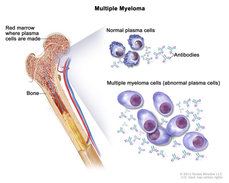
Plasma Cell Neoplasms (Including Multiple Myeloma)—Patient Version

Plasma Cell Neoplasms (Including Multiple Myeloma)—Patient Version

Overview

Plasma cell neoplasms occur when abnormal plasma cells form cancerous tumors in bone or soft tissue. When there is only one tumor, the disease is called a plasmacytoma. When there are multiple tumors, it is called multiple myeloma. Explore the links on this page to learn more about multiple myeloma treatment, statistics, research, and clinical trials.

Causes & Prevention

NCI does not have PDQ evidence-based information about prevention of plasma cell neoplasms (including multiple myeloma).

Screening

NCI does not have PDQ evidence-based information about screening for plasma cell neoplasms (including multiple myeloma).

Coping with Cancer

The information in this section is meant to help you cope with the many issues and concerns that occur when you have cancer.

Emotions and Cancer Adjusting to Cancer Support for Caregivers Survivorship Advanced Cancer Managing Cancer Care

Plasma Cell Neoplasms (Including Multiple Myeloma)—Health Professional Version

Plasma Cell Neoplasms (Including Multiple Myeloma)—Health Professional Version

Causes & Prevention

NCI does not have PDQ evidence-based information about prevention of plasma cell neoplasms (including multiple myeloma).

Screening

NCI does not have PDQ evidence-based information about screening for plasma cell neoplasms (including multiple myeloma).

Supportive & Palliative Care

We offer evidence-based supportive and palliative care information for health professionals on the assessment and management of cancer-related symptoms and conditions.

Cancer Pain Nausea and Vomiting Nutrition in Cancer Care Transition to End-of-Life Care Last Days of Life View all Supportive and Palliative Care Summaries

Advances in Multiple Myeloma Research

Advances in Multiple Myeloma Research

 Myeloma tumor cells shown in green and bone cells shown in red, growing on a scaffold made of silk protein in purple, which is designed to resemble bone material.

Myeloma tumor cells (in green) and bone cells (red) growing on a scaffold made of silk protein (purple), which is designed to resemble bone material.

Multiple myeloma is the most common type of plasma cell cancer. Plasma cells develop from a type of white blood cell found in the bone marrow. They normally make antibodies to fight bacteria and viruses, to stop infection and disease. Plasma cell cancers occur when abnormal plasma cells form tumors in the bones or soft tissues of the body.

NCI-funded researchers are working to advance our understanding of how to treat plasma cell cancers, including multiple myeloma and other related cancers.

This page highlights some of the latest research in multiple myeloma and other plasma cell cancers, including clinical advances that may soon translate into improved care and research findings from recent studies. To learn about standard therapies for multiple myeloma, see Plasma Cell Neoplasms (Including Multiple Myeloma) Treatment.

Research in Multiple Myeloma Treatment

Multiple myeloma is not considered curable. However, with recent advances in treatment, it can be managed like a chronic disease in some people.

The mainstays of treatment for multiple myeloma have been chemotherapy followed by stem cell transplant for people healthy enough to tolerate the procedure. Targeted therapies and immunotherapies have also been used, either to prepare people for a stem cell transplant or in place of one. Recent advances in immunotherapy have changed the way many people are treated, especially those unable to have a stem cell transplant.

Immunotherapy

Immunotherapy is treatment that helps the body’s immune system fight cancer more effectively. Types of immunotherapies being used or tested for multiple myeloma include:

CAR T Cells

CAR T cells are an immunotherapy in which a patient’s T cells, a type of immune cell, are changed in the lab so they will better attack cancer cells and then returned to the patient’s bloodstream.

Two types of CAR T cells have been approved by the FDA to treat multiple myeloma that has come back after previous treatments:

Researchers are now testing whether some patients may benefit from getting CAR T-cell therapy instead of a stem cell transplant as their initial treatment.

Currently, CAR T cells must be created from scratch for each patient, making them the most personalized of therapies. But this process is complicated and expensive. Researchers have been testing the use of so-called off-the-shelf CAR T-cell therapies, which could potentially be made in bulk and used immediately.

An ongoing trial at NCI is also testing another type of immunotherapy using T cells, called TCR T-cell therapy, in people with multiple myeloma who have at least one tumor that can be removed surgically.

Bispecific T-Cell Engagers (BiTEs)

BiTEs are drugs that latch onto both tumor cells and T cells. By bringing T cells and cancer cells close together, they help the T cells recognize and destroy the cancer cells.

Three BiTEs have been approved by the FDA to treat adults with multiple myeloma that came back or did not get better after treatment with several other anticancer therapies:

Researchers are now studying whether giving these drugs to people with multiple myeloma who have received only one previous treatment can help keep the disease at bay for longer. They’re also making sure the potential side effects, such as an increased risk of dangerous infections, don’t outweigh the potential benefits.

Ongoing trials are also testing whether using more than one BiTE at the same time can keep multiple myeloma in remission for longer than using a single BiTE. Additional trials, including one sponsored by NCI, are investigating combinations of other new myeloma therapies with BiTEs.

Immunomodulating Drugs

Immunomodulating agents are drugs that either stimulate or suppress parts of the immune system to help the body fight cancer. These types of drugs, including lenalidomide (Revlimid) and pomalidomide (Actimid), have been used for decades to treat some people with multiple myeloma.

Studies are now testing a new generation of immunomodulating drugs that have been developed for use once resistance to current drugs occurs. These include iberdomide and mezigdomide.

Targeted Therapies

Targeted therapy treats cancer by shutting down proteins that control how cancer cells grow, divide, and spread. Some of the earliest targeted therapies, drugs called proteasome inhibitors, were developed for use in multiple myeloma. These drugs, such as bortezomib (Velcade), block the action of proteasomes, large protein complexes that help destroy other cellular proteins when they are no longer needed.

But resistance to proteasome inhibitors eventually develops and multiple myeloma starts to grow again. So researchers are searching for new ways to shut down multiple myeloma cells using targeted drugs.

Approaches being tested include:

Picking very specific populations for treatment. For example, studies found that adding venetoclax (Venclexta)—a drug that has shown promise in treating some types of leukemia—to other multiple myeloma drugs actually made myeloma grow faster. However, further research suggested that people whose multiple myeloma tumors harbor a rare genetic mutation may benefit from venetoclax. Clinical trials are now testing the drug only in people with this specific gene change.

Targeting a family of genes called RAS. After pancreatic cancer and colorectal cancer, multiple myeloma is the third most likely cancer type to be driven by changes in RAS. RAS used to be considered “undruggable,” that is, that it couldn’t be shut down with targeted therapies. But over the last decade, drugs have been developed that can shut down RAS and stop tumor growth. Clinical trials, including one at NCI, are now testing such drugs in people with multiple myeloma.

Targeting epigenetic regulation of cancer cells. Epigenetics refers to changes in the way genes are switched on and off that don’t involve changes in the actual DNA sequence. Drugs that shut down cancer cells by targeting their epigenetic regulation are now being tested in multiple myeloma.

Monoclonal antibodies (Mabs). Mabs are versions of immune system proteins that are created in the lab and bind to cancer cells. They can kill cancer cells directly or indirectly, by engaging the immune system to kill the cancer cells. 

A Mab called daratumumab (Darzalex) binds to a protein found on the surface of myeloma cells and helps immune cells kill myeloma cells. Daratumumab is FDA approved to be used with some drug combinations for newly diagnosed multiple myeloma, as well as myeloma that has relapsed, and is being tested in addition to other combinations.

For example, a recent study tested adding daratumumab to the standard chemotherapy drugs given after an initial diagnosis of multiple myeloma. Patients treated with daratumumab lived substantially longer without their cancer getting worse or dying than those who received the standard treatment only. An ongoing study is now testing whether giving people with newly diagnosed multiple myeloma a treatment regimen that includes daratumumab can lengthen the time before a stem cell transplant is needed.

FDA has also approved another Mab, called isatuximab (Sarclisa), to be given along with the drugs bortezomib (Velcade), lenalidomide (Revlimid), and dexamethasone. The approval was based on a clinical trial called IMROZ, which showed that the four-drug regimen substantially increased the time patients lived without evidence of their cancer coming back or getting worse.

Elotuzumab (Empliciti) is another monoclonal antibody approved by for myeloma that has relapsed after previous treatment. This Mab targets a different protein on myeloma cells than the one targeted by daratumumab and isatuximab, so it may be effective after other antibodies stop working. Elotuzumab is currently being tested in combinations with other targeted therapies and with immunotherapies.

Advances in Stem Cell Transplant

Despite advances in immunotherapy and targeted therapies, autologous stem cell transplant is still used to treat many people with multiple myeloma. But often, too few stem cells can be successfully collected from a patient, making transplant impossible. Researchers are working to make stem cell transplant an option for more people with this cancer type.

For example, a clinical trial funded in part by NCI tested the injection of a drug called motixafortide (Aphexda) in addition to injections of G-CSF, the drug most widely used to “mobilize” stem cells from the bone marrow to the blood. People who received motixafortide had a markedly increased number of stem cells that could be collected for transplant compared with people who received G-CSF alone. Motixafortide received FDA approval in 2023 for use as part of preparation for an autologous stem cell transplant.

Research in the Treatment of Precursor Conditions

Multiple myeloma is a slow-growing cancer. It can develop silently for years without causing symptoms. The most common such precursor condition to multiple myeloma is called monoclonal gammopathy of undetermined significance, or MGUS. People with this condition have abnormal levels of certain blood markers. In some people, MGUS can progress to a condition called smoldering myeloma, which also doesn’t have symptoms. From there, it may turn into multiple myeloma. But it also may not cause full-blown cancer in a person’s lifetime.

Both MGUS and smoldering myeloma are usually found incidentally, by blood tests looking for other problems. If this happens, people are usually monitored and not given treatment right away. However, researchers have wondered if they can predict which people with MGUS or smoldering myeloma will eventually progress to myeloma. And, if progression can be predicted, would giving treatment before multiple myeloma develops help them live longer? Or would it only expose them to the side effects of treatment earlier without providing any benefit?

  • In a recent clinical trial called AQUILA, which was funded by the manufacturer of daratumumab, people with smoldering myeloma at high risk of progressing to multiple myeloma were randomly assigned to either receive the drug for up to 3 years or to undergo monitoring. Fewer patients receiving daratumumab progressed to multiple myeloma during the study, though they also experienced more side effects.
  • An ongoing trial at NCI is testing daratumumab as part of a multidrug regimen in people with smoldering myeloma at high risk of progressing to multiple myeloma.

NCI-Supported Research Programs

Many NCI-funded researchers working at the NIH campus and across the United States and the world are seeking ways to address multiple myeloma and other plasma cell neoplasms more effectively. Some research is basic, exploring questions as diverse as the biological underpinnings of cancer. And some is more clinical, seeking to translate this basic information into improving patient outcomes. The programs listed below are a small sampling of NCI’s research efforts in multiple myeloma and related plasma cell tumors.

The Multiple Myeloma Specialized Programs of Research Excellence (Myeloma SPOREs) are designed to quickly move basic scientific findings into clinical settings. The Myeloma SPOREs support the development of new treatments for multiple myeloma and related, rarer conditions such as Waldenstrom’s macroglobulinemia.

The National Clinical Trials Network funds clinical trials testing new treatments for multiple myeloma as well as precursor conditions such as smoldering myeloma. 

The Cancer Intervention and Surveillance Modeling Network (CISNET) is a consortium of NCI-sponsored investigators who use simulation modeling to improve our understanding of cancer control interventions in prevention, screening, and treatment and their effects on population trends in incidence and mortality. Investigators within CISNET’s Multiple Myeloma Working Group are developing such models to assess the value of guideline-recommended therapies and novel intervention strategies for myeloma prevention and control.

The Genomic Data Commons (GDC) provides the cancer research community with a unified repository and cancer knowledge base that enables data sharing across cancer genomic studies in support of precision medicine. The Multiple Myeloma Research Foundation has made genomic data from a large clinical trial of precision medicine for multiple myeloma, called The Relating Clinical Outcomes in Multiple Myeloma to Personal Assessment of Genetic Profile study (CoMMpassSM) available to the research community through the GDC.

Clinical Trials for Multiple Myeloma and Other Plasma Cell Cancers

NCI funds and oversees both early- and late-phase clinical trials to develop new treatments and improve patient care. Treatment clinical trials are available for multiple myeloma and other plasma cell cancers.

Multiple Myeloma Research Results

The following are some of NCI’s latest news articles on multiple myeloma research:

View the full list of Plasma Cell Neoplasm Research Results and Study Updates.

Plasma Cell Neoplasms (Including Multiple Myeloma) Treatment (PDQ®)–Health Professional Version

Plasma Cell Neoplasms (Including Multiple Myeloma) Treatment (PDQ®)–Health Professional Version

General Information About Plasma Cell Neoplasms

There are several types of plasma cell neoplasms. These diseases are all associated with a monoclonal (or myeloma) protein (M protein). They include monoclonal gammopathy of undetermined significance (MGUS), isolated plasmacytoma of the bone, extramedullary plasmacytoma, and multiple myeloma.

For information about immunoglobulin (Ig) M monoclonal antibody–associated lymphoma, see the Lymphoplasmacytic Lymphoma (Waldenström Macroglobulinemia) section in Indolent B-Cell Non-Hodgkin Lymphoma Treatment.

Incidence and Mortality

Estimated new cases and deaths from multiple myeloma in the United States in 2025:[1]

  • New cases: 36,110.
  • Deaths: 12,030.

Clinical Presentation and Evaluation

Table 1. Clinical Presentation of Plasma Cell Neoplasms
Plasma Cell Neoplasm M Protein Type Pathology Clinical Presentation
Ig = immunoglobulin; MGUS = monoclonal gammopathy of undetermined significance.
MGUS IgG kappa or lambda; or IgA kappa or lambda <10% plasma cells in bone marrow Asymptomatic, with minimal evidence of disease (aside from the presence of an M protein) [2]
Isolated plasmacytoma of bone IgG kappa or lambda; or IgA kappa or gamma Solitary lesion of bone; <10% plasma cells in marrow of uninvolved site Asymptomatic or symptomatic
Extramedullary plasmacytoma IgG kappa or lambda; or IgA kappa or gamma Solitary lesion of soft tissue; most commonly occurs in the nasopharynx, tonsils, or paranasal sinuses [3] Asymptomatic or symptomatic
Multiple myeloma IgG kappa or lambda; or IgA kappa or gamma Often, multiple lesions of bone Symptomatic

Evaluation of patients with monoclonal (or myeloma) protein (M protein)

Idiotypic myeloma cells can be found in the blood of patients with myeloma in all stages of the disease.[4,5] For this reason, when treatment is indicated, systemic treatment must be considered for all patients with symptomatic plasma cell neoplasms. Patients with MGUS or asymptomatic smoldering myeloma do not require immediate treatment but must be followed carefully for signs of disease progression.

The major challenge is to separate the stable asymptomatic group of patients who do not require treatment from patients with progressive, symptomatic myeloma who may need to be treated immediately.[68]

Patients with an M protein in the serum and/or urine are evaluated by some of the following criteria:

  • Measure and follow the serum M protein by serum electrophoresis or by specific Ig assays; however, specific Ig quantification always overestimates the M protein because normal Ig are included in the result. For this reason, the preference is often that baseline and follow-up measurements of the M protein be done by the same method.[9] Quantitative serum free light chains (FLC) may be helpful to follow response when an M protein is not apparent.
  • Measure and follow the amount of M protein light chains excreted in the urine over 24 hours. Measure the total amount of protein excreted over 24 hours and multiply this value by the percentage of urine protein that is M protein, as determined by electrophoresis of concentrated urine protein. An easier, but less accurate, method uses a spot-urine protein electrophoresis.
  • Identify the heavy and light chain of the M protein by immunofixation electrophoresis.
  • Measure the hemoglobin, leukocyte, platelet, and differential counts.
  • Determine the percentage of marrow plasma cells. Be aware that marrow plasma-cell distribution may vary in different sites. Bone marrow is often sent for cytogenetics and fluorescence in situ hybridization testing for genetic markers of high-risk disease. For more information, see the Genetic factors and risk groups section.
  • Measure serum free kappa and lambda light chains. This is especially useful in cases of oligosecretory plasma-cell dyscrasia or for following cases of light-chain amyloidosis.[10] The FLC ratio of over 100 can predict a greater than 70% progression within 2 years in patients with smoldering myeloma.[11]
  • If clinically warranted, obtain needle aspirates of a solitary lytic bone lesion, extramedullary tumor(s), or enlarged lymph node(s) to determine whether these are plasmacytomas.
  • Evaluate renal function with serum creatinine and a creatinine clearance.
  • Electrophoresis of concentrated urine protein is very helpful in differentiating glomerular lesions from tubular lesions. Glomerular lesions, such as those resulting from glomerular deposits of amyloid or light-chain deposition disease, result in the nonselective leakage of all serum proteins into the urine; the electrophoresis pattern of this urine resembles the serum pattern with a preponderance of albumin.

    In most patients with myeloma, the glomeruli function normally allows only the small molecular weight proteins, such as light chains, to filter into the urine. The concentration of protein in the tubules increases as water is reabsorbed. This leads to precipitation of proteins and the formation of tubular casts, which may injure the tubular cells. With tubular lesions, the typical electrophoresis pattern shows a small albumin peak and a larger light-chain peak in the globulin region; this tubular pattern is the usual pattern found in patients with myeloma.

  • Measure serum levels of calcium, alkaline phosphatase, lactic dehydrogenase, and, when indicated by clinical symptoms, cryoglobulins and serum viscosity.
  • Obtain radiographs of the skull, ribs, vertebrae, pelvis, shoulder girdle, and long bones.
  • Obtain a spinal magnetic resonance imaging (MRI) scan (or spinal computed tomography [CT] or positron emission tomography [PET]–CT scan depending on availability) if the skeletal survey is negative.[1214] At diagnosis, whole-body PET scan or MRI of the total spine and pelvis appears to be equally efficacious in the detection of bone lesions.[15,16]
  • If amyloidosis is suspected, perform a needle aspiration of subcutaneous abdominal fat and stain the bone marrow biopsy for amyloid as the easiest and safest way to confirm the diagnosis.[17]
  • Measure serum albumin and beta-2-microglobulin as independent prognostic factors.[18,19]
  • The presence of circulating myeloma cells is considered a poor prognostic factor.[20] Primary plasma cell leukemia has a particularly poor prognosis.[21,22]

These initial studies are often compared with subsequent values at a later time, when it is necessary to decide whether the disease is stable or progressive, responding to treatment, or getting worse.

Monoclonal Gammopathy of Undetermined Significance (MGUS)

Patients with MGUS have an M protein in the serum without findings of multiple myeloma, macroglobulinemia, amyloidosis, or lymphoma and have fewer than 10% of plasma cells in the bone marrow.[2,2325] Patients with smoldering myeloma have similar characteristics but may have more than 10% of plasma cells in the bone marrow.

These types of patients are asymptomatic and do not need to be treated. However, patients with MGUS and risk factors for disease progression must be followed carefully because they are more likely to develop myeloma (most commonly), amyloidosis, lymphoplasmacytic lymphoma, or chronic lymphocytic leukemia. These patients may then require therapy.[2527]

Virtually all cases of multiple myeloma are preceded by a gradually rising level of MGUS.[2830] The annual risk of progression of MGUS to a lymphoid or plasma cell malignancy ranges from 0.5% to 1.0% in population-based cohorts.[31,32] This risk ranges from 2% to more than 20% in higher-risk patients.

The following risk factors predict disease progression:

  • An abnormal serum FLC ratio.[31,33]
  • Non-IgG class MGUS.
  • A high level of serum M protein (≥1.5 g/dL).[31,33]

A Swedish cohort study confirmed that an abnormal serum FLC ratio and a high level of serum monoclonal protein are high-risk factors.[32] The study described the additional risk factor of immunoparesis, which is defined as the reciprocal depression of the other Ig classes (i.e., if a patient has an IgG kappa M protein, the IgM and IgA would be below normal levels with immunoparesis). Incorporation of gene-expression profiles to better assess risk is under clinical evaluation.[34]

Monoclonal gammopathies that cause organ damage, particularly to the kidney, heart, or peripheral nerves, require immediate therapy with the same strategies applied for the conventional plasma-cell dyscrasias.[35] A monoclonal gammopathy causing renal dysfunction—by direct antibody deposition or amyloidosis—is referred to as monoclonal gammopathy of renal significance.[36] Rising serum creatinine, dropping glomerular filtration rates, and increasing urinary–albumin excretion are all parameters that may signify renal damage and are assessed prospectively for high-risk MGUS patients. Although the N-terminal pro-brain natriuretic peptide is a very sensitive marker for amyloid involvement in the heart, the low specificity must be noted. These extra tests are included with the M-protein level, FLC levels, and FLC ratio when following patients with MGUS.[37]

In a retrospective review of 6,399 patients with newly diagnosed multiple myeloma, 44 patients were found to have a biclonal IgG or IgA MGUS. The overall response rate of the myeloma clone to induction therapy was 93%, compared with 64% for the separate-clone MGUS (P = .001).[38][Level of evidence C3] Many MGUS plasma cell clones were unresponsive to available myeloma therapy; this result highlights the need to lower expectations for response in situations in which an MGUS may require therapy because of end-organ damage.

Isolated Plasmacytoma of Bone

The patient has an isolated plasmacytoma of the bone if the following are found:

  • A solitary lytic lesion of plasma cells on skeletal survey in an otherwise asymptomatic patient.
  • A bone marrow examination from an uninvolved site contains less than 10% plasma cells.[3941] The absence of plasma cells on flow cytometry of the bone marrow suggests a low (<10%) risk of recurrence after radiation therapy of the isolated bone plasmacytoma.[42]

MRI may reveal unsuspected bony lesions that were undetected on standard radiographs. MRI scans of the total spine and pelvis may identify other bony lesions.[43]

Extramedullary Plasmacytoma

A patient has extramedullary plasmacytoma if the following are found:

  • Isolated plasma-cell tumors of soft tissues, most commonly occurring in the tonsils, nasopharynx, or paranasal sinuses.
  • Negative findings on skeletal x-rays and bone marrow biopsy.[4446]

Multiple Myeloma

Multiple myeloma is a systemic malignancy of plasma cells that typically involves multiple sites within the bone marrow and secretes all or part of a monoclonal antibody.

Prognosis

Multiple myeloma is highly treatable but rarely curable. The median survival in the prechemotherapy era was about 7 months. After the introduction of chemotherapy, prognosis improved significantly with a median survival of 24 to 30 months and a 10-year survival rate of 3%. Even further improvements in prognosis have occurred because of the introduction of newer biological therapies and better salvage options, with median survivals now exceeding 10 years.[47] Patients with plasma cell leukemia or with soft tissue plasmacytomas (often with plasmablastic morphology) in association with multiple myeloma have poor outcomes.[21,48] Racial disparities because of socioeconomic factors, genetics, differences in risk factor exposure, and structural racism are under evaluation.[49]

Multiple myeloma is potentially curable when it presents as a solitary plasmacytoma of bone or as an extramedullary plasmacytoma. For more information, see the sections on Isolated Plasmacytoma of Bone and Extramedullary Plasmacytoma.

Amyloidosis Associated With Plasma Cell Neoplasms

Multiple myeloma and other plasma cell neoplasms may cause a condition called amyloidosis. Primary amyloidosis can result in severe organ dysfunction, especially in the kidney, heart, or peripheral nerves.[50] Clinical symptoms and signs include:

  • Fatigue.
  • Purpura.
  • Enlarged tongue.
  • Diarrhea.
  • Edema.
  • Lower-extremity paresthesia.

Accurate diagnosis of amyloidosis requires histological evidence of amyloid deposits and characterization of the amyloidogenic protein using immunoelectron microscopy.[51] In one series of 745 consecutive patients, 20% of patients with nonamyloid light chain amyloidosis (usually transthyretin) had an innocent monoclonal gammopathy, indicating the significant risk of misdiagnosis.[51]

Elevated serum levels of cardiac troponins, amino-terminal fragment brain-type natriuretic peptide, and serum FLC are poor prognostic factors.[52,53] Proposed staging systems for primary systemic amyloidosis based on these serum levels require independent and prospective confirmation.[52,54] An increase in levels of serum FLC over many years can precede the clinical diagnosis of amyloid light chain amyloidosis.[55] Amyloidosis associated with an IgM monoclonal gammopathy is a rare, but distinct, clinical entity with more frequent neuropathy and adenopathy and less cardiac involvement.[56]

POEMS Syndrome

POEMS (polyneuropathy, organomegaly, endocrinopathy, monoclonal gammopathy, and skin changes) syndrome is a rare paraneoplastic condition associated with a plasma cell dyscrasia of early or late stage. The acronym describes a constellation of findings often marked by polyneuropathy, organomegaly (usually splenomegaly), endocrinopathy, monoclonal plasma cell dyscrasia, and skin changes.[57] Both sclerotic or lytic bone lesions and lymphadenopathy (with possible Castleman histology) may be identified. Anecdotal reports suggest remissions have been achieved using myeloma-directed therapy.[5862]

References
  1. American Cancer Society: Cancer Facts and Figures 2025. American Cancer Society, 2025. Available online. Last accessed January 16, 2025.
  2. Kyle RA, Rajkumar SV: Monoclonal gammopathy of undetermined significance and smouldering multiple myeloma: emphasis on risk factors for progression. Br J Haematol 139 (5): 730-43, 2007. [PUBMED Abstract]
  3. Knowling MA, Harwood AR, Bergsagel DE: Comparison of extramedullary plasmacytomas with solitary and multiple plasma cell tumors of bone. J Clin Oncol 1 (4): 255-62, 1983. [PUBMED Abstract]
  4. Zandecki M, Facon T, Preudhomme C, et al.: Significance of circulating plasma cells in multiple myeloma. Leuk Lymphoma 14 (5-6): 491-6, 1994. [PUBMED Abstract]
  5. Billadeau D, Van Ness B, Kimlinger T, et al.: Clonal circulating cells are common in plasma cell proliferative disorders: a comparison of monoclonal gammopathy of undetermined significance, smoldering multiple myeloma, and active myeloma. Blood 88 (1): 289-96, 1996. [PUBMED Abstract]
  6. He Y, Wheatley K, Clark O, et al.: Early versus deferred treatment for early stage multiple myeloma. Cochrane Database Syst Rev (1): CD004023, 2003. [PUBMED Abstract]
  7. Kyle RA, Remstein ED, Therneau TM, et al.: Clinical course and prognosis of smoldering (asymptomatic) multiple myeloma. N Engl J Med 356 (25): 2582-90, 2007. [PUBMED Abstract]
  8. Vaxman I, Gertz MA: How I approach smoldering multiple myeloma. Blood 140 (8): 828-838, 2022. [PUBMED Abstract]
  9. Riches PG, Sheldon J, Smith AM, et al.: Overestimation of monoclonal immunoglobulin by immunochemical methods. Ann Clin Biochem 28 ( Pt 3): 253-9, 1991. [PUBMED Abstract]
  10. Dispenzieri A, Kyle R, Merlini G, et al.: International Myeloma Working Group guidelines for serum-free light chain analysis in multiple myeloma and related disorders. Leukemia 23 (2): 215-24, 2009. [PUBMED Abstract]
  11. Larsen JT, Kumar SK, Dispenzieri A, et al.: Serum free light chain ratio as a biomarker for high-risk smoldering multiple myeloma. Leukemia 27 (4): 941-6, 2013. [PUBMED Abstract]
  12. Horger M, Kanz L, Denecke B, et al.: The benefit of using whole-body, low-dose, nonenhanced, multidetector computed tomography for follow-up and therapy response monitoring in patients with multiple myeloma. Cancer 109 (8): 1617-26, 2007. [PUBMED Abstract]
  13. Walker R, Barlogie B, Haessler J, et al.: Magnetic resonance imaging in multiple myeloma: diagnostic and clinical implications. J Clin Oncol 25 (9): 1121-8, 2007. [PUBMED Abstract]
  14. Kyle RA, Durie BG, Rajkumar SV, et al.: Monoclonal gammopathy of undetermined significance (MGUS) and smoldering (asymptomatic) multiple myeloma: IMWG consensus perspectives risk factors for progression and guidelines for monitoring and management. Leukemia 24 (6): 1121-7, 2010. [PUBMED Abstract]
  15. Moreau P, Attal M, Caillot D, et al.: Prospective Evaluation of Magnetic Resonance Imaging and [18F]Fluorodeoxyglucose Positron Emission Tomography-Computed Tomography at Diagnosis and Before Maintenance Therapy in Symptomatic Patients With Multiple Myeloma Included in the IFM/DFCI 2009 Trial: Results of the IMAJEM Study. J Clin Oncol 35 (25): 2911-2918, 2017. [PUBMED Abstract]
  16. Zamagni E, Tacchetti P, Cavo M: Imaging in multiple myeloma: How? When? Blood 133 (7): 644-651, 2019. [PUBMED Abstract]
  17. Gertz MA, Li CY, Shirahama T, et al.: Utility of subcutaneous fat aspiration for the diagnosis of systemic amyloidosis (immunoglobulin light chain). Arch Intern Med 148 (4): 929-33, 1988. [PUBMED Abstract]
  18. Greipp PR: Advances in the diagnosis and management of myeloma. Semin Hematol 29 (3 Suppl 2): 24-45, 1992. [PUBMED Abstract]
  19. Durie BG, Stock-Novack D, Salmon SE, et al.: Prognostic value of pretreatment serum beta 2 microglobulin in myeloma: a Southwest Oncology Group Study. Blood 75 (4): 823-30, 1990. [PUBMED Abstract]
  20. Garcés JJ, Cedena MT, Puig N, et al.: Circulating Tumor Cells for the Staging of Patients With Newly Diagnosed Transplant-Eligible Multiple Myeloma. J Clin Oncol 40 (27): 3151-3161, 2022. [PUBMED Abstract]
  21. Pagano L, Valentini CG, De Stefano V, et al.: Primary plasma cell leukemia: a retrospective multicenter study of 73 patients. Ann Oncol 22 (7): 1628-35, 2011. [PUBMED Abstract]
  22. Royer B, Minvielle S, Diouf M, et al.: Bortezomib, Doxorubicin, Cyclophosphamide, Dexamethasone Induction Followed by Stem Cell Transplantation for Primary Plasma Cell Leukemia: A Prospective Phase II Study of the Intergroupe Francophone du Myélome. J Clin Oncol 34 (18): 2125-32, 2016. [PUBMED Abstract]
  23. Kyle RA, Therneau TM, Rajkumar SV, et al.: Prevalence of monoclonal gammopathy of undetermined significance. N Engl J Med 354 (13): 1362-9, 2006. [PUBMED Abstract]
  24. International Myeloma Working Group: Criteria for the classification of monoclonal gammopathies, multiple myeloma and related disorders: a report of the International Myeloma Working Group. Br J Haematol 121 (5): 749-57, 2003. [PUBMED Abstract]
  25. Bird J, Behrens J, Westin J, et al.: UK Myeloma Forum (UKMF) and Nordic Myeloma Study Group (NMSG): guidelines for the investigation of newly detected M-proteins and the management of monoclonal gammopathy of undetermined significance (MGUS). Br J Haematol 147 (1): 22-42, 2009. [PUBMED Abstract]
  26. Attal M, Harousseau JL, Stoppa AM, et al.: A prospective, randomized trial of autologous bone marrow transplantation and chemotherapy in multiple myeloma. Intergroupe Français du Myélome. N Engl J Med 335 (2): 91-7, 1996. [PUBMED Abstract]
  27. Kyle RA, Therneau TM, Rajkumar SV, et al.: A long-term study of prognosis in monoclonal gammopathy of undetermined significance. N Engl J Med 346 (8): 564-9, 2002. [PUBMED Abstract]
  28. Weiss BM, Abadie J, Verma P, et al.: A monoclonal gammopathy precedes multiple myeloma in most patients. Blood 113 (22): 5418-22, 2009. [PUBMED Abstract]
  29. Landgren O, Kyle RA, Pfeiffer RM, et al.: Monoclonal gammopathy of undetermined significance (MGUS) consistently precedes multiple myeloma: a prospective study. Blood 113 (22): 5412-7, 2009. [PUBMED Abstract]
  30. Bladé J, Rosiñol L, Cibeira MT: Are all myelomas preceded by MGUS? Blood 113 (22): 5370, 2009. [PUBMED Abstract]
  31. Rajkumar SV, Kyle RA, Therneau TM, et al.: Serum free light chain ratio is an independent risk factor for progression in monoclonal gammopathy of undetermined significance. Blood 106 (3): 812-7, 2005. [PUBMED Abstract]
  32. Turesson I, Kovalchik SA, Pfeiffer RM, et al.: Monoclonal gammopathy of undetermined significance and risk of lymphoid and myeloid malignancies: 728 cases followed up to 30 years in Sweden. Blood 123 (3): 338-45, 2014. [PUBMED Abstract]
  33. Kyle RA, Larson DR, Therneau TM, et al.: Long-Term Follow-up of Monoclonal Gammopathy of Undetermined Significance. N Engl J Med 378 (3): 241-249, 2018. [PUBMED Abstract]
  34. Dhodapkar MV, Sexton R, Waheed S, et al.: Clinical, genomic, and imaging predictors of myeloma progression from asymptomatic monoclonal gammopathies (SWOG S0120). Blood 123 (1): 78-85, 2014. [PUBMED Abstract]
  35. Fermand JP, Bridoux F, Dispenzieri A, et al.: Monoclonal gammopathy of clinical significance: a novel concept with therapeutic implications. Blood 132 (14): 1478-1485, 2018. [PUBMED Abstract]
  36. Leung N, Bridoux F, Nasr SH: Monoclonal Gammopathy of Renal Significance. N Engl J Med 384 (20): 1931-1941, 2021. [PUBMED Abstract]
  37. Merlini G: Determining the significance of MGUS. Blood 123 (3): 305-7, 2014. [PUBMED Abstract]
  38. Campbell JP, Heaney JLJ, Pandya S, et al.: Response comparison of multiple myeloma and monoclonal gammopathy of undetermined significance to the same anti-myeloma therapy: a retrospective cohort study. Lancet Haematol 4 (12): e584-e594, 2017. [PUBMED Abstract]
  39. Ozsahin M, Tsang RW, Poortmans P, et al.: Outcomes and patterns of failure in solitary plasmacytoma: a multicenter Rare Cancer Network study of 258 patients. Int J Radiat Oncol Biol Phys 64 (1): 210-7, 2006. [PUBMED Abstract]
  40. Dimopoulos MA, Moulopoulos LA, Maniatis A, et al.: Solitary plasmacytoma of bone and asymptomatic multiple myeloma. Blood 96 (6): 2037-44, 2000. [PUBMED Abstract]
  41. Dimopoulos MA, Hamilos G: Solitary bone plasmacytoma and extramedullary plasmacytoma. Curr Treat Options Oncol 3 (3): 255-9, 2002. [PUBMED Abstract]
  42. Paiva B, Chandia M, Vidriales MB, et al.: Multiparameter flow cytometry for staging of solitary bone plasmacytoma: new criteria for risk of progression to myeloma. Blood 124 (8): 1300-3, 2014. [PUBMED Abstract]
  43. Liebross RH, Ha CS, Cox JD, et al.: Solitary bone plasmacytoma: outcome and prognostic factors following radiotherapy. Int J Radiat Oncol Biol Phys 41 (5): 1063-7, 1998. [PUBMED Abstract]
  44. Tournier-Rangeard L, Lapeyre M, Graff-Caillaud P, et al.: Radiotherapy for solitary extramedullary plasmacytoma in the head-and-neck region: A dose greater than 45 Gy to the target volume improves the local control. Int J Radiat Oncol Biol Phys 64 (4): 1013-7, 2006. [PUBMED Abstract]
  45. Michalaki VJ, Hall J, Henk JM, et al.: Definitive radiotherapy for extramedullary plasmacytomas of the head and neck. Br J Radiol 76 (910): 738-41, 2003. [PUBMED Abstract]
  46. Alexiou C, Kau RJ, Dietzfelbinger H, et al.: Extramedullary plasmacytoma: tumor occurrence and therapeutic concepts. Cancer 85 (11): 2305-14, 1999. [PUBMED Abstract]
  47. Joseph NS, Kaufman JL, Dhodapkar MV, et al.: Long-Term Follow-Up Results of Lenalidomide, Bortezomib, and Dexamethasone Induction Therapy and Risk-Adapted Maintenance Approach in Newly Diagnosed Multiple Myeloma. J Clin Oncol 38 (17): 1928-1937, 2020. [PUBMED Abstract]
  48. Bladé J, Fernández de Larrea C, Rosiñol L, et al.: Soft-tissue plasmacytomas in multiple myeloma: incidence, mechanisms of extramedullary spread, and treatment approach. J Clin Oncol 29 (28): 3805-12, 2011. [PUBMED Abstract]
  49. Marinac CR, Ghobrial IM, Birmann BM, et al.: Dissecting racial disparities in multiple myeloma. Blood Cancer J 10 (2): 19, 2020. [PUBMED Abstract]
  50. Gertz MA, Dispenzieri A: Systemic Amyloidosis Recognition, Prognosis, and Therapy: A Systematic Review. JAMA 324 (1): 79-89, 2020. [PUBMED Abstract]
  51. Fernández de Larrea C, Verga L, Morbini P, et al.: A practical approach to the diagnosis of systemic amyloidoses. Blood 125 (14): 2239-44, 2015. [PUBMED Abstract]
  52. Kumar S, Dispenzieri A, Lacy MQ, et al.: Revised prognostic staging system for light chain amyloidosis incorporating cardiac biomarkers and serum free light chain measurements. J Clin Oncol 30 (9): 989-95, 2012. [PUBMED Abstract]
  53. Pinney JH, Lachmann HJ, Bansi L, et al.: Outcome in renal Al amyloidosis after chemotherapy. J Clin Oncol 29 (6): 674-81, 2011. [PUBMED Abstract]
  54. Lilleness B, Ruberg FL, Mussinelli R, et al.: Development and validation of a survival staging system incorporating BNP in patients with light chain amyloidosis. Blood 133 (3): 215-223, 2019. [PUBMED Abstract]
  55. Weiss BM, Hebreo J, Cordaro DV, et al.: Increased serum free light chains precede the presentation of immunoglobulin light chain amyloidosis. J Clin Oncol 32 (25): 2699-704, 2014. [PUBMED Abstract]
  56. Sachchithanantham S, Roussel M, Palladini G, et al.: European Collaborative Study Defining Clinical Profile Outcomes and Novel Prognostic Criteria in Monoclonal Immunoglobulin M-Related Light Chain Amyloidosis. J Clin Oncol 34 (17): 2037-45, 2016. [PUBMED Abstract]
  57. Dispenzieri A: POEMS syndrome: 2011 update on diagnosis, risk-stratification, and management. Am J Hematol 86 (7): 591-601, 2011. [PUBMED Abstract]
  58. Humeniuk MS, Gertz MA, Lacy MQ, et al.: Outcomes of patients with POEMS syndrome treated initially with radiation. Blood 122 (1): 68-73, 2013. [PUBMED Abstract]
  59. Li J, Zhang W, Jiao L, et al.: Combination of melphalan and dexamethasone for patients with newly diagnosed POEMS syndrome. Blood 117 (24): 6445-9, 2011. [PUBMED Abstract]
  60. Royer B, Merlusca L, Abraham J, et al.: Efficacy of lenalidomide in POEMS syndrome: a retrospective study of 20 patients. Am J Hematol 88 (3): 207-12, 2013. [PUBMED Abstract]
  61. Misawa S, Sato Y, Katayama K, et al.: Safety and efficacy of thalidomide in patients with POEMS syndrome: a multicentre, randomised, double-blind, placebo-controlled trial. Lancet Neurol 15 (11): 1129-37, 2016. [PUBMED Abstract]
  62. Zhao H, Huang XF, Gao XM, et al.: What is the best first-line treatment for POEMS syndrome: autologous transplantation, melphalan and dexamethasone, or lenalidomide and dexamethasone? Leukemia 33 (4): 1023-1029, 2019. [PUBMED Abstract]

Stage Information for Plasma Cell Neoplasms

No generally accepted staging system exists for monoclonal gammopathy of undetermined significance, isolated plasmacytoma of bone, or extramedullary plasmacytoma. Of the plasma cell neoplasms, a staging system exists only for multiple myeloma.

Multiple Myeloma

Multiple myeloma is staged by estimating the myeloma tumor cell mass based on the amount of monoclonal (or myeloma) protein (M protein) in the serum and/or urine, along with various clinical parameters, such as hemoglobin and serum calcium concentrations, the number of lytic bone lesions, and the presence or absence of renal failure. Impaired renal function worsens prognosis regardless of stage.[1]

The stage of the disease at presentation is a strong determinant of survival, but it has little influence on the choice of therapy because almost all patients, except for rare patients with solitary bone tumors or extramedullary plasmacytomas, have generalized disease.

International staging system

The International Myeloma Working Group (IMWG) studied 11,171 patients, 2,901 of whom received high-dose therapy and 8,270 of whom received only standard-dose therapy.[2] The IMWG evaluated 4,445 patients to create a Revised International Staging System (R-ISS) incorporating lactate dehydrogenase levels and interphase fluorescence in situ hybridization (I-FISH) results.[3]

An International Staging System (ISS) was derived and is shown below in Table 2.[2]

Table 2. The International Staging System (ISS) for Multiple Myeloma
Stage Criteria Median Survival (mo)
I-FISH = interphase fluorescence in situ hybridization; LDH = lactate dehydrogenase; R-ISS = Revised International Staging System.
I Beta-2-microglobulin <3.5 mg/L and albumin ≥3.5 g/dL Not reached
II Not R-ISS I or III 83
III Beta-2-microglobulin ≥5.5 mg/L and either high LDH or high-risk chromosomal abnormalities by I-FISH (defined as presence of del(17p) and/or translocation t(4;14) and/or translocation t(14;16)) 43

Genetic factors and risk groups

Newer clinical investigations are stratifying patients with multiple myeloma into so-called good-risk, intermediate-risk, and high-risk groups, based on genetic aberrations detected by I-FISH.[46] (See Table 3 below.) This stratification, based on cytogenetic findings, has been derived from retrospective analyses and requires prospective validation.[4] Bone marrow samples are sent for cytogenetic and FISH analysis.[6] Plasma cell leukemia (>2%–5% circulating plasma cells) has a particularly poor prognosis.[713] The otherwise favorable prognosis of hyperploidy is trumped by coexistent adverse cytogenetics.[14]

Table 3. Risk Groups for Multiple Myeloma
Risk Group Cytogenetic Findings Disease Characteristics Median Survival (y)
FISH = fluorescence in situ hybridization; Ig = immunoglobulin.
Good risk Has any of the following cytogenetic findings: These patients most often have disease that expresses IgG kappa monoclonal gammopathies, and lytic bone lesions. 10–12 [15]
  No adverse FISH or cytogenetics
  Hyperdiploidy
  t(11;14) by FISH
  t(6;14) by FISH
Intermediate risk Has one of the following formerly deleterious criteria that have been abrogated by standard triplet or quadruplet regimens:[16] These patients often have IgA lambda monoclonal gammopathies and less bone disease. 5–10
  t(4;14)
  t(14;16)
High risk Has any of the following cytogenetic findings: These patients have disease that expresses IgA lambda monoclonal gammopathies (often) and skeletal-related complications (less often). <5 for high-risk; <3 for ultra-high risk [15]
  del 17p by FISH
  t(14;16) by FISH
  t(4;14)
  t(14;20)
  del 13
  Biallelic del TP53 (ultra-high risk)
  1q gain (3 copies), 1 q amp (4 copies, ultra-high risk), monoallelic del (1p32),[17] biallelic del (1p32)[17]
  Plasma cell leukemia
References
  1. Royal V, Leung N, Troyanov S, et al.: Clinicopathologic predictors of renal outcomes in light chain cast nephropathy: a multicenter retrospective study. Blood 135 (21): 1833-1846, 2020. [PUBMED Abstract]
  2. Greipp PR, San Miguel J, Durie BG, et al.: International staging system for multiple myeloma. J Clin Oncol 23 (15): 3412-20, 2005. [PUBMED Abstract]
  3. Palumbo A, Avet-Loiseau H, Oliva S, et al.: Revised International Staging System for Multiple Myeloma: A Report From International Myeloma Working Group. J Clin Oncol 33 (26): 2863-9, 2015. [PUBMED Abstract]
  4. Kumar SK, Mikhael JR, Buadi FK, et al.: Management of newly diagnosed symptomatic multiple myeloma: updated Mayo Stratification of Myeloma and Risk-Adapted Therapy (mSMART) consensus guidelines. Mayo Clin Proc 84 (12): 1095-110, 2009. [PUBMED Abstract]
  5. Avet-Loiseau H, Attal M, Campion L, et al.: Long-term analysis of the IFM 99 trials for myeloma: cytogenetic abnormalities [t(4;14), del(17p), 1q gains] play a major role in defining long-term survival. J Clin Oncol 30 (16): 1949-52, 2012. [PUBMED Abstract]
  6. Sonneveld P, Avet-Loiseau H, Lonial S, et al.: Treatment of multiple myeloma with high-risk cytogenetics: a consensus of the International Myeloma Working Group. Blood 127 (24): 2955-62, 2016. [PUBMED Abstract]
  7. Ramsingh G, Mehan P, Luo J, et al.: Primary plasma cell leukemia: a Surveillance, Epidemiology, and End Results database analysis between 1973 and 2004. Cancer 115 (24): 5734-9, 2009. [PUBMED Abstract]
  8. Fernández de Larrea C, Kyle RA, Durie BG, et al.: Plasma cell leukemia: consensus statement on diagnostic requirements, response criteria and treatment recommendations by the International Myeloma Working Group. Leukemia 27 (4): 780-91, 2013. [PUBMED Abstract]
  9. Granell M, Calvo X, Garcia-Guiñón A, et al.: Prognostic impact of circulating plasma cells in patients with multiple myeloma: implications for plasma cell leukemia definition. Haematologica 102 (6): 1099-1104, 2017. [PUBMED Abstract]
  10. Mina R, Joseph NS, Kaufman JL, et al.: Survival outcomes of patients with primary plasma cell leukemia (pPCL) treated with novel agents. Cancer 125 (3): 416-423, 2019. [PUBMED Abstract]
  11. Royer B, Minvielle S, Diouf M, et al.: Bortezomib, Doxorubicin, Cyclophosphamide, Dexamethasone Induction Followed by Stem Cell Transplantation for Primary Plasma Cell Leukemia: A Prospective Phase II Study of the Intergroupe Francophone du Myélome. J Clin Oncol 34 (18): 2125-32, 2016. [PUBMED Abstract]
  12. Gonsalves WI, Rajkumar SV, Go RS, et al.: Trends in survival of patients with primary plasma cell leukemia: a population-based analysis. Blood 124 (6): 907-12, 2014. [PUBMED Abstract]
  13. Jelinek T, Bezdekova R, Zihala D, et al.: More Than 2% of Circulating Tumor Plasma Cells Defines Plasma Cell Leukemia-Like Multiple Myeloma. J Clin Oncol 41 (7): 1383-1392, 2023. [PUBMED Abstract]
  14. Pawlyn C, Melchor L, Murison A, et al.: Coexistent hyperdiploidy does not abrogate poor prognosis in myeloma with adverse cytogenetics and may precede IGH translocations. Blood 125 (5): 831-40, 2015. [PUBMED Abstract]
  15. Davies FE, Pawlyn C, Usmani SZ, et al.: Perspectives on the Risk-Stratified Treatment of Multiple Myeloma. Blood Cancer Discov 3 (4): 273-284, 2022. [PUBMED Abstract]
  16. Khot A: Del(1p32): prime time in (ultra) high-risk myeloma. Blood 141 (11): 1241-1243, 2023. [PUBMED Abstract]
  17. Schavgoulidze A, Talbot A, Perrot A, et al.: Biallelic deletion of 1p32 defines ultra-high-risk myeloma, but monoallelic del(1p32) remains a strong prognostic factor. Blood 141 (11): 1308-1315, 2023. [PUBMED Abstract]

Treatment Option Overview for Plasma Cell Neoplasms

The major challenge in treating plasma cell neoplasms is separating the stable asymptomatic patients who do not require immediate treatment from patients with progressive symptomatic myeloma who may need to be treated immediately.[13] Monoclonal gammopathy of undetermined significance or smoldering myeloma must be distinguished from progressive myeloma.

Asymptomatic Plasma Cell Neoplasms (Smoldering Multiple Myeloma)

Asymptomatic patients with multiple myeloma who have no lytic bone lesions and normal renal function may be initially observed safely outside the context of a clinical trial.[1,4,5] Increasing anemia is the most reliable indicator of progression.[5] The following criteria represent the new definition for smoldering myeloma:[3]

  • Serum monoclonal protein immunoglobulin (Ig) G or IgA of at least 30 g/L or urinary monoclonal protein of at least 500 mg per 24 hours.
  • Clonal bone marrow plasma cells 10% to 60% (>60% represents overt myeloma).
  • Absence of amyloidosis or myeloma-defining events as follows:
    • Hypercalcemia greater than 1 mg/dL higher than reference range.
    • Creatinine greater than 2 mg/dL or creatinine clearance less than 40 mL/min.
    • Anemia with hemoglobin less than 10.0 g/dL.
    • Bone lesions (one or more) on skeletal radiography, computed tomography (CT) or positron emission tomography (PET)-CT.
    • Clonal plasma cell percentage in marrow at 60% or more.
    • Involved:uninvolved serum free light chain (FLC) ratio of 100 or more.
    • More than one focal lesion of at least 5 mm on magnetic resonance imaging (MRI) of the spine.

The International Myeloma Working Group (IMWG) 2/20/20 rule measures four adverse risk factors for patients with smoldering myeloma. The presence of three or four of the following adverse factors predicts a greater than 50% chance of progression to myeloma within 2 years:[6]

  • Serum monoclonal (or myeloma) protein (M protein) greater than 2 g/dL.
  • Involved:uninvolved serum FLC ratio of more than 20.
  • Bone marrow plasma cell infiltration of more than 20%.
  • t(4;14), t(14;16), 1q gain, or del13q/monosomy 13 chromosomal abnormality.

Clinical trials evaluating smoldering myeloma need to exclude patients at high risk of progression to myeloma because those patients should consider induction therapy for symptomatic patients. For more information, see the Symptomatic Plasma Cell Neoplasms section.

  1. In a prospective randomized trial of 390 patients with smoldering myeloma, patients were eligible if the marrow plasmacytosis was 10% to 49% and one of the following criteria was met:
    • M protein of at least 3 g/dL.
    • Immunoparesis of two Ig classes.
    • Involved:uninvolved serum FLC ratio of 8 to 99.
    • IgA monoclonal protein.
    • Marrow plasmacytosis of 50% to 59%.

    These patients received daratumumab (the anti-CD38 monoclonal antibody) or no therapy.[7]

    • With a median follow-up of 65.2 months, the 5-year progression-free survival rate was 63.1% for patients in the daratumumab arm and 40.8% for patients in the watchful waiting arm (hazard ratio [HR], 0.49; 95% confidence interval [CI], 0.36–0.67; P < .0001).[7][Level of evidence B1]
    • The median time to progression was 44.1 months for patients in the daratumumab arm and 17.8 months for patients in the watchful waiting arm (HR, 0.51; 95% CI, 0.40–0.66; P < .0001).
    • The 5-year overall survival (OS) rate was 93% for patients in the daratumumab arm and 86% for patients in the watchful waiting arm (HR, 0.52; 95% CI, 0.27–0.98).
    • Three quality-of-life assessments showed no difference between the arms.

    Summary: This trial did not show a significant improvement in OS or quality of life. However, the significant and clinically relevant delay in progression may particularly benefit older, less fit patients with myeloma. Early use of daratumumab did not result in a shortened OS after the subsequent initiation of full-dose induction therapy.

Symptomatic Plasma Cell Neoplasms

Patients with symptomatic advanced disease require treatment.

Treatment most often is directed at reducing the tumor cell burden and reversing any complications of disease, such as renal failure, infection, hyperviscosity, or hypercalcemia, with appropriate medical management. The IMWG has published new criteria for identifying patients with active myeloma who require therapy:[3]

  • Amyloidosis.
  • Hypercalcemia greater than 1 mg/dL higher than reference range.
  • Creatinine greater than 2 mg/dL or creatinine clearance less than 40 mL/min. Myeloma can cause renal dysfunction via hypercalcemia, amyloidosis, or light chain deposition disease.[8]
  • Anemia with hemoglobin less than 10.0 g/dL.
  • Bone lesions (one or more) on skeletal radiography, whole-body MRI or spine and pelvis MRI, or PET-CT scans.[9]
  • Clonal plasma cell percentage in marrow at 60% or more.
  • Involved:uninvolved serum FLC ratio of 100 or more.
  • More than one focal lesion of at least 5 mm on skeletal bone survey, or if negative, total-body MRI, or MRI of the spine and pelvis, or PET-CT scan.

Response criteria have been developed for patients on clinical trials by the IMWG.[10] A very good partial response (VGPR) is defined as a reduction of 90% or more in the serum monoclonal protein and a 24-hour urine monoclonal protein of less than 100 mg. Although not incorporated in the IMWG criteria, many trials report near complete response when patients have less than 5% bone marrow plasma cells and unmeasurable serum monoclonal proteins but still have positive serum and/or urine immunofixation. Note that these near complete response patients are incorporated into the VGPR group by the IMWG. Patients who achieve a complete response by IMWG criteria (with a negative immunofixation along with the clear marrow and unmeasurable serum monoclonal proteins) are often said to have attained a stringent complete response if their free kappa/lambda light–chain levels and ratio return to reference ranges. The clinical utility of these various categories must be validated in clinical trials.

Therapy options for patients with symptomatic myeloma include:

  • Induction therapies.
  • Consolidation therapies, which are less applicable for patients of advanced age.
  • Maintenance therapies.
  • Supportive care, such as bisphosphonates. For more information, see the Pharmacological Therapies for Pain Control section in Cancer Pain.
  • Infection prevention, which includes vaccination, antimicrobial prophylaxis, and immunoglobulin replacement (in a small subset of patients), per consensus guidelines from the IMWG.[11]
References
  1. He Y, Wheatley K, Clark O, et al.: Early versus deferred treatment for early stage multiple myeloma. Cochrane Database Syst Rev (1): CD004023, 2003. [PUBMED Abstract]
  2. Kyle RA, Remstein ED, Therneau TM, et al.: Clinical course and prognosis of smoldering (asymptomatic) multiple myeloma. N Engl J Med 356 (25): 2582-90, 2007. [PUBMED Abstract]
  3. Rajkumar SV, Dimopoulos MA, Palumbo A, et al.: International Myeloma Working Group updated criteria for the diagnosis of multiple myeloma. Lancet Oncol 15 (12): e538-48, 2014. [PUBMED Abstract]
  4. Riccardi A, Mora O, Tinelli C, et al.: Long-term survival of stage I multiple myeloma given chemotherapy just after diagnosis or at progression of the disease: a multicentre randomized study. Cooperative Group of Study and Treatment of Multiple Myeloma. Br J Cancer 82 (7): 1254-60, 2000. [PUBMED Abstract]
  5. Bladé J, Dimopoulos M, Rosiñol L, et al.: Smoldering (asymptomatic) multiple myeloma: current diagnostic criteria, new predictors of outcome, and follow-up recommendations. J Clin Oncol 28 (4): 690-7, 2010. [PUBMED Abstract]
  6. Mateos MV, Kumar S, Dimopoulos MA, et al.: International Myeloma Working Group risk stratification model for smoldering multiple myeloma (SMM). Blood Cancer J 10 (10): 102, 2020. [PUBMED Abstract]
  7. Dimopoulos MA, Voorhees PM, Schjesvold F, et al.: Daratumumab or Active Monitoring for High-Risk Smoldering Multiple Myeloma. N Engl J Med 392 (18): 1777-1788, 2025. [PUBMED Abstract]
  8. Sayed RH, Wechalekar AD, Gilbertson JA, et al.: Natural history and outcome of light chain deposition disease. Blood 126 (26): 2805-10, 2015. [PUBMED Abstract]
  9. Dimopoulos MA, Hillengass J, Usmani S, et al.: Role of magnetic resonance imaging in the management of patients with multiple myeloma: a consensus statement. J Clin Oncol 33 (6): 657-64, 2015. [PUBMED Abstract]
  10. Durie BG, Harousseau JL, Miguel JS, et al.: International uniform response criteria for multiple myeloma. Leukemia 20 (9): 1467-73, 2006. [PUBMED Abstract]
  11. Raje NS, Anaissie E, Kumar SK, et al.: Consensus guidelines and recommendations for infection prevention in multiple myeloma: a report from the International Myeloma Working Group. Lancet Haematol 9 (2): e143-e161, 2022. [PUBMED Abstract]

Treatment of Amyloidosis Associated With Plasma Cell Neoplasms

Treatment Options for Amyloidosis Associated With Plasma Cell Neoplasms

Treatment depends on assessing the extent of systemic damage from the amyloidosis and the underlying plasma cell dyscrasia.[1,2] A rising and elevated level of N-terminal pro-brain natriuretic peptide (NT-proBNP) may predict impending cardiac failure in cases of cardiac amyloidosis, and early treatment should be considered for these patients.[3]

Treatment options for amyloidosis associated with plasma cell neoplasms include:

Induction therapy

As is true for all plasma cell dyscrasias, responses have been reported for patients treated with all the same regimens active in multiple myeloma.[412] Lower doses of lenalidomide or pomalidomide must be used in patients with renal dysfunction.[13] Patients with amyloidosis respond to treatment with daratumumab, with or without other active agents. Daratumumab is usually combined with other agents used for myeloma.[1420] Rapid responses to induction therapy may result in improvement of renal or cardiac function.[21,22]

Evidence (chemotherapy):

  1. A prospective trial (NCT03201965) included 388 previously untreated patients with immunoglobulin light-chain amyloidosis (excluding symptomatic myeloma). Patients were randomly assigned to receive bortezomib, cyclophosphamide, and dexamethasone with or without subcutaneous daratumumab.[23]
    • With a median follow-up of 11.4 months, the hematologic complete response rate was 53% for patients in the daratumumab group and 18.1% for patients in the control group (relative risk, 2.9; 95% confidence interval [CI], 2.1–4.1; P < .001). A landmark analysis at 6 months was also performed.[23][Level of evidence B3]
    • Survival free from organ deterioration, hematologic progression, or death favored the daratumumab arm (hazard ratio, 0.58; 95% CI, 0.36–0.93; P = .02).
    • The cardiac and renal responses were doubled for patients in the daratumumab group, but no statistical analysis was provided.

    Daratumumab combined with bortezomib, cyclophosphamide, and dexamethasone is considered a standard regimen for previously untreated patients who are eligible to receive this regimen. When using daratumumab induction therapy, the fluorescence in situ hybridization–detected cytogenetic abnormality of t(11;14) no longer confers an adverse prognostic impact. However, the presence of 1q gain continues to be associated with a lower response rate and hematologic event-free survival during treatment of amyloid light chain amyloidosis.[24]

Stem cell rescue

A prospective randomized study of 100 patients with immunoglobulin light-chain amyloidosis compared melphalan plus high-dose dexamethasone with high-dose melphalan plus autologous stem cell rescue.[25] After a median follow-up of 3 years, median overall survival (OS) favored the nontransplant arm (56.9 months vs. 22.2 months; P = .04).[25][Level of evidence A1] The 24% transplant-related mortality in this series and others reflects the difficulties involved with high-dose chemotherapy in older patients with organ dysfunction.[2530] Between 2007 and 2012, the International Blood and Marrow Transplant Research Program identified 800 patients with amyloidosis who underwent autologous stem cell transplant (SCT); the 5-year OS rate was 77% and the transplant-related mortality rate was 5%, suggesting better selection of patients for transplant.[31][Level of evidence C1] Similarly, in a retrospective review of 672 consecutive patients with amyloidosis who underwent autologous SCT over 20 years, the treatment-related mortality rate declined to 2.4% between 2010 and 2016, compared with rates of 8.6% between 2003 and 2009 and 14.5% between 1996 and 2002.[32][Level of evidence C2] A randomized trial confirming the benefit of autologous SCT is not anticipated.[3,33]

An anecdotal series described full-intensity and reduced-intensity allogeneic SCT.[34]

Monoclonal antibody targeting of amyloid deposits

The monoclonal antibody anselamimab binds to immunoglobulin-associated amyloid in an effort to promote phagocytosis and clearance of the amyloid deposits.

  1. In a phase I study (NCT02245867), 27 patients with deep hematologic responses to myeloma therapy, but persistent organ involvement, received anselamimab.[35]
    • Fifteen of 24 patients (63%) manifested cardiac, renal, hepatic, gastrointestinal, or soft tissue response by serum biomarkers (such as NT-proBNP), renal function, cardiac function, or imaging studies.

    This treatment is not approved by the U.S. Food and Drug Administration and is under clinical evaluation.[35][Level of evidence C3]

Current Clinical Trials

Use our advanced clinical trial search to find NCI-supported cancer clinical trials that are now enrolling patients. The search can be narrowed by location of the trial, type of treatment, name of the drug, and other criteria. General information about clinical trials is also available.

References
  1. Gertz MA, Dispenzieri A: Systemic Amyloidosis Recognition, Prognosis, and Therapy: A Systematic Review. JAMA 324 (1): 79-89, 2020. [PUBMED Abstract]
  2. Palladini G, Merlini G: How I treat AL amyloidosis. Blood 139 (19): 2918-2930, 2022. [PUBMED Abstract]
  3. Merlini G, Wechalekar AD, Palladini G: Systemic light chain amyloidosis: an update for treating physicians. Blood 121 (26): 5124-30, 2013. [PUBMED Abstract]
  4. Kumar SK, Hayman SR, Buadi FK, et al.: Lenalidomide, cyclophosphamide, and dexamethasone (CRd) for light-chain amyloidosis: long-term results from a phase 2 trial. Blood 119 (21): 4860-7, 2012. [PUBMED Abstract]
  5. Venner CP, Lane T, Foard D, et al.: Cyclophosphamide, bortezomib, and dexamethasone therapy in AL amyloidosis is associated with high clonal response rates and prolonged progression-free survival. Blood 119 (19): 4387-90, 2012. [PUBMED Abstract]
  6. Wechalekar AD, Schonland SO, Kastritis E, et al.: A European collaborative study of treatment outcomes in 346 patients with cardiac stage III AL amyloidosis. Blood 121 (17): 3420-7, 2013. [PUBMED Abstract]
  7. Sanchorawala V, Shelton AC, Lo S, et al.: Pomalidomide and dexamethasone in the treatment of AL amyloidosis: results of a phase 1 and 2 trial. Blood 128 (8): 1059-62, 2016. [PUBMED Abstract]
  8. Palladini G, Milani P, Foli A, et al.: Presentation and outcome with second-line treatment in AL amyloidosis previously sensitive to nontransplant therapies. Blood 131 (5): 525-532, 2018. [PUBMED Abstract]
  9. Manwani R, Cohen O, Sharpley F, et al.: A prospective observational study of 915 patients with systemic AL amyloidosis treated with upfront bortezomib. Blood 134 (25): 2271-2280, 2019. [PUBMED Abstract]
  10. Kastritis E, Leleu X, Arnulf B, et al.: Bortezomib, Melphalan, and Dexamethasone for Light-Chain Amyloidosis. J Clin Oncol 38 (28): 3252-3260, 2020. [PUBMED Abstract]
  11. Lentzsch S, Lagos GG, Comenzo RL, et al.: Bendamustine With Dexamethasone in Relapsed/Refractory Systemic Light-Chain Amyloidosis: Results of a Phase II Study. J Clin Oncol 38 (13): 1455-1462, 2020. [PUBMED Abstract]
  12. Dispenzieri A, Kastritis E, Wechalekar AD, et al.: A randomized phase 3 study of ixazomib-dexamethasone versus physician’s choice in relapsed or refractory AL amyloidosis. Leukemia 36 (1): 225-235, 2022. [PUBMED Abstract]
  13. Mikhael J, Manola J, Dueck AC, et al.: Lenalidomide and dexamethasone in patients with relapsed multiple myeloma and impaired renal function: PrE1003, a PrECOG study. Blood Cancer J 8 (9): 86, 2018. [PUBMED Abstract]
  14. Palladini G, Kastritis E, Maurer MS, et al.: Daratumumab plus CyBorD for patients with newly diagnosed AL amyloidosis: safety run-in results of ANDROMEDA. Blood 136 (1): 71-80, 2020. [PUBMED Abstract]
  15. Sanchorawala V, Sarosiek S, Schulman A, et al.: Safety, tolerability, and response rates of daratumumab in relapsed AL amyloidosis: results of a phase 2 study. Blood 135 (18): 1541-1547, 2020. [PUBMED Abstract]
  16. Nooka AK, Kaufman JL, Hofmeister CC, et al.: Daratumumab in multiple myeloma. Cancer 125 (14): 2364-2382, 2019. [PUBMED Abstract]
  17. Dispenzieri A: AL patients don’t dare go without dara. Blood 135 (18): 1509-1510, 2020. [PUBMED Abstract]
  18. Roussel M, Merlini G, Chevret S, et al.: A prospective phase 2 trial of daratumumab in patients with previously treated systemic light-chain amyloidosis. Blood 135 (18): 1531-1540, 2020. [PUBMED Abstract]
  19. Kimmich CR, Terzer T, Benner A, et al.: Daratumumab for systemic AL amyloidosis: prognostic factors and adverse outcome with nephrotic-range albuminuria. Blood 135 (18): 1517-1530, 2020. [PUBMED Abstract]
  20. Royal V, Leung N, Troyanov S, et al.: Clinicopathologic predictors of renal outcomes in light chain cast nephropathy: a multicenter retrospective study. Blood 135 (21): 1833-1846, 2020. [PUBMED Abstract]
  21. Basset M, Milani P, Foli A, et al.: Early cardiac response is possible in stage IIIb cardiac AL amyloidosis and is associated with prolonged survival. Blood 140 (18): 1964-1971, 2022. [PUBMED Abstract]
  22. Muchtar E, Dispenzieri A, Wisniowski B, et al.: Graded Cardiac Response Criteria for Patients With Systemic Light Chain Amyloidosis. J Clin Oncol 41 (7): 1393-1403, 2023. [PUBMED Abstract]
  23. Kastritis E, Palladini G, Minnema MC, et al.: Daratumumab-Based Treatment for Immunoglobulin Light-Chain Amyloidosis. N Engl J Med 385 (1): 46-58, 2021. [PUBMED Abstract]
  24. Chakraborty R, Zanwar S, Hegenbart U, et al.: Prognostic impact of cytogenetic abnormalities detected by FISH in AL amyloidosis with daratumumab-based frontline therapy. Blood 144 (25): 2613-2624, 2024. [PUBMED Abstract]
  25. Jaccard A, Moreau P, Leblond V, et al.: High-dose melphalan versus melphalan plus dexamethasone for AL amyloidosis. N Engl J Med 357 (11): 1083-93, 2007. [PUBMED Abstract]
  26. Dispenzieri A, Kyle RA, Lacy MQ, et al.: Superior survival in primary systemic amyloidosis patients undergoing peripheral blood stem cell transplantation: a case-control study. Blood 103 (10): 3960-3, 2004. [PUBMED Abstract]
  27. Skinner M, Sanchorawala V, Seldin DC, et al.: High-dose melphalan and autologous stem-cell transplantation in patients with AL amyloidosis: an 8-year study. Ann Intern Med 140 (2): 85-93, 2004. [PUBMED Abstract]
  28. Leung N, Leung TR, Cha SS, et al.: Excessive fluid accumulation during stem cell mobilization: a novel prognostic factor of first-year survival after stem cell transplantation in AL amyloidosis patients. Blood 106 (10): 3353-7, 2005. [PUBMED Abstract]
  29. Madan S, Kumar SK, Dispenzieri A, et al.: High-dose melphalan and peripheral blood stem cell transplantation for light-chain amyloidosis with cardiac involvement. Blood 119 (5): 1117-22, 2012. [PUBMED Abstract]
  30. Cibeira MT, Sanchorawala V, Seldin DC, et al.: Outcome of AL amyloidosis after high-dose melphalan and autologous stem cell transplantation: long-term results in a series of 421 patients. Blood 118 (16): 4346-52, 2011. [PUBMED Abstract]
  31. D’Souza A, Dispenzieri A, Wirk B, et al.: Improved Outcomes After Autologous Hematopoietic Cell Transplantation for Light Chain Amyloidosis: A Center for International Blood and Marrow Transplant Research Study. J Clin Oncol 33 (32): 3741-9, 2015. [PUBMED Abstract]
  32. Sidiqi MH, Aljama MA, Buadi FK, et al.: Stem Cell Transplantation for Light Chain Amyloidosis: Decreased Early Mortality Over Time. J Clin Oncol 36 (13): 1323-1329, 2018. [PUBMED Abstract]
  33. Mehta J, Gerta MA, Dispenzieri A: High-dose therapy for amyloidosis: the end of the beginning? Blood 103 (10): 3612-3, 2004.
  34. Schönland SO, Lokhorst H, Buzyn A, et al.: Allogeneic and syngeneic hematopoietic cell transplantation in patients with amyloid light-chain amyloidosis: a report from the European Group for Blood and Marrow Transplantation. Blood 107 (6): 2578-84, 2006. [PUBMED Abstract]
  35. Edwards CV, Rao N, Bhutani D, et al.: Phase 1a/b study of monoclonal antibody CAEL-101 (11-1F4) in patients with AL amyloidosis. Blood 138 (25): 2632-2641, 2021. [PUBMED Abstract]

Treatment of Monoclonal Gammopathy of Undetermined Significance

Treatment Options for Monoclonal Gammopathy of Undetermined Significance (MGUS)

Treatment options for MGUS include:

Watchful waiting

Multiple myeloma, other plasma cell dyscrasia, or lymphoma will develop in 12% of patients by 10 years, 25% of patients by 20 years, and 30% of patients by 25 years.

All patients with MGUS are generally observed to detect increases in monoclonal (M) protein levels and development of a plasma cell dyscrasia. Higher levels of initial M protein levels may correlate with increased risk of progression to multiple myeloma.[1,2] In a large retrospective report, the risk of progression at 20 years was 14% for an initial M protein level of 0.5 g/dL or less, 25% for a level of 1.5 g/dL, 41% for a level of 2.0 g/dL, 49% for a level of 2.5 g/dL, and 64% for a level of 3.0 g/dL.[1]

Treatment is delayed until the disease progresses to the stage that symptoms or signs appear.

Patients with MGUS or smoldering myeloma do not respond more frequently, achieve longer remissions, or have improved survival if chemotherapy is started early while they are still asymptomatic as opposed to waiting for progression before treatment is initiated.[36] Newer therapies have not been proven to prevent or delay the progression of MGUS to a plasma cell dyscrasia.[2]

Current Clinical Trials

Use our advanced clinical trial search to find NCI-supported cancer clinical trials that are now enrolling patients. The search can be narrowed by location of the trial, type of treatment, name of the drug, and other criteria. General information about clinical trials is also available.

References
  1. Kyle RA, Therneau TM, Rajkumar SV, et al.: A long-term study of prognosis in monoclonal gammopathy of undetermined significance. N Engl J Med 346 (8): 564-9, 2002. [PUBMED Abstract]
  2. Bird J, Behrens J, Westin J, et al.: UK Myeloma Forum (UKMF) and Nordic Myeloma Study Group (NMSG): guidelines for the investigation of newly detected M-proteins and the management of monoclonal gammopathy of undetermined significance (MGUS). Br J Haematol 147 (1): 22-42, 2009. [PUBMED Abstract]
  3. Bladé J, Dimopoulos M, Rosiñol L, et al.: Smoldering (asymptomatic) multiple myeloma: current diagnostic criteria, new predictors of outcome, and follow-up recommendations. J Clin Oncol 28 (4): 690-7, 2010. [PUBMED Abstract]
  4. He Y, Wheatley K, Clark O, et al.: Early versus deferred treatment for early stage multiple myeloma. Cochrane Database Syst Rev (1): CD004023, 2003. [PUBMED Abstract]
  5. Riccardi A, Mora O, Tinelli C, et al.: Long-term survival of stage I multiple myeloma given chemotherapy just after diagnosis or at progression of the disease: a multicentre randomized study. Cooperative Group of Study and Treatment of Multiple Myeloma. Br J Cancer 82 (7): 1254-60, 2000. [PUBMED Abstract]
  6. Kyle RA, Remstein ED, Therneau TM, et al.: Clinical course and prognosis of smoldering (asymptomatic) multiple myeloma. N Engl J Med 356 (25): 2582-90, 2007. [PUBMED Abstract]

Treatment of Waldenström Macroglobulinemia (Lymphoplasmacytic Lymphoma)

For more information, see the Lymphoplasmacytic Lymphoma (Waldenström Macroglobulinemia) section in Indolent B-Cell Non-Hodgkin Lymphoma Treatment.

Treatment of Isolated Plasmacytoma of Bone

Treatment Options for Isolated Plasmacytoma of Bone

Treatment options for isolated plasmacytoma of bone include:

  1. Radiation therapy to the lesion.
  2. Chemotherapy (if monoclonal [or myeloma] protein [M protein] increases and other evidence of symptomatic multiple myeloma occurs).

Radiation therapy

About 25% of patients have serum and/or urine M protein present. Generally, this disappears after adequate radiation therapy to the lytic lesion.

The survival rate of patients with isolated plasmacytoma of bone treated with radiation therapy to the lesion is greater than 50% at 10 years, which is more favorable than the survival rate of patients with disseminated multiple myeloma.[1]

Chemotherapy

Most patients eventually develop disseminated disease and require chemotherapy. Almost 50% of patients do so within 2 years of diagnosis.[2,3] However, patients with serum paraprotein or Bence Jones protein, who have complete disappearance of these proteins after radiation therapy, may remain free of disease for prolonged periods.[2,4] Patients with a negative flow cytometry on bone marrow examination for plasma cell infiltration are also unlikely to relapse.[5] Patients with progression to multiple myeloma often have good responses to chemotherapy, with a median survival of 63 months after progression.[2,4]

Current Clinical Trials

Use our advanced clinical trial search to find NCI-supported cancer clinical trials that are now enrolling patients. The search can be narrowed by location of the trial, type of treatment, name of the drug, and other criteria. General information about clinical trials is also available.

References
  1. Tsang RW, Gospodarowicz MK, Pintilie M, et al.: Solitary plasmacytoma treated with radiotherapy: impact of tumor size on outcome. Int J Radiat Oncol Biol Phys 50 (1): 113-20, 2001. [PUBMED Abstract]
  2. Liebross RH, Ha CS, Cox JD, et al.: Solitary bone plasmacytoma: outcome and prognostic factors following radiotherapy. Int J Radiat Oncol Biol Phys 41 (5): 1063-7, 1998. [PUBMED Abstract]
  3. Dimopoulos MA, Moulopoulos LA, Maniatis A, et al.: Solitary plasmacytoma of bone and asymptomatic multiple myeloma. Blood 96 (6): 2037-44, 2000. [PUBMED Abstract]
  4. Dimopoulos MA, Goldstein J, Fuller L, et al.: Curability of solitary bone plasmacytoma. J Clin Oncol 10 (4): 587-90, 1992. [PUBMED Abstract]
  5. Paiva B, Chandia M, Vidriales MB, et al.: Multiparameter flow cytometry for staging of solitary bone plasmacytoma: new criteria for risk of progression to myeloma. Blood 124 (8): 1300-3, 2014. [PUBMED Abstract]

Treatment of Extramedullary Plasmacytoma

Treatment Options for Extramedullary Plasmacytoma

Treatment options for extramedullary plasmacytoma include:

  1. Radiation therapy to the isolated lesion with fields that cover the regional lymph nodes, if possible.[1,2]
  2. In some cases, surgical resection may be considered, but it is usually followed by radiation therapy.[2]
  3. If the presence of monoclonal (or myeloma) protein (M protein) persists or reappears, the patient may need further radiation therapy. In some patients, the plasmacytoma may shrink, but not disappear, and the M protein persists. Close follow-up is generally warranted for these patients. Surgery is often performed if the plasmacytoma is in a site where it can be removed easily (e.g., in the tonsil); the M protein may disappear from the blood or urine. In other cases, persistence or an increasing M protein may herald progression to multiple myeloma.
  4. Chemotherapy is required if the disease progresses and causes symptoms.

Patients with isolated plasma cell tumors of soft tissues, most commonly occurring in the tonsils, nasopharynx, or paranasal sinuses, may need to have skeletal x-rays and bone marrow biopsy (both of which are most often negative) and evaluation for M protein in serum and urine.[14]

About 25% of patients have serum and/or urine M protein; this frequently disappears after adequate radiation.

Extramedullary plasmacytoma is a highly curable disease. Progression-free survival rates range from 70% to 87% at 10 to 14 years after treatment with radiation therapy (with or without previous resection).[1,2,5]

Current Clinical Trials

Use our advanced clinical trial search to find NCI-supported cancer clinical trials that are now enrolling patients. The search can be narrowed by location of the trial, type of treatment, name of the drug, and other criteria. General information about clinical trials is also available.

References
  1. Tsang RW, Gospodarowicz MK, Pintilie M, et al.: Solitary plasmacytoma treated with radiotherapy: impact of tumor size on outcome. Int J Radiat Oncol Biol Phys 50 (1): 113-20, 2001. [PUBMED Abstract]
  2. Alexiou C, Kau RJ, Dietzfelbinger H, et al.: Extramedullary plasmacytoma: tumor occurrence and therapeutic concepts. Cancer 85 (11): 2305-14, 1999. [PUBMED Abstract]
  3. Meis JM, Butler JJ, Osborne BM, et al.: Solitary plasmacytomas of bone and extramedullary plasmacytomas. A clinicopathologic and immunohistochemical study. Cancer 59 (8): 1475-85, 1987. [PUBMED Abstract]
  4. Soesan M, Paccagnella A, Chiarion-Sileni V, et al.: Extramedullary plasmacytoma: clinical behaviour and response to treatment. Ann Oncol 3 (1): 51-7, 1992. [PUBMED Abstract]
  5. Strojan P, Soba E, Lamovec J, et al.: Extramedullary plasmacytoma: clinical and histopathologic study. Int J Radiat Oncol Biol Phys 53 (3): 692-701, 2002. [PUBMED Abstract]

Treatment of Multiple Myeloma

Initial Evaluation

The initial approach to the patient is to evaluate the following parameters:

  1. Detection and quantification of a monoclonal (or myeloma) protein (M protein) in the serum or urine, and possible immunoparesis (suppression of the other uninvolved immunoglobulins [Ig]).[1]
  2. Detection of more than 10% of plasma cells on a bone marrow examination, along with flow cytometry, cytogenetics, and fluorescence in situ hybridization testing.
  3. Detection of lytic bone lesions or generalized osteoporosis in skeletal x-rays, or whole-body or spinal and pelvic magnetic resonance imaging (MRI) scans, or focal bone lesions on positron emission tomography-computed tomography (CT) scan.[2,3]
  4. Presence of soft tissue plasmacytomas.
  5. Serum albumin and beta-2-microglobulin levels.
  6. Detection of free kappa and free lambda serum Ig light chain, with calculation of the serum free light chain ratio.[1,4]
  7. Presence of hypercalcemia.
  8. Detection of renal dysfunction attributable to the plasma cell dyscrasia (induced by gammopathy or amyloidosis).
  9. Presence of anemia.
  10. Presence of circulating plasma cells.
  11. Presence of hyperviscosity. Asymptomatic patients usually respond to myeloma therapy; plasma exchange is indicated with hemorrhagic or central nervous system manifestations.[5]

Treatment selection is influenced by the age and general health of the patient, previous therapy, and the presence of disease complications.[6]

Therapeutic Overview

Despite the introduction of many new therapeutic agents over the past two decades, there is still no confirmed curative approach.

Indolent myeloma (smoldering multiple myeloma)

For information on watchful waiting versus immediate therapy for patients with asymptomatic smoldering myeloma, see the Asymptomatic Plasma Cell Neoplasms (Smoldering Multiple Myeloma) section.

Symptomatic myeloma

Newly diagnosed patients who require therapy fall into two categories: (1) patients in good or well-controlled health (previously referred to as the fit, transplant-eligible patient) or (2) the less-fit patient with significant comorbidities or advanced age (previously referred to as not transplant eligible). Comorbidities and performance status are important determinants to help decide about fitness of patients at all ages, especially those between the ages of 70 years and 80 years. Nomograms exist for geriatric patients to define life expectancy independent of the myeloma diagnosis.[7] Age, organ dysfunction, and risk of cardiovascular and thrombotic complications influence the choice of induction therapies. These factors are also important when considering consolidation therapies, such as chimeric antigen receptor T-cell therapy, bispecific antibody therapy, and autologous stem cell transplant (SCT) consolidation. Most patients also receive a bisphosphonate or RANKL inhibitor to prevent skeletal-related complications.[8,9]

The International Myeloma Working Group has issued guidance for the diagnosis and management of patients with renal impairment.[10]

Fit patients in good or well-controlled health

Patients in good or well-controlled health may receive induction chemotherapy with a four-drug (quadruplet) or three-drug (triplet) approach that includes bortezomib in the absence of a clinical trial. The most commonly used regimens include:

  • D-VRd: daratumumab + bortezomib + lenalidomide + dexamethasone.[11,12]
  • I-VRd: isatuximab + bortezomib + lenalidomide + dexamethasone.[13,14]
  • D-CVRd: daratumumab + cyclophosphamide + bortezomib + lenalidomide + dexamethasone.
  • VRd: bortezomib + lenalidomide + dexamethasone.[1518]
  • CyBorD: cyclophosphamide + bortezomib + dexamethasone.[19,20] This regimen is preferred in the presence of significant renal dysfunction (creatinine clearance less than 45 cc/min). If the renal function recovers rapidly, some clinicians switch to VRd.

After 4 to 8 months of therapy, patients with disease response may undergo autologous SCT consolidation.[16,21] The previously standard autologous SCT consolidation has been questioned because a large, prospective, randomized trial failed to demonstrate an overall survival (OS) benefit.[22] Maintenance therapy is then implemented until the time of relapse.[2325] At relapse, subsequent therapies are given sequentially by using previously successful drugs (if the interval of time since previous exposure is >1 year) or newer drugs not previously tried.

Less-fit patients with significant comorbidities or advanced age (aged ≥80 years)

The less-fit patient may receive induction chemotherapy with a triplet or quadruplet regimen (as described for the patient in good or well-controlled health, but with dosage adjustments) including the CD38-directed monoclonal antibodies daratumumab or isatuximab, or with a doublet regimen including daratumumab or isatuximab, which might be better tolerated.[26] Therapy is continued until maximal response, and then maintenance therapy is given until relapse.[27] At relapse, subsequent therapies are given sequentially (as described for the patient in good or well-controlled health).

High risk versus standard risk

Patients with newly diagnosed or relapsing myeloma can be identified as having standard-risk or high-risk disease. This determination is made based on cytogenetics, genetic aberrations detected by fluorescence in situ hybridization, and possibly the genetic expression profile analyses that are in the process of standardization.[28] Plasma cell leukemia at presentation, or as a leukemic evolution of refractory myeloma, is a particularly high-risk, poor-prognosis entity.[2933] Plasma cell leukemia with an ultra-high poor prognosis is defined by the presence of more than 2% circulating tumor plasma cells by flow cytometry.[34] Higher-risk patients are candidates for clinical trials using newer agents upfront or newer combination therapies currently used for relapsed disease at the discretion of the clinician.[3537] Beyond induction therapy, high-risk disease can warrant more aggressive strategies, such as tandem transplant or consideration of allogeneic SCT. More intensive maintenance therapies may also be given for high-risk disease; instead of using lenalidomide alone, lenalidomide plus bortezomib has been chosen based on prior trials using thalidomide.[38] These more aggressive strategies have been implemented because of poor responsiveness to standard regimens and the worse prognosis of high-risk patients. Ultimately, prospective randomized trials are needed to establish improved outcomes with these newer approaches for high-risk patients.

Measurable Residual Disease

The assessment of measurable residual disease (MRD) in the bone marrow is mandatory for the assessment of efficacy in clinical trials.[3942] Does MRD testing outside of the trial setting yield meaningful clinical improvement in patient outcomes by informing selection or duration of therapy? Achievement of MRD negativity after induction therapy (with or without consolidation therapy) is associated with improved progression-free survival (PFS) and improved OS.[4352] While MRD negativity may be useful for the design of clinical trials, there are no data suggesting that this interim marker improves outcomes by altering subsequent therapy. Similarly, there are no data to suggest that sustained MRD negativity can allow deintensification or discontinuation of maintenance therapy.[53,54] Using peripheral blood to assess for MRD appears feasible with next-generation flow and mass spectroscopy. This approach is less-invasive than using bone marrow.[55]

Induction Therapy

Patients with myeloma who are symptomatic or require therapy because of progression or adverse laboratory findings require induction therapy. Ideally, induction therapy should reduce tumor burden, provide symptomatic relief, and prevent further end-organ damage.

Fit patients in good or well-controlled health

Two prospective randomized trials evaluated induction therapy with the D-VRd regimen (which includes daratumumab, an anti-CD38 monoclonal antibody) in fit patients in good or well-controlled health. Another prospective randomized trial evaluated I-VRd (which includes isatuximab, another anti-CD38 monoclonal antibody) as an alternative induction therapy.

  1. A prospective trial included 709 transplant-eligible patients with newly diagnosed myeloma. Patients were randomly assigned to receive either D-VRd or VRd. All patients received an autologous SCT and subsequent lenalidomide maintenance therapy.[56]
    • With a median follow-up of 47.5 months, the 4-year PFS rate was 84.3% in the D-VRd group and 67.7% in the VRd group (hazard ratio [HR], 0.42; 95% confidence interval [CI], 0.30–0.59; P < .001).[56][Level of evidence B1]
    • The complete response rate was 87.9% in the D-VRd group and 70.1% in the VRd group (P < .001). The percentage of patients who achieved MRD-negative status favored D-VRd (75.2% vs. 47.5%; P < .001).
    • The most common grade 3 or 4 adverse event was neutropenia, which occurred in 62% of patients who received D-VRd and 51% of patients who received VRd.
  2. The phase II GRIFFIN trial (NCT02874742) enrolled 207 patients with newly diagnosed transplant-eligible multiple myeloma. Patients were randomly assigned to receive induction therapy with either D-VRd or VRd. Patients then received an autologous SCT, two more cycles of the induction regimen, and 2 years of maintenance therapy with Dara-R (daratumumab and lenalidomide) or lenalidomide alone depending on the original randomization.[57]
    • With a median follow-up of 49.6 months, the percentage of patients with a stringent complete response was 67% in the D-VRd group and 48% in the VRd group (odds ratio [OR], 2.18; 95% CI, 1.22–3.89; P = .0079).[57][Level of evidence B3]
    • The 4-year PFS rate was 87.2% in the D-VRd group and 70.0% in the VRd group (HR, 0.45; 95% CI, 0.21–0.95; P = .032).[57][Level of evidence B3]
    • The median OS was not reached in either group (HR, 0.90; 95% CI, 0.31–2.56; P = .84).
    • A Markov model using MRD status to predict PFS suggested improved quality of life and lower cost over 10 years with the use of daratumumab in the first-line setting.[58]
  3. A prospective trial, reported in abstract form, included 662 transplant-eligible patients with newly diagnosed myeloma. Patients were randomly assigned to receive either I-VRd or VRd. All patients received an autologous SCT and subsequent lenalidomide maintenance therapy.[13]
    • With a median follow-up of 47.0 months, the 3-year PFS rate was 83% in the I-VRd group and 75% in the VRd group (HR, 0.70; 95% CI, 0.52–0.94; P = .02).[13][Level of evidence B1]
    • Isatuximab is given intravenously (IV), unlike daratumumab, which is given subcutaneously.

A more intensive regimen of induction therapy, consolidation therapy, and maintenance therapy was investigated in patients with high-risk cytogenetic abnormalities.

  1. In the multicenter phase II OPTIMUM trial (NCT03188172), 412 newly diagnosed patients were screened to identify 103 patients with ultra–high-risk (UHR) myeloma or plasma cell leukemia.[59] UHR myeloma was defined by the presence of at least two specified genetic risk markers (t(4;14), t(14;16), t(14;20), del (1p), 1q gain, and del 17p)) and/or SKY92 gene expression risk signature. All patients were treated with D-CVRd induction for six cycles (or until maximum response), autologous SCT consolidation, and D-VRd consolidation followed by Dara-R maintenance.[60]
    • With a median follow-up of 41.2 months, the 30-month PFS rate was 77% (95% CI, 69%–81%) for patients who received the study treatment versus 40% (95% CI, 31%–49%) for an historical control group who received KCRD (carfilzomib, cyclophosphamide, lenalidomide, and dexamethasone).[59][Level of evidence C2] The 30-month OS rate was 84% (95% CI, 76%–91%) for patients who received the OPTIMUM regimen and 74% (95% CI, 66%–82%) for the historical control KCRD regimen.[59][Level of evidence C1]
    • The study design did not prove that D-CVRd is the preferred regimen for patients with UHR myeloma, but it is a feasible regimen for further randomized trials in this patient population.

In transplant-eligible patients, alkylators such as melphalan are avoided upfront to prevent stem cell toxicity with subsequent risks for cytopenias, secondary malignancies, or poor stem cell harvesting.[61] Bortezomib is given subcutaneously, which helps to avoid the neuropathies that were much more severe with IV administration.[6264] Bortezomib is also preferred for patients with renal impairment.[65] Patients receiving a bortezomib-containing regimen need prophylaxis for herpes zoster (usually with valacyclovir or acyclovir). Lenalidomide is given orally and can cause an increased risk of deep venous thrombosis (DVT) or pulmonary embolism, requiring additional prophylactic medication.[66,67] Because lenalidomide is metabolized erratically in patients with renal failure, clinicians may choose the CyBorD regimen,[19,20] but this selection is empiric and not based on randomized trial results. For patients without extra risk factors for DVT, aspirin (81 mg daily) suffices, but stronger anticoagulants should be considered for patients with multiple risk factors who receive lenalidomide (or other similar immunomodulating agents such as pomalidomide or thalidomide). Lower doses of lenalidomide must be used for patients with renal dysfunction.[68]

Less-fit patients with significant comorbidities or advanced age (aged ≥80 years)

Triplet or quadruplet therapies such as VRd and CyBorD with daratumumab or isatuximab can be used in patients with adequate fitness and minimal comorbidities. When triplets are deemed too difficult, doublets with Vd (bortezomib plus dexamethasone) or Rd (lenalidomide plus dexamethasone) can be used, or even a triplet such as VMP (bortezomib, melphalan, and prednisone).[15,26] Therapeutic options have changed with the advent of daratumumab and isatuximab, the CD38-directed monoclonal antibodies.

  1. A prospective randomized trial, reported in abstract form, included 395 patients with newly diagnosed myeloma who were aged 70 years or older or ineligible for autologous SCT with medical comorbidities. Patients received D-VRd or VRd.[69]
    • With a median follow-up of 58.7 months, the 2-year MRD negativity (10-6) was 32.0% for patients who received D-VRd and 15.7% for patients who received VRd (HR, 0.40; 95% CI, 0.24–0.64; P = .001).[69][Level of evidence B3]
  2. A prospective randomized trial (NCT03319667) was conducted in 446 patients with newly diagnosed myeloma who were ineligible for transplant. Patients were randomly assigned in a 3:2 ratio to receive either I-VRd or VRd alone.[14]
    • With a median follow-up of 59.7 months, the 5-year PFS rate was 63.2% for patients who received I-VRd versus 45.2% for patients who received VRd alone (HR, 0.60; 98.5% CI, 0.41–0.88; P < .001).[14][Level of evidence B1]
    • Isatuximab is given IV, unlike daratumumab, which is given subcutaneously (with significantly less side effects than the IV formulation).
    • Serious adverse events were similar in the two groups.
  3. In a prospective randomized trial (NCT02252172) of 737 patients with newly diagnosed myeloma who were ineligible for transplant, daratumumab plus lenalidomide and dexamethasone was compared with Rd alone.[70]
    • With a median follow-up of 56.2 months, the 60-month OS rate was 66.3% (95% CI, 60.8%–71.3%) for patients who received daratumumab and 53.1% (95% CI, 47.2%–58.6%) for patients who received Rd alone (HR, 0.68; 95% CI, 0.53–0.86; P = .0013).[70][Level of evidence A1]
    • With a median follow-up of 56.2 months, the 60-month PFS rate was 52.5% for patients who received daratumumab (95% CI, 46.7%−58.0%) and 28.7% for patients who received Rd alone (95% CI, 23.1%−34.6%) (HR, 0.53; 95% CI, 0.43−0.66; P < .0001).[70]
    • Results for the percentage of patients falling below the threshold for MRD (<1 tumor cell per 105 white cells) favored the daratumumab combination, 24.2% versus 7.3% (P < .001).
    • The daratumumab combination resulted in significant and sustained reductions of pain scores and improved quality of life in the EuroQOL 5-dimensional descriptive visual system.[71][Level of evidence A3]
  4. In a prospective randomized trial of 706 patients with newly diagnosed myeloma who were ineligible for transplant, daratumumab plus VMP was compared with VMP alone.[72]
    • With a median follow-up of 40.1 months, the 3-year OS rate favored the daratumumab combination group at 78% (95% CI, 73.2%−83.0%) versus 67.9% in the VMP-alone group (95% CI, 62.6%−72.6%) (HR, 0.60; 95% CI, 0.46−0.80; P = .003).[72][Level of evidence A1]
    • With a median follow-up of 40.1 months, the 3-year PFS rate favored the daratumumab combination group at 50.7% (95% CI, 45.1%−55.9%) versus 18.5% in the VMP-alone group (95% CI, 14.4%−23.1%) (HR, 0.42; 95% CI, 0.34−0.51; P < .0001).[72][Level of evidence B1]
    • In the daratumumab combination group, 22.3% of patients were MRD negative (at a threshold of one tumor cell per 105 white cells); in the VMP-alone group, 6.2% of patients were MRD negative (P < .001).

    Immunological reaction to the initial dose of daratumumab can be modulated by splitting the first infusion over 2 days or using the subcutaneous version (this dosing schedule is not approved by the U.S. Food and Drug Administration).

  5. In a prospective randomized trial (NCT04751877), 270 patients with newly diagnosed myeloma who were transplant ineligible and aged 65 to 79 years received either I-VRd or isatuximab plus Rd.[73]
    • With a median follow-up of 23.5 months, the MRD negativity rates (at 10-5) at 18 months were 53% for the I-VRd group and 26% for the isatuximab-Rd group (OR, 3.16; 95% CI, 1.89–5.28; P < .0001).[73][Level of evidence B3]
    • Isatuximab is given IV, unlike daratumumab, which is given subcutaneously (with significantly less side effects than the IV formulation).
    • Serious adverse events were similar in the two groups.
  6. In a prospective randomized trial, 955 patients with newly diagnosed multiple myeloma who were ineligible for transplant received either carfilzomib plus melphalan and prednisone or VMP.[74]
    • With a median follow-up of 23 months, there was no difference in median PFS (22.3 vs. 22.1 months; HR, 0.91; 95% CI, 0.75−1.10; P = .159) or in median OS (HR, 1.1; 95% CI, 0.82−1.4).[74][Level of evidence A1]
  7. In a prospective trial, 1,087 patients with standard-risk or intermediate-risk myeloma who deferred transplant for induction therapy were randomly assigned to receive carfilzomib plus lenalidomide and dexamethasone or bortezomib plus lenalidomide and dexamethasone.[75]
    • With a median follow-up of 26 months, there was no difference in median PFS (34.6 vs. 34.4 months; HR, 1.04; 95% CI, 0.83−1.31; P = .742) or in median OS (HR, 0.98; 95% CI, 0.71−1.36; P = .923).[75][Level of evidence A1]
  8. Many other phase II and phase III trials, published in preliminary abstract form, show results similar to the trial that combined daratumumab with melphalan and prednisone, and used daratumumab with other triplets and doublets in both previously untreated and previously treated patients.[76,77] Further follow-up is required to establish OS benefits. Mature OS data are required to better assess the cost-effectiveness of daratumumab in the first-line setting.[78]

Consolidation Therapy

Autologous bone marrow or peripheral stem cell transplant

Evidence (autologous bone marrow or peripheral SCT):

The failure of conventional therapy to cure myeloma has led investigators to test the effectiveness of much higher doses of drugs such as melphalan. The development of techniques for harvesting hematopoietic stem cells, from marrow aspirates or the peripheral blood of the patient, and infusing these cells to promote hematopoietic recovery made it possible for investigators to test very large doses of chemotherapy.

Based on the experience of treating thousands of patients in this way, it is possible to draw a few conclusions:

  • The risk of early death caused by treatment-related toxic effects has been reduced to less than 3% in highly selected populations.[79]
  • Extensive prior chemotherapy, especially with alkylating agents, compromises marrow hemopoiesis and may make the harvesting of adequate numbers of hematopoietic stem cells impossible.[61]
  • Younger patients in good health tolerate high-dose therapy better than older patients with a poor performance status.[8082] However, fit patients older than 70 to 75 years can receive autologous SCT consolidation.[83,84]

Single autologous bone marrow or peripheral stem cell transplant

Evidence (single autologous bone marrow or peripheral SCT):

  1. While some prospective randomized trials showed improved survival for patients who received autologous peripheral stem cell or bone marrow transplant after induction chemotherapy compared with chemotherapy alone,[25,8587][Level of evidence A1] other trials have not shown any survival advantage.[8893][Level of evidence A1]
  2. In a prospective randomized trial (NCT01208662), 722 patients aged 65 years or younger with newly diagnosed multiple myeloma received either VRd for three cycles followed by autologous SCT consolidation and two more cycles of VRd or VRd alone for eight cycles. Both groups received maintenance lenalidomide given continuously in the absence of disease relapse or unacceptable side effects.[94] At relapse, patients who received VRd only (without autologous SCT) were re-induced and offered transplant if they were still responding.
    • With a median follow-up of 76.0 months, the median PFS was shorter for patients in the nontransplant arm (42.0 months) than for patients in the transplant arm (67.5 months) (HR, 1.53; 95% CI, 1.23–1.91; P < .001).[94][Level of evidence B1]
    • The 5-year OS rate was not significantly different: 79.2% for patients in the nontransplant arm versus 80.7% for patients in the transplant arm (HR, 1.10; 95% CI, 0.73–1.65; P > 0.99).
    • Rates of grade 3 or 4 hematologic adverse events were significantly higher in the transplant arm (41.9%) than in the nontransplant arm (26.1%) (P < .001). Acute myeloid leukemia (AML) or myelodysplastic syndromes (MDS) were only reported in the transplant arm (10 cases).
    • Only 28% of patients who received VRd originally received autologous SCT at any time after the end of study treatment.
  3. Three meta-analyses of almost 3,000 patients showed no survival advantage.[22,95,96][Level of evidence A1]
  4. A meta-analysis was performed for all randomized clinical trials conducted between 2000 and 2021 that compared up-front autologous SCT with standard-dose therapy/consolidation. A total of 3,307 citations were screened: six trials were selected for PFS analysis and four trials were selected for OS analysis (2,959 patients).[97]
    • With a median follow-up of 3.1 to 7.8 years, patients with high-risk cytogenetics (t(4;14), t(14;16), and/or del(17p)) had a significant OS benefit from autologous SCT versus standard-dose therapy (HR, 0.66; 95% CI, 0.45–0.97; P = .03).[97][Level of evidence A2] No survival benefit was seen for patients with standard-risk cytogenetics.
    • Patients with high-risk disease (approximately 20% of the total) showed a significant PFS benefit from autologous SCT versus standard-dose therapy (HR, 0.52; 95% CI, 0.33–0.83). Patients with standard-risk disease also showed a PFS benefit from autologous SCT (HR, 0.65; 95% CI, 0.56–0.76).[97]

Even the trials suggesting improved survival showed no signs of a slowing in the relapse rate or a plateau to suggest that any of these patients had been cured.[25,8587,98] The role of autologous SCT has changed, from a mandated standard consolidation for those patients healthy enough to undergo it toward a therapeutic option, like any other, that offers approximately 2 years of increased PFS on average with defined toxicities. Incorporating, eliminating, delaying, or even replacing autologous SCT in the future (perhaps with chimeric antigen receptor T cells or bispecific antibodies) will be the subject of ongoing and upcoming clinical trials. Subgroups of patients may have a particular benefit from autologous SCT. Patients with a t(11;14) translocation may show differential benefit, as found in a retrospective review of 3,538 total patients in a dataset from the Center of International Blood and Marrow Transplant Research.[99] One meta-analysis of only four randomized clinical trials suggested an OS benefit for up-front autologous SCT in patients with high-risk cytogenetics.[97]

Tandem autologous bone marrow or peripheral stem cell transplant followed by autologous or allogeneic transplant

Another approach to high-dose therapy has been the use of two sequential infusions of high-dose therapy with stem cell support (tandem transplants).[100104]

Evidence (tandem autologous bone marrow or peripheral SCT):

  1. A meta-analysis of six randomized clinical trials enrolling 1,803 patients compared single autologous hematopoietic cell transplant with tandem autologous hematopoietic cell transplant.
    • There was no difference in OS (HR, 0.94; 95% CI, 0.77–1.14) or in event-free survival (EFS) (HR, 0.86; 95% CI, 0.70–1.05).[105][Level of evidence A2]
  2. A prospective randomized trial of 758 patients who completed induction therapy in less than 12 months compared autologous SCT plus lenalidomide maintenance, tandem autologous SCT, and autologous SCT plus VRd maintenance.[106]
    • There was no difference in 38-month PFS (53.9%−58.5%) and OS (81.8%−85.4%) rates among these three randomized groups.[106][Level of evidence A1]
  3. Five different groups have compared single or tandem autologous transplants with one autologous transplant followed by a reduced-intensity conditioning allograft from a HLA-identical sibling; treatment assignment was based on the presence or absence of an HLA-identical sibling. The results have been discordant for survival in these nonrandomized trials.[107110][Level of evidence C1]
  4. Six clinical trials compared the outcomes of patients receiving tandem autologous transplant with those of patients receiving a reduced-intensity allogeneic SCT after autologous transplant. Patients were assigned to the latter treatments based on the availability of an HLA-matched donor. Two meta-analyses of these data showed that although the complete remission rate was higher in patients undergoing reduced-intensity allogeneic SCT, OS was comparable because of an increased incidence of nonrelapse mortality with allogeneic transplant.[111,112][Level of evidence A1]

A Cochrane review of 14 controlled studies found none of the trials helpful for contemporary treatment decisions regarding single versus tandem transplants.[113] None of the trials used bortezomib or lenalidomide, and the sharp decrease in compliance with a second transplant complicated sample-size calculations for sufficient statistical power.

Allogeneic bone marrow or peripheral stem cell transplant

Evidence (allogeneic bone marrow or peripheral SCT):

Many patients are not young enough or healthy enough to undergo these intensive approaches. A definite graft-versus-myeloma effect has been demonstrated, including regression of myeloma relapses after the infusion of donor lymphocytes.[114]

Favorable prognostic features included:

  • Low tumor burden.
  • Responsive disease before transplant.
  • Application of transplant after first-line therapy.

Myeloablative allogeneic SCT has significant toxic effects (15%–40% mortality), but the possibility of a potent and possibly curative graft-versus-myeloma effect in a minority of patients may offset the high transplant-related mortality.[114116] In one anecdotal series of 60 patients who underwent allogeneic SCT, six of the patients relapsed between 6 and 12 years, suggesting that late relapses still occur with this type of consolidation.[117]

The lower transplant-related mortality from nonmyeloablative approaches has been accompanied by a greater risk of relapse.[116] Since the introduction of lenalidomide and bortezomib, a trial exploring donor versus no donor comparison of autologous SCT versus autologous SCT and nonmyeloablative allogeneic SCT in 260 untreated patients showed no difference in PFS or OS.[118][Level of evidence C1] This result contrasted with two older trials (before introduction of lenalidomide and bortezomib), which suggested improvement of PFS and OS with a sibling donor.[109,119][Level of evidence C1]

Six clinical trials compared the outcomes of patients receiving tandem autologous transplant with those of patients receiving a reduced-intensity autologous SCT after autologous transplant. Patients were assigned to the latter treatments based on the availability of an HLA-matched donor. Two meta-analyses of these data showed that although the complete remission rate was higher in patients undergoing reduced-intensity autologous SCT, OS was comparable because of an increased incidence of nonrelapse mortality with allogeneic transplant.[111,112][Level of evidence A1] Anecdotal long-term survivals have been reported for patients with therapy-related MDS, AML, acute lymphoblastic leukemia, or chronic myelomonocytic leukemia treated with allogeneic SCT.[120]

Salvage autologous bone marrow or peripheral stem cell transplant after relapse from first transplant

After relapsing more than 24 months after autologous SCT, 174 patients received reinduction therapy and were then randomly assigned to receive either high-dose melphalan and salvage autologous SCT or oral weekly cyclophosphamide.[121] With a median follow-up of 52 months, the median OS was superior for salvage autologous SCT: 67 months (95% CI, 55–not estimable) versus 52 months (42–60) (HR, 0.56; 0.35–0.90; P = .017).[121,122][Level of evidence A1]

In a retrospective review of 233 patients with refractory myeloma or relapsed and refractory myeloma who underwent a salvage autologous SCT, 81% of patients achieved a partial response (PR) or better.[123][Level of evidence C3]

Maintenance Therapy

Myeloma patients who respond to treatment show a progressive fall in the M protein until a plateau is reached; subsequent treatment with conventional doses does not result in any further improvement. This has led investigators to question how long treatment should be continued. No clinical trial has directly compared a consolidation approach with a maintenance approach to assess which is better in prolonging remission and, ultimately, survival.[124] Most clinical trials employ one or both.[125,126] Maintenance trials with glucocorticosteroids [127,128] and with interferon [129] showed very minor improvements in remission duration and survival but with toxicities that outweighed the benefits. The efficacy and tolerability of thalidomide, lenalidomide, bortezomib, and ixazomib in the induction and relapse settings has made these agents attractive options in maintenance trials.[124]

Maintenance therapy (lenalidomide, ixazomib, and daratumumab alone or in combination)

Evidence (maintenance therapy [lenalidomide, ixazomib, and daratumumab alone or in combination]):

  1. The prospective randomized AURIGA trial (NCT03901963) included 200 patients with newly diagnosed multiple myeloma who achieved a very good or better partial response, were MRD-positive on bone marrow (10-5), and who had not received daratumumab (or isatuximab) anti-CD38 therapy during induction therapy.[130] Patients were randomly assigned to receive either Dara-R or lenalidomide alone.
    • At a median follow-up of 32.3 months, the 30-month PFS rate was 82.7% for patients who received Dara-R and 66.4% for patients who received lenalidomide alone (HR, 0.53; 95% CI, 0.29–0.97; P = .036).[130][Level of evidence B1]
    • The MRD-negative (10-6) conversion rate was 23.2% for patients who received Dara-R and 5.0% for patients who received lenalidomide alone (OR, 5.97; 95% CI, 2.15–16.58; P = .0002).
    • Grade 3 or 4 cytopenias occurred in 54.2% of patients who received Dara-R versus 46.9% of patients who received lenalidomide alone. Infections occurred in 18.8% of patients who received Dara-R versus 13.3% of patients who received lenalidomide alone.
  2. A prospective randomized trial of 460 patients with newly diagnosed multiple myeloma who had completed induction therapy and autologous SCT compared lenalidomide maintenance with placebo.[131]
    • With a median follow-up of 91 months, the median OS for the lenalidomide maintenance group was 113.8 months (95% CI, 100.4−not reached) versus 84.1 months for the placebo group (range, 73.8−106.0 months; HR, 0.61; 95% CI, 0.46−0.80; P = .0004).[131][Level of evidence A1]
    • This translated to a 5-year OS rate of 76% (95% CI, 70%−81%) for the lenalidomide group versus 64% (95% CI, 58%−70%) for the placebo group.
  3. A prospective randomized trial evaluated lenalidomide maintenance in 1,917 patients newly diagnosed with or without transplant.[132]
    • With a median follow-up of 31 months, lenalidomide showed improved median PFS, 39 months (95% CI, 36−42) versus 20 months (range, 18−22) (HR, 0.46; 95% CI, 0.41−0.53; P < .0001), but lenalidomide failed to significantly improve the 3-year OS rate, 78.6% (95% CI, 75.6%−81.6%) versus 75.8% (72.4%−79.2%) (HR, 0.87; 95% CI, 0.73−1.05; P = .15).[132][Level of evidence B1]
  4. A meta-analysis included 1,208 patients with newly diagnosed disease who underwent an autologous SCT.[133]
    • With a median follow-up of 79.5 months, OS was not reached for the lenalidomide maintenance group versus 86 months for the placebo or observation group (HR, 0.75; 95% CI, 0.63‒0.90; P = .001).[133][Level of evidence A2]
  5. A meta-analysis of 7,730 patients in randomized clinical trials investigated lenalidomide or thalidomide maintenance in patients with newly diagnosed myeloma, with or without transplant.[134]
    • The immunomodulatory maintenance therapy significantly improved PFS (HR, 0.62; 95% CI, 0.57−0.67; P < .001), but failed to significantly improve OS (HR, 0.93; 95% CI, 0.85−1.01; P = .082).[134][Level of evidence B1]
  6. A meta-analysis of 5,073 patients in randomized clinical trials investigated maintenance therapy in patients with newly diagnosed myeloma.[135]
    • Lenalidomide (with or without prednisone) significantly improved PFS (HR, 0.47; 95% CI, 0.39−0.55), but also failed to significantly improve OS (HR, 0.76; 95% CI, 0.51−1.16).[135][Level of evidence B1]
  7. A prospective randomized trial of lenalidomide maintenance versus no maintenance after induction with melphalan and prednisone or melphalan, prednisone, and lenalidomide included patients aged 65 years and older who were not eligible for transplant.[27]
    • The results showed a 66% reduction in the rate of progression (HR, 0.34; P < .001), which translated to an EFS of 31 months versus 14 months in favor of maintenance lenalidomide.[27][Level of evidence B1]
  8. A prospective randomized trial (CASSIOPEIA [NCT02541383]) evaluated daratumumab maintenance versus no maintenance for patients with newly diagnosed myeloma. Patients had already received bortezomib, thalidomide, and dexamethasone with or without daratumumab as induction before autologous SCT.[136]
    • With a median follow-up of 35.4 months after maintenance randomization, the median PFS was not reached in the daratumumab arm and was 46.7 months (40.0–not evaluable) with observation only (HR, 0.53; 95% CI, 0.42–0.68; P < .0001).[136][Level of evidence B1]
  9. A prospective randomized trial evaluated ixazomib maintenance versus no maintenance for patients with newly diagnosed myeloma who were not undergoing autologous SCT after induction therapy.[137]
    • With a median follow-up of 41 months, the median PFS favored ixazomib maintenance at 17.4 months versus 9.4 months (HR, 0.66; 95% CI, 0.54–0.80; P < .001).[137][Level of evidence B1]
  10. A prospective trial (NCT02659293) evaluated maintenance therapy in patients with newly diagnosed melanoma who had received induction therapy and autologous SCT. Patients were randomly assigned to receive maintenance therapy with either 3 years of carfilzomib, lenalidomide, and dexamethasone or lenalidomide alone.[138]
    • With a median follow-up of 33.8 months, the median PFS was 59.1 months for patients who received the triplet maintenance therapy and 41.4 months for patients who received lenalidomide alone (HR, 0.51; 95% CI, 0.31–0.86; P = .012).[138][Level of evidence B1]
    • Toxicity and efficacy data are not mature.[138]

All the trials and meta-analyses of lenalidomide maintenance showed a significant improvement in PFS, while OS was improved in one trial and one meta-analysis, both after autologous SCT. All of these trials showed an increase in myelodysplasia or acute leukemia from 3% to 7% for lenalidomide, consistent with other studies of lenalidomide. This increased risk is mostly seen in patients with previous exposure to alkylating agents. Doses of 5 mg to 15 mg a day have been used either continuously or with 1 week off every month.

One prospective trial found improved benefit in PFS for Dara-R compared with lenalidomide alone in a population that had undergone transplant and was naïve to anti-CD38 therapy.[130] One trial establishing the D-VRd regimen for newly diagnosed patients included Dara-R maintenance (for 2 to 3 years or longer) in the D-VRd induction arm.[57] The incremental impact of Dara-R maintenance therapy in patients who received daratumumab induction therapy has not been established. It is also unclear if patients with MRD-negative (10-5) disease after induction therapy and transplant would benefit similarly since they were also not studied.[139]

Among 556 patients in the Myeloma XI trial (NCT01554852), those with del(1p), del(17p), and t(4;14) had a median PFS of 57.3 months with lenalidomide maintenance and 10.9 months with observation.[140] For patients unable to receive lenalidomide maintenance, ixazomib is a reasonable alternative. Although maintenance therapy with carfilzomib, lenalidomide, and dexamethasone resulted in improved PFS when compared with lenalidomide alone, the preliminary toxicity and efficacy results must mature before implementing this regimen.[138]

Proteasome inhibitor maintenance therapy

Evidence (proteasome inhibitor maintenance therapy):

  1. In a prospective randomized trial of 656 newly diagnosed patients with at least a PR after standard induction therapy followed by autologous SCT, ixazomib (the oral proteasome inhibitor) was compared with placebo.[141]
    • With a median follow-up of 31 months, the ixazomib maintenance improved medial PFS, 26.5 months (95% CI, 23.7−33.8) versus 21.3 months (95% CI, 18.0−24.7) (HR, 0.72; 95% CI, 0.58−0.89; P = .0023).[141][Level of evidence B1] There was no increase in second malignancies with the proteasome inhibitor (3% for both groups).
  2. In 511 previously untreated patients not eligible for transplant and aged 65 years or older, a randomized comparison of bortezomib, melphalan, prednisone, thalidomide and subsequent maintenance using bortezomib plus thalidomide versus VMP (with no maintenance) showed superiority of the arm with thalidomide and bortezomib during induction and maintenance.
    • With a median follow-up of 47 months, the 3-year PFS rate was 55% versus 33% (P < .01), and the 5-year OS rate was 59% versus 46% (P = .04).[142][Level of evidence A1]
    • Because of trial design, it is unclear whether the improved results were caused by the addition of thalidomide during the induction or by the use of maintenance therapy with bortezomib and thalidomide.

Summary: After autologous SCT, patients are offered lenalidomide maintenance therapy based on the consistent PFS and occasional OS benefits previously described. But short-term and long-term toxicities, and financial toxicities, may prevent implementation.[143,144] High-risk patients, especially those with del(17p) or t(14;16), may require bortezomib maintenance (with or without lenalidomide), but this approach is not evidence-based and confirmatory clinical trials are required.[145,146]

Management and Prevention of Myeloma Bone Disease

Myeloma bone disease is a consequence of increased osteoclastic activity, and agents that inhibit osteoclasts are an important component of myeloma therapy.[9] The bisphosphonates pamidronate and zoledronate are used most often, via IV infusion, but the RANKL monoclonal antibody inhibitor denosumab, given subcutaneously, is also effective, especially when renal dysfunction precludes the use of bisphosphonates.[8,9]

Zoledronate (bisphosphonate)

Evidence (zoledronate):

  1. A prospective randomized trial of 1,970 patients compared IV zoledronate with oral clodronate in newly diagnosed patients receiving induction chemotherapy with or without consolidation.[147]
    • With a median follow-up of 3.7 years, zoledronate improved median OS from 44.5 months to 50.0 months (HR, 0.84; 95% CI, 0.74–0.96; P = .0118).[147][Level of evidence A1]
    • In this trial, both bisphosphonates were continued until time of relapse. As expected, skeletal-related events were also reduced in the zoledronate group (27% vs. 35%; P = .004).[148,149]
  2. The improvement of median OS with zoledronate was confirmed in a Cochrane network meta-analysis.[150][Level of evidence A2] This meta-analysis also showed that 6 to 15 patients need treatment with bisphosphonates to prevent one skeletal-related event.
  3. A clinical trial of zoledronate given once a month compared with zoledronate given every 12 weeks showed noninferiority for the 12-week regimen in 1,822 patients with bone metastases from breast cancer, prostate cancer, or multiple myeloma.[151] However, this study included only 278 patients with myeloma, and evaluation of this subgroup was insufficiently powered to establish noninferiority for the 12-week regimen. Nonetheless, this trial is used as justification for implementing a 12-week schedule at the start of therapy or as soon as maximal response has been reached.
  4. Bisphosphonates are associated with infrequent long-term complications (in 3%–5% of patients), including osteonecrosis of the jaw and avascular necrosis of the hip.[152,153] For more information about osteonecrosis of the jaw, see Oral Complications of Cancer Therapies. These side effects must be balanced against the potential benefits of bisphosphonates when bone metastases are evident.[154] Bisphosphonates are usually given IV on a monthly basis for 2 years and then extended at the same schedule or at a reduced schedule (i.e., once every 3–4 months), if there is evidence of active myeloma bone disease.[155,156] On the aforementioned randomized trial,[148] which showed OS advantage, patients received bisphosphonates monthly until time of relapse.

Pamidronate (bisphosphonate)

Evidence (pamidronate):

  1. A randomized, double-blind study of patients with stage III myeloma showed that monthly IV pamidronate significantly reduced pathological fractures, bone pain, spinal cord compression, and the need for bone radiation therapy (38% skeletal-related events were reported in the treatment group vs. 51% in the placebo group after 21 months of therapy; P = .015).[157][Level of evidence B1] For more information about bisphosphonate therapy, see the Pharmacological Therapies for Pain Control section in Cancer Pain.
  2. A double-blind, randomized, controlled trial of 504 patients with newly diagnosed multiple myeloma compared 30 mg of pamidronate to 90 mg of pamidronate. The study found that there was no difference in skeletal-related events, but there was less osteonecrosis (2 events vs. 8 events) seen in the low-dose group.[158][Level of evidence B3]
  3. A randomized comparison of pamidronate versus zoledronic acid in 518 patients with multiple myeloma showed equivalent efficacy in regard to skeletal-related complications (both were given for 2 years).[159][Level of evidence B1]

Denosumab (RANKL inhibitor)

Evidence (denosumab):

  1. In a prospective randomized double-blind trial, 1,718 patients with newly diagnosed myeloma and at least one documented lytic bone lesion received either zoledronate or denosumab.[8]
    • The study met its primary end point of noninferiority for denosumab compared with zoledronate (HR, 0.98; 95% CI, 0.85‒1.14; P = .01 for noninferiority).[8]
    • Denosumab is significantly more expensive than the bisphosphonates, which are available in generic form.

Unlike bisphosphonates, the reversible mechanism of action for denosumab may result in rebound fractures if it is discontinued, although this theoretical concern for patients with myeloma may be mitigated by continuous maintenance therapy.[160]

Radiation therapy for bone lesions

Lytic lesions of the spine generally require radiation if any of the following are true:

  1. They are associated with an extramedullary (paraspinal) plasmacytoma.
  2. A painful destruction of a vertebral body occurred.
  3. CT or MRI scans present evidence of spinal cord compression.[161]

Back pain caused by osteoporosis and small compression fractures of the vertebrae responds best to chemotherapy.

Extensive radiation of the spine or long bones for diffuse osteoporosis may lead to prolonged suppression of hemopoiesis and is rarely indicated.[162]

Bisphosphonates are useful for slowing or reversing the osteopenia that is common in patients with myeloma.[157]

Current Clinical Trials

Use our advanced clinical trial search to find NCI-supported cancer clinical trials that are now enrolling patients. The search can be narrowed by location of the trial, type of treatment, name of the drug, and other criteria. General information about clinical trials is also available.

References
  1. Rajkumar SV, Landgren O, Mateos MV: Smoldering multiple myeloma. Blood 125 (20): 3069-75, 2015. [PUBMED Abstract]
  2. Dimopoulos MA, Hillengass J, Usmani S, et al.: Role of magnetic resonance imaging in the management of patients with multiple myeloma: a consensus statement. J Clin Oncol 33 (6): 657-64, 2015. [PUBMED Abstract]
  3. Moreau P, Attal M, Caillot D, et al.: Prospective Evaluation of Magnetic Resonance Imaging and [18F]Fluorodeoxyglucose Positron Emission Tomography-Computed Tomography at Diagnosis and Before Maintenance Therapy in Symptomatic Patients With Multiple Myeloma Included in the IFM/DFCI 2009 Trial: Results of the IMAJEM Study. J Clin Oncol 35 (25): 2911-2918, 2017. [PUBMED Abstract]
  4. Dispenzieri A, Kyle R, Merlini G, et al.: International Myeloma Working Group guidelines for serum-free light chain analysis in multiple myeloma and related disorders. Leukemia 23 (2): 215-24, 2009. [PUBMED Abstract]
  5. Gertz MA: Acute hyperviscosity: syndromes and management. Blood 132 (13): 1379-1385, 2018. [PUBMED Abstract]
  6. Raab MS, Podar K, Breitkreutz I, et al.: Multiple myeloma. Lancet 374 (9686): 324-39, 2009. [PUBMED Abstract]
  7. Yourman LC, Lee SJ, Schonberg MA, et al.: Prognostic indices for older adults: a systematic review. JAMA 307 (2): 182-92, 2012. [PUBMED Abstract]
  8. Raje N, Terpos E, Willenbacher W, et al.: Denosumab versus zoledronic acid in bone disease treatment of newly diagnosed multiple myeloma: an international, double-blind, double-dummy, randomised, controlled, phase 3 study. Lancet Oncol 19 (3): 370-381, 2018. [PUBMED Abstract]
  9. Anderson K, Ismaila N, Flynn PJ, et al.: Role of Bone-Modifying Agents in Multiple Myeloma: American Society of Clinical Oncology Clinical Practice Guideline Update. J Clin Oncol 36 (8): 812-818, 2018. [PUBMED Abstract]
  10. Dimopoulos MA, Sonneveld P, Leung N, et al.: International Myeloma Working Group Recommendations for the Diagnosis and Management of Myeloma-Related Renal Impairment. J Clin Oncol 34 (13): 1544-57, 2016. [PUBMED Abstract]
  11. Voorhees PM, Kaufman JL, Laubach J, et al.: Daratumumab, lenalidomide, bortezomib, and dexamethasone for transplant-eligible newly diagnosed multiple myeloma: the GRIFFIN trial. Blood 136 (8): 936-945, 2020. [PUBMED Abstract]
  12. Voorhees PM, Rodriguez C, Reeves B, et al.: Daratumumab plus RVd for newly diagnosed multiple myeloma: final analysis of the safety run-in cohort of GRIFFIN. Blood Adv 5 (4): 1092-1096, 2021. [PUBMED Abstract]
  13. Goldschmidt H, Bertsch U, Pozek E, et al.: Isatuximab, lenalidomide, bortezomib and dexamethasone induction therapy for transplant-eligible patients with newly diagnosed multiple myeloma: final progression-free survival analysis of part 1 of an open-label, multicenter, randomized, phase 3 trial (GMMG-HD7). [Abstract] Blood 144 (Suppl 1): A-769, 2024.
  14. Facon T, Dimopoulos MA, Leleu XP, et al.: Isatuximab, Bortezomib, Lenalidomide, and Dexamethasone for Multiple Myeloma. N Engl J Med 391 (17): 1597-1609, 2024. [PUBMED Abstract]
  15. San Miguel JF, Schlag R, Khuageva NK, et al.: Persistent overall survival benefit and no increased risk of second malignancies with bortezomib-melphalan-prednisone versus melphalan-prednisone in patients with previously untreated multiple myeloma. J Clin Oncol 31 (4): 448-55, 2013. [PUBMED Abstract]
  16. Attal M, Lauwers-Cances V, Hulin C, et al.: Lenalidomide, Bortezomib, and Dexamethasone with Transplantation for Myeloma. N Engl J Med 376 (14): 1311-1320, 2017. [PUBMED Abstract]
  17. Rosiñol L, Oriol A, Rios R, et al.: Bortezomib, lenalidomide, and dexamethasone as induction therapy prior to autologous transplant in multiple myeloma. Blood 134 (16): 1337-1345, 2019. [PUBMED Abstract]
  18. Piechotta V, Jakob T, Langer P, et al.: Multiple drug combinations of bortezomib, lenalidomide, and thalidomide for first-line treatment in adults with transplant-ineligible multiple myeloma: a network meta-analysis. Cochrane Database Syst Rev 2019 (11): , 2019. [PUBMED Abstract]
  19. Reece DE, Rodriguez GP, Chen C, et al.: Phase I-II trial of bortezomib plus oral cyclophosphamide and prednisone in relapsed and refractory multiple myeloma. J Clin Oncol 26 (29): 4777-83, 2008. [PUBMED Abstract]
  20. Knop S, Liebisch H, Wandt H, et al.: Bortezomib, IV cyclophosphamide, and dexamethasone (VelCD) as induction therapy in newly diagnosed multiple myeloma: results of an interim analysis of the German DSMM Xia trial. [Abstract] J Clin Oncol 27 (Suppl 15): A-8516, 2009.
  21. Barlogie B, Attal M, Crowley J, et al.: Long-term follow-up of autotransplantation trials for multiple myeloma: update of protocols conducted by the intergroupe francophone du myelome, southwest oncology group, and university of arkansas for medical sciences. J Clin Oncol 28 (7): 1209-14, 2010. [PUBMED Abstract]
  22. Koreth J, Cutler CS, Djulbegovic B, et al.: High-dose therapy with single autologous transplantation versus chemotherapy for newly diagnosed multiple myeloma: A systematic review and meta-analysis of randomized controlled trials. Biol Blood Marrow Transplant 13 (2): 183-96, 2007. [PUBMED Abstract]
  23. McCarthy PL, Owzar K, Hofmeister CC, et al.: Lenalidomide after stem-cell transplantation for multiple myeloma. N Engl J Med 366 (19): 1770-81, 2012. [PUBMED Abstract]
  24. Attal M, Lauwers-Cances V, Marit G, et al.: Lenalidomide maintenance after stem-cell transplantation for multiple myeloma. N Engl J Med 366 (19): 1782-91, 2012. [PUBMED Abstract]
  25. Palumbo A, Cavallo F, Gay F, et al.: Autologous transplantation and maintenance therapy in multiple myeloma. N Engl J Med 371 (10): 895-905, 2014. [PUBMED Abstract]
  26. Facon T, Dimopoulos MA, Dispenzieri A, et al.: Final analysis of survival outcomes in the phase 3 FIRST trial of up-front treatment for multiple myeloma. Blood 131 (3): 301-310, 2018. [PUBMED Abstract]
  27. Palumbo A, Hajek R, Delforge M, et al.: Continuous lenalidomide treatment for newly diagnosed multiple myeloma. N Engl J Med 366 (19): 1759-69, 2012. [PUBMED Abstract]
  28. Sonneveld P, Avet-Loiseau H, Lonial S, et al.: Treatment of multiple myeloma with high-risk cytogenetics: a consensus of the International Myeloma Working Group. Blood 127 (24): 2955-62, 2016. [PUBMED Abstract]
  29. Mina R, Joseph NS, Kaufman JL, et al.: Survival outcomes of patients with primary plasma cell leukemia (pPCL) treated with novel agents. Cancer 125 (3): 416-423, 2019. [PUBMED Abstract]
  30. Fernández de Larrea C, Kyle RA, Durie BG, et al.: Plasma cell leukemia: consensus statement on diagnostic requirements, response criteria and treatment recommendations by the International Myeloma Working Group. Leukemia 27 (4): 780-91, 2013. [PUBMED Abstract]
  31. Granell M, Calvo X, Garcia-Guiñón A, et al.: Prognostic impact of circulating plasma cells in patients with multiple myeloma: implications for plasma cell leukemia definition. Haematologica 102 (6): 1099-1104, 2017. [PUBMED Abstract]
  32. Royer B, Minvielle S, Diouf M, et al.: Bortezomib, Doxorubicin, Cyclophosphamide, Dexamethasone Induction Followed by Stem Cell Transplantation for Primary Plasma Cell Leukemia: A Prospective Phase II Study of the Intergroupe Francophone du Myélome. J Clin Oncol 34 (18): 2125-32, 2016. [PUBMED Abstract]
  33. Gonsalves WI, Rajkumar SV, Go RS, et al.: Trends in survival of patients with primary plasma cell leukemia: a population-based analysis. Blood 124 (6): 907-12, 2014. [PUBMED Abstract]
  34. Jelinek T, Bezdekova R, Zihala D, et al.: More Than 2% of Circulating Tumor Plasma Cells Defines Plasma Cell Leukemia-Like Multiple Myeloma. J Clin Oncol 41 (7): 1383-1392, 2023. [PUBMED Abstract]
  35. Landgren O, Hultcrantz M, Diamond B, et al.: Safety and Effectiveness of Weekly Carfilzomib, Lenalidomide, Dexamethasone, and Daratumumab Combination Therapy for Patients With Newly Diagnosed Multiple Myeloma: The MANHATTAN Nonrandomized Clinical Trial. JAMA Oncol 7 (6): 862-868, 2021. [PUBMED Abstract]
  36. Corre J, Munshi NC, Avet-Loiseau H: Risk factors in multiple myeloma: is it time for a revision? Blood 137 (1): 16-19, 2021. [PUBMED Abstract]
  37. Al Hadidi S, Yellapragada S: Treatment Options for Relapsed and Refractory Multiple Myeloma: A Luxury or a Challenge? JAMA Oncol 7 (10): 1449-1450, 2021. [PUBMED Abstract]
  38. Mateos MV, Oriol A, Martínez-López J, et al.: Maintenance therapy with bortezomib plus thalidomide or bortezomib plus prednisone in elderly multiple myeloma patients included in the GEM2005MAS65 trial. Blood 120 (13): 2581-8, 2012. [PUBMED Abstract]
  39. Kumar S, Paiva B, Anderson KC, et al.: International Myeloma Working Group consensus criteria for response and minimal residual disease assessment in multiple myeloma. Lancet Oncol 17 (8): e328-e346, 2016. [PUBMED Abstract]
  40. Lahuerta JJ, Paiva B, Vidriales MB, et al.: Depth of Response in Multiple Myeloma: A Pooled Analysis of Three PETHEMA/GEM Clinical Trials. J Clin Oncol 35 (25): 2900-2910, 2017. [PUBMED Abstract]
  41. Gambella M, Omedé P, Spada S, et al.: Minimal residual disease by flow cytometry and allelic-specific oligonucleotide real-time quantitative polymerase chain reaction in patients with myeloma receiving lenalidomide maintenance: A pooled analysis. Cancer 125 (5): 750-760, 2019. [PUBMED Abstract]
  42. Paiva B, Puig N, Cedena MT, et al.: Measurable Residual Disease by Next-Generation Flow Cytometry in Multiple Myeloma. J Clin Oncol 38 (8): 784-792, 2020. [PUBMED Abstract]
  43. Munshi NC, Avet-Loiseau H, Rawstron AC, et al.: Association of Minimal Residual Disease With Superior Survival Outcomes in Patients With Multiple Myeloma: A Meta-analysis. JAMA Oncol 3 (1): 28-35, 2017. [PUBMED Abstract]
  44. Gormley NJ, Farrell AT, Pazdur R: Minimal Residual Disease as a Potential Surrogate End Point-Lingering Questions. JAMA Oncol 3 (1): 18-20, 2017. [PUBMED Abstract]
  45. Perrot A, Lauwers-Cances V, Corre J, et al.: Minimal residual disease negativity using deep sequencing is a major prognostic factor in multiple myeloma. Blood 132 (23): 2456-2464, 2018. [PUBMED Abstract]
  46. San-Miguel J, Avet-Loiseau H, Paiva B, et al.: Sustained minimal residual disease negativity in newly diagnosed multiple myeloma and the impact of daratumumab in MAIA and ALCYONE. Blood 139 (4): 492-501, 2022. [PUBMED Abstract]
  47. Avet-Loiseau H, San-Miguel J, Casneuf T, et al.: Evaluation of Sustained Minimal Residual Disease Negativity With Daratumumab-Combination Regimens in Relapsed and/or Refractory Multiple Myeloma: Analysis of POLLUX and CASTOR. J Clin Oncol 39 (10): 1139-1149, 2021. [PUBMED Abstract]
  48. de Tute RM, Pawlyn C, Cairns DA, et al.: Minimal Residual Disease After Autologous Stem-Cell Transplant for Patients With Myeloma: Prognostic Significance and the Impact of Lenalidomide Maintenance and Molecular Risk. J Clin Oncol 40 (25): 2889-2900, 2022. [PUBMED Abstract]
  49. Paiva B, Manrique I, Dimopoulos MA, et al.: MRD dynamics during maintenance for improved prognostication of 1280 patients with myeloma in the TOURMALINE-MM3 and -MM4 trials. Blood 141 (6): 579-591, 2023. [PUBMED Abstract]
  50. Goicoechea I, Puig N, Cedena MT, et al.: Deep MRD profiling defines outcome and unveils different modes of treatment resistance in standard- and high-risk myeloma. Blood 137 (1): 49-60, 2021. [PUBMED Abstract]
  51. Cavo M, San-Miguel J, Usmani SZ, et al.: Prognostic value of minimal residual disease negativity in myeloma: combined analysis of POLLUX, CASTOR, ALCYONE, and MAIA. Blood 139 (6): 835-844, 2022. [PUBMED Abstract]
  52. Pasquini MC, Wallace PK, Logan B, et al.: Minimal Residual Disease Status in Multiple Myeloma 1 Year After Autologous Hematopoietic Cell Transplantation and Lenalidomide Maintenance Are Associated With Long-Term Overall Survival. J Clin Oncol 42 (23): 2757-2768, 2024. [PUBMED Abstract]
  53. Bianchi G: Sustained minimal residual disease in myeloma. Blood 139 (4): 469-471, 2022. [PUBMED Abstract]
  54. Costa LJ, Chhabra S, Medvedova E, et al.: Daratumumab, Carfilzomib, Lenalidomide, and Dexamethasone With Minimal Residual Disease Response-Adapted Therapy in Newly Diagnosed Multiple Myeloma. J Clin Oncol 40 (25): 2901-2912, 2022. [PUBMED Abstract]
  55. Lasa M, Notarfranchi L, Agullo C, et al.: Minimally Invasive Assessment of Peripheral Residual Disease During Maintenance or Observation in Transplant-Eligible Patients With Multiple Myeloma. J Clin Oncol 43 (2): 125-132, 2025. [PUBMED Abstract]
  56. Sonneveld P, Dimopoulos MA, Boccadoro M, et al.: Daratumumab, Bortezomib, Lenalidomide, and Dexamethasone for Multiple Myeloma. N Engl J Med 390 (4): 301-313, 2024. [PUBMED Abstract]
  57. Voorhees PM, Sborov DW, Laubach J, et al.: Addition of daratumumab to lenalidomide, bortezomib, and dexamethasone for transplantation-eligible patients with newly diagnosed multiple myeloma (GRIFFIN): final analysis of an open-label, randomised, phase 2 trial. Lancet Haematol 10 (10): e825-e837, 2023. [PUBMED Abstract]
  58. Yamamoto C, Minakata D, Koyama S, et al.: Daratumumab in first-line therapy is cost-effective in transplant-eligible patients with newly diagnosed myeloma. Blood 140 (6): 594-607, 2022. [PUBMED Abstract]
  59. Kaiser MF, Hall A, Walker K, et al.: Daratumumab, Cyclophosphamide, Bortezomib, Lenalidomide, and Dexamethasone as Induction and Extended Consolidation Improves Outcome in Ultra-High-Risk Multiple Myeloma. J Clin Oncol 41 (23): 3945-3955, 2023. [PUBMED Abstract]
  60. Brown S, Sherratt D, Hinsley S, et al.: MUKnine OPTIMUM protocol: a screening study to identify high-risk patients with multiple myeloma suitable for novel treatment approaches combined with a phase II study evaluating optimised combination of biological therapy in newly diagnosed high-risk multiple myeloma and plasma cell leukaemia. BMJ Open 11 (3): e046225, 2021. [PUBMED Abstract]
  61. Goldschmidt H, Hegenbart U, Wallmeier M, et al.: Factors influencing collection of peripheral blood progenitor cells following high-dose cyclophosphamide and granulocyte colony-stimulating factor in patients with multiple myeloma. Br J Haematol 98 (3): 736-44, 1997. [PUBMED Abstract]
  62. Mateos MV, Richardson PG, Schlag R, et al.: Bortezomib plus melphalan and prednisone compared with melphalan and prednisone in previously untreated multiple myeloma: updated follow-up and impact of subsequent therapy in the phase III VISTA trial. J Clin Oncol 28 (13): 2259-66, 2010. [PUBMED Abstract]
  63. Richardson PG, Sonneveld P, Schuster M, et al.: Extended follow-up of a phase 3 trial in relapsed multiple myeloma: final time-to-event results of the APEX trial. Blood 110 (10): 3557-60, 2007. [PUBMED Abstract]
  64. Richardson PG, Briemberg H, Jagannath S, et al.: Frequency, characteristics, and reversibility of peripheral neuropathy during treatment of advanced multiple myeloma with bortezomib. J Clin Oncol 24 (19): 3113-20, 2006. [PUBMED Abstract]
  65. San-Miguel JF, Richardson PG, Sonneveld P, et al.: Efficacy and safety of bortezomib in patients with renal impairment: results from the APEX phase 3 study. Leukemia 22 (4): 842-9, 2008. [PUBMED Abstract]
  66. Rajkumar SV, Jacobus S, Callander NS, et al.: Lenalidomide plus high-dose dexamethasone versus lenalidomide plus low-dose dexamethasone as initial therapy for newly diagnosed multiple myeloma: an open-label randomised controlled trial. Lancet Oncol 11 (1): 29-37, 2010. [PUBMED Abstract]
  67. Bradbury CA, Craig Z, Cook G, et al.: Thrombosis in patients with myeloma treated in the Myeloma IX and Myeloma XI phase 3 randomized controlled trials. Blood 136 (9): 1091-1104, 2020. [PUBMED Abstract]
  68. Mikhael J, Manola J, Dueck AC, et al.: Lenalidomide and dexamethasone in patients with relapsed multiple myeloma and impaired renal function: PrE1003, a PrECOG study. Blood Cancer J 8 (9): 86, 2018. [PUBMED Abstract]
  69. Zweegman S, Facon T, Hungria V, et al.: Phase 3 randomized study of daratumumab (DARA) + bortezomib, lenalidomide and dexamethasone (VRd) versus alone in patients with transplant-ineligible newly diagnosed multiple myeloma or for whom transplant is not planned as initial therapy: analysis of minimal residual disease in the Cepheus trial. [Abstract] Blood 144 (Suppl 1): A-362, 2024.
  70. Facon T, Kumar SK, Plesner T, et al.: Daratumumab, lenalidomide, and dexamethasone versus lenalidomide and dexamethasone alone in newly diagnosed multiple myeloma (MAIA): overall survival results from a randomised, open-label, phase 3 trial. Lancet Oncol 22 (11): 1582-1596, 2021. [PUBMED Abstract]
  71. Perrot A, Facon T, Plesner T, et al.: Health-Related Quality of Life in Transplant-Ineligible Patients With Newly Diagnosed Multiple Myeloma: Findings From the Phase III MAIA Trial. J Clin Oncol 39 (3): 227-237, 2021. [PUBMED Abstract]
  72. Mateos MV, Cavo M, Blade J, et al.: Overall survival with daratumumab, bortezomib, melphalan, and prednisone in newly diagnosed multiple myeloma (ALCYONE): a randomised, open-label, phase 3 trial. Lancet 395 (10218): 132-141, 2020. [PUBMED Abstract]
  73. Leleu X, Hulin C, Lambert J, et al.: Isatuximab, lenalidomide, dexamethasone and bortezomib in transplant-ineligible multiple myeloma: the randomized phase 3 BENEFIT trial. Nat Med 30 (8): 2235-2241, 2024. [PUBMED Abstract]
  74. Facon T, Lee JH, Moreau P, et al.: Carfilzomib or bortezomib with melphalan-prednisone for transplant-ineligible patients with newly diagnosed multiple myeloma. Blood 133 (18): 1953-1963, 2019. [PUBMED Abstract]
  75. Kumar SK, Jacobus SJ, Cohen AD, et al.: Carfilzomib or bortezomib in combination with lenalidomide and dexamethasone for patients with newly diagnosed multiple myeloma without intention for immediate autologous stem-cell transplantation (ENDURANCE): a multicentre, open-label, phase 3, randomised, controlled trial. Lancet Oncol 21 (10): 1317-1330, 2020. [PUBMED Abstract]
  76. Sonneveld P, Chanan-Khan A, Weisel K, et al.: Overall Survival With Daratumumab, Bortezomib, and Dexamethasone in Previously Treated Multiple Myeloma (CASTOR): A Randomized, Open-Label, Phase III Trial. J Clin Oncol 41 (8): 1600-1609, 2023. [PUBMED Abstract]
  77. Dimopoulos MA, Oriol A, Nahi H, et al.: Daratumumab, Lenalidomide, and Dexamethasone for Multiple Myeloma. N Engl J Med 375 (14): 1319-1331, 2016. [PUBMED Abstract]
  78. Patel KK, Giri S, Parker TL, et al.: Cost-Effectiveness of First-Line Versus Second-Line Use of Daratumumab in Older, Transplant-Ineligible Patients With Multiple Myeloma. J Clin Oncol 39 (10): 1119-1128, 2021. [PUBMED Abstract]
  79. Bladé J, Vesole DH, Gertz Morie: High-dose therapy in multiple myeloma. Blood 102 (10): 3469-70, 2003. [PUBMED Abstract]
  80. Siegel DS, Desikan KR, Mehta J, et al.: Age is not a prognostic variable with autotransplants for multiple myeloma. Blood 93 (1): 51-4, 1999. [PUBMED Abstract]
  81. Badros A, Barlogie B, Siegel E, et al.: Autologous stem cell transplantation in elderly multiple myeloma patients over the age of 70 years. Br J Haematol 114 (3): 600-7, 2001. [PUBMED Abstract]
  82. Lenhoff S, Hjorth M, Westin J, et al.: Impact of age on survival after intensive therapy for multiple myeloma: a population-based study by the Nordic Myeloma Study Group. Br J Haematol 133 (4): 389-96, 2006. [PUBMED Abstract]
  83. Munshi PN, Vesole D, Jurczyszyn A, et al.: Age no bar: A CIBMTR analysis of elderly patients undergoing autologous hematopoietic cell transplantation for multiple myeloma. Cancer 126 (23): 5077-5087, 2020. [PUBMED Abstract]
  84. Munshi PN, Vesole DH, St Martin A, et al.: Outcomes of upfront autologous hematopoietic cell transplantation in patients with multiple myeloma who are 75 years old or older. Cancer 127 (22): 4233-4239, 2021. [PUBMED Abstract]
  85. Attal M, Harousseau JL, Stoppa AM, et al.: A prospective, randomized trial of autologous bone marrow transplantation and chemotherapy in multiple myeloma. Intergroupe Français du Myélome. N Engl J Med 335 (2): 91-7, 1996. [PUBMED Abstract]
  86. Child JA, Morgan GJ, Davies FE, et al.: High-dose chemotherapy with hematopoietic stem-cell rescue for multiple myeloma. N Engl J Med 348 (19): 1875-83, 2003. [PUBMED Abstract]
  87. Palumbo A, Bringhen S, Petrucci MT, et al.: Intermediate-dose melphalan improves survival of myeloma patients aged 50 to 70: results of a randomized controlled trial. Blood 104 (10): 3052-7, 2004. [PUBMED Abstract]
  88. Segeren CM, Sonneveld P, van der Holt B, et al.: Overall and event-free survival are not improved by the use of myeloablative therapy following intensified chemotherapy in previously untreated patients with multiple myeloma: a prospective randomized phase 3 study. Blood 101 (6): 2144-51, 2003. [PUBMED Abstract]
  89. Fermand JP, Katsahian S, Divine M, et al.: High-dose therapy and autologous blood stem-cell transplantation compared with conventional treatment in myeloma patients aged 55 to 65 years: long-term results of a randomized control trial from the Group Myelome-Autogreffe. J Clin Oncol 23 (36): 9227-33, 2005. [PUBMED Abstract]
  90. Bladé J, Rosiñol L, Sureda A, et al.: High-dose therapy intensification compared with continued standard chemotherapy in multiple myeloma patients responding to the initial chemotherapy: long-term results from a prospective randomized trial from the Spanish cooperative group PETHEMA. Blood 106 (12): 3755-9, 2005. [PUBMED Abstract]
  91. Barlogie B, Kyle RA, Anderson KC, et al.: Standard chemotherapy compared with high-dose chemoradiotherapy for multiple myeloma: final results of phase III US Intergroup Trial S9321. J Clin Oncol 24 (6): 929-36, 2006. [PUBMED Abstract]
  92. Cook G, Williams C, Brown JM, et al.: High-dose chemotherapy plus autologous stem-cell transplantation as consolidation therapy in patients with relapsed multiple myeloma after previous autologous stem-cell transplantation (NCRI Myeloma X Relapse [Intensive trial]): a randomised, open-label, phase 3 trial. Lancet Oncol 15 (8): 874-85, 2014. [PUBMED Abstract]
  93. Gay F, Oliva S, Petrucci MT, et al.: Chemotherapy plus lenalidomide versus autologous transplantation, followed by lenalidomide plus prednisone versus lenalidomide maintenance, in patients with multiple myeloma: a randomised, multicentre, phase 3 trial. Lancet Oncol 16 (16): 1617-29, 2015. [PUBMED Abstract]
  94. Richardson PG, Jacobus SJ, Weller EA, et al.: Triplet Therapy, Transplantation, and Maintenance until Progression in Myeloma. N Engl J Med 387 (2): 132-147, 2022. [PUBMED Abstract]
  95. Lévy V, Katsahian S, Fermand JP, et al.: A meta-analysis on data from 575 patients with multiple myeloma randomly assigned to either high-dose therapy or conventional therapy. Medicine (Baltimore) 84 (4): 250-60, 2005. [PUBMED Abstract]
  96. Dhakal B, Szabo A, Chhabra S, et al.: Autologous Transplantation for Newly Diagnosed Multiple Myeloma in the Era of Novel Agent Induction: A Systematic Review and Meta-analysis. JAMA Oncol 4 (3): 343-350, 2018. [PUBMED Abstract]
  97. Chakraborty R, Siddiqi R, Willson G, et al.: Impact of autologous transplantation on survival in patients with newly diagnosed multiple myeloma who have high-risk cytogenetics: A meta-analysis of randomized controlled trials. Cancer 128 (12): 2288-2297, 2022. [PUBMED Abstract]
  98. Pineda-Roman M, Barlogie B, Anaissie E, et al.: High-dose melphalan-based autotransplants for multiple myeloma: the Arkansas experience since 1989 in 3077 patients. Cancer 112 (8): 1754-64, 2008. [PUBMED Abstract]
  99. Badar T, Hari P, Dávila O, et al.: African Americans with translocation t(11;14) have superior survival after autologous hematopoietic cell transplantation for multiple myeloma in comparison with Whites in the United States. Cancer 127 (1): 82-92, 2021. [PUBMED Abstract]
  100. Barlogie B, Tricot GJ, van Rhee F, et al.: Long-term outcome results of the first tandem autotransplant trial for multiple myeloma. Br J Haematol 135 (2): 158-64, 2006. [PUBMED Abstract]
  101. Barlogie B, Tricot G, Rasmussen E, et al.: Total therapy 2 without thalidomide in comparison with total therapy 1: role of intensified induction and posttransplantation consolidation therapies. Blood 107 (7): 2633-8, 2006. [PUBMED Abstract]
  102. Barlogie B, Zangari M, Bolejack V, et al.: Superior 12-year survival after at least 4-year continuous remission with tandem transplantations for multiple myeloma. Clin Lymphoma Myeloma 6 (6): 469-74, 2006. [PUBMED Abstract]
  103. Bruno B, Rotta M, Patriarca F, et al.: Nonmyeloablative allografting for newly diagnosed multiple myeloma: the experience of the Gruppo Italiano Trapianti di Midollo. Blood 113 (14): 3375-82, 2009. [PUBMED Abstract]
  104. Rotta M, Storer BE, Sahebi F, et al.: Long-term outcome of patients with multiple myeloma after autologous hematopoietic cell transplantation and nonmyeloablative allografting. Blood 113 (14): 3383-91, 2009. [PUBMED Abstract]
  105. Kumar A, Kharfan-Dabaja MA, Glasmacher A, et al.: Tandem versus single autologous hematopoietic cell transplantation for the treatment of multiple myeloma: a systematic review and meta-analysis. J Natl Cancer Inst 101 (2): 100-6, 2009. [PUBMED Abstract]
  106. Stadtmauer EA, Pasquini MC, Blackwell B, et al.: Autologous Transplantation, Consolidation, and Maintenance Therapy in Multiple Myeloma: Results of the BMT CTN 0702 Trial. J Clin Oncol 37 (7): 589-597, 2019. [PUBMED Abstract]
  107. Moreau P, Garban F, Attal M, et al.: Long-term follow-up results of IFM99-03 and IFM99-04 trials comparing nonmyeloablative allotransplantation with autologous transplantation in high-risk de novo multiple myeloma. Blood 112 (9): 3914-5, 2008. [PUBMED Abstract]
  108. Bruno B, Rotta M, Patriarca F, et al.: A comparison of allografting with autografting for newly diagnosed myeloma. N Engl J Med 356 (11): 1110-20, 2007. [PUBMED Abstract]
  109. Gahrton G, Iacobelli S, Björkstrand B, et al.: Autologous/reduced-intensity allogeneic stem cell transplantation vs autologous transplantation in multiple myeloma: long-term results of the EBMT-NMAM2000 study. Blood 121 (25): 5055-63, 2013. [PUBMED Abstract]
  110. Rosiñol L, Pérez-Simón JA, Sureda A, et al.: A prospective PETHEMA study of tandem autologous transplantation versus autograft followed by reduced-intensity conditioning allogeneic transplantation in newly diagnosed multiple myeloma. Blood 112 (9): 3591-3, 2008. [PUBMED Abstract]
  111. Armeson KE, Hill EG, Costa LJ: Tandem autologous vs autologous plus reduced intensity allogeneic transplantation in the upfront management of multiple myeloma: meta-analysis of trials with biological assignment. Bone Marrow Transplant 48 (4): 562-7, 2013. [PUBMED Abstract]
  112. Kharfan-Dabaja MA, Hamadani M, Reljic T, et al.: Comparative efficacy of tandem autologous versus autologous followed by allogeneic hematopoietic cell transplantation in patients with newly diagnosed multiple myeloma: a systematic review and meta-analysis of randomized controlled trials. J Hematol Oncol 6: 2, 2013. [PUBMED Abstract]
  113. Naumann-Winter F, Greb A, Borchmann P, et al.: First-line tandem high-dose chemotherapy and autologous stem cell transplantation versus single high-dose chemotherapy and autologous stem cell transplantation in multiple myeloma, a systematic review of controlled studies. Cochrane Database Syst Rev 10: CD004626, 2012. [PUBMED Abstract]
  114. Reynolds C, Ratanatharathorn V, Adams P, et al.: Allogeneic stem cell transplantation reduces disease progression compared to autologous transplantation in patients with multiple myeloma. Bone Marrow Transplant 27 (8): 801-7, 2001. [PUBMED Abstract]
  115. Arora M, McGlave PB, Burns LJ, et al.: Results of autologous and allogeneic hematopoietic cell transplant therapy for multiple myeloma. Bone Marrow Transplant 35 (12): 1133-40, 2005. [PUBMED Abstract]
  116. Lokhorst H, Einsele H, Vesole D, et al.: International Myeloma Working Group consensus statement regarding the current status of allogeneic stem-cell transplantation for multiple myeloma. J Clin Oncol 28 (29): 4521-30, 2010. [PUBMED Abstract]
  117. Sahebi F, Shen Y, Thomas SH, et al.: Late relapses following reduced intensity allogeneic transplantation in patients with multiple myeloma: a long-term follow-up study. Br J Haematol 160 (2): 199-206, 2013. [PUBMED Abstract]
  118. Lokhorst HM, van der Holt B, Cornelissen JJ, et al.: Donor versus no-donor comparison of newly diagnosed myeloma patients included in the HOVON-50 multiple myeloma study. Blood 119 (26): 6219-25; quiz 6399, 2012. [PUBMED Abstract]
  119. Giaccone L, Storer B, Patriarca F, et al.: Long-term follow-up of a comparison of nonmyeloablative allografting with autografting for newly diagnosed myeloma. Blood 117 (24): 6721-7, 2011. [PUBMED Abstract]
  120. Vasudevan Nampoothiri R, Pasic I, Law AD, et al.: Allogeneic hematopoietic stem cell transplantation in patients with therapy-related hematologic malignancies developing after multiple myeloma. Eur J Haematol 108 (5): 430-436, 2022. [PUBMED Abstract]
  121. Cook G, Ashcroft AJ, Cairns DA, et al.: The effect of salvage autologous stem-cell transplantation on overall survival in patients with relapsed multiple myeloma (final results from BSBMT/UKMF Myeloma X Relapse [Intensive]): a randomised, open-label, phase 3 trial. Lancet Haematol 3 (7): e340-51, 2016. [PUBMED Abstract]
  122. Ahmedzai SH, Snowden JA, Ashcroft AJ, et al.: Patient-Reported Outcome Results From the Open-Label, Randomized Phase III Myeloma X Trial Evaluating Salvage Autologous Stem-Cell Transplantation in Relapsed Multiple Myeloma. J Clin Oncol 37 (19): 1617-1628, 2019. [PUBMED Abstract]
  123. Veltri LW, Milton DR, Delgado R, et al.: Outcome of autologous hematopoietic stem cell transplantation in refractory multiple myeloma. Cancer 123 (18): 3568-3575, 2017. [PUBMED Abstract]
  124. Ludwig H, Durie BG, McCarthy P, et al.: IMWG consensus on maintenance therapy in multiple myeloma. Blood 119 (13): 3003-15, 2012. [PUBMED Abstract]
  125. Benboubker L, Dimopoulos MA, Dispenzieri A, et al.: Lenalidomide and dexamethasone in transplant-ineligible patients with myeloma. N Engl J Med 371 (10): 906-17, 2014. [PUBMED Abstract]
  126. Palumbo A, Gay F, Cavallo F, et al.: Continuous Therapy Versus Fixed Duration of Therapy in Patients With Newly Diagnosed Multiple Myeloma. J Clin Oncol 33 (30): 3459-66, 2015. [PUBMED Abstract]
  127. Shustik C, Belch A, Robinson S, et al.: A randomised comparison of melphalan with prednisone or dexamethasone as induction therapy and dexamethasone or observation as maintenance therapy in multiple myeloma: NCIC CTG MY.7. Br J Haematol 136 (2): 203-11, 2007. [PUBMED Abstract]
  128. Berenson JR, Crowley JJ, Grogan TM, et al.: Maintenance therapy with alternate-day prednisone improves survival in multiple myeloma patients. Blood 99 (9): 3163-8, 2002. [PUBMED Abstract]
  129. The Myeloma Trialists’ Collaborative Group: Interferon as therapy for multiple myeloma: an individual patient data overview of 24 randomized trials and 4012 patients. Br J Haematol 113 (4): 1020-34, 2001. [PUBMED Abstract]
  130. Badros A, Foster L, Anderson LD, et al.: Daratumumab with lenalidomide as maintenance after transplant in newly diagnosed multiple myeloma: the AURIGA study. Blood 145 (3): 300-310, 2025. [PUBMED Abstract]
  131. Holstein SA, Jung SH, Richardson PG, et al.: Updated analysis of CALGB (Alliance) 100104 assessing lenalidomide versus placebo maintenance after single autologous stem-cell transplantation for multiple myeloma: a randomised, double-blind, phase 3 trial. Lancet Haematol 4 (9): e431-e442, 2017. [PUBMED Abstract]
  132. Jackson GH, Davies FE, Pawlyn C, et al.: Lenalidomide maintenance versus observation for patients with newly diagnosed multiple myeloma (Myeloma XI): a multicentre, open-label, randomised, phase 3 trial. Lancet Oncol 20 (1): 57-73, 2019. [PUBMED Abstract]
  133. McCarthy PL, Holstein SA, Petrucci MT, et al.: Lenalidomide Maintenance After Autologous Stem-Cell Transplantation in Newly Diagnosed Multiple Myeloma: A Meta-Analysis. J Clin Oncol 35 (29): 3279-3289, 2017. [PUBMED Abstract]
  134. Wang Y, Yang F, Shen Y, et al.: Maintenance Therapy With Immunomodulatory Drugs in Multiple Myeloma: A Meta-Analysis and Systematic Review. J Natl Cancer Inst 108 (3): , 2016. [PUBMED Abstract]
  135. Gay F, Jackson G, Rosiñol L, et al.: Maintenance Treatment and Survival in Patients With Myeloma: A Systematic Review and Network Meta-analysis. JAMA Oncol 4 (10): 1389-1397, 2018. [PUBMED Abstract]
  136. Moreau P, Hulin C, Perrot A, et al.: Maintenance with daratumumab or observation following treatment with bortezomib, thalidomide, and dexamethasone with or without daratumumab and autologous stem-cell transplant in patients with newly diagnosed multiple myeloma (CASSIOPEIA): an open-label, randomised, phase 3 trial. Lancet Oncol 22 (10): 1378-1390, 2021. [PUBMED Abstract]
  137. Dimopoulos MA, Špička I, Quach H, et al.: Ixazomib as Postinduction Maintenance for Patients With Newly Diagnosed Multiple Myeloma Not Undergoing Autologous Stem Cell Transplantation: The Phase III TOURMALINE-MM4 Trial. J Clin Oncol 38 (34): 4030-4041, 2020. [PUBMED Abstract]
  138. Dytfeld D, Wróbel T, Jamroziak K, et al.: Carfilzomib, lenalidomide, and dexamethasone or lenalidomide alone as maintenance therapy after autologous stem-cell transplantation in patients with multiple myeloma (ATLAS): interim analysis of a randomised, open-label, phase 3 trial. Lancet Oncol 24 (2): 139-150, 2023. [PUBMED Abstract]
  139. Touzeau C, Perrot A: Daratumumab for maintenance in myeloma. Blood 145 (3): 251-252, 2025. [PUBMED Abstract]
  140. Panopoulou A, Cairns DA, Holroyd A, et al.: Optimizing the value of lenalidomide maintenance by extended genetic profiling: an analysis of 556 patients in the Myeloma XI trial. Blood 141 (14): 1666-1674, 2023. [PUBMED Abstract]
  141. Dimopoulos MA, Gay F, Schjesvold F, et al.: Oral ixazomib maintenance following autologous stem cell transplantation (TOURMALINE-MM3): a double-blind, randomised, placebo-controlled phase 3 trial. Lancet 393 (10168): 253-264, 2019. [PUBMED Abstract]
  142. Palumbo A, Bringhen S, Rossi D, et al.: Overall survival benefit for bortezomib-melphalan-prednisone-thalidomide followed by maintenance with bortezomib-thalidomide (VMPT-VT) versus bortezomib-melphalan-prednisone (VMP) in newly diagnosed multiple myeloma patients. [Abstract] Blood 120 (21): A-200, 2012.
  143. Olszewski AJ, Dusetzina SB, Eaton CB, et al.: Subsidies for Oral Chemotherapy and Use of Immunomodulatory Drugs Among Medicare Beneficiaries With Myeloma. J Clin Oncol 35 (29): 3306-3314, 2017. [PUBMED Abstract]
  144. Olszewski AJ, Dusetzina SB, Trivedi AN, et al.: Prescription Drug Coverage and Outcomes of Myeloma Therapy Among Medicare Beneficiaries. J Clin Oncol 36 (28): 2879-2886, 2018. [PUBMED Abstract]
  145. Mikhael JR: Maintenance Lenalidomide After Transplantation in Multiple Myeloma Prolongs Survival-In Most. J Clin Oncol 35 (29): 3269-3271, 2017. [PUBMED Abstract]
  146. van de Donk NWCJ, Yong K: Oral proteasome inhibitor maintenance for multiple myeloma. Lancet 393 (10168): 204-205, 2019. [PUBMED Abstract]
  147. Morgan GJ, Davies FE, Gregory WM, et al.: First-line treatment with zoledronic acid as compared with clodronic acid in multiple myeloma (MRC Myeloma IX): a randomised controlled trial. Lancet 376 (9757): 1989-99, 2010. [PUBMED Abstract]
  148. Morgan GJ, Child JA, Gregory WM, et al.: Effects of zoledronic acid versus clodronic acid on skeletal morbidity in patients with newly diagnosed multiple myeloma (MRC Myeloma IX): secondary outcomes from a randomised controlled trial. Lancet Oncol 12 (8): 743-52, 2011. [PUBMED Abstract]
  149. Morgan GJ, Davies FE, Gregory WM, et al.: Effects of induction and maintenance plus long-term bisphosphonates on bone disease in patients with multiple myeloma: the Medical Research Council Myeloma IX Trial. Blood 119 (23): 5374-83, 2012. [PUBMED Abstract]
  150. Mhaskar R, Redzepovic J, Wheatley K, et al.: Bisphosphonates in multiple myeloma. Cochrane Database Syst Rev (3): CD003188, 2010. [PUBMED Abstract]
  151. Himelstein AL, Foster JC, Khatcheressian JL, et al.: Effect of Longer-Interval vs Standard Dosing of Zoledronic Acid on Skeletal Events in Patients With Bone Metastases: A Randomized Clinical Trial. JAMA 317 (1): 48-58, 2017. [PUBMED Abstract]
  152. Badros A, Weikel D, Salama A, et al.: Osteonecrosis of the jaw in multiple myeloma patients: clinical features and risk factors. J Clin Oncol 24 (6): 945-52, 2006. [PUBMED Abstract]
  153. Kademani D, Koka S, Lacy MQ, et al.: Primary surgical therapy for osteonecrosis of the jaw secondary to bisphosphonate therapy. Mayo Clin Proc 81 (8): 1100-3, 2006. [PUBMED Abstract]
  154. Lacy MQ, Dispenzieri A, Gertz MA, et al.: Mayo clinic consensus statement for the use of bisphosphonates in multiple myeloma. Mayo Clin Proc 81 (8): 1047-53, 2006. [PUBMED Abstract]
  155. Jakubowiak AJ, Kendall T, Al-Zoubi A, et al.: Phase II trial of combination therapy with bortezomib, pegylated liposomal doxorubicin, and dexamethasone in patients with newly diagnosed myeloma. J Clin Oncol 27 (30): 5015-22, 2009. [PUBMED Abstract]
  156. Terpos E, Sezer O, Croucher PI, et al.: The use of bisphosphonates in multiple myeloma: recommendations of an expert panel on behalf of the European Myeloma Network. Ann Oncol 20 (8): 1303-17, 2009. [PUBMED Abstract]
  157. Berenson JR, Lichtenstein A, Porter L, et al.: Long-term pamidronate treatment of advanced multiple myeloma patients reduces skeletal events. Myeloma Aredia Study Group. J Clin Oncol 16 (2): 593-602, 1998. [PUBMED Abstract]
  158. Gimsing P, Carlson K, Turesson I, et al.: Effect of pamidronate 30 mg versus 90 mg on physical function in patients with newly diagnosed multiple myeloma (Nordic Myeloma Study Group): a double-blind, randomised controlled trial. Lancet Oncol 11 (10): 973-82, 2010. [PUBMED Abstract]
  159. Rosen LS, Gordon D, Kaminski M, et al.: Long-term efficacy and safety of zoledronic acid compared with pamidronate disodium in the treatment of skeletal complications in patients with advanced multiple myeloma or breast carcinoma: a randomized, double-blind, multicenter, comparative trial. Cancer 98 (8): 1735-44, 2003. [PUBMED Abstract]
  160. Chakraborty R, Majhail NS, Anwer F: Denosumab vs Zoledronic Acid for Bone-Targeted Therapy in Multiple Myeloma: What Are the Unanswered Questions? JAMA Oncol 5 (8): 1095-1096, 2019. [PUBMED Abstract]
  161. Rades D, Hoskin PJ, Stalpers LJ, et al.: Short-course radiotherapy is not optimal for spinal cord compression due to myeloma. Int J Radiat Oncol Biol Phys 64 (5): 1452-7, 2006. [PUBMED Abstract]
  162. Catell D, Kogen Z, Donahue B, et al.: Multiple myeloma of an extremity: must the entire bone be treated? Int J Radiat Oncol Biol Phys 40 (1): 117-9, 1998. [PUBMED Abstract]

Treatment of Relapsed or Refractory Multiple Myeloma

Treatment Options for Relapsed or Refractory Multiple Myeloma

Relapses occur for almost all patients after induction therapy, consolidation with autologous stem cell transplant (SCT), and maintenance therapy. During initial therapy, some patients have poor disease response, or their disease progresses. The general strategy is to give new therapies sequentially as required. In fit patients, reinduction therapy with response may be consolidated with an autologous SCT or allogeneic SCT in some cases. Sometimes, when relapse occurs 1 year or more after initial therapy, the same drugs can be administered a second time.

A subgroup of patients who do not achieve a response to induction chemotherapy have stable disease and a survival prognosis that is as good as that for responding patients.[1,2] When the stable nature of the disease becomes established, these patients can discontinue therapy until the myeloma begins to progress again. Other patients with primary refractory myeloma and progressive disease require a change in therapy. For more information, see the Treatment of Multiple Myeloma section.

For patients with disease response to initial therapy, the myeloma growth rate, as measured by the monoclonal (or myeloma) protein-doubling time, increases progressively with each subsequent relapse, and remission durations become shorter and shorter. Marrow function becomes increasingly compromised as patients develop pancytopenia and enter a refractory phase; occasionally, the myeloma cells dedifferentiate and extramedullary plasmacytomas develop. The myeloma cells may still be sensitive to chemotherapy, but the regrowth rate during relapse is so rapid that progressive improvement is not observed.

Combinations of drugs or single agents may be given sequentially as required. The goal is to avoid symptoms and adverse consequences of relapsing disease. However, the onset of therapy may be delayed because of slow disease progression and good performance status.

Treatment options for relapsed or refractory multiple myeloma include:

Monoclonal antibodies

Daratumumab

Daratumumab is a monoclonal antibody targeting CD38 that can be given on its own but is usually given in combination with other drugs. Although it is given as an infusion, the subcutaneous formulation has equivalent efficacy and fewer adverse events.[3]

Evidence (daratumumab):

  1. In the prospective CASTOR trial (NCT02136134), 498 previously treated patients were randomly assigned to receive either daratumumab, bortezomib, and dexamethasone (DVd) or bortezomib and dexamethasone (Vd).[4]
    • With a median follow-up of 72.6 months, the median overall survival (OS) was 49.6 months for patients in the DVd group and 38.5 months for patients in the Vd group (hazard ratio [HR], 0.74; 95% confidence interval [CI], 0.59–0.92; P = .0075).[4][Level of evidence A1]
  2. In the prospective POLLUX trial (NCT02076009), 569 previously treated patients were randomly assigned to receive either daratumumab, lenalidomide, and dexamethasone (DRd) or lenalidomide and dexamethasone (Rd).[5,6]
    • With a median follow-up of 79.7 months, the median OS was 67.6 months for patients in the DRd group and 51.8 months for patients in the Rd group (HR, 0.73; 95% CI, 0.58–0.91; P = .0044).[6][Level of evidence A1]
  3. In a prospective trial (NCT03158688), 466 previously treated patients were randomly assigned in a 2:1 ratio to receive either daratumumab, carfilzomib, and dexamethasone or carfilzomib and dexamethasone.[7]
    • With a median follow-up of 27.8 months, the median progression-free survival (PFS) was 28.6 months (95% CI, 25.6–29.5) in the daratumumab group and 15.2 months (11.1–19.9) in the control arm (log-rank P < .0001).[8][Level of evidence B1]
  4. In a prospective randomized trial, 1,304 previously treated patients received either daratumumab plus pomalidomide and dexamethasone (DPd) or pomalidomide and dexamethasone (Pd) alone.[9]
    • With a median follow-up of 16.9 months, the median PFS was 12.4 months (95% CI, 8.3–19.3) for patients who received DPd and 6.9 months (95% CI, 5.5–9.3) for patients who received Pd (HR, 0.63; 95% CI, 0.47–0.85; P = .0018).[9][Level of evidence B1]
  5. Several phase I and phase II trials evaluated daratumumab as a single agent for relapsed or refractory multiple myeloma.[1012]
    • With a median follow-up of 12 to 17 months, the overall response rate was 31% and 36%, with minimal response or stable disease in about 40% of patients.[1012][Level of evidence C3]

In every prospective randomized trial to date, adding daratumumab to other active myeloma combination therapies improved responses and PFS when compared with the combination therapies alone.

Elotuzumab

Elotuzumab is a monoclonal antibody directed at SLAMF7 (signaling lymphocytic activation molecule F7).

Evidence (elotuzumab):

  1. A prospective trial (ELOQUENT-3 [NCT02654132]) included 117 patients with relapsed or refractory disease after both lenalidomide and a proteasome inhibitor. Patients were randomly assigned to receive either elotuzumab, pomalidomide, and dexamethasone (EPd) or Pd.[13]
    • With a median follow-up of 45 months, the median OS was 29.8 months (22.9–45.7) for patients who received EPd compared with 17.4 months (13.8–27.7) for patients who received Pd (HR, 0.59; 95% CI, 0.37–0.93; P = .0217).[13][Level of evidence A1]
  2. In a prospective randomized trial (ELOQUENT-2 [NCT01239797]) of 646 patients with relapsed or refractory myeloma, elotuzumab was combined with lenalidomide and dexamethasone and compared with Rd alone.[14][Level of evidence A1]
    • With a median follow-up of 70.6 months, the 5-year PFS rate was 17% in the elotuzumab group and 11% in the Rd group (HR, 0.73; 95% CI, 0.60–0.89; P = .0014). The 5-year OS rate was 40% in the elotuzumab group and 33% in the Rd group (HR, 0.82; 95.4% CI, 0.68–1.00; P = .0408).[14]
Isatuximab

Isatuximab is a monoclonal antibody directed against CD38.

Evidence (isatuximab):

  1. In a prospective randomized trial of 387 patients with relapsed or refractory disease, Pd was given with or without isatuximab.[15]
    • With a median follow-up of 35.3 months, the median OS was 24.6 months (95% CI, 20.3–31.3) for patients who received isatuximab plus PD and 17.7 months (95% CI, 14.4–26.2) for patients who received Pd alone (HR, 0.76; 95% CI, 0.57–1.01; 2-sided P = .056).
    • Similarly, the median PFS was 17.5 months (95% CI, 14.9–19.2) for the isatuximab group and 12.9 months (95% CI, 10.1–16.6) for the Pd group (HR, 0.76; 95% CI, 0.58–0.99; P = .020).[15][Level of evidence B1]
  2. In a prospective randomized trial of 302 patients with relapsed or refractory disease, the combination of carfilzomib and dexamethasone was given with or without isatuximab.
    • With a median follow-up of 20.7 months, the 2-year PFS rate was 68.9% (95% CI, 60.7%–75.8%) for patients who received isatuximab group and 45.7% (95% CI, 35.2%–55.6%) for patients who received carfilzomib and dexamethasone alone (HR, 0.53; 99% CI, 0.32–0.89; 2-sided P = .0014).[16][Level of evidence B1]

There are no data comparing isatuximab with daratumumab, both of which target CD38. There are no data proving that isatuximab has efficacy in patients with disease that is resistant to daratumumab.

Proteasome inhibitors

Bortezomib

Bortezomib is the first-in-class proteasome inhibitor that is given subcutaneously on a weekly basis for 3 of every 4 weeks; the subcutaneous route is preferred to the intravenous (IV) route because it causes significantly less neuropathy and no loss of responsiveness.[1719] Bortezomib is metabolized and cleared by the liver, and it appears to be active and well tolerated in patients with renal impairment.[20,21] More than 6 months after completion of bortezomib induction therapy, bortezomib can be given again with a 40% overall response rate, according to a meta-analysis of 23 phase II studies.[22][Level of evidence C3]

Evidence (bortezomib):

  1. A prospective randomized study of 669 patients with relapsed myeloma compared bortezomib given by IV with high-dose oral dexamethasone.[23]
    • With a median follow-up of 22 months, the median OS was 29.8 months for bortezomib versus 23.7 months for dexamethasone (HR, 0.77; P = .027) even though the trial allowed crossover after relapse.[23][Level of evidence A1]
  2. A prospective randomized trial (NCT00103506) of 646 previously treated patients compared bortezomib plus pegylated liposomal doxorubicin with bortezomib alone.[24]
    • With a median follow-up of 7.0 months, 1-year OS rates were better in patients who received the combination (82% vs. 75%; P = .05).[24][Level of evidence A1]
  3. A prospective randomized trial of 260 newly diagnosed patients aged 65 years and older compared bortezomib, melphalan, and prednisone (VMP) with bortezomib, thalidomide, and prednisone (VTP).[25]
    • With a median follow-up of 72 months, the median OS favored the VMP arm, 63 months versus 43 months for the VTP group (HR, 0.67; 95% CI, 0.49–0.91; P = .01).[25][Level of evidence A1]
Carfilzomib

Carfilzomib is a second-generation proteasome inhibitor that is given by IV (unlike the subcutaneous route for bortezomib). Most studies have used twice-weekly administration, but once-weekly administration appears at least equally efficacious and safe.[26]

Evidence (carfilzomib):

  1. A prospective randomized trial included 578 patients with relapsed or refractory myeloma.[26]
    • The median PFS of patients who received carfilzomib once a week was significantly better (11.2 months; 95% CI, 8.6‒13.0) than twice a week (7.6 months; 95% CI, 5.8‒9.2) (HR, 0.69; 95% CI, 0.54‒0.83; P = .0029).[26][Level of evidence B1]
  2. In a prospective randomized trial of 792 patients with relapsed or refractory myeloma, the combination of carfilzomib, lenalidomide, and dexamethasone was compared with Rd.[27]
    • With a median follow-up of 67.1 months, median OS in the carfilzomib arm was 48.3 months (95% CI, 42.4‒52.8) versus 40.4 months (95% CI, 33.6‒44.4) (HR, 0.79; 95% CI, 0.67‒0.95; one-sided P = .009).[27][Level of evidence A1]
    • In a preplanned subgroup analysis, patients with high-risk cytogenetics (i.e., t(4;14), t(14;16), del(17p)) also had improved PFS with the triplet regimen (23 months vs. 14 months; HR, 0.70; 95% CI, 0.43−1.16; one-sided P = .083). Response rates were also improved, but the carfilzomib combination did not abrogate the worse prognosis.[28][Level of evidence B3]
  3. A prospective randomized study (NCT01568866) of 929 patients compared carfilzomib and dexamethasone with bortezomib and dexamethasone.[29]
    • With a median follow-up of 37 months, the median OS was 47.6 months (95% CI, 42.5–not evaluable) for the carfilzomib combination compared with 40.0 months (95% CI, 32.6–42.3) for the bortezomib combination (HR, 0.79; 95% CI, 0.65–0.96; P = .020).[29][Level of evidence A1]
  4. Cardiovascular adverse events such as heart failure, chest pain, and acute coronary syndrome (grade 3 or higher) occurred in 25% of patients, especially in the first 3 months of therapy.[30,31]
  5. A systematic review and meta-analysis demonstrated that renal adverse events occurred in 21% of patients of patients who received carfilzomib, and 8.3% had grade 3 to 5 toxicities. Acute kidney injury was the most common renal toxicity.[32]
Ixazomib

Ixazomib is a second-generation proteasome inhibitor that is given orally on a weekly basis for 3 of every 4 weeks.

Evidence (ixazomib):

  1. In a prospective randomized trial involving 722 patients with relapsed or refractory myeloma, ixazomib combined with lenalidomide and dexamethasone was compared with a placebo combined with lenalidomide and dexamethasone.[33,34]
    • With a median follow-up of 2 years, the median PFS was 20.6 months in the ixazomib group versus 14.7 months for the placebo group (HR, 0.66; 95% CI, 0.47–0.93; P = .016).[33][Level of evidence B1]
    • Improved PFS was also seen for high-risk patients (defined by fluorescence in situ hybridization and cytogenetics).[34][Level of evidence B1]
    • No grade 3 or 4 neuropathy was seen in any patient treated with ixazomib.
    • With a median follow-up of 85 months, there was little difference in the median OS at 53.6 months for the ixazomib group and 51.6 months for the placebo group (HR, 0.939; P = .49).[35][Level of evidence B1 based on PFS, as noted above]
  2. A prospective randomized trial (NCT01850524) included 705 patients with newly diagnosed, transplant-eligible multiple myeloma. The study compared ixazomib combined with lenalidomide and dexamethasone with a placebo combined with lenalidomide and dexamethasone.[36]
    • With a median follow-up of 53.3 to 55.8 months for each arm, the median PFS was 35.3 months in the ixazomib group versus 21.8 months in the placebo group (HR, 0.830; 95% CI, 0.676–1.018; P = .073).[36][Level of evidence B1] This difference did not meet statistical significance for PFS.
  3. A prospective randomized trial of patients with relapsed myeloma undergoing a second autologous SCT compared consolidation and maintenance therapy with ixazomib versus observation alone.[37]
    • With a median follow-up of 27 months, the median PFS was 20 months in the consolidation and maintenance therapy group and 13 months in the observation group (HR, 0.55; 95% CI, 0.39–0.78; P = .0006).[37][Level of evidence B1]

CAR T-cell therapy

CAR T-cell therapy is a cellular therapy for refractory and/or multiply relapsed myeloma. This therapy consists of autologous anti-BCMA transduced T cells. This therapy has shown a 50% to 65% complete remission rate and a median PFS of 18 to 20 months in patients from highly selected nonrandomized series.[3840][Level of evidence C3] Based on the durable responses in these nonrandomized series, the U.S. Food and Drug Administration (FDA) approved the BCMA-directed CAR T-cell products idecabtagene vicleucel (ide-cel) and ciltacabtagene autoleucel (cilta-cel) for patients with relapsed or refractory disease. A review of the management of moderate-to-severe immune-related adverse events suggests immediate use of corticosteroids and supportive hospital care.[41] Other molecular targets and expanded clinical approaches are being investigated.[42][Level of evidence C3]

Ciltacabtagene autoleucel

Evidence (cilta-cel):

  1. A prospective randomized trial (NCT04181827) included 419 patients with relapsed myeloma after one to three prior lines of treatment who also had lenalidomide-refractory disease. Patients received either CAR T-cell therapy with cilta-cel or the standard of care at the discretion of physicians.[43]
    • With a median follow-up of 15.9 months, the 12-month PFS rate was 75.9% (95% CI, 69.4%–81.1%) in the cilta-cel group and 48.6% (95% CI, 41.5%–55.3%) in the standard-of-care group (HR, 0.26; 95% CI, 0.18–0.38; P < .001).[43][Level of evidence B1]
    • No difference in OS was reported. Grade 3 or higher cytokine-release syndrome occurred in 1.1% of patients in the CAR T-cell therapy group, and no patients had grade 3 or higher neurotoxicity.
    • Two patients developed a T-cell lymphoma on the skin at 5 and 16 months after cilta-cell infusion, and the T cells had detectable CAR transgenic expression and integration.[44] Both patients attained a complete response with lymphoma-directed therapy.
    • A quality-of-life evaluation showed significant improvement in global health status and visual analogue scale responses for patients who received cilta-cel.[45][Level of evidence A3]

    Based on this trial, the FDA approved cilta-cel for patients with (1) relapsed or refractory myeloma after at least one line of therapy including a proteosome inhibitor (such as bortezomib) and an immunomodulatory agent (such as lenalidomide) AND (2) lenalidomide-refractory disease.

Idecabtagene vicleucel (ide-cel)

Evidence (ide-cel):

  1. A prospective randomized trial (NCT03651128) included 386 patients with relapsed or refractory myeloma after two to four prior lines of treatment who also had lenalidomide-refractory disease. Patients received either CAR T-cell therapy with ide-cel or one of five standard alternative regimens.[46,47]
    • With a median follow-up of 30.9 months, the median PFS was 13.8 months in the ide-cel group versus 4.4 months in the standard-regimen group (HR, 0.49; 95% CI, 0.38–0.63; P < .0001).[47][Level of evidence B1]
    • The overall and complete response rates were higher in patients who received ide-cel (71% and 39%, respectively) than in patients who received the standard regimens (42% and 5%, respectively).
    • Grade 3 or 4 adverse events occurred in 93% of patients who received ide-cel and 75% who received a standard regimen. Ide-cel was associated with cytokine-release syndrome in 88% of patients (grade 3 or higher in 5% of patients) and with neurological symptoms in 15% of patients (grade 3 or higher in 3% of patients).

    Based on this trial, the FDA approved ide-cel for patients with (1) relapsed or refractory myeloma after at least two lines of therapy AND (2) lenalidomide-refractory disease.

Bispecific antibody therapy

Bispecific antibodies target both CD3, which is on the surface of T cells, and either BCMA or GPRC5D (G protein–coupled receptor family C group 5 member D), both of which concentrate on the surface of myeloma cells.[48,49]

Teclistamab

Teclistamab is a T-cell-redirecting bispecific antibody.

Evidence (teclistamab):

  1. In a phase I/II study (NCT03145181 and NCT04557098), teclistamab was given to 165 patients with relapsed or refractory myeloma who received at last four prior systemic treatments.[50]
    • With a median follow-up of 14.1 months, the overall response rate was 63.0%, with a complete response rate of 39.4%. The median PFS was 11.3 months (95% CI, 8.8–17.1).[50][Level of evidence C3]
    • Side effects included cytokine release syndrome in 72% of patients (grade 3 or 4 in only one patient) and hematologic toxicity (grade 3 or 4) in 61% of patients, including febrile neutropenia (44.8%).
    • Of the 165 patients, 40 patients who had received prior anti-BCMA therapy (an antibody-drug conjugate or CAR T-cell therapy) were assessed at a median follow-up of 28.0 months.[51] The overall response rate was 52.5%. A total of 47.5% of patients had a very good partial response or better, and 30.0% of patients had a complete response or better. The median duration of response was 14.8 months.[51][Level of evidence C3]

Patients receiving teclistamab can be given replacement immunoglobulin G IV or subcutaneously every 2 to 4 weeks to reduce the risk of infection.[52] Replacement of immunoglobulin G may be applicable for patients receiving other bispecific antibodies.

Talquetamab

Talquetamab is a T-cell-redirecting bispecific antibody that targets GPRC5D, a receptor highly expressed on plasma cells, along with CD3.

Evidence (talquetamab):

  1. In a phase II study, talquetamab was given to 232 patients with relapsed or refractory myeloma who had received at least four prior systemic treatments.[53]
    • At a median follow-up of 11.7 months, the overall response rate was 73.6% (95% CI, 63%–82.4%). Approximately 85% of responders were still in remission by 9 months.[53,54][Level of evidence C3]
    • Cytokine release syndrome occurred in up to 89% patients and was grade 1 or 2 in all but one case. Skin-related events and dysgeusia occurred in 60% to 70% of patients.
Elranatamab

Elranatamab is a T-cell directing bispecific antibody targeting BCMA and CD3.

Evidence (elranatamab):

  1. Elranatamab was given to 187 patients with relapsed or refractory myeloma who received at least four prior lines of therapy.[55]
    • With a median follow-up of 14.7 months, the objective response rate was 61% (95% CI, 51.8%–69.6%), and 35% of patients achieved a complete response. The 15-month PFS rate was 50.9% (95% CI, 40.9%–60.0%).[55][Level of evidence C3]

Summary: Patients with myeloma who have received one to four prior lines of therapy, are refractory to proteosome inhibitors and immunomodulatory agents, and are also experiencing a slow relapse are often referred for CAR T-cell therapy. This is because delays in production of the CAR T-cell agent are less problematic, and because time receiving therapy is fixed and short-term, allowing a long duration therapy-free time after a response.[49] Patients who experience a quick relapse may benefit from an “off-the-shelf” bispecific antibody that results in similar response rates and durability of response, but this approach comes with the downside of required continual therapy.[49] The choice of bispecific antibody cannot be made based on any clinical evidence because of the lack of comparative trials. In heavily pretreated patients, bispecific antibodies impair humoral immunity, but this can be ameliorated using IV or subcutaneous immunoglobulin and antimicrobial prophylaxis.[49] Current logic supports using products with different targets sequentially.

Immunomodulatory agents

Pomalidomide

Pomalidomide is a third-generation immunomodulatory agent. Pomalidomide is associated with some myelosuppression and an increased incidence of thromboembolic events, as noted with lenalidomide and thalidomide (requiring thromboprophylaxis with aspirin at least), but very little peripheral neuropathy compared with other agents.

Evidence (pomalidomide):

  1. A prospective trial (ELOQUENT-3 [NCT02654132]) included 117 patients who had relapsed or refractory disease after both lenalidomide and a proteasome inhibitor. Patients were randomly assigned to receive either EPd or Pd.[13]
    • With a median follow-up of 45 months, the median OS was 29.8 months (95% CI, 22.9–45.7) for patients who received EPd compared with 17.4 months (95% CI, 13.8–27.7) for patients who received Pd (HR, 0.59; 95% CI, 0.37–0.93; P = .0217).[13][Level of evidence A1]
  2. In a prospective trial of 559 patients with relapsed or refractory myeloma and previous treatment with lenalidomide, patients were randomly assigned to receive either pomalidomide plus bortezomib and dexamethasone or Vd.[56]
    • With a median follow-up of 15.9 months, the median PFS was 11.2 months (95% CI, 9.7−13.7) in the pomalidomide combination group and 7.1 months (95% CI, 5.9−8.9) in the Vd group (HR, 0.61; 95% CI, 0.49−0.77; P < .001).[56][Level of evidence B1]
  3. For 302 patients with relapsed or refractory disease, Pd (40 mg weekly) was compared with a higher-dose dexamethasone regimen (40 mg daily for 4 days every 8 days).[57]
    • With a median follow-up of 10.0 months, the PFS was superior for the pomalidomide arm, at 4.0 months versus 1.9 months (HR, 0.48; 95% CI, 0.39–0.60; P < .0001)[57][Level of evidence B1]
Lenalidomide

Lenalidomide is a second-generation immunomodulatory agent. Lenalidomide is associated with increased incidence of thromboembolic events, as noted with pomalidomide and thalidomide (requiring thromboprophylaxis with aspirin at least), myelosuppression (more than pomalidomide), and neuropathy (less than thalidomide, but more than pomalidomide).[5862]

A meta-analysis of 3,254 patients from seven randomized trials showed that lenalidomide was associated with an increased risk of hematologic second primary malignancies (3.1% in patients who received lenalidomide vs. 1.4% in those who did not; HR, 3.8; 95% CI, 1.15–12.62; P = .029).[63] This risk was confined to the combination of lenalidomide and melphalan (HR, 4.86; 95% CI, 2.79–8.46; P = .0001) but was not higher for lenalidomide with either cyclophosphamide or dexamethasone.[63] A retrospective review of almost 4,000 patients with relapsed or refractory disease who received lenalidomide in 11 clinical trials suggested an increased incidence of nonmelanoma skin cancers.[64]

As a result of predominant renal clearance, lenalidomide doses need to be reduced for patients with impaired renal function (creatinine clearance, 30–50: 10 mg every day; creatinine clearance, <30: 15 mg every other day; dialysis, 15 mg on day after dialysis).[65] Uncontrolled trials have added clarithromycin (500 mg twice a day) to lenalidomide and dexamethasone, with reports of increased response rates.[66] Controlled studies are required to establish the value of this approach.

Evidence (lenalidomide):

  1. Two prospective randomized and placebo-controlled studies of 351 and 353 patients with relapsed myeloma compared lenalidomide plus high-dose dexamethasone versus high-dose dexamethasone alone.[67,68]
    • With a median follow-up of 16 to 26 months, the median OS was 29.6 months or more (not reached in one trial) versus 20.2 months to 20.6 months in the control group (HR, 0.66; 95% CI, 0.45‒0.96; P = .03 in one study [67] and P < .001 in the other study).[68][Level of evidence A1]
  2. A prospective randomized study of 1,623 patients with transplant-ineligible, previously untreated myeloma compared lenalidomide and dexamethasone given until disease progression with a 72-week induction regimen with melphalan, prednisone, and thalidomide (MPT) for 72 weeks.[59]
    • With a median follow-up of 46 months, there was improved OS for the lenalidomide group, with 4-year OS rates of 52% versus 38% (HR, 0.72; 95% CI, 0.54–0.96; P = .02).[59][Level of evidence A1]
Thalidomide

Thalidomide is a first-generation immunomodulatory agent that is not often used because of its sedative and constipating effects, its significant and potentially debilitating neuropathy, and its thrombogenic effect (thromboprophylaxis is required).[69,70] Very little myelosuppression is seen with this agent.

Late in the disease course, when all other options have failed, thalidomide can be employed, sometimes with durable responses.[71] By using a low dose (50 mg by mouth every day), significant sedation, constipation, and neuropathy may be avoided. Thromboprophylaxis with aspirin, warfarin, or low-molecular-weight heparin is required; the choice of therapy depends on preexisting risk factors.[62]

Evidence (thalidomide):

  1. A meta-analysis of 1,685 previously untreated patients considered six prospective randomized trials comparing thalidomide, melphalan, and prednisone versus melphalan and prednisone alone.[72]
    • The addition of thalidomide improved median OS from 32.7 months to 39.3 months (HR, 0.83; 95% CI, 0.73–0.94; P = .004).[72][Level of evidence A1]

BCMA-targeting antibody-drug conjugates

Belantamab mafodotin

Belantamab mafodotin is an antibody-drug conjugate composed of an anti-BCMA monoclonal antibody attached to monomethyl auristatin, which inhibits microtubule formation. In 2020, the FDA approved this antibody-drug conjugate for patients with relapsed or refractory myeloma. However, this approval was withdrawn in 2023 based on findings from a prospective randomized trial in 325 patients with relapsed or refractory myeloma, which compared belantamab mafodotin with pomalidomide plus dexamethasone and showed no significant difference in median PFS.[73][Level of evidence B1] Since then, the results of several European clinical trials may revive interest in seeking FDA approval in the United States.

Evidence (belantamab mafodotin):

  1. A prospective randomized trial (NCT04246047) included 494 patients with relapsed or refractory myeloma. Patients received either belantamab mafodotin plus Vd or DVd.[74,75]
    • With a median follow-up of 28.2 months, the 3-year OS rate was 74% for the belantamab-Vd group and 60% for the DVd group (HR, 0.58; 95% CI, 0.43–0.79; P = .00023).[75][Level of evidence A1]
    • The median PFS was 36.6 months for the belantamab mafodotin-Vd group and 13.4 months for the DVd group (HR, 0.41; 95% CI, 0.31–0.53; P < .001).[74][Level of evidence B1]
    • Belantamab mafodotin is known to cause ocular toxicity (mostly in the cornea) which was noted in 79% of patients who received the drug in this trial. Monthly corneal examinations allowed for dose modification, and 98% of patients regained normal vision from 20/50 or worse.
  2. A prospective randomized trial (NCT04484623) included 302 patients with relapsed or refractory myeloma. Patients received either belantamab mafodotin plus Pd (BPd) or PVd (pomalidomide, bortezomib, and dexamethasone).[76]
    • With a median follow-up of 21.8 months, the 1-year PFS rate was 71% for the BPd group and 51% for the PVd group (HR, 0.52; 95% CI, 0.37–0.73; P < .001).[76][Level of evidence B1]
    • Belantamab mafodotin is known to cause ocular toxicity (usually in the cornea). Grade 3 or 4 ocular toxicity occurred in 43% of patients who received the drug in this trial (all-grades toxicity occurred in 89% of patients). Monthly corneal examinations allowed for dose modification, and 9% of patients in the BPd group required early treatment discontinuation.

Summary: The use of BCMA-targeted therapy in the second- or third-line of therapy appears better than the usual standard of care. FDA approval may occur after review of these European trials. The use of belantamab mafodotin may affect the subsequent efficacy of other BCMA-directed therapies that have already been approved by the FDA, including the CAR T-cell therapies, cilta-cel and ide-cel, and the bispecific BCMA T-cell enhancers, teclistamab and elranatamab. Clinical trials must establish the correct sequencing of BCMA-directed therapies. In the meantime, belantamab mafodotin has not been approved by the FDA.

Chemotherapy (cytotoxic agents)

Regimens:

  • Melphalan and prednisone.[77,78]
  • Vincristine + doxorubicin (infusion) + dexamethasone (VAD).[79,80]
  • Cyclophosphamide (+ bortezomib + dexamethasone in the CyBorD regimen).[81,82]
  • Pegylated liposomal doxorubicin (in a modified VAD regimen) [83,84] or combined with bortezomib and dexamethasone.[85]

Evidence (chemotherapy):

  1. A meta-analysis of prospective randomized trials compared melphalan and prednisone with combinations of other cytotoxic agents. No differences were noted in PFS or OS.[78][Level of evidence A1]
  2. The VAD regimen has shown activity in previously untreated patients and in relapsed patients, with response rates ranging from 60% to 80%.[79,80][Level of evidence C3] Because of logistics problems delivering a 96-hour infusion of doxorubicin, substitution with pegylated liposomal doxorubicin provides comparable response rates.[83,84]

Chemotherapy alone has been used to obtain a clinical remission after exhausting most of the new regimens, allowing improvement in performance status that may permit subsequent use of clinical trials investigating alternative therapies.

Selinexor

Selinexor is a selective inhibitor of nuclear export compounds that blocks exportin 1 (which activates tumor suppressor proteins), inhibits nuclear factor κB, and reduces oncoprotein mRNA translation.

Selinexor (evidence):

  1. A prospective randomized trial included 402 patients with relapsed or refractory disease. Patients received either selinexor plus bortezomib and dexamethasone (SVd) or Vd.[86]
    • With a median follow-up of 13.2 months (SVd) or 16.5 months (Vd), the median PFS was 13.9 months (95% CI, 11.7–not evaluable) for patients who received SVd and 9.5 months (95% CI, 8.1–10.8) for patients who received Vd (HR, 0.70; 95% CI, 0.53–0.93; P = .0075).[86][Level of evidence B1]
    • Patients who received the selinexor combination had more thrombocytopenia (39% vs. 17%) and fatigue (13% vs. 1%).
  2. In a phase IIB multicenter study, 122 patients with multiply resistant myeloma refractory to a proteasome inhibitor, an immunomodulatory agent, and daratumumab received oral selinexor and dexamethasone.[87] High-risk cytogenetics were present in 53% of patients. Patients had received a median of seven previous regimens.
    • A partial response or better was observed in 26% of patients (95% CI, 19%−35%), with a median duration of response of 4.4 months. The median PFS was 3.7 months; median OS was 8.6 months.[87][Level of evidence C3]
  3. In a phase II study of 42 patients with relapsed or refractory disease, patients received SVd.[88]
    • A partial response or better was observed in 63% of patients, with a median PFS of 9.0 months.[88][Level of evidence C3]

Selinexor has significant side effects, including nausea, vomiting, fatigue, diarrhea, weight loss, poor appetite, and cytopenias. A descriptive retrospective study of 437 patients enrolled in clinical trials focused on aggressive medical support for these side effects.[89]

Venetoclax

Venetoclax is a selective BCL-2 inhibitor that induces apoptosis in myeloma cells, particularly in those with t(11;14), which expresses high levels of bcl2.

Evidence (venetoclax):

  1. In a phase I study of 66 heavily pretreated patients with relapsed or refractory myeloma, 30 patients harbored a t(11;14) translocation.[90]
    • Among all 66 patients, the overall response rate was 21%, and 15% of patients achieved very good partial response or better. For those with t(11;14), the overall response rate was 40%, with 27% achieving a very good partial response or better.[90][Level of evidence C3]
  2. In a prospective trial (BELLINI [NCT02755597]), 291 patients with relapsed or refractory myeloma were randomly assigned to receive venetoclax plus bortezomib and dexamethasone versus placebo plus bortezomib and dexamethasone.[91]
    • With a median follow-up of 18.7 months, the median PFS was 22.4 months for patients on the venetoclax arm versus 11.5 months for patients on the placebo arm (HR, 0.63; 95% CI, 0.44–0.90; P = .01).
    • OS favored the placebo arm because of treatment-related sepsis (HR, 2.03; 95% CI, 1.04–3.95; P = .034).[91][Level of evidence A1]
    • A prespecified analysis of 35 patients with t(11;14) translocation (20 patients who received venetoclax and 15 patients who received placebo) resulted in the median PFS not being reached for patients on the venetoclax arm versus 9.5 months for patients on the placebo arm (HR, 0.11; 95% CI, 0.02–0.56; P = .004).[91][Level of evidence B1]

BRAF/MEK inhibitors

Although activating BRAF variants are rarely found in patients with newly diagnosed myeloma, these variants appear in multiple-relapsing disease. Twelve such patients with a BRAF V600E variant who received encorafenib and binimetinib had an overall response rate of 83.3%, a median PFS of 5.6 months, and an OS rate of 55% at 24 months.[92][Level of evidence C3]

Corticosteroids

Dexamethasone dosage has been evaluated in two prospective randomized trials.

  1. A prospective randomized study (ECOG-E4A03) of 445 previously untreated patients with myeloma compared lenalidomide and high-dose dexamethasone (40 mg on days 1–4, 9–12, and 17–20, every 28 days) with lenalidomide and low-dose dexamethasone (40 mg on days 1, 8, 15, and 22, every 28 days).[58]
    • With a median follow-up of 36 months, the 2-year OS rate favored the low-dose dexamethasone arm (87% vs. 75%; P = .006), despite no difference in PFS.[58][Level of evidence A1]
    • The increased deaths on the high-dose dexamethasone arm were attributed to cardiopulmonary toxicity.
    • Deep venous thromboses (DVTs) were also more frequent in the high-dose arm (25% vs. 9%). Patients in the low-dose dexamethasone arm with lenalidomide experienced less than 5% DVT with aspirin alone.
  2. A prospective randomized trial of melphalan and prednisone versus melphalan and high-dose dexamethasone showed no difference in PFS or OS, but there was an increase in infection in the high-dose dexamethasone arm.[93]
  3. A retrospective review of 541 patients in pooled myeloma studies from the Southwest Oncology Group found frequent dosage reductions of dexamethasone because of toxicity. These dose reductions did not appear to impact PFS or OS.[94][Level of evidence C3]

Summary: Although prospective randomized trials are needed to clarify the role of corticosteroids or their dose, it is doubtful that those studies will ever be performed.

Based on these trials, all ongoing trials and regimens use the low-dose dexamethasone schedule in combination with other therapeutic agents: 40 mg dexamethasone (oral or IV) weekly in fit patients, or 20 mg (oral or IV) in less-fit patients at higher risk for complications.

Current Clinical Trials

Use our advanced clinical trial search to find NCI-supported cancer clinical trials that are now enrolling patients. The search can be narrowed by location of the trial, type of treatment, name of the drug, and other criteria. General information about clinical trials is also available.

References
  1. Riccardi A, Mora O, Tinelli C, et al.: Response to first-line chemotherapy and long-term survival in patients with multiple myeloma: results of the MM87 prospective randomised protocol. Eur J Cancer 39 (1): 31-7, 2003. [PUBMED Abstract]
  2. Durie BG, Jacobson J, Barlogie B, et al.: Magnitude of response with myeloma frontline therapy does not predict outcome: importance of time to progression in southwest oncology group chemotherapy trials. J Clin Oncol 22 (10): 1857-63, 2004. [PUBMED Abstract]
  3. Usmani SZ, Nahi H, Mateos MV, et al.: Subcutaneous delivery of daratumumab in relapsed or refractory multiple myeloma. Blood 134 (8): 668-677, 2019. [PUBMED Abstract]
  4. Sonneveld P, Chanan-Khan A, Weisel K, et al.: Overall Survival With Daratumumab, Bortezomib, and Dexamethasone in Previously Treated Multiple Myeloma (CASTOR): A Randomized, Open-Label, Phase III Trial. J Clin Oncol 41 (8): 1600-1609, 2023. [PUBMED Abstract]
  5. Dimopoulos MA, Oriol A, Nahi H, et al.: Daratumumab, Lenalidomide, and Dexamethasone for Multiple Myeloma. N Engl J Med 375 (14): 1319-1331, 2016. [PUBMED Abstract]
  6. Dimopoulos MA, Oriol A, Nahi H, et al.: Overall Survival With Daratumumab, Lenalidomide, and Dexamethasone in Previously Treated Multiple Myeloma (POLLUX): A Randomized, Open-Label, Phase III Trial. J Clin Oncol 41 (8): 1590-1599, 2023. [PUBMED Abstract]
  7. Usmani SZ, Quach H, Mateos MV, et al.: Carfilzomib, dexamethasone, and daratumumab versus carfilzomib and dexamethasone for patients with relapsed or refractory multiple myeloma (CANDOR): updated outcomes from a randomised, multicentre, open-label, phase 3 study. Lancet Oncol 23 (1): 65-76, 2022. [PUBMED Abstract]
  8. Dimopoulos M, Quach H, Mateos MV, et al.: Carfilzomib, dexamethasone, and daratumumab versus carfilzomib and dexamethasone for patients with relapsed or refractory multiple myeloma (CANDOR): results from a randomised, multicentre, open-label, phase 3 study. Lancet 396 (10245): 186-197, 2020. [PUBMED Abstract]
  9. Dimopoulos MA, Terpos E, Boccadoro M, et al.: Daratumumab plus pomalidomide and dexamethasone versus pomalidomide and dexamethasone alone in previously treated multiple myeloma (APOLLO): an open-label, randomised, phase 3 trial. Lancet Oncol 22 (6): 801-812, 2021. [PUBMED Abstract]
  10. Usmani SZ, Weiss BM, Plesner T, et al.: Clinical efficacy of daratumumab monotherapy in patients with heavily pretreated relapsed or refractory multiple myeloma. Blood 128 (1): 37-44, 2016. [PUBMED Abstract]
  11. Lokhorst HM, Plesner T, Laubach JP, et al.: Targeting CD38 with Daratumumab Monotherapy in Multiple Myeloma. N Engl J Med 373 (13): 1207-19, 2015. [PUBMED Abstract]
  12. Plesner T, Arkenau HT, Gimsing P, et al.: Phase 1/2 study of daratumumab, lenalidomide, and dexamethasone for relapsed multiple myeloma. Blood 128 (14): 1821-1828, 2016. [PUBMED Abstract]
  13. Dimopoulos MA, Dytfeld D, Grosicki S, et al.: Elotuzumab Plus Pomalidomide and Dexamethasone for Relapsed/Refractory Multiple Myeloma: Final Overall Survival Analysis From the Randomized Phase II ELOQUENT-3 Trial. J Clin Oncol 41 (3): 568-578, 2023. [PUBMED Abstract]
  14. Dimopoulos MA, Lonial S, White D, et al.: Elotuzumab, lenalidomide, and dexamethasone in RRMM: final overall survival results from the phase 3 randomized ELOQUENT-2 study. Blood Cancer J 10 (9): 91, 2020. [PUBMED Abstract]
  15. Richardson PG, Perrot A, San-Miguel J, et al.: Isatuximab plus pomalidomide and low-dose dexamethasone versus pomalidomide and low-dose dexamethasone in patients with relapsed and refractory multiple myeloma (ICARIA-MM): follow-up analysis of a randomised, phase 3 study. Lancet Oncol 23 (3): 416-427, 2022. [PUBMED Abstract]
  16. Moreau P, Dimopoulos MA, Mikhael J, et al.: Isatuximab, carfilzomib, and dexamethasone in relapsed multiple myeloma (IKEMA): a multicentre, open-label, randomised phase 3 trial. Lancet 397 (10292): 2361-2371, 2021. [PUBMED Abstract]
  17. Bringhen S, Larocca A, Rossi D, et al.: Efficacy and safety of once-weekly bortezomib in multiple myeloma patients. Blood 116 (23): 4745-53, 2010. [PUBMED Abstract]
  18. Mateos MV, Oriol A, Martínez-López J, et al.: Bortezomib, melphalan, and prednisone versus bortezomib, thalidomide, and prednisone as induction therapy followed by maintenance treatment with bortezomib and thalidomide versus bortezomib and prednisone in elderly patients with untreated multiple myeloma: a randomised trial. Lancet Oncol 11 (10): 934-41, 2010. [PUBMED Abstract]
  19. Moreau P, Pylypenko H, Grosicki S, et al.: Subcutaneous versus intravenous administration of bortezomib in patients with relapsed multiple myeloma: a randomised, phase 3, non-inferiority study. Lancet Oncol 12 (5): 431-40, 2011. [PUBMED Abstract]
  20. San-Miguel JF, Richardson PG, Sonneveld P, et al.: Efficacy and safety of bortezomib in patients with renal impairment: results from the APEX phase 3 study. Leukemia 22 (4): 842-9, 2008. [PUBMED Abstract]
  21. Dimopoulos MA, Terpos E, Chanan-Khan A, et al.: Renal impairment in patients with multiple myeloma: a consensus statement on behalf of the International Myeloma Working Group. J Clin Oncol 28 (33): 4976-84, 2010. [PUBMED Abstract]
  22. Knopf KB, Duh MS, Lafeuille MH, et al.: Meta-analysis of the efficacy and safety of bortezomib re-treatment in patients with multiple myeloma. Clin Lymphoma Myeloma Leuk 14 (5): 380-8, 2014. [PUBMED Abstract]
  23. Richardson PG, Sonneveld P, Schuster M, et al.: Extended follow-up of a phase 3 trial in relapsed multiple myeloma: final time-to-event results of the APEX trial. Blood 110 (10): 3557-60, 2007. [PUBMED Abstract]
  24. Orlowski RZ, Nagler A, Sonneveld P, et al.: Randomized phase III study of pegylated liposomal doxorubicin plus bortezomib compared with bortezomib alone in relapsed or refractory multiple myeloma: combination therapy improves time to progression. J Clin Oncol 25 (25): 3892-901, 2007. [PUBMED Abstract]
  25. Mateos MV, Oriol A, Martínez-López J, et al.: GEM2005 trial update comparing VMP/VTP as induction in elderly multiple myeloma patients: do we still need alkylators? Blood 124 (12): 1887-93, 2014. [PUBMED Abstract]
  26. Moreau P, Mateos MV, Berenson JR, et al.: Once weekly versus twice weekly carfilzomib dosing in patients with relapsed and refractory multiple myeloma (A.R.R.O.W.): interim analysis results of a randomised, phase 3 study. Lancet Oncol 19 (7): 953-964, 2018. [PUBMED Abstract]
  27. Siegel DS, Dimopoulos MA, Ludwig H, et al.: Improvement in Overall Survival With Carfilzomib, Lenalidomide, and Dexamethasone in Patients With Relapsed or Refractory Multiple Myeloma. J Clin Oncol 36 (8): 728-734, 2018. [PUBMED Abstract]
  28. Avet-Loiseau H, Fonseca R, Siegel D, et al.: Carfilzomib significantly improves the progression-free survival of high-risk patients in multiple myeloma. Blood 128 (9): 1174-80, 2016. [PUBMED Abstract]
  29. Dimopoulos MA, Goldschmidt H, Niesvizky R, et al.: Carfilzomib or bortezomib in relapsed or refractory multiple myeloma (ENDEAVOR): an interim overall survival analysis of an open-label, randomised, phase 3 trial. Lancet Oncol 18 (10): 1327-1337, 2017. [PUBMED Abstract]
  30. Cornell RF, Ky B, Weiss BM, et al.: Prospective Study of Cardiac Events During Proteasome Inhibitor Therapy for Relapsed Multiple Myeloma. J Clin Oncol 37 (22): 1946-1955, 2019. [PUBMED Abstract]
  31. Waxman AJ, Clasen S, Hwang WT, et al.: Carfilzomib-Associated Cardiovascular Adverse Events: A Systematic Review and Meta-analysis. JAMA Oncol 4 (3): e174519, 2018. [PUBMED Abstract]
  32. Ball S, Behera TR, Anwer F, et al.: Risk of kidney toxicity with carfilzomib in multiple myeloma: a meta-analysis of randomized controlled trials. Ann Hematol 99 (6): 1265-1271, 2020. [PUBMED Abstract]
  33. Moreau P, Masszi T, Grzasko N, et al.: Oral Ixazomib, Lenalidomide, and Dexamethasone for Multiple Myeloma. N Engl J Med 374 (17): 1621-34, 2016. [PUBMED Abstract]
  34. Avet-Loiseau H, Bahlis NJ, Chng WJ, et al.: Ixazomib significantly prolongs progression-free survival in high-risk relapsed/refractory myeloma patients. Blood 130 (24): 2610-2618, 2017. [PUBMED Abstract]
  35. Richardson PG, Kumar SK, Masszi T, et al.: Final Overall Survival Analysis of the TOURMALINE-MM1 Phase III Trial of Ixazomib, Lenalidomide, and Dexamethasone in Patients With Relapsed or Refractory Multiple Myeloma. J Clin Oncol 39 (22): 2430-2442, 2021. [PUBMED Abstract]
  36. Facon T, Venner CP, Bahlis NJ, et al.: Oral ixazomib, lenalidomide, and dexamethasone for transplant-ineligible patients with newly diagnosed multiple myeloma. Blood 137 (26): 3616-3628, 2021. [PUBMED Abstract]
  37. Cook G, Ashcroft AJ, Senior E, et al.: Ixazomib as consolidation and maintenance versus observation in patients with relapsed multiple myeloma eligible for salvage autologous stem-cell transplantation (Myeloma XII [ACCoRD]): interim analysis of a multicentre, open-label, randomised, phase 3 trial. Lancet Haematol 11 (11): e816-e829, 2024. [PUBMED Abstract]
  38. Wang Y, Cao J, Gu W, et al.: Long-Term Follow-Up of Combination of B-Cell Maturation Antigen and CD19 Chimeric Antigen Receptor T Cells in Multiple Myeloma. J Clin Oncol 40 (20): 2246-2256, 2022. [PUBMED Abstract]
  39. Munshi NC, Anderson LD, Shah N, et al.: Idecabtagene Vicleucel in Relapsed and Refractory Multiple Myeloma. N Engl J Med 384 (8): 705-716, 2021. [PUBMED Abstract]
  40. Martin T, Usmani SZ, Berdeja JG, et al.: Ciltacabtagene Autoleucel, an Anti-B-cell Maturation Antigen Chimeric Antigen Receptor T-Cell Therapy, for Relapsed/Refractory Multiple Myeloma: CARTITUDE-1 2-Year Follow-Up. J Clin Oncol 41 (6): 1265-1274, 2023. [PUBMED Abstract]
  41. Santomasso BD, Nastoupil LJ, Adkins S, et al.: Management of Immune-Related Adverse Events in Patients Treated With Chimeric Antigen Receptor T-Cell Therapy: ASCO Guideline. J Clin Oncol 39 (35): 3978-3992, 2021. [PUBMED Abstract]
  42. Mailankody S, Devlin SM, Landa J, et al.: GPRC5D-Targeted CAR T Cells for Myeloma. N Engl J Med 387 (13): 1196-1206, 2022. [PUBMED Abstract]
  43. San-Miguel J, Dhakal B, Yong K, et al.: Cilta-cel or Standard Care in Lenalidomide-Refractory Multiple Myeloma. N Engl J Med 389 (4): 335-347, 2023. [PUBMED Abstract]
  44. Harrison SJ, Touzeau C, Kint N, et al.: CAR+ T-Cell Lymphoma after Cilta-cel Therapy for Relapsed or Refractory Myeloma. N Engl J Med 392 (7): 677-685, 2025. [PUBMED Abstract]
  45. Mina R, Mylin AK, Yokoyama H, et al.: Patient-reported outcomes following ciltacabtagene autoleucel or standard of care in patients with lenalidomide-refractory multiple myeloma (CARTITUDE-4): results from a randomised, open-label, phase 3 trial. Lancet Haematol 12 (1): e45-e56, 2025. [PUBMED Abstract]
  46. Rodriguez-Otero P, Ailawadhi S, Arnulf B, et al.: Ide-cel or Standard Regimens in Relapsed and Refractory Multiple Myeloma. N Engl J Med 388 (11): 1002-1014, 2023. [PUBMED Abstract]
  47. Ailawadhi S, Arnulf B, Patel K, et al.: Ide-cel vs standard regimens in triple-class-exposed relapsed and refractory multiple myeloma: updated KarMMa-3 analyses. Blood 144 (23): 2389-2401, 2024. [PUBMED Abstract]
  48. Moreau P, Touzeau C: T-cell-redirecting bispecific antibodies in multiple myeloma: a revolution? Blood 139 (26): 3681-3687, 2022. [PUBMED Abstract]
  49. Holstein SA, Grant SJ, Wildes TM: Chimeric Antigen Receptor T-Cell and Bispecific Antibody Therapy in Multiple Myeloma: Moving Into the Future. J Clin Oncol 41 (27): 4416-4429, 2023. [PUBMED Abstract]
  50. Moreau P, Garfall AL, van de Donk NWCJ, et al.: Teclistamab in Relapsed or Refractory Multiple Myeloma. N Engl J Med 387 (6): 495-505, 2022. [PUBMED Abstract]
  51. Touzeau C, Krishnan AY, Moreau P, et al.: Efficacy and safety of teclistamab in patients with relapsed/refractory multiple myeloma after BCMA-targeting therapies. Blood 144 (23): 2375-2388, 2024. [PUBMED Abstract]
  52. Cheruvalath H, Clennon A, Shrestha A, et al.: Effects of intravenous immunoglobulin supplementation (IVIG) on infections in recipients of teclistamab therapy for multiple myeloma (MM): a multi-institutional study. [Abstract] Blood 144 (Suppl 1): A-256, 2024.
  53. Chari A, Minnema MC, Berdeja JG, et al.: Talquetamab, a T-Cell-Redirecting GPRC5D Bispecific Antibody for Multiple Myeloma. N Engl J Med 387 (24): 2232-2244, 2022. [PUBMED Abstract]
  54. U.S. Food and Drug Administration: FDA grants accelerated approval to talquetamab-tgvs for relapsed or refractory multiple myeloma. U.S. Food and Drug Administration, 2023. Available online. Last accessed April 23, 2025.
  55. Lesokhin AM, Tomasson MH, Arnulf B, et al.: Elranatamab in relapsed or refractory multiple myeloma: phase 2 MagnetisMM-3 trial results. Nat Med 29 (9): 2259-2267, 2023. [PUBMED Abstract]
  56. Richardson PG, Oriol A, Beksac M, et al.: Pomalidomide, bortezomib, and dexamethasone for patients with relapsed or refractory multiple myeloma previously treated with lenalidomide (OPTIMISMM): a randomised, open-label, phase 3 trial. Lancet Oncol 20 (6): 781-794, 2019. [PUBMED Abstract]
  57. San Miguel J, Weisel K, Moreau P, et al.: Pomalidomide plus low-dose dexamethasone versus high-dose dexamethasone alone for patients with relapsed and refractory multiple myeloma (MM-003): a randomised, open-label, phase 3 trial. Lancet Oncol 14 (11): 1055-66, 2013. [PUBMED Abstract]
  58. Rajkumar SV, Jacobus S, Callander NS, et al.: Lenalidomide plus high-dose dexamethasone versus lenalidomide plus low-dose dexamethasone as initial therapy for newly diagnosed multiple myeloma: an open-label randomised controlled trial. Lancet Oncol 11 (1): 29-37, 2010. [PUBMED Abstract]
  59. Hulin C, Belch A, Shustik C, et al.: Updated Outcomes and Impact of Age With Lenalidomide and Low-Dose Dexamethasone or Melphalan, Prednisone, and Thalidomide in the Randomized, Phase III FIRST Trial. J Clin Oncol 34 (30): 3609-3617, 2016. [PUBMED Abstract]
  60. Zangari M, Tricot G, Polavaram L, et al.: Survival effect of venous thromboembolism in patients with multiple myeloma treated with lenalidomide and high-dose dexamethasone. J Clin Oncol 28 (1): 132-5, 2010. [PUBMED Abstract]
  61. Larocca A, Cavallo F, Bringhen S, et al.: Aspirin or enoxaparin thromboprophylaxis for patients with newly diagnosed multiple myeloma treated with lenalidomide. Blood 119 (4): 933-9; quiz 1093, 2012. [PUBMED Abstract]
  62. Bradbury CA, Craig Z, Cook G, et al.: Thrombosis in patients with myeloma treated in the Myeloma IX and Myeloma XI phase 3 randomized controlled trials. Blood 136 (9): 1091-1104, 2020. [PUBMED Abstract]
  63. Palumbo A, Bringhen S, Kumar SK, et al.: Second primary malignancies with lenalidomide therapy for newly diagnosed myeloma: a meta-analysis of individual patient data. Lancet Oncol 15 (3): 333-42, 2014. [PUBMED Abstract]
  64. Dimopoulos MA, Richardson PG, Brandenburg N, et al.: A review of second primary malignancy in patients with relapsed or refractory multiple myeloma treated with lenalidomide. Blood 119 (12): 2764-7, 2012. [PUBMED Abstract]
  65. Dimopoulos MA, Christoulas D, Roussou M, et al.: Lenalidomide and dexamethasone for the treatment of refractory/relapsed multiple myeloma: dosing of lenalidomide according to renal function and effect on renal impairment. Eur J Haematol 85 (1): 1-5, 2010. [PUBMED Abstract]
  66. Rossi A, Mark T, Jayabalan D, et al.: BiRd (clarithromycin, lenalidomide, dexamethasone): an update on long-term lenalidomide therapy in previously untreated patients with multiple myeloma. Blood 121 (11): 1982-5, 2013. [PUBMED Abstract]
  67. Dimopoulos M, Spencer A, Attal M, et al.: Lenalidomide plus dexamethasone for relapsed or refractory multiple myeloma. N Engl J Med 357 (21): 2123-32, 2007. [PUBMED Abstract]
  68. Weber DM, Chen C, Niesvizky R, et al.: Lenalidomide plus dexamethasone for relapsed multiple myeloma in North America. N Engl J Med 357 (21): 2133-42, 2007. [PUBMED Abstract]
  69. Palumbo A, Cavo M, Bringhen S, et al.: Aspirin, warfarin, or enoxaparin thromboprophylaxis in patients with multiple myeloma treated with thalidomide: a phase III, open-label, randomized trial. J Clin Oncol 29 (8): 986-93, 2011. [PUBMED Abstract]
  70. Delforge M, Bladé J, Dimopoulos MA, et al.: Treatment-related peripheral neuropathy in multiple myeloma: the challenge continues. Lancet Oncol 11 (11): 1086-95, 2010. [PUBMED Abstract]
  71. Palumbo A, Facon T, Sonneveld P, et al.: Thalidomide for treatment of multiple myeloma: 10 years later. Blood 111 (8): 3968-77, 2008. [PUBMED Abstract]
  72. Fayers PM, Palumbo A, Hulin C, et al.: Thalidomide for previously untreated elderly patients with multiple myeloma: meta-analysis of 1685 individual patient data from 6 randomized clinical trials. Blood 118 (5): 1239-47, 2011. [PUBMED Abstract]
  73. Dimopoulos MA, Hungria VTM, Radinoff A, et al.: Efficacy and safety of single-agent belantamab mafodotin versus pomalidomide plus low-dose dexamethasone in patients with relapsed or refractory multiple myeloma (DREAMM-3): a phase 3, open-label, randomised study. Lancet Haematol 10 (10): e801-e812, 2023. [PUBMED Abstract]
  74. Hungria V, Robak P, Hus M, et al.: Belantamab Mafodotin, Bortezomib, and Dexamethasone for Multiple Myeloma. N Engl J Med 391 (5): 393-407, 2024. [PUBMED Abstract]
  75. Hungria V, Robak P, Hus M, et al.: Belantamab mafodotin, bortezomib, and dexamethasone vs daratumumab, bortezomib, and dexamethasone in relapsed/refractory multiple myeloma: overall survival analysis and updated efficacy outcomes of the phase 3 Dreamm-7 trial. [Abstract] Blood 144 (Suppl 1): A-772, 2024.
  76. Dimopoulos MA, Beksac M, Pour L, et al.: Belantamab Mafodotin, Pomalidomide, and Dexamethasone in Multiple Myeloma. N Engl J Med 391 (5): 408-421, 2024. [PUBMED Abstract]
  77. Gregory WM, Richards MA, Malpas JS: Combination chemotherapy versus melphalan and prednisolone in the treatment of multiple myeloma: an overview of published trials. J Clin Oncol 10 (2): 334-42, 1992. [PUBMED Abstract]
  78. Combination chemotherapy versus melphalan plus prednisone as treatment for multiple myeloma: an overview of 6,633 patients from 27 randomized trials. Myeloma Trialists’ Collaborative Group. J Clin Oncol 16 (12): 3832-42, 1998. [PUBMED Abstract]
  79. Segeren CM, Sonneveld P, van der Holt B, et al.: Vincristine, doxorubicin and dexamethasone (VAD) administered as rapid intravenous infusion for first-line treatment in untreated multiple myeloma. Br J Haematol 105 (1): 127-30, 1999. [PUBMED Abstract]
  80. Anderson H, Scarffe JH, Ranson M, et al.: VAD chemotherapy as remission induction for multiple myeloma. Br J Cancer 71 (2): 326-30, 1995. [PUBMED Abstract]
  81. Reece DE, Rodriguez GP, Chen C, et al.: Phase I-II trial of bortezomib plus oral cyclophosphamide and prednisone in relapsed and refractory multiple myeloma. J Clin Oncol 26 (29): 4777-83, 2008. [PUBMED Abstract]
  82. Knop S, Liebisch H, Wandt H, et al.: Bortezomib, IV cyclophosphamide, and dexamethasone (VelCD) as induction therapy in newly diagnosed multiple myeloma: results of an interim analysis of the German DSMM Xia trial. [Abstract] J Clin Oncol 27 (Suppl 15): A-8516, 2009.
  83. Dimopoulos MA, Pouli A, Zervas K, et al.: Prospective randomized comparison of vincristine, doxorubicin and dexamethasone (VAD) administered as intravenous bolus injection and VAD with liposomal doxorubicin as first-line treatment in multiple myeloma. Ann Oncol 14 (7): 1039-44, 2003. [PUBMED Abstract]
  84. Rifkin RM, Gregory SA, Mohrbacher A, et al.: Pegylated liposomal doxorubicin, vincristine, and dexamethasone provide significant reduction in toxicity compared with doxorubicin, vincristine, and dexamethasone in patients with newly diagnosed multiple myeloma: a Phase III multicenter randomized trial. Cancer 106 (4): 848-58, 2006. [PUBMED Abstract]
  85. Jakubowiak AJ, Kendall T, Al-Zoubi A, et al.: Phase II trial of combination therapy with bortezomib, pegylated liposomal doxorubicin, and dexamethasone in patients with newly diagnosed myeloma. J Clin Oncol 27 (30): 5015-22, 2009. [PUBMED Abstract]
  86. Grosicki S, Simonova M, Spicka I, et al.: Once-per-week selinexor, bortezomib, and dexamethasone versus twice-per-week bortezomib and dexamethasone in patients with multiple myeloma (BOSTON): a randomised, open-label, phase 3 trial. Lancet 396 (10262): 1563-1573, 2020. [PUBMED Abstract]
  87. Chari A, Vogl DT, Gavriatopoulou M, et al.: Oral Selinexor-Dexamethasone for Triple-Class Refractory Multiple Myeloma. N Engl J Med 381 (8): 727-738, 2019. [PUBMED Abstract]
  88. Bahlis NJ, Sutherland H, White D, et al.: Selinexor plus low-dose bortezomib and dexamethasone for patients with relapsed or refractory multiple myeloma. Blood 132 (24): 2546-2554, 2018. [PUBMED Abstract]
  89. Gavriatopoulou M, Chari A, Chen C, et al.: Integrated safety profile of selinexor in multiple myeloma: experience from 437 patients enrolled in clinical trials. Leukemia 34 (9): 2430-2440, 2020. [PUBMED Abstract]
  90. Kumar S, Kaufman JL, Gasparetto C, et al.: Efficacy of venetoclax as targeted therapy for relapsed/refractory t(11;14) multiple myeloma. Blood 130 (22): 2401-2409, 2017. [PUBMED Abstract]
  91. Kumar SK, Harrison SJ, Cavo M, et al.: Venetoclax or placebo in combination with bortezomib and dexamethasone in patients with relapsed or refractory multiple myeloma (BELLINI): a randomised, double-blind, multicentre, phase 3 trial. Lancet Oncol 21 (12): 1630-1642, 2020. [PUBMED Abstract]
  92. Giesen N, Chatterjee M, Scheid C, et al.: A phase 2 clinical trial of combined BRAF/MEK inhibition for BRAFV600E-mutated multiple myeloma. Blood 141 (14): 1685-1690, 2023. [PUBMED Abstract]
  93. Shustik C, Belch A, Robinson S, et al.: A randomised comparison of melphalan with prednisone or dexamethasone as induction therapy and dexamethasone or observation as maintenance therapy in multiple myeloma: NCIC CTG MY.7. Br J Haematol 136 (2): 203-11, 2007. [PUBMED Abstract]
  94. Banerjee R, Sexton R, Cowan AJ, et al.: Dexamethasone dose intensity does not impact outcomes in newly diagnosed multiple myeloma: a secondary SWOG analysis. Blood 145 (1): 75-84, 2025. [PUBMED Abstract]

Key References for Plasma Cell Neoplasms (Including Multiple Myeloma)

These references have been identified by members of the PDQ Adult Treatment Editorial Board as significant in the field of plasma cell neoplasms and multiple myeloma treatment. This list is provided to inform users of important studies that have helped shape the current understanding of and treatment options for plasma cell neoplasms and multiple myeloma. Listed after each reference are the sections within this summary where the reference is cited.

Latest Updates to This Summary (04/25/2025)

The PDQ cancer information summaries are reviewed regularly and updated as new information becomes available. This section describes the latest changes made to this summary as of the date above.

Treatment of Amyloidosis Associated With Plasma Cell Neoplasms

Added text to state that when daratumumab is used for induction therapy, the fluorescence in situ hybridization–detected cytogenetic abnormality of t(11;14) no longer confers an adverse prognostic impact. However, the presence of 1q gain continues to be associated with a lower response rate and hematologic event-free survival during treatment of amyloid light chain amyloidosis (cited Chakraborty et al. as reference 24).

Treatment of Multiple Myeloma

Added text to state that using peripheral blood to assess for measurable residual disease (MRD) appears feasible with next-generation flow and mass spectroscopy. This approach is less invasive than using bone marrow (cited Lasa et al. as reference 55).

Added text about a prospective randomized trial of 200 patients with newly diagnosed multiple myeloma who achieved a very good or better partial response, were MRD-positive on bone marrow, and who had not received daratumumab (or isatuximab) anti-CD38 therapy during induction. Patients were randomly assigned to receive either daratumumab and lenalidomide (Dara-R) or lenalidomide alone (cited Badros et al. as reference 130 and level of evidence B1).

Added text to state that one prospective trial found improved progression-free survival (PFS) for Dara-R compared with lenalidomide alone in a population that had undergone transplant and was naïve to anti-CD38 therapy. One trial establishing the daratumumab, bortezomib, lenalidomide, and dexamethasone (D-VRd) regimen for newly diagnosed patients included Dara-R maintenance in the D-VRd induction arm. The incremental impact of Dara-R maintenance therapy in patients who received daratumumab induction therapy has not been established. It is also unclear if patients with MRD-negative disease after induction therapy and transplant would benefit similarly since they were also not studied (cited Touzeau et al. as reference 139).

Treatment of Relapsed or Refractory Multiple Myeloma

Revised text about the results of a prospective randomized trial that included 419 patients with relapsed myeloma after one to three prior lines of treatment who also had lenalidomide-refractory disease. Patients received either ciltacabtagene autoleucel or the standard of care at the discretion of physicians (cited Harrison et al. as reference 44, and Mina et al. as reference 45, level of evidence A3).

Added text about a retrospective review of 541 patients in pooled myeloma studies from the Southwest Oncology Group. The review found frequent dosage reductions of dexamethasone because of toxicity. These dose reductions did not appear to impact PFS or overall survival (cited Banerjee et al. as reference 94 and level of evidence C3). Also added text to state that although prospective randomized trials are needed to clarify the role of corticosteroids or their dose, it is doubtful that those studies will ever be performed.

This summary is written and maintained by the PDQ Adult Treatment Editorial Board, which is editorially independent of NCI. The summary reflects an independent review of the literature and does not represent a policy statement of NCI or NIH. More information about summary policies and the role of the PDQ Editorial Boards in maintaining the PDQ summaries can be found on the About This PDQ Summary and PDQ® Cancer Information for Health Professionals pages.

About This PDQ Summary

Purpose of This Summary

This PDQ cancer information summary for health professionals provides comprehensive, peer-reviewed, evidence-based information about treatment of plasma cell neoplasms (including multiple myeloma). It is intended as a resource to inform and assist clinicians in the care of their patients. It does not provide formal guidelines or recommendations for making health care decisions.

Reviewers and Updates

This summary is reviewed regularly and updated as necessary by the PDQ Adult Treatment Editorial Board, which is editorially independent of the National Cancer Institute (NCI). The summary reflects an independent review of the literature and does not represent a policy statement of NCI or the National Institutes of Health (NIH).

Board members review recently published articles each month to determine whether an article should:

  • be discussed at a meeting,
  • be cited with text, or
  • replace or update an existing article that is already cited.

Changes to the summaries are made through a consensus process in which Board members evaluate the strength of the evidence in the published articles and determine how the article should be included in the summary.

The lead reviewer for Plasma Cell Neoplasms (Including Multiple Myeloma) Treatment is:

  • Eric J. Seifter, MD (Johns Hopkins University)

Any comments or questions about the summary content should be submitted to Cancer.gov through the NCI website’s Email Us. Do not contact the individual Board Members with questions or comments about the summaries. Board members will not respond to individual inquiries.

Levels of Evidence

Some of the reference citations in this summary are accompanied by a level-of-evidence designation. These designations are intended to help readers assess the strength of the evidence supporting the use of specific interventions or approaches. The PDQ Adult Treatment Editorial Board uses a formal evidence ranking system in developing its level-of-evidence designations.

Permission to Use This Summary

PDQ is a registered trademark. Although the content of PDQ documents can be used freely as text, it cannot be identified as an NCI PDQ cancer information summary unless it is presented in its entirety and is regularly updated. However, an author would be permitted to write a sentence such as “NCI’s PDQ cancer information summary about breast cancer prevention states the risks succinctly: [include excerpt from the summary].”

The preferred citation for this PDQ summary is:

PDQ® Adult Treatment Editorial Board. PDQ Plasma Cell Neoplasms (Including Multiple Myeloma) Treatment. Bethesda, MD: National Cancer Institute. Updated <MM/DD/YYYY>. Available at: /types/myeloma/hp/myeloma-treatment-pdq. Accessed <MM/DD/YYYY>. [PMID: 26389362]

Images in this summary are used with permission of the author(s), artist, and/or publisher for use within the PDQ summaries only. Permission to use images outside the context of PDQ information must be obtained from the owner(s) and cannot be granted by the National Cancer Institute. Information about using the illustrations in this summary, along with many other cancer-related images, is available in Visuals Online, a collection of over 2,000 scientific images.

Disclaimer

Based on the strength of the available evidence, treatment options may be described as either “standard” or “under clinical evaluation.” These classifications should not be used as a basis for insurance reimbursement determinations. More information on insurance coverage is available on Cancer.gov on the Managing Cancer Care page.

Contact Us

More information about contacting us or receiving help with the Cancer.gov website can be found on our Contact Us for Help page. Questions can also be submitted to Cancer.gov through the website’s Email Us.
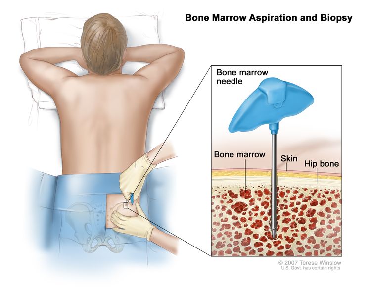
Plasma Cell Neoplasms (Including Multiple Myeloma) Treatment (PDQ®)–Patient Version

Plasma Cell Neoplasms (Including Multiple Myeloma) Treatment (PDQ®)–Patient Version

General Information About Plasma Cell Neoplasms

Key Points

  • Plasma cell neoplasms are diseases in which the body makes too many plasma cells.
  • Plasma cell neoplasms can be benign (not cancer) or malignant (cancer).
  • There are several types of plasma cell neoplasms.
    • Monoclonal gammopathy of undetermined significance (MGUS)
    • Plasmacytoma
    • Multiple myeloma
  • Multiple myeloma and other plasma cell neoplasms may cause a condition called amyloidosis.
  • Age can affect the risk of plasma cell neoplasms.
  • Tests that examine the blood, bone marrow, and urine are used to diagnose multiple myeloma and other plasma cell neoplasms.
  • Certain factors affect prognosis (chance of recovery) and treatment options.

Plasma cell neoplasms are diseases in which the body makes too many plasma cells.

Plasma cells develop from B lymphocytes (B cells), a type of white blood cell that is made in the bone marrow. Normally, when bacteria or viruses enter the body, some of the B cells will change into plasma cells. The plasma cells make antibodies to fight bacteria and viruses, to stop infection and disease.

EnlargeMultiple myeloma; drawing shows normal plasma cells, multiple myeloma cells (abnormal plasma cells), and antibodies. Also shown is red marrow inside bone, where plasma cells are made.
Multiple myeloma. Multiple myeloma cells are abnormal plasma cells (a type of white blood cell) that build up in the bone marrow and form tumors in many bones of the body. Normal plasma cells make antibodies to help the body fight infection and disease. As the number of multiple myeloma cells increases, more antibodies are made. This can cause the blood to thicken and keep the bone marrow from making enough healthy blood cells. Multiple myeloma cells also damage and weaken the bone.

Plasma cell neoplasms are diseases in which abnormal plasma cells form tumors in the bones or soft tissues of the body. The plasma cells also make an antibody protein, called M protein, that is not needed by the body and does not help fight infection. These antibody proteins build up in the bone marrow and can cause the blood to thicken or can damage the kidneys.

Plasma cell neoplasms can be benign (not cancer) or malignant (cancer).

Monoclonal gammopathy of undetermined significance (MGUS) is not cancer but can become cancer. The following types of plasma cell neoplasms are cancer:

There are several types of plasma cell neoplasms.

Plasma cell neoplasms include the following:

Monoclonal gammopathy of undetermined significance (MGUS)

In this type of plasma cell neoplasm, less than 10% of the bone marrow is made up of abnormal plasma cells and there is no cancer. The abnormal plasma cells make M protein, which is sometimes found during a routine blood or urine test. In most patients, the amount of M protein stays the same and there are no signs, symptoms, or health problems.

In some patients, MGUS may later become a more serious condition, such as amyloidosis, or cause problems with the kidneys, heart, or nerves. MGUS can also become cancer, such as multiple myeloma, lymphoplasmacytic lymphoma, or chronic lymphocytic leukemia.

Plasmacytoma

In this type of plasma cell neoplasm, the abnormal plasma cells (myeloma cells) are in one place and form one tumor, called a plasmacytoma. Sometimes plasmacytoma can be cured. There are two types of plasmacytoma.

Signs and symptoms depend on where the tumor is.

  • In bone, the plasmacytoma may cause pain or broken bones.
  • In soft tissue, the tumor may press on nearby areas and cause pain or other problems. For example, a plasmacytoma in the throat can make it hard to swallow.

Multiple myeloma

In multiple myeloma, abnormal plasma cells (myeloma cells) build up in the bone marrow and form tumors in many bones of the body. These tumors may keep the bone marrow from making enough healthy blood cells. Normally, the bone marrow makes stem cells (immature cells) that become three types of mature blood cells:

As the number of myeloma cells increases, fewer red blood cells, white blood cells, and platelets are made. The myeloma cells also damage and weaken the bone.

Sometimes multiple myeloma does not cause any signs or symptoms. This is called smoldering multiple myeloma. It may be found when a blood or urine test is done for another condition. Signs and symptoms may be caused by multiple myeloma or other conditions. Check with your doctor if you have any of the following:

  • Bone pain, especially in the back or ribs.
  • Bones that break easily.
  • Fever for no known reason or frequent infections.
  • Easy bruising or bleeding.
  • Trouble breathing.
  • Weakness of the arms or legs.
  • Feeling very tired.

A tumor can damage the bone and cause hypercalcemia (too much calcium in the blood). This can affect many organs in the body, including the kidneys, nerves, heart, muscles, and digestive tract, and cause serious health problems.

Hypercalcemia may cause the following signs and symptoms:

Multiple myeloma and other plasma cell neoplasms may cause a condition called amyloidosis.

In rare cases, multiple myeloma can cause peripheral nerves (nerves that are not in the brain or spinal cord) and organs to fail. This may be caused by a condition called amyloidosis. Antibody proteins build up and stick together in peripheral nerves and organs, such as the kidney and heart. This can cause the nerves and organs to become stiff and unable to work the way they should.

Amyloidosis may cause the following signs and symptoms:

  • Feeling very tired.
  • Purple spots on the skin.
  • Enlarged tongue.
  • Diarrhea.
  • Swelling caused by fluid in your body’s tissues.
  • Tingling or numbness in your legs and feet.

Age can affect the risk of plasma cell neoplasms.

Anything that increases a person’s chance of getting a disease is called a risk factor. Not every person with one or more of these risk factors will develop plasma cell neoplasms, and they will develop in people who don’t have any known risk factors. Talk with your doctor if you think you may be at risk.

Plasma cell neoplasms are most common in people who are middle aged or older. For multiple myeloma and plasmacytoma, other risk factors include the following:

Studies about how racial, social, and financial factors affect access to treatment and rates of plasma cell neoplasms are ongoing.

Tests that examine the blood, bone marrow, and urine are used to diagnose multiple myeloma and other plasma cell neoplasms.

In addition to asking about your personal and family health history and doing a physical exam, your doctor may perform the following tests and procedures:

  • Blood and urine immunoglobulin studies: A procedure in which a blood or urine sample is checked to measure the amounts of certain antibodies (immunoglobulins). For multiple myeloma, beta-2-microglobulin, M protein, free light chains, and other proteins made by the myeloma cells are measured. A higher-than-normal amount of these substances can be a sign of disease.
  • Bone marrow aspiration and biopsy: The removal of bone marrow, blood, and a small piece of bone by inserting a hollow needle into the hipbone or breastbone. A pathologist views the bone marrow, blood, and bone under a microscope to look for abnormal cells.
    EnlargeBone marrow aspiration and biopsy; drawing shows a patient lying face down on a table and a bone marrow needle being inserted into the hip bone. An inset shows a close up of the needle being inserted through the skin and hip bone into the bone marrow.
    Bone marrow aspiration and biopsy. After a small area of skin is numbed, a long, hollow needle is inserted through the patient’s skin and hip bone into the bone marrow. A sample of bone marrow and a small piece of bone are removed for examination under a microscope.

    The following tests may be done on the sample of tissue removed during the bone marrow aspiration and biopsy:

    • Cytogenetic analysis: A laboratory test in which the chromosomes of cells in a sample of bone marrow are counted and checked for any changes, such as broken, missing, rearranged, or extra chromosomes. Changes in certain chromosomes may be a sign of cancer. Cytogenetic analysis is used to help diagnose cancer, plan treatment, or find out how well treatment is working.
    • FISH (fluorescence in situ hybridization): A laboratory test used to look at and count genes or chromosomes in cells and tissues. Pieces of DNA that contain fluorescent dyes are made in the laboratory and added to a sample of a patient’s cells or tissues. When these dyed pieces of DNA attach to certain genes or areas of chromosomes in the sample, they light up when viewed under a fluorescent microscope. The FISH test is used to help diagnose cancer and help plan treatment.
    • Flow cytometry: A laboratory test that measures the number of cells in a sample, the percentage of live cells in a sample, and certain characteristics of the cells, such as size, shape, and the presence of tumor (or other) markers on the cell surface. The cells from a sample of a patient’s bone marrow are stained with a fluorescent dye, placed in a fluid, and then passed one at a time through a beam of light. The test results are based on how the cells that were stained with the fluorescent dye react to the beam of light. This test is used to help diagnose and manage certain types of cancers, such as leukemia and lymphoma.
  • Skeletal bone survey: In a skeletal bone survey, x-rays of all the bones in the body are taken. The x-rays are used to find areas where the bone is damaged. An x-ray is a type of energy beam that can go through the body and onto film, making a picture of areas inside the body.
  • Complete blood count (CBC) with differential: A procedure in which a sample of blood is drawn and checked for the following:
    • The number of red blood cells and platelets.
    • The number and type of white blood cells.
    • The amount of hemoglobin (the protein that carries oxygen) in the red blood cells.
    • The portion of the blood sample made up of red blood cells.
  • Blood chemistry studies: A procedure in which a blood sample is checked to measure the amounts of certain substances, such as calcium or albumin, released into the blood by organs and tissues in the body. An unusual (higher or lower than normal) amount of a substance can be a sign of disease.
  • Twenty-four-hour urine test: A test in which urine is collected for 24 hours to measure the amounts of certain substances. An unusual (higher or lower than normal) amount of a substance can be a sign of disease in the organ or tissue that makes it. A higher than normal amount of protein may be a sign of multiple myeloma.
  • MRI (magnetic resonance imaging): A procedure that uses a magnet, radio waves, and a computer to make a series of detailed pictures of areas inside the body. This procedure is also called nuclear magnetic resonance imaging (NMRI). An MRI of the spine and pelvis may be used to find areas where the bone is damaged.
  • PET scan (positron emission tomography scan): A procedure to find malignant tumor cells in the body. A small amount of radioactive glucose (sugar) is injected into a vein. The PET scanner rotates around the body and makes a picture of where glucose is being used in the body. Malignant tumor cells show up brighter in the picture because they are more active and take up more glucose than normal cells do.
  • CT scan (CAT scan): A procedure that makes a series of detailed pictures of areas inside the body, such as the spine, taken from different angles. The pictures are made by a computer linked to an x-ray machine. A dye may be injected into a vein or swallowed to help the organs or tissues show up more clearly. This procedure is also called computed tomography, computerized tomography, or computerized axial tomography.
  • PET-CT scan: A procedure that combines the pictures from a positron emission tomography (PET) scan and a computed tomography (CT) scan. The PET and CT scans are done at the same time with the same machine. The combined scans give more detailed pictures of areas inside the body, such as the spine, than either scan gives by itself.

Certain factors affect prognosis (chance of recovery) and treatment options.

The prognosis depends on the following:

  • The type of plasma cell neoplasm.
  • The stage of the disease.
  • Whether a certain immunoglobulin (antibody) is present.
  • Whether there are certain genetic changes.
  • Whether the kidney is damaged.
  • Whether the cancer responds to initial treatment or recurs (comes back).

Treatment options depend on the following:

  • The type of plasma cell neoplasm.
  • The age and general health of the patient.
  • Whether there are signs, symptoms, or health problems, such as kidney failure or infection, related to the disease.
  • Whether the cancer responds to initial treatment or recurs (comes back).

Stages of Plasma Cell Neoplasms

Key Points

  • There are no standard staging systems for monoclonal gammopathy of undetermined significance (MGUS) and plasmacytoma.
  • After multiple myeloma has been diagnosed, tests are done to find out how much cancer is in the body.
  • The stage of multiple myeloma is based on the levels of beta-2-microglobulin and albumin in the blood.
  • The following stages are used for multiple myeloma:
    • Stage I multiple myeloma
    • Stage II multiple myeloma
    • Stage III multiple myeloma
  • Plasma cell neoplasms may not respond to treatment or may come back after treatment.

There are no standard staging systems for monoclonal gammopathy of undetermined significance (MGUS) and plasmacytoma.

After multiple myeloma has been diagnosed, tests are done to find out how much cancer is in the body.

The process used to find out the amount of cancer in the body is called staging. It is important to know the stage in order to plan treatment.

The following tests and procedures may be used to find out how much cancer is in the body:

  • Skeletal bone survey: In a skeletal bone survey, x-rays of all the bones in the body are taken. The x-rays are used to find areas where the bone is damaged. An x-ray is a type of energy beam that can go through the body and onto film, making a picture of areas inside the body.
  • MRI (magnetic resonance imaging): A procedure that uses a magnet, radio waves, and a computer to make a series of detailed pictures of areas inside the body, such as the bone marrow. This procedure is also called nuclear magnetic resonance imaging (NMRI).
  • Bone densitometry: A procedure that uses a special type of x-ray to measure bone density.

The stage of multiple myeloma is based on the levels of beta-2-microglobulin and albumin in the blood.

Beta-2-microglobulin and albumin are found in the blood. Beta-2-microglobulin is a protein found on plasma cells. Albumin makes up the biggest part of the blood plasma. It keeps fluid from leaking out of blood vessels. It also brings nutrients to tissues, and carries hormones, vitamins, drugs, and other substances, such as calcium, all through the body. In the blood of patients with multiple myeloma, the amount of beta-2-microglobulin is increased and the amount of albumin is decreased.

The following stages are used for multiple myeloma:

Stage I multiple myeloma

In stage I multiple myeloma, the blood levels are as follows:

Stage II multiple myeloma

In stage II multiple myeloma, the blood levels are in between the levels for stage I and stage III.

Stage III multiple myeloma

In stage III multiple myeloma, the blood level of beta-2-microglobulin is 5.5 mg/L or higher and the patient also has one of the following:

Plasma cell neoplasms may not respond to treatment or may come back after treatment.

Plasma cell neoplasms are called refractory when the number of plasma cells keeps going up even though treatment is given. Plasma cell neoplasms are called relapsed when they have come back after treatment.

Treatment Option Overview

Key Points

  • There are different types of treatment for patients with plasma cell neoplasms.
  • The following types of treatment are used:
    • Chemotherapy
    • Other drug therapy
    • Targeted therapy
    • High-dose chemotherapy with stem cell transplant
    • Immunotherapy
    • Radiation therapy
    • Surgery
    • Watchful waiting
  • New types of treatment are being tested in clinical trials.
    • New combinations of therapies
  • Treatment for plasma cell neoplasms may cause side effects.
  • Supportive care is given to lessen the problems caused by the disease or its treatment.
  • Patients may want to think about taking part in a clinical trial.
  • Patients can enter clinical trials before, during, or after starting their cancer treatment.
  • Follow-up tests may be needed.

There are different types of treatment for patients with plasma cell neoplasms.

Different types of treatments are available for patients with plasma cell neoplasms. Some treatments are standard (the currently used treatment), and some are being tested in clinical trials. A treatment clinical trial is a research study meant to help improve current treatments or obtain information on new treatments for patients with cancer. When clinical trials show that a new treatment is better than the standard treatment, the new treatment may become the standard treatment. Patients may want to think about taking part in a clinical trial. Some clinical trials are open only to patients who have not started treatment.

The following types of treatment are used:

Chemotherapy

Chemotherapy is a cancer treatment that uses drugs to stop the growth of cancer cells, either by killing the cells or by stopping them from dividing. When chemotherapy is taken by mouth or injected into a vein or muscle, the drugs enter the bloodstream and can reach cancer cells throughout the body (systemic chemotherapy).

See Drugs Approved for Multiple Myeloma and Other Plasma Cell Neoplasms.

Other drug therapy

Corticosteroids are steroids that have antitumor effects in multiple myeloma.

Targeted therapy

Targeted therapy is a type of treatment that uses drugs or other substances to identify and attack specific cancer cells. Several types of targeted therapy may be used to treat multiple myeloma and other plasma cell neoplasms. There are different types of targeted therapy:

  • Proteasome inhibitor therapy: This treatment blocks the action of proteasomes in cancer cells. A proteasome is a protein that removes other proteins no longer needed by the cell. When the proteins are not removed from the cell, they build up and may cause the cancer cell to die. Bortezomib, carfilzomib, and ixazomib are proteasome inhibitors used in the treatment of multiple myeloma and other plasma cell neoplasms.
  • Monoclonal antibody therapy: Monoclonal antibodies are immune system proteins made in the laboratory to treat many diseases, including cancer. As a cancer treatment, these antibodies can attach to a specific target on cancer cells or other cells that may help cancer cells grow. The antibodies are able to then kill the cancer cells, block their growth, or keep them from spreading. Monoclonal antibodies are given by infusion. They may be used alone or to carry drugs, toxins, or radioactive material directly to cancer cells. Daratumumab and elotuzumab are monoclonal antibodies used in the treatment of multiple myeloma and other plasma cell neoplasms. Denosumab is a monoclonal antibody used to slow bone loss and reduce bone pain in patients with multiple myeloma.
    How do monoclonal antibodies work to treat cancer? This video shows how monoclonal antibodies, such as trastuzumab, pembrolizumab, and rituximab, block molecules cancer cells need to grow, flag cancer cells for destruction by the body’s immune system, or deliver harmful substances to cancer cells.
  • BCL2 inhibitor therapy: This treatment blocks a protein called BCL2. Blocking this protein may help kill cancer cells and may make them more sensitive to anticancer drugs. Venetoclax is a BCL2 inhibitor being studied in the treatment of relapsed or refractory multiple myeloma.

See Drugs Approved for Multiple Myeloma and Other Plasma Cell Neoplasms.

High-dose chemotherapy with stem cell transplant

High doses of chemotherapy are given to kill cancer cells. Healthy cells, including blood-forming cells, are also destroyed by the cancer treatment. Stem cell transplant is a treatment to replace the blood-forming cells. Stem cells (immature blood cells) are removed from the blood or bone marrow of the patient (autologous) or a donor (allogeneic) and are frozen and stored. After the patient completes chemotherapy, the stored stem cells are thawed and given back to the patient through an infusion. These reinfused stem cells grow into (and restore) the body’s blood cells.

EnlargeDonor stem cell transplant; (Panel 1): Drawing of stem cells being collected from a donor's bloodstream using an apheresis machine. Blood is removed from a vein in the donor's arm and flows through the machine where the stem cells are removed. The rest of the blood is then returned to the donor through a vein in their other arm. (Panel 2): Drawing of a health care provider giving a patient an infusion of chemotherapy through a catheter in the patient's chest. The chemotherapy is given to kill cancer cells and prepare the patient's body for the donor stem cells. (Panel 3): Drawing of a patient receiving an infusion of the donor stem cells through a catheter in the chest.
Donor stem cell transplant. (Step 1): Four to five days before donor stem cell collection, the donor receives a medicine to increase the number of stem cells circulating through their bloodstream (not shown). The blood-forming stem cells are then collected from the donor through a large vein in their arm. The blood flows through an apheresis machine that removes the stem cells. The rest of the blood is returned to the donor through a vein in their other arm. (Step 2): The patient receives chemotherapy to kill cancer cells and prepare their body for the donor stem cells. The patient may also receive radiation therapy (not shown). (Step 3): The patient receives an infusion of the donor stem cells.

Immunotherapy

Immunotherapy is a treatment that uses the patient’s immune system to fight cancer. Substances made by the body or made in a laboratory are used to boost, direct, or restore the body’s natural defenses against cancer. This cancer treatment is a type of biologic therapy.

  • Immunomodulator therapy: Thalidomide, lenalidomide, and pomalidomide are immunomodulators used to treat multiple myeloma and other plasma cell neoplasms.
  • CAR T-cell therapy: This treatment changes the patient’s T cells (a type of immune system cell) so they will attack certain proteins on the surface of cancer cells. T cells are taken from the patient and special receptors are added to their surface in the laboratory. The changed cells are called chimeric antigen receptor (CAR) T cells. The CAR T cells are grown in the laboratory and given to the patient by infusion. The CAR T cells multiply in the patient’s blood and attack cancer cells. CAR T-cell therapy is being studied in the treatment of multiple myeloma that has recurred (come back).
    EnlargeCAR T-cell therapy; drawing of blood being removed from a vein in a patient’s arm to get T cells. Also shown is a special receptor called a chimeric antigen receptor (CAR) being made in the laboratory; the gene for CAR is inserted into the T cells and then millions of CAR T cells are grown. Drawing also shows the CAR T cells being given to the patient by infusion and binding to antigens on the cancer cells and killing them.
    CAR T-cell therapy. A type of treatment in which a patient’s T cells (a type of immune cell) are changed in the laboratory so they will bind to cancer cells and kill them. Blood from a vein in the patient’s arm flows through a tube to an apheresis machine (not shown), which removes the white blood cells, including the T cells, and sends the rest of the blood back to the patient. Then, the gene for a special receptor called a chimeric antigen receptor (CAR) is inserted into the T cells in the laboratory. Millions of the CAR T cells are grown in the laboratory and then given to the patient by infusion. The CAR T cells are able to bind to an antigen on the cancer cells and kill them.

See Drugs Approved for Multiple Myeloma and Other Plasma Cell Neoplasms.

Radiation therapy

Radiation therapy is a cancer treatment that uses high-energy x-rays or other types of radiation to kill cancer cells or keep them from growing. External radiation therapy uses a machine outside the body to send radiation toward the area of the body with cancer.

Surgery

Surgery to remove the tumor may be done. After the doctor removes all the cancer that can be seen at the time of the surgery, some patients may be given radiation therapy after surgery to kill any cancer cells that are left. Treatment given after the surgery, to lower the risk that the cancer will come back, is called adjuvant therapy.

Watchful waiting

Watchful waiting is closely monitoring a patient’s condition without giving any treatment until signs or symptoms appear or change.

New types of treatment are being tested in clinical trials.

This summary section describes treatments that are being studied in clinical trials. It may not mention every new treatment being studied. Information about clinical trials is available from the NCI website.

New combinations of therapies

Clinical trials are studying different combinations of immunotherapy, chemotherapy, steroid therapy, and drugs. New treatment regimens using selinexor are also being studied.

Treatment for plasma cell neoplasms may cause side effects.

For information about side effects caused by treatment for cancer, visit our Side Effects page.

Supportive care is given to lessen the problems caused by the disease or its treatment.

This therapy controls problems or side effects caused by the disease or its treatment, and improves quality of life. Supportive care is given to treat problems caused by multiple myeloma and other plasma cell neoplasms.

Supportive care may include the following:

  • Plasmapheresis: If the blood becomes thick with extra antibody proteins and interferes with circulation, plasmapheresis is done to remove extra plasma and antibody proteins from the blood. In this procedure blood is removed from the patient and sent through a machine that separates the plasma (the liquid part of the blood) from the blood cells. The patient’s plasma contains the unneeded antibodies and is not returned to the patient. The normal blood cells are returned to the bloodstream along with donated plasma or a plasma replacement. Plasmapheresis does not keep new antibodies from forming.
  • Induction therapy with stem cell transplant: If amyloidosis occurs, treatment may include induction therapy followed by stem cell transplant using the patient’s own stem cells.
  • Immunotherapy: Immunotherapy with thalidomide, lenalidomide, or pomalidomide is given to treat amyloidosis.
  • Targeted therapy: Targeted therapy with proteasome inhibitors is given to decrease how much immunoglobulin M is in the blood and treat amyloidosis. Targeted therapy with daratumumab is given with or without other drugs to treat amyloidosis. Targeted therapy with a monoclonal antibody is given to slow bone loss and reduce bone pain.
  • Radiation therapy: Radiation therapy is given for bone lesions of the spine.
  • Chemotherapy: Chemotherapy is given to reduce back pain from osteoporosis or compression fractures of the spine.
  • Bisphosphonate therapy: Bisphosphonate therapy is given to slow bone loss and reduce bone pain. For more information on bisphosphonates and problems related to their use, see Oral Complications of Cancer Therapies.

Patients may want to think about taking part in a clinical trial.

For some patients, taking part in a clinical trial may be the best treatment choice. Clinical trials are part of the cancer research process. Clinical trials are done to find out if new cancer treatments are safe and effective or better than the standard treatment.

Many of today’s standard treatments for cancer are based on earlier clinical trials. Patients who take part in a clinical trial may receive the standard treatment or be among the first to receive a new treatment.

Patients who take part in clinical trials also help improve the way cancer will be treated in the future. Even when clinical trials do not lead to effective new treatments, they often answer important questions and help move research forward.

Patients can enter clinical trials before, during, or after starting their cancer treatment.

Some clinical trials only include patients who have not yet received treatment. Other trials test treatments for patients whose cancer has not gotten better. There are also clinical trials that test new ways to stop cancer from recurring (coming back) or reduce the side effects of cancer treatment.

Clinical trials are taking place in many parts of the country. Information about clinical trials supported by NCI can be found on NCI’s clinical trials search webpage. Clinical trials supported by other organizations can be found on the ClinicalTrials.gov website.

Follow-up tests may be needed.

As you go through treatment, you will have follow-up tests or check-ups. Some tests that were done to diagnose or stage the cancer may be repeated to see how well the treatment is working. Decisions about whether to continue, change, or stop treatment may be based on the results of these tests.

Some of the tests will continue to be done from time to time after treatment has ended. The results of these tests can show if your condition has changed or if the cancer has recurred (come back).

Treatment of Monoclonal Gammopathy of Undetermined Significance

For information about the treatments listed below, see the Treatment Option Overview section.

Treatment of monoclonal gammopathy of undetermined significance (MGUS) is usually watchful waiting. Regular blood tests to check the level of M protein in the blood and physical exams to check for signs or symptoms of cancer will be done.

Use our clinical trial search to find NCI-supported cancer clinical trials that are accepting patients. You can search for trials based on the type of cancer, the age of the patient, and where the trials are being done. General information about clinical trials is also available.

Treatment of Isolated Plasmacytoma of Bone

For information about the treatments listed below, see the Treatment Option Overview section.

Treatment of isolated plasmacytoma of bone is usually radiation therapy to the bone lesion.

Use our clinical trial search to find NCI-supported cancer clinical trials that are accepting patients. You can search for trials based on the type of cancer, the age of the patient, and where the trials are being done. General information about clinical trials is also available.

Treatment of Extramedullary Plasmacytoma

For information about the treatments listed below, see the Treatment Option Overview section.

Treatment of extramedullary plasmacytoma may include the following:

Use our clinical trial search to find NCI-supported cancer clinical trials that are accepting patients. You can search for trials based on the type of cancer, the age of the patient, and where the trials are being done. General information about clinical trials is also available.

Treatment of Multiple Myeloma

For information about the treatments listed below, see the Treatment Option Overview section.

Patients without signs or symptoms may not need treatment. These patients can have watchful waiting until signs or symptoms appear.

When signs or symptoms appear, there are two categories for patients receiving treatment:

  • Younger, fit patients who are eligible for a stem cell transplant.
  • Older, unfit patients who are not eligible for a stem cell transplant.

Patients younger than 65 years are usually considered younger and fit. Patients older than 75 years are usually not eligible for a stem cell transplant. For patients between the ages of 65 and 75 years, fitness is determined by their overall health and other factors.

The treatment of multiple myeloma is usually done in phases:

Use our clinical trial search to find NCI-supported cancer clinical trials that are accepting patients. You can search for trials based on the type of cancer, the age of the patient, and where the trials are being done. General information about clinical trials is also available.

Treatment of Relapsed or Refractory Multiple Myeloma

For information about the treatments listed below, see the Treatment Option Overview section.

Treatment of relapsed or refractory multiple myeloma may include the following:

  • Watchful waiting for patients whose disease is stable.
  • A different treatment than treatment already given, for patients whose tumor kept growing during treatment. See Multiple Myeloma treatment options.
  • The same drugs used before the relapse may be used if the relapse occurs one or more years after initial treatment. See Multiple Myeloma treatment options.

Drugs used may include the following:

Use our clinical trial search to find NCI-supported cancer clinical trials that are accepting patients. You can search for trials based on the type of cancer, the age of the patient, and where the trials are being done. General information about clinical trials is also available.

To Learn More About Plasma Cell Neoplasms

About This PDQ Summary

About PDQ

Physician Data Query (PDQ) is the National Cancer Institute’s (NCI’s) comprehensive cancer information database. The PDQ database contains summaries of the latest published information on cancer prevention, detection, genetics, treatment, supportive care, and complementary and alternative medicine. Most summaries come in two versions. The health professional versions have detailed information written in technical language. The patient versions are written in easy-to-understand, nontechnical language. Both versions have cancer information that is accurate and up to date and most versions are also available in Spanish.

PDQ is a service of the NCI. The NCI is part of the National Institutes of Health (NIH). NIH is the federal government’s center of biomedical research. The PDQ summaries are based on an independent review of the medical literature. They are not policy statements of the NCI or the NIH.

Purpose of This Summary

This PDQ cancer information summary has current information about treatment of plasma cell neoplasms (including multiple myeloma). It is meant to inform and help patients, families, and caregivers. It does not give formal guidelines or recommendations for making decisions about health care.

Reviewers and Updates

Editorial Boards write the PDQ cancer information summaries and keep them up to date. These Boards are made up of experts in cancer treatment and other specialties related to cancer. The summaries are reviewed regularly and changes are made when there is new information. The date on each summary (“Updated”) is the date of the most recent change.

The information in this patient summary was taken from the health professional version, which is reviewed regularly and updated as needed, by the PDQ Adult Treatment Editorial Board.

Clinical Trial Information

A clinical trial is a study to answer a scientific question, such as whether one treatment is better than another. Trials are based on past studies and what has been learned in the laboratory. Each trial answers certain scientific questions in order to find new and better ways to help cancer patients. During treatment clinical trials, information is collected about the effects of a new treatment and how well it works. If a clinical trial shows that a new treatment is better than one currently being used, the new treatment may become “standard.” Patients may want to think about taking part in a clinical trial. Some clinical trials are open only to patients who have not started treatment.

Clinical trials can be found online at NCI’s website. For more information, call the Cancer Information Service (CIS), NCI’s contact center, at 1-800-4-CANCER (1-800-422-6237).

Permission to Use This Summary

PDQ is a registered trademark. The content of PDQ documents can be used freely as text. It cannot be identified as an NCI PDQ cancer information summary unless the whole summary is shown and it is updated regularly. However, a user would be allowed to write a sentence such as “NCI’s PDQ cancer information summary about breast cancer prevention states the risks in the following way: [include excerpt from the summary].”

The best way to cite this PDQ summary is:

PDQ® Adult Treatment Editorial Board. PDQ Plasma Cell Neoplasms (Including Multiple Myeloma) Treatment. Bethesda, MD: National Cancer Institute. Updated <MM/DD/YYYY>. Available at: /types/myeloma/patient/myeloma-treatment-pdq. Accessed <MM/DD/YYYY>. [PMID: 26389437]

Images in this summary are used with permission of the author(s), artist, and/or publisher for use in the PDQ summaries only. If you want to use an image from a PDQ summary and you are not using the whole summary, you must get permission from the owner. It cannot be given by the National Cancer Institute. Information about using the images in this summary, along with many other images related to cancer can be found in Visuals Online. Visuals Online is a collection of more than 3,000 scientific images.

Disclaimer

The information in these summaries should not be used to make decisions about insurance reimbursement. More information on insurance coverage is available on Cancer.gov on the Managing Cancer Care page.

Contact Us

More information about contacting us or receiving help with the Cancer.gov website can be found on our Contact Us for Help page. Questions can also be submitted to Cancer.gov through the website’s E-mail Us.

Hairy Cell Leukemia Treatment (PDQ®)–Health Professional Version

Hairy Cell Leukemia Treatment (PDQ®)–Health Professional Version

General Information About Hairy Cell Leukemia

Incidence and Mortality

Hairy cell leukemia is an indolent, low-grade, B-cell lymphoid malignancy. It is rare, with only 1,200 to 1,300 new cases annually in the United States.[1]

Clinical Presentation

Hairy cell leukemia usually presents with:

  • Splenomegaly.
  • Varying degrees of leukopenia (occasionally leukocytosis).
  • Pancytopenia.
  • Monocytopenia.
  • Bone marrow infiltration by atypical cells with prominent cytoplasmic projections (i.e., hairy cells).

Lymphadenopathy is absent, except with multiply recurrent progressive disease.

Diagnostic Evaluation

The following tests and procedures may be used to diagnose hairy cell leukemia:

  • Flow cytometry.
  • Bone marrow aspiration and biopsy.
  • Immunophenotyping.
  • Cytogenetic analysis.
  • BRAF gene testing.
  • Computed tomography scan.

The bone marrow is usually fibrotic and is not easily aspirated. It has circulating B cells with cytoplasmic projections (hairy appearance). Although a bone marrow biopsy may be required to enroll in a clinical trial, the hairy cell leukemia diagnosis can usually be made by flow cytometry.

In addition to the B-cell antigens CD19, CD20 (very high levels), and CD22, the cells coexpress CD11c, CD25, and CD103. The BRAF V600E pathogenic variant is a hairy cell leukemia–defining genetic feature that can aid in diagnosis.[2,3]

There is a variation of hairy cell leukemia (HCL-v) which accounts for 10% of cases. HCL-v is distinguished clinically by an elevated white blood cell count (15–50 × 109/L) and aberrant markers, including variable (instead of bright) CD103 and the absence of CD23, CD25, CD12, and CD43.[4,5] HCL-v cells also lack BRAF variants. Patients with HCL-v have more aggressive clinical courses, reduced responses to purine nucleoside analogue-based therapy, and shorter durations of response.[5]

The depth of a complete remission can be evaluated with measurable residual disease (MRD) by testing for a BRAF variant or an immunoglobulin heavy chain gene rearrangement. However, the usefulness of altering therapeutic choices with MRD remains unclear and requires further evaluation.[6]

References
  1. Falini B, Tiacci E: Hairy-Cell Leukemia. N Engl J Med 391 (14): 1328-1341, 2024. [PUBMED Abstract]
  2. Tiacci E, Schiavoni G, Forconi F, et al.: Simple genetic diagnosis of hairy cell leukemia by sensitive detection of the BRAF-V600E mutation. Blood 119 (1): 192-5, 2012. [PUBMED Abstract]
  3. Naik RR, Saven A: My treatment approach to hairy cell leukemia. Mayo Clin Proc 87 (1): 67-76, 2012. [PUBMED Abstract]
  4. Jones G, Parry-Jones N, Wilkins B, et al.: Revised guidelines for the diagnosis and management of hairy cell leukaemia and hairy cell leukaemia variant*. Br J Haematol 156 (2): 186-95, 2012. [PUBMED Abstract]
  5. Troussard X, Grever MR: The revised guidelines for the diagnosis and management of hairy cell leukaemia and the hairy cell leukaemia variant. Br J Haematol 193 (1): 11-14, 2021. [PUBMED Abstract]
  6. Ravandi F, Kreitman RJ, Tiacci E, et al.: Consensus opinion from an international group of experts on measurable residual disease in hairy cell leukemia. Blood Cancer J 12 (12): 165, 2022. [PUBMED Abstract]

Stage Information for Hairy Cell Leukemia

There is no generally accepted staging system used in the prognosis and treatment of hairy cell leukemia.

Treatment of Hairy Cell Leukemia

Hairy cell leukemia is highly treatable but rarely cured. Because it is easily controlled, many patients have prolonged survival with the use of sequential therapies. The decision to treat is based on signs of disease progression, including any of the following factors:

  • Cytopenias (especially if symptomatic).
  • Increasing splenomegaly.
  • The presence of other, usually infectious, complications.

If the patient is asymptomatic and if blood counts are maintained in an acceptable range, therapy may not be needed.[1]

Treatment Options for Hairy Cell Leukemia

Prior to the COVID-19 (SARS-CoV-2) pandemic, the standard initial therapy for patients with hairy cell leukemia was infusion of cladribine daily for 5 days, given with or without eight weekly doses of rituximab.[24] However, treatment with a purine analogue–based regimen led to significant and prolonged neutropenia and impairment of T-cell function, which were both problematic during the pandemic in fighting viral infection and establishing vaccination-induced immunity.

Other options for initial standard therapy, instead of cladribine or pentostatin, may offer less toxicity in terms of infection and long-term risk of secondary malignancies. However, these options may provide less durable response.

The Hairy Cell Leukemia Foundation convened a virtual meeting of 39 experts from around the world to amend the 2017 consensus recommendations.[5] The adapted treatment guidelines, published in 2021, are based primarily on anecdotal experience and expert opinion, as controlled trials for this indolent leukemia cannot be completed expeditiously given the low incidence of this disease.[6][Level of evidence C3] The adapted treatment guidelines are summarized below.

  1. Consider watchful waiting when feasible; asymptomatic patients with noncritical levels of pancytopenia can be monitored closely.
  2. Cladribine, with or without rituximab,[4] remains the standard of care. However, due to the risk of serious and prolonged immunosuppression, nonchemotherapy treatment options may be preferable for older, frail patients with higher risk of infection (or for those who have active infections).
  3. BRAF inhibitors such as vemurafenib, dabrafenib, or encorafenib are nonchemotherapeutic options that can be combined with rituximab or obinutuzumab.[710] Most patients with hairy cell leukemia have BRAF pathogenic variants, but this should be verified by flow cytometry. Despite extensive experience with vemurafenib for patients with relapsed disease, the U.S. Food and Drug Administration (FDA) has not approved oral vemurafenib for patients with hairy cell leukemia.
  4. Consider using rituximab alone intravenously (IV) for 4 to 8 weeks or in combination with a BRAF inhibitor.[11] Anti-CD20 monoclonal antibodies can impair future vaccine response, but they do not affect immunity from prior vaccination.
  5. In patients with relapsed disease, the previously mentioned options are available, along with ibrutinib (the Bruton tyrosine kinase inhibitor).[12]

Treatment options for hairy cell leukemia include:

Cladribine with or without rituximab

Cladribine may be given with or without rituximab to treat hairy cell leukemia.

Evidence (cladribine with or without rituximab):

  1. In a phase II study, 68 patients with previously untreated hairy cell leukemia were randomly assigned to receive cladribine (0.15 mg/kg IV) on days 1 to 5, with eight weekly doses of rituximab either concurrently (starting on day 1) or delayed (starting after 6 months of cladribine treatment) if still positive with measurable residual disease (MRD) testing.[4][Level of evidence C3]
    • With a median follow-up of 96 months, 94% of patients who received concurrent therapy were MRD-free, compared with 12% of patients who received delayed therapy.
    • Although patients who underwent concurrent therapy had more need for platelet transfusions, they demonstrated higher neutrophil and platelet counts after 1 month.
    • A retrospective case series reported a median progression-free survival (PFS) of 67 months in patients with relapsed disease who received a purine nucleoside analogue (usually cladribine) plus rituximab.[13][Level of evidence C2]
    • A retrospective case series of nine patients with a histologic variation of hairy cell leukemia (HCL-v) reported an 88% complete response rate and 3-year PFS rate of 42% (95% confidence interval [CI], 1%–84%) after treatment with a purine nucleoside analogue (usually cladribine) plus rituximab.[14][Level of evidence C2]
  2. Cladribine was given by daily subcutaneous injections or by daily 2-hour IV infusions for 5 to 7 days.[5,1517][Level of evidence C3] Purine analogues should be avoided in cases of active infection or moderate to severe hepatic or renal impairment.
    • The complete response rate was 50% to 80%.
    • The overall response rate was 85% to 95%.
  3. A National Cancer Institute group C protocol of 979 patients treated with cladribine reported lower response rates (i.e., 50% complete remission rate, 37% partial remission rate) compared with other studies.[18] Responses were durable in patients treated with a short course of cladribine, and patients who had a relapse often responded to re-treatment with cladribine.[2,13,19]
  4. A retrospective review included 83 patients, aged 40 years and younger.[2]
    • The median time to first relapse was 54 months for all responders, and the median overall survival (OS) was 21 years from diagnosis.
    • Cladribine may cause fever and immunosuppression; documented infection was found in 33% of treated patients.

In a retrospective study of patients with cladribine-associated neutropenic fever, filgrastim (G-CSF) did not reduce the percentage of febrile patients, number of febrile days, or frequency of hospital admissions to receive antibiotics.[3]

BRAF inhibitors (vemurafenib or dabrafenib) with or without rituximab or trametinib

BRAF V600E pathogenic variants are found in almost 100% of patients with classic-form hairy cell leukemia and almost never found in patients with other B-cell lymphomas and leukemias, including HCL-v.[20][Level of evidence C3] Vemurafenib or other BRAF inhibitors such as dabrafenib can be given with rituximab or obinutuzumab.[10,21] The FDA has not approved BRAF inhibitors for hairy cell leukemia, but they can be used off-label in clinical practice.[22]

Evidence (vemurafenib with or without rituximab):

  1. Several multicenter studies evaluated vemurafenib, given orally alone for 4 months or orally for 2 months with rituximab infused in eight doses over 18 weeks, in patients with relapsed or refractory hairy cell leukemia.[7,8,10,23][Level of evidence C3]
    1. After a median follow-up of 23 to 40 months, for the 86 patients treated with vemurafenib alone, the following was reported in two studies:[7,8,23][Level of evidence C3]
      • The overall response rate was 86% to 98%.
      • The complete response rate was 33% to 38%.
      • The median treatment-free survival was 18 to 25 months.
      • Retreatment at relapse resulted in an 86% response rate, and the median relapse-free survival was 12.7 months in one of the trials, with a 40-month median follow-up.[23][Level of evidence C2]
    2. After a median follow-up of 37 months, for the 30 patients treated with vemurafenib plus rituximab, the following was reported:[10][Level of evidence C3]
      • The complete response rate was 87%.
      • The PFS rate was 78% at 37 months.
      • In patients who had a complete response, 65% had no MRD.

Evidence (dabrafenib plus trametinib):

  1. In a phase II trial of patients with relapsed or refractory disease, 55 patients received dabrafenib and trametinib orally until their disease progressed, they experienced unacceptable toxicity, or death occurred.[24]
    • With a median follow-up of 43.2 months, the overall response rate was 89.0% (95% CI, 77.8%–95.9%), the complete response rate was 65.5%, the 2-year PFS rate was 94.5%, and the 2-year OS rate was 95.5%.[24][Level of evidence C1]

Rituximab

Rituximab can induce durable remissions (with minimal toxic effects), but rarely complete remissions, in patients with multiple relapses or refractory disease after treatment with a purine analogue or interferon.[11,22,25][Level of evidence C3]

Pentostatin

Pentostatin given IV every other week for 3 to 6 months produced a 50% to 76% complete response rate and an 80% to 87% overall response rate.[26] Complete remissions were of substantial duration. Purine analogues should be avoided in cases of active infection or moderate to severe hepatic or renal impairment.

Evidence (pentostatin):

  1. Two trials reported results on the 9-year median follow-up of patients treated with pentostatin.[27,28]
    • The relapse-free survival rates ranged from 56% to 67%.
    • Side effects included fever, immunosuppression, cytopenias, and renal dysfunction.
  2. A randomized trial compared pentostatin to recombinant interferon alfa-2a.[26]
    • Pentostatin demonstrated higher response rates and more durable responses.

Ibrutinib

Ibrutinib, a tyrosine kinase inhibitor, has been studied in the treatment of hairy cell leukemia.

Evidence (ibrutinib):

  1. In a phase II study, 37 patients with refractory hairy cell leukemia were treated with ibrutinib. The median follow-up was 42 months.[29][Level of evidence C3]
    • The response rate was 54%.
    • The estimated 36-month PFS rate was 73%.
    • The OS rate was 85%.

Re-treatment with cladribine or pentostatin

Patients with hairy cell leukemia who have a relapse after the first course of cladribine or pentostatin often respond well to re-treatment with the same or another purine analogue, especially if relapse occurs after several years.[13][Level of evidence C3]

Bendamustine with rituximab

Evidence (bendamustine with rituximab):

  1. A phase II study evaluated 12 patients with relapsed or refractory disease, three of whom were negative for BRAF variants. Patients received the combination of bendamustine and rituximab.[30]

Splenectomy

Splenectomy plays a decreasing role in treating hairy cell leukemia because effective alternatives are available. Splenectomy will partially or completely normalize the peripheral blood in most patients with hairy cell leukemia.[31] After a splenectomy, there is usually little or no change in the bone marrow, and virtually all patients have progressive disease within 12 to 18 months.

Interferon

Interferon alfa is no longer available because production has been halted.[32] According to the Hairy Cell Leukemia Foundation, ropeginterferon alfa-2b-njft is the best available preparation, but it is not FDA approved for hairy cell leukemia.

Interferon is useful when treating hairy cell leukemia during pregnancy because it does not involve cytotoxic agents.

Current Clinical Trials

Use our advanced clinical trial search to find NCI-supported cancer clinical trials that are now enrolling patients. The search can be narrowed by location of the trial, type of treatment, name of the drug, and other criteria. General information about clinical trials is also available.

References
  1. Troussard X, Maître E, Cornet E: Hairy cell leukemia 2022: Update on diagnosis, risk-stratification, and treatment. Am J Hematol 97 (2): 226-236, 2022. [PUBMED Abstract]
  2. Rosenberg JD, Burian C, Waalen J, et al.: Clinical characteristics and long-term outcome of young hairy cell leukemia patients treated with cladribine: a single-institution series. Blood 123 (2): 177-83, 2014. [PUBMED Abstract]
  3. Saven A, Burian C, Adusumalli J, et al.: Filgrastim for cladribine-induced neutropenic fever in patients with hairy cell leukemia. Blood 93 (8): 2471-7, 1999. [PUBMED Abstract]
  4. Chihara D, Arons E, Stetler-Stevenson M, et al.: Randomized Phase II Study of First-Line Cladribine With Concurrent or Delayed Rituximab in Patients With Hairy Cell Leukemia. J Clin Oncol 38 (14): 1527-1538, 2020. [PUBMED Abstract]
  5. Grever MR, Abdel-Wahab O, Andritsos LA, et al.: Consensus guidelines for the diagnosis and management of patients with classic hairy cell leukemia. Blood 129 (5): 553-560, 2017. [PUBMED Abstract]
  6. Grever M, Andritsos L, Banerji V, et al.: Hairy cell leukemia and COVID-19 adaptation of treatment guidelines. Leukemia 35 (7): 1864-1872, 2021. [PUBMED Abstract]
  7. Tiacci E, Park JH, De Carolis L, et al.: Targeting Mutant BRAF in Relapsed or Refractory Hairy-Cell Leukemia. N Engl J Med 373 (18): 1733-47, 2015. [PUBMED Abstract]
  8. Dietrich S, Pircher A, Endris V, et al.: BRAF inhibition in hairy cell leukemia with low-dose vemurafenib. Blood 127 (23): 2847-55, 2016. [PUBMED Abstract]
  9. Falini B, Tiacci E: New treatment options in hairy cell leukemia with focus on BRAF inhibitors. Hematol Oncol 37 (Suppl 1): 30-37, 2019. [PUBMED Abstract]
  10. Tiacci E, De Carolis L, Simonetti E, et al.: Vemurafenib plus Rituximab in Refractory or Relapsed Hairy-Cell Leukemia. N Engl J Med 384 (19): 1810-1823, 2021. [PUBMED Abstract]
  11. Angelopoulou MK, Pangalis GA, Sachanas S, et al.: Outcome and toxicity in relapsed hairy cell leukemia patients treated with rituximab. Leuk Lymphoma 49 (9): 1817-20, 2008. [PUBMED Abstract]
  12. Jones J, Andritsos L, Kreitman RJ: Efficacy and safety of the Bruton tyrosine kinase inhibitor ibrutinib in patients with hairy cell leukemia: stage 1 results of a phase 2 study. [Abstract] Blood 128 (22): A-1215, 2016.
  13. Hu R, Wei W, Mian A, et al.: Treatment outcomes with purine nucleoside analog alone or with rituximab for hairy cell leukemia at first relapse. Eur J Haematol 108 (5): 379-382, 2022. [PUBMED Abstract]
  14. Wang Y, Wang T, Yu Y, et al.: Purine nucleoside analogs plus rituximab are an effective treatment choice for hairy cell leukemia-variant. Ann Hematol 101 (6): 1201-1210, 2022. [PUBMED Abstract]
  15. Pagano L, Criscuolo M, Broccoli A, et al.: Long-term follow-up of cladribine treatment in hairy cell leukemia: 30-year experience in a multicentric Italian study. Blood Cancer J 12 (7): 109, 2022. [PUBMED Abstract]
  16. Zenhäusern R, Schmitz SF, Solenthaler M, et al.: Randomized trial of daily versus weekly administration of 2-chlorodeoxyadenosine in patients with hairy cell leukemia: a multicenter phase III trial (SAKK 32/98). Leuk Lymphoma 50 (9): 1501-11, 2009. [PUBMED Abstract]
  17. Hermel DJ, Cheng B, Bhangoo MS, et al.: Long-term outcomes of elderly hairy cell leukemia patients treated with cladribine. Ann Hematol 101 (5): 1089-1096, 2022. [PUBMED Abstract]
  18. Cheson BD, Sorensen JM, Vena DA, et al.: Treatment of hairy cell leukemia with 2-chlorodeoxyadenosine via the Group C protocol mechanism of the National Cancer Institute: a report of 979 patients. J Clin Oncol 16 (9): 3007-15, 1998. [PUBMED Abstract]
  19. Else M, Dearden CE, Catovsky D: Long-term follow-up after purine analogue therapy in hairy cell leukaemia. Best Pract Res Clin Haematol 28 (4): 217-29, 2015. [PUBMED Abstract]
  20. Pettirossi V, Santi A, Imperi E, et al.: BRAF inhibitors reverse the unique molecular signature and phenotype of hairy cell leukemia and exert potent antileukemic activity. Blood 125 (8): 1207-16, 2015. [PUBMED Abstract]
  21. Park JH, Devlin S, Durham BH, et al.: Vemurafenib and Obinutuzumab as Frontline Therapy for Hairy Cell Leukemia. NEJM Evid 2 (10): EVIDoa2300074, 2023. [PUBMED Abstract]
  22. Falini B, De Carolis L, Tiacci E: How I treat refractory/relapsed hairy cell leukemia with BRAF inhibitors. Blood 139 (15): 2294-2305, 2022. [PUBMED Abstract]
  23. Handa S, Lee JO, Derkach A, et al.: Long-term outcomes in patients with relapsed or refractory hairy cell leukemia treated with vemurafenib monotherapy. Blood 140 (25): 2663-2671, 2022. [PUBMED Abstract]
  24. Kreitman RJ, Moreau P, Ravandi F, et al.: Dabrafenib plus trametinib in patients with relapsed/refractory BRAF V600E mutation-positive hairy cell leukemia. Blood 141 (9): 996-1006, 2023. [PUBMED Abstract]
  25. Thomas DA, O’Brien S, Bueso-Ramos C, et al.: Rituximab in relapsed or refractory hairy cell leukemia. Blood 102 (12): 3906-11, 2003. [PUBMED Abstract]
  26. Grever M, Kopecky K, Foucar MK, et al.: Randomized comparison of pentostatin versus interferon alfa-2a in previously untreated patients with hairy cell leukemia: an intergroup study. J Clin Oncol 13 (4): 974-82, 1995. [PUBMED Abstract]
  27. Johnston JB, Eisenhauer E, Wainman N, et al.: Long-term outcome following treatment of hairy cell leukemia with pentostatin (Nipent): a National Cancer Institute of Canada study. Semin Oncol 27 (2 Suppl 5): 32-6, 2000. [PUBMED Abstract]
  28. Flinn IW, Kopecky KJ, Foucar MK, et al.: Long-term follow-up of remission duration, mortality, and second malignancies in hairy cell leukemia patients treated with pentostatin. Blood 96 (9): 2981-6, 2000. [PUBMED Abstract]
  29. Rogers KA, Andritsos LA, Wei L, et al.: Phase 2 study of ibrutinib in classic and variant hairy cell leukemia. Blood 137 (25): 3473-3483, 2021. [PUBMED Abstract]
  30. Burotto M, Stetler-Stevenson M, Arons E, et al.: Bendamustine and rituximab in relapsed and refractory hairy cell leukemia. Clin Cancer Res 19 (22): 6313-21, 2013. [PUBMED Abstract]
  31. Golomb HM, Vardiman JW: Response to splenectomy in 65 patients with hairy cell leukemia: an evaluation of spleen weight and bone marrow involvement. Blood 61 (2): 349-52, 1983. [PUBMED Abstract]
  32. Assanto GM, Riemma C, Malaspina F, et al.: The current role of interferon in hairy cell leukaemia: clinical and molecular aspects. Br J Haematol 194 (1): 78-82, 2021. [PUBMED Abstract]

Latest Updates to This Summary (02/21/2025)

The PDQ cancer information summaries are reviewed regularly and updated as new information becomes available. This section describes the latest changes made to this summary as of the date above.

General Information About Hairy Cell Leukemia

Added Falini et al. as reference 1.

Treatment of Hairy Cell Leukemia

Revised text to state that cladribine, with or without rituximab, remains the standard of care. However, due to the risk of serious and prolonged immunosuppression, nonchemotherapy treatment options may be preferable for older, frail patients with higher risks of infection (or for those who have active infections). BRAF inhibitors such as vemurafenib, dabrafenib, or encorafenib are nonchemotherapeutic options that can be combined with rituximab or obinutuzumab.

Revised the list of treatment options for hairy cell leukemia to include interferon.

Revised text to state that vemurafenib or other BRAF inhibitors such as dabrafenib can be given with rituximab or obinutuzumab (cited Park et al. as reference 21).

Added Interferon as a new subsection.

This summary is written and maintained by the PDQ Adult Treatment Editorial Board, which is editorially independent of NCI. The summary reflects an independent review of the literature and does not represent a policy statement of NCI or NIH. More information about summary policies and the role of the PDQ Editorial Boards in maintaining the PDQ summaries can be found on the About This PDQ Summary and PDQ® Cancer Information for Health Professionals pages.

About This PDQ Summary

Purpose of This Summary

This PDQ cancer information summary for health professionals provides comprehensive, peer-reviewed, evidence-based information about the treatment of hairy cell leukemia. It is intended as a resource to inform and assist clinicians in the care of their patients. It does not provide formal guidelines or recommendations for making health care decisions.

Reviewers and Updates

This summary is reviewed regularly and updated as necessary by the PDQ Adult Treatment Editorial Board, which is editorially independent of the National Cancer Institute (NCI). The summary reflects an independent review of the literature and does not represent a policy statement of NCI or the National Institutes of Health (NIH).

Board members review recently published articles each month to determine whether an article should:

  • be discussed at a meeting,
  • be cited with text, or
  • replace or update an existing article that is already cited.

Changes to the summaries are made through a consensus process in which Board members evaluate the strength of the evidence in the published articles and determine how the article should be included in the summary.

The lead reviewer for Hairy Cell Leukemia Treatment is:

  • Eric J. Seifter, MD (Johns Hopkins University)

Any comments or questions about the summary content should be submitted to Cancer.gov through the NCI website’s Email Us. Do not contact the individual Board Members with questions or comments about the summaries. Board members will not respond to individual inquiries.

Levels of Evidence

Some of the reference citations in this summary are accompanied by a level-of-evidence designation. These designations are intended to help readers assess the strength of the evidence supporting the use of specific interventions or approaches. The PDQ Adult Treatment Editorial Board uses a formal evidence ranking system in developing its level-of-evidence designations.

Permission to Use This Summary

PDQ is a registered trademark. Although the content of PDQ documents can be used freely as text, it cannot be identified as an NCI PDQ cancer information summary unless it is presented in its entirety and is regularly updated. However, an author would be permitted to write a sentence such as “NCI’s PDQ cancer information summary about breast cancer prevention states the risks succinctly: [include excerpt from the summary].”

The preferred citation for this PDQ summary is:

PDQ® Adult Treatment Editorial Board. PDQ Hairy Cell Leukemia Treatment. Bethesda, MD: National Cancer Institute. Updated <MM/DD/YYYY>. Available at: /types/leukemia/hp/hairy-cell-treatment-pdq. Accessed <MM/DD/YYYY>. [PMID: 26389184]

Images in this summary are used with permission of the author(s), artist, and/or publisher for use within the PDQ summaries only. Permission to use images outside the context of PDQ information must be obtained from the owner(s) and cannot be granted by the National Cancer Institute. Information about using the illustrations in this summary, along with many other cancer-related images, is available in Visuals Online, a collection of over 2,000 scientific images.

Disclaimer

Based on the strength of the available evidence, treatment options may be described as either “standard” or “under clinical evaluation.” These classifications should not be used as a basis for insurance reimbursement determinations. More information on insurance coverage is available on Cancer.gov on the Managing Cancer Care page.

Contact Us

More information about contacting us or receiving help with the Cancer.gov website can be found on our Contact Us for Help page. Questions can also be submitted to Cancer.gov through the website’s Email Us.