Prostate Cancer—Health Professional Version

Prostate Cancer—Health Professional Version

Prostate Cancer—Patient Version

Prostate Cancer—Patient Version

Overview

Prostate cancer is the most common cancer and the second leading cause of cancer death among men in the United States. Prostate cancer usually grows very slowly, and finding and treating it before symptoms occur may not improve men’s health or help them live longer. Explore the links on this page to learn about prostate cancer treatment, prevention, screening, statistics, research, and more.

Coping with Cancer

The information in this section is meant to help you cope with the many issues and concerns that occur when you have cancer.

Emotions and Cancer Adjusting to Cancer Support for Caregivers Survivorship Advanced Cancer Managing Cancer Care

Kidney Cancer Research Results and Study Updates

Kidney Cancer Research Results and Study Updates

See Advances in Kidney Cancer Research for an overview of recent findings and progress, plus ongoing projects supported by NCI.

Childhood Acute Promyelocytic Leukemia Treatment (PDQ®)–Health Professional Version

Childhood Acute Promyelocytic Leukemia Treatment (PDQ®)–Health Professional Version

General Information About Childhood Acute Promyelocytic Leukemia (APL)

APL occurs in about 7% of children with acute myeloid leukemia (AML).[1,2] APL is a distinct subtype of AML. Several factors that make APL unique include the following:

  • Clinical presentation of universal coagulopathy (disseminated intravascular coagulation) and unique morphological characteristics (French-American-British [FAB] M3 or its variants).
  • Unique molecular etiology as a result of the involvement of the RARA oncogene.
  • Unique sensitivity to the differentiating agent tretinoin and to the proapoptotic agent arsenic trioxide.[3]

When these unique features of APL are discovered at diagnosis, it is important to initiate proper supportive care measures to avoid coagulopathic complications during the first few days of therapy. It is also critical to institute an induction regimen specific to the treatment of APL. This regimen minimizes the risk of coagulopathic complications and provides a much improved long-term relapse-free survival and overall survival, compared with outcomes for patients with the other forms of AML.[4,5]

References
  1. von Neuhoff C, Reinhardt D, Sander A, et al.: Prognostic impact of specific chromosomal aberrations in a large group of pediatric patients with acute myeloid leukemia treated uniformly according to trial AML-BFM 98. J Clin Oncol 28 (16): 2682-9, 2010. [PUBMED Abstract]
  2. Smith MA, Ries LA, Gurney JG, et al.: Leukemia. In: Ries LA, Smith MA, Gurney JG, et al., eds.: Cancer incidence and survival among children and adolescents: United States SEER Program 1975-1995. National Cancer Institute, SEER Program, 1999. NIH Pub.No. 99-4649, pp 17-34. Also available online. Last accessed August 11, 2022.
  3. Melnick A, Licht JD: Deconstructing a disease: RARalpha, its fusion partners, and their roles in the pathogenesis of acute promyelocytic leukemia. Blood 93 (10): 3167-215, 1999. [PUBMED Abstract]
  4. Sanz MA, Grimwade D, Tallman MS, et al.: Management of acute promyelocytic leukemia: recommendations from an expert panel on behalf of the European LeukemiaNet. Blood 113 (9): 1875-91, 2009. [PUBMED Abstract]
  5. Sanz MA, Lo-Coco F: Modern approaches to treating acute promyelocytic leukemia. J Clin Oncol 29 (5): 495-503, 2011. [PUBMED Abstract]

Clinical Presentation

Clinically, acute promyelocytic leukemia (APL) is characterized by severe coagulopathy that is often present at the time of diagnosis.[1] APL blasts induce coagulopathy by activation of the coagulation cascade (caused by the expression of tissue factor and other procoagulants) with concomitant increase in primary and secondary fibrinolysis, resulting from the expression of annexin II on the APL blasts. Coagulopathy is typically manifested with thrombocytopenia, prolonged prothrombin time and partial thromboplastin time, elevated d-dimers, and hypofibrinogenemia.[2]

Coagulopathy and bleeding complications increase the risk of early death during induction therapy (particularly with cytotoxic agents used alone). Because of these complications, mortality was once more common in patients with APL than in patients with other French-American-British (FAB) or World Health Organization (WHO) AML types.[3,4]

Patients at greatest risk of coagulopathic complications are those presenting with high white blood cell (WBC) counts, decreased platelet count, abnormal coagulation studies (hypofibrinogenemia, prothrombin time), high body mass index, molecular variants of APL, and the presence of FLT3 internal tandem duplication (ITD) variants.[2,5,6]

Scoring systems using clinical characteristics and laboratory values can help predict the risk of developing severe or lethal coagulopathy, as demonstrated in studies of both adult and pediatric patients.[7,8] Aggressive supportive care to correct coagulopathy, even before clinical signs and symptoms of bleeding or thrombosis occur, is important to prevent early death.

Because tretinoin has been shown to ameliorate bleeding risk for patients with APL, tretinoin therapy is initiated as soon as APL is suspected on the basis of morphological and clinical presentation.[6,9,10] A retrospective analysis identified an increase in early death resulting from hemorrhage in patients with APL in whom tretinoin introduction was delayed.[2]

A multicooperative group analysis of children with APL who were treated with tretinoin and chemotherapy reported the following:[5]

  • Early induction coagulopathic deaths occurred in 25 of 683 children (3.7%); 23 deaths resulted from hemorrhage (19 central nervous system [CNS], 4 pulmonary), and 2 resulted from CNS thrombosis.
  • A lumbar puncture at diagnosis should not be performed until evidence of coagulopathy has resolved. When current treatment regimens with tretinoin and arsenic trioxide are used, diagnostic and therapeutic lumbar punctures are limited to only a relatively small subset of patients who present with signs and symptoms concerning for CNS disease and/or CNS hemorrhage.[11]

Tretinoin is administered early to address this emergent need, but participation in other AML clinical trials is not precluded should the diagnosis of APL prove to be incorrect. Additionally, initiation of supportive measures such as replacement transfusions to correct coagulopathy is critical during these initial days of diagnosis and therapy.

References
  1. Tallman MS, Hakimian D, Kwaan HC, et al.: New insights into the pathogenesis of coagulation dysfunction in acute promyelocytic leukemia. Leuk Lymphoma 11 (1-2): 27-36, 1993. [PUBMED Abstract]
  2. Altman JK, Rademaker A, Cull E, et al.: Administration of ATRA to newly diagnosed patients with acute promyelocytic leukemia is delayed contributing to early hemorrhagic death. Leuk Res 37 (9): 1004-9, 2013. [PUBMED Abstract]
  3. Lehmann S, Ravn A, Carlsson L, et al.: Continuing high early death rate in acute promyelocytic leukemia: a population-based report from the Swedish Adult Acute Leukemia Registry. Leukemia 25 (7): 1128-34, 2011. [PUBMED Abstract]
  4. Park JH, Qiao B, Panageas KS, et al.: Early death rate in acute promyelocytic leukemia remains high despite all-trans retinoic acid. Blood 118 (5): 1248-54, 2011. [PUBMED Abstract]
  5. Abla O, Ribeiro RC, Testi AM, et al.: Predictors of thrombohemorrhagic early death in children and adolescents with t(15;17)-positive acute promyelocytic leukemia treated with ATRA and chemotherapy. Ann Hematol 96 (9): 1449-1456, 2017. [PUBMED Abstract]
  6. Breen KA, Grimwade D, Hunt BJ: The pathogenesis and management of the coagulopathy of acute promyelocytic leukaemia. Br J Haematol 156 (1): 24-36, 2012. [PUBMED Abstract]
  7. Mitrovic M, Suvajdzic N, Bogdanovic A, et al.: International Society of Thrombosis and Hemostasis Scoring System for disseminated intravascular coagulation ≥ 6: a new predictor of hemorrhagic early death in acute promyelocytic leukemia. Med Oncol 30 (1): 478, 2013. [PUBMED Abstract]
  8. Rajpurkar M, Alonzo TA, Wang YC, et al.: Risk Markers for Significant Bleeding and Thrombosis in Pediatric Acute Promyelocytic Leukemia; Report From the Children’s Oncology Group Study AAML0631. J Pediatr Hematol Oncol 41 (1): 51-55, 2019. [PUBMED Abstract]
  9. Sanz MA, Grimwade D, Tallman MS, et al.: Management of acute promyelocytic leukemia: recommendations from an expert panel on behalf of the European LeukemiaNet. Blood 113 (9): 1875-91, 2009. [PUBMED Abstract]
  10. Visani G, Gugliotta L, Tosi P, et al.: All-trans retinoic acid significantly reduces the incidence of early hemorrhagic death during induction therapy of acute promyelocytic leukemia. Eur J Haematol 64 (3): 139-44, 2000. [PUBMED Abstract]
  11. Kutny MA, Alonzo TA, Abla O, et al.: Assessment of Arsenic Trioxide and All-trans Retinoic Acid for the Treatment of Pediatric Acute Promyelocytic Leukemia: A Report From the Children’s Oncology Group AAML1331 Trial. JAMA Oncol 8 (1): 79-87, 2022. [PUBMED Abstract]

Molecular Variants and Therapeutic Impact

RARA Fusion Proteins

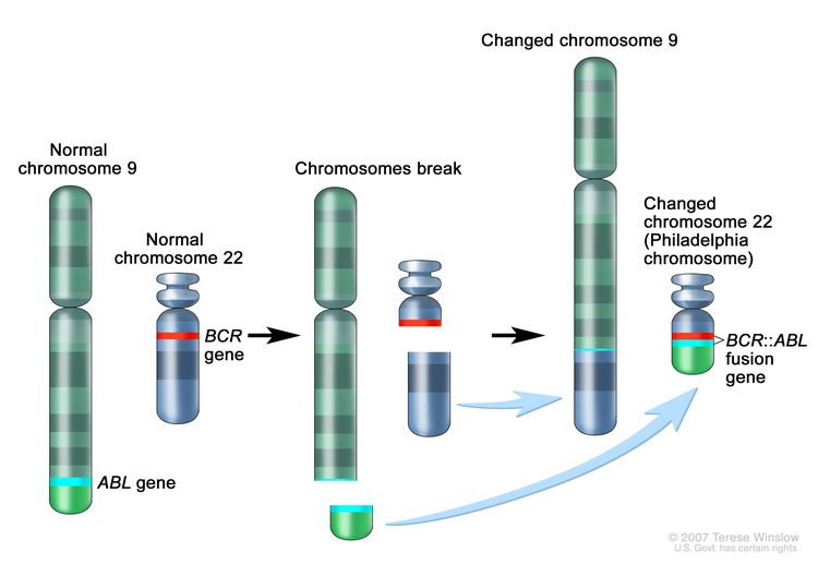
The characteristic chromosomal abnormality associated with acute promyelocytic leukemia (APL) is t(15;17)(q22;q21). This translocation involves a breakpoint that includes the retinoic acid receptor and leads to production of the PML::RARA fusion protein.[1] Other more complex chromosomal rearrangements may also lead to a PML::RARA fusion and result in APL.

Patients with a suspected diagnosis of APL can have their diagnosis confirmed by detection of the PML::RARA fusion protein through fluorescence in situ hybridization (FISH), reverse transcriptase–polymerase chain reaction (RT-PCR), or conventional cytogenetics. Quantitative RT-PCR allows identification of the three common transcript variants and is used for monitoring response on treatment and early detection of molecular relapse.[2] In addition, an immunofluorescence method using an anti-PML monoclonal antibody can rapidly establish the presence of the PML::RARA fusion protein based on the characteristic distribution pattern of PML that occurs in the presence of the fusion protein.[35]

Uncommon molecular variants of APL produce fusion proteins that join distinctive gene partners (e.g., PLZF, NPM, STAT5B, and NuMA) to RARA.[6,7] Recognition of these rare variants is important because they differ in their sensitivities to tretinoin and arsenic trioxide.[8]

  • PLZF::RARA fusion gene variant. The PLZF::RARA variant, characterized by t(11;17)(q23;q21), represents about 0.8% of APL, expresses surface CD56, and has very fine granules, compared with t(15;17) APL.[911] APL with the PLZF::RARA fusion gene has been associated with a poor prognosis and usually does not respond to tretinoin or arsenic trioxide.[811]
  • NPM::RARA or NuMA::RARA fusion gene variants. The rare APL variants with NPM::RARA (t(5;17)(q35;q21)) or NuMA::RARA (t(11;17)(q13;q21)) translocations may still be responsive to tretinoin.[8,1215]
  • PML::RARA fusion gene variant. There are rare case reports of patients with PML::RARA fusion–negative APL. One such APL is the torque teno mini virus (TTMV) subtype.[1618] This is a newly described entity in which the TTMV genome is integrated into intron 2 of the human RARA gene, resulting in a TTMV::RARA gene fusion. The clinical and morphological features of this APL subtype are similar to those of PML::RARA fusion–positive APL.

FLT3 Variants

FLT3 variants (either internal tandem duplication or tyrosine kinase domain variants) are observed in 40% to 50% of APL cases. The presence of FLT3 variants is correlated with higher white blood cell counts and the microgranular variant (M3v) subtype.[1923] The FLT3 variant has previously been associated with an increased risk of induction death and, in some reports, an increased risk of treatment failure.[1925] Given the extremely high cure rates for children with APL who were treated with tretinoin and arsenic trioxide, FLT3 variants are not associated with inferior outcomes.[26]

References
  1. Melnick A, Licht JD: Deconstructing a disease: RARalpha, its fusion partners, and their roles in the pathogenesis of acute promyelocytic leukemia. Blood 93 (10): 3167-215, 1999. [PUBMED Abstract]
  2. Sanz MA, Grimwade D, Tallman MS, et al.: Management of acute promyelocytic leukemia: recommendations from an expert panel on behalf of the European LeukemiaNet. Blood 113 (9): 1875-91, 2009. [PUBMED Abstract]
  3. Falini B, Flenghi L, Fagioli M, et al.: Immunocytochemical diagnosis of acute promyelocytic leukemia (M3) with the monoclonal antibody PG-M3 (anti-PML). Blood 90 (10): 4046-53, 1997. [PUBMED Abstract]
  4. Gomis F, Sanz J, Sempere A, et al.: Immunofluorescent analysis with the anti-PML monoclonal antibody PG-M3 for rapid and accurate genetic diagnosis of acute promyelocytic leukemia. Ann Hematol 83 (11): 687-90, 2004. [PUBMED Abstract]
  5. Dimov ND, Medeiros LJ, Kantarjian HM, et al.: Rapid and reliable confirmation of acute promyelocytic leukemia by immunofluorescence staining with an antipromyelocytic leukemia antibody: the M. D. Anderson Cancer Center experience of 349 patients. Cancer 116 (2): 369-76, 2010. [PUBMED Abstract]
  6. Zelent A, Guidez F, Melnick A, et al.: Translocations of the RARalpha gene in acute promyelocytic leukemia. Oncogene 20 (49): 7186-203, 2001. [PUBMED Abstract]
  7. Yan W, Zhang G: Molecular Characteristics and Clinical Significance of 12 Fusion Genes in Acute Promyelocytic Leukemia: A Systematic Review. Acta Haematol 136 (1): 1-15, 2016. [PUBMED Abstract]
  8. Rego EM, Ruggero D, Tribioli C, et al.: Leukemia with distinct phenotypes in transgenic mice expressing PML/RAR alpha, PLZF/RAR alpha or NPM/RAR alpha. Oncogene 25 (13): 1974-9, 2006. [PUBMED Abstract]
  9. Licht JD, Chomienne C, Goy A, et al.: Clinical and molecular characterization of a rare syndrome of acute promyelocytic leukemia associated with translocation (11;17). Blood 85 (4): 1083-94, 1995. [PUBMED Abstract]
  10. Guidez F, Ivins S, Zhu J, et al.: Reduced retinoic acid-sensitivities of nuclear receptor corepressor binding to PML- and PLZF-RARalpha underlie molecular pathogenesis and treatment of acute promyelocytic leukemia. Blood 91 (8): 2634-42, 1998. [PUBMED Abstract]
  11. Grimwade D, Biondi A, Mozziconacci MJ, et al.: Characterization of acute promyelocytic leukemia cases lacking the classic t(15;17): results of the European Working Party. Groupe Français de Cytogénétique Hématologique, Groupe de Français d’Hematologie Cellulaire, UK Cancer Cytogenetics Group and BIOMED 1 European Community-Concerted Action “Molecular Cytogenetic Diagnosis in Haematological Malignancies”. Blood 96 (4): 1297-308, 2000. [PUBMED Abstract]
  12. Sukhai MA, Wu X, Xuan Y, et al.: Myeloid leukemia with promyelocytic features in transgenic mice expressing hCG-NuMA-RARalpha. Oncogene 23 (3): 665-78, 2004. [PUBMED Abstract]
  13. Redner RL, Corey SJ, Rush EA: Differentiation of t(5;17) variant acute promyelocytic leukemic blasts by all-trans retinoic acid. Leukemia 11 (7): 1014-6, 1997. [PUBMED Abstract]
  14. Wells RA, Catzavelos C, Kamel-Reid S: Fusion of retinoic acid receptor alpha to NuMA, the nuclear mitotic apparatus protein, by a variant translocation in acute promyelocytic leukaemia. Nat Genet 17 (1): 109-13, 1997. [PUBMED Abstract]
  15. Wells RA, Hummel JL, De Koven A, et al.: A new variant translocation in acute promyelocytic leukaemia: molecular characterization and clinical correlation. Leukemia 10 (4): 735-40, 1996. [PUBMED Abstract]
  16. Umeda M, Ma J, Huang BJ, et al.: Integrated Genomic Analysis Identifies UBTF Tandem Duplications as a Recurrent Lesion in Pediatric Acute Myeloid Leukemia. Blood Cancer Discov 3 (3): 194-207, 2022. [PUBMED Abstract]
  17. Chen X, Wang F, Zhou X, et al.: Torque teno mini virus driven childhood acute promyelocytic leukemia: The third case report and sequence analysis. Front Oncol 12: 1074913, 2022. [PUBMED Abstract]
  18. Sala-Torra O, Beppu LW, Abukar FA, et al.: TTMV-RARA fusion as a recurrent cause of AML with APL characteristics. Blood Adv 6 (12): 3590-3592, 2022. [PUBMED Abstract]
  19. Callens C, Chevret S, Cayuela JM, et al.: Prognostic implication of FLT3 and Ras gene mutations in patients with acute promyelocytic leukemia (APL): a retrospective study from the European APL Group. Leukemia 19 (7): 1153-60, 2005. [PUBMED Abstract]
  20. Gale RE, Hills R, Pizzey AR, et al.: Relationship between FLT3 mutation status, biologic characteristics, and response to targeted therapy in acute promyelocytic leukemia. Blood 106 (12): 3768-76, 2005. [PUBMED Abstract]
  21. Arrigoni P, Beretta C, Silvestri D, et al.: FLT3 internal tandem duplication in childhood acute myeloid leukaemia: association with hyperleucocytosis in acute promyelocytic leukaemia. Br J Haematol 120 (1): 89-92, 2003. [PUBMED Abstract]
  22. Noguera NI, Breccia M, Divona M, et al.: Alterations of the FLT3 gene in acute promyelocytic leukemia: association with diagnostic characteristics and analysis of clinical outcome in patients treated with the Italian AIDA protocol. Leukemia 16 (11): 2185-9, 2002. [PUBMED Abstract]
  23. Tallman MS, Kim HT, Montesinos P, et al.: Does microgranular variant morphology of acute promyelocytic leukemia independently predict a less favorable outcome compared with classical M3 APL? A joint study of the North American Intergroup and the PETHEMA Group. Blood 116 (25): 5650-9, 2010. [PUBMED Abstract]
  24. Iland HJ, Bradstock K, Supple SG, et al.: All-trans-retinoic acid, idarubicin, and IV arsenic trioxide as initial therapy in acute promyelocytic leukemia (APML4). Blood 120 (8): 1570-80; quiz 1752, 2012. [PUBMED Abstract]
  25. Kutny MA, Moser BK, Laumann K, et al.: FLT3 mutation status is a predictor of early death in pediatric acute promyelocytic leukemia: a report from the Children’s Oncology Group. Pediatr Blood Cancer 59 (4): 662-7, 2012. [PUBMED Abstract]
  26. Kutny MA, Alonzo TA, Abla O, et al.: Assessment of Arsenic Trioxide and All-trans Retinoic Acid for the Treatment of Pediatric Acute Promyelocytic Leukemia: A Report From the Children’s Oncology Group AAML1331 Trial. JAMA Oncol 8 (1): 79-87, 2022. [PUBMED Abstract]

Classification of Childhood APL

Childhood acute myeloid leukemia and other myeloid malignancies are classified according to the 2022 World Health Organization Classification system. For more information, see the Classification of Pediatric Myeloid Malignancies section in Childhood Acute Myeloid Leukemia Treatment.

Prognostic Factors Affecting Risk-Based Treatment

White Blood Cell (WBC) Count

The prognostic significance of WBC count is used to define high-risk and low-risk patient populations and to assign induction treatment. High-risk patients are defined by a WBC count of 10 × 109/L or greater.[1,2] Patients with high-risk acute promyelocytic leukemia (APL) are given an anthracycline (idarubicin) along with induction therapy. Postinduction therapy is the same for both standard- and high-risk APL.[3]

APL in children is generally similar to APL in adults, although children have a higher incidence of hyperleukocytosis (defined as a WBC count higher than 10 × 109/L) and a higher incidence of the microgranular morphological subtype.[47] As in adults, children with WBC counts of less than 10 × 109/L at diagnosis have historically had better outcomes than patients with higher WBC counts.[5,6,8] Presenting WBC count is still used to determine induction therapy. However, with modern tretinoin- and arsenic trioxide–based therapy, patients with high-risk APL have similar excellent survival rates as patients with standard-risk APL.

In the Children’s Oncology Group (COG) AAML0631 (NCT00866918) trial, which included treatment with chemotherapy, tretinoin, and arsenic trioxide, patients were stratified on the basis of WBC count to standard risk or high risk. Risk classification primarily defined early death risk rather than relapse risk (standard risk, 0 of 66 patients vs. high risk, 4 of 35 patients).[9] In the COG AAML1331 (NCT02339740) trial, patients were treated with tretinoin and arsenic trioxide along with aggressive supportive care measures. There was only 1 death (standard-risk APL) and 3 relapses (1 standard risk and 2 high risk) reported among 154 patients. Thus, no significant differences were seen between the risk groups.[3] In the COG AAML0631 (NCT00866918) and AAML1331 (NCT02339740) trials, relapse risk after remission induction was 4% and 2% overall, respectively.[3,9]

Minimal Residual Disease (MRD) and Molecular Remission

For APL, MRD detection at the end of induction therapy lacks prognostic significance, likely related to the delayed clearance of differentiating leukemic cells destined to eventually die.[10,11] However, it is standard practice to document molecular remission after completion of two to four cycles of consolidation therapy.

References
  1. Sanz MA, Martín G, González M, et al.: Risk-adapted treatment of acute promyelocytic leukemia with all-trans-retinoic acid and anthracycline monochemotherapy: a multicenter study by the PETHEMA group. Blood 103 (4): 1237-43, 2004. [PUBMED Abstract]
  2. Lo-Coco F, Avvisati G, Vignetti M, et al.: Front-line treatment of acute promyelocytic leukemia with AIDA induction followed by risk-adapted consolidation for adults younger than 61 years: results of the AIDA-2000 trial of the GIMEMA Group. Blood 116 (17): 3171-9, 2010. [PUBMED Abstract]
  3. Kutny MA, Alonzo TA, Abla O, et al.: Assessment of Arsenic Trioxide and All-trans Retinoic Acid for the Treatment of Pediatric Acute Promyelocytic Leukemia: A Report From the Children’s Oncology Group AAML1331 Trial. JAMA Oncol 8 (1): 79-87, 2022. [PUBMED Abstract]
  4. de Botton S, Coiteux V, Chevret S, et al.: Outcome of childhood acute promyelocytic leukemia with all-trans-retinoic acid and chemotherapy. J Clin Oncol 22 (8): 1404-12, 2004. [PUBMED Abstract]
  5. Testi AM, Biondi A, Lo Coco F, et al.: GIMEMA-AIEOPAIDA protocol for the treatment of newly diagnosed acute promyelocytic leukemia (APL) in children. Blood 106 (2): 447-53, 2005. [PUBMED Abstract]
  6. Ortega JJ, Madero L, Martín G, et al.: Treatment with all-trans retinoic acid and anthracycline monochemotherapy for children with acute promyelocytic leukemia: a multicenter study by the PETHEMA Group. J Clin Oncol 23 (30): 7632-40, 2005. [PUBMED Abstract]
  7. Guglielmi C, Martelli MP, Diverio D, et al.: Immunophenotype of adult and childhood acute promyelocytic leukaemia: correlation with morphology, type of PML gene breakpoint and clinical outcome. A cooperative Italian study on 196 cases. Br J Haematol 102 (4): 1035-41, 1998. [PUBMED Abstract]
  8. Sanz MA, Lo Coco F, Martín G, et al.: Definition of relapse risk and role of nonanthracycline drugs for consolidation in patients with acute promyelocytic leukemia: a joint study of the PETHEMA and GIMEMA cooperative groups. Blood 96 (4): 1247-53, 2000. [PUBMED Abstract]
  9. Kutny MA, Alonzo TA, Gerbing RB, et al.: Arsenic Trioxide Consolidation Allows Anthracycline Dose Reduction for Pediatric Patients With Acute Promyelocytic Leukemia: Report From the Children’s Oncology Group Phase III Historically Controlled Trial AAML0631. J Clin Oncol 35 (26): 3021-3029, 2017. [PUBMED Abstract]
  10. Mandelli F, Diverio D, Avvisati G, et al.: Molecular remission in PML/RAR alpha-positive acute promyelocytic leukemia by combined all-trans retinoic acid and idarubicin (AIDA) therapy. Gruppo Italiano-Malattie Ematologiche Maligne dell’Adulto and Associazione Italiana di Ematologia ed Oncologia Pediatrica Cooperative Groups. Blood 90 (3): 1014-21, 1997. [PUBMED Abstract]
  11. Burnett AK, Grimwade D, Solomon E, et al.: Presenting white blood cell count and kinetics of molecular remission predict prognosis in acute promyelocytic leukemia treated with all-trans retinoic acid: result of the Randomized MRC Trial. Blood 93 (12): 4131-43, 1999. [PUBMED Abstract]

The Central Nervous System (CNS) and APL

CNS involvement at the time of diagnosis is not ascertained in most patients with acute promyelocytic leukemia (APL) because of the presence of disseminated intravascular coagulation. The Children’s Oncology Group (COG) AAML0631 (NCT00866918) trial identified 28 patients out of 101 enrolled children who had cerebrospinal fluid (CSF) exams at diagnosis. In 7 of these children, blasts were identified in atraumatic taps.[1] None of the patients experienced a CNS relapse with intrathecal treatment during induction and prophylactic doses during therapy. In the COG AAML1331 (NCT02339740) study, CSF exams were deferred if the patient did not have CNS symptoms or hemorrhage. Only 5 of 141 children without a history of CNS hemorrhage were diagnosed with CNS involvement, whereas 2 of 13 patients with CNS hemorrhage met the criteria for CNS disease.[2]

Overall, CNS relapse is uncommon for patients with APL, particularly for those with white blood cell (WBC) counts of less than 10 × 109/L.[3,4] In two clinical trials enrolling more than 1,400 adults with APL in which CNS prophylaxis was not administered, the cumulative incidence of CNS relapse was less than 1% for patients with WBC counts of less than 10 × 109/L, while it was approximately 5% for those with WBC counts of 10 × 109/L or greater.[3,4] In addition to high WBC counts at diagnosis, CNS hemorrhage during induction is also a risk factor for CNS relapse.[4] A review of published cases of pediatric APL also observed low rates of CNS relapse.[5,6]

Arsenic trioxide is an agent known to have excellent CNS penetration. Because patients with APL receive arsenic trioxide and there is a low prevalence of CNS relapses, CSF exams are not necessary at diagnosis. In addition, the use of intrathecal chemotherapy prophylaxis is not required unless CNS hemorrhage occurs. Two COG trials revealed similar low incidences of CNS relapses.

  • The COG AAML0631 study included treatment with two courses of arsenic trioxide along with prophylactic intrathecal chemotherapy. CNS disease was detected in 2 of 3 children whose disease relapsed.[1]
  • In the COG AAML1331 trial, only one patient was found to have CNS involvement (CNS 2A) that occurred in conjunction with a marrow relapse. In this study, triple intrathecal chemotherapy was administered to those who had CNS disease at diagnosis and/or experienced a CNS hemorrhage early in their diagnosis. No patients experienced recurrent CNS disease.[2]
References
  1. Kutny MA, Alonzo TA, Gerbing RB, et al.: Arsenic Trioxide Consolidation Allows Anthracycline Dose Reduction for Pediatric Patients With Acute Promyelocytic Leukemia: Report From the Children’s Oncology Group Phase III Historically Controlled Trial AAML0631. J Clin Oncol 35 (26): 3021-3029, 2017. [PUBMED Abstract]
  2. Kutny MA, Alonzo TA, Abla O, et al.: Assessment of Arsenic Trioxide and All-trans Retinoic Acid for the Treatment of Pediatric Acute Promyelocytic Leukemia: A Report From the Children’s Oncology Group AAML1331 Trial. JAMA Oncol 8 (1): 79-87, 2022. [PUBMED Abstract]
  3. de Botton S, Sanz MA, Chevret S, et al.: Extramedullary relapse in acute promyelocytic leukemia treated with all-trans retinoic acid and chemotherapy. Leukemia 20 (1): 35-41, 2006. [PUBMED Abstract]
  4. Montesinos P, Díaz-Mediavilla J, Debén G, et al.: Central nervous system involvement at first relapse in patients with acute promyelocytic leukemia treated with all-trans retinoic acid and anthracycline monochemotherapy without intrathecal prophylaxis. Haematologica 94 (9): 1242-9, 2009. [PUBMED Abstract]
  5. Chow J, Feusner J: Isolated central nervous system recurrence of acute promyelocytic leukemia in children. Pediatr Blood Cancer 52 (1): 11-3, 2009. [PUBMED Abstract]
  6. Kaspers G, Gibson B, Grimwade D, et al.: Central nervous system involvement in relapsed acute promyelocytic leukemia. Pediatr Blood Cancer 53 (2): 235-6; author reply 237, 2009. [PUBMED Abstract]

Special Considerations for the Treatment of Children With Cancer

Cancer in children and adolescents is rare, although the overall incidence has slowly increased since 1975.[1] Children and adolescents with cancer should be referred to medical centers that have a multidisciplinary team of cancer specialists with experience treating the cancers that occur during childhood and adolescence.[2] This multidisciplinary team approach incorporates the skills of the following pediatric specialists and others to ensure that children receive treatment, supportive care, and rehabilitation to achieve optimal survival and quality of life:

  • Primary care physicians.
  • Pediatric surgeons.
  • Pathologists.
  • Pediatric radiation oncologists.
  • Pediatric medical oncologists and hematologists.
  • Rehabilitation specialists.
  • Pediatric oncology nurses.
  • Social workers.
  • Child-life professionals.
  • Psychologists.
  • Nutritionists.

For specific information about supportive care for children and adolescents with cancer, see the summaries on Supportive and Palliative Care.

The American Academy of Pediatrics has outlined guidelines for pediatric cancer centers and their role in the treatment of children and adolescents with cancer.[3] At these centers, clinical trials are available for most types of cancer that occur in children and adolescents, and the opportunity to participate is offered to most patients and their families. Clinical trials for children and adolescents diagnosed with cancer are generally designed to compare potentially better therapy with current standard therapy. Other types of clinical trials test novel therapies when there is no standard therapy for a cancer diagnosis. Most of the progress in identifying curative therapies for childhood cancers has been achieved through clinical trials. Information about ongoing clinical trials is available from the NCI website.

References
  1. Smith MA, Seibel NL, Altekruse SF, et al.: Outcomes for children and adolescents with cancer: challenges for the twenty-first century. J Clin Oncol 28 (15): 2625-34, 2010. [PUBMED Abstract]
  2. Wolfson J, Sun CL, Wyatt L, et al.: Adolescents and Young Adults with Acute Lymphoblastic Leukemia and Acute Myeloid Leukemia: Impact of Care at Specialized Cancer Centers on Survival Outcome. Cancer Epidemiol Biomarkers Prev 26 (3): 312-320, 2017. [PUBMED Abstract]
  3. American Academy of Pediatrics: Standards for pediatric cancer centers. Pediatrics 134 (2): 410-4, 2014. Also available online. Last accessed February 25, 2025.

Treatment of APL

Modern treatment programs for acute promyelocytic leukemia (APL) are based on the sensitivity of leukemia cells to the differentiation-inducing and apoptotic effects of tretinoin and arsenic trioxide. APL therapy first diverged from the therapy of other non-APL subtypes of acute myeloid leukemia (AML) with the addition of tretinoin to chemotherapy. With the incorporation of arsenic trioxide into modern treatment regimens, the use of traditional chemotherapy in adults and children is restricted to only the induction phase for high-risk patients.[13]

Treatment options for children with APL may include the following:

Arsenic Trioxide and Tretinoin, With or Without Chemotherapy

Given the very high level of activity with the combination of arsenic trioxide and tretinoin for adults with APL [1,2] and data indicating that children with APL have a similar response to these agents,[37] the use of these two agents is the optimal therapeutic approach for this disease.

Induction therapy for patients with standard-risk APL includes repeated cycles of tretinoin and arsenic trioxide alone. Patients with high-risk APL receive treatment similar to that for patients with standard-risk disease, but they also receive short courses of chemotherapy during induction therapy.[3] Assessment of response to induction therapy in the first month of treatment using morphological and molecular criteria may provide misleading results because delayed persistence of differentiating leukemia cells can occur in patients who will ultimately achieve a complete remission (CR).[8,9] Alterations in planned treatment based on these early observations are not appropriate because it is rare for APL to be resistant to tretinoin plus arsenic trioxide.[3,10,11]

Almost all children with APL who were treated with tretinoin, arsenic trioxide, and modern supportive care achieved CR in the absence of coagulopathy-related mortality.[3,1216]

Results from the completed cooperative group trial (Children’s Oncology Group [COG] AAML1331 [NCT02339740]) verified the benefit of treatment with tretinoin and arsenic trioxide for children with newly diagnosed APL,[3] similar to results reported by other groups.[7] The dramatic efficacy of tretinoin against APL results from the ability of pharmacological doses of tretinoin to overcome the repression of signaling caused by the PML::RARA fusion protein at physiological tretinoin concentrations. Restoration of signaling leads to differentiation of APL cells and then to postmaturation apoptosis.[17] Most patients with APL achieve a CR when treated with tretinoin, although single-agent tretinoin is generally not curative.[18,19]

Arsenic trioxide, a proapoptotic and differentiation agent via binding to and the degradation of the PML::RARA fusion oncoprotein, is the most active agent in the treatment of APL. While initially used in patients with relapsed APL, it is now incorporated into the treatment of newly diagnosed patients. Data supporting the use of arsenic trioxide initially came from trials that included adult patients only, but its efficacy has now been seen in trials that included pediatric patients.

Based on the adult and pediatric experiences, consolidation therapy may include repeated cycles of tretinoin and arsenic trioxide without additional chemotherapy.[13,7] Studies using arsenic trioxide–based consolidation have demonstrated excellent survival rates without cytarabine consolidation.[1,3,7,20,21]

Based on data from adult trials and the COG AAML1331 (NCT02339740) trial, maintenance therapy is likely unnecessary for patients with APL who are treated with tretinoin and arsenic trioxide.[3] Because of the favorable outcomes with tretinoin and arsenic trioxide, hematopoietic stem cell transplant is not recommended in first CR.

Before this approach was discovered, chemotherapy was used in all or most phases of therapy including induction, consolidation, and maintenance for pediatric trials like AAML0631 (NCT00866918). The regimens that use chemotherapy are now primarily of historical interest. They can also be used as a reference in refractory cases because of the findings from randomized clinical trials that compared regimens with the combination of tretinoin and arsenic trioxide with or without chemotherapy.

Evidence (arsenic trioxide and tretinoin, with or without chemotherapy):

  1. In children and adolescents with newly diagnosed APL treated on the COG AAML0631 (NCT00866918) trial, two consolidation cycles of arsenic trioxide were incorporated into a chemotherapy regimen with lower cumulative anthracycline doses compared with historical controls.[22]
    • The 3-year overall survival (OS) rate was 94%, and the event-free survival (EFS) rate was 91%.
    • Patients with standard-risk APL had an OS rate of 98% and an EFS rate of 95%.
    • Patients with high-risk APL had an OS rate of 86% and an EFS rate of 83%. This lower survival compared with standard-risk patients was primarily caused by early death events.
    • The relapse risk after arsenic trioxide consolidation was 4% and was similar for standard-risk and high-risk APL.
  2. The concurrent use of arsenic trioxide and tretinoin in newly diagnosed patients with APL results in high rates of CR.[2325] Early experience in children with newly diagnosed APL showed high rates of CR to arsenic trioxide, either as a single agent or given with tretinoin.[26][Level of evidence C1] Results of a meta-analysis of seven published studies in adult patients with APL suggested that using a combination of arsenic trioxide and tretinoin may be more effective than using arsenic trioxide alone to induce CR.[27]
    • In early trials in children, the impact of arsenic added to induction (either alone or with tretinoin) on EFS and OS had appeared promising.[26,28,29]
  3. Arsenic trioxide was evaluated as a component of induction therapy with idarubicin and tretinoin in the APML4 clinical trial, which enrolled both children and adults (N = 124 evaluable patients).[20] Patients received two courses of consolidation therapy with arsenic trioxide and tretinoin (but no anthracycline) and maintenance therapy with tretinoin, mercaptopurine, and methotrexate.[30]
    • The 2-year freedom-from-relapse rate was 97.5%, the failure-free survival rate was 88.1%, and the OS rate was 93.2%.
    • These outcome results were superior to those reported for patients who did not receive arsenic trioxide in the predecessor clinical trial (APML3).
  4. The historically controlled noninferiority COG AAML1331 (NCT02339740) trial was conducted between 2015 and 2019. The study included pediatric patients (age range, 1–21 years) with APL. The study examined whether the addition of arsenic trioxide to induction therapy, and continued through consolidation, could sustain the excellent outcomes seen in the AAML0631 (NCT00866918) trial. Additionally, chemotherapy was eliminated entirely, except when patients with high-risk APL were given short courses of idarubicin during induction therapy. Patients with standard risk APL, compared to past trials, had idarubicin eliminated from the induction cycle. Mitoxantrone, high-dose cytarabine, and idarubicin were eliminated from the consolidation cycles. Then, mercaptopurine and methotrexate were eliminated from the maintenance cycles. Intrathecal doses of cytarabine were also eliminated. The AAML1331 study included 154 patients, 98 of whom were classified as standard risk and 56 of whom were classified as high risk.[3]

    Standard-risk patients received tretinoin plus arsenic trioxide on days 1 to 28, with the possibility of continuing treatment up to day 70 to achieve a hematologic CR. High-risk patients received the same induction therapy schedule as standard-risk patients, with the addition of idarubicin on induction days 1, 3, 5, and 7. High-risk patients also received daily dexamethasone as a prophylactic treatment to prevent differentiation syndrome on days 1 to 14. All patients received the same consolidation therapy, which consisted of tretinoin on days 1 to 14 and days 29 to 42. Patients were also given arsenic trioxide 5 days each week for 4 consecutive weeks in every 8-week consolidation cycle for four cycles, although the fourth consolidation therapy cycle concluded on day 28. There was no maintenance therapy phase.[3]

    • The median duration of induction therapy for all patients (standard risk and high risk) was 47 days which included a 14-day rest period before starting consolidation therapy. All standard-risk and high-risk patients who completed their induction therapy achieved a hematologic CR or a CR with incomplete hematologic recovery before day 70.
    • During induction therapy, one standard-risk patient died of complications from coagulopathy, differentiation syndrome, and subsequent organ failure. No high-risk patients died of complications.
    • All patients who received quantitative polymerase chain reaction (PCR) testing after completing their second round of consolidation therapy were in molecular remission.
    • No patients experienced a relapse while on therapy. One standard-risk patient (1%) and two high-risk patients (4%) experienced relapses after therapy completion. These patients were successfully salvaged.

    The AAML1331 and AAML0631 trials were compared and the following was reported:

    • Standard-risk patients had equivalent 2-year EFS rates (98% vs. 97%) and OS rates (99% vs. 98.5%).
    • High-risk patients who enrolled in the AAML1331 trial had a significantly improved 2-year EFS rate (96.4% vs. 82.9%; P = .05) and OS rate (100% vs. 85.7%; P = .02).
    • In the AAML1331 trial, only a few patients with CNS symptoms or hemorrhage were examined and treated using triple intrathecal chemotherapy, whereas in the AAML0631 study, all standard-risk patients received three prophylactic doses of intrathecal chemotherapy, and all high-risk patients received four prophylactic doses of intrathecal chemotherapy.
    • The length of therapy was significantly shorter in the AAML1331 trial (9 months) than in the AAML0631 trial (>2 years).
    • Hospitalizations during consolidation therapy were significantly reduced in the AAML1331 trial, when compared with the AAML0631 trial (0 days vs. 13 days, respectively; P < .001).
    • In the AAML1331 trial, early death was significantly lower in high-risk patients (0 vs. 4 in AAML0631; P = .02), and not significantly different for standard-risk patients (1 vs. 0 in AAML0631; P = .16).

In summary, survival rates for children with APL exceeding 90% are achievable using treatment programs that prescribe the rapid initiation of tretinoin with appropriate supportive care measures and combine arsenic trioxide with tretinoin for induction and consolidation therapy.[3,7] Cytotoxic chemotherapy is required only for high-risk patients, and its use is restricted to induction therapy.[3] For patients in CR for more than 5 years, relapse is extremely rare.[31][Level of evidence B1]

Treatment Options Under Clinical Evaluation

Information about National Cancer Institute (NCI)–supported clinical trials can be found on the NCI website. For information about clinical trials sponsored by other organizations, see the ClinicalTrials.gov website.

Complications Unique to APL Therapy

In addition to the previously mentioned universal presence of coagulopathy in patients newly diagnosed with APL (further described below), several other unique complications occur in patients with APL as a result of treatment. The clinician should be aware of these complications. These include two tretinoin-related conditions, pseudotumor cerebri and differentiation syndrome (also called retinoic acid syndrome), and an arsenic trioxide–related complication, QT interval prolongation.

  • Pseudotumor cerebri. Pseudotumor cerebri is typically manifested by headache, papilledema, sixth nerve palsy, visual field cuts, and normal intracranial imaging in the face of an elevated opening lumbar puncture pressure (not often obtained in APL patients). Pseudotumor cerebri is known to be associated with tretinoin, presumably by the same mechanism of vitamin A toxicity that leads to increased production of cerebrospinal fluid.

    The incidence of pseudotumor cerebri has been reported to be as low as 1.7% with very strict definitions of the complication and as high as 6% to 16% in pediatric trials.[3,12,22,32,33] Pseudotumor cerebri is thought to be more prevalent in children receiving tretinoin, leading to lower dosing in contemporary pediatric APL clinical trials.[3,33] Pseudotumor cerebri most typically occurs during induction at a median of 15 days (range, 1–35 days) after starting tretinoin, but is known to occur in other phases of therapy as well.[32] Pseudotumor cerebri incidence and severity may be exacerbated with the concurrent use of azoles via inhibition of cytochrome P450 metabolism of tretinoin.

    When a diagnosis of pseudotumor cerebri is suspected, tretinoin is withheld until symptoms abate and then is slowly escalated to full dose as tolerated.[32]

  • Differentiation syndrome. Differentiation syndrome (also known as retinoic acid syndrome or tretinoin syndrome) is a life-threatening syndrome thought to be an inflammatory response–mediated syndrome manifested by weight gain, fever, edema, pulmonary infiltrates, pleuro-pericardial effusions, hypotension, and, in the most severe cases, acute renal failure.[34] In the COG AAML0631 (NCT00866918) study, it was present in 20% of patients during induction. It was more prevalent in high-risk children (31%) than in low-risk children (13%), a risk factor also seen in adults with APL.[22,35] There is a bimodal peak with this syndrome seen in the first and third weeks of induction therapy.

    Since differentiation syndrome occurs more often in high-risk patients, dexamethasone is given with tretinoin and/or arsenic trioxide to prevent this complication.[34] Prophylaxis with dexamethasone and hydroxyurea (for cytoreduction) is also administered to standard-risk patients if their WBC count rises to greater than 10 × 109/L after the start of tretinoin or arsenic. If differentiation syndrome occurs, the patient’s dexamethasone dose may be escalated with temporary withholding of tretinoin and arsenic trioxide and, similar to pseudotumor cerebri, restarted at a lower dose and escalated as tolerated. When this approach was used in the COG AAML1331 (NCT02339740) trial, 24.5% of standard-risk patients and 30.4% of high-risk patients presented with differentiation syndrome. Only one standard-risk patient died of differentiation syndrome and coagulopathy.[3]

    Patients with standard-risk APL who are treated during induction with tretinoin and arsenic trioxide alone, without other cytotoxic chemotherapy, have a risk of hyperleukocytosis (WBC count >10 × 109/L). The differentiating effect of tretinoin and arsenic trioxide can cause a rapid and significant rise in the WBC count after initiation of therapy. While hyperleukocytosis is a risk factor for developing differentiation syndrome, it may occur without developing the signs or symptoms of differentiation syndrome. In the COG AAML1331 trial, 32 of 98 patients with standard-risk APL developed hyperleukocytosis. This was managed with the initiation of hydroxyurea for cytoreduction and prophylaxis with dexamethasone to prevent differentiation syndrome. Patients with high-risk APL did not require hydroxyurea because they received idarubicin doses in early induction, which were effective for cytoreduction.[3]

  • Coagulopathy. Along with differentiation syndrome, coagulopathy complications result in a higher risk of death during induction therapy (early death in APL). For more information about the diagnosis and management of coagulopathy, see the Clinical Presentation section.
  • QT interval prolongation. Arsenic trioxide is associated with QT interval prolongation that can lead to life-threatening arrhythmias (e.g., torsades de pointes).[36] It is essential to monitor electrolytes closely in patients receiving arsenic trioxide and to maintain potassium and magnesium values at mid-reference ranges, as well as to be cognizant of other agents known to prolong the QT interval.[37]

Minimal Residual Disease Monitoring

The current induction and consolidation therapies result in molecular remission in most patients, as measured by reverse transcriptase (RT)-PCR for the PML::RARA fusion protein. Only 1% or less of patients show molecular evidence of disease at the end of consolidation therapy.[10,11] While two negative RT-PCR assays after completion of therapy are associated with long-term remission,[38] conversion from negative to positive RT-PCR is highly predictive of subsequent hematologic relapse.[39]

Patients with persistent or relapsing disease on the basis of PML::RARA fusion protein RT-PCR measurement may benefit from intervention with relapse therapies.[40,41] For more information, see the Treatment of Recurrent APL section.

References
  1. Lo-Coco F, Avvisati G, Vignetti M, et al.: Retinoic acid and arsenic trioxide for acute promyelocytic leukemia. N Engl J Med 369 (2): 111-21, 2013. [PUBMED Abstract]
  2. Platzbecker U, Avvisati G, Cicconi L, et al.: Improved Outcomes With Retinoic Acid and Arsenic Trioxide Compared With Retinoic Acid and Chemotherapy in Non-High-Risk Acute Promyelocytic Leukemia: Final Results of the Randomized Italian-German APL0406 Trial. J Clin Oncol 35 (6): 605-612, 2017. [PUBMED Abstract]
  3. Kutny MA, Alonzo TA, Abla O, et al.: Assessment of Arsenic Trioxide and All-trans Retinoic Acid for the Treatment of Pediatric Acute Promyelocytic Leukemia: A Report From the Children’s Oncology Group AAML1331 Trial. JAMA Oncol 8 (1): 79-87, 2022. [PUBMED Abstract]
  4. Creutzig U, Dworzak MN, Bochennek K, et al.: First experience of the AML-Berlin-Frankfurt-Münster group in pediatric patients with standard-risk acute promyelocytic leukemia treated with arsenic trioxide and all-trans retinoid acid. Pediatr Blood Cancer 64 (8): , 2017. [PUBMED Abstract]
  5. Yang MH, Wan WQ, Luo JS, et al.: Multicenter randomized trial of arsenic trioxide and Realgar-Indigo naturalis formula in pediatric patients with acute promyelocytic leukemia: Interim results of the SCCLG-APL clinical study. Am J Hematol 93 (12): 1467-1473, 2018. [PUBMED Abstract]
  6. Zhang L, Zou Y, Chen Y, et al.: Role of cytarabine in paediatric acute promyelocytic leukemia treated with the combination of all-trans retinoic acid and arsenic trioxide: a randomized controlled trial. BMC Cancer 18 (1): 374, 2018. [PUBMED Abstract]
  7. Zheng H, Jiang H, Hu S, et al.: Arsenic Combined With All-Trans Retinoic Acid for Pediatric Acute Promyelocytic Leukemia: Report From the CCLG-APL2016 Protocol Study. J Clin Oncol 39 (28): 3161-3170, 2021. [PUBMED Abstract]
  8. Sanz MA, Grimwade D, Tallman MS, et al.: Management of acute promyelocytic leukemia: recommendations from an expert panel on behalf of the European LeukemiaNet. Blood 113 (9): 1875-91, 2009. [PUBMED Abstract]
  9. Sanz MA, Lo-Coco F: Modern approaches to treating acute promyelocytic leukemia. J Clin Oncol 29 (5): 495-503, 2011. [PUBMED Abstract]
  10. Lo-Coco F, Avvisati G, Vignetti M, et al.: Front-line treatment of acute promyelocytic leukemia with AIDA induction followed by risk-adapted consolidation for adults younger than 61 years: results of the AIDA-2000 trial of the GIMEMA Group. Blood 116 (17): 3171-9, 2010. [PUBMED Abstract]
  11. Sanz MA, Montesinos P, Rayón C, et al.: Risk-adapted treatment of acute promyelocytic leukemia based on all-trans retinoic acid and anthracycline with addition of cytarabine in consolidation therapy for high-risk patients: further improvements in treatment outcome. Blood 115 (25): 5137-46, 2010. [PUBMED Abstract]
  12. Testi AM, Biondi A, Lo Coco F, et al.: GIMEMA-AIEOPAIDA protocol for the treatment of newly diagnosed acute promyelocytic leukemia (APL) in children. Blood 106 (2): 447-53, 2005. [PUBMED Abstract]
  13. Ortega JJ, Madero L, Martín G, et al.: Treatment with all-trans retinoic acid and anthracycline monochemotherapy for children with acute promyelocytic leukemia: a multicenter study by the PETHEMA Group. J Clin Oncol 23 (30): 7632-40, 2005. [PUBMED Abstract]
  14. Imaizumi M, Tawa A, Hanada R, et al.: Prospective study of a therapeutic regimen with all-trans retinoic acid and anthracyclines in combination of cytarabine in children with acute promyelocytic leukaemia: the Japanese childhood acute myeloid leukaemia cooperative study. Br J Haematol 152 (1): 89-98, 2011. [PUBMED Abstract]
  15. Gregory J, Kim H, Alonzo T, et al.: Treatment of children with acute promyelocytic leukemia: results of the first North American Intergroup trial INT0129. Pediatr Blood Cancer 53 (6): 1005-10, 2009. [PUBMED Abstract]
  16. Testi AM, Pession A, Diverio D, et al.: Risk-adapted treatment of acute promyelocytic leukemia: results from the International Consortium for Childhood APL. Blood 132 (4): 405-412, 2018. [PUBMED Abstract]
  17. Altucci L, Rossin A, Raffelsberger W, et al.: Retinoic acid-induced apoptosis in leukemia cells is mediated by paracrine action of tumor-selective death ligand TRAIL. Nat Med 7 (6): 680-6, 2001. [PUBMED Abstract]
  18. Huang ME, Ye YC, Chen SR, et al.: Use of all-trans retinoic acid in the treatment of acute promyelocytic leukemia. Blood 72 (2): 567-72, 1988. [PUBMED Abstract]
  19. Castaigne S, Chomienne C, Daniel MT, et al.: All-trans retinoic acid as a differentiation therapy for acute promyelocytic leukemia. I. Clinical results. Blood 76 (9): 1704-9, 1990. [PUBMED Abstract]
  20. Iland HJ, Bradstock K, Supple SG, et al.: All-trans-retinoic acid, idarubicin, and IV arsenic trioxide as initial therapy in acute promyelocytic leukemia (APML4). Blood 120 (8): 1570-80; quiz 1752, 2012. [PUBMED Abstract]
  21. Powell BL, Moser B, Stock W, et al.: Arsenic trioxide improves event-free and overall survival for adults with acute promyelocytic leukemia: North American Leukemia Intergroup Study C9710. Blood 116 (19): 3751-7, 2010. [PUBMED Abstract]
  22. Kutny MA, Alonzo TA, Gerbing RB, et al.: Arsenic Trioxide Consolidation Allows Anthracycline Dose Reduction for Pediatric Patients With Acute Promyelocytic Leukemia: Report From the Children’s Oncology Group Phase III Historically Controlled Trial AAML0631. J Clin Oncol 35 (26): 3021-3029, 2017. [PUBMED Abstract]
  23. Shen ZX, Shi ZZ, Fang J, et al.: All-trans retinoic acid/As2O3 combination yields a high quality remission and survival in newly diagnosed acute promyelocytic leukemia. Proc Natl Acad Sci U S A 101 (15): 5328-35, 2004. [PUBMED Abstract]
  24. Ravandi F, Estey E, Jones D, et al.: Effective treatment of acute promyelocytic leukemia with all-trans-retinoic acid, arsenic trioxide, and gemtuzumab ozogamicin. J Clin Oncol 27 (4): 504-10, 2009. [PUBMED Abstract]
  25. Hu J, Liu YF, Wu CF, et al.: Long-term efficacy and safety of all-trans retinoic acid/arsenic trioxide-based therapy in newly diagnosed acute promyelocytic leukemia. Proc Natl Acad Sci U S A 106 (9): 3342-7, 2009. [PUBMED Abstract]
  26. Cheng Y, Zhang L, Wu J, et al.: Long-term prognosis of childhood acute promyelocytic leukaemia with arsenic trioxide administration in induction and consolidation chemotherapy phases: a single-centre experience. Eur J Haematol 91 (6): 483-9, 2013. [PUBMED Abstract]
  27. Wang H, Chen XY, Wang BS, et al.: The efficacy and safety of arsenic trioxide with or without all-trans retinoic acid for the treatment of acute promyelocytic leukemia: a meta-analysis. Leuk Res 35 (9): 1170-7, 2011. [PUBMED Abstract]
  28. Zhang L, Zhao H, Zhu X, et al.: Retrospective analysis of 65 Chinese children with acute promyelocytic leukemia: a single center experience. Pediatr Blood Cancer 51 (2): 210-5, 2008. [PUBMED Abstract]
  29. Zhou J, Zhang Y, Li J, et al.: Single-agent arsenic trioxide in the treatment of children with newly diagnosed acute promyelocytic leukemia. Blood 115 (9): 1697-702, 2010. [PUBMED Abstract]
  30. Iland HJ, Collins M, Bradstock K, et al.: Use of arsenic trioxide in remission induction and consolidation therapy for acute promyelocytic leukaemia in the Australasian Leukaemia and Lymphoma Group (ALLG) APML4 study: a non-randomised phase 2 trial. Lancet Haematol 2 (9): e357-66, 2015. [PUBMED Abstract]
  31. Douer D, Zickl LN, Schiffer CA, et al.: All-trans retinoic acid and late relapses in acute promyelocytic leukemia: very long-term follow-up of the North American Intergroup Study I0129. Leuk Res 37 (7): 795-801, 2013. [PUBMED Abstract]
  32. Coombs CC, DeAngelis LM, Feusner JH, et al.: Pseudotumor Cerebri in Acute Promyelocytic Leukemia Patients on Intergroup Protocol 0129: Clinical Description and Recommendations for New Diagnostic Criteria. Clin Lymphoma Myeloma Leuk 16 (3): 146-51, 2016. [PUBMED Abstract]
  33. de Botton S, Coiteux V, Chevret S, et al.: Outcome of childhood acute promyelocytic leukemia with all-trans-retinoic acid and chemotherapy. J Clin Oncol 22 (8): 1404-12, 2004. [PUBMED Abstract]
  34. Sanz MA, Montesinos P: How we prevent and treat differentiation syndrome in patients with acute promyelocytic leukemia. Blood 123 (18): 2777-82, 2014. [PUBMED Abstract]
  35. Montesinos P, Bergua JM, Vellenga E, et al.: Differentiation syndrome in patients with acute promyelocytic leukemia treated with all-trans retinoic acid and anthracycline chemotherapy: characteristics, outcome, and prognostic factors. Blood 113 (4): 775-83, 2009. [PUBMED Abstract]
  36. Unnikrishnan D, Dutcher JP, Varshneya N, et al.: Torsades de pointes in 3 patients with leukemia treated with arsenic trioxide. Blood 97 (5): 1514-6, 2001. [PUBMED Abstract]
  37. Barbey JT: Cardiac toxicity of arsenic trioxide. Blood 98 (5): 1632; discussion 1633-4, 2001. [PUBMED Abstract]
  38. Jurcic JG, Nimer SD, Scheinberg DA, et al.: Prognostic significance of minimal residual disease detection and PML/RAR-alpha isoform type: long-term follow-up in acute promyelocytic leukemia. Blood 98 (9): 2651-6, 2001. [PUBMED Abstract]
  39. Diverio D, Rossi V, Avvisati G, et al.: Early detection of relapse by prospective reverse transcriptase-polymerase chain reaction analysis of the PML/RARalpha fusion gene in patients with acute promyelocytic leukemia enrolled in the GIMEMA-AIEOP multicenter “AIDA” trial. GIMEMA-AIEOP Multicenter “AIDA” Trial. Blood 92 (3): 784-9, 1998. [PUBMED Abstract]
  40. Lo Coco F, Diverio D, Avvisati G, et al.: Therapy of molecular relapse in acute promyelocytic leukemia. Blood 94 (7): 2225-9, 1999. [PUBMED Abstract]
  41. Esteve J, Escoda L, Martín G, et al.: Outcome of patients with acute promyelocytic leukemia failing to front-line treatment with all-trans retinoic acid and anthracycline-based chemotherapy (PETHEMA protocols LPA96 and LPA99): benefit of an early intervention. Leukemia 21 (3): 446-52, 2007. [PUBMED Abstract]

Treatment of Recurrent APL

Historically, 10% to 20% of patients with acute promyelocytic leukemia (APL) relapsed. However, current studies that incorporated arsenic trioxide therapy showed a cumulative incidence of relapse of less than 5%.[13]

In patients with APL who initially received chemotherapy-based treatments, the duration of first remission was prognostic. Patients who relapsed within 12 to 18 months of initial diagnosis had a worse outcome.[46]

An important issue in children who relapsed is the exposure to anthracyclines received in previous trials, which ranged from 400 mg/m2 to 750 mg/m2.[7] Thus, regimens containing anthracyclines were often not optimal for children with APL who relapsed.

Treatment options for children with recurrent APL may include the following:

Arsenic Trioxide With or Without Tretinoin

For children with recurrent APL, the use of arsenic trioxide as a single agent or in regimens including tretinoin should be considered, depending on the therapy given during first remission. Arsenic trioxide is an active agent in adult patients with recurrent APL, with approximately 85% to 94% of patients achieving remission after treatment with this agent.[813] More limited data in children suggest that children with relapsed APL have a response to arsenic trioxide that is similar to that of adults.[8,10,13,14] Arsenic trioxide is well tolerated in children with relapsed APL, with a toxicity profile similar to that of adults.[8,13]

Arsenic trioxide is capable of inducing remissions in patients who relapse after having received arsenic trioxide with or without other agents during initial therapy.[13,15] However, APL cells may develop arsenic trioxide resistance when they acquire somatic variants in the PML domain of the PML::RARA fusion oncogene.[16]

Because arsenic trioxide causes QT-interval prolongation that can lead to life-threatening arrhythmias,[17] it is essential to monitor electrolytes closely in patients receiving arsenic trioxide and to maintain potassium and magnesium values at midnormal ranges.[18]

Gemtuzumab Ozogamicin

In one trial, the use of gemtuzumab ozogamicin, an anti-CD33/calicheamicin antibody-drug conjugate, as a single agent resulted in a molecular remission rate of 91% (9 of 11 patients) after two doses and a molecular remission rate of 100% (13 of 13 patients) after three doses. These results demonstrate excellent activity of this agent in patients with relapsed APL.[19]

HSCT

Retrospective pediatric studies have reported 5-year event-free survival (EFS) rates after either autologous or allogeneic transplant approaches to be similar, at approximately 70%.[20,21]

Evidence (autologous HSCT):

  1. A study in adult patients treated with an autologous transplant demonstrated the following:[22]
    • There was an improved 7-year EFS rate (77% vs. 50%) when both the patient and the stem cell product had negative PML::RARA fusion transcripts by polymerase chain reaction (molecular remission) before transplant.
  2. Another study demonstrated that among seven patients undergoing autologous HSCT and whose cells were minimal residual disease (MRD) positive, all relapsed in less than 9 months after transplant. However, only one of eight patients whose autologous donor cells were MRD negative relapsed.[23]
  3. An additional report demonstrated a difference in survival based on the treatment received during relapse.[24]
    • The 5-year EFS rate was 83.3% for patients who underwent autologous HSCT in second molecular remission.
    • The 5-year EFS rate was 34.5% for patients who received only maintenance therapy.
  4. Another retrospective report found an improved survival for patients treated with HSCT after achieving a molecular remission.[13]
    • Ninety-four percent of pediatric and adult patients (64 of 67) with relapsed APL, after primarily receiving single-agent arsenic trioxide, achieved a molecular remission after treatment with arsenic-containing reinduction regimens.
    • For patients who received postremission consolidation with HSCT (n = 35), the 5-year overall survival (OS) rate was 90.3% (± 5.3%), and the EFS rate was 87.1% (± 6.0%). These outcomes were significantly superior to the outcomes of patients who received an arsenic-containing maintenance regimen, which resulted in a 5-year OS rate of 58.6% (± 10.4%) and an EFS rate of 47.7% (± 10.3%).

Such data support the use of autologous transplant in patients who are MRD negative in second complete remission and have MRD-negative stem cell collections.

Because of the rarity of APL in children and the favorable outcome for this disease, clinical trials in relapsed APL to compare treatment approaches are likely not feasible. However, an international expert panel provided recommendations for the treatment of relapsed APL on the basis of the reported pediatric and adult experiences.[25]

References
  1. Platzbecker U, Avvisati G, Cicconi L, et al.: Improved Outcomes With Retinoic Acid and Arsenic Trioxide Compared With Retinoic Acid and Chemotherapy in Non-High-Risk Acute Promyelocytic Leukemia: Final Results of the Randomized Italian-German APL0406 Trial. J Clin Oncol 35 (6): 605-612, 2017. [PUBMED Abstract]
  2. Kutny MA, Alonzo TA, Gerbing RB, et al.: Arsenic Trioxide Consolidation Allows Anthracycline Dose Reduction for Pediatric Patients With Acute Promyelocytic Leukemia: Report From the Children’s Oncology Group Phase III Historically Controlled Trial AAML0631. J Clin Oncol 35 (26): 3021-3029, 2017. [PUBMED Abstract]
  3. Kutny MA, Alonzo TA, Abla O, et al.: Assessment of Arsenic Trioxide and All-trans Retinoic Acid for the Treatment of Pediatric Acute Promyelocytic Leukemia: A Report From the Children’s Oncology Group AAML1331 Trial. JAMA Oncol 8 (1): 79-87, 2022. [PUBMED Abstract]
  4. Marjerrison S, Antillon F, Bonilla M, et al.: Outcome of children treated for relapsed acute myeloid leukemia in Central America. Pediatr Blood Cancer 61 (7): 1222-6, 2014. [PUBMED Abstract]
  5. Lengfelder E, Lo-Coco F, Ades L, et al.: Arsenic trioxide-based therapy of relapsed acute promyelocytic leukemia: registry results from the European LeukemiaNet. Leukemia 29 (5): 1084-91, 2015. [PUBMED Abstract]
  6. Holter Chakrabarty JL, Rubinger M, Le-Rademacher J, et al.: Autologous is superior to allogeneic hematopoietic cell transplantation for acute promyelocytic leukemia in second complete remission. Biol Blood Marrow Transplant 20 (7): 1021-5, 2014. [PUBMED Abstract]
  7. Sanz MA, Grimwade D, Tallman MS, et al.: Management of acute promyelocytic leukemia: recommendations from an expert panel on behalf of the European LeukemiaNet. Blood 113 (9): 1875-91, 2009. [PUBMED Abstract]
  8. Fox E, Razzouk BI, Widemann BC, et al.: Phase 1 trial and pharmacokinetic study of arsenic trioxide in children and adolescents with refractory or relapsed acute leukemia, including acute promyelocytic leukemia or lymphoma. Blood 111 (2): 566-73, 2008. [PUBMED Abstract]
  9. Niu C, Yan H, Yu T, et al.: Studies on treatment of acute promyelocytic leukemia with arsenic trioxide: remission induction, follow-up, and molecular monitoring in 11 newly diagnosed and 47 relapsed acute promyelocytic leukemia patients. Blood 94 (10): 3315-24, 1999. [PUBMED Abstract]
  10. Shen ZX, Chen GQ, Ni JH, et al.: Use of arsenic trioxide (As2O3) in the treatment of acute promyelocytic leukemia (APL): II. Clinical efficacy and pharmacokinetics in relapsed patients. Blood 89 (9): 3354-60, 1997. [PUBMED Abstract]
  11. Shen ZX, Shi ZZ, Fang J, et al.: All-trans retinoic acid/As2O3 combination yields a high quality remission and survival in newly diagnosed acute promyelocytic leukemia. Proc Natl Acad Sci U S A 101 (15): 5328-35, 2004. [PUBMED Abstract]
  12. Avvisati G, Lo-Coco F, Paoloni FP, et al.: AIDA 0493 protocol for newly diagnosed acute promyelocytic leukemia: very long-term results and role of maintenance. Blood 117 (18): 4716-25, 2011. [PUBMED Abstract]
  13. Fouzia NA, Sharma V, Ganesan S, et al.: Management of relapse in acute promyelocytic leukaemia treated with up-front arsenic trioxide-based regimens. Br J Haematol 192 (2): 292-299, 2021. [PUBMED Abstract]
  14. Zhang P: The use of arsenic trioxide (As2O3) in the treatment of acute promyelocytic leukemia. J Biol Regul Homeost Agents 13 (4): 195-200, 1999 Oct-Dec. [PUBMED Abstract]
  15. Lu J, Huang X, Bao L, et al.: Treatment outcomes in relapsed acute promyelocytic leukemia patients initially treated with all-trans retinoic acid and arsenic compound-based combined therapies. Oncol Lett 7 (1): 177-182, 2014. [PUBMED Abstract]
  16. Zhu HH, Qin YZ, Huang XJ: Resistance to arsenic therapy in acute promyelocytic leukemia. N Engl J Med 370 (19): 1864-6, 2014. [PUBMED Abstract]
  17. Unnikrishnan D, Dutcher JP, Varshneya N, et al.: Torsades de pointes in 3 patients with leukemia treated with arsenic trioxide. Blood 97 (5): 1514-6, 2001. [PUBMED Abstract]
  18. Barbey JT: Cardiac toxicity of arsenic trioxide. Blood 98 (5): 1632; discussion 1633-4, 2001. [PUBMED Abstract]
  19. Lo-Coco F, Cimino G, Breccia M, et al.: Gemtuzumab ozogamicin (Mylotarg) as a single agent for molecularly relapsed acute promyelocytic leukemia. Blood 104 (7): 1995-9, 2004. [PUBMED Abstract]
  20. Dvorak CC, Agarwal R, Dahl GV, et al.: Hematopoietic stem cell transplant for pediatric acute promyelocytic leukemia. Biol Blood Marrow Transplant 14 (7): 824-30, 2008. [PUBMED Abstract]
  21. Bourquin JP, Thornley I, Neuberg D, et al.: Favorable outcome of allogeneic hematopoietic stem cell transplantation for relapsed or refractory acute promyelocytic leukemia in childhood. Bone Marrow Transplant 34 (9): 795-8, 2004. [PUBMED Abstract]
  22. de Botton S, Fawaz A, Chevret S, et al.: Autologous and allogeneic stem-cell transplantation as salvage treatment of acute promyelocytic leukemia initially treated with all-trans-retinoic acid: a retrospective analysis of the European acute promyelocytic leukemia group. J Clin Oncol 23 (1): 120-6, 2005. [PUBMED Abstract]
  23. Meloni G, Diverio D, Vignetti M, et al.: Autologous bone marrow transplantation for acute promyelocytic leukemia in second remission: prognostic relevance of pretransplant minimal residual disease assessment by reverse-transcription polymerase chain reaction of the PML/RAR alpha fusion gene. Blood 90 (3): 1321-5, 1997. [PUBMED Abstract]
  24. Thirugnanam R, George B, Chendamarai E, et al.: Comparison of clinical outcomes of patients with relapsed acute promyelocytic leukemia induced with arsenic trioxide and consolidated with either an autologous stem cell transplant or an arsenic trioxide-based regimen. Biol Blood Marrow Transplant 15 (11): 1479-84, 2009. [PUBMED Abstract]
  25. Abla O, Kutny MA, Testi AM, et al.: Management of relapsed and refractory childhood acute promyelocytic leukaemia: recommendations from an international expert panel. Br J Haematol 175 (4): 588-601, 2016. [PUBMED Abstract]

Latest Updates to This Summary (06/14/2024)

The PDQ cancer information summaries are reviewed regularly and updated as new information becomes available. This section describes the latest changes made to this summary as of the date above.

Editorial changes were made to this summary.

This summary is written and maintained by the PDQ Pediatric Treatment Editorial Board, which is editorially independent of NCI. The summary reflects an independent review of the literature and does not represent a policy statement of NCI or NIH. More information about summary policies and the role of the PDQ Editorial Boards in maintaining the PDQ summaries can be found on the About This PDQ Summary and PDQ® Cancer Information for Health Professionals pages.

About This PDQ Summary

Purpose of This Summary

This PDQ cancer information summary for health professionals provides comprehensive, peer-reviewed, evidence-based information about the treatment of childhood acute promyelocytic leukemia. It is intended as a resource to inform and assist clinicians in the care of their patients. It does not provide formal guidelines or recommendations for making health care decisions.

Reviewers and Updates

This summary is reviewed regularly and updated as necessary by the PDQ Pediatric Treatment Editorial Board, which is editorially independent of the National Cancer Institute (NCI). The summary reflects an independent review of the literature and does not represent a policy statement of NCI or the National Institutes of Health (NIH).

Board members review recently published articles each month to determine whether an article should:

  • be discussed at a meeting,
  • be cited with text, or
  • replace or update an existing article that is already cited.

Changes to the summaries are made through a consensus process in which Board members evaluate the strength of the evidence in the published articles and determine how the article should be included in the summary.

The lead reviewers for Childhood Acute Promyelocytic Leukemia Treatment are:

  • Alan Scott Gamis, MD, MPH (Children’s Mercy Hospital)
  • Karen J. Marcus, MD, FACR (Dana-Farber of Boston Children’s Cancer Center and Blood Disorders Harvard Medical School)
  • Jessica Pollard, MD (Dana-Farber/Boston Children’s Cancer and Blood Disorders Center)
  • Michael A. Pulsipher, MD (Huntsman Cancer Institute at University of Utah)
  • Rachel E. Rau, MD (University of Washington School of Medicine, Seatle Children’s)
  • Lewis B. Silverman, MD (Dana-Farber Cancer Institute/Boston Children’s Hospital)
  • Malcolm A. Smith, MD, PhD (National Cancer Institute)
  • Sarah K. Tasian, MD (Children’s Hospital of Philadelphia)

Any comments or questions about the summary content should be submitted to Cancer.gov through the NCI website’s Email Us. Do not contact the individual Board Members with questions or comments about the summaries. Board members will not respond to individual inquiries.

Levels of Evidence

Some of the reference citations in this summary are accompanied by a level-of-evidence designation. These designations are intended to help readers assess the strength of the evidence supporting the use of specific interventions or approaches. The PDQ Pediatric Treatment Editorial Board uses a formal evidence ranking system in developing its level-of-evidence designations.

Permission to Use This Summary

PDQ is a registered trademark. Although the content of PDQ documents can be used freely as text, it cannot be identified as an NCI PDQ cancer information summary unless it is presented in its entirety and is regularly updated. However, an author would be permitted to write a sentence such as “NCI’s PDQ cancer information summary about breast cancer prevention states the risks succinctly: [include excerpt from the summary].”

The preferred citation for this PDQ summary is:

PDQ® Pediatric Treatment Editorial Board. PDQ Childhood Acute Promyelocytic Leukemia Treatment. Bethesda, MD: National Cancer Institute. Updated <MM/DD/YYYY>. Available at: /types/leukemia/hp/child-aml-treatment-pdq/childhood-apl-treatment-pdq. Accessed <MM/DD/YYYY>.

Images in this summary are used with permission of the author(s), artist, and/or publisher for use within the PDQ summaries only. Permission to use images outside the context of PDQ information must be obtained from the owner(s) and cannot be granted by the National Cancer Institute. Information about using the illustrations in this summary, along with many other cancer-related images, is available in Visuals Online, a collection of over 2,000 scientific images.

Disclaimer

Based on the strength of the available evidence, treatment options may be described as either “standard” or “under clinical evaluation.” These classifications should not be used as a basis for insurance reimbursement determinations. More information on insurance coverage is available on Cancer.gov on the Managing Cancer Care page.

Contact Us

More information about contacting us or receiving help with the Cancer.gov website can be found on our Contact Us for Help page. Questions can also be submitted to Cancer.gov through the website’s Email Us.

Advances in Kidney Cancer Research

Advances in Kidney Cancer Research

Representation of a DNA strand with a mutation

About 5% to 8% of kidney cancers are caused by inherited genetic changes.

Credit: iStock

NCI-funded researchers are working to advance our understanding of how to detect and treat kidney cancer. Much progress has been made over the last few decades, especially in identifying genes that can drive the development of kidney cancer. This knowledge has led to more effective treatments. Today, about 75% of people with kidney cancer will be alive 5 years after diagnosis.

This page highlights some of the latest research in kidney cancer, including advances that may soon translate into improved clinical care, NCI-supported programs that are fueling progress, and research findings from recent studies.

Research in Early Detection of Kidney Cancer

Liquid Biopsies to Detect Small Kidney Cancers

There is no screening test that is recommended to diagnose kidney cancer early in people at average risk. Genetic counseling and blood tests—sometimes called liquid biopsy tests—to detect kidney cancers early may be offered to people with hereditary conditions that put them at high risk of such tumors. Some liquid biopsy tests might also be used for tracking response to treatment and monitoring for cancer recurrence.

Genetic Testing for Kidney Cancer Risk

About 5% of kidney cancers are caused by inherited genetic changes. Many different hereditary syndromes increase the risk of kidney cancer (and sometimes other cancers). The gene changes that cause these syndromes have been identified, and people who have a history of kidney cancer in their family can now undergo genetic testing to see if they carry any of these changes.

This information from genetic testing can help health care providers develop a personalized plan for monitoring kidney health. Genetic testing and counseling may also be recommended based on factors such as age at diagnosis and what type of kidney cancer a person has.

Research in Kidney Cancer Treatment

Until a few decades ago, kidney cancer was considered to be a single disease. But that changed after the first gene linked to kidney cancer, called the VHL gene, was discovered at NCI in the 1990s. Alterations in this gene can be inherited (in people with Von Hippel-Lindau disease), or they can arise during someone’s lifetime.

Since this discovery, researchers have come to recognize that kidney cancer is many different diseases, each driven by distinct genetic features. This work has led to the development of many therapies for kidney cancer. Ongoing research is working to further develop targeted treatments and immunotherapy in kidney cancer treatment.

Targeted Therapies for Advanced Kidney Cancer

Clear Cell Renal Cancer

The most common type of kidney cancer is clear cell renal cancer. It is also called clear cell renal cell carcinoma or clear cell RCC. VHL is the most commonly altered gene in that cancer type. The VHL protein normally blocks tumor development. However, when it is altered or missing, cancer can develop and grow. Several drugs that target the VHL gene pathway have been approved by the FDA to treat clear cell renal cancer.

Researchers are continuing to study new treatments that target the VHL pathway. For example, clinical trials are testing drugs that shut down a protein in the VHL pathway called HIF-2α.

  • Other studies are testing belzutifan in combination with other targeted therapies and with immunotherapy.

Other types of drugs are also being tested in kidney cancer. For example, a new NCI-supported study is testing a combination of targeted drugs to help reduce the symptoms of kidney cancer that has spread to the bone.

Rare Kidney Cancer Types

About 15% of people with kidney cancer have papillary renal cell carcinoma, or papillary RCC. It is thought to start in a different kind of cell than clear cell renal cancer. Data from The Cancer Genome Atlas and other research efforts have shown that some cases of papillary RCC are driven by changes in a gene called MET. A number of studies are underway to improve treatment for people with this rare kidney cancer. Examples include:

Immunotherapy for Kidney Cancer

Immunotherapies are treatments that help the body’s immune system fight cancer more effectively. Immunotherapy has become a major focus of kidney cancer treatment research.

Immunotherapy After Surgery

For many people whose kidney cancer is found early, surgery alone is often enough to prevent the cancer from ever coming back. Until recently, no adjuvant therapy (treatment given after surgery) had been proven to improve how long people with kidney cancer live, even those at high risk of cancer recurrence.

But recently, a large study found that giving the immunotherapy drug pembrolizumab (Keytruda) after surgery helped people with clear-cell renal cancer at high risk of recurrence live longer. The drug can have serious side effects, however, so people with this type of cancer and their doctors must weigh the potential pros and cons of adjuvant treatment.

Immunotherapy for Advanced Kidney Cancer

Today, most people with advanced kidney cancer will receive a type of immunotherapy drug called an immune checkpoint inhibitor at some point during their treatment. 

A small minority of people with clear-cell renal cancer and other, rarer types of kidney cancer have their tumors disappear entirely during treatment with these drugs. Studies are underway to uncover characteristics of patients or tumors that make immunotherapy more likely to work. And combinations of immunotherapies or of immunotherapies plus targeted therapies have been approved or are being studied in trials.

Once cancer has spread from the kidney to other parts of the body, it’s not clear whether using surgery or radiation therapy to treat the initial kidney tumor helps patients live longer than treatment with immunotherapy alone. Ongoing NCI-supported trials are testing:

To date, studies have not compared existing immunotherapy combinations directly, or tested whether these drugs work better when given together than given sequentially.

Treatment of Kidney Tumors in Children

Although rare, kidney cancer can develop in children and adolescents. The most common type of kidney cancer in children is called Wilms tumor. Although screening for kidney cancer in adults hasn’t been shown to be effective to date, screening ultrasounds of the kidneys may benefit children with high genetic risk for Wilms tumor.

Treatment with the combination of surgery, radiation therapy, and chemotherapy has increased 5-year survival rates for children with all stages of Wilms tumor from 40% in the 1950s to nearly 90% today. NCI-funded studies are still testing ways to use existing drugs to further improve survival.

But this intensive treatment can have serious or even fatal long-term side effects, including second cancers and scarring of the lungs. So researchers are now testing whether less-intensive treatment regimens can maintain high survival rates while reducing side effects. For example:

The COG also conducts studies of rarer types of childhood kidney cancer. One COG study is currently analyzing data collected on the combination of targeted therapy and immunotherapy for a rare type of kidney cancer that tends to occur in adolescents and young adults called translocation renal cell carcinoma (tRCC). This study also enrolled adult patients with this rare cancer.

NCI-Supported Research Programs

Many NCI-funded researchers working at the NIH campus, as well as across the United States and throughout the world, are seeking ways to address kidney cancer more effectively. Some research is basic, exploring questions such as the biological underpinnings of cancer. And some is more clinical, seeking to translate this basic information into improving patient outcomes. The programs listed below are a small sampling of NCI’s research efforts in kidney cancer.

  • NCI’s Kidney Cancer Specialized Programs of Research Excellence (SPOREs) promote collaborative, interdisciplinary research. SPORE grants involve both basic and clinical/applied scientists working together. They support the efficient movement of basic scientific findings into clinical settings, as well as studies to determine the biological basis for observations made in individuals with cancer or in populations at risk for cancer.

Clinical Trials

NCI funds and oversees both early- and late-phase clinical trials to develop new treatments and improve patient care. Trials are available for kidney cancer diagnosis and treatment.

Kidney Cancer Research Results

The following are some of our latest news articles on kidney cancer research:

View the full list of Kidney Cancer Research Results and Study Updates.

Juvenile Myelomonocytic Leukemia Treatment (PDQ®)–Health Professional Version

Juvenile Myelomonocytic Leukemia Treatment (PDQ®)–Health Professional Version

Incidence

Juvenile myelomonocytic leukemia (JMML) is a rare leukemia that occurs approximately ten times less frequently than acute myeloid leukemia in children. The annual incidence is about 1 to 2 cases per 1 million people.[1] JMML is the most common myeloproliferative neoplasm observed in young children, presenting at a median age of approximately 1.8 years. It occurs more commonly in boys (male-to-female ratio, approximately 2.5:1).

References
  1. Passmore SJ, Chessells JM, Kempski H, et al.: Paediatric myelodysplastic syndromes and juvenile myelomonocytic leukaemia in the UK: a population-based study of incidence and survival. Br J Haematol 121 (5): 758-67, 2003. [PUBMED Abstract]

Clinical Presentation

Common clinical features at diagnosis include the following:[1]

  • Hepatosplenomegaly (97%).
  • Lymphadenopathy (76%).
  • Pallor (64%).
  • Fever (54%).
  • Skin rash (36%).

Patients may also present with an elevated white blood cell count and increased circulating monocytes.[1]

References
  1. Niemeyer CM, Arico M, Basso G, et al.: Chronic myelomonocytic leukemia in childhood: a retrospective analysis of 110 cases. European Working Group on Myelodysplastic Syndromes in Childhood (EWOG-MDS) Blood 89 (10): 3534-43, 1997. [PUBMED Abstract]

World Health Organization Classification

The World Health Organization (WHO) classifies juvenile myelomonocytic leukemia (JMML) as a RAS pathway activation–driven myeloproliferative neoplasm (MPN) of early childhood.[1]

For information about the classification system for acute myeloid leukemia (AML), see the World Health Organization (WHO) Classification System for Childhood AML section in Childhood Acute Myeloid Leukemia Treatment.

References
  1. Khoury JD, Solary E, Abla O, et al.: The 5th edition of the World Health Organization Classification of Haematolymphoid Tumours: Myeloid and Histiocytic/Dendritic Neoplasms. Leukemia 36 (7): 1703-1719, 2022. [PUBMED Abstract]

Diagnostic Criteria

In children presenting with clinical features suggestive of juvenile myelomonocytic leukemia (JMML), current criteria for a definitive diagnosis are described in Table 1.[1]

Table 1. Diagnostic Criteria for JMML According to the 5th Edition of the WHO Classification of Hematolymphoid Tumors
GM-CSF = granulocyte-macrophage colony-stimulating factor; JMML = juvenile myelomonocytic leukemia; WHO = World Health Organization.
aGermline variants in PTPN11, KRAS, or NRAS (which cause Noonan syndrome) may lead to JMML-like transient myeloproliferative disorder.
bOccasional cases have heterozygous splice-site variants.
cSuch as RRAS or RRAS2.
dFor cases that do not meet the genetic criteria or if genetic testing is not available. These individuals must meet the following criteria in addition to the clinical, hematologic, and laboratory criteria.
Clinical, Hematologic, and Laboratory Criteria (All Criteria Are Required for Diagnosis)
  1. Peripheral blood monocyte count is ≥1 × 10⁹/L
  2. Blasts and promonocytes constitute <20% of peripheral blood and bone marrow
  3. Clinical evidence of organ infiltration, most commonly splenomegaly
  4. Absence of the BCR::ABL1 fusion gene
  5. Absence of a KMT2A rearrangement
Genetic Criteria (1 Criterion is Sufficient for Diagnosis)
  1. A variant in a component or a regulator of the canonical RAS pathway:
    a) A clonal somatic variant in PTPN11, KRAS, or NRASa
    b) A clonal somatic or germline variant in NF1 and a loss of heterozygosity or compound heterozygosity in NF1
    c) A clonal somatic or germline variant in CBL and a loss of heterozygosity in CBLb
  2. A noncanonical clonal RAS pathway pathogenic variantc or fusions that activate genes located upstream of the RAS pathway, such as ALK, PDGFRB, and ROS1
Other Criteria (2 or More Are Required for Diagnosis)d
  1. Circulating myeloid (promyelocytes, myelocytes, metamyelocytes) and erythroid precursors
  2. Increased hemoglobin F for age
  3. Thrombocytopenia with hypercellular bone marrow, often with megakaryocytic hypoplasia; dysplastic features may or may not be evident
  4. Myeloid progenitors are hypersensitive to GM-CSF (detected by clonogenic assays or by measuring STAT5 phosphorylation in the absence or with low dose of exogenous GM-CSF)
References
  1. Khoury JD, Solary E, Abla O, et al.: The 5th edition of the World Health Organization Classification of Haematolymphoid Tumours: Myeloid and Histiocytic/Dendritic Neoplasms. Leukemia 36 (7): 1703-1719, 2022. [PUBMED Abstract]

Pathogenesis and Risk Factors

The pathogenesis of juvenile myelomonocytic leukemia (JMML) has been closely linked to activation of the RAS oncogene pathway, along with related syndromes (see Figure 1).[1,2] In addition, distinctive RNA expression and DNA methylation patterns have been reported. These patterns are correlated with clinical factors such as age and appear to be associated with prognosis.[3,4]

EnlargeSchematic diagram showing ligand-stimulated Ras activation, the Ras-Erk pathway, and gene mutations contributing to the neuro-cardio-facio-cutaneous congenital disorders and JMML.
Figure 1. Schematic diagram showing ligand-stimulated Ras activation, the Ras-Erk pathway, and the gene mutations found to date contributing to the neuro-cardio-facio-cutaneous congenital disorders and JMML. NL/MGCL: Noonan-like/multiple giant cell lesion; CFC: cardia-facio-cutaneous; JMML: juvenile myelomonocytic leukemia. Reprinted from Leukemia Research, 33 (3), Rebecca J. Chan, Todd Cooper, Christian P. Kratz, Brian Weiss, Mignon L. Loh, Juvenile myelomonocytic leukemia: A report from the 2nd International JMML Symposium, Pages 355-62, Copyright 2009, with permission from Elsevier.

Syndromes and genetic features associated with an increased risk of developing JMML include the following:[5,6]

  • Neurofibromatosis type 1 (NF1). Up to 14% of cases of JMML occur in children with NF1.[7]
  • Noonan syndrome. Noonan syndrome is usually inherited as an autosomal dominant condition but can also arise spontaneously. It is characterized by facial dysmorphism, short stature, webbed neck, and neurocognitive and cardiac abnormalities. Germline variants in PTPN11 are observed in children with Noonan syndrome and in children with JMML.[810]

    Importantly, some children with Noonan syndrome have hematologic features indistinguishable from JMML that self-resolve during infancy, similar to what happens in children with Down syndrome and transient myeloproliferative disorder.[2,10]

    In a large prospective cohort of 641 patients with Noonan syndrome and a germline PTPN11 variant, 36 patients (approximately 6%) showed myeloproliferative features, with 20 patients (approximately 3%) meeting the consensus diagnostic criteria for JMML.[10]

    • Of the 20 patients meeting the criteria for JMML, 12 patients had severe neonatal manifestations (e.g., life-threatening complications related to congenital heart defects, pleural effusion, leukemia infiltrates, and/or thrombocytopenia), and 10 of 20 patients died during the first month of life.
    • Among the remaining eight patients, none required intensive therapy at diagnosis or during follow-up.
    • All 16 patients with myeloproliferative features that did not meet JMML criteria were alive, with a median follow-up of 3 years, and no patient received chemotherapy.
  • Variants in the CBL gene. CBL is an E3 ubiquitin-protein ligase that is involved in targeting proteins, particularly tyrosine kinases, for proteasomal degradation. Variants in the CBL gene occur in 10% to 15% of JMML cases,[11,12] with many of these cases occurring in children with germline CBL variants.[1315]

    CBL germline variants result in an autosomal dominant developmental disorder that is often characterized by impaired growth, developmental delay, cryptorchidism, and a predisposition to JMML.[13,15] Some individuals with CBL germline variants experience spontaneous regression of their JMML but develop vasculitis later in life,[13] whereas patients with only somatic CBL variants require therapy.[15] JMML arising from germline variants is clinically indistinguishable from JMML arising from somatic variants, which necessitates studies of both normal and leukemic tissue.[15] CBL variants are nearly always mutually exclusive of RAS and PTPN11 variants.[11]

References
  1. Chan RJ, Cooper T, Kratz CP, et al.: Juvenile myelomonocytic leukemia: a report from the 2nd International JMML Symposium. Leuk Res 33 (3): 355-62, 2009. [PUBMED Abstract]
  2. Loh ML: Recent advances in the pathogenesis and treatment of juvenile myelomonocytic leukaemia. Br J Haematol 152 (6): 677-87, 2011. [PUBMED Abstract]
  3. Bresolin S, Zecca M, Flotho C, et al.: Gene expression-based classification as an independent predictor of clinical outcome in juvenile myelomonocytic leukemia. J Clin Oncol 28 (11): 1919-27, 2010. [PUBMED Abstract]
  4. Olk-Batz C, Poetsch AR, Nöllke P, et al.: Aberrant DNA methylation characterizes juvenile myelomonocytic leukemia with poor outcome. Blood 117 (18): 4871-80, 2011. [PUBMED Abstract]
  5. Stiller CA, Chessells JM, Fitchett M: Neurofibromatosis and childhood leukaemia/lymphoma: a population-based UKCCSG study. Br J Cancer 70 (5): 969-72, 1994. [PUBMED Abstract]
  6. Choong K, Freedman MH, Chitayat D, et al.: Juvenile myelomonocytic leukemia and Noonan syndrome. J Pediatr Hematol Oncol 21 (6): 523-7, 1999 Nov-Dec. [PUBMED Abstract]
  7. Niemeyer CM, Arico M, Basso G, et al.: Chronic myelomonocytic leukemia in childhood: a retrospective analysis of 110 cases. European Working Group on Myelodysplastic Syndromes in Childhood (EWOG-MDS) Blood 89 (10): 3534-43, 1997. [PUBMED Abstract]
  8. Tartaglia M, Niemeyer CM, Fragale A, et al.: Somatic mutations in PTPN11 in juvenile myelomonocytic leukemia, myelodysplastic syndromes and acute myeloid leukemia. Nat Genet 34 (2): 148-50, 2003. [PUBMED Abstract]
  9. Kratz CP, Niemeyer CM, Castleberry RP, et al.: The mutational spectrum of PTPN11 in juvenile myelomonocytic leukemia and Noonan syndrome/myeloproliferative disease. Blood 106 (6): 2183-5, 2005. [PUBMED Abstract]
  10. Strullu M, Caye A, Lachenaud J, et al.: Juvenile myelomonocytic leukaemia and Noonan syndrome. J Med Genet 51 (10): 689-97, 2014. [PUBMED Abstract]
  11. Loh ML, Sakai DS, Flotho C, et al.: Mutations in CBL occur frequently in juvenile myelomonocytic leukemia. Blood 114 (9): 1859-63, 2009. [PUBMED Abstract]
  12. Muramatsu H, Makishima H, Jankowska AM, et al.: Mutations of an E3 ubiquitin ligase c-Cbl but not TET2 mutations are pathogenic in juvenile myelomonocytic leukemia. Blood 115 (10): 1969-75, 2010. [PUBMED Abstract]
  13. Niemeyer CM, Kang MW, Shin DH, et al.: Germline CBL mutations cause developmental abnormalities and predispose to juvenile myelomonocytic leukemia. Nat Genet 42 (9): 794-800, 2010. [PUBMED Abstract]
  14. Pérez B, Mechinaud F, Galambrun C, et al.: Germline mutations of the CBL gene define a new genetic syndrome with predisposition to juvenile myelomonocytic leukaemia. J Med Genet 47 (10): 686-91, 2010. [PUBMED Abstract]
  15. Hecht A, Meyer JA, Behnert A, et al.: Molecular and phenotypic diversity of CBL-mutated juvenile myelomonocytic leukemia. Haematologica 107 (1): 178-186, 2022. [PUBMED Abstract]

Genomics of Juvenile Myelomonocytic Leukemia (JMML)

Molecular Features of JMML

The genomic landscape of JMML is characterized by variants in one of five genes of the RAS pathway: NF1, NRAS, KRAS, PTPN11, and CBL.[13] In a series of 118 consecutively diagnosed JMML cases with RAS pathway–activating variants, PTPN11 was the most commonly altered gene, accounting for 51% of cases (19% germline and 32% somatic) (see Figure 2).[1] Patients with NRAS variants accounted for 19% of cases, and patients with KRAS variants accounted for 15% of cases. NF1 variants accounted for 8% of cases, and CBL variants accounted for 11% of cases. Although variants among these five genes are generally mutually exclusive, 4% to 17% of cases have variants in two of these RAS pathway genes,[13] a finding that is associated with poorer prognosis.[1,3]

The variant rate in JMML leukemia cells is very low, but additional variants beyond those of the five RAS pathway genes described above are observed.[13] Secondary genomic alterations are observed for genes of the transcriptional repressor complex PRC2 (e.g., ASXL1 was altered in 7%–8% of cases). Some genes associated with myeloproliferative neoplasms in adults are also altered at low rates in JMML (e.g., SETBP1 was altered in 6%–9% of cases).[14] JAK3 variants are also observed in a small percentage (4%–12%) of JMML cases.[14] Cases with germline PTPN11 and germline CBL variants showed low rates of additional variants (see Figure 2).[1] The presence of variants beyond disease-defining RAS pathway variants is associated with an inferior prognosis.[1,2]

A report describing the genomic landscape of JMML found that 16 of 150 patients (11%) lacked canonical RAS pathway variants. Among these 16 patients, 3 were observed to have in-frame fusions involving receptor tyrosine kinases (DCTN1::ALK, RANBP2::ALK, and TBL1XR1::ROS1 gene fusions). These patients all had monosomy 7 and were aged 56 months or older. One patient with an ALK gene fusion was treated with crizotinib plus conventional chemotherapy and achieved a complete molecular remission and proceeded to allogeneic bone marrow transplant.[3]

EnlargeChart showing alteration profiles in individual JMML cases.
Figure 2. Alteration profiles in individual JMML cases. Germline and somatically acquired alterations with recurring hits in the RAS pathway and PRC2 network are shown for 118 patients with JMML who underwent detailed genetic analysis. Blast excess was defined as a blast count ≥10% but <20% of nucleated cells in the bone marrow at diagnosis. Blast crisis was defined as a blast count ≥20% of nucleated cells in the bone marrow. NS, Noonan syndrome. Reprinted by permission from Macmillan Publishers Ltd: Nature Genetics (Caye A, Strullu M, Guidez F, et al.: Juvenile myelomonocytic leukemia displays mutations in components of the RAS pathway and the PRC2 network. Nat Genet 47 [11]: 1334-40, 2015), copyright (2015).

Genomic and Molecular Prognostic Factors

Several genomic factors affect the prognosis of patients with JMML, including the following:

  1. Number of non–RAS pathway variants. A predictor of prognosis for children with JMML is the number of variants beyond the disease-defining RAS pathway variants.[1,2]
    • One study observed that zero or one somatic alteration (pathogenic variant or monosomy 7) was identified in 64 patients (65.3%) at diagnosis, whereas two or more alterations were identified in 34 patients (34.7%).[2] In multivariate analysis, variant number (2 or more vs. 0 or 1) maintained significance as a predictor of inferior event-free survival (EFS) and overall survival (OS). A higher proportion of patients diagnosed with two or more alterations were older and male, and these patients also demonstrated a higher rate of monosomy 7 or somatic NF1 variants.[2]
    • Another study observed that approximately 60% of patients had one or more additional variants beyond their disease-defining RAS pathway variant. These patients had an inferior OS compared with patients who had no additional variants (3-year OS rate, 61% vs. 85%, respectively).[1]
    • A third study observed a trend for an inferior OS for patients with two or more variants compared with patients with zero or one variant.[3]
  2. RAS pathway double variants. Although variants in the five canonical RAS pathway genes associated with JMML (NF1, NRAS, KRAS, PTPN11, and CBL) are generally mutually exclusive, 4% to 17% of cases have variants in two of these RAS pathway genes.[1,2] This finding has been associated with a poorer prognosis.[1,2]
    • Two RAS pathway variants were identified in 11% of JMML patients in one report, and these patients had a significantly inferior EFS rate (14%) compared with patients who had a single RAS pathway variant (62%). Patients with Noonan syndrome were excluded from the analyses.[2]
    • Similar findings for RAS pathway variants were reported in a second study. This study observed that patients with RAS pathway double variants (15 of 96 patients) had lower survival rates than did patients with either no additional variants or with additional variants beyond the RAS pathway variant.[1]
  3. DNA methylation profile.
    • One study applied DNA methylation profiling to a discovery cohort of 39 patients with JMML and to a validation cohort of 40 patients. Distinctive subsets of JMML with either high, intermediate, or low methylation levels were observed in both cohorts. Patients with the lowest methylation levels had the highest survival rates, and all but 1 of 15 patients experienced spontaneous resolution in the low methylation cohort. High methylation status was associated with lower EFS rates.[5]
    • Another study applied DNA methylation profiling to a cohort of 106 patients with JMML. The study observed one subgroup of patients with a hypermethylation profile and one subgroup of patients with a hypomethylation profile. Patients in the hypermethylation group had a significantly lower OS rate than did patients in the hypomethylation group (5-year OS rate, 46% vs. 73%, respectively). Patients in the hypermethylation group also had a significantly poorer 5-year transplant-free survival rate than did patients in the hypomethylation group (2.2%; 95% CI, 0.2%–10.1% vs. 41.2%; 95% CI, 27.1%–54.8%). Hypermethylation status was associated with two or more variants, higher fetal hemoglobin levels, older age, and lower platelet count at diagnosis. All patients with Noonan syndrome were in the hypomethylation group.[3]
    • A study examined 33 patients with JMML who had CBL variants. The study identified 31 patients with low methylation and 2 patients with intermediate methylation. Both of the children with intermediate methylation relapsed after undergoing HSCT. Because treatment, which included observation only, varied among the 31 patients with low methylation, the impact of the methylation profile on therapeutic decisions and outcomes could not be fully assessed. However, the methylation status was not prognostic of spontaneous resolution.[6]
  4. LIN28B overexpression. LIN28B overexpression, which is present in approximately one-half of children with JMML, identifies a biologically distinctive subset of JMML. LIN28B is an RNA-binding protein that regulates stem cell renewal.[7]
    • LIN28B overexpression was positively correlated with high blood fetal hemoglobin level and age (both of which are associated with poor prognosis), and it was negatively correlated with presence of monosomy 7 (also associated with inferior prognosis). Although LIN28B overexpression identifies a subset of patients with increased risk of treatment failure, it was not found to be an independent prognostic factor when other factors such as age and monosomy 7 status are considered.[7]
    • Another study also observed a subset of JMML patients with elevated LIN28B expression. The study identified LIN28B as the gene for which expression was most strongly associated with hypermethylation status.[3]
References
  1. Caye A, Strullu M, Guidez F, et al.: Juvenile myelomonocytic leukemia displays mutations in components of the RAS pathway and the PRC2 network. Nat Genet 47 (11): 1334-40, 2015. [PUBMED Abstract]
  2. Stieglitz E, Taylor-Weiner AN, Chang TY, et al.: The genomic landscape of juvenile myelomonocytic leukemia. Nat Genet 47 (11): 1326-33, 2015. [PUBMED Abstract]
  3. Murakami N, Okuno Y, Yoshida K, et al.: Integrated molecular profiling of juvenile myelomonocytic leukemia. Blood 131 (14): 1576-1586, 2018. [PUBMED Abstract]
  4. Sakaguchi H, Okuno Y, Muramatsu H, et al.: Exome sequencing identifies secondary mutations of SETBP1 and JAK3 in juvenile myelomonocytic leukemia. Nat Genet 45 (8): 937-41, 2013. [PUBMED Abstract]
  5. Stieglitz E, Mazor T, Olshen AB, et al.: Genome-wide DNA methylation is predictive of outcome in juvenile myelomonocytic leukemia. Nat Commun 8 (1): 2127, 2017. [PUBMED Abstract]
  6. Hecht A, Meyer JA, Behnert A, et al.: Molecular and phenotypic diversity of CBL-mutated juvenile myelomonocytic leukemia. Haematologica 107 (1): 178-186, 2022. [PUBMED Abstract]
  7. Helsmoortel HH, Bresolin S, Lammens T, et al.: LIN28B overexpression defines a novel fetal-like subgroup of juvenile myelomonocytic leukemia. Blood 127 (9): 1163-72, 2016. [PUBMED Abstract]

Clinical Prognostic Factors

Historically, more than 90% of patients with juvenile myelomonocytic leukemia (JMML) died despite the use of chemotherapy.[1] However, with the application of hematopoietic stem cell transplant, survival rates of approximately 50% are now observed.[2] Patients appeared to follow three distinct clinical courses:

  • Rapidly progressive disease and early demise.
  • Transiently stable disease followed by progression and death.
  • Clinical improvement that lasted up to 9 years before progression or, rarely, long-term survival.

Favorable prognostic factors for survival after any therapy include the following:[3,4]

  • Age younger than 2 years.
  • Platelet count greater than 33 × 109/L.
  • Low age-adjusted fetal hemoglobin levels.

In contrast, being older than 2 years and having high blood fetal hemoglobin levels at diagnosis are predictors of poor outcome.[3,4]

References
  1. Freedman MH, Estrov Z, Chan HS: Juvenile chronic myelogenous leukemia. Am J Pediatr Hematol Oncol 10 (3): 261-7, 1988 Fall. [PUBMED Abstract]
  2. Locatelli F, Nöllke P, Zecca M, et al.: Hematopoietic stem cell transplantation (HSCT) in children with juvenile myelomonocytic leukemia (JMML): results of the EWOG-MDS/EBMT trial. Blood 105 (1): 410-9, 2005. [PUBMED Abstract]
  3. Niemeyer CM, Arico M, Basso G, et al.: Chronic myelomonocytic leukemia in childhood: a retrospective analysis of 110 cases. European Working Group on Myelodysplastic Syndromes in Childhood (EWOG-MDS) Blood 89 (10): 3534-43, 1997. [PUBMED Abstract]
  4. Passmore SJ, Chessells JM, Kempski H, et al.: Paediatric myelodysplastic syndromes and juvenile myelomonocytic leukaemia in the UK: a population-based study of incidence and survival. Br J Haematol 121 (5): 758-67, 2003. [PUBMED Abstract]

Special Considerations for the Treatment of Children With Cancer

Cancer in children and adolescents is rare, although the overall incidence has slowly increased since 1975.[1] Children and adolescents with cancer should be referred to medical centers that have a multidisciplinary team of cancer specialists with experience treating the cancers that occur during childhood and adolescence.[2] This multidisciplinary team approach incorporates the skills of the following pediatric specialists and others to ensure that children receive treatment, supportive care, and rehabilitation to achieve optimal survival and quality of life:

  • Primary care physicians.
  • Pediatric surgeons.
  • Pathologists.
  • Pediatric radiation oncologists.
  • Pediatric medical oncologists and hematologists.
  • Rehabilitation specialists.
  • Pediatric oncology nurses.
  • Social workers.
  • Child-life professionals.
  • Psychologists.
  • Nutritionists.

For specific information about supportive care for children and adolescents with cancer, see the summaries on Supportive and Palliative Care.

The American Academy of Pediatrics has outlined guidelines for pediatric cancer centers and their role in the treatment of children and adolescents with cancer.[3] At these centers, clinical trials are available for most types of cancer that occur in children and adolescents, and the opportunity to participate is offered to most patients and their families. Clinical trials for children and adolescents diagnosed with cancer are generally designed to compare potentially better therapy with current standard therapy. Other types of clinical trials test novel therapies when there is no standard therapy for a cancer diagnosis. Most of the progress in identifying curative therapies for childhood cancers has been achieved through clinical trials. Information about ongoing clinical trials is available from the NCI website.

References
  1. Smith MA, Seibel NL, Altekruse SF, et al.: Outcomes for children and adolescents with cancer: challenges for the twenty-first century. J Clin Oncol 28 (15): 2625-34, 2010. [PUBMED Abstract]
  2. Wolfson J, Sun CL, Wyatt L, et al.: Adolescents and Young Adults with Acute Lymphoblastic Leukemia and Acute Myeloid Leukemia: Impact of Care at Specialized Cancer Centers on Survival Outcome. Cancer Epidemiol Biomarkers Prev 26 (3): 312-320, 2017. [PUBMED Abstract]
  3. American Academy of Pediatrics: Standards for pediatric cancer centers. Pediatrics 134 (2): 410-4, 2014. Also available online. Last accessed February 25, 2025.

Treatment of JMML

Treatment options for juvenile myelomonocytic leukemia (JMML) include the following:

Chemotherapy Before HSCT

Previous efforts to use chemotherapy before curative-intent HSCT have had a mixed and overall unsatisfactory impact on survival. However, control of symptoms has been aided by various lower- and higher-intensity regimens.[1,2]

Evidence (chemotherapy before HSCT):

  1. In an attempt to cytoreduce leukemic burden, the combination of fludarabine and cytarabine with isotretinoin alone was given for two cycles before planned HSCT in 34 of 87 evaluable children with JMML in the Children’s Oncology Group (COG) AAML0122 trial.[3]
    • In this group, the overall response rate (partial response [PR] and complete response [CR]) was 68%. However, achieving a CR before HSCT did not significantly improve overall survival, event-free survival (EFS), or relapse risk.
  2. A phase II, single-arm, open-label trial included 18 children with newly diagnosed JMML who received single-agent azacitidine, given for 7 days in 28-day cycles.[4]
    • The study found that 61% of patients had partial remissions after three cycles of treatment.
    • Responding patients, defined using the International JMML response criteria,[5] tended to be younger and had low-to-medium methylation classifications.
    • Six patients became platelet-transfusion independent, and all responders had reductions in splenomegaly.
    • Seventeen of the 18 patients received an HSCT at a median of 5.5 months after diagnosis. Of these 17 patients, 14 remained leukemia-free at last follow-up (median, 23.8 months after HSCT).[4]

    Partially based on this trial, the U.S. Food and Drug Administration expanded the approved indications for azacitidine to include children with newly diagnosed JMML.

HSCT

HSCT currently offers the best chance of cure for JMML.[1,69]

Evidence (HSCT):

  1. A report from the European Working Group on Childhood Myelodysplastic Syndromes included 100 transplant recipients at multiple centers treated with a common preparative regimen of busulfan, cyclophosphamide, and melphalan, with or without antithymocyte globulin. Recipients had been treated with varying degrees of pretransplant chemotherapy or differentiating agents, and some patients had a splenectomy.[7]
    • The 5-year EFS rate was 55% for children with JMML who underwent HSCT using HLA-identical matched family donor cells and 49% for children with JMML who underwent HSCT using unrelated donor cells.
    • The multivariate analysis showed no effect on survival of previous acute myeloid leukemia–like chemotherapy versus low-dose chemotherapy or no chemotherapy.
    • No effect on survival was observed for splenectomy pretransplant or difference in spleen size.
    • No difference in outcomes was found based on related versus unrelated donors.
    • Only age older than 4 years and female sex were shown to be poor prognostic factors for outcome and increased risk of relapse (relative risk [RR], 2.24 [1.07–4.69]; P = .032 for older age; RR, 2.22 [1.09–4.50]; P = .028 for females).[7]
  2. In one study, cord blood transplant produced the following results:[10][Level of evidence C2]
    • The 5-year disease-free survival rate was 44%.
    • Outcomes were improved in children younger than 1.4 years at diagnosis, those with nonmonosomy 7 karyotype, and those receiving 5/6 to 6/6 HLA-matched cord units.
    • This suggests that cord blood can provide an additional donor pool for this group of children.
  3. The use of reduced-intensity preparative regimens to decrease the adverse side effects of transplant have also been reported in small numbers of patients, generally for patients ineligible for myeloablative HSCT.[11,12]
    1. The COG conducted a randomized trial in children with JMML that compared a standard-intensity preparative regimen (busulfan/cyclophosphamide/melphalan) with a reduced-intensity regimen (busulfan/fludarabine).[13]
      • The trial closed to enrollment early when an interim analysis revealed a higher frequency of relapse/disease persistence (7 of 9 patients) in children who received the reduced-intensity regimen than in children who received the standard-intensity regimen (1 of 6 patients).

The role of conventional antileukemia therapy in the treatment of JMML is not defined. Determining the role of specific agents in the treatment of JMML is complicated because of the absence of consensus response criteria.[14] Some agents that have shown antileukemia activity against JMML include etoposide, cytarabine, thiopurines (thioguanine and mercaptopurine), isotretinoin, and farnesyl inhibitors, but none of these have been shown to improve outcome.[1418]; [3][Level of evidence B4]

Approaches to Recurrence After HSCT or Refractory JMML

Disease recurrence is the primary cause of treatment failure for children with JMML after HSCT and occurs in 30% to 40% of cases.[68] While the role of donor lymphocyte infusions is uncertain,[19] reports indicate that approximately 50% of patients with relapsed JMML can be successfully treated with a second HSCT.[20]

In a prospective study, four children with relapsed JMML after stem cell transplant were treated with azacitidine. Three patients responded to azacitidine and were able to proceed to a second transplant.[21]

In a prospective study, ten children with relapsed or refractory JMML were treated with oral trametinib (an MEK inhibitor) daily for up to 12 28-day cycles. Five patients had objective responses (three clinical PRs and two clinical CRs) within five cycles. Two patients had stable disease. All seven patients remained alive at a median follow-up of 24 months, including three who continued to receive trametinib off study (for 6, 24, and 24 months, respectively) without proceeding to HSCT. The four patients who underwent HSCT remained in CR at a median of 24 months of follow-up. The RAS pathway variants were no longer detected in the four patients who underwent HSCT, whereas the three other patients continued to have detectable variants without progressive disease while receiving trametinib. No severe adverse events were reported.[22]

References
  1. Locatelli F, Niemeyer CM: How I treat juvenile myelomonocytic leukemia. Blood 125 (7): 1083-90, 2015. [PUBMED Abstract]
  2. Wintering A, Dvorak CC, Stieglitz E, et al.: Juvenile myelomonocytic leukemia in the molecular era: a clinician’s guide to diagnosis, risk stratification, and treatment. Blood Adv 5 (22): 4783-4793, 2021. [PUBMED Abstract]
  3. Stieglitz E, Ward AF, Gerbing RB, et al.: Phase II/III trial of a pre-transplant farnesyl transferase inhibitor in juvenile myelomonocytic leukemia: a report from the Children’s Oncology Group. Pediatr Blood Cancer 62 (4): 629-36, 2015. [PUBMED Abstract]
  4. Niemeyer CM, Flotho C, Lipka DB, et al.: Response to upfront azacitidine in juvenile myelomonocytic leukemia in the AZA-JMML-001 trial. Blood Adv 5 (14): 2901-2908, 2021. [PUBMED Abstract]
  5. Niemeyer CM, Loh ML, Cseh A, et al.: Criteria for evaluating response and outcome in clinical trials for children with juvenile myelomonocytic leukemia. Haematologica 100 (1): 17-22, 2015. [PUBMED Abstract]
  6. Smith FO, King R, Nelson G, et al.: Unrelated donor bone marrow transplantation for children with juvenile myelomonocytic leukaemia. Br J Haematol 116 (3): 716-24, 2002. [PUBMED Abstract]
  7. Locatelli F, Nöllke P, Zecca M, et al.: Hematopoietic stem cell transplantation (HSCT) in children with juvenile myelomonocytic leukemia (JMML): results of the EWOG-MDS/EBMT trial. Blood 105 (1): 410-9, 2005. [PUBMED Abstract]
  8. Yusuf U, Frangoul HA, Gooley TA, et al.: Allogeneic bone marrow transplantation in children with myelodysplastic syndrome or juvenile myelomonocytic leukemia: the Seattle experience. Bone Marrow Transplant 33 (8): 805-14, 2004. [PUBMED Abstract]
  9. Baker D, Cole C, Price J, et al.: Allogeneic bone marrow transplantation in juvenile myelomonocytic leukemia without total body irradiation. J Pediatr Hematol Oncol 26 (3): 200-3, 2004. [PUBMED Abstract]
  10. Locatelli F, Crotta A, Ruggeri A, et al.: Analysis of risk factors influencing outcomes after cord blood transplantation in children with juvenile myelomonocytic leukemia: a EUROCORD, EBMT, EWOG-MDS, CIBMTR study. Blood 122 (12): 2135-41, 2013. [PUBMED Abstract]
  11. Yabe M, Sako M, Yabe H, et al.: A conditioning regimen of busulfan, fludarabine, and melphalan for allogeneic stem cell transplantation in children with juvenile myelomonocytic leukemia. Pediatr Transplant 12 (8): 862-7, 2008. [PUBMED Abstract]
  12. Koyama M, Nakano T, Takeshita Y, et al.: Successful treatment of JMML with related bone marrow transplantation after reduced-intensity conditioning. Bone Marrow Transplant 36 (5): 453-4; author reply 454, 2005. [PUBMED Abstract]
  13. Dvorak CC, Satwani P, Stieglitz E, et al.: Disease burden and conditioning regimens in ASCT1221, a randomized phase II trial in children with juvenile myelomonocytic leukemia: A Children’s Oncology Group study. Pediatr Blood Cancer 65 (7): e27034, 2018. [PUBMED Abstract]
  14. Bergstraesser E, Hasle H, Rogge T, et al.: Non-hematopoietic stem cell transplantation treatment of juvenile myelomonocytic leukemia: a retrospective analysis and definition of response criteria. Pediatr Blood Cancer 49 (5): 629-33, 2007. [PUBMED Abstract]
  15. Castleberry RP, Emanuel PD, Zuckerman KS, et al.: A pilot study of isotretinoin in the treatment of juvenile chronic myelogenous leukemia. N Engl J Med 331 (25): 1680-4, 1994. [PUBMED Abstract]
  16. Woods WG, Barnard DR, Alonzo TA, et al.: Prospective study of 90 children requiring treatment for juvenile myelomonocytic leukemia or myelodysplastic syndrome: a report from the Children’s Cancer Group. J Clin Oncol 20 (2): 434-40, 2002. [PUBMED Abstract]
  17. Loh ML: Childhood myelodysplastic syndrome: focus on the approach to diagnosis and treatment of juvenile myelomonocytic leukemia. Hematology Am Soc Hematol Educ Program 2010: 357-62, 2010. [PUBMED Abstract]
  18. Hasle H: Myelodysplastic and myeloproliferative disorders in children. Curr Opin Pediatr 19 (1): 1-8, 2007. [PUBMED Abstract]
  19. Yoshimi A, Bader P, Matthes-Martin S, et al.: Donor leukocyte infusion after hematopoietic stem cell transplantation in patients with juvenile myelomonocytic leukemia. Leukemia 19 (6): 971-7, 2005. [PUBMED Abstract]
  20. Yoshimi A, Mohamed M, Bierings M, et al.: Second allogeneic hematopoietic stem cell transplantation (HSCT) results in outcome similar to that of first HSCT for patients with juvenile myelomonocytic leukemia. Leukemia 21 (3): 556-60, 2007. [PUBMED Abstract]
  21. Rubio-San-Simón A, van Eijkelenburg NKA, Hoogendijk R, et al.: Azacitidine (Vidaza®) in Pediatric Patients with Relapsed Advanced MDS and JMML: Results of a Phase I/II Study by the ITCC Consortium and the EWOG-MDS Group (Study ITCC-015). Paediatr Drugs 25 (6): 719-728, 2023. [PUBMED Abstract]
  22. Stieglitz E, Lee AG, Angus SP, et al.: Efficacy of the Allosteric MEK Inhibitor Trametinib in Relapsed and Refractory Juvenile Myelomonocytic Leukemia: a Report from the Children’s Oncology Group. Cancer Discov 14 (9): 1590-1598, 2024. [PUBMED Abstract]

Treatment Options Under Clinical Evaluation

Information about National Cancer Institute (NCI)–supported clinical trials can be found on the NCI website. For information about clinical trials sponsored by other organizations, see the ClinicalTrials.gov website.

The following is an example of a national and/or institutional clinical trial that is currently being conducted:

  • NCT05849662 (A Phase I/II Study of Trametinib and Azacitidine for Patients With Newly Diagnosed Juvenile Myelomonocytic Leukemia [JMML]): This clinical trial will test the safety and efficacy of combining trametinib and azacitidine in patients with JMML.

Latest Updates to This Summary (12/10/2024)

The PDQ cancer information summaries are reviewed regularly and updated as new information becomes available. This section describes the latest changes made to this summary as of the date above.

Treatment of Juvenile Myelomonocytic Leukemia (JMML)

Revised text to state that previous efforts to use chemotherapy before curative-intent hematopoietic stem cell transplant (HSCT) have had a mixed and overall unsatisfactory impact on survival. However, control of symptoms has been aided by various lower- and higher-intensity regimens (cited Wintering et al. as reference 2).

Added text to state that in an attempt to cytoreduce leukemic burden, the combination of fludarabine and cytarabine with isotretinoin alone was given for two cycles before planned HSCT in 34 of 87 evaluable children with JMML in the Children’s Oncology Group AAML0122 trial. In this group, the overall response rate was 68%. However, achieving a complete response before HSCT did not significantly improve overall survival, event-free survival, or relapse risk.

Added Approaches to Recurrence After HSCT or Refractory JMML as a new subsection.

This summary is written and maintained by the PDQ Pediatric Treatment Editorial Board, which is editorially independent of NCI. The summary reflects an independent review of the literature and does not represent a policy statement of NCI or NIH. More information about summary policies and the role of the PDQ Editorial Boards in maintaining the PDQ summaries can be found on the About This PDQ Summary and PDQ® Cancer Information for Health Professionals pages.

About This PDQ Summary

Purpose of This Summary

This PDQ cancer information summary for health professionals provides comprehensive, peer-reviewed, evidence-based information about the treatment of juvenile myelomonocytic leukemia. It is intended as a resource to inform and assist clinicians in the care of their patients. It does not provide formal guidelines or recommendations for making health care decisions.

Reviewers and Updates

This summary is reviewed regularly and updated as necessary by the PDQ Pediatric Treatment Editorial Board, which is editorially independent of the National Cancer Institute (NCI). The summary reflects an independent review of the literature and does not represent a policy statement of NCI or the National Institutes of Health (NIH).

Board members review recently published articles each month to determine whether an article should:

  • be discussed at a meeting,
  • be cited with text, or
  • replace or update an existing article that is already cited.

Changes to the summaries are made through a consensus process in which Board members evaluate the strength of the evidence in the published articles and determine how the article should be included in the summary.

The lead reviewers for Juvenile Myelomonocytic Leukemia Treatment are:

  • Alan Scott Gamis, MD, MPH (Children’s Mercy Hospital)
  • Karen J. Marcus, MD, FACR (Dana-Farber of Boston Children’s Cancer Center and Blood Disorders Harvard Medical School)
  • Jessica Pollard, MD (Dana-Farber/Boston Children’s Cancer and Blood Disorders Center)
  • Michael A. Pulsipher, MD (Huntsman Cancer Institute at University of Utah)
  • Rachel E. Rau, MD (University of Washington School of Medicine, Seatle Children’s)
  • Lewis B. Silverman, MD (Dana-Farber Cancer Institute/Boston Children’s Hospital)
  • Malcolm A. Smith, MD, PhD (National Cancer Institute)
  • Sarah K. Tasian, MD (Children’s Hospital of Philadelphia)

Any comments or questions about the summary content should be submitted to Cancer.gov through the NCI website’s Email Us. Do not contact the individual Board Members with questions or comments about the summaries. Board members will not respond to individual inquiries.

Levels of Evidence

Some of the reference citations in this summary are accompanied by a level-of-evidence designation. These designations are intended to help readers assess the strength of the evidence supporting the use of specific interventions or approaches. The PDQ Pediatric Treatment Editorial Board uses a formal evidence ranking system in developing its level-of-evidence designations.

Permission to Use This Summary

PDQ is a registered trademark. Although the content of PDQ documents can be used freely as text, it cannot be identified as an NCI PDQ cancer information summary unless it is presented in its entirety and is regularly updated. However, an author would be permitted to write a sentence such as “NCI’s PDQ cancer information summary about breast cancer prevention states the risks succinctly: [include excerpt from the summary].”

The preferred citation for this PDQ summary is:

PDQ® Pediatric Treatment Editorial Board. PDQ Juvenile Myelomonocytic Leukemia Treatment. Bethesda, MD: National Cancer Institute. Updated <MM/DD/YYYY>. Available at: /types/leukemia/hp/child-aml-treatment-pdq/childhood-jmml-treatment-pdq. Accessed <MM/DD/YYYY>. [PMID: 38630974]

Images in this summary are used with permission of the author(s), artist, and/or publisher for use within the PDQ summaries only. Permission to use images outside the context of PDQ information must be obtained from the owner(s) and cannot be granted by the National Cancer Institute. Information about using the illustrations in this summary, along with many other cancer-related images, is available in Visuals Online, a collection of over 2,000 scientific images.

Disclaimer

Based on the strength of the available evidence, treatment options may be described as either “standard” or “under clinical evaluation.” These classifications should not be used as a basis for insurance reimbursement determinations. More information on insurance coverage is available on Cancer.gov on the Managing Cancer Care page.

Contact Us

More information about contacting us or receiving help with the Cancer.gov website can be found on our Contact Us for Help page. Questions can also be submitted to Cancer.gov through the website’s Email Us.

Myeloproliferative Neoplasms—Patient Version

Myeloproliferative Neoplasms—Patient Version

Overview

Myeloproliferative neoplasms and myelodysplastic syndromes are diseases of the blood cells and bone marrow. Sometimes both conditions are present. Explore the links on this page to learn about their treatment, research, and clinical trials.

Causes & Prevention

NCI does not have PDQ evidence-based information about prevention of myeloproliferative neoplasms.

Screening

NCI does not have PDQ evidence-based information about screening for myeloproliferative neoplasms.

Coping with Cancer

The information in this section is meant to help you cope with the many issues and concerns that occur when you have cancer.

Emotions and Cancer Adjusting to Cancer Support for Caregivers Survivorship Advanced Cancer Managing Cancer Care

Chronic Myeloproliferative Neoplasms Treatment (PDQ®)–Patient Version

Chronic Myeloproliferative Neoplasms Treatment (PDQ®)–Patient Version

General Information About Chronic Myeloproliferative Neoplasms

Key Points

  • Myeloproliferative neoplasms are a group of diseases in which the bone marrow makes too many red blood cells, white blood cells, or platelets.
  • There are 6 types of chronic myeloproliferative neoplasms.
  • Tests that examine the blood and bone marrow are used to diagnose chronic myeloproliferative neoplasms.

Myeloproliferative neoplasms are a group of diseases in which the bone marrow makes too many red blood cells, white blood cells, or platelets.

Normally, the bone marrow makes blood stem cells (immature cells) that become mature blood cells over time.

EnlargeAnatomy of the bone; drawing shows spongy bone, red marrow, and yellow marrow. A cross section of the bone shows compact bone and blood vessels in the bone marrow. Also shown are red blood cells, white blood cells, platelets, and a blood stem cell.
Anatomy of the bone. The bone is made up of compact bone, spongy bone, and bone marrow. Compact bone makes up the outer layer of the bone. Spongy bone is found mostly at the ends of bones and contains red marrow. Bone marrow is found in the center of most bones and has many blood vessels. There are two types of bone marrow: red and yellow. Red marrow contains blood stem cells that can become red blood cells, white blood cells, or platelets. Yellow marrow is made mostly of fat.

A blood stem cell may become a myeloid stem cell or a lymphoid stem cell. A lymphoid stem cell becomes a white blood cell. A myeloid stem cell becomes one of three types of mature blood cells:

EnlargeBlood cell development; drawing shows the steps a blood stem cell goes through to become a red blood cell, platelet, or white blood cell. Drawing shows a myeloid stem cell becoming a red blood cell, platelet, or myeloblast, which then becomes a white blood cell. Drawing also shows a lymphoid stem cell becoming a lymphoblast and then one of several different types of white blood cells.
Blood cell development. A blood stem cell goes through several steps to become a red blood cell, platelet, or white blood cell.

In myeloproliferative neoplasms, too many blood stem cells become one or more types of blood cells. The neoplasms usually get worse slowly as the number of extra blood cells increases.

There are 6 types of chronic myeloproliferative neoplasms.

The type of myeloproliferative neoplasm is based on whether too many red blood cells, white blood cells, or platelets are being made. Sometimes the body will make too many of more than one type of blood cell, but usually one type of blood cell is affected more than the others are. Chronic myeloproliferative neoplasms include the following 6 types:

These types are described below. Chronic myeloproliferative neoplasms sometimes become acute leukemia, in which too many abnormal white blood cells are made.

Tests that examine the blood and bone marrow are used to diagnose chronic myeloproliferative neoplasms.

The following tests and procedures may be used:

  • Physical exam and health history: An exam of the body to check general signs of health, including checking for signs of disease, such as lumps or anything else that seems unusual. A history of the patient’s health habits and past illnesses and treatments will also be taken.
  • Complete blood count (CBC) with differential: A procedure in which a sample of blood is drawn and checked for the following:
    • The number of red blood cells and platelets.
    • The number and type of white blood cells.
    • The amount of hemoglobin (the protein that carries oxygen) in the red blood cells.
    • The portion of the blood sample made up of red blood cells.
    EnlargeComplete blood count (CBC); left panel shows blood being drawn from a vein on the inside of the elbow using a tube attached to a syringe; right panel shows a laboratory test tube with blood cells separated into layers: plasma, white blood cells, platelets, and red blood cells.
    Complete blood count (CBC). Blood is collected by inserting a needle into a vein and allowing the blood to flow into a tube. The blood sample is sent to the laboratory and the red blood cells, white blood cells, and platelets are counted. The CBC is used to test for, diagnose, and monitor many different conditions.
  • Peripheral blood smear: A procedure in which a sample of blood is checked for the following:
    • Whether there are red blood cells shaped like teardrops.
    • The number and kinds of white blood cells.
    • The number of platelets.
    • Whether there are blast cells.
  • Blood chemistry studies: A procedure in which a blood sample is checked to measure the amounts of certain substances released into the blood by organs and tissues in the body. An unusual (higher or lower than normal) amount of a substance can be a sign of disease.
  • Bone marrow aspiration and biopsy: The removal of bone marrow, blood, and a small piece of bone by inserting a hollow needle into the hipbone or breastbone. A pathologist views the bone marrow, blood, and bone under a microscope to look for abnormal cells.
    EnlargeBone marrow aspiration and biopsy; drawing shows a patient lying face down on a table and a bone marrow needle being inserted into the hip bone. An inset shows a close up of the needle being inserted through the skin and hip bone into the bone marrow.
    Bone marrow aspiration and biopsy. After a small area of skin is numbed, a long, hollow needle is inserted through the patient’s skin and hip bone into the bone marrow. A sample of bone marrow and a small piece of bone are removed for examination under a microscope.
  • Cytogenetic analysis: A laboratory test in which the chromosomes of cells in a sample of bone marrow or blood are counted and checked for any changes, such as broken, missing, rearranged, or extra chromosomes. Changes in certain chromosomes may be a sign of cancer. Cytogenetic analysis is used to help diagnose cancer, plan treatment, or find out how well treatment is working.
  • Gene mutation test: A laboratory test done on a bone marrow or blood sample to check for mutations in JAK2, MPL, or CALR genes. A JAK2 gene mutation is often found in patients with polycythemia vera, essential thrombocythemia, or primary myelofibrosis. MPL or CALR gene mutations are found in patients with essential thrombocythemia or primary myelofibrosis.

Chronic Myelogenous Leukemia

Chronic myelogenous leukemia is a disease in which too many white blood cells are made in the bone marrow. See the PDQ summary on Chronic Myelogenous Leukemia Treatment for information on diagnosis, staging, and treatment.

Polycythemia Vera

Key Points

  • Polycythemia vera is a disease in which too many red blood cells are made in the bone marrow.
  • Symptoms of polycythemia vera include headaches and a feeling of fullness below the ribs on the left side.
  • Special blood tests are used to diagnose polycythemia vera.

Polycythemia vera is a disease in which too many red blood cells are made in the bone marrow.

In polycythemia vera, the blood becomes thickened with too many red blood cells. The number of white blood cells and platelets may also increase. These extra blood cells may collect in the spleen and cause it to swell. The increased number of red blood cells, white blood cells, or platelets in the blood can cause bleeding problems and make clots form in blood vessels. This can increase the risk of stroke or heart attack. In patients who are older than 65 years or who have a history of blood clots, the risk of stroke or heart attack is higher. Patients also have an increased risk of acute myeloid leukemia or primary myelofibrosis.

Symptoms of polycythemia vera include headaches and a feeling of fullness below the ribs on the left side.

Polycythemia vera often does not cause early signs or symptoms. It may be found during a routine blood test. Signs and symptoms may occur as the number of blood cells increases. Other conditions may cause the same signs and symptoms. Check with your doctor if you have any of the following:

  • A feeling of pressure or fullness below the ribs on the left side.
  • Headaches.
  • Double vision or seeing dark or blind spots that come and go.
  • Itching all over the body, especially after being in warm or hot water.
  • Reddened face that looks like a blush or sunburn.
  • Weakness.
  • Dizziness.
  • Weight loss for no known reason.

Special blood tests are used to diagnose polycythemia vera.

In addition to a complete blood count, bone marrow aspiration and biopsy, and cytogenetic analysis, a serum erythropoietin test is used to diagnose polycythemia vera. In this test, a sample of blood is checked for the level of erythropoietin (a hormone that stimulates new red blood cells to be made). In polycythemia vera, the erythropoietin level would be lower than normal because the body does not need to make more red blood cells.

Primary Myelofibrosis

Key Points

  • Primary myelofibrosis is a disease in which abnormal blood cells and fibers build up inside the bone marrow.
  • Symptoms of primary myelofibrosis include pain below the ribs on the left side and feeling very tired.
  • Certain factors affect prognosis (chance of recovery) and treatment options for primary myelofibrosis.

Primary myelofibrosis is a disease in which abnormal blood cells and fibers build up inside the bone marrow.

The bone marrow is made of tissues that make blood cells (red blood cells, white blood cells, and platelets) and a web of fibers that support the blood-forming tissues. In primary myelofibrosis (also called chronic idiopathic myelofibrosis), large numbers of blood stem cells become blood cells that do not mature properly (blasts). The web of fibers inside the bone marrow also becomes very thick (like scar tissue) and slows the blood-forming tissue’s ability to make blood cells. This causes the blood-forming tissues to make fewer and fewer blood cells. In order to make up for the low number of blood cells made in the bone marrow, the liver and spleen begin to make the blood cells.

Symptoms of primary myelofibrosis include pain below the ribs on the left side and feeling very tired.

Primary myelofibrosis often does not cause early signs or symptoms. It may be found during a routine blood test. Signs and symptoms may be caused by primary myelofibrosis or by other conditions. Check with your doctor if you have any of the following:

  • Feeling pain or fullness below the ribs on the left side.
  • Feeling full sooner than normal when eating.
  • Feeling very tired.
  • Shortness of breath.
  • Easy bruising or bleeding.
  • Petechiae (flat, red, pinpoint spots under the skin that are caused by bleeding).
  • Fever.
  • Drenching night sweats.
  • Weight loss.

Certain factors affect prognosis (chance of recovery) and treatment options for primary myelofibrosis.

Prognosis depends on the following:

  • The age of the patient.
  • The number of abnormal red blood cells and white blood cells.
  • The number of blasts in the blood.
  • Whether there are certain changes in the chromosomes.
  • Whether the patient has signs such as fever, drenching night sweats, or weight loss.

Essential Thrombocythemia

Key Points

  • Essential thrombocythemia is a disease in which too many platelets are made in the bone marrow.
  • Patients with essential thrombocythemia may have no signs or symptoms.
  • Certain factors affect prognosis (chance of recovery) and treatment options for essential thrombocythemia.

Essential thrombocythemia is a disease in which too many platelets are made in the bone marrow.

Essential thrombocythemia causes an abnormal increase in the number of platelets made in the blood and bone marrow.

Patients with essential thrombocythemia may have no signs or symptoms.

Essential thrombocythemia often does not cause early signs or symptoms. It may be found during a routine blood test. Signs and symptoms may be caused by essential thrombocythemia or by other conditions. Check with your doctor if you have any of the following:

  • Headache.
  • Burning or tingling in the hands or feet.
  • Redness and warmth of the hands or feet.
  • Vision or hearing problems.

Platelets are sticky. When there are too many platelets, they may clump together and make it hard for the blood to flow. Clots may form in blood vessels and there may also be increased bleeding. These can cause serious health problems such as stroke or heart attack.

Certain factors affect prognosis (chance of recovery) and treatment options for essential thrombocythemia.

Prognosis and treatment options depend on the following:

  • The age of the patient.
  • Whether the patient has signs or symptoms or other problems related to essential thrombocythemia.

Chronic Neutrophilic Leukemia

Chronic neutrophilic leukemia is a disease in which too many blood stem cells become a type of white blood cell called neutrophils. Neutrophils are infection-fighting blood cells that surround and destroy dead cells and foreign substances (such as bacteria). The spleen and liver may swell because of the extra neutrophils. Chronic neutrophilic leukemia may stay the same or it may progress quickly to acute leukemia.

Chronic Eosinophilic Leukemia

Key Points

  • Chronic eosinophilic leukemia is a disease in which too many white blood cells (eosinophils) are made in the bone marrow.
  • Signs and symptoms of chronic eosinophilic leukemia include fever and feeling very tired.

Chronic eosinophilic leukemia is a disease in which too many white blood cells (eosinophils) are made in the bone marrow.

Eosinophils are white blood cells that react to allergens (substances that cause an allergic response) and help fight infections caused by certain parasites. In chronic eosinophilic leukemia, there are too many eosinophils in the blood, bone marrow, and other tissues. Chronic eosinophilic leukemia may stay the same for many years or it may progress quickly to acute leukemia.

Signs and symptoms of chronic eosinophilic leukemia include fever and feeling very tired.

Chronic eosinophilic leukemia may not cause early signs or symptoms. It may be found during a routine blood test. Signs and symptoms may be caused by chronic eosinophilic leukemia or by other conditions. Check with your doctor if you have any of the following:

  • Fever.
  • Feeling very tired.
  • Cough.
  • Swelling under the skin around the eyes and lips, in the throat, or on the hands and feet.
  • Muscle pain.
  • Itching.
  • Diarrhea.

Stages of Chronic Myeloproliferative Neoplasms

Key Points

  • There is no standard staging system for chronic myeloproliferative neoplasms.

There is no standard staging system for chronic myeloproliferative neoplasms.

The process used to find out if cancer has spread to other parts of the body is called staging. There is no standard staging system for chronic myeloproliferative neoplasms. It is important to know the type of myeloproliferative neoplasm in order to plan treatment.

Treatment Option Overview

Key Points

  • There are different types of treatment for patients with chronic myeloproliferative neoplasms.
  • Eleven types of standard treatment are used:
    • Watchful waiting
    • Phlebotomy
    • Platelet apheresis
    • Transfusion therapy
    • Chemotherapy
    • Radiation therapy
    • Other drug therapy
    • Surgery
    • Immunotherapy
    • Targeted therapy
    • High-dose chemotherapy with stem cell transplant
  • New types of treatment are being tested in clinical trials.
  • Treatment for chronic myeloproliferative neoplasms may cause side effects.
  • Patients may want to think about taking part in a clinical trial.
  • Patients can enter clinical trials before, during, or after starting their cancer treatment.
  • Follow-up tests may be needed.

There are different types of treatment for patients with chronic myeloproliferative neoplasms.

Different types of treatments are available for patients with chronic myeloproliferative neoplasms. Some treatments are standard (the currently used treatment), and some are being tested in clinical trials. A treatment clinical trial is a research study meant to help improve current treatments or obtain information on new treatments. When clinical trials show that a new treatment is better than the standard treatment, the new treatment may become the standard treatment. Patients may want to think about taking part in a clinical trial. Some clinical trials are open only to patients who have not started treatment.

Eleven types of standard treatment are used:

Watchful waiting

Watchful waiting is closely monitoring a patient’s condition without giving any treatment until signs or symptoms appear or change.

Phlebotomy

Phlebotomy is a procedure in which blood is taken from a vein. A sample of blood may be taken for tests such as a CBC or blood chemistry. Sometimes phlebotomy is used as a treatment and blood is taken from the body to remove extra red blood cells. Phlebotomy is used in this way to treat some chronic myeloproliferative neoplasms.

Platelet apheresis

Platelet apheresis is a treatment that uses a special machine to remove platelets from the blood. Blood is taken from the patient and put through a blood cell separator where the platelets are removed. The rest of the blood is then returned to the patient’s bloodstream.

Transfusion therapy

Transfusion therapy (blood transfusion) is a method of giving red blood cells, white blood cells, or platelets to replace blood cells destroyed by disease or cancer treatment.

Chemotherapy

Chemotherapy is a cancer treatment that uses drugs to stop the growth of cancer cells, either by killing the cells or by stopping them from dividing. When chemotherapy is taken by mouth or injected into a vein or muscle, the drugs enter the bloodstream and can reach cancer cells throughout the body (systemic chemotherapy).

See Drugs Approved for Myeloproliferative Neoplasms for more information.

Radiation therapy

Radiation therapy is a cancer treatment that uses high-energy x-rays or other types of radiation to kill cancer cells or keep them from growing. External radiation therapy uses a machine outside the body to send radiation toward the area of the body, such as the spleen, with cancer.

Other drug therapy

Prednisone and danazol are drugs that may be used to treat anemia in patients with primary myelofibrosis.

Anagrelide therapy is used to reduce the risk of blood clots in patients who have too many platelets in their blood. Low-dose aspirin may also be used to reduce the risk of blood clots.

Thalidomide, lenalidomide, and pomalidomide are drugs that prevent blood vessels from growing into areas of tumor cells.

Erythropoietic growth factors are used to stimulate the bone marrow to make red blood cells.

See Drugs Approved for Myeloproliferative Neoplasms for more information.

Surgery

Splenectomy (surgery to remove the spleen) may be done if the spleen is enlarged.

Immunotherapy

Immunotherapy is a treatment that uses the patient’s immune system to fight cancer. Substances made by the body or made in a laboratory are used to boost, direct, or restore the body’s natural defenses against cancer. This cancer treatment is a type of biologic therapy.

  • Interferon: Interferon affects the division of cancer cells and can slow tumor growth. Interferon alfa and pegylated interferon alpha are commonly used to treat certain chronic myeloproliferative neoplasms.

Targeted therapy

Targeted therapy is a type of treatment that uses drugs or other substances to identify and attack specific cancer cells.

See Drugs Approved for Myeloproliferative Neoplasms for more information.

Other types of targeted therapies are being studied in clinical trials.

High-dose chemotherapy with stem cell transplant

High doses of chemotherapy are given to kill cancer cells. Healthy cells, including blood-forming cells, are also destroyed by the cancer treatment. Stem cell transplant is a treatment to replace the blood-forming cells. Stem cells (immature blood cells) are removed from the blood or bone marrow of the patient or a donor and are frozen and stored. After the patient completes chemotherapy, the stored stem cells are thawed and given back to the patient through an infusion. These reinfused stem cells grow into (and restore) the body’s blood cells.

EnlargeDonor stem cell transplant; (Panel 1): Drawing of stem cells being collected from a donor's bloodstream using an apheresis machine. Blood is removed from a vein in the donor's arm and flows through the machine where the stem cells are removed. The rest of the blood is then returned to the donor through a vein in their other arm. (Panel 2): Drawing of a health care provider giving a patient an infusion of chemotherapy through a catheter in the patient's chest. The chemotherapy is given to kill cancer cells and prepare the patient's body for the donor stem cells. (Panel 3): Drawing of a patient receiving an infusion of the donor stem cells through a catheter in the chest.
Donor stem cell transplant. (Step 1): Four to five days before donor stem cell collection, the donor receives a medicine to increase the number of stem cells circulating through their bloodstream (not shown). The blood-forming stem cells are then collected from the donor through a large vein in their arm. The blood flows through an apheresis machine that removes the stem cells. The rest of the blood is returned to the donor through a vein in their other arm. (Step 2): The patient receives chemotherapy to kill cancer cells and prepare their body for the donor stem cells. The patient may also receive radiation therapy (not shown). (Step 3): The patient receives an infusion of the donor stem cells.

New types of treatment are being tested in clinical trials.

Information about clinical trials is available from the NCI website.

Treatment for chronic myeloproliferative neoplasms may cause side effects.

For information about side effects caused by treatment for cancer, visit our Side Effects page.

Patients may want to think about taking part in a clinical trial.

For some patients, taking part in a clinical trial may be the best treatment choice. Clinical trials are part of the cancer research process. Clinical trials are done to find out if new cancer treatments are safe and effective or better than the standard treatment.

Many of today’s standard treatments for cancer are based on earlier clinical trials. Patients who take part in a clinical trial may receive the standard treatment or be among the first to receive a new treatment.

Patients who take part in clinical trials also help improve the way cancer will be treated in the future. Even when clinical trials do not lead to effective new treatments, they often answer important questions and help move research forward.

Patients can enter clinical trials before, during, or after starting their cancer treatment.

Some clinical trials only include patients who have not yet received treatment. Other trials test treatments for patients whose cancer has not gotten better. There are also clinical trials that test new ways to stop cancer from recurring (coming back) or reduce the side effects of cancer treatment.

Clinical trials are taking place in many parts of the country. Information about clinical trials supported by NCI can be found on NCI’s clinical trials search webpage. Clinical trials supported by other organizations can be found on the ClinicalTrials.gov website.

Follow-up tests may be needed.

As you go through treatment, you will have follow-up tests or check-ups. Some tests that were done to diagnose or stage the cancer may be repeated to see how well the treatment is working. Decisions about whether to continue, change, or stop treatment may be based on the results of these tests.

Some of the tests will continue to be done from time to time after treatment has ended. The results of these tests can show if your condition has changed or if the cancer has recurred (come back).

Treatment of Chronic Myelogenous Leukemia

See the PDQ summary about Chronic Myelogenous Leukemia Treatment for information.

Use our clinical trial search to find NCI-supported cancer clinical trials that are accepting patients. You can search for trials based on the type of cancer, the age of the patient, and where the trials are being done. General information about clinical trials is also available.

Treatment of Polycythemia Vera

For information about the treatments listed below, see the Treatment Option Overview section.

The purpose of treatment for polycythemia vera is to reduce the number of extra blood cells. Treatment of polycythemia vera may include the following:

Use our clinical trial search to find NCI-supported cancer clinical trials that are accepting patients. You can search for trials based on the type of cancer, the age of the patient, and where the trials are being done. General information about clinical trials is also available.

Treatment of Primary Myelofibrosis

For information about the treatments listed below, see the Treatment Option Overview section.

Treatment of primary myelofibrosis in patients without signs or symptoms is usually watchful waiting.

Patients with primary myelofibrosis may have signs or symptoms of anemia. Anemia is usually treated with transfusion of red blood cells to relieve symptoms and improve quality of life. In addition, anemia may be treated with:

Treatment of primary myelofibrosis in patients with other signs or symptoms may include the following:

Use our clinical trial search to find NCI-supported cancer clinical trials that are accepting patients. You can search for trials based on the type of cancer, the age of the patient, and where the trials are being done. General information about clinical trials is also available.

Treatment of Essential Thrombocythemia

For information about the treatments listed below, see the Treatment Option Overview section.

Treatment of essential thrombocythemia in patients younger than 60 years who have no signs or symptoms and an acceptable platelet count is usually watchful waiting. Treatment of other patients may include the following:

Use our clinical trial search to find NCI-supported cancer clinical trials that are accepting patients. You can search for trials based on the type of cancer, the age of the patient, and where the trials are being done. General information about clinical trials is also available.

Treatment of Chronic Neutrophilic Leukemia

For information about the treatments listed below, see the Treatment Option Overview section.

Treatment of chronic neutrophilic leukemia may include the following:

Use our clinical trial search to find NCI-supported cancer clinical trials that are accepting patients. You can search for trials based on the type of cancer, the age of the patient, and where the trials are being done. General information about clinical trials is also available.

Treatment of Chronic Eosinophilic Leukemia

For information about the treatments listed below, see the Treatment Option Overview section.

Treatment of chronic eosinophilic leukemia may include the following:

Use our clinical trial search to find NCI-supported cancer clinical trials that are accepting patients. You can search for trials based on the type of cancer, the age of the patient, and where the trials are being done. General information about clinical trials is also available.

To Learn More About Chronic Myeloproliferative Neoplasms

About This PDQ Summary

About PDQ

Physician Data Query (PDQ) is the National Cancer Institute’s (NCI’s) comprehensive cancer information database. The PDQ database contains summaries of the latest published information on cancer prevention, detection, genetics, treatment, supportive care, and complementary and alternative medicine. Most summaries come in two versions. The health professional versions have detailed information written in technical language. The patient versions are written in easy-to-understand, nontechnical language. Both versions have cancer information that is accurate and up to date and most versions are also available in Spanish.

PDQ is a service of the NCI. The NCI is part of the National Institutes of Health (NIH). NIH is the federal government’s center of biomedical research. The PDQ summaries are based on an independent review of the medical literature. They are not policy statements of the NCI or the NIH.

Purpose of This Summary

This PDQ cancer information summary has current information about the treatment of chronic myeloproliferative neoplasms. It is meant to inform and help patients, families, and caregivers. It does not give formal guidelines or recommendations for making decisions about health care.

Reviewers and Updates

Editorial Boards write the PDQ cancer information summaries and keep them up to date. These Boards are made up of experts in cancer treatment and other specialties related to cancer. The summaries are reviewed regularly and changes are made when there is new information. The date on each summary (“Updated”) is the date of the most recent change.

The information in this patient summary was taken from the health professional version, which is reviewed regularly and updated as needed, by the PDQ Adult Treatment Editorial Board.

Clinical Trial Information

A clinical trial is a study to answer a scientific question, such as whether one treatment is better than another. Trials are based on past studies and what has been learned in the laboratory. Each trial answers certain scientific questions in order to find new and better ways to help cancer patients. During treatment clinical trials, information is collected about the effects of a new treatment and how well it works. If a clinical trial shows that a new treatment is better than one currently being used, the new treatment may become “standard.” Patients may want to think about taking part in a clinical trial. Some clinical trials are open only to patients who have not started treatment.

Clinical trials can be found online at NCI’s website. For more information, call the Cancer Information Service (CIS), NCI’s contact center, at 1-800-4-CANCER (1-800-422-6237).

Permission to Use This Summary

PDQ is a registered trademark. The content of PDQ documents can be used freely as text. It cannot be identified as an NCI PDQ cancer information summary unless the whole summary is shown and it is updated regularly. However, a user would be allowed to write a sentence such as “NCI’s PDQ cancer information summary about breast cancer prevention states the risks in the following way: [include excerpt from the summary].”

The best way to cite this PDQ summary is:

PDQ® Adult Treatment Editorial Board. PDQ Chronic Myeloproliferative Neoplasms Treatment. Bethesda, MD: National Cancer Institute. Updated <MM/DD/YYYY>. Available at: /types/myeloproliferative/patient/chronic-treatment-pdq. Accessed <MM/DD/YYYY>. [PMID: 26389435]

Images in this summary are used with permission of the author(s), artist, and/or publisher for use in the PDQ summaries only. If you want to use an image from a PDQ summary and you are not using the whole summary, you must get permission from the owner. It cannot be given by the National Cancer Institute. Information about using the images in this summary, along with many other images related to cancer can be found in Visuals Online. Visuals Online is a collection of more than 3,000 scientific images.

Disclaimer

The information in these summaries should not be used to make decisions about insurance reimbursement. More information on insurance coverage is available on Cancer.gov on the Managing Cancer Care page.

Contact Us

More information about contacting us or receiving help with the Cancer.gov website can be found on our Contact Us for Help page. Questions can also be submitted to Cancer.gov through the website’s E-mail Us.

Myeloproliferative Neoplasms Treatment (PDQ®)–Health Professional Version

Myeloproliferative Neoplasms Treatment (PDQ®)–Health Professional Version

General Information About Myeloproliferative Neoplasms

The categories of myeloproliferative neoplasms (MPN) include:[1]

All of these disorders involve dysregulation at the multipotent hematopoietic stem cell, with one or more of the following shared features:

  • Overproduction of one or several blood elements with dominance of a transformed clone.
  • Hypercellular marrow/marrow fibrosis.
  • Cytogenetic abnormalities.
  • Thrombotic and/or hemorrhagic diatheses.[2]
  • Extramedullary hematopoiesis (liver/spleen).
  • Transformation to acute leukemia.
  • Overlapping clinical features.

MPN usually occur sporadically; however, familial clusters of MPN have been reported. These familial clusters include autosomal-dominant inheritance and autosomal-recessive inheritance.[3] Patients with PV and ET have marked increases of red blood cell and platelet production. Treatment is directed at reducing the excessive numbers of blood cells. Both PV and ET can develop a spent phase during their courses that resembles PMF with cytopenias and marrow hypoplasia and fibrosis called post-PV/ET myelofibrosis.[4] A recurrent single nucleotide variant in one copy of the JAK2 gene, a cytoplasmic tyrosine kinase on chromosome 9, has been identified in most patients with PV, ET, and PMF.[5] Other single nucleotide variants were associated with genes encoding calreticulin (CALR) and the thrombopoietin receptor (MPL).[6,7]

There is no standard treatment approach for patients with progression from chronic-phase MPN to accelerated phase (blasts 10% to <20% in the peripheral blood or bone marrow) or blast phase (leukemic transformation, blasts ≥20% in the peripheral blood or bone marrow), and these patients have a poor prognosis (3- to 18-month median survival).[8] Allogeneic hematopoietic cell transplant has resulted in long-term survival, but this approach is often not feasible in older patients with comorbid conditions or lack of initial response to leukemic induction therapy.[9]

References
  1. Arber DA, Orazi A, Hasserjian RP, et al.: International Consensus Classification of Myeloid Neoplasms and Acute Leukemias: integrating morphologic, clinical, and genomic data. Blood 140 (11): 1200-1228, 2022. [PUBMED Abstract]
  2. Hultcrantz M, Björkholm M, Dickman PW, et al.: Risk for Arterial and Venous Thrombosis in Patients With Myeloproliferative Neoplasms: A Population-Based Cohort Study. Ann Intern Med 168 (5): 317-325, 2018. [PUBMED Abstract]
  3. Ranjan A, Penninga E, Jelsig AM, et al.: Inheritance of the chronic myeloproliferative neoplasms. A systematic review. Clin Genet 83 (2): 99-107, 2013. [PUBMED Abstract]
  4. Barosi G, Mesa RA, Thiele J, et al.: Proposed criteria for the diagnosis of post-polycythemia vera and post-essential thrombocythemia myelofibrosis: a consensus statement from the International Working Group for Myelofibrosis Research and Treatment. Leukemia 22 (2): 437-8, 2008. [PUBMED Abstract]
  5. James C, Ugo V, Le Couédic JP, et al.: A unique clonal JAK2 mutation leading to constitutive signalling causes polycythaemia vera. Nature 434 (7037): 1144-8, 2005. [PUBMED Abstract]
  6. Lundberg P, Karow A, Nienhold R, et al.: Clonal evolution and clinical correlates of somatic mutations in myeloproliferative neoplasms. Blood 123 (14): 2220-8, 2014. [PUBMED Abstract]
  7. Tefferi A, Vannucchi AM: Genetic Risk Assessment in Myeloproliferative Neoplasms. Mayo Clin Proc 92 (8): 1283-1290, 2017. [PUBMED Abstract]
  8. Mudireddy M, Gangat N, Hanson CA, et al.: Validation of the WHO-defined 20% circulating blasts threshold for diagnosis of leukemic transformation in primary myelofibrosis. Blood Cancer J 8 (6): 57, 2018. [PUBMED Abstract]
  9. Alchalby H, Zabelina T, Stübig T, et al.: Allogeneic stem cell transplantation for myelofibrosis with leukemic transformation: a study from the Myeloproliferative Neoplasm Subcommittee of the CMWP of the European Group for Blood and Marrow Transplantation. Biol Blood Marrow Transplant 20 (2): 279-81, 2014. [PUBMED Abstract]

Treatment of Chronic Myeloid Leukemia

For information, see Chronic Myeloid Leukemia Treatment.

Treatment of Polycythemia Vera

Disease Overview for Polycythemia Vera (PV)

To establish a diagnosis of PV, the International Consensus Classification requires that the patient meet either all three major criteria or the first two major criteria with the minor criterion.[1]

Major Criteria

  1. Hemoglobin greater than 16.5 g/dL in men or 16.0 g/dL in women, hematocrit greater than 49% in men or 48% in women, or elevated red cell mass greater than 25% above mean normal predicted value.
  2. Presence of a JAK2 V617F variant or a JAK2 exon 12 variant.
  3. Bone marrow biopsy showing age-adjusted hypercellularity with trilineage proliferation (panmyelosis), including prominent erythroid, granulocytic, and increase in pleomorphic, mature megakaryocytes without atypia.

Minor Criterion

  1. Serum erythropoietin level below reference range.

There is no staging system for this disease.

Patients have an increased risk of cardiovascular and thrombotic events [2] and leukemic transformation (blast-phase disease) or post-PV myelofibrosis.[35] Age older than 67 years, leukocytosis (≥15 × 109/L), a history of thrombosis, and the presence of pathogenic variants (SRSF2) are associated with a poor prognosis.[6]

Treatment Option Overview for PV

The primary therapy for PV includes the use of phlebotomy or cytoreductive therapy to maintain the hematocrit below 45%. This approach was confirmed in a randomized prospective trial, which demonstrated lower rates of cardiovascular death and major thrombosis using this hematocrit target.[7]

Complications of phlebotomy include:

  • Thrombocytosis and symptoms related to chronic iron deficiency, including pica, angular stomatitis, and glossitis.
  • Dysphagia resulting from esophageal webs (very rare).
  • Potential muscle weakness.

In addition, progressive splenomegaly and pruritus not controllable by antihistamines may persist despite control of the hematocrit by phlebotomy. For more information, see Pruritus.

If symptoms persist or phlebotomy is not tolerated, cytoreductive therapy can be added to control the disease.

Guidelines based on anecdotal reports have been developed for the management of pregnant patients with PV.[8]

Treatment Options for PV

Treatment options for PV include:

  1. Phlebotomy.[7]
  2. Hydroxyurea.[9]
  3. Pegylated interferon alfa-2a.[1012]
  4. Ropeginterferon alfa-2B.[13,14]
  5. Ruxolitinib.[15]
  6. Low-dose aspirin (≤100 mg) daily, unless contraindicated by major bleeding or gastric intolerance.[16]

Frontline cytoreductive therapy

Early retrospective studies in patients with PV suggested a superior median survival with myelosuppressive therapy as opposed to either no treatment or treatment with phlebotomy alone. This observation was countered by concerns regarding the leukemogenicity of cytoreductive therapy. The Polycythemia Vera Study Group (PSVG) found that both chlorambucil and radioisotope phosphorous 32 can have leukemogenic potential and are detrimental to survival, but hydroxyurea does not have these effects.[9] Similarly, the leukemic potential of pipobroman and busulfan has been established.[17,18] The leukemogenic hazards of hydroxyurea are still being debated. In several large studies, no consistent association between exposure to hydroxyurea and leukemic transformation (blast-phase MPN) has been identified.

Evidence (frontline cytoreductive therapy):

  1. In an analysis of 51 patients from the PSVG-08 study, the use of hydroxyurea, along with phlebotomy as needed, significantly reduced the risk of thrombosis compared with 134 patients treated with phlebotomy alone from the PSVG-01 study.[19]
    • During the first 7.25 years of observation, there were fewer thrombotic events in patients who received hydroxyurea (9.8%) compared with those who received phlebotomy alone (32.8%) (P = .18). There was no difference in the incidence of leukemic transformation between the two groups at that time point.
    • With further follow-up (median, 8.6 years; maximum, 15.3 years), leukemic transformation occurred in three patients who received hydroxyurea (5.9%) and two patients who received phlebotomy alone (1.5%) (P = .25). There was no significant difference in the incidence of post-PV myelofibrosis or overall survival between the two groups.[19][Level of evidence C3]
  2. The randomized phase III MPN-RC 112 study (NCT01259856) included 87 patients with high-risk PV. Patients were randomly assigned to receive either hydroxyurea or pegylated interferon alfa.[20]
    • The complete response rates at 12 months were similar between patients who received hydroxyurea or pegylated interferon alfa (30% vs. 28%, respectively).[20][Level of evidence A3]
    • Thrombotic events and disease progression were infrequent in both arms, whereas grade 3 or 4 adverse events were more frequent for patients who received pegylated interferon alfa (46% vs. 28%).
  3. The PROUD-PV study (NCT01949805) randomly assigned 257 patients with an indication for cytoreduction to receive either ropeginterferon alfa-2B or hydroxyurea. Patients could have previously received hydroxyurea for up to 3 years, but with suboptimal response or intolerance. After 1 year, patients could choose to continue study treatment in the CONTINUATION-PV trial (NCT02218047): patients either continued to receive ropeginterferon alfa-2B or received best-available treatment (hydroxyurea or another standard first-line treatment).[13]
    • In PROUD-PV, the 12-month complete hematological response rates were similar between the treatment groups (43% for ropeginterferon alfa-2B vs. 46% for hydroxyurea; P = .63).
    • In PROUD-PV, adverse events resulting in dose reduction occurred in 40% of the patients in the ropeginterferon alfa-2B group and 58% of patients in the hydroxyurea group. Serious treatment-related adverse events occurred in 3 of 127 patients (2%) in the ropeginterferon alfa-2B group and 5 of 127 patients (4%) in the hydroxyurea group.
    • In CONTINUATION-PV, the 5-year complete hematological response rate (with the last observation carried forward) was 72.6% (69 of 95 patients) in the ropeginterferon alfa-2B group and 52.6% (40 of 76 patients) in the best-available treatment group (rate ratio,1.43; 95% confidence interval [CI], 1.12–1.81; P = .004). The 5-year molecular response rate was 69.1% in the ropeginterferon alfa-2B group and 21.6% in the best-available treatment group (rate ratio, 3.04; 95% CI, 1.96–4.71; P < .0001). Also at 5 years, the median JAK2 V617F allele burden was better for the ropeginterferon alpha-2B group compared with the hydroxyurea group (8% vs. 44%, respectively; P < .0001).[14][Level of evidence A3]

Posthydroxyurea cytoreductive therapy

Evidence (posthydroxyurea cytoreductive therapy):

  1. In the phase II MPN-RC 111 study (NCT01259817), 50 patients with PV received pegylated interferon alfa-2a. Patients had previously received hydroxyurea and had either an inadequate response or unacceptable side effects.[12]
    • The complete response rate was 22%, and the partial response rate was 38%. A total of 14% of patients discontinued treatment because of side effects.[12][Level of evidence C3]
  2. In the open-label RESPONSE study (NCT01243944), patients with phlebotomy-dependent PV and palpable splenomegaly were randomly assigned to receive either ruxolitinib or standard therapy (interferon, pipobroman, anagrelide, immunomodulators, or no treatment/phlebotomy alone). Patients had previously received hydroxyurea but had either an inadequate response or unacceptable side effects.[21]
    • Ruxolitinib was superior to standard therapy with regards to phlebotomy-free control of hematocrit (60% vs. 20%; P < .001), reduction of spleen volume (38% vs. 1%; P < .001), and reduction in symptom score by 50% (49% vs. 5%; P < .001).[21][Level of evidence B3]
  3. The follow-up open-label RESPONSE-2 study (NCT02038036) included 173 patients with phlebotomy-dependent PV without palpable splenomegaly who had either an inadequate response to or unacceptable side effects from hydroxyurea. Patients were randomly assigned to receive either ruxolitinib or best-available therapy (interferon, pipobroman, anagrelide, immunomodulators, or no treatment/phlebotomy alone).[15]
    • Hematocrit control was achieved in 62% of patients who received ruxolitinib and 19% of patients who received best-available therapy (hazard ratio [HR], 7.28; 95% CI, 3.43‒15.45; P < .001).[15][Level of evidence B3]
  4. MAJIC-PV was a phase II study that randomly assigned 180 patients to receive either ruxolitinib or best-available therapy. Patients had experienced an inadequate response or unacceptable side effects from prior hydroxyurea therapy. Patients remained on study for up to 5 years without crossover.[22]
    • At 1 year, the complete response rate was 43% for patients who received ruxolitinib and 26% for patients who received best-available therapy (odds ratio, 2.12; 90% CI, 1.25–3.60; P = .02).[22][Level of evidence B1]
    • Thromboembolic event–free survival was significantly higher in the ruxolitinib group compared with the best-available therapy group (HR, 0.58; 95% CI, 0.35–0.94; P = .03).

No randomized trial has compared ruxolitinib with interferons in patients with PV who have previously received hydroxyurea.

Antiplatelet therapy

After controlling hematocrit with phlebotomy or cytoreductive therapy, the second principle in treating PV is the use of antiplatelet agents to reduce the risk of thrombosis.

Evidence (antiplatelet therapy):

  1. The double-blind, randomized, phase III European Collaboration on Low-Dose Aspirin in Polycythemia Vera (ECLAP) studied the use of aspirin in patients with PV.[16]
    • Aspirin use was associated with a lower combined risk of nonfatal myocardial infarction, nonfatal stroke, pulmonary embolism, major venous thrombosis, or death from cardiovascular causes (relative risk, 0.40; 95 % CI, 0.18–0.91).[16][Level of evidence B1]
    • The incidence of major bleeding was not significantly increased in the aspirin group.

Current Clinical Trials

Use our advanced clinical trial search to find NCI-supported cancer clinical trials that are now enrolling patients. The search can be narrowed by location of the trial, type of treatment, name of the drug, and other criteria. General information about clinical trials is also available.

References
  1. Arber DA, Orazi A, Hasserjian RP, et al.: International Consensus Classification of Myeloid Neoplasms and Acute Leukemias: integrating morphologic, clinical, and genomic data. Blood 140 (11): 1200-1228, 2022. [PUBMED Abstract]
  2. Hultcrantz M, Björkholm M, Dickman PW, et al.: Risk for Arterial and Venous Thrombosis in Patients With Myeloproliferative Neoplasms: A Population-Based Cohort Study. Ann Intern Med 168 (5): 317-325, 2018. [PUBMED Abstract]
  3. Marchioli R, Finazzi G, Landolfi R, et al.: Vascular and neoplastic risk in a large cohort of patients with polycythemia vera. J Clin Oncol 23 (10): 2224-32, 2005. [PUBMED Abstract]
  4. Elliott MA, Tefferi A: Thrombosis and haemorrhage in polycythaemia vera and essential thrombocythaemia. Br J Haematol 128 (3): 275-90, 2005. [PUBMED Abstract]
  5. Chait Y, Condat B, Cazals-Hatem D, et al.: Relevance of the criteria commonly used to diagnose myeloproliferative disorder in patients with splanchnic vein thrombosis. Br J Haematol 129 (4): 553-60, 2005. [PUBMED Abstract]
  6. Tefferi A, Guglielmelli P, Lasho TL, et al.: Mutation-enhanced international prognostic systems for essential thrombocythaemia and polycythaemia vera. Br J Haematol 189 (2): 291-302, 2020. [PUBMED Abstract]
  7. Marchioli R, Finazzi G, Specchia G, et al.: Cardiovascular events and intensity of treatment in polycythemia vera. N Engl J Med 368 (1): 22-33, 2013. [PUBMED Abstract]
  8. McMullin MF, Bareford D, Campbell P, et al.: Guidelines for the diagnosis, investigation and management of polycythaemia/erythrocytosis. Br J Haematol 130 (2): 174-95, 2005. [PUBMED Abstract]
  9. Kaplan ME, Mack K, Goldberg JD, et al.: Long-term management of polycythemia vera with hydroxyurea: a progress report. Semin Hematol 23 (3): 167-71, 1986. [PUBMED Abstract]
  10. Quintás-Cardama A, Kantarjian H, Manshouri T, et al.: Pegylated interferon alfa-2a yields high rates of hematologic and molecular response in patients with advanced essential thrombocythemia and polycythemia vera. J Clin Oncol 27 (32): 5418-24, 2009. [PUBMED Abstract]
  11. Quintás-Cardama A, Abdel-Wahab O, Manshouri T, et al.: Molecular analysis of patients with polycythemia vera or essential thrombocythemia receiving pegylated interferon α-2a. Blood 122 (6): 893-901, 2013. [PUBMED Abstract]
  12. Yacoub A, Mascarenhas J, Kosiorek H, et al.: Pegylated interferon alfa-2a for polycythemia vera or essential thrombocythemia resistant or intolerant to hydroxyurea. Blood 134 (18): 1498-1509, 2019. [PUBMED Abstract]
  13. Gisslinger H, Klade C, Georgiev P, et al.: Ropeginterferon alfa-2b versus standard therapy for polycythaemia vera (PROUD-PV and CONTINUATION-PV): a randomised, non-inferiority, phase 3 trial and its extension study. Lancet Haematol 7 (3): e196-e208, 2020. [PUBMED Abstract]
  14. Kiladjian JJ, Klade C, Georgiev P, et al.: Long-term outcomes of polycythemia vera patients treated with ropeginterferon Alfa-2b. Leukemia 36 (5): 1408-1411, 2022. [PUBMED Abstract]
  15. Passamonti F, Griesshammer M, Palandri F, et al.: Ruxolitinib for the treatment of inadequately controlled polycythaemia vera without splenomegaly (RESPONSE-2): a randomised, open-label, phase 3b study. Lancet Oncol 18 (1): 88-99, 2017. [PUBMED Abstract]
  16. Landolfi R, Marchioli R, Kutti J, et al.: Efficacy and safety of low-dose aspirin in polycythemia vera. N Engl J Med 350 (2): 114-24, 2004. [PUBMED Abstract]
  17. Finazzi G, Caruso V, Marchioli R, et al.: Acute leukemia in polycythemia vera: an analysis of 1638 patients enrolled in a prospective observational study. Blood 105 (7): 2664-70, 2005. [PUBMED Abstract]
  18. Wang R, Shallis RM, Stempel JM, et al.: Second malignancies among older patients with classical myeloproliferative neoplasms treated with hydroxyurea. Blood Adv 7 (5): 734-743, 2023. [PUBMED Abstract]
  19. Tremblay D, Kosiorek HE, Dueck AC, et al.: Evaluation of Therapeutic Strategies to Reduce the Number of Thrombotic Events in Patients With Polycythemia Vera and Essential Thrombocythemia. Front Oncol 10: 636675, 2020. [PUBMED Abstract]
  20. Mascarenhas J, Kosiorek HE, Prchal JT, et al.: A randomized phase 3 trial of interferon-α vs hydroxyurea in polycythemia vera and essential thrombocythemia. Blood 139 (19): 2931-2941, 2022. [PUBMED Abstract]
  21. Vannucchi AM, Kiladjian JJ, Griesshammer M, et al.: Ruxolitinib versus standard therapy for the treatment of polycythemia vera. N Engl J Med 372 (5): 426-35, 2015. [PUBMED Abstract]
  22. Harrison CN, Nangalia J, Boucher R, et al.: Ruxolitinib Versus Best Available Therapy for Polycythemia Vera Intolerant or Resistant to Hydroxycarbamide in a Randomized Trial. J Clin Oncol 41 (19): 3534-3544, 2023. [PUBMED Abstract]

Treatment of Essential Thrombocythemia

Disease Overview for Essential Thrombocythemia (ET)

To establish a diagnosis of ET, the revised World Health Organization (WHO) classification requires that the patient meet the following criteria:[1]

  1. Sustained platelet count of at least 450 × 109/L.
  2. Bone marrow biopsy showing predominant proliferation of enlarged mature megakaryocytes; no significant increase of granulocytic or erythroid precursors. This finding distinguishes ET from another entity with thrombocytosis, namely prefibrotic primary myelofibrosis (PMF), which is identified by increased granulocytic or erythroid precursors, atypical megakaryocytes, and increased bone marrow cellularity.

    Patients with prefibrotic PMF have a worse survival than patients with ET because of an increased progression to myelofibrosis or acute myeloid leukemia.[24] Patients with prefibrotic PMF may also have a higher tendency to bleed, which can be exacerbated by low-dose aspirin.[5]

  3. Not meeting criteria for polycythemia vera (PV), PMF, chronic myeloid leukemia, myelodysplastic syndrome, or other myeloid neoplasm.
  4. Demonstration of a JAK2 V617F variant or an MPL exon 10 variant.[6] In the absence of a clonal marker, there must be no evidence for reactive thrombocytosis. In particular, with a decreased serum ferritin, there must be no increase in hemoglobin level to PV range with iron replacement therapy. If a JAK2 variant or an MPL variant is present and other myeloproliferative or myelodysplastic features are excluded, a bone marrow aspirate/biopsy may not be mandatory for diagnosis.[7] About 60% of patients with ET carry a JAK2 variant, and about 5% to 10% of the patients have activating variants in the MPL thrombopoietin receptor gene. About 70% of patients without JAK2 or MPL variants carry a somatic variant in the CALR gene, which is associated with a more indolent clinical course than that seen in patients with JAK2 or MPL variants.[812]

Patients older than 60 years or those with a previous thrombotic episode or with leukocytosis have as much as a 25% chance of developing cerebral, cardiac, or peripheral arterial thromboses and, less often, a chance of developing a pulmonary embolism or deep venous thrombosis.[2,1315] Similar to the other myeloproliferative syndromes, conversion to acute leukemia is found in a small percentage of patients (<10%) with long-term follow-up. Patients younger than 40 years have a more indolent course, with fewer thrombotic events or transformation to acute leukemia.[16] A multivariable analysis in several cohorts that included almost 1,500 patients showed worse outcomes for men, with a hazard ratio (HR) of 1.5 (95% confidence interval [CI], 1.1‒2.5).[17]

There is no staging system for this disease.

Categorizing a patient as having untreated ET means that a patient is newly diagnosed and has had no previous treatment except supportive care.

Treatment Option Overview for ET

Initiation of therapy for patients with asymptomatic ET is controversial.[18] In a case-controlled observational study of 65 low-risk patients (age <60 years, platelet count <1,500 × 109/L, and no history of thrombosis or hemorrhage) with a median follow-up of 4.1 years, the thrombotic risk of 1.91 cases per 100 patient-years and hemorrhagic risk of 1.12 cases per 100 patient-years was not increased compared with normal controls.[19]

Treatment Options for ET

Treatment options for ET include:

  1. No treatment, unless complications develop, if patients are asymptomatic, younger than 60 years, and have a platelet count of less than 1,500 × 109/L.
  2. Hydroxyurea.[13]
  3. Interferon alfa [2023] or pegylated interferon alfa-2a.[24,25]
  4. Anagrelide.[26,27]

Hydroxyurea

Evidence (hydroxyurea):

  1. A prospective randomized trial included 382 patients aged 40 to 59 years with ET and without high-risk factors (no history of thrombosis or bleeding, no hypertension, no diabetes, platelet count ≤1,500 × 109/L). Patients were randomly assigned to receive aspirin alone or hydroxyurea plus aspirin.[28]
    • After a median follow-up of 73 months, there was no difference in thrombosis, hemorrhage, or survival (HR, 0.98; 95% CI, 0.42‒2.25; P = 1.0).[28][Level of evidence B1] Patients younger than 60 years who lacked high-risk factors did not benefit from the addition of hydroxyurea to aspirin.
  2. A randomized trial of patients with ET and a high risk of thrombosis compared treatment with hydroxyurea titrated to attain a platelet count below 600 × 109/L with a control group that received no therapy. Hydroxyurea was found to be effective in preventing thrombotic episodes (4% vs. 24%).[13][Level of evidence B3]
    • A retrospective analysis of this trial found that antiplatelet drugs had no significant influence on the outcome. Resistance to hydroxyurea was defined as (1) a platelet count of greater than 600 × 109/L after 3 months of at least 2 g per day of hydroxyurea or (2) a platelet count greater than 400 × 109/L and a white blood cell count of less than 2.5 × 10⁹/L or a hemoglobin less than 10 g/dL at any dose of hydroxyurea.[29]
  3. A prospective randomized trial in the United Kingdom of 809 patients compared hydroxyurea plus aspirin with anagrelide plus aspirin.[30]
    • Although the platelet-lowering effect was equivalent, the anagrelide group had significantly more thrombotic and hemorrhagic events (HR, 1.57; P = .03) and more myelofibrosis (HR, 2.92; P = .01).
    • No differences were seen for subsequent myelodysplasia or acute leukemia in this trial.[27][Level of evidence B1]
  4. Another prospective randomized trial also compared hydroxyurea with anagrelide in 259 previously untreated and high-risk patients.[31] In this central European trial, the diagnosis of ET was made by the WHO recommendations, not by the Polycythemia Vera Study Group criteria as in the U.K. study. This means that patients with leukocytosis and a diagnosis of early prefibrotic myelofibrosis (both groups with much higher rates of thrombosis) were excluded from the central European trial.
    • In this analysis, there were no differences in outcome for thrombotic or hemorrhagic events.[31][Level of evidence B1]

These randomized prospective trials establish the efficacy and safety for the use of hydroxyurea for patients with high-risk ET (age >60 years + platelet count >1,000 × 109/L or >1,500 × 109/L). For patients diagnosed by WHO standards (excluding patients with leukocytosis and prefibrotic myelofibrosis by bone marrow biopsy), anagrelide represents a reasonable alternative therapy. The addition of aspirin to cytoreductive therapies like hydroxyurea or anagrelide remains controversial, but a retrospective anecdotal report suggested reduction in thrombosis for patients older than 60 years.[32] In a phase II study (NCT01259856), 65 patients with ET who required therapy with hydroxyurea and had either an inadequate response or unacceptable side effects received pegylated interferon alfa-2a. The complete response rate was 43% and the partial response rate was 26%, with only a 14% discontinuation rate from side effects. Patients with a CALR variant had a significantly higher complete response rate than patients without a CALR variant (57% vs. 28%).[33][Level of evidence C3] Unlike results for PV or myelofibrosis, ruxolitinib was not helpful for patients resistant to hydroxyurea.[34]

Many clinicians use hydroxyurea or platelet apheresis prior to elective surgery to reduce the platelet count and to prevent postoperative thromboembolism. No prospective or randomized trials document the value of this approach.

Among low-risk patients (defined as age ≤60 years with no prior thrombotic episodes), a retrospective review of 300 patients showed benefit for antiplatelet agents in reducing venous thrombosis in JAK2-positive cases and in reducing arterial thrombosis in patients with cardiovascular risk factors.[35] Balancing the risks and benefits of aspirin for low-risk patients can be difficult.[36] In an extrapolation of the data from trials of PV, low-dose aspirin to prevent vascular events has been suggested, but there are no data from clinical trials to address this issue.[37,38]

Current Clinical Trials

Use our advanced clinical trial search to find NCI-supported cancer clinical trials that are now enrolling patients. The search can be narrowed by location of the trial, type of treatment, name of the drug, and other criteria. General information about clinical trials is also available.

References
  1. Tefferi A, Thiele J, Vardiman JW: The 2008 World Health Organization classification system for myeloproliferative neoplasms: order out of chaos. Cancer 115 (17): 3842-7, 2009. [PUBMED Abstract]
  2. Passamonti F, Thiele J, Girodon F, et al.: A prognostic model to predict survival in 867 World Health Organization-defined essential thrombocythemia at diagnosis: a study by the International Working Group on Myelofibrosis Research and Treatment. Blood 120 (6): 1197-201, 2012. [PUBMED Abstract]
  3. Barbui T, Thiele J, Carobbio A, et al.: Disease characteristics and clinical outcome in young adults with essential thrombocythemia versus early/prefibrotic primary myelofibrosis. Blood 120 (3): 569-71, 2012. [PUBMED Abstract]
  4. Barbui T, Thiele J, Passamonti F, et al.: Survival and disease progression in essential thrombocythemia are significantly influenced by accurate morphologic diagnosis: an international study. J Clin Oncol 29 (23): 3179-84, 2011. [PUBMED Abstract]
  5. Finazzi G, Carobbio A, Thiele J, et al.: Incidence and risk factors for bleeding in 1104 patients with essential thrombocythemia or prefibrotic myelofibrosis diagnosed according to the 2008 WHO criteria. Leukemia 26 (4): 716-9, 2012. [PUBMED Abstract]
  6. Campbell PJ, Green AR: The myeloproliferative disorders. N Engl J Med 355 (23): 2452-66, 2006. [PUBMED Abstract]
  7. Harrison CN, Bareford D, Butt N, et al.: Guideline for investigation and management of adults and children presenting with a thrombocytosis. Br J Haematol 149 (3): 352-75, 2010. [PUBMED Abstract]
  8. Klampfl T, Gisslinger H, Harutyunyan AS, et al.: Somatic mutations of calreticulin in myeloproliferative neoplasms. N Engl J Med 369 (25): 2379-90, 2013. [PUBMED Abstract]
  9. Nangalia J, Massie CE, Baxter EJ, et al.: Somatic CALR mutations in myeloproliferative neoplasms with nonmutated JAK2. N Engl J Med 369 (25): 2391-405, 2013. [PUBMED Abstract]
  10. Cazzola M, Kralovics R: From Janus kinase 2 to calreticulin: the clinically relevant genomic landscape of myeloproliferative neoplasms. Blood 123 (24): 3714-9, 2014. [PUBMED Abstract]
  11. Rumi E, Pietra D, Ferretti V, et al.: JAK2 or CALR mutation status defines subtypes of essential thrombocythemia with substantially different clinical course and outcomes. Blood 123 (10): 1544-51, 2014. [PUBMED Abstract]
  12. Rotunno G, Mannarelli C, Guglielmelli P, et al.: Impact of calreticulin mutations on clinical and hematological phenotype and outcome in essential thrombocythemia. Blood 123 (10): 1552-5, 2014. [PUBMED Abstract]
  13. Cortelazzo S, Finazzi G, Ruggeri M, et al.: Hydroxyurea for patients with essential thrombocythemia and a high risk of thrombosis. N Engl J Med 332 (17): 1132-6, 1995. [PUBMED Abstract]
  14. Harrison C, Kiladjian JJ, Al-Ali HK, et al.: JAK inhibition with ruxolitinib versus best available therapy for myelofibrosis. N Engl J Med 366 (9): 787-98, 2012. [PUBMED Abstract]
  15. Hultcrantz M, Björkholm M, Dickman PW, et al.: Risk for Arterial and Venous Thrombosis in Patients With Myeloproliferative Neoplasms: A Population-Based Cohort Study. Ann Intern Med 168 (5): 317-325, 2018. [PUBMED Abstract]
  16. Boddu P, Masarova L, Verstovsek S, et al.: Patient characteristics and outcomes in adolescents and young adults with classical Philadelphia chromosome-negative myeloproliferative neoplasms. Ann Hematol 97 (1): 109-121, 2018. [PUBMED Abstract]
  17. Tefferi A, Betti S, Barraco D, et al.: Gender and survival in essential thrombocythemia: A two-center study of 1,494 patients. Am J Hematol 92 (11): 1193-1197, 2017. [PUBMED Abstract]
  18. Masarova L, Verstovsek S: Therapeutic Approach to Young Patients With Low-Risk Essential Thrombocythemia: Primum Non Nocere. J Clin Oncol : JCO2018793497, 2018. [PUBMED Abstract]
  19. Ruggeri M, Finazzi G, Tosetto A, et al.: No treatment for low-risk thrombocythaemia: results from a prospective study. Br J Haematol 103 (3): 772-7, 1998. [PUBMED Abstract]
  20. Sacchi S: The role of alpha-interferon in essential thrombocythaemia, polycythaemia vera and myelofibrosis with myeloid metaplasia (MMM): a concise update. Leuk Lymphoma 19 (1-2): 13-20, 1995. [PUBMED Abstract]
  21. Gilbert HS: Long term treatment of myeloproliferative disease with interferon-alpha-2b: feasibility and efficacy. Cancer 83 (6): 1205-13, 1998. [PUBMED Abstract]
  22. Huang BT, Zeng QC, Zhao WH, et al.: Interferon α-2b gains high sustained response therapy for advanced essential thrombocythemia and polycythemia vera with JAK2V617F positive mutation. Leuk Res 38 (10): 1177-83, 2014. [PUBMED Abstract]
  23. Masarova L, Patel KP, Newberry KJ, et al.: Pegylated interferon alfa-2a in patients with essential thrombocythaemia or polycythaemia vera: a post-hoc, median 83 month follow-up of an open-label, phase 2 trial. Lancet Haematol 4 (4): e165-e175, 2017. [PUBMED Abstract]
  24. Quintás-Cardama A, Kantarjian H, Manshouri T, et al.: Pegylated interferon alfa-2a yields high rates of hematologic and molecular response in patients with advanced essential thrombocythemia and polycythemia vera. J Clin Oncol 27 (32): 5418-24, 2009. [PUBMED Abstract]
  25. Quintás-Cardama A, Abdel-Wahab O, Manshouri T, et al.: Molecular analysis of patients with polycythemia vera or essential thrombocythemia receiving pegylated interferon α-2a. Blood 122 (6): 893-901, 2013. [PUBMED Abstract]
  26. Anagrelide, a therapy for thrombocythemic states: experience in 577 patients. Anagrelide Study Group. Am J Med 92 (1): 69-76, 1992. [PUBMED Abstract]
  27. Green A, Campbell P, Buck G: The Medical Research Council PT1 trial in essential thrombocythemia. [Abstract] Blood 104 (11): A-6, 2004.
  28. Godfrey AL, Campbell PJ, MacLean C, et al.: Hydroxycarbamide Plus Aspirin Versus Aspirin Alone in Patients With Essential Thrombocythemia Age 40 to 59 Years Without High-Risk Features. J Clin Oncol 36 (34): 3361-3369, 2018. [PUBMED Abstract]
  29. Barosi G, Besses C, Birgegard G, et al.: A unified definition of clinical resistance/intolerance to hydroxyurea in essential thrombocythemia: results of a consensus process by an international working group. Leukemia 21 (2): 277-80, 2007. [PUBMED Abstract]
  30. Harrison CN, Campbell PJ, Buck G, et al.: Hydroxyurea compared with anagrelide in high-risk essential thrombocythemia. N Engl J Med 353 (1): 33-45, 2005. [PUBMED Abstract]
  31. Gisslinger H, Gotic M, Holowiecki J, et al.: Anagrelide compared with hydroxyurea in WHO-classified essential thrombocythemia: the ANAHYDRET Study, a randomized controlled trial. Blood 121 (10): 1720-8, 2013. [PUBMED Abstract]
  32. Alvarez-Larrán A, Pereira A, Arellano-Rodrigo E, et al.: Cytoreduction plus low-dose aspirin versus cytoreduction alone as primary prophylaxis of thrombosis in patients with high-risk essential thrombocythaemia: an observational study. Br J Haematol 161 (6): 865-71, 2013. [PUBMED Abstract]
  33. Yacoub A, Mascarenhas J, Kosiorek H, et al.: Pegylated interferon alfa-2a for polycythemia vera or essential thrombocythemia resistant or intolerant to hydroxyurea. Blood 134 (18): 1498-1509, 2019. [PUBMED Abstract]
  34. Harrison CN, Mead AJ, Panchal A, et al.: Ruxolitinib vs best available therapy for ET intolerant or resistant to hydroxycarbamide. Blood 130 (17): 1889-1897, 2017. [PUBMED Abstract]
  35. Alvarez-Larrán A, Cervantes F, Pereira A, et al.: Observation versus antiplatelet therapy as primary prophylaxis for thrombosis in low-risk essential thrombocythemia. Blood 116 (8): 1205-10; quiz 1387, 2010. [PUBMED Abstract]
  36. Harrison C, Barbui T: Aspirin in low-risk essential thrombocythemia, not so simple after all? Leuk Res 35 (3): 286-9, 2011. [PUBMED Abstract]
  37. Finazzi G: How to manage essential thrombocythemia. Leukemia 26 (5): 875-82, 2012. [PUBMED Abstract]
  38. Squizzato A, Romualdi E, Passamonti F, et al.: Antiplatelet drugs for polycythaemia vera and essential thrombocythaemia. Cochrane Database Syst Rev 4: CD006503, 2013. [PUBMED Abstract]

Treatment of Primary Myelofibrosis

Disease Overview for Primary Myelofibrosis (PMF)

PMF (also known as agnogenic myeloid metaplasia, chronic idiopathic myelofibrosis, myelosclerosis with myeloid metaplasia, and idiopathic myelofibrosis) is characterized by splenomegaly, immature peripheral blood granulocytes and erythrocytes, and teardrop-shaped red blood cells.[1] In its early phase, the disease is characterized by elevated numbers of CD34-positive cells in the marrow, while the later phases involve marrow fibrosis with decreasing CD34 cells in the marrow and a corresponding increase in splenic and liver engorgement with CD34 cells.

As distinguished from chronic myeloid leukemia (CML), PMF usually presents as follows:[2]

  • A white blood cell count less than 30 × 109/L.
  • Prominent teardrops on peripheral smear.
  • Normocellular or hypocellular marrow with moderate to marked fibrosis.
  • An absence of the Philadelphia chromosome or the BCR::ABL translocation.
  • Identification of a JAK2, MPL, or CALR variant (70% of patients).[35]

In addition to the clonal proliferation of a multipotent hematopoietic progenitor cell, an event common to all chronic myeloproliferative neoplasms, myeloid metaplasia is characterized by colonization of extramedullary sites such as the spleen or liver.[6,7]

Most patients are older than 60 years at diagnosis, and 33% of patients are asymptomatic at presentation. Splenomegaly, sometimes massive, is a characteristic finding. Patients younger than 40 years have a more indolent course, with fewer thrombotic events or transformation to acute leukemia.[8]

Symptoms of PMF include:

  • Splenic pain.
  • Early satiety.
  • Anemia.
  • Bone pain.
  • Fatigue.
  • Fever.
  • Night sweats.
  • Weight loss.

For more information about the symptoms listed above, see Fatigue, Hot Flashes and Night Sweats, and Nutrition in Cancer Care.

To establish a diagnosis of PMF, the World Health Organization classification requires that the patient meet all three major criteria and two minor criteria.[9]

Major Criteria

  1. Megakaryocyte proliferation and atypia, usually accompanied by either reticulin and/or collagen fibrosis; or, in the absence of significant reticulin fibrosis, the megakaryocyte changes must be accompanied by increased bone marrow cellularity characterized by granulocytic proliferation and often decreased erythropoiesis (so-called prefibrotic cellular-phase disease).
  2. Not meeting criteria for polycythemia vera (PV), CML, myelodysplastic syndrome, or other myeloid neoplasm.
  3. Demonstration of JAK2 V617F or other clonal marker; or, in the absence of a clonal marker, no evidence of bone marrow fibrosis caused by an underlying inflammatory disease or another neoplastic disease. About 60% of patients with PMF carry a JAK2 variant, and about 5% to 10% of the patients have activating variants in the thrombopoietin receptor gene, MPL. More than half of the patients without JAK2 or MPL carry a somatic pathogenic variant in the CALR gene, which is associated with a more indolent clinical course than that seen in patients with JAK2 or MPL variants.[35,1012]

Minor Criteria

  1. Leukoerythroblastosis.
  2. Increased serum lactate dehydrogenase level.
  3. Anemia.
  4. Palpable splenomegaly.

The major causes of death include:[13]

  • Progressive marrow failure.
  • Transformation to acute nonlymphoblastic leukemia.[14]
  • Infection.
  • Thrombohemorrhagic events.[15]
  • Heart failure.
  • Portal hypertension.

Fatal and nonfatal thrombosis was associated with age older than 60 years and JAK2 V617F positivity in a multivariable analysis of 707 patients followed from 1973 to 2008.[16] Bone marrow examination including cytogenetic testing may exclude other causes of myelophthisis, such as CML, myelodysplastic syndrome, metastatic cancer, lymphomas, and plasma cell disorders.[7] In acute myelofibrosis, patients present with pancytopenia but no splenomegaly or peripheral blood myelophthisis. Peripheral blood or marrow monocytosis is suggestive for myelodysplasia in this setting.

There is no staging system for this disease.

Prognostic factors include:[1721]

  • Age 65 years or older.
  • Anemia (hemoglobin <10 g/dL).
  • Constitutional symptoms: fever, night sweats, or weight loss.
  • Leukocytosis (white blood cell count >25 × 109/L).
  • Circulating blasts of at least 1%.

Patients without any of the adverse features, excluding age, have a median survival of more than 10 to 15 years, but the presence of any two of the adverse features lowers the median survival to less than 4 years.[22,23] International prognostic scoring systems incorporate the aforementioned prognostic factors.[22,24] Thrombocytopenia (platelet count <50 × 109/L) is a very poor prognostic factor for PMF and for myelofibrosis following thrombocythemia or PV.[25]

Karyotype abnormalities can also affect prognosis. In a retrospective series, the 13q and 20q deletions and trisomy 9 correlated with improved survival and no leukemia transformation in comparison with the worse prognosis with trisomy 8, complex karyotype, -7/7q-, i(17q), inv(3), -5/5q-, 12p-, or 11q23 rearrangement.[16,26]

Treatment Option Overview for PMF

Asymptomatic low-risk patients (based on the aforementioned prognostic systems) should be monitored with a watchful waiting approach. The development of symptomatic anemia, marked leukocytosis, drenching night sweats, weight loss, fever, or symptomatic splenomegaly warrants therapeutic intervention.

The profound anemia that develops in this disease usually requires red blood cell transfusion. Red blood cell survival is markedly decreased in some patients; this can sometimes be treated with glucocorticoids. Disease-associated anemia may occasionally respond to:[7,2729]

  • Erythropoietic growth factors. Erythropoietin and darbepoetin are less likely to help when patients are transfusion dependent or manifest a serum erythropoietin level greater than 125 U/L.[30,31]
  • Prednisone (40–80 mg/day).
  • Danazol (600 mg/day).
  • Thalidomide (50 mg/day) with or without prednisone.[32] Patients on thalidomide require prophylaxis for avoiding thrombosis and careful monitoring for hematologic toxicity.
  • Lenalidomide (10 mg/day) with or without prednisone.[3335] In the presence of del(5q), lenalidomide with or without prednisone, can reverse anemia and splenomegaly in most patients.[3335] However, patients receiving lenalidomide require prophylaxis for avoiding thrombosis and careful monitoring for hematologic toxicity.
  • Pomalidomide.[36] Patients on pomalidomide require prophylaxis for avoiding thrombosis and careful monitoring for hematologic toxicity.

Treatment Options for PMF

Treatment options for PMF include:

  1. Ruxolitinib.[3740]
  2. Clinical trials involving other JAK2 inhibitors.
  3. Hydroxyurea.[6,7]
  4. Allogeneic peripheral stem cell or bone marrow transplant.[4145]
  5. Thalidomide.[27,32,4649]
  6. Lenalidomide.[29,3335,49]
  7. Pomalidomide.[36]
  8. Splenectomy.[50,51]
  9. Splenic radiation therapy or radiation to sites of symptomatic extramedullary hematopoiesis (e.g., large lymph nodes, cord compression).[7]
  10. Cladribine.[52]
  11. Interferon alfa.[53,54]

Cytoreductive therapy

Ruxolitinib, an inhibitor of JAK1 and JAK2, can reduce the splenomegaly and debilitating symptoms of weight loss, fatigue, and night sweats for patients with JAK2-positive or JAK2-negative PMF, post–essential thrombocythemia myelofibrosis, or post-PV myelofibrosis.[55]

Evidence (cytoreductive therapy):

  1. In two prospective randomized trials, 528 higher-risk patients were randomly assigned to ruxolitinib or to either placebo (COMFORT-I [NCT00952289]) or best-available therapy (COMFORT-II [NCT00934544]).[37,38]
    • At 48 weeks, patients who received ruxolitinib had a decrease of 30% to 40% in mean spleen volume compared with an increase of 7% to 8% in the control patients.[37,38][Level of evidence B3]
    • Ruxolitinib also improved overall quality-of-life measures, with low toxic effects in both studies, but with no benefit in overall survival in the initial reports.
    • Additional follow-up in both studies (5 years in COMFORT-I and in COMFORT-II) showed a survival benefit (statistically significant only for COMFORT-I) among patients who received ruxolitinib compared with control patients (COMFORT-I hazard ratio [HR], 0.69; 95% confidence interval [CI], 0.50–0.96; P = .025; and COMFORT-II HR, 0.67; 95% CI, 0.44–1.02; P = .06).[56,57][Level of evidence A1]
    • Clinical benefits were observed across a wide variety of clinical subgroups.[58,59]

Discontinuation of ruxolitinib results in a rapid worsening of splenomegaly and the recurrence of systemic symptoms.[3739] Ruxolitinib does not reverse bone marrow fibrosis or induce histological or cytogenetic remissions. Aggressive B-cell lymphomas have occurred among patients treated with ruxolitinib when a preexisting clonal B-cell population was identified at diagnosis in conjunction with myelofibrosis.[60]

Treatment of splenomegaly

Painful splenomegaly can be treated temporarily with ruxolitinib, hydroxyurea, thalidomide, lenalidomide, cladribine, or radiation therapy, but sometimes requires splenectomy.[29,50,61] The decision to perform splenectomy represents a weighing of the benefits (i.e., reduction of symptoms, decreased portal hypertension, and less need for red blood cell transfusions lasting for 1 to 2 years) versus the debits (i.e., postoperative mortality of 10% and morbidity of 30% caused by infection, bleeding, or thrombosis; no benefit for thrombocytopenia; and accelerated progression to the blast-crisis phase that was seen by some investigators but not others).[7,50]

After splenectomy, many physicians use anticoagulation therapy for 4 to 6 weeks to reduce portal vein thrombosis. Hydroxyurea can be used to reduce high platelet levels (>1 million).[62] However, in a retrospective review of 150 patients who underwent surgery, 8% of the patients had a thromboembolism and 7% had a major hemorrhage with prior cytoreduction and postoperative subcutaneous heparin used in one-half of the patients.[63]

Hydroxyurea is useful in patients with splenomegaly but may have a leukemogenic effect.[7] In patients with thrombocytosis and hepatomegaly after splenectomy, cladribine may be an alternative to hydroxyurea.[52] The use of interferon alfa may result in hematological responses, including reduction in spleen size in 30% to 50% of patients, though many patients do not tolerate this medication.[53,54] Favorable responses to thalidomide and lenalidomide have been reported in about 20% to 60% of patients.[2729,4749][Level of evidence C3]

A more aggressive approach involves allogeneic peripheral stem cell or bone marrow transplant when a suitable donor is available.[4146] Allogeneic stem cell transplant is the only potentially curative treatment available, but the associated morbidity and mortality limit its use to younger, high-risk patients.[44,64] Detection of a JAK2 variant after transplant is associated with a worse prognosis.[65]

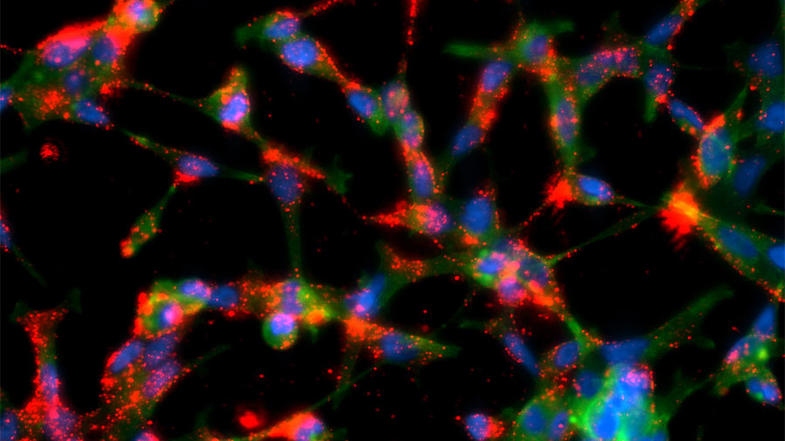
Current Clinical Trials

Use our advanced clinical trial search to find NCI-supported cancer clinical trials that are now enrolling patients. The search can be narrowed by location of the trial, type of treatment, name of the drug, and other criteria. General information about clinical trials is also available.

References
  1. Hennessy BT, Thomas DA, Giles FJ, et al.: New approaches in the treatment of myelofibrosis. Cancer 103 (1): 32-43, 2005. [PUBMED Abstract]
  2. Campbell PJ, Green AR: The myeloproliferative disorders. N Engl J Med 355 (23): 2452-66, 2006. [PUBMED Abstract]
  3. Cazzola M, Kralovics R: From Janus kinase 2 to calreticulin: the clinically relevant genomic landscape of myeloproliferative neoplasms. Blood 123 (24): 3714-9, 2014. [PUBMED Abstract]
  4. Rumi E, Pietra D, Ferretti V, et al.: JAK2 or CALR mutation status defines subtypes of essential thrombocythemia with substantially different clinical course and outcomes. Blood 123 (10): 1544-51, 2014. [PUBMED Abstract]
  5. Rotunno G, Mannarelli C, Guglielmelli P, et al.: Impact of calreticulin mutations on clinical and hematological phenotype and outcome in essential thrombocythemia. Blood 123 (10): 1552-5, 2014. [PUBMED Abstract]
  6. Barosi G: Myelofibrosis with myeloid metaplasia: diagnostic definition and prognostic classification for clinical studies and treatment guidelines. J Clin Oncol 17 (9): 2954-70, 1999. [PUBMED Abstract]
  7. Tefferi A: Myelofibrosis with myeloid metaplasia. N Engl J Med 342 (17): 1255-65, 2000. [PUBMED Abstract]
  8. Boddu P, Masarova L, Verstovsek S, et al.: Patient characteristics and outcomes in adolescents and young adults with classical Philadelphia chromosome-negative myeloproliferative neoplasms. Ann Hematol 97 (1): 109-121, 2018. [PUBMED Abstract]
  9. Tefferi A, Thiele J, Vardiman JW: The 2008 World Health Organization classification system for myeloproliferative neoplasms: order out of chaos. Cancer 115 (17): 3842-7, 2009. [PUBMED Abstract]
  10. Klampfl T, Gisslinger H, Harutyunyan AS, et al.: Somatic mutations of calreticulin in myeloproliferative neoplasms. N Engl J Med 369 (25): 2379-90, 2013. [PUBMED Abstract]
  11. Nangalia J, Massie CE, Baxter EJ, et al.: Somatic CALR mutations in myeloproliferative neoplasms with nonmutated JAK2. N Engl J Med 369 (25): 2391-405, 2013. [PUBMED Abstract]
  12. Guglielmelli P, Lasho TL, Rotunno G, et al.: The number of prognostically detrimental mutations and prognosis in primary myelofibrosis: an international study of 797 patients. Leukemia 28 (9): 1804-10, 2014. [PUBMED Abstract]
  13. Chim CS, Kwong YL, Lie AK, et al.: Long-term outcome of 231 patients with essential thrombocythemia: prognostic factors for thrombosis, bleeding, myelofibrosis, and leukemia. Arch Intern Med 165 (22): 2651-8, 2005 Dec 12-26. [PUBMED Abstract]
  14. Odenike O: How I treat the blast phase of Philadelphia chromosome-negative myeloproliferative neoplasms. Blood 132 (22): 2339-2350, 2018. [PUBMED Abstract]
  15. Hultcrantz M, Björkholm M, Dickman PW, et al.: Risk for Arterial and Venous Thrombosis in Patients With Myeloproliferative Neoplasms: A Population-Based Cohort Study. Ann Intern Med 168 (5): 317-325, 2018. [PUBMED Abstract]
  16. Hussein K, Pardanani AD, Van Dyke DL, et al.: International Prognostic Scoring System-independent cytogenetic risk categorization in primary myelofibrosis. Blood 115 (3): 496-9, 2010. [PUBMED Abstract]
  17. Cervantes F, Barosi G, Demory JL, et al.: Myelofibrosis with myeloid metaplasia in young individuals: disease characteristics, prognostic factors and identification of risk groups. Br J Haematol 102 (3): 684-90, 1998. [PUBMED Abstract]
  18. Strasser-Weippl K, Steurer M, Kees M, et al.: Age and hemoglobin level emerge as most important clinical prognostic parameters in patients with osteomyelofibrosis: introduction of a simplified prognostic score. Leuk Lymphoma 47 (3): 441-50, 2006. [PUBMED Abstract]
  19. Tefferi A: Survivorship and prognosis in myelofibrosis with myeloid metaplasia. Leuk Lymphoma 47 (3): 379-80, 2006. [PUBMED Abstract]
  20. Tam CS, Kantarjian H, Cortes J, et al.: Dynamic model for predicting death within 12 months in patients with primary or post-polycythemia vera/essential thrombocythemia myelofibrosis. J Clin Oncol 27 (33): 5587-93, 2009. [PUBMED Abstract]
  21. Morel P, Duhamel A, Hivert B, et al.: Identification during the follow-up of time-dependent prognostic factors for the competing risks of death and blast phase in primary myelofibrosis: a study of 172 patients. Blood 115 (22): 4350-5, 2010. [PUBMED Abstract]
  22. Cervantes F, Dupriez B, Pereira A, et al.: New prognostic scoring system for primary myelofibrosis based on a study of the International Working Group for Myelofibrosis Research and Treatment. Blood 113 (13): 2895-901, 2009. [PUBMED Abstract]
  23. Tefferi A, Lasho TL, Jimma T, et al.: One thousand patients with primary myelofibrosis: the mayo clinic experience. Mayo Clin Proc 87 (1): 25-33, 2012. [PUBMED Abstract]
  24. Gangat N, Caramazza D, Vaidya R, et al.: DIPSS plus: a refined Dynamic International Prognostic Scoring System for primary myelofibrosis that incorporates prognostic information from karyotype, platelet count, and transfusion status. J Clin Oncol 29 (4): 392-7, 2011. [PUBMED Abstract]
  25. Masarova L, Alhuraiji A, Bose P, et al.: Significance of thrombocytopenia in patients with primary and postessential thrombocythemia/polycythemia vera myelofibrosis. Eur J Haematol 100 (3): 257-263, 2018. [PUBMED Abstract]
  26. Caramazza D, Begna KH, Gangat N, et al.: Refined cytogenetic-risk categorization for overall and leukemia-free survival in primary myelofibrosis: a single center study of 433 patients. Leukemia 25 (1): 82-8, 2011. [PUBMED Abstract]
  27. Giovanni B, Michelle E, Letizia C, et al.: Thalidomide in myelofibrosis with myeloid metaplasia: a pooled-analysis of individual patient data from five studies. Leuk Lymphoma 43 (12): 2301-7, 2002. [PUBMED Abstract]
  28. Marchetti M, Barosi G, Balestri F, et al.: Low-dose thalidomide ameliorates cytopenias and splenomegaly in myelofibrosis with myeloid metaplasia: a phase II trial. J Clin Oncol 22 (3): 424-31, 2004. [PUBMED Abstract]
  29. Tefferi A, Cortes J, Verstovsek S, et al.: Lenalidomide therapy in myelofibrosis with myeloid metaplasia. Blood 108 (4): 1158-64, 2006. [PUBMED Abstract]
  30. Cervantes F, Alvarez-Larrán A, Hernández-Boluda JC, et al.: Erythropoietin treatment of the anaemia of myelofibrosis with myeloid metaplasia: results in 20 patients and review of the literature. Br J Haematol 127 (4): 399-403, 2004. [PUBMED Abstract]
  31. Huang J, Tefferi A: Erythropoiesis stimulating agents have limited therapeutic activity in transfusion-dependent patients with primary myelofibrosis regardless of serum erythropoietin level. Eur J Haematol 83 (2): 154-5, 2009. [PUBMED Abstract]
  32. Thomas DA, Giles FJ, Albitar M, et al.: Thalidomide therapy for myelofibrosis with myeloid metaplasia. Cancer 106 (9): 1974-84, 2006. [PUBMED Abstract]
  33. Tefferi A, Lasho TL, Mesa RA, et al.: Lenalidomide therapy in del(5)(q31)-associated myelofibrosis: cytogenetic and JAK2V617F molecular remissions. Leukemia 21 (8): 1827-8, 2007. [PUBMED Abstract]
  34. Mesa RA, Yao X, Cripe LD, et al.: Lenalidomide and prednisone for myelofibrosis: Eastern Cooperative Oncology Group (ECOG) phase 2 trial E4903. Blood 116 (22): 4436-8, 2010. [PUBMED Abstract]
  35. Quintás-Cardama A, Kantarjian HM, Manshouri T, et al.: Lenalidomide plus prednisone results in durable clinical, histopathologic, and molecular responses in patients with myelofibrosis. J Clin Oncol 27 (28): 4760-6, 2009. [PUBMED Abstract]
  36. Begna KH, Mesa RA, Pardanani A, et al.: A phase-2 trial of low-dose pomalidomide in myelofibrosis. Leukemia 25 (2): 301-4, 2011. [PUBMED Abstract]
  37. Harrison C, Kiladjian JJ, Al-Ali HK, et al.: JAK inhibition with ruxolitinib versus best available therapy for myelofibrosis. N Engl J Med 366 (9): 787-98, 2012. [PUBMED Abstract]
  38. Verstovsek S, Mesa RA, Gotlib J, et al.: A double-blind, placebo-controlled trial of ruxolitinib for myelofibrosis. N Engl J Med 366 (9): 799-807, 2012. [PUBMED Abstract]
  39. Tefferi A, Litzow MR, Pardanani A: Long-term outcome of treatment with ruxolitinib in myelofibrosis. N Engl J Med 365 (15): 1455-7, 2011. [PUBMED Abstract]
  40. Verstovsek S: Janus-activated kinase 2 inhibitors: a new era of targeted therapies providing significant clinical benefit for Philadelphia chromosome-negative myeloproliferative neoplasms. J Clin Oncol 29 (7): 781-3, 2011. [PUBMED Abstract]
  41. Deeg HJ, Gooley TA, Flowers ME, et al.: Allogeneic hematopoietic stem cell transplantation for myelofibrosis. Blood 102 (12): 3912-8, 2003. [PUBMED Abstract]
  42. Daly A, Song K, Nevill T, et al.: Stem cell transplantation for myelofibrosis: a report from two Canadian centers. Bone Marrow Transplant 32 (1): 35-40, 2003. [PUBMED Abstract]
  43. Kröger N, Holler E, Kobbe G, et al.: Allogeneic stem cell transplantation after reduced-intensity conditioning in patients with myelofibrosis: a prospective, multicenter study of the Chronic Leukemia Working Party of the European Group for Blood and Marrow Transplantation. Blood 114 (26): 5264-70, 2009. [PUBMED Abstract]
  44. Gupta V, Hari P, Hoffman R: Allogeneic hematopoietic cell transplantation for myelofibrosis in the era of JAK inhibitors. Blood 120 (7): 1367-79, 2012. [PUBMED Abstract]
  45. Abelsson J, Merup M, Birgegård G, et al.: The outcome of allo-HSCT for 92 patients with myelofibrosis in the Nordic countries. Bone Marrow Transplant 47 (3): 380-6, 2012. [PUBMED Abstract]
  46. Guardiola P, Anderson JE, Bandini G, et al.: Allogeneic stem cell transplantation for agnogenic myeloid metaplasia: a European Group for Blood and Marrow Transplantation, Société Française de Greffe de Moelle, Gruppo Italiano per il Trapianto del Midollo Osseo, and Fred Hutchinson Cancer Research Center Collaborative Study. Blood 93 (9): 2831-8, 1999. [PUBMED Abstract]
  47. Strupp C, Germing U, Scherer A, et al.: Thalidomide for the treatment of idiopathic myelofibrosis. Eur J Haematol 72 (1): 52-7, 2004. [PUBMED Abstract]
  48. Mesa RA, Elliott MA, Schroeder G, et al.: Durable responses to thalidomide-based drug therapy for myelofibrosis with myeloid metaplasia. Mayo Clin Proc 79 (7): 883-9, 2004. [PUBMED Abstract]
  49. Jabbour E, Thomas D, Kantarjian H, et al.: Comparison of thalidomide and lenalidomide as therapy for myelofibrosis. Blood 118 (4): 899-902, 2011. [PUBMED Abstract]
  50. Barosi G, Ambrosetti A, Centra A, et al.: Splenectomy and risk of blast transformation in myelofibrosis with myeloid metaplasia. Italian Cooperative Study Group on Myeloid with Myeloid Metaplasia. Blood 91 (10): 3630-6, 1998. [PUBMED Abstract]
  51. Tefferi A, Silverstein MN, Li CY: 2-Chlorodeoxyadenosine treatment after splenectomy in patients who have myelofibrosis with myeloid metaplasia. Br J Haematol 99 (2): 352-7, 1997. [PUBMED Abstract]
  52. Tefferi A, Mesa RA, Nagorney DM, et al.: Splenectomy in myelofibrosis with myeloid metaplasia: a single-institution experience with 223 patients. Blood 95 (7): 2226-33, 2000. [PUBMED Abstract]
  53. Sacchi S: The role of alpha-interferon in essential thrombocythaemia, polycythaemia vera and myelofibrosis with myeloid metaplasia (MMM): a concise update. Leuk Lymphoma 19 (1-2): 13-20, 1995. [PUBMED Abstract]
  54. Gilbert HS: Long term treatment of myeloproliferative disease with interferon-alpha-2b: feasibility and efficacy. Cancer 83 (6): 1205-13, 1998. [PUBMED Abstract]
  55. Verstovsek S, Kantarjian H, Mesa RA, et al.: Safety and efficacy of INCB018424, a JAK1 and JAK2 inhibitor, in myelofibrosis. N Engl J Med 363 (12): 1117-27, 2010. [PUBMED Abstract]
  56. Verstovsek S, Mesa RA, Gotlib J, et al.: Long-term treatment with ruxolitinib for patients with myelofibrosis: 5-year update from the randomized, double-blind, placebo-controlled, phase 3 COMFORT-I trial. J Hematol Oncol 10 (1): 55, 2017. [PUBMED Abstract]
  57. Harrison CN, Vannucchi AM, Kiladjian JJ, et al.: Long-term findings from COMFORT-II, a phase 3 study of ruxolitinib vs best available therapy for myelofibrosis. Leukemia 30 (8): 1701-7, 2016. [PUBMED Abstract]
  58. Mascarenhas J, Hoffman R: A comprehensive review and analysis of the effect of ruxolitinib therapy on the survival of patients with myelofibrosis. Blood 121 (24): 4832-7, 2013. [PUBMED Abstract]
  59. Verstovsek S, Mesa RA, Gotlib J, et al.: The clinical benefit of ruxolitinib across patient subgroups: analysis of a placebo-controlled, Phase III study in patients with myelofibrosis. Br J Haematol 161 (4): 508-16, 2013. [PUBMED Abstract]
  60. Porpaczy E, Tripolt S, Hoelbl-Kovacic A, et al.: Aggressive B-cell lymphomas in patients with myelofibrosis receiving JAK1/2 inhibitor therapy. Blood 132 (7): 694-706, 2018. [PUBMED Abstract]
  61. Lavrenkov K, Krepel-Volsky S, Levi I, et al.: Low dose palliative radiotherapy for splenomegaly in hematologic disorders. Leuk Lymphoma 53 (3): 430-4, 2012. [PUBMED Abstract]
  62. Mesa RA, Nagorney DS, Schwager S, et al.: Palliative goals, patient selection, and perioperative platelet management: outcomes and lessons from 3 decades of splenectomy for myelofibrosis with myeloid metaplasia at the Mayo Clinic. Cancer 107 (2): 361-70, 2006. [PUBMED Abstract]
  63. Ruggeri M, Rodeghiero F, Tosetto A, et al.: Postsurgery outcomes in patients with polycythemia vera and essential thrombocythemia: a retrospective survey. Blood 111 (2): 666-71, 2008. [PUBMED Abstract]
  64. Alchalby H, Yunus DR, Zabelina T, et al.: Risk models predicting survival after reduced-intensity transplantation for myelofibrosis. Br J Haematol 157 (1): 75-85, 2012. [PUBMED Abstract]
  65. Alchalby H, Badbaran A, Zabelina T, et al.: Impact of JAK2V617F mutation status, allele burden, and clearance after allogeneic stem cell transplantation for myelofibrosis. Blood 116 (18): 3572-81, 2010. [PUBMED Abstract]

Treatment of Chronic Neutrophilic Leukemia

Disease Overview for Chronic Neutrophilic Leukemia (CNL)

CNL is a rare chronic myeloproliferative neoplasm of unknown etiology, characterized by sustained peripheral blood neutrophilia (>25 × 109/L) and hepatosplenomegaly.[1,2] The bone marrow is hypercellular in patients with CNL. No significant dysplasia is in any of the cell lineages, and bone marrow fibrosis is uncommon.[1,2] Cytogenetic studies are normal in nearly 90% of the patients. In the remaining patients, clonal karyotypic abnormalities may include +8, +9, del (20q) and del(11q).[1,35] There is no Philadelphia chromosome or BCR::ABL fusion gene. CNL is a slowly progressive disorder, and the survival of patients ranges from 6 months to more than 20 years.

Treatment Option Overview for CNL

In the past, the treatment of CNL focused on disease control rather than cure. Once the disease progressed to a more aggressive leukemia, there was typically little chance of obtaining a long-lasting remission because of the older age of most patients, as well as the acquisition of multiple poor prognostic cytogenetic abnormalities. Allogeneic bone marrow transplant represents a potentially curative treatment modality for CNL.[68] Results vary with the use of traditional chemotherapies including hydroxyurea and interferon.[9]

Current Clinical Trials

Use our advanced clinical trial search to find NCI-supported cancer clinical trials that are now enrolling patients. The search can be narrowed by location of the trial, type of treatment, name of the drug, and other criteria. General information about clinical trials is also available.

References
  1. Imbert M, Bain B, Pierre R, et al.: Chronic neutrophilic leukemia. In: Jaffe ES, Harris NL, Stein H, et al., eds.: Pathology and Genetics of Tumours of Haematopoietic and Lymphoid Tissues. IARC Press, 2001. World Health Organization Classification of Tumours, 3, pp 27-8.
  2. Zittoun R, Réa D, Ngoc LH, et al.: Chronic neutrophilic leukemia. A study of four cases. Ann Hematol 68 (2): 55-60, 1994. [PUBMED Abstract]
  3. Froberg MK, Brunning RD, Dorion P, et al.: Demonstration of clonality in neutrophils using FISH in a case of chronic neutrophilic leukemia. Leukemia 12 (4): 623-6, 1998. [PUBMED Abstract]
  4. Yanagisawa K, Ohminami H, Sato M, et al.: Neoplastic involvement of granulocytic lineage, not granulocytic-monocytic, monocytic, or erythrocytic lineage, in a patient with chronic neutrophilic leukemia. Am J Hematol 57 (3): 221-4, 1998. [PUBMED Abstract]
  5. Matano S, Nakamura S, Kobayashi K, et al.: Deletion of the long arm of chromosome 20 in a patient with chronic neutrophilic leukemia: cytogenetic findings in chronic neutrophilic leukemia. Am J Hematol 54 (1): 72-5, 1997. [PUBMED Abstract]
  6. Piliotis E, Kutas G, Lipton JH: Allogeneic bone marrow transplantation in the management of chronic neutrophilic leukemia. Leuk Lymphoma 43 (10): 2051-4, 2002. [PUBMED Abstract]
  7. Hasle H, Olesen G, Kerndrup G, et al.: Chronic neutrophil leukaemia in adolescence and young adulthood. Br J Haematol 94 (4): 628-30, 1996. [PUBMED Abstract]
  8. Böhm J, Schaefer HE: Chronic neutrophilic leukaemia: 14 new cases of an uncommon myeloproliferative disease. J Clin Pathol 55 (11): 862-4, 2002. [PUBMED Abstract]
  9. Elliott MA, Dewald GW, Tefferi A, et al.: Chronic neutrophilic leukemia (CNL): a clinical, pathologic and cytogenetic study. Leukemia 15 (1): 35-40, 2001. [PUBMED Abstract]

Treatment of Chronic Eosinophilic Leukemia

Disease Overview for Chronic Eosinophilic Leukemia (CEL)

CEL is a chronic myeloproliferative neoplasm of unknown etiology in which a clonal proliferation of eosinophilic precursors results in persistently increased numbers of eosinophils in the blood, bone marrow, and peripheral tissues. In CEL, the eosinophil count is greater than or equal to 1.5 × 10⁹/L.[1] To make a diagnosis of CEL, there should be evidence for clonality of the eosinophils or an increase in blasts in the blood or bone marrow. However, in many cases, it is impossible to prove clonality of the eosinophils, in which case, if there is no increase in blast cells, the diagnosis of idiopathic hypereosinophilic syndrome (HES) is preferred. Because of the difficulty in distinguishing CEL from HES, the true incidence of these diseases is unknown, although they are rare. In about 10% of patients, eosinophilia is detected incidentally. In others, the constitutional symptoms found include:[1,2]

  • Fever.
  • Fatigue.
  • Cough.
  • Angioedema.
  • Muscle pains.
  • Pruritus.
  • Diarrhea.

No single or specific cytogenetic or molecular genetic abnormality has been identified in CEL.

For more information about the symptoms listed above, see Hot Flashes and Night Sweats, Fatigue, Cardiopulmonary Syndromes, Pruritus, and Gastrointestinal Complications.

Treatment Option Overview for CEL

CEL is rare, and the optimal treatment remains uncertain. The clinical course can range from cases with decades of stable disease to cases with rapid progression to acute leukemia. Case reports suggest that treatment options include bone marrow transplant and interferon alfa.[3,4]

Treatment of HES has included corticosteroids, chemotherapeutic agents (e.g., hydroxyurea, cyclophosphamide, or vincristine), and interferon alfa.[5,6]

Case reports suggest that patients with HES who have not responded to conventional options may have symptomatic responses to imatinib mesylate.[68][Level of evidence C3] Imatinib mesylate acts as an inhibitor of a novel FIP1L1::PDGFRA fusion tyrosine kinase, which results as a consequence of an interstitial chromosomal deletion.[6,9][Level of evidence C3] HES with the FIP1L1::PDGFRA fusion tyrosine kinase translocation has been shown to respond to low-dose imatinib mesylate.[9]

Current Clinical Trials

Use our advanced clinical trial search to find NCI-supported cancer clinical trials that are now enrolling patients. The search can be narrowed by location of the trial, type of treatment, name of the drug, and other criteria. General information about clinical trials is also available.

References
  1. Bain B, Pierre P, Imbert M, et al.: Chronic eosinophillic leukaemia and the hypereosinophillic syndrome. In: Jaffe ES, Harris NL, Stein H, et al., eds.: Pathology and Genetics of Tumours of Haematopoietic and Lymphoid Tissues. IARC Press, 2001. World Health Organization Classification of Tumours, 3, pp 29-31.
  2. Weller PF, Bubley GJ: The idiopathic hypereosinophilic syndrome. Blood 83 (10): 2759-79, 1994. [PUBMED Abstract]
  3. Basara N, Markova J, Schmetzer B, et al.: Chronic eosinophilic leukemia: successful treatment with an unrelated bone marrow transplantation. Leuk Lymphoma 32 (1-2): 189-93, 1998. [PUBMED Abstract]
  4. Yamada O, Kitahara K, Imamura K, et al.: Clinical and cytogenetic remission induced by interferon-alpha in a patient with chronic eosinophilic leukemia associated with a unique t(3;9;5) translocation. Am J Hematol 58 (2): 137-41, 1998. [PUBMED Abstract]
  5. Butterfield JH, Gleich GJ: Interferon-alpha treatment of six patients with the idiopathic hypereosinophilic syndrome. Ann Intern Med 121 (9): 648-53, 1994. [PUBMED Abstract]
  6. Gotlib J, Cools J, Malone JM, et al.: The FIP1L1-PDGFRalpha fusion tyrosine kinase in hypereosinophilic syndrome and chronic eosinophilic leukemia: implications for diagnosis, classification, and management. Blood 103 (8): 2879-91, 2004. [PUBMED Abstract]
  7. Gleich GJ, Leiferman KM, Pardanani A, et al.: Treatment of hypereosinophilic syndrome with imatinib mesilate. Lancet 359 (9317): 1577-8, 2002. [PUBMED Abstract]
  8. Ault P, Cortes J, Koller C, et al.: Response of idiopathic hypereosinophilic syndrome to treatment with imatinib mesylate. Leuk Res 26 (9): 881-4, 2002. [PUBMED Abstract]
  9. Cools J, DeAngelo DJ, Gotlib J, et al.: A tyrosine kinase created by fusion of the PDGFRA and FIP1L1 genes as a therapeutic target of imatinib in idiopathic hypereosinophilic syndrome. N Engl J Med 348 (13): 1201-14, 2003. [PUBMED Abstract]

Latest Updates to This Summary (09/27/2024)

The PDQ cancer information summaries are reviewed regularly and updated as new information becomes available. This section describes the latest changes made to this summary as of the date above.

This summary was renamed from Chronic Myeloproliferative Neoplasms Treatment.

General Information About Myeloproliferative Neoplasms (MPN)

Added Arber et al. as reference 1.

Added Barosi et al. as reference 4.

Revised text to state that there is no standard treatment approach for patients with progression from chronic-phase MPN to accelerated or blast phase, and these patients have a poor prognosis (cited Mudireddy et al. as reference 8).

Treatment of Polycythemia Vera

This section was extensively revised.

This summary is written and maintained by the PDQ Adult Treatment Editorial Board, which is editorially independent of NCI. The summary reflects an independent review of the literature and does not represent a policy statement of NCI or NIH. More information about summary policies and the role of the PDQ Editorial Boards in maintaining the PDQ summaries can be found on the About This PDQ Summary and PDQ® Cancer Information for Health Professionals pages.

About This PDQ Summary

Purpose of This Summary

This PDQ cancer information summary for health professionals provides comprehensive, peer-reviewed, evidence-based information about the treatment of myeloproliferative neoplasms. It is intended as a resource to inform and assist clinicians in the care of their patients. It does not provide formal guidelines or recommendations for making health care decisions.

Reviewers and Updates

This summary is reviewed regularly and updated as necessary by the PDQ Adult Treatment Editorial Board, which is editorially independent of the National Cancer Institute (NCI). The summary reflects an independent review of the literature and does not represent a policy statement of NCI or the National Institutes of Health (NIH).

Board members review recently published articles each month to determine whether an article should:

  • be discussed at a meeting,
  • be cited with text, or
  • replace or update an existing article that is already cited.

Changes to the summaries are made through a consensus process in which Board members evaluate the strength of the evidence in the published articles and determine how the article should be included in the summary.

The lead reviewers for Myeloproliferative Neoplasms Treatment are:

  • Aaron Gerds, MD (Cleveland Clinic Taussig Cancer Institute)
  • Eric J. Seifter, MD (Johns Hopkins University)

Any comments or questions about the summary content should be submitted to Cancer.gov through the NCI website’s Email Us. Do not contact the individual Board Members with questions or comments about the summaries. Board members will not respond to individual inquiries.

Levels of Evidence

Some of the reference citations in this summary are accompanied by a level-of-evidence designation. These designations are intended to help readers assess the strength of the evidence supporting the use of specific interventions or approaches. The PDQ Adult Treatment Editorial Board uses a formal evidence ranking system in developing its level-of-evidence designations.

Permission to Use This Summary

PDQ is a registered trademark. Although the content of PDQ documents can be used freely as text, it cannot be identified as an NCI PDQ cancer information summary unless it is presented in its entirety and is regularly updated. However, an author would be permitted to write a sentence such as “NCI’s PDQ cancer information summary about breast cancer prevention states the risks succinctly: [include excerpt from the summary].”

The preferred citation for this PDQ summary is:

PDQ® Adult Treatment Editorial Board. PDQ Myeloproliferative Neoplasms Treatment. Bethesda, MD: National Cancer Institute. Updated <MM/DD/YYYY>. Available at: /types/myeloproliferative/hp/myeloproliferative-neoplasms-treatment. Accessed <MM/DD/YYYY>. [PMID: 26389291]

Images in this summary are used with permission of the author(s), artist, and/or publisher for use within the PDQ summaries only. Permission to use images outside the context of PDQ information must be obtained from the owner(s) and cannot be granted by the National Cancer Institute. Information about using the illustrations in this summary, along with many other cancer-related images, is available in Visuals Online, a collection of over 2,000 scientific images.

Disclaimer

Based on the strength of the available evidence, treatment options may be described as either “standard” or “under clinical evaluation.” These classifications should not be used as a basis for insurance reimbursement determinations. More information on insurance coverage is available on Cancer.gov on the Managing Cancer Care page.

Contact Us

More information about contacting us or receiving help with the Cancer.gov website can be found on our Contact Us for Help page. Questions can also be submitted to Cancer.gov through the website’s Email Us.

Chronic Myeloid Leukemia Treatment (PDQ®)–Health Professional Version

Chronic Myeloid Leukemia Treatment (PDQ®)–Health Professional Version

General Information About Chronic Myeloid Leukemia (CML)

Incidence and Mortality

Estimated new cases and deaths from CML in the United States in 2025:[1]

  • New cases: 9,560.
  • Deaths: 1,290.

CML is one of a group of diseases called the myeloproliferative disorders. It is also called chronic myelogenous leukemia. Other related entities include:

  • Polycythemia vera.
  • Myelofibrosis.
  • Essential thrombocythemia.

For more information, see Myeloproliferative Neoplasms Treatment.

Molecular Genetics

CML is identified by too many myeloblasts in the blood and bone marrow, and the disease worsens as the number of myeloblasts increase.

EnlargeBlood cell development; drawing shows the steps a blood stem cell goes through to become a red blood cell, platelet, or white blood cell. A myeloid stem cell becomes a red blood cell, a platelet, or a myeloblast, which then becomes a granulocyte (the types of granulocytes are eosinophils, basophils, and neutrophils). A lymphoid stem cell becomes a lymphoblast and then becomes a B-lymphocyte, T-lymphocyte, or natural killer cell.
Figure 1. Hematopoietic tree, expanded lymphoid line.

CML is a clonal disorder that is easily diagnosed because the leukemic cells of more than 95% of patients have a distinctive cytogenetic abnormality, the Philadelphia chromosome (Ph).[2]

EnlargePhiladelphia chromosome; three-panel drawing shows a piece of chromosome 9 and a piece of chromosome 22 breaking off and trading places, creating a changed chromosome 22 called the Philadelphia chromosome. In the left panel, the drawing shows a normal chromosome 9 with the ABL1 gene and a normal chromosome 22 with the BCR gene. In the center panel, the drawing shows part of the ABL1 gene breaking off from chromosome 9 and a piece of chromosome 22 breaking off, below the BCR gene. In the right panel, the drawing shows chromosome 9 with the piece from chromosome 22 attached. It also shows a shortened version of chromosome 22 with the piece from chromosome 9 containing part of the ABL1 gene attached. The ABL1 gene joins to the BCR gene on chromosome 22 to form the BCR::ABL1 fusion gene. The changed chromosome 22 with the BCR::ABL1 fusion gene on it is called the Philadelphia chromosome.
Figure 2. The Philadelphia chromosome is a translocation between the ABL1 oncogene (on the long arm of chromosome 9) and the BCR gene (on the long arm of chromosome 22), resulting in the BCR::ABL1 fusion gene. BCR::ABL1 encodes an oncogenic protein with tyrosine kinase activity.

The Ph chromosome results from a reciprocal translocation between the long arms of chromosomes 9 and 22, and it is demonstrable in all hematopoietic precursors.[3] This translocation results in the transfer of the ABL1 oncogene on chromosome 9 to an area of chromosome 22 termed the breakpoint cluster region (within the BCR gene).[3] This, in turn, results in a BCR::ABL1 fusion gene and in the production of an abnormal tyrosine kinase protein that causes the disordered myelopoiesis found in CML. Using peripheral blood, molecular techniques can detect the presence of the 9;22 translocation.

Clinical Presentation

Although CML may present without symptoms, splenomegaly is the most common finding during physical examination at the time of diagnosis.[4] The spleen may be enormous, filling most of the abdomen, causing pain or a feeling of fullness and presenting a significant clinical problem, or the spleen may be only minimally enlarged. In about 10% of patients, the spleen is neither palpable nor enlarged on computed tomography (CT) scan.

Patients may also present with the following symptoms:

  • Fatigue.
  • Unexplained weight loss.
  • Drenching night sweats.
  • Fever.

Transition between the chronic, accelerated, and blastic phases may occur gradually over 1 year or more, or it may occur abruptly (blast crisis). Patients with accelerated-phase CML show signs of progression without meeting the criteria for blast crisis (acute leukemia). The following signs and symptoms indicate a change to accelerated-phase CML:

  • Progressive splenomegaly.
  • Increased leukocytosis and/or thrombocytosis.
  • Progressive anemia.

The following signs and symptoms indicate a change to a blast crisis, in addition to the accelerated-phase CML symptoms:

  • Thrombocytopenia.
  • Increasing and painful splenomegaly or hepatomegaly.
  • Fever.
  • Bone pain.
  • Development of destructive bone lesions.

In the accelerated phase, differentiated cells persist, although they often show increasing morphological abnormalities. The patient experiences increased anemia, thrombocytopenia, and marrow fibrosis.[4]

Risk Factors

Risk factors for CML include:

  • Older age.
  • Exposure to high-dose ionizing radiation.

Diagnostic Evaluation

In addition to a health history and physical examination, the initial workup may include:

  • Complete blood count with differential.
  • Blood chemistry studies.
  • Bone marrow aspiration and biopsy. In routine presentations of CML, the utility of bone marrow aspiration and biopsy for all newly diagnosed patients is questionable outside the context of a clinical trial. Bone marrow testing is appropriate for patients with clinical signs of accelerated phase or blast crisis (fever, enlarged spleen, or >20% blasts in the peripheral blood).[5]
  • Cytogenetic analysis.
  • Fluorescence in situ hybridization (FISH). FISH of the BCR::ABL1 translocation can be performed using the bone marrow aspirate or peripheral blood of patients with CML.[4]
  • Reverse transcription–polymerase chain reaction (RT-PCR). A small subset of patients has the BCR::ABL1 rearrangement detectable only by RT-PCR, which is the most sensitive technique currently available. Patients with RT-PCR evidence of the BCR::ABL1 fusion gene appear clinically and prognostically identical to patients with a classic Ph chromosome. However, patients who are BCR::ABL1-negative by RT-PCR have a clinical course more consistent with chronic myelomonocytic leukemia, which is a distinct clinical entity related to myelodysplastic syndrome.[68]
  • CT scan.

Prognosis and Survival

The median age of patients with Ph chromosome–positive CML is 67 years.[9] With the advent of the oral tyrosine kinase inhibitors (TKIs) , the median survival is projected to approach normal life expectancy for most patients.[10]

Ph chromosome–negative CML is a poorly defined entity that is less clearly distinguished from other myeloproliferative syndromes. Patients with Ph chromosome–negative CML generally have a poorer response to treatment and shorter survival than Ph chromosome–positive patients.[11] Ph chromosome–negative patients who have BCR::ABL1 gene rearrangements detectable by Southern blot analysis, however, have prognoses equivalent to Ph chromosome–positive patients.[6,12]

References
  1. American Cancer Society: Cancer Facts and Figures 2025. American Cancer Society, 2025. Available online. Last accessed January 16, 2025.
  2. Jabbour E, Kantarjian H: Chronic myeloid leukemia: 2020 update on diagnosis, therapy and monitoring. Am J Hematol 95 (6): 691-709, 2020. [PUBMED Abstract]
  3. Deininger MW, Goldman JM, Melo JV: The molecular biology of chronic myeloid leukemia. Blood 96 (10): 3343-56, 2000. [PUBMED Abstract]
  4. Jabbour E, Kantarjian H: Chronic myeloid leukemia: 2012 update on diagnosis, monitoring, and management. Am J Hematol 87 (11): 1037-45, 2012. [PUBMED Abstract]
  5. Hidalgo-Lόpez JE, Kanagal-Shamanna R, Quesada AE, et al.: Bone marrow core biopsy in 508 consecutive patients with chronic myeloid leukemia: Assessment of potential value. Cancer 124 (19): 3849-3855, 2018. [PUBMED Abstract]
  6. Martiat P, Michaux JL, Rodhain J: Philadelphia-negative (Ph-) chronic myeloid leukemia (CML): comparison with Ph+ CML and chronic myelomonocytic leukemia. The Groupe Français de Cytogénétique Hématologique. Blood 78 (1): 205-11, 1991. [PUBMED Abstract]
  7. Oscier DG: Atypical chronic myeloid leukaemia, a distinct clinical entity related to the myelodysplastic syndrome? Br J Haematol 92 (3): 582-6, 1996. [PUBMED Abstract]
  8. Kurzrock R, Bueso-Ramos CE, Kantarjian H, et al.: BCR rearrangement-negative chronic myelogenous leukemia revisited. J Clin Oncol 19 (11): 2915-26, 2001. [PUBMED Abstract]
  9. Lee SJ, Anasetti C, Horowitz MM, et al.: Initial therapy for chronic myelogenous leukemia: playing the odds. J Clin Oncol 16 (9): 2897-903, 1998. [PUBMED Abstract]
  10. Bower H, Björkholm M, Dickman PW, et al.: Life Expectancy of Patients With Chronic Myeloid Leukemia Approaches the Life Expectancy of the General Population. J Clin Oncol 34 (24): 2851-7, 2016. [PUBMED Abstract]
  11. Onida F, Ball G, Kantarjian HM, et al.: Characteristics and outcome of patients with Philadelphia chromosome negative, bcr/abl negative chronic myelogenous leukemia. Cancer 95 (8): 1673-84, 2002. [PUBMED Abstract]
  12. Cortes JE, Talpaz M, Beran M, et al.: Philadelphia chromosome-negative chronic myelogenous leukemia with rearrangement of the breakpoint cluster region. Long-term follow-up results. Cancer 75 (2): 464-70, 1995. [PUBMED Abstract]

Histopathology and Phases of CML

Histopathological examination of the bone marrow aspirate of patients with chronic myeloid leukemia (CML) demonstrates a shift in the myeloid series to immature forms that increase in number as patients progress to the blastic phase of the disease. The marrow is hypercellular, and differential counts of both marrow and blood show a spectrum of mature and immature granulocytes like that found in normal marrow. Increased numbers of eosinophils or basophils are often present, and monocytosis is sometimes seen. Increased megakaryocytes are often found in the marrow, and sometimes fragments of megakaryocytic nuclei are present in the blood, especially when the platelet count is very high. The percentage of lymphocytes is reduced in both the marrow and blood compared with normal samples. The myeloid:erythroid ratio in the marrow is usually greatly elevated. The leukocyte alkaline phosphatase enzyme is either absent or markedly reduced in the neutrophils of patients with CML.[1]

Most patients do not require bone marrow examination. However, bone marrow testing is appropriate for patients with fever, malaise, rapidly enlarging splenomegaly, and more than 10% circulating blasts. In patients with CML, bone marrow sampling is performed to assess cellularity, fibrosis, and cytogenetics. Reverse transcription–polymerase chain reaction (RT-PCR) or fluorescence in situ hybridization (FISH) analyses using blood or marrow aspirates demonstrate the 9;22 translocation.[1]

Chronic-Phase CML

Chronic-phase CML is characterized by bone marrow and cytogenetic findings as listed below with less than 10% blasts and promyelocytes in the peripheral blood and bone marrow.[2] The following factors are predictive of a shorter chronic phase after treatment with tyrosine kinase inhibitors:

  • Older age.[3]
  • Cytogenetic abnormalities in addition to the Philadelphia chromosome.[3,4]
  • A higher proportion of marrow or peripheral blood blasts.[3]
  • Anemia.[3]

Predictive models using multivariate analysis have been derived.[57]

The rate of progression from chronic phase to blast crisis is 5% to 10% in the first 2 years and 20% in subsequent years.[5]

For more information, see the Treatment of Chronic-Phase CML section.

Accelerated-Phase CML

Accelerated-phase CML is characterized by 10% to 19% blasts in either the peripheral blood or bone marrow.[2]

For more information, see the Treatment of Accelerated-Phase CML section.

Blastic-Phase CML

Blastic-phase CML is characterized by 20% or more blasts in the peripheral blood or bone marrow.

When 20% or more blasts are present along with fever, malaise, and progressive splenomegaly, the patient has entered blast crisis.[2]

For more information, see the Treatment of Blastic-Phase CML section.

References
  1. Jabbour E, Kantarjian H: Chronic myeloid leukemia: 2012 update on diagnosis, monitoring, and management. Am J Hematol 87 (11): 1037-45, 2012. [PUBMED Abstract]
  2. Cortes JE, Talpaz M, O’Brien S, et al.: Staging of chronic myeloid leukemia in the imatinib era: an evaluation of the World Health Organization proposal. Cancer 106 (6): 1306-15, 2006. [PUBMED Abstract]
  3. Lauseker M, Bachl K, Turkina A, et al.: Prognosis of patients with chronic myeloid leukemia presenting in advanced phase is defined mainly by blast count, but also by age, chromosomal aberrations and hemoglobin. Am J Hematol 94 (11): 1236-1243, 2019. [PUBMED Abstract]
  4. Fabarius A, Leitner A, Hochhaus A, et al.: Impact of additional cytogenetic aberrations at diagnosis on prognosis of CML: long-term observation of 1151 patients from the randomized CML Study IV. Blood 118 (26): 6760-8, 2011. [PUBMED Abstract]
  5. Sokal JE, Baccarani M, Russo D, et al.: Staging and prognosis in chronic myelogenous leukemia. Semin Hematol 25 (1): 49-61, 1988. [PUBMED Abstract]
  6. Hasford J, Pfirrmann M, Hehlmann R, et al.: A new prognostic score for survival of patients with chronic myeloid leukemia treated with interferon alfa. Writing Committee for the Collaborative CML Prognostic Factors Project Group. J Natl Cancer Inst 90 (11): 850-8, 1998. [PUBMED Abstract]
  7. Kvasnicka HM, Thiele J, Schmitt-Graeff A, et al.: Bone marrow features improve prognostic efficiency in multivariate risk classification of chronic-phase Ph(1+) chronic myelogenous leukemia: a multicenter trial. J Clin Oncol 19 (12): 2994-3009, 2001. [PUBMED Abstract]

Treatment Option Overview for CML

Treatment of patients with chronic myeloid leukemia (CML) is usually initiated at diagnosis, which is based on the presence of an elevated white blood cell count, splenomegaly, thrombocytosis, and identification of the BCR::ABL1 translocation.[1]

Table 1. Treatment Options for CML Phases
Phase Treatment Options
BMT = bone marrow transplant; CML = chronic myeloid leukemia; SCT = stem cell transplant; TKIs = tyrosine kinase inhibitors.
Chronic-phase CML Targeted therapy with an allosteric inhibitor of BCR::ABL1 at the ABL1 myristoyl pocket
Targeted therapy with other BCR::ABL1 TKIs
Allogeneic BMT or SCT
Accelerated-phase CML Targeted therapy with TKIs
Allogeneic SCT
Blastic-phase CML Targeted therapy with TKIs
Allogeneic BMT or SCT
Relapsed CML Targeted therapy with TKIs

Targeted Therapy With Tyrosine Kinase Inhibitors (TKIs)

The optimal front-line treatment for patients with chronic-phase CML involves specific inhibitors of the BCR::ABL1 tyrosine kinase. Although imatinib mesylate has been extensively studied in patients with CML, TKIs with greater potency and selectivity for BCR::ABL1 than imatinib have also been evaluated.[14] Bariatric surgery may impede proper absorption of oral TKIs, resulting in suboptimal responses.[5]

Allogeneic Bone Marrow Transplant (BMT) or Stem Cell Transplant (SCT)

Allogeneic BMT or SCT has also been used with curative intent.[6] Long-term data beyond 10 years of therapy are available, and most long-term survivors show no evidence of the BCR::ABL1 translocation by any available test (e.g., cytogenetics, reverse transcription–polymerase chain reaction, or fluorescence in situ hybridization). Some patients, however, are not eligible for this approach because of age, comorbid conditions, or lack of a suitable donor. In addition, substantial morbidity and mortality result from allogeneic BMT or SCT; a 5% to 10% treatment-related mortality can be expected, depending on whether a donor is related and the presence of mismatched antigens.[6]

Evidence (allogeneic SCT vs. drug treatment):

  1. In a prospective trial of 427 transplant-eligible, previously untreated patients, 166 patients were allocated to allogeneic SCT, and 261 patients were allocated to drug treatment (mostly imatinib).[6][Level of evidence C1]
    • No difference in 10-year overall survival was reported between the treatment groups.

    Similar outcomes were seen in patients who underwent allogeneic SCT because of TKI intolerance or nonadherence.[7]

Interferon Alfa

Long-term data are also available for patients treated with interferon alfa.[810] Approximately 10% to 20% of these patients have a complete cytogenetic response with no evidence of BCR::ABL1 translocation by any available test, and most of these patients are disease free beyond 10 years. Maintenance therapy with interferon is required, however, and some patients experience side effects that preclude continued treatment.

Hydroxyurea

Hydroxyurea is superior to busulfan in the chronic phase of CML, with significantly longer median survival and significantly fewer severe adverse effects.[11] A dose of 40 mg/kg per day is often used initially, and frequently results in a rapid reduction of the white blood cell (WBC) count. When the WBC count drops below 20 × 109/L, the hydroxyurea dose is often reduced and titrated to maintain a WBC count between 5 × 10⁹/L and 20 × 109/L.

Hydroxyurea is used primarily to stabilize patients with hyperleukocytosis or as palliative therapy for patients who have not responded to other therapies.

References
  1. Cortes J, Pavlovsky C, Saußele S: Chronic myeloid leukaemia. Lancet 398 (10314): 1914-1926, 2021. [PUBMED Abstract]
  2. Jabbour E, Kantarjian H: Chronic myeloid leukemia: 2020 update on diagnosis, therapy and monitoring. Am J Hematol 95 (6): 691-709, 2020. [PUBMED Abstract]
  3. Brümmendorf TH, Cortes JE, Milojkovic D, et al.: Bosutinib versus imatinib for newly diagnosed chronic phase chronic myeloid leukemia: final results from the BFORE trial. Leukemia 36 (7): 1825-1833, 2022. [PUBMED Abstract]
  4. Hochhaus A, Wang J, Kim DW, et al.: Asciminib in Newly Diagnosed Chronic Myeloid Leukemia. N Engl J Med 391 (10): 885-898, 2024. [PUBMED Abstract]
  5. Haddad FG, Kantarjian HM, Bidikian A, et al.: Association between bariatric surgery and outcomes in chronic myeloid leukemia. Cancer 129 (12): 1866-1872, 2023. [PUBMED Abstract]
  6. Gratwohl A, Pfirrmann M, Zander A, et al.: Long-term outcome of patients with newly diagnosed chronic myeloid leukemia: a randomized comparison of stem cell transplantation with drug treatment. Leukemia 30 (3): 562-9, 2016. [PUBMED Abstract]
  7. Wu J, Chen Y, Hageman L, et al.: Late mortality after bone marrow transplant for chronic myelogenous leukemia in the context of prior tyrosine kinase inhibitor exposure: A Blood or Marrow Transplant Survivor Study (BMTSS) report. Cancer 125 (22): 4033-4042, 2019. [PUBMED Abstract]
  8. Ozer H, George SL, Schiffer CA, et al.: Prolonged subcutaneous administration of recombinant alpha 2b interferon in patients with previously untreated Philadelphia chromosome-positive chronic-phase chronic myelogenous leukemia: effect on remission duration and survival: Cancer and Leukemia Group B study 8583. Blood 82 (10): 2975-84, 1993. [PUBMED Abstract]
  9. Kantarjian HM, Smith TL, O’Brien S, et al.: Prolonged survival in chronic myelogenous leukemia after cytogenetic response to interferon-alpha therapy. The Leukemia Service. Ann Intern Med 122 (4): 254-61, 1995. [PUBMED Abstract]
  10. Long-term follow-up of the Italian trial of interferon-alpha versus conventional chemotherapy in chronic myeloid leukemia. The Italian Cooperative Study Group on Chronic Myeloid Leukemia. Blood 92 (5): 1541-8, 1998. [PUBMED Abstract]
  11. Hehlmann R, Heimpel H, Hasford J, et al.: Randomized comparison of busulfan and hydroxyurea in chronic myelogenous leukemia: prolongation of survival by hydroxyurea. The German CML Study Group. Blood 82 (2): 398-407, 1993. [PUBMED Abstract]

Treatment of Chronic-Phase CML

Treatment Options for Chronic-Phase CML

Treatment options for chronic-phase chronic myeloid leukemia (CML) include:

The preferred initial treatment for patients with newly diagnosed chronic-phase CML could be any of the specific inhibitors of the BCR::ABL1 tyrosine kinase (including asciminib, nilotinib, dasatinib, bosutinib, or imatinib).[1] With any of these agents, the 10-year event-free survival and overall survival (OS) rates exceed 90%.[24]

CML response rate abbreviations used in this section include:

  • DMR: Deep molecular response (previously called CMR [complete molecular response]). This means greater than 4-log reduction (BCR::ABL1 ≤ 0.01%) and is also called MR 4 (molecular response 4). MR 4.5 is designated for BCR::ABL1 ≤ 0.0032%, and MR 5 is designated for BCR::ABL1 ≤ 0.001%.
  • EMR: Early molecular response. This means a greater than 1-log reduction (BCR::ABL1 ≤ 10%) at 3 months.
  • MMR: Major molecular response. This means a greater than 3-log reduction (BCR::ABL1 ≤ 0.1%).

A BCR::ABL1 transcript level of 10% or less in patients after 3 months of treatment with a specific TKI (deemed EMR) is associated with the best prognosis in terms of failure-free survival, progression-free survival (PFS), and OS.[510] However, in a retrospective analysis, even patients with a BCR::ABL1 transcript level greater than 10% after 3 months of therapy did well when the halving time was less than 76 days.[11]

Mandating a change of therapy based on this 10% transcript level at 3 to 6 months is problematic because 75% of patients do well even with a suboptimal response.[12] After 1 year, the preferred response target is an MMR, which is defined as a BCR::ABL1 level of less than or equal to 0.1%. The optimal target is a DMR, which is defined as under 4 logs (BCR::ABL1 ≤ 0.01%) or undetectable, which is usually a BCR::ABL1 level of less than or equal to 0.001% (MR 5).[13]

Targeted therapy with an allosteric inhibitor of BCR::ABL1 at the ABL1 myristoyl pocket

Evidence (targeted therapy with an allosteric inhibitor of BCR::ABL1 at the ABL1 myristoyl pocket):

  1. A prospective study (NCT04971226) included 405 patients with newly diagnosed CML. Patients were randomly assigned to receive asciminib (n = 201) (an allosteric inhibitor of BCR::ABL1 at the ABL1 myristoyl pocket, a site unique from those used by other TKIs) or either imatinib mesylate (n = 102) or nilotinib, dasatinib, or bosutinib (n = 102).[14]
    • With a median follow-up of 16.3 months, the 48-week MMR rate was 67.7% for patients who received asciminib and 49% for patients who received imatinib, nilotinib, dasatinib, or bosutinib (P < .002).[14][Level of evidence B3]
    • Patients who received asciminib had fewer grade 3 or greater adverse events (38%) compared with imatinib (44%) and the other TKIs (55%). The rate of discontinuation due to adverse events was lower for patients who received asciminib (5%) compared with patients who received imatinib (11%) or the other TKIs (10%).
    • Asciminib showed improved efficacy in this early reporting of the trial, and it also showed better tolerability based on adverse events and discontinuations. On this basis, the U.S. Food and Drug Administration approved the use of asciminib as first-line therapy. Use of asciminib will pose significant financial toxicity ($260,000 per year in 2024) versus imatinib ($500 per year in 2024). The price of the other TKIs may decrease because dasatinib is available as a generic, and nilotinib, bosutinib, and ponatinib are expected to be released as generics in 2027.
    • A prespecified subgroup analysis compared asciminib with the second-generation TKIs (not including imatinib). At week 48, 66.0% of patients who received asciminib had an MMR, and 57.8% of patients who received second-generation TKIs had an MMR. The 8.2% difference was not statistically significant (95% confidence interval [CI], -5.1 to 21.5). In the first year, it appears that the efficacy of asciminib is equivalent to those of second-generation TKIs. Longer follow-up is required to fully assess efficacy and toxicity outcomes.[14]

Targeted therapy with other BCR::ABL1 TKIs

Evidence (targeted therapy with other BCR::ABL1 TKIs):

  1. A randomized prospective study of 846 patients compared nilotinib with imatinib.[15][Level of evidence B3]
    • The rate of MMR at 24 months was 71% and 67% for patients who received two-dose schedules of nilotinib and 44% for patients who received imatinib (P < .0001 for both comparisons).
    • Progression to accelerated-phase CML or blast crisis occurred in 17 patients who received imatinib (14%), but this progression only occurred in two patients who received nilotinib 300 mg twice daily (<1%, P = .0003) and in five patients who received nilotinib 400 mg twice daily (1.8%, P = .0089).
  2. A randomized prospective study of 519 patients compared dasatinib with imatinib, with the following results:[16][Level of evidence B3]
    • The rate of MMR at 12 months was 46% for patients who received dasatinib and 28% for patients who received imatinib (P < .0001).
    • The rate of MMR at 24 months was 64% for patients who received dasatinib and 46% for patients who received imatinib (P < .0001).
    • At 5 years, there was no difference in PFS or OS.
    • Progression to accelerated-phase CML or blast crisis occurred in 13 patients (5%) who received imatinib and in six patients (2.3%) who received dasatinib (not statistically significant).
    • In retrospective comparative analyses, a dasatinib dose of 50 mg a day showed equal efficacy to 100 mg, but resulted in fewer pleural effusions (5% vs. 21%).[17][Level of evidence C3]
  3. A randomized prospective study of 536 patients compared bosutinib with imatinib.[18][Level of evidence B3]
    • The MMR rate at 5 years was 73.9% for patients in the bosutinib arm versus 64.6% for patients in the imatinib arm (hazard ratio [HR], 1.57; 95% CI, 1.08–2.28; P = .0075). At 5 years, a DMR (4.5 logs) was attained by 47.4% of patients in the bosutinib arm and 36.6% of patients in the imatinib arm (HR, 1.57; 95% CI, 1.11–2.22).[18]
    • Progression to accelerated-phase CML or blast crisis occurred in four patients (1.6%) who received bosutinib and in six patients (2.5%) who received imatinib.

In randomized prospective trials, nilotinib, dasatinib, and bosutinib showed higher rates of earlier MMR compared with imatinib. It is unclear whether this will translate to improved long-term outcomes.[8,9,18][Level of evidence B3] A dose-ranging phase II study of dasatinib in patients older than 70 years showed optimal response and reduction of toxicity starting at 20 mg once daily (with dose escalation if needed), versus the standard dose of 100 mg daily.[19][Level of evidence C3]

Can TKIs be discontinued?

For patients who obtain a DMR, it is unclear if TKI therapy can be discontinued. Several nonrandomized reports are summarized as follows:[2024][Level of evidence C3]

  • Patients who have taken a TKI for more than 3 to 5 years and attained a DMR (molecular remission, 4.5; BCR::ABL1 ≤ 0.0032%) are the best candidates to consider stopping therapy.
  • 50% of patients will experience a relapse of their disease if they discontinue TKI therapy. However, a retrospective analysis with a median follow-up of 3 years found that patients who were in DMR (4 to 4.5 logs) for 5 or more years had a relapse rate of approximately 10%.[25][Level of evidence C3] Another retrospective report with a median of 3 years of follow-up found three measurable factors predictive of MMR maintenance: increased duration of TKI treatment, increased duration of DMR on TKI treatment, and the absence of any peripheral blood blast cells at diagnosis.[20]
  • Almost all patients who relapse based on BCR::ABL1 quantitative reverse transcription–polymerase chain reaction (RT-PCR) testing can be successfully reinduced with the previous TKI.

However, after the reinduction of a previous TKI, the duration of remissions or the depth of responses are not known. Data to recommend universal discontinuation of TKIs are insufficient, even in patients with a DMR or CMR. Follow-up (i.e., at least every 3 months initially, although the precise interval is not well-defined) is required after stopping therapy because relapses have been noted even after 2 to 3 years. A withdrawal syndrome of muscle and joint pain has been reported after discontinuing TKI therapy.[26] Quality-of-life assessments suggest improved social function, diarrhea, and fatigue after stopping TKI therapy.[27][Level of evidence C1]

Allogeneic BMT or SCT

Allogeneic BMT or SCT is the only consistently successful curative treatment for patients with CML.[2830] Patients younger than 60 years with an identical twin or with HLA–matched siblings can consider BMT early in the chronic phase. Although the procedure is associated with considerable acute morbidity and mortality, 50% to 70% of patients who undergo transplant in the chronic phase appear to be cured. The results are better in younger patients, especially for those younger than 20 years. The outcomes of patients who undergo transplant in the accelerated and blastic phases of the disease are progressively worse.[31,32] Most transplant series suggest improved survival when the procedure is performed within 1 year of diagnosis.[3335][Level of evidence C1] The data supporting early transplant, however, have never been confirmed in controlled trials.

Evidence (allogeneic SCT):

  1. In a randomized clinical trial, patients underwent allogeneic SCT after receiving preparative therapy with either cyclophosphamide and total-body irradiation (TBI) or busulfan and cyclophosphamide without TBI. The following results were reported:[36][Level of evidence A1]
    • Disease-free survival and OS were comparable between arms.
    • Busulfan and cyclophosphamide without TBI was associated with less graft-versus-host disease (GVHD) and fewer fevers, hospitalizations, and hospital days.
  2. A retrospective review of 2,444 patients who underwent myeloablative allogeneic SCT reported the following:[37]
    • The 15-year OS rates were 88% (95% CI, 86%–90%) for sibling-matched transplant recipients and 87% (95% CI, 83%–90%) for unrelated-donor transplant recipients.
    • The cumulative incidences of relapse were 8% (95% CI, 7%–10%) for sibling-matched transplant recipients and 2% (95% CI, 1%– 4%) for unrelated-donor transplant recipients.
  3. In a prospective trial of 354 patients younger than 60 years, 123 of 135 patients with a matched, related donor underwent early allogeneic SCT while the others received interferon-based therapy and imatinib at relapse. Some patients also underwent a matched unrelated-donor SCT in remission.[38][Level of evidence B4]
    • With a 9-year median follow-up, survival still favored the drug treatment arm (P = .049), but most of the benefit was early from transplant-related mortality, with the survival curves converging by 8 years.

Although most relapses occur within 5 years of transplant, relapses have occurred as late as 15 years after a BMT.[39] In a molecular analysis of 243 patients who underwent allogeneic BMT over a 20-year interval, only 15% had no detectable BCR::ABL1 transcript by PCR analysis.[40] The risk of relapse appears to be less in patients who underwent transplant early in disease and in patients who developed chronic GVHD.[32,41] In a retrospective review, patients with relapsed disease after allogeneic transplant who received TKI therapy had a 3-year OS rate of 60%.[42][Level of evidence C1]

With the introduction of asciminib, imatinib, dasatinib, bosutinib, and nilotinib therapy, the timing and sequence of allogeneic BMT or SCT has been questioned.[43] Allogeneic SCT is the preferred choice for certain patients presenting with blastic-phase disease, those with a T315I variant and resistance to ponatinib (an oral TKI), and for patients with complete intolerance to the pharmacological options.[44] Similar outcomes were seen in patients who underwent allogeneic SCT because of TKI intolerance or nonadherence.[45]

Current Clinical Trials

Use our advanced clinical trial search to find NCI-supported cancer clinical trials that are now enrolling patients. The search can be narrowed by location of the trial, type of treatment, name of the drug, and other criteria. General information about clinical trials is also available.

References
  1. Wei G, Rafiyath S, Liu D: First-line treatment for chronic myeloid leukemia: dasatinib, nilotinib, or imatinib. J Hematol Oncol 3: 47, 2010. [PUBMED Abstract]
  2. Hochhaus A, Larson RA, Guilhot F, et al.: Long-Term Outcomes of Imatinib Treatment for Chronic Myeloid Leukemia. N Engl J Med 376 (10): 917-927, 2017. [PUBMED Abstract]
  3. Masarova L, Cortes JE, Patel KP, et al.: Long-term results of a phase 2 trial of nilotinib 400 mg twice daily in newly diagnosed patients with chronic-phase chronic myeloid leukemia. Cancer 126 (7): 1448-1459, 2020. [PUBMED Abstract]
  4. Maiti A, Cortes JE, Patel KP, et al.: Long-term results of frontline dasatinib in chronic myeloid leukemia. Cancer 126 (7): 1502-1511, 2020. [PUBMED Abstract]
  5. Marin D, Ibrahim AR, Lucas C, et al.: Assessment of BCR-ABL1 transcript levels at 3 months is the only requirement for predicting outcome for patients with chronic myeloid leukemia treated with tyrosine kinase inhibitors. J Clin Oncol 30 (3): 232-8, 2012. [PUBMED Abstract]
  6. Branford S, Kim DW, Soverini S, et al.: Initial molecular response at 3 months may predict both response and event-free survival at 24 months in imatinib-resistant or -intolerant patients with Philadelphia chromosome-positive chronic myeloid leukemia in chronic phase treated with nilotinib. J Clin Oncol 30 (35): 4323-9, 2012. [PUBMED Abstract]
  7. Marin D, Hedgley C, Clark RE, et al.: Predictive value of early molecular response in patients with chronic myeloid leukemia treated with first-line dasatinib. Blood 120 (2): 291-4, 2012. [PUBMED Abstract]
  8. Jabbour E, Kantarjian HM, Saglio G, et al.: Early response with dasatinib or imatinib in chronic myeloid leukemia: 3-year follow-up from a randomized phase 3 trial (DASISION). Blood 123 (4): 494-500, 2014. [PUBMED Abstract]
  9. Hughes TP, Saglio G, Kantarjian HM, et al.: Early molecular response predicts outcomes in patients with chronic myeloid leukemia in chronic phase treated with frontline nilotinib or imatinib. Blood 123 (9): 1353-60, 2014. [PUBMED Abstract]
  10. Neelakantan P, Gerrard G, Lucas C, et al.: Combining BCR-ABL1 transcript levels at 3 and 6 months in chronic myeloid leukemia: implications for early intervention strategies. Blood 121 (14): 2739-42, 2013. [PUBMED Abstract]
  11. Branford S, Yeung DT, Parker WT, et al.: Prognosis for patients with CML and >10% BCR-ABL1 after 3 months of imatinib depends on the rate of BCR-ABL1 decline. Blood 124 (4): 511-8, 2014. [PUBMED Abstract]
  12. Baccarani M, Deininger MW, Rosti G, et al.: European LeukemiaNet recommendations for the management of chronic myeloid leukemia: 2013. Blood 122 (6): 872-84, 2013. [PUBMED Abstract]
  13. Shanmuganathan N, Hughes TP: Molecular monitoring in CML: how deep? How often? How should it influence therapy? Blood 132 (20): 2125-2133, 2018. [PUBMED Abstract]
  14. Hochhaus A, Wang J, Kim DW, et al.: Asciminib in Newly Diagnosed Chronic Myeloid Leukemia. N Engl J Med 391 (10): 885-898, 2024. [PUBMED Abstract]
  15. Kantarjian HM, Hochhaus A, Saglio G, et al.: Nilotinib versus imatinib for the treatment of patients with newly diagnosed chronic phase, Philadelphia chromosome-positive, chronic myeloid leukaemia: 24-month minimum follow-up of the phase 3 randomised ENESTnd trial. Lancet Oncol 12 (9): 841-51, 2011. [PUBMED Abstract]
  16. Cortes JE, Saglio G, Kantarjian HM, et al.: Final 5-Year Study Results of DASISION: The Dasatinib Versus Imatinib Study in Treatment-Naïve Chronic Myeloid Leukemia Patients Trial. J Clin Oncol 34 (20): 2333-40, 2016. [PUBMED Abstract]
  17. Jabbour E, Sasaki K, Haddad FG, et al.: Low-dose dasatinib 50 mg/day versus standard-dose dasatinib 100 mg/day as frontline therapy in chronic myeloid leukemia in chronic phase: A propensity score analysis. Am J Hematol 97 (11): 1413-1418, 2022. [PUBMED Abstract]
  18. Brümmendorf TH, Cortes JE, Milojkovic D, et al.: Bosutinib versus imatinib for newly diagnosed chronic phase chronic myeloid leukemia: final results from the BFORE trial. Leukemia 36 (7): 1825-1833, 2022. [PUBMED Abstract]
  19. Murai K, Ureshino H, Kumagai T, et al.: Low-dose dasatinib in older patients with chronic myeloid leukaemia in chronic phase (DAVLEC): a single-arm, multicentre, phase 2 trial. Lancet Haematol 8 (12): e902-e911, 2021. [PUBMED Abstract]
  20. Mahon FX, Pfirrmann M, Dulucq S, et al.: European Stop Tyrosine Kinase Inhibitor Trial (EURO-SKI) in Chronic Myeloid Leukemia: Final Analysis and Novel Prognostic Factors for Treatment-Free Remission. J Clin Oncol 42 (16): 1875-1880, 2024. [PUBMED Abstract]
  21. Mahon FX, Boquimpani C, Kim DW, et al.: Treatment-Free Remission After Second-Line Nilotinib Treatment in Patients With Chronic Myeloid Leukemia in Chronic Phase: Results From a Single-Group, Phase 2, Open-Label Study. Ann Intern Med 168 (7): 461-470, 2018. [PUBMED Abstract]
  22. Legros L, Nicolini FE, Etienne G, et al.: Second tyrosine kinase inhibitor discontinuation attempt in patients with chronic myeloid leukemia. Cancer 123 (22): 4403-4410, 2017. [PUBMED Abstract]
  23. Chamoun K, Kantarjian H, Atallah R, et al.: Tyrosine kinase inhibitor discontinuation in patients with chronic myeloid leukemia: a single-institution experience. J Hematol Oncol 12 (1): 1, 2019. [PUBMED Abstract]
  24. Atallah E, Schiffer CA, Radich JP, et al.: Assessment of Outcomes After Stopping Tyrosine Kinase Inhibitors Among Patients With Chronic Myeloid Leukemia: A Nonrandomized Clinical Trial. JAMA Oncol 7 (1): 42-50, 2021. [PUBMED Abstract]
  25. Haddad FG, Sasaki K, Issa GC, et al.: Treatment-free remission in patients with chronic myeloid leukemia following the discontinuation of tyrosine kinase inhibitors. Am J Hematol 97 (7): 856-864, 2022. [PUBMED Abstract]
  26. Richter J, Söderlund S, Lübking A, et al.: Musculoskeletal pain in patients with chronic myeloid leukemia after discontinuation of imatinib: a tyrosine kinase inhibitor withdrawal syndrome? J Clin Oncol 32 (25): 2821-3, 2014. [PUBMED Abstract]
  27. Schoenbeck KL, Atallah E, Lin L, et al.: Patient-Reported Functional Outcomes in Patients With Chronic Myeloid Leukemia After Stopping Tyrosine Kinase Inhibitors. J Natl Cancer Inst 114 (1): 160-164, 2022. [PUBMED Abstract]
  28. Gratwohl A, Hermans J: Allogeneic bone marrow transplantation for chronic myeloid leukemia. Working Party Chronic Leukemia of the European Group for Blood and Marrow Transplantation (EBMT). Bone Marrow Transplant 17 (Suppl 3): S7-9, 1996. [PUBMED Abstract]
  29. Crawley C, Szydlo R, Lalancette M, et al.: Outcomes of reduced-intensity transplantation for chronic myeloid leukemia: an analysis of prognostic factors from the Chronic Leukemia Working Party of the EBMT. Blood 106 (9): 2969-76, 2005. [PUBMED Abstract]
  30. Bacher U, Klyuchnikov E, Zabelina T, et al.: The changing scene of allogeneic stem cell transplantation for chronic myeloid leukemia–a report from the German Registry covering the period from 1998 to 2004. Ann Hematol 88 (12): 1237-47, 2009. [PUBMED Abstract]
  31. Wagner JE, Zahurak M, Piantadosi S, et al.: Bone marrow transplantation of chronic myelogenous leukemia in chronic phase: evaluation of risks and benefits. J Clin Oncol 10 (5): 779-89, 1992. [PUBMED Abstract]
  32. Enright H, Davies SM, DeFor T, et al.: Relapse after non-T-cell-depleted allogeneic bone marrow transplantation for chronic myelogenous leukemia: early transplantation, use of an unrelated donor, and chronic graft-versus-host disease are protective. Blood 88 (2): 714-20, 1996. [PUBMED Abstract]
  33. Goldman JM, Szydlo R, Horowitz MM, et al.: Choice of pretransplant treatment and timing of transplants for chronic myelogenous leukemia in chronic phase. Blood 82 (7): 2235-8, 1993. [PUBMED Abstract]
  34. Clift RA, Appelbaum FR, Thomas ED: Treatment of chronic myeloid leukemia by marrow transplantation. Blood 82 (7): 1954-6, 1993. [PUBMED Abstract]
  35. Hansen JA, Gooley TA, Martin PJ, et al.: Bone marrow transplants from unrelated donors for patients with chronic myeloid leukemia. N Engl J Med 338 (14): 962-8, 1998. [PUBMED Abstract]
  36. Clift RA, Buckner CD, Thomas ED, et al.: Marrow transplantation for chronic myeloid leukemia: a randomized study comparing cyclophosphamide and total body irradiation with busulfan and cyclophosphamide. Blood 84 (6): 2036-43, 1994. [PUBMED Abstract]
  37. Goldman JM, Majhail NS, Klein JP, et al.: Relapse and late mortality in 5-year survivors of myeloablative allogeneic hematopoietic cell transplantation for chronic myeloid leukemia in first chronic phase. J Clin Oncol 28 (11): 1888-95, 2010. [PUBMED Abstract]
  38. Hehlmann R, Berger U, Pfirrmann M, et al.: Drug treatment is superior to allografting as first-line therapy in chronic myeloid leukemia. Blood 109 (11): 4686-92, 2007. [PUBMED Abstract]
  39. Maziarz R: Transplantation for CML: lifelong PCR monitoring? Blood 107 (10): 3820, 2006.
  40. Kaeda J, O’Shea D, Szydlo RM, et al.: Serial measurement of BCR-ABL transcripts in the peripheral blood after allogeneic stem cell transplantation for chronic myeloid leukemia: an attempt to define patients who may not require further therapy. Blood 107 (10): 4171-6, 2006. [PUBMED Abstract]
  41. Pichert G, Roy DC, Gonin R, et al.: Distinct patterns of minimal residual disease associated with graft-versus-host disease after allogeneic bone marrow transplantation for chronic myelogenous leukemia. J Clin Oncol 13 (7): 1704-13, 1995. [PUBMED Abstract]
  42. Shimazu Y, Murata M, Kondo T, et al.: The new generation tyrosine kinase inhibitor improves the survival of chronic myeloid leukemia patients after allogeneic stem cell transplantation. Hematol Oncol 40 (3): 442-456, 2022. [PUBMED Abstract]
  43. Saussele S, Lauseker M, Gratwohl A, et al.: Allogeneic hematopoietic stem cell transplantation (allo SCT) for chronic myeloid leukemia in the imatinib era: evaluation of its impact within a subgroup of the randomized German CML Study IV. Blood 115 (10): 1880-5, 2010. [PUBMED Abstract]
  44. O’Brien S, Berman E, Moore JO, et al.: NCCN Task Force report: tyrosine kinase inhibitor therapy selection in the management of patients with chronic myelogenous leukemia. J Natl Compr Canc Netw 9 (Suppl 2): S1-25, 2011. [PUBMED Abstract]
  45. Wu J, Chen Y, Hageman L, et al.: Late mortality after bone marrow transplant for chronic myelogenous leukemia in the context of prior tyrosine kinase inhibitor exposure: A Blood or Marrow Transplant Survivor Study (BMTSS) report. Cancer 125 (22): 4033-4042, 2019. [PUBMED Abstract]

Treatment of Accelerated-Phase CML

Treatment Options for Accelerated-Phase CML

Treatment options for accelerated-phase chronic myeloid leukemia (CML) include:

Targeted therapy with TKIs

Bosutinib

The U.S. Food and Drug Administration approved bosutinib as a first-line treatment for patients with accelerated-phase CML. These patients were included in the initial phase I/II trial that showed improved efficacy versus imatinib, based on response rates and major molecular response at 5 years of follow-up.[1][Level of evidence C3]

Allogeneic SCT

Induction of remission using a TKI and consideration of an allogeneic SCT for patients with poor responses, when feasible, is a standard approach for patients with accelerated-phase CML.[2]

Evidence (imatinib vs. allogeneic SCT):

  1. A cohort study of 132 patients with accelerated-phase CML compared imatinib with allogeneic SCT as first-line therapy, with a median follow-up of 32 months.[2][Level of evidence C1]
    • The overall survival rate was improved using allogeneic SCT for the Sokal high-risk patients (100% vs. 17.7%; P = .008).
    • For Sokal low- and intermediate-risk patients, there were no survival differences between the two first-line approaches.

Current Clinical Trials

Use our advanced clinical trial search to find NCI-supported cancer clinical trials that are now enrolling patients. The search can be narrowed by location of the trial, type of treatment, name of the drug, and other criteria. General information about clinical trials is also available.

References
  1. Gambacorti-Passerini C, Cortes JE, Lipton JH, et al.: Safety and efficacy of second-line bosutinib for chronic phase chronic myeloid leukemia over a five-year period: final results of a phase I/II study. Haematologica 103 (8): 1298-1307, 2018. [PUBMED Abstract]
  2. Jiang Q, Xu LP, Liu DH, et al.: Imatinib mesylate versus allogeneic hematopoietic stem cell transplantation for patients with chronic myelogenous leukemia in the accelerated phase. Blood 117 (11): 3032-40, 2011. [PUBMED Abstract]

Treatment of Blastic-Phase CML

Treatment Options for Blastic-Phase CML

Treatment options for blastic-phase chronic myeloid leukemia (CML) include:

Targeted therapy with TKIs

Bosutinib, imatinib mesylate, dasatinib, and nilotinib have demonstrated activity in patients with myeloid blast crisis and lymphoid blast crisis or Philadelphia (Ph) chromosome–positive acute lymphoblastic leukemia (ALL).[13]

Evidence (targeted therapy with TKIs):

  1. Two trials of imatinib mesylate and one trial of dasatinib involved a total of 518 patients with blastic-phase CML.[2,4,5][Level of evidence C1]
    • The studies confirmed a hematologic response rate of 42% to 55% and a major cytogenetic response rate of 16% to 25%, but the estimated 2-year survival rate was below 28%.
  2. Patients with lymphoid blastic-phase CML (as opposed to the more common myeloid blastic phase) have been given the same therapy as patients with Ph chromosome–positive ALL. In a phase II trial, 23 patients with lymphoid blastic-phase CML received hyper-CVAD (hyperfractionated cyclophosphamide, vincristine, doxorubicin, and dexamethasone) and dasatinib. The major molecular response rate was 70%, and most patients were referred for allogeneic SCT.[6][Level of evidence C3]
  3. A review of 477 patients with blastic-phase CML treated between 1997 and 2016 at a single center showed that 72% had received previous TKI therapy in chronic phase before transformation.[7][Level of evidence C3]
    • The median overall survival was 12 months.
    • The median failure-free survival was 5 months.
    • Patients who could complete an allogeneic SCT fared best, but this may have resulted from selection bias.

Allogeneic BMT or SCT

Allogeneic BMT or SCT should be considered when feasible, depending on response and durability of response.[812]

Current Clinical Trials

Use our advanced clinical trial search to find NCI-supported cancer clinical trials that are now enrolling patients. The search can be narrowed by location of the trial, type of treatment, name of the drug, and other criteria. General information about clinical trials is also available.

References
  1. Druker BJ, Sawyers CL, Kantarjian H, et al.: Activity of a specific inhibitor of the BCR-ABL tyrosine kinase in the blast crisis of chronic myeloid leukemia and acute lymphoblastic leukemia with the Philadelphia chromosome. N Engl J Med 344 (14): 1038-42, 2001. [PUBMED Abstract]
  2. Saglio G, Hochhaus A, Goh YT, et al.: Dasatinib in imatinib-resistant or imatinib-intolerant chronic myeloid leukemia in blast phase after 2 years of follow-up in a phase 3 study: efficacy and tolerability of 140 milligrams once daily and 70 milligrams twice daily. Cancer 116 (16): 3852-61, 2010. [PUBMED Abstract]
  3. Gambacorti-Passerini C, Cortes JE, Lipton JH, et al.: Safety and efficacy of second-line bosutinib for chronic phase chronic myeloid leukemia over a five-year period: final results of a phase I/II study. Haematologica 103 (8): 1298-1307, 2018. [PUBMED Abstract]
  4. Kantarjian HM, Cortes J, O’Brien S, et al.: Imatinib mesylate (STI571) therapy for Philadelphia chromosome-positive chronic myelogenous leukemia in blast phase. Blood 99 (10): 3547-53, 2002. [PUBMED Abstract]
  5. Sawyers CL, Hochhaus A, Feldman E, et al.: Imatinib induces hematologic and cytogenetic responses in patients with chronic myelogenous leukemia in myeloid blast crisis: results of a phase II study. Blood 99 (10): 3530-9, 2002. [PUBMED Abstract]
  6. Morita K, Kantarjian HM, Sasaki K, et al.: Outcome of patients with chronic myeloid leukemia in lymphoid blastic phase and Philadelphia chromosome-positive acute lymphoblastic leukemia treated with hyper-CVAD and dasatinib. Cancer 127 (15): 2641-2647, 2021. [PUBMED Abstract]
  7. Jain P, Kantarjian HM, Ghorab A, et al.: Prognostic factors and survival outcomes in patients with chronic myeloid leukemia in blast phase in the tyrosine kinase inhibitor era: Cohort study of 477 patients. Cancer 123 (22): 4391-4402, 2017. [PUBMED Abstract]
  8. Wagner JE, Zahurak M, Piantadosi S, et al.: Bone marrow transplantation of chronic myelogenous leukemia in chronic phase: evaluation of risks and benefits. J Clin Oncol 10 (5): 779-89, 1992. [PUBMED Abstract]
  9. Enright H, Davies SM, DeFor T, et al.: Relapse after non-T-cell-depleted allogeneic bone marrow transplantation for chronic myelogenous leukemia: early transplantation, use of an unrelated donor, and chronic graft-versus-host disease are protective. Blood 88 (2): 714-20, 1996. [PUBMED Abstract]
  10. Goldman JM, Szydlo R, Horowitz MM, et al.: Choice of pretransplant treatment and timing of transplants for chronic myelogenous leukemia in chronic phase. Blood 82 (7): 2235-8, 1993. [PUBMED Abstract]
  11. Clift RA, Appelbaum FR, Thomas ED: Treatment of chronic myeloid leukemia by marrow transplantation. Blood 82 (7): 1954-6, 1993. [PUBMED Abstract]
  12. Hansen JA, Gooley TA, Martin PJ, et al.: Bone marrow transplants from unrelated donors for patients with chronic myeloid leukemia. N Engl J Med 338 (14): 962-8, 1998. [PUBMED Abstract]

Treatment of Relapsed CML

Treatment Options for Relapsed CML

Treatment options for relapsed chronic myeloid leukemia (CML) include:

Relapsed CML is characterized by any evidence of progression of disease from a stable remission. This may include:

  • Increasing myeloid or blast cells in the peripheral blood or bone marrow.
  • Cytogenetic positivity when previously cytogenetic negative.
  • Fluorescence in situ hybridization (FISH) positivity for BCR::ABL1 translocation when previously FISH negative.

Detection of the BCR::ABL1 translocation by reverse transcription–polymerase chain reaction (RT-PCR) during prolonged remissions does not constitute relapse on its own. However, exponential drops in quantitative RT-PCR measurements for 3 to 12 months correlates with the degree of cytogenetic response, just as exponential rises may be associated with quantitative RT-PCR measurements that are closely connected with clinical relapse.[1] Overt treatment failure is defined as a loss of hematologic remission or progression to accelerated-phase or blast crisis phase CML. A consistently rising quantitative RT-PCR BCR::ABL1 level suggests relapsed disease.

Targeted therapy with TKIs

In case of treatment failure or suboptimal response, patients should undergo BCR::ABL1 kinase domain mutation analysis to help guide therapy with the newer TKIs or with allogeneic transplant.[2,3]

Variants in the tyrosine kinase domain can confer resistance to imatinib mesylate. Alternative TKIs such as dasatinib, nilotinib, or bosutinib, higher doses of imatinib mesylate, and allogeneic stem cell transplant (SCT) have been studied in this setting.[416] In particular, the T315I variant marks resistance to imatinib, dasatinib, nilotinib, and bosutinib.

Ponatinib

Ponatinib is an oral TKI that has activity in patients with T315I variants or in patients for whom another TKI failed.[1719] Multiple phase II studies concluded that the optimal response (≤1% BCR::ABL1) and least toxicity occurred at a 45 mg starting dose, with a decrease to 15 mg upon achieving the response.[20,21][Level of evidence C3] Ponatinib is associated with increased cardiovascular adverse events. Patients with significant cardiovascular disease, hypertension, or diabetes mellitus have been excluded from clinical trials.[20,21]

Evidence (ponatinib):

  1. Ponatinib has been studied in multiple phase II studies involving 799 patients.[17,21][Level of evidence C3]
    • Of the 799 patients with the T315I variant or resistance to two or more prior TKIs, 46% to 68% had an optimal response (≤1% BCR::ABL1) to ponatinib.
  2. In a retrospective review of 184 patients with recurrent chronic CML and the T315I variant, the following was reported:[18][Level of evidence C3]
    • Patients treated with ponatinib had a higher 4-year overall survival (OS) rate than did patients treated with allogeneic SCT (73% vs. 56%; hazard ratio [HR], 0.37; 95% confidence interval [CI], 0.16−0.84; P = .017).
    • For patients with accelerated-phase CML, survival was equivalent; however, for patients with blast crisis-phase CML, OS was worse for those who received ponatinib (HR, 2.29; 95% CI, 1.08−4.82; P = .030).
  3. In a retrospective review, patients with a T315I variant and CML that did not respond to ponatinib had a poor prognosis, with a median survival of 16 months. The outcomes for these patients were best after allogeneic SCT, but this could have resulted from selection bias.[22][Level of evidence C3]
  4. A phase II trial of 282 patients was conducted to determine the lowest efficacious dose of ponatinib, because higher doses are correlated with arterial occlusive events.[20]
    • The optimal dose was found to be an initial 45 mg dose given once daily, then lowered to 15 mg upon achievement of a response (≤1% BCR::ABL1).[20]
Asciminib

Asciminib is an allosteric inhibitor of BCR::ABL1 at the ABL1 myristoyl pocket, a site unique from those used by TKIs.

Evidence (asciminib):

  1. An open-label randomized clinical trial compared asciminib with bosutinib. With a median follow-up of 14.9 months, 233 patients with refractory or resistant disease were randomly assigned in a 2:1 ratio to receive either asciminib or bosutinib.[23]
    • The major molecular response (MMR) rate at week 24 was 25.5% for patients who received asciminib versus 13.2% for patients who received bosutinib. The difference in response (adjusted for major cytogenetic response at baseline) was 12.2% (95% CI, 2.19%–22.30%; P = .029).[23][Level of evidence B3]
    • Grade 3 or 4 adverse events were experienced by 50.6% of patients who received asciminib and 60.5% of patients who received bosutinib.
  2. A phase I trial of asciminib included heavily pretreated patients who experienced resistance or unacceptable side effects after standard TKIs. Patients with a T315I variant and those in whom ponatinib failed were included.[24][Level of evidence C3]
    • Of 141 patients, 48% achieved an MMR by 12 months.
  3. A phase II trial included 31 patients who received asciminib.[25][Level of evidence C3]
    • An MMR rate of 41% was reported by 12 months.
    • Three of nine patients with disease that failed to respond to previous ponatinib responded to asciminib.

Current Clinical Trials

Use our advanced clinical trial search to find NCI-supported cancer clinical trials that are now enrolling patients. The search can be narrowed by location of the trial, type of treatment, name of the drug, and other criteria. General information about clinical trials is also available.

References
  1. Martinelli G, Iacobucci I, Rosti G, et al.: Prediction of response to imatinib by prospective quantitation of BCR-ABL transcript in late chronic phase chronic myeloid leukemia patients. Ann Oncol 17 (3): 495-502, 2006. [PUBMED Abstract]
  2. Soverini S, Hochhaus A, Nicolini FE, et al.: BCR-ABL kinase domain mutation analysis in chronic myeloid leukemia patients treated with tyrosine kinase inhibitors: recommendations from an expert panel on behalf of European LeukemiaNet. Blood 118 (5): 1208-15, 2011. [PUBMED Abstract]
  3. Parker WT, Lawrence RM, Ho M, et al.: Sensitive detection of BCR-ABL1 mutations in patients with chronic myeloid leukemia after imatinib resistance is predictive of outcome during subsequent therapy. J Clin Oncol 29 (32): 4250-9, 2011. [PUBMED Abstract]
  4. Jabbour E, Cortes J, Kantarjian HM, et al.: Allogeneic stem cell transplantation for patients with chronic myeloid leukemia and acute lymphocytic leukemia after Bcr-Abl kinase mutation-related imatinib failure. Blood 108 (4): 1421-3, 2006. [PUBMED Abstract]
  5. le Coutre PD, Giles FJ, Hochhaus A, et al.: Nilotinib in patients with Ph+ chronic myeloid leukemia in accelerated phase following imatinib resistance or intolerance: 24-month follow-up results. Leukemia 26 (6): 1189-94, 2012. [PUBMED Abstract]
  6. Hochhaus A, Baccarani M, Deininger M, et al.: Dasatinib induces durable cytogenetic responses in patients with chronic myelogenous leukemia in chronic phase with resistance or intolerance to imatinib. Leukemia 22 (6): 1200-6, 2008. [PUBMED Abstract]
  7. Guilhot F, Apperley J, Kim DW, et al.: Dasatinib induces significant hematologic and cytogenetic responses in patients with imatinib-resistant or -intolerant chronic myeloid leukemia in accelerated phase. Blood 109 (10): 4143-50, 2007. [PUBMED Abstract]
  8. Kantarjian HM, Giles FJ, Bhalla KN, et al.: Nilotinib is effective in patients with chronic myeloid leukemia in chronic phase after imatinib resistance or intolerance: 24-month follow-up results. Blood 117 (4): 1141-5, 2011. [PUBMED Abstract]
  9. Kantarjian H, Cortes J, Kim DW, et al.: Phase 3 study of dasatinib 140 mg once daily versus 70 mg twice daily in patients with chronic myeloid leukemia in accelerated phase resistant or intolerant to imatinib: 15-month median follow-up. Blood 113 (25): 6322-9, 2009. [PUBMED Abstract]
  10. Jabbour E, Jones D, Kantarjian HM, et al.: Long-term outcome of patients with chronic myeloid leukemia treated with second-generation tyrosine kinase inhibitors after imatinib failure is predicted by the in vitro sensitivity of BCR-ABL kinase domain mutations. Blood 114 (10): 2037-43, 2009. [PUBMED Abstract]
  11. Apperley JF, Cortes JE, Kim DW, et al.: Dasatinib in the treatment of chronic myeloid leukemia in accelerated phase after imatinib failure: the START a trial. J Clin Oncol 27 (21): 3472-9, 2009. [PUBMED Abstract]
  12. Hughes T, Saglio G, Branford S, et al.: Impact of baseline BCR-ABL mutations on response to nilotinib in patients with chronic myeloid leukemia in chronic phase. J Clin Oncol 27 (25): 4204-10, 2009. [PUBMED Abstract]
  13. Kantarjian H, Pasquini R, Lévy V, et al.: Dasatinib or high-dose imatinib for chronic-phase chronic myeloid leukemia resistant to imatinib at a dose of 400 to 600 milligrams daily: two-year follow-up of a randomized phase 2 study (START-R). Cancer 115 (18): 4136-47, 2009. [PUBMED Abstract]
  14. Saglio G, Hochhaus A, Goh YT, et al.: Dasatinib in imatinib-resistant or imatinib-intolerant chronic myeloid leukemia in blast phase after 2 years of follow-up in a phase 3 study: efficacy and tolerability of 140 milligrams once daily and 70 milligrams twice daily. Cancer 116 (16): 3852-61, 2010. [PUBMED Abstract]
  15. Cortes JE, Kantarjian HM, Brümmendorf TH, et al.: Safety and efficacy of bosutinib (SKI-606) in chronic phase Philadelphia chromosome-positive chronic myeloid leukemia patients with resistance or intolerance to imatinib. Blood 118 (17): 4567-76, 2011. [PUBMED Abstract]
  16. Khoury HJ, Cortes JE, Kantarjian HM, et al.: Bosutinib is active in chronic phase chronic myeloid leukemia after imatinib and dasatinib and/or nilotinib therapy failure. Blood 119 (15): 3403-12, 2012. [PUBMED Abstract]
  17. Cortes JE, Kim DW, Pinilla-Ibarz J, et al.: A phase 2 trial of ponatinib in Philadelphia chromosome-positive leukemias. N Engl J Med 369 (19): 1783-96, 2013. [PUBMED Abstract]
  18. Nicolini FE, Basak GW, Kim DW, et al.: Overall survival with ponatinib versus allogeneic stem cell transplantation in Philadelphia chromosome-positive leukemias with the T315I mutation. Cancer 123 (15): 2875-2880, 2017. [PUBMED Abstract]
  19. Shacham-Abulafia A, Raanani P, Lavie D, et al.: Real-life Experience With Ponatinib in Chronic Myeloid Leukemia: A Multicenter Observational Study. Clin Lymphoma Myeloma Leuk 18 (7): e295-e301, 2018. [PUBMED Abstract]
  20. Cortes J, Apperley J, Lomaia E, et al.: Ponatinib dose-ranging study in chronic-phase chronic myeloid leukemia: a randomized, open-label phase 2 clinical trial. Blood 138 (21): 2042-2050, 2021. [PUBMED Abstract]
  21. Kantarjian HM, Jabbour E, Deininger M, et al.: Ponatinib after failure of second-generation tyrosine kinase inhibitor in resistant chronic-phase chronic myeloid leukemia. Am J Hematol 97 (11): 1419-1426, 2022. [PUBMED Abstract]
  22. Boddu P, Shah AR, Borthakur G, et al.: Life after ponatinib failure: outcomes of chronic and accelerated phase CML patients who discontinued ponatinib in the salvage setting. Leuk Lymphoma 59 (6): 1312-1322, 2018. [PUBMED Abstract]
  23. Réa D, Mauro MJ, Boquimpani C, et al.: A phase 3, open-label, randomized study of asciminib, a STAMP inhibitor, vs bosutinib in CML after 2 or more prior TKIs. Blood 138 (21): 2031-2041, 2021. [PUBMED Abstract]
  24. Hughes TP, Mauro MJ, Cortes JE, et al.: Asciminib in Chronic Myeloid Leukemia after ABL Kinase Inhibitor Failure. N Engl J Med 381 (24): 2315-2326, 2019. [PUBMED Abstract]
  25. Garcia-Gutiérrez V, Luna A, Alonso-Dominguez JM, et al.: Safety and efficacy of asciminib treatment in chronic myeloid leukemia patients in real-life clinical practice. Blood Cancer J 11 (2): 16, 2021. [PUBMED Abstract]

Key References for CML

These references have been identified by members of the PDQ Adult Treatment Editorial Board as significant in the field of chronic myeloid leukemia (CML) treatment. This list is provided to inform users of important studies that have helped shape the current understanding of and treatment options for CML. Listed after each reference are the sections within this summary where the reference is cited.

  • Hughes TP, Saglio G, Kantarjian HM, et al.: Early molecular response predicts outcomes in patients with chronic myeloid leukemia in chronic phase treated with frontline nilotinib or imatinib. Blood 123 (9): 1353-60, 2014. [PUBMED Abstract]

    Cited in:

  • Jabbour E, Kantarjian HM, Saglio G, et al.: Early response with dasatinib or imatinib in chronic myeloid leukemia: 3-year follow-up from a randomized phase 3 trial (DASISION). Blood 123 (4): 494-500, 2014. [PUBMED Abstract]

    Cited in:

  • Kantarjian HM, Hochhaus A, Saglio G, et al.: Nilotinib versus imatinib for the treatment of patients with newly diagnosed chronic phase, Philadelphia chromosome-positive, chronic myeloid leukaemia: 24-month minimum follow-up of the phase 3 randomised ENESTnd trial. Lancet Oncol 12 (9): 841-51, 2011. [PUBMED Abstract]

    Cited in:

Latest Updates to This Summary (03/13/2025)

The PDQ cancer information summaries are reviewed regularly and updated as new information becomes available. This section describes the latest changes made to this summary as of the date above.

General Information About Chronic Myeloid Leukemia (CML)

Updated statistics with estimated new cases and deaths for 2025 (cited American Cancer Society as reference 1). 

Treatment of Chronic-Phase CML

Revised text about a prospective study that included 405 patients with newly diagnosed CML. Patients were randomly assigned to receive asciminib or either imatinib mesylate or nilotinib, dasatinib, or bosutinib. A prespecified subgroup analysis compared asciminib with the second-generation tyrosine kinase inhibitors (TKIs) (not including imatinib). At week 48, 66.0% who received asciminib had a major molecular response (MMR), and 57.8% of patients who received second-generation TKIs had an MMR. The 8.2% difference was not statistically significant. In the first year, it appears that the efficacy of asciminib is equivalent to those of second-generation TKIs. Longer follow-up is required to fully assess efficacy and toxicity outcomes.

This summary is written and maintained by the PDQ Adult Treatment Editorial Board, which is editorially independent of NCI. The summary reflects an independent review of the literature and does not represent a policy statement of NCI or NIH. More information about summary policies and the role of the PDQ Editorial Boards in maintaining the PDQ summaries can be found on the About This PDQ Summary and PDQ® Cancer Information for Health Professionals pages.

About This PDQ Summary

Purpose of This Summary

This PDQ cancer information summary for health professionals provides comprehensive, peer-reviewed, evidence-based information about the treatment of chronic myeloid leukemia. It is intended as a resource to inform and assist clinicians in the care of their patients. It does not provide formal guidelines or recommendations for making health care decisions.

Reviewers and Updates

This summary is reviewed regularly and updated as necessary by the PDQ Adult Treatment Editorial Board, which is editorially independent of the National Cancer Institute (NCI). The summary reflects an independent review of the literature and does not represent a policy statement of NCI or the National Institutes of Health (NIH).

Board members review recently published articles each month to determine whether an article should:

  • be discussed at a meeting,
  • be cited with text, or
  • replace or update an existing article that is already cited.

Changes to the summaries are made through a consensus process in which Board members evaluate the strength of the evidence in the published articles and determine how the article should be included in the summary.

The lead reviewers for Chronic Myeloid Leukemia Treatment are:

  • Aaron Gerds, MD (Cleveland Clinic Taussig Cancer Institute)
  • Eric J. Seifter, MD (Johns Hopkins University)

Any comments or questions about the summary content should be submitted to Cancer.gov through the NCI website’s Email Us. Do not contact the individual Board Members with questions or comments about the summaries. Board members will not respond to individual inquiries.

Levels of Evidence

Some of the reference citations in this summary are accompanied by a level-of-evidence designation. These designations are intended to help readers assess the strength of the evidence supporting the use of specific interventions or approaches. The PDQ Adult Treatment Editorial Board uses a formal evidence ranking system in developing its level-of-evidence designations.

Permission to Use This Summary

PDQ is a registered trademark. Although the content of PDQ documents can be used freely as text, it cannot be identified as an NCI PDQ cancer information summary unless it is presented in its entirety and is regularly updated. However, an author would be permitted to write a sentence such as “NCI’s PDQ cancer information summary about breast cancer prevention states the risks succinctly: [include excerpt from the summary].”

The preferred citation for this PDQ summary is:

PDQ® Adult Treatment Editorial Board. PDQ Chronic Myeloid Leukemia Treatment. Bethesda, MD: National Cancer Institute. Updated <MM/DD/YYYY>. Available at: /types/leukemia/hp/cml-treatment-pdq. Accessed <MM/DD/YYYY>. [PMID: 26389354]

Images in this summary are used with permission of the author(s), artist, and/or publisher for use within the PDQ summaries only. Permission to use images outside the context of PDQ information must be obtained from the owner(s) and cannot be granted by the National Cancer Institute. Information about using the illustrations in this summary, along with many other cancer-related images, is available in Visuals Online, a collection of over 2,000 scientific images.

Disclaimer

Based on the strength of the available evidence, treatment options may be described as either “standard” or “under clinical evaluation.” These classifications should not be used as a basis for insurance reimbursement determinations. More information on insurance coverage is available on Cancer.gov on the Managing Cancer Care page.

Contact Us

More information about contacting us or receiving help with the Cancer.gov website can be found on our Contact Us for Help page. Questions can also be submitted to Cancer.gov through the website’s Email Us.